WO2015080286A1 - Immunochromatography-assisted detection method - Google Patents
Immunochromatography-assisted detection method Download PDFInfo
- Publication number
- WO2015080286A1 WO2015080286A1 PCT/JP2014/081753 JP2014081753W WO2015080286A1 WO 2015080286 A1 WO2015080286 A1 WO 2015080286A1 JP 2014081753 W JP2014081753 W JP 2014081753W WO 2015080286 A1 WO2015080286 A1 WO 2015080286A1
- Authority
- WO
- WIPO (PCT)
- Prior art keywords
- sample
- substance
- detected
- virus
- immunochromatography
- Prior art date
Links
Images


Classifications
-
- G—PHYSICS
- G01—MEASURING; TESTING
- G01N—INVESTIGATING OR ANALYSING MATERIALS BY DETERMINING THEIR CHEMICAL OR PHYSICAL PROPERTIES
- G01N33/00—Investigating or analysing materials by specific methods not covered by groups G01N1/00 - G01N31/00
- G01N33/48—Biological material, e.g. blood, urine; Haemocytometers
- G01N33/50—Chemical analysis of biological material, e.g. blood, urine; Testing involving biospecific ligand binding methods; Immunological testing
- G01N33/53—Immunoassay; Biospecific binding assay; Materials therefor
- G01N33/558—Immunoassay; Biospecific binding assay; Materials therefor using diffusion or migration of antigen or antibody
-
- G—PHYSICS
- G01—MEASURING; TESTING
- G01N—INVESTIGATING OR ANALYSING MATERIALS BY DETERMINING THEIR CHEMICAL OR PHYSICAL PROPERTIES
- G01N33/00—Investigating or analysing materials by specific methods not covered by groups G01N1/00 - G01N31/00
- G01N33/48—Biological material, e.g. blood, urine; Haemocytometers
- G01N33/50—Chemical analysis of biological material, e.g. blood, urine; Testing involving biospecific ligand binding methods; Immunological testing
- G01N33/53—Immunoassay; Biospecific binding assay; Materials therefor
- G01N33/569—Immunoassay; Biospecific binding assay; Materials therefor for microorganisms, e.g. protozoa, bacteria, viruses
- G01N33/56983—Viruses
-
- G—PHYSICS
- G01—MEASURING; TESTING
- G01N—INVESTIGATING OR ANALYSING MATERIALS BY DETERMINING THEIR CHEMICAL OR PHYSICAL PROPERTIES
- G01N33/00—Investigating or analysing materials by specific methods not covered by groups G01N1/00 - G01N31/00
- G01N33/48—Biological material, e.g. blood, urine; Haemocytometers
- G01N33/50—Chemical analysis of biological material, e.g. blood, urine; Testing involving biospecific ligand binding methods; Immunological testing
- G01N33/53—Immunoassay; Biospecific binding assay; Materials therefor
- G01N33/543—Immunoassay; Biospecific binding assay; Materials therefor with an insoluble carrier for immobilising immunochemicals
- G01N33/54366—Apparatus specially adapted for solid-phase testing
- G01N33/54386—Analytical elements
- G01N33/54387—Immunochromatographic test strips
- G01N33/54388—Immunochromatographic test strips based on lateral flow
-
- G—PHYSICS
- G01—MEASURING; TESTING
- G01N—INVESTIGATING OR ANALYSING MATERIALS BY DETERMINING THEIR CHEMICAL OR PHYSICAL PROPERTIES
- G01N33/00—Investigating or analysing materials by specific methods not covered by groups G01N1/00 - G01N31/00
- G01N33/48—Biological material, e.g. blood, urine; Haemocytometers
- G01N33/50—Chemical analysis of biological material, e.g. blood, urine; Testing involving biospecific ligand binding methods; Immunological testing
- G01N33/53—Immunoassay; Biospecific binding assay; Materials therefor
- G01N33/576—Immunoassay; Biospecific binding assay; Materials therefor for hepatitis
- G01N33/5761—Hepatitis B
-
- G—PHYSICS
- G01—MEASURING; TESTING
- G01N—INVESTIGATING OR ANALYSING MATERIALS BY DETERMINING THEIR CHEMICAL OR PHYSICAL PROPERTIES
- G01N2333/00—Assays involving biological materials from specific organisms or of a specific nature
- G01N2333/005—Assays involving biological materials from specific organisms or of a specific nature from viruses
- G01N2333/08—RNA viruses
- G01N2333/11—Orthomyxoviridae, e.g. influenza virus
-
- G—PHYSICS
- G01—MEASURING; TESTING
- G01N—INVESTIGATING OR ANALYSING MATERIALS BY DETERMINING THEIR CHEMICAL OR PHYSICAL PROPERTIES
- G01N2469/00—Immunoassays for the detection of microorganisms
- G01N2469/10—Detection of antigens from microorganism in sample from host
Definitions
- the present invention relates to a detection method using immunochromatography. It also relates to said method for detecting influenza B virus.
- a detection method using an immunochromatographic test strip As a method for detecting a substance to be detected in a sample by an antigen-antibody reaction, a detection method using an immunochromatographic test strip is known.
- an immune complex formed by a labeled conjugate in some cases, the conjugate is sometimes referred to as a conjugate
- an insoluble membrane carrier having a detection part immobilized as a capture reagent is developed as a stationary phase together with a mobile phase such as a buffer solution, and the immune complex captured by the capture reagent is detected.
- colloidal metal particles such as gold colloid and color latex particles are used, and the presence of the substance to be detected in the sample and, in some cases, the amount thereof can be determined from the degree of coloring of the detection part.
- Typical configurations of immunochromatographic test strips include a sample supply section (hereinafter sometimes referred to as a sample pad) for supplying a sample, a conjugate pad for placing a conjugate, and a capture reagent such as an antibody.
- a sample pad for supplying a sample
- a conjugate pad for placing a conjugate
- a capture reagent such as an antibody.
- positioned can be mention
- the intensity of the reflected light derived from the label is measured to calculate the absorbance (reflected absorbance).
- the reflected absorbance is calculated by measuring the reflected light intensity at the detection line of the substance to be detected in the detection unit and the two portions in the vicinity thereof, and calculating the ratio.
- the immune complex is detected.
- the reflected light intensity near the upstream side and downstream side (especially downstream side) of the detection line increases non-specifically, and the measurement waveform may be disturbed.
- the disturbance in the measured waveform is a phenomenon in which the color of the relevant part of the membrane is relatively white compared to the surrounding color (so-called white spots).
- white spot phenomenon This phenomenon can also be observed visually as a phenomenon, hereinafter simply referred to as white spot phenomenon or white spot phenomenon).
- white spot phenomenon This phenomenon is not clear, but is often observed when an immunochromatographic test strip is used after being stored for a long period (for example, 1 year or longer). A method for eliminating such a blank phenomenon has not been known so far.
- Immunochromatography has been put to practical use in many rapid clinical tests (Point-of-care-testing), and is widely used for diagnosis of infectious diseases such as influenza. There is a problem that the detection sensitivity of the B-type virus is low, and a method for solving this problem has not been known.
- An object of the present invention is to provide a detection method using immunochromatography that can suppress the occurrence of so-called white spots in a test strip and can perform more accurate detection. It is also an object of the present invention to provide a method for improving the detection sensitivity of influenza B virus in the detection method using immunochromatography.
- the present inventors have conducted extensive research to solve the above-mentioned problems. Surprisingly, in an immune reaction using an immunochromatographic test strip, methanol was present on the test strip. It has been found that the occurrence of the so-called white spot phenomenon can be suppressed and good detection can be performed. Furthermore, surprisingly, it has been found that the detection sensitivity of influenza B virus is improved by the coexistence of methanol, and the present invention has been completed. That is, the present invention has the following configuration. [1] A detection method using immunochromatography, The said detection method which uses the test strip for immunochromatography containing the following (1) and (2), and has the process of the following (A) and (B).
- a sample supply unit that supplies a sample that may contain a substance to be detected, and a conjugate in which an antibody to the substance to be detected is immobilized on a label on the downstream side of the sample supply part Conjugate pad, (2) Insoluble membrane carrier (A) having at least one detection part on which an antibody against the substance to be immobilized is immobilized, and possibly substance to be detected
- the detection method comprising the step of supplying a sample to a sample supply unit (B) and detecting a substance to be detected as an immune reaction product on the insoluble membrane carrier.
- the step (A) is a step of supplying a sample diluted in advance with a sample diluent containing methanol to a sample supply unit.
- step (A) is a step of diluting the sample with a sample diluent containing methanol and supplying the diluted sample to the sample supply unit.
- the detected substance is an influenza B virus, and the antibody against the detected substance immobilized on the label and the antibody against the detected substance immobilized on the detection part are anti-influenza B virus monoclonal antibodies.
- a detection kit for immunochromatography comprising the following (a) and (b).
- (A) A test strip for immunochromatography that detects a substance to be detected by developing a sample that may contain the substance to be detected, and includes the following (1) and (2).
- a sample supply unit that supplies a sample that may contain a substance to be detected, and a conjugate in which an antibody to the substance to be detected is immobilized on a label on the downstream side of the sample supply part Conjugate pad,
- Sample dilution for immunochromatography [6] containing insoluble membrane carrier (b) methanol having at least one detection part on which an antibody against the substance to be detected is immobilized
- the detected substance is an influenza B virus
- the antibody against the detected substance immobilized on the label and the antibody against the detected substance immobilized on the detection part are anti-influenza B virus monoclonal antibodies
- a detection kit for immunochromatography as described in 1.
- a test strip for immunochromatography including the following (1) and (2): (1) A sample supply unit that supplies a sample that may contain influenza B virus, and a downstream side of the sample supply unit A conjugate pad comprising a conjugate part containing a conjugate in which an anti-influenza B virus antibody is immobilized on a label, (2) at least one detection part on which the anti-influenza B virus antibody is immobilized A sample diluent for immunochromatography containing 0.1 to 20% of insoluble membrane carrier (b) methanol.
- immobilizing an antibody means that the antibody is physically or chemically supported on a label or an insoluble membrane carrier.
- the detection referred to in the present invention is not limited to visual observation or the case of using an analytical device, and includes not only qualitative detection but also quantitative detection, that is, measurement, for a quantifiable substance to be detected.
- the upstream (upstream side) and the downstream (downstream side) are defined as the downstream (downstream side) direction in which the mobile phase develops after the sample is supplied.
- the present invention it is possible to suppress the occurrence of the so-called white spot phenomenon in the immunochromatographic test strip and realize an accurate measurement. Moreover, in the detection of influenza virus, it is possible to detect a type B virus, which has been problematic in particular because of its low detection sensitivity.
- FIG. 1 shows an embodiment of a structural schematic diagram of a test strip for immunochromatography of the present invention.
- FIG. 4 shows another embodiment of the structural schematic diagram of the immunochromatographic test strip of the present invention.
- FIG. 4 shows still another embodiment of the structural schematic diagram of the immunochromatographic test strip of the present invention.
- the detection method of the present invention uses an immunochromatographic test strip having at least the following constitutions (1) and (2): (1) A sample supply unit that supplies a sample that may contain a substance to be detected, and a conjugate in which an antibody to the substance to be detected is immobilized on a label on the downstream side of the sample supply part A conjugate pad having a conjugate part, and (2) an insoluble membrane carrier having at least one detection part on which an antibody against a substance to be detected is immobilized, and the following steps (A) and (B): Features.
- a step of supplying methanol and a sample possibly containing a substance to be detected to the sample supply unit (B) A step of detecting a substance to be detected as an immune reaction product on the insoluble membrane carrier
- the present invention is characterized in that methanol is added to the sample supply portion of the test strip for immunochromatography.
- Methanol can be added as 100% methanol or as a methanol solution diluted with other solvents.
- the other solvent is preferably a water-soluble solvent, and specific examples include purified water, physiological saline, and a low-concentration buffer solution having a pH of 6.0 to 10.0. Since these water-soluble solvents are common to the components of the sample diluent, it is preferable to add methanol to the sample diluent and add it to the sample supply unit together with the sample when using a diluted sample.
- the immunochromatographic test strip is designed not as a lateral flow type but as a dipstick type, it can be replaced with addition of methanol by immersing the test strip in a solution containing methanol.
- the methanol concentration in the water-soluble solvent is preferably 0.1 to 20.0% (v / v), and 0.5 to 18.0. % (V / v), 0.7 to 16.0% (v / v), 1.0 to 15.0% (v / v), 1.5 to 13.0% (v / v), 1 0.8 to 12.0% (v / v), 2.0 to 11.0% (v / v) are more preferable, and 2.0 to 10% (v / v) are even more preferable. Also, 3.0 to 10.0% (v / v), 4.0 to 10.0% (v / v), and 5.0 to 10.0% (v / v) may be preferable. Since methanol is volatile, it can be used by adding it to a solvent at the time of use, or a methanol-containing solvent that has been adjusted to the above concentration in advance and stored in a highly airtight container.
- sample diluent The sample diluent is used to dilute the sample depending on the concentration of the substance to be detected in the sample, so that it significantly inhibits the antigen-antibody reaction or vice versa, A diluted solution of any composition may be used as long as the body over-aggregates to cause poor development in capillary action or signal detection of the antigen-antibody reaction according to the antigen concentration is not impossible.
- purified water, physiological saline, or a buffer solution having a low concentration of pH 6.0 to 10.0 is desirable.
- the pH of the buffer solution is more preferably 6.5 to 9.5, 7.0 to 9.0, 7.5 to 8.5.
- the buffer examples include a 10-20 mmol / L phosphate buffer, a 10-20 mmol / L Tris-HCl buffer, and a 10-20 mmol / L Bis-Tris buffer. It is also possible to add a surfactant to these diluents for the purpose of controlling the rate of development of the sample strip.
- the sample is directly diluted or diluted with a sample diluent and appropriately filtered and supplied to the sample supply unit as a sample.
- a sample that may contain a substance to be detected include substances mainly derived from living bodies (organisms) such as body fluids, and extracts obtained by extracting substances to be detected from them.
- substances derived from living organisms include blood, urine, stool, nasal discharge and nasal discharge from nasal cavity / nasal cavity / pharynx / nasopharynx, secretions collected as sputum and swab specimens Examples include liquid and saliva.
- the sample is preferably a nasal secretion or nasal aspirate derived from the nostril, nasal cavity, pharynx, nasopharynx, etc., or a secreted liquid collected as a sputum or swab specimen.
- the substance to be detected includes viruses and physiologically active substances such as proteins that can generally be measured using an antigen-antibody reaction.
- the virus include influenza viruses such as influenza A virus and influenza B virus, hepatitis B virus, hepatitis C virus, human immunodeficiency virus, etc.
- the protein include human hemoglobin, Examples include hepatitis B virus antibody, hepatitis C virus antibody, and human immunodeficiency virus antibody.
- influenza virus such as influenza A virus and influenza B virus, hepatitis B virus, hepatitis C virus, human immunodeficiency virus antibody.
- it is preferable to use influenza virus as a substance to be detected and it is more preferable to form a plurality of detection parts to be described later and to use influenza A virus and influenza B virus as substances to be detected.
- the reason for this is not clear, but the present invention has made it possible to detect influenza B virus with higher detection sensitivity than before.
- any method for detecting a substance to be detected as an immune reaction product on an insoluble membrane carrier any method can be used as long as it can detect a signal derived from a labeled body, and a known method can be mentioned.
- the marker is a gold colloid
- the absorbance or reflected light intensity may be detected.
- the marker is a color latex
- the coloring intensity may be detected.
- the present invention even when optically detecting using a white insoluble membrane, the occurrence of a so-called white spot phenomenon is suppressed, so that a waveform without disturbance is obtained, and accurate detection and measurement are performed. Can do.
- the antibody against the substance to be detected is an antibody capable of immunologically specific binding to the substance to be detected.
- An antibody against the substance to be detected is immobilized on a label and a detection part described later.
- the antibody immobilized on the label and the detection unit may be the same, but the label and the detection unit are preferably different.
- the immunochromatographic test strip obtained by using different antibodies or antigens to be immobilized on the labeled body and antibodies to be immobilized on the detection unit, detection of the detected substance bound to the conjugate and detection It is possible to suppress the competition between the reaction with some antibodies or antigens and the reaction between the detected substance bound to the conjugate and the unreacted conjugate, and to detect the detected substance bound to the conjugate and the detected substance.
- the reactivity with a part of the antibodies can be increased, and as a result, the sensitivity of the immunochromatographic test strip is improved.
- another means that the type is different, specifically, an antibody that recognizes a different epitope.
- the antibody immobilized on the label and the detection part is preferably a monoclonal antibody.
- the specificity of the reaction can be increased.
- the antibody immobilized on the label and the detection part may be any antibody that can detect influenza virus, but anti-influenza A virus monoclonal antibody, anti-influenza B virus monoclonal antibody
- Anti-influenza virus monoclonal antibodies such as antibodies are preferred.
- functional fragments of antibodies having antigen-antibody reaction activity are also treated as antibodies in the present invention. Examples of the functional fragment of the antibody include those obtained through an immunization process for animals, those obtained using genetic recombination techniques, and chimeric antibodies.
- Examples of the functional fragment of an antibody include F (ab ′) 2 and Fab ′. These functional fragments can be produced by treating the antibody with a proteolytic enzyme (for example, pepsin or papain).
- a proteolytic enzyme for example, pepsin or papain.
- F (ab ′) 2 which is a functionally fragmented antibody
- the size of the conjugate in which the antibody is bound to the label can be reduced, and the conjugate pad and the insoluble membrane carrier It has excellent expandability inside.
- the specificity of the reaction can be increased by using a functional fragmented antibody.
- the color latex particles are particularly preferable.
- the color latex particles were prepared as polystyrene particles by soap-free polymerization without using an emulsifier, and described in [0025] to [0035]. Colored particles that can be produced according to the method and are commercially available from Seradyn, Magsphere, or the like can also be used. In the following description, the case where color latex particles are used as the label is described in detail.
- the conjugate used in the present invention is obtained by immobilizing an antibody that immunologically reacts with a substance to be detected on the label as described above.
- the conjugate is preferably one in which anti-influenza virus monoclonal antibody is immobilized on color latex particles. Immobilization of the antibody on the color latex particles is usually performed by chemical bonding, and the antibody concentration at this time is preferably adjusted to 1 mg / mL to 5 mg / mL, and the buffer and pH are 20 mmol / L MES.
- a buffer pH 5.5-6.5 or a 50 mmol / L borate buffer (pH 8-9) is preferable, and a 20 mmol / L MES buffer (pH 6.5) is more preferable.
- the region where the antibody on the color latex particle is not bound is blocked by binding BSA or the like.
- the color latex particle-labeled antibody thus prepared is dispersed and stored in a storage reagent for preventing denaturation.
- proteins such as BSA, glycerin, sugars and the like are used.
- sample pad is a part that bears a sample supply unit that receives a sample, and is disposed in contact with the pad upstream of a conjugate pad described later.
- the sample pad absorbs a liquid sample and includes any substance and form that allows the liquid and components of the detection target to pass through.
- Specific examples of materials suitable for the sample pad include, but are not limited to, glass fiber (glass fiber), acrylic fiber, hydrophilic polyethylene material, dry paper, paper pulp, and fabric.
- a fiberglass pad is used.
- the sample pad is disposed upstream of the conjugate pad so as to be in contact with the conjugate pad. However, the sample pad can be combined with the function of the conjugate pad.
- the sample pad is also a conjugate pad and the sample pad need not be independent.
- the upstream side has the function of the sample pad and the downstream side has the function of the conjugate pad. This will be described in the paragraph of the conjugate pad.
- the sample pad does not deviate from the object of the present invention and does not affect the reaction system.
- a blood agglutinating agent that is normally used as a blocking reagent, buffer component, or sample is used. Etc. can also be included. In that case, it should just be contained in at least one part of a sample pad, and can also be contained in all.
- the conjugate pad used in the present invention is composed of a pad-like porous material that can pass through the sample pad and on which the sample can be developed and can hold the conjugate. To do.
- the conjugate pad contains a conjugate, and when it is contained in a part of the conjugate pad, for example, it is preferable that the conjugate is held in a line so as to be perpendicular to the development direction of the sample.
- the line width of the line-shaped conjugate portion is sufficient if it has a width that allows the amount of the conjugate necessary for detection of the substance to be detected to be contained, and is preferably 3 to 5 mm.
- the conjugate pad is laminated with the insoluble membrane carrier so that the lower surface of the downstream end thereof is in contact with the upper surface of the insoluble membrane carrier described later.
- the conjugate pad is laminated with the insoluble membrane carrier such that the lower surface of the sample supply portion does not contact the upper surface of the insoluble membrane carrier, and the lower surface of the conjugate portion contacts the upper surface of the insoluble membrane carrier.
- the contact portion between the lower surface of the conjugate pad and the upper surface of the insoluble membrane carrier may be a part of the lower surface of the conjugate pad, but is desirably half or more.
- the entire lower surface of the conjugate pad is in contact with the upper surface of the insoluble membrane carrier.
- the substance to be detected influenza virus
- the conjugate color latex particles on which the anti-influenza virus monoclonal antibody is immobilized
- the sample is then developed into an insoluble membrane carrier that is placed in contact with the lower surface of the conjugate portion.
- the conjugate part is formed in the downstream part of the pad, and the sample supply part is formed in the upstream part.
- the sample supply unit corresponds to a part that plays the role of the sample pad.
- porous material constituting the conjugate pad examples include pads made of nonwoven fibers such as paper, cellulose mixture, nitrocellulose, polyester, acrylonitrile copolymer, glass, and rayon. Of these, a glass fiber pad (glass fiber pad) is preferred.
- the insoluble membrane carrier used in the present invention has at least one detection unit on which an antibody that reacts immunologically with a substance to be detected is immobilized. Immobilization of an antibody that reacts immunologically with a substance to be detected on an insoluble membrane carrier can be performed by a conventionally known method. In the case of a lateral flow type immunochromatographic test strip, immobilization is performed as follows. Prepare a liquid containing the above-mentioned antibody at a predetermined concentration, and then line the liquid using a device having a mechanism that can move the liquid horizontally from the nozzle while discharging the liquid at a constant speed. It can be immobilized by applying it to an insoluble membrane carrier and drying it.
- the concentration of the antibody in the solution is preferably from 0.1 to 5 mg / mL, and more preferably from 0.5 to 2 mg / mL.
- the amount of the antibody immobilized on the insoluble membrane carrier can be optimized by adjusting the ejection speed from the nozzle of the above apparatus in the case of the lateral flow type, and is preferably 0.5 to 2 ⁇ L / cm. .
- the measurement method using the lateral flow type immunochromatography test strip is such that the sample supplied from the portion of the conjugate pad that contacts the insoluble carrier moves in a parallel direction with respect to the insoluble membrane carrier by capillary action. It is a measurement method of the system developed so as to.
- a solution containing the above antibody at a predetermined concentration can be prepared by adding the antibody to a buffer solution.
- the buffer include normal buffers such as phosphate buffer, Tris buffer, and Good's buffer.
- the pH of the buffer is preferably in the range of 6.0 to 9.5, and more preferably 6.5 to 8.5.
- the buffer may further contain salts such as sodium chloride, stabilizers such as sucrose, preservatives, preservatives such as procrine, and the like.
- the salts include those added for the purpose of adjusting the pH of the buffer solution, such as sodium hydroxide, in addition to those included for adjusting the ionic strength, such as sodium chloride.
- control capture reagent is a reagent for ensuring the reliability of the assay, and captures the control reagent contained in the conjugate pad.
- an anti-KLH antibody or the like corresponds to the control capture reagent.
- the position at which the control capture reagent is immobilized can be appropriately selected to suit the design of the assay system.
- the membrane constituting the insoluble membrane carrier used in the present invention a known membrane conventionally used as an insoluble membrane carrier for immunochromatographic test strips can be used.
- membranes composed of fibers made of polyethylene, polyethylene terephthalate, nylons, glass, polysaccharides such as cellulose and cellulose derivatives, ceramics, and the like.
- Specific examples include glass fiber filter paper and cellulose filter paper commercially available from Sartorius, Millipore, Toyo Roshi, Whatman and the like. Among these, Sartorius and UniSart CN140 are preferable.
- the pore size and structure of the insoluble membrane carrier it is possible to control the speed at which the complex of the conjugate and the substance to be detected in the sample flows through the insoluble membrane carrier.
- the absorption pad is a portion having liquid absorbency that controls the development of the sample by absorbing the sample that has moved and passed through the insoluble membrane carrier.
- the absorbent pad a known absorbent pad that has been conventionally used for immunochromatographic test strips is used.
- filter paper can be used.
- Whatman 740-E is used.
- the immunochromatographic test strip of the present invention includes at least the conjugate pad and an insoluble membrane carrier.
- the conjugate pad and the insoluble membrane carrier are laminated so that the lower surface of the conjugate pad and the upper surface of the insoluble membrane carrier are in contact with each other.
- a part or all of the lower surface of the conjugate part of the conjugate pad is disposed so as to contact the upper surface of the insoluble membrane carrier.
- an absorption pad is further disposed at the downstream end of the insoluble membrane carrier.
- the immunochromatographic test strip is preferably disposed on a solid support such as a plastic adhesive sheet.
- the solid support is composed of a material that does not interfere with the capillary flow of the sample and conjugate.
- the immunochromatographic test strip may be fixed on a solid support with an adhesive or the like.
- the adhesive component and the like are also composed of a substance that does not hinder the capillary flow of the sample and the conjugate. It is also possible to laminate a polyester film or the like for the purpose of increasing the mechanical strength of the insoluble membrane carrier and preventing moisture evaporation (drying) during the assay.
- the immunochromatographic test strip should be placed in an appropriate container (housing) in consideration of the size of the immunochromatographic test strip, the method and position of addition of the sample, the formation position of the detection part of the insoluble membrane carrier, the signal detection method, etc. It can be stored and used, and the state stored and mounted in this way is called “device”.
- the test strip for immunochromatography of the present invention includes a conjugate pad and an insoluble membrane carrier, and may further include other reagents and configurations according to measurement conditions and samples.
- examples of other reagents include blocking agents that prevent non-specific reactions, and examples of other configurations include a 3rd pad for removing components unnecessary for measurement in a sample.
- FIG. 1 A schematic diagram of a typical structure of the immunochromatographic test strip of the present invention is shown in FIG.
- the antibody-immobilized membrane (b) is affixed to the plastic adhesive sheet (a), and then the conjugate pad (d) is placed and mounted, and the sample pad (e) is placed and mounted so as to overlap the conjugate pad. Then, the absorbent pad (f) is disposed and mounted on the opposite end.
- On the antibody-immobilized membrane (b), (c1) anti-influenza A virus monoclonal antibody, (c2) anti-influenza B virus monoclonal antibody, and (c3) control antibody are immobilized in a line, and the sample passes.
- the A line, the B line, and the control line are configured to appear.
- Examples of the immunochromatographic test strip having such a structure include Rapid Tester (registered trademark) FLUII, manufactured by Sekisui Medical Co., Ltd.).
- FIG. 2 shows the structure in the case where the conjugate pad (d) plays the role of the sample pad (e) in the test step lip having the structure shown in FIG.
- FIG. 3 shows the structure in the case where the conjugate part (g) is formed in a very small part of the sample pad.
- Examples of the immunochromatographic test strip having such a structure include Rapid Tester (registered trademark) color FLU stick (manufactured by Sekisui Medical Co., Ltd.).
- the immunochromatography detection kit of the present invention includes at least the above-mentioned nochromatography test strip and a sample diluent for immunochromatography containing methanol.
- the present invention has been described so far for sandwich detection using an antigen as a substance to be detected.
- the present invention is applicable to cases where the substance to be detected is an antibody or competitive type detection.
- the substance to be detected is an antibody or competitive type detection.
- antibodies, lectins, receptors, and nucleic acid chains can also be used as long as they can specifically bind to the detection substance.
- Example 1 White spots were evaluated using an immunochromatographic test strip (Rapid Tester (registered trademark) color FLU stick, Lot. K1221221077P manufactured by Sekisui Medical Co., Ltd.) that was stored unopened for 2 years at the temperature described in the package insert. .
- Glycerol or methanol is added to the specimen diluent (Tris buffer (pH 8.5)) attached to the test strip, and specimen diluent containing 1%, 5%, and 10% glycerol with respect to the total volume, Alternatively, specimen dilutions containing 1%, 5% and 10% methanol were prepared, respectively.
- Example 2 (acceleration test) Immediately as in Example 1, the immunochromatographic test strip and the immunochromatographic test strip stored at 60 ° C. for 10 days and 20 days were immersed in a specimen diluent containing 5% and 10% methanol. After developing for 10 minutes, the occurrence of white spots was visually confirmed. The results are shown in Table 3.
- Example 3 Comparison of coloring intensity of A line and B line
- the coloring intensity of “A line” and “B line” was compared using an immunochromatographic test strip.
- 330 ⁇ L of sample diluent containing 5% and 10% methanol and the inactivated virus antigens of influenza A and influenza B strains are shown in Tables 4 and 5 (in the table, below the influenza virus type).
- Each sample was mixed with 50 ⁇ L of the sample diluted with physiological saline to prepare a sample.
- n 2
- the test strip was immersed in 135 ⁇ L of the sample to which each inactivated virus antigen was added, and the specimen dilution solution was developed on the test strip for 10 minutes.
- the coloring intensity in the detection part of each virus was calculated
- the results for influenza A are shown in Table 4, and the results for influenza B are shown in Table 5.
- the detection method using the immunochromatography of the present invention There is no so-called white spot in the test strip, and an accurate detection method can be provided.
- the present invention when the present invention is applied to detection of influenza B virus, it is possible to provide a detection method that is particularly superior in sensitivity than conventional methods.
Abstract
Description
白色のメンブレンを用いる通常のイムノクロマトグラフィーにおいて、前記測定波形の乱れは、メンブレンにおける該当部位の色がその周辺の色よりも相対的に白く、色が抜けているように見える現象(いわゆる、白抜け現象、以下単に白抜け現象または白抜けということがある)として目視でも観察することができる。
このような白抜け現象が発生する原因は定かではないが、イムノクロマトグラフィー用テストストリップを長期間(例えば、1年以上)保管した後に使用した場合に観察されることが多い。このような白抜け現象を解消するための方法は今までに知られていない。 As a method for detecting the complex of the substance to be detected and the conjugate in the detection section of the immunochromatographic test strip having the above-described configuration, the intensity of the reflected light derived from the label is measured to calculate the absorbance (reflected absorbance). There is a way. The reflected absorbance is calculated by measuring the reflected light intensity at the detection line of the substance to be detected in the detection unit and the two portions in the vicinity thereof, and calculating the ratio. When this method is used, the immune complex is detected. The reflected light intensity near the upstream side and downstream side (especially downstream side) of the detection line increases non-specifically, and the measurement waveform may be disturbed. When detecting the above complex in a sample with a low concentration of the substance to be detected, it is impossible to accurately detect specific signals (peaks) because the baseline cannot be drawn ignoring the disturbance of the measurement waveform. It may be impossible. Since the frequency of occurrence of such measurement waveform disturbances and the degree of measurement waveform disturbances are not constant, the reproducibility of the measurement is reduced, or correction to an appropriate measurement value is performed each time measurement is performed (in other words, measurement waveform Confirmation) was necessary.
In normal immunochromatography using a white membrane, the disturbance in the measured waveform is a phenomenon in which the color of the relevant part of the membrane is relatively white compared to the surrounding color (so-called white spots). This phenomenon can also be observed visually as a phenomenon, hereinafter simply referred to as white spot phenomenon or white spot phenomenon).
The cause of such a white spot phenomenon is not clear, but is often observed when an immunochromatographic test strip is used after being stored for a long period (for example, 1 year or longer). A method for eliminating such a blank phenomenon has not been known so far.
〔1〕 イムノクロマトグラフィーを利用した検出方法であって、
以下の(1)および(2)を含むイムノクロマトグラフィー用テストストリップを使用し、かつ、下記(A)および(B)の工程を有する前記検出方法。
(1)被検出物質を含有する可能性のあるサンプルを供給するサンプル供給部と、前記サンプル供給部よりも下流側に、被検出物質に対する抗体が標識体に固定化されたコンジュゲートを含有するコンジュゲート部とを有する、コンジュゲートパッド
(2)被検出物質に対する抗体が固定化された検出部を少なくとも1つ有する、不溶性メンブレン担体
(A)メタノール、および被検出物質を含有する可能性のあるサンプルをサンプル供給部に供給する工程
(B)不溶性メンブレン担体上の免疫反応生成物としての被検出物質を検出する工程
を含む、前記検出方法。
〔2〕(A)の工程が、あらかじめメタノールを含むサンプル希釈液によって希釈されたサンプルを、サンプル供給部に供給する工程である〔1〕に記載の検出方法。
〔3〕(A)の工程が、メタノールを含むサンプル希釈液によりサンプルを希釈して、希釈されたサンプルを、サンプル供給部に供給する工程である〔1〕に記載の検出方法。
〔4〕被検出物質がインフルエンザB型ウイルスであり、標識体に固定化された被検出物質に対する抗体および検出部に固定化された被検出物質に対する抗体が、抗インフルエンザB型ウイルスモノクローナル抗体である、〔1〕~〔3〕のいずれかに記載の検出方法。
〔5〕以下の(a)および(b)を含むイムノクロマトグラフィー用検出キット。
(a)被検出物質を含有する可能性のあるサンプルを展開させることにより被検出物質を検出するイムノクロマトグラフィー用テストストリップであって以下の(1)および(2)を含むテストストリップ。
(1)被検出物質を含有する可能性のあるサンプルを供給するサンプル供給部と、前記サンプル供給部よりも下流側に、被検出物質に対する抗体が標識体に固定化されたコンジュゲートを含有するコンジュゲート部とを有する、コンジュゲートパッド
(2)被検出物質に対する抗体が固定化された検出部を少なくとも1つ有する、不溶性メンブレン担体
(b)メタノールを含有するイムノクロマトグラフィー用サンプル希釈液
〔6〕被検出物質がインフルエンザB型ウイルスであり、標識体に固定化された被検出物質に対する抗体および検出部に固定化された被検出物質に対する抗体が抗インフルエンザB型ウイルスモノクローナル抗体である、〔5〕に記載のイムノクロマトグラフィー用検出キット。
〔7〕(b)が、メタノールを0.1~20%(v/v)含有するイムノクロマトグラフィー用サンプル希釈液である、〔5〕または〔6〕に記載のイムノクロマトグラフィー用検出キット。
〔8〕(b)が、pH6.0~10.0の緩衝液中にメタノールを0.1~20%(v/v)含有するイムノクロマトグラフィー用サンプル希釈液である、〔5〕~〔7〕のいずれかに記載のイムノクロマトグラフィー用検出キット。
〔9〕インフルエンザB型ウイルス検出のためのイムノクロマトグラフィー用サンプル希釈液であって、メタノールを0.1~20%(v/v)含有する前記サンプル希釈液。
〔10〕pH6.0~10.0の緩衝液中にメタノールを0.1~20%(v/v)含有する〔9〕に記載のサンプル希釈液。
〔11〕インフルエンザB型ウイルスを含有する可能性のあるサンプルを展開させることにより当該ウイルスを検出する下記(a)のイムノクロマトグラフィー用テストストリップを含む、インフルエンザB型ウイルス検出キットの製造における、下記(b)のイムノクロマトグラフィー用サンプル希釈液の使用。
(a)以下の(1)および(2)を含むイムノクロマトグラフィー用テストストリップ
(1)インフルエンザB型ウイルスを含有する可能性のあるサンプルを供給するサンプル供給部と、前記サンプル供給部よりも下流側に、抗インフルエンザB型ウイルス抗体が標識体に固定化されたコンジュゲートを含有するコンジュゲート部とを有する、コンジュゲートパッド
(2)抗インフルエンザB型ウイルス抗体が固定化された検出部を少なくとも1つ有する、不溶性メンブレン担体
(b)メタノールを0.1~20%含有するイムノクロマトグラフィー用サンプル希釈液。 The present inventors have conducted extensive research to solve the above-mentioned problems. Surprisingly, in an immune reaction using an immunochromatographic test strip, methanol was present on the test strip. It has been found that the occurrence of the so-called white spot phenomenon can be suppressed and good detection can be performed. Furthermore, surprisingly, it has been found that the detection sensitivity of influenza B virus is improved by the coexistence of methanol, and the present invention has been completed. That is, the present invention has the following configuration.
[1] A detection method using immunochromatography,
The said detection method which uses the test strip for immunochromatography containing the following (1) and (2), and has the process of the following (A) and (B).
(1) A sample supply unit that supplies a sample that may contain a substance to be detected, and a conjugate in which an antibody to the substance to be detected is immobilized on a label on the downstream side of the sample supply part Conjugate pad, (2) Insoluble membrane carrier (A) having at least one detection part on which an antibody against the substance to be immobilized is immobilized, and possibly substance to be detected The detection method comprising the step of supplying a sample to a sample supply unit (B) and detecting a substance to be detected as an immune reaction product on the insoluble membrane carrier.
[2] The detection method according to [1], wherein the step (A) is a step of supplying a sample diluted in advance with a sample diluent containing methanol to a sample supply unit.
[3] The detection method according to [1], wherein the step (A) is a step of diluting the sample with a sample diluent containing methanol and supplying the diluted sample to the sample supply unit.
[4] The detected substance is an influenza B virus, and the antibody against the detected substance immobilized on the label and the antibody against the detected substance immobilized on the detection part are anti-influenza B virus monoclonal antibodies. [1] to [3].
[5] A detection kit for immunochromatography comprising the following (a) and (b).
(A) A test strip for immunochromatography that detects a substance to be detected by developing a sample that may contain the substance to be detected, and includes the following (1) and (2).
(1) A sample supply unit that supplies a sample that may contain a substance to be detected, and a conjugate in which an antibody to the substance to be detected is immobilized on a label on the downstream side of the sample supply part Conjugate pad, (2) Sample dilution for immunochromatography [6] containing insoluble membrane carrier (b) methanol having at least one detection part on which an antibody against the substance to be detected is immobilized The detected substance is an influenza B virus, and the antibody against the detected substance immobilized on the label and the antibody against the detected substance immobilized on the detection part are anti-influenza B virus monoclonal antibodies [5] A detection kit for immunochromatography as described in 1.
[7] The immunochromatography detection kit according to [5] or [6], wherein (b) is a sample dilution for immunochromatography containing 0.1 to 20% (v / v) methanol.
[8] (b) is an immunochromatographic sample diluent containing 0.1 to 20% (v / v) methanol in a buffer of pH 6.0 to 10.0, [5] to [7 ] The detection kit for immunochromatography in any one of.
[9] A sample diluent for immunochromatography for detecting influenza B virus, which contains 0.1 to 20% (v / v) methanol.
[10] The sample diluent according to [9], wherein 0.1 to 20% (v / v) of methanol is contained in a pH 6.0 to 10.0 buffer.
[11] In the production of an influenza B virus detection kit comprising the immunochromatographic test strip of (a) below for detecting the virus by developing a sample possibly containing influenza B virus ( Use of the sample diluent for immunochromatography of b).
(A) A test strip for immunochromatography including the following (1) and (2): (1) A sample supply unit that supplies a sample that may contain influenza B virus, and a downstream side of the sample supply unit A conjugate pad comprising a conjugate part containing a conjugate in which an anti-influenza B virus antibody is immobilized on a label, (2) at least one detection part on which the anti-influenza B virus antibody is immobilized A sample diluent for immunochromatography containing 0.1 to 20% of insoluble membrane carrier (b) methanol.
また、本発明でいう検出とは、目視あるいは分析装置を使用した場合を問わず、また、定性的な検出だけでなく、定量が可能な被検出物質については定量的な検出、すなわち測定も含む。
また、本発明において上流(上流側)、下流(下流側)とは、サンプルが供給された後、移動相が展開して行く方向を下流(下流側)としている。 In the present invention, immobilizing an antibody means that the antibody is physically or chemically supported on a label or an insoluble membrane carrier.
In addition, the detection referred to in the present invention is not limited to visual observation or the case of using an analytical device, and includes not only qualitative detection but also quantitative detection, that is, measurement, for a quantifiable substance to be detected. .
In the present invention, the upstream (upstream side) and the downstream (downstream side) are defined as the downstream (downstream side) direction in which the mobile phase develops after the sample is supplied.
本発明の検出方法は、少なくとも下記(1)および(2)の構成を有するイムノクロマトグラフィー用テストストリップを使用し、
(1)被検出物質を含有する可能性のあるサンプルを供給するサンプル供給部と、前記サンプル供給部よりも下流側に、被検出物質に対する抗体が標識体に固定化されたコンジュゲートを含有するコンジュゲート部とを有する、コンジュゲートパッド
(2)被検出物質に対する抗体が固定化された検出部を少なくとも1つ有する、不溶性メンブレン担体
かつ、下記(A)および(B)の工程を有することを特徴とする。
(A)メタノール、および被検出物質を含有する可能性のあるサンプルとをサンプル供給部に供給する工程
(B)不溶性メンブレン担体上の免疫反応生成物としての被検出物質を検出する工程 (Detection method using immunochromatography)
The detection method of the present invention uses an immunochromatographic test strip having at least the following constitutions (1) and (2):
(1) A sample supply unit that supplies a sample that may contain a substance to be detected, and a conjugate in which an antibody to the substance to be detected is immobilized on a label on the downstream side of the sample supply part A conjugate pad having a conjugate part, and (2) an insoluble membrane carrier having at least one detection part on which an antibody against a substance to be detected is immobilized, and the following steps (A) and (B): Features.
(A) A step of supplying methanol and a sample possibly containing a substance to be detected to the sample supply unit (B) A step of detecting a substance to be detected as an immune reaction product on the insoluble membrane carrier
本発明は、イムノクロマトグラフィー用テストストリップのサンプル供給部にメタノールを添加することを特徴とする。メタノールは、100%メタノールとして、あるいは他の溶媒で希釈したメタノール溶液として添加することができる。他の溶媒としては、水溶性の溶媒が好ましく、具体的には、精製水、生理食塩水、pH6.0~10.0の低濃度の緩衝液等が挙げられる。なお、これらの水溶性の溶媒は、サンプル希釈液の成分とも共通するため、希釈サンプルを使用する場合は、メタノールをサンプル希釈液に添加し、サンプルとともにサンプル供給部に添加する態様が好ましい。
なお、イムノクロマトグラフィー用テストストリップが、ラテラルフロー式ではなく、ディップスティック式で設計されている場合には、テストストリップをメタノールを含む溶液に浸漬することによりメタノールの添加に代えることができる。 (Addition of methanol to the sample supply unit)
The present invention is characterized in that methanol is added to the sample supply portion of the test strip for immunochromatography. Methanol can be added as 100% methanol or as a methanol solution diluted with other solvents. The other solvent is preferably a water-soluble solvent, and specific examples include purified water, physiological saline, and a low-concentration buffer solution having a pH of 6.0 to 10.0. Since these water-soluble solvents are common to the components of the sample diluent, it is preferable to add methanol to the sample diluent and add it to the sample supply unit together with the sample when using a diluted sample.
When the immunochromatographic test strip is designed not as a lateral flow type but as a dipstick type, it can be replaced with addition of methanol by immersing the test strip in a solution containing methanol.
メタノールは、揮発性であるため、使用時に溶媒に添加して用いることもできるし、あらかじめ前記濃度に調整したメタノール含有溶媒を気密性の高い容器に入れて保存したものを用いることもできる。 When methanol is diluted with a water-soluble solvent and added to the sample supply unit, the methanol concentration in the water-soluble solvent is preferably 0.1 to 20.0% (v / v), and 0.5 to 18.0. % (V / v), 0.7 to 16.0% (v / v), 1.0 to 15.0% (v / v), 1.5 to 13.0% (v / v), 1 0.8 to 12.0% (v / v), 2.0 to 11.0% (v / v) are more preferable, and 2.0 to 10% (v / v) are even more preferable. Also, 3.0 to 10.0% (v / v), 4.0 to 10.0% (v / v), and 5.0 to 10.0% (v / v) may be preferable.
Since methanol is volatile, it can be used by adding it to a solvent at the time of use, or a methanol-containing solvent that has been adjusted to the above concentration in advance and stored in a highly airtight container.
サンプル希釈液は、サンプル中の被検出物質の濃度に応じて、サンプルの希釈をするために用いられるものであるため、抗原抗体反応を著しく阻害したり、または反対に著しく反応を促進して標識体が過凝集するために毛細管現象における展開不良を起こしたり、抗原濃度に応じた抗原抗体反応のシグナル検出が不可能にさえならなければ、いずれの組成の希釈液を用いても良い。
例えば、精製水、生理食塩水、pH6.0~10.0の低濃度の緩衝液が望ましい。緩衝液のpHは、6.5~9.5、7.0~9.0、7.5~8.5がよりいっそう望ましい。
また、緩衝液としては、例えば10~20mmol/Lリン酸緩衝液や10~20mmol/L Tris-HCl緩衝液、10~20mmol/L Bis-Tris緩衝液が挙げられる。また、サンプルのストリップでの展開速度を制御する目的で、これらの希釈液に界面活性剤を添加することも可能である。 (Sample diluent)
The sample diluent is used to dilute the sample depending on the concentration of the substance to be detected in the sample, so that it significantly inhibits the antigen-antibody reaction or vice versa, A diluted solution of any composition may be used as long as the body over-aggregates to cause poor development in capillary action or signal detection of the antigen-antibody reaction according to the antigen concentration is not impossible.
For example, purified water, physiological saline, or a buffer solution having a low concentration of pH 6.0 to 10.0 is desirable. The pH of the buffer solution is more preferably 6.5 to 9.5, 7.0 to 9.0, 7.5 to 8.5.
Examples of the buffer include a 10-20 mmol / L phosphate buffer, a 10-20 mmol / L Tris-HCl buffer, and a 10-20 mmol / L Bis-Tris buffer. It is also possible to add a surfactant to these diluents for the purpose of controlling the rate of development of the sample strip.
本発明において、サンプルは、希釈されずに直接、あるいはサンプル希釈液により希釈して、また適宜濾過したものがサンプルとしてサンプル供給部に供給される。
被検出物質を含有する可能性のあるサンプルとしては、体液などの主に生体(生物)由来の物質やそれらから被検出物質を抽出した抽出液等が挙げられる。生体(生物)由来の物質としては、具体的には、血液、尿、便、鼻孔・鼻腔・咽頭・鼻咽頭などを由来とする鼻汁液や鼻汁吸引液、喀痰やスワブ検体として収集された分泌液、唾液等が挙げられる。中でも、被検出物質をインフルエンザウイルスとする場合、サンプルとしては、鼻孔・鼻腔・咽頭・鼻咽頭などを由来とする鼻汁液や鼻汁吸引液、喀痰やスワブ検体として収集された分泌液等が好ましい。 (Sample addition)
In the present invention, the sample is directly diluted or diluted with a sample diluent and appropriately filtered and supplied to the sample supply unit as a sample.
Examples of a sample that may contain a substance to be detected include substances mainly derived from living bodies (organisms) such as body fluids, and extracts obtained by extracting substances to be detected from them. Specific examples of substances derived from living organisms (organisms) include blood, urine, stool, nasal discharge and nasal discharge from nasal cavity / nasal cavity / pharynx / nasopharynx, secretions collected as sputum and swab specimens Examples include liquid and saliva. In particular, when the substance to be detected is influenza virus, the sample is preferably a nasal secretion or nasal aspirate derived from the nostril, nasal cavity, pharynx, nasopharynx, etc., or a secreted liquid collected as a sputum or swab specimen.
本発明において、被検出物質としては、ウイルス、および一般に抗原抗体反応を利用して測定し得るタンパク質などの生理活性物質等が挙げられる。
上記ウイルスとしては、例えば、インフルエンザA型ウイルスやインフルエンザB型ウイルスなどのインフルエンザウイルス、B型肝炎ウイルス、C型肝炎ウイルス、ヒト免疫不全ウイルス等が挙げられ、上記タンパク質としては、例えば、ヒトヘモグロビン、B型肝炎ウイルス抗体、C型肝炎ウイルス抗体、ヒト免疫不全ウイルス抗体等が挙げられる。中でも、インフルエンザウイルスを被検出物質とするのが好ましく、後述する検出部を複数個所形成して、インフルエンザA型ウイルスおよびインフルエンザB型ウイルスを被検出物質とするのがより好ましく、インフルエンザB型ウイルスがもっとも好ましい。特に、その理由は明らかではないが、本発明によりインフルエンザB型ウイルスを従来に比べて高い検出感度で検出することができるようになった。 (Substance to be detected)
In the present invention, the substance to be detected includes viruses and physiologically active substances such as proteins that can generally be measured using an antigen-antibody reaction.
Examples of the virus include influenza viruses such as influenza A virus and influenza B virus, hepatitis B virus, hepatitis C virus, human immunodeficiency virus, etc. Examples of the protein include human hemoglobin, Examples include hepatitis B virus antibody, hepatitis C virus antibody, and human immunodeficiency virus antibody. Among them, it is preferable to use influenza virus as a substance to be detected, and it is more preferable to form a plurality of detection parts to be described later and to use influenza A virus and influenza B virus as substances to be detected. Most preferred. In particular, the reason for this is not clear, but the present invention has made it possible to detect influenza B virus with higher detection sensitivity than before.
不溶性メンブレン担体上の免疫反応生成物としての被検出物質を検出する方法としては、標識体に由来するシグナルを検出できる方法であればいずれの方法でもよく、公知の方法が挙げられる。例えば標識体が金コロイドの場合は、吸光度あるいは反射光強度を検出すればよく、標識体がカラーラッテクスの場合は、着色強度を検出すればよい。
本発明によれば、白色の不溶性メンブレンを用いて光学的に検出する場合においても、いわゆる、白抜け現象の発生が抑えられるため、乱れのない波形が得られ、正確な検出および測定を行うことができる。 (detection)
As a method for detecting a substance to be detected as an immune reaction product on an insoluble membrane carrier, any method can be used as long as it can detect a signal derived from a labeled body, and a known method can be mentioned. For example, when the marker is a gold colloid, the absorbance or reflected light intensity may be detected. When the marker is a color latex, the coloring intensity may be detected.
According to the present invention, even when optically detecting using a white insoluble membrane, the occurrence of a so-called white spot phenomenon is suppressed, so that a waveform without disturbance is obtained, and accurate detection and measurement are performed. Can do.
本発明において、被検出物質に対する抗体は、被検出物質に免疫学的に特異的な結合が可能な抗体である。被検出物質に対する抗体は、後述する標識体および検出部に固定化される。標識体および検出部に固定化される抗体は同一であってもよいが、標識体と検出部とで別のものであることが好ましい。標識体に固定化される抗体または抗原と、検出部に固定化される抗体とで、別のものを用いることにより、得られるイムノクロマトグラフィー用テストストリップにおいて、コンジュゲートと結合した被検出物質と検出部の抗体または抗原との反応と、コンジュゲートと結合した被検出物質と未反応のコンジュゲートとの反応とが競合するのを抑制することができるとともに、コンジュゲートと結合した被検出物質と検出部の抗体との反応性を上げることができ、結果としてイムノクロマトグラフィー用テストストリップの感度が良好になる。なお、別のものとは、種類が異なることをいい、具体的には異なるエピトープを認識する抗体をいう。
さらに、標識体および検出部に固定化される抗体はモノクローナル抗体が好ましい。モノクローナル抗体を用いることで、反応の特異性を上げることができる。
被検出物質がインフルエンザウイルスの場合、標識体および検出部に固定化される抗体は、インフルエンザウイルスを検出できる抗体であればいずれでもよいが、抗インフルエンザA型ウイルスモノクローナル抗体、抗インフルエンザB型ウイルスモノクローナル抗体等の抗インフルエンザウイルスモノクローナル抗体が好ましい。
また、これらの抗体の分子全体のほかに、抗原抗体反応活性を有する抗体の機能性断片も本発明では同じく抗体として取り扱う。抗体の機能性断片としては、動物への免疫工程を経て得られたもののほか、遺伝子組み換え技術を使用して得られたものや、キメラ抗体が挙げられる。抗体の機能性断片としては、例えば、F(ab’)2 、Fab’などが挙げられる。これらの機能性断片は前記抗体をタンパク質分解酵素(例えば、ペプシンやパパインなど)で処理することにより製造できる。標識体に機能性断片化抗体であるF(ab’)2を用いた場合、例えば、標識体に抗体が結合されたコンジュゲートの大きさを小さくすることができ、コンジュゲートパッドおよび不溶性メンブレン担体中における展開性に優れたものとなる。また、被検出物質によっては、機能性断片化抗体を用いることにより、反応の特異性を上げることが可能となる。 (Antibodies against detected substances)
In the present invention, the antibody against the substance to be detected is an antibody capable of immunologically specific binding to the substance to be detected. An antibody against the substance to be detected is immobilized on a label and a detection part described later. The antibody immobilized on the label and the detection unit may be the same, but the label and the detection unit are preferably different. In the immunochromatographic test strip obtained by using different antibodies or antigens to be immobilized on the labeled body and antibodies to be immobilized on the detection unit, detection of the detected substance bound to the conjugate and detection It is possible to suppress the competition between the reaction with some antibodies or antigens and the reaction between the detected substance bound to the conjugate and the unreacted conjugate, and to detect the detected substance bound to the conjugate and the detected substance. The reactivity with a part of the antibodies can be increased, and as a result, the sensitivity of the immunochromatographic test strip is improved. In addition, another means that the type is different, specifically, an antibody that recognizes a different epitope.
Furthermore, the antibody immobilized on the label and the detection part is preferably a monoclonal antibody. By using a monoclonal antibody, the specificity of the reaction can be increased.
When the substance to be detected is influenza virus, the antibody immobilized on the label and the detection part may be any antibody that can detect influenza virus, but anti-influenza A virus monoclonal antibody, anti-influenza B virus monoclonal antibody Anti-influenza virus monoclonal antibodies such as antibodies are preferred.
In addition to the whole molecule of these antibodies, functional fragments of antibodies having antigen-antibody reaction activity are also treated as antibodies in the present invention. Examples of the functional fragment of the antibody include those obtained through an immunization process for animals, those obtained using genetic recombination techniques, and chimeric antibodies. Examples of the functional fragment of an antibody include F (ab ′) 2 and Fab ′. These functional fragments can be produced by treating the antibody with a proteolytic enzyme (for example, pepsin or papain). When F (ab ′) 2 , which is a functionally fragmented antibody, is used for the label, for example, the size of the conjugate in which the antibody is bound to the label can be reduced, and the conjugate pad and the insoluble membrane carrier It has excellent expandability inside. Depending on the substance to be detected, the specificity of the reaction can be increased by using a functional fragmented antibody.
これらの抗体に標識する標識体としては、金コロイド粒子、白金コロイド粒子、カラーラテックス粒子、磁性粒子などが好ましく、特にカラーラテックス粒子が好ましい。
カラーラテックス粒子は、例えば特開平6-306108号公報の〔0022〕記載の方法に従い、乳化剤を使用しないソープフリー重合によりポリスチレン系粒子を作製し、同〔0025〕から〔0035〕までに記載された方法に準じて作製可能であり、Seradyn社やMagsphere社などから市販されている着色粒子を用いることも出来る。以下の説明では、標識体としてカラーラテックス粒子を用いた場合について詳述する。 (Marker)
As a label for labeling these antibodies, gold colloid particles, platinum colloid particles, color latex particles, magnetic particles and the like are preferable, and color latex particles are particularly preferable.
For example, according to the method described in [0022] of JP-A-6-306108, the color latex particles were prepared as polystyrene particles by soap-free polymerization without using an emulsifier, and described in [0025] to [0035]. Colored particles that can be produced according to the method and are commercially available from Seradyn, Magsphere, or the like can also be used. In the following description, the case where color latex particles are used as the label is described in detail.
本発明で用いられるコンジュゲートは、上記のような標識体に被検出物質に対して免疫学的に反応する抗体が固定化されたものである。被検出物質がインフルエンザウイルスの場合、コンジュゲートは、カラーラテックス粒子に抗インフルエンザウイルスモノクローナル抗体が固定化されたものが好ましい。
上記抗体のカラーラテックス粒子への固定化は、通常化学結合によって行うが、この際の抗体濃度は1mg/mL~5mg/mLに調製されるのが好ましく、緩衝液およびpHは、20mmol/L MES緩衝液(pH5.5-6.5)または50mmol/Lホウ酸緩衝液(pH8-9)が好ましく、さらに好ましくは20mmol/L MES緩衝液(pH6.5)である。また、カラーラテックス粒子上の抗体が結合していない領域は、BSAなどを結合させブロッキングするのが好適である。このようにして作製されたカラーラテックス粒子標識抗体は、変性を阻止するための保存試薬中に分散され保存される。この変性阻止剤としては、BSAなどの蛋白質、グリセリン、糖などが用いられる。 (Conjugate)
The conjugate used in the present invention is obtained by immobilizing an antibody that immunologically reacts with a substance to be detected on the label as described above. When the substance to be detected is influenza virus, the conjugate is preferably one in which anti-influenza virus monoclonal antibody is immobilized on color latex particles.
Immobilization of the antibody on the color latex particles is usually performed by chemical bonding, and the antibody concentration at this time is preferably adjusted to 1 mg / mL to 5 mg / mL, and the buffer and pH are 20 mmol / L MES. A buffer (pH 5.5-6.5) or a 50 mmol / L borate buffer (pH 8-9) is preferable, and a 20 mmol / L MES buffer (pH 6.5) is more preferable. In addition, it is preferable that the region where the antibody on the color latex particle is not bound is blocked by binding BSA or the like. The color latex particle-labeled antibody thus prepared is dispersed and stored in a storage reagent for preventing denaturation. As this denaturation inhibitor, proteins such as BSA, glycerin, sugars and the like are used.
本発明において、「サンプルパッド」とは、サンプルを受け入れるサンプル供給部を担う部位であり、後述するコンジュゲートパッドの上流に、前記パッドと接して配置される。サンプルパッドは液体のサンプルを吸収し、液体と検出対象物の成分とが通り抜けることができる物質および形態であればいずれのものをも含む。
サンプルパッドに適した材料の具体例として、ガラス繊維(グラスファイバー)、アクリル繊維、親水性ポリエチレン材、乾燥紙、紙パルプ、織物等が含まれるが、これらに限定されない。好適には、グラスファイバー製パッドが用いられる。
該サンプルパッドは、コンジュゲートパッドの上流であって、コンジュゲートパッドと接触可能に配置されるが、サンプルパッドにコンジュゲートパッドの機能を併せ持たせることも可能である。すなわち、サンプルパッドはコンジュゲートパッドでもあり、サンプルパッドは独立している必要はない。この場合、同一パッド内で上流側がサンプルパッドの機能、下流側がコンジュゲートパッドの機能を有する構成であるがこれについてはコンジュゲートパッドの段落にて説明する。
また、サンプルパッドには、本発明の目的を逸脱せず、反応系に影響のない範囲において、必要に応じ通常使用されるブロッキング試薬、緩衝液成分、サンプルが血液の場合には、血液凝集剤等を含ませておくこともできる。その場合、サンプルパッドの少なくとも一部に含まれていればよく、全部に含ませることもできる。 (Sample pad)
In the present invention, the “sample pad” is a part that bears a sample supply unit that receives a sample, and is disposed in contact with the pad upstream of a conjugate pad described later. The sample pad absorbs a liquid sample and includes any substance and form that allows the liquid and components of the detection target to pass through.
Specific examples of materials suitable for the sample pad include, but are not limited to, glass fiber (glass fiber), acrylic fiber, hydrophilic polyethylene material, dry paper, paper pulp, and fabric. Preferably, a fiberglass pad is used.
The sample pad is disposed upstream of the conjugate pad so as to be in contact with the conjugate pad. However, the sample pad can be combined with the function of the conjugate pad. That is, the sample pad is also a conjugate pad and the sample pad need not be independent. In this case, in the same pad, the upstream side has the function of the sample pad and the downstream side has the function of the conjugate pad. This will be described in the paragraph of the conjugate pad.
In addition, the sample pad does not deviate from the object of the present invention and does not affect the reaction system. If the sample is blood, a blood agglutinating agent that is normally used as a blocking reagent, buffer component, or sample is used. Etc. can also be included. In that case, it should just be contained in at least one part of a sample pad, and can also be contained in all.
本発明で用いられるコンジュゲートパッドは、サンプルパッドを通過した、サンプルが展開可能であり、かつ、コンジュゲートを保持可能なパッド状の多孔質材料からなり、その一部あるいは全部にコンジュゲートを保持する。
コンジュゲートパッドは、コンジュゲートを含有しており、その一部に含有する場合は例えば、サンプルの展開方向に直行するようにライン状にコンジュゲートが保持されるのが好ましい。
また、ライン状のコンジュゲート部のライン幅は、被検出物質の検出に必要な量のコンジュゲートを含有させられる程度の幅があればよく、3~5mmが望ましい。 (Conjugate pad)
The conjugate pad used in the present invention is composed of a pad-like porous material that can pass through the sample pad and on which the sample can be developed and can hold the conjugate. To do.
The conjugate pad contains a conjugate, and when it is contained in a part of the conjugate pad, for example, it is preferable that the conjugate is held in a line so as to be perpendicular to the development direction of the sample.
In addition, the line width of the line-shaped conjugate portion is sufficient if it has a width that allows the amount of the conjugate necessary for detection of the substance to be detected to be contained, and is preferably 3 to 5 mm.
なお、上述のように同一パッド内でサンプルパッドの機能およびコンジュゲートパッドの機能を有する構成とした場合、パッドの下流側の部分にコンジュゲート部、上流側の部分に、サンプル供給部が形成されており、このサンプル供給部は前記サンプルパッドの役割を担う部分に相当する。被検出物質を含有する可能性のあるサンプルがコンジュゲートパッドのサンプル供給部に供給されると、サンプルは、上流側のサンプル供給部から、コンジュゲートを含まない多孔質材料部分を通って下流側のコンジュゲート部へと流れる。 The conjugate pad is laminated with the insoluble membrane carrier so that the lower surface of the downstream end thereof is in contact with the upper surface of the insoluble membrane carrier described later. The conjugate pad is laminated with the insoluble membrane carrier such that the lower surface of the sample supply portion does not contact the upper surface of the insoluble membrane carrier, and the lower surface of the conjugate portion contacts the upper surface of the insoluble membrane carrier. In the lamination, it is only necessary that the lower surface of the conjugate pad and the upper surface of the insoluble membrane carrier are in contact with each other, and it is not necessary to immobilize them. The contact portion between the lower surface of the conjugate pad and the upper surface of the insoluble membrane carrier may be a part of the lower surface of the conjugate pad, but is desirably half or more. It is also desirable that the entire lower surface of the conjugate pad is in contact with the upper surface of the insoluble membrane carrier. In the conjugate pad, the substance to be detected (influenza virus) in the sample and the conjugate (color latex particles on which the anti-influenza virus monoclonal antibody is immobilized) form a complex (aggregate). The sample is then developed into an insoluble membrane carrier that is placed in contact with the lower surface of the conjugate portion.
If the sample pad function and the conjugate pad function are configured in the same pad as described above, the conjugate part is formed in the downstream part of the pad, and the sample supply part is formed in the upstream part. The sample supply unit corresponds to a part that plays the role of the sample pad. When a sample that may contain a substance to be detected is supplied to the sample supply part of the conjugate pad, the sample flows downstream from the upstream sample supply part through the porous material part not containing the conjugate. It flows to the conjugate part.
本発明で用いられる不溶性メンブレン担体は、被検出物質に対して免疫学的に反応する抗体が固定化された少なくとも1つの検出部を有する。被検出物質に対して免疫学的に反応する抗体の不溶性メンブレン担体への固定化は、従来公知の方法で実施することができる。ラテラルフロー式のイムノクロマトグラフィー用テストストリップの場合には、次のように固定化を行う。上記の抗体を所定の濃度で含有する液を調製し、次に、ノズルから液を一定の速度で吐出しながら水平方向に移動させることのできる機構を有する装置などを用いて、上記液をライン状に不溶性メンブレン担体に塗布し、乾燥させることにより固定化させることができる。
上記液の抗体の濃度は0.1~5mg/mLが好ましく、0.5~2mg/mLがさらに好適である。また、抗体の不溶性メンブレン担体への固定化量は、ラテラルフロー式の場合には上記の装置のノズルからの吐出速度を調節することによって最適化でき、0.5~2μL/cmが好適である。
なお、上記ラテラルフロー式のイムノクロマトグラフィー用テストストリップを用いた測定方法は、コンジュゲートパッドの、不溶性担体と接触する部分から供給されるサンプルが、毛細管現象により不溶性メンブレン担体に対して並行方向に移動するように展開する方式の測定方法である。
また、上記の抗体を所定の濃度で含有する液は、緩衝液に抗体を添加することにより調製することができる。該緩衝液の種類としては、リン酸緩衝液、トリス緩衝液、グッド緩衝液など通常使用される緩衝液をあげることができる。緩衝液のpHは6.0~9.5の範囲が好ましく、6.5~8.5がより好ましい。緩衝液には、さらに塩化ナトリウムなどの塩類、スクロースなどの安定剤や保存剤、プロクリンなどの防腐剤等を含んでもよい。塩類は塩化ナトリウムなどのようにイオン強度の調整のために含ませるもののほか、水酸化ナトリウムなど緩衝液のpHを調整する目的で添加するものも含まれる。
不溶性メンブレン担体に抗体を固定化した後、さらに、通常使用されるブロッキング剤を溶液あるいは蒸気状にして抗体を固定化した部位以外を被覆し、ブロッキングを行うこともできる。
なお、不溶性メンブレン担体には、従来からイムノクロマトグラフィー用テストストリップで用いられているコントロール捕捉試薬を固定化してもよい。該コントロール捕捉試薬は、アッセイの信頼性を担保するための試薬であって、コンジュゲートパッドに含ませたコントロール試薬を捕捉するものである。例えば、コンジュゲートパッドに標識されたKLHをコントロール試薬として含む場合には、抗KLH抗体などがコントロール捕捉試薬に該当する。コントロール捕捉試薬を固定化する位置は、アッセイ系の設計に適合するよう適宜選択することができる。 (Insoluble membrane carrier)
The insoluble membrane carrier used in the present invention has at least one detection unit on which an antibody that reacts immunologically with a substance to be detected is immobilized. Immobilization of an antibody that reacts immunologically with a substance to be detected on an insoluble membrane carrier can be performed by a conventionally known method. In the case of a lateral flow type immunochromatographic test strip, immobilization is performed as follows. Prepare a liquid containing the above-mentioned antibody at a predetermined concentration, and then line the liquid using a device having a mechanism that can move the liquid horizontally from the nozzle while discharging the liquid at a constant speed. It can be immobilized by applying it to an insoluble membrane carrier and drying it.
The concentration of the antibody in the solution is preferably from 0.1 to 5 mg / mL, and more preferably from 0.5 to 2 mg / mL. The amount of the antibody immobilized on the insoluble membrane carrier can be optimized by adjusting the ejection speed from the nozzle of the above apparatus in the case of the lateral flow type, and is preferably 0.5 to 2 μL / cm. .
The measurement method using the lateral flow type immunochromatography test strip is such that the sample supplied from the portion of the conjugate pad that contacts the insoluble carrier moves in a parallel direction with respect to the insoluble membrane carrier by capillary action. It is a measurement method of the system developed so as to.
A solution containing the above antibody at a predetermined concentration can be prepared by adding the antibody to a buffer solution. Examples of the buffer include normal buffers such as phosphate buffer, Tris buffer, and Good's buffer. The pH of the buffer is preferably in the range of 6.0 to 9.5, and more preferably 6.5 to 8.5. The buffer may further contain salts such as sodium chloride, stabilizers such as sucrose, preservatives, preservatives such as procrine, and the like. The salts include those added for the purpose of adjusting the pH of the buffer solution, such as sodium hydroxide, in addition to those included for adjusting the ionic strength, such as sodium chloride.
After immobilizing the antibody on the insoluble membrane carrier, blocking can also be carried out by coating a region other than the region where the antibody is immobilized by using a commonly used blocking agent in solution or vapor.
It should be noted that a control capture reagent conventionally used in immunochromatographic test strips may be immobilized on the insoluble membrane carrier. The control capture reagent is a reagent for ensuring the reliability of the assay, and captures the control reagent contained in the conjugate pad. For example, when KLH labeled on the conjugate pad is included as a control reagent, an anti-KLH antibody or the like corresponds to the control capture reagent. The position at which the control capture reagent is immobilized can be appropriately selected to suit the design of the assay system.
本発明のイムノクロマトグラフィー用テストストリップにおいては、上記不溶性メンブレン担体の下流側端部に吸収パッドを設置するのが好ましい。吸収パッドとは、不溶性メンブレン担体を移動・通過したサンプルを吸収することにより、サンプルの展開を制御する液体吸収性を有する部位である。吸収パッドとしては、従来からイムノクロマトグラフィー用テストストリップに用いられている公知の吸収パッドが用いられ、例えば、ろ紙を用いることができる。好適には、Whatman社、740-Eが用いられる。 (Absorption pad)
In the immunochromatographic test strip of the present invention, it is preferable to install an absorption pad at the downstream end of the insoluble membrane carrier. The absorption pad is a portion having liquid absorbency that controls the development of the sample by absorbing the sample that has moved and passed through the insoluble membrane carrier. As the absorbent pad, a known absorbent pad that has been conventionally used for immunochromatographic test strips is used. For example, filter paper can be used. Preferably, Whatman 740-E is used.
本発明のイムノクロマトグラフィー用テストストリップは、少なくとも上記コンジュゲートパッドと不溶性メンブレン担体を含む。コンジュゲートパッドおよび不溶性メンブレン担体は、コンジュゲートパッドの下面と不溶性メンブレン担体の上面とが接触するように積層されている。ここで、コンジュゲートパッドのコンジュゲート部の下面の一部または全部は、不溶性メンブレン担体の上面と接触するように配置されている。また、上記で述べた通り、不溶性メンブレン担体の下流側端部には、さらに吸収パッドが配置されていることが好ましい。
上記イムノクロマトグラフィー用テストストリップは、プラスチック製粘着シートのような固相支持体上に配置させることが好ましい。該固相支持体は、サンプルおよびコンジュゲートの毛管流を妨げない物質で構成する。また、イムノクロマトグラフィー用テストストリップを固相支持体上に接着剤等で固定化してもよい。この場合、接着剤の成分等においてもサンプルおよびコンジュゲートの毛管流を妨げない物質で構成する。なお、不溶性メンブレン担体の機械的強度を上げ且つアッセイ中の水分の蒸発(乾燥)を防ぐ目的でポリエステルフィルムなどをラミネートすることも可能である。該イムノクロマトグラフィー用テストストリップは、イムノクロマトグラフィー用テストストリップの大きさ、サンプルの添加方法や添加位置、不溶性メンブレン担体の検出部の形成位置、シグナルの検出方法などを考慮した適当な容器(ハウジング)に格納・搭載して使用することができ、このように格納・搭載された状態を「デバイス」という。
また、本発明のイムノクロマトグラフィー用テストストリップは、コンジュゲートパッドと不溶性メンブレン担体を含み、さらに測定条件、サンプルに応じて他の試薬や構成を含み得る。他の試薬としては、例えば非特異反応を防止するブロッキング剤が挙げられ、他の構成としては、例えば、試料中における測定に不要な成分を除去するための3rd padが挙げられる。 (Test strip for immunochromatography)
The immunochromatographic test strip of the present invention includes at least the conjugate pad and an insoluble membrane carrier. The conjugate pad and the insoluble membrane carrier are laminated so that the lower surface of the conjugate pad and the upper surface of the insoluble membrane carrier are in contact with each other. Here, a part or all of the lower surface of the conjugate part of the conjugate pad is disposed so as to contact the upper surface of the insoluble membrane carrier. Further, as described above, it is preferable that an absorption pad is further disposed at the downstream end of the insoluble membrane carrier.
The immunochromatographic test strip is preferably disposed on a solid support such as a plastic adhesive sheet. The solid support is composed of a material that does not interfere with the capillary flow of the sample and conjugate. Further, the immunochromatographic test strip may be fixed on a solid support with an adhesive or the like. In this case, the adhesive component and the like are also composed of a substance that does not hinder the capillary flow of the sample and the conjugate. It is also possible to laminate a polyester film or the like for the purpose of increasing the mechanical strength of the insoluble membrane carrier and preventing moisture evaporation (drying) during the assay. The immunochromatographic test strip should be placed in an appropriate container (housing) in consideration of the size of the immunochromatographic test strip, the method and position of addition of the sample, the formation position of the detection part of the insoluble membrane carrier, the signal detection method, etc. It can be stored and used, and the state stored and mounted in this way is called “device”.
Moreover, the test strip for immunochromatography of the present invention includes a conjugate pad and an insoluble membrane carrier, and may further include other reagents and configurations according to measurement conditions and samples. Examples of other reagents include blocking agents that prevent non-specific reactions, and examples of other configurations include a 3rd pad for removing components unnecessary for measurement in a sample.
プラスチック製粘着シート(a)に上記抗体固定化メンブレン(b)を貼り、次いで、上記コンジュゲートパッド(d)を配置装着し、さらにこのコンジュゲートパッドに重なるようにサンプルパッド(e)を配置装着し、反対側の端には吸収パッド(f)を配置装着する。抗体固定化メンブレン(b)には、(c1)抗インフルエンザA型ウイルスモノクローナル抗体、(c2)抗インフルエンザB型ウイルスモノクローナル抗体、(c3)コントロール抗体がライン状に固定化されており、サンプルの通過により、それぞれAライン、Bライン、コントロールラインが現れるように構成されている。
このような構造を有するイムノクロマトグラフィー用テストストリップとしては、ラピッドテスタ(登録商標)FLUII、積水メディカル株式会社製)等が挙げられる。
また、図1の構造のテストステリップにおいて、コンジュゲートパッド(d)がサンプルパッド(e)の役割を担った場合の構造を図2に示す。また、サンプルパッドのごく一部にコンジュゲート部(g)が形成されている場合の構造を図3に示す。
このような構造を有するイムノクロマトグラフィー用テストストリップとしては、ラピッドテスタ(登録商標)カラーFLUスティック(積水メディカル株式会社製)等が挙げられる。 A schematic diagram of a typical structure of the immunochromatographic test strip of the present invention is shown in FIG.
The antibody-immobilized membrane (b) is affixed to the plastic adhesive sheet (a), and then the conjugate pad (d) is placed and mounted, and the sample pad (e) is placed and mounted so as to overlap the conjugate pad. Then, the absorbent pad (f) is disposed and mounted on the opposite end. On the antibody-immobilized membrane (b), (c1) anti-influenza A virus monoclonal antibody, (c2) anti-influenza B virus monoclonal antibody, and (c3) control antibody are immobilized in a line, and the sample passes. Thus, the A line, the B line, and the control line are configured to appear.
Examples of the immunochromatographic test strip having such a structure include Rapid Tester (registered trademark) FLUII, manufactured by Sekisui Medical Co., Ltd.).
FIG. 2 shows the structure in the case where the conjugate pad (d) plays the role of the sample pad (e) in the test step lip having the structure shown in FIG. Further, FIG. 3 shows the structure in the case where the conjugate part (g) is formed in a very small part of the sample pad.
Examples of the immunochromatographic test strip having such a structure include Rapid Tester (registered trademark) color FLU stick (manufactured by Sekisui Medical Co., Ltd.).
本発明のイムノクロマトグラフィー用検出キットは、少なくとも上記ノクロマトグラフィー用テストストリップとメタノールを含有するイムノクロマトグラフィー用サンプル希釈液とを含む。 (kit)
The immunochromatography detection kit of the present invention includes at least the above-mentioned nochromatography test strip and a sample diluent for immunochromatography containing methanol.
添付文書記載の温度で、2年間、未開封のまま保存したイムノクロマトグラフィー用テストストリップ(積水メディカル製、ラピッドテスタ(登録商標)カラーFLUスティック、Lot.K1221221077P)を用いて白抜けの評価を行った。上記テストストリップ付属の検体希釈液(トリス緩衝液(pH8.5))に、グリセリンまたはメタノールを加え、全体の体積に対して、1%、5%および10%のグリセリンを含有する検体希釈液、または1%、5%および10%のメタノールを含有する検体希釈液をそれぞれ調製した。各群n=3でそれぞれの検体希釈液にイムノクロマトグラフィー用テストストリップを浸漬させ、10分間クロマトグラフ上で検体希釈液を展開させた。その後、それぞれのテストストリップで起きる白抜け発生を目視で確認した。その結果を表1にしめす。
同様に1%、5%および10%のエタノールを含む検体希釈液に浸漬させ、同様の試験を行った。その結果を表2に示す。
なお、表中の「A line」および「B line」は抗インフルエンザA型ウイルスモノクローナル抗体、抗インフルエンザB型ウイルスモノクローナル抗体をそれぞれ固定化した部位であり、各ウイルスが検出されるとライン状に着色する部分である。また「Cont.」はコントロール捕捉試薬である抗KLH抗体が固定化された部位であり、コンジュゲート中のKLHを検出するとライン状に着色する。また表中の評価結果は、「-」は「白抜けなし」、「+」は「白抜けがごく細く、わずかにある。」、「++」は「白抜けのラインは細く、判定に影響を与えないレベル」、「+++」は「白抜けのラインはやや太いが、判定に影響を与えないレベル」、「++++」は「白抜けのラインは太く、判定に影響を与えるレベル」という基準で評価した。これらの表記は以降の試験でも共通のものとする。


White spots were evaluated using an immunochromatographic test strip (Rapid Tester (registered trademark) color FLU stick, Lot. K1221221077P manufactured by Sekisui Medical Co., Ltd.) that was stored unopened for 2 years at the temperature described in the package insert. . Glycerol or methanol is added to the specimen diluent (Tris buffer (pH 8.5)) attached to the test strip, and specimen diluent containing 1%, 5%, and 10% glycerol with respect to the total volume, Alternatively, specimen dilutions containing 1%, 5% and 10% methanol were prepared, respectively. In each group n = 3, a test strip for immunochromatography was immersed in each sample diluent, and the sample diluent was developed on the chromatograph for 10 minutes. Thereafter, the occurrence of white spots occurring in each test strip was visually confirmed. The results are shown in Table 1.
Similarly, the same test was performed by immersing in a specimen diluent containing 1%, 5% and 10% ethanol. The results are shown in Table 2.
In addition, “A line” and “B line” in the table are sites where the anti-influenza A virus monoclonal antibody and the anti-influenza B virus monoclonal antibody are immobilized, respectively, and are colored in a line when each virus is detected. It is a part to do. “Cont.” Is a site where an anti-KLH antibody as a control capture reagent is immobilized, and when KLH in the conjugate is detected, it is colored in a line. The evaluation results in the table are: “-” means “no white spots”, “+” means “white outlines are very thin and slightly”, and “++” means “white outlines are thin, which affects the judgment. "+++" is the standard that "white line is slightly thick but does not affect the judgment", and "++++" is "level that the white line is thick and affects the judgment" It was evaluated with. These notations are common to the subsequent tests.
グリセリン、メタノールまたはエタノールのいずれの添加もない検体希釈液での成績を対照とした場合、グリセリンが添加された検体希釈液を使用した場合は、白抜けの発生は同程度であったが、メタノールを5%または10%含有する検体希釈液を使用した場合には、白抜けの発生が顕著に抑制された。
一方、エタノールを含有する検体希釈液を使用した場合には、白抜けの発生は抑制されず、むしろ白抜けの程度が増した。
なお、色調見本に基づき求めた「Cont.」の着色強度は、いずれの場合も2.5~3.75であり、正常に使用できる範囲内であった。 (result)
When the results of the sample dilution without any addition of glycerin, methanol or ethanol were used as controls, white spots were similar when using the sample dilution with glycerin. When using a sample diluent containing 5% or 10%, the occurrence of white spots was significantly suppressed.
On the other hand, when the sample diluent containing ethanol was used, the occurrence of white spots was not suppressed, but rather the degree of white spots increased.
The coloring intensity of “Cont.” Determined based on the color tone sample was 2.5 to 3.75 in all cases, and was within the range where it can be used normally.
製造直後のイムノクロマトグラフィー用テストストリップと、60℃で10日間、および20日間保存したイムノクロマトグラフィー用テストストリップを、5%および10%のメタノールを含有する検体希釈液に浸漬させ、実施例1と同様に10分間展開させた後、それぞれの白抜け発生を目視で確認した。結果を表3に示す。
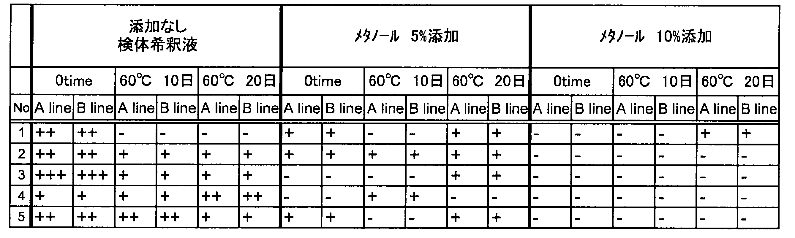
Immediately as in Example 1, the immunochromatographic test strip and the immunochromatographic test strip stored at 60 ° C. for 10 days and 20 days were immersed in a specimen diluent containing 5% and 10% methanol. After developing for 10 minutes, the occurrence of white spots was visually confirmed. The results are shown in Table 3.
5%および10%のメタノールを含有する検体希釈液を使用した場合、製造直後、60℃で10日間、および20日間保存したイムノクロマトグラフィー用テストストリップ何れにおいても、白抜け発生の抑制および程度の軽減が認められた。特に10%メタノールを含有する検体希釈液を使用した場合において、顕著な白抜け発生の抑制が確認された。 (result)
When specimen dilutions containing 5% and 10% methanol were used, suppression of white spots and reduction of the extent of any test strips for immunochromatography stored immediately after production, at 60 ° C. for 10 days, and for 20 days Was recognized. In particular, when a specimen diluent containing 10% methanol was used, significant suppression of white spots was confirmed.
イムノクロマトグラフィー用テストストリップを用いて「A line」、「B line」の着色強度を比較した。5%および10%のメタノールを含有する検体希釈液330μLと、インフルエンザA型およびインフルエンザB型の各株の不活化ウイルス抗原を表4、表5に示す倍率(表中、インフルエンザウイルスの種類の下にそれぞれ記載)で生理食塩水で希釈した試料50μLを混合しサンプルとした。各群n=2で、それぞれの不活化ウイルス抗原を加えたサンプル135μLにテストストリップを浸漬させ、10分間テストストリップ上で検体希釈液を展開させた。その後、それぞれのウイルスの検出部位における着色強度を色調見本に基づき求めた。インフルエンザA型の結果を表4に、インフルエンザB型の結果を表5に示した。


The coloring intensity of “A line” and “B line” was compared using an immunochromatographic test strip. 330 μL of sample diluent containing 5% and 10% methanol and the inactivated virus antigens of influenza A and influenza B strains are shown in Tables 4 and 5 (in the table, below the influenza virus type). Each sample was mixed with 50 μL of the sample diluted with physiological saline to prepare a sample. In each group n = 2, the test strip was immersed in 135 μL of the sample to which each inactivated virus antigen was added, and the specimen dilution solution was developed on the test strip for 10 minutes. Then, the coloring intensity in the detection part of each virus was calculated | required based on the color tone sample. The results for influenza A are shown in Table 4, and the results for influenza B are shown in Table 5.
インフルエンザA型ウイルスの不活化抗原を用いた場合、何れの株においても、検体希釈液に5%および10%のメタノールを添加させたか否かにかかわらず、着色強度に変化は見られなかった。一方、インフルエンザB型ウイルスの不活化抗原を用いた場合、メタノールを加えたことによって、着色強度の上昇が見られた。 (result)
When an inactivated antigen of influenza A virus was used, no change was observed in the coloring intensity in any strain regardless of whether 5% and 10% methanol was added to the sample diluent. On the other hand, when an inactivated antigen of influenza B virus was used, an increase in coloring intensity was observed by adding methanol.
(b)抗体固定化メンブレン
(c1)抗インフルエンザA型ウイルスモノクローナル抗体
(c2)抗インフルエンザB型ウイルスモノクローナル抗体
(c3)コントロール抗体
(d)コンジュゲートパッド
(e)サンプルパッド
(f)吸収パッド
(g)コンジュゲート部 (A) Plastic adhesive sheet (b) Antibody-immobilized membrane (c1) Anti-influenza A virus monoclonal antibody (c2) Anti-influenza B virus monoclonal antibody (c3) Control antibody (d) Conjugate pad (e) Sample pad (F) Absorption pad (g) Conjugate part
Claims (11)
- イムノクロマトグラフィーを利用した検出方法であって、
以下の(1)および(2)を含むイムノクロマトグラフィー用テストストリップを使用し、かつ、下記(A)および(B)の工程を有する前記検出方法。
(1)被検出物質を含有する可能性のあるサンプルを供給するサンプル供給部と、前記サンプル供給部よりも下流側に、被検出物質に対する抗体が標識体に固定化されたコンジュゲートを含有するコンジュゲート部とを有する、コンジュゲートパッド
(2)被検出物質に対する抗体が固定化された検出部を少なくとも1つ有する、不溶性メンブレン担体
(A)メタノール、および被検出物質を含有する可能性のあるサンプルをサンプル供給部に供給する工程
(B)不溶性メンブレン担体上の免疫反応生成物としての被検出物質を検出する工程
を含む、前記検出方法。 A detection method using immunochromatography,
The said detection method which uses the test strip for immunochromatography containing the following (1) and (2), and has the process of the following (A) and (B).
(1) A sample supply unit that supplies a sample that may contain a substance to be detected, and a conjugate in which an antibody to the substance to be detected is immobilized on a label on the downstream side of the sample supply part Conjugate pad, (2) Insoluble membrane carrier (A) having at least one detection part on which an antibody against the substance to be immobilized is immobilized, and possibly substance to be detected The detection method comprising the step of supplying a sample to a sample supply unit (B) and detecting a substance to be detected as an immune reaction product on the insoluble membrane carrier. - (A)の工程が、あらかじめメタノールを含むサンプル希釈液によって希釈されたサンプルを、サンプル供給部に供給する工程である請求項1に記載の検出方法。 The detection method according to claim 1, wherein the step (A) is a step of supplying a sample diluted in advance with a sample diluent containing methanol to a sample supply unit.
- (A)の工程が、メタノールを含むサンプル希釈液によりサンプルを希釈して、希釈されたサンプルを、サンプル供給部に供給する工程である請求項1に記載の検出方法。 The detection method according to claim 1, wherein the step (A) is a step of diluting the sample with a sample diluent containing methanol and supplying the diluted sample to the sample supply unit.
- 被検出物質がインフルエンザB型ウイルスであり、標識体に固定化された被検出物質に対する抗体および検出部に固定化された被検出物質に対する抗体が、抗インフルエンザB型ウイルスモノクローナル抗体である、請求項1~3のいずれかに記載の検出方法。 The detected substance is an influenza B virus, and the antibody against the detected substance immobilized on the label and the antibody against the detected substance immobilized on the detection part are anti-influenza B virus monoclonal antibodies. 4. The detection method according to any one of 1 to 3.
- 以下の(a)および(b)を含むイムノクロマトグラフィー用検出キット。
(a)被検出物質を含有する可能性のあるサンプルを展開させることにより被検出物質を検出するイムノクロマトグラフィー用テストストリップであって以下の(1)および(2)を含むテストストリップ。
(1)被検出物質を含有する可能性のあるサンプルを供給するサンプル供給部と、前記サンプル供給部よりも下流側に、被検出物質に対する抗体が標識体に固定化されたコンジュゲートを含有するコンジュゲート部とを有する、コンジュゲートパッド
(2)被検出物質に対する抗体が固定化された検出部を少なくとも1つ有する、不溶性メンブレン担体
(b)メタノールを含有するイムノクロマトグラフィー用サンプル希釈液 A detection kit for immunochromatography comprising the following (a) and (b).
(A) A test strip for immunochromatography that detects a substance to be detected by developing a sample that may contain the substance to be detected, and includes the following (1) and (2).
(1) A sample supply unit that supplies a sample that may contain a substance to be detected, and a conjugate in which an antibody to the substance to be detected is immobilized on a label on the downstream side of the sample supply part Conjugate pad (2) Immunochromatographic sample diluent containing insoluble membrane carrier (b) methanol having at least one detection part on which an antibody against the substance to be detected is immobilized - 被検出物質がインフルエンザB型ウイルスであり、標識体に固定化された被検出物質に対する抗体および検出部に固定化された被検出物質に対する抗体が抗インフルエンザB型ウイルスモノクローナル抗体である、請求項5に記載のイムノクロマトグラフィー用検出キット。 The detected substance is an influenza B virus, and the antibody against the detected substance immobilized on the label and the antibody against the detected substance immobilized on the detection part are anti-influenza B virus monoclonal antibodies. A detection kit for immunochromatography as described in 1.
- (b)が、メタノールを0.1~20%(v/v)含有するイムノクロマトグラフィー用サンプル希釈液である、請求項5または6に記載のイムノクロマトグラフィー用検出キット。 The immunochromatographic detection kit according to claim 5 or 6, wherein (b) is an immunochromatographic sample diluent containing 0.1 to 20% (v / v) methanol.
- (b)が、pH6.0~10.0の緩衝液中にメタノールを0.1~20%(v/v)含有するイムノクロマトグラフィー用サンプル希釈液である、請求項5~7のいずれかに記載のイムノクロマトグラフィー用検出キット。 (B) is a sample diluent for immunochromatography containing 0.1 to 20% (v / v) methanol in a buffer having a pH of 6.0 to 10.0. The detection kit for immunochromatography as described.
- インフルエンザB型ウイルス検出のためのイムノクロマトグラフィー用サンプル希釈液であって、メタノールを0.1~20%(v/v)含有する前記サンプル希釈液。 A sample diluent for immunochromatography for detecting influenza B virus, which contains 0.1 to 20% (v / v) methanol.
- pH6.0~10.0の緩衝液中にメタノールを0.1~20%(v/v)含有する請求項9に記載のサンプル希釈液。 The sample diluent according to claim 9, comprising 0.1 to 20% (v / v) of methanol in a buffer having a pH of 6.0 to 10.0.
- インフルエンザB型ウイルスを含有する可能性のあるサンプルを展開させることにより当該ウイルスを検出する下記(a)のイムノクロマトグラフィー用テストストリップを含む、インフルエンザB型ウイルス検出キットの製造における、下記(b)のイムノクロマトグラフィー用サンプル希釈液の使用
(a)以下の(1)および(2)を含むイムノクロマトグラフィー用テストストリップ。
(1)インフルエンザB型ウイルスを含有する可能性のあるサンプルを供給するサンプル供給部と、前記サンプル供給部よりも下流側に、抗インフルエンザB型ウイルス抗体が標識体に固定化されたコンジュゲートを含有するコンジュゲート部とを有する、コンジュゲートパッド
(2)抗インフルエンザB型ウイルス抗体が固定化された検出部を少なくとも1つ有する、不溶性メンブレン担体
(b)メタノールを0.1~20%含有するイムノクロマトグラフィー用サンプル希釈液 (B) in the manufacture of an influenza B virus detection kit comprising the immunochromatographic test strip of (a) below for detecting the virus by developing a sample that may contain influenza B virus; Use of a sample diluent for immunochromatography (a) An immunochromatographic test strip comprising the following (1) and (2).
(1) A sample supply unit that supplies a sample that may contain an influenza B virus, and a conjugate in which an anti-influenza B virus antibody is immobilized on a label on the downstream side of the sample supply unit Conjugate pad having a conjugate portion containing (2) Insoluble membrane carrier (b) having 0.1 to 20% of methanol having at least one detection portion on which an anti-influenza B virus antibody is immobilized Sample diluent for immunochromatography
Priority Applications (4)
| Application Number | Priority Date | Filing Date | Title |
|---|---|---|---|
| CN201480065040.2A CN105793707B (en) | 2013-11-29 | 2014-12-01 | The detection method of immunochromatography auxiliary |
| US15/039,531 US20160377616A1 (en) | 2013-11-29 | 2014-12-01 | Immunochromatographic detection method |
| JP2015525659A JP5831918B2 (en) | 2013-11-29 | 2014-12-01 | Detection method using immunochromatography |
| EP14866585.4A EP3076177B1 (en) | 2013-11-29 | 2014-12-01 | Immunochromatography-assisted detection method |
Applications Claiming Priority (2)
| Application Number | Priority Date | Filing Date | Title |
|---|---|---|---|
| JP2013247279 | 2013-11-29 | ||
| JP2013-247279 | 2013-11-29 |
Publications (1)
| Publication Number | Publication Date |
|---|---|
| WO2015080286A1 true WO2015080286A1 (en) | 2015-06-04 |
Family
ID=53199220
Family Applications (1)
| Application Number | Title | Priority Date | Filing Date |
|---|---|---|---|
| PCT/JP2014/081753 WO2015080286A1 (en) | 2013-11-29 | 2014-12-01 | Immunochromatography-assisted detection method |
Country Status (5)
| Country | Link |
|---|---|
| US (1) | US20160377616A1 (en) |
| EP (1) | EP3076177B1 (en) |
| JP (1) | JP5831918B2 (en) |
| CN (1) | CN105793707B (en) |
| WO (1) | WO2015080286A1 (en) |
Cited By (2)
| Publication number | Priority date | Publication date | Assignee | Title |
|---|---|---|---|---|
| WO2017094825A1 (en) * | 2015-12-02 | 2017-06-08 | 東洋紡株式会社 | Immunochromatographic test piece |
| WO2020067233A1 (en) | 2018-09-27 | 2020-04-02 | 積水メディカル株式会社 | Test piece for immunochromatography |
Families Citing this family (5)
| Publication number | Priority date | Publication date | Assignee | Title |
|---|---|---|---|---|
| US10234614B2 (en) | 2017-01-20 | 2019-03-19 | Corning Research & Development Corporation | Light source assemblies and systems and methods with mode homogenization |
| WO2020105079A1 (en) * | 2018-11-19 | 2020-05-28 | 積水メディカル株式会社 | Test piece for immunochromatography and immunochromatographic detection kit |
| EP3771908A1 (en) | 2019-07-29 | 2021-02-03 | Fundació Institut Català de Nanociència i Nanotecnologia (ICN2) | Lateral-electrophoretic bioassay |
| CN110988338A (en) * | 2019-12-09 | 2020-04-10 | 南京市产品质量监督检验院 | Quantum dot test paper capable of quantitatively detecting trimethoprim residual quantity in various matrixes |
| CN112485433A (en) * | 2020-10-29 | 2021-03-12 | 海卫特(广州)医疗科技有限公司 | Serum amyloid protein A immunochromatographic test strip, kit and preparation method thereof |
Citations (4)
| Publication number | Priority date | Publication date | Assignee | Title |
|---|---|---|---|---|
| JPH06306108A (en) | 1993-04-27 | 1994-11-01 | Sekisui Chem Co Ltd | Production of colored latex particles |
| JPH10332699A (en) * | 1997-06-04 | 1998-12-18 | Iatron Lab Inc | Immuno-chromatography method |
| WO2005007698A1 (en) * | 2003-07-23 | 2005-01-27 | Fujirebio Inc. | Monoclonal antibody against influenza b virus and immunoassay instrument using the antibody |
| WO2012043746A1 (en) | 2010-09-30 | 2012-04-05 | 積水メディカル株式会社 | Test strip for immunochromatography, and process for production thereof |
Family Cites Families (11)
| Publication number | Priority date | Publication date | Assignee | Title |
|---|---|---|---|---|
| US5958791A (en) * | 1996-09-27 | 1999-09-28 | Innovative Biotechnologies, Inc. | Interdigitated electrode arrays for liposome-enhanced immunoassay and test device |
| US6194221B1 (en) * | 1996-11-19 | 2001-02-27 | Wyntek Diagnostics, Inc. | Hybrid one-step immunochromatographic device and method of use |
| US6224872B1 (en) * | 1997-07-31 | 2001-05-01 | Kabushiki Kaisha Hayashibara Seibutsu Kagaku Kenkyujo | Composition |
| EP2270508B1 (en) * | 2003-12-24 | 2013-02-20 | DENKA SEIKEN Co., Ltd. | Membrane assay method and kit |
| WO2007092302A2 (en) * | 2006-02-07 | 2007-08-16 | Envirologix Inc. | Test device for analyte detection |
| CN101231290A (en) * | 2007-01-25 | 2008-07-30 | 天津科技大学 | Kit and method for quantitative determination of ochratoxin A content in food product |
| US20100112604A1 (en) * | 2008-11-04 | 2010-05-06 | Joy Ann Drawdy | Home food test kit and method of use |
| CN101526534A (en) * | 2009-03-10 | 2009-09-09 | 沈鹤柏 | Fluorescence immune chromatography test paper and preparing method and application thereof |
| CN101566627A (en) * | 2009-05-27 | 2009-10-28 | 汤凌霄 | Melamine rapid detection kit and detection method thereofRapid detection kit and detection method thereof for melamine |
| CN101900728A (en) * | 2010-08-05 | 2010-12-01 | 中国农业科学院油料作物研究所 | Multi-test-line immunochromatographic test strip for semi-quantitatively detecting aflatoxin B1 and preparation method thereof |
| CN103018439B (en) * | 2012-11-30 | 2013-12-25 | 中国农业科学院油料作物研究所 | Method for enhancing detection sensitivity of colloidal gold immunity chromatography test strip |
-
2014
- 2014-12-01 EP EP14866585.4A patent/EP3076177B1/en active Active
- 2014-12-01 JP JP2015525659A patent/JP5831918B2/en active Active
- 2014-12-01 CN CN201480065040.2A patent/CN105793707B/en active Active
- 2014-12-01 WO PCT/JP2014/081753 patent/WO2015080286A1/en active Application Filing
- 2014-12-01 US US15/039,531 patent/US20160377616A1/en not_active Abandoned
Patent Citations (4)
| Publication number | Priority date | Publication date | Assignee | Title |
|---|---|---|---|---|
| JPH06306108A (en) | 1993-04-27 | 1994-11-01 | Sekisui Chem Co Ltd | Production of colored latex particles |
| JPH10332699A (en) * | 1997-06-04 | 1998-12-18 | Iatron Lab Inc | Immuno-chromatography method |
| WO2005007698A1 (en) * | 2003-07-23 | 2005-01-27 | Fujirebio Inc. | Monoclonal antibody against influenza b virus and immunoassay instrument using the antibody |
| WO2012043746A1 (en) | 2010-09-30 | 2012-04-05 | 積水メディカル株式会社 | Test strip for immunochromatography, and process for production thereof |
Non-Patent Citations (3)
| Title |
|---|
| MASATAKA ICHIKAWA ET AL.: "Immunochromatography-ho to Koso Men'ekiho o Kumiawaseta Genri ni yoru Atarashii Influenza Jinsoku Shindan Kit (Espline Influenza A & B) no Kento", THE JOURNAL OF MEDICINE, vol. 49, no. 3., March 2003 (2003-03-01), pages 469 - 478, XP002983639 * |
| NAKAGAWA N ET AL.: "Rapid detection and identification of two lineages of influenza B strains with monoclonal antibodies.", J VIROL METHODS., vol. 79, no. 1, April 1999 (1999-04-01), pages 113 - 120, XP027296682 * |
| See also references of EP3076177A4 |
Cited By (4)
| Publication number | Priority date | Publication date | Assignee | Title |
|---|---|---|---|---|
| WO2017094825A1 (en) * | 2015-12-02 | 2017-06-08 | 東洋紡株式会社 | Immunochromatographic test piece |
| JPWO2017094825A1 (en) * | 2015-12-02 | 2018-09-20 | 東洋紡株式会社 | Immunochromatographic test piece |
| WO2020067233A1 (en) | 2018-09-27 | 2020-04-02 | 積水メディカル株式会社 | Test piece for immunochromatography |
| KR20210064268A (en) | 2018-09-27 | 2021-06-02 | 세키스이 메디칼 가부시키가이샤 | Specimens for Immunochromatography |
Also Published As
| Publication number | Publication date |
|---|---|
| EP3076177A1 (en) | 2016-10-05 |
| US20160377616A1 (en) | 2016-12-29 |
| JPWO2015080286A1 (en) | 2017-03-16 |
| CN105793707A (en) | 2016-07-20 |
| EP3076177A4 (en) | 2017-05-03 |
| EP3076177B1 (en) | 2018-08-01 |
| JP5831918B2 (en) | 2015-12-09 |
| CN105793707B (en) | 2018-05-08 |
Similar Documents
| Publication | Publication Date | Title |
|---|---|---|
| JP5831918B2 (en) | Detection method using immunochromatography | |
| US9435806B2 (en) | Immunochromatographic test strip and manufacturing method thereof | |
| CN104502586B (en) | A kind of immunochromatography detection method and test paper | |
| JP2020530567A (en) | Assay Methods for Improved Analyze Detection | |
| JP6008670B2 (en) | Membrane for immunochromatographic test strip, test strip and inspection method | |
| US20110136142A1 (en) | Immunochromatography method | |
| JP5753942B2 (en) | Immunochromatographic test strip and detection method using immunochromatography for detecting an object in a sample containing red blood cells | |
| JP6061843B2 (en) | Detection method based on immunochromatography capable of determining sample-unadded sample as unsuitable sample and test strip used therefor | |
| EP3339862A1 (en) | Immunological detection method and test strip used therefor | |
| KR102586991B1 (en) | Immunochromatography device | |
| KR20160120675A (en) | Rapid Quantitative Diagnostic Kit | |
| JP2021188960A (en) | Test strip for immunochromatography | |
| WO2020067233A1 (en) | Test piece for immunochromatography | |
| JP7181547B2 (en) | Test strip for immunochromatography and method for detecting substance to be detected | |
| JP6464308B1 (en) | Test specimen for immunochromatography | |
| CN113614533A (en) | Immunochromatography assay method and test strip for use in the same | |
| JP6595215B2 (en) | Immunochromatographic analyzer, manufacturing method thereof, and immunochromatographic analysis method | |
| JP5265423B2 (en) | Chromatographic method | |
| JP2020125994A (en) | Immunochromatographic specimen | |
| JP2009145079A (en) | Strip for chromatography analysis |
Legal Events
| Date | Code | Title | Description |
|---|---|---|---|
| ENP | Entry into the national phase |
Ref document number: 2015525659 Country of ref document: JP Kind code of ref document: A |
|
| 121 | Ep: the epo has been informed by wipo that ep was designated in this application |
Ref document number: 14866585 Country of ref document: EP Kind code of ref document: A1 |
|
| WWE | Wipo information: entry into national phase |
Ref document number: 15039531 Country of ref document: US |
|
| NENP | Non-entry into the national phase |
Ref country code: DE |
|
| REEP | Request for entry into the european phase |
Ref document number: 2014866585 Country of ref document: EP |
|
| WWE | Wipo information: entry into national phase |
Ref document number: 2014866585 Country of ref document: EP |