WO2015053375A1 - Method of producing retinal pigment epithelial cell - Google Patents
Method of producing retinal pigment epithelial cell Download PDFInfo
- Publication number
- WO2015053375A1 WO2015053375A1 PCT/JP2014/077111 JP2014077111W WO2015053375A1 WO 2015053375 A1 WO2015053375 A1 WO 2015053375A1 JP 2014077111 W JP2014077111 W JP 2014077111W WO 2015053375 A1 WO2015053375 A1 WO 2015053375A1
- Authority
- WO
- WIPO (PCT)
- Prior art keywords
- cells
- retinal pigment
- pigment epithelial
- laminin
- epithelial cells
- Prior art date
Links
Images


Classifications
-
- C—CHEMISTRY; METALLURGY
- C12—BIOCHEMISTRY; BEER; SPIRITS; WINE; VINEGAR; MICROBIOLOGY; ENZYMOLOGY; MUTATION OR GENETIC ENGINEERING
- C12N—MICROORGANISMS OR ENZYMES; COMPOSITIONS THEREOF; PROPAGATING, PRESERVING, OR MAINTAINING MICROORGANISMS; MUTATION OR GENETIC ENGINEERING; CULTURE MEDIA
- C12N5/00—Undifferentiated human, animal or plant cells, e.g. cell lines; Tissues; Cultivation or maintenance thereof; Culture media therefor
- C12N5/06—Animal cells or tissues; Human cells or tissues
- C12N5/0602—Vertebrate cells
- C12N5/0618—Cells of the nervous system
- C12N5/0621—Eye cells, e.g. cornea, iris pigmented cells
-
- A—HUMAN NECESSITIES
- A61—MEDICAL OR VETERINARY SCIENCE; HYGIENE
- A61K—PREPARATIONS FOR MEDICAL, DENTAL OR TOILETRY PURPOSES
- A61K35/00—Medicinal preparations containing materials or reaction products thereof with undetermined constitution
- A61K35/12—Materials from mammals; Compositions comprising non-specified tissues or cells; Compositions comprising non-embryonic stem cells; Genetically modified cells
- A61K35/30—Nerves; Brain; Eyes; Corneal cells; Cerebrospinal fluid; Neuronal stem cells; Neuronal precursor cells; Glial cells; Oligodendrocytes; Schwann cells; Astroglia; Astrocytes; Choroid plexus; Spinal cord tissue
-
- A—HUMAN NECESSITIES
- A61—MEDICAL OR VETERINARY SCIENCE; HYGIENE
- A61K—PREPARATIONS FOR MEDICAL, DENTAL OR TOILETRY PURPOSES
- A61K9/00—Medicinal preparations characterised by special physical form
- A61K9/0012—Galenical forms characterised by the site of application
- A61K9/0048—Eye, e.g. artificial tears
-
- A—HUMAN NECESSITIES
- A61—MEDICAL OR VETERINARY SCIENCE; HYGIENE
- A61P—SPECIFIC THERAPEUTIC ACTIVITY OF CHEMICAL COMPOUNDS OR MEDICINAL PREPARATIONS
- A61P27/00—Drugs for disorders of the senses
- A61P27/02—Ophthalmic agents
-
- C—CHEMISTRY; METALLURGY
- C12—BIOCHEMISTRY; BEER; SPIRITS; WINE; VINEGAR; MICROBIOLOGY; ENZYMOLOGY; MUTATION OR GENETIC ENGINEERING
- C12N—MICROORGANISMS OR ENZYMES; COMPOSITIONS THEREOF; PROPAGATING, PRESERVING, OR MAINTAINING MICROORGANISMS; MUTATION OR GENETIC ENGINEERING; CULTURE MEDIA
- C12N2501/00—Active agents used in cell culture processes, e.g. differentation
- C12N2501/70—Enzymes
- C12N2501/72—Transferases (EC 2.)
- C12N2501/727—Kinases (EC 2.7.)
-
- C—CHEMISTRY; METALLURGY
- C12—BIOCHEMISTRY; BEER; SPIRITS; WINE; VINEGAR; MICROBIOLOGY; ENZYMOLOGY; MUTATION OR GENETIC ENGINEERING
- C12N—MICROORGANISMS OR ENZYMES; COMPOSITIONS THEREOF; PROPAGATING, PRESERVING, OR MAINTAINING MICROORGANISMS; MUTATION OR GENETIC ENGINEERING; CULTURE MEDIA
- C12N2506/00—Differentiation of animal cells from one lineage to another; Differentiation of pluripotent cells
- C12N2506/45—Differentiation of animal cells from one lineage to another; Differentiation of pluripotent cells from artificially induced pluripotent stem cells
-
- C—CHEMISTRY; METALLURGY
- C12—BIOCHEMISTRY; BEER; SPIRITS; WINE; VINEGAR; MICROBIOLOGY; ENZYMOLOGY; MUTATION OR GENETIC ENGINEERING
- C12N—MICROORGANISMS OR ENZYMES; COMPOSITIONS THEREOF; PROPAGATING, PRESERVING, OR MAINTAINING MICROORGANISMS; MUTATION OR GENETIC ENGINEERING; CULTURE MEDIA
- C12N2509/00—Methods for the dissociation of cells, e.g. specific use of enzymes
-
- C—CHEMISTRY; METALLURGY
- C12—BIOCHEMISTRY; BEER; SPIRITS; WINE; VINEGAR; MICROBIOLOGY; ENZYMOLOGY; MUTATION OR GENETIC ENGINEERING
- C12N—MICROORGANISMS OR ENZYMES; COMPOSITIONS THEREOF; PROPAGATING, PRESERVING, OR MAINTAINING MICROORGANISMS; MUTATION OR GENETIC ENGINEERING; CULTURE MEDIA
- C12N2533/00—Supports or coatings for cell culture, characterised by material
- C12N2533/50—Proteins
- C12N2533/52—Fibronectin; Laminin
Definitions
- the present invention relates to a method for efficiently producing retinal pigment epithelium (RPE) cells from human pluripotent stem cells, a method for stably culturing retinal pigment epithelial cells, and the like.
- RPE retinal pigment epithelium
- Non-patent Document 1 As a method for producing retinal pigment epithelial cells from pluripotent stem cells, a method of culturing ES cells as floating aggregates in a serum-free medium called SFEB method (Patent Document 1 etc.), and in the presence of a differentiation-inducing factor, A method of inducing differentiation of pluripotent stem cells on a culture substrate coated with a weak cell adhesion coating agent is known (Non-patent Document 1, etc.).
- Non-Patent Document 2 reports that human ES cells could be maintained and cultured on laminin 511 for a long period of time.
- the E8 fragment known as a modified laminin with further improved cell adhesion activity for example, Patent Document 2 and Non-Patent Document 3 include an E8 fragment of human laminin ⁇ 5 ⁇ 1 ⁇ 1 (laminin 511E8, hereinafter the same) and human laminin 322E8. Discloses a method for culturing human pluripotent stem cells.
- Non-Patent Document 4 describes that laminin 511E8 retains the same degree of binding activity to ⁇ 6 ⁇ 1 integrin as full-length laminin 511, and Patent Document 2 uses this laminin 511E8 to achieve multiple functions. It is described that sex stem cells can be stably fixed on a culture dish and, as a result, can be maintained and cultured while maintaining the pluripotency of the cells. However, there is no report that such an E8 fragment of laminin is used for inducing differentiation of pluripotent stem cells other than the culture of pluripotent stem cells.
- Non-Patent Document 5 describes that differentiation induction efficiency into retinal pigment epithelial cells was remarkably increased by adhesion culture of pluripotent stem cells on laminin 111 and Matrigel. However, there is still no report that laminin E8 fragment was used to induce differentiation of pluripotent stem cells into retinal pigment epithelial cells.
- An object of the present invention is to produce a retinal pigment epithelial cell that improves the efficiency of inducing differentiation from pluripotent stem cells into retinal pigment epithelial cells, and can obtain high purity retinal pigment epithelial cells in a short period of time by a simple operation. It is intended to provide a method, a retinal pigment epithelial cell culture method capable of stably growing and culturing cells, a toxicity / drug efficacy evaluation method using retinal pigment epithelial cells useful for transplantation treatment, and a retinal disease therapeutic agent.
- the present inventors have cultivated human pluripotent stem cells on a culture substrate coated with laminin E8, so that the seeded pluripotent stem cells quickly adhere to the culture substrate.
- the production of a large amount of pigment cells is recognized from an early stage, the yield of retinal pigment epithelial cells can be remarkably improved, the cells are not easily lost when the medium is changed, the purification process can be simplified, and high purity cells can be obtained in a short period of time. We found that a group was obtained.
- the present invention relates to the following.
- a method for producing retinal pigment epithelial cells comprising a step of adhesion-culturing human pluripotent stem cells on a culture substrate coated with laminin E8 fragment.
- the adhesion culture step is performed in the presence of a differentiation-inducing factor.
- a method for amplifying retinal pigment epithelial cells comprising a step of adhesion-culturing retinal pigment epithelial cells on a culture substrate coated with laminin E8 fragment.
- a method for purifying retinal pigment epithelial cells comprising a step of adhesion-culturing retinal pigment epithelial cells on a culture substrate coated with laminin E8 fragment.
- a therapeutic agent for retinal diseases comprising retinal pigment epithelial cells produced by the method of any one of [1] to [4] or retinal pigment epithelial cells cultured by the method of [7].
- retinal pigment epithelial cells can be produced from pluripotent stem cells with high yield. Moreover, differentiation induction efficiency is improved, a cell population containing retinal pigment epithelial cells at a high concentration can be obtained, and highly purified retinal pigment epithelial cells can be produced by a simple purification operation. Furthermore, according to the present invention, the retinal pigment epithelial cells can be stably adhered, so that the cells are not easily lost during the medium exchange and can be stably subcultured.
- the schematic diagram of the structure of laminin E8 is shown.
- derived from the human iPS cell (201B7) is shown.
- Cell adhesion on days 1, 2 and 3 after seeding iPS cells on a substrate coated with full length laminin 511-E8, matrigel or laminin 511 in Comparative Example 3 Indicates.
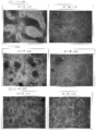
- the state of the cell population around 15th and 40th days after iPS cells were seeded on a substrate coated with laminin 511-E8, Matrigel or full length laminin 511 in Comparative Example 3 and differentiation induction was started is shown.
- the present invention is a method for producing retinal pigment epithelial cells comprising the step of adhesion-culturing human pluripotent stem cells using a culture substrate coated with laminin E8 fragment (hereinafter referred to as “this” The manufacturing method of the invention ").
- the “pluripotent stem cell” in the present invention means a stem cell having self-renewal ability and differentiation pluripotency, and is not particularly limited.
- stem cells embryonic stem cells (ES cells), induced pluripotent stem cells (IPS cells) and the like are widely used.
- ES cells embryonic stem cells
- IPS cells induced pluripotent stem cells
- human ES cells or human iPS cells are used, and more preferably human iPS cells are used.
- the “iPS cell” in the present invention artificially acquired self-renewal ability and pluripotency by bringing a nuclear reprogramming factor into contact with a somatic cell (eg, fibroblast, skin cell, lymphocyte, etc.).
- a somatic cell eg, fibroblast, skin cell, lymphocyte, etc.
- a cell eg, fibroblast, skin cell, lymphocyte, etc.
- the method for producing iPS cells in the present invention is not particularly limited.
- retinal pigment epithelial cell refers to an epithelial cell constituting the retinal pigment epithelium and a precursor cell thereof. Whether it is retinal pigment epithelial cells, for example, expression of cell markers (RPE65, CRALBP, MERTK, BEST1, etc.) and cell morphology (intracellular melanin pigmentation, polygonal and flat cell morphology, polygonal For example, formation of actin bundles).
- progenitor cells of retinal pigment epithelial cells mean cells that have been induced to differentiate into retinal cells, and whether they are progenitor cells depends on the expression of cell markers (Mitf, Pax6, Rx, Crx, etc.) Can be confirmed.
- retinal pigment epithelial cells can be confirmed using, for example, the secretion ability or phagocytic ability of cytokines (VEGF, PEDF, etc.) as an index.
- cytokines VEGF, PEDF, etc.
- laminin is a heterotrimeric molecule composed of ⁇ , ⁇ , and ⁇ chains, and is an extracellular matrix protein in which isoforms having different subunit chain compositions exist. Specifically, laminin has about 15 isoforms in a combination of heterotrimers of 5 ⁇ chains, 4 ⁇ chains, and 3 ⁇ chains.
- the name of laminin is determined by combining the numbers of ⁇ chain ( ⁇ 1 to ⁇ 5), ⁇ chain ( ⁇ 1 to ⁇ 4) and ⁇ chain ( ⁇ 1 to ⁇ 3).
- laminin by a combination of ⁇ 1, ⁇ 1, and ⁇ 1 chains is called laminin 111
- laminin by a combination of ⁇ 5, ⁇ 1, and ⁇ 1 chains is called laminin 511
- laminin by a combination of ⁇ 5, ⁇ 2, and ⁇ 1 chains is called laminin 521.
- laminin for example, laminin derived from a mammal can be used. Examples of mammals include mice, rats, guinea pigs, hamsters, rabbits, cats, dogs, sheep, pigs, cows, horses, goats, monkeys, and humans.
- human laminin is preferably used. At present, 15 types of isoforms are known for human laminin.
- Laminin E8 fragment was originally identified as a fragment having strong cell adhesion activity among fragments obtained by digesting mouse laminin 111 with elastase (EMBO J., 3: 1463-1468, 1984. J.JCell Biol., 105: 589-598, 1987.).
- laminin other than mouse laminin 111 is also digested with elastase, the presence of a fragment corresponding to the E8 fragment of mouse laminin 111 is presumed, but laminin other than mouse laminin 111 is digested with elastase to isolate and identify the E8 fragment.
- laminin other than mouse laminin 111 is digested with elastase to isolate and identify the E8 fragment.
- the laminin E8 fragment used in the present invention does not need to be an elastase digestion product of each laminin, has the same cell adhesion activity as each corresponding laminin, and corresponds to the E8 fragment digested with elastase.
- a recombinant fragment may be used as long as it is a fragment of laminin having a structure. That is, the “laminin E8 fragment” (hereinafter sometimes referred to as “laminin E8”) in the present invention constitutes a heterotrimer in the C-terminal regions of the ⁇ chain, ⁇ chain, and ⁇ chain, and is used for integrin.
- laminin E8 constitutes a heterotrimer in the C-terminal regions of the ⁇ chain, ⁇ chain, and ⁇ chain, and is used for integrin.
- Laminin E8 exhibits a different integrin binding specificity for each laminin isoform and can exert
- the laminin E8 in the present invention is specifically described as follows: (1) functionally has at least the same cell adhesion activity as full-length laminin and at least equivalent integrin binding activity, and (2) has a structure corresponding to mouse laminin E8, specifically Refers to a laminin fragment having a structure corresponding to the first to third positions of the G domain from the coiled-coil C-terminal region of the laminin trimer.
- (1) functional aspects and (2) structural aspects will be described in more detail.
- laminin E8 for example, a molecule showing binding specificity to at least one of integrins expressed on the surface of human pluripotent stem cells and / or human retinal pigment epithelial cells, are preferably used integrins expressed on the surface of both human pluripotent stem cells and human retinal pigment epithelial cells and molecules showing binding specificity to the integrin expressed on the surface of human retinal pigment epithelial cells.
- Examples of the integrin expressed on the surface of human pluripotent stem cells include ⁇ 6 ⁇ 1 integrin, and examples of the integrin expressed on the surface of human retinal pigment epithelial cells include ⁇ 6 ⁇ 1 integrin, ⁇ 3 ⁇ 1 integrin, ⁇ 7 ⁇ 1 integrin, and the like.
- Laminin E8 in the present invention exhibits a binding specificity for integrin at least equivalent to each laminin.
- those having strong affinity for integrin are preferably used.
- “Laminin E8 having strong affinity for integrin” has a significantly low dissociation constant measured by a known method. For example, Table 1 of The Journal of Biological Chemistry (2009) 284, pp.7820-7831 The dissociation constant measured by the method shown in FIG.
- laminin E8 in the present invention those having strong cell adhesion activity are preferably used.
- “Laminin E8 having a strong cell adhesion activity” indicates a significantly strong adhesion activity in a cell adhesion test measured by a known method. For example, The Journal of Biological Chemistry (2007) 282, pp. When the cell adhesion assay described in 11144-11154 is performed, the number of adherent cells of 400 cells / mm 2 or more can be obtained at a laminin E8 coating concentration of 10 nM or less.
- laminin E8 exhibits binding specificity to ⁇ 6 ⁇ 1 integrin, can stably adhere to pluripotent stem cells at the early stage of differentiation induction, and can stably adhere to retinal pigment epithelial cells or their precursor cells at the later stage of differentiation induction. Those are preferably used.
- laminin E8 in particular laminin 511E8 has binding specificity for ⁇ 3 ⁇ 1 integrin and ⁇ 7 ⁇ 1 integrin in addition to ⁇ 6 ⁇ 1 integrin. This is very preferable in that it contributes to improving the efficiency of inducing differentiation into retinal pigment epithelial cells, or can contribute to stabilization of maintenance culture of retinal pigment epithelial cells.
- laminin E8 in the present invention structurally corresponds to a fragment (E8 fragment) having cell adhesion activity in the elastase digest of mouse laminin 111.
- Laminin fragment That is, a part of the domain II (three-stranded coiled coil domain) in the full length laminin is retained (not illustrated as such in the picture representing the E8 fragment in FIG. 1 but actually retains the coiled coil structure), and E8 On the N-terminal side, a short coiled-coil structure is formed with the corresponding fragment of the ⁇ chain and the corresponding fragment of the ⁇ chain.
- the G1-G3 domain structure of the ⁇ chain is retained. Further, the ⁇ chain and the ⁇ chain are bonded to each other by forming a disulfide bond through a cysteine residue on the C-terminal side.
- laminin E8 in the present invention may be an enzyme-treated product obtained by treating natural laminin with elastase, or a recombinant produced by genetic recombination.
- the laminin E8 of the present invention is a recombinant, the N-terminal is used for the purpose of purifying the corresponding full-length (natural) laminin so long as it retains the binding activity with integrin and does not impair its cell adhesion.
- a tag may be bound to Such a tag is not particularly limited, and examples thereof include a His tag, a Flag tag, and an HA tag.
- the sequence of the linker portion between the tag and laminin E8 is not particularly limited as long as it retains the binding activity with the integrin in the corresponding full-length (natural) laminin and does not impair cell adhesion.
- the laminin E8 in the present invention retains the binding activity with the integrin in the corresponding laminin, and even if part of the amino acid sequence is deleted, added, or substituted as long as the cell adhesion is not impaired. Good.
- the E8 fragment generally lacks two G domains (G4 and G5) on the C-terminal side of the ⁇ chain, but in the laminin E8 in the present invention, the corresponding full-length (natural) laminin is different from the integrin. A part or all of the G4 and G5 domains may be contained as long as the binding activity is maintained and the cell adhesiveness is not impaired.
- laminin 511E8 retains the same binding activity to integrin ⁇ 6 ⁇ 1 as laminin 511, and part or all of G4 and G5 domains may be included as long as cell adhesion activity is not impaired.
- laminin E8 like full-length laminin, has a ⁇ -chain and a ⁇ -chain linked via a cysteine on the C-terminal side of the coiled-coil portion, but this cysteine affects integrin binding activity, so substitution and deletion It is desirable not to be.
- the C-terminal amino acid after the cysteine in the ⁇ chain also affects integrin binding activity, it is desirable that at least it is not deleted (J Biol Chem. 2007 Apr 13; 282 (15): 11144-54.).
- laminin E8 examples include rhLM511E8 produced in Example (3) of WO2011 / 043405.
- the laminin 511E8 can be preferably used as the laminin E8 of the present invention.
- the “culture substrate” used in the present invention can be produced by coating the surface of the incubator with laminin E8 of the present invention.
- “coating” on the surface of the incubator means that laminin E8 is adsorbed on the surface of the incubator through some interaction between laminin E8 and the surface of the incubator, and laminin E8 is particularly effective in achieving the effects of the present invention.
- the orientation of is not a problem.
- the incubator is not particularly limited as long as it can be used for cell culture.
- a dish also referred to as a culture dish
- a petri dish or a plate (6 holes, 24 holes, 48 holes, 96 holes, 384 holes, 9600 holes)
- Microtiter plates, microplates, deep well plates, etc. Microtiter plates, microplates, deep well plates, etc.), flasks, chamber slides, tubes, cell factories, roller bottles, spinner flasks, follower fibers, microcarriers, beads and the like.
- the culture substrate in the present invention may be appropriately subjected to a surface treatment as long as the cell adhesion property by laminin E8 is not impaired.
- the “adhesive culture” in the present invention means a culture in which the target cells adhere to the bottom surface of the incubator via laminin E8, and the cells do not float in the culture medium even if the incubator is shaken lightly during the culture. means. Since laminin E8 used in the present invention exhibits extremely excellent cell adhesion, it is preferable that the laminin E8 is uniformly dispersed by a method such as vibrating the incubator immediately after cell seeding. In addition, if it is in the range of the objective of this invention, you may culture
- the medium is composed of basal medium, serum and / or serum substitute, and other components.
- Basal medium one or a combination of synthetic media generally used for culturing mammalian cells can be used.
- synthetic media generally used for culturing mammalian cells
- commercially available products such as DMEM and GMEM are available.
- Serum substitute is a low protein substitute that replaces serum such as FBS used in cell culture.
- KSR Knockout Serum Replacement
- Glutamax Chemically-defined Lipid concentrated
- N2, B27, etc. which are serum substitutes for neuronal cell culture, are available.
- a serum substitute is preferable from the viewpoint of quality control of the target cells, and KSR is particularly preferable.
- the concentration of serum or serum substitute can be appropriately set, for example, within a range of 0.5 to 30% (v / v), and the concentration may be constant or may be used in a stepwise manner. You may use it, reducing in steps at intervals of about 3 days (preferably 2 days). For example, the concentration of serum or serum substitute can be added in three stages of 20%, 15%, and 10%.
- a Rho kinase inhibitor such as Y-27632 can be used for the purpose of suppressing cell death of human pluripotent stem cells dispersed in the culture medium.
- the addition period of the Rho kinase inhibitor may be a part or the whole period of the differentiation induction process. For example, in the later stage of the differentiation induction step, unnecessary cells that have not differentiated into the target cells can be removed by cell death by using a medium to which no Rho kinase inhibitor is added.
- the medium can contain other components generally used for culturing mammalian cells.
- the concentration of the human pluripotent stem cells used in the production method of the present invention is not particularly limited as long as the pluripotent stem cells can be uniformly seeded and can be adhered and cultured.
- a 10 cm dish one dish is used. 1 ⁇ 10 5 to 1 ⁇ 10 8 cells per cell, preferably 2 ⁇ 10 6 to 5 ⁇ 10 7 cells, more preferably 5 ⁇ 10 5 to 9 ⁇ 10 6 cells.
- the adhesion culture in the production method of the present invention can also be performed in the presence of a differentiation-inducing factor.
- a differentiation inducing factor a known factor can be used as a factor for promoting differentiation induction into a target cell.
- differentiation induction into retinal pigment epithelial cells is preferably used in order to induce differentiation into retinal pigment epithelial cells.
- the differentiation-inducing factor for retinal pigment epithelial cells include a Nodal signal inhibitor, a Wnt signal inhibitor, a sonic hedgehog signal inhibitor, and an activin signal promoter.
- the Nodal signal inhibitor is not particularly limited as long as it can suppress signal transduction mediated by Nodal, and proteins, nucleic acids, low molecular weight compounds and the like can be used.
- the Nodal signal inhibitor include proteins, peptides, or nucleic acids such as Lefty-A, soluble Nodal receptor, anti-Nodal antibody, Nodal receptor inhibitor, and low molecular weight compounds such as SB-431542.
- low molecular weight compounds such as SB-431542, which are easily available and have little difference between lots, are suitable.
- the Wnt signal inhibitor is not particularly limited as long as it can suppress signal transduction mediated by Wnt, and proteins, nucleic acids, low molecular weight compounds and the like can be used.
- Wnt signal inhibitors include proteins, peptides, or nucleic acids such as Dkk1, Cerberus protein, Wnt receptor inhibitor, soluble Wnt receptor, Wnt antibody, casein kinase inhibitor, dominant negative Wnt protein; CKI- 7 (N- (2-aminoethyl) -5-chloro-isoquinoline-8-sulfonamide), D4476 (4- ⁇ 4- (2,3-dihydrobenzo [1,4] dioxin-6-yl) -5 -Pyridin-2-yl-1H-imidazol-2-yl ⁇ benzamide), IWR-1-endo (IWR1e), IWP-2, and other low molecular weight compounds. Less low molecular weight compounds are preferred. Of these, low molecular weight compounds having
- activin signal promoter examples include proteins belonging to the activin family, activin receptors, activin receptor agonists, and the like.
- the concentration of these differentiation-inducing factors can be appropriately selected according to the type of differentiation-inducing factor. Specifically, when SB-431542 is used as a Nodal signal inhibitor, for example, 0.01 to 50 ⁇ M, preferably 0.1 to 10 ⁇ M. More preferably, the concentration is 5 ⁇ M. When CKI-7 is used as the Wnt signal inhibitor, it is added at a concentration of 0.01 to 30 ⁇ M, preferably 0.1 to 30 ⁇ M, more preferably 3 ⁇ M.
- a Nodal signal inhibitor eg, SB-431542
- a Wnt signal inhibitor eg, CKI-7
- the medium is preferably replaced with a maintenance medium for retinal pigment epithelial cells. For example, it is preferable to further culture for 5 to 10 days while exchanging the whole medium at least once every 3 days. . Thereby, melanin pigmented cells and cells having a polygonal flat shape adhering to the basement membrane can be observed more clearly.
- IOVS March 2004, Vol. 45, No. 3, Masatoshi Haruta, et.al., IOVS, November 2011, Vol. 52, No. 12, Okamoto and Takahashi, J. Cell Science 122 (17), Fumitaka Osakada, et.al., IOVS, February 2008, Vol. 49, No. 2, Gamm, et.al. Consists of serum replacement and other ingredients.
- the basal medium one or a combination of synthetic media generally used for culturing mammalian cells can be used.
- synthetic media generally used for culturing mammalian cells
- commercially available products such as DMEM and GMEM are available.
- serum serum derived from mammals such as bovine, human and pig can be used.
- a serum substitute is a low protein substitute that substitutes for serum such as FBS used in cell culture, and commercially available products such as Knockout Serum Replacement (KSR) and Chemically-defined Lipid concentrated (Gibco) In addition to Glutamax (manufactured by Gibco), N2, B27, etc., which are serum substitutes for neuronal cell culture, are available.
- KSR Knockout Serum Replacement
- Gibco Chemically-defined Lipid concentrated
- N2, B27, etc. which are serum substitutes for neuronal cell culture, are available.
- a serum substitute is preferable from the viewpoint of quality control of target cells, and B27 is particularly preferable.
- other components include L-glutamine, penicillin sodium, streptomycin sulfate and the like.
- the production method of the present invention may further include a step of purifying retinal pigment epithelial cells by a concentration operation.
- a step of purifying retinal pigment epithelial cells by a concentration operation According to the production method of the present invention, differentiation induction efficiency is remarkably improved, and thus high-purity retinal pigment epithelial cells can be obtained in a short production period with a simple operation.
- the method for concentrating retinal pigment epithelial cells is not particularly limited as long as it is a generally known method for concentrating cells. For example, filtration, centrifugation, perfusion separation, flow cytometry separation, trap separation using an antibody-modified carrier, etc. This method can be used, and preferably a method such as filtration using a nylon mesh can be used.
- differentiation induction efficiency is remarkably improved by culturing human pluripotent stem cells quickly and firmly attached to a culture vessel via laminin E8 having excellent cell adhesion.
- cell loss during medium exchange can be suppressed. Therefore, a large amount of cell population of high concentration retinal pigment epithelial cells can be obtained.
- cell purification can be performed with a simple operation, the time required for the purification process can be shortened, and retinal pigment epithelial cells can be produced very efficiently.
- retinal pigment epithelial cells can adhere to each other to take a sheet-like structure.
- a sheet of retinal pigment epithelial cells can be produced by the production method of the present invention.
- This sheet of retinal pigment epithelial cells is 3.
- the cell population is useful as a cell transplantation therapeutic agent for treating retinal diseases.
- the present invention also relates to a method for amplifying retinal pigment epithelial cells comprising the step of adhesion-culturing retinal pigment epithelial cells using a culture substrate coated with laminin E8 (hereinafter referred to as “Amplification method "). According to the amplification method of the present invention, retinal pigment epithelial cells can be easily proliferated by a simple method.
- retinal pigment epithelial cells in the amplification method of the present invention for example, retinal pigment epithelial cells derived from mammals can be used.
- mammals include mice, rats, guinea pigs, hamsters, rabbits, cats, dogs, sheep, pigs, cows, horses, goats, monkeys, and humans.
- human retinal pigment epithelial cells are preferably used.
- the retinal pigment epithelial cell in the amplification method of the present invention refers to an epithelial cell constituting the retinal pigment epithelium and a precursor cell thereof, for example, a retinal pigment epithelial cell derived from a living body; an undifferentiated cell (pluripotent stem cell, retinal pigment epithelium) Retinal pigment epithelial cells induced to differentiate from cell precursors, etc .; Retinal pigment epithelial cells differentiated from differentiated cells or precursors of cells other than retinal pigment epithelial cells, preferably differentiated from undifferentiated cells Retinal pigment epithelial cells, more preferably retinal pigment epithelial cells obtained by the production method of the present invention and induced to differentiate from pluripotent stem cells.
- the concentration of the retinal pigment epithelial cells used in the amplification method of the present invention is not particularly limited as long as the retinal pigment epithelial cells can be uniformly adhered and cultured.
- 1 ⁇ 10 10 per dish is used.
- 5 to 1 ⁇ 10 8 cells preferably 2 ⁇ 10 6 to 5 ⁇ 10 7 cells, more preferably 5 ⁇ 10 5 to 1 ⁇ 10 7 cells.
- the laminin E8 fragment in the amplification method of the present invention is the same as described above, and the method for coating the culture substrate is also the same. Therefore, by culturing the retinal pigment epithelial cells obtained by the production method of the present invention on a culture vessel coated with the same laminin E8 fragment, the obtained retinal pigment epithelial cells are amplified as they are to obtain a large amount. Can do it. Furthermore, according to the amplification method of the present invention, the cells that could not be induced to differentiate contained in the differentiation-induced retinal pigment epithelial cells can be relatively reduced. It can also be used as a purification method.
- laminin E8 used in the amplification method of the present invention laminin 511E8 has binding specificity to ⁇ 3 ⁇ 1 integrin and ⁇ 7 ⁇ 1 integrin in addition to ⁇ 6 ⁇ 1 integrin, so that adhesion activity to retinal pigment epithelial cells is improved. This is preferable in that it can contribute to stabilization of maintenance culture and cell growth.
- retinal pigment epithelial cells are rapidly fixed to the incubator via laminin E8 having excellent cell adhesion, and cell loss during medium replacement can be suppressed. Since deformation can be suppressed, it is possible to stably maintain and culture retinal pigment epithelial cells.
- the membranous retinal pigment epithelial cell group can be used as it is or as a therapeutic agent for retinal diseases shown below by separating and collecting from a culture substrate.
- retinal pigment epithelial cells obtained by the production method or amplification method of the present invention are cultured on laminin E8, particularly laminin 511E8, they are separated and recovered from the culture substrate either as a cell alone or as a sheet. It is easy to do and has excellent characteristics.
- Retinal disease therapeutic agent Retinal pigment epithelial cells produced by the production method of the present invention and retinal pigment epithelial cells amplified by the amplification method of the present invention are transplanted into a living body by suspension or sheet shape to treat retinal diseases It can be used as a therapeutic agent for cell transplantation.
- Retinal diseases are ophthalmic diseases related to the retina, and include complications due to other diseases such as diabetes.
- Toxicity / Efficacy Evaluation Drug Retinal pigment epithelial cells produced by the production method of the present invention are used as model cells for healthy and disease, remedies for retinal diseases, and drugs for treating other complications such as diabetes, and prevention thereof. It can be used for drug screening, chemical safety test, stress test, toxicity test, side effect test, infection / contamination test. On the other hand, it can also be used for toxicity studies such as phototoxicity and retinal excitotoxicity specific to retinal cells, and toxicity tests.
- the evaluation methods include stimulation and toxicity tests such as apoptosis evaluation, as well as tests to evaluate the effect on normal differentiation from progenitor cells to retinal pigment epithelial cells and photoreceptor cells (RT-PCR for various gene markers, ELISA for cytokines) Analysis of expressed protein, phagocytic ability test), toxicity test such as phototoxicity, retinal potential and transepithelial electrical resistance to visual function, and cytotoxicity test caused by autoimmune reaction.
- RT-PCR for various gene markers
- ELISA cytokines
- toxicity test such as phototoxicity, retinal potential and transepithelial electrical resistance to visual function
- cytotoxicity test caused by autoimmune reaction cytotoxicity test caused by autoimmune reaction.
- cell materials for these tests not only retinal pigment epithelial cells but also progenitor cells thereof can be used. For example, cells seeded and adhered, cell suspensions, sheets or molded articles thereof Can be provided. This can be used as an extrapolation test for human and animal studies.
- Example 1 Production of iPS cell-derived RPE cells Reagent / differentiation induction basic medium (GMEM medium (Invitrogen), KSR (Invitrogen), 0.1 mM MEM non-essential amino acid solution (Invitrogen), 1 mM sodium pyruvate (SIGMA), 0.1 M 2-mercaptoethanol (Wako Pure Chemicals), 100 U / ml penicillin-100 ⁇ g / ml streptomycin (Invitrogen)) ⁇ First differentiation induction medium (differentiation induction basic medium containing 20% KSR, 10 ⁇ M Y-27632 (Wako Pure Chemical Industries), 5 ⁇ M SB431542 (SIGMA), 3 ⁇ M CKI-7 (SIGMA)) ⁇ Second differentiation induction medium (differentiation induction basic medium containing 15% KSR, 10 ⁇ M Y-27632 (Wako Pure Chemical Industries), 5 ⁇ M SB431542 (SIGMA), 3 ⁇ M CKI-7 (SIGMA)) ⁇ Third differentiation in
- Laminin-coated culture dish is a 9-cm culture dish (BD FALCON) laminin 511E8 fragment (protein disclosed in Example (3) of WO2011043405. (Nippi (iMatrix-511, NIP-8920-02))) 0.5 ⁇ g It was prepared by coating at 37 ° C. for 1 hour or more using a / cm 2 aqueous solution.
- the differentiation induction was performed according to the following timeline, and all differentiation inductions in the examples and comparative examples in the present specification followed this.
- the first day of culture was set to Day 0, and the entire medium was changed every day from the start of culture (Day 1) until around Day 40 when pigment cells were confirmed.
- the composition of the medium was changed stepwise as shown below. That is, Day1-4 is the first differentiation induction medium (20% KSR), Day5-8 is the second differentiation induction medium (15% KSR), Day9-12 is the third differentiation induction medium (10% KSR), Day13 To Day 40 until pigment cells were confirmed, the fourth differentiation induction medium (10% KSR) was used.
- the whole medium was exchanged at least once every 3 days using RPE maintenance medium until Day 47.
- the pigment of the cells became darker, and on Day 47, a group of cells having many dark pigments was observed.
- cell populations containing pigment cells were collected.
- laminin 511E8 fragment as a coating agent, the seeded iPS cells adhered rapidly to the culture dish at high density, and the adhesion state was stably maintained even during the culture period using the differentiation induction medium. For this reason, cell loss could be kept low even when the whole medium exchange was repeated.
- the rate at which pigment cells were generated was greatly improved, and the differentiation induction efficiency was remarkably improved.
- Example 2 Production of RPE cells (other iPS cells) Similar to Example 1 except that human skin (fibroblast) -derived iPS cells (201B7, provided by Kyoto University) were used instead of human peripheral blood (mononuclear cell) -derived iPS cells (1120C7, provided by Kyoto University).
- the pigment cell was obtained by the method. As a result, similar to Example 1, the ratio of pigment cells to the seeded iPS cells was greatly improved, and the differentiation induction efficiency was remarkably improved.
- Example 3 Amplification of iPS cell-derived RPE cells
- 0.01% Trypsin-0.53 mM EDTA was added and treated from the culture dishes. The cell mass was detached. The adhesion between cells was then dissociated by gentle pipetting. Furthermore, the proteolytic enzyme solution and its residual impurities in the cell mixture are removed together with the supernatant by centrifugation, and then unnecessary cells are separated by filtration through a cell strainer (DB Falcon Cell Strainer 40 ⁇ m Nylon) to contain RPE cells. The cell population was collected (Day 48).
- Purity is determined by performing immunostaining of Pax6 or Bestrphin, and if any of them is stained, it is determined to be RPE cells. For cells that do not show fluorescence, the presence or absence of intracellular melanin dye is checked to confirm the dye. Therefore, it was determined as an RPE cell (because fluorescence observation may be inhibited by the dye, it is determined as an RPE cell if the dye is present even if no fluorescence is observed). And it calculated
- Comparative Example 1 In the differentiation induction step of Example 1, instead of adhesion culture using a culture dish (BD FALCON) coated with laminin 511E8, floating using a non-adhesive culture dish (Nunc) treated with MPC (2-Methacryloxylethyl Phosphoryl Choline) The differentiation induction step was performed in the same manner as in Example 1 except that the culture was performed. As a result, almost all the pigment cells were lost during the medium exchange in the differentiation induction step, and the pigment cells could not be recovered on Day 47.
- Comparative Example 2 In the differentiation induction step of Example 1, the same method as in Example 1 except that instead of the culture dish coated with laminin 511E8, adhesion culture was performed using a culture dish coated with poly-D-lysine and gelatin. Differentiation induction was performed. Compared to the laminin 511E8 coated culture dish, the adhesion of the cells to the poly D lysine / gelatin coated culture dish was weak and cells were more likely to be lost during the medium change. Therefore, the ratio of the pigment cells on Day 47 after the start of culture was visually observed as the number of pigment cells with respect to all cells in the culture dish, which was 1/20 or less of Example 1, and the differentiation induction efficiency was remarkably low.
- RPE65-F TCC CCA ATA CAA CTG CCA CT (SEQ ID NO: 1) RPE65-R CCT TGG CAT TCA GAA TCA GG (SEQ ID NO: 2) CRALBP-F GAG GGT GCA AGA GAA GGA CA (SEQ ID NO: 3) CRALBP-R TGC AGA AGC CAT TGA TTT GA (SEQ ID NO: 4) MERTK-F TCC TTG GCC ATC AGA AAA AG (SEQ ID NO: 5) MERTK-R CAT TTG GGT GGC TGA AGT CT (SEQ ID NO: 6) BEST1-F TAG AAC CAT CAG CGC CGT C (SEQ ID NO: 7) BEST1-R TGA GTG TAG TGT GTA TGT TGG (SEQ ID NO: 8)
- Example 3 As in Example 2, using human skin (fibroblast) -derived iPS cells (201B7, provided by Kyoto University) and using a culture dish coated with any one of 1) to 3) below, as in Example 1. Differentiation was induced by the method described above. The test which used three base materials for differentiation induction simultaneously was performed twice, and the additional test was performed once for every base material separately. 1) Laminin 511E8 (molecular weight: 150,000) (iMatrix511: NIP-8920-02, manufactured by Nippi Co., Ltd.) is used for coating so as to be 0.5 ⁇ g / cm 2 .
- a human recombinant laminin 511 full length (molecular weight 776,000) (sales: Veritas (manufactured by: BioLamina) / BLA-LN511) is applied to the coating so as to be 2.59 ⁇ g / cm 2 .
- Matrigel manufactured by BD 354230 (total protein concentration: 9-12 mg / ml)) (basement membrane matrix, mouse Engelbreth-Holm-Swarm (EHS) sarcoma-derived soluble basement membrane preparation.
- FIG. 4 shows the state of the cell population on the 15th day after the start of differentiation induction and before the cell filtration and separation step (around the 40th day).
- retinal pigment epithelial cells can be efficiently produced by a simple method using a culture substrate coated with laminin E8 fragment.
- the production method of the present invention has excellent differentiation induction efficiency, can purify retinal pigment epithelial cells by a simple operation, and can produce retinal pigment epithelial cells with high yield while suppressing cell loss during the process.
- the retinal pigment epithelial cells produced by the method of the present invention are useful not only for treatment of retinal diseases but also as healthy and disease model cells.
Abstract
Description
[1]ラミニンE8フラグメントがコーティングされた培養基材上でヒト多能性幹細胞を接着培養する工程を含む、網膜色素上皮細胞の製造方法。
[2]ラミニンE8フラグメントが、ラミニン511E8フラグメントである、[1]の方法。
[3]接着培養する工程が、分化誘導因子の存在下で行われる、[1]または[2]の方法。
[4]ヒト多能性幹細胞が、ヒトiPS細胞である、[1]~[3]のいずれかの方法。
[5]ラミニンE8フラグメントがコーティングされた培養基材上でヒト多能性幹細胞を接着培養することによって製造される、網膜色素上皮細胞。
[6]ラミニンE8フラグメントがコーティングされた培養基材上でヒト多能性幹細胞を接着培養することによって製造される、網膜色素上皮細胞シート。
[7]ラミニンE8フラグメントがコーティングされた培養基材上で網膜色素上皮細胞を接着培養する工程を含む、網膜色素上皮細胞の増幅方法。
[8]ラミニンE8フラグメントがコーティングされた培養基材上で網膜色素上皮細胞を接着培養する工程を含む、網膜色素上皮細胞の純化方法。
[9][1]~[4]のいずれかの方法により製造される網膜色素上皮細胞、又は[7]の方法により培養される網膜色素上皮細胞を含む、網膜疾患治療薬。 That is, the present invention relates to the following.
[1] A method for producing retinal pigment epithelial cells, comprising a step of adhesion-culturing human pluripotent stem cells on a culture substrate coated with laminin E8 fragment.
[2] The method of [1], wherein the laminin E8 fragment is a laminin 511E8 fragment.
[3] The method according to [1] or [2], wherein the adhesion culture step is performed in the presence of a differentiation-inducing factor.
[4] The method according to any one of [1] to [3], wherein the human pluripotent stem cell is a human iPS cell.
[5] Retinal pigment epithelial cells produced by adherent culture of human pluripotent stem cells on a culture substrate coated with laminin E8 fragment.
[6] A retinal pigment epithelial cell sheet produced by adhesion-culturing human pluripotent stem cells on a culture substrate coated with laminin E8 fragment.
[7] A method for amplifying retinal pigment epithelial cells, comprising a step of adhesion-culturing retinal pigment epithelial cells on a culture substrate coated with laminin E8 fragment.
[8] A method for purifying retinal pigment epithelial cells, comprising a step of adhesion-culturing retinal pigment epithelial cells on a culture substrate coated with laminin E8 fragment.
[9] A therapeutic agent for retinal diseases comprising retinal pigment epithelial cells produced by the method of any one of [1] to [4] or retinal pigment epithelial cells cultured by the method of [7].
本発明は、ラミニンE8フラグメントがコーティングされた培養基材を用いてヒト多能性幹細胞を接着培養する工程を含む網膜色素上皮細胞の製造方法である(以下、「本発明の製造方法」という)。 1. Method for Producing Retinal Pigment Epithelial Cells The present invention is a method for producing retinal pigment epithelial cells comprising the step of adhesion-culturing human pluripotent stem cells using a culture substrate coated with laminin E8 fragment (hereinafter referred to as “this” The manufacturing method of the invention ").
(1)機能上、全長ラミニンと少なくとも同等の細胞接着活性を有し、かつ少なくとも同等のインテグリン結合活性を有し、かつ
(2)構造上、マウスラミニンE8に対応する構造を有する、具体的にはラミニン三量体のコイルドコイルC末端領域からGドメインの1~3番目に対応する構造を有するラミニンフラグメントをいう。以下、(1)機能面および(2)構造面からさらに詳細に説明する。 The laminin E8 in the present invention is specifically described as follows:
(1) functionally has at least the same cell adhesion activity as full-length laminin and at least equivalent integrin binding activity, and (2) has a structure corresponding to mouse laminin E8, specifically Refers to a laminin fragment having a structure corresponding to the first to third positions of the G domain from the coiled-coil C-terminal region of the laminin trimer. Hereinafter, (1) functional aspects and (2) structural aspects will be described in more detail.
本発明におけるラミニンE8としては、例えば、ヒト多能性幹細胞及び/又はヒト網膜色素上皮細胞の表面に発現するインテグリンの少なくとも一つに対する結合特異性を示す分子、好ましくはヒト多能性幹細胞及びヒト網膜色素上皮細胞の両細胞の表面に発現するインテグリンと、ヒト網膜色素上皮細胞の表面に発現するインテグリンに結合特異性を示す分子が好ましく用いられる。ヒト多能性幹細胞の表面に発現するインテグリンとしては、α6β1インテグリン等が挙げられ、ヒト網膜色素上皮細胞の表面に発現するインテグリンとしては、例えばα6β1インテグリン、α3β1インテグリン、α7β1インテグリン等が挙げられる。 (1) Regarding the function of laminin E8 As the laminin E8 in the present invention, for example, a molecule showing binding specificity to at least one of integrins expressed on the surface of human pluripotent stem cells and / or human retinal pigment epithelial cells, Are preferably used integrins expressed on the surface of both human pluripotent stem cells and human retinal pigment epithelial cells and molecules showing binding specificity to the integrin expressed on the surface of human retinal pigment epithelial cells. Examples of the integrin expressed on the surface of human pluripotent stem cells include α6β1 integrin, and examples of the integrin expressed on the surface of human retinal pigment epithelial cells include α6β1 integrin, α3β1 integrin, α7β1 integrin, and the like.
本発明におけるラミニンE8は、前述のとおり、あるいは図1に示されるとおり、マウスラミニン111のエラスターゼ消化物における細胞接着活性を有するフラグメント(E8フラグメント)に構造上対応するラミニンフラグメントである。すなわち全長ラミニンにおけるドメインII(三本鎖コイルドコイルドメイン)の一部を保持(図1のE8 fragmentを表す絵ではそのようには記載されていないが、実際はコイルドコイル構造を保持している)し、E8のN末端側ではβ鎖の対応するフラグメントおよびγ鎖の対応するフラグメントとで短いコイルドコイル構造を形成する。またE8のC末端側では、α鎖のG1~G3ドメイン構造が保持されている。またβ鎖とγ鎖とは、それぞれのC末端側にあるシステイン残基を介して、ジスルフィド結合を形成することにより、互いに結合している。 (2) Structure of laminin E8 As described above or as shown in FIG. 1, laminin E8 in the present invention structurally corresponds to a fragment (E8 fragment) having cell adhesion activity in the elastase digest of mouse laminin 111. Laminin fragment. That is, a part of the domain II (three-stranded coiled coil domain) in the full length laminin is retained (not illustrated as such in the picture representing the E8 fragment in FIG. 1 but actually retains the coiled coil structure), and E8 On the N-terminal side, a short coiled-coil structure is formed with the corresponding fragment of the β chain and the corresponding fragment of the γ chain. In addition, at the C-terminal side of E8, the G1-G3 domain structure of the α chain is retained. Further, the β chain and the γ chain are bonded to each other by forming a disulfide bond through a cysteine residue on the C-terminal side.
一方で、ラミニンE8は全長ラミニンと同様にβ鎖とγ鎖とがコイルドコイル部分のC末端側でシステインを介して結合しているが、このシステインはインテグリン結合活性に影響するため、置換、欠失されないことが望ましい。さらにγ鎖における当該システイン以降のC末端側アミノ酸もインテグリン結合活性に影響するため、少なくとも欠失されないことが望ましい(J Biol Chem. 2007 Apr 13;282(15):11144-54.)。 The laminin E8 in the present invention retains the binding activity with the integrin in the corresponding laminin, and even if part of the amino acid sequence is deleted, added, or substituted as long as the cell adhesion is not impaired. Good. The E8 fragment generally lacks two G domains (G4 and G5) on the C-terminal side of the α chain, but in the laminin E8 in the present invention, the corresponding full-length (natural) laminin is different from the integrin. A part or all of the G4 and G5 domains may be contained as long as the binding activity is maintained and the cell adhesiveness is not impaired. For example, laminin 511E8 retains the same binding activity to integrin α6β1 as
On the other hand, laminin E8, like full-length laminin, has a β-chain and a γ-chain linked via a cysteine on the C-terminal side of the coiled-coil portion, but this cysteine affects integrin binding activity, so substitution and deletion It is desirable not to be. Furthermore, since the C-terminal amino acid after the cysteine in the γ chain also affects integrin binding activity, it is desirable that at least it is not deleted (J Biol Chem. 2007 Apr 13; 282 (15): 11144-54.).
血清は、ウシ、ヒト、ブタなどの哺乳動物に由来する血清を使用できる。血清代替物とは、細胞培養に用いられるFBS等の血清の代わりとなる低タンパク質代替品であって、市販品として、例えば、Knockout Serum Replacement(KSR)、Chemically-defined Lipid concentrated(Gibco社製)、Glutamax(Gibco社製)などのほか、神経細胞培養用血清代替物であるN2、B27などが入手可能である。本発明においては、目的の細胞の品質管理上の観点から血清代替物が好ましく、特にB27が好適である。
その他の成分としては、例えば、L-glutamine、ペニシリンナトリウム、硫酸ストレプトマイシン等が挙げられる。 For example, IOVS, March 2004, Vol. 45, No. 3, Masatoshi Haruta, et.al., IOVS, November 2011, Vol. 52, No. 12, Okamoto and Takahashi, J. Cell Science 122 (17), Fumitaka Osakada, et.al., IOVS, February 2008, Vol. 49, No. 2, Gamm, et.al. Consists of serum replacement and other ingredients. As the basal medium, one or a combination of synthetic media generally used for culturing mammalian cells can be used. For example, commercially available products such as DMEM and GMEM are available.
As the serum, serum derived from mammals such as bovine, human and pig can be used. A serum substitute is a low protein substitute that substitutes for serum such as FBS used in cell culture, and commercially available products such as Knockout Serum Replacement (KSR) and Chemically-defined Lipid concentrated (Gibco) In addition to Glutamax (manufactured by Gibco), N2, B27, etc., which are serum substitutes for neuronal cell culture, are available. In the present invention, a serum substitute is preferable from the viewpoint of quality control of target cells, and B27 is particularly preferable.
Examples of other components include L-glutamine, penicillin sodium, streptomycin sulfate and the like.
本発明はまた、ラミニンE8がコーティングされた培養基材を用いて網膜色素上皮細胞を接着培養する工程を含む網膜色素上皮細胞の増幅方法に関する(以下、「本発明の増幅方法」という)。本発明の増幅方法によれば、網膜色素上皮細胞を簡便な方法で容易に増殖させることができる。 2. Method for Amplifying Retinal Pigment Epithelial Cells The present invention also relates to a method for amplifying retinal pigment epithelial cells comprising the step of adhesion-culturing retinal pigment epithelial cells using a culture substrate coated with laminin E8 (hereinafter referred to as “ Amplification method "). According to the amplification method of the present invention, retinal pigment epithelial cells can be easily proliferated by a simple method.
本発明の製造方法、あるいは増幅方法によって得られた網膜色素上皮細胞は、ラミニンE8、特にラミニン511E8上で培養することから、細胞単独でも、そしてシートとしても培養基材から分離回収することが容易であり、優れた特性を有している。 3. Retinal pigment epithelial cells Since the retinal pigment epithelial cells obtained by the production method or amplification method of the present invention are cultured on laminin E8, particularly laminin 511E8, they are separated and recovered from the culture substrate either as a cell alone or as a sheet. It is easy to do and has excellent characteristics.
本発明の製造方法により製造された網膜色素上皮細胞、及び本発明の増幅方法により増幅された網膜色素上皮細胞は、懸濁液やシート形状により生体へ移植して網膜疾患を治療する細胞移植治療薬として用いることができる。網膜疾患とは、網膜に関わる眼科疾患であって、糖尿病など他の疾患による合併症も含まれる。 4). Retinal disease therapeutic agent Retinal pigment epithelial cells produced by the production method of the present invention and retinal pigment epithelial cells amplified by the amplification method of the present invention are transplanted into a living body by suspension or sheet shape to treat retinal diseases It can be used as a therapeutic agent for cell transplantation. Retinal diseases are ophthalmic diseases related to the retina, and include complications due to other diseases such as diabetes.
本発明の製造方法により製造された網膜色素上皮細胞は、健常および疾患のモデル細胞として網膜系疾患治療薬および・薬効評価糖尿病など他の合併症の疾患治療薬、またその予防薬のスクリーニング、化学物質等の安全性試験、ストレス試験、毒性試験、副作用試験、感染・混入試験に活用が可能である。一方、網膜細胞特有の光毒性、網膜興奮毒性等の毒性研究、毒性試験等に活用することも可能である。その評価方法としては、アポトーシス評価などの刺激・毒性試験のほか、前駆細胞から網膜色素上皮細胞および視細胞への正常分化に及ぼす影響を評価する試験(各種遺伝子マーカーのRT-PCR、サイトカインのELISAなどによる発現タンパク質解析、貪食能試験)、光毒性などの毒性試験、視機能に対する網膜電位や経上皮電気抵抗、自己免疫反応に起因する細胞傷害試験などがある。また、これらの試験の為の細胞材料としては、網膜色素上皮細胞のみならず、その前駆細胞も用いることが可能で、例えば、細胞を播種接着したプレート、細胞懸濁液、そのシートまたは成形体を提供することができる。これは、ヒトおよび動物試験の外挿試験として用いることができる。 5. Toxicity / Efficacy Evaluation Drug Retinal pigment epithelial cells produced by the production method of the present invention are used as model cells for healthy and disease, remedies for retinal diseases, and drugs for treating other complications such as diabetes, and prevention thereof. It can be used for drug screening, chemical safety test, stress test, toxicity test, side effect test, infection / contamination test. On the other hand, it can also be used for toxicity studies such as phototoxicity and retinal excitotoxicity specific to retinal cells, and toxicity tests. The evaluation methods include stimulation and toxicity tests such as apoptosis evaluation, as well as tests to evaluate the effect on normal differentiation from progenitor cells to retinal pigment epithelial cells and photoreceptor cells (RT-PCR for various gene markers, ELISA for cytokines) Analysis of expressed protein, phagocytic ability test), toxicity test such as phototoxicity, retinal potential and transepithelial electrical resistance to visual function, and cytotoxicity test caused by autoimmune reaction. As cell materials for these tests, not only retinal pigment epithelial cells but also progenitor cells thereof can be used. For example, cells seeded and adhered, cell suspensions, sheets or molded articles thereof Can be provided. This can be used as an extrapolation test for human and animal studies.
試薬
・分化誘導基本培地(GMEM培地(Invitrogen), KSR(Invitrogen), 0.1mM MEM非必須アミノ酸溶液(Invitrogen), 1mM ピルビン酸ナトリウム(SIGMA), 0.1M 2-メルカプトエタノール(和光純薬), 100U/ml ペニシリン-100μg/ml ストレプトマイシン(Invitrogen))
・第1分化誘導培地(KSRを20%含む分化誘導基本培地、10μM Y-27632(和光純薬)、5μM SB431542(SIGMA)、3μM CKI-7(SIGMA))
・第2分化誘導培地(KSRを15%含む分化誘導基本培地、10μM Y-27632(和光純薬)、5μM SB431542(SIGMA)、3μM CKI-7(SIGMA))
・第3分化誘導培地(KSRを10%含む分化誘導基本培地、10μM Y-27632(和光純薬)、5μM SB431542(SIGMA)、3μM CKI-7(SIGMA))
・第4分化誘導培地(KSRを10%含む分化誘導基本培地)
・RPE維持培地 (67% DMEM low glucose (SIGMA), 29% F12 (SIGMA), 1.9mM L-glutamine (Invitrogen), 1.9% B-27 supplement (Invitrogen), 96 U/mL ペニシリンナトリウム, 96 μg/mL 硫酸ストレプトマイシン) Example 1 Production of iPS cell-derived RPE cells
Reagent / differentiation induction basic medium (GMEM medium (Invitrogen), KSR (Invitrogen), 0.1 mM MEM non-essential amino acid solution (Invitrogen), 1 mM sodium pyruvate (SIGMA), 0.1 M 2-mercaptoethanol (Wako Pure Chemicals), 100 U / ml penicillin-100μg / ml streptomycin (Invitrogen))
・ First differentiation induction medium (differentiation induction basic medium containing 20% KSR, 10 μM Y-27632 (Wako Pure Chemical Industries), 5 μM SB431542 (SIGMA), 3 μM CKI-7 (SIGMA))
・ Second differentiation induction medium (differentiation induction basic medium containing 15% KSR, 10 μM Y-27632 (Wako Pure Chemical Industries), 5 μM SB431542 (SIGMA), 3 μM CKI-7 (SIGMA))
・ Third differentiation induction medium (differentiation induction basic medium containing 10% KSR, 10 μM Y-27632 (Wako Pure Chemical Industries), 5 μM SB431542 (SIGMA), 3 μM CKI-7 (SIGMA))
・ Fourth differentiation induction medium (differentiation induction basic medium containing 10% KSR)
・ RPE maintenance medium (67% DMEM low glucose (SIGMA), 29% F12 (SIGMA), 1.9 mM L-glutamine (Invitrogen), 1.9% B-27 supplement (Invitrogen), 96 U / mL penicillin sodium, 96 μg / mL streptomycin sulfate)
ヒト末梢血(単核球)由来iPS細胞(1120C7、京都大学提供)をラミニンコーティング培養皿(住化ベークライト社製)へ、9×10⁶cells/9cm dishになるように播種した。ラミニンコーティング培養皿は、9cm培養皿(BD FALCON)をラミニン511E8フラグメント(WO2011043405の実施例(3)に開示のタンパク質。(ニッピ社製(iMatrix-511、NIP-8920-02))) の0.5μg/cm2水溶液を用い、37℃で1時間以上コーティングして作成した。iPS細胞は、培養皿上に速やかに接着し、浮遊凝集体の形成は確認されなかった。
上記分化誘導は以下のタイムラインで行い、本明細書中の実施例、比較例における分化誘導は全てこれに従った。培養初日をDay0とし、培養開始後(Day1)から色素細胞が確認されるDay40ごろまで毎日、全量培地交換を行った。培地は、次に示す通り段階的に組成を変更した。すなわちDay1-4は第1次分化誘導培地(20% KSR)、Day5-8は第2次分化誘導培地(15% KSR)、Day9-12は第3次分化誘導培地(10% KSR)、Day13から色素細胞が確認されるDay40ごろまでは第4次分化誘導培地(10% KSR)を用いた。
Day40ごろに色素細胞が確認された後は、Day47まで、RPE維持培地を用いて、3日に1回以上、全量培地交換を行った。培養が進むにつれ、細胞の色素が濃くなり、Day47には、多数の濃い色素を有する細胞群が認められた。Day 47に色素細胞を含む細胞集団を回収した。
コーティング剤としてラミニン511E8フラグメントを用いたことにより、播種されたiPS細胞は、培養皿に高密度で速やかに接着し、分化誘導培地による培養期間中も接着状態を安定に維持していた。そのため、全量培地交換を繰り返した際にも細胞の損失を低く抑えることができた。さらに、コラーゲンをコーティングした培養器にiPS細胞を播種したときと比較して、色素細胞が生成される割合が大幅に向上し、分化誘導効率が著しく改善された。 Production of retinal pigment epithelial cells (differentiation induction)
Human peripheral blood (mononuclear cell) -derived iPS cells (1120C7, provided by Kyoto University) were seeded on a laminin-coated culture dish (manufactured by Sumika Bakelite) at 9 × 10 6 cells / 9 cm dish. Laminin-coated culture dish is a 9-cm culture dish (BD FALCON) laminin 511E8 fragment (protein disclosed in Example (3) of WO2011043405. (Nippi (iMatrix-511, NIP-8920-02))) 0.5 μg It was prepared by coating at 37 ° C. for 1 hour or more using a / cm 2 aqueous solution. iPS cells quickly adhered to the culture dish, and formation of floating aggregates was not confirmed.
The differentiation induction was performed according to the following timeline, and all differentiation inductions in the examples and comparative examples in the present specification followed this. The first day of culture was set to Day 0, and the entire medium was changed every day from the start of culture (Day 1) until around Day 40 when pigment cells were confirmed. The composition of the medium was changed stepwise as shown below. That is, Day1-4 is the first differentiation induction medium (20% KSR), Day5-8 is the second differentiation induction medium (15% KSR), Day9-12 is the third differentiation induction medium (10% KSR), Day13 To Day 40 until pigment cells were confirmed, the fourth differentiation induction medium (10% KSR) was used.
After pigment cells were confirmed around Day 40, the whole medium was exchanged at least once every 3 days using RPE maintenance medium until Day 47. As the culture progressed, the pigment of the cells became darker, and on Day 47, a group of cells having many dark pigments was observed. On Day 47, cell populations containing pigment cells were collected.
By using laminin 511E8 fragment as a coating agent, the seeded iPS cells adhered rapidly to the culture dish at high density, and the adhesion state was stably maintained even during the culture period using the differentiation induction medium. For this reason, cell loss could be kept low even when the whole medium exchange was repeated. Furthermore, compared with the case where iPS cells were seeded in an incubator coated with collagen, the rate at which pigment cells were generated was greatly improved, and the differentiation induction efficiency was remarkably improved.
ヒト末梢血(単核球)由来iPS細胞(1120C7、京都大学提供)に代えて、ヒト皮膚(繊維芽細胞)由来iPS細胞(201B7、京都大学提供)を用いた点以外は実施例1と同様の方法で色素細胞を得た。
その結果、実施例1と同様に、播種したiPS細胞に対して色素細胞が生成される割合が大幅に向上し、分化誘導効率が著しく改善された。 Example 2 Production of RPE cells (other iPS cells)
Similar to Example 1 except that human skin (fibroblast) -derived iPS cells (201B7, provided by Kyoto University) were used instead of human peripheral blood (mononuclear cell) -derived iPS cells (1120C7, provided by Kyoto University). The pigment cell was obtained by the method.
As a result, similar to Example 1, the ratio of pigment cells to the seeded iPS cells was greatly improved, and the differentiation induction efficiency was remarkably improved.
実施例1及び実施例2におけるDay47の色素細胞を含む細胞集団が接着培養された培養皿に、0.01%Trypsin-0.53mM EDTAを加えて処理し培養皿から細胞塊を剥離した。次いで穏やかなピペッティングにより、細胞間の接着を乖離した。さらに遠心分離により細胞混合物中のタンパク質分解酵素液とその残渣不純物を上清と共に除き、次いで、セルストレーナー(DB Falcon Cell Strainer 40μm Nylon)を通してろ過分離により不要な細胞を分離して、RPE細胞を含む細胞集団を回収した(Day48)。
得られた細胞を、9×10⁶cells/9cm dishになるように、実施例1に記載のRPE維持培地を用いて、実施例1で用いたのと同じラミニン511E8コーティング培養皿5枚へ播種し、RPE細胞コロニーの接着が確認されるDay50ごろまで静置培養した。
Day51からDay71まで、3日に1回以上3週間、RPE維持培地を用いた全量培地交換を行い、次いで、2度目のフィルター濾過分離に供したのち同じラミニン511E8コーティング培養皿各5枚へ播種し、同様に2週間培養したところ、実施例1及び実施例2のいずれの細胞集団からも、網膜色素上皮細胞を10cmディッシュで25枚分の収量にまで増幅した。得られた細胞集団の純度(n=4)は、それぞれ96.4%、100%、98.6%、99.6%であった。
一例として、98.6%純度であったディッシュの純度算出データを示す。 Example 3 Amplification of iPS cell-derived RPE cells To culture dishes in which the cell population containing the pigment cells of Day 47 in Example 1 and Example 2 was adherently cultured, 0.01% Trypsin-0.53 mM EDTA was added and treated from the culture dishes. The cell mass was detached. The adhesion between cells was then dissociated by gentle pipetting. Furthermore, the proteolytic enzyme solution and its residual impurities in the cell mixture are removed together with the supernatant by centrifugation, and then unnecessary cells are separated by filtration through a cell strainer (DB Falcon Cell Strainer 40 μm Nylon) to contain RPE cells. The cell population was collected (Day 48).
The obtained cells are seeded on the same 5 laminin 511E8 coated culture dishes as used in Example 1 using the RPE maintenance medium described in Example 1 so as to be 9 × 10 6 cells / 9 cm dish. Then, static culture was performed until around Day 50 when adhesion of RPE cell colonies was confirmed.
From Day51 to Day71, exchange the whole volume with RPE maintenance medium at least once every 3 weeks for 3 weeks, then subject to the second filter filtration separation and seed on 5 each of the same laminin 511E8 coated culture dishes Similarly, when cultured for 2 weeks, retinal pigment epithelial cells were amplified to a yield of 25 sheets in a 10 cm dish from any of the cell populations of Example 1 and Example 2. The purity (n = 4) of the obtained cell population was 96.4%, 100%, 98.6%, and 99.6%, respectively.
As an example, the purity calculation data of a dish having a purity of 98.6% is shown.

なお本実施例を異なるライン(201B7)のiPS細胞で行っても、同様の結果が得られた。 Purity is determined by performing immunostaining of Pax6 or Bestrphin, and if any of them is stained, it is determined to be RPE cells. For cells that do not show fluorescence, the presence or absence of intracellular melanin dye is checked to confirm the dye. Therefore, it was determined as an RPE cell (because fluorescence observation may be inhibited by the dye, it is determined as an RPE cell if the dye is present even if no fluorescence is observed). And it calculated | required by the method of adding each to a positive cell.
Similar results were obtained when this example was performed with iPS cells on a different line (201B7).
実施例1の分化誘導工程において、ラミニン511E8でコーティングした培養皿(BD FALCON)による接着培養に代えて、MPC(2-Methacryloxylethyl Phosphoryl Choline)処理された非接着性培養皿(Nunc)を用いて浮遊培養を行った点以外は、実施例1と同様の方法で分化誘導工程を行った。
その結果、分化誘導工程における培地交換中にほぼすべての色素細胞が失われ、Day 47には色素細胞を回収できなかった。 Comparative Example 1
In the differentiation induction step of Example 1, instead of adhesion culture using a culture dish (BD FALCON) coated with laminin 511E8, floating using a non-adhesive culture dish (Nunc) treated with MPC (2-Methacryloxylethyl Phosphoryl Choline) The differentiation induction step was performed in the same manner as in Example 1 except that the culture was performed.
As a result, almost all the pigment cells were lost during the medium exchange in the differentiation induction step, and the pigment cells could not be recovered on Day 47.
実施例1の分化誘導工程において、ラミニン511E8でコーティングされた培養皿に代えて、ポリDリジン及びゼラチンでコーティングした培養皿を用いて接着培養を行った点以外は、実施例1と同様の方法で分化誘導を行った。
ラミニン511E8コーティング培養皿に比べて、ポリDリジン/ゼラチンコート培養皿に対する細胞の接着性は弱く、培地交換中に細胞が失われやすかった。そのため、培養開始後Day47の色素細胞の割合は、培養皿中の全細胞に対する色素細胞数を目視観察したところ、実施例1の1/20以下であり、分化誘導効率も著しく低かった。 Comparative Example 2
In the differentiation induction step of Example 1, the same method as in Example 1 except that instead of the culture dish coated with laminin 511E8, adhesion culture was performed using a culture dish coated with poly-D-lysine and gelatin. Differentiation induction was performed.
Compared to the laminin 511E8 coated culture dish, the adhesion of the cells to the poly D lysine / gelatin coated culture dish was weak and cells were more likely to be lost during the medium change. Therefore, the ratio of the pigment cells on Day 47 after the start of culture was visually observed as the number of pigment cells with respect to all cells in the culture dish, which was 1/20 or less of Example 1, and the differentiation induction efficiency was remarkably low.
実施例1~3で得られた色素細胞について、Journal of Cell Science 2009 Sep 1 122 3169-79に記載された方法に従って、以下に示す配列のプライマーを用いてRT-PCR解析を行ったところ、市販のヒトRPE細胞株と同様、RPE細胞特異的遺伝子(RPE65,CRALBP,MERTK,BEST1)の発現がみられ、RPE細胞であることが確認された。
RPE65-F TCC CCA ATA CAA CTG CCA CT(配列番号1)
RPE65-R CCT TGG CAT TCA GAA TCA GG(配列番号2)
CRALBP-F GAG GGT GCA AGA GAA GGA CA(配列番号3)
CRALBP-R TGC AGA AGC CAT TGA TTT GA(配列番号4)
MERTK-F TCC TTG GCC ATC AGA AAA AG(配列番号5)
MERTK-R CAT TTG GGT GGC TGA AGT CT(配列番号6)
BEST1-F TAG AAC CAT CAG CGC CGT C(配列番号7)
BEST1-R TGA GTG TAG TGT GTA TGT TGG(配列番号8) (Evaluation 1) Expression of RPE cell marker The pigment cells obtained in Examples 1 to 3 were subjected to RT using primers of the following sequences according to the method described in Journal of Cell Science 2009
RPE65-F TCC CCA ATA CAA CTG CCA CT (SEQ ID NO: 1)
RPE65-R CCT TGG CAT TCA GAA TCA GG (SEQ ID NO: 2)
CRALBP-F GAG GGT GCA AGA GAA GGA CA (SEQ ID NO: 3)
CRALBP-R TGC AGA AGC CAT TGA TTT GA (SEQ ID NO: 4)
MERTK-F TCC TTG GCC ATC AGA AAA AG (SEQ ID NO: 5)
MERTK-R CAT TTG GGT GGC TGA AGT CT (SEQ ID NO: 6)
BEST1-F TAG AAC CAT CAG CGC CGT C (SEQ ID NO: 7)
BEST1-R TGA GTG TAG TGT GTA TGT TGG (SEQ ID NO: 8)
実施例1~3で得られた色素細胞について、IOVS.2006 47 612-3624に記載の方法に準じて、ELISAでPEDFの産生量を検出した。その結果、成人網膜のRPE細胞と同様にサイトカイン分泌能があることが確認された(表2)。 (Evaluation 2) Cytokine secretion capacity About the pigment cells obtained in Examples 1 to 3, the production amount of PEDF was detected by ELISA according to the method described in IOVS.2006 47 612-3624. As a result, it was confirmed that it had the ability to secrete cytokines as well as RPE cells in adult retina (Table 2).

実施例1~3で得られた色素細胞について、J Cell Sci. 1993 104 37-49に記載の方法に準じて、FluoSpheres(登録商標)蛍光マイクロスフェア(Invitrogen, F13081)を用いて貪食能を解析したところ、市販のヒトRPE細胞株と同程度の貪食能があることが確認された。なお本実施例を異なるライン(201B7)のiPS細胞で行っても同様の結果が得られた。また異なるライン(201B7)のiPS細胞を用い、The Lancet 2012 379 713-720に記載された方法に準じてpHrod Green E. coli BioParticles(登録商標) Conjugate for Phagocytosisb (Molecular Probes, P35366)を用いて貪食能を解析しても、同様の結果が得られた。 (Evaluation 3) Phagocytosis According to the method described in J Cell Sci. 1993 104 37-49, FluoSpheres (registered trademark) fluorescent microspheres (Invitrogen, F13081) were applied to the pigment cells obtained in Examples 1 to 3. When the phagocytic ability was analyzed by using it, it was confirmed that the phagocytic ability was comparable to that of a commercially available human RPE cell line. Similar results were obtained when this example was performed with iPS cells on a different line (201B7). In addition, using iPS cells from different lines (201B7), phagocytosis using pHrod Green E. coli BioParticles (registered trademark) Conjugate for Phagocytosisb (Molecular Probes, P35366) according to the method described in The Lancet 2012 379 713-720 Similar results were obtained by analyzing performance.
実施例2と同様にヒト皮膚(繊維芽細胞)由来iPS細胞(201B7、京都大学提供)を用い、以下1)~3)のいずれかでコーティングされた培養皿を用いて、実施例1と同様の方法で分化誘導を行った。3つの基材を同時に分化誘導に供する試験を2回行い、基材ごとに別々に追試を一度行った。
1)ラミニン511E8(分子量150,000)(iMatrix511:ニッピ社製NIP-8920-02)を0.5μg/cm2 となるようコーティングに供する。
2)ヒト組み換えラミニン511全長(分子量776,000)(販売:ベリタス(製造:BioLamina)/BLA-LN511)を2.59μg/cm2となるようコーティングに供する。
3)マトリゲル(BD社製 354230 (総タンパク濃度:9-12 mg/ml))(基底膜マトリックス、マウス Engelbreth-Holm-Swarm(EHS) 肉腫由来の可溶性基底膜調製品。基底膜の主要な分子,IV型コラーゲン、ニドゲン、パールカン、ラミニン-111 を含む)を使用し、4℃で一晩溶解し、冷やした状態のまま、10mL/57 cm2 (0.175ml/cm2)となるように培養容器のコーティングに供する。
分化誘導開始後1日目、2日目、3日目の細胞接着の様子を図3に示す。ラミニン511E8コーティングの基材の場合、播種翌日から、ディッシュ全面に万遍なく細胞が接着・増殖し、速やかに分化誘導過程に入った。マトリゲル(ラミニン-111)コーティングの基材の場合、細胞の接着にムラがあり、一部はゲル内に浸食し、最後まで全面層にならなかった。ラミニン全長コーティングの基材の場合、接着が弱く播種翌日には細胞が一部コロニー状になっていた。増殖してディッシュ全面に広がるには4~5日を要した。そのため分化誘導過程が不揃いになり結果として分化誘導が進まない部分が認められた。
また、分化誘導開始後15日目および細胞のろ過分離工程を行う前(40日頃)の細胞集団の様子を図4に示す。ラミニン511E8コーティングの基材の場合、15日頃、全面に細胞が敷き詰められた上に、厚く重層細胞が積り、メラニン呈色を認めた。40日頃には、細胞が厚く均一に重層しており、基底部の破れや剥がれはなかった。重層の上部にはっきりとしたメラニン含有細胞塊を認めた。マトリゲルコーティングの基材の場合、15日頃、接着細胞の均一層は認められず、細胞はゲル内に巻き込まれていた。40日頃には、明確に基底側に一層の細胞膜が形成されず、メラニン呈色もなかった。ラミニン全長コーティングの基材の場合、15日頃、全面に接着細胞が認められたが、分化誘導が進んでいない部分が多く認められ、他のロットでは接着面層に破れや剥がれが見られたケースもあった。また上部の重層細胞が貧弱であり、メラニン呈色は極めて弱かった。重層していた細胞は20日頃から徐々に脱落し、40日頃には、基底側の細胞膜の破れが顕著化した。
以上のことから、マトリゲルコーティングの基材の場合、いずれも生育不全および分化誘導不全が認められ、ラミニン全長コーティングの基材の場合、いずれも接着細胞数の減少、重層細胞数の脆弱が顕著化し、メラニン呈色によって示されるRPE細胞の出現効率、その分化誘導速度ともに著しく低く、最終的に得られるRPE細胞の収率は低かった。よって、本発明のラミニンE8フラグメントでコーティングされた培養基材により、網膜色素上皮細胞が極めて効率よく製造できることが明らかになった。 Comparative Example 3
As in Example 2, using human skin (fibroblast) -derived iPS cells (201B7, provided by Kyoto University) and using a culture dish coated with any one of 1) to 3) below, as in Example 1. Differentiation was induced by the method described above. The test which used three base materials for differentiation induction simultaneously was performed twice, and the additional test was performed once for every base material separately.
1) Laminin 511E8 (molecular weight: 150,000) (iMatrix511: NIP-8920-02, manufactured by Nippi Co., Ltd.) is used for coating so as to be 0.5 μg / cm 2 .
2) A human
3) Matrigel (manufactured by BD 354230 (total protein concentration: 9-12 mg / ml)) (basement membrane matrix, mouse Engelbreth-Holm-Swarm (EHS) sarcoma-derived soluble basement membrane preparation. Major molecules of basement membrane , Type IV collagen, nidogen, perlecan, laminin-111), dissolve overnight at 4 ° C, and keep chilled to 10 mL / 57 cm 2 (0.175 ml / cm 2 ) Use for container coating.
The state of cell adhesion on the first day, the second day, and the third day after the start of differentiation induction is shown in FIG. In the case of the substrate coated with laminin 511E8, from the day after sowing, cells uniformly adhered and proliferated over the entire surface of the dish, and promptly entered the differentiation induction process. In the case of the matrix coated with Matrigel (Laminin-111), the cell adhesion was uneven, and a part of the matrix was eroded into the gel and did not become a full layer until the end. In the case of the base material of laminin full length coating, the adhesion was weak and the cells were partly colony the day after seeding. It took 4-5 days to grow and spread over the entire dish. Therefore, the differentiation induction process became uneven and as a result, a part where differentiation induction did not progress was recognized.
Further, FIG. 4 shows the state of the cell population on the 15th day after the start of differentiation induction and before the cell filtration and separation step (around the 40th day). In the case of the base material coated with laminin 511E8, on the 15th day, cells were spread on the entire surface, and stratified cells were thickly accumulated, and melanin coloring was observed. Around 40 days, the cells were thick and uniformly overlaid, and the base was not broken or peeled off. A clear melanin-containing cell mass was observed at the top of the overlay. In the case of the Matrigel-coated substrate, a uniform layer of adherent cells was not recognized around 15 days, and the cells were entrained in the gel. Around 40 days, a single cell membrane was not clearly formed on the basal side, and there was no melanin coloration. In the case of a substrate with a full-length laminin coating, adherent cells were observed on the entire surface around 15th, but there were many areas where differentiation induction was not progressing, and in other lots, the adhesive surface layer was broken or peeled off. There was also. The upper stratified cells were poor and the melanin coloration was extremely weak. The stratified cells gradually dropped out from around the 20th day, and the basal cell membrane was noticeably broken around the 40th day.
From the above, in the case of the matrix coated with Matrigel, both growth failure and differentiation induction failure were observed, and in the case of the substrate coated with full length laminin, the decrease in the number of adherent cells and the weakness in the number of stratified cells became remarkable. The appearance efficiency of RPE cells and the differentiation induction rate indicated by melanin coloring were remarkably low, and the yield of RPE cells finally obtained was low. Thus, it was revealed that retinal pigment epithelial cells can be produced very efficiently by using the culture substrate coated with the laminin E8 fragment of the present invention.
Claims (9)
- ラミニンE8フラグメントがコーティングされた培養基材上でヒト多能性幹細胞を接着培養する工程を含む、網膜色素上皮細胞の製造方法。 A method for producing retinal pigment epithelial cells, comprising a step of adhesion-culturing human pluripotent stem cells on a culture substrate coated with laminin E8 fragment.
- ラミニンE8フラグメントが、ラミニン511E8フラグメントである、請求項1に記載の方法。 The method of claim 1, wherein the laminin E8 fragment is a laminin 511E8 fragment.
- 接着培養する工程が、分化誘導因子の存在下で行われる、請求項1または2に記載の方法。 The method according to claim 1 or 2, wherein the adhesion culture step is performed in the presence of a differentiation-inducing factor.
- ヒト多能性幹細胞が、ヒトiPS細胞である、請求項1~3のいずれか一項に記載の方法。 The method according to any one of claims 1 to 3, wherein the human pluripotent stem cells are human iPS cells.
- ラミニンE8フラグメントがコーティングされた培養基材上でヒト多能性幹細胞を接着培養することによって製造される、網膜色素上皮細胞。 Retinal pigment epithelial cells produced by adherent culture of human pluripotent stem cells on a culture substrate coated with laminin E8 fragment.
- ラミニンE8フラグメントがコーティングされた培養基材上でヒト多能性幹細胞を接着培養することによって製造される、網膜色素上皮細胞シート。 Retinal pigment epithelial cell sheet produced by adherent culture of human pluripotent stem cells on a culture substrate coated with laminin E8 fragment.
- ラミニンE8フラグメントがコーティングされた培養基材上で網膜色素上皮細胞を接着培養する工程を含む、網膜色素上皮細胞の増幅方法。 A method for amplifying retinal pigment epithelial cells, comprising a step of adhesion-culturing retinal pigment epithelial cells on a culture substrate coated with laminin E8 fragment.
- ラミニンE8フラグメントがコーティングされた培養基材上で網膜色素上皮細胞を接着培養する工程を含む、網膜色素上皮細胞の純化方法。 A method for purifying retinal pigment epithelial cells, comprising a step of adhesion-culturing retinal pigment epithelial cells on a culture substrate coated with laminin E8 fragment.
- 請求項1~4のいずれか一項に記載の方法により製造される網膜色素上皮細胞、又は請求項7に記載の方法により培養される網膜色素上皮細胞を含む、網膜疾患治療薬。 A therapeutic agent for retinal diseases comprising retinal pigment epithelial cells produced by the method according to any one of claims 1 to 4 or retinal pigment epithelial cells cultured by the method according to claim 7.
Priority Applications (10)
| Application Number | Priority Date | Filing Date | Title |
|---|---|---|---|
| JP2015541641A JP6518878B2 (en) | 2013-10-09 | 2014-10-09 | Method for producing retinal pigment epithelial cells |
| AU2014332856A AU2014332856B2 (en) | 2013-10-09 | 2014-10-09 | Method of producing retinal pigment epithelial cell |
| BR112016007853A BR112016007853A2 (en) | 2013-10-09 | 2014-10-09 | retinal pigment epithelial cell production method |
| US15/028,076 US20160237403A1 (en) | 2013-10-09 | 2014-10-09 | Method of producing retinal pigment epithelial cell |
| EP14851809.5A EP3056563B1 (en) | 2013-10-09 | 2014-10-09 | Method of producing retinal pigment epithelial cell |
| CN201480055673.5A CN105829526A (en) | 2013-10-09 | 2014-10-09 | Method of producing retinal pigment epithelial cell |
| CA2926721A CA2926721C (en) | 2013-10-09 | 2014-10-09 | Method of producing retinal pigment epithelial cell |
| SG11201602734SA SG11201602734SA (en) | 2013-10-09 | 2014-10-09 | Method of producing retinal pigment epithelial cell |
| KR1020167012163A KR102253422B1 (en) | 2013-10-09 | 2014-10-09 | Method of producing retinal pigment epithelial cell |
| ZA201603051A ZA201603051B (en) | 2013-10-09 | 2016-05-06 | Method of producing retinal pigment epithelial cell |
Applications Claiming Priority (2)
| Application Number | Priority Date | Filing Date | Title |
|---|---|---|---|
| JP2013212345 | 2013-10-09 | ||
| JP2013-212345 | 2013-10-09 |
Publications (1)
| Publication Number | Publication Date |
|---|---|
| WO2015053375A1 true WO2015053375A1 (en) | 2015-04-16 |
Family
ID=52813190
Family Applications (2)
| Application Number | Title | Priority Date | Filing Date |
|---|---|---|---|
| PCT/JP2014/077111 WO2015053375A1 (en) | 2013-10-09 | 2014-10-09 | Method of producing retinal pigment epithelial cell |
| PCT/JP2014/077112 WO2015053376A1 (en) | 2013-10-09 | 2014-10-09 | Method for purification of retinal pigment epithelial cells |
Family Applications After (1)
| Application Number | Title | Priority Date | Filing Date |
|---|---|---|---|
| PCT/JP2014/077112 WO2015053376A1 (en) | 2013-10-09 | 2014-10-09 | Method for purification of retinal pigment epithelial cells |
Country Status (11)
| Country | Link |
|---|---|
| US (2) | US11492593B2 (en) |
| EP (2) | EP3056564B1 (en) |
| JP (4) | JP6518878B2 (en) |
| KR (2) | KR102245015B1 (en) |
| CN (2) | CN105829526A (en) |
| AU (2) | AU2014332856B2 (en) |
| BR (2) | BR112016007853A2 (en) |
| CA (2) | CA2926721C (en) |
| SG (4) | SG10201802879PA (en) |
| WO (2) | WO2015053375A1 (en) |
| ZA (2) | ZA201603051B (en) |
Cited By (17)
| Publication number | Priority date | Publication date | Assignee | Title |
|---|---|---|---|---|
| CN105013010A (en) * | 2015-07-07 | 2015-11-04 | 中山大学中山眼科中心 | Laminin membrane for assisting iPS-RPE transplantation |
| WO2017183732A1 (en) * | 2016-04-22 | 2017-10-26 | 大日本住友製薬株式会社 | Method for producing retinal tissue |
| WO2018164240A1 (en) | 2017-03-08 | 2018-09-13 | 大日本住友製薬株式会社 | Method for producing retinal pigment epithelial cells |
| WO2019017492A1 (en) * | 2017-07-20 | 2019-01-24 | 国立研究開発法人理化学研究所 | Method for maturation of retinal tissue containing continuous epithelium |
| WO2019050015A1 (en) | 2017-09-08 | 2019-03-14 | 国立研究開発法人理化学研究所 | Cell aggregate including retinal tissue and production method therefor |
| WO2020204149A1 (en) | 2019-03-29 | 2020-10-08 | 公立大学法人横浜市立大学 | Screening method and toxicity evaluation method |
| WO2020218480A1 (en) | 2019-04-26 | 2020-10-29 | 国立研究開発法人理化学研究所 | Composite including neural retina, retinal pigment epithelial cells, and hydrogel, and method for producing same |
| WO2021241658A1 (en) | 2020-05-26 | 2021-12-02 | 株式会社ヘリオス | Hypoimmunogenic cells |
| US11214771B2 (en) | 2014-10-24 | 2022-01-04 | Sumitomo Dainippon Pharma Co., Ltd. | Production method for nerve tissue |
| US11214772B2 (en) | 2014-10-24 | 2022-01-04 | Sumitomo Dainippon Pharma Co., Ltd. | Production method for retinal tissue |
| WO2022054924A1 (en) | 2020-09-11 | 2022-03-17 | 大日本住友製薬株式会社 | Medium for tissue for transplantation |
| WO2022191216A1 (en) | 2021-03-09 | 2022-09-15 | 国立研究開発法人理化学研究所 | Method for producing hypoimmunogenic retinal pigment epithelial cells |
| WO2022230977A1 (en) | 2021-04-30 | 2022-11-03 | 国立研究開発法人理化学研究所 | Cord-like aggregates of retinal pigment epithelial cells, device and production method for producing same, and therapeutic agent comprising said cord-like aggregates |
| EP4092109A1 (en) | 2015-09-08 | 2022-11-23 | Sumitomo Pharma Co., Ltd. | Method for producing retinal pigment epithelial cells |
| US11624053B2 (en) | 2013-11-27 | 2023-04-11 | Kyoto Prefectural Public University Corporation | Application of laminin to corneal endothelial cell culture |
| US11633477B2 (en) | 2014-10-31 | 2023-04-25 | Kyoto Prefectural Public University Corporation | Treatment of cornea using laminin |
| US11918630B2 (en) * | 2014-10-31 | 2024-03-05 | Kyoto Prefectural Public University Corporation | Treatment of retina and nerve using laminin |
Families Citing this family (8)
| Publication number | Priority date | Publication date | Assignee | Title |
|---|---|---|---|---|
| KR102073730B1 (en) | 2009-11-17 | 2020-02-05 | 아스텔라스 인스티튜트 포 리제너러티브 메디슨 | Methods of producing human rpe cells and pharmaceutical preparations of human rpe cells |
| US11492593B2 (en) * | 2013-10-09 | 2022-11-08 | Healios K.K. | Method for purification of retinal pigment epithelial cells |
| CN108642014B (en) * | 2018-05-17 | 2019-10-08 | 山东兴瑞生物科技有限公司 | The method for preparing retinal pigment epithelium thin slice using autoimmune cell |
| ES2959705T3 (en) * | 2018-06-20 | 2024-02-27 | Biolamina Ab | Methods and compositions for producing retinal pigment epithelium cells |
| CN115261301A (en) * | 2021-04-30 | 2022-11-01 | 深圳华大生命科学研究院 | In-vitro induction and culture method of retinal pigment epithelial cells |
| CN115418343A (en) * | 2022-10-28 | 2022-12-02 | 深圳市俊元生物科技有限公司 | Method for extracting pluripotent stem cells from human retinal pigment epithelial cells |
| CN116769713B (en) * | 2023-08-14 | 2023-10-27 | 四川大学华西医院 | Preparation method of retinal pigment epithelial cells |
| CN117625536B (en) * | 2024-01-26 | 2024-04-30 | 中国人民解放军总医院第三医学中心 | Purification and culture method of human retina pigment epithelial cells |
Citations (4)
| Publication number | Priority date | Publication date | Assignee | Title |
|---|---|---|---|---|
| WO2005123902A1 (en) | 2004-06-18 | 2005-12-29 | Riken | Method of inducing the differentiation of embryonic stem cells into nerve by serum-free suspension culture |
| WO2008129554A1 (en) * | 2007-04-18 | 2008-10-30 | Hadasit Medical Research Services & Development Limited | Stem cell-derived retinal pigment epithelial cells |
| WO2011043405A1 (en) | 2009-10-08 | 2011-04-14 | 国立大学法人大阪大学 | Culture substratum for human pluripotent stem cells, and use thereof |
| WO2012115244A1 (en) * | 2011-02-25 | 2012-08-30 | 独立行政法人理化学研究所 | Method of producing retinal pigment epithelial cell sheet |
Family Cites Families (8)
| Publication number | Priority date | Publication date | Assignee | Title |
|---|---|---|---|---|
| EP1947170B1 (en) * | 2005-10-21 | 2018-08-08 | Kaneka Corporation | Stem cell separating material and method of separation |
| WO2008087917A1 (en) * | 2007-01-18 | 2008-07-24 | Riken | Method for induction/differentiation into photoreceptor cell |
| JP2012080821A (en) * | 2010-10-12 | 2012-04-26 | Univ Of Tsukuba | Method of concentrating nucleated cell component in blood |
| JP6169972B2 (en) * | 2011-11-09 | 2017-07-26 | 株式会社カネカ | Stem cell isolation method |
| US9850463B2 (en) * | 2012-02-01 | 2017-12-26 | The Regents Of The University Of California | Methods of culturing retinal pigmented epithelium cells, including xeno-free production, RPE enrichment, and cryopreservation |
| US10519422B2 (en) | 2012-02-29 | 2019-12-31 | Riken | Method of producing human retinal pigment epithelial cells |
| JP2013212345A (en) | 2012-04-03 | 2013-10-17 | Kurashiki Seni Kako Kk | Non-woven fabric for curtain and method of manufacturing the same |
| US11492593B2 (en) | 2013-10-09 | 2022-11-08 | Healios K.K. | Method for purification of retinal pigment epithelial cells |
-
2014
- 2014-10-09 US US15/028,158 patent/US11492593B2/en active Active
- 2014-10-09 KR KR1020167012164A patent/KR102245015B1/en active IP Right Grant
- 2014-10-09 CA CA2926721A patent/CA2926721C/en active Active
- 2014-10-09 CA CA2926912A patent/CA2926912C/en active Active
- 2014-10-09 AU AU2014332856A patent/AU2014332856B2/en active Active
- 2014-10-09 SG SG10201802879PA patent/SG10201802879PA/en unknown
- 2014-10-09 BR BR112016007853A patent/BR112016007853A2/en active IP Right Grant
- 2014-10-09 AU AU2014332857A patent/AU2014332857B2/en active Active
- 2014-10-09 JP JP2015541641A patent/JP6518878B2/en active Active
- 2014-10-09 US US15/028,076 patent/US20160237403A1/en active Pending
- 2014-10-09 BR BR112016007833-0A patent/BR112016007833B1/en active IP Right Grant
- 2014-10-09 CN CN201480055673.5A patent/CN105829526A/en active Pending
- 2014-10-09 WO PCT/JP2014/077111 patent/WO2015053375A1/en active Application Filing
- 2014-10-09 KR KR1020167012163A patent/KR102253422B1/en active IP Right Grant
- 2014-10-09 JP JP2015541642A patent/JP6518879B2/en active Active
- 2014-10-09 SG SG11201602734SA patent/SG11201602734SA/en unknown
- 2014-10-09 SG SG10201802877WA patent/SG10201802877WA/en unknown
- 2014-10-09 SG SG11201602796QA patent/SG11201602796QA/en unknown
- 2014-10-09 EP EP14852053.9A patent/EP3056564B1/en active Active
- 2014-10-09 EP EP14851809.5A patent/EP3056563B1/en active Active
- 2014-10-09 CN CN201480055588.9A patent/CN105849254A/en active Pending
- 2014-10-09 WO PCT/JP2014/077112 patent/WO2015053376A1/en active Application Filing
-
2016
- 2016-05-06 ZA ZA201603051A patent/ZA201603051B/en unknown
- 2016-05-06 ZA ZA201603054A patent/ZA201603054B/en unknown
-
2019
- 2019-03-06 JP JP2019040103A patent/JP6850455B2/en active Active
- 2019-03-06 JP JP2019040104A patent/JP6850456B2/en active Active
Patent Citations (4)
| Publication number | Priority date | Publication date | Assignee | Title |
|---|---|---|---|---|
| WO2005123902A1 (en) | 2004-06-18 | 2005-12-29 | Riken | Method of inducing the differentiation of embryonic stem cells into nerve by serum-free suspension culture |
| WO2008129554A1 (en) * | 2007-04-18 | 2008-10-30 | Hadasit Medical Research Services & Development Limited | Stem cell-derived retinal pigment epithelial cells |
| WO2011043405A1 (en) | 2009-10-08 | 2011-04-14 | 国立大学法人大阪大学 | Culture substratum for human pluripotent stem cells, and use thereof |
| WO2012115244A1 (en) * | 2011-02-25 | 2012-08-30 | 独立行政法人理化学研究所 | Method of producing retinal pigment epithelial cell sheet |
Non-Patent Citations (24)
| Title |
|---|
| EMBO J., vol. 3, 1984, pages 1463 - 1468 |
| FUMITAKA OSAKADA, IOVS, vol. 49, no. 2, February 2008 (2008-02-01) |
| IOVS, vol. 45, no. 3, March 2004 (2004-03-01) |
| IOVS, vol. 47, 2006, pages 612 - 3624 |
| J BIOL CHEM., vol. 282, no. 15, 13 April 2007 (2007-04-13), pages 11144 - 54 |
| J BIOL CHEM., vol. 284, 2009, pages 7820 - 7831 |
| J CELL SCI., vol. 104, 1993, pages 37 - 49 |
| J. CELL BIOL., vol. 105, 1987, pages 589 - 598 |
| J. TISSUE ENG REGEN MED, vol. 7, 2013, pages 642 - 653 |
| JOURNAL OF CELL SCIENCE, vol. 122, 1 September 2009 (2009-09-01), pages 3169 - 79 |
| KIYOTOSHI SEKIGUCHI: "Tanosei Kansaibo Feeder Free Baiyo Kizai no Kaihatsu", CLINICAL EVALUATION, vol. 41, no. 1, July 2013 (2013-07-01), pages 124 - 127, XP008183598 * |
| KURODA TAKUYA ET AL.: "Highly Sensitive In Vitro Methods for Detection of Residual Undifferentiated Cells in Retinal Pigment Epithelial Cells Derived from Human iPS Cells", PLOS ONE, vol. 7, no. 5, 2012, pages E37342, XP055332450 * |
| MASATOSHI HARUTA, IOVS, vol. 52, no. 12, November 2011 (2011-11-01) |
| MIYAZAKI TAKAMICHI ET AL.: "Laminin E8 fragments support efficient adhesion and expansion of dissociated human pluripotent stem cells", NATURE COMMUNICATIONS, vol. 3, 2012, pages 1 - 10, XP055055638 * |
| NAT. COMMUN., vol. 3, pages 1236 |
| NATURE BIOTECH., vol. 28, no. 6, June 2010 (2010-06-01), pages 611 - 5 |
| OKAMOTO; TAKAHASHI, J. CELL SCIENCE, vol. 122, no. 17 |
| PENG SHAOMIN ET AL.: "Tight Junctions of RPE Derived from Human Embryonic Stem Cells", VISIONARY GENOMICS, 2011, XP055197442 * |
| PLOS ONE, vol. 7, no. 5, 2012, pages E37342 |
| RODIN SERGEY ET AL.: "Long-term self-renewal of human pluripotent stem cells on human recombinant laminin-511", NATURE BIOTECHNOLOGY, vol. 28, no. 6, 2010, pages 611 - 615, XP009135150 * |
| THE JOURNAL OF BIOLOGICAL CHEMISTRY, vol. 282, 2007, pages 11144 - 11154 |
| THE JOURNAL OF BIOLOGICAL CHEMISTRY, vol. 284, 2009, pages 7820 - 7831 |
| THE LANCET, vol. 379, 2012, pages 713 - 720 |
| ZHU YU ET AL.: "Three-Dimensional Neuroepithelial Culture from Human Embryonic Stem Cells and Its Use for Quantitative Conversion to Retinal Pigment Epithelium", PLOS ONE, vol. 8, no. 1, January 2013 (2013-01-01), pages E54552, XP055332448 * |
Cited By (25)
| Publication number | Priority date | Publication date | Assignee | Title |
|---|---|---|---|---|
| US11624053B2 (en) | 2013-11-27 | 2023-04-11 | Kyoto Prefectural Public University Corporation | Application of laminin to corneal endothelial cell culture |
| US11214771B2 (en) | 2014-10-24 | 2022-01-04 | Sumitomo Dainippon Pharma Co., Ltd. | Production method for nerve tissue |
| US11214772B2 (en) | 2014-10-24 | 2022-01-04 | Sumitomo Dainippon Pharma Co., Ltd. | Production method for retinal tissue |
| US11918630B2 (en) * | 2014-10-31 | 2024-03-05 | Kyoto Prefectural Public University Corporation | Treatment of retina and nerve using laminin |
| US11633477B2 (en) | 2014-10-31 | 2023-04-25 | Kyoto Prefectural Public University Corporation | Treatment of cornea using laminin |
| CN105013010A (en) * | 2015-07-07 | 2015-11-04 | 中山大学中山眼科中心 | Laminin membrane for assisting iPS-RPE transplantation |
| EP4092109A1 (en) | 2015-09-08 | 2022-11-23 | Sumitomo Pharma Co., Ltd. | Method for producing retinal pigment epithelial cells |
| US11649432B2 (en) | 2015-09-08 | 2023-05-16 | Sumitomo Pharma Co., Ltd. | Method for producing retinal pigment epithelial cells |
| JP6995314B2 (en) | 2016-04-22 | 2022-02-21 | 大日本住友製薬株式会社 | Manufacturing method of retinal tissue |
| JPWO2017183732A1 (en) * | 2016-04-22 | 2019-02-28 | 大日本住友製薬株式会社 | Manufacturing method of retinal tissue |
| CN109312306A (en) * | 2016-04-22 | 2019-02-05 | 大日本住友制药株式会社 | The method for generating retinal tissue |
| WO2017183732A1 (en) * | 2016-04-22 | 2017-10-26 | 大日本住友製薬株式会社 | Method for producing retinal tissue |
| US11371016B2 (en) | 2016-04-22 | 2022-06-28 | Sumitomo Pharma Co., Ltd. | Method for producing retinal tissue |
| WO2018164240A1 (en) | 2017-03-08 | 2018-09-13 | 大日本住友製薬株式会社 | Method for producing retinal pigment epithelial cells |
| US11618883B2 (en) | 2017-03-08 | 2023-04-04 | Sumitomo Pharma Co., Ltd. | Method for producing retinal pigment epithelial cells |
| JPWO2019017492A1 (en) * | 2017-07-20 | 2020-07-02 | 国立研究開発法人理化学研究所 | Method for maturing retinal tissue containing continuous epithelium |
| WO2019017492A1 (en) * | 2017-07-20 | 2019-01-24 | 国立研究開発法人理化学研究所 | Method for maturation of retinal tissue containing continuous epithelium |
| JP7150281B2 (en) | 2017-07-20 | 2022-10-11 | 国立研究開発法人理化学研究所 | Method for maturation of retinal tissue containing continuous epithelium |
| WO2019050015A1 (en) | 2017-09-08 | 2019-03-14 | 国立研究開発法人理化学研究所 | Cell aggregate including retinal tissue and production method therefor |
| WO2020204149A1 (en) | 2019-03-29 | 2020-10-08 | 公立大学法人横浜市立大学 | Screening method and toxicity evaluation method |
| WO2020218480A1 (en) | 2019-04-26 | 2020-10-29 | 国立研究開発法人理化学研究所 | Composite including neural retina, retinal pigment epithelial cells, and hydrogel, and method for producing same |
| WO2021241658A1 (en) | 2020-05-26 | 2021-12-02 | 株式会社ヘリオス | Hypoimmunogenic cells |
| WO2022054924A1 (en) | 2020-09-11 | 2022-03-17 | 大日本住友製薬株式会社 | Medium for tissue for transplantation |
| WO2022191216A1 (en) | 2021-03-09 | 2022-09-15 | 国立研究開発法人理化学研究所 | Method for producing hypoimmunogenic retinal pigment epithelial cells |
| WO2022230977A1 (en) | 2021-04-30 | 2022-11-03 | 国立研究開発法人理化学研究所 | Cord-like aggregates of retinal pigment epithelial cells, device and production method for producing same, and therapeutic agent comprising said cord-like aggregates |
Also Published As
Similar Documents
| Publication | Publication Date | Title |
|---|---|---|
| JP6850455B2 (en) | Method for producing retinal pigment epithelial cells | |
| US11421203B2 (en) | Method of producing human retinal pigment epithelial cells | |
| JP2021019633A (en) | Method for producing retinal pigment epithelial cells | |
| JP6393614B2 (en) | Method for producing ciliary body periphery-like structure | |
| JP5700301B2 (en) | Method for inducing differentiation of neural crest cells from pluripotent stem cells | |
| JPWO2019017492A1 (en) | Method for maturing retinal tissue containing continuous epithelium | |
| JP2022081657A (en) | Method for generating mesoderm cells and/or vascular endothelial colony-forming cell-like cells having in vivo blood vessel forming capacity | |
| KR20220008894A (en) | Method of Purification of Neuronal Cells or Corneal Epithelial Cells | |
| KR20210121112A (en) | Dorsal-derived oligodendrocyte progenitor cells from human pluripotent stem cells | |
| JP2023052609A (en) | Method for manufacturing cell culture | |
| JP6916523B2 (en) | Materials for culturing muscle satellite cells and methods for culturing muscle satellite cells | |
| JP7100853B2 (en) | Method for preparing differentiation-inducing cells |
Legal Events
| Date | Code | Title | Description |
|---|---|---|---|
| 121 | Ep: the epo has been informed by wipo that ep was designated in this application |
Ref document number: 14851809 Country of ref document: EP Kind code of ref document: A1 |
|
| ENP | Entry into the national phase |
Ref document number: 2015541641 Country of ref document: JP Kind code of ref document: A |
|
| ENP | Entry into the national phase |
Ref document number: 2926721 Country of ref document: CA |
|
| WWE | Wipo information: entry into national phase |
Ref document number: 15028076 Country of ref document: US |
|
| NENP | Non-entry into the national phase |
Ref country code: DE |
|
| REG | Reference to national code |
Ref country code: BR Ref legal event code: B01A Ref document number: 112016007853 Country of ref document: BR |
|
| REEP | Request for entry into the european phase |
Ref document number: 2014851809 Country of ref document: EP |
|
| WWE | Wipo information: entry into national phase |
Ref document number: 2014851809 Country of ref document: EP |
|
| ENP | Entry into the national phase |
Ref document number: 20167012163 Country of ref document: KR Kind code of ref document: A |
|
| ENP | Entry into the national phase |
Ref document number: 2014332856 Country of ref document: AU Date of ref document: 20141009 Kind code of ref document: A |
|
| ENP | Entry into the national phase |
Ref document number: 112016007853 Country of ref document: BR Kind code of ref document: A2 Effective date: 20160408 |