WO2014182136A1 - 표적 지향성 펩타이드를 포함하는 재조합 자기조립성 단백질 및 그의 용도 - Google Patents
표적 지향성 펩타이드를 포함하는 재조합 자기조립성 단백질 및 그의 용도 Download PDFInfo
- Publication number
- WO2014182136A1 WO2014182136A1 PCT/KR2014/004193 KR2014004193W WO2014182136A1 WO 2014182136 A1 WO2014182136 A1 WO 2014182136A1 KR 2014004193 W KR2014004193 W KR 2014004193W WO 2014182136 A1 WO2014182136 A1 WO 2014182136A1
- Authority
- WO
- WIPO (PCT)
- Prior art keywords
- protein
- gold
- peptide
- self
- recombinant
- Prior art date
- Legal status (The legal status is an assumption and is not a legal conclusion. Google has not performed a legal analysis and makes no representation as to the accuracy of the status listed.)
- Ceased
Links
Images




Classifications
-
- C—CHEMISTRY; METALLURGY
- C07—ORGANIC CHEMISTRY
- C07K—PEPTIDES
- C07K19/00—Hybrid peptides, i.e. peptides covalently bound to nucleic acids, or non-covalently bound protein-protein complexes
-
- A—HUMAN NECESSITIES
- A61—MEDICAL OR VETERINARY SCIENCE; HYGIENE
- A61N—ELECTROTHERAPY; MAGNETOTHERAPY; RADIATION THERAPY; ULTRASOUND THERAPY
- A61N5/00—Radiation therapy
- A61N5/06—Radiation therapy using light
- A61N5/0613—Apparatus adapted for a specific treatment
- A61N5/062—Photodynamic therapy, i.e. excitation of an agent
-
- A—HUMAN NECESSITIES
- A61—MEDICAL OR VETERINARY SCIENCE; HYGIENE
- A61K—PREPARATIONS FOR MEDICAL, DENTAL OR TOILETRY PURPOSES
- A61K41/00—Medicinal preparations obtained by treating materials with wave energy or particle radiation ; Therapies using these preparations
- A61K41/0052—Thermotherapy; Hyperthermia; Magnetic induction; Induction heating therapy
-
- A—HUMAN NECESSITIES
- A61—MEDICAL OR VETERINARY SCIENCE; HYGIENE
- A61K—PREPARATIONS FOR MEDICAL, DENTAL OR TOILETRY PURPOSES
- A61K47/00—Medicinal preparations characterised by the non-active ingredients used, e.g. carriers or inert additives; Targeting or modifying agents chemically bound to the active ingredient
- A61K47/50—Medicinal preparations characterised by the non-active ingredients used, e.g. carriers or inert additives; Targeting or modifying agents chemically bound to the active ingredient the non-active ingredient being chemically bound to the active ingredient, e.g. polymer-drug conjugates
- A61K47/51—Medicinal preparations characterised by the non-active ingredients used, e.g. carriers or inert additives; Targeting or modifying agents chemically bound to the active ingredient the non-active ingredient being chemically bound to the active ingredient, e.g. polymer-drug conjugates the non-active ingredient being a modifying agent
- A61K47/62—Medicinal preparations characterised by the non-active ingredients used, e.g. carriers or inert additives; Targeting or modifying agents chemically bound to the active ingredient the non-active ingredient being chemically bound to the active ingredient, e.g. polymer-drug conjugates the non-active ingredient being a modifying agent the modifying agent being a protein, peptide or polyamino acid
-
- A—HUMAN NECESSITIES
- A61—MEDICAL OR VETERINARY SCIENCE; HYGIENE
- A61K—PREPARATIONS FOR MEDICAL, DENTAL OR TOILETRY PURPOSES
- A61K47/00—Medicinal preparations characterised by the non-active ingredients used, e.g. carriers or inert additives; Targeting or modifying agents chemically bound to the active ingredient
- A61K47/50—Medicinal preparations characterised by the non-active ingredients used, e.g. carriers or inert additives; Targeting or modifying agents chemically bound to the active ingredient the non-active ingredient being chemically bound to the active ingredient, e.g. polymer-drug conjugates
- A61K47/51—Medicinal preparations characterised by the non-active ingredients used, e.g. carriers or inert additives; Targeting or modifying agents chemically bound to the active ingredient the non-active ingredient being chemically bound to the active ingredient, e.g. polymer-drug conjugates the non-active ingredient being a modifying agent
- A61K47/62—Medicinal preparations characterised by the non-active ingredients used, e.g. carriers or inert additives; Targeting or modifying agents chemically bound to the active ingredient the non-active ingredient being chemically bound to the active ingredient, e.g. polymer-drug conjugates the non-active ingredient being a modifying agent the modifying agent being a protein, peptide or polyamino acid
- A61K47/66—Medicinal preparations characterised by the non-active ingredients used, e.g. carriers or inert additives; Targeting or modifying agents chemically bound to the active ingredient the non-active ingredient being chemically bound to the active ingredient, e.g. polymer-drug conjugates the non-active ingredient being a modifying agent the modifying agent being a protein, peptide or polyamino acid the modifying agent being a pre-targeting system involving a peptide or protein for targeting specific cells
-
- A—HUMAN NECESSITIES
- A61—MEDICAL OR VETERINARY SCIENCE; HYGIENE
- A61K—PREPARATIONS FOR MEDICAL, DENTAL OR TOILETRY PURPOSES
- A61K49/00—Preparations for testing in vivo
- A61K49/04—X-ray contrast preparations
- A61K49/0409—Physical forms of mixtures of two different X-ray contrast-enhancing agents, containing at least one X-ray contrast-enhancing agent which is not a halogenated organic compound
- A61K49/0414—Particles, beads, capsules or spheres
- A61K49/0423—Nanoparticles, nanobeads, nanospheres, nanocapsules, i.e. having a size or diameter smaller than 1 micrometer
- A61K49/0428—Surface-modified nanoparticles, e.g. immuno-nanoparticles
-
- B—PERFORMING OPERATIONS; TRANSPORTING
- B82—NANOTECHNOLOGY
- B82B—NANOSTRUCTURES FORMED BY MANIPULATION OF INDIVIDUAL ATOMS, MOLECULES, OR LIMITED COLLECTIONS OF ATOMS OR MOLECULES AS DISCRETE UNITS; MANUFACTURE OR TREATMENT THEREOF
- B82B1/00—Nanostructures formed by manipulation of individual atoms or molecules, or limited collections of atoms or molecules as discrete units
-
- C—CHEMISTRY; METALLURGY
- C07—ORGANIC CHEMISTRY
- C07K—PEPTIDES
- C07K14/00—Peptides having more than 20 amino acids; Gastrins; Somatostatins; Melanotropins; Derivatives thereof
- C07K14/005—Peptides having more than 20 amino acids; Gastrins; Somatostatins; Melanotropins; Derivatives thereof from viruses
-
- C—CHEMISTRY; METALLURGY
- C07—ORGANIC CHEMISTRY
- C07K—PEPTIDES
- C07K14/00—Peptides having more than 20 amino acids; Gastrins; Somatostatins; Melanotropins; Derivatives thereof
- C07K14/435—Peptides having more than 20 amino acids; Gastrins; Somatostatins; Melanotropins; Derivatives thereof from animals; from humans
- C07K14/46—Peptides having more than 20 amino acids; Gastrins; Somatostatins; Melanotropins; Derivatives thereof from animals; from humans from vertebrates
- C07K14/47—Peptides having more than 20 amino acids; Gastrins; Somatostatins; Melanotropins; Derivatives thereof from animals; from humans from vertebrates from mammals
-
- C—CHEMISTRY; METALLURGY
- C07—ORGANIC CHEMISTRY
- C07K—PEPTIDES
- C07K14/00—Peptides having more than 20 amino acids; Gastrins; Somatostatins; Melanotropins; Derivatives thereof
- C07K14/435—Peptides having more than 20 amino acids; Gastrins; Somatostatins; Melanotropins; Derivatives thereof from animals; from humans
- C07K14/705—Receptors; Cell surface antigens; Cell surface determinants
- C07K14/71—Receptors; Cell surface antigens; Cell surface determinants for growth factors; for growth regulators
-
- C—CHEMISTRY; METALLURGY
- C07—ORGANIC CHEMISTRY
- C07K—PEPTIDES
- C07K14/00—Peptides having more than 20 amino acids; Gastrins; Somatostatins; Melanotropins; Derivatives thereof
- C07K14/435—Peptides having more than 20 amino acids; Gastrins; Somatostatins; Melanotropins; Derivatives thereof from animals; from humans
- C07K14/78—Connective tissue peptides, e.g. collagen, elastin, laminin, fibronectin, vitronectin or cold insoluble globulin [CIG]
-
- C—CHEMISTRY; METALLURGY
- C12—BIOCHEMISTRY; BEER; SPIRITS; WINE; VINEGAR; MICROBIOLOGY; ENZYMOLOGY; MUTATION OR GENETIC ENGINEERING
- C12N—MICROORGANISMS OR ENZYMES; COMPOSITIONS THEREOF; PROPAGATING, PRESERVING, OR MAINTAINING MICROORGANISMS; MUTATION OR GENETIC ENGINEERING; CULTURE MEDIA
- C12N15/00—Mutation or genetic engineering; DNA or RNA concerning genetic engineering, vectors, e.g. plasmids, or their isolation, preparation or purification; Use of hosts therefor
- C12N15/09—Recombinant DNA-technology
-
- B—PERFORMING OPERATIONS; TRANSPORTING
- B82—NANOTECHNOLOGY
- B82Y—SPECIFIC USES OR APPLICATIONS OF NANOSTRUCTURES; MEASUREMENT OR ANALYSIS OF NANOSTRUCTURES; MANUFACTURE OR TREATMENT OF NANOSTRUCTURES
- B82Y5/00—Nanobiotechnology or nanomedicine, e.g. protein engineering or drug delivery
-
- C—CHEMISTRY; METALLURGY
- C07—ORGANIC CHEMISTRY
- C07K—PEPTIDES
- C07K2319/00—Fusion polypeptide
-
- C—CHEMISTRY; METALLURGY
- C07—ORGANIC CHEMISTRY
- C07K—PEPTIDES
- C07K2319/00—Fusion polypeptide
- C07K2319/20—Fusion polypeptide containing a tag with affinity for a non-protein ligand
-
- C—CHEMISTRY; METALLURGY
- C07—ORGANIC CHEMISTRY
- C07K—PEPTIDES
- C07K2319/00—Fusion polypeptide
- C07K2319/20—Fusion polypeptide containing a tag with affinity for a non-protein ligand
- C07K2319/21—Fusion polypeptide containing a tag with affinity for a non-protein ligand containing a His-tag
-
- C—CHEMISTRY; METALLURGY
- C07—ORGANIC CHEMISTRY
- C07K—PEPTIDES
- C07K2319/00—Fusion polypeptide
- C07K2319/32—Fusion polypeptide fusions with soluble part of a cell surface receptor, "decoy receptors"
-
- C—CHEMISTRY; METALLURGY
- C07—ORGANIC CHEMISTRY
- C07K—PEPTIDES
- C07K2319/00—Fusion polypeptide
- C07K2319/33—Fusion polypeptide fusions for targeting to specific cell types, e.g. tissue specific targeting, targeting of a bacterial subspecies
-
- C—CHEMISTRY; METALLURGY
- C07—ORGANIC CHEMISTRY
- C07K—PEPTIDES
- C07K2319/00—Fusion polypeptide
- C07K2319/70—Fusion polypeptide containing domain for protein-protein interaction
- C07K2319/735—Fusion polypeptide containing domain for protein-protein interaction containing a domain for self-assembly, e.g. a viral coat protein (includes phage display)
-
- C—CHEMISTRY; METALLURGY
- C12—BIOCHEMISTRY; BEER; SPIRITS; WINE; VINEGAR; MICROBIOLOGY; ENZYMOLOGY; MUTATION OR GENETIC ENGINEERING
- C12N—MICROORGANISMS OR ENZYMES; COMPOSITIONS THEREOF; PROPAGATING, PRESERVING, OR MAINTAINING MICROORGANISMS; MUTATION OR GENETIC ENGINEERING; CULTURE MEDIA
- C12N2730/00—Reverse transcribing DNA viruses
- C12N2730/00011—Details
- C12N2730/10011—Hepadnaviridae
- C12N2730/10111—Orthohepadnavirus, e.g. hepatitis B virus
-
- C—CHEMISTRY; METALLURGY
- C12—BIOCHEMISTRY; BEER; SPIRITS; WINE; VINEGAR; MICROBIOLOGY; ENZYMOLOGY; MUTATION OR GENETIC ENGINEERING
- C12N—MICROORGANISMS OR ENZYMES; COMPOSITIONS THEREOF; PROPAGATING, PRESERVING, OR MAINTAINING MICROORGANISMS; MUTATION OR GENETIC ENGINEERING; CULTURE MEDIA
- C12N2730/00—Reverse transcribing DNA viruses
- C12N2730/00011—Details
- C12N2730/10011—Hepadnaviridae
- C12N2730/10111—Orthohepadnavirus, e.g. hepatitis B virus
- C12N2730/10122—New viral proteins or individual genes, new structural or functional aspects of known viral proteins or genes
-
- C—CHEMISTRY; METALLURGY
- C12—BIOCHEMISTRY; BEER; SPIRITS; WINE; VINEGAR; MICROBIOLOGY; ENZYMOLOGY; MUTATION OR GENETIC ENGINEERING
- C12N—MICROORGANISMS OR ENZYMES; COMPOSITIONS THEREOF; PROPAGATING, PRESERVING, OR MAINTAINING MICROORGANISMS; MUTATION OR GENETIC ENGINEERING; CULTURE MEDIA
- C12N2730/00—Reverse transcribing DNA viruses
- C12N2730/00011—Details
- C12N2730/10011—Hepadnaviridae
- C12N2730/10111—Orthohepadnavirus, e.g. hepatitis B virus
- C12N2730/10123—Virus like particles [VLP]
-
- C—CHEMISTRY; METALLURGY
- C12—BIOCHEMISTRY; BEER; SPIRITS; WINE; VINEGAR; MICROBIOLOGY; ENZYMOLOGY; MUTATION OR GENETIC ENGINEERING
- C12N—MICROORGANISMS OR ENZYMES; COMPOSITIONS THEREOF; PROPAGATING, PRESERVING, OR MAINTAINING MICROORGANISMS; MUTATION OR GENETIC ENGINEERING; CULTURE MEDIA
- C12N2730/00—Reverse transcribing DNA viruses
- C12N2730/00011—Details
- C12N2730/10011—Hepadnaviridae
- C12N2730/10111—Orthohepadnavirus, e.g. hepatitis B virus
- C12N2730/10133—Use of viral protein as therapeutic agent other than vaccine, e.g. apoptosis inducing or anti-inflammatory
-
- C—CHEMISTRY; METALLURGY
- C12—BIOCHEMISTRY; BEER; SPIRITS; WINE; VINEGAR; MICROBIOLOGY; ENZYMOLOGY; MUTATION OR GENETIC ENGINEERING
- C12N—MICROORGANISMS OR ENZYMES; COMPOSITIONS THEREOF; PROPAGATING, PRESERVING, OR MAINTAINING MICROORGANISMS; MUTATION OR GENETIC ENGINEERING; CULTURE MEDIA
- C12N2730/00—Reverse transcribing DNA viruses
- C12N2730/00011—Details
- C12N2730/10011—Hepadnaviridae
- C12N2730/10111—Orthohepadnavirus, e.g. hepatitis B virus
- C12N2730/10141—Use of virus, viral particle or viral elements as a vector
- C12N2730/10142—Use of virus, viral particle or viral elements as a vector virus or viral particle as vehicle, e.g. encapsulating small organic molecule
-
- C—CHEMISTRY; METALLURGY
- C12—BIOCHEMISTRY; BEER; SPIRITS; WINE; VINEGAR; MICROBIOLOGY; ENZYMOLOGY; MUTATION OR GENETIC ENGINEERING
- C12N—MICROORGANISMS OR ENZYMES; COMPOSITIONS THEREOF; PROPAGATING, PRESERVING, OR MAINTAINING MICROORGANISMS; MUTATION OR GENETIC ENGINEERING; CULTURE MEDIA
- C12N2810/00—Vectors comprising a targeting moiety
- C12N2810/50—Vectors comprising as targeting moiety peptide derived from defined protein
- C12N2810/80—Vectors comprising as targeting moiety peptide derived from defined protein from vertebrates
- C12N2810/85—Vectors comprising as targeting moiety peptide derived from defined protein from vertebrates mammalian
- C12N2810/855—Vectors comprising as targeting moiety peptide derived from defined protein from vertebrates mammalian from receptors; from cell surface antigens; from cell surface determinants
Definitions
- the present invention relates to recombinant self-assembled proteins comprising the target directed peptide and uses thereof.
- Targeting the drug mainly uses a method of chemically or physically binding an antibody or peptide, a specific ligand, or a polymer, which is specific to a tissue or cell for the purpose of drug delivery, to the drug.
- an antibody or peptide, a specific ligand, or a polymer which is specific to a tissue or cell for the purpose of drug delivery, to the drug.
- a binder or stabilizer for chemical or physical binding may affect the pharmacological properties or biotoxicity of the drug. Problems may arise. Therefore, there is a continuing technical need for a method for targeting drugs that does not cause this problem.
- one of the methods of delivering drugs in a targeted manner to kill cancer cells may be an effective cancer treatment.
- Gold nanoparticles are in the order of tens of nanometers, and exhibit inherent optical properties by quantization of surface electrons, and are widely used in fields such as single-electron devices, chemical sensors, biosensors, drug delivery, and catalysts. For example, Halas and West researchers at Rice University have synthesized gold nanoshells and applied them to cancer cell necrosis by heat treatment.
- the gold nanoparticles are nanostructures consisting of a silica nucleus portion and a gold nanosurface portion thereon, and the absorption wavelength of light is controlled from the visible region to the near infrared (NIR) region according to the thickness ratio of the nucleus and the outer shell.
- NIR near infrared
- the team synthesized a gold nanoshell with a very large NIR absorption cross-sectional area, and coupled the cancer cell specific antibody to the surface of the gold nanoshell, reacted with the cancer cell, and irradiated with a NIR continuous wave laser (CW laser).
- the NIR light absorbed by the gold nanoshells turned into heat, effectively killing cancer cells.
- Light in the NIR region of 800 nm to 1200 nm can reach deeper tissue than visible light because absorption by biological tissue is minimal. Therefore, minimizing the incision and irradiating light in the NIR region can bring about the desired thermal treatment effect.
- KR2012-0052501 a substance that acts as a clot tuber of gold ions such as sodium citrate is used.
- KR2012-0052501 the technique reported in KR2012-0052501 is only a degree of aggregation of gold nanoparticles around the protein or peptide by reacting a gold precursor with a known gold affinity protein or a gold ion reducing peptide. In addition to difficult size control, additional chemical bonding is required to impart target directivity.
- the present invention seeks to provide a recombinant self-assembled protein comprising a target oriented peptide and its use, which does not require additional target directing process.
- the present invention is an example of such a recombinant self-assembled protein, to provide a gold-protein particle fusion and its use capable of exposing a high-density uniform target-directed peptide at a time without additional surface stabilization or target directing process .
- the present invention provides a recombinant self-assembled protein comprising a target-directed peptide fused to a self-assembled protein, a recombinant self-assembled protein comprising a target-directed peptide and a gold ion reducing peptide, and a recombinant self-assembled protein composed of such a recombinant self-assembled protein.
- Gold-protein particle fusions in which gold nanoparticles are formed on sex protein nanoparticles, and uses for photothermal therapy or as a contrast agent for gold-protein particle fusions.
- Recombinant self-assembled protein nanoparticles is a spherical protein particle having a diameter of nanometer size, the monomers of the recombinant self-assembled protein comprising a target-directed peptide fused to the self-assembled protein is assembled and self-assembled One spherical recombinant self-assembled protein nanoparticle is formed.
- a self-assembled protein means a protein, a subunit or peptide of a protein, when a plurality of proteins, subunits or peptides of a protein are collected, they form an organizational structure or pattern and form an aggregate.
- Such self-assembled protein can be advantageously used for the production of protein nanoparticles according to the present invention because it enables the nanoparticle formation of the protein without a separate manipulation. Since the protein nanoparticles of the present invention form particles by self-assembly, not only can they produce particles having a very uniform particle size distribution reproducibly, but also are biocompatible materials that can be degraded after being used in vivo. There is no toxicity problem due to the remaining of the nanoparticles.
- a target-directed peptide is a cell such as cancer cells or inflammatory cells so that the recombinant self-assembled nanoparticles according to the present invention can move to a target site in vivo that requires treatment or diagnosis, such as cancer cells or inflammatory cells. It means a peptide or an antibody that can bind to the surface.
- the target directional peptide may be introduced to the surface of the recombinant self-assembled protein nanoparticles.
- the monomer of the recombinant self-assembled protein nanoparticles in which the target directional peptide is fused to the self-assembled protein is expressed through one expression vector.
- the target directional peptide can be easily inserted into the expression vector, and any one can be used regardless of the length of the peptide sequence as long as it does not affect the structure of the self-assembled protein nanoparticles according to the present invention.
- it can be replaced by antagonists and peptide hormones (Somatostatins, Bombesin, Cholecystokinin, gastrin analog) in the form of a number of peptides known to inhibit signal transduction by binding to receptors specifically overexpressed in specific disease tissues and cells similarly to actual hormones.
- antagonists and peptide hormones Somatostatins, Bombesin, Cholecystokinin, gastrin analog
- the self-assembled protein may be a self-assembled protein derived from humans.
- a human-derived self-assembled protein or a protein nanoparticle comprising the same is considered to also include a humanized self-assembled protein or a humanized protein nanoparticle.
- the self-assembled protein may be ferritin.
- Ferritin consists of 24 identical protein subunits consisting of heavy and light chains, and is self-assembled to form a spherical hollow shell in vivo.
- the self-assembled protein may be a ferritin heavy chain (hereinafter also referred to as "FTN-H").
- the target directional peptide fused to ferritin may be fused to the N-terminus or C-terminus of ferritin.
- the target directional peptide fused with ferritin is located on the surface of the protein nanoparticles, thereby providing strong target directivity, thereby efficiently delivering the recombinant self-assembled protein nanoparticles of the present invention to the target site.
- the self-assembling protein of the invention may be hepatitis B virus (HBV) capsid protein.
- HBV hepatitis B virus
- Hepatitis B virus (HBV) capsid protein used for the production of recombinant self-assembled protein according to the present invention is self-assembled 180-240 monomers to form one spherical protein nanoparticles, E. coli using genetic recombination technology Mass production is possible.
- the foreign protein when the fusion expression of the foreign protein in the spike region, the foreign protein has a feature that is expressed on the surface of the protein nanoparticles.
- specific targeting may be enabled by expressing the target directional peptide in the spike region of HBV.
- the present invention also provides a recombinant self-assembled protein nanoparticle consisting of the recombinant self-assembled protein.
- Recombinant self-assembled protein nanoparticles according to the present invention is a target-directed peptide is expressed on the surface is efficiently delivered to the target site in vivo that meets the purpose of the end use, such as photothermal therapy, drug delivery or imaging.
- Recombinant self-assembled protein nanoparticles according to the present invention can be used for a variety of uses because it provides a target directivity. For example, it is possible to extend its use by further including other fusion peptides or proteins in addition to the target directed peptide.
- the recombinant self-assembled protein nanoparticles according to the invention can be used for photothermal therapy, drug delivery or contrast.
- the recombinant self-assembled protein provides a recombinant self-assembled protein comprising a target directional peptide and a gold ion reducing peptide fused to the self-assembled protein.
- Recombinant self-assembling protein nanoparticles for photothermal treatment includes a gold ion reducing peptide, gold ions are reduced when reacted with the gold precursor to form gold nanoparticles on the recombinant self-assembling protein nanoparticles.
- particles in which gold nanoparticles are formed on recombinant self-assembled protein nanoparticles are referred to as gold-protein particle fusions.
- the self-assembled protein can be HBV capsid protein.
- the target directional peptide may be introduced at the spike site, N terminus or C terminus of the recombinant HBV capsid protein.
- the target directed peptide may be introduced at the spike site of the recombinant HBV capsid protein.
- the monomers of the HBV capsid protein nanoparticles in which the target directional peptide and the gold ion reducing peptide are fused to the HBV capsid protein are expressed through one expression vector.
- the target directional peptide can be easily inserted into the expression vector, the target directional peptide may not be affected by the function of the gold ion reducing peptide according to the present invention and the structure of the self-assembled protein nanoparticles, regardless of the length of the peptide sequence. You can use it. In particular, it can be replaced by antagonists and peptide hormones (Somatostatins, Bombesin, Cholecystokinin, gastrin analog) in the form of a number of peptides known to inhibit signal transduction by binding to receptors specifically overexpressed in specific disease tissues and cells similarly to actual hormones. Do.
- antagonists and peptide hormones Somatostatins, Bombesin, Cholecystokinin, gastrin analog
- a target-directed peptide targeting an EGFR Epidermal Growth Factor Receptor
- EDB human fibronectin extradomain B
- the target directed peptide is introduced at the spike site of the recombinant HBV capsid protein.
- a genetic recombination technology peptide EGFR affibody
- MDA MB-468 tumor model A method of imparting target directivity to the gold-protein particle fusions of the invention by expressing at the spike site of the HBV capsid protein is provided.
- the target directional peptide may be highly integrated at the spike region of the HBV capsid protein. .
- the target directional peptide may be inserted into a plurality of recombinant HBV capsid proteins. Insertion of a plurality of target-directed peptides increases the expression density of the target-directed peptides, thereby contributing to the movement of the gold-protein particle fusion according to the present invention into the target tissue.
- the target directional peptide when the target directional peptide is bound by a genetic method rather than a chemical method, there is an advantage of controlling the surface expression frequency and the expression position of the target directional peptide.
- the recombinant self-assembled protein including the target directional peptide and the gold ion reducing peptide and the target directional peptide are not fused together with only the gold ion reducing peptide.
- Recombinant self-assembled protein may be mixed in an appropriate ratio to produce recombinant self-assembled protein nanoparticles.
- Gold ion reducing peptides refer to peptides that provide a standard reduction potential to which gold ions contained in the gold precursor can be reduced.
- Gold ion reducing peptides are generally peptides containing amino acids such as tyrosine, histidine, cysteine, and a plurality of tyrosine (Y n , n ⁇ 2), histidine (H n , n ⁇ 2), cysteine (C n , n). ⁇ 2) or a peptide consisting of an amino acid sequence comprising at least one amino acid thereof.
- peptides having amino acid sequences such as YYY, YYYYYY, YAHHYAHHYAADY and the like are known as gold ionic peptides.
- the gold ion reducing peptide may be hexatyrosine.
- such a gold ion reducing peptide may be introduced at the spike site, N terminal or C terminal of the recombinant self-assembled protein.
- the gold ion reducing peptide is introduced at the N terminus or C terminus of the recombinant self-assembled protein.
- the gold ion reducing peptide by expressing hexatyrosine at the N-terminus of the protein as the gold ion reducing peptide, it is shown that the gold ion reducing peptide can be expressed at high density on the surface of the nanoparticles of the recombinant self-assembled protein.
- the recombinant self-assembled protein may further comprise a gold particle size controlling peptide in the vicinity of the gold ion reducing peptide.
- Gold particle size controlling peptides form a physical barrier to prevent gold ion reducing peptides from reacting with gold precursors to form gold particles in size without increasing indefinitely.
- the gold particle size controlling peptide can be used as long as it can form such a physical barrier, and the type thereof is not particularly limited.
- a gold particle size control peptide a biotinylated peptide is fused to a recombinant HBV capsid protein, and gold nanoparticles are prevented from growing excessively large by adding biotin during the expression process. It shows that the size of the particles can be controlled.
- the size of the gold nanoparticles is not controlled, and aggregation occurs during concentration or pH control.
- the recombinant self-assembled protein may further comprise a linker peptide between the gold ion reducing peptide and the gold particle size controlling peptide.
- Linker peptides are non-reactive peptides that contain a number of glycines, which provide sufficient space between the gold ion reducing peptide and the gold particle size control peptide to allow the gold particles to grow sufficiently while simultaneously reducing the gold ions excessively. It can block the possibility and provide structural stability.
- the linker peptide may have a length that allows adequate space between the gold ion reducing peptide and the gold particle size control peptide.
- the length of the linker peptide may vary depending on the type and size of the gold ion reducing peptide and the gold particle size controlling peptide.
- the linker peptide may be a peptide consisting of 5 to 20, such as 5 to 15 amino acids.
- the recombinant self-assembled protein may further comprise a peptide for separation and purification, such as histidine tag, FLAG (DYKDDDDK) tag, GST tag.
- a peptide for separation and purification such as histidine tag, FLAG (DYKDDDDK) tag, GST tag.
- the recombinant self-assembled protein comprises a) obtaining a gene clone derived from the gene of the HBV self-assembled protein, b) a nucleic acid sequence encoding a target directed peptide for insertion into the self-assembled protein. Preparing a clone, c) preparing an expression vector through ligation of the prepared gene clone, and d) transforming the expression vector into a host to express a recombinant self-assembled protein. It can be prepared through the method.
- Gene clones derived from the self-assembled protein gene may comprise a nucleic acid sequence encoding the above-described gold ion reducing peptide, linker and peptide for separation and purification.
- the above-described recombinant self-assembled protein is a monomer constituting the recombinant self-assembled protein nanoparticles, 180-240 monomers are self-assembled to form a recombinant self-assembled protein nanoparticles. Accordingly, the present invention provides recombinant self-assembled protein nanoparticles consisting of the recombinant self-assembled protein.
- the gold-protein particle fusion according to the present invention refers to a fusion in which gold nanoparticles are formed on self-assembled protein nanoparticles consisting of the recombinant self-assembled protein, and a gold precursor and the recombinant self-assembled protein.
- the nanoparticles are reacted to reduce gold ions, thereby forming gold nanoparticles on the recombinant self-assembled protein nanoparticles.
- the present invention provides a method for preparing a gold-protein particle fusion comprising reacting a gold precursor with the recombinant self-assembled protein nanoparticles to form gold nanoparticles on the recombinant self-assembled protein nanoparticles, and thus preparing To provide a gold-protein particle fusion.
- gold precursors that can be used for reaction with recombinant self-assembled protein nanoparticles are chloro (trimethylphosphine) gold (AuClP (CH 3 ) 3 ), potassium tetrachloroaurate (III) (KAuCl 4 ), chloroauric acid sodium (NaAuCl 4), chloroauric acid (HAuCl 4), a well-known, such as bromide Keumsan sodium (NaAuBr 4), yeomhwageum (AuCl), yeomhwageum (III) (AuCl 3) or bromide, gold (AuBr 3) gold Precursors can be used.
- the reaction of the recombinant self-assembled protein nanoparticles with the gold precursor may be carried out under pH conditions of pH 7.0 to pH 10.
- reaction between the recombinant self-assembled protein nanoparticles and the gold precursor is not limited thereto, but may be performed for 2 hours to 16 hours. If the reaction time is less than 2 hours, the reduction is not enough, because if it exceeds 16 hours may adversely affect the protein itself. The reaction time may be changed depending on the type, concentration, etc. of the recombinant self-assembled protein nanoparticles or gold precursors participating in the reaction.
- the reaction between the recombinant self-assembled protein nanoparticles and the gold precursor according to the present invention is initiated by the addition of a reducing agent. Since the protein itself has great structural stability, it is difficult to form gold particles even if gold ions are added unless the strong reducing power of the reducing agent is provided. Although a reducing agent is used, the present invention allows the reaction to proceed at room temperature, not at high temperature, as compared with the preparation of chemical gold nanoparticles, and the resulting gold-protein nanoparticle fusions are highly structural without additional surface treatment. It has the advantage of having stability.
- the reducing agent that may be used in the present invention may be, but is not limited to, NaBH 4 or H 2 O 2 .
- the gold-protein particle fusion prepared in this manner has a shape in which gold nanoparticles of controlled size are formed on the surface of recombinant self-assembling protein nanoparticles.
- Gold nanoparticles formed on the gold-protein particle fusion according to the present invention may have a diameter of 1 nm or less. This size may vary depending on the type of recombinant self-assembled protein, and the particle size may be controlled by adjusting the distance between the gold particle reducing peptide and the gold particle size controlling peptide, that is, the length of the linker peptide and the concentration reaction time of the reducing agent. Can be. For example, since the size of the recombinant HBV capsid protein nanoparticles is about 35 to 36 nm, the gold-protein particle fusion according to the present invention in which gold particles are formed on the surface of the protein nanoparticles has properties similar to those of 35-40 nm gold nanoparticles. I broke it.
- the gold-protein particle fusion of the present invention exhibits a very uniform particle size distribution because gold particles are formed on the recombinant self-assembled protein nanoparticles.
- the gold particle size control peptide was used to control the size of the gold particles on the protein nanoparticles, thus not interfering with the target directional peptide.
- it is a biocompatible material that can be decomposed after being used in vivo, and there is no problem of safety and toxicity because additives for chemical surface modification of gold nanoparticles are not used.
- the gold-protein particle fusions according to the invention can be used very effectively for photothermal therapy.
- the laser is irradiated with laser to increase the concentration of cancer cells or inflammatory cells necrosis temperature (typically 43 degrees to 45 degrees) or more up to 55 degrees It shows an increase in temperature.
- the embodiment of the present invention shows that the gold-protein particle fusion of the present invention including the breast cancer cell-specific target-directed peptide is effectively targeted to human breast cancer cells as a result of injection into the body.
- the present invention provides a pharmaceutical composition for photothermal therapy comprising the use of a medicament for photothermal treatment of the gold-protein particle fusion, the gold-protein particle fusion and a pharmaceutically acceptable carrier, and the gold-protein particle.
- a pharmaceutical composition for photothermal therapy comprising the use of a medicament for photothermal treatment of the gold-protein particle fusion, the gold-protein particle fusion and a pharmaceutically acceptable carrier, and the gold-protein particle.
- methods of photothermal treatment comprising administering a fusion to a subject and irradiating with light.
- cancer cells are weaker in heat than normal cells, if a photosensitive material is placed in the local area where cancer cells are located, heat can be generated by stimulation from the outside to selectively kill only cancer cells, compared to chemotherapy or radiation therapy. The adverse effects on normal cells can be minimized.
- Photothermal therapy can be used to remove cancer as well as inflammatory cells or lesion tissue in rheumatoid arthritis.
- Lesions of rheumatoid arthritis are synoviitis of the synovial membrane that surrounds the joint cavity inwards. As inflammation begins in the synovial membrane, more inflammatory cells infiltrate the synovial membrane, the synovial cells proliferate, and the process of neovascularization causes the synovial tissue to proliferate and activate. As the disease progresses, the cartilage damage and bone fractures around the synovial tissue appear as a result of the inflammatory response.
- T lymphocytes and B lymphocytes are involved in the inflammatory response in these joints, and inflammation is sustained by various cytokines secreted by them, especially TNF- ⁇ , interleukin (IL) -1, and IL-6, and destruction of cartilage and bone. Will occur. Therefore, the removal of such lesion tissue found in rheumatoid arthritis through photothermal therapy can enhance the therapeutic effect of rheumatoid arthritis.
- photothermal therapy may be performed in parallel with the administration of nonsteroidal anti-inflammatory drugs (NSAIDs) or anti-rheumatic drugs (DMARDs).
- NSAIDs nonsteroidal anti-inflammatory drugs
- DMARDs anti-rheumatic drugs
- the wavelength of the light applied for the exotherm of the gold-protein particle fusion according to the present invention is not limited thereto, but may be a wavelength of 600 nm to 1500 nm, for example, 800 nm to 1200 nm.
- the time for applying light for exotherm of the gold-protein particle fusion may be 1 second to 10 hours, for example, 1 second to 1 hour, 1 second to 30 minutes, 1 second to 10 minutes, and 1 second to 1 minute. .
- the time to apply the light may vary depending on the amount of cells to be killed, such as the extent of lesion tissue or the severity of the disease.
- the present invention also provides a method for preparing a contrast agent of the gold-protein particle fusion, a contrast agent composition comprising the gold-protein particle fusion and a pharmaceutically acceptable carrier and administering the gold-protein particle fusion to a subject and It provides a biological imaging method comprising irradiating a line.
- pharmaceutically acceptable carriers used in the photothermal therapeutic pharmaceutical composition or contrast agent composition according to the present invention include carriers and vehicles commonly used in the pharmaceutical field.
- Such pharmaceutically acceptable carriers include, but are not limited to, ion exchange, alumina, aluminum stearate, lecithin, serum proteins, buffer substances, water, salts or electrolytes, colloidal silica, magnesium trisilicates, polyvinylpi Rollidone, cellulose based substrates, polyethylene glycol, sodium carboxymethylcellulose, polyarylates, waxes, polyethylene glycols, wool, and the like.
- the composition of the present invention may further include lubricants, wetting agents, emulsifiers, suspending agents, preservatives and the like in addition to the above components.
- the composition according to the invention may be prepared in an aqueous solution for parenteral administration.
- buffer solutions such as Hans' solution (Hank's solution), Ringer's solution (Ringer's solution) or physically buffered saline may be used.
- Aqueous injection suspensions may add a substrate that can increase the viscosity of the suspension, such as sodium carboxymethylcellulose, sorbitol or dextran.
- compositions of the invention may be in the form of sterile injectable preparations as sterile injectable aqueous or oily suspensions. Such suspensions may be formulated according to the techniques known in the art using suitable dispersing or wetting agents and suspending agents. Sterile injectable preparations may also be sterile injectable solutions or suspensions in nontoxic parenterally acceptable diluents or solvents. Vehicles and solvents that may be used include mannitol, water, Ringer's solution, and isotonic sodium chloride solution. In addition, sterile, nonvolatile oils are conventionally employed as a solvent or suspending medium. For this purpose it is possible to use non-irritating non-volatile oils, including synthetic mono or diglycerides.
- a desired drug can be delivered to the target tissue or target cell without using an adjuvant such as a chemical binder or stabilizer. This is possible and thus usable for photothermal therapy, drug delivery or imaging.
- a gold-protein nanoparticle fusion can be prepared in which gold nanoparticles of uniform high density are bound to a protein surface without having to perform additional surface stabilization or target directivity.
- the gold-protein nanoparticle fusion according to the present invention exhibits structural stability against pH change and concentration change and excellent target directivity, compared to the existing gold nanoparticles, and thus enhances the potential utilization of gold nanoparticles in photothermal therapy. Can bring
- Figure 1 shows a schematic diagram of the expression vector for the preparation of gold-protein nanoparticle fusion.
- Figure 2 shows (A) TEM image and (B) Energy-dispersive X-ray spectroscopy (EDX) results of protein nanoparticles, gold-protein nanoparticle fusions isolated and purified after expression in E. coli.
- EDX Energy-dispersive X-ray spectroscopy
- Figure 3 is a graph of the relationship between the temperature and the concentration of the gold-protein nanoparticle fusion after 15 minutes of laser irradiation.
- Figure 4 shows the results of the targeting cancer cells (MDA MB468 cell line) targeting experiments of gold-protein nanoparticle fusion with or without a bio-diagnostic breast cancer cell specific peptide (EGFR affibody).
- FIG. 5A shows the survival rate of cancer cells according to the concentration of the gold-protein nanoparticle fusions.
- FIG. 5B shows the results of photothermal therapy for cancer cells following 10 minutes of laser irradiation after targeting the target cancer cells (MDA MB468 cell line) of the gold-protein nanoparticle fusions with or without a biodiagnostic breast cancer cell specific peptide (EGFR affibody).
- 5C shows the results of confirming the distribution of viable cells and dead cells immediately after the gold-protein nanoparticle fusion including breast cancer cell specific peptides were introduced into cells through double staining and after necrosis through laser irradiation.
- FIG. 6A shows the cancer cells of each gold-protein fusion in order to compare the targeting effect on the target cancer cells (MDA MB468 cell line) of the gold-protein nanoparticle fusions with or without the EGFR affibody for biodiagnosis. After injection into the tail vein of the mice sufficiently generated, the distribution of the particles around the cancer cells with time was confirmed.
- 6B is a graph showing the change trend by determining a value indicating the highest fluorescence at the center of cancer cells based on the imaging result of FIG. 6A.
- FIG. 7 is a graph illustrating the photothermal treatment effect of the gold-protein nanoparticle fusion, wherein the gold-protein nanoparticle fusion is directly injected into a cancer cell of a mouse that has sufficiently generated cancer cells (Intratumoral injection), and a cancer cell and a gold-protein nanoparticle fusion.
- FIG. 8 shows that the gold-protein nanoparticle fusion is injected into the tail vein of a mouse that has sufficiently generated cancer cells, and then waits 9 hours for the gold-protein nanoparticle fusion to be targeted to the cancer cells, followed by a laser for 50 minutes.
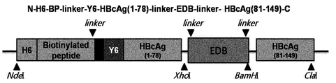
- Figure 9 shows a schematic diagram of the expression vector for the preparation of gold-protein nanoparticle fusion comprising EDB as a target directional peptide.
- FIG. 10 is a TEM photograph showing that the recombinant hepatitis B virus (HBV) capsid protein comprising EDB and gold ion reducing peptide forms a stable structure.
- HBV hepatitis B virus
- FIG. 11 shows the results of comparing the toxicity in the body of the 40 nm gold nanoparticle administration group, the gold-protein nanoparticle fusion administration group, and the deionized water administration group as the untreated control group.
- FIG. 12 shows imaging with X-ray CT imaging after injection of a gold-protein nanoparticle fusion into a tumor.
- the gene sequence of the HBV capsid protein (NCBI Nucleotide accession number: 1901-2452 sequence of the AF286594 sequence, SEQ ID NO: 1) was used as a template.
- N-NdeI-H6 hexahistidine
- BP Biotinylated peptides
- Y6 hexatyrosine
- HBVcAg XhoI-C
- SEQ ID NO: 2 N- Bam HI-HBVcAg (81) derived from the HBVcAg) gene
- 5'-XhoI-EGFR affibody-BamHI-3 '(SEQ ID NO: 4) was prepared by PCR to replace P79A80 of HBVcAg with EGFR affibody (Epidermal Growth Factor Receptor 1).
- the first part is primer sequence from primer sequence part 1 which contains restriction enzyme Nde I, using the gene sequence of HBV capsid protein (1901-2452 sequence of NCBI Nucleotide accession number: AF286594 sequence, SEQ ID NO: 1) as a template. Up to 5 were subjected to extesion PCR with primer sequence 6 containing Xho I. As a result, a PCR product consisting of 5'- Nde I-H6-BP-Y6-HBV capsid protein (amino acid sequence 1-78) -Xho I-3 '(SEQ ID NO: 2) was obtained.
- the third part was PCR using primer sequence 9 including Xho I and primer sequence 10 including Bam HII using EGFR affibody base sequence as a template.
- Primer sequences 9 and 10 were used to construct 5'- Xho I-EGFR affibody- Bam HI-3 '(SEQ ID NO: 4).
- This PCR product was sequentially inserted into the pT7-7 vector to express a hexatyrosine functional group capable of inducing reduction of HBV capsid-derived gold ions and an EGFR affibody that specifically binds to human breast cancer cells.
- pT7-7-N-H6-BP-Y6-HBVcAg (1-78) -EGFR affibody-HBVcAg (81-149) -C was constructed (FIG. 1, (A)).
- primers sequence 1 to primer sequence 5 were subjected to extesion PCR with primer sequence 8 containing Cla I.
- the resulting PCR product was transferred to pT7-7 vector.
- An expression vector having a hexatyrosine functional group capable of inducing reduction of HBV capsid-derived gold ions was inserted into the expression vector, and an expression vector was constructed and used as a control for the EGFR affibody (FIG. 1, (B)).
- E. coli strain BL21 (DE3) [F-ompThsdSB (rB-mB-)] was transformed with the expression vector prepared above, and an ampicillin-resistant transformant was selected.
- Transformed E. coli was cultured in a flask containing 250 mL of Luria-Bertani (LB) medium (containing 100 mg L-1 ampicillin) (250 mL Erlenmeyer flasks, 37 ° C., 150 rpm). When medium turbidity (O.D600) reaches about 0.4-0.5, Isopropyl- ⁇ -D-thiogalactopyranosid (IPTG) is added.
- LB Luria-Bertani
- IPTG Isopropyl- ⁇ -D-thiogalactopyranosid
- the expression of the recombinant gene was induced without adding biotin as a control.
- the cultured E. coli was centrifuged at 4,500 rpm for 10 minutes to recover the cell precipitate, and then suspended in 5 ml of crushed solution (10 mM Tris-HCl buffer, pH 7.5, 10 mM EDTA). Was crushed using an ultrasonic crusher (Branson Ultrasonics Corp., Danbury, CT, USA). After crushing, the mixture was centrifuged at 13,000 rpm for 10 minutes, and the supernatant and the insoluble aggregate were separated. Purification was carried out according to Example 3 using the separated supernatant.
- E. coli cultured in the same manner as described above was recovered and the cell pellet was resuspended in 5 mL lysis buffer (pH 8.0, 50 mMsodium phosphate, 300 mM NaCl, 20 mM imidazole) using an ultrasonic crusher. Cells were disrupted. The crushed cell solution was centrifuged at 13,000 rpm for 10 minutes to separate only the supernatant, and each recombinant protein was separated using a Ni2 + -NTA column (Qiagen, Hilden, Germany) (wash buffer: pH 8.0, 50 mM sodium phosphate). , 300 mM NaCl, 80 mM imidazole / elution buffer: pH 8.0, 50 mM sodium phosphate, 300 mM NaCl, 200 mM imidazole).
- Sucrose was added to the self-assembly buffer for each concentration to prepare a solution containing 60%, 50%, 40%, 30%, 20%, and 10% sucrose, and then the ultrafast centrifuge tube (ultraclear 13.2ml tube, Beckman), add 2 ml of sucrose solution of each concentration (60 ⁇ 20%) from the solution of high concentration. Finally, 0.5 ml of a 10% sucrose solution was filled, followed by 1 ml of the recombinant protein solution in the prepared self-assembly buffer, and ultra-high centrifugation was performed at 24,000 rpm for 16 hours at 4 ° C. (Ultracentrifuge L-90k, Beckman ).
- HBV capsid-derived chimeric nanoparticles obtained through Example 3 hexatyrosine capable of reducing gold ions is bound thereon with biotin to control the size of the gold particles.
- AuClP (CH 3 ) 3 chloro (trimethylphosphine) gold (I) was added to HBV capsid-derived chimeric nanoparticles contained in a pH 7.0 recombinant protein buffer and reacted for 16 hours at 4 ° C. for 10 minutes at 13,000 rpm. Centrifugation was performed.
- the supernatant was separated and reacted for 10 minutes with the addition of a reducing agent, NaBH 4 , 10 times the concentration of the recombinant protein, and the size of the gold-protein particle fusion (FIG. 2, (A)), which was not controlled by biotin, was A gold-protein particle fusion (FIG. 2, (B)) size controlled by biotin is obtained.
- Example 6 Temperature increase with concentration of prepared HBV capsid-derived chimeric gold-protein nanoparticle fusion-in vitro
- Cetuximab a therapeutic antibody used for anticancer treatment by binding to the EGF receptor, was pre-reacted with human breast cancer cells for 72 hours, and then the gold containing the EGFR affibody. 10 minutes of reaction with protein nanoparticle fusion. As a result, no fluorescent reaction by Cy5.5 was observed. This proved that the EGFR affibody binds directly to the EGF receptor of human breast cancer cells by targeting (FIG. 4).
- Example 7 after obtaining the cancer cells in which the particles were absorbed into the cells and the cancer cells that were not absorbed, the lasers were irradiated with lasers (655 nm and 200 W), thereby increasing the temperature caused by the gold-protein particle fusions verified in Example 6.
- the purpose of this study was to investigate the performance of photothermal therapy by confirming the degree of cancer cell necrosis. For this purpose, each 35 pie plate was irradiated with a laser, and the cell viability was confirmed using a CCK-8 kit (Dojindo, Japan).
- the laser is irradiated to the gold-protein nanoparticle fusion for 40 minutes, it can be seen that as the concentration of the fusion increases, a lot of heat is generated and cancer cell survival is reduced (FIG.
- the gold-protein nanoparticle fusion in which the biodiagnosis breast cancer specific peptide (EGFR affibody) is present shows an average of 2 to 3 times stronger fluorescence.
- the HBV capsid-derived chimeric gold-protein nanoparticle fusion was directly injected into the target cancer cell (MDA MB468 cell line), and then the photothermal treatment effect by laser irradiation was observed.
- FIG. 7A is a test group for checking the tissue state of cancer cells as a test group which is not injected with a gold-protein nanoparticle fusion as a control and is not irradiated with a laser.
- FIG. 7B is an experimental group irradiated with laser only without injecting the gold-protein nanoparticle fusion and confirmed the possibility of necrosis of tissue by the laser. Compared with the tissue result of FIG. 7A, it can be seen that necrosis of the tissue by the laser does not occur.
- 7C is an experimental group for confirming the toxicity of the gold-protein nanoparticle fusion itself by injecting the gold-protein nanoparticle fusion and not irradiating the laser. In comparison with FIG. 7A, it was confirmed that necrosis of tissue did not occur, and that there was no toxicity of the gold-protein nanoparticle fusion itself.
- 7D and 7E are experimental groups verifying photothermal treatment effects of gold-protein nanoparticle fusions by injecting gold-protein nanoparticle fusions and irradiating a laser.
- the laser irradiation for 10 minutes in the experimental group of Figure 7D it can be confirmed that necrosis of the tissue has been started in the area indicated by the arrow.
- the laser irradiation in the experimental group of FIG. 7E sufficiently for 50 minutes, it can be confirmed that there was damage of the overall tumor tissue over the entire portion of the tumor through the low magnification photograph on the left, necrosis of the tumor tissue through the circular portion of the right high magnification photograph It can be seen that has proceeded remarkably.
- this example confirmed that there was no change causing significant damage to the cancer cell tissues in the cases 1), 2), and 3), and that necrosis of the cancer cells started from the point of purchase for 10 minutes through 4). After 50 minutes of irradiation, it was confirmed that most cancer cells were necrotic by heat. Through this, it was confirmed that photothermal treatment using gold-protein nanoparticle fusion is effective enough.
- HBV capsid-derived chimeric gold-protein nanoparticle fusions were injected into the tail vein of rats that sufficiently developed the target cancer cells (MDA MB468 cell line), and then time for targeting and laser irradiation We investigated the effects of photothermal therapy according to targeting.
- mice that fully generate target cancer cells (MDA MB468 cell line) are prepared, and the gold-protein nanoparticle fusion is injected into the tail vein of the rat. Then, after confirming 9-12 hours, which is the maximum targeting time to the cancer cells through Example 9, wait for 9 hours to target the tumor, and then analyzed the tumor tissue for the experimental group (Fig. 8A) and light heat treatment The effect was compared through the experimental group (FIG. 8B) which fully irradiated the laser for 50 minutes. In addition, after 5 days, the size of the cancer cells of the experimental group irradiated with the laser-only test and the gold-protein nanoparticle fusion and irradiated with the laser was compared (FIG. 8C, D).
- Figure 8 (A) is a test group to confirm whether the tumor tissue necrosis by the gold-protein nanoparticle fusion itself, and whether the gold-protein nanoparticle fusion is toxic in the body, the gold through the tail vein- After injecting the protein nanoparticle fusion and waiting for targeting the tumor tissue for 9 hours, the tissues were checked. As a result of tissue analysis, necrosis due to toxicity of the gold-protein nanoparticle fusion itself does not occur, and since it was targeted to tumor tissue through FIGS. 6 and 7, this is a result that there is no toxicity of the substance itself.
- Figure 8 (B) is injected gold-protein nanoparticle fusion into the tail vein, wait for 9 hours to be targeted and then laser irradiation for 50 minutes to see the effect of photothermal therapy. Compared with the results of FIG. 8 (A), necrosis occurred centrally in the tumor, blood vessels expanded, and bleeding occurred with extensive necrosis.
- necrosis of cancer cells by the body targeting of the gold-protein nanoparticle fusion does not occur, through which cancer cell necrosis due to the toxicity of the substance itself did not occur.
- the laser was sufficiently irradiated for 50 minutes, necrosis of a wide range of tissues was confirmed along with expansion of blood vessels in the tumor core. In addition, bleeding due to necrosis of blood vessels in some cancer cells was confirmed.
- FIG. 8 (C) compares the tumor status after 5 days of the experimental group irradiated with only the laser without the gold-protein nanoparticle fusion and the experimental group injected with the gold-protein nanoparticle fusion and fully irradiated with the laser for 50 minutes.
- the tumor was larger in the experimental group irradiated with the laser only, but in the experimental group irradiated with the gold-protein nanoparticle fusion, the tumor was removed and disappeared as shown in FIG. have.
- FIG. 8 (D) shows quantitatively the tumor size growth trends of the two experimental groups and the change in tumor size after application of photothermal therapy with laser.
- gold-protein nanoparticle fusion can be injected into the body and targeted to the target organ where tumor cells are generated, and it can be confirmed that it can be used to remove cancer cells through photothermal treatment using a laser of NIR wavelength.
- the gold-protein nanoparticle fusion used in this experiment is more stable for various changes in pH environment in the body than the existing gold nanoparticles, and its structure is stable and its targeting effect is excellent. have.
- target directed peptides for EDB with linear structure were used to test the synthesis of HBV capsid derived chimeric nanoparticles.
- 5'-XhoI-EDB-BamHI-3 '(SEQ ID NO: 16) was prepared by PCR to replace P79A80 of HBVcAg with human fibronectin extradomain B (EDB).
- the plasmid expression vector pT7-7-N-H6-BP-Y6-HBVcAg (1-78) -EDB-HBVcAg (81-149) -C was constructed (FIG. 9). All plasmid expression vectors produced were sequence verified through complete DNA sequencing after agarose gel-purification.
- the first part is primer sequence from primer sequence part 1 which contains restriction enzyme Nde I, using the gene sequence of HBV capsid protein (1901-2452 sequence of NCBI Nucleotide accession number: AF286594 sequence, SEQ ID NO: 1) as a template. Up to 5 were subjected to extesion PCR with primer sequence 6 containing Xho I. As a result, a PCR product consisting of 5'- Nde I-H6-BP-Y6-HBV capsid protein (amino acid sequence 1-78) -Xho I-3 '(SEQ ID NO: 2) was obtained.
- PCR was performed using primer sequence 11 including Xho I and primer sequence 12 including Bam HII, using the EDB base sequence as a template.
- Primer sequences 11 and 12 of Table 2 were used for the preparation of 5′- Xho I-EDB- Bam HI-3 ′ (SEQ ID NO: 16).
- PCR products were sequentially inserted into the pT7-7 vector to specifically bind to the hexatyrosine functional group and human glioblastoma and astrocytoma tumor cells (U87MG cell line), which can induce reduction of HBV capsid-derived gold ions.
- primers sequence 1 to primer sequence 5 were subjected to extesion PCR with primer sequence 8 containing Cla I, and the resulting PCR product was transferred to pT7-7 vector.
- An expression vector having a hexatyrosine functional group capable of inducing reduction of HBV capsid-derived gold ions was inserted to construct an expression vector and used as a control for EDB (FIG. 1, (B)).
- Primer 11 (SEQ ID NO: 18) 5 'XhoI-EDB CTCGAG CAT AGC TGC AGC TCC CCG ATT CAG Primer 12 (SEQ ID NO: 19) 3 'BamHI-EDB GGA TCC CGG CTG CTG TTC CAG ACG AAT AAT GCC
- the experimental group injected with gold nanoparticles showed black color due to no release of substances from the liver for 21 days, and showed a high level of concentration in the dark-field photograph, whereas the gold-protein nanoparticle fusion was dissected.
- the dark-field image shows that gold is almost invisible after 7 days as the substance escapes in vitro within a week.
- the gold-protein nanoparticle fusion when applied to the body, it can be seen that it can be used as a biocompatible material by solving the toxicity problems in the existing gold nanoparticles.
- the experimental group injected with the gold-protein nanoparticle fusion in the tumor was compared with the untreated control group to confirm whether it could be utilized as a contrast agent of X-ray CT (FIG. 12).
- Gold can be used as a contrast agent when the X-ray is irradiated, and the contrast level can be used as a contrast agent. If the contrast effect of the gold-protein nanoparticles is verified, a high level of contrast effect can be obtained using the in vivo targeting shown in Example 9. It can be used as a contrast agent.
- X-ray CT scans show similar brightness to the surrounding bones.
Landscapes
- Health & Medical Sciences (AREA)
- Chemical & Material Sciences (AREA)
- Life Sciences & Earth Sciences (AREA)
- Organic Chemistry (AREA)
- General Health & Medical Sciences (AREA)
- Medicinal Chemistry (AREA)
- Engineering & Computer Science (AREA)
- Biophysics (AREA)
- Proteomics, Peptides & Aminoacids (AREA)
- Genetics & Genomics (AREA)
- Molecular Biology (AREA)
- Biochemistry (AREA)
- Gastroenterology & Hepatology (AREA)
- Zoology (AREA)
- Animal Behavior & Ethology (AREA)
- Public Health (AREA)
- Veterinary Medicine (AREA)
- Toxicology (AREA)
- Epidemiology (AREA)
- Biomedical Technology (AREA)
- Nanotechnology (AREA)
- Immunology (AREA)
- Pharmacology & Pharmacy (AREA)
- Bioinformatics & Cheminformatics (AREA)
- Cell Biology (AREA)
- Virology (AREA)
- Radiology & Medical Imaging (AREA)
- Nuclear Medicine, Radiotherapy & Molecular Imaging (AREA)
- Pathology (AREA)
- General Engineering & Computer Science (AREA)
- Wood Science & Technology (AREA)
- Biotechnology (AREA)
- Crystallography & Structural Chemistry (AREA)
- Plant Pathology (AREA)
- Physics & Mathematics (AREA)
- Microbiology (AREA)
- Peptides Or Proteins (AREA)
- Medicines That Contain Protein Lipid Enzymes And Other Medicines (AREA)
Abstract
본 발명은 표적 지향성 펩타이드를 포함하는 재조합 자기조립성 단백질 및 그의 용도에 관한 것이다. 본 발명에 따른 표적 지향성 펩타이드를 포함하는 재조합 자기조립성 단백질은 추가적인 표적 지향성 부여 과정을 필요로 하지 않기 때문에 화학적 결합제 또는 안정화제 등의 보조제를 사용하지 않고도 원하는 약물을 표적 조직 또는 표적 세포로 전달하는 것이 가능하며, 이로써 광열 치료, 약물 전달 또는 이미징 등에 사용가능하다. 특히, 본 발명에 따르면 추가적인 표면 안정화 과정이나 표적 지향성 부여 과정 없이도, 표적 지향성을 가지면서 균일한 고밀도의 금 나노입자가 단백질 표면에 결합되어 있는 금-단백질 나노입자 융합체를 제작할 수 있다. 본 발명에 따른 금-단백질 나노입자 융합체는 기존의 금 나노입자에 비하여 pH의 변화 및 농도의 변화에 구조적 안정성을 보이며 표적 지향성 또한 우수하므로, 금 나노입자의 광열 치료 요법에 있어서 획기적인 활용도의 증진을 가져다 줄 수 있다.
Description
본 발명은 표적 지향성 펩타이드를 포함하는 재조합 자기조립성 단백질 및 그의 용도에 관한 것이다.
현대 의학의 발전에도 불구하고 약물 요법 또는 방사선 요법을 필요로 하는 질병을 앓고 있는 환자들은 약물의 전신적 투여에 의한 세포독성 또는 부작용, 혹은 방사선 요법에 의한 정상세포의 변이 또는 사멸 등의 부작용을 겪게 된다. 만일 조영제 또는 치료제와 같은 약물을 원하는 표적 조직에 정확히 전달할 수 있다면 정상세포 또는 정상조직에 대한 부작용 없이 질환을 진단하고 또한 치료하는 것이 가능해진다. 때문에, 부작용 없는 진단 또는 치료를 위해 약물의 표적화에 대한 연구가 광범위하게 이루어지고 있다.
약물의 표적화는 약물의 전달을 목적으로 하는 조직 또는 세포에 특이적인 항체나 펩타이드, 특정 리간드, 또는 고분자 등을 약물에 화학적 또는 물리적인 방법으로 결합시키는 방법을 주로 사용한다. 그러나, 약물의 물리화학적 특성으로 인해 항체, 펩타이드, 리간드 등을 결합시키는 것이 항상 용이하지는 않으며, 또한 화학적 또는 물리적 결합을 위한 결합제 또는 안정화제 등을 사용할 경우 약물의 약리적 특성이나 생체독성 등에 영향을 미치는 문제가 발생할 수 있다. 따라서 이러한 문제를 일으키지 않는 약물의 표적화 방법에 대한 기술적 요구는 계속되고 있는 실정이다.
한편, 표적지향적으로 약물을 전달하여 특히 암 세포를 사멸화시키는 방법들의 중의 하나로 금 나노입자의 전달에 이은 광열 치료방법이 효과적인 암 치료수단이 될 수 있다. 금 나노입자는 수십 나노미터 크기에서, 표면 전자의 양자화에 의해서 고유의 광학적 물성을 나타내며, 단전자 소자, 화학물질 센서, 바이오 센서, 약물전달 및 촉매 등의 분야에 다양하게 활용되고 있다. 일예로, Rice 대학의 Halas 교수와 West 교수 연구팀에서는 금 나노 쉘을 합성하여 이를 열치료에 의한 암세포의 괴사에 응용하였다. 상기 금 나노입자는 실리카 핵 부분과 그 위의 금 나노 외각부로 이루어진 나노 구조물인데 핵과 외각의 두께 비율에 따라 빛의 흡수 파장이 가시 광선 영역에서 근적외선 (NIR) 영역까지 조절된다. 상기 연구팀에서는 NIR 흡수 단면적이 매우 큰 금 나노 쉘을 합성하여, 금 나노 쉘 표면에 암세포 특이적인 항체를 결합시킨 후 이를 암세포에 반응시키고, 여기에 NIR 연속파 레이저 (CW laser)를 조사하였다. 금 나노 쉘에 의해 흡수된 NIR 빛은 열로 바뀌어 암세포를 효과적으로 괴사시킬 수 있었다. 800 nm 내지 1200 nm의 NIR 영역의 빛은 생체 조직에 의한 흡수가 최소에 달하기 때문에 가시 광선에 비해 조직 깊숙한 곳 도달할 수 있다. 따라서 절개 부위를 최소화하고 NIR 영역의 빛을 조사함으로써 원하는 열 치료 효과를 가져올 수 있다.
그러나, 금 나노입자를 합성하기 위해 화학 환원반응과 같은 종래의 방법을 사용할 때에는, 일반적으로 금속 화합물, 용매, 환원제 또는 안정화제를 필요로 한다. 의약 분야에서의 금 나노입자의 사용을 가로막는 주된 문제는 이러한 화학 첨가제에 의해 발생되는 독성이다.
또한, 안정적인 금 나노입자를 제조하기 위해서는 금 나노입자의 표면 개질이 반드시 필요하다. 표면 개질을 하지 않는 경우, pH에 변화를 주거나 고농도로 농축을 할 때, 구조적 불안정성으로 인해 응축현상이 일어나 입자의 크기와 형태가 변형되어 버린다. 따라서 추가적인 표면 안정화 기술이 반드시 요구된다. 더군다나, 질병 진단 및 치료에 이용을 하는 경우, 앞서 설명한 약물의 표적화를 위해 금 나노입자의 표면에 항체 또는 타켓팅 펩타이드를 화학적 결합을 이용하여 노출시켜야 하는데, 이 때 고밀도로 균일하게 노출시키는 부분에서 한계점을 갖는다.
일반적으로 금 나노입자의 합성 과정에서는 소듐 시트르산염와 같은 금 이온의 응결핵 역할을 하는 물질이 사용되는데, 티로신과 같이 pH 7.0이상에서 금 이온이 환원될 수 있는 표준 환원 전위를 제공하는 아미노산을 포함하는 펩타이드를 이용하면 금 이온이 환원되어 펩타이드 표면에 금 나노입자를 합성할 수 있음이 보고된 바 있다(KR2012-0052501호 등). 그러나, KR2012-0052501호에서 보고하고 있는 기술은 공지의 금 친화성 단백질 또는 금 이온 환원성 펩타이드와 금 전구체를 반응시켜 상기 단백질 또는 펩타이드 주변에 금 나노입자를 응집시키는 정도에 불과하여 금 나노입자의 형상이나 크기 제어가 어려울 뿐만 아니라, 표적 지향성을 부여하기 위해서는 추가적인 화학적 결합을 필요로 한다.
본 발명은 추가적인 표적 지향성 부여 과정을 필요로 하지 않는, 표적 지향성 펩타이드를 포함하는 재조합 자기조립성 단백질 및 그의 용도를 제공하고자 한다. 또한, 본 발명은 이러한 재조합 자기조립성 단백질의 일 례로서, 추가적인 표면 안정화 또는 표적 지향성 부여 과정 없이 고밀도의 균일한 표적 지향성 펩타이드를 한번에 노출시킬 수 있는 금-단백질 입자 융합체 및 이의 용도를 제공하고자 한다.
본 발명은 자기조립성 단백질에 융합된 표적 지향성 펩타이드를 포함하는 재조합 자기조립성 단백질, 표적 지향성 펩타이드 및 금 이온 환원성 펩타이드를 포함하는 재조합 자기조립성 단백질, 이러한 재조합 자기조립성 단백질로 이루어진 재조합 자기조립성 단백질 나노입자 상에 금 나노입자가 형성되어 있는 금-단백질 입자 융합체, 및 상기 금-단백질 입자 융합체의 광열 치료를 위한 용도 또는 조영제로서의 용도를 제공한다.
본 발명에 따른 재조합 자기조립성 단백질 나노입자는 나노미터 크기의 직경을 갖는 구형의 단백질 입자로서, 자기조립성 단백질에 융합된 표적 지향성 펩타이드를 포함하는 재조합 자기조립성 단백질의 단량체들이 모여 자가조립되어 하나의 구형 재조합 자기조립성 단백질 나노입자를 이룬다.
본 발명에 있어서, 자기조립성 단백질은 단백질, 단백질의 서브유닛 또는 펩타이드가 수 개가 모였을 때 이들이 스스로 조직적인 구조 또는 패턴을 형성하며 집합체를 형성하는 단백질, 단백질의 서브유닛 또는 펩타이드를 의미한다. 이러한 자기조립성 단백질은 별도의 조작 없이도 단백질의 나노입자 형성을 가능하게 하므로 본 발명에 따른 단백질 나노입자의 제조를 위해 유리하게 사용할 수 있다. 본 발명의 단백질 나노입자는 자기조립에 의해 입자를 이루므로, 매우 균일한 입도 분포를 보이는 입자를 재현성있게 제조할 수 있을 뿐 아니라, 생체 내에서 사용된 후 분해될 수 있는 생체친화성 물질이기 때문에 나노입자의 잔존으로 인한 독성의 문제가 없다.
한편, 본 발명에 있어서, 표적 지향성 펩타이드란 암세포나 염증세포와 같이 치료나 진단이 요구되는 생체 내 목적 부위로 본 발명에 따른 재조합 자기조립성 나노입자가 이동할 수 있도록, 암세포나 염증세포 등의 세포 표면에 결합할 수 있는 펩타이드나 항체를 의미한다. 상기 표적 지향성 펩타이드는 재조합 자기조립성 단백질 나노입자의 표면에 도입될 수 있다.
본 발명에 있어서, 자기조립성 단백질에 표적 지향성 펩타이드가 융합된 재조합 자기조립성 단백질 나노입자의 모노머는 하나의 발현 벡터를 통해 발현된다.
따라서, 표적 지향성 펩타이드는 상기 발현 벡터에 쉽게 삽입 가능하면서, 본 발명에 따른 자기조립성 단백질 나노입자의 구조에 영향을 미치지 않는 것이라면 펩타이드의 서열의 길이와 관계없이 어떠한 것이든 사용할 수 있다. 특히 특정 질환 조직 및 세포에서 특이적으로 과발현되는 수용체에 실제 호르몬과 유사하게 대신 결합하여 신호 전달을 억제한다고 알려진 다수 펩타이드 형태의 안타고니스트, 펩타이드 호르몬 (Somatostatins, Bombesin, Cholecystokinin, gastrin analog) 등으로 대체 가능하다.
본 발명의 한 구체예에서는 상기 자기조립성 단백질이 인간 유래의 자기조립성 단백질일 수 있다. 본 발명에 있어서, 인간 유래의 자기조립성 단백질 또는 이를 포함하는 단백질 나노입자는 인간화된 자기조립성 단백질 또는 인간화된 단백질 나노입자를 또한 포함하는 것으로 간주된다.
이에 제한되는 것은 아니나, 본 발명의 한 구체예에서, 상기 자기조립성 단백질은 페리틴일 수 있다. 페리틴은 중쇄와 경쇄로 구성된 24개의 동일한 단백질 서브유닛으로 구성되며, 자기조립성을 갖고 있어 생체 내에서 구형의 hollow shell을 형성한다.
한 구체예에서, 상기 자기조립성 단백질은 페리틴 중쇄 단백질(Ferritin Heavy Chain: 이하 "FTN-H"이라고도 함)일 수 있다.
이에 제한되는 것은 아니나, 페리틴에 융합되는 표적 지향성 펩타이드는 페리틴의 N-말단 또는 C-말단에 융합될 수 있다. 페리틴과 융합되는 표적 지향성 펩타이드는 단백질 나노입자의 표면에 위치하게 됨으로써 강한 표적 지향성을 제공하므로 본 발명의 재조합 자기조립성 단백질 나노입자를 목적 부위까지 효율적으로 전달할 수 있다.
또 다른 구체예에서, 본 발명의 자기 조립성 단백질은 B형 간염 바이러스(HBV) 캡시드 단백질일 수 있다.
본 발명에 따른 재조합 자기조립성 단백질의 제조를 위해 사용되는 B형 간염 바이러스(HBV) 캡시드 단백질은 180-240 개의 단량체가 자가조립되어 하나의 구형 단백질 나노입자를 이루며, 유전자 재조합 기술을 이용하여 대장균에서 대량 생산이 가능하다. 특히 스파이크 부위에 외래 단백질을 융합 발현할 경우 외래 단백질이 단백질 나노입자의 표면에 표출되는 특징을 가지고 있다. 따라서, 본 발명에서는 표적 지향성 펩타이드를 HBV의 스파이크 부위에 발현시킴으로써 특이적인 타겟팅이 가능하게 할 수 있다.
또한, 본 발명은 상기 재조합 자기조립성 단백질로 이루어진 재조합 자기조립성 단백질 나노입자를 제공한다. 본 발명에 따른 재조합 자기조립성 단백질 나노입자는 표면에 표적 지향성 펩타이드가 표출되어 있어 광열 치료, 약물전달 또는 이미징 등의 최종 용도의 목적에 부합하는 생체 내 목적 부위에 효율적으로 전달된다.
본 발명에 따른 재조합 자기조립성 단백질 나노입자는 표적 지향성을 제공하므로 다양한 용도로 활용될 수 있다. 예를 들어 표적 지향성 펩타이드 외에 다른 융합 펩타이드 또는 단백질을 추가로 포함시킴으로써 그 용도를 확장시키는 것이 가능하다.
한 구체예에서, 본 발명에 따른 재조합 자기조립성 단백질 나노입자는 광열 치료, 약물 전달 또는 조영을 위해 사용할 수 있다.
본 발명의 일 실시예에서는, 상기 재조합 자기조립성 단백질의 일예로서, 자기조립성 단백질에 융합된 표적 지향성 펩타이드 및 금 이온 환원성 펩타이드를 포함하는 재조합 자기조립성 단백질을 제공한다.
광열 치료를 위한 재조합 자기조립성 단백질 나노입자는 금 이온 환원성 펩타이드를 포함하고 있으므로 금 전구체와 반응시 금 이온이 환원되면서 재조합 자기조립성 단백질 나노입자 상에 금 나노입자를 형성하게 된다. 본 명세서에서는 금 나노입자가 재조합 자기조립성 단백질 나노입자 상에 형성되어 있는 입자를 금-단백질 입자 융합체라고 지칭한다.
한 구체예에서, 상기 자기조립성 단백질은 HBV 캡시드 단백질일 수 있다. 이 경우 상기 표적 지향성 펩타이드는 재조합 HBV 캡시드 단백질의 스파이크 부위, N 말단 또는 C 말단에 도입될 수 있다. 본 발명의 한 구체예에서, 상기 표적 지향성 펩타이드는 재조합 HBV 캡시드 단백질의 스파이크 부위에 도입될 수 있다.
본 발명에 있어서, HBV 캡시드 단백질에 표적 지향성 펩타이드 및 금 이온 환원성 펩타이드가 융합된 HBV 캡시드 단백질 나노입자의 모노머는 하나의 발현 벡터를 통해 발현된다.
따라서, 표적 지향성 펩타이드는 상기 발현 벡터에 쉽게 삽입 가능하면서, 본 발명에 따른 금 이온 환원성 펩타이드의 기능 및 자기조립성 단백질 나노입자의 구조에 영향을 미치지 않는 것이라면 펩타이드의 서열의 길이와 관계없이 어떠한 것이든 사용할 수 있다. 특히 특정 질환 조직 및 세포에서 특이적으로 과발현되는 수용체에 실제 호르몬과 유사하게 대신 결합하여 신호 전달을 억제한다고 알려진 다수 펩타이드 형태의 안타고니스트, 펩타이드 호르몬 (Somatostatins, Bombesin, Cholecystokinin, gastrin analog) 등으로 대체 가능하다.
이에 제한되는 것은 아니나, 본 발명의 실시예에서는 인간 유방암세포에 특이성을 갖는 EGFR(Epidermal Growth Factor Receptor) 또는 두경부암의 바이오마커로 활용할 수 있는 EDB(human fibronectin extradomain B)를 표적으로 하는 표적 지향성 펩타이드를 사용한다.
한 구체예에서, 상기 표적 지향성 펩타이드는 재조합 HBV 캡시드 단백질의 스파이크 부위에 도입된다. 본 발명의 일 실시예에서는 유전자 재조합 기술을 이용하여 인간 유방암세포(MDA MB-468 tumor model)에 특이성을 갖는 펩타이드(EGFR affibody)를 HBV 캡시드 단백질의 스파이크 부위에 발현시킴으로써 본 발명의 금-단백질 입자 융합체에 표적 지향성을 부여하는 방법을 제시한다. 예를 들어, HBV 캡시드 단백질의 1-78번째 아미노산 서열 부분과 81-149번째 아미노산 서열 부분 사이에 상기 표적 지향성 펩타이드가 위치하도록 하면 HBV 캡시드 단백질의 스파이크 부위에 표적 지향성 펩타이드가 고집적도로 표출될 수 있다. 본 발명의 한 구체예에서, 상기 표적 지향성 펩타이드는 재조합 HBV 캡시드 단백질 내로 복수 개 삽입될 수 있다. 표적 지향성 펩타이드를 복수 개 삽입하게 되면 표적 지향성 펩타이드의 표출 밀도를 높여 결과적으로 본 발명에 따른 금-단백질 입자 융합체의 표적 조직으로의 이동을 돕게 된다.
본 발명에 따라 화학적 방법이 아닌 유전공학적 방법으로 표적 지향성 펩타이드를 결합시키게 되면, 표적 지향성 펩타이드의 표면 표출 빈도 및 표출 위치를 조절 할 수 있는 장점이 있다. 예를 들어, 표적 지향성 펩타이드의 표면 표출 빈도를 낮게 조절하고자 하는 경우에는 표적 지향성 펩타이드와 금 이온 환원성 펩타이드를 포함하는 재조합 자기조립성 단백질과 표적 지향성 펩타이드가 융합되어 있지 않은채 금 이온 환원성 펩타이드만을 포함하는 재조합 자기조립성 단백질을 적정 비율로 혼합하여 재조합 자기조립성 단백질 나노입자를 제조할 수 있다.
한편, 재조합 자기조립성 단백질에 추가로 포함되는 금 이온 환원성 펩타이드는 본 발명에 따른 금-단백질 입자 융합체의 형성을 위해 필수적인 요소이다. 금 이온 환원성 펩타이드는 금 전구체에 포함된 금 이온이 환원될 수 있는 표준 환원 전위를 제공하는 펩타이드를 의미한다. 금 이온 환원성 펩타이드는 일반적으로 티로신, 히스티딘, 시스테인과 같은 아미노산이 포함되어 있는 펩타이드로서, 복수 개의 타이로신(Yn, n≥2), 히스티딘(Hn, n≥2), 시스테인(Cn, n≥2) 또는 최소한 이들 중 하나의 아미노산을 포함하는 아미노산 서열로 이루어진 펩타이드일 수 있다. 예를 들어, YYY, YYYYYY, YAHHYAHHYAADY 등의 아미노산 서열을 갖는 펩타이드들이 금 이온성 펩타이드로서 알려져 있다. 본 발명의 한 구체예에서, 상기 금 이온 환원성 펩타이드는 헥사티로신일 수 있다. 한편, 이러한 금 이온 환원성 펩타이드는 재조합 자기조립성 단백질의 스파이크 부위, N 말단 또는 C 말단에 도입될 수 있다. 한 구체예에서, 상기 금 이온 환원성 펩타이드는 재조합 자기조립성 단백질의 N 말단 또는 C 말단에 도입된다. 본 발명의 일 실시예에서는 상기 금 이온 환원성 펩타이드로서 헥사티로신을 단백질의 N-말단에 발현시켜 재조합 자기조립성 단백질의 나노입자의 표면 상에 금 이온 환원성 펩타이드가 고집적도로 표출될 수 있음을 보여준다.
추가로, 한 구체예에서, 상기 재조합 자기조립성 단백질은 금 이온 환원성 펩타이드 부근에 금 입자 크기 제어 펩타이드를 추가로 포함할 수 있다. 금 입자 크기 제어 펩타이드란 금 이온 환원성 펩타이드가 금 전구체와 반응하여 금 입자를 형성할 때 그 크기가 무한정 커지지 않도록 물리적 장벽을 형성해 주는 역할을 한다. 금 입자 크기 제어 펩타이드는 이러한 물리적 장벽을 형성해 줄 수 있는 것이면 어떠한 것이든 사용할 수 있으므로 그 종류는 특별히 제한되지 않는다. 본 발명의 일 실시예에서는 금 입자 크기 제어 펩타이드로서 바이오틴 결합 펩타이드(biotinylated peptide)를 재조합 HBV 캡시드 단백질에 융합발현 시키고, 발현 과정에서 바이오틴을 첨가하여 금 나노입자가 과도하게 크게 성장하는 것을 막아 금 나노입자의 크기를 제어할 수 있음을 보여준다. 반면 바이오틴 결합 펩타이드를 융합시키지 않은 군에서는 금 나노입자의 크기가 제어되지 않으며, 농축이나 pH 조절시 응집이 발생하게 된다.
본 발명의 한 구체예에서, 재조합 자기조립성 단백질은 금 이온 환원성 펩타이드와 금 입자 크기 제어 펩타이드 사이에 링커 펩타이드를 추가로 포함할 수 있다. 링커 펩타이드는 다수의 글라이신을 포함하는 반응성이 없는 펩타이드로서 금 이온 환원성 펩타이드와 금 입자 크기 제어 펩타이드 사이 간의 공간을 적절한 크기로 확보하여 금 입자가 충분히 성장할 수 있으면서 동시에 금 이온이 과도하게 환원될 수 있는 가능성을 차단시킬 수 있으며, 구조적 안정성을 제공해 준다.
링커 펩타이드는 금 이온 환원성 펩타이드와 금 입자 크기 제어 펩타이드 간의 적절한 공간을 확보해 줄 수 있는 길이를 가질 수 있다. 따라서, 링커 펩타이드의 길이는 금 이온 환원성 펩타이드와 금 입자 크기 제어 펩타이드의 종류 및 크기에 따라 달라질 수 있을 것이다. 예를 들어 링커 펩타이드는 5 내지 20개, 예컨대, 5 내지 15개의 아미노산으로 이루어진 펩타이드일 수 있다.
또한, 분리 정제의 용이성을 위해, 한 구체예에서, 상기 재조합 자기조립성 단백질은 히스티딘 태그나 FLAG(DYKDDDDK) 태그, GST 태그와 같은 분리정제를 위한 펩타이드를 추가로 포함할 수 있다.
본 발명에 따른 재조합 자기조립성 단백질은 a) HBV 자기조립성 단백질의 유전자로부터 유래한 유전자 클론을 얻는 단계, b) 자기조립성 단백질 내로의 삽입을 위한 표적 지향성 펩타이드를 코딩하는 핵산서열을 포함하는 클론을 제조하는 단계, c) 상기 제조된 유전자클론의 라이게이션을 통하여 발현 벡터를 제조하는 단계, 및 d) 상기 발현벡터를 숙주에 형질 전환하여 재조합 자기조립성 단백질을 발현하는 단계를 포함하는 제조방법을 통해 제조될 수 있다.
상기 자기조립성 단백질 유전자로부터 유래한 유전자 클론은 상술한 금 이온 환원성 펩타이드, 링커 및 분리정제를 위한 펩타이드를 코딩하는 핵산 서열을 포함할 수 있다.
한편, 상술한 재조합 자기조립성 단백질은 재조합 자기조립성 단백질 나노입자를 구성하는 모노머로서, 180-240 개의 단량체가 자가조립되어 재조합 자기조립성단백질 나노입자를 이룬다. 따라서, 본 발명은 상기 재조합 자기조립성 단백질로 이루어진 재조합 자기조립성 단백질 나노입자를 제공한다.
한편, 본 발명에 따른 금-단백질 입자 융합체는 상기 재조합 자기조립성 단백질로 이루어진 자기조립성 단백질 나노입자 상에 금 나노입자가 형성되어 있는 융합체를 의미하는 것으로, 금 전구체와 상기 재조합 자기조립성단백질 나노입자를 반응시켜 금 이온을 환원시킴으로써 상기 재조합 자기조립성 단백질 나노입자 상에 금 나노입자를 형성한 것을 의미한다. 따라서, 본 발명은 금 전구체와 상기 재조합 자기조립성 단백질 나노입자를 반응시켜 상기 재조합 자기조립성 단백질 나노입자 상에 금 나노입자를 형성하는 것을 포함하는 금-단백질 입자 융합체의 제조방법 및 이에 따라 제조된 금-단백질 입자 융합체를 제공한다.
본 발명에 있어서, 재조합 자기조립성 단백질 나노입자와의 반응을 위해 사용될 수 있는 금 전구체는 클로로(트리메틸포스핀)금(AuClP(CH3)3), 칼륨테트라클로로아우르산염(Ⅲ)(KAuCl4), 염화금산나트륨(NaAuCl4), 염화금산(HAuCl4), 브롬화금산나트륨(NaAuBr4), 염화금(AuCl), 염화금(III)(AuCl3) 또는 브롬화금(AuBr3) 등과 같은 공지의 금 전구체를 사용할 수 있다.
재조합 자기조립성 단백질 나노입자와 금 전구체와의 반응은 pH 7.0 내지 pH 10 의 pH 조건하에서 수행될 수 있다.
또한, 재조합 자기조립성 단백질 나노입자와 금 전구체와의 반응은 이에 제한되는 것은 아니나, 2시간 내지 16시간 동안 수행될 수 있다. 반응 시간이 2시간 미만일 경우에는 환원이 충분하게 되지 않으며, 16시간을 초과하게 될 경우 단백질 자체에 좋지 않은 영향이 있을 수 있기 때문이다. 반응 시간은 반응에 참여하는 재조합 자기조립성 단백질 나노입자나 금 전구체의 종류, 농도 등에 따라 변경될 수 있다.
본 발명에 따른 재조합 자기조립성 단백질 나노입자와 금 전구체와의 반응은 환원제의 추가에 의해 반응이 개시된다. 단백질 자체는 구조적 안정성이 크므로 환원제의 강력한 환원력이 제공되지 않으면 금 이온을 넣더라도 금 입자 형성이 어렵기 때문이다. 비록 환원제를 사용하긴 하나, 화학적 금 나노입자의 제조방법과 비교할 때 본 발명은 고온의 조건이 아닌 상온 조건에서 반응을 진행시킬 수 있고, 만들어진 금-단백질 나노입자 융합체가 추가적 표면 처리 없이도 매우 높은 구조적 안정성을 갖는다는 장점이 있다. 한 구체예에서, 본 발명에서 사용될 수 있는 환원제는 NaBH4 또는 H2O2 일 수 있으나, 이에 제한되는 것은 아니다.
이와 같은 방법으로 제조된 금-단백질 입자 융합체는 제어된 크기의 금 나노입자가 재조합 자기조립성 단백질 나노입자 표면에 형성되어 있는 형상을 갖는다.
본 발명에 따른 금-단백질 입자 융합체 상에 형성된 금 나노입자는 1nm 이하의 직경을 가질 수 있다. 이 크기는 재조합 자기조립성 단백질의 종류에 따라 달라질 수 있으며, 또한 금 입자 환원 펩타이드와 금 입자 크기 제어 펩타이드 간의 간격 즉, 링커 펩타이드의 길이와 환원제의 농도 반응 시간 등을 조절하여 그 입자 크기를 조절할 수 있다. 예를 들어, 재조합 HBV 캡시드 단백질 나노입자의 크기는 약 35 내지 36 nm이므로, 금 입자가 단백질 나노입자 표면에 형성된 본 발명에 따른 금-단백질 입자 융합체는 35-40nm의 금 나노입자와 유사한 물성을 띈다.
본 발명의 금-단백질 입자 융합체는 재조합 자기조립성 단백질 나노입자 상에 금 입자가 형성된 것이기 때문에 매우 균일한 입도 분포를 나타낸다. 또한, 금 입자 크기 제어 펩타이드를 이용하여 단백질 나노입자 상의 금 입자의 크기를 제어하였기 때문에, 표적 지향성 펩타이드를 방해하지 않는다. 또한, 생체 내에서 사용된 후 분해될 수 있는 생체친화성 물질이고, 금 나노입자의 화학적 표면 개질을 위한 첨가제들을 사용하지 않았기 때문에 안전성과 독성의 문제가 없다.
본 발명에 따른 금-단백질 입자 융합체는 광열 치료에 매우 효과적으로 사용할 수 있다. 본 발명의 일 실시예에서는 금-단백질 나노입자 융합체의 농도를 증가시켜가면서 레이저를 조사함으로써 암 세포나 염증 세포가 괴사할 수 있는 온도(일반적으로 43도 내지 45도) 이상 최대 55도까지 융합체의 온도가 증가함을 보여준다. 또한, 본 발명의 일 실시예에서는 체내에 주사하여 확인한 결과 유방암 세포 특이적인 표적 지향성 펩타이드를 포함하는 본 발명의 금-단백질 입자 융합체가 인간 유방암 세포에 효과적으로 타겟팅됨을 보여준다.
따라서, 본 발명은 상기 금-단백질 입자 융합체의 광열 치료를 위한 의약의 제조를 위한 용도, 상기 금-단백질 입자 융합체 및 약학적으로 허용되는 담체를 포함하는 광열 치료용 의약 조성물 및 상기 금-단백질 입자 융합체를 대상체에 투여하고 광 조사하는 것을 포함하는 광열 치료 방법을 제공한다.
암 세포 또는 염증 세포에 대한 광열 치료를 통해 사멸이 요구되는 이들 세포를 제거하는 치료법에 대해서는 당업계에 잘 알려져 있다.
특히 암 세포는 정상세포에 비해 열에 약하기 때문에 암 세포가 위치한 국소 부위에 광감응물질을 위치시킨 뒤, 외부에서 자극을 주여 열을 발생시키면 암세포만을 선택적으로 사멸시킬 수 있어 화학요법이나 방사선 치료에 비해 정상세포에 미치는 악영향을 최소화할 수 있다.
광열 치료는 암뿐만 아니라 류마티스 관절염 내 염증세포 또는 병변 조직을 제거하기 위해 사용할 수 있다. 류마티스 관절염의 병변은 관절강을 안쪽으로 둘러싸고 있는 활막의 염증(synovitis)이다. 활막 내에서 염증이 시작되면 더 많은 염증세포가 활막에 침윤되고, 활막 세포들이 증식하며, 신생혈관의 생성되는 과정을 통해 활막 조직은 더욱 증식되고 활성화된다. 병이 진행됨에 따라 염증반응의 결과로 활막 조직 주위의 관절 연골의 손상과 골파괴가 나타난다. 이러한 관절 내의 염증 반응에는 T 림프구와 B 림프구가 주로 관여하며 이들에서 분비되는 다양한 사이토카인들, 특히 TNF-α, 인터루킨(IL)-1, IL-6에 의해 염증이 지속되고 연골과 골의 파괴가 발생하게 된다. 따라서, 광열 치료를 통해 류마티스 관절염에서 발견되는 이러한 병변 조직을 제거하게 되면 류마티스 관절염의 치료 효과를 증진시킬 수 있다. 이러한 광열 치료는 비스테로이드성 항염증제(nonsteroidal anti-inflammatory drugs, NSAIDs) 또는 항류마티스 약물(disease-modifying anti-rheumatic drugs, DMARDs)의 투여와 병행하여 수행될 수 있을 것이다.
본 발명에 따른 금-단백질 입자 융합체의 발열을 위해 가해지는 광의 파장은 이에 제한되는 것은 아니나 600nm 내지 1500 nm, 예를 들어, 800nm 내지 1200 nm의 파장을 사용할 수 있다. 상기 금-단백질 입자 융합체의 발열을 위해 광을 가하는 시간은 1초 내지 10시간, 예를 들어, 1초 내지 1시간, 1초 내지 30분, 1초 내지 10분, 1초 내지 1분일 수 있다. 광을 가하는 시간은 사멸시키고자 하는 세포의 양, 즉 병변 조직의 범위나 병의 중증도에 따라 달라질 수 있을 것이다.
본 발명에 따른 금-단백질 입자 융합체는 금 나노입자를 단백질 표면에 포함하고 있기 때문에 X선이나 CT 이미징을 통해 금-단백질 입자 융합체의 생체 내 투여 후 이동 상황이나 표적 부위에의 도달 등을 가시적으로 확인할 수 있다. 따라서, 본 발명은 또한 상기 금-단백질 입자 융합체의 조영제 제조를 위한 용도, 상기 금-단백질 입자 융합체 및 약학적으로 허용되는 담체를 포함하는 조영제 조성물 및 상기 금-단백질 입자 융합체를 대상체에 투여하고 X선을 조사 하는 것을 포함하는 생체 이미징 방법을 제공한다.
한편, 본 발명에 따른 광열 치료용 의약 조성물 또는 조영제 조성물에서 사용되는 약제학적으로 허용되는 담체는 의약 분야에서 통상 사용되는 담체 및 비히클을 포함한다.
상기 약제학적으로 허용되는 담체로는 이들로 한정되는 것은 아니지만, 이온 교환, 알루미나, 알루미늄 스테아레이트, 레시틴, 혈청 단백질, 완충 물질, 물, 염 또는 전해질, 교질성 실리카, 마그네슘 트리실리케이트, 폴리비닐피롤리돈, 셀룰로즈계 기질, 폴리에틸렌 글리콜, 나트륨 카르복시메틸셀룰로즈, 폴리아릴레이트, 왁스, 폴리에틸렌 글리콜 및 양모지 등이 포함된다. 본 발명의 조성물은 상기 성분들 이외에 윤활제, 습윤제, 유화제, 현탁제, 보존제 등을 추가로 포함할 수 있다.
한 양태로서, 본 발명에 따른 조성물은 비경구 투여를 위한 수용성 용액으로 제조할 수 있다. 바람직하게는 한스 용액(Hank's 용액), 링거 용액(Ringer's 용액) 또는 물리적으로 완충된 염수와 같은 완충용액을 사용할 수 있다. 수용성 주입 현탁액은 소디움 카르복시메틸셀룰로즈, 솔비톨 또는 덱스트란과 같이 현탁액의 점도를 증가시킬 수 있는 기질을 첨가할 수 있다.
본 발명의 바람직한 조성물은 멸균 주사용 수성 또는 유성 현탁액으로서 멸균 주사용 제제의 형태일 수 있다. 이러한 현탁액은 적합한 분산제 또는 습윤제 및 현탁화제를 사용하여 본 분야에 공지된 기술에 따라 제형될 수 있다. 멸균 주사용 제제는 또한 무독성의 비경구적으로 허용되는 희석제 또는 용매 중의 멸균 주사 용액 또는 현탁액일 수 있다. 사용될 수 있는 비히클 및 용매로는 만니톨, 물, 링거 용액 및 등장성 염화나트륨 용액이 있다. 또한, 멸균 비휘발성 오일이 통상적으로 용매 또는 현탁화 매질로서 사용된다. 이러한 목적을 위해 합성 모노 또는 디글리세라이드를 포함하여 자극성이 적은 비휘발성 오일을 사용할 수 있다.
본 발명에 따른 표적 지향성 펩타이드를 포함하는 재조합 자기조립성 단백질은 추가적인 표적 지향성 부여 과정을 필요로 하지 않기 때문에 화학적 결합제 또는 안정화제 등의 보조제를 사용하지 않고도 원하는 약물을 표적 조직 또는 표적 세포로 전달하는 것이 가능하며, 이로써 광열 치료, 약물 전달 또는 이미징 등에 사용가능하다. 특히, 본 발명에 따르면 추가적인 표면 안정화 과정이나 표적 지향성 부여 과정 없이도, 표적 지향성을 가지면서 균일한 고밀도의 금 나노입자가 단백질 표면에 결합되어 있는 금-단백질 나노입자 융합체를 제작할 수 있다. 본 발명에 따른 금-단백질 나노입자 융합체는 기존의 금 나노입자에 비하여 pH의 변화 및 농도의 변화에 구조적 안정성을 보이며 표적 지향성 또한 우수하므로, 금 나노입자의 광열 치료 요법에 있어서 획기적인 활용도의 증진을 가져다 줄 수 있다.
도 1은 금-단백질 나노입자 융합체 제조를 위한 발현벡터의 모식도를 보여준다.
도 2는 대장균에서 발현 후 분리 정제한 단백질 나노입자, 금-단백질 나노입자 융합체의 (A) TEM 이미지 및 (B) EDX(Energy-dispersive X-ray spectroscopy) 결과를 보여준다.
도 3은 15분간의 레이저 조사에 따른 금-단백질 나노입자 융합체의 농도와 온도 증가의 관계 그래프이다.
도 4는 생체진단용 유방암세포 특이적 펩타이드 (EGFR affibody) 유무에 따른 금-단백질 나노입자 융합체의 표적 암세포 (MDA MB468 cell line) 타게팅 실험 결과를 보여준다.
도 5A는 금-단백질 나노입자 융합체의 농도에 따른 암세포의 생존율을 보여준다. 도 5B는 생체진단용 유방암세포 특이적 펩타이드 (EGFR affibody) 유무에 따른 금-단백질 나노입자 융합체의 표적 암세포 (MDA MB468 cell line) 타게팅 후, 10분간의 레이저 조사에 따른 암세포에 대한 광열치료 결과를 보여준다. 도 5C는 이중 염색을 통해 유방암세포 특이적 펩타이드를 포함하는 금-단백질 나노입자 융합체가 세포 내에 유입된 직후와 레이저 조사를 통해 괴사된 후의 생세포 및 사세포 분포에 대한 확인 결과이다.
도 6A는 생체진단용 유방암세포 특이적 펩타이드 (EGFR affibody) 유무에 따른 금-단백질 나노입자 융합체의 표적 암세포에 (MDA MB468 cell line)에 대한 타게팅 효과를 비교하기 위하여 각각의 금-단백질 융합체를 암세포를 충분히 발생시킨 쥐의 꼬리 정맥(Tail Vein)에 주사한 후, 시간에 따른 입자의 암세포 주위에서의 분포를 확인한 결과이다. 도 6B는 상기 도 6A의 이미징 결과를 바탕으로 암세포 중심에서 최고 형광도를 나타내는 값을 정하여 변화 추이를 그래프를 통해 나타낸 것이다.
도 7은 금-단백질 나노입자 융합체의 광열치료 효과를 확인하기 위하여, 암세포를 충분히 발생시킨 쥐의 암세포 내에 금-단백질 나노입자 융합체를 직접 주입하여 (Intratumoral injection), 암세포, 금-단백질 나노입자 융합체를 주입한 암세포, 금-단백질 나노입자 융합체를 주입하지 않은 채 레이저를 조사한 암세포 및 금-단백질 나노입자 융합체를 주입한 뒤, 레이저를 초기 10분간 조사 뒤의 결과와 50분간 충분히 조사한 뒤의 암세포에 대한 조직 분석 결과이다.
도 8은 금-단백질 나노입자 융합체를 암세포를 충분히 발생시킨 쥐의 꼬리 정맥(Tail Vein)에 주사한 후, 금-단백질 나노입자 융합체가 암세포에 대한 타겟팅되도록 9시간을 기다린 후 레이저를 50분간 충분히 조사하여(도 8B) 레이저를 조사하지 않은 대조군(도 8A)과 함께 암세포에 대한 조직 분석을 실시한 결과 및 5일이 지난 이후, 레이저만을 조사한 실험군과 금-단백질 나노입자 융합체를 주사하고 레이저를 조사한 실험군의 암세포의 크기를 비교한 결과를 보여준다(도 8C 및 도 8D).
도 9는 EDB를 표적 지향성 펩타이드로 포함하는 금-단백질 나노입자 융합체 제조를 위한 발현벡터의 모식도를 보여준다.
도 10은 EDB및 금 이온 환원성 펩타이드를 포함하는 재조합 B형 간염 바이러스(HBV) 캡시드 단백질이 안정적인 구조를 형성한다는 것을 보여주는 TEM 사진이다.
도 11은 40nm 금 나노입자 투여군, 금-단백질 나노입자 융합체 투여군, 및 무처리 대조군으로서 탈이온수(deionized water)를 투여한 군의 체내 독성을 비교한 결과를 보여준다.
도 12는 종양 내에 금-단백질 나노입자 융합체를 주사한 후 X-ray CT 촬영을 한 이미징을 보여준다.
본 발명의 이점 및 특징, 그리고 그것들을 달성하는 방법은 상세하게 후술되어 있는 실시예들을 참조하면 명확해질 것이다. 그러나 본 발명은 이하에서 개시되는 실시예들에 한정되는 것이 아니라 서로 다른 다양한 형태로 구현될 것이며, 단지 본 실시예들은 본 발명의 개시가 완전하도록 하고, 본 발명이 속하는 기술 분야에서 통상의 지식을 가진 자에게 발명의 범주를 완전하게 알려주기 위해 제공되는 것이며, 본 발명은 청구항의 범주에 의해 정의될 뿐이다.
[실시예]
[실시예1] HBV 캡시드-유래 키메릭 나노입자의 생합성을 위한 발현 벡터 제조
하기 표 1에 기재된 프라이머1-5와 프라이머6을 사용하여 HBV 캡시드 단백질의 유전자 서열 (NCBI Nucleotide accession number: AF286594 서열 중 1901-2452 서열, 서열번호 1)을 주형으로 extension PCR 후에, HBV 코어 단백질(HBVcAg) 유전자로부터 유래한 N-NdeI-H6(hexahistidine)-BP(Biotinylated peptides)-Y6(hexatyrosine)-HBVcAg(1-78)-XhoI-C (서열번호 2) 및 N-BamHI-HBVcAg(81-149)-ClaI -C (서열번호 3)의 합성을 코드하는 두 유전자 클론을 얻었다. 또 EGFR affibody (Epidermal Growth Factor Receptor 1)로 HBVcAg의 P79A80를 대체하기 위하여, 5’-XhoI-EGFR affibody-BamHI-3’(서열번호 4)를 PCR을 이용하여 제조하였다. 플라즈미드 pT7-7에 상기 유전자 클론의 일련의 라이게이션을 통하여, N-H6-BP-Y6-HBVcAg(1-78)- EGFR affibody-HBVcAg(81-149)-C(서열번호 5)의 합성을 코딩하는 플라즈미드 발현 벡터 pT7-7-N-H6-BP-Y6-HBVcAg(1-78)- EGFR affibody-HBVcAg(81-149)-C를 구축하였다 (도 1). 제작된 모든 플라즈미드 발현 벡터는 아가로스 젤- 정제 후 완전한 DNA 시퀀싱을 통해 서열을 확인하였다.
HBV 캡시드 유래 키메릭 나노입자의 제조와 관련된 프라이머 서열과 주형에 대한 정보 및 이에 대한 좀 더 상세한 기재는 아래와 같다 (표 1).
1) 첫 번째 부분은 HBV 캡시드 단백질의 유전자 서열 (NCBI Nucleotide accession number: AF286594 서열 중 1901-2452 서열, 서열번호 1)을 주형으로 하여 제한 효소 NdeI이 포함되어 있는 프라이머 서열부분 1부터 프라이머 서열부분 5까지를 XhoI을 포함하고 있는 프라이머 서열 6과 함께 extesion PCR을 진행하였다. 그 결과 5‘-NdeI-H6-BP-Y6-HBV 캡시드 단백질(아미노산 서열 1-78)-XhoI-3' (서열번호 2)으로 이루어진 PCR 산물을 확보하였다.
2) 두 번째 부분은 HBV 캡시드 단백질의 유전자 서열을 주형으로 하여 제한 효소 BamHI이 포함되어 있는 프라이머 서열부분 7과 ClaI를 포함하고 있는 프라이머 서열 8를 이용하여 PCR 을 진행하였다. 그 결과 5‘-BamHI-HBV 캡시드 단백질(아미노산 서열 81-149)-ClaI-3' (서열번호 3)으로 이루어진 PCR 산물을 확보하였다.
3) 세 번째 부분은 EGFR affibody 염기서열을 주형으로 하여 XhoI을 포함하고 있는 프라이머 서열 9, BamHII을 포함하는 프라이머 서열 10을 이용하여 PCR을 수행하였다. 5’-XhoI-EGFR affibody-BamHI-3’ (서열번호 4)의 제작을 위해서 프라이머 서열 9와 10을 이용하였다.
이렇게 만들어진 PCR 산물을 순차적으로 pT7-7 벡터에 삽입하여 HBV 캡시드 유래 금이온의 환원을 유도할 수 있는(hexatyrosine) 작용기와 인간 유방암 세포에 특이적으로 결합력을 갖는 EGFR affibody 를 합성할 수 있는 발현 벡터 pT7-7-N-H6-BP-Y6-HBVcAg(1-78)- EGFR affibody-HBVcAg(81-149)-C 를 구성하였다 (도 1, (A)).
4) EGFR affibody의 암세포에 대한 결합력을 비교하기 위하여 프라이머 서열부분 1부터 프라이머 서열부분 5까지를 ClaI을 포함하고 있는 프라이머 서열 8과 함께 extesion PCR을 진행하고, 이렇게 만들어진 PCR 산물을 pT7-7 벡터에 삽입하여 HBV 캡시드 유래 금이온의 환원을 유도할 수 있는(hexatyrosine)작용기를 갖는 발현벡터를 구성하여, 발현 벡터를 구성하여 EGFR affibody에 대한 대조군으로 사용하였다 (도 1, (B)).
표 1
| 프라이머1 (서열번호 6) | 5’ NdeI-H6-BP-Y6- HBcAg1 (1) | CATATG CATCACCATCACCATCAC ATGGCGTCTAGTCTGCGT |
| 프라이머2 (서열번호 7) | 5’ NdeI-H6-BP-Y6- HBcAg1 (2) | ATGGCGTCTAGTCTGCGTCAGATTCTGGATTCTCAGAAAATGGAATGGCG |
| 프라이머3 (서열번호 8) | 5’ NdeI-H6-BP-Y6- HBcAg1 (3) | CAGAAAATGGAATGGCGTTCTAATGCG GGTGGCTCTGGTGGCGGAAGTGGG |
| 프라이머4 (서열번호 9) | 5’ NdeI-H6-BP-Y6- HBcAg1 (4) | GGTGGCGGAAGTGGGGGAGGCACTGGAGGTGGCGGCGGTGGG TACTATTAC |
| 프라이머5 (서열번호 10) | 5’ NdeI-H6-BP-Y6- HBcAg1 (5) | GGCGGTGGG TACTATTACTATTACTAT GACATTGACCCGTATAAAGAA |
| 프라이머6 (서열번호 11) | 3’ XhoI-HBcAg78 | CTCGAG GTCTTCCAAATTACTTCCCA |
| 프라이머7 (서열번호 12) | 5’ BamHI-HBcAg81 | GGATCC TCCAGGGAATTAGTAGTCAGC |
| 프라이머8 (서열번호 13) | 3’ ClaI-HBcAg149 | ATCGAT TTAAACAACAGTAGTTTCCGGAAGTGT |
| 프라이머9 (서열번호 14) | 5’ XhoI-EGFR AFFIBODY | CTCGAG GTGGATAACAAATTTAACAAA |
| 프라이머10 (서열번호 15) | 3’ BamHI-EGFR AFFIBODY | GGATCC TTTCGGCGCCTGCGCATCGTTCAGTTTTTTCGCTTC |
[실시예 2] HBV 캡시드-유래 키메릭 나노입자의 생합성
대장균 균주 BL21(DE3)[F-ompThsdSB(rB-mB-)]를 상기 제조된 발현 벡터로 각각 형질전환하고, 앰피실린-저항성 형질전환체를 선택하였다. 형질전환된 대장균을 50 mL의 Luria-Bertani (LB) 배지(100 mg L-1앰피실린 함유)를 함유하는 플라스크(250 mL Erlenmeyer flasks, 37 ℃, 150 rpm)에서 배양하였다. 배지 탁도(O.D600)가 약 0.4-0.5에 도달할 때, IPTG (Isopropyl-β-D-thiogalactopyranosid) (1.0 mM)를 첨가한다. 단백질의 N-말단에 환원될 금 나노입자의 과도한 성장을 억제하는 크기 제어를 위한 비오틴 (100uM)을 첨가하여 재조합 유전자의 발현을 유도하였다. 또한 대조군으로서 비오틴을 첨가하지 않은 채 재조합 유전자의 발현을 유도하였다. 20 ℃에서 16~18 시간 배양 후, 배양한 대장균을 4,500 rpm으로 10분간 원심 분리하여 균체 침전물을 회수한 후 5 ㎖의 파쇄 용액(10 mM Tris-HCl 완충액, pH 7.5, 10 mM EDTA)에 현탁하여 초음파 파쇄기(Branson Ultrasonics Corp., Danbury, CT, USA)를 이용하여 파쇄하였다. 파쇄한 후 13,000 rpm으로 10분간 원심분리한 뒤 상등액과 불용성 응집체를 분리하였다. 분리된 상등액을 이용하여 실시예 3에 따라 정제를 진행하였다.
[실시예 3] HBV 캡시드-유래 키메릭 나노입자의 정제
위와 같이 발현된 재조합 단백질 중 자가 조립된 EGFR affibody 단백질 융합 단백질 나노입자를 정제하기 위하여 3단계의 정제 과정을 거쳐 진행하였다. 먼저 1) 재조합 단백질에 융합 발현된 히스티딘과 니켈의 결합을 이용한 Ni2+-NTA affinity 크로마토그래피를 진행한 후, 2) 재조합 단백질의 자가 조립을 효율을 향상시키기 위하여 Ultracentrifugal filter (Amicon Ultra 100K, Millipore, Billerica, MA) 를 이용하여 재조합 단백질이 포함된 수용액을 조성이 다른 자가조립용 버퍼 (500mM NaCl .50mM Tris-HCl pH 7.0) 로 변경해 줌과 동시에 재조합 단백질을 농축하였으며, 3) 마지막으로 자가 조립된 단백질 나노입자만을 분리하기 위하여 수크로스 구배 ultracentifugation을 진행하였다. 각 단계별 상세한 기재는 아래와 같다.
1) Ni2+-NTA affinity 크로마토그래피
재조합 단백질을 정제하기 위하여 상기 명시된 동일한 방법으로 배양된 대장균을 회수하여 그 세포 펠렛을 5 mL 라이시스 버퍼(pH 8.0, 50 mMsodium phosphate, 300 mM NaCl, 20 mM imidazole)에 재부유하고 초음파 파쇄기를 이용하여 세포를 파쇄하였다. 파쇄된 세포액을 13,000 rpm에서 10분간 원심분리하여 그 상등액만 분리한 후 각 재조합 단백질을 Ni2+-NTA 컬럼(Qiagen, Hilden, Germany)을 사용하여 각각 분리하였다 (세척 버퍼: pH 8.0, 50 mM sodium phosphate, 300 mM NaCl, 80 mM imidazole / 용출 버퍼: pH 8.0, 50 mM sodium phosphate, 300mM NaCl, 200 mM imidazole).
2) 자가 조립 촉진을 위한 버퍼 변경 및 농축
Ni2+-NTA affinity 크로마토그래피를 거쳐 용출된 3ml의 재조합 단백질을 Ultracentrifugal filter (Amicon Ultra 100K, Millipore, Billerica, MA) 에 담아 5,000 g로 10분간 원심 분리 한 후 컬럼 상부를 자가조립용 버퍼 (500mM NaCl .50mM Tris-HCl pH 7.0)로 가득 채운 후 컬럼 위에 500μl 의 용액이 남을 때까지 5,000 g 로 원심 분리를 진행하였다. 이를 3회 반복한 후 최종 부피를 1ml로 맞춘 후 아래 단계를 진행하였다.
3) 수크로스 구배 초고속 원심 분리
자가조립용 버퍼에 수크로스를 농도별로 각각 첨가하여 60%, 50%, 40%, 30%, 20%, 10%의 수크로스를 포함하는 용액을 각각 준비한 후 초고속 원심 분리용 튜브 (ultraclear 13.2ml tube, Beckman) 에 각 농도별 (60~20%) 수크로스 용액을 농도가 높은 용액부터 2 ml씩 담는다. 마지막으로 10% 수크로스 용액을 0.5 ml 담은 후 상기 준비된 자가조립용 버퍼에 존재하는 재조합 단백질 용액을 1ml 채운 후 24,000rpm으로 16시간 동안 4℃로 초고속 원심 분리를 시행하였다 (Ultracentrifuge L-90k, Beckman). 원심 분리 후 조심스럽게 파이펫을 이용하여 위 층 (10~40% 수크로스 용액 부분)을 없애주고 50~60% 수크로스 용액 부분을 2) 에 명시된 바와 같이 ultracentrifugal filter와 자가 조립용 버퍼를 이용하여 재조합 단백질의 버퍼를 교체해 주었다.
[실시예 4] 제조된 HBV 캡시드-유래 키메릭 나노입자의 표면에 금이온의 환원
실시예 3을 통해서 얻어진 HBV 캡시드-유래 키메릭 나노입자의 N-말단에는 금 이온이 환원될 수 있는 헥사티로신이 있고, 그 위에 금 입자의 크기를 제어할 수 있는 비오틴이 결합되어 있다. pH 7.0의 재조합 단백질 버퍼에 담겨 있는 HBV 캡시드-유래 키메릭 나노입자에 AuClP(CH3)3 클로로(트리메틸포스핀)금(I)을 첨가하고, 16시간 반응시킨 뒤에 13,000rpm으로 10분간 4℃로 원심 분리를 시행하였다. 그 상등액을 분리하여, 재조합 단백질의 농도의 10배 농도의 환원제 NaBH4를 첨가 하여 10분간 반응시키면, 비오틴에 의해 크기가 제어되지 않은 금-단백질 입자 융합체 (도2, (A))와 크기가 비오틴에 의해 크기가 제어된 금-단백질 입자 융합체(도2, (B))가 얻어진다.
[실시예 5] 제조된 HBV 캡시드-유래 키메릭 나노입자의 구조 분석
상기 과정을 거친 후 정제된 재조합 단백질 나노입자의 구조 분석을 위하여 투과전자현미경 (TEM) 재조합 단백질을 촬영하였다. 먼저 염색하지 않은 정제한 단백질 샘플을 탄소 코팅된 구리 전자 현미경 그리드(grids) 올린 후 자연 건조하였다. 단백질 나노입자의 염색된 이미지를 얻기 위해, 자연 건조된 샘플을 포함하는 전자 현미경 그리드를 2% (w/v) 수성 우라닐 아세테이트 용액과 함께 10분 동안 실온에서 인큐베이션하고, 증류수로 3-4회 세척하였다. 단백질 나노입자 이미지를 Philips Technai 120 kV 전자 현미경을 이용하여 관찰한 결과 30-35nm 의 구형 나노입자가 형성된 것을 확인하였으며 그 결과는 도 3 (A)와 (B)와 같다. 금-단백질 입자 융합체의 표면에 결합한 금속이 금이라는 것을 확인하기 위하여 EDX(Energy-dispersive X-ray spectroscopy)를 함께 시행하였고, 그 결과 표면에 결합한 금속이 금이라는 결과를 얻었다.
[실시예 6] 제조된 HBV 캡시드-유래 키메릭 금-단백질 나노입자 융합체의 농도에 따른 온도 증가 추이 - in vitro
레이저 조사에 따른 금-단백질 입자 융합체가 암세포에 대한 광열치료에 적용 가능할 만큼의 온도 증가 추이를 나타내는가를 확인하기 위하여 96웰 플레이트에 각 100ul씩 넣고, 금-단백질 입자 융합체의 530nm에서의 흡광도를 증가시키면서 15분간 레이저(655nm, 200 W)를 이용하여 조사하였다. 이에 따라 각각의 흡광도를 갖는 금-단백질 입자 융합체의 증가한 15분간의 온도 증가 추이가 도 3에 나타나있다.
[실시예 7] 제조된 HBV 캡시드-유래 키메릭 금-단백질 나노입자 융합체의 암세포 특이적 타게팅 검증 - in vitro
금-단백질 입자 융합체에 노출되어 있는 타겟팅 펩타이드 EGFR affibody 가 암세포에 특이적 타게팅 능력을 보이는가를 확인하기 위하여, 인간 유방암 세포(MDA MB-468 cell line)을 이용하여 타게팅 검증 실험을 해 보았다. 각각의 35파이 plate에 인간 유방암 세포를 성장시키고, 실시예 1에서 얻은 EGFR affibody 유무에 따른 두 개의 금-단백질 입자 융합체의 N-말단에 Cy5.5(λex = 675 nm/λem = 694 nm)를 표지한 뒤, 10분 동안 인간 유방암 세포에 반응시켜 세포의 입자 흡수 실험을 실시하였다. 그 결과 EGFR affibody를 갖는 세포군에서만 Cy5.5에 의한 형광이 관찰 되었다. EGFR affibody가 EGF 수용체와 직접적으로 타게팅 되는가를 확인하기 위하여 EGF수용체에 결합함으로서 항암치료에 사용되는 치료항체인 세툭시맙(cetuximab)을 인간 유방암 세포와 72시간 사전 반응 시킨 뒤, EGFR affibody를 갖는 금-단백질 나노입자 융합체와 10분동안 반응시켜 보았다. 이 결과 Cy5.5에 의한 형광 반응이 관찰되지 않았다. 이를 통해 EGFR affibody가 인간 유방암 세포의 EGF 수용체와 직접적으로 타게팅에 의한 결합을 한다는 사실을 증명하였다 (도 4).
[실시예 8] 레이저 조사에 따른 암세포 괴사 정도 검증
실시예 7을 통해서 세포 내에 입자가 흡수된 암세포와 흡수되지 않은 암세포를 얻은 뒤에 각각에 레이저(655nm, 200 W)를 조사하여, 실시예 6에서 검증한 금-단백질 입자 융합체에 의한 온도 상승과 이에 따른 암세포 괴사 정도를 확인하여 광열치료의 성능을 알아보고자 하였다. 이를 위하여 각각의 35파이 플레이트에 레이저를 조사하고 CCK-8 kit (Dojindo, Japan)를 이용하여 세포의 생존도를 확인해 보았다. 40분간 레이저를 금-단백질 나노입자 융합체에 조사하였을 때, 융합체의 농도가 증가함에 따라 많은 열이 발생하여 암세포의 생존이 줄어듦을 알 수 있다 (도 5A). EGFR affibody유무에 따른 레이저 조사 결과, EGFR affibody가 존재하는 실험군에서 더 많은 세포의 물질 흡수가 일어나 같은 시간 레이저를 조사하였을 때, 더 많이 감소한 세포밀도를 보이고 있음을 확인하였다 (도 5B). Calcein AM (live cell 염색, 녹색 형광으로 관찰)과 PI (dead cell 염색, 적색 형광으로 관찰)을 이용한 이중 염색방법을 통해 레이저 조사에 의한 암세포의 괴사를 확인한 결과 25nM의 농도에서 40분간 레이저를 조사하였을 때, 대부분의 암세포가 괴사되어 적색 형광을 띄고 있음을 확인하였다 (도 5C).
[실시예 9] HBV 캡시드-유래 키메릭 금-단백질 나노입자 융합체의 생체 내 암세포에 대한 특이적 타게팅 검증
HBV 캡시드-유래 키메릭 금-단백질 나노입자 융합체의 생체 내 암세포에 대한 특이적 타게팅을 검증하기 위하여 생체진단용 유방암세포 특이적 펩타이드 (EGFR affibody) 유무에 따른 금-단백질 나노입자 융합체를 각각 준비하고, 이를 표적 암세포 (MDA MB468 cell line)를 충분히 발생시킨 쥐의 꼬리 정맥(Tail Vein)에 주입한 후, 1시간에서부터 24시간 동안 금-단백질 나노입자 융합체의 암세포 주위에서의 분포를 확인하였다(도 6A). 또한 암세포 중심에서 형광도의 최대값을 구한 뒤, 이 값에 대한 1시간에서부터 24 시간 동안의 변화를 그래프로 나타내었다(도 6B). 이 결과 생체진단용 유방암세포 특이적 펩타이드 (EGFR affibody)가 존재하는 금-단백질 나노입자 융합체에서 평균 2배에서 최대 3배의 더 강한 형광도를 나타내는 것을 확인하였다. 이를 통해 유방암세포 특이적 펩타이드 (EGFR affibody)의 존재 시 표적 암세포 (MDA MB468 cell line)에 더 강하게 타게팅이 이루어 짐을 알 수 있다.
[실시예 10] 암세포 내 주입 후, 레이저 조사에 따른HBV 캡시드-유래 키메릭 금-단백질 나노입자 융합체의 광열치료 효과 검증
HBV 캡시드-유래 키메릭 금-단백질 나노입자 융합체를 표적 암세포 (MDA MB468 cell line)에 직접 주입(Intratumoral Injection)한 후, 레이저 조사에 따른 광열치료 효과를 관찰하였다.
표적 암세포 (MDA MB468 cell line)을 충분히 발생시킨 쥐를 준비한 후, 1) 대조군으로서 금-단백질 나노입자 융합체를 주입하지 않고 레이저 조사를 하지 않은 쥐 (도 7A), 2) 금-단백질 나노입자 융합체를 주입하지 않고 레이저를 조사한 쥐 (도 7B), 3) 금-단백질 나노입자 융합체를 주입하고 레이저를 조사하지 않은 쥐(도 7C)를 준비하여 금-단백질 나노입자 융합체 및 레이저 단독에 의한 조직 손상효과를 확인해 본 뒤, 4) 금-단백질 나노입자 융합체를 주입하고 레이저를 10분간 조사한 쥐(도 7D)와 50분간 충분히 조사한 쥐(도 7E)를 통해 암세포의 조직 괴사 정도를 암세포 조직 분석을 통해 확인하였다.
도 7A는 대조군으로서 금-단백질 나노입자 융합체를 주입하지 않고, 레이저를 조사하지 않은 실험군으로서 암세포 자체의 조직 상태를 확인해보기 위한 실험군이다. 도 7B는 금-단백질 나노입자 융합체를 주입하지 않고, 레이저만 조사한 실험군으로서 레이저에 의한 조직의 괴사가 일어날 가능성에 대해 확인한 실험 군이다. 도 7A의 조직 결과와 비교해 볼 때, 레이저에 의한 조직의 괴사는 일어나지 않음을 알 수 있다. 도 7C는 금-단백질 나노입자 융합체를 주입하고, 레이저는 조사하지 않음으로서 금-단백질 나노입자 융합체 자체의 독성이 있는가를 확인하기 위한 실험군이다. 역시 도 7A와 비교하였을 때, 조직의 괴사는 일어나지 않음을 확인할 수 있으며, 금-단백질 나노입자 융합체 자체의 독성이 없음을 확인하였다.
도 7D와 도 7E는 금-단백질 나노입자 융합체를 주입하고, 레이저를 조사하여 금-단백질 나노입자 융합체의 광열치료 효과를 검증한 실험군으로, 도 7D는 10분간, 도 7E는 50분간 레이저를 조사한 것이다. 도 7D의 실험군에서 10분 간 레이저를 조사하였을 때, 화살표로 나타낸 부분에서 조직의 괴사가 시작되었음을 확인할 수 있다. 도 7E의 실험군에서 50분간 충분히 레이저를 조사하였을 때, 좌측의 저배율 사진을 통해 종양의 전부분에 걸쳐 전반적인 종양 조직의 손상이 있었음을 확인할 수 있으며, 우측 고배율 사진의 원형 부분을 통해 종양 조직의 괴사가 현저하게 진행되었음을 알 수 있다.
요컨대, 이 실시예를 통해 1), 2), 3)의 경우에서 암세포 조직에 유의미한 손상을 가져오는 변화가 없음을 확인하였으며, 4)를 통해서 10분간 조사를 사는 시점에서부터 암세포의 괴사가 시작되었음을 확인하였고, 50분간 충분히 조사를 한 결과, 대부분의 암세포가 열에 의해 괴사되었음을 확인하였다. 이를 통해 금-단백질 나노입자 융합체를 이용한 광열치료가 충분히 효과가 있음을 확인하였다.
[실시예 11] 체내 주입 후, 레이저 조사를 실시한 뒤 HBV 캡시드-유래 키메릭 금-단백질 나노입자 융합체의 표적 암세포 (MDA MB468 cell line)에 대한 타케팅 효과에 의한 광열치료 검증
HBV 캡시드-유래 키메릭 금-단백질 나노입자 융합체를 표적 암세포 (MDA MB468 cell line)를 충분히 발생시킨 쥐의 꼬리 정맥(Tail Vein)에 주입한 후, 타게팅을 위한 시간이 지나고 레이저 조사를 실시하였을 때, 타게팅에 따른 광열치료 효과를 조사하였다.
표적 암세포 (MDA MB468 cell line)을 충분히 발생시킨 쥐를 준비한 뒤 금-단백질 나노입자 융합체를 쥐의 꼬리 정맥에 주사한다. 그 뒤 실시예 9를 통해 암세포에 최대로 타게팅이 되는 시간인 9-12시간을 확인한 후, 9시간동안 종양에 타게팅이 되도록 기다린 후, 종양조직을 분석한 실험군(도 8A)과 광열 치료를 위해 레이저를 50분간 충분히 조사한 실험군(도 8B)를 통해 그 효과를 비교하였다. 또한 5일이 지난 이후, 레이저만을 조사한 실험군과 금-단백질 나노입자 융합체를 주사하고 레이저를 조사한 실험군의 암세포의 크기를 비교하였다 (도 8C, D).
도 8 (A) 는 금-단백질 나노입자 융합체 자체에 의한 종양 조직의 괴사가 있는지를 확인해보고, 금-단백질 나노입자 융합체가 체 내에서 독성을 띄는지를 확인해 본 실험군으로, 꼬리 정맥을 통해 금-단백질 나노입자 융합체를 주입하고, 9시간동안 종양조직에 타게팅이 되는 것을 기다린 뒤에 조직을 확인해 본 실험군이다. 조직 분석 결과 금-단백질 나노입자 융합체 자체의 독성에 의한 괴사가 일어나지 않음을 확인할 수 있으며, 도 6과 도 7을 통해 종양조직에 타게팅이 되었으므로 이는 물질 자체의 독성이 없다고 볼 수 있는 결과이다.
도 8(B)는 금-단백질 나노입자 융합체를 꼬리 정맥에 주입하고, 9시간 동안 타게팅이 되도록 기다린 뒤에 50분간 레이저를 조사하여 광열치료의 효과를 본 것이다. 도 8(A)의 결과와 비교해 볼 때, 종양의 중심부분에서 괴사가 집중적으로 일어났으며, 혈관이 확장되고, 광범위한 괴사와 함께 출혈이 일어남을 확인 할 수 있다.
이 결과, 금-단백질 나노입자 융합체의 체내 타게팅에 의한 암세포의 괴사는 일어나지 않는 것으로 확인하였으며, 이를 통해 물질 자체의 독성으로 인한 암세포 괴사는 일어나지 않음을 알 수 있었다. 50분간 레이저를 충분히 조사하였을 때, 암세포 중심(tumor core)에서 혈관의 확장과 함께 광범위한 조직의 괴사를 확인하였다. 또한 일부 암세포 내 혈관의 괴사에 의한 출혈을 확인하였다.
도 8(C)는 금-단백질 나노입자 융합체를 주사하지 않고 오직 레이저만을 조사한 실험군과 금-단백질 나노입자 융합체를 주사하고 레이저를 50분간 충분히 조사한 실험군의 5일 뒤 종양의 상태를 비교하였다. 이 결과 레이저만을 조사한 실험군에서는 종양이 더 커졌으나 금-단백질 나노입자 융합체를 주사한 후 레이저를 조사한 실험군에서는 도 8(B)에서 보여주는 바와 같이 종양이 제거되어 사라지고 상처 위에 가피를 형성함을 알 수 있다.
도 8(D)는 두 실험군의 종양 크기 생장 추이와 레이저를 통한 광열치료 적용 후 종양의 크기변화를 정량적으로 나타내었다.
이를 통해 금-단백질 나노입자 융합체를 체내 주입하여 종양세포가 발생한 표적 기관에 타게팅을 한 후, NIR파장의 레이저를 이용하여 광열치료를 통한 암세포의 제거에 이용할 수 있음을 확인할 수 있다. 본 실험에서 사용된 금-단백질 나노입자 융합체의 경우, 기존의 금 나노 입자에 비해 체내의 다양한 pH환경 변화에 안정적이며, 그 구조가 안정적이고 타게팅 효과가 뛰어나므로 광열치료를 위한 우수한 물질이라고 할 수 있다.
[실시예 12] HBV 캡시드-유래 키메릭 나노입자의 생합성을 위한 발현 벡터 제조
알파 헬릭스 구조의 EGFR에 대한 표적 지향성 펩타이드와는 달리 선형의 구조를 갖는 EDB에 대한 표적 지향성 펩타이드를 사용하여 HBV 캡시드 유래 키메릭 나노입자의 합성을 시험하였다.
상기 실시예 1의 표 1에 기재된 프라이머1-5와 프라이머6을 사용하여 HBV 캡시드 단백질의 유전자 서열 (NCBI Nucleotide accession number: AF286594 서열 중 1901-2452 서열, 서열번호 1)을 주형으로 extension PCR 후에, HBV 코어 단백질(HBVcAg) 유전자로부터 유래한 N-NdeI-H6(hexahistidine)-BP(Biotinylated peptides)-Y6(hexatyrosine)-HBVcAg(1-78)-XhoI-C (서열번호 2) 및 N-BamHI-HBVcAg(81-149)-ClaI -C (서열번호 3)의 합성을 코드하는 두 유전자 클론을 얻었다. 또 EDB (human fibronectin extradomain B)로 HBVcAg의 P79A80를 대체하기 위하여, 5’-XhoI-EDB-BamHI-3’(서열번호 16)를 PCR을 이용하여 제조하였다. 플라즈미드 pT7-7에 상기 유전자 클론의 일련의 라이게이션을 통하여, N-H6-BP-Y6-HBVcAg(1-78)-EDB-HBVcAg(81-149)-C(서열번호 17)의 합성을 코딩하는 플라즈미드 발현 벡터 pT7-7-N-H6-BP-Y6-HBVcAg(1-78)-EDB-HBVcAg(81-149)-C를 구축하였다 (도 9). 제작된 모든 플라즈미드 발현 벡터는 아가로스 젤- 정제 후 완전한 DNA 시퀀싱을 통해 서열을 확인하였다.
HBV 캡시드 유래 키메릭 나노입자의 제조와 관련된 프라이머 서열과 주형에 대한 정보 및 이에 대한 좀 더 상세한 기재는 아래와 같다 (표 2).
1) 첫 번째 부분은 HBV 캡시드 단백질의 유전자 서열 (NCBI Nucleotide accession number: AF286594 서열 중 1901-2452 서열, 서열번호 1)을 주형으로 하여 제한 효소 NdeI이 포함되어 있는 프라이머 서열부분 1부터 프라이머 서열부분 5까지를 XhoI을 포함하고 있는 프라이머 서열 6과 함께 extesion PCR을 진행하였다. 그 결과 5‘-NdeI-H6-BP-Y6-HBV 캡시드 단백질(아미노산 서열 1-78)-XhoI-3' (서열번호 2)으로 이루어진 PCR 산물을 확보하였다.
2) 두 번째 부분은 HBV 캡시드 단백질의 유전자 서열을 주형으로 하여 제한 효소 BamHI이 포함되어 있는 프라이머 서열부분 7과 ClaI를 포함하고 있는 프라이머 서열 8를 이용하여 PCR 을 진행하였다. 그 결과 5‘-BamHI-HBV 캡시드 단백질(아미노산 서열 81-149)-ClaI-3' (서열번호 3)으로 이루어진 PCR 산물을 확보하였다.
3) 세 번째 부분은 EDB 염기서열을 주형으로 하여 XhoI을 포함하고 있는 프라이머 서열 11, BamHII을 포함하는 프라이머 서열 12를 이용하여 PCR을 수행하였다. 5’-XhoI-EDB-BamHI-3’ (서열번호 16)제작을 위해서 하기 표 2의 프라이머 서열 11과 12를 이용하였다.
이렇게 만들어진 PCR 산물을 순차적으로 pT7-7 벡터에 삽입하여 HBV 캡시드 유래 금이온의 환원을 유도할 수 있는(hexatyrosine)작용기와 인간 교모세포종 및 성상세포종 종양 세포(U87MG cell line)에 특이적으로 결합력을 갖는 EDB를 합성할 수 있는 발현 벡터 pT7-7-N-H6-BP-Y6-HBVcAg(1-78)-EDB-HBVcAg(81-149)-C 를 구성하였다 (도 9).
3) EDB의 암세포에 대한 결합력을 비교하기 위하여 프라이머 서열부분 1부터 프라이머 서열부분 5까지를 ClaI을 포함하고 있는 프라이머 서열 8과 함께 extesion PCR을 진행하고, 이렇게 만들어진 PCR 산물을 pT7-7 벡터에 삽입하여 HBV 캡시드 유래 금이온의 환원을 유도할 수 있는(hexatyrosine)작용기를 갖는 발현벡터를 구성하여, 발현 벡터를 구성하여 EDB에 대한 대조군으로 사용하였다 (도 1, (B)).
표 2
| 프라이머11 (서열번호 18) | 5’ XhoI-EDB | CTCGAG CAT AGC TGC AGC TCC CCG ATT CAG |
| 프라이머12 (서열번호 19) | 3’ BamHI-EDB | GGA TCC CGG CTG CTG TTC CAG ACG AAT AAT GCC |
상기 실시예 2와 실시예 3에서 언급한 방법으로 상기 제조된 벡터 pT7-7-N-H6-BP-Y6-HBVcAg(1-78)-EDB-HBVcAg(81-149)-C 의 생합성과 정제를 실시하였다. 이를 통해 만들어진 키메릭 단백질을 상기 실시예 5에 언급된 방법을 통해 TEM 을 통한 구조 분석을 실시하였다 (도 10). 그 결과, 도 10에서 볼 수 있는 바와 같이, EGFR을 표적으로 한 표적 지향성 펩타이드를 사용한 실시예 1과 마찬가지로 표적 지향성 펩타이드 및 금 이온 환원성 펩타이드를 포함하는 재조합 B형 간염 바이러스(HBV) 캡시드 단백질이 안정적으로 형성됨을 확인할 수 있었다.
[실험예]
[실험예 1] 40nm크기의 금 나노입자와 비교한 체 내 독성 비교
금-단백질 나노입자 융합체와 유사한 크기의 40nm 금 나노입자와 금-단백질 나노입자 융합체, 대조군으로서 탈이온수(deionized water)를 이용하여 체내 물질의 체내 독성을 비교하였다. 1일이 지난 뒤, 간, 폐, 신장, 비장, 심장 등 주요 다섯개 장기를 조직검사를 통해 체 내 독성을 확인하였다 (도 11A). 이 결과 금 나노입자를 주사한 실험군에서 일부 출혈과 부종, 독성물질 소화를 위한 대식세포군이 발견되어 심각한 독성을 나타내었지만 같은 농도의 금-단백질 나노입자 융합체를 주사한 실험군에서는 대조군인 탈이온수를 투여한 실험군과 마찬가지로 특이할만한 변화가 나타나지 않았다. 이 두 물질의 체내 농축되는 정도를 알아보기 위하여 21일간 7일 간격으로 간에서의 농축되는 정도를 간의 해부 사진과 조직에 존재하는 금을 반사시켜 밝게 보이게 하는 dark-field 현미경을 이용하여 알아보았다 (도 11B).
이 결과 금 나노입자를 주사한 실험군은 21일동안 간에서 물질이 빠져 나가지 않아 검은 색을 띄고, dark-field 사진에서 꾸준히 높은 수준의 농축정도를 띄고 있음에 반해 금-단백질 나노입자 융합체는 해부 사진 상에서 색의 변화 없이 정상적인 조직의 상태를 보여줄 뿐만 아니라 일주일 안에 물질이 체 외로 빠져나감으로서 dark-field 사진에서 금이 7일 이후 거의 보이지 않음을 알 수 있다.
이를 통해 금-단백질 나노입자 융합체를 체내에 적용하였을 때, 기존 금 나노입자가 갖는 체 내 독성 문제를 해결함으로서 생체 적합한 물질로서 이용이 가능함을 알 수 있다.
[실험예 2] 체내 적용 후 CT 조영제로의 활용 (intratumoral injection)
종양 내에 금-단백질 나노입자 융합체를 주사한 실험군과 무처리 대조군을 비교하여 X-ray CT의 조영제로서 활용 가능 여부를 확인해 보았다 (도 12). 금은 X-ray를 조사하였을 때, 명암의 단차를 나타내어 조영제로서 이용이 가능하며, 금-단백질 나노입자의 조영효과를 검증한다면, 실시예 9에서 보여주는 생체 내 타게팅을 이용하여 높은 수준의 조영효과를 내는 조영제로 활용이 가능할 것이다. 물질을 종양 내에 직접 주사한 후, X-ray CT 촬영을 하였을 때, 주변 뼈과 비슷한 수준의 밝기를 보임을 알 수 있다.
이를 통해 금-단백질 나노입자 융합체를 이용한 CT조영제로서의 활용이 가능함 을 알 수 있다.
Claims (32)
- 자기조립성 단백질에 융합된 표적 지향성 펩타이드를 포함하는 재조합 자기조립성 단백질.
- 제1항에 있어서, 상기 자기조립성 단백질은 페리틴 또는 B형 간염 바이러스(HBV) 캡시드 단백질인 재조합 자기조립성 단백질.
- 제1항 또는 제2항에 따른 재조합 자기조립성 단백질로 이루어진 재조합 자기조립성 단백질 나노입자.
- 제3항에 있어서, 표적 지향성 펩타이드가 표면에 표출되어 있는 재조합 자기조립성 단백질 나노입자.
- 자기조립성 단백질에 융합된 표적 지향성 펩타이드 및 금 이온 환원성 펩타이드를 포함하는 재조합 자기조립성 단백질.
- 제5항에 있어서, 상기 자기조립성 단백질은 HBV 캡시드 단백질인 재조합 자기조립성 단백질.
- 제5항에 있어서, 표적 지향성 펩타이드는 재조합 HBV 캡시드 단백질의 스파이크 부위에 도입되는 것인 재조합 자기조립성단백질.
- 제5항에 있어서, 상기 표적 지향성 펩타이드는 EGFR(Epidermal Growth Factor Receptor) 또는 EDB(human fibronectin extradomain B)를 표적으로 하는 재조합 자기조립성 단백질.
- 제5항에 있어서, 상기 표적 지향성 펩타이드는 재조합 HBV 캡시드 단백질의 1-78번째 아미노산 서열 부분과 81-149번째 아미노산 서열 부분사이에 위치하는 것인 재조합 자기조립성 단백질.
- 제5항에 있어서, 상기 표적 지향성 펩타이드는 재조합 HBV 캡시드 단백질 내에 복수 개 삽입되는 것인 재조합 자기조립성 단백질.
- 제5항에 있어서, 상기 금 이온 환원성 펩타이드는 복수 개의 타이로신(Yn, n≥2), 히스티딘(Hn, n≥2), 시스테인(Cn, n≥2) 또는 최소한 이들 중 하나의 아미노산을 포함하는 아미노산 서열로 이루어진 펩타이드인 재조합 자기조립성 단백질.
- 제5항에 있어서, 금 이온 환원성 펩타이드는 재조합 자기조립성 단백질의 N 말단 또는 C 말단에 도입되는 것인 재조합 자기조립성 단백질.
- 제5항에 있어서, 상기 재조합 자기조립성 단백질은 금 이온 환원성 펩타이드 부근에 금 입자 크기 제어 펩타이드를 추가로 포함하는 것인 재조합 자기조립성 단백질.
- 제13항에 있어서, 상기 금 입자 크기 제어 펩타이드는 바이오틴 결합 펩타이드인 재조합 자기조립성 단백질.
- 제13항에 있어서, 재조합 자기조립성 단백질은 금 이온 환원성 펩타이드와 금 입자 크기 제어 펩타이드 사이에 링커 펩타이드를 추가로 포함하는 것인 재조합 자기조립성 단백질.
- a) 자기조립성 단백질의 유전자로부터 유래한 유전자 클론을 얻는 단계,b) 자기조립성 단백질 내로의 삽입을 위한 표적 지향성 펩타이드를 코딩하는 핵산서열을 포함하는 클론을 제조하는 단계,c) 상기 제조된 유전자클론의 라이게이션을 통하여 발현 벡터를 제조하는 단계, 및d) 상기 발현벡터를 숙주에 형질 전환하여 재조합 자기조립성 단백질을 발현하는 단계를 포함하는제5항 내지 제15항 중 어느 한 항에 따른 재조합 자기조립성 단백질의 제조방법.
- 제5항 내지 제 15항 중 어느 한 항에 따른 재조합 자기조립성 단백질로 이루어진 재조합 자기조립성 단백질 나노입자.
- 제17항에 따른 재조합 자기조립성 단백질 나노입자를 금 전구체와 반응시켜 상기 재조합 자기조립성 단백질 나노입자 상에 금 나노입자를 형성하는 것을 포함하는 금-단백질 입자 융합체의 제조방법.
- 제18항에 있어서, 금 전구체는 클로로(트리메틸포스핀)금(AuClP(CH3)3), 칼륨테트라클로로아우르산염(Ⅲ)(KAuCl4), 염화금산나트륨(NaAuCl4), 염화금산(HAuCl4), 브롬화금산나트륨(NaAuBr4), 염화금(AuCl), 염화금(III)(AuCl3) 또는 브롬화금(AuBr3)인 금-단백질 입자 융합체의 제조방법.
- 제17항에 따른 재조합 HBV 캡시드 나노입자 상에 금 나노입자가 형성되어 있는 금-단백질 융합체.
- 제20항에 따른 금-단백질 입자 융합체 및 약학적으로 허용되는 담체를 포함하는 광열 치료용 의약 조성물.
- 제21항에 있어서, 상기 광열 치료는 암의 치료를 위해 수행되는 것인 광열 치료용 의약 조성물.
- 광열 치료용 의약의 제조를 위한 제20항에 따른 금-단백질 입자 융합체의 용도.
- 제23항에 있어서, 상기 광열 치료는 암의 치료를 위해 수행되는 것인 용도.
- 제20항에 따른 금-단백질 입자 융합체를 대상체에 투여하고 광 조사 하는 것을 포함하는 광열 치료 방법.
- 제25항에 있어서, 상기 광열 치료는 암의 치료를 위해 수행되는 것인 방법.
- 제20항에 따른 금-단백질 입자 융합체 및 약학적으로 허용되는 담체를 포함하는 조영제 조성물.
- 제27항에 있어서, 상기 조영제 조성물은 CT 이미징을 위한 것인 조영제 조성물.
- 조영제 제조를 위한 제20항에 따른 금-단백질 입자 융합체의 용도.
- 제29항에 있어서, 상기 조영제는 CT 이미징을 위한 것인 용도.
- 제20항에 따른 금-단백질 입자 융합체를 대상체에 투여하고 X선을 조사하는 것을 포함하는 생체 이미징 방법.
- 제31항에 있어서, 상기 생체 이미징은 CT 이미징인 방법.
Priority Applications (1)
| Application Number | Priority Date | Filing Date | Title |
|---|---|---|---|
| US14/849,379 US9814907B2 (en) | 2013-05-10 | 2015-09-09 | Recombinant self-assembling protein comprising target-oriented peptide and use thereof |
Applications Claiming Priority (4)
| Application Number | Priority Date | Filing Date | Title |
|---|---|---|---|
| KR10-2013-0053291 | 2013-05-10 | ||
| KR20130053291 | 2013-05-10 | ||
| KR1020140055902A KR101836921B1 (ko) | 2013-05-10 | 2014-05-09 | 표적 지향성 펩타이드를 포함하는 재조합 자기조립성 단백질 및 그의 용도 |
| KR10-2014-0055902 | 2014-05-09 |
Related Child Applications (1)
| Application Number | Title | Priority Date | Filing Date |
|---|---|---|---|
| US14/849,379 Continuation US9814907B2 (en) | 2013-05-10 | 2015-09-09 | Recombinant self-assembling protein comprising target-oriented peptide and use thereof |
Publications (1)
| Publication Number | Publication Date |
|---|---|
| WO2014182136A1 true WO2014182136A1 (ko) | 2014-11-13 |
Family
ID=51867529
Family Applications (1)
| Application Number | Title | Priority Date | Filing Date |
|---|---|---|---|
| PCT/KR2014/004193 Ceased WO2014182136A1 (ko) | 2013-05-10 | 2014-05-09 | 표적 지향성 펩타이드를 포함하는 재조합 자기조립성 단백질 및 그의 용도 |
Country Status (2)
| Country | Link |
|---|---|
| US (2) | US9814907B2 (ko) |
| WO (1) | WO2014182136A1 (ko) |
Families Citing this family (2)
| Publication number | Priority date | Publication date | Assignee | Title |
|---|---|---|---|---|
| US11331019B2 (en) | 2017-08-07 | 2022-05-17 | The Research Foundation For The State University Of New York | Nanoparticle sensor having a nanofibrous membrane scaffold |
| CN110095425B (zh) * | 2019-05-10 | 2021-08-27 | 天康生物制药有限公司 | 口蹄疫146s的定量检测方法及应用 |
Citations (5)
| Publication number | Priority date | Publication date | Assignee | Title |
|---|---|---|---|---|
| KR20080035926A (ko) * | 2006-10-20 | 2008-04-24 | 재단법인서울대학교산학협력재단 | 자성체 나노입자를 함유한 코어-외각 구조의 금 나노입자의자기공명영상의 티2 조영제, 암 진단제 및 암치료제로서의 용도 |
| KR20110027998A (ko) * | 2009-09-11 | 2011-03-17 | 연세대학교 산학협력단 | 나노복합체, 이의 제조방법 및 이를 포함하는 약제학적 조성물 |
| KR20110090347A (ko) * | 2010-02-03 | 2011-08-10 | 고려대학교 산학협력단 | 키메릭 단백질, 그 제조방법 및 그 키메릭 단백질이 고정화된 나노센서 및 그 응용 |
| KR20120052501A (ko) * | 2010-11-16 | 2012-05-24 | 광주과학기술원 | 단백질을 기반으로 하는 금나노입자의 제조방법 |
| KR20130039672A (ko) * | 2011-10-12 | 2013-04-22 | 국립대학법인 울산과학기술대학교 산학협력단 | 항체결합 펩타이드-페리틴 융합단백질 및 이의 용도 |
Family Cites Families (1)
| Publication number | Priority date | Publication date | Assignee | Title |
|---|---|---|---|---|
| WO2008154867A1 (fr) * | 2007-06-18 | 2008-12-24 | Shanghai Zerun-Ankegens Biopharmaceutical Co., Ltd | Composé immunogénique |
-
2014
- 2014-05-09 WO PCT/KR2014/004193 patent/WO2014182136A1/ko not_active Ceased
-
2015
- 2015-09-09 US US14/849,379 patent/US9814907B2/en not_active Expired - Fee Related
-
2017
- 2017-10-12 US US15/782,234 patent/US10478638B2/en active Active
Patent Citations (5)
| Publication number | Priority date | Publication date | Assignee | Title |
|---|---|---|---|---|
| KR20080035926A (ko) * | 2006-10-20 | 2008-04-24 | 재단법인서울대학교산학협력재단 | 자성체 나노입자를 함유한 코어-외각 구조의 금 나노입자의자기공명영상의 티2 조영제, 암 진단제 및 암치료제로서의 용도 |
| KR20110027998A (ko) * | 2009-09-11 | 2011-03-17 | 연세대학교 산학협력단 | 나노복합체, 이의 제조방법 및 이를 포함하는 약제학적 조성물 |
| KR20110090347A (ko) * | 2010-02-03 | 2011-08-10 | 고려대학교 산학협력단 | 키메릭 단백질, 그 제조방법 및 그 키메릭 단백질이 고정화된 나노센서 및 그 응용 |
| KR20120052501A (ko) * | 2010-11-16 | 2012-05-24 | 광주과학기술원 | 단백질을 기반으로 하는 금나노입자의 제조방법 |
| KR20130039672A (ko) * | 2011-10-12 | 2013-04-22 | 국립대학법인 울산과학기술대학교 산학협력단 | 항체결합 펩타이드-페리틴 융합단백질 및 이의 용도 |
Non-Patent Citations (1)
| Title |
|---|
| KIM ET AL.: "Heptamenc targeting ligands against EGFR and HER2 with high stability and avidity.", PLOS ONE, vol. 7, no. 8, 2012, pages 1 - 13 * |
Also Published As
| Publication number | Publication date |
|---|---|
| US20160074511A1 (en) | 2016-03-17 |
| US9814907B2 (en) | 2017-11-14 |
| US20180028833A1 (en) | 2018-02-01 |
| US10478638B2 (en) | 2019-11-19 |
Similar Documents
| Publication | Publication Date | Title |
|---|---|---|
| KR101836921B1 (ko) | 표적 지향성 펩타이드를 포함하는 재조합 자기조립성 단백질 및 그의 용도 | |
| Bruckman et al. | Tobacco mosaic virus rods and spheres as supramolecular high-relaxivity MRI contrast agents | |
| JP2022520792A (ja) | 抗体-tlrアゴニストコンジュゲートを含有する組成物、その方法、及び使用 | |
| WO2013048222A2 (ko) | 실리카 합성 펩타이드 및 이의 용도 | |
| KR101940626B1 (ko) | 반감기가 증가된 페리틴 나노케이지 및 그의 용도 | |
| WO2019107896A1 (ko) | 글루타치온-s-전이효소 및 표적 세포 또는 표적 단백질 결합능을 갖는 단백질을 포함하는 융합 단백질 및 이의 용도 | |
| KR20090090315A (ko) | 메타스틴 유도체 및 그의 용도 | |
| Yang et al. | Magainin II modified polydiacetylene micelles for cancer therapy | |
| JP2019535246A (ja) | ナノケージ | |
| WO2021104414A1 (zh) | 一种细胞表面偶联抗体的方法及其应用 | |
| WO2012124998A2 (ko) | 바이오핀 | |
| CN111973758A (zh) | 一种肿瘤微环境中性粒细胞胞外诱捕网调控的智能药物递送系统及其制备方法 | |
| CN110627912B (zh) | 一种仿生纤维网络抗体自组装材料及其制备方法和应用 | |
| KR20240082324A (ko) | 면역 관문 분자와 결합 가능한 분자가 융합된 단백질 및 이의 용도 | |
| CN111150841B (zh) | 一种主动免疫调控微粒及其制备方法和应用 | |
| WO2014182136A1 (ko) | 표적 지향성 펩타이드를 포함하는 재조합 자기조립성 단백질 및 그의 용도 | |
| Li et al. | A DNA-binding, albumin-targeting fusion protein promotes the cellular uptake and bioavailability of framework DNA nanostructures | |
| Yang et al. | A near-infrared light-controlled system for reversible presentation of bioactive ligands using polypeptide-engineered functionalized gold nanorods | |
| EP4141020A1 (en) | Novel cell-penetrating peptide and use thereof | |
| KR102719839B1 (ko) | 알부민 결합 펩타이드를 이용한 암 표적기능 지닌 비면역원성 단백질 나노입자 | |
| WO2019009604A2 (ko) | 자기공명영상 및 자기온열 치료를 위한 초상자성 금 나노입자 클러스터-단백질 나노입자 융합체 | |
| KR20240035722A (ko) | 세포 내 형질주입을 위한 신규한 폴리펩타이드 조성물 | |
| KR102274876B1 (ko) | 신규한 세포 투과성 펩타이드 및 이의 용도 | |
| KR20210053252A (ko) | 면역 관문 분자와 결합 가능한 분자가 융합된 단백질 및 이의 용도 | |
| CN111234003B (zh) | 一种用于癌症靶向治疗的短肽及基于其的超声响应纳米载药微泡和应用 |
Legal Events
| Date | Code | Title | Description |
|---|---|---|---|
| 121 | Ep: the epo has been informed by wipo that ep was designated in this application |
Ref document number: 14794828 Country of ref document: EP Kind code of ref document: A1 |
|
| NENP | Non-entry into the national phase |
Ref country code: DE |
|
| 122 | Ep: pct application non-entry in european phase |
Ref document number: 14794828 Country of ref document: EP Kind code of ref document: A1 |