WO2014181786A1 - 結晶化促進ポリペプチド - Google Patents
結晶化促進ポリペプチド Download PDFInfo
- Publication number
- WO2014181786A1 WO2014181786A1 PCT/JP2014/062215 JP2014062215W WO2014181786A1 WO 2014181786 A1 WO2014181786 A1 WO 2014181786A1 JP 2014062215 W JP2014062215 W JP 2014062215W WO 2014181786 A1 WO2014181786 A1 WO 2014181786A1
- Authority
- WO
- WIPO (PCT)
- Prior art keywords
- fusion protein
- protein
- polypeptide
- crystallization
- seq
- Prior art date
- Legal status (The legal status is an assumption and is not a legal conclusion. Google has not performed a legal analysis and makes no representation as to the accuracy of the status listed.)
- Ceased
Links
Images






Classifications
-
- C—CHEMISTRY; METALLURGY
- C07—ORGANIC CHEMISTRY
- C07K—PEPTIDES
- C07K14/00—Peptides having more than 20 amino acids; Gastrins; Somatostatins; Melanotropins; Derivatives thereof
- C07K14/435—Peptides having more than 20 amino acids; Gastrins; Somatostatins; Melanotropins; Derivatives thereof from animals; from humans
- C07K14/46—Peptides having more than 20 amino acids; Gastrins; Somatostatins; Melanotropins; Derivatives thereof from animals; from humans from vertebrates
- C07K14/47—Peptides having more than 20 amino acids; Gastrins; Somatostatins; Melanotropins; Derivatives thereof from animals; from humans from vertebrates from mammals
-
- C—CHEMISTRY; METALLURGY
- C12—BIOCHEMISTRY; BEER; SPIRITS; WINE; VINEGAR; MICROBIOLOGY; ENZYMOLOGY; MUTATION OR GENETIC ENGINEERING
- C12N—MICROORGANISMS OR ENZYMES; COMPOSITIONS THEREOF; PROPAGATING, PRESERVING, OR MAINTAINING MICROORGANISMS; MUTATION OR GENETIC ENGINEERING; CULTURE MEDIA
- C12N15/00—Mutation or genetic engineering; DNA or RNA concerning genetic engineering, vectors, e.g. plasmids, or their isolation, preparation or purification; Use of hosts therefor
- C12N15/09—Recombinant DNA-technology
- C12N15/11—DNA or RNA fragments; Modified forms thereof; Non-coding nucleic acids having a biological activity
- C12N15/62—DNA sequences coding for fusion proteins
-
- C—CHEMISTRY; METALLURGY
- C12—BIOCHEMISTRY; BEER; SPIRITS; WINE; VINEGAR; MICROBIOLOGY; ENZYMOLOGY; MUTATION OR GENETIC ENGINEERING
- C12P—FERMENTATION OR ENZYME-USING PROCESSES TO SYNTHESISE A DESIRED CHEMICAL COMPOUND OR COMPOSITION OR TO SEPARATE OPTICAL ISOMERS FROM A RACEMIC MIXTURE
- C12P21/00—Preparation of peptides or proteins
- C12P21/02—Preparation of peptides or proteins having a known sequence of two or more amino acids, e.g. glutathione
-
- C—CHEMISTRY; METALLURGY
- C30—CRYSTAL GROWTH
- C30B—SINGLE-CRYSTAL GROWTH; UNIDIRECTIONAL SOLIDIFICATION OF EUTECTIC MATERIAL OR UNIDIRECTIONAL DEMIXING OF EUTECTOID MATERIAL; REFINING BY ZONE-MELTING OF MATERIAL; PRODUCTION OF A HOMOGENEOUS POLYCRYSTALLINE MATERIAL WITH DEFINED STRUCTURE; SINGLE CRYSTALS OR HOMOGENEOUS POLYCRYSTALLINE MATERIAL WITH DEFINED STRUCTURE; AFTER-TREATMENT OF SINGLE CRYSTALS OR A HOMOGENEOUS POLYCRYSTALLINE MATERIAL WITH DEFINED STRUCTURE; APPARATUS THEREFOR
- C30B29/00—Single crystals or homogeneous polycrystalline material with defined structure characterised by the material or by their shape
- C30B29/54—Organic compounds
- C30B29/58—Macromolecular compounds
-
- C—CHEMISTRY; METALLURGY
- C07—ORGANIC CHEMISTRY
- C07K—PEPTIDES
- C07K2299/00—Coordinates from 3D structures of peptides, e.g. proteins or enzymes
-
- C—CHEMISTRY; METALLURGY
- C07—ORGANIC CHEMISTRY
- C07K—PEPTIDES
- C07K2319/00—Fusion polypeptide
-
- C—CHEMISTRY; METALLURGY
- C07—ORGANIC CHEMISTRY
- C07K—PEPTIDES
- C07K2319/00—Fusion polypeptide
- C07K2319/20—Fusion polypeptide containing a tag with affinity for a non-protein ligand
- C07K2319/21—Fusion polypeptide containing a tag with affinity for a non-protein ligand containing a His-tag
-
- C—CHEMISTRY; METALLURGY
- C07—ORGANIC CHEMISTRY
- C07K—PEPTIDES
- C07K2319/00—Fusion polypeptide
- C07K2319/20—Fusion polypeptide containing a tag with affinity for a non-protein ligand
- C07K2319/24—Fusion polypeptide containing a tag with affinity for a non-protein ligand containing a MBP (maltose binding protein)-tag
Definitions
- the present invention relates to a polypeptide capable of promoting protein crystallization and a protein crystallization method. According to the present invention, a protein crystal for use in an X-ray crystal structure analysis method can be easily obtained.
- the X-ray crystal structure analysis method can obtain a molecular image with higher accuracy than an electron microscope. It can also be applied to molecules having a larger molecular weight than the NMR method. Therefore, the X-ray crystal structure analysis method is widely used as a protein structure analysis method.
- the X-ray crystal structure analysis method is widely used as a protein structure analysis method.
- protein crystallization is the biggest difficulty in X-ray crystal structure analysis. Protein crystallization conditions vary greatly, for example, among proteins belonging to the same family, and are very sensitive to changes in pH and precipitant concentration. In addition, the mechanism of protein crystallization is still unclear. Therefore, it is necessary to examine a large amount of conditions for protein crystallization, which requires a great deal of labor, time and cost. In the X-ray crystal structure analysis method, since a good quality crystal cannot be obtained, many proteins cannot be analyzed. In order to obtain a protein crystal used in the X-ray crystal structure analysis method, a crystallization kit for examining a number of crystallization conditions has been developed, and a condition study using a crystallization robot has been performed. Further, crystallization in a weightless field (space), crystallization by magnetic field or laser irradiation, and the like have been studied, but it is difficult to say that all have sufficient effects.
- the present inventors diligently studied the crystallization of the protein used in the X-ray crystal structure analysis method. As a result, 80% of the protein crystal systems have two or three rotation axes. I noticed. That is, it was considered that a dimeric or trimeric protein having rotational symmetry is easier to crystallize than a monomeric protein. In addition, the present inventors have found the possibility of artificially forming a dimer and a trimeric protein using a polypeptide from the analyzed structure (Patent Document 1). As a method for dimerizing a protein, it is disclosed that crystallization is promoted by introducing a cysteine mutation into lysozyme (target protein) and dimerizing (Non-patent Document 1).
- Non-patent Document 2 a mutation of a pair of histidine or cysteine is introduced into lysozyme or maltose-binding protein, and the protein is dimerized with metal ions.
- these methods need to examine amino acids into which mutations are introduced for each target protein based on the structure and the like, and are not general methods for all proteins.
- the mutation since the mutation is introduced into the protein to be crystallized, there is a possibility that the structure of the protein is changed by the introduction of the mutation. Therefore, these methods are not optimal for X-ray crystal structure analysis of proteins.
- An object of the present invention is to provide a versatile and efficient method for promoting crystallization of a target protein in an X-ray crystal structure analysis method.
- the present inventor has surprisingly found that a polypeptide comprising an amino acid having a specific sequence is converted to the N-terminal of the target protein. Alternatively, it was found that crystallization of the target protein is effectively promoted by fusing to the C-terminus.
- the present invention is based on these findings. Therefore, the present invention [1] (1) a polypeptide comprising the amino acid sequence represented by SEQ ID NO: 1, (2) a polypeptide comprising the amino acid sequence represented by SEQ ID NO: 2, and (3) an amino acid sequence represented by SEQ ID NO: 2.
- a method for producing a fusion protein comprising [8] A step of constructing a recombinant vector comprising a polynucleotide encoding the fusion protein according to [3], a step
- crystallization of a target protein can be promoted by fusing a polypeptide comprising an amino acid having a specific sequence to the N-terminus or C-terminus of the target protein.
- the polypeptide consisting of the amino acid represented by SEQ ID NO: 1, 3, or 4 can promote protein crystallization.
- the protein fused with the polypeptide of the present invention has excellent crystallization ability as compared with the native (wild-type) protein.
- the polynucleotide, recombinant vector, and transformed cell of the present invention are very useful as tools for preparing the fusion protein of the present invention.
- the protein crystal production method of the present invention is a versatile protein crystal production method that can be applied to many proteins as compared to conventional protein engineering crystallization methods.
- the method for producing a protein crystal fused to the polypeptide of the present invention can crystallize a hardly crystallized protein. For example, if a protein that does not crystallize at all is fused to the polypeptide of the present invention and crystallization occurs even under one crystallization condition, the X-ray crystal structure analysis of the protein becomes possible. . That is, the fact that at least one condition for allowing the protein fused to the polypeptide of the present invention to be crystallized in screening for crystallization conditions described later is found is a sufficient effect in promoting protein crystallization.
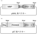
- FIG. 2 is a diagram schematically showing the structures of a pMAL-2RS1 vector (A) expressing MBP / 2RS1 fusion protein and a pET22-PH0828 / 2RS1 vector (B) expressing PH0828 / 2RS1 fusion protein. It is a schematic diagram of the structure of 2RS2 tag (A), 2RS3 tag (B), 2RS4 tag (C), and 2RS5 tag (D) which are comparative examples.
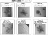
- MBP / 2RS1 fusion protein (Example 1), MBP / 2RS2 fusion protein (Comparative Example 1), MBP / 2RS3 fusion protein (Comparative Example 2), MBP / 2RS4 fusion protein (Comparative Example 3), MBP / 2RS5 fusion protein ( It is the figure which showed the analysis result of the dimerization by the gel filtration chromatography of the comparative example 4) and MBP (comparative example 5) which has not fused the tag. It is the photograph of the crystal
- polypeptide of the present invention comprises (1) a polypeptide comprising the amino acid sequence represented by SEQ ID NO: 1, (2) a polypeptide comprising the amino acid sequence represented by SEQ ID NO: 2, and (3) a sequence In the amino acid sequence represented by No. 2, a polypeptide comprising an amino acid sequence having 1 to 18 amino acids deleted from the N-terminal side and / or 1 to 3 amino acids deleted from the C-terminal side, or ( 4)
- the amino acid sequence of the polypeptide (1), polypeptide (2), or polypeptide (3) consists of an amino acid sequence in which one or several amino acids are deleted, substituted, inserted, and / or added.
- polypeptide of the present invention is a tag polypeptide that can promote protein crystallization by fusing directly or via a linker (several residues) to the N-terminus or C-terminus of any protein.
- linker severe residues
- End-Binding 1 is a partial peptide of a protein.
- End-Binding 1 is a protein that binds to the extended end of intracellular microtubules and binds to the tumor suppressor gene APC (Adenomatous Polyposis Coli) protein.
- APC Adomatous Polyposis Coli
- Polypeptide 1 consists of 45 amino acids, and the specific amino acid sequence is EDLEKERDFY FGKLRNIELI CQENEGENDP VLQRIVDILY ATDEG (SEQ ID NO: 1). Polypeptide 1 is presumed to have a structure as shown in FIG.
- Polypeptide consisting of the amino acid sequence represented by SEQ ID NO: 2 Polypeptide consisting of the amino acid sequence represented by SEQ ID NO: 2 (hereinafter sometimes referred to as polypeptide 2) is End-Binding 1 (Eb1) It is a partial peptide of a protein. It is a polypeptide consisting of 66 amino acids with 18 amino acids added to the N-terminal side and 3 amino acids added to the C-terminal side from the polypeptide 1, and exhibits an activity of promoting protein crystallization.
- Eb1 End-Binding 1
- polypeptide 2 has the structure shown in FIG. 1
- polypeptide 3 Peptide of intermediate chain length of polypeptide 1 and polypeptide 2
- polypeptide 3 A polypeptide consisting of an amino acid sequence from which three amino acids have been deleted (hereinafter sometimes referred to as polypeptide 3) is a partial peptide of End-Binding 1 (Eb1) protein. That is, it is a polypeptide having an intermediate chain length of polypeptide 1 and polypeptide 2, and exhibits activity of promoting protein crystallization.
- the number of amino acids that can be deleted from the N-terminal is 1, 2, 3, 4, 5, 6, 7, 8, 9, 10, 11, 12, 13, 14, 15, 16, 17, or 18.
- the number of amino acids that can be deleted from the C-terminal side is 1, 2, or 3. Therefore, the chain length of the crystallization promoting tag polypeptide contained in polypeptide 3 is 65, 64, 63, 62, 61, 60, 59, 58, 57, 56, 55, 54, 53, 52, 51, 50. 49, 48, 47, 46, or 45.
- the polypeptide having a chain length of 45 amino acids is the same as polypeptide 1.
- modified polypeptide is 1 in the amino acid sequence of the polypeptide (1), polypeptide (2), or polypeptide (3).
- a polypeptide comprising an amino acid sequence in which several amino acids are deleted, substituted, inserted and / or added, and fused to the C-terminus or N-terminus of the protein directly or via a peptide linker Shows activity to promote crystallization.
- amino acid in which one or more amino acids have been deleted, substituted, inserted, and / or added in an amino acid sequence refers to a well-known method such as site-directed mutagenesis, or naturally. It means that the modification has been made by substitution of several amino acids as much as possible.
- the number of amino acid modifications is preferably 1 to 15, more preferably 1 to 10, still more preferably 1 to 5, even more preferably 1 to 3, and most preferably 1.
- An example of a modified amino acid sequence of the polypeptide of the present invention may preferably be an amino acid sequence in which the amino acid has one or several (preferably 1, 2, 3 or 4) substitutions.
- the modified polypeptide of the present invention has an activity of promoting protein crystallization.
- the modification by deletion, substitution, insertion, and / or addition of the polypeptide may be a modification (improved modification) that improves the crystallization promoting activity of the polypeptide before modification, or a modification that maintains the crystallization promoting activity (maintenance modification). )
- modified polypeptide obtained by the improved modification examples include a polypeptide consisting of the amino acid sequence represented by SEQ ID NO: 3 or 4. Specific amino acid sequences are EDLEKELDFY FGKLRNIELI CQENEGENDP VLQRIVDILY ATDEG (SEQ ID NO: 3) and EDLEKDERDF FGKLRNIELI CQENEGENDP VLQRIVDILY ATDEG (SEQ ID NO: 4). Confirmation that the modification by deletion, substitution, insertion, and / or addition is an improved modification by the fact that the crystallization promoting activity is improved compared to the crystallization promoting activity of the parent polypeptide. Can do.
- the modification is an improved modification.
- the substitution used for the modification is not limited as long as the crystallization promoting activity is improved, but substitution to an amino acid residue that frequently maintains the coiled-coil rule is preferable.
- the amino acids to be deleted, inserted, and / or added are not limited as long as the crystallization promoting activity is improved. However, lack of amino acid residues that frequently maintain the coiled-coil rule is not limited. Loss, insertion, and / or addition are preferred.
- the maintenance modification is a modification that maintains the crystallization promoting activity of the parent polypeptide.
- the substitution used for the maintenance modification is not limited as long as the crystallization promoting activity is maintained, but substitution to an amino acid residue that frequently maintains the coiled-coil rule is preferable.
- the amino acids to be deleted, inserted, and / or added are not limited as long as the crystallization promoting activity is maintained, but amino acid residues that frequently maintain the coiled-coil rule are also included. Deletions, insertions and / or additions are preferred. That the modification by deletion, substitution, insertion, and / or addition is a maintenance modification can be confirmed by the fact that the crystallization promoting activity is equivalent to the crystallization promoting activity of the parent polypeptide.
- the crystallization promoting activity is lower than the crystallization promoting activity of the parent polypeptide, but superior to the crystallization of the target protein to which the parent polypeptide is not fused, It can be determined that it is a maintenance modification. For example, in the screening for crystallization conditions, if crystallization can be confirmed under many conditions than the protein to which the polypeptide of the present invention is not fused, the modified polypeptide is judged to be a maintenance modification. be able to.
- the modified polypeptide of the present invention is obtained by introducing a mutation into a polynucleotide encoding polypeptides 1 to 3 by site-specific mutagenesis by a conventional method, for example, site-specific mutagenesis.
- a desired variant polypeptide can be obtained.
- the fusion protein of the present invention is obtained by fusing the crystallization promoting tag polypeptide directly or via a peptide linker to the C-terminus or N-terminus of the protein.
- the fusion protein of the present invention is a protein that is easily crystallized as compared to a native protein that is not fused with a crystallization promoting tag peptide.
- the fusion protein of the present invention is a fusion of the polypeptide of the present invention and a protein different from End-Binding 1. That is, the fusion protein of the present invention does not contain End-Binding 1.
- the MBP / 2RS1 fusion protein obtained in Example 1 and the AxCeSD-PilZ / 2RS1 fusion protein obtained in Example 3 were subjected to X-ray crystal structure analysis as described below, and are shown in FIGS. 8 and 9, respectively. It was thought to have a structure. Furthermore, the MBP / 2RS1-R7L fusion protein obtained in Example 4 succeeded in the structural analysis with a resolution of 2.3 ⁇ , and the PH0828 / 2RS1-R7L fusion protein with a resolution of 3.5 ⁇ .
- the target protein to be fused with the crystallization promoting tag polypeptide is a protein to be crystallized, and is not particularly limited, and examples thereof include a maltose binding protein or PH0828.
- the protein which exists as a monomer in a solution can be mentioned as target protein. This is because, when a protein existing as a monomer forms a fusion protein with the crystallization promoting tag polypeptide of the present invention, crystallization is promoted.
- it is also possible to crystallize proteins that hardly crystallize that is, proteins that are difficult to crystallize. Examples of proteins that hardly crystallize include AxCeSD-PilZ.
- the target protein and the crystallization promoting tag polypeptide can be fused by a linker peptide. That is, the crystallization promoting tag polypeptide can be fused to the target protein via the linker peptide on the N-terminal (amino terminal) side or C-terminal (carboxyl terminal) side of the target protein.
- the linker peptide is not limited, but a linker peptide that does not inhibit crystallization of the target protein is preferable.
- the chain length of the linker peptide is preferably 1 to 20, more preferably 1 to 10, more preferably 1 to 5, and still more preferably 1 to 3.
- Specific amino acid sequences include “AAA”, “AGA”, “ASA”, “ANA”, “GAG”, “GSG”, “GNG”, “AG”, “AS”, “AN”, “AL”. ”,“ AE ”,“ GS ”,“ GN ”,“ A ”,“ G ”,“ S ”, or“ L ”.
- “promoting protein crystallization” means that a protein that could not be crystallized conventionally can be crystallized, or the efficiency of protein crystallization is improved.
- the efficiency of protein crystallization can be examined by screening crystallization conditions. Protein crystallization is sensitively influenced by buffer pH, salt type, salt concentration, precipitant type, precipitant concentration, and the like. Therefore, screening for crystallization conditions is usually performed for each protein. Protein crystallization conditions are screened by examining the presence or absence of crystallization under a plurality of conditions that combine the pH of the buffer, the type of salt, the salt concentration, the type of precipitant, the concentration of precipitant, and the like.
- the protein concentration at which crystallization occurs varies depending on the protein, but a protein concentration of, for example, 5 to 50 mg / mL is often used.
- the crystallization method is not limited, and for example, a hanging-drop vapor diffusion method or a sitting-drop vapor diffusion method can be used.
- the screening temperature for the crystallization conditions is not particularly limited, but can be performed at about 1 to 30 ° C., for example. Since protein crystallization is also affected by temperature, it is preferably performed at 4 ° C. and 20 ° C., for example.
- the screening time for crystallization conditions is not limited, and can be performed for about 1 day to 1 month, but the plate can be observed on the 1st, 3rd, 7th, 14th, and 28th days. preferable.
- Screening for protein crystallization conditions can be performed using a commercially available kit.
- a commercially available screening kit for example, a screening kit commercially available from QIAGEN, HAMPTON RESEARCH, Molecular Dimensions, Emerald BioSystems, or Jena Bioscience can be used.
- protein crystallization is promoted when the fusion protein of the present invention is crystallized, compared to the parent protein that the crystallization promoting tag polypeptide is not fused and could not be crystallized. It can be judged that it was done. It can also be determined that protein crystallization has been promoted when the crystallization conditions increase compared to the parent protein.
- Proteins that do not crystallize with monomers are dimerized, and crystallization occurs even under one condition, enabling X-ray crystal structure analysis. Therefore, it is a sufficient effect that at least one crystallization condition is found, for example, by screening crystallization conditions.
- the polynucleotide of the present invention is a polynucleotide encoding the polypeptide of the present invention (hereinafter sometimes referred to as tag polynucleotide), or a polynucleotide encoding the fused protein of the present invention (hereinafter referred to as fusion polynucleotide). There are things).
- the fusion polynucleotide is a fusion of a tag polynucleotide and a polynucleotide encoding a target protein (hereinafter sometimes referred to as a target polynucleotide).
- a fusion polynucleotide is one in which a tag and a polynucleotide of interest are fused on a nucleotide. Further, the fusion polynucleotide may include a nucleotide encoding a linker polypeptide (hereinafter sometimes referred to as a linker nucleotide). That is, the tag polynucleotide can be fused to the 3 ′ end or 5 ′ end of the target polynucleotide via a linker nucleotide.
- Tag polynucleotide The tag polynucleotide of the present invention is not limited as long as it encodes the polypeptide of the present invention, and the tag polynucleotide includes all polynucleotides composed of codon combinations corresponding to amino acids of the polypeptide.
- the polynucleotide consists of a base sequence represented by SEQ ID NO: 5. Mention may be made of polynucleotides.
- Examples of a polynucleotide encoding polypeptide 2 include, but are not limited to, a polynucleotide comprising the base sequence represented by SEQ ID NO: 6. be able to.
- examples of polynucleotides encoding polypeptide 4 (variant polypeptide) include, but are not limited to, those represented by SEQ ID NOs: 7 and 8.
- tags polynucleotide 4 include, but are not limited to, those represented by SEQ ID NOs: 7 and 8.
- a polynucleotide comprising a base sequence A polynucleotide encoding polypeptide 3 may be referred to as polynucleotide 3.
- the polynucleotides 1 to 3 can be obtained according to a conventional method. For example, total RNA is extracted from cells (eg, HeLa cells) expressing End-Binding 1 (Eb1) protein by the Acid Guanidinium-Phenol-Chloroform method. Using a mRNA isolation kit from the total RNA, purify poly A + RNA according to the method recommended by the manufacturer. Thereafter, poly A + mRNA is obtained by an affinity column method using poly U-sepharose or the like using oligo dT-cellulose or Sepharose 2B as a carrier, or a batch method.
- Eb1 End-Binding 1
- cDNA Single-stranded cDNA is synthesized using reverse transcriptase, and double-stranded DNA is synthesized using DNA polymerase, whereby End-Binding 1 (Eb1) protein is synthesized.
- Encoding cDNA can be obtained.
- Polynucleotides 1 to 3 can be obtained by PCR using this cDNA as a template and an appropriate primer set. By introducing restriction enzyme sites at both ends of the polynucleotide by PCR, it can be easily cloned into a vector. It is also possible to obtain polynucleotides 1 to 3 from the total RNA obtained from cells by RT-PCR.
- the polynucleotide 4 can be obtained by introducing mutations into the polynucleotides 1 to 3 by site-specific mutagenesis.
- the obtained polynucleotide and a polynucleotide encoding the target protein are fused and expressed as a fusion protein.
- the desired polynucleotide 4 can be obtained by confirming that the expressed fusion protein has crystallization promoting activity, for example, by a screening method for crystallization conditions.
- the tag polynucleotide of the present invention can also be prepared by DNA synthesis. It is also possible to prepare a combination of methods such as DNA synthesis and PCR.
- the target polynucleotide used in the fusion polynucleotide of the present invention is a polynucleotide encoding the target protein, and can be obtained according to a conventional method.
- total RNA is extracted from cells expressing the target protein by the Acid Guanidinium-Phenol-Chloroform method.
- a mRNA isolation kit from the total RNA, purify poly A + RNA according to the method recommended by the manufacturer. Thereafter, poly A + mRNA is obtained by an affinity column method using poly U-sepharose or the like using oligo dT-cellulose or Sepharose 2B as a carrier, or a batch method.
- a gene fragment having a restriction enzyme site introduced at both ends of a polynucleotide can be obtained by PCR using the cDNA as a template and an appropriate primer set, and cloned into a vector. It is also possible to obtain the target polynucleotide from the total RNA obtained from the cells by RT-PCR.
- the fusion polynucleotide of the present invention is a polynucleotide that encodes the fusion protein of the present invention. Specifically, a tag polynucleotide is fused to the 3 ′ end or 5 ′ end of the target polynucleotide. The tag polynucleotide may be fused to the 3 ′ end or 5 ′ end of the target polynucleotide via a linker nucleotide. Fusion of the tag polynucleotide to the target protein, introduction of a linker nucleotide, and the like can be performed using known genetic engineering techniques such as cleavage with restriction enzymes, ligation, and PCR.
- the recombinant vector of the present invention may be a vector comprising the tag polynucleotide of the present invention (hereinafter sometimes referred to as a tag vector) or a vector comprising the fusion polynucleotide of the present invention (hereinafter sometimes referred to as a fusion protein vector). ).
- the tag vector can be used to construct a fusion protein vector, and preferably has a restriction enzyme site into which a polynucleotide encoding the target protein can be easily introduced.
- the tag vector is not limited, but a tag vector having an expression cassette including an expression promoter so that the fusion protein can be expressed is preferable.
- the fusion protein vector is not limited, but an expression vector for expressing the fusion protein is preferred.
- a fusion protein vector can be constructed by introducing the polynucleotide of interest into the tag vector. Moreover, it can construct
- any vector such as a plasmid, phage, virus, etc. can be used as long as it can be replicated in a host cell.
- Escherichia coli plasmids such as pBR322, pBR325, pUC118, pUC119, pKC30, and pCFM536, Bacillus subtilis plasmids such as pUB110, yeast plasmids such as pG-1, YEp13, and YCp50, and phage DNAs such as ⁇ gt110 and ⁇ ZAPII.
- vectors for mammalian cells include viral DNA such as baculovirus, vaccinia virus, adenovirus, SV40 and its derivatives.
- the vector contains a replication origin, a selectable marker, and a promoter, and may contain an enhancer, a transcription termination sequence (terminator), a ribosome binding site, a polyadenylation signal, and the like as necessary.
- vectors such as pQE70, pQE60, pQE-9 (Qiagen), pBluescript II KS, ptr99a, pKK223-3, pDR540, pRIT2T (Pharmacia),
- pET-11a Novagen
- eukaryotic ones include pXT1, pSG5 (Stratagene), pSVK3, pBPV, pMSG, pSVL, SV40 (Pharmacia) and the like.
- coli vectors for example, those derived from ColiE1, R factor, F factor, yeast vectors, for example, 2 ⁇ m DNA, ARS1, derived from mammalian cell vectors, for example, SV40, Those derived from adenovirus and bovine papilloma virus can be used.
- promoters adenovirus or SV40 promoter, E. coli lac or trp promoter, phage lambda PL promoter, ADH, PHO5, GPD, PGK, AOX1 promoter for yeast, keratopolyhedrovirus virus-derived promoter for sputum cells, etc. Can be used.
- selection markers vectors for E.
- coli include kanamycin resistance gene, ampicillin resistance gene, tetracycline resistance gene, etc.
- yeast vectors include Leu2, Trp1, Ura3 gene, etc.
- mammalian cells include neomycin resistance gene, thymidine kinase. Gene, dihydrofolate reductase gene and the like.
- the transformed cell of the present invention is a cell containing the recombinant vector. That is, the transformed cell of the present invention contains the tag vector or the fusion protein vector.
- host cells containing the recombinant vector of the present invention include bacterial cells such as Escherichia coli, Streptomyces and Bacillus subtilis, fungal cells such as Aspergillus spp., Yeast cells such as baker's yeast and methanol-assimilating yeast, Drosophila S2, Spodoptera Insect cells such as Sf9, mammalian cells such as CHO, COS, BHK, 3T3, and C127 can be used.
- Transformation can be performed by a known method such as calcium chloride, calcium phosphate, DEAE-dextran mediated transfection, electroporation and the like.
- the mechanism by which the crystallization-promoting tag polypeptide of the present invention promotes protein crystallization is to obtain a symmetric protein dimer by dimerizing the protein and promote protein crystallization. Conceivable. However, as shown in Comparative Examples, the crystallinity is not improved in all of the dimerized proteins.
- the crystallization promoting tag polypeptide of the present invention has an excellent effect in protein crystallization.
- the method for producing a fusion protein of the present invention comprises a step of constructing a recombinant vector containing a polynucleotide encoding the fusion protein, a step of expressing the fusion protein using the recombinant vector, And a step of purifying the expressed fusion protein, preferably a step of confirming the formation of a multimer of the purified fusion protein.
- the method for producing a fusion protein crystal of the present invention includes a step of constructing a recombinant vector containing a polynucleotide encoding the fusion protein, a step of expressing the fusion protein using the recombinant vector, and the expressed fusion protein.
- the vector construction step in the method for producing a fusion protein of the present invention is a step of constructing a recombinant vector containing a polynucleotide encoding the fusion protein of the present invention. That is, it is a step of constructing the fusion protein vector.
- the procedure for constructing the fusion protein vector is not particularly limited.
- the target polynucleotide may be incorporated into a tag vector containing the tag polynucleotide, or the fusion polynucleotide may be incorporated into a normal expression vector.
- the tag polynucleotide and the target polynucleotide may be simultaneously incorporated into a normal expression vector. Incorporation of these polynucleotides into a vector can be performed using known genetic engineering techniques such as PCR, restriction enzyme cleavage, and ligation.
- the expression step in the method for producing a fusion protein of the present invention is a step of expressing the fusion protein using a recombinant vector.
- the expression step is not limited, but may be carried out by introducing the constructed fusion protein vector into a host cell using a known transformation method and culturing the resulting transformed cell. it can.
- the medium and culture conditions of the transformed cells can be appropriately selected and used by known methods. That is, the medium can be selected according to the cells. For example, when describing the case of Escherichia coli, Escherichia coli may be cultured in a commonly used L medium, YT medium, M9-CA medium or the like.
- Escherichia coli may be cultured in a commonly used L medium, YT medium, M9-CA medium or the like.
- the fusion protein vector has a drug resistance gene, it is desirable to add the corresponding drug to an appropriate concentration.
- an inducer may be added to the medium as necessary.
- IPTG isopropyl- ⁇ -D-thiogalactopyranoside
- IAA indoleacrylic acid
- the purification step in the production method of the fusion protein of the present invention is a step of purifying the fusion protein expressed by the transformed cells.
- the method for purifying the fusion protein is not particularly limited, and may be performed by combining known techniques, that is, lysozyme treatment, ultrasonic crushing, centrifugation, salting out method, desalting method, precipitation method, and various chromatographies. it can.
- the fusion protein of the present invention when expressed in a dissolved state in a transformed cell, the transformed cell is recovered by centrifugation after culturing, suspended in a buffer, A cell-free extract is obtained by crushing the cells with a sonicator, a French press or the like and then centrifuging them. Furthermore, a fusion protein purified by a technique used in a usual enzyme isolation and purification method can be obtained from the supernatant obtained by centrifuging the cell-free extract. In order to prevent inactivation of the fusion protein, an antioxidant, an enzyme stabilizer, a polyphenol adsorbent, a metal ligand and the like can be added to the buffer.
- Purification methods include solvent extraction, salting out with ammonium sulfate, desalting, precipitation with organic solvents, anion exchange chromatography, cation exchange chromatography, hydrophobic chromatography, gel filtration, Examples thereof include an affinity chromatography method, a chromatofocusing method, and an electrophoresis method such as isoelectric focusing, and these can be used alone or in combination.
- the polypeptide when expressed in an insoluble state in the cell, the polypeptide is recovered by a conventional method from a precipitate fraction obtained by crushing the cell and then performing centrifugation as described above. can do. Furthermore, the insoluble matter of the obtained fusion protein is solubilized with a denaturing agent, diluted or dialyzed into a dilute solution, and then the purified fusion protein can be obtained by the same isolation and purification method as described above.
- the polypeptide fusion protein or its derivative can be recovered in the culture supernatant. Then, a soluble fraction can be obtained by treating the culture by a method such as centrifugation as described above, and a purified fusion protein can be obtained from the soluble fraction by using the same isolation and purification method as described above. it can.
- the confirmation step of multimer formation in the method for producing a fusion protein of the present invention is a step of confirming the formation of multimerization of the purified fusion protein.
- the confirmation method of multimer formation is not specifically limited, For example, the molecular weight as a multimer of the said fusion protein can be confirmed from an elution position using a gel filtration chromatography method. That is, whether or not a monomer or multimer is formed can be confirmed by the molecular weight.
- the protein crystal production method of the present invention comprises a step of constructing a recombinant vector comprising a polynucleotide encoding the fusion protein according to claim 3 (hereinafter referred to as a vector construction step), A step of expressing a fusion protein using the recombinant vector (hereinafter referred to as an expression step), a step of purifying the expressed fusion protein (hereinafter referred to as a purification step), and multimer formation of the purified fusion protein.
- a step of confirming (hereinafter referred to as a multimer formation confirmation step), a step of crystallizing the purified fusion protein (hereinafter referred to as a crystallization step), and a step of confirming protein crystals by X-ray irradiation (hereinafter referred to as crystals) Referred to as a conversion confirmation step).
- the vector construction step, the expression step, the purification step, and the multimer formation confirmation step can use each step in the method for producing a fusion protein. Below, a crystallization process and a crystallization confirmation process are demonstrated.
- Crystallization step in the protein crystal production method of the present invention is a step of crystallizing the purified fusion protein.
- the crystallization method is not particularly limited, and for example, a batch method, a vapor diffusion method, a dialysis method, or the like can be used.
- protein crystallization is sensitively affected by the pH of the buffer solution, the type of salt, the salt concentration, the type of precipitant, the concentration of precipitant, and the like. Therefore, it is preferable to study the optimal crystallization conditions for the fusion protein to be crystallized.
- buffer solution such as an acetate buffer solution, a citrate buffer solution, a phosphate buffer solution, a borate buffer solution, a tris hydrochloric acid buffer solution, HEPES, and MES, can be used. . Based on these buffers, the pH, salt type, and salt concentration can be further studied.
- a salt for example, a salt, polyethylene glycol, alcohol, or a combination thereof can be used.
- the salt include lithium chloride, sodium chloride, magnesium chloride, potassium chloride, calcium chloride, ammonium sulfate, lithium sulfate, and magnesium sulfate.
- the polyethylene glycol include those having a molecular weight of 100 to 100,000.
- the alcohol include methanol, ethanol, propanol, butanol, or methylpentanediol.
- the crystallization temperature is not particularly limited, it can be carried out at 1 ° C. to 30 ° C., but it is preferable to examine a temperature at which crystallization is likely to occur by screening crystallization conditions.
- the crystallization time is not limited and can be performed in about 1 day to 1 month, but it is preferable to examine the time for crystallization of the fusion protein by screening the crystallization conditions. .
- Crystallization confirmation step in the protein crystal production method of the present invention is a step of confirming the protein crystal by X-ray irradiation in the protein crystal production method. In order to confirm protein crystals, diffraction reflection is measured. The measurement is performed at a temperature of 100K or less under a nozzle that blows out liquid nitrogen or helium.
- cryoprotectants such as polyethylene glycol with a molecular weight of 400 Da, glycerol and sucrose
- the beam line of a large synchrotron radiation facility such as SPring-8 or PhotonFactory, or a laboratory X-ray irradiation is performed with an X-ray irradiation apparatus to obtain diffraction data.
- Example 1 a fusion protein was prepared by fusing MBP and a 2RS1 tag.
- pMAL-2RS1 vector a pMAL-2RS1 vector (fusion) in which a polynucleotide encoding a 2RS1 tag (partial peptide derived from Eb1 protein) is fused to a pMAL-c2e vector encoding MBP. Protein vector).
- An oligo DNA fragment was synthesized based on the registered DNA gene sequence encoding a partial peptide of End-Binding 1 (Eb1) protein, and PCR was performed with the following oligo DNA to obtain a 2RS1 DNA fragment.
- Eb1 End-Binding 1
- the obtained DNA fragment is a DNA fragment having a SacI site and a KpnI site introduced at the end of the nucleotide represented by SEQ ID NO: 5.
- the oligo DNA was designed so that “AAA” was inserted as a peptide linker between the MBP and the 2RS1 tag.
- 2RS1 oligo primer for MBP 1 5′-AATTAAGAGCTCGGCTGCTGCTGAAGACTT-3 ′ (SEQ ID NO: 9)
- 2RS1 oligo primer for MBP 2 5′-TCCGAAGTAGAAATCCCTCTCTTTCTCCAAGTCTTCAGCA-3 ′ (SEQ ID NO: 10)
- 2RS1 oligo primer for MBP 3 5′-CTACTTCGGAAAGCTACGGAACATTGAATTGATTTGCCAGGAGAA-3 ′
- 2RS1 oligo primer for MBP 4 5′-CCTCTGCAATACAGGGTCGTTTTCCCCCTCGTTCTCCTGG-3 ′
- 2RS1 oligo primer for MBP 5 5′-ATTGCAGAGGATTGTAGACATTCTGTATGCCACAGATGAA-3 ′
- 2RS1 oligo primer for MBP 6 5′-AATTAAGGTACCTTAGCCTTCATCTGTG
- the DNA fragment and the pMAL-c2 vector were digested with restriction enzymes SacI and KpnI at 37 ° C. for 24 hours.
- the reaction solution was purified using a DNA purification kit (QIAquick PCR purification kit (QIAgen)).
- the cleaved pMAL-c2e vector and DNA fragment were ligated at 16 ° C. for 2 hours using T4 DNA ligase.
- the reaction solution was mixed with Escherichia coli JM109 strain (competent cell) and transformed by the heat shock method.
- the cells were cultured overnight at 37 ° C. in an LB agar medium containing 100 ⁇ g / mL ampicillin. Colonies were cultured to obtain plasmids. After cutting with a restriction enzyme, agarose electrophoresis was performed to select a clone in which the target DNA fragment was incorporated. The base sequence was confirmed by sequencing, and the pMAL-2RS1 vector was obtained.
- IPTG was added to a final concentration of 1 mM to induce expression of the target protein.
- the expression of the target protein was confirmed by SDS-PAGE.
- the bacterial solution after the main culture is centrifuged at 4500 ⁇ g for 20 min at 4 ° C., the supernatant is taken, suspended in a suspension buffer (STE buffer), and further centrifuged at 5,000 ⁇ g for 20 min at 4 ° C.
- STE buffer suspension buffer
- the bacteria were suspended with a crushing buffer, and subjected to ultrasonic crushing at ON 1 sec, OFF 1.5 sec, and total 10 min.
- the fusion protein is purified by a combination of purification methods such as ammonium sulfate precipitation, dialysis, anion exchange chromatography, gel filtration chromatography, affinity chromatography (using His tag or affinity beads for MBP) to obtain MBP / 2RS1 fusion protein. It was.
- the result of gel filtration chromatography is shown in FIG.
- Comparative Example 1 a fusion protein was prepared by fusing MBP and a 2RS2 tag.
- the amino acid sequence of the 2RS2 tag is shown in SEQ ID NO: 15, and the nucleotide sequence of the nucleotide is shown in SEQ ID NO: 16.
- a schematic diagram of the structure of the 2RS2 tag is shown in FIG.
- the pMAL-2RS2 vector was obtained by repeating the procedure of Example 1 (1) except that 2RS2 oligoprimers 1 to 4 for MBP were used instead of 2RS1 oligoprimers 1 to 6 for MBP.
- 2RS2 oligo primer for MBP 1 5′-AATTAAGAGCTCGGCTGCTGCTATGGTTTCTAAACTGAGC-3 ′ (SEQ ID NO: 17)
- 2RS2 oligo primer for MBP 2 5′-GGGCCGCCAGGAGTTCCGTCTGCAGCTGGCTCAGTTTA-3 ′
- 2RS2 oligo primer for MBP 3 5′-CCTGGCGGCCCTGCTCGAATCAGGGCTGAGCAAAGAGGCACTGATCCA-3 ′
- 2RS2 oligo primer for MBP 4 5′-AATTAAGGTACCTTACTCACCCAGTGCC-3 ′ (SEQ ID NO: 20)
- the MBP / 2RS2 fusion protein was obtained by repeating the operation of Example 1 (2) except that the pMAL-2RS2 vector was used instead of the pMAL-2RS1 vector.
- the result of gel filtration chromatography is shown in FIG.
- Comparative Example 2 a fusion protein was prepared by fusing MBP and a 2RS3 tag.
- the amino acid sequence of 2RS3 tag is shown in SEQ ID NO: 21, and the nucleotide sequence of nucleotide is shown in SEQ ID NO: 22.
- a schematic diagram of the structure of the 2RS3 tag is shown in FIG.
- the pMAL-2RS3 vector was obtained by repeating the procedure of Example 1 (1) except that 2RS3 oligoprimers 1 to 4 for MBP were used instead of 2RS1 oligoprimers 1 to 6 for MBP.
- 2RS3 oligo primer for MBP 1 5′-AATTAAGAGCTCGGCTGCTGCTGGAATTGGA-3 ′ (SEQ ID NO: 23)
- 2RS3 oligo primer for MBP 2 5′-CCTGTGGTTAATACCTTCAGAACTGCTCCAATTCCAGC-3 ′
- 2RS3 oligo primer for MBP 3 5′-TAACCACAGGATTGCCCGCCCTCATAAGTTGGATTAAACGTAAGAGGCAA-3 ′
- 2RS3 oligo primer for MBP 4 5′-AATTAAGGTACCTTACTGTTGCCTCTTACG-3 ′ (SEQ ID NO: 26)
- the MBP / 2RS3 fusion protein was obtained by repeating the operation of Example 1 (2) except that the pMAL-2RS3 vector was used instead of the pMAL-2RS1 vector.
- the result of gel filtration chromatography is shown in FIG.
- Comparative Example 3 a fusion protein was prepared by fusing MBP and a 2RS4 tag.
- the amino acid sequence of the 2RS4 tag is shown in SEQ ID NO: 27, and the nucleotide base sequence is shown in SEQ ID NO: 28.
- a schematic diagram of the structure of the 2RS4 tag is shown in FIG.
- the pMAL-2RS4 vector was obtained by repeating the procedure of Example 1 (1) except that 2RS4 oligoprimers 1 to 6 for MBP were used instead of 2RS1 oligoprimers 1 to 6 for MBP.
- 2RS4 oligo primer for MBP 1 5′-AATTAAGAGCTCGGCTGCTGCTAATGTTGA-3 ′ (SEQ ID NO: 29)
- 2RS4 oligo primer for MBP 2 5′-TCCATGACCCGCATTTGAAACTCCTGTTCAAAGCTGAGGTCAACATTAGC-3 ′
- 2RS4 oligo primer for MBP 3 5'-GGGTCATGGAGGAGCAAGTGAGTGCCATGAGCCTTCAGGAAGCTCGTGAG-3 '(SEQ ID NO: 31)
- 2RS4 oligo primer for MBP 5 5′-TATTGATGATGAAAGACAACGTCATTCGCTCCTTGGTCAAACGCGCTGCC-3 ′
- 2RS4 oligo primer for MBP 6 5′-TATTGATGATGAAAGACAACGTCATTCGCTCCTT
- the MBP / 2RS4 fusion protein was obtained by repeating the operation of Example 1 (2) except that the pMAL-2RS4 vector was used instead of the pMAL-2RS1 vector.
- the result of gel filtration chromatography is shown in FIG.
- Comparative Example 4 a fusion protein was prepared by fusing MBP and a 2RS5 tag.
- the amino acid sequence of the 2RS5 tag is shown in SEQ ID NO: 35, and the nucleotide base sequence is shown in SEQ ID NO: 36.
- a schematic diagram of the structure of the 2RS2 tag is shown in FIG.
- the pMAL-2RS5 vector was obtained by repeating the procedure of Example 1 (1) except that 2RS5 oligoprimers 1-6 for MBP were used instead of 2RS1 oligoprimers 1-6 for MBP.
- 2RS5 oligo primer for MBP 1 5′-AATTAAGAGCTCGGCTGCTGCTGGTGAGGA-3 ′ (SEQ ID NO: 37)
- 2RS5 oligo primer for MBP 2 5′-CTACATAAGTAGCCGCGAAGGCGGCATCTTTATCCTCACCAGC-3 ′
- 2RS5 oligo primer for MBP 3 5'-CTACTTATGTAGCACCGCTTATCGACGAATCCATCCACCTTGGT-3 '(SEQ ID NO: 39)
- 2RS5 oligo primer for MBP 4 5′-GTGAATGCGCGCACGAGTGAAACCAAGGTG-3 ′
- 2RS5 oligo primer for MBP 5 5'-CATTCACGCCCTTTTAGACCAGGTCGCTGAAAG-3 '(SEQ ID NO: 41)
- 2RS5 oligo primer for MBP 6 5′-AATTAAGGTACCTTAGCCCCTACTTTCAGC-3 ′ (SEQ ID NO: 37)
- the MBP / 2RS5 fusion protein was obtained by repeating the operation of Example 1 (2) except that the pMAL-2RS5 vector was used instead of the pMAL-2RS1 vector.
- the result of gel filtration chromatography is shown in FIG.
- Comparative Example 5 MBP without tag fusion was prepared.
- the pMAL-notag vector was obtained by repeating the procedure of Example 1 (1) except that MBP notag oligo primers 1 and 2 were used in place of 2P1 oligo primers 1 and 2 for MBP.
- MBP notag oligo primer 1 By using the MBP notag oligo primer 1, a stop codon (underlined) is inserted between the MBP gene and linker (AAA) and the 2RS1 tag gene.
- MBP / 2RS1 fusion protein, MBP / 2RS2 fusion protein, MBP / 2RS4 fusion protein, and MBP / 2RS5 fusion protein have higher molecular weight (dimer) than MBP-notag (monomer). It was eluted to the side and hardly eluted at the monomer position. Therefore, it is suggested that these fusion proteins are stably dimerized. However, it was speculated that the MBP / 2RS3 fusion protein eluted at a very large molecular weight and formed an aggregate.
- Example 2 a fusion protein was prepared by fusing PH0828 and a 2RS1 tag.
- a 2RS1 tag DNA fragment containing a polynucleotide encoding a 2RS1 tag (a partial peptide derived from Eb1 protein) was constructed. PCR was performed using the following oligo DNA to obtain a DNA fragment encoding a partial peptide of End-Binding 1 (Eb1) protein.
- the obtained DNA fragment is a DNA fragment having an XhoI site and an NheI site introduced at the end of the nucleotide represented by SEQ ID NO: 4.
- 2RS1 oligo primer for PH0828 1 5′-AATTAACTCGAGGAAGACTTGGAGAAAGAGAGGGA-3 ′
- 2RS1 oligo primer for PH0828 2 5'-TCAATTCAATGTTCCGTAGCTTTCCGAAGTAGAAATCCCTCTC-3 '(SEQ ID NO: 44)
- 2RS1 oligo primer for PH0828 3 5′-GCTACGGAACATTGAATTGATTTGCCAGGAGAACGA-3 ′
- 2RS1 oligo primer for PH0828 4 5′-TACAATCCTCTGCAATACAGGGTCGTTTTCCCCCTCGTT-3 ′
- 2RS1 oligo primer for PH0828 5 5′-TATTGCAGAGGATTGTAGACATTCTGTATGCCACAGATG-3 ′
- 2RS1 oligo primer for PH0828 6 5′-AATTAAGC
- PH0828 gene PCR was carried out using the PH0828 gene cDNA clone derived from Pyrococcus horikoshii OT3 strain as a template, and the following sense primer and antisense primer were used to obtain a DNA fragment encoding PH0828.
- the obtained DNA fragment is a DNA fragment in which an NdeI site and an XhoI site are introduced at the end of the nucleotide of PH0828.
- PH0828 sense primer 5′-GGAATTCCATATGTCTTATAAGGAAAAGAG-3 ′ (SEQ ID NO: 49)
- PH0828 antisense primer 5′-CCGCTCGAGTCTAATCTCATCGAAATCACC-3 ′ (SEQ ID NO: 50)
- pET28M-NheI primer 5′-CTTAAGCTAGCTTTAAACACCACCACCACCACCACTG-3 ′ (SEQ ID NO: 51)
- pET28M-NdeI primer 5′-AATTAACATATGGTGATGGTGATGGTGGCCCATG-3 ′ SEQ ID NO: 52)
- the 2RS1 tag DNA fragment was digested with XhoI and NheI, the PH0828 gene DNA fragment with NdeI and XhoI, and the pET28M-derived DNA fragment with NheI and NdeI at 37 ° C. for 24 hours. Each reaction solution was purified using a DNA purification kit (QIAquick PCR purification kit (QIAgen)).
- Each purified DNA fragment was mixed and ligated at 16 ° C. for 2 hours using T4 DNA ligase.
- the reaction solution was mixed with Escherichia coli JM109 strain (competent cell) and transformed by the heat shock method.
- the cells were cultured overnight at 37 ° C. in an LB agar medium containing 50 ⁇ g / mL kanamycin. Colonies were cultured to obtain plasmids. After cutting with a restriction enzyme, agarose electrophoresis was performed to select a clone in which the target DNA fragment was incorporated. The nucleotide sequence was confirmed by sequencing, and the pET28M-PH0828-2RS1 vector was obtained.
- Example 1 (2) Expression and purification of the fusion protein
- the procedure of Example 1 (2) was repeated except that the pET28M-PH0828-2RS1 vector was used in place of the pMAL-2RS1 vector to obtain the PH0828 / 2RS1 fusion protein. I got it.
- Comparative Example 6 a fusion protein was prepared by fusing PH0828 and 2RS2 tag. The procedure of Example 2 (1) and (3) was repeated except that 2RS2 oligo primer 1 to 4 for PH0828 was used instead of 2RS1 oligo primer 1 to 6 for PH0828, and pET28M-PH0828-2RS2 Acquired the vector.
- 2RS2 oligo primer for PH0828 1 5′-AATTAACTCGAGATGGTTTCTAAACTGAGCCAGCT-3 ′ (SEQ ID NO: 53)
- 2RS2 oligo primer for PH0828 2 5'-TTCGAGCAGGGCCGCCAGGAGTTCCGTCTGCAGCTGGCTC-3 '(SEQ ID NO: 54)
- 2RS2 oligo primer for PH0828 3 5′-CCCTGCTCGAATCAGGGCTGAGCAAAGAGGCACTGATCCAGG-3 ′ (SEQ ID NO: 55)
- 2RS2 oligo primer for PH0828 4 5′-AATTAAGCTAGCTTACTCACCCAGTGCCTGGATCAGTG-3 ′ (SEQ ID NO: 56)
- the PH0828 / 2RS2 fusion protein was obtained by repeating the procedure of Example 2 (4) except that the pET28M-PH0828-2RS2 vector was used instead of the pET28M-PH0828-2RS1 vector.
- Comparative Example 7 a fusion protein was prepared by fusing PH0828 and 2RS3 tag. The procedure of Example 2 (1) and (3) was repeated except that 2RS3 oligo primer 1 to 4 for PH0828 was used instead of 2RS1 oligo primer 1 to 6 for PH0828, and pET28M-PH0828-2RS3 Acquired the vector.
- 2RS3 oligo primer for PH0828 1 5′-AATTAACTCGAGGGAATTGGAGCAGTTCT-3 ′ (SEQ ID NO: 57)
- 2RS3 oligo primer for PH0828 2 5'-GCAATCCTGTGGTTAATACCTTCAGAACTGCTCC-3 '(SEQ ID NO: 58)
- 2RS3 oligo primer for PH0828 3 5′-ACAGGATTGCCCGCCCTCATAAGTTGGATTAAA-3 ′ (SEQ ID NO: 59)
- the PH0828 / 2RS3 fusion protein was obtained by repeating the procedure of Example 2 (4) except that the pET28M-PH0828-2RS3 vector was used instead of the pET28M-PH0828-2RS1 vector.
- Comparative Example 8 a fusion protein was prepared by fusing PH0828 and 2RS4 tag. The procedure of Example 2 (1) and (3) was repeated except that 2RS4 oligoprimers 1 to 6 for PH0828 were used instead of 2RS1 oligoprimers 1 to 6 for PH0828, and pET28M-PH0828-2RS4 Acquired the vector.
- 2RS4 oligo primer for PH0828 1 5′-AATTAACTCGAGAATGTTGACCTCAGCTTTGAACAGGA-3 ′
- 2RS4 oligo primer for PH0828 2 5′-CACTCACTTGCTCCTCCATGACCCGCATTTGAAACTCCTGTTCAAA-3 ′
- 2RS4 oligo primer for PH0828 3 5'-CAAGTGAGTGCCATGAGCCTTCAGGAAGCTCGTGAGCTACTGTTGCAAG-3 '(SEQ ID NO: 63) 2RS4 oligo primer for PH0828 4: 5′-CGAATGACGTTGTCTTTCATCATCAATAGGCGAGAGGCTTGCAAC-3 ′
- 2RS4 oligo primer for PH0828 5 5′-GACAACGTCATTCGCTCCTTGGTCAAACGCGCTGC-3 ′
- the PH0828 / 2RS4 fusion protein was obtained by repeating the procedure of Example 2 (4) except that the pET28M-PH0828-2RS4 vector was used instead of the pET28M-PH0828-2RS1 vector.
- Comparative Example 9 a fusion protein was prepared by fusing PH0828 and 2RS5 tag. The procedure of Example 2 (1) and (3) was repeated except that 2RS5 oligo primer 1 to 6 for PH0828 was used instead of 2RS1 oligo primer 1 to 6 for PH0828, and pET28M-PH0828-2RS5 Acquired the vector.
- 2RS5 oligo primer for PH0828 1 5′-AATTAACTCGAGGGTGAGGATAAAGATGCCGCCTTC-3 ′
- 2RS5 oligo primer for PH0828 2 5′-GTCGATAAGCGGTGCTACATAAGTAGCCGCGAAGGCGG-3 ′
- 2RS5 oligo primer for PH0828 3 5′-GCTTATCGACGAATCCATCCACCTTGGTTTCACTCGT-3 ′
- 2RS5 oligo primer for PH0828 4 5′-AAAAGGGCGTGAATGCGCGCACGAGTGAAACC-3 ′
- 2RS5 oligo primer for PH0828 5 5′-CCCTTTTAGACCAGGTCGCTGAAAGTAGG-3 ′
- 2RS5 oligo primer for PH0828 6 5′-AATTAAGCTAGCTTAGC
- the PH0828 / 2RS5 fusion protein was obtained by repeating the procedure of Example 2 (4) except that the pET28M-PH0828-2RS5 vector was used instead of the pET28M-PH0828-2RS1 vector.
- Comparative Example 10 PH0828 with no tag fusion was prepared.
- the pH0828-notag vector was obtained by repeating the procedure of Example 1 (1) except that the PH0828 notag oligo primer 1 was used in place of the PH0828 2RS1 oligo primer 1.
- a stop codon (underlined) is inserted between the PH0828 gene and the linker and the 2RS1 tag gene.
- Notag oligo primer for PH0828 1 5′-AATTAACTCGAG TAA GAAGACTTGGAGAAAGAGAGGGA-3 ′ (SEQ ID NO: 75)
- Crystallization screening was performed using each fusion protein with MBP obtained in Example 1 and Comparative Examples 1 to 4 and MBP-notag obtained in Comparative Example 5. Using four types of crystallization screening kits of “Nextal JCSG Core Suite I / II / III / IV” (QIAGEN), initial crystallization screening of each fusion protein was performed by the sitting drop method. The results are shown in Table 1.
- Example 1 As shown in Table 1, the MBP / 2RS1 fusion protein obtained in Example 1 was induced to crystallize under six conditions. However, although the fusion proteins of Comparative Examples 1, 3, and 4 formed dimers, crystallization could not be confirmed.
- FIG. 7 shows a photograph of the crystals after 8 days under the 6 conditions found for the MBP / 2RS1 fusion protein.
- Crystallization screening was performed using each fusion protein with PH0828 obtained in Example 2 and Comparative Examples 6 to 9 and PH0828-notag obtained in Comparative Example 10. The same crystallization screening kit as MBP was used.
- Example 3 an AxCeSD-PilZ / 2RS1 fusion protein was prepared by fusing AxCeSD-PilZ and a 2RS1 tag.
- the pET28b-AxCeSD-PilZ-2RS1 vector was obtained by incorporating the 2RS1 tag into the pET28b-AxCeSD-PilZ vector incorporating AxCeSD-PilZ into the pET28b vector.
- the AxCeSD-PilZ / 2RS1 fusion protein was obtained by repeating the procedure of Example 1 (2) except that the pET28b-AxCeSD-PilZ-2RS1 vector was used instead of the MAL-2RS1 vector.
- Example 4 the effect on the crystallization of MBP and PH0828 by the substitution of the 7RS arginine (R) of the 2RS1 tag with leucine (L) was examined.
- the R7L mutation was introduced using the following primers, and the pMAL-2RS1-R7L vector or pET28M- The PH0828-2RS1-R7L vector was obtained.
- R7L primer 1 5′-TTCTACTTCGGAAAGCTACGG-3 ′ (SEQ ID NO: 76)
- R7L primer 2 5′-ATCTAACTCTTTCTCCAAGTC-3 ′ (SEQ ID NO: 77)
- the procedure of Example 1 (2) was repeated except that the pMAL-2RS1-R7L vector or the pET28M-PH0828-2RS1-R7L vector was used instead of the pMAL-2RS1 vector, and MBP / 2RS1-R7L A fusion protein, or PH0828 / 2RS1-R7L fusion protein was obtained.
- crystallization of MBP / 2RS1-R7L fusion protein and PH0828 / 2RS1-R7L fusion protein was screened. The results are shown in Table 3.
- Example 5 the effect on the crystallization of MBP and PH0828 by the substitution of the 10th tyrosine (Y) of the 2RS1 tag with leucine (L) was examined.
- the Y10L mutation was introduced using the following primers, and the pMAL-2RS1-Y10L vector or pET28M- The PH0828-2RS1-Y10L vector was obtained.
- Y10L primer 1 5′-TTCTTATTCGGAAAGCTACGG-3 ′ (SEQ ID NO: 78)
- Y10L primer 2 5′-ATCCCTCTCTTTCTCCAAGTC-3 ′ (SEQ ID NO: 79)
- the procedure of Example 1 (2) was repeated except that the pMAL-2RS1-Y10L vector or the pET28M-PH0828-2RS1-Y10L vector was used instead of the MAL-2RS1 vector, and MBP / 2RS1-Y10L A fusion protein or PH0828 / 2RS1-Y10L fusion protein was obtained.
- crystallization of MBP / 2RS1-Y10L fusion protein and PH0828 / 2RS1-Y10L fusion protein was screened. The results are shown in Table 3.
- MBP crystallization increased from 6/384 of Example 1 to 11/384 due to substitution (mutation) of R7L of the 2RS1 tag of Example 4, and crystallization of PH0828 was performed. It increased from 2/384 in Example 2 to 6/384. Therefore, the substitution of the 7RS arginine (R) of the 2RS1 tag with leucine (L) is considered to be an improved modification. Further, the substitution (mutation) of Y10L of the 2RS1 tag in Example 5 increased the crystallization of PH0828 from 2/384 in Example 2 to 4/384. Therefore, the substitution of the 7RS arginine (R) to the leucine (L) of the 2RS1 tag is considered to be an improved modification for PH0828.
- the crystallization of MBP decreased from 6/384 in Example 1 to 3/384 by substitution (mutation) of Y10L of the 2RS1 tag.
- the crystallization of MBP without a tag is 1/384, and the substitution (mutation) of Y10L causes the crystallization of MBP to be 3/384, so the substitution of Y10L Is considered a maintenance modification for MBP.
- the polypeptide of the present invention can be fused with the target protein and crystallized.
- the obtained fusion protein can be subjected to X-ray irradiation, diffraction data can be measured, and the structure of the protein can be analyzed using a crystal structure analysis method.
Landscapes
- Health & Medical Sciences (AREA)
- Life Sciences & Earth Sciences (AREA)
- Chemical & Material Sciences (AREA)
- Organic Chemistry (AREA)
- Engineering & Computer Science (AREA)
- Genetics & Genomics (AREA)
- Zoology (AREA)
- Molecular Biology (AREA)
- Wood Science & Technology (AREA)
- Biomedical Technology (AREA)
- Biotechnology (AREA)
- General Engineering & Computer Science (AREA)
- General Health & Medical Sciences (AREA)
- Bioinformatics & Cheminformatics (AREA)
- Biochemistry (AREA)
- Biophysics (AREA)
- Microbiology (AREA)
- Proteomics, Peptides & Aminoacids (AREA)
- Crystallography & Structural Chemistry (AREA)
- Physics & Mathematics (AREA)
- Materials Engineering (AREA)
- Metallurgy (AREA)
- Plant Pathology (AREA)
- Chemical Kinetics & Catalysis (AREA)
- General Chemical & Material Sciences (AREA)
- Toxicology (AREA)
- Gastroenterology & Hepatology (AREA)
- Medicinal Chemistry (AREA)
- Peptides Or Proteins (AREA)
- Preparation Of Compounds By Using Micro-Organisms (AREA)
- Micro-Organisms Or Cultivation Processes Thereof (AREA)
Abstract
本発明の目的は、X線結晶構造解析法において、目的のタンパク質の結晶化を促進するための、汎用性があり、かつ効率的な方法を提供することである。 前記課題は、(1)配列番号1で表されるアミノ酸配列からなるポリペプチド、(2)配列番号2で表されるアミノ酸配列からなるポリペプチド、(3)配列番号2で表されるアミノ酸配列において、N末側から1~18個のアミノ酸が欠失、及び/若しくはC末側から1~3個のいずれかのアミノ酸が欠失したアミノ酸配列からなるポリペプチド、によって解決することができる。
Description
本発明は、タンパク質の結晶化を促進することのできるポリペプチド及びタンパク質の結晶化方法に関する。本発明によれば、X線結晶構造解析法に用いるためのタンパク質結晶を容易に得ることができる。
タンパク質の構造解析にはX線結晶構造解析法、NMR法、又は電子顕微鏡による解析が主に使用されている。その中で、X線結晶構造解析法は、電子顕微鏡よりも精度の高い分子像が得られる。また、NMR法と比べて分子量が大きな分子においても適用可能である。従って、X線結晶構造解析法は、タンパク質の構造解析法として、汎用されている。しかし、X線結晶構造解析法によるタンパク質の分析のためには、タンパク質を結晶化する必要がある。単分子のタンパク質は、X線散乱が弱く強度が低いため、構造を解析することができない。タンパク質の結晶では、回折光が増幅されるため、散乱強度を測定することができ、それにより電子密度を計算することによって、タンパク質分子の構造モデルを作製することが可能となる。
しかしながら、タンパク質の結晶化はX線結晶構造解析法でも一番の難関である。タンパク質の結晶化条件は、例えば同じファミリーに属するタンパク質同士でも大きく異なり、pHや沈殿剤濃度の変化に非常に敏感である。また、タンパク質の結晶化のメカニズムは現在でも明らかになっていない。そのため、タンパク質の結晶化には、大量の条件検討を行う必要があり、多大な労力、時間、及び費用がかかる。そして、X線結晶構造解析法においては、良質の結晶が得られないために、構造解析ができないタンパク質も多い。
X線結晶構造解析法に用いるタンパク質結晶を得るために、多数の結晶化条件を検討するための結晶化キットが開発され、また結晶化ロボットを用いる条件検討が行われている。更に、無重力場(宇宙空間)での結晶化、磁場又はレーザー照射による結晶化などが検討されているが、いずれも十分な効果を挙げているとは言いがたいものであった。
X線結晶構造解析法に用いるタンパク質結晶を得るために、多数の結晶化条件を検討するための結晶化キットが開発され、また結晶化ロボットを用いる条件検討が行われている。更に、無重力場(宇宙空間)での結晶化、磁場又はレーザー照射による結晶化などが検討されているが、いずれも十分な効果を挙げているとは言いがたいものであった。
「プロシーディング・オブ・ザ・ナショナル・アカデミー・オブ・サイエンス・オブ・ザ・ユナイテッド・ステイト・オブ・アメリカ(Proceedings of the National Academy of Sciences of the United States of America)」2006年(米国)、第103巻、p.16230-15235
「プロテイン・サイエンス(Protein Science)」2011年(米国)、第20巻、p.1876-1890
本発明者らは、X線結晶構造解析法に用いるタンパク質の結晶化について鋭意検討したところ、タンパク質結晶系には2回もしくは3回回転軸を持つものは全体の80%となっていることに気が付いた。すなわち、回転対称を持つニ量体又は三量体タンパク質は、単量体タンパク質よりも結晶化しやすいと考えられた。また,本発明者らは解析された構造からポリペプチドを用いて、二量体及び三量体タンパク質を人工的に形成させる可能性を見出した(特許文献1)。
タンパク質を二量体化する方法として、リゾチーム(目的タンパク質)にシステインの変異を導入し、ニ量体化することによって、結晶化が促進されることが開示されている(非特許文献1)。更に、リゾチーム又はマルトース結合タンパク質に、一対のヒスチジン、又はシステインの変異を導入し、金属イオンによりタンパク質をニ量体化することが開示されている(非特許文献2)。しかしながら、これらの方法は、目的のタンパク質ごとに、構造などに基づいて変異を導入するアミノ酸を検討する必要があり、すべてのタンパク質に汎用できる方法ではなかった。更に、結晶化させるタンパク質自体に変異を導入するものであるため、変異の導入によりタンパク質の構造が変化する可能性があった。従ってこれらの方法は、タンパク質のX線結晶構造解析法に最適なものではなかった。
タンパク質を二量体化する方法として、リゾチーム(目的タンパク質)にシステインの変異を導入し、ニ量体化することによって、結晶化が促進されることが開示されている(非特許文献1)。更に、リゾチーム又はマルトース結合タンパク質に、一対のヒスチジン、又はシステインの変異を導入し、金属イオンによりタンパク質をニ量体化することが開示されている(非特許文献2)。しかしながら、これらの方法は、目的のタンパク質ごとに、構造などに基づいて変異を導入するアミノ酸を検討する必要があり、すべてのタンパク質に汎用できる方法ではなかった。更に、結晶化させるタンパク質自体に変異を導入するものであるため、変異の導入によりタンパク質の構造が変化する可能性があった。従ってこれらの方法は、タンパク質のX線結晶構造解析法に最適なものではなかった。
本発明の目的は、X線結晶構造解析法において、目的のタンパク質の結晶化を促進するための、汎用性があり、かつ効率的な方法を提供することである。
本発明者は、X線結晶構造解析法における目的のタンパク質の結晶化を促進する方法について、鋭意研究した結果、驚くべきことに、特定の配列のアミノ酸からなるポリペプチドを目的のタンパク質のN末端又はC末端に融合させることにより、目的のタンパク質の結晶化が効果的に促進されることを見出した。
本発明は、こうした知見に基づくものである。
従って、本発明は、
[1](1)配列番号1で表されるアミノ酸配列からなるポリペプチド、(2)配列番号2で表されるアミノ酸配列からなるポリペプチド、(3)配列番号2で表されるアミノ酸配列において、N末側から1~18個のアミノ酸が欠失、及び/若しくはC末側から1~3個のいずれかのアミノ酸が欠失したアミノ酸配列からなるポリペプチド、又は(4)前記ポリペプチド(1)、ポリペプチド(2)、又はポリペプチド(3)のアミノ酸配列において、1若しくは複数のアミノ酸が欠失、置換、挿入、及び/若しくは付加されたアミノ酸配列からなり、かつタンパク質のC末端若しくはN末端に直接若しくはペプチドリンカーを介して融合させた融合タンパク質として発現させた場合に当該融合タンパク質の結晶化を促進する活性を示す、改変体ポリペプチド、
[2]前記改変体ポリペプチドが、配列番号3、又は4で表されるアミノ酸配列からなる、[1]に記載のポリペプチド、
[3][1]又は[2]に記載のポリペプチドが、タンパク質のC末端又はN末端に、直接又はペプチドリンカーを介して融合した、融合タンパク質、
[4][1]又は[2]に記載のポリペプチド又は[3]に記載の融合タンパク質をコードするポリヌクレオチド、
[5][4]に記載のポリヌクレオチドを含む組換えベクター、
[6][5]に記載の組換えベクターを含む形質転換細胞、
[7][3]に記載の融合タンパク質をコードするポリヌクレオチドを含む組換えベクターを構築する工程、前記組換えベクターを用いて融合タンパク質を発現させる工程、及び前記発現した融合タンパク質を精製する工程、を含む融合タンパク質の製造方法、
[8][3]に記載の融合タンパク質をコードするポリヌクレオチドを含む組換えベクターを構築する工程、前記組換えベクターを用いて融合タンパク質を発現させる工程、
前記発現した融合タンパク質を精製する工程、及び精製された融合タンパク質の多量体形成を確認する工程、を含む[7]に記載の融合タンパク質の製造方法、及び
[9][3]に記載の融合タンパク質をコードするポリヌクレオチドを含む組換えベクターを構築する工程、前記組換えベクターを用いて融合タンパク質を発現させる工程、
前記発現した融合タンパク質を精製する工程、精製された融合タンパク質の多量体形成を確認する工程、精製された融合タンパク質を結晶化させる工程、及びX線照射によりタンパク質結晶を確認する工程、を含むタンパク質結晶の製造方法、
に関する。
本発明は、こうした知見に基づくものである。
従って、本発明は、
[1](1)配列番号1で表されるアミノ酸配列からなるポリペプチド、(2)配列番号2で表されるアミノ酸配列からなるポリペプチド、(3)配列番号2で表されるアミノ酸配列において、N末側から1~18個のアミノ酸が欠失、及び/若しくはC末側から1~3個のいずれかのアミノ酸が欠失したアミノ酸配列からなるポリペプチド、又は(4)前記ポリペプチド(1)、ポリペプチド(2)、又はポリペプチド(3)のアミノ酸配列において、1若しくは複数のアミノ酸が欠失、置換、挿入、及び/若しくは付加されたアミノ酸配列からなり、かつタンパク質のC末端若しくはN末端に直接若しくはペプチドリンカーを介して融合させた融合タンパク質として発現させた場合に当該融合タンパク質の結晶化を促進する活性を示す、改変体ポリペプチド、
[2]前記改変体ポリペプチドが、配列番号3、又は4で表されるアミノ酸配列からなる、[1]に記載のポリペプチド、
[3][1]又は[2]に記載のポリペプチドが、タンパク質のC末端又はN末端に、直接又はペプチドリンカーを介して融合した、融合タンパク質、
[4][1]又は[2]に記載のポリペプチド又は[3]に記載の融合タンパク質をコードするポリヌクレオチド、
[5][4]に記載のポリヌクレオチドを含む組換えベクター、
[6][5]に記載の組換えベクターを含む形質転換細胞、
[7][3]に記載の融合タンパク質をコードするポリヌクレオチドを含む組換えベクターを構築する工程、前記組換えベクターを用いて融合タンパク質を発現させる工程、及び前記発現した融合タンパク質を精製する工程、を含む融合タンパク質の製造方法、
[8][3]に記載の融合タンパク質をコードするポリヌクレオチドを含む組換えベクターを構築する工程、前記組換えベクターを用いて融合タンパク質を発現させる工程、
前記発現した融合タンパク質を精製する工程、及び精製された融合タンパク質の多量体形成を確認する工程、を含む[7]に記載の融合タンパク質の製造方法、及び
[9][3]に記載の融合タンパク質をコードするポリヌクレオチドを含む組換えベクターを構築する工程、前記組換えベクターを用いて融合タンパク質を発現させる工程、
前記発現した融合タンパク質を精製する工程、精製された融合タンパク質の多量体形成を確認する工程、精製された融合タンパク質を結晶化させる工程、及びX線照射によりタンパク質結晶を確認する工程、を含むタンパク質結晶の製造方法、
に関する。
本発明のポリペプチドによれば、特定の配列のアミノ酸からなるポリペプチドを、目的のタンパク質のN末端又はC末端に融合させることにより、目的のタンパク質の結晶化を促進することができる。特に、配列番号1、3、又は4で表されるアミノ酸からなるポリペプチドは、タンパク質の結晶化を促進することが可能である。本発明のポリペプチドと融合するタンパク質は、ネイティブ(野生型)タンパク質と比較して、優れた結晶化能を有するものである。本発明のポリヌクレオチド、組換えベクター、及び形質転換細胞は、本発明の融合タンパク質を調製するためのツールとし、非常に有用なものである。また、本発明のタンパク質結晶の製造方法によれば、従来のタンパク質工学的な結晶化方法と比較して、多くのタンパク質に適用可能であり、汎用性のあるタンパク質結晶の製造方法である。特に本発明のポリペプチドに融合するタンパク質結晶の製造方法は、難結晶化タンパク質を結晶化することが可能である。
例えば、全く結晶化が起こらないタンパク質が、本発明のポリペプチドに融合することにより、1つの結晶化条件であっても結晶化が起きるならば、そのタンパク質のX線結晶構造解析が可能になる。すなわち、本発明のポリペプチドに融合するタンパク質が、後述の結晶化条件のスクリーニングにおいて、結晶化できる条件が、1つでも見出されることは、タンパク質の結晶化促進においては、十分な効果である。
例えば、全く結晶化が起こらないタンパク質が、本発明のポリペプチドに融合することにより、1つの結晶化条件であっても結晶化が起きるならば、そのタンパク質のX線結晶構造解析が可能になる。すなわち、本発明のポリペプチドに融合するタンパク質が、後述の結晶化条件のスクリーニングにおいて、結晶化できる条件が、1つでも見出されることは、タンパク質の結晶化促進においては、十分な効果である。
[1]ポリペプチド
本発明のポリペプチドは、(1)配列番号1で表されるアミノ酸配列からなるポリペプチド、(2)配列番号2で表されるアミノ酸配列からなるポリペプチド、(3)配列番号2で表されるアミノ酸配列において、N末側から1~18個のアミノ酸が欠失、及び/若しくはC末側から1~3個のアミノ酸が欠失したアミノ酸配列からなるポリペプチド、又は(4)前記ポリペプチド(1)、ポリペプチド(2)、又はポリペプチド(3)のアミノ酸配列において、1若しくは数個のアミノ酸が欠失、置換、挿入、及び/若しくは付加されたアミノ酸配列からなり、かつタンパク質のC末端若しくはN末端に直接若しくはペプチドリンカーを介して融合させた融合タンパク質の結晶化を促進する活性を示す、改変体ポリペプチドである。
本発明のポリペプチドは、任意のタンパク質のN末端又はC末端に直接又はリンカー(数残基)を介して融合することによって、タンパク質の結晶化を促進することのできるタグポリペプチドである。(以下、前記(1)~(4)のポリペプチドを、結晶化促進タグポリペプチドと称することがある。)
本発明のポリペプチドは、(1)配列番号1で表されるアミノ酸配列からなるポリペプチド、(2)配列番号2で表されるアミノ酸配列からなるポリペプチド、(3)配列番号2で表されるアミノ酸配列において、N末側から1~18個のアミノ酸が欠失、及び/若しくはC末側から1~3個のアミノ酸が欠失したアミノ酸配列からなるポリペプチド、又は(4)前記ポリペプチド(1)、ポリペプチド(2)、又はポリペプチド(3)のアミノ酸配列において、1若しくは数個のアミノ酸が欠失、置換、挿入、及び/若しくは付加されたアミノ酸配列からなり、かつタンパク質のC末端若しくはN末端に直接若しくはペプチドリンカーを介して融合させた融合タンパク質の結晶化を促進する活性を示す、改変体ポリペプチドである。
本発明のポリペプチドは、任意のタンパク質のN末端又はC末端に直接又はリンカー(数残基)を介して融合することによって、タンパク質の結晶化を促進することのできるタグポリペプチドである。(以下、前記(1)~(4)のポリペプチドを、結晶化促進タグポリペプチドと称することがある。)
(1)配列番号1で表されるアミノ酸配列からなるポリペプチド
配列番号1で表されるアミノ酸配列からなるポリペプチド(以下、ポリペプチド1又は2RS1タグと称することがある)は、End-Binding 1(Eb1)タンパク質の部分ペプチドである。End-Binding 1は、細胞内微小管の伸長端に結合し、癌抑制遺伝子APC(Adenomatous Polyposis Coli)タンパク質に結合するタンパク質である。しかしながら、その詳細な機能は明らかではない。本発明の2RS1タグは、このEb1タンパク質の部分ペプチドであるが、任意のタンパク質のN末端又はC末端に直接又はリンカーを介して融合することによって、タンパク質の結晶化を促進することができる結晶化促進タグとして用いることができる。
ポリペプチド1は45アミノ酸からなり、具体的なアミノ酸配列は、EDLEKERDFY FGKLRNIELI CQENEGENDP VLQRIVDILY ATDEG(配列番号1)である。ポリペプチド1は、図1に示したような構造を有していると推定される。
配列番号1で表されるアミノ酸配列からなるポリペプチド(以下、ポリペプチド1又は2RS1タグと称することがある)は、End-Binding 1(Eb1)タンパク質の部分ペプチドである。End-Binding 1は、細胞内微小管の伸長端に結合し、癌抑制遺伝子APC(Adenomatous Polyposis Coli)タンパク質に結合するタンパク質である。しかしながら、その詳細な機能は明らかではない。本発明の2RS1タグは、このEb1タンパク質の部分ペプチドであるが、任意のタンパク質のN末端又はC末端に直接又はリンカーを介して融合することによって、タンパク質の結晶化を促進することができる結晶化促進タグとして用いることができる。
ポリペプチド1は45アミノ酸からなり、具体的なアミノ酸配列は、EDLEKERDFY FGKLRNIELI CQENEGENDP VLQRIVDILY ATDEG(配列番号1)である。ポリペプチド1は、図1に示したような構造を有していると推定される。
(2)配列番号2で表されるアミノ酸配列からなるポリペプチド
配列番号2で表されるアミノ酸配列からなるポリペプチド(以下、ポリペプチド2と称することがある)は、End-Binding 1(Eb1)タンパク質の部分ペプチドである。前記ポリペプチド1より、N末端側に18アミノ酸が付加され、C末端側に3アミノ酸が付加された66アミノ酸からなるポリペプチドであり、タンパク質の結晶化を促進する活性を示す。具体的なアミノ酸配列は、DDEAAELMQQ VNVLKLTVED LEKERDFYFG KLRNIELICQ ENEGENDPVL QRIVDILYAT DEGFVI(配列番号2)であり、ポリペプチド2は、図2に示した構造を有していると推定される。
配列番号2で表されるアミノ酸配列からなるポリペプチド(以下、ポリペプチド2と称することがある)は、End-Binding 1(Eb1)タンパク質の部分ペプチドである。前記ポリペプチド1より、N末端側に18アミノ酸が付加され、C末端側に3アミノ酸が付加された66アミノ酸からなるポリペプチドであり、タンパク質の結晶化を促進する活性を示す。具体的なアミノ酸配列は、DDEAAELMQQ VNVLKLTVED LEKERDFYFG KLRNIELICQ ENEGENDPVL QRIVDILYAT DEGFVI(配列番号2)であり、ポリペプチド2は、図2に示した構造を有していると推定される。
(3)ポリペプチド1及びポリペプチド2の中間鎖長のペプチド
配列番号2で表されるアミノ酸配列において、N末側から1~18個のアミノ酸が欠失、及び/若しくはC末側から1~3個のアミノ酸が欠失したアミノ酸配列からなるポリペプチド(以下、ポリペプチド3と称することがある)は、End-Binding 1(Eb1)タンパク質の部分ペプチドである。すなわち、ポリペプチド1及びポリペプチド2の中間鎖長のポリペプチドであり、タンパク質の結晶化を促進する活性を示す。N末端側からの欠失することのできるアミノ酸数は、1、2、3、4、5、6、7、8、9、10、11、12、13、14、15、16、17、又は18である。また、C末端側からの欠失することのできるアミノ酸数は、1、2、又は3である。従って、ポリペプチド3に含まれる結晶化促進タグポリペプチドの鎖長は、65、64、63、62、61、60、59、58、57、56、55、54、53、52、51、50、49、48、47、46、又は45である。なお45アミノ酸の鎖長のポリペプチドは、ポリペプチド1と同じである。
配列番号2で表されるアミノ酸配列において、N末側から1~18個のアミノ酸が欠失、及び/若しくはC末側から1~3個のアミノ酸が欠失したアミノ酸配列からなるポリペプチド(以下、ポリペプチド3と称することがある)は、End-Binding 1(Eb1)タンパク質の部分ペプチドである。すなわち、ポリペプチド1及びポリペプチド2の中間鎖長のポリペプチドであり、タンパク質の結晶化を促進する活性を示す。N末端側からの欠失することのできるアミノ酸数は、1、2、3、4、5、6、7、8、9、10、11、12、13、14、15、16、17、又は18である。また、C末端側からの欠失することのできるアミノ酸数は、1、2、又は3である。従って、ポリペプチド3に含まれる結晶化促進タグポリペプチドの鎖長は、65、64、63、62、61、60、59、58、57、56、55、54、53、52、51、50、49、48、47、46、又は45である。なお45アミノ酸の鎖長のポリペプチドは、ポリペプチド1と同じである。
(4)改変体ポリペプチド
改変体ポリペプチド(以下、ポリペプチド4と称することがある)は、前記ポリペプチド(1)、ポリペプチド(2)、又はポリペプチド(3)のアミノ酸配列において、1若しくは数個のアミノ酸が欠失、置換、挿入、及び/若しくは付加されたアミノ酸配列からなるポリペプチドであり、かつタンパク質のC末端若しくはN末端に直接若しくはペプチドリンカーを介して融合させた融合タンパク質の結晶化を促進する活性を示す。
改変体ポリペプチド(以下、ポリペプチド4と称することがある)は、前記ポリペプチド(1)、ポリペプチド(2)、又はポリペプチド(3)のアミノ酸配列において、1若しくは数個のアミノ酸が欠失、置換、挿入、及び/若しくは付加されたアミノ酸配列からなるポリペプチドであり、かつタンパク質のC末端若しくはN末端に直接若しくはペプチドリンカーを介して融合させた融合タンパク質の結晶化を促進する活性を示す。
本発明において、「アミノ酸配列において、1若しくは複数のアミノ酸が欠失、置換、挿入、及び/若しくは付加されたアミノ酸」とは、部位特異的突然変異誘発法等の周知の方法により、又は天然に生じ得る程度の数個の数のアミノ酸の置換等により改変がなされたことを意味する。アミノ酸の改変の個数は、好ましくは1~15個、より好ましくは1~10個、更に好ましくは1~5個、更により好ましくは1~3個、最も好ましくは1個である。
本発明のポリペプチドの改変アミノ酸配列の例は、好ましくは、そのアミノ酸が、1又は数個(好ましくは、1、2、3又は4個)の置換を有するアミノ酸配列であることができる。
本発明のポリペプチドの改変アミノ酸配列の例は、好ましくは、そのアミノ酸が、1又は数個(好ましくは、1、2、3又は4個)の置換を有するアミノ酸配列であることができる。
本発明の改変体ポリペプチドは、タンパク質の結晶化を促進する活性を有する。ポリペプチドの欠失、置換、挿入、及び/又は付加による改変は、改変前のポリペプチドの結晶化促進活性を改良する改変(改良改変)でもよく、結晶化促進活性を維持する改変(維持改変)でもよい。
改良改変によって得られた改変体ポリペプチドとしては、例えば配列番号3、又は4で表されるアミノ酸配列からなるポリペプチドを挙げることができる。具体的なアミノ酸配列は、EDLEKELDFY FGKLRNIELI CQENEGENDP VLQRIVDILY ATDEG(配列番号3)、EDLEKERDFL FGKLRNIELI CQENEGENDP VLQRIVDILY ATDEG(配列番号4)である。欠失、置換、挿入、及び/又は付加による改変が、改良改変であることは、親のポリペプチドの結晶化促進活性と比較して、結晶化促進活性が向上していることによって確認することができる。例えば、結晶化条件のスクリーニングにおいて、親のポリペプチドが融合したタンパク質より、改変体ポリペプチドの融合したタンパク質が多くの結晶化条件で結晶化が確認できた場合、改良改変であると判断することができる。
また、改良改変に用いられる置換は、結晶化促進活性が向上する限りにおいて、限定されるものではないが、coiled-coilの規則を維持する頻度の高いアミノ酸残基への置換が好ましい。また、欠失、挿入、及び/又は付加されるアミノ酸も、結晶化促進活性が向上する限りにおいて、限定されるものではないが、coiled-coilの規則を維持する頻度の高いアミノ酸残基の欠失、挿入、及び/又は付加が好ましい。
また、改良改変に用いられる置換は、結晶化促進活性が向上する限りにおいて、限定されるものではないが、coiled-coilの規則を維持する頻度の高いアミノ酸残基への置換が好ましい。また、欠失、挿入、及び/又は付加されるアミノ酸も、結晶化促進活性が向上する限りにおいて、限定されるものではないが、coiled-coilの規則を維持する頻度の高いアミノ酸残基の欠失、挿入、及び/又は付加が好ましい。
また、維持改変は親ポリペプチドの結晶化促進活性を維持する改変である。維持改変に用いられる置換は、結晶化促進活性が維持される限りにおいて、限定されるものではないが、coiled-coilの規則を維持する頻度の高いアミノ酸残基への置換が好ましい。また、欠失、挿入、及び/又は付加されるアミノ酸も、結晶化促進活性が維持される限りにおいて、限定されるものではないが、coiled-coilの規則を維持する頻度の高いアミノ酸残基の欠失、挿入、及び/又は付加が好ましい。欠失、置換、挿入、及び/又は付加による改変が、維持改変であることは、親のポリペプチドの結晶化促進活性と比較して、結晶化促進活性が同等であることによって確認することができる。また、親のポリペプチドの結晶化促進活性と比較して、結晶化促進活性が低下していても、親のポリペプチドが融合していない目的のタンパク質の結晶化よりも優れている場合は、維持改変であると判断することができる。例えば、結晶化条件のスクリーニングにおいて、本発明のポリペプチドが融合していないタンパク質より、改変体ポリペプチドの融合したタンパク質が多くの条件で結晶化が確認できた場合、維持改変であると判断することができる。
本発明の改変体ポリペプチドは、常法、例えば部位特異的突然変異誘発法(site-specific mutagenesis)により、ポリペプチド1~3をコードするポリヌクレオチドに変異を導入し、改変体ポリペプチドをコードするポリヌクレオチドを取得し、そのポリヌクレオチドを発現させることによって取得することができる。特には、改変体ポリペプチドをコードするポリヌクレオチドと、目的のタンパク質をコードするポリヌクレオチドを融合し、融合タンパク質として発現させることが好ましい。発現した融合タンパク質が、例えば、結晶化条件のスクリーニング方法により、結晶化促進活性を有することを確認することにより、所望の改変体ポリペプチドを取得することができる。
(融合タンパク質)
本発明の融合タンパク質は、前記結晶化促進タグポリペプチドが、タンパク質のC末端又はN末端に、直接又はペプチドリンカーを介して融合したものである。本発明の融合タンパク質は、結晶化促進タグペプチドの融合していないネイティブタンパク質と比較して、結晶化されやすいタンパク質である。なお、本発明の融合タンパク質は、本発明のポリペプチドと、End-Binding 1と異なるタンパク質とを融合させるものである。すなわち、本発明の融合タンパク質は、End-Binding 1を含むものではない。
本発明の融合タンパク質は、前記結晶化促進タグポリペプチドが、タンパク質のC末端又はN末端に、直接又はペプチドリンカーを介して融合したものである。本発明の融合タンパク質は、結晶化促進タグペプチドの融合していないネイティブタンパク質と比較して、結晶化されやすいタンパク質である。なお、本発明の融合タンパク質は、本発明のポリペプチドと、End-Binding 1と異なるタンパク質とを融合させるものである。すなわち、本発明の融合タンパク質は、End-Binding 1を含むものではない。
実施例1で得られたMBP/2RS1融合タンパク質及び実施例3で得られたAxCeSD-PilZ/2RS1融合タンパク質は、後述のようにX線結晶構造解析され、それぞれ図8及び図9で示された構造を有していると考えられた。更に、実施例4で得られたMBP/2RS1-R7L融合タンパク質は、2.3Åの分解能で、そしてPH0828/2RS1-R7L融合タンパク質は、3.5Åの分解能で、構造解析に成功した。
(目的のタンパク質)
結晶化促進タグポリペプチドを融合させる目的のタンパク質は、結晶化させるタンパク質であり、特に限定されるものでないが、例えばマルトース結合タンパク質(maltose binding protein)、又はPH0828を挙げることができる。また、限定されるものではないが、目的のタンパク質として、溶液中で単量体として存在するタンパク質を挙げることができる。単量体として存在するタンパク質は、本発明の結晶化促進タグポリペプチドと融合タンパク質を形成した場合に、結晶化が促進されるからである。
更に、本発明によると、難結晶化するタンパク質、いわゆる結晶化し難いタンパク質を結晶化することも可能である。難結晶化するタンパク質としては、例えばAxCeSD-PilZを挙げることができる。
結晶化促進タグポリペプチドを融合させる目的のタンパク質は、結晶化させるタンパク質であり、特に限定されるものでないが、例えばマルトース結合タンパク質(maltose binding protein)、又はPH0828を挙げることができる。また、限定されるものではないが、目的のタンパク質として、溶液中で単量体として存在するタンパク質を挙げることができる。単量体として存在するタンパク質は、本発明の結晶化促進タグポリペプチドと融合タンパク質を形成した場合に、結晶化が促進されるからである。
更に、本発明によると、難結晶化するタンパク質、いわゆる結晶化し難いタンパク質を結晶化することも可能である。難結晶化するタンパク質としては、例えばAxCeSD-PilZを挙げることができる。
(リンカーペプチド)
本発明の融合タンパク質において、目的タンパク質及び結晶化促進タグポリペプチドは、リンカーペプチドによって融合することができる。すなわち、目的タンパク質のN末端(アミノ末端)側、又はC末端(カルボキシル末端)側において、リンカーペプチドを介して結晶化促進タグポリペプチドが目的タンパク質に融合することができる。リンカーペプチドは、限定されるものではないが、目的のタンパク質の結晶化を阻害しないものが好ましい。リンカーペプチドの鎖長は、1~20が好ましく、1~10がより好ましく、1~5がより好ましく、1~3が更に好ましい。具体的なアミノ酸配列としては、「AAA」、「AGA」、「ASA」、「ANA」、「GAG」、「GSG」、「GNG」、「AG」、「AS」、「AN」、「AL」、「AE」、「GS」、「GN」、「A」、「G」、「S」、又は「L」を挙げることができる。
本発明の融合タンパク質において、目的タンパク質及び結晶化促進タグポリペプチドは、リンカーペプチドによって融合することができる。すなわち、目的タンパク質のN末端(アミノ末端)側、又はC末端(カルボキシル末端)側において、リンカーペプチドを介して結晶化促進タグポリペプチドが目的タンパク質に融合することができる。リンカーペプチドは、限定されるものではないが、目的のタンパク質の結晶化を阻害しないものが好ましい。リンカーペプチドの鎖長は、1~20が好ましく、1~10がより好ましく、1~5がより好ましく、1~3が更に好ましい。具体的なアミノ酸配列としては、「AAA」、「AGA」、「ASA」、「ANA」、「GAG」、「GSG」、「GNG」、「AG」、「AS」、「AN」、「AL」、「AE」、「GS」、「GN」、「A」、「G」、「S」、又は「L」を挙げることができる。
(結晶化促進活性)
本明細書において「タンパク質の結晶化を促進する」とは、従来結晶化できなかったタンパク質を結晶化できるようにすること、又はタンパク質の結晶化の効率を向上させることを意味する。
例えば、タンパク質の結晶化の効率は、結晶化条件のスクリーニングによって検討することが可能である。タンパク質の結晶化は、緩衝液のpH、塩の種類、塩濃度、沈殿剤の種類、及び沈殿剤濃度などに敏感に影響を受ける。従って、通常タンパク質ごとに、結晶化条件のスクリーニングを行う。
タンパク質の結晶化条件のスクリーニングは、緩衝液のpH、塩の種類、塩濃度、沈殿剤の種類、及び沈殿剤濃度などを組み合わせた複数の条件において結晶化の有無を検討することによって行う。結晶化が起こるタンパク質の濃度も、タンパク質によって異なるが、例えば5~50mg/mLのタンパク質濃度を用いることが多い。結晶化の方法としては、限定されるものではないが、例えばハンギングドロップ(hanging-drop)蒸気拡散法、又はシッティングドロップ(sitting-drop)蒸気拡散法を用いることができる。結晶化条件のスクリーニングの温度は、特に限定されるものではないが、例えば1~30℃程度で行うことができる。タンパク質の結晶化は、温度にも影響を受けるので、例えば4℃及び20℃で行うことが好ましい。結晶化条件のスクリーニングの時間も、限定されるものではなく、1日~1ヶ月程度行うことができるが、1日、3日、7日、14日、及び28日にプレートを観察することが好ましい。
本明細書において「タンパク質の結晶化を促進する」とは、従来結晶化できなかったタンパク質を結晶化できるようにすること、又はタンパク質の結晶化の効率を向上させることを意味する。
例えば、タンパク質の結晶化の効率は、結晶化条件のスクリーニングによって検討することが可能である。タンパク質の結晶化は、緩衝液のpH、塩の種類、塩濃度、沈殿剤の種類、及び沈殿剤濃度などに敏感に影響を受ける。従って、通常タンパク質ごとに、結晶化条件のスクリーニングを行う。
タンパク質の結晶化条件のスクリーニングは、緩衝液のpH、塩の種類、塩濃度、沈殿剤の種類、及び沈殿剤濃度などを組み合わせた複数の条件において結晶化の有無を検討することによって行う。結晶化が起こるタンパク質の濃度も、タンパク質によって異なるが、例えば5~50mg/mLのタンパク質濃度を用いることが多い。結晶化の方法としては、限定されるものではないが、例えばハンギングドロップ(hanging-drop)蒸気拡散法、又はシッティングドロップ(sitting-drop)蒸気拡散法を用いることができる。結晶化条件のスクリーニングの温度は、特に限定されるものではないが、例えば1~30℃程度で行うことができる。タンパク質の結晶化は、温度にも影響を受けるので、例えば4℃及び20℃で行うことが好ましい。結晶化条件のスクリーニングの時間も、限定されるものではなく、1日~1ヶ月程度行うことができるが、1日、3日、7日、14日、及び28日にプレートを観察することが好ましい。
タンパク質の結晶化条件のスクリーニングは、市販のキットを用いて行うことができる。市販のスクリーニングキットとしては、例えばQIAGEN社、HAMPTON RESEARCH社、Molecular Dimensions社、Emerald BioSystems社、又はJena Bioscience社から市販されているスクリーニングキットを用いることができる。
結晶化条件のスクリーニングにおいて、結晶化促進タグポリペプチドが融合しておらずそして結晶化できなかった親のタンパク質と比較して、本発明の融合タンパク質が結晶化した場合、タンパク質の結晶化が促進されたと判断できる。また、親のタンパク質と比較して、結晶化する条件が増加した場合も、タンパク質の結晶化が促進されたと判断できる。
単量体では結晶化が起こらないタンパク質が2量体化し、1つの条件でも結晶化が起きることにより、X線結晶構造解析が可能になる。従って、例えば結晶化条件のスクリーニングによって、1つでも結晶化条件が見出されることは、十分な効果である。
結晶化条件のスクリーニングにおいて、結晶化促進タグポリペプチドが融合しておらずそして結晶化できなかった親のタンパク質と比較して、本発明の融合タンパク質が結晶化した場合、タンパク質の結晶化が促進されたと判断できる。また、親のタンパク質と比較して、結晶化する条件が増加した場合も、タンパク質の結晶化が促進されたと判断できる。
単量体では結晶化が起こらないタンパク質が2量体化し、1つの条件でも結晶化が起きることにより、X線結晶構造解析が可能になる。従って、例えば結晶化条件のスクリーニングによって、1つでも結晶化条件が見出されることは、十分な効果である。
(ポリヌクレオチド)
本発明のポリヌクレオチドは、本発明のポリペプチドをコードするポリヌクレオチド(以下、タグポリヌクレオチドと称することがある)、又は本発明の融合したタンパク質をコードするポリヌクレオチド(以下、融合ポリヌクレオチドと称することがある)である。融合ポリヌクレオチドは、タグポリヌクレオチド及び目的タンパク質をコードするポリヌクレオチド(以下、目的ポリヌクレオチドと称することがある)が融合したものである。融合ポリヌクレオチドは、ヌクレオチド上にタグ及び目的ポリヌクレオチドが融合するものである。
更に、融合ポリヌクレオチドは、リンカーポリペプチドをコードするヌクレオチド(以下、リンカーヌクレオチドと称することがある)を含んでもよい。すなわち、タグポリヌクレオチドは、目的ポリヌクレオチドの3’末端又は5’末端に、リンカーヌクレオチドを介して融合することができる。
本発明のポリヌクレオチドは、本発明のポリペプチドをコードするポリヌクレオチド(以下、タグポリヌクレオチドと称することがある)、又は本発明の融合したタンパク質をコードするポリヌクレオチド(以下、融合ポリヌクレオチドと称することがある)である。融合ポリヌクレオチドは、タグポリヌクレオチド及び目的タンパク質をコードするポリヌクレオチド(以下、目的ポリヌクレオチドと称することがある)が融合したものである。融合ポリヌクレオチドは、ヌクレオチド上にタグ及び目的ポリヌクレオチドが融合するものである。
更に、融合ポリヌクレオチドは、リンカーポリペプチドをコードするヌクレオチド(以下、リンカーヌクレオチドと称することがある)を含んでもよい。すなわち、タグポリヌクレオチドは、目的ポリヌクレオチドの3’末端又は5’末端に、リンカーヌクレオチドを介して融合することができる。
(タグポリヌクレオチド)
本発明のタグポリヌクレオチドは、本発明のポリペプチドをコードする限りにおいて、限定されるものではなく、タグポリヌクレオチドは、ポリペプチドのアミノ酸に対応するコドンの組み合わせからなるすべてのポリヌクレオチドを含む。
例えば、ポリペプチド1(2RS1タグ)をコードするポリヌクレオチド(以下、タグポリヌクレオチド1と称することがある)の例として、限定されるものではないが、配列番号5で表される塩基配列からなるポリヌクレオチドを挙げることができる。また、ポリペプチド2をコードするポリヌクレオチド(以下、タグポリヌクレオチド2と称することがある)の例として、限定されるものではないが、配列番号6で表される塩基配列からなるポリヌクレオチドを挙げることができる。更に、ポリペプチド4(改変体ポリペプチド)をコードするポリヌクレオチド(以下、タグポリヌクレオチド4と称することがある)の例として、限定されるものではないが、配列番号7、及び8で表される塩基配列からなるポリヌクレオチドを挙げることができる。なお、ポリペプチド3をコードするポリヌクレオチドをポリヌクレオチド3と称することがある。
本発明のタグポリヌクレオチドは、本発明のポリペプチドをコードする限りにおいて、限定されるものではなく、タグポリヌクレオチドは、ポリペプチドのアミノ酸に対応するコドンの組み合わせからなるすべてのポリヌクレオチドを含む。
例えば、ポリペプチド1(2RS1タグ)をコードするポリヌクレオチド(以下、タグポリヌクレオチド1と称することがある)の例として、限定されるものではないが、配列番号5で表される塩基配列からなるポリヌクレオチドを挙げることができる。また、ポリペプチド2をコードするポリヌクレオチド(以下、タグポリヌクレオチド2と称することがある)の例として、限定されるものではないが、配列番号6で表される塩基配列からなるポリヌクレオチドを挙げることができる。更に、ポリペプチド4(改変体ポリペプチド)をコードするポリヌクレオチド(以下、タグポリヌクレオチド4と称することがある)の例として、限定されるものではないが、配列番号7、及び8で表される塩基配列からなるポリヌクレオチドを挙げることができる。なお、ポリペプチド3をコードするポリヌクレオチドをポリヌクレオチド3と称することがある。
前記ポリヌクレオチド1~3は、常法に従って取得することができる。例えば、End-Binding 1(Eb1)タンパク質を発現している細胞(例えばHeLa細胞)から、アシッドグアニジニウム-フェノール-クロロホルム(Acid Guanidinium-Phenol-Chloroform)法にて全RNAを抽出する。全RNAよりmRNA単離キットを用い、メーカーの推奨する方法に従ってポリA+RNAを精製する。その後、オリゴdT-セルロースやセファロース2B等を担体とするポリU-セファロース等を用いたアフィニティーカラム法、又はバッチ法によりポリA+mRNAを得る。このようにして選られたmRNAを鋳型として、逆転写酵素を用いて一本鎖cDNAを合成し、DNAポリメラーゼを用いて二本鎖DNAを合成することによって、End-Binding 1(Eb1)タンパク質をコードしているcDNAを得ることができる。このcDNAを鋳型として適当なプライマーセットを用いたPCR法により、ポリヌクレオチド1~3を取得することが可能である。PCRによりポリヌクレオチドの両末端に制限酵素サイトを導入することにより、容易にベクターにクローニングすることができる。また、細胞から取得した前記全RNAより、RT-PCR法により、ポリヌクレオチド1~3を取得することも可能である。
前記ポリヌクレオチド4は、部位特異的突然変異誘発法(site-specific mutagenesis)により、前記ポリヌクレオチド1~3に変異を導入することによって取得することができる。得られたポリヌクレオチドと、目的のタンパク質をコードするポリヌクレオチドとを融合し、融合タンパク質として発現させる。発現した融合タンパク質が、例えば、結晶化条件のスクリーニング方法により、結晶化促進活性を有することを確認することにより、所望のポリヌクレオチド4を取得することができる。
また、本発明のタグポリヌクレオチドは、DNA合成によって調製することも可能である。また、DNA合成とPCRなどの方法を組み合わせて調製することも可能である。
また、本発明のタグポリヌクレオチドは、DNA合成によって調製することも可能である。また、DNA合成とPCRなどの方法を組み合わせて調製することも可能である。
(目的ポリヌクレオチド)
本発明の融合ポリヌクレオチドに用いる目的ポリヌクレオチドは、目的タンパク質をコードするポリヌクレオチドであり、常法に従って取得することができる。
例えば、目的のタンパク質を発現している細胞から、アシッドグアニジニウム-フェノール-クロロホルム(Acid Guanidinium-Phenol-Chloroform)法にて全RNAを抽出する。全RNAよりmRNA単離キットを用い、メーカーの推奨する方法に従ってポリA+RNAを精製する。その後、オリゴdT-セルロースやセファロース2B等を担体とするポリU-セファロース等を用いたアフィニティーカラム法、又はバッチ法によりポリA+mRNAを得る。このようにして選られたmRNAを鋳型として、逆転写酵素を用いて一本鎖cDNAを合成し、DNAポリメラーゼを用いて二本鎖DNAを合成することによって、目的のタンパク質をコードしているcDNAを得ることができる。例えば、前記cDNAを鋳型として適当なプライマーセットを用いたPCR法により、ポリヌクレオチドの両末端に制限酵素サイトが導入された遺伝子断片を取得し、ベクターにクローニングすることができる。また、細胞から取得した前記全RNAより、RT-PCR法により、目的ポリヌクレオチドを取得することも可能である。
本発明の融合ポリヌクレオチドに用いる目的ポリヌクレオチドは、目的タンパク質をコードするポリヌクレオチドであり、常法に従って取得することができる。
例えば、目的のタンパク質を発現している細胞から、アシッドグアニジニウム-フェノール-クロロホルム(Acid Guanidinium-Phenol-Chloroform)法にて全RNAを抽出する。全RNAよりmRNA単離キットを用い、メーカーの推奨する方法に従ってポリA+RNAを精製する。その後、オリゴdT-セルロースやセファロース2B等を担体とするポリU-セファロース等を用いたアフィニティーカラム法、又はバッチ法によりポリA+mRNAを得る。このようにして選られたmRNAを鋳型として、逆転写酵素を用いて一本鎖cDNAを合成し、DNAポリメラーゼを用いて二本鎖DNAを合成することによって、目的のタンパク質をコードしているcDNAを得ることができる。例えば、前記cDNAを鋳型として適当なプライマーセットを用いたPCR法により、ポリヌクレオチドの両末端に制限酵素サイトが導入された遺伝子断片を取得し、ベクターにクローニングすることができる。また、細胞から取得した前記全RNAより、RT-PCR法により、目的ポリヌクレオチドを取得することも可能である。
(融合ポリヌクレオチド)
本発明の融合ポリヌクレオチドは、本発明の融合タンパク質をコードするポリヌクレオチドであるが、具体的には、前記目的ポリヌクレオチドの3’末端又は5’末端にタグポリヌクレオチドが融合したものである。タグポリヌクレオチドは、目的ポリヌクレオチドの3’末端又は5’末端に、リンカーヌクレオチドを介して融合してもよい。タグポリヌクレオチドの目的タンパク質への融合、及びリンカーヌクレオチドの導入などは、制限酵素での切断、ライゲーション、及びPCRなどの公知の遺伝子工学的手法を用いて行うことができる。
本発明の融合ポリヌクレオチドは、本発明の融合タンパク質をコードするポリヌクレオチドであるが、具体的には、前記目的ポリヌクレオチドの3’末端又は5’末端にタグポリヌクレオチドが融合したものである。タグポリヌクレオチドは、目的ポリヌクレオチドの3’末端又は5’末端に、リンカーヌクレオチドを介して融合してもよい。タグポリヌクレオチドの目的タンパク質への融合、及びリンカーヌクレオチドの導入などは、制限酵素での切断、ライゲーション、及びPCRなどの公知の遺伝子工学的手法を用いて行うことができる。
(組換えベクター)
本発明の組換えベクターは、本発明のタグポリヌクレオチドを含むベクター(以下、タグベクターと称することがある)、又は本発明の融合ポリヌクレオチドを含むベクター(以下、融合タンパク質ベクターと称することがある)である。
タグベクターは、融合タンパク質ベクターを構築するために用いることができ、目的タンパク質をコードするポリヌクレオチドを容易に導入できる制限酵素サイトを有するものが好ましい。また、タグベクターは、限定されるものではないが、融合タンパク質を発現することができるように発現用のプロモーターを含む発現用のカセットを有するものが好ましい。
融合タンパク質ベクターは、限定されるものではないが、融合タンパク質を発現するための発現ベクターが好ましい。融合タンパク質ベクターは、前記タグベクターに前記目的ポリヌクレオチドを導入することによって構築することができる。また、通常のベクターに、本発明の融合ポリヌクレオチドを導入することによって構築することができる。
本発明の組換えベクターは、本発明のタグポリヌクレオチドを含むベクター(以下、タグベクターと称することがある)、又は本発明の融合ポリヌクレオチドを含むベクター(以下、融合タンパク質ベクターと称することがある)である。
タグベクターは、融合タンパク質ベクターを構築するために用いることができ、目的タンパク質をコードするポリヌクレオチドを容易に導入できる制限酵素サイトを有するものが好ましい。また、タグベクターは、限定されるものではないが、融合タンパク質を発現することができるように発現用のプロモーターを含む発現用のカセットを有するものが好ましい。
融合タンパク質ベクターは、限定されるものではないが、融合タンパク質を発現するための発現ベクターが好ましい。融合タンパク質ベクターは、前記タグベクターに前記目的ポリヌクレオチドを導入することによって構築することができる。また、通常のベクターに、本発明の融合ポリヌクレオチドを導入することによって構築することができる。
本発明の組換えベクターに用いることのできるベクターとしては、宿主細胞において複製可能である限り、プラスミド、ファージ、ウイルス等のいかなるベクターも用いることができる。例えば、pBR322、pBR325、pUC118、pUC119、pKC30、pCFM536等の大腸菌プラスミド、pUB110等の枯草菌プラスミド、pG-1、YEp13、YCp50等の酵母プラスミド、λgt110、λZAPII等のファージのDNA等が挙げられ、哺乳類細胞用のベクターとしては、バキュロウイルス、ワクシニアウイルス、アデノウイルス等のウイルスDNA、SV40とその誘導体等が挙げられる。ベクターは、複製開始点、選択マーカー、プロモーターを含み、必要に応じてエンハンサー、転写終結配列(ターミネーター)、リボソーム結合部位、ポリアデニル化シグナル等を含んでいてもよい。
ベクターは、商業的に入手可能なものを使用することができ、例えば細菌性のものではpQE70、pQE60、pQE-9(Qiagen)、pBluescriptII KS、ptrc99a、pKK223-3、pDR540、pRIT2T(Pharmacia)、pET-11a(Novagen)、真核性のものではpXT1、pSG5(Stratagene)、pSVK3、pBPV、pMSG、pSVL、SV40(Pharmacia)等を挙げることができる。
複製開始点として、大腸菌ベクターに対して、例えばColiE1、R因子、F因子由来のものが、酵母ベクターに対して、例えば2μmDNA、ARS1由来のものが、哺乳類細胞用ベクターに対して、例えばSV40、アデノウイルス、ウシパピローマウイルス由来のものを用いることができる。また、プロモーターとしてアデノウイルス又はSV40プロモーター、大腸菌lac又はtrpプロモーター、ファージラムダPLプロモーター、酵母用としてのADH、PHO5、GPD、PGK、AOX1プロモーター、蚕細胞用としての角多角体病ウイルス由来プロモーター等を用いることができる。
選択マーカーとして、大腸菌用ベクターには、カナマイシン耐性遺伝子、アンピシリン耐性遺伝子、テトラサイクリン耐性遺伝子等を、酵母用ベクターには、Leu2、Trp1、Ura3遺伝子等を、哺乳類細胞には、ネオマイシン耐性遺伝子、チミジンキナーゼ遺伝子、ジヒドロ葉酸還元酵素遺伝子等を挙げることができる。
複製開始点として、大腸菌ベクターに対して、例えばColiE1、R因子、F因子由来のものが、酵母ベクターに対して、例えば2μmDNA、ARS1由来のものが、哺乳類細胞用ベクターに対して、例えばSV40、アデノウイルス、ウシパピローマウイルス由来のものを用いることができる。また、プロモーターとしてアデノウイルス又はSV40プロモーター、大腸菌lac又はtrpプロモーター、ファージラムダPLプロモーター、酵母用としてのADH、PHO5、GPD、PGK、AOX1プロモーター、蚕細胞用としての角多角体病ウイルス由来プロモーター等を用いることができる。
選択マーカーとして、大腸菌用ベクターには、カナマイシン耐性遺伝子、アンピシリン耐性遺伝子、テトラサイクリン耐性遺伝子等を、酵母用ベクターには、Leu2、Trp1、Ura3遺伝子等を、哺乳類細胞には、ネオマイシン耐性遺伝子、チミジンキナーゼ遺伝子、ジヒドロ葉酸還元酵素遺伝子等を挙げることができる。
(形質転換細胞)
本発明の形質転換細胞は、前記組換えベクターを含む細胞である。すなわち、本発明の形質転換細胞は、前記タグベクター又は融合タンパク質ベクターを含む。
本発明の組換えベクターを含む宿主細胞としては、大腸菌、ストレプトミセス、枯草菌等の細菌細胞、アスペルギルス属菌株等の真菌細胞、パン酵母、メタノール資化性酵母等の酵母細胞、ドロソフィラS2、スポドプテラSf9等の昆虫細胞、CHO、COS、BHK、3T3、C127等の哺乳類細胞等を用いることができる。
本発明の形質転換細胞は、前記組換えベクターを含む細胞である。すなわち、本発明の形質転換細胞は、前記タグベクター又は融合タンパク質ベクターを含む。
本発明の組換えベクターを含む宿主細胞としては、大腸菌、ストレプトミセス、枯草菌等の細菌細胞、アスペルギルス属菌株等の真菌細胞、パン酵母、メタノール資化性酵母等の酵母細胞、ドロソフィラS2、スポドプテラSf9等の昆虫細胞、CHO、COS、BHK、3T3、C127等の哺乳類細胞等を用いることができる。
形質転換は、塩化カルシウム、リン酸カルシウム、DEAE-デキストラン介在トランスフェクション、エレクトロポーレーション等の公知の方法で行うことができる。
(作用)
本発明の結晶化促進タグポリペプチドが、タンパク質の結晶化を促進する機構は、タンパク質を2量体化することによって、対称なタンパク質2量体を得て、タンパク質の結晶化を促進するものと考えられる。しかしながら、比較例に示すように、2量体化したタンパク質のすべてにおいて、結晶性が改善するわけではない。本発明の結晶化促進タグポリペプチドは、タンパク質の結晶化において、優れた効果を有しているものである。
本発明の結晶化促進タグポリペプチドが、タンパク質の結晶化を促進する機構は、タンパク質を2量体化することによって、対称なタンパク質2量体を得て、タンパク質の結晶化を促進するものと考えられる。しかしながら、比較例に示すように、2量体化したタンパク質のすべてにおいて、結晶性が改善するわけではない。本発明の結晶化促進タグポリペプチドは、タンパク質の結晶化において、優れた効果を有しているものである。
[2]融合タンパク質の製造方法
本発明の融合タンパク質の製造方法は、前記融合タンパク質をコードするポリヌクレオチドを含む組換えベクターを構築する工程、前記組換えベクターを用いて融合タンパク質を発現させる工程、及び前記発現した融合タンパク質を精製する工程を含み、好ましくは、更に精製された融合タンパク質の多量体形成を確認する工程、を含む。
また、本発明の融合タンパク質結晶の製造方法は、前記融合タンパク質をコードするポリヌクレオチドを含む組換えベクターを構築する工程、前記組換えベクターを用いて融合タンパク質を発現させる工程、前記発現した融合タンパク質を精製する工程、精製された融合タンパク質の多量体形成を確認する工程、精製された融合タンパク質を結晶化させる工程、及びX線照射によりタンパク質結晶を確認する工程、を含む。
本発明の融合タンパク質の製造方法は、前記融合タンパク質をコードするポリヌクレオチドを含む組換えベクターを構築する工程、前記組換えベクターを用いて融合タンパク質を発現させる工程、及び前記発現した融合タンパク質を精製する工程を含み、好ましくは、更に精製された融合タンパク質の多量体形成を確認する工程、を含む。
また、本発明の融合タンパク質結晶の製造方法は、前記融合タンパク質をコードするポリヌクレオチドを含む組換えベクターを構築する工程、前記組換えベクターを用いて融合タンパク質を発現させる工程、前記発現した融合タンパク質を精製する工程、精製された融合タンパク質の多量体形成を確認する工程、精製された融合タンパク質を結晶化させる工程、及びX線照射によりタンパク質結晶を確認する工程、を含む。
(1)ベクター構築工程
本発明の融合タンパク質の製造方法におけるベクター構築工程は、本発明の融合タンパク質をコードするポリヌクレオチドを含む組換えベクターを構築する工程である。すなわち、前記融合タンパク質ベクターを構築する工程である。
融合タンパク質ベクターの構築の手順は、特に限定されるものではなく、例えば前記タグポリヌクレオチドを含むタグベクターに、前記目的ポリヌクレオチドを組込んでもよく、通常の発現ベクターに融合ポリヌクレオチドを組込んでもよく、通常の発現ベクターにタグポリヌクレオチド及び目的ポリヌクレオチドを同時に組込んでもよい。これらのポリヌクレオチドのベクターへの組み込みは、PCR、制限酵素での切断、ライゲーションなどの公知の遺伝子工学的手法を用いて行うことができる。
本発明の融合タンパク質の製造方法におけるベクター構築工程は、本発明の融合タンパク質をコードするポリヌクレオチドを含む組換えベクターを構築する工程である。すなわち、前記融合タンパク質ベクターを構築する工程である。
融合タンパク質ベクターの構築の手順は、特に限定されるものではなく、例えば前記タグポリヌクレオチドを含むタグベクターに、前記目的ポリヌクレオチドを組込んでもよく、通常の発現ベクターに融合ポリヌクレオチドを組込んでもよく、通常の発現ベクターにタグポリヌクレオチド及び目的ポリヌクレオチドを同時に組込んでもよい。これらのポリヌクレオチドのベクターへの組み込みは、PCR、制限酵素での切断、ライゲーションなどの公知の遺伝子工学的手法を用いて行うことができる。
(2)発現工程
本発明の融合タンパク質の製造方法における発現工程は、組換えベクターを用いて融合タンパク質を発現させる工程である。発現工程は、限定されるものではないが、構築された融合タンパク質ベクターを、公知の形質転換方法を用いて宿主細胞に導入し、得られた形質転換細胞を培養することによって、実施することができる。
本発明の融合タンパク質の製造方法における発現工程は、組換えベクターを用いて融合タンパク質を発現させる工程である。発現工程は、限定されるものではないが、構築された融合タンパク質ベクターを、公知の形質転換方法を用いて宿主細胞に導入し、得られた形質転換細胞を培養することによって、実施することができる。
形質転換細胞の培地、培養条件は、公知の方法により適宜選択して用いることができる。すなわち、細胞に応じて培地を選択することができる。例えば、大腸菌の場合について記載すると、大腸菌は通常用いられるL培地、YT培地、M9-CA培地などで培養すればよい。融合タンパク質ベクターは薬剤耐性遺伝子を持って場合は、それに対応する薬剤を適当な濃度になるように添加することが望ましい。プロモーターとして誘導性のプロモーターを有する融合タンパク質ベクターを用いた形質転換細胞を培養するときには、必要に応じてインデューサーを培地に添加してもよい。例えば、lacプロモーターを有する融合タンパク質ベクターを用いた形質転換体を培養するときにはイソプロピル-β-D-チオガラクトピラノシド(IPTG)等を、trpプロモーターを有する融合タンパク質ベクターを用いた形質転換体を培養するときにはインドールアクリル酸(IAA)等を培地に添加してもよい。
(3)精製工程
本発明の融合タンパク質の製造方法における精製工程は、形質転換細胞によって発現した融合タンパク質を精製する工程である。
融合タンパク質の精製方法は、特に限定されるものではなく、公知の技術、すなわちリゾチーム処理、超音波破砕、遠心分離、塩析法、脱塩法、沈殿法及び各種クロマトグラフィーを組み合わせて行うことができる。
本発明の融合タンパク質の製造方法における精製工程は、形質転換細胞によって発現した融合タンパク質を精製する工程である。
融合タンパク質の精製方法は、特に限定されるものではなく、公知の技術、すなわちリゾチーム処理、超音波破砕、遠心分離、塩析法、脱塩法、沈殿法及び各種クロマトグラフィーを組み合わせて行うことができる。
具体的には、本発明の融合タンパク質が、形質転換細胞内に溶解状態で発現した場合には、培養終了後、形質転換細胞を遠心分離により回収し、緩衝液に懸濁して、ホモジナイザー、超音波破砕機、フレンチプレス等により細胞を破砕後、遠心分離して無細胞抽出液を得る。更に、前記無細胞抽出液を遠心分離することにより得られた上清から、通常の酵素の単離精製法で用いられる手法により精製した融合タンパク質を得ることもできる。緩衝液には、融合タンパク質の失活を防ぐため、抗酸化剤、酵素の安定化剤、ポリフェノール吸着剤、金属配位子などを添加することができる。更に、比活性を高めるために、融合タンパク質を精製することも有効である。
精製方法としては、溶媒抽出法、硫安等による塩析法、脱塩法、有機溶媒による沈殿法、陰イオン交換クロマトグラフィー法、陽イオン交換クロマトグラフィー法、疎水性クロマトグラフィー法、ゲルろ過法、アフィニティークロマトグラフィー法、クロマトフォーカシング法、等電点電気泳動等の電気泳動法等が挙げられ、これらは単独で、あるいは組み合わせて用いることができる。
精製方法としては、溶媒抽出法、硫安等による塩析法、脱塩法、有機溶媒による沈殿法、陰イオン交換クロマトグラフィー法、陽イオン交換クロマトグラフィー法、疎水性クロマトグラフィー法、ゲルろ過法、アフィニティークロマトグラフィー法、クロマトフォーカシング法、等電点電気泳動等の電気泳動法等が挙げられ、これらは単独で、あるいは組み合わせて用いることができる。
また、本発明の融合タンパク質が細胞内に不溶状態で発現した場合は、前記と同様に細胞を破砕後、遠心分離を行うことにより得られた沈殿画分より、通常の方法によりポリペプチドを回収することができる。更に、得られた融合タンパク質の不溶体を変性剤で可溶化し、希薄溶液に希釈あるいは透析した後、上記と同様の単離精製法により精製融合タンパク質を得ることができる。
更に本発明の融合タンパク質が細胞外に分泌された場合には、培養上清に前記ポリペプチド融合タンパク質あるいはその誘導体を回収することができる。そして、培養物を上記と同様の遠心分離等の手法により処理することにより可溶性画分を取得し、可溶性画分から、上記と同様の単離精製法を用いることにより、精製融合タンパク質を得ることができる。
(4)多量体形成の確認工程
本発明の融合タンパク質の製造方法における多量体形成の確認工程は、前記精製した融合タンパク質の多量体化の形成を確認する工程である。多量体形成の確認方法は、特に限定されるものではないが、例えばゲルろ過クロマトグラフィー法を用いて、溶出位置から当該融合タンパク質の多量体としての分子量を確認することができる。すなわち、分子量によって、単量体又は多量体を形成しているか否かを確認することができる。
本発明の融合タンパク質の製造方法における多量体形成の確認工程は、前記精製した融合タンパク質の多量体化の形成を確認する工程である。多量体形成の確認方法は、特に限定されるものではないが、例えばゲルろ過クロマトグラフィー法を用いて、溶出位置から当該融合タンパク質の多量体としての分子量を確認することができる。すなわち、分子量によって、単量体又は多量体を形成しているか否かを確認することができる。
[3]タンパク質結晶の製造方法
本発明のタンパク質結晶の製造方法は、請求項3に記載の融合タンパク質をコードするポリヌクレオチドを含む組換えベクターを構築する工程(以下、ベクター構築工程と称する)、前記組換えベクターを用いて融合タンパク質を発現させる工程(以下、発現工程と称する)、前記発現した融合タンパク質を精製する工程(以下、精製工程と称する)、精製された融合タンパク質の多量体形成を確認する工程(以下、多量体形成確認工程と称する)、精製された融合タンパク質を結晶化させる工程(以下、結晶化工程と称する)、及びX線照射によりタンパク質結晶を確認する工程(以下、結晶化確認工程と称する)、を含む。本発明のタンパク質結晶の製造方法における、ベクター構築工程、発現工程、精製工程、及び多量体形成確認工程は、前記融合タンパク質の製造方法における各工程を用いることができる。以下に、結晶化工程及び結晶化確認工程について、説明する。
本発明のタンパク質結晶の製造方法は、請求項3に記載の融合タンパク質をコードするポリヌクレオチドを含む組換えベクターを構築する工程(以下、ベクター構築工程と称する)、前記組換えベクターを用いて融合タンパク質を発現させる工程(以下、発現工程と称する)、前記発現した融合タンパク質を精製する工程(以下、精製工程と称する)、精製された融合タンパク質の多量体形成を確認する工程(以下、多量体形成確認工程と称する)、精製された融合タンパク質を結晶化させる工程(以下、結晶化工程と称する)、及びX線照射によりタンパク質結晶を確認する工程(以下、結晶化確認工程と称する)、を含む。本発明のタンパク質結晶の製造方法における、ベクター構築工程、発現工程、精製工程、及び多量体形成確認工程は、前記融合タンパク質の製造方法における各工程を用いることができる。以下に、結晶化工程及び結晶化確認工程について、説明する。
(5)結晶化工程
本発明のタンパク質結晶の製造方法における結晶化工程は、前記精製した融合タンパク質を結晶化させる工程である。結晶化方法は、特に限定されるものではなく、例えばバッチ法、蒸気拡散法、又は透析法などを用いることができる。また、前記のように、タンパク質の結晶化は、緩衝液のpH、塩の種類、塩濃度、沈殿剤の種類、及び沈殿剤濃度などに敏感に影響を受ける。従って、結晶化させる融合タンパク質の最適な結晶化条件を検討することが好ましい。
本発明のタンパク質結晶の製造方法における結晶化工程は、前記精製した融合タンパク質を結晶化させる工程である。結晶化方法は、特に限定されるものではなく、例えばバッチ法、蒸気拡散法、又は透析法などを用いることができる。また、前記のように、タンパク質の結晶化は、緩衝液のpH、塩の種類、塩濃度、沈殿剤の種類、及び沈殿剤濃度などに敏感に影響を受ける。従って、結晶化させる融合タンパク質の最適な結晶化条件を検討することが好ましい。
緩衝液としては、限定されるものではないが、例えば酢酸緩衝液、クエン酸緩衝液、リン酸緩衝液、ホウ酸緩衝液、トリス塩酸緩衝液、HEPES、MESなどの緩衝液を用いることができる。これらの緩衝液を基本として、pH、塩の種類、及び塩濃度を更に検討することができる。
沈殿剤としては、たとえば、塩、ポリエチレングリコール、アルコール、又はその組み合わせを用いることができる。塩としては、例えば塩化リチウム、塩化ナトリウム、塩化マグネシウム、塩化カリウム、塩化カルシウム、硫酸アンモニウム、硫酸リチウム、又は硫酸マグネシウムを挙げることができる。ポリエチレングリコールとしては、例えば分子量100~100000のものを挙げることができる。アルコールとしては、メタノール、エタノール、プロパノール、ブタノール、又はメチルペンタンジオールを挙げることができる。
結晶化の温度は、特に限定されるものではないが、1℃~30℃で行うことができるが、結晶化条件のスクリーニングにより、結晶化の起こりやすい温度を検討しておくことが好ましい。結晶化の時間も、限定されるものではなく、1日~1ヶ月程度で行うことができるが、結晶化条件のスクリーニングにより、その融合タンパク質の結晶化の起きる時間を検討しておくことが好ましい。
(6)結晶化確認工程
本発明のタンパク質結晶の製造方法における結晶化確認工程は前記タンパク質結晶の製造方法におけるX線照射によりタンパク質結晶を確認する工程である。タンパク質結晶を確認するために,回折反射の測定を行う。測定は液体窒素やヘリウムの吹き出すノズルの下で100K以下の温度で行う。その際、タンパク質結晶の凍結を防ぐため、分子量400Daのポリエチレングリコールやグリセロール、スクロースなどのクライオプロテクタントを混合したうえで、SPring-8又はPhotonFactoryなどの大型放射光施設のビームライン、もしくは研究室のX線照射装置にて、X線の照射を行い、回折データを得る。
本発明のタンパク質結晶の製造方法における結晶化確認工程は前記タンパク質結晶の製造方法におけるX線照射によりタンパク質結晶を確認する工程である。タンパク質結晶を確認するために,回折反射の測定を行う。測定は液体窒素やヘリウムの吹き出すノズルの下で100K以下の温度で行う。その際、タンパク質結晶の凍結を防ぐため、分子量400Daのポリエチレングリコールやグリセロール、スクロースなどのクライオプロテクタントを混合したうえで、SPring-8又はPhotonFactoryなどの大型放射光施設のビームライン、もしくは研究室のX線照射装置にて、X線の照射を行い、回折データを得る。
以下、実施例によって本発明を具体的に説明するが、これらは本発明の範囲を限定するものではない。
《実施例1》
本実施例では、MBPと2RS1タグとを融合させた融合タンパク質を調製した。
(1)pMAL-2RS1ベクターの構築
本実施例では、MBPがコードされているpMAL-c2eベクターに、2RS1タグ(Eb1タンパク質由来の部分ペプチド)をコードするポリヌクレオチドを融合したpMAL-2RS1ベクター(融合タンパク質ベクター)を構築した。
登録されているEnd-Binding 1(Eb1)タンパク質の部分ペプチドをコードするDNA遺伝子配列を元に、オリゴDNA断片を合成し、下記のオリゴDNAでPCRを行い、2RS1のDNA断片を得た。なお、得られたDNA断片は、配列番号5で表されるヌクレオチドの末端にSacIサイト及びKpnIサイトが導入されているDNA断片である。なお、MBPと2RS1タグとの間にペプチドリンカーとして「AAA」が挿入されるようにオリゴDNAはデザインした。
MBP用2RS1オリゴプライマー1:5’-AATTAAGAGCTCGGCTGCTGCTGAAGACTT-3’(配列番号9)
MBP用2RS1オリゴプライマー2:5’-TCCGAAGTAGAAATCCCTCTCTTTCTCCAAGTCTTCAGCA-3’(配列番号10)
MBP用2RS1オリゴプライマー3:5’-CTACTTCGGAAAGCTACGGAACATTGAATTGATTTGCCAGGAGAA-3’(配列番号11)
MBP用2RS1オリゴプライマー4:5’-CCTCTGCAATACAGGGTCGTTTTCCCCCTCGTTCTCCTGG-3’(配列番号12)
MBP用2RS1オリゴプライマー5:5’-ATTGCAGAGGATTGTAGACATTCTGTATGCCACAGATGAA-3’(配列番号13)
MBP用2RS1オリゴプライマー6:5’-AATTAAGGTACCTTAGCCTTCATCTGTGGC-3’(配列番号14)
得られたDNA断片をpMAL-c2eベクターに導入した。DNA断片及びpMAL-c2ベクターを制限酵素SacI及びKpnIによって、37℃、24時間で消化した。反応液を、DNAの精製キット(QIAquick PCR purification kit(QIAgen))を用いて精製した。切断されたpMAL-c2eベクター及びDNA断片をT4 DNAライゲースを用いて、16℃、2時間で結合させた。反応液を大腸菌JM109株(コンピテントセル)と混合し、ヒートショック法により形質転換した。100μg/mLアンピシリンを含むLB寒天培地において、37℃で一晩培養した。コロニーを培養して、プラスミドを取得した。制限酵素で切断後に、アガロース電気泳動を行い、目的のDNA断片が組込まれたクローンを選択した。シークエンスによって、塩基配列を確認し、pMAL-2RS1ベクターを取得した。
本実施例では、MBPと2RS1タグとを融合させた融合タンパク質を調製した。
(1)pMAL-2RS1ベクターの構築
本実施例では、MBPがコードされているpMAL-c2eベクターに、2RS1タグ(Eb1タンパク質由来の部分ペプチド)をコードするポリヌクレオチドを融合したpMAL-2RS1ベクター(融合タンパク質ベクター)を構築した。
登録されているEnd-Binding 1(Eb1)タンパク質の部分ペプチドをコードするDNA遺伝子配列を元に、オリゴDNA断片を合成し、下記のオリゴDNAでPCRを行い、2RS1のDNA断片を得た。なお、得られたDNA断片は、配列番号5で表されるヌクレオチドの末端にSacIサイト及びKpnIサイトが導入されているDNA断片である。なお、MBPと2RS1タグとの間にペプチドリンカーとして「AAA」が挿入されるようにオリゴDNAはデザインした。
MBP用2RS1オリゴプライマー1:5’-AATTAAGAGCTCGGCTGCTGCTGAAGACTT-3’(配列番号9)
MBP用2RS1オリゴプライマー2:5’-TCCGAAGTAGAAATCCCTCTCTTTCTCCAAGTCTTCAGCA-3’(配列番号10)
MBP用2RS1オリゴプライマー3:5’-CTACTTCGGAAAGCTACGGAACATTGAATTGATTTGCCAGGAGAA-3’(配列番号11)
MBP用2RS1オリゴプライマー4:5’-CCTCTGCAATACAGGGTCGTTTTCCCCCTCGTTCTCCTGG-3’(配列番号12)
MBP用2RS1オリゴプライマー5:5’-ATTGCAGAGGATTGTAGACATTCTGTATGCCACAGATGAA-3’(配列番号13)
MBP用2RS1オリゴプライマー6:5’-AATTAAGGTACCTTAGCCTTCATCTGTGGC-3’(配列番号14)
得られたDNA断片をpMAL-c2eベクターに導入した。DNA断片及びpMAL-c2ベクターを制限酵素SacI及びKpnIによって、37℃、24時間で消化した。反応液を、DNAの精製キット(QIAquick PCR purification kit(QIAgen))を用いて精製した。切断されたpMAL-c2eベクター及びDNA断片をT4 DNAライゲースを用いて、16℃、2時間で結合させた。反応液を大腸菌JM109株(コンピテントセル)と混合し、ヒートショック法により形質転換した。100μg/mLアンピシリンを含むLB寒天培地において、37℃で一晩培養した。コロニーを培養して、プラスミドを取得した。制限酵素で切断後に、アガロース電気泳動を行い、目的のDNA断片が組込まれたクローンを選択した。シークエンスによって、塩基配列を確認し、pMAL-2RS1ベクターを取得した。
(2)融合タンパク質の発現と精製
前記pMAL-2RS1ベクターをタンパク質発現用の大腸菌BL21(DE3)pRARE株に形質転換した。100μg/mLのアンピシリンと30μg/mLのクロラムフェニコールを加えたLB培地1.5mLを用いて37℃で一晩前々培養を行った。同じくアンピシリンとクロラムフェニコールを加えたLB培地100mLを用いて37℃で一晩前培養を行った後、LB培地3Lで本培養を行った。OD600=0.6になるまで37℃で培養し、培養温度を25℃に下げた後終濃度1mMとなるようIPTGを加えて目的タンパク質の発現誘導を行った。更に一晩培養した後、SDS-PAGEによって目的タンパク質の発現を確認した。
本培養後の菌液を4500×g、20min、4℃で遠心し上清を取り、懸濁buffer(STE buffer)で懸濁した後更に5,000×g、20min、4℃で遠心し上清を取って集菌を行った。
菌を破砕bufferで懸濁し、ON 1sec、OFF 1.5sec、total 10minで超音波破砕を行った。その後40,000×g、25min、10℃で遠心して上清のみを取り出した。70℃で15min加熱処理を行い氷上で10minインキュベートし、40,000×g、30min、10℃で遠心して上清を取った。硫安沈殿、透析、陰イオン交換クロマトグラフィー、ゲルろ過クロマトグラフィー、アフィニティークロマトグラフィー(Hisタグ又はMBPに対するアフィニティービーズを使用)などの精製法の組み合わせにより融合タンパク質を精製し、MBP/2RS1融合タンパク質を得た。ゲルろ過クロマトグラフィーの結果を図6に示す。
前記pMAL-2RS1ベクターをタンパク質発現用の大腸菌BL21(DE3)pRARE株に形質転換した。100μg/mLのアンピシリンと30μg/mLのクロラムフェニコールを加えたLB培地1.5mLを用いて37℃で一晩前々培養を行った。同じくアンピシリンとクロラムフェニコールを加えたLB培地100mLを用いて37℃で一晩前培養を行った後、LB培地3Lで本培養を行った。OD600=0.6になるまで37℃で培養し、培養温度を25℃に下げた後終濃度1mMとなるようIPTGを加えて目的タンパク質の発現誘導を行った。更に一晩培養した後、SDS-PAGEによって目的タンパク質の発現を確認した。
本培養後の菌液を4500×g、20min、4℃で遠心し上清を取り、懸濁buffer(STE buffer)で懸濁した後更に5,000×g、20min、4℃で遠心し上清を取って集菌を行った。
菌を破砕bufferで懸濁し、ON 1sec、OFF 1.5sec、total 10minで超音波破砕を行った。その後40,000×g、25min、10℃で遠心して上清のみを取り出した。70℃で15min加熱処理を行い氷上で10minインキュベートし、40,000×g、30min、10℃で遠心して上清を取った。硫安沈殿、透析、陰イオン交換クロマトグラフィー、ゲルろ過クロマトグラフィー、アフィニティークロマトグラフィー(Hisタグ又はMBPに対するアフィニティービーズを使用)などの精製法の組み合わせにより融合タンパク質を精製し、MBP/2RS1融合タンパク質を得た。ゲルろ過クロマトグラフィーの結果を図6に示す。
《比較例1》
本比較例では、MBPと2RS2タグとを融合させた融合タンパク質を調製した。2RS2タグのアミノ酸配列を配列番号15に、ヌクレオチドの塩基配列を配列番号16に示す。また、2RS2タグの構造の模式図を図5(A)に示す。
MBP用2RS1オリゴプライマー1~6に代えて、MBP用2RS2オリゴプライマー1~4を用いたことを除いては、実施例1(1)の操作を繰り返して、pMAL-2RS2ベクターを取得した。
MBP用2RS2オリゴプライマー1:5’-AATTAAGAGCTCGGCTGCTGCTATGGTTTCTAAACTGAGC-3’(配列番号17)
MBP用2RS2オリゴプライマー2:5’-GGGCCGCCAGGAGTTCCGTCTGCAGCTGGCTCAGTTTA-3’(配列番号18)
MBP用2RS2オリゴプライマー3:5’-CCTGGCGGCCCTGCTCGAATCAGGGCTGAGCAAAGAGGCACTGATCCA-3’(配列番号19)
MBP用2RS2オリゴプライマー4:5’-AATTAAGGTACCTTACTCACCCAGTGCC-3’(配列番号20)
本比較例では、MBPと2RS2タグとを融合させた融合タンパク質を調製した。2RS2タグのアミノ酸配列を配列番号15に、ヌクレオチドの塩基配列を配列番号16に示す。また、2RS2タグの構造の模式図を図5(A)に示す。
MBP用2RS1オリゴプライマー1~6に代えて、MBP用2RS2オリゴプライマー1~4を用いたことを除いては、実施例1(1)の操作を繰り返して、pMAL-2RS2ベクターを取得した。
MBP用2RS2オリゴプライマー1:5’-AATTAAGAGCTCGGCTGCTGCTATGGTTTCTAAACTGAGC-3’(配列番号17)
MBP用2RS2オリゴプライマー2:5’-GGGCCGCCAGGAGTTCCGTCTGCAGCTGGCTCAGTTTA-3’(配列番号18)
MBP用2RS2オリゴプライマー3:5’-CCTGGCGGCCCTGCTCGAATCAGGGCTGAGCAAAGAGGCACTGATCCA-3’(配列番号19)
MBP用2RS2オリゴプライマー4:5’-AATTAAGGTACCTTACTCACCCAGTGCC-3’(配列番号20)
pMAL-2RS1ベクターに代えて、pMAL-2RS2ベクターを用いたことを除いては、実施例1(2)の操作を繰り返して、MBP/2RS2融合タンパク質を取得した。ゲルろ過クロマトグラフィーの結果を図6に示す。
《比較例2》
本比較例では、MBPと2RS3タグとを融合させた融合タンパク質を調製した。2RS3タグのアミノ酸配列を配列番号21に、ヌクレオチドの塩基配列を配列番号22に示す。また、2RS3タグの構造の模式図を図5(A)に示す。
MBP用2RS1オリゴプライマー1~6に代えて、MBP用2RS3オリゴプライマー1~4を用いたことを除いては、実施例1(1)の操作を繰り返して、pMAL-2RS3ベクターを取得した。
MBP用2RS3オリゴプライマー1:5’-AATTAAGAGCTCGGCTGCTGCTGGAATTGGA-3’(配列番号23)
MBP用2RS3オリゴプライマー2:5’-CCTGTGGTTAATACCTTCAGAACTGCTCCAATTCCAGC-3’(配列番号24)
MBP用2RS3オリゴプライマー3:5’-TAACCACAGGATTGCCCGCCCTCATAAGTTGGATTAAACGTAAGAGGCAA-3’(配列番号25)
MBP用2RS3オリゴプライマー4:5’-AATTAAGGTACCTTACTGTTGCCTCTTACG-3’(配列番号26)
本比較例では、MBPと2RS3タグとを融合させた融合タンパク質を調製した。2RS3タグのアミノ酸配列を配列番号21に、ヌクレオチドの塩基配列を配列番号22に示す。また、2RS3タグの構造の模式図を図5(A)に示す。
MBP用2RS1オリゴプライマー1~6に代えて、MBP用2RS3オリゴプライマー1~4を用いたことを除いては、実施例1(1)の操作を繰り返して、pMAL-2RS3ベクターを取得した。
MBP用2RS3オリゴプライマー1:5’-AATTAAGAGCTCGGCTGCTGCTGGAATTGGA-3’(配列番号23)
MBP用2RS3オリゴプライマー2:5’-CCTGTGGTTAATACCTTCAGAACTGCTCCAATTCCAGC-3’(配列番号24)
MBP用2RS3オリゴプライマー3:5’-TAACCACAGGATTGCCCGCCCTCATAAGTTGGATTAAACGTAAGAGGCAA-3’(配列番号25)
MBP用2RS3オリゴプライマー4:5’-AATTAAGGTACCTTACTGTTGCCTCTTACG-3’(配列番号26)
pMAL-2RS1ベクターに代えて、pMAL-2RS3ベクターを用いたことを除いては、実施例1(2)の操作を繰り返して、MBP/2RS3融合タンパク質を取得した。ゲルろ過クロマトグラフィーの結果を図6に示す。
《比較例3》
本比較例では、MBPと2RS4タグとを融合させた融合タンパク質を調製した。2RS4タグのアミノ酸配列を配列番号27に、ヌクレオチドの塩基配列を配列番号28に示す。また、2RS4タグの構造の模式図を図5(A)に示す。
MBP用2RS1オリゴプライマー1~6に代えて、MBP用2RS4オリゴプライマー1~6を用いたことを除いては、実施例1(1)の操作を繰り返して、pMAL-2RS4ベクターを取得した。
MBP用2RS4オリゴプライマー1:5’-AATTAAGAGCTCGGCTGCTGCTAATGTTGA-3’(配列番号29)
MBP用2RS4オリゴプライマー2:5’-TCCATGACCCGCATTTGAAACTCCTGTTCAAAGCTGAGGTCAACATTAGC-3’(配列番号30)
MBP用2RS4オリゴプライマー3:5’-GGGTCATGGAGGAGCAAGTGAGTGCCATGAGCCTTCAGGAAGCTCGTGAG-3’(配列番号31)
MBP用2RS4オリゴプライマー4:5’-CATCATCAATAGGCGAGAGGCTTGCAACAGTAGCTCACGAGCTTCCTGAA-3’(配列番号32)
MBP用2RS4オリゴプライマー5:5’-TATTGATGATGAAAGACAACGTCATTCGCTCCTTGGTCAAACGCGCTGCC-3’(配列番号33)
MBP用2RS4オリゴプライマー6:5’-AATTAAGGTACCTTATCTGGCAGCGCGTTT-3’(配列番号34)
本比較例では、MBPと2RS4タグとを融合させた融合タンパク質を調製した。2RS4タグのアミノ酸配列を配列番号27に、ヌクレオチドの塩基配列を配列番号28に示す。また、2RS4タグの構造の模式図を図5(A)に示す。
MBP用2RS1オリゴプライマー1~6に代えて、MBP用2RS4オリゴプライマー1~6を用いたことを除いては、実施例1(1)の操作を繰り返して、pMAL-2RS4ベクターを取得した。
MBP用2RS4オリゴプライマー1:5’-AATTAAGAGCTCGGCTGCTGCTAATGTTGA-3’(配列番号29)
MBP用2RS4オリゴプライマー2:5’-TCCATGACCCGCATTTGAAACTCCTGTTCAAAGCTGAGGTCAACATTAGC-3’(配列番号30)
MBP用2RS4オリゴプライマー3:5’-GGGTCATGGAGGAGCAAGTGAGTGCCATGAGCCTTCAGGAAGCTCGTGAG-3’(配列番号31)
MBP用2RS4オリゴプライマー4:5’-CATCATCAATAGGCGAGAGGCTTGCAACAGTAGCTCACGAGCTTCCTGAA-3’(配列番号32)
MBP用2RS4オリゴプライマー5:5’-TATTGATGATGAAAGACAACGTCATTCGCTCCTTGGTCAAACGCGCTGCC-3’(配列番号33)
MBP用2RS4オリゴプライマー6:5’-AATTAAGGTACCTTATCTGGCAGCGCGTTT-3’(配列番号34)
pMAL-2RS1ベクターに代えて、pMAL-2RS4ベクターを用いたことを除いては、実施例1(2)の操作を繰り返して、MBP/2RS4融合タンパク質を取得した。ゲルろ過クロマトグラフィーの結果を図6に示す。
《比較例4》
本比較例では、MBPと2RS5タグとを融合させた融合タンパク質を調製した。2RS5タグのアミノ酸配列を配列番号35に、ヌクレオチドの塩基配列を配列番号36に示す。また、2RS2タグの構造の模式図を図5(A)に示す。
MBP用2RS1オリゴプライマー1~6に代えて、MBP用2RS5オリゴプライマー1~6を用いたことを除いては、実施例1(1)の操作を繰り返して、pMAL-2RS5ベクターを取得した。
MBP用2RS5オリゴプライマー1:5’-AATTAAGAGCTCGGCTGCTGCTGGTGAGGA-3’(配列番号37)
MBP用2RS5オリゴプライマー2:5’-CTACATAAGTAGCCGCGAAGGCGGCATCTTTATCCTCACCAGC-3’(配列番号38)
MBP用2RS5オリゴプライマー3:5’-CTACTTATGTAGCACCGCTTATCGACGAATCCATCCACCTTGGT-3’(配列番号39)
MBP用2RS5オリゴプライマー4:5’-GTGAATGCGCGCACGAGTGAAACCAAGGTG-3’(配列番号40)
MBP用2RS5オリゴプライマー5:5’-CATTCACGCCCTTTTAGACCAGGTCGCTGAAAG-3’(配列番号41)
MBP用2RS5オリゴプライマー6:5’-AATTAAGGTACCTTAGCCCCTACTTTCAGC-3’(配列番号42)
本比較例では、MBPと2RS5タグとを融合させた融合タンパク質を調製した。2RS5タグのアミノ酸配列を配列番号35に、ヌクレオチドの塩基配列を配列番号36に示す。また、2RS2タグの構造の模式図を図5(A)に示す。
MBP用2RS1オリゴプライマー1~6に代えて、MBP用2RS5オリゴプライマー1~6を用いたことを除いては、実施例1(1)の操作を繰り返して、pMAL-2RS5ベクターを取得した。
MBP用2RS5オリゴプライマー1:5’-AATTAAGAGCTCGGCTGCTGCTGGTGAGGA-3’(配列番号37)
MBP用2RS5オリゴプライマー2:5’-CTACATAAGTAGCCGCGAAGGCGGCATCTTTATCCTCACCAGC-3’(配列番号38)
MBP用2RS5オリゴプライマー3:5’-CTACTTATGTAGCACCGCTTATCGACGAATCCATCCACCTTGGT-3’(配列番号39)
MBP用2RS5オリゴプライマー4:5’-GTGAATGCGCGCACGAGTGAAACCAAGGTG-3’(配列番号40)
MBP用2RS5オリゴプライマー5:5’-CATTCACGCCCTTTTAGACCAGGTCGCTGAAAG-3’(配列番号41)
MBP用2RS5オリゴプライマー6:5’-AATTAAGGTACCTTAGCCCCTACTTTCAGC-3’(配列番号42)
pMAL-2RS1ベクターに代えて、pMAL-2RS5ベクターを用いたことを除いては、実施例1(2)の操作を繰り返して、MBP/2RS5融合タンパク質を取得した。ゲルろ過クロマトグラフィーの結果を図6に示す。
《比較例5》
本比較例では、タグの融合していないMBPを調製した。
MBP用2RS1オリゴプライマー1、2に代えて、MBP用notagオリゴプライマー1、2を用いたことを除いては、実施例1(1)の操作を繰り返して、pMAL-notagベクターを取得した。なお、MBP用notagオリゴプライマー1を用いることで、MBP遺伝子及びリンカー(AAA)と2RS1タグ遺伝子の間に終止コドン(下線)が挿入される。
MBP用notagオリゴプライマー1:5’-AATTAAGAGCTCGGCTGCTGCTTAAGAAGACTT-3’(配列番号73)
MBP用notagオリゴプライマー2:5’-TCCGAAGTAGAAATCCCTCTCTTTCTCCAAGTCTTCTTAAGCA-3’(配列番号74)
pMAL-notagベクターを用いて、実施例1(2)の操作を繰り返して、タグの融合していないMBP(MBP-notag)を取得した。ゲルろ過クロマトグラフィーの結果を図6に示す。
図6に示すように、MBP/2RS1融合タンパク質、MBP/2RS2融合タンパク質、MBP/2RS4融合タンパク質、MBP/2RS5融合タンパク質については,MBP-notag(単量体)よりも高分子量(2量体)側に溶出され、単量体の位置にほとんど溶出されなかった。従って、これらの融合タンパク質は、安定的に2量体化していることが示唆される。しかし、MBP/2RS3融合タンパク質に関しては、非常に巨大な分子量の位置に溶出され、凝集体を形成したと推測された。
本比較例では、タグの融合していないMBPを調製した。
MBP用2RS1オリゴプライマー1、2に代えて、MBP用notagオリゴプライマー1、2を用いたことを除いては、実施例1(1)の操作を繰り返して、pMAL-notagベクターを取得した。なお、MBP用notagオリゴプライマー1を用いることで、MBP遺伝子及びリンカー(AAA)と2RS1タグ遺伝子の間に終止コドン(下線)が挿入される。
MBP用notagオリゴプライマー1:5’-AATTAAGAGCTCGGCTGCTGCTTAAGAAGACTT-3’(配列番号73)
MBP用notagオリゴプライマー2:5’-TCCGAAGTAGAAATCCCTCTCTTTCTCCAAGTCTTCTTAAGCA-3’(配列番号74)
pMAL-notagベクターを用いて、実施例1(2)の操作を繰り返して、タグの融合していないMBP(MBP-notag)を取得した。ゲルろ過クロマトグラフィーの結果を図6に示す。
図6に示すように、MBP/2RS1融合タンパク質、MBP/2RS2融合タンパク質、MBP/2RS4融合タンパク質、MBP/2RS5融合タンパク質については,MBP-notag(単量体)よりも高分子量(2量体)側に溶出され、単量体の位置にほとんど溶出されなかった。従って、これらの融合タンパク質は、安定的に2量体化していることが示唆される。しかし、MBP/2RS3融合タンパク質に関しては、非常に巨大な分子量の位置に溶出され、凝集体を形成したと推測された。
《実施例2》
本実施例では、PH0828と2RS1タグとを融合させた融合タンパク質を調製した。
(1)2RS1タグDNA断片の構築
本実施例では、2RS1タグ(Eb1タンパク質由来の部分ペプチド)をコードするポリヌクレオチドを含む2RS1タグDNA断片を構築した。下記のオリゴDNAを用いてPCRを行い、End-Binding 1(Eb1)タンパク質の部分ペプチドをコードするDNA断片を得た。なお、得られたDNA断片は、配列番号4で表されるヌクレオチドの末端にXhoIサイト及びNheIサイトが導入されているDNA断片である。
PH0828用2RS1オリゴプライマー1:5’-AATTAACTCGAGGAAGACTTGGAGAAAGAGAGGGA-3’(配列番号43)
PH0828用2RS1オリゴプライマー2:5’-TCAATTCAATGTTCCGTAGCTTTCCGAAGTAGAAATCCCTCTC-3’(配列番号44)
PH0828用2RS1オリゴプライマー3:5’-GCTACGGAACATTGAATTGATTTGCCAGGAGAACGA-3’(配列番号45)
PH0828用2RS1オリゴプライマー4:5’-TACAATCCTCTGCAATACAGGGTCGTTTTCCCCCTCGTT-3’(配列番号46)
PH0828用2RS1オリゴプライマー5:5’-TATTGCAGAGGATTGTAGACATTCTGTATGCCACAGATG-3’(配列番号47)
PH0828用2RS1オリゴプライマー6:5’-AATTAAGCTAGCTTAGCCTTCATCTG-3’(配列番号48)
本実施例では、PH0828と2RS1タグとを融合させた融合タンパク質を調製した。
(1)2RS1タグDNA断片の構築
本実施例では、2RS1タグ(Eb1タンパク質由来の部分ペプチド)をコードするポリヌクレオチドを含む2RS1タグDNA断片を構築した。下記のオリゴDNAを用いてPCRを行い、End-Binding 1(Eb1)タンパク質の部分ペプチドをコードするDNA断片を得た。なお、得られたDNA断片は、配列番号4で表されるヌクレオチドの末端にXhoIサイト及びNheIサイトが導入されているDNA断片である。
PH0828用2RS1オリゴプライマー1:5’-AATTAACTCGAGGAAGACTTGGAGAAAGAGAGGGA-3’(配列番号43)
PH0828用2RS1オリゴプライマー2:5’-TCAATTCAATGTTCCGTAGCTTTCCGAAGTAGAAATCCCTCTC-3’(配列番号44)
PH0828用2RS1オリゴプライマー3:5’-GCTACGGAACATTGAATTGATTTGCCAGGAGAACGA-3’(配列番号45)
PH0828用2RS1オリゴプライマー4:5’-TACAATCCTCTGCAATACAGGGTCGTTTTCCCCCTCGTT-3’(配列番号46)
PH0828用2RS1オリゴプライマー5:5’-TATTGCAGAGGATTGTAGACATTCTGTATGCCACAGATG-3’(配列番号47)
PH0828用2RS1オリゴプライマー6:5’-AATTAAGCTAGCTTAGCCTTCATCTG-3’(配列番号48)
(2)PH0828遺伝子の増幅
Pyrococcus horikoshii OT3株由来PH0828遺伝子のcDNAクローンを鋳型とし、下記のセンスプライマー及びアンチセンスプライマーを用いてPCRを行い、PH0828をコードするDNA断片を得た。なお、得られたDNA断片は、PH0828のヌクレオチドの末端にNdeIサイト及びXhoIサイトが導入されているDNA断片である。
PH0828センスプライマー:5’-GGAATTCCATATGTCTTATAAGGAAAAGAG-3’(配列番号49)
PH0828アンチセンスプライマー:5’-CCGCTCGAGTCTAATCTCATCGAAATCACC-3’(配列番号50)
Pyrococcus horikoshii OT3株由来PH0828遺伝子のcDNAクローンを鋳型とし、下記のセンスプライマー及びアンチセンスプライマーを用いてPCRを行い、PH0828をコードするDNA断片を得た。なお、得られたDNA断片は、PH0828のヌクレオチドの末端にNdeIサイト及びXhoIサイトが導入されているDNA断片である。
PH0828センスプライマー:5’-GGAATTCCATATGTCTTATAAGGAAAAGAG-3’(配列番号49)
PH0828アンチセンスプライマー:5’-CCGCTCGAGTCTAATCTCATCGAAATCACC-3’(配列番号50)
(3)pET28M-PH0828-2RS1ベクターの構築
前記、2RS1タグDNA断片、及びPH0828遺伝子を、pET28Mベクターに導入し、pET28M-PH0828-2RS1ベクターを作成した。この際、2RS1タグとpET28断片の境界にNheIサイトを導入するために、pET28Mを鋳型として、下記のセンスプライマー及びアンチセンスプライマーを用いてPCRを行い、末端がNheIサイト及びNdeIサイトとなるようなDNA断片を得た。
pET28M-NheIプライマー:5’-CTTAAGCTAGCTTTAAACACCACCACCACCACCACTG-3’(配列番号51)
pET28M-NdeIプライマー:5’-AATTAACATATGGTGATGGTGATGGTGGCCCATG-3’配列番号52)
2RS1タグDNA断片をXhoI及びNheI、PH0828遺伝子DNA断片をNdeI及びXhoI、pET28M由来DNA断片をNheI及びNdeIによって、37℃、24時間で消化した。各反応液を、DNAの精製キット(QIAquick PCR purification kit (QIAgen))を用いて精製した。精製した各DNA断片を混合し、T4 DNAライゲースを用いて、16℃、2時間で結合させた。反応液を大腸菌JM109株(コンピテントセル)と混合し、ヒートショック法により形質転換した。50μg/mLカナマイシンを含むLB寒天培地において、37℃で一晩培養した。コロニーを培養して、プラスミドを取得した。制限酵素で切断後に、アガロース電気泳動を行い、目的のDNA断片が組込まれたクローンを選択した。シークエンスによって、塩基配列を確認し、pET28M-PH0828-2RS1ベクターを取得した。
(4)融合タンパク質の発現と精製
pMAL-2RS1ベクターに代えて、pET28M-PH0828-2RS1ベクターを用いたことを除いては、実施例1(2)の操作を繰り返して、PH0828/2RS1融合タンパク質を取得した。
前記、2RS1タグDNA断片、及びPH0828遺伝子を、pET28Mベクターに導入し、pET28M-PH0828-2RS1ベクターを作成した。この際、2RS1タグとpET28断片の境界にNheIサイトを導入するために、pET28Mを鋳型として、下記のセンスプライマー及びアンチセンスプライマーを用いてPCRを行い、末端がNheIサイト及びNdeIサイトとなるようなDNA断片を得た。
pET28M-NheIプライマー:5’-CTTAAGCTAGCTTTAAACACCACCACCACCACCACTG-3’(配列番号51)
pET28M-NdeIプライマー:5’-AATTAACATATGGTGATGGTGATGGTGGCCCATG-3’配列番号52)
2RS1タグDNA断片をXhoI及びNheI、PH0828遺伝子DNA断片をNdeI及びXhoI、pET28M由来DNA断片をNheI及びNdeIによって、37℃、24時間で消化した。各反応液を、DNAの精製キット(QIAquick PCR purification kit (QIAgen))を用いて精製した。精製した各DNA断片を混合し、T4 DNAライゲースを用いて、16℃、2時間で結合させた。反応液を大腸菌JM109株(コンピテントセル)と混合し、ヒートショック法により形質転換した。50μg/mLカナマイシンを含むLB寒天培地において、37℃で一晩培養した。コロニーを培養して、プラスミドを取得した。制限酵素で切断後に、アガロース電気泳動を行い、目的のDNA断片が組込まれたクローンを選択した。シークエンスによって、塩基配列を確認し、pET28M-PH0828-2RS1ベクターを取得した。
(4)融合タンパク質の発現と精製
pMAL-2RS1ベクターに代えて、pET28M-PH0828-2RS1ベクターを用いたことを除いては、実施例1(2)の操作を繰り返して、PH0828/2RS1融合タンパク質を取得した。
《比較例6》
本比較例では、PH0828と2RS2タグとを融合させた融合タンパク質を調製した。
PH0828用2RS1オリゴプライマー1~6に代えて、PH0828用2RS2オリゴプライマー1~4を用いたことを除いては、実施例2(1)及び(3)の操作を繰り返して、pET28M-PH0828-2RS2ベクターを取得した。
PH0828用2RS2オリゴプライマー1:5’-AATTAACTCGAGATGGTTTCTAAACTGAGCCAGCT-3’(配列番号53)
PH0828用2RS2オリゴプライマー2:5’-TTCGAGCAGGGCCGCCAGGAGTTCCGTCTGCAGCTGGCTC-3’(配列番号54)
PH0828用2RS2オリゴプライマー3:5’-CCCTGCTCGAATCAGGGCTGAGCAAAGAGGCACTGATCCAGG-3’(配列番号55)
PH0828用2RS2オリゴプライマー4:5’-AATTAAGCTAGCTTACTCACCCAGTGCCTGGATCAGTG-3’(配列番号56)
本比較例では、PH0828と2RS2タグとを融合させた融合タンパク質を調製した。
PH0828用2RS1オリゴプライマー1~6に代えて、PH0828用2RS2オリゴプライマー1~4を用いたことを除いては、実施例2(1)及び(3)の操作を繰り返して、pET28M-PH0828-2RS2ベクターを取得した。
PH0828用2RS2オリゴプライマー1:5’-AATTAACTCGAGATGGTTTCTAAACTGAGCCAGCT-3’(配列番号53)
PH0828用2RS2オリゴプライマー2:5’-TTCGAGCAGGGCCGCCAGGAGTTCCGTCTGCAGCTGGCTC-3’(配列番号54)
PH0828用2RS2オリゴプライマー3:5’-CCCTGCTCGAATCAGGGCTGAGCAAAGAGGCACTGATCCAGG-3’(配列番号55)
PH0828用2RS2オリゴプライマー4:5’-AATTAAGCTAGCTTACTCACCCAGTGCCTGGATCAGTG-3’(配列番号56)
pET28M-PH0828-2RS1ベクターに代えて、pET28M-PH0828-2RS2ベクターを用いたことを除いては、実施例2(4)の操作を繰り返して、PH0828/2RS2融合タンパク質を取得した。
《比較例7》
本比較例では、PH0828と2RS3タグとを融合させた融合タンパク質を調製した。
PH0828用2RS1オリゴプライマー1~6に代えて、PH0828用2RS3オリゴプライマー1~4を用いたことを除いては、実施例2(1)及び(3)の操作を繰り返して、pET28M-PH0828-2RS3ベクターを取得した。
PH0828用2RS3オリゴプライマー1:5’-AATTAACTCGAGGGAATTGGAGCAGTTCT-3’(配列番号57)
PH0828用2RS3オリゴプライマー2:5’-GCAATCCTGTGGTTAATACCTTCAGAACTGCTCC-3’(配列番号58)
PH0828用2RS3オリゴプライマー3:5’-ACAGGATTGCCCGCCCTCATAAGTTGGATTAAA-3’(配列番号59)
PH0828用2RS3オリゴプライマー4:5’-AATTAAGCTAGCTTACTGTTGCCTCTTACGTTTAA-3’(配列番号60)
本比較例では、PH0828と2RS3タグとを融合させた融合タンパク質を調製した。
PH0828用2RS1オリゴプライマー1~6に代えて、PH0828用2RS3オリゴプライマー1~4を用いたことを除いては、実施例2(1)及び(3)の操作を繰り返して、pET28M-PH0828-2RS3ベクターを取得した。
PH0828用2RS3オリゴプライマー1:5’-AATTAACTCGAGGGAATTGGAGCAGTTCT-3’(配列番号57)
PH0828用2RS3オリゴプライマー2:5’-GCAATCCTGTGGTTAATACCTTCAGAACTGCTCC-3’(配列番号58)
PH0828用2RS3オリゴプライマー3:5’-ACAGGATTGCCCGCCCTCATAAGTTGGATTAAA-3’(配列番号59)
PH0828用2RS3オリゴプライマー4:5’-AATTAAGCTAGCTTACTGTTGCCTCTTACGTTTAA-3’(配列番号60)
pET28M-PH0828-2RS1ベクターに代えて、pET28M-PH0828-2RS3ベクターを用いたことを除いては、実施例2(4)の操作を繰り返して、PH0828/2RS3融合タンパク質を取得した。
《比較例8》
本比較例では、PH0828と2RS4タグとを融合させた融合タンパク質を調製した。
PH0828用2RS1オリゴプライマー1~6に代えて、PH0828用2RS4オリゴプライマー1~6を用いたことを除いては、実施例2(1)及び(3)の操作を繰り返して、pET28M-PH0828-2RS4ベクターを取得した。
PH0828用2RS4オリゴプライマー1:5’-AATTAACTCGAGAATGTTGACCTCAGCTTTGAACAGGA-3’(配列番号61)
PH0828用2RS4オリゴプライマー2:5’-CACTCACTTGCTCCTCCATGACCCGCATTTGAAACTCCTGTTCAAA-3’(配列番号62)
PH0828用2RS4オリゴプライマー3:5’-CAAGTGAGTGCCATGAGCCTTCAGGAAGCTCGTGAGCTACTGTTGCAAG-3’(配列番号63)
PH0828用2RS4オリゴプライマー4:5’-CGAATGACGTTGTCTTTCATCATCAATAGGCGAGAGGCTTGCAAC-3’(配列番号64)
PH0828用2RS4オリゴプライマー5:5’-GACAACGTCATTCGCTCCTTGGTCAAACGCGCTGC-3’(配列番号65)
PH0828用2RS4オリゴプライマー6:5’-AATTAAGCTAGCTTATCTGGCAGCGCG-3’(配列番号66)
本比較例では、PH0828と2RS4タグとを融合させた融合タンパク質を調製した。
PH0828用2RS1オリゴプライマー1~6に代えて、PH0828用2RS4オリゴプライマー1~6を用いたことを除いては、実施例2(1)及び(3)の操作を繰り返して、pET28M-PH0828-2RS4ベクターを取得した。
PH0828用2RS4オリゴプライマー1:5’-AATTAACTCGAGAATGTTGACCTCAGCTTTGAACAGGA-3’(配列番号61)
PH0828用2RS4オリゴプライマー2:5’-CACTCACTTGCTCCTCCATGACCCGCATTTGAAACTCCTGTTCAAA-3’(配列番号62)
PH0828用2RS4オリゴプライマー3:5’-CAAGTGAGTGCCATGAGCCTTCAGGAAGCTCGTGAGCTACTGTTGCAAG-3’(配列番号63)
PH0828用2RS4オリゴプライマー4:5’-CGAATGACGTTGTCTTTCATCATCAATAGGCGAGAGGCTTGCAAC-3’(配列番号64)
PH0828用2RS4オリゴプライマー5:5’-GACAACGTCATTCGCTCCTTGGTCAAACGCGCTGC-3’(配列番号65)
PH0828用2RS4オリゴプライマー6:5’-AATTAAGCTAGCTTATCTGGCAGCGCG-3’(配列番号66)
pET28M-PH0828-2RS1ベクターに代えて、pET28M-PH0828-2RS4ベクターを用いたことを除いては、実施例2(4)の操作を繰り返して、PH0828/2RS4融合タンパク質を取得した。
《比較例9》
本比較例では、PH0828と2RS5タグとを融合させた融合タンパク質を調製した。
PH0828用2RS1オリゴプライマー1~6に代えて、PH0828用2RS5オリゴプライマー1~6を用いたことを除いては、実施例2(1)及び(3)の操作を繰り返して、pET28M-PH0828-2RS5ベクターを取得した。
PH0828用2RS5オリゴプライマー1:5’-AATTAACTCGAGGGTGAGGATAAAGATGCCGCCTTC-3’(配列番号67)
PH0828用2RS5オリゴプライマー2:5’-GTCGATAAGCGGTGCTACATAAGTAGCCGCGAAGGCGG-3’(配列番号68)
PH0828用2RS5オリゴプライマー3:5’-GCTTATCGACGAATCCATCCACCTTGGTTTCACTCGT-3’(配列番号69)
PH0828用2RS5オリゴプライマー4:5’-AAAAGGGCGTGAATGCGCGCACGAGTGAAACC-3’(配列番号70)
PH0828用2RS5オリゴプライマー5:5’-CCCTTTTAGACCAGGTCGCTGAAAGTAGG-3’(配列番号71)
PH0828用2RS5オリゴプライマー6:5’-AATTAAGCTAGCTTAGCCCCTACTTTC-3’(配列番号72)
本比較例では、PH0828と2RS5タグとを融合させた融合タンパク質を調製した。
PH0828用2RS1オリゴプライマー1~6に代えて、PH0828用2RS5オリゴプライマー1~6を用いたことを除いては、実施例2(1)及び(3)の操作を繰り返して、pET28M-PH0828-2RS5ベクターを取得した。
PH0828用2RS5オリゴプライマー1:5’-AATTAACTCGAGGGTGAGGATAAAGATGCCGCCTTC-3’(配列番号67)
PH0828用2RS5オリゴプライマー2:5’-GTCGATAAGCGGTGCTACATAAGTAGCCGCGAAGGCGG-3’(配列番号68)
PH0828用2RS5オリゴプライマー3:5’-GCTTATCGACGAATCCATCCACCTTGGTTTCACTCGT-3’(配列番号69)
PH0828用2RS5オリゴプライマー4:5’-AAAAGGGCGTGAATGCGCGCACGAGTGAAACC-3’(配列番号70)
PH0828用2RS5オリゴプライマー5:5’-CCCTTTTAGACCAGGTCGCTGAAAGTAGG-3’(配列番号71)
PH0828用2RS5オリゴプライマー6:5’-AATTAAGCTAGCTTAGCCCCTACTTTC-3’(配列番号72)
pET28M-PH0828-2RS1ベクターに代えて、pET28M-PH0828-2RS5ベクターを用いたことを除いては、実施例2(4)の操作を繰り返して、PH0828/2RS5融合タンパク質を取得した。
《比較例10》
本比較例では、タグの融合していないPH0828を調製した。PH0828用2RS1オリゴプライマー1に代えて、PH0828用notagオリゴプライマー1を用いたことを除いては、実施例1(1)の操作を繰り返して、pH0828-notagベクターを取得した。なお、PH0828用notagオリゴプライマー1を用いることで、PH0828遺伝子及びリンカーと2RS1タグ遺伝子の間に終止コドン(下線)が挿入される。
PH0828用notagオリゴプライマー1:5’-AATTAACTCGAGTAAGAAGACTTGGAGAAAGAGAGGGA-3’(配列番号75)
本比較例では、タグの融合していないPH0828を調製した。PH0828用2RS1オリゴプライマー1に代えて、PH0828用notagオリゴプライマー1を用いたことを除いては、実施例1(1)の操作を繰り返して、pH0828-notagベクターを取得した。なお、PH0828用notagオリゴプライマー1を用いることで、PH0828遺伝子及びリンカーと2RS1タグ遺伝子の間に終止コドン(下線)が挿入される。
PH0828用notagオリゴプライマー1:5’-AATTAACTCGAGTAAGAAGACTTGGAGAAAGAGAGGGA-3’(配列番号75)
《結晶化スクリーニング》
実施例1及び比較例1~4で得られたMBPとの各融合タンパク質並びに比較例5で得られたMBP-notagを用いて、結晶化のスクリーニングを行った。「NeXtal JCSG Core Suite I・II・III・IV」(QIAGEN社)の4種の結晶化スクリーニングキットを用いて、各融合タンパク質の結晶化初期スクリーニングをシッティングドロップ法で行った。
結果を表1に示す。
実施例1及び比較例1~4で得られたMBPとの各融合タンパク質並びに比較例5で得られたMBP-notagを用いて、結晶化のスクリーニングを行った。「NeXtal JCSG Core Suite I・II・III・IV」(QIAGEN社)の4種の結晶化スクリーニングキットを用いて、各融合タンパク質の結晶化初期スクリーニングをシッティングドロップ法で行った。
結果を表1に示す。
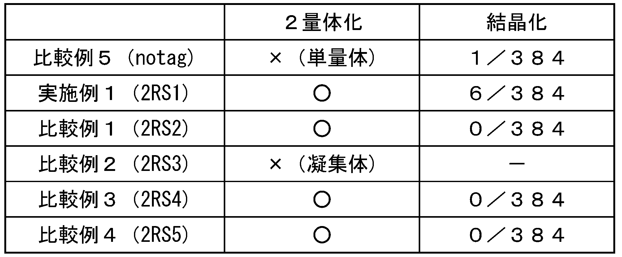
MBP/2RS1融合タンパク質で見られた6つの条件での、8日後の結晶の写真を図7に示す。
実施例2及び比較例6~9で得られたPH0828との各融合タンパク質並びに比較例10で得られたPH0828-notagを用いて、結晶化のスクリーニングを行った。結晶化スクリーニングキットは、MBPと同じものを用いた。
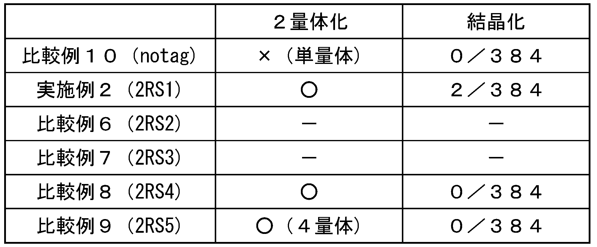
《実施例3》
本実施例では、AxCeSD-PilZと2RS1タグとを融合させたAxCeSD-PilZ/2RS1融合タンパク質を調製した。
pET28bベクターにAxCeSD-PilZを組み込んだpET28b-AxCeSD-PilZベクターに、GENEART Seamless Cloning and Assembly Kit(invitrogen社)を用いて、2RS1タグを組み込み、pET28b-AxCeSD-PilZ-2RS1ベクターを得た。
本実施例では、AxCeSD-PilZと2RS1タグとを融合させたAxCeSD-PilZ/2RS1融合タンパク質を調製した。
pET28bベクターにAxCeSD-PilZを組み込んだpET28b-AxCeSD-PilZベクターに、GENEART Seamless Cloning and Assembly Kit(invitrogen社)を用いて、2RS1タグを組み込み、pET28b-AxCeSD-PilZ-2RS1ベクターを得た。
MAL-2RS1ベクターに代えて、pET28b-AxCeSD-PilZ-2RS1ベクターを用いたことを除いては、実施例1(2)の操作を繰り返して、AxCeSD-PilZ/2RS1融合タンパク質を取得した。
《X線回折像の測定と構造解析》
実施例1で得られたMBP/2RS1融合タンパク質及び実施例3で得られたAxCeSD-PilZ/2RS1融合タンパク質をX線結晶構造解析した。それぞれのタンパク質結晶に対して、SPring-8又はPhotonFactoryの放射光施設にて、X線を照射し、回折像を得た。得られた回折データをHKL2000又はXDSのソフトウェアによって処理し、空間群や格子定数を決定し、データの指数付け、積分、スケーリング、マージングを行った。MBP/2RS1の場合には、既存のMBPの構造を用いて分子置換を行い、構造を決定した。AxCeSD-PilZ/2RS1融合タンパク質の場合には、既存の構造がなかったため、メチオニンをセレノメチオニンに置換したタンパク質結晶を作成し、Se-SAD法を用いて構造を決定した。
得られたMBP/2RS1融合タンパク質及びAxCeSD-PilZ/2RS1融合タンパク質の構造の模式図を、それぞれ図8及び図9に示す。
実施例1で得られたMBP/2RS1融合タンパク質及び実施例3で得られたAxCeSD-PilZ/2RS1融合タンパク質をX線結晶構造解析した。それぞれのタンパク質結晶に対して、SPring-8又はPhotonFactoryの放射光施設にて、X線を照射し、回折像を得た。得られた回折データをHKL2000又はXDSのソフトウェアによって処理し、空間群や格子定数を決定し、データの指数付け、積分、スケーリング、マージングを行った。MBP/2RS1の場合には、既存のMBPの構造を用いて分子置換を行い、構造を決定した。AxCeSD-PilZ/2RS1融合タンパク質の場合には、既存の構造がなかったため、メチオニンをセレノメチオニンに置換したタンパク質結晶を作成し、Se-SAD法を用いて構造を決定した。
得られたMBP/2RS1融合タンパク質及びAxCeSD-PilZ/2RS1融合タンパク質の構造の模式図を、それぞれ図8及び図9に示す。
《実施例4》
本実施例では、2RS1タグの7番目のアルギニン(R)のロイシン(L)への置換、による、MBP及びPH0828の結晶化への影響を検討した。
実施例1(1)で得られたpMAL-2RS1ベクター、又はpET28M-PH0828-2RS1ベクターを鋳型として、下記のプライマーを用いて、R7Lの変異を導入し、pMAL-2RS1-R7Lベクター、又はpET28M-PH0828-2RS1-R7Lベクターを得た。
R7Lプライマー1:5’-TTCTACTTCGGAAAGCTACGG-3’(配列番号76)
R7Lプライマー2:5’-ATCTAACTCTTTCTCCAAGTC-3’(配列番号77)
pMAL-2RS1ベクターに代えて、pMAL-2RS1-R7Lベクター、又はpET28M-PH0828-2RS1-R7Lベクターを用いたことを除いては、実施例1(2)の操作を繰り返して、MBP/2RS1-R7L融合タンパク質、又はPH0828/2RS1-R7L融合タンパク質を取得した。
前記結晶化スクリーニングに記載の方法に従って、MBP/2RS1-R7L融合タンパク質及びPH0828/2RS1-R7L融合タンパク質の結晶化のスクリーニングを行った。結果を表3に示す。
本実施例では、2RS1タグの7番目のアルギニン(R)のロイシン(L)への置換、による、MBP及びPH0828の結晶化への影響を検討した。
実施例1(1)で得られたpMAL-2RS1ベクター、又はpET28M-PH0828-2RS1ベクターを鋳型として、下記のプライマーを用いて、R7Lの変異を導入し、pMAL-2RS1-R7Lベクター、又はpET28M-PH0828-2RS1-R7Lベクターを得た。
R7Lプライマー1:5’-TTCTACTTCGGAAAGCTACGG-3’(配列番号76)
R7Lプライマー2:5’-ATCTAACTCTTTCTCCAAGTC-3’(配列番号77)
pMAL-2RS1ベクターに代えて、pMAL-2RS1-R7Lベクター、又はpET28M-PH0828-2RS1-R7Lベクターを用いたことを除いては、実施例1(2)の操作を繰り返して、MBP/2RS1-R7L融合タンパク質、又はPH0828/2RS1-R7L融合タンパク質を取得した。
前記結晶化スクリーニングに記載の方法に従って、MBP/2RS1-R7L融合タンパク質及びPH0828/2RS1-R7L融合タンパク質の結晶化のスクリーニングを行った。結果を表3に示す。
《実施例5》
本実施例では、2RS1タグの10番目のチロシン(Y)のロイシン(L)への置換による、MBP及びPH0828の結晶化への影響を検討した。
実施例1(1)で得られたpMAL-2RS1ベクター、又はpET28M-PH0828-2RS1ベクターを鋳型として、下記のプライマーを用いて、Y10Lの変異を導入し、pMAL-2RS1-Y10Lベクター、又はpET28M-PH0828-2RS1-Y10Lベクターを得た。
Y10Lプライマー1:5’-TTCTTATTCGGAAAGCTACGG-3’(配列番号78)
Y10Lプライマー2:5’-ATCCCTCTCTTTCTCCAAGTC-3’(配列番号79)
MAL-2RS1ベクターに代えて、pMAL-2RS1-Y10Lベクター、又はpET28M-PH0828-2RS1-Y10Lベクターを用いたことを除いては、実施例1(2)の操作を繰り返して、MBP/2RS1-Y10L融合タンパク質、又はPH0828/2RS1-Y10L融合タンパク質を取得した。
前記結晶化スクリーニングに記載の方法に従って、MBP/2RS1-Y10L融合タンパク質及びPH0828/2RS1-Y10L融合タンパク質の結晶化のスクリーニングを行った。結果を表3に示す。
本実施例では、2RS1タグの10番目のチロシン(Y)のロイシン(L)への置換による、MBP及びPH0828の結晶化への影響を検討した。
実施例1(1)で得られたpMAL-2RS1ベクター、又はpET28M-PH0828-2RS1ベクターを鋳型として、下記のプライマーを用いて、Y10Lの変異を導入し、pMAL-2RS1-Y10Lベクター、又はpET28M-PH0828-2RS1-Y10Lベクターを得た。
Y10Lプライマー1:5’-TTCTTATTCGGAAAGCTACGG-3’(配列番号78)
Y10Lプライマー2:5’-ATCCCTCTCTTTCTCCAAGTC-3’(配列番号79)
MAL-2RS1ベクターに代えて、pMAL-2RS1-Y10Lベクター、又はpET28M-PH0828-2RS1-Y10Lベクターを用いたことを除いては、実施例1(2)の操作を繰り返して、MBP/2RS1-Y10L融合タンパク質、又はPH0828/2RS1-Y10L融合タンパク質を取得した。
前記結晶化スクリーニングに記載の方法に従って、MBP/2RS1-Y10L融合タンパク質及びPH0828/2RS1-Y10L融合タンパク質の結晶化のスクリーニングを行った。結果を表3に示す。
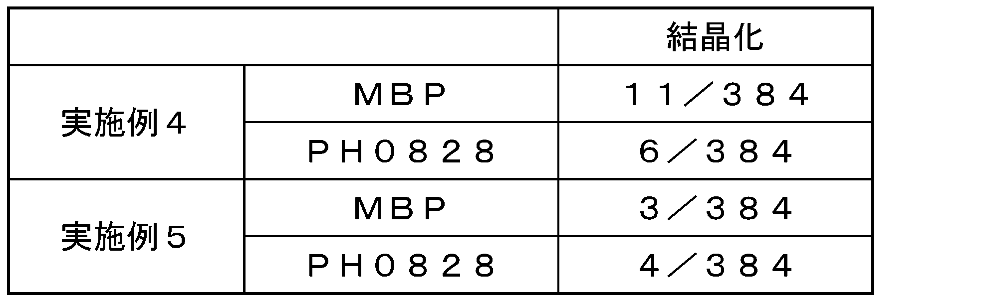
また、実施例5の2RS1タグのY10Lの置換(変異)により、PH0828の結晶化は、実施例2の2/384から4/384に上昇した。従って、2RS1タグの7番目のアルギニン(R)のロイシン(L)への置換は、PH0828については改良改変であると考えられる。一方、2RS1タグのY10Lの置換(変異)により、MBPの結晶化は、実施例1の6/384から3/384に低下した。しかしながら、表1の比較例5に示すようにタグを持たないMBPの結晶化は1/384であり、Y10Lの置換(変異)により、MBPの結晶化は3/384であるため、Y10Lの置換はMBPについては、維持改変であると考えられる。
本発明のポリペプチドは、目的のタンパク質と融合させ、そのタンパク質を結晶化させることができる。得られた融合タンパク質は、X線照射して、回折データを測定し、結晶構造解析法に用いて、そのタンパク質の構造を解析することができる。
以上、本発明を特定の態様に沿って説明したが、当業者に自明の変形や改良は本発明の範囲に含まれる。
以上、本発明を特定の態様に沿って説明したが、当業者に自明の変形や改良は本発明の範囲に含まれる。
Claims (9)
- (1)配列番号1で表されるアミノ酸配列からなるポリペプチド、
(2)配列番号2で表されるアミノ酸配列からなるポリペプチド、
(3)配列番号2で表されるアミノ酸配列において、N末側から1~18個のアミノ酸が欠失、及び/若しくはC末側から1~3個のいずれかのアミノ酸が欠失したアミノ酸配列からなるポリペプチド、又は
(4)前記ポリペプチド(1)、ポリペプチド(2)、又はポリペプチド(3)のアミノ酸配列において、1若しくは複数のアミノ酸が欠失、置換、挿入、及び/若しくは付加されたアミノ酸配列からなり、かつタンパク質のC末端若しくはN末端に直接若しくはペプチドリンカーを介して融合させた融合タンパク質として発現させた場合に当該融合タンパク質の結晶化を促進する活性を示す、改変体ポリペプチド。 - 前記改変体ポリペプチドが、配列番号3、又は4で表されるアミノ酸配列からなる、請求項1に記載のポリペプチド。
- 請求項1又は2に記載のポリペプチドが、タンパク質のC末端又はN末端に、直接又はペプチドリンカーを介して融合した、融合タンパク質。
- 請求項1又は2に記載のポリペプチド又は請求項3に記載の融合タンパク質をコードするポリヌクレオチド。
- 請求項4に記載のポリヌクレオチドを含む組換えベクター。
- 請求項5に記載の組換えベクターを含む形質転換細胞。
- 請求項3に記載の融合タンパク質をコードするポリヌクレオチドを含む組換えベクターを構築する工程、
前記組換えベクターを用いて融合タンパク質を発現させる工程、及び
前記発現した融合タンパク質を精製する工程、
を含む融合タンパク質の製造方法。 - 請求項3に記載の融合タンパク質をコードするポリヌクレオチドを含む組換えベクターを構築する工程、
前記組換えベクターを用いて融合タンパク質を発現させる工程、
前記発現した融合タンパク質を精製する工程、及び
精製された融合タンパク質の多量体形成を確認する工程、
を含む請求項7に記載の融合タンパク質の製造方法。 - 請求項3に記載の融合タンパク質をコードするポリヌクレオチドを含む組換えベクターを構築する工程、
前記組換えベクターを用いて融合タンパク質を発現させる工程、
前記発現した融合タンパク質を精製する工程、
精製された融合タンパク質の多量体形成を確認する工程、
精製された融合タンパク質を結晶化させる工程、及び
X線照射によりタンパク質結晶を確認する工程、
を含むタンパク質結晶の製造方法。
Priority Applications (1)
| Application Number | Priority Date | Filing Date | Title |
|---|---|---|---|
| JP2015515878A JP6399707B2 (ja) | 2013-05-07 | 2014-05-07 | 結晶化促進ポリペプチド |
Applications Claiming Priority (2)
| Application Number | Priority Date | Filing Date | Title |
|---|---|---|---|
| JP2013-097698 | 2013-05-07 | ||
| JP2013097698 | 2013-05-07 |
Publications (1)
| Publication Number | Publication Date |
|---|---|
| WO2014181786A1 true WO2014181786A1 (ja) | 2014-11-13 |
Family
ID=51867265
Family Applications (1)
| Application Number | Title | Priority Date | Filing Date |
|---|---|---|---|
| PCT/JP2014/062215 Ceased WO2014181786A1 (ja) | 2013-05-07 | 2014-05-07 | 結晶化促進ポリペプチド |
Country Status (2)
| Country | Link |
|---|---|
| JP (1) | JP6399707B2 (ja) |
| WO (1) | WO2014181786A1 (ja) |
Cited By (1)
| Publication number | Priority date | Publication date | Assignee | Title |
|---|---|---|---|---|
| WO2018163947A1 (ja) * | 2017-03-07 | 2018-09-13 | 国立大学法人徳島大学 | 多孔性タンパク質結晶を利用したタンパク質結晶化 |
Citations (1)
| Publication number | Priority date | Publication date | Assignee | Title |
|---|---|---|---|---|
| US20070148663A1 (en) * | 2005-07-15 | 2007-06-28 | Ying Wen | EB1-related compositions and methods |
-
2014
- 2014-05-07 JP JP2015515878A patent/JP6399707B2/ja not_active Expired - Fee Related
- 2014-05-07 WO PCT/JP2014/062215 patent/WO2014181786A1/ja not_active Ceased
Patent Citations (1)
| Publication number | Priority date | Publication date | Assignee | Title |
|---|---|---|---|---|
| US20070148663A1 (en) * | 2005-07-15 | 2007-06-28 | Ying Wen | EB1-related compositions and methods |
Non-Patent Citations (2)
| Title |
|---|
| FRYE ET AL.: "Structure of the Tropomyosin Overlap Complex from Chicken Smooth Muscle: Insight into the Diversity of N-Terminal Recognition", BIOCHEMISTRY, vol. 49, 2010, pages 4908 - 4920 * |
| TOMOYUKI MORI ET AL.: "The crystal structure of EB1-CLASP complex", DAI 11 KAI PROTEIN SCIENCE SOCIETY OF JAPAN NENKAI PROGRAM YOSHISHU, 2011, pages 53 * |
Cited By (1)
| Publication number | Priority date | Publication date | Assignee | Title |
|---|---|---|---|---|
| WO2018163947A1 (ja) * | 2017-03-07 | 2018-09-13 | 国立大学法人徳島大学 | 多孔性タンパク質結晶を利用したタンパク質結晶化 |
Also Published As
| Publication number | Publication date |
|---|---|
| JPWO2014181786A1 (ja) | 2017-02-23 |
| JP6399707B2 (ja) | 2018-10-03 |
Similar Documents
| Publication | Publication Date | Title |
|---|---|---|
| JP2013507123A (ja) | C型レクチンドメインをベースとするコンビナトリアルライブラリー | |
| Bateman et al. | Association behaviour of human βB1-crystallin and its truncated forms | |
| JP6399707B2 (ja) | 結晶化促進ポリペプチド | |
| KR20020056565A (ko) | 인간유래 세포 소멸 인자와 그 수용체의 결합체인TRAIL-sDR5 결합체의 결정화에 의한 3차원 구조및 TRAIL 결실 돌연변이 단백질 | |
| PT1573017E (pt) | Preparação de uma proteína anticongelante | |
| Goessweiner-Mohr et al. | The type IV secretion protein TraK from the Enterococcus conjugative plasmid pIP501 exhibits a novel fold | |
| Pedersen et al. | Crystallization and X-ray analysis of monodisperse human properdin | |
| US20030096312A1 (en) | Novel natrium-calcium exchanger protein | |
| KR100980378B1 (ko) | 고농도 우레아하에서 안정된 활성을 갖는 디유비퀴틸레이팅효소를 이용한 펩티드의 제조방법 | |
| CN117586415A (zh) | 一种重组Ulp1融合蛋白酶及其制备方法 | |
| Yan et al. | Crystallization and crystallographic studies of human serine protease inhibitor (serpin) B9 | |
| KR102779028B1 (ko) | Usp44-wdr5 복합체 결정 및 결정화 방법 | |
| Goessweiner-Mohr et al. | Crystallization and preliminary structure determination of the transfer protein TraM from the Gram-positive conjugative plasmid pIP501 | |
| CN112375138A (zh) | 一种重组载脂蛋白e和应用 | |
| US20050118683A1 (en) | Method for producing a polypeptide | |
| JP4555928B2 (ja) | がん細胞認識破壊活性を発現するように活性化されたパラスポリン−1の結晶、その製造方法、その三次元構造および利用方法 | |
| CN113795500A (zh) | 金刚烷胺结合蛋白 | |
| Lin et al. | Crystallization and crystallographic studies of kallistatin | |
| Yoon et al. | Purification, crystallization and data collection of the apoptotic nuclease endonuclease G | |
| Radzimanowski et al. | Overproduction, purification, crystallization and preliminary X-ray analysis of human Fe65-PTB2 in complex with the amyloid precursor protein intracellular domain | |
| Cheng et al. | Protein production, crystallization and preliminary crystallographic analysis of the four N-terminal immunoglobulin domains of Down syndrome cell adhesion molecule 1 | |
| EP1225181A1 (en) | Protein expression and structure resolution using specific fusion vectors | |
| Hillier et al. | Expression, purification, crystallization and preliminary X-ray analysis of the olfactomedin domain from the sea urchin cell-adhesion protein amassin | |
| Park | X-ray crystallographic studies of the middle part of the human synaptonemal complex protein 1 coiled-coil domain | |
| JPWO2003083101A1 (ja) | Dapキナーゼの結晶、それを用いたdapキナーゼをターゲットとする薬物候補分子のスクリーニング方法及びそれにより得られる医薬 |
Legal Events
| Date | Code | Title | Description |
|---|---|---|---|
| 121 | Ep: the epo has been informed by wipo that ep was designated in this application |
Ref document number: 14794546 Country of ref document: EP Kind code of ref document: A1 |
|
| ENP | Entry into the national phase |
Ref document number: 2015515878 Country of ref document: JP Kind code of ref document: A |
|
| NENP | Non-entry into the national phase |
Ref country code: DE |
|
| 122 | Ep: pct application non-entry in european phase |
Ref document number: 14794546 Country of ref document: EP Kind code of ref document: A1 |