WO2014136443A1 - 抗Aβ42モノクローナル抗体 - Google Patents
抗Aβ42モノクローナル抗体 Download PDFInfo
- Publication number
- WO2014136443A1 WO2014136443A1 PCT/JP2014/001187 JP2014001187W WO2014136443A1 WO 2014136443 A1 WO2014136443 A1 WO 2014136443A1 JP 2014001187 W JP2014001187 W JP 2014001187W WO 2014136443 A1 WO2014136443 A1 WO 2014136443A1
- Authority
- WO
- WIPO (PCT)
- Prior art keywords
- peptide
- monoclonal antibody
- antibody
- amino acid
- seq
- Prior art date
- Legal status (The legal status is an assumption and is not a legal conclusion. Google has not performed a legal analysis and makes no representation as to the accuracy of the status listed.)
- Ceased
Links
Images

Classifications
-
- C—CHEMISTRY; METALLURGY
- C07—ORGANIC CHEMISTRY
- C07K—PEPTIDES
- C07K16/00—Immunoglobulins [IG], e.g. monoclonal or polyclonal antibodies
- C07K16/18—Immunoglobulins [IG], e.g. monoclonal or polyclonal antibodies against material from animals or humans
Definitions
- the present invention relates to a monoclonal antibody that specifically reacts with A ⁇ 42 peptide, a hybridoma that can produce the monoclonal antibody, and a kit containing the monoclonal antibody.
- AD Alzheimer's disease
- a ⁇ peptide In order to diagnose Alzheimer's disease at an early stage, a method for detecting ⁇ amyloid (hereinafter also referred to as A ⁇ ) peptide present in body fluids such as blood and spinal fluid is known.
- a ⁇ peptide is a peptide composed of 40 to 43 amino acids, and it is known that in Alzheimer's disease, the A ⁇ peptide aggregates to form amyloid and deposits in the brain.
- the A ⁇ 42 peptide which is a peptide consisting of 42 amino acids, is said to be a factor marker for Alzheimer's disease.
- a ⁇ 43 peptide which is a peptide consisting of 43 amino acids, is localized at the center of cerebral senile plaques. It has been clarified through research (see Non-Patent Document 1).
- Monoclonal antibodies that react with A ⁇ peptides are already known (see, for example, Patent Document 1), and A ⁇ peptide detection kits containing such monoclonal antibodies are already commercially available.
- Non-Patent Document 2 immunohistochemical staining using autopsy tissue from AD patients has shown that A ⁇ is also present in tissues other than the brain, such as the nasal mucosa (see Non-Patent Document 2).
- a ⁇ 42 peptide and A ⁇ 43 peptide are considered to have different functions among A ⁇ peptides
- a ⁇ 42 peptide and A ⁇ 43 peptide are used in detecting A ⁇ peptide present in body fluids such as blood and cerebrospinal fluid. It would be preferable to be able to detect them separately.
- a ⁇ 42 which controls the total amount of the elderly group, is considered to be more highly related to AD than A ⁇ 43, which is localized at the center of the elderly group. It is desirable that it can be detected automatically.
- conventionally known monoclonal antibodies that react with the antigen A ⁇ peptide react with both the A ⁇ 42 peptide and the A ⁇ 43 peptide.
- a monoclonal antibody that does not react with A ⁇ 43 peptide but specifically reacts with A ⁇ 42 peptide, which is a factor marker of Alzheimer's disease, has not been known. Therefore, the problem to be solved by the present invention is to provide an antibody for distinguishing and detecting A ⁇ 42 peptide from A ⁇ 43 peptide.
- the present inventors have conducted extensive research and succeeded in establishing a specific hybridoma that produces a monoclonal antibody that does not substantially react with the A ⁇ 43 peptide but reacts specifically with the A ⁇ 42 peptide, thereby completing the present invention. It came to do.
- the present invention provides the following: (1) a monoclonal antibody that reacts with A ⁇ 42 peptide and does not react with A ⁇ 43 peptide; (2) From the heavy chain complementarity determining region (CDR) 1 consisting of the amino acid sequence represented by SEQ ID NO: 8, the heavy chain CDR2 consisting of the amino acid sequence represented by SEQ ID NO: 9, and the amino acid sequence represented by SEQ ID NO: 10 A heavy chain variable region composed of a heavy chain CDR3 and a heavy chain framework sequence derived from an immunoglobulin heavy chain; and a light chain CDR1 composed of an amino acid sequence represented by SEQ ID NO: 13, represented by SEQ ID NO: 14 A light chain CDR2 comprising an amino acid sequence, a light chain CDR3 comprising an amino acid sequence represented by SEQ ID NO: 15, and a light chain variable region comprising a light chain framework sequence derived from an immunoglobulin light chain, (1) A monoclonal antibody according to claim 1; (3) The monoclonal antibody according to (1), comprising a heavy chain complement
- the monoclonal antibody provided by the present invention does not substantially react with the A ⁇ 43 peptide, but reacts specifically with the A ⁇ 42 peptide, so that the A ⁇ 42 peptide that is a factor marker of Alzheimer's disease can be specifically detected.
- the use of the monoclonal antibody of the present invention or the A ⁇ detection kit containing the antibody makes it possible to specifically detect or measure the A ⁇ 42 peptide, which was impossible with conventional antibodies or detection kits, and thus is extremely useful.
- the present invention relates to a monoclonal antibody that reacts with A ⁇ 42 peptide and does not react with A ⁇ 43 peptide.
- the present invention provides: Heavy chain complementarity determining region (CDR) 1 consisting of the amino acid sequence represented by SEQ ID NO: 8, heavy chain CDR2 consisting of the amino acid sequence represented by SEQ ID NO: 9, and heavy chain consisting of the amino acid sequence represented by SEQ ID NO: 10
- the present invention relates to a monoclonal antibody comprising a light chain CDR2 comprising: a light chain CDR3 comprising the amino acid sequence represented by SEQ ID NO: 15, and a light chain variable region comprising a light chain framework sequence derived from an immunoglobulin light chain.
- the monoclonal antibody of the present invention is represented by the heavy chain CDR sequence consisting of the amino acid sequence represented by SEQ ID NO: 8 and SEQ ID NO: 9 as long as it is a monoclonal antibody that reacts with the A ⁇ 42 peptide but does not react with the A ⁇ 43 peptide. Also included are antibodies comprising at least one heavy chain CDR sequence selected from the group consisting of a heavy chain CDR sequence consisting of an amino acid sequence and a heavy chain CDR sequence consisting of the amino acid sequence represented by SEQ ID NO: 10.
- the monoclonal antibody of the present invention is represented by the heavy chain CDR sequence consisting of the amino acid sequence represented by SEQ ID NO: 8, represented by SEQ ID NO: 9, as long as it is a monoclonal antibody that reacts with the A ⁇ 42 peptide and does not react with the A ⁇ 43 peptide.
- antibodies comprising one light chain CDR sequence.
- the monoclonal antibody of the present invention is a monoclonal antibody that reacts with the A ⁇ 42 peptide and does not react with the A ⁇ 43 peptide, the amino acid sequence of the CDR (heavy chain CDR1 to 3 and light chain CDR1 to 3) of the monoclonal antibody of the present invention described above. For example, at least 60%, 70%, 75%, 80%, 85%, 86%, 87%, 88%, 89%, 90%, 91%, 92%, or 93% sequence identity. Also included are antibodies comprising a CDR sequence having an amino acid sequence.
- the monoclonal antibody of the present invention includes the CDRs of the above-described monoclonal antibodies of the present invention (heavy chain CDR1 to 3 and light chain CDR1 to 3) as long as the monoclonal antibody reacts with the A ⁇ 42 peptide and does not react with the A ⁇ 43 peptide. Also included are antibodies comprising a CDR sequence having an amino acid sequence in which one or several, eg, 2 or 3, amino acids are substituted, deleted or added for each amino acid sequence.
- “does not react with a peptide” or “does not substantially react with a peptide” means that in a test for confirming the reactivity of an antibody and / or antibody fragment with respect to a peptide, no result showing the reactivity is obtained. Or the result is below the detection limit.
- a test is, for example, a test as described in Example 2 below.
- sequence identity refers to a case where two or more sequences (base sequence or amino acid sequence) are aligned in consideration of gaps and insertions so that the degree of sequence match is maximized. Of the same base or amino acid at corresponding positions in two or more sequences.
- the method of determining identity is designed to give the greatest degree of match between aligned sequences.
- a method for determining identity between two sequences is not limited, and includes BLASTP, BLASTN, FASTA and the like. It can also be determined using DNASIS (manufactured by Hitachi Software Engineering Co., Ltd.) or GENETYX (manufactured by Genetics Co., Ltd.). Alternatively, in the case of a short chain peptide, it can also be determined by simply comparing the sequences.
- One skilled in the art can determine identity between sequences by the methods described above.
- the present invention also relates to a polynucleotide encoding the amino acid sequence of the CDR (heavy chain CDR1 to 3 and light chain CDR1 to 3) of the monoclonal antibody of the present invention described above.
- One skilled in the art can determine and generate sequences for such polynucleotides using techniques known in the art.
- the monoclonal antibody of the present invention is a monoclonal antibody produced by a hybridoma whose accession number is NITE P-1463.
- the invention relates to a hybridoma that produces the monoclonal antibody of the invention.
- the hybridoma was devisated on November 8, 2012 at the depositary organization (NITE), the National Institute for Product Evaluation and Technology (NITE), which has an address in 2-5-8 Kazusa Kamashika, Kisarazu City, Chiba Prefecture, Japan. (NPMD)) and the deposit number is NITE P-1463.
- the monoclonal antibody produced by the hybridoma does not react with A ⁇ 40 peptide and A ⁇ 43 peptide, but reacts only with A ⁇ 42 peptide. Therefore, A ⁇ 42 peptide can be specifically detected among A ⁇ , and is extremely useful.
- the monoclonal antibody produced by the hybridoma exhibits excellent characteristics such as high reactivity with the A ⁇ 42 peptide as compared with a commercially available anti-A ⁇ antibody. Furthermore, it shows the characteristic that the reactivity with respect to the A ⁇ 42 peptide in the nasal sample is higher than that of the A ⁇ 42 peptide in the brain sample.
- the production method (immunization method and selective collection method) of the hybridoma of the present invention is not particularly limited. Using an A ⁇ 42 peptide or a part thereof as an antigen, it can be produced by a usual method for producing a hybridoma. That is, the hybridoma of the present invention is injected by injecting an antigen into an immunized animal such as a rat, taking out lymphocytes related to the production of antibodies thereto, mixing it with myeloma, and culturing it on a HAT medium. Can be manufactured.
- a ⁇ 42 peptide When using a part of the A ⁇ 42 peptide as an antigen, it is particularly preferable to use an amino acid sequence containing the C-terminus of the A ⁇ 42 peptide. Since A ⁇ 40 peptide (SEQ ID NO: 16), A ⁇ 42 peptide (SEQ ID NO: 17), and A ⁇ 43 peptide (SEQ ID NO: 18) have different C-terminal amino acid sequences, respectively, by using an amino acid sequence including the C-terminal of A ⁇ 42 peptide, It becomes possible to obtain a hybridoma that produces a monoclonal antibody that specifically reacts with the A ⁇ 42 peptide. In the examples described later, the amino acid sequence containing the amino acid sequence from the 33rd to the 42nd position on the C-terminal side of the amino acid sequence of the A ⁇ 42 peptide was used as the antigen peptide.
- the monoclonal antibody produced by the hybridoma is labeled with, for example, a general labeled protein, and the reactivity with respect to each of A ⁇ 42 peptide and A ⁇ 43 peptide is measured. .
- a monoclonal antibody that specifically reacts with the A ⁇ 42 peptide can be confirmed.
- the inventors determined the amino acid sequence of the monoclonal antibody produced by the hybridoma of the present invention, and succeeded in identifying the complementarity determining region (CDR), which is a hypervariable region.
- CDR complementarity determining region
- Three CDRs contained in the variable region of the heavy chain of the monoclonal antibody have an amino acid sequence represented by SEQ ID NOs: 8 to 10.
- the three CDRs contained in the variable region of the light chain of the monoclonal antibody have an amino acid sequence represented by the amino acid sequence represented by SEQ ID NOs: 13-15.
- a heavy chain CDR consisting of the amino acid sequence represented by SEQ ID NOs: 8 to 10 is incorporated into a heavy chain framework sequence derived from an immunoglobulin heavy chain to constitute a heavy chain variable region, and is represented by SEQ ID NOs: 13 to 15
- An antibody can be prepared by constructing a light chain variable region by incorporating a light chain CDR comprising an amino acid sequence represented by the amino acid sequence described above into a light chain framework sequence derived from an immunoglobulin light chain.
- a conventionally known method can be adopted as a method for producing such an antibody.
- the present invention relates to a chimeric monoclonal antibody or a humanized monoclonal antibody having the CDR sequence of the monoclonal antibody of the present invention described above.
- a chimeric antibody typically comprises a variable region of an antibody derived from a certain species and a constant region of an antibody derived from a different species, and can be easily constructed by a gene recombination technique. Methods for making chimeric antibodies are known in the art. For example, it can be produced by the method described in “Roguska et al., Proc Natl Acad Sci USA A.
- a humanized antibody is typically an antibody having one or more CDRs from a non-human species, a framework region (FR) from a human immunoglobulin, and a constant region from a human immunoglobulin.
- Methods for making humanized antibodies are known in the art.
- a humanized antibody can be produced by applying a human heavy chain framework sequence and a human light chain framework sequence as the heavy chain framework sequence and the light chain framework sequence.
- Almagro et al. FRONT Biosci. 2008 Jan 1; 13: 1619-1633. See etc.
- CDR As a method of determining the definition of CDR and its position, for example, the definition of Kabat (Sequences of Proteins of Immunological Interest, 5th ed., Public Health Service, National Institute of Health 91, or National Institute of Health.
- the definition Chothia et al., J. Mol. Biol., 1987; 196: 901-917) may be adopted. In some cases, it may be determined in consideration of both the Kabat definition and the Chothia definition. For example, an overlapping part of CDRs according to each definition or a part including both CDRs according to each definition may be determined. It can also be a CDR.
- Martin et al.'S method Proc. Natl. Acad. Sci.
- the present invention relates to an antibody fragment derived from the antibody of the present invention described above.
- the “antibody fragment derived from the antibody of the present invention” is a part (partial fragment) of the antibody of the present invention or a peptide containing a part of the antibody of the present invention, and retains the action of the antibody of the present invention on the antigen.
- Such antibody fragments include Fab, Fab ′, F (ab ′) 2 , single chain antibody (scFv), dimerized V region (diabody), disulfide stabilized V region (dsFv), and the present invention.
- Fab is an antibody fragment having antigen-binding activity in which about half of the N-terminal side of the H chain and the entire L chain are linked by a disulfide bond among fragments obtained by treating immunoglobulin protein with papain. Therefore, the Fab of the present invention can be obtained by treating the monoclonal antibody of the present invention with papain.
- Fab encoding is produced by inserting DNA encoding Fab of the monoclonal antibody of the present invention into a prokaryotic expression vector or eukaryotic expression vector, and introducing the vector into prokaryotic or eukaryotic organisms. You can also
- F (ab ′) 2 is a fragment having antigen-binding activity, which is obtained by treating the lower part of the disulfide bond of the hinge region of an immunoglobulin protein with pepsin, and is composed of two Fab regions joined by a hinge portion. is there. Therefore, F (ab ′) 2 of the present invention can be obtained by treating the monoclonal antibody of the present invention with pepsin. Alternatively, Fab ′ described later can be produced by thioether bond or disulfide bond.
- Fab ′ is an antibody fragment having antigen-binding activity obtained by cleaving the disulfide bond in the hinge region of F (ab ′) 2 described above. Therefore, Fab ′ of the present invention can be obtained by treating F (ab ′) 2 of the present invention with a reducing agent such as dithiothreitol.
- DNA encoding the Fab ′ fragment of the monoclonal antibody of the present invention is inserted into a prokaryotic expression vector or eukaryotic expression vector, and the vector is introduced into prokaryotic or eukaryotic organisms to be expressed. 'Can also be manufactured.
- a single chain antibody is a polypeptide in which one heavy chain variable region (VH) and one light chain variable region (VL) are linked using an appropriate peptide linker, and has an antigen binding activity. It is an antibody fragment having The scFv of the present invention obtains cDNA encoding the VH and VL of the monoclonal antibody of the present invention, constructs a DNA encoding scFv, and inserts the DNA into a prokaryotic expression vector or eukaryotic expression vector.
- the expression vector can be expressed and produced by introducing it into a prokaryotic or eukaryotic organism.
- the dimerization V region (diabody) is an antibody fragment obtained by dimerizing the above-mentioned scFv, and is an antibody fragment having a bivalent antigen-binding activity.
- the bivalent antigen binding activity may be the same or different.
- the dimerized V region of the present invention is obtained by obtaining cDNA encoding the VH and VL of the monoclonal antibody of the present invention, constructing DNA encoding scFv so as to contain an appropriate short peptide linker, It can be expressed and produced by inserting it into a prokaryotic expression vector or eukaryotic expression vector and introducing the expression vector into a prokaryotic or eukaryotic organism.
- the disulfide-stabilized V region refers to a polypeptide in which one amino acid residue in each of VH and VL is substituted with a cysteine residue and bonded via a disulfide bond between the cysteine residues.
- the amino acid residue substituted for the cysteine residue can be selected based on the three-dimensional structure prediction of the antibody according to a known method (for example, Protein Engineering, 7, 697 (1994)).
- the dsFv of the present invention obtains cDNA encoding the VH and VL of the monoclonal antibody of the present invention, constructs a DNA encoding dsFv, and inserts the DNA into a prokaryotic expression vector or a eukaryotic expression vector.
- the expression vector can be expressed and produced by introducing it into a prokaryotic or eukaryotic organism.
- the peptide containing CDR is composed of at least one region of CDR sequence of VH or VL. Peptides containing multiple CDRs can be linked directly or via a suitable peptide linker.
- the peptide containing the CDR of the present invention constructs the DNA encoding the VH and VL CDRs of the monoclonal antibody of the present invention, inserts the DNA into a prokaryotic expression vector or eukaryotic expression vector, and the expression vector Can be expressed and produced by introducing it into a prokaryotic or eukaryotic organism.
- the peptide containing CDR can also be manufactured by chemical synthesis methods, such as Fmoc method or tBoc method.
- the monoclonal antibody of the present invention includes a derivative of an antibody in which a radioisotope, a low molecular drug, a high molecular drug, a protein or the like is chemically or genetically bound to the monoclonal antibody of the present invention or an antibody fragment thereof.
- a normal immunological detection or measurement method as a drug that binds to the monoclonal antibody of the present invention or an antibody fragment thereof.
- alkaline phosphatase, peroxidase or luciferase is used.
- the present invention relates to a method for producing an antibody or antibody fragment of the present invention comprising culturing a hybridoma having a deposit number of NITE P-1463.
- a conventionally well-known method can be employ
- the present invention relates to an A ⁇ 42 detection or measurement kit comprising the antibody or antibody fragment of the present invention described above.
- the A ⁇ 42 detection kit of the present invention includes a buffer solution, a diluent, a washing solution, a secondary antibody solution, a calibration curve reagent necessary for concentration regression of the detected measurement value, a plate, and instructions for use Documents, specimen collection devices, specimen collection tubes, and the like.
- the kit of the present invention is a system that detects A ⁇ 42 peptide by a color development reaction
- the kit of the present invention can further contain a color developing solution, a color developing stop solution and the like.
- the kit of the present invention is a system that detects A ⁇ 42 peptide by fluorescence or color development, it is not necessary to include a color development solution and a color development stop solution.
- the kit of the present invention is an ELISA kit.
- the present invention relates to a method for detecting or measuring A ⁇ 42 in a specimen, including the use of the antibody, antibody fragment or kit of the present invention described above.
- the “specimen” is an animal, preferably a mammal, more preferably a primate, even more preferably a human, particularly preferably a human who may be suffering from AD, or a mild cognitive impairment of AD type. Collected from human or AD patients.
- the site from which the specimen is collected is not particularly limited as long as it is a site where A ⁇ 42 is known to exist in AD patients. Accordingly, the specimen is, for example, a sample collected from the nose (eg, nasal mucosa) such as cerebrospinal fluid, blood, or nasal wiping solution, or a sample collected from the oral cavity (eg, oral mucosa) such as the oral wiping solution.
- these specimens may be prepared in a state suitable for use in the kit or method of the present invention using a lysate, a buffer or a diluent.
- the specimen used in the kit or method of the present invention is collected from the nose (eg, nasal mucosa).
- the specimen used in the kit or method of the present invention is a nasal wipe.
- Example 1 Acquisition of hybridoma A hybridoma producing the monoclonal antibody of the present invention was obtained by the following procedure.
- a peptide having the amino acid sequence represented by SEQ ID NO: 1 was synthesized as an antigen peptide. This peptide is obtained by adding Cys to the N-terminus of the amino acid sequence of the A ⁇ 42 peptide from the 33rd to the 42nd amino acid sequence on the C-terminal side. The addition of Cys is for conjugation with the carrier protein.
- Hybridomas were cloned from the 12 positive wells, and the following tests were performed. ⁇ Cell seeding by limiting dilution ⁇ Reactivity check by ELISA (confirms reactivity with A ⁇ 42 peptide and no reactivity with A ⁇ 40 peptide) -Checking for single colony formation by microscopic observation of ELISA positive wells By the above test, one hybridoma of an ELISA positive single clone was obtained. The name of the hybridoma was 6C10H3.
- hybridoma was cryopreserved and deposited with the National Institute of Technology and Evaluation (NITE) and the Patent Microorganism Deposit Center (NPMD). The deposit number is NITE P-1463, and the deposit date is November 8, 2012.
- NITE National Institute of Technology and Evaluation
- NPMD Patent Microorganism Deposit Center
- Example 2 Specificity evaluation of monoclonal antibody (1) Preparation of labeled antibody Monoclonal antibody 6C10H3 was obtained from hybridoma 6C10H3. Subsequently, the monoclonal antibody 6C10H3 was labeled by the following procedure to obtain a labeled antibody.
- Processing was performed according to the protocol of Peroxidase Labeling Kit-SH manufactured by Dojindo Laboratories. That is, a sample solution containing 200 ⁇ g of antibody and 100 ⁇ L of Solution A attached to the kit were added to Filtration Tube attached to the kit. After light mixing by pipetting, centrifugation was performed at 8000 ⁇ g for 10 minutes. 100 ⁇ L of Solution A was added to the reducing agent included in the kit and dissolved by pipetting. 100 ⁇ L of this solution was added onto a Filtration Tube membrane in which the antibody was concentrated, and mixed well with the antibody by pipetting. After reacting at 37 ° C.
- the labeled antibody was diluted 1000 times, and the reactivity to each of A ⁇ 40 peptide, A ⁇ 42 peptide, and A ⁇ 43 peptide was measured by the following procedure.
- the reactivity with respect to each peptide was similarly measured using the monoclonal antibody contained in a commercially available A ⁇ detection kit (manufactured by Wako Pure Chemical Industries, Ltd.).
- HRP-labeled antibody (BC05) solution (provided with a kit manufactured by Wako Pure Chemical Industries, Ltd.) was added to each well in one set.
- HRP-labeled antibody (6C10H3) solution was added to each well. After the addition, the reaction was allowed to proceed for 5 minutes at room temperature with stirring. After washing was performed 5 times, 100 ⁇ L of TMB solution was added and allowed to react for 30 minutes in the dark at room temperature.
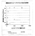
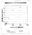
- FIG. 1 shows that neither the monoclonal antibody 6C10H3 of the present invention nor the commercially available monoclonal antibody shows reactivity to the A ⁇ 40 peptide, and from FIG. 2, both antibodies show reactivity to the A ⁇ 42 peptide.
- FIG. 3 shows that the monoclonal antibody 6C10H3 of the present invention does not show reactivity with the A ⁇ 43 peptide, whereas the commercially available monoclonal antibody shows reactivity with the A ⁇ 43 peptide.
- the monoclonal antibody 6C10H3 of the present invention shows reactivity to A ⁇ 42 peptide, but A ⁇ 43 peptide Since it does not show any reactivity to the A ⁇ 42 peptide, it can be seen that the A ⁇ 42 peptide can be distinguished from the A ⁇ 43 peptide.
- Example 3 Evaluation of recognition ability of monoclonal antibody Comparison of recognition ability of A ⁇ 42 peptide using monoclonal antibody 6C10H3 and 1F115 antibody (manufactured by IBL) which is generally an anti-A ⁇ antibody frequently used by researchers did.
- a 4G8 antibody was biotinylated using a biotin labeling kit (SH label, manufactured by Dojindo).
- HRP labeling of 6C10H3 antibody and 1F115 antibody was performed using an HRP labeling kit (SH labeling, manufactured by Dojindo).
- a ⁇ 42 peptide Human, 1-42, manufactured by Peptide Laboratories was diluted to obtain an A ⁇ 42 peptide solution having a concentration of 0, 5, 10, 20, 50, 100, 200, or 500 pM.
- each of the prepared A ⁇ 42 peptide solution (0, 5, 10, 20, 50, 100, 200, 500 pM) was added to the plate and reacted at room temperature for 2 hours, and then the A ⁇ 42 peptide solution was removed from the plate. did. The plate was washed 5 times using the washing solution.
- HRP-labeled antibodies (6C10H3 antibody and 1F115 antibody) at the same concentration was added to each plate and reacted. Thereafter, the plate was washed 5 times using the washing solution (5 times each).
- HRP enzyme substrate manufactured by SIGMA
- a stop solution manufactured by KPL
- color development was measured at a wavelength of 450 nm to 620 nm.
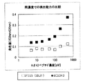
- FIG. 4 shows the color values of HRP-labeled 6C10H3 antibody (black square) and HRP-labeled 1F115 antibody (white square) when an A ⁇ 42 peptide solution diluted 2500 times is used. From this figure, it can be seen that the 6C10H3 antibody of the present invention is more reactive with the A ⁇ 42 peptide than the 1F115 antibody, which is a commercially available anti-A ⁇ antibody.
- Example 4 CDR sequencing of monoclonal antibody The sequence of the complementarity determining region (CDR) of the 6C10H3 antibody of the present invention was determined by the following procedure.
- RNA Extraction Procedure Using hybridoma 6C10H3 (about 1 ⁇ 10 7 cells), the following procedure was performed in this order to collect total RNA.
- -Homogenization step 1 mL of TRIzol Reagent (manufactured by life technologies (Invitrogen), No. 15596-026) was added by pipetting and treated at room temperature for 5 minutes.
- RNA extraction step 0.2 mL of chloroform was added, mixed by inversion, and treated at room temperature for 3 minutes. Thereafter, centrifugation (12000 g, 4 ° C., 15 minutes) was performed, and the aqueous layer was recovered.
- First washing step 0.5 mL of isopropanol is added, mixed by inversion, and treated at room temperature for 10 minutes. Thereafter, centrifugation (12000 g, 4 ° C., 10 minutes) is performed, and the precipitate is collected (the supernatant is removed by suction).
- -Second washing step 1 mL of 75% ethanol was added and mixed by inversion. Thereafter, centrifugation (12000 g, 4 ° C., 5 minutes) is performed, and the precipitate is collected (the supernatant is removed by suction) and air-dried.
- -Adjustment process Rnase free water 0.02mL is added and it processes at 55 degreeC for 10 minute (s).
- cDNA preparation procedure Using the total RNA recovered in the above (1), cDNA was prepared by the following procedure. Denaturation step: Dilute 5 ⁇ g of total RNA to 20 ⁇ L. Then, after processing at 65 degreeC for 10 minutes, it cools rapidly. Then, 11 ⁇ L of Bulk first-strand reaction mix, 1 ⁇ L of DTT solution, and 1 ⁇ L of primer (Not Id (T) 18) were added from First-Strand cDNA synthesis kit manufactured by GE Healthcare. Incubation step: held at 37 ° C. for 1 hour.
- RT-PCR RT-PCR was performed using the cDNA obtained in (2) above.
- primers having sequences represented by SEQ ID NOs: 2 and 3 were used as heavy chain primers
- primers having sequences represented by SEQ ID NOs: 4 and 5 were used as light chain primers.
- Sequencing Sequencing was performed from BigDye (registered trademark) Terminator v3.1 Cycle Sequencing Kit manufactured by Life technologies (Invitrogen) using primers specific to pGEM-T Easy Vector.
- the heavy chain of the 6C10H3 antibody consists of the amino acid sequence represented by SEQ ID NO: 7, and of which the heavy chain CDR consists of the amino acid sequence represented by SEQ ID NOs: 8 to 10.
- the light chain of the 6C10H3 antibody consists of the amino acid sequence represented by SEQ ID NO: 12, of which the light chain CDR consists of the amino acid sequence represented by SEQ ID NOs: 13-15.
- SEQ ID NO: 6 is the base sequence encoding the heavy chain of the 6C10H3 antibody
- SEQ ID NO: 11 is the base sequence encoding the light chain of the 6C10H3 antibody.
- the CDR region was amplified by PCR using the pGEM-T vector incorporating the 6C10H3 antibody gene (heavy chain and light chain) as a template and the following primers.
- primers having sequences represented by SEQ ID NOs: 19 and 20 were used as heavy chain primers
- primers having sequences represented by SEQ ID NOs: 21 and 22 were used as light chain primers.
- each PCR product treated with a restriction enzyme was incorporated into the following vector treated with the same restriction enzyme.
- the human IgG1 gene Fc region has already been incorporated.
- Heavy chain pCAGGS vector
- Light chain pCAGGS vector
- the obtained vector containing 6C10H3 antibody-derived heavy chain CDR comprises a rat-derived heavy chain variable region (VH) and a human-derived heavy chain constant region (CH1, CH2 and CH3).
- the obtained vector containing the 6C10H3 antibody-derived light chain CDR contains a rat-derived light chain variable region (VL) and a human-derived light chain constant region (CL).
- HEK293T Human embryonic kidney cell HEK293T was used as a host and cultured on a 5 mL scale. All the cultures were performed under conditions of 37 ° C. and 5% CO 2.
- HEK293T cells were cultured in a DMEM containing 10% FBS per 6 cm dish. When 80-90% confluent, transfection was performed. Before transfection, 293 ⁇ L of DMEM (0% FBS) mixed with 7.0 ⁇ g of plasmid (heavy chain: 3.5 ⁇ g, light chain: 3.5 ⁇ g) and DMEM (0% FBS) in 293 ⁇ l A mixture of 10.6 ⁇ L of a 1 mg / mL polyethyleneimine solution was prepared.
- Example 6 Evaluation of recognition ability for A ⁇ 42 derived from different tissues Using a specimen collected from tissues around the nose and a specimen collected from the brain, the recognition ability of the monoclonal antibody 6C10H3 to A ⁇ 42 derived from different tissues was evaluated. For the measurement, the labeled monoclonal antibody 6C10H3 prepared in Example 2 was used. As a comparative example, a monoclonal antibody contained in a commercially available A ⁇ detection kit (manufactured by Wako Pure Chemical Industries, Ltd.) was used.
- the labeled antibody was diluted 1000 times, and the reactivity with respect to each of the specimen collected from the tissue around the nose and the specimen collected from the brain was measured by the following procedure.
- a ⁇ detection kit human ⁇ amyloid (1-42) ELISA kit Wako, high-sensitivity product, manufactured by Wako Pure Chemical Industries, Ltd.
- Sex was measured.
- HRP-labeled antibody (BC05) solution (provided with a kit manufactured by Wako Pure Chemical Industries, Ltd.) was added to each well in one set.
- HRP-labeled antibody (6C10H3) solution was added to each well. After the addition, the reaction was allowed to proceed for 5 minutes at room temperature with stirring. After washing was performed 5 times, 100 ⁇ L of TMB solution was added and allowed to react for 30 minutes in the dark at room temperature.
- the monoclonal antibody 6C10H3 of the present invention has a higher reactivity with respect to the sample collected from the tissue around the nose than the sample collected from the brain.
- the commercially available monoclonal antibody there was no difference between the reactivity to the specimen collected from the brain and the reactivity to the specimen collected from the tissues around the nose. From this, it was shown that the monoclonal antibody of the present invention reacts more specifically with A ⁇ 42 derived from nasal tissue.
- the monoclonal antibody provided by the present invention enables detection of A ⁇ 42 peptide contained in a sample when performing AD diagnosis outside the body using a sample collected from an AD patient or a healthy person who has the possibility of AD. Further, the monoclonal antibody provided by the present invention may be used as an antibody drug by making it a humanized antibody. Therefore, the present invention can be used in fields such as pharmaceuticals, diagnostic agents, and test agents.
Landscapes
- Chemical & Material Sciences (AREA)
- Organic Chemistry (AREA)
- Health & Medical Sciences (AREA)
- General Health & Medical Sciences (AREA)
- Biochemistry (AREA)
- Biophysics (AREA)
- Life Sciences & Earth Sciences (AREA)
- Genetics & Genomics (AREA)
- Medicinal Chemistry (AREA)
- Molecular Biology (AREA)
- Proteomics, Peptides & Aminoacids (AREA)
- Immunology (AREA)
- Peptides Or Proteins (AREA)
- Preparation Of Compounds By Using Micro-Organisms (AREA)
- Micro-Organisms Or Cultivation Processes Thereof (AREA)
Abstract
本発明は、Aβ42ペプチドに反応し、Aβ43ペプチドに反応しないモノクローナル抗体を提供する。また本発明は、前記抗体を産生するハイブリドーマ、前記抗体に由来する抗体断片、前記抗体または抗体断片の製造方法、前記抗体または抗体断片を含むキット、及び前記抗体、抗体断片またはキットの使用を含むAβ42を測定する方法等も提供する。
Description
本願は、2013年3月8日に出願された日本特許出願2013-046216号の優先権を主張するものであり、その明細書、特許請求の範囲、図面及び要約書の全内容をここに引用し、本発明の明細書の開示として、取り入れるものである。
本発明は、Aβ42ペプチドに特異的に反応するモノクローナル抗体、当該モノクローナル抗体を産生し得るハイブリドーマ、及び、当該モノクローナル抗体を含むキットに関する。
アルツハイマー病(以下「AD」ともいう)は、初老期から老年期に起こる進行性の認知症を特徴とする疾患であり、現在、国内の患者数は100万人以上と言われている。今後、人口の高齢化に伴いその数は確実に増加すると予想される。
早期にアルツハイマー病を診断するために、血液や髄液などの体液中に存在するβアミロイド(以下Aβともいう)ペプチドを検出する方法が知られている。Aβペプチドとは、40~43個のアミノ酸からなるペプチドであり、アルツハイマー病ではAβペプチドが凝集してアミロイドとなって、脳内に沈着することが知られている。
Aβペプチドの中でも、42個のアミノ酸からなるペプチドであるAβ42ペプチドはアルツハイマー病の因子マーカーと言われている。
一方、Aβペプチドの凝集体である脳内老人斑を免疫組織学検査に付すると、43個のアミノ酸からなるペプチドであるAβ43ペプチドが脳内老人斑の中心に局在していることが最近の研究で明らかにされている(非特許文献1を参照)。
これらより、Aβ42ペプチドとAβ43ペプチドは異なる機能を有すると考えられる。
Aβペプチドに反応するモノクローナル抗体はすでに知られており(例えば特許文献1を参照)、このようなモノクローナル抗体を含むAβペプチド検出キットがすでに市販されている。
最近になって、AD患者の剖検組織を用いた免疫組織化学染色により、Aβが、鼻粘膜などの脳以外の組織にも存在していることが示されている(非特許文献2を参照)。
Saito T,et al、"Potent amyloido genicity and pathogenicity of Aβ43"、Nature Neurosci.、2011年、p.1023-1032
Hock,et al、"Histological Markers in Nasal Mucosa of Patients with Alzheimer’s Disease"、Eur Neurol、1998年、40:31-36
以上のように、Aβペプチドの中でもAβ42ペプチドとAβ43ペプチドは異なる機能を有すると考えられることから、血液や髄液などの体液中に存在するAβペプチドを検出するにあたっては、Aβ42ペプチドとAβ43ペプチドを区別して検出できることが好ましいと考えられる。また、近年報告されているように、老人班の中心に局在するAβ43よりも、老人班の総量を左右するAβ42の方がADとの関連性が高いと考えられることからも、Aβ42を特異的に検出できることが望まれる。しかしながら、従来知られている抗原Aβペプチドに反応するモノクローナル抗体は、Aβ42ペプチドとAβ43ペプチドの双方に反応するものであった。Aβ43ペプチドには反応せず、アルツハイマー病の因子マーカーであるAβ42ペプチドに特異的に反応するモノクローナル抗体は従来知られていなかった。従って、本発明の解決課題は、Aβ42ペプチドをAβ43ペプチドと区別して検出するための抗体を提供することである。
本発明者らは、鋭意研究を重ね、Aβ43ペプチドには実質的に反応せず、Aβ42ペプチドに特異的に反応するモノクローナル抗体を産生する特異なハイブリドーマを樹立することに成功し、本発明を完成するに至った。
すなわち、本発明は、以下のものを提供するものである:
(1)Aβ42ペプチドに反応し、Aβ43ペプチドに反応しないモノクローナル抗体;
(2)配列番号8で表されるアミノ酸配列からなる重鎖相補性決定領域(CDR)1、配列番号9で表されるアミノ酸配列からなる重鎖CDR2、配列番号10で表されるアミノ酸配列からなる重鎖CDR3、及び、免疫グロブリン重鎖由来の重鎖フレームワーク配列から構成される重鎖可変領域;並びに
配列番号13で表されるアミノ酸配列からなる軽鎖CDR1、配列番号14で表されるアミノ酸配列からなる軽鎖CDR2、配列番号15で表されるアミノ酸配列からなる軽鎖CDR3、及び、免疫グロブリン軽鎖由来の軽鎖フレームワーク配列から構成される軽鎖可変領域
を含む、(1)に記載のモノクローナル抗体;
(3)配列番号7で表されるアミノ酸配列からなる重鎖可変領域および配列番号12で表されるアミノ酸配列からなる軽鎖可変領域を含む、(1)に記載のモノクローナル抗体;
(4)受託番号がNITE P-1463であるハイブリドーマにより産生される、(1)に記載のモノクローナル抗体;
(5)受託番号がNITE P-1463であるハイブリドーマ;
(6)キメラモノクローナル抗体である、(1)または(2)に記載のモノクローナル抗体;
(7)ヒト化モノクローナル抗体である、(1)または(2)に記載のモノクローナル抗体;
(8)(1)~(4)、(6)または(7)のいずれか一項に記載のモノクローナル抗体に由来する抗体断片であって、Fab、Fab’、F(ab’)2、一本鎖抗体(scFv)、二量体化V領域(diabody)、ジスルフィド安定化V領域(dsFv)及びCDRを含むペプチドから選ばれる、抗体断片;
(9)受託番号がNITE P-1463であるハイブリドーマを培養することを含む、(1)~(4)のいずれか一項に記載のモノクローナル抗体または(8)に記載の抗体断片の製造方法;
(10)(1)~(4)、(6)または(7)のいずれか一項に記載のモノクローナル抗体または(8)に記載の抗体断片を含む、Aβ42検出又は測定キット;
(11)ELISAキットである、(10)に記載のキット;
(12)鼻から採取された検体中のAβ42を検出又は測定するための、(10)または(11)に記載のキット;
(13)(1)~(4)、(6)または(7)のいずれか一項に記載のモノクローナル抗体、(8)に記載の抗体断片または(10)または(11)に記載のキットの使用を含む、検体中のAβ42を検出又は測定する方法;および
(14)検体が鼻から採取される、(13)に記載の方法。
(1)Aβ42ペプチドに反応し、Aβ43ペプチドに反応しないモノクローナル抗体;
(2)配列番号8で表されるアミノ酸配列からなる重鎖相補性決定領域(CDR)1、配列番号9で表されるアミノ酸配列からなる重鎖CDR2、配列番号10で表されるアミノ酸配列からなる重鎖CDR3、及び、免疫グロブリン重鎖由来の重鎖フレームワーク配列から構成される重鎖可変領域;並びに
配列番号13で表されるアミノ酸配列からなる軽鎖CDR1、配列番号14で表されるアミノ酸配列からなる軽鎖CDR2、配列番号15で表されるアミノ酸配列からなる軽鎖CDR3、及び、免疫グロブリン軽鎖由来の軽鎖フレームワーク配列から構成される軽鎖可変領域
を含む、(1)に記載のモノクローナル抗体;
(3)配列番号7で表されるアミノ酸配列からなる重鎖可変領域および配列番号12で表されるアミノ酸配列からなる軽鎖可変領域を含む、(1)に記載のモノクローナル抗体;
(4)受託番号がNITE P-1463であるハイブリドーマにより産生される、(1)に記載のモノクローナル抗体;
(5)受託番号がNITE P-1463であるハイブリドーマ;
(6)キメラモノクローナル抗体である、(1)または(2)に記載のモノクローナル抗体;
(7)ヒト化モノクローナル抗体である、(1)または(2)に記載のモノクローナル抗体;
(8)(1)~(4)、(6)または(7)のいずれか一項に記載のモノクローナル抗体に由来する抗体断片であって、Fab、Fab’、F(ab’)2、一本鎖抗体(scFv)、二量体化V領域(diabody)、ジスルフィド安定化V領域(dsFv)及びCDRを含むペプチドから選ばれる、抗体断片;
(9)受託番号がNITE P-1463であるハイブリドーマを培養することを含む、(1)~(4)のいずれか一項に記載のモノクローナル抗体または(8)に記載の抗体断片の製造方法;
(10)(1)~(4)、(6)または(7)のいずれか一項に記載のモノクローナル抗体または(8)に記載の抗体断片を含む、Aβ42検出又は測定キット;
(11)ELISAキットである、(10)に記載のキット;
(12)鼻から採取された検体中のAβ42を検出又は測定するための、(10)または(11)に記載のキット;
(13)(1)~(4)、(6)または(7)のいずれか一項に記載のモノクローナル抗体、(8)に記載の抗体断片または(10)または(11)に記載のキットの使用を含む、検体中のAβ42を検出又は測定する方法;および
(14)検体が鼻から採取される、(13)に記載の方法。
本発明により提供されるモノクローナル抗体は、Aβ43ペプチドには実質的に反応せず、Aβ42ペプチドに特異的に反応するので、アルツハイマー病の因子マーカーであるAβ42ペプチドを特異的に検出することができる。本発明のモノクローナル抗体または該抗体を含むAβ検出キットを用いれば、従来の抗体または検出キットでは不可能であったAβ42ペプチドの特異的な検出又は測定が可能となるので、極めて有用性が高い。
一つの態様において、本発明は、Aβ42ペプチドに反応し、Aβ43ペプチドに反応しないモノクローナル抗体に関する。
一実施形態では、本発明は:
配列番号8で表されるアミノ酸配列からなる重鎖相補性決定領域(CDR)1、配列番号9で表されるアミノ酸配列からなる重鎖CDR2、配列番号10で表されるアミノ酸配列からなる重鎖CDR3、及び、免疫グロブリン重鎖由来の重鎖フレームワーク配列から構成される重鎖可変領域;並びに
配列番号13で表されるアミノ酸配列からなる軽鎖CDR1、配列番号14で表されるアミノ酸配列からなる軽鎖CDR2、配列番号15で表されるアミノ酸配列からなる軽鎖CDR3、及び、免疫グロブリン軽鎖由来の軽鎖フレームワーク配列から構成される軽鎖可変領域
を含む、モノクローナル抗体に関する。
配列番号8で表されるアミノ酸配列からなる重鎖相補性決定領域(CDR)1、配列番号9で表されるアミノ酸配列からなる重鎖CDR2、配列番号10で表されるアミノ酸配列からなる重鎖CDR3、及び、免疫グロブリン重鎖由来の重鎖フレームワーク配列から構成される重鎖可変領域;並びに
配列番号13で表されるアミノ酸配列からなる軽鎖CDR1、配列番号14で表されるアミノ酸配列からなる軽鎖CDR2、配列番号15で表されるアミノ酸配列からなる軽鎖CDR3、及び、免疫グロブリン軽鎖由来の軽鎖フレームワーク配列から構成される軽鎖可変領域
を含む、モノクローナル抗体に関する。
さらに、本発明のモノクローナル抗体には、Aβ42ペプチドに反応し、Aβ43ペプチドに反応しないモノクローナル抗体である限り、配列番号8で表されるアミノ酸配列からなる重鎖CDR配列、配列番号9で表されるアミノ酸配列からなる重鎖CDR配列、および配列番号10で表されるアミノ酸配列からなる重鎖CDR配列からなる群から選択される少なくとも1つの重鎖CDR配列を含む抗体も含まれる。また、Aβ42ペプチドに反応し、Aβ43ペプチドに反応しないモノクローナル抗体である限り、配列番号13で表されるアミノ酸配列からなる軽鎖CDR配列、配列番号14で表されるアミノ酸配列からなる軽鎖CDR配列、および配列番号15で表されるアミノ酸配列からなる軽鎖CDR配列からなる群から選択される少なくとも1つの軽鎖CDR配列を含む抗体も含まれる。
またさらに、本発明のモノクローナル抗体には、Aβ42ペプチドに反応し、Aβ43ペプチドに反応しないモノクローナル抗体である限り、配列番号8で表されるアミノ酸配列からなる重鎖CDR配列、配列番号9で表されるアミノ酸配列からなる重鎖CDR配列、および配列番号10で表されるアミノ酸配列からなる重鎖CDR配列からなる群から選択される少なくとも1つの重鎖CDR配列を含み、かつ配列番号13で表されるアミノ酸配列からなる軽鎖CDR配列、配列番号14で表されるアミノ酸配列からなる軽鎖CDR配列、および配列番号15で表されるアミノ酸配列からなる軽鎖CDR配列からなる群から選択される少なくとも1つの軽鎖CDR配列を含む抗体も含まれる。
本発明のモノクローナル抗体には、Aβ42ペプチドに反応し、Aβ43ペプチドに反応しないモノクローナル抗体である限り、上述した本発明のモノクローナル抗体のCDR(重鎖CDR1~3および軽鎖CDR1~3)のアミノ酸配列のそれぞれと、例えば少なくとも60%、70%、75%、80%、85%、86%、87%、88%、89%、90%、91%、92%、または93%の配列同一性を有するアミノ酸配列を有するCDR配列を含む抗体も含まれる。
また、本発明のモノクローナル抗体には、Aβ42ペプチドに反応し、Aβ43ペプチドに反応しないモノクローナル抗体である限り、上述した本発明のモノクローナル抗体のCDR(重鎖CDR1~3および軽鎖CDR1~3)のアミノ酸配列のそれぞれについて、1個または数個、例えば、2または3個のアミノ酸が置換、欠失もしくは付加されたアミノ酸配列を有するCDR配列を含む抗体も含まれる。
本発明において、「ペプチドに反応しない」または「ペプチドに実質的に反応しない」とは、ペプチドに対する抗体および/または抗体断片の反応性を確認する試験において、反応性を示す結果が得られないこと、あるいは結果が検出限界以下であることを意味する。そのような試験とは、例えば、以下の実施例2に記載するような試験である。
本明細書で用いる場合、「配列同一性」とは、二以上の配列(塩基配列又はアミノ酸配列)が、配列の一致度が最大となるように、ギャップおよび挿入を考慮してアラインメントされる場合の、二以上の配列における対応する位置での同一の塩基又はアミノ酸のパーセンテージを意味する。同一性を決定する方法は、アラインメントされる配列間に最大の一致度を与えるように設計される。二つの配列間の同一性を決定する方法は、限定するものではないが、BLASTP、BLASTN、FASTA等が挙げられる。また、DNASIS(日立ソフトウェアエンジニアリング(株)製)、GENETYX((株)ジェネティクス製)を用いて決定することもできる。あるいは、短鎖ペプチドであれば、単純に配列を比較して決定することもできる。当業者であれば、上述のような方法で、配列間の同一性を決定することができる。
本発明はまた、上述した本発明のモノクローナル抗体のCDR(重鎖CDR1~3および軽鎖CDR1~3)のアミノ酸配列をコードするポリヌクレオチドに関する。当業者であれば、当技術分野で公知の技術を用いて、かかるポリヌクレオチドの配列を決定し、作製することができる。
好ましい実施形態において、本発明のモノクローナル抗体は、受託番号がNITE P-1463であるハイブリドーマにより産生される、モノクローナル抗体である。
従って、他の態様において、本発明は、本発明のモノクローナル抗体を産生する、ハイブリドーマに関する。当該ハイブリドーマは、2012年11月8日に寄託機関(千葉県木更津市かずさ鎌足2-5-8、日本、に住所を有する、独立行政法人製品評価技術基盤機構(NITE)、特許微生物寄託センター(NPMD))に寄託され、受託番号はNITE P-1463である。当該ハイブリドーマにより産生されるモノクローナル抗体は、Aβ40ペプチド及びAβ43ペプチドに反応せず、Aβ42ペプチドにのみ反応する。そのため、Aβの中でも、Aβ42ペプチドを特異的に検出することができ、極めて有用である。また、当該ハイブリドーマにより産生されるモノクローナル抗体は、市販の抗Aβ抗体と比較して、Aβ42ペプチドに対する反応性が高いという優れた特性を示す。さらに、脳由来検体中のAβ42ペプチドよりも、鼻由来検体中のAβ42ペプチドに対する反応性が高いという特性を示す。
本発明のハイブリドーマの製造方法(免疫方法及び選択採取方法)は特に限定されない。抗原としてAβ42ペプチド又はその一部分を使用して、ハイブリドーマの通常の製造方法により製造することができる。すなわち、抗原をラット等の免疫動物に注射し、それに対する抗体の産生に関するリンパ細胞を取り出し、これをミエローマと混合して細胞融合させ、これをHAT培地上で培養することにより、本発明のハイブリドーマを製造することができる。
抗原としてAβ42ペプチドの一部を使用する場合には、特に、Aβ42ペプチドのC末端を含むアミノ酸配列を使用することが好ましい。Aβ40ペプチド(配列番号16)、Aβ42ペプチド(配列番号17)、及びAβ43ペプチド(配列番号18)はそれぞれC末端のアミノ酸配列が異なるため、Aβ42ペプチドのC末端を含むアミノ酸配列を使用することで、Aβ42ペプチドに特異的に反応するモノクローナル抗体を産生するハイブリドーマを取得することが可能となる。後述する実施例では、Aβ42ペプチドのアミノ酸配列のうちC末端側の33位から42位のアミノ酸配列を含むアミノ酸配列を抗原ペプチドとして使用した。
本発明のハイブリドーマの製造を確認する手法としては、当該ハイブリドーマにより産生されるモノクローナル抗体を、例えば一般的な標識タンパク質により標識した上で、Aβ42ペプチド、及びAβ43ペプチドそれぞれに対する反応性を測定すればよい。これと、従来公知の抗Aβ抗体が示す、Aβ42ペプチド、及びAβ43ペプチドそれぞれに対する反応性とを比較することで、Aβ42ペプチドに特異的に反応するモノクローナル抗体を確認することができる。
本発明者らが、本発明のハイブリドーマにより産生されるモノクローナル抗体のアミノ酸配列を決定したところ、超可変領域である相補性決定領域(CDR)を同定することに成功した。モノクローナル抗体の重鎖の可変領域に含まれる3つのCDRは、配列番号8~10で表されるアミノ酸配列を有する。これに対し、モノクローナル抗体の軽鎖の可変領域に含まれる3つのCDRは、配列番号13~15で表されるアミノ酸配列で表されるアミノ酸配列を有する。
配列番号8~10で表されるアミノ酸配列からなる重鎖CDRを、免疫グロブリン重鎖由来の重鎖フレームワーク配列に組み込んで重鎖可変領域を構成し、かつ、配列番号13~15で表されるアミノ酸配列で表されるアミノ酸配列からなる軽鎖CDRを、免疫グロブリン軽鎖由来の軽鎖フレームワーク配列に組み込んで軽鎖可変領域を構成することで、抗体を作製することができる。このような抗体の作製方法については従来公知の方法を採用することができる。
別の態様において、本発明は、上述した本発明のモノクローナル抗体のCDR配列を有する、キメラモノクローナル抗体またはヒト化モノクローナル抗体に関する。キメラ抗体は、典型的には、ある生物種に由来する抗体の可変領域と、異種の生物種に由来する抗体の定常領域とを連結したものであり、遺伝子組換え技術によって容易に構築できる。キメラ抗体を作製する方法は、当技術分野で公知である。例えば、”Roguska et al., Proc Natl Acad Sci U S A. 1994 Feb 1;91(3):969-973.”に記載の方法により作製できる。ヒト化抗体は、典型的には、非ヒト種由来の一以上のCDR、ヒト免疫グロブリン由来のフレームワーク領域(FR)、及びヒト免疫グロブリン由来の定常領域を有する抗体である。ヒト化抗体を作製する方法は、当技術分野で公知である。例えば、ヒト化抗体は、重鎖フレームワーク配列及び軽鎖フレームワーク配列として、ヒト重鎖フレームワーク配列及びヒト軽鎖フレームワーク配列を適用することで、作製することができる。Almagro et al., FRont Biosci. 2008 Jan 1;13:1619-1633.等を参照のこと。
CDRの定義およびその位置を決定する方法としては、例えばKabatの定義(Sequences of Proteins of Immunological Interest, 5th ed., Public Health Service, National Institutes of Health, Bethesda, MD. (1991))、又はChothiaの定義(Chothia et al., J. Mol. Biol.,1987;196:901-917)を採用してもよい。また、場合によっては、Kabatの定義とChothiaの定義の両方を考慮して決定してもよく、例えば、各々の定義によるCDRの重複部分を、又は各々の定義によるCDRの両方を含んだ部分をCDRとすることもできる。そのような方法の例としては、Kabatの定義とChothiaの定義の折衷案である、Oxford Molecular’s AbM antibody modeling softwareを用いたMartinらの方法(Proc. Natl. Acad. Sci. USA, 1989;86:9268-9272)がある。特に明記しない限り、本明細書におけるCDRの定義およびその位置の決定には、Kabatの定義が採用される。
さらに別の態様において、本発明は、上述した本発明の抗体に由来する抗体断片に関する。「本発明の抗体に由来する抗体断片」とは、本発明の抗体の一部分(部分断片)又は本発明の抗体の一部分を含むペプチドであって、本発明の抗体の抗原への作用を保持するものである。このような抗体断片には、Fab、Fab’、F(ab’)2、一本鎖抗体(scFv)、二量体化V領域(diabody)、ジスルフィド安定化V領域(dsFv)、及び本発明のモノクローナル抗体に含まれるCDRを含むペプチドが挙げられるが、これらに限定されない。
Fabは、免疫グロブリンタンパク質をパパインで処理して得られる断片のうち、H鎖のN末端側約半分とL鎖全体がジスルフィド結合で結合した、抗原結合活性を有する抗体断片である。従って、本発明のFabは、本発明のモノクローナル抗体をパパインで処理して得ることができる。また、本発明のモノクローナル抗体のFabをコードするDNAを原核生物用発現ベクター又は真核生物用発現ベクターに挿入し、該ベクターを原核生物又は真核生物へ導入することにより発現させ、Fabを製造することもできる。
F(ab’)2は、免疫グロブリンタンパク質のヒンジ領域のジスルフィド結合の下部をペプシンで処理して得られる、2つのFab領域がヒンジ部分で結合して構成された、抗原結合活性を有する断片である。従って、本発明のF(ab’)2は、本発明のモノクローナル抗体をペプシンで処理して得ることができる。また、後述するFab’をチオエーテル結合又はジスルフィド結合させ、作製することもできる。
Fab’は、上述のF(ab’)2のヒンジ領域のジスルフィド結合を切断して得られる、抗原結合活性を有する抗体断片である。従って、本発明のFab’は、本発明のF(ab’)2をジチオスレイトールなどの還元剤で処理して得ることができる。また、本発明のモノクローナル抗体のFab’断片をコードするDNAを原核生物用発現ベクター又は真核生物用発現ベクターに挿入し、該ベクターを原核生物又は真核生物へ導入することにより発現させ、Fab’を製造することもできる。
一本鎖抗体(scFv)は、1本の重鎖可変領域(VH)と1本の軽鎖可変領域(VL)とを適当なペプチドリンカーを用いて連結したポリペプチドであって、抗原結合活性を有する抗体断片である。本発明のscFvは、本発明のモノクローナル抗体のVH及びVLをコードするcDNAを取得し、scFvをコードするDNAを構築し、該DNAを原核生物用発現ベクター又は真核生物用発現ベクターに挿入し、該発現ベクターを原核生物又は真核生物へ導入することにより発現させ、製造することができる。
二量体化V領域(diabody)は、上述のscFvが二量体化した抗体断片であって、二価の抗原結合活性を有する抗体断片である。二価の抗原結合活性は、同一であってもよく、異なっていてもよい。本発明の二量体化V領域は、本発明のモノクローナル抗体のVH及びVLをコードするcDNAを取得し、scFvをコードするDNAを、適切な短いペプチドリンカーを含むように構築し、該DNAを原核生物用発現ベクター又は真核生物用発現ベクターに挿入し、該発現ベクターを原核生物又は真核生物へ導入することにより発現させ、製造することができる。
ジスルフィド安定化V領域(dsFv)は、VH及びVL中のそれぞれ1アミノ酸残基をシステイン残基に置換したポリペプチドを該システイン残基間のジスルフィド結合を介して結合させたものをいう。システイン残基に置換するアミノ酸残基は既知の方法(例えば、Protein Engineering、7、697(1994))に従って、抗体の立体構造予測に基づいて選択することができる。本発明のdsFvは、本発明のモノクローナル抗体のVH及びVLをコードするcDNAを取得し、dsFvをコードするDNAを構築し、該DNAを原核生物用発現ベクター又は真核生物用発現ベクターに挿入し、該発現ベクターを原核生物又は真核生物へ導入することにより発現させ、製造することができる。
CDRを含むペプチドは、VH又はVLのCDR配列の少なくとも1領域以上を含んで構成される。複数のCDRを含むペプチドは、直接又は適当なペプチドリンカーを介して結合させることができる。本発明のCDRを含むペプチドは、本発明のモノクローナル抗体のVH及びVLのCDRをコードするDNAを構築し、該DNAを原核生物用発現ベクター又は真核生物用発現ベクターに挿入し、該発現ベクターを原核生物又は真核生物へ導入することにより発現させ、製造することができる。また、CDRを含むペプチドは、Fmoc法、又はtBoc法などの化学合成法によって製造することもできる。
本発明のモノクローナル抗体には、本発明のモノクローナル抗体又はその抗体断片に放射性同位元素、低分子薬剤、高分子薬剤、タンパク質などを化学的又は遺伝子工学的に結合させた、抗体の誘導体が含まれる。抗体の誘導体を、後述するAβ42検出又は測定キットにおける検出用又は測定用試薬として使用する場合には、本発明のモノクローナル抗体又はその抗体断片に結合する薬剤として、通常の免疫学的検出又は測定法で用いられる標識体、例えば、アルカリフォスファターゼ、ペルオキシダーゼもしくはルシフェラーゼ等、が挙げられる。
他の態様において、本願発明は、受託番号がNITE P-1463であるハイブリドーマを培養することを含む、本発明の抗体または抗体断片の製造方法に関する。ハイブリドーマの培養は、従来公知の方法を採用することができる。
また別の態様において、本発明は、上述した本発明の抗体または抗体断片を含む、Aβ42検出又は測定キットに関する。本発明のAβ42検出キットは、本発明の抗体または抗体断片以外に、緩衝液、希釈液、洗浄液、二次抗体溶液、検出された測定値の濃度回帰に必要な検量線試薬、プレート、使用説明書、検体採取器具、検体採取用のチューブ等を含むことができる。また、本発明のキットが発色反応によりAβ42ペプチドを検出する系の場合、本発明のキットは、さらに、発色液、発色停止液等を含むことができる。本発明のキットが蛍光または発色によりAβ42ペプチドを検出する系の場合、発色液、発色停止液を含む必要はない。好ましい実施形態では、本発明のキットはELISAキットである。
さらに別の態様において、本発明は、上述した本発明の抗体、抗体断片またはキットの使用を含む、検体中のAβ42を検出又は測定する方法に関する。
本発明において、「検体」は、動物、好ましくはほ乳類、より好ましくは霊長類、さらに好ましくはヒト、特に好ましくはADを患っている可能性があるヒト、AD型の軽度認知障害を患っているヒトもしくはAD患者から採取される。検体を採取する部位は、AD患者においてAβ42が存在すると知られている部位であれば、特に限定されない。従って、検体とは、例えば、脳脊髄液、血液、鼻腔拭い液などの鼻(例えば鼻粘膜)からの採取物、口腔拭い液などの口腔(例えば口腔粘膜)からの採取物等である。これらの検体は、溶解液、バッファーまたは希釈液などを用いて、本発明のキットまたは方法において用いるために適した状態へと調製されてもよい。
好ましい実施形態では、本発明のキットまたは方法において用いられる検体は、鼻(例えば鼻粘膜)から採取される。さらに好ましい実施形態では、本発明のキットまたは方法において用いられる検体は、鼻腔拭い液である。
以下に実施例を掲げて本発明をさらに詳細に説明するが、本発明はこれら実施例に限定されるものではない。
(実施例1)ハイブリドーマの取得
以下の手順により、本発明のモノクローナル抗体を産生するハイブリドーマを取得した。
以下の手順により、本発明のモノクローナル抗体を産生するハイブリドーマを取得した。
(1)抗原デザイン及びペプチド合成
抗原ペプチドとして、配列番号1で表されるアミノ酸配列のペプチドを合成した。このペプチドは、Aβ42ペプチドのアミノ酸配列のうちC末端側の33位から42位のアミノ酸配列に対して、そのN末端にCysを付加したものである。Cysの付加は、キャリアータンパク質とのコンジュゲーションのためである。
抗原ペプチドとして、配列番号1で表されるアミノ酸配列のペプチドを合成した。このペプチドは、Aβ42ペプチドのアミノ酸配列のうちC末端側の33位から42位のアミノ酸配列に対して、そのN末端にCysを付加したものである。Cysの付加は、キャリアータンパク質とのコンジュゲーションのためである。
(2)ペプチドコンジュゲーション
以下の内容で、前記(1)で合成した抗原ペプチドとキャリアータンパク質のペプチドコンジュゲーションを行った。
・方法:MBS法
・キャリアータンパク質:KLH(免疫用)/BSA(ELISA用)
SDS-PAGEでのペプチド-BSA結合物のバンドシフトにより、ペプチドコンジュゲーションの完了を確認した。
以下の内容で、前記(1)で合成した抗原ペプチドとキャリアータンパク質のペプチドコンジュゲーションを行った。
・方法:MBS法
・キャリアータンパク質:KLH(免疫用)/BSA(ELISA用)
SDS-PAGEでのペプチド-BSA結合物のバンドシフトにより、ペプチドコンジュゲーションの完了を確認した。
(3)免疫
以下の内容で免疫を実施した。
・抗原調製:ペプチド-KLH結合物とフロイントコンプリートアジュバントを体積比1:2で混合し、エマルジョンを作製した。
・抗原量:約170μg(キャリアータンパク質換算)
・免疫動物:ラット(WKY/Izm) 10週齢 雌1匹
・免疫箇所:footpad/皮下注射
免疫後にラットの健康状態に問題がないことを確認した。
以下の内容で免疫を実施した。
・抗原調製:ペプチド-KLH結合物とフロイントコンプリートアジュバントを体積比1:2で混合し、エマルジョンを作製した。
・抗原量:約170μg(キャリアータンパク質換算)
・免疫動物:ラット(WKY/Izm) 10週齢 雌1匹
・免疫箇所:footpad/皮下注射
免疫後にラットの健康状態に問題がないことを確認した。
(4)細胞融合
以下の内容で細胞融合を実施した。目視にて腸骨リンパ節の腫れが十分であることを確認した。
・方法:PEG法
・リンパ球採取組織:腸骨リンパ節
・リンパ球数:1.0×10⁸個
・ミエローマ:SP2(マウス)
・ハイブリドーマの培養:96ウェルプレート8枚に細胞を播種し、HAT選択培地にて培養した。
以下の内容で細胞融合を実施した。目視にて腸骨リンパ節の腫れが十分であることを確認した。
・方法:PEG法
・リンパ球採取組織:腸骨リンパ節
・リンパ球数:1.0×10⁸個
・ミエローマ:SP2(マウス)
・ハイブリドーマの培養:96ウェルプレート8枚に細胞を播種し、HAT選択培地にて培養した。
また、スクリーニング前日には、培養中の全ウェルの培地を除き、新たなHAT選択培地を加えた。本作業は培養液中に含まれる死滅したリンパ球由来の抗体を除去する目的で行った。
(5)第一回ELISAスクリーニング
前記(4)により得られたハイブリドーマの培養上清を用い、以下に記載の実験条件にてELISAスクリーニングを実施し、Aβ42ペプチドに対しては反応性を示すが、Aβ40ペプチドに対しては反応性を示さないウェルを選抜した。
前記(4)により得られたハイブリドーマの培養上清を用い、以下に記載の実験条件にてELISAスクリーニングを実施し、Aβ42ペプチドに対しては反応性を示すが、Aβ40ペプチドに対しては反応性を示さないウェルを選抜した。
(実験条件)
・プレート:costar 2797
・抗原:ペプチド-BSA結合物/10mM リン酸Naバッファー(pH7.0)、濃度3μg/mL(キャリアータンパク質換算)、1ウェルあたり50μL、反応条件:4℃/一晩
・ブロッキング:1%BSA/PBS、1ウェルあたり100μL、反応条件:室温/30分
・1次抗体:培養上清/ブロッキング、希釈率20倍、1ウェルあたり50μL、反応条件:室温/60分
・2次抗体:a-RatIgG(HRP標識)/ブロッキング(SIGMA社製、A57
95-1ML)、希釈率20000倍、1ウェルあたり50μL、反応条件:室温/30分
・発色:HRP/H2O2/TMBZ 発色系
抗原である上記ペプチドとしては、Aβ42ペプチド及びAβ40ペプチドを使用した。なお、Aβ40ペプチドはネガティブコントロールである。
・プレート:costar 2797
・抗原:ペプチド-BSA結合物/10mM リン酸Naバッファー(pH7.0)、濃度3μg/mL(キャリアータンパク質換算)、1ウェルあたり50μL、反応条件:4℃/一晩
・ブロッキング:1%BSA/PBS、1ウェルあたり100μL、反応条件:室温/30分
・1次抗体:培養上清/ブロッキング、希釈率20倍、1ウェルあたり50μL、反応条件:室温/60分
・2次抗体:a-RatIgG(HRP標識)/ブロッキング(SIGMA社製、A57
95-1ML)、希釈率20000倍、1ウェルあたり50μL、反応条件:室温/30分
・発色:HRP/H2O2/TMBZ 発色系
抗原である上記ペプチドとしては、Aβ42ペプチド及びAβ40ペプチドを使用した。なお、Aβ40ペプチドはネガティブコントロールである。
スクリーニングの結果、800ウェル中、12個の陽性ウェルが得られた。
(6)ハイブリドーマのクローニング
前記12個の陽性ウェルからハイブリドーマをクローニングし、下記試験を実施した。
・限界希釈による細胞の播種
・ELISAによる反応性のチェック(Aβ42ペプチドに対して反応性を示し、Aβ40ペプチドに対して反応性を示さないことを確認)
・ELISA陽性ウェルについて、顕微鏡観察によるシングルコロニー形成のチェック
以上の試験により、ELISA陽性シングルクローンのハイブリドーマを1つ得た。当該ハイブリドーマの名称を、6C10H3とした。
前記12個の陽性ウェルからハイブリドーマをクローニングし、下記試験を実施した。
・限界希釈による細胞の播種
・ELISAによる反応性のチェック(Aβ42ペプチドに対して反応性を示し、Aβ40ペプチドに対して反応性を示さないことを確認)
・ELISA陽性ウェルについて、顕微鏡観察によるシングルコロニー形成のチェック
以上の試験により、ELISA陽性シングルクローンのハイブリドーマを1つ得た。当該ハイブリドーマの名称を、6C10H3とした。
(7)ハイブリドーマの寄託
前記ハイブリドーマを凍結保存し、独立行政法人製品評価技術基盤機構(NITE)、特許微生物寄託センター(NPMD)に寄託した。受託番号はNITE P-1463であり、寄託日は2012年11月8日である。
前記ハイブリドーマを凍結保存し、独立行政法人製品評価技術基盤機構(NITE)、特許微生物寄託センター(NPMD)に寄託した。受託番号はNITE P-1463であり、寄託日は2012年11月8日である。
(実施例2)モノクローナル抗体の特異性評価
(1)標識抗体の作製
ハイブリドーマ6C10H3からモノクローナル抗体6C10H3を取得した。次いで、以下に示す手順でモノクローナル抗体6C10H3に対して標識を行い、標識抗体を得た。
(1)標識抗体の作製
ハイブリドーマ6C10H3からモノクローナル抗体6C10H3を取得した。次いで、以下に示す手順でモノクローナル抗体6C10H3に対して標識を行い、標識抗体を得た。
同仁化学研究所製のPeroxidase Labeling Kit-SHのプロトコルに従って処理を実施した。すなわち、抗体200μgを含むサンプル溶液とキット付属のSolution A 100μLをキット付属のFiltration Tubuに加えた。ピペッティングにより軽く混合した後、8000×gで10分間遠心処理を行った。Solution A 100μLをキット付属のReducing Agentに加え、ピペッティングにより溶解した。この溶液100μLを、抗体が濃縮されているFiltration Tubeのメンブレン上に加え、ピペッティングにより抗体とよく混合した。37℃で30分間反応した後、キット付属のsolution B 100μLをFiltration Tubeに加え、8000×gで10分間遠心処理を行った。ろ液を捨てた後、solution B 200μLをFiltration Tubeに加え、8000×gで10分間遠心処理を行った。Reaction buffer 50μLをキット付属のSH-Reactive Peroxidaseに加え、ピペッティングにより溶解した。SH-Reactive Peroxidaseを含む溶液を、還元抗体が濃縮されているFiltration Tubeのメンブレン上に加えた。ピペッティングによりメンブレン上の抗体とよく混合した後、37℃で1時間反応させた。Solution A 100μLをFiltration Tubeに加えた。8000×gで10分間遠心処理を行った。キット付属のStorage Buffer 200μLを加えて、10回程度ピペッティングし、標識抗体として回収した。
(2)反応性の測定
標識抗体を1000倍に希釈し、以下の手順でAβ40ペプチド、Aβ42ペプチド、及びAβ43ペプチドそれぞれに対する反応性を測定した。比較例として、市販のAβ検出キット(和光純薬工業株式会社製)に含まれるモノクローナル抗体を用いて、同様に、各ペプチドに対する反応性を測定した。
標識抗体を1000倍に希釈し、以下の手順でAβ40ペプチド、Aβ42ペプチド、及びAβ43ペプチドそれぞれに対する反応性を測定した。比較例として、市販のAβ検出キット(和光純薬工業株式会社製)に含まれるモノクローナル抗体を用いて、同様に、各ペプチドに対する反応性を測定した。
抗体(BNT77)固相化マイクロプレート(和光純薬工業株式会社製キット付属)の各ウェルに、Aβ40ペプチド、Aβ42ペプチド、Aβ43ペプチド(それぞれペプチド研究所製)を、0、10、20、50、100、200、500、1000pM添加し、37℃にて2時間反応させた(2セット準備した)。その後、洗浄処理を5回実施した。
その後、1セットには、HRP標識抗体(BC05)溶液(和光純薬工業株式会社製キット付属)を各ウェルに100μLずつ添加した。別の1セットには、HRP標識抗体(6C10H3)溶液を各ウェルに100μLずつ添加した。添加後、室温下にて、攪拌しながら、5分間反応させた。洗浄処理を5回実施した後、100μLのTMB溶液を加えて室温暗所で30分間反応させた。
100μLの停止液を加え、マイクロプレートリーダーで450nmと620nmの吸光度を測定した。
結果を図1~図3に示す。
図1より、本発明のモノクローナル抗体6C10H3及び市販のモノクローナル抗体のいずれも、Aβ40ペプチドに対して反応性を示さないこと、図2より、双方の抗体ともにAβ42ペプチドに対して反応性を示すことが分かる。これらに対し、図3より、本発明のモノクローナル抗体6C10H3はAβ43ペプチドに対して反応性を示さないが、市販のモノクローナル抗体はAβ43ペプチドに対して反応性を示すことが分かる。
以上より、市販のモノクローナル抗体はAβ42ペプチドとAβ43ペプチドの双方に反応性を示し、両ペプチドを識別できないのに対し、本発明のモノクローナル抗体6C10H3はAβ42ペプチドに対して反応性を示すものの、Aβ43ペプチドに対しては反応性を示さないため、Aβ42ペプチドに特異的な反応性を有し、Aβ42ペプチドとAβ43ペプチドを識別できることが分かる。
(実施例3)モノクローナル抗体の認識能力評価
モノクローナル抗体6C10H3と、一般的に研究者の使用頻度の高い抗Aβ抗体である1F115抗体(IBL社製)を使用して、Aβ42ペプチドの認識能力を比較した。
モノクローナル抗体6C10H3と、一般的に研究者の使用頻度の高い抗Aβ抗体である1F115抗体(IBL社製)を使用して、Aβ42ペプチドの認識能力を比較した。
(1)事前準備
まず、ビオチン標識キット(SH標識、同仁堂社製)を使用し、4G8抗体のビオチン化を行った。
まず、ビオチン標識キット(SH標識、同仁堂社製)を使用し、4G8抗体のビオチン化を行った。
次いで、HRP標識キット(SH標識、同仁堂社製)を使用し、6C10H3抗体及び1F115抗体(IBL社製)のHRP標識を行った。
また、Aβ42ペプチド(Human、1-42、ペプチド研究所社製)を希釈し、濃度が0、5、10、20、50、100、200、又は500pMのAβ42ペプチド溶液を得た。
(2)評価方法)
まず、ビオチン化した4G8抗体をストレプトアビジン標識プレートに固定した。次に、N101(日油社)を使用し、ブロッキングを行った後、1×PBSでプレートを洗浄した。
まず、ビオチン化した4G8抗体をストレプトアビジン標識プレートに固定した。次に、N101(日油社)を使用し、ブロッキングを行った後、1×PBSでプレートを洗浄した。
次いで、調製したAβ42ペプチド溶液(0、5、10、20、50、100、200、500pM)を100μLずつ、プレートに添加し、室温で2時間、反応させた後、Aβ42ペプチド溶液をプレートから除去した。洗浄液を使用して、プレートの洗浄を各5回実施した。
各プレートに、同濃度のHRP標識抗体(6C10H3抗体及び1F115抗体)のいずれかを添加し、反応を行った。その後、洗浄液を使用して、プレートの洗浄を各5回実施した(各5回)。
次いで、HRP酵素用基質(SIGMA社製)を100μLずつ添加し、室温で30分間、反応させた。その後、停止液(KPL社製)を添加し、波長450nm-620nmでの発色測定を実施した。
図4では、2500倍に希釈したAβ42ペプチド溶液を用いた場合のHRP標識6C10H3抗体(黒色正方形)及びHRP標識1F115抗体(白色正方形)の発色値を示す。この図より、本発明の6C10H3抗体は、市販されている抗Aβ抗体である1F115抗体よりも、Aβ42ペプチドに対する反応性が優れていることが分かる。
(実施例4)モノクローナル抗体のCDR配列決定
以下の手順により本発明の6C10H3抗体の相補性決定領域(CDR)の配列を決定した。
以下の手順により本発明の6C10H3抗体の相補性決定領域(CDR)の配列を決定した。
(1)全RNA抽出手順
ハイブリドーマ6C10H3(約1×10⁷細胞)を用い、次に示す手順をこの順で行って全RNAを回収した。
・ホモジナイズ工程:TRIzol Reagent(life technologies(Invitrogen)社製、No.15596-026)1mLをピペッティングで添加、室温で5分間処理。
・RNA抽出工程:クロロホルム0.2mLを添加、転倒混和、室温で3分間処理。その後、遠心分離(12000g、4℃、15分間)を行い、水層を回収。
・第一回洗浄工程:イソプロパノール0.5mLを添加、転倒混和、室温で10分間処理。その後、遠心分離(12000g、4℃、10分間)を行い、沈殿を回収(上清を吸引除去)。
・第2回洗浄工程:75%エタノール1mLを添加、転倒混和。その後、遠心分離(12000g、4℃、5分間)を行い、沈殿を回収(上清を吸引除去)し、風乾。
・調整工程:Rnase free water 0.02mLを添加し、55℃で10分間処理。
ハイブリドーマ6C10H3(約1×10⁷細胞)を用い、次に示す手順をこの順で行って全RNAを回収した。
・ホモジナイズ工程:TRIzol Reagent(life technologies(Invitrogen)社製、No.15596-026)1mLをピペッティングで添加、室温で5分間処理。
・RNA抽出工程:クロロホルム0.2mLを添加、転倒混和、室温で3分間処理。その後、遠心分離(12000g、4℃、15分間)を行い、水層を回収。
・第一回洗浄工程:イソプロパノール0.5mLを添加、転倒混和、室温で10分間処理。その後、遠心分離(12000g、4℃、10分間)を行い、沈殿を回収(上清を吸引除去)。
・第2回洗浄工程:75%エタノール1mLを添加、転倒混和。その後、遠心分離(12000g、4℃、5分間)を行い、沈殿を回収(上清を吸引除去)し、風乾。
・調整工程:Rnase free water 0.02mLを添加し、55℃で10分間処理。
(2)cDNA作製手順
前記(1)で回収した全RNAを用いて、以下の手順でcDNAを作製した。
・変性工程:全RNA5μgを20μLに希釈。その後、65℃で10分間処理した後、急冷。その後、GE Healthcare社製のFirst-Strand cDNA合成キットから、Bulk first-strand reaction mixを11μL、DTT溶液を1μL、プライマー(Not I d(T)18)を1μL添加。
・インキュベーション工程:37℃で1時間保持した。
前記(1)で回収した全RNAを用いて、以下の手順でcDNAを作製した。
・変性工程:全RNA5μgを20μLに希釈。その後、65℃で10分間処理した後、急冷。その後、GE Healthcare社製のFirst-Strand cDNA合成キットから、Bulk first-strand reaction mixを11μL、DTT溶液を1μL、プライマー(Not I d(T)18)を1μL添加。
・インキュベーション工程:37℃で1時間保持した。
(3)RT-PCR
前記(2)で得られたcDNAを用いてRT-PCRを行った。ここでは、重鎖プライマーとして、配列番号2及び3で表される配列のプライマーを、軽鎖プライマーとして、配列番号4及び5で表される配列のプライマーを使用した。
前記(2)で得られたcDNAを用いてRT-PCRを行った。ここでは、重鎖プライマーとして、配列番号2及び3で表される配列のプライマーを、軽鎖プライマーとして、配列番号4及び5で表される配列のプライマーを使用した。
(4)切り出し
前記RT-PCRで増幅されたバンドを、QIAGEN社製のゲル抽出キットを用いて切り出した後、Promega社製のpGEM-T Easy Vector System Iを用いてTAクローニングを行った。
前記RT-PCRで増幅されたバンドを、QIAGEN社製のゲル抽出キットを用いて切り出した後、Promega社製のpGEM-T Easy Vector System Iを用いてTAクローニングを行った。
(5)配列決定
Life technologies(Invitrogen)社製のBigDye(登録商標)Terminator v3.1Cycle Sequencing Kitから、pGEM-T Easy Vectorに特異的なプライマーを用いて配列決定を行った。
Life technologies(Invitrogen)社製のBigDye(登録商標)Terminator v3.1Cycle Sequencing Kitから、pGEM-T Easy Vectorに特異的なプライマーを用いて配列決定を行った。
結果、6C10H3抗体の重鎖は配列番号7で表されるアミノ酸配列からなり、そのうち、重鎖CDRは配列番号8~10で表されるアミノ酸配列からなることが判明した。また、6C10H3抗体の軽鎖は配列番号12で表されるアミノ酸配列からなり、そのうち、軽鎖CDRは配列番号13~15で表されるアミノ酸配列からなることが判明した。配列番号6は、6C10H3抗体の重鎖をコードする塩基配列であり、配列番号11は、6C10H3抗体の軽鎖をコードする塩基配列である。
(実施例5)キメラ抗体の作製
以下の手順によりキメラ抗体を作製した。
以下の手順によりキメラ抗体を作製した。
(1)抗体遺伝子CDR領域増幅
6C10H3抗体遺伝子(重鎖および軽鎖)を組み込んだpGEM-Tベクターを鋳型とし、以下のプライマーを用いてPCR法によりCDR領域を増幅した。ここでは、重鎖プライマーとして、配列番号19及び20で表される配列のプライマーを、軽鎖プライマーとして、配列番号21及び22で表される配列のプライマーを使用した。
6C10H3抗体遺伝子(重鎖および軽鎖)を組み込んだpGEM-Tベクターを鋳型とし、以下のプライマーを用いてPCR法によりCDR領域を増幅した。ここでは、重鎖プライマーとして、配列番号19及び20で表される配列のプライマーを、軽鎖プライマーとして、配列番号21及び22で表される配列のプライマーを使用した。
(2)制限酵素処理
上記PCR産物を、下記制限酵素を用いて切断した。
重鎖:Afl II & Nhe I
軽鎖:EcoR V & BsiW I
上記PCR産物を、下記制限酵素を用いて切断した。
重鎖:Afl II & Nhe I
軽鎖:EcoR V & BsiW I
(3)キメラ抗体発現用ベクターへの組み込み
T4ライゲースを用いて、制限酵素で処理した各PCR産物を、同じ制限酵素で処理した下記ベクターに組み込んだ。なお、両ベクターともヒトIgG1遺伝子Fc領域がすでに組み込まれたものである。
重鎖:pCAGGSベクター
軽鎖:pCAGGSベクター
得られた6C10H3抗体由来重鎖CDRを含むベクターは、ラット由来重鎖可変領域(VH)およびヒト由来重鎖定常領域(CH1、CH2およびCH3)を含む。また、得られた6C10H3抗体由来軽鎖CDRを含むベクターは、ラット由来軽鎖可変領域(VL)およびヒト由来軽鎖定常領域(CL)を含む。
T4ライゲースを用いて、制限酵素で処理した各PCR産物を、同じ制限酵素で処理した下記ベクターに組み込んだ。なお、両ベクターともヒトIgG1遺伝子Fc領域がすでに組み込まれたものである。
重鎖:pCAGGSベクター
軽鎖:pCAGGSベクター
得られた6C10H3抗体由来重鎖CDRを含むベクターは、ラット由来重鎖可変領域(VH)およびヒト由来重鎖定常領域(CH1、CH2およびCH3)を含む。また、得られた6C10H3抗体由来軽鎖CDRを含むベクターは、ラット由来軽鎖可変領域(VL)およびヒト由来軽鎖定常領域(CL)を含む。
(4)配列確認
各ベクターに組み込んだモノクローナル抗体6C10H3由来CDR領域遺伝子配列をDNAシークエンサーにより確認した。
各ベクターに組み込んだモノクローナル抗体6C10H3由来CDR領域遺伝子配列をDNAシークエンサーにより確認した。
(5)キメラ抗体の発現
ヒト胎児腎細胞HEK293Tを宿主として用い、5mLスケールで培養を行った。培養は全て37℃、5%CO2の条件下で行った。まず、6cmディッシュ1枚にHEK293T細胞を10% FBS含有DMEM中で培養した。80~90%コンフルエントになったところで、トランスフェクションを行った。トランスフェクションの際には事前に、DMEM(0% FBS) 293μLにプラスミド7.0μg(重鎖:3.5μg、軽鎖:3.5μg)を混ぜたものと、DMEM(0% FBS) 293μlに1mg/mLポリエチレンイミン溶液10.6μLを混ぜたものをそれぞれ用意した。この2つの溶液を混合し、20分間静置後、2% FBS含有DMEM 3.5mLに加え、これをトランスフェクション溶液とした。細胞を播種したディッシュから培地を除いた後、ディッシュにトランスフェクション溶液を全量注ぎ、16時間培養した。FBSを含む培地を除き、PBSで洗浄した後、DMEM(0% FBS)を5mL注ぎ、さらに3日間培養した。培養上清を回収し、SDS-ポリアクリルアミドゲル電気泳動(SDS-PAGE)と、抗ヒトFc抗体及び抗ヒト軽鎖抗体を用いたウェスタンブロッティングとによって発現を確認した。
ヒト胎児腎細胞HEK293Tを宿主として用い、5mLスケールで培養を行った。培養は全て37℃、5%CO2の条件下で行った。まず、6cmディッシュ1枚にHEK293T細胞を10% FBS含有DMEM中で培養した。80~90%コンフルエントになったところで、トランスフェクションを行った。トランスフェクションの際には事前に、DMEM(0% FBS) 293μLにプラスミド7.0μg(重鎖:3.5μg、軽鎖:3.5μg)を混ぜたものと、DMEM(0% FBS) 293μlに1mg/mLポリエチレンイミン溶液10.6μLを混ぜたものをそれぞれ用意した。この2つの溶液を混合し、20分間静置後、2% FBS含有DMEM 3.5mLに加え、これをトランスフェクション溶液とした。細胞を播種したディッシュから培地を除いた後、ディッシュにトランスフェクション溶液を全量注ぎ、16時間培養した。FBSを含む培地を除き、PBSで洗浄した後、DMEM(0% FBS)を5mL注ぎ、さらに3日間培養した。培養上清を回収し、SDS-ポリアクリルアミドゲル電気泳動(SDS-PAGE)と、抗ヒトFc抗体及び抗ヒト軽鎖抗体を用いたウェスタンブロッティングとによって発現を確認した。
(6)キメラ抗体の反応性の確認
ELISA法を用いて、上記で得られたキメラ抗体がAβ42ペプチドを認識することを確認した。
ELISA法を用いて、上記で得られたキメラ抗体がAβ42ペプチドを認識することを確認した。
(実施例6)異なる組織由来のAβ42に対する認識能力評価
鼻周辺組織から採取した検体及び脳から採取した検体を用い、異なる組織由来のAβ42に対するモノクローナル抗体6C10H3の認識能力を評価した。測定には、実施例2で作製した標識したモノクローナル抗体6C10H3を用いた。比較例として、市販のAβ検出キット(和光純薬工業株式会社製)に含まれるモノクローナル抗体を用いた。
鼻周辺組織から採取した検体及び脳から採取した検体を用い、異なる組織由来のAβ42に対するモノクローナル抗体6C10H3の認識能力を評価した。測定には、実施例2で作製した標識したモノクローナル抗体6C10H3を用いた。比較例として、市販のAβ検出キット(和光純薬工業株式会社製)に含まれるモノクローナル抗体を用いた。
(1)鼻周辺組織からの検体の採取
アルツハイマー病モデルマウスであるTg2576を脱血(0.01M PBS)し、眼球のくぼみの部分より鼻寄りで切断して、鼻骨を摘出した。この際、嗅球、表皮、筋肉等をできるかぎり除いた。得られた検体を細片化し、3倍量の2%SDSを加えてホモジナイズし、超音波処理を行った。次いで、得られた溶液を超遠心(100,000×rpm、1時間、4℃)にかけ、上清(SDS抽出上清)および沈殿物を回収した。得られた沈殿物に1mLの70%ギ酸を加え、超遠心(100,000×rpm、1時間、4℃)にかけ、上清を回収した(FA抽出上清)。
アルツハイマー病モデルマウスであるTg2576を脱血(0.01M PBS)し、眼球のくぼみの部分より鼻寄りで切断して、鼻骨を摘出した。この際、嗅球、表皮、筋肉等をできるかぎり除いた。得られた検体を細片化し、3倍量の2%SDSを加えてホモジナイズし、超音波処理を行った。次いで、得られた溶液を超遠心(100,000×rpm、1時間、4℃)にかけ、上清(SDS抽出上清)および沈殿物を回収した。得られた沈殿物に1mLの70%ギ酸を加え、超遠心(100,000×rpm、1時間、4℃)にかけ、上清を回収した(FA抽出上清)。
(2)脳からの検体の採取
APPマウスを脱血(0.01M PBS)し、脳を摘出した。さらに、小脳と嗅球を切断し、嗅球を除く前脳半球を採取した。得られた検体を細片化し、3倍量の2%SDSを加えてホモジナイズし、超音波処理を行った。得られた溶液を超遠心(100,000×rpm、1時間、4℃)にかけ、上清(SDS抽出上清)および沈殿物を回収した。得られた沈殿物に1mLの70%ギ酸を加え、超遠心(100,000×rpm、1時間、4℃)にかけ、上清を回収した(FA抽出上清)。
APPマウスを脱血(0.01M PBS)し、脳を摘出した。さらに、小脳と嗅球を切断し、嗅球を除く前脳半球を採取した。得られた検体を細片化し、3倍量の2%SDSを加えてホモジナイズし、超音波処理を行った。得られた溶液を超遠心(100,000×rpm、1時間、4℃)にかけ、上清(SDS抽出上清)および沈殿物を回収した。得られた沈殿物に1mLの70%ギ酸を加え、超遠心(100,000×rpm、1時間、4℃)にかけ、上清を回収した(FA抽出上清)。
(3)反応性の測定
標識抗体を1000倍に希釈し、以下の手順で鼻周辺組織から採取した検体及び脳から採取した検体それぞれに対する反応性を測定した。比較例として、市販のAβ検出キット(ヒトβアミロイド(1-42)ELISAキットワコー、高感度品、和光純薬工業株式会社製)に含まれるモノクローナル抗体を用いて、同様に、各検体に対する反応性を測定した。
標識抗体を1000倍に希釈し、以下の手順で鼻周辺組織から採取した検体及び脳から採取した検体それぞれに対する反応性を測定した。比較例として、市販のAβ検出キット(ヒトβアミロイド(1-42)ELISAキットワコー、高感度品、和光純薬工業株式会社製)に含まれるモノクローナル抗体を用いて、同様に、各検体に対する反応性を測定した。
抗体(BAN50)固相化マイクロプレート(和光純薬工業株式会社製キット付属)の各ウェルに、鼻周辺組織から採取した検体及び脳から採取した検体それぞれに由来するSDS抽出上清及びFA抽出上清を100μL添加し、37℃にて2時間反応させた(2セット準備した)。その後、洗浄処理を5回実施した。
その後、1セットには、HRP標識抗体(BC05)溶液(和光純薬工業株式会社製キット付属)を各ウェルに100μLずつ添加した。別の1セットには、HRP標識抗体(6C10H3)溶液を各ウェルに100μLずつ添加した。添加後、室温下にて、攪拌しながら、5分間反応させた。洗浄処理を5回実施した後、100μLのTMB溶液を加えて室温暗所で30分間反応させた。
100μLの停止液を加え、マイクロプレートリーダーで450nmと620nmの吸光度を測定した。
上記の実験により、本発明のモノクローナル抗体6C10H3が、脳から採取した検体よりも鼻周辺組織から採取した検体に対する反応性が高いことが示された。一方、市販のモノクローナル抗体では、脳から採取した検体に対する反応性と鼻周辺組織から採取した検体に対する反応性に差が見られなかった。これより、本発明のモノクローナル抗体が、鼻組織に由来するAβ42に対して、より特異的に反応することが示された。
本発明により提供されるモノクローナル抗体は、AD患者やADの可能性がある健常者から採取した検体を利用した体外でのAD診断を行うに際し、検体に含まれるAβ42ペプチドの検出を可能とする。また、本発明により提供されるモノクローナル抗体は、ヒト化抗体とすることにより抗体医薬として使用できる可能性がある。したがって、本発明は、医薬品、診断薬および検査薬などの分野において利用可能である。
Claims (14)
- Aβ42ペプチドに反応し、Aβ43ペプチドに反応しないモノクローナル抗体。
- 配列番号8で表されるアミノ酸配列からなる重鎖相補性決定領域(CDR)1、配列番号9で表されるアミノ酸配列からなる重鎖CDR2、配列番号10で表されるアミノ酸配列からなる重鎖CDR3、及び、免疫グロブリン重鎖由来の重鎖フレームワーク配列から構成される重鎖可変領域;並びに
配列番号13で表されるアミノ酸配列からなる軽鎖CDR1、配列番号14で表されるアミノ酸配列からなる軽鎖CDR2、配列番号15で表されるアミノ酸配列からなる軽鎖CDR3、及び、免疫グロブリン軽鎖由来の軽鎖フレームワーク配列から構成される軽鎖可変領域
を含む、請求項1に記載のモノクローナル抗体。 - 配列番号7で表されるアミノ酸配列からなる重鎖可変領域および配列番号12で表されるアミノ酸配列からなる軽鎖可変領域を含む、請求項1に記載のモノクローナル抗体。
- 受託番号がNITE P-1463であるハイブリドーマにより産生される、請求項1に記載のモノクローナル抗体。
- 受託番号がNITE P-1463であるハイブリドーマ。
- キメラモノクローナル抗体である、請求項1または2に記載のモノクローナル抗体。
- ヒト化モノクローナル抗体である、請求項1または2に記載のモノクローナル抗体。
- 請求項1~4、6または7のいずれか一項に記載のモノクローナル抗体に由来する抗体断片であって、Fab、Fab’、F(ab’)2、一本鎖抗体(scFv)、二量体化V領域(diabody)、ジスルフィド安定化V領域(dsFv)及びCDRを含むペプチドから選ばれる、抗体断片。
- 受託番号がNITE P-1463であるハイブリドーマを培養することを含む、請求項1~4のいずれか一項に記載のモノクローナル抗体または請求項8に記載の抗体断片の製造方法。
- 請求項1~4、6または7のいずれか一項に記載のモノクローナル抗体または請求項8に記載の抗体断片を含む、Aβ42検出又は測定キット。
- ELISAキットである、請求項10に記載のキット。
- 鼻から採取された検体中のAβ42を検出又は測定するための、請求項10または11に記載のキット。
- 請求項1~4、6または7のいずれか一項に記載のモノクローナル抗体、請求項8に記載の抗体断片または請求項10または11に記載のキットの使用を含む、検体中のAβ42を検出又は測定する方法。
- 検体が鼻から採取される、請求項13に記載の方法。
Applications Claiming Priority (2)
| Application Number | Priority Date | Filing Date | Title |
|---|---|---|---|
| JP2013046216A JP2016103980A (ja) | 2013-03-08 | 2013-03-08 | ハイブリドーマ及びモノクローナル抗体 |
| JP2013-046216 | 2013-03-08 |
Publications (1)
| Publication Number | Publication Date |
|---|---|
| WO2014136443A1 true WO2014136443A1 (ja) | 2014-09-12 |
Family
ID=51490976
Family Applications (1)
| Application Number | Title | Priority Date | Filing Date |
|---|---|---|---|
| PCT/JP2014/001187 Ceased WO2014136443A1 (ja) | 2013-03-08 | 2014-03-04 | 抗Aβ42モノクローナル抗体 |
Country Status (2)
| Country | Link |
|---|---|
| JP (1) | JP2016103980A (ja) |
| WO (1) | WO2014136443A1 (ja) |
Citations (5)
| Publication number | Priority date | Publication date | Assignee | Title |
|---|---|---|---|---|
| JPH10509736A (ja) * | 1995-02-14 | 1998-09-22 | バイエル コーポレイション | βA4ペプチドに特異的なモノクローナル抗体 |
| WO2005080435A1 (ja) * | 2004-02-20 | 2005-09-01 | Immuno-Biological Laboratories Co., Ltd. | モノクローナル抗体およびその利用 |
| JP2005531509A (ja) * | 2002-02-28 | 2005-10-20 | マインドセット・バイオファーマスーティカルズ・ユーエスエイ・インコーポレイテッド | アミロイドβペプチドに対する特異性抗体、医薬組成物、及びその使用方法 |
| WO2006046644A1 (ja) * | 2004-10-28 | 2006-05-04 | Sanko Junyaku Co., Ltd. | アルツハイマー病の検定方法及び診断試薬 |
| WO2009016734A1 (ja) * | 2007-07-31 | 2009-02-05 | University Of Tsukuba | 軽度認知機能障害の検定方法 |
-
2013
- 2013-03-08 JP JP2013046216A patent/JP2016103980A/ja active Pending
-
2014
- 2014-03-04 WO PCT/JP2014/001187 patent/WO2014136443A1/ja not_active Ceased
Patent Citations (5)
| Publication number | Priority date | Publication date | Assignee | Title |
|---|---|---|---|---|
| JPH10509736A (ja) * | 1995-02-14 | 1998-09-22 | バイエル コーポレイション | βA4ペプチドに特異的なモノクローナル抗体 |
| JP2005531509A (ja) * | 2002-02-28 | 2005-10-20 | マインドセット・バイオファーマスーティカルズ・ユーエスエイ・インコーポレイテッド | アミロイドβペプチドに対する特異性抗体、医薬組成物、及びその使用方法 |
| WO2005080435A1 (ja) * | 2004-02-20 | 2005-09-01 | Immuno-Biological Laboratories Co., Ltd. | モノクローナル抗体およびその利用 |
| WO2006046644A1 (ja) * | 2004-10-28 | 2006-05-04 | Sanko Junyaku Co., Ltd. | アルツハイマー病の検定方法及び診断試薬 |
| WO2009016734A1 (ja) * | 2007-07-31 | 2009-02-05 | University Of Tsukuba | 軽度認知機能障害の検定方法 |
Also Published As
| Publication number | Publication date |
|---|---|
| JP2016103980A (ja) | 2016-06-09 |
Similar Documents
| Publication | Publication Date | Title |
|---|---|---|
| KR101462307B1 (ko) | 항-헵시딘-25 선택적 항체 및 그의 용도 | |
| US20170298138A1 (en) | Cell senescence markers as diagnostic and therapeutic targets | |
| WO2022007304A1 (zh) | 能够特异性识别RBD蛋白的IgA抗体以及试剂盒 | |
| JP6509967B2 (ja) | 抗T.cruzi抗体及び使用方法 | |
| US10421812B2 (en) | Isoform specific soluble FMS-like tyrosine kinase (sFlt) binding agents and uses thereof | |
| US11505614B2 (en) | Antibodies binding to soluble BCMA | |
| JP7359390B2 (ja) | チクングニアウイルスに対する抗体またはその抗原結合断片、およびその用途 | |
| EP2831114A1 (en) | Tem-1 diagnostic antibodies | |
| US20180171002A1 (en) | Filamin a binding proteins and uses thereof | |
| US20250042976A1 (en) | Monoclonal antibodies specific for sars-cov-2 rbd | |
| JP2024509683A (ja) | 抗pt217 tau抗体 | |
| EP2700650B1 (en) | Monoclonal antibodies which specifically recognize human liver-carboxylesterase 1, hybridoma cell lines which produce monoclonal antibodies, and uses thereof | |
| WO2014136443A1 (ja) | 抗Aβ42モノクローナル抗体 | |
| CN111434686B (zh) | 一种抗人pbx1单克隆抗体,其制备方法及其在复发性流产临床诊断中的用途 | |
| EP2484694B1 (en) | Monoclonal antibody against human hig-1 polypeptide | |
| TW201840588A (zh) | 抗gpr20抗體 | |
| WO2018085441A1 (en) | Filamin b binding proteins and uses thereof | |
| US20170210824A1 (en) | Generation and use of polyclonal and monoclonal antibodies specific for 3-phosphohistidine | |
| US20240218078A1 (en) | Anti-GM2AP ANTIBODY AND APPLICATIONS THEREOF | |
| JP2012070648A (ja) | マウスhig1ポリペプチドに対するモノクローナル抗体 | |
| HK40109511A (en) | Anti-gm2ap antibody and applications thereof | |
| TWI615408B (zh) | 人源化之單株抗體及其用途 | |
| CN118772269A (zh) | 一种抗副流感病毒3型np蛋白的抗体及其应用 | |
| CN118725102A (zh) | cTnI抗体及其用途 | |
| CN120137017A (zh) | 抗p24抗体及其用途 |
Legal Events
| Date | Code | Title | Description |
|---|---|---|---|
| 121 | Ep: the epo has been informed by wipo that ep was designated in this application |
Ref document number: 14760962 Country of ref document: EP Kind code of ref document: A1 |
|
| DPE1 | Request for preliminary examination filed after expiration of 19th month from priority date (pct application filed from 20040101) | ||
| NENP | Non-entry into the national phase |
Ref country code: DE |
|
| 122 | Ep: pct application non-entry in european phase |
Ref document number: 14760962 Country of ref document: EP Kind code of ref document: A1 |
|
| NENP | Non-entry into the national phase |
Ref country code: JP |