WO2014038652A1 - 膵臓由来体性幹細胞の製造方法 - Google Patents
膵臓由来体性幹細胞の製造方法 Download PDFInfo
- Publication number
- WO2014038652A1 WO2014038652A1 PCT/JP2013/074026 JP2013074026W WO2014038652A1 WO 2014038652 A1 WO2014038652 A1 WO 2014038652A1 JP 2013074026 W JP2013074026 W JP 2013074026W WO 2014038652 A1 WO2014038652 A1 WO 2014038652A1
- Authority
- WO
- WIPO (PCT)
- Prior art keywords
- cells
- cell
- pancreatic
- somatic stem
- pancreas
- Prior art date
- Legal status (The legal status is an assumption and is not a legal conclusion. Google has not performed a legal analysis and makes no representation as to the accuracy of the status listed.)
- Ceased
Links
Images



Classifications
-
- A—HUMAN NECESSITIES
- A61—MEDICAL OR VETERINARY SCIENCE; HYGIENE
- A61K—PREPARATIONS FOR MEDICAL, DENTAL OR TOILETRY PURPOSES
- A61K35/00—Medicinal preparations containing materials or reaction products thereof with undetermined constitution
- A61K35/12—Materials from mammals; Compositions comprising non-specified tissues or cells; Compositions comprising non-embryonic stem cells; Genetically modified cells
- A61K35/37—Digestive system
- A61K35/39—Pancreas; Islets of Langerhans
-
- A—HUMAN NECESSITIES
- A61—MEDICAL OR VETERINARY SCIENCE; HYGIENE
- A61P—SPECIFIC THERAPEUTIC ACTIVITY OF CHEMICAL COMPOUNDS OR MEDICINAL PREPARATIONS
- A61P43/00—Drugs for specific purposes, not provided for in groups A61P1/00-A61P41/00
-
- A—HUMAN NECESSITIES
- A61—MEDICAL OR VETERINARY SCIENCE; HYGIENE
- A61P—SPECIFIC THERAPEUTIC ACTIVITY OF CHEMICAL COMPOUNDS OR MEDICINAL PREPARATIONS
- A61P5/00—Drugs for disorders of the endocrine system
- A61P5/48—Drugs for disorders of the endocrine system of the pancreatic hormones
-
- C—CHEMISTRY; METALLURGY
- C12—BIOCHEMISTRY; BEER; SPIRITS; WINE; VINEGAR; MICROBIOLOGY; ENZYMOLOGY; MUTATION OR GENETIC ENGINEERING
- C12N—MICROORGANISMS OR ENZYMES; COMPOSITIONS THEREOF; PROPAGATING, PRESERVING, OR MAINTAINING MICROORGANISMS; MUTATION OR GENETIC ENGINEERING; CULTURE MEDIA
- C12N5/00—Undifferentiated human, animal or plant cells, e.g. cell lines; Tissues; Cultivation or maintenance thereof; Culture media therefor
- C12N5/06—Animal cells or tissues; Human cells or tissues
- C12N5/0602—Vertebrate cells
- C12N5/0676—Pancreatic cells
-
- C—CHEMISTRY; METALLURGY
- C12—BIOCHEMISTRY; BEER; SPIRITS; WINE; VINEGAR; MICROBIOLOGY; ENZYMOLOGY; MUTATION OR GENETIC ENGINEERING
- C12N—MICROORGANISMS OR ENZYMES; COMPOSITIONS THEREOF; PROPAGATING, PRESERVING, OR MAINTAINING MICROORGANISMS; MUTATION OR GENETIC ENGINEERING; CULTURE MEDIA
- C12N2510/00—Genetically modified cells
Definitions
- the present invention relates to a method for producing pancreas-derived somatic stem cells, and applied technology thereof.
- Measures may be taken to immortalize differentiated cells in order to clear these problems, particularly those related to proliferation.
- many attempts have been made to immortalize cells by various methods such as introducing SV (simian virus) 40 large T antigen gene and human telomerase gene into human hepatocytes (Non-patent Document 1).
- SV simian virus
- telomerase gene is hardly expressed in normal cells of human tissue.
- oncogenes for the production of immortalized cells is considered to be unfavorable, including the possibility of deviating from normal cells.
- somatic stem cells having high proliferative properties are isolated from a living body and cultured and proliferated outside the body.
- somatic stem cells As a method for obtaining somatic stem cells, for example, a method has been reported in which somatic cells are collected from a living tissue and somatic stem cells are separated using a cell sorter based on the expression pattern of cell surface molecules.
- a tissue containing a desired somatic stem cell and select an appropriate antibody against the somatic stem cell it is difficult to obtain a tissue containing a desired somatic stem cell and select an appropriate antibody against the somatic stem cell, and it is not easy to obtain a somatic stem cell population with high purity.
- the number of stem cells present in the tissue is extremely small, in order to collect a certain amount of stem cells using a cell sorter, a very large number of cells (for example, 10 8 or more) are required as a starting material. It will be.
- a somatic stem cell population with high purity it is difficult to culture and proliferate it (particularly, culture in a state in which the characteristics of the original somatic stem cells are maintained).
- stem cells having pluripotency such as ES cells and iPS cells are proliferated and then induced to differentiate into target differentiated cells.
- stem cells having pluripotency such as ES cells and iPS cells
- the differentiation efficiency is not sufficient at the present stage, and there are many cases in which various cells are mixed and the differentiated function has high purity. It is still difficult to obtain a cell population.
- an object of the present invention is to provide a method for obtaining pancreas-derived stem cells that can proliferate in a state having differentiation ability.
- the present inventors have conducted extensive research, and as a result, a group of cells containing cells constituting the pancreas, a gene of a protein having an activity of passing through the G0 phase or G1 phase and shifting to the S phase. Introduced into the cell and cultured in the presence of extracellular growth factor, somatic stem cells with clonal proliferative ability can be proliferated predominantly without introducing other genes. It has been found that only sex stem cells can be obtained simply and efficiently. In addition, the present inventors have confirmed that pancreatic-derived somatic stem cells thus obtained can be differentiated into cells constituting the target pancreas by culturing them in a medium suitable for differentiation. did. The inventors of the present invention have completed the present invention by further studying and improving the above knowledge.
- Item 1 A method for producing pancreatic somatic stem cells, comprising the following steps (A) and (B): (A) In a cell group including cells constituting the pancreas, a step of expressing a gene of a protein having an activity of passing through the G0 phase or G1 phase and transferring to the S phase; and (B) obtained in the step (A). Culturing cultured cells in the presence of extracellular growth factor.
- Item 2 Item 2. The method according to Item 1, wherein the expression is transient expression.
- Item 3 Item 3. The method according to Item 1 or 2, wherein the step (A) and the step (B) are repeated twice or more.
- Item 4 Item 4.
- Item 5 Item 5.
- the extracellular growth factor is epidermal growth factor (EGF), hepatocyte growth factor (HGF), fibroblast growth factor (FGF), platelet derived growth factor (PDGF), vascular endothelial growth factor (VEGF), and insulin-like Item 6.
- Item 7 Item 7.
- a pancreatic-derived somatic stem cell obtainable by the method according to any one of Items 1 to 6.
- Item 8 Item 8. The cell according to Item 7, wherein the pancreatic somatic stem cell is PDX-1 positive.
- Item 9 Item 9. The cell according to Item 8, wherein the pancreatic-derived somatic stem cell is further CD133-positive.
- Item 10 Item 10. The cell according to Item 8 or 9, wherein the pancreas-derived somatic stem cell is further c-MET positive.
- Item 11 Item 11. A cell in which an exogenous gene is further introduced into the cell according to any one of Items 7 to 10.
- Item 12 Item 12.
- a method for producing a differentiated cell comprising the step of differentiating the cell according to any one of Items 7 to 11 into a cell constituting a pancreas.
- Item 13 Item 13.
- the method according to Item 12, wherein the step of differentiating comprises a step of performing three-dimensional culture.
- Item 14 Item 14.
- a pharmaceutical composition comprising the cell according to any one of Items 7 to 11 and 14.
- Item 16 Isolating pancreatic-derived somatic stem cells from a group of cells containing cells that constitute the pancreas, including an expression vector that expresses a protein having an activity to pass through the G0 phase or G1 phase and enter the S phase, and an extracellular growth factor Kit for.
- pancreatic-derived somatic stem cells can be produced efficiently and with high purity.
- somatic stem cells can be efficiently obtained from cells constituting a relatively small number of pancreas (eg, about 10 5 to the 6th power) without preparing a large number of cells like a cell sorter. I can do it.
- the pancreatic-derived somatic stem cells produced by the method of the present invention can be differentiated into cells constituting the pancreas according to the purpose (for example, insulin-producing cells), they are useful for the treatment of diabetes and the like. is there.
- pancreas-derived somatic stem cells in the present invention, can be expressed transiently.
- the pancreas-derived somatic stem cells produced in this manner do not contain exogenous genes, and thus have substantially the same structure and properties as pancreatic somatic stem cells that proliferate in vivo. Therefore, the pancreas-derived somatic stem cell of the present invention is suitable for transplantation into a living body from the viewpoint of safety.
- pancreatic somatic stem cells obtained by the present invention also function as a host cell that expresses an exogenous gene
- the exogenous gene is introduced into the pancreatic somatic stem cell and used as a drug for gene therapy. Is also possible.
- FIG. 1 shows the results of obtaining pancreatic somatic stem cells in Example 1.
- FIG. 2 shows the results of obtaining pancreatic-derived somatic stem cells in Example 2.
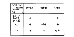
- FIG. 3 shows the property analysis results of pancreatic somatic stem cells in Example 3.
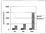
- FIG. 4 shows the results of inducing differentiation of pancreatic-derived somatic stem cells into pancreatic cells in Example 4.
- Ins represents insulin expression
- Glu represents glucagon expression
- Amy represents amylase expression.
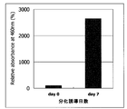
- FIG. 5 shows the results of induction of differentiation of pancreatic somatic stem cells into insulin-producing cells in Example 5.
- FIG. 6 shows the results of obtaining human pancreatic cell-derived somatic stem cells in Example 6.
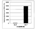
- FIG. 7 shows the results of induction of differentiation of human pancreatic cell-derived somatic stem cells into insulin-producing cells in Example 7.
- A. Method for Producing Pancreatic-derived Somatic Stem Cell of the present invention includes the following steps (A) and (B): (A) In a cell group including cells constituting the pancreas, a step of expressing a gene of a protein having an activity of passing through the G0 phase or G1 phase and transferring to the S phase; and (B) obtained in the step (A). Culturing cultured cells in the presence of extracellular growth factor.
- Step (A) is a step of expressing a gene of a protein having an activity of passing through the G0 phase or G1 phase and shifting to the S phase in a cell group including cells constituting the pancreas.
- Cell group containing cells constituting pancreas used as a raw material only needs to contain cells constituting pancreas, and may contain other cells.
- the ratio of the cells constituting the pancreas in the cell group is not particularly limited, but it is preferable to use a cell group consisting only of cells constituting the pancreas from the viewpoint of production efficiency.
- Examples of the cells constituting the pancreas include pancreatic duct epithelial cells, acinar cells, islet cells ( ⁇ cells, ⁇ cells, ⁇ cells, and PP cells).
- pancreatic progenitor cells a very small number of pancreatic progenitor cells (Pdx1-positive cells), endocrine progenitor cells (Ngn3-positive cells), pancreatic stem cells, and the like are included.
- the “cell group including cells constituting the pancreas” used as a raw material in the present invention preferably includes pancreatic stem cells.
- the ratio of including pancreatic stem cells is not particularly limited, but may be included at a ratio of 1/1000000 to 1/10000, for example.
- the cell group including cells constituting the pancreas commercially available cells may be used, or cells collected from a living body by a surgical technique may be used. In the case of collecting from a living body, the excised pancreas treated with collagenase or trypsinin inhibitor and chopped can be used.
- the cell group including cells constituting the pancreas is, for example, a primary cultured cell or a subcultured cell obtained by repeating subculture within a range in which the original function of the cell is maintained after being collected from the living body (this book) In the specification, it may be referred to as “early subcultured cell”).
- the number of subcultures is not particularly limited as long as the pancreatic-derived somatic stem cells can be produced, but is preferably within 10 times, more preferably within 5 times. Preferably, it is more preferably within 2 times.
- the organism from which the cell group including cells constituting the pancreas is derived can be appropriately selected according to the purpose, for example, mouse, rat, guinea pig, hamster, rabbit, cat, dog, sheep, pig, cow, goat, Examples include mammals such as monkeys and humans.
- pancreas-derived somatic stem cells are used for research or treatment of human diseases, it is preferably derived from human.
- the cell group including cells constituting the pancreas preferably has high viability, for example, viability is 70% or more, more preferably 80% or more, and further preferably 90% or more.
- viability of the cells can be measured using a commercially available analyzer.
- the cell group containing the cells constituting the pancreas has a high adhesion rate (for example, 70% or more) to a plate coated with collagen or the like.
- Viability of a cell group including cells constituting the pancreas can be measured according to a known method. For example, a cell group including cells constituting the pancreas is treated with trypan blue dye and stained blue. It can be determined by measuring the ratio of dead cells using a microscope or the like.
- cell cycle reactivation protein is not particularly limited as long as it has an activity of passing through the G0 phase or G1 phase and shifting to the S phase.
- “Transfer to the S phase by passing the G0 phase or the G1 phase” means (1) acting on the cells in the G0 phase and being in a dormant state by deviating (escaping) from the cell cycle. Or (2) acting on a cell in the G1 phase to shift the cell cycle from the G1 phase to the S phase.
- the presence / absence of “activity to pass through the G0 phase or G1 phase and shift to the S phase” can be confirmed by the following method. That is, confirmation of the transition to S phase (DNA synthesis phase) can be determined by examining the activity of 5-bromo-2-deoxyuridine (BrdU), which is an analog of thymidine, into the cells.
- PrdU 5-bromo-2-deoxyuridine
- a protein having an action of promoting phosphorylation of Rb protein can be used.
- examples include cyclin-dependent kinases and cyclins.
- examples of cyclin-dependent kinases include CDK1, CDK2, CDK3, CDK4, CDK6, and CDK7.
- An example of a cyclin is cyclin D.
- CDK4 and CDK6 are preferable, and CDK4 is more preferable.
- Only one type of cell cycle reactivation protein gene may be used alone, or a plurality of types may be used in combination.
- the origin of the cell cycle reactivation protein gene is not particularly limited as long as the effect of the present invention is exhibited. It may be the same species as the animal species from which the cell group including the cells constituting the pancreas used in step (A) is derived, or may be a different animal species. From the viewpoint of expression efficiency and the like, the cell cycle reactivation protein gene is preferably derived from the same species as the animal from which the cell group including cells constituting the pancreas is derived.
- the means for expressing the cell cycle reactivation protein gene is not limited as long as the cell cycle reactivation protein can be expressed.
- transient expression means that a gene is introduced into a cell by a DNA transfection method or the like and expressed transiently. Transient usually refers to a period of hours to days.
- stable expression means that a gene to be expressed is stably expressed in a chromosome. From the viewpoint that the pancreas-derived somatic stem cells and pancreatic differentiated cells produced by the method of the present invention have the same or as similar structure and properties as the pancreatic stem cells or pancreatic differentiated cells in vivo, the cell cycle reactivation is performed. It is preferable that the gene of the activation protein is transiently expressed.
- Transient expression is not particularly limited, and can be performed, for example, by introducing an expression vector having a target gene downstream of an expression promoter into a cell and expressing the gene from this expression vector.
- an expression promoter for example, CMV promoter, SV40 promoter and the like can be used, but are not limited thereto.
- expression vectors include, but are not limited to, plasmid vectors and liposomes as non-viral vectors, and adenovirus vectors and retrovirus vectors as virus vectors. It is preferable to use a non-viral vector from the viewpoint of safety when using the cells to be produced for pharmaceutical purposes and ensuring that the gene to be introduced is transiently expressed, and in particular, the origin of replication in the host cell.
- Non-viral vectors that do not contain are preferred. In order to carry out transient expression more reliably, it is possible to add a step of confirming that the introduced cell is not incorporated into the chromosome.
- plasmid vectors that can be used from such a viewpoint include pcDNA, pSVL, and the like.
- a method for introducing an expression vector into a cell for example, a lipofection method, an electroporation method, a method in which a gene is incorporated into a viral vector and infected, and the like can be used, but not limited thereto.
- stable expression is not particularly limited, it can be performed, for example, by the following method.
- An expression vector having a target gene and a dominant selection marker downstream of the expression promoter is introduced into the cell, and a strain in which the target gene is integrated into the chromosome is established. In this established strain, stable expression is performed.
- an expression promoter for example, CMV promoter, SV40 promoter and the like can be used, but are not limited thereto.
- a dominant selection marker for example, various drug resistance genes can be used, but not limited thereto. When a drug resistance gene is used as a dominant selection marker, only cell lines that stably express the drug resistance gene can be selected by continuing cell culture in the presence of a drug exhibiting resistance. .
- the target gene is considered to be stably expressed as well. Whether or not the target gene is actually stably expressed can be clarified by analyzing the base sequence of the chromosome by a DNA sequence or the like.
- a lipofection method, an electroporation method, or the like can be used, but is not limited thereto.
- a viral vector a method of incorporating a gene into a viral vector and infecting it can also be used.
- transfection reagent that is considered to be relatively weak in cytotoxicity.
- Step (A) is preferably performed while culturing in a medium that supports the growth of cells (preferably pancreatic stem cells) constituting the pancreas.
- a medium that supports the growth of cells preferably pancreatic stem cells constituting the pancreas.
- DMEM Doulbecco's modified Eagle's Medium
- Gibco used for normal mammalian cell culture
- a medium supplemented with fetal bovine serum, human serum or the like is preferably used. be able to.
- Extracellular growth factor may be further added to the medium used when performing step (A).
- the extracellular growth factor is a substance having an action of externally supporting the growth of pancreatic somatic stem cells, and any substance having such action may be used without any particular limitation.
- Examples of extracellular growth factors include cell growth factors and hormones that stimulate cell growth.
- Examples of cell growth factors include epidermal growth factor (EGF), hepatocyte growth factor (Hepatocyte Growth Factor; HGF), platelet-derived growth factor (PDGF), insulin-like growth factor (IGF), vascular endothelial cells.
- Examples include growth factors (Vascular Endothelial Growth Factor; VEGF) and fibroblast growth factors (FGF). Of these, EGF and HGF are preferred.
- insulin can be preferably used. These may be used alone or in combination of two or more.
- Proliferation can be improved synergistically.
- the concentration of the extracellular growth factor added in the medium is not particularly limited. For example, it is preferably 0.1 to 200 ng / ml, more preferably 1.0 to 100 ng / ml, and 5 to 50 ng / ml. Further preferred.
- pancreatic somatic stem cells proliferated by the following step (B) have structural and property characteristics that are the same as or infinitely similar to those of the pancreatic somatic stem cells in vivo.
- exogenous DNA other than the DNA of the activating protein eg, cyclin-dependent kinase
- pancreatic somatic stem cells the gene involved in the regulation of the cell cycle introduced into pancreatic somatic stem cells is only the gene of a cell cycle activation protein (eg, cyclin-dependent kinase, particularly CDK4 or CDK6) Is preferred.
- a cell cycle activation protein eg, cyclin-dependent kinase, particularly CDK4 or CDK6
- the pancreatic somatic stem cells grown according to the present invention preferably do not contain a gene encoding human telomerase reverse transcriptase.
- Step (B) is a step of culturing the cells obtained in step (A) in the presence of an extracellular growth factor.
- pancreatic-derived somatic stem cells proliferate predominantly from the group of cells containing cells constituting the pancreas as follows.
- the cell cycle transitions from the G0 phase or G1 phase to the S phase by the action of the cell cycle reactivation protein introduced from the outside in the step (A), and further to the M phase (divide). Period), and then proceed to the G1 period again.
- the action of the cell cycle reactivation protein is activated by the action of the extracellular growth factor present in the medium.
- pancreatic somatic stem cells continue to proliferate.
- pancreatic-derived somatic stem cells proliferate predominantly from a cell group including cells constituting the pancreas.
- the cell in which the gene for the cell cycle reactivation protein is expressed in the step (A) is cultured in the presence of an extracellular growth factor.
- the medium used in the step (A) contains an appropriate extracellular growth factor from the beginning, cells expressing the cell cycle reactivation protein gene may be cultured as they are without changing the medium. Good. Further, the extracellular growth factor may be further added to the medium without changing the medium. The medium may be replaced with a medium containing the extracellular growth factor, or the medium may be replaced with a medium not containing the extracellular growth factor, and then the extracellular growth factor may be added to the medium.
- the extracellular growth factor for example, EGF, HGF, VEGF, FGF, PDGF, IGF, etc.
- EGF extracellular growth factor
- the concentration of the extracellular growth factor added in the medium is not particularly limited. For example, it is preferably 0.1 to 200 ng / ml, more preferably 1.0 to 100 ng / ml, and 5 to 50 ng / ml. Further preferred.
- DMEM Doulbecco's modified Eagle's Medium
- pancreatic-derived somatic stem cells are used. What added the component which supports proliferation further can be used.
- the medium may be changed at an appropriate interval.
- the frequency of replacing the medium is not particularly limited, but the medium may be replaced once every two or three days.
- the medium may be replaced with a medium containing the same concentration of extracellular growth factor, or may be replaced with a medium containing a different concentration of extracellular growth factor.
- the medium may be replaced with a medium containing the same extracellular growth factor, or may be replaced with a medium containing a different extracellular growth factor.
- culture conditions such as culture temperature can be appropriately set according to known methods according to the type of pancreatic somatic stem cells to be cultured.
- Step (A) and Step (B) may be performed once each, or may be repeated a plurality of times with these as one set. When it is performed a plurality of times, it can be performed preferably 2 to 10 times, more preferably 3 to 8 times, and even more preferably 3 to 5 times. Repeating the steps (A) and (B) in this manner is preferable when the gene encoding the cell cycle reactivation protein is transiently expressed. This is because transient expression is the peak of expression about 3 days after transfection, and in order to shift pancreatic-derived somatic stem cells to a proliferative state, the expression of the gene continued for a certain period of time. This is because it is considered preferable.
- Process (A) and process (B) can be performed successively in this order, or other processes may be performed after process (A), and then process (B) may be performed.
- Step (B) or repetition of steps (A) and (B) can be continued until the required amount of pancreatic somatic stem cells is obtained. For example, as shown in Examples 1 and 6 to be described later, it is preferable to continue the steps (A) and (B) for about 20 to 60 days while repeating them.
- the pancreas-derived somatic stem cell group can be easily collected.
- the end point of the step (B) is preferably a time point at which a colony of pancreatic somatic stem cells can be confirmed with a microscope or the naked eye, or a time point at which a colony of pancreatic somatic stem cells consisting of 10 to 10,000 cell groups is formed. More preferably, it is the time when a colony of pancreatic-derived somatic stem cells consisting of a group of 100 to 1000 cells is formed.
- pancreatic stem cells different from pancreatic stem cells that were originally included in the group of cells containing pancreatic somatic stem cells, the cell division is stopped by the end of step (B) at the latest due to the cell division life, and many of them are lost. Even if it remains, the form has changed greatly from the beginning. For this reason, it is easily distinguishable from the pancreatic somatic stem cells forming colonies, and a highly pure pancreatic somatic stem cell group can be obtained.
- the number of times the step (A) and the step (B) are repeated is preferably the time when a colony of pancreatic somatic stem cells can be confirmed with a microscope or the naked eye, or the number of colonies of pancreatic somatic stem cells composed of 10 to 10,000 cell groups. Until formation, more preferably, until a colony of pancreatic-derived somatic stem cells consisting of a group of 100 to 1000 cells is formed.
- a colony is usually formed within a few weeks to a month from the start of the step (B).
- Cells different from pancreatic stem cells which were originally included in the cell group including cells constituting the pancreas, have stopped cell division due to the cell division lifetime by the end of the last step (B) at the latest, Most of them have dropped out or remain, but the shape has changed greatly from the beginning. For this reason, it is easily distinguishable from the pancreatic somatic stem cells forming colonies, and a highly pure pancreatic somatic stem cell group can be obtained.
- each step (B) can be continued as long as the cells continue to grow. However, it can be terminated during the growth.
- the pancreatic stem cells eventually form a colony in order to maintain good proliferative ability from the cell group including the cells constituting the pancreas.
- the other cells have a cell division lifetime at the end of the step (B), so that the cell division is stopped, and many of them have dropped or remain, but the shape has changed greatly from the beginning. Therefore, it can be easily distinguished from pancreatic somatic stem cells forming colonies, and by collecting this colony, the pancreatic somatic stem cell population can be isolated as a clone.
- Pancreatic-derived somatic stem cells can be obtained by performing a step of collecting colonies after the step (B). Colony recovery can be performed by a conventionally known method. Although not limited, it can be performed by, for example, a limiting dilution method or a method using a micropipette under a microscope.
- pancreatic somatic stem cell group containing 50% or more of pancreatic somatic stem cells in terms of the number of cells, more preferably a pancreatic somatic stem cell group containing 80% or more of pancreatic somatic stem cells.
- a pancreatic somatic stem cell group containing 50% or more of pancreatic somatic stem cells in terms of the number of cells, more preferably a pancreatic somatic stem cell group containing 80% or more of pancreatic somatic stem cells.
- pancreatic somatic stem cells are not particularly limited, but can be performed, for example, by examining whether or not they differentiate into target cells.
- pancreatic-derived somatic stem cells can also be determined by confirming the presence or absence of a cell surface marker.
- determination as a pancreatic-derived somatic stem cell can be made based on the positiveness of one or more molecular markers selected from the group consisting of PDX-1, CD133, and c-MET.
- the determination is performed on the basis that two or more of the molecular markers are positive, more preferably all of the molecular markers are positive.
- pancreatic somatic stem cells obtained by the method of the present invention have the same properties as in vivo pancreatic stem cells, they can be used to stably supply a high-purity population of pancreatic differentiated cells.
- the pancreas-derived somatic stem cells obtained by the method of the present invention are not only cells derived from somatic stem cells originally present in the pancreas but also cells other than somatic stem cells in the above steps (A) and (B). Also included are cells that have acquired clonal proliferation and pluripotency.
- the pancreatic differentiated cells can be finally used in clinical applications such as cell preparations, or in various research and development such as new drug development or disease research.
- the method for producing differentiated cells constituting the pancreas includes a step of differentiating pancreatic-derived somatic stem cells obtained by the production method of A above.
- the step of differentiation can be performed in vitro or in vivo according to a known method.
- somatic stem cells derived from the pancreas are cultured in a medium suitable for differentiation, and the various differentiated cells constituting the pancreas (eg, ⁇ cells, ⁇ cells, ⁇ cells, PP cells, pancreatic progenitor cells, endocrine progenitor cells) Differentiation can be induced.
- a medium and other culture conditions suitable for differentiation can be appropriately selected and set from known conditions according to the type of target differentiated cells.
- fetal bovine serum 1-20%
- FGF 150 ng / ml
- insulin 1-100 ⁇ g / ml
- transferrin 1-100 ⁇ g / ml
- nicotinamide 1 to 50 mM
- selenium 0.1 to 10 ng / ml
- gentamicin 10 to 100 ⁇ g / ml
- a medium suitable for culturing stem cells for example, DMEM / F12 medium.
- More specific culture conditions can be appropriately set by those skilled in the art according to the examples described later.
- stem cell spheroids are formed on a cell culture plate, transplanted under the kidney capsule of an animal, and maintained for a certain period of time. Can be implemented. Differentiation into insulin-producing cells can be confirmed using insulin production ability, glucagon positive, and amylase positive as indicators.
- the culture for differentiating pancreatic somatic stem cells may be two-dimensional culture or three-dimensional culture, but three-dimensional culture is preferable from the viewpoint of inducing differentiation more quickly.
- the three-dimensional culture is literally a three-dimensional culture of cells, and various substrates and kits for three-dimensional culture can be used.
- the culture time can be appropriately set according to the type of differentiated cells for the purpose.
- the pancreatic-derived somatic stem cells are preferably cultured in advance for a certain period (priming culture) in the presence of an activating substance prior to culturing in a medium suitable for differentiation.
- the priming culture is a process in which differentiation is advanced by one step, and this makes it possible to shorten the culture period necessary for subsequent differentiation.
- the priming culture can be performed, for example, in the presence of BMP or FGF.
- the cells constituting the differentiated pancreas can be used for various research materials related to the pancreas, pancreas transplantation, and the like.
- a pancreas-derived somatic stem cell is produced from a group of cells containing the cells constituting the pancreas obtained from a diabetic patient, differentiated into cells constituting the pancreas, and transplanted to the patient, thereby causing rejection. It becomes possible to carry out transplantation treatment in which the suppression is suppressed.
- compositions Another embodiment of the present invention is a pharmaceutical composition (cell preparation) comprising pancreatic-derived somatic stem cells obtained by the method of the present invention and / or pancreatic differentiated cells obtained by further differentiation thereof.
- the pharmaceutical composition of the present invention can be used for the treatment of various diseases (for example, type I diabetes and type II diabetes).
- the cells contained in the pharmaceutical composition of the present invention may be those into which an exogenous gene has been introduced.
- Introduction of exogenous genes into pancreatic somatic stem cells or pancreatic differentiated cells produced by the method of the present invention can be performed using methods known in the art.
- the exogenous gene to be introduced can be appropriately selected according to the purpose of use of the cell preparation (for example, gene therapy).
- the dosage form of the pharmaceutical composition of the present invention is appropriately set according to the affected area to be applied.
- Examples include intravenous injection, intraarterial injection, intraportal injection, intradermal injection, subcutaneous injection, submucosal injection, intraperitoneal injection, and the like.
- a method of administration by attaching cells formed into a sheet form by culturing to the affected area to be applied can also be mentioned.
- a tissue-like structure in which cells are three-dimensionally cultured using a so-called scaffold having biocompatibility can be attached or transplanted to the affected tissue.
- the dosage form of the pharmaceutical composition of the present invention is appropriately set according to the dosage form and the like.
- examples thereof include a liquid agent in which cells are suspended in a liquid, a gel agent in which cells are suspended in a gel, a cell sheet, and a tissue-like cell aggregate.
- the dosage and administration frequency of the pharmaceutical composition of the present invention are appropriately set according to the administration form, dosage form, recipient's condition, cell activity level, disease type, and the like.
- the dose per administration may be a therapeutically effective amount.
- the pharmaceutical composition of the present invention may be administered at a frequency of once per day or divided into 2 or 3 times, and doses for 2 days to 1 week may be administered at a time. Also good.
- the ratio of pancreatic-derived somatic stem cells or pancreatic differentiated cells contained in the pharmaceutical composition can be appropriately set according to the dosage form, dosage form, dosage, administration frequency, and the like.
- the cell preparation of the present invention may further contain other components as required in addition to the active ingredient (pancreas-derived somatic stem cells or pancreatic differentiated cells).
- active ingredient pancreas-derived somatic stem cells or pancreatic differentiated cells.
- examples of such components include excipients necessary for formulation depending on the dosage form, storage-stable components necessary for storage stability, and other medicinal ingredients.
- examples of other medicinal ingredients include anti-inflammatory agents, antibacterial agents, immunosuppressive agents, cell growth factors, hormones and the like.
- pancreatic-derived somatic stem cell isolation kit The kit for isolating pancreatic-derived somatic stem cells of the present invention includes cells that constitute the pancreas, including an expression vector that expresses a cell cycle reactivation protein and an extracellular growth factor.
- a cell group including cells constituting the pancreas, an extracellular growth factor, a cell cycle reactivation protein, and an expression vector are described in A. above. This is the same as described in.
- the kit includes A.I. It may contain a suitable extracellular growth factor described in.
- Example 1 Obtaining rat pancreas-derived somatic stem cells (1) Culture of primary rat pancreatic cells (I). Isolation of pancreatic duct epithelial cells and preparation of suspension Sprague-Dawley rats (male, 1 to 3 weeks old) were anesthetized with pentobarbital, dislocated from the cervical vertebra, and then laparotomized to remove the pancreas.
- the isolated rat pancreas is 20 mL RPMI-1640 containing 200 U / mL collagenase, 10 mM Hepes, 10% fetal bovine serum, 100 U / mL penicillin, 50 ⁇ g / mL streptomycin, and 0.2 mg / mL soybean trypsin inhibitor. Placed in a beaker with medium. The tissue was minced with scissors and incubated at 37 ° C. for 30-45 minutes with shaking. The resulting cell suspension was gently petted and then filtered through a 100 ⁇ m mesh to remove large tissue pieces to obtain a pancreatic cell suspension.
- transfection Two days after seeding of pancreatic duct epithelial cells and islet cells, transfection was performed under the conditions of the following medium and transgene.
- a commercially available protein expression plasmid pcDNA3 (Invitrogen) was used, and human CDK4 was cloned into the cloning site between EcoRI and XbaI, or DNA encoding CDK6 was cloned into the cloning site between HindIII and BamHI. Inserted.
- DNA encoding human CDK4 or CDK6 is a primer based on the nucleotide sequence registered in NCBI (National Center for Biotechnology Information) (CDK4 gene accession number: CAG47043, CDK6 gene accession number: NP-001138778) And using total RNA purified from HuS-E / 2 cells (human hepatocyte-derived cells, deposited at the National Institute of Advanced Industrial Science and Technology Patent Biological Depositary: FERM ABP-10908) as a template Obtained by RT-PCR.
- a forward primer consisting of the base sequence shown in SEQ ID NO: 1 and a reverse primer consisting of the base sequence shown in SEQ ID NO: 2 were used.
- a forward primer consisting of the base sequence shown in SEQ ID NO: 3 and a reverse primer consisting of the base sequence shown in SEQ ID NO: 4 were used.
- the base sequences of SEQ ID NOs: 1 and 3 include a sequence corresponding to Flag Tag.
- Transfection was performed under the following two conditions, and was repeated 3 times at a frequency of once every 3 to 5 days.
- Group 1 20 ng / ml hepatocyte growth factor (HGF) in DMEM-based medium containing 5% fetal bovine serum, 5% human serum (human HGF, Toyobo, Code: HGF-101, CHO cell recombinant) Medium supplemented with The transgene is CDK4 or CDK6 (pcDNA-FLAG-CDK4 or pcDNA-FLAG-CDK6).
- Group 2 Same medium as Group 1. The only transgene is a plasmid (pcDNA3).
- Example 2 Acquisition of rat pancreas-derived somatic stem cells
- the growth factor added to the medium used at the time of transfection was 10 ng / ml FGF (fibroblast growth factor), and the same experimental procedure was performed. The number of clonal cells obtained in this case is shown in FIG.
- Example 3 Characterization of clonal proliferating cells derived from rat pancreatic cells
- the cells showing clonal expansion obtained in Examples 1 and 2 were examined mainly for pancreatic stem cell-related differentiation markers. That is, PDX-1, CD133, and c-Met were subjected to RT-PCR using total RNA extracted from cells collected, and mRNA expression was examined. The result is shown in FIG. In the figure, positive is represented by (+), negative to weak positive is represented by (+/ ⁇ ), and negative is represented by ( ⁇ ).
- Example 4 Induction of differentiation of rat pancreatic cell-derived clonal cells into pancreatic cells (in vivo)
- Cells obtained by the present invention showing clonal proliferation were cultured for 1 to 2 weeks in a low adhesion cell culture plate (Sumitomo Bakelite, MS-9096U) to form spheroids (spherical cell mass).
- spheroids spherical cell mass
- this spheroid was transplanted under the kidney capsule of NOD SCID mice (male, 3 weeks old, Nippon Charles River).
- NOD SCID mice male, 3 weeks old, Nippon Charles River
- HEK human epidermal keratinocytes
- the transplanted tissue piece (part of the cell mass) was collected 4 to 8 weeks after the transplantation.
- the cells showing clonal proliferation obtained by the present invention have the ability to differentiate into a plurality of types of cells constituting the pancreas, that is, pancreatic-derived somatic stem cells.
- Example 5 Induction of differentiation of somatic stem cells derived from rat pancreatic cells into insulin-producing cells (in vitro)
- the cells showing clonal growth obtained by the present invention were seeded in a 12-well plate and grown to subconfluence, and then 10% fetal bovine serum, b-FGF (10 ng / ml), insulin (into DMEM / F12 medium) 10 ⁇ g / ml), transferrin (10 ⁇ g / ml), nicotinamide (10 mM), selenium (30 nM), and gentamicin (50 ⁇ g / ml) were cultured for 5 to 7 days.
- the concentrations of these components are not limited to the above, and those skilled in the art appropriately adjust the concentrations of the components to be used based on well-known techniques or descriptions in the examples of the present specification. be able to.
- the cultured cells were washed 3 times with PBS and then collected with 200 ⁇ l of acid / ethanol solution. This was homogenized and centrifuged (10000 ⁇ g for 5 minutes at 4 ° C.) to obtain a supernatant. The concentration of insulin contained in the supernatant sample was measured by a sandwich ELISA method using a rat insulin measurement kit (Morinaga Institute of Science).
- pancreatic-derived somatic stem cells obtained by the present invention have the ability to differentiate into insulin-producing cells. .
- the result is shown in FIG.
- Example 6 Acquisition of somatic stem cells derived from human pancreatic cells (1) Culture of primary human pancreatic cells Human pancreatic epithelial cells after resuscitation of human pancreatic epithelial cells (Cell Systems, ACBRI515) and human pancreatic stromal cells (Cell Systems, ACBRI513) And about 1.0 ⁇ 10 5 cells in a 12-well cell culture plate coated with collagen and suspended in DMEM medium containing 10% FCS. Seeded at a cell density of / well. The seeded cells were cultured in an incubator under conditions of 37 ° C. and 5% CO 2 .
- transfection Two days after seeding with human pancreatic epithelial cells and human pancreatic stromal cells, transfection was carried out under the conditions of the following medium and transgene.
- a commercially available protein expression plasmid pcDNA3 (Invitrogen) was used, and human CDK4 was inserted into the cloning site between EcoR I and Xba I.
- DNA encoding human CDK4 was designed based on the nucleotide sequence (accession number: CAG47043) registered in NCBI (National Center for Biotechnology Information), and HuS-E / 2 cells (cells derived from human hepatocytes) And obtained from RT-PCR using the total RNA purified from FERM ABP-10908) as a template.
- a forward primer consisting of the base sequence shown in SEQ ID NO: 1 and a reverse primer consisting of the base sequence shown in SEQ ID NO: 2 were used as the primer.
- the base sequence of SEQ ID NO: 1 includes a sequence corresponding to Flag Tag.
- the plasmid (pcDNA-FLAG-CDK4) in which the FLAG tag is introduced on the N-terminal side of the open reading frame of the DNA encoding human CDK4 thus obtained is 0.2 ⁇ g per well of effectene transformation reagent (Qiagen). ) And introduced the gene into the cell.
- Transfection was performed under the following two conditions, and was repeated 3 times at a frequency of once every 3 to 5 days.
- Group 1 20 ng / ml hepatocyte growth factor (HGF) in DMEM-based medium containing 5% fetal bovine serum, 5% human serum (human HGF, Toyobo, Code: HGF-101, CHO cell recombinant) Medium supplemented with The transgene is CDK4 (pcDNA-FLAG-CDK4).
- Group 2 Same medium as Group 1. The only transgene is a plasmid (pcDNA3).
- Example 7 Induction of differentiation of human pancreatic cell-derived clonal cells into insulin-producing cells (in vitro)
- the cells showing clonal growth obtained in Example 6 were seeded in a 12-well plate and grown to subconfluence, and then 10% fetal bovine serum, b-FGF (10 ng / ml), insulin (into DMEM / F12 medium) 10 ⁇ g / ml), transferrin (10 ⁇ g / ml), nicotinamide (10 mM), selenium (30 nM), and gentamicin (50 ⁇ g / ml) were cultured for 5 to 7 days.
- b-FGF 10 ng / ml
- insulin into DMEM / F12 medium
- transferrin 10 ⁇ g / ml
- nicotinamide 10 mM
- selenium 30 nM
- gentamicin 50 ⁇ g / ml
- the cultured cells were washed 3 times with PBS and then collected with 200 ⁇ l of acid / ethanol solution. This was homogenized and centrifuged (10000 ⁇ g for 5 minutes at 4 ° C.) to obtain a supernatant. The insulin concentration contained in the supernatant sample was measured by sandwich ELISA using a human insulin measurement kit (Mercodia).
- the insulin concentration is markedly increased as compared to the state before differentiation induction, indicating that the clonal proliferative cells obtained by the present invention have the ability to differentiate into insulin-producing cells. It was.
- the result is shown in FIG. From this result, it was confirmed that the cells showing clonal proliferation obtained in Example 6 were human pancreas-derived somatic hepatocytes.
Landscapes
- Health & Medical Sciences (AREA)
- Life Sciences & Earth Sciences (AREA)
- Engineering & Computer Science (AREA)
- Chemical & Material Sciences (AREA)
- General Health & Medical Sciences (AREA)
- Bioinformatics & Cheminformatics (AREA)
- Organic Chemistry (AREA)
- Biomedical Technology (AREA)
- Pharmacology & Pharmacy (AREA)
- Biotechnology (AREA)
- Zoology (AREA)
- Medicinal Chemistry (AREA)
- Animal Behavior & Ethology (AREA)
- Cell Biology (AREA)
- Public Health (AREA)
- Veterinary Medicine (AREA)
- Nuclear Medicine, Radiotherapy & Molecular Imaging (AREA)
- General Chemical & Material Sciences (AREA)
- Genetics & Genomics (AREA)
- Wood Science & Technology (AREA)
- Chemical Kinetics & Catalysis (AREA)
- Developmental Biology & Embryology (AREA)
- Nutrition Science (AREA)
- Virology (AREA)
- Immunology (AREA)
- Physiology (AREA)
- Diabetes (AREA)
- Endocrinology (AREA)
- Epidemiology (AREA)
- Microbiology (AREA)
- Gastroenterology & Hepatology (AREA)
- Biochemistry (AREA)
- General Engineering & Computer Science (AREA)
- Micro-Organisms Or Cultivation Processes Thereof (AREA)
- Medicines Containing Material From Animals Or Micro-Organisms (AREA)
- Materials For Medical Uses (AREA)
Abstract
膵臓由来体性幹細胞を効率的に増殖する方法等を提供することを課題とする。以下の工程(A)及び(B)を含む、膵臓由来体性幹細胞の製造方法: (A)膵臓を構成する細胞を含む細胞群において、G0期又はG1期を通過させS期へと移行させる活性を有するタンパク質の遺伝子を発現させる工程;及び (B)工程(A)で得られた細胞を細胞外増殖因子の存在下で培養する工程。
Description
本発明は、膵臓由来体性幹細胞の製造方法、及びその応用技術に関する。
疾患研究や新薬開発において、細胞、特にヒト細胞を利用することは、今や必須のことであり、今後益々その需要は高まると考えられている。細胞は生体から採取した組織から分離調製され、これを培養して試験研究に広く使用される。この場合、目的に応じた分化細胞(differentiated cell)が使用されることが多い。しかしながら、生体外に取り出した分化細胞は増殖させることが困難な場合がある。また、本来の活性が短時間で失われてしまうこと、採取コストが非常に高いこと、及びドナーによりその特性に大きなバラツキがあること等問題点は多い。
これらの問題、特に増殖性に関する問題をクリアするため、分化細胞を不死化する手段がとられることがある。例えば、ヒト肝細胞にSV(simian virus)40ラージT抗原遺伝子やヒトテロメラーゼ遺伝子を導入する等種々の方法により細胞を不死化する試みが数多くなされてきた(非特許文献1)。しかしながら、不死化して得られた細胞は本来の分化形質を持たない場合がほとんどであって、不死化細胞で本来の機能を十分に保持しているものはこれまで知られていない。例えば、テロメラーゼ遺伝子はヒト組織の正常細胞では殆ど発現していない。また、不死化細胞作出のために、いわゆるがん遺伝子を使用することは、正常細胞から逸脱する可能性を含み好ましいことではないと考えられている。
一方、目的の正常初代細胞を大量に得る手段として、生体から増殖性が高い体性幹細胞を分離し、これを体外で培養・増殖させて利用する方法がある。
体性幹細胞の取得方法としては、例えば生体組織から体細胞を採取し、細胞表面分子の発現パターンに基づきセルソーターを用いて体性幹細胞を分離する方法が報告されている。しかしながら、目的とする体性幹細胞を含む組織を入手し、かつその体性幹細胞に対する適切な抗体を選択することは困難であり、純度の高い体性幹細胞集団を得ることは容易ではない。また、組織中に存在する幹細胞の数は極めて少数であるため、セルソーターを使ってある程度の幹細胞を集めるためには出発材料として非常に多くの細胞(例えば10の8乗個又はそれ以上)を要することとなる。更に、純度の高い体性幹細胞集団が得られたとしても、それを培養して増殖させること(特に、本来の体性幹細胞の特性を維持した状態での培養)は困難である。
他の手段として、ES細胞、iPS細胞といった多分化能を有する幹細胞を増殖させた後に、目的の分化細胞へと分化誘導するという方法がある。ES細胞、又はiPS細胞も一部の細胞が分化することは確認されているものの、現在の段階では分化効率が充分とは言えず、種々の細胞が混在する場合が多く純度の高い分化した機能細胞集団を得ることはまだまだ困難であるとされる。
このように、一定の分化能を保持する体性幹細胞の高純度集団を安定的に供給できる方法が真に望まれていた。
Ramirez, D.R. et al., Cancer Research 64, 9027-9034, 2004
本発明は、上記従来技術の課題を解決することを課題とする。より詳細には、本発明は、分化能を有する状態で増殖可能な膵臓由来幹細胞を取得する方法等を提供することを目的とする。
本発明者らは、上記の課題に鑑み、鋭意研究を重ねた結果、G0期又はG1期を通過させS期へと移行させる活性を有するタンパク質の遺伝子を、膵臓を構成する細胞を含む細胞群に導入し、細胞外増殖因子の存在下で培養することにより、その他の遺伝子を導入しなくても、クローン性増殖能を有する体性幹細胞だけを優勢的に増殖させることができ、増殖した体性幹細胞だけを簡便且つ効率的に取得することが可能であることを見出した。また、本発明者等は、そのようにして取得した膵臓由来体性幹細胞を分化に適した培地で培養することにより、目的とする膵臓を構成する細胞に分化させることが可能であることを確認した。本発明者らは、以上のような知見にさらなる検討と改良を重ねることにより、本発明を完成するに至った。
代表的な本願発明は以下の通りである。
項1
以下の工程(A)及び(B)を含む、膵臓由来体性幹細胞の製造方法:
(A)膵臓を構成する細胞を含む細胞群において、G0期又はG1期を通過させS期へと移行させる活性を有するタンパク質の遺伝子を発現させる工程;及び
(B)工程(A)で得られた細胞を細胞外増殖因子の存在下で培養する工程。
項2
前記発現が一過性発現である、項1に記載の方法。
項3
前記工程(A)及び前記工程(B)を二回以上繰り返す、項1又は2に記載の方法。
項4
前記タンパク質がサイクリン依存性キナーゼである、項1~3のいずれかに記載の方法。
項5
前記タンパク質がサイクリン依存性キナーゼ4及びサイクリン依存性キナーゼ6からなる群より選択される少なくとも一種のタンパク質である、項1~4のいずれかに記載の方法。
項6
前記細胞外増殖因子が上皮成長因子(EGF)、肝細胞増殖因子(HGF)、線維芽細胞増殖因子(FGF)、血小板由来成長因子(PDGF)、血管内皮細胞増殖因子(VEGF)、及びインスリン様成長因子(IGF)からなる群より選択される少なくとも一種の細胞外増殖因子である、項1~5のいずれかに記載の方法。
項7
項1~6のいずれかに記載の方法により得られうる、膵臓由来体性幹細胞。
項8
膵臓由来体性幹細胞がPDX-1陽性である、項7に記載の細胞。
項9
膵臓由来体性幹細胞が更にCD133陽性である、項8に記載の細胞。
項10
膵臓由来体性幹細胞が更にc-MET陽性である、項8又は9に記載の細胞。
項11
項7~10のいずれかに記載の細胞に、さらに外因性遺伝子が導入された細胞。
項12
項7~11のいずれかに記載の細胞を、膵臓を構成する細胞に分化させる工程を含む、分化細胞の製造方法。
項13
前記分化させる工程が3次元培養を行う工程を含む、項12に記載の方法。
項14
項12又は13に記載の方法により得られうる、膵臓を構成する細胞。
項15
項7~11及び14のいずれかに記載の細胞を含む、医薬組成物。
項16
G0期又はG1期を通過させS期へと移行させる活性を有するタンパク質を発現させる発現ベクター及び細胞外増殖因子を含む、膵臓を構成する細胞を含む細胞群から膵臓由来体性幹細胞を単離するためのキット。
項1
以下の工程(A)及び(B)を含む、膵臓由来体性幹細胞の製造方法:
(A)膵臓を構成する細胞を含む細胞群において、G0期又はG1期を通過させS期へと移行させる活性を有するタンパク質の遺伝子を発現させる工程;及び
(B)工程(A)で得られた細胞を細胞外増殖因子の存在下で培養する工程。
項2
前記発現が一過性発現である、項1に記載の方法。
項3
前記工程(A)及び前記工程(B)を二回以上繰り返す、項1又は2に記載の方法。
項4
前記タンパク質がサイクリン依存性キナーゼである、項1~3のいずれかに記載の方法。
項5
前記タンパク質がサイクリン依存性キナーゼ4及びサイクリン依存性キナーゼ6からなる群より選択される少なくとも一種のタンパク質である、項1~4のいずれかに記載の方法。
項6
前記細胞外増殖因子が上皮成長因子(EGF)、肝細胞増殖因子(HGF)、線維芽細胞増殖因子(FGF)、血小板由来成長因子(PDGF)、血管内皮細胞増殖因子(VEGF)、及びインスリン様成長因子(IGF)からなる群より選択される少なくとも一種の細胞外増殖因子である、項1~5のいずれかに記載の方法。
項7
項1~6のいずれかに記載の方法により得られうる、膵臓由来体性幹細胞。
項8
膵臓由来体性幹細胞がPDX-1陽性である、項7に記載の細胞。
項9
膵臓由来体性幹細胞が更にCD133陽性である、項8に記載の細胞。
項10
膵臓由来体性幹細胞が更にc-MET陽性である、項8又は9に記載の細胞。
項11
項7~10のいずれかに記載の細胞に、さらに外因性遺伝子が導入された細胞。
項12
項7~11のいずれかに記載の細胞を、膵臓を構成する細胞に分化させる工程を含む、分化細胞の製造方法。
項13
前記分化させる工程が3次元培養を行う工程を含む、項12に記載の方法。
項14
項12又は13に記載の方法により得られうる、膵臓を構成する細胞。
項15
項7~11及び14のいずれかに記載の細胞を含む、医薬組成物。
項16
G0期又はG1期を通過させS期へと移行させる活性を有するタンパク質を発現させる発現ベクター及び細胞外増殖因子を含む、膵臓を構成する細胞を含む細胞群から膵臓由来体性幹細胞を単離するためのキット。
本発明の方法により、膵臓を構成する雑多な細胞集団の中から膵臓由来体性幹細胞を優勢的に増殖させることにより、従来のセルソーター等を用いて体性幹細胞を単離することを必要とする方法と比較して、膵臓由来体性幹細胞を効率的且つ高純度で製造することが可能である。本発明を用いればセルソーターのように多数の細胞を用意しなくても比較的少数の膵臓を構成する細胞(例えば10の5乗から6乗個程度)から効率的に体性幹細胞を得ることが出来る。また、本発明の方法で製造された膵臓由来体性幹細胞は、目的に応じた膵臓を構成する細胞(例えば、インスリン産生細胞)に分化させることが可能であるため、糖尿病等の治療に有用である。
本発明において膵臓由来体性幹細胞を増殖させるために導入される遺伝子は、一過性の発現とすることが可能である。このようにして製造された膵臓由来体性幹細胞は、外因性の遺伝子を含まないため、生体内で増殖する膵臓体性幹細胞と実質的に同一の構造・性質を有する。従って、本発明の膵臓由来体性幹細胞は、安全性の観点から生体への移植に適している。
本発明によって得られる膵臓由来体性幹細胞は、外因性の遺伝子を発現する宿主細胞としても機能するため、外因性の遺伝子を当該膵臓由来体性幹細胞に導入し、遺伝子治療用医薬として利用することも可能である。
以下に本発明の具体的な実施の形態を説明する。
A.膵臓由来体性幹細胞の製造方法
本発明の膵臓由来体性幹細胞の製造方法は、以下の工程(A)及び(B)を含む:
(A)膵臓を構成する細胞を含む細胞群において、G0期又はG1期を通過させS期へと移行させる活性を有するタンパク質の遺伝子を発現させる工程;及び
(B)工程(A)で得られた細胞を細胞外増殖因子の存在下で培養する工程。
本発明の膵臓由来体性幹細胞の製造方法は、以下の工程(A)及び(B)を含む:
(A)膵臓を構成する細胞を含む細胞群において、G0期又はG1期を通過させS期へと移行させる活性を有するタンパク質の遺伝子を発現させる工程;及び
(B)工程(A)で得られた細胞を細胞外増殖因子の存在下で培養する工程。
1.工程(A)
工程(A)は、膵臓を構成する細胞を含む細胞群において、G0期又はG1期を通過させS期へと移行させる活性を有するタンパク質の遺伝子を発現させる工程である。
工程(A)は、膵臓を構成する細胞を含む細胞群において、G0期又はG1期を通過させS期へと移行させる活性を有するタンパク質の遺伝子を発現させる工程である。
(1)膵臓を構成する細胞を含む細胞群
原材料として用いる「膵臓を構成する細胞を含む細胞群」は、膵臓を構成する細胞を含んでいればよく、他の細胞を含んでいても良い。当該細胞群中に膵臓を構成する細胞が含まれる割合は特に制限されないが、製造効率の観点から膵臓を構成する細胞のみからなる細胞群を使用することが好ましい。膵臓を構成する細胞には、例えば、膵管上皮細胞、腺房細胞、膵島細胞(α細胞、β細胞、δ細胞、及びPP細胞)などがある。これら細胞の中に、ごく少数の膵前駆細胞(Pdx1陽性細胞)、内分泌前駆細胞(Ngn3陽性細胞)、及び膵幹細胞等が含まれる。本発明で原材料として用いる「膵臓を構成する細胞を含む細胞群」には、膵臓幹細胞が含まれることが好ましい。膵幹細胞が含まれる割合は、特に制限されないが、例えば、1/1000000~1/10000個の割合で含まれていれば良い。
原材料として用いる「膵臓を構成する細胞を含む細胞群」は、膵臓を構成する細胞を含んでいればよく、他の細胞を含んでいても良い。当該細胞群中に膵臓を構成する細胞が含まれる割合は特に制限されないが、製造効率の観点から膵臓を構成する細胞のみからなる細胞群を使用することが好ましい。膵臓を構成する細胞には、例えば、膵管上皮細胞、腺房細胞、膵島細胞(α細胞、β細胞、δ細胞、及びPP細胞)などがある。これら細胞の中に、ごく少数の膵前駆細胞(Pdx1陽性細胞)、内分泌前駆細胞(Ngn3陽性細胞)、及び膵幹細胞等が含まれる。本発明で原材料として用いる「膵臓を構成する細胞を含む細胞群」には、膵臓幹細胞が含まれることが好ましい。膵幹細胞が含まれる割合は、特に制限されないが、例えば、1/1000000~1/10000個の割合で含まれていれば良い。
膵臓を構成する細胞を含む細胞群は、市販のものを使用しても良く、生体から外科的手法で採取したものを使用してもよい。生体から採取する場合は、摘出した膵臓をコラゲナーゼやトリプシニンヒビターで処理し、細断したものを使用することができる。膵臓を構成する細胞を含む細胞群は、例えば、初代培養細胞、又は生体から採取した後、生体内における細胞本来の機能が維持される範囲内で継代培養を繰り返した継代培養細胞(本明細書において、「初期継代培養細胞」ということがある。)等を用いることができる。初期継代培養細胞を使用する場合は、その継代培養回数は、膵臓由来体性幹細胞の製造が可能である限り特に制限されないが、10回以内であれば好ましく、5回以内であればより好ましく、2回以内であればさらに好ましい。
膵臓を構成する細胞を含む細胞群が由来する生物は、目的に応じて適宜選択することができ、例えば、マウス、ラット、モルモット、ハムスター、ウサギ、ネコ、イヌ、ヒツジ、ブタ、ウシ、ヤギ、サル、ヒト等の哺乳類が挙げられる。得られる膵臓由来体性幹細胞をヒトの疾患の研究又は治療に用いる場合は、ヒト由来であることが好ましい。
膵臓を構成する細胞を含む細胞群は、高いバイアビリティを有することが好ましく、例えば、バイアビリティは70%以上であり、より好ましくは80%以上、更に好ましくは90%以上である。当該細胞のバイアビリティは、市販される分析器を用いて測定することが可能である。また、膵臓を構成する細胞を含む細胞群は、コラーゲン等でコートしたプレートに対する接着率が高いこと(例えば、70%以上)が好ましい。
膵臓を構成する細胞を含む細胞群のバイアビリティは、公知の方法に従って測定することが可能であり、例えば、トリパンブルー色素を用いて膵臓を構成する細胞を含む細胞群を処理し、青く染色された死細胞の割合を顕微鏡等を用いて測定することによって求めることができる。
(2)G0期又はG1期を通過させS期へと移行させる活性を有するタンパク質
細胞に遺伝子として導入する「G0期又はG1期を通過させS期へと移行させる活性を有するタンパク質」(本明細書において、「細胞周期再活性化タンパク質」ということがある。)は、G0期又はG1期を通過させS期へと移行させる活性を有していればよく、特に限定されない。
細胞に遺伝子として導入する「G0期又はG1期を通過させS期へと移行させる活性を有するタンパク質」(本明細書において、「細胞周期再活性化タンパク質」ということがある。)は、G0期又はG1期を通過させS期へと移行させる活性を有していればよく、特に限定されない。
「G0期又はG1期を通過させS期へと移行させる」とは、(1)細胞周期から逸脱(脱出)することによりG0期にあり休止状態となっている細胞に対して働きかけ、S期へと移行させることにより再び細胞周期へと進入させること、又は(2)G1期にある細胞に対して働きかけ、その細胞周期をG1期からS期へと移行させることをいう。
「G0期又はG1期を通過させS期へと移行させる活性」の有無は、次の方法によって確認することができる。即ち、S期(DNA合成期)への移行の確認は、thymidineのアナログである5-Bromo-2-deoxyuridine(BrdU)の細胞への取り込み活性を調べることで判断できる。具体的には、細胞培養培地にBrdUを入れ、その後、蛍光標識した抗BrdU抗体で反応させて細胞表面を免疫染色する等し、フローサイトメーター等を使って解析することによって実施することができる。
「G0期又はG1期を通過させS期へと移行させる活性」の有無は、次の方法によって確認することができる。即ち、S期(DNA合成期)への移行の確認は、thymidineのアナログである5-Bromo-2-deoxyuridine(BrdU)の細胞への取り込み活性を調べることで判断できる。具体的には、細胞培養培地にBrdUを入れ、その後、蛍光標識した抗BrdU抗体で反応させて細胞表面を免疫染色する等し、フローサイトメーター等を使って解析することによって実施することができる。
細胞周期再活性化タンパク質としては、例えば、Rbタンパク質のリン酸化を促進する作用を有するものを用いることができる。例えば、サイクリン依存性キナーゼ及びサイクリン等が挙げられる。サイクリン依存性キナーゼとしては、例えば、CDK1、CDK2、CDK3、CDK4、CDK6及びCDK7が挙げられる。サイクリンとしては、例えば、サイクリンDが挙げられる。これらの中でもCDK4及びCDK6が好ましく、CDK4がより好ましい。細胞周期再活性化タンパク質の遺伝子は、1種のみを単独で使用しても良く、複数種を組み合わせて使用しても良い。
細胞周期再活性化タンパク質の遺伝子の由来は、本発明の効果が奏される限り特に限定されない。工程(A)で用いる膵臓を構成する細胞を含む細胞群が由来する動物種と同一種であってもよいし、異なる動物種であってもよい。発現効率等の観点から、好ましくは、細胞周期再活性化タンパク質の遺伝子の由来は、膵臓を構成する細胞を含む細胞群が由来する動物と同一種に由来する。
細胞周期再活性化タンパク質遺伝子を発現させる手段は、細胞周期再活性化タンパク質を発現させることができればよく、限定されない。例えば、一過性発現(transient expression)させてもよいし、安定発現(stable expression)させてもよい。一過性発現とは、遺伝子をDNAトランスフェクション法等により細胞に導入し、一過性に発現させることをいう。一過性とは通常、数時間から数日以内の期間をいう。これに対して安定発現とは、発現させようとする遺伝子が安定に染色体中に組み込まれた状態で発現することをいう。本発明の方法によって製造される膵臓由来体性幹細胞及び膵臓分化細胞が、生体内の膵幹細胞又は膵臓分化細胞と同一又は可能な限り類似する構造及び性質を有するという観点からは、当該細胞周期再活性化タンパク質の遺伝子は、一過性に発現されることが好ましい。
一過性発現は、特に限定されないが、例えば、発現プロモーターの下流に目的の遺伝子を持つ発現ベクターを細胞に導入し、この発現ベクターから当該遺伝子を発現させること等によって行うことができる。この場合、発現プロモーターとしては、例えば、CMVプロモーター、SV40プロモーター等を用いることができるが、これらに限定されない。また、発現ベクターとしては、例えば、非ウイルスベクターとしてプラスミドベクターやリポソーム等、ウイルスベクターとしてアデノウイルスベクター、レトロウイルスベクター等を用いることができるが、これらに限定されない。製造する細胞を医薬の目的で使用する場合の安全性や導入する遺伝子をより確実に一過性発現とするという観点から、非ウイルスベクターを用いることが好ましく、中でも宿主細胞中での複製開始点を含まない非ウイルスベクターが好ましい。より確実に一過性発現を実施するために、導入した細胞が染色体に取り込まれていないことを確認する工程を加えることも可能である。このような観点から使用可能なプラスミドベクターとしては、例えばpcDNA、pSVL等を挙げることができる。細胞への発現ベクターの導入方法としては、例えば、リポフェクション法、エレクトロポレーション法、ウイルスベクターに遺伝子を組み込み感染させる方法等を用いることができるが、これらに限定されない。
安定発現は、特に限定されないが、例えば、次の方法等によって行うことができる。発現プロモーターの下流に目的の遺伝子と優性選択マーカーを持つ発現ベクターを細胞に導入し、目的の遺伝子が染色体に組み込まれた株を樹立する。この樹立された株では安定発現が行われている。この場合、発現プロモーターとしては、例えば、CMVプロモーター、SV40プロモーター等を用いることができるが、これらに限定されない。優性選択マーカーとしては、例えば、各種の薬剤耐性遺伝子等を用いることができるが、これらに限定されない。優性選択マーカーとして薬剤耐性遺伝子を用いた場合は、耐性を示す薬剤の存在下で細胞培養を継続して行うことにより、当該薬剤耐性遺伝子を安定発現している細胞株のみを選別することができる。通常、そのような細胞株においては同様に目的の遺伝子も安定発現していると考えられる。なお、実際に目的の遺伝子が安定発現しているか否かについては、染色体の塩基配列をDNAシークエンス等によって解析すること等によって明らかにすることができる。また、細胞への発現ベクターの導入方法としては、例えば、リポフェクション法、エレクトロポレーション法等を用いることができるが、これらに限定されない。また、ウイルスベクターを用いる場合は、ウイルスベクターに遺伝子を組み込み感染させる方法も用いることができる。
なお、初代培養細胞に対して遺伝子導入を行う場合は、細胞毒性が比較的弱いとされるトランスフェクション試薬を用いることが好ましい。トランスフェクション効率を向上させるため、例えば、良好な状態にある細胞に対してトランスフェクションを行うことが好ましい。初代細胞の場合は、培養プレート上に播種した後、2~3日以内であれば細胞の状態が比較的良好である。
工程(A)は、膵臓を構成する細胞(好ましくは膵幹細胞)の増殖をサポートする培地で培養しながら行うことが好ましい。例えば、通常の哺乳類細胞培養に用いられるDMEM(Doulbecco’s modified Eagle’s Medium)培地(ギブコ社製)等を基礎培地とし、これにウシ胎児血清、ヒト血清等を添加した培地を好適に用いることができる。
工程(A)を行う際に使用する培地には、さらに、細胞外増殖因子を添加してもよい。細胞外増殖因子とは、膵臓由来体性幹細胞の増殖を外的にサポートする作用を有する物質であり、そのような作用を有しているものであればよく、特に限定されない。細胞外増殖因子としては、例えば、細胞増殖因子、細胞増殖を刺激するホルモン類等を挙げることができる。細胞増殖因子としては、例えば、上皮成長因子(Epidermal Growth Factor;EGF)、肝細胞増殖因子(Hepatocyte Growth Factor;HGF)、血小板由来成長因子(PDGF)、インスリン様成長因子(IGF)、血管内皮細胞増殖因子(Vascular Endothelial Growth Factor;VEGF)及び繊維芽細胞増殖因子(Fibroblast Growth Factor;FGF)等を挙げることができる。中でもEGF及びHGFが好適である。ホルモン類としては、例えばインスリン等を好適に用いることができる。これらを単独で用いてもよいし、複数種を組み合わせて用いてもよい。例えば、EGF及びHGFからなる群より選択される少なくとも1種の細胞増殖因子に、VEGF及びFGFからなる群より選択される少なくとも1種の細胞増殖因子をさらに組み合わせて用いることにより、相加的あるいは相乗的に増殖を向上させることができる。
細胞外増殖因子の培地中における添加濃度は特に限定されないが、例えば0.1~200ng/mlであれば好ましく、1.0~100ng/mlであればより好ましく、5~50ng/mlであればさらに好ましい。
膵臓を構成する細胞を含む細胞群には、細胞周期再活性化タンパク質をコードするDNA以外にも、膵臓由来体性幹細胞の増殖能と多分化能とを阻害しない限り、他の遺伝子を導入し発現させることができる。しかしながら、下記の工程(B)によって増殖された膵臓由来体性幹細胞が、生体内の膵臓体性幹細胞と同一又はそれと限りなく類似する構造上及び性質上の特性を有するという観点からは、細胞周期活性化タンパク質(例えば、サイクリン依存性キナーゼ)のDNA以外に外因性のDNAを実質的に含んでないことが好ましい。同様の観点から、例えば、膵臓由来体性幹細胞に導入される細胞の周期の調節に係る遺伝子は、細胞周期活性化タンパク質(例えば、サイクリン依存性キナーゼ、特にCDK4又はCDK6)の遺伝子のみであることが好ましい。一実施形態において、本発明によって増殖される膵臓由来体性幹細胞は、ヒトテロメラーゼ逆転写酵素をコードする遺伝子を含まないことが好ましい。
2.工程(B)
工程(B)は、工程(A)で得られた細胞を細胞外増殖因子の存在下で培養する工程である。
工程(B)は、工程(A)で得られた細胞を細胞外増殖因子の存在下で培養する工程である。
工程(B)により、膵臓を構成する細胞を含む細胞群の中から膵臓由来体性幹細胞が次の通り優勢的に増殖する。膵臓由来体性幹細胞では、まず、工程(A)で外部から導入された細胞周期再活性化タンパク質の働きによって、細胞周期がG0期又はG1期からS期へと移行し、さらにM期(分裂期)、続いて再びG1期へと進む。同時に、培地中に存在する細胞外増殖因子の働きにより、細胞周期再活性化タンパク質の働きが活性化される。その結果、膵臓由来体性幹細胞は増殖を続ける。一方、膵臓由来体性幹細胞とは異なる細胞においては、細胞周期再活性化タンパク質の働きによる上記のような増殖活性化は起こらないか、又は増殖が一時的なものに止まり継続しない。この結果、膵臓を構成する細胞を含む細胞群の中から膵臓由来体性幹細胞が優勢的に増殖する。
工程(B)では、工程(A)で細胞周期再活性化タンパク質の遺伝子を発現させた細胞を、細胞外増殖因子の存在下で培養する。工程(A)で使用した培地が当初から適切な細胞外増殖因子を含んでいる場合は、細胞周期再活性化タンパク質の遺伝子を発現させた細胞を、培地を交換せずにそのまま培養してもよい。また、培地を交換せずにさらに当該細胞外増殖因子を培地中に添加してもよい。当該細胞外増殖因子を含む培地に交換してもよいし、当該細胞外増殖因子を含まない培地に交換した後に、当該細胞外増殖因子を当該培地に添加してもよい。
細胞外増殖因子としては、上記工程(A)に関して説明した細胞外増殖因子(例えば、EGF、HGF、VEGF、FGF、PDGF、IGF等)を用いることができる。
細胞外増殖因子の培地中における添加濃度は特に限定されないが、例えば0.1~200ng/mlであれば好ましく、1.0~100ng/mlであればより好ましく、5~50ng/mlであればさらに好ましい。
工程(B)において新たな培地を用いる場合は、通常の哺乳類細胞培養に用いられるDMEM(Doulbecco’s modified Eagle’s Medium)培地(ギブコ社製)等を基礎培地とし、膵臓由来体性幹細胞の増殖をサポートする成分をさらに添加したもの等を用いることができる。
工程(B)においては、適当な間隔で培地を交換してもよい。培地を交換する頻度は、特に制限されないが、2又は3日に一度の頻度で培地交換を行ってもよい。同じ濃度の細胞外増殖因子を含む培地へと交換してもよいし、異なる濃度の細胞外増殖因子を含む培地へと交換してもよい。同じ細胞外増殖因子を含む培地へと交換してもよいし、異なる細胞外増殖因子を含む培地へと交換してもよい。
培養温度等その他の培養条件は、培養する膵臓由来体性幹細胞の種類に応じて公知の方法に従って適宜設定することができる。
工程(A)及び工程(B)はそれぞれ一回ずつ行ってもよいし、これらを1セットとしてそれを複数回繰り返し行ってもよい。複数回行う場合、好ましくは2~10回、より好ましくは3~8回、さらに好ましくは3~5回行うことができる。このように工程(A)及び(B)を繰り返すことは、細胞周期再活性化タンパク質をコードする遺伝子を一過性に発現させる場合に好ましい。これは、一過性の発現は、トランスフェクション後約3日が発現のピークであること、膵臓由来体性幹細胞を増殖性の状態にシフトさせるためには、一定期間の継続した当該遺伝子の発現が好ましいと考えられるためである。
工程(A)及び工程(B)はこの順に連続して行うこともできるし、工程(A)の後にその他の工程を実施し、その後に工程(B)を実施してもよい。工程(B)又は工程(A)と(B)の繰り返しは、必要な量の膵臓由来体性幹細胞が得られるまで続けることができる。例えば、後述する実施例1及び実施例6に示されるように、20~60日程度、工程(A)と(B)とを繰り返しながら継続することが好ましい。
工程(B)を一度だけ行う場合は、例えば、膵臓由来体性幹細胞によるコロニー形成が十分に行われるまで工程(B)を続ければ、膵臓由来体性幹細胞群を容易に採取することができるため好ましい。工程(B)の終了時点は、好ましくは顕微鏡若しくは肉眼で膵臓由来体性幹細胞のコロニーが確認できる時点、又は10~10000個の細胞群からなる膵臓由来体性幹細胞のコロニーが形成される時点であり、より好ましくは、100~1000個の細胞群からなる膵臓由来体性幹細胞のコロニーが形成される時点である。膵臓由来体性幹細胞を含む細胞群に当初含まれていた、膵幹細胞とは異なる細胞は、遅くとも工程(B)の終了時点までには細胞分裂寿命のため細胞分裂は停止し、その多くは脱落するか、あるいは残存していても当初とは大きく形態が変化している。このため、コロニーを形成している膵臓由来体性幹細胞とは容易に区別可能であり、純度の高い膵臓由来体性幹細胞群を取得することができる。
工程(A)及び工程(B)を繰り返し行う場合は、例えば、膵臓由来体性幹細胞によるコロニー形成が十分に行われるまで工程(A)及び工程(B)を繰り返せば、膵臓由来体性幹細胞群を容易に採取することができるため好ましい。工程(A)及び工程(B)を繰り返す回数は、好ましくは顕微鏡若しくは肉眼で膵臓由来体性幹細胞のコロニーが確認できる時点、又は10~10000個の細胞群からなる膵臓由来体性幹細胞のコロニーが形成されるまでであり、より好ましくは、100~1000個の細胞群からなる膵臓由来体性幹細胞のコロニーが形成されるまでである。コロニーは、工程(B)の開始から通常数週間~1ヶ月程度で形成される。膵臓を構成する細胞を含む細胞群に当初含まれていた、膵幹細胞とは異なる細胞は、遅くとも最後の回の工程(B)の終了時点までには細胞分裂寿命のため細胞分裂は停止し、その多くは脱落するか、あるいは残存していても当初とは大きく形態が変化している。このため、コロニーを形成している膵臓由来体性幹細胞とは容易に区別可能であり、純度の高い膵臓由来体性幹細胞群を取得することができる。また、この場合、各回の工程(B)は、細胞が増殖し続けている限り継続することができる。ただし、増殖している途中に終了させることもできる。
先述の通り工程(B)により、膵臓を構成する細胞を含む細胞群の中から膵幹細胞は良好な増殖性を維持するためやがてコロニーを形成する。その他の細胞は工程(B)の終了時点には細胞分裂寿命のため細胞分裂は停止し、その多くは脱落するか、あるいは残存していても当初とは大きく形態が変化している。したがって、コロニーを形成している膵臓由来体性幹細胞とは容易に区別可能であり、このコロニーを回収することによって、膵臓由来体性幹細胞集団をクローンとして分離できる。
3.その他
工程(B)の後に、コロニーを回収する工程を行うことによって、膵臓由来体性幹細胞を取得できる。コロニーの回収は、従来公知の方法によって行うことができる。限定されないが、例えば限界希釈法や顕微鏡観察下においてマイクロピペットを使用する方法等によって行うことができる。
工程(B)の後に、コロニーを回収する工程を行うことによって、膵臓由来体性幹細胞を取得できる。コロニーの回収は、従来公知の方法によって行うことができる。限定されないが、例えば限界希釈法や顕微鏡観察下においてマイクロピペットを使用する方法等によって行うことができる。
本発明の製造方法により、好ましくは膵臓由来体性幹細胞を細胞数換算で50%以上含む膵臓由来体性幹細胞群、より好ましくは膵臓由来体性幹細胞を80%以上含む膵臓由来体性幹細胞群を製造することができる。本発明の製造方法により、さらに好ましくは実質的に膵臓由来体性幹細胞のみから構成されてなる体性幹細胞群、よりさらに好ましくは単離された膵臓由来体性幹細胞群を製造することができる。
本発明において膵臓由来体性幹細胞の判定は、特に限定されないが、例えば目的の細胞へと分化するか否かを調べることによって行うことができる。また、本発明において膵臓由来体性幹細胞の判定は、細胞表面マーカーの有無を確認すること等によっても行うことができる。例えば、膵臓由来体性幹細胞であることの判定は、PDX-1、CD133及びc-METから成る群から選択される一種以上の分子マーカーが陽性であることに基づいて行うことができる。好ましくは当該判定は、前記分子マーカーの2種以上が陽性であり、より好ましくは前記分子マーカーの全てが陽性であることに基づいて実施される。
本発明の方法により得られる膵臓由来体性幹細胞は、生体内の膵幹細胞と同等の性質を持つため、膵分化細胞の高純度集団を安定的に供給するために利用することができる。尚、本発明の方法によって得られる膵臓由来体性幹細胞は、膵臓に本来的に存在する体性幹細胞に由来する細胞だけでなく、体性幹細胞以外の細胞が上記工程(A)及び(B)によってクローン増殖性及び多分化能を獲得した細胞も含まれる。膵分化細胞は、最終的に細胞製剤等の臨床応用、又は新薬開発若しくは疾患研究等の様々な研究開発において活用することができる。
B.膵臓を構成する分化細胞の製造方法
膵臓を構成する分化細胞の製造方法は、上記Aの製造方法によって得られる膵臓由来体性幹細胞を分化させる工程を含む。
膵臓を構成する分化細胞の製造方法は、上記Aの製造方法によって得られる膵臓由来体性幹細胞を分化させる工程を含む。
分化させる工程は、公知の手法に従ってin vitro 又はin vivoで実施することができる。例えば、分化に適した培地で膵臓由来体性幹細胞を培養し、膵臓を構成する各種の分化細胞(例えば、α細胞、β細胞、δ細胞、及びPP細胞、膵前駆細胞、内分泌前駆細胞)への分化を誘導することができる。分化に適切な培地及びその他の培養条件は、目的とする分化細胞の種類に応じて公知の条件から適宜選択して設定することができる。具体的に、β細胞へ分化させる場合は、ウシ胎児血清(1~20%)、FGF(150ng/ml)、インスリン(1~100μg/ml)、トランスフェリン(1~100μg/ml)、ニコチンアミド(1~50mM)、セレン(0.1~10ng/ml)、ゲンタマイシン(10~100μg/ml)を含む幹細胞の培養に適した培地(例えば、DMEM/F12培地)で、膵臓由来体性幹細胞を培養することができる。より具体的な培養条件は、当業者であれば、後述する実施例に従って、適宜設定することが可能である。in vivoで分化させる手法としては、例えば、後述する実施例4に示すように、細胞培養プレート上で幹細胞のスフェロイドを形成させ、それを動物の腎臓被膜下に移植し、一定期間維持することで実施できる。インスリン産生細胞への分化は、インスリン産生能、グルカゴン陽性、及びアミラーゼ陽性を指標に確認することができる。
膵臓由来体性幹細胞を分化させるための培養は、2次元培養でも3次元培養でも良いが、より速く分化を誘導するという観点からは、3次元培養が好ましい。3次元培養とは、文字通り立体的に細胞を培養することであり、3次元培養をするための種々の基材やキットを用いることができる。培養時間は、目的に分化細胞の種類に応じて、適宜設定することができる。
膵臓由来体性幹細胞は、分化に適した培地での培養に先立って、活性化物質の存在下で予め一定期間培養(プライミング培養)することが好ましい。プライミング培養は分化を一段階進める処理であり、これによってその後の分化に必要な培養期間を短縮することが可能となる。プライミング培養は、例えば、BMPやFGFの存在下で実施することができる。
分化した膵臓を構成する細胞は、膵臓に関する種々の研究材料や膵臓移植等に利用することができる。例えば、糖尿病患者から取得した膵臓を構成する細胞を含む細胞群から、膵臓由来体性幹細胞を製造し、それを膵臓を構成する細胞へと分化させて、当該患者に移植することにより、拒絶反応が抑制された移植治療を実施することが可能となる。
C.医薬組成物
本発明の他の実施形態は、本発明の方法で得られる膵臓由来体性幹細胞及び/又はそれを更に分化させて得られる膵臓分化細胞を含む医薬組成物(細胞製剤)である。本発明の医薬組成物は、種々の疾患(例えば、I型糖尿病やII型糖尿病)の治療に用いることができる。
本発明の他の実施形態は、本発明の方法で得られる膵臓由来体性幹細胞及び/又はそれを更に分化させて得られる膵臓分化細胞を含む医薬組成物(細胞製剤)である。本発明の医薬組成物は、種々の疾患(例えば、I型糖尿病やII型糖尿病)の治療に用いることができる。
本発明の医薬組成物に含まれる細胞は、外因性の遺伝子が導入されたものでもよい。本発明の方法で製造される膵臓由来体性幹細胞又は膵臓分化細胞への外因性遺伝子の導入は、当該技術分野に公知の方法を用いて行うことができる。導入される外因性遺伝子は、細胞製剤の使用目的(例えば、遺伝子治療)に応じて適宜選択することができる。
本発明の医薬組成物の投与形態は、適用対象患部等に応じて適宜設定される。例えば、静脈内注射、動脈内注射、門脈内注射、皮内注射、皮下注射、粘膜下注射又は腹腔内注射等が挙げられる。この他にも、例えば培養によりシート状にした細胞を適用対象患部に対して貼付することによって投与する方法も挙げることができる。あるいは生体適合性を持ついわゆる足場(scaffold)を用いて細胞を3次元的に培養し構築した組織様のものを患部組織に貼付、あるいは移植することができる。
本発明の医薬組成物の剤型は、投与形態等に応じて適宜設定される。例えば、液体中に細胞を懸濁させてなる液剤、ゲル中に細胞を懸濁させたゲル剤、細胞シート、及び組織様の細胞凝集体等が挙げられる。
本発明の医薬組成物の投与量及び投与頻度は、投与形態及び剤型の他、被投与者の状態、細胞の活性の程度、疾患の種類等に応じて適宜設定される。一回あたりの投与量は治療有効量であればよい。例えば、本発明の医薬組成物は、1日当たり1回の頻度で若しくは2又は3回程度に分割して投与してもよく、2日~1週間分の投与量を一度にまとめて投与してもよい。医薬組成物中に含まれる膵臓由来体性幹細胞又は膵臓分化細胞の割合は、投与形態、剤型、投与量、及び投与頻度等に応じて適宜設定することができる。
本発明の細胞製剤は、有効成分(膵臓由来体性幹細胞又は膵臓分化細胞)に加えて必要に応じてさらに他の成分を含んでいてもよい。そのような成分としては例えば、剤型に応じて製剤化のために必要となる賦形剤、保存安定のために必要となる保存安定成分、及びその他の薬効成分等が挙げられる。その他の薬効成分としては、例えば、抗炎症剤、抗菌剤、免疫抑制剤、細胞成長因子、ホルモン等が挙げられる。
D.膵臓由来体性幹細胞単離用キット
本発明の膵臓由来体性幹細胞を単離するキットは、細胞周期再活性化タンパク質を発現させる発現ベクター及び細胞外増殖因子を含む、膵臓を構成する細胞を含む細胞群から膵臓由来体性幹細胞を単離するキットである。
本発明の膵臓由来体性幹細胞を単離するキットは、細胞周期再活性化タンパク質を発現させる発現ベクター及び細胞外増殖因子を含む、膵臓を構成する細胞を含む細胞群から膵臓由来体性幹細胞を単離するキットである。
膵臓を構成する細胞を含む細胞群、細胞外増殖因子、細胞周期再活性化タンパク質、及び発現ベクターは、上記A.で説明したのと同様である。当該キットは、その他に、A.で説明した適当な細胞外増殖因子を含んでいてもよい。
以下、実施例及び試験例を示して本発明をより詳細に説明するが、本発明はこれらの実施例に限定されるものではない。
実施例1.ラット膵臓由来体性幹細胞の取得
(1)初代ラット膵細胞の培養
(I).膵管上皮細胞の分離と懸濁液の調製
Sprague-Dawleyラット(雄、1~3週齢)をペントバルビタールで麻酔し、頚椎脱臼させた後、開腹し膵臓を摘出した。摘出したラット膵臓は200U/mLのコラゲナーゼ、10mMのHepes、10%ウシ胎児血清、100U/mLのペニシリン、50μg/mLのストレプトマイシン、及び0.2mg/mLの大豆トリプシンインヒビターを含む20mLのRPMI-1640培地を入れたビーカーに入れた。組織をハサミで細かく切り、振盪をしながら37℃で30~45分間インキュベートした。得られた細胞の懸濁液を穏やかにペッティングした後、100μmのメッシュで濾過し、大きな組織片を取り除いて膵臓細胞懸濁液を得た。
(1)初代ラット膵細胞の培養
(I).膵管上皮細胞の分離と懸濁液の調製
Sprague-Dawleyラット(雄、1~3週齢)をペントバルビタールで麻酔し、頚椎脱臼させた後、開腹し膵臓を摘出した。摘出したラット膵臓は200U/mLのコラゲナーゼ、10mMのHepes、10%ウシ胎児血清、100U/mLのペニシリン、50μg/mLのストレプトマイシン、及び0.2mg/mLの大豆トリプシンインヒビターを含む20mLのRPMI-1640培地を入れたビーカーに入れた。組織をハサミで細かく切り、振盪をしながら37℃で30~45分間インキュベートした。得られた細胞の懸濁液を穏やかにペッティングした後、100μmのメッシュで濾過し、大きな組織片を取り除いて膵臓細胞懸濁液を得た。
(II).膵島細胞の分離と懸濁液の調製
上記(I)と同様の操作で得た膵臓細胞懸濁液から、更にFicoll-Conray比重遠心法を用いて、膵島を単離した。得られた膵島は、カルシウム及びマグネシウムを含まない塩類溶液で1回洗浄した後、0.04%EDTA液中に5分間浸した。もう一度カルシウム及びマグネシウムを含まない塩類溶液で洗浄した後、ディスパーゼ溶液(カルシウム及びマグネシウムを含まない塩類溶液5ml中、1000単位のディスパーゼを含む)に懸濁し、攪拌しながら15分間消化した。消化後、沈殿しやすい未消化の膵島を集め、再びディスパーゼ溶液で消化する操作を繰り返すことにより、分散した膵島細胞の懸濁液を調製した。
上記(I)と同様の操作で得た膵臓細胞懸濁液から、更にFicoll-Conray比重遠心法を用いて、膵島を単離した。得られた膵島は、カルシウム及びマグネシウムを含まない塩類溶液で1回洗浄した後、0.04%EDTA液中に5分間浸した。もう一度カルシウム及びマグネシウムを含まない塩類溶液で洗浄した後、ディスパーゼ溶液(カルシウム及びマグネシウムを含まない塩類溶液5ml中、1000単位のディスパーゼを含む)に懸濁し、攪拌しながら15分間消化した。消化後、沈殿しやすい未消化の膵島を集め、再びディスパーゼ溶液で消化する操作を繰り返すことにより、分散した膵島細胞の懸濁液を調製した。
(III)膵管上皮細胞、膵島細胞の植え込み
上記(I)及び(II)で得られた膵管上皮細胞及び膵島細胞をそれぞれの細胞数が、凡そ1:1となるように混合した後、10%FCSを含むDMEM培地に再懸濁し、コラーゲンコートした12ウェル細胞培養プレートに約1.0×10⁵個/ウェルの細胞密度で播種した。播種した細胞を37℃、5%CO2の条件下、インキュベーター内で培養した。
上記(I)及び(II)で得られた膵管上皮細胞及び膵島細胞をそれぞれの細胞数が、凡そ1:1となるように混合した後、10%FCSを含むDMEM培地に再懸濁し、コラーゲンコートした12ウェル細胞培養プレートに約1.0×10⁵個/ウェルの細胞密度で播種した。播種した細胞を37℃、5%CO2の条件下、インキュベーター内で培養した。
(2)トランスフェクション
膵管上皮細胞及び膵島細胞を播種した2日後に、以下の培地及び導入遺伝子の条件でトランスフェクションを実施した。トランスフェクションには、市販のタンパク質発現用プラスミドpcDNA3(Invitrogen)を使用し、そのEcoRIとXbaIの間のクローニングサイトにヒトCDK4を、又はそのHindIIIとBamHIの間のクローニングサイトにCDK6をコードするDNAを挿入した。ヒトCDK4、又はCDK6をコードするDNAは、NCBI(National Center for Biotechnology Information)に登録された塩基配列(CDK4遺伝子のアクセッション番号:CAG47043、CDK6遺伝子のアクセッション番号:NP-001138778)を基にプライマーを設計し、HuS-E/2細胞(ヒト肝細胞由来の細胞で、独立行政法人産業技術総合研究所特許生物寄託センターに寄託されている:FERM ABP-10908)から精製したtotal RNAを鋳型としてRT-PCRを行って得た。CDK4をコードするDNAを取得するためのプライマーには、配列番号1に示される塩基配列から成るフォワードプライマーと配列番号2に示される塩基配列から成るリバースプライマーを用いた。CDK6をコードするDNAを取得するためのプライマーには、配列番号3に示す塩基配列から成るフォワードプライマーと配列番号4に示す塩基配列から成るリバースプライマーを使用した。配列番号1及び3の塩基配列にはFlag Tagに相当する配列が含まれる。
膵管上皮細胞及び膵島細胞を播種した2日後に、以下の培地及び導入遺伝子の条件でトランスフェクションを実施した。トランスフェクションには、市販のタンパク質発現用プラスミドpcDNA3(Invitrogen)を使用し、そのEcoRIとXbaIの間のクローニングサイトにヒトCDK4を、又はそのHindIIIとBamHIの間のクローニングサイトにCDK6をコードするDNAを挿入した。ヒトCDK4、又はCDK6をコードするDNAは、NCBI(National Center for Biotechnology Information)に登録された塩基配列(CDK4遺伝子のアクセッション番号:CAG47043、CDK6遺伝子のアクセッション番号:NP-001138778)を基にプライマーを設計し、HuS-E/2細胞(ヒト肝細胞由来の細胞で、独立行政法人産業技術総合研究所特許生物寄託センターに寄託されている:FERM ABP-10908)から精製したtotal RNAを鋳型としてRT-PCRを行って得た。CDK4をコードするDNAを取得するためのプライマーには、配列番号1に示される塩基配列から成るフォワードプライマーと配列番号2に示される塩基配列から成るリバースプライマーを用いた。CDK6をコードするDNAを取得するためのプライマーには、配列番号3に示す塩基配列から成るフォワードプライマーと配列番号4に示す塩基配列から成るリバースプライマーを使用した。配列番号1及び3の塩基配列にはFlag Tagに相当する配列が含まれる。
このようにして取得したヒトCDK4又はCDK6をコードするDNAのOpen Reading FrameのN末端側にFLAG tagが導入されたプラスミド(pcDNA-FLAG-CDK4又はpcDNA-FLAG-CDK6)を1ウェルあたり0.2μgとなるようEffectene transfection reagent(Qiagen)と共に添加し、細胞内に遺伝子を導入した。
トランスフェクションは以下の2条件で行い、3~5日間に一度の頻度で計3回繰り返し実施した。
第1群:5%ウシ胎児血清、5%ヒト血清を含むDMEMベースの培地に20ng/mlの肝細胞増殖因子(HGF)(human HGF、東洋紡、Code:HGF-101、CHO細胞組換え体)を添加した培地。導入遺伝子はCDK4又はCDK6(pcDNA-FLAG-CDK4又はpcDNA-FLAG-CDK6)。
第2群:第1群と同じ培地。導入遺伝子はプラスミドのみ(pcDNA3)。
第1群:5%ウシ胎児血清、5%ヒト血清を含むDMEMベースの培地に20ng/mlの肝細胞増殖因子(HGF)(human HGF、東洋紡、Code:HGF-101、CHO細胞組換え体)を添加した培地。導入遺伝子はCDK4又はCDK6(pcDNA-FLAG-CDK4又はpcDNA-FLAG-CDK6)。
第2群:第1群と同じ培地。導入遺伝子はプラスミドのみ(pcDNA3)。
(3)体性幹細胞の取得
上記(1)の細胞調製をラットの個体を替えて合計4回実施し、それぞれの細胞において(2)のトランスフェクションを実施した。
上記(1)の細胞調製をラットの個体を替えて合計4回実施し、それぞれの細胞において(2)のトランスフェクションを実施した。
培養35日目には、第2群の条件(ネガティブコントロール)のトランスフェクションした細胞はすべて脱落して無くなった。この時点でpcDNA-FLAG-CDK4トランスフェクションした細胞群から、クローン性増殖を示す細胞からなる数個のコロニー形成が見られた。同様にpcDNA-FLAG-CDK6でトランスフェクションした細胞からも数個のコロニー形成が見られた。これらのコロニーを限界希釈法にてクローニングを実施し、クローン性増殖を示す細胞を得た。これら一連の操作により得られたクローンは、樹立後1年以上継続した増殖が認められ、自己複製能力を有していることが確認された。4回の実験の結果、取得したクローン細胞数を図1に示す。
実施例2.ラット膵臓由来体性幹細胞の取得
実施例1において、トランスフェクション時に使用する培地に添加する増殖因子を、10ng/mlのFGF(繊維芽細胞増殖因子)とし、同様の実験操作を実施した。この場合に取得したクローン細胞数を図2に示す。
実施例1において、トランスフェクション時に使用する培地に添加する増殖因子を、10ng/mlのFGF(繊維芽細胞増殖因子)とし、同様の実験操作を実施した。この場合に取得したクローン細胞数を図2に示す。
実施例3.ラット膵臓細胞由来のクローン性増殖細胞の性状解析
実施例1及び2で得られた各クローン性増殖を示す細胞について膵臓幹細胞関連の分化マーカーを中心に調べた。即ち、PDX-1、CD133、c-Metについて、細胞を回収して抽出したtotal RNAを用いてRT-PCRを実施し、mRNAの発現を調べた。その結果を図3に示す。図中、陽性を(+)、陰性~弱陽性を(+/-)、陰性を(-)として表す。
実施例1及び2で得られた各クローン性増殖を示す細胞について膵臓幹細胞関連の分化マーカーを中心に調べた。即ち、PDX-1、CD133、c-Metについて、細胞を回収して抽出したtotal RNAを用いてRT-PCRを実施し、mRNAの発現を調べた。その結果を図3に示す。図中、陽性を(+)、陰性~弱陽性を(+/-)、陰性を(-)として表す。
得られたクローン性増殖を示す細胞のほとんどは、PDX-1陽性、CD133陽性、c-Met陽性という共通の細胞表面マーカー特性を示した。一部のクローン性増殖を示す細胞は、PDX-1陽性、CD133陰性~弱陽性、c-Met陽性、あるいは、PDX-1陽性、CD133陽性、c-Met陰性~弱陽性を示した。この結果から、本発明によって得られたクローン性増殖を示す細胞は、膵臓幹細胞や膵臓前駆細胞が持つ特徴であるマーカーを提示していることが分かった。
実施例4.ラット膵臓細胞由来のクローン性増殖を示す細胞の膵臓細胞への分化誘導(in vivo)
本発明によって得られたクローン性増殖を示す細胞を、低接着性の細胞培養プレート(住友ベークライト、MS-9096U)にて1~2週間培養し、スフェロイド(球状細胞塊)を形成させた。次に、このスフェロイドをNOD SCIDマウス(雄、3週齢、日本チャールスリバー)の腎臓被膜下に移植した。この時、比較対照として、ヒト表皮角化細胞(HEK)を同様に処理し、別のマウスに移植した。移植後、4~8週間後に移植した組織片(細胞塊の一部)を回収した。回収した組織片からRNAを抽出してreal timePCRにて遺伝子発現解析を実施した。各マーカーの発現強度は、対照細胞であるHEKにおける発現強度を100%とした相対値を算出した。その結果、インスリン陽性細胞、グルカゴン陽性細胞、アミラーゼ陽性細胞が誘導されていることが確認出来た。結果を図4に示す。
本発明によって得られたクローン性増殖を示す細胞を、低接着性の細胞培養プレート(住友ベークライト、MS-9096U)にて1~2週間培養し、スフェロイド(球状細胞塊)を形成させた。次に、このスフェロイドをNOD SCIDマウス(雄、3週齢、日本チャールスリバー)の腎臓被膜下に移植した。この時、比較対照として、ヒト表皮角化細胞(HEK)を同様に処理し、別のマウスに移植した。移植後、4~8週間後に移植した組織片(細胞塊の一部)を回収した。回収した組織片からRNAを抽出してreal timePCRにて遺伝子発現解析を実施した。各マーカーの発現強度は、対照細胞であるHEKにおける発現強度を100%とした相対値を算出した。その結果、インスリン陽性細胞、グルカゴン陽性細胞、アミラーゼ陽性細胞が誘導されていることが確認出来た。結果を図4に示す。
この結果は、本発明によって得られたクローン性増殖を示す細胞が、膵臓を構成する複数種の細胞への分化能を持つこと、すなわち膵臓由来体性幹細胞であることを示す。
実施例5.ラット膵臓細胞由来体性幹細胞のインスリン産生細胞への分化誘導(in vitro)
本発明によって得られたクローン性増殖を示す細胞を、12ウェルプレートに播種しサブコンフルエントまで増殖させた後に、DMEM/F12培地に10%ウシ胎児血清、b-FGF (10ng/ml)、インスリン(10μg/ml)、トランスフェリン(10μg/ml)、ニコチンアミド(10mM)、セレン(30nM)、ゲンタマイシン(50μg/ml)を含む培地で5日から7日間培養した。なお、これらの成分についての濃度は、上記に限定される訳ではなく、当業者であれば、周知技術又は本明細書の実施例の記載等を基に、使用する成分の濃度を適宜調整することができる。
本発明によって得られたクローン性増殖を示す細胞を、12ウェルプレートに播種しサブコンフルエントまで増殖させた後に、DMEM/F12培地に10%ウシ胎児血清、b-FGF (10ng/ml)、インスリン(10μg/ml)、トランスフェリン(10μg/ml)、ニコチンアミド(10mM)、セレン(30nM)、ゲンタマイシン(50μg/ml)を含む培地で5日から7日間培養した。なお、これらの成分についての濃度は、上記に限定される訳ではなく、当業者であれば、周知技術又は本明細書の実施例の記載等を基に、使用する成分の濃度を適宜調整することができる。
上記の培養した細胞をPBSで3回洗浄後、200μlの酸・エタノール溶液にて回収した。
これをホモジナイズし、遠心分離(10000×g で5分間4℃)して上清を得た。この上清サンプルに含まれるインスリン濃度を、ラットインスリン測定キット(森永生科学研究所)を用いてサンドイッチELISA法にて測定した。
これをホモジナイズし、遠心分離(10000×g で5分間4℃)して上清を得た。この上清サンプルに含まれるインスリン濃度を、ラットインスリン測定キット(森永生科学研究所)を用いてサンドイッチELISA法にて測定した。
この結果、分化誘導を実施する前の状態に比べ顕著にインスリン濃度が増加しており、本発明により得られた膵臓由来体性幹細胞がインスリン産生細胞へと分化する能力を持つことが示された。その結果を図5に示す。
実施例6.ヒト膵臓細胞由来体性幹細胞の取得
(1)初代ヒト膵細胞の培養
ヒト膵上皮細胞(Cell Systems社,ACBRI515)及びヒト膵間質細胞(Cell Systems社,ACBRI513)を蘇生後、ヒト膵上皮細胞及びヒト膵間質細胞それぞれの細胞数が、凡そ1:1となるよう混合し、10%FCSを含むDMEM培地に懸濁し、コラーゲンコートした12ウェル細胞培養プレートに約1.0×10⁵個/ウェルの細胞密度で播種した。播種した細胞を37℃、5%CO2の条件下、インキュベーター内で培養した。
(1)初代ヒト膵細胞の培養
ヒト膵上皮細胞(Cell Systems社,ACBRI515)及びヒト膵間質細胞(Cell Systems社,ACBRI513)を蘇生後、ヒト膵上皮細胞及びヒト膵間質細胞それぞれの細胞数が、凡そ1:1となるよう混合し、10%FCSを含むDMEM培地に懸濁し、コラーゲンコートした12ウェル細胞培養プレートに約1.0×10⁵個/ウェルの細胞密度で播種した。播種した細胞を37℃、5%CO2の条件下、インキュベーター内で培養した。
(2)トランスフェクション
ヒト膵上皮細胞及びヒト膵間質細胞を播種した2日後に、以下の培地及び導入遺伝子の条件でトランスフェクションを実施した。トランスフェクションには、市販のタンパク質発現用プラスミドpcDNA3(Invitrogen)を使用し、そのEcoR IとXba Iの間のクローニングサイトにヒトCDK4を挿入した。ヒトCDK4をコードするDNAは、NCBI(National Center for Biotechnology Information)に登録された塩基配列(アクセッション番号:CAG47043)を基にプライマーを設計し、HuS-E/2細胞(ヒト肝細胞由来の細胞で、独立行政法人産業技術総合研究所特許生物寄託センターに寄託されている:FERM ABP-10908)から精製したtotal RNAを鋳型としてRT-PCRを行って得た。上記のプライマーには、配列番号1に示される塩基配列から成るフォワードプライマーと配列番号2に示される塩基配列から成るリバースプライマーを用いた。配列番号1の塩基配列にはFlag Tagに相当する配列が含まれる。
ヒト膵上皮細胞及びヒト膵間質細胞を播種した2日後に、以下の培地及び導入遺伝子の条件でトランスフェクションを実施した。トランスフェクションには、市販のタンパク質発現用プラスミドpcDNA3(Invitrogen)を使用し、そのEcoR IとXba Iの間のクローニングサイトにヒトCDK4を挿入した。ヒトCDK4をコードするDNAは、NCBI(National Center for Biotechnology Information)に登録された塩基配列(アクセッション番号:CAG47043)を基にプライマーを設計し、HuS-E/2細胞(ヒト肝細胞由来の細胞で、独立行政法人産業技術総合研究所特許生物寄託センターに寄託されている:FERM ABP-10908)から精製したtotal RNAを鋳型としてRT-PCRを行って得た。上記のプライマーには、配列番号1に示される塩基配列から成るフォワードプライマーと配列番号2に示される塩基配列から成るリバースプライマーを用いた。配列番号1の塩基配列にはFlag Tagに相当する配列が含まれる。
このようにして取得したヒトCDK4をコードするDNAのOpen Reading FrameのN末端側にFLAG tagが導入されたプラスミド(pcDNA-FLAG-CDK4)を1ウェルあたり0.2μgとなるようEffectene transfection reagent(Qiagen)と共に添加し、細胞内に遺伝子を導入した。
トランスフェクションは以下の2条件で行い、3~5日間に一度の頻度で計3回繰り返し実施した。
第1群:5%ウシ胎児血清、5%ヒト血清を含むDMEMベースの培地に20ng/mlの肝細胞増殖因子(HGF)(human HGF、東洋紡、Code:HGF-101、CHO細胞組換え体)を添加した培地。導入遺伝子はCDK4(pcDNA-FLAG-CDK4)。
第2群:第1群と同じ培地。導入遺伝子はプラスミドのみ(pcDNA3)。
第1群:5%ウシ胎児血清、5%ヒト血清を含むDMEMベースの培地に20ng/mlの肝細胞増殖因子(HGF)(human HGF、東洋紡、Code:HGF-101、CHO細胞組換え体)を添加した培地。導入遺伝子はCDK4(pcDNA-FLAG-CDK4)。
第2群:第1群と同じ培地。導入遺伝子はプラスミドのみ(pcDNA3)。
(3)クローン性増殖細胞の取得
上記(1)の細胞培養をヒト細胞のロットを替えて計2回実施し、それぞれの細胞において(2)のトランスフェクションを実施した。
上記(1)の細胞培養をヒト細胞のロットを替えて計2回実施し、それぞれの細胞において(2)のトランスフェクションを実施した。
培養28日目には、ネガティブコントロールのトランスフェクションした細胞はすべて脱落して無くなった。この時点でpcDNA-FLAG-CDK4トランスフェクションした細胞群から、クローン性増殖を示す細胞からなる数個のコロニー形成が見られた。これらのコロニーを限界希釈法にてクローニングを実施し、クローン細胞を得た。これら一連の操作により得られたクローンは、樹立後1年以上継続した増殖が認められ、自己複製能力を有していることが確認された。2回の実験の結果、取得したクローン細胞数を図6に示した。
実施例7.ヒト膵臓細胞由来のクローン性増殖を示す細胞のインスリン産生細胞への分化誘導(in vitro)
実施例6において取得したクローン性増殖を示す細胞を、12ウェルプレートに播種しサブコンフルエントまで増殖させた後に、DMEM/F12培地に10%ウシ胎児血清、b-FGF (10ng/ml)、インスリン(10μg/ml)、トランスフェリン(10μg/ml)、ニコチンアミド(10mM)、セレン(30nM)、ゲンタマイシン(50μg/ml)を含む培地で5日から7日間培養した。
実施例6において取得したクローン性増殖を示す細胞を、12ウェルプレートに播種しサブコンフルエントまで増殖させた後に、DMEM/F12培地に10%ウシ胎児血清、b-FGF (10ng/ml)、インスリン(10μg/ml)、トランスフェリン(10μg/ml)、ニコチンアミド(10mM)、セレン(30nM)、ゲンタマイシン(50μg/ml)を含む培地で5日から7日間培養した。
上記の培養した細胞をPBSで3回洗浄後、200μlの酸・エタノール溶液にて回収した。
これをホモジナイズし、遠心分離(10000×g で5分間4℃)して上清を得た。この上清サンプルに含まれるインスリン濃度をヒトインスリン測定キット(メルコディア)を用いてサンドイッチELISA法にて測定した。
これをホモジナイズし、遠心分離(10000×g で5分間4℃)して上清を得た。この上清サンプルに含まれるインスリン濃度をヒトインスリン測定キット(メルコディア)を用いてサンドイッチELISA法にて測定した。
この結果、分化誘導を実施する前の状態に比べ顕著にインスリン濃度が増加しており、本発明により得られたクローン性増殖を示す細胞がインスリン産生細胞へと分化する能力を持つことが示された。その結果を図7に示す。この結果から、実施例6で取得されたクローン性増殖を示す細胞が人膵臓由来体性肝細胞であることが確認された。
Claims (16)
- 以下の工程(A)及び(B)を含む、膵臓由来体性幹細胞の製造方法:
(A)膵臓を構成する細胞を含む細胞群において、G0期又はG1期を通過させS期へと移行させる活性を有するタンパク質の遺伝子を発現させる工程;及び
(B)工程(A)で得られた細胞を細胞外増殖因子の存在下で培養する工程。 - 前記発現が一過性発現である、請求項1に記載の方法。
- 前記工程(A)及び前記工程(B)を二回以上繰り返す、請求項1又は2に記載の方法。
- 前記タンパク質がサイクリン依存性キナーゼである、請求項1~3のいずれかに記載の方法。
- 前記タンパク質がサイクリン依存性キナーゼ4及びサイクリン依存性キナーゼ6からなる群より選択される少なくとも一種のタンパク質である、請求項1~4のいずれかに記載の方法。
- 前記細胞外増殖因子が上皮成長因子(EGF)、肝細胞増殖因子(HGF)、線維芽細胞増殖因子(FGF)、血小板由来成長因子(PDGF)、血管内皮細胞増殖因子(VEGF)、及びインスリン様成長因子(IGF)からなる群より選択される少なくとも一種の細胞外増殖因子である、請求項1~5のいずれかに記載の方法。
- 請求項1~6のいずれかに記載の方法により得られうる、膵臓由来体性幹細胞。
- 膵臓由来体性幹細胞がPDX-1陽性である、請求項7に記載の細胞。
- 膵臓由来体性幹細胞が更にCD133陽性である、請求項8に記載の細胞。
- 膵臓由来体性幹細胞が更にc-MET陽性である、請求項8又は9に記載の細胞。
- 請求項7~10のいずれかに記載の細胞に、さらに外因性遺伝子が導入された細胞。
- 請求項7~11のいずれかに記載の細胞を、膵臓を構成する細胞に分化させる工程を含む、分化細胞の製造方法。
- 前記分化させる工程が3次元培養を行う工程を含む、請求項12に記載の方法。
- 請求項12又は13に記載の方法により得られうる、膵臓を構成する細胞。
- 請求項7~11及び14のいずれかに記載の細胞を含む、医薬組成物。
- G0期又はG1期を通過させS期へと移行させる活性を有するタンパク質を発現させる発現ベクター及び細胞外増殖因子を含む、膵臓を構成する細胞を含む細胞群から膵臓由来体性幹細胞を単離するためのキット。
Priority Applications (1)
| Application Number | Priority Date | Filing Date | Title |
|---|---|---|---|
| JP2014534413A JPWO2014038652A1 (ja) | 2012-09-07 | 2013-09-06 | 膵臓由来体性幹細胞の製造方法 |
Applications Claiming Priority (2)
| Application Number | Priority Date | Filing Date | Title |
|---|---|---|---|
| JP2012-197168 | 2012-09-07 | ||
| JP2012197168 | 2012-09-07 |
Publications (1)
| Publication Number | Publication Date |
|---|---|
| WO2014038652A1 true WO2014038652A1 (ja) | 2014-03-13 |
Family
ID=50237258
Family Applications (1)
| Application Number | Title | Priority Date | Filing Date |
|---|---|---|---|
| PCT/JP2013/074026 Ceased WO2014038652A1 (ja) | 2012-09-07 | 2013-09-06 | 膵臓由来体性幹細胞の製造方法 |
Country Status (2)
| Country | Link |
|---|---|
| JP (1) | JPWO2014038652A1 (ja) |
| WO (1) | WO2014038652A1 (ja) |
Citations (1)
| Publication number | Priority date | Publication date | Assignee | Title |
|---|---|---|---|---|
| EP2380972A1 (en) * | 2010-04-19 | 2011-10-26 | Technische Universität Dresden | Methods and compositions for the expansion of somatic stem cells and progenitor cells |
-
2013
- 2013-09-06 WO PCT/JP2013/074026 patent/WO2014038652A1/ja not_active Ceased
- 2013-09-06 JP JP2014534413A patent/JPWO2014038652A1/ja active Pending
Patent Citations (1)
| Publication number | Priority date | Publication date | Assignee | Title |
|---|---|---|---|---|
| EP2380972A1 (en) * | 2010-04-19 | 2011-10-26 | Technische Universität Dresden | Methods and compositions for the expansion of somatic stem cells and progenitor cells |
Non-Patent Citations (4)
| Title |
|---|
| ARTEGIANI B. ET AL.: "Overexpression of cdk4 and cyclinD1 triggers greater expansion of neural stem cells in the adult mouse brain", J. EXP. MED., vol. 208, no. 5, 9 May 2011 (2011-05-09), pages 937 - 948 * |
| CHEN S. ET AL.: "Transient overexpression of cyclin D2/CDK4/GLP1 genes induces proliferation and differentiation of adult pancreatic progenitors and mediates islet regeneration", CELL CYCLE, vol. 11, no. 4, 15 February 2012 (2012-02-15), pages 695 - 705 * |
| LANGE C. ET AL.: "Cdk4/cyclinDl overexpression in neural stem cells shortens G1, delays neurogenesis, and promotes the generation and expansion of basal progenitors", CELL STEM CELL, vol. 5, no. 3, 4 September 2009 (2009-09-04), pages 320 - 331 * |
| ZHANG Y.Q. ET AL.: "Identification and expansion of pancreatic stem/progenitor cells", J. CELL. MOL. MED., vol. 9, no. 2, June 2005 (2005-06-01), pages 331 - 344 * |
Also Published As
| Publication number | Publication date |
|---|---|
| JPWO2014038652A1 (ja) | 2016-08-12 |
Similar Documents
| Publication | Publication Date | Title |
|---|---|---|
| US10435710B2 (en) | Engineering a heterogeneous tissue from pluripotent stem cells | |
| JP4336821B2 (ja) | 哺乳動物の骨髄細胞または臍帯血由来細胞と脂肪組織を利用した心筋細胞の誘導 | |
| US11920160B2 (en) | Pancreatic insulin-producing beta-cell lines derived from human pluripotent stem cells | |
| Steinle et al. | Reprogramming of urine-derived renal epithelial cells into iPSCs using srRNA and consecutive differentiation into beating cardiomyocytes | |
| JP2008119002A (ja) | 単球を起源に持つ、脱分化したプログラム可能な幹細胞およびそれらの製造と使用 | |
| TW201233800A (en) | Lung tissue model | |
| WO2010093655A2 (en) | Enhanced method for producing stem-like cells from somatic cells | |
| JP2021035364A (ja) | 膵内分泌細胞 | |
| Cho et al. | Generation of human secondary cardiospheres as a potent cell processing strategy for cell-based cardiac repair | |
| JP2021512643A (ja) | 細胞誘導の方法 | |
| Li et al. | In vitro reprogramming of rat bone marrow-derived mesenchymal stem cells into insulin-producing cells by genetically manipulating negative and positive regulators | |
| US9969978B2 (en) | Method for producing cardiomyocytes from human or mouse embryonic stem cells in a medium consisting of a serum-free medium and N2 supplement | |
| JP6095272B2 (ja) | 上皮系体性幹細胞の製造方法 | |
| Bahrebar et al. | Generation of islet-like cell aggregates from human adipose tissue-derived stem cells by lentiviral overexpression of PDX-1 | |
| JP2016516394A (ja) | ヒト線維芽細胞において特徴的な幹細胞であるMuse細胞から容易に分化した機能性メラノサイトの使用 | |
| JPWO2007010858A1 (ja) | 骨格筋組織由来の単一細胞よりクローン化した多能性幹細胞 | |
| WO2010099643A1 (zh) | 一种促进体细胞增殖的方法 | |
| WO2007091409A1 (ja) | 組織幹細胞由来フィーダー細胞 | |
| JPWO2011016485A1 (ja) | iPS細胞から肝実質細胞への分化誘導方法 | |
| JP7307478B2 (ja) | ダイレクトリプログラミングによる肝幹細胞又は肝前駆細胞の製造方法 | |
| KR20250075707A (ko) | 심장 섬유아세포의 제조 방법 | |
| Xiao et al. | Establishing a human pancreatic stem cell line and transplanting induced pancreatic islets to reverse experimental diabetes in rats | |
| JPWO2014038652A1 (ja) | 膵臓由来体性幹細胞の製造方法 | |
| WO2014038655A1 (ja) | 腸上皮由来体性幹細胞の製造方法 | |
| WO2014038653A1 (ja) | 腎臓由来体性幹細胞の製造方法 |
Legal Events
| Date | Code | Title | Description |
|---|---|---|---|
| 121 | Ep: the epo has been informed by wipo that ep was designated in this application |
Ref document number: 13835650 Country of ref document: EP Kind code of ref document: A1 |
|
| ENP | Entry into the national phase |
Ref document number: 2014534413 Country of ref document: JP Kind code of ref document: A |
|
| NENP | Non-entry into the national phase |
Ref country code: DE |
|
| 122 | Ep: pct application non-entry in european phase |
Ref document number: 13835650 Country of ref document: EP Kind code of ref document: A1 |