WO2014011003A1 - Trx-txnip 복합체 변형 단백질의 결정화 방법 및 그의 입체 구조 - Google Patents
Trx-txnip 복합체 변형 단백질의 결정화 방법 및 그의 입체 구조 Download PDFInfo
- Publication number
- WO2014011003A1 WO2014011003A1 PCT/KR2013/006286 KR2013006286W WO2014011003A1 WO 2014011003 A1 WO2014011003 A1 WO 2014011003A1 KR 2013006286 W KR2013006286 W KR 2013006286W WO 2014011003 A1 WO2014011003 A1 WO 2014011003A1
- Authority
- WO
- WIPO (PCT)
- Prior art keywords
- txnip
- trx
- protein
- modified protein
- complex
- Prior art date
- Legal status (The legal status is an assumption and is not a legal conclusion. Google has not performed a legal analysis and makes no representation as to the accuracy of the status listed.)
- Ceased
Links
Images


Classifications
-
- C—CHEMISTRY; METALLURGY
- C07—ORGANIC CHEMISTRY
- C07K—PEPTIDES
- C07K1/00—General methods for the preparation of peptides, i.e. processes for the organic chemical preparation of peptides or proteins of any length
- C07K1/14—Extraction; Separation; Purification
-
- C—CHEMISTRY; METALLURGY
- C07—ORGANIC CHEMISTRY
- C07K—PEPTIDES
- C07K14/00—Peptides having more than 20 amino acids; Gastrins; Somatostatins; Melanotropins; Derivatives thereof
- C07K14/435—Peptides having more than 20 amino acids; Gastrins; Somatostatins; Melanotropins; Derivatives thereof from animals; from humans
- C07K14/46—Peptides having more than 20 amino acids; Gastrins; Somatostatins; Melanotropins; Derivatives thereof from animals; from humans from vertebrates
- C07K14/47—Peptides having more than 20 amino acids; Gastrins; Somatostatins; Melanotropins; Derivatives thereof from animals; from humans from vertebrates from mammals
- C07K14/4701—Peptides having more than 20 amino acids; Gastrins; Somatostatins; Melanotropins; Derivatives thereof from animals; from humans from vertebrates from mammals not used
- C07K14/4702—Regulators; Modulating activity
-
- C—CHEMISTRY; METALLURGY
- C07—ORGANIC CHEMISTRY
- C07K—PEPTIDES
- C07K14/00—Peptides having more than 20 amino acids; Gastrins; Somatostatins; Melanotropins; Derivatives thereof
- C07K14/435—Peptides having more than 20 amino acids; Gastrins; Somatostatins; Melanotropins; Derivatives thereof from animals; from humans
- C07K14/46—Peptides having more than 20 amino acids; Gastrins; Somatostatins; Melanotropins; Derivatives thereof from animals; from humans from vertebrates
- C07K14/47—Peptides having more than 20 amino acids; Gastrins; Somatostatins; Melanotropins; Derivatives thereof from animals; from humans from vertebrates from mammals
-
- C—CHEMISTRY; METALLURGY
- C07—ORGANIC CHEMISTRY
- C07K—PEPTIDES
- C07K14/00—Peptides having more than 20 amino acids; Gastrins; Somatostatins; Melanotropins; Derivatives thereof
-
- C—CHEMISTRY; METALLURGY
- C12—BIOCHEMISTRY; BEER; SPIRITS; WINE; VINEGAR; MICROBIOLOGY; ENZYMOLOGY; MUTATION OR GENETIC ENGINEERING
- C12N—MICROORGANISMS OR ENZYMES; COMPOSITIONS THEREOF; PROPAGATING, PRESERVING, OR MAINTAINING MICROORGANISMS; MUTATION OR GENETIC ENGINEERING; CULTURE MEDIA
- C12N15/00—Mutation or genetic engineering; DNA or RNA concerning genetic engineering, vectors, e.g. plasmids, or their isolation, preparation or purification; Use of hosts therefor
- C12N15/09—Recombinant DNA-technology
- C12N15/63—Introduction of foreign genetic material using vectors; Vectors; Use of hosts therefor; Regulation of expression
-
- C—CHEMISTRY; METALLURGY
- C12—BIOCHEMISTRY; BEER; SPIRITS; WINE; VINEGAR; MICROBIOLOGY; ENZYMOLOGY; MUTATION OR GENETIC ENGINEERING
- C12N—MICROORGANISMS OR ENZYMES; COMPOSITIONS THEREOF; PROPAGATING, PRESERVING, OR MAINTAINING MICROORGANISMS; MUTATION OR GENETIC ENGINEERING; CULTURE MEDIA
- C12N9/00—Enzymes; Proenzymes; Compositions thereof; Processes for preparing, activating, inhibiting, separating or purifying enzymes
- C12N9/0004—Oxidoreductases (1.)
- C12N9/0051—Oxidoreductases (1.) acting on a sulfur group of donors (1.8)
-
- C—CHEMISTRY; METALLURGY
- C12—BIOCHEMISTRY; BEER; SPIRITS; WINE; VINEGAR; MICROBIOLOGY; ENZYMOLOGY; MUTATION OR GENETIC ENGINEERING
- C12Y—ENZYMES
- C12Y108/00—Oxidoreductases acting on sulfur groups as donors (1.8)
- C12Y108/01—Oxidoreductases acting on sulfur groups as donors (1.8) with NAD+ or NADP+ as acceptor (1.8.1)
- C12Y108/01008—Protein-disulfide reductase (1.8.1.8), i.e. thioredoxin
-
- G—PHYSICS
- G01—MEASURING; TESTING
- G01N—INVESTIGATING OR ANALYSING MATERIALS BY DETERMINING THEIR CHEMICAL OR PHYSICAL PROPERTIES
- G01N33/00—Investigating or analysing materials by specific methods not covered by groups G01N1/00 - G01N31/00
- G01N33/48—Biological material, e.g. blood, urine; Haemocytometers
- G01N33/50—Chemical analysis of biological material, e.g. blood, urine; Testing involving biospecific ligand binding methods; Immunological testing
- G01N33/53—Immunoassay; Biospecific binding assay; Materials therefor
- G01N33/5308—Immunoassay; Biospecific binding assay; Materials therefor for analytes not provided for elsewhere, e.g. nucleic acids, uric acid, worms, mites
-
- G—PHYSICS
- G01—MEASURING; TESTING
- G01N—INVESTIGATING OR ANALYSING MATERIALS BY DETERMINING THEIR CHEMICAL OR PHYSICAL PROPERTIES
- G01N33/00—Investigating or analysing materials by specific methods not covered by groups G01N1/00 - G01N31/00
- G01N33/48—Biological material, e.g. blood, urine; Haemocytometers
- G01N33/50—Chemical analysis of biological material, e.g. blood, urine; Testing involving biospecific ligand binding methods; Immunological testing
- G01N33/53—Immunoassay; Biospecific binding assay; Materials therefor
- G01N33/575—Immunoassay; Biospecific binding assay; Materials therefor for cancer
- G01N33/5758—Immunoassay; Biospecific binding assay; Materials therefor for cancer involving compounds serving as markers for tumours, cancers or neoplasias, e.g. cellular determinants, receptors, heat shock/stress proteins, A-protein, oligosaccharides or metabolites
-
- G—PHYSICS
- G01—MEASURING; TESTING
- G01N—INVESTIGATING OR ANALYSING MATERIALS BY DETERMINING THEIR CHEMICAL OR PHYSICAL PROPERTIES
- G01N33/00—Investigating or analysing materials by specific methods not covered by groups G01N1/00 - G01N31/00
- G01N33/48—Biological material, e.g. blood, urine; Haemocytometers
- G01N33/50—Chemical analysis of biological material, e.g. blood, urine; Testing involving biospecific ligand binding methods; Immunological testing
- G01N33/68—Chemical analysis of biological material, e.g. blood, urine; Testing involving biospecific ligand binding methods; Immunological testing involving proteins, peptides or amino acids
-
- C—CHEMISTRY; METALLURGY
- C07—ORGANIC CHEMISTRY
- C07B—GENERAL METHODS OF ORGANIC CHEMISTRY; APPARATUS THEREFOR
- C07B2200/00—Indexing scheme relating to specific properties of organic compounds
- C07B2200/13—Crystalline forms, e.g. polymorphs
-
- G—PHYSICS
- G01—MEASURING; TESTING
- G01N—INVESTIGATING OR ANALYSING MATERIALS BY DETERMINING THEIR CHEMICAL OR PHYSICAL PROPERTIES
- G01N2500/00—Screening for compounds of potential therapeutic value
- G01N2500/02—Screening involving studying the effect of compounds C on the interaction between interacting molecules A and B (e.g. A = enzyme and B = substrate for A, or A = receptor and B = ligand for the receptor)
-
- G—PHYSICS
- G01—MEASURING; TESTING
- G01N—INVESTIGATING OR ANALYSING MATERIALS BY DETERMINING THEIR CHEMICAL OR PHYSICAL PROPERTIES
- G01N2500/00—Screening for compounds of potential therapeutic value
- G01N2500/04—Screening involving studying the effect of compounds C directly on molecule A (e.g. C are potential ligands for a receptor A, or potential substrates for an enzyme A)
Definitions
- the present invention relates to a thiredoxin-interacting protein (TXNIP) modified protein, a method for producing the TXNIP modified protein, a polynucleotide encoding the modified protein, an expression vector including the polynucleotide, a transformant into which the expression vector is introduced, and
- the present invention relates to a method for crystallizing a TRX (thioredoxin) -TXNIP complex modified protein using a TXNIP modified protein, and a method for screening a substance that modulates an interaction between the TRX and TXNIP, a TRX activity inhibitor, and a substance that modulates TXNIP function.
- TRX which is overexpressed in most cancers, is an important protein targeted by the world's leading pharmaceutical companies to develop anticancer drugs because it is involved in various signaling systems, including cellular immune responses in response to various oxidative stresses (Powis, G). Et al. 2007 Current opinion in pharmacology 7: 392, Mukherjee, A. et al. 2008 The British journal of radiology 81: S57). TXNIP is the only known endogenous inhibitor of TRX, which not only inhibits the redox-potential activity of TRX, but also inhibits the interaction of TRX with important proteins in cellular signaling systems. Association studies with disease and TRX-TXNIP have been actively conducted (Schulze, PC et al .
- TXNIP apoptosis signal-regulating kinase 1
- TRX-ASK1 apoptosis signal-regulating kinase 1
- TXNIP In addition to its role as an intrinsic inhibitor of TRX, TXNIP is also associated with blood glucose control (Muoio, DM 2007 Cell metabolism 5: 412, Parikh, H. et al. 2007 PLoS medicine 4: 2158). Glucose stimulates TXNIP transcription through the carbohydrate-response element present in the TXNIP promoter (2005 Endocrinology 146: 2397, et al., Minn, AH), and the transcription factors MLX (max-like protein X) and MondoA (Stoltzman, CA, 2008 Proc Natl). Acad Sci USA 105: 6912).
- TXNIP pancreatic beta cells, insulin secretion and peripheral glucose uptake
- deficiency of TXNIP inhibits beta cell death and increases insulin sensitivity
- Yoshihara, E. et al . 2010 Nature communications 1: 127, Chen, J. et al. 2008 FASEB journal 22: 3581.
- the present inventors have made a thorough effort to determine the stereostructure of TXNIP, and as a result, have secured a method for producing a TRX-TXNIP complex protein having a stable degree of purification of 90% or more through various engineering, the TRX protein activity is significantly different from the wild type Complex crystals with TRX protein can be prepared using a lack of TXNIP modified protein to identify TRX and TXNIP interactions at the level of protein structure and to develop substances that can control the activity or interaction of TRX and TXNIP through the protein structure. It confirmed that it exists and completed this invention.
- One object of the present invention is to provide a method for crystallizing a TRX-TXNIP complex modified protein, comprising a TXNIP modified protein and a TRX modified protein.
- Another object of the present invention is to provide crystals of the TXNIP modified protein and TRX-TXNIP complex modified protein.
- Still another object of the present invention is to provide a TXNIP modified protein, a polynucleotide encoding the protein, an expression vector comprising the polynucleotide, and a transformant into which the expression vector is introduced.
- Still another object of the present invention is to provide a method for preparing the TXNIP modified protein.
- Still another object of the present invention is to provide a method for screening TRX activity inhibitors using the tertiary structure of the TXNIP modified protein or TRX-TXNIP complex modified protein.
- Still another object of the present invention is to provide a method for screening a TXNIP function modulator using a tertiary structure of the TXNIP modified protein or TRX-TXNIP complex modified protein.
- TXNIP protein is a protein that can bring about a variety of physiological activities such as cell proliferation, blood sugar control by controlling the activity of TRX through interaction with the TRX protein, crystallization of the complex of the TRX and TXNIP protein provided in the present invention, crystallization
- the method and its three-dimensional structure may be useful for developing new drugs for the treatment or diagnosis of diseases such as cancer or diabetes related to the interaction of TRX and TXNIP proteins.
- Figure 1 shows the process of cloning the TXNIP gene and TRX gene with E. coli expression vector pPROEX-T-TXNIP-TRX.
- Figure 2 shows the process for inducing mutations for nonspecific cysteines.
- Figure 3 shows the result of SDS-polyacrylamide gel electrophoresis of the cell precipitate obtained by culturing E. coli transformed with pPROEX-T-TXNIP (C120,170,205,267S) -TRX (C35A) which induced mutations.
- Figure 4 shows the purification of T-TXNIP-TRX from the cell precipitate obtained by culturing E. coli transformed with pPROEX-T-TXNIP (C120,170,205,267S) -TRX (C35A) which induced mutations, and then SDS-polyacrylamide. Gel electrophoresis results are shown.
- FIG. 6A schematically illustrates the TXNIP and TRX constructs used in the present invention. The cysteine position of each protein is indicated.
- FIG. 6C shows that the TRX interaction activity of T-TXNIP and its mutants is nearly identical to full length TXNIP.
- HEK 293T cells were transformed with the expression plasmid shown in FIG. 6B and the cells lysed. GST and GST-fusion proteins were pulled down with glutathione beads and subjected to immunoblot with anti-FLAG or anti-GST antibodies. 1% of each whole cell lysate (WCL) was used as input.
- TRX activity was analyzed by insulin disulfide reduction assay. The data was analyzed by means of three independent experiments, expressed as mean ⁇ standard error.
- FIG. 6D shows that the TRX and T-TXNIP complexes exist in the form of a single heterodimer in solution. Wild-type complexes and their complex modified proteins are shown in different colors. Egg yolk albumin, shown in black dashed line, was used as a size marker.
- FIG. 6E shows representative structures of the TRX and TXNIP complexes. ⁇ -sheets of TXNIP are numbered and the disordered regions of TXNIP are indicated by gray dotted lines.
- the N-terminal domain of TXNIP is denoted N-TXNIP
- the C-terminal domain of TXNIP is denoted C-TXNIP.
- the position of cysteine in the TXNIP structure is indicated by white carbon atoms.
- T-TXNIP domain structure analysis and TRX (Free TRX) that did not bind to T-TXNIP-bound TRX.
- A C-TXNIP structure is overlapped with N-TXNIP structure.
- B No significant structural difference was observed between the TRX (C35A) structure complexed with T-TXNIP and the free TRX (PDB ID 1ERT) structure.
- TXNIP bound to TRX exhibits a completely different domain sequence folding from all previously reported Arrestin family proteins.
- Representative Arestin structures are labeled with their PDB access IDs and compared with TXNIP.
- the N-terminal (N) and C-terminal (C) domains of each domain are indicated.
- TXNIP to which TRX is bound has a domain arrangement different from that of the Arrestin proteins previously reported.
- Representative arrestin structures mVPS26B, PDB ID 3LH8
- TXNIP The structure superimposition of TXNIP and mVPS26B based on the C-terminal domain is shown.
- 8C is a diagram illustrating overlapping of N-TXNIP and C-TXNIP domain structures with N-mVPS26B and C-mVPS26B, respectively.
- FIG. 8D shows the electrostatic surface potential of TXNIP, showing a very basic C-terminal domain and a relatively negative electrostatic N-terminal domain.
- TRX Cys32 and TXNIP Cys247 residues that form disulfide bonds between molecules are shown in a rod atom model, respectively.
- 9B is a bar diagram detailing the interaction between TRX (transparent surface) and TXNIP (top).
- salt bridge between TRX Asp60 and TXNIP Arg251; And hydrogen bonds between TRX Gly33 and TXNIP Glu202 are also indicated by short dashed lines.
- ⁇ 15 and ⁇ 18 strands of TXNIP are indicated.
- TRX and Ref-1 (PDB ID 1CQG); TRX and NF- ⁇ B (PDB ID 1MDI); And TRX and TXNIP complexes.
- the conserved backbone-backbone interactions that stabilize disulfide between molecules in each of the complexes are indicated by black short dotted lines in red circles.
- Figure 9d is a diagram showing the interaction between in vivo protomers between TXNIP molecules.
- HEK 293T cells transformed with plasmids expressing FLAG-tagged or HA-tagged TXNIP were disrupted with lysis buffer containing 50 mM IAA, and then experiments were performed using this lysate.
- Proteins immobilized on (left) FLAG-agarose beads were visualized by Western blot using anti-FLAG or anti-HA antibodies. (Right)
- the proteins immobilized on FLAG-agarose beads were fractionated by SDS-PAGE under reducing and non-reducing conditions and then visualized by Western blot analysis using anti-FLAG antibody.
- F FLAG.
- FIG. 9E is a diagram showing that TXNIP molecules are formed by intersomer disulfide bonds through Cys63 and Cys247.
- Left shows that TXNIP proteins interact with each other through their N- and C-terminal domains.
- Light shows that Cys63 and Cys247 residues are important for the interaction between TXNIP molecules.
- Coimmunoprecipitation methods used 293T cell lysates transfected with a combination of plasmids expressing FLAG-tagged TXNIP or HA-tagged TXNIP, and plasmids expressing GST-tagged TXNIP or FLAG-tagged TXNIP. Proteins immobilized on FLAG-agarose beads or glutathione beads were visualized by Western blot using anti-HA, anti-FLAG or anti-GST antibodies. 1% WCL was used as input.
- FIG. 9F is a diagram showing that TXNIP molecules interact with each other in a redox-dependent manner. Pulldown assays were performed using lysates of HEK293T cells transfected with plasmids expressing GST-fused TXNIP. (a) A protein immobilized on glutathione beads was fractionated by SDS-PAGE, and (b) Western blot was performed with anti-GST antibody.
- Fig. 9G is a diagram showing the result of separating T-TXNIP (C36S / C49S / C120S / C170S / C205S / C267S).
- the TRX (C73A) -T-TXNIP (C36S / C49S / C120S / C170S / C205S / C267S) complex was incubated with 100 mM DTT and then subjected to size exclusion chromatography to T-TXNIP (C36S / C49S / C120S / C170S / C205S / C267S) was isolated from residual protein complex and TRX.
- T-TXNIP A fraction containing T-TXNIP (C36S / C49S / C120S / C170S / C205S / C267S) was collected and dialyzed against 50 mM Tris-HCl (pH 8.0), 500 mM NaCl and 10% glycerol to disulfide bonds between TXNIP molecules. Induced formation.
- Figure 9h is a diagram showing the result of SDS-PAGE analysis of TXNIP molecules interacting between the protomers. Reduced and non-reduced protein samples were fractionated by SDS-PAGE. The ⁇ 32 kDa and ⁇ 60 kDa bands were analyzed with monomer and dimer T-TXNIP (C36S / C49S / C120S / C170S / C205S / C267S). The rest of the high molecular bands were analyzed as nonspecific aggregates of TXNIP molecules. There were no disulfide bonds in this band.
- 9I is a diagram showing disulfide bonds in a domain between Cys63 and Cys190.
- the ⁇ 32 kDa protein band of SDS-PAGE was used for protein analysis.
- Disulfide bonds in the domain between Cys63 and Cys190 have been identified in the MS / MS spectra '54 VLWMQGSQQ C K 64 ', ' 188 GF C EGDEISIHADFENTSSR 207' .
- Ion types of matched peaks are indicated by b- and y-ions.
- 9J is a diagram showing the results of proteome analysis of the interaction between TXNIP molecules.
- 9K is a diagram showing that TRX reduces the interaction between TXNIP molecules.
- Coimmunoprecipitation assays were performed with lysates of 293T cells transfected with plasmids expressing FLAG-tagged TXNIP, HA-tagged TXNIP and FLAG-tagged TRX. Immobilized proteins or HA-agarose beads were detected with anti-HA, anti-FLAG antibodies, HA-tagged TXNIP, FLAG-tagged TXNIP and FLAG-tagged TXNIP and FLAG-tagged TRX using anti-HA, FLAG antibodies. .
- N-TXNIP was purified by expression with TRX (C35A) and analyzed by SDS-PAGE.
- M molecular weight size marker
- T total cell lysate
- S soluble fraction
- I insoluble fraction
- FT flow-through fraction
- W wash fraction
- E hexahistidine-tagged N-TXNIP eluted from Ni-NTA column
- N-TXNIP tag-free N-TXNIP obtained by treatment with recombinant TEV protease
- P purified protein.
- FIG. 11 shows that the interaction between TRX and TXNIP is related to disulfide bond switching.
- A A ribbon plot of N-TXNIP showing intramolecular disulfide bonds between Cys63 and Cys120, showing the location of other cysteine residues.
- B TXNIP undergoes disulfide bond conversion through large conformational changes mediated by Cys63. The N-TXNIP structure is superimposed on the T-TXNIP structure. ⁇ -strand and cysteine residues of T-TXNIP are shown in green. Disulfide bonds between domains between Cys63 and Cys190 were shown. Disulfide bonds between domains between Cys63 and Cys190 of N-TXNIP are shown in magenta.
- (C) is a diagram showing disulfide bonds between domains in TXNIP between N-TXNIP Cys63 and C-TXNIP Cys190. Disulfide bonds were central to the interface between domains. Residues located at the interface between N-TXNIP and C-TXNIP are shown as bar graphs with magenta and green carbon atoms. Strands of ⁇ 5, ⁇ 6 and ⁇ 14 are indicated. (D) left and right interactions between TXNIP domains. The interaction between the domains between N-terminal strand ⁇ 6 and C-terminal strand ⁇ 19 is indicated.
- FIG. 12 is a diagram showing that TRX interacts with N-TXNIP through its active cysteine residue.
- the interaction between N-TXNIP and TRX was analyzed by monitoring the 1 H- 15 N HSQC spectrum of 15 N-labeled N-TXNIP after adding the TRX modified protein.
- the addition of C62A, C69A and C73A variant proteins showed similar results as chemical shifts due to the addition of wild type TRX, whereas no significant chemical shifts occurred when the C32A or C35A TRX modified proteins were added. Insets A and B were enlarged for ease of visualization.
- FIG. 13 shows Structure-related functional sequence conservation related to the structure between TXNIP and its homologous proteins.
- FIG. Alignment Shown above are the secondary structural elements of TXNIP. Numbers shown are from human TXNIP. The amino acid residues 63, 120, 190 and 247 where the semo is located are the functional cysteine residues of TXNIP. As shown in the figure, Cys63, Cys190 and Cys247 are strictly conserved between species, while Cys120 is not conserved. The remaining triangles represent residues important for interacting with TRX. Strictly conserved residues are highlighted by inverted boxes.
- Access codes for biological resources and sequences for the sequence are as follows: Hs, Homo sapiens (gi: 171184421); Mm, Mus musculus (gi: 254553444); Ss, Salmo salar (gi: 223647818); Dr, Danio rerio (gi: 41056117). Sequences were combined using CLUSTALW and analyzed using ESPript software, all of which are on ExPASy Proteomics Server (http://au.expasy.org/).
- FIG. 14 shows TRX interacts with disulfide bonds comprising cysteine 63 of TXNIP.
- FIG. 1 H- 15 N HSQC NMR experiments were performed using 15 N-labeled N-TXNIP modified protein and TRX protein. Comparing the spectra for each modified protein without addition of TRX, no significant chemical shift was seen even when added to the C63S and C120S modified proteins of N-TXNIP.
- TRX-T-TXNIP C120S / C170S / C205S / C267S
- TRX (C73A) -T-TXNIP C36S / C49S / C120S / C170S / C205S / C267S
- the reaction product was injected into a HiLoadTM 16/60 Superdex 75 gel filtration column at room temperature (b).
- TRX Cys35 residues are essential for dissociation between TRX and TXNIP.
- TRX (C35A) -T-TXNIP (C120S / C170S / C205S / C267S) complex was reacted with diamide at 37 ° C. for 60 minutes and the reaction was injected into a SuperdexTM 75 10/300 GL gel filtration column (c). T-TXNIP and TRX eluted after ROS treatment are shown in each reaction. All eluted proteins were analyzed by SDS-PAGE.
- FIG. 15D is a diagram showing the proposed molecular mechanism for negative regulation of TRX by TXNIP.
- Fig. 16 shows Lys212 residue positions ubiquitized in the TXNIP structure complexed with TRX. Lys2l2 residues are present in the very base C-terminal region to form a very deeply curved beta-sandwich domain, represented by a dark stick. Residues with a positive electrostatic charge of C-TXNIP are indicated by light colored sticks.
- the present invention provides a method for crystallizing a TRX-TXNIP complex modified protein, comprising a TXNIP modified protein and a TRX modified protein.
- the method for crystallizing the TRX-TXNIP complex modified protein (a) the amino acid sequence of SEQ ID NO: 1 of the TXNIP protein is deleted two amino acids of the N-terminal and 74 amino acids of the C-terminal, 170, 205 TXNIP modified protein in which both the 1st and 267th cysteine (Cys) were substituted with serine (Ser); And simultaneously expressing the TRX modified protein set forth in SEQ ID NO: 3; And (b) crystallizing the TXNIP modified protein and TRX modified protein.
- the TXNIP modified protein may be a modified protein in which cysteine 120 is substituted with serine, but is not limited thereto.
- step (a) may include purifying the expressed protein, and purification of the protein may be performed by a known purification method such as affinity chromatography.
- TRX protein is a Redox-active protein, and can function as a defense protein induced in response to various oxidative stress and play a major role in signal transmission inside and outside the cell.
- TRX may mean a protein that can interact with TXNIP, but is not limited thereto.
- the TRX is preferably human TRX, but is not limited thereto.
- the amino acid sequence and nucleotide sequence for the TRX protein can be obtained from a known database such as GenBank of the National Institutes of Health (NCBI), and the like.
- the amino acid sequence of SEQ ID NO: 2 and the nucleotide sequence of SEQ ID NO: 10 are not limited thereto. Do not.
- TRX modified protein refers to a protein in which one or more amino acids are substituted, inserted, removed, or modified in a wild type TRX protein.
- the TRX modified protein may mean a protein modified to prevent further reaction after the interaction when interacting with TXNIP, but is not limited thereto.
- the TRX modified protein includes, but is not limited to, TRX modified protein which may be water soluble, or water soluble and crystalline.
- the TRX modified protein is preferably a protein in the form of a substitution of alanine (Ala) for cysteine (Cysteine, Cys) No. 35 of the wild-type TRX protein, but is not limited thereto.
- TRX protein in which the cysteine No. 35 was substituted with alanine was named TRX (C35A).
- the TRX modified protein is substantially an amino acid sequence as shown in SEQ ID NO: 3, as well as an amino acid sequence showing at least 70%, preferably at least 80%, more preferably at least 95%, most preferably at least 98% similarity with the sequence.
- a protein having an amino acid sequence in which some sequences are deleted, modified, substituted or added is also included in the scope of the present invention.
- TRX modified protein in which the 35th cysteine substituted with alanine in the TRX protein having the amino acid sequence of SEQ ID NO: 2, and together with the TXNIP modified protein to form a crystal of the TRX-TXNIP complex modified protein was used.
- TXNIP protein refers to a protein that can interact with the TRX protein, and may interact with TRX to inhibit the activity of TRX.
- the TXNIP protein functions as a tumor suppressor protein in cancer and its expression is inhibited in many cancer cells.
- TXNIP may exhibit anticancer effects by interacting with TRX overexpressed in cancer cells and inhibiting its activity, but is not limited thereto.
- An example of the anticancer activity of TXNIP may include, but is not limited to, anticancer activity through inhibitory activity of the binding between TRN and ASK1 of TXNIP.
- the TXNIP protein may be preferably a human TXNIP protein, but is not limited thereto.
- the amino acid sequence and nucleotide sequence for the TXNIP protein can be obtained from a known database such as GenBank of the National Institutes of Health (NCBI) and the like, and preferably the amino acid sequence of SEQ ID NO: 1 and the nucleotide sequence of SEQ ID NO: 9.
- TXNIP modified protein refers to a protein in which one or more amino acids are substituted, inserted, removed, or modified in a wild-type TXNIP protein.
- the TXNIP modified protein includes without limitation any protein that can crystallize a complex with a wild type or modified TRX protein and changes one or more amino acids of an amino acid sequence encoding a wild type TXNIP protein.
- the TXNIP modified protein may preferably refer to a protein in which two amino acids are removed from the N-terminus of the wild-type TXNIP protein, 74 amino acids are deleted from the C-terminus, and cysteines of 170, 205 and 267 are substituted with serine.
- TXNIP modified proteins include proteins in which cysteine 120 is substituted with serine in addition to proteins in which cysteine 170, 205 and 267 are all substituted with serine.
- the TXNIP modified protein has an effect that is easy to crystallize compared to the wild type TXNIP protein that has not been crystallized with the TRX protein by changing its length and some sequences in the wild type TXNIP protein.
- TXNIP modified protein from which two amino acids at the N-terminus and 74 amino acids at the C-terminus of the wild-type TXNIP protein was removed was named T-TXNIP and is represented by SEQ ID NO: 4.
- TXNIP modified protein in which T-TXNIP (C170S / C205S / C267S) further substituted cysteine 120 with serine was named T-TXNIP (C120S / C170S / C205S / C267S), which is represented by SEQ ID NO: 6. .
- T-TXNIP C170S / C205S / C267S
- T-TXNIP C120S / C170S / C205S / C267S
- TXNIP modified proteins to prepare crystals of the complex between TXNIP modified protein and TRX (C35A).
- T-TXNIP C170S / C205S / C267S
- T-TXNIP and T-TXNIP (C120S / C170S / C205S / C267S) proteins like wild-type TXNIP proteins, effectively inhibit the activity of TRX protein, replacing the cysteine residues with serine. Even if it was confirmed that the activity is not affected (Fig. 6c).
- TRX-TXNIP complex means a complex formed by the interaction between TRX protein and TXNIP protein. Preferably, it may mean a complex formed by an intermolecular disulfide bond between the cysteine residue 32 of the TRX protein (Cys32) and the cysteine residue 247 of the TXNIP protein (Cys247), but is not limited thereto.
- TRX-TXNIP complex modified protein means a complex protein formed by the interaction between the TRX modified protein and TXNIP modified protein.
- the TRX-TXNIP complex modified protein refers to a complex protein formed by intermolecular disulfide bonds between cysteine 32 of the TRX modified protein and cysteine residue 247 of the TXNIP modified protein, but is not limited thereto.
- the TRX-TXNIP complex modified protein is preferably a complex modified protein formed by the interaction between TRX (C35A) and T-TXNIP (C170S / C205S / C267S) or TRX (C35A) and T-TXNIP (C120S / C170S / C205S Complex modified protein formed by the interaction between / C267S), but is not limited thereto.
- the TRX-TXNIP complex modified protein of the present invention readily modified TXNIP protein, which had difficulty in crystallization, to crystallize without affecting its activity.
- the interaction between TRX and TXNIP has the advantage of confirming the three-dimensional structure.
- TRX-TXNIP complex modified protein COM1 TRX (C35A) and T-TXNIP (C120S / C170S / C205S / C267S)) and COM2 (TRX (C35A) and T-TXNIP (C170S) / C205S / C267S)
- the TXNIP modified protein of the present invention exhibits an activity of inhibiting TRX similarly to the wild type TXNIP protein (FIG. 6C), so that the TRX-TXNIP complex modified protein of the present invention can be understood to represent the wild type TRX-TXNIP complex. .
- the term "interaction between TRX protein and TXNIP protein” means, but is not limited to, interaction by disulfide bonds between Cys32 of TRX protein and Cys247 of TXNIP protein.
- the interaction between the TRX protein and the TXNIP protein is specifically, the ⁇ 18 strand along with the ⁇ 15 strand of TXNIP plays an important role in this interaction, and the intermolecular backbone-backbone interaction between the TRX and T-TXNIP ⁇ 18 strands (Intermolecular backbone) -backbone interaction) contributes to stabilizing disulfide bonds between C32 of TRX and C247 of TXNIP.
- 251 arginine (Arg, R) of TXNIP forms a salt bridge with 60 aspartic acid (Asp, D) of TRX protein, contributing to stabilizing the interaction (Fig. 9b).
- the interaction control mechanism between TRX protein and TXNIP protein was confirmed using the crystal structure identified in the present invention.
- the interaction between the TRX protein and the TXNIP protein is caused by disulfide bond switching.
- disulfide bonds between TXNIP 63 and TXNIP 247 are present between TXNIP proteins in the form of head-to-tail, and when the TRX approaches and the TXNIP interacts, the disulfide bonds between the protomers are broken.
- Disulfide bond between TRX 32 and TXNIP 247 is formed, the disulfide bond between 63 and 190 in the TXNIP protein is TXNIP protein S form (Figs. 9 to 15).
- the method of crystallization preferably includes, but is not limited to, expressing TRX modified protein and TXNIP modified protein simultaneously.
- the TXNIP protein is not expressed as a water-soluble protein without the TRX protein, it is preferable to express the TRX-modified protein and the TXNIP-modified protein together to prepare the TRX-TXNIP complex-modified protein as water-soluble.
- Such co-expression can be performed by expression in one vector, in one cell, or in a separate vector, and the protein thus expressed can be purified and then crystallized.
- two promoter vector systems capable of simultaneously expressing TRX-modified protein and TXNIP-modified protein are used to construct a vector to include a polynucleotide encoding each of the two proteins in one vector. It produced (Example 1).
- the term "expression" in water-soluble form means that the protein is changed from an insoluble form that is insoluble in water to a water-soluble form that is insoluble in water. Expression of such a water-soluble form is a condition that must be preceded for NMR analysis of the protein. Since proteins can only be structurally determined in aqueous state, they must be preceded for later crystallization steps.
- the term "crystallization” or “crystallinity” means that homogeneity by introducing mutations into a protein molecule to bring the protein into a suitable state for X-ray protein tertiary structure analysis. It means to form solid particles having a certain shape and size from the liquid phase or to make the crystal state of the protein more stable.
- the three-dimensional structure of a protein is very important for understanding the in vivo action of the protein and for developing therapeutic drugs.
- TRX modified protein expressed both TRX modified protein and TXNIP modified protein, and produced TRX and TXNIP modified protein in which cysteine residues were substituted with the amino acid sequence of TRX or / and TXNIP protein. It was possible to construct a TRX-TXNIP complex modified protein that can be stably represented by the TRX-TXNIP complex.
- the method of crystallizing in the step (b) may use a variety of known crystallization method, preferably it may be carried out by the vapor diffusion method (vapor diffusion method).
- the vapor diffusion method includes a sitting drop vapo-diffusion method or a hanging-drop vapor diffusion method, and more preferably, may be performed by a sitting drop vapor diffusion method, but is not limited thereto. It doesn't work.
- sheeting drop evaporation diffusion method means that when small droplets of mother liquor and a much larger reservoir solution coexist in a confined space, water or other volatiles move between them. It is a supersaturated state in the solution condition of the protein, and the crystallization method using the precipitated protein in accordance with the change of the precipitant in the thermodynamic metastable state. As the protein precipitates, it slows down to a stable crystallization state, known as a precipitant, to reduce the solubility of the concentrated protein solution, and to reduce the relative adsorption layer around the protein molecule. They gather together and form a crystal.
- the reservoir solution is a mixture of such precipitants, buffers, salts, surfactants (detergent) in different concentrations
- the protein solution and the reservoir solution of these various conditions is usually mixed in a ratio of 1: 1 to form droplets
- the drops thus obtained are placed on a microbrige and sealed. At this time, the concentration of the protein in the droplet is different from the concentration of the reservoir solution initially, so that the protein does not exist in the crystal state. When left in this sealed state, equilibrium is gradually achieved, and crystals are formed under specific conditions under the principles described above.
- droplet mixed vapor diffusion method is one of the methods of crystallization of proteins, and provides a crystal of sufficient size to analyze the protein structure.
- Droplet mixed vapor diffusion attaches a reagent containing a sample and a pure reagent in liquid form to the top of the vessel in vapor equilibrium. In order for the sample with less reagent to reach equilibrium, the water contained in the sample falls into the container, so that the water contained in the sample is removed until the concentration is the same as the reagent in the liquid state. The protein crystals reached can be obtained.
- N-TXNIP, COM1 and COM2 crystals were obtained by using a sitting drop evaporation diffusion method (Example 6).
- the present invention provides crystals of the TXNIP modified protein or TRX-TXNIP complex modified protein.
- the TXNIP modified protein is as described above.
- the TXNIP modified protein is preferably an N-TXNIP (K5A / K6A) protein
- the TRX-TXNIP complex modified protein is a TRX (C35A) -T-TXNIP (C120S / C170S / C205S / C267S) complex (COM1) and TRX (C35A) -T-TXNIP (C170S / C205S / C267S) complex (COM2), but is not limited thereto.
- NMR Spectroscopy uses the principle of predicting the distance between specific atoms of a molecule by analyzing changes in the signal due to chemical causes that can be observed in the NMR spectrum of the molecule. By getting a set of distances between labeled atoms in a protein and generating a model or set of models that fits well with information about all the distances determined in the experiment, the blind spot to invest heavily in the collection and analysis of large amounts of data have.
- X-ray crystallization uses the principle of obtaining a result by analyzing the x-rays reflected by a cloud of electrons surrounding the atoms of the crystals by placing the crystals of the material in the x-ray generator. Since the molecule is in an orderly form in a regular lattice, the x-rays reflected by the protein crystals have a regular pattern, which creates the electron density of the protein with the x-rays scattered and reflected from the protein crystals. There is a blind spot that requires structural crystallization or protein crystallization, since a pure protein sample is required. In the present invention, crystals of N-TXNIP protein, COM1 and COM2 were prepared by appropriately modifying TXNIP protein, which was difficult to crystallize protein.
- the N-TXNIP (K5A / K6A) protein has the atomic coordinates shown in Table 9.
- the COM1 complex has the atomic coordinates shown in Table 10, whose atomic coordinates and structure factor amplitudes have been deposited in the Protein Data Bank (PDB), with an access code of 4LL1.
- the COM2 complex has the atomic coordinates shown in Table 11, whose atomic coordinates and structure factor amplitudes have been deposited in the Protein Data Bank (PDB), with an access code of 4LL4.
- space group refers to symmetry of a unit cell of a crystal, and a combination of symmetric elements forms a group. The space is mixed with a space group.
- unit cell dimension is also referred to as the lattice coefficient, and the unit cell is the smallest interpretable minimum repeating unit constituting the space group, and is defined as three crystallographic axes, and these three vectors (vector) ) Means the lengths (a, b, c) and the angles ( ⁇ , ⁇ , ⁇ ) they form.
- the phase information may be obtained through multiple isomorphous replacement, multiwavelength anomalous dispersion, and molecular replacement.
- the heavy metal substitution method is to replace various crystals with heavy metals, collect data, analyze the information, and obtain phase information.
- multi-wavelength analysis is one of the techniques widely used to obtain phase information after collecting data by using anomalies of atoms at various wavelengths by using specific metals or atoms in the crystal instead of heavy metals.
- the amino acid methionine is replaced with selenium-methionine (Se-Met) using a molecular biological method without the hassle of collecting data from multiple crystals.
- Se-Met selenium-methionine
- the structure substitution method solves the topology problem from similar structures already known.
- This method is widely used as the known structure increases. After each structure is obtained from the data, this data and our model are as good as possible. The process of refinement is done. This process is performed using a variety of well-known programs (CCP4, Coot, Quanta, CNS, etc.), and the standardization process, such as angle and coupling length, is required. The process of fitting to the electronic density map is repeated.
- CCP4 well-known programs
- Coot, Quanta, CNS, etc. the standardization process, such as angle and coupling length
- the process of fitting to the electronic density map is repeated.
- various information is inferred and interpreted from the structure, and in this analysis phase, the mechanism of action based on the structure can be studied. Is the basis for obtaining.
- the structure of the complex with the regulatory compound for the protein can directly identify the relevant residues, providing important information for the next step in the study of the regulatory compound.
- the phase information of human N-TXNIP (K5A / K6A), COM1 and COM2 structures were obtained, and the calculation was performed using SOLVE and RESOLVE programs, and the precision of the structure was COOT and REFMAC5.
- the program was used.
- the atomic coordinates and structure factor amplitudes for N-TXNIP (K5A / K6A), COM1 and COM2 obtained in this manner are shown in Tables 9-11, respectively, and FIGS. 5-9 for three-dimensional structures. , 11 and 16 are shown.
- the present invention provides a TXNIP modified protein, a polynucleotide encoding the protein, an expression vector comprising the polynucleotide, and a transformant into which the expression vector is introduced.
- the TXNIP modified protein is as described above.
- Expression vectors comprising the polynucleotide encoding the TXNIP modified protein provided by the present invention is not particularly limited thereto, but include mammalian cells (eg, human, monkey, rabbit, rat, hamster, mouse cells, etc.), plant cells Can be a vector capable of replicating and / or expressing the polynucleotide in eukaryotic or prokaryotic cells, including yeast cells, insect cells or bacterial cells (e.g., E.
- coli preferably in the host cell It may be a vector operably linked to an appropriate promoter for expression of the polynucleotide, and may comprise a vector comprising at least one selection marker, more preferably phage, plasmid, cosmid, mini-chromosome, virus, retroviral vector It may be a form in which the polynucleotide is introduced.
- the expression vector comprising a polynucleotide encoding the TXNIP modified protein may be an expression vector in a form further comprising a polynucleotide encoding a wild type or modified TRX protein. Since TXNIP modified proteins are not expressed in water without expression of TRX protein, expression vectors containing polynucleotides encoding TXNIP modified proteins are introduced together with expression vectors containing polynucleotides encoding wild type or modified TRX proteins. Or polynucleotides encoding wild-type or modified TRX proteins and polynucleotides encoding TXNIP-modified proteins to be capable of expressing both TRX protein and TXNIP-modified protein.
- the term “transformer” refers to a host cell transformed with the vector, thereby producing a large amount of N-TXNIP (K5A / K6A), COM1 or COM2 protein of the present invention. It refers to a transformant, and includes, but is not limited to, a transformant capable of screening for drug development candidates through NMR by introducing the N-TXNIP (K5A / K6A), COM1 or COM2 protein. .
- the transformant introduced with the expression vector provided by the present invention is not particularly limited thereto, but the bacterial cells such as E.
- coli coli, Streptomyces, Salmonella typhimurium transformed by introducing the expression vector
- Yeast cells Fungal cells such as Pchia pastoris; Insect cells such as Drozophila and Spodoptera Sf9 cells; Animal cells such as CHO, COS, NSO, 293T, bow melanoma cells; Or plant cells.
- introduction refers to a method of delivering a vector comprising a polynucleotide encoding the TXNIP modified protein to a host cell.
- introductions include calcium phosphate-DNA coprecipitation, DEAE-dextran-mediated transfection, polybrene-mediated transfection, electroshock, microinjection, liposome fusion, lipofectamine and protoplast fusion. It can be carried out by various methods known in the art.
- transduction refers to the delivery of a target product into cells using viral particles by means of infection.
- the vector can be introduced into the host cell by gene bombardment or the like. Introduction in the present invention can be used interchangeably with transformation.
- the present invention provides a method for producing the TXNIP modified protein.
- the TXNIP modified protein is as described above.
- the method for preparing T-TXNIP (C170S / C205S / C267S) in the TXNIP modified protein deletes two amino acids at the N-terminus and 74 amino acids at the C-terminus among the amino acid sequences of SEQ ID NO. It may include the step of substituting Serin for all of the 170th, 205th and 267th cysteine (Cys) based on the amino acid sequence of 1.
- the method for preparing T-TXNIP (C120S / C170S / C205S / C267S) of the TXNIP modified protein deletes two amino acids at the N-terminus and 74 amino acids at the C-terminus among the amino acid sequences of SEQ ID NO.
- the 120th, 170th, 205th and 267th cysteine (Cys) may all include a step of replacing the serine (Ser), but is not limited thereto.
- N-TXNIP (K5A / K6A) to include only amino acids 3 to 156 of the amino acid sequence of SEQ ID NO: 1, and may include the step of replacing all 5 and 6 lysine with alanine This is not restrictive.
- the present invention provides a method for screening candidate substances for controlling the interaction between TRX and TXNIP using the tertiary structure of the TXNIP modified protein or TRX-TXNIP complex modified protein.
- the method preferably comprises: (a) a tertiary structure of N-terminal domain of thioredoxin-interacting protein (N-TXNIP) having an atomic coordinate shown in Table 9; Tertiary structure of a triX (thioredoxin) and TXNIP (thioredoxin-interacting protein) complex modified protein having atomic coordinates shown in Table 10; Or generating or selecting a candidate for controlling interaction between a thiredoxin (TRX) and a thiredoxin-interacting protein (TXNIP) using a tertiary structure of a TRX and TXNIP complex modified protein having atomic coordinates shown in Table 11; And (b) checking whether the interaction between TRX and TXNIP of the candidate substance generated or selected in step (a) is controlled.
- N-TXNIP N-terminal domain of thioredoxin-interacting protein
- TXNIP thioredoxin-interacting protein
- TXNIP modified protein and the TRX-TXNIP complex modified protein are as described above.
- the TXNIP modified protein is preferably N-TXNIP (K5A / K6A), and the TRX-TXNIP complex modified protein is preferably COM1 and COM2, but is not limited thereto.
- Atomic coordinates for the N-TXNIP, TRX-TXNIP complex modified proteins can be obtained through the atomic coordinates of the proteins shown in Tables 9 to 11.
- Table 9 shows atomic coordinates for N-TXNIP (K5A / K6A)
- Table 10 shows atomic coordinates for COM1
- Table 11 shows atomic coordinates for COM2.
- the atomic coordinates for the N-TXNIP (K5A / K6A), COM1 or COM2 may be stored in a medium for continuous use with a computing device such as a computer.
- the coordinates may be stored in a medium useful for storing large amounts of data, such as magnetic or optical media (ie, floppy disks, hard disks, compact disks, magneto-optical media or electronic media, etc.).
- magnetic or optical media ie, floppy disks, hard disks, compact disks, magneto-optical media or electronic media, etc.
- interaction sites are included using a computer-readable medium containing data on atomic coordinates and / or three-dimensional structure representations. Information on various protein sites can be provided. Through this process, it is possible to predict the reaction pattern without carrying out actual experiments on a number of new drug candidates, and increase the economics of drug development by conducting experiments on selected substances according to the results.
- step (a) of the screening method may include inputting data on the coordinates of the tertiary structure of the proteins and the like on a computer together with an appropriate software program; And representing a three-dimensional protein structure capable of visualization and further computer manipulation.
- the amino acid modified for the wild-type protein is substituted with the amino acid of the wild-type, etc.
- Amino acids and the like may be appropriately modified within a range that does not bring a large modification, but is not limited thereto.
- candidate substances capable of controlling the interaction of TRX and TXNIP may be specifically selected or generated. That is, the material for controlling the interaction may be designed based on the tertiary structure, and the control candidate material may be selected by determining whether the known materials can control the interaction.
- the TXNIP protein binds to the TRX protein, it is known that it may inhibit the activity of TRX.
- the control material not treated with the candidate substance is In contrast, substances that enhance the interaction of TRX and TXNIP may exhibit anticancer effects in cancer and the like, and thus may be regarded as cancer treatments.
- TXNIP protein when TXNIP protein is overexpressed, insulin sensitivity may be lowered and blood glucose may be increased, and thus, a substance that reduces the interaction of TRX and TXNIP may lower blood sugar levels in diabetes mellitus compared to a control group not treated with the candidate substance. It can be judged as a diabetes treatment.
- the present invention provides a method for screening TRX activity inhibitors using the tertiary structure of the TXNIP modified protein or TRX-TXNIP complex modified protein.
- the method for screening the TRX activity inhibitor preferably (a) a tertiary structure of N-terminal domain of thioredoxin-interacting protein (N-TXNIP) having an atomic coordinate shown in Table 9; Tertiary structure of a triX (thioredoxin) and TXNIP (thioredoxin-interacting protein) complex modified protein having atomic coordinates shown in Table 10; Or generating or selecting a TRX activity inhibitory candidate peptide or TRX binding compound using a tertiary structure of TRX and TXNIP complex modified protein having the atomic coordinates shown in Table 11; And (b) determining whether the candidate peptide or compound produced or selected in step (a) inhibits the activity of TRX, but is not limited thereto.
- N-TXNIP a tertiary structure of N-terminal domain of thioredoxin-interacting protein having an atomic coordinate shown in Table 9
- the TRX activity inhibitor screening method includes a tertiary structure of N-terminal domain of thioredoxin-interacting protein (N-TXNIP) having an atomic coordinate shown in Table 9; Tertiary structure of a triX (thioredoxin) and TXNIP (thioredoxin-interacting protein) complex modified protein having atomic coordinates shown in Table 10; Or the amino acid sequence of ⁇ 18 strand of T-TXNIP, located at the center of the interaction interface between TRX and TXNIP, using the tertiary structure of the TRX-TXNIP complex modified protein having the atomic coordinates shown in Table 11, ⁇ 15 TRX activity inhibition candidate peptides or compounds can be generated based on one or more amino acid sequences selected from the group consisting of amino acid sequences of strands and combination amino acid sequences thereof, but are not limited thereto.
- N-TXNIP N-terminal domain of thioredoxin-interacting protein
- TXNIP thioredoxin-
- N-TXNIP K5A / K6A
- TX TIP activity inhibitory peptide can be synthesized or designed using T-TXNIP ⁇ 18, ⁇ 15 strand amino acids and / or combination amino acid sequences thereof involved in the interaction with TRX.
- the information on the ⁇ 18 and ⁇ 15 strands of the interaction interface between TRX and TXNIP may be used to design or / or synthesize a compound capable of modulating activity by binding to TRX.
- the compound is preferably a small molecule compound, but is not limited thereto.
- the TRX protein is known as a protein that can bind to TXNIP and inhibit the activity of TRX.
- synthesizing a peptide or a compound that binds to TRX using information of the interaction interface between TRX and TXNIP such a substance They may be substances that bind to TRX and inhibit activity, such as TXNIP, but are not limited thereto.
- Inhibition of the activity of TRX includes, but is not limited to, inhibiting Redox-potential of TRX or interaction with proteins known to interact with TRX.
- the method for screening the TRX activity inhibitor may further screen a substance having a greater ability to inhibit TRX activity than the TRX activity inhibitor screened in step (b) using the designed tertiary structure. It is possible to identify substances with a greater inhibitory effect on TRX activity.
- the peptide or compound may further include determining the cancer treatment.
- the present invention provides a method for screening a TXNIP function modulator using a tertiary structure of the TXNIP modified protein or TRX-TXNIP complex modified protein.
- the method for screening the TXNIP function modulator preferably comprises: (a) a tertiary structure of N-terminal domain of thioredoxin-interacting protein (N-TXNIP) having an atomic coordinate shown in Table 9; Tertiary structure of a triX (thioredoxin) and TXNIP (thioredoxin-interacting protein) complex modified protein having atomic coordinates shown in Table 10; Or generating or selecting candidate substances for controlling function of TXNIP by using a tertiary structure of TRX and TXNIP complex modified protein having atomic coordinates shown in Table 11; And (b) determining whether TXNIP function of the candidate substance generated or selected in step (a) is regulated, but is not limited thereto.
- the method may further include determining that the TXNIP function control candidate substance is a diabetes therapeutic agent when the TXNIP activity is reduced or inhibited compared to a control group that does not process the candidate substance.
- TXNIP is not expressed in soluble protein form without TRX
- two promoter vector systems were used to construct plasmids expressing both human TRX and TXNIP.
- pProEX which expresses tag-free human TRX (T-TXNIP) with an N-terminal hexahistidine tag containing an integrated rTEV protease cleavage site, under the T7 promoter control Cloned into HTa plasmid (Invitrogen). Plasmids expressing both TRX (C35A) and N-TXNIP, which substituted the cysteine No.
- N-TXNIP (residues 3-156) with was constructed by cloning into pProEX HTa plasmid expressing tag-free human TRX (C35A) under T7 promoter control as above.
- N-TXNIP was sub-cloned into a pHis-Parallel1 expression vector, a protein expression vector encoding an N-terminal histidine tag with an rTEV protease cleavage site.
- TRX was subcloned into the pET21 vector, which contains the C-terminal hexahistidine tag. The cloning of T-TXNIP and TRX is described in more detail below.
- Example 1-1 T-TXNIP Gene amplification
- T-TXNIP (3-317) T-TXNIP (3-317)
- two oligonucleotides were designed as follows. Synthesis was carried out using a synthesizer (Table 1).
- the forward primer includes a nucleotide sequence corresponding to the NcoI restriction enzyme recognition site, and it is underlined.
- the reverse primer has a stop codon and italicized, and the NotI recognition site, a restriction enzyme, is underlined.
- PCR was performed using the TXNIP gene of human origin as a template and the following procedure.
- Example 1-2 T-TXNIP Preparation of expression vector comprising
- Plasmid pPROEX was digested completely with restriction enzymes NcoI and NotI and separated on 1% agarose gel. 0.5 ⁇ g of T-TXNIP N / N was added to the reaction tube with 0.6 ⁇ g of plasmid vector pPROEX N / N, and then 1 ⁇ l of 10-fold ligation solution (500 mM Tris-HCl, pH 7.8, 100 mM magnesium chloride, 100 mM). DTT, 10 mM ATP), 10 U of T4 DNA ligase (ligase) was added thereto, distilled water was added to a total volume of 10 ⁇ l, and the reaction was carried out at 4 ° C. for 12 hours. The reaction solution was added to E.
- the forward primer includes a base sequence corresponding to the NdeI restriction enzyme recognition site (underlined), and the reverse primer includes a base sequence corresponding to the stop codon (italic) and the restriction enzyme XhoI recognition site (underlined).
- Trx N / X PCR was performed using the human-derived TRX gene as a template by the following procedure. 1 ⁇ l of Trx DNA, 1 ⁇ l of 10 mM dNTP (final concentration: 0.2 mM), 1 ⁇ l each of forward and reverse primer (final concentration: 0.2 ⁇ M), 0.5 ⁇ l of Taq DNA polymerase (5 U / ⁇ l, NEB, USA), 40.5 ⁇ l of distilled water was added to 5 ⁇ l PCR buffer (NEB, USA) to prepare a reaction solution. Reaction at 95 ° C. for 3 minutes, and 30 seconds at 95 ° C .; 45 seconds at 55 ° C .; The reaction of 30 seconds at 72 ° C. was repeated 30 times. The reaction solution was separated by electrophoresis on a 1% agarose gel to extract the desired DNA fragments and dissolved in 40 ⁇ l of distilled water (hereinafter referred to as Trx N / X).
- TXNIP protein is not expressed as a water-soluble protein in the absence of the TRX protein
- a plasmid expressing both human TRX and T-TXNIP was constructed using two promoter vector systems as follows.
- Recombinant plasmid pPROEX-T-TXNIP was digested completely with restriction enzymes NdeI and XhoI and isolated on 1% agarose gel. 0.3 ⁇ l of Trx N / X was added to the reaction tube with 0.2 ⁇ g of the above plasmid vector pPROEX-T-XNIP N / X, followed by 1 ⁇ l of 10-fold ligation solution (500 mM Tris-HCl, pH 7.8, 100 mM magnesium chloride, 100mM DTT, 10mM ATP) and 10U of T4 DNA ligase were added and distilled water was added so that the total volume was 10 ⁇ l and reacted at 4 ° C for 12 hours.
- 10-fold ligation solution 500 mM Tris-HCl, pH 7.8, 100 mM magnesium chloride, 100mM DTT, 10mM ATP
- the reaction solution was added to E. coli DH5 ⁇ cells and transformed, and then, E. coli transformants were selected by plating on 100 ⁇ g / ml LB-ampicillin medium. It was confirmed that the plasmids were extracted from these, and pPROEX-T-TXNIP-TRX was obtained by restriction enzyme and sequencing, respectively. Nucleotide sequence identification of the TXNIP gene cloned into the recombinant plasmid obtained above was performed using a Big-Dye Cycle Sequencing System (Applied Biosystems, USA) according to the method of Sanger et al. (Sanger, F. et al. 1977 PNAS USA 74: 5463). And analyzed by ABI 377 DNA sequencer.
- the forward and reverse primers include a nucleotide sequence (underscore) that encodes an amino acid substituted with alanine (Ala) instead of cysteine (Cys).
- underscore a nucleotide sequence that encodes an amino acid substituted with alanine (Ala) instead of cysteine (Cys).
- pPROEX-T-TXNIP-TRX 0.5 ⁇ l of pPROEX-T-TXNIP-TRX, 1 ⁇ l of 100 mM dNTP, 1 ⁇ l of forward primer and reverse primer (final concentration: 0.2 ⁇ M), 1 ⁇ l of pfu DNA polymerase (2.5 U / ⁇ l, Stratagene, USA) 40.5 ⁇ l of distilled water was added to 5 ⁇ l mutagenesis buffer solution (Stratagene, USA) to prepare a reaction solution. After 30 seconds of reaction at 95 ° C, 30 seconds at 95 ° C; 60 seconds at 55 ° C .; The reaction of 8 minutes at 68 ° C. was repeated 18 times.
- the forward and reverse primers include amino acid sequences (underlined) substituted with serine instead of cysteine.
- cysteine 170 of the TXNIP gene in order to replace cysteine 170 of the TXNIP gene with serine, each of SEQ ID NO: 19 and SEQ ID NO: 20 of Table 5 below, and SEQ ID NO: 21 and SEQ ID NO: 22 of Table 6 below to replace cysteine 205 with serine
- the primers of SEQ ID NO: 23 and SEQ ID NO: 24 of Table 7 were synthesized to replace the phosphorus primer with serine of 267 cysteine.
- pPROEX-T-TXNIP-TRX (C35A) as a template obtained by inducing a mutation of TRX
- site-specific mutation was performed in the following procedure. 0.5 ⁇ l of pPROEX-T-TXNIP-TRX (C35A), 1 ⁇ l of 100 mM dNTP, 1 ⁇ l each of the forward and reverse primers in Table 4 (final concentration: 0.2 ⁇ M), 1 ⁇ l of pfu DNA polymerase (2.5 U / 4 ⁇ l of distilled water was added to 5 ⁇ l mutagenesis buffer solution (Stratagene, USA) to prepare a reaction solution.
- Plasmids were extracted from these and the nucleotide sequences of the mutation-induced Txnip genes were confirmed.
- C120,170S pPROEX-T-TXNIP
- C35A the primers of Table 5 were used as a template of pPROEX-T-TXNIP (C120S) -TRX (C35A), and pPROEX-T-TXNIP (C120 PPROEX-T-TXNIP (C120,170,205S) -TRX (C35A) was obtained using the primer of Table 6 as a template using, 170S) -TRX (C35A).
- pPROEX-T_TXNIPC120,170,205S-TRXC35A was used as a template, and pPROEX-T-TXNIP (C120,170,205,267S) -TRX (C35A) was obtained using the primers of Table 7. The same method as in Example 1-4 was used to confirm the nucleotide sequence.
- TRX and T-TXNIP complexes, complex modified proteins thereof, and TRX (C35A) -N-TXNIP complex expression were expressed in E. coli Rosetta Gami TM (DE3) cells by adding 0.5 mM IPTG for 40 hours at a temperature of 21 ° C. Induced. Expression of N-TXNIP (K5A / K6A) mutants was induced by addition of 0.5 mM IPTG to E. coli C41 (DE3) cells for 16 hours at 21 ° C.
- the recombinant protein was purified by Ni-NTA affinity chromatography, hexahistidine-tag was removed by treatment with rTEV protease, and then size-exclusion chromatography and further Purification using Ni-NTA affinity chromatography.
- the purified protein was dialyzed with 50 mM Tris-HCl, pH 7.0. SeMet-substituted N-TXNIP (K5A / K6A) and a complex substituted with selenomethionine (SeMet) of TRX (C35A) and T-TXNIP (C120S / C170S / C205S / C267S) were treated with methionine E. coli B834.
- coli C41 (DE3) transformed with a plasmid comprising a gene encoding N-TXNIP and its variant protein is a single nitrogen source ( 15 NH 4 ) 2 SO 4 (99% 15 N; Cambridge Isotope Laboratory, Inc.). ) was cultured in M9 minimal medium rich). All proteins labeled with 15 N were purified using Ni-NTA affinity chromatography. Proteins were treated with rTEV protease to remove hexahistidine-tags and dialyzed with 50 mM potassium phosphate (pH 6.6) as the last step before NMR.
- Example 3-1 Expression of E. coli of T-TXNIP (C120, 170, 205, 267S) and TRX (C35A)
- E. coli Rosetta Gami (DE3) (Novagen Inc.), an expression host expressing pPROEX-T-TXNIP (C120,170,205S, C267S) -TRX (C35A), the expression vector obtained in Example 2-1, was transformed. .
- the transformed E. coli strains were shaken incubated for 12 hours in LB medium (1% bactotryptone, 0.5% yeast extract, 1% sodium chloride) containing LB-ampicillin at a concentration of 100 ⁇ g / ml, followed by 2 ml of Was transferred to 50 ml of LB medium (containing 100 ug / ml of ampicillin) and when the absorbance of the cells at 37 ° C.
- E. coli cells with T-TXNIP (C120, 170, 205, 267S) and TRX (C35A) were cultured in the same manner as in Example 2-1, and the E. coli cell precipitates were collected by centrifugation at 6000 rpm for 10 minutes using a centrifuge. And suspended in a buffer solution of 50mM Tris pH 8.0, 500mM sodium chloride, 10% glycerol, 1mM DTT composition, and the cells were disrupted in an ice bath using an ultrasonic grinder (VCX 500, Sonics, USA). The solution was centrifuged at 16,000 rpm for 60 minutes using a centrifuge, and the supernatant was used for the following procedure.
- T-TXNIP C120, 170, 205, 267S
- TRX C35A
- the supernatant obtained above was bound to Ni-NTA resin (Quiagen, USA) previously equilibrated with the above buffer solution, and then buffered with 50 mM Tris pH 8.0, 500 mM sodium chloride, 10% glycerol, 1 mM DTT, and 30 mM Imidazole.
- the Ni-NTA column was washed. Elution with 50 mM Tris pH 8.0, 500 mM sodium chloride, 10% glycerol, 1 mM DTT, 250 mM Imidazole, and then SDS-PAGE to collect only the T-TXNIP (C120, 170, 205, 267S) and TRX (C35A) protein fractions. It was used for the process.
- HEK 293T cells were trackspected with plasmids expressing HA-tagged TXNIP and FLAG-tagged T-TXNIP, and after 24 hours cells were collected and 50 mM It was dissolved in buffer containing iodoacetamide [0.5% Triton X-100, 150 mM NaCl, 10% glycerol, 1 mM NaF, 1 mM AEBSF, 2 ⁇ g / mL leupeptin, 5 ⁇ g / mL aprotinin and 20 mM HEPES (pH 7.2)].
- buffer containing iodoacetamide 0.5% Triton X-100, 150 mM NaCl, 10% glycerol, 1 mM NaF, 1 mM AEBSF, 2 ⁇ g / mL leupeptin, 5 ⁇ g / mL aprotinin and 20 mM HEPES (pH 7.2)].
- HRP horseradish peroxidase
- the pre-dissolved lysates were reacted with rabbit anti-HA polyclonal antibody (Santa Cruz Biotechnology) for 14 hours at 4 ° C and further with protein G-Sepharose beads for 5 hours at 4 ° C. After immunoprecipitation, the beads were washed five times with lysis buffer. Bound protein or WCL was eluted by boiling LDS-PAGE loading buffer.
- mouse anti-HA monoclonal antibody (Abcam) or mouse anti-FLAG antibody (Sigma) was used and HRP-conjugated secondary antibody (Santa Cruz Biotechnology) or fur Oxidase-conjugated light chain specific secondary antibodies (Jackson ImmunoResearch Laboratories. Inc) were used to visualize specific target bands in the membrane.
- N-TXNIP could not be crystallized
- NTXNIP (K5A / K6A) with reduced surface entropy was crystallized at 21 ° C. using the sitting drop vapor-diffusion method. The best crystals could be obtained in sodium-potassium phosphate (0.75 M) and 0.1 M HEPES-Na (pH 7.5). Diffraction data was collected at Pohang Accelerator Laboratory (PAL) beamline 4A with a resolution of 1.6 Hz. SeMet substituted K5A / K6A crystals were formed under the same crystallization conditions as the native protein.
- PAL Pohang Accelerator Laboratory
- Multiple-wavelength anomalous diffraction (MAD) data for SeMet substituted crystals were collected at PAL beamline 6C with a resolution of 1.8 kHz.
- Crystals of the TRX (C35A) and T-TXNIP (C120S / C170S / C205S / C267S) complexes were optimized with 0.16M tri-sodium citrate and 16% PEG 3350, and the diffraction data were 2.0 ⁇ at PAL beamline 4A. Acquired in resolution.
- SeMet-substituted crystals were formed from 0.2M sodium citrate and 20% PEG 3,350.
- MAD data was recovered at a resolution of 3.0 GHz on a PAL beamline 6C.
- Crystals of the TRX (C35A) and T-TXNIP (C170S / C205S / C267S) complexes were optimized using conditions similar to those used for the other complexes, and diffraction data were recovered at a resolution of 2.7 GHz on PAL beamline 4A. All experimental results were analyzed by HKL2000 package.
- the N-TXNIP (K5A / K6A) structure is the program SOLVE, which resolves the phase problem by analyzing the transformed signal of Se atoms, and the density correction and subsequent automation model establishment were performed using RESOLVE.
- the N-TXNIP (K5A / K6A) crystal structure was performed at a resolution of 1.6 Hz using the molecular replacement method (MR) through the program MOLREP, based on a partially refined model of SeMet crystals.
- SeMet-substituted TRX (C35A) and T-TXNIP (C120S / C170S / C205S / C267S) structures were determined using SOLVE, after which automated modeling was performed using RESOLVE.
- the complex structure at a resolution of 2.0 Hz was solved by MR using a partially purified SeMet complex model.
- TRX (C35A) and T-TXNIP (C170S / C205S / C267S) complex structures were determined by MR using TRX (C35A) and T-TXNIP (C120S / C170S / C205S / C267S) complex model. All structures were modified using COOT and improved with REFMAC5.
- NMR interaction experiments were performed with unlabeled TRX, reduced to 5 mM DTT and dialysis in 50 mM potassium phosphate buffer (pH 6.6) before confirmation of interaction to remove excess DTT or adding a mutant thereof, and then, 15 N- 1 H- 15 N-labeling of the TXNIP N HSQC NMR seupekteureomreul was performed by writing.
- TRX activity was analyzed using the insulin disulfide reduction assay method.
- Transiently transformed HEK 293T cells were lysed with lysis buffer containing 20 mM HEPES (pH 7.9), 100 mM KCl, 300 mM NaCl, 10 mM EDTA, 0.1% Nonidet P-40 and protease inhibitors.
- 20 ⁇ g of cell extract was added to 70 ⁇ l of total volume for 20 min at 37 ° C. with 2 ⁇ l DTT activity buffer containing 50 mM HEPES (pH 7.6), 1 mM EDTA, 1 mg / ml BSA and 2 mM DTT.
- TRX and T-TXNIP (C120S / C170S / C205S / C267S) complexes were used to avoid aggregation by nonfunctional cysteine.
- the complex was incubated with H 2 O 2 in a concentration range of 0-10 mM in 50 mM Tris-HCl (pH 8.0), 500 mM NaCl and 10% glycerol at 37 ° C.
- TRX (C37A) and T-TXNIP (C36S / C49S / C120S / C170S / C205S / C267S) complexes were also used to avoid TRX dimerization and non-specific aggregation.
- the complex was incubated with diamide at 0-3 mM concentration in 50 mM Tris-HCl (pH 8.0), 500 mM NaCl and 10% glycerol at 37 ° C. After reacting for 30 minutes, the sample was injected into a HiLoad TM 16/60 Superdex 75 gel filtration column (GE Healthcare) mounted on an Acta purifier FPLC system at a flow rate of 1 ml / min at room temperature.
- TRX (C35A) -T-TXNIP (C120S / C170S / C205S / C267S) complex was reacted with 3.3 mM diamide at room temperature for 1 hour, which was followed by SuperdexTM Injected into a 75 10/300 GL gel filtration column. SDS-PAGE was performed with all fractions.
- HEK 293T cells were transformed with an expression vector expressing TXNIP protein fused with GST and transformed cells after 24 hours. Collected and dissolved in lysis buffer containing complete protease inhibitor cocktail (Roche, 0.5% Triton X-100, 150 mM NaCl, 10% glycerol and 20 mM HEPES, pH 7.2). Then, the mixture was reacted at 4 ° C. for 30 minutes, and then the lysate was centrifuged at 16,000 g for 20 minutes.
- T-TXNIP Fractions containing T-TXNIP (C36S / C49S / C120S / C170S / C205S / C267S) were dialyzed with 50 mM Tris-HCl (pH 8.0), 500 mM NaCl and 10% glycerol to form disulfide bonds between TXNIP molecules via air oxidation Induced. Dialysis proteins were then subjected to SDS-PAGE under non-reducing conditions.
- Gel pieces containing the polymer TXNIP complex were digested with 25 mM ammonium bicarbonate and 50% acetonitrile solution, dehydrated with 100% acetonitrile and dried at room temperature. Alkylation of free cysteine was performed with 55 mM iodoacetamide in 25 mM ammonium bicarbonate, and reacted for 1 hour at a temperature of 25 ° C. under dark conditions. The gel was digested with 25 mM ammonium bicarbonate and 50% acetonitrile solution, dehydrated with 100% acetonitrile and dried at room temperature.
- the gel pieces were rehydrated with a sequencing-grade trypsin (12.5 ng / ⁇ L; Promega, Madison, WI, U.S.A.) solution contained in 25 mM ammonium bicarbonate to react for 16 hours at 37 ° C. to degrade the protein. The supernatant is then transferred to a new tube, and the gel pieces are transferred to 50% acetonitrile in 25 mM ammonium bicarbonate, 50% acetonitrile in 0.5% trifluoroacetic acid (TFA) and 70% aceto in 0.5% TFA. Residual peptide was extracted sequentially by reaction with nitrile. The extracted peptides were collected and dried on a vacuum evaporator (MIVAC DUO, Genevac, Ipswich, England). The peptides were stored at -20 ° C until use.
- a sequencing-grade trypsin (12.5 ng / ⁇ L; Promega, Madison, WI, U.S.A.
- the peptide obtained in 10-2 was diluted in 0.4% acetic acid to a concentration of 1 ⁇ g / ⁇ L, and the fractions (aliquot, ⁇ 1 ⁇ g) were inverted Magic C18aq column (15 cm ⁇ 75 ⁇ m) under Eksigent nanoLC-ultra 1D plus system. Was injected at a flow rate of 300 nL / min. The column was equilibrated with 95% buffer A (0.1% formic acid in H 2 O) + 5% buffer B (0.1% formic acid in acetonitrile) before use. Peptides were eluted with a linear gradient for 40 minutes from 100% Buffer B to 40% Buffer B.
- the HPLC system was coupled to a Q Exactive quadrupole mass spectrometer (Thermo Scientific, San Jose, Calif., USA).
- the Q Exactive device was operated in data dependent mode.
- Survey full-scan MS spectra (m / z 300-2000) were acquired by Orbitrap at a resolution of 75,000.
- the MS / MS spectra of the 12 strongest ions obtained in the MS1 scan with charge state ⁇ 2 were obtained with the following options: resolution, 17,500; Isolation width, 2 m / z; Normalized collision energy, 27%; Dynamic exclusion duration, 30 s.
- Ion selection threshold 4.00E + 03 counts.
- Tandem search results peptides with probability greater than 0.9 were included in subsequent Protein-Prophet, and proteins with probability greater than 0.9 were collected. From the results, contaminants were removed (eg keratin and trypsin artistic). Identification analysis of disulfide linkage was performed with DBond software (version 3.02, http://prix.hanyang.ac.kr/download/dbond.jsp) (PMID: 19902913).
- TXNIP protein As a water soluble protein, but when only TXNIP is expressed, it acts as a molten globular protein that exhibits a small binding activity. Accordingly, TXNIP protein was structurally changed, and TRX and TXNIP were expressed together to express the water-soluble protein.
- TXNIP containing 11 cysteines
- the present invention prevents nonspecific aggregation and produces crystals suitable for X-ray diffraction.
- T-TXNIP tandem arrestin-like domain
- Burn cysteine (Cys) was substituted with alanine (Ala).
- the structures of COM1 and COM2 are basically identical, including heterodimeric complexes between two TRX and T-TXNIP in each asymmetric unit, with a large interface between the two T-TXNIP molecules (FIG. 5). A and B). However, according to size exclusion chromatography results for TRX-T-TXNIP, TRX (C35A) -T-TXNIP (C170S / C205S / C267S) and TRX (C35A) -T-TXNIP (C120S / C170S / C205S / C267S). The complexes were all in solution as a single heterodimer (FIG. 6 D). Thus, the crystal structure indicates that the interface between two T-TXNIP molecules only constitutes a large crystal contact.
- the T-TXNIP consists entirely of ⁇ strands, which form two deeply curved ⁇ -sandwich domains.
- T-TXNIP is N-terminal domain consisting of ⁇ 1 to ⁇ 11 (N-TXNIP, residues 8-147); And a C-terminal domain consisting of ⁇ 12 to ⁇ 20 (C-TXNIP, residues 154-299).
- T-TXNIP is a form of C-TXNIP folded into an elongated S-shaped domain arrangement with pseudo-2-fold symmetry inverted relative to N-TXNIP.
- T-TXNIP The two domains may overlap with a root mean-square deviation of 1.88 for a 60 C ⁇ atom (FIG. 7A).
- the overall structure of T-TXNIP is structurally very similar, despite having 13% amino acid homology with proteins belonging to the Arrestin superfamily, including mVPS26B (mouse vacuolar protein sorting-assciated protein 26B, PDB ID code 3LH8). It was.
- mVPS26B mouse vacuolar protein sorting-assciated protein 26B, PDB ID code 3LH8. It was.
- the domain arrangement of T-TXNIP interacting with TRX differed completely from that of all other arrestin structures reported to have ⁇ -type domains (FIG. 8A).
- the interior of the curved ⁇ -sandwich of the C-TXNIP domain contained a predominant basic region, whereas N-TXNIP had a negative electrostatic potential.
- the TRX structure present in the complex with T-TXNIP did not show a significant difference from the previously reported free TRX structure (FIG. 7B).
- TXNIP TXNIP Cys247 of TXNIP is known to be the most important residue for interacting with TRX Cys32.
- studying the intermolecular disulfide bonds between TRX Cys32 and T-TXNIP Cys247 helps to understand TRX inhibition by TXNIP (FIG. 9A).
- the disulfide bonds were located at the center of the interaction interface between the two proteins.
- the ⁇ 18 strand of T-TXNIP is an important component in the interaction between TRX and TXNIP with ⁇ 15, the overall structure of bound ⁇ 18 is extended, and gaps formed by residues located at the active site of TRX (cleft) exists.
- intermolecular backbone-backbone interactions (TRX Ala92 and T-TXNIP Gly245, and TRX Met74 and T-TXNIP Cys247) between residues of the ⁇ 18 strands of TRX and T-TXNIP also support intermolecular disulfide bonds. Stabilized.
- intermolecular hydrogen bonds and hydrophobic interactions between the proteins there were intermolecular hydrogen bonds and hydrophobic interactions between the proteins.
- the side chain amino acid group of T-TXNIP Arg251 forms a salt bridge with the side chain carboxyl group of TRX Asp60, further strengthening the interaction between TRX and T-TXNIP (FIG. 9B).
- TXNIP and TRX The interaction between TXNIP and TRX includes oxidized TXNIP, which is believed to contain an intramolecular disulfide bond between Cys63 and Cys247; Reduced TRX was required.
- T-TXNIP structure shown in FIG. 5 since Cys63 is only 39 Cy away from Cys247, it was determined that it is very difficult to form intramolecular disulfide bonds between the two residues. Thus, Cys63 and Cys247 were thought to be able to form disulfide bonds of the TXNIP intermolecular protomer rather than to form disulfide bonds within the molecule. In order to prove the hypothesis as described above, the following experiment was performed.
- T-TXNIP C36S / C49S / C120S / C170S / C205S / C267S protein
- TRX C73A
- T-TXNIP C36S / C49S / C120S / C170S / C205S / C267S
- disulfide bond sites were identified as Cys63-Cys190, Cys63-Cys247, Cys63-Cys63, Cys190-Cys247, and Cys247-Cys247, having a Dbond score> 20.
- the distribution of disulfide bond sites appeared very different between the two bands.
- Cys63-Cys247 was identified as 5 spectra containing the major disulfide bonds, whereas at ⁇ 32 kDa, Cys63-Cys190 appeared as 93 of the 102 total.
- free cysteine which appears to be carbamidomethyl cysteines after alkylation, it showed a very different distribution.
- TXXIP with TRX bound predominantly in heterodimer form in solution As shown in FIG. 6D, TXXIP with TRX bound predominantly in heterodimer form in solution.
- co-immunization using 293T cell lysates transfected with plasmids expressing FLAG tagged TXNIP, TX tagged TXNIP, and FLAG tagged TRX Sedimentation analysis was performed.
- disulfide bond formation between the prototypes of TXNIP was inhibited by overexpression of TRX (FIG. 9E).
- N-terminal TXNIP N-TXNIP
- TRX and TRX C35A
- TRX resulted in a significant chemical shift indicating a direct interaction between N-TXNIP and TRX (FIG. 10 B), but failed to crystallize the N-TXNIP and TRX (C35A) complexes. These results indicate that the interaction between the two proteins is temporary.
- TRX mutein was added and the 1 H- 15 N HSQC spectrum of 15 N-labeled N-TXNIP was monitored to investigate the interaction between N-TXNIP and TRX.
- C32A or C35A TRX mutein was added to 15 N-labeled N-TXNIP, no significant chemical shift was seen (FIG. 12), indicating that the cysteine residues are involved in the interaction of TRX and N-TXNIP.
- C62A, C69A and C73A mutants resulted in similar chemical shifts as wild type TRX (FIG. 12).
- the N-TXNIP structure was very similar to the structure of each domain of T-TXNIP.
- FIG. 11A The distance between Cys63 and Cys120 in the COM2 structure was 14.1 kV, which had a great influence on the disulfide bond formation (FIG. 5B). Cys120 has not been reported to be highly involved in TRX binding.
- Cys190 and Cys247 are strictly conserved while Cys120 is not conserved (FIG. 13).
- Cys120 was expected to be an artificial artifact due to crystallization, the unexpected disulfide bond between Cys63 and Cys120 of N-TXNIP structure. If the cysteine residue that Cys63 requires to form a stable disulfide bond is not Cys120, it was thought that it would likely require another orphan cysteine residue. There were other cysteine residues at positions 36 and 49 of N-TXNIP, but they were located at 49 kHz and 22 kHz, too far away for disulfide bond formation in Cys63 (FIG. 11A). These results suggest that while TXNIP interacts with TRX, it has undergone a conformation change mediated by Cys63.
- N-TXNIP The interaction between Cys63 and Cys120 of N-TXNIP caused N-TXNIP to move approximately 96 ° clockwise and bend below 54 ° of ⁇ 5 and ⁇ 6 strands. This shift resulted in a secondary structure change of the ⁇ 5 and ⁇ 6 strands (FIG. 11B).
- TXNIP Interprotomer disulfide bonds between Cys63 and Cys247 of TXNIP and transient interactions of TRX resulted in stable intermolecular interactions between TRX Cys32 and TXNIP Cys247 as shown in the complex structure shown in FIG. Therefore, if TRX inhibits the interaction between the protons of the TXNIP molecule, that is, the disulfide bond between Cys63 and Cys247 of TXNIP, Cys63 becomes reactive again.
- One prominent aspect of the TXNIP structure is the disulfide bond between domains between N-TXNIP Cys63 and C-TXNIP Cys190 (FIG. 11C).
- orphan Cys 63 (Orphaned Cys 63) will immediately find the surrounding cysteine residue to form a disulfide bond with cysteine 190 to form a bond to stabilize the state. This interaction results in a tight junction between one side of the ⁇ -sheet of each domain and between two domains where disulfide bonds located at the center of the interface between domains are located. It is then surrounded by other residues involved in the interface and can be completely blocked from the solvent. At this time, the long N-terminal strands ⁇ 5 and ⁇ 6 were to allow the C terminal strand ⁇ 14 to rise (Fig. 11C).
- the unusual S-shaped domain morphology of TXNIP was thought to be due to the unique interlocation of the two domains, mainly due to disulfide bonds between Cys63 and Cys190.
- This interaction between the domains of TXNIP was completely different from what was previously reported for Arestine proteins.
- the domain interface of the Arrestin protein is known to include a cluster of embedded polar interactions.
- TXNIP Cys63-TXNIP Cys247 disulfide bonds between head-to-tail protomers are TXNIP Cys63-Cys190 between domains. Disulfide bonds and new intermolecular TXNIP Cys247-TRX Cys32 disulfide bonds were replaced.
- TRXNIP The regulation of TRX by TXNIP is known to be dependent on redox. It is also known that ROS is involved in the activation of oxidative stress response pathways and can alter protein structure and / or function. In order to evaluate the effect of ROS on the interaction between TRX and TXNIP, ROS was treated in the TRX-T-TXNIP complex.
- T-TXNIP C120S / C170S / C205S / C267S
- TRX to form a complex with wild-type TRX to prevent the reaction of cysteine-induced non-specific aggregation of T-TXNIP and dimerization of TRX T-TXNIP (C36S / C49S / C120S / C170S / C205S / C267S) was used to form a complex with (C73A).
- These two complexes include intermolecular disulfide bonds between TXNIP Cys247 and TRX Cys32 and intramolecular disulfide bonds between TXNIP Cys63 and Cys190.
- FIG. 15A Treatment of H 2 O 2 with TRX-T-TXNIP resulted in concentration-dependent separation of proteins (FIG. 15A), and this result was the same even with diamide, a thiol oxidizing agent. Obtained (FIG. 15B).
- T-TXNIP isolated during ROS treatment is present as a monomer, so that disulfide bonds between TXNIP domains between Cys63 and Cys190 are maintained during oxidation, but intermolecular disulfide bonds between Cys32 and TXNIP Cys247 of TRX. Indicates that it reacts with ROS.
- the interaction control between TRX-TXNIP by such ROS is shown in a schematic diagram in FIG. 15d.
- TXNIP binds to TRX through intermolecular disulfide bonds and inhibits TRX activity in oxygen normal conditions, but increases ROS in response to oxidative stress, thereby increasing the molecule between TRX and TXNIP. Interfere with liver disulfide bonds. Therefore, when a high proportion of ROS is present, the intermolecular disulfide bonds are oxidized and eventually form thisulfinate. The intermediate thus formed reacts with other possible thiol groups (e.g. -TRX Cys35) and the like to form disulfide bonds (e.g.
- TXNIP is separated from TRX.
- the TRX system including TRX reductase and NADPH, is used to bring back the reducing environment along with other antioxidant systems. Accordingly, disulfide bonds between the TXNIP domains between Cys63 and Cys190 are reduced, and Cys63 can form disulfide linkages between Cys247 and the protomer of other TXNIP molecules.
- TRX is able to interact with the disulfide bonds between the TXNIP protons of Cys63 and Cys247 through its activated cysteine, and Cys63 becomes orphaned and activated by inhibiting the interprotoner interaction between TXNIP molecules.
- Cys63 is connected to the C-terminal Cys190 and becomes s in ⁇ form, which in turn forms a stable interaction interface between TRX and TXNIP via TRX Cys32 and TXNIP Cys247 (FIG. 15D).
Landscapes
- Health & Medical Sciences (AREA)
- Life Sciences & Earth Sciences (AREA)
- Chemical & Material Sciences (AREA)
- Engineering & Computer Science (AREA)
- Organic Chemistry (AREA)
- Molecular Biology (AREA)
- Genetics & Genomics (AREA)
- Biochemistry (AREA)
- General Health & Medical Sciences (AREA)
- Biomedical Technology (AREA)
- Zoology (AREA)
- Medicinal Chemistry (AREA)
- Immunology (AREA)
- Hematology (AREA)
- Urology & Nephrology (AREA)
- Wood Science & Technology (AREA)
- Biotechnology (AREA)
- Bioinformatics & Cheminformatics (AREA)
- Microbiology (AREA)
- Proteomics, Peptides & Aminoacids (AREA)
- Biophysics (AREA)
- General Engineering & Computer Science (AREA)
- Physics & Mathematics (AREA)
- Analytical Chemistry (AREA)
- Gastroenterology & Hepatology (AREA)
- Cell Biology (AREA)
- Pathology (AREA)
- General Physics & Mathematics (AREA)
- Food Science & Technology (AREA)
- Toxicology (AREA)
- Tropical Medicine & Parasitology (AREA)
- Plant Pathology (AREA)
- Peptides Or Proteins (AREA)
- Investigating Or Analysing Biological Materials (AREA)
- Micro-Organisms Or Cultivation Processes Thereof (AREA)
Abstract
본 발명은 TXNIP 변형 단백질, 상기 TXNIP 변형 단백질의 제조 방법, 상기 변형 단백질을 코딩하는 폴리뉴클레오티드, 상기 폴리뉴클레오티드를 포함하는 발현 벡터, 상기 발현 벡터가 도입된 형질전환체, 상기 TXNIP 변형 단백질을 이용한 TRX-TXNIP 복합체 변형 단백질의 결정화 방법, 및 상기 TRX 및 TXNIP 간의 상호작용을 조절하는 물질, TRX 활성 저해제 및 TXNIP 기능을 조절하는 물질을 스크리닝하는 방법에 관한 것이다.
Description
본 발명은 TXNIP(thioredoxin-interacting protein) 변형 단백질, 상기 TXNIP 변형 단백질의 제조 방법, 상기 변형 단백질을 코딩하는 폴리뉴클레오티드, 상기 폴리뉴클레오티드를 포함하는 발현 벡터, 상기 발현 벡터가 도입된 형질전환체, 상기 TXNIP 변형 단백질을 이용한 TRX(thioredoxin)-TXNIP 복합체 변형 단백질의 결정화 방법, 및 상기 TRX 및 TXNIP 간의 상호작용을 조절하는 물질, TRX 활성 저해제 및 TXNIP 기능을 조절하는 물질을 스크리닝하는 방법에 관한 것이다.
대부분의 암에서 과발현되어 있는 TRX는 다양한 산화적 스트레스에 반응하여 세포의 면역적 반응을 포함하는 다양한 신호전달체계에 관여하기 때문에 세계 유수의 제약회사들이 항암제 개발 타겟으로 하는 중요한 단백질이다(Powis, G. 등 2007 Current opinion in pharmacology 7:392, Mukherjee, A. 등 2008 The British journal of radiology 81:S57). TXNIP는 지금까지 알려진 유일한 TRX의 내재적 저해제(endogenous inhibitor)인 단백질로, TRX의 redox-potential 활성을 저해할 뿐만 아니라, TRX와 세포 신호전달체계의 중요 단백질과의 상호작용 또한 저해하기 때문에 암 및 대사성 질병과 TRX-TXNIP의 연관연구가 활발히 진행되고 있다(Schulze, P.C. 등 2004 J Biol Chem 279:30369, Dunn, L.L. 등 2010 Arteriosclerosis, thrombosis, and vascular biology 30:2089, Spindel, O.N. 등 2012 Antioxidants & redox signaling 16:587). 다양한 종양세포 및 조직에서의 TXNIP의 발현율이 현저히 낮은 상태로 유지되고 있고(Shin, K.H. 등 2008 biochem Biophys Res Commun 372:880), TXNIP가 know-out된 쥐에서는 간암(hepatocellular carcinoma)의 발병율이 증가되었다(Sheth, S.S. 등 2006 Oncogene 25:3528)는 연구결과가 이를 잘 보여주고 있다. TXNIP가 TRX의 활성을 저해함으로 암 억제제로의 기능을 보인, 가장 잘 알려진 예는 TXNIP에 의한 TRX-ASK1(apoptosis signal-regulating kinase 1)의 상호작용 저해에 관련한 연구이다. 즉, TXNIP-TRX의 상호작용이 TRX-ASK1의 상호작용을 억제하게 되어, ROS(reactive oxygen species)의 증가 및 ASK1에 의한 세포사멸이 증가하게 된다는 것이다(Matsuoka, S. 등 2008 Cancer science 99:2485).
TXNIP는 TRX의 내재적 저해제로서의 기능 외에도, 혈당수위 조절과도 연관이 있다(Muoio, D.M. 2007 Cell metabolism 5:412, Parikh, H. 등 2007 PLoS medicine 4:2158). 포도당은 TXNIP 프로모터에 존재하는 carbohydrate-response element를 통해 TXNIP 전사를 자극하며(Minn, A.H. 등 2005 Endocrinology 146:2397) 전사인자인 MLX(max-like protein X)와 MondoA(Stoltzman, C.A. 등 2008 Proc Natl Acad Sci U S A 105:6912)와 관련된다. 증가된 TXNIP는 췌장의 베타 세포, 인슐린 분비 및 peripheral 포도당 흡수를 감소시킨다(Parikh, H. 등 2007 PLoS medicine 4:2158,23). 반면, TXNIP의 결핍은 베타 세포의 사멸을 억제하며, 인슐린 민감도를 증가시킨다(Yoshihara, E. 등 2010 Nature communications 1:127, Chen, J. 등 2008 FASEB journal 22:3581). 이와 같이 암뿐만 아니라 대사성 질병에도 관련하고 있는 TXNIP의 특성 및 TRX의 조절 기작 연구에 대한 많은 노력에도 불구하고, 구조적 정보에 대한 부족으로 인해 TXNIP 및 TXNIP-TRX의 조절 기작 이해는 한계에 도달해 있었다.
최근에는 대규모로 항암제 후보물질 및 질병 치료 후보물질을 찾는 무작위적 접근 방법보다는 표적 단백질의 발굴과 표적 단백질의 구조와 반응 원리를 규명하여 보다 합리적이고 과학적인 단백질 엔지니어링을 통한 약물 후보물질 설계 및 계발이 효과적인 치료제를 설계하는 접근 방식으로 전환되고 있다. 약물 후보 물질을 설계 및 계발하기 위해서는 단백질의 입체구조가 필수적이고, 단백질의 3차 구조를 알기 위해서는 먼저 안정한 상태의 단백질을 확보하는 것이 필수적이다. 그러나, 상기 TXNIP 단백질은 11개의 시스테인을 함유하는 redox 단백질로 다루기가 매우 어려워 단백질 생산이 어려웠으며, 이러한 이유로 그 중요성에도 불구하고 입체구조가 밝혀지지 않았다.
이에 본 발명자들은 TXNIP의 입체구조를 규명하기 위하여 예의 노력한 결과, 다양한 엔지니어링을 통해 안정한 90% 이상의 정제도를 갖는 TRX-TXNIP 복합체 단백질을 제조하는 방법을 확보하였으며, TRX 단백질의 활성이 야생형과 큰 차이가 없는 TXNIP 변형 단백질을 이용하여 TRX 단백질과의 복합체 결정체를 제조하여 TRX 및 TXNIP의 상호작용을 단백질 구조 수준에서 확인하고 상기 단백질 구조를 통하여 TRX 및 TXNIP의 활성 또는 상호작용을 조절할 수 있는 물질을 개발할 수 있음을 확인하고, 본 발명을 완성하였다.
본 발명의 하나의 목적은 TXNIP 변형 단백질 및 TRX 변형 단백질을 포함하는, TRX-TXNIP 복합체 변형 단백질을 결정화시키는 방법을 제공하는 것이다.
본 발명의 다른 목적은 상기 TXNIP 변형 단백질 및 TRX-TXNIP 복합체 변형 단백질의 결정체를 제공하는 것이다.
본 발명의 또 다른 목적은 TXNIP 변형 단백질, 상기 단백질을 코딩하는 폴리뉴클레오티드, 상기 폴리뉴클레오티드를 포함하는 발현 벡터 및 상기 발현 벡터가 도입된 형질전환체를 제공하는 것이다.
본 발명의 또 다른 목적은 상기 TXNIP 변형 단백질의 제조 방법을 제공하는 것이다.
본 발명의 또 다른 목적은 상기 TXNIP 변형 단백질 또는 TRX-TXNIP 복합체 변형 단백질의 3차 구조를 이용하여 TRX 및 TXNIP 간의 상호작용 조절 후보 물질을 스크리닝하는 방법을 제공하는 것이다.
본 발명의 또 다른 목적은 상기 TXNIP 변형 단백질 또는 TRX-TXNIP 복합체 변형 단백질의 3차 구조를 이용하여 TRX 활성 저해제를 스크리닝하는 방법을 제공하는 것이다.
본 발명의 또 다른 목적은 상기 TXNIP 변형 단백질 또는 TRX-TXNIP 복합체 변형 단백질의 3차 구조를 이용하여 TXNIP 기능 조절 물질을 스크리닝하는 방법을 제공하는 것이다.
TXNIP 단백질은 TRX 단백질과의 상호작용을 통하여 TRX의 활성을 조절함으로써, 세포 증식, 혈당 조절 등 다양한 생리활성을 가져올 수 있는 단백질이므로, 본 발명에서 제공하는 TRX 및 TXNIP 단백질의 복합체의 결정체, 결정화시키는 방법 및 이의 3차원적 구조는 TRX 및 TXNIP 단백질의 상호작용에 관련된 암 또는 당뇨병과 같은 질환의 치료 또는 진단을 위한 신약 개발에 유용하게 이용될 수 있을 것이다.
도 1은, TXNIP 유전자와 TRX 유전자를 대장균 발현벡터 pPROEX-T-TXNIP-TRX로 클로닝한 과정을 도시한 것이다.
도 2는, 비특이적 시스테인에 대해 돌연변이를 유도하는 과정을 도시한 것이다.
도 3은, 돌연변이를 유도한 pPROEX-T-TXNIP (C120,170,205,267S)-TRX(C35A)로 형질 전환된 대장균을 배양하여 얻은 세포 침전물을 SDS-폴리아크릴아미드 겔 전기 영동한 결과를 나타낸 것이다.
도 4는, 돌연변이를 유도한 pPROEX-T-TXNIP(C120,170,205,267S)-TRX(C35A)로 형질 전환된 대장균을 배양하여 얻은 세포 침전물로부터 T-TXNIP-TRX를 정제한 후 SDS-폴리아크릴아미드 겔 전기 영동한 결과를 나타낸 것이다.
도 5는, TRX 및 TXNIP 복합체의 전체 구조를 나타낸 것이다. (A) COM1의 리본그림으로, TRX(C35A)-T-TXNIP(C120S/C170S/C205S/C267S) 복합체 구조(COM1)는 2.0 Å 해상도로 측정하였다. 비대칭단위로 TRX 및 TXNIP의 두 이형이합체가 존재한다. N-말단 TXNIP(N-TXNIP) 및 C-말단 TXNIP(C-TXNIP) 도메인을 표시하였다. (B) COM2의 리본그림으로서, TRX(C35A)-T-TXNIP(C170S/C205S/C267S) 복합체 구조(COM2)는 2.7Å 해상도로 측정하였다. TRX Cys32 및 TXNIP Cys247 사이의 분자 간의 이황화 결합 및 TXNIP Cys63 그리고 Cys190 사이의 도메인 간의 이황화 결합을 나타내었다. 또한, TXNIP에 있는 Cys120의 위치를 나타내었다.
도 6a는, 본 발명에 사용된 TXNIP 및 TRX 작제물을 도식적으로 나타낸 것이다. 각 단백질의 시스테인 위치를 표시하였다.
도 6b 및 c는 Native T-TXNIP 및 이의 돌연변이체가 TRX를 부정적으로 조절하는 능력면에서 전장 TXNIP와 구별되지 않을 정도의 차이를 나타냄을 보이는 도이다. 구체적으로, 도 6c는 T-TXNIP 및 이의 돌연변이체의 TRX 상호작용 활성이 전장 TXNIP와 거의 동일함을 나타낸다. HEK 293T 세포는 도 6b에 표시한 발현 플라스미드로 형질전환한 다음, 세포를 용해시켰다. GST와 GST-융합 단백질은 글루타치온(glutathione) 비드와 풀다운한 다음, 항-FLAG 또는 항-GST 항체로 면역 블롯을 실시하였다. 각각의 모든 세포 용해물(Whole cell lysate, WCL)의 1%는 인풋(Input)으로 사용하였다. 도 6c는 야생형 T-TXNIP 및 이의 변형 단백질은 내재적 TRX를 저해할 수 있는 능력에 있어 전장 TXNIP와 큰 차이가 없음을 나타내는 도이다. TRX활성은 인슐린 이황화물 환원분석으로 분석하였다. 자료 분석은 총 3회의 독립적인 실험을 실시하여 평균 ± 표준오차로 나타내었다.
도 6d는, TRX 및 T-TXNIP 복합체가 용액 내에서 단일 이형이합체(heterodimer) 형태로 존재함을 나타내는 도이다. 야생형 복합체와 이의 복합체 변형 단백질을 서로 다른 색으로 표시하였다. 검은색 점선으로 표시된 난황 알부민은 크기 마커(size marker)로 사용하였다.
도 6e는, TRX 및 TXNIP 복합체의 대표적인 구조를 나타낸 것이다. TXNIP의 β-시트를 숫자로 표기하였고, TXNIP의 비정형 부위(disordered region)는 회색 점선으로 표시하였다. TXNIP의 N-말단 도메인은 N-TXNIP로, TXNIP의 C-말단 도메인은 C-TXNIP로 표시하였다. TXNIP 구조에서 시스테인의 위치는 흰색 탄소 원자로 나타내었다.
도 7은, T-TXNIP 도메인 구조분석 및 T-TXNIP-결합된 TRX와 결합하지 않은 TRX(Free TRX)를 비교한 도이다. (A) C-TXNIP 구조를 N-TXNIP구조로 겹친 도이다. (B) T-TXNIP과 복합체를 이룬 TRX(C35A) 구조와 유리상태의 TRX(PDB ID 1ERT) 구조 사이에 두드러진 구조적 차이점은 관찰되지 않았다.
도 8a는, TXNIP의 도메인 조립(domain assembly)의 특이성을 나타낸 것이다. TRX에 결합한 TXNIP는 기존에 보고된 모든 어레스틴(arrestin) 류 단백질과는 완전히 다른 도메인 배열 접힘을 나타낸다. 대표적인 어레스틴 구조에 대해서 이의 PDB 접근 IDs와 함께 표시하였고, TXNIP와 서로 비교하였다. 각 도메인의 N-말단(N) 및 C-말단(C) 도메인을 표시하였다.
도 8b는, TRX가 결합된 TXNIP가 기존에 보고된 어레스틴 류의 단백질과 다른 도메인 배열을 하고 있음을 나타내는 도이다. 대표적인 어레스틴 구조(mVPS26B, PDB ID 3LH8)를 표시하고 TXNIP와 비교하였다. C-말단 도메인을 기초로 하여 TXNIP과 mVPS26B를 구조 겹침(structure superimposition)한 결과를 나타낸 것이다.
도 8c는 N-TXNIP 및 C-TXNIP 도메인 구조를 N-mVPS26B 및 C-mVPS26B로 각각 겹친 것을 나타내는 도이다.
도 8d는 TXNIP의 정전기적 표면 전위(electrostatic surface potential)를 나타낸 것으로, 매우 염기성인 C-말단 도메인과 상대적으로 음성 정전기적인 N-말단 도메인을 보여준다.
도 9a는, TRX(투명한 표면)와 상호작용하는 TXNIP의 중요한 β-가닥(β18, 진한 회색)를 나타내었다. TRX의 활성 위치의 잔기가 형성하는 틈(cleft)는 C247 표기 밑에 투명한 회색 영역으로 나타내었다. 분자 간의 이황화 결합을 형성하는 TRX Cys32 및 TXNIP Cys247 잔기를 막대 원자 모형으로 각각 나타내었다.
도 9b는, TRX(투명한 표면)와 TXNIP(위쪽) 간의 상호작용을 막대 그림으로 자세하게 나타낸 도이다. TRX Met74 및 TXNIP Cys247, 및 TRX Ala92 및 TXNIP Gly245 간의 백본-백본 상호작용은 짧은 점선으로 나타내었다. 또한, TRX Asp60 및 TXNIP Arg251 간의 염교; 및 TRX Gly33 및 TXNIP Glu202 간의 수소 결합 역시 짧은 점선으로 나타내었다. 또한, TXNIP의 β15 및 β18 가닥을 표시하였다.
도 9c는, TRX와 상호작용하는 기질들의 겹침을 나타내는 도이다. TRX 및 Ref-1 (PDB ID 1CQG); TRX 및 NF-κB (PDB ID 1MDI); 및 TRX 및 TXNIP 복합체를 나타내었다. 상기 각각의 복합체에서 분자 사이의 이황화물을 안정화시키는 보존된 백본-백본 상호작용은 빨간색 동그라미 안에 검은색 짧은 점선으로 표시하였다.
도 9d는, TXNIP 분자 간의 in vivo 프로토머 간의 상호작용을 나타낸 도이다. FLAG-tagged 또는 HA-tagged TXNIP을 발현시키는 플라스미드로 형질전환된 HEK 293T 세포를 50mM IAA가 포함된 용해 완충액으로 세포를 파쇄한 후, 이 용해물(lysate)을 사용하여 실험을 수행하였다. (왼쪽) FLAG-아가로즈 비드에 고정된 단백질은 항-FLAG 또는 항-HA 항체를 사용하여 웨스턴 블롯으로 시각화하였다. (오른쪽) FLAG-아가로즈 비드에 고정화된 단백질을 환원 및 비환원 조건 하에서 SDS-PAGE로 분획화한 다음, 항-FLAG 항체를 사용하여 웨스턴 블롯 분석하여 시각화한 결과를 나타낸 것이다. F, FLAG.
도 9e는, TXNIP 분자가 Cys63 및 Cys247을 통한 프로토머 간 이황화 결합으로 형성됨을 나타내는 도이다. (왼쪽) TXNIP 단백질이 이의 N- 및 C-말단 도메인을 통하여 서로 상호작용함을 나타낸다. (오른쪽) Cys63 및 Cys247 잔기가 TXNIP 분자간의 상호작용에 중요함을 보여준다. 공동면역침강법은 FLAG-tagged TXNIP 또는 HA-tagged TXNIP를 발현시키는 플라스미드, 및 GST-tagged TXNIP 또는 FLAG-tagged TXNIP를 발현시키는 플라스미드의 조합으로 트랜스펙션한 293T 세포 용해물을 사용하였다. FLAG-아가로즈 비드 또는 글루타치온 비드에 고정된 단백질은 항-HA, 항-FLAG 또는 항-GST 항체를 사용하여 웨스턴 블럿으로 시각화하였다. 1% WCL은 인풋으로 사용하였다.
도 9f는, TXNIP 분자가 산화-환원(redox)에 의존적인 방식으로 서로 상호작용함을 나타내는 도이다. GST-융합된 TXNIP를 발현하는 플라스미드로 트랜스펙션한 HEK293T 세포의 용해물을 사용하여 풀다운 어세이를 수행하였다. (a) 글루타치온 비드에 고정화된 단백질을 SDS-PAGE로 분획한 것이며, (b)이를 anti-GST 항체로 웨스턴 블롯 수행한 것이다.
도 9g는, T-TXNIP(C36S/C49S/C120S/C170S/C205S/C267S)를 분리한 결과를 나타낸 도이다. TRX(C73A)-T-TXNIP(C36S/C49S/C120S/C170S/C205S/C267S) 복합체를 100mM DTT와 인큐베이션한 다음, 크기 배제 크로마토그래피를 수행하여 T-TXNIP(C36S/C49S/C120S/C170S/C205S/C267S)를 잔여 단백질 복합체 및 TRX로부터 분리하였다. T-TXNIP(C36S/C49S/C120S/C170S/C205S/C267S)를 포함하는 분획을 수집하여 이를 50 mM Tris-HCl (pH 8.0), 500 mM NaCl 및 10% glycerol에 대하여 투석하여 TXNIP 분자 간의 이황화 결합 형성을 유도하였다.
도 9h는, 프로토머 간에 상호작용하는 TXNIP 분자를 SDS-PAGE 분석한 결과를 나타낸 도이다. 환원 및 비환원 단백질 시료는 SDS-PAGE로 분획하였다. ~32kDa 및 ~60kDa 밴드는 단량체 및 이량체 T- TXNIP(C36S/C49S/C120S/C170S/C205S/C267S)로 분석되었다. 고 분자 밴드의 나머지는 TXNIP 분자의 비특이적인 응집체로 분석되었다. 이러한 밴드 내에는 이황화 결합이 존재하지 않았다.
도 9i는, Cys63 및 Cys190 간의 도메인 내의 이황화 결합을 나타내는 도이다. SDS-PAGE의 ~32kDa 단백질 밴드는 단백질 분석에 사용되었다. Cys63 및 Cys190 간의 도메인 내의 이황화 결합은 MS/MS 스펙트럼 '54VLWMQGSQQCK64', '188GFCEGDEISIHADFENTSSR207'로 규명되었다. 매칭된 피크의 이온 종류는 b- 및 y-이온으로 표시하였다.
도 9j는, TXNIP 분자 간의 상호작용을 프로테옴적으로 분석한 결과를 나타내는 도이다. 비환원 조건 하에서 SDS-PAGE로 분획한, 프로토머 간 상호하는 TXNIP 단백질을 프로테옴적으로 분석하였다. MS/MS 스펙트럼은 Cys63 및 Cys247 간의 프로토머 간 이황화 결합은 “54VLWMQGSQQCK64-240GNHISGTCASWR251”로 규명되었다.
도 9k는, TRX가 TXNIP 분자 간의 상호작용을 감소시킴을 나타내는 도이다. FLAG-tagged TXNIP, HA-tagged TXNIP 및 FLAG-tagged TRX를 발현하는 플라스미드로 트랜스펙션된 293T 세포의 용해물로 공동면역침강분석법을 수행하였다. 고정화된 단백질 또는 HA-아가로즈 비드는 항-HA, 항-FLAG 항체, HA-tagged TXNIP, FLAG-tagged TXNIP 및 FLAG-tagged TXNIP 및 FLAG-tagged TRX를 항-HA, FLAG 항체를 이용하여 검출하였다.
도 10은, N-TXNIP 및 TRX 간의 상호작용을 나타내는 도이다. (A) N-TXNIP는 TRX(C35A)와 함께 발현시켜 정제한 다음, SDS-PAGE로 분석하였다. M, 분자량 크기 마커; T, 총 세포 용해물(total cell lysate); S, 가용성 분획(soluble fraction); I, 불용성 분획(insoluble fraction); FT, 흐름을 통한 분획(flow-through fraction); W, 세척 분획(wash fraction); E, Ni-NTA 컬럼에서 용출된 hexahistidine-tagged N-TXNIP; N-TXNIP, 재조합 TEV protease를 처리하여 얻은 tag-free N-TXNIP; P, 정제된 단백질. (B) NMR 적정 실험(NMR titration experiment)의 결과로서, TRX의 양을 달리하여 첨가한 다음, N-TXNIP의 대표적인 15N-HSQC NMR 스펙트럼을 나타낸 것이다. 인세트(Inset), TRX의 0, 0.2, 0.5, 1, 1.5 그리고 2몰 등가액(equivalent)을 처리한 화학적 이동 변화를 나타낸 것이다.
도 11은, TRX와 TXNIP 사이의 상호 작용은 이황화 결합 전환(disulfide bond switching)과 관련이 있음을 나타내는 도이다. (A) Cys63 및 Cys120 사이에 분자 내 이황화 결합을 나타내는 N-TXNIP의 리본그림으로, 다른 시스테인 잔기들의 위치도 나타내었다. (B) TXNIP는 Cys63에 의해 매개되는 큰 입체구조 변화를 통해 이황화 결합 전환을 겪는다. N-TXNIP 구조는 T-TXNIP구조에 겹쳐져 있다. T-TXNIP의 β-가닥 및 시스테인 잔기를 녹색으로 표시하였다. Cys63 및 Cys190 사이의 도메인 간의 이황화 결합을 나타내었다. N-TXNIP의 Cys63 및 Cys190 사이의 도메인 간의 이황화 결합을 자홍색으로 표시하였다. (C) 도메인 간의 이황화 결합은 N-TXNIP Cys63 및 C-TXNIP Cys190 사이의 TXNIP에서 이루어짐을 나타내는 도이다. 이황화 결합은 도메인 간의 인터페이스 중심에 자리하고 있었다. N-TXNIP 및 C-TXNIP 사이의 인터페이스에 위치한 잔기들은 자홍색, 녹색 탄소 원자와 함께 막대그림으로 표시하였다. β5, β6 및 β14의 가닥을 표시하였다. (D) TXNIP 도메인 간의 좌우 상호작용을 나타낸 것이다. N-말단 가닥 β6 및 C-말단 가닥 β19 사이의 도메인 간의 상호작용을 표시하였다.
도 12는, TRX가 자신의 활성 시스테인 잔기(active cysteine residue)를 통해 N-TXNIP과 상호작용함을 나타내는 도이다. N-TXNIP 및 TRX 사이의 상호작용은 TRX 변형 단백질을 첨가한 후, 15N-labeled N-TXNIP의 1H-15N HSQC 스펙트럼을 분석하여 모니터링하여 분석하였다. C62A, C69A 및 C73A 변이 단백질을 첨가한 경우, 야생형 TRX의 첨가로 인한 화학적 이동과 비슷한 결과를 나타낸 반면, C32A 또는 C35A TRX 변형 단백질들을 첨가한 경우 유의한 화학적 이동이 일어나지 않았다. 인세트 A와 B는 시각화의 편리성을 위해서 확대하였다.
도 13은, TXNIP와 이의 상동 단백질(homologous proteins)간의 구조와 관련된 기능적 서열 보존(Structure-related functional sequence conservation)을 나타낸 도이다. 정렬 위에 나타낸 것은 TXNIP의 2차 구조 요소들이다. 표시된 번호는 인간 TXNIP에서 유래한 것이다. 세모가 위치한 63번, 120번, 190번 및 247번 아미노산 잔기는 TXNIP의 기능적 시스테인 잔기이다. 도에서 보듯이, Cys63, Cys190 및 Cys247의 경우 종간에 엄격히 보존되어 있는 반면, Cys120은 보존되지 않는다. 나머지 세모는 TRX와 상호작용을 위해 중요한 잔기를 나타낸 것이다. 엄격히 보존된 잔기는 반전된 상자로 강조하였다. 상기 서열에 대한 생물학적 자원 및 염기서열을 위한 접근 코드(accession code)는 다음과 같다: Hs, Homo sapiens (gi:171184421); Mm, Mus musculus (gi:254553444); Ss, Salmo salar (gi:223647818); Dr, Danio rerio (gi:41056117). 염기서열은 CLUSTALW를 사용하여 조합하였고, ESPript software를 사용하여 분석하였으며, 상기 프로그램 모두는 ExPASy Proteomics Server (http://au.expasy.org/)에 있다.
도 14는, TRX가 TXNIP의 63번 시스테인을 포함하는 이황화 결합으로 상호작용함을 나타내는 도이다. 15N-labeled N-TXNIP 변형 단백질과 TRX 단백질을 사용하여 1H-15N HSQC NMR 실험을 실시하였다. TRX를 첨가하지 않은 각각의 변형 단백질에 대한 스펙트럼을 비교하였을 때, N-TXNIP의 C63S 및 C120S 변형 단백질에 첨가하여도 유의한 화학적 이동은 나타나지 않았다.
도 15a 내지 c는, ROS가 TRX 및 TXNIP 간의 분자 간의 이황화 결합에 직접적으로 영향을 끼침을 나타내는 도이다. 구체적으로, TRX-T-TXNIP(C120S/C170S/C205S/C267S) 복합체를 H2O2와 함께 37°C에서 30분간 반응시켰다(a). TRX(C73A)-T-TXNIP(C36S/C49S/C120S/C170S/C205S/C267S) 복합체를 디아미드와 37°C에서 30분간 반응시켰다. 반응 생산물은 상온에서 HiLoadTM 16/60 Superdex 75 겔 여과 컬럼으로 주입하였다(b). TRX Cys35 잔기가 TRX 및 TXNIP 간의 해리에 필수적임을 나타낸다. TRX(C35A)-T-TXNIP(C120S/C170S/C205S/C267S) 복합체를 디아미드와 37°C에서 60분간 반응시켰고, 반응물을 SuperdexTM 75 10/300 GL 겔 여과 컬럼에 주입하였다(c). ROS 처리 후 용출된 T-TXNIP 및 TRX를 각 반응에서 나타낸다. 모든 용출된 단백질은 SDS-PAGE로 분석하였다.
도 15D는, TXNIP에 의한 TRX의 음성 조절(Negative regulation)에 대한 제안된 분자 기전을 나타내는 도이다.
도 16은, TRX와 복합체를 형성한 TXNIP 구조에서 유비퀴틴화되는 Lys212 잔기 위치를 나타낸 도이다. Lys2l2 잔기는 매우 염기인 C-말단 영역에 존재하여 매우 깊게 커브된 베타-샌드위치 도메인을 형성하며 이를 진한 스틱으로 나타내었다. C-TXNIP의 양성 정전기적 전하를 가지는 잔기는 연한 색 스틱으로 표시하였다.
하나의 양태로서, 본 발명은 TXNIP 변형 단백질 및 TRX 변형 단백질을 포함하는, TRX-TXNIP 복합체 변형 단백질을 결정화시키는 방법을 제공한다.
구체적으로 상기 TRX-TXNIP 복합체 변형 단백질을 결정화시키는 방법은 (a) TXNIP 단백질인 서열번호 1의 아미노산 서열 중 N-말단의 2개의 아미노산 및 C-말단의 74개의 아미노산을 결손시키고, 170번째, 205번째 및 267번째 시스테인(Cys)을 모두 세린(Ser)으로 치환시킨 TXNIP 변형 단백질; 및 서열번호 3으로 기재된 TRX 변형 단백질을 동시에 발현시키는 단계; 및 (b) 상기 TXNIP 변형 단백질 및 TRX 변형 단백질을 결정화시키는 단계를 포함하나, 이에 제한되지 않는다. 또한, 상기 TXNIP 변형 단백질은 추가로 120번 시스테인이 세린으로 치환된 변형 단백질일 수 있으나, 이에 제한되지 않는다.
상기 결정화시키는 방법에서 (a) 단계는 발현시킨 단백질을 정제하는 단계를 포함할 수 있으며, 단백질의 정제는 친화성 크로마토그래피 등 공지의 정제 방법에 의하여 수행될 수 있다.
본 발명에서 용어, "TRX 단백질"은 Redox-active 단백질로서, 다양한 산화적 스트레스에 반응하여 유도되는 방어 단백질(defense protein)로 기능할 수 있고 세포 안팎의 신호 전달에 주요한 역할을 수행할 수 있다. 본 발명의 목적상 상기 TRX는 TXNIP와 상호작용할 수 있는 단백질을 의미할 수 있으나, 이에 제한되지 않는다. 상기 TRX는 바람직하게는 인간 TRX이나, 이에 제한되지 않는다. 상기 TRX 단백질에 대한 아미노산 서열 및 뉴클레오티드 서열은 미국 국립 보건원(NCBI)의 GenBank와 같은 공지의 데이터베이스 등에서 얻을 수 있으며, 바람직하게는 서열번호 2의 아미노산 서열 및 서열번호 10의 뉴클레오티드 서열이나, 이에 제한되지 않는다.
본 발명에서 용어, "TRX 변형 단백질"은 야생형 TRX 단백질에 하나 이상의 아미노산을 치환, 삽입, 제거 또는 변형한 단백질을 의미한다. 본 발명의 목적상 상기 TRX 변형 단백질은 TXNIP과의 상호작용시, 상호작용 이후의 추가적인 반응이 야기되는 것을 막기 위하여 변형시킨 단백질을 의미할 수 있으나, 이에 제한되지 않는다. 또한, 상기 TRX 변형 단백질은 수용성, 또는 수용성 및 결정성을 가질 수 있는 TRX 변형 단백질이라면 제한없이 포함하나, 이에 제한되지 않는다. 상기 TRX 변형 단백질은 바람직하게는 야생형 TRX 단백질의 35번 시스테인(Cysteine, Cys)을 알라닌(Alanine, Ala)으로 치환시킨 형태의 단백질이나, 이에 제한되지 않는다. 본 발명에서는 상기 35번 시스테인을 알라닌으로 치환시킨 TRX 단백질을 TRX(C35A)로 명명하였다. 상기 TRX 변형 단백질은 서열번호 3으로 기재된 아미노산 서열뿐만 아니라 상기 서열과 70% 이상, 바람직하게는 80% 이상, 더욱 바람직하게는 95% 이상, 가장 바람직하게는 98% 이상의 유사성을 나타내는 아미노산 서열로서 실질적으로 TXNIP 변형 단백질과 함께 결정체를 만들 수 있는 활성을 지닌 아미노산 서열이라면 일부 서열이 결실, 변형, 치환 또는 부가된 아미노산 서열을 갖는 단백질도 본 발명의 범위 내에 포함됨은 자명하다. 본 발명의 일 실시예에서는 서열번호 2의 아미노산 서열을 가진 TRX 단백질에서 35번째 시스테인을 알라닌으로 치환시킨 TRX 변형 단백질을 제조하여, 이를 TXNIP 변형 단백질과 함께 TRX-TXNIP 복합체 변형 단백질의 결정체를 만드는 데 이용하였다.
본 발명에서 용어, "TXNIP 단백질"은 TRX 단백질과 상호작용할 수 있는 단백질을 의미하며, TRX와 상호작용하여 TRX의 활성을 저해시킬 수 있다. 상기 TXNIP 단백질은 암에 있어 암 억제 단백질(Tumor suppressor protein)로서 기능하며 많은 암 세포에서 그 발현이 저해되어 있다. TXNIP는 암세포에 과발현되어 있는 TRX와 상호작용하여 그 활성을 저해시킴으로써 항암 효과를 나타낼 수 있으나, 이에 제한되는 것은 아니다. TXNIP의 항암 활성의 한 예로, TXNIP의 TRX와 ASK1 간의 결합을 저해 활성을 통한 항암 활성을 들 수 있으나, 이에 제한되는 것은 아니다. 상기 TXNIP 단백질은 바람직하게는 인간 TXNIP 단백질일 수 있으나, 이에 제한되지 않는다. TXNIP 단백질에 대한 아미노산 서열 및 뉴클레오티드 서열은 미국 국립 보건원(NCBI)의 GenBank와 같은 공지의 데이터베이스 등에서 얻을 수 있으며, 바람직하게는 서열번호 1의 아미노산 서열 및 서열번호 9의 뉴클레오티드 서열이다.
본 발명에서 용어, "TXNIP 변형 단백질"은 야생형 TXNIP 단백질에 하나 이상의 아미노산을 치환, 삽입, 제거 또는 변형한 단백질을 의미한다. 본 발명의 목적상 상기 TXNIP 변형 단백질은 야생형 또는 변형 TRX 단백질과의 복합체를 결정화할 수 있으며 야생형 TXNIP 단백질을 코딩하는 아미노산 서열의 하나 이상의 아미노산을 변화시킨 단백질이라면 제한없이 포함한다. 상기 TXNIP 변형 단백질은 바람직하게는 야생형 TXNIP 단백질의 N 말단에서 2개의 아미노산을 제거하고 C 말단에서 74개의 아미노산을 결손시키고, 170번, 205번 및 267번의 시스테인을 세린으로 치환시킨 단백질을 의미할 수 있으나, 이에 제한되지 않는다. 또한, TXNIP 변형 단백질은 170번, 205번 및 267번 시스테인을 모두 세린으로 치환시킨 단백질에 추가로 120번 시스테인을 세린으로 치환시킨 단백질을 포함한다. 상기 TXNIP 변형 단백질은 야생형 TXNIP 단백질에서 그 길이 및 일부 서열이 변이됨으로써 그 동안 TRX 단백질과 함께 결정화되지 못했던 야생형 TXNIP 단백질에 비하여 결정화되기 쉬운 효과를 지닌다.
상기 야생형 TXNIP 단백질의 N 말단의 2개의 아미노산 및 C 말단의 74개의 아미노산을 제거한 TXNIP 변형 단백질을 T-TXNIP로 명명하였으며, 서열번호 4로 나타내었다.
또한, 상기 T-TXNIP에서 서열번호 1의 아미노산 서열로 기재된 야생형 TXNIP 단백질을 기준으로 170번, 205번 및 267번 시스테인을 세린으로 치환시킨 TXNIP 변형 단백질을 T-TXNIP(C170S/C205S/C267S)로 명명하였으며, 이를 서열번호 5로 나타내었다.
또한, T-TXNIP(C170S/C205S/C267S)에서 120번 시스테인을 추가로 세린으로 치환시킨 TXNIP 변형 단백질을 T-TXNIP(C120S/C170S/C205S/C267S)로 명명하였으며, 이를 서열번호 6으로 나타내었다.
본 발명의 일 실시예에서는 TXNIP 변형 단백질과 TRX(C35A) 간의 복합체의 결정체를 제조하기 위하여 TXNIP 변형 단백질로서 T-TXNIP(C170S/C205S/C267S) 및 T-TXNIP(C120S/C170S/C205S/C267S)를 각각 이용하였다. 또한, 상기 T-TXNIP(C170S/C205S/C267S) 및 T-TXNIP(C120S/C170S/C205S/C267S) 단백질은 야생형 TXNIP 단백질과 마찬가지로 TRX 단백질의 활성을 효과적으로 저해시켜, 상기 시스테인 잔기들을 세린으로 치환한다고 하더라도 그 활성에는 영향이 없는 것을 확인하였다(도 6c).
본 발명에서 용어, "TRX-TXNIP 복합체"는 TRX 단백질과 TXNIP 단백질 간의 상호작용에 의해 형성된 복합체를 의미한다. 바람직하게는 TRX 단백질의 32번 시스테인 잔기(Cys32)와 TXNIP 단백질의 247번 시스테인 잔기(Cys247) 간의 분자 간 이황화 결합(Intermolecular disulfide bond)에 의하여 형성된 복합체를 의미할 수 있으나, 이에 제한되지 않는다.
본 발명에서 용어, "TRX-TXNIP 복합체 변형 단백질"은 상기 TRX 변형 단백질과 TXNIP 변형 단백질 간의 상호작용에 의해 형성된 복합체 단백질을 의미한다. 상기 TRX-TXNIP 복합체 변형 단백질은 TRX 변형 단백질의 32번 시스테인과 TXNIP 변형 단백질의 247번 시스테인 잔기 간의 분자 간 이황화 결합에 의해 형성된 복합체 단백질을 의미하나, 이에 제한되지 않는다. 상기 TRX-TXNIP 복합체 변형 단백질은 바람직하게는 TRX(C35A)와 T-TXNIP(C170S/C205S/C267S) 간의 상호작용에 의해 형성된 복합체 변형 단백질이거나 TRX(C35A)와 T-TXNIP(C120S/C170S/C205S/C267S) 간의 상호작용에 의해 형성된 복합체 변형 단백질이나, 이에 제한되지 않는다. 본 발명의 TRX-TXNIP 복합체 변형 단백질은 결정화하기에 어려움이 있었던 TXNIP 단백질을 이의 활성에 영향을 미치지 않으면서도 결정화할 수 있도록 용이하게 변형시킨. TXNIP 변형 단백질과 TRX 변형 단백질 간의 상호작용에 의해 형성된 TRX-TXNIP 복합체 변형 단백질에 대하여, 결정체를 형성시킬 수 있는 단백질로서, TRX 및 TXNIP 간의 상호작용을 입체 구조로 확인할 수 있도록 하는 이점을 지닌다. 본 발명의 일 실시예에서는 상기 TRX-TXNIP 복합체 변형 단백질을 이용하여 COM1(TRX(C35A)와 T-TXNIP(C120S/C170S/C205S/C267S)) 및 COM2(TRX(C35A)와 T-TXNIP(C170S/C205S/C267S)) 두 가지 결정체를 제조하여 그 구조를 확인하였다(실시예 6, 실험예 1 및 도 5). 또한, 본 발명의 TXNIP 변형 단백질은 야생형 TXNIP 단백질과 유사하게 TRX를 저해시키는 활성을 나타내어(도 6 c), 본 발명의 TRX-TXNIP 복합체 변형 단백질은 야생형 TRX-TXNIP 복합체를 나타내는 것으로 이해될 수 있다.
본 발명에서 용어, "TRX 단백질과 TXNIP 단백질 간의 상호작용"은 TRX 단백질의 Cys32과 TXNIP 단백질의 Cys247 간의 이황화 결합에 의한 상호작용을 의미하나, 이에 제한되지 않는다. 상기 TRX 단백질과 TXNIP 단백질 간의 상호작용은 구체적으로, TXNIP의 β15 가닥과 함께 β18 가닥이 상기 상호작용에 있어서 중요하게 작용하며, TRX 및 T-TXNIP β18 가닥 간의 분자 간 백본-백본 상호작용(Intermolecular backbone-backbone interaction)이 TRX의 C32 및 TXNIP의 C247 간의 이황화 결합을 안정화시키는 데 기여한다. 또한, TXNIP의 251번 아르기닌(Arg, R)은 TRX 단백질의 60번 아스파르트산(Asp, D)과 염교(Salt bridge)를 형성하여 상기 상호작용을 안정화시키는 데 기여하고 있다(도 9 b).
본 발명에서는 TRX 단백질과 TXNIP 단백질 간의 상호작용 조절 메커니즘을 본 발명에서 규명한 결정체 구조를 이용하여 확인하였다. 구체적으로, TRX 단백질과 TXNIP 단백질 간의 상호작용은 이황화 결합 스위칭(disulfide bond switching)에 의해 일어나게 된다. Head-to-tail 형태로 프로토머 간의 TXNIP 63번 및 TXNIP 247번과의 이황화 결합이 TXNIP 단백질 간에 존재하다가, TRX가 접근하여 TXNIP가 상호작용을 하게 되는 경우에는 상기 프로토머 간 이황화 결합은 깨어지고, TRX 32번 및 TXNIP 247번 간의 이황화 결합이 형성되고, TXNIP 단백질 내에서는 63번 및 190번 간의 이황화 결합이 되면서 TXNIP 단백질이 S 형태가 된다(도 9 내지 15).
상기 결정화시키는 방법은 바람직하게는 TRX 변형 단백질 및 TXNIP 변형 단백질을 동시에 발현시키는 단계를 포함하나, 이에 제한되지 않는다.
TXNIP 단백질은 TRX 단백질 없이는 수용성 단백질로 발현되지 않기 때문에, TRX-TXNIP 복합체 변형 단백질을 수용성으로 제조하기 위해서는 TRX 변형 단백질 및 TXNIP 변형 단백질을 함께 발현시키는 것이 바람직하다. 이와 같은 동시 발현은 하나의 벡터 내, 하나의 세포 내, 각각 별개의 벡터 내에서 발현함으로써 수행될 수 있으며, 이와 같이 발현한 단백질을 정제한 다음, 결정화시킬 수 있다. 본 발명의 일 실시예에서는 TRX 변형 단백질 및 TXNIP 변형 단백질을 동시에 발현시킬 수 있는(Co-expression) 두 개의 프로모터 벡터 시스템을 이용하여 하나의 벡터 내에 두 단백질을 각각 코딩하는 폴리뉴클레오티드를 포함하도록 벡터를 제작하였다(실시예 1).
본 발명에서 용어, "수용성" 형태로 발현한다는 것은, 단백질을, 물에 잘 녹지 않는 불용성 형태에서 물에 잘 녹는 수용성 형태로 바꾸어 준다는 것을 의미한다. 이와 같은 수용성 형태의 발현은 단백질의 NMR 분석 등을 위해 선행되어야 하는 조건이다. 단백질은 수용성 상태에서만 구조 결정을 할 수 있기 때문에 추후 결정화시키는 단계를 위해 선행되어야 한다.
본 발명에서 용어, "결정화"를 이룰 수 있게 한다는 것 또는 "결정성"을 갖는다는 것은 단백질을 X-선 단백질 3차 구조 분석을 위한 적합한 상태로 만들어주기 위해 단백질 분자에 변이를 도입하여 균일한 액상으로부터 일정한 모양과 크기를 갖는 고체입자를 형성하게 하거나 단백질의 결정 상태를 더욱 안정하게 하는 것을 말한다. 단백질의 3차원 구조는 단백질의 생체 내 작용을 이해하고, 치료 약물을 개발하는 데 매우 중요하다. 즉, 고분자인 단백질을 구성하는 원자의 배열과 3차원 구조를 안다면, TRX-TXNIP 복합체 변형 단백질의 3차 구조 분석이 가능하며 TRX 단백질 및 TXNIP 단백질 간의 상호작용을 저해하는 신약개발의 기반을 마련할 수 있으므로 생물 및 의학 분야에서 공통된 과제이나, 지금까지는 TRX-TXNIP 복합체의 단백질 3차 구조의 구조 규명이 매우 어려웠다. 상기 TRX-TXNIP 복합체 단백질의 3차 구조 분석을 위해서는 단백질을 결정체로 만들어 분석하여야 하며, 결정체로 만들기 위해서는 우선적으로 수용성을 가져야 하며 안정적으로 결정체를 이루어야 한다. 이러한 단백질을 얻기 위하여 본 발명자는 TRX 변형 단백질 및 TXNIP 변형 단백질을 모두 발현시켰으며, TRX 또는/및 TXNIP 단백질의 아미노산 서열에 시스테인 잔기를 치환시킨 TRX 및 TXNIP 변형 단백질을 제작하였고, 상기 변형 단백질들로 안정적으로 TRX-TXNIP 복합체로 대변될 수 있는 TRX-TXNIP 복합체 변형 단백질을 제작할 수 있었다.
상기 (b) 단계에서 결정화시키는 방법은 공지의 다양한 결정화 방법을 이용할 수 있으며, 바람직하게는 증기 확산법(vapor diffusion method)에 의해 수행될 수 있다. 상기 증기 확산법은 시팅 드롭 증기 확산법(sitting drop vapo-diffusion method) 또는 방울 혼합 증기 확산법(Hanging-drop vapor diffusion method)을 포함하며, 더욱 바람직하게는 시팅 드롭 증기 확산법에 의해 수행될 수 있으나, 이에 제한되지 않는다.
본 발명에서 용어, "시팅 드롭 증발 확산법"이란 밀폐된 공간에 모액의 작은 방울과 훨씬 큰 규모의 저수조(reservoir) 용액이 분리된 채 공존할 때, 이들 사이에 물 또는 다른 휘발성 물질의 이동이 일어나게 되고, 단백질의 용액조건에서 슈퍼포화상태가 되며, 그러한 열역학적 준안정 상태에서 침전제의 변화에 따라 단백질이 침전되는 것을 이용한 결정화 방법이다. 단백질이 침전될 때, 그 속도가 느리게 진행되면서 안정된 결정화 상태가 되고, 침전제로 알려져 있는 것은 농축상태의 단백질 용액의 용해도를 줄여 주는 역할을 하게 되며, 단백질 분자 주위의 상대적인 흡착층을 감소시키기 위하여 단백질들은 서로 모여들며 이것이 결정을 형성하게 된다. 저수조 용액은 이와 같은 침전제, 완충액, 염, 계면활성제(detergent) 등이 여러 농도로 섞여 있게 되는데, 단백질 용액과 이런 다양한 조건의 저수조 용액이 보통의 경우 1:1의 비율로 섞어 방울을 형성하게 되고, 이렇게 얻어진 방울을 마이크로브릿지(microbrige) 위에 놓은 후 밀봉한다. 이때, 초기에는 방울 중 단백질의 농도가 저수조 용액의 농도와 차이가 있어서 단백질이 결정 상태로 존재하지 않는다. 이렇게 밀봉된 상태로 놓아두면 서서히 평형이 이루어지고, 상기에 설명한 원리에 의해 특정상태의 조건에서 결정이 형성되는 것이다. 이와 같은 시팅 드롭 증기 평형법에서 저수조 용액 중의 침전제뿐만 아니라 염, 완충액 및 계면활성제의 종류와 적정 농도, 용액의 pH 및 실험온도는 단백질의 종류에 따라 달리 선택되고 경우에 따라서는 단백질의 결정형성에 있어서 아주 중요한 요소가 된다.
본 발명에서 용어, "방울 혼합 증기 확산법"은 단백질의 결정화 방법 중 하나로서, 단백질 구조를 분석하기에 충분한 크기의 결정을 제공하는 방법이다. 방울 혼합 증기 확산법은 샘플이 포함된 시약과 액체 상태의 순수한 시약을 증기 평형 상태에 있는 용기 상단에 부착한다. 용기에 비해 시약의 함량이 적은 샘플이 평형에 도달하기 위해서, 샘플에 포함된 물이 용기로 떨어지게 하므로 액체 상태의 시약과 농도가 같아질 때까지 샘플에 함유된 물이 제거되고, 결과적으로 평형 상태에 도달한 단백질 결정을 얻을 수 있다.
본 발명의 일 실시예에서는 시팅 드롭 증발 확산법을 이용하여 N-TXNIP, COM1 및 COM2 결정체를 수득하였다(실시예 6).
또 다른 양태로서, 본 발명은 상기 TXNIP 변형 단백질 또는 TRX-TXNIP 복합체 변형 단백질의 결정체를 제공한다.
상기 TXNIP 변형 단백질에 대해서는 상기에서 설명한 바와 같다. 상기 TXNIP 변형 단백질은 바람직하게는 N-TXNIP(K5A/K6A) 단백질이며, 상기 TRX-TXNIP 복합체 변형 단백질은 TRX(C35A)-T-TXNIP(C120S/C170S/C205S/C267S) 복합체(COM1) 및 TRX(C35A)-T-TXNIP(C170S/C205S/C267S) 복합체(COM2)이나, 이에 제한되지 않는다.
단백질 결정 구조를 규명하는 방법으로는 여러 종류가 있으나, NMR Spectroscopy를 이용한 방법과 x-선 결정화를 이용한 방법 등 크게 두 가지가 주로 사용되고 있다. NMR Spectroscopy는 분자의 NMR 스펙트럼에서 관찰할 수 있는 화학적인 원인에 의한 신호의 변화를 해석하여 분자의 특정 원자 사이의 거리를 예측하는 원리를 이용한 것으로, NMR 실험을 통해 얻은 화학적인 변화 데이터를 분석하여 한 단백질 내에 표지된 원자 사이의 거리에 대한 집합을 얻고 실험으로 결정된 모든 거리에 대한 정보와 충분히 들어맞는 모델이나 모델의 집합을 생성하는 것으로, 방대한 데이터의 수집 및 분석에 상당한 투자를 해야 한다는 맹점이 있다. 한편, x-선 결정화법은 물질의 결정체를 x-선 발생 장치에 넣고 결정체의 원자를 둘러싸고 있는 전자의 구름에 의해 반사되는 x-선을 분석해 결과를 얻는 원리를 이용한 것으로서, 단백질 결정체는 개별적인 단백질 분자가 규칙적인 격자 내에 정돈된 형태를 하고 있으므로 단백질 결정체에 의해 반사된 x-선이 규칙적인 패턴을 갖고 있음을 이용하여 단백질 결정체에서 산란되어 반사하는 x-선으로 단백질의 전자 밀도를 생성해 단백질 구조 분석을 하는 방법이나, 순수한 단백질 샘플이 필요하여 단백질의 결정체화가 필요한 맹점이 있다. 본 발명에서는 그 동안 단백질의 결정체화가 힘들었던 TXNIP 단백질을 적절히 변형시켜 N-TXNIP 단백질, COM1 및 COM2에 대한 결정체를 제조하였다.
본 발명에서는 N-TXNIP(K5A/K6A) 단백질, COM1 및 COM2에 대한 3차원적 결정 구조를 제공하고자, x-선 영상 플레이트를 사용하여 회절 패턴을 얻고, Se-Met(셀레늄-메티오닌)을 이용한 다중파장분석법(Multiple Anomalous Dispersion, MAD)을 통해 위상(Phase) 정보를 얻었다. 상기 x-선 회절 패턴 및 위상 정보로부터 전자밀도 지도를 작성하고, 다시 이로부터 원자 좌표(atomic coordinates)를 도출함으로써 3차원 구조를 얻었다. 본 발명의 N-TXNIP(K5A/K6A) 단백질에 대한 결정체는, 바람직하게는 서열번호 8로 표시되는 아미노산 서열을 가지며, 공간군(space group)이 P212121이고, 단위 셀 디멘션(unit-cell dimension)은 a=37.43Å, b=56.62Å 및 c=67.66Å, 및 α=β=γ=90°인 결정체이나, 이에 제한되지 않는다(표 8). 상기 N-TXNIP(K5A/K6A) 단백질은 표 9에 나타낸 원자 좌표를 가진다.
또한, 본 발명의 COM1에 대한 결정체는 서열번호 6으로 표시되는 TXNIP 변형 단백질 및 서열번호 3으로 표시되는 TRX 변형 단백질의 복합체 결정체로서, 공간군이 P21이고, 단위 셀 디멘션은 a=80.14Å, b=64.02Å, c=88.30Å, α=γ=90°및 β=91.28°인 결정체이나, 이에 제한되지 않는다(표 8). 상기 COM1 복합체는 표 10에 나타낸 원자 좌표를 가지며, 이의 원자 좌표 및 구조 인자 진폭(structure factor amplitudes)은 PDB(Protein Data Bank)에 기탁하였으며, 이에 대한 접근 코드는 4LL1이다.
본 발명의 COM2에 대한 결정체는 서열번호 5로 표시되는 TXNIP 변형 단백질 및 서열번호 3으로 표시되는 TRX 변형 단백질의 복합체 결정체로서, 공간군이 P21이고, 단위 셀 디멘션은 a=79.83Å, b=64.99Å 및 c=88.42Å, α=γ=90°및 β=90.88°인 결정체이나, 이에 제한되지 않는다(표 8). 상기 COM2 복합체는 표 11에 나타낸 원자 좌표를 가지며, 이의 원자 좌표 및 구조 인자 진폭(structure factor amplitudes)은 PDB(Protein Data Bank)에 기탁하였으며, 이에 대한 접근 코드는 4LL4이다.
본 발명에서 용어, "공간군"이란 결정의 단위 세포(unit cell)의 대칭(symmetry)을 의미하며, 대칭요소들을 조합하면 군(group)을 형성한다. 상기 공간은 공간 그룹과 혼용된다.
본 발명에서 용어, "단위 셀 디멘션"은 격자계수로도 불리며, 단위 셀은 공간군을 구성하는 가장 해석이 용이한 최소 반복 단위(repeating unit)으로, 세 crystallographic axes로 정의되며, 이 세 벡터(vector)에 대한 길이(a, b, c)와 이들이 서로 이루는 각(α, β, γ)을 의미한다.
상기 위상정보는 중금속 치환법(Multiple Isomorphous Replacement), 다중파장분석법(Multiwavelength Anomalous Dispersion) 및 구조 치환법(Molecular Replacement) 등을 통해 얻을 수 있다. 첫째, 중금속치환법은 여러 결정을 중금속으로 치환하여 데이터를 모은 후 그 정보를 분석, 위상정보를 얻는 것이다. 둘째, 다중파장분석법은 중금속 대신에 결정 내의 특이적인 금속 또는 원자를 이용하여 여러 파장에서의 그 원자의 변칙(anomalous)을 이용하여 데이터를 모은 후 위상 정보를 얻는 것으로 널리 이용되고 있는 기술 중 하나이다. 즉 여러 결정에서 데이터를 수집하는 번거로움 없이 하나의 결정으로부터 분자생물학적 방법을 이용하여 아미노산 메티오닌이 셀레늄-메티오닌(Se-Met)으로 치환되어 이 셀레늄 원자를 이용하여 쉽게 데이터를 얻을 수 있으나 이 방법은 꼭 방사광에서만 얻어야 하는 단점이 있다. 셋째, 구조 치환법은 이미 알려진 유사한 구조로부터 위상문제를 해결하는 방법으로, 이는 알려진 구조가 증가함에 따라 널리 이용되는 방법으로서, 각각의 구조를 데이터로부터 얻은 후, 이 데이터와 우리의 모델이 최대한 잘 맞게 해주는 정밀화단계 과정이 진행된다. 이러한 과정은 공지의 여러가지 프로그램(CCP4, Coot, Quanta, CNS 등)을 이용하여 수행되며, 각각의 각도 및 결합길이 등의 표준화 과정이 필요한데, 이 과정에서는 컴퓨터의 성능과 눈으로 보고 직접 모델을 얻어진 전자밀도 지도에 맞추는 과정이 반복 수행된다. 구조 정밀화 작업후 분석단계에서는 그 구조로부터 여러가지 정보를 유추 해석해 내며, 이러한 분석단계에서 구조에 바탕을 둔 작용기작에 대한 연구를 할 수 있으며, 이러한 정확한 작용기작에 대한 연구는 신약개발에 필요한 여러가지 정보를 얻어내는 기반이 된다. 또한, 그 단백질에 대한 조절화합물과의 복합체의 구조를 통해서는 직접적인 관련 잔기들을 알 수 있으므로, 그 다음 단계의 조절화합물 연구에 중요한 정보를 제공하게 된다.
본 발명에서는 다중파장분석법을 사용하여, 인간 N-TXNIP(K5A/K6A), COM1 및 COM2 구조의 위상정보를 얻었는데, 이의 계산은 SOLVE, RESOLVE 프로그램을 사용하였고, 구조의 정밀화 작업은 COOT 및 REFMAC5 프로그램을 이용하였다. 이러한 방법으로 얻은 N-TXNIP(K5A/K6A), COM1 및 COM2에 대한 원자 좌표 및 구조 인자 진폭(Structure factor amplitudes)에 대해서는 표 9 내지 11에 각각 나타내었으며, 3차원적 구조에 대해서는 도 5 내지 9, 11 및 16에 나타내었다.
또 다른 양태로서, 본 발명은 TXNIP 변형 단백질, 상기 단백질을 코딩하는 폴리뉴클레오티드, 상기 폴리뉴클레오티드를 포함하는 발현 벡터 및 상기 발현 벡터가 도입된 형질전환체를 제공한다.
상기 TXNIP 변형 단백질에 대해서는 상기에서 설명한 바와 같다.
본 발명에서 제공하는 상기 TXNIP 변형 단백질을 코딩하는 폴리뉴클레오티드를 포함하는 발현 벡터는 특별히 이에 제한되지 않으나, 포유류 세포(예를 들어, 사람, 원숭이, 토끼, 래트, 햄스터, 마우스 세포 등), 식물 세포, 효모 세포, 곤충 세포 또는 박테리아 세포(예를 들어, 대장균 등)를 포함하는 진핵 또는 원핵세포에서 상기 폴리뉴클레오티드를 복제 및/또는 발현할 수 있는 벡터가 될 수 있고, 바람직하게는 숙주세포에서 상기 폴리뉴클레오티드가 발현될 수 있도록 적절한 프로모터에 작동가능하도록 연결되며, 적어도 하나의 선별마커를 포함하는 벡터가 될 수 있으며, 보다 바람직하게는 파아지, 플라스미드, 코스미드, 미니-염색체, 바이러스, 레트로바이러스 벡터 등에 상기 폴리뉴클레오티드가 도입된 형태가 될 수 있다.
상기 TXNIP 변형 단백질을 코딩하는 폴리뉴클레오티드를 포함하는 발현 벡터는 야생형 또는 변형 TRX 단백질을 코딩하는 폴리뉴클레오티드를 추가로 포함하는 형태의 발현 벡터일 수 있다. TXNIP 변형 단백질은 TRX 단백질의 발현 없이는 수용성으로 발현되지 않기 때문에, TXNIP 변형 단백질을 코딩하는 폴리뉴클레오티드를 포함하는 발현벡터를 야생형 또는 변형 TRX 단백질을 코딩하는 폴리뉴클레오티드를 포함하는 발현 벡터와 함께 도입하여 발현시키거나, TRX 단백질 및 TXNIP 변형 단백질을 모두 발현할 수 있도록 야생형 또는 변형 TRX 단백질을 코딩하는 폴리뉴클레오티드 및 TXNIP 변형 단백질을 코딩하는 폴리뉴클레오티드가 하나의 벡터 내에 존재하는 형태일 수 있다.
본 발명에서 용어, "형질전환체"는 상기 벡터로 형질전환된 숙주세포로 본 발명의 수용성, 또는 수용성 및 결정성을 갖는 N-TXNIP(K5A/K6A), COM1 또는 COM2 단백질을 다량으로 생산할 수 있는 형질전환체를 의미하며, 또한 상기 N-TXNIP(K5A/K6A), COM1 또는 COM2 단백질이 도입되어 NMR 등을 통한 신약개발 후보물질을 스크리닝할 수 있는 형질전환체를 포함하나, 이에 제한되지 않는다. 본 발명에서 제공하는 상기 발현벡터가 도입된 형질전환체는 특별히 이에 제한되지 않으나, 상기 발현벡터가 도입되어 형질전환된 대장균, 스트렙토미세스, 살모넬라 티피뮤리움 등의 박테리아 세포; 효모 세포; 피치아 파스토리스 등의 균류 세포; 드로조필라, 스포도프테라 Sf9 세포 등의 곤충 세포; CHO, COS, NSO, 293T, 보우 멜라노마 세포 등의 동물 세포; 또는 식물 세포가 될 수 있다.
본 발명에서 용어, "도입"은 상기 TXNIP 변형 단백질을 코딩하는 폴리뉴클레오티드를 포함하는 벡터를 숙주세포에 전달하는 방법을 의미한다. 이와 같은 도입은 칼슘 포스페이트-DNA 공침전법, DEAE-덱스트란-매개 트랜스펙션법, 폴리브렌-매개 형질감염법, 전기충격법, 미세주사법, 리포좀 융합법, 리포펙타민 및 원형질체 융합법 등의 당 분야에 공지된 여러 방법에 의해 수행될 수 있다. 또한, 형질도입은 감염(infection)을 수단으로 하여 바이러스 입자를 사용하여 목적물을 세포 내로 전달시키는 것을 의미한다. 아울러, 유전자 밤바드먼트 등에 의해 벡터를 숙주세포 내로 도입할 수 있다. 본 발명에서 도입은 형질전환과 혼용되어 사용될 수 있다.
또 다른 양태로서, 본 발명은 상기 TXNIP 변형 단백질의 제조 방법을 제공한다.
상기 TXNIP 변형 단백질에 대해서는 상기에서 설명한 바와 같다.
상기 TXNIP 변형 단백질 중 T-TXNIP(C170S/C205S/C267S)의 제조 방법은 TXNIP 단백질인 서열번호 1의 아미노산 서열 중 N-말단의 2개의 아미노산 및 C-말단의 74개의 아미노산을 결손시키고, 서열번호 1의 아미노산 서열 기준 170번째, 205번째 및 267번째 시스테인(Cys)을 모두 세린(Ser)을 치환하는 단계를 포함할 수 있다. 또한, 상기 TXNIP 변형 단백질 중 T-TXNIP(C120S/C170S/C205S/C267S)의 제조 방법은 TXNIP 단백질인 서열번호 1의 아미노산 서열 중 N-말단의 2개의 아미노산 및 C-말단의 74개의 아미노산을 결손시키고, 120번째, 170번째, 205번째 및 267번째 시스테인(Cys)을 모두 세린(Ser)을 치환하는 단계를 포함할 수 있으나, 이에 제한되지 않는다.
또한, N-TXNIP(K5A/K6A)의 제조 방법은 서열번호 1의 아미노산 서열 중 3번 내지 156번 아미노산만을 포함하도록 하고, 5번 및 6번 라이신 모두를 알라닌으로 치환하는 단계를 포함할 수 있으나, 이에 제한되지 않는다.
또 다른 양태로서, 본 발명은 상기 TXNIP 변형 단백질 또는 TRX-TXNIP 복합체 변형 단백질의 3차 구조를 이용하여 TRX 및 TXNIP 간의 상호작용 조절 후보 물질을 스크리닝하는 방법을 제공한다.
상기 방법은 바람직하게는, (a) 표 9에 나타낸 원자 좌표(atomic coordinate)를 가지는 N-TXNIP(N-terminal domain of thioredoxin-interacting protein)의 3차 구조; 표 10에 나타낸 원자 좌표를 가지는 TRX(thioredoxin)와 TXNIP(thioredoxin-interacting protein) 복합체 변형 단백질의 3차 구조; 또는 표 11에 나타낸 원자 좌표를 가지는 TRX와 TXNIP 복합체 변형 단백질의 3차 구조를 이용하여, TRX(thioredoxin)와 TXNIP(thioredoxin-interacting protein) 간의 상호작용 조절 후보 물질을 생성 또는 선별하는 단계; 및 (b) 상기 (a) 단계에서 생성 또는 선별된 후보 물질의 TRX와 TXNIP 간의 상호작용 조절 여부를 확인하는 단계를 포함할 수 있다.
상기 TXNIP 변형 단백질 및 TRX-TXNIP 복합체 변형 단백질에 대해서는 상기에서 설명한 바와 같다.
상기 TXNIP 변형 단백질은 바람직하게는 N-TXNIP(K5A/K6A)이며, 상기 TRX-TXNIP 복합체 변형 단백질은 바람직하게는 COM1 및 COM2이나, 이에 제한되지 않는다. 상기 N-TXNIP, TRX-TXNIP 복합체 변형 단백질들에 대한 원자 좌표는 표 9 내지 11에 나타낸 상기 단백질의 원자 좌표를 통하여 얻을 수 있다. 표 9에는 N-TXNIP(K5A/K6A)에 대한 원자 좌표를, 표 10에는 COM1에 대한 원자좌표를, 표 11에는 COM2에 대한 원자좌표를 나타내었다.
또한, 상기 N-TXNIP(K5A/K6A), COM1 또는 COM2에 대한 원자좌표는 컴퓨터와 같은 계산 장치로의 연속적인 사용을 위한 매체에 저장될 수 있다. 전형적으로 상기 좌표는 자기 또는 광학매체와 같은 다량의 데이터를 저장하는 데 유용한 매체(즉, 플로피 디스크, 하드 디스크, 컴팩트 디스크, 마그네토-광학 매체 또는 전자 매체 등)에 저장될 수 있다. 이와 관련한 컴퓨터, 저장매체, 네트워킹 및 다른 장치 또는 기술의 선택은 구조/계산 화학 분야의 당업자에게 익숙할 것이다.
본 발명에서 규명한 N-TXNIP(K5A/K6A), COM1 또는 COM2 3차원 구조를 바탕으로 원자좌표 및/또는 3차원 구조 표시에 대한 데이터를 포함하는 컴퓨터-판독 매체를 이용하여 상호작용 부위를 포함하는 다양한 단백질 부위에 대한 정보를 제공받을 수 있다. 이러한 과정을 통하여 수많은 신약 후보물질들에 대한 실질적인 실험을 수행하지 않고도 그 반응 양상을 예측할 수 있고, 그 결과에 따라 선별된 물질에 한하여 실험을 수행함으로써 신약 개발의 경제성을 증대시킬 수 있다.
또한 상기 스크리닝 방법의 (a) 단계는 상기 단백질들에 대한 3차 구조에 대한 원자 좌표 등에 대한 데이터를 적절한 소프트웨어 프로그램과 함께 컴퓨터 상에 입력하는 단계; 및 시각화 및 추가적인 컴퓨터 조작이 가능한 3차원적 단백질 구조로 나타내는 단계를 포함할 수 있으나, 이에 제한되지 않는다. 또한, 이때 상기 표 9 내지 11에 나타낸 원자 좌표를 이용하여 TXNIP, TRX 또는 상기 두 단백질의 복합체의 3차 구조를 디자인할 경우에 야생형 단백질에 대해 변형된 아미노산을 야생형의 아미노산과 동일하게 치환시키는 등 큰 구조의 변형을 가지고 오지 않는 범위 내에서 아미노산 등을 적절히 변형시킬 수 있으나, 이에 제한되지는 않는다.
이와 같이 상기 N-TXNIP(K5A/K6A), COM1 및 COM2의 3차 구조의 전부 또는 일부를 이용하는 경우, TRX 및 TXNIP의 상호작용을 조절할 수 있는 후보 물질을 구체적으로 선택하거나 생성할 수 있다. 즉, 3차 구조를 바탕으로 상호작용을 조절하는 물질을 디자인할 수도 있으며, 기존에 공지된 물질들이 상호작용을 조절할 수 있을 것인지를 판단하여 조절 후보 물질을 선택할 수도 있다. 또한, TXNIP 단백질이 TRX 단백질에 결합하는 경우, TRX의 활성을 저해시킬 수 있음이 알려져 있으며, TRX 단백질은 암세포에 과발현되어 암의 진행을 촉진시키는 것으로 알려져 있으므로, 상기 후보 물질을 처리하지 않은 대조군에 비하여 TRX 및 TXNIP의 상호작용을 증진시키는 물질의 경우에는 암 등에 있어서 항암 효과를 나타낼 수 있어 암 치료제로 판단될 수 있다. 또한, TXNIP 단백질이 과발현되는 경우에는 인슐린 민감도를 낮추고 혈당을 높일 수 있으므로, 상기 후보 물질을 처리하지 않은 대조군에 비하여 TRX 및 TXNIP의 상호작용을 감소시키는 물질의 경우에는 당뇨병에 있어 혈당을 낮춰주는 역할을 하여 당뇨병 치료제로 판단할 수 있다.
또 다른 양태로서, 본 발명은 상기 TXNIP 변형 단백질 또는 TRX-TXNIP 복합체 변형 단백질의 3차 구조를 이용하여 TRX 활성 저해제를 스크리닝하는 방법을 제공한다.
상기 TRX 활성 저해제를 스크리닝하는 방법은, 바람직하게는 (a) 표 9에 나타낸 원자 좌표(atomic coordinate)를 가지는 N-TXNIP(N-terminal domain of thioredoxin-interacting protein)의 3차 구조; 표 10에 나타낸 원자 좌표를 가지는 TRX(thioredoxin)와 TXNIP(thioredoxin-interacting protein) 복합체 변형 단백질의 3차 구조; 또는 표 11에 나타낸 원자 좌표를 가지는 TRX와 TXNIP 복합체 변형 단백질의 3차 구조를 이용하여, TRX 활성 저해 후보 펩타이드 또는 TRX 결합 화합물을 생성 또는 선별하는 단계; 및 (b) 상기 (a) 단계에서 생성 또는 선별된 후보 펩타이드 또는 화합물이 TRX의 활성을 저해하는지 여부를 확인하는 단계를 포함할 수 있으나, 이에 제한되지 않는다.
특히, 상기 TRX 활성 저해제 스크리닝 방법은 표 9에 나타낸 원자 좌표(atomic coordinate)를 가지는 N-TXNIP(N-terminal domain of thioredoxin-interacting protein)의 3차 구조; 표 10에 나타낸 원자 좌표를 가지는 TRX(thioredoxin)와 TXNIP(thioredoxin-interacting protein) 복합체 변형 단백질의 3차 구조; 또는 표 11에 나타낸 원자 좌표를 가지는 TRX와 TXNIP 복합체 변형 단백질의 3차 구조를 이용하여, TRX와 TXNIP 간의 상호작용 인터페이스(Interaction interface) 중심부에 자리잡고 있는 T-TXNIP의 β18 가닥의 아미노산 서열, β15 가닥의 아미노산 서열 및 이들의 조합 아미노산 서열로 구성되는 군으로부터 선택되는 하나 이상의 아미노산 서열을 바탕으로 TRX 활성 저해 후보 펩타이드 또는 화합물을 생성할 수 있으나, 이에 제한되지 않는다.
본 발명에서는 N-TXNIP(K5A/K6A); 및 TRX와 TXNIP 복합체 변형 단백질의 3차 구조를 규명하였고, 상기 복합체에서 TRX와 TXNIP의 상호작용에 있어 T-TXNIP의 β18 및 β15 가닥이 상호작용 인터페이스 중심부에 자리잡고 있음을 확인하였다. 따라서, 상기 TRX와의 상호작용에 관여하는 T-TXNIP β18, β15 가닥의 아미노산 또는/및 이들의 조합 아미노산 서열을 이용하여 TRX 활성 저해 펩타이드를 합성하거나 디자인할 수 있다. 또한, 상기 TRX 및 TXNIP 간의 상호작용 인터페이스 중 β18 및 β15 가닥에 대한 정보를 이용하여 TRX에 결합하여 활성을 조절할 수 있는 화합물을 디자인 또는/및 합성할 수 있다. 상기 화합물은 바람직하게는 소분자 화합물이나, 이에 제한되지 않는다. 또한, 상기 TRX 단백질은 TXNIP에 결합하여 TRX의 활성을 저해할 수 있는 단백질로 알려져 있으므로, 상기 TRX와 TXNIP의 상호작용 인터페이스의 정보를 이용하여 TRX에 결합하는 펩타이드 또는 화합물을 합성하는 경우 이와 같은 물질들은 TXNIP처럼 TRX에 결합하여 활성을 저해하는 물질일 수 있으나, 이에 제한되지 않는다.
상기 TRX의 활성 저해는 TRX의 Redox-potential을 저해하거나 TRX와 상호작용하는 것으로 알려진 단백질과의 상호작용을 저해하는 것을 포함하나, 이에 제한되지 않는다.
상기 TRX 활성 저해제를 스크리닝하는 방법은 추가로, 설계된 3차구조를 이용하여 (b) 단계에서 스크리닝된 TRX 활성 저해제보다 TRX 활성을 저해하는 능력이 더 큰 물질을 스크리닝할 수 있고, 이와 같은 방법으로 보다 더 큰 TRX 활성 저해 효과를 지닌 물질을 규명해낼 수 있다.
상기 스크리닝된 펩타이드 또는 화합물이 TXNIP보다 TRX에 결합하는 능력을 확인하였을 때, 그 능력이 더 큰 경우, 상기 펩타이드 또는 화합물은 암 치료제로 판단하는 단계를 추가로 더 포함할 수 있다.
또 하나의 양태로서, 본 발명은 상기 TXNIP 변형 단백질 또는 TRX-TXNIP 복합체 변형 단백질의 3차 구조를 이용하여 TXNIP 기능 조절 물질을 스크리닝하는 방법을 제공한다.
상기 TXNIP 기능 조절 물질을 스크리닝하는 방법은 바람직하게는, (a) 표 9에 나타낸 원자 좌표(atomic coordinate)를 가지는 N-TXNIP(N-terminal domain of thioredoxin-interacting protein)의 3차 구조; 표 10에 나타낸 원자 좌표를 가지는 TRX(thioredoxin)와 TXNIP(thioredoxin-interacting protein) 복합체 변형 단백질의 3차 구조; 또는 표 11에 나타낸 원자 좌표를 가지는 TRX와 TXNIP 복합체 변형 단백질의 3차 구조를 이용하여, TXNIP의 기능 조절 후보 물질을 생성 또는 선별하는 단계; 및 (b) 상기 (a) 단계에서 생성 또는 선별된 후보 물질의 TXNIP 기능 조절 여부를 확인하는 단계를 포함하나, 이에 제한되지 않는다. 또한, 상기 방법은 추가로, 상기 TXNIP 기능 조절 후보 물질이 후보물질을 처리하지 않은 대조군에 비하여 TXNIP의 활성을 감소 또는 저해하면 당뇨병 치료제로 판단하는 단계를 포함할 수 있다.
이하, 본 발명을 하기 실시예에서 보다 구체적으로 설명한다. 그러나 이들 실시예는 본 발명의 이해를 돕기 위한 것일 뿐, 이들에 의해 본 발명이 한정되는 것은 아니다.
실시예 1: DNA 클로닝
TXNIP는 TRX 없이는 수용성 단백질 형태로 발현되지 않기 때문에, 두 프로모터 벡터 시스템(two promoter vector system)을 이용하여 인간 TRX 및 TXNIP을 모두 발현시키는 플라스미드를 제작하였다. 또한, integrated rTEV 단백질분해효소 절단 부위를 포함하는 N-말단 헥사히스티딘 태그를 가진 T-TXNIP(residues 3-317)을 T7 프로모터 조절 하에 태그가 없는 인간 TRX(tag-free human TRX)를 발현하는 pProEX HTa 플라스미드(Invitrogen)에 클로닝하였다. 인간 TRX의 35번 시스테인을 알라닌으로 치환시킨 TRX(C35A) 및 N-TXNIP를 모두 발현하는 플라스미드는, 두 프로모터 벡터 시스템을 이용하여, integrated rTEV 단백질분해효소 절단 부위를 포함하는 N-말단 헥사히스티딘 태그를 가진 N-TXNIP(residues 3-156)을 상기와 같이 T7 프로모터 조절 하에 태그가 없는 인간 TRX(C35A)(tag-free human TRX(C35A))를 발현하는 pProEX HTa 플라스미드에 클로닝함으로써 제작하였다. N-TXNIP는 rTEV 단백질분해효소 절단 사이트를 가진 N-말단 히스티딘 태그를 코딩하는 단백질 발현 벡터인 pHis-Parallel1 발현 벡터에 서브-클로닝하였다. NMR 실험을 위해서, TRX는 pET21 벡터에 서브클로닝하였으며, 상기 벡터는 C 말단 헥사히스티딘 태그를 포함한다. 하기에서 T-TXNIP 및 TRX의 클로닝에 대해서 보다 상세히 기술한다.
실시예 1-1:
T-TXNIP
유전자의 증폭
N-말단이 아미노산 3번부터 시작하여 아미노산 317번으로 끝나는 T-TXNIP 유전자(T-TXNIP(3-317))를 PCR 반응을 통해 합성하고 증폭하기 위해 2개의 올리고뉴클레오타이드를 다음과 같이 고안하여 핵산 합성기를 이용하여 합성하였다(표 1).
표 1
| 서열(5'→3') | 서열번호 | |
| 정방향 프라이머 | CAT GCC ATG GTC AAG AAG ATC AAG | 11 |
| 역방향 프라이머 | ATA AGA ATG CGG CCG C TC ACA TCT CAG AGC TGG | 12 |
상기 정방향 프라이머는 NcoI 제한효소 인지부위에 해당하는 염기서열을 포함하고, 이를 밑줄로 표시하였다. 또한, 상기 역방향 프라이머는 종결 코돈을 가지며 이를 이탤릭체로 표시하였으며, 제한 효소인 NotI 인지부위를 밑줄로 표시하였다.
인간(human) 유래의 TXNIP 유전자를 주형으로 하여, 다음의 과정으로 PCR을 수행하였다. TXNIP DNA 1㎕, 1㎕의 10mM dNTP(최종농도: 0.2mM), 각 1㎕의 정방향 및 역방향 프라이머(최종농도: 0.2μM), Taq DNA 중합효소 0.5㎕(5U/㎕, NEB, USA), 5㎕ PCR 완충용액(NEB)에 40.5㎕의 증류수를 넣어 반응용액을 제조하였다. 95℃에서 3분 반응시킨 후, 95℃에서 30초; 55℃에서 45초; 72℃에서 1분 30초의 반응을 30회 반복하였다. 반응용액을 1% 아가로즈 겔에서 전기영동법으로 분리하여 원하는 DNA 절편을 추출한 후, 40㎕의 증류수 용액에 용존시켰다(이하 T-Txnip N/N).
실시예 1-2:
T-TXNIP
을 포함하는 발현벡터의 제조
플라스미드 pPROEX를 제한효소 NcoI과 NotI으로 완전히 절단하고 1% 아가로즈 겔 상에서 분리하였다. T-TXNIP N/N 0.5㎍을 플라스미드 벡터 pPROEX N/N 0.6㎍과 함께 반응튜브에 넣은 후, 1㎕의 10배 연결반응용액(ligation solution, 500mM 트리스-염산, pH 7.8, 100mM 염화마그네슘, 100mM DTT, 10mM ATP), 10U의 T4 DNA 리가제(ligase)를 넣고 총 부피가 10㎕가 되도록 증류수를 가한 다음, 4℃에서 12시간 동안 반응시켰다. 이 반응용액을 대장균 DH5α 세포에 첨가하여 형질전환시킨 다음 100 ㎍/㎖ 농도의 LB-앰피실린(amphicillin) 배지에 평판하여 대장균 형질전환체를 선별하였다. 이들로부터 플라스미드를 추출하고 제한효소 및 염기서열 분석에 의해 각각 pPROEX-T-TXNIP을 얻은 것을 확인하였다. 상기에서 얻은 재조합 플라스미드에 클로닝된 N-TXNIP 및 T-TXNIP 유전자의 염기서열 확인은 Sanger 등(Sanger, F. 등 1977 PNAS U.S.A 74:5463)의 방법에 따라 Big-Dye Cycle Sequencing System(Applied Biosystems, U.S.A)을 이용하여 수행하였고 ABI 377 DNA 염기서열 분석기에 의해 분석하였다.
실시예 1-3:
TRX
유전자의 증폭
TRX 유전자를 PCR 반응을 통해 합성하고 증폭하기 위해 2개의 올리고뉴클레오타이드를 다음과 같이 고안하여 핵산 합성기를 이용하여 합성하였다(표 2).
표 2
| 서열(5'→3') | 서열번호 | |
| 정방향 프라이머 | GGA ATT CCA TAT GGT GAA GCA GAT | 13 |
| 역방향 프라이머 | CCG CTC GAG TCA GAC TAA TTC ATT AAT | 14 |
상기 정방향 프라이머는 NdeI 제한효소 인지부위(밑줄)에 해당하는 염기서열을 포함하고, 상기 역방향 프라이머는 종결 코돈(이탤릭체), 제한 효소 XhoI 인지부위(밑줄)에 해당하는 염기서열을 포함한다.
인간 유래의 TRX 유전자를 주형으로 해서 다음의 과정으로 PCR을 수행하였다. Trx DNA 1㎕, 1㎕의 10mM dNTP(최종농도: 0.2mM), 각 1㎕의 정방향 및 역방향 프라이머(최종농도: 0.2μM), Taq DNA 중합효소 0.5㎕(5U/㎕, NEB, 미국), 5㎕ PCR 완충용액(NEB, 미국)에 40.5㎕의 증류수를 넣어 반응용액을 제조하였다. 95℃에서 3분 동안 반응시키고, 95℃에서 30초; 55℃에서 45초; 72℃에서 30초의 반응을 30회 반복하였다. 반응용액을 1% 아가로즈 겔에서 전기영동법으로 분리하여 원하는 DNA 절편을 추출한 후 40㎕의 증류수용액에 용존시켰다(이하 Trx N/X).
실시예 1-4:
T-TXNIP
와
TRX
를 포함하는 발현벡터의 제조
TXNIP 단백질은 TRX 단백질이 없는 경우에는 수용성 단백질로 발현되지 않기 때문에, 두 프로모터 벡터 시스템을 사용하여 인간 TRX 및 T-TXNIP를 모두 발현하는 플라스미드를 하기와 같이 제작하였다.
재조합 플라스미드 pPROEX-T-TXNIP를 제한효소 NdeI과 XhoI으로 완전히 절단하고 1% 아가로즈 겔 상에서 분리하였다. Trx N/X 0.3㎍을 위의 플라스미드 벡터 pPROEX-T-XNIP N/X 0.2㎍과 함께 반응튜브에 넣은 후, 1㎕의 10배 연결반응용액(500mM 트리스-염산, pH 7.8, 100mM 염화마그네슘, 100mM DTT, 10mM ATP), 10U의 T4 DNA 리가제를 넣고 총 부피가 10㎕가 되도록 증류수를 가한 다음 4℃에서 12시간 동안 반응시켰다. 이 반응용액을 대장균 DH5α 세포에 첨가하여 형질전환시킨 다음 100㎍/㎖ 농도의 LB-앰피실린 배지에 평판하여 대장균 형질 전환체를 선별하였다. 이들로부터 플라스미드를 추출하고 제한효소 및 염기서열 분석에 의해 각각 pPROEX-T-TXNIP-TRX을 얻은 것을 확인하였다. 상기에서 얻은 재조합 플라스미드에 클로닝된 TXNIP 유전자의 염기서열 확인은 Sanger 등(Sanger, F. 등 1977 PNAS U.S.A 74:5463)의 방법에 따라 Big-Dye Cycle Sequencing System(Applied Biosystems, U.S.A)을 이용하여 수행하였고 ABI 377 DNA 염기서열 분석기에 의해 분석하였다.
상기 일련의 과정들을 도 1에 모식도로 나타내었다.
실시예 2: DNA 돌연변이화(mutagenesis)
실시예 2-1:
TRX
의 돌연변이화(mutagenesis)
TRX유전자의 35번 시스테인을 알라닌으로 치환하기 위해 2개의 올리고뉴클레오타이드를 다음과 같이 고안하여 핵산 합성기를 이용하여 합성하였다(표 3).
표 3
| 서열(5'→3') | 서열번호 | |
| 정방향 프라이머 | CAG CCA CGT GGT GTG GGC CTG CCA AAA TGA TCA AGC CTT TC | 15 |
| 역방향 프라이머 | GAA AGG CTT GAT CAT TTT GGC AGG CCC ACA CCA CGT GGC TG | 16 |
상기 정방향 및 역방향 프라이머는 시스테인(Cysteine, Cys) 대신 알라닌(Alanine, Ala)으로 치환한 아미노산을 코딩하는 염기서열(밑줄)을 포함한다. 상기 실시예 1-4에서 수득한 발현 벡터 pPROEX-T-TXNIP-TRX을 주형으로 하여, 다음의 과정으로 부위 특이적 돌연변이(Site-directed mutagenesis)를 수행하였다. 0.5㎕의 pPROEX-T-TXNIP-TRX, 1㎕의 100mM dNTP, 각각 1㎕의 정방향 프라이머 및 역방향 프라이머(최종농도: 0.2μM), pfu DNA 중합효소 1㎕(2.5U/㎕, Stratagene, 미국), 5㎕ mutagenesis 완충용액(Stratagene, 미국)에 40.5㎕의 증류수를 넣어 반응용액을 제조하였다. 95℃에서 30초 반응시킨 후, 95℃에서 30초; 55℃에서 60초; 68℃에서 8분의 반응을 18회 반복하였다. 반응이 끝난 후, 반응용액에 1㎕의 DpnI(NEB, 미국)을 첨가하고, 37℃에서 1시간 동안 반응을 시켜 주형으로 사용된 pPROEX-T-TXNIP-TRX를 제거하였다. 이 반응용액을 대장균 DH5α 세포에 첨가하여 형질전환시킨 다음 100㎍/㎖ 농도의 LB-앰피실린 배지에 평판하여 대장균 형질전환체를 선별하였다. 이들로부터 플라스미드를 추출하고 돌연변이가 유도된 TRX 유전자의 염기서열을 확인하였다.
실시예 2-2: T-TXNIP의 돌연변이화(mutagenesis)
TXNIP 유전자의 120번 시스테인을 세린으로 치환하기 위해 2개의 올리고 뉴클레오타이드를 다음과 같이 고안하여 핵산 합성기를 이용하여 합성하였다(표 4).
표 4
| 서열(5'→3') | 서열번호 | |
| 정방향 프라이머 | TTC AAA GGA AAA TAT GGG TCT GTA GAC TAC TGG GTG AAG | 17 |
| 역방향 프라이머 | CTT CAC CCA GTA GTC TAC AGA CCC ATA TTT TCC TTT GAA | 18 |
상기 정방향 및 역방향 프라이머는 시스테인 대신 세린으로 치환된 아미노산 염기서열(밑줄)을 포함한다. 마찬가지로 TXNIP 유전자의 170번 시스테인을 세린으로 치환하기 위해, 하기 표 5의 서열번호 19와 서열번호 20인 프라이머 각각을, 205번 시스테인을 세린으로 치환하기 위해 하기 표 6의 서열번호 21 및 서열번호 22인 프라이머를, 267번 시스테인을 세린으로 치환하기 위해 하기 표 7의 서열번호 23 및 서열번호 24인 프라이머를 합성하였다.
표 5
| 서열(5'→3') | 서열번호 | |
| 정방향 프라이머 | AAA GAA AAG AAA GTT TCC TCC ATG TTC ATT CCT GAT GGG | 19 |
| 역방향 프라이머 | CCC ATC AGG AAT GAA CAT GGA GGA AAC TTT CTT TTC | 20 |
표 6
| 서열(5'→3') | 서열번호 | |
| 정방향 프라이머 | GCT GAC TTT GAG AAT ACA TCT TCC CGA ATT GTG GTC CCC | 21 |
| 역방향 프라이머 | GGG GAC CAC AAT TCG GGA AGA TGT ATT CTC AAA GTC AGC | 22 |
표 7
| 서열(5'→3') | 서열번호 | |
| 정방향 프라이머 | AGG CCT TCT ATC CTG GGC TCC AAC ATC CTT CGA GTT GAA | 23 |
| 역방향 프라이머 | TTC AAC TCG AAG GAT GTT GGA GCC CAG GAT AGA AGG CCT | 24 |
TRX의 돌연변이를 유도하여 얻은 발현 벡터 pPROEX-T-TXNIP-TRX(C35A)를 주형으로 해서 다음의 과정으로 부위 특이적 돌연변이를 수행하였다. 0.5㎕의 pPROEX-T-TXNIP-TRX(C35A), 1㎕의 100mM dNTP, 각각 1㎕의 상기 표 4의 정방향 및 역방향 프라이머(최종농도: 0.2μM), pfu DNA 중합효소 1㎕(2.5U/㎕, Stratagene, 미국), 5 ㎕ mutagenesis 완충용액 (Stratagene, 미국)에 40.5㎕의 증류수를 넣어 반응용액을 제조하였다. 95℃에서 30초를 반응시키고, 95℃에서 30초; 55℃에서 60초; 68℃에서 8분의 반응을 18회 반복하였다. 반응이 끝난 후, 반응용액에 1㎕의 DpnI(NEB, 미국)을 첨가하고, 37℃에서 1 시간 동안 반응을 시켜 주형으로 사용된 pPROEX-T-TXNIP-TRX(C35A)를 제거하였다. 이 반응용액을 대장균 DH5α 세포에 첨가하여 형질전환시킨 다음 100㎍/㎖ 농도의 LB-앰피실린 배지에 평판하여 대장균 형질전환체를 선별하였다. 이들로부터 플라스미드를 추출하고 돌연변이가 유도된 Txnip 유전자의 염기서열을 확인하였다. pPROEX-T-TXNIP(C120,170S)-TRX(C35A)을 얻기 위해 pPROEX-T-TXNIP(C120S)-TRX(C35A)를 주형으로 상기 표 5의 프라이머를 사용하였으며, pPROEX-T-TXNIP(C120,170S)-TRX(C35A)를 주형으로 해서 상기 표 6의 프라이머를 사용하여 pPROEX-T-TXNIP(C120,170,205S)-TRX(C35A)을 얻었다. pPROEX-T_TXNIPC120,170,205S-TRXC35A를 주형으로 하고, 상기 표 7의 프라이머를 사용하여 pPROEX-T-TXNIP(C120,170,205,267S)-TRX(C35A)를 얻었다. 염기서열의 확인은 실시예 1-4와 동일한 방법이 이용되었다.
상기 일련의 돌연변이화 과정들을 도 2에 모식도로 나타내었다.
실시예 3: 단백질 발현 및 정제
TRX 및 T-TXNIP 복합체, 이의 복합체 변형 단백질 및 TRX(C35A)-N-TXNIP 복합체 발현을 E. coli Rosetta Gami TM(DE3)세포에 0.5mM IPTG를 첨가하여, 21℃의 온도에서 40시간 동안 발현을 유도하였다. N-TXNIP(K5A/K6A) 돌연변이체의 발현은 21℃에서 16시간 동안 E. coli C41(DE3)세포에 0.5mM IPTG를 첨가하여 유도하였다. 상기 재조합 단백질은 Ni-NTA 친화성 크로마토그래피(Ni-NTA affinity chromatography)로 정제하고, 헥사히스티딘-태그를 rTEV 단백질분해효소를 처리하여 제거한 다음, 크기 배제 크로마토그래피(size-exclusion chromatography) 및 추가로 Ni-NTA 친화성 크로마토 그래피를 이용하여 정제하였다. 상기 정제된 단백질은 50mM Tris-HCl(pH 7.0)로 투석하였다. TRX(C35A) 및 T-TXNIP(C120S/C170S/C205S/C267S)의 셀레노메티오닌(SeMet)가 치환된 복합체와 SeMet-치환된 N-TXNIP(K5A/K6A)을 메티오닌 영양요구균 E. coli B834(DE3) (Novagen)에서 발현시켰고, 정상 플라스미드(native plasmid)를 함유하는 세포와 동일한 조건 하에서 50㎎/㎖의 SeMet가 첨가된 최소 배지에서 성장시켰다. SeMet-치환 단백질은 정상 단백질에 대해서 정제되었다. TRX 및 이의 변형 단백질을 21℃에서 18시간 동안 0.5mM IPTG를 첨가하여 E. coil C41(DE3) 균주에서 발현시켰으며, Ni-NTA 아가로스 친화성 크로마토그래피 및 크기배제 크로마토그래피로 정제하였다. 상기 단백질은 NMR 실험을 위해 50mM 인산칼륨 완충액(potassium phosphate, pH 6.6)으로 투석하였다. N-TXNIP 및 이의 변형체 단백질을 코딩하는 유전자를 포함하는 플라스미드로 형질전환된 E. coli C41(DE3)는 단독 질소원인 (15NH4)2SO4(99% 15N;Cambridge Isotope Laboratory, Inc.)가 풍부한 M9 최소배지에서 배양시켰다. 15N이 라벨링된 모든 단백질은 Ni-NTA 친화성 크로마토그래피를 사용하여 정제하였다. 단백질은 헥사히스티딘-태그를 제거하기 위하여 rTEV 단백질 분해효소를 처리하였고, NMR 전의 마지막 단계로서 50mM 인산 칼륨 완충액(potassium phosphate, pH 6.6)으로 투석하였다.
실시예 3-1: T-TXNIP(C120,170,205,267S)와 TRX(C35A)의 대장균에서 발현
상기 실시에 2-1에서 얻은 발현 벡터인 pPROEX-T-TXNIP(C120,170,205S,C267S)-TRX(C35A)를 발현하는 발현 숙주인 대장균 Rosetta Gami(DE3) (Novagen Inc.)에 형질 전환시켰다. 형질 전환된 대장균 균주를 100㎍/㎖ 농도의 LB-앰피실린이 함유된 LB 배지(1% 박토트립톤, 0.5% 효모 추출물, 1% 염화나트륨)에서 12시간 동안 진탕 배양한 후, 이 중 2㎖을 50㎖의 LB 배지(100 ug/㎖의 앰피실린 함유)에 옮겨 37℃에서 세포의 흡광도가 600nm에서 약 0.4-0.6 사이가 될 때, 배양 온도를 21℃로 낮추고, IPTG를 최종농도가 0.5mM이 되도록 첨가한 후, 약 40시간을 배양하였다. 10㎖의 대장균을 취하여, 6,500rpm에서 10분간 원심 분리하여 세포 침전물을 수거하고, 50mM Tris pH 8.0, 500mM 염화나트륨, 10% 글리세롤, 1mM DTT 조성의 완충용액에 현탁시킨 다음, 얼음중탕에서 초음파 분쇄기(VCX 500, Sonics, 미국)를 이용하여 세포를 파쇄하였다. 이 용액을 원심분리기로 16000rpm에서 30분간 원심 분리한 후 상층액과 침전물을 램리 등의 방법(Laemmli 등 1970 Nature 227: 680)에 따라 15% SDS-폴리아크릴아미드 겔 전기영동시킨 후, 쿠마지블루(Coomasie Brilliant Blue)로 단백질을 염색하여 분석하였다.
실시예 3-2: T-TXNIP(C120,170,205,267S)와 TRX(C35A) 단백질의 정제
실시예 2-1과 같은 방법으로 T-TXNIP(C120,170,205,267S)와 TRX(C35A)가 발현된 대장균을 2 리터 배양한 후 원심 분리기를 이용하여 6000rpm에서 10분간 원심 분리하여 대장균 세포 침전물을 수거하고 50mM Tris pH 8.0, 500mM 염화나트륨, 10% 글리세롤, 1mM DTT 조성의 완충용액에 현탁시킨 다음, 얼음중탕에서 초음파 분쇄기(VCX 500, Sonics, 미국)를 이용하여 세포를 파쇄하였다. 이 용액을 원심분리기로 16,000rpm에서 60분간 원심 분리한 후 그 상층액을 하기 과정에 이용하였다.
상기에서 얻은 상층액을 상기의 완충용액으로 미리 평형된 Ni-NTA 수지(Quiagen, 미국)에 결합시킨 후, 50mM Tris pH 8.0, 500mM 염화나트륨, 10% 글리세롤, 1mM DTT, 30mM Imidazole 조성의 완충용액으로 Ni-NTA 칼럼을 세척하였다. 50mM Tris pH 8.0, 500mM 염화나트륨, 10% 글리세롤, 1mM DTT, 250mM Imidazole 조성의 완충용액으로 용출한 후, SDS-PAGE하여 T-TXNIP(C120,170,205,267S)와 TRX(C35A) 단백질 부분만 모은 후 다음 과정에 이용하였다. 상기 과정에서 얻은 T-TXNIP(C120,170,205,267S)와 TRX(C35A) 단백질 부분을 1㎖까지 농축하고 50mM Tris pH 8.0, 500mM 염화나트륨, 10% 글리세롤, 2mM DTT 조성의 완충용액으로 평형된 크기 배제 크로마토그래피 칼럼(Superdex 200, 10/30 GL, GE)에 주입한 후 단백질의 분자량에 따라 분리하여 전기영동으로 확인하고 T-TXNIP(C120,170,205,267S)와 TRX(C35A) 단백질의 부분만 모았다.
실시예 4: 크기 배제 크로마토그래피(Size-Exclusion chromatography)
Acta purifier FPLC 시스템(GE Healthcare)에 설치되어 있는 SuperdexTM 200 10/300 GL 크기 배제 크로마토그래피 컬럼(GE Healthcare)을, 실온에서 0.4㎖/min의 유속으로 50mM Tris-HCl(pH 8.0), 500mM NaCl 및 10% 글리세롤로 균일하게 적셔주었다. 정제된 TRX-T-TXNIP 및 이의 돌연변이를 컬럼 안으로 주입하였고, 오브 알부민(Ovalbumin)은 분자량 표준(molecular weight standard)으로 사용하였다.
실시예 5: GST Pull-Down 어세이 및 공동 면역침강(Co-Immunoprecipitation) 어세이
in vivo에서의 TXNIP 분자 간의 프로토머 간 상호작용을 확인하기 위하여, HEK 293T 세포에 HA-tagged TXNIP 및 FLAG-tagged T-TXNIP를 발현하는 플라스미드를 트랙스펙션시키고, 24시간 후 세포를 수집하고 50mM 아이오도아세트아마이드를 포함하는 완충액[0.5% Triton X-100, 150mM NaCl, 10% glycerol, 1mM NaF, 1mM AEBSF, 2μg/mL leupeptin, 5μg/mL aprotinin 및 20mM HEPES (pH 7.2)]에 용해시켰다. 그 다음, 20분간 16,000 rpm에서 원심분리하였고, 상층액은 anti-FLAG M2 agarose(Sigma)와 4℃에서 12시간 동안 반응시켰다. 고정화된 단백질들은 원심분리로 수집하였고, 용해 완충액으로 세차례 세척하였다. 결합된 단백질들로 환원 및 비환원 조건 하에 SDS-PAGE를 수행하였다. TXNIP-TXNIP 상호작용을 확인하기 위한 실험에서는, HEK 293T 세포에 발현 플라스미드를 조합하여 트랜스펙션하였고, 24시간 후 세포를 수집하고, 용해 완충액으로 용해시켰다. 16,000g에서 20분간 원심분리를 수행한 다음, 상층액을 anti-FLAG M2 단일클론항체 아가로스 비드(Sigma)과 12시간 동안 4℃에서 반응시켜 면역 침전에 사용하거나, 글루타치온-접합된 세파로스 비드(GE Healthcare)와 반응시켜 풀-다운 어세이를 수행하였다. 고정화된 단백질은 원심분리로 수집하였고, 용해 완충액으로 세차례 세척하였다. 결합된 단백질 또는 WCL(Whole cell lysate)는 SDS 샘플 완충액에 끓여 용출시켰다. HA-tagged 단백, FLAG-tagged 단백질 또는 GST-fused 단백질을 검출하기 위하여, 토끼 항-HA 다클론항체(Santa Cruz Biotechnology), 토끼 항-FLAG 다클론항체(Santa Cruz Biotechnology) 또는 토끼 항-GST 다클론항체(Santa Cruz Biotechnology)를 사용하였고, HRP(horseradish peroxidase)-접합된 이차 항체(Santa Cruz Biotechnology)를 멤브레인 내에 특정 표적 밴드를 시각화하기 위하여 사용하였다.
TRX가 TXNIP-TXNIP 간의 상호작용에 미치는 영향을 나타내는 면역침전 분석법에서는, 형질전환된 HEX 293T 세포를 수집하였고, 완전 프로테아제 억제제 칵테일(Roche)를 첨가한 0.5% Triton X-100, 150 mM NaCl, 10% 글리세롤 및 20 mM HEPES(pH 7.2)를 포함하는 용해 완충액으로 용해시켰다. 4℃에서 30분간 반응시킨 후, 용해물은 16,000g에서 20분 간 원심분리하였다. 그 다음, 상층액을 20μl의 단백질 G-세파로오스 비드(GE healthcare)와 2시간 동안 4℃에서 반응시켜 전-제거(preclearing)하고, 5분간 10,000g에서 원심분리하였다. 전-제거된 용해물은 4℃에서 14시간 동안 토끼 항-HA 다클론항체(Santa Cruz Biotechnology)와 반응시켰으며, 추가로 단백질 G-세파로오스 비드와 4℃에서 5시간 동안 반응시켰다. 면역 침전 후 비드는 용해 완충액으로 5번 세척하였다. 결합된 단백질이나 WCL은 LDS-PAGE 로딩 완충액을 끓여 용출시켰다. HA-tagged 단백질 또는 FLAG-tagged 단백질을 검출하기 위하여, 마우스 항-HA 단일클론항체(Abcam) 또는 마우스 항-FLAG 항체(Sigma)를 사용하였고, HRP-접합된 이차 항체(Santa Cruz Biotechnology) 또는 퍼옥시다제-접합된 경쇄 특이적 이차 항체(Jackson ImmunoResearch Laboratories. Inc)를 멤브레인 내의 특이적 표적 밴드를 시각화하기 위하여 사용하였다.
실시예 6: 결정화(crystallization) 및 구조 결정(structure determination)
N-TXNIP가 결정화될 수 없기 때문에, 21℃에서 시팅 드롭 증기-확산 방법(sitting drop vapor-diffusion method)를 사용하여 표면 엔트로피가 감소한 NTXNIP(K5A/K6A)를 결정화하였다. 최상의 결정물은 나트륨-인산 칼륨(0.75 M) 및 0.1 M HEPES-Na(pH 7.5)에서 획득할 수 있었다. 회절 자료(diffraction data)는 1.6 Å의 해상도(resolution)로, 포항 가속기 연구소(PAL) beamline 4A에서 수집하였다. SeMet 치환 K5A/K6A 결정체는 야생형 단백질(the native protein)과 동일한 결정화 조건에서 형성되었다.
SeMet 치환 결정체에 대한 MAD(Multiple-wavelength anomalous diffraction) 자료는 1.8Å의 해상도로 PAL beamline 6C에서 수집하였다. TRX(C35A) 및 T-TXNIP(C120S/C170S/C205S/C267S) 복합체의 결정체는 0.16M 구연산 나트륨(tri-sodium citrate)과 16% PEG 3350에서 최적화하였으며, 회절 자료는 PAL beamline 4A에서 2.0Å의 해상도로 획득하였다. SeMet 치환 결정체(SeMet-substituted crystals)는 0.2M 구연산 나트륨과 20% PEG 3,350에서 형성시켰다. MAD 자료는 PAL 방식 beamline 6C에서 3.0Å의 해상도로 회수하였다. TRX(C35A) 및 T-TXNIP(C170S/C205S/C267S) 복합체의 결정체는 다른 복합체에 사용된 조건과 유사한 조건을 사용하여 최적화하였으며, 회절 자료는 PAL 방식 beamline 4A에서 2.7Å의 해상도로 회수하였다. 모든 실험결과는 HKL2000 패키지로 분석하였다. N-TXNIP(K5A/K6A) 구조는 프로그램 SOLVE로, Se 원자의 변형된 신호를 분석하여 위상문제를 해결하고, 밀도 수정 및 후속 자동화 모델 수립은 RESOLVE를 사용하여 수행하였다. N-TXNIP(K5A/K6A) 결정 구조는 SeMet 결정의 부분적으로 정제된 모델을 기반으로, 프로그램 MOLREP를 통한 분자 교체 방법(the molecular replacement method, MR)을 사용하여 1.6Å의 해상도로 수행하였다. SeMet치환 TRX(C35A) 및 T-TXNIP(C120S/C170S/C205S/C267S)구조는 SOLVE를 사용하여 결정하였고, 그 이후 자동화된 모델 수립은 RESOLVE를 사용하여 실시하였다. 2.0Å의 해상도에서 복합체 구조는 부분적으로 정제된 SeMet 복합체 모델을 사용하여 MR로 해결하였다. TRX(C35A) 및 T-TXNIP(C170S/C205S/C267S) 복합체 구조는 TRX(C35A) 및 T-TXNIP(C120S/C170S/C205S/C267S) 복합체 모델을 사용한 MR로 결정하였다. 모든 구조물은 COOT를 사용하여 수정하고, REFMAC5로 개선시켰다.
상기의 방법으로 규명한 결정체의 데이터를 요약한 결과를 하기 표 8에 나타내었다. 또한, N-TXNIP(K5A/K6A)의 원자 좌표를 표 9에, COM1의 원자 좌표를 표 10에, COM2의 원자 좌표를 표 11에 나타내었다.
[규칙 제91조에 의한 정정 17.09.2013]
표 8 
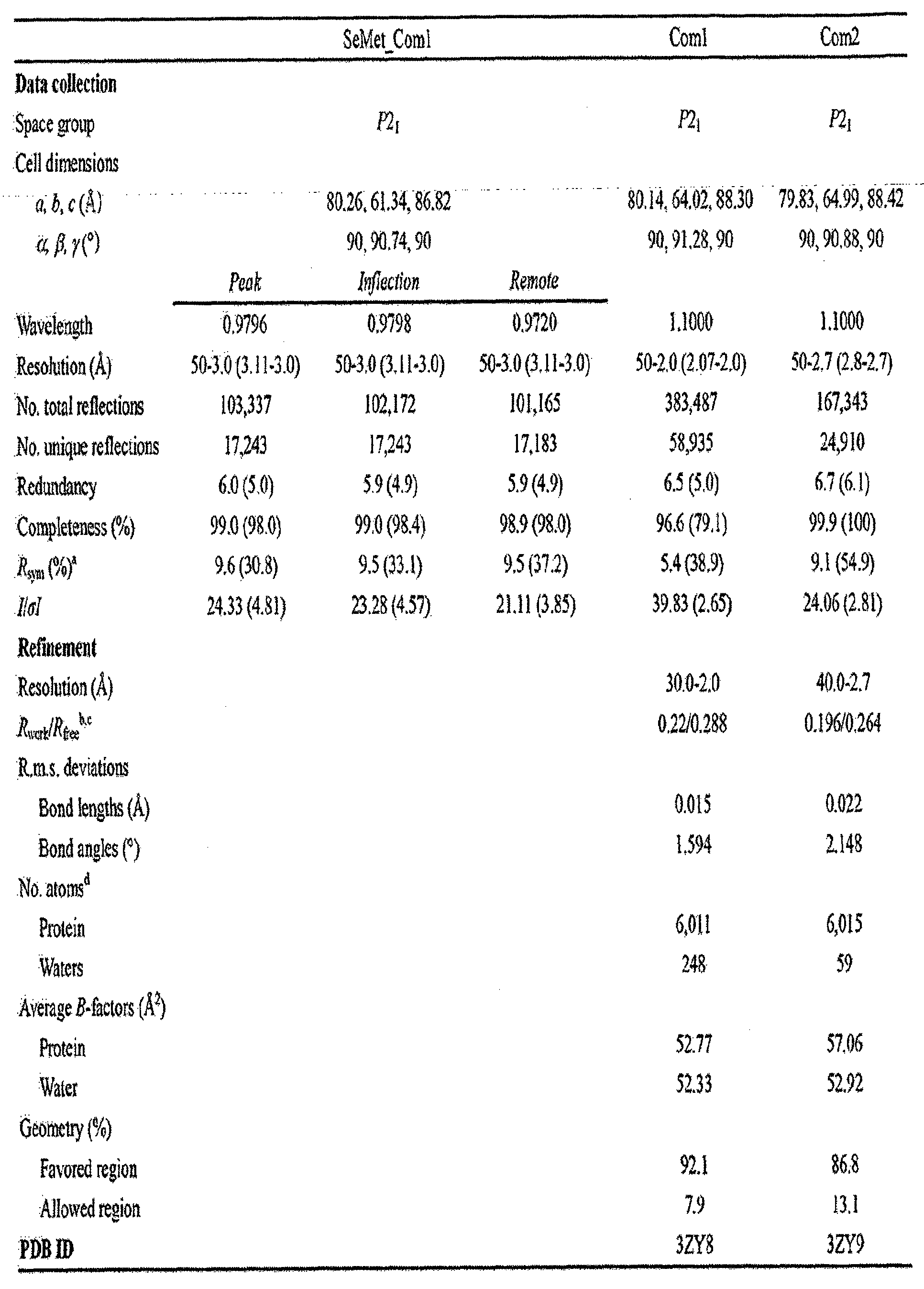
표 8
[규칙 제91조에 의한 정정 17.09.2013]
표 9 
표 9
[규칙 제91조에 의한 정정 17.09.2013]
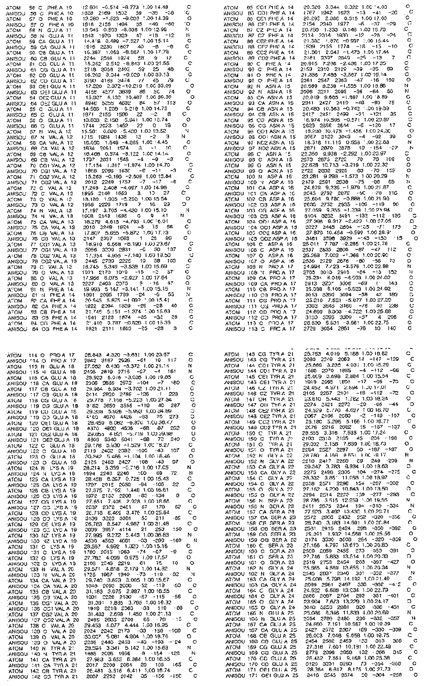
[규칙 제91조에 의한 정정 17.09.2013]
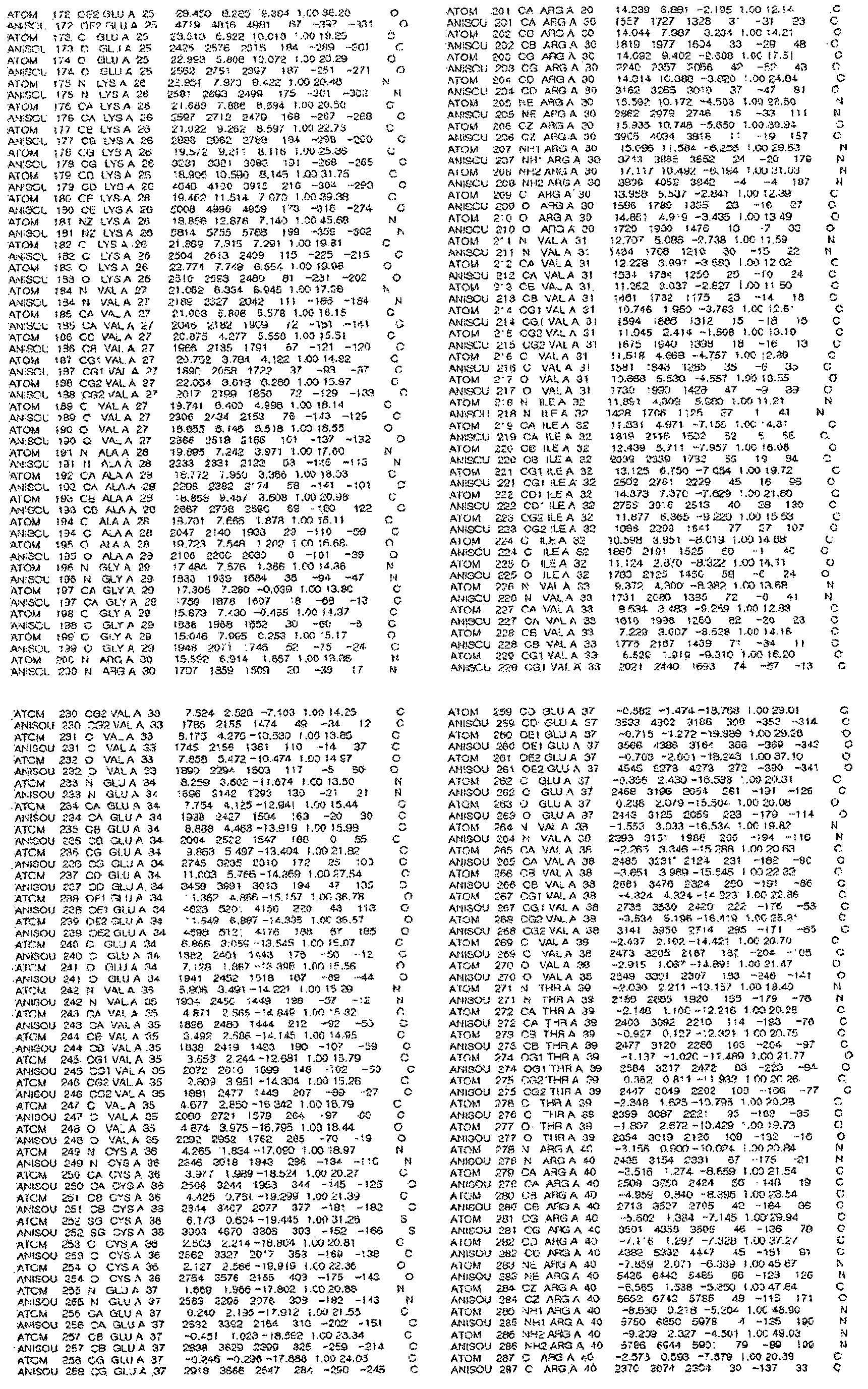
[규칙 제91조에 의한 정정 17.09.2013]
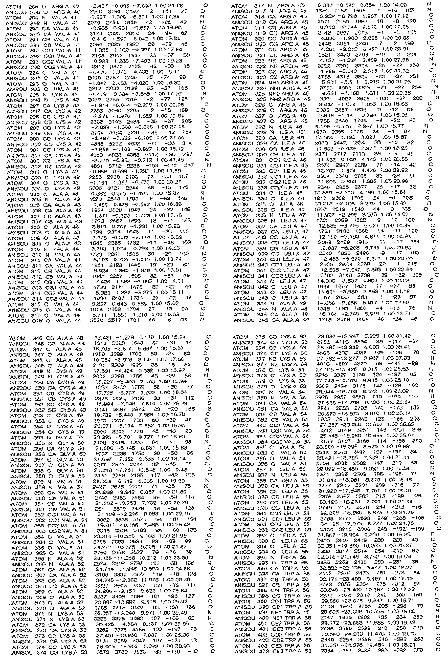
[규칙 제91조에 의한 정정 17.09.2013]
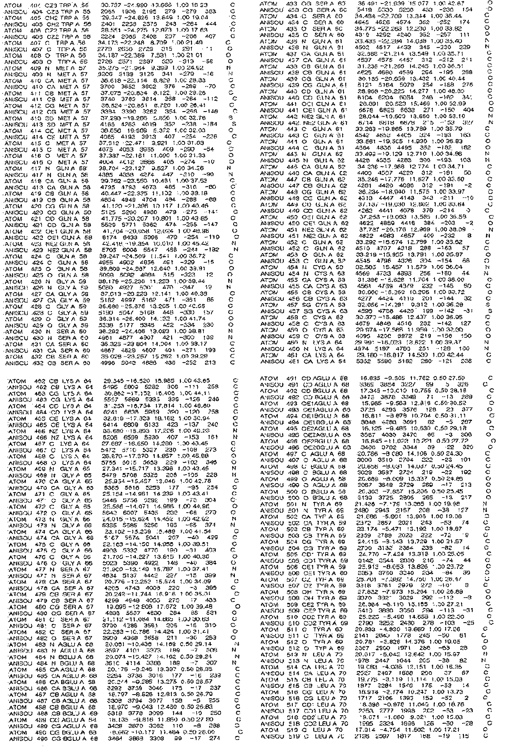
[규칙 제91조에 의한 정정 17.09.2013]

[규칙 제91조에 의한 정정 17.09.2013]
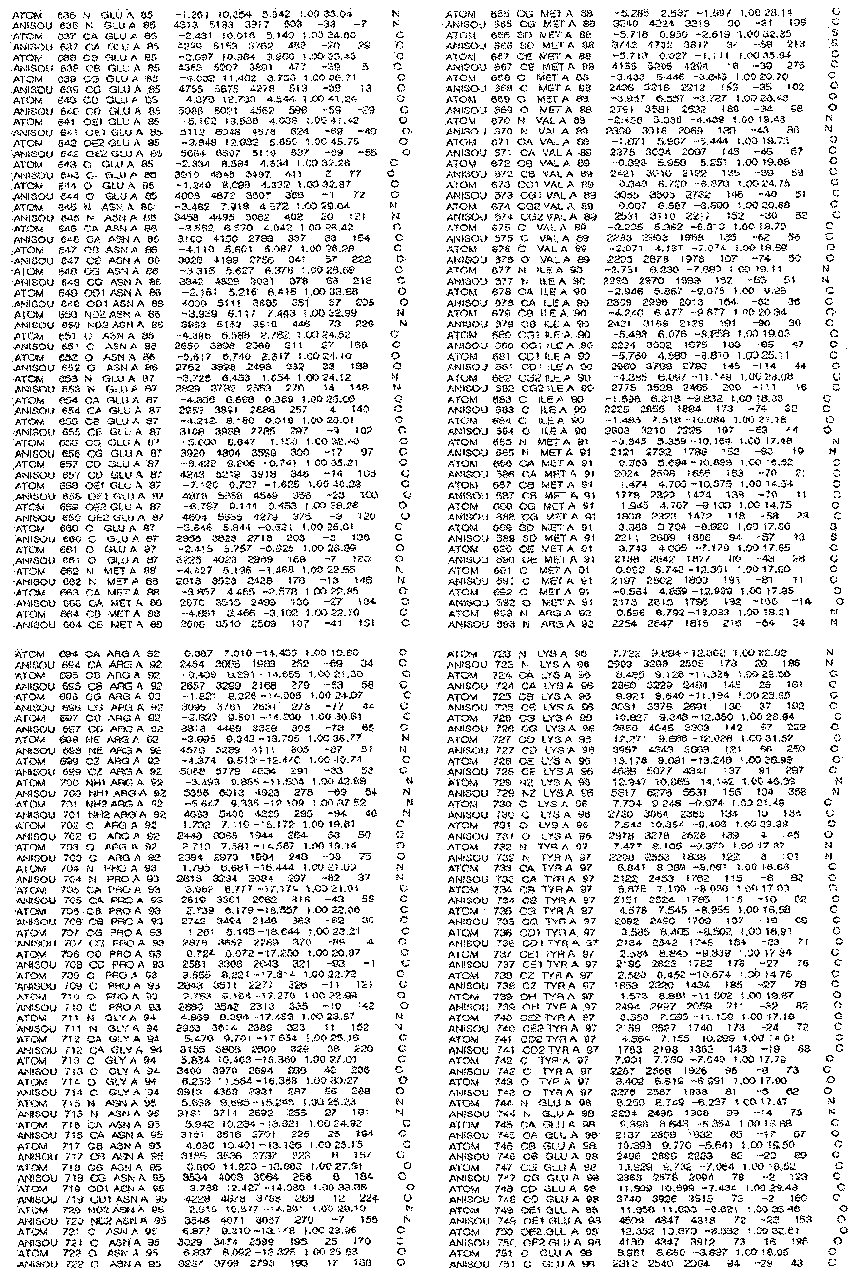
[규칙 제91조에 의한 정정 17.09.2013]

[규칙 제91조에 의한 정정 17.09.2013]
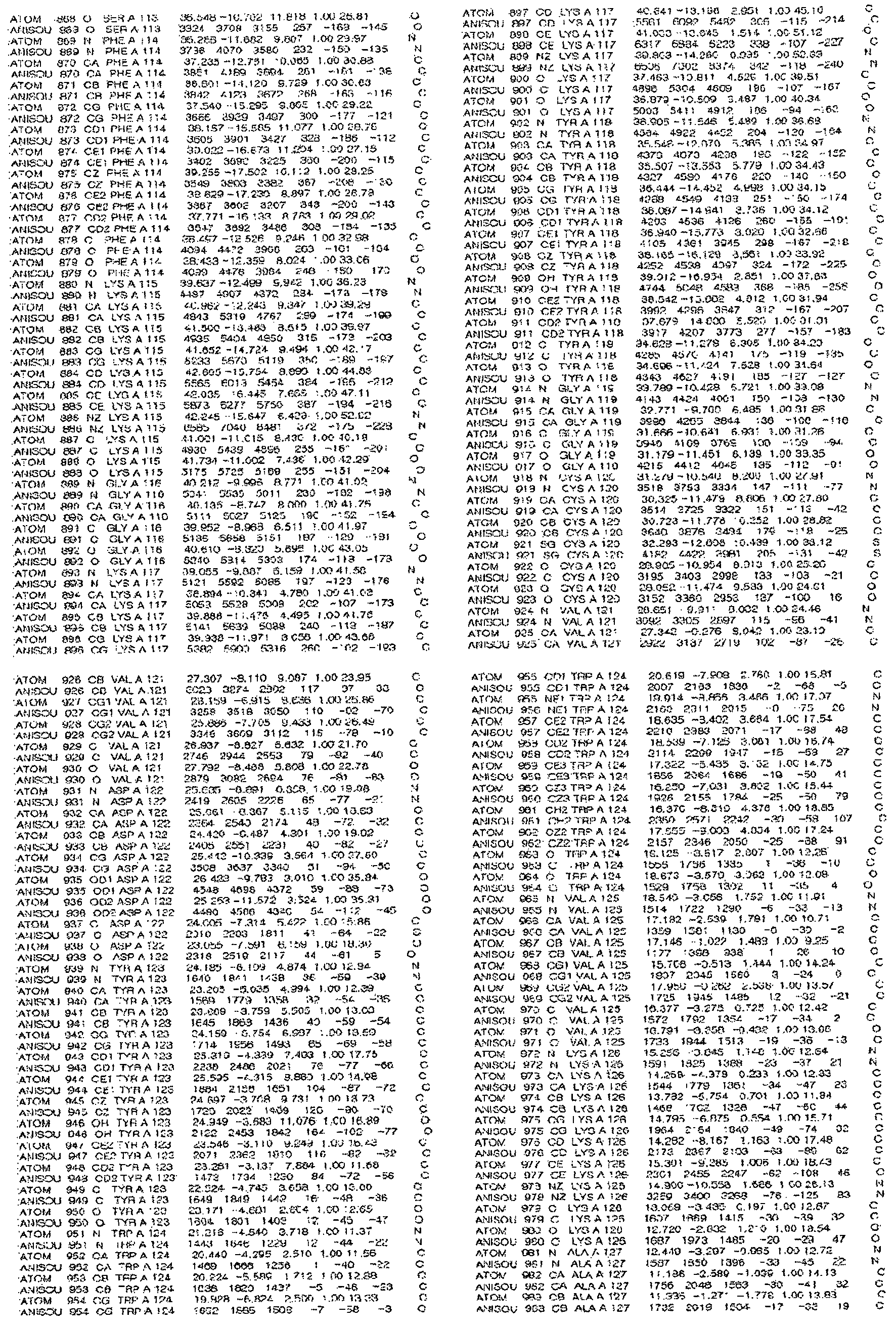
[규칙 제91조에 의한 정정 17.09.2013]

[규칙 제91조에 의한 정정 17.09.2013]
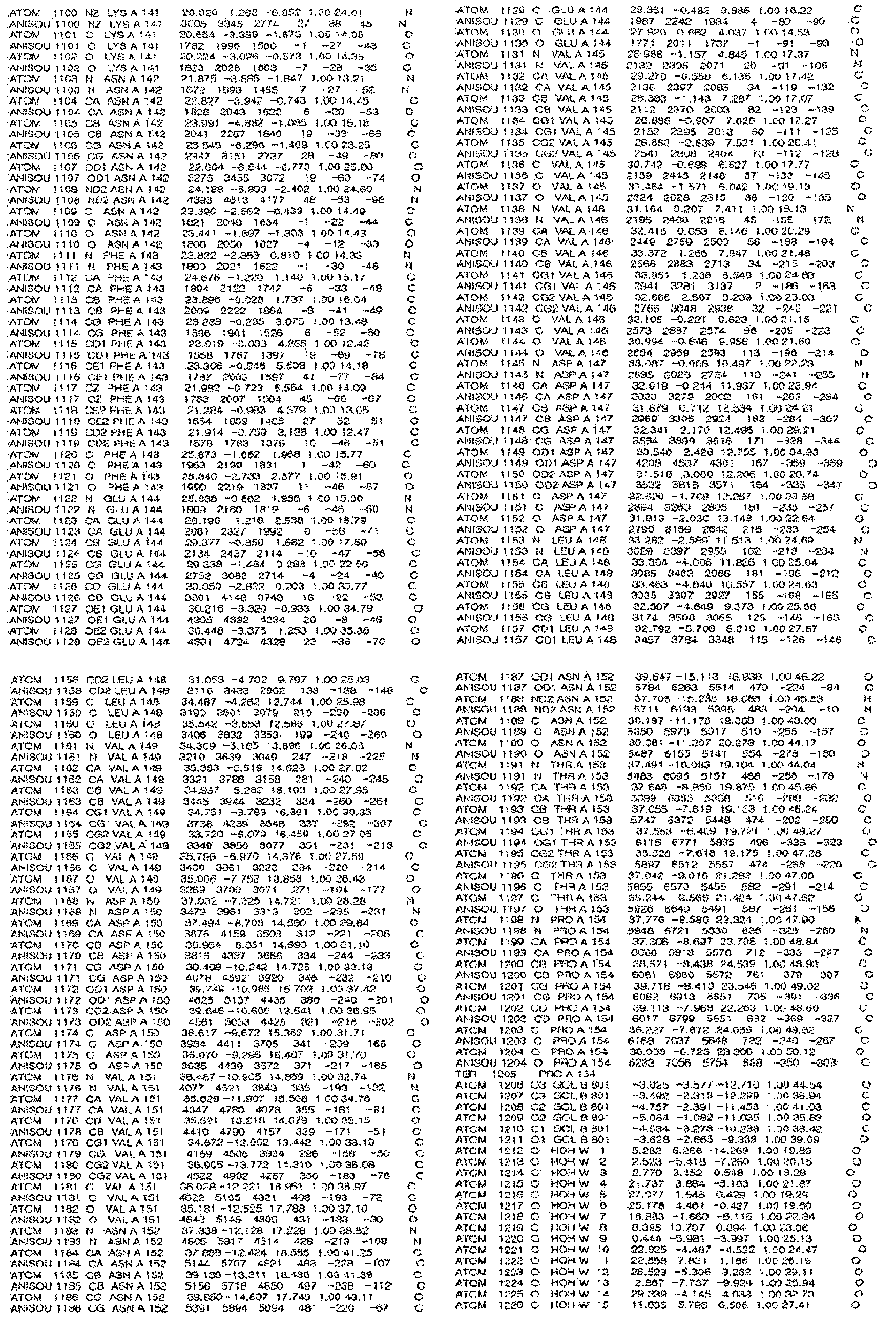
[규칙 제91조에 의한 정정 17.09.2013]

[규칙 제91조에 의한 정정 17.09.2013]
표 10 
표 10
[규칙 제91조에 의한 정정 17.09.2013]
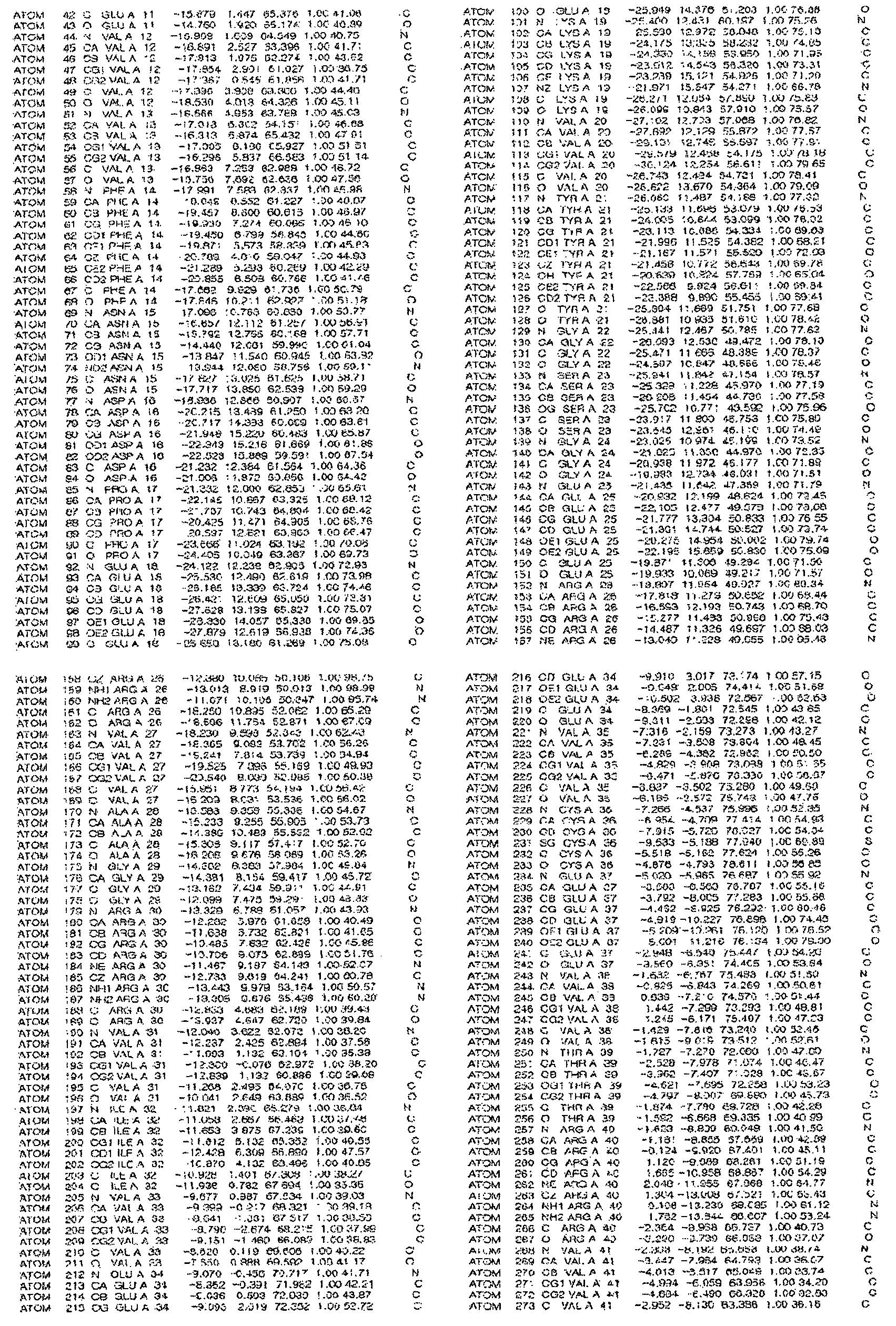
[규칙 제91조에 의한 정정 17.09.2013]
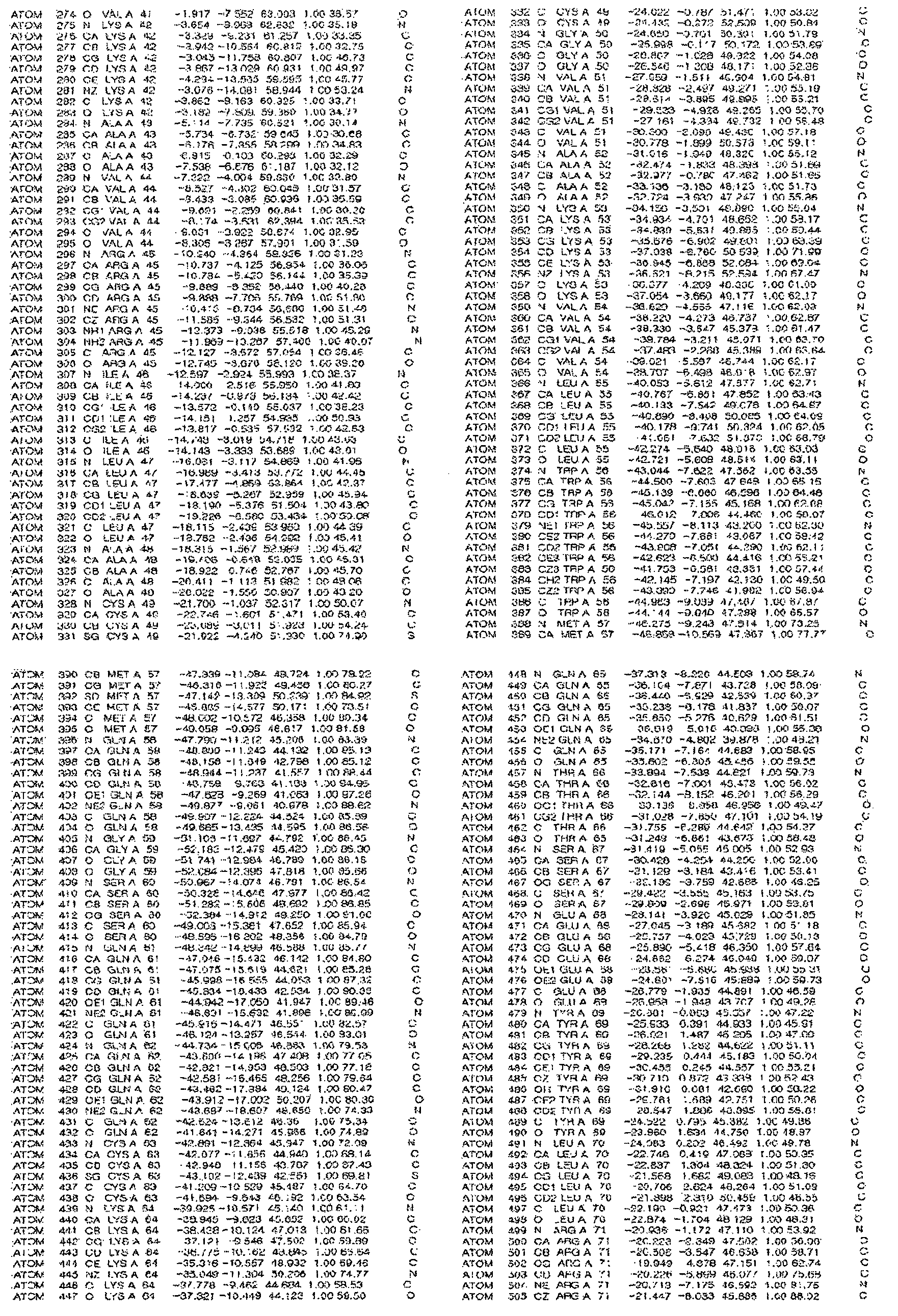
[규칙 제91조에 의한 정정 17.09.2013]
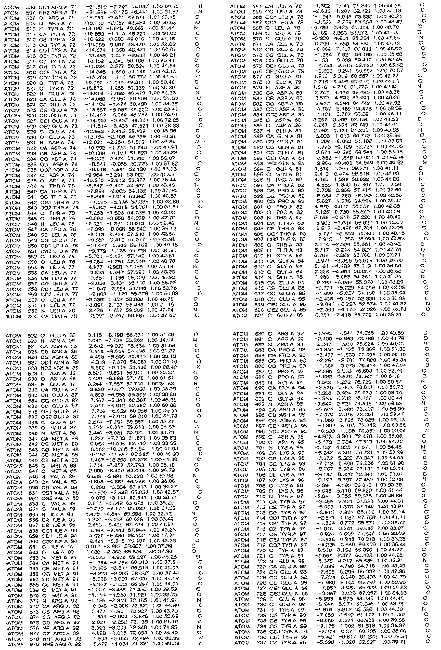
[규칙 제91조에 의한 정정 17.09.2013]
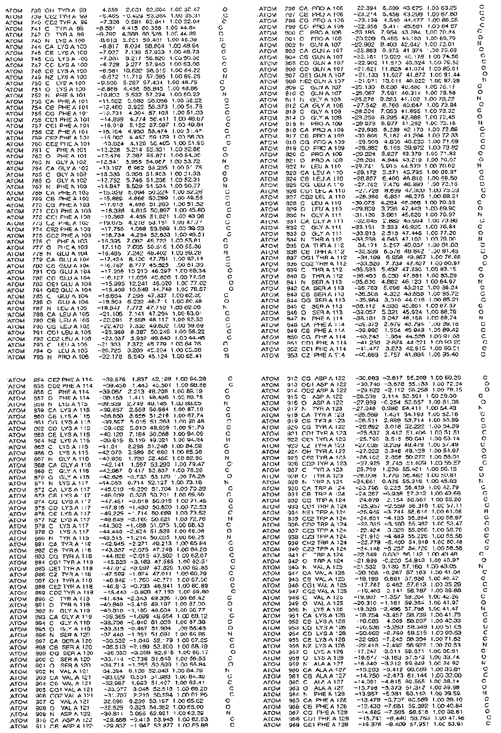
[규칙 제91조에 의한 정정 17.09.2013]

[규칙 제91조에 의한 정정 17.09.2013]

[규칙 제91조에 의한 정정 17.09.2013]
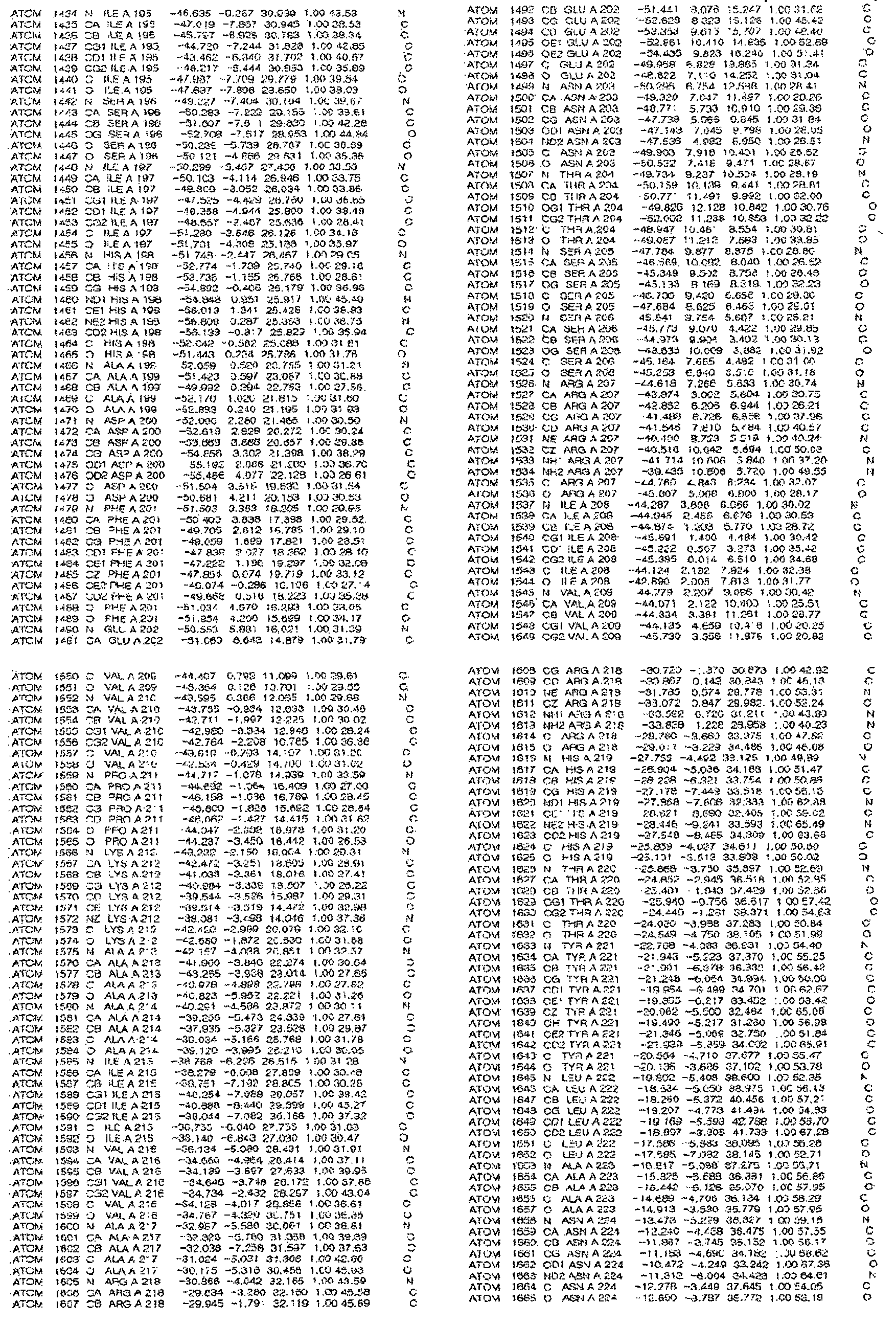
[규칙 제91조에 의한 정정 17.09.2013]
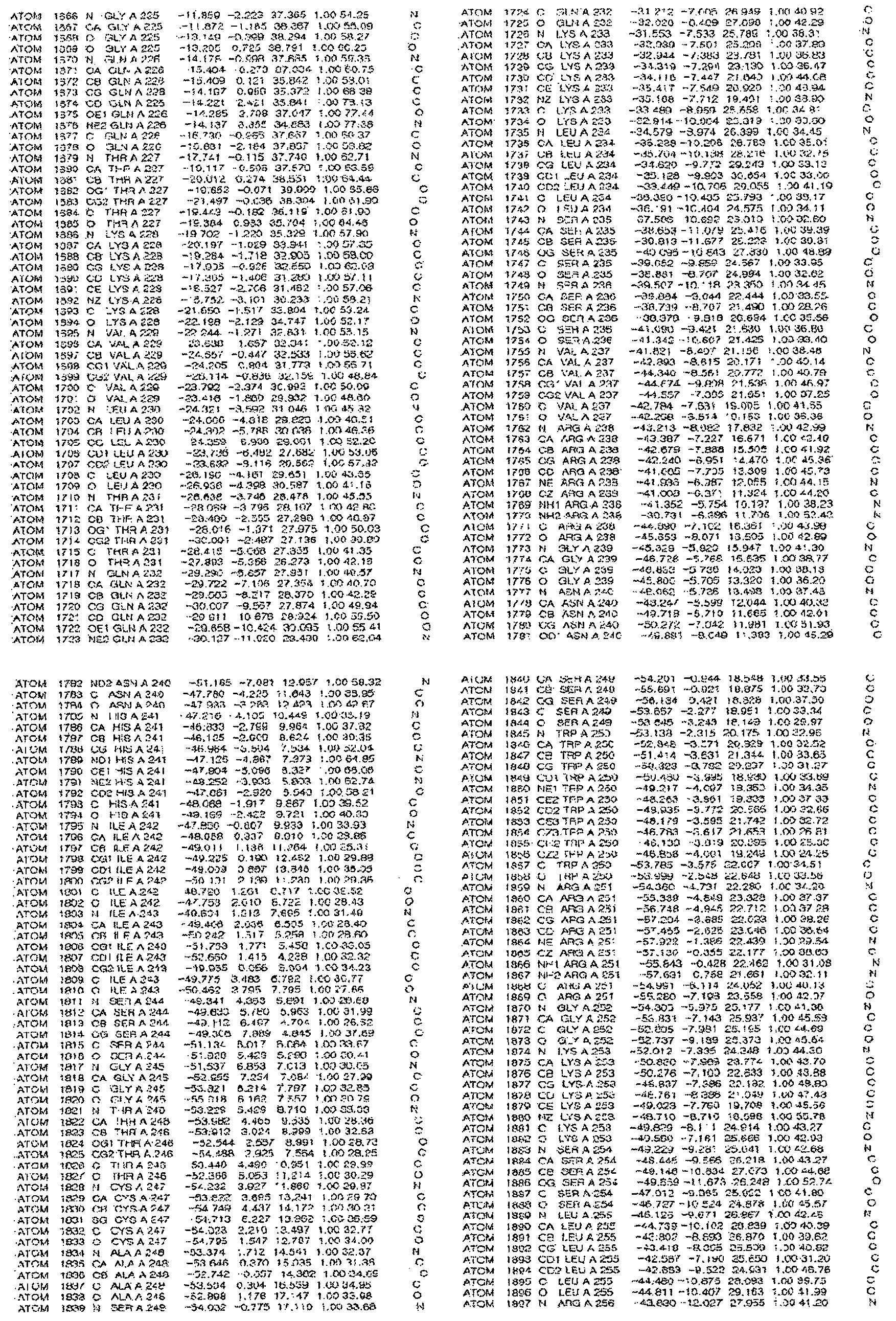
[규칙 제91조에 의한 정정 17.09.2013]
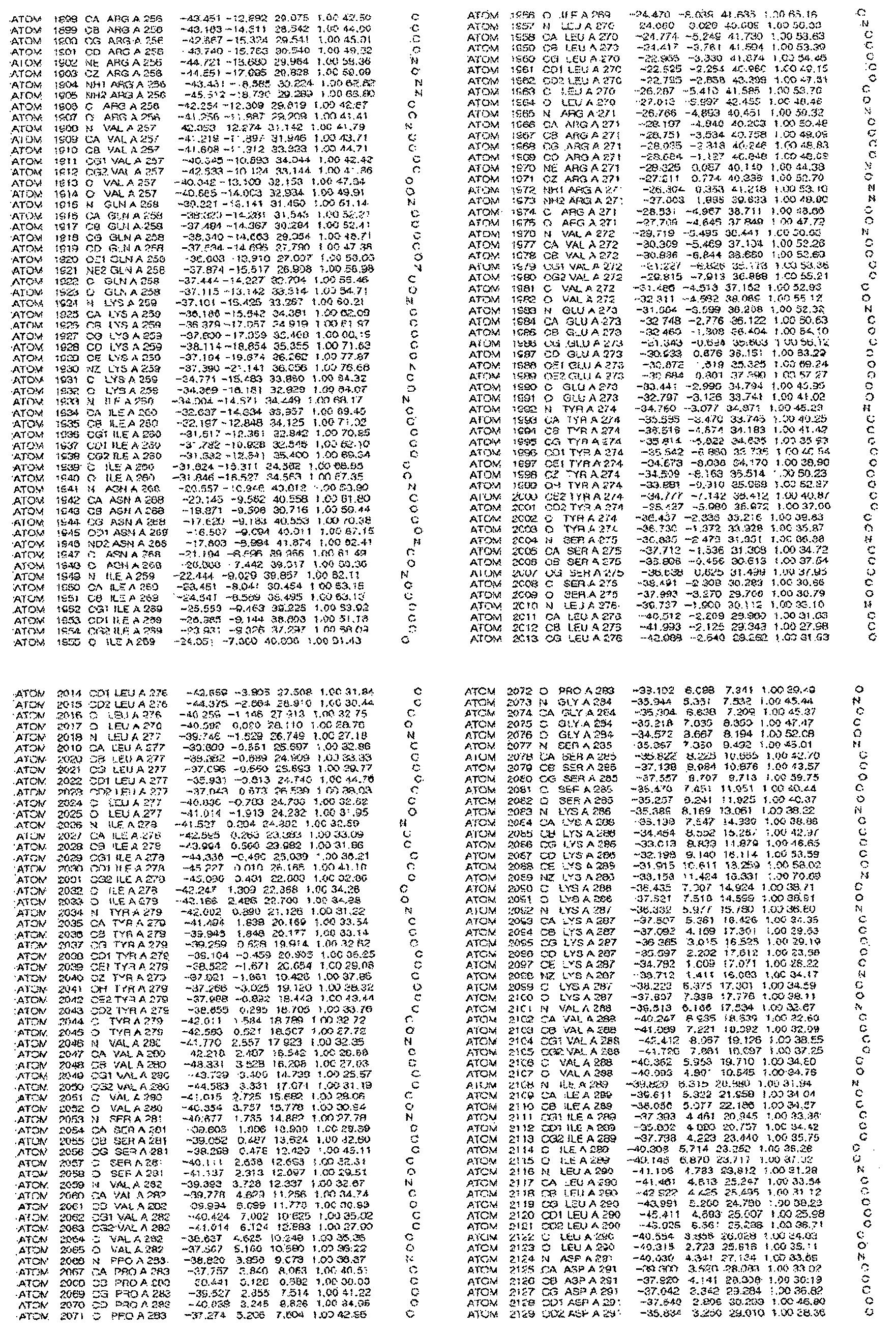
[규칙 제91조에 의한 정정 17.09.2013]
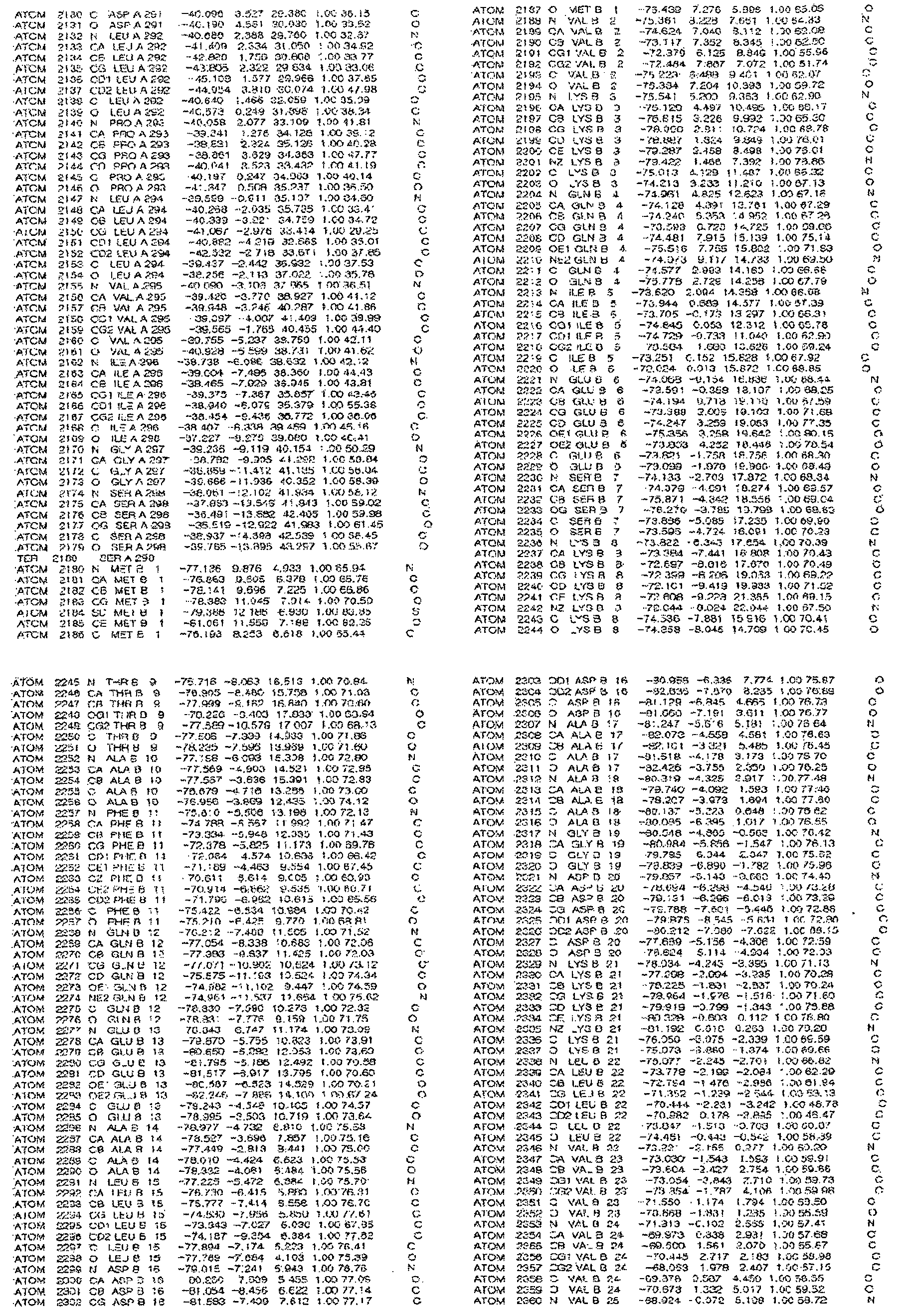
[규칙 제91조에 의한 정정 17.09.2013]
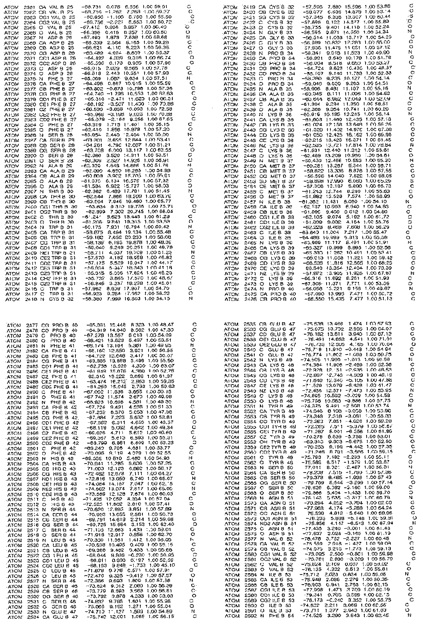
[규칙 제91조에 의한 정정 17.09.2013]
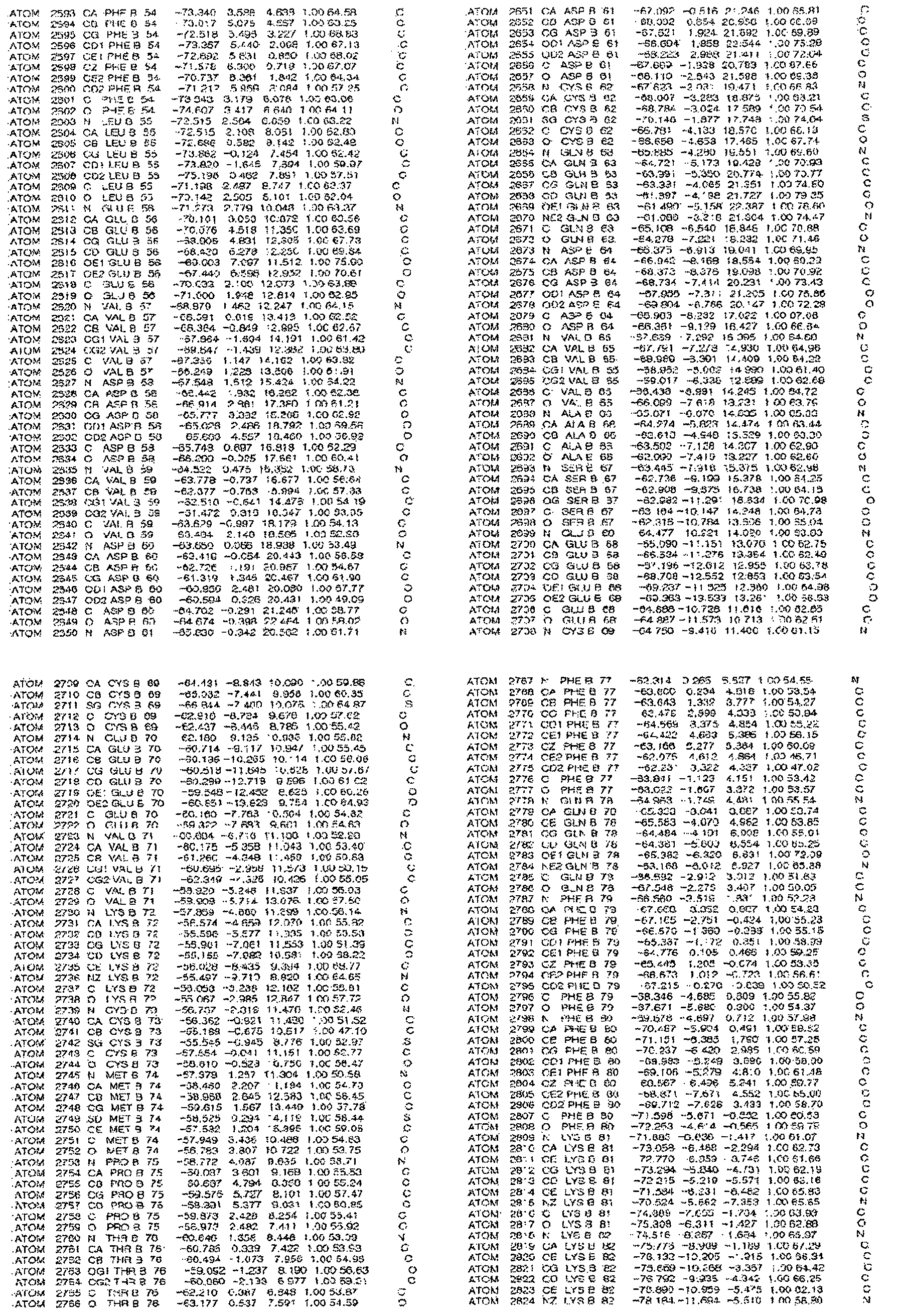
[규칙 제91조에 의한 정정 17.09.2013]
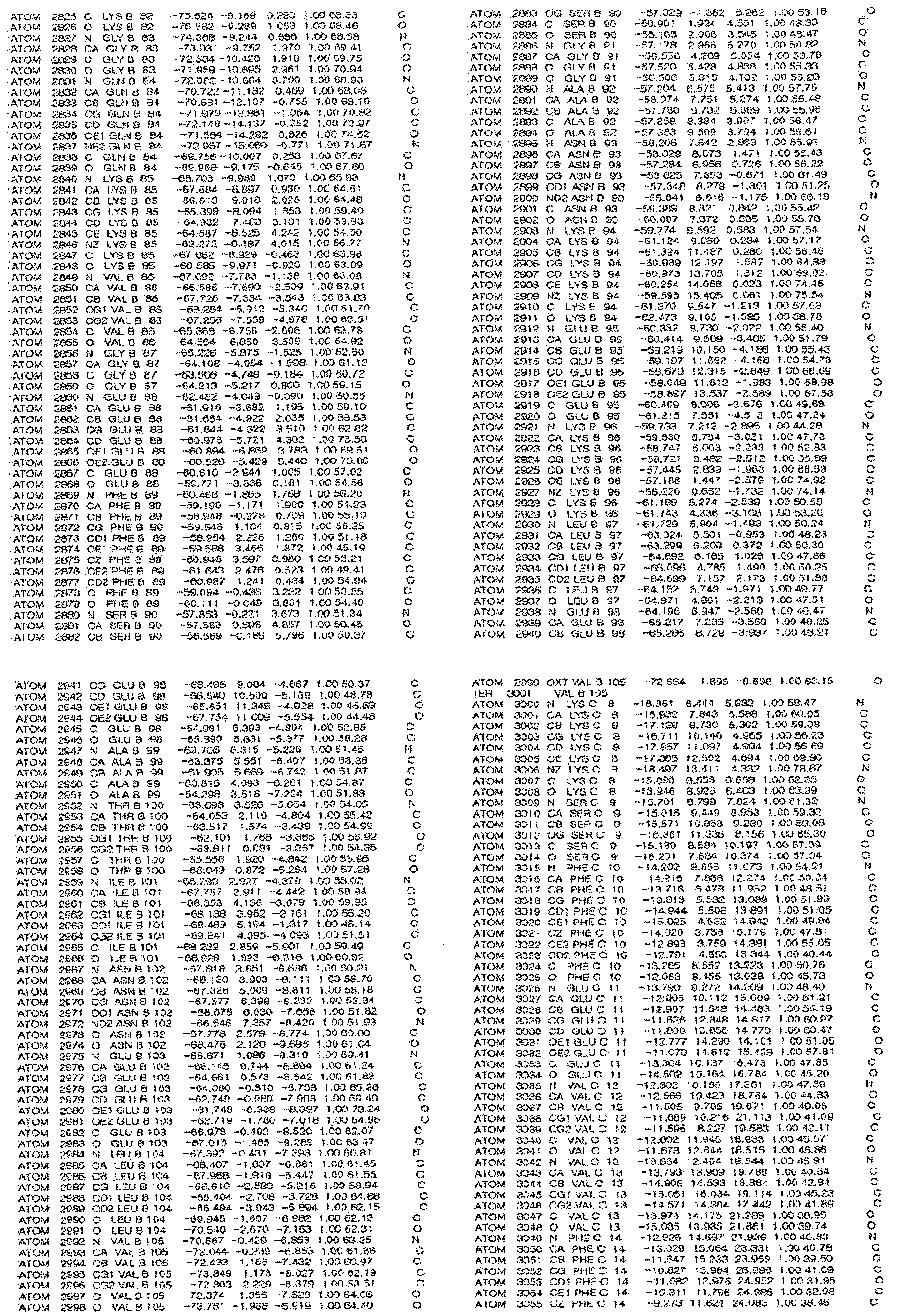
[규칙 제91조에 의한 정정 17.09.2013]
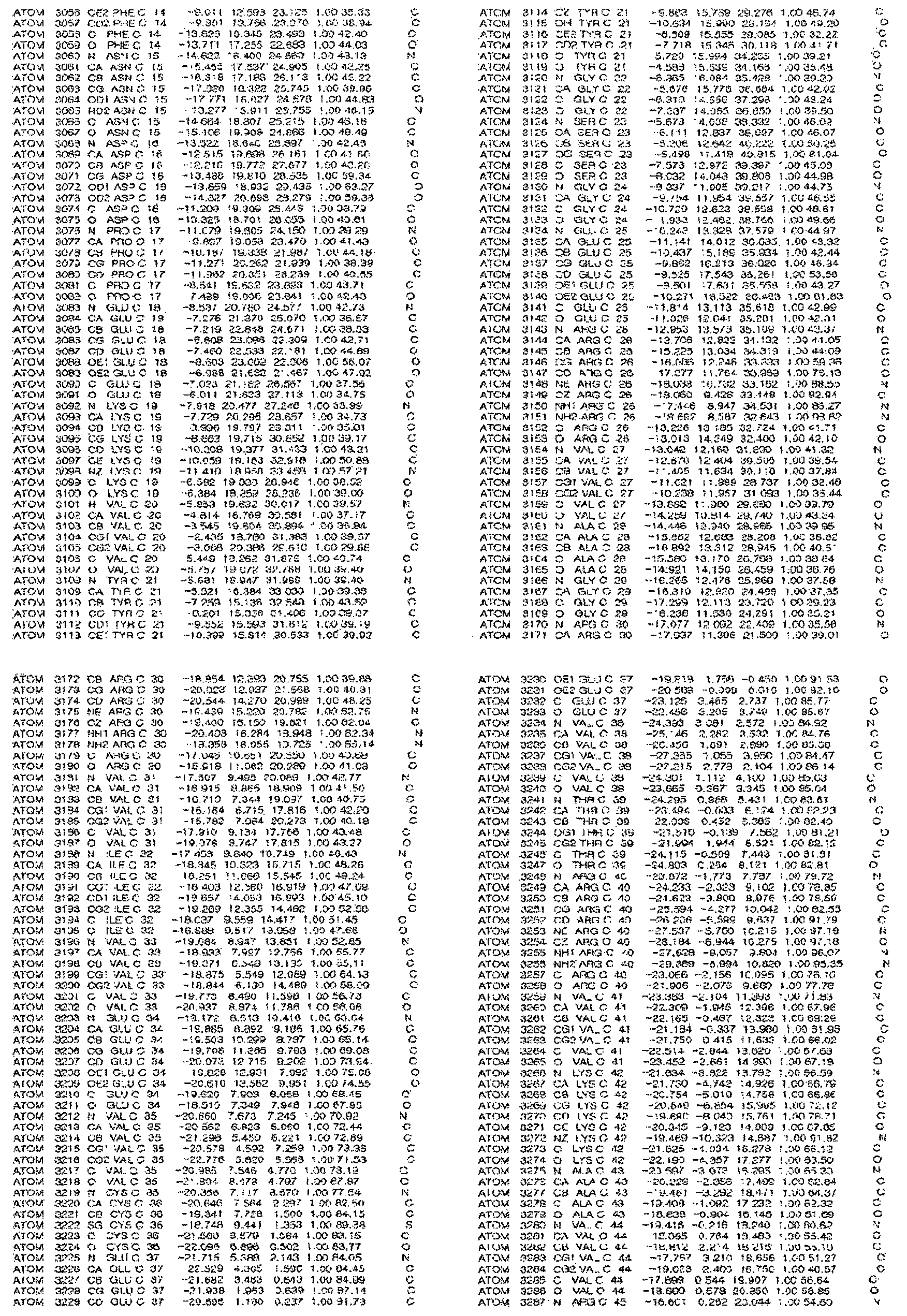
[규칙 제91조에 의한 정정 17.09.2013]
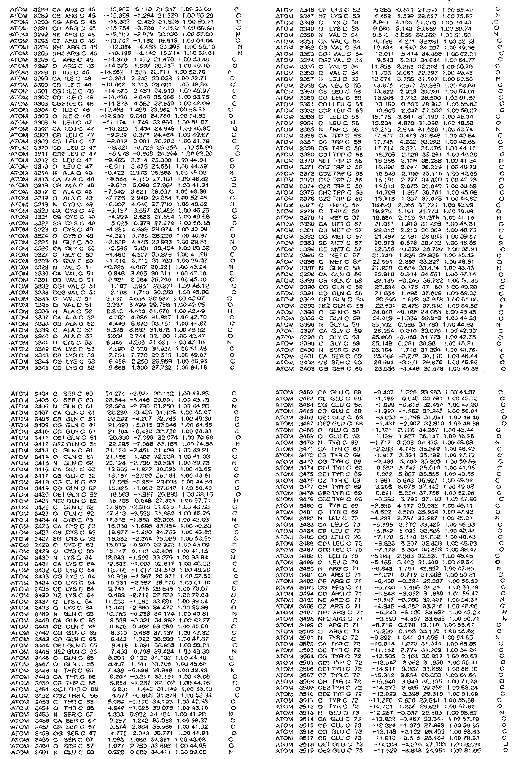
[규칙 제91조에 의한 정정 17.09.2013]
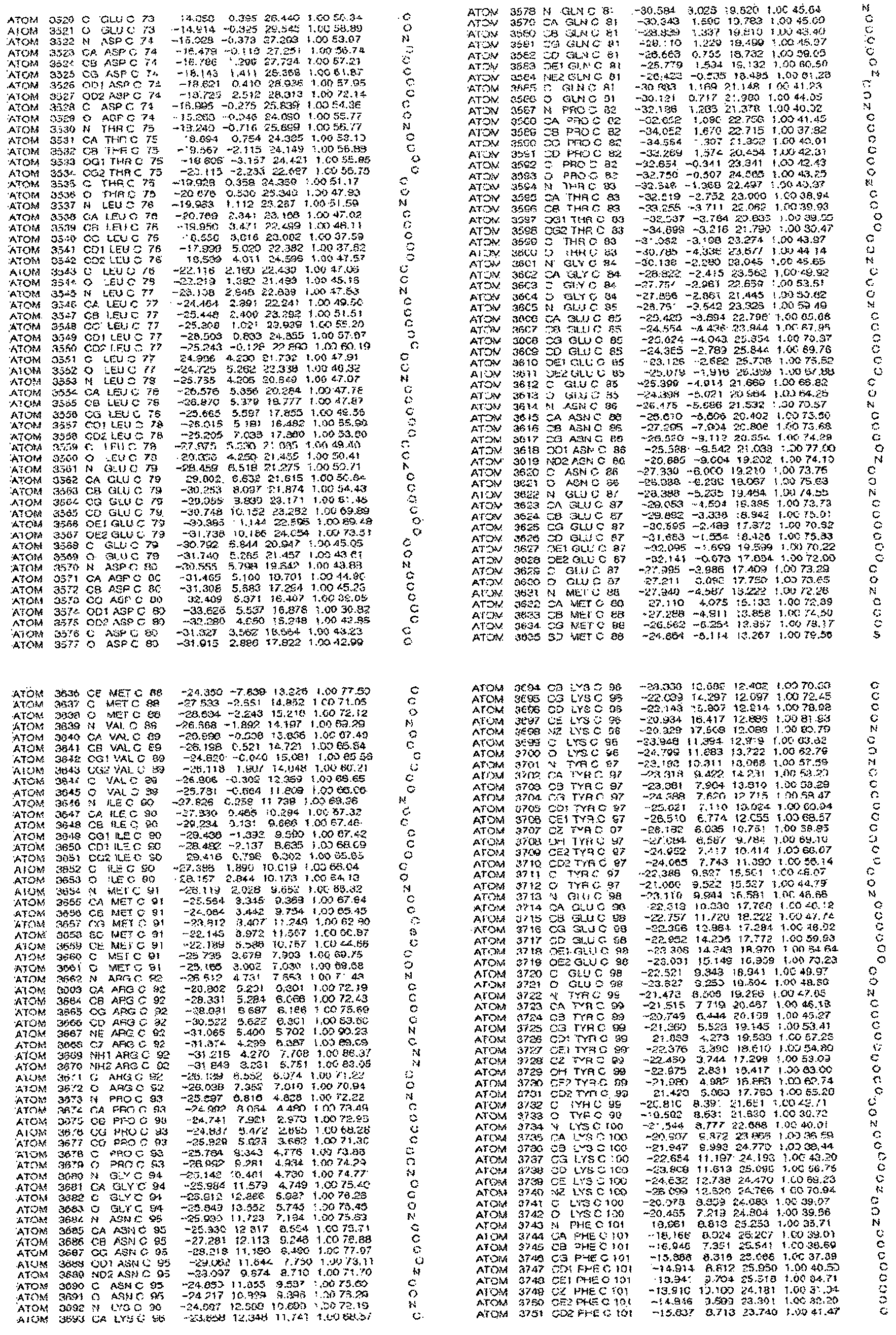
[규칙 제91조에 의한 정정 17.09.2013]
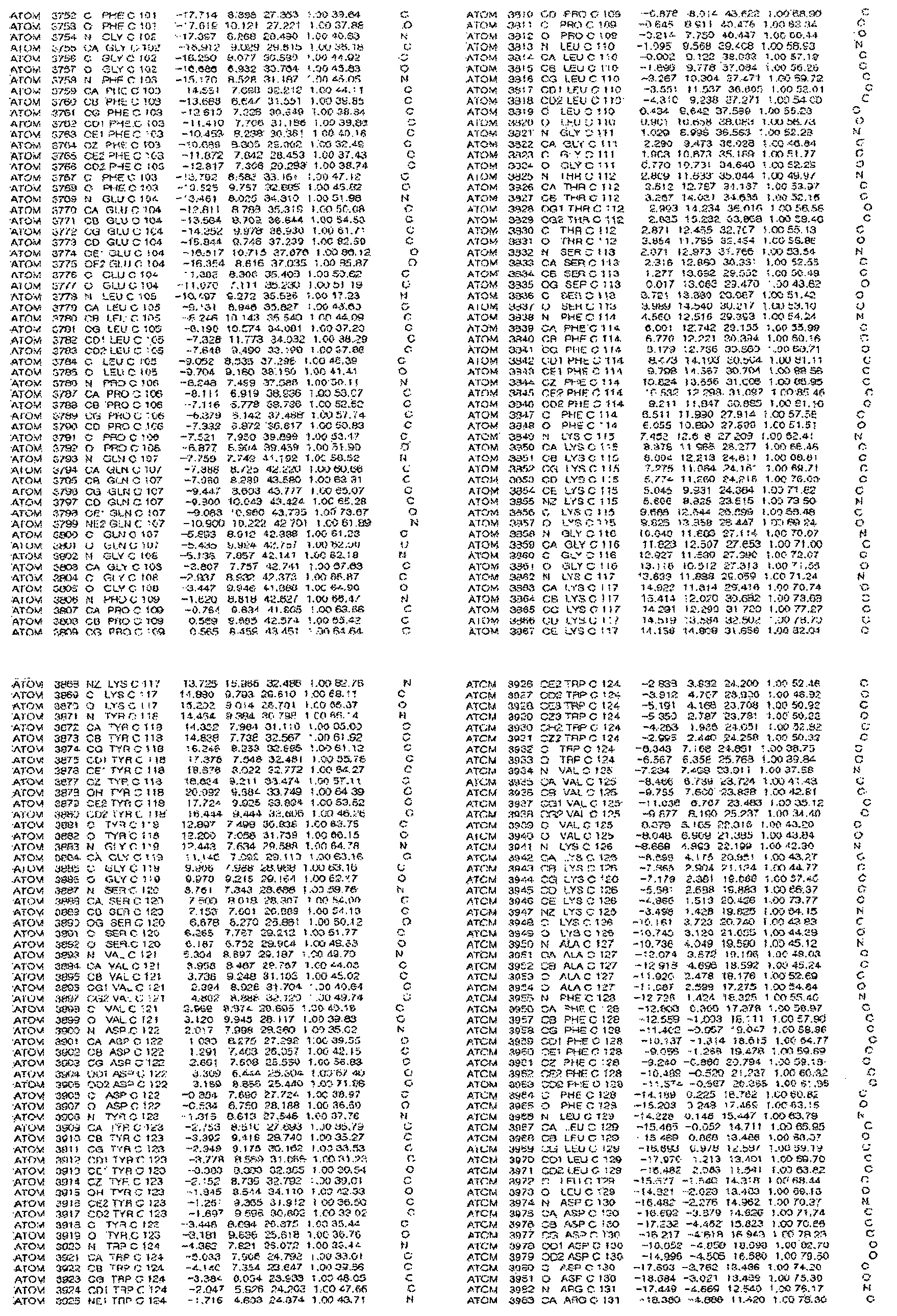
[규칙 제91조에 의한 정정 17.09.2013]
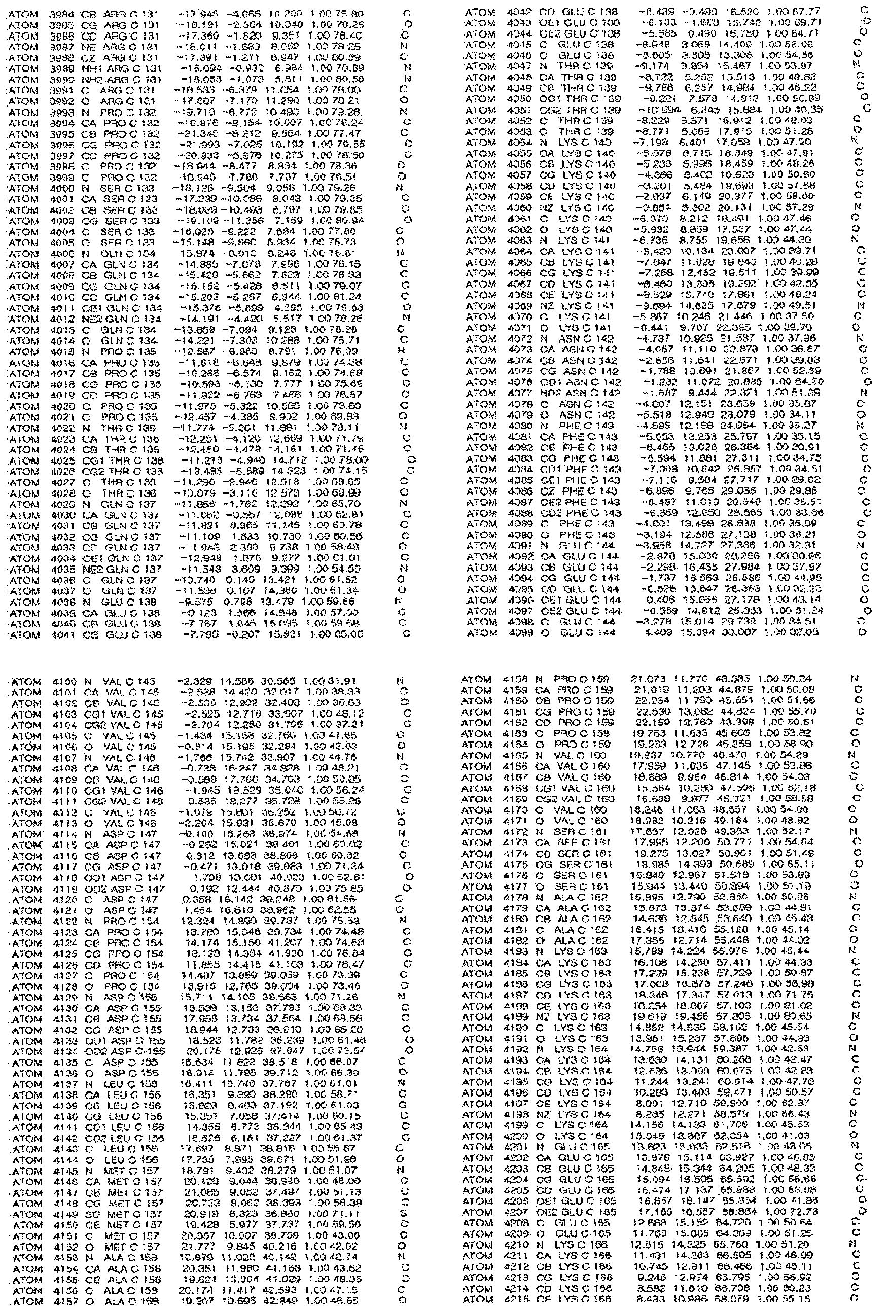
[규칙 제91조에 의한 정정 17.09.2013]
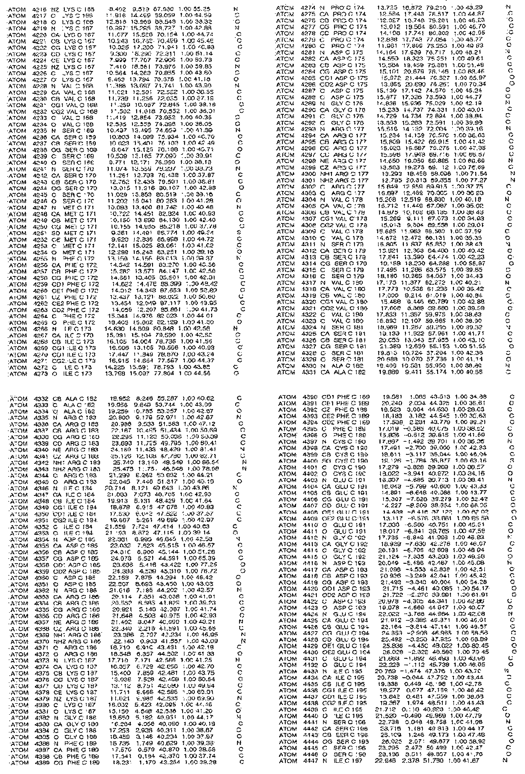
[규칙 제91조에 의한 정정 17.09.2013]
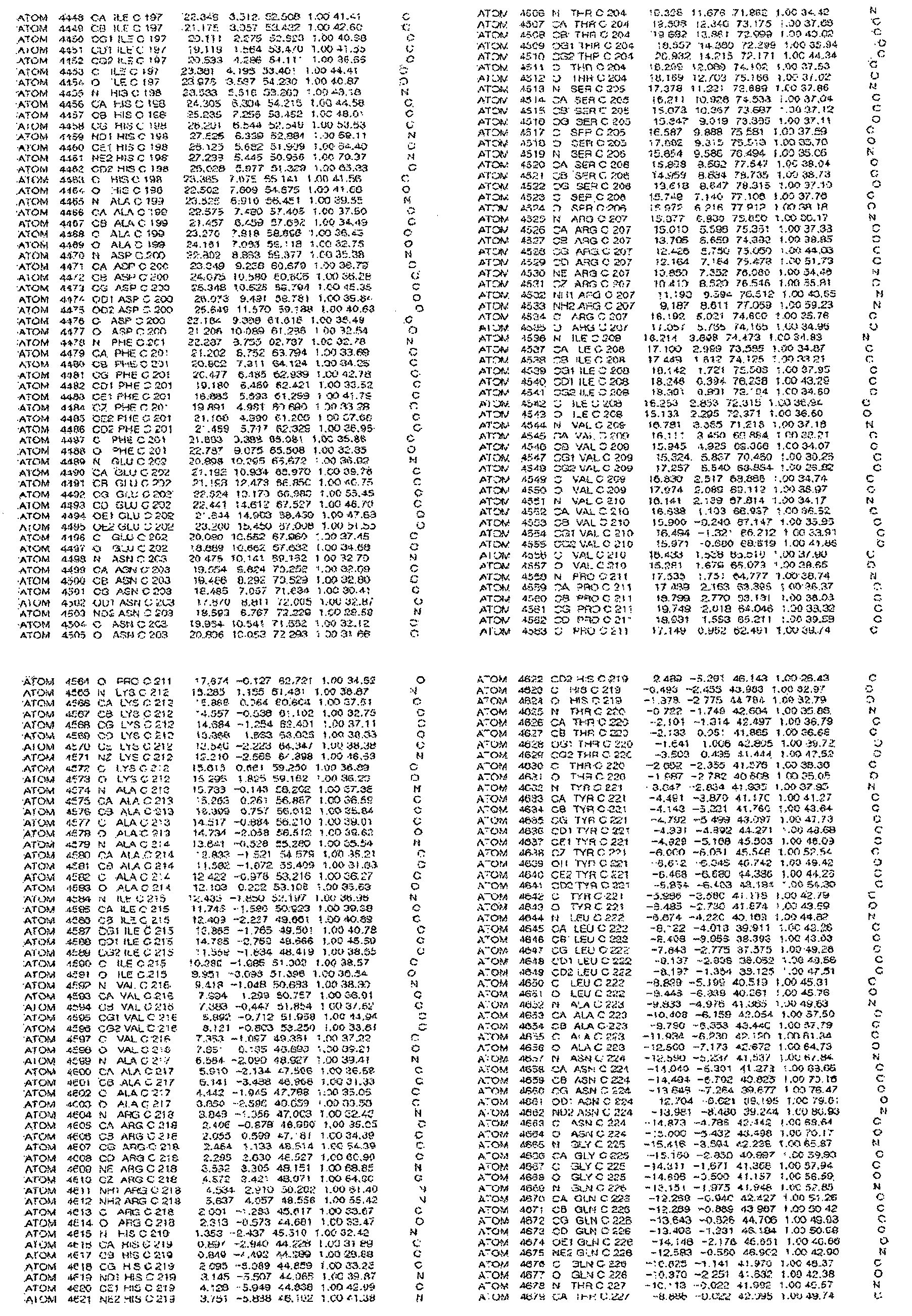
[규칙 제91조에 의한 정정 17.09.2013]

[규칙 제91조에 의한 정정 17.09.2013]
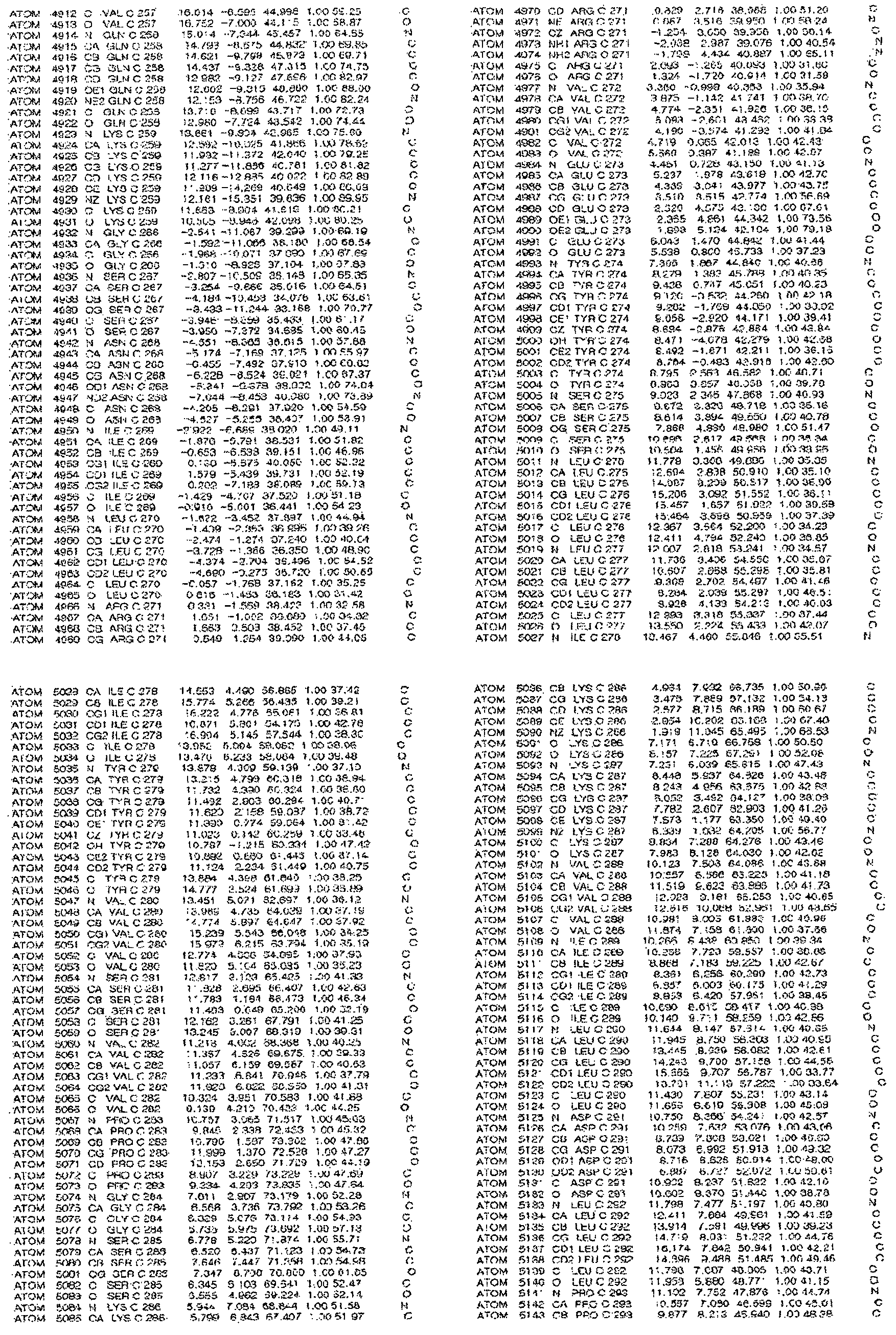
[규칙 제91조에 의한 정정 17.09.2013]
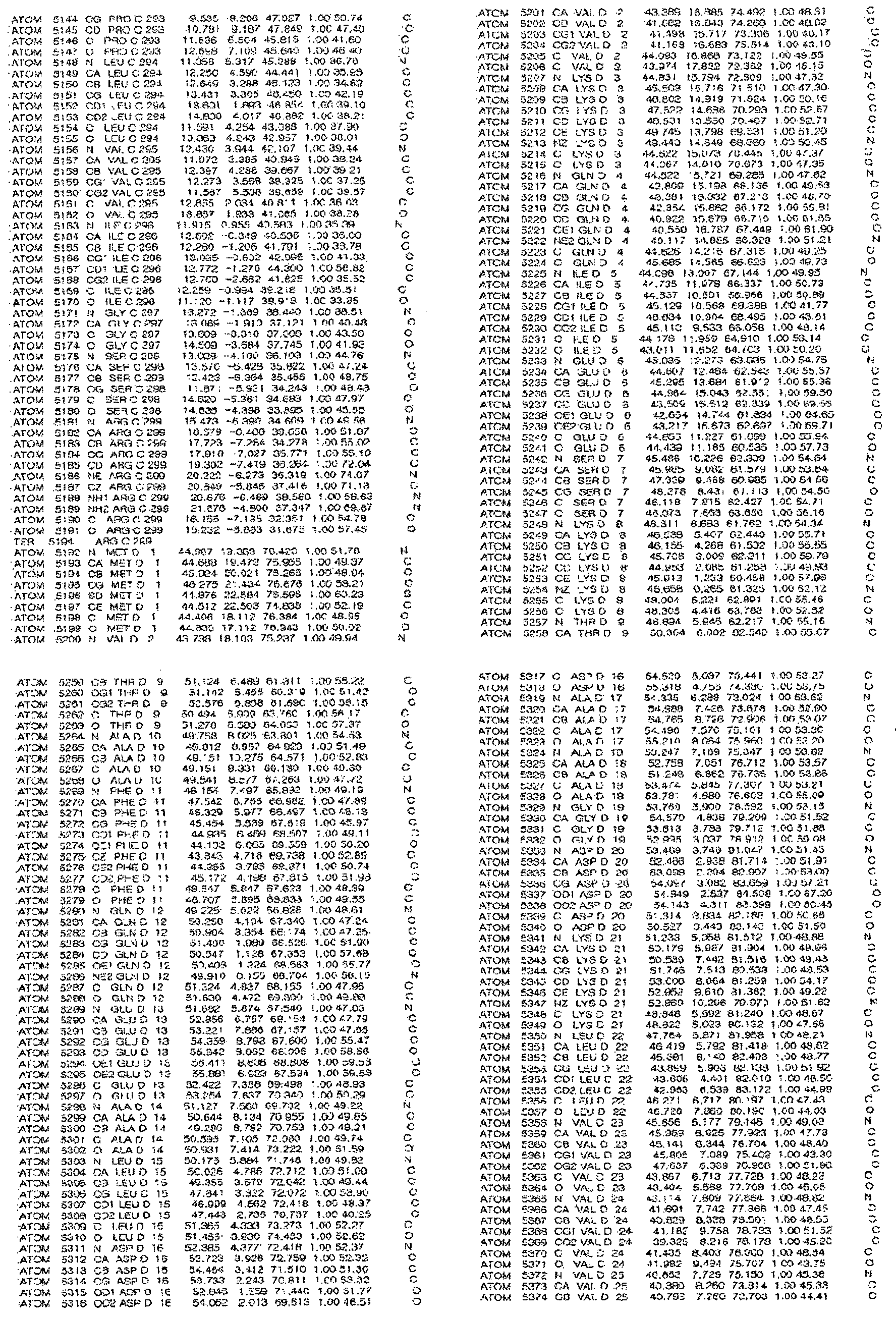
[규칙 제91조에 의한 정정 17.09.2013]
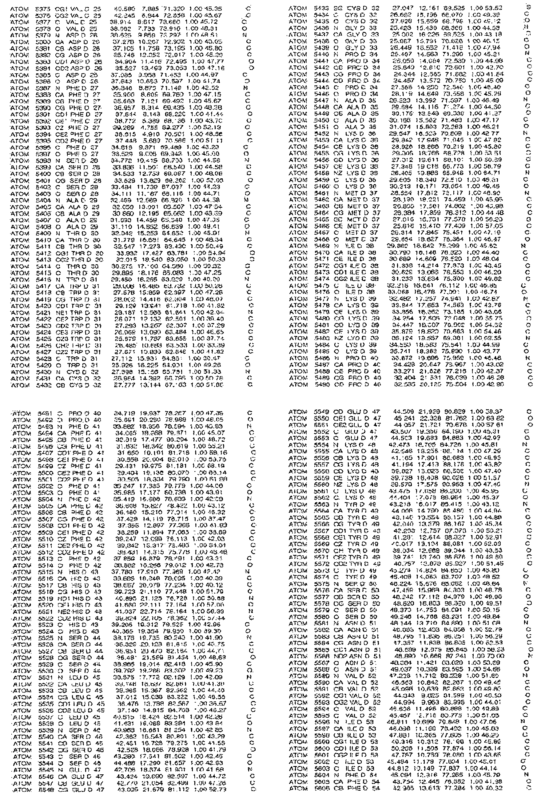
[규칙 제91조에 의한 정정 17.09.2013]

[규칙 제91조에 의한 정정 17.09.2013]
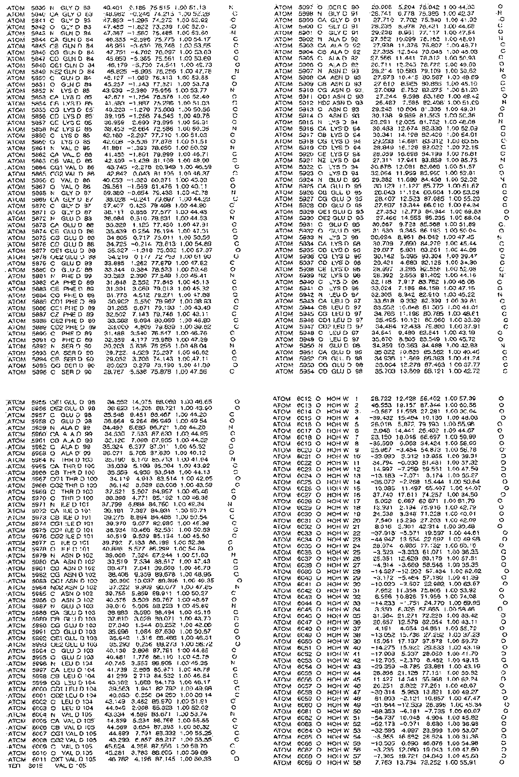
[규칙 제91조에 의한 정정 17.09.2013]
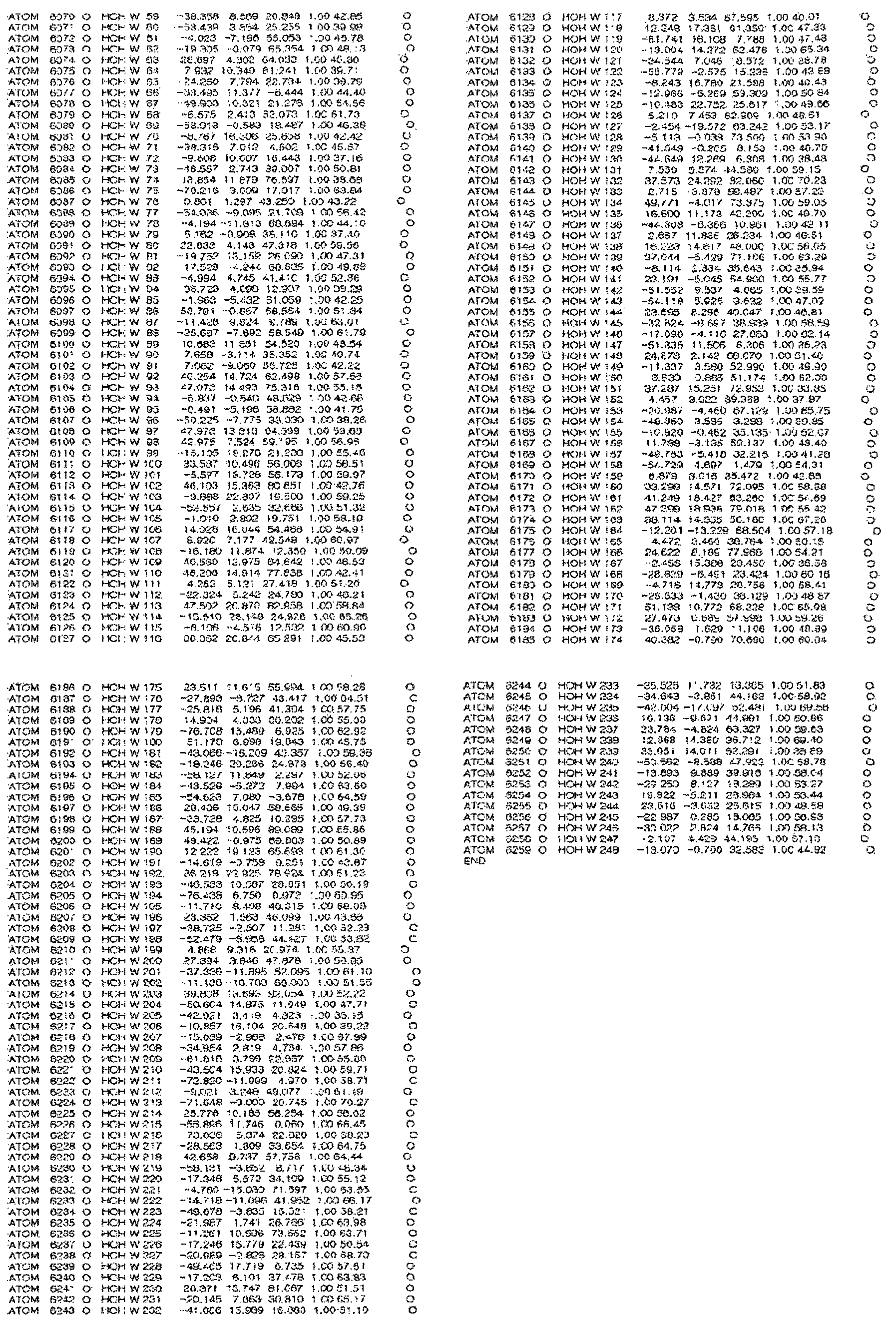
[규칙 제91조에 의한 정정 17.09.2013]
표 11 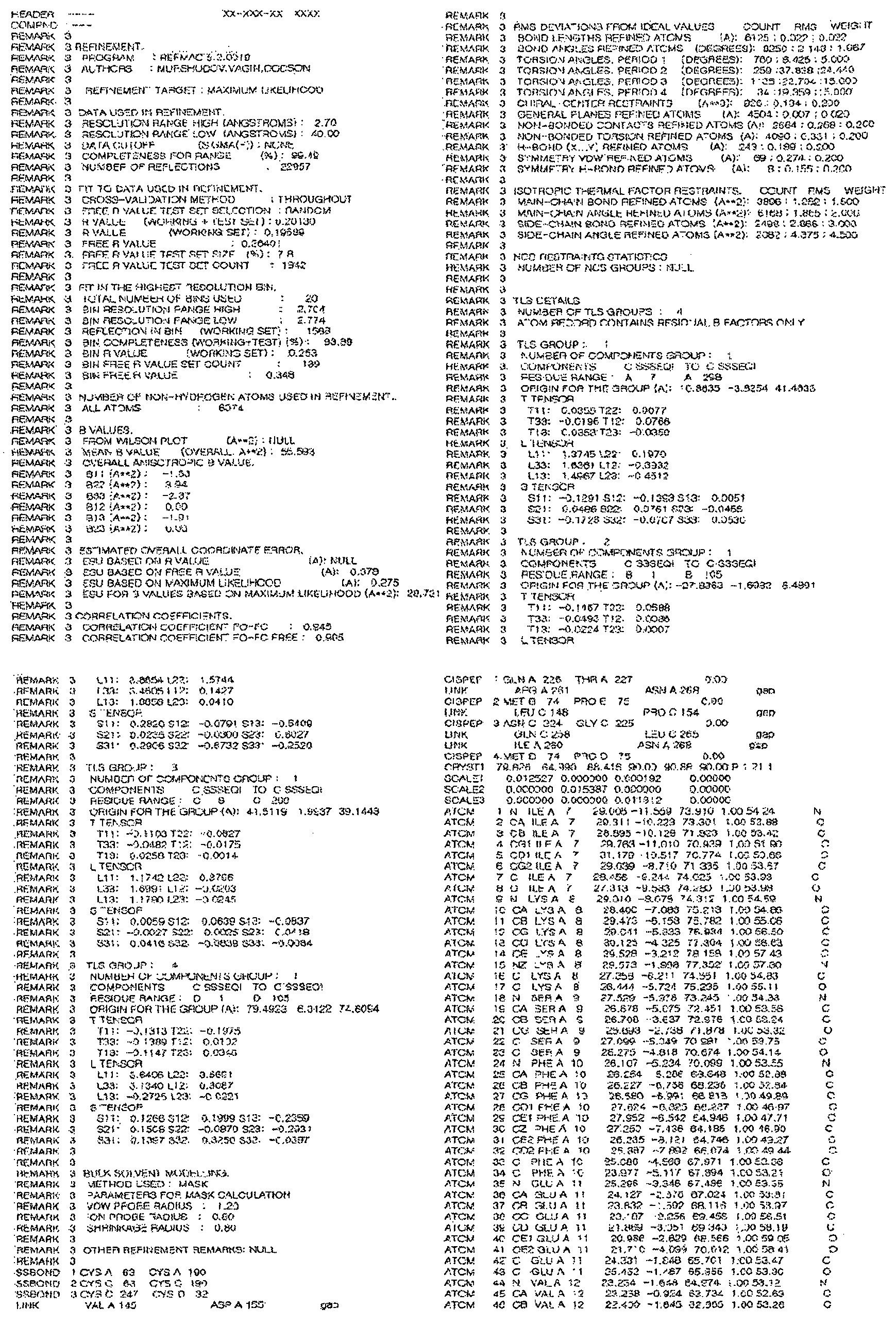
표 11
[규칙 제91조에 의한 정정 17.09.2013]

[규칙 제91조에 의한 정정 17.09.2013]

[규칙 제91조에 의한 정정 17.09.2013]

[규칙 제91조에 의한 정정 17.09.2013]
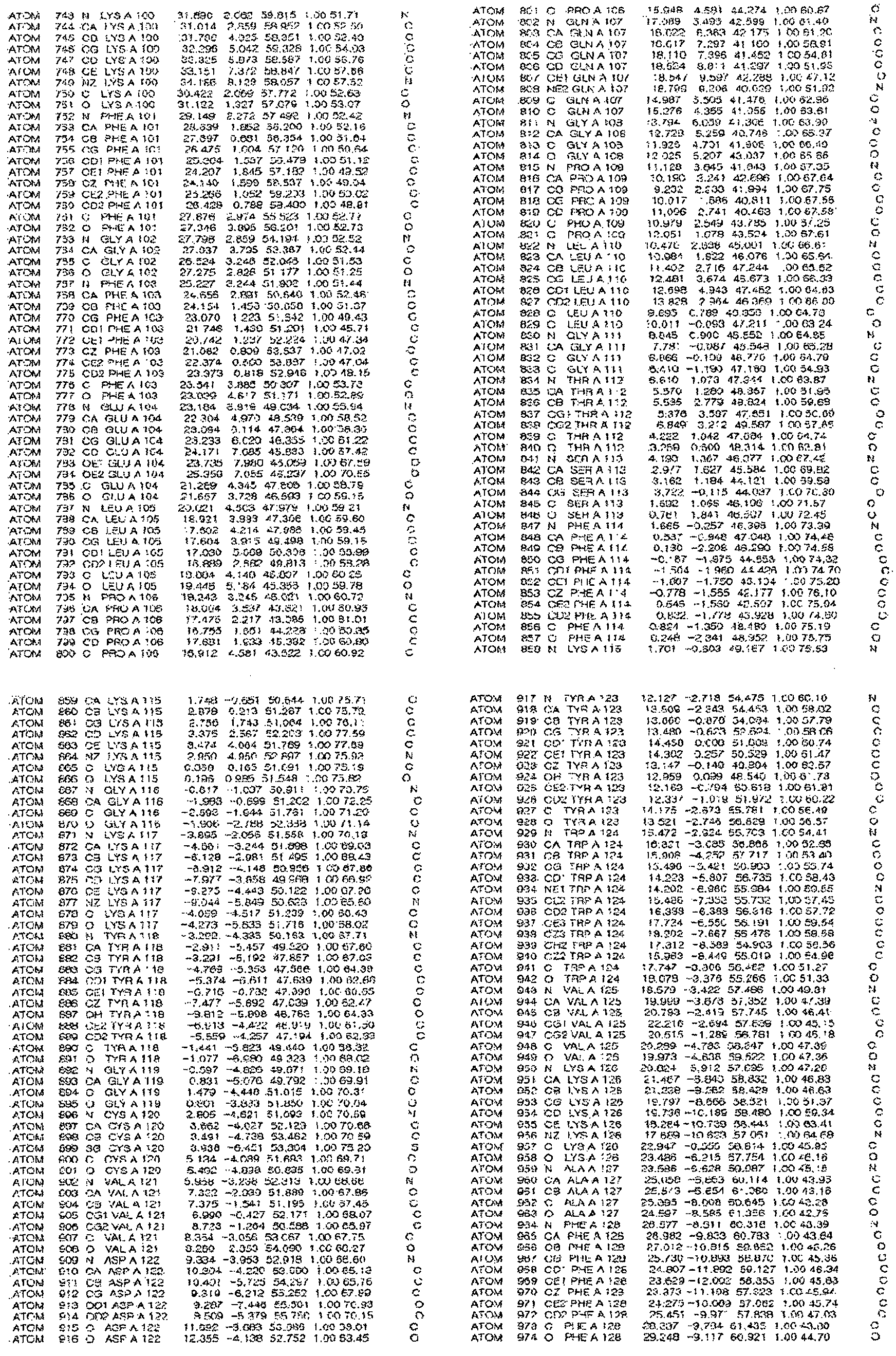
[규칙 제91조에 의한 정정 17.09.2013]
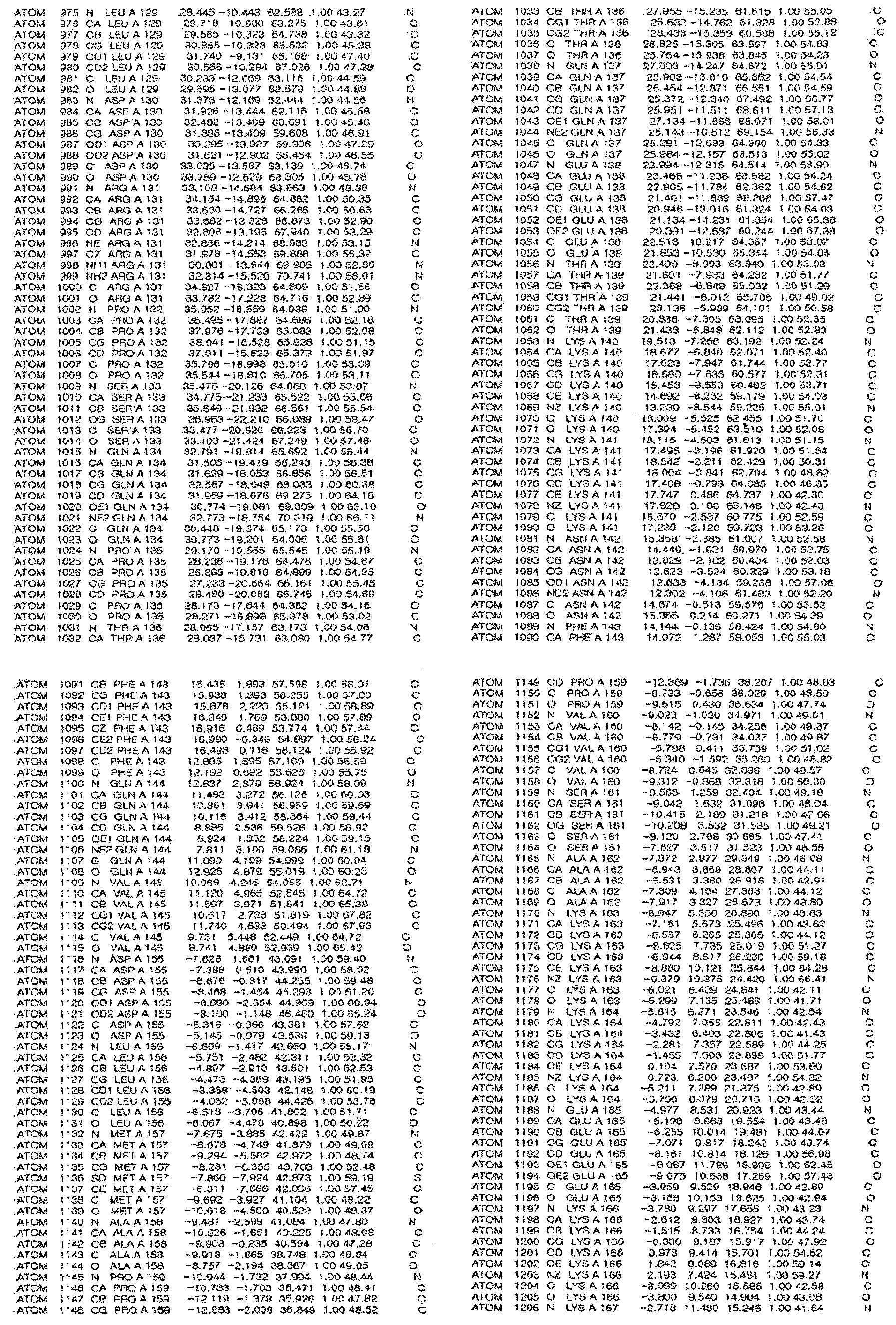
[규칙 제91조에 의한 정정 17.09.2013]
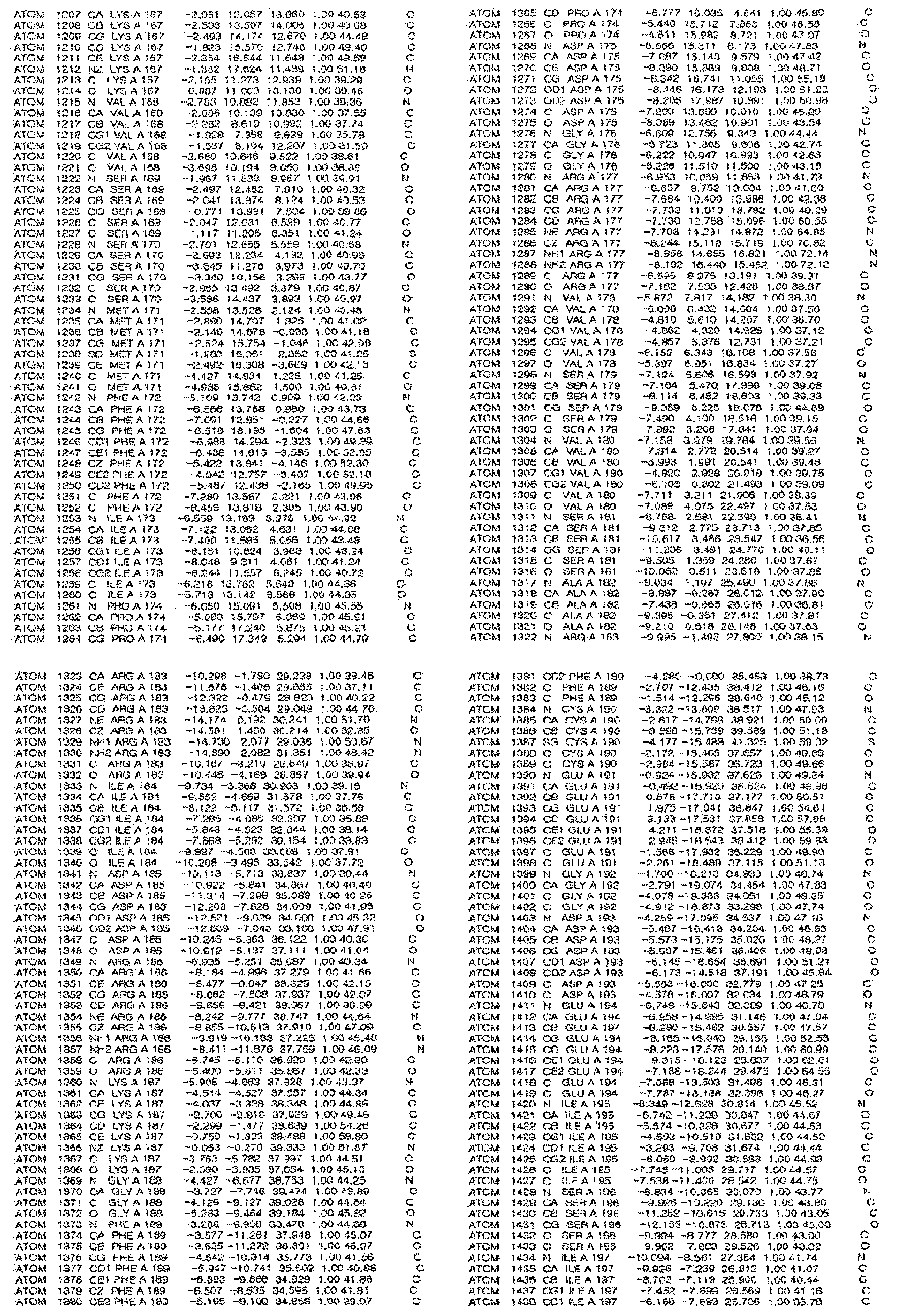
[규칙 제91조에 의한 정정 17.09.2013]
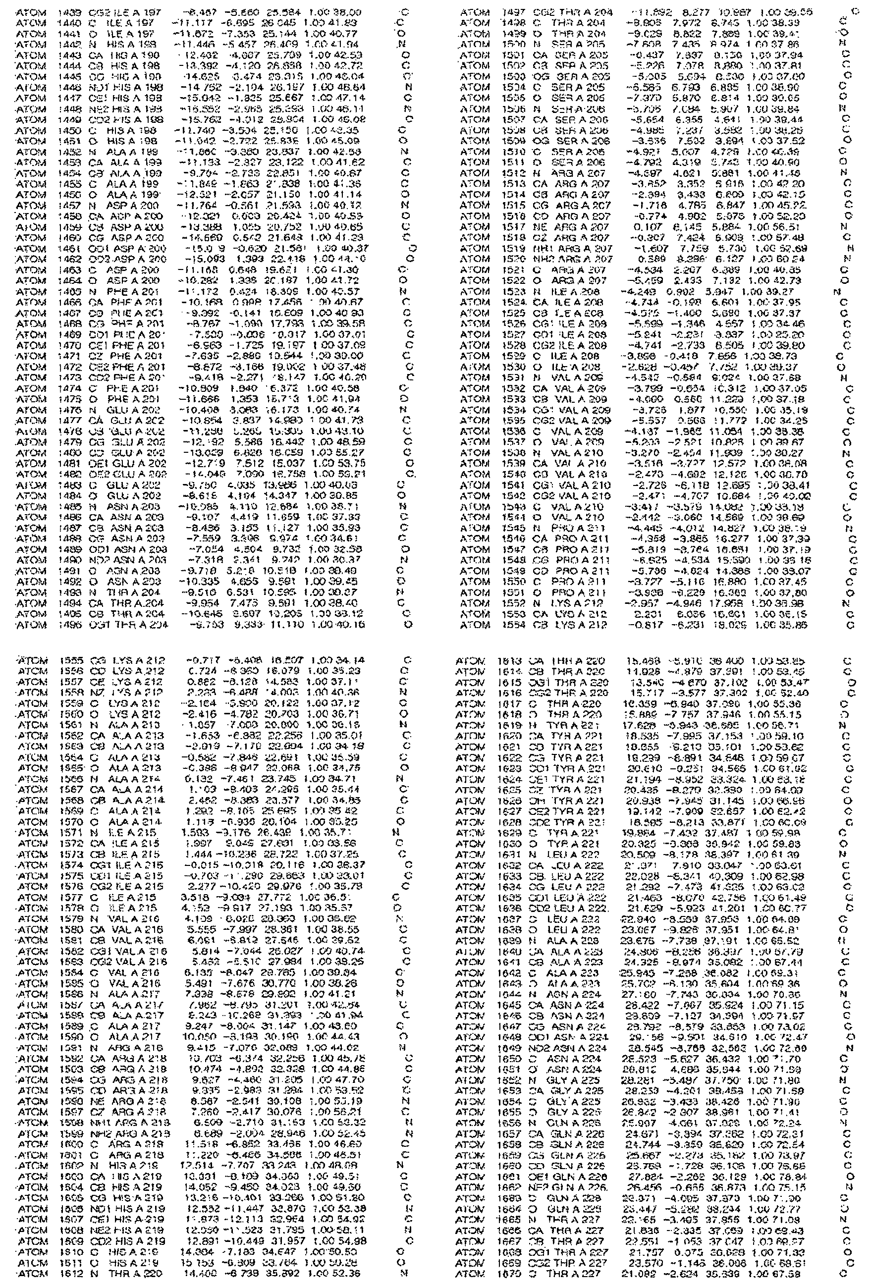
[규칙 제91조에 의한 정정 17.09.2013]
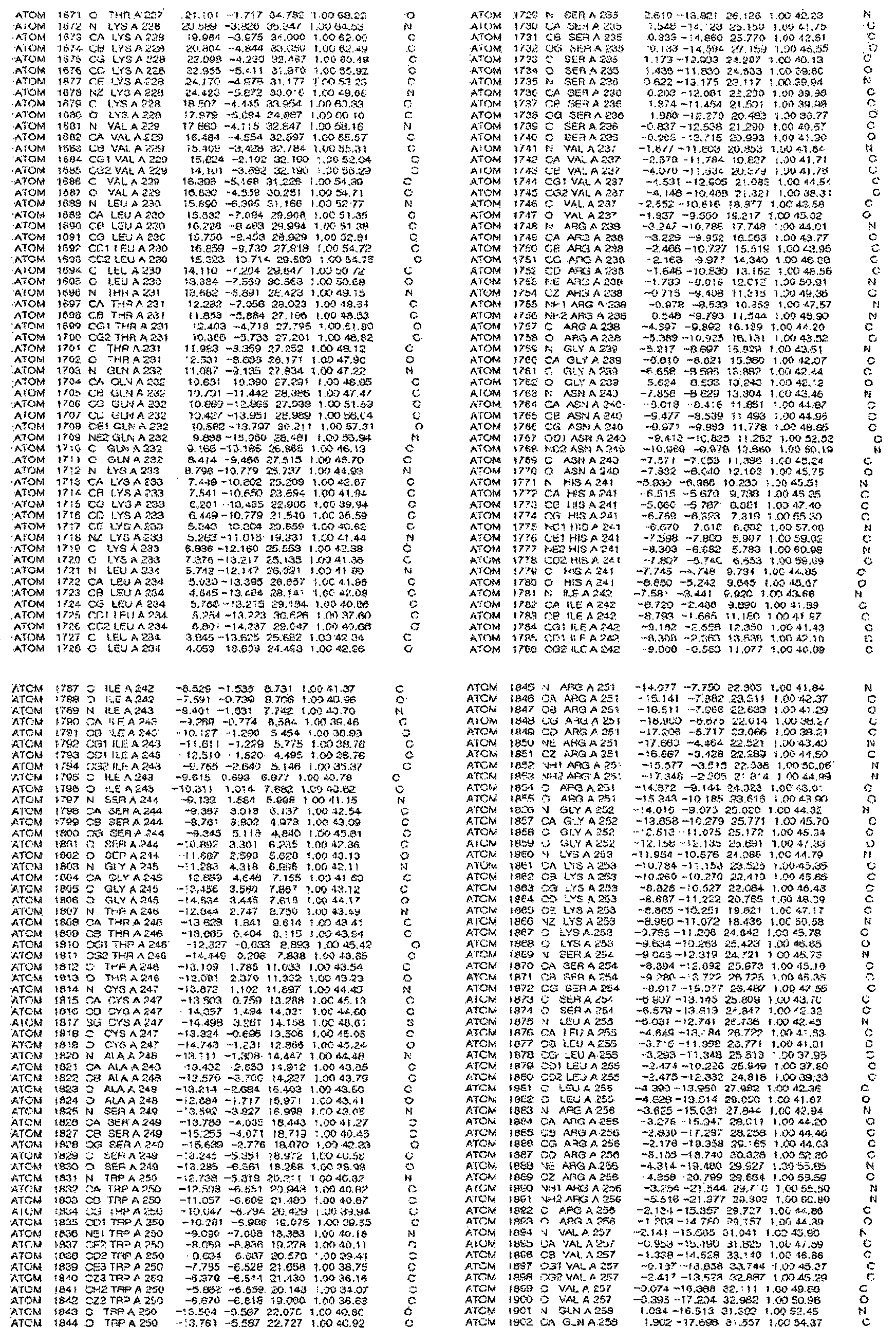
[규칙 제91조에 의한 정정 17.09.2013]
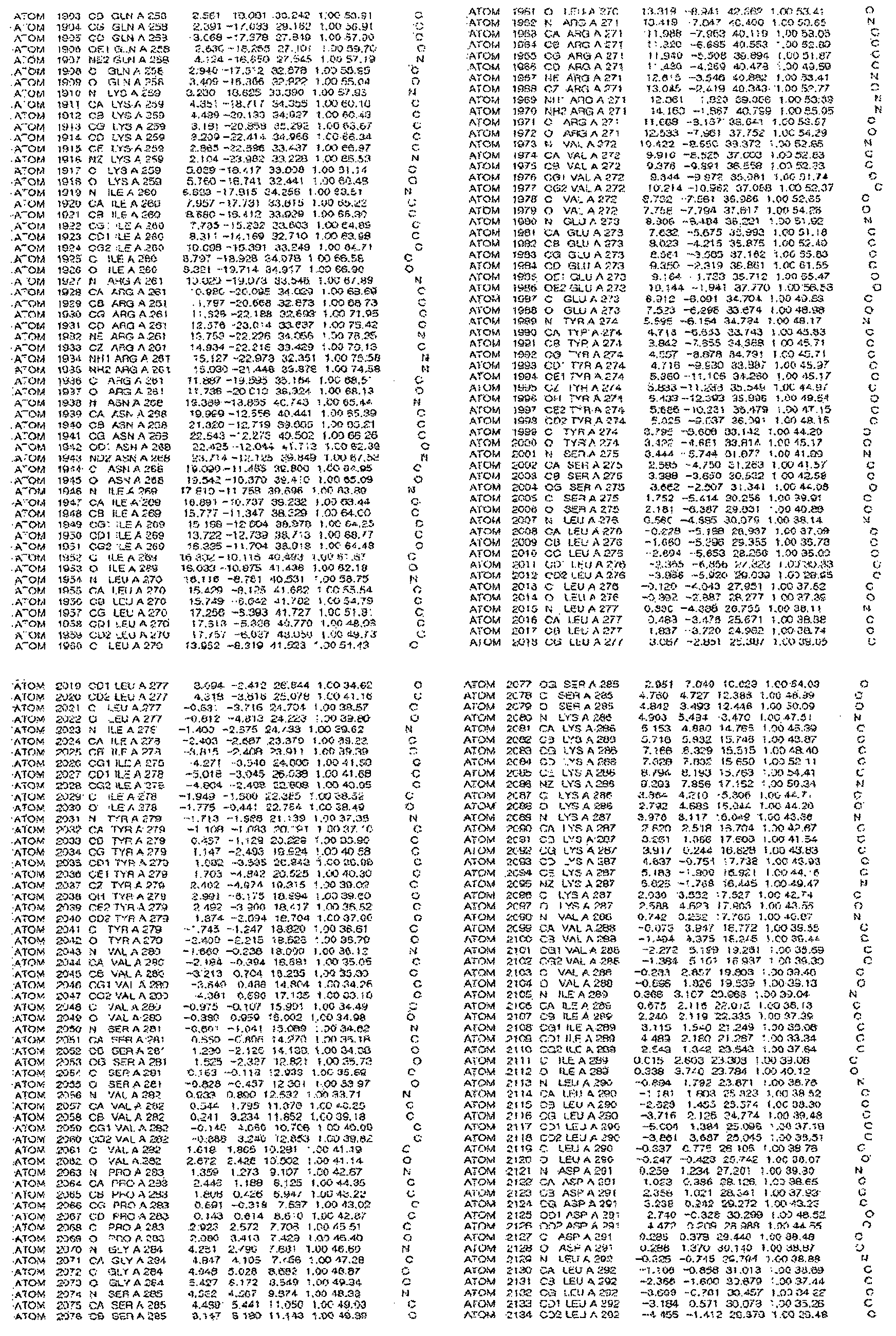
[규칙 제91조에 의한 정정 17.09.2013]
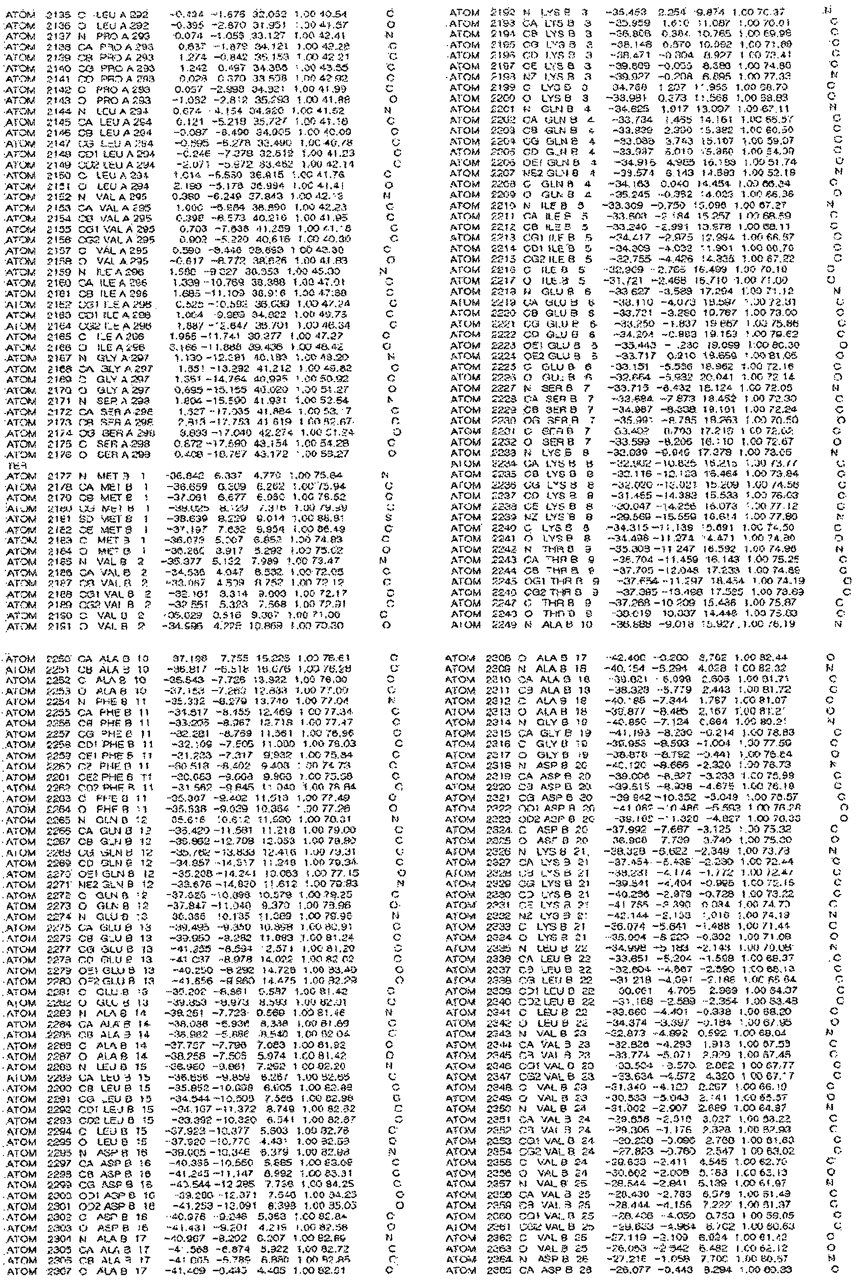
[규칙 제91조에 의한 정정 17.09.2013]
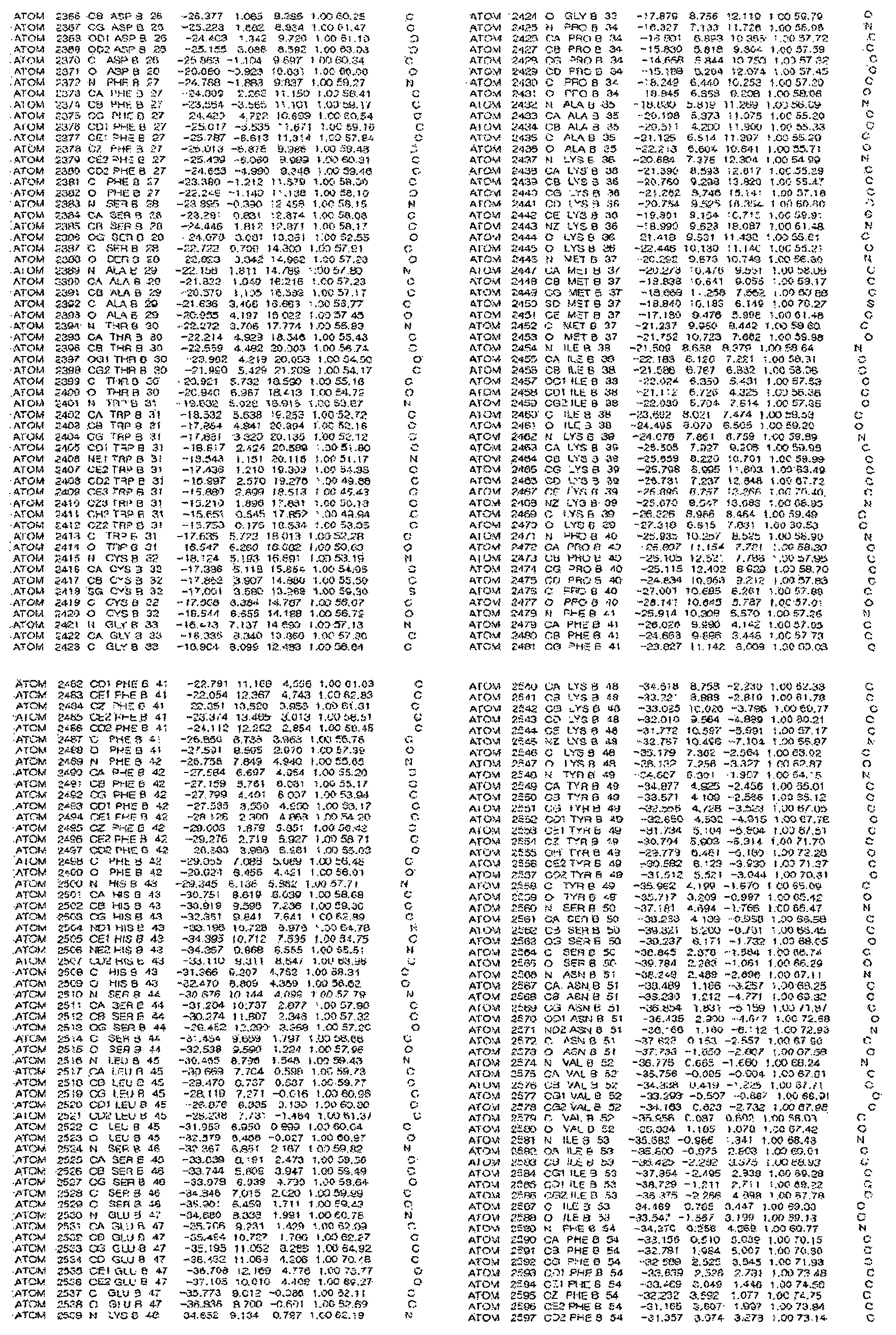
[규칙 제91조에 의한 정정 17.09.2013]
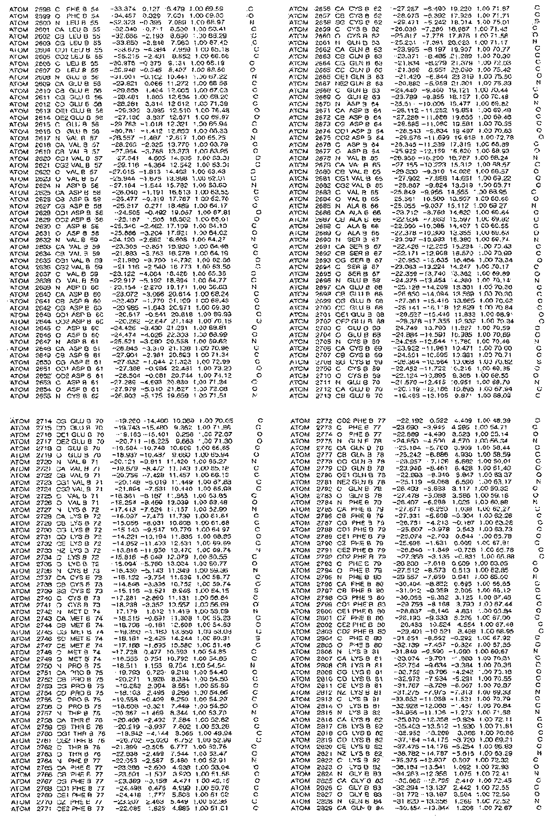
[규칙 제91조에 의한 정정 17.09.2013]

[규칙 제91조에 의한 정정 17.09.2013]
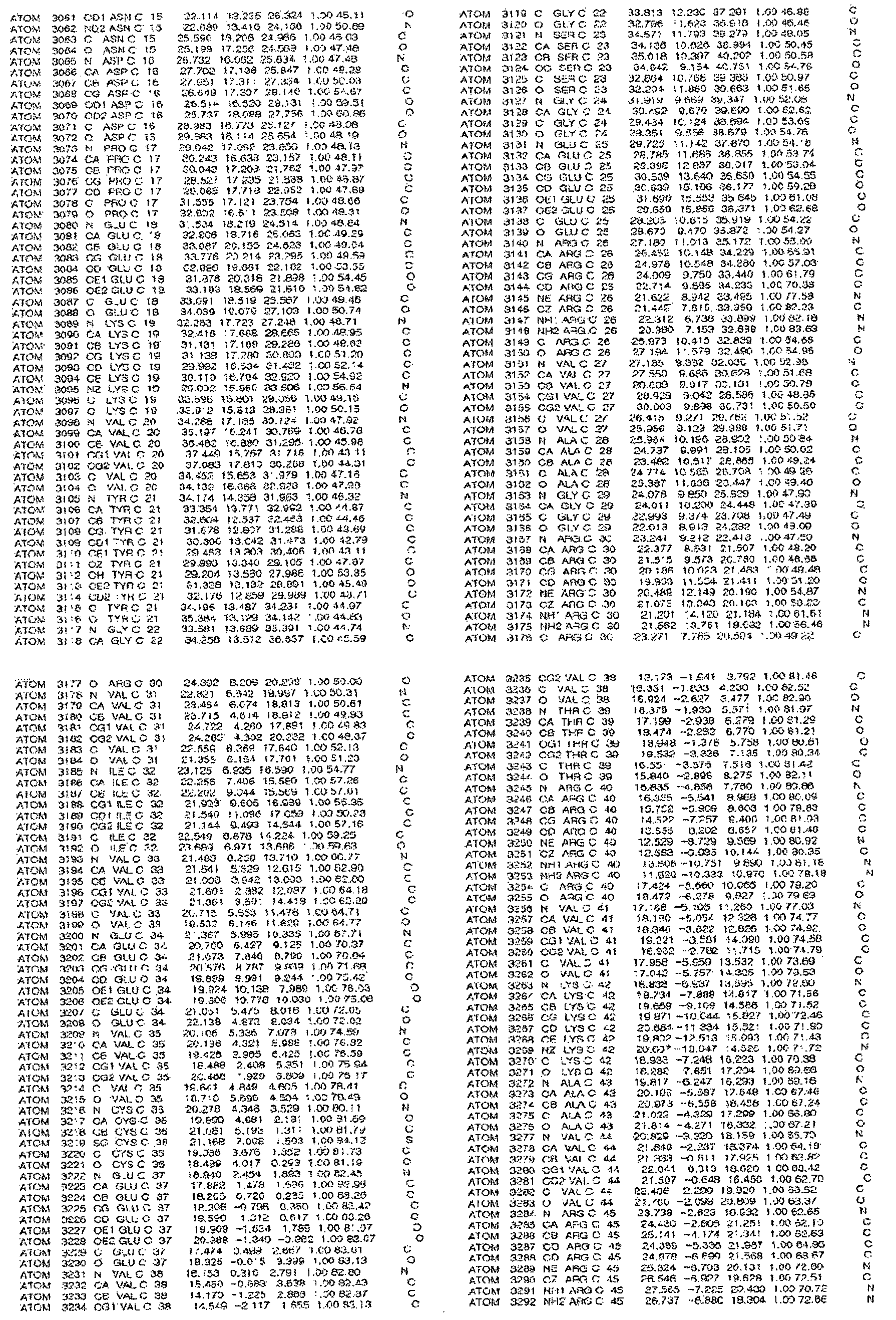
[규칙 제91조에 의한 정정 17.09.2013]
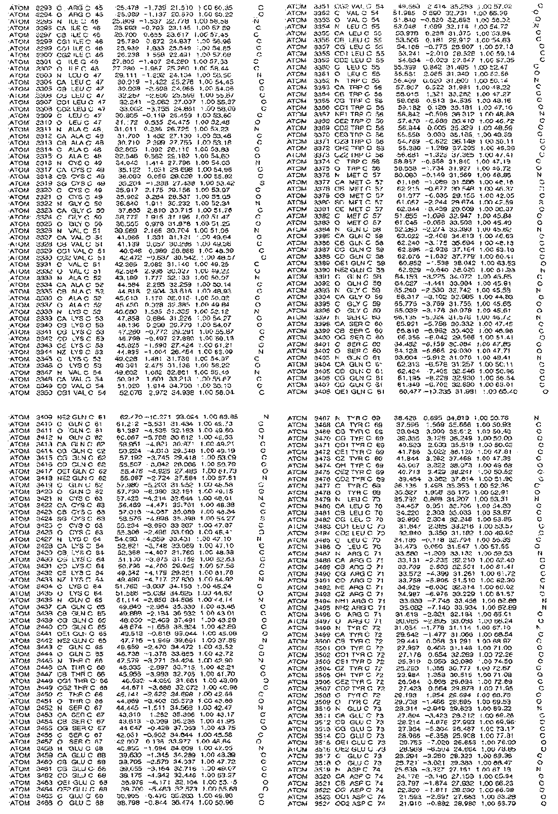
[규칙 제91조에 의한 정정 17.09.2013]
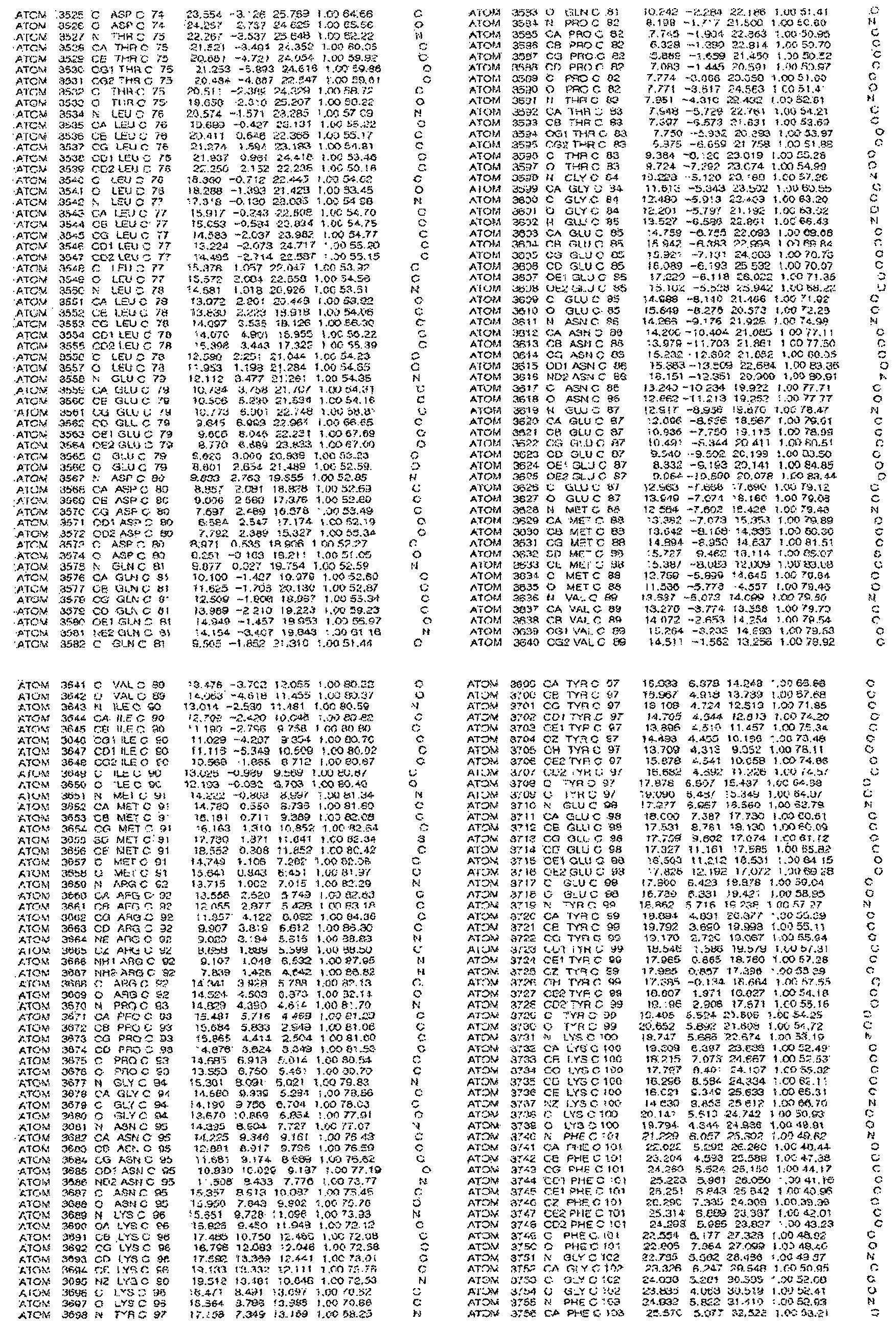
[규칙 제91조에 의한 정정 17.09.2013]
[규칙 제91조에 의한 정정 17.09.2013]
[규칙 제91조에 의한 정정 17.09.2013]
[규칙 제91조에 의한 정정 17.09.2013]
[규칙 제91조에 의한 정정 17.09.2013]
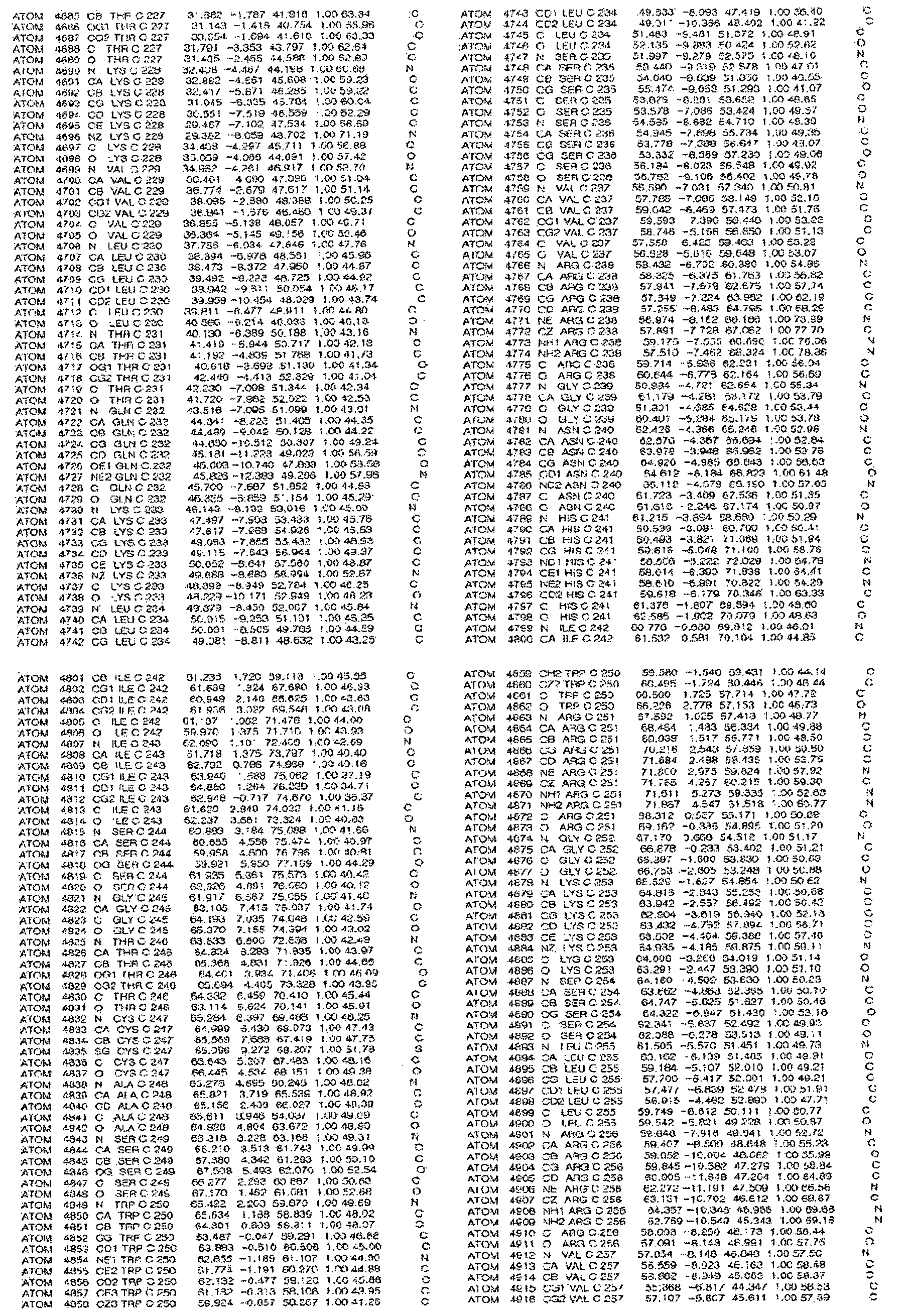
[규칙 제91조에 의한 정정 17.09.2013]
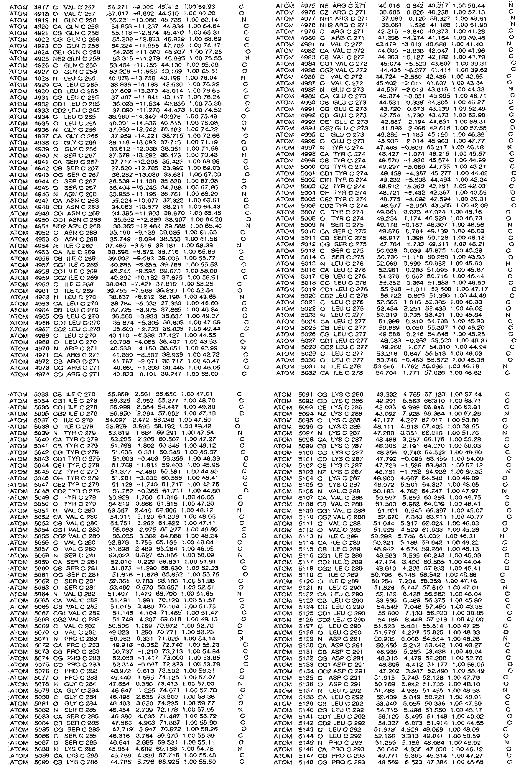
[규칙 제91조에 의한 정정 17.09.2013]
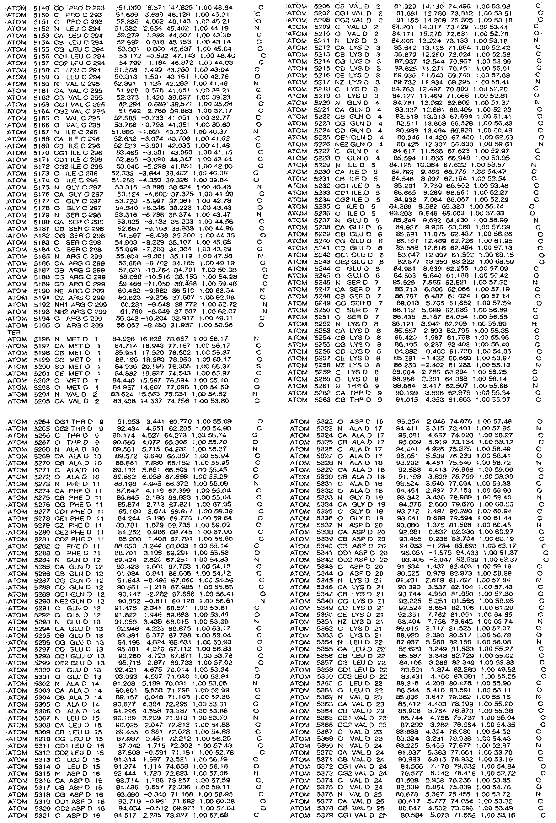
[규칙 제91조에 의한 정정 17.09.2013]
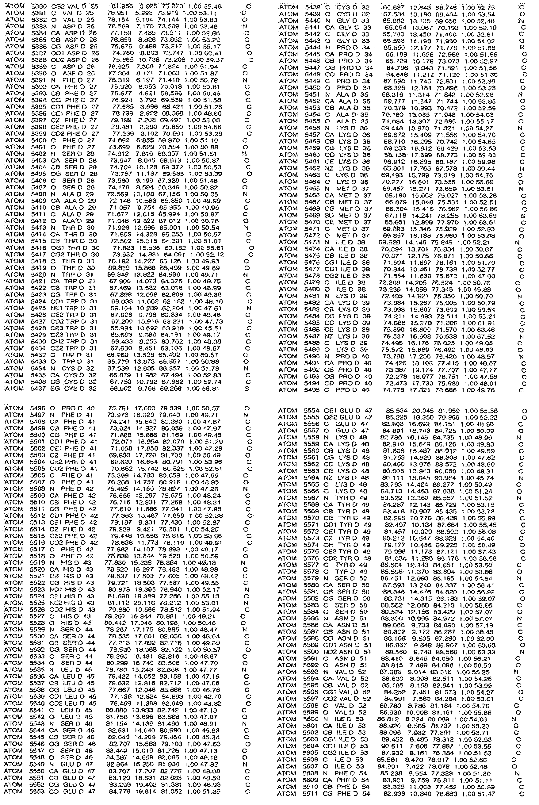
[규칙 제91조에 의한 정정 17.09.2013]

[규칙 제91조에 의한 정정 17.09.2013]
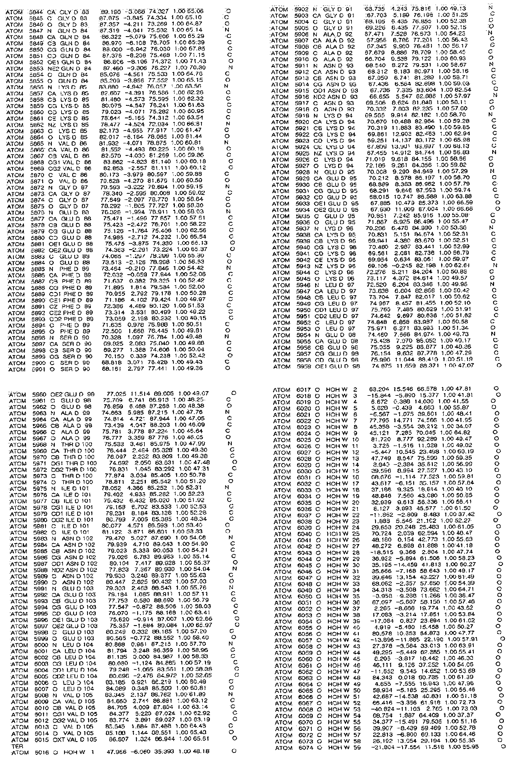
실시예 7: 핵자기공명 분광학(NMR Spectroscopy)
NMR 상호작용 실험(NMR interaction experiments)은 5mM DTT로 환원시키고, 여분의 DTT를 제거하기 위해 상호작용 확인 전에 50mM 인산 칼륨 완충액(pH 6.6)에 투석(dialysis)시킨, 라벨하지 않은(unlabeled) TRX 또는 이의 돌연변이를 추가한 다음, 15N-라벨된 N-TXNIP의 1H-15N NMR HSQC 스펙트럼를 기록함으로서 실시하였다.
모든 단백질의 1H-15N NMR HSQC 스펙트럼은 한국기초과학연구원(오창, 한국)에 있는 Bruker 900 MHz 핵자기공명분광기로 측정하였다. NMR 측정은 25℃에서 10% D2O를 포함하는 50mM 인산 칼륨(pH 6.6)에 0.3mM 15N-라벨된 단백질로 수행하였다. 모든 NMR의 스펙트럼은 Topspin 2.1로 처리하여 프로그램 SPARKY 3.1으로 분석하였다.
실시예 8: TRX 활성 어세이
TRX 활성은 인슐린 이황화 환원 분석 방법을 사용하여 분석하였다. 일시적으로 형질전환시킨 HEK 293T세포는 20mM HEPES(pH 7.9), 100mM KCl, 300mM NaCl, 10mM EDTA, 0.1% Nonidet P-40 및 단백질분해효소 억제제를 함유하는 용해 버퍼로 용해시켰다. TRX를 환원시키기 위하여, 세포 추출물 20㎍을 총용량 70㎕ 속에 50mM HEPES(pH 7.6), 1mM EDTA, 1㎎/㎖ BSA 그리고 2 mM DTT가 함유된 2㎕ DTT 활성 완충용액과 함께 37℃에서 20분 동안 배양하였다. 1M HEPES(pH 7.6) 200㎕, 0.2M EDTA 40㎕, NADPH(40㎎/㎖) 40㎕ 및 인슐린(10㎎/㎖) 500㎕을 포함하는 반응 혼합물의 40㎕을 추가하였다. 랫트 thioredoxin 환원효소(100 U/㎖)를 반응에 추가하고, 37℃에서 20분 동안 배양하였다. 반응은 6M guanidine-HCl과 1mM DNTB(3-carboxy-4-nitrophenyl disulfide)를 추가하여 중단시키고, 412nm에서 흡광도를 측정하였다.
실시예 9: ROS(Reactive oxygen species) 어세이
비기능성 시스테인에 의한 응집(aggregation)을 회피하기 위하여 TRX 및 T-TXNIP(C120S/C170S/C205S/C267S) 복합체를 사용하였다. 0.05mM 농도에서 상기 복합체는 37℃에서 50mM Tris-HCl(pH 8.0), 500mM NaCl 및 10% 글리세롤에서 0 내지 10mM 농도 범위의 H2O2와 함께 배양하였다. 또한, TRX(C37A) 및 T-TXNIP(C36S/C49S/C120S/C170S/C205S/C267S) 복합체를 TRX 이합체 형성 (dimerization) 및 비특이적 응집(non-specific aggregation)을 피하기 위해서 사용하였다. 상기 복합체는 37℃에서 50mM Tris-HCl(pH 8.0), 500mM NaCl 및 10% 글리세롤에 0 내지 3mM 농도의 디아미드(diamide)와 배양하였다. 30분간 반응한 후, 상기 시료를 상온에서 1㎖/min 유속으로 Acta purifier FPLC system에 장착된 HiLoadTM 16/60 Superdex 75 겔 여과 컬럼(GE Healthcare)에 주입하였다. TRX Cys35의 TRX 및 TXNIP 간의 분리에서의 역할을 규명하기 위하여, TRX(C35A)-T-TXNIP(C120S/C170S/C205S/C267S) 복합체를 3.3mM 디아미드와 상온에서 1시간 동안 반응시키고, 이를 SuperdexTM 75 10/300 GL 겔 여과 컬럼에 주입하였다. 모든 분획으로 SDS-PAGE를 수행하였다.
실시예 10: 질량 분석법
실시예 10-1: 질량 분석을 위한 시료의 제조
질량 분석기로 TXNIP 분자 간의 프로토머 간의 이황화 결합을 질량 분석기로 in vivo에서 분석하기 위하여, HEK 293T 세포를 GST와 융합된 TXNIP 단백질을 발현하는 발현 벡터로 형질전환시키고, 24시간 후 형질전환된 세포를 수집하고 이를 완전 프로테아제 억제제 칵테일(complete protease inhibitor cocktail, Roche, 0.5% Triton X-100, 150 mM NaCl, 10% 글리세롤 및 20 mM HEPES (pH 7.2))를 포함하는 용해 완충액으로 용해시켰다. 그 다음, 4℃에서 30분간 반응시킨 다음, 용해물을 16,000g에서 20분간 원심분리하였다. 그 다음, 상층액을 4℃에서 Glutathione Sepharose 4FF(GE healthcare)와 밤새 반응시키고, 10,000g에서 5분간 원심분리하였다. 그 다음, 상층액을 제거하고 레진은 용해 완충액으로 5회 세척하였다. 레진에 결합한 단백질은 LDS-PAGE 로딩 완충액(life technologies)으로 끓여서 용출시켰다.
in vitro에서 질량 분석기로 분석하기 위하여, 정제된 TRX(C73A)-T-TXNIP(C36S/C49S/C120S/C170S/C205S/C267S) 복합체를 100mM DTT로 1시간 동안 상온에서 반응시키고, AKTA purifier FPLC 시스템으로 인스톨된 SuperdexTM 200 10/300 GL 겔 여과 컬럼에 주입하여 잔여 단백질 복합체 및 TRX(C73A)로부터 T-TXNIP(C36S/C49S/C120S/C170S/C205S/C267S)를 수득하였다. T-TXNIP(C36S/C49S/C120S/C170S/C205S/C267S)를 포함하는 분획은 50mM Tris-HCl(pH 8.0), 500mM NaCl 및 10% 글리세롤로 투석하여, 공기 산화를 통한 TXNIP 분자 간의 이황화 결합 형성을 유도하였다. 그 다음, 투석된 단백질들은 비환원 조건 하에 SDS-PAGE를 진행하였다.
실시예 10-2: In gel digestion
고분자 TXNIP 복합체를 포함하는 겔 조각을 25 mM 암모늄 바이카보네이트(ammonium bicarbonate) 및 50% 아세토니트릴(acetonitrile) 용액으로 분해시키고, 100% 아세토니트릴로 탈수시키고, 상온에서 건조시켰다. 자유 시스테인의 알킬화는 25mM 암모늄 바이카보네이트(ammonium bicarbonate)에 존재하는 55 mM 아이오도아세트아미이드(iodoacetamide)로 수행하였으며, 암(dark) 조건에서 25℃의 온도에서 1시간 동안 반응하였다. 상기 겔은 25 mM 암모늄 바이카보네이트(ammonium bicarbonate) 및 50% 아세토니트릴(acetonitrile) 용액으로 분해시키고, 100% 아세토니트릴로 탈수시키고, 상온에서 건조시켰다. 겔 조각은 25mM 암모늄 바이카보네이트에 포함된 시퀀싱-등급의 트립신(12.5 ng/μL; Promega, Madison, WI, U.S.A.) 용액으로 재수화하여 37℃에서 16시간 반응시킴으로 단백질을 분해하였다. 그 다음, 상층액을 새 튜브로 옮기고, 겔 조각을 25mM 암모늄 바이카보네이트 내의 50% 아세토니트릴, 0.5% 트리플루오로아세트산(TFA)에 포함된 50% 아세토니트릴 및 0.5% TFA에 포함된 70% 아세토니트릴로 반응시켜 잔여 펩타이드를 순차적으로 추출하였다. 추출한 펩타이드를 모아 진공 증발기(MIVAC DUO, Genevac, Ipswich, England)에서 건조시켰다. 상기 펩타이드를 -20℃에 사용 전까지 보관하였다.
실시예 10-3: 질량 분석
10-2에서 수득한 펩타이드를 0.4% 아세트산에 희석시켜 농도가 1μg/μL가 되도록 하고, 분획(aliquot, ~1μg)을 Eksigent nanoLC-ultra 1D plus system 하의 역상 Magic C18aq 컬럼(15 cm × 75 μm)에 300 nL/min의 유속으로 주입하였다. 상기 컬럼은 사용 전에 95% buffer A(0.1% formic acid in H2O) + 5% buffer B(0.1% formic acid in acetonitrile)로 평형화시켰다. 펩타이드는 100% Buffer B에서 40% Buffer B로 40분간 선형 구배(linear gradient)로 용출시켰다. 상기 HPLC 시스템은 Q Exactive quadrupole 질량 분석기(Thermo Scientific, San Jose, CA, USA)로 커플링시켰다. 상기 Q Exactive 기기는 데이터 의존적 모드로 작동시켰다. Survey full-scan MS 스펙트럼(m/z 300-2000)이 75,000의 해상도(resolution)에서 Orbitrap에서 회득되었다. 전하 상태 ≥ 2인 MS1 스캔에서 수득된 12개의 가장 강한 이온의 MS/MS 스펙트럼은 하기와 같은 옵션으로 획득되었다: 해상도, 17,500; 분리 폭(isolation width), 2 m/z; 정상화된 충돌 에너지(normalized collision energy), 27%; 다이나믹 배제 기간(dynamic exclusion duration), 30s. 이온 선택 역치(ion selection threshold), 4.00E+03 counts.
실시예 10-4: 데이터 검색 및 비준(validation)
획득된 MS/MS 스펙트럼은 SwissProt 데이터 베이스에 대한 X!Tandem(open source software, available from http://www.proteome.ca/opensource.html)을 사용하여 검색하였다. 요약하면, 펩타이드 분자량 오차범위는 ± 15 ppm으로 설정하였다. 절단 특이성은 트립신으로 설정하여, one missed cleavage를 최대로 하였다. 메티오닌 산화(+15.9949 Da) 및 카바이미도메틸화된 시스테인(carbamidomethylated cysteine)(+57.0215 Da)의 여러 변형을 허용하였다. 펩타이드 배치는 Institute for Systems Biology(TPP, version 4.5 RAPTURE rev 2, http://www.proteomecenter.org)에서 제공된 Trans Proteomics Pipeline으로 수행하였다. X!Tandem 검색 결과로 부터, 0.9보다 큰 개연성을 가지는 펩타이드는 차후 의 Protein-Prophet에 포함시켰으며, 0.9 이상의 개연성을 가지는 단백질들을 수집하였다. 그 결과로부터, 오염원들은 제거하였다(예를 들어, 케라틴 및 트립신 아티피셜). 이황화 연결의 규명 분석은 DBond 소프트웨어(version 3.02, http://prix.hanyang.ac.kr/download/dbond.jsp) (PMID: 19902913)로 수행하였다.
실험예 1: TRX-TXNIP 복합체 구조 결정 및 분자 구성 확인
재조합 전장 TXNIP 단백질을 수용성 단백질로 발현시키기 위하여 여러 차례 시도하였으나, TXNIP만 발현되는 경우, 작은 결합력(binding activity)을 나타내는 몰텐 구상 단백질(molten globular protein)로서 동작하였다. 이에, TXNIP 단백질을 구조적으로 변화시켰고, TRX 및 TXNIP을 함께 발현시켜 수용성 단백질로 발현하고자 하였다.
특히, TRX(5개의 시스테인 포함)와 TXNIP(11개의 시스테인 포함)은 체외에서 다루기 힘든 산화 환원 단백질(redox protein)이기 때문에, 본 발명에서는 비특이적인 응집을 방지하고 X선 회절에 적합한 결정체를 생성하기 위하여 TXNIP의 텐덤 어레스틴 유사 도메인(tandem arrestin-like domain) (T-TXNIP, 아미노산 3 내지 317)에 여러 시스테인 돌연변이를 유도하였고, 두 단백질 간의 상호작용 이후에 더 이상의 반응을 방지하기 위하여 TRX의 35번 시스테인(Cys)을 알라닌(Ala)으로 치환시켰다. TRX(C35A)-T-TXNIP(C120S/C170S/C205S/C267S) 복합체(COM1) TRX(C35A)-T-TXNIP(C170S/C205S/C267S) 복합체(COM2), 이 두 변형 복합체의 결정을 이용하여 X선 회절 실험을 수행하였고, 이들의 구조는 여러 변칙 파장 분산 방법과 함께 분자 교체 방법을 사용하여 구축하였다. 각각 2.0Å 및 2.7Å의 해상도에서 정제한 COM1 및 COM2의 결정체 구조를 도 5에 나타내었다.
또한, 천연형 T-TXNIP 및 이의 돌연변이체의 TRX에 대한 결합 능력, 및 TRX 활성 저해능력을 확인한 결과, 전장 TXNIP(full-lengh TXNIP)와 비교하였을 때, 결합 능력 및 TRX 활성 저해 능력이 크게 다르지 않는 결과를 나타내었다(도 6 B 및 C). 이와 같은 결과는 TXNIP 단백질에 돌연변이를 유도한 TXNIP 변형 단백질을 이용한 COM1 및 COM2의 결정체 구조는 TRX와 TXNIP 사이의 상호작용을 통한 TRX-TXNIP 복합체 결정체 구조를 나타내는 것으로 볼 수 있음을 나타내는 결과이다.
COM1과 COM2의 구조는 기본적으로 일치하며, 각각의 비대칭 단위로 두 개의 TRX 및 T-TXNIP 간의 이형이합체(heterodimeric complex)를 포함하며, 두 T-TXNIP 분자 간에 큰 인터페이스(interface)를 가진다(도 5 A 및 B). 그러나 TRX-T-TXNIP, TRX(C35A)-T-TXNIP(C170S/C205S/C267S) 및 TRX(C35A)-T-TXNIP(C120S/C170S/C205S/C267S)에 대한 크기 배제 크로마토그래피 결과에 따르면, 상기 복합체들은 모두 단일 이형 이합체로 용액 내에 존재하였다(도 6 D). 따라서, 결정체 구조에서는 두 T-TXNIP 분자 간의 인터페이스가 단지 큰 결정 접촉(Crystal contact)를 구성하고 있을 뿐임을 나타낸다.
COM1 및 COM2의 구조는 기본적으로 일치하고 TRX 및 T-TXNIP 복합체는 용액 내에서 단일 이형 이합체로서 존재하기 때문에, COM1의 T-TXNIP(chain C)와 TRX(chain D) 복합체(도 6 E)를 대표적인 구조로 선택하였다. 상기 이형이합체의 최종 구조에서는, T-TXNIP의 발현 단백질의 3-317 잔기 중 8-299 잔기; 및 TRX의 모든 잔기를 포함하고 있다. T-TXNIP의 148 내지 153 잔기 및 260 내지 265 잔기가 전자밀도 지도에서 보이지 않기 때문에, 이 부분은 최종 모델에 포함시키지 않았다; 상기 영역들은 매우 유연성(flexibility)을 가지고 있다.
그 결과, 도 6 E에 나타낸 바와 같이, T-TXNIP는 전부 β 가닥(strand)들로 이루어져 있으며, 이들은 두 개의 깊게 파인 β-샌드위치 도메인(two deeply curved β-sandwich domain)을 형성한다. 또한, T-TXNIP는 β1 내지 β11(N-TXNIP, residues 8-147)로 이루어진 N-말단 도메인; 및 β12 내지 β20(C-TXNIP, residues 154-299)로 이루어진 C-말단 도메인으로 이루어져 있다. T-TXNIP는 C-TXNIP가 N-TXNIP에 상대적으로 뒤집어진 형태의 pseudo-2-fold symmetry로 길쭉한 S형 도메인 배열(elongated S-shaped domain arrangement)로 접혀 있는 형태이다. 상기 두 도메인은 60 Cα 원자에 대해서 1.88의 r.m.s.d.(root mean-square deviation)으로 겹쳐질 수 있다(도 7 A). T-TXNIP의 전체 구조는 mVPS26B(mouse vacuolar protein sorting-assciated protein 26B, PDB ID code 3LH8)를 포함하는 어레스틴 슈퍼 패밀리에 속하는 단백질과 13%의 아미노산 상동성을 가지고 있음에도 불구하고, 구조적으로 매우 유사하였다. 그러나, TRX와 상호 작용하는 T-TXNIP의 도메인 배열은, ω형 도메인을 가지고 있다고 보고된 모든 다른 어레스틴 구조의 것과 완전히 차이를 보였다(도 8 A). 따라서 TXNIP의 각각의 도메인인 N-TXNIP(r.m.s.d.=1.33 for 82 Cα atoms; Z=12.3) 및 C-TXNIP(r.m.s.d.=1.42 for 84 Cα atoms; Z=12.5)는 각각 N-mVPS26B와 C-mVPS26B로 겹쳐질 수 있는 반면(도 8 C), 두 도메인으로 구성된 Tandem T-TXNIP은 mVPS26B과 겹쳐질 수 없었다(도 8 B). 또한 구조분석에 따르면, T-TXNIP이 전기적 편광임을 나타내었다(도 8 D). 특히 C-TXNIP 도메인의 곡선 β-샌드위치 내부는 우세한 염기성 영역(basic region)을 포함하는 반면, N-TXNIP는 음성 정전기 전위(negative electrostatic potential)를 가지고 있었다. TRX의 경우, T-TXNIP와의 복합체 내에 존재하는 TRX 구조는 기존에 보고된 유리 TRX 구조(free TRX structure)와 큰 차이를 나타내지 않았다(도 7 B).
실험예 2: TRX-TXNIP 상호작용 분석
TXNIP의 Cys247는 TRX Cys32와 상호작용을 하는데 가장 중요한 잔기로 알려져 있다. 따라서, TRX Cys32 및 T-TXNIP Cys247 간의 분자간의 이황화 결합(intermolecular disulfide bond)에 대하여 연구하는 것은 TXNIP에 의한 TRX 억제를 이해하는 것에 도움을 준다(도 9 A). 도 9에 나타낸 TXNIP 및 TRX 결합체 구조에 따르면, 상기 이황화 결합은 두 단백질 사이의 상호작용 인터페이스(Interaction interface) 중심부에 자리하고 있었다. T-TXNIP의 β18 가닥은 β15와 함께 TRX 및 TXNIP 간의 상호작용에 있어 중요한 구성요소이며, 결합된 β18의 전체적인 구조는 연장되어 있고, TRX의 활성 부위(Active site)에 위치한 잔기들에 의해 형성된 틈(cleft)이 존재한다. 또한, TRX 및 T-TXNIP의 β18 가닥의 잔기 간의 분자간 백본-백본 상호작용(intermolecular backbone-backbone interaction)(TRX Ala92 및 T-TXNIP Gly245, 및 TRX Met74 및 T-TXNIP Cys247)이 분자간의 이황화 결합을 안정화시켰다. 또한, 상기 단백질 간의 분자간 수소 결합과 소수성 상호작용이 존재하였다. 특히, T-TXNIP Arg251의 측쇄 아미노산 기는 TRX Asp60의 측쇄 카복실기와 염교(Salt bridge)를 형성하여, TRX와 T-TXNIP사이의 상호작용을 더욱 강화시켰다(도 9 B). 상기와 같은 상호작용 유형은 세포기질의 TRX에 의해 매개된 이황화 결합 환원과 관련이 있는 NF-κB 및 Ref-1 펩타이드와 TRX 복합체에서의 패턴과 아주 유사하였다(도 9 C). 또한 분자간의 이황화 결합을 안정화시키는 상기 분자간 백본-백본 상호작용은 각 복합체 구조에서 보존되어 있었다.
실험예 3: TXNIP의 Head-to-tail 형태의 인터프로토머(interprotomer) 이황화 결합 형성 확인
TXNIP과 TRX간의 상호작용은 Cys63와 Cys247 간의 분자내 이황화 결합(Inramolecular disulfide bond)을 포함하는 것으로 여겨지는 산화된 TXNIP 및; 환원된 TRX를 필요로 하였다. 도 5에 나타낸 T-TXNIP 구조에 따르면, Cys63는 Cys247와 39Å 밖에 떨어져 있지 않기 때문에, 이러한 거리를 감안할 때, 상기 두 잔기 간에 분자내 이황화 결합을 형성하는 것은 매우 힘든 것으로 판단되었다. 따라서, Cys63과 Cys247은 분자 내의 이황화 결합을 형성하는 것보다 TXNIP 분자 간 프로토머(intermolecular protomer)의 이황화 결합을 형성할 수 있을 것으로 생각되었다. 상기와 같은 가설을 입증하기 위하여 하기와 같은 실험을 수행하였다.
우선, TXNIP 분자 간의 in vivo 인터프로토머 상호작용을 HA-tagged TXNIP, FLAG-tagged T-TXNIP를 발현하는 플라스미드가 형질감염된 293T 세포로 조사하였다. 이때, 비특이적 추가적인 산화환원 반응이 세포 용해중 일어나는 것을 막기 위하여 시스테인-블로킹 물질인 아이오도아세트아마이드를 포함하는 용해 완충액을 사용하였다. 그 결과를 도 9D에 나타내었다.
그 결과, 도 9에 나타난 배와 같이, TXNIP 분자 간의 인터프로토머 상호작용이 관찰되었으며(왼쪽), T-TXNIP 고분자 복합체는 산화환원 조건에 의존적으로 검출되었다(오른쪽).
또한, FLAG이 태그된 TXNIP 및 HA가 태그된 TXNIP를 발현하는 플라스미드로 293T 세포를 형질감염시켜 얻은 용해물(Lysate)을 이용하여 공동-면역침강실험(Co-immunoprecipitation)을 수행하였고, 그 결과를 도 9E에 나타내었다. 프로토머간의 이황화 결합에 관여하는 도메인을 확인하기 위하여, Flag가 태그된 Deletion Mutant들을 제작하였고, HA가 태그된 TXNIP를 면역침강시키기 위해 항-HA항체를 사용하였다.
그 결과, 도 9E (왼쪽)에 나타낸 바와 같이, 항-HA 항체를 이용한 면역블롯 결과는 N-말단 및 C-말단 TXNIP 도메인 모두가 프로토머 간 이황화 결합 형성에 필요함을 나타내었다.
또한, GST가 융합되거나 FLAG가 태그된 TXNIP를 사용하여 프로토머 간 이황화 결합 형성에 있어서 C-말단 도메인에 위치한 Cys247 및 N-말단 도메인에 위치한 Cys63의 역할을 확인한 결과, GST가 융합된 TXNIP의 247번 시스테인을 세린으로 치환시킨 경우와 달리, GST가 융합되거나 FLAG가 태그된 TXNIP 모두를 247번 시스테인의 세린으로의 치환시킨 경우, 프로토머 간의 이황화 결합이 형성되지 못하였다(도 9E 오른쪽). 동일한 결과가, 63번 시스테인을 세린으로 치환시킨 경우 얻어졌다. 구조 분석 결과와 상기와 같은 결과는 모두, TXNIP 분자에 존재하는 Cys63및 Cys247 간의 head-to-tail 프로토머 간의 이황화 결합이 in vivo에 존재함을 나타내는 것이다.
이에 더하여, Cys63 및 Cys247 간의 인터프로토머 이황화 결합을 분석하기 위하여 질량 분석을 수행하였다. 이를 위하여, 비특이적 공유적인 올리고머화가 방지된, T-TXNIP(C36S/C49S/C120S/C170S/C205S/C267S) 단백질을 TRX(C73A)-T-TXNIP(C36S/C49S/C120S/C170S/C205S/C267S) 복합체로부터 분리하였고(도 9g), 이를 이용하여 인터프로토머 상호작용하는 TXNIP 분자를 in vitro에서 유도하였고, 비 환원 조건 하에서 SDS-PAGE를 수행하여 분획화된 단백질 밴드를 질량 분석에 사용하였다(도 9h). ~32kDa 및 ~60kDa의 두 단백질 밴드(각각 단량체 형태의 T-TXNIP 돌연변이체 및 인터프로토머 상호작용 단백질 복합체)에 대하여 이황화 결합 및 자유 시스테인을 질량 분석기로 분석한 결과를 하기 표 12에 나타내었다.
표 12
| The number of spectra | |||
| ~60 kDa | ~32 kDa | ||
| Disulfide bond | C63-C190 | 1 | 93 |
| C63-C247 | 5 | 3 | |
| C63-C63 | 1 | 1 | |
| C190-C247 | 1 | 3 | |
| C247-C247 | 1 | 2 | |
| Total | 9 | 102 | |
| Free cysteine | C63 | 4 | 5 |
| C190 | 0 | 42 | |
| C247 | 2 | 5 | |
| Total | 6 | 52 | |
상기 표 12에 나타낸 바와 같이, 이황화 결합 위치는 Dbond score > 20인, Cys63-Cys190, Cys63-Cys247, Cys63-Cys63, Cys190-Cys247 및 Cys247-Cys247로 규명되었다. 그러나, 이황화 결합 위치의 분포는 상기 두 밴드 간에 매우 다르게 나타났다. ~60kDa 밴드에서는, Cys63-Cys247이 주요 이황화 결합을 포함하는 5 스펙트럼으로 규명된 반면, ~32kDa에서는 Cys63-Cys190이 총 102개 중 93개의 스펙트럼으로 주요하게 나타났다. 또한, 알킬화 후 카바미도메틸 시스테인(carbamidomethyl cysteines)으로 보이는 자유 시스테인의 경우에도 매우 다른 분포를 나타내었다.
이러한 결과는, Cys63의 대부분이 ~32kDa 밴드(monomeric T-TXNIP)에서는 Cys190과 이황화 결합을 대부분 형성하는 반면, ~60kDa 밴드(interprotomer-interacting T-TXNIP molecule)에서는 Cys247과 이황화 결합을 형성함을 나타내는 것이며, 또한 많은 Cys190이 ~32kDa 밴드에서는 자유 시스테인으로 존재하고 있음을 나타낸다.
또한, ~32kDa 밴드의 Cys63-Cys190; 및 ~60kDa 밴드의 Cys63-Cys247에 대한 대표적인 MS/MS 스펙트럼을 도 9i 및 j에 각각 나타내었다. 또한, m/z 1759.77인, 두 배 전하를 띤 [M+2H]+ 펩타이드 이온(도 9i) 및 m/z 1353.64인, 두 배 전하를 띤 [M+2H]+ 펩타이드 이온(도 9j)는 HCD(higher-energy collisional dissociation)로 단편화하였다. 그 결과, 아미드 결합 절단에 의한 스탠다드 백본 단편들이 매우 많이 존재했고, S-S 또는 C-S 결합 절단 반응으로 형성된 과황화물 이온(P+32) 역시 본 스펙트럼에서 검출되었다.
본 결과를 종합하면, Cys63 및 Cys247 간의 head-to-tail 인터프로토머 이황화 결합이 TXNIP 분자 간에 존재함을 시사한다.
도 6 D에 나타난 바와 같이, TRX가 결합된 TXNIP는 용액 내에서 이형 이합체 형태로 우세하게 존재하였다. TRX가 TXNIP의 프로토머 간의 이황화 결합 형성에 미치는 영향을 확인하기 위하여, FLAG 태그된 TXNIP, HA가 태그된 TXNIP 및 FLAG 태그된 TRX를 발현하는 플라스미드로 형질감염시킨 293T 세포 용해물을 사용하여 공동 면역침강분석을 수행하였다. 그 결과, TXNIP의 프로토머 간의 이황화 결합 형성은 TRX의 과발현으로 저해되는 결과를 보였다(도 9 E).
이러한 결과는, 안정적인 TRX-TXNIP 이형 이합체의 형성이 TXNIP 분자 간의 프로토머 간의 이황화 결합과 TRX와의 일시적인 상호작용과 관련이 있음을 나타내는 것이다.
실험예 4: TRX-TXNIP 상호작용에 관여하는 63번 시스테인(Cys63)에 의해 매개되는 이황화 결합 스위칭(switching) 확인
본 발명자들은 도 10에 나타낸 바와 같이, N-말단 TXNIP인 N-TXNIP를 TRX 및 TRX(C35A)와 함께 공동 발현(co-expression)하여, 공동 정제(co-purification)할 수 있음을 확인하였다. TRX와 TXNIP의 상호작용에 있어서 N-TXNIP의 관련성을 알아보기 위하여, 라벨링하지 않은 TRX를 첨가한 후, 15N-labeled N-TXNIP의 일련의 1H-15N HSQC(heteronuclear single quantum coherence) 스펙트럼을 기록함으로서 NMR 적정 실험(NMR-titration experiment)을 수행하였다.
TRX를 첨가하였을 때, N-TXNIP와 TRX 간의 직접적인 상호작용을 나타내는 유의한 화학적 이동(chemical shift)을 일으켰으나(도 10 B), N-TXNIP 및 TRX(C35A) 복합체를 결정화시키지 못하였다. 이러한 결과는 두 단백질 간의 상호작용이 일시적으로 일어나는 것임을 나타내는 것이다.
또한, TRX mutein을 추가하고, 15N-labeled N-TXNIP의 1H-15N HSQC 스펙트럼을 모니터링하여, N-TXNIP와 TRX 간의 상호작용을 조사하였다. C32A 또는 C35A TRX mutein을 15N-labeled N-TXNIP에 첨가하였을 때, 유의한 화학적 이동이 나타나지 않아(도 12), 상기 시스테인 잔기들이 TRX 및 N-TXNIP의 상호작용에 관여하고 있음을 나타내었다. 반대로, C62A, C69A 및 C73A mutant는 야생형 TRX와 유사한 화학적 이동을 가져왔다(도 12).
이황화 결합을 가진 산화된 T-TXNIP이 환원된 TRX과 결합하기 때문에, 이황화 결합이 N-TXNIP와 TRX 간 상호작용과 관련이 있을 것으로 예측되었다. 이와 같은 가설을 실험하기 위하여, 1.6Å 해상도로 N-TXNIP 구조를 결정하고, 이를 도 11에 나타내었다.
그 결과, N-TXNIP 구조는 T-TXNIP의 각 도메인 구조와 아주 유사한 형태를 나타내었다. 그러나 T-TXNIP의 경우와는 달리, Cys63와 Cys120 간의 예상하지 못한 분자 간의 이황화 결합이 존재하였다(도 11 A). COM2 구조에서 Cys63과 Cys120의 거리는 이황화 결합 형성에 아주 큰 영향을 미치는 14.1Å이였다(도 5 B). Cys120는 TRX 결합에 크게 관여하지 않는 것으로 보고된 바 있다. 또한, 다른 종으로부터 TXNIP의 아미노산 서열을 정렬하였을 때, Cys190 및 Cys247이 엄격하게 보존되는 반면, Cys120은 보존되지 않는다는 것을 알 수 있었다(도 13). 따라서, Cys120은 N-TXNIP 구조의 Cys63과 Cys120간의 예상하지 못한 이황화 결합은 결정화에 따른 인공 산물(artifact)일 것이라 예상되었다. Cys63이 안정적인 이황화 결합을 형성하기 위하여 요구하는 시스테인 잔기가 Cys120이 아니라면, 다른 고아(orphan) 시스테인 잔기를 요구할 가능성이 높을 것이라 생각되었다. N-TXNIP의 36번 및 49번 위치에 다른 시스테인 잔기가 존재하나, Cys63에서 이황화 결합 형성을 하기에는 너무나 거리가 먼 49Å 및 22Å 거리에 각각 위치해 있었다(도 11 A). 이와 같은 결과는 TXNIP가 TRX와 상호작용하는 동안, Cys63에 의해 매개되는 형태변화(conformation change)를 겪은 것이라는 것을 제시하는 것이다. N-TXNIP의 Cys63과 Cys120 사이의 상호작용으로 N-TXNIP는 시계방향으로 대략 96°가량 이동하며, β5 와 β6 가닥의 54°아래로 굽혀졌다. 이와 같은 이동은 β5와 β6 가닥의 2차구조 변화를 가져왔다(도 11 B).
또한, β8 내지 β10 지역에 중요한 변화가 있었다(도 11 B). TRX 및 TXNIP의 상호작용에 대한 Cys63 이황화 결합의 중요성을 알아보고자, 15N-labeled N-TXNIP mutein 및 TRX를 가지고 NMR 1H-15N HSQC 실험을 수행하였다. TRX의 존재하에서 N-TXNIP mutein C63S 또는 C120S의 스펙트럼에서 유의한 화학적 이동은 일어나지 않는 결과를 보였다(도 14). 이러한 결과는, TXNIP와 TRX의 상호작용은 TXNIP 분자의 프로토머 간의 Cys63-Cys247 이황화 결합과 TRX 간의 일시적인 상호작용에 의해 개시될 것이라는 것을 뒷받침하는 결과였다.
TXNIP의 Cys63 및 Cys247 간의 프로토머 간 이황화 결합(Interprotomer disulfide bond)과 TRX의 일시적인 상호작용은, 도 11에 나타낸 복합체 구조에서 볼 수 있듯이 TRX Cys32 및 TXNIP Cys247 간의 안정적인 분자간 상호작용을 가져왔다. 따라서, TRX에 의해 TXNIP 분자의 프로토머 간의 상호작용 즉, TXNIP의 Cys63 및 Cys247 간의 이황화 결합이 저해되게 되면, Cys63이 다시 반응성을 가지게 된다. TXNIP 구조의 두드러진 양상 중 하나는 N-TXNIP Cys63 및 C-TXNIP Cys190 간의 도메인 간의 이황화 결합이다(도 11 C). 따라서, 고아가 된 Cys63(Orphaned Cys 63)이 상태를 안정화시키기 위한 결합을 형성하기 위하여 주변의 시스테인 잔기를 즉시 찾아 190번 시스테인과 이황화 결합을 형성시킬 것이라는 것을 나타내는 것이다. 이와 같은 상호작용은 각 도메인의 β-시트의 한쪽 면 사이에, 도메인 간의 인터페이스의 중심부에 위치한 이황화 결합이 자리하고 있는 두 도메인 간에, 밀착 연접(tight junction)을 가져온다. 그런 다음, 이것은 인터페이스에 관여하는 다른 잔기에 의해서 둘러싸이게 되며, 용매로부터 완전히 차단될 수 있다. 이때, 긴 N-말단 가닥 β5와 β6는 C말단 가닥인 β14가 올라갈 수 있도록 하였다(도 11 C). 따라서 TXNIP의 이례적인 S 모양의 도메인 형태는 주로 Cys63과 Cys190 사이의 이황화 결합으로 인한 두 도메인의 독특한 상호 배치에 의한 것으로 생각되었다. 게다가, N- 말단 가닥 β6과 C-말단 가닥 β-시트의 다른 면에 위치한 가닥 β19 간에도 다른 도메인간의 상호작용이 존재하였다(도 11 D). 이는 두 도메인의 β-시트 간의 옆으로 이루어진 상호작용은 S 모양의 형태를 확실하게 하기 위함이라 여겨졌다. 이와 같은 TXNIP의 도메인 간의 상호작용은 기존에 어레스틴류 단백질에 대해 보고된 것과 완전히 달랐다. 일반적으로 어레스틴류 단백질의 도메인 인터페이스(domain interface)는 파묻힌 극성 상호작용의 클러스터를 포함하고 있는 것으로 알려져 있다.
상기와 같은 실험으로 TRX 및 TXNIP 상호 작용이 하기와 같은 이황화 결합 변화의 새로운 형태와 관련이 있음을 나타내었다: head-to-tail 프로토머 간 TXNIP Cys63-TXNIP Cys247 이황화 결합은 도메인 간의 TXNIP Cys63-Cys190 이황화 결합과 새로운 분자간 TXNIP Cys247-TRX Cys32 이황화 결합으로 대체되는 변화를 나타내었다.
실험예 5: TRX-TXNIP 상호작용에 대한 ROS의 영향 확인
TXNIP에 의한 TRX의 조절은 산화환원에 의존하는 것으로 알려져 있다. 또한, ROS는 산화 스트레스 반응 경로의 활성화에 관여하고 있으며, 단백질 구조 및/또는 기능을 변경할 수 있는 것으로 알려져 있다. 이에 ROS의 TRX와 TXNIP 간의 상호 작용에 미치는 영향을 평가하기 위하여, TRX-T-TXNIP 복합체에 ROS를 처리하였다. T-TXNIP의 시스테인이 유도하는 비특이적 응집(non-specific aggregation) 및 TRX의 이합체화의 반응을 방지하기 위하여 야생형 TRX와 복합체를 형성시키기 위한 T-TXNIP(C120S/C170S/C205S/C267S), 또는 TRX(C73A)와 복합체를 형성시키기 위한 T-TXNIP(C36S/C49S/C120S/C170S/C205S/C267S)를 사용하였다. 상기 두 복합체들은 TXNIP Cys247 및 TRX Cys32간의 분자간 이황화 결합과 TXNIP Cys63 및 Cys190 간의 분자 내 이황화 결합을 포함하는 것이다. TRX-T-TXNIP에 H2O2의 처리는 농도의존적으로 단백질의 분리를 가져왔고(도 15a), 이러한 결과는 티올 산화 인자(Thiol oxidizing agent)인 디아미드(diamide)를 사용한 경우에도 동일하게 얻어졌다(도 15b). 또한, ROS 처리시 분리된 T-TXNIP는 단량체로 존재함을 나타내어, Cys63와 Cys190 사이의 TXNIP 도메인 간의 이황화 결합은 산화가 지속되는 동안에도 유지되나, TRX의 Cys32 및 TXNIP Cys247 사이의 분자 간의 이황화 결합은 ROS와 반응하는 것임을 나타내었다. 이러한 ROS에 의한 TRX-TXNIP 간의 상호작용 조절을 도 15d에 모식도로 나타내었다.
상기 결과들을 종합하면, 산소 정상 상태(Normoxic condition)에서는 TXNIP는 TRX에 분자 간 이황화 결합을 통하여 결합하여 TRX의 활성을 저해시키나, 산화적 스트레스에 반응하는 경우에는 ROS가 증가하여 TRX 및 TXNIP 간의 분자 간 이황화 결합을 방해하게 된다. 따라서 높은 비율의 ROS가 존재하는 경우에는 분자 간 이황화 결합이 산화되게 되고, 결국 티오설피네이트(Thiosulfinate)를 형성하게 된다. 이렇게 형성된 중간체는 다른 가능한 티올기(예-TRX Cys35) 등과 반응하여 이황화 결합(예-TRX Cys32 및 Cys35 간의 결합) 및 설펜산(sulfenic acid)을 형성하고, 이는 다시 빠르게 티올기와 반응하여 다른 이황화 결합 및 물을 생성하게 된다. 결국, TXNIP는 TRX와 분리되게 된다. 반면, ROS는 항산화 경로를 촉진시킬 것이므로, TRX 환원효소 및 NADPH를 포함하는 TRX 시스템을 사용하여 다른 항산화 시스템과 함께 환원 환경을 다시 가져오게 된다. 이에 따라, Cys63 및 Cys190 간의 TXNIP 도메인 간 이황화 결합이 환원되고, Cys63은 다른 TXNIP 분자의 Cys247과 프로토머 간 이황화 연결을 형성할 수 있게 된다. TRX는 자신의 활성화 시스테인을 통하여 Cys63 및 Cys247의 TXNIP 프로토머 간 이황화 결합과 상호작용할 수 있게 되고 TXNIP 분자간의 프로토머 간 상호작용이 저해되면서 Cys63이 고아(orphaned)가 되며 활성화된다. 결국, Cys63은 C-말단 Cys190과 연결되면서 ω 형태에서 s 형태가 되며, 결국 이러한 구조가 TRX Cys32 및 TXNIP Cys247을 통한 TRX 및 TXNIP 간의 안정적인 상호작용 인터페이스를 형성시키게 되는 것이다(도 15d).
이상의 설명으로부터, 본 발명이 속하는 기술분야의 당업자는 본 발명이 그 기술적 사상이나 필수적 특징을 변경하지 않고서 다른 구체적인 형태로 실시될 수 있다는 것을 이해할 수 있을 것이다. 이와 관련하여, 이상에서 기술한 실시 예들은 모든 면에서 예시적인 것이며 한정적인 것이 아닌 것으로서 이해해야만 한다. 본 발명의 범위는 상기 상세한 설명보다는 후술하는 특허 청구범위의 의미 및 범위 그 리고 그 등가 개념으로부터 도출되는 모든 변경 또는 변형된 형태가 본 발명의 범위에 포함되는 것으로 해석되어야 한다.
Claims (24)
- (a) TXNIP(thioredoxin-interacting protein) 단백질인 서열번호 1의 아미노산 서열 중 N-말단의 2개의 아미노산 및 C-말단의 74개의 아미노산을 결손시키고, 170번째, 205번째 및 267번째 시스테인(Cys)을 모두 세린(Ser)으로 치환시킨 TXNIP 변형 단백질; 및 서열번호 3으로 기재된 TRX(thioredoxin) 변형 단백질을 동시에 발현시키는 단계; 및(b) 상기 TXNIP 변형 단백질 및 TRX(thioredoxin) 변형 단백질을 결정화시키는 단계를 포함하는, TRX-TXNIP 복합체 변형 단백질을 결정화시키는 방법.
- 제1항에 있어서, 상기 TXNIP 변형 단백질은 추가로 120번째 시스테인이 세린으로 치환된 것인 방법.
- (a) TXNIP(thioredoxin-interacting protein) 단백질인 서열번호 1의 아미노산 서열 중 N-말단의 2개의 아미노산 및 C-말단의 235개의 아미노산이 결손되고, 5번 및 6번 라이신(Lys)이 모두 알라닌(Ala)으로 치환된 TXNIP 변형 단백질을 제조하는 단계; 및(b) 상기 TXNIP 변형 단백질을 결정화시키는 단계를 포함하는, TXNIP 변형 단백질을 결정화시키는 방법.
- 제1항 내지 제3항 중 어느 한 항에 있어서, 상기 결정화시키는 방법은 시팅 드롭 증기 확산법에 의한 것인 방법.
- 공간군(space group)이 P212121이고, 단위 셀 디멘션(unit-cell dimension)은 a=37.43Å, b=56.62Å 및 c=67.66Å, 및 α=β=γ=90°이며, 서열번호 8로 표시되는 아미노산 서열을 갖는 TXNIP(thioredoxin-interacting protein) 변형 단백질의 결정체.
- 공간군(space group)이 P21이고, 단위 셀 디멘션(unit-cell dimension)은 a=80.14Å, b=64.02Å, c=88.30Å, α=γ=90°및 β=91.28°인, 서열번호 6로 표시되는 TXNIP(thioredoxin-interacting protein) 변형 단백질 및 서열번호 3으로 표시되는 TRX(thioredoxin) 변형 단백질의 복합체 결정체.
- 공간군(space group)이 P21이고, 단위 셀 디멘션(unit-cell dimension)은 a=79.83Å, b=64.99Å 및 c=88.42Å, α=γ=90°및 β=90.88°이며, 서열번호 5로 표시되는 TXNIP(thioredoxin-interacting protein) 변형 단백질 및 서열번호 3으로 표시되는 TRX(thioredoxin) 변형 단백질의 복합체 결정체.
- TXNIP(thioredoxin-interacting protein) 단백질인 서열번호 1의 아미노산 서열 중 N-말단의 2개의 아미노산 및 C-말단의 74개의 아미노산이 결손되고, 170번 시스테인(Cys), 205번 시스테인 및 267번 시스테인이 모두 세린(Ser)으로 치환된, TXNIP 변형 단백질.
- 제8항에 있어서, 추가로 120번째 시스테인이 세린으로 치환된, TXNIP 변형 단백질.
- TXNIP(thioredoxin-interacting protein) 단백질인 서열번호 1의 아미노산 서열에서 N-말단의 2개의 아미노산 및 C-말단의 235개의 아미노산이 결손되고, 5번 및 6번 라이신(Lys)이 모두 알라닌(Ala)으로 치환된, TXNIP 변형 단백질.
- 제8항 내지 제10항 중 어느 한 항에 있어서, 상기 TXNIP 변형 단백질은 결정화를 이룰 수 있는 것인 변형 단백질.
- 제8항 내지 제10항 중 어느 한 항의 TXNIP 변형 단백질을 코딩하는 폴리뉴클레오티드.
- 제12항의 폴리뉴클레오티드를 포함하는 발현 벡터.
- 제13항의 발현 벡터가 도입된 형질전환체.
- TXNIP(thioredoxin-interacting protein) 단백질인 서열번호 1의 아미노산 서열 중 N-말단의 2개의 아미노산 및 C-말단의 74개의 아미노산을 결손시키고, 170번째, 205번째 및 267번째 시스테인(Cys)을 모두 세린(Ser)을 치환하는 단계를 포함하는, TXNIP 변형 단백질의 제조 방법.
- 제15항에 있어서, 상기 TXNIP 변형 단백질의 제조 방법은 120번째 시스테인을 세린으로 치환하는 단계를 추가로 포함하는 것인, TXNIP 변형 단백질의 제조 방법.
- (a) 표 9에 나타낸 원자 좌표(atomic coordinate)를 가지는 N-TXNIP(N-terminal domain of thioredoxin-interacting protein)의 3차 구조; 표 10에 나타낸 원자 좌표를 가지는 TRX(thioredoxin)와 TXNIP(thioredoxin-interacting protein) 복합체 변형 단백질의 3차 구조; 또는 표 11에 나타낸 원자 좌표를 가지는 TRX와 TXNIP 복합체 변형 단백질의 3차 구조를 이용하여, TRX와 TXNIP 간의 상호작용 조절 후보 물질을 생성 또는 선별하는 단계; 및(b) 상기 (a) 단계에서 생성 또는 선별된 후보 물질의 TRX와 TXNIP 간의 상호작용 조절 여부를 확인하는 단계를 포함하는, TRX와 TXNIP 간의 상호작용을 조절하는 물질을 스크리닝하는 방법.
- 제17항에 있어서, 상기 TRX와 TXNIP 간의 상호작용을 조절하는 후보 물질이 상호작용 조절 후보 물질을 처리하지 않은 대조군에 비해 TRX와 TXNIP 간의 상호작용을 증가시키면 암 치료제로 판단하는 단계를 추가로 포함하는 방법.
- 제17항에 있어서, 상기 TRX와 TXNIP 간의 상호작용을 조절하는 후보 물질이 상호작용 조절 후보 물질을 처리하지 않은 대조군에 비해 TRX와 TXNIP 간의 상호작용을 감소시키면 당뇨병 치료제로 판단하는 단계를 추가로 포함하는 방법.
- (a) 표 9에 나타낸 원자 좌표를 가지는 N-TXNIP(N-terminal domain of thioredoxin-interacting protein)의 3차 구조; 표 10에 나타낸 원자 좌표를 가지는 TRX(thioredoxin)와 TXNIP(thioredoxin-interacting protein) 복합체 변형 단백질의 3차 구조; 또는 표 11에 나타낸 원자 좌표를 가지는 TRX와 TXNIP 복합체 변형 단백질의 3차 구조를 이용하여,TRX 활성 저해 후보 펩타이드 또는 TRX 결합 화합물을 생성 또는 선별하는 단계; 및(b) 상기 (a) 단계에서 생성 또는 선별된 후보 펩타이드 또는 화합물이 TRX의 활성을 저해하는지 여부를 확인하는 단계를 포함하는, TRX 활성 저해제의 스크리닝 방법.
- 제20항에 있어서, 상기 3차 구조를 이용하여 (b) 단계에서 스크리닝된 TRX 활성 저해제보다 TRX 활성을 저해하는 능력이 더 큰 물질을 스크리닝하는 방법을 추가로 포함하는 것인 방법.
- 제20항에 있어서, 상기 펩타이드 또는 화합물이 TXNIP보다 TRX에 강하게 결합하여 TRX의 활성을 저해하면 암 치료제로 판단하는 단계를 추가로 포함하는 것인 방법.
- (a) 표 9에 나타낸 원자 좌표(atomic coordinate)를 가지는 N-TXNIP(N-terminal domain of thioredoxin-interacting protein)의 3차 구조; 표 10에 나타낸 원자 좌표를 가지는 TRX(thioredoxin)와 TXNIP(thioredoxin-interacting protein) 복합체 변형 단백질의 3차 구조; 또는 표 11에 나타낸 원자 좌표를 가지는 TRX와 TXNIP 복합체 변형 단백질의 3차 구조를 이용하여, TXNIP의 기능 조절 후보 물질을 생성 또는 선별하는 단계; 및(b) 상기 (a) 단계에서 생성 또는 선별된 후보 물질의 TXNIP 기능 조절 여부를 확인하는 단계를 포함하는, TXNIP 기능 조절 물질을 스크리닝하는 방법.
- 제23항에 있어서, 상기 TXNIP의 기능 조절 후보 물질이 후보 물질을 처리하지 않은 대조군에 비해 TXNIP의 기능을 조절하면 당뇨병 치료제로 판단하는 단계를 추가로 포함하는 것인 방법.
Priority Applications (1)
| Application Number | Priority Date | Filing Date | Title |
|---|---|---|---|
| US14/414,370 US9732126B2 (en) | 2012-07-12 | 2013-07-12 | Method for crystallization of TRX-TXNIP complex mutein and 3D structure thereof |
Applications Claiming Priority (2)
| Application Number | Priority Date | Filing Date | Title |
|---|---|---|---|
| KR10-2012-0076310 | 2012-07-12 | ||
| KR20120076310 | 2012-07-12 |
Publications (1)
| Publication Number | Publication Date |
|---|---|
| WO2014011003A1 true WO2014011003A1 (ko) | 2014-01-16 |
Family
ID=49916350
Family Applications (1)
| Application Number | Title | Priority Date | Filing Date |
|---|---|---|---|
| PCT/KR2013/006286 Ceased WO2014011003A1 (ko) | 2012-07-12 | 2013-07-12 | Trx-txnip 복합체 변형 단백질의 결정화 방법 및 그의 입체 구조 |
Country Status (3)
| Country | Link |
|---|---|
| US (1) | US9732126B2 (ko) |
| KR (1) | KR101636932B1 (ko) |
| WO (1) | WO2014011003A1 (ko) |
Families Citing this family (3)
| Publication number | Priority date | Publication date | Assignee | Title |
|---|---|---|---|---|
| WO2016072798A1 (ko) * | 2014-11-06 | 2016-05-12 | 한국생명공학연구원 | Pas1 변이 단백질을 포함하는 tods 변이 단백질 및 이의 용도 |
| EP4185701A4 (en) * | 2020-07-23 | 2025-01-01 | President and Fellows of Harvard College | TXNIP VARIANT COMPOSITIONS AND METHODS OF USE THEREOF FOR TREATING DEGENERATIVE EYE DISEASES |
| WO2025127472A1 (ko) * | 2023-12-14 | 2025-06-19 | 한국생명공학연구원 | 티오레독신 결합단백질 유래 펩타이드를 포함하는 비만 또는 간질환 예방 또는 치료용 조성물 |
Citations (1)
| Publication number | Priority date | Publication date | Assignee | Title |
|---|---|---|---|---|
| US20110097317A1 (en) * | 2005-01-20 | 2011-04-28 | University Of Rochester | Thioredoxin Interacting Protein (TXNIP) As Regulator Of Vascular Function |
Family Cites Families (2)
| Publication number | Priority date | Publication date | Assignee | Title |
|---|---|---|---|---|
| EP1549318B1 (en) * | 2002-05-01 | 2010-02-17 | Vertex Pharmaceuticals Incorporated | Crystal structure of aurora-2 protein and binding pockets thereof |
| US20120058105A1 (en) * | 2008-06-27 | 2012-03-08 | Martin Kean Chong Ng | Method of treatment of vascular complications |
-
2013
- 2013-07-12 KR KR1020130082514A patent/KR101636932B1/ko not_active Expired - Fee Related
- 2013-07-12 US US14/414,370 patent/US9732126B2/en not_active Expired - Fee Related
- 2013-07-12 WO PCT/KR2013/006286 patent/WO2014011003A1/ko not_active Ceased
Patent Citations (1)
| Publication number | Priority date | Publication date | Assignee | Title |
|---|---|---|---|---|
| US20110097317A1 (en) * | 2005-01-20 | 2011-04-28 | University Of Rochester | Thioredoxin Interacting Protein (TXNIP) As Regulator Of Vascular Function |
Non-Patent Citations (4)
| Title |
|---|
| DATABASE NCBI 13 May 2003 (2003-05-13), accession no. AP36296.1 * |
| FOULD, BENJAMIN ET AL.: "Mutagenic analysis in a pure molecular system shows that thioredoxin-interacting protein residue Cys247 is necessary and sufficient for a mixed disulfide formation with thioredoxin", PROTEIN SCIENCE, vol. 21, 3 July 2012 (2012-07-03), pages 1323 - 1333 * |
| POLEKHINA, GALINA ET AL.: "Crystallization and preliminary X-ray analysis of the N-terminal domain of human thioredoxin-interacting protein", ACTA CRYSTALLOGRAPHICA SECTION F: STRUCTURAL BIOLOGY AND CRYSTALLIZATION COMMUNICATIONS, vol. 67, no. 5, 2011, pages 613 - 617 * |
| YOSHIHARA, EIJI ET AL.: "Disruption ofTBP-2 ameliorates insulin sensitivity and secretion without affecting obesity", NATURE COMMUNICATIONS, vol. 1, no. 127, 2010, pages 1 - 12 * |
Also Published As
| Publication number | Publication date |
|---|---|
| US9732126B2 (en) | 2017-08-15 |
| US20150284436A1 (en) | 2015-10-08 |
| KR20140009079A (ko) | 2014-01-22 |
| KR101636932B1 (ko) | 2016-07-07 |
Similar Documents
| Publication | Publication Date | Title |
|---|---|---|
| WO2020197230A1 (ko) | 인간 히알루로니다제 ph20의 변이체와 약물을 포함하는 피하투여용 약학 조성물 | |
| WO2018062866A2 (en) | CELL-PERMEABLE (CP)-Cas9 RECOMBINANT PROTEIN AND USES THEREOF | |
| WO2020184944A1 (ko) | 위치특이적 항체 콘쥬게이션 및 이의 구체예로서의 항체-약물 콘쥬게이트 | |
| WO2012128580A1 (en) | Water-soluble polypeptides comprised of repeat modules, method for preparing the same and method for a target-specific polypeptide and analysis of biological activity thereof | |
| WO2010056043A9 (en) | Cell-penetrating, sequence-specific and nucleic acid-hydrolyzing antibody, method for preparing the same and pharmaceutical composition comprising the same | |
| WO2018194380A2 (ko) | Lrig-1 단백질에 특이적인 결합 분자 및 이의 용도 | |
| WO2010047509A9 (ko) | 친화도와 안정성이 향상된 항 dr5 항체, 및 이를 포함하는 암 예방 또는 치료용 조성물 | |
| WO2014011003A1 (ko) | Trx-txnip 복합체 변형 단백질의 결정화 방법 및 그의 입체 구조 | |
| WO2017030323A1 (en) | Cell-permeable (cp)-δsocs3 recombinant protein and uses thereof | |
| WO2024155094A1 (ko) | 조절 t 세포 표면 항원 및 이에 특이적으로 결합하는 항체 | |
| WO2016122058A1 (ko) | 세포성점균을 이용한 인간 페닐알라닌 수산화효소의 활성분석 방법 | |
| Mueller et al. | Significance of ectodomain cysteine boxes 2 and 3 for the activation mechanism of the thyroid-stimulating hormone receptor | |
| WO2022035271A1 (ko) | 삼중 활성체 지속형 결합체를 유효성분으로 포함하는 약학 조성물 | |
| EP4600411A2 (en) | Versatile peptide and protein macrocyclization and multimerization with diels-alder cycloadditions | |
| WO2024136573A1 (ko) | 신규한 glp-1, gip, 및 글루카곤 수용체 모두에 활성을 갖는 삼중 활성체 및 이를 포함하는 비만의 예방 또는 치료용 약학적 조성물 | |
| EP1756156B1 (en) | Selective vpac2 receptor peptide agonists | |
| WO2022065936A1 (ko) | 기능성 폴리펩티드 변이체의 컨쥬게이트 및 그 용도 | |
| WO2021107519A1 (en) | Biotin moiety-conjugated polypeptide and pharmaceutical composition for oral administration comprising the same | |
| WO2011155643A1 (ko) | 포그 2 다섯 번째 징크 핑거 도메인을 포함하는 포스파티딜이노시톨3키나아제 활성 조절제 | |
| WO2023043271A1 (ko) | 암 치료용 cage 저해제에 대한 반응자 특성화 방법 | |
| WO2019132610A1 (ko) | Baf57 재조합 융합 단백질 및 이의 용도 | |
| EP3898663A1 (en) | Fusion protein comprising human lefty a protein variants and use thereof | |
| WO2014069834A1 (ko) | 스네일 및 임포틴베타 단백질 복합체 3차원 결정구조 및 결정화 방법 | |
| WO2018194376A1 (ko) | 단백질의 순도 및 항원에 대한 친화성이 향상된 폴리펩티드, 이의 항체 또는 항원 결합 단편과의 복합체, 및 이들의 제조방법 | |
| WO2026024089A1 (ko) | Marchf6 단백질 유래 도메인을 포함하는, 페롭토시스 조절용 조성물 |
Legal Events
| Date | Code | Title | Description |
|---|---|---|---|
| 121 | Ep: the epo has been informed by wipo that ep was designated in this application |
Ref document number: 13817537 Country of ref document: EP Kind code of ref document: A1 |
|
| NENP | Non-entry into the national phase |
Ref country code: DE |
|
| WWE | Wipo information: entry into national phase |
Ref document number: 14414370 Country of ref document: US |
|
| 122 | Ep: pct application non-entry in european phase |
Ref document number: 13817537 Country of ref document: EP Kind code of ref document: A1 |