WO2013073536A1 - 磁気刺激リハビリテーション装置 - Google Patents
磁気刺激リハビリテーション装置 Download PDFInfo
- Publication number
- WO2013073536A1 WO2013073536A1 PCT/JP2012/079428 JP2012079428W WO2013073536A1 WO 2013073536 A1 WO2013073536 A1 WO 2013073536A1 JP 2012079428 W JP2012079428 W JP 2012079428W WO 2013073536 A1 WO2013073536 A1 WO 2013073536A1
- Authority
- WO
- WIPO (PCT)
- Prior art keywords
- movement
- muscle
- magnetic
- magnetic stimulation
- sensor
- Prior art date
- Legal status (The legal status is an assumption and is not a legal conclusion. Google has not performed a legal analysis and makes no representation as to the accuracy of the status listed.)
- Ceased
Links
Images


Classifications
-
- A—HUMAN NECESSITIES
- A61—MEDICAL OR VETERINARY SCIENCE; HYGIENE
- A61H—PHYSICAL THERAPY APPARATUS, e.g. DEVICES FOR LOCATING OR STIMULATING REFLEX POINTS IN THE BODY; ARTIFICIAL RESPIRATION; MASSAGE; BATHING DEVICES FOR SPECIAL THERAPEUTIC OR HYGIENIC PURPOSES OR SPECIFIC PARTS OF THE BODY
- A61H1/00—Apparatus for passive exercising; Vibrating apparatus; Chiropractic devices, e.g. body impacting devices, external devices for briefly extending or aligning unbroken bones
- A61H1/02—Stretching or bending or torsioning apparatus for exercising
-
- A—HUMAN NECESSITIES
- A61—MEDICAL OR VETERINARY SCIENCE; HYGIENE
- A61B—DIAGNOSIS; SURGERY; IDENTIFICATION
- A61B5/00—Measuring for diagnostic purposes; Identification of persons
- A61B5/103—Measuring devices for testing the shape, pattern, colour, size or movement of the body or parts thereof, for diagnostic purposes
- A61B5/11—Measuring movement of the entire body or parts thereof, e.g. head or hand tremor or mobility of a limb
- A61B5/1107—Measuring contraction of parts of the body, e.g. organ or muscle
-
- A—HUMAN NECESSITIES
- A61—MEDICAL OR VETERINARY SCIENCE; HYGIENE
- A61H—PHYSICAL THERAPY APPARATUS, e.g. DEVICES FOR LOCATING OR STIMULATING REFLEX POINTS IN THE BODY; ARTIFICIAL RESPIRATION; MASSAGE; BATHING DEVICES FOR SPECIAL THERAPEUTIC OR HYGIENIC PURPOSES OR SPECIFIC PARTS OF THE BODY
- A61H1/00—Apparatus for passive exercising; Vibrating apparatus; Chiropractic devices, e.g. body impacting devices, external devices for briefly extending or aligning unbroken bones
- A61H1/02—Stretching or bending or torsioning apparatus for exercising
- A61H1/0274—Stretching or bending or torsioning apparatus for exercising for the upper limbs
- A61H1/0285—Hand
-
- A—HUMAN NECESSITIES
- A61—MEDICAL OR VETERINARY SCIENCE; HYGIENE
- A61N—ELECTROTHERAPY; MAGNETOTHERAPY; RADIATION THERAPY; ULTRASOUND THERAPY
- A61N2/00—Magnetotherapy
- A61N2/004—Magnetotherapy specially adapted for a specific therapy
- A61N2/006—Magnetotherapy specially adapted for a specific therapy for magnetic stimulation of nerve tissue
-
- A—HUMAN NECESSITIES
- A61—MEDICAL OR VETERINARY SCIENCE; HYGIENE
- A61H—PHYSICAL THERAPY APPARATUS, e.g. DEVICES FOR LOCATING OR STIMULATING REFLEX POINTS IN THE BODY; ARTIFICIAL RESPIRATION; MASSAGE; BATHING DEVICES FOR SPECIAL THERAPEUTIC OR HYGIENIC PURPOSES OR SPECIFIC PARTS OF THE BODY
- A61H2201/00—Characteristics of apparatus not provided for in the preceding codes
- A61H2201/50—Control means thereof
- A61H2201/5002—Means for controlling a set of similar massage devices acting in sequence at different locations on a patient
-
- A—HUMAN NECESSITIES
- A61—MEDICAL OR VETERINARY SCIENCE; HYGIENE
- A61H—PHYSICAL THERAPY APPARATUS, e.g. DEVICES FOR LOCATING OR STIMULATING REFLEX POINTS IN THE BODY; ARTIFICIAL RESPIRATION; MASSAGE; BATHING DEVICES FOR SPECIAL THERAPEUTIC OR HYGIENIC PURPOSES OR SPECIFIC PARTS OF THE BODY
- A61H2201/00—Characteristics of apparatus not provided for in the preceding codes
- A61H2201/50—Control means thereof
- A61H2201/5058—Sensors or detectors
- A61H2201/5064—Position sensors
-
- A—HUMAN NECESSITIES
- A61—MEDICAL OR VETERINARY SCIENCE; HYGIENE
- A61H—PHYSICAL THERAPY APPARATUS, e.g. DEVICES FOR LOCATING OR STIMULATING REFLEX POINTS IN THE BODY; ARTIFICIAL RESPIRATION; MASSAGE; BATHING DEVICES FOR SPECIAL THERAPEUTIC OR HYGIENIC PURPOSES OR SPECIFIC PARTS OF THE BODY
- A61H2201/00—Characteristics of apparatus not provided for in the preceding codes
- A61H2201/50—Control means thereof
- A61H2201/5058—Sensors or detectors
- A61H2201/5092—Optical sensor
-
- A—HUMAN NECESSITIES
- A61—MEDICAL OR VETERINARY SCIENCE; HYGIENE
- A61N—ELECTROTHERAPY; MAGNETOTHERAPY; RADIATION THERAPY; ULTRASOUND THERAPY
- A61N2/00—Magnetotherapy
- A61N2/02—Magnetotherapy using magnetic fields produced by coils, including single turn loops or electromagnets
Definitions
- the present invention relates to the development of an apparatus for performing efficient rehabilitation by focusing on the plasticity of the brain and nerves and moving the muscles and joints paralyzed due to brain dysfunction by the magnetic stimulation method.
- BMI Brain Machine Interface
- Non-patent Document 1 In order to recover damaged brain function through rehabilitation, active utilization of brain plasticity has been attempted. In order to generate a new synaptic connection by the plasticity of the brain, it is considered that the effect of trying to exercise spontaneously and realizing its achievement is greater than the passive movement (Non-patent Document 1). However, voluntary movement of a paralyzed limb is not an easy task. Therefore, in addition to assistance and exercise assistance by devices, methods for inducing exercise by electrically stimulating the brain and nerves have been developed.
- transcutaneous electrical nerve stimulation TENS As a method for causing muscle contraction electrically, transcutaneous electrical nerve stimulation TENS is widely used. This principle is to restore motor function by moving the hands and feet by electrically stimulating motor nerves from the outside. In order to obtain large muscle contraction by the electrical stimulation method, strong electrical stimulation is required. Since this phenomenon is the same as electric shock, it is accompanied by discomfort and pain due to electric shock. Various studies have been conducted, such as improving the frequency and waveform to reduce shock, but it is difficult to achieve large muscle contraction by electrical stimulation.
- a magnetic stimulation method as another method for electrically stimulating nerves.
- This is a method of stimulating a nerve with an induced current generated when a pulse current is passed through a coil placed near the nerve.
- This magnetic stimulation method does not require steps such as attaching and embedding electrodes, and in addition, there is almost no electric shock or pain that is tingling like electric shock. For this reason, there is an advantage that strong stimulation is possible as compared with the electrical stimulation method and a large muscle contraction can be obtained.
- Non-Patent Document 2 it is known that the central nervous system is reconstructed by induced muscle movement by magnetic stimulation. Moreover, it is known that it is effective in the treatment of the hemiplegia caused by the cerebrovascular disorder (refer nonpatent literature 3). Furthermore, magnetic stimulation has been widely applied to rehabilitation of muscles with weak activity (see Patent Documents 1 to 4).
- the inventors of the present application attach an electrode of a myoelectric meter to a site intended to improve motor function and use myoelectric potential generated by exercise as a trigger signal.
- a method to be used was developed (see Patent Document 5).
- a pulsed magnetic stimulation triggered by the generated myoelectric potential is generated, and the movement of the muscle is enhanced by this stimulation.
- a new neural network can be constructed and the motor function can be restored.
- JP-A-9-276418 Japanese Patent Laid-Open No. 10-234870 JP 11-33003 A Japanese Patent Publication No. 2001-526947 Japanese Patent Application No. 2009-010212
- the signal to be detected is detected by a sensor.
- a pulsed magnetic field with different strength and number of repetitions is generated according to the level of the sensor signal to cause a large muscle contraction due to the magnetic stimulation, thereby enhancing the muscle movement. This process is repeated to construct a new neural network by actively utilizing the plasticity of the brain and effectively perform rehabilitation.
- Magnetic stimulation of the cerebral motor area, peripheral nerves, or muscles with magnetic pulses causes muscle contraction of the desired strength in the target muscle location and direction.
- This is a magnetic stimulation rehabilitation device that enhances the spontaneous movement of muscles and promotes the formation of new neural circuits based on the plasticity of the brain and nerves.
- a trigger generating device that detects a part, direction, and intensity of a small spontaneous movement that occurs in a muscle, processes an output signal, and generates a plurality of trigger signals corresponding to the part, direction, and intensity of the spontaneous movement Magnetic stimulation rehabilitation device provided.
- a magnetic stimulation rehabilitation device using a magnetic sensor as a spontaneous motion detection means is a magnetic sensor as a spontaneous motion detection means.
- a magnetic stimulation rehabilitation device using an ultrasonic sensor as a spontaneous motion detection means using an ultrasonic sensor as a spontaneous motion detection means.
- a magnetic stimulation rehabilitation apparatus using an optical sensor as a spontaneous motion detection means using an optical sensor as a spontaneous motion detection means.
- Magnetic stimulation rehabilitation device that can be set in the range of 1 ms to 100 ms.
- a muscle that is difficult to move spontaneously due to paralysis is moved by the intention and effort of the person, and the part and direction in which the movement occurs is detected by an optical sensor, an ultrasonic sensor, or a magnetic field sensor, and the output signal is pulsed. Used to trigger power.
- a pulse current flows through one of the magnetic stimulation coils installed in the vicinity of the muscle to generate a pulsed magnetic field, and a large muscle contraction is generated by the minute current induced in the nerve. If this process is repeated, the formation of a new neural circuit is promoted by the plasticity of the brain and nerves, and rehabilitation can be performed effectively. Since this method does not involve shock and pain unlike electrical stimulation, it is possible to repeatedly cause muscle contraction that is greater than electrical stimulation.
- this is a new rehabilitation technique that enables intensive training specific to the target muscle.
- the exercise generated according to the will of the injured person has an excellent feature that the rehabilitation effect is remarkably enhanced because a new neural circuit is efficiently regenerated by the plasticity of the brain and nerves.
- a movement of a muscle or a joint caused by a spontaneous movement of a muscle is detected by an optical sensor, an ultrasonic sensor, or a magnetic sensor, and converted into an electrical signal.
- the sensor output generated from the spontaneous movement of the paralyzed muscles and joints is weak and has a lot of noise because the motor function is reduced.
- it is possible to detect the movement part, direction, and magnitude by arbitrarily amplifying the sensor output and removing noise by signal processing.
- the magnetic stimulation coil is caused to generate a magnetic pulse having a target intensity or a repetitive number so that the target part is moved in a target direction using a signal related to the movement part, direction, and size detected by the sensor as a trigger.
- muscle movement is enhanced as expected.
- the function is restored by strengthening existing neural circuits or constructing a new neural network.
- the construction of a new neural network does not mean the formation of new neurons, but the use of undamaged nerves to improve the communication between nerves and form a new network Means.
- the repetition time of this magnetic stimulation is 1 to 100 milliseconds. If the repetition time is less than 1 millisecond, muscle contraction cannot follow. Further, at 100 milliseconds or longer, the bending is divided into a plurality of times, so that the magnetic stimulation effect does not increase.
- Example 1 The displacement of the fingertip was detected by an optical sensor using six CCD high-speed cameras shown as 1 in FIG.
- the position of the reflector 3 attached to each finger of the hand 2 is detected by a personal computer 4 as three-dimensional coordinates.
- 5 trigger circuits When the target finger movement exceeds the set value, 5 trigger circuits generate signals.
- One or a plurality of triggers can be selected in accordance with the number of target parts and the magnitude of motion.
- the pulse power source 6 was discharged by one trigger, a single or continuous magnetic pulse was generated in one magnetic stimulation coil 7 in close contact with the wrist. The wrist was bent greatly by magnetic stimulation of the wrist peripheral nerve.
- Example 2 A reflected wave was detected by applying a 5 MHz broadband ultrasonic sensor to the wrist. Reflected ultrasound waveforms were observed from different parts of the wrist according to the movement of each finger. Multiple trigger signals were obtained by applying multiple gates to the ultrasonic signal. When magnetic stimulation was performed using these signals in the same manner as in Example 1, a small movement of an arbitrary finger could be amplified by the magnetic stimulation.
- Example 3 The effect of single to eight consecutive magnetic stimulations on peripheral nerve magnetic stimulation was examined using the apparatus of FIG.
- the limb position of the experiment was set at the third finger MP joint flexion of 30 °, and the upper arm was magnetically stimulated repeatedly with the total finger extensor muscle as the target.
- the third finger tip elevation distance was measured with a high-speed camera.
- the pulse was repeatedly generated with the discharge interval being 20 milliseconds, the bending of the tip of the third finger was larger than that in the single shot.
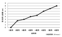
- the degree of bending increased linearly with increasing number of pulses.
- FIG. 3 shows the relationship between the number of pulses and the bending distance of the tip of the third finger when the pulse interval is 20 ms.
- FIG. 4 shows the relationship between the pulse interval and the bending distance of the tip of the third finger when the number of pulses is fixed to 8 and the pulse interval is set to 5, 10, 20, and 30 ms. Thus, when the pulse interval was 20 ms, the bending distance of the tip of the third finger was maximized.
- the movement of the muscles and joints caused by moving the limbs even by the intention of the person is detected by an optical sensor, an ultrasonic sensor, or a magnetic sensor.
- signals relating to the movement part, direction and intensity are separated into trigger signals.
- a magnetic stimulus that reinforces the spontaneous movement is generated via the trigger signal, and a large movement of the limb is expressed.
- a neural network derived from the plasticity of the brain and nerves is newly constructed, and the time required for functional recovery is significantly shortened.
- the present invention has a great effect on rehabilitation of a patient having a motor function disorder due to brain and nerve damage.
Landscapes
- Health & Medical Sciences (AREA)
- Life Sciences & Earth Sciences (AREA)
- General Health & Medical Sciences (AREA)
- Veterinary Medicine (AREA)
- Public Health (AREA)
- Animal Behavior & Ethology (AREA)
- Engineering & Computer Science (AREA)
- Biomedical Technology (AREA)
- Rehabilitation Therapy (AREA)
- Pain & Pain Management (AREA)
- Physical Education & Sports Medicine (AREA)
- Epidemiology (AREA)
- Oral & Maxillofacial Surgery (AREA)
- Biophysics (AREA)
- Neurology (AREA)
- Physiology (AREA)
- Dentistry (AREA)
- Radiology & Medical Imaging (AREA)
- Physics & Mathematics (AREA)
- Nuclear Medicine, Radiotherapy & Molecular Imaging (AREA)
- Pathology (AREA)
- Heart & Thoracic Surgery (AREA)
- Medical Informatics (AREA)
- Molecular Biology (AREA)
- Surgery (AREA)
- Magnetic Treatment Devices (AREA)
- Electrotherapy Devices (AREA)
- Rehabilitation Tools (AREA)
Abstract
【課題】麻痺により随意運動が困難な筋肉あるいは関節を、本人の意思と努力によってわずかでも動かし、その際に生じる動きの、部位、方向および強さをセンサで検出する。センサ信号のレベルに応じたパルス磁場を発生して、磁気刺激による大きな筋収縮を生じさせ、筋肉の動きを増強する。脳の可塑性を活用して神経ネットワークを構築し、リハビリテーションを効果的に行う技術を提供する。 【解決手段】自発運動によって筋肉若しくは関節に生じる変位を、光学センサ、あるいは超音波センサ、あるいは磁気センサによって検出し、この部位、方向および強さに対応するセンサ出力電位をトリガ信号として用いる。パルス電源に接続した一個または複数個の磁気刺激コイルに、トリガ信号レベルに対応する強度、回数の磁気パルスを発生させる。この磁気パルスによって大脳の運動野、末梢神経、あるいは筋肉を磁気刺激することにより麻痺した筋肉の動きを増強する。
Description
本発明は脳・神経の可塑性に着目し、脳の機能障害によって麻痺した筋肉・関節を磁気刺激法によって動かすことで、効率的なリハビリテーションを行う装置の開発に関する。
現在、脳卒中による片麻痺や脊髄損傷による四肢麻痺を有する患者数は200万人に達しており、日本の年齢構成の推移によって、その数はさらに増加している。脳損傷によって四肢の麻痺が長期間継続すると、廃用症候群によって筋肉の機能が著しく低下し、回復が困難な状態になる。片麻痺や四肢麻痺による廃用症候群を防止し、筋肉の機能を積極的に回復するために、運動療法によるリハビリテーションは最も重要な治療法である。
最近、脳波を検出することによって、麻痺した四肢を動かす「ブレインマシンインターフェース(BMI)」技術が注目を集めており、リハビリテーションへの応用が試みられている。しかしながら、BMI法は大脳運動野に機能障害がある場合には目的とする脳波を検出できないので、適用が困難という技術的な制約がある。
このような場合も、神経や筋肉に異常がなければ外部刺激によって筋肉を動かすことが可能である。
このような場合も、神経や筋肉に異常がなければ外部刺激によって筋肉を動かすことが可能である。
従来、成人の運動野は神経回路が固定しており、神経の再生やシナプス結合の変化は生じないと考えられてきた。しかし、1980年代に行われた、記憶学習とシナプス伝達効率に関連する研究から、運動野においてもシナプス結合の可塑的な変化が起こることが判明した。この過程は「脳の可塑性」と称される。
リハビリテーションによって、損傷した脳機能を回復させるために、脳の可塑性の積極的な活用が試みられている。新しいシナプス結合を脳の可塑性によって生じさせるには、他動的な運動よりも、自発的な運動を試みてその達成を実感することの効果が大きいとされている(非特許文献1)。しかしながら、麻痺した四肢を自発的に運動させることは容易な作業ではない。そこで介助や機器による運動補助に加えて、脳や神経を電気的に刺激して運動を誘発する方法が開発されている。
筋収縮を電気的に起こす方法として、経皮電気的神経刺激TENSが広く用いられている。この原理は外部から電気的に運動神経を刺激することで、手や足を動かすことにより、運動機能を回復するものである。
電気刺激法によって大きな筋収縮を得るためには、強い電気刺激を必要とする。この現象は感電と同じなので電気ショックによる不快感や痛みを伴う。ショックを少なくするために周波数や波形を改良するなど様々な研究が行われてきたが、電気的な刺激によって大きな筋収縮を実現することは困難な状況にある。
電気刺激法によって大きな筋収縮を得るためには、強い電気刺激を必要とする。この現象は感電と同じなので電気ショックによる不快感や痛みを伴う。ショックを少なくするために周波数や波形を改良するなど様々な研究が行われてきたが、電気的な刺激によって大きな筋収縮を実現することは困難な状況にある。
神経を電気的に刺激する他の方法として磁気刺激法がある。これは、神経の近くに置いたコイルにパルス電流を流した時に生じる誘導電流で、神経を刺激する方法である。この磁気刺激法は電極を貼り付ける、埋め込むなどの工程が不要であり、加えて感電のようなビリビリする電気ショックや痛みがほとんどない。この理由で、前記電気刺激法に比べて強い刺激が可能であり、大きな筋収縮が得られる利点がある。
磁気刺激法がリハビリテーションに有効なことは公知である。一例として、磁気刺激による誘発筋運動によって中枢神経系が再構築されることが知られている(非特許文献2参照)。また、脳血管障害によって生じた片麻痺の治療に効果があることが知られている(非特許文献3参照)。さらに、磁気刺激は、活動が弱った筋肉のリハビリテーションに広く応用されている(特許文献1~4参照)。
筋肉の自発運動に連動して磁気刺激を行う方法として、本出願の発明者らは、運動機能改善を目的とする部位に、筋電位計の電極を取り付け、運動によって生じる筋電位をトリガ信号として用いる方法を開発した(特許文献5参照)。目的とする部位の筋肉が自発的にわずかに動くと、発生する筋電位をトリガとするパルス磁気刺激が生じて、この刺激により筋肉の動きが増強される。この過程を繰り返すことにより、新たな神経ネットワークを構築させて、運動機能を回復させることができる。しかし筋電位によって筋肉が運動する部位、方向、大きさを特定することは困難であった。
中央法規出版(株)「回復する身体と脳」、出江著、磁気刺激により麻痺が改善した例、P.183
医歯薬出版(株)「磁気刺激法の基礎と応用」、真野著、中枢神経の再構築、P.127
医歯薬出版(株)「磁気刺激法の基礎と応用」、出江著、脳血管障害、P.198
脳卒中による片麻痺や脊髄損傷による四肢麻痺のために自発運動が困難になった筋肉あるいは関節を、本人の意思と努力によってわずかでも動かし、その際に生じる動きの、部位、方向および強さに対応する信号をセンサで検出する。センサ信号のレベル応じて強度、繰り返し回数の異なるパルス磁場を発生して、磁気刺激による大きな筋収縮を生じさせ、筋肉の動きを増強する。この過程を繰り返して、脳の可塑性を積極的に利用することにより新たな神経ネットワークを構築し、リハビリテーションを効果的に行う。
以上の課題に鑑み、本願発明は、以下の技術を用いて上記の課題を解決した。
(1)磁気パルスによって大脳の運動野、末梢神経、あるいは筋肉を磁気刺激することで、目的とする筋肉の部位と方向に、目的とする強さの筋収縮を生じさせる。これによって筋肉の自発運動を増強し、脳、神経の可塑性に基づく新しい神経回路形成が促進する磁気刺激リハビリテーション装置。
(2)筋肉に生じるわずかな自発運動の部位、方向および強さを検出し、出力信号を処理し、自発運動の部位、方向および強さに対応する複数のトリガ信号を発生させるトリガ発生装置を備えた磁気刺激リハビリテーション装置。
(3)自発運動検出手段として、磁気センサを用いる磁気刺激リハビリテーション装置。
(4)自発運動検出手段として、超音波センサを用いる磁気刺激リハビリテーション装置。
(5)自発運動検出手段として、光学センサを用いる磁気刺激リハビリテーション装置。
(6)トリガ信号の種類に応じて、磁気刺激パルスの強度・反復回数を変えることで、筋肉の動きあるいは神経の刺激を制御する磁気刺激リハビリテーション装置。
(7)磁気刺激の反復時間が1ミリ秒から100ミリ秒の範囲で設定可能な磁気刺激リハビリテーション装置。
本発明は麻痺により自発運動が困難な筋肉を、本人の意思と努力によってわずかでも動かし、動きの生じる部位や方向を光学センサ、あるいは超音波センサ、あるいは磁界センサによって検出し、その出力信号をパルス電源のトリガに用いる。動きを生じた部位または方向に応じ、筋肉近傍に設置したいずれかの磁気刺激コイルにパルス電流が流れてパルス磁場が発生し、神経に誘導される微小電流によって大きな筋収縮が生じる。この過程を繰り返すと、脳、神経の可塑性によって新しい神経回路形成が促進されて、リハビリテーションを効果的に行うことができる。本方法は電気刺激のようなショックや痛みを伴うことがないので、電気刺激よりも大きな筋収縮を繰り返して起こすことが可能である。また、目的とする筋肉に特定した集中的な訓練を行うことができる、新規なリハビリテーション技術である。本発明によれば、被険者の意欲に応じて生じた運動が、脳、神経の可塑性によって効率的に新しい神経回路を再生するのでリハビリテーション効果を画期的に高める、優れた特徴を有する。
本発明の磁気刺激によるリハビリテーション法は、まず筋肉の自発運動により発生する筋肉若しくは関節の動きを光学センサ、あるいは超音波センサ、あるいは磁気センサにより検出し、これを電気信号に変換する。麻痺した筋肉や関節の自発運動から発生するセンサ出力は、運動機能が低下しているので微弱でありノイズも多い。本願発明では、センサ出力を任意に増幅し、信号処理によってノイズを除去することにより運動の部位、方向、大きさを検出することができる。
次に、センサが検出した運動部位、方向、大きさに関する信号をトリガとして、目的とする部位を目指す方向に運動させるように、磁気刺激コイルに目的強度あるいは繰り返し数の磁気パルスを発生させる。この結果、筋肉の動きは想定の通りに増強される。これを繰り返し、磁気刺激で筋肉や関節を動かすことにより、既存の神経回路の強化又は新たな神経ネットワークを構築させて、機能を回復させる。
この場合、新たな神経ネットワークを構築するということは、神経細胞の新生を意味するものではなく、損傷を受けていない神経を利用して神経相互の連絡を良くし、新たなネットワークを形成することを意味する。
本願発明においては、短時間に複数回の磁気刺激を反復することが有効である。そして、この磁気刺激の反復時間が1ミリ秒から100ミリ秒であるのが望ましい。反復時間が1ミリ秒以下では筋収縮が追従できない。また100ミリ秒以上では、屈曲が複数回に分かれるので磁気刺激効果の増大はない。
次に、本願発明の詳細を実施例に基づいて説明する。なお、この実施例は当業者の理解を容易にするためのものである。すなわち、本願発明は明細書の全体に記載される技術思想によってのみ限定されるものであり、本実施例によってのみ限定されるものでないことは理解されるべきことである。
(実施例1)指先の変位を、図1の1に示す6個のCCD高速度カメラを用いる光学センサで検出した。手2の各指につけた反射器3の位置を4のパーソナルコンピュータによって3次元座標として検出する。目的とする指の動きが設定値を超えると5のトリガ回路が信号を発生する。トリガ数は、目的部位の数、運動の大きさに応じて1個または複数個を選択できる。一個のトリガによってパルス電源6を放電させると、手首に密着させた一個の磁気刺激コイル7に単発あるいは連続磁気パルスが発生した。磁気パルスによって手首の末梢神経が磁気刺激されて、手首は大きく曲がった。
次に、親指と小指の動きに対応する個別のトリガ信号によって、親指側の末梢神経と小指側の末梢神経を個別のコイルによって磁気刺激すると、親指と小指それぞれの動きに応じた磁気刺激によって大きく動き、各々の指の運動を増幅することができた。
(実施例2)5MHzの広帯域超音波センサを手首に当てて反射波を検出した。各指の動きに応じて、手首の異なる箇所から反射超音波波形が観察された。超音波信号に複数のゲートをかけて複数のトリガ信号を得た。これらの信号によって実施例1と同様に磁気刺激を行うと、任意の指の小さな運動を、磁気刺激によって増幅することができた。
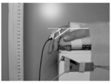
(実施例3)末梢神経の磁気刺激に及ぼす単発~8連発の磁気刺激の効果を図2の装置を用いて調べた。実験開始肢位を第3指MP関節屈曲30°に設定し,総指伸筋を標的として上腕部を連発磁気刺激した。これによる第3指先端挙上距離を高速度カメラによって計測した。放電間隔を20ミリ秒としてパルスを連発すると、第3指先端の屈曲は単発の場合よりも大きくなった。屈曲の程度は、パルス数の増加とともに直線的に増大した。図3にパルス間隔を20msとした場合の、パルス数と第3指先端の屈曲距離の関係を示す。
連続パルスによる磁気刺激効果はパルス数だけでなくパルス間隔にも依存した。パルス数を8連発に固定して、パルス間隔を5、10、20、30msとした場合のパルス間隔と第3指先端の屈曲距離の関係を図4に示す。このようにパルス間隔は20msの場合に第3指先端の屈曲距離は最大になった。
脊髄損傷による四肢麻痺や脳卒中による片麻痺のリハビリテーションにおいて、本人の意思によってわずかでも手足を動かすことによって生じる筋肉や関節の運動を、光学センサ、あるいは超音波センサ、あるいは磁気センサで検出する。これを解析して運動の部位、方向、強度に関する信号を分離してトリガ信号とする。このトリガ信号を介し自発運動を補強する磁気刺激を起こして、手足の大きな動きを発現する。この過程を繰り返すことによって脳及び神経の可塑性に由来する神経ネットワークが新たに構築され、機能回復に要する時間を著しく短縮する。このように本発明は脳及び神経の損傷により運動機能に障害を有する患者のリハビリテーションに大な効果を有する。
1:CCD高速度カメラ
2:手首
3:反射器
4:パーソナルコンピュータ(座標計算)
5:トリガ回路
6:パルス電源
7.磁気刺激コイル
2:手首
3:反射器
4:パーソナルコンピュータ(座標計算)
5:トリガ回路
6:パルス電源
7.磁気刺激コイル
Claims (7)
- 磁気パルスによって大脳の運動野、末梢神経、あるいは筋肉を磁気刺激することで、目的とする筋肉の部位に、目的とする方向と強さの筋収縮を生じさせ、これによって筋肉の自発運動を増強し、脳、神経の可塑性に基づく新しい神経回路形成を促進することを特徴とする磁気刺激リハビリテーション装置。
- 筋肉に生じるわずかな前記自発運動の部位、方向および強さを検出し、出力信号を処理し、前記自発運動の部位、関節運動の方向および強さに対応する複数のトリガ信号を発生させるトリガ発生装置を備えたことを特徴とする請求項1記載の磁気刺激リハビリテーション装置。
- 前記自発運動を検出する手段として、光学センサを用いることを特徴とする請求項2に記載の磁気刺激リハビリテーション装置。
- 前記自発運動を検出する手段として、超音波センサを用いることを特徴とする請求項2に記載の磁気刺激リハビリテーション装置。
- 前記自発運動を検出する手段として、磁気センサを用いることを特徴とする請求項2に記載の磁気刺激リハビリテーション装置。
- 前記トリガ信号の種類に応じて、磁気刺激パルスの強度、反復回数を変えて、筋肉の運動あるいは神経の刺激を制御することを特徴とする請求項2乃至5のいずれか1項に記載の磁気刺激リハビリテーション装置。
- 前記磁気刺激の反復時間が1ミリ秒から100ミリ秒の範囲で設定可能であることを特徴とする請求項1乃至6のいずれか1項に記載の磁気刺激リハビリテーション装置。
Applications Claiming Priority (2)
| Application Number | Priority Date | Filing Date | Title |
|---|---|---|---|
| JP2011-264090 | 2011-11-14 | ||
| JP2011264090A JP5893367B2 (ja) | 2011-11-14 | 2011-11-14 | 磁気刺激リハビリテーション装置 |
Publications (1)
| Publication Number | Publication Date |
|---|---|
| WO2013073536A1 true WO2013073536A1 (ja) | 2013-05-23 |
Family
ID=48429593
Family Applications (1)
| Application Number | Title | Priority Date | Filing Date |
|---|---|---|---|
| PCT/JP2012/079428 Ceased WO2013073536A1 (ja) | 2011-11-14 | 2012-11-13 | 磁気刺激リハビリテーション装置 |
Country Status (2)
| Country | Link |
|---|---|
| JP (1) | JP5893367B2 (ja) |
| WO (1) | WO2013073536A1 (ja) |
Cited By (1)
| Publication number | Priority date | Publication date | Assignee | Title |
|---|---|---|---|---|
| CN109452943A (zh) * | 2018-09-17 | 2019-03-12 | 周建菊 | 一种用于重症偏瘫患者的多功能康复衣 |
Families Citing this family (3)
| Publication number | Priority date | Publication date | Assignee | Title |
|---|---|---|---|---|
| JP2017063971A (ja) * | 2015-09-29 | 2017-04-06 | 株式会社Ifg | 運動努力をトリガとした同期的磁気刺激を行うリハビリテーションシステム |
| JP6251302B2 (ja) * | 2016-01-27 | 2017-12-20 | H2L株式会社 | 電気刺激装置 |
| CN107929939B (zh) * | 2017-03-17 | 2023-10-20 | 重庆理工大学 | 一种神经电刺激的上肢康复训练机器人及其使用方法 |
Citations (2)
| Publication number | Priority date | Publication date | Assignee | Title |
|---|---|---|---|---|
| JP2003533299A (ja) * | 2000-05-16 | 2003-11-11 | ロッキード マーティン コーポレイション | 神経障害症状緩和のための適応型刺激装置 |
| JP2010166971A (ja) * | 2009-01-20 | 2010-08-05 | Tohoku Univ | 筋肉の動きを増強又は回復させる方法及び装置 |
-
2011
- 2011-11-14 JP JP2011264090A patent/JP5893367B2/ja not_active Expired - Fee Related
-
2012
- 2012-11-13 WO PCT/JP2012/079428 patent/WO2013073536A1/ja not_active Ceased
Patent Citations (2)
| Publication number | Priority date | Publication date | Assignee | Title |
|---|---|---|---|---|
| JP2003533299A (ja) * | 2000-05-16 | 2003-11-11 | ロッキード マーティン コーポレイション | 神経障害症状緩和のための適応型刺激装置 |
| JP2010166971A (ja) * | 2009-01-20 | 2010-08-05 | Tohoku Univ | 筋肉の動きを増強又は回復させる方法及び装置 |
Cited By (2)
| Publication number | Priority date | Publication date | Assignee | Title |
|---|---|---|---|---|
| CN109452943A (zh) * | 2018-09-17 | 2019-03-12 | 周建菊 | 一种用于重症偏瘫患者的多功能康复衣 |
| CN109452943B (zh) * | 2018-09-17 | 2021-03-23 | 周建菊 | 一种用于重症偏瘫患者的多功能康复衣 |
Also Published As
| Publication number | Publication date |
|---|---|
| JP5893367B2 (ja) | 2016-03-23 |
| JP2013103121A (ja) | 2013-05-30 |
Similar Documents
| Publication | Publication Date | Title |
|---|---|---|
| Farina et al. | Accessing the neural drive to muscle and translation to neurorehabilitation technologies | |
| Petersen et al. | Investigating human motor control by transcranial magnetic stimulation | |
| Niazi et al. | Peripheral electrical stimulation triggered by self-paced detection of motor intention enhances motor evoked potentials | |
| Ambrosini et al. | A myocontrolled neuroprosthesis integrated with a passive exoskeleton to support upper limb activities | |
| Bao et al. | Electrode placement on the forearm for selective stimulation of finger extension/flexion | |
| CN105233406B (zh) | 一种脑卒中功能性电刺激康复系统 | |
| JP2010207268A (ja) | 治療用生体磁気刺激方法及び装置 | |
| CN105377358A (zh) | 神经运动功能障碍治疗的方法和系统 | |
| CN101711908B (zh) | 针对下肢的层级式功能性电刺激康复系统 | |
| JP2017063971A (ja) | 運動努力をトリガとした同期的磁気刺激を行うリハビリテーションシステム | |
| CN105148404B (zh) | 基于健侧肢体肌电信号触发的电刺激训练系统及训练方法 | |
| Rigosa et al. | Decoding bipedal locomotion from the rat sensorimotor cortex | |
| CN102488514A (zh) | 基于自主、刺激动作模态下的脑肌电相关性的分析方法 | |
| CN102949783B (zh) | 回馈控制的穿戴式上肢电刺激装置 | |
| JP5893367B2 (ja) | 磁気刺激リハビリテーション装置 | |
| CN106974649A (zh) | 可穿戴式肌肉运动状态识别和肌肉电刺激运动辅助系统 | |
| Missiroli et al. | Haptic stimulation for improving training of a motor imagery BCI developed for a hand-exoskeleton in rehabilitation | |
| JP2010166971A (ja) | 筋肉の動きを増強又は回復させる方法及び装置 | |
| Zhang et al. | Reciprocal EMG controlled FES for pathological tremor suppression of forearm | |
| CN110882485A (zh) | 采用体表电极刺激目标肌肉的瘫痪肢体运动功能重建系统 | |
| Cao et al. | Development of a wearable ultrasound–FES integrated rehabilitation and motor-functional reconstruction system for post-stroke patients | |
| Muraoka et al. | Specifications of an electromyogram-driven neuromuscular stimulator for upper limb functional recovery | |
| Shuhan et al. | Effect of introducing EMG biofeedback to a finger extensor facilitation training device for hemiplegic patients after strokes | |
| Zhou et al. | Electromyographic bridge—A multi-movement volitional control method for functional electrical stimulation: Prototype system design and experimental validation | |
| Talanov et al. | Neurointerface implemented with oscillator motifs |
Legal Events
| Date | Code | Title | Description |
|---|---|---|---|
| 121 | Ep: the epo has been informed by wipo that ep was designated in this application |
Ref document number: 12849884 Country of ref document: EP Kind code of ref document: A1 |
|
| NENP | Non-entry into the national phase |
Ref country code: DE |
|
| 122 | Ep: pct application non-entry in european phase |
Ref document number: 12849884 Country of ref document: EP Kind code of ref document: A1 |