WO2013069143A1 - Training model for ultrasonic bronchoscopy - Google Patents
Training model for ultrasonic bronchoscopy Download PDFInfo
- Publication number
- WO2013069143A1 WO2013069143A1 PCT/JP2011/076001 JP2011076001W WO2013069143A1 WO 2013069143 A1 WO2013069143 A1 WO 2013069143A1 JP 2011076001 W JP2011076001 W JP 2011076001W WO 2013069143 A1 WO2013069143 A1 WO 2013069143A1
- Authority
- WO
- WIPO (PCT)
- Prior art keywords
- simulated
- needle puncture
- puncture site
- trachea
- bronchial
- Prior art date
Links
Images


Classifications
-
- G—PHYSICS
- G09—EDUCATION; CRYPTOGRAPHY; DISPLAY; ADVERTISING; SEALS
- G09B—EDUCATIONAL OR DEMONSTRATION APPLIANCES; APPLIANCES FOR TEACHING, OR COMMUNICATING WITH, THE BLIND, DEAF OR MUTE; MODELS; PLANETARIA; GLOBES; MAPS; DIAGRAMS
- G09B23/00—Models for scientific, medical, or mathematical purposes, e.g. full-sized devices for demonstration purposes
- G09B23/28—Models for scientific, medical, or mathematical purposes, e.g. full-sized devices for demonstration purposes for medicine
- G09B23/285—Models for scientific, medical, or mathematical purposes, e.g. full-sized devices for demonstration purposes for medicine for injections, endoscopy, bronchoscopy, sigmoidscopy, insertion of contraceptive devices or enemas
-
- A—HUMAN NECESSITIES
- A61—MEDICAL OR VETERINARY SCIENCE; HYGIENE
- A61B—DIAGNOSIS; SURGERY; IDENTIFICATION
- A61B1/00—Instruments for performing medical examinations of the interior of cavities or tubes of the body by visual or photographical inspection, e.g. endoscopes; Illuminating arrangements therefor
- A61B1/267—Instruments for performing medical examinations of the interior of cavities or tubes of the body by visual or photographical inspection, e.g. endoscopes; Illuminating arrangements therefor for the respiratory tract, e.g. laryngoscopes, bronchoscopes
- A61B1/2676—Bronchoscopes
-
- G—PHYSICS
- G09—EDUCATION; CRYPTOGRAPHY; DISPLAY; ADVERTISING; SEALS
- G09B—EDUCATIONAL OR DEMONSTRATION APPLIANCES; APPLIANCES FOR TEACHING, OR COMMUNICATING WITH, THE BLIND, DEAF OR MUTE; MODELS; PLANETARIA; GLOBES; MAPS; DIAGRAMS
- G09B23/00—Models for scientific, medical, or mathematical purposes, e.g. full-sized devices for demonstration purposes
- G09B23/28—Models for scientific, medical, or mathematical purposes, e.g. full-sized devices for demonstration purposes for medicine
- G09B23/288—Models for scientific, medical, or mathematical purposes, e.g. full-sized devices for demonstration purposes for medicine for artificial respiration or heart massage
-
- G—PHYSICS
- G09—EDUCATION; CRYPTOGRAPHY; DISPLAY; ADVERTISING; SEALS
- G09B—EDUCATIONAL OR DEMONSTRATION APPLIANCES; APPLIANCES FOR TEACHING, OR COMMUNICATING WITH, THE BLIND, DEAF OR MUTE; MODELS; PLANETARIA; GLOBES; MAPS; DIAGRAMS
- G09B23/00—Models for scientific, medical, or mathematical purposes, e.g. full-sized devices for demonstration purposes
- G09B23/28—Models for scientific, medical, or mathematical purposes, e.g. full-sized devices for demonstration purposes for medicine
- G09B23/30—Anatomical models
-
- A—HUMAN NECESSITIES
- A61—MEDICAL OR VETERINARY SCIENCE; HYGIENE
- A61B—DIAGNOSIS; SURGERY; IDENTIFICATION
- A61B8/00—Diagnosis using ultrasonic, sonic or infrasonic waves
- A61B8/12—Diagnosis using ultrasonic, sonic or infrasonic waves in body cavities or body tracts, e.g. by using catheters
Definitions
- the present invention relates to a training model for an ultrasonic endoscope, and more particularly to a training model for training needle-puncture under an ultrasound guide to a lymph node near the trachea / bronchus.
- Ultrasound examination is indispensable in modern medicine, but an ultrasonic endoscope with an ultrasonic probe attached to the insertion tip enables high-resolution ultrasound observation and pathology. Since a technique called ultrasonic endoscope-guided puncture, in which cells are collected while observing ultrasonically for examination, it is a very useful pathological examination apparatus.
- ultrasonic testing requires a high level of knowledge, experience, and technology for the person performing the inspection, and in particular, an ultrasonic endoscope-guided puncture procedure requires a higher level of knowledge, experience, and technology.
- the Education and training for those who perform ultrasound examinations are conducted at medical institutions such as hospitals while training while conducting actual patient examinations under a knowledgeable and experienced instructor. There were considerable restrictions on the diseases experienced.
- a human body model for ultrasonic medical training for training in ultrasonic examination has been proposed that reproduces the shape and internal structure of the human body organ, and has a replaceable puncture training site.
- Punure technique training has been limited to those that do not have a shield against the puncture site from the esophagus to the organ.
- the present invention is for solving the above-mentioned problems, and an object of the present invention is to supervise a puncture technique for trachea / bronchial inner side trachea / bronchial adjacent lymph node by ultrasonic bronchoendoscopy. To provide a sonic bronchoscopy training model.
- the training model for ultrasonic bronchoscopy is a training model for training needle-guided needle puncture to lymph nodes near the trachea and bronchus, and includes a jaw model, bronchus It has a model and replaceable needle puncture site.
- the needle puncture site consists of simulated trachea / bronchial cartilage, simulated lymph node and simulated peripheral tissue. Each part of the needle puncture site is made of either silicone rubber or urethane resin.
- the main component at least the simulated trachea / bronchial cartilage at the needle puncture site and the surrounding tissue are mixed with organic powder filler, and the simulated trachea / bronchial cartilage at the needle puncture site is connected to the simulated trachea / bronchial wall surface of the needle puncture site It is provided on one side.
- the simulated trachea / bronchial cartilage provided at the needle puncture site preferably has a ladder shape.
- the organic powder filler is preferably a nylon filler, and the needle puncture site is preferably composed mainly of silicone rubber.
- the simulated lymph node at the needle puncture site preferably includes silicone rubber and liquid paraffin, and the simulated peripheral tissue at the needle puncture site preferably includes silicone rubber, nylon filler, and liquid paraffin.
- the needle puncture site has a quadrangular prism shape in which a simulated trachea / bronchial cartilage is provided on one side of the inner wall of the simulated trachea / bronchi and a spherical simulated lymph node is provided inside.
- a needle puncture site mounting portion having a concave part in which a part of the trachea / bronchial wall is opened in a rectangular shape and the needle puncture site can be mounted.
- the present invention it becomes possible to perform training for puncture techniques to the trachea / bronchial adjacent lymph nodes performed from the inside of the trachea / bronchus, which could not be performed with the conventional puncture training model, and contribute to the improvement of the puncture technique of the practitioner can do.
- FIG. 1 is an overall schematic diagram of an ultrasonic bronchoscopy training model according to an embodiment of the present invention. It is the schematic of the needle puncture site
- FIG. 5 is a cross-sectional view of FIG. 4.
- FIG. 1 shows the arrangement of lymph nodes near the trachea and bronchi.
- lymph nodes # 1 to # 12 shown in FIG. 1 mediastinal lymph nodes (# 1, # 2, # 3, # 4, # 7), hilar lymph nodes (# 10, # 11, # 12) Are targets for ultrasonic bronchoscopy, and # 5, # 6, # 8, and # 9 that do not contact the trachea or bronchus are excluded.
- the training model of the present invention is intended for training of the puncture technique to the lymph node in contact with the trachea or bronchus.
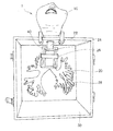
- FIG. 2 is a schematic diagram of an ultrasonic bronchoscopy training model according to an embodiment of the present invention.
- the ultrasonic bronchoscopy training model 1 according to the embodiment of the present invention includes a jaw model 10, a bronchial model 20, a case 30, and a needle puncture site 40.
- the jaw model 10 is a part that becomes an insertion opening when an ultrasonic bronchoscope is inserted, and it is approximated to a case where it is performed by a human body, so that the training effect is enhanced. It is preferable to use an approximated jaw model.
- the jaw model 10 is provided with a larynx (not shown) serving as an insertion port for an endoscope, and the end of the larynx is connected to a simulated trachea portion of a bronchial model.
- the larynx of the jaw model should be detachable so that it can be replaced when damaged.
- the bronchial model 20 includes a simulated tracheal part 22 connected to the larynx of the jaw model 10, a needle puncture site mounting part 24 provided above the tracheal branch part of the simulated tracheal part 22, and a lower part of the simulated tracheal part 22. It consists of the simulated bronchial portion 28 located in the area. Silicone rubber, urethane resin, vinyl chloride resin, or the like can be used as the material for the simulated trachea part 22 and simulated bronchus part 28 of the bronchial model 20, and silicone rubber that allows the insertion feeling of the bronchoscope to be obtained with a feeling close to that of the human body. It is particularly preferable to use
- the simulated bronchial portion 28 can be used not only for puncture technique training but also for normal bronchoscopy training by reproducing even fine bronchi. It is preferable to be able to be used for training of an ultra-thin bronchoscope.
- the case 30 is a case for housing the bronchial model 20, and the front side is opened so that the illumination light at the tip of the endoscope can be seen through the simulated trachea / bronchial wall. Since the position of the endoscope tip is known, it is useful for enhancing the effect of bronchoendoscopy training.
- the needle puncture site 40 is mounted on the needle puncture site mounting part 24 provided on the upper part of the tracheal bifurcation of the simulated trachea unit 22.
- the installation positions of the needle puncture site mounting portion 24 and the needle puncture site 40 are not limited to the upper part of the tracheal branch portion of the simulated trachea unit 22 and may be provided in the simulated bronchial unit 28.
- the needle puncture site 40 is detachable from the needle puncture site mounting part 24, and the needle puncture site 40 damaged can be easily replaced by repeating the puncture technique.
- FIG. 3 is a schematic view of the needle puncture site mounting part 24.
- the needle puncture site mounting portion 24 is provided with a mounting recess 24a in which a recess is formed in accordance with the shape of the needle puncture site 40.
- a puncture opening 24b in which a part of the bottom surface of the recess is opened is provided at the bottom of the mounting recess 24a facing the simulated trachea 22 side.
- the puncture opening 24b serves as a window for performing a puncture technique with a bronchoscope from inside the simulated trachea / bronchi to the needle puncture site 40 that has been mounted.
- the needle puncture site mounting part 24 is provided with a fixing belt 26 for fixing the needle puncture site so that the needle puncture site 40 mounted on the needle puncture site installation unit 24 is not detached during training. Yes.
- the shape of the mounting recess 24a may be any shape as long as the needle puncture site 40 can be accommodated, and the shape of the puncture opening 24b may be any shape as long as a puncture technique is possible. From the viewpoint of ease of operation, a rectangular shape along the top and bottom of the simulated tracheal part or simulated bronchial part is preferable.
- the shape of the needle puncture site 40 may be any shape as long as the puncture technique can be performed. Like the shape of the puncture opening 24b, the needle puncture site 40 is easy to perform puncture technique training and replacement. It is preferable that it is a square column along the upper and lower sides of the simulated trachea, and the shape in the present embodiment is a rectangular column of 6 cm long ⁇ 3 cm wide ⁇ 4 cm deep, and is easily replaceable.
- FIG. 4 is a schematic view of a needle puncture site 40 according to an embodiment of the present invention
- FIG. 5 is a cross-sectional view of FIG.
- the needle puncture site 40 includes a simulated trachea / bronchial cartilage 42, a simulated peripheral tissue 44, and a simulated lymph node 46, and one side surface that serves as a simulated tracheal / bronchial inner wall surface of the simulated peripheral tissue 44.
- the simulated trachea / bronchial cartilage 42 is provided on one side of the needle puncture site 40 that is the surface of the inner wall of the simulated trachea / bronchus, and matches the puncture opening 24b provided at the bottom of the mounting recess 24a of the needle puncture site mounting part 24. It forms part of the inner wall of the simulated trachea or simulated bronchus.
- the shape of the simulated trachea / bronchial cartilage 42 is preferably approximated to the shape of the actual tracheal cartilage of the human body, and is preferably a ladder shape. Specifically, as shown in FIG. 4, it is formed along the trachea / bronchus.
- the horizontal part extends from the vertical part at regular intervals, and the part where the simulated peripheral tissue 44 between the horizontal parts simulating the trachea / bronchial cartilage is exposed corresponds to the interchondral ligament.
- the simulated trachea / bronchial cartilage 42 preferably has a thickness approximating that of the actual trachea / bronchial cartilage in order to enhance the training effect, and preferably has a thickness of 1.5 mm to 2.0 mm.
- the trainee inserts the ultrasonic bronchoscope from the throat of the jaw model 10 into the simulated trachea unit 22 and trachea / bronchial cartilage ring.
- a puncture technique is performed on the simulated lymph node 46 on the back side of the simulated trachea / bronchial cartilage 42 through the simulated peripheral tissue 44 in the gap. Therefore, the simulated trachea / bronchial cartilage 42, the simulated peripheral tissue 44, and the simulated lymph node 46 can obtain an ultrasonic image similar to that of an actual human body and can clearly distinguish each contour. There is a need.
- the material of the simulated trachea / bronchial cartilage 42, the simulated peripheral tissue 44, and the simulated lymph node 46 of the needle puncture site 40 is selected so that the touch when puncturing approximates the touch of the human body.
- the simulated trachea / bronchial cartilage 42, simulated peripheral tissue 44, and simulated lymph node 46 of the needle puncture site 40 are formed mainly of silicone rubber or urethane resin, and in particular, ease of adjustment of the ultrasound image Silicone rubber is preferred from the standpoint of resistance to repeated punctures, liquid silicone rubber is more preferred from the viewpoint of ease of crosslinking and hardness adjustment, and two-component addition type silicone rubber that is not a condensation product at the time of curing is more preferred.
- the simulated trachea / bronchial cartilage 42 and the simulated peripheral tissue 44 in the needle puncture site 40 are blended with the organic powder filler in the main component material, and the type, shape, and amount of the organic powder filler to be blended are: It is appropriately selected so that an ultrasonic image similar to that of the actual human body can be obtained.
- the use of nylon filler as the organic powder filler makes it easier to approximate the ultrasonic image of the actual human body and between the simulated organs. It is preferable because it is easy to add contrast.
- the simulated trachea / bronchial cartilage 42 is preferably formed of silicone rubber blended with a nylon filler, and the silicone rubber used for the simulated trachea / bronchial cartilage 42 has a puncture feel that is similar to that of the human body. In order to approximate, it is preferable to set the hardness to be harder than that of the simulated peripheral tissue 44 and the simulated lymph node 46.
- the simulated peripheral tissue 44 and the simulated lymph node 46 are provided with a lubricant as a main component material for adjusting the hardness and improving needle penetration so that the touch at the time of puncture approximates the touch at the human body. It is preferable to mix.
- a lubricant As the lubricant to be mixed with the simulated peripheral tissue 44 and the simulated lymph node 46, a liquid lubricant is preferable because of easy hardness adjustment, and liquid paraffin is particularly preferable.
- the amount of the lubricant mixed in the simulated peripheral tissue 44 and the simulated lymph node 46 is preferably 15% by weight to 25% by weight with respect to the main component material. An effect that approximates the feel of the human body is obtained.
- the position of the simulated lymph node 46 in the simulated peripheral tissue 44 is approximated to the actual lymph node and is formed at a position close to the simulated trachea / bronchial cartilage 42 in the simulated peripheral tissue 44.
- the shape of the simulated lymph node 46 is preferably a spherical shape having a diameter of 5 mm to 10 mm, and preferably 2 to 6 are formed in the needle puncture site 40.
- # 1, # 2 (R, L), # 3, # 4 (R, L) 2 to 6 corresponding to the above are provided as simulated lymph nodes 46.
- Bronchial endoscopic training model 10 Jaw model 20: Bronchial model 22: Simulated tracheal part 24: Needle puncture site mounting part 24a: Mounting concave part 24b: Puncture opening part 26: Fixing belt 28: Simulated bronchial part 30: Case 40 : Needle puncture site 42: Simulated trachea / bronchial cartilage 44: Simulated peripheral tissue 46: Simulated lymph node
Abstract
Description
そして、超音波検査を行う者の教育や研修は、病院等の医療機関で、知識、経験が豊富な指導者の下、実際の患者の検査を行いながらトレーニングをしているため、実習の時間や経験する疾患にかなりの制約があった。 Such ultrasonic testing requires a high level of knowledge, experience, and technology for the person performing the inspection, and in particular, an ultrasonic endoscope-guided puncture procedure requires a higher level of knowledge, experience, and technology. The
Education and training for those who perform ultrasound examinations are conducted at medical institutions such as hospitals while training while conducting actual patient examinations under a knowledgeable and experienced instructor. There were considerable restrictions on the diseases experienced.
しかしながら、従来のトレーニング用のモデルにおいて、穿刺手技のトレーニングは、食道等から臓器への穿刺部位に対して遮蔽物がないものに限られていた。 Therefore, a human body model for ultrasonic medical training for training in ultrasonic examination has been proposed that reproduces the shape and internal structure of the human body organ, and has a replaceable puncture training site. Has also been proposed (see, for example, Patent Document 1).
However, in conventional training models, puncture technique training has been limited to those that do not have a shield against the puncture site from the esophagus to the organ.
そのため、従来の穿刺トレーニングモデルでは、気管・気管支内側から行う気管・気管支隣接リンパ節への穿刺手技のトレーニングが十分に行えず、このような手技に対応するトレーニングモデルが強く求められていた。 In recent years, ultrasonic bronchoscopy has begun to be used frequently for pathological diagnosis around the trachea and bronchus. Therefore, a high level of skill is required for puncture.
For this reason, in the conventional puncture training model, training of the puncture technique to the trachea / bronchial adjacent lymph node performed from the inside of the trachea / bronchus cannot be performed sufficiently, and a training model corresponding to such a technique has been strongly demanded.
また、前記有機粉体フィラーはナイロンフィラーであることが好ましく、前記ニードル穿刺部位はシリコーンゴムを主成分とすることが好ましい。
前記ニードル穿刺部位の模擬リンパ節は、シリコーンゴムと流動パラフィンを含むことが好ましく、前記ニードル穿刺部位の模擬周辺組織は、シリコーンゴム、ナイロンフィラー及び流動パラフィンを含むことが好ましい。 The simulated trachea / bronchial cartilage provided at the needle puncture site preferably has a ladder shape.
The organic powder filler is preferably a nylon filler, and the needle puncture site is preferably composed mainly of silicone rubber.
The simulated lymph node at the needle puncture site preferably includes silicone rubber and liquid paraffin, and the simulated peripheral tissue at the needle puncture site preferably includes silicone rubber, nylon filler, and liquid paraffin.
前記トレーニングモデルの気管支モデルには、気管・気管支壁の一部が矩形に開放され、前記ニードル穿刺部位が装着可能な凹部を有するニードル穿刺部位装着部を設けることが好ましい。 It is preferable that the needle puncture site has a quadrangular prism shape in which a simulated trachea / bronchial cartilage is provided on one side of the inner wall of the simulated trachea / bronchi and a spherical simulated lymph node is provided inside.
In the bronchial model of the training model, it is preferable to provide a needle puncture site mounting portion having a concave part in which a part of the trachea / bronchial wall is opened in a rectangular shape and the needle puncture site can be mounted.
本発明のトレーニングモデルは上記の気管もしくは気管支に接したリンパ節への穿刺手技のトレーニングを目的としたものである。 FIG. 1 shows the arrangement of lymph nodes near the trachea and bronchi. Among the
The training model of the present invention is intended for training of the puncture technique to the lymph node in contact with the trachea or bronchus.
本発明の実施形態に係る超音波気管支内視鏡トレーニングモデル1は、顎モデル10、気管支モデル20、ケース30、ニードル穿刺部位40からなる。 FIG. 2 is a schematic diagram of an ultrasonic bronchoscopy training model according to an embodiment of the present invention.
The ultrasonic
また、顎モデル10には、内視鏡の挿入口となる喉頭部(図示しない)が設けられているが、該喉頭部の末端は気管支モデルの模擬気管部と連結している。 The
The
気管支モデル20の模擬気管部22、模擬気管支部28の材質は、シリコーンゴム、ウレタン樹脂、塩化ビニル樹脂などを用いることができ、気管支内視鏡の挿入感覚が人体に近い感覚で得られるシリコーンゴムを用いることが特に好ましい。 In the embodiment of the present invention, the
Silicone rubber, urethane resin, vinyl chloride resin, or the like can be used as the material for the simulated
ニードル穿刺部位装着部24には、ニードル穿刺部位40を装着するために、ニードル穿刺部位40の形状に合わせて凹部を形成した装着凹部24aが設けられている。
装着凹部24aの模擬気管部22側に面した底部には、凹部の底面の一部が開放された穿刺開口部24bが設けられている。この穿刺開口部24bは、装着したニードル穿刺部位40に対して、模擬気管・気管支内から気管支内視鏡による穿刺手技を施すための窓の役割を果たす。 FIG. 3 is a schematic view of the needle puncture
In order to mount the
A puncture opening 24b in which a part of the bottom surface of the recess is opened is provided at the bottom of the mounting recess 24a facing the simulated
図4、図5に示すように、ニードル穿刺部位40は、模擬気管・気管支軟骨42、模擬周辺組織44、模擬リンパ節46からなり、模擬周辺組織44の模擬気管・気管支内壁表面となる一側面に模擬気管・気管支軟骨42を備え、模擬周辺組織44の内部に模擬リンパ節46が設けられている構成となっている。 FIG. 4 is a schematic view of a
As shown in FIGS. 4 and 5, the
模擬気管・気管支軟骨42の形状は、実際の人体の気管軟骨の形状に近似させ、梯子形にすることが好ましく、具体的には、図4に示すように、気管・気管支に沿って形成された縦部分から一定間隔で横部分が伸びた形状であり、気管・気管支軟骨を模した横部分の間の模擬周辺組織44が露出した部分が軟骨間靭帯に相当する。
また、模擬気管・気管支軟骨42は、トレーニング効果を高めるため、厚みを実際の気管・気管支軟骨に近似させるのが好ましく、1.5mm~2.0mmの厚みであることが好ましい。 The simulated trachea /
The shape of the simulated trachea /
Further, the simulated trachea /
ニードル穿刺部位40の模擬気管・気管支軟骨42、模擬周辺組織44及び模擬リンパ節46は、シリコーンゴム、ウレタン樹脂のいずれかを主成分として形成され、特には、超音波画像の調整のしやすさ、繰り返しの穿刺に対する耐性から、シリコーンゴムが好ましく、架橋、硬度調整の容易さから液状シリコーンゴムが更に好ましく、硬化時に縮合生成物のでない2成分付加型シリコーンゴムはより好ましい。 The material of the simulated trachea /
The simulated trachea /
模擬周辺組織44及び模擬リンパ節46に混合する潤滑剤としては、硬度調整の容易さから液状潤滑剤が好ましく、なかでも、流動パラフィンが好ましい。 In the present embodiment, the simulated
As the lubricant to be mixed with the simulated
本実施形態においては、ニードル穿刺部位40の取り付け位置が気管であるので、図1に示したリンパ節のうち、#1、#2(R、L)、#3、#4(R、L)に対応する2~6個が模擬リンパ節46として設けられている。 The position of the
In this embodiment, since the attachment position of the
10 : 顎モデル
20 : 気管支モデル
22 : 模擬気管部
24 : ニードル穿刺部位装着部
24a : 装着凹部
24b : 穿刺開口部
26 : 固定ベルト
28 : 模擬気管支部
30 : ケース
40 : ニードル穿刺部位
42 : 模擬気管・気管支軟骨
44 : 模擬周辺組織
46 : 模擬リンパ節
1: Bronchial endoscopic training model 10: Jaw model 20: Bronchial model 22: Simulated tracheal part 24: Needle puncture
Claims (7)
- 気管・気管支近傍のリンパ節への超音波ガイド下のニードル穿刺を訓練するためのトレーニングモデルであって、
前記トレーニングモデルは、顎モデル、気管支モデル及び交換可能なニードル穿刺部位を有し、
前記ニードル穿刺部位は、模擬気管・気管支軟骨と、模擬リンパ節及び模擬周辺組織からなり、
前記ニードル穿刺部位の模擬気管・気管支軟骨と、模擬リンパ節及び模擬周辺組織はシリコーンゴム、ウレタン樹脂のいずれかを主成分とし、少なくとも前記ニードル穿刺部位の模擬気管・気管支軟骨と模擬周辺組織には有機粉体フィラーが配合され、
前記ニードル穿刺部位の模擬気管・気管支軟骨は、前記ニードル穿刺部位の模擬気管・気管支内壁表面に設けられていることを特徴とする超音波気管支内視鏡用トレーニングモデル。 A training model for training ultrasound-guided needle puncture to lymph nodes near the trachea / bronchus,
The training model has a jaw model, a bronchial model and a replaceable needle puncture site,
The needle puncture site consists of simulated trachea / bronchial cartilage, simulated lymph node and simulated surrounding tissue,
The simulated trachea / bronchial cartilage at the needle puncture site, and the simulated lymph node and simulated peripheral tissue are mainly composed of either silicone rubber or urethane resin. Organic powder filler is blended,
The training model for ultrasonic bronchoscopy, wherein the simulated trachea / bronchial cartilage at the needle puncture site is provided on the surface of the simulated trachea / bronchial wall at the needle puncture site. - 前記ニードル穿刺部位に設けられた模擬気管・気管支軟骨は、梯子形の形状であることを特徴とする請求項1に記載の超音波気管支内視鏡用トレーニングモデル。 The training model for ultrasonic bronchoscopy according to claim 1, wherein the simulated trachea / bronchial cartilage provided at the needle puncture site has a ladder shape.
- 前記有機粉体フィラーがナイロンフィラーで、前記ニードル穿刺部位はシリコーンゴムであることを特徴とする請求項1に記載の超音波気管支内視鏡用トレーニングモデル。 The training model for an ultrasonic bronchoscope according to claim 1, wherein the organic powder filler is a nylon filler and the needle puncture site is silicone rubber.
- 前記ニードル穿刺部位の模擬リンパ節は、シリコーンゴムと流動パラフィンを含むことを特徴とする請求項1に記載の超音波気管支内視鏡用トレーニングモデル。 The training model for an ultrasonic bronchoscope according to claim 1, wherein the simulated lymph node of the needle puncture site includes silicone rubber and liquid paraffin.
- 前記ニードル穿刺部位の模擬周辺組織は、シリコーンゴム、ナイロンフィラー及び流動パラフィンを含むことを特徴とする請求項1に記載の超音波気管支内視鏡用トレーニングモデル。 The training model for ultrasonic bronchoscopy according to claim 1, wherein the simulated peripheral tissue of the needle puncture site includes silicone rubber, nylon filler, and liquid paraffin.
- 前記ニードル穿刺部位は、一側面に模擬気管・気管支軟骨が設けられ、内部に球形の模擬リンパ節が設けられた四角柱の形状であることを特徴とする請求項1に記載の超音波気管支内視鏡用トレーニングモデル。 2. The ultrasonic intrabronchial body according to claim 1, wherein the needle puncture site has a quadrangular prism shape in which a simulated trachea / bronchial cartilage is provided on one side surface and a spherical simulated lymph node is provided therein. Endoscopy training model.
- 前記トレーニングモデルの気管支モデルには、気管・気管支壁の一部が矩形に開放され、前記ニードル穿刺部位が装着可能な凹部を有するニードル穿刺部位装着部が設けられていることを特徴とする請求項1に記載の超音波気管支内視鏡用トレーニングモデル。 The bronchial model of the training model is provided with a needle puncture site mounting portion having a recess in which a part of a trachea / bronchial wall is opened in a rectangular shape and the needle puncture site can be mounted. The training model for ultrasonic bronchoscopy according to 1.
Priority Applications (5)
| Application Number | Priority Date | Filing Date | Title |
|---|---|---|---|
| PCT/JP2011/076001 WO2013069143A1 (en) | 2011-11-10 | 2011-11-10 | Training model for ultrasonic bronchoscopy |
| JP2013542779A JP5927389B2 (en) | 2011-11-10 | 2011-11-10 | Ultrasound bronchoscopy training model |
| EP11875567.7A EP2779143A4 (en) | 2011-11-10 | 2011-11-10 | Training model for ultrasonic bronchoscopy |
| US14/357,483 US20150213731A1 (en) | 2011-11-10 | 2011-11-10 | Training model for ultrasonic bronchoscopy |
| CN201180074720.7A CN103918019B (en) | 2011-11-10 | 2011-11-10 | Ultrasound wave bronchial endoscope training pattern |
Applications Claiming Priority (1)
| Application Number | Priority Date | Filing Date | Title |
|---|---|---|---|
| PCT/JP2011/076001 WO2013069143A1 (en) | 2011-11-10 | 2011-11-10 | Training model for ultrasonic bronchoscopy |
Publications (1)
| Publication Number | Publication Date |
|---|---|
| WO2013069143A1 true WO2013069143A1 (en) | 2013-05-16 |
Family
ID=48288765
Family Applications (1)
| Application Number | Title | Priority Date | Filing Date |
|---|---|---|---|
| PCT/JP2011/076001 WO2013069143A1 (en) | 2011-11-10 | 2011-11-10 | Training model for ultrasonic bronchoscopy |
Country Status (5)
| Country | Link |
|---|---|
| US (1) | US20150213731A1 (en) |
| EP (1) | EP2779143A4 (en) |
| JP (1) | JP5927389B2 (en) |
| CN (1) | CN103918019B (en) |
| WO (1) | WO2013069143A1 (en) |
Cited By (4)
| Publication number | Priority date | Publication date | Assignee | Title |
|---|---|---|---|---|
| CN103578337A (en) * | 2013-11-05 | 2014-02-12 | 中国人民解放军第二军医大学 | Training model of transbronchial lymphonodus biopsy operation |
| JP6022091B2 (en) * | 2014-06-05 | 2016-11-09 | オリンパス株式会社 | Endoscope model |
| JP2018534626A (en) * | 2015-11-20 | 2018-11-22 | アプライド メディカル リソーシーズ コーポレイション | Simulated incisionable tissue |
| KR20220159880A (en) * | 2021-05-26 | 2022-12-05 | 메디컬아이피 주식회사 | Upper gastrointestinal endoscopy simulator |
Families Citing this family (17)
| Publication number | Priority date | Publication date | Assignee | Title |
|---|---|---|---|---|
| US11627944B2 (en) | 2004-11-30 | 2023-04-18 | The Regents Of The University Of California | Ultrasound case builder system and method |
| US11631342B1 (en) | 2012-05-25 | 2023-04-18 | The Regents Of University Of California | Embedded motion sensing technology for integration within commercial ultrasound probes |
| US10380919B2 (en) | 2013-11-21 | 2019-08-13 | SonoSim, Inc. | System and method for extended spectrum ultrasound training using animate and inanimate training objects |
| CN105448170B (en) * | 2014-08-19 | 2019-10-08 | 中山大学附属第三医院 | A kind of imitative body Model including tree-shaped pipeline configuration |
| US11600201B1 (en) | 2015-06-30 | 2023-03-07 | The Regents Of The University Of California | System and method for converting handheld diagnostic ultrasound systems into ultrasound training systems |
| CN105303942B (en) * | 2015-10-22 | 2018-02-16 | 中国人民解放军第三军医大学第一附属医院 | A kind of bronchus navigation model |
| US10896628B2 (en) | 2017-01-26 | 2021-01-19 | SonoSim, Inc. | System and method for multisensory psychomotor skill training |
| JPWO2019059330A1 (en) * | 2017-09-22 | 2020-10-15 | 株式会社Micotoテクノロジー | Medical simulator |
| US10679519B2 (en) * | 2017-10-03 | 2020-06-09 | Synaptive Medical (Barbados) Inc. | Flourescence training simulator |
| CN108288427A (en) * | 2018-01-06 | 2018-07-17 | 无锡市第二人民医院 | A kind of production method of 3 D-printing transtracheal mirror lymph node puncture training pattern |
| US11056020B2 (en) * | 2018-11-05 | 2021-07-06 | William OZGA | Method, system, and apparatus for modeling a human trachea |
| US11810473B2 (en) | 2019-01-29 | 2023-11-07 | The Regents Of The University Of California | Optical surface tracking for medical simulation |
| US11495142B2 (en) * | 2019-01-30 | 2022-11-08 | The Regents Of The University Of California | Ultrasound trainer with internal optical tracking |
| AU2020271121B2 (en) * | 2019-04-11 | 2024-04-11 | University Of Pittsburgh-Of The Commonwealth System Of Higher Education | Minimally invasive cell transplant procedure to induce the development of in vivo organogenesis |
| CN111292600B (en) * | 2020-03-20 | 2023-06-09 | 上海长海医院 | Visual detachable EBUS-GS external Zhou Fei nodule puncture training model |
| KR102314270B1 (en) * | 2021-01-26 | 2021-10-19 | 주식회사 웨이센 | Ultrasonic bronchoscopy analysis method and apparatus |
| CN113147038B (en) * | 2021-03-29 | 2022-09-16 | 新疆医科大学第一附属医院 | Clinical practice training ganglion block therapy training model for pain treatment and control method thereof |
Citations (3)
| Publication number | Priority date | Publication date | Assignee | Title |
|---|---|---|---|---|
| US4332569A (en) * | 1981-03-16 | 1982-06-01 | University Of Kentucky Research Foundation | Instructional device for use of a bronchoscope |
| JP2004174171A (en) | 2002-10-01 | 2004-06-24 | Olympus Corp | Ultrasonic phantom |
| JP3125070U (en) * | 2006-06-26 | 2006-09-07 | 俊朗 大渕 | Lung model |
Family Cites Families (6)
| Publication number | Priority date | Publication date | Assignee | Title |
|---|---|---|---|---|
| US5597310A (en) * | 1995-05-15 | 1997-01-28 | Edde; Pierre | Teaching model of the bronchial and lungs useful for teaching the biology of those organs |
| CN2489420Y (en) * | 2001-04-26 | 2002-05-01 | 白显利 | Simulated bronchial tree for bronchoscopy |
| JP3650096B2 (en) * | 2002-02-20 | 2005-05-18 | 株式会社京都科学 | Ultrasonic phantom |
| US20040067591A1 (en) * | 2002-10-04 | 2004-04-08 | Wisconsin Alumni Research Foundation | Tissue mimicking elastography phantoms |
| US20090287080A1 (en) * | 2008-05-15 | 2009-11-19 | Olympus Medical Systems Corp. | Treatment instrument for endoscope and lymph node removing method |
| CN101656028A (en) * | 2009-09-21 | 2010-02-24 | 朱科明 | Fiber bronchoscope training box |
-
2011
- 2011-11-10 WO PCT/JP2011/076001 patent/WO2013069143A1/en active Application Filing
- 2011-11-10 EP EP11875567.7A patent/EP2779143A4/en not_active Withdrawn
- 2011-11-10 JP JP2013542779A patent/JP5927389B2/en active Active
- 2011-11-10 CN CN201180074720.7A patent/CN103918019B/en active Active
- 2011-11-10 US US14/357,483 patent/US20150213731A1/en not_active Abandoned
Patent Citations (3)
| Publication number | Priority date | Publication date | Assignee | Title |
|---|---|---|---|---|
| US4332569A (en) * | 1981-03-16 | 1982-06-01 | University Of Kentucky Research Foundation | Instructional device for use of a bronchoscope |
| JP2004174171A (en) | 2002-10-01 | 2004-06-24 | Olympus Corp | Ultrasonic phantom |
| JP3125070U (en) * | 2006-06-26 | 2006-09-07 | 俊朗 大渕 | Lung model |
Non-Patent Citations (1)
| Title |
|---|
| See also references of EP2779143A4 * |
Cited By (7)
| Publication number | Priority date | Publication date | Assignee | Title |
|---|---|---|---|---|
| CN103578337A (en) * | 2013-11-05 | 2014-02-12 | 中国人民解放军第二军医大学 | Training model of transbronchial lymphonodus biopsy operation |
| JP6022091B2 (en) * | 2014-06-05 | 2016-11-09 | オリンパス株式会社 | Endoscope model |
| JPWO2015186623A1 (en) * | 2014-06-05 | 2017-04-20 | オリンパス株式会社 | Endoscope model |
| US10068498B2 (en) | 2014-06-05 | 2018-09-04 | Olympus Corporation | Organ model for endoscope |
| JP2018534626A (en) * | 2015-11-20 | 2018-11-22 | アプライド メディカル リソーシーズ コーポレイション | Simulated incisionable tissue |
| KR20220159880A (en) * | 2021-05-26 | 2022-12-05 | 메디컬아이피 주식회사 | Upper gastrointestinal endoscopy simulator |
| KR102539664B1 (en) * | 2021-05-26 | 2023-06-08 | 메디컬아이피 주식회사 | Upper gastrointestinal endoscopy simulator |
Also Published As
| Publication number | Publication date |
|---|---|
| US20150213731A1 (en) | 2015-07-30 |
| EP2779143A1 (en) | 2014-09-17 |
| CN103918019A (en) | 2014-07-09 |
| JP5927389B2 (en) | 2016-06-01 |
| JPWO2013069143A1 (en) | 2015-04-02 |
| EP2779143A4 (en) | 2015-07-29 |
| CN103918019B (en) | 2016-08-17 |
Similar Documents
| Publication | Publication Date | Title |
|---|---|---|
| JP5927389B2 (en) | Ultrasound bronchoscopy training model | |
| CN1950862A (en) | Device and method for medical training and evaluation | |
| JP2004174171A (en) | Ultrasonic phantom | |
| Malekzadeh et al. | Simulation‐based otorhinolaryngology emergencies boot camp: part 2: special skills using task trainers | |
| Licci et al. | Development and validation of a synthetic 3D-printed simulator for training in neuroendoscopic ventricular lesion removal | |
| Sorbi et al. | A simple phantom for learning EUS-guided FNA | |
| Costache et al. | Ultrasonographic anatomy of head and neck-a pictorial for the ENT specialist | |
| Gordon et al. | Direct versus indirect laryngoscopy using a Macintosh video laryngoscope: a mannequin study comparing applied forces | |
| Tseng et al. | A comparison of Trachway intubating stylet and Airway Scope for tracheal intubation by novice operators: a manikin study | |
| US20190130791A1 (en) | Method of assessing the performance of a human or robot carrying out a medical procedure and assessment tool | |
| Storck et al. | Laryngeal electromyography: electrode guidance based on 3-dimensional magnetic resonance tomography images of the larynx | |
| La Via et al. | Combined laryngo-bronchoscopy intubation approach in the normal airway scenario: a simulation study on anesthesiology residents. | |
| Lee et al. | Comparison of the Karl Storz video laryngoscope with the Macintosh laryngoscope for intubating difficult airway: a manikin study | |
| Wang et al. | Fabrication of SEBS Block Copolymer-Based Ultrasound Phantom Containing Mimic Tumors for Ultrasound-Guided Needle Biopsy Training | |
| JP2008180743A (en) | Upper respiratory airway insertion practicing device | |
| JP7280446B2 (en) | 3D Trachea/Bronchi Model and Airway Reconstruction Training Method Using the Same | |
| Dharmarajan et al. | Difficult airway intubation simulation using Bonfils fiberscope and rigid fiberscope for surgical training | |
| Santander et al. | Development and validation of a laryngeal microsurgery simulation training system for otolaryngology residents | |
| RU208258U1 (en) | UROLOGICAL SIMULATOR | |
| CN108133652A (en) | A kind of electronic simulation bronchus scope Supplementary Anesthesia Intubaction device | |
| Chang et al. | Bedside Ultrasound: The Silent Guardian for Upper Airway Assessment and Management | |
| Norton et al. | Interpretation of surgical neuromonitoring data in Canada: author response | |
| Hampala | Vibration and morphology of the vocal folds studied by excised larynx experiments, electroglottography, high-speed imaging, and computed tomography | |
| Anscomb | Approaches to improve student academic success in a mixed year 1 anatomy and physiology subject | |
| SUARIA et al. | Morphological and functional characterization of the diaphragm in patients on the waiting list for lung transplantation |
Legal Events
| Date | Code | Title | Description |
|---|---|---|---|
| 121 | Ep: the epo has been informed by wipo that ep was designated in this application |
Ref document number: 11875567 Country of ref document: EP Kind code of ref document: A1 |
|
| ENP | Entry into the national phase |
Ref document number: 2013542779 Country of ref document: JP Kind code of ref document: A |
|
| WWE | Wipo information: entry into national phase |
Ref document number: 14357483 Country of ref document: US |
|
| NENP | Non-entry into the national phase |
Ref country code: DE |
|
| WWE | Wipo information: entry into national phase |
Ref document number: 2011875567 Country of ref document: EP |