WO2013027802A1 - 抗腫瘍活性を有する新規な抗ddr1抗体 - Google Patents
抗腫瘍活性を有する新規な抗ddr1抗体 Download PDFInfo
- Publication number
- WO2013027802A1 WO2013027802A1 PCT/JP2012/071332 JP2012071332W WO2013027802A1 WO 2013027802 A1 WO2013027802 A1 WO 2013027802A1 JP 2012071332 W JP2012071332 W JP 2012071332W WO 2013027802 A1 WO2013027802 A1 WO 2013027802A1
- Authority
- WO
- WIPO (PCT)
- Prior art keywords
- antibody
- ddr1
- cell
- cancer
- cells
- Prior art date
Links
Images


Classifications
-
- C—CHEMISTRY; METALLURGY
- C07—ORGANIC CHEMISTRY
- C07K—PEPTIDES
- C07K16/00—Immunoglobulins [IGs], e.g. monoclonal or polyclonal antibodies
- C07K16/40—Immunoglobulins [IGs], e.g. monoclonal or polyclonal antibodies against enzymes
-
- A—HUMAN NECESSITIES
- A61—MEDICAL OR VETERINARY SCIENCE; HYGIENE
- A61K—PREPARATIONS FOR MEDICAL, DENTAL OR TOILETRY PURPOSES
- A61K39/00—Medicinal preparations containing antigens or antibodies
- A61K39/395—Antibodies; Immunoglobulins; Immune serum, e.g. antilymphocytic serum
- A61K39/39533—Antibodies; Immunoglobulins; Immune serum, e.g. antilymphocytic serum against materials from animals
- A61K39/3955—Antibodies; Immunoglobulins; Immune serum, e.g. antilymphocytic serum against materials from animals against proteinaceous materials, e.g. enzymes, hormones, lymphokines
-
- A—HUMAN NECESSITIES
- A61—MEDICAL OR VETERINARY SCIENCE; HYGIENE
- A61P—SPECIFIC THERAPEUTIC ACTIVITY OF CHEMICAL COMPOUNDS OR MEDICINAL PREPARATIONS
- A61P35/00—Antineoplastic agents
-
- C—CHEMISTRY; METALLURGY
- C07—ORGANIC CHEMISTRY
- C07K—PEPTIDES
- C07K16/00—Immunoglobulins [IGs], e.g. monoclonal or polyclonal antibodies
- C07K16/18—Immunoglobulins [IGs], e.g. monoclonal or polyclonal antibodies against material from animals or humans
- C07K16/28—Immunoglobulins [IGs], e.g. monoclonal or polyclonal antibodies against material from animals or humans against receptors, cell surface antigens or cell surface determinants
- C07K16/2851—Immunoglobulins [IGs], e.g. monoclonal or polyclonal antibodies against material from animals or humans against receptors, cell surface antigens or cell surface determinants against the lectin superfamily, e.g. CD23, CD72
-
- C—CHEMISTRY; METALLURGY
- C07—ORGANIC CHEMISTRY
- C07K—PEPTIDES
- C07K16/00—Immunoglobulins [IGs], e.g. monoclonal or polyclonal antibodies
- C07K16/18—Immunoglobulins [IGs], e.g. monoclonal or polyclonal antibodies against material from animals or humans
- C07K16/28—Immunoglobulins [IGs], e.g. monoclonal or polyclonal antibodies against material from animals or humans against receptors, cell surface antigens or cell surface determinants
- C07K16/30—Immunoglobulins [IGs], e.g. monoclonal or polyclonal antibodies against material from animals or humans against receptors, cell surface antigens or cell surface determinants from tumour cells
- C07K16/3023—Lung
-
- A—HUMAN NECESSITIES
- A61—MEDICAL OR VETERINARY SCIENCE; HYGIENE
- A61K—PREPARATIONS FOR MEDICAL, DENTAL OR TOILETRY PURPOSES
- A61K39/00—Medicinal preparations containing antigens or antibodies
- A61K2039/505—Medicinal preparations containing antigens or antibodies comprising antibodies
-
- C—CHEMISTRY; METALLURGY
- C07—ORGANIC CHEMISTRY
- C07K—PEPTIDES
- C07K2317/00—Immunoglobulins specific features
- C07K2317/30—Immunoglobulins specific features characterized by aspects of specificity or valency
- C07K2317/34—Identification of a linear epitope shorter than 20 amino acid residues or of a conformational epitope defined by amino acid residues
-
- C—CHEMISTRY; METALLURGY
- C07—ORGANIC CHEMISTRY
- C07K—PEPTIDES
- C07K2317/00—Immunoglobulins specific features
- C07K2317/70—Immunoglobulins specific features characterized by effect upon binding to a cell or to an antigen
- C07K2317/76—Antagonist effect on antigen, e.g. neutralization or inhibition of binding
-
- C—CHEMISTRY; METALLURGY
- C07—ORGANIC CHEMISTRY
- C07K—PEPTIDES
- C07K2317/00—Immunoglobulins specific features
- C07K2317/70—Immunoglobulins specific features characterized by effect upon binding to a cell or to an antigen
- C07K2317/77—Internalization into the cell
Definitions
- the present invention relates to a novel DDR1 antibody having antitumor activity and a cancer therapeutic agent containing the same as an active ingredient.
- Discoidin Domain Receptor 1 (also referred to as DDR1, EDDR1, NEP, NTRK1, and CAK, hereinafter referred to as DDR1) is a RTK having a molecular weight of 105 kDa cloned from human placental tissue as a homologous protein of receptor tyrosine kinase (RTK) ( Non-patent document 1), it is known to cause signal transduction to downstream molecules through autophosphorylation caused by binding to the ligand collagen (non-patent document 2).
- RTK receptor tyrosine kinase
- DDR1 is a single transmembrane type, and its extracellular domain is composed of a discoidin (DS) region and a stalk region from the N-terminus, the former is required for binding to collagen, the latter is required for DDR1 dimerization, and DDR1 by collagen Both have been reported to be necessary for autophosphorylation of (Non-patent Documents 3 and 4).
- DDR1 the molecular function of DDR1 contributes to cell morphological change, adhesion, migration, invasion, proliferation, apoptosis inhibition, and the like.
- the experimental facts that have been the basis for these functional estimations are based on phenomena that occur in cells due to phenotype analysis and collagen treatment in DDR1 overexpressing strains or expression-suppressing strains.
- Non-patent document 5 enhanced invasion
- Non-patent document 6 enhanced invasion ability in human prostate cancer cells
- inhibition of apoptosis in human colon cancer cells enhanced proliferation
- Non-patent document 7 experimental results such as increased migration ability and increased invasion ability in human lung cancer cells
- DDR1 is involved in molecular function in cancer growth and metastasis.
- DDR1 high expression and activation of DDR1 in cancer tissues have been reported in a plurality of cancer types shown in the following cases; glioma (Non-patent document 9), breast cancer (Non-patent document 10), endometrial cancer (non- Patent document 11), ovarian cancer (non-patent document 12), lung cancer (non-patent document 13), bile duct cancer (non-patent document 14).
- glioma Non-patent document 9
- breast cancer Non-patent document 10
- endometrial cancer non- Patent document 11
- ovarian cancer non-patent document 12
- lung cancer non-patent document 13
- bile duct cancer non-patent document 14
- DDR1 has a function not mediated by collagen binding activity or kinas
- a monoclonal antibody against DDR1 is prepared, and it binds to an epitope centered on the DS region of DDR1, particularly the 53rd tryptophan residue, inhibits phosphorylation of DDR1 induced by collagen,
- the antibody alone did not show significant antitumor activity, but it has been reported that it shows antitumor activity when used in combination with irinotecan, which is a chemotherapeutic agent (Patent Document 3).
- Patent Document 3 there are no known examples of anti-DDR1 antibodies that can exhibit high antitumor effects in vivo even with antibodies alone.
- the present invention has been made in view of such circumstances, and an object thereof is to provide a novel anti-DDR1 antibody having antitumor activity. Another object of the present invention is to provide a cancer therapeutic or prophylactic agent comprising the antibody as an active ingredient.
- the inventors of the present invention made an anti-DDR1 antibody and conducted extensive research on its antitumor activity. As a result, antibodies that bind to the stalk region in the amino acid sequence of human DDR1 are compared with antibodies that bind to other regions. Thus, it was newly found that the antibody alone has strong antitumor activity.
- those antibodies (I) an activity of inhibiting cell proliferation, (Ii) an activity that inhibits cell migration; (Iii) activity of inhibiting phosphorylation of DDR1 in cells, (Iv) the activity taken up into the cell, (V) an activity that reduces the expression level of DDR1 in cells, (Vi) an activity of reducing the expression level of TGF ⁇ in the cell, It was found to have one or more activities selected from the group of
- the present invention is based on such knowledge, and specifically relates to the following inventions.
- [5] The antibody according to any one of [1] to [4], which is taken up into a cell.
- [7] The antibody according to any one of [1] to [6], wherein the expression level of TGF ⁇ in the cell is reduced.
- [8] The antibody according to any one of [2] to [7], wherein the cell is a cancer cell.
- [9] The antibody according to [8], wherein the cancer is lung cancer, breast cancer, glioma, ovarian cancer, gastric cancer, pancreatic cancer, esophageal cancer, endometrial cancer or bile duct cancer.
- [12] An antibody that competes for binding to DDR1 with the antibody according to any one of [1] to [11].
- [13] An antibody that binds to the same epitope as the epitope to which the antibody according to any one of [1] to [11] binds.
- [14] The antibody according to any one of [1] to [13], wherein one or more amino acids are added, deleted, and / or substituted with other amino acids, the addition, deletion An antibody having a binding activity to the Stalk region of DDR1 equivalent to that of the antibody before deletion and / or substitution.
- [16] The antibody according to any one of [1] to [15], which is a chimeric antibody or a humanized antibody.
- [17] The antibody according to any one of [1] to [16], which is a low molecular weight antibody.
- [18] The antibody according to any one of [1] to [17], wherein a cytotoxic agent is linked.
- [19] A nucleic acid encoding the antibody according to any one of [1] to [18].
- [20] A vector comprising the nucleic acid according to [19].
- [21] A host cell carrying the vector according to [20].
- [22] An antibody recovered from the culture supernatant after culturing the cell according to [21].
- the hybridoma according to any of the following (a) to (c); (A) the hybridoma deposited with accession number FERM BP-11399 (# 115), (B) the hybridoma deposited as deposit number FERM BP-11398 (# 27), (C) Hybridoma deposited with accession number FERM BP-11397 (# 24).
- a therapeutic or prophylactic agent for cancer comprising the antibody according to any one of [1] to [18] or [22] as an active ingredient.
- a cell growth inhibitor comprising the antibody according to any one of [1] to [18] or [22] as an active ingredient.
- a cell migration inhibitor comprising the antibody according to any one of [1] to [18] or [22] as an active ingredient.
- a phosphorylation inhibitor of DDR1 in cells comprising the antibody according to any one of [1] to [18] or [22] as an active ingredient.
- a TGF ⁇ expression level inhibitor in a cell comprising as an active ingredient the antibody according to any one of [1] to [18] or [22].
- a method for treating or preventing cancer comprising administering an effective amount of the antibody according to any one of [1] to [18] or [22] to a mammal.
- a method for suppressing cell proliferation comprising administering an effective amount of the antibody according to any one of [1] to [18] or [22] to a mammal.
- a method for inhibiting cell migration comprising administering an effective amount of the antibody according to any one of [1] to [18] or [22] to a mammal.
- a method for inhibiting phosphorylation in cells comprising administering an effective amount of the antibody according to any one of [1] to [18] or [22] to a mammal.
- Inhibiting the expression of DDR1 in cells comprising administering an effective amount of the antibody according to any one of [1] to [18] or [22] to a mammal Method.
- the present invention also relates to the following inventions.
- Treatment of cancer comprising administering an effective amount of the antibody according to any one of [1] to [18] or [22] to a subject suffering from cancer Method.
- a cancer characterized by administering an effective amount of the antibody according to any one of [1] to [18] or [22] to a subject diagnosed as having carcinogenesis Treatment methods.
- a step of diagnosing whether the subject is carcinogenic, and an effective amount of the antibody according to any one of [1] to [18] or [22] for the subject diagnosed as having carcinogenesis A method of treating cancer comprising the step of administering [44] The method according to [42] or [43], wherein the diagnosis uses the expression level of DDR1 in a biological sample obtained from a subject as an index. [45] The method according to [44], wherein, in the diagnosis, an increase in the expression level when compared with a normal control level of DDR1 suggests that the subject is carcinogenic. [46] The method according to any one of [41] to [45], wherein the cancer is a cancer expressing DDR1.
- the cancer is a cancer that expresses more DDR1 than normal.
- the cancer is lung cancer, breast cancer, glioma, ovarian cancer, gastric cancer, pancreatic cancer, esophageal cancer, endometrial cancer or bile duct cancer.
- the present invention relates to the following inventions.
- [A] Production of cancer therapeutic or preventive agent, cell growth inhibitor, cell migration inhibitor, DDR1 phosphorylation inhibitor in cells, DDR1 expression inhibitor in cells, or TGF ⁇ expression inhibitor in cells Use of an antibody of the invention in [B] Cancer treatment or prevention method, method of suppressing cell proliferation, method of inhibiting cell migration, method of inhibiting phosphorylation in cell, method of suppressing DDR1 expression in cell, or expression of TGF ⁇ in cell
- PBS negative control
- anti-DDR1 antibody was intraperitoneally administered to mice transplanted with human lung cancer cell line NCI-H1993, and changes in tumor volume over time were measured.
- # 115, # 24, and # 27 an inhibitory effect on tumor growth was observed.
- # 115 showed the strongest tumor growth inhibitory effect.
- 20M102 showed no inhibitory effect on tumor growth.
- It is a graph showing the inhibitory activity of ligand-dependent cell migration of an anti-DDR1 antibody.
- A Ligand-dependent cell migration activity of human lung cancer cell line NCI-H1993 was measured using xCELLligence system TM . Collagen type 4 was used as the ligand.
- the vertical axis represents the degree of migration inhibition (%) of each anti-DDR1 antibody, where 100 is the case where cell migration by the ligand is completely inhibited. A negative degree of inhibition indicates that the amount of cell migration has increased due to the addition of antibody as compared to the case of ligand alone. In # 115 and # 24, inhibition of ligand-dependent cell migration was observed.
- B The ligand-dependent cell migration activity of the human lung cancer cell line NCI-H1993 was measured using a Cultrex assay kit. Collagen type 4 was used as the ligand.
- the vertical axis represents the degree of migration inhibition (%) of each anti-DDR1 antibody, where 100 is the case where cell migration by the ligand is completely inhibited.
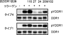
- FIG. 115 is a photograph showing the ligand-dependent phosphorylation inhibitory activity of DDR1 of an anti-DDR1 antibody.
- Ligand-dependent phosphorylation of DDR1 in the human breast cancer cell line T47D was detected by Western blotting using a polyclonal antibody that specifically recognizes DDR1 (pYDDR1) phosphorylated at tyrosine 796. Collagen type 1 or collagen type 4 was used as the ligand.
- pYDDR1 phosphorylation inhibitory activity of DDR1 of an anti-DDR1 antibody.
- Anti-DDR1 antibody uptake into cells was evaluated by adding anti-DDR1 antibody and MabZAP (saporin-labeled anti-mouse IgG antibody) to human breast cancer cell line T47D and examining whether cell proliferation was inhibited.
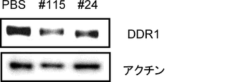
- the vertical axis represents the ratio of cell growth when anti-DDR1 antibody and MabZAP are added, assuming that cell growth when neither anti-DDR1 antibody nor MabZAP is added is 1. Uptake into cells was observed at # 115 and # 24. It is a photograph showing the fall of the expression level of DDR1 by an anti- DDR1 antibody.
- PBS negative control
- anti-DDR1 antibody was added to human breast cancer cell line T47D, and the expression level of DDR1 in the cells was detected by Western blotting. Actin was used as an internal control. In # 115 and # 24, a decrease in the expression level of DDR1 was observed. It is a graph showing the ligand-dependent TGF ⁇ expression suppression activity of anti-DDR1 antibody.
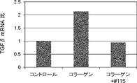
- the expression level of ligand-dependent TGF ⁇ mRNA in a co-culture system of human lung cancer cell line NCI-H1993 and mouse fibroblast MRC5 was measured by quantitative RT-PCR (qRT-PCR). Collagen type 1 was used as the ligand.
- the vertical axis represents the ratio of the amount of TGF ⁇ mRNA when no ligand / antibody is added (control). In # 115, a ligand-dependent suppression of TGF ⁇ expression was observed.
- the present invention provides a novel anti-DDR1 antibody having antitumor activity.
- DDR1 Discoidin Domain Receptor 1
- the present inventors have found that antibodies that bind to the stalk region are antibodies alone or in comparison with antibodies that bind to other discoidin (DS) regions. It was found for the first time that it has strong antitumor activity. That is, the present invention provides an antibody that binds to the DDR1 stalk region.
- the animal species of DDR1 used in the present invention is preferably a mammal, and most preferably a human.
- the gene sequence and amino acid sequence of human DDR1 are registered as GenBank accession numbers NM_013993 and NP_054699, respectively.
- the gene sequence and amino acid sequence of mouse DDR1 are registered as GenBank accession numbers NM_007584 and NP_031610, and the gene sequence and amino acid sequence of rat DDR1 are registered as NM_013137 and NP_037269, respectively.
- those skilled in the art can determine the sequence by gene cloning using homology between species.
- DDR1 is a single transmembrane receptor tyrosine kinase (RTK), and is structurally divided into an extracellular region, a transmembrane (TM) region, and an intracellular region (kinase region).
- the amino acid sequence of human DDR1 is shown in SEQ ID NO: 2, and the base sequence is shown in SEQ ID NO: 1.
- the extracellular region includes a Discoidin (DS) region and a stalk region, and the DS region is considered to be involved in binding to collagen as a ligand.
- the region consisting of the amino acid sequence from the 32nd to the 185th is the DS region
- the region consisting of the amino acid sequence from the 199th to 412th is the stalk region It is called.
- the amino acid sequence of the DS region of human DDR1 is shown in SEQ ID NO: 3
- the amino acid sequence of the stalk region is shown in SEQ ID NO: 4.
- regions corresponding to each region can be similarly determined from sequence homology with human DDR1.
- binding activity of the antibody in the present invention can be measured using techniques known to those skilled in the art, such as ELISA (Enzyme-linked immunosorbent assay), Biacore, Western blotting, FACS and the like.
- binding means that the binding activity value measured by the method as described above is at least twice as high as the binding activity value of the negative control or the background value of the measurement method. It is preferably 3 times or more, more preferably 5 times or more, and most preferably 10 times or more.
- an antibody that binds to the DDR1 stalk region is obtained by immunizing an animal such as a mouse with a DDR1 protein to obtain a plurality of anti-DDR1 antibodies, and then screening to a stalk region.
- a partial protein corresponding to the DDR1 stalk region can be prepared in advance using a genetic engineering technique known to those skilled in the art, and immunized to an animal such as a mouse. It is also possible to produce it.
- One preferred embodiment of the anti-DDR1 antibody provided by the present invention includes an anti-DDR1 antibody characterized by suppressing cell proliferation.
- the cells in the present invention may be primary cultured cells collected from living tissue, or may be cell lines established by immortalizing them by some method.
- the cell phenotype is preferably a cell that expresses more DDR1 gene or DDR1 protein than normal cells.
- the amount of DDR1 gene expressed in the cell is determined by the amount of DDR1 protein expressed in the cell using techniques known to those skilled in the art such as RT-PCR using a primer specific for the DDR1 gene and GeneChip analysis. Can be evaluated using techniques known to those skilled in the art, such as Western blotting using an antibody specific for DDR1 protein and immunohistochemical staining (IHC).
- IHC immunohistochemical staining
- “suppressing cell proliferation” means that when an anti-DDR1 antibody is brought into contact with a cell, the proliferation of the cell is reduced as compared to when the antibody is not brought into contact.
- the decrease in cell proliferation includes a decrease in the growth rate while the cells are alive, and also includes the induction of cell death by apoptosis, necrosis, and the like.
- Inhibition of cell proliferation is preferably caused as a result of anti-DDR1 antibody binding to DDR1 on the cell surface. Inhibition of cell proliferation may be observed in vitro or in vivo.
- Suppression of cell proliferation in vitro can be measured by assay systems known to those skilled in the art, such as [ 3 H] thymidine incorporation method, MTT method, WST method, etc.
- Inhibition of cell proliferation in vivo It can be measured by an assay system known to those skilled in the art such as a xenograft model for transplanting human cells.
- the growth of cells in the evaluation system is completely suppressed to 100%, the growth is suppressed by, for example, 30% or more, 40% or more, 50% or more, 60% or more, 70% or more by the anti-DDR1 antibody.
- the antibody 20M102 that binds to the DS region of DDR1 disclosed in the prior document (Patent Document 3) showed about 20% growth inhibition in vivo (see FIG. 6 above), the present invention provides The present inventors have found that antibodies # 24 and # 27 that bind to the stalk region of DDR1 exhibit 48%, 61%, and # 115, respectively, 71% growth inhibition in vivo (Example 3 described later). reference).
- One preferred embodiment of the anti-DDR1 antibody provided by the present invention includes an anti-DDR1 antibody characterized by inhibiting cell migration.
- Cell migration is a phenomenon that explains the spontaneous movement of cells in a living body, and antibodies that inhibit cell migration may be able to suppress invasion or metastasis of cancer cells and are considered useful.
- “inhibiting cell migration” means that when an anti-DDR1 antibody is brought into contact with a cell, the cell migration activity is reduced as compared with the case where the antibody is not brought into contact. Inhibition of cell migration can be measured by an assay system that detects cell migration between chambers as described in Example 4 and the like. In cells expressing DDR1 protein on the cell surface, cell migration is stimulated by binding of a ligand to the extracellular region of DDR1, and cell migration is particularly induced by binding of collagen as a ligand.
- type I collagen or type IV collagen is suitable. Inhibition of cell migration is preferably caused as a result of the anti-DDR1 antibody inhibiting binding of DDR1 to the ligand.
- One preferred embodiment of the anti-DDR1 antibody provided by the present invention includes an anti-DDR1 antibody characterized by inhibiting phosphorylation of DDR1 in cells.
- the phosphorylation of DDR1 is preferably phosphorylation of a tyrosine residue contained in DDR1, and particularly preferably phosphorylation of the 796th tyrosine residue in the amino acid sequence of DDR1.
- DDR1 phosphorylation is known to transmit signals such as cell survival or cell invasion / metastasis, and antibodies that inhibit DDR1 phosphorylation may suppress cancer cell proliferation, invasion / metastasis, etc. There seems to be useful.
- inhibiting DDR1 phosphorylation means that when an anti-DDR1 antibody is brought into contact with a cell, the proportion of phosphorylated DDR1 is lower than when the antibody is not brought into contact. To do. Inhibition of DDR1 phosphorylation can be measured by an assay system known to those skilled in the art, such as Western blotting using an anti-phosphotyrosine antibody. In cells expressing DDR1 protein on the cell surface, DDR1 phosphorylation occurs when a ligand binds to the extracellular region of DDR1, and in particular, phosphorylation of DDR1 is induced by binding of collagen as a ligand. (Vogel W. et al., Mol. Cell (1997) 1, 13-23).
- DDR1 phosphorylation is preferably caused as a result of an anti-DDR1 antibody inhibiting binding of DDR1 to a ligand.
- DDR1 phosphorylation may be caused by DDR1 autophosphorylation or by phosphorylation by other kinases.
- the phosphorylation of DDR1 can be measured, for example, by the following method.
- DDR1-expressing cells eg, A549, NCI-H1993, SK-MES-1, Panc-1, MFE-280, HCT-116, BT474, ZR-75-1, T47D, BxPC3, etc.
- DDR1-expressing cells eg, A549, NCI-H1993, SK-MES-1, Panc-1, MFE-280, HCT-116, BT474, ZR-75-1, T47D, BxPC3, etc.
- the tyrosine residue of the extracted DDR1 protein is phosphorylated by Western blotting using an anti-phosphotyrosine antibody. More specifically, it can be measured by the method described in Example 5.
- the expression of DDR1 in the above cells is indicated by the following documents (L'HOTE CMM et al., FASEB J.
- One preferred embodiment of the anti-DDR1 antibody provided by the present invention is an anti-DDR1 antibody that is incorporated into cells. It is already known that there is a phenomenon in which substances existing on the cell surface are actively taken into cells through some mechanism. The uptake of the anti-DDR1 antibody into the cell is preferably caused as a result of binding of the anti-DDR1 antibody to the DDR1 protein expressed on the cell surface. Antibodies incorporated into cells are thought to be useful because they may suppress the growth of cancer cells by conjugating a compound having cytotoxic activity such as a toxin.
- incorporated into a cell means that when an anti-DDR1 antibody is brought into contact with a cell, a larger amount of the antibody is taken up than when a negative control antibody is brought into contact.
- the uptake of the antibody into the cell was taken into the cell by directly labeling the antibody with a toxin or by binding a secondary antibody labeled with the toxin to the antibody as in Example 6. It can be measured as the amount of toxin.
- the cell phenotype is preferably a cell that expresses more DDR1 than a normal cell, and such a cell has a gene level such as RT-PCR using a primer specific for the DDR1 gene or GeneChip analysis. Or analysis of protein level such as Western blotting using an antibody specific for DDR1 protein or immunohistochemical staining (IHC).
- One preferred embodiment of the anti-DDR1 antibody provided by the present invention includes an anti-DDR1 antibody characterized by reducing the expression level of DDR1 in cells.
- the decrease in the expression level of DDR1 may be due to promotion of DDR1 protein degradation or may be due to suppression of translation of DDR1 protein. Further, it may be due to promotion of DDR1 mRNA degradation, or may be due to suppression of transcription of DDR1 mRNA.
- An antibody that reduces the expression level of DDR1 is considered useful because it may be able to suppress phenomena such as survival, invasion, and metastasis involving DDR1 in cancer cells.
- reducing the expression level of DDR1 means that when the anti-DDR1 antibody is brought into contact with a cell, the expression level of DDR1 is lowered as compared with the case where the antibody is not brought into contact.
- the decrease in the expression level of DDR1 is preferably caused as a result of binding of anti-DDR1 antibody to DDR1 on the cell surface.
- the amount of DDR1 mRNA can be measured by an assay system known to those skilled in the art, such as RT-PCR using a primer specific for the DDR1 gene.
- the amount of DDR1 protein can be measured by an assay system known to those skilled in the art, such as Western blotting using an antibody specific for DDR1 protein.
- DDR1 protein is extracted from DDR1-expressing cells (for example, A549, NCI-H1993, SK-MES-1, Panc-1, MFE-280, HCT-116, BT474, ZR-75-1, T47D, BxPC3, etc.). Extracted DDR1 protein is detected by Western blotting. More specifically, it can be measured by the method described in Example 7.
- One preferred embodiment of the anti-DDR1 antibody provided by the present invention is an anti-DDR1 antibody characterized by reducing the expression level of TGF ⁇ in a cell.
- the decrease in the expression level of TGF ⁇ may be due to promotion of degradation of TGF ⁇ protein, or may be due to suppression of translation of TGF ⁇ protein. Further, it may be due to promotion of degradation of TGF ⁇ mRNA, or may be due to suppression of transcription of TGF ⁇ mRNA.
- TGF ⁇ is a marker molecule that is known to increase expression during epithelial-mesenchymal transition (EMT), which is reported to promote tumor formation, and reduces the expression level of TGF ⁇ .
- EMT epithelial-mesenchymal transition
- Antibodies are thought to be useful because they may inhibit tumor formation by suppressing cell epithelial-mesenchymal transition.
- one aspect of the anti-DDR1 antibody in the present invention may include an anti-DDR1 antibody characterized by inhibiting cell epithelial-mesenchymal transition (EMT).
- EMT cell epithelial-mesenchymal transition
- “reducing the expression level of TGF ⁇ ” means that when the anti-DDR1 antibody is brought into contact with a cell, the expression level of TGF ⁇ is lowered as compared with the case where the antibody is not brought into contact.
- collagen may induce the expression of TGF ⁇ via DDR1 (Guerrot D. et al., Am. J. Pathol. (2011) 179). 83-91).
- type I collagen or type IV collagen is suitable.
- the decrease in the expression level of TGF ⁇ is preferably caused as a result of the anti-DDR1 antibody inhibiting the binding between DDR1 and collagen.
- the amount of TGF ⁇ mRNA can be measured by an assay system known to those skilled in the art, such as RT-PCR using a primer specific for the TGF ⁇ gene.
- the amount of TGF ⁇ protein can be measured by an assay system known to those skilled in the art, such as Western blotting using an antibody specific for TGF ⁇ protein.
- the expression level of TGF ⁇ can be measured, for example, by the following method.
- DDR1-expressing cells eg, A549, NCI-H1993, SK-MES-1, Panc-1, MFE-280, HCT-116, BT474, ZR-75-1, T47D, BxPC3, etc.
- fibroblasts such as MRC5
- the amount of TGF ⁇ mRNA is measured by RT-PCR using a primer specific for TGF ⁇ . More specifically, it can be measured by the method described in Example 8.
- the cell in the present invention is preferably a cancer cell, more preferably a cancer cell expressing DDR1.
- cancer cells that express more DDR1 than normal cells.
- Such cells include gene-level analysis such as RT-PCR and GeneChip analysis using primers specific for the DDR1 gene, Western blotting and immunohistochemical staining (IHC) using antibodies specific for the DDR1 protein, etc.
- the protein level can be selected by analysis.
- the cancer cell type of the cancer cell of the present invention is not particularly limited.
- lung cancer small cell lung cancer, non-small cell lung cancer, etc.
- breast cancer glioma, ovarian cancer
- gastric cancer pancreatic cancer
- esophageal cancer endometrial cancer
- bile duct examples include cancer, colon cancer, liver cancer, leukemia, lymphoma, kidney cancer, prostate cancer, melanoma, thyroid cancer, bladder cancer, osteosarcoma and the like.
- lung cancer non-small cell lung cancer
- breast cancer glioma, ovarian cancer
- gastric cancer pancreatic cancer
- esophageal cancer endometrial cancer
- bile duct cancer examples include cancer, colon cancer, liver cancer, leukemia, lymphoma, kidney cancer, prostate cancer, melanoma, thyroid cancer, bladder cancer, osteosarcoma and the like.
- lung cancer non-small cell lung cancer
- breast cancer glioma, ovarian cancer
- the present invention provides the following antibodies (a) to (c): (A) an antibody (# 115) having the same amino acid sequence as the antibody produced by the hybridoma deposited under accession number FERM BP-11399; (B) an antibody (# 27) having the same amino acid sequence as the antibody produced by the hybridoma deposited under accession number FERM BP-11398; (C) An antibody (# 24) having the same amino acid sequence as the antibody produced by the hybridoma deposited under accession number FERM BP-11397.
- hybridomas are deposited internationally as follows. The contents specifying the international deposit of each hybridoma are described below. The following depositary organization (independent administrative agency, National Institute of Advanced Industrial Science and Technology, Patent Biological Depositary Center) has been handed over to the National Institute of Technology and Evaluation (NITE) on April 1, 2012.
- the antibodies described in (a) to (c) above are all antibodies that bind to the DDR1 stalk region.
- a person skilled in the art can determine the base sequence and amino acid sequence of an antibody produced by a hybridoma using, for example, the methods described in the Examples below.
- a known gene can be determined based on the sequence.
- Engineering techniques can be used to produce recombinant antibodies.
- the present invention also provides an antibody having the same CDR sequence as that of the antibodies described in (a) to (c) above.
- There are 6 CDRs in an antibody that is, H chain CDR1, CDR2, CDR3, L chain CDR1, CDR2, CDR3, and any one of these CDRs may be the same, and more preferably the H chain. It is sufficient that the three CDRs or the three CDRs of the L chain are the same, and it is more preferable that all the six CDRs are the same.
- a person skilled in the art can bind the CDR of the antibody described in any one of (a) to (c) above to another appropriate antibody, thereby binding the DDR1 to the stalk region substantially equivalent to the antibody.
- CDR-grafted antibody can be prepared, and such an antibody is useful in the same manner as the antibody.
- the position and numbering system of the CDR region and the FR region of the antibody are defined by, for example, Kabat et al. (Kabat EA et al., (1991) Sequences of Proteins of Immunological Edition, Fifth Edition, US Department). Health and Human Services, US Government Printing Office).
- the present invention provides an antibody that competes with the antibody of the present invention for binding to DDR1.
- “competing binding to DDR1” means that the binding activity of the antibody of the present invention to DDR1 when an anti-DDR1 antibody coexists in an assay system for measuring the binding of an antibody to DDR1. It means to decline.
- Such an antibody is considered to be an antibody that binds to an antigenic determinant (epitope) that is the same as or very close to that of the antibody of the present invention, and thus is useful in the same manner as the antibody of the present invention.
- the full length of the DDR1 protein may be used, or the extracellular region of the DDR1 protein may be used. Alternatively, the stalk region of the DDR1 protein may be used.
- Competition for binding to DDR1 can be measured by an assay system known to those skilled in the art such as a cross-blocking assay.
- a competitive ELISA assay that utilizes an enzyme label is a preferred cross-blocking assay.
- the competition for binding to DDR1 can be measured, for example, by the following method.
- the anti-DDR1 antibody is added after preincubation of the DDR1 protein coated on the wells of the microtiter plate in the presence or absence of the test antibody.
- the amount of anti-DDR1 antibody that binds to the DDR1 protein in the well decreases.
- the amount of antibody to be bound can be easily measured by labeling the anti-DDR1 antibody in advance.
- the amount of antibody bound can be measured by labeling anti-DDR1 antibody with biotin and using an avidin-peroxidase conjugate and its appropriate substrate.
- the amount of bound antibody can be measured by radioactively labeling or fluorescently labeling the anti-DDR1 antibody.
- the amount of antibody to be bound is determined by a labeled antibody that specifically recognizes the constant region of the antibody derived from the animal species. It can also be measured. Alternatively, if the antibodies are derived from the same animal species but have different subclasses, the amount of antibody to be bound can be measured with a labeled antibody that specifically recognizes each subclass.
- the present invention provides an antibody that binds to the same epitope to which the antibody of the present invention binds.
- An epitope of an antibody can be identified by a method of synthesizing a group of peptides (such as a peptide array) that covers the amino acid sequences of the antigen so as to overlap each other and measuring the binding activity of the antibody to each peptide (Poetz O Et al., Proteomics (2005) 5, 2402-11).
- a method for analyzing the crystal structure of an antigen-antibody (Vyas NK, et al., Biochemistry (2004) 41, 13575-86), a group of mutant proteins in which the amino acid sequence of the antigen is substituted with alanine one by one is prepared.
- epitope thus identified is identical to the epitope to which the antibody of the present invention binds or is very close to the epitope to which the antibody of the present invention binds, then the antibody that binds to the epitope Since the antibody is considered to have a binding activity equivalent to that of the antibody of the invention, it is useful in the same manner as the antibody of the present invention.
- “Epitopes are very close” means that the difference in epitope positions is preferably within 5 amino acids, more preferably within 4 amino acids, even more preferably within 3 amino acids, particularly preferably within 2 amino acids, most preferably 1 Means an amino acid.
- an antibody that binds to an epitope to which the antibody of the present invention binds can be obtained by methods known to those skilled in the art.
- the epitope to which the antibody of the present invention binds is determined by the above-described method, and an antibody is prepared using a polypeptide having an amino acid sequence contained in the epitope as an immunogen, or an epitope of an antibody prepared by a normal method And a method for selecting an antibody having the same epitope as that of the antibody of the present invention.
- the present invention relates to an antibody in which one or more amino acids are added, deleted and / or substituted with other amino acids in the antibody of the present invention, and the antibody before the addition, deletion and / or substitution is performed.
- Antibodies having equivalent binding activity to the DDR1 stalk region are provided.
- “having an equivalent binding activity to the DDR1 stalk region” means that an antibody in which one or more amino acids are added, deleted, and / or substituted with another amino acid to the DDR1 stalk region. Is preferably 70% or more, more preferably 80% or more, still more preferably 90% or more, and most preferably 95% or more compared to the antibody before the addition, deletion and / or substitution. Means. Since such an antibody is considered to have almost the same properties as the antibody of the present invention, it is useful as well as the antibody of the present invention.
- Amino acid additions, deletions and / or substitutions are described, for example, by site-directed mutagenesis (Hashimoto-Gotoh T. et al., Gene (1995) 152, 271-275, Zoller MJ & Smith M., Methods Enzymol (1983). ) 100, 468-500, Kramer W. et al., Nucleic Acids Res (1987) 12, 9441-9456, Kramer W. & Fritz H. J., Methods Enzymol (1987) 154, 350-367, Kunkel TA,. Proc Natl Acad Sci USA (1985) 82, 488-492) can be used by methods known to those skilled in the art.
- amino acid side chain is substituted with another amino acid that preserves the properties of the amino acid side chain.
- amino acid substitutions that preserve the properties of amino acid side chains include, for example, hydrophobic amino acids (A, I, L, M, F, P, W, Y, V), hydrophilic amino acids (R, D, N , C, E, Q, G, H, K, S, T) amino acids having aliphatic side chains (G, A, V, L, I, P), amino acids having hydroxyl group-containing side chains (S, T, Y), amino acids having sulfur atom-containing side chains (C, M), amino acids having carboxylic acid and amide-containing side chains (D, N, E, Q), amino acids having base-containing side chains (R, K, H) ), Amino acid substitutions within each group such as amino acids (H, F, Y, W) having aromatic-containing side chains.
- the position of the amino acid to which addition, deletion and / or substitution is performed is not particularly limited, but addition, deletion and / or substitution to an amino acid which is not involved in antigen binding or antibody structure maintenance Is preferred.
- an antibody is divided into a constant region and a variable region, those skilled in the art can easily specify such a position in the constant region.
- the variable region is divided into a framework region and a CDR region, those skilled in the art can specify such a position in the framework region without undue burden.
- One skilled in the art can also identify such positions within the CDR regions.
- the antibody of the present invention may be a polyclonal antibody or a monoclonal antibody, but is preferably a monoclonal antibody.
- the monoclonal antibody can be obtained by using a known means such as a hybridoma method for immunizing an animal with an antigen or a phage display method for screening an antibody library.
- the monoclonal antibodies of the present invention include not only antibodies obtained from clones derived from antibody-producing cells such as hybridomas, but also humanized antibodies and chimeric antibodies not derived from hybridomas.
- the antibody subclass is not particularly limited.
- IgG, IgM, IgA, IgD, IgE and the like are preferable, and IgG is more preferable.
- a monoclonal antibody can be obtained as follows. First, a DDR1 protein serving as an antigen is prepared, and this is immunized to an animal by a normal immunization method. Immune cells obtained from the immunized animal are fused with known parental cells by a conventional cell fusion method to obtain a hybridoma. From the obtained hybridomas, a hybridoma producing the desired anti-DDR1 antibody is selected by a usual screening method. Specifically, monoclonal antibodies can be obtained by the method described in Example 1.
- Monoclonal antibodies are prepared as follows, for example. First, by expressing the DDR1 gene, a DDR1 protein used as a sensitizing antigen for antibody acquisition can be obtained.
- the base sequence of the human DDR1 gene is already known (GenBank accession number NM — 013993). That is, after inserting a gene sequence encoding DDR1 into a known expression vector to transform an appropriate host cell, the target DDR1 protein can be purified from the host cell or culture supernatant by a known method.
- a purified natural DDR1 protein can also be used in the same manner. The purification can be generated by using a plurality of chromatographies such as ordinary ion chromatography and affinity chromatography one time or a plurality of times, in combination or independently.
- a fusion protein obtained by fusing a partial polypeptide containing at least a part of the DDR1 stalk region with a different polypeptide can also be used as an immunogen.
- an immunogen for example, an Fc fragment of an antibody, a peptide tag, or the like can be used.
- a vector that expresses a fusion protein can be prepared by fusing genes encoding two or more desired polypeptide fragments in-frame and inserting the fusion gene into an expression vector as described above ( Sambrook J. et al., Molecular Cloning 2nd ed. (1989) 9.47-9.58, Cold Spring Harbor Lab. Press).
- the DDR1 protein thus purified can be used as a sensitizing antigen used for immunization against mammals.
- a peptide having the entire DDR1 stalk region or at least 5 or more consecutive amino acid sequences can be used as the partial peptide.
- the at least 5 or more consecutive amino acid sequences are preferably 6 or more, more preferably 8 or more consecutive amino acid sequences. Further, at least 5 or more consecutive amino acid sequences are sequences specific to the DDR1 stalk region and have an antigenicity.
- the mammal immunized with the sensitizing antigen is not particularly limited.
- an immunized animal in consideration of compatibility with a parent cell used for cell fusion.
- mice, rats, hamsters, rabbits, chickens, monkeys and the like can be used as immunized animals.
- rodent animals such as mice, rats and hamsters are preferred as immunized animals.
- the above animals can be immunized with a sensitizing antigen.
- mammals can be immunized by injecting a sensitizing antigen intraperitoneally or subcutaneously. Specifically, the sensitizing antigen is administered to mammals several times every 4 to 21 days.
- the sensitizing antigen is used for immunization after being diluted with PBS (Phosphate-Buffered Saline) or physiological saline at an appropriate dilution rate.
- a sensitizing antigen can be administered with an adjuvant. For example, it can be mixed with Freund's complete adjuvant and emulsified to obtain a sensitizing antigen.
- An appropriate carrier can be used for immunization with the sensitizing antigen.
- a partial peptide having a small molecular weight is used as a sensitizing antigen, it is desirable to immunize the sensitizing antigen peptide by binding it to a carrier protein such as albumin or keyhole limpet hemocyanin.
- immune cells are collected from the mammal and subjected to cell fusion.
- spleen cells can be used.
- Mammalian myeloma cells are used as the cells fused with the immune cells.
- the myeloma cell is preferably provided with an appropriate selection marker for screening.
- a selectable marker refers to a trait that can (or cannot) survive under certain culture conditions.
- Known selection markers include hypoxanthine-guanine-phosphoribosyltransferase deficiency (hereinafter abbreviated as HGPRT deficiency) or thymidine kinase deficiency (hereinafter abbreviated as TK deficiency).
- HGPRT deficiency hypoxanthine-guanine-phosphoribosyltransferase deficiency
- TK deficiency thymidine kinase deficiency
- Cells having HGPRT or TK deficiency have hypoxanthine-aminopterin-thymidine sensitivity (hereinafter abbreviated as HAT sensitivity).
- HGPRT-deficient and TK-deficient cells can be selected in a medium containing 6 thioguanine, 8 azaguanine (hereinafter abbreviated as 8AG), or 5 ′ bromodeoxyuridine, respectively.
- 8AG 8 azaguanine
- 5 ′ bromodeoxyuridine normal cells die because they incorporate these pyrimidine analogs into the DNA, but cells deficient in these enzymes cannot survive these pyrimidine analogs and can survive in selective media.
- G418 resistance confers resistance to 2-deoxystreptamine antibiotics (gentamicin analogs) with a neomycin resistance gene.
- Various myeloma cells suitable for cell fusion are known. For example, P3 (P3x63Ag8.653) (J. Immunol.
- the cell fusion between the immune cells and myeloma cells can be performed according to a known method, for example, the method of Kohler and Milstein (Kohler G. & Milstein C., Methods Enzymol. (1981) 73, 3-46). Is possible. More specifically, for example, the cell fusion can be carried out in a normal nutrient culture medium in the presence of a cell fusion promoter.
- a cell fusion promoter for example, polyethylene glycol (PEG), Sendai virus (HVJ) or the like can be used.
- an auxiliary agent such as dimethyl sulfoxide can be added as desired in order to increase the fusion efficiency.
- the usage ratio of immune cells and myeloma cells can be set arbitrarily.
- the number of immune cells is preferably 1 to 10 times that of myeloma cells.
- the culture solution used for the cell fusion for example, RPMI1640 culture solution suitable for growth of the myeloma cell line, MEM culture solution, and other normal culture solutions used for this type of cell culture can be used.
- serum supplements such as fetal calf serum (FCS) can be added to the culture medium.
- a predetermined amount of the immune cells and myeloma cells are mixed well in the culture solution, and a target PEG (hybridoma) is formed by mixing a PEG solution preheated to about 37 ° C.
- the In the cell fusion method for example, PEG having an average molecular weight of about 1000 to 6000 can be usually added at a concentration of 30 to 60% (w / v).
- cell fusion agents and the like that are undesirable for the growth of hybridomas are removed by sequentially adding the appropriate culture solution listed above, and centrifuging to remove the supernatant.
- the hybridoma obtained in this manner can be selected by using a selective culture solution corresponding to the selection marker possessed by the myeloma used for cell fusion.
- a selective culture solution corresponding to the selection marker possessed by the myeloma used for cell fusion.
- cells having HGPRT or TK deficiency can be selected by culturing in a HAT culture solution (a culture solution containing hypoxanthine, aminopterin and thymidine). That is, when HAT-sensitive myeloma cells are used for cell fusion, cells that have succeeded in cell fusion with normal cells can be selectively proliferated in the HAT culture solution.
- the culture using the HAT culture solution is continued for a time sufficient for cells other than the target hybridoma (non-fused cells) to die.
- the target hybridoma can be selected by culturing for several days to several weeks. Subsequently, by carrying out the usual limiting dilution method, screening and single cloning of the hybridoma producing the target antibody can be performed.
- Screening and single cloning of the target antibody can be suitably performed by a screening method based on a known antigen-antibody reaction.
- the antigen is bound to a carrier such as beads made of polystyrene or the like, or a commercially available 96-well microtiter plate, and reacted with the culture supernatant of the hybridoma.
- a secondary antibody labeled with an enzyme is reacted.
- the secondary antibody binds to the carrier via this antibody.
- the DDR1 protein that is practically homogeneous, including those used for immunization, can be preferably used as the antigen.
- a target antibody can be obtained by sensitizing human lymphocytes with an antigen.
- human lymphocytes are first sensitized with DDR1 protein in vitro.
- the immunized lymphocytes are then fused with an appropriate fusion partner.
- the fusion partner for example, a myeloma cell derived from human and having a permanent division ability can be used (Japanese Patent Publication No. 1-59878).
- the anti-DDR1 antibody obtained by this method is a human antibody having a binding activity to the DDR1 protein.
- an anti-DDR1 human antibody can also be obtained by immunizing a transgenic animal having all repertoires of human antibody genes with DDR1 protein as an antigen.
- Antibody-producing cells of the immunized animal can be immortalized by treatment such as cell fusion with an appropriate fusion partner or Epstein-Barr virus infection.
- Human antibodies can also be isolated from the immortalized cells thus obtained (WO94 / 25585, WO93 / 12227, WO92 / 03918, WO94 / 02602). Further, by cloning the immortalized cells, it is possible to clone cells that produce an antibody having the desired reaction specificity.
- the immune system of the animal recognizes human DDR1 as a foreign substance. Therefore, a human antibody against human DDR1 can be easily obtained.
- the hybridoma producing the monoclonal antibody thus produced can be subcultured in a normal culture solution.
- the hybridoma can also be stored for a long time in liquid nitrogen.
- the hybridoma can be cultured according to a usual method, and the target monoclonal antibody can be obtained from the culture supernatant.
- a hybridoma can be administered to a mammal compatible therewith to proliferate and a monoclonal antibody can be obtained as its ascites.
- the former method is suitable for obtaining a highly pure antibody.
- an antibody encoded by an antibody gene cloned from an antibody-producing cell can also be used.
- the cloned antibody gene can be expressed as an antibody by incorporating it into a suitable vector and introducing it into a host. Methods for isolation of antibody genes, introduction into vectors, and transformation of host cells have already been established (Vandam AM et al., Eur. J. Biochem. (1990) 192, 767-775. ).
- cDNA encoding a variable region (V region) of an anti-DDR1 antibody can be obtained from a hybridoma cell that produces the anti-DDR1 antibody.
- V region variable region
- RNA is extracted from the hybridoma.
- Methods for extracting total RNA from cells include, for example, guanidine ultracentrifugation (Chirgwin J.M. et al., Biochemistry (1979) 18, 5294-5299), AGPC method (Chomczynski P. et al., Anal. Biochem. ( 1987) 162, 156-159) and the like can be used.
- MRNA can be purified from the extracted total RNA using mRNA Purification Kit (GE Healthcare) or the like.
- kits for directly extracting mRNA from cells such as QuickPrep mRNA Purification Kit (GE Healthcare) are also commercially available.
- mRNA can also be obtained from the hybridoma.
- CDNA encoding the antibody V region can be synthesized from the obtained mRNA using reverse transcriptase.
- cDNA can be synthesized by AMV Reverse Transscriptase First-strand cDNA Synthesis Kit (Seikagaku Corporation).
- 5'-RACE method Frohman MA et al., Proc. Natl. Acad. Sci.
- the target cDNA fragment is purified from the obtained PCR product and then ligated with vector DNA.
- a recombinant vector is produced, introduced into Escherichia coli or the like and a colony is selected, a desired recombinant vector can be prepared from Escherichia coli that has formed the colony.
- Whether or not the recombinant vector has the target cDNA base sequence can be confirmed by a known method such as the dideoxynucleotide chain termination method.
- a PCR method using primers for variable region gene amplification can also be used.
- cDNA is synthesized using the extracted mRNA as a template to obtain a cDNA library. It is convenient to use a commercially available kit for the synthesis of the cDNA library. Actually, the amount of mRNA obtained from only a small number of cells is extremely small, so the yield is low when it is purified directly. Therefore, it is usually purified after adding carrier RNA that is apparently free of antibody genes. Alternatively, when a certain amount of RNA can be extracted, it is possible to efficiently extract only RNA of antibody-producing cells. For example, it may not be necessary to add carrier RNA for RNA extraction from 10 or more, 30 or more, preferably 50 or more antibody-producing cells.
- the antibody gene is amplified by the PCR method using the obtained cDNA library as a template.
- Primers for amplifying antibody genes by PCR are known. For example, J. et al. Mol. Biol. (1991) 222, 581-597 and the like can be used to design primers for human antibody gene amplification. These primers have different nucleotide sequences for each immunoglobulin subclass. Therefore, when a cDNA library whose subclass is unknown is used as a template, the PCR method is performed in consideration of all possibilities.
- a primer capable of amplifying genes encoding ⁇ 1 to ⁇ 5 as a heavy chain and ⁇ chain and ⁇ chain as a light chain may be used. it can.
- a primer that anneals to a portion corresponding to the hinge region is generally used as the 3 ′ primer.
- a primer corresponding to each subclass can be used as the 5′-side primer.
- PCR products using primers for gene amplification of each subclass of heavy chain and light chain should be independent libraries.
- an immunoglobulin comprising a combination of a heavy chain and a light chain can be reconstructed.
- the target antibody can be screened using the binding activity of the reconstituted immunoglobulin to the antigen as an index.
- the cDNA is digested with a restriction enzyme that recognizes restriction enzyme sites inserted at both ends of the cDNA.
- a preferred restriction enzyme is a restriction enzyme that recognizes and digests a base sequence that is unlikely to appear in a base sequence constituting an antibody gene.
- a restriction enzyme that provides a sticky end is preferred.
- An antibody expression vector can be obtained by inserting cDNA encoding the V region of the antibody digested as described above into an appropriate expression vector.
- a chimeric antibody can be obtained by fusing the gene encoding the antibody constant region (C region) and the gene encoding the V region in-frame.
- the chimeric antibody refers to an antibody having a different origin from the constant region and the variable region.
- a heterologous chimeric antibody such as mouse-human
- a human-human homologous chimeric antibody is also included in the chimeric antibody of the present invention.
- a chimeric antibody expression vector can also be constructed by inserting the V region gene into an expression vector having a constant region in advance.
- a restriction enzyme recognition sequence for a restriction enzyme that digests the V region gene is placed on the 5 ′ side of an expression vector holding a DNA encoding a desired antibody constant region (C region).
- a chimeric antibody expression vector is constructed by digesting both with the same combination of restriction enzymes and fusing them in frame.
- the antibody gene can be incorporated into an expression vector so as to be expressed under the control of the expression control region.
- An expression control region for expressing an antibody includes, for example, an enhancer and a promoter. Subsequently, by transforming an appropriate host cell with this expression vector, a recombinant cell that expresses the DNA encoding the antibody can be obtained.
- DNAs encoding antibody heavy chains (H chains) and light chains (L chains) can be incorporated into separate expression vectors. By co-transfecting a vector incorporating the H chain and the L chain into the same host cell at the same time, an antibody molecule having the H chain and the L chain can be expressed.
- host cells may be transformed by incorporating DNAs encoding H and L chains into a single expression vector (WO94 / 11523).
- hosts and expression vectors for expressing antibodies are known. Any of these expression systems can be applied to the present invention.
- animal cells When eukaryotic cells are used as hosts, animal cells, plant cells, or fungal cells can be used.
- animal cells that can be used in the present invention include mammalian cells (CHO, COS, 3T3, myeloma, BHK (baby hamster kidney), Hela, C127, HEK293, Bowes melanoma cells, Vero, and the like. ), Amphibian cells (Xenopus oocytes, etc.), insect cells (Drosophila S2, sf9, sf21, Tn5, etc.) can be used.
- mammalian cells CHO, COS, 3T3, myeloma, BHK (baby hamster kidney), Hela, C127, HEK293, Bowes melanoma cells, Vero, and the like.
- Amphibian cells Xenopus oocytes, etc.
- Nicotiana As plant cells, antibody gene expression systems using cells derived from the genus Nicotiana, such as Nicotiana tabacum, are known. Callus cultured cells can be used for transformation of plant cells.
- fungal cells include yeasts (eg, Saccharomyces cerevisiae, Saccharomyces pombe), methanol-utilizing yeasts (Pichia pistia piapia, etc.).
- yeasts eg, Saccharomyces cerevisiae, Saccharomyces pombe
- methanol-utilizing yeasts Piichia pistia piapia, etc.
- a fungus can be used.
- antibody gene expression systems using prokaryotic cells are also known.
- bacterial cells such as E. coli, Streptococcus, Staphylococcus, Streptomyces and Bacillus subtilis can be used in the present invention.
- the promoter / enhancer can include human cytomegalovirus early promoter / enhancer (human cytomegalovirus immediate early promoter / enhancer).
- viral promoters / enhancers or promoters / enhancers derived from mammalian cells such as human elongation factor 1 ⁇ (HEF1 ⁇ ).
- viral promoters / enhancers or promoters / enhancers derived from mammalian cells such as human elongation factor 1 ⁇ (HEF1 ⁇ ).
- viruses that can utilize promoters / enhancers include retroviruses, polyomaviruses, adenoviruses, and simian virus 40 (SV40).
- the method of Mulligan et al. (Nature (1979) 277, 108) can be used. Further, the HEF1 ⁇ promoter / enhancer can be easily used for target gene expression by the method of Mishimashima et al. (Nucleic Acids Res. (1990) 18, 5322).
- the gene can be expressed by functionally combining a useful promoter commonly used, a signal sequence for antibody secretion, and an antibody gene to be expressed.
- the promoter include lacZ promoter and araB promoter.
- the lacZ promoter the method of Ward et al. (Nature (1989) 341, 544-546, FASEB J. (1992) 6, 2422-2427) can be used.
- the araB promoter can be used for expression of a target gene by the method of Better et al. (Science (1988) 240, 1041-1043).
- a pelB signal sequence (Lei SP et al., J. Bacteriol. (1987) 169, 4379) may be used in the case of production in the periplasm of E. coli. Then, after separating the antibody produced in the periplasm, the structure of the antibody is reshaped so as to have a desired binding activity by using a protein denaturant such as urea or guanidine hydrochloride.
- a selectable marker can be inserted into the expression vector for amplification of the gene copy number in the host cell system.
- selection markers such as aminoglycoside transferase (APH) gene, thymidine kinase (TK) gene, E. coli xanthine guanine phosphoribosyltransferase (Ecogpt) gene, dihydrofolate reductase (dhfr) gene and the like can be used.
- These expression vectors are introduced into host cells, and the transformed host cells are cultured in vitro or in vivo to produce the desired antibody.
- Host cells are cultured according to a known method.
- DMEM, MEM, RPMI 1640, and IMDM can be used as a culture solution, and a serum supplement such as fetal calf serum (FCS) can be used in combination.
- FCS fetal calf serum
- the present invention relates to an antibody produced by culturing a host cell transformed in this manner. For example, antibodies obtained by culturing transformed host cells and recovering them from the culture supernatant and the like are included.
- the present invention also provides a method for producing an antibody, comprising a step of culturing transformed host cells and recovering the antibody.
- a method for producing an antibody comprising a step of culturing transformed host cells and recovering the antibody.
- the production method when the antibody of the present invention is secreted into the medium or culture supernatant, the medium or culture supernatant is collected.
- the antibody of the present invention is produced intracellularly, the cell is first lysed, and then the antibody is recovered.
- mammals and insects can also be used for the production of recombinant antibodies.
- mammals goats, pigs, sheep, mice, cows and the like can be used (Vicki Glaser, SPECTRUM Biotechnology Applications (1993)).
- a transgenic animal can be used. That is, the antibody can be obtained from an animal into which a gene encoding the target antibody has been introduced.
- an antibody gene can be constructed as a fusion gene by inserting in frame into a gene that encodes a protein that is uniquely produced in milk.
- a protein secreted into milk for example, goat ⁇ -casein can be used.
- the DNA fragment containing the fusion gene into which the antibody gene has been inserted is injected into a goat embryo, and the injected embryo is introduced into a female goat.
- the desired antibody can be obtained as a fusion protein with milk protein from milk produced by a transgenic goat (or its offspring) born from a goat that has received the embryo.
- hormones can be used as appropriate in transgenic goats to increase the amount of milk containing the desired antibody produced from the transgenic goat (Ebert KM et al., Bio / Technology (1994) 12, 699-702). ).
- the target antibody can be obtained from the body fluid of the silkworm by infecting the silkworm with a baculovirus into which a nucleic acid encoding the target antibody is inserted (Susumu et al., Nature (1985) 315: 592-4).
- a plant when a plant is used for producing the antibody of the present invention, for example, tobacco can be used.
- a nucleic acid encoding the target antibody is inserted into a plant expression vector such as pMON 530, and this vector is introduced into a bacterium such as Agrobacterium tumefaciens.
- This bacterium can be infected with tobacco, for example, Nicotiana tabacum, and the desired antibody can be obtained from the leaves of this tobacco (Ma et al., Eur. J. Immunol. (1994) 24: 131- 8).
- the antibody expressed and produced as described above can be isolated from inside or outside the host cell (medium, milk, etc.) and purified as a substantially pure and homogeneous antibody. Separation and purification of the antibody can be performed by using a known method used in normal protein purification alone or in appropriate combination. For example, ammonium sulfate or ethanol precipitation, acid extraction, chromatography column, filter, ultrafiltration, salting out, solvent precipitation, solvent extraction, distillation, immunoprecipitation, SDS-polyacrylamide gel electrophoresis, isoelectric focusing, dialysis
- the antibodies can be separated and purified by appropriately selecting and combining recrystallization and the like (Harrow E. & Lane D., Antibodies: A Laboratory Manual (1988) Cold Spring Harbor Laboratory).
- chromatography examples include affinity chromatography, ion exchange chromatography such as anion or cation exchange chromatography, phosphocellulose chromatography, hydrophobic (interaction) chromatography, gel filtration, reverse phase chromatography, adsorption chromatography, hydroxyl Examples include apatite chromatography and lectin chromatography (Strategies for Protein Purification and Characterization: A Laboratory Course Manual. Ed Daniel R. Marshak et al. (1996) Cold Spring Harbor Laboratory Press). These chromatography can be performed using liquid phase chromatography, for example, liquid phase chromatography such as HPLC and FPLC.
- Columns used for affinity chromatography include protein A columns and protein G columns. Examples of the column using protein A include Hyper D, POROS, Sepharose F F F (Pharmacia), and the like.
- an appropriate protein modifying enzyme can be allowed to act before or after purification of the antibody, and the peptide can be partly removed.
- protein modifying enzymes include trypsin, chymotrypsin, lysyl endopeptidase, protein kinase, glucosidase, and the like.
- An animal antibody-derived C region can be used as the C region of the recombinant antibody of the present invention.
- C ⁇ 1, C ⁇ 2a, C ⁇ 2b, C ⁇ 3, C ⁇ , C ⁇ , C ⁇ 1, C ⁇ 2, and C ⁇ can be used as the H chain C region of the mouse antibody, and C ⁇ and C ⁇ can be used as the L chain C region.
- animal antibodies such as rats, rabbits, goats, sheep, camels and monkeys can be used as animal antibodies other than mouse antibodies. These sequences are known.
- the C region can be modified in order to improve the stability of the antibody itself or the production of the antibody.
- an antibody when administered to a human, it can be a genetically modified antibody artificially modified for the purpose of reducing the immunogenicity of the human.
- the recombinant antibody includes, for example, a chimeric antibody and a humanized antibody.
- a chimeric antibody refers to an antibody in which different variable regions and constant regions derived from each other are linked.
- an antibody comprising a heavy chain and light chain variable region of a mouse antibody and a human antibody heavy chain and light chain constant region is a mouse-human heterochimeric antibody.
- a recombinant vector that expresses a chimeric antibody can be prepared by ligating DNA encoding the variable region of a mouse antibody with DNA encoding the constant region of a human antibody in frame and incorporating it into an expression vector.
- the chimeric antibody produced in the culture can be obtained by culturing recombinant cells transformed with the vector and expressing the incorporated DNA.
- Human antibodies can be used for the C region of chimeric and humanized antibodies.
- C ⁇ 1, C ⁇ 2, C ⁇ 3, C ⁇ 4, C ⁇ , C ⁇ , C ⁇ 1, C ⁇ 2, and C ⁇ can be used as the C region.
- C ⁇ and C ⁇ can be used as the C region.
- the amino acid sequences of these C regions and the base sequences encoding them are known.
- the human antibody C region can be modified in order to improve the antibody itself or the stability of antibody production.
- a chimeric antibody is composed of a V region of an antibody derived from a non-human animal and a C region derived from a human antibody.
- humanized antibodies include complementarity determining regions (CDRs) of non-human animal-derived antibodies, framework regions (FR) of human antibodies, and C regions derived from human antibodies. Consists of Since humanized antibodies have reduced immunogenicity in the human body, they are useful as active ingredients of the therapeutic agent of the present invention.
- variable region of an antibody is usually composed of three complementarity determining regions (CDRs) sandwiched between four framework regions (FR).
- CDR complementarity determining regions
- FR framework regions
- the CDR is a region that substantially determines the binding specificity of the antibody.
- the amino acid sequence of CDR is rich in diversity.
- the amino acid sequence constituting FR often shows high homology among antibodies having different binding specificities. Therefore, it is generally said that the binding specificity of one antibody can be transplanted to another antibody by CDR grafting.
- Humanized antibodies are also referred to as reshaped human antibodies.
- non-human animals for example, humanized antibodies obtained by transplanting CDRs of mouse antibodies into human antibodies are known.
- General genetic recombination techniques for obtaining humanized antibodies are also known.
- Overlap Extension PCR is known as a method for transplanting a CDR of a mouse antibody into a human FR.
- overlap extension PCR a base sequence encoding the CDR of a mouse antibody to be transplanted is added to a primer for synthesizing the FR of a human antibody. Primers are prepared for each of the four FRs.
- the base sequences to be linked are designed to be connected to each other in frame.
- Human FRs are synthesized individually with specific primer sets.
- a product in which DNA encoding mouse CDR is added to each FR is obtained.
- the base sequences encoding the mouse CDRs of each product are designed to overlap each other.
- the overlapping CDR portions of the products are annealed with each other to perform a complementary strand synthesis reaction.
- human FRs are linked via the mouse CDR sequence.
- a humanized antibody expression vector can be prepared by inserting the DNA obtained as described above and a DNA encoding the human antibody C region into an expression vector so as to be fused in frame. After introducing the expression vector into a host to establish a recombinant cell, the recombinant cell is cultured, and a DNA encoding the humanized antibody is expressed, whereby the humanized antibody is cultured in the cultured cell. (EP239400, WO96 / 02576).
- the CDR forms a favorable antigen-binding site when linked via CDR.
- a human antibody FR can be suitably selected.
- FR amino acid residues can be substituted so that the CDR of the reshaped human antibody forms an appropriate antigen-binding site.
- amino acid sequence mutations can be introduced into FRs by applying the PCR method used for transplantation of mouse CDRs into human FRs.
- partial nucleotide sequence mutations can be introduced into primers that anneal to the FR. A base sequence mutation is introduced into the FR synthesized by such a primer.
- a mutant FR sequence having a desired property can be selected by measuring and evaluating the binding activity of the mutant antibody substituted with an amino acid to the antigen by the above method (Sato K. et al., Cancer Res (1993) 53, 851-856). ).
- a method for obtaining a human antibody is also known.
- human lymphocytes are sensitized with the desired antigen or cells expressing the desired antigen in vitro.
- a desired human antibody having an antigen-binding activity can be obtained by fusing sensitized lymphocytes with human myeloma cells (Japanese Patent Publication No. 1-59878).
- U266 can be used for human myeloma cells which are fusion partners.
- a desired human antibody can be obtained by immunizing a transgenic animal having a repertoire of human antibody genes with a desired antigen (WO93 / 12227, WO92 / 03918, WO94 / 02602, WO94 / 25585, WO96 / 334096, WO96). / 33735).
- a technique for obtaining a human antibody by panning using a human antibody library is also known.
- the V region of a human antibody is expressed as a single chain antibody (scFv) on the surface of the phage by the phage display method, and a phage that binds to the antigen can be selected.
- the DNA sequence encoding the V region of the human antibody that binds to the antigen can be determined. After determining the DNA sequence of scFv that binds to the antigen, the V region sequence is fused in-frame with the sequence of the desired human antibody C region, and then inserted into an appropriate expression vector, whereby an expression vector can be prepared.
- the human antibody can be obtained by introducing the expression vector into a suitable expression cell as described above and expressing the gene encoding the human antibody. These methods are already known (WO92 / 01047, WO92 / 20791, WO93 / 06213, WO93 / 11236, WO93 / 19172, WO95 / 01438, WO95 / 15388).
- the antibody of the present invention includes not only a bivalent antibody typified by IgG but also a monovalent antibody or a multivalent antibody typified by IgM as long as it binds to the DDR1 stalk region.
- the multivalent antibodies of the present invention include multivalent antibodies that all have the same antigen-binding site, or multivalent antibodies that have some or all different antigen-binding sites.
- the antibody of the present invention may be a bispecific antibody as long as it binds to the stalk region of DDR1.
- Bispecific antibody refers to an antibody having variable regions that recognize different epitopes in the same antibody molecule, but the epitope may exist in different molecules or in the same molecule. It may be. That is, in the present invention, the bispecific antibody can also have an antigen-binding site that recognizes a different epitope in the DDR1 stalk region. It is also possible to use a bispecific antibody in which one recognition site recognizes the DDR1 stalk region and the other recognition site recognizes an antigen other than DDR1.
- the antigen other than DDR1 may be, for example, an antigen that is specifically expressed on the cell surface of a target cancer cell as in DDR1, or the surface of an immune cell such as a cytotoxic substance or T cell. It may be an antigen.
- an antigen such as a cytotoxic substance or T cell.
- These antibodies are also included in the “antibody” in the present invention.
- bispecific antibodies can be produced by combining two types of antibodies with different recognition antigens.
- the antibody to be bound may be a half molecule of an antibody each having an H chain and an L chain, or may be a quarter molecule of an antibody consisting of only an H chain.
- bispecific antibody-producing fused cells can be prepared by fusing hybridomas that produce different monoclonal antibodies.
- bispecific antibodies can be produced by genetic engineering techniques.
- the antibody of the present invention may be a conjugated antibody bound to various molecules such as a polymer substance such as polyethylene glycol (PEG) and hyaluronic acid, a fluorescent substance, a luminescent substance, and an enzyme.
- a conjugated antibody can be obtained by chemically modifying the antibody. Methods for modifying antibodies have already been established in this field.
- the present invention provides an antibody in which the antibody of the present invention has been reduced in molecular weight.
- the low molecular weight antibody includes an antibody fragment in which a part of a full-length antibody (such as whole IgG, such as whole IgG) is deleted. As long as it binds to the DDR1 stalk region, partial deletion of the antibody molecule is tolerated.
- the antibody fragment in the present invention preferably contains either or both of a heavy chain variable region (VH) and a light chain variable region (VL).
- VH heavy chain variable region
- VL light chain variable region
- the amino acid sequence of VH or VL can include additions, deletions and / or substitutions. Furthermore, as long as it binds to the DDR1 stalk region, either or both of VH and VL can be deleted.
- the antibody fragment may be chimerized or humanized.
- the antibody fragment include Fab, Fab ′, F (ab ′) 2, Fv and the like.
- Specific examples of the low molecular weight antibody include, for example, Fab, Fab ′, F (ab ′) 2, Fv, scFv (single-chain Fv), diabody (Diabody), sc (Fv) 2 (single- chain (Fv) 2) and the like. Multimers of these antibodies (eg, dimer, trimer, tetramer, polymer) are also included in the low molecular weight antibody of the present invention.
- Antibody fragments can be generated by digesting antibodies with enzymes.
- enzymes that produce antibody fragments include, for example, papain, pepsin, and plasmin.
- DNA encoding these antibody fragments can be constructed, introduced into an expression vector, and then expressed in an appropriate host cell (for example, Co MS et al., J. Immunol. (1994) 152). , 2968-2976, Better M. & Horwitz AH, Methods in Enzymology (1989) 178, 476-496, Puckthun A. & Skerra A., Methods in Enzymology, (1989) 15L4 , Methods in Enzymology (1986) 121, 652-663, Rousseaux J. et al., Methods in Enzymology (1986) 121, 663-669, ird R.E. & Walker B.W., Trends Biotechnol. (1991) 9,132-137).
- a digestive enzyme cleaves an antibody at a specific position to give an antibody fragment having a specific structure as follows.
- any part of the antibody can be deleted: Papain digestion: Fab; Pepsin digest: F (ab ′) 2 or F (ab ′); Plasmin digestion: Facb.
- ScFv can be obtained by linking antibody VH and VL.
- VH and VL are linked via a linker, preferably a peptide linker (Huston JS et al., Proc. Natl. Acad. Sci. USA (1988) 85, 5879-5883).
- VH and VL in scFv may be derived from any of the antibodies described herein as antibodies.
- any single chain peptide consisting of about 3 to 25 residues can be used as a linker.
- V regions can be linked by, for example, the PCR method as described above.
- DNA sequence encoding antibody H chain or H chain V region and all or desired partial amino acid sequence of DNA sequence encoding antibody L chain or L chain V region The encoding DNA is used as a template.
- DNAs encoding the V regions of the H chain and the L chain are each amplified by PCR using primers having sequences corresponding to the sequences at both ends of the DNA to be amplified.
- DNA encoding a peptide linker moiety is prepared.
- DNA encoding a peptide linker can also be synthesized using PCR.
- the assembly PCR primer is a primer set capable of amplifying DNA encoding the full-length sequence of scFv to be synthesized.
- a base sequence that can be linked to each V region DNA is added to [peptide linker DNA].
- these DNAs are ligated, and the full length of scFv is finally generated as an amplification product by the primer for assembly PCR.
- an expression vector containing them and a recombinant cell transformed with the expression vector can be obtained according to a conventional method. Further, the scFv can be obtained by culturing the resulting recombinant cells and expressing the DNA encoding the scFv.
- Diabodies refer to bivalent low molecular weight antibodies constructed by gene fusion (Holliger P. et al., Proc. Natl. Acad. Sci. USA (1993) 90, 6444-6448, EP 404097, WO 93 / 11161). Diabodies are dimers composed of two polypeptide chains. Usually, in the polypeptide chain constituting the dimer, VL and VH are connected to each other by a linker in the same chain.
- the polypeptide chain linker in the diabody is generally short enough that VL and VH in the same chain cannot bind to each other.
- the amino acid residues constituting the linker are preferably 2 to 12 residues, more preferably 3 to 10 residues, and particularly about 5 residues. Therefore, VL and VH encoded on the same polypeptide chain cannot form scFv, but dimerize so as to form two Fvs between different polypeptide chains. As a result, the diabody has two antigen binding sites.
- sc (Fv) 2 is a low molecular weight antibody in which two VHs and two VLs are combined with a linker or the like to form a single chain (Hudson PJ & Kortt AA, J. Immunol. Methods). (1999) 231, 177-189). sc (Fv) 2 can be prepared, for example, by linking two scFvs with a linker.
- two VH and two VL with a linker starting from the N-terminal side of the single-chain polypeptide [VH]-[Linker]-[VL]-[Linker]-[VH]-[Linker]-[VL] It can also be produced by tying in the order of The order of the two VHs and the two VLs is not particularly limited to the above arrangement, and may be arranged in any order. For example, the following arrangements can also be mentioned.
- the plurality of linkers may be the same type of linker, or may be different types of linkers.
- any peptide linker that can be introduced by genetic engineering, or a synthetic compound linker (for example, a linker disclosed in Protein Engineering (1996) 9, 299-305) or the like may be used. it can.
- a peptide linker is preferred.
- the length of the peptide linker is not particularly limited, and can be appropriately selected by those skilled in the art according to the purpose.
- the amino acid residue constituting the peptide linker is 1 to 100 amino acids, preferably 3 to 50 amino acids, more preferably 5 to 30 amino acids, particularly preferably 12 to 18 amino acids (for example, 15 amino acids).
- the amino acid sequence constituting the peptide linker can be any sequence as long as it does not inhibit the scFv binding action.
- the V regions can be linked using a synthetic chemical linker (chemical crosslinking agent).
- a crosslinking agent usually used for crosslinking such as peptide compounds can be used in the present invention.
- a crosslinking agent usually used for crosslinking such as peptide compounds can be used in the present invention.
- the present invention also provides an antibody in which a cytotoxic agent is linked to the antibody of the present invention.
- the cytotoxic agent means a substance that suppresses cell growth or induces cell death by inhibiting cell function. Examples include chemotherapeutic agents, toxins, cytokines, enzymes, radioisotopes and the like.
- the enzyme itself does not have cytotoxic activity like Antibody Enzyme / Prodrug Therapy (ADEPT), but it can be used for the purpose of cytotoxicity such as activating prodrug. Also included.
- the cytotoxic agent can be linked to the antibody of the present invention via a covalent bond by using a chemical modification technique.
- cytotoxic agent When the cytotoxic agent is a protein, it can be linked as a fusion protein with the antibody of the present invention.
- a method for producing a fusion protein may be a method in which a polynucleotide encoding the antibody of the present invention and a polynucleotide encoding a cytotoxic agent are linked in frame, introduced into an expression vector, and expressed in a host. This can be done using techniques known to those skilled in the art.
- the antibody of the present invention and the cytotoxic agent may be directly linked or may be linked via the peptide linker described above.
- the present invention also provides a nucleic acid encoding the antibody of the present invention.
- the present invention also provides a vector comprising a nucleic acid encoding the antibody of the present invention.
- the vector that can be used is not particularly limited as long as it can stably retain the inserted nucleic acid, and various commercially available vectors can be used. Examples of vectors for gene cloning include M13 vectors and pUC vectors.
- An expression vector is particularly useful when a vector is used for the purpose of producing the antibody of the present invention.
- the expression vector is not particularly limited as long as it is a vector that expresses a polypeptide in a test tube, E. coli, cultured cells, or an individual organism.
- pBEST vector manufactured by Promega
- pGEX pGEX
- pET pBluescript vector
- pME18S-FL3 vector GenBank Accession No. AB009864
- pcDNA as animal cell expression vector
- pME18S vector Mol Cell Biol. 8: 466-472 (1988)
- the nucleic acid of the present invention can be inserted into a vector using, for example, In-Fusion Advantage PCR Cloning Kit (manufactured by Clontech).
- the present invention provides a host cell carrying the above vector.
- the host cell is not particularly limited, and various host cells such as Escherichia coli (for example, JM109, DH5 ⁇ , BL21 (DE3)) and various animal cells (for example, CHO, COS) are preferably used depending on the purpose. it can.
- the host cell can be used, for example, as a production system for production and expression of the antibody of the present invention. Production systems include in vitro and in vivo production systems.
- the present invention also provides a hybridoma that produces an antibody that binds to the DDR1 Stalk region.
- the present invention provides a hybridoma according to any of the following (a) to (c); (A) the hybridoma deposited with accession number FERM BP-11399 (# 115), (B) the hybridoma deposited as deposit number FERM BP-11398 (# 27), (C) Hybridoma deposited with accession number FERM BP-11397 (# 24).
- the hybridomas (a) to (c) above all produce antibodies that bind to the DDR1 stalk region.
- the hybridomas (a) to (c) include an antibody that suppresses cell growth, an antibody that inhibits cell migration, an antibody that inhibits DDR1 phosphorylation in the cell, an antibody that is incorporated into the cell, a cell Produces an antibody that decreases the expression level of DDR1 and / or an antibody that decreases the expression level of TGF ⁇ in cells.
- the present invention also provides a cancer therapeutic or prophylactic agent comprising the antibody of the present invention as an active ingredient.
- treatment means obtaining a pharmacological and / or physiological effect. An effect can be prophylactic in that it completely or partially interferes with cancer cell growth and metastasis, cancer symptoms, and is therapeutic in that it completely or partially treats cancer symptoms. You can also.
- treatment includes all cancer treatments in mammals, particularly humans. And further, “treatment” to prevent cancer in subjects who are predisposed to cancer but have not yet been diagnosed with cancer, to suppress cancer progression or symptoms, or to reduce cancer progression or symptoms include.
- the antibody of the present invention has the activity of suppressing cell proliferation, the activity of inhibiting cell migration, the activity of inhibiting phosphorylation of DDR1 in cells, the activity of being taken into cells, and the activity of reducing the expression level of DDR1 in cells Developed as an active ingredient for cancer treatment or prevention because it has features useful for suppressing the growth of cancer cells and invasion / metastasis, such as the activity of reducing the expression level of TGF ⁇ in cells. Is possible.
- the antibody of the present invention can also be used in a method for treating cancer by administering it to a subject diagnosed as having carcinogenesis.
- the diagnosis preferably uses the expression level of DDR1 in the biological sample obtained from the subject as an index, and an increase in the expression level when compared with the normal control level of DDR1 suggests that the subject is carcinogenic. To do.
- the type of cancer that is the target of the drug of the present invention is not particularly limited, but is preferably a cancer that expresses DDR1, and more preferably a cancer that expresses DDR1 more than normal.
- Such cancers include gene-level analysis such as RT-PCR and GeneChip analysis using primers specific to the DDR1 gene, Western blotting and immunohistochemical staining (IHC) using antibodies specific to the DDR1 protein, etc.
- the protein level can be selected by analysis.
- the cancer type that is the target of the drug of the present invention is not particularly limited.
- lung cancer small cell lung cancer, non-small cell lung cancer, etc.
- breast cancer glioma, ovarian cancer
- gastric cancer pancreatic cancer
- esophageal cancer endometrial cancer
- bile duct cancer colon cancer, liver cancer, leukemia, lymphoma, kidney cancer, prostate cancer, melanoma, thyroid cancer, bladder cancer, osteosarcoma and the like.
- lung cancer non-small cell lung cancer
- breast cancer glioma, ovarian cancer
- gastric cancer pancreatic cancer
- esophageal cancer endometrial cancer
- bile duct cancer colon cancer
- liver cancer leukemia, lymphoma, kidney cancer, prostate cancer
- melanoma thyroid cancer
- bladder cancer osteosarcoma and the like.
- lung cancer non-small cell lung cancer
- breast cancer glioma, ovarian cancer
- gastric cancer pancreatic cancer
- the present invention also includes a cell growth inhibitor, a cell migration inhibitor, a DDR1 phosphorylation inhibitor in a cell, a DDR1 expression level inhibitor in a cell, or a TGF ⁇ expression level in a cell, comprising the antibody of the present invention as an active ingredient. Relates to inhibitors.
- the agent of the present invention may further contain a cytotoxic agent.
- a drug containing the antibody of the present invention as an active ingredient inhibits phosphorylation in a method for treating or preventing cancer using the antibody of the present invention, a method for suppressing cell proliferation, a method for inhibiting cell migration. It can also be expressed as a method, a method of suppressing the expression of DDR1 in cells, or a method of suppressing the expression of TGF ⁇ in cells. That is, the present invention provides a method for treating or preventing cancer, a method for inhibiting cell proliferation, a method for inhibiting cell migration, a method for inhibiting cell migration, comprising administering an effective amount of the antibody of the present invention to a subject animal.
- the present invention relates to a method for inhibiting oxidation, a method for suppressing DDR1 expression in cells, or a method for suppressing TGF ⁇ expression in cells.
- the target animal here is preferably a mammal, and most preferably a human.
- a cytotoxic agent can also be administered.
- the use of the antibody of the present invention for producing the medicament of the present invention treatment or prevention of cancer, suppression of cell proliferation, inhibition of cell migration, inhibition of cell phosphorylation, suppression of cell DDR1 expression, or cell TGF ⁇ It can also be expressed as an antibody of the present invention used for expression suppression.
- the therapeutic or prophylactic agent for cancer of the present invention can be formulated by a known pharmaceutical method in addition to being directly administered to a subject (patient etc.) (for example, Remington's Pharmaceutical Science, latest edition, Mark Publishing Company, Easton, USA).
- the drug of the present invention can be formulated by combining the antibody of the present invention with other pharmaceutical ingredients as necessary. For example, it may contain both pharmaceutically acceptable carriers and additives.
- the drug of the present invention can be used parenterally, for example, in the form of a sterile solution with water or other pharmaceutically acceptable liquid, or a suspension injection.
- Dosage forms for oral administration and parenteral administration and methods for producing the same are well known to those skilled in the art, and they can be produced according to conventional methods by mixing pharmaceutically acceptable carriers and the like with the drug of the present invention.
- Examples include tonicity agents, binders, disintegrants, lubricants, fluidity promoters, flavoring agents, and corrigents, but are not limited thereto, and other conventional carriers can be used as appropriate.
- Examples thereof include polyoxyethylene hydrogenated castor oil 60, sucrose, carboxymethylcellulose, corn starch, inorganic salts, and the like. It is conceivable to formulate the drug of the present invention by appropriately combining these and mixing them in a unit dosage form generally required for pharmaceutical practice. The amount of the active ingredient in these preparations is set so that an appropriate volume within the indicated range can be obtained.
- the drug of the present invention can be administered either orally or parenterally, but is preferably parenterally administered, and specific examples include injection administration, nasal administration, pulmonary administration, and transdermal administration. It is done. Examples of administration by injection include intravenous administration, intramuscular administration, intraperitoneal administration, and subcutaneous administration.
- the dose can be appropriately selected within the range of 0.0001 mg to 1000 mg per kg of the patient's body weight or within the range of 0.001 mg to 10000 mg per patient, but is not limited thereto.
- the subject to be administered is a mammal, preferably a human.
- the present invention also provides a kit containing the antibody or drug of the present invention and a kit for use in various methods of the present invention.
- the kit of the present invention contains the antibody or drug of the present invention.
- the kit of the present invention can be appropriately packaged with instructions describing the method of use.
- Example 1 Preparation of antigen A hamster ovary cell (CHO (dhfr-) cells) was transfected with a fusion protein (hDDR1-ECD-mIgG2aFc) expression vector in which a human DDR1 extracellular region and an Fc region of mouse IgG2a were fused, and G418 selection was performed.
- the hDDR1-ECD-mIgG2aFc protein-producing CHO cell line was cloned by the method.
- the nucleotide sequence and amino acid sequence of hDDR1-ECD-mIgG2aFc are shown in SEQ ID NOs: 5 and 6, respectively.
- the culture supernatant of the hDDR1-ECD-mIgG2aFc protein-producing CHO cell line collected using a serum-free medium (CHO-S-SFMII, GIBCO) was equilibrated with a binding buffer (20 mM phosphate buffer, pH 7.0).
- the protein G column HisTrap Protein G HP, GE Healthcare was added. After washing unbound proteins with binding buffer, hDDR1 was added to the tube into which neutralization buffer (1M Tris-HCl, pH 9.0) was dispensed using elution buffer (100 mM Glycine-HCl, pH 2.7).
- the ECD-mIgG2aFc protein fraction is recovered and purified protein phosphate buffered saline (pH 7.35-7.65, Takara) using an ultrafiltration kit (Centricon TM , Millipore) with a molecular weight cut off of 10 kDa. Buffer substitution and concentration to (Bio) were performed. The concentration of the purified protein was calculated from the absorbance at 280 nm using the molar extinction coefficient calculated according to the calculation formula of the literature (Pace CN, et al., Protein Sci. (1995) 4: 2411-2423).
- mouse spleen cells and mouse myeloma cells P3X63Ag8U. 1 (referred to as P3U1, ATCC CRL-1597) was cell-fused according to a conventional method using PEG1500 (Roche Diagnostics).
- the fused cells were cultured in an RPMI1640 medium (Invitrogen) containing 10% FBS (Invitrogen) (hereinafter referred to as 10% FBS / RPMI1640).
- 10% FBS / RPMI1640 10% FBS
- the fused cells were suspended in a semi-fluid medium (StemCells) to perform selective culture of the hybridoma and colonize the hybridoma.
- HAT selection medium (10% FBS / DMEM, 2 vol% HAT 50x concentrate (Dainippon Pharmaceutical), 5 vol% BM-Condimed H1 (Roche Diagnostics)
- the 96-well plate was seeded with 1 colony per well. After culturing for 3 to 4 days, the culture supernatant of each well is collected, and the binding activity to the control protein fused with the antigen and the Fc region of mouse IgG2a is measured by the ELISA method described in section 1-4.
- a hybridoma having a binding activity to the extracellular region of human DDR1 was selected.
- the antibody was eluted with elution Buffer (50 mM sodium acetate buffer, pH 3.3), and immediately neutralized with Buffer (Tris-HCl buffer, pH 7.8). It was summed up.
- the purified antibody was subjected to buffer replacement and concentration in phosphate buffered saline (pH 7.35-7.65, Nissui Pharmaceutical) using an ultrafiltration kit (Amicon TM , Millipore) with a molecular weight cut off of 10 kDa. And sterilized with a 0.22 ⁇ m sterilizing filter (Millipore GV, Millipore).
- binding activity of the anti-DDR1 antibody was measured by the following ELISA method.
- An antigen hDDR1-ECD-mIgG2aFc protein
- a coating buffer 100 mM sodium bicarbonate, pH 9.6, 0.02% sodium azide
- a control protein fused with the Fc region of mouse IgG2a
- the well plate (Nunc-Immuno TM 96 MicroWell TM plates MaxiSorp TM (Nalge Nunc International)) was dispensed at 80 ⁇ L / well and incubated at 4 ° C. overnight or longer.
- the plate was diluted overnight at 4 ° C. with Diluent buffer (BlockingOne (Nacalai Tesque) dilution). It blocked above. After removing the buffer, 80 ⁇ L / well of mouse antiserum or hybridoma culture supernatant diluted with Diluent buffer was added to the plate and incubated at room temperature for 1 hour.
- Diluent buffer BlockingOne (Nacalai Tesque) dilution). It blocked above.
- 80 ⁇ L / well of mouse antiserum or hybridoma culture supernatant diluted with Diluent buffer was added to the plate and incubated at room temperature for 1 hour.
- the plate was washed 3 times with tPBS ( ⁇ ), HRP-labeled anti-mouse IgG antibody (Stressgen) diluted 1/5000 with Diluent buffer was added at 80 ⁇ L / well, and incubated at room temperature for 1 hour. After the plate was washed 5 times with tPBS ( ⁇ ), a chromogenic substrate Peroxidase Substrate (Kirkegaad & Perry Laboratories) was added at 80 ⁇ L / well and incubated at room temperature for 20 minutes.
- each antibody was shown to be an antibody that binds specifically to DDR1, while binding to DDR1 strongly, but hardly binding to DDR2.
- Example 2 Binding activity to human ⁇ DS-DDR1, ⁇ Stark-DDR1 and FL-DDR1 To which region of DDR1 an anti-DDR1 monoclonal antibody binds was examined. Mutants lacking the DDR1 DS region and stalk region were transiently expressed in the Chinese hamster ovary cell line CHO, and immunoprecipitation experiments with anti-DDR1 monoclonal antibodies were performed.
- ⁇ Stark-DDR1 protein In order to express the ⁇ Stark-DDR1 protein from which amino acids from 199th to 412th of human DDR1 were deleted, the corresponding cDNA region was removed by PCR, and the 198th amino acid and the 413th amino acid were in-frame.
- the cDNA sequence to be fused was inserted into the expression vector pCXND3.
- a FLAG tag was fused to the carboxyl terminus of human DDR1.
- this expression vector is referred to as pCXND3- ⁇ Stalk-DDR1-FLAG.
- the amino acid sequence of ⁇ Stalk-DDR1 is shown in SEQ ID NO: 10, and the base sequence is shown in SEQ ID NO: 9.
- FL-DDR1 full-length DDR1
- pCXND3-DDR1-FLAG The amino acid sequence of FL-DDR1 is shown in SEQ ID NO: 12, and the base sequence is shown in SEQ ID NO: 11.
- FIG. 2 (a) shows a schematic diagram of ⁇ DS-DDR1, ⁇ Stark-DDR1, and FL-DDR1.
- a rabbit anti-FLAG antibody (1: 1000 diluted with TBS-T / 3% BSA, SIGMA) was added and incubated overnight at 4 ° C.
- the filter was washed 3 times for 10 minutes with TBS-T, Alexa680-labeled anti-rabbit IgG (Invitrogen) diluted 1: 24000 with TBS-T / 3% BSA was added, and incubated at room temperature for 2 hours. After washing with TBS-T for 3 minutes for 10 minutes and further once with TBS for 5 minutes, the filter was scanned using an infrared imaging system ODYSSEY (Li-COR).
- Anti-DDR1 antibody was prepared to 2 mg / mL with PBS and administered intraperitoneally at 40 mg / kg twice a week for 2 weeks at 40 mg / kg. PBS was similarly administered as a negative control.
- Tumor growth inhibitory effect (%) (1-tumor growth amount in antibody treatment group / tumor growth amount in control group) ⁇ 100
- Anti-DDR1 antibody # 115 showed the strongest 71% tumor growth inhibitory effect. Strong tumor growth inhibitory effects of 48% and 61% were observed for # 24 and # 27, respectively. On the other hand, 20M102 did not show an inhibitory effect on tumor growth (FIG. 3).
- Example 4 Ligand-dependent cell migration assay with anti-DDR1 antibody 4-1 Cell Migration Assay Using RT-CIM System The inhibitory activity of anti-DDR1 antibodies on collagen-dependent cell migration in the lung cancer cell line NCI-H1993 was evaluated using the xCEL Ligence System (Roche Applied Science). The experimental procedure followed the protocol attached to the instrument. The cells were collected with a cell dissociation buffer (GIBCO) and then centrifuged at 12,000 rpm, 5 minutes, 4 ° C.
- GEBCO cell dissociation buffer
- the culture solution for Lower chamber was prepared in a medium excluding serum so as to be 10 ⁇ g / mL anti-DDR1 antibody and 100 ⁇ g / mL collagen type 4 (Cellmatrix).
- Each membrane of the upper chamber of CIM-plate consisting of upper chamber and lower chamber was coated with fibrectin solution (Sigma) 40 ⁇ L / well prepared in PBS to 5 ⁇ g / mL.
- the amount of migrated cells when no collagen was added was taken as the background value, and the value obtained by subtracting the background value from the measured value of each group was taken as the cell migration amount of each group.
- the migration inhibitory activity of the anti-DDR1 antibody was calculated by the following formula.
- Migration inhibitory activity (%) (1 ⁇ Amount of migrated cells in antibody-treated group / Amount of migrated cells in control group) ⁇ 100
- the culture solution for Lower chamber was prepared in a medium excluding serum so as to be 10 ⁇ g / mL anti-DDR1 antibody and 100 ⁇ g / mL collagen type 4 (Cellmatrix).
- Each membrane on the bottom of the upper chamber of the Cartrex kit consisting of the upper chamber and the lower chamber was coated with a fibrectin solution (Sigma) prepared in PBS to 5 ⁇ g / mL at 40 ⁇ L / well.
- An antibody / collagen solution was added to the lower chamber at 150 ⁇ l / well, and the upper chamber was combined. Subsequently, antibody-treated cells were added to each upper chamber at 1 ⁇ 10 4 cells / well and allowed to migrate at 37 ° C. for 19 hours.
- the upper chamber membrane was washed with the attached wash buffer, immersed in a dissociation buffer to which Calcein-AM was added, and the amount of cells that migrated through the membrane was measured at a fluorescence wavelength of 485 nm / 520 nm.
- a group in which only the ligand (collagen) was added and no anti-DDR1 antibody was added was used as a negative control.
- the amount of migrated cells when no collagen was added was taken as the background value, and the value obtained by subtracting the background value from the measured value of each group was taken as the cell migration amount of each group.
- the migration inhibitory activity of the anti-DDR1 antibody was calculated by the following formula.
- Migration inhibitory activity (%) (1 ⁇ Amount of migrated cells in antibody-treated group / Amount of migrated cells in control group) ⁇ 100
- the filter was washed 4 times for 5 minutes with TBST (TBS containing 0.05 vol% Tween 20), anti-DDR1 antibody (diluted 1: 3000 with Santa Cruz, Can Get Signal Solution 1 (Toyobo)), anti-pY796DDR1 antibody (LYAGD Y YRVQG) Rabbit polyclonal antibody against peptide (wherein Y represents phosphorylated tyrosine) (SEQ ID NO: 15), prepared with MBL (diluted 1: 3000 with Can Get Signal Solution 1 (Toyobo)) for 2 hours at room temperature Incubated.
- the filter was washed 4 times for 5 minutes with TBST, and incubated for 1 hour with HRP-labeled anti-rabbit second antibody (CST) diluted 1: 10,000 with Can Get Signal Solution 2 (Toyobo). After washing with TBST three times for 5 minutes and further with TBS once for 5 minutes, the filter was scanned using LAS4000 (Fuji Film).
- CST HRP-labeled anti-rabbit second antibody
- Anti-DDR1 antibody was tested for its ability to inhibit ligand-dependent phosphorylation in cancer cells.
- Human breast cancer cell line T47D was seeded on a 6-well plate at a density of 1 ⁇ 10 6 cells / well. After 24 hours, the medium was replaced with a medium excluding serum, and cultured for 3 hours.
- Add anti-DDR1 antibody to 10 ⁇ g / mL incubate at 37 ° C. for 30 minutes, then add collagen type 1 (Cellmatrix) and collagen type 4 (Cellmatrix) to 100 ⁇ g / mL, at 37 ° C. Incubated for 2 hours.
- Example 6 Cell surface DDR1 uptake assay using anti-DDR1 antibody
- a human breast cancer cell line T47D was seeded at 5 ⁇ 10 3 cells / well and cultured at 37 ° C. for 24 hours.
- Anti-DDR1 antibody and MabZAP saporin-labeled anti-mouse IgG antibody, Advanced Targeting Systems
- WST-8 Cell Counting Kit-8, Dojindo Laboratories
- Example 7 Reduction of expression level of DDR1 by anti-DDR1 antibody The ability of anti-DDR1 antibody to reduce the expression level of DDR1 in cells was tested.
- Human breast cancer cell line T47D was seeded in a 6-well plate at a density of 5 ⁇ 10 5 cells / well and cultured for 24 hours.
- Anti-DDR1 antibody was added so that it might become 10 microgram / mL, and it incubated at 37 degreeC for 24 hours. Next, the cells were washed with PBS ( ⁇ ), and proteins were extracted from the cells with the cell lysis buffer described in Section 5-1. Separation via SuperSep TM Ace 7.5% (Wako) and immunoblotting according to Western blotting in section 5-2.
- Anti-DDR1 antibody (diluted 1: 3000 with Santa Cruz, Can Get Signal 1) and anti-actin antibody (diluted 1: 3000 with Santa Cruz, Can Get Signal 1) were used as detection antibodies. Incubated for hours. The filter was washed 4 times for 5 minutes with TBST, and incubated for 1 hour with HRP-labeled anti-rabbit second antibody (CST) and HRP-labeled anti-sheep second antibody (Invitrogen) diluted 1: 10,000 with Can Get Signal 2. did. After washing with TBST three times for 5 minutes and further with TBS once for 5 minutes, the filter was scanned using LAS4000 (Fuji Film). As a result, a decrease in the expression level of DDR1 was observed in # 115 and # 24 (FIG. 7).
- Example 8 Analysis of in vitro TGF ⁇ mRNA fluctuation by anti-DDR1 antibody The activity of suppressing anti-DDR1 antibody cancer cell TGF ⁇ mRNA expression in a cancer cell / fibroblast co-culture system was measured.
- Human lung cancer cell line NCI-H1993 and mouse fibroblast cell line MRC5 were seeded in Nano Culture plate (SCIVAX) so as to be 10,000 and 3333 / well respectively, and collagen type 1 (Cellmatrix) so that the final concentration would be 100 ⁇ g / mL. was added and cultured at 37 ° C. for 24 hours.
- SCIVAX Nano Culture plate
- RTQ was performed by RTq by PCR using Human Cycler 480 (Roche) using Human TGF ⁇ probe / primer mix (Applied Biosystems) and Human actin probe / primer mix (Applied Biosystems).
- Human TGF ⁇ probe / primer mix Applied Biosystems
- Human actin probe / primer mix Applied Biosystems
- TGF ⁇ is a marker molecule that is known to be up-regulated during epithelial-mesenchymal transition (EMT), which has been reported to promote the tumor formation. This result indicates that DDR1 is expressed by collagen. This suggests the possibility that # 115 inhibits EMT induced through the interferon.
- PrimeSTAR HS DNA polymerase Takara Bio
- the base sequence of each isolated DNA fragment was determined using the BigDye Terminator Cycle Sequencing Kit (Applied Biosystems) according to the DNA sequencer ABI PRISM 3730xl DNA Analyzer (Applied Biosystems description method).
- Example 10 Preparation of anti-DDR1 antibody 20M102 10-1
- Expression Vector 3 A cDNA sequence encoding the heavy chain and light chain of the anti-DDR1 antibody 20M102 described in SEQ ID NOs: 16 to 19 of WO2010 / 019702 (Patent Document 3) was synthesized, and the expression vector pCXND3, Each was inserted into pCXZD1 using a restriction enzyme site.
- pCXND3-20M102 Heavy Chain and pCXZD1-20M102 Light Chain are referred to as pCXND3-20M102 Heavy Chain and pCXZD1-20M102 Light Chain.
- Both pCXND3 and pCXZD1 are expression vectors having a cytomegalovirus enhancer and a chicken ⁇ -actin-rabbit ⁇ -globin promoter.
- pCXND3 has a neomycin resistance gene inserted
- pCXZD1 has a zeocin resistance gene inserted.
- Anti-DDR1 antibody was prepared to 2 mg / mL with PBS and administered into the abdominal cavity of mice transplanted with human lung cancer twice a week for 2 weeks at 40 mg / kg. PBS was similarly administered as a negative control.
- Tumor growth inhibitory effect (%) (1-tumor growth amount in antibody treatment group / tumor growth amount in control group) ⁇ 100
- an anti-DDR1 antibody capable of exhibiting a high antitumor effect in vivo even with an antibody alone could be obtained.
- the present invention makes it possible to treat a tumor such as cancer without using a chemotherapeutic agent, which is considered beneficial to the patient.
Abstract
抗DDR1抗体を作製してその抗腫瘍活性について鋭意研究を行った結果、ヒトDDR1のアミノ酸配列の中でもStalk領域に結合する抗体がそれ以外の領域に結合する抗体と比較して抗体単独で強い活性を有することを見出した。同時に当該抗体は、(i)細胞の増殖を抑制する活性、(ii)細胞の遊走を阻害する活性、(iii)細胞におけるDDR1のリン酸化を阻害する活性、(iv)細胞内に取り込まれる活性、(v)細胞におけるDDR1の発現量を低下させる活性、(vi)細胞におけるTGFβの発現量を低下させる活性、の群から選択される1つ以上の活性を有することを見出した。
Description
本発明は、抗腫瘍活性を有する新規なDDR1抗体およびそれを有効成分として含む癌治療剤に関する。
Discoidin Domain Receptor 1(DDR1、EDDR1、NEP、NTRK1、CAKとも称される。以下DDR1という。)は受容体チロシンキナーゼ(RTK)のホモロジータンパク質としてヒト胎盤組織よりクローニングされた分子量105kDaのRTKであり(非特許文献1)、リガンドであるコラーゲンとの結合により生じる自己リン酸化を通じて下流分子へのシグナル伝達を起こす事が知られている(非特許文献2)。DDR1は一回膜貫通型であり、その細胞外ドメインはN末端からDiscoidin(DS)領域とStalk領域より構成され、前者はコラーゲンへの結合、後者はDDR1のdimerizationに必要であり、コラーゲンによるDDR1の自己リン酸化には両者が必要であると報告されている(非特許文献3、4)。
DDR1の分子機能としては、細胞の形態変化、接着、遊走、浸潤、増殖、アポトーシス抑制などへの寄与が示唆されている。これらの機能推定の根拠となった実験事実は、DDR1過剰発現株または発現抑制株でのフェノタイプ解析やコラーゲン処理により細胞に生じる現象に基づいている。現在までに、マウスマクロファージでの接着、浸潤亢進(非特許文献5)、ヒト前立腺癌細胞での浸潤能亢進、アポトーシス抑制(非特許文献6)、ヒト大腸癌細胞でのアポトーシス抑制、増殖亢進(非特許文献7)、ヒト肺癌細胞での遊走能亢進、浸潤能亢進(非特許文献8)といった実験結果が報告されており、DDR1の癌の増殖、転移への分子機能関与が強く示唆されている。また、DDR1の癌組織における高発現、活性化については下記事例に示す複数の癌種において報告されている;グリオーマ(非特許文献9)、乳癌(非特許文献10)、子宮内膜癌(非特許文献11)、卵巣癌(非特許文献12)、肺癌(非特許文献13)、胆管癌(非特許文献14)。加えて、DDR1の発現と癌の予後や転移との相関については、グリオーマ(非特許文献15)、肺癌(非特許文献8)において報告されている。一方近年では、DDR1のコラーゲン結合活性やキナーゼ活性を介さない機能の存在も示唆されており(非特許文献16)、DDR1の作用機序にはまだ未知の部分もある。
DDR1を癌治療の標的とするという点においては、DDR1が乳癌などの癌細胞で高発現していることを示す実験結果から、DDR1を介して起こる事象(リン酸化など)を指標としてそれが阻害されるかどうかを測定することにより、癌治療剤をスクリーニング可能であるという着想がすでに開示されている(特許文献1)。また、DDR1に対するポリクローナル抗体を作製して、それがDDR1とコラーゲンとの結合を中和すること、癌細胞におけるコラーゲンの細胞保護作用を抑制することが報告されている(特許文献2)。さらに、DDR1に対するモノクローナル抗体を作製して、それがDDR1のDS領域、中でも53番目のトリプトファン残基を中心としたエピトープに結合すること、コラーゲンにより誘導されるDDR1のリン酸化を阻害すること、大腸癌xenograftモデルにおいて抗体単独では顕著な抗腫瘍活性を示さなかったが、化学療法剤であるイリノテカンと併用した場合に抗腫瘍活性を示すことなどが報告されている(特許文献3)。しかしこれまでのところ、抗体単独でもin vivoで高い抗腫瘍効果を示すことのできる抗DDR1抗体の例は知られていない。
Johnson J.D.ら、Proc.Natl.Acad.Sci.USA 1993;90:5677-81
Shintani Y.ら、J.Cell Biol.2008;180:1277-89
Curat C.A.ら、J.Biol.Chem.2001;276:45952-58
Abdulhussein R.ら、J.Biol.Chem.2008;283:12026-12033
Franco C.ら、Circ.Res.2009;105:1141-8
Shimada K.ら、Cancer Sci.2008;99:39-45
Kim H.G.ら、J.Biol.Chem.2011;286:17672-81
Yang S.H.ら、Oncol.Rep.2010;24:311-9
Ram R.ら、J.Neurooncol.2006;76:239-48
Kimman M.L.ら、BMC Cancer 2007;7:1-20
Colas E.ら、Int.J.Cancer 2011;129:
Heinzelmann-Schwarz V.A.ら、Clin.Cancer Res.2004;10:4427-36
Rikova K.ら、Cell 2007;131:1190-203
Gu T.L.ら、PLoS One 2011;6:e15640
Yamanaka R.ら、Oncogene 2006;25:5994-6002
Hidalgo-Carcedo C.ら、Nat.Cell Biol.2011;13:49-58
本発明はこのような状況に鑑みて為されたものであり、その目的は抗腫瘍活性を有する新規な抗DDR1抗体を提供することにある。また、該抗体を有効成分として含む癌治療または予防剤を提供することにある。
本発明者らは、抗DDR1抗体を作製してその抗腫瘍活性について鋭意研究を行った結果、ヒトDDR1のアミノ酸配列の中でもStalk領域に結合する抗体は、それ以外の領域に結合する抗体と比較して抗体単独で強い抗腫瘍活性を有することを新たに見出した。また同時にそれらの抗体は、
(i)細胞の増殖を抑制する活性、
(ii)細胞の遊走を阻害する活性、
(iii)細胞におけるDDR1のリン酸化を阻害する活性、
(iv)細胞内に取り込まれる活性、
(v)細胞におけるDDR1の発現量を低下させる活性、
(vi)細胞におけるTGFβの発現量を低下させる活性、
の群から選択される1つ以上の活性を有することを見出した。
(i)細胞の増殖を抑制する活性、
(ii)細胞の遊走を阻害する活性、
(iii)細胞におけるDDR1のリン酸化を阻害する活性、
(iv)細胞内に取り込まれる活性、
(v)細胞におけるDDR1の発現量を低下させる活性、
(vi)細胞におけるTGFβの発現量を低下させる活性、
の群から選択される1つ以上の活性を有することを見出した。
本発明は、このような知見に基づくものであり、具体的には以下の発明に関する。
〔1〕 DDR1のStalk領域に結合する抗体。
〔2〕 細胞の増殖を抑制することを特徴とする、〔1〕に記載の抗体。
〔3〕 細胞の遊走を阻害することを特徴とする、〔1〕または〔2〕に記載の抗体。
〔4〕 細胞におけるDDR1のリン酸化を阻害することを特徴とする、〔1〕から〔3〕のいずれか一項に記載の抗体。
〔5〕 細胞内に取り込まれることを特徴とする、〔1〕から〔4〕のいずれか一項に記載の抗体。
〔6〕 細胞におけるDDR1の発現量を低下させることを特徴とする、〔1〕から〔5〕のいずれか一項に記載の抗体。
〔7〕 細胞におけるTGFβの発現量を低下させることを特徴とする、〔1〕から〔6〕のいずれか一項に記載の抗体。
〔8〕 細胞が癌細胞である、〔2〕から〔7〕のいずれか一項に記載の抗体。
〔9〕 癌が、肺癌、乳癌、グリオーマ、卵巣癌、胃癌、膵臓癌、食道癌、子宮内膜癌または胆管癌である、〔8〕に記載の抗体。
〔10〕 以下の(a)~(c)のいずれかに記載の抗体;
(a)受託番号FERM BP-11399として寄託されたハイブリドーマにより産生される抗体と同一のアミノ酸配列を有する抗体(#115)、
(b)受託番号FERM BP-11398として寄託されたハイブリドーマにより産生される抗体と同一のアミノ酸配列を有する抗体(#27)、
(c)受託番号FERM BP-11397として寄託されたハイブリドーマにより産生される抗体と同一のアミノ酸配列を有する抗体(#24)。
〔11〕 〔10〕に記載の(a)~(c)のいずれかに記載の抗体が有するCDR配列と、同一のCDR配列を有する抗体。
〔12〕 〔1〕から〔11〕のいずれか一項に記載の抗体と、DDR1への結合を競合する抗体。
〔13〕 〔1〕から〔11〕のいずれか一項に記載の抗体が結合するエピトープと、同じエピトープに結合する抗体。
〔14〕 〔1〕から〔13〕のいずれか一項に記載の抗体において、1もしくは複数のアミノ酸が付加、欠失および/または他のアミノ酸に置換された抗体であって、当該付加、欠失および/または置換がなされる前の抗体と同等のDDR1のStalk領域への結合活性を有する抗体。
〔15〕 モノクローナル抗体である、〔1〕から〔14〕のいずれか一項に記載の抗体。
〔16〕 キメラ抗体またはヒト化抗体である、〔1〕から〔15〕のいずれか一項に記載の抗体。
〔17〕 低分子化抗体である、〔1〕から〔16〕のいずれか一項に記載の抗体。
〔18〕 細胞傷害剤が連結された、〔1〕から〔17〕のいずれか一項に記載の抗体。
〔19〕 〔1〕から〔18〕のいずれか一項に記載の抗体をコードする核酸。
〔20〕 〔19〕に記載の核酸を含むベクター。
〔21〕 〔20〕に記載のベクターを保持する宿主細胞。
〔22〕 〔21〕に記載の細胞を培養し培養上清から回収される抗体。
〔23〕 DDR1のStalk領域に結合する抗体を産生するハイブリドーマ。
〔24〕 以下の(a)~(c)のいずれかに記載のハイブリドーマ;
(a)受託番号FERM BP-11399として寄託されたハイブリドーマ(#115)、
(b)受託番号FERM BP-11398として寄託されたハイブリドーマ(#27)、
(c)受託番号FERM BP-11397として寄託されたハイブリドーマ(#24)。
〔25〕 〔1〕から〔18〕、または〔22〕のいずれか一項に記載の抗体を有効成分として含む癌の治療または予防剤。
〔26〕 癌が、肺癌、乳癌、グリオーマ、卵巣癌、胃癌、膵臓癌、食道癌、子宮内膜癌または胆管癌である、〔25〕に記載の癌の治療または予防剤。
〔27〕 〔1〕から〔18〕、または〔22〕のいずれか一項に記載の抗体を有効成分として含む、細胞増殖抑制剤。
〔28〕 〔1〕から〔18〕、または〔22〕のいずれか一項に記載の抗体を有効成分として含む、細胞遊走阻害剤。
〔29〕 〔1〕から〔18〕、または〔22〕のいずれか一項に記載の抗体を有効成分として含む、細胞におけるDDR1のリン酸化阻害剤。
〔30〕 〔1〕から〔18〕、または〔22〕のいずれか一項に記載の抗体を有効成分として含む、細胞におけるDDR1の発現量抑制剤。
〔31〕 〔1〕から〔18〕、または〔22〕のいずれか一項に記載の抗体を有効成分として含む、細胞におけるTGFβの発現量抑制剤。
〔32〕 細胞傷害剤をさらに含む、〔25〕から〔31〕のいずれか一項に記載の薬剤。
〔33〕 哺乳動物に対して、〔1〕から〔18〕、または〔22〕のいずれか一項に記載の抗体の有効量を投与することを特徴とする、癌の治療または予防方法。
〔34〕 哺乳動物に対して、〔1〕から〔18〕、または〔22〕のいずれか一項に記載の抗体の有効量を投与することを特徴とする、細胞増殖を抑制する方法。
〔35〕 哺乳動物に対して、〔1〕から〔18〕、または〔22〕のいずれか一項に記載の抗体の有効量を投与することを特徴とする、細胞の遊走を阻害する方法。
〔36〕 哺乳動物に対して、〔1〕から〔18〕、または〔22〕のいずれか一項に記載の抗体の有効量を投与することを特徴とする、細胞におけるリン酸化を阻害する方法。
〔37〕 哺乳動物に対して、〔1〕から〔18〕、または〔22〕のいずれか一項に記載の抗体の有効量を投与することを特徴とする、細胞におけるDDR1の発現を抑制する方法。
〔38〕 哺乳動物に対して、〔1〕から〔18〕、または〔22〕のいずれか一項に記載の抗体の有効量を投与することを特徴とする、細胞におけるTGFβの発現を抑制する方法。
〔39〕 細胞傷害剤をさらに投与する、〔33〕から〔38〕のいずれか一項に記載の方法。
〔40〕 〔21〕に記載の細胞を培養し培養上清から抗体を回収する工程を含む、抗体の製造方法。
また、本発明は以下の発明に関する。
〔41〕 癌を罹患している対象に対して、〔1〕から〔18〕、または〔22〕のいずれか一項に記載の抗体の有効量を投与することを特徴とする、癌の治療方法。
〔42〕 発癌していると診断された対象に対して、〔1〕から〔18〕、または〔22〕のいずれか一項に記載の抗体の有効量を投与することを特徴とする、癌の治療方法。
〔43〕 対象が発癌しているか診断する工程、および発癌していると診断された対象に対して〔1〕から〔18〕、または〔22〕のいずれか一項に記載の抗体の有効量を投与する工程を含む、癌の治療方法。
〔44〕 前記診断が、対象から得た生体試料におけるDDR1の発現レベルを指標とすることを特徴とする、〔42〕または〔43〕に記載の方法。
〔45〕 前記診断において、DDR1の正常対照レベルと比較した場合の発現レベルの上昇により、前記対象が発癌していることが示唆される、〔44〕に記載の方法。
〔46〕 癌が、DDR1を発現している癌である、〔41〕から〔45〕のいずれか一項に記載の方法。
〔47〕 癌が、正常よりもDDR1を多く発現している癌である、〔41〕から〔45〕のいずれか一項に記載の方法。
〔48〕 癌が、肺癌、乳癌、グリオーマ、卵巣癌、胃癌、膵臓癌、食道癌、子宮内膜癌または胆管癌である、〔41〕から〔45〕のいずれか一項に記載の方法。
さらに、本発明は以下の発明に関する。
〔a〕 癌の治療剤もしくは予防剤、細胞増殖抑制剤、細胞遊走阻害剤、細胞におけるDDR1のリン酸化阻害剤、細胞におけるDDR1の発現量抑制剤、または細胞におけるTGFβの発現量抑制剤の製造における、本発明の抗体の使用。
〔b〕 癌の治療もしくは予防方法、細胞増殖を抑制する方法、細胞の遊走を阻害する方法、細胞におけるリン酸化を阻害する方法、細胞におけるDDR1の発現を抑制する方法、または細胞におけるTGFβの発現を抑制する方法において使用するための、本発明の抗体。
〔c〕 本発明の抗体を使用する段階を含む、癌の治療剤もしくは予防剤、細胞増殖抑制剤、細胞遊走阻害剤、細胞におけるDDR1のリン酸化阻害剤、細胞におけるDDR1の発現量抑制剤、または細胞におけるTGFβの発現量抑制剤を製造するためのプロセス。
本発明は、抗腫瘍活性を有する新規な抗DDR1抗体を提供する。
本発明者らは、DDR1(Discoidin Domain Receptor 1)タンパク質の細胞外領域のうち、Stalk領域に結合する抗体は、それ以外のdiscoidin(DS)領域などに結合する抗体と比較して、抗体単独でも強い抗腫瘍活性を有することを初めて見出した。すなわち本発明は、DDR1のStalk領域に結合する抗体を提供する。
本発明者らは、DDR1(Discoidin Domain Receptor 1)タンパク質の細胞外領域のうち、Stalk領域に結合する抗体は、それ以外のdiscoidin(DS)領域などに結合する抗体と比較して、抗体単独でも強い抗腫瘍活性を有することを初めて見出した。すなわち本発明は、DDR1のStalk領域に結合する抗体を提供する。
本発明で用いられるDDR1の動物種は、好ましくは哺乳動物であり、最も好ましくはヒトである。ヒトDDR1の遺伝子配列およびアミノ酸配列はGenBankアクセッション番号NM_013993およびNP_054699としてそれぞれ登録されている。ヒト以外では、マウスのDDR1の遺伝子配列およびアミノ酸配列がGenBankアクセッション番号NM_007584およびNP_031610として、ラットのDDR1の遺伝子配列およびアミノ酸配列がNM_013137およびNP_037269としてそれぞれ登録されている。他の動物種についても当業者であれば、種間における相同性を利用した遺伝子クローニングなどにより配列を決定することが可能である。
DDR1は一回膜貫通型の受容体チロシンキナーゼ(RTK)であり、構造的には細胞外領域、膜貫通(TM)領域、細胞内領域(キナーゼ領域)に大別される。ヒトDDR1のアミノ酸配列を配列番号:2に、塩基配列を配列番号:1に示す。さらに細胞外領域にはDiscoidin(DS)領域とStalk領域が含まれており、DS領域はリガンドであるコラーゲンとの結合に関与すると考えられている。本明細書においては、ヒトDDR1のアミノ酸配列(配列番号:2)における、32番目から185番目までのアミノ酸配列からなる領域をDS領域、199番目から412番目までのアミノ酸配列からなる領域をStalk領域と呼称する。ヒトDDR1のDS領域のアミノ酸配列を配列番号:3に、Stalk領域のアミノ酸配列を配列番号:4に示す。ヒト以外のDDR1においても、ヒトDDR1との配列相同性から各領域に対応する領域を同様に決定することができる。
本発明における抗体の結合活性は、例えばELISA(Enzyme-linked immunosorbent assay)やBiacore、ウェスタンブロッティング、FACSなどの当業者に公知の手法を用いて測定することができる。本発明において「結合する」とは、上記のような方法により測定された結合活性の値が、陰性対照の結合活性の値あるいは当該測定方法のバックグラウンドの値よりも2倍以上高いことを意味し、好ましくは3倍以上、さらに好ましくは5倍以上、最も好ましくは10倍以上高いことを意味する。
DDR1のStalk領域に結合する抗体は、後述の実施例1に記載のように、DDR1タンパク質をマウスなどの動物に免疫して抗DDR1抗体を複数取得した後に、スクリーニングによりそれらの中からStalk領域に結合する抗体を選択することによって作製することができるし、当業者に公知の遺伝子工学的手法を用いて、あらかじめDDR1のStalk領域に該当する部分タンパク質を作製し、それをマウスなどの動物に免疫することによって作製することもできる。
本発明が提供する抗DDR1抗体の好ましい態様の一つとして、細胞の増殖を抑制することを特徴とする抗DDR1抗体が挙げられる。
本発明における細胞としては、生体の組織から回収された初代培養細胞であってもよいし、それらを何らかの方法で不死化することで樹立された細胞株であってもよい。細胞の表現型としては、通常の細胞に比べて、DDR1遺伝子やDDR1タンパク質を多く発現している細胞が好ましい。細胞に発現しているDDR1遺伝子の量は、DDR1遺伝子に特異的なプライマーを用いたRT-PCRやGeneChip解析などの当業者に公知の手法を用いて、細胞に発現しているDDR1タンパク質の量は、DDR1タンパク質に特異的な抗体を用いたウェスタンブロッティングや免疫組織染色(IHC)などの当業者に公知の手法を用いてそれぞれ評価することができる。
本発明において「細胞の増殖を抑制する」とは、抗DDR1抗体を細胞に接触させた場合、当該抗体を接触させなかった場合に比べて細胞の増殖が低下することを意味する。細胞増殖の低下には、細胞が生存している状態で増殖速度が低下することも含まれるし、アポトーシスやネクローシスなどにより細胞死が誘導されることも含まれる。細胞増殖の抑制は、抗DDR1抗体が細胞表面のDDR1に結合した結果として引き起こされることが好ましい。細胞増殖の抑制はin vitroで観察されてもよいし、in vivoで観察されてもよい。in vitroにおける細胞増殖の抑制は、[3H]チミジン取り込み法やMTT法、WST法などの当業者に公知のアッセイ系により測定することができるし、in vivoにおける細胞増殖の抑制は、マウスにヒトの細胞を移植する異種移植(xenograft)モデルなどの当業者に公知のアッセイ系により測定することができる。
評価系における細胞の増殖が完全に抑制された場合を100%とした場合、抗DDR1抗体により増殖が例えば30%以上、40%以上、50%以上、60%以上、70%以上抑制されることが好ましい。先行文献(特許文献3)に開示されているDDR1のDS領域に結合する抗体20M102が、in vivoで約20%の増殖抑制を示したのに対し(上記文献Figure6参照)、本発明が提供するDDR1のStalk領域に結合する抗体#24、#27は、それぞれ48%、61%、#115は71%の増殖抑制をin vivoで示すことを本発明者らは見出した(後述の実施例3参照)。
本発明が提供する抗DDR1抗体の好ましい態様の一つとして、細胞の遊走を阻害することを特徴とする抗DDR1抗体が挙げられる。
細胞の遊走は、生体内などでの細胞の自発的な移動を説明する現象であり、細胞の遊走を阻害する抗体は、癌細胞の浸潤や転移を抑制できる可能性があり有用と考えられる。本発明において「細胞の遊走を阻害する」とは、抗DDR1抗体を細胞に接触させた場合、当該抗体を接触させなかった場合に比べて細胞の遊走活性が低下することを意味する。細胞遊走の阻害は、実施例4などに記載の、チャンバー間の細胞の移動を検出するアッセイ系により測定することができる。DDR1タンパク質を細胞表面に発現している細胞においては、DDR1の細胞外領域にリガンドが結合することによって細胞遊走が刺激され、特にコラーゲンがリガンドとして結合することによって細胞遊走が誘導されることが知られている(Yang S.H.ら,Oncol.Rep.(2010)24,311-319)。コラーゲンとしては、I型コラーゲンあるいはIV型コラーゲンが適している。細胞遊走の阻害は、抗DDR1抗体がDDR1とリガンドとの結合を阻害した結果として引き起こされることが好ましい。
本発明が提供する抗DDR1抗体の好ましい態様の一つとして、細胞におけるDDR1のリン酸化を阻害することを特徴とする抗DDR1抗体が挙げられる。
DDR1のリン酸化としては、好ましくはDDR1に含まれるチロシン残基のリン酸化であり、特に好ましくはDDR1のアミノ酸配列における796番目のチロシン残基のリン酸化である。DDR1のリン酸化は、細胞の生存あるいは細胞の浸潤・転移などのシグナルを伝えることが知られており、DDR1のリン酸化を阻害する抗体は、癌細胞の増殖あるいは浸潤・転移を抑制できる可能性があり有用と考えられる。本発明において「DDR1のリン酸化を阻害する」とは、抗DDR1抗体を細胞に接触させた場合、当該抗体を接触させなかった場合に比べてリン酸化されるDDR1の割合が低下することを意味する。DDR1のリン酸化の阻害は、抗リン酸化チロシン抗体を用いたウェスタンブロッティングなどの当業者に公知のアッセイ系により測定することができる。DDR1タンパク質を細胞表面に発現している細胞においては、DDR1の細胞外領域にリガンドが結合することによってDDR1のリン酸化が起こり、特にコラーゲンがリガンドとして結合することによってDDR1のリン酸化が誘導されることが知られている(Vogel W.ら,Mol.Cell(1997)1,13-23)。コラーゲンとしては、I型コラーゲンあるいはIV型コラーゲンが適している。DDR1のリン酸化の阻害は、抗DDR1抗体がDDR1とリガンドとの結合を阻害した結果として引き起こされることが好ましい。DDR1のリン酸化は、DDR1の自己リン酸化によって引き起こされてもよいし、他のキナーゼによるリン酸化によって引き起こされてもよい。
DDR1のリン酸化としては、好ましくはDDR1に含まれるチロシン残基のリン酸化であり、特に好ましくはDDR1のアミノ酸配列における796番目のチロシン残基のリン酸化である。DDR1のリン酸化は、細胞の生存あるいは細胞の浸潤・転移などのシグナルを伝えることが知られており、DDR1のリン酸化を阻害する抗体は、癌細胞の増殖あるいは浸潤・転移を抑制できる可能性があり有用と考えられる。本発明において「DDR1のリン酸化を阻害する」とは、抗DDR1抗体を細胞に接触させた場合、当該抗体を接触させなかった場合に比べてリン酸化されるDDR1の割合が低下することを意味する。DDR1のリン酸化の阻害は、抗リン酸化チロシン抗体を用いたウェスタンブロッティングなどの当業者に公知のアッセイ系により測定することができる。DDR1タンパク質を細胞表面に発現している細胞においては、DDR1の細胞外領域にリガンドが結合することによってDDR1のリン酸化が起こり、特にコラーゲンがリガンドとして結合することによってDDR1のリン酸化が誘導されることが知られている(Vogel W.ら,Mol.Cell(1997)1,13-23)。コラーゲンとしては、I型コラーゲンあるいはIV型コラーゲンが適している。DDR1のリン酸化の阻害は、抗DDR1抗体がDDR1とリガンドとの結合を阻害した結果として引き起こされることが好ましい。DDR1のリン酸化は、DDR1の自己リン酸化によって引き起こされてもよいし、他のキナーゼによるリン酸化によって引き起こされてもよい。
DDR1のリン酸化は、例えば以下の方法により測定することが可能である。DDR1発現細胞(例えば、A549、NCI-H1993、SK-MES-1、Panc-1、MFE-280、HCT-116、BT474、ZR-75-1、T47D、BxPC3など)をコラーゲンで刺激し、その後細胞からDDR1タンパク質を抽出する。抗リン酸化チロシン抗体を用いたウェスタンブロッティングにより、抽出されたDDR1タンパク質のチロシン残基がリン酸化されていることを確認する。より具体的には、実施例5に記載の方法により測定することが可能である。上記の細胞にDDR1が発現していることは、以下の文献(L'HOTE C.G.M.ら、FASEB J.(2002)16,234-6、Rikova K.ら、Cell(2007)131,1190-203、Shintani Y.ら、J.Cell Biol.(2008)180,1277-89)などから公知であり、また本発明者らによるGeneChip解析、ウェスタンブロッティング解析等によっても確かめられている。
本発明が提供する抗DDR1抗体の好ましい態様の一つとして、細胞内に取り込まれることを特徴とする抗DDR1抗体が挙げられる。
細胞表面に存在する物質が何らかのメカニズムを介して能動的に細胞内に取り込まれる現象があることはすでに知られている。抗DDR1抗体の細胞内への取り込みは、好ましくは細胞表面に発現しているDDR1タンパク質に抗DDR1抗体が結合した結果として引き起こされる。細胞内に取り込まれる抗体は、毒素などの細胞障害活性を有する化合物をコンジュゲートさせることにより癌細胞の増殖を抑制できる可能性があり有用と考えられる。本発明において「細胞内に取り込まれる」とは、抗DDR1抗体を細胞に接触させた場合、陰性対照の抗体を接触させた場合に比べて取り込まれる抗体の量が多いことを意味する。抗体の細胞内への取り込みは、当該抗体を毒素で直接標識したり、あるいは実施例6のように当該抗体に毒素標識された2次抗体を結合させたりすることによって、細胞内に取り込まれた毒素の量として測定することができる。細胞の表現型としては、通常の細胞に比べてDDR1を多く発現している細胞が好ましく、そのような細胞は、DDR1遺伝子に特異的なプライマーを用いたRT-PCRやGeneChip解析などの遺伝子レベルの解析や、DDR1タンパク質に特異的な抗体を用いたウェスタンブロッティングや免疫組織染色(IHC)などのタンパク質レベルの解析により選択することができる。
細胞表面に存在する物質が何らかのメカニズムを介して能動的に細胞内に取り込まれる現象があることはすでに知られている。抗DDR1抗体の細胞内への取り込みは、好ましくは細胞表面に発現しているDDR1タンパク質に抗DDR1抗体が結合した結果として引き起こされる。細胞内に取り込まれる抗体は、毒素などの細胞障害活性を有する化合物をコンジュゲートさせることにより癌細胞の増殖を抑制できる可能性があり有用と考えられる。本発明において「細胞内に取り込まれる」とは、抗DDR1抗体を細胞に接触させた場合、陰性対照の抗体を接触させた場合に比べて取り込まれる抗体の量が多いことを意味する。抗体の細胞内への取り込みは、当該抗体を毒素で直接標識したり、あるいは実施例6のように当該抗体に毒素標識された2次抗体を結合させたりすることによって、細胞内に取り込まれた毒素の量として測定することができる。細胞の表現型としては、通常の細胞に比べてDDR1を多く発現している細胞が好ましく、そのような細胞は、DDR1遺伝子に特異的なプライマーを用いたRT-PCRやGeneChip解析などの遺伝子レベルの解析や、DDR1タンパク質に特異的な抗体を用いたウェスタンブロッティングや免疫組織染色(IHC)などのタンパク質レベルの解析により選択することができる。
本発明が提供する抗DDR1抗体の好ましい態様の一つとして、細胞におけるDDR1の発現量を低下させることを特徴とする抗DDR1抗体が挙げられる。
DDR1の発現量の低下は、DDR1タンパク質の分解の促進によるものであってもよいし、DDR1タンパク質の翻訳の抑制によるものであってもよい。また、DDR1mRNAの分解の促進によるものであってもよいし、DDR1mRNAの転写の抑制によるものであってもよい。DDR1の発現量を低下させる抗体は、癌細胞においてDDR1が関与する生存や浸潤・転移などの現象を抑制できる可能性があり有用と考えられる。本発明において「DDR1の発現量を低下させる」とは、抗DDR1抗体を細胞に接触させた場合、当該抗体を接触させなかった場合に比べてDDR1の発現量が低下することを意味する。DDR1の発現量の低下は、抗DDR1抗体が細胞表面のDDR1に結合した結果として引き起こされることが好ましい。DDR1のmRNA量は、DDR1遺伝子に特異的なプライマーを用いたRT-PCRなどの当業者に公知のアッセイ系により測定することができる。また、DDR1のタンパク質量は、DDR1タンパク質に特異的な抗体を用いたウェスタンブロッティングなどの当業者に公知のアッセイ系により測定することができる。
DDR1の発現量の低下は、DDR1タンパク質の分解の促進によるものであってもよいし、DDR1タンパク質の翻訳の抑制によるものであってもよい。また、DDR1mRNAの分解の促進によるものであってもよいし、DDR1mRNAの転写の抑制によるものであってもよい。DDR1の発現量を低下させる抗体は、癌細胞においてDDR1が関与する生存や浸潤・転移などの現象を抑制できる可能性があり有用と考えられる。本発明において「DDR1の発現量を低下させる」とは、抗DDR1抗体を細胞に接触させた場合、当該抗体を接触させなかった場合に比べてDDR1の発現量が低下することを意味する。DDR1の発現量の低下は、抗DDR1抗体が細胞表面のDDR1に結合した結果として引き起こされることが好ましい。DDR1のmRNA量は、DDR1遺伝子に特異的なプライマーを用いたRT-PCRなどの当業者に公知のアッセイ系により測定することができる。また、DDR1のタンパク質量は、DDR1タンパク質に特異的な抗体を用いたウェスタンブロッティングなどの当業者に公知のアッセイ系により測定することができる。
DDR1の発現量は、例えば以下の方法により測定することが可能である。DDR1発現細胞(例えば、A549、NCI-H1993、SK-MES-1、Panc-1、MFE-280、HCT-116、BT474、ZR-75-1、T47D、BxPC3など)からDDR1タンパク質を抽出する。抽出されたDDR1タンパク質をウェスタンブロッティングにより検出する。より具体的には、実施例7に記載の方法により測定することが可能である。
本発明が提供する抗DDR1抗体の好ましい態様の一つとして、細胞におけるTGFβの発現量を低下させることを特徴とする抗DDR1抗体が挙げられる。
TGFβの発現量の低下は、TGFβタンパク質の分解の促進によるものであってもよいし、TGFβタンパク質の翻訳の抑制によるものであってもよい。また、TGFβmRNAの分解の促進によるものであってもよいし、TGFβmRNAの転写の抑制によるものであってもよい。TGFβは、腫瘍形成に促進的に働くと報告されている上皮間葉移行(Epithelial-Mesenchymal Transition、EMT)時に発現が上昇する事が知られているマーカー分子であり、TGFβの発現量を低下させる抗体は、細胞の上皮間葉移行を抑制することで腫瘍形成を阻害できる可能性があり有用と考えられる。即ち本発明における抗DDR1抗体の態様の一つには、細胞の上皮間葉移行(EMT)を阻害することを特徴とする抗DDR1抗体が含まれてもよい。本発明において「TGFβの発現量を低下させる」とは、抗DDR1抗体を細胞に接触させた場合、当該抗体を接触させなかった場合に比べてTGFβの発現量が低下することを意味する。DDR1タンパク質を細胞表面に発現している細胞においては、コラーゲンによってDDR1を介してTGFβの発現が誘導される可能性が示唆されている(Guerrot D.ら、Am.J.Pathol.(2011)179,83-91)。コラーゲンとしては、I型コラーゲンあるいはIV型コラーゲンが適している。TGFβの発現量の低下は、抗DDR1抗体がDDR1とコラーゲンとの結合を阻害した結果として引き起こされることが好ましい。TGFβのmRNA量は、TGFβ遺伝子に特異的なプライマーを用いたRT-PCRなどの当業者に公知のアッセイ系により測定することができる。また、TGFβのタンパク質量は、TGFβタンパク質に特異的な抗体を用いたウェスタンブロッティングなどの当業者に公知のアッセイ系により測定することができる。
TGFβの発現量の低下は、TGFβタンパク質の分解の促進によるものであってもよいし、TGFβタンパク質の翻訳の抑制によるものであってもよい。また、TGFβmRNAの分解の促進によるものであってもよいし、TGFβmRNAの転写の抑制によるものであってもよい。TGFβは、腫瘍形成に促進的に働くと報告されている上皮間葉移行(Epithelial-Mesenchymal Transition、EMT)時に発現が上昇する事が知られているマーカー分子であり、TGFβの発現量を低下させる抗体は、細胞の上皮間葉移行を抑制することで腫瘍形成を阻害できる可能性があり有用と考えられる。即ち本発明における抗DDR1抗体の態様の一つには、細胞の上皮間葉移行(EMT)を阻害することを特徴とする抗DDR1抗体が含まれてもよい。本発明において「TGFβの発現量を低下させる」とは、抗DDR1抗体を細胞に接触させた場合、当該抗体を接触させなかった場合に比べてTGFβの発現量が低下することを意味する。DDR1タンパク質を細胞表面に発現している細胞においては、コラーゲンによってDDR1を介してTGFβの発現が誘導される可能性が示唆されている(Guerrot D.ら、Am.J.Pathol.(2011)179,83-91)。コラーゲンとしては、I型コラーゲンあるいはIV型コラーゲンが適している。TGFβの発現量の低下は、抗DDR1抗体がDDR1とコラーゲンとの結合を阻害した結果として引き起こされることが好ましい。TGFβのmRNA量は、TGFβ遺伝子に特異的なプライマーを用いたRT-PCRなどの当業者に公知のアッセイ系により測定することができる。また、TGFβのタンパク質量は、TGFβタンパク質に特異的な抗体を用いたウェスタンブロッティングなどの当業者に公知のアッセイ系により測定することができる。
TGFβの発現量は、例えば以下の方法により測定することが可能である。DDR1発現細胞(例えば、A549、NCI-H1993、SK-MES-1、Panc-1、MFE-280、HCT-116、BT474、ZR-75-1、T47D、BxPC3など)をコラーゲンで刺激する。この際、線維芽細胞(MRC5など)を共培養してもよい。細胞からRNAを抽出し、cDNAへの逆転写反応を行った後、TGFβに特異的なプライマーを用いたRT-PCRにより、TGFβのmRNA量を測定する。より具体的には、実施例8に記載の方法により測定することが可能である。
本発明における細胞は、好ましくは癌細胞であり、より好ましくはDDR1を発現している癌細胞である。特に好ましくは正常細胞よりもDDR1を多く発現している癌細胞である。そのような細胞は、DDR1遺伝子に特異的なプライマーを用いたRT-PCRやGeneChip解析などの遺伝子レベルの解析や、DDR1タンパク質に特異的な抗体を用いたウェスタンブロッティングや免疫組織染色(IHC)などのタンパク質レベルの解析により選択することができる。
本発明の癌細胞の癌種は特に限定されないが、例えば、肺癌(小細胞肺癌、非小細胞肺癌など)、乳癌、グリオーマ、卵巣癌、胃癌、膵臓癌、食道癌、子宮内膜癌、胆管癌、大腸癌、肝臓癌、白血病、リンパ腫、腎臓癌、前立腺癌、メラノーマ、甲状腺癌、膀胱癌、骨肉腫などを挙げることができる。好ましくは肺癌(非小細胞肺癌)、乳癌、グリオーマ、卵巣癌、胃癌、膵臓癌、食道癌、子宮内膜癌、胆管癌などである。
本発明は、以下の(a)~(c)の抗体を提供する;
(a)受託番号FERM BP-11399として寄託されたハイブリドーマにより産生される抗体と同一のアミノ酸配列を有する抗体(#115)、
(b)受託番号FERM BP-11398として寄託されたハイブリドーマにより産生される抗体と同一のアミノ酸配列を有する抗体(#27)、
(c)受託番号FERM BP-11397として寄託されたハイブリドーマにより産生される抗体と同一のアミノ酸配列を有する抗体(#24)。
(a)受託番号FERM BP-11399として寄託されたハイブリドーマにより産生される抗体と同一のアミノ酸配列を有する抗体(#115)、
(b)受託番号FERM BP-11398として寄託されたハイブリドーマにより産生される抗体と同一のアミノ酸配列を有する抗体(#27)、
(c)受託番号FERM BP-11397として寄託されたハイブリドーマにより産生される抗体と同一のアミノ酸配列を有する抗体(#24)。
上記の各ハイブリドーマは、以下のとおり国際寄託されている。以下に各ハイブリドーマの国際寄託を特定する内容を記載する。なお、下記寄託機関(独立行政法人 産業技術総合研究所 特許生物寄託センター)の業務は、2012年4月1日をもって独立行政法人 製品評価技術基盤機構(NITE)に承継されている。
(a)#115
(1)寄託機関の名称・あて名:
名称:独立行政法人 産業技術総合研究所 特許生物寄託センター
あて名:日本国茨城県つくば市東1丁目1番地1 中央第6(郵便番号305-8566)
(2)受託日(寄託日):2011年7月22日
(3)受託番号:FERM BP-11399
(4)寄託者が付した識別のための表示:DDR1 hybridoma #115 110627
(1)寄託機関の名称・あて名:
名称:独立行政法人 産業技術総合研究所 特許生物寄託センター
あて名:日本国茨城県つくば市東1丁目1番地1 中央第6(郵便番号305-8566)
(2)受託日(寄託日):2011年7月22日
(3)受託番号:FERM BP-11399
(4)寄託者が付した識別のための表示:DDR1 hybridoma #115 110627
(b)#27
(1)寄託機関の名称・あて名:
名称:独立行政法人 産業技術総合研究所 特許生物寄託センター
あて名:日本国茨城県つくば市東1丁目1番地1 中央第6(郵便番号305-8566)
(2)受託日(寄託日):2011年7月22日
(3)受託番号:FERM BP-11398
(4)寄託者が付した識別のための表示:DDR1 hybridoma #27 110629
(1)寄託機関の名称・あて名:
名称:独立行政法人 産業技術総合研究所 特許生物寄託センター
あて名:日本国茨城県つくば市東1丁目1番地1 中央第6(郵便番号305-8566)
(2)受託日(寄託日):2011年7月22日
(3)受託番号:FERM BP-11398
(4)寄託者が付した識別のための表示:DDR1 hybridoma #27 110629
(c)#24
(1)寄託機関の名称・あて名:
名称:独立行政法人 産業技術総合研究所 特許生物寄託センター
あて名:日本国茨城県つくば市東1丁目1番地1 中央第6(郵便番号305-8566)
(2)受託日(寄託日):2011年7月22日
(3)受託番号:FERM BP-11397
(4)寄託者が付した識別のための表示:DDR1 hybridoma #24 110629
(1)寄託機関の名称・あて名:
名称:独立行政法人 産業技術総合研究所 特許生物寄託センター
あて名:日本国茨城県つくば市東1丁目1番地1 中央第6(郵便番号305-8566)
(2)受託日(寄託日):2011年7月22日
(3)受託番号:FERM BP-11397
(4)寄託者が付した識別のための表示:DDR1 hybridoma #24 110629
後述の実施例に記載されているように、上記の(a)~(c)に記載の抗体は、いずれもDDR1のStalk領域に結合する抗体である。当業者であれば、例えば後述の実施例に記載の方法などを用いて、ハイブリドーマにより産生される抗体の塩基配列およびアミノ酸配列を決定することが可能であり、その配列をもとに公知の遺伝子工学的手法を用いて組換え抗体を作製することが可能である。
また本発明は、上記の(a)~(c)に記載の抗体が有するCDR配列と同一のCDR配列を有する抗体を提供する。抗体には、H鎖CDR1、CDR2、CDR3、L鎖CDR1、CDR2、CDR3の6つのCDRが存在するが、これらのうちのいずれか1つのCDRが同一であればよく、より好ましくはH鎖の3つのCDRあるいはL鎖の3つのCDRが同一であればよく、さらに好ましくは6つのCDRがすべて同一であればよい。当業者であれば、上記(a)~(c)のいずれかに記載の抗体が有するCDRを適当な他の抗体に移植することによって、当該抗体とほぼ同等のDDR1のStalk領域への結合活性を有する抗体(CDR移植抗体)を作製することが可能であり、そのような抗体は当該抗体と同様に有用である。抗体のCDR領域およびFR領域の位置およびナンバリングシステムは、例えば、Kabatらによって定義されている(Kabat E.A.ら,(1991)Sequences of Proteins of Immunological Interest,Fifth Edition,U.S.Department of Health and Human Services,U.S.Government Printing Office)。
本発明は、本発明の抗体とDDR1への結合を競合する抗体を提供する。
本発明において「DDR1への結合を競合する」とは、抗体のDDR1への結合を測定するアッセイ系において、ある抗DDR1抗体を共存させた場合に、本発明の抗体のDDR1への結合活性が低下することを意味する。そのような抗体は、本発明の抗体と同一かあるいは非常に近接した抗原決定基(エピトープ)に結合する抗体であると考えられるため、本発明の抗体と同様に有用である。ここでのDDR1への結合の測定には、DDR1タンパク質の全長を用いてもよいし、DDR1タンパク質の細胞外領域を用いてもよい。また、DDR1タンパク質のStalk領域を用いてもよい。
本発明において「DDR1への結合を競合する」とは、抗体のDDR1への結合を測定するアッセイ系において、ある抗DDR1抗体を共存させた場合に、本発明の抗体のDDR1への結合活性が低下することを意味する。そのような抗体は、本発明の抗体と同一かあるいは非常に近接した抗原決定基(エピトープ)に結合する抗体であると考えられるため、本発明の抗体と同様に有用である。ここでのDDR1への結合の測定には、DDR1タンパク質の全長を用いてもよいし、DDR1タンパク質の細胞外領域を用いてもよい。また、DDR1タンパク質のStalk領域を用いてもよい。
DDR1への結合の競合は、交叉ブロッキングアッセイなどの当業者に公知のアッセイ系により測定することができる。例えば、酵素標識を利用する競合ELISAアッセイは好ましい交叉ブロッキングアッセイである。DDR1への結合の競合は、例えば以下の方法により測定することが可能である。マイクロタイタープレートのウェル上にコートしたDDR1タンパク質を、被検抗体の存在下、または非存在下でプレインキュベートした後に、抗DDR1抗体が添加される。被検抗体と抗DDR1抗体がDDR1への結合を競合した場合、ウェル中のDDR1タンパク質に結合する抗DDR1抗体の量は低下する。結合する抗体量は、抗DDR1抗体を予め標識しておくことによって、容易に測定することができる。例えば、抗DDR1抗体をビオチン標識し、アビジン-ペルオキシダーゼコンジュゲートとその適切な基質を使用することにより、結合する抗体量を測定することができる。他には、抗DDR1抗体を放射性標識あるいは蛍光標識することにより、結合する抗体量を測定することができる。
さらに、被検抗体と抗DDR1抗体とが異なる動物種に由来する定常領域を有する場合には、その動物種に由来する抗体の定常領域を特異的に認識する標識抗体によって、結合する抗体量を測定することもできる。あるいは同じ動物種由来の抗体であってもサブクラスが相違する場合には、各サブクラスを特異的に認識する標識抗体によって、結合する抗体量を測定することができる。
本発明は、本発明の抗体が結合するエピトープと同じエピトープに結合する抗体を提供する。
抗体のエピトープは、抗原のアミノ酸配列を互いにオーバーラップする形でカバーする一群のペプチド(ペプチドアレイなど)を合成し、各ペプチドに対する抗体の結合活性を測定する方法により同定することができる(Poetz O.ら、Proteomics(2005)5,2402-11)。あるいは、抗原-抗体の結晶構造解析を行う方法(Vyas N.K.ら、Biochemistry(2004)41,13575-86)、抗原のアミノ酸配列を1アミノ酸ずつアラニンに置換した一群の変異タンパク質を作製し、各変異体に対する抗体の結合活性を測定する方法(アラニンスキャン、Cunningham B.C.& Wells J.A.、Science(1989)244,1081-5)、ランダムペプチドや抗原の部分ペプチドが提示されたファージライブラリーの中から、抗体に結合するペプチド配列をスクリーニングする方法(Smith G.P.& Petrenko V.A.、Chem.Rev.(1997)97,391-410)などによっても同定することができる。そのようにして同定されたエピトープが本発明の抗体が結合するエピトープと同一であるかあるいは本発明の抗体が結合するエピトープと非常に近接している場合には、当該エピトープに結合する抗体は本発明の抗体と同等の結合活性を有していると考えられるため、本発明の抗体と同様に有用である。「エピトープが非常に近接している」とは、エピトープの位置の違いが好ましくは5アミノ酸以内、より好ましくは4アミノ酸以内、さらに好ましくは3アミノ酸以内、特に好ましくは2アミノ酸以内、最も好ましくは1アミノ酸であることを意味する。
抗体のエピトープは、抗原のアミノ酸配列を互いにオーバーラップする形でカバーする一群のペプチド(ペプチドアレイなど)を合成し、各ペプチドに対する抗体の結合活性を測定する方法により同定することができる(Poetz O.ら、Proteomics(2005)5,2402-11)。あるいは、抗原-抗体の結晶構造解析を行う方法(Vyas N.K.ら、Biochemistry(2004)41,13575-86)、抗原のアミノ酸配列を1アミノ酸ずつアラニンに置換した一群の変異タンパク質を作製し、各変異体に対する抗体の結合活性を測定する方法(アラニンスキャン、Cunningham B.C.& Wells J.A.、Science(1989)244,1081-5)、ランダムペプチドや抗原の部分ペプチドが提示されたファージライブラリーの中から、抗体に結合するペプチド配列をスクリーニングする方法(Smith G.P.& Petrenko V.A.、Chem.Rev.(1997)97,391-410)などによっても同定することができる。そのようにして同定されたエピトープが本発明の抗体が結合するエピトープと同一であるかあるいは本発明の抗体が結合するエピトープと非常に近接している場合には、当該エピトープに結合する抗体は本発明の抗体と同等の結合活性を有していると考えられるため、本発明の抗体と同様に有用である。「エピトープが非常に近接している」とは、エピトープの位置の違いが好ましくは5アミノ酸以内、より好ましくは4アミノ酸以内、さらに好ましくは3アミノ酸以内、特に好ましくは2アミノ酸以内、最も好ましくは1アミノ酸であることを意味する。
本発明の抗体が結合するエピトープに結合する抗体は、当業者に公知の方法により得ることが可能である。例えば、本発明の抗体が結合するエピトープを上述の方法により決定し、該エピトープに含まれるアミノ酸配列を有するポリペプチドを免疫原として抗体を作製する方法や、通常の方法で作製された抗体のエピトープを決定し、本発明の抗体とエピトープが同じ抗体を選択する方法などにより得ることができる。
本発明は、本発明の抗体に1もしくは複数のアミノ酸が付加、欠失および/または他のアミノ酸に置換された抗体であって、当該付加、欠失および/または置換がなされる前の抗体と同等のDDR1のStalk領域への結合活性を有する抗体を提供する。
本発明において「同等のDDR1のStalk領域への結合活性を有する」とは、1もしくは複数のアミノ酸が付加、欠失および/または他のアミノ酸に置換された抗体のDDR1のStalk領域への結合活性が、当該付加、欠失および/または置換がなされる前の抗体と比較して好ましくは70%以上、より好ましくは80%以上、さらに好ましくは90%以上、最も好ましくは95%以上であることを意味する。そのような抗体は、本発明の抗体とほぼ同じ性質を有していると考えられるため、本発明の抗体と同様に有用である。
本発明において「同等のDDR1のStalk領域への結合活性を有する」とは、1もしくは複数のアミノ酸が付加、欠失および/または他のアミノ酸に置換された抗体のDDR1のStalk領域への結合活性が、当該付加、欠失および/または置換がなされる前の抗体と比較して好ましくは70%以上、より好ましくは80%以上、さらに好ましくは90%以上、最も好ましくは95%以上であることを意味する。そのような抗体は、本発明の抗体とほぼ同じ性質を有していると考えられるため、本発明の抗体と同様に有用である。
アミノ酸の付加、欠失および/または置換は、例えば部位特異的変異誘発法(Hashimoto-Gotoh T.ら,Gene(1995)152,271-275、Zoller M.J.&Smith M.,Methods Enzymol(1983)100,468-500、Kramer W.ら,Nucleic Acids Res(1987)12,9441-9456、Kramer W.&Fritz H.J.,Methods Enzymol(1987)154,350-367、Kunkel T.A.,Proc Natl Acad Sci USA(1985)82,488-492)など当業者に公知の手法により行うことができる。あるタンパク質と比較して、1もしくは複数のアミノ酸が付加、欠失および/または置換されたタンパク質においてもその生物学的活性が維持されることはすでに知られている(Mark D.F.ら,Proc Natl Acad Sci USA(1984)81,5662-5666、Wang A.ら,Science(1984)224,1431-1433)。
アミノ酸置換が行われる場合には、アミノ酸側鎖の性質が保存されている別のアミノ酸に置換されることが望ましい。アミノ酸側鎖の性質が保存されているアミノ酸置換の例としては、例えば疎水性アミノ酸(A、I、L、M、F、P、W、Y、V)、親水性アミノ酸(R、D、N、C、E、Q、G、H、K、S、T)脂肪族側鎖を有するアミノ酸(G、A、V、L、I、P)、水酸基含有側鎖を有するアミノ酸(S、T、Y)、硫黄原子含有側鎖を有するアミノ酸(C、M)、カルボン酸およびアミド含有側鎖を有するアミノ酸(D、N、E、Q)、塩基含有側鎖を有するアミノ酸(R、K、H)、芳香族含有側鎖を有するアミノ酸(H、F、Y、W)などの各群内でのアミノ酸置換を挙げることができる。
付加、欠失および/または置換が行われるアミノ酸の位置は特に限定されないが、抗原への結合や抗体の構造維持に関与していないアミノ酸に対して付加、欠失および/または置換が行われることが好ましい。抗体を定常領域と可変領域に分けた場合、当業者であれば定常領域内にそのような位置を容易に特定することができる。さらに可変領域をフレームワーク領域とCDR領域に分けた場合、当業者であればフレームワーク領域内にそのような位置を過度の負担なく特定することができる。当業者であればCDR領域内にそのような位置を特定することも可能である。
本発明の抗体は、ポリクローナル抗体であってもモノクローナル抗体であっても構わないが、好ましくはモノクローナル抗体である。モノクローナル抗体は、抗原を動物に免疫するハイブリドーマ法や抗体ライブラリーをスクリーニングするファージディスプレイ法などの公知の手段を用いて取得することが可能である。本発明のモノクローナル抗体には、ハイブリドーマ等の抗体産生細胞に由来するクローンから得られた抗体だけでなく、ハイブリドーマに由来しないヒト化抗体やキメラ抗体も含まれる。
抗体のサブクラスは特に限定されない。例えばIgG、IgM、IgA、IgD、IgEなどが好ましいが、より好ましくはIgGである。
ハイブリドーマ法では、例えば以下のようにしてモノクローナル抗体を取得することができる。まず、抗原となるDDR1タンパク質を用意して、これを通常の免疫方法により動物に免疫する。免疫された動物から得られる免疫細胞を通常の細胞融合法により公知の親細胞と融合させてハイブリドーマを得る。得られたハイブリドーマから、通常のスクリーニング法により、目的とする抗DDR1抗体を産生するハイブリドーマを選択する。具体的には、実施例1に記載の方法によりモノクローナル抗体を取得することが可能である。
モノクローナル抗体の作製は例えば以下に示すように行われる。まず、DDR1遺伝子を発現することによって、抗体取得の感作抗原として使用されるDDR1タンパク質が取得できる。ヒトDDR1遺伝子の塩基配列は既に公知である(GenBankアクセッション番号NM_013993)。すなわち、DDR1をコードする遺伝子配列を公知の発現ベクターに挿入して適当な宿主細胞を形質転換させた後、その宿主細胞中または培養上清中から目的のDDR1タンパク質が公知の方法で精製できる。また、精製した天然のDDR1タンパク質も同様に使用できる。精製は通常のイオンクロマトグラフィーやアフィニティクロマトグラフィーなどの複数のクロマトグラフィーを単数回又は複数回、組み合わせて又は単独で使用することにより生成することができる。また、少なくともDDR1のStalk領域の一部を含む部分ポリペプチドを異なるポリペプチドと融合した融合タンパク質を免疫原として利用することもできる。免疫原とする融合タンパク質を製造するために、例えば抗体のFc断片やペプチドタグ等を利用することができる。融合タンパク質を発現するベクターは所望の二種類又はそれ以上のポリペプチド断片をコードする遺伝子をインフレームで融合させ、当該融合遺伝子を前記の様に発現ベクターに挿入することにより作製することができる(Sambrook J.ら、Molecular Cloning 2nd ed.(1989)9.47-9.58,Cold Spring Harbor Lab.Press)。このようにして精製されたDDR1タンパク質を哺乳動物に対する免疫に使用する感作抗原として使用できる。
さらに、部分ペプチドとして、DDR1のStalk領域の全部または少なくとも5個以上の連続するアミノ酸配列を有するペプチドを用いることができる。少なくとも5個以上の連続するアミノ酸配列とは、好ましくは6個以上、さらに好ましくは8個以上の連続するアミノ酸配列である。また、少なくとも5個以上の連続するアミノ酸配列は、DDR1のStalk領域に特異的な配列であって、かつ抗原性を有するアミノ酸配列である。
該感作抗原で免疫される哺乳動物は、特に限定されない。モノクローナル抗体を細胞融合法によって得るためには、細胞融合に使用する親細胞との適合性を考慮して免疫動物を選択するのが好ましい。具体的には、マウス、ラット、ハムスター、ウサギ、ニワトリ、サルなどを免疫動物とすることができる。中でもマウス、ラット、ハムスターなどのげっ歯類の動物が免疫動物として好ましい。
公知の方法にしたがって上記の動物を感作抗原により免疫できる。例えば、一般的方法として、感作抗原を腹腔内または皮下に注射することにより哺乳動物を免疫することができる。具体的には、該感作抗原が哺乳動物に4から21日毎に数回投与される。感作抗原は、PBS(Phosphate-Buffered Saline)や生理食塩水等で適当な希釈倍率で希釈して免疫に使用される。さらに、感作抗原をアジュバントとともに投与することができる。例えばフロイント完全アジュバントと混合し乳化して、感作抗原とすることができる。また、感作抗原の免疫時には適当な担体が使用できる。特に分子量の小さい部分ペプチドが感作抗原として用いられる場合には、該感作抗原ペプチドをアルブミン、キーホールリンペットヘモシアニン等の担体タンパク質と結合させて免疫することが望ましい。
このように哺乳動物が免疫され、血清中における所望の抗体量の上昇が確認された後に、哺乳動物から免疫細胞が採取され、細胞融合に付される。好ましい免疫細胞としては、特に脾細胞が使用できる。
前記免疫細胞と融合される細胞として、哺乳動物のミエローマ細胞が用いられる。ミエローマ細胞は、スクリーニングのための適当な選択マーカーを備えていることが好ましい。選択マーカーとは、特定の培養条件の下で生存できる(あるいはできない)形質を指す。選択マーカーには、ヒポキサンチン-グアニン-ホスホリボシルトランスフェラーゼ欠損(以下HGPRT欠損と省略する)、あるいはチミジンキナーゼ欠損(以下TK欠損と省略する)などが公知である。HGPRTやTKの欠損を有する細胞は、ヒポキサンチン-アミノプテリン-チミジン感受性(以下HAT感受性と省略する)を有する。HAT感受性の細胞はHAT選択培地中でDNA合成を行うことができず死滅するが、正常な細胞と融合すると正常細胞のサルベージ経路を利用してDNAの合成を継続することができるためHAT選択培地中でも増殖するようになる。
HGPRT欠損やTK欠損の細胞は、それぞれ6チオグアニン、8アザグアニン(以下8AGと省略する)、あるいは5'ブロモデオキシウリジンを含む培地で選択することができる。正常な細胞はこれらのピリミジンアナログをDNA中に取り込んでしまうので死滅するが、これらの酵素を欠損した細胞は、これらのピリミジンアナログを取り込めないので選択培地の中で生存することができる。この他G418耐性と呼ばれる選択マーカーは、ネオマイシン耐性遺伝子によって2-デオキシストレプタミン系抗生物質(ゲンタマイシン類似体)に対する耐性を与える。細胞融合に好適な種々のミエローマ細胞が公知である。例えば、P3(P3x63Ag8.653)(J.Immunol.(1979)123,1548-1550)、P3x63Ag8U.1(Current Topics in Microbiology and Immunology(1978)81,1-7)、NS-1(Kohler G.& Milstein C.、Eur.J.Immunol.(1976)6,511-519)、MPC-11(Margulies D.H.ら、Cell(1976)8,405-415)、SP2/0(Shulman M.ら、Nature(1978)276,269-270)、FO(de St.Groth S.F.ら、J.Immunol.Methods(1980)35,1-21)、S194(Trowbridge I.S.J.、Exp.Med.(1978)148,313-323)、R210(Galfre G.ら、Nature(1979)277,131-133)等のようなミエローマ細胞を利用することができる。
前記免疫細胞とミエローマ細胞の細胞融合は公知の方法、例えばケーラーとミルステインらの方法(Kohler G.& Milstein C.、Methods Enzymol.(1981)73,3-46)等に準じて行なうことが可能である。
より具体的には、例えば細胞融合促進剤の存在下で通常の栄養培養液中で、前記細胞融合が実施できる。融合促進剤としては、例えばポリエチレングリコール(PEG)、センダイウイルス(HVJ)等を使用することができる。さらに融合効率を高めるために所望によりジメチルスルホキシド等の補助剤を加えることもできる。
より具体的には、例えば細胞融合促進剤の存在下で通常の栄養培養液中で、前記細胞融合が実施できる。融合促進剤としては、例えばポリエチレングリコール(PEG)、センダイウイルス(HVJ)等を使用することができる。さらに融合効率を高めるために所望によりジメチルスルホキシド等の補助剤を加えることもできる。
免疫細胞とミエローマ細胞との使用割合は任意に設定できる。例えば、ミエローマ細胞に対して免疫細胞を1から10倍とするのが好ましい。前記細胞融合に用いる培養液としては、例えば、前記ミエローマ細胞株の増殖に好適なRPMI1640培養液、MEM培養液、その他、この種の細胞培養に用いられる通常の培養液を利用することができる。さらに、牛胎児血清(FCS)等の血清補液を培養液に添加することができる。
細胞融合は、前記免疫細胞とミエローマ細胞との所定量を前記培養液中でよく混合し、予め37℃程度に加温したPEG溶液を混合することによって目的とする融合細胞(ハイブリドーマ)が形成される。細胞融合法においては、例えば平均分子量1000から6000程度のPEGを、通常30から60%(w/v)の濃度で添加することができる。続いて、上記に挙げた適当な培養液を逐次添加し、遠心して上清を除去する操作を繰り返すことにより、ハイブリドーマの生育に好ましくない細胞融合剤等が除去される。
このようにして得られたハイブリドーマは、細胞融合に用いられたミエローマが有する選択マーカーに応じた選択培養液を利用することによって選択することができる。例えばHGPRTやTKの欠損を有する細胞は、HAT培養液(ヒポキサンチン、アミノプテリンおよびチミジンを含む培養液)で培養することにより選択できる。すなわち、HAT感受性のミエローマ細胞を細胞融合に用いた場合、HAT培養液中で、正常細胞との細胞融合に成功した細胞を選択的に増殖させることができる。目的とするハイブリドーマ以外の細胞(非融合細胞)が死滅するのに十分な時間、上記HAT培養液を用いた培養が継続される。具体的には、一般に、数日から数週間の培養によって、目的とするハイブリドーマを選択することができる。ついで、通常の限界希釈法を実施することによって、目的とする抗体を産生するハイブリドーマのスクリーニングおよび単一クローニングが実施できる。
目的とする抗体のスクリーニングおよび単一クローニングは、公知の抗原抗体反応に基づくスクリーニング方法によって好適に実施できる。例えば、ポリスチレン等でできたビーズや市販の96ウェルのマイクロタイタープレート等の担体に抗原を結合させ、ハイブリドーマの培養上清と反応させる。次いで担体を洗浄した後に酵素で標識した二次抗体等を反応させる。培養上清中に感作抗原と反応する抗体が含まれる場合、二次抗体はこの抗体を介して担体に結合する。最終的に担体に結合する二次抗体を検出することによって、目的とする抗体が培養上清中に存在しているかどうかを決定することができる。抗原に対する結合能を有する所望の抗体を産生するハイブリドーマを限界希釈法等によりクローニングすることが可能となる。この際、抗原としては免疫に用いたものを始め、実施的に同質なDDR1タンパク質が好適に使用できる。
また、ヒト以外の動物に抗原を免疫することによって上記ハイブリドーマを得る方法以外に、ヒトリンパ球を抗原感作して目的とする抗体を得ることもできる。具体的には、まずインビトロにおいてヒトリンパ球をDDR1タンパク質で感作する。次いで免疫感作されたリンパ球を適当な融合パートナーと融合させる。融合パートナーには、たとえばヒト由来であって永久分裂能を有するミエローマ細胞を利用することができる(特公平1-59878号)。この方法によって得られる抗DDR1抗体は、DDR1タンパク質への結合活性を有するヒト抗体である。
さらに、ヒト抗体遺伝子の全てのレパートリーを有するトランスジェニック動物に抗原となるDDR1タンパク質を免疫することによって、抗DDR1ヒト抗体を得ることもできる。免疫動物の抗体産生細胞は、適当な融合パートナーとの細胞融合やエプスタインバールウイルスの感染などの処理によって不死化させることができる。このようにして得られた不死化細胞からヒト抗体を単離することもできる(WO94/25585、WO93/12227、WO92/03918、WO94/02602)。さらに不死化された細胞をクローニングすることにより、目的の反応特異性を有する抗体を産生する細胞をクローニングすることもできる。トランスジェニック動物を免疫動物とするときには、当該動物の免疫システムは、ヒトDDR1を異物と認識する。したがって、ヒトDDR1に対するヒト抗体を容易に得ることができる。
このようにして作製されるモノクローナル抗体を産生するハイブリドーマは、通常の培養液中で継代培養することができる。また、該ハイブリドーマを液体窒素中で長期にわたって保存することもできる。当該ハイブリドーマを通常の方法に従い培養し、その培養上清から目的とするモノクローナル抗体を得ることができる。あるいはハイブリドーマをこれと適合性がある哺乳動物に投与して増殖させ、その腹水としてモノクローナル抗体を得ることもできる。前者の方法は、高純度の抗体を得るのに適している。
本発明においては、抗体産生細胞からクローニングされた抗体遺伝子によってコードされる抗体を利用することもできる。クローニングした抗体遺伝子は、適当なベクターに組み込んで宿主に導入することによって、抗体として発現させることができる。抗体遺伝子の単離と、ベクターへの導入、そして宿主細胞の形質転換のための方法は既に確立されている(Vandamme A.M.ら、Eur.J.Biochem.(1990)192,767-775)。
例えば、抗DDR1抗体を産生するハイブリドーマ細胞から、抗DDR1抗体の可変領域(V領域)をコードするcDNAを得ることができる。そのためには、通常、まずハイブリドーマから全RNAが抽出される。細胞から全RNAを抽出するための方法として、たとえば、グアニジン超遠心法(Chirgwin J.M.ら、Biochemistry(1979)18,5294-5299)、AGPC法(Chomczynski P.ら、Anal.Biochem.(1987)162,156-159)などを用いることができる。
抽出された全RNAから、mRNA Purification Kit(GEヘルスケア)等を使用してmRNAを精製することができる。あるいは、QuickPrep mRNA Purification Kit(GEヘルスケア)などのように、細胞から直接mRNAを抽出するためのキットも市販されている。このようなキットを用いて、ハイブリドーマからmRNAを得ることもできる。得られたmRNAから逆転写酵素を用いて抗体V領域をコードするcDNAを合成することができる。cDNAは、AMV Reverse Transcriptase First-strand cDNA Synthesis Kit(生化学工業)等によって合成することができる。また、cDNAの合成および増幅のために、5'-AmpliFINDER RACE Kit(Clontech)およびPCRを用いた5'-RACE法(Frohman M.A.ら、Proc.Natl.Acad.Sci.USA(1988)85,8998-9002、Belyavsky A.ら、Nucleic Acids Res.(1989)17,2919-2932)を利用することができる。さらにこうしたcDNAの合成の過程においてcDNAの両末端に後述する適切な制限酵素サイトを導入できる。
得られたPCR産物から目的とするcDNA断片が精製され、次いでベクターDNAと連結される。このように組換えベクターが作製され、大腸菌等に導入されコロニーが選択された後に、該コロニーを形成した大腸菌から所望の組換えベクターが調製できる。そして、該組換えベクターが目的とするcDNAの塩基配列を有しているか否かについて、公知の方法、例えばジデオキシヌクレオチドチェインターミネーション法等により確認できる。
可変領域をコードする遺伝子を得るために、可変領域遺伝子増幅用のプライマーを使ったPCR法を利用することもできる。まず抽出されたmRNAを鋳型としてcDNAを合成し、cDNAライブラリーを得る。cDNAライブラリーの合成には市販のキットを用いるのが便利である。実際には、少数の細胞のみから得られるmRNAは極めて微量なので、それを直接精製すると収率が低い。したがって通常は、抗体遺伝子を含まないことが明らかなキャリアRNAを添加した後に精製される。あるいは一定量のRNAを抽出できる場合には、抗体産生細胞のRNAのみでも効率よく抽出することができる。たとえば10以上、あるいは30以上、好ましくは50以上の抗体産生細胞からのRNA抽出には、キャリアRNAの添加は必要でない場合がある。
得られたcDNAライブラリーを鋳型として、PCR法によって抗体遺伝子が増幅される。抗体遺伝子をPCR法によって増幅するためのプライマーが公知である。たとえば、J.Mol.Biol.(1991)222,581-597などの開示に基づいて、ヒト抗体遺伝子増幅用のプライマーをデザインすることができる。これらのプライマーは、イムノグロブリンのサブクラスごとに異なる塩基配列となる。したがって、サブクラスが不明のcDNAライブラリーを鋳型とするときには、あらゆる可能性を考慮してPCR法を行う。
具体的には、たとえばヒトIgGをコードする遺伝子の取得を目的とするときには、重鎖としてγ1~γ5、軽鎖としてκ鎖とλ鎖をコードする遺伝子の増幅が可能なプライマーを利用することができる。IgGの可変領域遺伝子を増幅するためには、一般に3'側のプライマーにはヒンジ領域に相当する部分にアニールするプライマーが利用される。一方5'側のプライマーには、各サブクラスに応じたプライマーを用いることができる。
重鎖と軽鎖の各サブクラスの遺伝子増幅用プライマーによるPCR産物は、それぞれ独立したライブラリーとする。こうして合成されたライブラリーを利用して、重鎖と軽鎖の組み合せからなるイムノグロブリンを再構成することができる。再構成されたイムノグロブリンの抗原に対する結合活性を指標として、目的とする抗体をスクリーニングすることができる。
目的とする抗体のV領域をコードするcDNAが得られた後に、該cDNAの両末端に挿入した制限酵素サイトを認識する制限酵素によって該cDNAが消化される。好ましい制限酵素は、抗体遺伝子を構成する塩基配列に出現する可能性が低い塩基配列を認識して消化する制限酵素である。さらに1コピーの消化断片をベクターに正しい方向で挿入するためには、付着末端を与える制限酵素が好ましい。上記のように消化された抗体のV領域をコードするcDNAを適当な発現ベクターに挿入することによって、抗体発現ベクターを得ることができる。このとき、抗体定常領域(C領域)をコードする遺伝子と、前記V領域をコードする遺伝子とをインフレームで融合させることによって、キメラ抗体を得ることができる。ここで、キメラ抗体とは、定常領域と可変領域の由来が異なる抗体のことを言う。したがって、マウス-ヒトなどの異種キメラ抗体に加え、ヒト-ヒト同種キメラ抗体も、本発明におけるキメラ抗体に含まれる。予め定常領域を有する発現ベクターに、前記V領域遺伝子を挿入して、キメラ抗体発現ベクターを構築することもできる。
具体的には、たとえば、所望の抗体定常領域(C領域)をコードするDNAを保持した発現ベクターの5'側に、前記V領域遺伝子を消化する制限酵素の制限酵素認識配列を配置しておくことができる。両者を同じ組み合わせの制限酵素で消化し、インフレームで融合させることによって、キメラ抗体発現ベクターが構築される。
本発明の抗体を製造するために、抗体遺伝子を発現制御領域による制御の下で発現するように発現ベクターに組み込むことができる。抗体を発現するための発現制御領域とは、例えば、エンハンサーやプロモーターを含む。次いで、この発現ベクターで適当な宿主細胞を形質転換することによって、当該抗体をコードするDNAを発現する組換え細胞を得ることができる。
抗体遺伝子の発現にあたり、抗体重鎖(H鎖)および軽鎖(L鎖)をコードするDNAは、それぞれ別の発現ベクターに組み込むことができる。H鎖とL鎖を組み込まれたベクターを、同じ宿主細胞に同時に形質転換(co-transfect)することによって、H鎖とL鎖を備えた抗体分子を発現させることができる。あるいはH鎖およびL鎖をコードするDNAを単一の発現ベクターに組み込んで宿主細胞を形質転換させてもよい(WO94/11523)。
抗体を発現させるための宿主と発現ベクターの多くの組み合わせが公知である。これらの発現系は、いずれも本発明に応用することができる。真核細胞を宿主として使用する場合、動物細胞、植物細胞、あるいは真菌細胞が使用できる。具体的には、本発明に利用することができる動物細胞としては、例えば、哺乳類細胞(CHO、COS、3T3、ミエローマ、BHK(baby hamster kidney)、Hela、C127、HEK293、Bowesメラノーマ細胞、Veroなど)、両生類細胞(アフリカツメガエル卵母細胞など)、昆虫細胞(ドロソフィラS2、sf9、sf21、Tn5など)などを用いることが可能である。
あるいは植物細胞としては、ニコティアナ・タバカム(Nicotiana tabacum)などのニコティアナ(Nicotiana)属由来の細胞による抗体遺伝子の発現系が公知である。植物細胞の形質転換には、カルス培養した細胞を利用することができる。
更に真菌細胞としては、酵母(例えばサッカロミセス・セレビシエ(Saccharomyces serevisiae)、サッカロミセス・ポンベ(Saccharomyces pombe)などのサッカロミセス(Saccharomyces)属、メタノール資化酵母(Pichia pastoris)などのピキア(Pichia)属)、糸状菌(アスペスギルス・ニガー(Aspergillus niger)などのアスペルギルス(Aspergillus)属)などを用いることができる。
あるいは原核細胞を利用した抗体遺伝子の発現系も公知である。たとえば、細菌細胞を用いる場合、大腸菌(E.coli)、ストレプトコッカス、スタフィロコッカス、ストレプトミセス、枯草菌などの細菌細胞を本発明に利用することができる。
哺乳類細胞を用いる場合、常用される有用なプロモーター、発現させる抗体遺伝子、その3'側下流にポリAシグナルを機能的に結合させた発現ベクターを構築することができる。例えばプロモーター/エンハンサーとしては、ヒトサイトメガロウイルス前期プロモーター/エンハンサー(human cytomegalovirus immediate early promoter/enhancer)を挙げることができる。
また、その他に本発明の抗体の発現に使用できるプロモーター/エンハンサーとして、ウイルスプロモーター/エンハンサー、あるいはヒトエロンゲーションファクター1α(HEF1α)などの哺乳類細胞由来のプロモーター/エンハンサー等が挙げられる。プロモーター/エンハンサーを利用することができるウイルスとして、具体的には、レトロウイルス、ポリオーマウイルス、アデノウイルス、シミアンウイルス40(SV40)等を示すことができる。
SV40プロモーター/エンハンサーを使用する場合はMulliganらの方法(Nature(1979)277,108)を利用することができる。また、HEF1αプロモーター/エンハンサーはMizushimaらの方法(Nucleic Acids Res.(1990)18,5322)により、容易に目的とする遺伝子発現に利用することができる。
大腸菌の場合、常用される有用なプロモーター、抗体分泌のためのシグナル配列および発現させる抗体遺伝子を機能的に結合させて当該遺伝子が発現できる。プロモーターとしては、例えばlacZプロモーター、araBプロモーターを挙げることができる。lacZプロモーターを使用する場合はWardらの方法(Nature(1989)341,544-546、FASEB J.(1992)6,2422-2427)を利用することができる。あるいはaraBプロモーターはBetterらの方法(Science(1988)240,1041-1043)により、目的とする遺伝子の発現に利用することができる。
抗体分泌のためのシグナル配列としては、大腸菌のペリプラズムに産生させる場合、pelBシグナル配列(Lei S.P.ら、J.Bacteriol.(1987)169,4379)を使用すればよい。そして、ペリプラズムに産生された抗体を分離した後、尿素やグアニジン塩酸塩の様なタンパク質変性剤を使用することによって所望の結合活性を有するように、抗体の構造が組み直される(refolded)。
発現ベクターに挿入される複製起源としては、SV40、ポリオーマウイルス、アデノウイルス、ウシパピローマウイルス(BPV)等の由来のものを用いることができる。さらに、宿主細胞系で遺伝子コピー数増幅のため、発現ベクター中に、選択マーカー挿入することができる。具体的には、アミノグリコシドトランスフェラーゼ(APH)遺伝子、チミジンキナーゼ(TK)遺伝子、大腸菌キサンチングアニンホスホリボシルトランスフェラーゼ(Ecogpt)遺伝子、ジヒドロ葉酸還元酵素(dhfr)遺伝子等の選択マーカーを利用することができる。
これらの発現ベクターを宿主細胞に導入し、形質転換された宿主細胞をインビトロまたはインビボで培養して目的とする抗体を産生させる。宿主細胞の培養は公知の方法に従い行う。例えば、培養液として、DMEM、MEM、RPMI1640、IMDMを使用することができ、牛胎児血清(FCS)等の血清補液を併用することもできる。本発明は、このようにして形質転換された宿主細胞を培養し産生された抗体に関する。例えば、形質転換された宿主細胞を培養し培養上清などから回収される抗体が含まれる。
また本発明は、形質転換された宿主細胞を培養し、抗体を回収する工程を含む、抗体の製造方法も提供する。当該製造方法における抗体の回収は、本発明の抗体が培地や培養上清に分泌される場合は、培地や培養上清を回収する。本発明の抗体が細胞内に産生される場合は、その細胞をまず溶解し、その後に抗体を回収する。
また、組換え抗体の産生には、上記宿主細胞に加えて、哺乳類動物、昆虫を用いることもできる。哺乳類動物としては、ヤギ、ブタ、ヒツジ、マウス、ウシ等を用いることができる(Vicki Glaser, SPECTRUM Biotechnology Applications (1993))。また、哺乳類動物を用いる場合、トランスジェニック動物を利用することができる。すなわち目的とする抗体をコードする遺伝子を導入された動物から、当該抗体を得ることができる。例えば、抗体遺伝子は、乳汁中に固有に産生されるタンパク質をコードする遺伝子の内部にインフレームで挿入することによって融合遺伝子として構築できる。乳汁中に分泌されるタンパク質として、たとえば、ヤギβカゼインなどを利用することができる。抗体遺伝子が挿入された融合遺伝子を含むDNA断片はヤギの胚へ注入され、該注入胚が雌のヤギへ導入される。胚を受容したヤギから生まれるトランスジェニックヤギ(またはその子孫)が産生する乳汁からは、所望の抗体を乳汁タンパク質との融合タンパク質として取得できる。また、トランスジェニックヤギから産生される所望の抗体を含む乳汁量を増加させるために、ホルモンがトランスジェニックヤギに適宜使用できる(Ebert K.M.ら、Bio/Technology(1994)12,699-702)。
また、本発明の抗体を産生させる昆虫としては、例えばカイコを用いることができる。カイコを用いる場合、目的の抗体をコードする核酸を挿入したバキュロウィルスをカイコに感染させることにより、このカイコの体液から目的の抗体を得ることができる(Susumu et al., Nature (1985) 315: 592-4)。
さらに、植物を本発明の抗体産生に使用する場合、例えばタバコを用いることができる。タバコを用いる場合、目的とする抗体をコードする核酸を植物発現用ベクター、例えばpMON 530に挿入し、このベクターをアグロバクテリウム・ツメファシエンス(Agrobacterium tumefaciens)のようなバクテリアに導入する。このバクテリアをタバコ、例えば、ニコチアナ・タバカム(Nicotiana tabacum)に感染させ、本タバコの葉より所望の抗体を得ることができる(Ma et al., Eur. J. Immunol. (1994) 24: 131-8)。
前記のように発現、産生された抗体は、宿主細胞内または細胞外(培地、乳汁など)から単離し、実質的に純粋で均一な抗体として精製することができる。抗体の分離、精製は、通常のタンパク質の精製で使用されている公知の方法を単独で使用することによって又は適宜組み合わせることによって精製できる。例えば、硫酸アンモニウムまたはエタノール沈殿、酸抽出、クロマトグラフィーカラム、フィルター、限外濾過、塩析、溶媒沈殿、溶媒抽出、蒸留、免疫沈降、SDS-ポリアクリルアミドゲル電気泳動、等電点電気泳動法、透析、再結晶等を適宜選択、組み合わせることにより、抗体を分離、精製することができる(Harlow E.& Lane D.、Antibodies: A Laboratory Manual(1988)Cold Spring Harbor Laboratory)。
クロマトグラフィーとしては、例えばアフィニティクロマトグラフィー、アニオンまたはカチオン交換クロマトグラフィーなどのイオン交換クロマトグラフィー、ホスホセルロースクロマトグラフィー、疎水性(相互作用)クロマトグラフィー、ゲル濾過、逆相クロマトグラフィー、吸着クロマトグラフィー、ヒドロキシルアパタイトクロマトグラフィー、レクチンクロマトグラフィー等が挙げられる(Strategies for Protein Purification and Characterization: A Laboratory Course Manual. Ed Daniel R. Marshak et al.(1996) Cold Spring Harbor Laboratory Press)。これらのクロマトグラフィーは、液相クロマトグラフィー、例えばHPLC、FPLC等の液相クロマトグラフィーを用いて行うことができる。アフィニティクロマトグラフィーに用いるカラムとしては、プロテインAカラム、プロテインGカラムが挙げられる。例えば、プロテインAを用いたカラムとして、Hyper D, POROS, Sepharose F. F. (Pharmacia製)等が挙げられる。
必要に応じ、抗体の精製前又は精製後に適当なタンパク質修飾酵素を作用させることにより、任意に修飾を加えたり、部分的にペプチドを除去することもできる。タンパク質修飾酵素としては、例えば、トリプシン、キモトリプシン、リシルエンドペプチダーゼ、プロテインキナーゼ、グルコシダーゼなどが用いられる。
本発明の組換え抗体のC領域として、動物抗体由来のC領域を使用できる。例えばマウス抗体のH鎖C領域としては、Cγ1、Cγ2a、Cγ2b、Cγ3、Cμ、Cδ、Cα1、Cα2、Cεが、L鎖C領域としてはCκ、Cλが使用できる。また、マウス抗体以外の動物抗体としてラット、ウサギ、ヤギ、ヒツジ、ラクダ、サル等の動物抗体が使用できる。これらの配列は公知である。また、抗体そのもの、あるいは抗体の産生の安定性を改善するために、C領域を修飾することができる。本発明において、抗体がヒトに投与される場合、ヒトに対する免疫原性を低下させること等を目的として人為的に改変した遺伝子組換え抗体とすることができる。遺伝子組換え抗体とは、例えば、キメラ(Chimeric)抗体、ヒト化(Humanized)抗体などを含む。
これらの改変抗体は、公知の方法を用いて製造することができる。キメラ抗体は、互いに由来の異なる可変領域と定常領域を連結した抗体を言う。例えば、マウス抗体の重鎖、軽鎖の可変領域と、ヒト抗体の重鎖、軽鎖の定常領域からなる抗体は、マウス-ヒト異種キメラ抗体である。マウス抗体の可変領域をコードするDNAをヒト抗体の定常領域をコードするDNAとインフレームで連結させ、これを発現ベクターに組み込むことによって、キメラ抗体を発現する組換えベクターが作製できる。該ベクターにより形質転換された組換え細胞を培養し、組み込まれたDNAを発現させることによって、培養中に産生される該キメラ抗体を取得できる。キメラ抗体およびヒト化抗体のC領域には、ヒト抗体のものが使用できる。例えばH鎖においては、Cγ1、Cγ2、Cγ3、Cγ4、Cμ、Cδ、Cα1、Cα2、およびCεをC領域として利用することができる。またL鎖においてはCκ、およびCλをC領域として使用できる。これらのC領域のアミノ酸配列ならびにそれをコードする塩基配列は公知である。また、抗体そのもの、あるいは抗体の産生の安定性を改善するために、ヒト抗体C領域を修飾することができる。
一般にキメラ抗体は、ヒト以外の動物由来抗体のV領域とヒト抗体由来のC領域とから構成される。これに対してヒト化抗体は、ヒト以外の動物由来抗体の相補性決定領域(CDR;complementarity determining region)と、ヒト抗体由来のフレームワーク領域(FR;framework region)およびヒト抗体由来のC領域とから構成される。ヒト化抗体はヒト体内における免疫原性が低下しているため、本発明の治療剤の有効成分として有用である。
抗体の可変領域は、通常、4つのフレームワーク領域(FR)にはさまれた3つの相補性決定領域(CDR)で構成されている。CDRは、実質的に抗体の結合特異性を決定している領域である。CDRのアミノ酸配列は多様性に富む。一方FRを構成するアミノ酸配列は、異なる結合特異性を有する抗体間でも、高い相同性を示すことが多い。そのため一般に、CDRの移植によって、ある抗体の結合特異性を他の抗体に移植することができるとされている。
ヒト化抗体は、再構成(reshaped)ヒト抗体とも称される。具体的には、ヒト以外の動物、たとえばマウス抗体のCDRをヒト抗体に移植したヒト化抗体などが公知である。ヒト化抗体を得るための一般的な遺伝子組換え手法も知られている。
具体的には、マウスの抗体のCDRをヒトのFRに移植するための方法として、たとえばOverlap Extension PCRが公知である。Overlap Extension PCRにおいては、ヒト抗体のFRを合成するためのプライマーに、移植すべきマウス抗体のCDRをコードする塩基配列が付加される。プライマーは4つのFRのそれぞれについて用意される。一般に、マウスCDRのヒトFRへの移植においては、マウスのFRと相同性の高いヒトFRを選択するのが、CDRの機能の維持において有利であるとされている。すなわち、一般に、移植すべきマウスCDRに隣接しているFRのアミノ酸配列と相同性の高いアミノ酸配列からなるヒトFRを利用するのが好ましい。
また連結される塩基配列は、互いにインフレームで接続されるようにデザインされる。特異的なプライマーセットによってヒトFRが個別に合成される。その結果、各FRにマウスCDRをコードするDNAが付加された産物が得られる。各産物のマウスCDRをコードする塩基配列は、互いにオーバーラップするようにデザインされている。続いて、上記産物のオーバーラップしたCDR部分を互いにアニールさせて相補鎖合成反応が行われる。この反応によって、ヒトFRがマウスCDRの配列を介して連結される。
最終的に3つのCDRと4つのFRが連結されたV領域遺伝子は、その5'末端と3'末端にアニールし適当な制限酵素認識配列を付加されたプライマーによってその全長が増幅される。上記のように得られたDNAとヒト抗体C領域をコードするDNAとをインフレームで融合するように発現ベクター中に挿入することによって、ヒト化抗体発現用ベクターが作成できる。該発現ベクターを宿主に導入して組換え細胞を樹立した後に、該組換え細胞を培養し、該ヒト化抗体をコードするDNAを発現させることによって、該ヒト化抗体が該培養細胞の培養物中に産生される(EP239400、WO96/02576)。
上記のように作製されたヒト化抗体の抗原への結合活性を定性的又は定量的に測定し、評価することによって、CDRを介して連結されたときに該CDRが良好な抗原結合部位を形成するようなヒト抗体のFRを好適に選択できる。必要に応じ、再構成ヒト抗体のCDRが適切な抗原結合部位を形成するようにFRのアミノ酸残基を置換することもできる。たとえば、マウスCDRのヒトFRへの移植に用いたPCR法を応用して、FRにアミノ酸配列の変異を導入することができる。具体的には、FRにアニーリングするプライマーに部分的な塩基配列の変異を導入することができる。このようなプライマーによって合成されたFRには、塩基配列の変異が導入される。アミノ酸を置換した変異抗体の抗原への結合活性を上記の方法で測定し評価することによって所望の性質を有する変異FR配列が選択できる(Sato K.ら、Cancer Res(1993)53,851-856)。
また、ヒト抗体の取得方法も知られている。例えば、ヒトリンパ球をインビトロで所望の抗原または所望の抗原を発現する細胞で感作する。次いで、感作リンパ球をヒトミエローマ細胞と融合させることによって、抗原への結合活性を有する所望のヒト抗体が取得できる(特公平1-59878)。融合パートナーであるヒトミエローマ細胞には、例えばU266などを利用することができる。
また、ヒト抗体遺伝子のレパートリーを有するトランスジェニック動物を所望の抗原で免疫することにより所望のヒト抗体が取得できる(WO93/12227、WO92/03918、WO94/02602、WO94/25585、WO96/34096、WO96/33735)。さらに、ヒト抗体ライブラリーを用いて、パンニングによりヒト抗体を取得する技術も知られている。例えば、ヒト抗体のV領域を一本鎖抗体(scFv)としてファージディスプレイ法によりファージの表面に発現させ、抗原に結合するファージを選択することができる。選択されたファージの遺伝子を解析することにより、抗原に結合するヒト抗体のV領域をコードするDNA配列が決定できる。抗原に結合するscFvのDNA配列を決定した後、当該V領域配列を所望のヒト抗体C領域の配列とインフレームで融合させた後に適当な発現ベクターに挿入することによって発現ベクターが作製できる。該発現ベクターを上に挙げたような好適な発現細胞に導入し、該ヒト抗体をコードする遺伝子を発現させることにより該ヒト抗体が取得できる。これらの方法は既に公知である(WO92/01047、WO92/20791、WO93/06213、WO93/11236、WO93/19172,WO95/01438、WO95/15388)。
本発明の抗体には、DDR1のStalk領域に結合する限り、IgGに代表される二価抗体だけでなく、一価抗体、若しくはIgMに代表される多価抗体も含まれる。本発明の多価抗体には、全て同じ抗原結合部位を有する多価抗体、または、一部もしくは全て異なる抗原結合部位を有する多価抗体が含まれる。
さらに、本発明の抗体は、DDR1のStalk領域に結合する限り、二重特異性抗体(bispecific antibody)であってもよい。二重特異性抗体とは、異なるエピトープを認識する可変領域を同一の抗体分子内に有する抗体をいうが、当該エピトープは異なる分子中に存在していてもよいし、同一の分子中に存在していてもよい。すなわち本発明において、二重特異性抗体はDDR1のStalk領域の異なるエピトープを認識する抗原結合部位を有することもできる。また、一方の認識部位がDDR1のStalk領域を認識し、他方の認識部位がDDR1以外の抗原を認識する二重特異性抗体とすることも可能である。DDR1以外の抗原としては、例えば、DDR1と同様に標的とする癌細胞の細胞表面に特異的に発現する抗原であってもよいし、あるいは、細胞障害性物質やT細胞などの免疫細胞の表面抗原であってもよい。本発明における「抗体」にはこれらの抗体も包含される。
二重特異性抗体を製造するための方法は公知である。たとえば、認識抗原が異なる2種類の抗体を結合させて、二重特異性抗体を作製することができる。結合させる抗体は、それぞれがH鎖とL鎖を有する抗体の1/2分子であっても良いし、H鎖のみからなる抗体の1/4分子であっても良い。あるいは、異なるモノクローナル抗体を産生するハイブリドーマを融合させて、二重特異性抗体産生融合細胞を作製することもできる。さらに、遺伝子工学的手法により二重特異性抗体が作製できる。
さらに、本発明の抗体は、ポリエチレングリコール(PEG)やヒアルロン酸などの高分子物質、蛍光物質、発光物質、酵素等の各種分子と結合したコンジュゲート抗体であってもよい。このようなコンジュゲート抗体は、抗体に化学的な修飾を施すことによって得ることができる。抗体の修飾方法はこの分野においてすでに確立されている。
本発明は、本発明の抗体が低分子化された抗体を提供する。
低分子化抗体は、全長抗体(whole antibody、例えばwhole IgG等)の一部分が欠損している抗体断片を含む。DDR1のStalk領域に結合する限り、抗体分子の部分的な欠損は許容される。本発明における抗体断片は、重鎖可変領域(VH)および軽鎖可変領域(VL)のいずれか、または両方を含んでいることが好ましい。VHまたはVLのアミノ酸配列は、付加、欠失および/または置換を含むことができる。さらにDDR1のStalk領域に結合する限り、VHおよびVLのいずれか、または両方の一部を欠損させることもできる。また、抗体断片はキメラ化やヒト化されていてもよい。抗体断片の具体例としては、例えば、Fab、Fab'、F(ab')2、Fvなどを挙げることができる。また、低分子化抗体の具体例としては、例えば、Fab、Fab'、F(ab')2、Fv、scFv(single-chain Fv)、ダイアボディー(Diabody)、sc(Fv)2(single-chain (Fv)2)などを挙げることができる。これら抗体の多量体(例えば、ダイマー、トリマー、テトラマー、ポリマー)も、本発明の低分子化抗体に含まれる。
低分子化抗体は、全長抗体(whole antibody、例えばwhole IgG等)の一部分が欠損している抗体断片を含む。DDR1のStalk領域に結合する限り、抗体分子の部分的な欠損は許容される。本発明における抗体断片は、重鎖可変領域(VH)および軽鎖可変領域(VL)のいずれか、または両方を含んでいることが好ましい。VHまたはVLのアミノ酸配列は、付加、欠失および/または置換を含むことができる。さらにDDR1のStalk領域に結合する限り、VHおよびVLのいずれか、または両方の一部を欠損させることもできる。また、抗体断片はキメラ化やヒト化されていてもよい。抗体断片の具体例としては、例えば、Fab、Fab'、F(ab')2、Fvなどを挙げることができる。また、低分子化抗体の具体例としては、例えば、Fab、Fab'、F(ab')2、Fv、scFv(single-chain Fv)、ダイアボディー(Diabody)、sc(Fv)2(single-chain (Fv)2)などを挙げることができる。これら抗体の多量体(例えば、ダイマー、トリマー、テトラマー、ポリマー)も、本発明の低分子化抗体に含まれる。
抗体断片は、抗体を酵素で消化して生成させることができる。抗体断片を生成する酵素として、例えばパパイン、ペプシン、あるいはプラスミンなどが公知である。あるいは、これら抗体断片をコードするDNAを構築し、これを発現ベクターに導入した後、適当な宿主細胞で発現させることができる(例えば、Co M.S.ら、J.Immunol.(1994)152,2968-2976、Better M.& Horwitz A.H.、Methods in Enzymology(1989)178,476-496、Pluckthun A.& Skerra A.、Methods in Enzymology(1989)178,497-515、Lamoyi E.、Methods in Enzymology(1986)121,652-663、Rousseaux J.ら、Methods in Enzymology(1986)121,663-669、Bird R.E.& Walker B.W.、Trends Biotechnol.(1991)9,132-137)。
消化酵素は、抗体を特定の位置で切断し、次のような特定の構造の抗体断片を与える。一方、遺伝子工学的手法を利用すると、抗体の任意の部分を欠失させることができる:
パパイン消化:Fab;
ペプシン消化:F(ab')2またはF(ab');
プラスミン消化:Facb。
パパイン消化:Fab;
ペプシン消化:F(ab')2またはF(ab');
プラスミン消化:Facb。
scFvは、抗体のVHとVLとを連結することにより得られる。scFvにおいて、VHとVLは、リンカー、好ましくはペプチドリンカーを介して連結される(Huston J.S.ら、Proc.Natl.Acad.Sci.USA(1988)85,5879-5883)。scFvにおけるVHおよびVLは、本明細書に抗体として記載されたもののいずれの抗体由来であってもよい。V領域を連結するペプチドリンカーには、特に制限はない。例えば3から25残基程度からなる任意の一本鎖ペプチドをリンカーとして用いることができる。
V領域は、たとえば上記のようなPCR法によって連結することができる。PCR法によるV領域の連結のために、抗体のH鎖またはH鎖V領域をコードするDNA配列、および抗体のL鎖またはL鎖V領域をコードするDNA配列の全部あるいは所望の部分アミノ酸配列をコードするDNAが鋳型として利用される。増幅すべきDNAの両端の配列に対応する配列を有するプライマーを用いたPCR法によって、H鎖とL鎖のV領域をコードするDNAがそれぞれ増幅される。次いで、ペプチドリンカー部分をコードするDNAを用意する。ペプチドリンカーをコードするDNAもPCRを利用して合成することができる。このとき利用するプライマーの5'側に、別に合成された各V領域の増幅産物と連結できる塩基配列を付加しておく。次いで、[VH DNA]、[ペプチドリンカーDNA]、[VL DNA]の各DNAと、アセンブリーPCR用のプライマーを利用してPCR反応を行う。アセンブリーPCR用のプライマーは、[VH DNA]の5'側にアニールするプライマーと、[VL DNA]の3'側にアニールするプライマーとの組み合わせからなる。すなわちアセンブリーPCR用プライマーとは、合成すべきscFvの全長配列をコードするDNAを増幅することができるプライマーセットである。一方[ペプチドリンカーDNA]には各V領域DNAと連結できる塩基配列が付加されている。その結果、これらのDNAが連結され、さらにアセンブリーPCR用のプライマーによって、最終的にscFvの全長が増幅産物として生成される。一旦scFvをコードするDNAが作製されると、それらを含有する発現ベクター、および該発現ベクターにより形質転換された組換え細胞が常法に従って取得できる。また、その結果得られる組換え細胞を培養して該scFvをコードするDNAを発現させることにより、該scFvが取得できる。
ダイアボディーは、遺伝子融合により構築された二価(bivalent)の低分子化抗体を指す(Holliger P.ら、Proc.Natl.Acad.Sci.USA(1993)90,6444-6448、EP404097、WO93/11161)。ダイアボディーは、2本のポリペプチド鎖から構成されるダイマーである。通常、ダイマーを構成するポリペプチド鎖は、各々、同じ鎖中でVLおよびVHがリンカーにより結合されている。ダイアボディーにおける当該ポリペプチド鎖のリンカーは、一般に、同一鎖中のVLとVHが互いに結合できない程度に十分短い。具体的には、リンカーを構成するアミノ酸残基は、2~12残基が好ましく、さらに3~10残基が好ましく、特には5残基程度である。そのため、同一ポリペプチド鎖上にコードされるVLとVHとは、scFvを形成できず、別のポリペプチド鎖間で2つのFvを形成するように二量体化する。その結果、ダイアボディーは2つの抗原結合部位を有することとなる。
sc(Fv)2は、2つのVHおよび2つのVLをリンカー等で結合して一本鎖にした低分子化抗体である(Hudson P.J.& Kortt A.A.、J.Immunol.Methods(1999)231,177-189)。sc(Fv)2は、例えば、2つのscFvをリンカーで結ぶことによって作製できる。あるいは、一本鎖ポリペプチドのN末端側を基点として2つのVHおよび2つのVLをリンカーで、
[VH]-[リンカー]-[VL]-[リンカー]-[VH]-[リンカー]-[VL]
の順に結ぶことによっても作製できる。なお2つのVHと2つのVLの順序は特に上記配置に限定されず、どのような順序で並べられていてもよい。例えば以下のような配置も挙げることができる。
[VL]-[リンカー]-[VH]-[リンカー]-[VH]-[リンカー]-[VL]
[VH]-[リンカー]-[VL]-[リンカー]-[VL]-[リンカー]-[VH]
[VH]-[リンカー]-[VH]-[リンカー]-[VL]-[リンカー]-[VL]
[VL]-[リンカー]-[VL]-[リンカー]-[VH]-[リンカー]-[VH]
[VL]-[リンカー]-[VH]-[リンカー]-[VL]-[リンカー]-[VH]
複数のリンカーは、同じ種類のリンカーであってもよいし、異なる種類のリンカーであってもよい。
抗体の可変領域を結合するリンカーとしては、遺伝子工学により導入し得る任意のペプチドリンカー、または合成化合物リンカー(例えば、Protein Engineering(1996)9,299-305に開示されるリンカー)等を用いることができる。本発明においてはペプチドリンカーが好ましい。ペプチドリンカーの長さは特に限定されず、目的に応じて当業者が適宜選択することができる。通常、ペプチドリンカーを構成するアミノ酸残基は、1から100アミノ酸、好ましくは3から50アミノ酸、更に好ましくは5から30アミノ酸、特に好ましくは12から18アミノ酸(例えば、15アミノ酸)である。ペプチドリンカーを構成するアミノ酸配列は、scFvの結合作用を阻害しない限り、任意の配列とすることができる。
あるいは、合成化学物リンカー(化学架橋剤)を利用してV領域を連結することもできる。ペプチド化合物などの架橋に通常用いられている架橋剤を本発明に利用することができる。例えばN-ヒドロキシスクシンイミド(NHS)、ジスクシンイミジルスベレート(DSS)、ビス(スルホスクシンイミジル)スベレート(BS3)、ジチオビス(スクシンイミジルプロピオネート)(DSP)、ジチオビス(スルホスクシンイミジルプロピオネート)(DTSSP)、エチレングリコールビス(スクシンイミジルスクシネート)(EGS)、エチレングリコールビス(スルホスクシンイミジルスクシネート)(スルホ-EGS)、ジスクシンイミジル酒石酸塩(DST)、ジスルホスクシンイミジル酒石酸塩(スルホ-DST)、ビス[2-(スクシンイミドオキシカルボニルオキシ)エチル]スルホン(BSOCOES)、ビス[2-(スルホスクシンイミドオキシカルボニルオキシ)エチル]スルホン(スルホ-BSOCOES)などを用いることが可能である。
また本発明は、本発明の抗体に細胞傷害剤が連結された抗体を提供する。
本発明において細胞傷害剤は、細胞の機能を阻害することにより細胞の増殖を抑制する、あるいは細胞死を誘導する物質を意味する。例としては化学療法剤、毒素、サイトカイン、酵素、放射性同位体などが挙げられる。酵素には、抗体酵素/プロドラッグ治療(Antibody-Directed Enzyme Prodrug Therapy、ADEPT)のようにそれ自身は細胞障害活性をもつものではないが、プロドラッグを活性化するなど細胞障害の目的で使用できるものも含まれる。細胞傷害剤は、化学修飾の手法を用いることにより本発明の抗体に共有結合を介して連結することができる。抗体を化学修飾する方法はこの分野ではすでに確立されている(例えばUS5057313、US5156840など)。また、細胞傷害剤がタンパク質の場合には、本発明の抗体との融合タンパク質として連結することもできる。融合タンパク質を作製する方法は、本発明の抗体をコードするポリヌクレオチドと細胞傷害剤をコードするポリヌクレオチドとをインフレームで連結してこれを発現ベクターに導入し、宿主で発現させればよく、当業者に公知の手法を用いて行うことができる。本発明の抗体と細胞傷害剤は直接連結してもよいし、上述のペプチドリンカーを介して連結してもよい。
本発明において細胞傷害剤は、細胞の機能を阻害することにより細胞の増殖を抑制する、あるいは細胞死を誘導する物質を意味する。例としては化学療法剤、毒素、サイトカイン、酵素、放射性同位体などが挙げられる。酵素には、抗体酵素/プロドラッグ治療(Antibody-Directed Enzyme Prodrug Therapy、ADEPT)のようにそれ自身は細胞障害活性をもつものではないが、プロドラッグを活性化するなど細胞障害の目的で使用できるものも含まれる。細胞傷害剤は、化学修飾の手法を用いることにより本発明の抗体に共有結合を介して連結することができる。抗体を化学修飾する方法はこの分野ではすでに確立されている(例えばUS5057313、US5156840など)。また、細胞傷害剤がタンパク質の場合には、本発明の抗体との融合タンパク質として連結することもできる。融合タンパク質を作製する方法は、本発明の抗体をコードするポリヌクレオチドと細胞傷害剤をコードするポリヌクレオチドとをインフレームで連結してこれを発現ベクターに導入し、宿主で発現させればよく、当業者に公知の手法を用いて行うことができる。本発明の抗体と細胞傷害剤は直接連結してもよいし、上述のペプチドリンカーを介して連結してもよい。
また本発明は、本発明の抗体をコードする核酸を提供する。
また本発明は、本発明の抗体をコードする核酸を含むベクターを提供する。用いることのできるベクターとしては、挿入した核酸を安定に保持するものであれば種類に特に制限はなく、市販の種々のベクターを利用することができる。遺伝子クローニング用のベクターとしては例えばM13系ベクター、pUC系ベクターなどが挙げられる。本発明の抗体を生産する目的においてベクターを用いる場合には、特に発現ベクターが有用である。発現ベクターとしては、試験管内、大腸菌内、培養細胞内、生物個体内でポリペプチドを発現するベクターであれば特に制限されない。例えば、試験管内発現用のベクターとしては例えばpBESTベクター(プロメガ社製)などが、大腸菌発現用のベクターとしては例えばpGEX、pET、pBluescriptベクター(Stratagene社製)などが、培養細胞発現用のベクターとしては例えばpME18S-FL3ベクター(GenBank Accession No. AB009864)などが、動物細胞発現用のベクターとしてはpcDNA、生物個体内発現用のベクターとしては例えばpME18Sベクター(Mol Cell Biol. 8:466-472(1988))などが挙げられる。ベクターへの本発明の核酸の挿入は、例えば、In-Fusion Advantage PCR Cloning Kit(クロンテック社製)を用いて行うことができる。
また本発明は、本発明の抗体をコードする核酸を含むベクターを提供する。用いることのできるベクターとしては、挿入した核酸を安定に保持するものであれば種類に特に制限はなく、市販の種々のベクターを利用することができる。遺伝子クローニング用のベクターとしては例えばM13系ベクター、pUC系ベクターなどが挙げられる。本発明の抗体を生産する目的においてベクターを用いる場合には、特に発現ベクターが有用である。発現ベクターとしては、試験管内、大腸菌内、培養細胞内、生物個体内でポリペプチドを発現するベクターであれば特に制限されない。例えば、試験管内発現用のベクターとしては例えばpBESTベクター(プロメガ社製)などが、大腸菌発現用のベクターとしては例えばpGEX、pET、pBluescriptベクター(Stratagene社製)などが、培養細胞発現用のベクターとしては例えばpME18S-FL3ベクター(GenBank Accession No. AB009864)などが、動物細胞発現用のベクターとしてはpcDNA、生物個体内発現用のベクターとしては例えばpME18Sベクター(Mol Cell Biol. 8:466-472(1988))などが挙げられる。ベクターへの本発明の核酸の挿入は、例えば、In-Fusion Advantage PCR Cloning Kit(クロンテック社製)を用いて行うことができる。
さらに本発明は、上記のベクターを保持する宿主細胞を提供する。該宿主細胞は特に制限されず、目的に応じて、例えば大腸菌(例えばJM109、DH5α、BL21(DE3))や種々の動物細胞(例えばCHO、COS)などの様々な宿主細胞を好適に用いることができる。宿主細胞は、例えば、本発明の抗体の製造や発現のための産生系として使用することができる。産生系には、in vitroおよびin vivoの産生系が含まれる。
宿主細胞へのベクターの導入には、例えば、塩化カルシウム法、リン酸カルシウム法、DEAEデキストラン法、カチオニックリボソームDOTAP(Boehringer Mannheim製)を用いた方法、エレクトロポレーション法、リポフェクション法、リポフェクタミン法(GIBCO-BRL社製)、マイクロインジェクション法など当業者に公知の手法を用いることができる。また、Free Style 293 Expression System(Invitrogen社製)を用いて、遺伝子導入からポリペプチドの発現までを行うこともできる。
また本発明は、DDR1のStalk領域に結合する抗体を産生するハイブリドーマを提供する。
具体的には、本発明は、以下の(a)~(c)のいずれかに記載のハイブリドーマを提供する;
(a)受託番号FERM BP-11399として寄託されたハイブリドーマ(#115)、
(b)受託番号FERM BP-11398として寄託されたハイブリドーマ(#27)、
(c)受託番号FERM BP-11397として寄託されたハイブリドーマ(#24)。
具体的には、本発明は、以下の(a)~(c)のいずれかに記載のハイブリドーマを提供する;
(a)受託番号FERM BP-11399として寄託されたハイブリドーマ(#115)、
(b)受託番号FERM BP-11398として寄託されたハイブリドーマ(#27)、
(c)受託番号FERM BP-11397として寄託されたハイブリドーマ(#24)。
上記の(a)~(c)のハイブリドーマは、いずれもDDR1のStalk領域に結合する抗体を産生する。また、上記の(a)~(c)のハイブリドーマは、細胞の増殖を抑制する抗体、細胞の遊走を阻害する抗体、細胞におけるDDR1のリン酸化を阻害する抗体、細胞内に取り込まれる抗体、細胞におけるDDR1の発現量を低下させる抗体および/または細胞におけるTGFβの発現量を低下させる抗体を産生する。
また本発明は、本発明の抗体を有効成分として含む癌の治療または予防剤を提供する。
本明細書において「治療」とは、薬理学的なおよび/または生理学的な効果を得ることを意味する。効果とは、癌細胞の増殖や転移、癌による症状を完全にあるいは部分的に妨げる点で予防的であることができ、癌の症状を完全にあるいは部分的に治療する点で治療的であることもできる。本明細書における「治療」とは、哺乳類、特にヒトにおける癌の治療すべてを含む。そしてさらに、癌の素因があるが未だ発癌していると診断されていない対象の発癌の予防、癌の進行や症状を抑制すること、または癌の進行や症状を軽減させることなども「治療」に含まれる。
本明細書において「治療」とは、薬理学的なおよび/または生理学的な効果を得ることを意味する。効果とは、癌細胞の増殖や転移、癌による症状を完全にあるいは部分的に妨げる点で予防的であることができ、癌の症状を完全にあるいは部分的に治療する点で治療的であることもできる。本明細書における「治療」とは、哺乳類、特にヒトにおける癌の治療すべてを含む。そしてさらに、癌の素因があるが未だ発癌していると診断されていない対象の発癌の予防、癌の進行や症状を抑制すること、または癌の進行や症状を軽減させることなども「治療」に含まれる。
本発明の抗体は、細胞の増殖を抑制する活性、細胞の遊走を阻害する活性、細胞におけるDDR1のリン酸化を阻害する活性、細胞内に取り込まれる活性、細胞におけるDDR1の発現量を低下させる活性および/または細胞におけるTGFβの発現量を低下させる活性など、癌細胞の増殖や浸潤・転移の抑制に有用な特徴を有していることから、癌の治療または予防剤の有効成分として開発することが可能である。
また本発明の抗体は、発癌していると診断された対象に投与することによって癌を治療する方法において使用することが可能である。当該診断は、好ましくは、対象から得た生体試料におけるDDR1の発現レベルを指標とし、ここでDDR1の正常対照レベルと比較した場合の発現レベルの上昇は、当該対象が発癌していることを示唆する。
また本発明の抗体は、発癌していると診断された対象に投与することによって癌を治療する方法において使用することが可能である。当該診断は、好ましくは、対象から得た生体試料におけるDDR1の発現レベルを指標とし、ここでDDR1の正常対照レベルと比較した場合の発現レベルの上昇は、当該対象が発癌していることを示唆する。
本発明の薬剤の対象となる癌の種類は特に限定されないが、DDR1を発現している癌であることが好ましく、正常よりもDDR1を多く発現している癌がさらに好ましい。そのような癌は、DDR1遺伝子に特異的なプライマーを用いたRT-PCRやGeneChip解析などの遺伝子レベルの解析や、DDR1タンパク質に特異的な抗体を用いたウェスタンブロッティングや免疫組織染色(IHC)などのタンパク質レベルの解析により選択することができる。
本発明の薬剤の対象となる癌種は特に限定されないが、例えば、肺癌(小細胞肺癌、非小細胞肺癌など)、乳癌、グリオーマ、卵巣癌、胃癌、膵臓癌、食道癌、子宮内膜癌、胆管癌、大腸癌、肝臓癌、白血病、リンパ腫、腎臓癌、前立腺癌、メラノーマ、甲状腺癌、膀胱癌、骨肉腫などを挙げることができる。好ましくは肺癌(非小細胞肺癌)、乳癌、グリオーマ、卵巣癌、胃癌、膵臓癌、食道癌、子宮内膜癌、胆管癌などである。
また本発明は、本発明の抗体を有効成分として含む、細胞増殖抑制剤、細胞遊走阻害剤、細胞におけるDDR1のリン酸化阻害剤、細胞におけるDDR1の発現量抑制剤、または細胞におけるTGFβの発現量抑制剤に関する。本発明の薬剤は、細胞傷害剤をさらに含むこともできる。
また本発明の抗体を有効成分として含む薬剤は、本発明の抗体を用いて癌を治療または予防する方法、細胞増殖を抑制する方法、細胞の遊走を阻害する方法、細胞におけるリン酸化を阻害する方法、細胞におけるDDR1の発現を抑制する方法、または細胞におけるTGFβの発現を抑制する方法とも表現できる。即ち本発明は本発明の抗体の有効量を対象動物に対して投与することを特徴とする、癌の治療または予防方法、細胞増殖を抑制する方法、細胞の遊走を阻害する方法、細胞におけるリン酸化を阻害する方法、細胞におけるDDR1の発現を抑制する方法、または細胞におけるTGFβの発現を抑制する方法に関する。ここで対象となる動物は、好ましくは哺乳動物であり、最も好ましくはヒトである。
また本発明のこれらの方法では、細胞傷害剤をさらに投与することもできる。
また本発明のこれらの方法では、細胞傷害剤をさらに投与することもできる。
また、本発明の薬剤を製造するための本発明の抗体の使用、癌の治療または予防、細胞の増殖抑制、細胞の遊走阻害、細胞のリン酸化阻害、細胞のDDR1発現抑制、または細胞のTGFβ発現抑制に使用される本発明の抗体とも表現できる。
本発明の癌の治療または予防剤は、直接投与対象(患者等)に投与する以外に、公知の製剤学的方法により製剤化して投与することも可能である(例えば、Remington's Pharmaceutical Science,latest edition, Mark Publishing Company,Easton,USA)。本発明の薬剤は、必要に応じ本発明の抗体をその他の医薬成分と組み合わせて製剤化することもできる。例えば医薬的に許容される担体や添加物を共に含むものであってもよい。
また本発明の薬剤は、例えば水もしくはそれ以外の薬学的に許容し得る液との無菌性溶液、又は懸濁液剤の注射剤の形で非経口的に使用できる。経口投与及び非経口投与のための剤形及びその製造方法は当業者に周知であり、本発明の薬剤に対し薬学的に許容される担体等を混合等することにより、常法に従って製造することができる。本発明においては、例えば滅菌水や生理食塩水、植物油、乳化剤、界面活性剤、賦形剤、ベヒクル、着色料、着香料、保存料、防腐剤、安定剤、緩衝剤、懸濁剤、等張化剤、結合剤、崩壊剤、滑沢剤、流動性促進剤、香味剤、矯味剤等が挙げられるが、これらに制限されず、その他常用の担体が適宜使用できる。具体的には、軽質無水ケイ酸、乳糖、結晶セルロース、マンニトール、デンプン、カルメロースカルシウム、カルメロースナトリウム、ヒドロキシプロピルセルロース、ヒドロキシプロピルメチルセルロース、ポリビニルアセタールジエチルアミノアセテート、ポリビニルピロリドン、ゼラチン、中鎖脂肪酸トリグリセライド、ポリオキシエチレン硬化ヒマシ油60、白糖、カルボキシメチルセルロース、コーンスターチ、無機塩類等を挙げることができる。これらを適宜組み合わせて、一般に認められた製薬実施に要求される単位用量形態で混和することによって、本発明の薬剤を製剤化することが考えられる。これら製剤における有効成分量は、指示された範囲の適当な容量が得られるように設定する。
本発明の薬剤は、経口投与または非経口投与のいずれでも投与可能であるが、好ましくは非経口投与であり、具体的には注射投与、経鼻投与、経肺投与、経皮投与などが挙げられる。注射投与の例としては、例えば、静脈内投与、筋肉内投与、腹腔内投与、皮下投与などである。投与量は患者の体重1kgあたり0.0001mgから1000mgの範囲あるいは患者1人あたり0.001mgから10000mgの範囲で適宜選択することができるがこれに限定されるものではない。投与される対象は哺乳動物であり、好ましくはヒトである。
また本発明は、本発明の抗体または薬剤を含有するキット、および本発明の各種方法に使用するためのキットを提供する。本発明のキットには本発明の抗体または薬剤が含まれる。本発明のキットには、さらに適宜、使用方法を記載した指示書等をパッケージしておくこともできる。
なお本明細書において引用された全ての先行技術文献は、参照として本明細書に組み入れられる。
以下の実施例により本発明をより詳細に説明するが、本発明はこれらの実施例により限定されるものではない。
〔実施例1〕
1-1 抗原の調製
ハムスター卵巣細胞(CHO(dhfr-)cells)にヒトDDR1細胞外領域とマウスIgG2aのFc領域を融合したFusion protein(hDDR1-ECD-mIgG2aFc)発現ベクターを遺伝子導入し、G418 selection法により、hDDR1-ECD-mIgG2aFcタンパク質産生CHO細胞株をクローニングした。hDDR1-ECD-mIgG2aFcの塩基配列およびアミノ酸配列をそれぞれ配列番号:5および6に示す。無血清培地(CHO-S-SFMII、GIBCO)を用いて回収したhDDR1-ECD-mIgG2aFcタンパク質産生CHO細胞株の培養上清を、結合緩衝液(20mM リン酸緩衝液、pH7.0)で平衡化したProtein Gカラム(HiTrap Protein G HP、GEヘルスケア)に添加した。結合緩衝液で非結合タンパク質を洗浄した後、溶出緩衝液(100mM Glycine-HCl、pH2.7)を用いて、中和緩衝液(1M Tris-HCl、pH9.0)を分注したチューブにhDDR1-ECD-mIgG2aFcタンパク質の画分を回収し、分画分子量10kDaの限外ろ過キット(CentriconTM、 Millipore)を用いて、精製タンパク質のリン酸緩衝生理食塩水(pH7.35-7.65、タカラバイオ)への緩衝液置換及び濃縮を行った。文献(Pace C.N.ら、Protein Sci.(1995)4:2411-2423)の計算式に従い算出したモル吸光係数を用いて、280nmの吸光度から精製タンパク質の濃度を算出した。
1-2 抗DDR1抗体産生ハイブリドーマの作製
BALB/cマウス(雄、免疫開始時6週齢、日本チャールス・リバー)2匹およびMRL/lprマウス(雄、免疫開始時6週齢、日本チャールス・リバー)5匹に、前項で作製した抗原(hDDR1-ECD-mIgG2aFcタンパク質)を以下の通り免疫した。初回免疫としてFCA(フロイント完全アジュバントH37 Ra(Difco laboratories))でエマルジョン化した抗原を100μg/head皮下投与した。2週間後にFIA(フロイント不完全アジュバント(Difco laboratories))でエマルジョン化した抗原を50μg/head皮下投与した。以後1週間間隔で追加免疫を3回行った。抗原に対する血清抗体価の上昇を、1-4項に示したELISA法(Enzyme linked immunosorbent assay)で確認後、最終免疫としてリン酸緩衝生理食塩水(カルシウムイオン、マグネシウムイオンを含まないphosphate buffered saline(PBS(-))、日水製薬)に希釈した抗原を10μg/head静脈内投与した。最終免疫の3日後、マウスの脾臓細胞とマウスミエローマ細胞P3X63Ag8U.1(P3U1と称す、ATCC CRL-1597)を、PEG1500(Roche Diagnostics)を用いた常法に従い細胞融合した。10% FBS(Invitrogen)を含むRPMI1640培地(Invitrogen)(以下、10% FBS/RPMI1640と称す)にて融合細胞を培養した。融合の翌日に、融合細胞を半流動培地(StemCells)に懸濁し、ハイブリドーマの選択培養を行うと共に、ハイブリドーマのコロニー化を実施した。融合後9日目または10日目にハイブリドーマのコロニーをピックアップし、HAT選択培地(10% FBS/DMEM、2vol% HAT 50x concentrate(大日本製薬)、5vol% BM-Condimed H1(Roche Diagnostics))の入った96-ウェルプレートに、1ウェル当り1コロニーを播種した。3~4日培養後、各ウェルの培養上清を回収し、1-4項に示したELISA法により、上記抗原、及びマウスIgG2aのFc領域を融合した対照タンパク質に対する結合活性を測定することにより、ヒトDDR1細胞外領域に対する結合活性を有するハイブリドーマを選択した。
BALB/cマウス(雄、免疫開始時6週齢、日本チャールス・リバー)2匹およびMRL/lprマウス(雄、免疫開始時6週齢、日本チャールス・リバー)5匹に、前項で作製した抗原(hDDR1-ECD-mIgG2aFcタンパク質)を以下の通り免疫した。初回免疫としてFCA(フロイント完全アジュバントH37 Ra(Difco laboratories))でエマルジョン化した抗原を100μg/head皮下投与した。2週間後にFIA(フロイント不完全アジュバント(Difco laboratories))でエマルジョン化した抗原を50μg/head皮下投与した。以後1週間間隔で追加免疫を3回行った。抗原に対する血清抗体価の上昇を、1-4項に示したELISA法(Enzyme linked immunosorbent assay)で確認後、最終免疫としてリン酸緩衝生理食塩水(カルシウムイオン、マグネシウムイオンを含まないphosphate buffered saline(PBS(-))、日水製薬)に希釈した抗原を10μg/head静脈内投与した。最終免疫の3日後、マウスの脾臓細胞とマウスミエローマ細胞P3X63Ag8U.1(P3U1と称す、ATCC CRL-1597)を、PEG1500(Roche Diagnostics)を用いた常法に従い細胞融合した。10% FBS(Invitrogen)を含むRPMI1640培地(Invitrogen)(以下、10% FBS/RPMI1640と称す)にて融合細胞を培養した。融合の翌日に、融合細胞を半流動培地(StemCells)に懸濁し、ハイブリドーマの選択培養を行うと共に、ハイブリドーマのコロニー化を実施した。融合後9日目または10日目にハイブリドーマのコロニーをピックアップし、HAT選択培地(10% FBS/DMEM、2vol% HAT 50x concentrate(大日本製薬)、5vol% BM-Condimed H1(Roche Diagnostics))の入った96-ウェルプレートに、1ウェル当り1コロニーを播種した。3~4日培養後、各ウェルの培養上清を回収し、1-4項に示したELISA法により、上記抗原、及びマウスIgG2aのFc領域を融合した対照タンパク質に対する結合活性を測定することにより、ヒトDDR1細胞外領域に対する結合活性を有するハイブリドーマを選択した。
1-3 ハイブリドーマ培養上清からの抗体精製
上記で得られたハイブリドーマを、FBSとしてlow IgG FBS(Invitrogen)を用いたHAT選択培地で培養した。該培養上清20~50mLに、溶媒をWash Buffer(20mM 酢酸ナトリウム緩衝液、pH5.0)に置換したProtein Gビーズ(Pharmacia)を、該培養上清10mL当り50μL加え、4℃で一晩転倒混和した。Protein Gビーズを回収した後、Wash Bufferで洗浄後、溶出Buffer(50mM 酢酸ナトリウム緩衝液、pH3.3)にて抗体を溶出し、直ちに中和Buffer(トリス塩酸緩衝液、pH7.8)で中和した。精製抗体は、分画分子量10kDaの限外ろ過キット(AmiconTM、Millipore)を用いて、リン酸緩衝生理食塩水(pH7.35-7.65、日水製薬)への緩衝液置換及び濃縮を行い、0.22μmの滅菌フィルター(MilliporeGV、Millipore)にて滅菌した。
上記で得られたハイブリドーマを、FBSとしてlow IgG FBS(Invitrogen)を用いたHAT選択培地で培養した。該培養上清20~50mLに、溶媒をWash Buffer(20mM 酢酸ナトリウム緩衝液、pH5.0)に置換したProtein Gビーズ(Pharmacia)を、該培養上清10mL当り50μL加え、4℃で一晩転倒混和した。Protein Gビーズを回収した後、Wash Bufferで洗浄後、溶出Buffer(50mM 酢酸ナトリウム緩衝液、pH3.3)にて抗体を溶出し、直ちに中和Buffer(トリス塩酸緩衝液、pH7.8)で中和した。精製抗体は、分画分子量10kDaの限外ろ過キット(AmiconTM、Millipore)を用いて、リン酸緩衝生理食塩水(pH7.35-7.65、日水製薬)への緩衝液置換及び濃縮を行い、0.22μmの滅菌フィルター(MilliporeGV、Millipore)にて滅菌した。
1-4 ヒトDDR1への結合活性
抗DDR1抗体の結合活性は、以下のELISA法により測定した。Coating buffer(100mM sodium bicarbonate、pH9.6、0.02% sodium azide)で1μg/mLに希釈した抗原(hDDR1-ECD-mIgG2aFcタンパク質)、またはマウスIgG2aのFc領域を融合した対照タンパク質を、96-ウェルプレート(Nunc-ImmunoTM 96 MicroWellTM plates MaxiSorpTM(Nalge Nunc International))に80μL/ウェルで分注後、4℃で一晩以上インキュベーションした。0.05vol% Tween20を含むリン酸緩衝生理食塩水(tPBS(-))で3回洗浄後、Diluent buffer(BlockingOne(ナカライテスク)の1/5希釈液)にて該プレートを4℃で一晩以上ブロッキングした。緩衝液を除去後、該プレートにDiluent bufferで希釈したマウスの抗血清またはハイブリドーマの培養上清を80μL/ウェル添加し、室温で1時間インキュベーションした。該プレートをtPBS(-)で3回洗浄後、Diluent bufferで1/5000に希釈したHRP標識抗マウスIgG抗体(Stressgen)を80μL/ウェルで添加し室温で1時間インキュベーションした。該プレートをtPBS(-)で5回洗浄後、発色基質Peroxidase Substrate(Kirkegaad & Perry Laboratories)を80μL/ウェルで添加し室温で20分インキュベーションした。Peroxidase Stop Solution(Kirkegaad & Perry Laboratories)を80μL/ウェル添加した後、405nmにおける吸光度をMicroplate Reader Model 3550(Bio-Rad Laboratories)で測定した。各抗体の結合活性を図1に示す。各抗体はいずれもDDR1に強く結合する一方、DDR2にはほとんど結合せず、DDR1に特異的に結合する抗体であることが示された。
抗DDR1抗体の結合活性は、以下のELISA法により測定した。Coating buffer(100mM sodium bicarbonate、pH9.6、0.02% sodium azide)で1μg/mLに希釈した抗原(hDDR1-ECD-mIgG2aFcタンパク質)、またはマウスIgG2aのFc領域を融合した対照タンパク質を、96-ウェルプレート(Nunc-ImmunoTM 96 MicroWellTM plates MaxiSorpTM(Nalge Nunc International))に80μL/ウェルで分注後、4℃で一晩以上インキュベーションした。0.05vol% Tween20を含むリン酸緩衝生理食塩水(tPBS(-))で3回洗浄後、Diluent buffer(BlockingOne(ナカライテスク)の1/5希釈液)にて該プレートを4℃で一晩以上ブロッキングした。緩衝液を除去後、該プレートにDiluent bufferで希釈したマウスの抗血清またはハイブリドーマの培養上清を80μL/ウェル添加し、室温で1時間インキュベーションした。該プレートをtPBS(-)で3回洗浄後、Diluent bufferで1/5000に希釈したHRP標識抗マウスIgG抗体(Stressgen)を80μL/ウェルで添加し室温で1時間インキュベーションした。該プレートをtPBS(-)で5回洗浄後、発色基質Peroxidase Substrate(Kirkegaad & Perry Laboratories)を80μL/ウェルで添加し室温で20分インキュベーションした。Peroxidase Stop Solution(Kirkegaad & Perry Laboratories)を80μL/ウェル添加した後、405nmにおける吸光度をMicroplate Reader Model 3550(Bio-Rad Laboratories)で測定した。各抗体の結合活性を図1に示す。各抗体はいずれもDDR1に強く結合する一方、DDR2にはほとんど結合せず、DDR1に特異的に結合する抗体であることが示された。
〔実施例2〕ヒトΔDS-DDR1、ΔStalk-DDR1およびFL-DDR1への結合活性
抗DDR1モノクローナル抗体がDDR1のどの領域に結合するのかを試験した。DDR1のDS領域、Stalk領域をそれぞれ欠失した変異体をチャイニーズハムスター卵巣細胞株CHOに一過性に発現させて、抗DDR1モノクローナル抗体による免疫沈降実験を行った。
抗DDR1モノクローナル抗体がDDR1のどの領域に結合するのかを試験した。DDR1のDS領域、Stalk領域をそれぞれ欠失した変異体をチャイニーズハムスター卵巣細胞株CHOに一過性に発現させて、抗DDR1モノクローナル抗体による免疫沈降実験を行った。
2-1 発現ベクターの作製
ヒトDDR1の32番目から185番目までのアミノ酸が欠失したΔDS-DDR1タンパク質を発現するために、相当するcDNA領域をPCR法により除去し、31番目のアミノ酸と186番目のアミノ酸をin-frameで融合させるようなcDNA配列を発現ベクターpCXND3に挿入した。この際、ヒトDDR1のカルボキシル末端にはFLAGタグを融合させた。以下この発現ベクターをpCXND3-ΔDS-DDR1-FLAGと呼ぶ。pCXND3はサイトメガロウイルスエンハンサー、ニワトリβアクチン-ウサギβグロビンプロモーターを有する発現ベクターである。ΔDS-DDR1のアミノ酸配列を配列番号:8に、塩基配列を配列番号:7に示す。
ヒトDDR1の32番目から185番目までのアミノ酸が欠失したΔDS-DDR1タンパク質を発現するために、相当するcDNA領域をPCR法により除去し、31番目のアミノ酸と186番目のアミノ酸をin-frameで融合させるようなcDNA配列を発現ベクターpCXND3に挿入した。この際、ヒトDDR1のカルボキシル末端にはFLAGタグを融合させた。以下この発現ベクターをpCXND3-ΔDS-DDR1-FLAGと呼ぶ。pCXND3はサイトメガロウイルスエンハンサー、ニワトリβアクチン-ウサギβグロビンプロモーターを有する発現ベクターである。ΔDS-DDR1のアミノ酸配列を配列番号:8に、塩基配列を配列番号:7に示す。
ヒトDDR1の199番目から412番目までのアミノ酸が欠失したΔStalk-DDR1タンパク質を発現するために、相当するcDNA領域をPCR法により除去し、198番目のアミノ酸と413番目のアミノ酸をin-frameで融合させるようなcDNA配列を発現ベクターpCXND3に挿入した。この際、ヒトDDR1のカルボキシル末端にはFLAGタグを融合させた。以下この発現ベクターをpCXND3-ΔStalk-DDR1-FLAGと呼ぶ。ΔStalk-DDR1のアミノ酸配列を配列番号:10に、塩基配列を配列番号:9に示す。
ヒト全長DDR1(FL-DDR1)タンパク質を発現するために、相当するcDNA配列を発現ベクターpCXND3に挿入した。この際、ヒトDDR1のカルボキシル末端にはFLAGタグを融合させた。以下この発現ベクターをpCXND3-DDR1-FLAGと呼ぶ。FL-DDR1のアミノ酸配列を配列番号:12に、塩基配列を配列番号:11に示す。
ΔDS-DDR1、ΔStalk-DDR1、FL-DDR1の模式図を図2(a)に示す。
2-2 組換えタンパク質の発現
2×10⁶個のCHO細胞を10cm dishに播種し一晩培養した。翌日、pCXND3-DDR1-FLAG、pCXND3-ΔDS-DDR1-FLAGおよびpCXND3-ΔStalk-DDR1-FLAGの3種類の発現ベクター各24μgをLipofectamine2000(Invitrogen)を用いてCHO細胞にそれぞれ一過性にトランスフェクションした。
2×10⁶個のCHO細胞を10cm dishに播種し一晩培養した。翌日、pCXND3-DDR1-FLAG、pCXND3-ΔDS-DDR1-FLAGおよびpCXND3-ΔStalk-DDR1-FLAGの3種類の発現ベクター各24μgをLipofectamine2000(Invitrogen)を用いてCHO細胞にそれぞれ一過性にトランスフェクションした。
2-3 免疫沈降法による抗DDR1抗体のΔDS-DDR1、ΔStalk-DDR1およびFL-DDR1への結合活性評価
トランスフェクション後、37℃、5%CO2で3日間培養したCHO細胞をPBSで洗浄後、細胞溶解バッファー(100mM Tris-HCl、pH7.5、150mM NaCl、5mM EDTA、10% Glycerol、1% TritonX-100、PhosSTOP(Roche)、Complete Mini EDTA-free(Roche))で溶解した。細胞・溶液混合物を超音波破砕機(トミー精工)で破砕後、4℃で15分間遠心した。1.2μgの抗DDR1抗体を上清液に加えて氷上で一晩インキュベーションした後、プロテインG-セファロース(GEヘルスケア)を30μL加えて4℃で1時間振とう培養した。4℃、5分間の遠心により細胞溶解バッファーで3度免疫沈降物を洗浄し、SDSサンプルバッファーに懸濁し95℃で10分加熱した。NuPAGE(Invitrogen)を用いて免疫沈降物を電気泳動した後、ゲルをiBlotTM(Invitrogen)およびiBlotTMゲルトランスファースタックス、ニトロセルロース、レギュラー(Invitrogen)を用いてニトロセルロースフィルターに転写した。フィルターをODEYSSEYブロッキングバッファー(Li-COR)でインキュベーション後にウサギ抗FLAG抗体(TBS-T/3%BSAで1:1000希釈、SIGMA)を添加し、4℃で一晩インキュベーションした。フィルターをTBS-Tで10分間、3回洗浄しTBS-T/3%BSAにて1:24000希釈したAlexa680標識抗ウサギIgG(Invitrogen)を添加後、室温で2時間インキュベーションした。TBS-Tで10分間、3回洗浄、さらにTBSで5分間、1回洗浄後、フィルターを赤外線イメージングシステムODYSSEY(Li-COR)を用いてスキャンした。
トランスフェクション後、37℃、5%CO2で3日間培養したCHO細胞をPBSで洗浄後、細胞溶解バッファー(100mM Tris-HCl、pH7.5、150mM NaCl、5mM EDTA、10% Glycerol、1% TritonX-100、PhosSTOP(Roche)、Complete Mini EDTA-free(Roche))で溶解した。細胞・溶液混合物を超音波破砕機(トミー精工)で破砕後、4℃で15分間遠心した。1.2μgの抗DDR1抗体を上清液に加えて氷上で一晩インキュベーションした後、プロテインG-セファロース(GEヘルスケア)を30μL加えて4℃で1時間振とう培養した。4℃、5分間の遠心により細胞溶解バッファーで3度免疫沈降物を洗浄し、SDSサンプルバッファーに懸濁し95℃で10分加熱した。NuPAGE(Invitrogen)を用いて免疫沈降物を電気泳動した後、ゲルをiBlotTM(Invitrogen)およびiBlotTMゲルトランスファースタックス、ニトロセルロース、レギュラー(Invitrogen)を用いてニトロセルロースフィルターに転写した。フィルターをODEYSSEYブロッキングバッファー(Li-COR)でインキュベーション後にウサギ抗FLAG抗体(TBS-T/3%BSAで1:1000希釈、SIGMA)を添加し、4℃で一晩インキュベーションした。フィルターをTBS-Tで10分間、3回洗浄しTBS-T/3%BSAにて1:24000希釈したAlexa680標識抗ウサギIgG(Invitrogen)を添加後、室温で2時間インキュベーションした。TBS-Tで10分間、3回洗浄、さらにTBSで5分間、1回洗浄後、フィルターを赤外線イメージングシステムODYSSEY(Li-COR)を用いてスキャンした。
抗DDR1抗体#115、#27、#24はいずれもΔDS-DDR1を免疫沈降したが、ΔStalk-DDR1は免疫沈降しなかった。以上から上記の抗DDR1抗体はいずれもDDR1のStalk領域を認識していることが明らかとなった。逆に、20M102はΔStalk-DDR1を免疫沈降したが、ΔDS-DDR1は免疫沈降しなかったことから、DDR1のDS領域を認識していることが明らかとなった(図2(b))。
〔実施例3〕抗DDR1抗体によるヒト肺癌移植マウスモデルに対する抗腫瘍効果の測定
3-1 ヒト肺癌移植マウスモデルの作製
ATCCより入手したヒト肺癌細胞株NCI-H1993をHBSSで5×10⁷個/mLになるように懸濁した。日本チャールズリバー株式会社より購入したCAnN.Cg-Foxn1<nu>/CrlCrlj nu/nu(BALB-nu/nu)マウスの皮下へ上記細胞懸濁液200μL(1×107個/マウス)を移植した。腫瘍体積の平均値が約150mm3になった時点でマウスを群分けし当該試験に供した(n=4)。
3-1 ヒト肺癌移植マウスモデルの作製
ATCCより入手したヒト肺癌細胞株NCI-H1993をHBSSで5×10⁷個/mLになるように懸濁した。日本チャールズリバー株式会社より購入したCAnN.Cg-Foxn1<nu>/CrlCrlj nu/nu(BALB-nu/nu)マウスの皮下へ上記細胞懸濁液200μL(1×107個/マウス)を移植した。腫瘍体積の平均値が約150mm3になった時点でマウスを群分けし当該試験に供した(n=4)。
3-2 抗体調製および投与
抗DDR1抗体はPBSで2mg/mLになるように調製し、週2回、2週間、40mg/kgでヒト肺癌移植マウスの腹腔内へ投与した。陰性対照としてはPBSを同様に投与した。
抗DDR1抗体はPBSで2mg/mLになるように調製し、週2回、2週間、40mg/kgでヒト肺癌移植マウスの腹腔内へ投与した。陰性対照としてはPBSを同様に投与した。
3-3 抗腫瘍効果の評価
ヒト肺癌移植マウスモデルにおける抗腫瘍効果は、抗DDR1抗体の投与開始時(腫瘍移植後25日目)から抗DDR1抗体の最終投与より4日後(腫瘍移植後39日目)までの腫瘍増殖量(Δmm3、n=4の平均値)から以下の式により算出した。
ヒト肺癌移植マウスモデルにおける抗腫瘍効果は、抗DDR1抗体の投与開始時(腫瘍移植後25日目)から抗DDR1抗体の最終投与より4日後(腫瘍移植後39日目)までの腫瘍増殖量(Δmm3、n=4の平均値)から以下の式により算出した。
腫瘍増殖抑制効果(%)=(1-抗体処理群の腫瘍増殖量/対照群の腫瘍増殖量)×100
3-4 統計処理
腫瘍体積は、平均値±標準偏差で表した。統計解析はSAS前臨床パッケージVerion5.0を用いてLSD法による対照群と処置群との比較を実施した。また、95%の信頼性度(*;p<0.05)をもって有意とした。
腫瘍体積は、平均値±標準偏差で表した。統計解析はSAS前臨床パッケージVerion5.0を用いてLSD法による対照群と処置群との比較を実施した。また、95%の信頼性度(*;p<0.05)をもって有意とした。
3-5 結果
抗DDR1抗体#115は最も強い71%の腫瘍増殖抑制効果を示した。#24、♯27もそれぞれ48%,61%の強い腫瘍増殖抑制効果が観察された。一方、20M102は腫瘍増殖の抑制効果を示さなかった(図3)。
抗DDR1抗体#115は最も強い71%の腫瘍増殖抑制効果を示した。#24、♯27もそれぞれ48%,61%の強い腫瘍増殖抑制効果が観察された。一方、20M102は腫瘍増殖の抑制効果を示さなかった(図3)。
〔実施例4〕抗DDR1抗体によるリガンド依存的細胞遊走アッセイ
4-1 RT-CIM systemを用いた細胞遊走アッセイ
xCELLigence System(Roche Applied Science)を用い、肺癌細胞株NCI-H1993におけるコラーゲン依存的な細胞遊走の抗DDR1抗体による阻害活性を評価した。実験手順は、機器付属のプロトコールに従った。上記細胞をCell dissociation buffer(GIBCO)にて回収後、12000rpm、5分、4℃にて遠心した。更にPBS(ナカライテスク)にて洗浄後、血清を除いた培地に懸濁させ、抗体処理細胞については、10μg/mL 抗DDR1抗体となる様抗体溶液を添加して30分培養した。Lower chamber用の培養溶液については、10μg/mL 抗DDR1抗体、100μg/mL コラーゲンタイプ4(Cellmatrix)となる様、血清を除いた培地で調製した。Upper chamber、lower chamberから成るCIM-plateのupper chamberの各メンブレンを5μg/mLにPBSで調製したfibronectin溶液(Sigma)40μL/ウェルでcoatingした。Lower chamberに抗体・コラーゲン溶液を160μL/ウェルで添加し、upper chamberを合体させた。次いで、抗体処理細胞を5×10⁴個/ウェルとなる様upper chamberの各ウェルに添加後、37℃インキュベーター内に設置したxCELLigence Systemによりupper chamberのメンブレン裏側に遊走した細胞量を電気抵抗値により測定した(遊走時間10時間)。リガンド(コラーゲン)のみを添加し抗DDR1抗体を添加しない群を陰性対照とした。
コラーゲン非添加時の遊走細胞量をバックグラウンド値とし、各群の測定値からバックグラウンド値を引いた値を各群の細胞遊走量とした。抗DDR1抗体の遊走阻害活性は、以下の式により算出した。
遊走阻害活性(%)=(1-抗体処理群の遊走細胞量/対照群の遊走細胞量)×100
その結果、#115、#24においてコラーゲン依存的な細胞遊走の阻害が観察された(図4(a))。
4-2 Cultrexを用いた細胞遊走アッセイ
Cultrex cell migration assay kit(Trevingen)を用い、肺癌細胞株NCI-H1993におけるコラーゲン依存的な細胞遊走の抗DDR1抗体による阻害活性を評価した。実験手順は、キット付属のプロトコールに従った。上記細胞をCell dissociation buffer(GIBCO)にて回収後、12000rpm、5分、4℃にて遠心した。更にPBS(ナカライテスク)にて洗浄後、血清を除いた培地に懸濁した。抗体処理細胞については、10μg/mL 抗DDR1抗体となる様抗体溶液を添加して30分培養した。Lower chamber用の培養溶液については、10μg/mL 抗DDR1抗体、100μg/mL コラーゲンタイプ4(Cellmatrix)となる様、血清を除いた培地で調製した。Upper chamber、lower chamberから成るCultrex kitのupper chamber底面の各メンブレンを5μg/mLにPBSで調製したfibronectin溶液(Sigma)40μL/ウェルでcoatingした。Lower chamberに抗体・コラーゲン溶液を150μL/ウェルで添加し、upper chamberを合体させた。次いで、抗体処理細胞を1×10⁴個/ウェルとなる様upper chamberの各ウェルに添加後、37℃で19時間遊走させた。その後、upper chamberのメンブレンを付属のwash bufferにて洗浄後、Calcein-AMを添加したdissociation buffer中に浸し、メンブレンを透過して遊走した細胞量を蛍光波長485nm/520nmにて測定した。リガンド(コラーゲン)のみを添加し抗DDR1抗体を添加しない群を陰性対照とした。
Cultrex cell migration assay kit(Trevingen)を用い、肺癌細胞株NCI-H1993におけるコラーゲン依存的な細胞遊走の抗DDR1抗体による阻害活性を評価した。実験手順は、キット付属のプロトコールに従った。上記細胞をCell dissociation buffer(GIBCO)にて回収後、12000rpm、5分、4℃にて遠心した。更にPBS(ナカライテスク)にて洗浄後、血清を除いた培地に懸濁した。抗体処理細胞については、10μg/mL 抗DDR1抗体となる様抗体溶液を添加して30分培養した。Lower chamber用の培養溶液については、10μg/mL 抗DDR1抗体、100μg/mL コラーゲンタイプ4(Cellmatrix)となる様、血清を除いた培地で調製した。Upper chamber、lower chamberから成るCultrex kitのupper chamber底面の各メンブレンを5μg/mLにPBSで調製したfibronectin溶液(Sigma)40μL/ウェルでcoatingした。Lower chamberに抗体・コラーゲン溶液を150μL/ウェルで添加し、upper chamberを合体させた。次いで、抗体処理細胞を1×10⁴個/ウェルとなる様upper chamberの各ウェルに添加後、37℃で19時間遊走させた。その後、upper chamberのメンブレンを付属のwash bufferにて洗浄後、Calcein-AMを添加したdissociation buffer中に浸し、メンブレンを透過して遊走した細胞量を蛍光波長485nm/520nmにて測定した。リガンド(コラーゲン)のみを添加し抗DDR1抗体を添加しない群を陰性対照とした。
コラーゲン非添加時の遊走細胞量をバックグラウンド値とし、各群の測定値からバックグラウンド値を引いた値を各群の細胞遊走量とした。抗DDR1抗体の遊走阻害活性は、以下の式により算出した。
遊走阻害活性(%)=(1-抗体処理群の遊走細胞量/対照群の遊走細胞量)×100
その結果、#115、#24、#27においてコラーゲン依存的な細胞遊走の阻害が観察された(図4(b))。
〔実施例5〕抗DDR1抗体によるリガンド依存的リン酸化阻害アッセイ
5-1 電気泳動
ヒト乳癌細胞株T47DをPBS(-)で洗浄後、細胞溶解バッファー(Lysis buffer(CST)、1/100×Phosphatase Inhibitor Cocktail 2、3(Sigma)、1/100×Aprotinin(Sigma)、1/100×PMSF(Sigma))で溶解し、-80℃にて凍結させた。その後、細胞溶液を超音波破砕機(トミー精工)で破砕し、4℃で10分間遠心した(20,000×g)。NuPAGE-LDSサンプルバッファー(Invitrogen)に懸濁し、70℃で10分加熱した。調製したタンパク溶液をSuperSepTM Ace 7.5%(和光)を用いて20mAで1時間電気泳動した。
5-2 ウエスタンブロッティングおよびチロシンリン酸化アッセイ
SuperSepTM Ace 7.5%にて電気泳動したタンパク質を、トランスファーバッファー(BioRad)にて70Vで3時間かけて、0.45μmのポリビニリデンジフルオライドフィルター(Immobilon-FL、Millipore)に電気泳動的にトランスファーした。フィルターをTBS(50mM Tris-HCl、pH7.6、150mM NaCl)で洗浄し、Blocking One-P/Blocking One(ナカライテスク)で一晩インキュベーションすることによってブロッキングした。フィルターをTBST(0.05vol% Tween20を含むTBS)で5分間4回洗浄し、抗DDR1抗体(SantaCruz、Can Get Signal Solution 1(東洋紡)にて1:3000希釈)、抗pY796DDR1抗体(LYAGDYYRVQGペプチド(ここでYはリン酸化チロシンを表わす)(配列番号:15)に対するウサギポリクローナル抗体、MBLにて作製)(Can Get Signal Solution 1(東洋紡)にて1:3000希釈)で室温にて2時間インキュベーションした。フィルターをTBSTにて5分間4回洗浄し、Can Get Signal Solution 2(東洋紡)にて1:10,000希釈したHRP標識抗ラビット第二抗体(CST)で1時間インキュベーションした。TBSTで5分間3回洗浄し、さらにTBSで5分間1回洗浄後、フィルターをLAS4000(富士フィルム)を用いてスキャンした。
SuperSepTM Ace 7.5%にて電気泳動したタンパク質を、トランスファーバッファー(BioRad)にて70Vで3時間かけて、0.45μmのポリビニリデンジフルオライドフィルター(Immobilon-FL、Millipore)に電気泳動的にトランスファーした。フィルターをTBS(50mM Tris-HCl、pH7.6、150mM NaCl)で洗浄し、Blocking One-P/Blocking One(ナカライテスク)で一晩インキュベーションすることによってブロッキングした。フィルターをTBST(0.05vol% Tween20を含むTBS)で5分間4回洗浄し、抗DDR1抗体(SantaCruz、Can Get Signal Solution 1(東洋紡)にて1:3000希釈)、抗pY796DDR1抗体(LYAGDYYRVQGペプチド(ここでYはリン酸化チロシンを表わす)(配列番号:15)に対するウサギポリクローナル抗体、MBLにて作製)(Can Get Signal Solution 1(東洋紡)にて1:3000希釈)で室温にて2時間インキュベーションした。フィルターをTBSTにて5分間4回洗浄し、Can Get Signal Solution 2(東洋紡)にて1:10,000希釈したHRP標識抗ラビット第二抗体(CST)で1時間インキュベーションした。TBSTで5分間3回洗浄し、さらにTBSで5分間1回洗浄後、フィルターをLAS4000(富士フィルム)を用いてスキャンした。
5-3 抗体によるリガンド依存的リン酸化阻害アッセイ
抗DDR1抗体による,癌細胞内のリガンド依存的リン酸化阻害能力を試験した。ヒト乳癌細胞株T47Dを6-ウェルプレートに1×10⁶個/ウェルの密度で播種し、24時間後に血清を除いた培地に交換後、3時間培養した。10μg/mLとなるように、抗DDR1抗体を添加し、37℃で30分インキュベーション後、100μg/mLとなるようにコラーゲンタイプ1(Cellmatrix)、コラーゲンタイプ4(Cellmatrix)を添加し、37℃で2時間インキュベーションした。ついで細胞をPBS(-)で洗浄し、細胞溶解バッファーで細胞からタンパク質を抽出した。SuperSepTM Ace 7.5%(和光)を介して分離し、5-2項のウエスタンブロッティングおよびチロシンリン酸化アッセイで免疫ブロットした。その結果、#27、#24によりリン酸化チロシン抗体のブロットが減弱した(図5)。これは、これらの抗DDR1抗体がDDR1のリガンドであるコラーゲンのリン酸化誘導作用を阻害する機能を持ちうることを強く支持する。コラーゲンによるDDR1のシグナル伝達経路の詳細は、該抗DDR1抗体にてDDR1の自己リン酸化を制御する現象を用いて、今後さらに研究されうる。
抗DDR1抗体による,癌細胞内のリガンド依存的リン酸化阻害能力を試験した。ヒト乳癌細胞株T47Dを6-ウェルプレートに1×10⁶個/ウェルの密度で播種し、24時間後に血清を除いた培地に交換後、3時間培養した。10μg/mLとなるように、抗DDR1抗体を添加し、37℃で30分インキュベーション後、100μg/mLとなるようにコラーゲンタイプ1(Cellmatrix)、コラーゲンタイプ4(Cellmatrix)を添加し、37℃で2時間インキュベーションした。ついで細胞をPBS(-)で洗浄し、細胞溶解バッファーで細胞からタンパク質を抽出した。SuperSepTM Ace 7.5%(和光)を介して分離し、5-2項のウエスタンブロッティングおよびチロシンリン酸化アッセイで免疫ブロットした。その結果、#27、#24によりリン酸化チロシン抗体のブロットが減弱した(図5)。これは、これらの抗DDR1抗体がDDR1のリガンドであるコラーゲンのリン酸化誘導作用を阻害する機能を持ちうることを強く支持する。コラーゲンによるDDR1のシグナル伝達経路の詳細は、該抗DDR1抗体にてDDR1の自己リン酸化を制御する現象を用いて、今後さらに研究されうる。
〔実施例6〕抗DDR1抗体による細胞表面DDR1取り込みアッセイ
ヒト乳癌細胞株T47Dを5×10³個/ウェルで播種し、37℃で24時間培養した。抗DDR1抗体とMabZAP(サポリン標識抗マウスIgG抗体、Advanced Targeting Systems)をそれぞれ最終濃度5μg/mLとなる様に添加し、37℃で3日間培養した。その後、細胞増殖測定用試薬WST-8(Cell Counting Kit-8、同仁化学研究所)を10μg/mL添加し、37℃で1時間培養後、460nmの吸光度をマイクロプレートリーダーにより測定した。
ヒト乳癌細胞株T47Dを5×10³個/ウェルで播種し、37℃で24時間培養した。抗DDR1抗体とMabZAP(サポリン標識抗マウスIgG抗体、Advanced Targeting Systems)をそれぞれ最終濃度5μg/mLとなる様に添加し、37℃で3日間培養した。その後、細胞増殖測定用試薬WST-8(Cell Counting Kit-8、同仁化学研究所)を10μg/mL添加し、37℃で1時間培養後、460nmの吸光度をマイクロプレートリーダーにより測定した。
このアッセイ系では、抗DDR1抗体およびそれに結合したMabZAPが細胞内に取り込まれるとサポリンの毒性によって細胞増殖が阻害されるため、細胞増殖を測定することで抗DDR1抗体の細胞内への取り込みを評価することができる。抗DDR1抗体、MabZAPともに非添加時の細胞増殖を1とした場合の、抗DDR1抗体およびMabZAP添加時の細胞増殖の比を算出し、抗DDR1抗体の細胞内への取り込みを測定した。その結果、#115、#24において細胞増殖阻害が検出され、#115、#24が細胞表面のDDR1に結合して細胞内に取り込まれる事が確認された(図6)。
〔実施例7〕抗DDR1抗体によるDDR1の発現量の低下
抗DDR1抗体の細胞におけるDDR1の発現量を低下させる能力を試験した。ヒト乳癌細胞株T47Dを6-ウェルプレートに5×10⁵個/ウェルの密度で播種し、24時間培養した。10μg/mLとなるように抗DDR1抗体を添加し、37℃で24時間インキュベーションした。ついで細胞をPBS(-)で洗浄し、5-1項の細胞溶解バッファーで細胞からタンパク質を抽出した。SuperSepTM Ace 7.5%(和光)を介して分離し、5-2項のウエスタンブロッティングに従い免疫ブロットした。
抗DDR1抗体の細胞におけるDDR1の発現量を低下させる能力を試験した。ヒト乳癌細胞株T47Dを6-ウェルプレートに5×10⁵個/ウェルの密度で播種し、24時間培養した。10μg/mLとなるように抗DDR1抗体を添加し、37℃で24時間インキュベーションした。ついで細胞をPBS(-)で洗浄し、5-1項の細胞溶解バッファーで細胞からタンパク質を抽出した。SuperSepTM Ace 7.5%(和光)を介して分離し、5-2項のウエスタンブロッティングに従い免疫ブロットした。
検出抗体には、抗DDR1抗体(SantaCruz、Can Get Signal 1にて1:3000希釈)と抗アクチン抗体(SantaCruz、Can Get Signal 1にて1:3000希釈)を用い、各抗体で室温にて2時間インキュベーションした。フィルターをTBSTにて5分間4回洗浄し、Can Get Signal 2にて1:10,000希釈したHRP標識抗ウサギ第二抗体(CST)とHRP標識抗ヒツジ第二抗体(Invitrogen)で1時間インキュベーションした。TBSTで5分間3回洗浄し、さらにTBSで5分間1回洗浄後、フィルターをLAS4000(富士フィルム)を用いてスキャンした。その結果、#115、#24においてDDR1の発現量の低下が観察された(図7)。
〔実施例8〕抗DDR1抗体によるin vitro TGFβ mRNA変動解析
抗DDR1抗体の癌細胞・線維芽細胞共培養系における癌細胞TGFβ mRNA発現抑制活性を測定した。ヒト肺癌細胞株NCI-H1993とマウス線維芽細胞株MRC5を各10000個,3333個/ウェルとなる様、Nano Culture plate(SCIVAX)に播種、最終濃度100μg/mLとなるようコラーゲンタイプ1(Cellmatrix)を添加し、37℃にて24時間培養した。RNAeasy 96 well kit(Qiagen)を使用してRNAを抽出し、Transcriptor First Strand cDNA Synthesis Kit(Roche)を使用して逆転写反応を行った。更に Human TGFβ probe/primer mix(Applied Biosystems)、Human actin probe/primer mix(Applied Biosystems)を使用してLightCycler480(Roche)によりTaqman qRT-PCRを行った。ここで、ヒトTGFβに特異的なプライマーを用いることによって、マウス線維芽細胞に由来するTGFβ mRNAを検出することなく、ヒト癌細胞に由来するTGFβ mRNAのみを検出することができる。測定値はqRT-PCR Cp値より、コラーゲンおよび抗体で処理されていないサンプルを1とした場合の相対的なmRNA発現量を算出した。その結果、癌細胞においてコラーゲンによって上昇するTGFβ mRNAが、#115によって抑制される現象が観察された(図8)。TGFβは、腫瘍形成に促進的に働くと報告されている上皮間葉移行(Epithelial-Mesenchymal Transition、EMT)時に発現上昇する事が知られているマーカー分子であり、本結果は、コラーゲンによりDDR1を介して誘導されるEMTを、#115が阻害する可能性を示唆している。
抗DDR1抗体の癌細胞・線維芽細胞共培養系における癌細胞TGFβ mRNA発現抑制活性を測定した。ヒト肺癌細胞株NCI-H1993とマウス線維芽細胞株MRC5を各10000個,3333個/ウェルとなる様、Nano Culture plate(SCIVAX)に播種、最終濃度100μg/mLとなるようコラーゲンタイプ1(Cellmatrix)を添加し、37℃にて24時間培養した。RNAeasy 96 well kit(Qiagen)を使用してRNAを抽出し、Transcriptor First Strand cDNA Synthesis Kit(Roche)を使用して逆転写反応を行った。更に Human TGFβ probe/primer mix(Applied Biosystems)、Human actin probe/primer mix(Applied Biosystems)を使用してLightCycler480(Roche)によりTaqman qRT-PCRを行った。ここで、ヒトTGFβに特異的なプライマーを用いることによって、マウス線維芽細胞に由来するTGFβ mRNAを検出することなく、ヒト癌細胞に由来するTGFβ mRNAのみを検出することができる。測定値はqRT-PCR Cp値より、コラーゲンおよび抗体で処理されていないサンプルを1とした場合の相対的なmRNA発現量を算出した。その結果、癌細胞においてコラーゲンによって上昇するTGFβ mRNAが、#115によって抑制される現象が観察された(図8)。TGFβは、腫瘍形成に促進的に働くと報告されている上皮間葉移行(Epithelial-Mesenchymal Transition、EMT)時に発現上昇する事が知られているマーカー分子であり、本結果は、コラーゲンによりDDR1を介して誘導されるEMTを、#115が阻害する可能性を示唆している。
〔実施例9〕抗体可変領域配列の決定
抗DDR1抗体を産生するハイブリドーマ細胞から、RNAeasy Mini Kit(QIAGEN)を用いてトータルRNAを抽出し、SuperScriptIII Cells Direct cDNA Synthesis system(Invitrogen)によりcDNAを合成した。非特許文献(Larrick J.W.ら、Biochem.Biophys.Res.Commun.(1989)160,1250-6、Jones S.T.& Bendig M.M.、Biotechnology(1991)9,579)に基づきマウス抗体可変領域増幅用プライマーを合成し、PrimeSTAR HS DNA polymerase(タカラバイオ)によってPCRを行い、抗体の可変領域遺伝子を単離した。
抗DDR1抗体を産生するハイブリドーマ細胞から、RNAeasy Mini Kit(QIAGEN)を用いてトータルRNAを抽出し、SuperScriptIII Cells Direct cDNA Synthesis system(Invitrogen)によりcDNAを合成した。非特許文献(Larrick J.W.ら、Biochem.Biophys.Res.Commun.(1989)160,1250-6、Jones S.T.& Bendig M.M.、Biotechnology(1991)9,579)に基づきマウス抗体可変領域増幅用プライマーを合成し、PrimeSTAR HS DNA polymerase(タカラバイオ)によってPCRを行い、抗体の可変領域遺伝子を単離した。
単離した各DNA断片の塩基配列は、BigDye Terminator Cycle Sequencing Kit(Applied Biosystems)を用い、DNAシークエンサーABI PRISM 3730xl DNA Analyzer(Applied Biosystems)にて、添付説明書記載の方法に従い決定した。
〔実施例10〕抗DDR1抗体20M102の調製
10-1 発現ベクターの作製
WO2010/019702(特許文献3)の配列番号:16~19に記載されている抗DDR1抗体20M102の重鎖、軽鎖をコードするcDNA配列を合成し、発現ベクターpCXND3、pCXZD1に制限酵素サイトを利用してそれぞれ挿入した。以下、pCXND3-20M102 Heavy Chain、pCXZD1-20M102 Light Chainとする。pCXND3、pCXZD1はともにサイトメガロウイルスエンハンサー、ニワトリβアクチン-ウサギβグロビンプロモーターを有する発現ベクターである。マーカー遺伝子としてpCXND3はネオマイシン耐性遺伝子を、pCXZD1はゼオシン耐性遺伝子が挿入されている。
10-2 安定的発現クローンの取得
制限酵素処理で断片化したpCXND3-20M102 Heavy Chain、pCXZD1-20M102 Light Chain各5μgずつを混合し、7.5×10⁶個のチャイニーズハムスター卵巣細胞株CHOへエレクトロポレーション法(GenePulser、BioRad)でトランスフェクションした。翌日、ネオマイシンおよびゼオシンを添加して約3週間培養し、薬剤耐性クローンを選択した。各クローンの培養液を回収し、10-3項に示したヒトDDR1-ECD-Hisを固相化したELISA法により20M102を高発現するクローンを選択した。ヒトDDR1-ECD-Hisの塩基配列を配列番号:13に、アミノ酸配列を配列番号:14に記載する。
制限酵素処理で断片化したpCXND3-20M102 Heavy Chain、pCXZD1-20M102 Light Chain各5μgずつを混合し、7.5×10⁶個のチャイニーズハムスター卵巣細胞株CHOへエレクトロポレーション法(GenePulser、BioRad)でトランスフェクションした。翌日、ネオマイシンおよびゼオシンを添加して約3週間培養し、薬剤耐性クローンを選択した。各クローンの培養液を回収し、10-3項に示したヒトDDR1-ECD-Hisを固相化したELISA法により20M102を高発現するクローンを選択した。ヒトDDR1-ECD-Hisの塩基配列を配列番号:13に、アミノ酸配列を配列番号:14に記載する。
10-3 ヒトDDR1への結合活性
Coating buffer(100mM sodium bicarbonate、pH9.6)で2μg/mLに希釈した抗原(ヒトDDR1-ECD-His)を96-ウェルプレート(Nunc-ImmunoTM 96 MicroWellTM plates MaxiSorpTM(Nalge Nunc International))に60μL/ウェルで分注後、4℃で一晩以上インキュベーションした。TBS-Tで3回洗浄後、diluent buffer(BlockingOne、ナカライテスク)の1/5希釈液)にて室温で2時間以上ブロッキングした。diluent buffer除去後に、10-2項の薬剤耐性クローンの培養液を100μL添加し、室温で2時間インキュベーションした。TBS-Tで3回洗浄後、diluent bufferで1/5000に希釈したアルカリフォスファターゼ標識したヤギ抗ヒトIgG抗体(BIOSOURCE)を100μL/ウェルで添加し、室温で1時間インキュベーションした。TBS-Tで3回洗浄後、発色試薬BluePhos(KPL)を60μL/ウェルで添加後、600nmにおける吸光度をマイクロプレ-トリーダー(WALLAC ARVO SX、パーキンエルマー)で測定した。その結果、CHO細胞において発現された20M102は、特許文献3に記載の通りDDR1への結合活性を有することが確認された。
Coating buffer(100mM sodium bicarbonate、pH9.6)で2μg/mLに希釈した抗原(ヒトDDR1-ECD-His)を96-ウェルプレート(Nunc-ImmunoTM 96 MicroWellTM plates MaxiSorpTM(Nalge Nunc International))に60μL/ウェルで分注後、4℃で一晩以上インキュベーションした。TBS-Tで3回洗浄後、diluent buffer(BlockingOne、ナカライテスク)の1/5希釈液)にて室温で2時間以上ブロッキングした。diluent buffer除去後に、10-2項の薬剤耐性クローンの培養液を100μL添加し、室温で2時間インキュベーションした。TBS-Tで3回洗浄後、diluent bufferで1/5000に希釈したアルカリフォスファターゼ標識したヤギ抗ヒトIgG抗体(BIOSOURCE)を100μL/ウェルで添加し、室温で1時間インキュベーションした。TBS-Tで3回洗浄後、発色試薬BluePhos(KPL)を60μL/ウェルで添加後、600nmにおける吸光度をマイクロプレ-トリーダー(WALLAC ARVO SX、パーキンエルマー)で測定した。その結果、CHO細胞において発現された20M102は、特許文献3に記載の通りDDR1への結合活性を有することが確認された。
10-4 ヒト肺癌移植マウスモデルを用いた抗腫瘍効果の測定
ATCCより入手したヒト肺癌細胞株NCI-H1993をHBSSで5×10⁷個/mLになるように懸濁した。日本チャールズリバー株式会社より購入したCAnN.Cg-Foxn1<nu>/CrlCrlj nu/nu(BALB-nu/nu)マウスの皮下へ上記細胞懸濁液200μL(1×107個/マウス)を移植した。腫瘍体積の平均値が約150mm3になった時点でマウスを群分けし当該試験に供した(n=5)。
ATCCより入手したヒト肺癌細胞株NCI-H1993をHBSSで5×10⁷個/mLになるように懸濁した。日本チャールズリバー株式会社より購入したCAnN.Cg-Foxn1<nu>/CrlCrlj nu/nu(BALB-nu/nu)マウスの皮下へ上記細胞懸濁液200μL(1×107個/マウス)を移植した。腫瘍体積の平均値が約150mm3になった時点でマウスを群分けし当該試験に供した(n=5)。
10-5 抗体調製および投与
抗DDR1抗体はPBSで2mg/mLになるように調製し、週2回、2週間、40mg/kgでヒト肺癌移植マウスの腹腔内へ投与した。陰性対照としてはPBSを同様に投与した。
抗DDR1抗体はPBSで2mg/mLになるように調製し、週2回、2週間、40mg/kgでヒト肺癌移植マウスの腹腔内へ投与した。陰性対照としてはPBSを同様に投与した。
10-6 抗腫瘍効果の評価
ヒト肺癌移植マウスモデルにおける抗腫瘍効果は、抗DDR1抗体の投与開始時(腫瘍移植後20日目)から抗DDR1抗体の最終投与より4日後(腫瘍移植後34日目)までの腫瘍増殖量(Δmm3、n=4の平均値)から以下の式により算出した。
ヒト肺癌移植マウスモデルにおける抗腫瘍効果は、抗DDR1抗体の投与開始時(腫瘍移植後20日目)から抗DDR1抗体の最終投与より4日後(腫瘍移植後34日目)までの腫瘍増殖量(Δmm3、n=4の平均値)から以下の式により算出した。
腫瘍増殖抑制効果(%)=(1-抗体処理群の腫瘍増殖量/対照群の腫瘍増殖量)×100
10-7 統計処理
腫瘍体積は、平均値±標準偏差で表した。統計解析はSAS前臨床パッケージVerion5.0を用いてLSD法による対照群と処置群との比較を実施した。また、95%の信頼性度(*;p<0.05)をもって有意とした。
腫瘍体積は、平均値±標準偏差で表した。統計解析はSAS前臨床パッケージVerion5.0を用いてLSD法による対照群と処置群との比較を実施した。また、95%の信頼性度(*;p<0.05)をもって有意とした。
10-8 結果
抗DDR1抗体20M102はNCI-H1993モデルにおいて顕著な抗腫瘍効果は観察されなかった。(図3)。
抗DDR1抗体20M102はNCI-H1993モデルにおいて顕著な抗腫瘍効果は観察されなかった。(図3)。
本発明によって、抗体単独でもin vivoで高い抗腫瘍効果を示すことができる抗DDR1抗体を得ることができた。本発明によって、化学療法剤を使用せずとも、癌などの腫瘍に対し治療を行うことが可能になり、患者にとって有益であると考えられる。
Claims (40)
- DDR1(Discoidin Domain Receptor 1)のStalk領域に結合する抗体。
- 細胞の増殖を抑制することを特徴とする、請求項1に記載の抗体。
- 細胞の遊走を阻害することを特徴とする、請求項1または2に記載の抗体。
- 細胞におけるDDR1のリン酸化を阻害することを特徴とする、請求項1から3のいずれか一項に記載の抗体。
- 細胞内に取り込まれることを特徴とする、請求項1から4のいずれか一項に記載の抗体。
- 細胞におけるDDR1の発現量を低下させることを特徴とする、請求項1から5のいずれか一項に記載の抗体。
- 細胞におけるTGFβの発現量を低下させることを特徴とする、請求項1から6のいずれか一項に記載の抗体。
- 細胞が癌細胞である、請求項2から7のいずれか一項に記載の抗体。
- 癌が、肺癌、乳癌、グリオーマ、卵巣癌、胃癌、膵臓癌、食道癌、子宮内膜癌または胆管癌である、請求項8に記載の抗体。
- 以下の(a)~(c)のいずれかに記載の抗体;
(a)受託番号FERM BP-11399として寄託されたハイブリドーマにより産生される抗体と同一のアミノ酸配列を有する抗体(#115)、
(b)受託番号FERM BP-11398として寄託されたハイブリドーマにより産生される抗体と同一のアミノ酸配列を有する抗体(#27)、
(c)受託番号FERM BP-11397として寄託されたハイブリドーマにより産生される抗体と同一のアミノ酸配列を有する抗体(#24)。 - 請求項10に記載の(a)~(c)のいずれかに記載の抗体が有するCDR配列と、同一のCDR配列を有する抗体。
- 請求項1から11のいずれか一項に記載の抗体と、DDR1への結合を競合する抗体。
- 請求項1から11のいずれか一項に記載の抗体が結合するエピトープと、同じエピトープに結合する抗体。
- 請求項1から13のいずれか一項に記載の抗体において、1もしくは複数のアミノ酸が付加、欠失および/または他のアミノ酸に置換された抗体であって、当該付加、欠失および/または置換がなされる前の抗体と同等のDDR1のStalk領域への結合活性を有する抗体。
- モノクローナル抗体である、請求項1から14のいずれか一項に記載の抗体。
- キメラ抗体またはヒト化抗体である、請求項1から15のいずれか一項に記載の抗体。
- 低分子化抗体である、請求項1から16のいずれか一項に記載の抗体。
- 細胞傷害剤が連結された、請求項1から17のいずれか一項に記載の抗体。
- 請求項1から18のいずれか一項に記載の抗体をコードする核酸。
- 請求項19に記載の核酸を含むベクター。
- 請求項20に記載のベクターを保持する宿主細胞。
- 請求項21に記載の細胞を培養し培養上清から回収される抗体。
- DDR1のStalk領域に結合する抗体を産生するハイブリドーマ。
- 以下の(a)~(c)のいずれかに記載のハイブリドーマ;
(a)受託番号FERM BP-11399として寄託されたハイブリドーマ(#115)、
(b)受託番号FERM BP-11398として寄託されたハイブリドーマ(#27)、
(c)受託番号FERM BP-11397として寄託されたハイブリドーマ(#24)。 - 請求項1から18、または22のいずれか一項に記載の抗体を有効成分として含む癌の治療または予防剤。
- 癌が、肺癌、乳癌、グリオーマ、卵巣癌、胃癌、膵臓癌、食道癌、子宮内膜癌または胆管癌である、請求項25に記載の癌の治療または予防剤。
- 請求項1から18、または22のいずれか一項に記載の抗体を有効成分として含む、細胞増殖抑制剤。
- 請求項1から18、または22のいずれか一項に記載の抗体を有効成分として含む、細胞遊走阻害剤。
- 請求項1から18、または22のいずれか一項に記載の抗体を有効成分として含む、細胞におけるDDR1のリン酸化阻害剤。
- 請求項1から18、または22のいずれか一項に記載の抗体を有効成分として含む、細胞におけるDDR1の発現量抑制剤。
- 請求項1から18、または22のいずれか一項に記載の抗体を有効成分として含む、細胞におけるTGFβの発現量抑制剤。
- 細胞傷害剤をさらに含む、請求項25から31のいずれか一項に記載の薬剤。
- 哺乳動物に対して、請求項1から18、または22のいずれか一項に記載の抗体の有効量を投与することを特徴とする、癌の治療または予防方法。
- 哺乳動物に対して、請求項1から18、または22のいずれか一項に記載の抗体の有効量を投与することを特徴とする、細胞増殖を抑制する方法。
- 哺乳動物に対して、請求項1から18、または22のいずれか一項に記載の抗体の有効量を投与することを特徴とする、細胞の遊走を阻害する方法。
- 哺乳動物に対して、請求項1から18、または22のいずれか一項に記載の抗体の有効量を投与することを特徴とする、細胞におけるリン酸化を阻害する方法。
- 哺乳動物に対して、請求項1から18、または22のいずれか一項に記載の抗体の有効量を投与することを特徴とする、細胞におけるDDR1の発現を抑制する方法。
- 哺乳動物に対して、請求項1から18、または22のいずれか一項に記載の抗体の有効量を投与することを特徴とする、細胞におけるTGFβの発現を抑制する方法。
- 細胞傷害剤をさらに投与する、請求項33から38のいずれか一項に記載の方法。
- 請求項21に記載の細胞を培養し培養上清から抗体を回収する工程を含む、抗体の製造方法。
Priority Applications (3)
| Application Number | Priority Date | Filing Date | Title |
|---|---|---|---|
| JP2013530059A JP6101205B2 (ja) | 2011-08-23 | 2012-08-23 | 抗腫瘍活性を有する新規な抗ddr1抗体 |
| US14/240,057 US9550835B2 (en) | 2011-08-23 | 2012-08-23 | Anti-DDR1 antibody having anti-tumor activity |
| EP12826213.6A EP2749572A4 (en) | 2011-08-23 | 2012-08-23 | NEW ANTI-DDR1 ANTIBODY WITH ANTITUMORACTIVITY |
Applications Claiming Priority (2)
| Application Number | Priority Date | Filing Date | Title |
|---|---|---|---|
| JP2011-181683 | 2011-08-23 | ||
| JP2011181683 | 2011-08-23 |
Publications (1)
| Publication Number | Publication Date |
|---|---|
| WO2013027802A1 true WO2013027802A1 (ja) | 2013-02-28 |
Family
ID=47746540
Family Applications (1)
| Application Number | Title | Priority Date | Filing Date |
|---|---|---|---|
| PCT/JP2012/071332 WO2013027802A1 (ja) | 2011-08-23 | 2012-08-23 | 抗腫瘍活性を有する新規な抗ddr1抗体 |
Country Status (4)
| Country | Link |
|---|---|
| US (1) | US9550835B2 (ja) |
| EP (1) | EP2749572A4 (ja) |
| JP (1) | JP6101205B2 (ja) |
| WO (1) | WO2013027802A1 (ja) |
Cited By (67)
| Publication number | Priority date | Publication date | Assignee | Title |
|---|---|---|---|---|
| WO2014047624A1 (en) * | 2012-09-24 | 2014-03-27 | Gilead Sciences, Inc. | Anti-ddr1 antibodies |
| WO2015081127A2 (en) | 2013-11-26 | 2015-06-04 | Gilead Sciences, Inc. | Therapies for treating myeloproliferative disorders |
| WO2015109286A1 (en) | 2014-01-20 | 2015-07-23 | Gilead Sciences, Inc. | Therapies for treating cancers |
| WO2016126552A1 (en) | 2015-02-03 | 2016-08-11 | Gilead Sciences, Inc. | Combination therapies for treating cancers |
| WO2016141092A1 (en) | 2015-03-04 | 2016-09-09 | Gilead Sciences, Inc. | Toll-like receptor modulating 4,6-diamino-pyrido[3,2-d]pyrimidine compounds |
| WO2017059224A2 (en) | 2015-10-01 | 2017-04-06 | Gilead Sciences, Inc. | Combination of a btk inhibitor and a checkpoint inhibitor for treating cancers |
| WO2017106556A1 (en) | 2015-12-17 | 2017-06-22 | Gilead Sciences, Inc. | Tank-binding kinase inhibitor compounds |
| WO2017152062A1 (en) | 2016-03-04 | 2017-09-08 | Gilead Sciences, Inc. | Compositions and combinations of autotaxin inhibitors |
| WO2017177179A1 (en) | 2016-04-08 | 2017-10-12 | Gilead Sciences, Inc. | Compositions and methods for treating cancer, inflammatory diseases and autoimmune diseases |
| WO2017215590A1 (en) | 2016-06-13 | 2017-12-21 | I-Mab | Anti-pd-l1 antibodies and uses thereof |
| WO2018026835A1 (en) | 2016-08-04 | 2018-02-08 | Gilead Sciences, Inc. | Cobicistat for use in cancer treatments |
| WO2018045150A1 (en) | 2016-09-02 | 2018-03-08 | Gilead Sciences, Inc. | 4,6-diamino-pyrido[3,2-d]pyrimidine derivaties as toll like receptor modulators |
| WO2018045144A1 (en) | 2016-09-02 | 2018-03-08 | Gilead Sciences, Inc. | Toll like receptor modulator compounds |
| WO2018085069A1 (en) | 2016-11-03 | 2018-05-11 | Gilead Sciences, Inc. | Combination of a bcl-2 inhibitor and a bromodomain inhibitor for treating cancer |
| WO2018097977A1 (en) | 2016-11-22 | 2018-05-31 | Gilead Sciences, Inc. | Crystalline forms of a phosphate complex of a bet inhibitor |
| WO2018137598A1 (en) | 2017-01-24 | 2018-08-02 | I-Mab | Anti-cd73 antibodies and uses thereof |
| WO2018156901A1 (en) | 2017-02-24 | 2018-08-30 | Gilead Sciences, Inc. | Inhibitors of bruton's tyrosine kinase |
| WO2018156895A1 (en) | 2017-02-24 | 2018-08-30 | Gilead Sciences, Inc. | Inhibitors of bruton's tyrosine kinase |
| WO2018195321A1 (en) | 2017-04-20 | 2018-10-25 | Gilead Sciences, Inc. | Pd-1/pd-l1 inhibitors |
| WO2019160882A1 (en) | 2018-02-13 | 2019-08-22 | Gilead Sciences, Inc. | Pd-1/pd-l1 inhibitors |
| WO2019193543A1 (en) | 2018-04-06 | 2019-10-10 | Institute Of Organic Chemistry And Biochemistry Ascr, V.V.I. | 3'3'-cyclic dinucleotides |
| WO2019193542A1 (en) | 2018-04-06 | 2019-10-10 | Institute Of Organic Chemistry And Biochemistry Ascr, V.V.I. | 2'3'-cyclic dinucleotides |
| WO2019193533A1 (en) | 2018-04-06 | 2019-10-10 | Institute Of Organic Chemistry And Biochemistry Ascr, V.V.I. | 2'2'-cyclic dinucleotides |
| WO2019204609A1 (en) | 2018-04-19 | 2019-10-24 | Gilead Sciences, Inc. | Pd-1/pd-l1 inhibitors |
| WO2019211799A1 (en) | 2018-05-03 | 2019-11-07 | Institute Of Organic Chemistry And Biochemistry Ascr, V.V.I. | 2'3'-cyclic dinucleotide analogue comprising a cyclopentanyl modified nucleotide |
| WO2019217780A1 (en) | 2018-05-11 | 2019-11-14 | Phosphorex, Inc. | Microparticles and nanoparticles having negative surface charges |
| WO2019222112A1 (en) | 2018-05-14 | 2019-11-21 | Gilead Sciences, Inc. | Mcl-1 inhibitors |
| WO2020014643A1 (en) | 2018-07-13 | 2020-01-16 | Gilead Sciences, Inc. | Pd-1/pd-l1 inhibitors |
| WO2020072656A1 (en) | 2018-10-03 | 2020-04-09 | Gilead Sciences, Inc. | Imidozopyrimidine derivatives |
| WO2020086556A1 (en) | 2018-10-24 | 2020-04-30 | Gilead Sciences, Inc. | Pd-1/pd-l1 inhibitors |
| WO2020092528A1 (en) | 2018-10-31 | 2020-05-07 | Gilead Sciences, Inc. | Substituted 6-azabenzimidazole compounds having hpk1 inhibitory activity |
| WO2020092621A1 (en) | 2018-10-31 | 2020-05-07 | Gilead Sciences, Inc. | Substituted 6-azabenzimidazole compounds as hpk1 inhibitors |
| WO2020178768A1 (en) | 2019-03-07 | 2020-09-10 | Institute Of Organic Chemistry And Biochemistry Ascr, V.V.I. | 3'3'-cyclic dinucleotide analogue comprising a cyclopentanyl modified nucleotide as sting modulator |
| WO2020178769A1 (en) | 2019-03-07 | 2020-09-10 | Institute Of Organic Chemistry And Biochemistry Ascr, V.V.I. | 2'3'-cyclic dinucleotides and prodrugs thereof |
| WO2020178770A1 (en) | 2019-03-07 | 2020-09-10 | Institute Of Organic Chemistry And Biochemistry Ascr, V.V.I. | 3'3'-cyclic dinucleotides and prodrugs thereof |
| WO2020214663A1 (en) | 2019-04-17 | 2020-10-22 | Gilead Sciences, Inc. | Solid forms of a toll-like receptor modulator |
| WO2020214652A1 (en) | 2019-04-17 | 2020-10-22 | Gilead Sciences, Inc. | Solid forms of a toll-like receptor modulator |
| WO2020237025A1 (en) | 2019-05-23 | 2020-11-26 | Gilead Sciences, Inc. | Substituted exo-methylene-oxindoles which are hpk1/map4k1 inhibitors |
| WO2020255038A1 (en) | 2019-06-18 | 2020-12-24 | Janssen Sciences Ireland Unlimited Company | Combination of hepatitis b virus (hbv) vaccines and pyridopyrimidine derivatives |
| WO2020263830A1 (en) | 2019-06-25 | 2020-12-30 | Gilead Sciences, Inc. | Flt3l-fc fusion proteins and methods of use |
| US10966999B2 (en) | 2017-12-20 | 2021-04-06 | Institute Of Organic Chemistry And Biochemistry Ascr, V.V.I. | 3′3′ cyclic dinucleotides with phosphonate bond activating the sting adaptor protein |
| WO2021076908A1 (en) | 2019-10-18 | 2021-04-22 | Forty Seven, Inc. | Combination therapies for treating myelodysplastic syndromes and acute myeloid leukemia |
| WO2021087064A1 (en) | 2019-10-31 | 2021-05-06 | Forty Seven, Inc. | Anti-cd47 and anti-cd20 based treatment of blood cancer |
| WO2021096860A1 (en) | 2019-11-12 | 2021-05-20 | Gilead Sciences, Inc. | Mcl1 inhibitors |
| WO2021130638A1 (en) | 2019-12-24 | 2021-07-01 | Carna Biosciences, Inc. | Diacylglycerol kinase modulating compounds |
| WO2021163064A2 (en) | 2020-02-14 | 2021-08-19 | Jounce Therapeutics, Inc. | Antibodies and fusion proteins that bind to ccr8 and uses thereof |
| WO2021222522A1 (en) | 2020-05-01 | 2021-11-04 | Gilead Sciences, Inc. | Cd73 inhibiting 2,4-dioxopyrimidine compounds |
| US11203610B2 (en) | 2017-12-20 | 2021-12-21 | Institute Of Organic Chemistry And Biochemistry Ascr, V.V.I. | 2′3′ cyclic dinucleotides with phosphonate bond activating the sting adaptor protein |
| WO2022221304A1 (en) | 2021-04-14 | 2022-10-20 | Gilead Sciences, Inc. | CO-INHIBITION OF CD47/SIRPα BINDING AND NEDD8-ACTIVATING ENZYME E1 REGULATORY SUBUNIT FOR THE TREATMENT OF CANCER |
| WO2022245671A1 (en) | 2021-05-18 | 2022-11-24 | Gilead Sciences, Inc. | Methods of using flt3l-fc fusion proteins |
| WO2022271684A1 (en) | 2021-06-23 | 2022-12-29 | Gilead Sciences, Inc. | Diacylglyercol kinase modulating compounds |
| WO2022271659A1 (en) | 2021-06-23 | 2022-12-29 | Gilead Sciences, Inc. | Diacylglyercol kinase modulating compounds |
| WO2022271650A1 (en) | 2021-06-23 | 2022-12-29 | Gilead Sciences, Inc. | Diacylglyercol kinase modulating compounds |
| WO2022271677A1 (en) | 2021-06-23 | 2022-12-29 | Gilead Sciences, Inc. | Diacylglyercol kinase modulating compounds |
| WO2023076983A1 (en) | 2021-10-28 | 2023-05-04 | Gilead Sciences, Inc. | Pyridizin-3(2h)-one derivatives |
| WO2023077030A1 (en) | 2021-10-29 | 2023-05-04 | Gilead Sciences, Inc. | Cd73 compounds |
| WO2023107954A1 (en) | 2021-12-08 | 2023-06-15 | Dragonfly Therapeutics, Inc. | Antibodies targeting 5t4 and uses thereof |
| WO2023107956A1 (en) | 2021-12-08 | 2023-06-15 | Dragonfly Therapeutics, Inc. | Proteins binding nkg2d, cd16 and 5t4 |
| WO2023122581A2 (en) | 2021-12-22 | 2023-06-29 | Gilead Sciences, Inc. | Ikaros zinc finger family degraders and uses thereof |
| WO2023122615A1 (en) | 2021-12-22 | 2023-06-29 | Gilead Sciences, Inc. | Ikaros zinc finger family degraders and uses thereof |
| WO2023147418A1 (en) | 2022-01-28 | 2023-08-03 | Gilead Sciences, Inc. | Parp7 inhibitors |
| EP4245756A1 (en) | 2022-03-17 | 2023-09-20 | Gilead Sciences, Inc. | Ikaros zinc finger family degraders and uses thereof |
| WO2023183817A1 (en) | 2022-03-24 | 2023-09-28 | Gilead Sciences, Inc. | Combination therapy for treating trop-2 expressing cancers |
| WO2023196784A1 (en) | 2022-04-05 | 2023-10-12 | Gilead Sciences, Inc. | Combinations of antibody therapies for treating colorectal cancer |
| WO2023205719A1 (en) | 2022-04-21 | 2023-10-26 | Gilead Sciences, Inc. | Kras g12d modulating compounds |
| WO2024006929A1 (en) | 2022-07-01 | 2024-01-04 | Gilead Sciences, Inc. | Cd73 compounds |
| WO2024064668A1 (en) | 2022-09-21 | 2024-03-28 | Gilead Sciences, Inc. | FOCAL IONIZING RADIATION AND CD47/SIRPα DISRUPTION ANTICANCER COMBINATION THERAPY |
Families Citing this family (5)
| Publication number | Priority date | Publication date | Assignee | Title |
|---|---|---|---|---|
| WO2010019702A2 (en) | 2008-08-12 | 2010-02-18 | Oncomed Pharmaceuticals, Inc. | Ddr1-binding agents and methods of use thereof |
| WO2018083237A1 (en) * | 2016-11-03 | 2018-05-11 | Roche Diagnostics Operations Inc. | Novel anti-py520-ddr1 antibodies |
| WO2018083238A1 (en) * | 2016-11-03 | 2018-05-11 | Roche Diagnostics Gmbh | Novel anti-py792-ddr1 antibodies |
| WO2018083240A1 (en) * | 2016-11-03 | 2018-05-11 | Roche Diagnostics Gmbh | Novel anti-py796-ddr1 antibodies |
| BR112022011037A2 (pt) * | 2019-12-17 | 2022-09-13 | Univ Texas | Anticorpos ddr1 novos e usos dos mesmos |
Citations (26)
| Publication number | Priority date | Publication date | Assignee | Title |
|---|---|---|---|---|
| EP0239400A2 (en) | 1986-03-27 | 1987-09-30 | Medical Research Council | Recombinant antibodies and methods for their production |
| JPH0159878B2 (ja) | 1982-05-21 | 1989-12-20 | Yunibaashitei Obu Karifuorunia | |
| EP0404097A2 (de) | 1989-06-22 | 1990-12-27 | BEHRINGWERKE Aktiengesellschaft | Bispezifische und oligospezifische, mono- und oligovalente Rezeptoren, ihre Herstellung und Verwendung |
| US5057313A (en) | 1986-02-25 | 1991-10-15 | The Center For Molecular Medicine And Immunology | Diagnostic and therapeutic antibody conjugates |
| WO1992001047A1 (en) | 1990-07-10 | 1992-01-23 | Cambridge Antibody Technology Limited | Methods for producing members of specific binding pairs |
| WO1992003918A1 (en) | 1990-08-29 | 1992-03-19 | Genpharm International, Inc. | Transgenic non-human animals capable of producing heterologous antibodies |
| US5156840A (en) | 1982-03-09 | 1992-10-20 | Cytogen Corporation | Amine-containing porphyrin derivatives |
| WO1992020791A1 (en) | 1990-07-10 | 1992-11-26 | Cambridge Antibody Technology Limited | Methods for producing members of specific binding pairs |
| WO1993006213A1 (en) | 1991-09-23 | 1993-04-01 | Medical Research Council | Production of chimeric antibodies - a combinatorial approach |
| WO1993011236A1 (en) | 1991-12-02 | 1993-06-10 | Medical Research Council | Production of anti-self antibodies from antibody segment repertoires and displayed on phage |
| WO1993011161A1 (en) | 1991-11-25 | 1993-06-10 | Enzon, Inc. | Multivalent antigen-binding proteins |
| WO1993012227A1 (en) | 1991-12-17 | 1993-06-24 | Genpharm International, Inc. | Transgenic non-human animals capable of producing heterologous antibodies |
| WO1993019172A1 (en) | 1992-03-24 | 1993-09-30 | Cambridge Antibody Technology Limited | Methods for producing members of specific binding pairs |
| WO1994002602A1 (en) | 1992-07-24 | 1994-02-03 | Cell Genesys, Inc. | Generation of xenogeneic antibodies |
| WO1994011523A2 (en) | 1992-11-13 | 1994-05-26 | Idec Pharmaceuticals Corporation | Fully impaired consensus kozac sequences for mammalian expression |
| WO1994025585A1 (en) | 1993-04-26 | 1994-11-10 | Genpharm International, Inc. | Transgenic non-human animals capable of producing heterologous antibodies |
| WO1995001438A1 (en) | 1993-06-30 | 1995-01-12 | Medical Research Council | Sbp members with a chemical moiety covalently bound within the binding site; production and selection thereof |
| WO1995002187A1 (en) | 1993-07-09 | 1995-01-19 | The Institute Of Cancer Research | Cell growth factor receptors |
| WO1995015388A1 (en) | 1993-12-03 | 1995-06-08 | Medical Research Council | Recombinant binding proteins and peptides |
| WO1996002576A1 (fr) | 1994-07-13 | 1996-02-01 | Chugai Seiyaku Kabushiki Kaisha | Anticorps humain reconstitue contre l'interleukine-8 humaine |
| WO1996033735A1 (en) | 1995-04-27 | 1996-10-31 | Abgenix, Inc. | Human antibodies derived from immunized xenomice |
| WO1996034096A1 (en) | 1995-04-28 | 1996-10-31 | Abgenix, Inc. | Human antibodies derived from immunized xenomice |
| JP2005521420A (ja) * | 2002-04-03 | 2005-07-21 | エージーワイ セラピューティクス インコーポレイティッド | 脳腫瘍の処置及び可視化における生体分子標的の使用法 |
| WO2006098465A1 (ja) | 2005-03-15 | 2006-09-21 | Takeda Pharmaceutical Company Limited | 癌の予防・治療剤 |
| WO2010001972A1 (ja) | 2008-07-04 | 2010-01-07 | 株式会社高木化学研究所 | 難燃性原着ポリエステル繊維、これを用いた難燃材および難燃性原着ポリエステル繊維の製造方法 |
| WO2010019702A2 (en) | 2008-08-12 | 2010-02-18 | Oncomed Pharmaceuticals, Inc. | Ddr1-binding agents and methods of use thereof |
-
2012
- 2012-08-23 US US14/240,057 patent/US9550835B2/en active Active
- 2012-08-23 JP JP2013530059A patent/JP6101205B2/ja active Active
- 2012-08-23 WO PCT/JP2012/071332 patent/WO2013027802A1/ja active Application Filing
- 2012-08-23 EP EP12826213.6A patent/EP2749572A4/en not_active Withdrawn
Patent Citations (26)
| Publication number | Priority date | Publication date | Assignee | Title |
|---|---|---|---|---|
| US5156840A (en) | 1982-03-09 | 1992-10-20 | Cytogen Corporation | Amine-containing porphyrin derivatives |
| JPH0159878B2 (ja) | 1982-05-21 | 1989-12-20 | Yunibaashitei Obu Karifuorunia | |
| US5057313A (en) | 1986-02-25 | 1991-10-15 | The Center For Molecular Medicine And Immunology | Diagnostic and therapeutic antibody conjugates |
| EP0239400A2 (en) | 1986-03-27 | 1987-09-30 | Medical Research Council | Recombinant antibodies and methods for their production |
| EP0404097A2 (de) | 1989-06-22 | 1990-12-27 | BEHRINGWERKE Aktiengesellschaft | Bispezifische und oligospezifische, mono- und oligovalente Rezeptoren, ihre Herstellung und Verwendung |
| WO1992001047A1 (en) | 1990-07-10 | 1992-01-23 | Cambridge Antibody Technology Limited | Methods for producing members of specific binding pairs |
| WO1992020791A1 (en) | 1990-07-10 | 1992-11-26 | Cambridge Antibody Technology Limited | Methods for producing members of specific binding pairs |
| WO1992003918A1 (en) | 1990-08-29 | 1992-03-19 | Genpharm International, Inc. | Transgenic non-human animals capable of producing heterologous antibodies |
| WO1993006213A1 (en) | 1991-09-23 | 1993-04-01 | Medical Research Council | Production of chimeric antibodies - a combinatorial approach |
| WO1993011161A1 (en) | 1991-11-25 | 1993-06-10 | Enzon, Inc. | Multivalent antigen-binding proteins |
| WO1993011236A1 (en) | 1991-12-02 | 1993-06-10 | Medical Research Council | Production of anti-self antibodies from antibody segment repertoires and displayed on phage |
| WO1993012227A1 (en) | 1991-12-17 | 1993-06-24 | Genpharm International, Inc. | Transgenic non-human animals capable of producing heterologous antibodies |
| WO1993019172A1 (en) | 1992-03-24 | 1993-09-30 | Cambridge Antibody Technology Limited | Methods for producing members of specific binding pairs |
| WO1994002602A1 (en) | 1992-07-24 | 1994-02-03 | Cell Genesys, Inc. | Generation of xenogeneic antibodies |
| WO1994011523A2 (en) | 1992-11-13 | 1994-05-26 | Idec Pharmaceuticals Corporation | Fully impaired consensus kozac sequences for mammalian expression |
| WO1994025585A1 (en) | 1993-04-26 | 1994-11-10 | Genpharm International, Inc. | Transgenic non-human animals capable of producing heterologous antibodies |
| WO1995001438A1 (en) | 1993-06-30 | 1995-01-12 | Medical Research Council | Sbp members with a chemical moiety covalently bound within the binding site; production and selection thereof |
| WO1995002187A1 (en) | 1993-07-09 | 1995-01-19 | The Institute Of Cancer Research | Cell growth factor receptors |
| WO1995015388A1 (en) | 1993-12-03 | 1995-06-08 | Medical Research Council | Recombinant binding proteins and peptides |
| WO1996002576A1 (fr) | 1994-07-13 | 1996-02-01 | Chugai Seiyaku Kabushiki Kaisha | Anticorps humain reconstitue contre l'interleukine-8 humaine |
| WO1996033735A1 (en) | 1995-04-27 | 1996-10-31 | Abgenix, Inc. | Human antibodies derived from immunized xenomice |
| WO1996034096A1 (en) | 1995-04-28 | 1996-10-31 | Abgenix, Inc. | Human antibodies derived from immunized xenomice |
| JP2005521420A (ja) * | 2002-04-03 | 2005-07-21 | エージーワイ セラピューティクス インコーポレイティッド | 脳腫瘍の処置及び可視化における生体分子標的の使用法 |
| WO2006098465A1 (ja) | 2005-03-15 | 2006-09-21 | Takeda Pharmaceutical Company Limited | 癌の予防・治療剤 |
| WO2010001972A1 (ja) | 2008-07-04 | 2010-01-07 | 株式会社高木化学研究所 | 難燃性原着ポリエステル繊維、これを用いた難燃材および難燃性原着ポリエステル繊維の製造方法 |
| WO2010019702A2 (en) | 2008-08-12 | 2010-02-18 | Oncomed Pharmaceuticals, Inc. | Ddr1-binding agents and methods of use thereof |
Non-Patent Citations (81)
| Title |
|---|
| "Remington's Pharmaceutical Science", MARK PUBLISHING COMPANY |
| "Strategies for Protein Purification and Characterization: A Laboratory Course Manual", 1996, COLD SPRING HARBOR LABORATORY PRESS |
| ABDULHUSSEIN R. ET AL., J.BIOL.CHEM., vol. 283, 2008, pages 12026 - 12033 |
| ABDULHUSSEIN R. ET AL.: "Identification of Disulfide-linked Dimers of the Receptor Tyrosine Kinase DDR1", THE JOURNAL OF BIOLOGICAL CHEMISTRY, vol. 283, no. 18, 2008, pages 12026 - 12033, XP055145958 * |
| BELYAVSKY, A. ET AL., NUCLEIC ACIDS RES., vol. 17, 1989, pages 2919 - 2932 |
| BETTER ET AL., SCIENCE, vol. 240, 1988, pages 1041 - 1043 |
| BETTER M.; HORWITZ A.H., METHODS IN ENZYMOLOGY, vol. 178, 1989, pages 476 - 496 |
| BIRD R.E.; WALKER B.W., TRENDS BIOTECHNOL., vol. 9, 1991, pages 132 - 137 |
| CARAFOLI F. ET AL.: "Structure of the Discoidin Domain Receptor 1 Extracellular Region Bound to an Inhibitory Fab Fragment Reveals Features Important for Signaling", STRUCTURE, vol. 20, April 2012 (2012-04-01), pages 688 - 697, XP055090582 * |
| CHIRGWIN, J.M. ET AL., BIOCHEMISTRY, vol. 18, 1979, pages 5294 - 5299 |
| CHOMCZYNSKI, P. ET AL., ANAL. BIOCHEM., vol. 162, 1987, pages 156 - 159 |
| CO M.S. ET AL., J.IMMUNOL., vol. 152, 1994, pages 2968 - 2976 |
| COLAS E. ET AL., INT.J.CANCER, 2011, pages 129 |
| CUNNINGHAM, B. C.; WELLS, J. A., SCIENCE, vol. 244, 1989, pages 1081 - 5 |
| CURAT C.A. ET AL., J.BIOL.CHEM., vol. 276, 2001, pages 45952 - 58 |
| CURRENT TOPICS IN MICROBIOLOGY AND IMMUNOLOGY, vol. 81, 1978, pages 1 - 7 |
| DE ST.GROTH S.F. ET AL., J.IMMUNOL.METHODS, vol. 35, 1980, pages 1 - 21 |
| EBERT, K. M. ET AL., BIO/TECHNOLOGY, vol. 12, 1994, pages 699 - 702 |
| FASEB J, vol. 6, 1992, pages 2422 - 2427 |
| FRANCO C. ET AL., CIRC.RES., vol. 105, 2009, pages 1141 - 8 |
| FROHMAN, M.A. ET AL., PROC. NATL ACAD. SCI. U. S. A., vol. 85, 1988, pages 8998 - 9002 |
| GALFRE G ET AL., NATURE, vol. 277, 1979, pages 131 - 133 |
| GU T.L. ET AL., PLOS ONE, vol. 6, 2011, pages E15640 |
| GUERROT D. ET AL.: "Discoidin Domain Receptor 1 Is a Major Mediator of Inflammation and Fibrosis in Obstructive Nephropathy", THE AMERICAN JOURNAL OF PATHOLOGY, vol. 179, no. 1, July 2011 (2011-07-01), pages 83 - 91, XP055145961 * |
| GUERROT, D. ET AL., AM. J. PATHOL., vol. 179, 2011, pages 83 - 91 |
| HARLOW, E.; LANE, D.: "Antibodies: A Laboratory Manual", 1988, COLD SPRING HARBOR LABORATORY |
| HASHIMOTO-GOTOH, T. ET AL., GENE, vol. 152, 1995, pages 271 - 275 |
| HEINZELMANN-SCHWARZ VA. ET AL., CLIN.CANCER RES., vol. 10, 2004, pages 4427 - 36 |
| HIDALGO-CARCEDO C. ET AL., NAT.CELL BIOL., vol. 13, 2011, pages 49 - 58 |
| HOLLIGER, P. ET AL., PROC. NATL. ACAD. SCI. USA, vol. 90, 1993, pages 6444 - 6448 |
| HUDSON, P. J.; KORTT, A. A., J. IMMUNOL. METHODS, vol. 231, 1999, pages 177 - 189 |
| HUSTON, J. S. ET AL., PROC. NATL ACAD. SCI. U. S. A., vol. 85, 1988, pages 5879 - 5883 |
| J. MOL. BIOL., vol. 222, 1991, pages 581 - 597 |
| J.IMMUNOL., vol. 123, 1979, pages 1548 - 1550 |
| JOHNSON J.D. ET AL., PROC.NATL.ACAD.SCI.USA, vol. 90, 1993, pages 5677 - 81 |
| JONES, S. T.; BENDIG, M. M., BIOTECHNOLOGY, vol. 9, 1991, pages 579 |
| KABAT, E. A. ET AL.: "Sequences of Proteins of Immunological Interest", 1991, U.S. DEPARTMENT OF HEALTH AND HUMAN SERVICES |
| KIM H.G. ET AL., J.BIOL.CHEM., vol. 286, 2011, pages 17672 - 81 |
| KIMMAN M.L. ET AL., BMC CANCER, vol. 7, 2007, pages 1 - 20 |
| KOHLER G; MILSTEIN C., EUR.J.IMMUNOL., vol. 6, 1976, pages 511 - 519 |
| KOHLER G; MILSTEIN C., METHODS ENZYMOL., vol. 73, 1981, pages 3 - 46 |
| KRAMER W; FRITZ H. J., METHODS ENZYMOL, vol. 154, 1987, pages 350 - 367 |
| KRAMER, W. ET AL., NUCLEIC ACIDS RES., vol. 12, 1987, pages 9441 - 9456 |
| KUNKEL, T. A., PROC. NATL. ACAD. SCI. USA, vol. 82, 1985, pages 488 - 492 |
| LAMOYI E., METHODS IN ENZYMOLOGY, vol. 121, 1986, pages 652 - 663 |
| LARRICK, J. W. ET AL., BIOCHEM. BIOPHYS. RES. COMMUN, vol. 160, 1989, pages 1250 - 1256 |
| LEI, S. P ET AL., J. BACTERIOL., vol. 169, 1987, pages 4379 |
| L'HOTE, C. G M. ET AL., FASEB J, vol. 16, 2002, pages 234 - 6 |
| MA ET AL., EUR. J. IMMUNOL., vol. 24, 1994, pages 131 - 8 |
| MARGULIES D.H. ET AL., CELL, vol. 8, 1976, pages 405 - 415 |
| MARK, D. F. ET AL., PROC. NATL. ACAD. SCI. USA, vol. 81, 1984, pages 5662 - 5666 |
| MIZUSHIMA ET AL., NUCLEIC ACIDS RES., vol. 18, 1990, pages 5322 |
| MOL. CELL. BIOL., vol. 8, 1988, pages 466 - 472 |
| NATURE, vol. 277, 1979, pages 108 |
| NATURE, vol. 341, 1989, pages 544 - 546 |
| PACE C.N. ET AL., PROTEIN SCI., vol. 4, 1995, pages 2411 - 2423 |
| PLUCKTHUN A.; SKERRA A., METHODS IN ENZYMOLOGY, vol. 178, 1989, pages 497 - 515 |
| POETZ, O. ET AL., PROTEOMICS, vol. 5, 2005, pages 2402 - 11 |
| PROTEIN ENGINEERING, vol. 9, 1996, pages 299 - 305 |
| RAM R. ET AL., J.NEUROONCO1., vol. 76, 2006, pages 239 - 48 |
| RIKOVA K. ET AL., CELL, vol. 131, 2007, pages 1190 - 203 |
| RIKOVA, K. ET AL., CELL, vol. 131, 2007, pages 1190 - 203 |
| ROUSSEAUX J. ET AL., METHODS IN ENZYMOLOGY, vol. 121, 1986, pages 663 - 669 |
| SAMBROOK J. ET AL.: "Molecular Cloning", 1989, COLD SPRING HARBOR LAB.PRESS, pages: 9.47 - 9.58 |
| SATO, K. ET AL., CANCER RES., vol. 53, 1993, pages 851 - 856 |
| See also references of EP2749572A4 |
| SHIMADA K. ET AL., CANCER SCI., vol. 99, 2008, pages 39 - 45 |
| SHINTANI Y ET AL., J. CELL BIOL., vol. 180, 2008, pages 1277 - 89 |
| SHINTANI, Y ET AL., J. CELL. BIOL., vol. 180, 2008, pages 1277 - 1289 |
| SHULMAN M. ET AL., NATURE, vol. 276, 1978, pages 269 - 270 |
| SMITH, G. P.; PETRENKO, V A., CHEM. REV., vol. 97, 1997, pages 391 - 410 |
| SUSUMU ET AL., NATURE, vol. 315, 1985, pages 592 - 4 |
| TROWBRIDGE I.S.J., EXP.MED., vol. 148, 1978, pages 313 - 323 |
| VANDAMME, A.M. ET AL., EUR. J. BIOCHEM., vol. 192, 1990, pages 767 - 775 |
| VOGEL, W. ET AL., MOL. CELL, vol. 1, 1997, pages 13 - 23 |
| VYAS, N. K. ET AL., BIOCHEMISTRY, vol. 41, 2004, pages 13575 - 86 |
| WANG, A. ET AL., SCIENCE, vol. 224, 1984, pages 1431 - 1433 |
| YAMANAKA R. ET AL., ONCOGENE, vol. 25, 2006, pages 5994 - 6002 |
| YANG S.H. ET AL., ONCOL.REP, vol. 24, 2010, pages 311 - 9 |
| YANG, S. H. ET AL., ONCOL. REP., vol. 24, 2010, pages 311 - 319 |
| ZOLLER, M. J.; SMITH, M., METHODS ENZYMOL., vol. 100, 1983, pages 468 - 500 |
Cited By (82)
| Publication number | Priority date | Publication date | Assignee | Title |
|---|---|---|---|---|
| WO2014047624A1 (en) * | 2012-09-24 | 2014-03-27 | Gilead Sciences, Inc. | Anti-ddr1 antibodies |
| WO2015081127A2 (en) | 2013-11-26 | 2015-06-04 | Gilead Sciences, Inc. | Therapies for treating myeloproliferative disorders |
| WO2015109286A1 (en) | 2014-01-20 | 2015-07-23 | Gilead Sciences, Inc. | Therapies for treating cancers |
| WO2016126552A1 (en) | 2015-02-03 | 2016-08-11 | Gilead Sciences, Inc. | Combination therapies for treating cancers |
| EP3321265A1 (en) | 2015-03-04 | 2018-05-16 | Gilead Sciences, Inc. | 4,6-diamino-pyrido[3,2-d]pyrimidine compounds and their utilisation as modulators of toll-like receptors |
| WO2016141092A1 (en) | 2015-03-04 | 2016-09-09 | Gilead Sciences, Inc. | Toll-like receptor modulating 4,6-diamino-pyrido[3,2-d]pyrimidine compounds |
| EP3722297A1 (en) | 2015-03-04 | 2020-10-14 | Gilead Sciences, Inc. | Toll-like receptor modulating 4,6-diamino-pyrido[3,2-d]pyrimidine compounds |
| WO2017059224A2 (en) | 2015-10-01 | 2017-04-06 | Gilead Sciences, Inc. | Combination of a btk inhibitor and a checkpoint inhibitor for treating cancers |
| WO2017106556A1 (en) | 2015-12-17 | 2017-06-22 | Gilead Sciences, Inc. | Tank-binding kinase inhibitor compounds |
| WO2017152062A1 (en) | 2016-03-04 | 2017-09-08 | Gilead Sciences, Inc. | Compositions and combinations of autotaxin inhibitors |
| WO2017177179A1 (en) | 2016-04-08 | 2017-10-12 | Gilead Sciences, Inc. | Compositions and methods for treating cancer, inflammatory diseases and autoimmune diseases |
| WO2017215590A1 (en) | 2016-06-13 | 2017-12-21 | I-Mab | Anti-pd-l1 antibodies and uses thereof |
| WO2018026835A1 (en) | 2016-08-04 | 2018-02-08 | Gilead Sciences, Inc. | Cobicistat for use in cancer treatments |
| WO2018045150A1 (en) | 2016-09-02 | 2018-03-08 | Gilead Sciences, Inc. | 4,6-diamino-pyrido[3,2-d]pyrimidine derivaties as toll like receptor modulators |
| WO2018045144A1 (en) | 2016-09-02 | 2018-03-08 | Gilead Sciences, Inc. | Toll like receptor modulator compounds |
| WO2018085069A1 (en) | 2016-11-03 | 2018-05-11 | Gilead Sciences, Inc. | Combination of a bcl-2 inhibitor and a bromodomain inhibitor for treating cancer |
| WO2018097977A1 (en) | 2016-11-22 | 2018-05-31 | Gilead Sciences, Inc. | Crystalline forms of a phosphate complex of a bet inhibitor |
| EP4050030A1 (en) | 2017-01-24 | 2022-08-31 | I-Mab Biopharma US Limited | Anti-cd73 antibodies and uses thereof |
| WO2018137598A1 (en) | 2017-01-24 | 2018-08-02 | I-Mab | Anti-cd73 antibodies and uses thereof |
| WO2018156895A1 (en) | 2017-02-24 | 2018-08-30 | Gilead Sciences, Inc. | Inhibitors of bruton's tyrosine kinase |
| US10314844B2 (en) | 2017-02-24 | 2019-06-11 | Gilead Sciences, Inc. | Inhibitors of Bruton's tyrosine kinase |
| US10370381B2 (en) | 2017-02-24 | 2019-08-06 | Gilead Sciences, Inc. | Inhibitors of bruton'S tyrosine kinase |
| WO2018156901A1 (en) | 2017-02-24 | 2018-08-30 | Gilead Sciences, Inc. | Inhibitors of bruton's tyrosine kinase |
| EP4026835A2 (en) | 2017-04-20 | 2022-07-13 | Gilead Sciences, Inc. | Pd-1/pd-l1 inhibitors |
| WO2018195321A1 (en) | 2017-04-20 | 2018-10-25 | Gilead Sciences, Inc. | Pd-1/pd-l1 inhibitors |
| US11203610B2 (en) | 2017-12-20 | 2021-12-21 | Institute Of Organic Chemistry And Biochemistry Ascr, V.V.I. | 2′3′ cyclic dinucleotides with phosphonate bond activating the sting adaptor protein |
| US10966999B2 (en) | 2017-12-20 | 2021-04-06 | Institute Of Organic Chemistry And Biochemistry Ascr, V.V.I. | 3′3′ cyclic dinucleotides with phosphonate bond activating the sting adaptor protein |
| WO2019160882A1 (en) | 2018-02-13 | 2019-08-22 | Gilead Sciences, Inc. | Pd-1/pd-l1 inhibitors |
| EP4227302A1 (en) | 2018-02-13 | 2023-08-16 | Gilead Sciences, Inc. | Pd-1/pd-l1 inhibitors |
| WO2019193542A1 (en) | 2018-04-06 | 2019-10-10 | Institute Of Organic Chemistry And Biochemistry Ascr, V.V.I. | 2'3'-cyclic dinucleotides |
| WO2019193543A1 (en) | 2018-04-06 | 2019-10-10 | Institute Of Organic Chemistry And Biochemistry Ascr, V.V.I. | 3'3'-cyclic dinucleotides |
| US11292812B2 (en) | 2018-04-06 | 2022-04-05 | Institute Of Organic Chemistry And Biochemistry Ascr, V.V.I. | 3′3′-cyclic dinucleotides |
| US11149052B2 (en) | 2018-04-06 | 2021-10-19 | Institute Of Organic Chemistry And Biochemistry Ascr, V.V.I. | 2′3′-cyclic dinucleotides |
| WO2019193533A1 (en) | 2018-04-06 | 2019-10-10 | Institute Of Organic Chemistry And Biochemistry Ascr, V.V.I. | 2'2'-cyclic dinucleotides |
| WO2019204609A1 (en) | 2018-04-19 | 2019-10-24 | Gilead Sciences, Inc. | Pd-1/pd-l1 inhibitors |
| WO2019211799A1 (en) | 2018-05-03 | 2019-11-07 | Institute Of Organic Chemistry And Biochemistry Ascr, V.V.I. | 2'3'-cyclic dinucleotide analogue comprising a cyclopentanyl modified nucleotide |
| WO2019217780A1 (en) | 2018-05-11 | 2019-11-14 | Phosphorex, Inc. | Microparticles and nanoparticles having negative surface charges |
| WO2019222112A1 (en) | 2018-05-14 | 2019-11-21 | Gilead Sciences, Inc. | Mcl-1 inhibitors |
| EP4029868A1 (en) | 2018-05-14 | 2022-07-20 | Gilead Sciences, Inc. | Mcl-1 inhibitors |
| WO2020014643A1 (en) | 2018-07-13 | 2020-01-16 | Gilead Sciences, Inc. | Pd-1/pd-l1 inhibitors |
| EP4234030A2 (en) | 2018-07-13 | 2023-08-30 | Gilead Sciences, Inc. | Pd-1/pd-l1 inhibitors |
| WO2020072656A1 (en) | 2018-10-03 | 2020-04-09 | Gilead Sciences, Inc. | Imidozopyrimidine derivatives |
| WO2020086556A1 (en) | 2018-10-24 | 2020-04-30 | Gilead Sciences, Inc. | Pd-1/pd-l1 inhibitors |
| WO2020092621A1 (en) | 2018-10-31 | 2020-05-07 | Gilead Sciences, Inc. | Substituted 6-azabenzimidazole compounds as hpk1 inhibitors |
| WO2020092528A1 (en) | 2018-10-31 | 2020-05-07 | Gilead Sciences, Inc. | Substituted 6-azabenzimidazole compounds having hpk1 inhibitory activity |
| US11766447B2 (en) | 2019-03-07 | 2023-09-26 | Institute Of Organic Chemistry And Biochemistry Ascr, V.V.I. | 3′3′-cyclic dinucleotide analogue comprising a cyclopentanyl modified nucleotide as sting modulator |
| WO2020178770A1 (en) | 2019-03-07 | 2020-09-10 | Institute Of Organic Chemistry And Biochemistry Ascr, V.V.I. | 3'3'-cyclic dinucleotides and prodrugs thereof |
| WO2020178769A1 (en) | 2019-03-07 | 2020-09-10 | Institute Of Organic Chemistry And Biochemistry Ascr, V.V.I. | 2'3'-cyclic dinucleotides and prodrugs thereof |
| WO2020178768A1 (en) | 2019-03-07 | 2020-09-10 | Institute Of Organic Chemistry And Biochemistry Ascr, V.V.I. | 3'3'-cyclic dinucleotide analogue comprising a cyclopentanyl modified nucleotide as sting modulator |
| WO2020214652A1 (en) | 2019-04-17 | 2020-10-22 | Gilead Sciences, Inc. | Solid forms of a toll-like receptor modulator |
| WO2020214663A1 (en) | 2019-04-17 | 2020-10-22 | Gilead Sciences, Inc. | Solid forms of a toll-like receptor modulator |
| WO2020237025A1 (en) | 2019-05-23 | 2020-11-26 | Gilead Sciences, Inc. | Substituted exo-methylene-oxindoles which are hpk1/map4k1 inhibitors |
| WO2020255038A1 (en) | 2019-06-18 | 2020-12-24 | Janssen Sciences Ireland Unlimited Company | Combination of hepatitis b virus (hbv) vaccines and pyridopyrimidine derivatives |
| WO2020263830A1 (en) | 2019-06-25 | 2020-12-30 | Gilead Sciences, Inc. | Flt3l-fc fusion proteins and methods of use |
| EP4349413A2 (en) | 2019-10-18 | 2024-04-10 | Forty Seven, Inc. | Combination therapies for treating myelodysplastic syndromes and acute myeloid leukemia |
| WO2021076908A1 (en) | 2019-10-18 | 2021-04-22 | Forty Seven, Inc. | Combination therapies for treating myelodysplastic syndromes and acute myeloid leukemia |
| WO2021087064A1 (en) | 2019-10-31 | 2021-05-06 | Forty Seven, Inc. | Anti-cd47 and anti-cd20 based treatment of blood cancer |
| WO2021096860A1 (en) | 2019-11-12 | 2021-05-20 | Gilead Sciences, Inc. | Mcl1 inhibitors |
| WO2021130638A1 (en) | 2019-12-24 | 2021-07-01 | Carna Biosciences, Inc. | Diacylglycerol kinase modulating compounds |
| US11692038B2 (en) | 2020-02-14 | 2023-07-04 | Gilead Sciences, Inc. | Antibodies that bind chemokine (C-C motif) receptor 8 (CCR8) |
| WO2021163064A2 (en) | 2020-02-14 | 2021-08-19 | Jounce Therapeutics, Inc. | Antibodies and fusion proteins that bind to ccr8 and uses thereof |
| WO2021222522A1 (en) | 2020-05-01 | 2021-11-04 | Gilead Sciences, Inc. | Cd73 inhibiting 2,4-dioxopyrimidine compounds |
| WO2022221304A1 (en) | 2021-04-14 | 2022-10-20 | Gilead Sciences, Inc. | CO-INHIBITION OF CD47/SIRPα BINDING AND NEDD8-ACTIVATING ENZYME E1 REGULATORY SUBUNIT FOR THE TREATMENT OF CANCER |
| WO2022245671A1 (en) | 2021-05-18 | 2022-11-24 | Gilead Sciences, Inc. | Methods of using flt3l-fc fusion proteins |
| WO2022271650A1 (en) | 2021-06-23 | 2022-12-29 | Gilead Sciences, Inc. | Diacylglyercol kinase modulating compounds |
| WO2022271677A1 (en) | 2021-06-23 | 2022-12-29 | Gilead Sciences, Inc. | Diacylglyercol kinase modulating compounds |
| WO2022271659A1 (en) | 2021-06-23 | 2022-12-29 | Gilead Sciences, Inc. | Diacylglyercol kinase modulating compounds |
| WO2022271684A1 (en) | 2021-06-23 | 2022-12-29 | Gilead Sciences, Inc. | Diacylglyercol kinase modulating compounds |
| WO2023076983A1 (en) | 2021-10-28 | 2023-05-04 | Gilead Sciences, Inc. | Pyridizin-3(2h)-one derivatives |
| WO2023077030A1 (en) | 2021-10-29 | 2023-05-04 | Gilead Sciences, Inc. | Cd73 compounds |
| WO2023107956A1 (en) | 2021-12-08 | 2023-06-15 | Dragonfly Therapeutics, Inc. | Proteins binding nkg2d, cd16 and 5t4 |
| WO2023107954A1 (en) | 2021-12-08 | 2023-06-15 | Dragonfly Therapeutics, Inc. | Antibodies targeting 5t4 and uses thereof |
| WO2023122615A1 (en) | 2021-12-22 | 2023-06-29 | Gilead Sciences, Inc. | Ikaros zinc finger family degraders and uses thereof |
| WO2023122581A2 (en) | 2021-12-22 | 2023-06-29 | Gilead Sciences, Inc. | Ikaros zinc finger family degraders and uses thereof |
| WO2023147418A1 (en) | 2022-01-28 | 2023-08-03 | Gilead Sciences, Inc. | Parp7 inhibitors |
| EP4245756A1 (en) | 2022-03-17 | 2023-09-20 | Gilead Sciences, Inc. | Ikaros zinc finger family degraders and uses thereof |
| WO2023178181A1 (en) | 2022-03-17 | 2023-09-21 | Gilead Sciences, Inc. | Ikaros zinc finger family degraders and uses thereof |
| WO2023183817A1 (en) | 2022-03-24 | 2023-09-28 | Gilead Sciences, Inc. | Combination therapy for treating trop-2 expressing cancers |
| WO2023196784A1 (en) | 2022-04-05 | 2023-10-12 | Gilead Sciences, Inc. | Combinations of antibody therapies for treating colorectal cancer |
| WO2023205719A1 (en) | 2022-04-21 | 2023-10-26 | Gilead Sciences, Inc. | Kras g12d modulating compounds |
| WO2024006929A1 (en) | 2022-07-01 | 2024-01-04 | Gilead Sciences, Inc. | Cd73 compounds |
| WO2024064668A1 (en) | 2022-09-21 | 2024-03-28 | Gilead Sciences, Inc. | FOCAL IONIZING RADIATION AND CD47/SIRPα DISRUPTION ANTICANCER COMBINATION THERAPY |
Also Published As
| Publication number | Publication date |
|---|---|
| US20140248282A1 (en) | 2014-09-04 |
| US9550835B2 (en) | 2017-01-24 |
| EP2749572A1 (en) | 2014-07-02 |
| JPWO2013027802A1 (ja) | 2015-03-19 |
| JP6101205B2 (ja) | 2017-03-22 |
| EP2749572A4 (en) | 2015-04-01 |
Similar Documents
| Publication | Publication Date | Title |
|---|---|---|
| JP6101205B2 (ja) | 抗腫瘍活性を有する新規な抗ddr1抗体 | |
| JP5554993B2 (ja) | Anexelektoに結合するモノクローナル抗体、およびその利用 | |
| US20210380715A1 (en) | Anti-dll3 antibody | |
| US10202461B2 (en) | Anti-human TROP-2 antibody having an antitumor activity in vivo | |
| JP6025904B2 (ja) | Egfファミリーリガンドのアンタゴニストを成分とする医薬組成物 | |
| JP5669732B2 (ja) | 抗axl抗体 | |
| JP6491633B2 (ja) | 抗ヒトclcp1抗体とその用途 | |
| WO2023145844A1 (ja) | 抗ヒトcxcl1抗体 |
Legal Events
| Date | Code | Title | Description |
|---|---|---|---|
| 121 | Ep: the epo has been informed by wipo that ep was designated in this application |
Ref document number: 12826213 Country of ref document: EP Kind code of ref document: A1 |
|
| ENP | Entry into the national phase |
Ref document number: 2013530059 Country of ref document: JP Kind code of ref document: A |
|
| NENP | Non-entry into the national phase |
Ref country code: DE |
|
| WWE | Wipo information: entry into national phase |
Ref document number: 14240057 Country of ref document: US |