WO2012153616A1 - 標的細胞移行能を有する脂質膜構造体、その製造方法および標的細胞において効果を示す物質のスクリーニング方法 - Google Patents
標的細胞移行能を有する脂質膜構造体、その製造方法および標的細胞において効果を示す物質のスクリーニング方法 Download PDFInfo
- Publication number
- WO2012153616A1 WO2012153616A1 PCT/JP2012/060622 JP2012060622W WO2012153616A1 WO 2012153616 A1 WO2012153616 A1 WO 2012153616A1 JP 2012060622 W JP2012060622 W JP 2012060622W WO 2012153616 A1 WO2012153616 A1 WO 2012153616A1
- Authority
- WO
- WIPO (PCT)
- Prior art keywords
- lipid
- group
- peptide
- membrane structure
- lipid membrane
- Prior art date
Links
Images


Classifications
-
- G—PHYSICS
- G01—MEASURING; TESTING
- G01N—INVESTIGATING OR ANALYSING MATERIALS BY DETERMINING THEIR CHEMICAL OR PHYSICAL PROPERTIES
- G01N33/00—Investigating or analysing materials by specific methods not covered by groups G01N1/00 - G01N31/00
- G01N33/48—Biological material, e.g. blood, urine; Haemocytometers
- G01N33/50—Chemical analysis of biological material, e.g. blood, urine; Testing involving biospecific ligand binding methods; Immunological testing
- G01N33/5005—Chemical analysis of biological material, e.g. blood, urine; Testing involving biospecific ligand binding methods; Immunological testing involving human or animal cells
- G01N33/5008—Chemical analysis of biological material, e.g. blood, urine; Testing involving biospecific ligand binding methods; Immunological testing involving human or animal cells for testing or evaluating the effect of chemical or biological compounds, e.g. drugs, cosmetics
-
- A—HUMAN NECESSITIES
- A61—MEDICAL OR VETERINARY SCIENCE; HYGIENE
- A61K—PREPARATIONS FOR MEDICAL, DENTAL OR TOILETRY PURPOSES
- A61K47/00—Medicinal preparations characterised by the non-active ingredients used, e.g. carriers or inert additives; Targeting or modifying agents chemically bound to the active ingredient
- A61K47/50—Medicinal preparations characterised by the non-active ingredients used, e.g. carriers or inert additives; Targeting or modifying agents chemically bound to the active ingredient the non-active ingredient being chemically bound to the active ingredient, e.g. polymer-drug conjugates
- A61K47/51—Medicinal preparations characterised by the non-active ingredients used, e.g. carriers or inert additives; Targeting or modifying agents chemically bound to the active ingredient the non-active ingredient being chemically bound to the active ingredient, e.g. polymer-drug conjugates the non-active ingredient being a modifying agent
- A61K47/54—Medicinal preparations characterised by the non-active ingredients used, e.g. carriers or inert additives; Targeting or modifying agents chemically bound to the active ingredient the non-active ingredient being chemically bound to the active ingredient, e.g. polymer-drug conjugates the non-active ingredient being a modifying agent the modifying agent being an organic compound
- A61K47/543—Lipids, e.g. triglycerides; Polyamines, e.g. spermine or spermidine
- A61K47/544—Phospholipids
-
- A—HUMAN NECESSITIES
- A61—MEDICAL OR VETERINARY SCIENCE; HYGIENE
- A61K—PREPARATIONS FOR MEDICAL, DENTAL OR TOILETRY PURPOSES
- A61K47/00—Medicinal preparations characterised by the non-active ingredients used, e.g. carriers or inert additives; Targeting or modifying agents chemically bound to the active ingredient
- A61K47/50—Medicinal preparations characterised by the non-active ingredients used, e.g. carriers or inert additives; Targeting or modifying agents chemically bound to the active ingredient the non-active ingredient being chemically bound to the active ingredient, e.g. polymer-drug conjugates
- A61K47/51—Medicinal preparations characterised by the non-active ingredients used, e.g. carriers or inert additives; Targeting or modifying agents chemically bound to the active ingredient the non-active ingredient being chemically bound to the active ingredient, e.g. polymer-drug conjugates the non-active ingredient being a modifying agent
- A61K47/56—Medicinal preparations characterised by the non-active ingredients used, e.g. carriers or inert additives; Targeting or modifying agents chemically bound to the active ingredient the non-active ingredient being chemically bound to the active ingredient, e.g. polymer-drug conjugates the non-active ingredient being a modifying agent the modifying agent being an organic macromolecular compound, e.g. an oligomeric, polymeric or dendrimeric molecule
- A61K47/59—Medicinal preparations characterised by the non-active ingredients used, e.g. carriers or inert additives; Targeting or modifying agents chemically bound to the active ingredient the non-active ingredient being chemically bound to the active ingredient, e.g. polymer-drug conjugates the non-active ingredient being a modifying agent the modifying agent being an organic macromolecular compound, e.g. an oligomeric, polymeric or dendrimeric molecule obtained otherwise than by reactions only involving carbon-to-carbon unsaturated bonds, e.g. polyureas or polyurethanes
- A61K47/60—Medicinal preparations characterised by the non-active ingredients used, e.g. carriers or inert additives; Targeting or modifying agents chemically bound to the active ingredient the non-active ingredient being chemically bound to the active ingredient, e.g. polymer-drug conjugates the non-active ingredient being a modifying agent the modifying agent being an organic macromolecular compound, e.g. an oligomeric, polymeric or dendrimeric molecule obtained otherwise than by reactions only involving carbon-to-carbon unsaturated bonds, e.g. polyureas or polyurethanes the organic macromolecular compound being a polyoxyalkylene oligomer, polymer or dendrimer, e.g. PEG, PPG, PEO or polyglycerol
-
- A—HUMAN NECESSITIES
- A61—MEDICAL OR VETERINARY SCIENCE; HYGIENE
- A61P—SPECIFIC THERAPEUTIC ACTIVITY OF CHEMICAL COMPOUNDS OR MEDICINAL PREPARATIONS
- A61P3/00—Drugs for disorders of the metabolism
- A61P3/04—Anorexiants; Antiobesity agents
-
- A—HUMAN NECESSITIES
- A61—MEDICAL OR VETERINARY SCIENCE; HYGIENE
- A61P—SPECIFIC THERAPEUTIC ACTIVITY OF CHEMICAL COMPOUNDS OR MEDICINAL PREPARATIONS
- A61P43/00—Drugs for specific purposes, not provided for in groups A61P1/00-A61P41/00
-
- C—CHEMISTRY; METALLURGY
- C07—ORGANIC CHEMISTRY
- C07K—PEPTIDES
- C07K7/00—Peptides having 5 to 20 amino acids in a fully defined sequence; Derivatives thereof
- C07K7/04—Linear peptides containing only normal peptide links
- C07K7/08—Linear peptides containing only normal peptide links having 12 to 20 amino acids
-
- A—HUMAN NECESSITIES
- A61—MEDICAL OR VETERINARY SCIENCE; HYGIENE
- A61K—PREPARATIONS FOR MEDICAL, DENTAL OR TOILETRY PURPOSES
- A61K38/00—Medicinal preparations containing peptides
-
- G—PHYSICS
- G01—MEASURING; TESTING
- G01N—INVESTIGATING OR ANALYSING MATERIALS BY DETERMINING THEIR CHEMICAL OR PHYSICAL PROPERTIES
- G01N2500/00—Screening for compounds of potential therapeutic value
- G01N2500/10—Screening for compounds of potential therapeutic value involving cells
-
- G—PHYSICS
- G01—MEASURING; TESTING
- G01N—INVESTIGATING OR ANALYSING MATERIALS BY DETERMINING THEIR CHEMICAL OR PHYSICAL PROPERTIES
- G01N2800/00—Detection or diagnosis of diseases
- G01N2800/04—Endocrine or metabolic disorders
- G01N2800/044—Hyperlipemia or hypolipemia, e.g. dyslipidaemia, obesity
Definitions
- the present invention relates to a lipid membrane structure having the ability to migrate to a target cell, a method for producing the same, and a screening method for a substance having an effect on a target cell, and more specifically, a lipid membrane structure having the ability to migrate to a target cell, A single lipid membrane comprising a peptide having the ability to migrate to a target cell, a long-chain polyethylene glycol and a lipid formed by binding lipids in this order and a lipid formed by binding a short-chain polyethylene glycol as constituent lipids Lipid membrane structure possessed as an obesity inhibitor / therapeutic agent using the same, adipose tissue inflammation inhibitor / therapeutic agent and agent for inhibiting / treating fat accumulation in non-adipose tissue, method for producing the lipid membrane structure, lipid Screening method of substance having effect on target cell using membrane structure, obesity inhibitor / therapeutic agent, adipose tissue inflammation inhibitor / therapeutic agent, and non-adipose group Accumulating a screening method of inhibit
- Patent Document 1 and Non-Patent Document 1 disclose a peptide having the ability to migrate adipose tissue to vascular endothelial cells, and by administering a peptide in which an apoptosis-inducing peptide is linked to this peptide, It has been disclosed that apoptosis can be induced in tissue vascular endothelial cells.
- a lipid membrane structure typified by a liposome can include (i) a substance, and the included substance is protected from degradation and metabolism in the living body, (ii) includes the substance. And (iii) excellent in biocompatibility and biodegradability, (iv) polyethylene on its surface, which can prevent the contained substance from acting outside the target cells (side effects)
- a functional molecule such as glycol (hereinafter sometimes referred to as “PEG” in this specification), antibody, protein, peptide, sugar chain, etc., the ability to migrate to target cells, fusion ability, pH responsiveness Therefore, it is expected to be an ideal vector for drugs and physiologically active substances.
- Non-Patent Document 2 Discloses a liposome having a lipid membrane containing, as a constituent lipid, a lipid obtained by binding the above-described peptide having the ability to migrate to adipose tissue vascular endothelial cells, PEG and lipid in this order.
- apoptosis-inducing peptide is bound to the peptide having the ability to migrate to adipose tissue vascular endothelial cells described in Patent Document 1 and Non-Patent Document 1 is not a lipid membrane structure, It is difficult to protect against metabolic effects, and it is difficult to prevent actions other than the target cells, adipose tissue vascular endothelial cells. Stealth liposomes are capable of migrating to cells of tumor tissues with enhanced vascular permeability, but are capable of migrating to cells of other tissues that do not have enhanced vascular permeability. Absent.
- Non-Patent Document 2 having a lipid membrane containing a lipid having a lipid tissue vascular endothelial cell migration ability, PEG, and a lipid formed by binding lipids in this order as a constituent lipid is a cultured cell (in vitro). ) Is only shown for the ability to migrate to adipose tissue vascular endothelial cells, and is not shown for the ability to migrate to adipose tissue vascular endothelial cells in the living body (in vivo). There is no description, nor disclosure, of having a lipid membrane containing a lipid formed by binding PEG as a constituent lipid.
- the present invention has been made to solve such problems, and is a lipid membrane structure having a target cell migration ability, a peptide having a target cell migration ability, a long-chain PEG and a lipid.
- a lipid membrane structure comprising as a single membrane a lipid membrane comprising a lipid formed by binding in this order and a lipid formed by binding a short chain length PEG as a constituent lipid, an obesity-suppressing / treating agent using the same, An agent for inhibiting / treating adipose tissue inflammation and an agent for inhibiting / treating fat accumulation in non-adipose tissue, a method for producing the lipid membrane structure, a method for screening a substance having an effect on target cells using the lipid membrane structure, and It is an object of the present invention to provide a screening method for agents for suppressing / treating obesity suppressing / therapeutic agents, adipose tissue inflammation suppressing / therapeutic agents and fat accumulation in non-adipose tissue.
- the present inventors have constructed a peptide having a target cell migration ability, a long-chain PEG and a lipid formed by combining lipids in this order and a lipid formed by combining a short-chain PEG.
- a lipid membrane structure having the ability to migrate to a target cell the lipid membrane structure having a lipid membrane containing the lipids of (a) and (b) below as a constituent lipid as a single membrane; (a) Lipid formed by binding a peptide having the ability to migrate to a target cell, polyethylene glycol and lipid in this order, and lipid formed by binding polyethylene glycol having a smaller number average molecular weight than polyethylene glycol constituting (b) (a) .
- the polyethylene glycol constituting (a) is a polyethylene glycol having a molecular weight Ma of 3500 ⁇ Ma ⁇ 6500
- the polyethylene glycol constituting (b) is a polyethylene glycol having a molecular weight Mb of 500 ⁇ Mb ⁇ 3500.
- the lipid membrane structure according to (1) or (2) which is a negatively charged lipid membrane structure or an uncharged lipid membrane structure.
- lipid membrane structure according to any one of (1) to (4), wherein the peptide having the ability to migrate to target cells is a peptide having ability to migrate to adipose tissue vascular endothelial cells.
- a peptide having the ability to migrate to adipose tissue vascular endothelial cells is KGGRAKD (wherein K is a lysine residue, G is a glycine residue, R is an arginine residue, A is an alanine residue, D is The lipid membrane structure according to (5), which is a peptide having an amino acid sequence of aspartic acid residues.
- An obesity-suppressing and / or therapeutic agent comprising the lipid membrane structure according to (5) or (6) in which an apoptosis-inducing agent is encapsulated as an active ingredient.
- apoptosis-inducing agent is the following (i) and / or (ii); (i) KLAKLAKKLAKLAK (where K is a lysine residue, L Represents a leucine residue, and A represents an alanine residue.) (Ii) Cytochrome c.
- An adipose tissue inflammation inhibitory and / or therapeutic agent comprising as an active ingredient the lipid membrane structure according to (5) or (6) in which an apoptosis-inducing agent is encapsulated.
- apoptosis-inducing agent is the following (i) and / or (ii); (i) KLAKLAKKLAKLAK (wherein K represents a lysine residue) , L represents a leucine residue, and A represents an alanine residue.) (Ii) Cytochrome c.
- An agent for suppressing and / or treating fat accumulation in non-adipose tissue comprising as an active ingredient the lipid membrane structure according to (5) or (6) in which an apoptosis-inducing agent is encapsulated.
- apoptosis-inducing agent is the following (i) and / or (ii); (i) KLAKLAKKLAKLAK, wherein K is a lysine residue, L is a leucine residue, A represents an alanine residue, respectively.) (Ii) Cytochrome c.
- a method for producing a lipid membrane structure having target cell migration ability comprising the step of preparing a single membrane lipid membrane containing the lipids of (a) and (b) below as constituent lipids: Method: (a) a lipid formed by binding a target cell migration ability, polyethylene glycol and lipid in this order; (b) a polyethylene glycol having a smaller number average molecular weight than the polyethylene glycol constituting (a) Combined lipids.
- the polyethylene glycol constituting (a) is a polyethylene glycol having a molecular weight Ma of 3500 ⁇ Ma ⁇ 6500, and the polyethylene glycol constituting (b) is a polyethylene glycol having a molecular weight Mb of 500 ⁇ Mb ⁇ 3500.
- a method for screening a substance having an effect on a target cell wherein the peptide having the ability to migrate to one target cell in the lipid membrane structure according to any one of (1) to (6) is selected and the target substance
- the method comprising: encapsulating and transferring to the target cell; and evaluating whether the target substance exhibits an effect in the target cell.
- lipid membrane structure of the present invention a lipid membrane structure that specifically migrates to a target cell can be obtained, and the encapsulated substance is delivered to the target cell without being damaged, and the target cell receives the lipid membrane structure.
- the encapsulated substance In addition to exerting the function of the substance, it has low toxicity and can be safely applied to living bodies.
- the obesity inhibitor / therapeutic agent using the lipid membrane structure according to the present invention it is possible to effectively suppress body weight increase due to a high-fat diet without inhibiting normal growth.
- adipose tissue inflammation suppressing / treating agent using the lipid membrane structure according to the present invention it is possible to effectively suppress or extinguish inflammation of the adipose tissue despite ingestion of a high fat diet. Therefore, diseases caused by inflammation of adipose tissue, such as inflammatory diseases such as type 2 diabetes and atherosclerosis, can be prevented or treated.
- the agent for suppressing / treating fat accumulation in non-adipose tissue using the lipid membrane structure according to the present invention fat accumulation in non-adipose tissue despite intake of a high fat diet It is possible to prevent or treat diseases caused by fat accumulation in non-adipose tissue, such as fatty liver, type 2 diabetes, myocardial injury, etc. it can. Moreover, according to the manufacturing method of the lipid membrane structure which concerns on this invention, the lipid membrane structure which has the above effects can be obtained. Furthermore, according to the screening method for a substance having an effect on the target cell according to the present invention, it is possible to simply and efficiently evaluate whether or not the target substance has an effect on the target cell, and the lipid membrane according to the present invention.
- an adipose tissue inflammation-suppressing and / or therapeutic agent or an agent that suppresses and / or treats fat accumulation in non-adipose tissue using a structure Easily and efficiently evaluate whether it is possible to suppress or treat, suppress or treat adipose tissue inflammation, or suppress or treat fat accumulation in non-adipose tissue be able to.
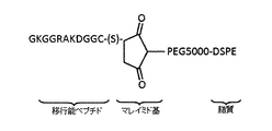
- FIG. 3 is a schematic diagram showing the structure of a lipid (Pep-PEG5000-DSPE) formed by binding at a point.
- (S) shows the thiol group of cysteine in which the carboxyl group which is the C-terminus of the migratory peptide is amidated.
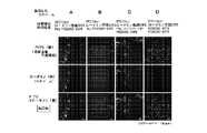
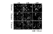
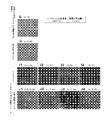
- Liposome A having a lipid membrane containing Pep-PEG2000-DSPE as a constituent lipid and fluorescently labeled with rhodamine as a single membrane, and one lipid membrane containing Pep-PEG5000-DSPE as a constituent lipid and fluorescently labeled with rhodamine
- Liposome B having a membrane, liposome C containing Pep-PEG5000-DSPE and PEG2000-DSPE as constituent lipids and a lipid membrane fluorescently labeled with rhodamine as a single membrane, and PEG5000-DSPE and PEG2000-DSPE as constituent lipids
- mouth the liposome D which has the lipid membrane which contains and was fluorescently labeled with rhodamine as a single membrane
- FITC (green) fluorescence is observed in the upper row
- rhodamine (red) fluorescence is observed in the middle row
- FITC (green) fluorescence and rhodamine (red) are superimposed.
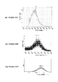
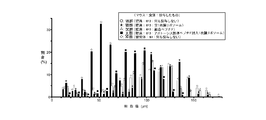
- the horizontal axis represents molecular weight (mass-to-charge ratio m / z)
- the vertical axis represents intensity (arbitrary unit).
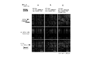
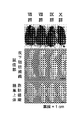
- the left column is the result of observing the fluorescence of FITC (green)
- the middle column is the result of observing the fluorescence of rhodamine (red)
- the right column is the result of superposing the left column and the middle column, It is the result of observing the overlap (yellow) of the fluorescence of FITC (green) and the fluorescence of rhodamine (red).
- Liposome E having PEG5000-DSPE and PEG2000-DSPE as constituent lipids and a lipid membrane fluorescently labeled with NBD as a single membrane, and containing Pep-PEG5000-DSPE and PEG2000-DSPE as constituent lipids and fluorescently labeled with NBD
- the lower diagram is an enlarged view of the frame shown in the upper diagram.
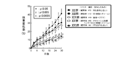
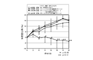
- High-fat were administered to healthy mice with fusion peptide (group IV) or apoptosis-inducing peptide-encapsulated single membrane liposome (group V) once every three days or without any administration (group VI). It is a figure which shows the result of having raised while giving a diet (HFD) and having calculated the weight change rate.
- Administer empty single membrane liposomes (Group VIII), fusion peptides (Group IX) or apoptosis-inducing peptide-encapsulated single membrane liposomes (Group X) to obese mice at intervals of once every 3 days, or none (Group VII) is a diagram showing the results of calculating the amount of change in body weight while feeding each with a high fat diet (HFD).
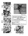
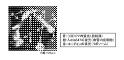
- FIG. 1 It is a figure which shows the observation result of a lipid droplet, a capillary endothelial cell, and a cell nucleus in the epididymal fat tissue of the obese mouse of a VII group, a VIII group, a IX group, and a X group, and the healthy mouse
- the right figure is an enlarged view of the portion shown in the left figure.
- the points where the fluorescence of BODIPY (blue), the fluorescence of Alexa 647 (red) and the fluorescence of Hoechst 33342 (green) are detected are illustrated by arrows.
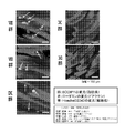
- FIG. 1 It is a figure which shows the observation result of a macrophage, a lipid droplet, a capillary endothelial cell, and a cell nucleus in the epididymal fat tissue of the obese mouse of a VII group, a VIII group, a IX group, and a X group, and the healthy mouse
- the right figure is an enlarged view of the portion shown in the left figure.
- arrows indicate the locations where Alexa 568 (red) fluorescence, BODIPY (light blue) fluorescence, Alexa 647 (green) fluorescence and Hoechst 33342 (blue) fluorescence are detected.
- the locations where BODIPY (green) fluorescence is detected are illustrated by arrows. It is a figure which shows the result of having calculated the energy intake amount for 24 hours of the obese mouse
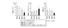
- HEPES buffer only (G) mixed lipid solution (H) containing 0.65 ⁇ mol (lipid) amount of empty single membrane liposome, final concentrations of 0.5 nmol / mL, 1 nmol / mL, 2 nmol / mL or 4 nmol / HEPES buffer (I1-4) in which cytochrome c is dissolved, or cytochrome c-encapsulated single membrane liposomes having a final concentration of cytochrome c of 0.5 nmol / mL, 1 nmol / mL, 2 nmol / mL, or 4 nmol / mL, respectively.
- FIG. 6 is a diagram showing the results of observing cell morphology after adding and incubating a mixed lipid solution (J1 to 4) containing a mixed lipid solution and an appropriate amount of empty single membrane liposomes to adipose tissue capillary endothelial cells.
- a mixed lipid solution J1 to 4
- J1 to 4 a mixed lipid solution
- an appropriate amount of empty single membrane liposomes to adipose tissue capillary endothelial cells.
- the locations where spherical cell morphology was confirmed are indicated by arrows.
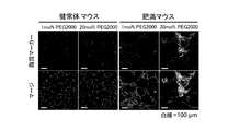
- cytochrome c amount was 0.5 mmol / kg (XIX group), 0.1 mmol / kg (XX group) or 0.02 mmol / kg (XXI) of cytochrome c-encapsulated single membrane liposomes were administered to healthy mice at an interval of once every 3 days, and normal diet (ND) was administered to group IV, XVII group, XIX group, XX group and XXI It is a figure which shows the result of having raised with giving the high fat diet (HFD) to each group, and calculating the weight change rate.
- HFD high fat diet
- the lipid membrane structure according to the present invention is a lipid membrane structure having a target cell migration ability, (a) a lipid formed by binding a peptide having a target cell migration ability, polyethylene glycol and lipid in this order, and (B) It has a lipid membrane containing as a constituent lipid a lipid formed by binding polyethylene glycol having a smaller number average molecular weight than that of polyethylene glycol constituting (a).
- long-chain PEG refers to PEG constituting the above (a).
- short chain length PEG means a PEG having a smaller number average molecular weight than the PEG constituting (a), and means the PEG constituting the above (b). Therefore, in the present invention, the long chain length means that the chain length is long (number average molecular weight is large) compared to the chain length of the PEG constituting (b), and the short chain length means (a) It means that the chain length is short (number average molecular weight is small) compared to the chain length of the PEG to be constructed.
- the structure of the lipid membrane structure according to the invention is shown in FIG.
- the difference in the number average molecular weight between the long-chain PEG and the short-chain PEG is not particularly limited, but for example, 10 to 100,000, 50 to 750,000, 100 to 50000, 500 to 25000, 800 to 20000, 1000 to 15000, 1200 to 10000, 1400 to 8000, preferably 1600 to 6000, more preferably 1800 to 5000, and still more preferably 2000 to 4000.
- the molecular weight Ma of the long chain PEG in the present invention is, for example, 1000 ⁇ Ma ⁇ 10000000, 1200 ⁇ Ma ⁇ 5000000, 1300 ⁇ Ma ⁇ 1000000, 1400 ⁇ Ma ⁇ 500000, 1500 ⁇ Ma ⁇ 100000, 1600 ⁇ Ma ⁇ 50000, 1700 ⁇ Ma ⁇ 30000, 1800 ⁇ Ma ⁇ 20000, 1900 ⁇ Ma ⁇ 15000, 2000 ⁇ Ma ⁇ 12000, 2100 ⁇ Ma ⁇ 10000, 2200 ⁇ Ma ⁇ 9000, 2300 ⁇ Ma ⁇ 8500, 2400 ⁇ Ma ⁇ 8000, 2500 ⁇ Ma ⁇ 7500, 2600 ⁇ Ma ⁇ 7400, 2700 ⁇ Ma ⁇ 7300, 2800 ⁇ Ma ⁇ 7200, 2900 ⁇ Ma ⁇ 7100, 3000 ⁇ Ma ⁇ 7000, 3100 ⁇ Ma ⁇ 6900, 3200 ⁇ Ma Can be exemplified 6800, preferably 3300 ⁇ Ma ⁇ 6700, more
- the molecular weight Mb of the short chain PEG is, for example, 5 ⁇ M ⁇ 20000, 10 ⁇ M ⁇ 15000, 50 ⁇ M ⁇ 10000, 100 ⁇ M ⁇ 8000, 200 ⁇ M ⁇ 7000, 300 ⁇ M ⁇ 5000, 400 ⁇ M ⁇ 4500, 450 ⁇ M ⁇ 4000, 470 ⁇ M ⁇ 3800, preferably 480 ⁇ M ⁇ 3700, more preferably 490 ⁇ M ⁇ 3600, and even more preferably 500 ⁇ Mb ⁇ 3500.
- 5 ⁇ M ⁇ 20000 10 ⁇ M ⁇ 15000, 50 ⁇ M ⁇ 10000, 100 ⁇ M 8000, 200 ⁇ M ⁇ 7000, 300 ⁇ M ⁇ 5000, 400 ⁇ M ⁇ 4500, 450 ⁇ M ⁇ 4000, 470 ⁇ M ⁇ 3800, preferably 480 ⁇ M ⁇ 3700, more preferably 490 ⁇ M ⁇ 3600, and even more preferably 500
- examples of the PEG in the present invention include PEGs having many shapes, for example, linear, branched, forked or multi-armed PEG, and linear PEG is preferred.
- the PEG in the present invention includes PEG to which a functional group or a protective group necessary for binding to a peptide or lipid such as a maleimide group or a methoxy group is added or modified.
- target tissue refers to a tissue or cell to which the lipid membrane structure according to the present invention is to be transferred.
- transfer to target cells or “transfer ability to target cells” means the “transfer to tissues containing target cells” or the “transfer ability to tissues containing target cells”.
- the phrase “transfer to target tissue” or “transfer ability to target tissue” does not necessarily mean “transfer to cells contained in target tissue” or “transfer ability to cells contained in target tissue”.
- the type of “target tissue” is not particularly limited, and examples thereof include tissues belonging to epithelial tissue, connective tissue, muscle tissue, and nerve tissue.
- tissue belonging to the connective tissue examples include a loose connective tissue, a dense connective tissue, an adipose tissue, and a reticulum tissue.
- the cell type of the “target cell” may be any cell type, whether it is a somatic cell or a germ cell, or may be a cell separated from a living body (in vitro cell), or a cell in a living body (in vivo cell). But you can.
- cell types include adipose tissue vascular endothelial cells and epithelial cells, epidermal cells, epidermal basal cells, keratinocytes, root sheath cells, hair matrix cells, mucosal epithelial cells, mammary cells, lacrimal gland cells, ears Malignant gland cells, sweat gland cells, prostate cells, endometrial cells, goblet cells, mucous epithelial cells, panel cells, type II alveolar cells, anterior pituitary cells, pituitary mesothelial cells, posterior pituitary cells, parathyroid glands Cells, gallbladder epithelial cells, hepatocytes, adipocytes, type I alveolar cells, pancreatic duct cells, acinar center cells, duct cells, synovial cells, choroid plexus cells, corneal endothelial cells, contractile cells, skeletal muscle cells, Cardiomyocytes, smooth muscle cells, myoepithelial cells, red blood cells, mega
- a known peptide known to have the ability to migrate to the target cell can be used, and a phage display method or the like can be used depending on the type of the target cell. Those appropriately identified or designed according to the law can be used.
- Specific examples of known peptides known to have target cell migration ability include, for example, amino acid sequences such as KGGRAKD (SEQ ID NO: 1), VMGSVTG (SEQ ID NO: 2), RGEVLWS (SEQ ID NO: 3), and the like.
- a peptide having the ability to migrate to adipose tissue vascular endothelial cells MG Kolonin et al., Nature Medicine, Vol. 10, No.
- NGR Arap W. et al., Science, No. 1) 279, 377-380, 1998; SEQ ID NO: 4
- amino acids of RGD Hirano Y. et al., J. Biomed Mater Res., 25, 1523-1534, 1991; SEQ ID NO: 5
- a peptide having the ability to migrate tumor tissue vascular endothelial cells polyarginine peptide (for example, cell membrane-permeable peptides such as International Patent Application Publication No. WO 2005/032593), GALA peptides (for example, T. Kakudo et al., Biochemistry, 2004, Vol. 43, pages 5618-5623) and the like. Mention may be made of membrane-fusogenic peptides and transcytosis-inducing peptides (for example, Japanese Patent Application No. 2006-179955).
- the “peptide having the ability to migrate to the target cell” in the present invention includes a peptide consisting of an amino acid sequence having one or more conservative amino acid substitutions in the amino acid sequence as long as it has the ability to migrate to the target cell.
- conservative amino acid substitutions are those that can generally be made without changing the physiological activity of the resulting molecule, ie, those that are recognized within the range of conservative substitutions (Watson et al., Molecular® Biology® of Gene), etc.
- Yes for example, acidic amino acids of aspartic acid and glutamic acid; basic amino acids of lysine, arginine and histidine; nonpolar amino acids of alanine, valine, leucine, isoleucine, proline, phenylalanine, methionine and tryptophan; glycine, asparagine, cysteine, glutamine, Serine, threonine and tyrosine polar uncharged side chain amino acids; phenylalanine, tryptophan, and tyrosine aromatic amino acids Replacement can be mentioned that occurs in the family inside) of Amino Acids.
- acidic amino acids of aspartic acid and glutamic acid acidic amino acids of aspartic acid and glutamic acid; basic amino acids of lysine, arginine and histidine, aliphatic amino acids of glycine, alanine, valine, leucine, isoleucine, serine and threonine (classified as aliphatic-hydroxyamino acids of serine and threonine Can be classified); phenylalanine, tyrosine and tryptophan aromatic amino acids; asparagine and glutamine amides; cysteine and methionine sulfur amino acids.
- the “peptide having the ability to migrate to the target cell” in the present invention as long as it has the ability to migrate to the target cell, one or several amino acids are deleted, substitutions other than the above-mentioned conservative amino acid substitution, insertion and / or Added peptides are included.
- the specific range for deletion is usually 1 to 3, preferably 1 to 2, more preferably 1, and the specific range for substitution excluding conservative amino acid substitution is usually 1 to 3, preferably
- the specific range for insertion is usually 1 to 5, preferably 1 to 3, more preferably 1 to 2, and still more preferably 1.
- the specific range for is usually 1 to 5, preferably 1 to 3, more preferably 1 to 2, and still more preferably 1.
- a glycine residue on the N-terminal side of the amino acid sequence a glycine residue and a cysteine on the C-terminal side
- One or several amino acids may be added while retaining the ability to migrate to the target cell, such as by adding a residue, but such peptides are still included in the peptides of the present invention.
- the “peptide having the ability to migrate to a target cell” in the present invention can be synthesized using a method that can be appropriately selected by those skilled in the art based on the sequence.
- a method for example, in addition to a peptide synthesis method in which each amino acid is chemically polymerized to synthesize a polypeptide, a recombinant vector containing a DNA encoding the peptide in the present invention is prepared and prepared.
- a transformant obtained by introducing the obtained vector into an appropriate host cell is cultured in a medium, and collected from the obtained culture, or a DNA encoding the peptide of the present invention is obtained in a cell-free protein synthesis system. Examples thereof include a method obtained by expression.
- any synthesis method other than a general method widely known by those skilled in the art can be used for any synthesis method.
- the number of amino acid residues of the peptide is not particularly limited, and for example, 2-4 residues, 5-7 residues, 8-10 residues, 11-15 residues, 16-20 residues, Examples include 21-30 residues, 31-40 residues, 41-55 residues, 56-75 residues, 76-100 residues, and 101 or more amino acid residues, depending on the function. Are appropriately selected.
- the “lipid membrane” refers to a membrane having a lipid membrane structure, the main component of which is lipid.
- the lipid membrane in the present invention may be composed of a lipid bilayer in which lipid molecules are associated with each other to form a hydrophobic portion, and the hydrophobic portion is formed inward or outward. It may consist of a single layer.
- lipid membrane structure in the present invention a closed vesicle having a lipid membrane composed of a lipid bilayer can be mentioned, and as such a lipid membrane structure, for example, a liposome can be mentioned.
- the lipid membrane structure in the present invention may be any of positive chargeability, nonchargeability, both (positive and negative) chargeability, and negative chargeability, but is preferably negatively chargeable or nonchargeable.
- the “negatively charged lipid membrane structure” in the present invention refers to a lipid membrane structure that is negatively charged as a whole.
- positively charged lipids or both (positive and negatively) chargeable lipids as lipids constituting the lipid membrane. Because it contains lipids, non-charged lipids, or is positively charged or both (positive and negative) charged, or modified with a non-chargeable substance, it is locally positively charged or both (positive and negative) charged. Even those that are non-charged are included in the “negatively-charged lipid membrane structure” if they are negatively charged as a whole.
- the “uncharged lipid membrane structure” in the present invention refers to a lipid membrane structure that is uncharged as a whole.
- a positively charged lipid a negatively charged lipid, both Because it contains a (positive and negative) chargeable lipid, or it is modified with a negatively chargeable, positively chargeable, or both (positive and negative) chargeable substance, it is locally negatively charged or positively charged. Even if it is charged, it is included in the “non-charged lipid membrane structure” if it is non-charged as a whole.
- lipid membrane structure having a surface potential of about ⁇ several mV to + several mV is a “non-charged lipid membrane structure” in vivo (Md. Vietnameser Hossen et al., Journal of Controlled Release, 147, 261-268, 2010; Sjoard Hak et al., European Journals pharmaceutics and Biopharmaceutics, Vol. 22, Vol. 104, No. 9266-9271, 2007). Further, in the present invention, “non-charging” is used interchangeably with “neutral”.
- the lipid constituting the lipid membrane structure in the present invention may be any of positively charged lipids, neutral (including both positive and negative) charged lipids and uncharged lipids, and negatively charged lipids. Examples thereof include phospholipids, glycolipids, sterols, long-chain aliphatic alcohols and glycerin fatty acid esters, and one or more of these can be used.
- phospholipid examples include phosphatidylcholine (for example, dioleoylphosphatidylcholine, dilauroylphosphatidylcholine, dimyristoylphosphatidylcholine, dipalmitoylphosphatidylcholine, distearoylphosphatidylcholine), phosphatidylglycerol (for example, dioleoylphosphatidylglycerol, dilauroylphosphatidylglycerol, Dimyristoyl phosphatidylglycerol, dipalmitoyl phosphatidylglycerol, distearoyl phosphatidylglycerol, etc., phosphatidylethanolamine (eg dioleoylphosphatidylethanolamine, dilauroylphosphatidylethanolamine, dimyristoylphosphatidylethanolamine, dipasto) Mitoylphosphatidylethanolamine, di
- glycolipids examples include glyceroglycolipids such as sphingomyelin, sulfoxyribosyl glyceride, diglycosyl diglyceride, digalactosyl diglyceride, galactosyl diglyceride and glycosyl diglyceride, and sphingoglycolipids such as galactosyl cerebroside, lactosyl cerebroside and ganglioside. 1 type, or 2 or more types of these can be used.
- glyceroglycolipids such as sphingomyelin, sulfoxyribosyl glyceride, diglycosyl diglyceride, digalactosyl diglyceride, galactosyl diglyceride and glycosyl diglyceride
- sphingoglycolipids such as galactosyl cerebroside, lactosyl cerebroside and ganglioside. 1 type, or 2
- sterols examples include sterols derived from animals such as cholesterol, cholesterol succinic acid, lanosterol, dihydrolanosterol, desmosterol, dihydrocholesterol, sterols derived from plants such as stigmasterol, sitosterol, campesterol, and brassicasterol (tytosterol). And sterols derived from microorganisms such as ergosterol, and one or more of these can be used. In addition, these sterols can generally be used to physically or chemically stabilize the lipid bilayer or to adjust the fluidity of the membrane.
- long-chain fatty acid or long-chain aliphatic alcohol a fatty acid having 10 to 20 carbon atoms or an alcohol thereof can be used.
- long-chain fatty acids or long-chain aliphatic alcohols include palmitic acid, stearic acid, lauric acid, myristic acid, pentadecylic acid, arachidic acid, margaric acid, tuberculostearic acid and other saturated fatty acids, palmitoleic acid, Mention of unsaturated fatty acids such as oleic acid, arachidonic acid, vaccenic acid, linoleic acid, linolenic acid, arachidonic acid, eleostearic acid, oleyl alcohol, stearyl alcohol, lauryl alcohol, myristyl alcohol, cetyl alcohol, linolyl alcohol 1 type, or 2 or more types of these can be used.
- glycerin fatty acid ester examples include monoacyl glycerides, diacyl glycerides, and triacyl glycerides, and one or more of these can be used.
- Examples of the positively charged lipid include dioctadecyldimethylammonium chloride (DODAC), N- (2,3-oleyloxy) propyl-N, N, N-trimethylammonium (N-) in addition to the above-described lipids.
- DODAC dioctadecyldimethylammonium chloride
- N- N- (2,3-oleyloxy) propyl-N
- N- N-trimethylammonium
- Examples of neutral lipids including both (positive and negative) charged lipids and non-charged lipids include diacylphosphatidylcholine, diacylphosphatidylethanolamine, ceramide and the like in addition to the above-described lipids. Or 2 or more types can be used.
- Examples of the negatively charged lipid include diacylphosphatidylserine, diacylphosphatidic acid, N-succinylphosphatidylethanolamine (N-succinylPE), phosphatidylethylene glycol, cholesteryl hemisuccinate (CHEMS), etc. 1 type, or 2 or more types of these can be used.
- the lipid membrane of the lipid membrane structure according to the present invention has positive charges such as tocopherol, propyl gallate, ascorbyl palmitate, butylated hydroxytoluene, stearylamine, oleylamine and the like.
- Positive charges such as tocopherol, propyl gallate, ascorbyl palmitate, butylated hydroxytoluene, stearylamine, oleylamine and the like.
- Charged substances to be added, charged substances to give negative charges such as dicetyl phosphate, membrane proteins such as membrane surface proteins and integral membrane proteins, and peptides that impart cell permeability and nuclear translocation ability to lipid membrane structures It can be combined or contained, and the amount and content of the bond can be adjusted as appropriate.
- the lipid membrane structure according to the present invention is produced using a known method such as a hydration method, an ultrasonic treatment method, an ethanol injection method, an ether injection method, a reverse phase evaporation method, a surfactant method, or a freezing / thawing method. can do.
- the lipid membrane structure according to the present invention can be used by being dispersed in an appropriate aqueous solvent such as physiological saline, phosphate buffer, citrate buffer, and acetate buffer.
- Additives such as saccharides, polyhydric alcohols, water-soluble polymers, nonionic surfactants, antioxidants, pH adjusters, and hydration accelerators may be appropriately added to the dispersion.
- the lipid membrane structure according to the present invention can be stored in a state in which the dispersion is dried.
- the lipid membrane structure according to the present invention may contain, for example, specific antibodies, targeting ligands and other functional elements for drug delivery according to known methods.
- the present invention provides an anti-obesity and / or therapeutic agent using the lipid membrane structure according to the present invention.
- the anti-obesity and / or therapeutic agent according to the present invention is the above-described lipid membrane structure according to the present invention in which an apoptosis-inducing agent is encapsulated, and the peptide having the ability to migrate to target cells is capable of migrating to adipose tissue vascular endothelial cells
- a lipid membrane structure, which is a peptide having the above, is used as an active ingredient, and obesity is suppressed / treated by inducing apoptosis in vascular endothelial cells of adipose tissue.
- apoptosis inducer for example, apoptosis of KLAKLAKKLAKLAK peptide (SEQ ID NO: 7; Ellerby HM et al., Nature Medicine, Vol. 5, pp 1032-1038, 1999) can be induced.
- cytochrome c cytochrome c
- Actinomycin D Anisomycin
- Antibiotic A23187 Apoptosis Activator 2
- Aristoforin Betulinic Acid
- Camptothecin Cisplatin
- Colchicine cycloheximide
- Daunorubicin HCl Dexamethasone
- Doxorubicin HCl Etopo side, Forskolin, Genistin, Okadaic acid, Phorbol-12-myristate 13-acetate, Staurosporine, Tamoxifen Citrate, Tapsigargin and the like.
- the peptide constituting the apoptosis inducer in the present invention is the same as the “peptide having the ability to migrate to a target cell” in the present invention described above.
- a lipid membrane structure in which an apoptosis-inducing agent is encapsulated can be produced according to a conventional method.
- a solution of an organic solvent in which lipid is dissolved and apoptosis induction It can be produced by mixing an aqueous solution in which the agent is dissolved and preparing a W / O emulsion by ultrasonic treatment, and then distilling off the organic solvent under reduced pressure.
- organic solvent for dissolving lipid examples include hydrocarbons such as pentane, hexane, heptane, and cyclohexane, halogenated hydrocarbons such as methylene chloride and chloroform, aromatic hydrocarbons such as benzene and toluene, methanol, and ethanol.
- Lower alcohols such as methyl acetate, esters such as methyl acetate and ethyl acetate, ketones such as acetone and the like can be used alone or in combination of two or more.
- the present invention provides an agent for suppressing and / or treating adipose tissue inflammation using the lipid membrane structure according to the present invention.
- the agent for inhibiting and / or treating adipose tissue inflammation according to the present invention is the aforementioned lipid membrane structure according to the present invention in which an apoptosis-inducing agent is encapsulated, wherein the peptide having the ability to migrate to target cells is adipose tissue vascular endothelial cell
- a lipid membrane structure, which is a peptide having a migratory ability, is used as an active ingredient, and apoptosis is induced / induced in vascular endothelial cells of adipose tissue to suppress / treat inflammation of adipose tissue.
- the description of the same or equivalent configuration as that of the lipid membrane structure and obesity suppression / treatment agent according to the present invention described above will be o
- the present invention provides an agent for suppressing and / or treating fat accumulation in non-adipose tissue using the lipid membrane structure according to the present invention.
- the agent for suppressing and / or treating fat accumulation in the non-adipose tissue according to the present invention is the above-mentioned lipid membrane structure according to the present invention in which an apoptosis-inducing agent is encapsulated, and having a target cell migration ability Inhibits / treats fat accumulation in non-adipose tissue by using as an active ingredient a lipid membrane structure, which is a peptide having the ability to migrate to adipose tissue vascular endothelial cells, and inducing apoptosis in vascular endothelial cells of adipose tissue.
- the structure of the lipid membrane structure, obesity inhibiting / treating agent and adipose tissue inflammation inhibiting / treating agent according to the present invention described above The description of the same or corresponding configuration will be omitted.
- non-adipose tissue refers to a tissue other than adipose tissue, and particularly refers to a tissue in which ectopic fat can accumulate.
- the non-adipose tissue in the present invention can preferably include a tissue in which a fat component can migrate and accumulate, that is, a tissue other than the brain.
- a tissue other than the brain Specifically, for example, the liver, skeletal muscle, pancreas, bone marrow, heart, Examples include kidneys and blood vessels.
- the “non-adipose tissue” in the present invention may or may not contain a fat component, and may or may not contain, for example, fat droplets or fat cells.
- “non-adipose tissue” is used interchangeably with “non-adipose tissue”, “lean body tissue”, and “lean body tissue”.
- ectopic fat means fat accumulated in tissues other than adipose tissue. That is, the agent for suppressing and / or treating fat accumulation in non-adipose tissue according to the present invention can inhibit and / or treat ectopic fat accumulation.
- the present invention provides a method for producing a lipid membrane structure.
- the method for producing a lipid membrane structure according to the present invention is a method for producing a lipid membrane structure having a target cell migration ability, and comprises a single membrane comprising the following lipids (a) and (b) as constituent lipids: (A) a peptide having the ability to migrate to a target cell, a lipid formed by binding PEG and lipid in this order, (b) a number average compared to the PEG constituting (a) Lipid formed by PEG with low molecular weight.
- a description of the same or corresponding configuration as that of the lipid membrane structure or obesity suppressing / treating agent according to the present invention is omitted.
- a single membrane lipid membrane containing the lipids of (a) and (b) as constituent lipids is prepared.
- a single membrane lipid membrane may be prepared using the lipids of (a) and (b), or a single membrane lipid membrane may be prepared using lipids, and then the lipid.
- a peptide having the ability to migrate to a target cell and a long-chain PEG and a short-chain PEG may be bound to the constituent lipids of the membrane.
- a peptide having the ability to migrate to a target cell may be bound to a constituent lipid formed by binding a chain-length PEG.
- a lipid formed by binding a peptide having a target cell migration ability, PEG and lipid in this order can be prepared according to a conventional method.
- a solution in which a peptide having the ability to migrate to a target cell is dissolved and a solution in which a lipid formed by binding PEG is dissolved can be mixed and incubated while shaking. .
- a screening method for a substance having an effect on a target cell according to the present invention comprises: (I) a step of selecting a peptide having the ability to migrate to one target cell in the lipid membrane structure according to the present invention and encapsulating a target substance to migrate to the target cell; (Ii) a step of evaluating whether or not the target substance shows an effect in the target cell; The step (i) or (ii) is included.
- the method for encapsulating the target substance in the lipid membrane structure according to the present invention includes the same method as the method for producing the lipid membrane structure in which the apoptosis-inducing agent is encapsulated. it can.
- step (i) as a method of selecting a peptide having a target cell migration ability and transferring a lipid membrane structure encapsulating a target substance to a target cell, for example, a tissue in which the target cell is separated from a living body,
- a method of adding a lipid membrane structure to the culture solution can be mentioned.
- the target cell is a cell in a living body (in vivo tissue or cell)
- examples thereof include a method of orally administering a solution containing a lipid membrane structure as it is or dissolving it, or administering it parenterally, such as intravenously, intraperitoneally, subcutaneously, or nasally.
- the method for evaluating whether or not the target substance shows an effect in the target cell can be appropriately set according to the effect expected of the target substance.
- the target cell is separated from the living body. If the tissue or cells are in vitro (tissues or cells in vitro), the target cells are stained by immunostaining or nuclear staining as necessary, and then the morphology and color intensity of the tissue or cells are observed using a microscope. Examples thereof include a method and a method of extracting RNA and analyzing the gene expression level.
- the target cell is a tissue or cell in a living body (in-vivo tissue or cell)
- the target cell is separated and collected, and the target cell is separated from the living body or cell (in-vitro tissue or cell).
- the effect is indirectly evaluated by collecting blood from a living body and analyzing the abundance of various marker molecules, measuring body weight and body fat percentage, etc. You can also do it.
- the present invention suppresses and / or treats fat accumulation in an obesity-suppressing and / or therapeutic agent, adipose tissue inflammation-suppressing and / or therapeutic agent or non-adipose tissue using the lipid membrane structure according to the present invention.
- a screening method for an agent to be used is provided.
- the method for screening for obesity suppression and / or treatment agent, adipose tissue inflammation suppression and / or treatment agent or agent for suppressing and / or treating fat accumulation in non-adipose tissue comprises: (Iii) The lipid membrane structure according to the present invention, wherein a target substance is encapsulated in a lipid membrane structure in which a peptide having a target cell migration ability is a peptide having a fat tissue vascular endothelial cell migration ability, Transferring to cells, (Iv) evaluating whether the target substance induces apoptosis in adipose tissue vascular endothelial cells; It has the above process (iii) or (iv).
- the obesity suppressing and / or treating agent, adipose tissue inflammation suppressing and / or therapeutic agent or the agent for suppressing and / or treating fat accumulation according to the present invention in the screening method of the agent for suppressing and / or treating fat accumulation in non-adipose tissue according to the present invention described above, the obesity suppressing and / or treating agent, adipose tissue inflammation suppressing and / or therapeutic agent or the agent for suppressing and / or treating fat accumulation according to the present invention.
- the description of the structure that is the same as or equivalent to the structure of the lipid membrane structure, the anti-obesity agent / therapeutic agent, the method of manufacturing the lipid membrane structure, or the screening method for substances that are effective in target cells will be omitted.
- lipid membrane structure having the ability to migrate to a target cell according to the present invention a method for producing the same, and a method for screening a substance having an effect on the target cell will be described based on examples. Note that the technical scope of the present invention is not limited to the features shown by these examples.
- PCT / US02 / 27836) which is known to have the ability of migrating, has 1 residue glycine (G) at the N-terminus and 2 residues glycine at the C-terminus ( G) and a peptide to which one residue of cysteine (C) has been added, wherein the C-terminal cysteine carboxyl group is amidated (GKGGRAKDGGC-NH 2 ; SEQ ID NO: 6) This was synthesized and this was designated as “migration ability peptide”.
- Mal-PEG2000 a lipid (Mal-PEG2000-DSPE; Nippon Oil & Fats Co., Ltd.) bound to L-distearoylphosphatidylethanolamine (DSPE) and a long chain length PEG (PEG5000) having a number average molecular weight of 5000 and maleimide
- a lipid (Mal-PEG5000-DSPE; Nippon Oil & Fats Co., Ltd.) obtained by binding a group to which a group is added (Mal-PEG5000) to DSPE was added to distilled water to 5 mmol / L, and a bath sonicator was used. Dissolve by sonication at room temperature for 1 minute, and transfer ability A peptide solution, Mal-PEG2000-DSPE solution and Mal-PEG5000-DSPE solution were obtained.
- the Mal-PEG2000-DSPE solution or the Mal-PEG5000-DSPE solution was added to the migrating peptide solution with gentle stirring so that the molar ratios were 1: 1. Then, while shaking using a bioshaker, incubation was performed at 30 ° C. for 24 hours to transfer 2.5 mmol / L of a solution of Mal-PEG2000-DSPE (Pep-PEG2000-DSPE) to which the transferable peptide was bound, A 2.5 mmol / L solution of Mal-PEG5000-DSPE (Pep-PEG5000-DSPE) to which a functional peptide was bound was prepared.
- a schematic diagram showing Pep-PEG5000-DSPE is shown in FIG.
- EPC Egg yolk phosphatidylcholine
- a 10 mmol / L EPC / Chol solution was prepared.
- rhodamine-labeled DOPE solution was prepared by dissolving rhodamine-labeled DOPE (Avanti Polar Lipid Co., Ltd.) in which rhodamine was bound to dioleoylglycerophosphoethanolamine (DOPE) in chloroform to be 3.8 mmol / L. .
- DSPE PEG2000-DSPE; Nippon Oil & Fats Co., Ltd.
- DSPE PEG5000-DSPE; Nippon Oil & Fats Co., Ltd.
- HEPES buffer pH 7.4; HEPES buffer
- EPC / Chol solution, rhodamine-labeled DOPE solution, PEG2000-DSPE solution, PEG5000-DSPE solution, Pep-PEG2000-DSPE solution and Pep-PEG5000-DSPE solution of Example 1 [1-2] A total of four mixed lipid solutions (lipid concentration: 10 mmol / L) of a, b, c, and d were prepared by mixing so that the mol% ratio was the ratio shown below.
- EPC / Chol Rhodamine labeled DOPE: Pep-PEG2000-DSPE: Pep-PEG5000-DSPE: PEG2000-DSPE: PEG5000-DSPE a; 94: 1: 5: 0: 0: 0 b; 94: 1: 0: 5: 0: 0 c; 93: 1: 0: 5: 1: 0 d; 93: 1: 0: 0: 1: 5
- Example 1 (1) [1-3] 500 ⁇ L of the mixed lipid solution of Example 1 (1) [1-3] and 500 ⁇ L of HEPES buffer were mixed, and sonicated at 4 ° C. for 15 seconds using a probe type sonicator. After evaporating and distilling off chloroform by flowing nitrogen gas using an evaporator, the mixture was sonicated for 1 minute using a bath sonicator to prepare a mixed lipid solution (lipid concentration 10 mmol / L) containing liposomes.
- lipid concentration 10 mmol / L lipid concentration 10 mmol / L
- the mixed lipid solution a contains Pep-PEG2000-DSPE (hereinafter, the same as the order of binding) formed by binding migratory peptides, Mal-PEG2000 and DSPE in this order, and is fluorescently labeled with rhodamine.
- Liposome A having a single lipid membrane as a single membrane
- Liposome B having a lipid membrane labeled with rhodamine as a single membrane and containing Pep-PEG5000-DSPE as a constituent lipid in the mixed lipid solution
- Liposome C containing Pep-PEG5000-DSPE and PEG2000-DSPE as constituent lipids in c and a lipid membrane fluorescently labeled with rhodamine as a single membrane, and PEG5000-DSPE and PE in mixed lipid solution
- Liposomes D having a fluorescence-labeled lipid membrane as a single film by rhodamine together including 2000-DSPE as a component lipid, were prepared, respectively.
- FITC-GSI-B4 FITC-labeled Griffoniaonsimplicifolia Lectin I-B4 Ilectin
- the FITC (green) fluorescence and the rhodamine (red) fluorescence were higher in mice administered with liposome C than in mice administered with liposomes A, B, and D.
- the area (yellow) showing the overlap was significantly large. That is, it was confirmed that the mice administered with liposome C had significantly more liposomes present in the capillary endothelial cells of adipose tissue than the mice administered with liposomes A, B and D.
- mice administered with liposome C a portion (yellow) showing an overlap between the fluorescence of FITC (green) and the fluorescence of rhodamine (red) was hardly detected. That is, it was confirmed that in mice administered with liposome C, there were significantly fewer liposomes present in vascular endothelial cells of major organs such as the heart and lungs (not shown).
- lipid membrane comprising a peptide having a target cell migration ability, a lipid formed by binding a long-chain PEG and a lipid in this order, and a lipid formed by binding a short-chain PEG as constituent lipids It was revealed that lipid membrane structures having a specific migration to target cells.
- Mal-PEG2000-DSPE had a molecular weight of about 1500 to about 4500.
- Mal-PEG5000-DSPE had a molecular weight of about 4500 to about 7500.
- the molecular weight of DSPE is about 760 and the molecular weight of maleimide group is about 170
- the molecular weight of PEG constituting Mal-PEG2000-DSPE is about 500 to about 3500, and its number average molecular weight is about 2000. It can be seen that the molecular weight of PEG constituting Mal-PEG5000-DSPE is about 3500 to about 6500, and the number average molecular weight is about 5000.
- Pep-PEG5000-DSPE had a molecular weight of about 5500 to about 8500.
- Example 2 (1) [1-1] was reversed based on the method described in Example 1 (1) [1-4].
- Liposomes single membrane liposomes
- liposomes were prepared by the simple hydration method. It is known that liposomes prepared by the simple hydration method are generally multilamellar liposomes having a plurality of lipid membranes (Danilo D., Biochem. J., Vol. 256, Vol. 1-11). Page, 1988).
- chloroform was distilled off from 500 ⁇ L of the mixed lipid solution of Example 2 (1) [1-1], 375 ⁇ L of chloroform was added and redissolved, and evaporated again. After the lipid film was prepared, 500 ⁇ L of HEPES buffer was added to hydrate the lipid film, and the mixture was stirred using a vortex mixer for 2 to 3 minutes to prepare a mixed lipid solution containing liposomes (lipid concentration 10 mmol / L). ) was prepared.
- multilamellar liposomes having multiple lipid membranes containing Pep-PEG5000-DSPE and PEG2000-DSPE as constituent lipids and fluorescently labeled with rhodamine were prepared.
- Example 1 (1) [1-2] single membrane liposome and Example 2 (1) [1-3] multilamellar liposome The average particle diameter, surface potential and PDI were measured by the method described in Example 1 (1) [1-5], and the measurement was further repeated 5 to 8 times in Example 2 (1) [1-1]. By repeating, each standard deviation was calculated. The results are shown in Table 2.
- the average particle diameter of each of the single membrane liposome and the multilamellar liposome was about 100 nm, the surface potential was about -5 mV, and the PDI was about 0.25. From this result, it was revealed that the single membrane liposome and the multilamellar liposome are negatively charged liposomes or uncharged liposomes having the same physical properties.
- Example 2 (1) [1-2] and the multilamellar liposome of Example 2 (1) [1-3] were administered to mice by the method described in), and fluorescence observation of subcutaneous fat tissue was performed.
- the dose of the liposome contained in each mixed lipid solution was the body weight (kg) x 0.2 mmol (lipid) of the individual mouse.
- the administration of FITC-GSI-B4 is 23.5 hours, 47.5 hours, and 71.5 hours after the administration of the mixed lipid solution (liposome), and the subcutaneous adipose tissue is removed by mixing lipid solution ( 24 hours, 48 hours and 72 hours after the administration of the liposome). The result is shown in FIG.
- the area of the portion (yellow) showing the overlap of the fluorescence of FITC (green) and the fluorescence of rhodamine (red) is the same as that of the mouse administered with the single membrane liposome and the mouse administered with the multiple membrane liposome.
- the values were almost the same after 24 hours, but were significantly larger in the mice administered with single membrane liposomes after 48 hours and 72 hours. That is, it was confirmed that in mice administered with single membrane liposomes, there were significantly more liposomes present in capillary endothelial cells of adipose tissue than in mice administered with multiple membrane liposomes.
- a lipid membrane comprising a peptide having a target cell migration ability, a lipid formed by binding a long-chain PEG and a lipid in this order, and a lipid formed by binding a short-chain PEG as constituent lipids It was revealed that a lipid membrane structure having a single membrane has a higher ability to migrate to a target cell than a lipid membrane structure having a plurality of similar lipid membranes.
- Example 3 Examination of liposome target cell migration ability (incorporation into target cells) (1) Preparation of mixed lipid solution Example 1 (1) Method described in [1-1] to [1-3] A mixed lipid solution was prepared as e and f. However, in place of rhodamine-labeled DOPE, DOPE (NBD-labeled DOPE) bound with Nitro-2-1,3-Benzoxadiazol-4-yl (NBD) was used. The mole% ratio of each lipid in e and f was as follows.
- EPC / Chol NBD-labeled DOPE: Pep-PEG5000-DSPE: PEG2000-DSPE: PEG5000-DSPE e; 93: 1: 0: 1: 5 f; 93: 1: 5: 1: 0
- a buffer ⁇ 123 mmol / L NaCl, 9.8 mmol / L KCl, 1.3 mmol / L CaCl 2 , 5 mmol / L D-(+)-glucose, 100 mmol / L HEPES, 2% (v Collagenase was added to / w) BSA ⁇ to 1 mg / mL to prepare a collagenase solution. 10 mL of collagenase solution per 1 g of subcutaneous adipose tissue was added and incubated at 37 ° C. for 30 minutes with shaking. During this incubation, every 5 minutes, the mixture was stirred for 5 seconds using a vortex mixer.
- the mixture was centrifuged at room temperature and 1500 rpm for 10 minutes, the upper layer was removed, and the lower layer was recovered.
- the cell fraction of endothelial cells After washing these cell fractions with Hank's equilibration buffer, the entire amount was seeded on a culture dish with a diameter of 35 mm, and 37 ° C., 5% (v / v) CO using EGM2-MV medium (Lonza). 2. Incubated for 2.5 hours in an environment of 100% relative humidity.
- Alexa647-labeled Griffonia simplicifolia Lectin I-B4 Iselectin Alexa647-GSI-B4 was added to the medium to a concentration of 5 ⁇ g / mL, and incubated for 0.5 hour to fluorescently stain capillary endothelial cells.
- Hoechst 33342 was added to the medium so as to be 2.5 ⁇ g / mL, and the cells were incubated for 0.2 hours to fluorescently stain the cell nucleus. After washing with Hanks equilibration buffer, Alexa647 (red) fluorescence, Hoechst 33342 (blue) fluorescence and NBD (green) fluorescence were observed using a confocal laser scanning microscope. A representative one of the observation results is shown in FIG.
- a lipid membrane containing a peptide having the ability to migrate to a target cell, a lipid formed by binding a long chain PEG and a lipid in this order and a lipid formed by binding a short chain PEG as constituent lipids is 1 It has been clarified that incorporation into the target cell is promoted by using a lipid membrane structure as a sheet membrane.
- Example 4 Examination of Apoptosis Inducing Effect of Liposomes Encapsulating Apoptosis Inducing Peptide (1) Chemical Synthesis of Apoptosis Inducing Peptide and Fusion Peptide M. et al., Nature Medicine, Vol. 5, pp. 1032-1038 (1999), wherein the carboxyl group of the C-terminal lysine is amidated (SEQ ID NO: 7; KLAKLAKKLAKLAK-NH 2 ) to Toray Industries, Inc. It was commissioned and chemically synthesized, and this was used as an apoptosis-inducing peptide.
- Example 4 (3) Preparation of liposomes by reverse phase evaporation method
- liposomes were prepared by the reverse phase evaporation method based on the method described in Example 1 (1) [1-4]. This was designated as empty single membrane liposome.
- the apoptosis-inducing peptide of Example 4 (1) is 190.4 mg / L (125 ⁇ mol / L) instead of the HEPES buffer.
- liposomes were prepared by the reverse phase evaporation method, and this was used as an apoptosis-inducing peptide-encapsulated single membrane liposome.
- an empty single membrane liposome is a liposome having a lipid membrane containing Pep-PEG5000-DSPE and PEG2000-DSPE as a constituent lipid, in which an apoptosis-inducing peptide is not encapsulated, and one apoptosis-inducing peptide-encapsulating membrane.
- the membrane liposome is a liposome having a lipid membrane containing Pep-PEG5000-DSPE and PEG2000-DSPE as a constituent lipid, in which an apoptosis-inducing peptide is encapsulated, as a single membrane.
- Example 4 The average particle size of the apoptosis-inducing peptide-encapsulated monolayer liposome of Example 4 (3) was determined by the method described in Example 1 (1) [1-5]. When the diameter, surface potential and PDI were measured, the average particle size was 109.2 ⁇ 7.8 nm, the surface potential was 6.0 ⁇ 0.9 mV, and the PDI was about 0.2 to 0.3. From this result, it became clear that the apoptosis-inducing peptide-encapsulated monolayer liposome is an uncharged liposome.
- Group I Empty single membrane liposome amount; (lipid) mouse body weight (kg) x 0.2 mmol
- Group II Fusion peptide amount; (Fusion peptide) Body weight (kg) of mouse individual x 3 mg (equivalent to 1.8 mg of apoptosis-inducing peptide)
- Group III Apoptosis-inducing peptide-encapsulated single membrane liposome amount; (apoptosis-inducing peptide) body weight of individual mouse (kg) ⁇ 1 mg, (lipid) body weight of individual mouse (kg) ⁇ 0.2 mmol
- cells containing activated caspase are caused by using a caspase 3,7 assay kit (Immunochemistry Technology) with a sulforhodamine fluorescently labeled caspase inhibitor (FLICA; fluorescent-labeled inhibitors of caspase).
- FLICA sulforhodamine fluorescently labeled caspase inhibitor
- Alexa647 (red) fluorescence and sulforhodamine (green) fluorescence were observed using a confocal laser scanning microscope.
- Alexa647 (red) fluorescence and sulforhodamine (green) fluorescence were observed using a confocal laser scanning microscope.
- apoptotic capillary endothelial cells were hardly observed when obese mice were administered empty monolayer liposomes, and were observed 24 hours after administration when a fusion peptide was administered, but thereafter Was hardly observed, but when the apoptosis-inducing peptide-encapsulated single membrane liposome was administered, it was continuously observed after administration.
- a lipid membrane structure having one membrane as a membrane and encapsulated with an apoptosis-inducing peptide can induce apoptosis in adipose tissue vascular endothelial cells, and its apoptosis-inducing effect is higher than that of a fusion peptide. Became more sustainable.
- Example 5 Examination of obesity suppression / treatment effect of liposome encapsulating apoptosis-inducing peptide (1) Preparation of liposome and fusion peptide Based on the method described in Example 4 (1) to (3), fusion peptide Empty single membrane liposomes and apoptosis-inducing peptide-encapsulated single membrane liposomes were prepared.
- the dose per one time of the fusion peptide in the HEPES buffer administered in the IV group or the V group or the apoptosis-inducing peptide-encapsulated single membrane liposome in the mixed lipid solution is as follows.
- HFD PMI
- water and feed were allowed to freely ingest, and the breeding temperature was 23 ° C.
- Group IV Fusion peptide amount; (Fusion peptide) Body weight (kg) of mouse individual ⁇ 3 mg (corresponding to 1.175 ⁇ mol of fusion peptide)
- Group V Apoptosis-inducing peptide-encapsulated monolayer liposome amount; (apoptotic fusion peptide) body weight (kg) of mouse individual ⁇ 1 mg (corresponding to 0.657 ⁇ mol of apoptosis-inducing peptide), (lipid) body weight of mouse individual (kg) ⁇ 0.2 mmol
- the body weight change rate is a value indicating the rate of weight gain or weight loss when the body weight at the start of administration of the fusion peptide or apoptosis-inducing peptide-encapsulated single membrane liposome is 100%.
- Rate of weight change (%) ⁇ (weight on day 0, 3, 6, 9 or 12 ⁇ weight on day 0) / weight on day 0 ⁇ ⁇ 100
- the weight change rate value of the IV group is approximated on any of the 0th, 3rd, 6th, 9th, and 12th days as compared to the weight change rate value of the VI group. There was no significant difference on days 3, 6, 9, and 12.
- the value of the weight change rate of the V group is somewhat smaller on the 6th day than the value of the weight change rate of the VI group, but is slightly smaller on the 6th day, and on the 9th and 12th days. It was remarkably small. Significant differences between group V and group VI were not on days 3 and 6, but p ⁇ 0.05 on day 9 and p ⁇ 0.01 on day 12.
- a lipid having the ability to migrate to adipose tissue vascular endothelial cells, a long chain-length PEG and a lipid formed by combining lipids in this order and a lipid formed by combining a short chain-length PEG are included as constituent lipids. It was revealed that a lipid membrane structure having a lipid membrane as a single membrane and having an apoptosis-inducing peptide encapsulated can suppress obesity.
- the group VIII contains a mixed lipid solution containing empty unilamellar liposomes of Example 5 (1)
- the group IX contains a HEPES buffer in which the fusion peptide of Example 5 (1) is dissolved
- the group X contains The mixed lipid solution containing the apoptosis-inducing peptide-encapsulated unilamellar liposome of Example 5 (1) was administered to the tail vein 10 times at an interval of once every 3 days, and reared for 30 days.
- the group VII was reared for 30 days without any administration.
- the amount of change in body weight is a value indicating the amount of increase or decrease in body weight based on the body weight at the start of administration of empty single membrane liposome, fusion peptide or apoptosis-inducing peptide-encapsulated single membrane liposome.
- the average value and the standard deviation were calculated for each group of the body weight change amount, and the weight change amount of the VIII group, the IX group, and the X group was subjected to a significant difference test with respect to the body weight change amount of the IIV group.
- the significant difference test was performed by one-way analysis of variance (one-way ANOVA) and Dunnett's method. The result is shown in FIG. 10a.
- the weight change of the VIII group was approximated on the 3-18th day and smaller on the 21-30th day than the weight change of the VII group. There was no significant difference between group VIII and group VII at any time point on days 3-30. In addition, the change in body weight of the IX group was smaller at any time point on the 3-30th day than the change in weight of the VII group. Significant differences between group IX and group VII were not on days 3-12, but were p ⁇ 0.05 on day 15 and p ⁇ 0.005 on days 18-30. In contrast, the change in body weight of Group X was significantly smaller at any time point on days 3-30 than the change in weight of Group VII. There was no significant difference between group X and group VII on days 3 and 6, but p ⁇ 0.05 on days 9 and 12, p ⁇ 0.005 on day 15, and On day 18-30, p ⁇ 0.0005.
- the weight change of the VII group was positive on any of the 3rd to 30th days, and increased with the passage of the breeding days. Therefore, the weight of the VII group continuously increased during the breeding period.
- the weight change of the group VIII was positive on any of the 3-30th days, increased with the passage of the breeding days on the 3-12th day, and almost the same value on the 12-30th day. Therefore, in the group VIII, the body weight increased until the 12th day during the breeding period and was maintained thereafter.
- the weight change of the IX group was positive on the 3rd to 9th days, 0 on the 12th day and negative on the 15th to 30th days, and slightly changed with the passage of the breeding days on the 3rd to 9th days.
- the body weight increased slightly until the 9th day during the breeding period, and then the body weight gradually decreased.
- the change in body weight of group X was negative on any of days 3 to 30 and decreased with the passage of the breeding days. Therefore, in group X, the body weight continuously decreased during the breeding period.
- the weight on the 30th day was not significantly changed in the VII group and the IX group with respect to the weight on the 0th day, but increased significantly in the VIII group, In, it decreased significantly. That is, from the results shown in FIGS. 10a and 10b, it has been clarified that, when an apoptosis-inducing peptide-encapsulated monolayer liposome is administered to obese mice, the body weight is decreased despite being fed with a high fat diet. It was.
- the sizes of the inguinal subcutaneous fat tissue and epididymal fat tissue of group X were significantly smaller than those of group VII, group VIII, and IX. That is, it has been clarified that, when an apoptosis-inducing peptide-encapsulated single membrane liposome is administered, fat tissue hypertrophy of obese mice is suppressed or adipose tissue is reduced under high fat diet.
- BODIPY boron dipyrromethene
- the size of fat droplets indicated by the fluorescence of BODIPY is used as the cell diameter of adipocytes, and the cell diameter is measured for 300 lipid droplets for each mouse in each group. The percentage (%) of how much is present was calculated and represented in the distribution map. The result is shown in FIG.
- the average value and standard deviation of the cell diameter are calculated for each group, and the significant difference test for the IX group of the VII group, the VIII group, the IX group, and the X group, and the X group of the VII group, the VIII group, and the IX group Significance test was performed and displayed in a histogram. Significant difference tests were performed by the one-way ANOVA, Tukey-Kramer's Honestly Significant Difference (HSD) method. The result is shown in FIG. 11c.
- the cell diameter of the X group was slightly larger than that of the XI group, but was significantly smaller than those of the VII group, VIII group and IX group. That is, it was clarified that when an apoptosis-inducing peptide-encapsulated monolayer liposome is administered to obese mice, fat cell hypertrophy is suppressed or adipocytes shrink under high-fat diet intake.
- BODIPY blue fluorescence
- Alexa647 red fluorescence
- Hoechst 33342 green fluorescence
- FIG. A representative one of the observation results is shown in FIG.
- the BODIPY fluorescent color is displayed in green and the Hoechst fluorescent color is displayed in blue.
- the entire field of view is very bright green. Since it is difficult to discriminate the blue color shown, pseudo-coloring processing is performed on the computer.
- the fluorescent color of BODIPY is blue.
- the lipid droplets indicated by the fluorescence of BODIPY blue
- the periphery of the fat cells and the capillary endothelial cells indicated by the fluorescence of Alexa647 (red) and Hoechst 33342 red
- Many structures hereinafter referred to as “angiogenic-adipogenic cluster structure” surrounded by cell nuclei indicated by the green fluorescence were observed, but were hardly observed in the X group and the XI group.
- the angiogenic-adipogenic cluster structure is a site where angiogenesis and adipogenesis occur and is known to appear characteristically in enlarged adipose tissue (NishimuramS. Et al., Diabetes). 56, 1517-1526, 2007). From this, the apoptosis-inducing peptide-encapsulated single membrane liposome can suppress angiogenesis and cell neoplasia or abolish new blood vessels and adipocytes in the adipose tissue of obese mice under high-fat diet intake. It was revealed.
- Example 5 From the results of the above Example 5 (3) [3-2], [3-3] and [3-4], the peptide having the ability to migrate to adipose tissue vascular endothelial cells, long-chain PEG and lipid were found to be A lipid membrane structure having a lipid membrane containing a lipid formed by binding in order and a lipid formed by binding a short chain length PEG as a constituent lipid and having an apoptosis-inducing peptide encapsulated as a single membrane, It became clear that obesity can be treated.
- the mixed lipid solution containing the apoptosis-inducing peptide-encapsulated single membrane liposome of Example 5 (3) was administered, and neither was administered to the XII group or XVI group.
- HFD PMI
- ND PMI
- the size of the adipose tissue was observed based on the method described in Example 5 (3) [3-2].
- the result is shown in FIG. 13b.
- the weight of the adipose tissue was measured, the ratio of the adipose tissue weight to the body weight at the end of the breeding period (adipose tissue weight ratio) was calculated, the average value and the standard deviation were calculated for each group, and the XIV group and the XV group A significant difference test was performed on the XII group. The significant difference test was performed by the method described in Example 5 (3) [3-2]. The result is shown in FIG. 13c.
- the rate of weight change in the XV group is small at any time point on day 3-30 compared to the XII group, XIII group and XIV group, but 3% when compared with the XVI group. It was approximated at any point on day 30. That is, when apoptosis-inducing peptide-encapsulated monolayer liposomes are administered to healthy mice, weight gain due to high-fat diet is suppressed, but weight gain corresponding to weight gain under normal diet intake, ie normal growth It became clear that weight gain due to was hardly suppressed.
- the inguinal subcutaneous fat tissue and epididymal fat tissue of the XV group are significantly smaller than those of the XII group and the XIV group, and as shown in FIG.
- the adipose tissue weight ratio was significantly smaller compared to the XII group and the XIV group. That is, when an apoptosis-inducing peptide-encapsulated single membrane liposome is administered, it becomes clear that fat tissue enlargement due to a high-fat diet is suppressed, and the proportion of the weight of adipose tissue in the body weight decreases. It was.
- a lipid having the ability to migrate to adipose tissue vascular endothelial cells, a long chain-length PEG and a lipid formed by combining lipids in this order and a lipid formed by combining a short chain-length PEG are included as constituent lipids.
- a lipid membrane structure having a lipid membrane as a single membrane and encapsulated with an apoptosis-inducing peptide does not inhibit normal growth and specifically suppresses the enlargement of adipose tissue caused by ingestion of a high-fat diet It became clear.
- Example 6 Inhibition of adipose tissue inflammation / treatment effect of liposome encapsulated with apoptosis-inducing peptide Based on the method described in Example 5 (3) [3-3], group VII, group VIII, group IX Small pieces of epididymal adipose tissue of group X and group XI were prepared. These small pieces of epididymal adipose tissue have been previously reported (Nishimura S. et al., Diabetes, 56, 1517-1526, 2007 and Nishimura S. et al., Nat. Med., 15, 914-94). 920 (2009), macrophages were immunostained with anti-F4 / 80 antibody.
- lipid droplets, capillary endothelial cells and cell nuclei were fluorescently stained based on the method described in Example 5 (3) [3-4], and Alexa 568 (red) was analyzed using a confocal laser scanning microscope. Fluorescence, BODIPY (light blue) fluorescence, Alexa 647 (green) fluorescence and Hoechst 33342 (blue) fluorescence were observed. Of the observation results, a representative one is shown in FIG. In general, the BODIPY fluorescent color is displayed in green and the Alexa fluorescent color is displayed in red. However, the entire field of view is very bright green. Since it is difficult to discriminate blue indicating the color, pseudo-coloring processing is performed on the computer.
- the fluorescent color of BODIPY is blue.
- the three primary colors of red, blue, and green are assigned so that their localization is as easy to distinguish as possible. A color that has no effect is selected.
- the fluorescent color of BODIPY is light blue, and the fluorescent color of Alexa 647 is green.
- the lipid droplets indicated by the fluorescence of BODIPY (light blue), that is, the periphery of the fat cells, the capillary endothelial cells indicated by the fluorescence of Alexa647 (green) and the Hoechst 33342 (blue).
- BODIPY light blue
- Alexa647 green
- Hoechst 33342 blue
- Many macrophages indicated by Alexa568 (red) fluorescence were observed around the structure surrounded by the cell nucleus indicated by fluorescence (angiogenic-adipogenic cluster structure) and its surroundings.
- a lipid having the ability to migrate to adipose tissue vascular endothelial cells, a long chain-length PEG and a lipid formed by combining lipids in this order and a lipid formed by combining a short chain-length PEG are included as constituent lipids. It was revealed that a lipid membrane structure having a lipid membrane as a single membrane and encapsulating an apoptosis-inducing peptide can suppress / treat inflammation in adipose tissue.
- Example 7 Examination of effect of suppressing / treating fat accumulation in non-adipose tissue of liposome encapsulated with apoptosis-inducing peptide
- Example 5 (3) [3-1] Group VII, VIII, IX and Liver and skeletal muscle were excised from group X and group XI of Example 5 (3) [3-3], and sections with a thickness of about 100 ⁇ m were prepared using microslicer DSK-1000. These sections were fluorescently stained for lipid droplets using BODIPY, F-actin using rhodamine-labeled phalloidin, and cell nuclei using Hoechst 33342.
- BODIPY green fluorescence
- rhodamine red fluorescence
- Hoechst 33342 blue fluorescence
- the fluorescence intensity of BODIPY and Hoechst 33342 was measured using Image-Pro Plus-4.5 software according to the previous report (shahen SM, et al., Nucleic Acids Res., 39, e48, 2011).
- the fluorescence intensity (relative fluorescence intensity) of BODIPY with respect to the fluorescence intensity was calculated.
- the average value and standard deviation were computed for every group, the significant difference test was performed, and it represented on the histogram.
- the significant difference test was performed according to the method described in Example 5 (3) [3-3].
- the significant difference test for the X group or the XI group of the VII group, the VIII group, and the IX group, and the VII group and the VIII group were performed.
- a significant difference test for the XI group of the VII group and the VIII group and a significant difference test for the IX group and the X group of the VII group and the VIII group were performed for skeletal muscle, respectively.
- the results for the liver are shown in FIG. 15b, and the results for skeletal muscle are shown in FIG. 16b.
- the result is similar to that in the liver in skeletal muscles, and many lipid droplets indicated by BODIPY (green) fluorescence are observed in the VII and VIII groups and to some extent in the IX group. In contrast, little was observed in the X and XI groups.
- the relative fluorescence intensity of BODIPY in skeletal muscle that is, the amount of accumulated fat, was significantly smaller in the IX, X, and XI groups than in the VII and VIII groups.
- the relative fluorescence intensity of BODIPY of group X was similar to that of group XI.
- a lipid membrane comprising a lipid having a structure capable of migrating adipose tissue vascular endothelial cells, a lipid having a long chain length PEG and a lipid bonded in this order and a lipid having a short chain length PEG bonded as a constituent lipid. It was revealed that a lipid membrane structure having a single membrane and having an apoptosis-inducing peptide encapsulated can suppress / treat fat accumulation in non-adipose tissue.
- Example 8 Evaluation of effects when administering liposome encapsulating apoptosis-inducing peptide (1) Examination of energy intake Example 5 (3) [VII] group VII, VIII, IX and X For the group, after the liposome or peptide was administered 6 times (after 15 days from the start of administration), the amount of HFD (PMI) ingested during 24 hours was measured, and the amount of energy ingested from the measured result (PMI) Intake energy amount; kcal) was calculated. Moreover, the average value and the standard deviation were calculated for each group of the intake energy amount, and the significant difference test was performed on the intake energy amount of the VII group with respect to the intake energy amounts of the VIII group, the IX group, and the X group. The significant difference test was performed by the method described in Example 5 (3) [3-2]. The result is shown in FIG.
- the intake energy amount of the IX group was significantly smaller than that of the VII group, whereas the intake energy amount of the VIII group and the X group was not significantly changed compared to the VII group. . That is, when the fusion peptide is administered, the energy intake is reduced compared to when the fusion peptide is not administered, whereas when the apoptosis-inducing peptide-encapsulated single membrane liposome is administered, it is compared with the case where it is not administered. And it became clear that the amount of energy consumed does not change.
- Forward primer 5′-atgctgactgcaatgtgctg-3 ′ (SEQ ID NO: 9), reverse primer: 5′-cagacttagacctgggaactct-3 ′ (SEQ ID NO: 10) Npy; forward primer: 5′-atgctagggtaacaagcgaatgg-3 ′ (SEQ ID NO: 11), reverse primer: 5′-tgtcgcagaggcggagtagtat-3 ′ (SEQ ID NO: 12) Gal; forward primer: 5′-ggcaggcgttatcctgcttag-3 ′ (SEQ ID NO: 13), reverse primer: 5′-ctgtcagggtccaactct-3 ′ (SEQ ID NO: 14) Pomc; forward primer: 5′-ctggagagccccgtgtttc-3 ′ (SEQ ID NO: 15), reverse primer: 5′-tggactcggctctggactg-3 ′ (SEQ ID
- 18S rRNA was amplified using Quantum RNA Classic II II 18S standard (Ambion) as an internal control. After separating the PCR product by gel electrophoresis, the gel was stained with ethidium bromide, and the signal was detected using an image analyzer LAS-400 mini (Fuji Film). The intensity of the detected signal was measured using Multi-Gage software, and a value divided by the signal intensity of 18S rRNA was calculated, and this was used as the relative expression level of each gene. For the relative expression level, the average value and the standard deviation were calculated for each group, and the relative expression level of the VIII group, the IX group, and the X group was subjected to a significant difference test with respect to the relative expression level of the VII group. The significant difference test was performed by the method described in Example 5 (3) [3-2]. The result is shown in FIG.
- the relative expression levels of Agrp, Npy, Gal, and Pomc were hardly changed between the groups.
- the relative expression level of Trh was not significantly changed in the VIII group and the X group as compared to the VII group, but was significantly larger in the IX group than the VII group. That is, it is clear that when the apoptosis-inducing peptide-encapsulated single membrane liposome is administered to obese mice, the expression level of both the feeding enhancement gene and the feeding suppression gene is hardly changed compared to the case where it is not administered. Became.
- a lipid membrane structure that has a lipid membrane containing a lipid that is bound as a constituent lipid as a single membrane and encapsulated with an apoptosis-inducing peptide has little effect on appetite It became clear.
- Leptin and adiponectin are both hormones secreted from adipocytes, and it is known that leptin blood concentration increases and adiponectin blood concentration decreases with obesity.
- ALT also called glutamate pyruvate transferase (GPT)
- GPT glutamate pyruvate transferase
- the concentration of leptin was not significantly changed in the VIII group compared to the VII group, while the IX group and the X group were significantly smaller than the VII group. It was remarkably small.
- the concentration of adiponectin was not significantly changed in the VIII group and the IX group as compared with the VII group, while the X group was significantly higher than that in the VII group.
- the ALT concentration did not change significantly in the VIII group, the IX group, and the X group as compared with the VII group, but the X group tended to be smaller than the VII group.
- a lipid having the ability to migrate to adipose tissue vascular endothelial cells, a long chain-length PEG and a lipid formed by combining lipids in this order and a lipid formed by combining a short chain-length PEG are included as constituent lipids.
- lipid membrane structure having a lipid membrane as a single membrane and encapsulating an apoptosis-inducing peptide normalizes the balance of hormones secreted from adipocytes and normalizes liver function It has been suggested.
- Example 9 (1) Preparation of cytochrome c-encapsulated single membrane liposomes by the reverse phase evaporation method
- the mixed lipid solution of Example 9 (1) was subjected to reverse phase based on the method described in Example 1 (1) [1-4].
- Liposomes were prepared by the evaporation method.
- a HEPES buffer in which cytochrome c was dissolved at 100 ⁇ mol / L was used instead of the HEPES buffer. That is, a liposome having a single lipid membrane containing Pep-PEG5000-DSPE and PEG2000-DSPE as a constituent lipid, in which cytochrome c is encapsulated, was prepared, and this was designated as a single membrane liposome encapsulating cytochrome c.
- Example 9 (1) Preparation of Empty Single Membrane Liposomes by Reverse Phase Evaporation Method
- the reverse phase evaporation method Liposomes were prepared by That is, a liposome having a single lipid membrane containing Pep-PEG5000-DSPE and PEG2000-DSPE as a constituent lipid, in which cytochrome c is not encapsulated, was prepared, and this was defined as an empty single membrane liposome.
- the lipid concentration of the mixed lipid solution containing empty single membrane liposomes was 10 mmol / L.
- Example 9 (3) Liposome addition and observation of cell morphology G, H, I1, I2, I3, I4, J1, and the like of Example 9 (3) so that the final concentration of cytochrome c and the amount of lipid added are as follows. In the medium of J2, J3 and J4, only HEPES buffer, mixed lipid solution containing empty single membrane liposome of Example 9 (3), HEPES buffer in which cytochrome c is dissolved, or of Example 9 (2) A mixed lipid solution containing cytochrome c-encapsulated single-membrane liposomes was added.
- apoptosis is not induced when cytochrome c is added to adipose tissue vascular endothelial cells as it is, but apoptosis is induced when cytochrome c-encapsulated monolayer liposome is added, and apoptosis is induced in that case. It was revealed that the number of cells increased depending on the final concentration of cytochrome c.
- a lipid having the ability to migrate to adipose tissue vascular endothelial cells, a long chain-length PEG and a lipid formed by combining lipids in this order and a lipid formed by combining a short chain-length PEG are included as constituent lipids. It was revealed that a lipid membrane structure having a lipid membrane as a single membrane and encapsulating cytochrome c can induce apoptosis in adipose tissue vascular endothelial cells.
- Example 10 Examination of obesity suppression / treatment effect of liposome encapsulated with cytochrome c (1) Preparation of cytochrome c-encapsulated monolayer liposome by reverse phase evaporation method Based on the method described in Example 9 (2), Cytochrome c-encapsulated single membrane liposomes were prepared by the reverse phase evaporation method.
- the dose per one time of cytochrome c in the mixed lipid solution is as follows. Also, during the breeding period, ND (PMI) was given to the XVII group, HFD (PMI) was given to the XVIII group, XIX group, XX group and XXI group, and water and food were freely ingested. The temperature was 23 ° C.
- Group XVII administered; (does not administer any liposome) Group XVIII: administered; (does not administer any liposome) XIX group: administered; cytochrome c-encapsulated monolayer liposomal cytochrome c amount; body weight of individual mouse (kg) ⁇ 0.5 mmol (6.16 mg) Group XX: administered; cytochrome c-encapsulated monolayer liposomal cytochrome c amount; body weight (kg) of mouse individual ⁇ 0.1 mmol (1.23 mg) XXI group: administered; cytochrome c-encapsulated monolayer liposome cytochrome c amount; body weight of mouse individual (kg) ⁇ 0.02 mmol (0.25 mg)
- Example 10 (3) Calculation of body weight change rate For the XVII group, XVIII group, XIX group, XX group, and XXI group of Example 10 (2), from the start of administration of the mixed lipid solution (cytochrome c-encapsulated monolayer liposome), The body weight was measured on days 3, 6, 7, 9, 12, 14, 15, 18, 21, 24, 27, 28 and 30 and the method described in Example 5 (2) [2-2]. The body weight change rate was calculated, and the weight change rates of the XVII group, the XIX group, the XX group, and the XXI group were subjected to a significant difference test with respect to the body weight change rate of the XVIII group. The result is shown in FIG.
- the values of the weight change rate of the XVIII group, the XIX group, the XX group, and the XXI group are 3, 6, 7, 9, 12, 14, 15 compared with the weight change rate of the XVII group. , 18, 21, 24, 27, 28 and 30 days. From this result, it was confirmed that weight gain was caused by the high fat diet.
- the value of the weight change rate of the XIX group is 3, 6, 7, 9, 12, 14, 15, 18, 21, 24, 27, 28 and 30 days compared with the value of the weight change rate of the XVIII group. It was small in all eyes. The significant difference of the XIX group from the XVIII group was not on days 3, 6, 7, 9, 12 and 15 but p ⁇ 0.05 on days 14 and 18 and 21, 24, 27, 28. On the 30th day, p ⁇ 0.01.
- weight change rate values of the XX group are approximated on the third, sixth, and twelfth days compared with the value of the weight change rate of the XVIII group, and are larger on the seventh and ninth days. , 18, 21, 24, 27, 28 and 30 days. Significant differences between group XX and group XVIII were not on days 3, 6, 7, 9, 12, 14, 15, 18 and 21 but p ⁇ 0.05 on day 24 and 27, 28. On the 30th day, p ⁇ 0.01.
- the value of the weight change rate of the XXI group is approximated on the third, sixth, twelfth, and fourteenth days compared with the value of the weight change rate of the XVIII group, and larger on the seventh and ninth days, Small on days 18, 21, 24, 27, 28 and 30.
- the significant difference of the XXI group from the XVIII group was not on days 3, 6, 7, 9, 12, 14, 15, 18, 21 and 24, but p ⁇ 0.05 on days 27 and 28, In addition, p ⁇ 0.01 on the 30th day.
- the XIX group ⁇ XX group / XXI group on the third, sixth, seventh, ninth and twelfth days, , 18, 21, 24, 27, 28, and 30 days, XIX group ⁇ XX group ⁇ XXI group. That is, the administration of cytochrome c-encapsulated monolayer liposomes can suppress the body weight gain of healthy mice due to the intake of a high-fat diet, and the inhibitory effect can be increased depending on the dose of cytochrome c. It was revealed.
- a lipid having the ability to migrate to adipose tissue vascular endothelial cells, a long chain-length PEG and a lipid formed by combining lipids in this order and a lipid formed by combining a short chain-length PEG are included as constituent lipids. It was revealed that a lipid membrane structure having a lipid membrane as a single membrane and encapsulating cytochrome c can suppress obesity.
- Example 11 (1) Preparation of cytochrome c-encapsulated multilamellar liposomes by simple hydration method
- the mixed lipid solution of Example 11 (1) was prepared by the simple hydration method according to the method described in Example 2 (1) [1-3]. Liposomes were prepared. However, instead of the HEPES buffer, a HEPES buffer in which cytochrome c (Sigma Aldrich) was dissolved so as to be 100 ⁇ mol / L was used. That is, cytochrome c-encapsulated multilamellar liposomes having a plurality of lipid membranes containing Pep-PEG5000-DSPE and PEG2000-DSPE as constituent lipids and encapsulating cytochrome c were prepared.
- cytochrome c-encapsulated multilamellar liposomes having a plurality of lipid membranes containing Pep-PEG5000-DSPE and PEG2000-DSPE as constituent lipids and encapsulating cytochrome c were
- Example 11 The cytochrome c-encapsulated single membrane liposome of Example 11 (2) and the cytochrome c-encapsulated multilamellar liposome of Example 11 (3) were obtained in Example 1 (1).
- the average particle diameter, the surface potential and the PDI were measured by the method described in [1-5], and each standard deviation was determined by repeating the measurement of Example 11 (1) to the above measurement 3 to 6 times. Calculated. The results are shown in Table 3.
- both the cytochrome c-encapsulated single membrane liposome and the cytochrome c-encapsulated multilamellar liposome had an average particle size of about 105 nm, a surface potential of about ⁇ 9 mV, and a PDI of about 0.25. From this result, it was revealed that the cytochrome c1 single membrane liposome and the cytochrome c multiple membrane liposome are negatively charged liposomes or non-charged liposomes having the same physical properties.
- Each recovered lower layer was suspended in 100 ⁇ L of distilled water to recover cytochrome c-encapsulated single membrane liposomes and cytochrome c-encapsulated multilamellar liposomes.
- BCA assay was performed using BCA protein assay kit (Pierce), and cytochrome c was quantified. Further, EPC was quantified for each collected liposome using a phospholipid C-Test Wako kit (Wako Pure Chemical Industries, Ltd.). Based on these quantitative results, cytochrome c recovery rate, lipid recovery rate, and cytochrome c encapsulation rate were calculated using the following formula 3, formula 4 and formula 5, respectively.
- Cytochrome c recovery rate (%) ⁇ quantitative cytochrome c amount / cytochrome c amount used in the preparation of liposomes in Example 11 (2) or Example 11 (3) ⁇ ⁇ 100
- lipid recovery rate (%) ⁇ quantitative EPC amount / EPC amount used in preparing liposome in Example 11 (2) or Example 11 (3) ⁇ 100
- Cytochrome c encapsulation rate (%) (Cytochrome c recovery rate / lipid recovery rate) ⁇ 100
- the cytochrome c recovery rate was about 15% for both cytochrome c-encapsulated single membrane liposomes and cytochrome c-encapsulated multilamellar liposomes.
- the lipid recovery rate of cytochrome c-encapsulated multilamellar liposomes is about 1.5 times that of cytochrome c-encapsulated single-membrane liposomes
- the cytochrome c-encapsulated rate of cytochrome c-encapsulated multilamellar liposomes is cytochrome c-encapsulated 1 The ratio was about 2/3 compared with the membrane liposome.
- the cytochrome c-encapsulated single membrane liposome and the cytochrome c-encapsulated multilamellar liposome have the same physical properties (average particle diameter, surface potential and PDI), but the cytochrome c encapsulation rate is determined by the cytochrome c-encapsulated monolayer membrane. It was revealed that the liposome was larger than the cytochrome c-encapsulated multilamellar liposome.
- Example 12 Examination of obesity suppression / treatment effect of liposomes with different numbers of lipid membranes encapsulating cytochrome c (1) Preparation of liposomes One cytochrome c encapsulated based on the method described in Example 9 (2) Membrane liposomes were prepared. Moreover, empty unilamellar liposomes were prepared based on the method described in Example 9 (3). In addition, cytochrome c-encapsulated multilamellar liposomes were prepared by the method described in Example 11 (3).
- cytochrome c was “weight of individual mouse (kg) ⁇ 6.16 mg (cytochrome c) in any of the XXIII, XXIV and XXV groups. ) Amount ”and the lipid was“ the body weight of the individual mouse (kg) ⁇ 0.19 mmol (lipid) amount ”.
- HFD PMI
- the value of the weight change rate of the XXIII group is approximated on any of the third, sixth, ninth, twelfth, fifteenth, eighteenth, and twenty-first days compared to the value of the weight change rate of the XXIV group.
- the XXIII group and the XXIV group were not significant difference between the XXIII group and the XXIV group on days 3, 6, 9, 12, 15, 18, and 21.
- the value of the weight change rate of the XXV group is approximated on the third day as compared with the value of the weight change rate of the XXII group, and slightly on the 6, 9, 12, 15, 18 and 21 days. It was small. There was no significant difference between the XXV group and the XXII group on days 3, 6, 9, 12, 15, 18, and 21.
- the value of the weight change rate of the XXIV group was remarkably small on the third, sixth, ninth, twelfth, fifteenth, eighteenth, and twenty-first days compared with the value of the weight change rate of the XXII group. .
- Significant differences between the XXIV group and the XXII group were not on day 3, but p ⁇ 0.05 on days 6, 9 and 12, and p ⁇ 0.01 on days 15, 18 and 21 there were.
- the value of the weight change rate of the XXII group, the XXIII group, and the XXV group was positive on any of the third, sixth, ninth, twelfth, fifteenth, eighteenth, and twenty-first days, and increased with the passage of the breeding days. That is, in the XXII group, the XXIII group, and the XXV group, the body weight continuously increased during the breeding period.
- the weight change rate value of the XXIV group was positive on the 3rd and 9th days, but negative on the 6th, 12th, 15th, 18th and 21st days, and decreased with the passage of the breeding days. became. That is, in the XXIV group, body weight continuously decreased during the breeding period.
- a lipid having the ability to migrate to adipose tissue vascular endothelial cells, a long chain-length PEG and a lipid formed by combining lipids in this order and a lipid formed by combining a short chain-length PEG are included as constituent lipids.
- a lipid membrane structure having a lipid membrane as a single membrane, in which cytochrome c is encapsulated, is formed by binding a peptide capable of migrating adipose tissue vascular endothelial cells, a long-chain PEG and a lipid in this order.
- a lipid membrane structure having a plurality of lipid membranes containing lipids and lipids formed by binding short-chain PEG as constituent lipids, in which cytochrome c is encapsulated, or encapsulated in a lipid membrane structure It was revealed that the effect of suppressing and treating obesity is extremely high compared to cytochrome c that has not been obtained.
- Liposomes P and Liposomes C of Example 1 were each administered in the tail vein to four normal 14-week-old male C57BL / 6J mice (Charles River) at a volume of 0.1 mmol lipid / kg body weight, 4.5 hours later, a HEPES buffer in which FITC-labeled Griffoniaisimplicifolia Lectin I-B4 Ilectin (FITC-GSI-B4; Vector Laboratories) was dissolved was administered to the tail vein. Furthermore, 30 minutes later, the subcutaneous adipose tissue of each mouse's liver and groin was excised to produce small pieces, and fluorescence observation was performed using a confocal laser scanning microscope. The observation results are shown in FIG.
- the molecular weight Ma of the PEG bound to the migratory peptide can be set to 500 ⁇ Mb ⁇ 3500 instead of 3500 ⁇ Ma ⁇ 6500, and the molecular weight Ma3500 ⁇ Ma ⁇ 6500 of the PEG bound to the peptide is more It has been confirmed to provide enhanced in vivo targeting.
- the liposome Q is a single membrane liposome of the present invention in which rhodamine is encapsulated, and the liposome R has the same configuration as the liposome Q except that it does not have a migratory peptide, that is, has two types of PEGs having different chain lengths.
- Single membrane liposome Single membrane liposome.
- the adipose tissue was immersed in Alexa647-GSIB4 solution for 1 hour to stain the blood vessels to produce small pieces, and fluorescence observation was performed using a confocal laser scanning microscope. The observation results are shown in FIG. In FIG. 24, blood vessel staining with Alexa 647 is shown in red, and fluorescence of TAMRA and rhodamine is shown in green.
- Delivery efficiency of the encapsulated rhodamine (green) to the target site (adipose tissue vascular endothelial cells) in the second group administered with the liposome Q of the present invention is the third group administered with the liposome R having no migratory peptide. Compared to that of it was dramatically improved. This is consistent with the results shown in other examples. On the other hand, when administered to obese mice, it was observed that Liposome R having no migratory peptide also accumulates to some extent in adipose tissue.
- liposomes having no migratory peptide when administered to healthy mice do not accumulate in adipose tissue, as shown in the preceding examples, accumulation of liposome R in adipose tissue in obese mice is It was inferred that it was based on a mechanism different from active targeting by migratory peptides.
- liposome R was not a vascular endothelial cell in the adipose tissue, but the angiogenetic-adipogenic cluster described in Example 5 (3) [3-4]. It was observed that it accumulated in (FIG. 25).
- the fluorescence of Alexa647 indicating blood vessels is green
- the fluorescence of rhodamine indicating liposomes is red
- the fluorescence of BODIPY indicating lipid droplets is blue.
- the angiogenic-adipogenic cluster formed in obese adipose tissue is a site where new adipocyte formation and angiogenesis occur, and therefore it can be expected that the vascular structure is sparse compared to normal blood vessels. Therefore, the accumulation of liposome R in an angiogenic-adipogenic cluster is thought to be the result of passive targeting due to the liposome R passing through the gaps in the sparse vascular wall.
- accumulation of substances across the blood vessel wall in a tissue with a sparse vascular structure has been proposed as an EPR effect (Enhanced permability and Retention, Matsumura et al., Cancer Res., 1986) in a tumor tissue.
- a tumor tissue is an example of a site where tumor cell formation and angiogenesis occur, and it has been reported that nanoparticles of about several tens to 200 nm exhibit the above EPR effect. Since liposomes R having an average particle diameter of around 100 nm accumulate in an angiogenetic-adipogenic cluster, it is considered that liposomes having an average particle diameter of several tens to 200 nm are advantageous for accumulation in an anionic-adipogenic cluster.
- Example 4 3 groups of obese model mice weighing 43 g or more were prepared.
- 3 groups of healthy mice (6 weeks old) were prepared by feeding normal food to C57BL / 6J mice.
- Liposomes S and T were administered to obese and healthy mice at a volume of 0.1 mmol lipid / kg body weight, respectively, via tail vein, and 24 hours later, the adipose tissue was excised and immersed in Alexa647-GSIB4 solution for 1 hour. Small pieces were prepared after staining, and fluorescence observation was performed using a confocal laser scanning microscope. The observation results are shown in FIG. In FIG. 26, blood vessel staining with Alexa 647 is shown in red, and fluorescence of a substance administered with rhodamine is shown in green.
- Reference Example 1 single-membrane liposomes having no migratory peptide and a PEG modification amount of 6 mol% or more based on the total lipid
- Reference Example 2 having no migratory peptide and one kind of PEG) Modified single-membrane liposomes
- passive targeting liposomes for an anionic-adipogenic cluster consisting of the lipid membrane structure of 1) or 2) below.
- a lipid having an increased blood retention having a lipid membrane containing a lipid formed by binding polyethylene glycol as a constituent lipid as a single membrane, and having a modified amount of PEG of 6 mol% or more based on the total lipid.
- Membrane structure 3) The lipid membrane structure according to 1) having an average particle diameter of several tens to 200 nm. Liposomes encapsulating anti-obesity drugs that act directly on adipose tissue, especially adipocytes, and substances that cause apoptosis of adipocytes, deliver the drug not only to adipose tissue vascular endothelial cells but also to adipocytes themselves. This is advantageous in that
Abstract
【課題】 標的細胞移行能を有する脂質膜構造体、それを用いた肥満抑制/治療剤、脂肪組織炎症抑制/治療剤および非脂肪組織における脂肪の蓄積を抑制/治療する剤、前記脂質膜構造体の製造方法、前記脂質膜構造体を用いた標的細胞において効果を示す物質のスクリーニング方法ならびに肥満抑制/治療剤、脂肪組織炎症抑制/治療剤および非脂肪組織における脂肪の蓄積を抑制/治療する剤のスクリーニング方法を提供する。 【解決手段】 標的細胞移行能を有する脂質膜構造体であって、標的細胞移行能を有するペプチド、長鎖長のPEGおよび脂質がこの順で結合してなる脂質と短鎖長のPEGが結合してなる脂質とを構成脂質として含む脂質膜を1枚膜として有する。
Description
本発明は、標的細胞移行能を有する脂質膜構造体、その製造方法および標的細胞において効果を示す物質のスクリーニング方法に関し、より詳細には、標的細胞移行能を有する脂質膜構造体であって、標的細胞移行能を有するペプチド、長鎖長のポリエチレングリコールおよび脂質がこの順で結合してなる脂質と短鎖長のポリエチレングリコールが結合してなる脂質とを構成脂質として含む脂質膜を1枚膜として有する脂質膜構造体、それを用いた肥満抑制/治療剤、脂肪組織炎症抑制/治療剤および非脂肪組織における脂肪の蓄積を抑制/治療する剤、前記脂質膜構造体の製造方法、前記脂質膜構造体を用いた標的細胞において効果を示す物質のスクリーニング方法ならびに肥満抑制/治療剤、脂肪組織炎症抑制/治療剤および非脂肪組織における脂肪の蓄積を抑制/治療する剤のスクリーニング方法に関する。
タンパク質、薬剤、あるいは核酸などの物質を生物個体の標的細胞に移行させるための技術(ターゲティング技術)の開発が盛んに行われている。例えば、特許文献1および非特許文献1には、脂肪組織の血管内皮細胞への移行能を有するペプチドが開示されており、このペプチドにアポトーシス誘導性ペプチドを連結したペプチドを投与することにより、脂肪組織の血管内皮細胞においてアポトーシスを誘導することができることが開示されている。
一方、リポソームに代表される脂質膜構造体は、(i)物質を内包することができてその内包された物質は生体内の分解作用や代謝作用から保護される、(ii)物質を内包することができてその内包された物質が標的細胞以外で作用を奏すること(副作用)を防止することができる、(iii)生体適合性や生分解性に優れている、(iv)その表面にポリエチレングリコール(以下、本明細書において「PEG」という場合がある。)、抗体、タンパク質、ペプチド、糖鎖などの機能性分子を導入することにより、標的細胞への移行能や融合能、pH応答性など様々な機能を付与することができるなどの利点を有することから、薬物や生理活性物質の理想的なベクターとして期待されている。
これまでに、標的細胞移行能を有する脂質膜構造体としては、例えば、表面がPEGでコーティングされた腫瘍細胞への移行能を有するリポソーム(ステルスリポソーム)が知られている他、非特許文献2には、本発明者らにより、上述の脂肪組織血管内皮細胞移行能を有するペプチド、PEGおよび脂質がこの順で結合してなる脂質を構成脂質として含む脂質膜を有するリポソームが開示されている。
M.G.Koloninら、Nature Medicine、第10巻、第6号、第625-632頁、2004年
Md.Nazir Hossenら、J.Control.Release、第147巻、第261-268頁、2010年
しかしながら、特許文献1および非特許文献1に記載された脂肪組織血管内皮細胞移行能を有するペプチドにアポトーシス誘導性ペプチドを結合したペプチドは、脂質膜構造体ではないことから、生体内の分解作用や代謝作用から保護されにくく、標的細胞である脂肪組織血管内皮細胞以外での作用を防止することが困難である。また、ステルスリポソームは、血管透過性が亢進している腫瘍組織の細胞に対しては移行能を有するが、血管透過性が亢進していない他の組織の細胞に対しては移行能を有さない。
さらに、非特許文献2に記載された脂肪組織血管内皮細胞移行能を有するペプチド、PEGおよび脂質がこの順で結合してなる脂質を構成脂質として含む脂質膜を有するリポソームは、培養細胞(in vitro)における脂肪組織血管内皮細胞移行能が示されているのみであり、生体(in vivo)における脂肪組織血管内皮細胞移行能については何ら示されておらず、そのうえ、非特許文献2には、さらにPEGが結合してなる脂質を構成脂質として含む脂質膜を有することの記載はおろか、開示もされていない。なお、後述の本明細書実施例1(2)、図3および図4にリポソームAおよびBとして示されているように、本願発明に係る脂質膜構造体と比較しても、生体(in vivo)における脂肪組織血管内皮細胞移行能および移行特異性が小さいことが分かる。
本発明は、このような問題点を解決するためになされたものであって、標的細胞移行能を有する脂質膜構造体であって、標的細胞移行能を有するペプチド、長鎖長のPEGおよび脂質がこの順で結合してなる脂質と短鎖長のPEGが結合してなる脂質とを構成脂質として含む脂質膜を1枚膜として有する脂質膜構造体、それを用いた肥満抑制/治療剤、脂肪組織炎症抑制/治療剤および非脂肪組織における脂肪の蓄積を抑制/治療する剤、前記脂質膜構造体の製造方法、前記脂質膜構造体を用いた標的細胞において効果を示す物質のスクリーニング方法ならびに肥満抑制/治療剤、脂肪組織炎症抑制/治療剤および非脂肪組織における脂肪の蓄積を抑制/治療する剤のスクリーニング方法を提供することを目的とする。
本発明者らは、鋭意研究の結果、標的細胞移行能を有するペプチド、長鎖長のPEGおよび脂質がこの順で結合してなる脂質と短鎖長のPEGが結合してなる脂質とを構成脂質として含む脂質膜を1枚膜として有する脂質膜構造体が、生体(in vivo)において標的細胞へ特異的に移行すること、ならびに脂肪組織血管内皮細胞移行能を有するペプチド、長鎖長のPEGおよび脂質がこの順で結合してなる脂質と短鎖長のPEGが結合してなる脂質とを構成脂質として含む脂質膜を1枚膜として有する脂質膜構造体であってアポトーシス誘導剤が封入されたものをマウスに投与することなどにより、体重増加を顕著に抑制、あるいは体重を減少させ、脂肪組織および脂肪細胞の肥大化を抑制、あるいは脂肪組織および脂肪細胞を縮小させ、脂肪細胞の新生を抑制、あるいは新生した脂肪細胞を消失させることができること、脂肪組織におけるマクロファージの浸潤を抑制、あるいはマクロファージを除去することができること、ならびに肝臓や骨格筋における脂肪の蓄積を抑制、あるいは蓄積した脂肪を減少させることができることなどを見出し、下記の各発明を完成した。
(1)標的細胞移行能を有する脂質膜構造体であって、下記(a)および(b)の脂質を構成脂質として含む脂質膜を1枚膜として有する、前記脂質膜構造体;(a)標的細胞移行能を有するペプチド、ポリエチレングリコールおよび脂質がこの順で結合してなる脂質、(b)(a)を構成するポリエチレングリコールと比較して数平均分子量が小さいポリエチレングリコールが結合してなる脂質。
(2)(a)を構成するポリエチレングリコールが、分子量Maが3500≦Ma≦6500のポリエチレングリコールであり、かつ、(b)を構成するポリエチレングリコールが、分子量Mbが500≦Mb≦3500のポリエチレングリコールである、(1)に記載の脂質膜構造体。
(3)負帯電性脂質膜構造体または非帯電性脂質膜構造体である、(1)または(2)に記載の脂質膜構造体。
(4)リポソームである、(1)から(3)のいずれかに記載の脂質膜構造体。
(5)標的細胞移行能を有するペプチドが、脂肪組織血管内皮細胞移行能を有するペプチドである、(1)から(4)のいずれかに記載の脂質膜構造体。
(6)脂肪組織血管内皮細胞移行能を有するペプチドが、KGGRAKD(式中、Kはリシン残基を、Gはグリシン残基を、Rはアルギニン残基を、Aはアラニン残基を、Dはアスパラギン酸残基を、それぞれ表す。)のアミノ酸配列からなるペプチドである、(5)に記載の脂質膜構造体。
(7)アポトーシス誘導剤が封入された(5)または(6)に記載の脂質膜構造体を有効成分とする、肥満抑制および/または治療剤。
(8)アポトーシス誘導剤が下記(i)および/または(ii)である、(7)に記載の肥満抑制および/または治療剤;(i)KLAKLAKKLAKLAK(式中、Kはリシン残基を、Lはロイシン残基を、Aはアラニン残基を、それぞれ表す。)のアミノ酸配列からなるペプチド、(ii)シトクロムc。
(9)アポトーシス誘導剤が封入された(5)または(6)に記載の脂質膜構造体を有効成分とする、脂肪組織炎症抑制および/または治療剤。
(10)アポトーシス誘導剤が下記(i)および/または(ii)である、(9)に記載の脂肪組織炎症抑制および/または治療剤;(i)KLAKLAKKLAKLAK(式中、Kはリシン残基を、Lはロイシン残基を、Aはアラニン残基を、それぞれ表す。)のアミノ酸配列からなるペプチド、(ii)シトクロムc。
(11)アポトーシス誘導剤が封入された(5)または(6)に記載の脂質膜構造体を有効成分とする、非脂肪組織における脂肪の蓄積を抑制および/または治療する剤。
(12)アポトーシス誘導剤が下記(i)および/または(ii)である、(11)に記載の剤;(i)KLAKLAKKLAKLAK(式中、Kはリシン残基を、Lはロイシン残基を、Aはアラニン残基を、それぞれ表す。)のアミノ酸配列からなるペプチド、(ii)シトクロムc。
(13)標的細胞移行能を有する脂質膜構造体を製造する方法であって、下記(a)および(b)の脂質を構成脂質として含む1枚膜の脂質膜を調製する工程を有する、前記方法;(a)標的細胞移行能を有するペプチド、ポリエチレングリコールおよび脂質がこの順で結合してなる脂質、(b)(a)を構成するポリエチレングリコールと比較して数平均分子量が小さいポリエチレングリコールが結合してなる脂質。
(14)(a)を構成するポリエチレングリコールが、分子量Maが3500≦Ma≦6500のポリエチレングリコールであり、かつ、(b)を構成するポリエチレングリコールが、分子量Mbが500≦Mb≦3500のポリエチレングリコールである、(13)に記載の方法。
(15)標的細胞において効果を示す物質のスクリーニング方法であって、(1)から(6)のいずれかに記載の脂質膜構造体において1の標的細胞移行能を有するペプチドを選択するとともに対象物質を封入して前記標的細胞へ移行させる工程と、前記対象物質が前記標的細胞において効果を示すか否かを評価する工程とを有する、前記方法。
(16)肥満抑制および/もしくは治療剤、脂肪組織炎症抑制および/もしくは治療剤または非脂肪組織における脂肪の蓄積を抑制および/もしくは治療する剤のスクリーニング方法であって、(5)または(6)に記載の脂質膜構造体に対象物質を封入して脂肪組織血管内皮細胞へ移行させる工程と、前記対象物質が脂肪組織血管内皮細胞においてアポトーシスを誘導するか否かを評価する工程とを有する、前記方法。
本発明に係る脂質膜構造体によれば、標的細胞に特異的に移行する脂質膜構造体を得ることができ、封入された物質を損傷させずに標的細胞へ送達して、標的細胞においてその物質の機能を発揮させることができるうえ、毒性が低く、安全に生体に適用することができる。また、本発明に係る脂質膜構造体を用いた肥満抑制/治療剤によれば、通常の成長を阻害することなく、高脂肪食餌による体重増加を有効に抑制することができるうえに、高脂肪食餌を摂取しているにもかかわらず、体重を減少させ、脂肪組織および脂肪細胞の肥大化を抑制、あるいは脂肪組織および脂肪細胞を縮小させ、脂肪細胞の新生および成長を抑制、あるいは新生した脂肪細胞を消失させることができる。また、本発明に係る脂質膜構造体を用いた脂肪組織炎症抑制/治療剤によれば、高脂肪食餌を摂取しているにもかかわらず、脂肪組織の炎症を有効に抑制あるいは消炎することができることから、脂肪組織の炎症が起因の一つとなる疾患、例えば、2型糖尿病やアテローム性動脈硬化症などの炎症性疾患などを予防ないし治療することができる。また、本発明に係る脂質膜構造体を用いた非脂肪組織における脂肪の蓄積を抑制/治療する剤によれば、高脂肪食餌を摂取しているにもかかわらず、非脂肪組織における脂肪の蓄積を有効に抑制あるいは蓄積した脂肪を減少させることができることから、非脂肪組織における脂肪の蓄積が起因の一つとなる疾患、例えば、脂肪肝や2型糖尿病や心筋障害などを予防ないし治療することができる。また、本発明に係る脂質膜構造体の製造方法によれば、前記のような効果を有した脂質膜構造体を得ることができる。さらに、本発明に係る標的細胞において効果を示す物質のスクリーニング方法によれば、対象物質が標的細胞において効果を示すか否かを簡便かつ効率的に評価することができ、本発明に係る脂質膜構造体を用いた肥満抑制および/もしくは治療剤、脂肪組織炎症抑制および/もしくは治療剤または非脂肪組織における脂肪の蓄積を抑制および/もしくは治療する剤のスクリーニング方法によれば、対象物質が肥満を抑制することや治療すること、脂肪組織の炎症を抑制することや治療すること、あるいは非脂肪組織における脂肪の蓄積を抑制することや治療することができるか否かを簡便かつ効率的に評価することができる。
以下、本発明に係る標的細胞移行能を有する脂質膜構造体、その製造方法および標的細胞において効果を示す物質のスクリーニング方法について詳細に説明する。本発明に係る脂質膜構造体は、標的細胞移行能を有する脂質膜構造体であって、(a)標的細胞移行能を有するペプチド、ポリエチレングリコールおよび脂質がこの順で結合してなる脂質、および(b)(a)を構成するポリエチレングリコールと比較して数平均分子量が小さいポリエチレングリコールが結合してなる脂質を構成脂質として含む脂質膜を1枚膜として有する。
本発明において「長鎖長のPEG」とは、上述の(a)を構成するPEGをいう。また、本発明において「短鎖長のPEG」とは、(a)を構成するPEGと比較して数平均分子量が小さいPEGをいい、上述の(b)を構成するPEGをいう。従って、本発明において、長鎖長とは(b)を構成するPEGの鎖長と比較して鎖長が長い(数平均分子量が大きい)ことを意味し、短鎖長とは(a)を構成するPEGの鎖長と比較して鎖長が短い(数平均分子量が小さい)ことを意味する。発明に係る脂質膜構造体の構造を図1に示す。
ここで、長鎖長のPEGと短鎖長のPEGとの数平均分子量の差は、特に限定されないが、例えば、10~100000、50~750000、100~50000、500~25000、800~20000、1000~15000、1200~10000、1400~8000を挙げることができ、好ましくは1600~6000、さらに好ましくは1800~5000、よりさらに好ましくは2000~4000を挙げることができる。
なお、本発明における長鎖長のPEGの分子量Maとしては、例えば、1000≦Ma≦10000000、1200≦Ma≦5000000、1300≦Ma≦1000000、1400≦Ma≦500000、1500≦Ma≦100000、1600≦Ma≦50000、1700≦Ma≦30000、1800≦Ma≦20000、1900≦Ma≦15000、2000≦Ma≦12000、2100≦Ma≦10000、2200≦Ma≦9000、2300≦Ma≦8500、2400≦Ma≦8000、2500≦Ma≦7500、2600≦Ma≦7400、2700≦Ma≦7300、2800≦Ma≦7200、2900≦Ma≦7100、3000≦Ma≦7000、3100≦Ma≦6900、3200≦Ma≦6800を挙げることができ、好ましくは3300≦Ma≦6700、さらに好ましくは3400≦Ma≦6600、よりさらに好ましくは3500≦Ma≦6500を挙げることができる。また、本発明における、短鎖長のPEGの分子量Mbとしては、例えば、5≦M≦20000、10≦M≦15000、50≦M≦10000、100≦M≦8000、200≦M≦7000、300≦M≦5000、400≦M≦4500、450≦M≦4000、470≦M≦3800、好ましくは480≦M≦3700、さらに好ましくは490≦M≦3600、よりさらに好ましくは500≦Mb≦3500を挙げることができる。
また、本発明におけるPEGとしては、多くの形状のPEG、例えば、直鎖状、分枝状、叉状あるいはマルチアーム状のPEGを挙げることができるが、直鎖状のPEGが好ましい。さらに、本発明におけるPEGには、マレイミド基やメトキシ基など、ペプチドや脂質との結合に必要な官能基や保護基などを付加または修飾されたPEGが包含される。
本発明において、「標的組織」または「標的細胞」とは、本発明に係る脂質膜構造体を移行させる予定の組織または細胞をいう。なお、「標的細胞への移行」または「標的細胞に対する移行能」という場合は、当該「標的細胞を含む組織への移行」または当該「標的細胞を含む組織に対する移行能」をも意味するが、「標的組織への移行」または「標的組織に対する移行能」という場合は、必ずしも「標的組織に含まれる細胞への移行」または「標的組織に含まれる細胞に対する移行能」を意味するとは限らない。本発明において、「標的組織」の種類は特に限定されず、上皮組織、結合組織、筋組織、神経組織に属する組織を挙げることができる。結合組織に属する組織としては、例えば、疎性結合組織、密生結合組織、脂肪組織、細網組織などを挙げることができる。「標的細胞」の細胞種もまた、体細胞、生殖細胞の別を問わずいかなるものでもよく、また、生体から分離した細胞(in vitroの細胞)でもよく、生体における細胞(in vivoの細胞)でもよい。細胞種として、具体的には、例えば、脂肪組織血管内皮細胞や上皮細胞、表皮細胞、表皮基底細胞、角化細胞、毛根鞘細胞、毛母細胞、粘膜上皮細胞、乳腺細胞、涙腺細胞、耳道腺細胞、汗腺細胞、前立腺細胞、子宮内膜細胞、杯細胞、粘液上皮細胞、パネト細胞、II型肺胞細胞、脳下垂体前葉細胞、脳下垂体中葉細胞、脳下垂体後葉細胞、副甲状腺細胞、胆嚢上皮細胞、肝細胞、脂肪細胞、I型肺胞細胞、膵臓導管細胞、腺房中心細胞、導管細胞、滑液細胞、脈絡叢細胞、角膜内皮細胞、収縮性細胞、骨格筋細胞、心筋細胞、平滑筋細胞、筋上皮細胞、赤血球、巨核球、マクロファージ、好中球、好酸球、好塩基球、マスト細胞、シュワン細胞、随伴細胞、ニューロン、グリア細胞、色素細胞、メラノサイト、網膜色素上皮細胞、胸腺上皮細胞など、およびこれらの腫瘍細胞を挙げることができる。また、本発明において「標的細胞移行能」または「標的組織移行能」とは、標的細胞または標的組織に移行することができる機能をいう。
本発明における「標的細胞移行能を有するペプチド」は、標的細胞移行能を有することが知られている既知のペプチドを用いることができる他、標的細胞の種類に応じて、ファージディスプレイ法などの常法に従って、適宜同定あるいは設計したものを用いることができる。標的細胞移行能を有することが知られている既知のペプチドとしては、具体的には、例えば、KGGRAKD(配列番号1)やVMGSVTG(配列番号2)、RGEVLWS(配列番号3)などのアミノ酸配列からなる脂肪組織血管内皮細胞移行能を有するペプチド(M.G.Koloninら、Nature Medicine、第10巻、第6号、第625-632頁、2004年)、NGR(Arap W.ら、Science、第279号、第377-380頁、1998年;配列番号4)やRGD(Hirano Y.ら、J.Biomed Mater Res.、第25号、第1523-1534頁、1991年;配列番号5)のアミノ酸配列を含む腫瘍組織血管内皮細胞移行能を有するペプチド、ポリアルギニンペプチド(例えば、国際特許出願公開第WO2005/032593号パンフレット)などの細胞膜透過性ペプチド、GALAペプチド(例えば、T.Kakudoら、Biochemistry、2004年、第43巻、第5618-5623頁)などのpH応答性膜融合性ペプチド、トランスサイトーシス誘導性ペプチド(例えば、特願2006-179955号)を挙げることができる。
なお、本発明における「標的細胞移行能を有するペプチド」は、標的細胞移行能を有する限り、そのアミノ酸配列の1または複数の保存的アミノ酸置換を有するアミノ酸配列からなるペプチドが包含される。
本発明において、保存的アミノ酸置換とは、生じる分子の生理学的活性を変化させることなく一般的になされ得る範囲、すなわち保存的置換の範囲で認められるもの(Watsonら、Molecular Biology of Geneなど)であり、例えば、アスパラギン酸およびグルタミン酸の酸性アミノ酸;リシン、アルギニンおよびヒスチジンの塩基性アミノ酸;アラニン、バリン、ロイシン、イソロイシン、プロリン、フェニルアラニン、メチオニンおよびトリプトファンの非極性アミノ酸;グリシン、アスパラギン、システイン、グルタミン、セリン、トレオニンおよびチロシンの極性無電荷側鎖アミノ酸;フェニルアラニン、トリプトファンおよびチロシンの芳香族アミノ酸といった側鎖に類似性のあるアミノ酸同士(アミノ酸のファミリー内部)で起こる置換を挙げることができる。同様に、アスパラギン酸およびグルタミン酸の酸性アミノ酸;リシン、アルギニンおよびヒスチジンの塩基性アミノ酸、グリシン、アラニン、バリン、ロイシン、イソロイシン、セリンおよびトレオニンの脂肪族アミノ酸(セリンおよびトレオニンの脂肪族-ヒドロキシアミノ酸と分類することもできる);フェニルアラニン、チロシンおよびトリプトファンの芳香族アミノ酸;アスパラギンおよびグルタミンのアミド;システインおよびメチオニンの含硫アミノ酸といった分類をすることができる。
また、本発明における「標的細胞移行能を有するペプチド」には、標的細胞移行能を有する限り、1個もしくは数個のアミノ酸が欠失、上述した保存的アミノ酸置換を除く置換、挿入および/または付加されたペプチドが包含される。欠失に関する具体的範囲は、通常1~3個、好ましくは1~2個、より好ましくは1個であり、保存的アミノ酸置換を除く置換に関する具体的範囲は、通常1~3個、好ましくは1~2個、より好ましくは1個であり、挿入に関する具体的範囲は、通常1~5個、好ましくは1~3個、より好ましくは1~2個、さらに好ましくは1個であり、付加に関する具体的範囲は、通常1~5個、好ましくは1~3個、より好ましくは1~2個、さらに好ましくは1個である。
例えば、後述するように、本発明における「標的細胞移行能を有するペプチド」をPEG結合脂質に結合させる際に、そのアミノ酸配列のN末端側にグリシン残基、C末端側にグリシン残基およびシステイン残基を付加するなど、標的細胞移行能を保持しつつ、1個もしくは数個のアミノ酸を付加する場合があるが、係るペプチドも依然として本発明のペプチドに含まれる。
本発明における「標的細胞移行能を有するペプチド」は、その配列を基にして、当業者によって適宜選択可能な方法を用いて合成することができる。そのような方法としては、例えば、アミノ酸1つ1つを化学的に重合してポリペプチドを合成するペプチド合成法の他、本発明におけるペプチドをコードするDNAを含む組換えベクターを作製し、作製したベクターを適切な宿主細胞中に導入して得られる形質転換体を培地にて培養し、得られた培養物から採取する方法や、本発明におけるペプチドをコードするDNAを無細胞タンパク質合成系で発現させて得る方法などを挙げることができる。なお、いずれの合成法も、当業者により広く知られている一般的な方法の他、あらゆる方法を利用することができる。
なお、本発明において、ペプチドのアミノ酸残基数は特に限定されず、例えば、2~4残基、5~7残基、8~10残基、11~15残基、16~20残基、21~30残基、31~40残基、41~55残基、56~75残基、76~100残基、101残基以上のアミノ酸残基数を挙げることができるが、その機能に応じて適宜選択される。
本発明において、「脂質膜」とは、脂質膜構造体が有する、脂質を主な構成成分とする膜をいう。本発明における脂質膜は、脂質分子が疎水性部分同士を会合させて二層に並んで形成する脂質二重層(lipid bilayer)からなるものでもよく、疎水性部分を内側または外側に向けて形成する一重層からなるものでもよい。
本発明における脂質膜構造体の好ましい形態としては、脂質二重層からなる脂質膜を有する閉鎖小胞を挙げることができ、そのような脂質膜構造体としては、例えば、リポソームを挙げることができる。
また、本発明における脂質膜構造体は、正帯電性、非帯電性、両(正負)帯電性および負帯電性のいずれでもよいが、負帯電性または非帯電性であることが好ましい。なお、本発明における「負帯電性脂質膜構造体」とは、全体として負帯電性である脂質膜構造体をいい、例えば、脂質膜の構成脂質として正帯電性脂質や両(正負)帯電性脂質、非帯電性脂質を含むため、または正帯電性や両(正負)帯電性、もしくは非帯電性の物質で修飾されているために、局所的に正帯電性や両(正負)帯電性、非帯電性であるものであっても、全体として負帯電性であれば「負帯電性脂質膜構造体」に包含される。
また、本発明における「非帯電性脂質膜構造体」とは、全体として非帯電性である脂質膜構造体をいい、例えば、脂質膜の構成脂質として正帯電性脂質や負帯電性脂質、両(正負)帯電性脂質を含むため、または負帯電性や正帯電性、もしくは両(正負)帯電性の物質で修飾されているために、局所的に負帯電性や正帯電性、両(正負)帯電性であるものであっても、全体として非帯電性であれば「非帯電性脂質膜構造体」に包含される。なお、当業者における技術常識として、-数mV~+数mV程度の表面電位を有する脂質膜構造体は、生体内においては「非帯電性脂質膜構造体」である(Md.Nazir Hossenら、Journal of Controlled Release、第147巻、第261-268頁、2010年;Sjoerd Hakら、European Journal of Pharmaceutics and Biopharmaceutics、第72巻、第397-404頁、2009年;Quentin le Masne de Chermontら、PNAS、第22巻、第104号、第9266-9271頁、2007年)。さらに、本発明において、「非帯電性」は「中性」と交換可能に用いられる。
本発明における脂質膜構造体を構成する脂質は、正帯電性脂質、両(正負)帯電性脂質や非帯電性脂質を含む中性脂質、負帯電性脂質のいずれでもよく、脂質の種類としては、例えば、リン脂質、糖脂質、ステロール、長鎖脂肪族アルコールあるいはグリセリン脂肪酸エステルなどを挙げることができ、これらの1種または2種以上を用いることができる。
リン脂質としては、例えば、ホスファチジルコリン(例えば、ジオレオイルホスファチジルコリン、ジラウロイルホスファチジルコリン、ジミリストイルホスファチジルコリン、ジパルミトイルホスファチジルコリン、ジステアロイルホスファチジルコリンなど)、ホスファチジルグリセロール(例えば、ジオレオイルホスファチジルグリセロール、ジラウロイルホスファチジルグリセロール、ジミリストイルホスファチジルグリセロール、ジパルミトイルホスファチジルグリセロール、ジステアロイルホスファチジルグリセロールなど)、ホスファチジルエタノールアミン(例えば、ジオレオイルホスファチジルエタノールアミン、ジラウロイルホスファチジルエタノールアミン、ジミリストイルホスファチジルエタノールアミン、ジパルミトイルホスファチジルエタノールアミン、ジステアロイルホスファチジルエタノールアミン(DSPE)、ジオレオイルグリセロフォスフォエタノールアミン(DOPE)など)、ホスファチジルセリン、ホスファチジルイノシトール、ホスファチジン酸(PA)、カルジオリピン、またはこれらの水素添加物、卵黄、大豆その他の動植物に由来する天然脂質(例えば、卵黄レシチン、大豆レシチンなど)などを挙げることができ、これらのうちの1種または2種以上を用いることができる。
糖脂質としては、スフィンゴミエリン、スルホキシリボシルグリセリド、ジグリコシルジグリセリド、ジガラクトシルジグリセリド、ガラクトシルジグリセリド、グリコシルジグリセリドなどのグリセロ糖脂質、ガラクトシルセレブロシド、ラクトシルセレブロシド、ガングリオシドなどのスフィンゴ糖脂質などを挙げることができ、これらの1種または2種以上を用いることができる。
ステロールとしては、コレステロール、コレステロールコハク酸、ラノステロール、ジヒドロラノステロール、デスモステロール、ジヒドロコレステロールなどの動物由来のステロール、スチグマステロール、シトステロール、カンペステロール、ブラシカステロールなどの植物由来のステロール(フィトステロール)、チモステロール、エルゴステロールなどの微生物由来のステロールなどを挙げることができ、これらの1種または2種以上を用いることができる。また、これらのステロールは、一般には脂質二重層を物理的または化学的に安定させるために、あるいは膜の流動性を調節するために用いることができる。
長鎖脂肪酸または長鎖脂肪族アルコールとしては、炭素数10~20の脂肪酸またはそのアルコールを使用することができる。そのような長鎖脂肪酸または長鎖脂肪族アルコールとしては、例えば、パルミチン酸、ステアリン酸、ラウリン酸、ミリスチン酸、ペンタデシル酸、アラキジン酸、マルガリン酸、ツベルクロステアリン酸などの飽和脂肪酸、パルミトレイン酸、オレイン酸、アラキドン酸、バクセン酸、リノール酸、リノレン酸、アラキドン酸、エレオステアリン酸などの不飽和脂肪酸、オレイルアルコール、ステアリルアルコール、ラウリルアルコール、ミリスチルアルコール、セチルアルコール、リノリルアルコールなどを挙げることができ、これらの1種または2種以上を用いることができる。
グリセリン脂肪酸エステルとしては、モノアシルグリセリド、ジアシルグリセリド、トリアシルグリセリドを挙げることができ、これらの1種または2種以上を用いることができる。
正帯電性脂質としては、上述した脂質の他、例えば、ジオクタデシルジメチルアンモニウムクロライド(dioctadecyldimethylammonium chloride、DODAC)、N-(2,3-オレイルオキシ)プロピル-N,N,N-トリメチルアンモニウム(N-(2,3-dioleyloxy)propyl-N,N,N-trimethylammonium、DOTMA)、ジドデシルアンモニウムブロミド(didodecylammonium bromide、DDAB)、1,2-ジオレイルオキシ-3-トリメチルアンモニウムプロパン(1,2-dioleoyloxy-3-trimethylammonio propane、DOTAP)、3β-N-(N’,N’-ジメチルアミノエタン)カルバモールコレステロール(3β-N-(N’,N’,-dimethyl-aminoethane)-carbamol cholesterol、DC-Chol)、1,2-ジミリストイルオキシプロピル-3-ジメチルヒドロキシエチルアンモニウム(1,2-dimyristoyloxypropyl-3-dimethylhydroxyethyl ammonium、DMRIE)、2,3-ジオレイルオキシ-N-[2(スペルミンカルボキサミド)エチル]-N,N-ジメチル-1-プロパンアンモニウムトリフルオロアセテート(2,3-dioleyloxy-N-[2(sperminecarboxamido)ethyl]-N,N-dimethyl-1-propanaminum trifluoroacetate、DOSPA)などを挙げることができ、これらの1種または2種以上を用いることができる。
また、両(正負)帯電性脂質や非帯電性脂質を含む中性脂質としては、上述した脂質の他、例えば、ジアシルホスファチジルコリン、ジアシルホスファチジルエタノールアミン、セラミドなどを挙げることができ、これらの1種または2種以上を用いることができる。また、負帯電性脂質としては、上述した脂質の他、例えば、ジアシルホスファチジルセリン、ジアシルホスファチジン酸、N-スクシニルホスファチジルエタノールアミン(N-スクシニルPE)、ホスファチジルエチレングリコール、コレステリルヘミスクシネート(CHEMS)などを挙げることができ、これらの1種または2種以上を用いることができる。
本発明に係る脂質膜構造体の脂質膜には、上述した脂質の他に、トコフェロール、没食子酸プロピル、パルミチン酸アスコルビル、ブチル化ヒドロキシトルエンなどの抗酸化剤、ステアリルアミン、オレイルアミンなどの正荷電を付与する荷電物質、ジセチルホスフェートなどの負電荷を付与する荷電物質、膜表在性タンパク質、膜内在性タンパク質などの膜タンパク質、脂質膜構造体に細胞透過能や核移行能を付与するペプチドを結合または含有させることができ、その結合量や含有量は適宜調節することができる。
本発明に係る脂質膜構造体は、水和法、超音波処理法、エタノール注入法、エーテル注入法、逆相蒸発法、界面活性剤法、凍結・融解法などの公知の方法を用いて製造することができる。
本発明に係る脂質膜構造体は、生理食塩水、リン酸緩衝液、クエン緩衝液、酢酸緩衝液などの適当な水性溶媒に分散させて使用することができる。分散液には、糖類、多価アルコール、水溶性高分子、非イオン界面活性剤、抗酸化剤、pH調節剤、水和促進剤などの添加剤を適宜添加してもよい。また、本発明に係る脂質膜構造体は、前記の分散液を乾燥させた状態で保存することができる。
また、本発明に係る脂質膜構造体には、例えば、特異抗体、標的化リガンドその他のドラッグデリバリー用機能性素子を、それぞれ公知の方法に従って含ませてもよい。
次に、本発明は、本発明に係る脂質膜構造体を用いた肥満抑制および/または治療剤を提供する。本発明に係る肥満抑制および/または治療剤は、アポトーシス誘導剤が封入された、上述の本発明に係る脂質膜構造体であって、標的細胞移行能を有するペプチドが脂肪組織血管内皮細胞移行能を有するペプチドである脂質膜構造体を有効成分とし、脂肪組織の血管内皮細胞においてアポトーシスを誘導することにより、肥満を抑制/治療する。
本発明において、アポトーシス誘導剤としては、例えば、KLAKLAKKLAKLAKペプチド(配列番号7;Ellerby H.M.ら、Nature Medicine、第5巻、第1032-1038頁、1999年)などのアポトーシスを誘導することが知られている既知のペプチドの他、シトクロムc、Actinomycin D、Anisomycin、Antibiotic A23187、Apoptosis Activator 2、Aristoforin、Betulinic Acid、Camptothecin、Cisplatin、Colchicine、Cycloheximide、Daunorubicin HCl、Dexamethasone、Doxorubicin HCl、Etoposide、Forskolin、Genistein、Okadaic acid、Phorbol-12-myristate 13-acetate、Staurosporine、Tamoxifen Citrate、Thapsigarginなどを挙げることができる。
なお、本発明におけるアポトーシス誘導剤を構成するペプチドについては、上述した本発明における「標的細胞移行能を有するペプチド」と同様である。
本発明において、アポトーシス誘導剤が封入された脂質膜構造体は、常法に従って製造することができ、例えば、逆相蒸発法により製造する場合には、脂質を溶解した有機溶媒の溶液とアポトーシス誘導剤を溶解した水溶液とを混合して、超音波処理によりW/Oエマルジョンを調製した後、有機溶媒を減圧留去させることにより製造することができる。
脂質を溶解させる有機溶媒としては、例えば、ペンタン、ヘキサン、ヘプタン、シクロヘキサンなどの炭化水素類、塩化メチレン、クロロホルムなどのハロゲン化炭化水素類、ベンゼン、トルエンなどの芳香族炭化水素類、メタノール、エタノールなどの低級アルコール類、酢酸メチル、酢酸エチルなどのエステル類、アセトンなどのケトン類などを、単独でまたは2種以上を組み合わせて使用することができる。
次に、本発明は、本発明に係る脂質膜構造体を用いた脂肪組織炎症抑制および/または治療剤を提供する。本発明に係る脂肪組織炎症抑制および/または治療剤は、アポトーシス誘導剤が封入された、上述の本発明に係る脂質膜構造体であって、標的細胞移行能を有するペプチドが脂肪組織血管内皮細胞移行能を有するペプチドである脂質膜構造体を有効成分とし、脂肪組織の血管内皮細胞においてアポトーシスを誘導することにより、脂肪組織の炎症を抑制/治療する。なお、本発明に係る脂肪組織炎症抑制および/または治療剤において、上述した本発明に係る脂質膜構造体、肥満抑制/治療剤の構成と同等または相当する構成については再度の説明を省略する。
次に、本発明は、本発明に係る脂質膜構造体を用いた非脂肪組織における脂肪の蓄積を抑制および/または治療する剤を提供する。本発明に係る非脂肪組織における脂肪の蓄積を抑制および/または治療する剤は、アポトーシス誘導剤が封入された、上述の本発明に係る脂質膜構造体であって、標的細胞移行能を有するペプチドが脂肪組織血管内皮細胞移行能を有するペプチドである脂質膜構造体を有効成分とし、脂肪組織の血管内皮細胞においてアポトーシスを誘導することにより、非脂肪組織における脂肪の蓄積を抑制/治療する。なお、本発明に係る非脂肪組織における脂肪の蓄積を抑制および/または治療する剤において、上述した本発明に係る脂質膜構造体、肥満抑制/治療剤および脂肪組織炎症抑制/治療剤の構成と同等または相当する構成については再度の説明を省略する。
本発明において「非脂肪組織」とは脂肪組織以外の組織をいい、特に、異所性脂肪が蓄積し得る組織をいう。本発明における非脂肪組織として、好ましくは、脂肪成分が移行して蓄積し得る組織すなわち脳以外の組織を挙げることができ、具体的には、例えば、肝臓、骨格筋、膵臓、骨髄、心臓、腎臓、血管などを挙げることができる。なお、本発明における「非脂肪組織」は脂肪成分を含むか否かは問わず、例えば脂肪滴や脂肪細胞などを含んでいてもよく、含んでいなくてもよい。さらに、本発明において、「非脂肪組織」は「非脂肪体組織」、「除脂肪組織」、「除脂肪体組織」と交換可能に用いられる。
また、異所性脂肪とは、脂肪組織以外の組織に蓄積された脂肪をいう。すなわち、本発明に係る非脂肪組織における脂肪の蓄積を抑制および/または治療する剤は、異所性脂肪の蓄積を抑制および/または治療することができる。
次に、本発明は、脂質膜構造体を製造する方法を提供する。本発明に係る脂質膜構造体を製造する方法は、標的細胞移行能を有する脂質膜構造体を製造する方法であって、下記(a)および(b)の脂質を構成脂質として含む1枚膜の脂質膜を調製する工程を有する;(a)標的細胞移行能を有するペプチド、PEGおよび脂質がこの順で結合してなる脂質、(b)(a)を構成するPEGと比較して数平均分子量が小さいPEGが結合してなる脂質。なお、本発明に係る脂質膜構造体を製造する方法において、上述した本発明に係る脂質膜構造体または肥満抑制/治療剤の構成と同等または相当する構成については再度の説明を省略する。
上記(a)および(b)の脂質を構成脂質として含む1枚膜の脂質膜を調製する工程において、(a)および(b)の脂質を構成脂質として含む1枚膜の脂質膜を調製する方法としては、例えば、(a)および(b)の脂質を用いて1枚膜の脂質膜を調製してもよく、あるいは、脂質を用いて1枚膜の脂質膜を調製した後、この脂質膜の構成脂質に標的細胞移行能を有するペプチドおよび長鎖長のPEGと、短鎖長のPEGとを結合してもよい。または、長鎖長のPEGが結合してなる脂質と短鎖長のPEGが結合してなる脂質とを用いて1枚膜の脂質膜を調製した後、この脂質膜の構成脂質のうち、長鎖長のPEGが結合してなる構成脂質に、標的細胞移行能を有するペプチドを結合するなどしてもよい。
なお、本発明において、(a)標的細胞移行能を有するペプチド、PEGおよび脂質がこの順で結合してなる脂質は常法に従い調製することができる。そのような方法としては、例えば、標的細胞移行能を有するペプチドを溶解した溶液とPEGが結合してなる脂質を溶解した溶液とを混合して、振とうしながらインキュベートする方法を挙げることができる。
次に、本発明は、本発明に係る脂質膜構造体を用いた、標的細胞において効果を示す物質のスクリーニング方法を提供する。本発明に係る標的細胞において効果を示す物質のスクリーニング方法は、
(i)本発明に係る脂質膜構造体において1の標的細胞移行能を有するペプチドを選択するとともに対象物質を封入して前記標的細胞へ移行させる工程、
(ii)前記対象物質が前記標的細胞において効果を示すか否かを評価する工程、
以上(i)または(ii)の工程を有する。なお、本発明に係る標的細胞において効果を示す物質のスクリーニング方法において、上述した本発明に係る脂質膜構造体、肥満抑制/治療剤または脂質膜構造体の製造方法の構成と同等または相当する構成については再度の説明を省略する。
(i)本発明に係る脂質膜構造体において1の標的細胞移行能を有するペプチドを選択するとともに対象物質を封入して前記標的細胞へ移行させる工程、
(ii)前記対象物質が前記標的細胞において効果を示すか否かを評価する工程、
以上(i)または(ii)の工程を有する。なお、本発明に係る標的細胞において効果を示す物質のスクリーニング方法において、上述した本発明に係る脂質膜構造体、肥満抑制/治療剤または脂質膜構造体の製造方法の構成と同等または相当する構成については再度の説明を省略する。
(i)の工程において、本発明に係る脂質膜構造体に対象物質を封入する方法としては、上述のアポトーシス誘導剤が封入された脂質膜構造体を製造する方法と同様の方法を挙げることができる。
(i)の工程において、1の標的細胞移行能を有するペプチドを選択して対象物質を封入した脂質膜構造体を標的細胞へ移行させる方法としては、例えば、標的細胞が生体から分離した組織や細胞(in vitroの組織や細胞)である場合は、培養液に脂質膜構造体を添加する方法を挙げることができ、標的細胞が生体における細胞(in vivoの組織や細胞)である場合は、脂質膜構造体をそのまま、あるいは溶解した溶液を経口投与、あるいは静脈、腹腔内、皮下、経鼻などへ非経口投与する方法を挙げることができる。
(ii)の工程において、対象物質が標的細胞において効果を示すか否かを評価する方法は、対象物質に期待する効果に応じて適宜設定することができるが、例えば、標的細胞が生体から分離した組織や細胞(in vitroの組織や細胞)である場合は、標的細胞を必要に応じて免疫染色や核染色などにより染色した後、顕微鏡を用いて組織や細胞の形態や発色強度を観察する方法や、RNAを抽出して遺伝子発現量を解析する方法などを挙げることができる。標的細胞が生体における組織や細胞(in vivoの組織や細胞)である場合は、標的細胞を分離採取して、上述の標的細胞が生体から分離した組織や細胞(in vitroの組織や細胞)である場合と同様の方法を行うことができる他、生体から血液を採取して種々のマーカー分子の存在量を解析することや、体重や体脂肪率を測定することなどによって間接的に効果を評価する方法を行うこともできる。
次に、本発明は、本発明に係る脂質膜構造体を用いた、肥満抑制および/もしくは治療剤、脂肪組織炎症抑制および/もしくは治療剤または非脂肪組織における脂肪の蓄積を抑制および/もしくは治療する剤のスクリーニング方法を提供する。本発明に係る肥満抑制および/もしくは治療剤、脂肪組織炎症抑制および/もしくは治療剤または非脂肪組織における脂肪の蓄積を抑制および/もしくは治療する剤のスクリーニング方法は、
(iii)本発明に係る脂質膜構造体であって、標的細胞移行能を有するペプチドが脂肪組織血管内皮細胞移行能を有するペプチドである脂質膜構造体に対象物質を封入して脂肪組織血管内皮細胞へ移行させる工程、
(iv)前記対象物質が脂肪組織血管内皮細胞においてアポトーシスを誘導するか否かを評価する工程、
以上(iii)または(iv)の工程を有する。なお、本発明に係る肥満抑制および/もしくは治療剤、脂肪組織炎症抑制および/もしくは治療剤または非脂肪組織における脂肪の蓄積を抑制および/もしくは治療する剤のスクリーニング方法において、上述した本発明に係る脂質膜構造体、肥満抑制/治療剤、脂質膜構造体の製造方法または標的細胞において効果を示す物質のスクリーニング方法の構成と同等または相当する構成については再度の説明を省略する。
以下、本発明に係る標的細胞移行能を有する脂質膜構造体、その製造方法および標的細胞において効果を示す物質のスクリーニング方法について、実施例に基づいて説明する。なお、本発明の技術的範囲は、これらの実施例によって示される特徴に限定されない。
<実施例1>構成脂質に結合したポリエチレングリコール(PEG)の態様が異なるリポソームの標的細胞移行能の検討
(1)リポソームの調製
[1-1]移行能ペプチドの合成
脂肪組織の血管内皮細胞への移行能を有することが知られているKGGRAKDペプチド(配列番号1;国際出願番号PCT/US02/27836)のN末端に1残基のグリシン(G)、ならびにC末端に2残基のグリシン(G)および1残基のシステイン(C)を付加したペプチドであって、C末端のシステインのカルボキシル基をアミド化したもの(GKGGRAKDGGC-NH2;配列番号6)を、東レ社に委託して化学合成し、これを「移行能ペプチド」とした。
(1)リポソームの調製
[1-1]移行能ペプチドの合成
脂肪組織の血管内皮細胞への移行能を有することが知られているKGGRAKDペプチド(配列番号1;国際出願番号PCT/US02/27836)のN末端に1残基のグリシン(G)、ならびにC末端に2残基のグリシン(G)および1残基のシステイン(C)を付加したペプチドであって、C末端のシステインのカルボキシル基をアミド化したもの(GKGGRAKDGGC-NH2;配列番号6)を、東レ社に委託して化学合成し、これを「移行能ペプチド」とした。
[1-2]移行能ペプチド-PEG結合脂質の調製
本実施例1(1)[1-1]の移行能ペプチド、数平均分子量2000の短鎖長ポリエチレングリコール(PEG2000)にマレイミド基が付加されたもの(Mal-PEG2000)がL-ジステアロイルホスファチジルエタノールアミン(DSPE)に結合してなる脂質(Mal-PEG2000-DSPE;日本油脂社)および数平均分子量5000の長鎖長PEG(PEG5000)にマレイミド基が付加されたもの(Mal-PEG5000)がDSPEに結合してなる脂質(Mal-PEG5000-DSPE;日本油脂社)を、蒸留水に5mmol/Lとなるようそれぞれ添加し、バスソニケーターを用いて室温で1分間、超音波処理を行うことにより溶解して、移行能ペプチド溶液、Mal-PEG2000-DSPE溶液およびMal-PEG5000-DSPE溶液を得た。
本実施例1(1)[1-1]の移行能ペプチド、数平均分子量2000の短鎖長ポリエチレングリコール(PEG2000)にマレイミド基が付加されたもの(Mal-PEG2000)がL-ジステアロイルホスファチジルエタノールアミン(DSPE)に結合してなる脂質(Mal-PEG2000-DSPE;日本油脂社)および数平均分子量5000の長鎖長PEG(PEG5000)にマレイミド基が付加されたもの(Mal-PEG5000)がDSPEに結合してなる脂質(Mal-PEG5000-DSPE;日本油脂社)を、蒸留水に5mmol/Lとなるようそれぞれ添加し、バスソニケーターを用いて室温で1分間、超音波処理を行うことにより溶解して、移行能ペプチド溶液、Mal-PEG2000-DSPE溶液およびMal-PEG5000-DSPE溶液を得た。
モル比がそれぞれ1:1となるよう、Mal-PEG2000-DSPE溶液またはMal-PEG5000-DSPE溶液を、移行能ペプチド溶液に穏やかに攪拌しながら添加した。その後、バイオシェイカーを用いて振とうしながら、30℃で24時間インキュベートすることにより、移行能ペプチドが結合したMal-PEG2000-DSPE(Pep-PEG2000-DSPE)の溶液2.5mmol/Lと、移行能ペプチドが結合したMal-PEG5000-DSPE(Pep-PEG5000-DSPE)の溶液2.5mmol/Lを調製した。Pep-PEG5000-DSPEを示す模式図を図2に示す。
[1-3]混合脂質溶液の調製
卵黄ホスファチジルコリン(EPC;日本油脂社)とコレステロール(Chol;アバンティポーラリピッド社)とを、モル比がEPC:Chol=2:1となるようクロロホルムに溶解し、10mmol/LのEPC/Chol溶液を調製した。また、ジオレオイルグリセロフォスフォエタノールアミン(DOPE)にローダミンが結合したローダミン標識DOPE(アバンティポーラリピッド社)を、3.8mmol/Lとなるようクロロホルムに溶解して、ローダミン標識DOPE溶液を調製した。また、メトキシ基が付加されたPEG2000が結合したDSPE(PEG2000-DSPE;日本油脂社)およびメトキシ基が付加されたPEG5000が結合したDSPE(PEG5000-DSPE;日本油脂社)を、それぞれ10mmol/LのHEPES緩衝液(pH7.4;HEPES緩衝液)に5mmol/Lとなるよう溶解して、PEG2000-DSPE溶液およびPEG5000-DSPE溶液を調製した。
卵黄ホスファチジルコリン(EPC;日本油脂社)とコレステロール(Chol;アバンティポーラリピッド社)とを、モル比がEPC:Chol=2:1となるようクロロホルムに溶解し、10mmol/LのEPC/Chol溶液を調製した。また、ジオレオイルグリセロフォスフォエタノールアミン(DOPE)にローダミンが結合したローダミン標識DOPE(アバンティポーラリピッド社)を、3.8mmol/Lとなるようクロロホルムに溶解して、ローダミン標識DOPE溶液を調製した。また、メトキシ基が付加されたPEG2000が結合したDSPE(PEG2000-DSPE;日本油脂社)およびメトキシ基が付加されたPEG5000が結合したDSPE(PEG5000-DSPE;日本油脂社)を、それぞれ10mmol/LのHEPES緩衝液(pH7.4;HEPES緩衝液)に5mmol/Lとなるよう溶解して、PEG2000-DSPE溶液およびPEG5000-DSPE溶液を調製した。
EPC/Chol溶液、ローダミン標識DOPE溶液、PEG2000-DSPE溶液、PEG5000-DSPE溶液、本実施例1(1)[1-2]のPep-PEG2000-DSPE溶液およびPep-PEG5000-DSPE溶液を、各脂質のモル%比が下記に示す比となるよう混合して、a、b、cおよびdの計4つの混合脂質溶液(脂質濃度10mmol/L)を調製した。
EPC/Chol:ローダミン標識DOPE:Pep-PEG2000-DSPE:Pep-PEG5000-DSPE:PEG2000-DSPE:PEG5000-DSPE
a; 94:1:5:0:0:0
b; 94:1:0:5:0:0
c; 93:1:0:5:1:0
d; 93:1:0:0:1:5
a; 94:1:5:0:0:0
b; 94:1:0:5:0:0
c; 93:1:0:5:1:0
d; 93:1:0:0:1:5
[1-4]逆相蒸発法によるリポソームの調製
本実施例1(1)[1-3]の混合脂質溶液について、逆相蒸発法により下記A、B、CおよびDのリポソームを調製した。なお、逆相蒸発法によって調製されたリポソームは、一般に、脂質膜を1枚膜として有することが知られている(Danilo D.、Biochem.J.、第256巻、第1-11頁、1988年)。
本実施例1(1)[1-3]の混合脂質溶液について、逆相蒸発法により下記A、B、CおよびDのリポソームを調製した。なお、逆相蒸発法によって調製されたリポソームは、一般に、脂質膜を1枚膜として有することが知られている(Danilo D.、Biochem.J.、第256巻、第1-11頁、1988年)。
具体的には、本実施例1(1)[1-3]の混合脂質溶液500μLとHEPES緩衝液500μLとを混合し、プローブタイプソニケーターを用いて4℃で15秒間、超音波処理し、エバポレーターを用いて窒素ガス流通によりクロロホルムを蒸発留去した後、バスソニケーターを用いて1分間、超音波処理することにより、リポソームを含む混合脂質溶液(脂質濃度10mmol/L)を調製した。
すなわち、混合脂質溶液aにおいて移行能ペプチド、Mal-PEG2000およびDSPEがこの順で結合してなるPep-PEG2000-DSPE(以下、結合の順について同じ。)を構成脂質として含むとともにローダミンにより蛍光標識された脂質膜を1枚膜として有するリポソームAを、混合脂質溶液bにおいてPep-PEG5000-DSPEを構成脂質として含むとともにローダミンにより蛍光標識された脂質膜を1枚膜として有するリポソームBを、混合脂質溶液cにおいてPep-PEG5000-DSPEおよびPEG2000-DSPEを構成脂質として含むとともにローダミンにより蛍光標識された脂質膜を1枚膜として有するリポソームCを、混合脂質溶液dにおいてPEG5000-DSPEおよびPEG2000-DSPEを構成脂質として含むとともにローダミンにより蛍光標識された脂質膜を1枚膜として有するリポソームDを、それぞれ調製した。
[1-5]平均粒子径、表面電位および粒度分布指数の測定
本実施例1(1)[1-4]のリポソームA、B、CおよびDについて、Zetasizer Nano ZS(Malvern Instruments社)を用いて、その平均粒子径、表面電位および粒度分布指数(Phase Doppler Interferometer;PDI)をそれぞれ測定した。その結果を表1に示す。
本実施例1(1)[1-4]のリポソームA、B、CおよびDについて、Zetasizer Nano ZS(Malvern Instruments社)を用いて、その平均粒子径、表面電位および粒度分布指数(Phase Doppler Interferometer;PDI)をそれぞれ測定した。その結果を表1に示す。

表1に示すように、A、B、CおよびDのいずれも、平均粒子径は100nm前後、表面電位は比較的差の小さいマイナス電位、およびPDIは0.24前後であった。この結果から、A、B、CおよびDは、同様の物性を有する負帯電性リポソームであることが明らかになった。
(2)リポソームの投与および蛍光観察
14週齢の雄のC57BL/6Jマウス(チャールズリバー社)4匹を用意し、本実施例1(1)[1-4]の混合脂質溶液a、b、cおよびd、すなわちリポソームA、B、CおよびDをそれぞれ尾静脈に投与した。各混合脂質溶液に含まれるリポソームの投与量はマウス個体の体重(kg)×0.1mmol(脂質)量とした。
14週齢の雄のC57BL/6Jマウス(チャールズリバー社)4匹を用意し、本実施例1(1)[1-4]の混合脂質溶液a、b、cおよびd、すなわちリポソームA、B、CおよびDをそれぞれ尾静脈に投与した。各混合脂質溶液に含まれるリポソームの投与量はマウス個体の体重(kg)×0.1mmol(脂質)量とした。
混合脂質溶液すなわちリポソームの投与から4.5時間後に、FITC標識Griffonia simplicifolia Lectin I-B4 Isolectin(FITC-GSI-B4;ベクターラボラトリーズ社)を溶解したHEPES緩衝液を尾静脈に投与して、毛細血管内皮細胞を蛍光染色した(Laitinen L.ら、Histochem.J.、第19巻、第225-234頁、1987年)。FITC-GSI-B4の投与量は、1匹当たり50μgとした。
FITC-GSI-B4の投与から0.5時間後(混合脂質溶液の投与から5時間後)に、各マウスの肝臓および鼠径部の皮下脂肪組織を摘出して小片を作製し、共焦点レーザースキャン顕微鏡を用いて蛍光観察を行った。また、混合脂質溶液c(リポソームC)を投与したマウスについては、心臓や肺などの主要な臓器を摘出して小片を作製し、同様に蛍光観察を行った。混合脂質溶液a、b、cおよびd(リポソームA、B、CおよびD)を投与したマウスにおける皮下脂肪組織の観察結果を図3に、混合脂質溶液a、bおよびc(リポソームA、BおよびC)を投与したマウスにおける肝臓の観察結果を図4にそれぞれ示す。
図3に示すように、皮下脂肪組織においては、リポソームCを投与したマウスでは、リポソームA、BおよびDを投与したマウスと比較して、FITC(緑)の蛍光とローダミン(赤)の蛍光との重複を示す箇所(黄)の面積は顕著に大きかった。すなわち、リポソームCを投与したマウスでは、リポソームA、BおよびDを投与したマウスと比較して、脂肪組織の毛細血管内皮細胞に存在しているリポソームが顕著に多いことが確認された。
一方、図4に示すように、肝臓においては、リポソームCを投与したマウスでは、リポソームAおよびBを投与したマウスと比較して、FITC(緑)の蛍光とローダミン(赤)の蛍光との重複を示す箇所(黄)の面積が顕著に小さかった。すなわち、リポソームAおよびBを投与したマウスと比較して、リポソームCを投与したマウスでは肝臓の血管内皮細胞に存在しているリポソームが顕著に少ないことが確認された。
また、心臓や肺などの主要な臓器においても、リポソームCを投与したマウスでは、FITC(緑)の蛍光とローダミン(赤)の蛍光との重複を示す箇所(黄)はほとんど検出されなかった。すなわち、リポソームCを投与したマウスでは、心臓や肺などの主要な臓器の血管内皮細胞に存在しているリポソームが顕著に少ないことが確認された(図示しない)。
これらの結果から、標的細胞移行能を有するペプチド、長鎖長のPEGおよび脂質がこの順で結合してなる脂質と、短鎖長のPEGが結合してなる脂質とを構成脂質として含む脂質膜を有する脂質膜構造体は、標的細胞へ特異的に移行することが明らかになった。
(3)PEGの分子量の確認
本実施例1(1)[1-2]のMal-PEG2000-DSPE、Mal-PEG5000-DSPEおよびPep-PEG5000-DSPEについて、質量分析計を用いて質量分析を行った。その結果を図5に示す。
本実施例1(1)[1-2]のMal-PEG2000-DSPE、Mal-PEG5000-DSPEおよびPep-PEG5000-DSPEについて、質量分析計を用いて質量分析を行った。その結果を図5に示す。
図5上段に示すように、Mal-PEG2000-DSPEは分子量が約1500~約4500であった。また、図5中段に示すように、Mal-PEG5000-DSPEは、分子量が約4500~約7500であった。
DSPEの分子量が約760、マレイミド基の分子量が約170であることから、Mal-PEG2000-DSPEを構成するPEGの分子量は約500~約3500であるとともに、その数平均分子量は約2000であることが分かり、Mal-PEG5000-DSPEを構成するPEGの分子量は約3500~約6500であるとともに、その数平均分子量は約5000であることが分かる。
また、図5下段に示すように、Pep-PEG5000-DSPEは分子量が約5500~約8500であった。本実施例1(1)[1-1]の移行能ペプチド(配列番号6)の分子量が約1000であることから、Mal-PEG5000-DSPEの分子量が約4500~約7500であることを示す図5中段の結果と一致し、妥当な結果が得られたことが示された。
<実施例2>脂質膜枚数が異なるリポソームの標的細胞移行能の検討
(1)リポソームの調製
[1-1]混合脂質溶液の調製
実施例1(1)[1-1]~[1-3]に記載の方法により、各脂質のモル%比がEPC/Chol:ローダミン標識DOPE:Pep-PEG5000-DSPE:PEG2000-DSPE=93:1:5:1である混合脂質溶液を調製した。
(1)リポソームの調製
[1-1]混合脂質溶液の調製
実施例1(1)[1-1]~[1-3]に記載の方法により、各脂質のモル%比がEPC/Chol:ローダミン標識DOPE:Pep-PEG5000-DSPE:PEG2000-DSPE=93:1:5:1である混合脂質溶液を調製した。
[1-2]逆相蒸発法によるリポソームの調製
本実施例2(1)[1-1]の混合脂質溶液について、実施例1(1)[1-4]に記載の方法に基づき、逆相蒸発法によりリポソーム(1枚膜リポソーム)を調製した。
本実施例2(1)[1-1]の混合脂質溶液について、実施例1(1)[1-4]に記載の方法に基づき、逆相蒸発法によりリポソーム(1枚膜リポソーム)を調製した。
[1-3]単純水和法によるリポソームの調製
本実施例2(1)[1-1]の混合脂質溶液について、単純水和法によりリポソームを調製した。なお、単純水和法によって調製されたリポソームは、一般に、脂質膜を複数枚有する複数膜リポソームであることが知られている(Danilo D.、Biochem.J.、第256巻、第1-11頁、1988年)。
本実施例2(1)[1-1]の混合脂質溶液について、単純水和法によりリポソームを調製した。なお、単純水和法によって調製されたリポソームは、一般に、脂質膜を複数枚有する複数膜リポソームであることが知られている(Danilo D.、Biochem.J.、第256巻、第1-11頁、1988年)。
具体的には、ロータリーエバポレーターを用いて、本実施例2(1)[1-1]の混合脂質溶液500μLからクロロホルムを蒸発留去させ、クロロホルム375μLを添加して再溶解し、再度蒸発留去して脂質フィルムを調製した後、HEPES緩衝液500μLを添加して脂質フィルムを水和させ、ボルテックスミキサーを用いて2~3分間攪拌することにより、リポソームを含む混合脂質溶液(脂質濃度10mmol/L)を調製した。
すなわち、Pep-PEG5000-DSPEおよびPEG2000-DSPEを構成脂質として含むとともにローダミンにより蛍光標識された多重の脂質膜を有する複数膜リポソームを調製した。
(2)平均粒子径、表面電位およびPDIの測定
本実施例2(1)[1-2]の1枚膜リポソームおよび本実施例2(1)[1-3]の複数膜リポソームについて、実施例1(1)[1-5]に記載の方法により、平均粒子径、表面電位およびPDIをそれぞれ測定し、さらに本実施例2(1)[1-1]~前記測定を5~8回繰り返すことによって、それぞれの標準偏差を算出した。その結果を表2に示す。
本実施例2(1)[1-2]の1枚膜リポソームおよび本実施例2(1)[1-3]の複数膜リポソームについて、実施例1(1)[1-5]に記載の方法により、平均粒子径、表面電位およびPDIをそれぞれ測定し、さらに本実施例2(1)[1-1]~前記測定を5~8回繰り返すことによって、それぞれの標準偏差を算出した。その結果を表2に示す。

表2に示すように、1枚膜リポソームおよび複数膜リポソームのいずれも、平均粒子径は100nm前後、表面電位は-5mV前後およびPDIは0.25前後であった。この結果から、1枚膜リポソームおよび複数膜リポソームは、同様の物性を有する負帯電性リポソームあるいは非帯電性リポソームであることが明らかになった。
(3)リポソームの投与および蛍光観察
本実施例2(1)[1-2]の1枚膜リポソームおよび本実施例2(1)[1-3]の複数膜リポソームを、実施例1(2)に記載の方法によりマウスへ投与し、皮下脂肪組織の蛍光観察を行った。ただし、各混合脂質溶液に含まれるリポソームの投与量はマウス個体の体重(kg)×0.2mmol(脂質)量とした。また、FITC-GSI-B4の投与は、混合脂質溶液(リポソーム)の投与から23.5時間後、47.5時間後および71.5時間後とし、皮下脂肪組織の摘出は、混合脂質溶液(リポソーム)の投与から24時間後、48時間後および72時間後とした。その結果を図6に示す。
本実施例2(1)[1-2]の1枚膜リポソームおよび本実施例2(1)[1-3]の複数膜リポソームを、実施例1(2)に記載の方法によりマウスへ投与し、皮下脂肪組織の蛍光観察を行った。ただし、各混合脂質溶液に含まれるリポソームの投与量はマウス個体の体重(kg)×0.2mmol(脂質)量とした。また、FITC-GSI-B4の投与は、混合脂質溶液(リポソーム)の投与から23.5時間後、47.5時間後および71.5時間後とし、皮下脂肪組織の摘出は、混合脂質溶液(リポソーム)の投与から24時間後、48時間後および72時間後とした。その結果を図6に示す。
図6に示すように、FITC(緑)の蛍光とローダミン(赤)の蛍光との重複を示す箇所(黄)の面積を、1枚膜リポソームを投与したマウスと複数膜リポソームを投与したマウスとで比較すると、24時間後ではほとんど同じであったのに対し、48時間後および72時間後では、1枚膜リポソームを投与したマウスにおいて顕著に大きかった。すなわち、1枚膜リポソームを投与したマウスでは、複数膜リポソームを投与したマウスと比較して脂肪組織の毛細血管内皮細胞に存在しているリポソームが顕著に多いことが確認された。
これらの結果から、標的細胞移行能を有するペプチド、長鎖長のPEGおよび脂質がこの順で結合してなる脂質と、短鎖長のPEGが結合してなる脂質とを構成脂質として含む脂質膜を1枚膜として有する脂質膜構造体は、同様の脂質膜を複数膜として有する脂質膜構造体と比較して、標的細胞移行能が高いことが明らかになった。
<実施例3>リポソームの標的細胞移行能(標的細胞内への取り込み)の検討
(1)混合脂質溶液の調製
実施例1(1)[1-1]~[1-3]に記載の方法により混合脂質溶液を調製し、eおよびfとした。ただし、ローダミン標識DOPEに代えて、Nitro-2-1,3-Benzoxadiazol-4-yl(NBD)が結合したDOPE(NBD標識DOPE)を用いた。また、eおよびfにおける各脂質のモル%比は下記のとおりとした。
(1)混合脂質溶液の調製
実施例1(1)[1-1]~[1-3]に記載の方法により混合脂質溶液を調製し、eおよびfとした。ただし、ローダミン標識DOPEに代えて、Nitro-2-1,3-Benzoxadiazol-4-yl(NBD)が結合したDOPE(NBD標識DOPE)を用いた。また、eおよびfにおける各脂質のモル%比は下記のとおりとした。
EPC/Chol:NBD標識DOPE:Pep-PEG5000-DSPE:PEG2000-DSPE:PEG5000-DSPE
e; 93:1:0:1:5
f; 93:1:5:1:0
e; 93:1:0:1:5
f; 93:1:5:1:0
(2)逆相蒸発法によるリポソームの調製
本実施例3(1)の混合脂質溶液について、実施例1(1)[1-4]に記載の方法に基づき、逆相蒸発法により下記EおよびFのリポソームを調製した。すなわち、eにおいてPEG5000-DSPEおよびPEG2000-DSPEを構成脂質として含むとともにNBDにより蛍光標識された脂質膜を1枚膜として有するリポソームEを、fにおいてPep-PEG5000-DSPEおよびPEG2000-DSPEを構成脂質として含むとともにNBDにより蛍光標識された脂質膜を1枚膜として有するリポソームFを、それぞれ調製した。
本実施例3(1)の混合脂質溶液について、実施例1(1)[1-4]に記載の方法に基づき、逆相蒸発法により下記EおよびFのリポソームを調製した。すなわち、eにおいてPEG5000-DSPEおよびPEG2000-DSPEを構成脂質として含むとともにNBDにより蛍光標識された脂質膜を1枚膜として有するリポソームEを、fにおいてPep-PEG5000-DSPEおよびPEG2000-DSPEを構成脂質として含むとともにNBDにより蛍光標識された脂質膜を1枚膜として有するリポソームFを、それぞれ調製した。
(3)リポソームの投与および蛍光観察
14週齢の雄のC57BL/6Jマウス(チャールズリバー社)2匹を用意し、本実施例3(2)の混合脂質溶液eおよびf(リポソームEおよびF)をそれぞれ尾静脈に投与した。各混合脂質溶液に含まれるリポソームの投与量は、マウス個体の体重(kg)×0.1mmol(脂質)量とした。
14週齢の雄のC57BL/6Jマウス(チャールズリバー社)2匹を用意し、本実施例3(2)の混合脂質溶液eおよびf(リポソームEおよびF)をそれぞれ尾静脈に投与した。各混合脂質溶液に含まれるリポソームの投与量は、マウス個体の体重(kg)×0.1mmol(脂質)量とした。
混合脂質溶液(リポソーム)の投与から1時間後に、各マウスの鼠径部の皮下脂肪組織を摘出し、既報(Kajimoto K.ら、J.Immunol.Methods、第357巻、第43-50頁、2010年)に従って、毛細血管内皮細胞を分離した。具体的には、まず、緩衝液{123mmol/L NaCl、9.8mmol/L KCl、1.3mmol/L CaCl2、5mmol/L D-(+)-glucose、100mmol/L HEPES、2%(v/w)BSA}に1mg/mLとなるようコラゲナーゼを添加してコラゲナーゼ溶液を調製した。皮下脂肪組織1gあたり10mLのコラゲナーゼ溶液を添加して、37℃で30分間、振とうしながらインキュベートした。このインキュベート中、5分間経過毎に、ボルテックスミキサーを用いて5秒間攪拌を行った。続いて、100μm孔のセルストレーナーを用いて、組織片を除去した後、室温、1500rpmの条件下で10分間遠心分離を行い、上層を除去して下層を回収し、これを間質細胞および血管内皮細胞の細胞画分とした。ハンクス平衡緩衝液を用いてこれらの細胞画分を洗浄した後、全量を直径35mmの培養皿に播種し、EGM2-MV培地(Lonza社)を用いて37℃、5%(v/v)CO2、相対湿度100%の環境下で2.5時間インキュベートした。
続いて、Alexa647標識Griffonia simplicifolia Lectin I-B4 Isolectin(Alexa647-GSI-B4)を5μg/mLとなるよう培地に添加し、0.5時間インキュベートして毛細血管内皮細胞を蛍光染色した。
また、インキュベート開始から2.8時間後に、Hoechst33342を2.5μg/mLとなるよう培地に添加し、0.2時間インキュベートして細胞核を蛍光染色した。ハンクス平衡緩衝液を用いて洗浄した後、共焦点レーザースキャン顕微鏡を用いて、Alexa647(赤)の蛍光、Hoechst33342(青)の蛍光およびNBD(緑)の蛍光を観察した。観察結果のうち、代表的なものを図7に示す。
図7に示すように、混合脂質溶液f(リポソームF)を投与したマウスでは、Alexa647(赤)の蛍光で示される毛細血管内皮細胞の外形の内部において、NBD(緑)の蛍光が検出される箇所が多く確認された。これに対し、混合脂質溶液e(リポソームE)を投与したマウスでは、Alexa647(赤)の蛍光で示される毛細血管内皮細胞の外形の内部において、NBD(緑)の蛍光が検出される箇所がほとんど確認されなかった。すなわち、リポソームEを投与したマウスではほとんどのリポソームが脂肪組織毛細血管内皮細胞の内部へ取り込まれていないのに対し、リポソームFを投与したマウスでは多くのリポソームが脂肪組織毛細血管内皮細胞の内部へ取り込まれていることが確認された。
この結果から、標的細胞移行能を有するペプチド、長鎖長のPEGおよび脂質がこの順で結合してなる脂質と短鎖長のPEGが結合してなる脂質とを構成脂質として含む脂質膜を1枚膜として有する脂質膜構造体とすることにより、標的細胞の内部への取り込みが促進されることが明らかになった。
<実施例4>アポトーシス誘導ペプチドが封入されたリポソームのアポトーシス誘導効果の検討
(1)アポトーシス誘導ペプチドおよび融合ペプチドの化学合成
腫瘍細胞においてアポトーシスを誘導することが知られているKLAKLAKKLAKLAKペプチド(Ellerby H.M.ら、Nature Medicine、第5巻、第1032-1038頁、1999年)であって、C末端のリジンのカルボキシル基をアミド化したもの(配列番号7;KLAKLAKKLAKLAK-NH2)を東レ社に委託して化学合成し、これをアポトーシス誘導ペプチドとした。
(1)アポトーシス誘導ペプチドおよび融合ペプチドの化学合成
腫瘍細胞においてアポトーシスを誘導することが知られているKLAKLAKKLAKLAKペプチド(Ellerby H.M.ら、Nature Medicine、第5巻、第1032-1038頁、1999年)であって、C末端のリジンのカルボキシル基をアミド化したもの(配列番号7;KLAKLAKKLAKLAK-NH2)を東レ社に委託して化学合成し、これをアポトーシス誘導ペプチドとした。
また、実施例1(1)[1-1]の配列番号1のペプチドのN末端に1残基のシステイン(C)、ならびにC末端に1残基のシステイン(C)および2残基のグリシン(G)を付加し、さらにそのC末端に配列番号7のペプチドを付加したペプチドであって、C-C間をジスルフィド結合により結合して環状構造を有するもの(配列番号8;CKGGRAKDCGGKLAKLAKKLAKLAK)を、東レ社に委託して化学合成し、これを「融合ペプチド」とした。
(2)混合脂質溶液の調製
実施例1(1)[1-1]~[1-3]に記載の方法により、混合脂質溶液を調製した。ただし、混合脂質溶液における各脂質のモル%比は、EPC/Chol:Pep-PEG5000-DSPE:PEG2000-DSPE=94:5:1とした。
実施例1(1)[1-1]~[1-3]に記載の方法により、混合脂質溶液を調製した。ただし、混合脂質溶液における各脂質のモル%比は、EPC/Chol:Pep-PEG5000-DSPE:PEG2000-DSPE=94:5:1とした。
(3)逆相蒸発法によるリポソームの調製
本実施例4(2)の混合脂質溶液について、実施例1(1)[1-4]に記載の方法に基づき、逆相蒸発法によりリポソームを調製し、これを空1枚膜リポソームとした。また、実施例1(1)[1-4]に記載の方法において、HEPES緩衝液に代えて、本実施例4(1)のアポトーシス誘導ペプチドを190.4mg/L(125μmol/L)となるよう溶解したHEPES緩衝液を用いて、逆相蒸発法によりリポソームを調製し、これをアポトーシス誘導ペプチド封入1枚膜リポソームとした。すなわち、空1枚膜リポソームは、アポトーシス誘導ペプチドが封入されていない、Pep-PEG5000-DSPEおよびPEG2000-DSPEを構成脂質として含む脂質膜を1枚膜として有するリポソームであり、アポトーシス誘導ペプチド封入1枚膜リポソームは、アポトーシス誘導ペプチドが封入された、Pep-PEG5000-DSPEおよびPEG2000-DSPEを構成脂質として含む脂質膜を1枚膜として有するリポソームである。
本実施例4(2)の混合脂質溶液について、実施例1(1)[1-4]に記載の方法に基づき、逆相蒸発法によりリポソームを調製し、これを空1枚膜リポソームとした。また、実施例1(1)[1-4]に記載の方法において、HEPES緩衝液に代えて、本実施例4(1)のアポトーシス誘導ペプチドを190.4mg/L(125μmol/L)となるよう溶解したHEPES緩衝液を用いて、逆相蒸発法によりリポソームを調製し、これをアポトーシス誘導ペプチド封入1枚膜リポソームとした。すなわち、空1枚膜リポソームは、アポトーシス誘導ペプチドが封入されていない、Pep-PEG5000-DSPEおよびPEG2000-DSPEを構成脂質として含む脂質膜を1枚膜として有するリポソームであり、アポトーシス誘導ペプチド封入1枚膜リポソームは、アポトーシス誘導ペプチドが封入された、Pep-PEG5000-DSPEおよびPEG2000-DSPEを構成脂質として含む脂質膜を1枚膜として有するリポソームである。
(4)平均粒子径、表面電位およびPDIの測定
本実施例4(3)のアポトーシス誘導ペプチド封入1枚膜リポソームについて、実施例1(1)[1-5]に記載の方法により、平均粒子径、表面電位およびPDIを測定したところ、平均粒子径は109.2±7.8nm、表面電位は6.0±0.9mV、PDIはおよそ0.2~0.3であった。この結果から、アポトーシス誘導ペプチド封入1枚膜リポソームは非帯電性リポソームであることが明らかになった。
本実施例4(3)のアポトーシス誘導ペプチド封入1枚膜リポソームについて、実施例1(1)[1-5]に記載の方法により、平均粒子径、表面電位およびPDIを測定したところ、平均粒子径は109.2±7.8nm、表面電位は6.0±0.9mV、PDIはおよそ0.2~0.3であった。この結果から、アポトーシス誘導ペプチド封入1枚膜リポソームは非帯電性リポソームであることが明らかになった。
(5)リポソームおよび融合ペプチドの投与
高脂肪食餌を与えることにより食餌性肥満を誘導することができるモデルマウスであるC57BL/6J-DIO(DIOマウス;チャールズリバー社)の雄9匹を用意した。これらのDIOマウスに高脂肪食餌(脂肪34.9%、タンパク質23.1%、炭水化物25.9%;商品番号58Y1;PMI社;以下、「HFD」と言う。)を与えることにより体重が約44gとなるまで肥育して肥満を誘導し、肥満マウスとした後、3匹ずつ3群に分けて、I群、II群およびIII群とした。I群には本実施例4(3)の空1枚膜リポソームを含む混合脂質溶液を、II群には本実施例4(1)の融合ペプチドを溶解したHEPES緩衝液を、III群には本実施例4(3)のアポトーシス誘導ペプチド封入1枚膜リポソームを含む混合脂質溶液を、それぞれ1回尾静脈に投与した後、72時間飼育した。I群で投与した混合脂質溶液中の空1枚膜リポソーム、II群で投与したHEPES緩衝液中の融合ペプチドおよびIII群で投与した混合脂質溶液中のアポトーシス誘導ペプチド封入1枚膜リポソームの、1回あたりの投与量は下記のとおりである。また、飼育期間中、それぞれに餌としてHFD(PMI社)を与えるとともに水および餌は自由摂取させ、飼育温度は23℃とした。
高脂肪食餌を与えることにより食餌性肥満を誘導することができるモデルマウスであるC57BL/6J-DIO(DIOマウス;チャールズリバー社)の雄9匹を用意した。これらのDIOマウスに高脂肪食餌(脂肪34.9%、タンパク質23.1%、炭水化物25.9%;商品番号58Y1;PMI社;以下、「HFD」と言う。)を与えることにより体重が約44gとなるまで肥育して肥満を誘導し、肥満マウスとした後、3匹ずつ3群に分けて、I群、II群およびIII群とした。I群には本実施例4(3)の空1枚膜リポソームを含む混合脂質溶液を、II群には本実施例4(1)の融合ペプチドを溶解したHEPES緩衝液を、III群には本実施例4(3)のアポトーシス誘導ペプチド封入1枚膜リポソームを含む混合脂質溶液を、それぞれ1回尾静脈に投与した後、72時間飼育した。I群で投与した混合脂質溶液中の空1枚膜リポソーム、II群で投与したHEPES緩衝液中の融合ペプチドおよびIII群で投与した混合脂質溶液中のアポトーシス誘導ペプチド封入1枚膜リポソームの、1回あたりの投与量は下記のとおりである。また、飼育期間中、それぞれに餌としてHFD(PMI社)を与えるとともに水および餌は自由摂取させ、飼育温度は23℃とした。
I群:
空1枚膜リポソーム量;(脂質)マウス個体の体重(kg)×0.2mmol
II群:
融合ペプチド量;(融合ペプチド)マウス個体の体重(kg)×3mg(アポトーシス誘導ペプチド1.8mgに相当)
III群:
アポトーシス誘導ペプチド封入1枚膜リポソーム量;(アポトーシス誘導ペプチド)マウス個体の体重(kg)×1mg、(脂質)マウス個体の体重(kg)×0.2mmol
空1枚膜リポソーム量;(脂質)マウス個体の体重(kg)×0.2mmol
II群:
融合ペプチド量;(融合ペプチド)マウス個体の体重(kg)×3mg(アポトーシス誘導ペプチド1.8mgに相当)
III群:
アポトーシス誘導ペプチド封入1枚膜リポソーム量;(アポトーシス誘導ペプチド)マウス個体の体重(kg)×1mg、(脂質)マウス個体の体重(kg)×0.2mmol
(6)アポトーシス細胞の検出
本実施例4(5)のI群、II群およびIII群について、混合脂質溶液(空1枚膜リポソームもしくはアポトーシス誘導ペプチド封入1枚膜リポソーム)またはHEPES緩衝液(融合ペプチド)の投与から24、48および72時間後に精巣上体脂肪組織を摘出し、2~3mm角の組織小片となるよう切り刻んだ。これらの組織小片を洗浄した後、Alexa647-GSI-B4を添加してインキュベートすることにより毛細血管内皮細胞を蛍光染色した。続いて、スルホローダミンで蛍光標識したカスパーゼ阻害剤(FLICA;fluorescent-labeled inhibitors of caspase)によるカスパーゼ3,7アッセイキット(Immunochemistry Technology社)を用いて、活性化カスパーゼを含む細胞すなわちアポトーシスを起こしている細胞を蛍光染色した。その後、共焦点レーザースキャン顕微鏡を用いて、Alexa647(赤)の蛍光およびスルホローダミン(緑)の蛍光を観察した。観察結果のうち、代表的なものを図8に示す。
本実施例4(5)のI群、II群およびIII群について、混合脂質溶液(空1枚膜リポソームもしくはアポトーシス誘導ペプチド封入1枚膜リポソーム)またはHEPES緩衝液(融合ペプチド)の投与から24、48および72時間後に精巣上体脂肪組織を摘出し、2~3mm角の組織小片となるよう切り刻んだ。これらの組織小片を洗浄した後、Alexa647-GSI-B4を添加してインキュベートすることにより毛細血管内皮細胞を蛍光染色した。続いて、スルホローダミンで蛍光標識したカスパーゼ阻害剤(FLICA;fluorescent-labeled inhibitors of caspase)によるカスパーゼ3,7アッセイキット(Immunochemistry Technology社)を用いて、活性化カスパーゼを含む細胞すなわちアポトーシスを起こしている細胞を蛍光染色した。その後、共焦点レーザースキャン顕微鏡を用いて、Alexa647(赤)の蛍光およびスルホローダミン(緑)の蛍光を観察した。観察結果のうち、代表的なものを図8に示す。
図8に示すように、I群では、Alexa647(赤)の蛍光とスルホローダミン(緑)の蛍光との重複を示す箇所(黄)が、24時間後、48時間後および72時間後のいずれにおいてもほとんど検出されなかった。また、II群では、Alexa647(赤)の蛍光とスルホローダミン(緑)の蛍光との重複を示す箇所(黄)が、24時間後においては多く観察されたが、48時間後および72時間後においてはほとんど検出されなかった。これに対し、III群では、Alexa647(赤)の蛍光とスルホローダミン(緑)の蛍光との重複を示す箇所(黄)が、24時間後、48時間後および72時間後のいずれにおいても多く観察された。
すなわち、アポトーシスを起こしている毛細血管内皮細胞は、肥満マウスに空1枚膜リポソームを投与した場合はほとんど観察されず、融合ペプチドを投与した場合は投与から24時間後においては観察されたがその後はほとんど観察されなかったのに対し、アポトーシス誘導ペプチド封入1枚膜リポソームを投与した場合は投与後、持続的に観察された。
この結果から、脂肪組織血管内皮細胞移行能を有するペプチド、長鎖長のPEGおよび脂質がこの順で結合してなる脂質と短鎖長のPEGが結合してなる脂質とを構成脂質として含む脂質膜を1枚膜として有する脂質膜構造体であってアポトーシス誘導ペプチドが封入されたものは、脂肪組織血管内皮細胞にアポトーシスを誘導することができること、およびそのアポトーシス誘導効果は、融合ペプチドと比較してより持続的であることが明らかになった。
<実施例5>アポトーシス誘導ペプチドが封入されたリポソームの肥満抑制/治療効果の検討
(1)リポソームおよび融合ペプチドの調製
実施例4(1)~(3)に記載の方法に基づいて、融合ペプチド、空1枚膜リポソームおよびアポトーシス誘導ペプチド封入1枚膜リポソームを調製した。
(1)リポソームおよび融合ペプチドの調製
実施例4(1)~(3)に記載の方法に基づいて、融合ペプチド、空1枚膜リポソームおよびアポトーシス誘導ペプチド封入1枚膜リポソームを調製した。
(2)肥満抑制効果の検討
[2-1]リポソームおよび融合ペプチドの投与
健常体のマウス(健常体マウス)として、5週齢の雄のC57BL/6Jマウス(チャールズリバー社)9匹を用意し、3匹ずつ3群に分けて、IV群、V群およびVI群とした。IV群には本実施例5(1)の融合ペプチドを溶解したHEPES緩衝液を、V群には本実施例5(1)のアポトーシス誘導ペプチド封入1枚膜リポソームを含む混合脂質溶液を、それぞれ3日に1回の間隔で計4回尾静脈に投与して、12日間飼育した。VI群には何れも投与せずに、12日間飼育した。IV群またはV群で投与したHEPES緩衝液中の融合ペプチドまたは混合脂質溶液中のアポトーシス誘導ペプチド封入1枚膜リポソームの、1回あたりの投与量は下記のとおりである。また、飼育期間中、それぞれに餌としてHFD(PMI社)を与えるとともに水および餌は自由摂取させ、飼育温度は23℃とした。
IV群:
融合ペプチド量;(融合ペプチド)マウス個体の体重(kg)×3mg(融合ペプチド1.175μmolに相当)
V群:
アポトーシス誘導ペプチド封入1枚膜リポソーム量;(アポトーシス融合ペプチド)マウス個体の体重(kg)×1mg(アポトーシス誘導ペプチド0.657μmolに相当)、(脂質)マウス個体の体重(kg)×0.2mmol
融合ペプチド量;(融合ペプチド)マウス個体の体重(kg)×3mg(融合ペプチド1.175μmolに相当)
V群:
アポトーシス誘導ペプチド封入1枚膜リポソーム量;(アポトーシス融合ペプチド)マウス個体の体重(kg)×1mg(アポトーシス誘導ペプチド0.657μmolに相当)、(脂質)マウス個体の体重(kg)×0.2mmol
[2-2]体重変化率の算出
本実施例5(2)[2-1]のIV群、V群およびVI群について、HEPES緩衝液(融合ペプチド)または混合脂質溶液(空1枚膜リポソームもしくはアポトーシス誘導ペプチド封入1枚膜リポソーム)の投与開始から0、3、6、9および12日目に体重を測定して、下記の式1を用いて体重変化率を算出した。すなわち、体重変化率は、融合ペプチドまたはアポトーシス誘導ペプチド封入1枚膜リポソームの投与開始時点の体重を100%として、体重増加または体重減少の割合を示す値である。
本実施例5(2)[2-1]のIV群、V群およびVI群について、HEPES緩衝液(融合ペプチド)または混合脂質溶液(空1枚膜リポソームもしくはアポトーシス誘導ペプチド封入1枚膜リポソーム)の投与開始から0、3、6、9および12日目に体重を測定して、下記の式1を用いて体重変化率を算出した。すなわち、体重変化率は、融合ペプチドまたはアポトーシス誘導ペプチド封入1枚膜リポソームの投与開始時点の体重を100%として、体重増加または体重減少の割合を示す値である。
式1;体重変化率(%)={(0、3、6、9または12日目の体重-0日目の体重)/0日目の体重}×100
また、体重変化率について、群毎に平均値および標準偏差を算出し、IV群およびV群の体重変化率について、VI群の体重変化率に対する有意差検定を行った。その結果を図9に示す。
図9に示すように、IV群の体重変化率の値はVI群の体重変化率の値と比較して、0、3、6、9および12日目のいずれにおいても近似しており、0、3、6、9および12日目のいずれにおいても有意差はなかった。これに対し、V群の体重変化率の値は、VI群の体重変化率の値と比較して、3日目は近似しているものの、6日目はやや小さく、9および12日目は顕著に小さかった。V群のVI群に対する有意差は、3および6日目においてはなかったが、9日目においてはp<0.05、および12日目においてはp<0.01であった。すなわち、健常体マウスにアポトーシス誘導ペプチド封入1枚膜リポソームを投与した場合は、融合ペプチドを投与した場合と比較して、高脂肪食餌摂食下において、体重増加を抑制する効果が顕著に高いことが明らかになった。
これらの結果から、脂肪組織血管内皮細胞移行能を有するペプチド、長鎖長のPEGおよび脂質がこの順で結合してなる脂質と短鎖長のPEGが結合してなる脂質とを構成脂質として含む脂質膜を1枚膜として有する脂質膜構造体であってアポトーシス誘導ペプチドが封入されたものは、肥満を抑制することができることが明らかになった。
(3)肥満治療効果の検討
[3-1]リポソームおよび融合ペプチドの投与
DIOマウス(チャールズリバー社)12匹にHFD(PMI社)を与えることにより平均体重が43.7±4.9gとなるまで肥育して肥満を誘導し、肥満マウスとした。その後、これらの肥満マウスを3匹ずつ4群に分けて、VII群、VIII群、IX群およびX群とした。VIII群には本実施例5(1)の空1枚膜リポソームを含む混合脂質溶液を、IX群には本実施例5(1)の融合ペプチドを溶解したHEPES緩衝液を、X群には本実施例5(1)のアポトーシス誘導ペプチド封入1枚膜リポソームを含む混合脂質溶液を、それぞれ3日に1回の間隔で計10回尾静脈に投与して、30日間飼育した。VII群には何れも投与せずに、30日間飼育した。VIII群で投与した混合脂質溶液中の空1枚膜リポソーム、IX群で投与したHEPES緩衝液中の融合ペプチドおよびX群で投与した混合脂質溶液中のアポトーシス誘導ペプチド封入1枚膜リポソームの、1回あたりの投与量は実施例4(5)に記載の投与量と同じである。また、飼育期間中、それぞれに餌としてHFD(PMI社)を与えるとともに水および餌は自由摂取させ、飼育温度は23℃とした。
[3-2]体重変化量の算出および脂肪組織の大きさの観察
本実施例5(3)[3-1]のVII群、VIII群、IX群およびX群について、混合脂質溶液(空1枚膜リポソームもしくはアポトーシス誘導ペプチド封入1枚膜リポソーム)またはHEPES緩衝液(融合ペプチド)の投与開始から3日間経過毎に体重を測定して、下記の式2を用いて体重変化量を算出した。すなわち、体重変化量は、空1枚膜リポソーム、融合ペプチドまたはアポトーシス誘導ペプチド封入1枚膜リポソームの投与開始時点の体重を基準として、体重の増加量または減少量を示す値である。また、体重変化量について、群毎に平均値および標準偏差を算出し、VIII群、IX群およびX群の体重変化量について、IIV群の体重変化量に対する有意差検定を行った。有意差検定は一元配置分散分析(one-way ANOVA)、Dunnett法により行った。その結果を図10aに示す。
本実施例5(3)[3-1]のVII群、VIII群、IX群およびX群について、混合脂質溶液(空1枚膜リポソームもしくはアポトーシス誘導ペプチド封入1枚膜リポソーム)またはHEPES緩衝液(融合ペプチド)の投与開始から3日間経過毎に体重を測定して、下記の式2を用いて体重変化量を算出した。すなわち、体重変化量は、空1枚膜リポソーム、融合ペプチドまたはアポトーシス誘導ペプチド封入1枚膜リポソームの投与開始時点の体重を基準として、体重の増加量または減少量を示す値である。また、体重変化量について、群毎に平均値および標準偏差を算出し、VIII群、IX群およびX群の体重変化量について、IIV群の体重変化量に対する有意差検定を行った。有意差検定は一元配置分散分析(one-way ANOVA)、Dunnett法により行った。その結果を図10aに示す。
式2;体重変化量(g)=各測定時の体重-投与開始時点の体重
また、飼育期間終了時点(30日目)の群毎の平均体重について、投与開始時点(0日目)の群毎の平均体重に対する有意差検定を行った。有意差検定はpaired T検定により行った。その結果を図10bに示す。さらに、飼育期間終了後に鼠径部の皮下脂肪組織および精巣上体脂肪組織を摘出して、大きさを比較した。その結果を図10cに示す。
図10aに示すように、VIII群の体重変化量はVII群の体重変化量と比較して、3-18日目においては近似しており、21-30日目においては小さかった。VIII群のVII群に対する有意差は、3-30日目のいずれの時点においてもなかった。また、IX群の体重変化量は、VII群の体重変化量と比較して、3-30日目のいずれの時点においても小さかった。IX群のVII群に対する有意差は、3-12日目においてはなかったが、15日目においてはp<0.05、および18-30日目においてはp<0.005であった。これに対し、X群の体重変化量は、VII群の体重変化量と比較して、3-30日目のいずれの時点においても顕著に小さかった。X群のVII群に対する有意差は、3日目および6日目においてはなかったが、9日目および12日目においてはp<0.05、15日目においてはp<0.005、ならびに18-30日目においてはp<0.0005であった。
また、VII群の体重変化量は、3-30日目のいずれにおいても正であり、飼育日数の経過とともに大きくなったことから、VII群では飼育期間中継続して体重が増加した。また、VIII群の体重変化量は、3-30日目のいずれにおいても正であり、3-12日目では飼育日数の経過とともに大きくなり、12-30日目ではほぼ同じ値であったことから、VIII群では飼育期間中12日目までは体重が増加し、その後維持された。また、IX群の体重変化量は、3-9日目においては正、12日目においては0、および15-30日目においては負であり、3-9日目では飼育日数の経過とともに僅かに大きくなり、12-30日目では飼育日数の経過とともにやや小さくなったことから、IX群では飼育期間中9日目までは体重が僅かに増加し、その後体重が緩やかに減少した。これに対し、X群の体重変化量は、3-30日目のいずれにおいても負であり、飼育日数の経過とともに小さくなったことから、X群では飼育期間中継続して体重が減少した。
また、図10bに示すように、30日目の体重は0日目の体重に対して、VII群およびIX群では有意な変化がなかったのに対し、VIII群では有意に増加し、X群では有意に減少した。すなわち、図10aおよびbに示す結果から、肥満マウスにアポトーシス誘導ペプチド封入1枚膜リポソームを投与した場合は、高脂肪食餌摂食下であるにもかかわらず、体重が減少することが明らかになった。
さらに、図10cに示すように、X群の鼠径部皮下脂肪組織および精巣上体脂肪組織の大きさは、VII群、VIII群およびIX群と比較して顕著に小さかった。すなわち、アポトーシス誘導ペプチド封入1枚膜リポソームを投与した場合は、高脂肪食餌摂食下において、肥満マウスの脂肪組織の肥大化が抑制される、あるいは脂肪組織が縮小することが明らかになった。
[3-3]脂肪細胞の大きさの観察
健常体マウスとして、通常食(脂肪4.5%、タンパク質25.0%、炭水化物49.3%;PMI社;商品番号5L37;以下「ND」と言う。)を与えて飼育した5週齢の雄のC57BL/6Jマウス(チャールズリバー社)3匹を用意し、これをXI群として、精巣上体脂肪組織を摘出した。また、本実施例5(3)[3-2]で摘出したVII群、VIII群、IX群およびX群の精巣上体脂肪組織を用意した。これらの精巣上体脂肪組織を、ホルムアルデヒドを用いて固定して小片を作製した後、既報(Nishimura S.ら、Diabetes、第56巻、第1517-1526頁、2007年)に従ってborondipyrromethene(BODIPY)を用いて脂肪滴を染色し、共焦点レーザースキャン顕微鏡を用いてBODIPYの蛍光を観察した。観察結果のうち、代表的なものを図11aに示す。
健常体マウスとして、通常食(脂肪4.5%、タンパク質25.0%、炭水化物49.3%;PMI社;商品番号5L37;以下「ND」と言う。)を与えて飼育した5週齢の雄のC57BL/6Jマウス(チャールズリバー社)3匹を用意し、これをXI群として、精巣上体脂肪組織を摘出した。また、本実施例5(3)[3-2]で摘出したVII群、VIII群、IX群およびX群の精巣上体脂肪組織を用意した。これらの精巣上体脂肪組織を、ホルムアルデヒドを用いて固定して小片を作製した後、既報(Nishimura S.ら、Diabetes、第56巻、第1517-1526頁、2007年)に従ってborondipyrromethene(BODIPY)を用いて脂肪滴を染色し、共焦点レーザースキャン顕微鏡を用いてBODIPYの蛍光を観察した。観察結果のうち、代表的なものを図11aに示す。
また、BODIPYの蛍光により示される脂肪滴の大きさを脂肪細胞の細胞径として、各群のマウス1個体につき300個の脂肪滴について細胞径を計測し、群毎に、どの大きさの細胞がどの程度存在するかの割合(%)を算出して分布図に表した。その結果を図11bに示す。また、細胞径の平均値と標準偏差を群毎に算出して、VII群、VIII群、IX群およびX群のIX群に対する有意差検定および、VII群、VIII群およびIX群のX群に対する有意差検定を行い、ヒストグラムに表した。有意差検定はone-way ANOVA、Tukey-Kramer’s Honestly Significant Difference(HSD)法により行った。その結果を図11cに示す。
図11a、bおよびcに示すように、X群の細胞径はXI群と比較してやや大きいものの、VII群、VIII群およびIX群と比較して有意に小さかった。すなわち、肥満マウスにアポトーシス誘導ペプチド封入1枚膜リポソームを投与した場合は、高脂肪食餌摂取下において、脂肪細胞の肥大化が抑制される、あるいは脂肪細胞が縮小することが明らかになった。
[3-4]angiogenic-adipogenicクラスター構造の観察
本実施例5(3)[3-3]に記載の方法に基づいて、VII群、VIII群、IX群、X群およびXI群の精巣上体脂肪組織の小片を用意した。これらの精巣上体脂肪組織の小片について、既報(Nishimura S.ら、Diabetes、第56巻、第1517-1526頁、2007年)に従ってBODIPYを用いて脂肪細胞中の脂肪滴を、Alexa647-GSI-B4を用いて毛細血管内皮細胞を、Hoechst33342を用いて細胞核を、それぞれ蛍光染色した。その後、共焦点レーザースキャン顕微鏡を用いて、BODIPY(青)の蛍光、Alexa647(赤)の蛍光およびHoechst33342(緑)の蛍光を観察した。観察結果のうち、代表的なものを図12に示す。なお、BODIPYの蛍光色は緑色、Hoechstの蛍光色は青色でそれぞれ表示するのが一般的であるが、視野全体が非常に明るい緑色を呈してしまうため、そのまま表示すると血管を示す赤色や核を示す青色の判別が困難となってしまうことから、コンピューター上で疑似彩色処理を施すこととしている。ここでは、BODIPYの蛍光色を青色としている。
本実施例5(3)[3-3]に記載の方法に基づいて、VII群、VIII群、IX群、X群およびXI群の精巣上体脂肪組織の小片を用意した。これらの精巣上体脂肪組織の小片について、既報(Nishimura S.ら、Diabetes、第56巻、第1517-1526頁、2007年)に従ってBODIPYを用いて脂肪細胞中の脂肪滴を、Alexa647-GSI-B4を用いて毛細血管内皮細胞を、Hoechst33342を用いて細胞核を、それぞれ蛍光染色した。その後、共焦点レーザースキャン顕微鏡を用いて、BODIPY(青)の蛍光、Alexa647(赤)の蛍光およびHoechst33342(緑)の蛍光を観察した。観察結果のうち、代表的なものを図12に示す。なお、BODIPYの蛍光色は緑色、Hoechstの蛍光色は青色でそれぞれ表示するのが一般的であるが、視野全体が非常に明るい緑色を呈してしまうため、そのまま表示すると血管を示す赤色や核を示す青色の判別が困難となってしまうことから、コンピューター上で疑似彩色処理を施すこととしている。ここでは、BODIPYの蛍光色を青色としている。
図12に示すように、VII群、VIII群およびIX群では、BODIPY(青)の蛍光で示される脂肪滴すなわち脂肪細胞の周囲をAlexa647(赤)の蛍光で示される毛細血管内皮細胞とHoechst33342(緑)の蛍光で示される細胞核とが取り囲んでいる構造(以下、「angiogenic-adipogenicクラスター構造」という)が多数観察されたのに対し、X群およびXI群ではほとんど観察されなかった。この結果から、肥満マウスにアポトーシス誘導ペプチド封入1枚膜リポソームを投与した場合は、高脂肪食餌摂取下において、angiogenic-adipogenicクラスター構造の形成が抑制される、あるいはangiogenic-adipogenicクラスター構造が消失することが明らかになった。
ここで、angiogenic-adipogenicクラスター構造は、血管新生と脂肪細胞の新生とが起こっている部位であり、肥大化した脂肪組織に特徴的に出現することが知られている(Nishimura S.ら、Diabetes、第56巻、第1517-1526頁、2007年)。このことから、アポトーシス誘導ペプチド封入1枚膜リポソームは、高脂肪食餌摂取下の肥満マウスの脂肪組織において、血管新生と細胞の新生とを抑制する、あるいは新生した血管や脂肪細胞を消失させることが明らかになった。
以上の本実施例5(3)[3-2]、[3-3]および[3-4]の結果から、脂肪組織血管内皮細胞移行能を有するペプチド、長鎖長のPEGおよび脂質がこの順で結合してなる脂質と短鎖長のPEGが結合してなる脂質とを構成脂質として含む脂質膜を1枚膜として有する脂質膜構造体であってアポトーシス誘導ペプチドが封入されたものは、肥満を治療することができることが明らかになった。
(4)通常食餌摂取下の成長に対する影響の評価
[4-1]リポソームおよび融合ペプチドの投与
健常体マウスとして、6週齢の雄のC57BL/6Jマウス(チャールズリバー社)15匹を用意し、3匹ずつ5群に分けて、XII群、XIII群、XIV群、XV群およびXVI群とした。各群について、本実施例5(3)[3-1]に記載の方法に基づいてリポソームおよび融合ペプチドの投与を行って飼育した。ただし、XIII群には本実施例5(3)の空1枚膜リポソームを含む混合脂質溶液を、XIV群には本実施例5(1)の融合ペプチドを溶解したHEPES緩衝液を、XIV群には本実施例5(3)のアポトーシス誘導ペプチド封入1枚膜リポソームを含む混合脂質溶液を、それぞれ投与し、XII群およびXVI群には何れも投与しなかった。また、飼育期間中、食餌としてXII群、XIII群、XIV群およびXV群にはHFD(PMI社)を、XVI群にはND(PMI社)をそれぞれ与えた。
[4-2]体重変化率の算出および脂肪組織の大きさの観察
本実施例5(4)[4-1]のXII群、XIII群、XIV群、XV群およびXVI群について、本実施例5(2)[2-2]に記載の方法に基づいて、体重変化率の算出を行った。ただし、有意差検定は、XIII群、XIV群、XV群およびXVI群のXII群に対する有意差検定を行った。その結果を図13aに示す。
本実施例5(4)[4-1]のXII群、XIII群、XIV群、XV群およびXVI群について、本実施例5(2)[2-2]に記載の方法に基づいて、体重変化率の算出を行った。ただし、有意差検定は、XIII群、XIV群、XV群およびXVI群のXII群に対する有意差検定を行った。その結果を図13aに示す。
また、XII群、XIV群およびXV群について、本実施例5(3)[3-2]に記載の方法に基づいて脂肪組織の大きさの観察を行った。その結果を図13bに示す。また、脂肪組織の重量を測定し、飼育期間終了時の体重に対する脂肪組織重量の割合(脂肪組織重量割合)を算出し、群毎に平均値と標準偏差を算出して、XIV群およびXV群のXII群に対する有意差検定を行った。有意差検定は本実施例5(3)[3-2]に記載の方法により行った。その結果を図13cに示す。
図13aに示すように、XV群の体重変化率は、XII群、XIII群およびXIV群と比較して3-30日目のいずれの時点においても小さいが、XVI群と比較した場合は3-30日目のいずれの時点においても近似していた。すなわち、健常体マウスにアポトーシス誘導ペプチド封入1枚膜リポソームを投与した場合は、高脂肪食餌による体重増加が抑制されるが、通常食餌の摂取下における体重増加に相当する体重増加、すなわち通常の成長による体重増加はほとんど抑制されないことが明らかになった。
また、図13bに示すように、XV群の鼠径部皮下脂肪組織および精巣上体脂肪組織の大きさはXII群およびXIV群と比較して顕著に小さく、図13cに示すように、XV群の脂肪組織重量割合はXII群およびXIV群と比較して有意に小さかった。すなわち、アポトーシス誘導ペプチド封入1枚膜リポソームを投与した場合は、高脂肪食餌による脂肪組織の肥大化が抑制されること、および、体重に占める脂肪組織の重量の割合が減少することが明らかになった。
これらの結果から、脂肪組織血管内皮細胞移行能を有するペプチド、長鎖長のPEGおよび脂質がこの順で結合してなる脂質と短鎖長のPEGが結合してなる脂質とを構成脂質として含む脂質膜を1枚膜として有する脂質膜構造体であってアポトーシス誘導ペプチドが封入されたものは、通常の成長を阻害せず、高脂肪食餌の摂取による脂肪組織の肥大化を特異的に抑制することが明らかになった。
<実施例6>アポトーシス誘導ペプチドが封入されたリポソームの脂肪組織炎症抑制/治療効果の検討
実施例5(3)[3-3]に記載の方法に基づいて、VII群、VIII群、IX群、X群およびXI群の精巣上体脂肪組織の小片を用意した。これらの精巣上体脂肪組織の小片について、既報(Nishimura S.ら、Diabetes、第56巻、第1517-1526頁、2007年およびNishimura S.ら、Nat.Med.、第15巻、第914-920頁、2009年)に従って抗F4/80抗体を用いてマクロファージを免疫染色した。続いて、実施例5(3)[3-4]に記載の方法に基づいて、脂肪滴、毛細血管内皮細胞および細胞核を蛍光染色し、共焦点レーザースキャン顕微鏡を用いて、Alexa568(赤)の蛍光、BODIPY(水色)の蛍光、Alexa647(緑)の蛍光およびHoechst33342(青)の蛍光を観察した。観察結果のうち、代表的なものを図14に示す。なお、BODIPYの蛍光色は緑色、Alexaの蛍光色は赤色でそれぞれ表示するのが一般的であるが、視野全体が非常に明るい緑色を呈してしまうことから、そのまま表示すると血管を示す赤色や核を示す青色の判別が困難となってしまうため、コンピューター上で疑似彩色処理を施すこととしている。ここでは、BODIPYの蛍光色を青色としている。特に、マクロファージ、各、血管の蛍光色については、それらの局在をできるだけ判別しやすいように赤色、青色、緑色の三原色を割り当てるため、4色目に当たる脂肪滴の蛍光色は、前記三原色の表示に影響のないような色彩を選択しているのである。なお、ここでは、BODIPYの蛍光色を水色と、Alexa647の蛍光色を緑色としている。
実施例5(3)[3-3]に記載の方法に基づいて、VII群、VIII群、IX群、X群およびXI群の精巣上体脂肪組織の小片を用意した。これらの精巣上体脂肪組織の小片について、既報(Nishimura S.ら、Diabetes、第56巻、第1517-1526頁、2007年およびNishimura S.ら、Nat.Med.、第15巻、第914-920頁、2009年)に従って抗F4/80抗体を用いてマクロファージを免疫染色した。続いて、実施例5(3)[3-4]に記載の方法に基づいて、脂肪滴、毛細血管内皮細胞および細胞核を蛍光染色し、共焦点レーザースキャン顕微鏡を用いて、Alexa568(赤)の蛍光、BODIPY(水色)の蛍光、Alexa647(緑)の蛍光およびHoechst33342(青)の蛍光を観察した。観察結果のうち、代表的なものを図14に示す。なお、BODIPYの蛍光色は緑色、Alexaの蛍光色は赤色でそれぞれ表示するのが一般的であるが、視野全体が非常に明るい緑色を呈してしまうことから、そのまま表示すると血管を示す赤色や核を示す青色の判別が困難となってしまうため、コンピューター上で疑似彩色処理を施すこととしている。ここでは、BODIPYの蛍光色を青色としている。特に、マクロファージ、各、血管の蛍光色については、それらの局在をできるだけ判別しやすいように赤色、青色、緑色の三原色を割り当てるため、4色目に当たる脂肪滴の蛍光色は、前記三原色の表示に影響のないような色彩を選択しているのである。なお、ここでは、BODIPYの蛍光色を水色と、Alexa647の蛍光色を緑色としている。
図14に示すように、VII群およびVIII群では、BODIPY(水色)の蛍光で示される脂肪滴すなわち脂肪細胞の周囲をAlexa647(緑)の蛍光で示される毛細血管内皮細胞とHoechst33342(青)の蛍光で示される細胞核とが取り囲んでいる構造(angiogenic-adipogenicクラスター構造)やその周囲において、Alexa568(赤)の蛍光で示されるマクロファージが多数観察された。また、IX群およびXI群では、脂肪細胞間の隙間やangiogenic-adipogenicクラスター構造の周囲において、Alexa568(赤)の蛍光で示されるマクロファージが少数観察された。これに対し、X群ではAlexa568(赤)の蛍光で示されるマクロファージがほとんど観察されなかった。すなわち、肥満マウスにアポトーシス誘導ペプチド封入1枚膜リポソームを投与した場合は、脂肪組織において、マクロファージの浸潤が抑制される、あるいはマクロファージが除去されることが明らかになった。
これらの結果から、脂肪組織血管内皮細胞移行能を有するペプチド、長鎖長のPEGおよび脂質がこの順で結合してなる脂質と短鎖長のPEGが結合してなる脂質とを構成脂質として含む脂質膜を1枚膜として有する脂質膜構造体であってアポトーシス誘導ペプチドが封入されたものは、脂肪組織における炎症を抑制/治療することができることが明らかになった。
<実施例7>アポトーシス誘導ペプチドが封入されたリポソームの非脂肪組織における脂肪の蓄積を抑制/治療する効果の検討
実施例5(3)[3-1]のVII群、VIII群、IX群およびX群、ならびに実施例5(3)[3-3]のXI群から肝臓および骨格筋を摘出して、マイクロスライサーDSK-1000を用いておよそ100μm厚さの切片を作製した。これらの切片について、BODIPYを用いて脂肪滴を、ローダミン標識ファロイジンを用いてFアクチンを、Hoechst33342を用いて細胞核を、それぞれ蛍光染色した。その後、共焦点レーザースキャン顕微鏡を用いて、BODIPY(緑)の蛍光、ローダミン(赤)の蛍光およびHoechst33342(青)の蛍光を観察した。肝臓についての観察結果を図15aに、骨格筋についての観察結果を図16aにそれぞれ示す。
実施例5(3)[3-1]のVII群、VIII群、IX群およびX群、ならびに実施例5(3)[3-3]のXI群から肝臓および骨格筋を摘出して、マイクロスライサーDSK-1000を用いておよそ100μm厚さの切片を作製した。これらの切片について、BODIPYを用いて脂肪滴を、ローダミン標識ファロイジンを用いてFアクチンを、Hoechst33342を用いて細胞核を、それぞれ蛍光染色した。その後、共焦点レーザースキャン顕微鏡を用いて、BODIPY(緑)の蛍光、ローダミン(赤)の蛍光およびHoechst33342(青)の蛍光を観察した。肝臓についての観察結果を図15aに、骨格筋についての観察結果を図16aにそれぞれ示す。
また、既報(shaheen S.M.ら、Nucleic Acids Res.、第39巻、e48、2011年)に従ってImage-Pro Plus-4.5ソフトウェアを用いてBODIPYおよびHoechst33342の蛍光強度を測定し、Hoechst33342の蛍光強度に対するBODIPYの蛍光強度(相対的蛍光強度)を算出した。この相対的蛍光強度について、平均値と標準偏差を群毎に算出して、有意差検定を行いヒストグラムに表した。有意差検定は、実施例5(3)[3-3]に記載の方法により、肝臓においてはVII群、VIII群およびIX群のX群またはXI群に対する有意差検定ならびにVII群およびVIII群のX群に対する有意差検定を、骨格筋においてはVII群およびVIII群のXI群に対する有意差検定ならびにVII群およびVIII群のIX群およびX群に対する有意差検定をそれぞれ行った。肝臓についての結果を図15bに、骨格筋についての結果を図16bにそれぞれ示す。
図15aおよびbに示すように、肝臓においては、BODIPY(緑)の蛍光で示される脂肪滴が、VII群、VIII群およびIX群では多数観察されたのに対し、X群およびXI群ではほとんど観察されなかった。肝臓におけるBODIPYの相対的蛍光強度すなわち脂肪の蓄積量は、X群およびXI群では、VII群、VIII群およびIX群と比較して有意に小さかった。特に、X群のBODIPYの相対的蛍光強度すなわち脂肪の蓄積量は、XI群と同程度であった。
また、図16aおよびbに示すように、骨格筋においても肝臓における結果と同様であり、BODIPY(緑)の蛍光で示される脂肪滴が、VII群およびVIII群では多数、IX群ではある程度観察されたのに対し、X群およびXI群ではほとんど観察されなかった。骨格筋におけるBODIPYの相対的蛍光強度すなわち脂肪の蓄積量は、IX群、X群およびXI群では、VII群およびVIII群と比較して有意に小さかった。特に、X群のBODIPYの相対的蛍光強度すなわち脂肪の蓄積量は、XI群と同程度であった。
これらの結果から、肥満マウスにアポトーシス誘導ペプチド封入1枚膜リポソームを投与した場合は、肝臓および骨格筋において、脂肪の蓄積が抑制される、あるいは蓄積した脂肪が減少することが明らかになった。すなわち、脂肪組織血管内皮細胞移行能を有するペプチド、長鎖長のPEGおよび脂質がこの順で結合してなる脂質と短鎖長のPEGが結合してなる脂質とを構成脂質として含む脂質膜を1枚膜として有する脂質膜構造体であってアポトーシス誘導ペプチドが封入されたものは、非脂肪組織における脂肪の蓄積を抑制/治療することができることが明らかになった。
<実施例8>アポトーシス誘導ペプチドが封入されたリポソームを投与した場合の影響評価
(1)摂取エネルギー量の検討
実施例5(3)[3-1]のVII群、VIII群、IX群およびX群について、リポソームまたはペプチドを6回投与した後(投与開始から15日間経過後)、24時間の間に摂取したHFD(PMI社)の量を計測し、計測した結果から摂取されたエネルギー量(摂取エネルギー量;kcal)を算出した。また、摂取エネルギー量について、群毎に平均値および標準偏差を算出し、VIII群、IX群およびX群の摂取エネルギー量について、VII群の摂取エネルギー量に対する有意差検定を行った。有意差検定は実施例5(3)[3-2]に記載の方法により行った。その結果を図17に示す。
(1)摂取エネルギー量の検討
実施例5(3)[3-1]のVII群、VIII群、IX群およびX群について、リポソームまたはペプチドを6回投与した後(投与開始から15日間経過後)、24時間の間に摂取したHFD(PMI社)の量を計測し、計測した結果から摂取されたエネルギー量(摂取エネルギー量;kcal)を算出した。また、摂取エネルギー量について、群毎に平均値および標準偏差を算出し、VIII群、IX群およびX群の摂取エネルギー量について、VII群の摂取エネルギー量に対する有意差検定を行った。有意差検定は実施例5(3)[3-2]に記載の方法により行った。その結果を図17に示す。
図17に示すように、IX群の摂取エネルギー量はVII群と比較して有意に小さかったのに対し、VIII群およびX群の摂取エネルギー量はVII群と比較して有意な変化はなかった。すなわち、融合ペプチドを投与した場合は、それを投与しない場合と比較して摂取エネルギー量が減少するのに対し、アポトーシス誘導ペプチド封入1枚膜リポソームを投与した場合は、それを投与しない場合と比較して、摂取エネルギー量は変化しないことが明らかになった。
(2)食欲に関する遺伝子発現量の検討
実施例5(3)[3-1]のVII群、VIII群、IX群およびX群について、飼育期間終了後に脳を摘出してホモジェナイズし、TRI試薬(シグマ社)を用いて添付の仕様書に従いトータルRNAを抽出した。続いて、トータルRNA10μgにTurbo DNase(Ambion社)を添加して37℃で30分間インキュベートすることによりゲノムDNAを除去した。このトータルRNAについて、電気泳動を行ってRNAの質を確認した後、1μgのトータルRNAを鋳型として、High Capacity RNA-to-cDNA Kit(Applied Biosystems社)を用いて逆転写反応を行い、cDNAを得た。続いて、視床下部で発現し、摂食を亢進させることが知られている遺伝子であるAgrp、NpyおよびGal、ならびに摂食を抑制させることが知られている遺伝子であるPomcおよびTrhについて、得られたcDNAを鋳型として、下記のプライマーを用いてRT-PCRを行った。
実施例5(3)[3-1]のVII群、VIII群、IX群およびX群について、飼育期間終了後に脳を摘出してホモジェナイズし、TRI試薬(シグマ社)を用いて添付の仕様書に従いトータルRNAを抽出した。続いて、トータルRNA10μgにTurbo DNase(Ambion社)を添加して37℃で30分間インキュベートすることによりゲノムDNAを除去した。このトータルRNAについて、電気泳動を行ってRNAの質を確認した後、1μgのトータルRNAを鋳型として、High Capacity RNA-to-cDNA Kit(Applied Biosystems社)を用いて逆転写反応を行い、cDNAを得た。続いて、視床下部で発現し、摂食を亢進させることが知られている遺伝子であるAgrp、NpyおよびGal、ならびに摂食を抑制させることが知られている遺伝子であるPomcおよびTrhについて、得られたcDNAを鋳型として、下記のプライマーを用いてRT-PCRを行った。
Agrp;フォワードプライマー:5’-atgctgactgcaatgttgctg-3’(配列番号9)、リバースプライマー:5’-cagacttagacctgggaactct-3’(配列番号10)
Npy;フォワードプライマー:5’-atgctaggtaacaagcgaatgg-3’(配列番号11)、リバースプライマー:5’-tgtcgcagagcggagtagtat-3’(配列番号12)
Gal;フォワードプライマー:5’-ggcagcgttatcctgctagg-3’(配列番号13)、リバースプライマー:5’-ctgttcagggtccaacctct-3’(配列番号14)
Pomc;フォワードプライマー:5’-ctggagacgcccgtgtttc-3’(配列番号15)、リバースプライマー:5’-tggactcggctctggactg-3’(配列番号16)
Trh;フォワードプライマー:5’-ggagaccccacaaacgacag-3’(配列番号17)、リバースプライマー:5’-catccaggggaaggatcgc-3’(配列番号18)
また、Quantam RNA Classic II 18S standard(Ambion社)を用いて18S rRNAを増幅して内部コントロールとした。PCR産物はゲル電気泳動を行って分離した後、エチジウムブロマイドを用いてゲルを染色し、イメージアナライザーLAS-400 mini(富士フィルム社)を用いてシグナルを検出した。検出したシグナルの強度をMulti-Gageソフトウェアを用いて測定し、18S rRNAのシグナル強度により除した値を算出し、これを各遺伝子の相対的発現量とした。相対的発現量について、群毎に平均値および標準偏差を算出し、VIII群、IX群およびX群の相対的発現量について、VII群の相対的発現量に対する有意差検定を行った。有意差検定は実施例5(3)[3-2]に記載の方法により行った。その結果を図18に示す。
図18に示すように、Agrp、Npy、GalおよびPomcの相対的発現量は、各群間でほとんど変化が見られなかった。これに対し、Trhの相対的発現量は、VIII群およびX群でVII群と比較して有意な変化が見られなかったが、IX群でVII群と比較した場合は有意に大きかった。すなわち、肥満マウスにアポトーシス誘導ペプチド封入1枚膜リポソームを投与した場合は、それを投与しない場合と比較して、摂食亢進遺伝子および摂食抑制遺伝子のいずれも発現量はほとんど変化しないことが明らかになった。
以上の本実施例8(1)および(2)の結果から、脂肪組織血管内皮細胞移行能を有するペプチド、長鎖長のPEGおよび脂質がこの順で結合してなる脂質と短鎖長のPEGが結合してなる脂質とを構成脂質として含む脂質膜を1枚膜として有する脂質膜構造体であってアポトーシス誘導ペプチドが封入されたものを投与することは、食欲に対してほとんど影響を及ぼさないことが明らかになった。
(3)血中パラメーターの検討
実施例5(3)[3-1]のVII群、VIII群、IX群およびX群について、飼育期間終了後に血清を採取し、Leptin Quantikine ELISA Kit(R&D systems社)を用いてレプチンの濃度を、Adiponectin Quantikine ELISA Kit(R&D systems社)を用いてアディポネクチンの濃度を、(トランスアミナーゼCII-テストワコー;和光純薬社)を用いてアラニンアミノトランスフェラーゼ(ALT)の濃度をそれぞれ測定した。測定結果について、群毎に平均値および標準偏差を算出し、VIII群、IX群およびX群の測定結果について、VII群の測定結果に対する有意差検定を行った。有意差検定は実施例5(3)[3-2]に記載の方法により行った。その測定結果を図19に示す。なお、レプチンおよびアディポネクチンはいずれも脂肪細胞から分泌されるホルモンであり、肥満に伴って、レプチンの血中濃度は上昇し、アディポネクチンの血中濃度は低下することが知られている。また、ALTはグルタミン酸ピルビン酸転移酵素(GPT)とも呼ばれ、ピルビン酸やグルタミン酸をアラニンやα-ケトグルタル酸に変換する酵素であり、肝細胞に豊富に存在するため、肝細胞の損傷すなわち肝機能障害の進行に伴って血中濃度は上昇することが知られている。
実施例5(3)[3-1]のVII群、VIII群、IX群およびX群について、飼育期間終了後に血清を採取し、Leptin Quantikine ELISA Kit(R&D systems社)を用いてレプチンの濃度を、Adiponectin Quantikine ELISA Kit(R&D systems社)を用いてアディポネクチンの濃度を、(トランスアミナーゼCII-テストワコー;和光純薬社)を用いてアラニンアミノトランスフェラーゼ(ALT)の濃度をそれぞれ測定した。測定結果について、群毎に平均値および標準偏差を算出し、VIII群、IX群およびX群の測定結果について、VII群の測定結果に対する有意差検定を行った。有意差検定は実施例5(3)[3-2]に記載の方法により行った。その測定結果を図19に示す。なお、レプチンおよびアディポネクチンはいずれも脂肪細胞から分泌されるホルモンであり、肥満に伴って、レプチンの血中濃度は上昇し、アディポネクチンの血中濃度は低下することが知られている。また、ALTはグルタミン酸ピルビン酸転移酵素(GPT)とも呼ばれ、ピルビン酸やグルタミン酸をアラニンやα-ケトグルタル酸に変換する酵素であり、肝細胞に豊富に存在するため、肝細胞の損傷すなわち肝機能障害の進行に伴って血中濃度は上昇することが知られている。
図19に示すように、レプチンの濃度は、VIII群ではVII群と比較して有意な変化がなかったのに対し、IX群およびX群ではVII群と比較して有意に小さく、特にX群では顕著に小さかった。また、アディポネクチンの濃度は、VIII群およびIX群ではVII群と比較して有意な変化がなかったのに対し、X群ではVII群と比較して有意に大きかった。また、ALTの濃度はVIII群、IX群およびX群のいずれにおいてもVII群と比較して有意な変化はなかったが、X群ではVII群と比較して小さい傾向であった。
すなわち、肥満マウスにアポトーシス誘導ペプチド封入1枚膜リポソームを投与した場合は、それを投与しない場合と比較して、レプチンの血中濃度が低下し、アディポネクチンの血中濃度が上昇すること、およびALTの血中濃度が低下する傾向であることが明らかになった。これらの結果から、脂肪組織血管内皮細胞移行能を有するペプチド、長鎖長のPEGおよび脂質がこの順で結合してなる脂質と短鎖長のPEGが結合してなる脂質とを構成脂質として含む脂質膜を1枚膜として有する脂質膜構造体であってアポトーシス誘導ペプチドが封入されたものを投与すると、脂肪細胞から分泌されるホルモンのバランスが正常化することや、肝臓の機能が正常化することが示唆された。
<実施例9>シトクロムcが封入されたリポソームのアポトーシス誘導効果の検討
(1)混合脂質溶液の調製
実施例1(1)[1-1]~[1-3]に記載の方法により、混合脂質溶液を調製した。ただし、混合脂質溶液における各脂質のモル%比は、EPC/Chol:Pep-PEG5000-DSPE:PEG2000-DSPE=94:5:1とした。
(1)混合脂質溶液の調製
実施例1(1)[1-1]~[1-3]に記載の方法により、混合脂質溶液を調製した。ただし、混合脂質溶液における各脂質のモル%比は、EPC/Chol:Pep-PEG5000-DSPE:PEG2000-DSPE=94:5:1とした。
(2)逆相蒸発法によるシトクロムc封入1枚膜リポソームの調製
本実施例9(1)の混合脂質溶液について、実施例1(1)[1-4]に記載の方法に基づき、逆相蒸発法によりリポソームを調製した。ただし、HEPES緩衝液に代えて、シトクロムcを100μmol/Lとなるよう溶解したHEPES緩衝液を用いた。すなわち、シトクロムcが封入された、Pep-PEG5000-DSPEおよびPEG2000-DSPEを構成脂質として含む脂質膜を1枚膜として有するリポソームを調製し、これをシトクロムc封入1枚膜リポソームとした。
本実施例9(1)の混合脂質溶液について、実施例1(1)[1-4]に記載の方法に基づき、逆相蒸発法によりリポソームを調製した。ただし、HEPES緩衝液に代えて、シトクロムcを100μmol/Lとなるよう溶解したHEPES緩衝液を用いた。すなわち、シトクロムcが封入された、Pep-PEG5000-DSPEおよびPEG2000-DSPEを構成脂質として含む脂質膜を1枚膜として有するリポソームを調製し、これをシトクロムc封入1枚膜リポソームとした。
(3)逆相蒸発法による空1枚膜リポソームの調製
本実施例9(1)の混合脂質溶液について、実施例1(1)[1-4]に記載の方法に基づき、逆相蒸発法によりリポソームを調製した。すなわち、シトクロムcが封入されていない、Pep-PEG5000-DSPEおよびPEG2000-DSPEを構成脂質として含む脂質膜を1枚膜として有するリポソームを調製し、これを空1枚膜リポソームとした。空1枚膜リポソームを含む混合脂質溶液の脂質濃度は、10mmol/Lであった。
本実施例9(1)の混合脂質溶液について、実施例1(1)[1-4]に記載の方法に基づき、逆相蒸発法によりリポソームを調製した。すなわち、シトクロムcが封入されていない、Pep-PEG5000-DSPEおよびPEG2000-DSPEを構成脂質として含む脂質膜を1枚膜として有するリポソームを調製し、これを空1枚膜リポソームとした。空1枚膜リポソームを含む混合脂質溶液の脂質濃度は、10mmol/Lであった。
(4)脂肪組織毛細血管内皮細胞の調製
9-10週齢の雄のC57BL/6Jマウスから鼠径部の皮下脂肪組織を摘出し、既報(Kajimoto K.ら、J.Immunol.Methods、第357巻、第43-50頁、2010年)に従って、毛細血管内皮細胞を分離して初代培養細胞を調製した。これを5×10⁴個/ウェルとなるよう播種した培養皿を10枚用意し、G、H、I1、I2、I3、I4、J1、J2、J3およびJ4とした。EGM2-MV培地(Lonza社)を用いて37℃、5%(v/v)CO2、相対湿度100%の環境下で、これらを一晩インキュベートした。
9-10週齢の雄のC57BL/6Jマウスから鼠径部の皮下脂肪組織を摘出し、既報(Kajimoto K.ら、J.Immunol.Methods、第357巻、第43-50頁、2010年)に従って、毛細血管内皮細胞を分離して初代培養細胞を調製した。これを5×10⁴個/ウェルとなるよう播種した培養皿を10枚用意し、G、H、I1、I2、I3、I4、J1、J2、J3およびJ4とした。EGM2-MV培地(Lonza社)を用いて37℃、5%(v/v)CO2、相対湿度100%の環境下で、これらを一晩インキュベートした。
(5)リポソームの添加および細胞形態の観察
下記のシトクロムcの終濃度および脂質の添加量となるように、本実施例9(3)のG、H、I1、I2、I3、I4、J1、J2、J3およびJ4の培地に、HEPES緩衝液のみ、本実施例9(3)の空1枚膜リポソームを含む混合脂質溶液、シトクロムcを溶解したHEPES緩衝液または本実施例9(2)のシトクロムc封入1枚膜リポソームを含む混合脂質溶液を添加した。なお、J1、J2、J3およびJ4については、シトクロムc封入1枚膜リポソームを含む混合脂質溶液とともに、脂質の添加量を一定(0.65μmol)とするために必要な量の空1枚膜リポソームを含む混合脂質溶液を添加した。
下記のシトクロムcの終濃度および脂質の添加量となるように、本実施例9(3)のG、H、I1、I2、I3、I4、J1、J2、J3およびJ4の培地に、HEPES緩衝液のみ、本実施例9(3)の空1枚膜リポソームを含む混合脂質溶液、シトクロムcを溶解したHEPES緩衝液または本実施例9(2)のシトクロムc封入1枚膜リポソームを含む混合脂質溶液を添加した。なお、J1、J2、J3およびJ4については、シトクロムc封入1枚膜リポソームを含む混合脂質溶液とともに、脂質の添加量を一定(0.65μmol)とするために必要な量の空1枚膜リポソームを含む混合脂質溶液を添加した。
シトクロムcの終濃度 脂質の添加量
G :HEPES緩衝液のみ 0 nmol/mL 0 μmol
H :空1枚膜リポソーム 0 nmol/mL 0.65μmol
I1:シトクロムc 0.5nmol/mL 0 μmol
I2:シトクロムc 1 nmol/mL 0 μmol
I3:シトクロムc 2 nmol/mL 0 μmol
I4:シトクロムc 4 nmol/mL 0 μmol
J1:シトクロムc封入1枚膜リポソーム+空1枚膜リポソーム
0.5nmol/mL 0.65μmol
J2:シトクロムc封入1枚膜リポソーム+空1枚膜リポソーム
1 nmol/mL 0.65μmol
J3:シトクロムc封入1枚膜リポソーム+空1枚膜リポソーム
2 nmol/mL 0.65μmol
J4:シトクロムc封入1枚膜リポソーム+空1枚膜リポソーム
4 nmol/mL 0.65μmol
その後、本実施例9(4)に記載の環境下で3時間インキュベートした後、光学顕微鏡を用いて細胞の形態を観察した。その結果を図20に示す。
図20に示すように、J1、J2、J3およびJ4では、アポトーシスが誘導されたことを示す球状の細胞形態(Friis M.B.ら、J.Physiol.、第567巻、第427-443頁、2005年)が確認された。また、球状の細胞形態を示す細胞数はJ1<J2<J3<J4であった。これに対し、G、H、I1、I2、I3およびI4のいずれにおいても、球状の細胞形態は確認されなかった。
すなわち、脂肪組織血管内皮細胞にシトクロムcをそのまま添加した場合はアポトーシスが誘導されないものの、シトクロムc封入1枚膜リポソームを添加した場合はアポトーシスが誘導されること、およびその場合のアポトーシスが誘導される細胞数はシトクロムcの終濃度に依存して多くなることが明らかになった。
これらの結果から、脂肪組織血管内皮細胞移行能を有するペプチド、長鎖長のPEGおよび脂質がこの順で結合してなる脂質と短鎖長のPEGが結合してなる脂質とを構成脂質として含む脂質膜を1枚膜として有する脂質膜構造体であってシトクロムcが封入されたものは、脂肪組織血管内皮細胞にアポトーシスを誘導することができることが明らかになった。
<実施例10>シトクロムcが封入されたリポソームの肥満抑制/治療効果の検討
(1)逆相蒸発法によるシトクロムc封入1枚膜リポソームの調製
実施例9(2)に記載の方法に基づき、逆相蒸発法によりシトクロムc封入1枚膜リポソームを調製した。
(1)逆相蒸発法によるシトクロムc封入1枚膜リポソームの調製
実施例9(2)に記載の方法に基づき、逆相蒸発法によりシトクロムc封入1枚膜リポソームを調製した。
(2)リポソームの投与
健常体マウスとして5週齢の雄のC57BL/6Jマウス(チャールズリバー社)15匹を用意し、3匹ずつ5群に分けて、XVII群、XVIII群、XIX群、XX群およびXXI群とした。XIX群、XX群およびXXI群には本実施例10(1)のシトクロムc封入1枚膜リポソームを含む混合脂質溶液を、3日に1回の間隔で計10回尾静脈に投与して、30日間飼育した。XVII群およびXVIII群には何れのリポソームも投与せずに、30日間飼育した。混合脂質溶液中のシトクロムcの、1回あたりの投与量は下記のとおりである。また、飼育期間中、XVII群にはND(PMI社)を、XVIII群、XIX群、XX群およびXXI群にはHFD(PMI社)をそれぞれ餌として与えるとともに水および餌は自由摂取させ、飼育温度は23℃とした。
健常体マウスとして5週齢の雄のC57BL/6Jマウス(チャールズリバー社)15匹を用意し、3匹ずつ5群に分けて、XVII群、XVIII群、XIX群、XX群およびXXI群とした。XIX群、XX群およびXXI群には本実施例10(1)のシトクロムc封入1枚膜リポソームを含む混合脂質溶液を、3日に1回の間隔で計10回尾静脈に投与して、30日間飼育した。XVII群およびXVIII群には何れのリポソームも投与せずに、30日間飼育した。混合脂質溶液中のシトクロムcの、1回あたりの投与量は下記のとおりである。また、飼育期間中、XVII群にはND(PMI社)を、XVIII群、XIX群、XX群およびXXI群にはHFD(PMI社)をそれぞれ餌として与えるとともに水および餌は自由摂取させ、飼育温度は23℃とした。
XVII群:投与したもの;(何れのリポソームも投与しない)
XVIII群:投与したもの;(何れのリポソームも投与しない)
XIX群:投与したもの;シトクロムc封入1枚膜リポソーム
シトクロムc量;マウス個体の体重(kg)×0.5mmol(6.16mg)
XX群:投与したもの;シトクロムc封入1枚膜リポソーム
シトクロムc量;マウス個体の体重(kg)×0.1mmol(1.23mg)
XXI群:投与したもの;シトクロムc封入1枚膜リポソーム
シトクロムc量;マウス個体の体重(kg)×0.02mmol(0.25mg)
XVIII群:投与したもの;(何れのリポソームも投与しない)
XIX群:投与したもの;シトクロムc封入1枚膜リポソーム
シトクロムc量;マウス個体の体重(kg)×0.5mmol(6.16mg)
XX群:投与したもの;シトクロムc封入1枚膜リポソーム
シトクロムc量;マウス個体の体重(kg)×0.1mmol(1.23mg)
XXI群:投与したもの;シトクロムc封入1枚膜リポソーム
シトクロムc量;マウス個体の体重(kg)×0.02mmol(0.25mg)
(3)体重変化率の算出
本実施例10(2)のXVII群、XVIII群、XIX群、XX群およびXXI群について、混合脂質溶液(シトクロムc封入1枚膜リポソーム)の投与開始から0、3、6、7、9、12、14、15、18、21、24、27、28および30日目に体重を測定して、実施例5(2)[2-2]に記載の方法により体重変化率を算出し、XVII群、XIX群、XX群およびXXI群の体重変化率について、XVIII群の体重変化率に対する有意差検定を行った。その結果を図21に示す。
本実施例10(2)のXVII群、XVIII群、XIX群、XX群およびXXI群について、混合脂質溶液(シトクロムc封入1枚膜リポソーム)の投与開始から0、3、6、7、9、12、14、15、18、21、24、27、28および30日目に体重を測定して、実施例5(2)[2-2]に記載の方法により体重変化率を算出し、XVII群、XIX群、XX群およびXXI群の体重変化率について、XVIII群の体重変化率に対する有意差検定を行った。その結果を図21に示す。
図21に示すように、XVIII群、XIX群、XX群およびXXI群の体重変化率の値は、XVII群の体重変化率と比較して、3、6、7、9、12、14、15、18、21、24、27、28および30日目のいずれにおいても大きかった。この結果から、高脂肪食餌によって体重増加が生じたことが確認された。
また、XIX群の体重変化率の値はXVIII群の体重変化率の値と比較して、3、6、7、9、12、14、15、18、21、24、27、28および30日目のいずれにおいても小さかった。XIX群のXVIII群に対する有意差は、3、6、7、9、12および15日目においてはなかったが、14および18日目においてはp<0.05、ならびに21、24、27、28および30日目においてはp<0.01であった。
また、XX群の体重変化率の値は、XVIII群の体重変化率の値と比較して、3、6、および12日目において近似しており、7および9日目において大きく、14、15、18、21、24、27、28および30日目において小さかった。XX群のXVIII群に対する有意差は、3、6、7、9、12、14、15、18および21日目においてはなかったが、24日目においてはp<0.05、ならびに27、28および30日目においてはp<0.01であった。
また、XXI群の体重変化率の値は、XVIII群の体重変化率の値と比較して、3、6、12および14日目において近似しており、7および9日目において大きく、15、18、21、24、27、28および30日目において小さかった。XXI群のXVIII群に対する有意差は、3、6、7、9、12、14、15、18、21および24日目においてはなかったが、27および28日目においてはp<0.05、ならびに30日目においてはp<0.01であった。
また、XIX群、XX群およびXXI群の間で体重変化率の値を比較すると、3、6、7、9および12日目においては、XIX群<XX群/XXI群であり、14、15、18、21、24、27、28および30日目においてはXIX群<XX群<XXI群であった。すなわち、シトクロムc封入1枚膜リポソームの投与によって高脂肪食餌の摂食による健常体マウスの体重増加を抑制することができること、およびその抑制効果はシトクロムcの投与量に依存して大きくなることが明らかになった。
これらの結果から、脂肪組織血管内皮細胞移行能を有するペプチド、長鎖長のPEGおよび脂質がこの順で結合してなる脂質と短鎖長のPEGが結合してなる脂質とを構成脂質として含む脂質膜を1枚膜として有する脂質膜構造体であってシトクロムcが封入されたものは、肥満を抑制することができることが明らかになった。
(4)毒性の確認
本実施例10(2)のXIX群、XX群およびXXI群について、飼育開始から30日目に血液検査を行って、投与したシトクロムc封入1枚膜リポソームの肝毒性を確認した。その結果、いずれの群においても薬物性肝障害の発生を示す値は検出されなかった。これらの結果から、シトクロムc封入1枚膜リポソームは毒性を有さないことが明らかになった(図示しない)。
本実施例10(2)のXIX群、XX群およびXXI群について、飼育開始から30日目に血液検査を行って、投与したシトクロムc封入1枚膜リポソームの肝毒性を確認した。その結果、いずれの群においても薬物性肝障害の発生を示す値は検出されなかった。これらの結果から、シトクロムc封入1枚膜リポソームは毒性を有さないことが明らかになった(図示しない)。
<実施例11>シトクロムcが封入された、脂質膜枚数が異なるリポソームのシトクロムc封入率の検討
(1)混合脂質溶液の調製
実施例1(1)[1-1]~[1-3]に記載の方法により、混合脂質溶液を調製した。ただし、混合脂質溶液における各脂質のモル%比は、EPC/Chol:Pep-PEG5000-DSPE:PEG2000-DSPE=94:5:1とした。
(1)混合脂質溶液の調製
実施例1(1)[1-1]~[1-3]に記載の方法により、混合脂質溶液を調製した。ただし、混合脂質溶液における各脂質のモル%比は、EPC/Chol:Pep-PEG5000-DSPE:PEG2000-DSPE=94:5:1とした。
(2)逆相蒸発法によるシトクロムc封入1枚膜リポソームの調製
本実施例11(1)の混合脂質溶液について、実施例9(2)に記載の方法に基づき、シトクロムc封入1枚膜リポソームを調製した。
本実施例11(1)の混合脂質溶液について、実施例9(2)に記載の方法に基づき、シトクロムc封入1枚膜リポソームを調製した。
(3)単純水和法によるシトクロムc封入複数膜リポソームの調製
本実施例11(1)の混合脂質溶液について、実施例2(1)[1-3]に記載の方法により単純水和法によるリポソームの調製を行った。ただし、HEPES緩衝液に代えて、シトクロムc(シグマアルドリッチ社)が100μmol/Lとなるよう溶解したHEPES緩衝液を用いた。すなわち、Pep-PEG5000-DSPEおよびPEG2000-DSPEを構成脂質として含む脂質膜を複数膜として有するとともにシトクロムcが封入された、シトクロムc封入複数膜リポソームを調製した。
本実施例11(1)の混合脂質溶液について、実施例2(1)[1-3]に記載の方法により単純水和法によるリポソームの調製を行った。ただし、HEPES緩衝液に代えて、シトクロムc(シグマアルドリッチ社)が100μmol/Lとなるよう溶解したHEPES緩衝液を用いた。すなわち、Pep-PEG5000-DSPEおよびPEG2000-DSPEを構成脂質として含む脂質膜を複数膜として有するとともにシトクロムcが封入された、シトクロムc封入複数膜リポソームを調製した。
(4)平均粒子径、表面電位およびPDIの測定
本実施例11(2)のシトクロムc封入1枚膜リポソームおよび本実施例11(3)のシトクロムc封入複数膜リポソームについて、実施例1(1)[1-5]に記載の方法により、平均粒子径、表面電位およびPDIをそれぞれ測定し、さらに本実施例11(1)~前記測定を3~6回繰り返すことによって、それぞれの標準偏差を算出した。その結果を表3に示す。
本実施例11(2)のシトクロムc封入1枚膜リポソームおよび本実施例11(3)のシトクロムc封入複数膜リポソームについて、実施例1(1)[1-5]に記載の方法により、平均粒子径、表面電位およびPDIをそれぞれ測定し、さらに本実施例11(1)~前記測定を3~6回繰り返すことによって、それぞれの標準偏差を算出した。その結果を表3に示す。

表3に示すように、シトクロムc封入1枚膜リポソームおよびシトクロムc封入複数膜リポソームのいずれも、平均粒子径は105nm前後、表面電位は-9mV前後およびPDIは0.25前後であった。この結果から、シトクロムc1枚膜リポソームおよびシトクロムc複数膜リポソームは、同様の物性を有する負帯電性リポソームあるいは非帯電性リポソームであることが明らかになった。
(5)シトクロムc封入率の確認
本実施例11(2)のシトクロムc封入1枚膜リポソームを含む混合脂質溶液および本実施例11(3)のシトクロムc封入複数膜リポソームを含む混合脂質溶液について、82000×gで45分間超遠心分離を行った後、上層を除去して下層を回収した。下層をHEPES緩衝液で洗浄した後、再度82000×gで45分間超遠心分離を行って上層を除去して下層を回収した。回収した下層をそれぞれ100μLの蒸留水に懸濁することにより、シトクロムc封入1枚膜リポソームおよびシトクロムc封入複数膜リポソームを回収した。回収した各リポソームについて、BCA protein assay kit(Pierce社)を用いてBCAアッセイを行い、シトクロムcを定量した。また、回収した各リポソームについて、リン脂質C-テストワコーキット(和光純薬社)を用いてEPCを定量した。これらの定量結果に基づき、下記の式3、式4および式5を用いて、シトクロムc回収率、脂質回収率およびシトクロムc封入率をそれぞれ算出した。
本実施例11(2)のシトクロムc封入1枚膜リポソームを含む混合脂質溶液および本実施例11(3)のシトクロムc封入複数膜リポソームを含む混合脂質溶液について、82000×gで45分間超遠心分離を行った後、上層を除去して下層を回収した。下層をHEPES緩衝液で洗浄した後、再度82000×gで45分間超遠心分離を行って上層を除去して下層を回収した。回収した下層をそれぞれ100μLの蒸留水に懸濁することにより、シトクロムc封入1枚膜リポソームおよびシトクロムc封入複数膜リポソームを回収した。回収した各リポソームについて、BCA protein assay kit(Pierce社)を用いてBCAアッセイを行い、シトクロムcを定量した。また、回収した各リポソームについて、リン脂質C-テストワコーキット(和光純薬社)を用いてEPCを定量した。これらの定量結果に基づき、下記の式3、式4および式5を用いて、シトクロムc回収率、脂質回収率およびシトクロムc封入率をそれぞれ算出した。
式3;シトクロムc回収率(%)={定量したシトクロムc量/本実施例11(2)または本実施例11(3)においてリポソーム調製の際に用いたシトクロムc量}×100
式4;脂質回収率(%)={定量したEPC量/本実施例11(2)または本実施例11(3)においてリポソーム調製の際に用いたEPC量}×100
式5;シトクロムc封入率(%)=(シトクロムc回収率/脂質回収率)×100
式4;脂質回収率(%)={定量したEPC量/本実施例11(2)または本実施例11(3)においてリポソーム調製の際に用いたEPC量}×100
式5;シトクロムc封入率(%)=(シトクロムc回収率/脂質回収率)×100
さらに本実施例11(1)~(3)および前記測定を3回繰り返すことによって、各サンプルについて得られたシトクロムc回収率、脂質回収率およびシトクロムc封入率の平均値、ならびにそれぞれの標準偏差を算出した。その結果を表4に示す。

表4に示すように、シトクロムc回収率は、シトクロムc封入1枚膜リポソームおよびシトクロムc封入複数膜リポソームのいずれも約15%であった。これに対し、シトクロムc封入複数膜リポソームの脂質回収率はシトクロムc封入1枚膜リポソームと比較して約1.5倍であり、シトクロムc封入複数膜リポソームのシトクロムc封入率はシトクロムc封入1枚膜リポソームと比較して約2/3であった。
これらの結果から、シトクロムc封入1枚膜リポソームおよびシトクロムc封入複数膜リポソームは、同様の物性(平均粒子径、表面電位およびPDI)を有するものの、シトクロムc封入率は、シトクロムc封入1枚膜リポソームの方がシトクロムc封入複数膜リポソームと比較して大きいことが明らかになった。
<実施例12>シトクロムcが封入された、脂質膜枚数が異なるリポソームの肥満抑制/治療効果の検討
(1)リポソームの調製
実施例9(2)に記載の方法に基づいてシトクロムc封入1枚膜リポソームを調製した。また、実施例9(3)に記載の方法に基づいて空1枚膜リポソームを調製した。また、実施例11(3)に記載の方法により、シトクロムc封入複数膜リポソームを調製した。
(1)リポソームの調製
実施例9(2)に記載の方法に基づいてシトクロムc封入1枚膜リポソームを調製した。また、実施例9(3)に記載の方法に基づいて空1枚膜リポソームを調製した。また、実施例11(3)に記載の方法により、シトクロムc封入複数膜リポソームを調製した。
(2)リポソームの投与
4週齢から16週齢までの12週間、HFD(PMI社)を与えることにより、中程度の食餌性肥満としたDIOマウス(チャールズリバー社)の雄16匹を用意し、肥満マウスとした。これらの肥満マウスを4匹ずつ4群に分けて、XXII群、XXIII群、XXIV群およびXXV群とした。
4週齢から16週齢までの12週間、HFD(PMI社)を与えることにより、中程度の食餌性肥満としたDIOマウス(チャールズリバー社)の雄16匹を用意し、肥満マウスとした。これらの肥満マウスを4匹ずつ4群に分けて、XXII群、XXIII群、XXIV群およびXXV群とした。
XXII群には何れのリポソームも投与せず、XXIII群には本実施例12(1)のシトクロムc封入複数膜リポソームを含む混合脂質溶液を、XXIV群には本実施例12(1)のシトクロムc封入1枚膜リポソームおよび空1枚膜リポソームを含む混合脂質溶液を、またはXXV群にはシトクロムcおよび本実施例12(1)の空1枚膜リポソームを含む混合脂質溶液を、3日に1回の間隔で計7回尾静脈に投与して、21日間飼育した。混合脂質溶液中のシトクロムcおよび脂質の、1回あたりの投与量は、XXIII群、XXIV群およびXXV群のいずれにおいても、シトクロムcが「マウス個体の体重(kg)×6.16mg(シトクロムc)量」かつ脂質が「マウス個体の体重(kg)×0.19mmol(脂質)量」となるようにした。飼育期間中、それぞれに餌としてHFD(PMI社)を与えるとともに水および餌は自由摂取させ、飼育温度は23℃とした。
(3)体重変化率の算出
本実施例12(2)のXXII群、XXIII群、XXIV群およびXXV群について、混合脂質溶液(シトクロムc封入複数膜リポソーム、シトクロムc封入1枚膜リポソームおよび空1枚膜リポソーム、またはシトクロムcおよび空1枚膜リポソーム)の投与開始から0、3、6、9、12、15、18および21日目に体重を測定して、実施例5(2)[2-2]に記載の方法により体重変化率を算出し、XXIII群、XXIV群およびXXV群の体重変化率について、XXII群の体重変化率に対する有意差検定を行った。その結果を図22に示す。
本実施例12(2)のXXII群、XXIII群、XXIV群およびXXV群について、混合脂質溶液(シトクロムc封入複数膜リポソーム、シトクロムc封入1枚膜リポソームおよび空1枚膜リポソーム、またはシトクロムcおよび空1枚膜リポソーム)の投与開始から0、3、6、9、12、15、18および21日目に体重を測定して、実施例5(2)[2-2]に記載の方法により体重変化率を算出し、XXIII群、XXIV群およびXXV群の体重変化率について、XXII群の体重変化率に対する有意差検定を行った。その結果を図22に示す。
図22に示すように、XXIII群の体重変化率の値は、XXIV群の体重変化率の値と比較して、3、6、9、12、15、18および21日目のいずれにおいても近似しており、XXIII群のXXIV群に対する有意差は、3、6、9、12、15、18および21日目のいずれにおいてもなかった。
また、XXV群の体重変化率の値は、XXII群の体重変化率の値と比較して、3日目において近似しており、6、9、12、15、18および21日目においてわずかに小さかった。XXV群のXXII群に対する有意差は、3、6、9、12、15、18および21日目のいずれにおいてもなかった。
これに対し、XXIV群の体重変化率の値は、XXII群の体重変化率の値と比較して、3、6、9、12、15、18および21日目のいずれにおいても顕著に小さかった。XXIV群のXXII群に対する有意差は、3日目においてはなかったが、6、9および12日目においてはp<0.05、ならびに15、18および21日目においてはp<0.01であった。
さらに、XXII群、XXIII群およびXXV群の体重変化率の値は、3、6、9、12、15、18および21日目のいずれにおいても正であり、飼育日数の経過とともに大きくなった。すなわち、XXII群、XXIII群およびXXV群では飼育期間中継続して体重が増加した。これに対し、XXIV群の体重変化率の値は、3および9日目においては正であったが、6、12、15、18および21日目においては負であり、飼育日数の経過とともに小さくなった。すなわち、XXIV群では飼育期間中継続して体重が減少した。
これらの結果から、脂肪組織血管内皮細胞移行能を有するペプチド、長鎖長のPEGおよび脂質がこの順で結合してなる脂質と短鎖長のPEGが結合してなる脂質とを構成脂質として含む脂質膜を1枚膜として有する脂質膜構造体であってシトクロムcが封入されたものは、脂肪組織血管内皮細胞移行能を有するペプチド、長鎖長のPEGおよび脂質がこの順で結合してなる脂質と、短鎖長のPEGが結合してなる脂質とを構成脂質として含む脂質膜を複数膜として有する脂質膜構造体であってシトクロムcが封入されたもの、あるいは脂質膜構造体に封入されていないシトクロムcと比較して、肥満を抑制および治療する効果が極めて高いことが明らかになった。
<実施例13>PEG鎖長を入れ替えたリポソーム
実施例1に記載した方法に準じ、EPC/Chol:ローダミン標識DOPE:Pep-PEG2000-DSPE:PEG5000-DSPE=94:1:5:1のリポソーム(以下、リポソームPとする)を調製した。このリポソームPは、実施例1のリポソームC(EPC/Chol:ローダミン標識DOPE:Pep-PEG5000-DSPE:PEG2000-DSPE=94:1:5:1)において、移行能ペプチドに結合するPEGの鎖長を入れ替えたものに相当する。
実施例1に記載した方法に準じ、EPC/Chol:ローダミン標識DOPE:Pep-PEG2000-DSPE:PEG5000-DSPE=94:1:5:1のリポソーム(以下、リポソームPとする)を調製した。このリポソームPは、実施例1のリポソームC(EPC/Chol:ローダミン標識DOPE:Pep-PEG5000-DSPE:PEG2000-DSPE=94:1:5:1)において、移行能ペプチドに結合するPEGの鎖長を入れ替えたものに相当する。
リポソームPおよび実施例1のリポソームCをそれぞれ0.1mmol脂質/kg体重の容量で、健常体マウスである14週齢の雄のC57BL/6Jマウス(チャールズリバー社)4匹に尾静脈投与し、その4.5時間後にFITC標識Griffonia simplicifolia Lectin I-B4 Isolectin(FITC-GSI-B4;ベクターラボラトリーズ社)を溶解したHEPES緩衝液を尾静脈に投与した。さらにその30分後に各マウスの肝臓および鼠径部の皮下脂肪組織を摘出して小片を作製し、共焦点レーザースキャン顕微鏡を用いて蛍光観察を行った。観察結果を図23にそれぞれ示す。
図23のマージ写真に示されるように、移行能ペプチドに結合するPEGの鎖長を5000から2000に変更したリポソームPも脂肪組織の毛細血管内皮細胞に局在することが確認された。ただしその量は、脂質マーカー写真(赤で示される)で見られるようにリポソームCと比較して少量であった。以上から、本発明において移行能ペプチドに結合するPEGの分子量Maは3500≦Ma≦6500に代わり500≦Mb≦3500とすることもできること、またペプチドに結合するPEGの分子量Ma3500≦Ma≦6500がより高められたインビボでの標的指向性を与えること、が確認された。
<参考例1>移行能ペプチドを持たないリポソームの脂肪組織への集積
(1)リポソームの調製
実施例1に記載された移行能ペプチド(GKGGRAKDGGC)のC末端のシステイン残基と5(6)-カルボキシテトラメチルローダミン-C5-マレイミド(TAMRA)を反応させて、TAMRA修飾ペプチド(TAMRA-Pep)を用意した。TAMRAはローダミンとほぼ同じ蛍光波長を有するラベル化合物である。
(1)リポソームの調製
実施例1に記載された移行能ペプチド(GKGGRAKDGGC)のC末端のシステイン残基と5(6)-カルボキシテトラメチルローダミン-C5-マレイミド(TAMRA)を反応させて、TAMRA修飾ペプチド(TAMRA-Pep)を用意した。TAMRAはローダミンとほぼ同じ蛍光波長を有するラベル化合物である。
またHEPES緩衝液を、ローダミンが0.5mmol/Lとなるように溶解したHEPES緩衝液に代えた他は実施例1(1)[1-4]の記載に従い、EPC/Chol:DOPE:Pep-PEG5000-DSPE:PEG2000-DSPE=94:1:5:1のローダミン封入リポソーム(以下、リポソームQとする)およびEPC/Chol:DOPE:PEG5000-DSPE:PEG2000-DSPE=94:1:5:1のローダミン封入リポソーム(以下、リポソームRとする)を調製した。上記リポソームQは内部にローダミンが封入された本発明の1枚膜リポソームであり、リポソームRは移行能ペプチドを持たない以外はリポソームQと同じ構成を、すなわち鎖長の異なる2種のPEGを有する1枚膜リポソームである。
(2)肥満マウスへのリポソーム投与
実施例4(5)に記載の方法に準じて体重43g以上の肥満モデルマウス3群を作成した。第1群に(1)で作成したTAMRA-Pepのみを0.2μmolローダミン/kg体重の容量で尾静脈投与し、第2群と第3群にリポソームQまたはリポソームRをそれぞれ0.2μmolローダミン/kg体重の容量で尾静脈投与し、その24時間後に脂肪組織を摘出した。脂肪組織をAlexa647-GSIB4溶液に1時間浸漬させて血管を染色後に小片を作製し、共焦点レーザースキャン顕微鏡を用いて蛍光観察を行った。観察結果を図24にそれぞれ示す。なお、図24において、Alexa647による血管の染色は赤色で、TAMRAおよびローダミンの蛍光は緑でそれぞれ示される。
実施例4(5)に記載の方法に準じて体重43g以上の肥満モデルマウス3群を作成した。第1群に(1)で作成したTAMRA-Pepのみを0.2μmolローダミン/kg体重の容量で尾静脈投与し、第2群と第3群にリポソームQまたはリポソームRをそれぞれ0.2μmolローダミン/kg体重の容量で尾静脈投与し、その24時間後に脂肪組織を摘出した。脂肪組織をAlexa647-GSIB4溶液に1時間浸漬させて血管を染色後に小片を作製し、共焦点レーザースキャン顕微鏡を用いて蛍光観察を行った。観察結果を図24にそれぞれ示す。なお、図24において、Alexa647による血管の染色は赤色で、TAMRAおよびローダミンの蛍光は緑でそれぞれ示される。
本発明のリポソームQを投与した第2群における封入されたローダミン(緑)の標的部位(脂肪組織の血管内皮細胞)への送達効率は、移行能ペプチドを持たないリポソームRを投与した第3群のそれと比較して劇的に向上していた。このことは他の実施例において示された結果と整合する。一方、肥満マウスに対する投与では、移行能ペプチドを持たないリポソームRも、ある程度は脂肪組織に集積することが観察された。健常体マウスに投与した場合には移行能ペプチドを持たないリポソームは、脂肪組織に集積しないことは先行する実施例で示したとおりであるので、肥満マウスにおけるリポソームRの脂肪組織への集積は、移行能ペプチドによる能動的ターゲティングとは異なる機構に基づくものであると推認された。
上記のリポソームRを投与した肥満マウスの脂肪組織をさらに詳しく解析したところ、リポソームRは、脂肪組織において血管内皮細胞ではなく、実施例5(3)[3-4]において述べたangiogenic-adipogenicクラスターに集積していることが観察された(図25)。なお、図25において、血管を示すAlexa647の蛍光は緑色で、リポソームを示すローダミンの蛍光は赤色で、脂肪滴を示すBODIPYの蛍光は青でそれぞれ示される。肥満の脂肪組織内に形成されるangiogenic-adipogenicクラスターは新しい脂肪細胞の形成と血管新生が生じている部位であり、従って正常血管と比べて血管構造が疎であると予想できる。そのため、angiogenic-adipogenicクラスターにおけるリポソームRの集積は、リポソームRが疎な血管壁の間隙を通り抜けたことによる受動性ターゲティングの結果であると考えられる。なお、血管構造が疎である組織における血管壁を越えた物質の蓄積は、腫瘍組織においてはEPR効果(Enhanced permiability and Retention、Matsumuraら、Cancer Res.、1986年)として提唱されている。腫瘍組織は腫瘍細胞の形成と血管新生が生じている部位の例であり、数十~200nm程度のナノ粒子が上記EPR効果を示すことが報告されている。100nm前後の平均粒子径を有するリポソームRがangiogenic-adipogenicクラスターに集積することから、数十~200nmの平均粒子径を有するリポソームがangiogenic-adipogenicクラスターへの集積に有利であると考えられる。
<参考例2>血中滞留性が高められたリポソームの受動性ターゲティング
実施例1に記載した方法に準じ、脂質構成がEPC/Chol:ローダミン標識DOPE:PEG2000-DSPE=98:1:1のリポソームSを用意した。同様に、脂質構成がEPC/Chol:ローダミン標識DOPE:PEG2000-DSPE=79:1:20のリポソームTを調製した。リポソームSとリポソームTは、前者のPEG-2000-DSPEの修飾量が1mol%で後者のそれが20mol%である他は、同一の構成を有する。
実施例1に記載した方法に準じ、脂質構成がEPC/Chol:ローダミン標識DOPE:PEG2000-DSPE=98:1:1のリポソームSを用意した。同様に、脂質構成がEPC/Chol:ローダミン標識DOPE:PEG2000-DSPE=79:1:20のリポソームTを調製した。リポソームSとリポソームTは、前者のPEG-2000-DSPEの修飾量が1mol%で後者のそれが20mol%である他は、同一の構成を有する。
実施例4(5)に記載の方法に準じて体重43g以上の肥満モデルマウス3群を作成した。同時にC57BL/6Jマウスに通常の餌を摂餌させた健常体マウス(6週齢)3群を作成した。リポソームSおよびTをそれぞれ0.1mmol脂質/kg体重の容量で肥満マウスおよび健常体マウスに尾静脈投与し、その24時間後に脂肪組織を摘出してAlexa647-GSIB4溶液に1時間浸漬させて血管を染色後に小片を作製し、共焦点レーザースキャン顕微鏡を用いて蛍光観察を行った。観察結果を図26に示す。なお、図26において、Alexa647による血管の染色は赤色で、ローダミンによる投与された物質の蛍光は緑色でそれぞれ示される。
健常体マウスでは、PEG2000の修飾量にかかわらず、脂肪組織血管内皮細胞へのリポソームの集積は殆ど認められないのに対して、肥満マウスではPEG2000の修飾量を20mol%としたリポソームTが脂肪組織に有意に集積しており、かつその部位はangiogenic-adipogenicクラスターであることが確認された。
一般に、リポソームの表面をPEGで修飾すると、その修飾量に応じてリポソームの血中滞留性が向上することが知られている。このPEG修飾量と血中滞留性の関係を合わせ考えると、鎖長の異なる2種類のPEGで修飾したリポソームにおいてPEG修飾量を高めたことがリポソームの血中滞留性を向上させ、その結果としてangiogenic-adipogenicクラスターにおける受動的なリポソームの集積量の増加をもたらしたと考えられる。この様に、鎖長の異なる2種類のPEGで修飾し、かつその両方あるいは一方の修飾量を高くして血中滞留性を高めたリポソームは、angiogenic-adipogenicクラスターに対する受動的ターゲティングにおいて有利である。
上記参考例1(移行能ペプチドを持たず、かつPEG修飾量が総脂質に対して6mol%以上である1枚膜リポソーム)および参考例2(移行能ペプチドを持たず、かつ1種類のPEGで修飾された1枚膜リポソーム)から、下記1)または2)の脂質膜構造体からなる、angiogenic-adipogenicクラスターに対する受動的ターゲティングリポソームが導かれる。
1)ポリエチレングリコールが結合してなる脂質を構成脂質として含む脂質膜を1枚膜として有し、PEGの修飾量が総脂質に対して6mol%以上である、標的組織移行能を有する脂質膜構造体。
2)ポリエチレングリコールが結合してなる脂質を構成脂質として含む脂質膜を1枚膜として有し、PEGの修飾量が総脂質に対して6mol%以上である、血中滞留性が高められた脂質膜構造体。
3)平均粒子径が数十~200nmである、1)に記載の脂質膜構造体。
脂肪組織、特に脂肪細胞に対して直接作用する抗肥満薬や脂肪細胞のアポトーシスを引き起こす物質などを封入させた上記リポソームは、脂肪組織血管内皮細胞だけでなく脂肪細胞そのものに前記薬物を送達することができる点で有利である。
Claims (16)
- 標的細胞移行能を有する脂質膜構造体であって、下記(a)および(b)の脂質を構成脂質として含む脂質膜を1枚膜として有する、前記脂質膜構造体;
(a)標的細胞移行能を有するペプチド、ポリエチレングリコールおよび脂質がこの順で結合してなる脂質、
(b)(a)を構成するポリエチレングリコールと比較して数平均分子量が小さいポリエチレングリコールが結合してなる脂質。 - (a)を構成するポリエチレングリコールが、分子量Maが3500≦Ma≦6500のポリエチレングリコールであり、かつ、(b)を構成するポリエチレングリコールが、分子量Mbが500≦Mb≦3500のポリエチレングリコールである、請求項1に記載の脂質膜構造体。
- 負帯電性脂質膜構造体または非帯電性脂質膜構造体である、請求項1または請求項2に記載の脂質膜構造体。
- リポソームである、請求項1から請求項3のいずれかに記載の脂質膜構造体。
- 標的細胞移行能を有するペプチドが、脂肪組織血管内皮細胞移行能を有するペプチドである、請求項1から請求項4のいずれかに記載の脂質膜構造体。
- 脂肪組織血管内皮細胞移行能を有するペプチドが、KGGRAKD(式中、Kはリシン残基を、Gはグリシン残基を、Rはアルギニン残基を、Aはアラニン残基を、Dはアスパラギン酸残基を、それぞれ表す。)のアミノ酸配列からなるペプチドである、請求項5に記載の脂質膜構造体。
- アポトーシス誘導剤が封入された請求項5または請求項6に記載の脂質膜構造体を有効成分とする、肥満抑制および/または治療剤。
- アポトーシス誘導剤が下記(i)および/または(ii)である、請求項7に記載の肥満抑制および/または治療剤;
(i)KLAKLAKKLAKLAK(式中、Kはリシン残基を、Lはロイシン残基を、Aはアラニン残基を、それぞれ表す。)のアミノ酸配列からなるペプチド、
(ii)シトクロムc。 - アポトーシス誘導剤が封入された請求項5または請求項6に記載の脂質膜構造体を有効成分とする、脂肪組織炎症抑制および/または治療剤。
- アポトーシス誘導剤が下記(i)および/または(ii)である、請求項9に記載の脂肪組織炎症抑制および/または治療剤;
(i)KLAKLAKKLAKLAK(式中、Kはリシン残基を、Lはロイシン残基を、Aはアラニン残基を、それぞれ表す。)のアミノ酸配列からなるペプチド、
(ii)シトクロムc。 - アポトーシス誘導剤が封入された請求項5または請求項6に記載の脂質膜構造体を有効成分とする、非脂肪組織における脂肪の蓄積を抑制および/または治療する剤。
- アポトーシス誘導剤が下記(i)および/または(ii)である、請求項11に記載の剤;
(i)KLAKLAKKLAKLAK(式中、Kはリシン残基を、Lはロイシン残基を、Aはアラニン残基を、それぞれ表す。)のアミノ酸配列からなるペプチド、
(ii)シトクロムc。 - 標的細胞移行能を有する脂質膜構造体を製造する方法であって、下記(a)および(b)の脂質を構成脂質として含む1枚膜の脂質膜を調製する工程を有する、前記方法;
(a)標的細胞移行能を有するペプチド、ポリエチレングリコールおよび脂質がこの順で結合してなる脂質、
(b)(a)を構成するポリエチレングリコールと比較して数平均分子量が小さいポリエチレングリコールが結合してなる脂質。 - (a)を構成するポリエチレングリコールが、分子量Maが3500≦Ma≦6500のポリエチレングリコールであり、かつ、(b)を構成するポリエチレングリコールが、分子量Mbが500≦Mb≦3500のポリエチレングリコールである、請求項13に記載の方法。
- 標的細胞において効果を示す物質のスクリーニング方法であって、
請求項1から請求項6のいずれかに記載の脂質膜構造体において1の標的細胞移行能を有するペプチドを選択するとともに対象物質を封入して前記標的細胞へ移行させる工程と、
前記対象物質が前記標的細胞において効果を示すか否かを評価する工程と
を有する、前記方法。 - 肥満抑制および/もしくは治療剤、脂肪組織炎症抑制および/もしくは治療剤または非脂肪組織における脂肪の蓄積を抑制および/もしくは治療する剤のスクリーニング方法であって、
請求項5または請求項6に記載の脂質膜構造体に対象物質を封入して脂肪組織血管内皮細胞へ移行させる工程と、
前記対象物質が脂肪組織血管内皮細胞においてアポトーシスを誘導するか否かを評価する工程と
を有する、前記方法。
Applications Claiming Priority (4)
| Application Number | Priority Date | Filing Date | Title |
|---|---|---|---|
| JP2011104816 | 2011-05-10 | ||
| JP2011-104816 | 2011-05-10 | ||
| JP2011-244878 | 2011-11-08 | ||
| JP2011244878 | 2011-11-08 |
Publications (1)
| Publication Number | Publication Date |
|---|---|
| WO2012153616A1 true WO2012153616A1 (ja) | 2012-11-15 |
Family
ID=47139102
Family Applications (1)
| Application Number | Title | Priority Date | Filing Date |
|---|---|---|---|
| PCT/JP2012/060622 WO2012153616A1 (ja) | 2011-05-10 | 2012-04-19 | 標的細胞移行能を有する脂質膜構造体、その製造方法および標的細胞において効果を示す物質のスクリーニング方法 |
Country Status (1)
| Country | Link |
|---|---|
| WO (1) | WO2012153616A1 (ja) |
Cited By (2)
| Publication number | Priority date | Publication date | Assignee | Title |
|---|---|---|---|---|
| JP2014514283A (ja) * | 2011-03-15 | 2014-06-19 | インダストリー−アカデミック コーポレーション ファウンデーション,ヨンセイ ユニバーシティ | バイオピン |
| JP2017078027A (ja) * | 2015-10-19 | 2017-04-27 | 国立大学法人 東京大学 | 培養基材への細胞の固定を促進する化合物 |
Citations (4)
| Publication number | Priority date | Publication date | Assignee | Title |
|---|---|---|---|---|
| JP2007169223A (ja) * | 2005-12-22 | 2007-07-05 | Osaka Univ | アポトーシス促進剤およびそれを用いた医薬製剤 |
| JP2008538105A (ja) * | 2005-03-10 | 2008-10-09 | メビオファーム株式会社 | 新規リポソーム組成物 |
| JP2009501237A (ja) * | 2005-03-14 | 2009-01-15 | ボード オブ リージェンツ オブ ザ ユニバーシティー オブ テキサス システム | 生物活性fus1ペプチドおよびナノ粒子−ポリペプチド複合体 |
| JP2011057561A (ja) * | 2009-09-05 | 2011-03-24 | Ucc Ueshima Coffee Co Ltd | メタボリックシンドローム予防剤 |
-
2012
- 2012-04-19 WO PCT/JP2012/060622 patent/WO2012153616A1/ja active Application Filing
Patent Citations (4)
| Publication number | Priority date | Publication date | Assignee | Title |
|---|---|---|---|---|
| JP2008538105A (ja) * | 2005-03-10 | 2008-10-09 | メビオファーム株式会社 | 新規リポソーム組成物 |
| JP2009501237A (ja) * | 2005-03-14 | 2009-01-15 | ボード オブ リージェンツ オブ ザ ユニバーシティー オブ テキサス システム | 生物活性fus1ペプチドおよびナノ粒子−ポリペプチド複合体 |
| JP2007169223A (ja) * | 2005-12-22 | 2007-07-05 | Osaka Univ | アポトーシス促進剤およびそれを用いた医薬製剤 |
| JP2011057561A (ja) * | 2009-09-05 | 2011-03-24 | Ucc Ueshima Coffee Co Ltd | メタボリックシンドローム予防剤 |
Non-Patent Citations (1)
| Title |
|---|
| HOSSEN, M.N. ET AL.: "Ligand-based targeted delivery of a peptide modified nanocarrier to endothelial cells in adipose tissue", J CONTROL RELEASE, vol. 147, no. 2, 2010, pages 261 - 268 * |
Cited By (2)
| Publication number | Priority date | Publication date | Assignee | Title |
|---|---|---|---|---|
| JP2014514283A (ja) * | 2011-03-15 | 2014-06-19 | インダストリー−アカデミック コーポレーション ファウンデーション,ヨンセイ ユニバーシティ | バイオピン |
| JP2017078027A (ja) * | 2015-10-19 | 2017-04-27 | 国立大学法人 東京大学 | 培養基材への細胞の固定を促進する化合物 |
Similar Documents
| Publication | Publication Date | Title |
|---|---|---|
| Kim et al. | Nanoparticle-assisted transcutaneous delivery of a signal transducer and activator of transcription 3-inhibiting peptide ameliorates psoriasis-like skin inflammation | |
| AU2013308245B2 (en) | Nanoparticle formulation | |
| JP6243331B2 (ja) | pH感受性担体およびその製造方法、並びに該担体を含むpH感受性医薬、pH感受性医薬組成物およびこれを用いた培養方法 | |
| Zhang et al. | Hierarchical drug release of pH-sensitive liposomes encapsulating aqueous two phase system | |
| US20100166840A1 (en) | Liposome having lipid membrane containing bacterial cell component | |
| JP2013245190A (ja) | 脂質膜構造体にpH依存性カチオン性を付与する剤、それによりpH依存性カチオン性が付与された脂質膜構造体および脂質膜構造体の製造方法 | |
| Martinelli et al. | Development of nanostructured lipid carriers for the delivery of idebenone in autosomal recessive spastic ataxia of charlevoix-saguenay | |
| Fu et al. | Combination of oxaliplatin and POM-1 by nanoliposomes to reprogram the tumor immune microenvironment | |
| JPWO2008105178A1 (ja) | リポソーム用生体成分抵抗性増強剤及びこれにより修飾されたリポソーム | |
| ES2897983T3 (es) | Liposomas cargados con IPA-3 y procedimientos de uso de los mismos | |
| WO2013140643A1 (ja) | 機能性タンパク質を細胞内に送達するためのキャリア | |
| CN114042147A (zh) | 靶向调控线粒体呼吸链的微纳水凝胶微球及其制备与应用 | |
| JP6238366B2 (ja) | 非極性溶媒に分散性を有する細菌菌体成分を内封する脂質膜構造体およびその製造方法 | |
| WO2012153616A1 (ja) | 標的細胞移行能を有する脂質膜構造体、その製造方法および標的細胞において効果を示す物質のスクリーニング方法 | |
| KR102043248B1 (ko) | 세포투과성 물질이 융합된 앱타이드가 포집된 지질 나노 입자 복합체 및 이의 용도 | |
| JP5253716B2 (ja) | pH応答性分子集合体 | |
| Kunte et al. | Evaluation of transfection efficacy, biodistribution, and toxicity of branched amphiphilic peptide capsules (BAPCs) associated with mRNA | |
| EP2514760B1 (en) | Peptides imparting cell permeability to lipid membrane structure | |
| JP2014114267A (ja) | 脂質膜構造体に対して肝臓血管内皮細胞への移行能を付与および/または増強するペプチド、肝臓血管内皮細胞への移行能を有するまたは肝臓血管内皮細胞への移行能が増強された脂質膜構造体、ならびに脂質膜構造体に対して肝臓血管内皮細胞への移行能を付与および/または増強する剤 | |
| Denieva et al. | Vesicle Delivery Systems of Biologically Active Compounds: From Liposomes to Cerasomes | |
| Hafiz et al. | Improving the antitumor efficiency against hepatocellular carcinoma by harmine-loaded liposomes with mitochondria targeting and legumain response | |
| Wu et al. | Orally Deliverable Sequence-Targeted Fucoxanthin-Loaded Biomimetic Extracellular Vesicles for Alleviation of Nonalcoholic Fatty Liver Disease | |
| Atchison | Nanomaterial solutions for the protection of insulin producing beta cells | |
| WO2012117971A1 (ja) | 脂質膜構造体、脂質膜構造体の製造方法および1の目的物質を1枚の脂質膜で封入する方法 |
Legal Events
| Date | Code | Title | Description |
|---|---|---|---|
| 121 | Ep: the epo has been informed by wipo that ep was designated in this application |
Ref document number: 12782011 Country of ref document: EP Kind code of ref document: A1 |
|
| NENP | Non-entry into the national phase |
Ref country code: DE |
|
| 122 | Ep: pct application non-entry in european phase |
Ref document number: 12782011 Country of ref document: EP Kind code of ref document: A1 |
|
| NENP | Non-entry into the national phase |
Ref country code: JP |