WO2012120841A1 - Radiological imaging method and device - Google Patents
Radiological imaging method and device Download PDFInfo
- Publication number
- WO2012120841A1 WO2012120841A1 PCT/JP2012/001403 JP2012001403W WO2012120841A1 WO 2012120841 A1 WO2012120841 A1 WO 2012120841A1 JP 2012001403 W JP2012001403 W JP 2012001403W WO 2012120841 A1 WO2012120841 A1 WO 2012120841A1
- Authority
- WO
- WIPO (PCT)
- Prior art keywords
- imaging
- radiation
- switch
- threshold
- period
- Prior art date
Links
Images


Classifications
-
- A—HUMAN NECESSITIES
- A61—MEDICAL OR VETERINARY SCIENCE; HYGIENE
- A61B—DIAGNOSIS; SURGERY; IDENTIFICATION
- A61B6/00—Apparatus for radiation diagnosis, e.g. combined with radiation therapy equipment
- A61B6/54—Control of apparatus or devices for radiation diagnosis
-
- A—HUMAN NECESSITIES
- A61—MEDICAL OR VETERINARY SCIENCE; HYGIENE
- A61B—DIAGNOSIS; SURGERY; IDENTIFICATION
- A61B6/00—Apparatus for radiation diagnosis, e.g. combined with radiation therapy equipment
- A61B6/02—Devices for diagnosis sequentially in different planes; Stereoscopic radiation diagnosis
- A61B6/022—Stereoscopic imaging
-
- A—HUMAN NECESSITIES
- A61—MEDICAL OR VETERINARY SCIENCE; HYGIENE
- A61B—DIAGNOSIS; SURGERY; IDENTIFICATION
- A61B6/00—Apparatus for radiation diagnosis, e.g. combined with radiation therapy equipment
- A61B6/04—Positioning of patients; Tiltable beds or the like
- A61B6/0407—Supports, e.g. tables or beds, for the body or parts of the body
- A61B6/0414—Supports, e.g. tables or beds, for the body or parts of the body with compression means
-
- A—HUMAN NECESSITIES
- A61—MEDICAL OR VETERINARY SCIENCE; HYGIENE
- A61B—DIAGNOSIS; SURGERY; IDENTIFICATION
- A61B6/00—Apparatus for radiation diagnosis, e.g. combined with radiation therapy equipment
- A61B6/10—Application or adaptation of safety means
-
- A—HUMAN NECESSITIES
- A61—MEDICAL OR VETERINARY SCIENCE; HYGIENE
- A61B—DIAGNOSIS; SURGERY; IDENTIFICATION
- A61B6/00—Apparatus for radiation diagnosis, e.g. combined with radiation therapy equipment
- A61B6/50—Clinical applications
- A61B6/502—Clinical applications involving diagnosis of breast, i.e. mammography
-
- A—HUMAN NECESSITIES
- A61—MEDICAL OR VETERINARY SCIENCE; HYGIENE
- A61B—DIAGNOSIS; SURGERY; IDENTIFICATION
- A61B6/00—Apparatus for radiation diagnosis, e.g. combined with radiation therapy equipment
- A61B6/54—Control of apparatus or devices for radiation diagnosis
- A61B6/542—Control of apparatus or devices for radiation diagnosis involving control of exposure
-
- A—HUMAN NECESSITIES
- A61—MEDICAL OR VETERINARY SCIENCE; HYGIENE
- A61B—DIAGNOSIS; SURGERY; IDENTIFICATION
- A61B6/00—Apparatus for radiation diagnosis, e.g. combined with radiation therapy equipment
- A61B6/58—Testing, adjusting or calibrating apparatus or devices for radiation diagnosis
- A61B6/586—Detection of faults or malfunction of the device
Definitions
- the present invention relates to a radiation image capturing method and apparatus for irradiating a subject with radiation from a plurality of mutually different capturing directions and detecting a radiation image for each capturing direction.
- stereoscopic viewing can be performed using parallax by displaying a combination of a plurality of images.
- a stereoscopically viewable image hereinafter referred to as a stereoscopic image or a stereo image
- a stereoscopic image or a stereo image is generated based on a plurality of images having parallax obtained by photographing the same subject from different directions.
- stereoscopic images is used not only in the fields of digital cameras and televisions, but also in the field of radiographic imaging. That is, the patient is irradiated with radiation from different directions, the radiation transmitted through the subject is detected by a radiation image detector, and a plurality of radiation images having parallax are obtained, and these radiations are acquired.
- a stereoscopic image is generated based on the image (see, for example, Patent Document 1). And by generating a stereoscopic image in this way, a radiographic image with a sense of depth can be observed, and a radiographic image more suitable for diagnosis can be observed.
- this tomosynthesis imaging apparatus performs imaging by irradiating a subject with radiation from mutually different imaging directions, and adds a plurality of radiographic images acquired by the imaging to obtain an image in which a desired tomographic plane is emphasized. It is something that can be done.
- a series of imaging sequences including radiation irradiation are executed while the photographer presses the imaging switch, and the photographer switches the imaging switch.
- the shooting sequence is canceled when released.
- the entire shooting sequence was canceled until the photographer accidentally left the shooting switch for a short time or the shooting switch was turned off for a short time due to chattering or electrical noise. This may be inconvenient.
- imaging is performed a plurality of times while the breast is compressed by a compression plate.
- the imaging switch is pressed again to restart the imaging sequence, and when breast compression is performed, the breast shape before the imaging sequence is canceled It is almost impossible to return again, and breasts having different shapes are photographed between a plurality of photographing operations, and appropriate stereoscopic images and tomographic images cannot be acquired.
- the imaging switch when the imaging switch is turned off between a plurality of imaging and the imaging sequence is stopped, if the X-ray tube is lowered, the X-ray tube is lowered again. It takes a long time to complete the start-up operation of the tube, and the imaging time becomes long. In particular, in the case of mammography imaging, since imaging is performed with the breast pressed as described above, the burden on the patient increases as the imaging time increases.
- the present invention when the photographer accidentally releases the shooting switch for a short time, or when the shooting switch is turned off for a short time due to chattering or electrical noise, It is an object of the present invention to provide a radiographic imaging method and apparatus capable of quickly returning to a normal imaging sequence and capable of imaging an appropriate radiographic image.
- the radiographic imaging device of the present invention includes a radiation irradiation unit that irradiates a subject with radiation emitted from a radiation source from a plurality of different imaging directions, and a radiographic image for each imaging direction by irradiation of radiation from the radiation irradiation unit.
- a radiographic imaging device including a radiographic image detector for detecting radiography, imaging is controlled so as to continuously perform a series of imaging sequences including irradiation of radiation for each imaging direction when an imaging switch is continuously on.
- a control unit and when the shooting switch is turned off for a predetermined period, if the predetermined period is equal to or less than a predetermined threshold, some operations of the shooting sequence are continued. It is characterized by being performed.
- the subject is a breast
- a compression plate for compressing the breast is provided
- the imaging control unit is configured to perform the imaging sequence when the off-state period is equal to or less than the threshold value.
- the imaging control unit sets the X-ray tube startup operation as a part of the imaging sequence when the off-state period is equal to or less than the threshold value. If the off-state period is longer than the threshold value, the X-ray tube can be controlled to be lowered.
- the imaging control unit can stop the radiation irradiation when the period of the imaging switch OFF state is less than or equal to the threshold value.
- the imaging control unit can stop the movement of the radiation source when the period of the imaging switch OFF state is less than or equal to the threshold value.
- the threshold value can be set to 1 second or more and 2 seconds or less.
- a threshold value setting reception unit that receives setting of an arbitrary value as the threshold value can be provided.
- a plurality of the above threshold values can be set in advance.
- a plurality of the threshold values can be set in advance for each photographer.
- the photographing control unit can accept the photographer's information and set the threshold value corresponding to the accepted photographer.
- the radiographic imaging method of the present invention irradiates a subject with radiation emitted from a radiation source from a plurality of different imaging directions, and detects a radiographic image for each imaging direction by irradiation of the radiation.
- a series of imaging sequences including irradiation of radiation for each imaging direction is continuously performed when the imaging switch is continuously on, when the imaging switch is in an off state for a predetermined period, When the period is equal to or less than a predetermined threshold, some operations of the imaging sequence are continuously performed.
- the imaging switch when the imaging switch is turned off only for a predetermined period, when the predetermined period is equal to or less than a predetermined threshold, a part of the operation of the imaging sequence is performed. Since, for example, the above-described compression by the compression plate and the X-ray tube start-up operation can be performed without interruption, normal imaging can be performed promptly. It is possible to return to the sequence and to capture an appropriate stereoscopic image or tomographic image.
- the radiographing sequence is not an error of the radiographer.
- the imaging switch is turned off for the purpose of stopping, radiation irradiation can be stopped immediately, and safety against radiation exposure can be ensured.
- the imaging switch OFF state is equal to or less than the threshold value, if the movement of the radiation source is stopped, the imaging switch is in the OFF state for the purpose of canceling the imaging sequence rather than the photographer's error. In this case, the movement of the radiation source can be stopped immediately, and for example, the collision of the radiation source with the subject can be avoided.
- FIG. 1 is a schematic configuration diagram of a breast image photographing display system using an embodiment of a radiographic image photographing device of the present invention.
- the figure which shows the state which moved the radiation source unit in the mammography imaging display system shown in FIG. 1 is a block diagram showing a schematic configuration inside a computer of the breast image capturing and displaying system shown in FIG.
- Timing chart showing the ON / OFF state of the imaging switch, the irradiation timing of radiation from the radiation source, and the movement timing of the radiation source in a series of imaging sequences
- the flowchart for demonstrating an effect
- the figure which shows an example of the table which matched the some photographer and the threshold value of the off period preset for every photographer.
- FIG. 1 is a diagram showing a schematic configuration of the entire breast image photographing display system of the present embodiment.
- a breast image radiographing display system 1 includes a mammography apparatus 10, a computer 2 connected to the mammography apparatus 10, a monitor 3 connected to the computer 2, and an input unit. 4 and a photographing switch 5.
- the mammography apparatus 10 includes a base 11, a rotary shaft 12 that can move in the vertical direction (Z direction) with respect to the base 11, and can be rotated.
- the arm part 13 connected with the base 11 is provided.
- FIG. 2 shows the arm 13 viewed from the right direction in FIG.
- the arm portion 13 has an alphabet C shape, and an imaging table 14 on which a breast is installed is attached to one end, and a radiation source unit 16 is attached to the other end so as to face the imaging table 14.
- the movement of the arm unit 13 in the vertical direction is controlled by an arm controller 31 incorporated in the base 11.
- a radiation image detector 15 such as a flat panel detector, and a detector controller 33 for controlling reading of a charge signal from the radiation image detector 15 are provided.
- a charge amplifier that converts the charge signal read from the radiation image detector 15 into a voltage signal
- a correlated double sampling circuit that samples the voltage signal output from the charge amplifier
- a circuit board provided with an AD conversion unit for converting a voltage signal into a digital signal is also installed.
- the radiation image detector 15 can repeatedly perform recording and reading of a radiation image, and may use a so-called direct type radiation image detector that directly receives radiation and generates charges. Alternatively, a so-called indirect radiation image detector that converts radiation once into visible light and converts the visible light into a charge signal may be used.
- a radiation image signal readout method a radiation image signal is read out by turning on / off a TFT (thin film transistor) switch, or by irradiating reading light.
- TFT thin film transistor
- a radiation source 17 and a radiation source controller 32 are accommodated in the radiation source unit 16.
- the radiation source 17 is provided with an X-ray tube, and the X-ray tube is subjected to collision of an electron beam while performing a rotating operation with a capacitor used when applying a high voltage tube voltage. And an anode that emits radiation.
- the radiation source controller 32 raises and lowers the X-ray tube in the radiation source 17, the timing of irradiating the radiation from the radiation source 17, and the radiation generation conditions (tube current, time, tube voltage, etc.) in the radiation source 17. ) And the like are controlled.
- the compression plate 18 is disposed above the imaging table 14 and presses the breast to press it, the support portion 20 that supports the compression plate 18, and the support portion 20 in the vertical direction.
- a moving mechanism 19 for moving is provided. The position of the compression plate 18 and the compression pressure are controlled by the compression plate controller 34.
- the computer 2 includes a central processing unit (CPU), a storage device such as a semiconductor memory, a hard disk, and an SSD.
- the hardware includes an imaging control unit 40, a radiographic image storage unit 41, and a storage device such as shown in FIG.
- a display control unit 42 is configured.
- the imaging control unit 40 outputs predetermined control signals to the various controllers 31 to 34, and controls the imaging sequence of the entire system. A specific control method will be described in detail later.
- the radiation image storage unit 41 stores two radiation image signals detected by the radiation image detector 15 by photographing from two different photographing directions.
- the display control unit 42 displays a breast stereo image on the monitor 3 after performing predetermined signal processing on the radiographic image signal read from the radiographic image storage unit 41.
- the input unit 4 is composed of a pointing device such as a keyboard and a mouse, for example, and accepts input of shooting conditions by a photographer.
- the shooting switch 5 is a button for instructing execution of a series of shooting sequences including shooting from two different shooting directions.
- the photographing control unit 40 of the computer 2 controls each unit so that a series of photographing sequences is executed when the photographing switch 5 is turned on by the photographer. If the shooting switch 5 is accidentally released or if the shooting switch 5 is turned off due to chattering or electrical noise, the operation of a part of the shooting sequence is stopped according to the length of the off-state period. Or continue. The control of the operation of part of the imaging sequence will be described in detail later.
- the monitor 3 is configured to be able to display a stereo image using two radiation image signals output from the computer 2.
- a configuration for displaying a stereo image for example, a radiographic image based on two radiographic image signals is displayed using two screens, and one of the radiographic images is observed by using a half mirror or a polarizing glass. It is possible to adopt a configuration in which a stereo image is displayed by being incident on the right eye of the observer and the other radiation image is incident on the left eye of the observer.
- two radiographic images may be displayed in a superimposed manner while being shifted by a predetermined amount of parallax, and this may be configured to generate a stereo image by observing with a polarizing glass, or a parallax barrier method and a lenticular method
- a stereo image may be generated by displaying two radiation images on a stereoscopically viewable 3D liquid crystal.
- FIG. 5 shows on / off states of the imaging switch 5 in a series of imaging sequences, radiation irradiation timing of the radiation source 17, and movement timing of the radiation source 17 (operation timing of the drive motor that moves the radiation source 17). It is shown.
- a series of shooting sequences when the shooting switch 5 is not in the OFF state as described above will be described first.
- the computer 2, and the monitor 3 are turned on, the breast of the subject M is placed on the imaging table 14, and the breast is compressed with a predetermined pressure by the compression plate 18. (S10).
- the photographing switch 5 is turned on as shown in FIG. 5 (S12), whereby a series of photographing sequences is started by the photographing control unit 40 (S14). ).
- a control signal is output from the imaging control unit 40 to the radiation source controller 32, and the radiation source controller 32 performs an X-ray tube startup operation in the radiation source 17 in accordance with the input control signal.
- Start (S16) For example, the start-up operation of the X-ray tube includes the start of an anode rotation operation and the charging of a capacitor.
- the imaging control unit 40 captures the first radiographic image of the two radiographic images constituting the stereo image.
- the shooting control unit 40 reads a preset shooting angle ⁇ for shooting a stereo image, and outputs information of the read shooting angle ⁇ to the arm controller 31.
- the shooting angle can be set.
- the imaging control unit 40 includes the radiation source controller 32 and the detector controller 33.
- a control signal is output so as to perform radiation irradiation and readout of the radiation image signal.
- the radiation image obtained by photographing the breast from the 0 ° direction is detected by the radiation image detector 15 by the first radiation irradiation from the radiation source 17 (S20).
- V 0 is a tube voltage at which the emission of radiation from the radiation source 17 is stopped, and may be a predetermined voltage of about 5 V or 0 V.
- a radiation image signal is read from the radiation image detector 15 by the detector controller 33, subjected to predetermined signal processing on the radiation image signal, and then stored in the radiation image storage unit 41 of the computer 2. (S24).
- the arm controller 31 controls the arm unit 13 to rotate by + ⁇ ° with respect to the direction perpendicular to the imaging table 14, as shown in FIG. Output a signal. That is, in the present embodiment, the control signal is output so that the arm unit 13 is rotated by 4 ° with respect to the direction perpendicular to the imaging table 14.
- the arm unit 13 moves in the 4 ° direction based on the control signal output from the arm controller 31, and the radiation source unit 16 also moves in the 4 ° direction.
- the imaging control unit 40 applies radiation to the radiation source controller 32 and the detector controller 33 and the radiation image signal.
- a control signal is output so as to read out.
- a radiation image signal is read from the radiation image detector 15 by the detector controller 33, subjected to predetermined signal processing on the radiation image signal, and then stored in the radiation image storage unit 41 of the computer 2. (S34).
- the photographing control unit 40 ends the series of photographing sequences and outputs a control signal to the compression plate controller 34. Then, the compression plate controller 34 releases the compression of the breast by the compression plate 18 according to the input control signal (S38).
- the imaging control unit 40 outputs a control signal to the radiation source controller 32, and the radiation source controller 32 performs the operation of lowering the X-ray tube of the radiation source 17 of the radiation source unit 16 according to the input control signal. Perform (S40).
- the X-ray tube falling operation mentioned here includes, for example, stopping the rotating operation of the anode and stopping charging of the capacitor.
- the two radiographic image signals stored in the radiographic image storage unit 41 as described above are read out by the display control unit 42, and the display control unit 42 performs predetermined processing on these radiographic image signals. After being applied, it is output to the monitor 3. Then, on the monitor 3, the radiographic image for the right eye and the radiographic image for the left eye are respectively displayed, and a stereo image of the breast is displayed (S42).
- the photographing switch 5 is erroneously turned off after the photographing switch 5 is pressed by the photographer until the photographer stops pressing the photographing switch 5 after the two radiographic images are photographed.
- the operation in this case will be described with reference to the flowchart shown in FIG.
- the imaging control unit 40 measures the time from when the imaging switch 5 is turned off (S60), and while the off period is equal to or less than the predetermined threshold (S62, YES), The imaging sequence excluding irradiation and movement of the radiation source 17 is continuously executed (S64). Specifically, for example, the compression of the breast by the compression plate 18 and the startup state of the X-ray tube of the radiation source 17 are maintained.
- the photographing switch 5 is turned on (S66).
- the imaging control unit 40 resumes the movement of the radiation source 17 and continuously executes the normal imaging sequence (S68).
- the photographing control unit 40 when the OFF period of the photographing switch 5 exceeds a predetermined threshold (S62, NO), the photographing control unit 40 outputs a control signal to the compression plate controller 34, and the compression plate controller 34 receives the input control. In response to the signal, the compression of the breast by the compression plate 18 is released (S70). Further, the imaging control unit 40 outputs a control signal to the radiation source controller 32, and the radiation source controller 32 performs an X-ray tube falling operation in the radiation source 17 in accordance with the input control signal (S72). Then, the shooting control unit 40 stops a series of shooting sequences (S74).
- the radiographing switch when the radiographing switch is turned off only for a predetermined period, when the off period is equal to or less than a predetermined threshold, the compression by the compression plate or the X-ray tube Since the start-up operation and the like are continuously performed without being stopped, it is possible to quickly return to the normal shooting sequence and to capture an appropriate stereo image.
- the breast compression and the X-ray tube start-up operation are continuously performed in a series of imaging sequences.
- the present invention is not limited to this, and other operations may be continued.
- the reason why the X-ray tube start-up operation is not stopped is that it takes time to re-start up once it is started down, but it is stopped from the viewpoint of the time until it is restored again. Other operations that are not desirable to be performed may be continued.
- the movement of the radiation source unit 16 is stopped when the imaging switch 5 is turned off. This is set in this way from the viewpoint of safety. For example, in the case where reduction in imaging time is given priority over safety, when the imaging switch 5 is turned off and the off period is equal to or less than a predetermined threshold, the radiation source unit You may make it perform 16 movements continuously.
- the threshold value of the off period used in the above embodiment is desirable to, for example, 1 second or more and 2 seconds or less.
- the off-period threshold may be set in advance, but the photographer may be able to set an arbitrary value using the input unit 4.
- a plurality of off-period threshold values are set in advance, and the photographer selects one of the plurality of threshold values using the input unit 4 and sets the selected threshold value. Good.
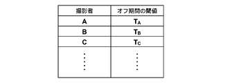
- threshold values (T A , T B , T C ,%) For each photographer (A, B, C,%) are set in advance as a table.
- the input unit 4 may receive photographer information, and may set a threshold of an off period corresponding to the received photographer information.
- radiographic imaging apparatus of this invention applies the radiographic imaging apparatus of this invention to the imaging
- a radiographic image is moved from several imaging directions by moving a radiation source. Any other apparatus can be applied as long as the apparatus performs the above imaging, and for example, there is a tomosynthesis imaging apparatus as such an apparatus.
Abstract
[Problem] To control a radiological imaging device, which continuously performs a series of imaging sequences when an imaging switch is continuously on, such that, if the imaging switch is off for only a short time when an operator's hand is accidentally released from the imaging switch, etc., the radiological imaging device promptly reverts to a normal imaging sequence, and takes appropriate radiological images. [Solution] Control is performed in such a manner that, when an imaging switch (5) is off for a predetermined period, a portion of the operations in an imaging sequence continues to be carried out if the predetermined period is equal to or less than a threshold value.
Description
本発明は、被写体に対して互いに異なる複数の撮影方向から放射線をそれぞれ照射し、撮影方向毎の放射線画像を検出する放射線画像撮影方法および装置に関するものである。
The present invention relates to a radiation image capturing method and apparatus for irradiating a subject with radiation from a plurality of mutually different capturing directions and detecting a radiation image for each capturing direction.
従来、複数の画像を組み合わせて表示することにより、視差を利用して立体視できることが知られている。このような立体視できる画像(以下、立体視画像またはステレオ画像という)は、同一の被写体を異なる方向から撮影して取得された互いに視差のある複数の画像に基づいて生成される。
Conventionally, it is known that stereoscopic viewing can be performed using parallax by displaying a combination of a plurality of images. Such a stereoscopically viewable image (hereinafter referred to as a stereoscopic image or a stereo image) is generated based on a plurality of images having parallax obtained by photographing the same subject from different directions.
そして、このような立体視画像の生成は、デジタルカメラやテレビなどの分野だけでなく、放射線画像撮影の分野においても利用されている。すなわち、被検者に対して互いに異なる方向から放射線を照射し、その被検者を透過した放射線を放射線画像検出器によりそれぞれ検出して互いに視差のある複数の放射線画像を取得し、これらの放射線画像に基づいて立体視画像を生成することが行われている(たとえば、特許文献1参照)。そして、このように立体視画像を生成することによって奥行感のある放射線画像を観察することができ、より診断に適した放射線画像を観察することができる。
And the generation of such stereoscopic images is used not only in the fields of digital cameras and televisions, but also in the field of radiographic imaging. That is, the patient is irradiated with radiation from different directions, the radiation transmitted through the subject is detected by a radiation image detector, and a plurality of radiation images having parallax are obtained, and these radiations are acquired. A stereoscopic image is generated based on the image (see, for example, Patent Document 1). And by generating a stereoscopic image in this way, a radiographic image with a sense of depth can be observed, and a radiographic image more suitable for diagnosis can be observed.
一方、上述した立体視画像を撮影する放射線画像撮影装置と同様に、放射線源を移動させて互いに異なる撮影方向から被写体に放射線を照射してトモシンセシス撮影を行う装置も提案されている(たとえば、特許文献2参照)。このトモシンセシス撮影装置は、上述したように互いに異なる撮影方向から被写体に放射線を照射して撮影を行い、その撮影によって取得した複数の放射線画像を加算して所望の断層面を強調した画像を得ることができるものである。
On the other hand, as with the above-described radiographic imaging apparatus that captures a stereoscopic image, an apparatus that performs tomosynthesis imaging by moving a radiation source and irradiating a subject with radiation from different imaging directions has been proposed (for example, a patent) Reference 2). As described above, this tomosynthesis imaging apparatus performs imaging by irradiating a subject with radiation from mutually different imaging directions, and adds a plurality of radiographic images acquired by the imaging to obtain an image in which a desired tomographic plane is emphasized. It is something that can be done.
ここで、一般的に放射線画像撮影装置においては、安全性の観点から、撮影者が撮影スイッチを押下している間において放射線の照射を含む一連の撮影シーケンスが実行され、撮影者が撮影スイッチを離したときには撮影シーケンスは中止される。
Here, in general, in a radiographic imaging apparatus, from the viewpoint of safety, a series of imaging sequences including radiation irradiation are executed while the photographer presses the imaging switch, and the photographer switches the imaging switch. The shooting sequence is canceled when released.
しかしながら、たとえば、撮影者が誤って撮影スイッチから短時間だけ手を離してしまったり、チャタリングや電気ノイズによって撮影スイッチが短時間だけオフ状態になってしまったときにまで全ての撮影シーケンスを中止したのでは不都合な場合がある。
However, for example, the entire shooting sequence was canceled until the photographer accidentally left the shooting switch for a short time or the shooting switch was turned off for a short time due to chattering or electrical noise. This may be inconvenient.
具体的には、上述した立体視画像を撮影する装置やトモシンセシス撮影装置を用いて、たとえばマンモグラフィの撮影を行う場合には、乳房を圧迫板によって圧迫した状態で複数回の撮影が行われるが、上述したように撮影シーケンスを中止したときに圧迫板による圧迫も解除すると、再び撮影スイッチが押下されて撮影シーケンスが再開され、乳房の圧迫が行われた際、撮影シーケンス中止前の乳房の形状に再び戻すことはほとんど無理であり、複数回の撮影間で異なる形状の乳房を撮影することになり、適切な立体視画像や断層画像を取得することができない。
Specifically, for example, when performing mammography imaging using the above-described apparatus for capturing stereoscopic images and tomosynthesis imaging apparatus, imaging is performed a plurality of times while the breast is compressed by a compression plate. As described above, when compression by the compression plate is released when the imaging sequence is stopped, the imaging switch is pressed again to restart the imaging sequence, and when breast compression is performed, the breast shape before the imaging sequence is canceled It is almost impossible to return again, and breasts having different shapes are photographed between a plurality of photographing operations, and appropriate stereoscopic images and tomographic images cannot be acquired.
また、複数の撮影間において撮影スイッチがオフ状態となって撮影シーケンスが中止した場合にX線管球の立ち下げ動作まで行うと、再び撮影スイッチが押下されて撮影シーケンスが再開された際、X線管球の立ち上げ動作が完了するまでに長い時間がかかってしまい撮影時間が長くなってしまう。特に、マンモグラフィの撮影の場合には、上述したように乳房が圧迫された状態で撮影が行われるので、撮影時間が長くなると患者に与える負担が大きくなってしまう。
In addition, when the imaging switch is turned off between a plurality of imaging and the imaging sequence is stopped, if the X-ray tube is lowered, the X-ray tube is lowered again. It takes a long time to complete the start-up operation of the tube, and the imaging time becomes long. In particular, in the case of mammography imaging, since imaging is performed with the breast pressed as described above, the burden on the patient increases as the imaging time increases.
本発明は、上記の事情に鑑み、上述したように撮影者が誤って撮影スイッチから短時間だけ手を離してしまったり、チャタリングや電気ノイズによって撮影スイッチが短時間だけオフ状態になった場合、速やかに通常の撮影シーケンスに復帰することができるとともに、適切な放射線画像の撮影を行うことができる放射線画像撮影方法および装置を提供することを目的とする。
In view of the above circumstances, the present invention, as described above, when the photographer accidentally releases the shooting switch for a short time, or when the shooting switch is turned off for a short time due to chattering or electrical noise, It is an object of the present invention to provide a radiographic imaging method and apparatus capable of quickly returning to a normal imaging sequence and capable of imaging an appropriate radiographic image.
本発明の放射線画像撮影装置は、放射線源から射出された放射線を被写体に対して互いに異なる複数の撮影方向からそれぞれ照射する放射線照射部と、放射線照射部の放射線の照射による撮影方向毎の放射線画像を検出する放射線画像検出器とを備えた放射線画像撮影装置において、撮影スイッチが連続してオン状態のときに撮影方向毎の放射線の照射を含む一連の撮影シーケンスを連続して行うよう制御する撮影制御部を備え、撮影制御部が、撮影スイッチが所定の期間だけオフ状態となった場合、その所定の期間が所定の閾値以下である場合には、撮影シーケンスの一部の動作については継続して行うものであることを特徴とする。
The radiographic imaging device of the present invention includes a radiation irradiation unit that irradiates a subject with radiation emitted from a radiation source from a plurality of different imaging directions, and a radiographic image for each imaging direction by irradiation of radiation from the radiation irradiation unit. In a radiographic imaging device including a radiographic image detector for detecting radiography, imaging is controlled so as to continuously perform a series of imaging sequences including irradiation of radiation for each imaging direction when an imaging switch is continuously on. Provided with a control unit, and when the shooting switch is turned off for a predetermined period, if the predetermined period is equal to or less than a predetermined threshold, some operations of the shooting sequence are continued. It is characterized by being performed.
また、上記本発明の放射線画像撮影装置においては、被写体を乳房とするとともに、乳房を圧迫する圧迫板を設け、撮影制御部を、オフ状態の期間が閾値以下である場合には、撮影シーケンスの一部の動作として圧迫板による圧迫を継続して行うものとし、オフ状態の期間が上記閾値より長い場合には、圧迫板による圧迫を解除するものとできる。
In the radiographic image capturing apparatus of the present invention, the subject is a breast, a compression plate for compressing the breast is provided, and the imaging control unit is configured to perform the imaging sequence when the off-state period is equal to or less than the threshold value. As a part of the operation, compression by the compression plate is continuously performed, and when the period of the off state is longer than the threshold value, the compression by the compression plate can be released.
また、放射線源を、X線管球を備えたものとし、撮影制御部を、オフ状態の期間が閾値以下である場合には、撮影シーケンスの一部の動作としてX線管球の立ち上げ動作を継続して行うものとし、オフ状態の期間が上記閾値より長い場合には、X線管球の立ち下げ動作を行うよう制御するものとできる。
Further, when the radiation source is provided with an X-ray tube and the imaging control unit sets the X-ray tube startup operation as a part of the imaging sequence when the off-state period is equal to or less than the threshold value. If the off-state period is longer than the threshold value, the X-ray tube can be controlled to be lowered.
また、撮影制御部を、撮影スイッチのオフ状態の期間が閾値以下である場合、放射線の照射は停止するものとできる。
Also, the imaging control unit can stop the radiation irradiation when the period of the imaging switch OFF state is less than or equal to the threshold value.
また、撮影制御部を、撮影スイッチのオフ状態の期間が閾値以下である場合、放射線源の移動は停止するものとできる。
Also, the imaging control unit can stop the movement of the radiation source when the period of the imaging switch OFF state is less than or equal to the threshold value.
また、上記閾値を、1秒以上2秒以下とすることができる。
Further, the threshold value can be set to 1 second or more and 2 seconds or less.
また、上記閾値として任意の値の設定を受け付ける閾値設定受付部を設けることができる。
Also, a threshold value setting reception unit that receives setting of an arbitrary value as the threshold value can be provided.
また、上記閾値を予め複数設定することができる。
Also, a plurality of the above threshold values can be set in advance.
また、上記閾値を、撮影者毎に予め複数設定することができる。
Also, a plurality of the threshold values can be set in advance for each photographer.
また、撮影制御部を、撮影者の情報を受け付け、その受け付けた撮影者に対応する上記閾値を設定するものとできる。
Also, the photographing control unit can accept the photographer's information and set the threshold value corresponding to the accepted photographer.
本発明の放射線画像撮影方法は、放射線源から射出された放射線を被写体に対して互いに異なる複数の撮影方向からそれぞれ照射し、その放射線の照射によって撮影方向毎の放射線画像を検出する放射線画像撮影方法において、撮影スイッチが連続してオン状態のときに撮影方向毎の放射線の照射を含む一連の撮影シーケンスを連続して行う際、撮影スイッチが所定の期間だけオフ状態となった場合、その所定の期間が所定の閾値以下である場合には、撮影シーケンスの一部の動作については継続して行うことを特徴とする。
The radiographic imaging method of the present invention irradiates a subject with radiation emitted from a radiation source from a plurality of different imaging directions, and detects a radiographic image for each imaging direction by irradiation of the radiation. In this case, when a series of imaging sequences including irradiation of radiation for each imaging direction is continuously performed when the imaging switch is continuously on, when the imaging switch is in an off state for a predetermined period, When the period is equal to or less than a predetermined threshold, some operations of the imaging sequence are continuously performed.
本発明の放射線画像撮影方法および装置によれば、撮影スイッチが所定の期間だけオフ状態となった場合、その所定の期間が所定の閾値以下である場合には、撮影シーケンスの一部の動作については継続して行うようにしたので、たとえば、上述したような圧迫板による圧迫やX線管球の立ち上げ動作などについては中止することなく継続して行うことができるので、速やかに通常の撮影シーケンスに復帰することができるとともに、適切な立体視画像や断層画像などを撮影することができる。
According to the radiographic imaging method and apparatus of the present invention, when the imaging switch is turned off only for a predetermined period, when the predetermined period is equal to or less than a predetermined threshold, a part of the operation of the imaging sequence is performed. Since, for example, the above-described compression by the compression plate and the X-ray tube start-up operation can be performed without interruption, normal imaging can be performed promptly. It is possible to return to the sequence and to capture an appropriate stereoscopic image or tomographic image.
また、上記本発明の放射線画像撮影装置において、撮影スイッチのオフ状態の期間が閾値以下である場合でも、放射線の照射については停止するようにした場合には、撮影者の誤りではなく撮影シーケンスの中止を目的として撮影スイッチがオフ状態にされた際、即座に放射線の照射を停止することができ、放射線の被曝に対する安全性を確保することができる。
Further, in the radiographic image capturing apparatus of the present invention, when radiation irradiation is stopped even when the period of the imaging switch OFF state is equal to or less than the threshold, the radiographing sequence is not an error of the radiographer. When the imaging switch is turned off for the purpose of stopping, radiation irradiation can be stopped immediately, and safety against radiation exposure can be ensured.
また、撮影スイッチのオフ状態の期間が閾値以下である場合でも、放射線源の移動については停止するようにした場合には、撮影者の誤りではなく撮影シーケンスの中止を目的として撮影スイッチがオフ状態にされた際、即座に放射線源の移動を停止することができ、たとえば、放射線源が被検者に衝突するのを回避することができる。
In addition, even when the period of the imaging switch OFF state is equal to or less than the threshold value, if the movement of the radiation source is stopped, the imaging switch is in the OFF state for the purpose of canceling the imaging sequence rather than the photographer's error. In this case, the movement of the radiation source can be stopped immediately, and for example, the collision of the radiation source with the subject can be avoided.
また、撮影スイッチのオフ状態の期間の閾値を撮影者毎に予め複数設定するようにした場合には、撮影者の要望に応じた閾値を設定することができ、個々の撮影者にとってより使い勝手の良いものとすることができる。
In addition, when a plurality of thresholds for the shooting switch OFF state are set in advance for each photographer, it is possible to set a threshold according to the photographer's request, which is more convenient for individual photographers. Can be good.
以下、図面を参照して本発明の放射線画像撮影装置の一実施形態を用いた乳房画像撮影表示システムについて説明する。図1は、本実施形態の乳房画像撮影表示システム全体の概略構成を示す図である。
Hereinafter, a breast image radiographing display system using an embodiment of a radiographic image radiographing apparatus of the present invention will be described with reference to the drawings. FIG. 1 is a diagram showing a schematic configuration of the entire breast image photographing display system of the present embodiment.
本実施形態の乳房画像撮影表示システム1は、図1に示すように、乳房画像撮影装置10と、乳房画像撮影装置10に接続されたコンピュータ2と、コンピュータ2に接続されるモニタ3、入力部4および撮影スイッチ5とを備えている。
As shown in FIG. 1, a breast image radiographing display system 1 according to this embodiment includes a mammography apparatus 10, a computer 2 connected to the mammography apparatus 10, a monitor 3 connected to the computer 2, and an input unit. 4 and a photographing switch 5.
乳房画像撮影装置10は、図1に示すように、基台11と、基台11に対し上下方向(Z方向)に移動可能であり、かつ回転可能な回転軸12と、回転軸12により基台11と連結されたアーム部13を備えている。図2には、図1の右方向から見たアーム部13を示している。
As shown in FIG. 1, the mammography apparatus 10 includes a base 11, a rotary shaft 12 that can move in the vertical direction (Z direction) with respect to the base 11, and can be rotated. The arm part 13 connected with the base 11 is provided. FIG. 2 shows the arm 13 viewed from the right direction in FIG.
アーム部13はアルファベットのCの形をしており、その一端には乳房が設置される撮影台14が、その他端には撮影台14と対向するように放射線源ユニット16が取り付けられている。アーム部13の上下方向の移動は、基台11に組み込まれたアームコントローラ31により制御される。
The arm portion 13 has an alphabet C shape, and an imaging table 14 on which a breast is installed is attached to one end, and a radiation source unit 16 is attached to the other end so as to face the imaging table 14. The movement of the arm unit 13 in the vertical direction is controlled by an arm controller 31 incorporated in the base 11.
撮影台14の内部には、フラットパネルディテクタ等の放射線画像検出器15と、放射線画像検出器15からの電荷信号の読み出しなどを制御する検出器コントローラ33が備えられている。
In the imaging table 14, a radiation image detector 15 such as a flat panel detector, and a detector controller 33 for controlling reading of a charge signal from the radiation image detector 15 are provided.
また、撮影台14の内部には、放射線画像検出器15から読み出された電荷信号を電圧信号に変換するチャージアンプや、チャージアンプから出力された電圧信号をサンプリングする相関2重サンプリング回路や、電圧信号をデジタル信号に変換するAD変換部などが設けられた回路基板なども設置されている。
Further, inside the imaging table 14, a charge amplifier that converts the charge signal read from the radiation image detector 15 into a voltage signal, a correlated double sampling circuit that samples the voltage signal output from the charge amplifier, A circuit board provided with an AD conversion unit for converting a voltage signal into a digital signal is also installed.
放射線画像検出器15は、放射線画像の記録と読出しを繰り返して行うことができるものであり、放射線の照射を直接受けて電荷を発生する、いわゆる直接型の放射線画像検出器を用いてもよいし、放射線を一旦可視光に変換し、その可視光を電荷信号に変換する、いわゆる間接型の放射線画像検出器を用いるようにしてもよい。また、放射線画像信号の読出方式としては、TFT(thin film transistor)スイッチをオン・オフされることによって放射線画像信号が読みだされる、いわゆるTFT読出方式のものや、読取光を照射することによって放射線画像信号が読み出される、いわゆる光読出方式のものを用いることができるが、これに限らずその他のものを用いるようにしてもよい。
The radiation image detector 15 can repeatedly perform recording and reading of a radiation image, and may use a so-called direct type radiation image detector that directly receives radiation and generates charges. Alternatively, a so-called indirect radiation image detector that converts radiation once into visible light and converts the visible light into a charge signal may be used. As a radiation image signal readout method, a radiation image signal is read out by turning on / off a TFT (thin film transistor) switch, or by irradiating reading light. A so-called optical readout system in which a radiation image signal is read out can be used, but the invention is not limited to this, and other types may be used.
放射線源ユニット16の中には放射線源17と放射線源コントローラ32とが収納されている。放射線源17はX線管球を備えたものであり、そのX線管球は、高電圧の管電圧を印加する際に用いられるコンデンサと、回転動作を行いながら電子線の衝突を受けることによって放射線を放出する陽極とを備えたものである。放射線源コントローラ32は、放射線源17におけるX線管球の立ち上げ・立ち下げ動作や、放射線源17から放射線を照射するタイミングや、放射線源17における放射線発生条件(管電流、時間、管電圧等)などを制御するものである。
A radiation source 17 and a radiation source controller 32 are accommodated in the radiation source unit 16. The radiation source 17 is provided with an X-ray tube, and the X-ray tube is subjected to collision of an electron beam while performing a rotating operation with a capacitor used when applying a high voltage tube voltage. And an anode that emits radiation. The radiation source controller 32 raises and lowers the X-ray tube in the radiation source 17, the timing of irradiating the radiation from the radiation source 17, and the radiation generation conditions (tube current, time, tube voltage, etc.) in the radiation source 17. ) And the like are controlled.
また、アーム部13の中央部には、撮影台14の上方に配置され、乳房を押さえつけて圧迫する圧迫板18と、その圧迫板18を支持する支持部20と、支持部20を上下方向に移動させる移動機構19が設けられている。圧迫板18の位置、圧迫圧は、圧迫板コントローラ34により制御される。
Further, in the central portion of the arm portion 13, the compression plate 18 is disposed above the imaging table 14 and presses the breast to press it, the support portion 20 that supports the compression plate 18, and the support portion 20 in the vertical direction. A moving mechanism 19 for moving is provided. The position of the compression plate 18 and the compression pressure are controlled by the compression plate controller 34.
コンピュータ2は、中央処理装置(CPU)および半導体メモリやハードディスクやSSD等のストレージデバイスなどを備えており、これらのハードウェアによって、図3に示すような撮影制御部40、放射線画像記憶部41および表示制御部42が構成されている。
The computer 2 includes a central processing unit (CPU), a storage device such as a semiconductor memory, a hard disk, and an SSD. The hardware includes an imaging control unit 40, a radiographic image storage unit 41, and a storage device such as shown in FIG. A display control unit 42 is configured.
撮影制御部40は、各種のコントローラ31~34に対して所定の制御信号を出力し、システム全体の撮影シーケンスの制御を行うものである。具体的な制御方法については後で詳述する。
The imaging control unit 40 outputs predetermined control signals to the various controllers 31 to 34, and controls the imaging sequence of the entire system. A specific control method will be described in detail later.
放射線画像記憶部41は、互いに異なる2つの撮影方向からの撮影によって放射線画像検出器15によって検出された2枚の放射線画像信号を記憶するものである。
The radiation image storage unit 41 stores two radiation image signals detected by the radiation image detector 15 by photographing from two different photographing directions.
表示制御部42は、放射線画像記憶部41から読み出された放射線画像信号に対して所定の信号処理を施した後、モニタ3に乳房のステレオ画像を表示させるものである。
The display control unit 42 displays a breast stereo image on the monitor 3 after performing predetermined signal processing on the radiographic image signal read from the radiographic image storage unit 41.
入力部4は、たとえば、キーボードやマウスなどのポインティングデバイスから構成されるものであり、撮影者による撮影条件の入力などを受け付けるものである。
The input unit 4 is composed of a pointing device such as a keyboard and a mouse, for example, and accepts input of shooting conditions by a photographer.
撮影スイッチ5は、互いに異なる2つの撮影方向からの撮影を含む一連の撮影シーケンスの実行を指示するためのボタンである。そして、コンピュータ2の撮影制御部40は、この撮影スイッチ5が撮影者によってオン状態となっているときに一連の撮影シーケンスが実行されるよう各部を制御するものであるが、たとえば、撮影者が誤って撮影スイッチ5から手を離してしまったり、チャタリングや電気ノイズによって撮影スイッチ5がオフ状態となった場合、そのオフ状態の期間の長さに応じて撮影シーケンスの一部の動作を停止したり、継続したりするものである。その一部の撮影シーケンスの動作の制御については後で詳述する。
The shooting switch 5 is a button for instructing execution of a series of shooting sequences including shooting from two different shooting directions. The photographing control unit 40 of the computer 2 controls each unit so that a series of photographing sequences is executed when the photographing switch 5 is turned on by the photographer. If the shooting switch 5 is accidentally released or if the shooting switch 5 is turned off due to chattering or electrical noise, the operation of a part of the shooting sequence is stopped according to the length of the off-state period. Or continue. The control of the operation of part of the imaging sequence will be described in detail later.
モニタ3は、コンピュータ2から出力された2つの放射線画像信号を用いてステレオ画像を表示可能なように構成されたものである。ステレオ画像を表示する構成としては、たとえば、2つの画面を用いて2つの放射線画像信号に基づく放射線画像をそれぞれ表示させて、これらをハーフミラーや偏光グラスなどを用いることで一方の放射線画像は観察者の右目に入射させ、他方の放射線画像は観察者の左目に入射させることによってステレオ画像を表示する構成を採用することができる。または、たとえば、2つの放射線画像を所定の視差量だけずらして重ね合わせて表示し、これを偏光グラスで観察することでステレオ画像を生成する構成としてもよいし、もしくはパララックスバリア方式およびレンチキュラー方式のように、2つの放射線画像を立体視可能な3D液晶に表示することによってステレオ画像を生成する構成としてもよい。
The monitor 3 is configured to be able to display a stereo image using two radiation image signals output from the computer 2. As a configuration for displaying a stereo image, for example, a radiographic image based on two radiographic image signals is displayed using two screens, and one of the radiographic images is observed by using a half mirror or a polarizing glass. It is possible to adopt a configuration in which a stereo image is displayed by being incident on the right eye of the observer and the other radiation image is incident on the left eye of the observer. Or, for example, two radiographic images may be displayed in a superimposed manner while being shifted by a predetermined amount of parallax, and this may be configured to generate a stereo image by observing with a polarizing glass, or a parallax barrier method and a lenticular method As described above, a stereo image may be generated by displaying two radiation images on a stereoscopically viewable 3D liquid crystal.
次に、本実施形態の乳房画像撮影表示システムの作用について、図4に示すフローチャートおよび図5に示すタイミングチャートを参照しながら説明する。図5は、一連の撮影シーケンスにおける撮影スイッチ5のオン・オフ状態と、放射線源17の放射線の照射タイミングと、放射線源17の移動タイミング(放射線源17を移動させる駆動モータの動作タイミング)とを示すものである。なお、ここではまず撮影スイッチ5が上述したようなオフ状態とならない場合の一連の撮影シーケンスについて説明する。
Next, the operation of the breast image radiographing display system of this embodiment will be described with reference to the flowchart shown in FIG. 4 and the timing chart shown in FIG. FIG. 5 shows on / off states of the imaging switch 5 in a series of imaging sequences, radiation irradiation timing of the radiation source 17, and movement timing of the radiation source 17 (operation timing of the drive motor that moves the radiation source 17). It is shown. Here, a series of shooting sequences when the shooting switch 5 is not in the OFF state as described above will be described first.
まず、乳房画像撮影装置10、コンピュータ2およびモニタ3の電源がオンされた後、撮影台14の上に被検者Mの乳房が設置され、圧迫板18により乳房が所定の圧力によって圧迫される(S10)。
First, after the mammography apparatus 10, the computer 2, and the monitor 3 are turned on, the breast of the subject M is placed on the imaging table 14, and the breast is compressed with a predetermined pressure by the compression plate 18. (S10).
次に、撮影者によって撮影スイッチ5が押下されることによって、図5に示すように撮影スイッチ5がオン状態となり(S12)、これにより撮影制御部40によって一連の撮影シーケンスが開始される(S14)。具体的には、まず、撮影制御部40から放射線源コントローラ32に制御信号が出力され、放射線源コントローラ32は、入力された制御信号に応じて放射線源17におけるX線管球の立ち上げ動作を開始する(S16)。X線管球の立ち上げ動作としては、たとえば、陽極の回転動作の開始やコンデンサの充電などがある。
Next, when the photographing switch 5 is pressed by the photographer, the photographing switch 5 is turned on as shown in FIG. 5 (S12), whereby a series of photographing sequences is started by the photographing control unit 40 (S14). ). Specifically, first, a control signal is output from the imaging control unit 40 to the radiation source controller 32, and the radiation source controller 32 performs an X-ray tube startup operation in the radiation source 17 in accordance with the input control signal. Start (S16). For example, the start-up operation of the X-ray tube includes the start of an anode rotation operation and the charging of a capacitor.
次に、撮影制御部40は、放射線源17のX線管球の立ち上げ動作が終了した後、ステレオ画像を構成する2枚の放射線画像のうちの1枚目の放射線画像の撮影を行う。
Next, after the start-up operation of the X-ray tube of the radiation source 17 is completed, the imaging control unit 40 captures the first radiographic image of the two radiographic images constituting the stereo image.
具体的には、まず、撮影制御部40が、予め設定されたステレオ画像の撮影のための撮影角度θを読み出し、その読み出した撮影角度θの情報をアームコントローラ31に出力する。なお、本実施形態においては、このときの撮影角度θの情報としてθ=0°とθ=4°が予め記憶されているものとするが、これに限らず、撮影者によって入力部4において任意の撮影角度を設定可能である。
Specifically, first, the shooting control unit 40 reads a preset shooting angle θ for shooting a stereo image, and outputs information of the read shooting angle θ to the arm controller 31. In this embodiment, it is assumed that θ = 0 ° and θ = 4 ° are stored in advance as information on the shooting angle θ at this time. The shooting angle can be set.
そして、アームコントローラ31において、撮影制御部40から出力された撮影角度θ=0°とθ=4°の情報が受け付けられ、撮影制御部40から出力された制御信号に応じてアームコントローラ31は、まず、θ=0°の情報に基づいて、図2に示すようにアーム部13が撮影台14に対して垂直な方向となるように制御信号を出力する。
Then, the arm controller 31 receives information on the shooting angles θ = 0 ° and θ = 4 ° output from the shooting control unit 40, and the arm controller 31 determines whether the arm controller 31 outputs a control signal output from the shooting control unit 40. First, based on the information of θ = 0 °, a control signal is output so that the arm unit 13 is perpendicular to the imaging table 14 as shown in FIG.
そして、このアームコントローラ31から出力された制御信号に応じてアーム部13が、撮影台14に対して垂直な方向となった状態において、撮影制御部40は、放射線源コントローラ32および検出器コントローラ33に対して放射線の照射と放射線画像信号の読出しを行うよう制御信号を出力する。
Then, in a state where the arm unit 13 is in a direction perpendicular to the imaging table 14 according to the control signal output from the arm controller 31, the imaging control unit 40 includes the radiation source controller 32 and the detector controller 33. A control signal is output so as to perform radiation irradiation and readout of the radiation image signal.
放射線源コントローラ32は、入力された制御信号に応じて放射線源17における管電圧の印加(V=V1)を開始することによって、図5に示す1回目の放射線の照射を開始する(S18)。
The radiation source controller 32 starts application of the tube voltage (V = V 1 ) in the radiation source 17 in accordance with the input control signal, thereby starting the first radiation irradiation shown in FIG. 5 (S18). .
そして、この放射線源17からの1回目の放射線の照射によって、乳房を0°方向から撮影した放射線画像が放射線画像検出器15によって検出される(S20)。
The radiation image obtained by photographing the breast from the 0 ° direction is detected by the radiation image detector 15 by the first radiation irradiation from the radiation source 17 (S20).
そして、放射線源コントローラ32は、図5に示すような予め設定されたワンショットの放射線照射期間が経過すると、放射線源17における管電圧をV=V0とし、1回目の放射線の照射を終了する(S22)。なお、V0は放射線源17からの放射線の射出が停止される管電圧であり、5V程度の所定の電圧としてもよいし、0Vとしてもよい。
Then, when a preset one-shot radiation irradiation period as shown in FIG. 5 elapses, the radiation source controller 32 sets the tube voltage in the radiation source 17 to V = V 0 and ends the first radiation irradiation. (S22). Note that V 0 is a tube voltage at which the emission of radiation from the radiation source 17 is stopped, and may be a predetermined voltage of about 5 V or 0 V.
次に、検出器コントローラ33によって放射線画像検出器15から放射線画像信号が読み出され、その放射線画像信号に対して所定の信号処理が施された後、コンピュータ2の放射線画像記憶部41に記憶される(S24)。
Next, a radiation image signal is read from the radiation image detector 15 by the detector controller 33, subjected to predetermined signal processing on the radiation image signal, and then stored in the radiation image storage unit 41 of the computer 2. (S24).
上述したような1枚目の放射線画像の撮影の後、図5に示すように放射線源ユニット17の移動が開始される(S26)。
After taking the first radiation image as described above, the movement of the radiation source unit 17 is started as shown in FIG. 5 (S26).
具体的には、撮影制御部40から出力された制御信号に応じてアームコントローラ31は、図2に示すように、アーム部13を撮影台14に垂直な方向に対して+θ°回転するよう制御信号を出力する。すなわち、本実施形態においては、アーム部13を撮影台14に垂直な方向に対して4°回転するよう制御信号を出力する。
Specifically, according to the control signal output from the imaging control unit 40, the arm controller 31 controls the arm unit 13 to rotate by + θ ° with respect to the direction perpendicular to the imaging table 14, as shown in FIG. Output a signal. That is, in the present embodiment, the control signal is output so that the arm unit 13 is rotated by 4 ° with respect to the direction perpendicular to the imaging table 14.
アーム部13は、アームコントローラ31から出力された制御信号に基づいて4°の方向に移動し、これにより放射線源ユニット16も4°の方向に移動する。
The arm unit 13 moves in the 4 ° direction based on the control signal output from the arm controller 31, and the radiation source unit 16 also moves in the 4 ° direction.
そして、アームコントローラ31から出力された制御信号に応じてアーム部13が4°回転した状態において、撮影制御部40は、放射線源コントローラ32および検出器コントローラ33に対して放射線の照射と放射線画像信号の読出しを行うよう制御信号を出力する。
Then, in a state where the arm unit 13 is rotated by 4 ° according to the control signal output from the arm controller 31, the imaging control unit 40 applies radiation to the radiation source controller 32 and the detector controller 33 and the radiation image signal. A control signal is output so as to read out.
放射線源コントローラ32は、入力された制御信号に応じて放射線源17における管電圧の印加(V=V1)を開始することによって、図5に示す2回目の放射線の照射を開始する(S28)。
The radiation source controller 32 starts the application of the tube voltage (V = V 1 ) in the radiation source 17 in accordance with the input control signal, thereby starting the second radiation irradiation shown in FIG. 5 (S28). .
そして、この放射線源17からの2回目の放射線の照射によって、乳房を4°方向から撮影した放射線画像が放射線画像検出器15によって検出される(S30)。
Then, a radiation image obtained by photographing the breast from the 4 ° direction is detected by the radiation image detector 15 by the second irradiation of radiation from the radiation source 17 (S30).
そして、放射線源コントローラ32は、図5に示すような予め設定されたワンショットの放射線照射期間が経過すると、放射線源17における管電圧をV=V0とし、2回目の放射線の照射を終了する(S32)。
Then, when a preset one-shot radiation irradiation period as shown in FIG. 5 elapses, the radiation source controller 32 sets the tube voltage in the radiation source 17 to V = V 0 and ends the second radiation irradiation. (S32).
次に、検出器コントローラ33によって放射線画像検出器15から放射線画像信号が読み出され、その放射線画像信号に対して所定の信号処理が施された後、コンピュータ2の放射線画像記憶部41に記憶される(S34)。
Next, a radiation image signal is read from the radiation image detector 15 by the detector controller 33, subjected to predetermined signal processing on the radiation image signal, and then stored in the radiation image storage unit 41 of the computer 2. (S34).
そして、上述したようなステレオ画像を構成する2枚の放射線画像の撮影が終了すると、撮影者は撮影スイッチ5の押下をやめ、これにより撮影スイッチ5がオフ状態となる。撮影制御部40は、撮影が終了して撮影スイッチ5がオフ状態となると一連の撮影シーケンスを終了し、圧迫板コントローラ34に制御信号を出力する。そして、圧迫板コントローラ34は、入力された制御信号に応じて圧迫板18による乳房の圧迫を解除する(S38)。
Then, when the photographing of the two radiographic images constituting the stereo image as described above is completed, the photographer stops pressing the photographing switch 5, and the photographing switch 5 is turned off. When the photographing is finished and the photographing switch 5 is turned off, the photographing control unit 40 ends the series of photographing sequences and outputs a control signal to the compression plate controller 34. Then, the compression plate controller 34 releases the compression of the breast by the compression plate 18 according to the input control signal (S38).
さらに撮影制御部40は、放射線源コントローラ32に制御信号を出力し、放射線源コントローラ32は、入力された制御信号に応じて放射線源ユニット16の放射線源17のX線管球の立ち下げ動作を行う(S40)。なお、ここでいうX線管球の立ち下げ動作としては、たとえば、陽極の回転動作の停止やコンデンサへの充電の停止などがある。
Further, the imaging control unit 40 outputs a control signal to the radiation source controller 32, and the radiation source controller 32 performs the operation of lowering the X-ray tube of the radiation source 17 of the radiation source unit 16 according to the input control signal. Perform (S40). Note that the X-ray tube falling operation mentioned here includes, for example, stopping the rotating operation of the anode and stopping charging of the capacitor.
そして、上述したようにして放射線画像記憶部41に記憶された2枚の放射線画像信号は、表示制御部42によって読み出され、表示制御部42においてこれらの放射線画像信号に対して所定の処理が施された後、モニタ3に出力される。そして、モニタ3において、右目用放射線画像と左目用放射線画像とがそれぞれ表示されて乳房のステレオ画像が表示される(S42)。
The two radiographic image signals stored in the radiographic image storage unit 41 as described above are read out by the display control unit 42, and the display control unit 42 performs predetermined processing on these radiographic image signals. After being applied, it is output to the monitor 3. Then, on the monitor 3, the radiographic image for the right eye and the radiographic image for the left eye are respectively displayed, and a stereo image of the breast is displayed (S42).
以上が、通常の一連の撮影シーケンスの説明である。次に、撮影者によって撮影スイッチ5が押下された時点から、2枚の放射線画像の撮影の終了後に撮影者が撮影スイッチ5の押下をやめるまでの間に、撮影スイッチ5が誤ってオフ状態になった場合の作用について、図6に示すフローチャートを参照しながら説明する。
The above is a description of a normal series of shooting sequences. Next, the photographing switch 5 is erroneously turned off after the photographing switch 5 is pressed by the photographer until the photographer stops pressing the photographing switch 5 after the two radiographic images are photographed. The operation in this case will be described with reference to the flowchart shown in FIG.
撮影者によって撮影スイッチ5が押下された時点から、たとえば撮影者が誤って撮影スイッチ5から手を離してしまったり、チャタリングや電気ノイズによって撮影スイッチ5がオフ状態となった場合(S50,YES)、このとき放射線源17から放射線が照射中である場合には、すなわち図5に示す1回目の放射線の照射中(管電圧V=V1)または2回目の放射線の照射中(管電圧V=V1)である場合には(S52,YES)、撮影制御部40から放射線源コントローラ32に制御信号が出力され、放射線源コントローラ32によって放射線の照射は停止される(S54)。また、このとき放射線源17を移動中である場合には(S56、YES)、撮影制御部40からアームコントローラ31に制御信号が出力され、アームコントローラ31によってアーム部13(放射線源ユニット16)の移動は停止される(S58)。
When, for example, the photographer accidentally releases the photographing switch 5 from the time when the photographing switch 5 is pressed by the photographer, or when the photographing switch 5 is turned off due to chattering or electrical noise (S50, YES). In this case, when radiation is being irradiated from the radiation source 17, that is, during the first irradiation (tube voltage V = V 1 ) or the second irradiation (tube voltage V =) shown in FIG. If V 1 ) (S52, YES), a control signal is output from the imaging control unit 40 to the radiation source controller 32, and radiation irradiation is stopped by the radiation source controller 32 (S54). If the radiation source 17 is moving at this time (YES in S56), a control signal is output from the imaging control unit 40 to the arm controller 31, and the arm controller 31 causes the arm unit 13 (radiation source unit 16) to move. The movement is stopped (S58).
そして、撮影制御部40は、撮影スイッチ5がオフ状態となった時点からの時間を計測し(S60)、そのオフ期間が所定の閾値以下である間は(S62,YES)、上述した放射線の照射や放射線源17の移動を除く撮影シーケンスは継続して実行する(S64)。具体的には、たとえば圧迫板18による乳房の圧迫や放射線源17のX線管球の立ち上げ状態などは維持されたままとなる。
The imaging control unit 40 measures the time from when the imaging switch 5 is turned off (S60), and while the off period is equal to or less than the predetermined threshold (S62, YES), The imaging sequence excluding irradiation and movement of the radiation source 17 is continuously executed (S64). Specifically, for example, the compression of the breast by the compression plate 18 and the startup state of the X-ray tube of the radiation source 17 are maintained.
そして、オフ期間が所定の閾値以下の間に、再び撮影者によって撮影スイッチ5が押し直されたり、チャタリングや電気ノイズの影響がなくなって撮影スイッチ5がオン状態となった場合には(S66)、撮影制御部40は、放射線源17の移動を再開して、通常の撮影シーケンスを継続して実行する(S68)。
Then, when the photographing switch 5 is pressed again by the photographer or the influence of chattering or electrical noise is eliminated while the off period is equal to or less than the predetermined threshold, the photographing switch 5 is turned on (S66). The imaging control unit 40 resumes the movement of the radiation source 17 and continuously executes the normal imaging sequence (S68).
一方、撮影制御部40は、撮影スイッチ5のオフ期間が所定の閾値を超えた場合には(S62,NO)、圧迫板コントローラ34に制御信号を出力し、圧迫板コントローラ34は入力された制御信号に応じて圧迫板18による乳房の圧迫を解除する(S70)。さらに撮影制御部40は、放射線源コントローラ32に制御信号を出力し、放射線源コントローラ32は入力された制御信号に応じて放射線源17におけるX線管球の立ち下げ動作を行う(S72)。そして、撮影制御部40は、一連の撮影シーケンスを中止とする(S74)。
On the other hand, when the OFF period of the photographing switch 5 exceeds a predetermined threshold (S62, NO), the photographing control unit 40 outputs a control signal to the compression plate controller 34, and the compression plate controller 34 receives the input control. In response to the signal, the compression of the breast by the compression plate 18 is released (S70). Further, the imaging control unit 40 outputs a control signal to the radiation source controller 32, and the radiation source controller 32 performs an X-ray tube falling operation in the radiation source 17 in accordance with the input control signal (S72). Then, the shooting control unit 40 stops a series of shooting sequences (S74).
上記実施形態の乳房画像撮影表示システムによれば、撮影スイッチが所定の期間だけオフ状態となった場合、そのオフ期間が所定の閾値以下である場合には、圧迫板による圧迫やX線管球の立ち上げ動作などについては中止することなく継続して行うようにしたので、速やかに通常の撮影シーケンスに復帰することができるとともに、適切なステレオ画像を撮影することができる。
According to the breast image radiographing display system of the above-described embodiment, when the radiographing switch is turned off only for a predetermined period, when the off period is equal to or less than a predetermined threshold, the compression by the compression plate or the X-ray tube Since the start-up operation and the like are continuously performed without being stopped, it is possible to quickly return to the normal shooting sequence and to capture an appropriate stereo image.
また、上記実施形態においては、撮影スイッチ5のオフ期間が所定の閾値以下の場合に、一連の撮影シーケンスのうちの乳房の圧迫とX線管球の立ち上げ動作について継続して行うようにしたが、これに限らず、その他の動作を継続するようにしてもよい。
In the above-described embodiment, when the imaging switch 5 is off or below a predetermined threshold, the breast compression and the X-ray tube start-up operation are continuously performed in a series of imaging sequences. However, the present invention is not limited to this, and other operations may be continued.
すなわち、乳房の圧迫を継続して行うようにしたのは、乳房の圧迫を一旦解除してしまうと再び圧迫したときに乳房の形が変わってしまい適切なステレオ画像を撮影できなくなってしまうからであるが、このようにステレオ画像の適切な撮影の観点から停止することが望ましくないその他の動作を継続させるようにしてもよい。
In other words, the reason why breast compression is continuously performed is that once breast compression is released, the shape of the breast changes when it is compressed again, making it impossible to capture appropriate stereo images. However, other operations that are not desirable to be stopped from the viewpoint of appropriate photographing of stereo images may be continued.
また、X線管球の立ち上げ動作を停止しないようにしたのは、一旦立ち下げてしまうと再立ち上げに時間がかかるからであるが、このように再び復帰させるまでの時間の観点から停止することが望ましくないその他の動作を継続させるようにしてもよい。
The reason why the X-ray tube start-up operation is not stopped is that it takes time to re-start up once it is started down, but it is stopped from the viewpoint of the time until it is restored again. Other operations that are not desirable to be performed may be continued.
また、上記実施形態においては、撮影スイッチ5がオフ状態になったとき放射線源ユニット16の移動を停止するようにしたが、これは安全性の観点からこのように設定したものであり、これに限らず、たとえば、安全性よりも撮影時間の短縮を優先的に考慮する場合には、撮影スイッチ5がオフ状態になったとき、そのオフ期間が所定の閾値以下の場合には、放射線源ユニット16の移動を継続して行うようにしてもよい。
Further, in the above embodiment, the movement of the radiation source unit 16 is stopped when the imaging switch 5 is turned off. This is set in this way from the viewpoint of safety. For example, in the case where reduction in imaging time is given priority over safety, when the imaging switch 5 is turned off and the off period is equal to or less than a predetermined threshold, the radiation source unit You may make it perform 16 movements continuously.
また、上記実施形態において用いられるオフ期間の閾値については、たとえば1秒以上2秒以下に設定することが望ましい。
Further, it is desirable to set the threshold value of the off period used in the above embodiment to, for example, 1 second or more and 2 seconds or less.
また、オフ期間の閾値は、予め設定するようにしてもよいが、入力部4を用いて撮影者が任意の値を設定できるようにしてもよい。また、複数のオフ期間の閾値を予め設定しておき、撮影者が入力部4を用いてその複数の閾値の中からいずれか1つを選択し、その選択した閾値を設定するようにしてもよい。
Further, the off-period threshold may be set in advance, but the photographer may be able to set an arbitrary value using the input unit 4. In addition, a plurality of off-period threshold values are set in advance, and the photographer selects one of the plurality of threshold values using the input unit 4 and sets the selected threshold value. Good.
また、図6に示すように撮影者(A、B、C、・・・)毎のオフ期間の閾値(TA、TB、TC、・・・)を予めテーブルとして設定しておき、入力部4において撮影者の情報を受け付け、その受け付けた撮影者の情報に対応するオフ期間の閾値を設定するようにしてもよい。
Further, as shown in FIG. 6, threshold values (T A , T B , T C ,...) For each photographer (A, B, C,...) Are set in advance as a table. The input unit 4 may receive photographer information, and may set a threshold of an off period corresponding to the received photographer information.
また、上記実施形態は、本発明の放射線画像撮影装置を、乳房のステレオ画像の撮影装置に適用したものであるが、これに限らず、放射線源を移動させることにより複数の撮影方向から放射線画像の撮影を行う装置であればその他の装置にも適用可能であり、たとえば、このような装置としてトモシンセシス撮影装置がある。
Moreover, although the said embodiment applies the radiographic imaging apparatus of this invention to the imaging | photography apparatus of a breast stereo image, it is not restricted to this, A radiographic image is moved from several imaging directions by moving a radiation source. Any other apparatus can be applied as long as the apparatus performs the above imaging, and for example, there is a tomosynthesis imaging apparatus as such an apparatus.
Claims (11)
- 放射線源から射出された放射線を被写体に対して互いに異なる複数の撮影方向からそれぞれ照射する放射線照射部と、該放射線照射部の前記放射線の照射による前記撮影方向毎の放射線画像を検出する放射線画像検出器とを備えた放射線画像撮影装置において、
撮影スイッチが連続してオン状態のときに前記撮影方向毎の放射線の照射を含む一連の撮影シーケンスを連続して行うよう制御する撮影制御部を備え、
該撮影制御部が、前記撮影スイッチが所定の期間だけオフ状態となった場合、該所定の期間が所定の閾値以下である場合には、前記撮影シーケンスの一部の動作については継続して行うものであることを特徴とする放射線画像撮影装置。 A radiation irradiator that irradiates a subject with radiation emitted from a plurality of different imaging directions, and a radiation image detection that detects a radiation image in each imaging direction by irradiation of the radiation of the radiation irradiator In a radiographic imaging device comprising a container,
An imaging control unit that controls to continuously perform a series of imaging sequences including radiation irradiation for each imaging direction when the imaging switch is continuously on,
When the shooting switch is turned off for a predetermined period, the shooting control unit continuously performs a part of the shooting sequence when the predetermined period is equal to or less than a predetermined threshold. A radiographic imaging device characterized by being a thing. - 前記被写体が乳房であるとともに、該乳房を圧迫する圧迫板を備え、
前記撮影制御部が、前記オフ状態の期間が前記閾値以下である場合には、前記撮影シーケンスの一部の動作として前記圧迫板による圧迫を継続して行うものであり、前記オフ状態の期間が前記閾値より長い場合には、前記圧迫板による圧迫を解除するものであることを特徴とする請求項1記載の放射線画像撮影装置。 The subject is a breast, and includes a compression plate that compresses the breast,
When the off state period is equal to or less than the threshold, the imaging control unit continuously performs compression with the compression plate as part of the imaging sequence, and the off state period is The radiographic image capturing apparatus according to claim 1, wherein when the length is longer than the threshold value, the compression by the compression plate is released. - 前記放射線源がX線管球を備えたものであり、
前記撮影制御部が、前記オフ状態の期間が前記閾値以下である場合には、前記撮影シーケンスの一部の動作として前記X線管球の立ち上げ動作を継続して行うものであり、前記オフ状態の期間が前記閾値より長い場合には、前記X線管球の立ち下げ動作を行うよう制御するものであることを特徴とする請求項1または2記載の放射線画像撮影装置。 The radiation source comprises an X-ray tube;
When the period of the off state is equal to or less than the threshold, the imaging control unit continuously performs the startup operation of the X-ray tube as a part of the imaging sequence. The radiographic image capturing apparatus according to claim 1, wherein when the state period is longer than the threshold, the X-ray tube is controlled to fall. - 前記撮影制御部が、前記撮影スイッチの前記オフ状態の期間が前記閾値以下である場合、前記放射線の照射は停止するものであることを特徴とする請求項1から3いずれか1項記載の放射線画像撮影装置。 The radiation according to any one of claims 1 to 3, wherein the imaging control unit stops the irradiation of the radiation when a period of the imaging switch in the OFF state is equal to or less than the threshold value. Image shooting device.
- 前記撮影制御部が、前記撮影スイッチの前記オフ状態の期間が前記閾値以下である場合、前記放射線源の移動は停止するものであることを特徴とする請求項1から4いずれか1項記載の放射線画像撮影装置。 The said imaging | photography control part stops the movement of the said radiation source, when the period of the said OFF state of the said imaging switch is below the said threshold value, The any one of Claim 1 to 4 characterized by the above-mentioned. Radiation imaging device.
- 前記閾値が、1秒以上2秒以下であることを特徴とする請求項1から5いずれか1項記載の放射線画像撮影装置。 The radiographic image capturing apparatus according to any one of claims 1 to 5, wherein the threshold value is 1 second or more and 2 seconds or less.
- 前記閾値として任意の値の設定を受け付ける閾値設定受付部を備えたことを特徴とする請求項1から6いずれか1項記載の放射線画像撮影装置。 The radiographic image capturing apparatus according to claim 1, further comprising a threshold setting receiving unit that receives a setting of an arbitrary value as the threshold.
- 前記閾値が、予め複数設定されていることを特徴とする請求項1から7いずれか1項記載の放射線画像撮影装置。 The radiographic image capturing apparatus according to any one of claims 1 to 7, wherein a plurality of the threshold values are set in advance.
- 前記閾値が、撮影者毎に予め複数設定されていることを特徴とする請求項8記載の放射線画像撮影装置。 The radiographic image capturing apparatus according to claim 8, wherein a plurality of the threshold values are set in advance for each photographer.
- 前記撮影制御部が、前記撮影者の情報を受け付け、該受け付けた撮影者に対応する前記閾値を設定するものであることを特徴とする請求項9記載の放射線画像撮影装置。 10. The radiographic image capturing apparatus according to claim 9, wherein the imaging control unit receives information of the photographer and sets the threshold corresponding to the accepted photographer.
- 放射線源から射出された放射線を被写体に対して互いに異なる複数の撮影方向からそれぞれ照射し、該放射線の照射によって前記撮影方向毎の放射線画像を検出する放射線画像撮影方法において、
撮影スイッチが連続してオン状態のときに前記撮影方向毎の放射線の照射を含む一連の撮影シーケンスを連続して行う際、前記撮影スイッチが所定の期間だけオフ状態となった場合、該所定の期間が所定の閾値以下である場合には、前記撮影シーケンスの一部の動作については継続して行うことを特徴とする放射線画像撮影方法。 In a radiographic imaging method of irradiating a subject with radiation emitted from a plurality of different imaging directions with respect to a subject, and detecting a radiographic image for each imaging direction by irradiation of the radiation,
When a series of imaging sequences including irradiation of radiation in each imaging direction is continuously performed when the imaging switch is continuously on, when the imaging switch is in an off state for a predetermined period, When the period is equal to or less than a predetermined threshold, a part of the operation of the imaging sequence is continuously performed.
Priority Applications (4)
| Application Number | Priority Date | Filing Date | Title |
|---|---|---|---|
| EP12755197.6A EP2682058B1 (en) | 2011-03-04 | 2012-03-01 | Radiological imaging method and device |
| CN201280011759.9A CN103402432B (en) | 2011-03-04 | 2012-03-01 | Radiography method for imaging and device |
| US13/973,082 US9226724B2 (en) | 2011-03-04 | 2013-08-22 | Radiographic imaging method and apparatus |
| US14/951,845 US9510801B2 (en) | 2011-03-04 | 2015-11-25 | Radiographic imaging method and apparatus |
Applications Claiming Priority (2)
| Application Number | Priority Date | Filing Date | Title |
|---|---|---|---|
| JP2011047808A JP5600305B2 (en) | 2011-03-04 | 2011-03-04 | Radiographic imaging method and apparatus |
| JP2011-047808 | 2011-03-04 |
Related Child Applications (1)
| Application Number | Title | Priority Date | Filing Date |
|---|---|---|---|
| US13/973,082 Continuation US9226724B2 (en) | 2011-03-04 | 2013-08-22 | Radiographic imaging method and apparatus |
Publications (1)
| Publication Number | Publication Date |
|---|---|
| WO2012120841A1 true WO2012120841A1 (en) | 2012-09-13 |
Family
ID=46797814
Family Applications (1)
| Application Number | Title | Priority Date | Filing Date |
|---|---|---|---|
| PCT/JP2012/001403 WO2012120841A1 (en) | 2011-03-04 | 2012-03-01 | Radiological imaging method and device |
Country Status (5)
| Country | Link |
|---|---|
| US (2) | US9226724B2 (en) |
| EP (1) | EP2682058B1 (en) |
| JP (1) | JP5600305B2 (en) |
| CN (1) | CN103402432B (en) |
| WO (1) | WO2012120841A1 (en) |
Cited By (1)
| Publication number | Priority date | Publication date | Assignee | Title |
|---|---|---|---|---|
| CN104994790A (en) * | 2013-02-14 | 2015-10-21 | 株式会社东芝 | X-ray diagnostic device |
Families Citing this family (6)
| Publication number | Priority date | Publication date | Assignee | Title |
|---|---|---|---|---|
| JP5600305B2 (en) * | 2011-03-04 | 2014-10-01 | 富士フイルム株式会社 | Radiographic imaging method and apparatus |
| JP6099805B2 (en) * | 2013-03-29 | 2017-03-22 | ゼネラル・エレクトリック・カンパニイ | Mammography equipment |
| US10830712B2 (en) * | 2017-03-27 | 2020-11-10 | KUB Technologies, Inc. | System and method for cabinet x-ray systems with camera |
| CN107179650A (en) * | 2017-05-12 | 2017-09-19 | 太仓诚泽网络科技有限公司 | A kind of method for imaging based on ray |
| CN108919608B (en) * | 2018-06-27 | 2023-07-25 | 上海联影医疗科技股份有限公司 | Exposure flow control method, device, equipment and medium |
| JP7208849B2 (en) * | 2019-03-28 | 2023-01-19 | 富士フイルム株式会社 | radiography system |
Citations (4)
| Publication number | Priority date | Publication date | Assignee | Title |
|---|---|---|---|---|
| JPH03251232A (en) * | 1990-02-28 | 1991-11-08 | Shimadzu Corp | Angiographic photographing device |
| JP2006055633A (en) * | 2004-07-21 | 2006-03-02 | Toshiba Corp | X-ray imaging apparatus |
| JP2010264194A (en) * | 2009-05-18 | 2010-11-25 | Canon Inc | Radiographing apparatus and photographing method |
| JP2010279516A (en) * | 2009-06-04 | 2010-12-16 | Fujifilm Corp | Mammographic stereotactic device |
Family Cites Families (9)
| Publication number | Priority date | Publication date | Assignee | Title |
|---|---|---|---|---|
| JPH05237080A (en) * | 1992-02-28 | 1993-09-17 | Hitachi Medical Corp | X-ray apparatus |
| US5594772A (en) * | 1993-11-26 | 1997-01-14 | Kabushiki Kaisha Toshiba | Computer tomography apparatus |
| JPH118934A (en) * | 1997-06-17 | 1999-01-12 | Aloka Co Ltd | Medical device |
| JP4460695B2 (en) * | 1999-11-24 | 2010-05-12 | 株式会社東芝 | X-ray computed tomography system |
| JP3677199B2 (en) * | 2000-07-31 | 2005-07-27 | 和泉電気株式会社 | Push button switch and teaching pendant with the same |
| DE10347735B4 (en) * | 2003-10-14 | 2012-01-26 | Siemens Ag | Motorized X-ray device |
| US7177393B2 (en) | 2004-07-21 | 2007-02-13 | Kabushiki Kaisha Toshiba | X-ray imaging apparatus |
| JP2010131170A (en) | 2008-12-04 | 2010-06-17 | Fujifilm Corp | Tomography apparatus |
| JP5600305B2 (en) * | 2011-03-04 | 2014-10-01 | 富士フイルム株式会社 | Radiographic imaging method and apparatus |
-
2011
- 2011-03-04 JP JP2011047808A patent/JP5600305B2/en active Active
-
2012
- 2012-03-01 WO PCT/JP2012/001403 patent/WO2012120841A1/en active Application Filing
- 2012-03-01 EP EP12755197.6A patent/EP2682058B1/en active Active
- 2012-03-01 CN CN201280011759.9A patent/CN103402432B/en active Active
-
2013
- 2013-08-22 US US13/973,082 patent/US9226724B2/en active Active
-
2015
- 2015-11-25 US US14/951,845 patent/US9510801B2/en active Active
Patent Citations (4)
| Publication number | Priority date | Publication date | Assignee | Title |
|---|---|---|---|---|
| JPH03251232A (en) * | 1990-02-28 | 1991-11-08 | Shimadzu Corp | Angiographic photographing device |
| JP2006055633A (en) * | 2004-07-21 | 2006-03-02 | Toshiba Corp | X-ray imaging apparatus |
| JP2010264194A (en) * | 2009-05-18 | 2010-11-25 | Canon Inc | Radiographing apparatus and photographing method |
| JP2010279516A (en) * | 2009-06-04 | 2010-12-16 | Fujifilm Corp | Mammographic stereotactic device |
Cited By (1)
| Publication number | Priority date | Publication date | Assignee | Title |
|---|---|---|---|---|
| CN104994790A (en) * | 2013-02-14 | 2015-10-21 | 株式会社东芝 | X-ray diagnostic device |
Also Published As
| Publication number | Publication date |
|---|---|
| CN103402432A (en) | 2013-11-20 |
| EP2682058B1 (en) | 2015-09-30 |
| US9510801B2 (en) | 2016-12-06 |
| US9226724B2 (en) | 2016-01-05 |
| CN103402432B (en) | 2016-08-17 |
| EP2682058A1 (en) | 2014-01-08 |
| EP2682058A4 (en) | 2014-08-13 |
| US20130336444A1 (en) | 2013-12-19 |
| JP2012183169A (en) | 2012-09-27 |
| JP5600305B2 (en) | 2014-10-01 |
| US20160073996A1 (en) | 2016-03-17 |
Similar Documents
| Publication | Publication Date | Title |
|---|---|---|
| JP5600305B2 (en) | Radiographic imaging method and apparatus | |
| JP2012050519A (en) | Mammographic apparatus | |
| JP5490038B2 (en) | Breast imaging device | |
| WO2012090472A1 (en) | Imaging control device and imaging control method | |
| US20120051501A1 (en) | Radiation image radiographing apparatus and radiation image radiographing and displaying method | |
| WO2012114757A1 (en) | Radiographic imaging method and device | |
| JP2012024519A (en) | Radiological image radiographing and displaying method and apparatus | |
| JP2012029759A (en) | Radiological image radiographing and displaying method and apparatus | |
| JP2012066049A (en) | Radiation imaging apparatus and stereoscopic image display method | |
| WO2012127819A1 (en) | Three-dimensional radiographic apparatus and method | |
| WO2012176645A1 (en) | Radiographic imaging device and operating method for same | |
| WO2012056695A1 (en) | Three-dimensional image display device, method, and program | |
| JP2012050517A (en) | Radiographic apparatus | |
| WO2012039121A1 (en) | Radiation image capturing device and radiation image capturing method | |
| WO2012056722A1 (en) | Three-dimensional image display device, method, and program | |
| JP2012011036A (en) | Radiographic imaging method and apparatus | |
| WO2012114766A1 (en) | Stereoscopic radiological image generating device, method, and program | |
| JP2012050518A (en) | Radiation imaging apparatus | |
| WO2012114758A1 (en) | Radiographic imaging method and device | |
| JP2012071107A (en) | Radiological image radiographing apparatus and radiographing method thereof | |
| JP2012070826A (en) | Radiological image radiographing apparatus and method | |
| WO2012039126A1 (en) | Radiation image capturing device, image data compression device and image data compression method | |
| WO2013024572A1 (en) | Image reproduction device, image reproduction method, and program | |
| WO2012029726A1 (en) | Method and apparatus for imaging/displaying breast image | |
| WO2012132467A1 (en) | Radiological breast image capturing method, radiological breast image capturing device, and program |
Legal Events
| Date | Code | Title | Description |
|---|---|---|---|
| 121 | Ep: the epo has been informed by wipo that ep was designated in this application |
Ref document number: 12755197 Country of ref document: EP Kind code of ref document: A1 |
|
| NENP | Non-entry into the national phase |
Ref country code: DE |
|
| WWE | Wipo information: entry into national phase |
Ref document number: 2012755197 Country of ref document: EP |