WO2012115270A1 - iPS細胞クローンの選択方法、及びその選択方法に用いる遺伝子の選択方法 - Google Patents
iPS細胞クローンの選択方法、及びその選択方法に用いる遺伝子の選択方法 Download PDFInfo
- Publication number
- WO2012115270A1 WO2012115270A1 PCT/JP2012/054800 JP2012054800W WO2012115270A1 WO 2012115270 A1 WO2012115270 A1 WO 2012115270A1 JP 2012054800 W JP2012054800 W JP 2012054800W WO 2012115270 A1 WO2012115270 A1 WO 2012115270A1
- Authority
- WO
- WIPO (PCT)
- Prior art keywords
- gene
- ips cell
- expression level
- clone
- genes
- Prior art date
- Legal status (The legal status is an assumption and is not a legal conclusion. Google has not performed a legal analysis and makes no representation as to the accuracy of the status listed.)
- Ceased
Links
Images








Classifications
-
- C—CHEMISTRY; METALLURGY
- C12—BIOCHEMISTRY; BEER; SPIRITS; WINE; VINEGAR; MICROBIOLOGY; ENZYMOLOGY; MUTATION OR GENETIC ENGINEERING
- C12Q—MEASURING OR TESTING PROCESSES INVOLVING ENZYMES, NUCLEIC ACIDS OR MICROORGANISMS; COMPOSITIONS OR TEST PAPERS THEREFOR; PROCESSES OF PREPARING SUCH COMPOSITIONS; CONDITION-RESPONSIVE CONTROL IN MICROBIOLOGICAL OR ENZYMOLOGICAL PROCESSES
- C12Q1/00—Measuring or testing processes involving enzymes, nucleic acids or microorganisms; Compositions therefor; Processes of preparing such compositions
- C12Q1/68—Measuring or testing processes involving enzymes, nucleic acids or microorganisms; Compositions therefor; Processes of preparing such compositions involving nucleic acids
- C12Q1/6876—Nucleic acid products used in the analysis of nucleic acids, e.g. primers or probes
- C12Q1/6881—Nucleic acid products used in the analysis of nucleic acids, e.g. primers or probes for tissue or cell typing, e.g. human leukocyte antigen [HLA] probes
-
- C—CHEMISTRY; METALLURGY
- C12—BIOCHEMISTRY; BEER; SPIRITS; WINE; VINEGAR; MICROBIOLOGY; ENZYMOLOGY; MUTATION OR GENETIC ENGINEERING
- C12Q—MEASURING OR TESTING PROCESSES INVOLVING ENZYMES, NUCLEIC ACIDS OR MICROORGANISMS; COMPOSITIONS OR TEST PAPERS THEREFOR; PROCESSES OF PREPARING SUCH COMPOSITIONS; CONDITION-RESPONSIVE CONTROL IN MICROBIOLOGICAL OR ENZYMOLOGICAL PROCESSES
- C12Q2600/00—Oligonucleotides characterized by their use
- C12Q2600/158—Expression markers
Definitions
- the present invention relates to a method for selecting an iPS cell clone and a method for selecting a gene used in the selection method.
- IPS cells induced pluripotent stem cells
- IPS cells are pluripotent and can be prepared from differentiated cells, and thus are expected to be used for transplantation in regenerative medicine.
- iPS cells are differentiated and transplanted, a problem has been pointed out that cells derived from transplanted iPS cells become tumors in in vivo (Miura K. et al., Nat Biotechnol. 2009 vol.27). : P.743-5.).
- An object of the present invention is to provide a method for selecting an iPS cell clone that is unlikely to cause tumor formation even when differentiated, and a method for selecting a gene used in the selection method.
- the present invention comprises the following items.
- a method for selecting an iPS cell clone the step of examining the expression level of at least one gene in any of the following first to twelfth gene groups in an undifferentiated iPS cell: Including methods.
- the method according to item 7, wherein the genes belonging to the candidate gene group are pluripotent stem cells that are higher in expression than differentiated cells and genes involved in DNA repair.
- the gene belonging to the candidate gene group is a gene belonging to either the first gene group or the second gene group.
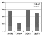
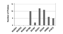
- the expression level of each iPS cell clone shown in Table 1 is 0.667 times or less or 1.5 times or more compared to the average expression level of human ES cells (KhES1-S3). It is the figure which graphed the number of the genes which show the abnormal expression level. In one Example of this invention, it is the figure which showed the tree obtained by clustering analysis of the expression of the gene shown in Table 2 in each clone in a figure. In one Example of this invention, it is the figure which showed the tree obtained by clustering analysis of the expression of the gene shown in Table 3 in each clone in a figure.
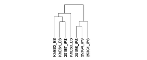
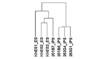
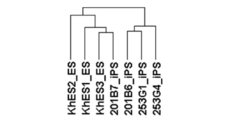
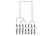
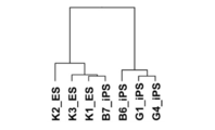
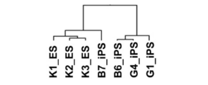
- Example of this invention it is the figure which showed the tree obtained by clustering analysis of the expression of the gene shown in Table 4 in each clone in a figure. In one Example of this invention, it is the figure which showed the tree obtained by clustering analysis of the expression of the gene shown in Table 5 in each clone in a figure. In one Example of this invention, it is the figure which showed the tree obtained by clustering analysis of the expression of the gene shown in Table 6 in each clone in a figure. In one Example of this invention, it is the figure which showed the tree obtained by clustering analysis of the expression of the gene shown in Table 7 in each clone in a figure.
- Example of this invention it is the figure which showed the tree obtained by clustering analysis of the expression of the gene shown in Table 8 in each clone in a figure. In one Example of this invention, it is the figure which showed the tree obtained by clustering analysis of the expression of the gene shown in Table 9 in each clone in a figure. In one Example of this invention, it is the figure which showed the tree obtained by clustering analysis of the expression of the gene shown in Table 10 in each clone in a figure. In one Example of this invention, it is the figure which showed the tree obtained by clustering analysis of the expression of the gene shown in Table 11 in each clone in a figure.
- Example of this invention it is the figure which showed the tree obtained by clustering analysis of the expression of the gene shown in Table 12 in each clone in a figure.
- human iPS cells are induced to differentiate into tertiary neurospheres, transplanted into the brains of NOS / SCID mice, and the results of analysis of tumorigenicity in vivo are shown.
- human iPS cells (253G4) are induced to differentiate into tertiary neurospheres, transplanted into the brains of NOS / SCID mice, and the results of analyzing the proliferation of transplanted iPS cells in vivo It is.
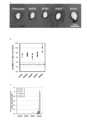
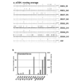
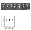
- human iPS cells are induced to differentiate into tertiary neurospheres, injected into the testes of NOD / SCID mice, and after 3 months, teratomas are removed, (A) appearance, (B) long axis It is the figure which observed expression of the reprogramming gene introduced at the time of the length of a direction, and (C) iPS cell preparation. In one Example of this invention, it is a figure which shows the result of having performed the array CGH analysis with respect to the undifferentiated iPS cell and the tertiary neurosphere which differentiated each cell. (A) On the genomic DNA, the location where an abnormality is detected is indicated by an arrow.
- (B) The number of genes having abnormalities on the genomic DNA is shown in a graph.
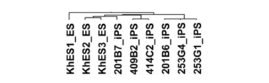
- it is a figure showing the tree obtained by performing clustering analysis using all the genes.
- it is the figure which showed the tree obtained by clustering analysis of the expression of the gene shown in Table 4 in each clone in a figure.
- it is the figure which showed the tree obtained by clustering analysis of the expression of the gene shown in Table 12 in each clone in a figure.
- about the probe of a 12th gene group it is a graph which shows the result of having measured the number of the probes which have a 1.5 times or more difference in signal intensity from the average value of the expression in ES cell.
- human iPS cells prepared using an episomal vector are induced to differentiate into tertiary neurospheres, injected into the testes of NOD / SCID mice, and teratomas are removed 3 months later. ) Observing the appearance and (B) measuring the length in the major axis direction.
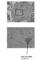
- it is the tissue slice image of the testis obtained by transplanting the neural stem cell derived from 409B2, and its enlarged view (A, B).
- it is the tissue slice image of the testis obtained by transplanting the neural stem cell derived from 414C2, and its enlarged view (A).
- iPS cells can be produced by introducing reprogramming factors into differentiated cells and dedifferentiating them. (Takahashi K & Yamanaka S. Cell 2006 vol.126: p.663-676; Takahashi K, et al., Cell 2007 vol.131: p.861-872).
- the initialization method is not particularly limited, but it is preferably induced to have pluripotency and self-proliferation ability by introducing a nuclear reprogramming factor.
- an initialization method described in International Publications WO2005 / 080598 and WO2007 / 069666 can be used.
- the nuclear reprogramming factor is not particularly limited, but preferably includes a combination of gene products of genes selected from each gene group of the Oct gene group, the Klf gene group, and the Sox gene group, and the efficiency of iPS cell establishment Then, it is more preferable to include a combination further including a gene product of a gene group.
- the genes belonging to the Oct gene group include Oct3 / 4, Oct1A, Oct6, the genes belonging to the Klf gene group include Klf1, Klf2, Klf4, Klf5, etc., and the genes belonging to the Sox gene group include Sox1 , Sox2, Sox3, Sox7, Sox15, Sox17, Sox18.
- Examples of genes belonging to the Myc gene group include c-Myc, N-Myc, and L-Myc.
- nuclear reprogramming factors include, in addition to the above combinations, combinations including Nanog gene, lin-28 gene, and / or glis1 gene in addition to genes of Oct gene group and Sox gene group. Further, in addition to any combination of these genes, p53 may be deleted. For example, the p53 gene may be knocked down by shp53. In addition, when introducing into a cell, in addition to the gene of the said combination, you may introduce
- genes are genes that are highly conserved in vertebrates. In this specification, unless an animal name is indicated, it means genes including homologs. Moreover, even if it is a gene which has a variation
- cytokines and compounds may be added to the medium as cofactors.
- cytokines in this case include SCF and bFGF, which can be substituted for c-Myc
- compounds include valproic acid, which can be substituted for c-Myc gene and Klf4 gene.
- the method for selecting iPS cell clones according to the present invention is based on a predetermined gene group such as the first to twelfth gene groups shown in Tables 1 to 12 in undifferentiated iPS cells. And a step of examining the expression level of at least one gene.
- the method for examining the expression level of the gene is not particularly limited, and for example, total mRNA is extracted from undifferentiated iPS cells by a conventional method, and using that mRNA, Northern blotting is performed using the gene to be examined as a probe. By performing PCR after reverse transcription, the expression level of the gene can be known.
- the expression level of a gene can be known by hybridizing all labeled mRNA to a DNA array in which a probe for the gene to be examined is immobilized and analyzing the intensity of the obtained signal.
- a probe for the gene to be examined is immobilized and analyzing the intensity of the obtained signal.
- whether the iPS cell is unlikely to undergo tumorigenesis by examining the expression level of a specific gene in an undifferentiated iPS cell and using the expression level as an index. It is possible to predict.
- the specific method is demonstrated about the prediction method, the method of this invention is not limited to the following.
- iPS cell clones are less likely to cause oncogenesis
- the clones are less likely to become tumors when differentiated in vitro or transplanted and differentiated in vivo. If it does not differentiate, it will not be included.
- a method for determining whether a certain clone is likely to cause a tumor is not particularly limited.
- neurospheres are differentiated from iPS cells (Okadaket al.,. Stem Cells. 2008 vol.26: p.3086- 98; Miura K. et al., Nat Biotechnol. 2009 vol.27: p.743-5), transplanted into the striatum and testes of NOD / SCID mice, and examined for neurosphere differentiation 1 to 6 months later , If a tumor is formed, if it is formed, the size and number of cells may be examined, or abnormal growth of transplanted cells may be examined (Okada et al., Stem Cells.
- the criteria for determining whether or not a tumor is likely to develop may be appropriately determined by those skilled in the art depending on the intended use of the iPS cells. For example, when used for transplantation for therapeutic purposes, the criteria may be strict. When studying, loose standards should be used. At this time, all types of tumors may be taken into account, but only specific tumors such as gliomas may be taken into account.
- the method of quantifying the control expression level is not particularly limited. From the list of expression levels in a plurality of clones, the average or mean ⁇ standard deviation (or error) of the expression levels in a plurality of clones, the upper limit of the expression level in a plurality of clones As long as it is possible to determine which expression level the expression level examined in the clone you want to examine, such as a range to the lower limit, a value or range estimated or statistically calculated from the expression levels in multiple clones, It may be a numerical value. An appropriate comparison method can be selected according to the digitization method. For example, it is tested whether there is a significant difference between the examined expression level and the control expression level, or whether the examined expression level falls within the range of the control expression level.
- the clone does not differentiate normally or matches the control expression level of a clone that is prone to oncogenesis even if differentiated, the clone does not differentiate normally or is differentiated and tumor If the clone is determined to be suitable for the control expression level in a clone that is unlikely to cause oncogenesis, or is determined not to be tumorigenic, and the clone is determined to be compatible with the control expression level in the clone that is unlikely to cause oncogenesis, the clone is an iPS cell that is unlikely to cause oncogenesis. Judge that there is.
- clustering analysis may be performed using the results of investigation using a plurality of types of probes.
- the proportion of genes using a plurality of types of probes is preferably small, and less than 20% of the total. Preferably, it is less than 10%, most preferably 0%.
- the number of types of probes is small, preferably three or less types of probes are used, and more preferably two types or less probes are used. Most preferred is when one type of probe is used.
- a probe in which a gene that can be detected is not specified may be used, but the number is preferably smaller, preferably less than 20%, more preferably less than 10%, more preferably 0%. Most preferably it is.
- the method for determining whether there is an abnormality in the expression level is not particularly limited, but it is preferable to determine by comparing with a normal pluripotent stem cell clone.
- the normal pluripotent stem cell clone may be a pluripotent stem cell clone that is unlikely to cause oncogenesis, and the pluripotent stem cell may be an ES cell or an iPS cell.
- the pluripotent stem cell may be an ES cell or an iPS cell.
- the rate of abnormality is 10% or less, 20% or less, 50% or less, 66.7% or less, 80% or less, or 120% or more, 125% or more, 133.3% or more, 150% or more, For example, 180% or more.
- the method of presenting the control number is not particularly limited, from a list of the numbers in multiple clones, the average or average ⁇ standard deviation (or error) of the numbers in multiple clones, from the upper limit to the lower limit of the numbers in multiple clones
- Any display may be used as long as it can be determined whether the number of clones to be examined is suitable, such as a range or a value or range estimated or statistically calculated from the number of clones. .
- An appropriate comparison method can be selected depending on the display method. For example, it is tested whether there is a significant difference between the number examined and the number of controls, or whether the number examined is within the range of the number of controls.
- the clone will not differentiate normally or will become tumoriable even if differentiated. If it is determined that the clone is not susceptible to oncogenesis, or is not likely to cause oncogenesis, and if it is determined to match the number of controls in a clone that is less likely to cause oncogenesis, the clone is determined to be less likely to cause oncogenesis.
- Method of classifying by clustering analysis Alternatively, in one or more iPS cell clones that are not normally differentiated or that are susceptible to oncogenesis even after differentiation, Determine the expression level of all genes. Here, also for iPS cell clones whose properties are to be examined, all expression levels of the same gene group are determined, and clustering analysis of each iPS cell clone is performed.
- clustering analysis may be performed using the results of investigation using a plurality of types of probes.
- the proportion of genes using a plurality of types of probes is preferably small, and less than 20% of the total. Preferably, it is less than 10%, most preferably 0%.
- the number of types of probes is small, preferably three or less types of probes are used, and more preferably two types or less probes are used. Most preferred is when one type of probe is used.
- a probe in which a gene that can be detected is not specified may be used, but the number is preferably smaller, preferably less than 20%, more preferably less than 10%, more preferably 0%.
- clusteringt is Cluster 3.0 (Freeware by M. ⁇ ⁇ ⁇ J. L. de Hoon, S. Imoto, J. Nolan, and S. Miyano: Open Source Clustering) Software. Bioinformatics, 20 (9): 1453--1454 (2004) .; http://bonsai.hgc.jp/ ⁇ mdehoon/software/cluster/) and software such as Gene spring GX (Agilent) It can be carried out.
- iPS cells whose properties are to be examined are classified as clones that do not differentiate normally or are susceptible to tumorigenesis even after differentiation, the clones do not differentiate normally or become tumorous even if differentiated.
- the clone is determined to be an iPS cell that is unlikely to cause tumorigenesis.
- the expression level of which gene or which gene group is examined may vary depending on the origin of iPS cells. Can be easily determined. The determination method will be described below.
- This selection method is not particularly limited, and may be selected at random, but is preferably selected from genes that are higher in pluripotent stem cells than differentiated cells and genes involved in DNA repair. More preferably, it is selected from the gene group. This makes it possible to select a gene for selecting a target clone with extremely high probability.
- the method for selecting candidate genes from these gene groups is not particularly limited, but as in the examples, pluripotent stem cell clones that are unlikely to undergo tumorigenesis and those that do not differentiate normally or are susceptible to tumorigenesis even when differentiated. It is preferable to select genes whose expression levels are significantly different from those of competent stem cell clones.
- a group of genes including one or more, preferably two or more genes selected in this way is defined as the Xth gene group.
- genes having higher expression than differentiated cells in pluripotent stem cells can be easily examined by those skilled in the art using RT-PCR, microarray, and the like.
- the gene involved in DNA repair may be any gene involved in any one of the DNA repair processes, and may be a gene reported as such in a paper, patent publication, database, or the like. Good. These reports are retrieved from articles and patent publications that are accessible from databases such as PUBMED, national (eg, Japan, Europe, USA) Patent Office, WIPO HP, GO (GeneGOntology), etc. be able to.
- genes involved in DNA repair are functions in PUBMED, papers, patent gazettes, and gene information included in the patent offices of various countries (for example, Japan, Europe, and the US), WIPO databases, and GO databases. Most preferably, at least one of the genes is a gene reported to be involved in DNA repair.
- first iPS cell clones that do not differentiate normally or are susceptible to oncogenesis upon differentiation
- second, idiosyncratic cells that are less likely to cause oncogenesis.
- the expression level of the gene belonging to the X gene group is examined in each undifferentiated iPS cell. Then, using the expression level as an index, the first iPS cell clone and the second iPS cell clone are compared.
- the comparison method is not particularly limited, but some specific examples will be described below.
- the judgment that the expression level is different is made by examining the expression level of the gene of the X gene group in clones in which it is unknown whether tumorigenesis is unlikely to occur, and the clones are the first clone and the second clone. As long as it can be determined which one of the two is applicable, any criterion may be used.
- the expression levels in the first and second clones may be expressed as a range of the lowest level and the highest level, respectively, and may be determined to be different when the ranges do not overlap.
- the present invention is not limited to these.
- this X gene group is used as a gene group for selecting clones that are unlikely to cause oncogenesis. be able to.
- a normal pluripotent stem cell clone is a pluripotent stem cell clone that has been found to be less likely to cause tumorigenesis when transplanted in vitro or in vivo. If present, it may be an ES cell or an iPS cell.
- the judgment that the number is different is that the expression level of the gene of the X gene group is similarly examined in the iPS cell clones that are unclear whether or not they are likely to be tumorigenic, and the expression levels are different from those of normal pluripotent stem cell clones.
- any criteria may be used.
- the standard for determining that the expression level is different from that of a normal pluripotent stem cell clone can be performed by the same method as described in [1].
- the gene group can be used as a gene group for selecting iPS cell clones that are unlikely to cause tumorigenesis.
- the neural stem cells derived from human iPS cells showed almost the same profile as the neural stem cells derived from human ES cells (see (6) Comparative Example described later).
- undifferentiated markers such as TRA-1-60, TRA-1-81 and NCAM are hardly detected, and these human iPS cell clones are resistant to differentiation. It was confirmed that almost no cells or undifferentiated cells remained.
- three clones with neurospheres were analyzed electrophysiologically, but action potentials were recorded in the same manner as human ES cells, indicating that these three clones can differentiate into functional neurons. .
- each iPS by a known method (Okada et al., Stem Cells. 2008 vol. 26: p. 3086-98; Miura K. et al., Nat Biotechnol. 2009 vol. 27: p. 743-5).
- Human EB Embryoid body
- a lentivirus expressing Venus was introduced to produce neural stem cells expressing Venus.
- the third neurosphere was generated by passage 2 times, separated into single cells, and 1-5 ⁇ 10 5 cells were injected into the striatum of NOD / SCID mice (Ogawa et al.,). J Neurosci Res. 2009 vol.87: p.307-17; Miura K. et al., Nat Biotechnol. 2009 vol.27: p.743-5).
- the brain was removed from the mouse, and the differentiation state of the transplanted cell was examined immunohistochemically using antibodies against each of the markers of nerve cells, astrocytes, oligodendrocytes. In vivo, it was differentiated into three nervous system cells. As for the types of differentiated neurons, TH positive catecholaminergic neurons, ChAT positive cholinergic neurons, and GAD67 positive GABAergic neurons were detected, and all clones had normal differentiation ability.
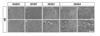
- mice brain transplanted with 253G1 and 253G4 were stained with HE
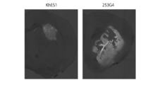
- images of lower glioma were observed in mice brain transplanted with 253G1 and 253G4, and cells having nuclear atypia (indicated by wrinkles in the figure) were also observed (Fig. 13).
- normal brain tissue was observed as in the control KhES1 (FIG. 13).
- signs of tumor formation were observed in 253G1 and 253G4, and a huge tumor derived from transplanted cells was observed particularly in the transplanted tissue of 253G4.
- FIG. 14 In 201B7, the above was not observed in the transplanted cells, similar to the control KhES1.
- the tertiary neurospheres are separated into single cells, and 1-5 ⁇ 10 5 cells are injected into the testes of NOD / SCID mice.
- the testis was removed, and the appearance (A), the length in the long axis direction (B), and the expression of the reprogramming gene introduced at the time of iPS cell production (C) were observed (FIG. 15).
- A the appearance
- B the length in the long axis direction
- C the expression of the reprogramming gene introduced at the time of iPS cell production
- FIG. 15 In 201B7, in all respects, it was similar to the control KhES1, but in 253G1, the cell proliferation was almost normal, but unlike the control KhES1, expression of the reprogramming gene (POU5F1 only) was observed. It was.
- 201B7 is a clone that is less likely to cause tumor formation in vivo when transplanted into tissue, and 253G1 and 253G4 are considered to be clones that are likely to cause tumor formation in vivo, and in the following experiments, It shows that 201B7 takes the same pattern as ES cells and 253G1 and 253G4 take a different pattern from the expression pattern of the gene group.
- All ES3 clones are p-flags All ES3 clones should be at least 5 times higher than the average human tissue expression level. 1340 probes (Table 1) satisfying the above three conditions (Table 1) (including 929 identified gene species; including 102 probes for which no corresponding gene is identified) are selected, and 929 identified gene species are selected as the first gene. Grouped.
- the specified gene means a gene whose gene name is known and is counted as a single gene.
- the expression levels of all 201B6, 253G1, and 253G4 are 1.5 times or more higher than the average expression level of the ES3 clone, and the difference between the expression level of 201B7 and the average ES is 1 80 probes that are within 5 times (Table 5) (70 identified gene species; including 3 probes for which the corresponding gene is not identified) are selected, and 70 identified gene species are selected as the fifth
- the expression level of all of 201B6, 253G1, and 253G4 is 1.5 times or more of the average expression level of ES3 clones, and further, all of ES3 clone and 201B7 72 probes (Table 6) with a difference from the average of the expression level of 1.5 within (Table 6) (63 identified gene species; (Including two unspecified probes) and 63 identified gene species as the sixth gene group; from the genes of the third gene group, all expression levels of 201B6, 253G1, 253G4 are
- 201B7 is classified into the same group as human ES cells, and 201B6, 253G1, and 253G4 are one group separately. It is classified.
- the gene group consisting of genes that are higher in the pluripotent stem cells than the differentiated cells and genes selected from genes involved in DNA repair are compared with the expression levels in the pluripotent stem cells that are unlikely to cause tumorigenesis.
- iPS cells that do not differentiate normally or are easily differentiated by iPS cells and iPS cells that are less likely to be tumoriZed by differentiation of expression levels having statistical significance are also obtained. Can be classified into cells.
- the present invention is based on the discovery that abnormal expression of genes involved in DNA repair in undifferentiated iPS cells affects the tumorigenicity of differentiated iPS cells. is there.
- abnormalities in genes involved in DNA repair in an undifferentiated state cause numerous wounds in DNA, which may cause iPS cells to not differentiate normally or iPS cells to differentiate. Sometimes indicates that it becomes tumorous.
- undifferentiated iPS cells 201B6, 253G1, 253G4, 201B7
- tertiary neurospheres obtained by differentiating each cell
- fibroblasts HDF
- human fetus as a control
- Genomic DNA was isolated from derived neural stem cells (hNSC) and patient glioma cell-derived cells (GDC) and subjected to array CGH analysis.
- Genomic DNA was extracted from each clone and from normal human male genome (Promega) as a control using phenol chloroform method, DNeasy Blood & Tissue Kit or QIAamp DNA mini Kit (Qiagen) and using PicoGreen And quantified.
- 500 ng to 1000 ng of double-stranded DNA was digested with restriction enzymes (AluI and RsaI) to fragment the DNA. Thereafter, using a Genomic DNA Enzymatic Labeling kit (Agilent), the DNA fragment was labeled with Cy3 or Cy5, one was used as a control, and the other was used as a sample.
- 409B2 and 414C2 were evaluated for tumorigenicity by the same method as in (2). As shown in FIG. 21, 409B2 and 414C2 were tested after testis transplantation, similar to ES cells and 201B7. No swelling was observed.
- paraffin sections of testis transplanted with 409B2 and 414C2 were prepared, and stained with HE, and histological images of teratomas derived from transplanted cells were observed. As shown in FIGS. 22 and 23, tumors were observed from 253G4 and 253G1. A well-differentiated nerve cell and glial cell were observed.
- the gene group of the present invention is useful for classifying iPS cells into clones that do not differentiate normally, or that tend to undergo tumorigenesis even when differentiated, and clones that do not tend to undergo tumorigenesis.
- the present invention it is possible to provide a method for selecting an iPS cell clone that is unlikely to cause oncogenesis even when differentiated, and a method for selecting a gene used in the selection method.
Landscapes
- Life Sciences & Earth Sciences (AREA)
- Chemical & Material Sciences (AREA)
- Proteomics, Peptides & Aminoacids (AREA)
- Health & Medical Sciences (AREA)
- Organic Chemistry (AREA)
- Zoology (AREA)
- Analytical Chemistry (AREA)
- Wood Science & Technology (AREA)
- Immunology (AREA)
- Engineering & Computer Science (AREA)
- Biotechnology (AREA)
- Microbiology (AREA)
- Molecular Biology (AREA)
- Biophysics (AREA)
- Physics & Mathematics (AREA)
- Cell Biology (AREA)
- Biochemistry (AREA)
- Bioinformatics & Cheminformatics (AREA)
- General Engineering & Computer Science (AREA)
- General Health & Medical Sciences (AREA)
- Genetics & Genomics (AREA)
- Measuring Or Testing Involving Enzymes Or Micro-Organisms (AREA)
- Micro-Organisms Or Cultivation Processes Thereof (AREA)
Abstract
本発明は、分化させても腫瘍化が起きにくいiPS細胞クローンの選択方法、及びその選択方法に用いる遺伝子の選択方法を提供することを目的とし、一実施態様として、未分化状態のiPS細胞クローンにおいて、第1~第10遺伝子群のいずれかの遺伝子群において、少なくとも一つの遺伝子について、発現レベルを調べることによって、分化させても腫瘍化が起きにくいiPS細胞クローンを選択する。また、正常に分化できないか、あるいは分化しても腫瘍化を起こしやすい複数の第1のiPS細胞クローンと、腫瘍化を起こしにくい複数の第2のiPS細胞クローンにおいて、未分化状態の各iPS細胞クローンで、1以上の遺伝子を含む候補遺伝子群に属する遺伝子の発現レベルを調べることによって、分化させても腫瘍化が起きにくいiPS細胞クローンの選択方法に用いる遺伝子を選択する。
Description
本発明は、iPS細胞クローンの選択方法、及びその選択方法に用いる遺伝子の選択方法に関する。
iPS細胞(induced pluripotent stem cells; 人工多能性幹細胞)は、多能性を有し、分化細胞から調製できるため、再生医療において移植に用いるための細胞として期待されている。しかしながら、iPS細胞を分化させて移植しても、in vivoでは、移植されたiPS細胞由来の細胞が腫瘍化する問題が指摘されている(Miura K. et al., Nat Biotechnol. 2009 vol.27: p.743-5.)。
本発明は、分化させても腫瘍化が起きにくいiPS細胞クローンの選択方法、及びその選択方法に用いる遺伝子の選択方法を提供することを目的とする。
本発明は、以下のような各項からなる。
(1)iPS細胞クローンの選択方法であって,未分化状態のiPS細胞において、以下の第1~第12遺伝子群のいずれかの遺伝子群において、少なくとも一つの遺伝子について、発現レベルを調べる工程、を含む方法。
(2)第1項に記載の選択方法であって,前記発現レベルを、正常に分化しないか、分化しても腫瘍化を起こしやすいiPS細胞クローンにおける当該遺伝子の発現レベル、及び、腫瘍化を起こしにくいiPS細胞クローンにおける当該遺伝子の発現レベルと比較する工程、をさらに含む方法。
(3)第1項に記載の選択方法であって,前記iPS細胞クローンにおいて、第1~第12遺伝子群のいずれかの遺伝子群に属する全ての遺伝子の発現レベルを調べる工程と,調べた遺伝子群において、発現レベルに異常のある遺伝子の個数を調べる工程と、調べた遺伝子の個数を、正常に分化しないか、分化しても腫瘍化を起こしやすいiPS細胞クローンにおける発現レベルに異常のある遺伝子の個数、及び、腫瘍化を起こしにくいiPS細胞クローンにおける発現レベルに異常のある遺伝子の個数と比較する工程、をさらに含む方法。
(4)第3項に記載の選択方法であって、腫瘍化を起こしにくい多能性幹細胞の発現レベルと比較することによって、前記発現レベルが異常かどうか判断することを特徴とする、方法。
(5)第1項に記載の選択方法であって,前記iPS細胞クローンにおいて、第1~第12遺伝子群のいずれかの遺伝子群に属する全ての遺伝子の発現レベルを調べる工程と,調べた遺伝子群について、前記iPS細胞クローンにおける発現レベルと、正常に分化しないか、分化しても腫瘍化を起こしやすいiPS細胞クローンにおける発現レベルとを指標にして、iPS細胞クローンのclustering分析を行う工程と、当該iPS細胞クローンが、腫瘍化を起こしにくいiPS細胞クローンと正常に分化しないか、分化しても腫瘍化を起こしやすいiPS細胞クローンのいずれに分類されるかを調べる工程と、を含む方法。
(6)第5項に記載の選択方法であって、腫瘍化を起こしにくい多能性幹細胞の発現レベルとの相対値を指標にして、前記iPS細胞クローンのclustering分析を行うことを特徴とする、請求項7に記載の方法。
(7)iPS細胞クローンを選択するために発現レベルを調べる遺伝子の選択方法であって、正常に分化しないか、分化しても腫瘍化を起こしやすい複数の第1のiPS細胞クローンと、腫瘍化を起こしにくい複数の第2のiPS細胞クローンにおいて、未分化状態のiPS細胞で、1以上の遺伝子を含む候補遺伝子群に属する遺伝子の発現レベルを調べる工程、を含む方法。
(8)前記候補遺伝子群に属する遺伝子が、多能性幹細胞で分化細胞より発現の高い遺伝子、及びDNA修復に関与する遺伝子であることを特徴とする、第7項に記載の方法。
(9)前記候補遺伝子群に属する遺伝子が、第1遺伝子群及び第2遺伝子群のいずれかに属する遺伝子であることを特徴とする、第7項に記載の方法。
(10)第7項に記載の選択方法であって、複数の第1のiPS細胞クローンにおいて得られた第1の発現レベルと、複数の第2のiPS細胞クローンにおいて得られた第2の発現レベルとを比較する工程と、第1の発現レベルと第2の発現レベルとが異なるかどうか調べる工程と、を含む方法。
(11)第7項に記載の選択方法であって、複数の第1のiPS細胞クローンにおいて得られた発現レベルにおいて、発現レベルに異常のある遺伝子の第1の個数と、複数の第2のiPS細胞クローンにおいて得られた発現レベルのうち、発現レベルに異常のある遺伝子の第2の個数とを比較する工程と、第1の個数と第2の個数とが異なるかどうか調べる工程と、を含む方法。
(12)第11項に記載の選択方法であって、腫瘍化を起こしにくい多能性幹細胞の発現レベルと比較することによって、前記発現レベルが異常かどうか判断することを特徴とする、方法。
(13)第7項に記載の選択方法であって、前記iPS細胞クローンについて、前記発現レベルを指標にしてclustering分析を行う工程と、複数の第1のiPS細胞クローンと複数の第2のiPS細胞クローンが、それぞれ別のclusterに分類されるかどうか調べる工程と、を含む方法。
(14)第13項に記載の選択方法であって、腫瘍化を起こしにくい多能性幹細胞の発現レベルとの相対値を指標にして、前記iPS細胞クローンについてclustering分析を行うことを特徴とする、方法。
(1)iPS細胞クローンの選択方法であって,未分化状態のiPS細胞において、以下の第1~第12遺伝子群のいずれかの遺伝子群において、少なくとも一つの遺伝子について、発現レベルを調べる工程、を含む方法。
(2)第1項に記載の選択方法であって,前記発現レベルを、正常に分化しないか、分化しても腫瘍化を起こしやすいiPS細胞クローンにおける当該遺伝子の発現レベル、及び、腫瘍化を起こしにくいiPS細胞クローンにおける当該遺伝子の発現レベルと比較する工程、をさらに含む方法。
(3)第1項に記載の選択方法であって,前記iPS細胞クローンにおいて、第1~第12遺伝子群のいずれかの遺伝子群に属する全ての遺伝子の発現レベルを調べる工程と,調べた遺伝子群において、発現レベルに異常のある遺伝子の個数を調べる工程と、調べた遺伝子の個数を、正常に分化しないか、分化しても腫瘍化を起こしやすいiPS細胞クローンにおける発現レベルに異常のある遺伝子の個数、及び、腫瘍化を起こしにくいiPS細胞クローンにおける発現レベルに異常のある遺伝子の個数と比較する工程、をさらに含む方法。
(4)第3項に記載の選択方法であって、腫瘍化を起こしにくい多能性幹細胞の発現レベルと比較することによって、前記発現レベルが異常かどうか判断することを特徴とする、方法。
(5)第1項に記載の選択方法であって,前記iPS細胞クローンにおいて、第1~第12遺伝子群のいずれかの遺伝子群に属する全ての遺伝子の発現レベルを調べる工程と,調べた遺伝子群について、前記iPS細胞クローンにおける発現レベルと、正常に分化しないか、分化しても腫瘍化を起こしやすいiPS細胞クローンにおける発現レベルとを指標にして、iPS細胞クローンのclustering分析を行う工程と、当該iPS細胞クローンが、腫瘍化を起こしにくいiPS細胞クローンと正常に分化しないか、分化しても腫瘍化を起こしやすいiPS細胞クローンのいずれに分類されるかを調べる工程と、を含む方法。
(6)第5項に記載の選択方法であって、腫瘍化を起こしにくい多能性幹細胞の発現レベルとの相対値を指標にして、前記iPS細胞クローンのclustering分析を行うことを特徴とする、請求項7に記載の方法。
(7)iPS細胞クローンを選択するために発現レベルを調べる遺伝子の選択方法であって、正常に分化しないか、分化しても腫瘍化を起こしやすい複数の第1のiPS細胞クローンと、腫瘍化を起こしにくい複数の第2のiPS細胞クローンにおいて、未分化状態のiPS細胞で、1以上の遺伝子を含む候補遺伝子群に属する遺伝子の発現レベルを調べる工程、を含む方法。
(8)前記候補遺伝子群に属する遺伝子が、多能性幹細胞で分化細胞より発現の高い遺伝子、及びDNA修復に関与する遺伝子であることを特徴とする、第7項に記載の方法。
(9)前記候補遺伝子群に属する遺伝子が、第1遺伝子群及び第2遺伝子群のいずれかに属する遺伝子であることを特徴とする、第7項に記載の方法。
(10)第7項に記載の選択方法であって、複数の第1のiPS細胞クローンにおいて得られた第1の発現レベルと、複数の第2のiPS細胞クローンにおいて得られた第2の発現レベルとを比較する工程と、第1の発現レベルと第2の発現レベルとが異なるかどうか調べる工程と、を含む方法。
(11)第7項に記載の選択方法であって、複数の第1のiPS細胞クローンにおいて得られた発現レベルにおいて、発現レベルに異常のある遺伝子の第1の個数と、複数の第2のiPS細胞クローンにおいて得られた発現レベルのうち、発現レベルに異常のある遺伝子の第2の個数とを比較する工程と、第1の個数と第2の個数とが異なるかどうか調べる工程と、を含む方法。
(12)第11項に記載の選択方法であって、腫瘍化を起こしにくい多能性幹細胞の発現レベルと比較することによって、前記発現レベルが異常かどうか判断することを特徴とする、方法。
(13)第7項に記載の選択方法であって、前記iPS細胞クローンについて、前記発現レベルを指標にしてclustering分析を行う工程と、複数の第1のiPS細胞クローンと複数の第2のiPS細胞クローンが、それぞれ別のclusterに分類されるかどうか調べる工程と、を含む方法。
(14)第13項に記載の選択方法であって、腫瘍化を起こしにくい多能性幹細胞の発現レベルとの相対値を指標にして、前記iPS細胞クローンについてclustering分析を行うことを特徴とする、方法。
==関連文献とのクロスリファレンス==
本出願は、2011年2月25日付で出願した日本国特許出願2011-40979、及び2011年3月22日付で出願した米国仮出願US61/466298に基づく優先権を主張するものであり、当該基礎出願を引用することにより、本明細書に含めるものとする。
本出願は、2011年2月25日付で出願した日本国特許出願2011-40979、及び2011年3月22日付で出願した米国仮出願US61/466298に基づく優先権を主張するものであり、当該基礎出願を引用することにより、本明細書に含めるものとする。
以下、上記知見に基づき完成した本発明の実施の形態を、実施例を挙げながら詳細に説明する。
実施の形態及び実施例に特に説明がない場合には、J. Sambrook, E. F. Fritsch & T. Maniatis (Ed.), Molecular cloning, a laboratory manual (3rd edition), Cold Spring Harbor Press, Cold Spring Harbor, New York (2001); F. M. Ausubel, R. Brent, R. E. Kingston, D. D. Moore, J.G. Seidman, J. A. Smith, K. Struhl (Ed.), Current Protocols in Molecular Biology, John Wiley & Sons Ltd.; Juan S. Bonifacino (Bethesda, Maryland); Mary Dasso (Bethesda, Maryland); J. B. Harford, J. L-Schwartz, K. M. Yamada (Ed.), Current Protocols in Cell Biology, John Wiley & Sons Ltd.等の標準的なプロトコール集に記載の方法、あるいはそれを修飾したり、改変した方法を用いる。また、市販の試薬キットや測定装置を用いる場合には、特に説明が無い場合、それらに添付のプロトコールを用いる。
なお、本発明の目的、特徴、利点、及びそのアイデアは、本明細書の記載により、当業者には明らかであり、本明細書の記載から、当業者であれば、容易に本発明を再現できる。以下に記載された発明の実施の形態及び具体的な実施例等は、本発明の好ましい実施態様を示すものであり、例示又は説明のために示されているのであって、本発明をそれらに限定するものではない。本明細書で開示されている本発明の意図ならびに範囲内で、本明細書の記載に基づき、様々に修飾ができることは、当業者にとって明らかである。
実施の形態及び実施例に特に説明がない場合には、J. Sambrook, E. F. Fritsch & T. Maniatis (Ed.), Molecular cloning, a laboratory manual (3rd edition), Cold Spring Harbor Press, Cold Spring Harbor, New York (2001); F. M. Ausubel, R. Brent, R. E. Kingston, D. D. Moore, J.G. Seidman, J. A. Smith, K. Struhl (Ed.), Current Protocols in Molecular Biology, John Wiley & Sons Ltd.; Juan S. Bonifacino (Bethesda, Maryland); Mary Dasso (Bethesda, Maryland); J. B. Harford, J. L-Schwartz, K. M. Yamada (Ed.), Current Protocols in Cell Biology, John Wiley & Sons Ltd.等の標準的なプロトコール集に記載の方法、あるいはそれを修飾したり、改変した方法を用いる。また、市販の試薬キットや測定装置を用いる場合には、特に説明が無い場合、それらに添付のプロトコールを用いる。
なお、本発明の目的、特徴、利点、及びそのアイデアは、本明細書の記載により、当業者には明らかであり、本明細書の記載から、当業者であれば、容易に本発明を再現できる。以下に記載された発明の実施の形態及び具体的な実施例等は、本発明の好ましい実施態様を示すものであり、例示又は説明のために示されているのであって、本発明をそれらに限定するものではない。本明細書で開示されている本発明の意図ならびに範囲内で、本明細書の記載に基づき、様々に修飾ができることは、当業者にとって明らかである。
(1)iPS細胞の作製方法
iPS細胞は、分化した細胞に初期化因子を導入し、脱分化させることによって製造できる。(Takahashi K & Yamanaka S. Cell 2006 vol.126: p.663-676; Takahashi K, et al., Cell 2007 vol.131: p.861-872)。
iPS細胞は、分化した細胞に初期化因子を導入し、脱分化させることによって製造できる。(Takahashi K & Yamanaka S. Cell 2006 vol.126: p.663-676; Takahashi K, et al., Cell 2007 vol.131: p.861-872)。
初期化方法は特に限定されないが、核初期化因子を導入することにより、多分化能及び自己増殖能を有するように誘導することが好ましい。例えば国際公開WO2005/080598やWO2007/069666に記載された初期化方法を用いることができる。
核初期化因子は、特に限定されないが、Oct遺伝子群、Klf遺伝子群、Sox遺伝子群のそれぞれの遺伝子群から選択された遺伝子の遺伝子産物の組み合わせを含むことが好ましく、iPS細胞樹立の効率という点では、遺伝子群の遺伝子産物をさらに含んだ組み合わせを含むことがより好ましい。Oct遺伝子群に属する遺伝子としては、Oct3/4、Oct1A、Oct6などがあり、Klf遺伝子群に属する遺伝子としては、Klf1、Klf2、Klf4、Klf5などがあり、Sox遺伝子群に属する遺伝子としては、Sox1、Sox2、Sox3、Sox7、Sox15、Sox17、Sox18などがある。Myc遺伝子群に属する遺伝子としては、c-Myc、N-Myc、L-Mycなどがある。
核初期化因子としては、上記組み合わせ以外にも、Oct遺伝子群の遺伝子、Sox遺伝子群の遺伝子に加え、Nanog遺伝子、lin-28遺伝子、及び/又はglis1遺伝子を含む組み合わせが挙げられる。また、これらの遺伝子のいずれかの組み合わせに加え、p53を欠損させてもよく、例えば、shp53によってp53遺伝子をノックダウンしてもよい。なお、細胞に導入する場合、上記組み合わせの遺伝子に加え、他にも遺伝子産物を導入してもよく、例えば、不死化誘導因子などが挙げられる。
これらの遺伝子は、いずれも、脊椎動物で高度に保存されている遺伝子であり、本明細書では、特に動物名を示さない限り、ホモログを含めた遺伝子を表すものとする。また、polymorphismを含め、変異を有する遺伝子であっても、野生型の遺伝子産物と同等の機能を有する遺伝子もまた、含まれるものとする。
さらに、これら遺伝子の組み合わせに加え、サイトカインや化合物を補助因子として培地に添加しても良い。この場合のサイトカインとして、例えばSCFやbFGFなどが挙げられ、これらは、c-Mycに代替でき、化合物として、例えばバルプロ酸などが挙げられ、これは、c-Myc遺伝子やKlf4遺伝子に代替できる。
(2)iPS細胞クローンの選択方法
本発明にかかるiPS細胞クローンの選択方法は、未分化状態のiPS細胞において、表1~12に示す第1~第12の遺伝子群など、所定の遺伝子群において、少なくとも一つの遺伝子について、発現レベルを調べる工程を含む。
遺伝子の発現レベルの調べ方は、特に限定されず、例えば、未分化状態のiPS細胞から常法によって全mRNAを抽出し、そのmRNAを用いて、調べたい遺伝子をプローブにしてノザンブロッティングを行ったり、逆転写の後PCRを行ったりすることで、その遺伝子の発現レベルを知ることができる。複数の遺伝子について同時に発現を調べる場合は、DNAアレイを用いることが好ましい。すなわち、調べたい遺伝子に対するプローブが固定されたDNAアレイに対し、標識した全mRNAをハイブリダイズさせ、得られたシグナルの強度を解析することによって、その遺伝子の発現レベルを知ることができる。
これまで、未分化状態のiPS細胞の遺伝子発現レベルを調べることによって、そのiPS細胞が分化した時にどのような性状を示すかを予測する方法は知られていなかった。しかし、本発明の一実施形態においては,未分化状態のiPS細胞において特定の遺伝子の発現レベルを調べ、その発現レベルを指標にすることによって、そのiPS細胞が腫瘍化を起こしにくいかどうか、を予測することが可能である。以下,その予測方法について,具体的な方法を説明するが、本発明の方法は、下記のものに限定されない。なお、本明細書で「iPS細胞クローンが腫瘍化を起こしにくい」とだけ言う時、in vitroで分化した時あるいはin vivoに移植して分化した場合に、そのクローンが腫瘍化しにくいということを意味するのであって、分化しない場合は含まないものとする。
本発明にかかるiPS細胞クローンの選択方法は、未分化状態のiPS細胞において、表1~12に示す第1~第12の遺伝子群など、所定の遺伝子群において、少なくとも一つの遺伝子について、発現レベルを調べる工程を含む。
遺伝子の発現レベルの調べ方は、特に限定されず、例えば、未分化状態のiPS細胞から常法によって全mRNAを抽出し、そのmRNAを用いて、調べたい遺伝子をプローブにしてノザンブロッティングを行ったり、逆転写の後PCRを行ったりすることで、その遺伝子の発現レベルを知ることができる。複数の遺伝子について同時に発現を調べる場合は、DNAアレイを用いることが好ましい。すなわち、調べたい遺伝子に対するプローブが固定されたDNAアレイに対し、標識した全mRNAをハイブリダイズさせ、得られたシグナルの強度を解析することによって、その遺伝子の発現レベルを知ることができる。
これまで、未分化状態のiPS細胞の遺伝子発現レベルを調べることによって、そのiPS細胞が分化した時にどのような性状を示すかを予測する方法は知られていなかった。しかし、本発明の一実施形態においては,未分化状態のiPS細胞において特定の遺伝子の発現レベルを調べ、その発現レベルを指標にすることによって、そのiPS細胞が腫瘍化を起こしにくいかどうか、を予測することが可能である。以下,その予測方法について,具体的な方法を説明するが、本発明の方法は、下記のものに限定されない。なお、本明細書で「iPS細胞クローンが腫瘍化を起こしにくい」とだけ言う時、in vitroで分化した時あるいはin vivoに移植して分化した場合に、そのクローンが腫瘍化しにくいということを意味するのであって、分化しない場合は含まないものとする。
[1]特定の遺伝子の発現を比較する方法
所定の遺伝子群のうちの少なくとも一つの遺伝子について、予め、正常に分化しないか、分化しても腫瘍化を起こしやすいiPS細胞クローンにおける当該遺伝子の発現レベルと、腫瘍化を起こしにくいiPS細胞クローンにおける当該遺伝子の発現レベルとを、それぞれ1以上のiPS細胞クローンを用いて決定し、そして、これらの発現(以下、対象発現と称する)のレベルを、性状を調べたいiPS細胞クローンにおける当該遺伝子の発現のレベルと比較する。
所定の遺伝子群のうちの少なくとも一つの遺伝子について、予め、正常に分化しないか、分化しても腫瘍化を起こしやすいiPS細胞クローンにおける当該遺伝子の発現レベルと、腫瘍化を起こしにくいiPS細胞クローンにおける当該遺伝子の発現レベルとを、それぞれ1以上のiPS細胞クローンを用いて決定し、そして、これらの発現(以下、対象発現と称する)のレベルを、性状を調べたいiPS細胞クローンにおける当該遺伝子の発現のレベルと比較する。
ここで、あるクローンが腫瘍を起こしやすいかどうかを判断する方法は特に限定されないが、例えば、iPS細胞からニューロスフェアを分化させ(Okada et al., Stem Cells. 2008 vol.26: p.3086-98; Miura K. et al., Nat Biotechnol. 2009 vol.27: p.743-5)、NOD/SCIDマウスの線条体や精巣に移植し、1~6ヶ月後にニューロスフェアの分化状態を調べ,腫瘍が形成されたかどうかを調べたり、形成された場合,その大きさや個数を調べたり、移植細胞の異常増殖等を調べたりすればよい(Okada et al., Stem Cells. 2008 vol.26: p.3086-98; Miura K. et al., Nat Biotechnol. 2009 vol.27: p.743-5)。腫瘍を起こしやすいかどうかの基準は、iPS細胞の使用目的によって、当業者が適切に決定すればよく、例えば、治療目的の移植に用いる場合は、厳しい基準とすればよく、形成された腫瘍を研究する場合は、緩い基準にすれば良い。この時、全ての種類の腫瘍について考慮に入れてもよいが、グリオーマなど特定の腫瘍についてのみ考慮に入れてもよい。
対照発現レベルの数値化の方法は特に限定されず、複数のクローンにおける発現レベルのリスト、複数のクローンにおける発現レベルの平均または平均±標準偏差(または誤差)、複数のクローンにおける発現レベルの上限から下限までの範囲、複数のクローンにおける発現レベルから推測または統計的に計算された値または範囲など、性状を調べたいクローンにおいて調べた発現レベルがどちらの発現レベルに適合するかが判定できる限り、いずれの数値化であってもよい。そして、その数値化のやり方によって、適切な比較方法を選択することができる。例えば,調べた発現レベルと対照発現レベルの間に有意差はあるか,あるいは、調べた発現レベルは対照発現レベルの範囲の中に入っているか等を検定する。そして、調べた発現レベルが、正常に分化しないか、分化しても腫瘍化を起こしやすいクローンの対照発現レベルに適合すると判定された場合,そのクローンは正常に分化しないか、分化しても腫瘍化を起こしやすいクローンである、あるいは腫瘍化を起こしにくいクローンではないと判断し、腫瘍化を起こしにくいクローンにおける対照発現レベルに適合すると判定された場合,そのクローンは腫瘍化を起こしにくいiPS細胞であると判断する。
[2]発現に異常のある遺伝子の個数を比較する方法
あるいは、予め、1以上の正常に分化しないか、分化しても腫瘍化を起こしやすいiPS細胞クローンと1以上の腫瘍化を起こしにくいiPS細胞クローンにおいて、所定の遺伝子群の全ての遺伝子の発現レベルを決定し、発現レベルに異常のある遺伝子の個数を算出し、それぞれのiPS細胞クローンにおいて発現レベルに異常のある遺伝子の個数を決定(以下、対照個数と称する)する。そして、性状を調べたいiPS細胞クローンにおいて発現レベルに異常のある遺伝子の個数を対照個数と比較する。
あるいは、予め、1以上の正常に分化しないか、分化しても腫瘍化を起こしやすいiPS細胞クローンと1以上の腫瘍化を起こしにくいiPS細胞クローンにおいて、所定の遺伝子群の全ての遺伝子の発現レベルを決定し、発現レベルに異常のある遺伝子の個数を算出し、それぞれのiPS細胞クローンにおいて発現レベルに異常のある遺伝子の個数を決定(以下、対照個数と称する)する。そして、性状を調べたいiPS細胞クローンにおいて発現レベルに異常のある遺伝子の個数を対照個数と比較する。
ここで、一つの遺伝子について、複数種類のプローブを用いて調べた結果を用いてclustering解析を行ってもよいが、複数種類のプローブを用いる遺伝子の割合は少ない方が好ましく、全体の20%未満であることが好ましく、10%未満であることがより好ましく、0%であることが最も好ましい。一つの遺伝子について、複数種類のプローブを用いる場合であっても、プローブの種類も少ない方が好ましく、三種類以下のプローブを用いるのが好ましく、二種類以下のプローブを用いるのがより好ましいが、最も好ましいのは一種類のプローブを用いる場合である。また、検出できる遺伝子が特定されていないプローブを用いても良いが、その数は少ない方が好ましく、全体の20%未満であることが好ましく、10%未満であることがより好ましく、0%であることが最も好ましい。なお、正常に分化しないか、分化しても腫瘍化を起こしやすいか、については、[1]と同様にして判断できる。
発現レベルに異常があるかどうかを判断する方法は特に限定されないが、正常な多能性幹細胞クローンと比較して決めることが好ましい。ここで、正常な多能性幹細胞クローンとしては、腫瘍化を起こしにくい多能性幹細胞クローンであればよく、多能性幹細胞は、ES細胞でもiPS細胞でもよい。そして、遺伝子群の各遺伝子について、正常な多能性幹細胞クローンにおける発現レベルと比較して、所定の割合以下、及び所定の割合以上の発現レベルを異常とする。例えば、異常とする割合は、10%以下、20%以下、50%以下、66.7%以下、80%以下、あるいは、120%以上、125%以上、133.3%以上、150%以上、180%以上を例示できる。
ここでも、対照個数の提示の仕方は特に限定されず、複数のクローンにおける個数のリスト、複数のクローンにおける個数の平均または平均±標準偏差(または誤差)、複数のクローンにおける個数の上限から下限までの範囲、複数のクローンにおける個数から推測または統計的に計算された値または範囲など、性状を調べたいクローンにおいて調べた個数がどちらに適合するかが判定できる限り、いずれの表示であってもよい。そして、その表示の仕方によって、適切な比較方法を選択することができる。例えば,調べた個数と対照個数の間に有意差はあるか,あるいは、調べた個数は対照個数の範囲の中に入っているか等を検定する。そして、調べた個数が、正常に分化しないか、分化しても腫瘍化を起こしやすいクローンの対照個数に適合すると判定された場合、そのクローンは正常に分化しないか、分化しても腫瘍化を起こしやすいクローンである、あるいは腫瘍化を起こしにくいクローンではないと判断され、腫瘍化を起こしにくいクローンにおける対照個数に適合すると判定された場合,そのクローンは腫瘍化を起こしにくいクローンであると判断される。
[3]clustering分析によって分類する方法
あるいは、予め、1以上の正常に分化しないか、分化しても腫瘍化を起こしやすいiPS細胞クローンと1以上の腫瘍化を起こしにくいiPS細胞クローンにおいて、所定の遺伝子群の全ての発現レベルを決定する。ここで、そして、性状を調べたいiPS細胞クローンにおいても、同じ遺伝子群の全ての発現レベルを決定し、各iPS細胞クローンのclustering解析を行なう。
あるいは、予め、1以上の正常に分化しないか、分化しても腫瘍化を起こしやすいiPS細胞クローンと1以上の腫瘍化を起こしにくいiPS細胞クローンにおいて、所定の遺伝子群の全ての発現レベルを決定する。ここで、そして、性状を調べたいiPS細胞クローンにおいても、同じ遺伝子群の全ての発現レベルを決定し、各iPS細胞クローンのclustering解析を行なう。
ここで、一つの遺伝子について、複数種類のプローブを用いて調べた結果を用いてclustering解析を行ってもよいが、複数種類のプローブを用いる遺伝子の割合は少ない方が好ましく、全体の20%未満であることが好ましく、10%未満であることがより好ましく、0%であることが最も好ましい。一つの遺伝子について、複数種類のプローブを用いる場合であっても、プローブの種類も少ない方が好ましく、三種類以下のプローブを用いるのが好ましく、二種類以下のプローブを用いるのがより好ましいが、最も好ましいのは一種類のプローブを用いる場合である。また、検出できる遺伝子が特定されていないプローブを用いても良いが、その数は少ない方が好ましく、全体の20%未満であることが好ましく、10%未満であることがより好ましく、0%であることが最も好ましい。なお、正常に分化しないか、分化しても腫瘍化を起こしやすいか、及び所定の遺伝子群の中の遺伝子について、発現に異常があるかどうか、については、[1]と同様にして判断できる。
ここで、clustering分析を行なう際、調べた発現レベルが異常かどうかを指標にしてもよい。発現レベルに異常があるかどうかを判断する方法は、[2]の記載と同様である。なお、clusteringtは、Cluster 3.0(東京大学医科学研究所ヒトゲノム解析センターDNA情報解析分野によるフリーウェア;M. J. L. de Hoon, S. Imoto, J. Nolan, and S. Miyano: Open Source Clustering Software. Bioinformatics, 20 (9): 1453--1454 (2004).;http://bonsai.hgc.jp/~mdehoon/software/cluster/)や Gene spring GX(Agilent社)などのソフトを用いて行うことができる。
clustering分析の結果、性状を調べたいiPS細胞が、正常に分化しないか、分化しても腫瘍化を起こしやすいクローンに分類された場合、そのクローンは正常に分化しないか、分化しても腫瘍化を起こしやすいクローンである、または腫瘍化を起こしにくいクローンではないと判断され、腫瘍化を起こしにくいクローンに分類された場合、そのクローンは腫瘍化を起こしにくいiPS細胞であると判断される。
(3)遺伝子の選択方法
(1)で述べたように、未分化状態のiPS細胞クローンにおいて、第1~第12遺伝子群など、所定の遺伝子群のうちの少なくとも一つの遺伝子について、発現レベルを調べることによって、腫瘍化を起こしにくいiPS細胞クローンを選択できる。
(1)で述べたように、未分化状態のiPS細胞クローンにおいて、第1~第12遺伝子群など、所定の遺伝子群のうちの少なくとも一つの遺伝子について、発現レベルを調べることによって、腫瘍化を起こしにくいiPS細胞クローンを選択できる。
あるクローンが腫瘍化を起こしにくいかどうかを判断するために、どの遺伝子あるいはどの遺伝子群について発現レベルを調べるかについては、iPS細胞の起源によっても異なる可能性があるが、当業者が試行錯誤することなく、容易に決定することができる。以下、その決定方法について述べる。
まず、クローンを選択するための遺伝子として使用できるかどうかを調べる候補遺伝子を選択する。この選択方法は、特に限定されず、ランダムに選んでも良いが、多能性幹細胞で分化細胞より発現の高い遺伝子やDNA修復に関与する遺伝子から選ぶのが好ましく、さらに第1遺伝子群及び第2遺伝子群から選ぶのがより好ましい。それによって、極めて高確率で目的のクローンを選択するための遺伝子を選択することが可能になる。それらの遺伝子群からの候補遺伝子の選び方も特に限定されないが、実施例のように、腫瘍化を起こしにくい多能性幹細胞クローンと、正常に分化しないか、分化しても腫瘍化を起こしやすい多能性幹細胞クローンとの間で、発現レベルが顕著に異なるような遺伝子を選択することが好ましい。こうして選択した、1以上、好ましくは2以上の遺伝子を含む遺伝子の群を第X遺伝子群とする。
なお、多能性幹細胞で分化細胞より発現の高い遺伝子は、RT-PCRやマイクロアレイ等を用いて、当業者には容易に調べることが可能である。また、DNA修復に関与する遺伝子とは、DNA修復過程のいずれか1つの過程に関わっている遺伝子であればよく、論文、特許公報、データベース等でそのように報告されている遺伝子であってもよい。これらの報告は、例えば、PUBMED、各国(例えば、日本、欧州、米国)特許庁やWIPOのHP、GO(Gene Ontology)などのデータベースからアクセス可能なデータベースに載っている論文や特許公報から検索することができる。すなわち、DNA修復に関与する遺伝子とは、PUBMEDや各国(例えば、日本、欧州、米国)特許庁やWIPOのデータベースやGOのデータベースに含まれている論文、特許公報、遺伝子情報の中に、機能の少なくとも一つがDNA修復に関与していることが報告されている遺伝子であることが最も好ましい。
次に、正常に分化しないか、分化しても腫瘍化を起こしやすい1以上の、好ましくは複数の第1のiPS細胞クローンと、腫瘍化を起こしにくい1以上の、好ましくは複数の第2のiPS細胞において、未分化状態の各iPS細胞で、第X遺伝子群に属する遺伝子の発現レベルを調べる。そして、この発現レベルを指標にして、第1のiPS細胞クローンと第2のiPS細胞クローンを比較する。比較の方法は特に限定されないが、以下に、いくつかの具体例を述べる。
[1]発現レベル自体を比較する方法
例えば、第1のクローンの発現レベルと、第2のクローンの発現レベルが異なるかどうか調べる。
例えば、第1のクローンの発現レベルと、第2のクローンの発現レベルが異なるかどうか調べる。
ここで、発現レベルが異なるという判断は、腫瘍化を起こしにくいかどうか不明のクローンにおいて、同様に第X遺伝子群の遺伝子の発現レベルを調べ、そのクローンが、第1のクローンと第2のクローンのどちらに適合するかが判断できれば、どのような基準で判断してもよい。例えば、第1及び第2のクローンにおける発現レベルを、それぞれ最低レベルと最高レベルの範囲で表し、それらの範囲が重ならない場合に異なると判断しても良く、それぞれ平均値で表し、それらの値に有意差がある場合に異なると判断してもよいが、これらに限定されない。
このように、第1のクローンと第2のクローンにおける発現レベルを比較し、それらが異なるようであれば、この第X遺伝子群は、腫瘍化を起こしにくいクローンを選択するための遺伝子群として用いることができる。
[2]多能性幹細胞と発現レベルが異なる遺伝子数を比較する方法
あるいは、第X遺伝子群の中で、第1及び第2のiPS細胞クローンにおいて、正常な多能性幹細胞クローンと発現レベルが異なる遺伝子の個数を測定し、それらの個数が異なれば、この第X遺伝子群は、腫瘍化を起こしにくいiPS細胞クローンを選択するための遺伝子群として用いることができる。なお、正常な多能性幹細胞クローンとは、in vitroまたはin vivoに移植した時に、腫瘍化を起こしにくいことが判明している多能性幹細胞クローンのことであって、腫瘍化を起こしにくいのであれば、ES細胞であってもよく、iPS細胞であってもよい。
あるいは、第X遺伝子群の中で、第1及び第2のiPS細胞クローンにおいて、正常な多能性幹細胞クローンと発現レベルが異なる遺伝子の個数を測定し、それらの個数が異なれば、この第X遺伝子群は、腫瘍化を起こしにくいiPS細胞クローンを選択するための遺伝子群として用いることができる。なお、正常な多能性幹細胞クローンとは、in vitroまたはin vivoに移植した時に、腫瘍化を起こしにくいことが判明している多能性幹細胞クローンのことであって、腫瘍化を起こしにくいのであれば、ES細胞であってもよく、iPS細胞であってもよい。
ここで、個数が異なるという判断は、腫瘍化を起こしやすいかどうか不明のiPS細胞クローンにおいて、同様に第X遺伝子群の遺伝子の発現レベルを調べ、正常な多能性幹細胞クローンと発現レベルが異なる遺伝子の個数が、第1のiPS細胞クローンと第2のiPS細胞クローンのどちらに適合するかが判断できれば、どのような基準で判断してもよい。ここで、正常な多能性幹細胞クローンと発現レベルが異なると判断するための基準は、[1]に記載の方法と同様の方法で行うことができる。そして、第1のiPS細胞クローンと第2のiPS細胞クローンとの間で、例えば、個数の分布範囲が重ならなかったり、個数の平均値が有意に異なったりするようであれば、この第X遺伝子群は、腫瘍化を起こしにくいiPS細胞クローンを選択するための遺伝子群として用いることができる。
[3]clustering分析の結果を比較する方法
この方法は、第X遺伝子群に属する遺伝子について、第1及び第2のiPS細胞クローンにおいて、正常な多能性細胞クローンと発現レベルを比較し、その比較結果を用いて、iPS細胞をclustering解析し、第1及び第2のiPS細胞クローンが、それぞれ別のclusterとして分類されれば、腫瘍化を起こしにくいiPS細胞クローンを選択するための遺伝子群として用いることができる。clusteringの方法は、(1)[3]で述べたのと同じ方法を用いることができる。
この方法は、第X遺伝子群に属する遺伝子について、第1及び第2のiPS細胞クローンにおいて、正常な多能性細胞クローンと発現レベルを比較し、その比較結果を用いて、iPS細胞をclustering解析し、第1及び第2のiPS細胞クローンが、それぞれ別のclusterとして分類されれば、腫瘍化を起こしにくいiPS細胞クローンを選択するための遺伝子群として用いることができる。clusteringの方法は、(1)[3]で述べたのと同じ方法を用いることができる。
(1)用いた多能性幹細胞クローンの性状の評価
実施例においては、ヒトES細胞クローンとして、KhES1~KhES3の3クローン(Suemori et al. Biochem Biophys Res Commun. 2006 vol.345: p.926-32.)、ヒトiPS細胞クローンとして、201B6、201B7、253G1、253G4の各クローン(Takahashi et al..Cell. 2007 131:861-72; Nakagawa et al. Nat Biotechnol. 2008 vol.26: p.101-6.)を用いた。培養は、通常に使用される方法を用いた(Suemori et al. Biochem Biophys Res Commun. 2006 vol.345: p.926-32.)。
実施例においては、ヒトES細胞クローンとして、KhES1~KhES3の3クローン(Suemori et al. Biochem Biophys Res Commun. 2006 vol.345: p.926-32.)、ヒトiPS細胞クローンとして、201B6、201B7、253G1、253G4の各クローン(Takahashi et al..Cell. 2007 131:861-72; Nakagawa et al. Nat Biotechnol. 2008 vol.26: p.101-6.)を用いた。培養は、通常に使用される方法を用いた(Suemori et al. Biochem Biophys Res Commun. 2006 vol.345: p.926-32.)。
まず、これらのiPS細胞クローンを、公知の方法(国際公開WO2010/090007号公報)を用いて、in vitroでニューロスフェアに分化させたところ、201B6のみ、ニューロスフェアが形成されなかったが、201B7、253G1、253G4からは、正常にニューロスフェアが形成された。
ニューロスフェアが形成された3系統について、2回継代した後、3次ニューロスフェアを、公知の方法(国際公開WO2010/090007号公報)を用いて神経系細胞に分化誘導したところ、大部分が神経細胞に分化し、グリア細胞はほとんど分化しなかった。なお、対照として、KhES1細胞をin vitroで分化させたものを用いた。
マイクロアレイにより、遺伝子発現profileを検討すると、ヒトiPS細胞から誘導した神経幹細胞も、ヒトES細胞から誘導した神経幹細胞とほぼ同様のProfileを示した(後述の(6)比較例参照)。なお、ヒトiPS細胞分化後の3次ニューロスフェアでは、TRA-1-60、TRA-1-81、NCAMなどの未分化マーカーはほとんど検出されず、これらのヒトiPS細胞クローンには、分化抵抗性の細胞や未分化細胞の残存はほとんど無いことが確認された。
また、ニューロスフェアが形成された3クローンについては、電気生理学的に解析をしたが、ヒトES細胞と同様に活動電位が記録され、これらの3クローンは機能的なニューロンに分化できることが示された。
ニューロスフェアが形成された3系統について、2回継代した後、3次ニューロスフェアを、公知の方法(国際公開WO2010/090007号公報)を用いて神経系細胞に分化誘導したところ、大部分が神経細胞に分化し、グリア細胞はほとんど分化しなかった。なお、対照として、KhES1細胞をin vitroで分化させたものを用いた。
マイクロアレイにより、遺伝子発現profileを検討すると、ヒトiPS細胞から誘導した神経幹細胞も、ヒトES細胞から誘導した神経幹細胞とほぼ同様のProfileを示した(後述の(6)比較例参照)。なお、ヒトiPS細胞分化後の3次ニューロスフェアでは、TRA-1-60、TRA-1-81、NCAMなどの未分化マーカーはほとんど検出されず、これらのヒトiPS細胞クローンには、分化抵抗性の細胞や未分化細胞の残存はほとんど無いことが確認された。
また、ニューロスフェアが形成された3クローンについては、電気生理学的に解析をしたが、ヒトES細胞と同様に活動電位が記録され、これらの3クローンは機能的なニューロンに分化できることが示された。
(2)用いた多能性幹細胞の造腫瘍性の評価
ここでは、これらのヒトiPS細胞クローンを、3次ニューロスフェアに分化誘導し、細胞を分離して、NOS/SCIDマウスの脳に移植し、in vivoにおける分化能と造腫瘍性を解析した。
ここでは、これらのヒトiPS細胞クローンを、3次ニューロスフェアに分化誘導し、細胞を分離して、NOS/SCIDマウスの脳に移植し、in vivoにおける分化能と造腫瘍性を解析した。
まず、公知の方法(Okada et al., Stem Cells. 2008 vol.26: p.3086-98; Miura K. et al., Nat Biotechnol. 2009 vol.27: p.743-5)で、各iPS細胞からヒトEB(Embryoid body; 胚様体)を作製し、その後、1次ニューロスフェアに分化誘導した。なお、iPS細胞クローンの一つ201B6は、正常に分化しなかった。ここで、Venusを発現するレンチウイルス(Okada et al., Stem Cells. 2008 vol.26: p.3086-98)を導入し、Venusを発現する神経幹細胞を製造した。さらに、2回継代して3次ニューロスフェアを作製し、単一の細胞に分離して、1-5x10⁵個の細胞をNOD/SCIDマウスの線条体に注入した(Ogawa et al., J Neurosci Res. 2009 vol.87: p.307-17; Miura K. et al., Nat Biotechnol. 2009 vol.27: p.743-5)。細胞注入6ヶ月後、マウスから脳を摘出し、神経細胞、アストロサイト、オリゴデンドロサイト、それぞれのマーカーに対する抗体を用いて、免疫組織化学的に、移植した細胞の分化状態を調べたところ、in vivoでは、3つの神経系細胞に分化していた。また、分化した神経細胞の種類も、TH陽性カテコラミン作動性ニューロン、ChAT陽性コリン作動性ニューロン、GAD67 陽性GABA作動性ニューロンが検出され、いずれのクローンも正常な分化能を有していた。
同時に、マウス脳切片をHE染色したところ、253G1、253G4を移植したマウス脳では、低級グリオーマの像が観察され、核異型を有する細胞(図では鏃で示されている)も観察された(図13)。これに対し、201B7を移植したマウス脳では、対照のKhES1と同様、正常な脳組織が観察された(図13)。
特に、脳の組織切片上でvenusの発現している面積を調べたところ、253G1及び253G4では腫瘍化の兆候が見られ、特に253G4の移植組織では、移植細胞由来の巨大な腫瘤が観察された(図14)。なお、201B7では、対照のKhES1と同様、移植細胞に以上は見られなかった。
特に、脳の組織切片上でvenusの発現している面積を調べたところ、253G1及び253G4では腫瘍化の兆候が見られ、特に253G4の移植組織では、移植細胞由来の巨大な腫瘤が観察された(図14)。なお、201B7では、対照のKhES1と同様、移植細胞に以上は見られなかった。
次に、未分化細胞の残存について、より高感度に解析するために、3次ニューロスフェアを単一の細胞に分離して、1-5x10⁵個の細胞をNOD/SCIDマウスの精巣に注入し、3ヵ月後に精巣を摘出し、外見(A)、長軸方向の長さ(B)、iPS細胞作製時に導入した初期化遺伝子の発現(C)を観察した(図15)。201B7では、全ての点で、対照のKhES1と同様であったが、253G1では、細胞の増殖はほぼ正常だったが、対照のKhES1とは異なり、初期化遺伝子(POU5F1のみ)の発現が認められた。253G4では、細胞の増殖が著しく、初期化遺伝子(特にPOU5F1)の発現も著しく亢進していた。また、組織化学的な解析では、脳の組織切片と同様に、201B7を移植したマウス脳では、対照のKhES1と同様、正常組織であったが、253G1及び253G4を移植したマウス脳では低級グリオーマの像が観察され、特に253G4は悪性度が高かった。
以上から、201B7は、組織に移植した時、in vivoで腫瘍化を起こしにくいクローンであり、253G1及び253G4は、in vivoで腫瘍化を起こしやすいクローンであると考えられ、以下の実験では、特定の遺伝子群の発現パターンで、201B7がES細胞と同じパターンを取り、253G1及び253G4は、それらと異なるパターンを取ることを示す。
(3)第1~第10遺伝子群を用いたiPS細胞クローンの分類
ここでは、多能性幹細胞で分化細胞より発現の高い遺伝子群、DNA修復に関与する遺伝子群、及びそれらに共通する遺伝子群から、様々な遺伝子群を抽出し、それらについて、ES細胞及びiPS細胞における未分化状態での発現パターンを調べ、その発現パターンによって、腫瘍化を起こしにくいクローンと、正常に分化しないか、分化しても腫瘍化を起こしやすいクローンが区別できることを示す。
ここでは、多能性幹細胞で分化細胞より発現の高い遺伝子群、DNA修復に関与する遺伝子群、及びそれらに共通する遺伝子群から、様々な遺伝子群を抽出し、それらについて、ES細胞及びiPS細胞における未分化状態での発現パターンを調べ、その発現パターンによって、腫瘍化を起こしにくいクローンと、正常に分化しないか、分化しても腫瘍化を起こしやすいクローンが区別できることを示す。
[1]遺伝子群の抽出
まず、マイクロアレイHuman U-133 Plus 2.0 Array (Affymetrix社)を用いて、多能性幹細胞で分化細胞より発現の高い遺伝子として、22種類のヒト組織(骨髄、小脳、大脳皮質、胎児脳、心臓、腎臓、肝臓、肺、膵臓、前立腺、唾液腺、骨格筋、小腸、脊髄、脾臓、胃、精巣、胸腺、甲状腺、気管支、子宮)の発現レベルと、KhES1~KhES3のES細胞クローンの発現レベルを比較し、
1.FDR(False Discovery Rate)のq値が0.05未満であること
2.ES3クローン全てがpフラグであること
3.ES3クローン全てで、ヒト組織の発現レベルの平均より5倍以上高いこと。
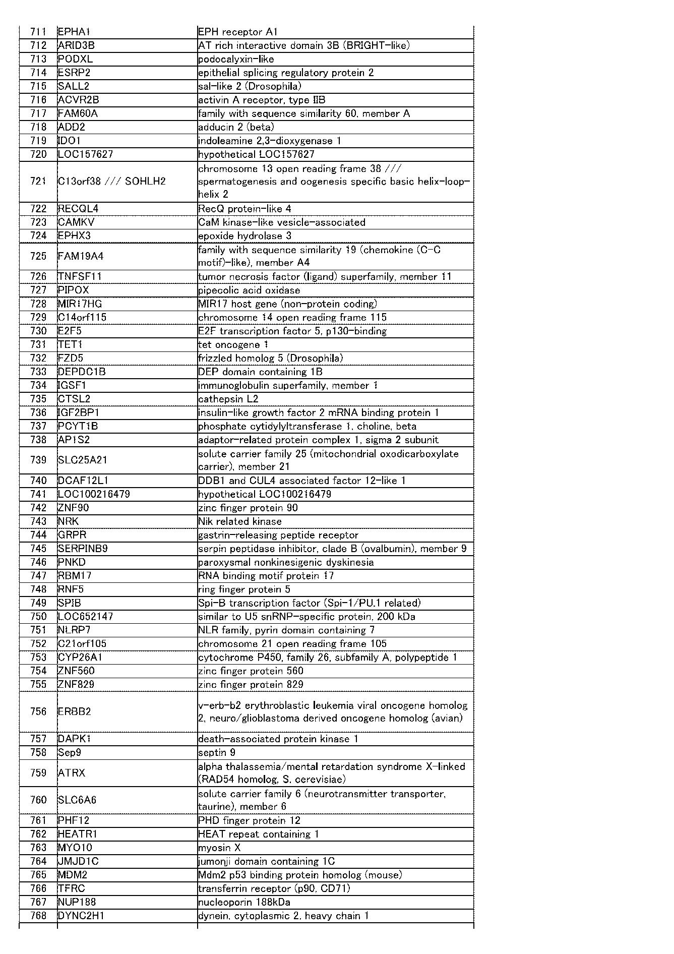
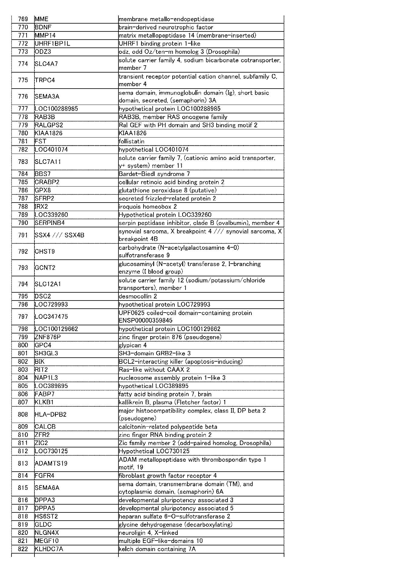
の3つの条件を満たすプローブ1340個(表1)(特定された遺伝子種929個;対応遺伝子が特定されていないプローブ102個を含む)を選択し、特定された遺伝子種929個を第1遺伝子群とした。ここで、特定された遺伝子とは、遺伝子名が判明し、単一に数えられた遺伝子のことを言う。
まず、マイクロアレイHuman U-133 Plus 2.0 Array (Affymetrix社)を用いて、多能性幹細胞で分化細胞より発現の高い遺伝子として、22種類のヒト組織(骨髄、小脳、大脳皮質、胎児脳、心臓、腎臓、肝臓、肺、膵臓、前立腺、唾液腺、骨格筋、小腸、脊髄、脾臓、胃、精巣、胸腺、甲状腺、気管支、子宮)の発現レベルと、KhES1~KhES3のES細胞クローンの発現レベルを比較し、
1.FDR(False Discovery Rate)のq値が0.05未満であること
2.ES3クローン全てがpフラグであること
3.ES3クローン全てで、ヒト組織の発現レベルの平均より5倍以上高いこと。
の3つの条件を満たすプローブ1340個(表1)(特定された遺伝子種929個;対応遺伝子が特定されていないプローブ102個を含む)を選択し、特定された遺伝子種929個を第1遺伝子群とした。ここで、特定された遺伝子とは、遺伝子名が判明し、単一に数えられた遺伝子のことを言う。
第1遺伝子群から、KhES1~KhES3のES細胞クローンの発現レベルの平均と201B7の発現レベルが2倍以上であるプローブを除いた1304個のプローブ(表2)(特定された遺伝子種906個;対応遺伝子が特定されていないプローブ96個を含む)からなる遺伝子群を選択し、特定された遺伝子種906個を第2遺伝子群とした。
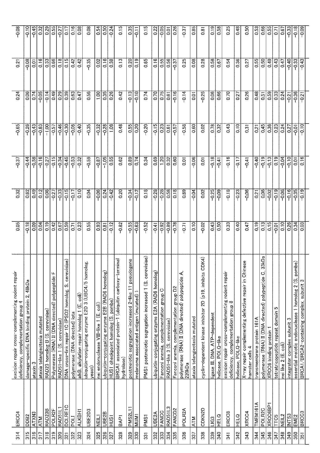
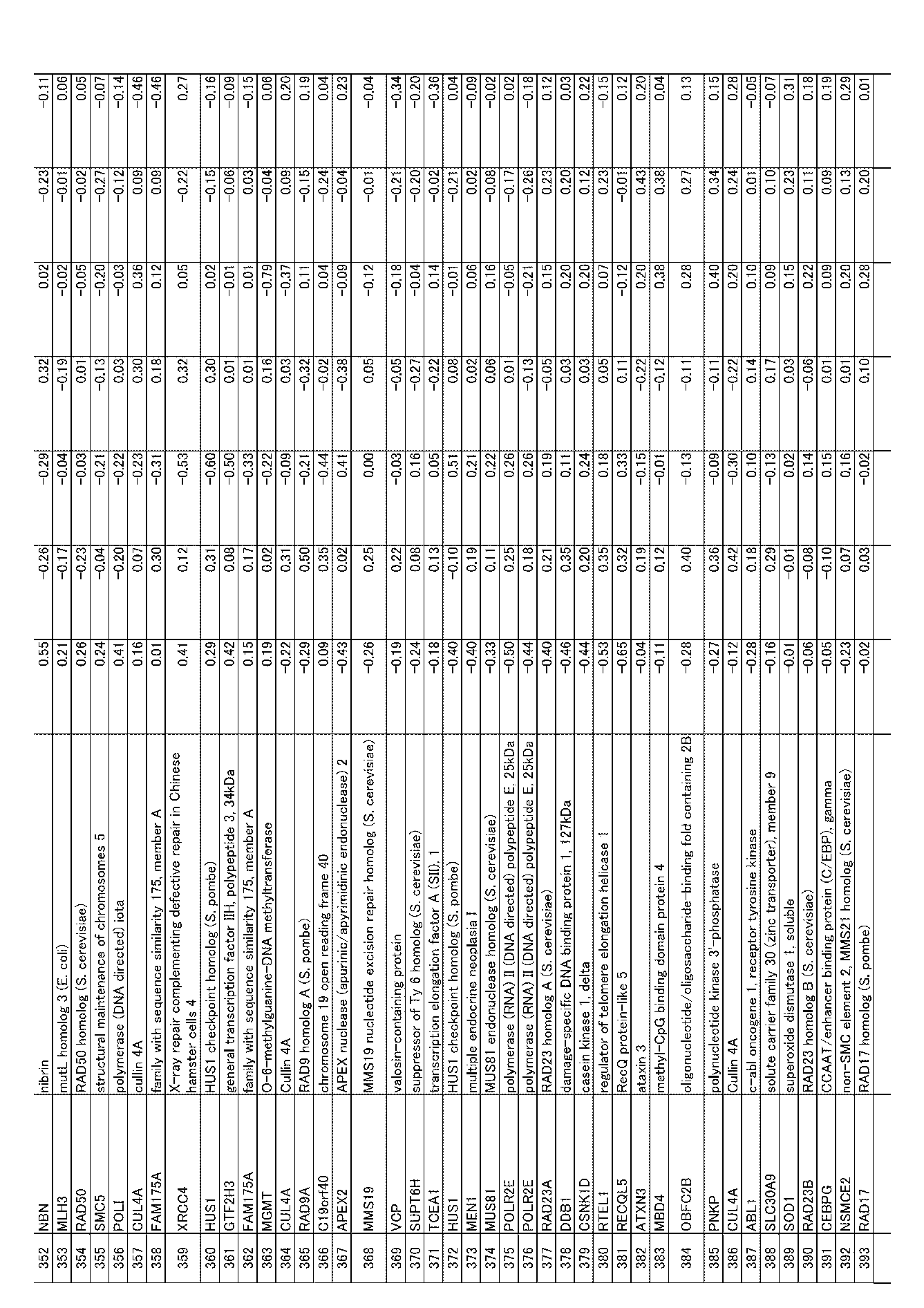
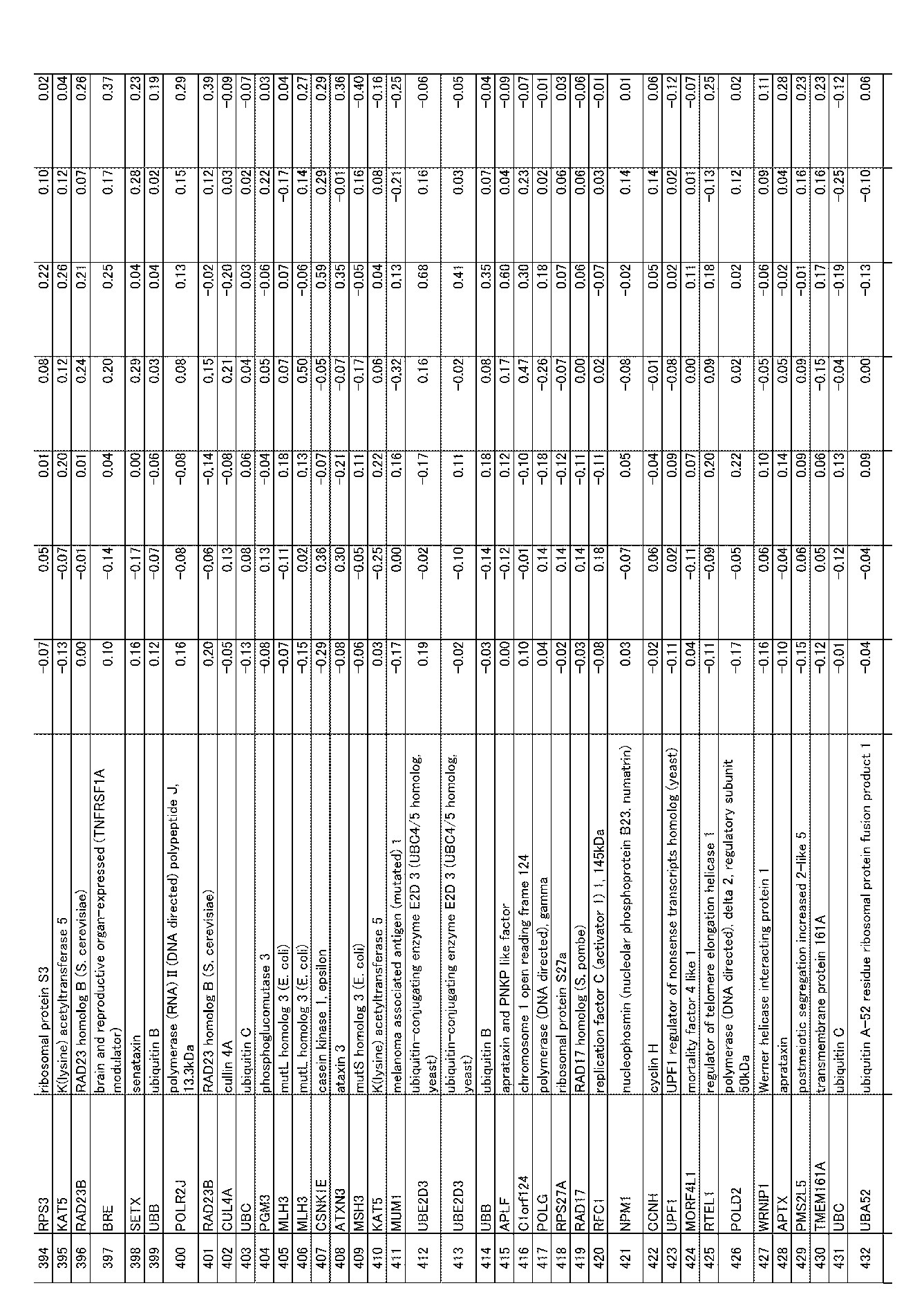
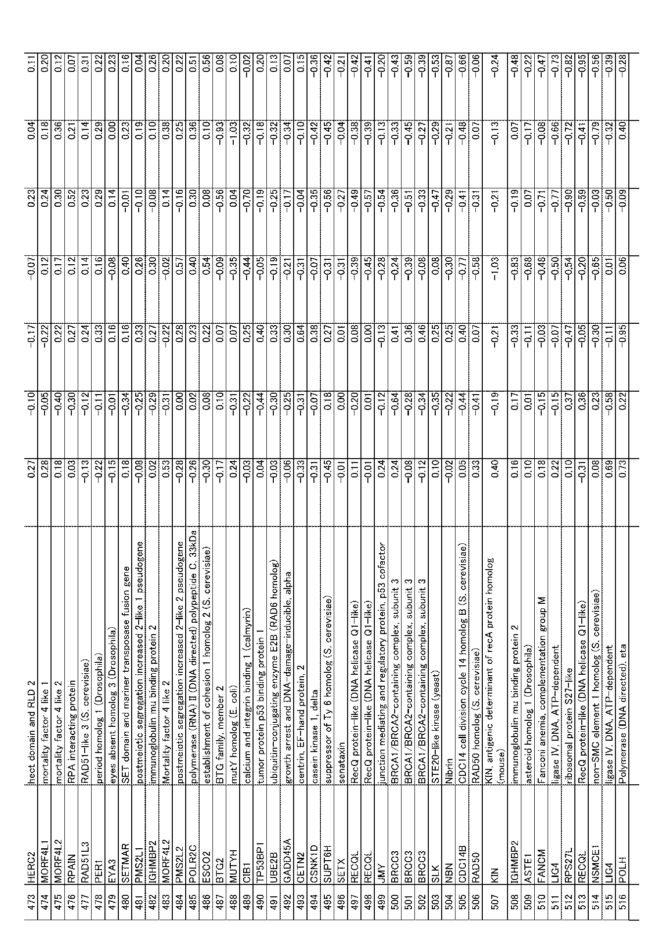
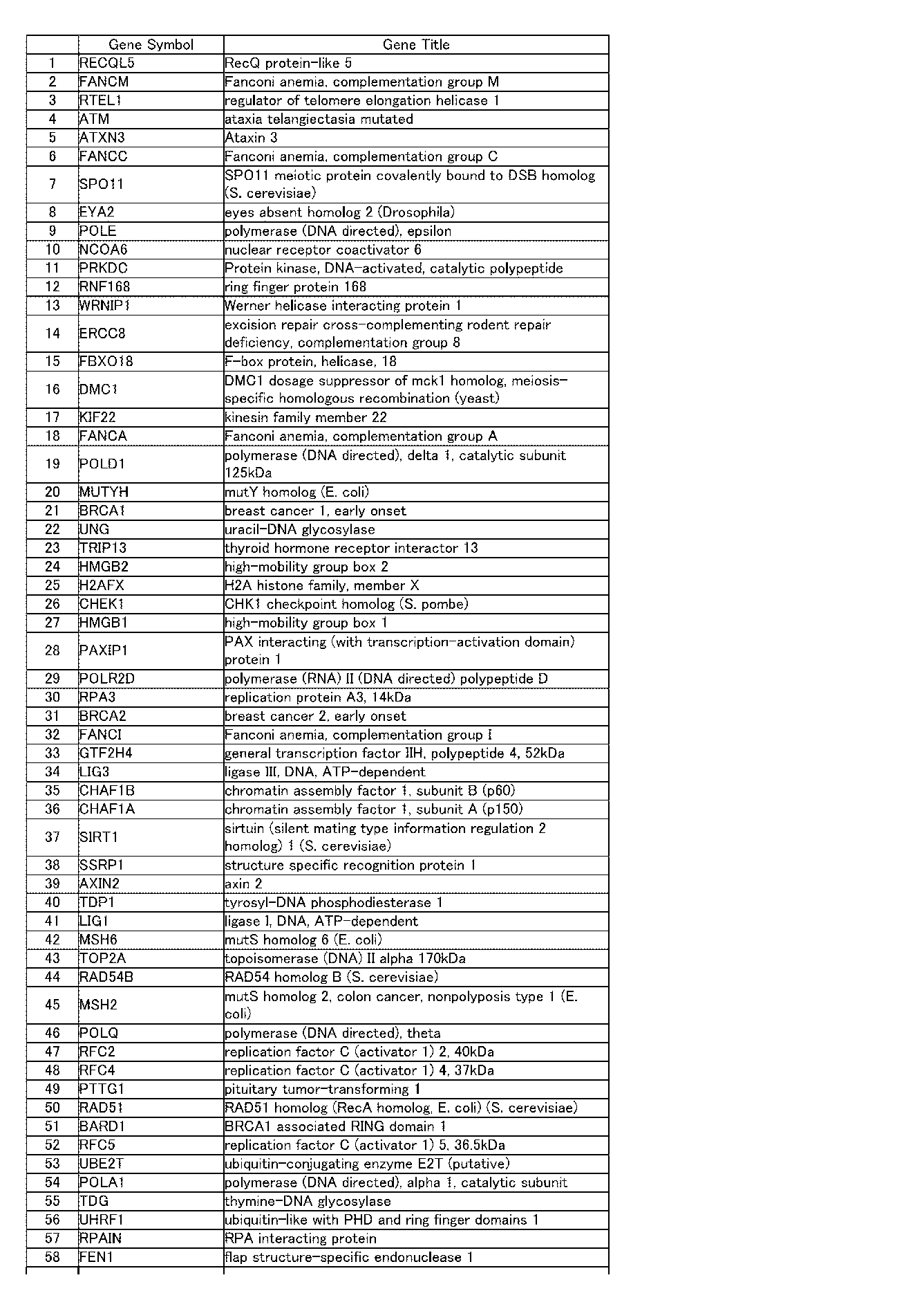
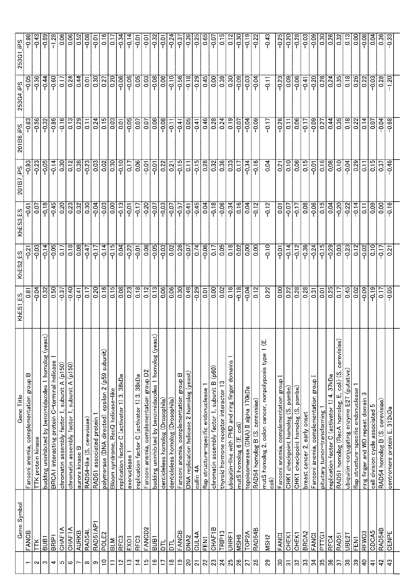
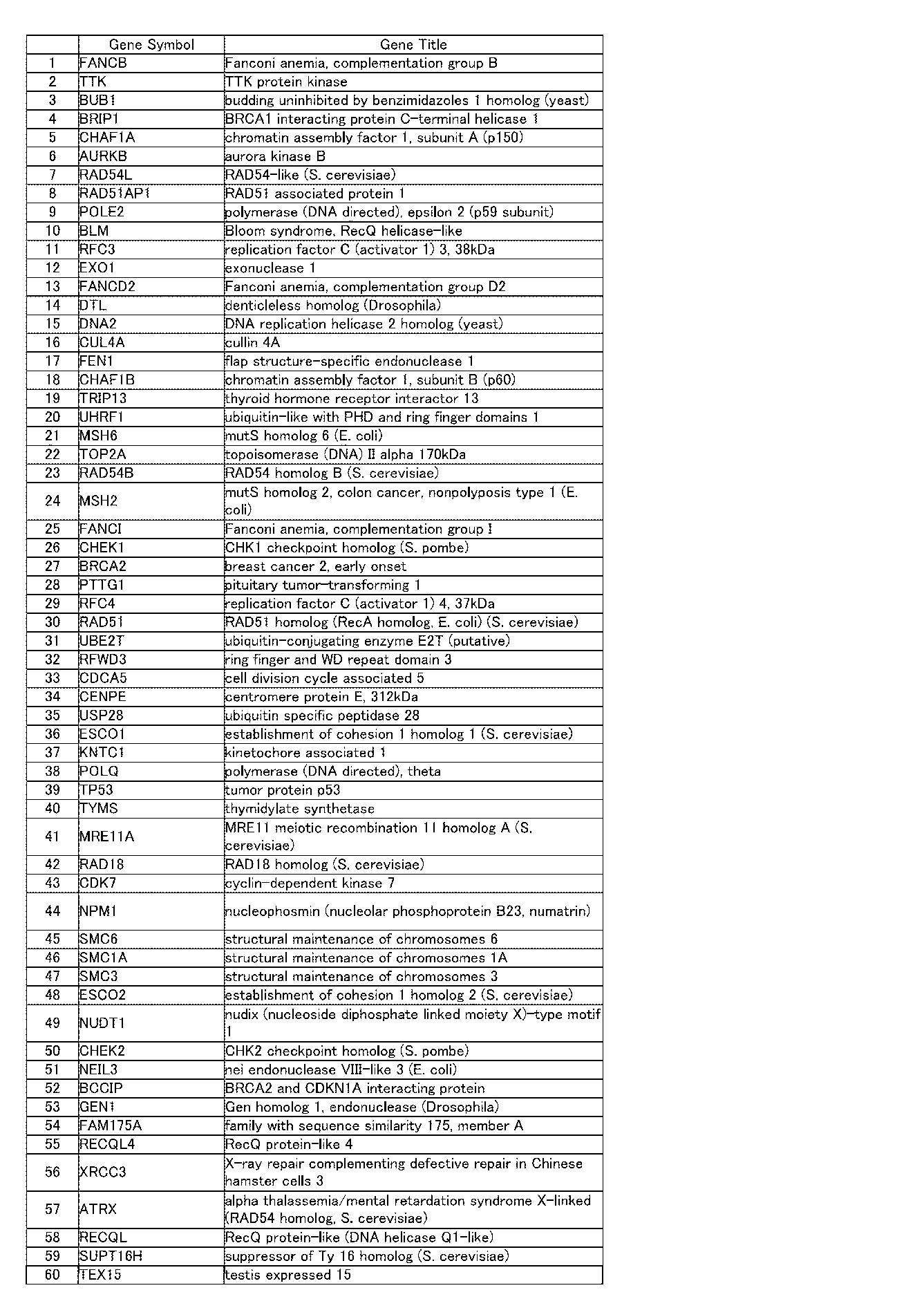
一方、DNA修復に関与する遺伝子群として、GO(Gene Ontology)のデータベースにおいて、DNA repairで登録されているhumanの遺伝子を抽出したところ、DNArepairに関するとされている遺伝子が313個得られた。また、文献中(Wood et al., Science 2001 vol.291 p.1284; Wood et al., 2005 Mutation research vol.577 p.275; Negrini Nat Rev Mol Cell Biol. 2010 vol.113: p.220-8.)にDNArepairに関するとされている遺伝子163個を得て、重複なく332個抽出して第3遺伝子群を作製し、それらに対応するプローブ676個を選択した(表3)。そして、第2遺伝子群と第3遺伝子群で重複している83個のプローブ(表4)(特定された遺伝子種60個を含む)を選択し、特定された遺伝子種60個を第4遺伝子群とした。
さらに、第1遺伝子群の遺伝子から、201B6、253G1、253G4すべての発現レベルが、ES3クローンの発現レベルの平均より1.5倍以上であり、201B7の発現レベルとESの平均との差が1.5倍以内である80個のプローブ(表5)(特定された遺伝子種70個;対応遺伝子が特定されていないプローブ3個を含む)を選択し、特定された遺伝子種70個を第5遺伝子群とし;第1遺伝子群の遺伝子から、201B6、253G1、253G4すべての発現レベルがES3クローンの発現レベルの平均より1.5倍以上であり、さらに、ES3クローンと201B7のすべてで、ES3クローンの発現レベルの平均との差が1.5倍以内である72個のプローブ(表6)(特定された遺伝子種63個;対応遺伝子が特定されていないプローブ2個を含む)を選択し、特定された遺伝子種63個を第6遺伝子群とし;第3遺伝子群の遺伝子から、201B6、253G1、253G4のすべての発現レベルが、ES3クローンの発現レベルの平均より1.5倍以上である59個のプローブ(表7)(特定された遺伝子種54個を含む)を選択し、特定された遺伝子種54個を第7遺伝子群とし;第7遺伝子群の遺伝子から、ES3クローンと201B7のすべてで、ES3クローンの発現レベルの平均との差が1.5倍以内である18個のプローブ(表8)(特定された遺伝子種17個を含む)を選択し、特定された遺伝子種17個を第8遺伝子群とし;第4遺伝子群の遺伝子から、201B6、253G1、253G4のいずれかの発現レベルが、ES3クローンの発現レベルの平均より1.5倍以上であり、201B7の発現レベルとESの平均との差が1.5倍以内である13個のプローブ(表9)(特定された遺伝子種13個を含む)を選択し、対応する遺伝子を第9遺伝子群とし;第4遺伝子群の遺伝子から、201B6、253G1、253G4のすべての発現レベルが、ES3クローンの発現レベルの平均より1.5倍以上であり、201B7の発現レベルとESの平均との差が1.5倍以内である5個のプローブ(表10)(特定された遺伝子種5個を含む)を選択し、対応する遺伝子を第10遺伝子群とした。
<マイクロアレイを用いた解析方法>
マイクロアレイとして、Affymetrix社 U133plus2.0を用いた。
正常ヒト組織(骨髄、小脳、大脳皮質、胎児脳、心臓、腎臓、肝臓、肺、膵臓、前立腺、唾液腺、骨格筋、小腸、脊髄、脾臓、胃、精巣、胸腺、甲状腺、気管支、子宮)のRNAは、Clontech, Ambion, Cell Applications, Stratagene社から購入した。他の細胞などに関しては、Trizol(InVitrogen社)にて全RNAを回収し、RNAeasy mini kit(Quiagen社)を用いてカラム精製を行った。また、キアゲン社のRNAase-free DNaseにより処理し、混入するDNAを分解した。RNAの品質は、Bioanalyzer 2100 (Agilent社)を用いて確認した。
マイクロアレイとして、Affymetrix社 U133plus2.0を用いた。
正常ヒト組織(骨髄、小脳、大脳皮質、胎児脳、心臓、腎臓、肝臓、肺、膵臓、前立腺、唾液腺、骨格筋、小腸、脊髄、脾臓、胃、精巣、胸腺、甲状腺、気管支、子宮)のRNAは、Clontech, Ambion, Cell Applications, Stratagene社から購入した。他の細胞などに関しては、Trizol(InVitrogen社)にて全RNAを回収し、RNAeasy mini kit(Quiagen社)を用いてカラム精製を行った。また、キアゲン社のRNAase-free DNaseにより処理し、混入するDNAを分解した。RNAの品質は、Bioanalyzer 2100 (Agilent社)を用いて確認した。
[2]発現に異常のある遺伝子の個数を比較する方法
ここでは、第1遺伝子群の遺伝子を含むプローブにおいて、各iPS細胞の発現レベルが、ES3クローンの発現レベルの平均に比べて、0.667倍以下あるいは1.5倍以上という異常な発現レベルを示すプローブの個数を調べ、その結果を図1にグラフ化した。
ここでは、第1遺伝子群の遺伝子を含むプローブにおいて、各iPS細胞の発現レベルが、ES3クローンの発現レベルの平均に比べて、0.667倍以下あるいは1.5倍以上という異常な発現レベルを示すプローブの個数を調べ、その結果を図1にグラフ化した。
腫瘍化を起こしにくいiPS細胞(201B7)と、正常に分化しないか(201B6)、分化しても腫瘍化を起こしやすい(253G1、253G4)iPS細胞とでは、発現に異常のあるプローブの個数に有意な差があった。
従って、第1遺伝子群の遺伝子の発現を調べ、腫瘍化を起こしにくい多能性幹細胞に対して発現に異常のある遺伝子の個数を調べることにより、腫瘍化を起こしにくいかどうかを判断できる。
[3]clustering分析によって分類する方法
ここでは、第2~第10遺伝子群の遺伝子を含むプローブについて、ES3クローンの発現レベルの平均に対する、各iPS細胞の発現レベルの割合を計算し、Log2(ratio)で表した(表2~表10)。これらのデータに対し、Cluster 3.0を用いて、clustering解析を行い、得られた結果をJava Treeview(東京大学医科学研究所ヒトゲノム解析センターDNA情報解析分野によるフリーウェア;M. J. L. de Hoon, S. Imoto, J. Nolan, and S. Miyano: Open Source Clustering Software. Bioinformatics, 20 (9): 1453--1454 (2004).;http://bonsai.hgc.jp/~mdehoon/software/cluster/)を用いて、treeを作製した(図2~図10)。
ここでは、第2~第10遺伝子群の遺伝子を含むプローブについて、ES3クローンの発現レベルの平均に対する、各iPS細胞の発現レベルの割合を計算し、Log2(ratio)で表した(表2~表10)。これらのデータに対し、Cluster 3.0を用いて、clustering解析を行い、得られた結果をJava Treeview(東京大学医科学研究所ヒトゲノム解析センターDNA情報解析分野によるフリーウェア;M. J. L. de Hoon, S. Imoto, J. Nolan, and S. Miyano: Open Source Clustering Software. Bioinformatics, 20 (9): 1453--1454 (2004).;http://bonsai.hgc.jp/~mdehoon/software/cluster/)を用いて、treeを作製した(図2~図10)。
図2~図10のtreeに示されているように、調べたiPS細胞のうち、201B7だけがヒトES細胞と同じグループに分類されており、201B6、253G1、253G4は、それとは別に一つのグループに分類されている。
このように、多能性幹細胞で分化細胞より発現の高い遺伝子、及びDNA修復に関与する遺伝子から選択された遺伝子からなる遺伝子群について、腫瘍化を起こしにくい多能性幹細胞における発現レベルと比較することにより、正常に分化しないか、分化しても腫瘍化を起こしやすいiPS細胞と、腫瘍化を起こしにくいiPS細胞に分類ができる。従って、この方法を使えば、腫瘍化を起こしにくいかどうかわからないiPS細胞についても、どちらのグループに属するかを明らかにでき、そのiPS細胞が腫瘍化を起こしにくいかどうかを判断できる。
[4]発現レベル自体を比較する方法
例えば、本実施例では、表10に記載のsuppressor of Ty 16 homolog (S. cerevisiae)の発現レベルは、ES3クローンと、正常に分化しないか(201B6)、分化しても腫瘍化を起こしやすい(253G1、253G4)とでは、有意に差がある。そして、腫瘍化を起こしにくいiPS細胞(201B7)の発現レベルは、ES3クローンと差がなく、腫瘍化を起こしやすいiPS細胞とは顕著な差がある。
例えば、本実施例では、表10に記載のsuppressor of Ty 16 homolog (S. cerevisiae)の発現レベルは、ES3クローンと、正常に分化しないか(201B6)、分化しても腫瘍化を起こしやすい(253G1、253G4)とでは、有意に差がある。そして、腫瘍化を起こしにくいiPS細胞(201B7)の発現レベルは、ES3クローンと差がなく、腫瘍化を起こしやすいiPS細胞とは顕著な差がある。
このように、発現レベル自体を比較することによっても、iPS細胞が腫瘍化を起こしにくいかどうかを判断できる。
(4)第11~第12遺伝子群を用いたiPS細胞クローンの分類
ここでは、第1遺伝子群及び第3遺伝子群に属する各遺伝子を含むプローブに対し、表2及び表3に示された発現レベルを表す数値に基づき、ES3クローンと201B7のグループ及び201B6、253G1、253G4のグループの間でSAMの検定を行い、各遺伝子群でp<0.1またはp<0.05で有意差のあったプローブを、それぞれ27個(表11)(特定された遺伝子26個を含む)と19個(表12)(特定された遺伝子17個を含む)抽出し、特定された遺伝子種26個と17個を第11遺伝子群及び第12遺伝子群とした。(3)[3]と同様に、clustering解析をしたところ、図11及び図12のtreeに示されているように、調べたiPS細胞のうち、201B7だけがヒトES細胞と同じグループに分類されており、201B6、253G1、253G4は、それとは別に一つのグループに分類されていた。
ここでは、第1遺伝子群及び第3遺伝子群に属する各遺伝子を含むプローブに対し、表2及び表3に示された発現レベルを表す数値に基づき、ES3クローンと201B7のグループ及び201B6、253G1、253G4のグループの間でSAMの検定を行い、各遺伝子群でp<0.1またはp<0.05で有意差のあったプローブを、それぞれ27個(表11)(特定された遺伝子26個を含む)と19個(表12)(特定された遺伝子17個を含む)抽出し、特定された遺伝子種26個と17個を第11遺伝子群及び第12遺伝子群とした。(3)[3]と同様に、clustering解析をしたところ、図11及び図12のtreeに示されているように、調べたiPS細胞のうち、201B7だけがヒトES細胞と同じグループに分類されており、201B6、253G1、253G4は、それとは別に一つのグループに分類されていた。
このように、統計学的有意性を有する発現レベルの差を用いることによっても、iPS細胞を、正常に分化しないか、分化しても腫瘍化を起こしやすいiPS細胞と、腫瘍化を起こしにくいiPS細胞に分類ができる。
(5)ゲノムDNAにおける変異
本発明は、未分化状態のiPS細胞における、DNA修復に関与する遺伝子の発現の異常が、分化後のiPS細胞の造腫瘍性に影響があるという発見に基づくものである。以下、そのメカニズムとして、未分化状態におけるDNA修復に関与する遺伝子の異状によって、DNAに多数の傷が生じ、それが原因となって、iPS細胞が正常に分化できないか、あるいはiPS細胞が分化したときに腫瘍化するということを示す。
本発明は、未分化状態のiPS細胞における、DNA修復に関与する遺伝子の発現の異常が、分化後のiPS細胞の造腫瘍性に影響があるという発見に基づくものである。以下、そのメカニズムとして、未分化状態におけるDNA修復に関与する遺伝子の異状によって、DNAに多数の傷が生じ、それが原因となって、iPS細胞が正常に分化できないか、あるいはiPS細胞が分化したときに腫瘍化するということを示す。
まず、未分化状態のiPS細胞(201B6、253G1、253G4、201B7)と、各細胞を分化させた3次ニューロスフェア、iPS細胞作製のもととなる線維芽細胞(HDF)、コントロールとして、ヒト胎児由来神経幹細胞(hNSC)および患者グリオーマ細胞由来細胞(GDC)から、ゲノムDNAを単離し、アレイCGH解析を行なった。
ゲノムDNAは、各クローンから、そして対照として正常ヒト男性ゲノム(Promega社)から、フェノールクロロホルム法、あるいは、DNeasy Blood & Tissue KitやQIAamp DNA mini Kit (Qiagen社)を用いて抽出し、PicoGreenを用いて定量した。各サンプルとして、500ngから1000ngの二本鎖DNAを用い、制限酵素(AluIとRsaI)による消化を行い、DNAを断片化した。その後、Genomic DNA Enzymatic Labeling キット(Agilent社)を用いて、DNA断片をCy3またはCy5でラベルし、片方をコントロール、もう片方をサンプルとした。精製後、Sureprint G3 human CGHマイクロアレイ4x180K(Agilent社)を用い、添付プロトコールに従い、ハイブリダイゼーション、Agilent スキャナーを用いたスキャニング、Feature extractionソフトウェアによるシグナルの数値化を行った。得られたデータは、Agilent Genomic Workbenchを用いて、Aberrationの検出を行った。検出されたAberrationのうち、対照のHDF(ヒト皮膚線維芽細胞)で見られるAberrationは、単なるCNV(copy number variation)の可能性が高いため、解析から除外して、各クローン間のaberrated geneとしての遺伝子数を比較した。
図16に示されるように、ES細胞や腫瘍化を起こしにくいiPS細胞(201B7)では、分化しても、ゲノム異常は全体として変化がなく、一方、正常に分化しないか(201B6)、分化しても腫瘍化を起こしやすい(253G1、253G4)では、分化と共に、ゲノム異常が増加しており、その異状のレベルは、患者グリオーマに由来する培養細胞(GDC21、GDC36、GDC40)ほどではないものの、ES細胞や201B7と比べて、はるかに高かった。
このように、未分化状態のiPS細胞において、DNA修復に関与する遺伝子の発現に異常があると、分化過程で生じるDNA変異が修復できず、そのために正常に分化できないか、あるいは分化させても細胞が腫瘍化するものと考えられる。
(6)比較例
本比較例では、いずれかのクローン中で発現が検出されたプローブ全てを対象とし、Rというclustering解析用ソフトウェアを用い(http://www.r-project.org/)、Ward's hierarchial clustering methodとEluclidian distanceによって、clustering解析を、行なった。その結果を図17に示すが、本発明のように適切な遺伝子群を選択しなければ、腫瘍化を起こしにくいiPS細胞と、正常に分化しないか、分化しても腫瘍化を起こしやすいiPS細胞は、clusterによって区別することができなかった。
本比較例では、いずれかのクローン中で発現が検出されたプローブ全てを対象とし、Rというclustering解析用ソフトウェアを用い(http://www.r-project.org/)、Ward's hierarchial clustering methodとEluclidian distanceによって、clustering解析を、行なった。その結果を図17に示すが、本発明のように適切な遺伝子群を選択しなければ、腫瘍化を起こしにくいiPS細胞と、正常に分化しないか、分化しても腫瘍化を起こしやすいiPS細胞は、clusterによって区別することができなかった。
(7)エピソーマルベクターを用いて作製したヒトiPS細胞の解析
エピソーマルベクターを用いて作製したヒトiPS細胞株409B2及び414C2(Okita et al., Nat Methods 2011)について、比較する対照の細胞株として、KhES1、KhES2、KhES3、201B7、409B2、414C2、201B6、253G4、253G1の各クローンを用い、第4遺伝子群及び第12遺伝子群について、マイクロアレイを用いて(3)と同様の方法で、発現の解析及びclustering分析を行った。その結果、図18及び図19のtreeに示されるように、どちらの場合も、409B2及び414C2は、ES細胞及び201B7と同じ枝に分類された。
また、第12遺伝子群の19プローブについて、3つのES細胞における発現の平均値から、シグナル強度に1.5倍以上の差があるプローブの個数を測定したところ、図20に示すように、その個数が、409B2、414C2、201B7では6個以内、201B6、253G4、253G1では、10個以上となり、2つのグループで有意に(t検定でp=0.017)差が見られた。
次に、409B2及び414C2について、(2)と同様の方法で、造腫瘍性の評価を行なったところ、図21に示すように、409B2及び414C2は、ES細胞及び201B7と同様に、精巣移植後の腫大は観察されなかった。また、409B2及び414C2を移植した精巣のパラフィン切片を作製し、HE染色を行なって、移植細胞由来のテラトーマの組織像を観察したところ、図22及び図23に示すように、253G4、253G1より腫瘍化傾向が低く、十分に分化した神経細胞及びグリア細胞が観察された。
このように、本発明の遺伝子群は、iPS細胞を、正常に分化しないか、分化しても腫瘍化を起こしやすいクローンと、腫瘍化を起こしにくいクローンに分類するのに有用である。
エピソーマルベクターを用いて作製したヒトiPS細胞株409B2及び414C2(Okita et al., Nat Methods 2011)について、比較する対照の細胞株として、KhES1、KhES2、KhES3、201B7、409B2、414C2、201B6、253G4、253G1の各クローンを用い、第4遺伝子群及び第12遺伝子群について、マイクロアレイを用いて(3)と同様の方法で、発現の解析及びclustering分析を行った。その結果、図18及び図19のtreeに示されるように、どちらの場合も、409B2及び414C2は、ES細胞及び201B7と同じ枝に分類された。
また、第12遺伝子群の19プローブについて、3つのES細胞における発現の平均値から、シグナル強度に1.5倍以上の差があるプローブの個数を測定したところ、図20に示すように、その個数が、409B2、414C2、201B7では6個以内、201B6、253G4、253G1では、10個以上となり、2つのグループで有意に(t検定でp=0.017)差が見られた。
次に、409B2及び414C2について、(2)と同様の方法で、造腫瘍性の評価を行なったところ、図21に示すように、409B2及び414C2は、ES細胞及び201B7と同様に、精巣移植後の腫大は観察されなかった。また、409B2及び414C2を移植した精巣のパラフィン切片を作製し、HE染色を行なって、移植細胞由来のテラトーマの組織像を観察したところ、図22及び図23に示すように、253G4、253G1より腫瘍化傾向が低く、十分に分化した神経細胞及びグリア細胞が観察された。
このように、本発明の遺伝子群は、iPS細胞を、正常に分化しないか、分化しても腫瘍化を起こしやすいクローンと、腫瘍化を起こしにくいクローンに分類するのに有用である。
[表1A]第1遺伝子群(表1B)を含むプローブ1340個において、ヒトES細胞クローン(KhES1-S3)の発現レベルの平均に対する、各ES細胞クローン及び各iPS細胞クローンの発現レベルの割合


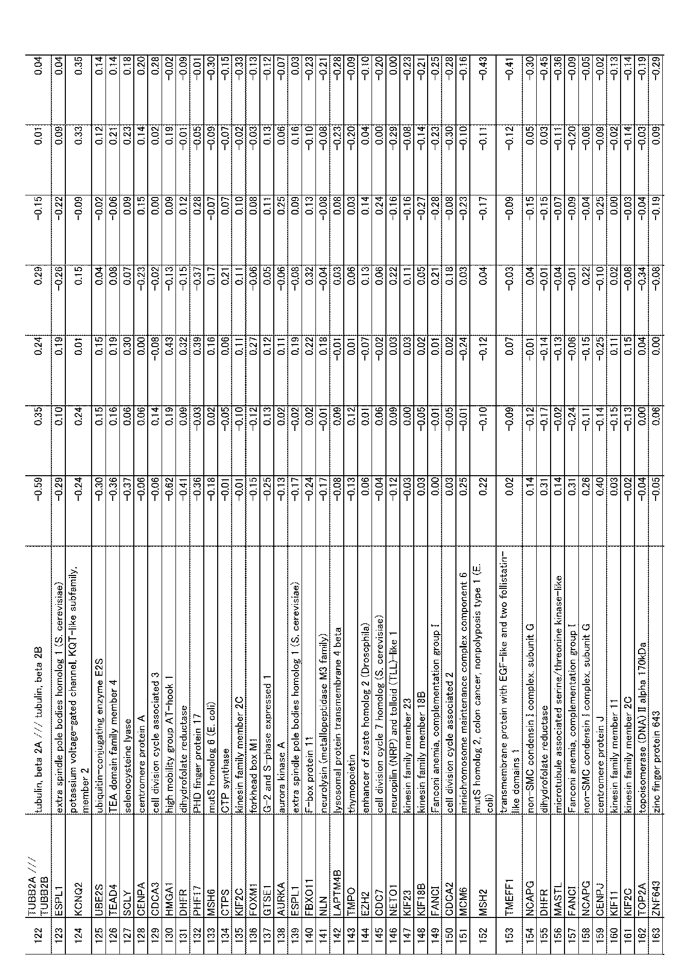


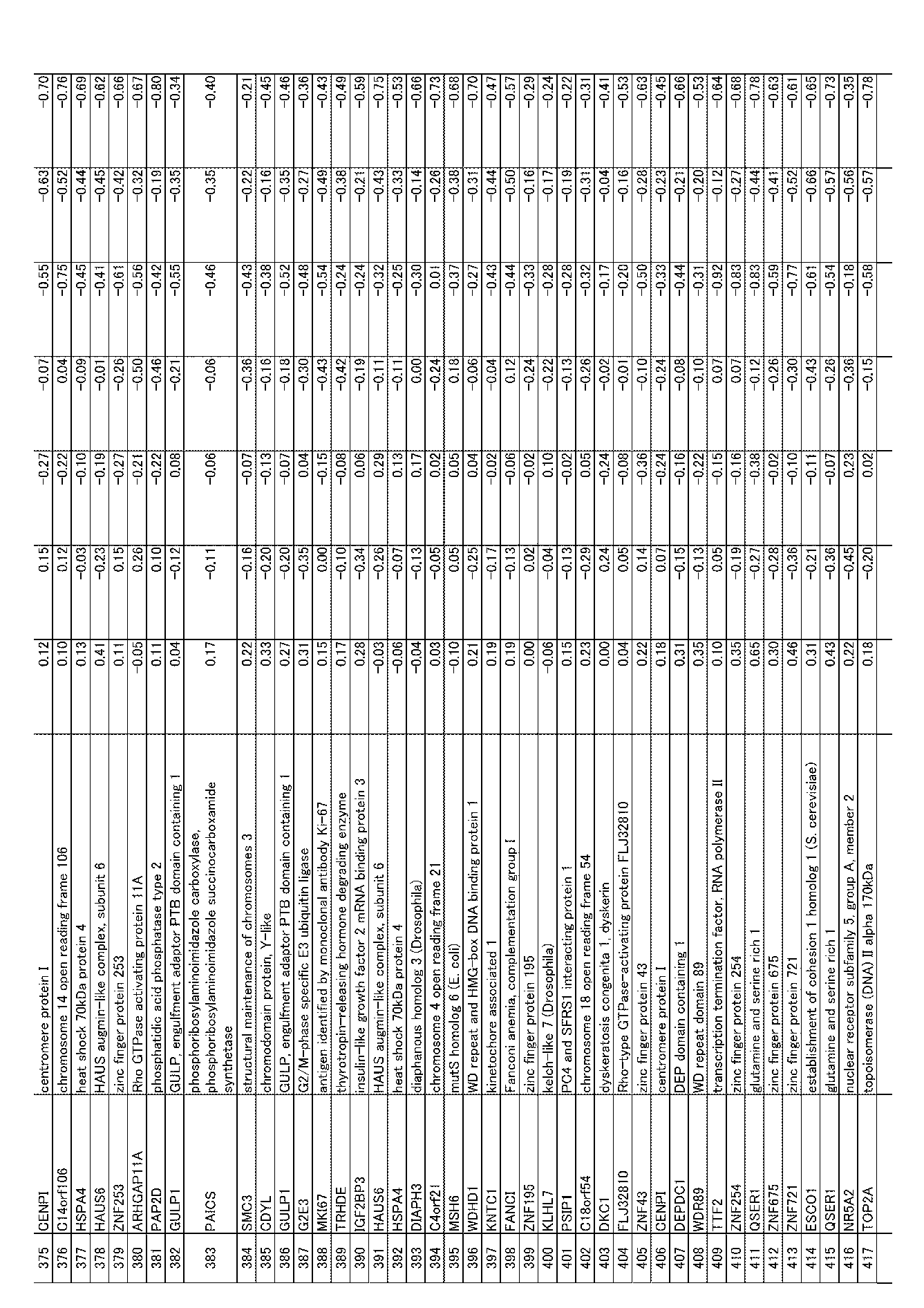
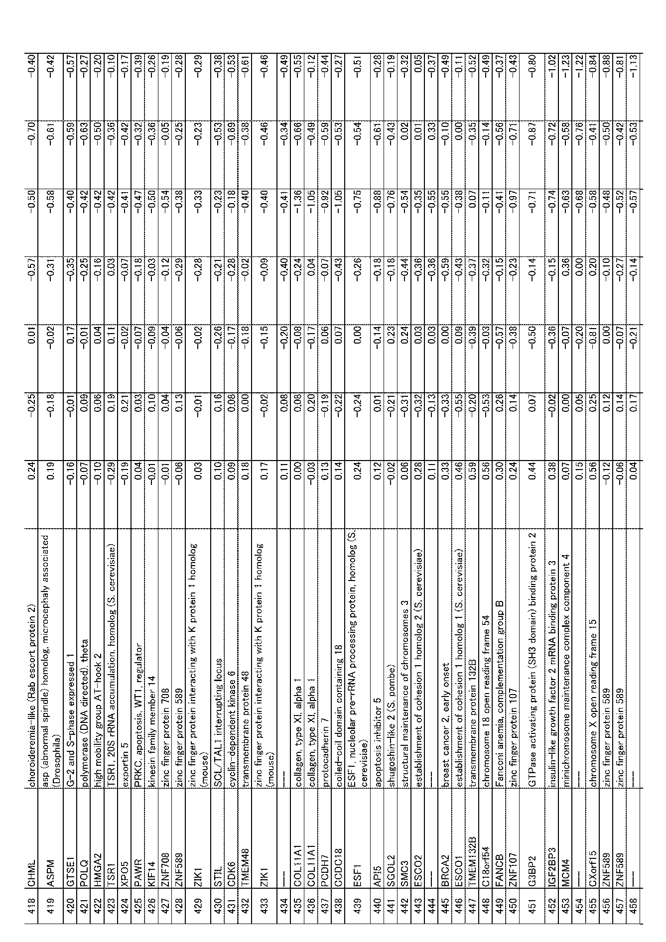


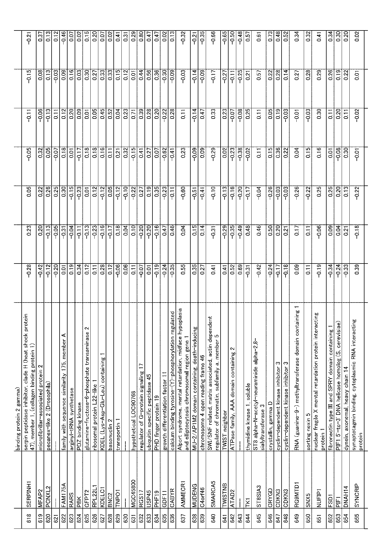
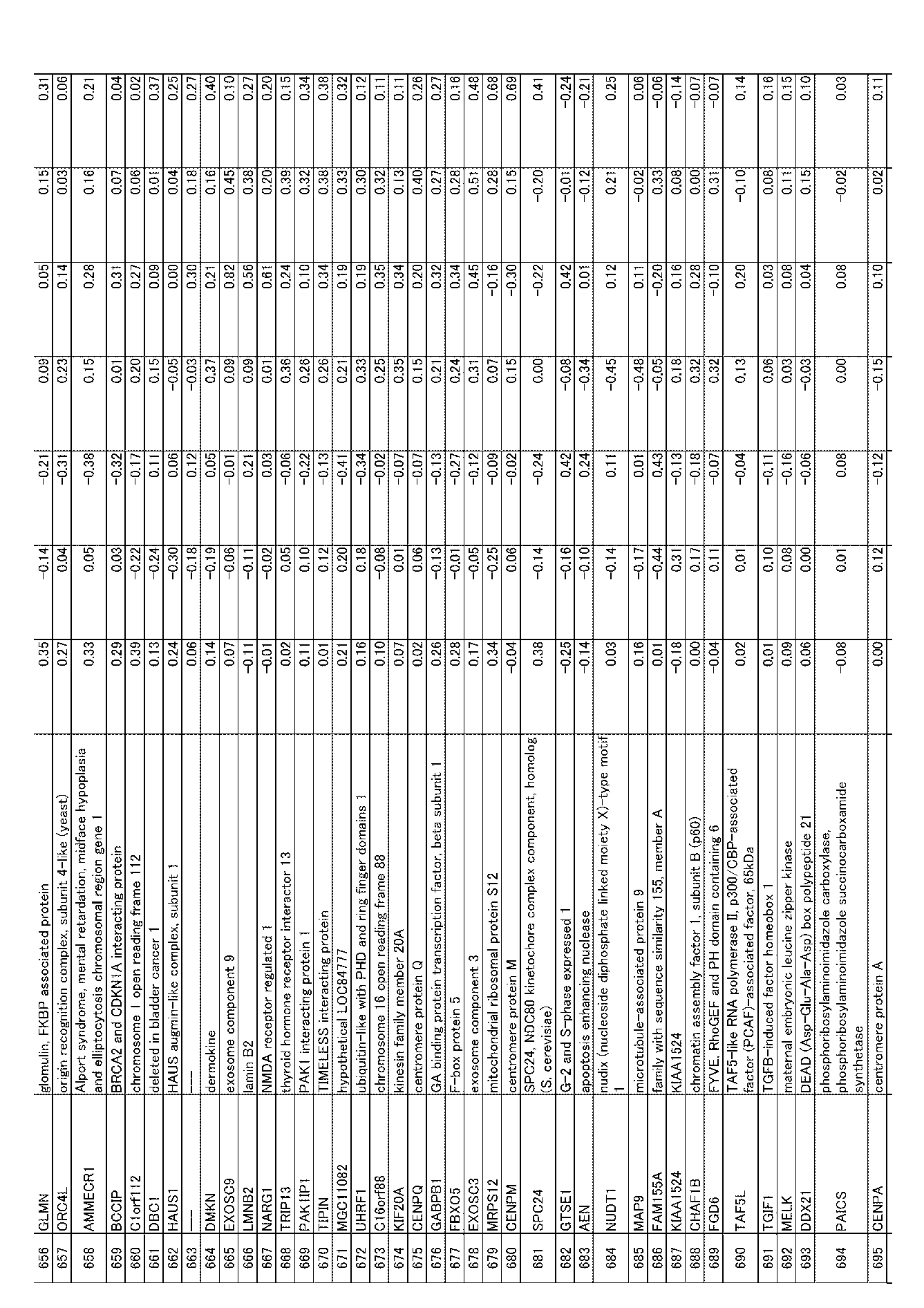


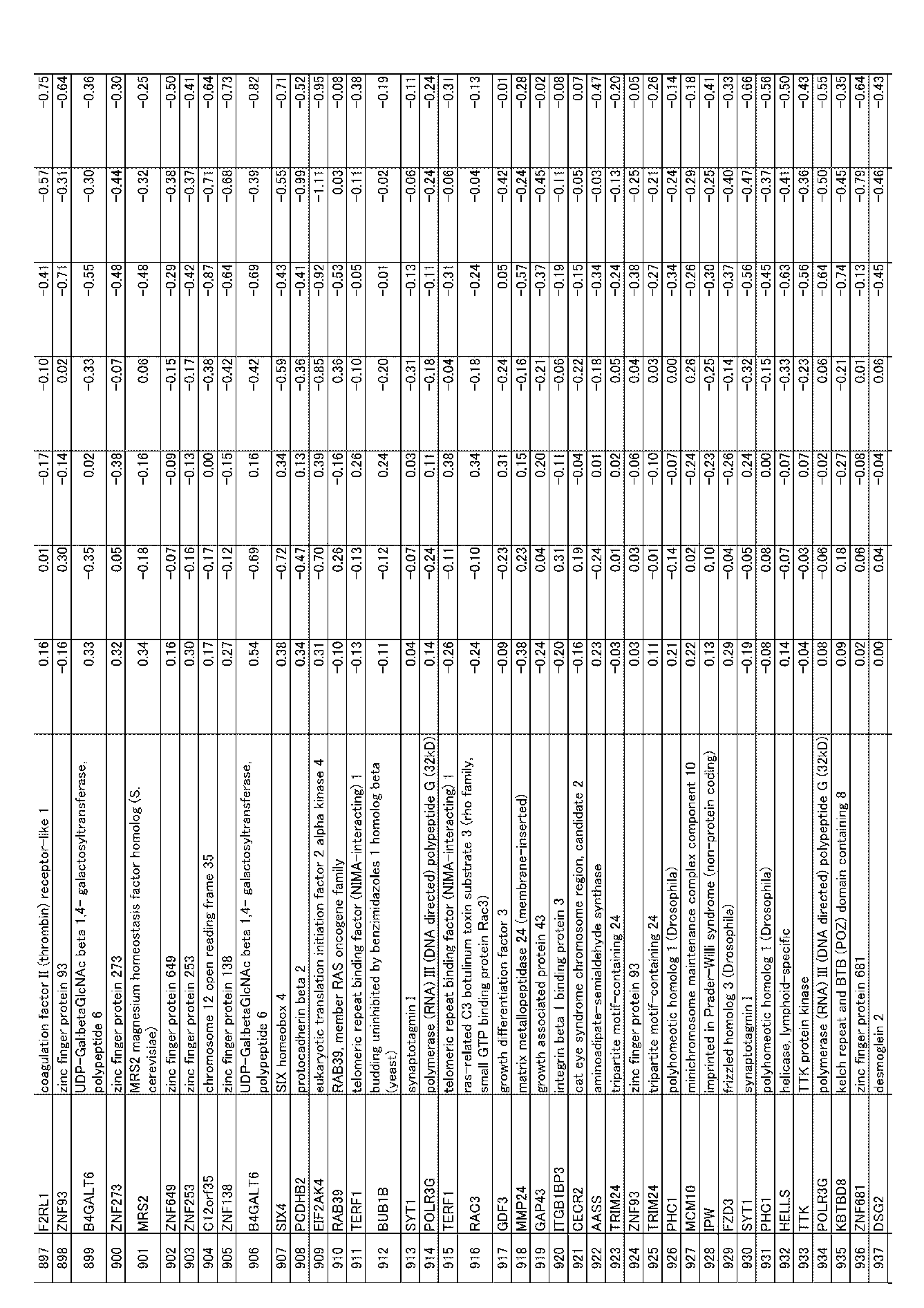
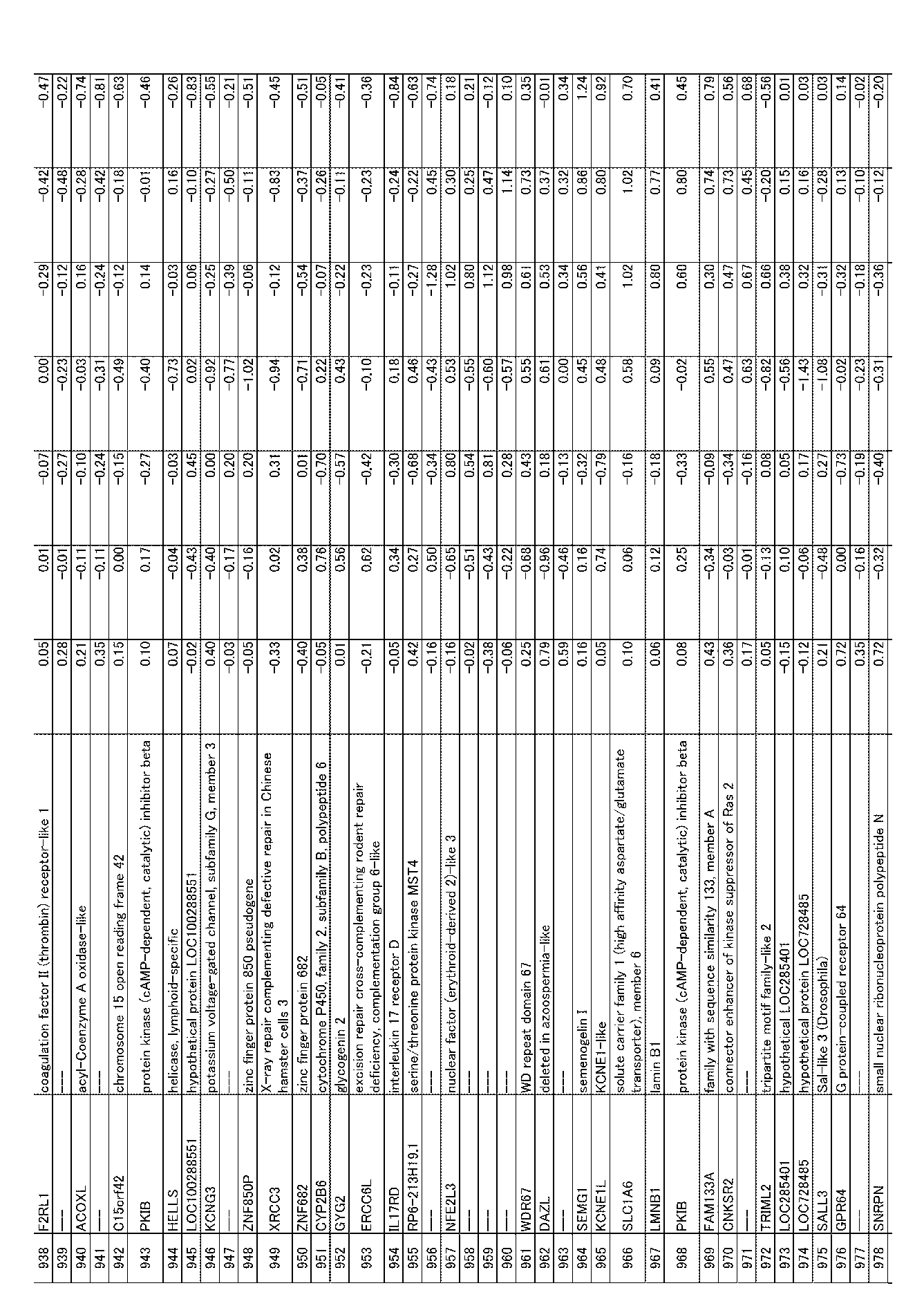

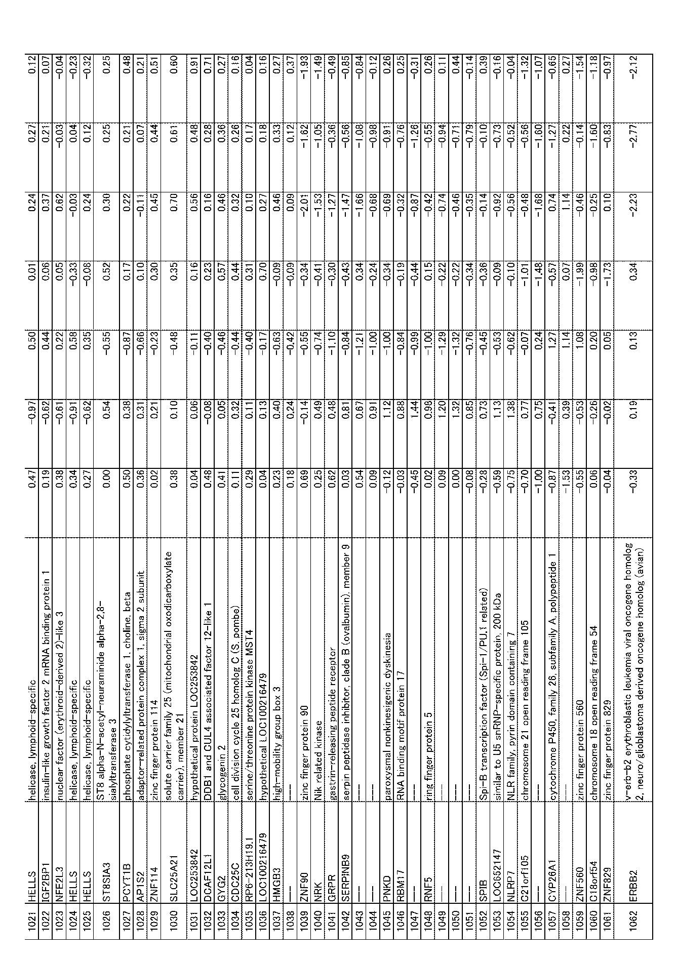
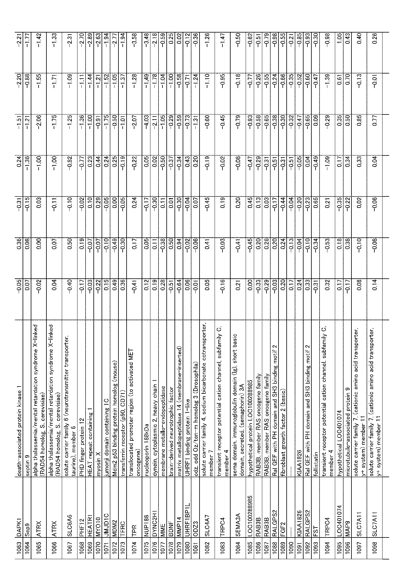

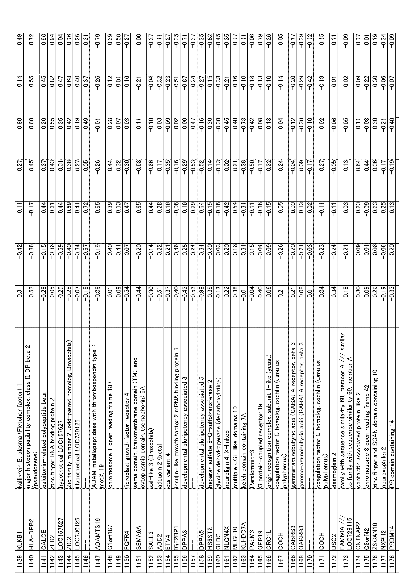
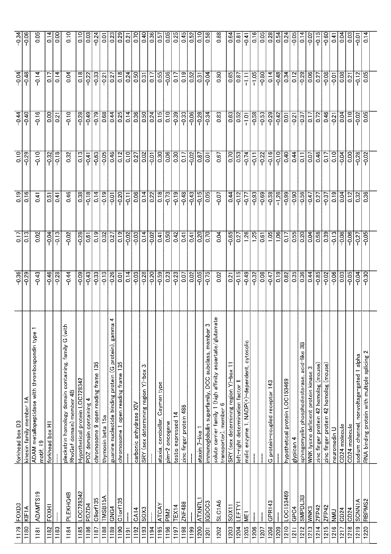

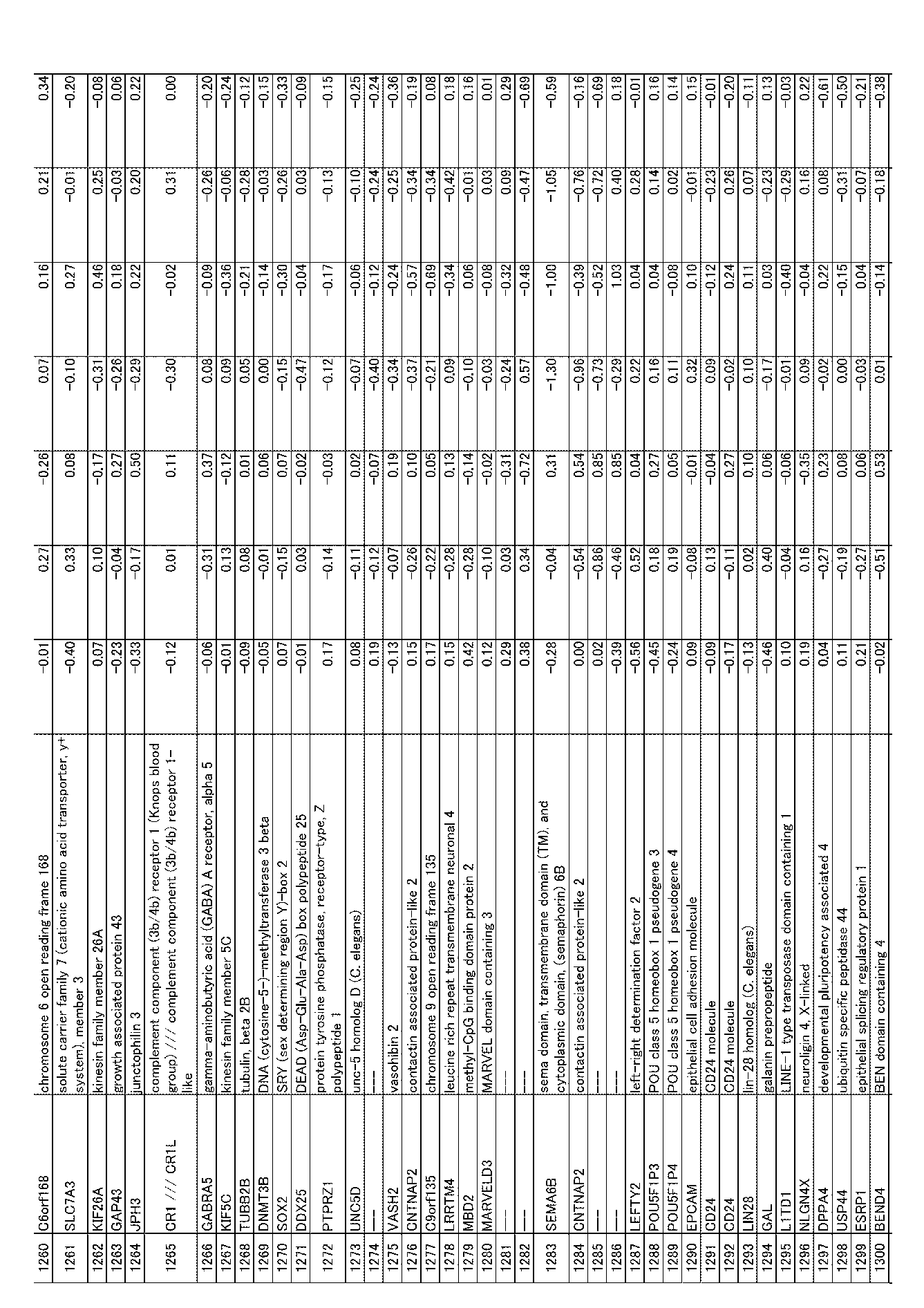
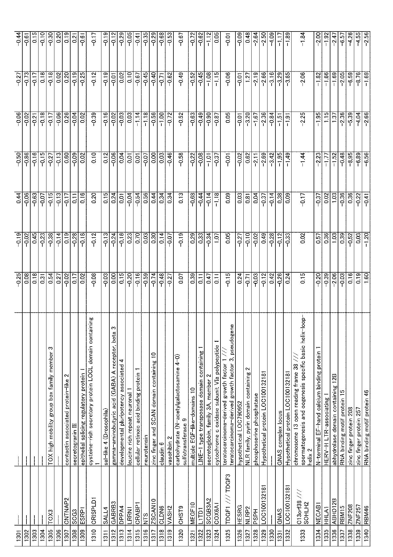
[表1B]第1遺伝子群


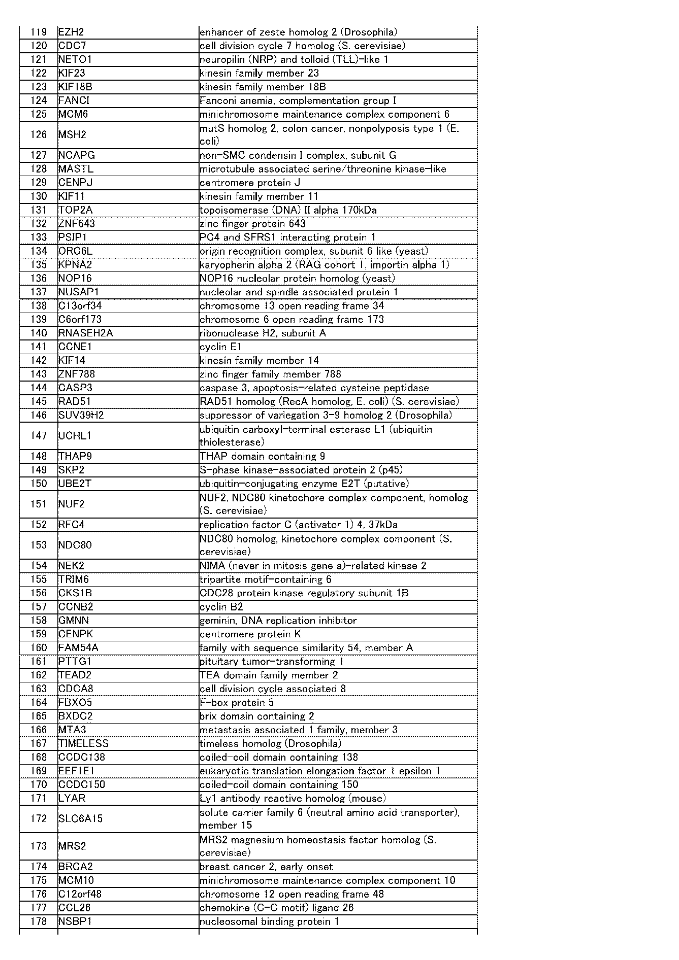
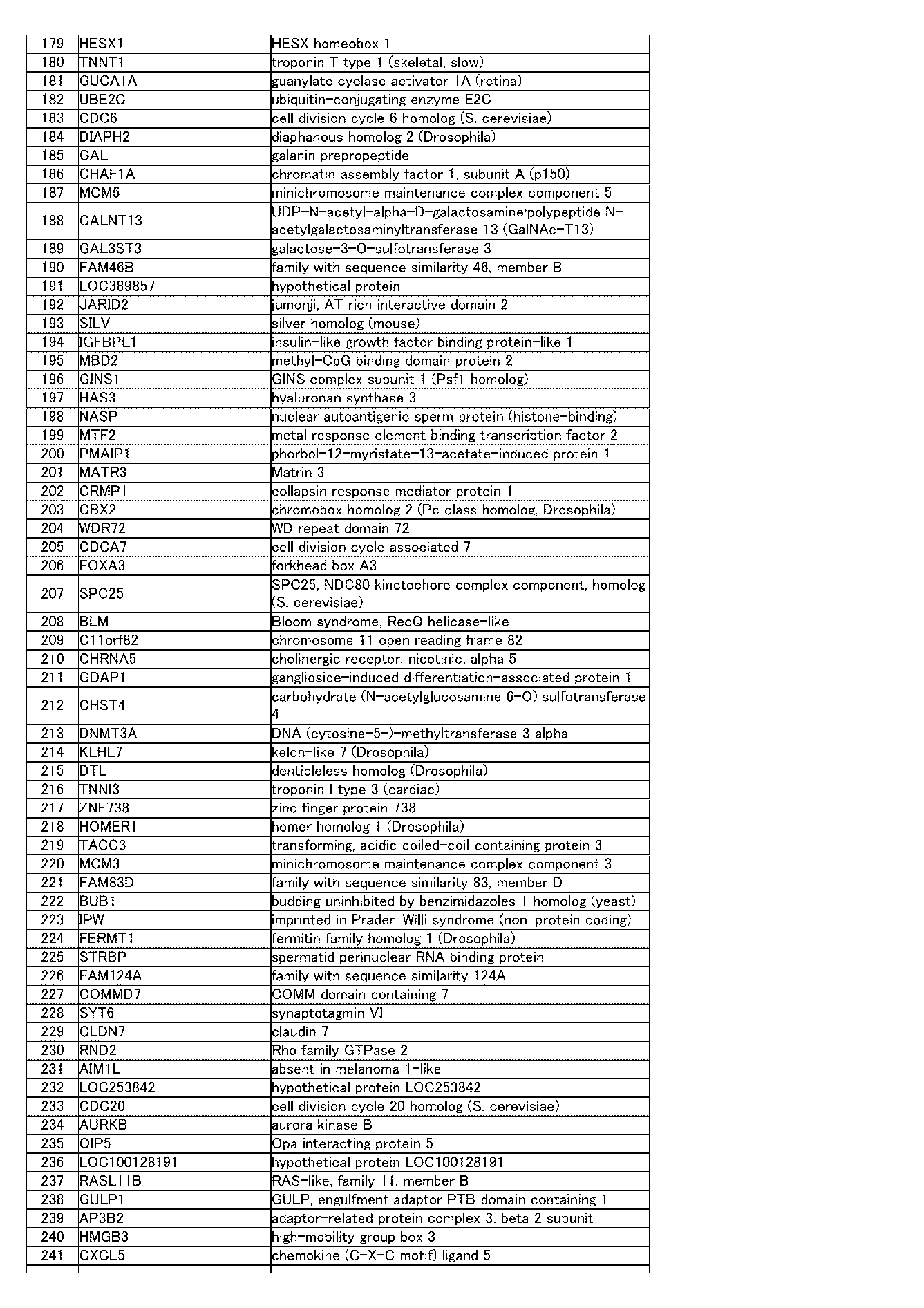


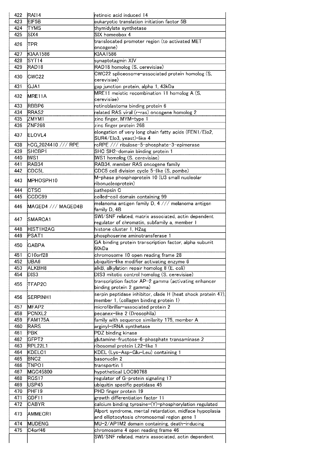


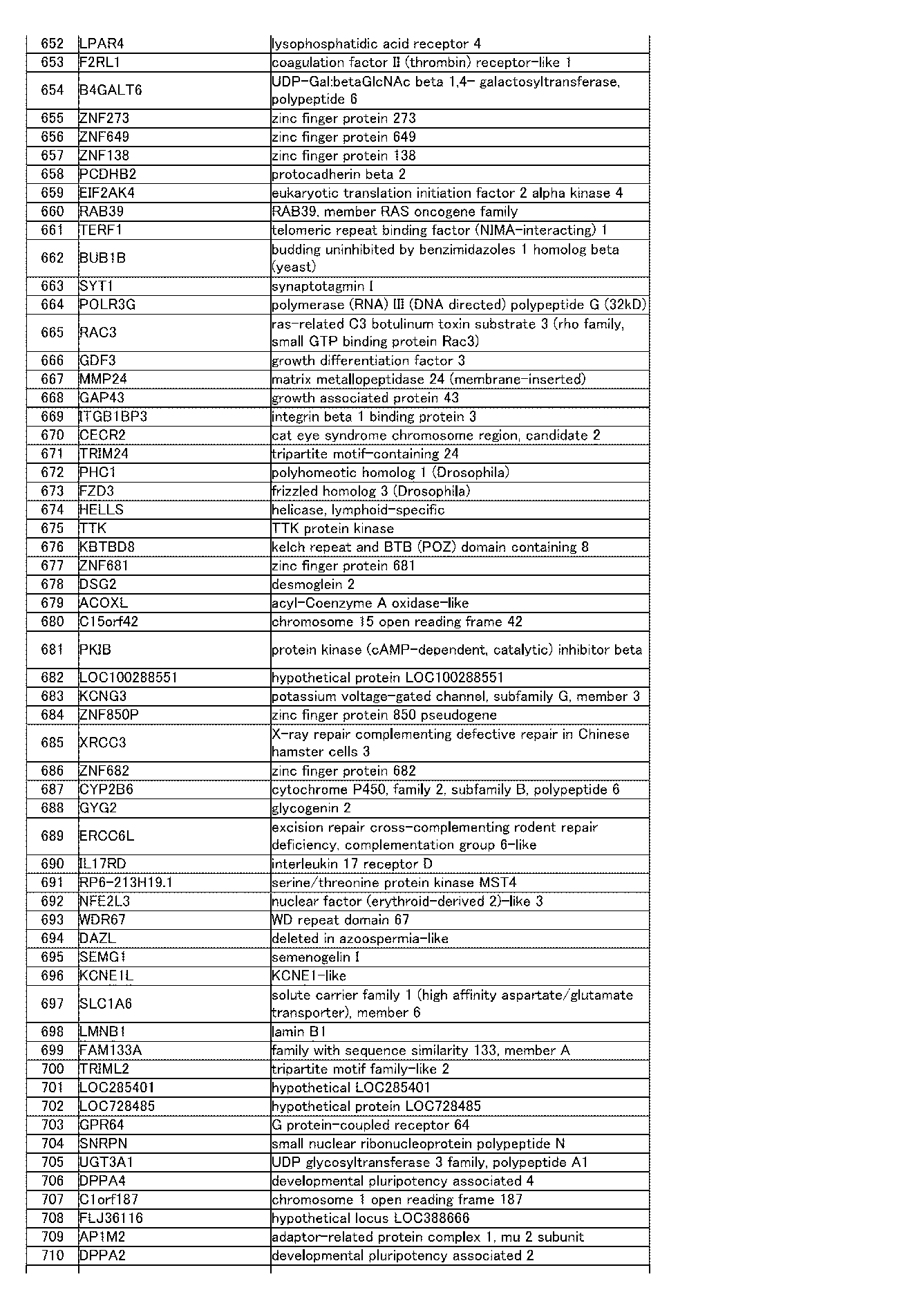


[表2A]第2遺伝子群(表2B)を含むプローブ1304個において、ヒトES細胞クローン(KhES1-S3)の発現レベルの平均に対する、各ES細胞クローン及び各iPS細胞クローンの発現レベルの割合

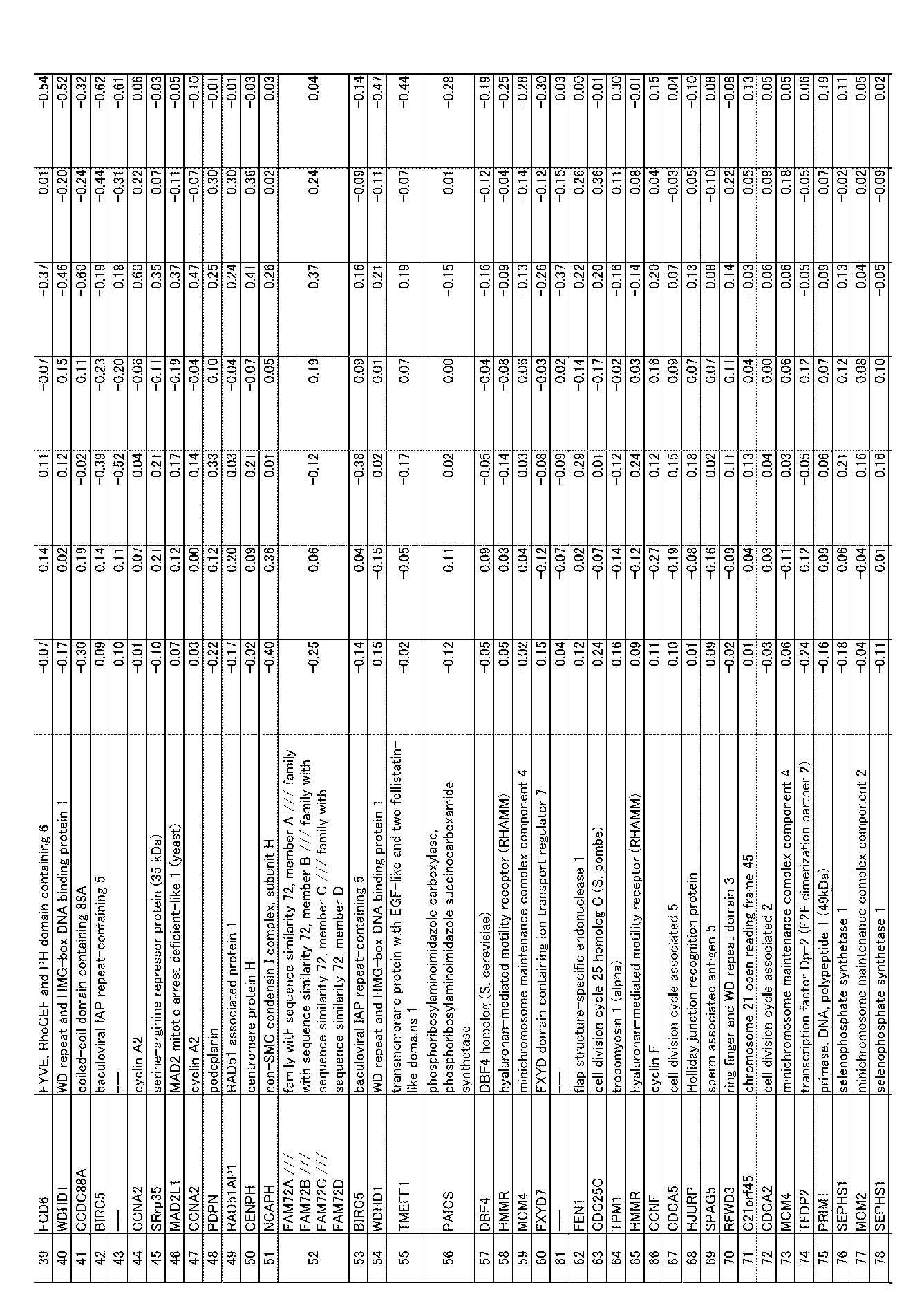

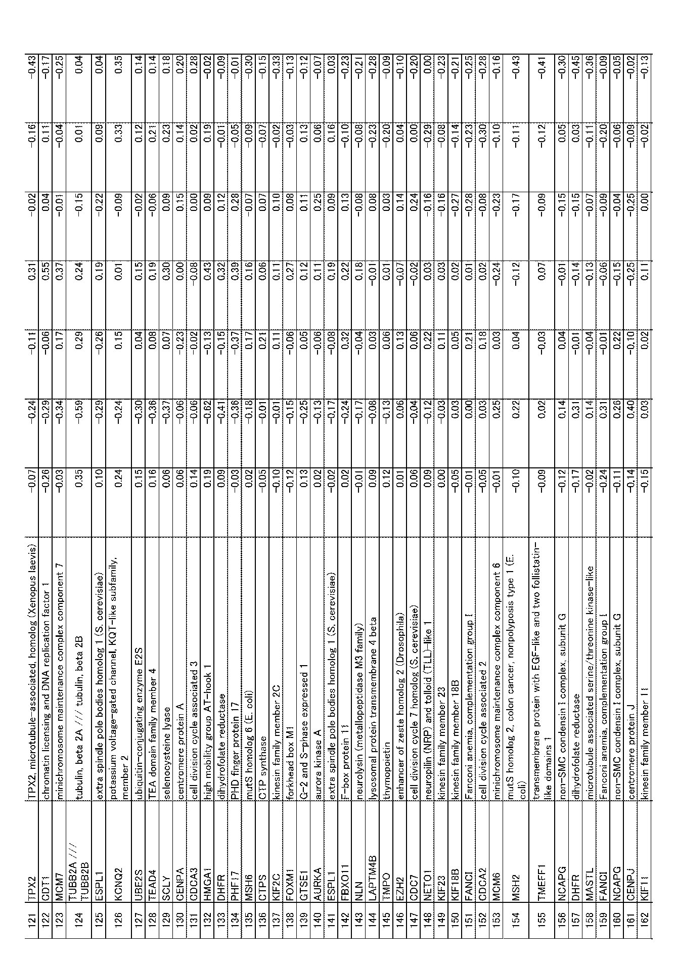
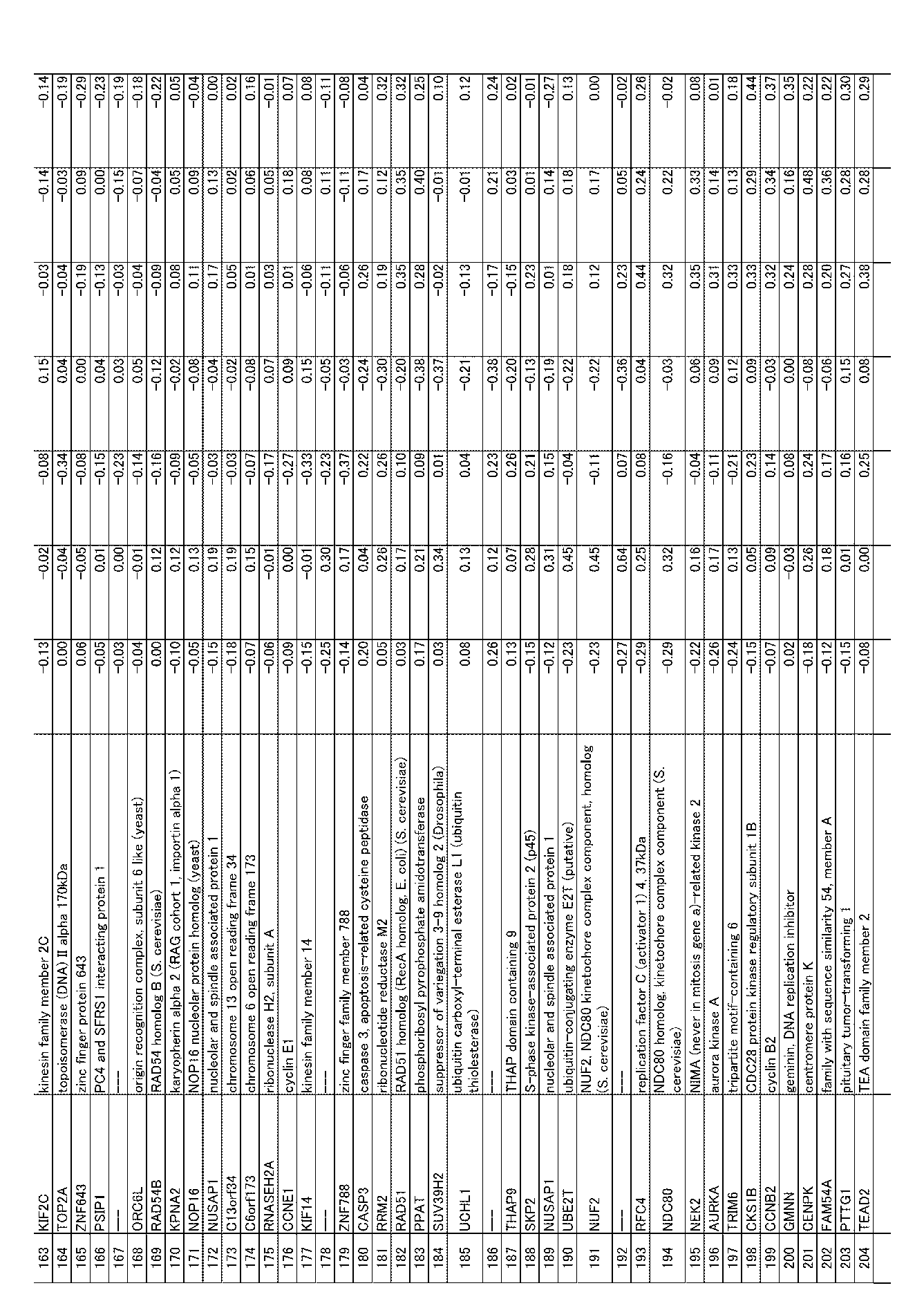
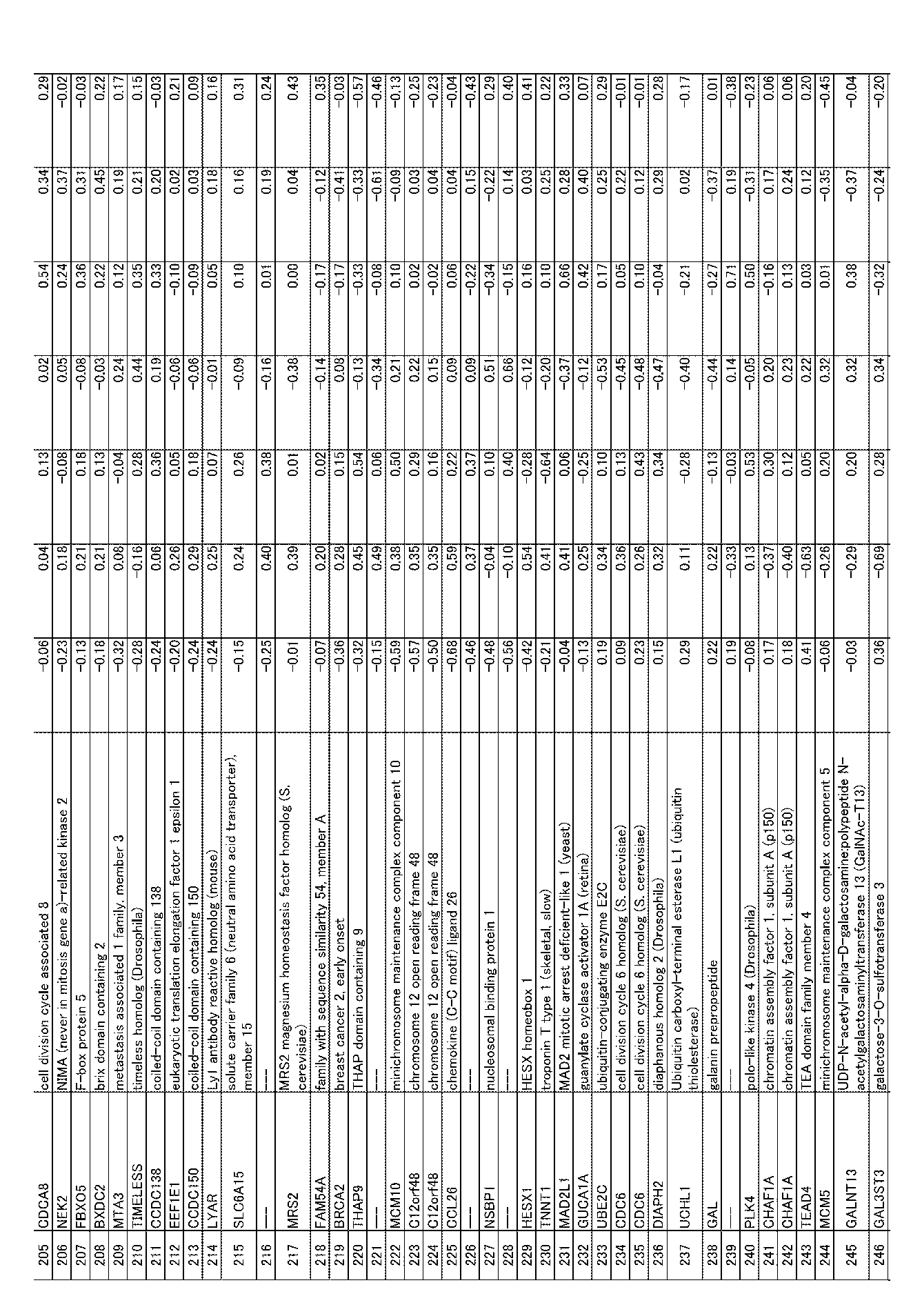

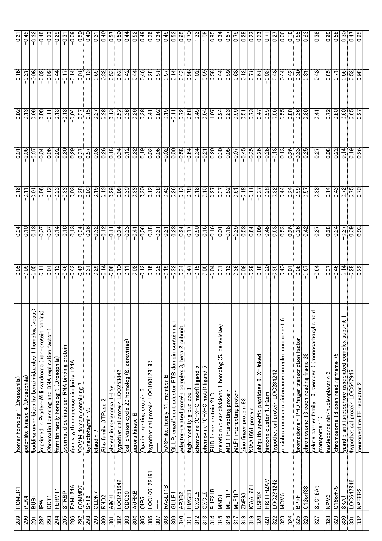

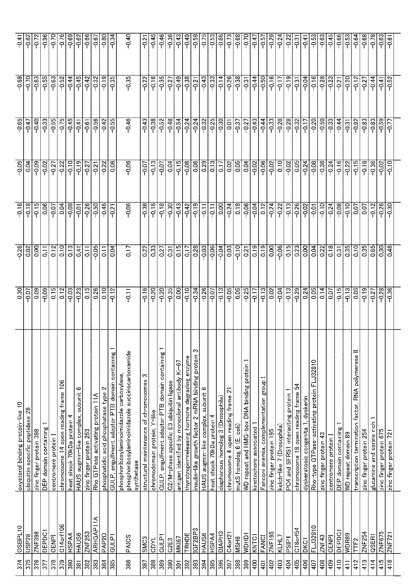


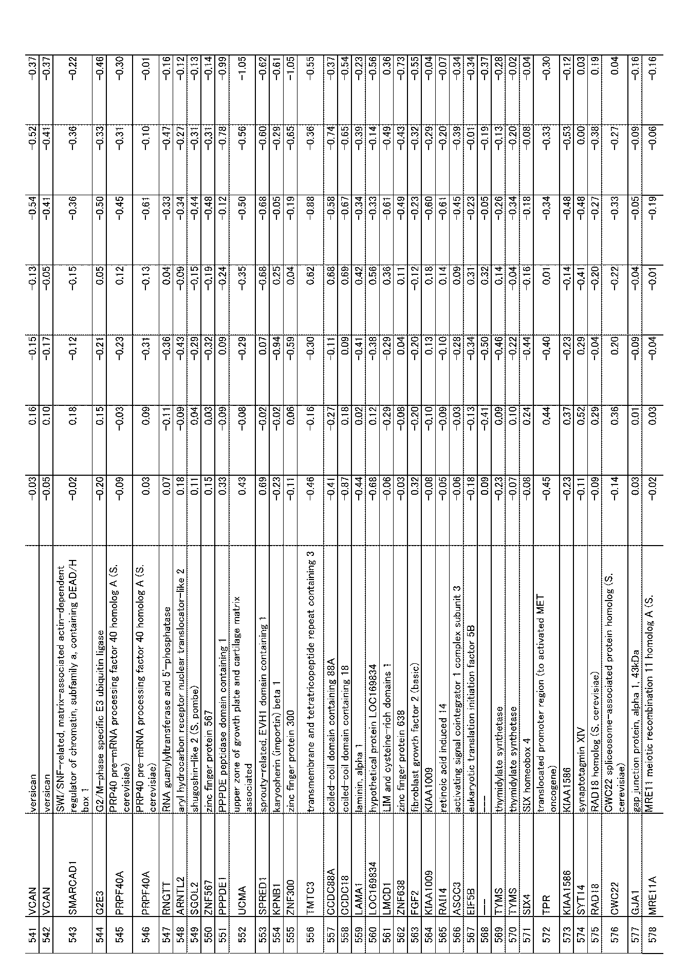
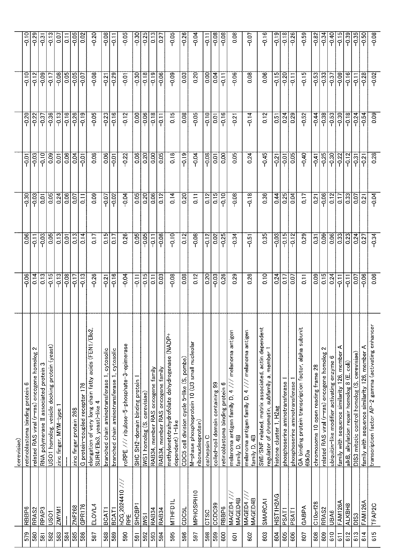
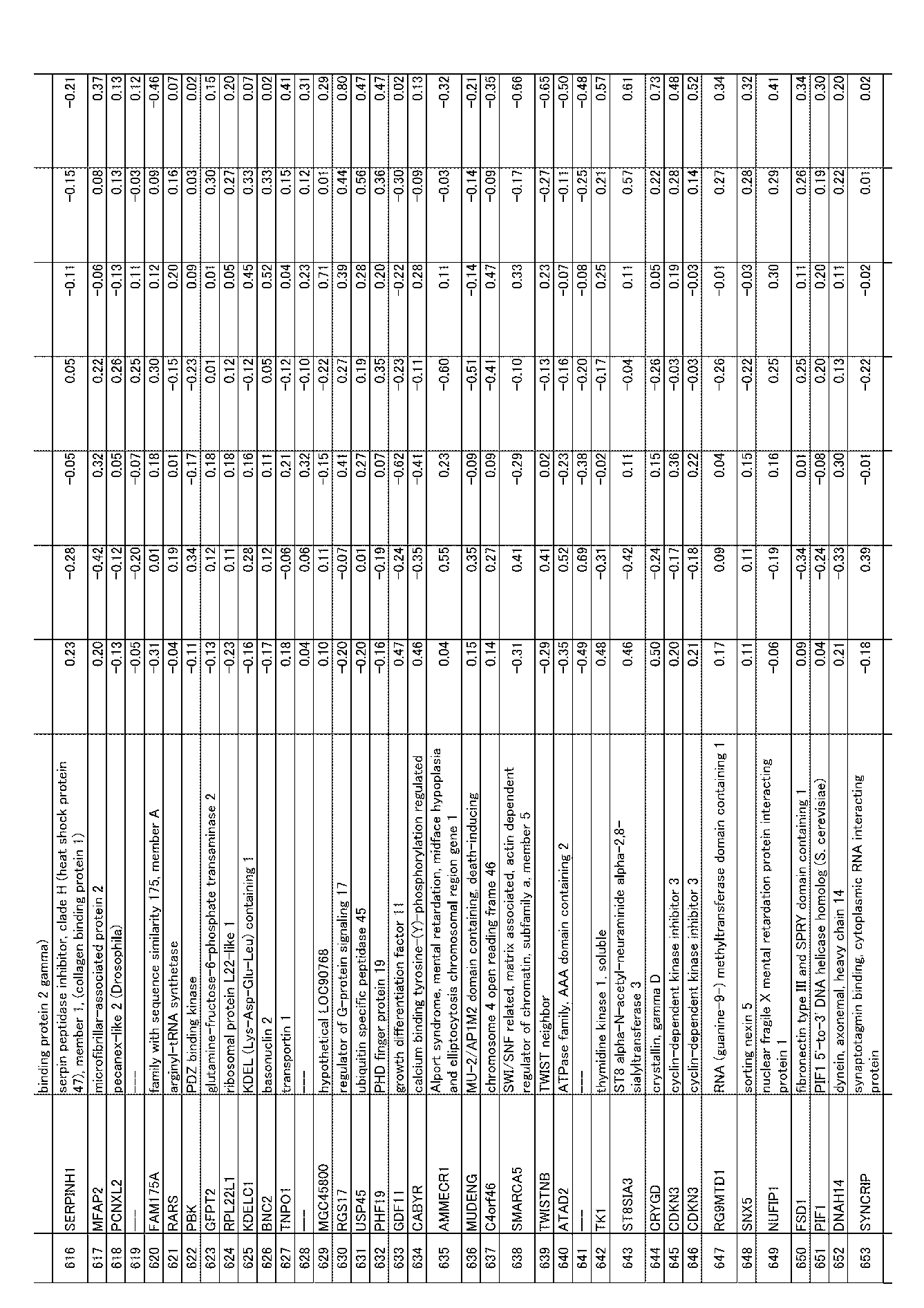


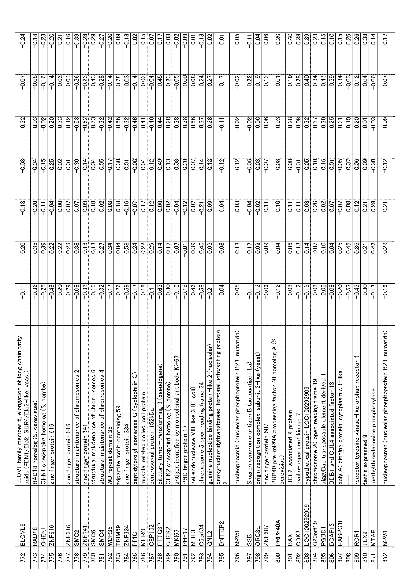
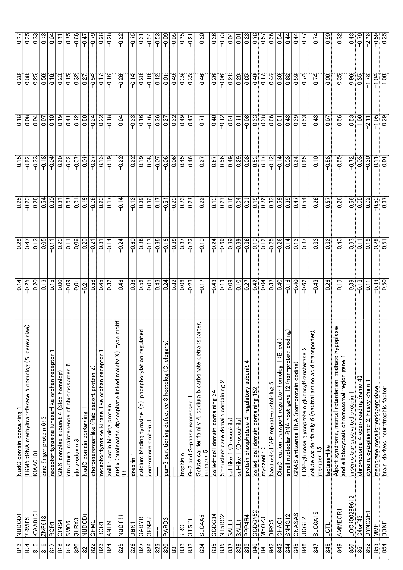

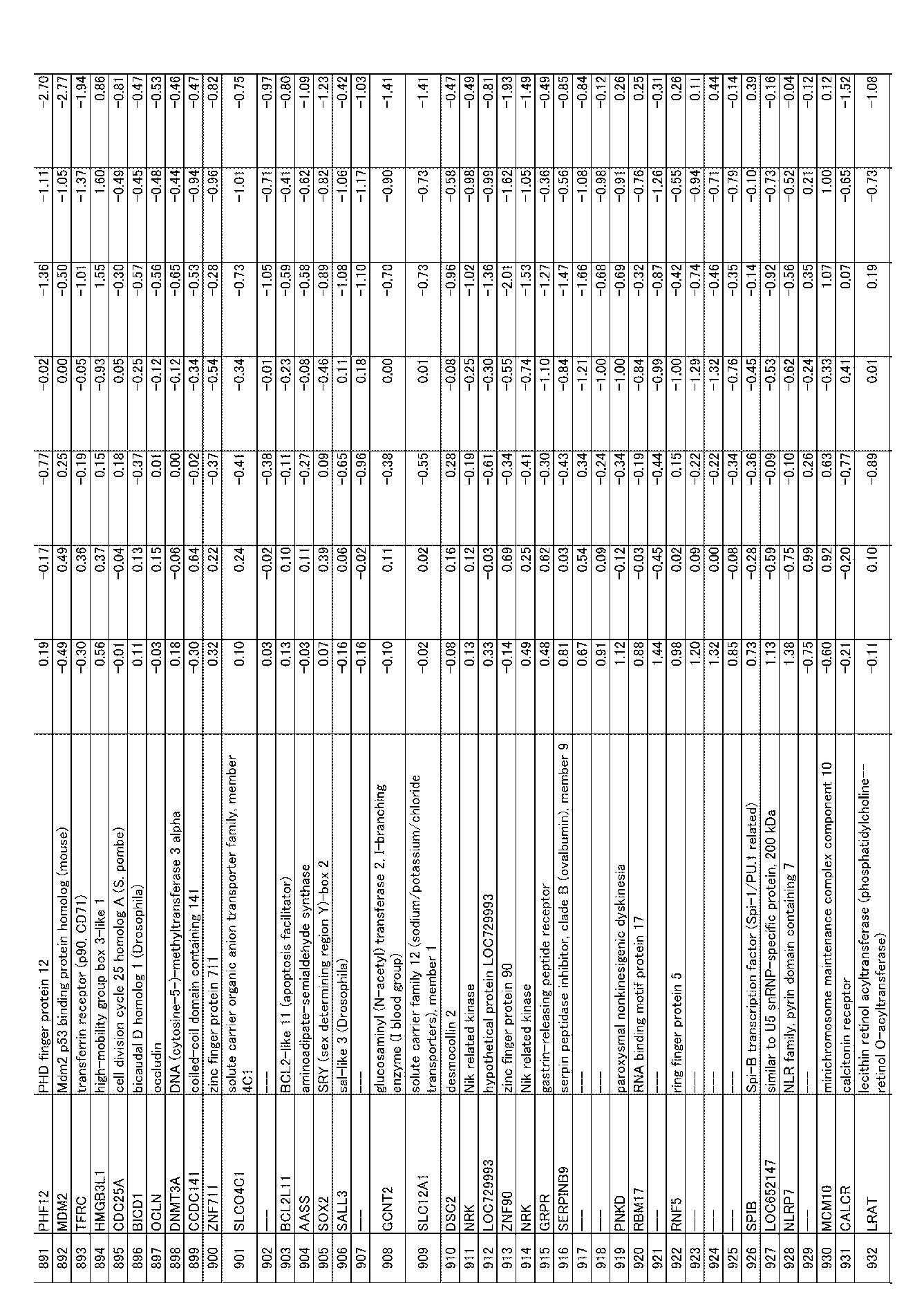


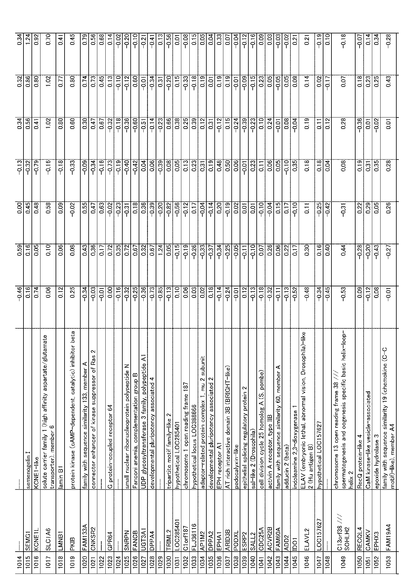

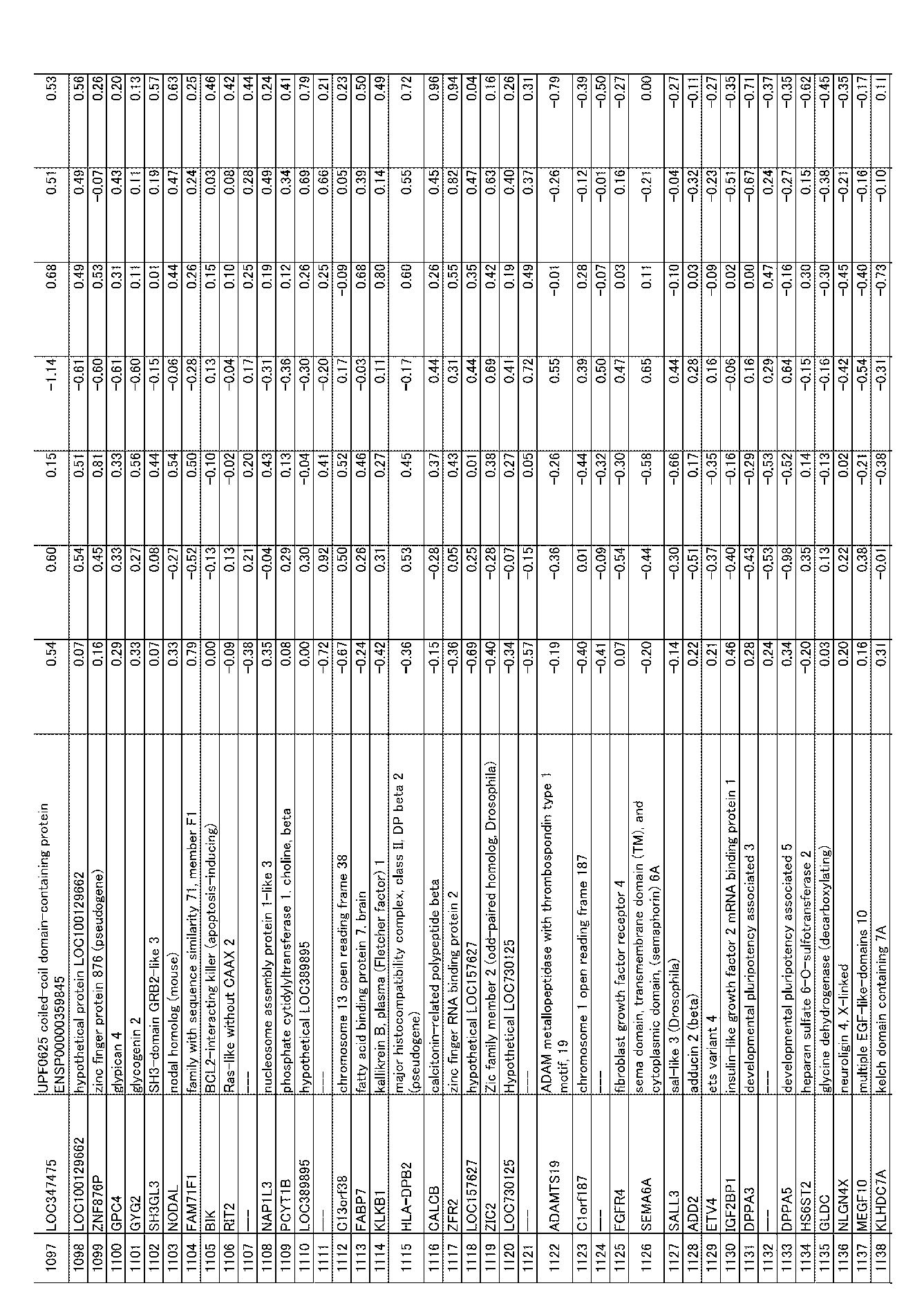
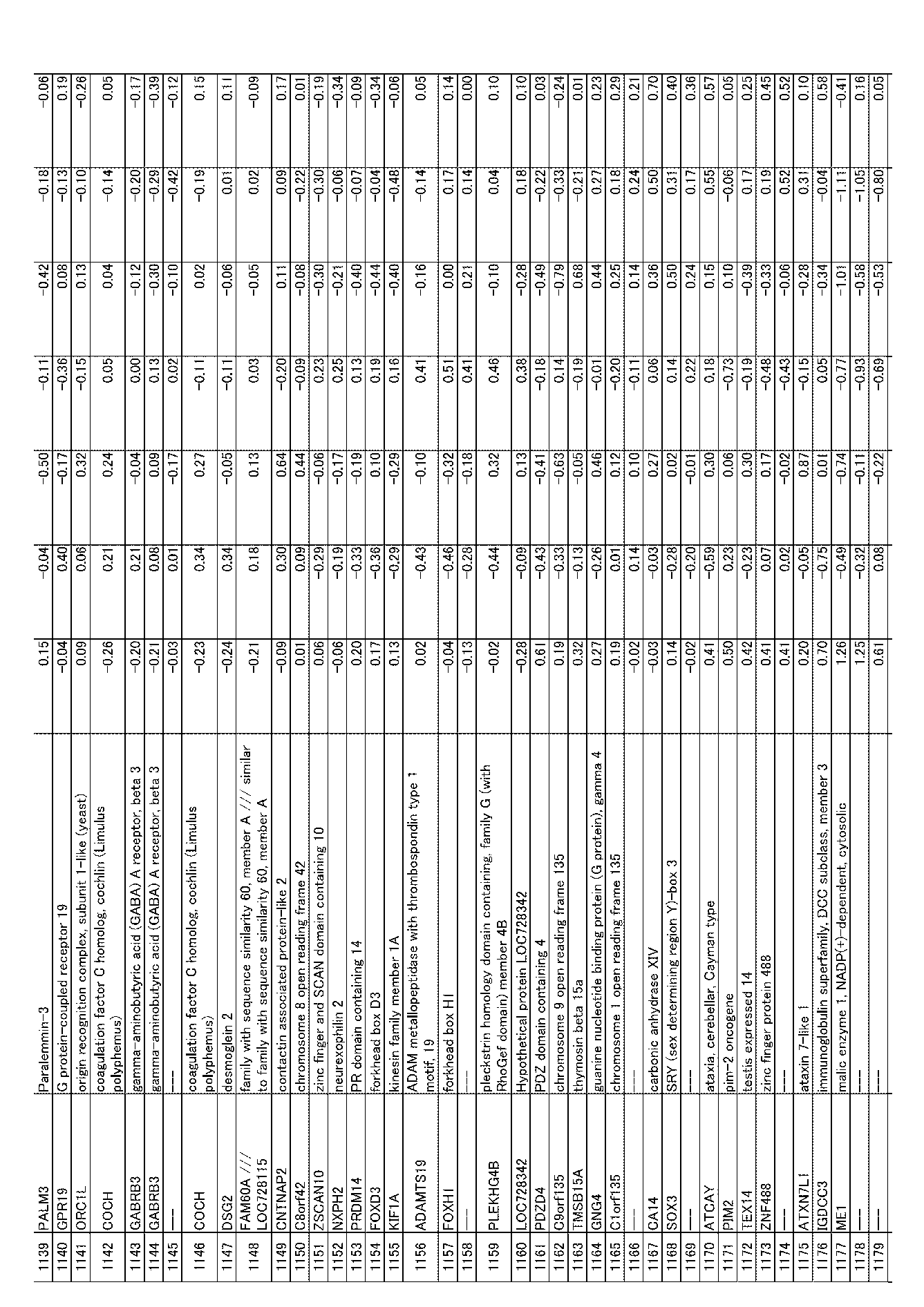


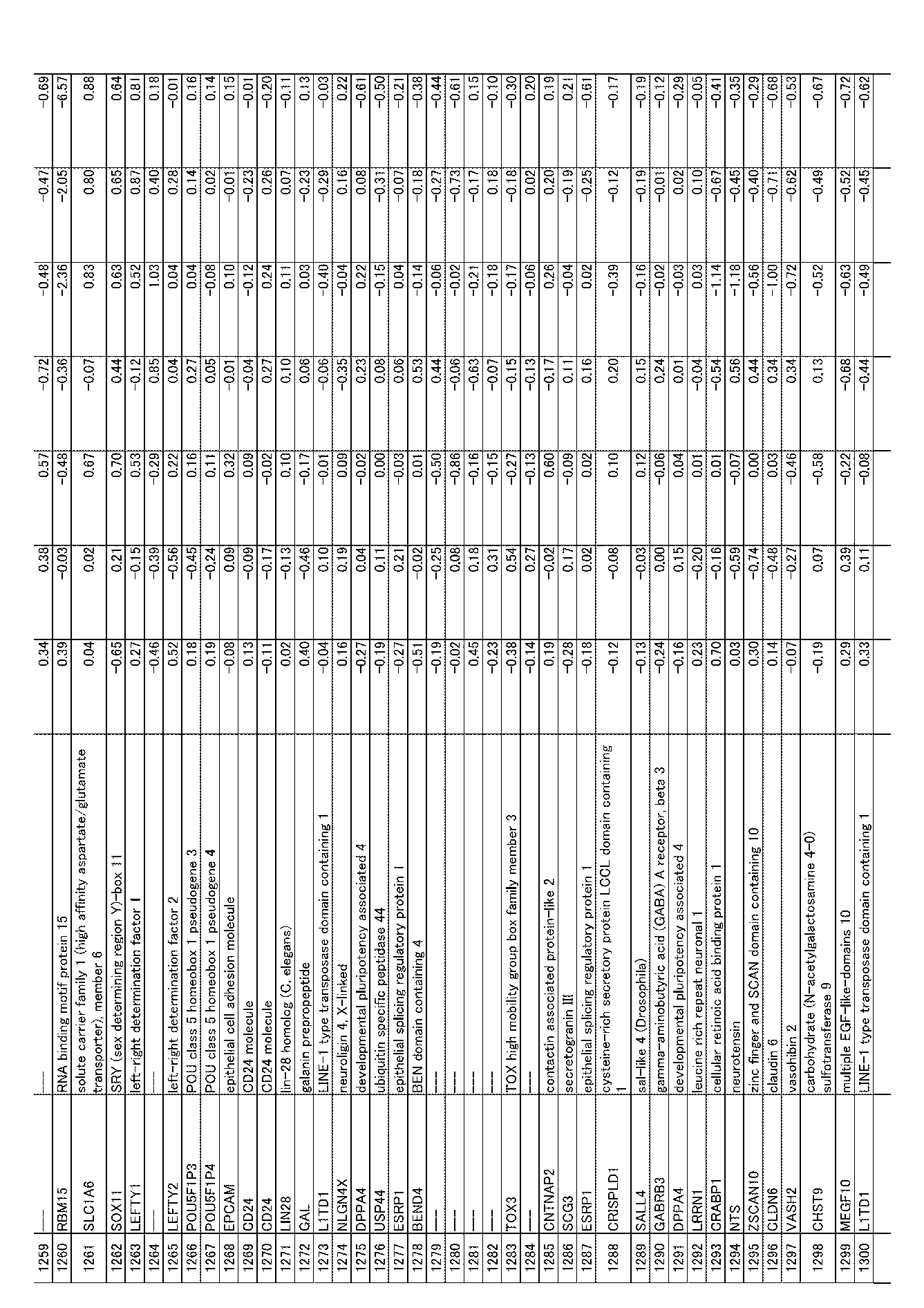

[表2B]第2遺伝子群

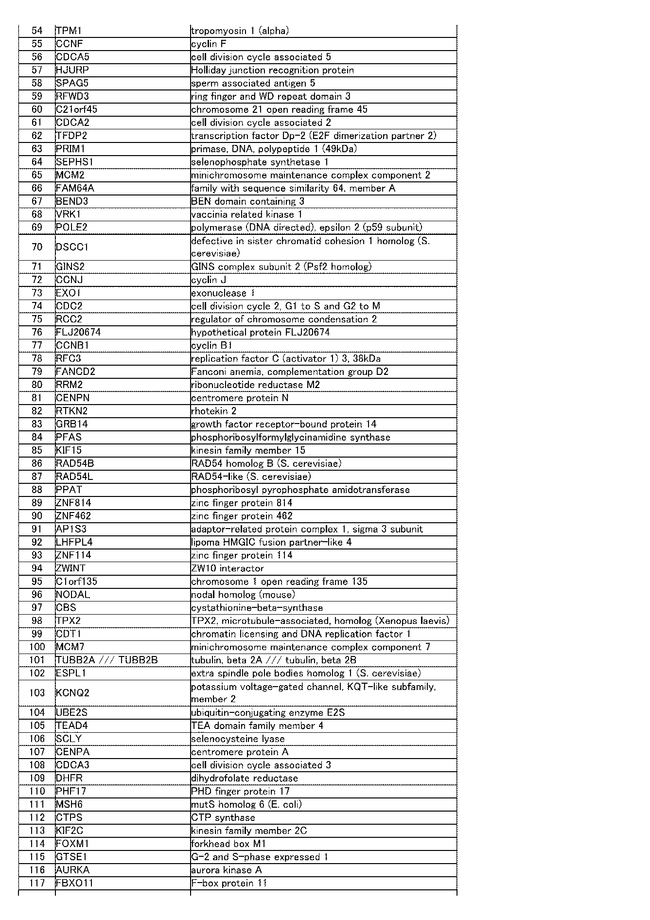
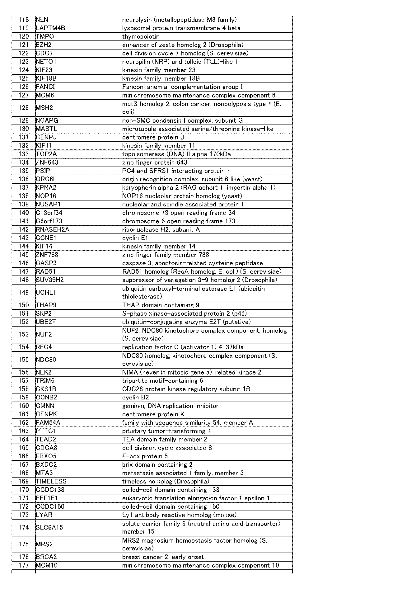


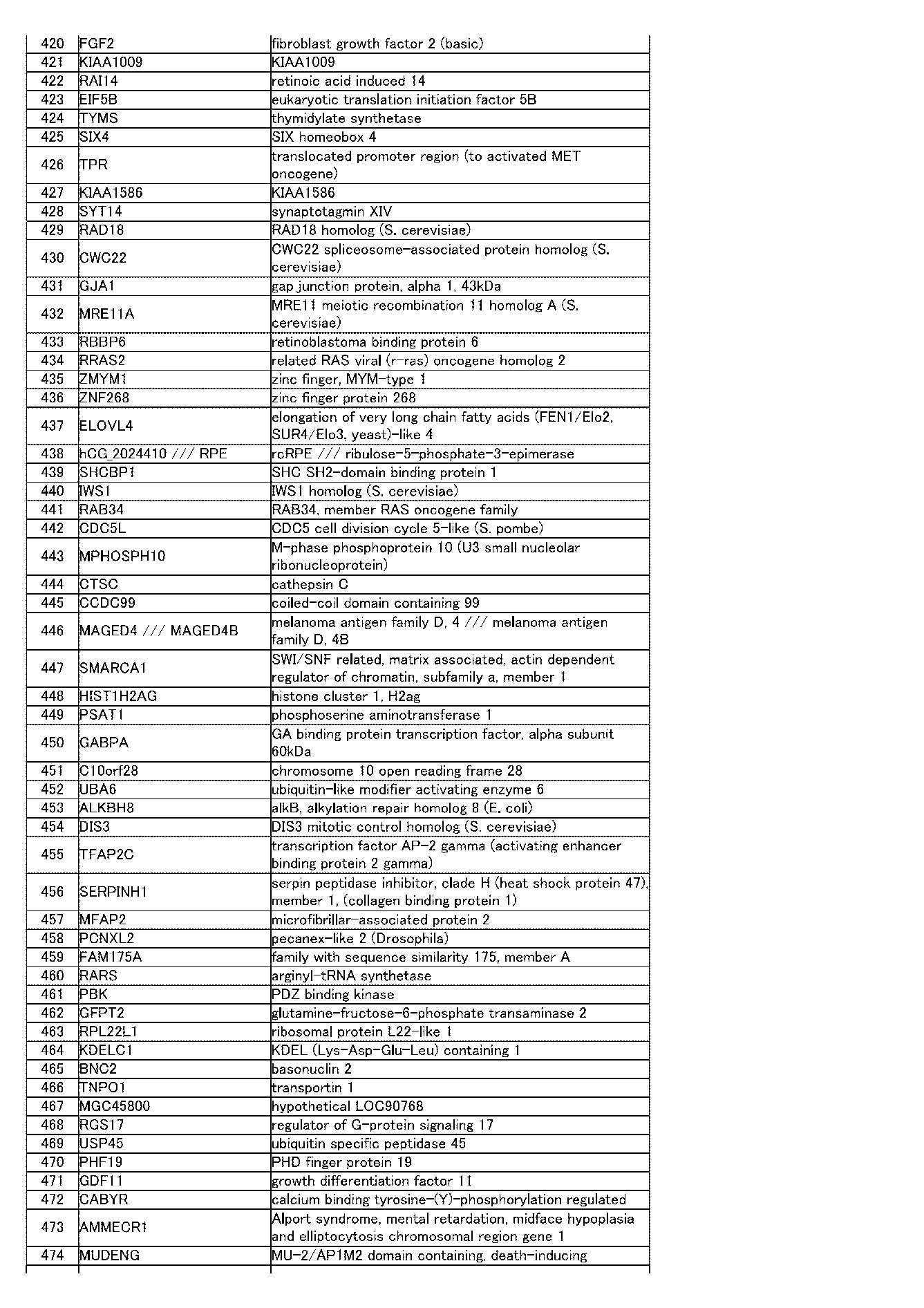
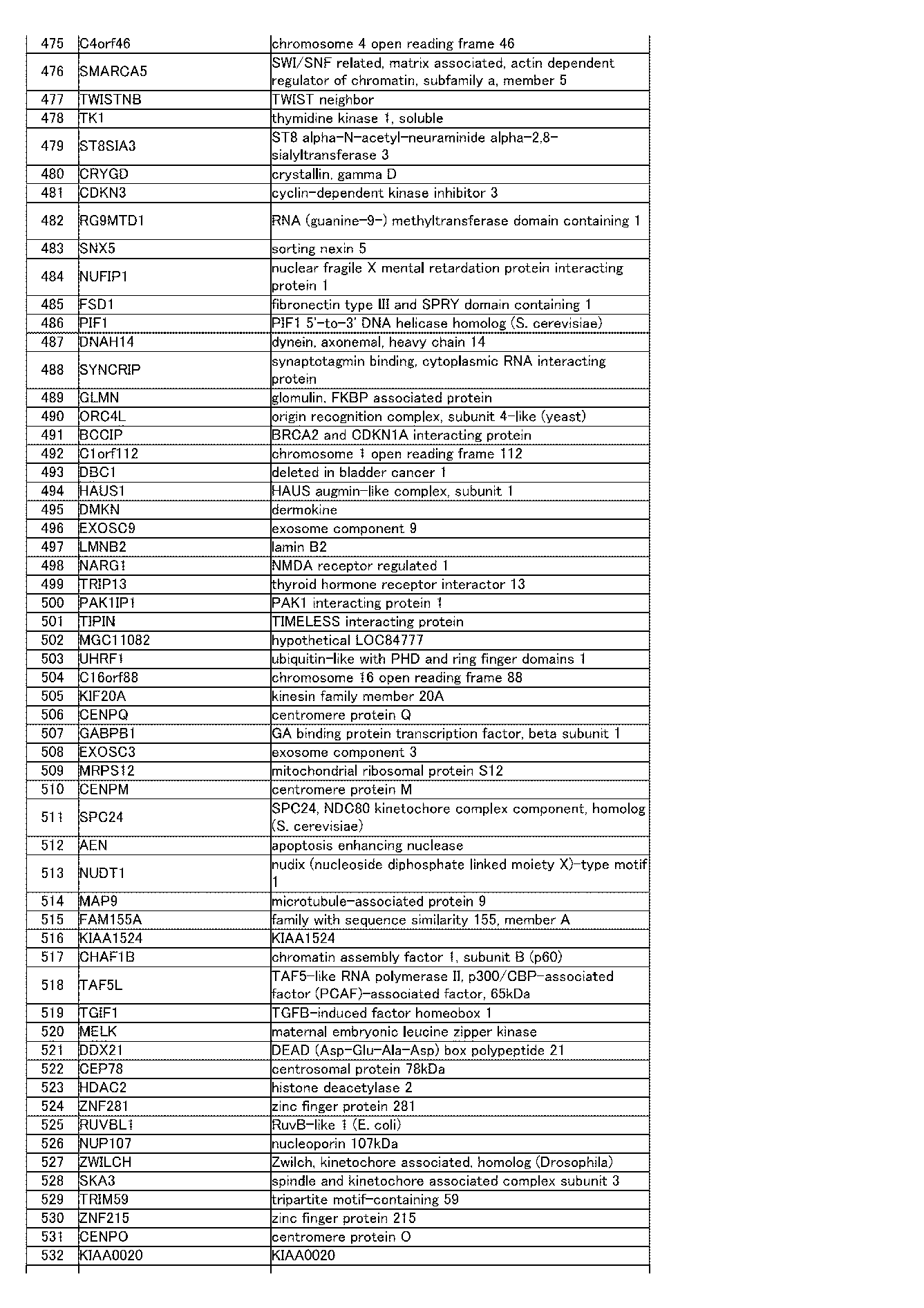
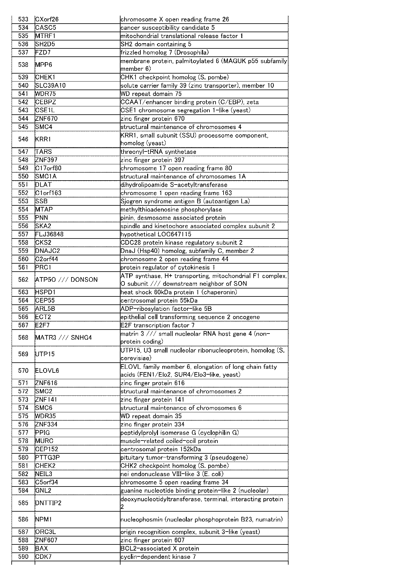
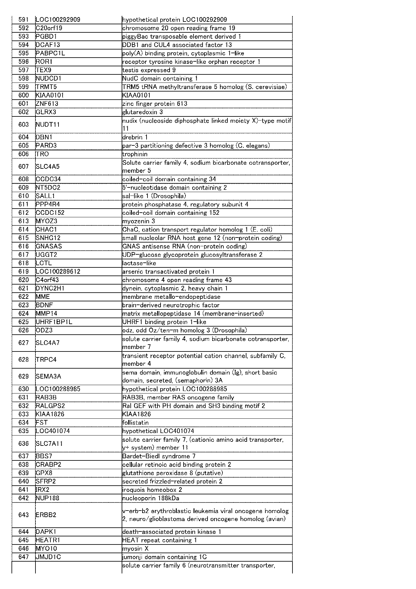


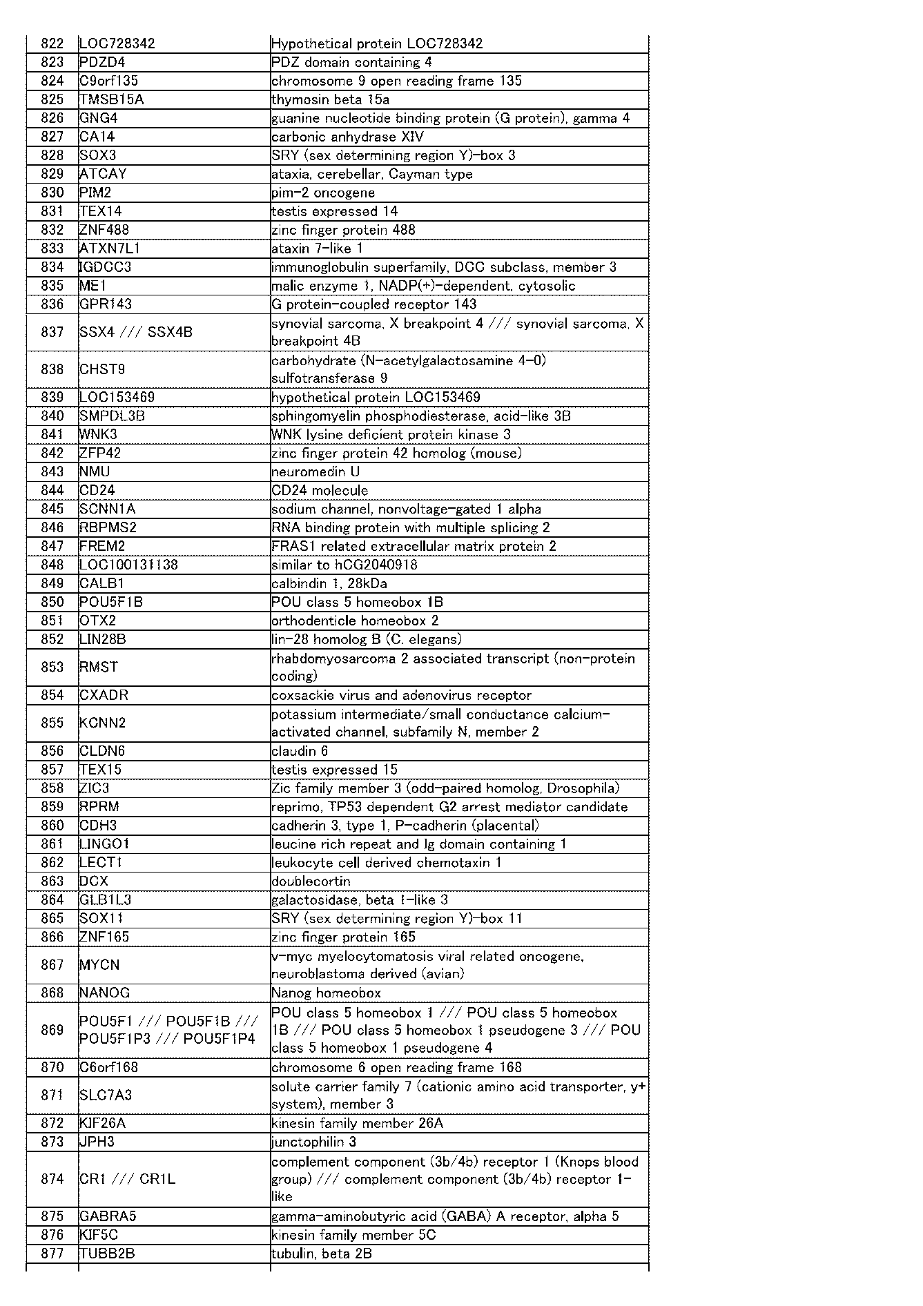
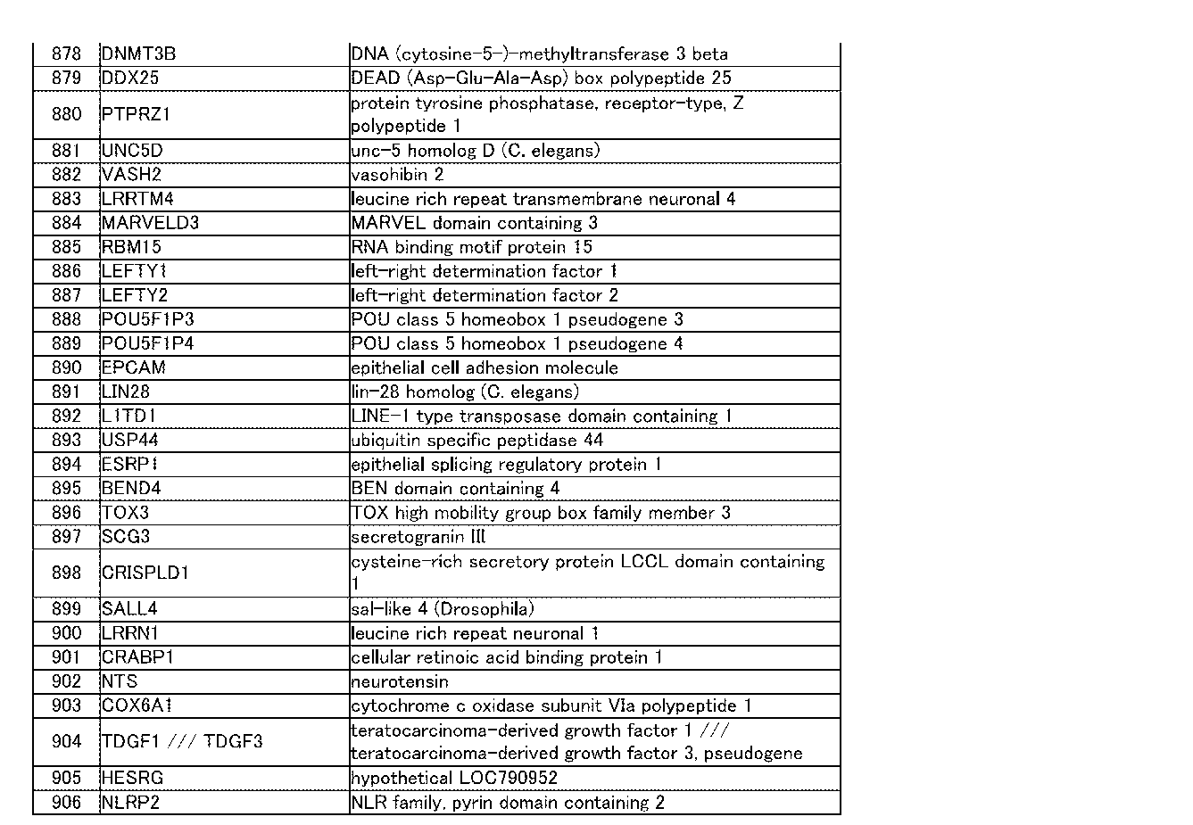
[表3A]第3遺伝子群(表3B)を含むプローブ676個において、ヒトES細胞クローン(KhES1-S3)の発現レベルの平均に対する、各ES細胞クローン及び各iPS細胞クローンの発現レベルの割合
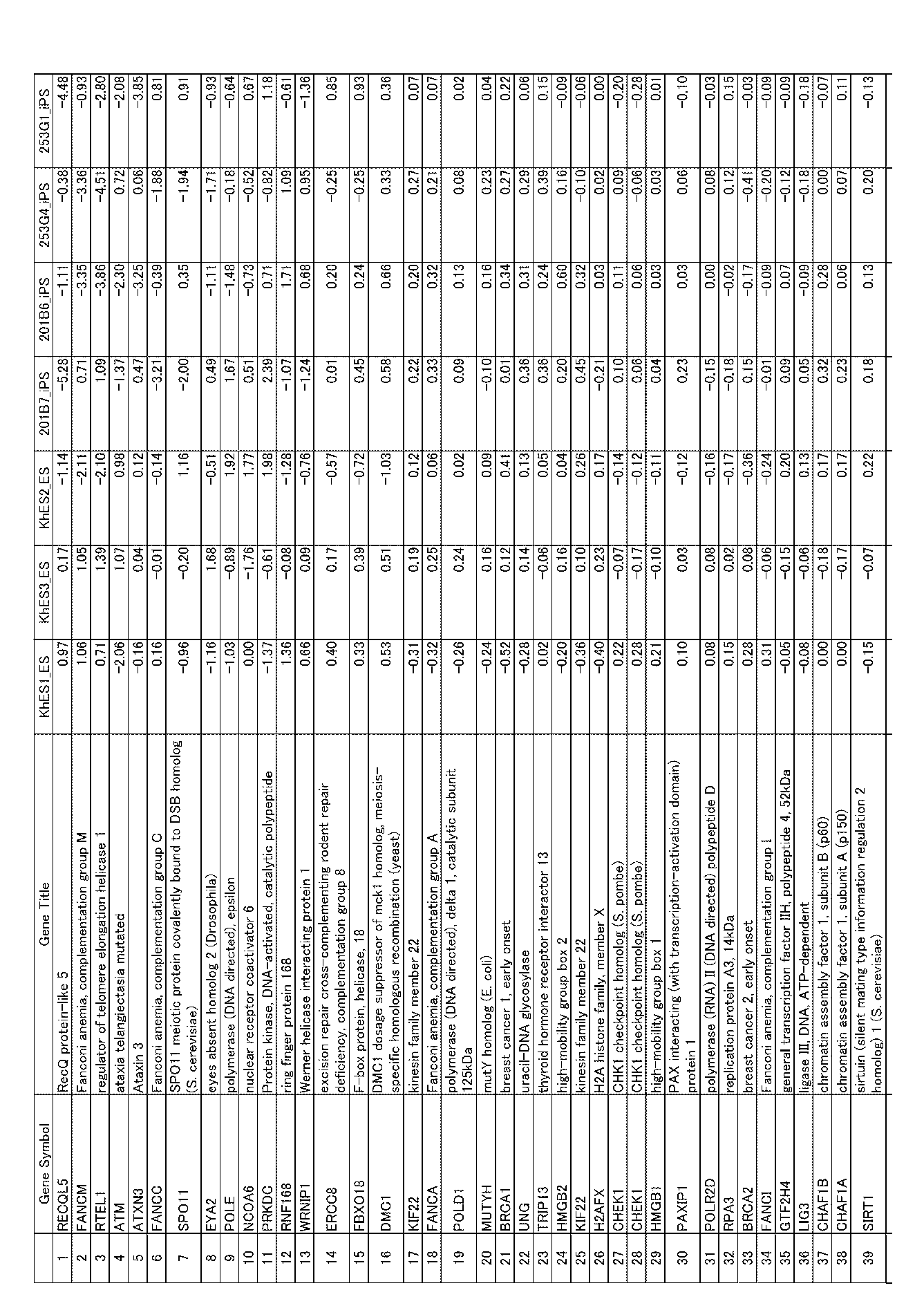


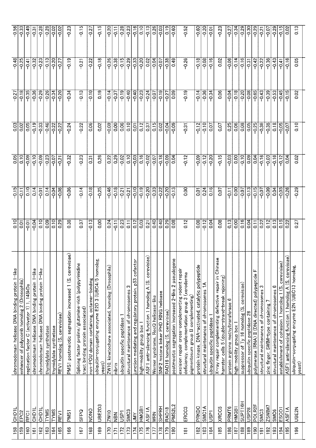



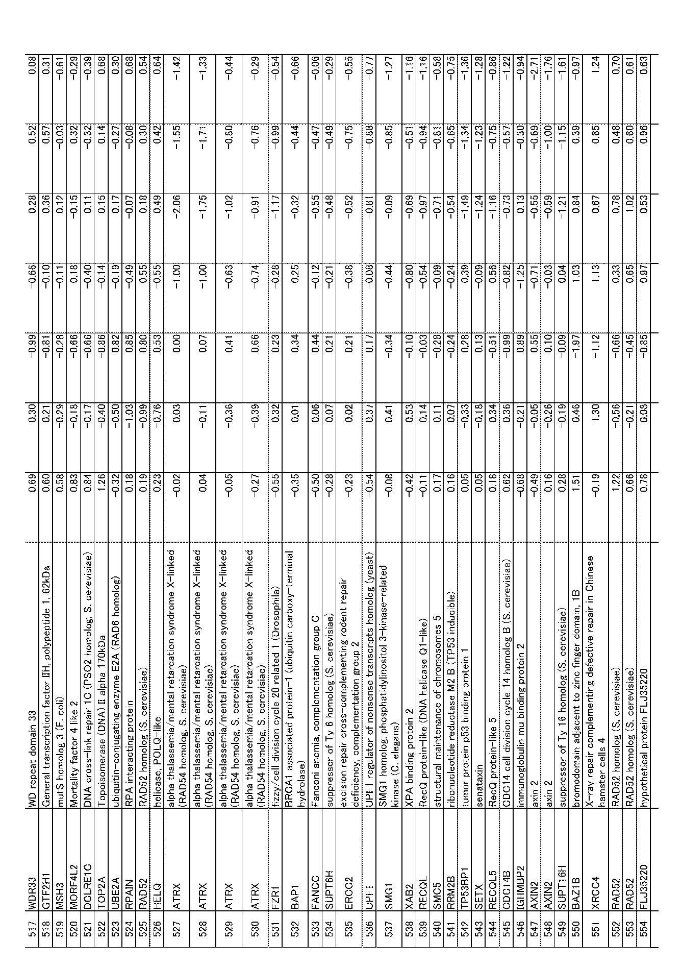


[表3B]第3遺伝子群


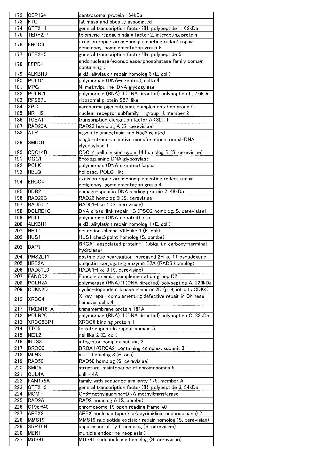


[表4A]第4遺伝子群(表4B)を含むプローブ83個において、ヒトES細胞クローン(KhES1-S3)の発現レベルの平均に対する、各ES細胞クローン及び各iPS細胞クローンの発現レベルの割合

[表4B]第4遺伝子群
[表5A]第5遺伝子群を含むプローブ80個において、ヒトES細胞クローン(KhES1-S3)の発現レベルの平均に対する、各ES細胞クローン及び各iPS細胞クローンの発現レベルの割合
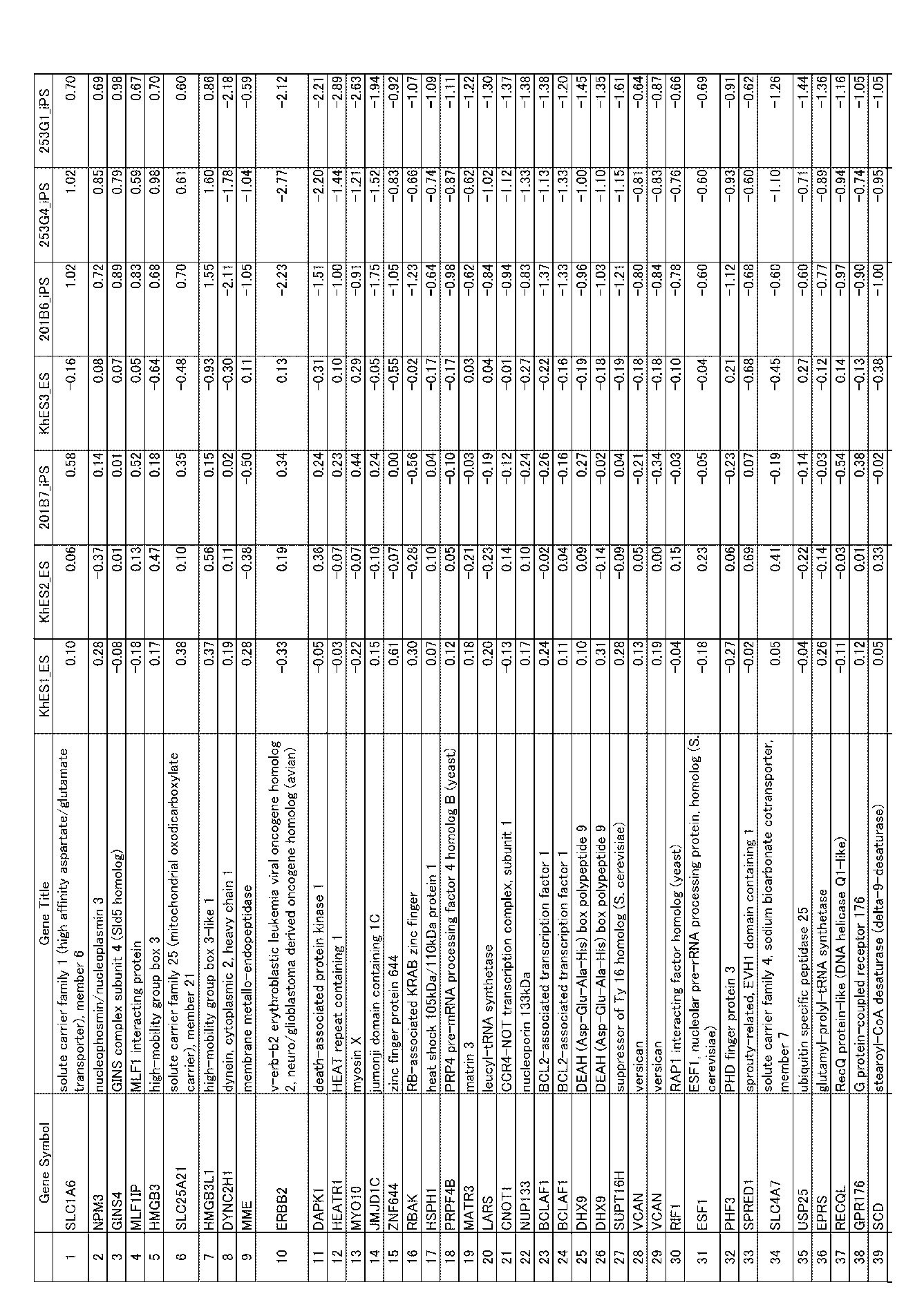


[表5B]第5遺伝子群

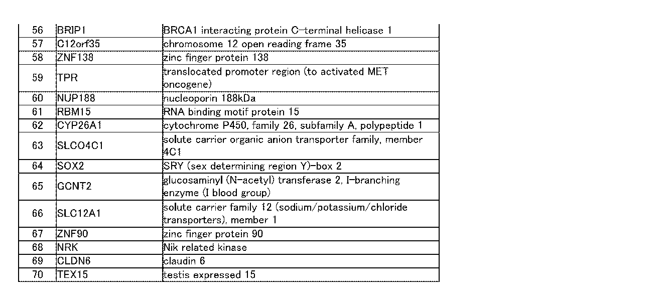
[表6A]第6遺伝子群(表6B)を含むプローブ72個において、ヒトES細胞クローン(KhES1-S3)の発現レベルの平均に対する、各ES細胞クローン及び各iPS細胞クローンの発現レベルの割合


[表6B]第6遺伝子群
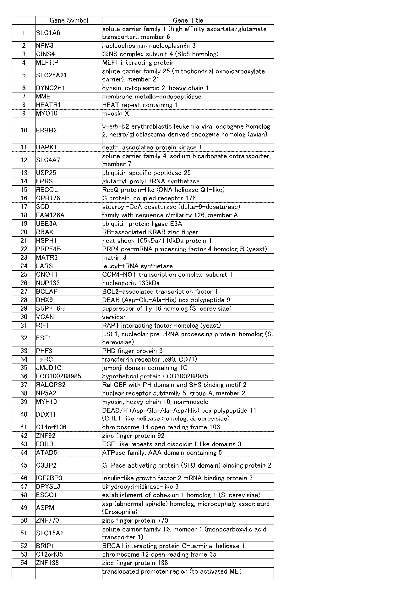

[表7A]第7遺伝子群(表7B)を含むプローブ59個において、ヒトES細胞クローン(KhES1-S3)の発現レベルの平均に対する、各ES細胞クローン及び各iPS細胞クローンの発現レベルの割合


[表7B]第7遺伝子群

[表8A]第8遺伝子群(表8B)を含むプローブ18個において、ヒトES細胞クローン(KhES1-S3)の発現レベルの平均に対する、各ES細胞クローン及び各iPS細胞クローンの発現レベルの割合

[表8B]第8遺伝子群

[表9A]第9遺伝子群(表9B)を含むプローブ13個において、ヒトES細胞クローン(KhES1-S3)の発現レベルの平均に対する、各ES細胞クローン及び各iPS細胞クローンの発現レベルの割合

[表9B]第9遺伝子群

[表10A]第10遺伝子群(表10B)を含むプローブ5個において、ヒトES細胞クローン(KhES1-S3)の発現レベルの平均に対する、各ES細胞クローン及び各iPS細胞クローンの発現レベルの割合

[表10B]第10遺伝子群

[表11A]第11遺伝子群(表11B)を含むプローブ27個において、ヒトES細胞クローン(KhES1-S3)の発現レベルの平均に対する、各ES細胞クローン及び各iPS細胞クローンの発現レベルの割合
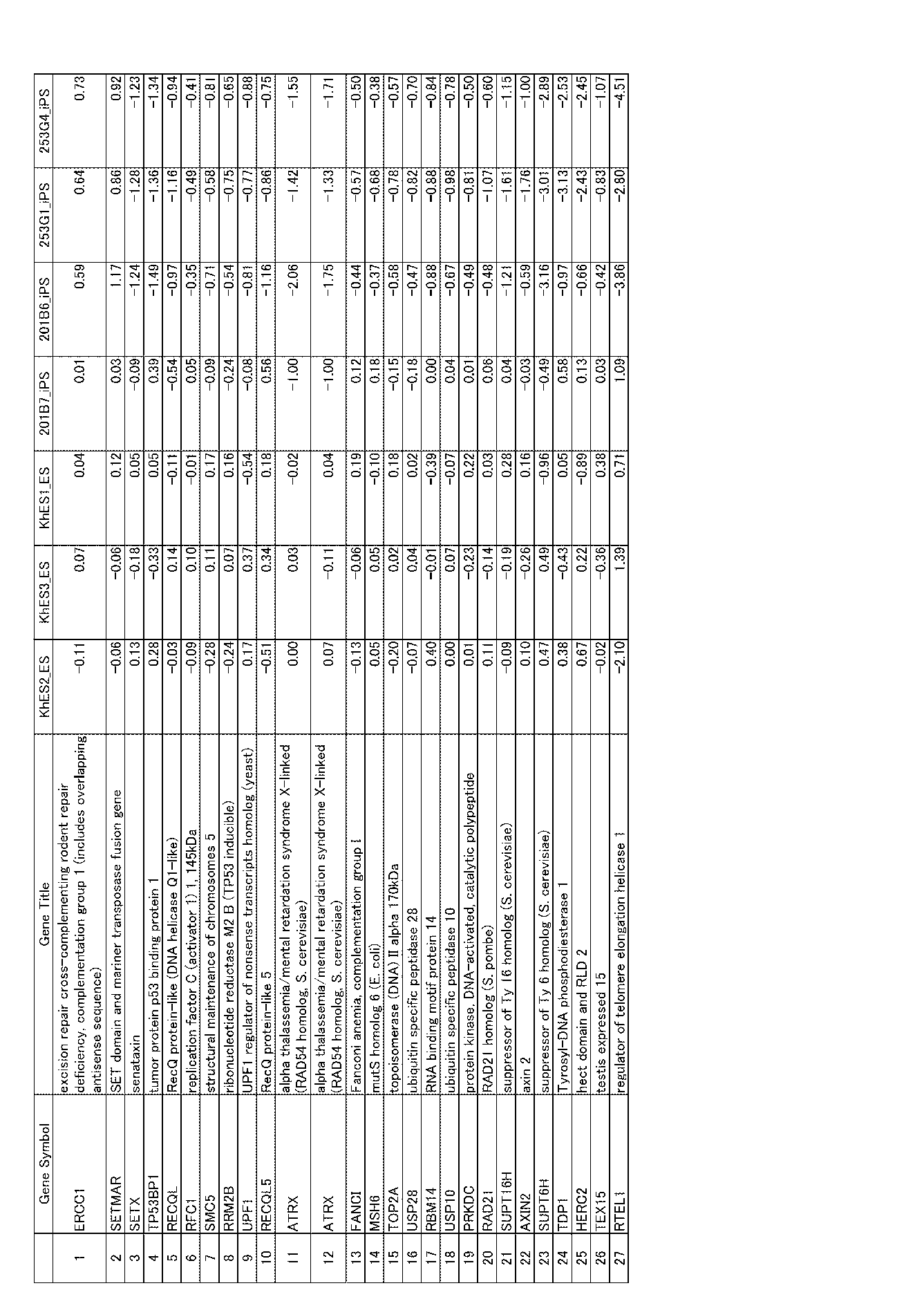
[表11B]第11遺伝子群
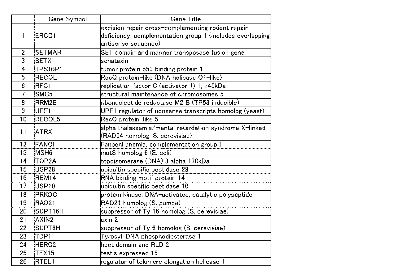
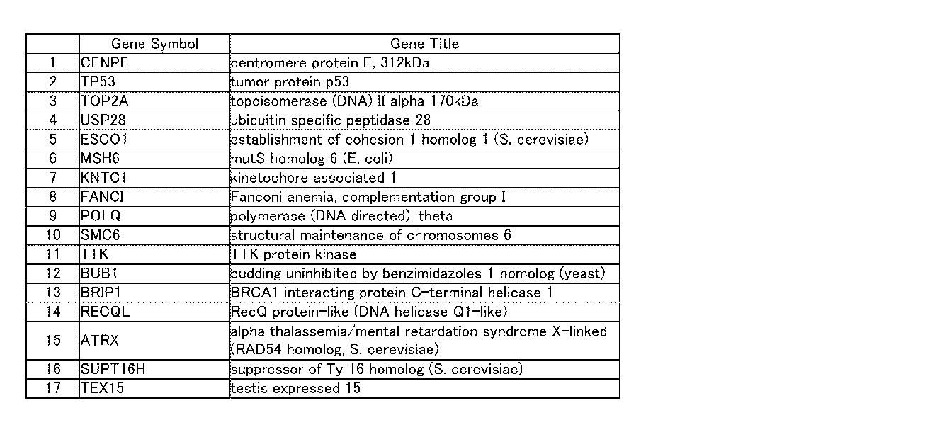
[表12A]第12遺伝子群(表12B)を含むプローブ19個において、ヒトES細胞クローン(KhES1-S3)の発現レベルの平均に対する、各ES細胞クローン及び各iPS細胞クローンの発現レベルの割合

[表12B]第12遺伝子群
本発明によって、分化させても腫瘍化が起きにくいiPS細胞クローンの選択方法、及びその選択方法に用いる遺伝子の選択方法が提供できるようになった。
Claims (14)
- iPS細胞クローンの選択方法であって,
未分化状態のiPS細胞クローンにおいて、第1~第12遺伝子群のいずれかの遺伝子群において、少なくとも一つの遺伝子について、発現レベルを調べる工程、
を含む方法。 - 請求項1に記載の選択方法であって,
前記発現レベルを、正常に分化できないか、あるいは分化しても腫瘍化を起こしやすいiPS細胞クローンにおける当該遺伝子の発現レベル、及び、腫瘍化を起こしにくいiPS細胞クローンにおける当該遺伝子の発現レベルと比較する工程、
をさらに含む方法。 - 請求項1に記載の選択方法であって,
前記iPS細胞クローンにおいて、第1~第12遺伝子群のいずれかの遺伝子群に属する全ての遺伝子の発現レベルを調べる工程と,
調べた遺伝子群において、発現レベルに異常のある遺伝子の個数を調べる工程と、
調べた遺伝子の個数を、正常に分化できないか、あるいは分化しても腫瘍化を起こしやすいiPS細胞クローンにおける発現レベルに異常のある遺伝子の個数、及び、腫瘍化を起こしにくいiPS細胞クローンにおける発現レベルに異常のある遺伝子の個数と比較する工程、
をさらに含む方法。 - 請求項3に記載の選択方法であって、
腫瘍化を起こしにくい多能性幹細胞クローンの発現レベルと比較することによって、前記発現レベルが異常かどうか判断することを特徴とする、方法。 - 請求項1に記載の選択方法であって,
前記iPS細胞クローンにおいて、第1~第12遺伝子群のいずれかの遺伝子群に属する全ての遺伝子の発現レベルを調べる工程と,
調べた遺伝子群について、前記iPS細胞クローンにおける発現レベルと、正常に分化できないか、あるいは分化しても腫瘍化を起こしやすいiPS細胞クローンにおける発現レベルと、腫瘍化を起こしやすいiPS細胞クローンにおける発現レベルとを指標にして、iPS細胞クローンのclustering分析を行う工程と、
当該iPS細胞クローンが、正常に分化できないか、あるいは分化しても腫瘍化を起こしやすいiPS細胞クローンと、腫瘍化を起こしにくいiPS細胞クローンのいずれに分類されるかを調べる工程と、
を含む方法。 - 請求項5に記載の選択方法であって、
腫瘍化を起こしにくい多能性幹細胞クローンの発現レベルとの相対値を指標にして、前記iPS細胞クローンのclustering解析を行うことを特徴とする、請求項7に記載の方法。 - iPS細胞クローンを選択するために発現レベルを調べる遺伝子の選択方法であって、
正常に分化できないか、あるいは分化しても腫瘍化を起こしやすい複数の第1のiPS細胞クローンと、腫瘍化を起こしにくい複数の第2のiPS細胞クローンにおいて、未分化状態の各iPS細胞クローンで、1以上の遺伝子を含む候補遺伝子群に属する遺伝子の発現レベルを調べる工程、
を含む方法。 - 前記候補遺伝子群に属する遺伝子が、多能性幹細胞で分化細胞より発現の高い遺伝子、及びDNA修復に関与する遺伝子であることを特徴とする、請求項7に記載の方法。
- 前記候補遺伝子群に属する遺伝子が、第1遺伝子群及び第2遺伝子群のいずれかに属する遺伝子であることを特徴とする、請求項7に記載の方法。
- 請求項7に記載の選択方法であって、
複数の第1のiPS細胞クローンにおいて得られた第1の発現レベルと、複数の第2のiPS細胞クローンにおいて得られた第2の発現レベルとを比較する工程と、
第1の発現レベルと第2の発現レベルとが異なるかどうか調べる工程と、
を含む方法。 - 請求項7に記載の選択方法であって、
複数の第1のiPS細胞クローンにおいて得られた発現レベルにおいて、発現レベルに異常のある遺伝子の第1の個数と、複数の第2のiPS細胞クローンにおいて得られた発現レベルのうち、発現レベルに異常のある遺伝子の第2の個数とを比較する工程と、
第1の個数と第2の個数とが異なるかどうか調べる工程と、
を含む方法。 - 請求項11に記載の選択方法であって、
腫瘍化を起こしにくい多能性幹細胞クローンの発現レベルと比較することによって、前記発現レベルが異常かどうか判断することを特徴とする、方法。 - 請求項7に記載の選択方法であって、
前記iPS細胞クローンについて、前記発現レベルを指標にしてclustering分析を行う工程と、
複数の第1のiPS細胞クローンと複数の第2のiPS細胞クローンが、それぞれ別のclusterに分類されるかどうか調べる工程と、
を含む方法。 - 請求項13に記載の選択方法であって、
腫瘍化を起こしにくい多能性幹細胞クローンの発現レベルとの相対値を指標にして、前記iPS細胞クローンについてclustering分析を行うことを特徴とする、方法。
Priority Applications (1)
| Application Number | Priority Date | Filing Date | Title |
|---|---|---|---|
| JP2013501163A JP6273842B2 (ja) | 2011-02-25 | 2012-02-27 | iPS細胞クローンの選択方法、及びその選択方法に用いる遺伝子の選択方法 |
Applications Claiming Priority (4)
| Application Number | Priority Date | Filing Date | Title |
|---|---|---|---|
| JP2011-040979 | 2011-02-25 | ||
| JP2011040979 | 2011-02-25 | ||
| US201161466298P | 2011-03-22 | 2011-03-22 | |
| US61/466,298 | 2011-03-22 |
Publications (1)
| Publication Number | Publication Date |
|---|---|
| WO2012115270A1 true WO2012115270A1 (ja) | 2012-08-30 |
Family
ID=46721037
Family Applications (1)
| Application Number | Title | Priority Date | Filing Date |
|---|---|---|---|
| PCT/JP2012/054800 Ceased WO2012115270A1 (ja) | 2011-02-25 | 2012-02-27 | iPS細胞クローンの選択方法、及びその選択方法に用いる遺伝子の選択方法 |
Country Status (2)
| Country | Link |
|---|---|
| JP (1) | JP6273842B2 (ja) |
| WO (1) | WO2012115270A1 (ja) |
Cited By (2)
| Publication number | Priority date | Publication date | Assignee | Title |
|---|---|---|---|---|
| WO2015107888A1 (ja) | 2014-01-14 | 2015-07-23 | 国立大学法人鹿児島大学 | 幹細胞における腫瘍化原因細胞の新たな標識法と治療法 |
| WO2018235583A1 (ja) * | 2017-06-19 | 2018-12-27 | 公益財団法人神戸医療産業都市推進機構 | 多能性幹細胞の分化能の予測方法及びそのための試薬 |
Citations (4)
| Publication number | Priority date | Publication date | Assignee | Title |
|---|---|---|---|---|
| WO2010038904A1 (ja) * | 2008-09-30 | 2010-04-08 | 国立大学法人鳥取大学 | 外来性核初期化因子またはそれをコードするdnaを含まない人工多能性幹細胞およびその作製方法 |
| JP2010523117A (ja) * | 2007-04-07 | 2010-07-15 | ホワイトヘッド・インスティテュート・フォー・バイオメディカル・リサーチ | 体細胞の再プログラミング |
| JP2010528622A (ja) * | 2007-05-30 | 2010-08-26 | ザ ジェネラル ホスピタル コーポレイション | 体細胞から多能性細胞を生成する方法 |
| WO2010134388A1 (ja) * | 2009-05-20 | 2010-11-25 | Mihara Makoto | 生体情報モニタリングシステム |
Family Cites Families (1)
| Publication number | Priority date | Publication date | Assignee | Title |
|---|---|---|---|---|
| JP2007054069A (ja) * | 2005-08-24 | 2007-03-08 | Medizinische Hochschule Hannover | 自己不活性化レトロウイルスベクター |
-
2012
- 2012-02-27 JP JP2013501163A patent/JP6273842B2/ja not_active Expired - Fee Related
- 2012-02-27 WO PCT/JP2012/054800 patent/WO2012115270A1/ja not_active Ceased
Patent Citations (4)
| Publication number | Priority date | Publication date | Assignee | Title |
|---|---|---|---|---|
| JP2010523117A (ja) * | 2007-04-07 | 2010-07-15 | ホワイトヘッド・インスティテュート・フォー・バイオメディカル・リサーチ | 体細胞の再プログラミング |
| JP2010528622A (ja) * | 2007-05-30 | 2010-08-26 | ザ ジェネラル ホスピタル コーポレイション | 体細胞から多能性細胞を生成する方法 |
| WO2010038904A1 (ja) * | 2008-09-30 | 2010-04-08 | 国立大学法人鳥取大学 | 外来性核初期化因子またはそれをコードするdnaを含まない人工多能性幹細胞およびその作製方法 |
| WO2010134388A1 (ja) * | 2009-05-20 | 2010-11-25 | Mihara Makoto | 生体情報モニタリングシステム |
Non-Patent Citations (1)
| Title |
|---|
| RIPKA S. ET AL.: "Glutamate receptor GRIA3-- target of CUX1 and mediator of tumor progression in pancreatic cancer.", NEOPLASIA, vol. 12, no. 8, August 2010 (2010-08-01), pages 659 - 667 * |
Cited By (6)
| Publication number | Priority date | Publication date | Assignee | Title |
|---|---|---|---|---|
| WO2015107888A1 (ja) | 2014-01-14 | 2015-07-23 | 国立大学法人鹿児島大学 | 幹細胞における腫瘍化原因細胞の新たな標識法と治療法 |
| EP3690055A1 (en) | 2014-01-14 | 2020-08-05 | Kagoshima University | Method for labelling cancerization-causing cell in stem cells |
| WO2018235583A1 (ja) * | 2017-06-19 | 2018-12-27 | 公益財団法人神戸医療産業都市推進機構 | 多能性幹細胞の分化能の予測方法及びそのための試薬 |
| JPWO2018235583A1 (ja) * | 2017-06-19 | 2020-04-16 | 公益財団法人神戸医療産業都市推進機構 | 多能性幹細胞の分化能の予測方法及びそのための試薬 |
| JP2020185000A (ja) * | 2017-06-19 | 2020-11-19 | 公益財団法人神戸医療産業都市推進機構 | 多能性幹細胞の分化能の予測方法及びそのための試薬 |
| JP7237890B2 (ja) | 2017-06-19 | 2023-03-13 | 公益財団法人神戸医療産業都市推進機構 | 多能性幹細胞の分化能の予測方法及びそのための試薬 |
Also Published As
| Publication number | Publication date |
|---|---|
| JP6273842B2 (ja) | 2018-02-07 |
| JPWO2012115270A1 (ja) | 2014-07-07 |
Similar Documents
| Publication | Publication Date | Title |
|---|---|---|
| Yan et al. | Genome-wide R-loop landscapes during cell differentiation and reprogramming | |
| Stadler et al. | Characterization of microRNAs involved in embryonic stem cell states | |
| Amenduni et al. | iPS cells to model CDKL5-related disorders | |
| Gupta et al. | Global transcriptional profiles of beating clusters derived from human induced pluripotent stem cells and embryonic stem cells are highly similar | |
| JP5936131B2 (ja) | 人工多能性幹細胞の選別方法 | |
| Durruthy-Durruthy et al. | Rapid and efficient conversion of integration-free human induced pluripotent stem cells to GMP-grade culture conditions | |
| Ulrich et al. | Transcriptional profiling predicts overwhelming homology of Schwann cells, olfactory ensheathing cells, and Schwann cell‐like glia | |
| Thomas et al. | Reprogramming LCLs to iPSCs results in recovery of donor-specific gene expression signature | |
| Willmann et al. | To clone or not to clone? Induced pluripotent stem cells can be generated in bulk culture | |
| Pashai et al. | Genome-wide profiling of pluripotent cells reveals a unique molecular signature of human embryonic germ cells | |
| US20230340595A1 (en) | Use of pluripotent markers to detect contaminating residual undifferentiated pluripotent stem cells | |
| Stelzer et al. | Global analysis of parental imprinting in human parthenogenetic induced pluripotent stem cells | |
| JP6273842B2 (ja) | iPS細胞クローンの選択方法、及びその選択方法に用いる遺伝子の選択方法 | |
| Sekiya et al. | Variation of DNA methylation on the IRX1/2 genes is responsible for the neural differentiation propensity in human induced pluripotent stem cells | |
| WO2023118050A1 (en) | Use of novel markers to detect pluripotent stem cells | |
| Petrova et al. | Induced pluripotent stem cell differentiation and three-dimensional tissue formation attenuate clonal epigenetic differences in trichohyalin | |
| WO2011008541A2 (en) | Molecular markers and assay methods for characterizing cells | |
| US20230339983A1 (en) | Fluorescent compounds specific for pluripotent stem cells and reprogramming-ready cells and methods of using the same | |
| WO2013024024A2 (en) | Quality assessment of induced pluripotent cells by referring to splicing signatures of pluripotency | |
| US20140004512A1 (en) | Quality determination of stem cells | |
| Pirozhkova et al. | Differences in transcription patterns between induced pluripotent stem cells produced from the same germ layer are erased upon differentiation | |
| CN112961858B (zh) | T-all耐药模型的构建及应用 | |
| Görgün | Gliom Oluşumunun Erken Aşamalarında IDH1 Mutasyonlarının Epigenomda Yol Açtığı Değişimlerin Karakterizasyonu | |
| Liu | Multiplexed reading and writing of the transcriptome | |
| WO2017171097A1 (ja) | 未分化細胞間における分化傾向の評価方法、分化傾向の評価マーカーとしてのSALL3 mRNA、及び、未分化細胞の分化能力の制御方法 |
Legal Events
| Date | Code | Title | Description |
|---|---|---|---|
| 121 | Ep: the epo has been informed by wipo that ep was designated in this application |
Ref document number: 12750054 Country of ref document: EP Kind code of ref document: A1 |
|
| ENP | Entry into the national phase |
Ref document number: 2013501163 Country of ref document: JP Kind code of ref document: A |
|
| NENP | Non-entry into the national phase |
Ref country code: DE |
|
| 122 | Ep: pct application non-entry in european phase |
Ref document number: 12750054 Country of ref document: EP Kind code of ref document: A1 |