WO2012110899A2 - Method for localizedin situdetection of mrna - Google Patents
Method for localizedin situdetection of mrna Download PDFInfo
- Publication number
- WO2012110899A2 WO2012110899A2 PCT/IB2012/000995 IB2012000995W WO2012110899A2 WO 2012110899 A2 WO2012110899 A2 WO 2012110899A2 IB 2012000995 W IB2012000995 W IB 2012000995W WO 2012110899 A2 WO2012110899 A2 WO 2012110899A2
- Authority
- WO
- WIPO (PCT)
- Prior art keywords
- kras
- seq
- gene
- cancer
- sample
- Prior art date
Links
Classifications
-
- C—CHEMISTRY; METALLURGY
- C12—BIOCHEMISTRY; BEER; SPIRITS; WINE; VINEGAR; MICROBIOLOGY; ENZYMOLOGY; MUTATION OR GENETIC ENGINEERING
- C12Q—MEASURING OR TESTING PROCESSES INVOLVING ENZYMES, NUCLEIC ACIDS OR MICROORGANISMS; COMPOSITIONS OR TEST PAPERS THEREFOR; PROCESSES OF PREPARING SUCH COMPOSITIONS; CONDITION-RESPONSIVE CONTROL IN MICROBIOLOGICAL OR ENZYMOLOGICAL PROCESSES
- C12Q1/00—Measuring or testing processes involving enzymes, nucleic acids or microorganisms; Compositions therefor; Processes of preparing such compositions
- C12Q1/68—Measuring or testing processes involving enzymes, nucleic acids or microorganisms; Compositions therefor; Processes of preparing such compositions involving nucleic acids
- C12Q1/6813—Hybridisation assays
- C12Q1/6827—Hybridisation assays for detection of mutation or polymorphism
-
- C—CHEMISTRY; METALLURGY
- C12—BIOCHEMISTRY; BEER; SPIRITS; WINE; VINEGAR; MICROBIOLOGY; ENZYMOLOGY; MUTATION OR GENETIC ENGINEERING
- C12Q—MEASURING OR TESTING PROCESSES INVOLVING ENZYMES, NUCLEIC ACIDS OR MICROORGANISMS; COMPOSITIONS OR TEST PAPERS THEREFOR; PROCESSES OF PREPARING SUCH COMPOSITIONS; CONDITION-RESPONSIVE CONTROL IN MICROBIOLOGICAL OR ENZYMOLOGICAL PROCESSES
- C12Q1/00—Measuring or testing processes involving enzymes, nucleic acids or microorganisms; Compositions therefor; Processes of preparing such compositions
- C12Q1/68—Measuring or testing processes involving enzymes, nucleic acids or microorganisms; Compositions therefor; Processes of preparing such compositions involving nucleic acids
- C12Q1/6876—Nucleic acid products used in the analysis of nucleic acids, e.g. primers or probes
- C12Q1/6883—Nucleic acid products used in the analysis of nucleic acids, e.g. primers or probes for diseases caused by alterations of genetic material
- C12Q1/6886—Nucleic acid products used in the analysis of nucleic acids, e.g. primers or probes for diseases caused by alterations of genetic material for cancer
-
- C—CHEMISTRY; METALLURGY
- C12—BIOCHEMISTRY; BEER; SPIRITS; WINE; VINEGAR; MICROBIOLOGY; ENZYMOLOGY; MUTATION OR GENETIC ENGINEERING
- C12Q—MEASURING OR TESTING PROCESSES INVOLVING ENZYMES, NUCLEIC ACIDS OR MICROORGANISMS; COMPOSITIONS OR TEST PAPERS THEREFOR; PROCESSES OF PREPARING SUCH COMPOSITIONS; CONDITION-RESPONSIVE CONTROL IN MICROBIOLOGICAL OR ENZYMOLOGICAL PROCESSES
- C12Q1/00—Measuring or testing processes involving enzymes, nucleic acids or microorganisms; Compositions therefor; Processes of preparing such compositions
- C12Q1/68—Measuring or testing processes involving enzymes, nucleic acids or microorganisms; Compositions therefor; Processes of preparing such compositions involving nucleic acids
- C12Q1/6809—Methods for determination or identification of nucleic acids involving differential detection
-
- C—CHEMISTRY; METALLURGY
- C12—BIOCHEMISTRY; BEER; SPIRITS; WINE; VINEGAR; MICROBIOLOGY; ENZYMOLOGY; MUTATION OR GENETIC ENGINEERING
- C12Q—MEASURING OR TESTING PROCESSES INVOLVING ENZYMES, NUCLEIC ACIDS OR MICROORGANISMS; COMPOSITIONS OR TEST PAPERS THEREFOR; PROCESSES OF PREPARING SUCH COMPOSITIONS; CONDITION-RESPONSIVE CONTROL IN MICROBIOLOGICAL OR ENZYMOLOGICAL PROCESSES
- C12Q1/00—Measuring or testing processes involving enzymes, nucleic acids or microorganisms; Compositions therefor; Processes of preparing such compositions
- C12Q1/68—Measuring or testing processes involving enzymes, nucleic acids or microorganisms; Compositions therefor; Processes of preparing such compositions involving nucleic acids
- C12Q1/6813—Hybridisation assays
- C12Q1/6841—In situ hybridisation
-
- C—CHEMISTRY; METALLURGY
- C12—BIOCHEMISTRY; BEER; SPIRITS; WINE; VINEGAR; MICROBIOLOGY; ENZYMOLOGY; MUTATION OR GENETIC ENGINEERING
- C12Q—MEASURING OR TESTING PROCESSES INVOLVING ENZYMES, NUCLEIC ACIDS OR MICROORGANISMS; COMPOSITIONS OR TEST PAPERS THEREFOR; PROCESSES OF PREPARING SUCH COMPOSITIONS; CONDITION-RESPONSIVE CONTROL IN MICROBIOLOGICAL OR ENZYMOLOGICAL PROCESSES
- C12Q2600/00—Oligonucleotides characterized by their use
- C12Q2600/112—Disease subtyping, staging or classification
-
- C—CHEMISTRY; METALLURGY
- C12—BIOCHEMISTRY; BEER; SPIRITS; WINE; VINEGAR; MICROBIOLOGY; ENZYMOLOGY; MUTATION OR GENETIC ENGINEERING
- C12Q—MEASURING OR TESTING PROCESSES INVOLVING ENZYMES, NUCLEIC ACIDS OR MICROORGANISMS; COMPOSITIONS OR TEST PAPERS THEREFOR; PROCESSES OF PREPARING SUCH COMPOSITIONS; CONDITION-RESPONSIVE CONTROL IN MICROBIOLOGICAL OR ENZYMOLOGICAL PROCESSES
- C12Q2600/00—Oligonucleotides characterized by their use
- C12Q2600/156—Polymorphic or mutational markers
-
- C—CHEMISTRY; METALLURGY
- C12—BIOCHEMISTRY; BEER; SPIRITS; WINE; VINEGAR; MICROBIOLOGY; ENZYMOLOGY; MUTATION OR GENETIC ENGINEERING
- C12Q—MEASURING OR TESTING PROCESSES INVOLVING ENZYMES, NUCLEIC ACIDS OR MICROORGANISMS; COMPOSITIONS OR TEST PAPERS THEREFOR; PROCESSES OF PREPARING SUCH COMPOSITIONS; CONDITION-RESPONSIVE CONTROL IN MICROBIOLOGICAL OR ENZYMOLOGICAL PROCESSES
- C12Q2600/00—Oligonucleotides characterized by their use
- C12Q2600/16—Primer sets for multiplex assays
Definitions
- the present invention relates to the detection of RNA, especially mRNA, in a sample of cells. More particularly, the present invention relates to the localized detection of RNA, particularly mRNA, in situ.
- the method relies on the conversion of RNA to complementary DNA (cDNA) prior to the targeting of the cDNA with a padlock probe(s).
- the hybridization of the padlock probe(s) relies on the nucleotide sequence of the cDNA which is derived from the corresponding nucleotide sequence of the target RNA.
- Rolling circle amplification (RCA) of the subsequently circularized padlock probe produces a rolling circle product (RCP) which allows detection of the RNA.
- the RCP may be localized to the RNA allowing the RNA to be detected in situ.
- kits for performing such a method are provided.
- RNA Ribonucleic acid
- mRNA Ribonucleic acid
- FISH Fluorescence in situ hybridization
- transcript variants Although permitting determination of transcript copy numbers in individual cells, this technique cannot resolve highly similar sequences, so it cannot be used to study, for example, allelic inactivation or splice variation and cannot distinguish among gene family members.
- the only option available for assigning transcript variants to a single cell in a given tissue involves polymerase chain reaction (PCR) of laser-capture microdissected material, which is time consuming and error prone, and thus not suitable for diagnostics.
- PCR polymerase chain reaction
- padlock probes As an alternative to PCR- and hybridization-based methods, padlock probes
- Circularized padlock probes can be amplified by RCA in situ (Lizardi et al, 1998), and thus can be used to provide information about the localization of target molecules, including, where DNA targets are concerned, at the single-cell level.
- RCA in situ Lizardi et al, 1998)
- Such a protocol is described in Larsson et al, 2004), in which the target DNA molecule is used to prime the RCA reaction, causing the RCP to be anchored to the target molecule, thereby preserving its localization and improving the in situ detection.
- RNA molecules can also serve as templates for the ligation of padlock probes (Nilsson et al, 2000), RNA detection with padlock probes in situ has so far proven more difficult than DNA detection and is subject to limitations (Lagunavicius et al, 2009). For example, the high selectivity reported for padlock probes with in situ DNA detection and genotyping has not been reproduced with detection of RNA targets in situ. This is possibly due to problems with ligation of DNA molecules on an RNA template, since it is known that both the efficiency and the specificity of the ligation reaction are lower compared to ligation on a DNA template (Nilsson et al, 2000 & Nilsson et al, 2001).
- RNA molecules may be detected in situ with padlock probes and target-primed RCA (Lagunavicius et al, 2009 & Stougaard et al, 2007).
- detection through target- primed RCA has for the most part been restricted to sequences in the 3 '-end of non- polyadenylated RNA or sequences adjacent to the poly(A)-tail of mRNA. Since target-priming of the RCA reaction is dependent on a nearby free 3 '-end that can be converted into an RCA primer, it is thought that this limitation results from the formation of RNA secondary structures which impede the polymerase action (3' exonucleolysis) required to convert the RNA into a reaction primer.
- the detection efficiency of direct mRNA detection with padlock probes has been estimated to be as low as 1% (Nilsson et al, 2001).
- ligation of the probes using an internal hairpin structure as template resulted in higher detection efficiency than using the RNA molecule itself as ligation template (Stougaard et al., 2007). This indicates that better ligation conditions are required to be able to efficiently detect and genotype RNA directly with padlock probes in situ.
- the method of the present invention advantageously allows for detection of RNA, and particularly the detection of single nucleotide variations in RNA.
- a detection resolution may be achieved that allows the study of differences in the relative expression of two allelic transcripts directly in tissue.
- Such studies have recently been recognized as important in the context of large-scale analyses of allele- specific expression, since it has been shown that many genes undergo this type of transcriptional regulation and that the allelic expression can differ among tissues.
- most human genes undergo alternative splicing, which could now be studied at the single-cell level using the methods described herein. No other in situ method exists today that can perform multiplex detection of expressed single nucleotide sequence variants in RNA. It is believed that the present method can meet this need, and that the ability it provides to visualize transcriptional variation directly in cells and tissues will be of value in both research and diagnostics, providing new insights about the human transcriptome.
- transcript detection in situ is accomplished by first converting the at least one mRNA into localized cDNA molecules that are detected with padlock probes and target-primed RCA (FIG. 1). Whilst of particular applicability to mRNA, the method may be used for the detection of any RNA molecule present in a cell, including but not limited to viral RNA, tRNA, rRNA, small nuclear RNA (snRNA), small nucleolar RNA (snoRNA), microRNA (miRNA), small interfering RNA (siRNA), piwi-interacting RNA (piRNA), antisense RNA and non-coding RNA.
- snRNA small nuclear RNA
- snoRNA small nucleolar RNA
- miRNA microRNA
- siRNA small interfering RNA
- piRNA piwi-interacting RNA
- the RNA is converted into cDNA, typically in a reverse transcriptase reaction comprising a reverse transcriptase enzyme and one or more reverse transcriptase primers.
- a ribonuclease is employed to digest the RNA in the resultant RNA:DNA duplex thus making the cDNA strand available for hybridization to a padlock probe(s).
- Hybridization of the padlock probe(s) to the cDNA allows circularization of the probe by direct or indirect ligation of the ends of the probe(s).
- the circularized padlock probe is then subjected to RCA and a RCP is detected by any appropriate means available in the art.
- the method may, in specific embodiments, also be used for localizing more than one target RNA, e.g. 2, 3, 4, 5, 6 or more target RNAs. These target RNAs may be derived from the same gene, or from different genes, or be derived from the same genomic sequence, or from different genomic sequences.
- the invention provides a method for in situ detection of at least one target RNA in a sample of one or more cells, comprising: generating a cDNA complementary to an RNA in the sample; adding a ribonuclease to said sample to digest the RNA hybridized to said cDNA; contacting said sample with one or more padlock probes wherein said padlock probe(s) comprise terminal regions complementary to immediately adjacent regions on said cDNA and hybridizing said padlock probe to the cDNA at said complementary terminal regions; ligating the ends of said padlock probe(s); subjecting said circularized padlock probe(s) to rolling circle amplification (RCA); and detecting the rolling circle amplification product(s).
- RCA rolling circle amplification
- the present invention provides a method for identifying a cell in a tissue sample that has a specific nucleic acid sequence comprising: (a) incubating the cell with a DNA complement that includes the specific nucleic acid sequence to generate an RNA-DNA hybrid; (b) incubating the RNA target molecule with a ribonuclease under conditions to digest at least part of the RNA-DNA hybrid; (c) incubating the DNA complement with a padlock probe under conditions to hybridize the padlock probe to the DNA complement comprising the specific nucleic acid sequence, wherein the padlock probe comprises two terminal ends that are complementary to different but immediately adjacent regions of the DNA complement; (d) incubating the DNA complement and padlock probe with a ligase under conditions to join terminal ends of the padlock probe; (e) incubating the ligated padlock probe with a polymerase and nucleotides under conditions to prime replication of the padlock probe with the DNA complement and generate a nucleic acid with multiple copies of the replicated padlock probe
- the present invention provides a method for identifying a cell in a cell sample that has a specific nucleic acid sequence comprising: (a) incubating the cell sample with a ribonuclease-resistant primer that is immobilized to the sample and reverse transcriptase under conditions to generate a DNA complement of an RNA, wherein the DNA complement comprises the specific nucleic acid sequence; (b) incubating the cell sample with a ribonuclease under conditions to digest at least part of the RNA; (c) incubating the DNA complement with a padlock probe under conditions to hybridize the padlock probe to the DNA complement comprising the specific nucleic acid sequence, wherein the padlock probe comprises two terminal ends that are complementary to different but immediately adjacent regions of the DNA complement; (d) incubating the DNA complement and padlock probe with a ligase under conditions to join terminal ends of the padlock probe; (e) incubating the ligated padlock probe with a polymerase and nucleotides under conditions to prime replication
- the present invention provides a method for in situ localization of a nucleic acid sequence in a cell in a biological sample on a slide comprising: (a) incubating an immobilized biological sample on solid support with reverse transcriptase and a ribonuclease-resistant primer under conditions to generate a nucleic acid molecule that contains the nucleic acid sequence and that hybridizes to a complementary RNA molecule in the cell to form an RNA-DNA hybrid; (b) adding a ribonuclease and incubating the ribonuclease under conditions to digest RNA in the RNA-DNA hybrid; (c) incubating the digested RNA-DNA hybrid under conditions to hybridize a complementing padlock probe to the DNA portion of the digested RNA- DNA hybrid, wherein the padlock probe comprises the nucleic acid sequence and has two terminal ends that are complementary to different but immediately adjacent regions of the DNA; (d) incubating the padlock probe hybridized to the DNA portion of the RNA-
- the sample is a formalin-fixed paraffin-embedded tissue section.
- the present invention provides a method for localized in situ detection of at least one RNA in a sample of cells, said method comprising: (a) contacting said sample with a reverse transcriptase and a reverse transcriptase primer to generate cDNA from RNA in the sample; (b) adding a ribonuclease to said sample to digest the RNA hybridized to said cDNA; (c) contacting said sample with one or more padlock probes wherein said padlock probe(s) comprise terminal regions complementary to said cDNA and hybridizing said padlock probe(s) to the cDNA at said complementary terminal regions; (d) circularizing said padlock probe(s) by ligating, directly or indirectly, the ends of said padlock probe(s); (e) subjecting said circularized padlock probe(s) to rolling circle amplification (RCA) using a DNA polymerase having 3 '-5' exonuc lease activity wherein, if necessary, said exonuclease activity digests the cDNA to generate a free
- the methods thus involve detecting the rolling circle amplification product (RCP) as a means of detecting the target RNA.
- RCP rolling circle amplification product
- the RCP is generated as a consequence of padlock probe recognition of a cDNA complementary to the target RNA (i.e. padlock probe binding to the cDNA complement of the target RNA by hybridization to complementary sequences in the cDNA) and ligation of the padlock probe to generate a circular template for the RCA reaction.
- the RCP may thus be viewed as a surrogate marker for the cDNA, which is detected to detect the RNA.
- the method may be used for the detection of any RNA molecule type or RNA sequence present in a cell.
- the method is used for the detection of mRNA.
- the cDNA complementary to the RNA in the sample may be generated by contacting said sample with an RNA-dependent DNA polymerase and a primer.
- the RNA dependent DNA polymerase may be, for example, a reverse transcriptase, such as an MMLV reverse transcriptase or an AMV reverse transcriptase.
- the primer used for first strand cDNA synthesis is ribonuclease resistant.
- a primer which is "ribonuclease resistant” means that it exhibits some degree of increased resistance to ribonuclease action (in particular to the action of an RNase H) over a naked, unmodified primer of the same sequence.
- the primer is at least partially protected from digestion by the ribonuclease, or more particularly when the primer is hybridized to its RNA template, the primer/template hybrid is at least partially protected from ribonuclease digestion. In preferred embodiments at least 50%, more preferably at least 60, 70, 80 or 90%>, or even 100% survives the ribonuclease treatment.
- a primer may, for example, comprise 2'O-Me RNA, methylphosphonates or 2' Fluor RNA bases, locked nucleic acid residues, or peptide nucleic acid residues, which make the primer resistant to digestion by ribonucleases.
- the primer comprises 2, 3, 4, 5, 6, 7, 8, 9 or more locked nucleic acids separated by 1 or more natural or synthetic nucleotides in the primer sequence. In certain embodiments, the primer comprises between 4 to 9 locked nucleic acids, with each locked nucleic acid being separated for the other locked nucleic acids by 1 or more natural or synthetic nucleotides in the primer sequence.
- RT primer refers to an oligonucleotide capable of acting as a point of initiation of cDNA synthesis by an RT under suitable conditions.
- a reverse transcription reaction is primed by an RT primer.
- the appropriate length of an RT primer typically ranges from 6 to 50 nucleotides, preferably from 15 to 35 nucleotides. Short primer molecules generally require cooler temperatures to form sufficiently stable hybrid complexes with the mRNA template, but may still be used. Shortening the primer from 30 to 25 nucleotides did not affect its function.
- a primer need not reflect the exact sequence of the template nucleic acid, but must be sufficiently complementary to hybridize with the template.
- suitable primers for cDNA synthesis is well known in the art.
- an RT primer is designed to bind to the region of interest in the RNA, for example a region within a particular RNA it is desired to detect, or a region within which sequence variations may occur (for example, allelic or splice variants, polymorphisms or mutations, etc., e.g. SNPs, etc.).Thus, in seeking to detect the presence or absence of particular mutations, etc. ⁇ e.g. in a genotyping context), the RT primer may be designed to bind in or around the region within which such mutations occur ⁇ e.g. near to such a region, for example within 100, 70, 50, 30, 20, 15, 10 or 5 nucleotides of such a region). Such mutations or sequence variations may be associated with disease ⁇ e.g. cancer) or disease risk or predisposition, or may with response to a therapeutic treatment, etc.
- a region within a particular RNA it is desired to detect or a region within which sequence variations may occur (for example, allelic or splice variants
- RT primers can incorporate additional features which allow for the immobilization of the primer to or within a cell in the sample but do not alter the basic property of the primer, that of acting as a point of initiation of cDNA synthesis.
- the primer may be provided with a functional moiety or means for immobilization of the primer to a cell or cellular component.
- This may for example be a moiety capable of binding to or reacting with a cell or cellular component and, as described above, such a cellular component may include RNA.
- the functional moiety may include a moiety(ies) which allow the primer to remain hybridized to the primer binding site within the template RNA, namely a moiety(ies) which render the primer resistant to ribonuclease digestion.
- the primer may be modified to incorporate one or more reactive groups, e.g. chemical coupling agents, capable of covalent attachment to cells or cellular components.
- reactive groups e.g. chemical coupling agents
- This may be achieved by providing the primer with chemical groups or modified nucleotide residues which carry chemical groups such as a thiol, hydroxy or amino group, a phosphate group via EDC (l-Ethyl-3-(3- dimethylaminopropyl)carbodiimide hydrochloride), NHS (N-hydroxysuccinimide)- esters, etc. which are reactive with cellular components such as proteins, etc.
- EDC l-Ethyl-3-(3- dimethylaminopropyl)carbodiimide hydrochloride
- NHS N-hydroxysuccinimide
- the primer may be provided with an affinity binding group capable of binding to a cell or cellular component or other sample component.
- an affinity binding group may be any such binding group known in the art which has specific binding activity for a corresponding binding partner in or on a cell, tissue, sample component, etc.
- representative binding groups include antibodies and their fragments and derivatives (.e.g. single chain antibodies, etc.), other binding proteins, which may be natural or synthetic, and their fragments and derivatives, e.g. lectins, receptors, etc., binding partners obtained or identified by screening technology such as peptide or phage display, etc., aptamers and such like, or indeed small molecule binding partners for proteins e.g. for receptors and other proteins on or within cells.
- Such immobilization systems may work best in relation to cellular components which are abundant e.g. actin filaments.
- the modification described above may be used in which the 5' phosphate of the primer may be linked to amines present on proteins in the cellular matrix via EDC-mediated conjugation, thus helping to maintain the localization of the RNA relative to other cellular components.
- EDC-mediated conjugation thus helping to maintain the localization of the RNA relative to other cellular components.
- modified residues may be incorporated into the RT primer every second, or every third, residue.
- the RT primer may thus preferably comprise 1, 2, 3, 4, 5, 6, 7, 8, 9, 10, 11, 12 or more modified residues.
- modifications ⁇ e.g. modified residues
- the 3' end is left unmodified.
- 1, 2, 3, 4, 5 or 6-most residues at the 3' end are unmodified.
- a preferred modification to confer ribonuclease resistance is the incorporation of LNA residues into the RT primer.
- the RT primer may include at least 1, preferably at least 2, 3, 4, 5, 6, 7, 8 or 9 LNA residues.
- LNA monomers have enhanced hybridization affinity for complementary RNA, and thus may be used to enhance hybridization efficiency.
- the RT primer comprises LNA residues every second, or every third, residue.
- LNA is a bicyclic nucleotide analogue wherein a ribonucleoside is linked between the 2 '-oxygen and the 4 '-carbon atoms by a methylene unit.
- Primers comprising LNA exhibit good thermal stabilities towards complementary RNA, which permits good mismatch discrimination.
- LNA offers the possibility to adjust Tm values of primers and probes in multiplex assays.
- the cDNA that is generated may be from about 10 nucleotides to about 1000 nucleotides in length, and in certain embodiments may range from about 10 to about 500 nucleotides in length including from about 50 to about 500 nucleotides in length, e.g. , from about 90 to about 400 nucleotides in length, such as from about 90 to about 200 nucleotides in length, from about 90 to about 100 nucleotides in length, and so on.
- the cDNA may range in length from about 10 to about 100 nucleotides in length, from about 30 to about 90 nucleotides in length, from about 14 to about 70 nucleotides in length, from about 50 to about 80 nucleotides in length, and any length between the stated ranges.
- the cDNA may be made up of deoxyribonucleotides and/or synthetic nucleotide residues that are capable of participating in Watson-Crick-type or analogous base pair interactions.
- the nucleotides used for incorporation in the reverse transcriptase step for synthesis of the cDNA may include any nucleotide analogue or derivative that is capable of participating in the reverse transcriptase reaction (i.e capable of being incorporated by the reverse transcriptase).
- Ribonucleases also known as RNases, are a class of enzymes that catalyze the hydrolysis of RNA.
- a ribonuclease for use according to the method of the present invention will be able to degrade RNA in an RNA:DNA duplex.
- the RNases H are a family of ribonucleases that cleave the 3'-0-P-bond of RNA in a DNA:RNA duplex to produce 3'-hydroxyl and 5'-phosphate terminated products. Since RNase H specifically degrades the RNA in RNA:DNA hybrids and will not degrade DNA or unhybridized RNA it is commonly used to destroy the RNA template after first-strand cDNA synthesis by reverse transcription.
- RNase H thus represents a preferred class of enzymes for use.
- Members of the RNase H family can be found in nearly all organisms, from archaea and prokaryota to eukaryota. Again, suitable ribonuclease, particularly RNase H, enzymes are well-known and widely available.
- the padlock probe Upon the hybridization of the terminal regions of a padlock probe to a complementary cDNA sequence, the padlock probe is "circularized” by ligation.
- the cirucularization of the padlock probe(s) may be carried out by ligating, directly or indirectly, the ends of said padlock probe(s). Procedures, reagents and conditions for this are well known and described in the art and may be selected according to choice.
- Suitable ligases include e.g., Tth DNA ligase, Taq DNA ligase, Thermococcus sp. (strain 9°N) DNA ligase (9°NTM DNA ligase, New England Biolabs), AmpligaseTM (Epicentre Biotechnologies) and T4 DNA ligase.
- the terminal regions of the padlock probe may hybridize to non-contiguous regions of the cDNA such that there is a gap between said terminal regions.
- the gap may be a gap of 1 to 60 nucleotides, preferably a gap of 1 to 40 nucleotides, more preferably a gap of 3 to 40 nucleotides.
- the gap may be a gap of 1, 2, 3, 4, 5, 6, 7, 8, 9, 10, 11, 12, 13, 14, 15, 16, 17, 18, 19, 20, 21, 22, 23, 24, 25, 27, 30, 32, 35, 37, 40, 42, 45, 47, 50, 52, 55, 57 or 60 nucleotides, of any integer of nucleotides in between the indicated values.
- the gap may be larger than 60 nucleotides.
- the gap may have a size of more than 60 nucleotides.
- the gap between said terminal regions may be filled by a gap oligonucleotide or by extending the 3' end of the padlock probe.
- the gap oligonucleotide may accordingly have a size of 1 to 60 nucleotides, e.g. a size of 1, 2, 3, 4, 5, 6, 7, 8, 9, 10, 11, 12, 13, 14, 15, 16, 17, 18, 19, 20, 21, 22, 23, 24, 25, 27, 30, 32, 35, 37, 40, 42, 45, 47, 50, 52, 55, 57 or 60 nucleotides, or any integer of nucleotides in between the indicated values.
- the size of the gap oligonucleotide may be more than 60 nucleotides.
- Rolling circle amplification or "RCA" of the circularized padlock probe results in the synthesis of a concatemeric amplification product containing numerous tandem repeats of the probe nucleotide sequence.
- RCA reactions and the conditions therefor are widely described in the literature and any such conditions, etc. may be used, as appropriate.
- the ligation reaction may be carried out at the same time (i.e. simultaneously) as the RCA reaction of step, i.e. in the same step.
- the RCA reaction is primed by the 3' end of the cDNA strand to which the padlock probe has hybridized.
- a primer is hybridized to the padlock probe and primes the RCA reaction. In certain aspects, this primer hybridizes to a region of the padlock probe other than the 5' and 3' terminal regions of the padlock probe.
- any unpaired 3' nucleotides in the cDNA are removed in order to generate the primer for RCA.
- a polymerase having 3'-5' exonuclease activity is known and described in the art as are appropriate polymerase enzymes for such use.
- a DNA polymerase such as phi29 ( ⁇ 29) polymerase, Klenow fragment, Bacillus stearothermophilus DNA polymerase (BST), T4 DNA polymerase, T7 DNA polymerase, or DNA polymerase I may be used.
- the skilled person may readily determine other suitable polymerases which might be used, including, for example, DNA polymerases that have been engineered or mutated to have desirable characteristics.
- the polymerase thus extends the 3' end of the cDNA using the circularized padlock probe as template.
- concatemeric amplification products containing numerous tandem repeats of the probe nucleotide sequence are produced and may be detected as indicative of the presence and/or nature of an RNA in the sample.
- a separate enzyme having 3 '-5' exonuclease activity may be added to the reaction to generate the free 3' end, in which case a DNA polymerase lacking 3 '-5' exonuclease activity could then be used for RCA.
- padlock probe and “probe” and their plural forms are synonymous and are used interchangeably throughout this specification.
- the use of a single padlock probe occurs in the case of a “simplex” (as opposed to “multiplex") embodiment of the method of the invention, i.e. when a single RNA or a single variant in an RNA are to be detected.
- the term “single” as used in relation to a padlock probe, or the RNA means single in the sense of a "single species,” i.e.
- RNA molecules of the same type may be present in the sample for detection, and a plurality of identical padlock probes specific for that RNA may be used, but such pluralities relate only to a unique sequence of RNA or padlock probe.
- two or more different target RNAs are to be detected in a sample of cells.
- the sample of cells is contacted with a plurality of padlock probes for each target RNA, such that the number of probes contacted with the sample may be two or more, e.g., three or more, four or more, etc.
- up to 10, 15 or 20 probes may be used. Such methods find particular use in high-throughput applications.
- the method may employ 1, 2, 3, 4, 5, 6, 7, 8, 9, 10, 11, 12, 13, 14, 15, 16, 17, 18, 19, 20, 21, 22, 23, 24, 25, 26, 27, 28, 29, 30, 31, 32, 33, 34, 35, 36, 37, 38, 39, or 40, or any range derivable therein, padlock probes in a single reaction.
- the first padlock probe may be configured to hybridize to a cDNA complementary to a wild-type mRNA sequence
- the second padlock probe is configured to hybridize to a cDNA complementary to a single nucleotide variant of the mRNA sequence.
- the padlock probes may be configured to detect insertions or deletions in a nucleic acid sequence.
- the padlock probe may be of any suitable length to act as an RCA template.
- the padlock probe may have an overall length (including two arms and a backpiece) of between about 50 and 150 nucleotides, preferably of about 60 to 120 nucleotides, more preferably of about 70 to 100 nucleotides.
- the padlock probe may have, for instance, a length of 70, 71, 72, 73, 74, 75, 76, 77, 78, 79, 80, 81, 82, 83, 84, 85, 86, 87, 88, 89, 90, 91, 92, 93, 94, 95, 96, 97, 98, 99, or 100 nucleotides.
- the arms of the padlock probes may have any suitable length, e.g. each may have a length of about 12, 13, 14, 15, 16, 17, 18, 19, 20, 21, 22, e.g.13, 24, 25, 26, 27, 28, 29, 30, 32, 35, 36, 37, 38, 39 or 40 nucleotides.
- the length of the two arms of the padlock probes may, in certain embodiments, be identical or essentially identical, e.g. showing a length difference of about 1-2 nucleotides.
- the length of the two arms may differ one from the other by more than 2 nucleotides, e.g. one arm having a length of about 15 nucleotides, whereas the other having a length of about 20 nucleotides.
- the length difference may preferably not surpass 5 to 7 nucleotides.
- the probe may contain features or sequences or portions useful in RCA or in the detection or further amplification of the RCA product. Such sequences may include binding sites for an RCA primer, hybridization probes, and/or amplification or sequencing primers.
- a padlock probe may be viewed as having a "back piece" which links the 3' and 5' target-complementary regions.
- the padlock probe may be seen as having, or more particularly as providing, a detection site for detection of the RCP.
- the padlock probe may contain an arbitrary "tag" or “barcode” sequence which may be used diagnostically to identify the cDNA, and by extension the corresponding mR A, to which a given RCA product relates, in the context of a multiplex assay.
- Such a sequence is simply a stretch of nucleotides comprising a sequence designed to be present only in the padlock probe which is "specific for" (i.e. capable of hybridizing only to) a particular cDNA.
- the tag sequence (or detection site) may be different for the padlock probes designed to detect the wild-type sequence and the mutant(s)/sequence variant(s) thereof.
- the padlock probes comprise a "tag" or "detection probe binding region.”
- the detection probe binding region may be used to incorporate detection probe binding regions into the rolling circle amplification products for subsequent hybridization to labeled detection probes.
- Different padlock probes may have different detection probe binding regions such that differentially labeled detection probes may be used in the detection of the rolling circle amplification products.
- a first padlock probe may comprise a first detection probe binding region
- a second padlock probe may comprise a second detection probe binding region.
- the sample may then be contacted with a first labeled detection probe comprising a sequence identical to the first detection probe binding region of the first padlock probe, and a second labeled detection probe comprising a sequence identical to the second detection probe binding region of the first padlock probe, such that the first and second labeled detection probes hybridize to the rolling circle amplification products, if any, generated by the first and second padlock probes.
- RNA detection is used broadly herein to include any means of determining, or measuring (e.g. quantitatively determining), the presence of at least one RNA (i.e. if, or to what extent, it is present, or not) in the sample.
- "Localized” detection means that the signal giving rise to the detection of the RNA is localized to the RNA. The RNA may therefore be detected in or at its location in the sample. In other words the spatial position (or localization) of the RNA within the sample may be determined (or “detected”). This means that the RNA may be localized to, or within, the cell in which it is expressed or to a position within the cell or tissue sample.
- localized detection may include determining, measuring, assessing or assaying the presence or amount and location, or absence, of RNA in any way. Quantitative and qualitative determinations, measurements or assessments are included, including semi-quantitative. Such determinations, measurements or assessments may be relative, for example when two or more different RNAs in a sample are being detected.
- the term "in situ” refers to the detection of at least one RNA in its native context, i.e. in the cell, organ, bodily fluid or tissue in which it normally occurs. Thus, this may refer to the natural or native localization of an RNA. In other words, the RNA may be detected where, or as, it occurs in its native environment or situation. Thus, the RNA is not moved from its normal location, i.e. it is not isolated or purified in any way, or transferred to another location or medium, etc. Typically, this term refers to the RNA as it occurs within a cell or within a cell, organ, bodily fluid or tissue sample, e.g. its native localization within the cell or tissue and/or within its normal or native cellular environment.
- labels are known for labeling nucleic acids and may be used in the detection of rolling circle amplification products.
- Non- limiting examples of such labels include fluorescent labels, chromogenic labels, radioactive labels, luminescent labels, magnetic labels, and electron-density labels.
- Labels may be incorporated directly into the amplification product, such as with modified or labeled dNTPs during amplification.
- the amplification products may be labeled indirectly, such as by hybridization to labeled probes. In multiplex reactions, it is contemplated that a different label may be used for each different amplification product that may be present in the reaction.
- the method of detection will depend on the type of label used.
- the detection is by imaging or direct visualization of fluorescent or chromogenic labels. Accordingly, the present method allows for the detection of the amplification products in situ at the location of the target RNA. This sensitivity permits, for example, genotyping at the single-cell level.
- sample may be any sample of cells in which an RNA molecule may occur, to the extent that such a sample is amenable to in situ detection.
- the sample may be any biological, clinical or environmental sample in which the RNA may occur, and particularly a sample in which the RNA is present at a fixed, detectable or visualizable position in the sample.
- the sample will thus be any sample which reflects the normal or native (in situ) localization of the RNA, i.e. any sample in which it normally or natively occurs.
- the sample may, for example, be derived from a tissue or organ of the body, or from a bodily fluid. Such a sample will advantageously be or comprise a cell or group of cells such as a tissue.
- the sample may, for example, be a colon, lung, pancreas, prostate, skin, thyroid, liver, ovary, endometrium, kidney, brain, testis, lymphatic fluid, blood, plasma, urinary bladder, or breast sample, or comprise colon, lung, pancreas, prostate, skin, thyroid, liver, ovary, endometrium, kidney, brain, testis, lymphatic fluid, blood, urinary bladder, or breast cells, groups of cells or tissue portions.
- samples such as cultured or harvested or biopsied cell or tissue samples, e.g. as mentioned above, in which the R A may be detected to reveal the qualitative nature of the RNA, i.e. that it is present, or the nucleotide sequence of the mRNA or the presence and/or identity of one or more nucleotides in the mRNA, and localization relative to other features of the cell.
- the sample of cells may be freshly prepared or may be prior-treated in any convenient way such as by fixation or freezing. Accordingly, fresh, frozen or fixed cells or tissues may be used, e.g. FFPE tissue (Formalin Fixed Paraffin Embedded). Thus, tissue sections, treated or untreated, may be used.
- a touch imprint sample of a tissue may be used.
- a single layer of cells is printed onto a surface ⁇ e.g. a slide) and the morphology is similar to normal tissue sections.
- the touch imprint are obtained using fresh tissue sample.
- Other cyto logical preparations may be used, e.g. cells immobilized or grown on slides, or cells prepared for flow cytometry.
- the sample of cells or tissues may be prepared, e.g. freshly prepared, or may be prior-treated in any convenient way, with the proviso that the preparation is not a preparation of fresh frozen tissues.
- the sample of cells or tissues may be prepared, e.g. freshly prepared, or may be prior-treated in any convenient way, with the proviso that the preparation is not a preparation including seeding on Superfrost Plus slides.
- the sample of cells or tissues may be prepared, e.g. freshly prepared, or may be prior-treated in any convenient way, with the proviso that the preparation is not a preparation as disclosed in Larsson et al, Nature Methods, 2010, Vol 7 (5), pages 395-397.
- the sample of cells or tissues may be prepared, e.g. freshly prepared, or may be prior-treated in any convenient way, with the proviso that the preparation is not a preparation as disclosed in section "Preparation of tissue sections” and/or “Sample pretreatment for in situ experiments” of Online methods of Larsson et al, Nature Methods, 2010, Vol 7 (5), pages 395-397.
- the sample may comprise any cell type that contains RNA including all types of mammalian and non-mammalian animal cells, plant cells, algae including blue- green algae, fungi, bacteria, protozoa, etc.
- Representative samples thus include clinical samples, e.g. whole blood and blood-derived products, blood cells, tissues, biopsies, as well as other samples such as cell cultures and cell suspensions, etc.
- the sample contains, or is suspected of containing, cancer cells, such as colorectal cancer, lung cancer, pancreas cancer, prostate cancer, skin cancer, thyroid cancer, liver cancer, ovary cancer, endometrium cancer, kidney cancer, cancer of the brain, testis cancer, acute non lymphocytic leukemia, myelodysplasia, urinary bladder cancer, head and neck cancer or breast cancer cells.
- cancer cells such as colorectal cancer, lung cancer, pancreas cancer, prostate cancer, skin cancer, thyroid cancer, liver cancer, ovary cancer, endometrium cancer, kidney cancer, cancer of the brain, testis cancer, acute non lymphocytic leukemia, myelodysplasia, urinary bladder cancer, head and neck cancer or breast cancer cells.
- the sample may be a colon, lung, pancreas, prostate, skin, thyroid, liver, ovary, endometrium, kidney, brain, testis, lymphatic fluid, blood, plasma, urinary bladder, or breast sample suspected to be cancerous, or suspected to comprise an mRNA found in a cancer or cancerous cell, or cancerous cell group or tissue.
- a sample is obtained from a patient who previously was known to have cancer, which was treated or went into remission.
- the patient may have a recurrent cancer.
- the patient may have a metastasis or be suspected of having a metastasis or be at risk for metastasis.
- a patient at risk for cancer or metastasis may be at risk because of familial history or at determination of other genetic predispositions.
- the patient may have been determined or may be determined to have cells exhibiting the pathology of cancer or precancer cells.
- Cancer "recurrence,” in pathology nomenclature, refers to cancer re-growth at the site of the primary tumor. For many cancers, such recurrence results from incomplete surgical removal or from micrometastatic lesions in neighboring blood or lymphatic vessels outside of the surgical field. Conversely, “metastasis” refers to a cancer growth distant from the site of the primary tumor. Metastasis of a cancer is believed to result from vascular and/or lymphatic permeation and spread of tumor cells from the site of the primary tumor prior to surgical removal.
- the sample contains pre-cancerous or premalignant cells, including but not limited to metaplasias, dysplasias, and/or hyperplasias. It may also be used to identify undesirable but benign cells, such as squamous metaplasia, dysplasia, benign prostate hyperplasia cells, and/or hyperplastic lesions.
- methods and compositions are implemented with respect to a specific type of lung cancer. They may be implemented with patients diagnosed, at risk for, or exhibiting symptoms of a specific type of lung cancer.
- the specific type of lung cancer is non-small cell lung cancer (NSCLC) as distinguished from small cell lung cancer (SCLC).
- NSCLC non-small cell lung cancer
- SCLC small cell lung cancer
- the NSCLC is squamous cell carcinoma (or epidermoid carcinoma), adenocarcinoma, bronchioalveolar carcinoma, or large-cell undifferentiated carcinoma.
- methods and compositions are implemented with respect to a specific type of colon cancer. They may be implemented with patients diagnosed, at risk for, or exhibiting symptoms of a specific type of colon cancer.
- the specific type of colon cancer is an adenocarcinoma, leiomyosarcoma, colorectal lymphoma, melanoma, neuroendocrine tumors (aggressive or indolent).
- the cancer may be further subtyped into mucinous or signet ring cell.
- target, target sequence, target region, and target nucleic acid are used synonymously herein and refer to the nucleic acid, or to a region or sequence thereof, which is to be detected or to which a reagent used in the method binds, for example the RNA to be detected, or the cDNA, or more particularly the regions thereof, to which the padlock probe is hybridized.
- a target sequence may be within a cDNA, in which case it is to be understood that the cDNA nucleotide sequence is derived from and is complementary to the target RNA nucleotide sequence.
- the target may, in certain embodiments, be a single RNA molecule. In other embodiments, the target may be at least one RNA molecule, e.g. a group of 2, 3, 4, 5, 6 or more RNA molecules. These RNA molecules may differ in molecule type, and/or may differ in sequence.
- hybridization refers to the formation of a duplex structure by two single-stranded nucleic acids due to complementary base pairing. Hybridization can occur between fully complementary nucleic acid strands or between "substantially complementary” nucleic acid strands that contain minor regions of mismatch. Conditions under which hybridization of fully complementary nucleic acid strands is strongly preferred are referred to as “stringent hybridization conditions” or “sequence-specific hybridization conditions”. Stable duplexes of substantially complementary sequences can be achieved under less stringent hybridization conditions; the degree of mismatch tolerated can be controlled by suitable adjustment of the hybridization conditions.
- nucleic acid technology can determine duplex stability empirically considering a number of variables including, for example, the length and base pair composition of the oligonucleotides, ionic strength, and incidence of mismatched base pairs, following the guidance provided by the art (see, e.g., Sambrook et ah, 1989.; Wetmur, 1991; and Owczarzy et ah, 2008, which are incorporated herein by reference).
- the design of appropriate primers and probes, and the conditions under which they hybridize to their respective targets is well within the routine skill of the person skilled in the art.
- the present invention provides a method for detecting the presence or absence of KRAS mutations in situ.
- the method uses padlock probe(s) configured to hybridize to cDNA(s) corresponding to one or more mutant KRAS mRNA sequences selected from the group consisting of 12AGT, 12CGT, 12TGT, 12GAT, 12GCT, 12GTT, and 13GAC (wherein the wild-type sequence is 12GGT and 13GGC) and mutants of KRAS codon 61, mutants of KRAS codon 146, and mutants of the 3' untranslated region of KRAS.
- the method uses padlock probe(s) configured to hybridize to cDNA(s) corresponding to the wild-type KRAS sequence. In further embodiments, the method uses padlock probe(s) configured to hybridize to cDNA(s) corresponding to one or more mutant KRAS mRNA sequences selected from the group consisting of 12AGT, 12CGT, 12TGT, 12GAT, 12GCT, 12GTT, and 13GAC (wherein the wild-type sequence is 12GGT and 13GGC) and mutants of KRAS codon 61, mutants of KRAS codon 146, and mutants of the 3' untranslated region of KRAS; and to one or more wild-type KRAS mRNA sequences selected from the group consisting of 12GGT and 13GGC, wild-type sequences of KRAS codon 61, KRAS codon 146, and of the 3' untranslated region of KRAS.
- the present invention provides a method for detecting the presence or absence of mutations in mRNA that codes for HER2, cMyc, TERT, APC, Braf, PTEN, PI3K and/or EGFR.
- the method uses padlock probe(s) configured to hybridize to cDNA(s) corresponding to one or more mutant HER2, cMyc, TERT, Braf, APC, PTEN and/or PBK mRNA sequences.
- the method uses padlock probe(s) configured to hybridize to cDNA(s) corresponding to one or more wild-type HER2, cMyc, TERT, Braf APC, PTEN and/or PI3K mRNA sequences.
- padlock probe(s) are configured to hybridize to cDNA(s) corresponding to one or more mutant Braf PTEN and/or PBK mRNA sequences, and to one or more wild-type Braf APC, PTEN and/or PBK mRNA sequences.
- the present invention accordingly provides methods for detecting the presence or absence of a rolling circle amplification product corresponding to one or more of mutant and wild-type Braf APC, PTEN and/or PBK mRNA sequences.
- the padlock probe(s) are configured to hybridize to cDNA(s) corresponding to one or more mutant KRAS mRNA sequences and to one or more mutant Braf mRNA sequences; or to one or more mutant KRAS mRNA sequences and to one or more mutant APC mRNA sequences; or to one or more mutant KRAS mRNA sequences and to one or more mutant PTEN mRNA sequences; or to one or more mutant KRAS mRNA sequences and to one or more mutant PBK mRNA sequences.
- the present invention accordingly provides methods for detecting the presence or absence of a rolling circle amplification product corresponding to mutant KRAS and mutant Braf mRNA sequences; or corresponding to mutant KRAS and mutant APC mRNA sequences; or corresponding to mutant KRAS and mutant PTEN mRNA sequences; or corresponding to mutant KRAS and mutant PBK mRNA sequences.
- the padlock probe(s) are configured to hybridize to cDNA(s) corresponding to wild-type KRAS and wild-type Braf mRNA sequences; or corresponding to wild-type KRAS and wild-type APC mRNA sequences; or corresponding to wild-type KRAS and wild-type PTEN mRNA sequences; or corresponding to wild-type KRAS and wild-type PBK mRNA sequences.
- the padlock probe(s) are configured to hybridize to cDNA(s) (i) corresponding to one or more mutant KRAS mRNA sequences and to one or more mutant Braf mRNA sequences; or corresponding to one or more mutant KRAS mRNA sequences and to one or more mutant APC mRNA sequences; or corresponding to one or more mutant KRAS mRNA sequences and to one or more mutant PTEN mRNA sequences; or corresponding to one or more mutant KRAS mRNA sequences and to one or more mutant PI3K mRNA sequences; and (ii) corresponding to wild-type KRAS and Braf mRNA sequences; or corresponding to wild-type KRAS and APC mRNA sequences; or corresponding to wild-type KRAS and PTEN mRNA sequences; or corresponding to wild-type KRAS and PI3K mRNA sequences.
- the present invention accordingly provides methods for detecting the presence or absence of a rolling circle amplification product corresponding to one or more mutant and wild-type KRAS and Braf mRNA sequences; or corresponding to one or more mutant and wild-type KRAS and APC mRNA sequences; or corresponding to one or more mutant and wild-type KRAS and PTEN mRNA sequences; or corresponding to one or more mutant and wild-type KRAS and PI3K mRNA sequences.
- the present invention provides a collection of padlock probes specific for mutations to the KRAS gene, comprising:
- XI is from 5-50 nucleotides
- Y1+Z1 20 to 40 nucleotides
- Y2+Z2 20 to 40 nucleotides
- Y3+Z3 20 to 40 nucleotides
- Yl is GTGGCGTAGGCAAGA (SEQ ID NO: l), GTGGCGTAGGCAAG (SEQ ID NO:2), GTGGCGTAGGCAA (SEQ ID NO:3), GTGGCGTAGGCA (SEQ ID NO:4), GTGGCGTAGGC (SEQ ID NO:5), GTGGCGTAGG (SEQ ID NO:6), GTGGCGTAG, GTGGCGTA, GTGGCGT, GTGGCG, GTGGC, GTGG, GTG, GT, G;
- Y2 is TGGC GT AGGC AAG AG (SEQ ID NO:7), TGGCGTAGGC AAGA (SEQ ID NO:8), TGGCGTAGGCAAG (SEQ ID NO:9), TGGCGTAGGCAA (SEQ ID NO: 10), TGGCGTAGGCA (SEQ ID NO: 11), TGGCGTAGGC (SEQ ID NO: 12), TGGCGTAGG, TGGCGTAG, TGGCGTA, TGGCGT, TGGCG, TGGC, TGG, TG, T;
- Y3 is TGGCGTAGGC AAGAGTGC (SEQ ID NO: 13), TGGCGT AGGC AAGAGTG (SEQ ID NO: 14), TGGCGT AGGC AAG AGT (SEQ ID NO: 15), TGGCGTAGGC AAGAG (SEQ ID NO:7), TGGCGTAGGC AAGA (SEQ ID NO: 8), TGGCGTAGGCAAG (SEQ ID NO:9), TGGCGTAGGCAA (SEQ ID NO:10), TGGCGTAGGCA (SEQ ID NO: l l), TGGCGTAGGC (SEQ ID NO:12), TGGCGTAGG, TGGCGTAG, TGGCGTA, TGGCGT, TGGCG, TGGCGT, TGGCG, TGGC, TGG, TGGC, TGG, TG, TGG, TG, T;
- Zl is TGGTAGTTGGAGCT (SEQ ID NO:27), GGTAGTTGGAGCT (SEQ ID NO:28), GTAGTTGGAGCT (SEQ ID NO:29), TAGTTGGAGCT (SEQ ID NO:30), AGTTGGAGCT (SEQ ID NO:31), GTTGGAGCT, TTGGAGCT, TGGAGCT, GGAGCT, GAGCT, AGCT, GCT, CT, T, or a bond;
- Z2 is GGTAGTTGGAGCTG (SEQ ID NO: 16), GTAGTTGGAGCTG (SEQ ID NO : 17), TAGTTGGAGCTG (SEQ ID NO : 18), AGTTGGAGCTG (SEQ ID NO : 19), GTTGGAGCTG (SEQ ID NO:20), TTGGAGCTG, TGGAGCTG, GGAGCTG, GAGCTG, AGCTG, GCTG, CTG, TG, G or a bond; and
- Z3 is AGTTGGAGCTGGGTG (SEQ ID NO:21), GTTGGAGCTGGGTG (SEQ ID NO:22), TTGGAGCTGGGTG (SEQ ID NO:23), TGGAGCTGGGTG (SEQ ID NO:24), GGAGCTGGGTG (SEQ ID NO:25), GAGCTGGGTG (SEQ ID NO:26), AGCTGGGTG, GCTGGGTG, CTGGGTG, TGGGTG, GGGTG, GGTG, GTG, TG, G or a bond.
- the collection of KRAS probes further comprises:
- X2 is from 10-50 nucleotides.
- the collection of KRAS probes further comprises: (h) Y1-X2-Z1-G
- X2 is from 10-50 nucleotides and differs from XI .
- the present invention provides a collection of padlock probes specific for mutations to the Braf gene comprising:
- XI is from 5-50 nucleotides
- Yl+Zl 20 to 40 nucleotides
- Yl is GAAATCTCGATGGAG (SEQ ID NO: 102), AAATCTCGATGGAG (SEQ ID NO:103), AATCTCGATGGAG (SEQ ID NO: 104), ATCTCGATGGAG (SEQ ID NO: 105), TCTCGATGGAG (SEQ ID NO: 106), CTCGATGGAG (SEQ ID NO: 107), TCGATGGAG, CGATGGAG, GATGGAG, ATGGAG, TGGAG, GGAG, GAG, AG, G; and
- Zl is TGGTCTAGCTACAG (SEQ ID NO: 108), GGTCTAGCTACAG (SEQ ID NO: 109), GTCTAGCTACAG (SEQ ID NO: 110), TCTAGCTACAG (SEQ ID NO: 111), CTAGCTACAG (SEQ ID NO: 112), TAGCTACAG, AGCTACAG , GCTACAG, CTACAG, TACAG, ACAG, CAG, AG, G, or a bond.
- the collection of Braf probes further comprises:
- X2 is from 10-50 nucleotides.
- the collection of Braf probes further comprises: (1) Y1-X2-Z1-T
- X2 is from 10-50 nucleotides and differs from XI .
- the present invention provides a collection of padlock probes specific for mutations to the APC gene, PTEN gene, PI3K gene, KRAS gene codon 61 or codon 146, or KRAS gene 3'UTR, comprising:
- XI is from 5-50 nucleotides
- Y1+Z1 20 to 40 nucleotides
- Yl comprises 5-20 nucleotides 3' to a point mutation in the APC gene, PTEN gene, PI3K gene, KRAS gene codon 61 or codon 146, or KRAS gene 3'UTR; wherein Zl comprises 5-20 nucleotides in the 5' to a point mutation in the APC gene, PTEN gene, PI3K gene, KRAS gene codon 61 or codon 146, or KRAS gene 3'UTR; and
- W is a nucleotide complementary to a point mutation in the APC gene, PTEN gene, PI3K gene, KRAS gene codon 61 or codon 146, or KRAS gene 3'UTR.
- the collection of probes specific for mutations to the APC gene, PTEN gene, PI3K gene, KRAS gene codon 61 or codon 146, or KRAS gene 3'UTR further comprises:
- X2 is from 10-50 nucleotides
- V is a nucleotide complementary to a wildtype sequence at the site of a point mutation in the APC gene, PTEN gene, PI3K gene, KRAS gene codon 61 or codon 146, or KRAS gene 3'UTR.
- the collection of probes specific for mutations to the APC gene, PTEN gene, PI3K gene, KRAS gene codon 61 or codon 146, or KRAS gene 3'UTR further comprises:
- X2 is from 10-50 nucleotides and differs from XI;
- V is a nucleotide complementary to a wildtype sequence at the site of a point mutation in the APC gene, PTEN gene, PI3K gene, KRAS gene codon 61 or codon 146, or KRAS gene 3'UTR.
- XI is from 25-50 nucleotides. In certain embodiments, XI comprises at least one labeled nucleotide. In some embodiments, each probe (a)- (g) has the same XI . In some embodiments, each probe selected from (a)-(g), (k) and (m) has the same X2.
- each of Yl+Zl, Y2+Z2 and Y3+Z3 is at least about 25 nucleotides.
- each probe in the collection of probes has a GC content of at least 40%.
- the present invention provides a collection of padlock probes specific for mutations to the KRAS gene, specific for mutations to the Braf gene, specific for mutations to the APC gene, PTEN gene, PI3K gene, KRAS gene codon 61 or codon 146, or KRAS gene 3'UTR, and optionally collections of padlock probes specific for corresponding wild-type sequences, e.g.
- the collection being capable of detecting a plurality of mutations in (i) the KRAS gene, (ii) the KRAS gene and the Braf gene, (iii) the KRAS gene and the APC gene, (iv) the KRAS gene and the PTEN gene, or (v) the KRAS gene and the PI3K gene, wherein the plurality of mutations constitute at least 40% of KRAS mutations associated with cancer.
- the present invention provides a collection of padlock probes specific for mutations to the KRAS gene, specific for mutations to the Braf gene, specific for mutations to the APC gene, PTEN gene, PI3K gene, KRAS gene codon 61 or codon 146, or KRAS gene 3'UTR, and optionally collections of padlock probes specific for corresponding wild-type sequences, e.g.
- the detection of mutations to allows to determine the presence of cancer or a predisposition for cancer.
- said cancer or predisposition for cancer is determined in at least about 5%, 10%, 15%, 20%, 25%, 30%, 40%, 45%, 50%, 55%, 60%, 65%, 70%, 75%, 80%, 85%, 90% or 95%, preferably of about 40%, 45%, 50%, 55%, 60%, 65%, 70%, 75%, 80%, 85%, 90% or 95%, more preferably of about 80%, 85%o, 90%) or 95%), of patients bearing a KRAS-mutant associated with tumor development.
- a "patient bearing a KRAS-mutant associated with tumor development” or a “patient group bearing a KRAS-mutant associated with tumor development” refers to an individual or group of individuals, wherein each patient or group member comprises at least one mutation in the KRAS gene (or a corresponding mutant), that has been described in the scientific literature or is known to the skilled person as being associated with tumor development, e.g. associated with preforms of tumors or predispositions for tumors, associated with different tumor development stages, or associated with full grown tumors or cancer.
- these mutations or mutants comprise mutations as can be derived from the Sanger database as of February 15, 2012 being associated with cancer or precancer (on the world wide web at sanger.ac.uk).
- said cancer is colorectal cancer, lung cancer, pancreas cancer, prostate cancer, skin cancer, thyroid cancer, liver cancer, ovary cancer, endometrium cancer, kidney cancer, cancer of the brain, testis cancer, acute non lymphocytic leukemia, myelodysplasia, urinary bladder cancer, head and neck cancer or breast cancer.
- the colorectal cancer is metastatic colorectal cancer, adenocarcinoma, leiomyosarcoma, colorectal lymphoma, melanoma or neuroendocrine tumor.
- the lung cancer is a non-small cell lung cancer (NSCLC), or small cell lung cancer (SCLC).
- the present invention provides a collection of padlock probes specific for mutations to the KRAS gene, specific for mutations to the Braf gene, specific for mutations to the APC gene, PTEN gene, PI3K gene, KRAS gene codon 61 or codon 146, or KRAS gene 3'UTR, and optionally collections of padlock probes specific for corresponding wild-type sequences, e.g. as defined above, or uses thereof, e.g. as defined above, allowing to determine
- pancreas cancer in at least about 80 to 90 % of a patient group bearing a KRAS-mutant associated with tumor development;
- liver cancer in at least about 10 to 25 % of a patient group bearing a KRAS-mutant associated with tumor development;
- an reverse transcriptase primer comprising one or more locked nucleic acid and capable of hybridizing to said target R A;
- the present invention provides a method for localized in situ detection of mRNA which codes for one or more mutations of the KRAS gene in a sample of cells on a slide surface, comprising:
- the present invention provides a method for localized in situ detection of mRNA which codes for one or more mutations of the KRAS gene in a sample of cells on a slide surface, comprising:
- each padlock probe comprises a sequence selected from the group consisting of:
- XI is from 5-50 nucleotides
- Y1+Z1 20 to 40 nucleotides
- Yl is GTGGCGTAGGCAAGA (SEQ ID NO: l), GTGGCGTAGGCAAG (SEQ ID NO:2), GTGGCGTAGGCAA (SEQ ID NO:3), GTGGCGTAGGCA (SEQ ID NO:4), GTGGCGTAGGC (SEQ ID NO:5), GTGGCGTAGG (SEQ ID NO:6), GTGGCGTAG, GTGGCGTA, GTGGCGT, GTGGCG, GTGGC, GTGG, GTG, GT, G;
- Y2 is TGGC GT AGGC AAG AG (SEQ ID NO:7), TGGCGTAGGCAAGA (SEQ ID NO:8), TGGCGTAGGCAAG (SEQ ID NO:9), TGGCGTAGGCAA (SEQ ID NO: 10), TGGCGTAGGCA (SEQ ID NO: 11), TGGCGTAGGC (SEQ ID NO: 12), TGGCGTAGG, TGGCGTAG, TGGCGTA, TGGCGT, TGGCG, TGGC, TGG, TG, T;
- Zl is TGGTAGTTGGAGCT (SEQ ID NO:27), GGTAGTTGGAGCT (SEQ ID NO:28), GTAGTTGGAGCT (SEQ ID NO:29), TAGTTGGAGCT (SEQ ID NO:30), AGTTGGAGCT (SEQ ID NO:31), GTTGGAGCT, TTGGAGCT, TGGAGCT, GGAGCT, GAGCT, AGCT, GCT, CT, T, or a bond;
- Z2 is GGTAGTTGGAGCTG (SEQ ID NO: 16), GTAGTTGGAGCTG (SEQ ID NO : 17), TAGTTGGAGCTG (SEQ ID NO : 18), AGTTGGAGCTG (SEQ ID NO : 19), GTTGGAGCTG (SEQ ID NO:20), TTGGAGCTG, TGGAGCTG, GGAGCTG, GAGCTG, AGCTG, GCTG, CTG, TG, G or a bond; and
- Z3 is AGTTGGAGCTGGGTG (SEQ ID NO:21), GTTGGAGCTGGGTG (SEQ ID NO:22), TTGGAGCTGGGTG (SEQ ID NO:23), TGGAGCTGGGTG (SEQ ID NO:24), GGAGCTGGGTG (SEQ ID NO:25), GAGCTGGGTG (SEQ ID NO:26), AGCTGGGTG, GCTGGGTG, CTGGGTG, TGGGTG, GGGTG, GGTG, GTG, TG, G or a bond;
- X2 is from 10-50 nucleotides.
- X2 is from 10-50 nucleotides and differs from XI .
- each padlock probe comprises a sequence selected from the group consisting of:
- XI is from 5-50 nucleotides
- Y1+Z1 20 to 40 nucleotides
- Yl is GAAATCTCGATGGAG (SEQ ID NO: 102), AAATCTCGATGGAG (SEQ ID NO:103), AATCTCGATGGAG (SEQ ID NO: 104), ATCTCGATGGAG (SEQ ID NO: 105), TCTCGATGGAG (SEQ ID NO: 106), CTCGATGGAG (SEQ ID NO: 107), TCGATGGAG, CGATGGAG, GATGGAG, ATGGAG, TGGAG, GGAG, GAG, AG, G; and Zl is TGGTCTAGCTACAG (SEQ ID NO: 108 ), GGTCTAGCTACAG (SEQ ID NO: 109), GTCTAGCTACAG (SEQ ID NO: 110 ), TCTAGCTACAG (SEQ ID NO: 111), CTAGCTACAG (SEQ ID NO: 112), TAGCTACAG, AGCTACAG , GCTACAG, CTACAG, TACAG, ACAG, CAG, AG, G, or a bond
- step (c) further comprises contacting said sample with padlock probes ( ), wherein each is specific for wild-type Braf gene and have sequences:
- X2 is from 10-50 nucleotides.
- step (c) further comprises contacting said sample with padlock probes ( ), wherein each is specific for wild-type Braf gene and have sequences:
- the present invention provides a method for localized in situ detection of mRNA which codes for one or more mutations of the APC gene
- PTEN gene, PBK gene, KRAS gene codon 61 or codon 146, or KRAS gene 3'UTR in a sample of cells on a slide surface comprising:
- each padlock probe comprises a sequence selected from the group consisting of:
- XI is from 5-50 nucleotides
- Y1+Z1 20 to 40 nucleotides
- Yl comprises 5-20 nucleotides 3' to a point mutation in the APC gene, PTEN gene, PI3K gene, KRAS gene codon 61 or codon 146, or KRAS gene 3'UTR;
- Zl comprises 5-20 nucleotides in the 5' to a point mutation in the APC gene, PTEN gene, PI3K gene, KRAS gene codon 61 or codon 146, or KRAS gene 3'UTR;
- W is a nucleotide complementary to a point mutation in the APC gene, PTEN gene, PI3K gene, KRAS gene codon 61 or codon 146, or KRAS gene 3'UTR.
- step (c) further comprises contacting said sample with padlock probes (n), wherein each is specific for wild-type APC gene,
- PTEN gene PI3K gene, KRAS gene codon 61 or codon 146, or KRAS gene 3'UTR and have sequences:
- V is a nucleotide complementary to a wildtype sequence at the site of a point mutation in the APC gene, PTEN gene, PI3K gene, KRAS gene codon 61 or codon 146, or KRAS gene 3'UTR.
- step (c) further comprises contacting said sample with padlock probes (n), wherein each is specific for wild-type APC gene, PTEN gene, PI3K gene, KRAS gene codon 61 or codon 146, or KRAS gene 3'UTR and have sequences:
- X2 is from 10-50 nucleotides and differs from XI;
- V is a nucleotide complementary to a wildtype sequence at the site of a point mutation in the APC gene, PTEN gene, PI3K gene, KRAS gene codon 61 or codon 146, or KRAS gene 3'UTR.
- XI and X2 each comprise at least one labeled nucleotide.
- the label is fluorophore or a chromophore.
- each probe selected from (a)-(g), (k) and (m) has the same XI .
- each probe selected from (h)- j), (1) and (n) has the same X2.
- the primer comprises 2'0-Me RNA, methylphosphonates or 2' Fluor RNA bases, peptidyl nucleic acid residues, or locked nucleic acid residues.
- the sample comprises a fixed tissue section, a fresh frozen tissue, touch imprint samples or a cytological preparation comprising one or more cells.
- a method, composition, kit, or system that "comprises,” “has,” “contains,” or “includes” one or more recited steps or elements possesses those recited steps or elements, but is not limited to possessing only those steps or elements; it may possess (i.e., cover) elements or steps that are not recited.
- an element of a method, composition, kit, or system that "comprises,” “has,” “contains,” or “includes” one or more recited features possesses those features, but is not limited to possessing only those features; it may possess features that are not recited.
- any embodiment of any of the present methods, composition, kit, and systems may consist of or consist essentially of— rather than comprise/include/contain/have— the described steps and/or features.
- the term “consisting of or “consisting essentially of may be substituted for any of the open-ended linking verbs recited above, in order to change the scope of a given claim from what it would otherwise be using the open-ended linking verb.
- FIG. 1 Schematic representation of the detection of individual transcripts in situ with padlock probes and target-primed RCA.
- cDNA is created using locked nucleic acid (LNA)-modified primers and is probed after degradation of mRNA by RNase H.
- RCPs are identified through hybridization of fluorescent detection probes.
- FIGs. 2a to 2d Multiplex in situ detection of cancer-related transcripts in cancer and primary human cell lines.
- FIG. 9 Schematic overview for n situ genotyping with padlock probes and target- primed RCA.
- KRAS genotype specific padlock probes with similar target sites except for the single point mutated base (G/C -> A/T), are hybridized to the cDNA and circularized by target-dependent ligation.
- the targeted KRAS transcripts act as primer for RCA and the resulting RCPs are labeled with fluorescence-labeled detection probes and visualized as bright spots in the cells or tissue, allowing for a differentiation between the wildtype KRAS and the mutated KRAS.
- FIGs. 10a to 10c Targeted genotyping of 12GAT KRAS point mutation in fresh frozen colon and lung tissues using padlock probes and RCA.
- the tissues display KRAS mutant (red) and wild-type (green) signals in fresh frozen (a) mutant colon tissue, (b) wild-type colon tissue, (c) mutant lung tissue and (d) wild-type lung tissue.
- Cell nuclei are shown in grey. Scale bar, 50 ⁇ .
- FIGs. 12a to 12d Blinded padlock probe-based genotyping in unknown colon and lung touch tumor imprints displaying KRAS mutant (red) and wild-type (green) signals. A cocktail of all codon 12 and 13 padlock probes were applied in multiplex for detection of possible KRAS mutations in colon tumor imprints with (a) 12GAT, (b) 12CGT KRAS mutations or (c) wild-type KRAS and in (d) lung tumor imprint sample with wild-type KRAS. Cell nuclei are shown in grey. Scale bar, 50 ⁇ .
- FIGs. 15a to 15j Panel of all fresh frozen (FIG. 15a-e) colon and (FIG. 15f-j) lung tissues that were genotyped with padlock probes and RCA.
- the tissues represent (a, f) wild-type samples from each tissue type along with (b-e, g-j) six of the seven mutations reported in codon 12 and 13 mutations of KRAS (b:12GAT, c: 12GCT, d: 12TGT, e:13GAC, g: 12GAT, h: 12GTT, i: 12AGT and j: 12TGT).
- Red signals represent mutant KRAS and wild-type signals are shown as green spots.
- the images are presented in a merged format as well as in respective color to show the distribution of the target transcripts.
- ACTB was targeted in the same tissues and its expression is displayed in blue. Nuclei are shown in grey. Scale bar, 100 ⁇ .
- FIGs. 16a to 16h Panel of all tumor imprints that were genotyped for possible KRAS mutations. The genotypes were unknown so a cocktail of all padlock probes for KRAS was applied on the samples.
- the images are presented in a merged format as well as in respective color to show the distribution of the target transcripts.
- ACTB was targeted in the same tissues and its expression is displayed in blue. Nuclei are shown in grey. Scale bar, 50 ⁇ .
- Targeted KRAS genotyping using the appropriate padlock probe pair, was applied on the FFPE colon samples that represented six of the seven mutations reported in codon 12 and 13 mutations of KRAS.
- (a, b) 12GAT, (c-e) 12GTT, (f, g) 12GCT, (h) 12AGT, (i, j) 12TGT and finally (k-m) 13GAC represented the mutations of the 13 FFPE tissues.
- the images are presented in a merged format as well as in respective color to show the distribution of the target transcripts. Red signals show mutant KRAS and wild-type RCPs are shown as green spots. Also, ACTB was targeted in the same tissues and its expression is displayed in blue. Nuclei are shown in grey. Scale bar, 50 ⁇ .
- RT primer Various such modifications of the RT primer are described herein and include, for example, the provision of reactive groups or moieties in the RT primer, e.g. chemical coupling agents such as a thiol group, NHS-esters, etc., which are capable of covalent attachment to the cells or cellular or other sample components, e.g. to proteins or other biomolecules in the cell, or to components in the sample e.g. matrix components in the sample.
- the primer may be provided with an affinity binding group capable of binding to a cell or cellular or sample component.
- the target RNA or the synthesized cDNA may be attached to a synthetic gel matrix instead of the native cellular matrix to preserve the localization of the detection signals.
- a synthetic gel matrix instead of the native cellular matrix to preserve the localization of the detection signals.
- This may be achieved by immersing the sample (e.g. the cells or tissue of the sample) in a gel solution which upon polymerization will give rise to a gel matrix to which the cDNA or target RNA can be attached.
- the RT primer may be provided with a reactive group or moiety which can react with the matrix material, for example at the 5' end thereof. This is described further below.
- the RT primer protects the primer binding site in the RNA from degradation.
- the RT primer accordingly remains bound to the RNA in the cell and in this way is immobilized in the cell.
- Modifications which may be made to the primer to render it ribonuclease-resistant are described below and include in particular the use of modified nucleotides, or nucleotide analogues for example nucleotides comprising 2'0-Me RNA, methylphosphonates, 2' fluor RNA bases, etc. which when incorporated into the primer, render the primer at least partially resistant to ribonuclease digestion.
- the primer may comprise locked nucleic acids (LNAs) or peptide nucleic acids (PNAs).
- LNAs locked nucleic acids
- PNAs peptide nucleic acids
- a padlock probe is essentially a linear circularizable oligonucleotide which has free 5' and 3' ends which are available for ligation, to result in the adoption of a circular conformation. It is understood that for circularization (ligation) to occur, the padlock probe has a free 5' phosphate group.
- the ligation may be direct or indirect.
- the ends of the padlock probe may be ligated directly to each other or they may be ligated to an intervening nucleic acid molecule/sequence of nucleotides.
- the end regions of the padlock probe may be complementary to adjacent, or contiguous, regions in the cDNA product of step (a), or they may be complementary to non-adjacent (non-contiguous) regions of the cDNA (in which case, for ligation to occur, the "gap" between the two ends of the hybridized padlock probe is filled by an intervening molecule/sequence).
- ribonuclease digestion of RNA, hybridization of padlock probes to the cDNA, ligation of the padlock probes, and RCA may be carried out sequentially or simultaneously.
- the ribonuclease, the padlock probe(s), the ligase, and the DNA polymerase for RCA may be added to the sample sequentially or substantially at the same time.
- any combination of steps of the method can be carried out simultaneously and are contemplated within the scope of the invention such that the RCP produced by the method is capable of detection and is indicative of the presence, absence and/or nature of an RNA in a sample.
- ribonuclease digestion of RNA and hybridization of the padlock probe may be carried out simultaneously, or in the same step, or ligation of the padlock probe and RCA may be carried out simultaneously, or in the same step.
- a padlock probe may be designed to bind to the cDNA at a site selected to detect the presence of a particular sequence or sequence variant in the corresponding RNA.
- the probes may be designed and used to verify or confirm the presence of particular mutations or sequence variations (e.g.
- targeted genotyping or they may be used on a sample with unknown mutation/variant status, to detect whether or not a mutation/variant is present, and/or the specific nature of the mutation/variant (blinded genotyping).
- a mixture of padlock probes may be used, one designed to detect the wild- type, and one more others designed to detect specific mutations/variants.
- padlock probes may be designed to have identical complementary regions, except for the last nucleotide at the 3' and/or 5' end, which differs according to the genotype the probe is designed to detect; the DNA ligase which is used for circularization of the padlock probe does not accept mismatches when joining the ends of the padlock probe and hence ligation will only occur when the probe hybridizes to a sequence which it "matches" at the said terminal nucleotide. In this way, single nucleotide differences may be discriminated.
- both ends of the padlock probe bind to the corresponding portion of, or region in, the cDNA such that they may become ligated, directly or indirectly, to each other resulting in circularization of the probe.
- Hybridization in this step does not require, but does include, 100% complementarity between said regions in the cDNA and the padlock probe.
- “complementary”, as used herein, means “functionally complementary”, i.e. a level of complementarity sufficient to mediate a productive hybridization, which encompasses degrees of complementarity less than 100%.
- the region of complementarity between the cDNA and the region of the padlock probe may be at least 5 nucleotides in length and is preferably 10 or more nucleotides in length, e.g. 6, 7, 8, 9, 11, 12, 13, 14, 15, 16, 17, 18, 19, 20, 21, 22, 23, 24, 25, 26, 27, 28, 29, 30, 35, 40, 45, 50, etc. nucleotides. It may for example be up to 30, 40, 50, 60, 70, 80, 90 or 100 nucleotides in length.
- the ends of the padlock probe may be ligated directly or indirectly.
- Direct ligation of the ends of the padlock probe means that the ends of the probe hybridize immediately adjacently on the cDNA strand to form a substrate for a ligase enzyme resulting in their ligation to each other (intramolecular ligation).
- indirect means that the ends of the probe hybridize non-adjacently to the cDNA, i.e. separated by one or more intervening nucleotides.
- said ends are not ligated directly to each other, but circularization of the probe instead occurs either via the intermediacy of one or more intervening (so-called “gap” or “gap-filling” (oligo)nucleotides) or by the extension of the 3' end of the probe to "fill” the "gap” corresponding to said intervening nucleotides (intermolecular ligation).
- the gap of one or more nucleotides between the hybridized ends of the padlock probe may be "filled” by one or more "gap” (oligo)nucleotide(s) which are complementary to the intervening part of the cDNA.
- the gap may be a gap of 1 to 60 nucleotides, preferably a gap of 1 to 40 nucleotides, more preferably a gap of 3 to 40 nucleotides.
- the gap may be a gap of 1, 2, 3, 4, 5, 6, 7, 8, 9, 10, 11, 12, 13, 14, 15, 16, 17, 18, 19, 20, 21, 22, 23, 24, 25, 27, 30, 32, 35, 37, 40, 42, 45, 47, 50, 52, 55, 57 or 60 nucleotides, of any integer of nucleotides in between the indicated values.
- the gap may have a size of more than 60 nucleotides.
- the gap between said terminal regions may be filled by a gap oligonucleotide or by extending the 3' end of the padlock probe, .e.g a gap oligonucleotide as defined herein above. Circularization of the padlock probe thereby involves ligation of the ends of the probe to at least one gap (oligo)nucleotide, such that the gap (oligo)nucleotide becomes incorporated into the resulting circularized probe.
- the template for the RCA contains the padlock probe and said gap (oligo)nucleotide.
- the intervening part of the cDNA may be of any length sufficient to allow a productive hybridization with the gap oligonucleotide, wherein by "productive hybridization", it is meant a hybridization capable of templating the indirect ligation (i.e. via the gap oligonucleotide) of the ends of the padlock probe.
- the padlock probe should be designed so that is does not contain any sequence which is complementary to the intervening part of the cDNA (i.e. the gap between the hybridized probe ends).
- the gap oligonucleotide may contain sequences useful for amplification or detection or sequencing, etc., of the eventual RCA product.
- the gap oligonucleotide may contain one or more tag or barcode sequences (discussed below). It will be seen that in a related embodiment more than one gap oligonucleotide might be used, which gap oligonucleotides hybridize to the intervening part of the cDNA in such a way that they, and the ends of the padlock probe, are ligated together end-to-end during the ligation step. In the latter case, the gap between the ends of the padlock probe hybridized to the cDNA may be filled by polymerase-mediated extension of the 3' end of the padlock probe. Suitable polymerases are known in the art.
- the ends may be joined in a ligation reaction.
- Hybridization of the probe and/or the (oligo)nucleotide to the cDNA is advantageously dependent on the nucleotide sequence of the cDNA thus allowing for the sensitive, specific, qualitative and/or quantitative detection of one or more cDNA, and by extension the corresponding RNA nucleotide sequences.
- RNA in any sample of cells in which an RNA molecule may occur may comprise a fixed tissue section, a fresh frozen tissue or a cytological preparation comprising one or more cells.
- the sample may be permeabilized to render the RNA accessible.
- Appropriate means to permeabilize cells are well known in the art and include for example the use of detergents, e.g. appropriately diluted Triton X-100 solution, e.g. 0.1% Triton X- 100, or Tween, 0.1% Tween, or acid treatment e.g. with 0.1M HC1.
- Permeabilization of tissue samples may also comprise treatment of the sample with one or more enzymes, e.g. pepsin, proteinase K, trypsinogen, or pronase, etc. Also, microwave treatment of the sample may be carried out as described in the art.
- enzymes e.g. pepsin, proteinase K, trypsinogen, or pronase, etc.
- microwave treatment of the sample may be carried out as described in the art.
- the sample may also be treated to fix RNA contained in the cells to the sample, for example to fix it to the cell matrix.
- RNA contained in the cells may also be treated to fix RNA contained in the cells to the sample, for example to fix it to the cell matrix.
- reagents are known for fixing mRNA to cells.
- 5' phosphate groups in the RNA may be linked to amines present on proteins in the cellular matrix via EDC-mediated conjugation (EDC: l-ethyl-3-(3-dimethylaminopropyl) carbodiimide), thus helping to maintain the localization of the RNA relative to other cellular components.
- EDC EDC-mediated conjugation
- the sample of cells or tissues may be prepared, e.g. freshly prepared, or may be prior-treated in any convenient way, with the proviso that the preparation is not a preparation of fresh frozen tissues.
- the sample of cells or tissues may also be prepared, e.g. freshly prepared, or may be prior-treated in any convenient way, with the proviso that the preparation is not a preparation including seeding on Superfrost Plus slides.
- the sample of cells or tissues may be prepared, e.g. freshly prepared, or may be prior-treated in any convenient way, with the proviso that the preparation is not a preparation as disclosed in Larsson et al, Nature Methods, 2010, Vol 7 (5), pages 395-397.
- the sample of cells or tissues may be prepared, e.g. freshly prepared, or may be prior-treated in any convenient way, with the proviso that the preparation is not a preparation as disclosed in section "Preparation of tissue sections” and/or “Sample pretreatment for in situ experiments” of Online methods of Larsson et al, Nature Methods, 2010, Vol 7 (5), pages 395-397.
- the next step of the method following the RCA step is to determine the presence of the extended product (i.e. the RCA product or RCP) in the reaction mixture in order to detect the target RNA in the sample.
- the sample is screened, etc. (i.e., assayed, assessed, evaluated, tested, etc.) for the presence of any resultant RCP in order to detect the presence of the target RNA in the sample being tested.
- RCP produced by the methods described herein may, in the broadest sense, be detected using any convenient protocol. The particular detection protocol may vary depending on the sensitivity desired and the application in which the method is being practiced.
- the RCP detection protocol may include an amplification component, in which the copy number of the RCA product (or part thereof) is increased, e.g., to enhance sensitivity of the particular assay, but this is not generally necessary.
- the RCP may be directly detected without any amplification.
- the localized detection may be viewed as comprising two steps, firstly the development of a detectable signal and secondly the read-out of the signal. With respect to the first step, the following detection methods could be contemplated.
- the signal may include, but is not limited to a fluorescent, chromogenic, radioactive, luminescent, magnetic, electron density or particle-based signal. Thus, a label directly or indirectly providing such a signal may be used.
- the signal could be obtained either by incorporating a labeled nucleotide during amplification to yield a labeled RCP, using a complementary labeled oligonucleotide that is capable of hybridization to the RCP (a "detection probe"), or to, in a sequence non-specific manner, label the produced nucleic acid.
- the label could be direct, ⁇ e.g. but not limited to: a fluorophore, chromogen, radioactive isotope, luminescent molecule, magnetic particle or Au-particle), or indirect ⁇ e.g. but not limited to an enzyme or branching oligonucleotide).
- the enzyme may produce the signal in a subsequent or simultaneous enzymatic step.
- horseradish peroxidase may be provided as a label which generates a signal upon contact with an appropriate substrate.
- various means e.g. microscopy (bright-field, fluorescent, electron, scanning probe), flow cytometry (fluorescent, particle, magnetic) or a scanning device.
- detection is by means of labeled oligonucleotide probes ("detection probes") which have complementarity, and thereby hybridize, to the RCP.
- labeling may be by any means known in the art, such as fluorescent labeling including ratiolabeling, radiolabeling, labeling with a chromogenic or luminescent substrate or with an enzyme e.g. horseradish peroxidase, etc. Fluorescently-labeled probes are preferred.
- the signal produced by the labels may be detected by any suitable means, such as visually, including microscopically.
- the sequences to which the oligonucleotide probes hybridize will be "repeated", i.e. assuming the RCA reaction proceeds beyond a single replication of the template, multiple sites for hybridization of the oligonucleotide probes will exist within each RCP.
- the signal intensity from the label on the oligonucleotide probes may be increased by prolonging the RCA reaction to produce a long RCA product containing many hybridization sites. Signal intensity and localization is further increased due to spontaneous coiling of the RCP.
- the resulting coils, containing multiple hybridized oligonucleotide probes, give a condensed signal which is readily discernible by, for example, microscopic visualization against a background of non-hybridized oligonucleotide probes.
- Multiplexed detection may be facilitated by using differently-labeled oligonucleotide probes for different RNAs, wherein the respective oligonucleotide probes are designed to have complementarity to "unique" sequences present only in the RCPs (corresponding to sequences present only in the padlock probes) for the respective RNAs. Such sequences may be barcode or tag sequences, as discussed above.
- two or more differentially labeled detection oligonucleotides may be used to detect one or more RCPs, one labeled detection oligonucleotides reporting the wild-type variant of a gene and another labeled detection oligonucleotide(s) reporting one or more mutant variants of the gene. Different fluorophores may be used as the labels.
- Multiplexed detection can also be achieved by applying in situ sequencing technologies such as sequencing by ligation, sequencing by synthesis, or sequencing by hybridization.
- the present method allows for single nucleotide resolution in the detection of RNA nucleotide sequences.
- the present method may thus be used for the detection of one or more point mutations in an RNA or indeed any single-nucleotide variant.
- the method may find utility in the detection of allelic variants or alternative splicing, etc.
- the superior sensitivity and localization afforded by the method also means that it may be used to detect RNA in single cells.
- multiplex detection of mRNA transcripts by the method of the invention may advantageously be used for expression profiling, including in a single cell.
- the present method may be used in various diagnostic applications, in particular those that require single nucleotide sensitivity.
- this method may be used to detect point mutations which are associated with disease, disease risk or predisposition, or with responsiveness to treatment, etc., e.g. activating mutations in oncogenes.
- the method of the present invention may be adapted for automation, for example applying procedures as used in conventional automated FISH assays.
- KRAS is one of the most frequently activated oncogenes.
- KRAS refers to v-Ki-ras2 Kirsten rat sarcoma viral oncogene homolog.
- KRAS is also known in the art as NS3, KRASl, KRAS2, RASK2, KI-RAS, C-K-RAS, K-RAS2A, K-RAS2B, K-RAS4A and K- RAS4B.
- This gene a Kirsten ras oncogene homolog from the mammalian ras gene family, encodes a protein that is a member of the small GTPase superfamily.
- a single amino acid substitution can be responsible for an activating mutation.
- the transforming protein that results can be implicated in various malignancies, including lung cancer, colon cancer, thyroid cancer and pancreatic cancer and is strongly associated with resistance to epidermal growth factor receptor (EGFR) inhibitor therapy.
- EGFR epidermal growth factor receptor
- metastatic colorectal cancer the presence of mutations in the KRAS gene is routinely analyzed, and a positive mutation status indicates that the tumor will not respond to EGFR antibody therapy.
- KRAS mutations are associated with smoking, poor prognosis and non-responsiveness to EGFR tyrosine kinase inhibitors (TKI) whereas KRAS wild-type tumors with EGFR mutations are linked to non-smoking, better prognosis and response to EGFR-TKI therapy.
- TKI EGFR tyrosine kinase inhibitors
- a tumor may have one or more mutations in KRAS (e.g., an activating mutation), unwanted expression of KRAS (e.g., overexpression over wild-type), KRAS deficiency, and/or amplification of KRAS gene (e.g., having more than two functional copies of KRAS gene).
- KRAS e.g., an activating mutation
- unwanted expression of KRAS e.g., overexpression over wild-type
- KRAS deficiency e.g., having more than two functional copies of KRAS gene
- amplification of KRAS gene e.g., having more than two functional copies of KRAS gene.
- There are seven point mutations in codon 12 and 13 that together account for more than 95% of all KRAS mutations.
- Conventional KRAS analysis is based on DNA extracted from crude tumor tissue, and after PCR amplification of the hot spot region on exon 1 the sequence aberrations in codon 12 and 13 are characterized by direct dideoxy sequencing
- tumor cells can be enriched for by manual microdissection, but in order to annotate a mutation to a certain tumor sub-compartment the required dissection is laborious. Still, single cell resolution is extremely difficult to achieve. This might not be a problem in colorectal cancer as activating KRAS mutations are considered to be early events in tumorigenesis and presumably homogenously distributed in the tumor.
- the present method may be used in a genotyping assay that targets KRAS-mutations in codon 12 and 13 in situ on tissue samples by the use of multiple mutation-specific padlock probes and rolling-circle amplification.
- Such an in situ technique offers single transcript analysis directly in tissues and thus circumvents traditional DNA extraction from heterogeneous tumor tissues.
- mutations in codon 61 and/or codon 146 of KRAS may be targeted (for specific information see also Loupakis et al, 2009, Br J Cancer, 101(4): 715-21, which is incorporated herein by reference in its entirety).
- mutations in the 3' UTR of KRAS transcripts may be targeted (for specific information see also Graziano et al, 2010, Pharmaco genomics J., doi 10.1038/tpj.2010.9, which is incorporated herein by reference in its entirety). These mutations may be detected in combination with a detection of codon 12, 13, 61 and/or 146 mutations, or they may be detected alone, or in combination with codon 12 mutations, or with codon 13 mutations, or with codon 61 mutations, or with codon 146 mutation, or with any subgrouping of codon 12, 13, 61 and 146 mutations.
- the method may preferably be carried out in formalin-fixed, paraffin-embedded (FFPE) tissue, or in tissues in touch imprint samples.
- FFPE paraffin-embedded
- Tissues samples may preferably be cancer tissue, e.g. colon or lung tissues.
- the method may also be carried out with fresh frozen tissue.
- the method may be carried out with samples of cells or tissues, which may be prepared or may be prior-treated in any convenient way, with the proviso that the preparation is not a preparation of fresh frozen tissues.
- the sample of cells or tissues may be prepared or may be prior-treated in any convenient way, with the proviso that the preparation is not a preparation including seeding on Superfrost Plus slides.
- the sample of cells or tissues may be prepared or may be prior-treated in any convenient way, with the proviso that the preparation is not a preparation as disclosed in Larsson et al, Nature Methods, 2010, Vol 7 (5), pages 395-397.
- the sample of cells or tissues may be prepared or may be prior-treated in any convenient way, with the proviso that the preparation is not a preparation as disclosed in section "Preparation of tissue sections” and/or “Sample pretreatment for in situ experiments" of Online methods of Larsson et al, Nature Methods, 2010, Vol 7 (5), pages 395-397.
- methods and compositions concern KRAS mutations, particularly those mutations that have been found in cancer cells.
- KRAS mutation associated with cancer or "KRAS mutant associated with tumor
- a plurality of mutations refers to at least or at most the following percentage of mutations in that gene associated with cancer: 5, 10, 15, 20, 25, 30, 35, 40, 45, 50, 55, 60, 65, 70, 75, 80, 85, 90, 95, 96, 97, 98, 99 or 100%, or any range derivable therein.
- F. EGFR EGFR
- the epidermal growth factor receptor (EGFR) is an important target in the treatment of some cancers.
- the combination of anti-EGFR antibodies with chemotherapy is thus commonly used in the treatment of these cancers.
- the KRAS protein is an important mediator in the signal transduction cascade regulated by the EGFR. Mutations in the KRAS gene are a very important factor in the selection of molecular biological treatment options targeted against EGFR. Studies have shown that if the mutation is present, anti-EGFR medications such as cetuximab (Erbitux) and panitumumab (Vectibix) are not sufficiently effective to warrant their use.
- the present method may advantageously be used to detect the presence or absence of a point mutation in the mRNA which codes for KRAS, wherein the identification of KRAS wild-type mRNA indicates that the cancer may be treated with EGFR inhibitors.
- the present method may be used to detect the presence or absence of a mutation in the mRNA which codes for the EGFR.
- Examples of EGFR mutations that may be detected according to the present invention are shown in Table 7.
- the method of the invention may be further used to detect one or more point mutations in the mRNA sequence that codes for Braf, APC, PTEN or PI3K.
- Suitable Braf mutations are known to the skilled person and are described in Rajagopalan et al, 2002, Nature, 418 (29), 934 and Monticone et al, 2008, Molecular Cancer, 7(92), which are incorporated herein by reference in their entirety. Particulary preferred is the detection of mutation V600E (see also Example 18).
- the method of the invention further envisages the detection of one or more point mutations in KRAS and Braf Braf and KRAS mutations are described as being mutually exclusive regarding the function of downstream pathway elements.
- Suitable PTEN mutations are known to the skilled person and are described, for example, in Laurent-Puig et al, 2009, J Clon Oncol, 27(35), 5924-30 or Loupakis et al, 2009, J clin Oncol, 27(16), 2622-9, which are incorporated herein by reference in their entirety.
- Suitable PI3K mutations are known to the skilled person and are described, for example, in Satore-Bianchi et al., 2009, Cancer Res., 69(5), 1851-7 or Prenen et al., 2009, Clin Cancer Res., 15(9), 3184-8, which are incorporated herein by reference in their entirety.
- methods and compositions concern Braf, APC, PTEN or PI3K mutations.
- methods and compositions concern KRAS mutations in combination with Braf mutations, and/or in combination with APC mutations, and/or in combination with PTEN mutations, and/or in combination with PI3K mutations, particularly those mutations that have been found in cancer cells.
- Such mutations may be derived from suitable literature sources, e.g. those mentioned above, or may be identified according to suitable databases, e.g. the Sanger database as of February 15, 2012 (on the world wide web at sanger.ac.uk).
- the methods and compositions concern detecting a plurality of said mutations.
- a plurality of mutations refers to at least or at most the following percentage of mutations in that said gene or gene combination associated with cancer: 5, 10, 15, 20, 25, 30, 35, 40, 45, 50, 55, 60, 65, 70, 75, 80, 85, 90, 95, 96, 97, 98, 99 or 100%, or any range derivable therein.
- kits for use in the method of the invention may comprise at least one (species of) padlock probe, as defined above, specific for a particular cDNA.
- a kit may also comprise RT primer(s), an RT enzyme, a ribonuclease, a DNA polymerase, a ligase and/or means of detection of RCA product.
- the kit may optionally further comprise one or more gap oligonucleotides with complementarity to the portion of the target cDNA which lies between non- adjacently-hybridized padlock probe ends or may comprise reagents for otherwise filling any gap present when the ends of the padlock probe are hybridized to the cDNA, such as a polymerase, nucleotides and necessary co-factors.
- the kit may further comprises a primer oligonucleotide for priming RCA of the padlock probe.
- the primer hybridizes to the padlock probe at a location other than the region(s) of the padlock probe that is complementary to the target cDNA.
- the kit may comprise a ligase for circularizing the padlock probe(s) (which may or may not be present in the kit) or a polymerase such as phi29 polymerase (and optionally necessary cofactors, as well and nucleotides) for effecting RCA.
- Reagents for detecting the RCA product may also be included in the kit.
- Such reagents may include a labeled oligonucleotide hybridization probe having complementarity to a portion of a padlock probe, or to a portion of a gap oligonucleotide, present in the kit.
- the kit may be designed for use in multiplex embodiments of the method of the invention, and accordingly may comprise combinations of the components defined above for more than target RNA. If probes having binding specificity respectively for a plurality of cDNA species are present in the kit, the kit may additionally comprise components allowing multiple RNA detection in parallel to be distinguished.
- the kit may contain padlock probes for different cDNA targets, wherein said cDNA targets have "unique" sequences for hybridization only to a particular species of probe. Such padlock probes may for example carry different tag or identifier sequences allowing the detection of different RNAs to be distinguished.
- the kit may be designed for use in the detection of an mRNA coding for KRAS.
- the kit may preferably contain one or more padlock probes that target cDNA reverse transcribed from the wild-type KRAS mRNA and/or one or more padlock probes which target cDNA reverse transcribed from a KRAS mRNA molecule comprising a point-mutation.
- the kit may further include instructions for practicing the method of the invention.
- These instructions may be present in the kit in a variety of forms, one or more of which may be present in the kit.
- One form in which these instructions may be present is as printed information on a suitable medium or substrate, e.g., a piece or pieces of paper on which the information is printed, in the packaging of the kit, in a package insert, etc.
- Yet another means would be a computer readable medium, e.g., diskette, CD, etc., on which the information has been recorded.
- Yet another means that may be present is a website address which may be used via the internet to access the information at a remote site. Any convenient means may be present in the kit.
- the present invention provides a kit for use in the localized in situ detection of a target R A in a sample, said kit comprising one or more components selected from the list comprising:
- a padlock probe comprising 3' and 5' terminal regions having complementarity to cDNA transcribed from said target RNA (such regions can alternatively be defined as corresponding in sequence to regions of said target RNA, which regions as defined above may be adjacent or non-adjacent);
- RNA ⁇ e.g. capable of hybridizing specifically to said RNA
- a detection probe capable of hybridizing to a complement of a padlock probe of (i); or to a complement of a gap oligonucleotide of (vii);
- the detection probe of (viii) may be a labeled detection oligonucleotide capable of hybridizing to the amplification product (which will contain a complement of a padlock probe of (i) or a complement of a gap oligonucleotide of (vii)).
- the detection oligonucleotide may be fluorescently labeled or may be labeled with a horse radish-peroxidase.
- the kit may contain the padlock probe of (i) and optionally one or more further components selected from any one of (ii) to (ix).
- kit components may contain the padlock probe of (i) and at least one of the reverse transcriptase primer of (ii), the reverse transcriptase of (iii) and the ribonuclease of (iv), optionally with one or more further components selected from any one of (ii) or (iii) or (iv) to (ix).
- kits of the invention may include the reverse transcriptase primer of (ii), and at least one of components (iii) to (ix), more particularly the primer of (ii) with at least one of components ((iii) to (vi), and optionally with one or more further components selected from any one of (i) or (vii) to (ix).
- a kit comprising at least two, or at least three, or all four, of components (iii) to (vi), optionally together with one or more further components selected from (i), (ii), or (vii) to (ix). All possible combinations of 2 or 3 components selected from (iii) to (iv) are covered.
- such an embodiment may include (iii), (iv) and (v), or (iii), (v) and (vi), or (iii), (iv) and (vi) and so on.
- Oligonuc eotides are given in 5 '-3' order + symbol denotes the LNA bases
- Oligonuc eotides were purchased from Integrated DNA Technologies 3 , DNA technology
- Oligonucleotides are given in 5 '-3' order. + symbol denotes the LNA bases.
- Oligonucleotides are given in 5 '-3' order. + symbol denotes the LNA base
- the primer name indicates the maximum length o the produced cDNA for each
- PP-EGFR-wt4 a CTCCGGTGCGTTCGGTCCTAGTAATG4G73 ⁇ 4GCCGrG ⁇ C73 ⁇ 4 92 (DP-1) TCGA CTGGTTC AAAGG ATC AAAGTGCTGGC
- PP-ACTB 3 (DP- AGCCTCGCCTTTGCCTTCCTTTTACGACCrC4yirGCrGCr 98 3) GCrG73 ⁇ 4CTACTCTTCGCCCCGCGAGCACAG
- Oligonucleotides were purchased from Integrated DNA Technologies 3 , Exiqon b ,
- Table 5 Summary of samples that were genotyped for KRAS mutations
- Table 7 EGFR mutations and prevalence based on cases in the COSMIC database.
- the cell lines GM08402 (Coriell Cell Repositories) and BJhTERT were cultured in MEM without phenol red and 1-glutamine (Gibco) supplemented with 10% FBS (Sigma), l x nonessential amino acids (Gibco), 2 mM 1-glutamine (Sigma) and 1 x penicillin-streptomycin (PEST, Sigma).
- Mouse embryonic fibroblasts (MEF) were cultured in DMEM without phenol red and 1-glutamine (Gibco) supplemented with 10% FBS, 2 mM 1-glutamine and l x PEST.
- Oligonucleotide sequences were designed using GenBank accession numbers NM 001101.3 (ACTB), NM_007393.3 (Actb), NMJ98253.2 (TERT), NM_002467 (MYQ, NM 001005862.1 (ERBB2), NM 009606 (Actal), NM 009609 (Actgl) and NM 033360 (KRAS). All padlock probes were 5'-phosphorylated at a concentration of 2 ⁇ with 0.2 U ⁇ 1 T4 polynucleotide kinase (Fermentas) in the manufacturer's buffer A plus 1 mM ATP for 30 min at 37 °C, followed by 10 min at 65 °C.
- primer P- al Pmus was used with padlock probe ⁇ 2- ⁇ 8 for ⁇ -actin and with padlock probe PLP-almus for al-actin
- primer P-ylmus was used together with padlock probe PLP-ylmus for ⁇ -actin detection.
- primer P-KRAS was used in combination with the padlock probes PLP-A7t4.S-wtGGT, P P-KRAS- mutGTT and PLP-ATMS-mutGAT.
- Fresh frozen and FFPE human tumor tissues from colorectal- and lung cancer patients were obtained from the Biobank at the Department of Pathology and Cytology (Botling and Micke, 2011), Uppsala University Hospital, in accordance with the Swedish Biobank Legislation and Ethical Review Act (Uppsala Ethical Review Board approval, reference numbers 2006/325 and 2009/224).
- FFPE tissue sections (4 ⁇ ) were placed on Superfrost Plus microscope slides (Menzel Glaser and baked for 30 min at 60 °C. The slides were then deparaffmized by immersion in xylene for 15 + 10 min and thereafter gradually rehydrated through an ethanol series (2 x 2 min in 100%, 2 x 2 min in 95%, 2 x 2 min in 70%, and finally for 5 min in DEPC-H 2 0). The slides were washed in DEPC-PBS for 2 min before fixation with 4% (w/v) paraformaldehyde in DEPC-PBS for 10 min at room temperature which was followed by another DEPC-PBS wash for 2 min.
- the FFPE tissue slides were then permeabilized in 2 mg ml-1 Pepsin (Sigma #P7012) in 0.1 M HC1 at 37 °C for 10 min. The digestion was stopped by a wash in DEPC-treated H 2 0 (DEPC- H 2 0) for 5 min followed by a wash in DEPC-PBS for 2 min. Finally, the slides were fixed a second time with 4% (w/v) paraformaldehyde in DEPC-PBS for 10 min at room temperature and washed in DEPC-PBS for 2 min. After completed pretreatments of tissues, the slides were dehydrated through an ethanol series of 70%, 85% and 99.5% ethanol for 1 min each.
- RNA portion of the created RNA-DNA hybrids was degraded with ribonuclease H. This was performed in the same step as the padlock probe hybridization and ligation. For most reactions, Ampligase (Epicentre) was used for ligation.
- T4 DNA ligase For detection of actin transcript isoforms in mouse embryonic tissue sections, ligation was instead carried out using T4 DNA ligase (Fermentas). Samples were then first preincubated in T4 DNA ligase buffer (Fermentas). Then 100 iiM of each padlock probe was added with 0.1 U ⁇ 1 T4 DNA ligase, 0.4 U ⁇ 1 RNase H, 1 U ⁇ 1 RiboLock RNase Inhibitor and 0.2 ⁇ g ⁇ 1 BSA in T4 DNA ligase buffer supplemented with 0.5 mM ATP and 250 mM NaCl. Slides were then incubated at 37 °C for 30 min.
- Images of cultured cells were acquired using an Axioplan II epifluorescence microscope (Zeiss) equipped with a 100 W mercury lamp, a CCD camera (C4742-95, Hamamatsu), and a computer-controlled filter wheel with excitation and emission filters for visualization of DAPI, FITC, Cy3, Cy3.5 and Cy5.
- a x20 Plant-Apocromat, Zeiss
- 40 Plant-Neofluar, Zeiss
- x63 Plant-Neofluar, Zeiss
- Exposure times for cell images were 260-340 ms (at x20 magnification), 10-80 ms ( ⁇ 40) or 220 ms (x63) for DAPI; 40 ms (x40) or 220 ms (x63) for FITC; 560-640 ms (x20), 110-160 ms (x40) or 200 ms (x63) for Cy3; 110 ms (x40) or 250 ms (x63) for Texas Red; and 6,350 ms (x20), 180 ms (x40) or 350 ms (x63) for Cy5.
- images were collected as z-stacks to ensure that all RCPs were imaged.
- Example 3 The imaging of al-actin and ⁇ - actin in fresh frozen mouse embryonic tissue sections in Example 3 was imaged using a Mirax Midi slide scanner (3D Histech) equipped with a CCD camera (AxioCam MRm, Zeiss) and a x20 Plan-Apochromat objective. Exposure times in the slide scanner were 45 ms for DAPI, 270 ms for Cy3, 340 ms for Texas Red and 3,200 ms for Cy5. For quantification, the numbers of RCPs and cell nuclei in images were counted digitally using BlobFinder software (version 3.0_beta). For cultured cells, the quantification was done on five 20x microscope images (approximately 20-30 cells for each sample).
- First- strand cDNA synthesis was carried out with 700 ng of template RNA in a mix containing 20 U RevertAid H minus M-MuLV reverse transcriptase (Fermentas) in the corresponding enzyme buffer, 0.5 ⁇ g oligo(dT) primer (20- mer), 1 mM dNTPs and 1 U ⁇ 1 RiboLock RNase Inhibitor.
- PCR was carried out with 2 min at 95 °C, followed by cycling 45 times (95 °C for 15 s, 50 °C for 15 s, and 72 °C for 1 min) and finishing with 72 °C for 5 min.
- the PCR product was purified using the Illustra GFX PCR and gel band purification kit (GE Healthcare) according to the protocol for purification of DNA from solution.
- the concentration of the purified PCR product was measured using a Nanodrop 1000 spectrophotometer (Thermo Scientific) and the number of molecules per microliter was calculated.
- RNA concentration and quality was investigated on an Agilent Bioanalyzer using an RNA 6000 Pico chip (Agilent). First strand cDNA synthesis was carried out using the High capacity cDNA reverse transcription kit (Applied Biosystems). The prepared cDNA was diluted 4x before analysis with TaqMan qPCR.
- Oligonucleotides sequences were designed using GenBank accession numbers NM_033360 (KRAS), NM_005228 (EGFR), NM_001 1261 14.1 (TP53) and NM 001 101.3 (ACTB). All padlock probes were 5 ' phosphorylated at a concentration of 10 ⁇ with 0.2 U ⁇ 1 T4 PNK (Fermentas) in PNK buffer A and 1 mM ATP for 30 min at 37 °C, followed by 10 min at 65 °C. The primers, padlock probes and detection probes applied on the different tissue samples and cell lines are summarized in Table 6.
- cDNA primer was added to the slides with 20 U ⁇ 1 of RevertAid H minus M-MuLV reverse transcriptase (Fermentas), 500 ⁇ dNTP (Fermentas), 0.2 ⁇ g ⁇ 1 BSA (NEB), and 1 U ⁇ 1 RiboLock RNase Inhibitor (Fermentas) in the M-MuLV reaction buffer. Slides were incubated for 3 hours at 37 °C.
- RCA was performed with 1 U ⁇ 1 ⁇ 29 DNA polymerase (Fermentas) in the supplied reaction buffer with 1 U ⁇ 1 RiboLock RNase Inhibitor, 250 ⁇ dNTP, 0.2 ⁇ g ⁇ 1 BSA, and 5% glycerol. Incubation was carried out for 2 h for tumor imprints as well as for cell lines and approximately 5 h for fresh frozen and FFPE tissues at 37 °C. After RCA the samples were washed flushing the Secure-seals chambers with PBS-T. RCPs were visualized using 100 nM of each corresponding detection probe in 2x SSC and 20% formamide at 37 °C for 15 min.
- ACTB ⁇ -actin
- padlock probes were designed for the three cancer-related transcripts HER2 (also known as ERBB2), cMyc (also known as MYC) and TERT.
- HER2 also known as ERBB2
- cMyc also known as MYC
- TERT TERT
- ⁇ -actin as a reference transcript, these transcripts were assayed in four cell lines (a human ovarian carcinoma cell line, a human breast carcinoma cell line, a TER ⁇ -immortalized human foreskin fibroblast cell line and a primary fibroblast cell culture).
- the levels of expression of the cancer-related genes differed among the cell lines (Fig. 2a-d).
- the three cancer-related transcripts TERT, HER2, and cMyc were assayed in four cell lines as described in Example 4. All cell lines expressed the housekeeping gene ⁇ -actin, but differed in the expression of the cancer-related transcripts according to the in situ data. qPCR measurements were then performed to quantify the different transcripts in the GM08402, BJhTERT and SKBR3 cell lines to be able to evaluate the variation in detection efficiency in the in situ experiments. qPCR measurements of TERT expression showed relatively high expression in the BJhTERT cell line (247 molecules/cell), as well as low expression in the SKBR3 breast carcinoma cell line (6 molecules/cell). No TERT expression was detected in the normal primary fibroblasts by qPCR.
- the qPCR data for TERT correlates well with the mRNA expression level for TERT found in the literature (220 molecules/cell for BJhTERT and 0.57 molecules/cell for SKBR3 (Yi et al, 2001).
- the in situ result of 39 RCPs detected per cell in BJhTERT thus corresponds to a detection efficiency of 16% based on the qPCR data.
- the HER2 transcript is known to be overexpressed in the ovarian and breast carcinoma cell lines.
- the number of HER2 mRNAs/cell is reported to be 168-336 molecules, the qPCR measurement described herein ended at 177 molecules/cell.
- the number of HER2 mRNAs/cell detected in situ was 25.
- the technique of this invention was also used to assess HER2 transcript distribution in a fresh frozen HER2 -positive human breast cancer tissue section. Expression varied widely among the cells, consistent with the expected presence of cancer cells and normal stroma in the tumor tissue.
- the method of the invention was also used to genotype a KRAS point mutation in KRAS wild-type and mutant cells (FIG. 7).
- the different cell types could be clearly distinguished on the basis of the color of their corresponding RCPs.
- Activating mutations of the KRAS oncogene are found in 17%— 25% of all human tumors, and assays to monitor these mutations and other tumor cell-specific markers in tissue specimens in situ could be of great value for clinical pathology investigations.
- the potential for studies of allelic expression was further investigated by analyzing 77 cells from a cell line heterozygous for a point mutation in KRAS. An average allelic ratio of 48% wild-type transcripts was observed, with considerable cell-to-cell variation (FIGs.
- the length of the produced cDNA molecules in cells was investigated.
- An in situ detection experiment was set with cDNA primers located at different distances from the 5 '-end of the ⁇ -actin mRNA. Reverse transcription was then performed in situ and the resulting cDNA molecules were detected with PLP- ⁇ , with a target sequence near the 3 '-end of the reverse transcribed cDNA. The number of RCPs formed per cell was then quantified for the different primers.
- the primers tested were to result in cDNA molecules ranging from approximately 90-500 nt in length, measured from the start of the primer site to the end of the transcript (see Table 3 for primer sequences).
- the efficiency of the RCA reaction has been shown to decrease as the length of the protruding 3 '-end of mitochondrial DNA is increased from 0 to 130 nucleotides.
- the target-primed RCA approach can efficiently be applied for signal amplification also for cDNA detection without further preparation of the target strand.
- Padlock probes were designed for point mutations of KRAS in codon 12 and 13 (G12S, G12R, G12C, G12D, G12A, G12V and G13D) and codon 61 (Q61H), as well as for EGFR (G719A, G719C, S768I and L858R) and TP53 (S127F and P190S). Padlock probes for the wild-type forms of the different targets were designed as well. The mutation-specific padlock probes were designed with identical target sequences except for the last nucleotide in the 3 ' -end that differ depending on genotype.
- the selectivity of the padlock probes was first tested in situ on wild-type- and mutation- specific KRAS cell lines. After confirmation of the quality of the probes, our in situ genotyping method was applied on ten fresh frozen human colon and lung cancer tissues with known KRAS status. In this validation phase, each probe-pair (one probe for a particular mutation and one for the corresponding wild-type variant) was tested individually on a collection of fresh-frozen tissue samples with known KRAS status. Wild-type probes were designed to generate green fluorescence RCPs and mutation-specific probes to generate red fluorescence RCPs. The samples represented all codon 12 and 13 mutations except for the rarest one, G12R.
- KRAS wild- type tumor tissues could be distinguished from ones having tumors carrying activating KRAS mutations by microscopic visualization in a fashion similar to regular fluorescent in situ hybridization (FISH).
- FISH regular fluorescent in situ hybridization
- the colon and lung sections with KRAS mutations displayed a mixture of signals originating from both of the probes in the padlock probe pair, whereas the normal tissues showed signals exclusively from the wild-type padlock probe.
- tumor imprint slides were prepared from eight prospective fresh colon cancer specimens with unknown KRAS mutation status. Multiplex KRAS mutation detection using padlock probes and target- primed RCA were prepared using the protocol for unfixed tissue. By microscopic examination of the imprints, two cases were found to be positive in the in situ mutation assay, while the other six tumor imprints only showed wild-type signals. DNA from corresponding FFPE tumor sections from the same cases were thereafter tested for KRAS mutations by pyrosequencing. The pyrosequencing results were completely concordant with the in situ assay.
- TMA Tissue microarrays
- IHC immuno- histochemistry
- FISH gene copy number variations
- the EGFR L858R mutation was targeted in a set of nine FFPE lung tissues in which eight were known to be positive.
- the results from the in situ mutation assay were completely concordant with the DNA sequencing data.
- In one lung sample a great histological heterogeneity was observed with regard to tumor growth patterns. Wild-type EGFR was only expressed in normal bronchial epithelium. In areas with bronchioalveolar/lepidic growth pattern the expression of mutated EGFR was low, and equaled the expression of the wild-type allele.
- mutant allele increased in more poorly differentiated glandular areas, both in absolute numbers and relative to the wild-type allele.
- the expression of mutant EGFR peaked in areas with solid growth pattern.
- L858R affects the sensitivity of a tumor clone for EGFR-TKI therapy
- the poorly differentiated areas of the tumor would be expected to respond better than the well differentiated areas in this individual tumor.
- probes were designed for tumors that were known to harbor multiple point mutations.
- Personalized medicine implies therapy tailored to the individual characteristics of a patient.
- next-generation sequencing technology is now increasingly providing researchers, and soon probably clinicians, with mutational profiles of individual tumors that taken together may provide improved opportunities for individualized therapy.
- Sequencing DNA prepared from a part of a tumor will reveal all mutations in that sample but not if they reside in different sub-clones of the tumor.
- individualized in situ mutation assays were set up for screening of FFPE cases carrying unique combinations of mutations in EGFR, KRAS, and TP53.
- One lung cancer case was positive for the activating EGFR mutation G719C as well as the EGFR S768I mutation that is associated with resistance to anti-EGFR therapy. Both mutation variants were successfully detected with the padlock probe-based in situ technique and their individual expression patterns were identified. The expression of the G719C mutation was high compared to the S768I mutation throughout the tumor section. This balance between the expressed mutated alleles might be expected as that this case represents a patient that had not received anti-EGFR therapy so no selection pressure for increased expression of the resistance mutation was present.
- Another lung FFPE sample was assayed for a G719A EGFR mutation in combination with a S127F mutation of the tumor suppressor gene TP53.
- the in situ analysis of this tissue showed cells in stromal regions that only expressed the wild-type form of TP53 while no expression of any of the EGFR alleles could be detected.
- Hematoxylin and eosin (HE) staining of this tissue sample confirmed that the cell populations with wild-type TP53 were lymphocytes.
- the TP 53 S127F mutation-positive tumor regions displayed signals from both the wild-type EGFR and G719A padlock probes but none from wild-type TP 53 padlock probe, indicating TP53 LOH.
- a set of padlock probes was applied on a FFPE lung tissue sample with reported KRAS G12C and TP 53 PI 90S mutations.
- the mutant and wild-type TP53 transcripts were expressed in a heterozygous fashion in the tumor compartment.
- the wild-type and mutant KRAS signals were evenly distributed across the tumor areas with a higher expression of mutant compared to wild-type KRAS alleles. This difference in expression pattern of the wild-type and mutant alleles in the two cases would not have been identified unless an in situ technique was included as a complement to DNA sequencing.
- Examples 11 to 17 document the establishment of a multiplex in situ assay that specifically targets point mutations on tumor tissue sections and on cyto logical preparations.
- Transcripts, synthesized by reverse transcription of mR A in situ are targeted with mutant- or wild-type specific padlock probes and amplified to a detectable level with RCA.
- the resulting wild-type and mutated products are thereafter labeled with fluorophores of different colors.
- This padlock probe-based assay demonstrates for the first time that mutation analysis for molecular cancer diagnostics can be performed directly on tumor tissue sections.
- a multiplexed in situ assay was developed and validated as a proof-of-concept for the activating point mutations in KRAS codon 12 and 13 that are associated with resistance to anti-EGFR therapy in colorectal cancer.
- the selectivity of the probes was first tested individually. There was a clear-cut distinction between the KRAS mutant and wild-type samples and the genotypes were easily determined by simple microscopic visualization of the corresponding fluorescent signals. For multiplex detection, a side-by- side comparison between single corresponding padlock pairs and a cocktail of all codon 12 and 13 KRAS padlock probes showed that the two approaches were similar in efficiency and specificity.
- the padlock strategy was developed on unfixed tissue preparations as fresh frozen tissue contains high quality DNA and RNA and serves as the golden standard for molecular studies.
- implementation of diagnostics on fresh frozen tissue requires substantial and expensive biobanking efforts.
- unfixed tumor cells were used on touch imprints from the fresh cut tumor surface.
- the KRAS mutation status could thus be determined on the day of sample arrival and was concordant with our routine pyrosequencing assay.
- FFPE tissue blocks are used globally in routine surgical pathology and can be preserved for years in tissue archives. However, crosslinking of biomolecules induced by formalin results in fragmentation of DNA and RNA.
- the short length of the padlock probe in combination with the requirement of dual recognition sites and ligation makes this assay ideal for fixed histopathology specimens.
- Using a protocol optimized for formalin- fixed tissues in situ detection in routine FFPE sections was achieved and prospective surgical cancer specimens with unknown KRAS status were successfully characterized.
- a promising prospect for this assay is that hundreds of FFPE cancer samples can be screened simultaneously in TMAs for presence of mutations.
- high-throughput screening for point mutations could be performed along with IHC for protein expression and FISH-analysis for chromosomal aberration.
- the in situ protocol can be adapted for automation as any conventional FISH-assay, facilitating implementation of the assay for routine use.
- the fluorescence readout can be changed to a histochemical staining for brightfield imaging if desired.
- Tumor heterogeneity is a complex concept.
- One aspect is the variable mixture of cancer cells with acquired somatic mutations and genetically normal stromal and inflammatory cells.
- a second aspect is the morphological, and possibly genetic, variation within the tumor compartment with regard to pre-neoplastic versus invasive components, high-grade versus low-grade areas, invasion front versus central tumor area, and variable differentiation patterns, e.g. sarcomatoid, glandular, squamous or neuroendocrine etc.
- a third aspect is that the expression of a mutated allele can be influenced by promoter and splicing mutations, epigenetic alteration, or gene copy number aberrations, e.g. amplifications, deletions and LOH, in different parts of the tumor.
- the technique allows dissection of how multiple different mutations are distributed and associated across a tumor lesion, as illustrated by two lung cancer cases where mutated TP53 alleles could be visualized together with activating mutations in EGFR and KRAS respectively.
- mutation analysis in situ can help to dissect processes such as cancer initiation, tumor progression and metastasis.
- an interesting application will be studies of the emergence of resistance mutations in response to targeted therapy.
- One case with a double mutation in EGFR was presented where low expression of the resistance mutation was seen in parallel with expression of the mutation associated with treatment response, as might be expected in a patient with a de novo resistance mutation. Analysis of a follow-up sample after EGFR treatment could reveal a patient-specific response on a histological level regarding the expression of the two mutations.
- BRAF presents somatic mutations in different sort of tumors, predominantly in malignant melanoma, sporadic colorectal tumors showing mismatch repair defects in microsatellites (MSI), low-grade ovarian serous carcinoma and thyroid papillary cancer. 80% of these mutations correspond to the hotspot transversion mutation T1799A that causes the amino acidic substitution V600E. Most common mutation is the V600E mutation (Substitution - Missense)
- Target cDNA region (mutated base): 5 ' -
Landscapes
- Chemical & Material Sciences (AREA)
- Life Sciences & Earth Sciences (AREA)
- Organic Chemistry (AREA)
- Health & Medical Sciences (AREA)
- Proteomics, Peptides & Aminoacids (AREA)
- Engineering & Computer Science (AREA)
- Zoology (AREA)
- Wood Science & Technology (AREA)
- Analytical Chemistry (AREA)
- Immunology (AREA)
- Genetics & Genomics (AREA)
- Physics & Mathematics (AREA)
- Biophysics (AREA)
- Biotechnology (AREA)
- Microbiology (AREA)
- Molecular Biology (AREA)
- Biochemistry (AREA)
- Bioinformatics & Cheminformatics (AREA)
- General Engineering & Computer Science (AREA)
- General Health & Medical Sciences (AREA)
- Pathology (AREA)
- Hospice & Palliative Care (AREA)
- Oncology (AREA)
- Measuring Or Testing Involving Enzymes Or Micro-Organisms (AREA)
Abstract
Description


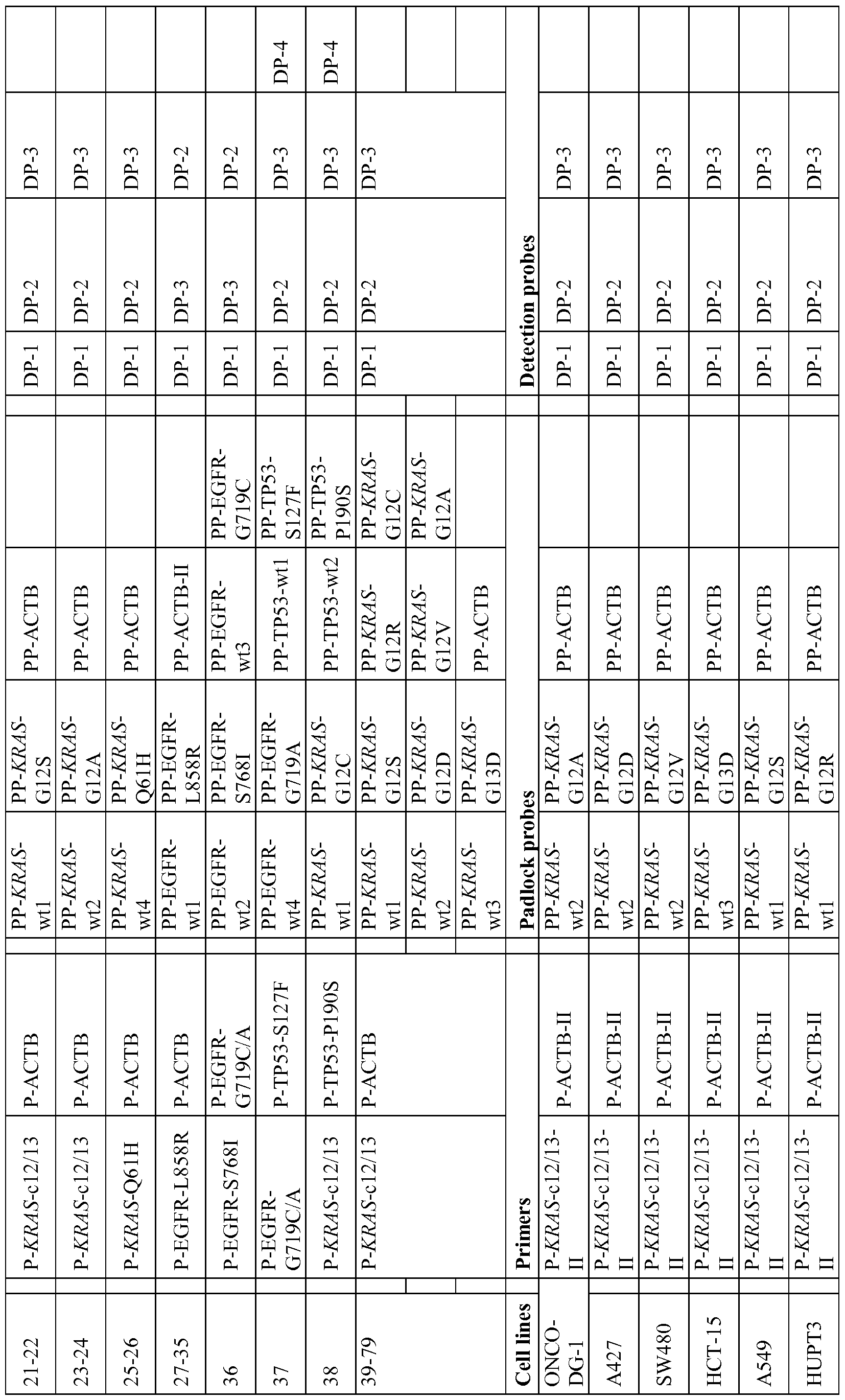

Claims
Priority Applications (6)
| Application Number | Priority Date | Filing Date | Title |
|---|---|---|---|
| CN201280009026.1A CN103649331A (en) | 2011-02-15 | 2012-02-15 | Method for localizedin situdetection of MRNA |
| JP2013554022A JP5881746B2 (en) | 2011-02-15 | 2012-02-15 | A method for localized in situ detection of mRNA |
| BR112013020773-6A BR112013020773B1 (en) | 2011-02-15 | 2012-02-15 | METHOD FOR LOCALIZED IN SITU DETECTION OF RNA, METHOD FOR DETERMINING THE PRESENCE AND LOCATION OF A GENETIC SEQUENCE AND METHOD FOR IN SITU LOCATION AND/OR DETECTION OF A SPECIFIC RNA |
| AU2012219132A AU2012219132B2 (en) | 2011-02-15 | 2012-02-15 | Method for localized in situ detection of mRNA |
| EP12728767.0A EP2675916B1 (en) | 2011-02-15 | 2012-02-15 | METHOD FOR LOCALIZED IN SITU DETECTION OF mRNA |
| CA2827497A CA2827497C (en) | 2011-02-15 | 2012-02-15 | Method for localized in situ detection of mrna |
Applications Claiming Priority (4)
| Application Number | Priority Date | Filing Date | Title |
|---|---|---|---|
| US201161442921P | 2011-02-15 | 2011-02-15 | |
| US61/442,921 | 2011-02-15 | ||
| US201161473662P | 2011-04-08 | 2011-04-08 | |
| US61/473,662 | 2011-04-08 |
Publications (2)
| Publication Number | Publication Date |
|---|---|
| WO2012110899A2 true WO2012110899A2 (en) | 2012-08-23 |
| WO2012110899A3 WO2012110899A3 (en) | 2012-12-13 |
Family
ID=46321164
Family Applications (1)
| Application Number | Title | Priority Date | Filing Date |
|---|---|---|---|
| PCT/IB2012/000995 WO2012110899A2 (en) | 2011-02-15 | 2012-02-15 | Method for localizedin situdetection of mrna |
Country Status (8)
| Country | Link |
|---|---|
| US (2) | US8551708B2 (en) |
| EP (1) | EP2675916B1 (en) |
| JP (2) | JP5881746B2 (en) |
| CN (2) | CN103649331A (en) |
| AU (1) | AU2012219132B2 (en) |
| BR (1) | BR112013020773B1 (en) |
| CA (1) | CA2827497C (en) |
| WO (1) | WO2012110899A2 (en) |
Cited By (18)
| Publication number | Priority date | Publication date | Assignee | Title |
|---|---|---|---|---|
| WO2014030066A3 (en) * | 2012-08-22 | 2014-05-22 | Bernitz Mats Nilsson | Rolling circle based methods identifying nucleic acids |
| US9556473B2 (en) | 2011-02-15 | 2017-01-31 | Leica Biosystems Newcastle Ltd | Methods for identifying nucleic acid sequences |
| EP3578666A1 (en) * | 2013-03-12 | 2019-12-11 | President and Fellows of Harvard College | Method of generating a three-dimensional nucleic acid containing matrix |
| US11008608B2 (en) | 2016-02-26 | 2021-05-18 | The Board Of Trustees Of The Leland Stanford Junior University | Multiplexed single molecule RNA visualization with a two-probe proximity ligation system |
| US11021737B2 (en) | 2011-12-22 | 2021-06-01 | President And Fellows Of Harvard College | Compositions and methods for analyte detection |
| WO2021123282A1 (en) * | 2019-12-20 | 2021-06-24 | 10X Genomics, Inc. | Method of detecting an analyte |
| US11085072B2 (en) | 2016-08-31 | 2021-08-10 | President And Fellows Of Harvard College | Methods of generating libraries of nucleic acid sequences for detection via fluorescent in situ sequencing |
| US11118220B2 (en) | 2015-11-03 | 2021-09-14 | President And Fellows Of Harvard College | Method and apparatus for volumetric imaging of a three-dimensional nucleic acid containing matrix |
| US11193163B2 (en) | 2018-07-30 | 2021-12-07 | Readcoor, Llc | Methods and systems for sample processing or analysis |
| US11293054B2 (en) | 2011-12-22 | 2022-04-05 | President And Fellows Of Harvard College | Compositions and methods for analyte detection |
| US11312992B2 (en) | 2011-10-14 | 2022-04-26 | President And Fellows Of Harvard College | Sequencing by structure assembly |
| US11447807B2 (en) | 2016-08-31 | 2022-09-20 | President And Fellows Of Harvard College | Methods of combining the detection of biomolecules into a single assay using fluorescent in situ sequencing |
| US11473139B2 (en) | 2012-06-05 | 2022-10-18 | President And Fellows Of Harvard College | Spatial sequencing of nucleic acids using DNA origami probes |
| US11713485B2 (en) | 2016-04-25 | 2023-08-01 | President And Fellows Of Harvard College | Hybridization chain reaction methods for in situ molecular detection |
| US11788123B2 (en) | 2017-05-26 | 2023-10-17 | President And Fellows Of Harvard College | Systems and methods for high-throughput image-based screening |
| US11959075B2 (en) | 2014-07-30 | 2024-04-16 | President And Fellows Of Harvard College | Systems and methods for determining nucleic acids |
| US11981917B2 (en) | 2013-06-04 | 2024-05-14 | President And Fellows Of Harvard College | RNA-guided transcriptional regulation |
| US12098425B2 (en) | 2021-04-08 | 2024-09-24 | Readcoor, Llc | Three-dimensional spatial molecular indexing |
Families Citing this family (98)
| Publication number | Priority date | Publication date | Assignee | Title |
|---|---|---|---|---|
| ES2544635T3 (en) | 2010-03-01 | 2015-09-02 | Quanterix Corporation | Methods to extend the dynamic range in assays for the detection of molecules or particles |
| US20190300945A1 (en) | 2010-04-05 | 2019-10-03 | Prognosys Biosciences, Inc. | Spatially Encoded Biological Assays |
| US10787701B2 (en) | 2010-04-05 | 2020-09-29 | Prognosys Biosciences, Inc. | Spatially encoded biological assays |
| EP2556171B1 (en) | 2010-04-05 | 2015-09-02 | Prognosys Biosciences, Inc. | Spatially encoded biological assays |
| US9952237B2 (en) | 2011-01-28 | 2018-04-24 | Quanterix Corporation | Systems, devices, and methods for ultra-sensitive detection of molecules or particles |
| EP2675916B1 (en) * | 2011-02-15 | 2016-08-24 | Leica Biosystems Newcastle Limited | METHOD FOR LOCALIZED IN SITU DETECTION OF mRNA |
| GB201106254D0 (en) | 2011-04-13 | 2011-05-25 | Frisen Jonas | Method and product |
| EP2744916A4 (en) * | 2011-07-13 | 2015-06-17 | Primeradx Inc | Multimodal methods for simultaneous detection and quantification of multiple nucleic acids in a sample |
| US20150011416A1 (en) * | 2012-02-23 | 2015-01-08 | Qiagen Mansfield, Inc | Multimodal pcr target detection |
| EP2909337B1 (en) | 2012-10-17 | 2019-01-09 | Spatial Transcriptomics AB | Methods and product for optimising localised or spatial detection of gene expression in a tissue sample |
| EP3597772A1 (en) | 2013-04-17 | 2020-01-22 | Agency For Science, Technology And Research | Method for generating extended sequence reads |
| US9879313B2 (en) | 2013-06-25 | 2018-01-30 | Prognosys Biosciences, Inc. | Methods and systems for determining spatial patterns of biological targets in a sample |
| EP3099822A4 (en) * | 2014-01-30 | 2017-08-30 | The Regents of the University of California | Methylation haplotyping for non-invasive diagnosis (monod) |
| CN104017861B (en) * | 2014-04-23 | 2015-08-12 | 常州方圆制药有限公司 | Nucleic acid detection method |
| WO2016007839A1 (en) * | 2014-07-11 | 2016-01-14 | President And Fellows Of Harvard College | Methods for high-throughput labelling and detection of biological features in situ using microscopy |
| CN104313187B (en) * | 2014-11-11 | 2016-03-23 | 舟山市质量技术监督检测研究院 | GI genome type norovirus reverse transcription Rolling Circle Amplification methods |
| FI3901281T3 (en) | 2015-04-10 | 2023-01-31 | Spatially distinguished, multiplex nucleic acid analysis of biological specimens | |
| US11254974B2 (en) * | 2016-02-10 | 2022-02-22 | The Board Of Trustees Of The Leland Stanford Junior University | RNA fixation and detection in clarity-based hydrogel tissue |
| EP3472359B1 (en) | 2016-06-21 | 2022-03-16 | 10X Genomics, Inc. | Nucleic acid sequencing |
| CN118562788A (en) * | 2016-08-30 | 2024-08-30 | 元素生物科学公司 | Methods and compositions for phased sequencing |
| EP3538867B1 (en) * | 2016-11-08 | 2024-01-03 | President and Fellows of Harvard College | Multiplexed imaging using merfish, expansion microscopy, and related technologies |
| US11185568B2 (en) | 2017-04-14 | 2021-11-30 | President And Fellows Of Harvard College | Methods for generation of cell-derived microfilament network |
| WO2018226930A1 (en) * | 2017-06-07 | 2018-12-13 | President And Fellows Of Harvard College | Targeted expansion fluorescent in situ sequencing |
| TW201905458A (en) * | 2017-06-23 | 2019-02-01 | 亞諾法生技股份有限公司 | Method and kit for detecting the location of a cell mutation in a biological sample |
| EP3668998A1 (en) | 2017-10-06 | 2020-06-24 | Cartana AB | Rna templated ligation |
| US11629342B2 (en) | 2017-10-17 | 2023-04-18 | President And Fellows Of Harvard College | Cas9-based transcription modulation systems |
| US11519033B2 (en) | 2018-08-28 | 2022-12-06 | 10X Genomics, Inc. | Method for transposase-mediated spatial tagging and analyzing genomic DNA in a biological sample |
| EP3894585A2 (en) | 2018-12-10 | 2021-10-20 | 10X Genomics, Inc. | Generating capture probes for spatial analysis |
| CN109652506A (en) * | 2018-12-20 | 2019-04-19 | 派德洛格(天津)生物科技有限公司 | Fluorescent in situ detects the method and kit of 21 exon p.L858R of Human epidermal growth factor receptor gene mutation |
| CN109652505A (en) * | 2018-12-20 | 2019-04-19 | 派德洛格(天津)生物科技有限公司 | Fluorescent in situ detects the method and kit of 20 exon p.T790M of Human epidermal growth factor receptor gene mutation |
| US11649485B2 (en) | 2019-01-06 | 2023-05-16 | 10X Genomics, Inc. | Generating capture probes for spatial analysis |
| US11926867B2 (en) | 2019-01-06 | 2024-03-12 | 10X Genomics, Inc. | Generating capture probes for spatial analysis |
| EP3921438A2 (en) * | 2019-02-04 | 2021-12-15 | Akoya Biosciences, Inc. | Analyte detection by selective labeling of biological samples |
| CN109797201B (en) * | 2019-03-12 | 2023-01-24 | 厦门先能生物科技有限公司 | In-situ multiple nucleic acid detection method |
| EP3976820A1 (en) | 2019-05-30 | 2022-04-06 | 10X Genomics, Inc. | Methods of detecting spatial heterogeneity of a biological sample |
| SG11202111878RA (en) | 2019-05-31 | 2021-11-29 | 10X Genomics Inc | Method of detecting target nucleic acid molecules |
| JP2022543562A (en) * | 2019-07-30 | 2022-10-13 | プレジデント・アンド・フェロウズ・オブ・ハーバード・カレッジ | Methods and compositions related to targeted analysis |
| WO2021092433A2 (en) | 2019-11-08 | 2021-05-14 | 10X Genomics, Inc. | Enhancing specificity of analyte binding |
| SG11202106899SA (en) | 2019-12-23 | 2021-09-29 | 10X Genomics Inc | Methods for spatial analysis using rna-templated ligation |
| US11732299B2 (en) | 2020-01-21 | 2023-08-22 | 10X Genomics, Inc. | Spatial assays with perturbed cells |
| US11702693B2 (en) | 2020-01-21 | 2023-07-18 | 10X Genomics, Inc. | Methods for printing cells and generating arrays of barcoded cells |
| US12076701B2 (en) | 2020-01-31 | 2024-09-03 | 10X Genomics, Inc. | Capturing oligonucleotides in spatial transcriptomics |
| US11898205B2 (en) | 2020-02-03 | 2024-02-13 | 10X Genomics, Inc. | Increasing capture efficiency of spatial assays |
| US11732300B2 (en) | 2020-02-05 | 2023-08-22 | 10X Genomics, Inc. | Increasing efficiency of spatial analysis in a biological sample |
| US20210254140A1 (en) | 2020-02-17 | 2021-08-19 | 10X Genomics, Inc. | Situ analysis of chromatin interaction |
| JP2023514749A (en) | 2020-02-21 | 2023-04-07 | 10エックス ジェノミクス インコーポレイテッド | Methods and compositions for integrated in situ spatial assays |
| US11891654B2 (en) | 2020-02-24 | 2024-02-06 | 10X Genomics, Inc. | Methods of making gene expression libraries |
| JP2023521356A (en) * | 2020-04-07 | 2023-05-24 | アドヴァンスド セル ダイアグノスティクス,インコーポレイテッド | Method and kit for detecting target nucleic acid by in situ hybridization |
| EP4139485B1 (en) | 2020-04-22 | 2023-09-06 | 10X Genomics, Inc. | Methods for spatial analysis using targeted rna depletion |
| WO2021236929A1 (en) | 2020-05-22 | 2021-11-25 | 10X Genomics, Inc. | Simultaneous spatio-temporal measurement of gene expression and cellular activity |
| AU2021275906A1 (en) | 2020-05-22 | 2022-12-22 | 10X Genomics, Inc. | Spatial analysis to detect sequence variants |
| WO2021242834A1 (en) | 2020-05-26 | 2021-12-02 | 10X Genomics, Inc. | Method for resetting an array |
| US12031177B1 (en) | 2020-06-04 | 2024-07-09 | 10X Genomics, Inc. | Methods of enhancing spatial resolution of transcripts |
| WO2021252499A1 (en) | 2020-06-08 | 2021-12-16 | 10X Genomics, Inc. | Methods of determining a surgical margin and methods of use thereof |
| US20230193356A1 (en) * | 2020-06-08 | 2023-06-22 | The Broad Institute, Inc. | Single cell combinatorial indexing from amplified nucleic acids |
| WO2021252591A1 (en) | 2020-06-10 | 2021-12-16 | 10X Genomics, Inc. | Methods for determining a location of an analyte in a biological sample |
| AU2021294334A1 (en) | 2020-06-25 | 2023-02-02 | 10X Genomics, Inc. | Spatial analysis of DNA methylation |
| US11761038B1 (en) | 2020-07-06 | 2023-09-19 | 10X Genomics, Inc. | Methods for identifying a location of an RNA in a biological sample |
| US11981960B1 (en) | 2020-07-06 | 2024-05-14 | 10X Genomics, Inc. | Spatial analysis utilizing degradable hydrogels |
| US11492662B2 (en) | 2020-08-06 | 2022-11-08 | Singular Genomics Systems, Inc. | Methods for in situ transcriptomics and proteomics |
| US11434525B2 (en) | 2020-08-06 | 2022-09-06 | Singular Genomics Systems, Inc. | Spatial sequencing |
| CN114075601B (en) * | 2020-08-18 | 2024-05-14 | 派德洛格(天津)生物科技有限公司 | Fusion gene in-situ detection method, kit and application thereof |
| US11981958B1 (en) | 2020-08-20 | 2024-05-14 | 10X Genomics, Inc. | Methods for spatial analysis using DNA capture |
| US11926822B1 (en) | 2020-09-23 | 2024-03-12 | 10X Genomics, Inc. | Three-dimensional spatial analysis |
| US20240018572A1 (en) | 2020-10-22 | 2024-01-18 | 10X Genomics, Inc. | Methods for spatial analysis using rolling circle amplification |
| US12071667B2 (en) | 2020-11-04 | 2024-08-27 | 10X Genomics, Inc. | Sequence analysis using meta-stable nucleic acid molecules |
| CN115198003B (en) * | 2020-11-18 | 2023-07-25 | 鲲羽生物科技(江门)有限公司 | Transcriptome spatial position information detection method suitable for barcode sequencing and application thereof |
| US11827935B1 (en) | 2020-11-19 | 2023-11-28 | 10X Genomics, Inc. | Methods for spatial analysis using rolling circle amplification and detection probes |
| EP4012046A1 (en) | 2020-12-11 | 2022-06-15 | 10X Genomics, Inc. | Methods and compositions for multimodal in situ analysis |
| EP4121555A1 (en) | 2020-12-21 | 2023-01-25 | 10X Genomics, Inc. | Methods, compositions, and systems for capturing probes and/or barcodes |
| CN116848263A (en) | 2020-12-23 | 2023-10-03 | 10X基因组学有限公司 | Methods and compositions for analyte detection |
| US12060603B2 (en) | 2021-01-19 | 2024-08-13 | 10X Genomics, Inc. | Methods for internally controlled in situ assays using padlock probes |
| EP4284945A1 (en) | 2021-01-26 | 2023-12-06 | 10X Genomics, Inc. | Nucleic acid analog probes for in situ analysis |
| WO2022187366A1 (en) | 2021-03-03 | 2022-09-09 | 10X Genomics, Inc. | Analyte detection in situ using nucleic acid origami |
| US20220380838A1 (en) | 2021-06-01 | 2022-12-01 | 10X Genomics, Inc. | Methods and compositions for analyte detection and probe resolution |
| US20230084407A1 (en) | 2021-06-02 | 2023-03-16 | 10X Genomics, Inc. | Sample analysis using asymmetric circularizable probes |
| WO2022256503A1 (en) | 2021-06-03 | 2022-12-08 | 10X Genomics, Inc. | Methods, compositions, kits, and systems for enhancing analyte capture for spatial analysis |
| WO2023283442A1 (en) | 2021-07-09 | 2023-01-12 | 10X Genomics, Inc. | Methods for detecting analytes using sparse labelling |
| CN113463203B (en) * | 2021-07-27 | 2024-08-30 | 厦门先能生物科技有限公司 | Construction method of in-situ sequencing library for realizing in-situ detection of multiple RNAs |
| CN113564230B (en) * | 2021-07-27 | 2023-10-31 | 华侨大学 | In-situ detection method for circular RNA |
| US20230057571A1 (en) | 2021-08-03 | 2023-02-23 | 10X Genomics, Inc. | Nucleic acid concatemers and methods for stabilizing and/or compacting the same |
| EP4326898B1 (en) | 2021-08-16 | 2024-07-31 | 10X Genomics, Inc. | Probes comprising a split barcode region and methods of use |
| WO2023034489A1 (en) | 2021-09-01 | 2023-03-09 | 10X Genomics, Inc. | Methods, compositions, and kits for blocking a capture probe on a spatial array |
| WO2023102118A2 (en) | 2021-12-01 | 2023-06-08 | 10X Genomics, Inc. | Methods, compositions, and systems for improved in situ detection of analytes and spatial analysis |
| WO2023108139A2 (en) | 2021-12-10 | 2023-06-15 | 10X Genomics, Inc. | Multi-resolution in situ decoding |
| WO2023129898A2 (en) | 2021-12-27 | 2023-07-06 | 10X Genomics, Inc. | Methods and compositions for rolling circle amplification |
| WO2023141588A1 (en) | 2022-01-21 | 2023-07-27 | 10X Genomics, Inc. | Multiple readout signals for analyzing a sample |
| WO2023172915A1 (en) | 2022-03-08 | 2023-09-14 | 10X Genomics, Inc. | In situ code design methods for minimizing optical crowding |
| WO2023192302A1 (en) | 2022-03-29 | 2023-10-05 | 10X Genomics, Inc. | Spectral unmixing combined with decoding for super-multiplexed in situ analysis |
| WO2023192616A1 (en) | 2022-04-01 | 2023-10-05 | 10X Genomics, Inc. | Compositions and methods for targeted masking of autofluorescence |
| US20230323427A1 (en) | 2022-04-06 | 2023-10-12 | 10X Genomics, Inc. | Methods and compositions for multiplex cell analysis |
| WO2023215612A1 (en) | 2022-05-06 | 2023-11-09 | 10X Genomics, Inc. | Analysis of antigen and antigen receptor interactions |
| WO2023245190A1 (en) | 2022-06-17 | 2023-12-21 | 10X Genomics, Inc. | Catalytic de-crosslinking of samples for in situ analysis |
| EP4339297A1 (en) | 2022-09-19 | 2024-03-20 | Miltenyi Biotec B.V. & Co. KG | Targeted detection of single nucleotide polymorphisms (snp) directly on tissue |
| WO2024081869A1 (en) | 2022-10-14 | 2024-04-18 | 10X Genomics, Inc. | Methods for analysis of biological samples |
| US20240158852A1 (en) | 2022-11-16 | 2024-05-16 | 10X Genomics, Inc. | Methods and compositions for assessing performance of in situ assays |
| WO2024137860A1 (en) | 2022-12-21 | 2024-06-27 | 10X Genomics, Inc. | Methods for tethering ribonucleic acids in biological samples |
| CN116004784A (en) * | 2022-12-29 | 2023-04-25 | 格物致和生物科技(北京)有限公司 | Probe combination and method for detecting nucleic acid |
Citations (1)
| Publication number | Priority date | Publication date | Assignee | Title |
|---|---|---|---|---|
| WO1999049079A1 (en) | 1998-03-25 | 1999-09-30 | Ulf Landegren | Rolling circle replication of padlock probes |
Family Cites Families (19)
| Publication number | Priority date | Publication date | Assignee | Title |
|---|---|---|---|---|
| US5591582A (en) | 1985-07-23 | 1997-01-07 | The Board Of Rijks Universiteit Leiden | Methods for detecting activated RAS oncogenes |
| WO1994003624A1 (en) | 1992-08-04 | 1994-02-17 | Auerbach Jeffrey I | Methods for the isothermal amplification of nucleic acid molecules |
| SE9400522D0 (en) | 1994-02-16 | 1994-02-16 | Ulf Landegren | Method and reagent for detecting specific nucleotide sequences |
| USRE38442E1 (en) | 1994-06-22 | 2004-02-24 | Mount Sinai School Of Medicine | Nucleic acid amplification method hybridization signal amplification method (HSAM) |
| US5866337A (en) | 1995-03-24 | 1999-02-02 | The Trustees Of Columbia University In The City Of New York | Method to detect mutations in a nucleic acid using a hybridization-ligation procedure |
| US5854033A (en) | 1995-11-21 | 1998-12-29 | Yale University | Rolling circle replication reporter systems |
| US6143495A (en) | 1995-11-21 | 2000-11-07 | Yale University | Unimolecular segment amplification and sequencing |
| DK0868530T3 (en) | 1995-12-05 | 2003-07-21 | Jorn Erland Koch | The cascade-nucleinsyreamplificeringsreaktion |
| SE9601676D0 (en) | 1996-04-30 | 1996-04-30 | Ulf Landegren | Improved probing of specific mucleic acids |
| US5912124A (en) | 1996-06-14 | 1999-06-15 | Sarnoff Corporation | Padlock probe detection |
| US6783943B2 (en) * | 2000-12-20 | 2004-08-31 | The Regents Of The University Of California | Rolling circle amplification detection of RNA and DNA |
| GB2378245A (en) | 2001-08-03 | 2003-02-05 | Mats Nilsson | Nucleic acid amplification method |
| US7932070B2 (en) * | 2001-12-21 | 2011-04-26 | Agilent Technologies, Inc. | High fidelity DNA polymerase compositions and uses therefor |
| US20040022764A1 (en) * | 2002-07-31 | 2004-02-05 | Hanan Polansky | Inhibition of microcompetition with a foreign polynucleotide as treatment of chronic disease |
| ES2741547T3 (en) | 2004-03-31 | 2020-02-11 | Massachusetts Gen Hospital | Method to determine the response of cancer to treatments directed at the epidermal growth factor receptor |
| US7745128B2 (en) | 2006-03-27 | 2010-06-29 | Globeimmune, Inc. | Ras mutation and compositions and methods related thereto |
| US7794938B2 (en) * | 2007-01-12 | 2010-09-14 | Lawrence Livermore National Security, Llc | Multiplex detection of agricultural pathogens |
| KR100957057B1 (en) * | 2007-12-03 | 2010-05-13 | 래플진(주) | Method for Detection of Nucleic Acids by Simultaneous Isothermal Amplification of Nucleic Acids and Signal Probe |
| EP2675916B1 (en) * | 2011-02-15 | 2016-08-24 | Leica Biosystems Newcastle Limited | METHOD FOR LOCALIZED IN SITU DETECTION OF mRNA |
-
2012
- 2012-02-15 EP EP12728767.0A patent/EP2675916B1/en active Active
- 2012-02-15 BR BR112013020773-6A patent/BR112013020773B1/en not_active IP Right Cessation
- 2012-02-15 AU AU2012219132A patent/AU2012219132B2/en not_active Ceased
- 2012-02-15 US US13/397,503 patent/US8551708B2/en active Active
- 2012-02-15 CN CN201280009026.1A patent/CN103649331A/en active Pending
- 2012-02-15 WO PCT/IB2012/000995 patent/WO2012110899A2/en active Application Filing
- 2012-02-15 CA CA2827497A patent/CA2827497C/en active Active
- 2012-02-15 JP JP2013554022A patent/JP5881746B2/en not_active Expired - Fee Related
- 2012-02-15 CN CN202110440392.3A patent/CN113151404A/en active Pending
- 2012-07-05 US US13/542,378 patent/US8551710B2/en active Active
-
2015
- 2015-11-05 JP JP2015217519A patent/JP6165215B2/en not_active Expired - Fee Related
Patent Citations (1)
| Publication number | Priority date | Publication date | Assignee | Title |
|---|---|---|---|---|
| WO1999049079A1 (en) | 1998-03-25 | 1999-09-30 | Ulf Landegren | Rolling circle replication of padlock probes |
Non-Patent Citations (25)
| Title |
|---|
| BOTLING; MICKE, METHODS MOL. BIOL., vol. 675, 2011, pages 299 - 306 |
| GRAZIANO ET AL., PHARMACOGENOMICS J., 2010 |
| LAGUNAVICIUS ET AL., RNA, vol. 15, 2009, pages 765 - 771 |
| LARSSON ET AL., NAT. METHODS, vol. 1, 2004, pages 227 - 232 |
| LARSSON ET AL., NATURE METHODS, vol. 7, no. 5, 2010, pages 395 - 397 |
| LAURENT-PUIG ET AL., J CLON ONCOL, vol. 27, no. 35, 2009, pages 5924 - 30 |
| LIZARDI ET AL., NAT. GENET., vol. 19, 1998, pages 225 - 232 |
| LOUPAKIS ET AL., BR J CANCER, vol. 101, no. 4, 2009, pages 715 - 21 |
| LOUPAKIS ET AL., J CLIN ONCOL, vol. 27, no. 16, 2009, pages 2622 - 9 |
| MITRA; CHURCH, NUCLEIC ACIDS RES., vol. 27, no. 24, 1999 |
| MONTICONE ET AL., MOLECULAR CANCER, vol. 7, no. 92, 2008 |
| NILSSON ET AL., NAT. BIOTECHNOL., vol. 18, 2000, pages 791 - 793 |
| NILSSON ET AL., NUCLEIC ACID RES., vol. 29, 2001, pages 578 - 581 |
| NILSSON ET AL., SCIENCE, vol. 265, 1994, pages 2085 - 2088 |
| OWCZARZY ET AL., BIOCHEMISTRY, vol. 47, 2008, pages 5336 - 5353 |
| PENA ET AL., NAT. METHODS, vol. 6, no. 2, 2009, pages 139 - 141 |
| PRENEN ET AL., CLIN CANCER RES., vol. 15, no. 9, 2009, pages 3184 - 8 |
| RAJAGOPALAN ET AL., NATURE, vol. 418, no. 29, 2002, pages 934 |
| SAMBROOK ET AL.: "Molecular Cloning--A Laboratory Manual", 1989, COLD SPRING HARBOR LABORATORY |
| SATORE-BIANCHI ET AL., CANCER RES., vol. 69, no. 5, 2009, pages 1851 - 7 |
| STOUGAARD ET AL., BMC BIOTECH., vol. 7, 2007, pages 69 |
| SUNDSTROM ET AL., BMC CANCER, vol. 10, 2010, pages 660 |
| VOGELSTEIN; FEARON, N ENGL J MED, vol. 319, no. 9, 1988, pages 525 - 32 |
| WETMUR, CRITICAL REV. BIOCHEM. MOL. BIOL., vol. 26, no. 3/4, 1991, pages 227 - 259 |
| YI ET AL., NUCLEIC ACID RES., vol. 29, 2001, pages 4818 - 4825 |
Cited By (33)
| Publication number | Priority date | Publication date | Assignee | Title |
|---|---|---|---|---|
| US9556473B2 (en) | 2011-02-15 | 2017-01-31 | Leica Biosystems Newcastle Ltd | Methods for identifying nucleic acid sequences |
| US11312992B2 (en) | 2011-10-14 | 2022-04-26 | President And Fellows Of Harvard College | Sequencing by structure assembly |
| US11976318B2 (en) | 2011-12-22 | 2024-05-07 | President And Fellows Of Harvard College | Compositions and methods for analyte detection |
| US11293051B2 (en) | 2011-12-22 | 2022-04-05 | President And Fellows Of Harvard College | Compositions and methods for analyte detection |
| US11021737B2 (en) | 2011-12-22 | 2021-06-01 | President And Fellows Of Harvard College | Compositions and methods for analyte detection |
| US11639518B2 (en) | 2011-12-22 | 2023-05-02 | President And Fellows Of Harvard College | Compositions and methods for analyte detection |
| US11566276B2 (en) | 2011-12-22 | 2023-01-31 | President And Fellows Of Harvard College | Compositions and methods for analyte detection |
| US11566277B2 (en) | 2011-12-22 | 2023-01-31 | President And Fellows Of Harvard College | Compositions and methods for analyte detection |
| US11111521B2 (en) | 2011-12-22 | 2021-09-07 | President And Fellows Of Harvard College | Compositions and methods for analyte detection |
| US11549136B2 (en) | 2011-12-22 | 2023-01-10 | President And Fellows Of Harvard College | Compositions and methods for analyte detection |
| US11293052B2 (en) | 2011-12-22 | 2022-04-05 | President And Fellows Of Harvard College | Compositions and methods for analyte detection |
| US11293054B2 (en) | 2011-12-22 | 2022-04-05 | President And Fellows Of Harvard College | Compositions and methods for analyte detection |
| US11473139B2 (en) | 2012-06-05 | 2022-10-18 | President And Fellows Of Harvard College | Spatial sequencing of nucleic acids using DNA origami probes |
| WO2014030066A3 (en) * | 2012-08-22 | 2014-05-22 | Bernitz Mats Nilsson | Rolling circle based methods identifying nucleic acids |
| US11299767B2 (en) | 2013-03-12 | 2022-04-12 | President And Fellows Of Harvard College | Method for generating a three-dimensional nucleic acid containing matrix |
| EP3578666A1 (en) * | 2013-03-12 | 2019-12-11 | President and Fellows of Harvard College | Method of generating a three-dimensional nucleic acid containing matrix |
| US11078520B2 (en) | 2013-03-12 | 2021-08-03 | President And Fellows Of Harvard College | Method for generating a three-dimensional nucleic acid containing matrix |
| US20240287582A1 (en) * | 2013-03-12 | 2024-08-29 | President And Fellows Of Harvard College | Method for Generating A Three-Dimensional Nucleic Acid Containing Matrix |
| US11981917B2 (en) | 2013-06-04 | 2024-05-14 | President And Fellows Of Harvard College | RNA-guided transcriptional regulation |
| US11959075B2 (en) | 2014-07-30 | 2024-04-16 | President And Fellows Of Harvard College | Systems and methods for determining nucleic acids |
| US11542554B2 (en) | 2015-11-03 | 2023-01-03 | President And Fellows Of Harvard College | Method and apparatus for volumetric imaging |
| US11118220B2 (en) | 2015-11-03 | 2021-09-14 | President And Fellows Of Harvard College | Method and apparatus for volumetric imaging of a three-dimensional nucleic acid containing matrix |
| US11008608B2 (en) | 2016-02-26 | 2021-05-18 | The Board Of Trustees Of The Leland Stanford Junior University | Multiplexed single molecule RNA visualization with a two-probe proximity ligation system |
| US11718874B2 (en) | 2016-04-25 | 2023-08-08 | President And Fellows Of Harvard College | Hybridization chain reaction methods for in situ molecular detection |
| US11713485B2 (en) | 2016-04-25 | 2023-08-01 | President And Fellows Of Harvard College | Hybridization chain reaction methods for in situ molecular detection |
| US11447807B2 (en) | 2016-08-31 | 2022-09-20 | President And Fellows Of Harvard College | Methods of combining the detection of biomolecules into a single assay using fluorescent in situ sequencing |
| US11085072B2 (en) | 2016-08-31 | 2021-08-10 | President And Fellows Of Harvard College | Methods of generating libraries of nucleic acid sequences for detection via fluorescent in situ sequencing |
| US11788123B2 (en) | 2017-05-26 | 2023-10-17 | President And Fellows Of Harvard College | Systems and methods for high-throughput image-based screening |
| US11193163B2 (en) | 2018-07-30 | 2021-12-07 | Readcoor, Llc | Methods and systems for sample processing or analysis |
| EP4365305A3 (en) * | 2019-12-20 | 2024-05-29 | 10X Genomics, Inc. | Method of detecting an analyte |
| WO2021123282A1 (en) * | 2019-12-20 | 2021-06-24 | 10X Genomics, Inc. | Method of detecting an analyte |
| US12098425B2 (en) | 2021-04-08 | 2024-09-24 | Readcoor, Llc | Three-dimensional spatial molecular indexing |
| US12104151B2 (en) | 2023-03-16 | 2024-10-01 | President And Fellows Of Harvard College | Systems and methods for determining nucleic acids |
Also Published As
| Publication number | Publication date |
|---|---|
| US20120270214A1 (en) | 2012-10-25 |
| CN113151404A (en) | 2021-07-23 |
| CA2827497A1 (en) | 2012-08-23 |
| JP2016052318A (en) | 2016-04-14 |
| US20130004953A1 (en) | 2013-01-03 |
| JP5881746B2 (en) | 2016-03-09 |
| CA2827497C (en) | 2014-12-02 |
| JP2014506472A (en) | 2014-03-17 |
| CN103649331A (en) | 2014-03-19 |
| US8551708B2 (en) | 2013-10-08 |
| BR112013020773A2 (en) | 2016-11-29 |
| EP2675916B1 (en) | 2016-08-24 |
| AU2012219132B2 (en) | 2016-05-12 |
| BR112013020773A8 (en) | 2018-05-02 |
| WO2012110899A3 (en) | 2012-12-13 |
| BR112013020773B1 (en) | 2023-01-24 |
| EP2675916A2 (en) | 2013-12-25 |
| JP6165215B2 (en) | 2017-07-19 |
| US8551710B2 (en) | 2013-10-08 |
| AU2012219132A1 (en) | 2013-05-02 |
Similar Documents
| Publication | Publication Date | Title |
|---|---|---|
| AU2012219132B2 (en) | Method for localized in situ detection of mRNA | |
| EP4139485B1 (en) | Methods for spatial analysis using targeted rna depletion | |
| US20240263218A1 (en) | Methods for spatial analysis using targeted probe silencing | |
| Ramdas et al. | miRNA expression profiles in head and neck squamous cell carcinoma and adjacent normal tissue | |
| Muralidhar et al. | Global microRNA profiles in cervical squamous cell carcinoma depend on Drosha expression levels | |
| EP3211086B1 (en) | Methods and composition to generate unique sequence dna probes, labeling of dna probes and the use of these probes | |
| US20210324468A1 (en) | Compositions and methods for screening mutations in thyroid cancer | |
| Schepeler et al. | Diagnostic and prognostic microRNAs in stage II colon cancer | |
| WO2014030066A2 (en) | Methods for identifying nucleic acid sequences | |
| US9556473B2 (en) | Methods for identifying nucleic acid sequences | |
| WO2012159025A2 (en) | Chromosome conformation analysis | |
| US20140147838A1 (en) | Method for multicolor codetection of microrna and proteins | |
| EP3177733B1 (en) | Single-stranded oligonucleotide probes for chromosome or gene copy enumeration | |
| WO2011012136A1 (en) | A method for classifying a human cell sample as cancerous | |
| Andersen et al. | Chapter New Applications for Microarrays |
Legal Events
| Date | Code | Title | Description |
|---|---|---|---|
| 121 | Ep: the epo has been informed by wipo that ep was designated in this application |
Ref document number: 12728767 Country of ref document: EP Kind code of ref document: A2 |
|
| ENP | Entry into the national phase |
Ref document number: 2012219132 Country of ref document: AU Date of ref document: 20120215 Kind code of ref document: A |
|
| ENP | Entry into the national phase |
Ref document number: 2013554022 Country of ref document: JP Kind code of ref document: A |
|
| ENP | Entry into the national phase |
Ref document number: 2827497 Country of ref document: CA |
|
| NENP | Non-entry into the national phase |
Ref country code: DE |
|
| REEP | Request for entry into the european phase |
Ref document number: 2012728767 Country of ref document: EP |
|
| WWE | Wipo information: entry into national phase |
Ref document number: 2012728767 Country of ref document: EP |
|
| REG | Reference to national code |
Ref country code: BR Ref legal event code: B01A Ref document number: 112013020773 Country of ref document: BR |
|
| ENP | Entry into the national phase |
Ref document number: 112013020773 Country of ref document: BR Kind code of ref document: A2 Effective date: 20130814 |