EP3578666A1 - Method of generating a three-dimensional nucleic acid containing matrix - Google Patents
Method of generating a three-dimensional nucleic acid containing matrix Download PDFInfo
- Publication number
- EP3578666A1 EP3578666A1 EP19169540.2A EP19169540A EP3578666A1 EP 3578666 A1 EP3578666 A1 EP 3578666A1 EP 19169540 A EP19169540 A EP 19169540A EP 3578666 A1 EP3578666 A1 EP 3578666A1
- Authority
- EP
- European Patent Office
- Prior art keywords
- matrix
- nucleic acids
- amplicons
- dna
- dimensional
- Prior art date
- Legal status (The legal status is an assumption and is not a legal conclusion. Google has not performed a legal analysis and makes no representation as to the accuracy of the status listed.)
- Pending
Links
Images


Classifications
-
- C—CHEMISTRY; METALLURGY
- C12—BIOCHEMISTRY; BEER; SPIRITS; WINE; VINEGAR; MICROBIOLOGY; ENZYMOLOGY; MUTATION OR GENETIC ENGINEERING
- C12Q—MEASURING OR TESTING PROCESSES INVOLVING ENZYMES, NUCLEIC ACIDS OR MICROORGANISMS; COMPOSITIONS OR TEST PAPERS THEREFOR; PROCESSES OF PREPARING SUCH COMPOSITIONS; CONDITION-RESPONSIVE CONTROL IN MICROBIOLOGICAL OR ENZYMOLOGICAL PROCESSES
- C12Q1/00—Measuring or testing processes involving enzymes, nucleic acids or microorganisms; Compositions therefor; Processes of preparing such compositions
- C12Q1/68—Measuring or testing processes involving enzymes, nucleic acids or microorganisms; Compositions therefor; Processes of preparing such compositions involving nucleic acids
- C12Q1/6806—Preparing nucleic acids for analysis, e.g. for polymerase chain reaction [PCR] assay
-
- C—CHEMISTRY; METALLURGY
- C12—BIOCHEMISTRY; BEER; SPIRITS; WINE; VINEGAR; MICROBIOLOGY; ENZYMOLOGY; MUTATION OR GENETIC ENGINEERING
- C12P—FERMENTATION OR ENZYME-USING PROCESSES TO SYNTHESISE A DESIRED CHEMICAL COMPOUND OR COMPOSITION OR TO SEPARATE OPTICAL ISOMERS FROM A RACEMIC MIXTURE
- C12P19/00—Preparation of compounds containing saccharide radicals
- C12P19/26—Preparation of nitrogen-containing carbohydrates
- C12P19/28—N-glycosides
- C12P19/30—Nucleotides
- C12P19/34—Polynucleotides, e.g. nucleic acids, oligoribonucleotides
-
- C—CHEMISTRY; METALLURGY
- C12—BIOCHEMISTRY; BEER; SPIRITS; WINE; VINEGAR; MICROBIOLOGY; ENZYMOLOGY; MUTATION OR GENETIC ENGINEERING
- C12Q—MEASURING OR TESTING PROCESSES INVOLVING ENZYMES, NUCLEIC ACIDS OR MICROORGANISMS; COMPOSITIONS OR TEST PAPERS THEREFOR; PROCESSES OF PREPARING SUCH COMPOSITIONS; CONDITION-RESPONSIVE CONTROL IN MICROBIOLOGICAL OR ENZYMOLOGICAL PROCESSES
- C12Q1/00—Measuring or testing processes involving enzymes, nucleic acids or microorganisms; Compositions therefor; Processes of preparing such compositions
- C12Q1/68—Measuring or testing processes involving enzymes, nucleic acids or microorganisms; Compositions therefor; Processes of preparing such compositions involving nucleic acids
- C12Q1/6844—Nucleic acid amplification reactions
-
- C—CHEMISTRY; METALLURGY
- C12—BIOCHEMISTRY; BEER; SPIRITS; WINE; VINEGAR; MICROBIOLOGY; ENZYMOLOGY; MUTATION OR GENETIC ENGINEERING
- C12Q—MEASURING OR TESTING PROCESSES INVOLVING ENZYMES, NUCLEIC ACIDS OR MICROORGANISMS; COMPOSITIONS OR TEST PAPERS THEREFOR; PROCESSES OF PREPARING SUCH COMPOSITIONS; CONDITION-RESPONSIVE CONTROL IN MICROBIOLOGICAL OR ENZYMOLOGICAL PROCESSES
- C12Q1/00—Measuring or testing processes involving enzymes, nucleic acids or microorganisms; Compositions therefor; Processes of preparing such compositions
- C12Q1/68—Measuring or testing processes involving enzymes, nucleic acids or microorganisms; Compositions therefor; Processes of preparing such compositions involving nucleic acids
- C12Q1/6869—Methods for sequencing
- C12Q1/6874—Methods for sequencing involving nucleic acid arrays, e.g. sequencing by hybridisation
Definitions
- the present invention relates to methods of making a three-dimensional matrix of nucleic acids and amplifying, detecting and sequencing such nucleic acids within the matrix.
- Embodiments of the present invention are directed to methods of making a three-dimensional matrix of nucleic acids.
- Embodiments of the present invention are directed to methods of making a three-dimensional matrix including nucleic acids covalently bound into a matrix or into or to a matrix material.
- the nucleic acids may be co-polymerized with the matrix material or cross-linked to the matrix material or both.
- a plurality of nucleic acid sequences of certain length, such as DNA or RNA sequences are part of a three-dimensional copolymer.
- the nucleic acids may then be amplified and sequenced in situ, i.e. within the matrix.
- the three-dimensional matrix of nucleic acids provides, in a certain aspect, an information storage medium where the nucleic acids, i.e. a sequence of one or more nucleotides, represent stored information which can be read within the three-dimensional matrix.
- nucleic acids such as DNA or RNA sequences of given length are covalently attached to a matrix material to preserve their spatial orientation in the x, y and z axes within the matrix.
- the three-dimensional matrix may include a matrix material and that the term copolymer, matrix and matrix material may be used interchangeably.
- methods described herein are directed to immobilizing naturally occurring nucleic acids within their native environment, such as within a cell or within a tissue sample.
- the three-dimensional nucleic acid matrix can be generated in situ in a cell or tissue sample to preserve the naturally occurring nucleic acid sequence diversity (such as DNA and RNA) and spatial orientation in cells, tissues or any other complex biomaterial.
- the location of nucleic acids and their relative position is identified as a three-dimensional structure, such as within subcellular compartments, within cells, within tissues, as three-dimensional nucleic acid assemblies, as three-dimensional nucleic acid material, etc.
- the nucleic acids can be amplified and sequenced, if desired, in situ thereby providing positional information of the nucleic acids within the cell or tissue.
- nucleic acids of interest can be present within a three-dimensional matrix material and covalently attached to the three-dimensional matrix material such that the relative position of each nucleic acid is fixed, i.e. immobilized, within the three-dimensional matrix material.
- a three-dimensional matrix of covalently bound nucleic acids of any desired sequence is provided.
- Each nucleic acid has its own three-dimensional coordinates within the matrix material and each nucleic acid represents information.
- a large amount of information can be stored in a three-dimensional matrix.
- Individual information-encoding nucleic acids, such as DNA or RNA can be amplified and sequenced in situ, i.e., within the matrix, thereby enabling a large amount of information to be stored and read in a suitable three-dimensional material.
- the nucleic acids can be amplified to produce amplicons within the three-dimensional matrix material.
- the amplicons can then be covalently attached to the matrix, for example, by copolymerization or cross-linking. This results in a structurally stable and chemically stable three-dimensional matrix of nucleic acids.
- the three-dimensional matrix of nucleic acids allows for prolonged information storage and read-out cycles.
- the nucleic acid / amplicon matrix allows for high throughput sequencing of a wide ranging array of biological and non-biological samples in three dimensions.
- a three-dimensional nucleic acid matrix where a plurality of nucleic acid molecules, such as DNA or RNA, amplicons or nucleic acid structural units are immobilized, such as by covalent bonding to the matrix, in a three-dimensional space relative to one another.
- the nucleic acid molecules are rigidly fixed to the extent that they maintain their coordinate position within the matrix. It is to be understood that even though a nucleic acid molecule may be covalently attached to the three-dimensional matrix material, the nucleic acid molecule itself may be capable of movement though bound to the matrix, such as for example, when a nucleic acid sequence is bound to the matrix at a single location on the nucleic acid.
- the three-dimensional matrix including nucleic acids is porous. According to one aspect, the three-dimensional matrix including nucleic acids is porous to the extent that reagents typically used in amplification methods can diffuse or otherwise move through the matrix to contact nucleic acids and thereby amplify nucleic acids under suitable conditions.
- the three-dimensional matrix material is chemically inert and thermally stable to allow for various reaction conditions and reaction temperatures. According to this aspect, the three-dimensional matrix material is chemically inert and thermally stable to conditions used in amplification and sequencing methods known to those of skill in the art.
- the three-dimensional matrix material is optically transparent. According to one aspect, the three-dimensional matrix material is optically transparent to allow for three-dimensional imaging techniques known to those of skill in the art.
- the nucleic acids are amplified to an extent to produce sufficient levels of amplicons for three-dimensional imaging.
- the nucleic acids are amplified and include a label sufficient for a high level of fluorescence compatible with three-dimensional imaging.
- the material used to form the matrix is compatible with a wide range of biological and non-biological specimens in situ so as to avoid extracting the nucleic acid molecules away from their native environment.
- the matrix material may be a semi-solid medium that can be made from polyacrylamide, cellulose, alginate, polyamide, cross-linked agarose, cross-linked dextran or cross-linked polyethylene glycol.
- the semi-solid medium has x, y and z axes, and the nucleic acids are present randomly or non-randomly within the three-dimensional matrix.
- the matrix material is porous. Porosity can result from polymerization and/or crosslinking of molecules used to make the matrix material.
- the diffusion property within the gel matrix is largely a function of the pore size.
- the molecular sieve size is chosen to allow for rapid diffusion of enzymes, oligonucleotides, formamide and other buffers used for amplification and sequencing (>50-nm).
- the molecular sieve size is also chosen so that large DNA or RNA amplicons do not readily diffuse within the matrix ( ⁇ 500-nm).
- the porosity is controlled by changing the cross-linking density, the chain lengths and the percentage of co-polymerized branching monomers according to methods known to those of skill in the art.
- the semi-solid medium can be attached to a solid support such as a microscope slide or a flow cell.
- the solid support can be attached to the bottom surface of the semi-solid medium.
- the present invention provides:
- the present invention provides a three-dimensional matrix of a plurality of nucleic acids.
- the present invention provides a three-dimensional matrix including a plurality of nucleic acids bound thereto.
- the matrix is a three-dimensional nucleic acid-containing polymer.
- the nucleic acids may be naturally occurring nucleic acids or non-naturally occurring nucleic acids, such as nucleic acids that have been made using synthetic methods.
- the nucleic acids in the three-dimensional matrix may be ordered or unordered.
- the nucleic acids in the three-dimensional matrix may be present in their natural spatial relationship within a cell, tissue or organism.
- the nucleic acids in the three-dimensional matrix may be present in rows and columns within the three-dimensional matrix.
- the nucleic acids are modified to incorporate a functional moiety for attachment to the matrix.
- the functional moiety can be covalently cross-linked, copolymerize with or otherwise non-covalently bound to the matrix.
- the functional moiety can react with a cross-linker.
- the functional moiety can be part of a ligand-ligand binding pair.
- dNTP or dUTP can be modified with the functional group, so that the function moiety is introduced into the DNA during amplification.
- a suitable exemplary functional moiety includes an amine, acrydite, alkyne, biotin, azide, and thiol. In the case of crosslinking, the functional moiety is cross-linked to modified dNTP or dUTP or both.
- Suitable exemplary cross-linker reactive groups include imidoester (DMP), succinimide ester (NHS), maleimide (Sulfo-SMCC), carbodiimide (DCC, EDC) and phenyl azide.
- Cross-linkers within the scope of the present disclosure may include a spacer moiety. Such spacer moieties may be functionalized. Such spacer moieties may be chemically stable. Such spacer moieties may be of sufficient length to allow amplification of the nucleic acid bound to the matrix. Suitable exemplary spacer moieties include polyethylene glycol, carbon spacers, photo-cleavable spacers and other spacers known to those of skill in the art and the like.
- a matrix-forming material is contacted to a plurality of nucleic acids spatially arrange in three-dimensions relative to one another.
- Matrix forming materials include polyacrylamide, cellulose, alginate, polyamide, cross-linked agarose, cross-linked dextran or cross-linked polyethylene glycol.
- the matrix forming materials can form a matrix by polymerization and/or crosslinking of the matrix forming materials using methods specific for the matrix forming materials and methods, reagents and conditions known to those of skill in the art.
- a matrix-forming material can be introduced into a cell.
- the cells are fixed with formaldehyde and then immersed in ethanol to disrupt the lipid membrane.
- the matrix forming reagents are added to the sample and are allowed to permeate throughout the cell.
- a polymerization inducing catalyst, UV or functional cross-linkers are then added to allow the formation of a gel matrix.
- the un-incorporated material is washed out and any remaining functionally reactive group is quenched.
- Exemplary cells include any cell, human or otherwise, including diseased cells or healthy cells. Certain cells include human cells, non-human cells, human stem cells, mouse stem cells, primary cell lines, immortalized cell lines, primary and immortalized fibroblasts, HeLa cells and neurons.
- a matrix-forming material can be used to encapsulate a biological sample, such as a tissue sample.
- a biological sample such as a tissue sample.
- the formalin-fixed embedded tissues on glass slides are incubated with xylene and washed using ethanol to remove the embedding wax. They are then treated with Proteinase K to permeabilized the tissue.
- a polymerization inducing catalyst, UV or functional cross-linkers are then added to allow the formation of a gel matrix.
- the un-incorporated material is washed out and any remaining functionally reactive group is quenched.
- Exemplary tissue samples include any tissue samples of interest whether human or non-human. Such tissue samples include those from skin tissue, muscle tissue, bone tissue, organ tissue and the like.
- Exemplary tissues include human and mouse brain tissue sections, embryo sections, tissue array sections, and whole insect and worm embryos.
- the matrix-forming material forms a three-dimensional matrix including the plurality of nucleic acids.
- the matrix-forming material forms a three-dimensional matrix including the plurality of nucleic acids while maintaining the spatial relationship of the nucleic acids.
- the plurality of nucleic acids is immobilized within the matrix material.
- the plurality of nucleic acids may be immobilized within the matrix material by co-polymerization of the nucleic acids with the matrix-forming material.
- the plurality of nucleic acids may also be immobilized within the matrix material by crosslinking of the nucleic acids to the matrix material or otherwise cross-linking with the matrix-forming material.
- the plurality of nucleic acids may also be immobilized within the matrix by covalent attachment or through ligand-protein interaction to the matrix.
- the matrix is porous thereby allowing the introduction of reagents into the matrix at the site of a nucleic acid for amplification of the nucleic acid.
- a porous matrix may be made according to methods known to those of skill in the art.
- a polyacrylamide gel matrix is co-polymerized with acrydite-modified streptavidin monomers and biotinylated DNA molecules, using a suitable acrylamide:bis-acrylamide ratio to control the cross-linking density. Additional control over the molecular sieve size and density is achieved by adding additional cross-linkers such as functionalized polyethylene glycols.
- the nucleic acids which may represent individual bits of information, are readily accessed by oligonucleotides, such as labeled oligonucleotide probes, primers, enzymes and other reagents with rapid kinetics.
- oligonucleotides such as labeled oligonucleotide probes, primers, enzymes and other reagents with rapid kinetics.
- the matrix is sufficiently optically transparent or otherwise has optical properties suitable for standard Next Generation sequencing chemistries and deep three-dimensional imaging for high throughput information readout.
- the Next Generation sequencing chemistries that utilize fluorescence imaging include ABI SoLiD (Life Technologies), in which a sequencing primer on a template is ligated to a library of fluorescently labeled nonamers with a cleavable terminator. After ligation, the beads are then imaged using four color channels (FITC, Cy3, Texas Red and Cy5). The terminator is then cleaved off leaving a free-end to engage in the next ligation-extension cycle.
- the images are mapped to the color code space to determine the specific base calls per template.
- the workflow is achieved using an automated fluidics and imaging device (i.e. SoLiD 5500 W Genome Analyzer, ABI Life Technologies).
- Another sequencing platform uses sequencing by synthesis, in which a pool of single nucleotide with a cleavable terminator is incorporated using DNA polymerase. After imaging, the terminator is cleaved and the cycle is repeated. The fluorescence images are then analyzed to call bases for each DNA amplicons within the flow cell (HiSeq, Illumia).
- the plurality of nucleic acids may be amplified to produce amplicons by methods known to those of skill in the art.
- the amplicons may be immobilized within the matrix generally at the location of the nucleic acid being amplified, thereby creating a localized colony of amplicons.
- the amplicons may be immobilized within the matrix by steric factors.
- the amplicons may also be immobilized within the matrix by covalent or noncovalent bonding. In this manner, the amplicons may be considered to be attached to the matrix.
- the amplicons are resistant to movement or unraveling under mechanical stress.
- the amplicons such as DNA amplicons
- the amplicons are then copolymerized and/or covalently attached to the surrounding matrix thereby preserving their spatial relationship and any information inherent thereto.
- the amplicons are those generated from DNA or RNA within a cell embedded in the matrix, the amplicons can also be functionalized to form covalent attachment to the matrix preserving their spatial information within the cell thereby providing a subcellular localization distribution pattern.
- a covalent interaction is a chemical linkage between two atoms or radicals formed by the sharing of a pair of electrons (i.e., a single bond), two pairs of electrons (i.e., a double bond) or three pairs of electrons (i.e., a triple bond).
- Covalent interactions are also known in the art as electron pair interactions or electron pair bonds.
- Noncovalent interactions include, but are not limited to, van der Waals interactions, hydrogen bonds, weak chemical bonds (i.e., via short-range noncovalent forces), hydrophobic interactions, ionic bonds and the like.
- nucleic acid includes the term “oligonucleotide” or “polynucleotide” which includes a plurality of nucleotides.
- nucleic acid is intended to include naturally occurring nucleic acids and synthetic nucleic acids.
- nucleic acid is intended to include single stranded nucleic acids and double stranded nucleic acids.
- nucleic acid is intended to include DNA and RNA, whether single stranded or double stranded.
- Nucleotides of the present invention will typically be the naturally-occurring nucleotides such as nucleotides derived from adenosine, guanosine, uridine, cytidine and thymidine.
- double-stranded it is understood by those of skill in the art that a pair of oligonucleotides exists in a hydrogen-bonded, helical array typically associated with, for example, DNA.
- double-stranded as used herein is also meant to include those form which include such structural features as bulges and loops (see Stryer, Biochemistry, Third Ed.
- polynucleotide refers to a strand of nucleic acids that can be a variety of different sizes. Polynucleotides may be the same size as an oligonucleotide, or may be two-times, three-times, four-times, five-times, ten-times, or greater than the size of an oligonucleotide.
- Oligonucleotides and/or polynucleotides may be isolated from natural sources or purchased from commercial sources. Oligonucleotide and/or polynucleotide sequences may be prepared by any suitable method, e.g., the phosphoramidite method described by Beaucage and Carruthers ((1981) Tetrahedron Lett. 22: 1859 ) or the triester method according to Matteucci et al. (1981) J. Am. Chem. Soc. 103:3185 ), or by other chemical methods using either a commercial automated oligonucleotide synthesizer or high-throughput, high-density array methods described herein and known in the art (see U.S. Patent Nos.
- Pre-synthesized oligonucleotides may also be obtained commercially from a variety of vendors.
- oligonucleotides and/or polynucleotides may be prepared using a variety of microarray technologies known in the art. Pre-synthesized oligonucleotide and/or polynucleotide sequences may be attached to a support or synthesized in situ using light-directed methods, flow channel and spotting methods, inkjet methods, pin-based methods and bead-based methods set forth in the following references: McGall et al. (1996) Proc. Natl. Acad. Sci. U.S.A. 93:13555 ; Synthetic DNA Arrays In Genetic Engineering, Vol. 20:111, Plenum Press (1998 ); Duggan et al. (1999) Nat. Genet.
- WO 04/031399 WO 04/031351 , WO 04/029586 , WO 03/100012 , WO 03/066212 , WO 03/065038 , WO 03/064699 , WO 03/064027 , WO 03/064026 , WO 03/046223 , WO 03/040410 and WO 02/24597s .
- Nucleic acids may be obtained from libraries, e.g., genomic libraries, cDNA libraries and the like. Examples of methods for the synthesis of molecular libraries can be found in the art, for example in: DeWitt et al. (1993) Proc. Natl. Acad. Sci. USA 90:6909 ; Erb et al. (1994) Proc. Natl. Acad. Sci. USA 91:11422 ; Zuckermann et al. (1994) J. Med. Chem. 37:2678 ; Cho et al. (1993) Science 261:1303 ; Carrell et al. (1994) Angew. Chem. Int. Ed. Engl. 33:2059 ; Carell et al. (1994) Angew. Chem. Int. Ed. Engl. 33:2061 ; and in Gallop et al. (1994) J. Med. Chem. 37:1233 .
- nucleic acids are those found naturally in a biological sample, such as a cell or tissue.
- a matrix is used in conjunction with a solid support.
- the matrix can be polymerized in such a way that one surface of the matrix is attached to a solid support (e.g., a glass surface), while the other surface of the matrix is exposed or sandwiched between two solid supports.
- the matrix can be contained within a container.
- Solid supports of the invention may be fashioned into a variety of shapes.
- the solid support is substantially planar.
- Examples of solid supports include plates such as slides, microtitre plates, flow cells, coverslips, microchips, and the like, containers such as microfuge tubes, test tubes and the like, tubing, sheets, pads, films and the like.
- the solid supports may be, for example, biological, nonbiological, organic, inorganic, or a combination thereof.
- Embodiments of the present invention are further directed to the amplification of nucleic acid sequences within the matrix, i.e. in situ, within the matrix.
- Methods of amplifying nucleic acids include rolling circle amplification in situ.
- methods of amplifying nucleic acids involves the use of PCR, such as anchor PCR or RACE PCR, or, alternatively, in a ligation chain reaction (LCR) (see, e.g., Landegran et al. (1988) Science 241:1077-1080 ; and Nakazawa et al. (1994) Proc. Natl. Acad. Sci. U.S.A. 91:360-364 ).
- LCR ligation chain reaction
- Alternative amplification methods include: self-sustained sequence replication ( Guatelli et al. (1990) Proc. Natl. Acad. Sci. USA 87:1874 ), transcriptional amplification system ( Kwoh et al. (1989) Proc. Natl. Acad. Sci. US. 86:1173 ), Q-Beta Replicase ( Lizardi et al. (1988) BioTechnology 6:1197 ), recursive PCR ( Jaffe et al. (2000) J. Biol. Chem. 275:2619 ; and Williams et al. (2002) J. Biol. Chem.
- Embodiments of the present invention are directed to methods of amplifying nucleic acids in situ within the matrix by contacting the nucleic acids within the matrix with reagents and under suitable reaction conditions sufficient to amplify the nucleic acids.
- the matrix is porous to allow migration of reagents into the matrix to contact the nucleic acids.
- oligonucleotides are amplified by selectively hybridizing an amplification primer to an amplification site at the 3' end of an oligonucleotide using conventional methods.
- Amplification primers are 6 to 100, and even up to 1,000, nucleotides in length, but typically from 10 to 40 nucleotides, although oligonucleotides of different length are of use.
- Amplification primers may be present in solution to be added to the matrix or they may be added during formation of the matrix to be present therein sufficiently adjacent to nucleic acids to allow for hybridization and amplification.
- selective hybridization occurs when two nucleic acid sequences are substantially complementary, i.e., at least about 65% 75%, 80%, 85%, 90%, 91%, 92%, 93%, 94%, 95%, 96%, 97%, 98% 99%, 99.1%, 99.2%, 99.3%, 99.4%, 99.5%, 99.6%, 99.7%, 99.8%, 99.9% or 100% complementary over a stretch of at least 14 to 25 nucleotides. See Kanehisa, M., 1984, Nucleic Acids Res. 12: 203 .
- Primer sequences with a high G-C content or that comprise palindromic sequences tend to self-hybridize, as do their intended target sites, since unimolecular, rather than bimolecular, hybridization kinetics are generally favored in solution; at the same time, it is important to design a primer containing sufficient numbers of G-C nucleotide pairings to bind the target sequence tightly, since each such pair is bound by three hydrogen bonds, rather than the two that are found when A and T bases pair.
- Hybridization temperature varies inversely with primer annealing efficiency, as does the concentration of organic solvents, e.g., formamide, that might be included in a hybridization mixture, while increases in salt concentration facilitate binding.
- Stringent hybridization conditions typically include salt concentrations of less than about 1M, more usually less than about 500 mM and preferably less than about 200 mM.
- Hybridization temperatures range from as low as 0 °C to greater than 22 °C, greater than about 30 °C, and (most often) in excess of about 37 °C. Longer fragments may require higher hybridization temperatures for specific hybridization. As several factors affect the stringency of hybridization, the combination of parameters is more important than the absolute measure of any one alone. Hybridization conditions are known to those skilled in the art and can be found in Current Protocols in Molecular Biology, John Wiley & Sons, N.Y. (1989), 6.3.1-6.3.6 .
- Primers are designed with the above first four considerations in mind. While estimates of the relative merits of numerous sequences are made mentally, computer programs have been designed to assist in the evaluation of these several parameters and the optimization of primer sequences (see, e.g., Hoover et al. (2002) Nucleic Acids Res. 30:e43 , and Rouillard et al. (2004) Nucleic Acids Res. 32:W176 ).
- methods of sequencing nucleic acid in situ within a matrix are provided.
- General sequencing methods known in the art such as sequencing by extension with reversible terminators, fluorescent in situ sequencing (FISSEQ), pyrosequencing, massively parallel signature sequencing (MPSS) and the like (described in Shendure et al. (2004) Nat. Rev. 5:335 ), are suitable for use with the matrix in which the nucleic acids are present.
- Reversible termination methods use step-wise sequencing-by-synthesis biochemistry that coupled with reversible termination and removable fluorescence (Shendure et al. supra and U.S. Patent Nos. 5,750,341 and 6,306,597 ).
- FISSEQ is a method whereby DNA is extended by adding a single type of fluorescently-labelled nucleotide triphosphate to the reaction, washing away unincorporated nucleotide, detecting incorporation of the nucleotide by measuring fluorescence, and repeating the cycle. At each cycle, the fluorescence from previous cycles is bleached or digitally subtracted or the fluorophore is cleaved from the nucleotide and washed away.
- FISSEQ is described further in Mitra et al. (2003) Anal. Biochem. 320:55 , incorporated herein by reference in its entirety for all purposes.
- Pyrosequencing is a method in which the pyrophosphate (PPi) released during each nucleotide incorporation event (i.e., when a nucleotide is added to a growing polynucleotide sequence).
- the PPi released in the DNA polymerase-catalyzed reaction is detected by ATP sulfurylase and luciferase in a coupled reaction which can be visibly detected.
- the added nucleotides are continuously degraded by a nucleotide-degrading enzyme. After the first added nucleotide has been degraded, the next nucleotide can be added. As this procedure is repeated, longer stretches of the template sequence are deduced. Pyrosequencing is described further in Ronaghi et al.
- MPSS utilizes ligation-based DNA sequencing simultaneously on microbeads. A mixture of labelled adaptors comprising all possible overhangs is annealed to a target sequence of four nucleotides. The label is detected upon successful ligation of an adaptor. A restriction enzyme is then used to cleave the DNA template to expose the next four bases. MPSS is described further in Brenner et al. (2000) Nat. Biotech. 18:630 .
- the nucleic acids within the matrix can be interrogated using methods known to those of skill in the art including fluorescently labeled oligonucleotide/DNA/RNA hybridization, primer extension with labeled ddNTP, sequencing by ligation and sequencing by synthesis.
- Ligated circular padlock probes described in Larsson, et al., (2004), Nat. Methods 1:227-232 can be used to detect multiple sequence targets in parallel, followed by either sequencing-by-ligation, -synthesis or -hybridization of the barcode sequences in the padlock probe to identify individual targets.
- methods described herein produce a three-dimensional nucleic acid amplicon matrix which is stable, long-lasting and resistant, substantially resistant or partially resistant to enzymatic or chemical degradation.
- the three-dimensional nucleic acid amplicon matrix can be repeatedly interrogated using standard probe hybridization and/or fluorescence-based sequencing.
- the three-dimensional nucleic acid amplicon matrix can be repeatedly interrogated with little or no signal degradation, such as after more than 50 cycles, and with little position shift, such as less than 1 ⁇ m per amplicon.
- a plurality of circular DNA molecules is covalently linked to one another.
- the circular DNA molecules are then amplified using methods known to those of skill in the art, such as isothermal enzymatic amplification one example of which is rolling circle amplification.
- the amplicons are localized near the circular DNA.
- the amplicons form a shell around the circular DNA or otherwise assemble around the circular DNA.
- Each circular DNA may have more than 1000 amplicons surrounding or otherwise associated therewith.
- the amplicons surrounding a particular circular DNA provide a high signal intensity, due in part to the number of amplicons and/or detectable labels associated with the amplicons.
- the amplicons may be functionalized and cross-linked or otherwise covalently bound together around their associate circular DNA to form a series or network of tightly bound DNA amplicon shells around each circular DNA.
- the series or network of tightly bound DNA amplicon shells around each circular DNA may be assembled onto a three-dimensional support.
- the series or network of tightly bound DNA amplicon shells around each circular DNA may be assembled onto a three-dimensional support producing a three-dimensional DNA polymer with defined overall shape, size and amplicon position.
- amplicons are covalently linked without the need for separate cross-linkers, such as bis-N-succinimidyl-(nonaethylene glycol) ester.
- An acrydite moiety such as a catalyst activated acrydite moiety is introduced at the end of a long carbon spacer (i.e., about C6 to about C12) at position 5 of a uracil base a representative formula of which is shown below.
- R represents the acrydite spacer moiety attached to the 5 position of the uracil base.
- the spacer can be a carbon chain of between about 2 carbons to about 200 carbons.
- the spacer can be polyethylene glycol.
- the length of the spacer can vary from about 30 angstroms to about 100 angstroms and can be of various molecular weights.
- the spacer can be permanent or reversible, such as by using UV light, enzymes, chemical cleavage, etc.
- a three-dimensional matrix such as a polyacrylamide gel matrix, can be used to embed a variety of biological structures containing enzymatically or chemically modified DNA or RNA molecules containing an acrydite functional moiety or moieties.
- the non-nucleic acid component is selectively dissolved using detergents, proteases, organic solvents or denaturants to create a three-dimensional matrix that preserves individual DNA or RNA molecules and their relative spatial location. Examples include embedding cells, healthy and diseased tissues and tissue sections, small model organisms such as worms and insects, bacterial colonies or biofilm, environmental samples containing other DNA or RNA containing materials or organisms.
- Human iPS cells or human primary fibroblasts are grown on a 1.5 cover slip. They are fixed using 4% formaldehyde in PBS for 15 min, followed by three washes of 70% ethanol.
- the reverse transcription mixture containing 1 uM random hexamer or 0.1 uM polydT(18)V primer with additional adapter sequences (TCTCGGGAACGCTGAAGA), 250 uM dNTP, 40 uM aminoallyl dUTP (Anaspec), 20U RNase inhibitor and 100 U MMuLV reverse transcriptase (Enzymatics) are then added to the fixed cells and incubated overnight at 37°C.
- the sample is then washed using PBS, and cross-linked using 100 uM BS(PEG)9 (Thermo-Fisher Scientific) in PBS for 1 hour, followed by 1M Tris treatment for 15 min.
- the circularization mixture containing 25U CircLigase (Epicentre), 1 mM MnCl and 1 M Betain is added, and the sample is incubated at 60°C for 2 hours.
- the residual RNA is degraded using a mixture of RNase cocktail (Roche) and RNase H (Enzymatics) at 37°C for 1 hour.
- the RCA primer is then hybridized to the sample at 60°C for 15 min and washed.
- Imaging is done using Leica SP5 scanning confocal microscope using 10x, 20x or 63x objectives in four color channels (FITC, Cy3, Texas Red and Cy5). The image stacks containing up to 50 optical sections are then visualized using Imaris Bitplane software for three-dimensional reconstruction of the DNA amplicons within the sample matrix.
- Methods described herein allow one to immobilize, amplify and image single DNA/RNA molecules in a three-dimensional space without perturbing the structure.
- single cells were grown in tissue culture.
- DNA/RNA was amplified in situ.
- the DNA/RNA was co-polymerized into a matrix material in situ, and individual amplicons were interrogated/hybridized with fluorescent oligonucleotides and imaged.
- individual amplicons can be imaged using confocal microscopy. This allows one to find out where different DNA/RNA molecules reside, how they are compartmentalized among different cell types and morphologies and how their representation changes over time in developing tissues.
- the similar concept can be used for many other specimens in both natural and synthetic materials, as long as they can be co-polymerized and/or encapsulated by the DNA amplicons.
- 20 to 500K mRNA molecules are distributed throughput the cytoplasm (Islam et al., 2011).
- cells are fixed and permeabilized.
- Cellular RNA is then converted into cDNA molecules using dUTP in place or in addition to dTTP.
- the cDNA molecules containing modified dUMP residues are then cross-linked to each other and circularized, forming a three-dimensional pseudo-polymer of circular cDNA molecules inside individual cells.
- rolling circle amplification is used to amplify the cDNA network into a DNA amplicon network.
- This cell-based DNA amplicon network then stores information about each transcript's identity, location, variation/mutations, etc.
- the cell-based DNA amplicon matrix can be read using sequencing by ligation (i.e. ABI SoLiD), sequencing by synthesis (i.e. Illumina), or any other proprietary or open sequencing chemistries (see Drmanac et al., 2010; Shendure et al., 2005).
- sequencing by ligation i.e. ABI SoLiD
- sequencing by synthesis i.e. Illumina
- any other proprietary or open sequencing chemistries see Drmanac et al., 2010; Shendure et al., 2005.
- Given the three-dimensional nature of the DNA amplicon network one can use confocal or multi-photon microscopy to sequencing individual amplicons throughout the whole thickness of the amplicon network, enabling one to visualize the cDNA distribution of transcripts between the apical side and the basal side of the cells as shown in Fig. 2 .
- Given the tight packing density one can selectively read different subpopulations sequentially, reducing the density of
- Drosophila embryos are fixed using 4% formaldehyde in PBS, followed by multiple washes of 70% ethanol. The embryos are then mounted on a cover glass using an optically transparent adhesive.
- the reverse transcription mixture containing 1 uM random hexamer or 0.1 uM polydT(18)V primer with additional adapter sequences (TCTCGGGAACGCTGAAGA), 250 uM dNTP, 40 uM aminoallyl dUTP (Anaspec), 20U RNase inhibitor and 100 U MMuLV reverse transcriptase (Enzymatics) are then added to the fixed cells and incubated overnight at 37°C.
- the sample is then washed using PBS, and cross-linked using 100 uM BS(PEG)9 (Thermo-Fisher Scientific) in PBS for 1 hour, followed by 1M Tris treatment for 15 min.
- the circularization mixture containing 25U CircLigase (Epicentre), 1 mM MnCl and 1 M Betain is added, and the sample is incubated at 60°C for 2 hours.
- the residual RNA is degraded using a mixture of RNase cocktail (Roche) and RNase H (Enzymatics) at 37°C for 1 hour.
- the RCA primer is then hybridized to the sample at 60°C for 15 min and washed.
- Imaging is done using Leica SP5 scanning confocal microscope using 10x, 20x or 63x objectives in four color channels (FITC, Cy3, Texas Red and Cy5). The image stacks are then visualized using Imaris Bitplane software for three-dimensional reconstruction of the DNA amplicons within the sample matrix.
- fly embryos were obtained and DNA/RNA was amplified in situ.
- the DNA/RNA was copolymerized into a matrix material in situ, and individual amplicons were interrogated/hybridized with fluorescent oligonucleotides and imaged.
- individual amplicons can be imaged using confocal microscopy even in these thick biological specimens. This allows one to find out where different DNA/RNA molecules reside, how they are compartmentalized among different cell types and morphologies and how their representation changes over time in developing tissues.
- the similar concept can be used for many other specimens in both natural and synthetic materials, as long as they can be co-polymerized and/or encapsulated by the DNA amplicons.
- a fresh frozen adult mouse brain sections (20-um cryosections) are fixed using 4% formaldehyde in PBS. It is then treated with 0.4 ug/ml Proteinase K for 30 min at room temperature and thoroughly washed using 70%, 95% and 100% ethanol.
- the reverse transcription mixture containing 1 uM random hexamer or 0.1 uM polydT(18)V primer with additional adapter sequences (TCTCGGGAACGCTGAAGA), 250 uM dNTP, 40 uM aminoallyl dUTP (Anaspec), 20U RNase inhibitor and 100 U MMuLV reverse transcriptase (Enzymatics) are then added to the fixed cells and incubated overnight at 37°C.
- the sample is then washed using PBS, and cross-linked using 100 uM BS(PEG)9 (Thermo-Fisher Scientific) in PBS for 1 hour, followed by 1M Tris treatment for 15 min.
- the circularization mixture containing 25U CircLigase (Epicentre), 1 mM MnCl and 1 M Betain is added, and the sample is incubated at 60°C for 2 hours.
- the residual RNA is degraded using a mixture of RNase cocktail (Roche) and RNase H (Enzymatics) at 37°C for 1 hour.
- the RCA primer is then hybridized to the sample at 60°C for 15 min and washed.
- Imaging is done using Leica epifluorescence microscope using a 10x objective in four color channels (FITC, Cy3, Texas Red and Cy5) in a tiled scan mode (20 by 15 separate images). The images are then stitched together during the image acquisition and visualized using Imaris Bitplane software.
- mouse brain sections were obtained and DNA/RNA was amplified in situ.
- the DNA/RNA was copolymerized into a matrix material in situ, and individual amplicons were interrogated/hybridized with fluorescent oligonucleotides and imaged.
- individual amplicons can be imaged using confocal microscopy even in these thick biological specimens. This allows one to find out where different DNA/RNA molecules reside, how they are compartmentalized among different cell types and morphologies and how their representation changes over time in developing tissues.
- the similar concept can be used for many other specimens in both natural and synthetic materials, as long as they can be co-polymerized and/or encapsulated by the DNA amplicons.
- a 50-base oligonucleotide is phosphorylated at the 5' end using polynucleotide kinase in the T4 ligase buffer for 15 min.
- the reaction mixture is incubated with CircLigase mixture at 60°C for 1 hour to generate circular templates for testing.
- the RCA primer (18 bases) is then hybridized to the circular template in solution and a diluted template:primer mixture is used for rolling circle amplification.
- the RCA reaction solution contained 0, 0.1 uM, 1 uM or 10 uM aminoallyl dUTP in addition to the normal dNTP.
- the reaction mixture was then loaded onto an 1% agarose gel and visualized using SYBR safe dyes.
- aminoallyl dUTP that is later cross-linked to each other and to the amine exposing substrate still allows for reverse transcription using M-MuLV reverse transcriptase and rolling circle amplification using Phi29 DNA polymerase, albeit at a reduced rate in a concentration dependent manner.
- Increasing amounts of aminoallyl dUTP were added as a competitor to dTTP present in the amplification mixture in solution.
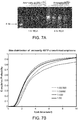
- the rolling circle amplicons are single stranded DNA which are highly folded. As shown in Figure 3A , these structures run as a single large band around ⁇ 10-15-kb on an 1% agarose gel.
- a 50-base oligonucleotide is phosphorylated at the 5' end using polynucleotide kinase in the T4 ligase buffer for 15 min.
- the reaction mixture is incubated with CircLigase mixture at 60°C for 1 hour to generate circular templates for testing.
- the RCA primer (18 bases) is then hybridized to the circular template in solution and a diluted template:primer mixture is used for rolling circle amplification.
- the RCA reaction solution contained aminoallyl dUTP and normal dNTP at varying ratios (1:50 to 1:50,000).
- the reaction mixture was diluted in PBS and bound to amino-silane treated coverglass.
- the bound RCA amplicons were then visualized by staining it with SYBR safe and imaging it using an epifluorescence microscope (63x objective). The images were then processed using Imaris Bitplane to identify individual amplicons and measure the average diameter of each spot.
- the incorporation of aminoallyl dUTP leads to slightly smaller diameter of the average DNA amplicon size.
- the circular cDNA template was used for rolling circle amplification, during which a range of aminoallyl dUTP was added.
- the amplicon mixture in solution was then arrayed on a glass surface and hybridized to a common fluorescent probe sequence. Since aminoallyl dUTP has a single positive charge, the increasing incorporation of aminoallyl dUTP led to a reduction in the overall negative charge, making each DNA amplicon slightly more compact.
- the ratio shown in the graph legend represents the molar ratio of aminoallyl dUTP to dTTP during the amplification step.
- a 50-base oligonucleotide is phosphorylated at the 5' end using polynucleotide kinase in the T4 ligase buffer for 15 min.
- the reaction mixture is incubated with CircLigase mixture at 60°C for 1 hour to generate circular templates for testing.
- the RCA primer (18 bases) is then hybridized to the circular template in solution and a diluted template:primer mixture is used for rolling circle amplification with or without aminoallyl dUTP.
- the reaction mixture was diluted in PBS and bound to amino-silane treated coverglass.
- the bound RCA amplicons were then cross-linked with BS(PEG)9. They were then washed using a continuous stream of 2x SSC wash solution for 1 min, stained with SYBR safe and imaged using an epifluorescence microscope (63x objective).
- the DNA amplicons generated in solution are arrayed on a glass surface and cross-linked via the aminoallyl moiety. They were then exposed to a constant flow of distilled water running across its surface with and with the cross-linker chemistry for 5 min at room temperature and then imaged after SYBR Gold staining. As shown in Figure 3C , the DNA amplicons that were not cross-linked stretched out as a result of high shear stress for about 5 minutes. The DNA amplicons cross-linked with aminoallyl dUTP were morphologically preserved after high shear stress for 5 minutes.
- Human primary fibroblasts are grown on a 1.5 cover slip. They are fixed using 4% formaldehyde in PBS for 15 min, followed by three washes of 70% ethanol.
- the reverse transcription mixture containing 1 uM random hexamer or 0.1 uM polydT(18)V primer with additional adapter sequences, 250 uM dNTP, 40 uM aminoallyl dUTP, 20U RNase inhibitor and 100 U M-MuLV reverse transcriptase are then added to the fixed cells and incubated overnight at 37°C. The sample is then washed using PBS, and cross-linked using 100 uM BS(PEG)9 in PBS for 1 hour, followed by 1M Tris treatment for 15 min.
- the circularization mixture containing 25 U CircLigase, 1 mM MnCl and 1 M Betain is added, and the sample is incubated at 60°C.
- the residual RNA is degraded using a mixture of RNase cocktail and RNase H.
- the RCA primer is then hybridized to the sample at 60°C for 15 min.
- 100 U phi29 DNA polymerase, 250 uM dNTP and 40 uM aminoallyl dNTP are added to the sample and incubated at 30°C overnight.
- the sample is then washed using PBS, and cross-linked using 100 uM BS(PEG)9 in PBS for 1 hour, followed by 1M Tris treatment for 15 min.
- 1 uM fluorescently labeled oligonucleotides are diluted in 2x SSC and hybridized to the matrix containing the DNA amplicons at 60°C and washed. Imaging is done using Leica SP5 scanning confocal microscope using a 63x objectives. The fluorescent oligonucleotides are stripped off using 80% formamide heated to 80°C. The sample is then dried and stored at 4°C from July 2011 to March 2012. The sample was rehydrated in PBS, and rehybridized to the fluorescently labeled oligonucleotides and imaged. The second image was obtained using an epifluorescence microscope.
- the DNA amplicons are structurally and chemically stable over a long period of time once cross-linked.
- the DNA amplicons preserved as a three-dimensional matrix in human fibroblasts can be interrogated using fluorescent primers and stored in phosphate buffered solution for up to a year and re-interrogated without losing their structural or sequence information.
- the different image quality here reflects the difference between confocal microscopy vs. epifluorescence microscopy, not the sample quality.
- a DNA Amplicon Matrix Within a Cell is Structurally and Chemically Stable
- Human primary fibroblasts are grown on a 1.5 cover slip. They are fixed using 4% formaldehyde in PBS for 15 min, followed by three washes of 70% ethanol.
- the reverse transcription mixture containing 1 uM random hexamer or 0.1 uM polydT(18)V primer with additional adapter sequences, 250 uM dNTP, 40 uM aminoallyl dUTP, 20U RNase inhibitor and 100 U M-MuLV reverse transcriptase are then added to the fixed cells and incubated overnight at 37°C. The sample is then washed using PBS, and cross-linked using 100 uM BS(PEG)9 in PBS for 1 hour, followed by 1M Tris treatment for 15 min.
- the circularization mixture containing 25 U CircLigase, 1 mM MnCl and 1 M Betain is added, and the sample is incubated at 60°C.
- the residual RNA is degraded using a mixture of RNase cocktail and RNase H.
- the RCA primer is then hybridized to the sample at 60°C for 15 min.
- 100 U phi29 DNA polymerase, 250 uM dNTP and 40 uM aminoallyl dNTP are added to the sample and incubated at 30°C overnight.
- the sample is then washed using PBS, and cross-linked using 100 uM BS(PEG)9 in PBS for 1 hour, followed by 1M Tris treatment for 15 min.
- 1 uM fluorescently labeled oligonucleotides are diluted in 2x SSC and hybridized to the matrix containing the DNA amplicons at 60°C and washed. Imaging is done using Leica SP5 scanning confocal microscope using a 63x objectives. The fluorescent oligonucleotides are stripped off using 80% formamide heated to 80°C. The sample is then washed with distilled water and rehybridized to the fluorescently labeled oligonucleotides and imaged.
- the DNA amplicon matrix inside the cell can be stripped using harsh chemical agents (i.e. 0.1N NaOH, 80% formamide) and heated up to 95°C for a prolonged period of time without losing their structural integrity or definition.
- harsh chemical agents i.e. 0.1N NaOH, 80% formamide
- a DNA Amplicon Matrix Within a Cell is Structurally and Chemically Stable
- Human iPS cells are grown on a 1.5 cover slip. They are fixed using 4% formaldehyde in PBS for 15 min, followed by three washes of 70% ethanol.
- the reverse transcription mixture containing 1 uM random hexamer or 0.1 uM polydT(18)V primer with additional adapter sequences, 250 uM dNTP, 40 uM aminoallyl dUTP, 20U RNase inhibitor and 100 U M-MuLV reverse transcriptase are then added to the fixed cells and incubated overnight at 37°C. The sample is then washed using PBS, and cross-linked using 100 uM BS(PEG)9 in PBS for 1 hour, followed by 1M Tris treatment for 15 min.
- the circularization mixture containing 25 U CircLigase, 1 mM MnCl and 1 M Betain is added, and the sample is incubated at 60°C.
- the residual RNA is degraded using a mixture of RNase cocktail and RNase H.
- the RCA primer is then hybridized to the sample at 60°C for 15 min.
- 100 U phi29 DNA polymerase, 250 uM dNTP and 40 uM aminoallyl dNTP are added to the sample and incubated at 30°C overnight.
- the sample is then washed using PBS, and cross-linked using 100 uM BS(PEG)9 in PBS for 1 hour, followed by 1M Tris treatment for 15 min.
- 1 uM fluorescently labeled oligonucleotides are diluted in 2x SSC and hybridized to the matrix containing the DNA amplicons at 60°C and washed three times using 2x SSC. After imaging, the fluorescent oligonucleotides are stripped off using 80% formamide heated to 80°C. The sample is then washed with distilled water and rehybridized to the fluorescently labeled oligonucleotides. This cycle is repeated sixty times (5 minutes per cycle). The multiple images were then aligned and processed using MatLab to identify a region of interest.
- the sample can be cycled through more than 50 heating, cooling, enzymatic and chemical reactions without any changes in the signal to noise ratio.
- the high absolute signal intensity here was due to insufficient probe washing in the initial cycles.
- the individual DNA amplicons in the matrix was imaged in three dimensions using confocal microscopy and tracked over 12 cycles, one measure the relative displacement of each amplicon over time.
- the mean displacement of each amplicon was ⁇ 500-nm in both lateral and axial dimensions, which was about the diameter of each amplicon.
- An example image of the analysis is shown in the right panel, in which the line representing the displacement is shown in different colors according to their cycle number.
- Human iPS cells are grown on a 1.5 cover slip. They are fixed using 4% formaldehyde in PBS for 15 min, followed by three washes of 70% ethanol.
- the reverse transcription mixture containing 1 uM random hexamer or 0.1 uM polydT(18)V primer with additional adapter sequences, 250 uM dNTP, 40 uM aminoallyl dUTP, 20U RNase inhibitor and 100 U M-MuLV reverse transcriptase are then added to the fixed cells and incubated overnight at 37°C. The sample is then washed using PBS, and cross-linked using 100 uM BS(PEG)9 in PBS for 1 hour, followed by 1M Tris treatment for 15 min.
- the circularization mixture containing 25 U CircLigase, 1 mM MnCl and 1 M Betain is added, and the sample is incubated at 60°C.
- the residual RNA is degraded using a mixture of RNase cocktail and RNase H.
- the RCA primer is then hybridized to the sample at 60°C for 15 min.
- 100 U phi29 DNA polymerase, 250 uM dNTP and 40 uM aminoallyl dNTP are added to the sample and incubated at 30°C overnight.
- the sample is then washed using PBS, and cross-linked using 100 uM BS(PEG)9 in PBS for 1 hour, followed by 1M Tris treatment for 15 min.
- 1 uM fluorescently labeled oligonucleotides are diluted in 2x SSC and hybridized to the matrix containing the DNA amplicons at 60°C and washed three times using 2x SSC.
- Leica SP5 scanning confocal microscope with 63x objective is used and scanning optical zoom of 5x is used. The line scan was repeated three times and averaged to generate a high quality image.
- Figure 5A is an image of the DNA amplicons (derived from reverse transcription of the cytoplasmic and the nuclear RNA) embedded within the cross-linked matrix inside human induced pluripotent stem cells. Individual amplicons are too tightly packed to visualize discrete amplicons, given the optical diffraction limitation in microscopy. But various subcellular compartments where the RNA is not expected to be present (i.e. nm: nuclear membrane, pm: plasma membrane) show dark staining, whereas the nucleus (Nu) and the cytoplasm (Cy) show a high density of the amplicons.
- the distribution of the cellular RNA shows unique patterns from cell to cell (1st panel vs. 2nd panel) and from one cell cycle phase to another (1st panel vs. 3rd panel). These results show that the DNA amplicons can be immobilized, amplified and interrogated in a manner to reflect their original spatial information.
- Human iPS cells are grown on a 1.5 cover slip. They are fixed using 4% formaldehyde in PBS for 15 min, followed by three washes of 70% ethanol.
- the reverse transcription mixture containing 1 uM random hexamer or 0.1 uM polydT(18)V primer with additional adapter sequences, 250 uM dNTP, 40 uM aminoallyl dUTP, 20U RNase inhibitor and 100 U M-MuLV reverse transcriptase are then added to the fixed cells and incubated overnight at 37°C. The sample is then washed using PBS, and cross-linked using 100 uM BS(PEG)9 in PBS for 1 hour, followed by 1M Tris treatment for 15 min.
- the circularization mixture containing 25 U CircLigase, 1 mM MnCl and 1 M Betain is added, and the sample is incubated at 60°C.
- the residual RNA is degraded using a mixture of RNase cocktail and RNase H.
- the RCA primer is then hybridized to the sample at 60°C for 15 min.
- 100 U phi29 DNA polymerase, 250 uM dNTP and 40 uM aminoallyl dNTP are added to the sample and incubated at 30°C overnight.
- the sample is then washed using PBS, and cross-linked using 100 uM BS(PEG)9 in PBS for 1 hour, followed by 1M Tris treatment for 15 min.
- the sequencing primer is designed with different 3' ends that that each primer can detect only 1/4th of the amplicons. If different dinucleotides are added to the 3' ends of the primer, each primer can detect only 1/16th of the amplicons.
- a chosen sequencing primer in 2x SSC is hybridized to the sample at 60°C for 15 minutes and washed.
- a ligation mixture containing 10 U T4 DNA ligase, ligation buffer and 1 uM fluorescently labeled nonamers (a pool containing A, G, C or T at fixed positions and labeled with FITC, Cy3, Texas Red or Cy5, respectively) is added and incubated for 50 min at room temperature.
- the cell After washing three times with 2x SSC, the cell is imaged on Leica SP5 scanning confocal microscope using four color channels. After imaging, the probe complex is stripped using 80% formamide and washed with distilled water. The sequencing by ligation step is repeated using a different nonamer set interrogating the next sequence.
- the left panel shows a subset of randomly primed cDNA amplicons being sequenced on a confocal microscope.
- the right panel shows GAPDH cDNA amplicons being sequenced over time using confocal microscopy. Only a single optical section is shown here.
- the axial dimension represents time or sequencing cycle steps.
- Circular DNA is Cross-linked or Co-polymerized Into A Matrix and Amplified
- circular DNA including cDNA
- a given cross-linker chemistry i.e. aminoallyl, thiol, biotin
- modified dUTP that competes with natural dTTP.
- the circular DNA is then cross-linked and/or co-polymerized within a three-dimensional container (i.e. cell), conforming the shape and the size of the container. Uncrosslinked molecules are then washed away, and one then performs rolling circle amplification, followed by imaging (i.e. sequencing).
- the density, the size and the signal strength can be controlled by varying the template size, the amplification time and the detection primer sequence.
- the DNA amplicons can be made into an ordered 3D matrix in a suitable scaffold material with addressable primers that can serve as amplification primers.
Abstract
Description
- This invention was made with Government support under grant number RC2HL102815 awarded by NHLBI and 1P50HG005550 awarded by NHGRI. The Government has certain rights in the invention.
- The present invention relates to methods of making a three-dimensional matrix of nucleic acids and amplifying, detecting and sequencing such nucleic acids within the matrix.
- Since many gene products such as RNA and proteins are enriched in regions where they function, their location provides an important clue to their function. This property has been used for in situ fluorescent hybridization, immunohistochemistry and tissue-specific reporter assays in numerous areas of biological research.
- Current methods involve extracting nucleic acid molecules from their native environment or making synthetic nucleic acid molecules, amplifying them in solution and placing them on a flat array surface or beads for gene detecting via hybridization or sequencing, making it impossible to identify the cellular origin of individual nucleic acids.
- Embodiments of the present invention are directed to methods of making a three-dimensional matrix of nucleic acids. Embodiments of the present invention are directed to methods of making a three-dimensional matrix including nucleic acids covalently bound into a matrix or into or to a matrix material. The nucleic acids may be co-polymerized with the matrix material or cross-linked to the matrix material or both. According to one aspect, a plurality of nucleic acid sequences of certain length, such as DNA or RNA sequences are part of a three-dimensional copolymer. The nucleic acids may then be amplified and sequenced in situ, i.e. within the matrix. The three-dimensional matrix of nucleic acids provides, in a certain aspect, an information storage medium where the nucleic acids, i.e. a sequence of one or more nucleotides, represent stored information which can be read within the three-dimensional matrix. According to one aspect, nucleic acids such as DNA or RNA sequences of given length are covalently attached to a matrix material to preserve their spatial orientation in the x, y and z axes within the matrix. It is to be understood that the three-dimensional matrix may include a matrix material and that the term copolymer, matrix and matrix material may be used interchangeably.
- According to one aspect, methods described herein are directed to immobilizing naturally occurring nucleic acids within their native environment, such as within a cell or within a tissue sample. The three-dimensional nucleic acid matrix can be generated in situ in a cell or tissue sample to preserve the naturally occurring nucleic acid sequence diversity (such as DNA and RNA) and spatial orientation in cells, tissues or any other complex biomaterial. According to this aspect, the location of nucleic acids and their relative position is identified as a three-dimensional structure, such as within subcellular compartments, within cells, within tissues, as three-dimensional nucleic acid assemblies, as three-dimensional nucleic acid material, etc. The nucleic acids can be amplified and sequenced, if desired, in situ thereby providing positional information of the nucleic acids within the cell or tissue.
- According to a related aspect, nucleic acids of interest, whether naturally occurring or synthetic, can be present within a three-dimensional matrix material and covalently attached to the three-dimensional matrix material such that the relative position of each nucleic acid is fixed, i.e. immobilized, within the three-dimensional matrix material. In this manner, a three-dimensional matrix of covalently bound nucleic acids of any desired sequence is provided. Each nucleic acid has its own three-dimensional coordinates within the matrix material and each nucleic acid represents information. In this manner, a large amount of information can be stored in a three-dimensional matrix. Individual information-encoding nucleic acids, such as DNA or RNA can be amplified and sequenced in situ, i.e., within the matrix, thereby enabling a large amount of information to be stored and read in a suitable three-dimensional material.
- According to a further aspect, the nucleic acids can be amplified to produce amplicons within the three-dimensional matrix material. The amplicons can then be covalently attached to the matrix, for example, by copolymerization or cross-linking. This results in a structurally stable and chemically stable three-dimensional matrix of nucleic acids. According to this aspect, the three-dimensional matrix of nucleic acids allows for prolonged information storage and read-out cycles. The nucleic acid / amplicon matrix allows for high throughput sequencing of a wide ranging array of biological and non-biological samples in three dimensions.
- According to certain aspects, a three-dimensional nucleic acid matrix is provided where a plurality of nucleic acid molecules, such as DNA or RNA, amplicons or nucleic acid structural units are immobilized, such as by covalent bonding to the matrix, in a three-dimensional space relative to one another. In this context, the nucleic acid molecules are rigidly fixed to the extent that they maintain their coordinate position within the matrix. It is to be understood that even though a nucleic acid molecule may be covalently attached to the three-dimensional matrix material, the nucleic acid molecule itself may be capable of movement though bound to the matrix, such as for example, when a nucleic acid sequence is bound to the matrix at a single location on the nucleic acid.
- According to one aspect, the three-dimensional matrix including nucleic acids is porous. According to one aspect, the three-dimensional matrix including nucleic acids is porous to the extent that reagents typically used in amplification methods can diffuse or otherwise move through the matrix to contact nucleic acids and thereby amplify nucleic acids under suitable conditions.
- According to one aspect, the three-dimensional matrix material is chemically inert and thermally stable to allow for various reaction conditions and reaction temperatures. According to this aspect, the three-dimensional matrix material is chemically inert and thermally stable to conditions used in amplification and sequencing methods known to those of skill in the art.
- According to one aspect, the three-dimensional matrix material is optically transparent. According to one aspect, the three-dimensional matrix material is optically transparent to allow for three-dimensional imaging techniques known to those of skill in the art.
- According to one aspect, the nucleic acids are amplified to an extent to produce sufficient levels of amplicons for three-dimensional imaging. For example, the nucleic acids are amplified and include a label sufficient for a high level of fluorescence compatible with three-dimensional imaging.
- According to one aspect, the material used to form the matrix is compatible with a wide range of biological and non-biological specimens in situ so as to avoid extracting the nucleic acid molecules away from their native environment.
- According to one aspect, the matrix material may be a semi-solid medium that can be made from polyacrylamide, cellulose, alginate, polyamide, cross-linked agarose, cross-linked dextran or cross-linked polyethylene glycol. In certain aspects, the semi-solid medium has x, y and z axes, and the nucleic acids are present randomly or non-randomly within the three-dimensional matrix.
- According to one aspect, the matrix material is porous. Porosity can result from polymerization and/or crosslinking of molecules used to make the matrix material. The diffusion property within the gel matrix is largely a function of the pore size. The molecular sieve size is chosen to allow for rapid diffusion of enzymes, oligonucleotides, formamide and other buffers used for amplification and sequencing (>50-nm). The molecular sieve size is also chosen so that large DNA or RNA amplicons do not readily diffuse within the matrix (<500-nm). The porosity is controlled by changing the cross-linking density, the chain lengths and the percentage of co-polymerized branching monomers according to methods known to those of skill in the art.
- In certain aspects, the semi-solid medium can be attached to a solid support such as a microscope slide or a flow cell. The solid support can be attached to the bottom surface of the semi-solid medium.
- The foregoing and other features and advantages of the present invention will be more fully understood from the following detailed description of illustrative embodiments taken in conjunction with the accompanying drawings.
-
Figure 1 depicts a schematic of nucleic acids at relative positions within a three-dimension environment and extraction and placement onto a two-dimensional environment, such as a glass slide or flow chamber. -
Figure 2 depicts in schematic the process of creating a matrix of nucleic acids within cells in situ, followed by amplifying the nucleic acids, such as DNA or RNA, in situ, co-polymerizing the amplicons in situ, covalently attaching the amplicons to the matrix material, interrogating the amplicons and imaging the amplicons along with a reconstructed 3D cell image with DNA/RNA amplicons on the order of 10-7m. -
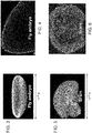
Figure 3 is an image of a whole mount Drosophilia embryo. -
Figure 4 is an optical section of a fly embryo. -
Figure 5 is an image of a whole mount mouse brain section. -
Figure 6 is an optical section of a mouse brain. -
Figure 7A is a gel image of aminoallyl dUTP after 1 hour of rolling circle amplification and after 4 hours of rolling circle amplification -
Figure 7B is a graph representative of the molar ratio of aminoallyl dUTP to dTTP during amplification. -
Figures 7C-E depict DNA amplicons with no shear stress (7C), DNA amplicons with no crosslinking and stretched out from 5 minutes of high shear stress (7D), and DNA amplicons with aminoallyl dUTP cross-linking being morphologically preserved after 5 minutes of high shear stress. -
Figure 8A depicts DNA amplicons cross-linked in fibroblasts. -
Figure 8B depicts the results of experiments demonstrating structural integrity of a DNA amplicon matrix within a cell. -
Figure 8C depicts the results of experiments demonstrating structural integrity of a DNA amplicon matrix within a cell after numerous chemical reactions. -
Figure 9A depicts amplicons within pluripotent stem cells. -
Figure 9B depicts confocal microscope images of cells with amplicons being sequenced. -
Figure 10 depicts in schematic a process for crosslinking or copolymerizing circular DNA, amplifying the circular DNA to produce amplicons and then placing the DNA amplicons into an ordered 3D matrix using a suitable scaffold material with addressable primers that can serve as amplification primers. - The present invention provides:
- 1. A method of making a three-dimensional matrix including a plurality of nucleic acids comprising
contacting a plurality of nucleic acids having a three-dimensional spatial relationship with a matrix-forming material in a manner to substantially retain the three-dimensional spatial relationship, and
forming a three-dimensional polymerized matrix including the plurality of nucleic acids covalently bound to the matrix. - 2. The method according to 1. above further including the step of amplifying the plurality of nucleic acids to produce amplicons within the matrix.
- 3. The method according to 2. above, further including the step of covalently bonding the amplicons to the matrix.
- 4. The method according to 1. above wherein the plurality of nucleic acids is contained within a biological sample and the matrix-forming material is introduced into the biological sample.
- 5. The method according to 1. above wherein the plurality of nucleic acids is contained within a cell and the matrix-forming material is introduced into the cell.
- 6. The method according to 1. above wherein the plurality of nucleic acids is contained within a tissue sample and the matrix-forming material is introduced into the tissue sample.
- 7. A method of identifying one or more nucleic acids within a cell comprising contacting a plurality of nucleic acids having a three-dimensional spatial relationship within the cell with a matrix-forming material in a manner to substantially retain the three-dimensional spatial relationship,
forming a three-dimensional polymerized matrix including the plurality of nucleic acids covalently bound to the matrix,
amplifying the plurality of nucleic acids to produce amplicons within the matrix, covalently bonding the amplicons to the matrix, and
sequencing the amplicons to identify the one or more nucleic acids. - 8. A method of identifying the spatial relationship of one or more nucleic acids within a cell comprising contacting a plurality of nucleic acids having a three-dimensional spatial relationship within the cell with a matrix-forming material in a manner to substantially retain the three-dimensional spatial relationship,
forming a three-dimensional polymerized matrix including the plurality of nucleic acids covalently bound to the matrix,
amplifying the plurality of nucleic acids to produce amplicons within the matrix, covalently bonding the amplicons to the matrix,
labeling the amplicons with a detectable label, and
imaging the amplicons to identify the relative position within the cell. - 9. A three-dimensional matrix including a plurality of nucleic acids covalently bound to the matrix, wherein the plurality of nucleic acids has a spatial relationship within the matrix relative to each other.
- 10. A three-dimensional matrix including a plurality of nucleic acids covalently bound to the matrix, wherein the plurality of nucleic acids has a spatial relationship within the matrix relative to each other, and wherein the plurality of nucleic acids has associated amplicons covalently bound to the matrix.
- 11. A method of making amplified circular nucleic acids comprising attaching a plurality of circular nucleic acids together,
amplifying the circular nucleic acids to produce amplicons, and
covalently attaching the amplicons to the circular nucleic acids. - 12. A plurality of circular nucleic acids covalently bound together and with each circular nucleic acid having a plurality of amplicons bound thereto.
- 13. A method of making a matrix of amplified circular nucleic acids comprising
attaching a plurality of circular nucleic acids together within a matrix-forming material,
amplifying the circular nucleic acids to produce amplicons, and
covalently attaching the amplicons to the circular nucleic acids. - The present invention provides a three-dimensional matrix of a plurality of nucleic acids. The present invention provides a three-dimensional matrix including a plurality of nucleic acids bound thereto. According to one aspect, the matrix is a three-dimensional nucleic acid-containing polymer. The nucleic acids may be naturally occurring nucleic acids or non-naturally occurring nucleic acids, such as nucleic acids that have been made using synthetic methods. The nucleic acids in the three-dimensional matrix may be ordered or unordered. The nucleic acids in the three-dimensional matrix may be present in their natural spatial relationship within a cell, tissue or organism. The nucleic acids in the three-dimensional matrix may be present in rows and columns within the three-dimensional matrix.
- According to one aspect, the nucleic acids are modified to incorporate a functional moiety for attachment to the matrix. The functional moiety can be covalently cross-linked, copolymerize with or otherwise non-covalently bound to the matrix. The functional moiety can react with a cross-linker. The functional moiety can be part of a ligand-ligand binding pair. dNTP or dUTP can be modified with the functional group, so that the function moiety is introduced into the DNA during amplification. A suitable exemplary functional moiety includes an amine, acrydite, alkyne, biotin, azide, and thiol. In the case of crosslinking, the functional moiety is cross-linked to modified dNTP or dUTP or both. Suitable exemplary cross-linker reactive groups include imidoester (DMP), succinimide ester (NHS), maleimide (Sulfo-SMCC), carbodiimide (DCC, EDC) and phenyl azide. Cross-linkers within the scope of the present disclosure may include a spacer moiety. Such spacer moieties may be functionalized. Such spacer moieties may be chemically stable. Such spacer moieties may be of sufficient length to allow amplification of the nucleic acid bound to the matrix. Suitable exemplary spacer moieties include polyethylene glycol, carbon spacers, photo-cleavable spacers and other spacers known to those of skill in the art and the like. According to one aspect, a matrix-forming material is contacted to a plurality of nucleic acids spatially arrange in three-dimensions relative to one another.
- Matrix forming materials include polyacrylamide, cellulose, alginate, polyamide, cross-linked agarose, cross-linked dextran or cross-linked polyethylene glycol. The matrix forming materials can form a matrix by polymerization and/or crosslinking of the matrix forming materials using methods specific for the matrix forming materials and methods, reagents and conditions known to those of skill in the art.
- According to one aspect, a matrix-forming material can be introduced into a cell. The cells are fixed with formaldehyde and then immersed in ethanol to disrupt the lipid membrane. The matrix forming reagents are added to the sample and are allowed to permeate throughout the cell. A polymerization inducing catalyst, UV or functional cross-linkers are then added to allow the formation of a gel matrix. The un-incorporated material is washed out and any remaining functionally reactive group is quenched. Exemplary cells include any cell, human or otherwise, including diseased cells or healthy cells. Certain cells include human cells, non-human cells, human stem cells, mouse stem cells, primary cell lines, immortalized cell lines, primary and immortalized fibroblasts, HeLa cells and neurons.
- According to one aspect, a matrix-forming material can be used to encapsulate a biological sample, such as a tissue sample. The formalin-fixed embedded tissues on glass slides are incubated with xylene and washed using ethanol to remove the embedding wax. They are then treated with Proteinase K to permeabilized the tissue. A polymerization inducing catalyst, UV or functional cross-linkers are then added to allow the formation of a gel matrix. The un-incorporated material is washed out and any remaining functionally reactive group is quenched. Exemplary tissue samples include any tissue samples of interest whether human or non-human. Such tissue samples include those from skin tissue, muscle tissue, bone tissue, organ tissue and the like. Exemplary tissues include human and mouse brain tissue sections, embryo sections, tissue array sections, and whole insect and worm embryos.
- The matrix-forming material forms a three-dimensional matrix including the plurality of nucleic acids. According to one aspect, the matrix-forming material forms a three-dimensional matrix including the plurality of nucleic acids while maintaining the spatial relationship of the nucleic acids. In this aspect, the plurality of nucleic acids is immobilized within the matrix material. The plurality of nucleic acids may be immobilized within the matrix material by co-polymerization of the nucleic acids with the matrix-forming material. The plurality of nucleic acids may also be immobilized within the matrix material by crosslinking of the nucleic acids to the matrix material or otherwise cross-linking with the matrix-forming material. The plurality of nucleic acids may also be immobilized within the matrix by covalent attachment or through ligand-protein interaction to the matrix.
- According to one aspect, the matrix is porous thereby allowing the introduction of reagents into the matrix at the site of a nucleic acid for amplification of the nucleic acid. A porous matrix may be made according to methods known to those of skill in the art. In one example, a polyacrylamide gel matrix is co-polymerized with acrydite-modified streptavidin monomers and biotinylated DNA molecules, using a suitable acrylamide:bis-acrylamide ratio to control the cross-linking density. Additional control over the molecular sieve size and density is achieved by adding additional cross-linkers such as functionalized polyethylene glycols. According to one aspect, the nucleic acids, which may represent individual bits of information, are readily accessed by oligonucleotides, such as labeled oligonucleotide probes, primers, enzymes and other reagents with rapid kinetics.
- According to one aspect, the matrix is sufficiently optically transparent or otherwise has optical properties suitable for standard Next Generation sequencing chemistries and deep three-dimensional imaging for high throughput information readout. The Next Generation sequencing chemistries that utilize fluorescence imaging include ABI SoLiD (Life Technologies), in which a sequencing primer on a template is ligated to a library of fluorescently labeled nonamers with a cleavable terminator. After ligation, the beads are then imaged using four color channels (FITC, Cy3, Texas Red and Cy5). The terminator is then cleaved off leaving a free-end to engage in the next ligation-extension cycle. After all dinucleotide combinations have been determined, the images are mapped to the color code space to determine the specific base calls per template. The workflow is achieved using an automated fluidics and imaging device (i.e. SoLiD 5500 W Genome Analyzer, ABI Life Technologies). Another sequencing platform uses sequencing by synthesis, in which a pool of single nucleotide with a cleavable terminator is incorporated using DNA polymerase. After imaging, the terminator is cleaved and the cycle is repeated. The fluorescence images are then analyzed to call bases for each DNA amplicons within the flow cell (HiSeq, Illumia).
- According to certain aspects, the plurality of nucleic acids may be amplified to produce amplicons by methods known to those of skill in the art. The amplicons may be immobilized within the matrix generally at the location of the nucleic acid being amplified, thereby creating a localized colony of amplicons. The amplicons may be immobilized within the matrix by steric factors. The amplicons may also be immobilized within the matrix by covalent or noncovalent bonding. In this manner, the amplicons may be considered to be attached to the matrix. By being immobilized to the matrix, such as by covalent bonding or crosslinking, the size and spatial relationship of the original amplicons is maintained. By being immobilized to the matrix, such as by covalent bonding or crosslinking, the amplicons are resistant to movement or unraveling under mechanical stress.
- According to one aspect, the amplicons, such as DNA amplicons, are then copolymerized and/or covalently attached to the surrounding matrix thereby preserving their spatial relationship and any information inherent thereto. For example, if the amplicons are those generated from DNA or RNA within a cell embedded in the matrix, the amplicons can also be functionalized to form covalent attachment to the matrix preserving their spatial information within the cell thereby providing a subcellular localization distribution pattern.
- As used herein, the term "attach" refers to both covalent interactions and noncovalent interactions. A covalent interaction is a chemical linkage between two atoms or radicals formed by the sharing of a pair of electrons (i.e., a single bond), two pairs of electrons (i.e., a double bond) or three pairs of electrons (i.e., a triple bond). Covalent interactions are also known in the art as electron pair interactions or electron pair bonds. Noncovalent interactions include, but are not limited to, van der Waals interactions, hydrogen bonds, weak chemical bonds (i.e., via short-range noncovalent forces), hydrophobic interactions, ionic bonds and the like. A review of noncovalent interactions can be found in Alberts et al., in Molecular Biology of the Cell, 3d edition, Garland Publishing, 1994.
- As used herein, the term "nucleic acid" includes the term "oligonucleotide" or "polynucleotide" which includes a plurality of nucleotides. The term "nucleic acid" is intended to include naturally occurring nucleic acids and synthetic nucleic acids. The term "nucleic acid" is intended to include single stranded nucleic acids and double stranded nucleic acids. The term "nucleic acid" is intended to include DNA and RNA, whether single stranded or double stranded. Nucleotides of the present invention will typically be the naturally-occurring nucleotides such as nucleotides derived from adenosine, guanosine, uridine, cytidine and thymidine. When oligonucleotides are referred to as "double-stranded," it is understood by those of skill in the art that a pair of oligonucleotides exists in a hydrogen-bonded, helical array typically associated with, for example, DNA. In addition to the 100% complementary form of double-stranded oligonucleotides, the term "double-stranded" as used herein is also meant to include those form which include such structural features as bulges and loops (see Stryer, Biochemistry, Third Ed. (1988)). As used herein, the term "polynucleotide" refers to a strand of nucleic acids that can be a variety of different sizes. Polynucleotides may be the same size as an oligonucleotide, or may be two-times, three-times, four-times, five-times, ten-times, or greater than the size of an oligonucleotide.
- Oligonucleotides and/or polynucleotides may be isolated from natural sources or purchased from commercial sources. Oligonucleotide and/or polynucleotide sequences may be prepared by any suitable method, e.g., the phosphoramidite method described by Beaucage and Carruthers ((1981) Tetrahedron Lett. 22: 1859) or the triester method according to Matteucci et al. (1981) J. Am. Chem. Soc. 103:3185), or by other chemical methods using either a commercial automated oligonucleotide synthesizer or high-throughput, high-density array methods described herein and known in the art (see
U.S. Patent Nos. 5,602,244 ,5,574,146 ,5,554,744 ,5,428,148 ,5,264,566 ,5,141,813 ,5,959,463 ,4,861,571 and4,659,774 ). Pre-synthesized oligonucleotides may also be obtained commercially from a variety of vendors. - In certain embodiments of the invention oligonucleotides and/or polynucleotides may be prepared using a variety of microarray technologies known in the art. Pre-synthesized oligonucleotide and/or polynucleotide sequences may be attached to a support or synthesized in situ using light-directed methods, flow channel and spotting methods, inkjet methods, pin-based methods and bead-based methods set forth in the following references: McGall et al. (1996) Proc. Natl. Acad. Sci. U.S.A. 93:13555; Synthetic DNA Arrays In Genetic Engineering, Vol. 20:111, Plenum Press (1998); Duggan et al. (1999) Nat. Genet. S21:10; Microarrays: Making Them and Using Them In Microarray Bioinformatics, Cambridge University Press, 2003;
U.S. Patent Application Publication Nos. 2003/0068633 and2002/0081582 ;U.S. Patent Nos. 6,833,450 ,6,830,890 ,6,824,866 ,6,800,439 ,6,375,903 and5,700,637 ; andPCT Application Nos. WO 04/031399 WO 04/031351 WO 04/029586 WO 03/100012 WO 03/066212 WO 03/065038 WO 03/064699 WO 03/064027 WO 03/064026 WO 03/046223 WO 03/040410 WO 02/24597s - Nucleic acids may be obtained from libraries, e.g., genomic libraries, cDNA libraries and the like. Examples of methods for the synthesis of molecular libraries can be found in the art, for example in: DeWitt et al. (1993) Proc. Natl. Acad. Sci. USA 90:6909; Erb et al. (1994) Proc. Natl. Acad. Sci. USA 91:11422; Zuckermann et al. (1994) J. Med. Chem. 37:2678; Cho et al. (1993) Science 261:1303; Carrell et al. (1994) Angew. Chem. Int. Ed. Engl. 33:2059; Carell et al. (1994) Angew. Chem. Int. Ed. Engl. 33:2061; and in Gallop et al. (1994) J. Med. Chem. 37:1233.
- In certain embodiments, nucleic acids are those found naturally in a biological sample, such as a cell or tissue.
- In still other aspects, a matrix is used in conjunction with a solid support. For example the matrix can be polymerized in such a way that one surface of the matrix is attached to a solid support (e.g., a glass surface), while the other surface of the matrix is exposed or sandwiched between two solid supports. According to one aspect, the matrix can be contained within a container.
- Solid supports of the invention may be fashioned into a variety of shapes. In certain embodiments, the solid support is substantially planar. Examples of solid supports include plates such as slides, microtitre plates, flow cells, coverslips, microchips, and the like, containers such as microfuge tubes, test tubes and the like, tubing, sheets, pads, films and the like. Additionally, the solid supports may be, for example, biological, nonbiological, organic, inorganic, or a combination thereof.
- Embodiments of the present invention are further directed to the amplification of nucleic acid sequences within the matrix, i.e. in situ, within the matrix. Methods of amplifying nucleic acids include rolling circle amplification in situ. In certain aspects, methods of amplifying nucleic acids involves the use of PCR, such as anchor PCR or RACE PCR, or, alternatively, in a ligation chain reaction (LCR) (see, e.g., Landegran et al. (1988) Science 241:1077-1080; and Nakazawa et al. (1994) Proc. Natl. Acad. Sci. U.S.A. 91:360-364). Alternative amplification methods include: self-sustained sequence replication (Guatelli et al. (1990) Proc. Natl. Acad. Sci. USA 87:1874), transcriptional amplification system (Kwoh et al. (1989) Proc. Natl. Acad. Sci. US. 86:1173), Q-Beta Replicase (Lizardi et al. (1988) BioTechnology 6:1197), recursive PCR (Jaffe et al. (2000) J. Biol. Chem. 275:2619; and Williams et al. (2002) J. Biol. Chem. 277:7790) or any other nucleic acid amplification method using techniques well known to those of skill in the art. A variety of amplification methods are described in
U.S. Patent Nos. 6,391,544 ,6,365,375 ,6,294,323 ,6,261,797 ,6,124,090 and5,612,199 . - Embodiments of the present invention are directed to methods of amplifying nucleic acids in situ within the matrix by contacting the nucleic acids within the matrix with reagents and under suitable reaction conditions sufficient to amplify the nucleic acids. According to one aspect, the matrix is porous to allow migration of reagents into the matrix to contact the nucleic acids. In certain aspects, oligonucleotides are amplified by selectively hybridizing an amplification primer to an amplification site at the 3' end of an oligonucleotide using conventional methods. Amplification primers are 6 to 100, and even up to 1,000, nucleotides in length, but typically from 10 to 40 nucleotides, although oligonucleotides of different length are of use. Amplification primers may be present in solution to be added to the matrix or they may be added during formation of the matrix to be present therein sufficiently adjacent to nucleic acids to allow for hybridization and amplification.
- Typically, selective hybridization occurs when two nucleic acid sequences are substantially complementary, i.e., at least about 65% 75%, 80%, 85%, 90%, 91%, 92%, 93%, 94%, 95%, 96%, 97%, 98% 99%, 99.1%, 99.2%, 99.3%, 99.4%, 99.5%, 99.6%, 99.7%, 99.8%, 99.9% or 100% complementary over a stretch of at least 14 to 25 nucleotides. See Kanehisa, M., 1984, Nucleic Acids Res. 12: 203.
- Overall, five factors influence the efficiency and selectivity of hybridization of the primer to a second nucleic acid molecule. These factors, which are (i) primer length, (ii) the nucleotide sequence and/or composition, (iii) hybridization temperature, (iv) buffer chemistry and (v) the potential for steric hindrance in the region to which the primer is required to hybridize, are important considerations when non-random priming sequences are designed.
- There is a positive correlation between primer length and both the efficiency and accuracy with which a primer will anneal to a target sequence; longer sequences have a higher Tm than do shorter ones, and are less likely to be repeated within a given target sequence, thereby cutting down on promiscuous hybridization. Primer sequences with a high G-C content or that comprise palindromic sequences tend to self-hybridize, as do their intended target sites, since unimolecular, rather than bimolecular, hybridization kinetics are generally favored in solution; at the same time, it is important to design a primer containing sufficient numbers of G-C nucleotide pairings to bind the target sequence tightly, since each such pair is bound by three hydrogen bonds, rather than the two that are found when A and T bases pair. Hybridization temperature varies inversely with primer annealing efficiency, as does the concentration of organic solvents, e.g., formamide, that might be included in a hybridization mixture, while increases in salt concentration facilitate binding. Under stringent hybridization conditions, longer probes hybridize more efficiently than do shorter ones, which are sufficient under more permissive conditions. Stringent hybridization conditions typically include salt concentrations of less than about 1M, more usually less than about 500 mM and preferably less than about 200 mM. Hybridization temperatures range from as low as 0 °C to greater than 22 °C, greater than about 30 °C, and (most often) in excess of about 37 °C. Longer fragments may require higher hybridization temperatures for specific hybridization. As several factors affect the stringency of hybridization, the combination of parameters is more important than the absolute measure of any one alone. Hybridization conditions are known to those skilled in the art and can be found in Current Protocols in Molecular Biology, John Wiley & Sons, N.Y. (1989), 6.3.1-6.3.6.
- Primers are designed with the above first four considerations in mind. While estimates of the relative merits of numerous sequences are made mentally, computer programs have been designed to assist in the evaluation of these several parameters and the optimization of primer sequences (see, e.g., Hoover et al. (2002) Nucleic Acids Res. 30:e43, and Rouillard et al. (2004) Nucleic Acids Res. 32:W176).
- In accordance with certain examples, methods of sequencing nucleic acid in situ within a matrix are provided. General sequencing methods known in the art, such as sequencing by extension with reversible terminators, fluorescent in situ sequencing (FISSEQ), pyrosequencing, massively parallel signature sequencing (MPSS) and the like (described in Shendure et al. (2004) Nat. Rev. 5:335), are suitable for use with the matrix in which the nucleic acids are present. Reversible termination methods use step-wise sequencing-by-synthesis biochemistry that coupled with reversible termination and removable fluorescence (Shendure et al. supra and
U.S. Patent Nos. 5,750,341 and6,306,597 ). FISSEQ is a method whereby DNA is extended by adding a single type of fluorescently-labelled nucleotide triphosphate to the reaction, washing away unincorporated nucleotide, detecting incorporation of the nucleotide by measuring fluorescence, and repeating the cycle. At each cycle, the fluorescence from previous cycles is bleached or digitally subtracted or the fluorophore is cleaved from the nucleotide and washed away. FISSEQ is described further in Mitra et al. (2003) Anal. Biochem. 320:55, incorporated herein by reference in its entirety for all purposes. Pyrosequencing is a method in which the pyrophosphate (PPi) released during each nucleotide incorporation event (i.e., when a nucleotide is added to a growing polynucleotide sequence). The PPi released in the DNA polymerase-catalyzed reaction is detected by ATP sulfurylase and luciferase in a coupled reaction which can be visibly detected. The added nucleotides are continuously degraded by a nucleotide-degrading enzyme. After the first added nucleotide has been degraded, the next nucleotide can be added. As this procedure is repeated, longer stretches of the template sequence are deduced. Pyrosequencing is described further in Ronaghi et al. (1998) Science 281:363. MPSS utilizes ligation-based DNA sequencing simultaneously on microbeads. A mixture of labelled adaptors comprising all possible overhangs is annealed to a target sequence of four nucleotides. The label is detected upon successful ligation of an adaptor. A restriction enzyme is then used to cleave the DNA template to expose the next four bases. MPSS is described further in Brenner et al. (2000) Nat. Biotech. 18:630. - According to certain aspects, the nucleic acids within the matrix can be interrogated using methods known to those of skill in the art including fluorescently labeled oligonucleotide/DNA/RNA hybridization, primer extension with labeled ddNTP, sequencing by ligation and sequencing by synthesis. Ligated circular padlock probes described in Larsson, et al., (2004), Nat. Methods 1:227-232 can be used to detect multiple sequence targets in parallel, followed by either sequencing-by-ligation, -synthesis or -hybridization of the barcode sequences in the padlock probe to identify individual targets.
- According to one aspect, methods described herein produce a three-dimensional nucleic acid amplicon matrix which is stable, long-lasting and resistant, substantially resistant or partially resistant to enzymatic or chemical degradation. The three-dimensional nucleic acid amplicon matrix can be repeatedly interrogated using standard probe hybridization and/or fluorescence-based sequencing. The three-dimensional nucleic acid amplicon matrix can be repeatedly interrogated with little or no signal degradation, such as after more than 50 cycles, and with little position shift, such as less than 1 µm per amplicon.
- According to one aspect, a plurality of circular DNA molecules is covalently linked to one another. The circular DNA molecules are then amplified using methods known to those of skill in the art, such as isothermal enzymatic amplification one example of which is rolling circle amplification. According to this aspect, the amplicons are localized near the circular DNA. According to this aspect, the amplicons form a shell around the circular DNA or otherwise assemble around the circular DNA. Each circular DNA may have more than 1000 amplicons surrounding or otherwise associated therewith. According to this aspect, the amplicons surrounding a particular circular DNA provide a high signal intensity, due in part to the number of amplicons and/or detectable labels associated with the amplicons. The amplicons may be functionalized and cross-linked or otherwise covalently bound together around their associate circular DNA to form a series or network of tightly bound DNA amplicon shells around each circular DNA. The series or network of tightly bound DNA amplicon shells around each circular DNA may be assembled onto a three-dimensional support. According to one aspect, the series or network of tightly bound DNA amplicon shells around each circular DNA may be assembled onto a three-dimensional support producing a three-dimensional DNA polymer with defined overall shape, size and amplicon position.
- According to one aspect, amplicons are covalently linked without the need for separate cross-linkers, such as bis-N-succinimidyl-(nonaethylene glycol) ester. An acrydite moiety, such as a catalyst activated acrydite moiety is introduced at the end of a long carbon spacer (i.e., about C6 to about C12) at
position 5 of a uracil base a representative formula of which is shown below.
- In the formula below, R represents the acrydite spacer moiety attached to the 5 position of the uracil base.

- When copolymerized with bis-acrylamide in the presence of a catalyst, a polymerization reaction takes place, encapsulating the circular DNA with the amplicons and fixing the amplicons in position. The chemically inert nature of the polymerized mixture allows various downstream applications. The spacer can be a carbon chain of between about 2 carbons to about 200 carbons. The spacer can be polyethylene glycol. The length of the spacer can vary from about 30 angstroms to about 100 angstroms and can be of various molecular weights. The spacer can be permanent or reversible, such as by using UV light, enzymes, chemical cleavage, etc. A three-dimensional matrix, such as a polyacrylamide gel matrix, can be used to embed a variety of biological structures containing enzymatically or chemically modified DNA or RNA molecules containing an acrydite functional moiety or moieties. The non-nucleic acid component is selectively dissolved using detergents, proteases, organic solvents or denaturants to create a three-dimensional matrix that preserves individual DNA or RNA molecules and their relative spatial location. Examples include embedding cells, healthy and diseased tissues and tissue sections, small model organisms such as worms and insects, bacterial colonies or biofilm, environmental samples containing other DNA or RNA containing materials or organisms.
- This invention is further illustrated by the following examples, which should not be construed as limiting.
- Human iPS cells or human primary fibroblasts are grown on a 1.5 cover slip. They are fixed using 4% formaldehyde in PBS for 15 min, followed by three washes of 70% ethanol. The reverse transcription mixture containing 1 uM random hexamer or 0.1 uM polydT(18)V primer with additional adapter sequences (TCTCGGGAACGCTGAAGA), 250 uM dNTP, 40 uM aminoallyl dUTP (Anaspec), 20U RNase inhibitor and 100 U MMuLV reverse transcriptase (Enzymatics) are then added to the fixed cells and incubated overnight at 37°C. The sample is then washed using PBS, and cross-linked using 100 uM BS(PEG)9 (Thermo-Fisher Scientific) in PBS for 1 hour, followed by 1M Tris treatment for 15 min. The circularization mixture containing 25U CircLigase (Epicentre), 1 mM MnCl and 1 M Betain is added, and the sample is incubated at 60°C for 2 hours. The residual RNA is degraded using a mixture of RNase cocktail (Roche) and RNase H (Enzymatics) at 37°C for 1 hour. The RCA primer is then hybridized to the sample at 60°C for 15 min and washed. For rolling circle amplification, 100 U phi29 DNA polymerase (Enzymatics), 250 uM dNTP and 40 uM aminoallyl dNTP are added to the sample and incubated at 30°C overnight. The sample is then washed using PBS, and cross-linked using 100 uM BS(PEG)9 in PBS for 1 hour, followed by 1M Tris treatment for 15 min. For the DNA amplicon detection, 1 uM fluorescently label oligonucleotides are diluted in 2x SSC and hybridized to the matrix containing the DNA amplicons at 60°C and washed. Imaging is done using Leica SP5 scanning confocal microscope using 10x, 20x or 63x objectives in four color channels (FITC, Cy3, Texas Red and Cy5). The image stacks containing up to 50 optical sections are then visualized using Imaris Bitplane software for three-dimensional reconstruction of the DNA amplicons within the sample matrix.
- Methods described herein allow one to immobilize, amplify and image single DNA/RNA molecules in a three-dimensional space without perturbing the structure. As shown in
Figure 2 , single cells were grown in tissue culture. DNA/RNA was amplified in situ. The DNA/RNA was co-polymerized into a matrix material in situ, and individual amplicons were interrogated/hybridized with fluorescent oligonucleotides and imaged. When viewed under much higher magnification, individual amplicons can be imaged using confocal microscopy. This allows one to find out where different DNA/RNA molecules reside, how they are compartmentalized among different cell types and morphologies and how their representation changes over time in developing tissues. The similar concept can be used for many other specimens in both natural and synthetic materials, as long as they can be co-polymerized and/or encapsulated by the DNA amplicons. - According to one specific aspect, inside individual mammalian cells, 20 to 500K mRNA molecules are distributed throughput the cytoplasm (Islam et al., 2011). According to embodiment, cells are fixed and permeabilized. Cellular RNA is then converted into cDNA molecules using dUTP in place or in addition to dTTP. The cDNA molecules containing modified dUMP residues are then cross-linked to each other and circularized, forming a three-dimensional pseudo-polymer of circular cDNA molecules inside individual cells. Then rolling circle amplification is used to amplify the cDNA network into a DNA amplicon network. This cell-based DNA amplicon network then stores information about each transcript's identity, location, variation/mutations, etc. The cell-based DNA amplicon matrix can be read using sequencing by ligation (i.e. ABI SoLiD), sequencing by synthesis (i.e. Illumina), or any other proprietary or open sequencing chemistries (see Drmanac et al., 2010; Shendure et al., 2005). Given the three-dimensional nature of the DNA amplicon network, one can use confocal or multi-photon microscopy to sequencing individual amplicons throughout the whole thickness of the amplicon network, enabling one to visualize the cDNA distribution of transcripts between the apical side and the basal side of the cells as shown in
Fig. 2 . Given the tight packing density, one can selectively read different subpopulations sequentially, reducing the density of information read at any given time and extending over time for better spatial resolution. - Drosophila embryos are fixed using 4% formaldehyde in PBS, followed by multiple washes of 70% ethanol. The embryos are then mounted on a cover glass using an optically transparent adhesive. The reverse transcription mixture containing 1 uM random hexamer or 0.1 uM polydT(18)V primer with additional adapter sequences (TCTCGGGAACGCTGAAGA), 250 uM dNTP, 40 uM aminoallyl dUTP (Anaspec), 20U RNase inhibitor and 100 U MMuLV reverse transcriptase (Enzymatics) are then added to the fixed cells and incubated overnight at 37°C. The sample is then washed using PBS, and cross-linked using 100 uM BS(PEG)9 (Thermo-Fisher Scientific) in PBS for 1 hour, followed by 1M Tris treatment for 15 min. The circularization mixture containing 25U CircLigase (Epicentre), 1 mM MnCl and 1 M Betain is added, and the sample is incubated at 60°C for 2 hours. The residual RNA is degraded using a mixture of RNase cocktail (Roche) and RNase H (Enzymatics) at 37°C for 1 hour. The RCA primer is then hybridized to the sample at 60°C for 15 min and washed. For rolling circle amplification, 100 U phi29 DNA polymerase (Enzymatics), 250 uM dNTP and 40 uM aminoallyl dNTP are added to the sample and incubated at 30°C overnight. The sample is then washed using PBS, and cross-linked using 100 uM BS(PEG)9 in PBS for 1 hour, followed by 1M Tris treatment for 15 min. For the DNA amplicon detection, 1 uM fluorescently label oligonucleotides are diluted in 2x SSC and hybridized to the matrix containing the DNA amplicons at 60°C and washed. Imaging is done using Leica SP5 scanning confocal microscope using 10x, 20x or 63x objectives in four color channels (FITC, Cy3, Texas Red and Cy5). The image stacks are then visualized using Imaris Bitplane software for three-dimensional reconstruction of the DNA amplicons within the sample matrix.
- As shown in
Figure 2 , fly embryos were obtained and DNA/RNA was amplified in situ. The DNA/RNA was copolymerized into a matrix material in situ, and individual amplicons were interrogated/hybridized with fluorescent oligonucleotides and imaged. When viewed under much higher magnification, individual amplicons can be imaged using confocal microscopy even in these thick biological specimens. This allows one to find out where different DNA/RNA molecules reside, how they are compartmentalized among different cell types and morphologies and how their representation changes over time in developing tissues. The similar concept can be used for many other specimens in both natural and synthetic materials, as long as they can be co-polymerized and/or encapsulated by the DNA amplicons. - A fresh frozen adult mouse brain sections (20-um cryosections) are fixed using 4% formaldehyde in PBS. It is then treated with 0.4 ug/ml Proteinase K for 30 min at room temperature and thoroughly washed using 70%, 95% and 100% ethanol. The reverse transcription mixture containing 1 uM random hexamer or 0.1 uM polydT(18)V primer with additional adapter sequences (TCTCGGGAACGCTGAAGA), 250 uM dNTP, 40 uM aminoallyl dUTP (Anaspec), 20U RNase inhibitor and 100 U MMuLV reverse transcriptase (Enzymatics) are then added to the fixed cells and incubated overnight at 37°C. The sample is then washed using PBS, and cross-linked using 100 uM BS(PEG)9 (Thermo-Fisher Scientific) in PBS for 1 hour, followed by 1M Tris treatment for 15 min. The circularization mixture containing 25U CircLigase (Epicentre), 1 mM MnCl and 1 M Betain is added, and the sample is incubated at 60°C for 2 hours. The residual RNA is degraded using a mixture of RNase cocktail (Roche) and RNase H (Enzymatics) at 37°C for 1 hour. The RCA primer is then hybridized to the sample at 60°C for 15 min and washed. For rolling circle amplification, 100 U phi29 DNA polymerase (Enzymatics), 250 uM dNTP and 40 uM aminoallyl dNTP are added to the sample and incubated at 30°C overnight. The sample is then washed using PBS, and cross-linked using 100 uM BS(PEG)9 in PBS for 1 hour, followed by 1M Tris treatment for 15 min. For the DNA amplicon detection, 1 uM fluorescently label oligonucleotides are diluted in 2x SSC and hybridized to the matrix containing the DNA amplicons at 60°C and washed. Imaging is done using Leica epifluorescence microscope using a 10x objective in four color channels (FITC, Cy3, Texas Red and Cy5) in a tiled scan mode (20 by 15 separate images). The images are then stitched together during the image acquisition and visualized using Imaris Bitplane software.
- As shown in
Figure 2 , mouse brain sections were obtained and DNA/RNA was amplified in situ. The DNA/RNA was copolymerized into a matrix material in situ, and individual amplicons were interrogated/hybridized with fluorescent oligonucleotides and imaged. When viewed under much higher magnification, individual amplicons can be imaged using confocal microscopy even in these thick biological specimens. This allows one to find out where different DNA/RNA molecules reside, how they are compartmentalized among different cell types and morphologies and how their representation changes over time in developing tissues. The similar concept can be used for many other specimens in both natural and synthetic materials, as long as they can be co-polymerized and/or encapsulated by the DNA amplicons. - A 50-base oligonucleotide is phosphorylated at the 5' end using polynucleotide kinase in the T4 ligase buffer for 15 min. The reaction mixture is incubated with CircLigase mixture at 60°C for 1 hour to generate circular templates for testing. The RCA primer (18 bases) is then hybridized to the circular template in solution and a diluted template:primer mixture is used for rolling circle amplification. The RCA reaction solution contained 0, 0.1 uM, 1 uM or 10 uM aminoallyl dUTP in addition to the normal dNTP. The reaction mixture was then loaded onto an 1% agarose gel and visualized using SYBR safe dyes.
- The incorporation of aminoallyl dUTP that is later cross-linked to each other and to the amine exposing substrate still allows for reverse transcription using M-MuLV reverse transcriptase and rolling circle amplification using Phi29 DNA polymerase, albeit at a reduced rate in a concentration dependent manner. Increasing amounts of aminoallyl dUTP were added as a competitor to dTTP present in the amplification mixture in solution. The rolling circle amplicons are single stranded DNA which are highly folded. As shown in
Figure 3A , these structures run as a single large band around ∼10-15-kb on an 1% agarose gel. - A 50-base oligonucleotide is phosphorylated at the 5' end using polynucleotide kinase in the T4 ligase buffer for 15 min. The reaction mixture is incubated with CircLigase mixture at 60°C for 1 hour to generate circular templates for testing. The RCA primer (18 bases) is then hybridized to the circular template in solution and a diluted template:primer mixture is used for rolling circle amplification. The RCA reaction solution contained aminoallyl dUTP and normal dNTP at varying ratios (1:50 to 1:50,000). After 8 hours of RCA, the reaction mixture was diluted in PBS and bound to amino-silane treated coverglass. The bound RCA amplicons were then visualized by staining it with SYBR safe and imaging it using an epifluorescence microscope (63x objective). The images were then processed using Imaris Bitplane to identify individual amplicons and measure the average diameter of each spot.
- The incorporation of aminoallyl dUTP leads to slightly smaller diameter of the average DNA amplicon size. The circular cDNA template was used for rolling circle amplification, during which a range of aminoallyl dUTP was added. The amplicon mixture in solution was then arrayed on a glass surface and hybridized to a common fluorescent probe sequence. Since aminoallyl dUTP has a single positive charge, the increasing incorporation of aminoallyl dUTP led to a reduction in the overall negative charge, making each DNA amplicon slightly more compact. As shown in
Figure 3B , the ratio shown in the graph legend represents the molar ratio of aminoallyl dUTP to dTTP during the amplification step. - A 50-base oligonucleotide is phosphorylated at the 5' end using polynucleotide kinase in the T4 ligase buffer for 15 min. The reaction mixture is incubated with CircLigase mixture at 60°C for 1 hour to generate circular templates for testing. The RCA primer (18 bases) is then hybridized to the circular template in solution and a diluted template:primer mixture is used for rolling circle amplification with or without aminoallyl dUTP. After 8 hours of RCA, the reaction mixture was diluted in PBS and bound to amino-silane treated coverglass. The bound RCA amplicons were then cross-linked with BS(PEG)9. They were then washed using a continuous stream of 2x SSC wash solution for 1 min, stained with SYBR safe and imaged using an epifluorescence microscope (63x objective).
- The DNA amplicons generated in solution are arrayed on a glass surface and cross-linked via the aminoallyl moiety. They were then exposed to a constant flow of distilled water running across its surface with and with the cross-linker chemistry for 5 min at room temperature and then imaged after SYBR Gold staining. As shown in
Figure 3C , the DNA amplicons that were not cross-linked stretched out as a result of high shear stress for about 5 minutes. The DNA amplicons cross-linked with aminoallyl dUTP were morphologically preserved after high shear stress for 5 minutes. - Human primary fibroblasts are grown on a 1.5 cover slip. They are fixed using 4% formaldehyde in PBS for 15 min, followed by three washes of 70% ethanol. The reverse transcription mixture containing 1 uM random hexamer or 0.1 uM polydT(18)V primer with additional adapter sequences, 250 uM dNTP, 40 uM aminoallyl dUTP, 20U RNase inhibitor and 100 U M-MuLV reverse transcriptase are then added to the fixed cells and incubated overnight at 37°C. The sample is then washed using PBS, and cross-linked using 100 uM BS(PEG)9 in PBS for 1 hour, followed by 1M Tris treatment for 15 min. The circularization mixture containing 25 U CircLigase, 1 mM MnCl and 1 M Betain is added, and the sample is incubated at 60°C. The residual RNA is degraded using a mixture of RNase cocktail and RNase H. The RCA primer is then hybridized to the sample at 60°C for 15 min. For rolling circle amplification, 100 U phi29 DNA polymerase, 250 uM dNTP and 40 uM aminoallyl dNTP are added to the sample and incubated at 30°C overnight. The sample is then washed using PBS, and cross-linked using 100 uM BS(PEG)9 in PBS for 1 hour, followed by 1M Tris treatment for 15 min. For the DNA amplicon detection, 1 uM fluorescently labeled oligonucleotides are diluted in 2x SSC and hybridized to the matrix containing the DNA amplicons at 60°C and washed. Imaging is done using Leica SP5 scanning confocal microscope using a 63x objectives. The fluorescent oligonucleotides are stripped off using 80% formamide heated to 80°C. The sample is then dried and stored at 4°C from July 2011 to March 2012. The sample was rehydrated in PBS, and rehybridized to the fluorescently labeled oligonucleotides and imaged. The second image was obtained using an epifluorescence microscope.
- The DNA amplicons are structurally and chemically stable over a long period of time once cross-linked. As shown in
Figure 4A , the DNA amplicons preserved as a three-dimensional matrix in human fibroblasts can be interrogated using fluorescent primers and stored in phosphate buffered solution for up to a year and re-interrogated without losing their structural or sequence information. The different image quality here reflects the difference between confocal microscopy vs. epifluorescence microscopy, not the sample quality. - Human primary fibroblasts are grown on a 1.5 cover slip. They are fixed using 4% formaldehyde in PBS for 15 min, followed by three washes of 70% ethanol. The reverse transcription mixture containing 1 uM random hexamer or 0.1 uM polydT(18)V primer with additional adapter sequences, 250 uM dNTP, 40 uM aminoallyl dUTP, 20U RNase inhibitor and 100 U M-MuLV reverse transcriptase are then added to the fixed cells and incubated overnight at 37°C. The sample is then washed using PBS, and cross-linked using 100 uM BS(PEG)9 in PBS for 1 hour, followed by 1M Tris treatment for 15 min. The circularization mixture containing 25 U CircLigase, 1 mM MnCl and 1 M Betain is added, and the sample is incubated at 60°C. The residual RNA is degraded using a mixture of RNase cocktail and RNase H. The RCA primer is then hybridized to the sample at 60°C for 15 min. For rolling circle amplification, 100 U phi29 DNA polymerase, 250 uM dNTP and 40 uM aminoallyl dNTP are added to the sample and incubated at 30°C overnight. The sample is then washed using PBS, and cross-linked using 100 uM BS(PEG)9 in PBS for 1 hour, followed by 1M Tris treatment for 15 min. For the DNA amplicon detection, 1 uM fluorescently labeled oligonucleotides are diluted in 2x SSC and hybridized to the matrix containing the DNA amplicons at 60°C and washed. Imaging is done using Leica SP5 scanning confocal microscope using a 63x objectives. The fluorescent oligonucleotides are stripped off using 80% formamide heated to 80°C. The sample is then washed with distilled water and rehybridized to the fluorescently labeled oligonucleotides and imaged.
- As shown in
Figure 4B , the DNA amplicon matrix inside the cell can be stripped using harsh chemical agents (i.e. 0.1N NaOH, 80% formamide) and heated up to 95°C for a prolonged period of time without losing their structural integrity or definition. - Human iPS cells are grown on a 1.5 cover slip. They are fixed using 4% formaldehyde in PBS for 15 min, followed by three washes of 70% ethanol. The reverse transcription mixture containing 1 uM random hexamer or 0.1 uM polydT(18)V primer with additional adapter sequences, 250 uM dNTP, 40 uM aminoallyl dUTP, 20U RNase inhibitor and 100 U M-MuLV reverse transcriptase are then added to the fixed cells and incubated overnight at 37°C. The sample is then washed using PBS, and cross-linked using 100 uM BS(PEG)9 in PBS for 1 hour, followed by 1M Tris treatment for 15 min. The circularization mixture containing 25 U CircLigase, 1 mM MnCl and 1 M Betain is added, and the sample is incubated at 60°C. The residual RNA is degraded using a mixture of RNase cocktail and RNase H. The RCA primer is then hybridized to the sample at 60°C for 15 min. For rolling circle amplification, 100 U phi29 DNA polymerase, 250 uM dNTP and 40 uM aminoallyl dNTP are added to the sample and incubated at 30°C overnight. The sample is then washed using PBS, and cross-linked using 100 uM BS(PEG)9 in PBS for 1 hour, followed by 1M Tris treatment for 15 min. For the DNA amplicon detection, 1 uM fluorescently labeled oligonucleotides are diluted in 2x SSC and hybridized to the matrix containing the DNA amplicons at 60°C and washed three times using 2x SSC. After imaging, the fluorescent oligonucleotides are stripped off using 80% formamide heated to 80°C. The sample is then washed with distilled water and rehybridized to the fluorescently labeled oligonucleotides. This cycle is repeated sixty times (5 minutes per cycle). The multiple images were then aligned and processed using MatLab to identify a region of interest. Up to twenty single cells and amplicons within the cells were chosen and compared to the cell-free region for determining the signal to noise ratio after each hybridization and stripping cycle. The sample was then used for 12 cycles of sequencing by ligation. After sequencing, the 12 image stacks (19 optical sections per field) were analyzed on Imaris Bitplane and individual DNA amplicons were tracked over the whole sequencing run. Only those amplicons that were identified in all 12 cycles were analyzed.
- As shown in
Figure 4C , the sample can be cycled through more than 50 heating, cooling, enzymatic and chemical reactions without any changes in the signal to noise ratio. The high absolute signal intensity here was due to insufficient probe washing in the initial cycles. When the individual DNA amplicons in the matrix was imaged in three dimensions using confocal microscopy and tracked over 12 cycles, one measure the relative displacement of each amplicon over time. Despite numerous thermal, chemical and enzymatic manipulations, the mean displacement of each amplicon was ∼500-nm in both lateral and axial dimensions, which was about the diameter of each amplicon. An example image of the analysis is shown in the right panel, in which the line representing the displacement is shown in different colors according to their cycle number. - Human iPS cells are grown on a 1.5 cover slip. They are fixed using 4% formaldehyde in PBS for 15 min, followed by three washes of 70% ethanol. The reverse transcription mixture containing 1 uM random hexamer or 0.1 uM polydT(18)V primer with additional adapter sequences, 250 uM dNTP, 40 uM aminoallyl dUTP, 20U RNase inhibitor and 100 U M-MuLV reverse transcriptase are then added to the fixed cells and incubated overnight at 37°C. The sample is then washed using PBS, and cross-linked using 100 uM BS(PEG)9 in PBS for 1 hour, followed by 1M Tris treatment for 15 min. The circularization mixture containing 25 U CircLigase, 1 mM MnCl and 1 M Betain is added, and the sample is incubated at 60°C. The residual RNA is degraded using a mixture of RNase cocktail and RNase H. The RCA primer is then hybridized to the sample at 60°C for 15 min. For rolling circle amplification, 100 U phi29 DNA polymerase, 250 uM dNTP and 40 uM aminoallyl dNTP are added to the sample and incubated at 30°C overnight. The sample is then washed using PBS, and cross-linked using 100 uM BS(PEG)9 in PBS for 1 hour, followed by 1M Tris treatment for 15 min. For the DNA amplicon detection, 1 uM fluorescently labeled oligonucleotides are diluted in 2x SSC and hybridized to the matrix containing the DNA amplicons at 60°C and washed three times using 2x SSC. Leica SP5 scanning confocal microscope with 63x objective is used and scanning optical zoom of 5x is used. The line scan was repeated three times and averaged to generate a high quality image.
-
Figure 5A is an image of the DNA amplicons (derived from reverse transcription of the cytoplasmic and the nuclear RNA) embedded within the cross-linked matrix inside human induced pluripotent stem cells. Individual amplicons are too tightly packed to visualize discrete amplicons, given the optical diffraction limitation in microscopy. But various subcellular compartments where the RNA is not expected to be present (i.e. nm: nuclear membrane, pm: plasma membrane) show dark staining, whereas the nucleus (Nu) and the cytoplasm (Cy) show a high density of the amplicons. The distribution of the cellular RNA shows unique patterns from cell to cell (1st panel vs. 2nd panel) and from one cell cycle phase to another (1st panel vs. 3rd panel). These results show that the DNA amplicons can be immobilized, amplified and interrogated in a manner to reflect their original spatial information. - Human iPS cells are grown on a 1.5 cover slip. They are fixed using 4% formaldehyde in PBS for 15 min, followed by three washes of 70% ethanol. The reverse transcription mixture containing 1 uM random hexamer or 0.1 uM polydT(18)V primer with additional adapter sequences, 250 uM dNTP, 40 uM aminoallyl dUTP, 20U RNase inhibitor and 100 U M-MuLV reverse transcriptase are then added to the fixed cells and incubated overnight at 37°C. The sample is then washed using PBS, and cross-linked using 100 uM BS(PEG)9 in PBS for 1 hour, followed by 1M Tris treatment for 15 min. The circularization mixture containing 25 U CircLigase, 1 mM MnCl and 1 M Betain is added, and the sample is incubated at 60°C. The residual RNA is degraded using a mixture of RNase cocktail and RNase H. The RCA primer is then hybridized to the sample at 60°C for 15 min. For rolling circle amplification, 100 U phi29 DNA polymerase, 250 uM dNTP and 40 uM aminoallyl dNTP are added to the sample and incubated at 30°C overnight. The sample is then washed using PBS, and cross-linked using 100 uM BS(PEG)9 in PBS for 1 hour, followed by 1M Tris treatment for 15 min. The sequencing primer is designed with different 3' ends that that each primer can detect only 1/4th of the amplicons. If different dinucleotides are added to the 3' ends of the primer, each primer can detect only 1/16th of the amplicons. A chosen sequencing primer in 2x SSC is hybridized to the sample at 60°C for 15 minutes and washed. A ligation mixture containing 10 U T4 DNA ligase, ligation buffer and 1 uM fluorescently labeled nonamers (a pool containing A, G, C or T at fixed positions and labeled with FITC, Cy3, Texas Red or Cy5, respectively) is added and incubated for 50 min at room temperature. After washing three times with 2x SSC, the cell is imaged on Leica SP5 scanning confocal microscope using four color channels. After imaging, the probe complex is stripped using 80% formamide and washed with distilled water. The sequencing by ligation step is repeated using a different nonamer set interrogating the next sequence.
- Using selective sequencing primers, only a subset of the total amplicons can be sequenced for better spatial resolution. The left panel shows a subset of randomly primed cDNA amplicons being sequenced on a confocal microscope. The right panel shows GAPDH cDNA amplicons being sequenced over time using confocal microscopy. Only a single optical section is shown here. The axial dimension represents time or sequencing cycle steps.
- As shown in
Figure 6 , circular DNA, including cDNA, is first modified to incorporate a given cross-linker chemistry (i.e. aminoallyl, thiol, biotin) using modified dUTP that competes with natural dTTP. The circular DNA is then cross-linked and/or co-polymerized within a three-dimensional container (i.e. cell), conforming the shape and the size of the container. Uncrosslinked molecules are then washed away, and one then performs rolling circle amplification, followed by imaging (i.e. sequencing). The density, the size and the signal strength can be controlled by varying the template size, the amplification time and the detection primer sequence. The DNA amplicons can be made into an ordered 3D matrix in a suitable scaffold material with addressable primers that can serve as amplification primers. -
- Drmanac, R., Sparks, A.B., Callow, M.J., Halpern, A.L., Burns, N.L., Kermani, B.G., Carnevali, P., Nazarenko, I., Nilsen, G.B., Yeung, G., et al. (2010). Human genome sequencing using unchained base reads on self-assembling DNA nanoarrays. Science 327, 78-81.
- Islam, S., Kjallquist, U., Moliner, A., Zajac, P., Fan, J.B., Lonnerberg, P., and Linnarsson, S. (2011). Characterization of the single-cell transcriptional landscape by highly multiplex RNA-seq. Genome Res 21, 1160-1167.
- Larsson, C., Grundberg, I., Soderberg, O., and Nilsson, M. (2010). In situ detection and genotyping of individual mRNA molecules. Nature methods 7, 395-397.
- Larsson, C., Koch, J., Nygren, A., Janssen, G., Raap, A.K., Landegren, U., and Nilsson, M. (2004). In situ genotyping individual DNA molecules by target-primed rolling-circle amplification of padlock probes. .
- Shendure, J., Porreca, G.J., Reppas, N.B., Lin, X., McCutcheon, J.P., Rosenbaum, A.M., Wang, M.D., Zhang, K., Mitra, R.D., and Church, G.M. (2005). Accurate multiplex polony sequencing of an evolved bacterial genome. Science 309, 1728-1732.

Claims (15)
- A method of making a three-dimensional (3D) polymerized matrix including a plurality of nucleic acids comprising(a) contacting a plurality of nucleic acids having a three-dimensional spatial relationship with a matrix-forming material separate from the plurality of nucleic acids in a manner to substantially retain the three-dimensional spatial relationship, and(b) forming a three-dimensional (3D) polymerized matrix including the plurality of nucleic acids or derivatives thereof covalently bound to the 3D polymerized matrix.
- The method of claim 1, further including the step of amplifying the plurality of nucleic acids to produce amplicons within the 3D polymerized matrix.
- The method of claim 2, further including the step of covalently bonding the amplicons to the 3D polymerized matrix.
- The method of claim 1, wherein the plurality of nucleic acids are contained within a biological sample and the matrix-forming material is introduced into the biological sample.
- The method of claim 1, wherein the plurality of nucleic acids are contained within a cell and the matrix-forming material is introduced into the cell.
- The method of claim 1 wherein the plurality of nucleic acids are contained within a tissue sample and the matrix-forming material is introduced into the tissue sample.
- The method of claim 1, wherein forming the 3D polymerized matrix comprises exposing the matrix-forming material to a polymerization inducing catalyst, ultraviolet (UV) light, or a functional cross-linker.
- The method of claim 1, wherein (i) the matrix-forming material comprises a polymer, wherein the 3D polymerized matrix is formed at least in part by cross-linking the polymer, or (ii) the matrix-forming material comprises a plurality of monomers, and wherein the 3D polymerized matrix is formed at least in part by polymerizing the plurality of monomers.
- The method of claim 1, wherein the plurality of nucleic acids is a plurality of ribonucleic acid (RNA) molecules, and wherein the method further comprises, prior to (b), hybridizing a plurality of reverse transcription primers to the plurality of nucleic acids.
- The method of claim 9, further comprising reverse transcribing the plurality of nucleic acids using the plurality of reverse transcription primers hybridized thereto to generate a plurality of complementary deoxyribonucleic acids (cDNAs).
- The method of claim 10, further comprising circularizing the plurality of cDNAs to generate a plurality of circularized cDNAs.
- The method of claim 11, further comprising hybridizing an amplification primer to the plurality of circularized cDNAs.
- The method of claim 13, further comprising amplifying the plurality of circularized cDNAs using the amplification primer to generate amplicons within the 3D polymerized matrix.
- The method of claim 3 or 13, further comprising imaging the amplicons within the 3D polymerized matrix.
- The method of claim 3 or 13, further comprising sequencing the amplicons within the 3D polymerized matrix.
Applications Claiming Priority (3)
| Application Number | Priority Date | Filing Date | Title |
|---|---|---|---|
| US201361777383P | 2013-03-12 | 2013-03-12 | |
| PCT/US2014/018580 WO2014163886A1 (en) | 2013-03-12 | 2014-02-26 | Method of generating a three-dimensional nucleic acid containing matrix |
| EP14779940.7A EP2971184B1 (en) | 2013-03-12 | 2014-02-26 | Method of generating a three-dimensional nucleic acid containing matrix |
Related Parent Applications (1)
| Application Number | Title | Priority Date | Filing Date |
|---|---|---|---|
| EP14779940.7A Division EP2971184B1 (en) | 2013-03-12 | 2014-02-26 | Method of generating a three-dimensional nucleic acid containing matrix |
Publications (1)
| Publication Number | Publication Date |
|---|---|
| EP3578666A1 true EP3578666A1 (en) | 2019-12-11 |
Family
ID=51658791
Family Applications (2)
| Application Number | Title | Priority Date | Filing Date |
|---|---|---|---|
| EP19169540.2A Pending EP3578666A1 (en) | 2013-03-12 | 2014-02-26 | Method of generating a three-dimensional nucleic acid containing matrix |
| EP14779940.7A Active EP2971184B1 (en) | 2013-03-12 | 2014-02-26 | Method of generating a three-dimensional nucleic acid containing matrix |
Family Applications After (1)
| Application Number | Title | Priority Date | Filing Date |
|---|---|---|---|
| EP14779940.7A Active EP2971184B1 (en) | 2013-03-12 | 2014-02-26 | Method of generating a three-dimensional nucleic acid containing matrix |
Country Status (4)
| Country | Link |
|---|---|
| US (5) | US10138509B2 (en) |
| EP (2) | EP3578666A1 (en) |
| HK (1) | HK1220494A1 (en) |
| WO (1) | WO2014163886A1 (en) |
Families Citing this family (154)
| Publication number | Priority date | Publication date | Assignee | Title |
|---|---|---|---|---|
| CA2794522C (en) | 2010-04-05 | 2019-11-26 | Prognosys Biosciences, Inc. | Spatially encoded biological assays |
| US10787701B2 (en) | 2010-04-05 | 2020-09-29 | Prognosys Biosciences, Inc. | Spatially encoded biological assays |
| GB201106254D0 (en) | 2011-04-13 | 2011-05-25 | Frisen Jonas | Method and product |
| EP2766498B1 (en) | 2011-10-14 | 2019-06-19 | President and Fellows of Harvard College | Sequencing by structure assembly |
| US11021737B2 (en) | 2011-12-22 | 2021-06-01 | President And Fellows Of Harvard College | Compositions and methods for analyte detection |
| WO2013096851A1 (en) | 2011-12-22 | 2013-06-27 | President And Fellows Of Harvard College | Compositions and methods for analyte detection |
| WO2013184754A2 (en) | 2012-06-05 | 2013-12-12 | President And Fellows Of Harvard College | Spatial sequencing of nucleic acids using dna origami probes |
| US10138509B2 (en) | 2013-03-12 | 2018-11-27 | President And Fellows Of Harvard College | Method for generating a three-dimensional nucleic acid containing matrix |
| US9879313B2 (en) | 2013-06-25 | 2018-01-30 | Prognosys Biosciences, Inc. | Methods and systems for determining spatial patterns of biological targets in a sample |
| WO2016007839A1 (en) | 2014-07-11 | 2016-01-14 | President And Fellows Of Harvard College | Methods for high-throughput labelling and detection of biological features in situ using microscopy |
| WO2016018963A1 (en) | 2014-07-30 | 2016-02-04 | President And Fellows Of Harvard College | Probe library construction |
| EP4321627A3 (en) | 2015-04-10 | 2024-04-17 | 10x Genomics Sweden AB | Spatially distinguished, multiplex nucleic acid analysis of biological specimens |
| WO2017079382A1 (en) | 2015-11-03 | 2017-05-11 | President And Fellows Of Harvard College | Systems and methods for processing spatially related sequence data received from a sequencing device |
| CA3004285A1 (en) * | 2015-11-03 | 2017-05-11 | President And Fellows Of Harvard College | Method and apparatus for volumetric imaging of a three-dimensional nucleic acid containing matrix |
| WO2017189525A1 (en) | 2016-04-25 | 2017-11-02 | President And Fellows Of Harvard College | Hybridization chain reaction methods for in situ molecular detection |
| WO2017222453A1 (en) * | 2016-06-21 | 2017-12-28 | Hauling Thomas | Nucleic acid sequencing |
| GB2569252A (en) | 2016-08-31 | 2019-06-12 | Harvard College | Methods of combining the detection of biomolecules into a single assay using fluorescent in situ sequencing |
| CN109983125A (en) | 2016-08-31 | 2019-07-05 | 哈佛学院董事及会员团体 | The method for generating the nucleic acid sequence library for detecting by fluorescent in situ sequencing |
| CA3043489A1 (en) | 2016-11-21 | 2018-05-24 | Nanostring Technologies, Inc. | Chemical compositions and methods of using same |
| AU2017370751B2 (en) | 2016-12-09 | 2023-11-09 | Ultivue, Inc. | Improved methods for multiplex imaging using labeled nucleic acid imaging agents |
| CN109789167A (en) | 2017-04-14 | 2019-05-21 | 哈佛学院董事及会员团体 | Method for generating cell-derived microfilament network |
| WO2018218150A1 (en) | 2017-05-26 | 2018-11-29 | President And Fellows Of Harvard College | Systems and methods for high-throughput image-based screening |
| WO2018226930A1 (en) * | 2017-06-07 | 2018-12-13 | President And Fellows Of Harvard College | Targeted expansion fluorescent in situ sequencing |
| EP3668998A1 (en) | 2017-10-06 | 2020-06-24 | Cartana AB | Rna templated ligation |
| WO2019079462A1 (en) | 2017-10-17 | 2019-04-25 | President And Fellows Of Harvard College | Cas9-based transcription modulation systems |
| AU2019271028A1 (en) | 2018-05-14 | 2020-12-03 | Nanostring Technologies, Inc. | Chemical compositions and methods of using same |
| AU2019280824A1 (en) | 2018-06-08 | 2020-11-26 | Ultivue, Inc. | Multiplexed catalyzed reporter deposition |
| CN112770776A (en) * | 2018-07-30 | 2021-05-07 | 瑞德库尔有限责任公司 | Method and system for sample processing or analysis |
| US20210317524A1 (en) | 2018-08-28 | 2021-10-14 | 10X Genomics, Inc. | Resolving spatial arrays |
| CN113366117A (en) | 2018-08-28 | 2021-09-07 | 10X基因组学股份有限公司 | Methods for transposase-mediated spatial labeling and analysis of genomic DNA in biological samples |
| US11519033B2 (en) | 2018-08-28 | 2022-12-06 | 10X Genomics, Inc. | Method for transposase-mediated spatial tagging and analyzing genomic DNA in a biological sample |
| WO2020076979A1 (en) * | 2018-10-10 | 2020-04-16 | Readcoor, Inc. | Surface capture of targets |
| WO2020123309A1 (en) | 2018-12-10 | 2020-06-18 | 10X Genomics, Inc. | Resolving spatial arrays by proximity-based deconvolution |
| US11926867B2 (en) | 2019-01-06 | 2024-03-12 | 10X Genomics, Inc. | Generating capture probes for spatial analysis |
| US11649485B2 (en) | 2019-01-06 | 2023-05-16 | 10X Genomics, Inc. | Generating capture probes for spatial analysis |
| EP3931354A1 (en) | 2019-02-28 | 2022-01-05 | 10X Genomics, Inc. | Profiling of biological analytes with spatially barcoded oligonucleotide arrays |
| CN114127309A (en) | 2019-03-15 | 2022-03-01 | 10X基因组学有限公司 | Method for single cell sequencing using spatial arrays |
| WO2020198071A1 (en) | 2019-03-22 | 2020-10-01 | 10X Genomics, Inc. | Three-dimensional spatial analysis |
| WO2020243579A1 (en) | 2019-05-30 | 2020-12-03 | 10X Genomics, Inc. | Methods of detecting spatial heterogeneity of a biological sample |
| CN113906147A (en) | 2019-05-31 | 2022-01-07 | 10X基因组学有限公司 | Method for detecting target nucleic acid molecule |
| CN117036248A (en) | 2019-10-01 | 2023-11-10 | 10X基因组学有限公司 | System and method for identifying morphological patterns in tissue samples |
| US20210130881A1 (en) | 2019-11-06 | 2021-05-06 | 10X Genomics, Inc. | Imaging system hardware |
| WO2021092433A2 (en) | 2019-11-08 | 2021-05-14 | 10X Genomics, Inc. | Enhancing specificity of analyte binding |
| EP4055185A1 (en) | 2019-11-08 | 2022-09-14 | 10X Genomics, Inc. | Spatially-tagged analyte capture agents for analyte multiplexing |
| US20230002812A1 (en) | 2019-11-13 | 2023-01-05 | 10X Genomics, Inc. | Generating capture probes for spatial analysis |
| AU2020388047A1 (en) | 2019-11-18 | 2022-06-23 | 10X Genomics, Inc. | Systems and methods for tissue classification |
| AU2020388573A1 (en) | 2019-11-21 | 2022-06-23 | 10X Genomics, Inc, | Spatial analysis of analytes |
| CA3158891A1 (en) | 2019-11-22 | 2021-05-27 | Neil Ira WEISENFELD | Systems and methods for spatial analysis of analytes using fiducial alignment |
| CN114885610A (en) | 2019-12-23 | 2022-08-09 | 10X基因组学有限公司 | Methods for spatial analysis using RNA templated ligation |
| EP4339299A2 (en) | 2020-01-10 | 2024-03-20 | 10X Genomics, Inc. | Methods for determining a location of a target nucleic acid in a biological sample |
| US11732299B2 (en) | 2020-01-21 | 2023-08-22 | 10X Genomics, Inc. | Spatial assays with perturbed cells |
| US11702693B2 (en) | 2020-01-21 | 2023-07-18 | 10X Genomics, Inc. | Methods for printing cells and generating arrays of barcoded cells |
| US11821035B1 (en) | 2020-01-29 | 2023-11-21 | 10X Genomics, Inc. | Compositions and methods of making gene expression libraries |
| US11898205B2 (en) | 2020-02-03 | 2024-02-13 | 10X Genomics, Inc. | Increasing capture efficiency of spatial assays |
| US11732300B2 (en) | 2020-02-05 | 2023-08-22 | 10X Genomics, Inc. | Increasing efficiency of spatial analysis in a biological sample |
| WO2021158925A1 (en) | 2020-02-07 | 2021-08-12 | 10X Genomics, Inc. | Quantitative and automated permeabilization performance evaluation for spatial transcriptomics |
| US11835462B2 (en) | 2020-02-11 | 2023-12-05 | 10X Genomics, Inc. | Methods and compositions for partitioning a biological sample |
| EP4107284A1 (en) | 2020-02-17 | 2022-12-28 | 10X Genomics, Inc. | In situ analysis of chromatin interaction |
| WO2021168278A1 (en) | 2020-02-20 | 2021-08-26 | 10X Genomics, Inc. | METHODS TO COMBINE FIRST AND SECOND STRAND cDNA SYNTHESIS FOR SPATIAL ANALYSIS |
| AU2021224760A1 (en) | 2020-02-21 | 2022-09-15 | 10X Genomics, Inc. | Capturing genetic targets using a hybridization approach |
| US20210262018A1 (en) | 2020-02-21 | 2021-08-26 | 10X Genomics, Inc. | Methods and compositions for integrated in situ spatial assay |
| CN115461143A (en) * | 2020-02-21 | 2022-12-09 | 10X基因组学有限公司 | Compositions, methods, and systems for sample processing |
| US11891654B2 (en) | 2020-02-24 | 2024-02-06 | 10X Genomics, Inc. | Methods of making gene expression libraries |
| US11768175B1 (en) | 2020-03-04 | 2023-09-26 | 10X Genomics, Inc. | Electrophoretic methods for spatial analysis |
| WO2021216708A1 (en) | 2020-04-22 | 2021-10-28 | 10X Genomics, Inc. | Methods for spatial analysis using targeted rna depletion |
| WO2021225900A1 (en) | 2020-05-04 | 2021-11-11 | 10X Genomics, Inc. | Spatial transcriptomic transfer modes |
| WO2021236625A1 (en) | 2020-05-19 | 2021-11-25 | 10X Genomics, Inc. | Electrophoresis cassettes and instrumentation |
| WO2021237056A1 (en) | 2020-05-22 | 2021-11-25 | 10X Genomics, Inc. | Rna integrity analysis in a biological sample |
| EP4153775A1 (en) | 2020-05-22 | 2023-03-29 | 10X Genomics, Inc. | Simultaneous spatio-temporal measurement of gene expression and cellular activity |
| WO2021237087A1 (en) | 2020-05-22 | 2021-11-25 | 10X Genomics, Inc. | Spatial analysis to detect sequence variants |
| WO2021242834A1 (en) | 2020-05-26 | 2021-12-02 | 10X Genomics, Inc. | Method for resetting an array |
| CN116249785A (en) | 2020-06-02 | 2023-06-09 | 10X基因组学有限公司 | Space transcriptomics for antigen-receptor |
| EP4025692A2 (en) | 2020-06-02 | 2022-07-13 | 10X Genomics, Inc. | Nucleic acid library methods |
| EP4162074B1 (en) | 2020-06-08 | 2024-04-24 | 10X Genomics, Inc. | Methods of determining a surgical margin and methods of use thereof |
| EP4165207A1 (en) | 2020-06-10 | 2023-04-19 | 10X Genomics, Inc. | Methods for determining a location of an analyte in a biological sample |
| WO2021252576A1 (en) | 2020-06-10 | 2021-12-16 | 10X Genomics, Inc. | Methods for spatial analysis using blocker oligonucleotides |
| WO2021263111A1 (en) | 2020-06-25 | 2021-12-30 | 10X Genomics, Inc. | Spatial analysis of dna methylation |
| US11761038B1 (en) | 2020-07-06 | 2023-09-19 | 10X Genomics, Inc. | Methods for identifying a location of an RNA in a biological sample |
| EP4189110A1 (en) | 2020-07-31 | 2023-06-07 | 10X Genomics, Inc. | De-crosslinking compounds and methods of use for spatial analysis |
| EP4121557A2 (en) | 2020-08-06 | 2023-01-25 | Singular Genomics Systems, Inc. | Spatial sequencing |
| US11492662B2 (en) | 2020-08-06 | 2022-11-08 | Singular Genomics Systems, Inc. | Methods for in situ transcriptomics and proteomics |
| EP4200441A1 (en) | 2020-09-15 | 2023-06-28 | 10X Genomics, Inc. | Methods of releasing an extended capture probe from a substrate and uses of the same |
| US20230313279A1 (en) | 2020-09-16 | 2023-10-05 | 10X Genomics, Inc. | Methods of determining the location of an analyte in a biological sample using a plurality of wells |
| WO2022061150A2 (en) | 2020-09-18 | 2022-03-24 | 10X Geonomics, Inc. | Sample handling apparatus and image registration methods |
| WO2022061152A2 (en) | 2020-09-18 | 2022-03-24 | 10X Genomics, Inc. | Sample handling apparatus and fluid delivery methods |
| US11926822B1 (en) | 2020-09-23 | 2024-03-12 | 10X Genomics, Inc. | Three-dimensional spatial analysis |
| WO2022081643A2 (en) | 2020-10-13 | 2022-04-21 | 10X Genomics, Inc. | Compositions and methods for generating recombinant antigen binding molecules from single cells |
| WO2022087273A1 (en) | 2020-10-22 | 2022-04-28 | 10X Genomics, Inc. | Methods for spatial analysis using rolling circle amplification |
| CN116829733A (en) | 2020-11-06 | 2023-09-29 | 10X基因组学有限公司 | Compositions and methods for binding analytes to capture probes |
| US20230407404A1 (en) | 2020-11-18 | 2023-12-21 | 10X Genomics, Inc. | Methods and compositions for analyzing immune infiltration in cancer stroma to predict clinical outcome |
| US11827935B1 (en) | 2020-11-19 | 2023-11-28 | 10X Genomics, Inc. | Methods for spatial analysis using rolling circle amplification and detection probes |
| EP4012046A1 (en) | 2020-12-11 | 2022-06-15 | 10X Genomics, Inc. | Methods and compositions for multimodal in situ analysis |
| WO2022140028A1 (en) | 2020-12-21 | 2022-06-30 | 10X Genomics, Inc. | Methods, compositions, and systems for capturing probes and/or barcodes |
| EP4267760A1 (en) | 2020-12-23 | 2023-11-01 | 10X Genomics, Inc. | Methods and compositions for analyte detection |
| US20240068017A1 (en) | 2020-12-30 | 2024-02-29 | 10X Genomics, Inc. | Methods for analyte capture determination |
| WO2022147296A1 (en) | 2020-12-30 | 2022-07-07 | 10X Genomics, Inc. | Cleavage of capture probes for spatial analysis |
| US20220228200A1 (en) | 2021-01-19 | 2022-07-21 | 10X Genomics, Inc. | Methods and compositions for internally controlled in situ assays |
| EP4284945A1 (en) | 2021-01-26 | 2023-12-06 | 10X Genomics, Inc. | Nucleic acid analog probes for in situ analysis |
| AU2022212231A1 (en) | 2021-01-29 | 2023-08-03 | 10X Genomics, Inc. | Method for transposase mediated spatial tagging and analyzing genomic dna in a biological sample |
| WO2022182903A1 (en) * | 2021-02-24 | 2022-09-01 | California Institute Of Technology | Multiplexing of experimental conditions and samples in spatial genomics |
| US20220282319A1 (en) | 2021-03-03 | 2022-09-08 | 10X Genomics, Inc. | Analyte detection in situ using nucleic acid origami |
| WO2022198068A1 (en) | 2021-03-18 | 2022-09-22 | 10X Genomics, Inc. | Multiplex capture of gene and protein expression from a biological sample |
| EP4319792A1 (en) | 2021-04-05 | 2024-02-14 | 10X Genomics, Inc. | Recombinant ligase composition and uses thereof |
| EP4305196A1 (en) | 2021-04-14 | 2024-01-17 | 10X Genomics, Inc. | Methods of measuring mislocalization of an analyte |
| WO2022226057A1 (en) | 2021-04-20 | 2022-10-27 | 10X Genomics, Inc. | Methods for assessing sample quality prior to spatial analysis using templated ligation |
| WO2022236054A1 (en) | 2021-05-06 | 2022-11-10 | 10X Genomics, Inc. | Methods for increasing resolution of spatial analysis |
| WO2022241227A1 (en) | 2021-05-14 | 2022-11-17 | Akoya Biosciences, Inc. | Amplification of rna detection signals in biological samples |
| US20220380838A1 (en) | 2021-06-01 | 2022-12-01 | 10X Genomics, Inc. | Methods and compositions for analyte detection and probe resolution |
| US20230084407A1 (en) | 2021-06-02 | 2023-03-16 | 10X Genomics, Inc. | Sample analysis using asymmetric circularizable probes |
| EP4347879A1 (en) | 2021-06-03 | 2024-04-10 | 10X Genomics, Inc. | Methods, compositions, kits, and systems for enhancing analyte capture for spatial analysis |
| WO2022271820A1 (en) | 2021-06-22 | 2022-12-29 | 10X Genomics, Inc. | Spatial detection of sars-cov-2 using templated ligation |
| WO2023283442A1 (en) | 2021-07-09 | 2023-01-12 | 10X Genomics, Inc. | Methods for detecting analytes using sparse labelling |
| EP4352252A1 (en) | 2021-07-13 | 2024-04-17 | 10X Genomics, Inc. | Methods for spatial analysis using targeted probe silencing |
| US20230026886A1 (en) | 2021-07-13 | 2023-01-26 | 10X Genomics, Inc. | Methods for preparing polymerized matrix with controllable thickness |
| US20230057571A1 (en) | 2021-08-03 | 2023-02-23 | 10X Genomics, Inc. | Nucleic acid concatemers and methods for stabilizing and/or compacting the same |
| WO2023014825A1 (en) | 2021-08-03 | 2023-02-09 | Akoya Biosciences, Inc. | Rna detection by selective labeling and amplification |
| WO2023018799A1 (en) | 2021-08-12 | 2023-02-16 | 10X Genomics, Inc. | Methods, compositions and systems for identifying antigen-binding molecules |
| EP4326898A1 (en) | 2021-08-16 | 2024-02-28 | 10X Genomics, Inc. | Probes comprising a split barcode region and methods of use |
| WO2023034489A1 (en) | 2021-09-01 | 2023-03-09 | 10X Genomics, Inc. | Methods, compositions, and kits for blocking a capture probe on a spatial array |
| WO2023044071A1 (en) | 2021-09-17 | 2023-03-23 | 10X Genomics, Inc. | Systems and methods for image registration or alignment |
| WO2023076345A1 (en) | 2021-10-26 | 2023-05-04 | 10X Genomics, Inc. | Methods for spatial analysis using targeted rna capture |
| WO2023086824A1 (en) | 2021-11-10 | 2023-05-19 | 10X Genomics, Inc. | Methods for identification of antigen-binding molecules |
| WO2023086880A1 (en) | 2021-11-10 | 2023-05-19 | 10X Genomics, Inc. | Methods, compositions, and kits for determining the location of an analyte in a biological sample |
| US20230167495A1 (en) | 2021-11-30 | 2023-06-01 | 10X Genomics, Inc. | Systems and methods for identifying regions of aneuploidy in a tissue |
| EP4305195A2 (en) | 2021-12-01 | 2024-01-17 | 10X Genomics, Inc. | Methods, compositions, and systems for improved in situ detection of analytes and spatial analysis |
| WO2023122033A1 (en) | 2021-12-20 | 2023-06-29 | 10X Genomics, Inc. | Self-test for pathology/histology slide imaging device |
| WO2023129898A2 (en) | 2021-12-27 | 2023-07-06 | 10X Genomics, Inc. | Methods and compositions for rolling circle amplification |
| US20230279475A1 (en) | 2022-01-21 | 2023-09-07 | 10X Genomics, Inc. | Multiple readout signals for analyzing a sample |
| WO2023150171A1 (en) | 2022-02-01 | 2023-08-10 | 10X Genomics, Inc. | Methods, compositions, and systems for capturing analytes from glioblastoma samples |
| WO2023150098A1 (en) | 2022-02-01 | 2023-08-10 | 10X Genomics, Inc. | Methods, kits, compositions, and systems for spatial analysis |
| WO2023150163A1 (en) | 2022-02-01 | 2023-08-10 | 10X Genomics, Inc. | Methods, compositions, and systems for capturing analytes from lymphatic tissue |
| WO2023159028A1 (en) | 2022-02-15 | 2023-08-24 | 10X Genomics, Inc. | Systems and methods for spatial analysis of analytes using fiducial alignment |
| WO2023164570A1 (en) | 2022-02-23 | 2023-08-31 | Insitro, Inc. | Pooled optical screening and transcriptional measurements of cells comprising barcoded genetic perturbations |
| WO2023172670A2 (en) | 2022-03-11 | 2023-09-14 | 10X Genomics, Inc. | Sample handling apparatus and fluid delivery methods |
| WO2023192616A1 (en) | 2022-04-01 | 2023-10-05 | 10X Genomics, Inc. | Compositions and methods for targeted masking of autofluorescence |
| US20230323427A1 (en) | 2022-04-06 | 2023-10-12 | 10X Genomics, Inc. | Methods and compositions for multiplex cell analysis |
| WO2023201235A2 (en) | 2022-04-12 | 2023-10-19 | 10X Genomics, Inc. | Compositions and methods for generating and characterizing recombinant antigen binding molecules |
| WO2023215552A1 (en) | 2022-05-06 | 2023-11-09 | 10X Genomics, Inc. | Molecular barcode readers for analyte detection |
| US20240002902A1 (en) | 2022-05-06 | 2024-01-04 | 10X Genomics, Inc. | Analysis of antigen and antigen receptor interactions |
| WO2023215603A1 (en) | 2022-05-06 | 2023-11-09 | 10X Genomics, Inc. | Methods and compositions for in situ analysis of v(d)j sequences |
| US20240084378A1 (en) | 2022-05-11 | 2024-03-14 | 10X Genomics, Inc. | Compositions and methods for in situ sequencing |
| WO2023225519A1 (en) | 2022-05-17 | 2023-11-23 | 10X Genomics, Inc. | Modified transposons, compositions and uses thereof |
| WO2023229988A1 (en) | 2022-05-23 | 2023-11-30 | 10X Genomics, Inc. | Tissue sample mold |
| WO2023229982A2 (en) | 2022-05-24 | 2023-11-30 | 10X Genomics, Inc. | Porous structure confinement for convection suppression |
| WO2023245190A1 (en) | 2022-06-17 | 2023-12-21 | 10X Genomics, Inc. | Catalytic de-crosslinking of samples for in situ analysis |
| WO2023250077A1 (en) | 2022-06-22 | 2023-12-28 | 10X Genomics, Inc. | Methods, compositions, and systems for capturing probes and/or barcodes |
| WO2024015862A1 (en) | 2022-07-13 | 2024-01-18 | 10X Genomics, Inc. | Methods for characterization of antigen-binding molecules from biological samples |
| WO2024015578A1 (en) | 2022-07-15 | 2024-01-18 | 10X Genomics, Inc. | Methods for determining a location of a target nucleic acid in a biological sample |
| US20240052404A1 (en) | 2022-08-05 | 2024-02-15 | 10X Genomics, Inc. | Systems and methods for immunofluorescence quantification |
| WO2024036191A1 (en) | 2022-08-10 | 2024-02-15 | 10X Genomics, Inc. | Systems and methods for colocalization |
| WO2024036304A1 (en) | 2022-08-12 | 2024-02-15 | 10X Genomics, Inc. | Puma1 polymerases and uses thereof |
| WO2024035844A1 (en) | 2022-08-12 | 2024-02-15 | 10X Genomics, Inc. | Methods for reducing capture of analytes |
| US20240084373A1 (en) | 2022-08-16 | 2024-03-14 | 10X Genomics, Inc. | Ap50 polymerases and uses thereof |
| WO2024044703A1 (en) | 2022-08-24 | 2024-02-29 | 10X Genomics, Inc. | Compositions and methods for antigenic epitope mapping in biological samples |
Citations (40)
| Publication number | Priority date | Publication date | Assignee | Title |
|---|---|---|---|---|
| US4659774A (en) | 1985-11-01 | 1987-04-21 | American Hoechst Corporation | Support for solid-phase oligonucleotide synthesis |
| US4861571A (en) | 1985-09-20 | 1989-08-29 | Toyo Soda Manufacturing Co., Ltd. | Process for preparation of synthetic mordenite molded body |
| US5141813A (en) | 1989-08-28 | 1992-08-25 | Clontech Laboratories, Inc. | Multifunctional controlled pore glass reagent for solid phase oligonucleotide synthesis |
| US5264566A (en) | 1985-10-15 | 1993-11-23 | Genentech, Inc. | Method for in vitro oligonucleotide synthesis using H-phosphonates |
| US5428148A (en) | 1992-04-24 | 1995-06-27 | Beckman Instruments, Inc. | N4 - acylated cytidinyl compounds useful in oligonucleotide synthesis |
| US5554744A (en) | 1994-09-23 | 1996-09-10 | Hybridon, Inc. | Method for loading solid supports for nucleic acid synthesis |
| US5574146A (en) | 1994-08-30 | 1996-11-12 | Beckman Instruments, Inc. | Oligonucleotide synthesis with substituted aryl carboxylic acids as activators |
| US5602244A (en) | 1988-05-26 | 1997-02-11 | Competitive Technologies, Inc. | Polynucleotide phosphorodithioate compounds |
| US5612199A (en) | 1991-10-11 | 1997-03-18 | Behringwerke Ag | Method for producing a polynucleotide for use in single primer amplification |
| US5700637A (en) | 1988-05-03 | 1997-12-23 | Isis Innovation Limited | Apparatus and method for analyzing polynucleotide sequences and method of generating oligonucleotide arrays |
| US5750341A (en) | 1995-04-17 | 1998-05-12 | Lynx Therapeutics, Inc. | DNA sequencing by parallel oligonucleotide extensions |
| US5959463A (en) | 1997-03-10 | 1999-09-28 | Mitsubishi Denki Kabushiki Kaisha | Semiconductor test apparatus for measuring power supply current of semiconductor device |
| US6124090A (en) | 1989-01-19 | 2000-09-26 | Behringwerke Ag | Nucleic acid amplification using single primer |
| US6261797B1 (en) | 1996-01-29 | 2001-07-17 | Stratagene | Primer-mediated polynucleotide synthesis and manipulation techniques |
| US6294323B1 (en) | 1993-04-14 | 2001-09-25 | Behringwerke Ag | Self initiating single primer amplification of nucleic acids |
| WO2002024597A2 (en) | 2000-09-20 | 2002-03-28 | Goodrich Corporation | Inorganic matrix compositions, composites and process of making the same |
| US6365375B1 (en) | 1998-03-26 | 2002-04-02 | Wolfgang Dietmaier | Method of primer-extension preamplification PCR |
| US6375903B1 (en) | 1998-02-23 | 2002-04-23 | Wisconsin Alumni Research Foundation | Method and apparatus for synthesis of arrays of DNA probes |
| US6391544B1 (en) | 1998-05-15 | 2002-05-21 | Abbott Laboratories | Method for using unequal primer concentrations for generating nucleic acid amplification products |
| US20020081582A1 (en) | 1998-02-11 | 2002-06-27 | Xiaolian Gao | Method and apparatus for chemical and biochemical reactions using photo-generated reagents |
| US20030068633A1 (en) | 2001-05-18 | 2003-04-10 | Belshaw Peter J. | Method for the synthesis of DNA sequences |
| WO2003040410A1 (en) | 2001-11-02 | 2003-05-15 | Nimblegen Systems, Inc. | Detection of hybridization oligonucleotide microarray through covalently labeling microarray probe |
| WO2003046223A1 (en) | 2001-11-26 | 2003-06-05 | Nimblegen Systems, Inc. | Microarrays with visible pattern detection |
| WO2003064027A2 (en) | 2002-01-31 | 2003-08-07 | Nimblegen Systems Llc | Correction for illumination non-uniformity during the synthesis of arrays of oligomers |
| WO2003064026A1 (en) | 2002-01-31 | 2003-08-07 | Nimblegen Systems Llc | Pre-patterned substrate, device and method for optical synthesis of dna probes |
| WO2003064699A2 (en) | 2002-01-31 | 2003-08-07 | Nimblegen Systems Llc | Apparatus for synthesis of arrays of dna probes |
| WO2003065038A2 (en) | 2002-01-31 | 2003-08-07 | Nimblegen Systems Llc | Apparatus for synthesis or arrays of dna probes using micromirrors, prism and k aleidoscopic element |
| WO2003066212A2 (en) | 2002-02-01 | 2003-08-14 | Nimblegen Systems Llc | Microarray synthesis instrument and method |
| WO2003100012A2 (en) | 2002-05-24 | 2003-12-04 | Nimblegen Systems, Inc. | Microarrays and method for running hybridization reaction for multiple samples on a single microarray |
| WO2004029586A1 (en) | 2002-09-27 | 2004-04-08 | Nimblegen Systems, Inc. | Microarray with hydrophobic barriers |
| WO2004031399A2 (en) | 2002-09-30 | 2004-04-15 | Nimblegen Systems, Inc. | Parallel loading of arrays |
| WO2004031351A2 (en) | 2002-10-01 | 2004-04-15 | Nimblegen Systems, Inc. | Microarrays having multiple oligonucleotides in single array features |
| US6800439B1 (en) | 2000-01-06 | 2004-10-05 | Affymetrix, Inc. | Methods for improved array preparation |
| US6824866B1 (en) | 1999-04-08 | 2004-11-30 | Affymetrix, Inc. | Porous silica substrates for polymer synthesis and assays |
| US6830890B2 (en) | 1994-10-24 | 2004-12-14 | Affymetrix, Inc. | Nucleic acid probe libraries |
| US6833450B1 (en) | 2000-03-17 | 2004-12-21 | Affymetrix, Inc. | Phosphite ester oxidation in nucleic acid array preparation |
| US20060024711A1 (en) * | 2004-07-02 | 2006-02-02 | Helicos Biosciences Corporation | Methods for nucleic acid amplification and sequence determination |
| WO2007001986A2 (en) * | 2005-06-20 | 2007-01-04 | Yuling Luo | Methods of detecting nucleic acids in individual cells and of identifying rare cells from large heterogeneous cell populations |
| WO2012110899A2 (en) * | 2011-02-15 | 2012-08-23 | Bernitz Mats Nilsson | Method for localizedin situdetection of mrna |
| WO2015127183A2 (en) * | 2014-02-21 | 2015-08-27 | Massachusetts Institute Of Technology | Expansion microscopy |
Family Cites Families (192)
| Publication number | Priority date | Publication date | Assignee | Title |
|---|---|---|---|---|
| US4123610A (en) | 1977-03-09 | 1978-10-31 | The United States Government | Nucleic acid crosslinking agent and affinity inactivation of nucleic acids therewith |
| US5525464A (en) | 1987-04-01 | 1996-06-11 | Hyseq, Inc. | Method of sequencing by hybridization of oligonucleotide probes |
| US4981985A (en) | 1987-10-20 | 1991-01-01 | Trustees Of The University Of Pennsylvania | Synthesis of photolabile chelators for multivalent cations |
| US4886741A (en) | 1987-12-09 | 1989-12-12 | Microprobe Corporation | Use of volume exclusion agents for the enhancement of in situ hybridization |
| US4844617A (en) | 1988-01-20 | 1989-07-04 | Tencor Instruments | Confocal measuring microscope with automatic focusing |
| US5151189A (en) | 1990-09-17 | 1992-09-29 | Gelman Sciences, Inc. | Cationic charge modified microporous membrane |
| JPH04268359A (en) | 1991-02-22 | 1992-09-24 | Taiho Yakuhin Kogyo Kk | Stimulus-responsive hydrogel and production thereof |
| EP0585436B1 (en) * | 1992-02-13 | 2000-05-03 | SurModics, Inc. | Immobilization of chemical species in crosslinked matrices |
| US5594235A (en) | 1993-06-17 | 1997-01-14 | Ultrapointe Corporation | Automated surface acquisition for a confocal microscope |
| US5578832A (en) | 1994-09-02 | 1996-11-26 | Affymetrix, Inc. | Method and apparatus for imaging a sample on a device |
| SE9400522D0 (en) | 1994-02-16 | 1994-02-16 | Ulf Landegren | Method and reagent for detecting specific nucleotide sequences |
| US5830708A (en) | 1995-06-06 | 1998-11-03 | Advanced Tissue Sciences, Inc. | Methods for production of a naturally secreted extracellular matrix |
| US6284284B1 (en) | 1995-06-06 | 2001-09-04 | Advanced Tissue Sciences, Inc. | Compositions and methods for production and use of an injectable naturally secreted extracellular matrix |
| WO1997046704A1 (en) | 1996-06-06 | 1997-12-11 | Lynx Therapeutics, Inc. | Sequencing by ligation of encoded adaptors |
| JP4532608B2 (en) | 1997-02-07 | 2010-08-25 | オリンパス株式会社 | Nucleic acid detection method using scanning probe microscope |
| US5948617A (en) | 1997-06-13 | 1999-09-07 | Biospeparations, Inc. | Methods of in situ hybridization |
| US6083726A (en) | 1998-02-03 | 2000-07-04 | Lucent Technologies, Inc. | Methods for polynucleotide synthesis and articles for polynucleotide hybridization |
| US6068979A (en) | 1998-04-22 | 2000-05-30 | Lumigen, Inc. | Simplified sequential chemiluminescent detection |
| JP3662850B2 (en) | 1998-06-24 | 2005-06-22 | イルミナ インコーポレイテッド | Decoding array sensors with microspheres |
| WO2000008212A1 (en) | 1998-08-07 | 2000-02-17 | Cellay, Llc | Gel microdrops in genetic analysis |
| EP1163052B1 (en) | 1999-02-23 | 2010-06-02 | Caliper Life Sciences, Inc. | Manipulation of microparticles in microfluidic systems |
| JP2002542793A (en) | 1999-04-22 | 2002-12-17 | ザ アルバート アインシュタイン カレッジ オブ メディシン オブ イエシバ ユニバーシティ | Assay of gene expression pattern by multi-fluorescent FISH |
| US20020015952A1 (en) | 1999-07-30 | 2002-02-07 | Anderson Norman G. | Microarrays and their manufacture by slicing |
| EP1242131A4 (en) | 1999-10-12 | 2006-05-03 | Univ Ohio State | Reactive polymeric valve, dispensing devices and methods using same |
| WO2001037266A1 (en) | 1999-11-17 | 2001-05-25 | The Research Foundation Of State University Of New York | Three dimensional data storage device and method for reading |
| AU2001247414A1 (en) | 2000-03-14 | 2001-09-24 | Active Motif | Oligonucleotide analogues, methods of synthesis and methods of use |
| DE60128908T2 (en) | 2000-06-02 | 2008-02-28 | Bayer Corp. | Method for the detection and localization of genes in situ by hybridization of a branched DNA |
| US7613571B2 (en) | 2000-07-28 | 2009-11-03 | Doyle Michael D | Method and system for the multidimensional morphological reconstruction of genome expression activity |
| AU2002243263A1 (en) | 2000-11-15 | 2002-07-24 | Roche Diagnostics Corporation | Methods and reagents for identifying rare fetal cells in the material circulation |
| CA2436857A1 (en) | 2000-12-14 | 2002-06-20 | Gen-Probe Incorporated | Method and kit for enhancing the association rates of polynucleotides |
| EP2801624B1 (en) | 2001-03-16 | 2019-03-06 | Singular Bio, Inc | Arrays and methods of use |
| US7473767B2 (en) | 2001-07-03 | 2009-01-06 | The Institute For Systems Biology | Methods for detection and quantification of analytes in complex mixtures |
| US20060248349A1 (en) | 2001-09-24 | 2006-11-02 | Rathjen Alicemarie G | Method for preparing and using personal and genetic profiles |
| WO2003031591A2 (en) | 2001-10-10 | 2003-04-17 | Superarray Bioscience Corporation | Detecting targets by unique identifier nucleotide tags |
| GB2382137A (en) | 2001-11-20 | 2003-05-21 | Mats Gullberg | Nucleic acid enrichment |
| US8013134B2 (en) | 2001-11-23 | 2011-09-06 | Olink Ab | Kit for proximity probing with multivalent proximity probes |
| US20090246879A1 (en) | 2002-12-20 | 2009-10-01 | Callida Genomics | Materials and Methods Relating to Nano-Tags and Nano-Brands |
| EP2261372B1 (en) | 2003-01-29 | 2012-08-22 | 454 Life Sciences Corporation | Methods of amplifying and sequencing nucleic acids |
| CA2897376A1 (en) | 2003-02-26 | 2004-09-10 | Radoje T. Drmanac | Modular system and probes for dna analysis |
| US7385043B1 (en) | 2003-04-30 | 2008-06-10 | The Public Health Research Institute Of The City Of New York, Inc. | Homogeneous multiplex screening assays and kits |
| WO2004104645A2 (en) | 2003-05-20 | 2004-12-02 | Lucid, Inc. | Confocal microscope for imaging of selected locations of the body of a patient |
| WO2005047543A2 (en) | 2003-06-10 | 2005-05-26 | Applera Corporation | Ligation assay |
| CN1580283A (en) | 2003-08-13 | 2005-02-16 | 清华大学 | Method for detecting nucleic acid molecule |
| US7193054B2 (en) | 2003-08-26 | 2007-03-20 | University Of Rochester | Nanofabrication using actin filaments |
| US20080050718A1 (en) | 2003-11-14 | 2008-02-28 | Gesteland Raymond F | Methods, Articles, and Compositions for Identifying Oligonucleotides |
| US7381529B2 (en) | 2003-12-31 | 2008-06-03 | Intel Corporation | Methods and compositions for detecting nucleic acids using scanning probe microscopy and nanocodes |
| WO2005085850A1 (en) | 2004-02-27 | 2005-09-15 | Tianxin Wang | Methods for multiplexed analyte detection |
| JP2007526772A (en) | 2004-02-27 | 2007-09-20 | プレジデント・アンド・フェロウズ・オブ・ハーバード・カレッジ | Polony fluorescent beads for in situ sequencing |
| US7427479B2 (en) | 2004-04-30 | 2008-09-23 | Applera Corporation | Methods and kits for identifying target nucleotides in mixed populations |
| US7253947B2 (en) | 2004-10-07 | 2007-08-07 | New York University | Portable automated confocal microscope |
| EP2272983A1 (en) | 2005-02-01 | 2011-01-12 | AB Advanced Genetic Analysis Corporation | Reagents, methods and libraries for bead-based sequencing |
| US7393665B2 (en) | 2005-02-10 | 2008-07-01 | Population Genetics Technologies Ltd | Methods and compositions for tagging and identifying polynucleotides |
| ITRM20050068A1 (en) | 2005-02-17 | 2006-08-18 | Istituto Naz Per Le Malattie I | METHOD FOR THE DETECTION OF NUCLEIC ACIDS OF BACTERIAL OR PATENT PATOGEN AGENTS IN URINE. |
| US20060234261A1 (en) | 2005-03-08 | 2006-10-19 | Pierce Niles A | Colorimetric readout of hybridization chain reaction |
| US7727721B2 (en) | 2005-03-08 | 2010-06-01 | California Institute Of Technology | Hybridization chain reaction amplification for in situ imaging |
| US8252754B2 (en) | 2005-03-21 | 2012-08-28 | The Trustees Of Columbia University In The City Of New York | Neuronal pain pathway |
| US20070020650A1 (en) | 2005-04-01 | 2007-01-25 | Avak Kahvejian | Methods for detecting proteins |
| WO2007086900A2 (en) | 2005-04-08 | 2007-08-02 | Mycosol, Inc. | Stilbazium research assays |
| US11561951B2 (en) | 2005-05-16 | 2023-01-24 | Panvia Future Technologies, Inc. | Multidimensional associative memory and data searching |
| US7601499B2 (en) | 2005-06-06 | 2009-10-13 | 454 Life Sciences Corporation | Paired end sequencing |
| US7842793B2 (en) | 2005-06-14 | 2010-11-30 | The California Institute Of Technology | Methods of making nucleic acid nanostructures |
| CA2611743C (en) | 2005-06-15 | 2019-12-31 | Callida Genomics, Inc. | Nucleic acid analysis by forming and tracking aliquoted fragments of a target polynucleotide |
| EP1919930B1 (en) | 2005-07-01 | 2016-09-14 | Dako Denmark A/S | New nucleic acid base pairs |
| US8486621B2 (en) * | 2005-08-11 | 2013-07-16 | Cornell Research Foundation, Inc. | Nucleic acid-based matrixes |
| CN1959384A (en) | 2005-11-01 | 2007-05-09 | 河南省生物工程技术研究中心 | 3D gridding detection technique |
| AU2006330830B2 (en) | 2005-12-23 | 2013-01-10 | Institute For Systems Biology | Nanoreporters and methods of manufacturing and use thereof |
| EP1963500B1 (en) | 2005-12-23 | 2013-02-13 | Nanostring Technologies, Inc. | Compositions comprising oriented, immobilized macromolecules and methods for their preparation |
| US7864996B2 (en) | 2006-02-17 | 2011-01-04 | Lucid, Inc. | System for macroscopic and confocal imaging of tissue |
| EP1996320A2 (en) | 2006-03-10 | 2008-12-03 | The President and Fellows of Harvard College | Methods and apparatus for near field irradiation |
| GB0605584D0 (en) | 2006-03-20 | 2006-04-26 | Olink Ab | Method for analyte detection using proximity probes |
| WO2007111937A1 (en) | 2006-03-23 | 2007-10-04 | Applera Corporation | Directed enrichment of genomic dna for high-throughput sequencing |
| US8975216B2 (en) | 2006-03-30 | 2015-03-10 | Pacific Biosciences Of California | Articles having localized molecules disposed thereon and methods of producing same |
| CA2648149A1 (en) | 2006-03-31 | 2007-11-01 | Solexa, Inc. | Systems and devices for sequence by synthesis analysis |
| JP2009538123A (en) | 2006-04-19 | 2009-11-05 | アプライド バイオシステムズ, エルエルシー | Reagents, methods and libraries for gel-free bead-based sequencing |
| EP2030011B1 (en) | 2006-05-22 | 2017-02-01 | Nanostring Technologies, Inc. | Systems and methods for analyzing nanoreporters |
| US7964347B2 (en) | 2006-06-15 | 2011-06-21 | Krassen Dimitrov | Labels for electronic detection of individual molecules and methods for their detection |
| WO2007149696A1 (en) | 2006-06-23 | 2007-12-27 | Applera Corporation | Systems and methods for cooling in biological analysis instruments |
| US7745129B1 (en) | 2006-07-12 | 2010-06-29 | Kenneth David Schatz | Methods for sequencing of a necleic acid |
| US20100009868A1 (en) | 2006-09-11 | 2010-01-14 | The Arizona Board of Regents a body corporate acti ng for and behalf of Arizona State university | Self-Assembled Combinatorial Encoding Nanoarrays for Multiplexed Biosensing |
| US20090208965A1 (en) | 2006-10-25 | 2009-08-20 | Ikonisys, Inc. | Automated method for detecting cancers and high grade hyperplasias |
| EP2644272B1 (en) | 2006-11-06 | 2016-04-20 | CLONDIAG GmbH | Device and process for assays using binding members |
| US9201063B2 (en) | 2006-11-16 | 2015-12-01 | General Electric Company | Sequential analysis of biological samples |
| DE112007002932B4 (en) | 2006-12-01 | 2015-08-06 | The Trustees Of Columbia University In The City Of New York | Four-color DNA sequencing by synthesis using cleavable, reversible, fluorescent nucleotide terminators |
| US20080269068A1 (en) | 2007-02-06 | 2008-10-30 | President And Fellows Of Harvard College | Multiplex decoding of sequence tags in barcodes |
| NZ592231A (en) | 2007-03-02 | 2012-07-27 | Danisco | Methods to generate bacteriophage resistant bacterial strains and produce bacteriophage CRISPR loci phage mutants |
| US8145677B2 (en) | 2007-03-27 | 2012-03-27 | Faleh Jassem Al-Shameri | Automated generation of metadata for mining image and text data |
| CA2687292C (en) | 2007-04-10 | 2017-07-04 | Nanostring Technologies, Inc. | Methods and computer systems for identifying target-specific sequences for use in nanoreporters |
| WO2008144562A1 (en) | 2007-05-16 | 2008-11-27 | California Institute Of Technology | A versatile nucleic acid hairpin motif for programming biomolecular self-assembly pathways |
| WO2008157696A2 (en) | 2007-06-19 | 2008-12-24 | Stratos Genomics Inc. | High throughput nucleic acid sequencing by expansion |
| EP2185725A4 (en) | 2007-08-02 | 2011-01-12 | Biodesic | Compositions and methods for analyte detection and quantitation |
| EP2212434A1 (en) | 2007-10-01 | 2010-08-04 | Applied Biosystems Inc. | Chase ligation sequencing |
| KR20100089060A (en) | 2007-10-04 | 2010-08-11 | 할싸이언 몰레큘러 | Sequencing nucleic acid polymers with electron microscopy |
| US20090139311A1 (en) | 2007-10-05 | 2009-06-04 | Applied Biosystems Inc. | Biological Analysis Systems, Devices, and Methods |
| US20110020291A1 (en) | 2007-11-17 | 2011-01-27 | Debrabrata Banerjee | Use of stem cells for wound healing |
| CN101586150B (en) | 2008-05-23 | 2016-09-28 | 陕西佰美基因股份有限公司 | Detection probe, general oligonucleotide chip and nucleic acid detection method and application thereof |
| US8546553B2 (en) | 2008-07-25 | 2013-10-01 | University Of Georgia Research Foundation, Inc. | Prokaryotic RNAi-like system and methods of use |
| WO2010017264A2 (en) * | 2008-08-05 | 2010-02-11 | Cornell University | Photo-crosslinked nucleic acid hydrogels |
| ES2614810T3 (en) | 2008-08-14 | 2017-06-02 | Nanostring Technologies, Inc | Stable nano-indicators |
| AU2009291752B2 (en) | 2008-09-10 | 2015-12-17 | Rutgers, The State University Of New Jersey | Imaging individual mRNA molecules using multiple singly labeled probes |
| US9156010B2 (en) | 2008-09-23 | 2015-10-13 | Bio-Rad Laboratories, Inc. | Droplet-based assay system |
| US20100076057A1 (en) | 2008-09-23 | 2010-03-25 | Northwestern University | TARGET DNA INTERFERENCE WITH crRNA |
| US20100087325A1 (en) | 2008-10-07 | 2010-04-08 | Illumina, Inc. | Biological sample temperature control system and method |
| US9404098B2 (en) | 2008-11-06 | 2016-08-02 | University Of Georgia Research Foundation, Inc. | Method for cleaving a target RNA using a Cas6 polypeptide |
| US8309306B2 (en) | 2008-11-12 | 2012-11-13 | Nodality, Inc. | Detection composition |
| WO2010080134A1 (en) | 2008-12-19 | 2010-07-15 | President And Fellows Of Harvard College | Particle-assisted nucleic acid sequencing |
| JP4528345B2 (en) | 2009-01-28 | 2010-08-18 | シャープ株式会社 | Movie reproduction apparatus, movie reproduction method, program for realizing movie reproduction method by computer, and recording medium recording the program |
| JP5277082B2 (en) | 2009-06-15 | 2013-08-28 | 株式会社日立ハイテクノロジーズ | Fluorescence analysis method |
| FR2948475A1 (en) | 2009-07-24 | 2011-01-28 | Bionext | METHOD FOR CHARACTERIZING THREE DIMENSIONAL OBJECTS |
| US8431151B2 (en) | 2009-08-06 | 2013-04-30 | Syracuse University | Antimicrobial nanostructured hydrogel web containing silver |
| AU2010306862B2 (en) | 2009-10-13 | 2015-08-27 | Nanostring Technologies, Inc. | Protein detection via nanoreporters |
| CN102712955A (en) | 2009-11-03 | 2012-10-03 | Htg分子诊断有限公司 | Quantitative nuclease protection sequencing (qNPS) |
| EP2531220B1 (en) | 2009-12-18 | 2018-05-02 | President and Fellows of Harvard College | Active scaffolds for on-demand drug and cell delivery |
| US8632976B2 (en) | 2010-01-19 | 2014-01-21 | Agdia | Amplification of TRP1 for specific detection of Phytophthora ramorum |
| AU2011215753B2 (en) | 2010-02-11 | 2015-09-03 | Nanostring Technologies, Inc. | Compositions and methods for the detection of small RNAs |
| US20110257031A1 (en) | 2010-02-12 | 2011-10-20 | Life Technologies Corporation | Nucleic acid, biomolecule and polymer identifier codes |
| US10267808B2 (en) | 2010-03-08 | 2019-04-23 | California Institute Of Technology | Molecular indicia of cellular constituents and resolving the same by super-resolution technologies in single cells |
| US10087431B2 (en) | 2010-03-10 | 2018-10-02 | The Regents Of The University Of California | Methods of generating nucleic acid fragments |
| GB201004292D0 (en) | 2010-03-15 | 2010-04-28 | Olink Ab | Assay for localised detection of analytes |
| CA2794522C (en) | 2010-04-05 | 2019-11-26 | Prognosys Biosciences, Inc. | Spatially encoded biological assays |
| KR101867359B1 (en) | 2010-05-10 | 2018-07-23 | 더 리젠츠 오브 더 유니버시티 오브 캘리포니아 | Endoribonuclease compositions and methods of use thereof |
| US20130059741A1 (en) | 2010-05-13 | 2013-03-07 | Illumina, Inc. | Binding assays for markers |
| EP3360975B1 (en) | 2010-07-09 | 2022-03-30 | Cergentis B.V. | 3-d genomic region of interest sequencing strategies |
| US8658361B2 (en) | 2010-10-21 | 2014-02-25 | Advanced Cell Diagnostics, Inc. | Ultra sensitive method for in situ detection of nucleic acids |
| WO2012056440A1 (en) | 2010-10-28 | 2012-05-03 | Nanodoc Ltd. | COMPOSITIONS AND METHODS FOR ACTIVATING EXPRESSION BY A SPECIFIC ENDOGENOUS miRNA |
| US20130323729A1 (en) | 2010-10-29 | 2013-12-05 | Olink Ab | Proximity Ligation Technology for Western Blot Applications |
| JP5978220B2 (en) | 2010-10-29 | 2016-08-24 | プレジデント アンド フェローズ オブ ハーバード カレッジ | Nucleic acid nanostructure barcode probe |
| US8859201B2 (en) | 2010-11-16 | 2014-10-14 | Nabsys, Inc. | Methods for sequencing a biomolecule by detecting relative positions of hybridized probes |
| JP5687514B2 (en) | 2011-02-17 | 2015-03-18 | 株式会社日立ハイテクノロジーズ | Nucleic acid sequence analyzer |
| DK2500436T3 (en) | 2011-03-17 | 2016-08-29 | Pasteur Institut | A method, probe and kit for in situ hybridization of DNA and use thereof |
| US20120252686A1 (en) | 2011-03-31 | 2012-10-04 | Good Start Genetics | Methods for maintaining the integrity and identification of a nucleic acid template in a multiplex sequencing reaction |
| GB201106254D0 (en) | 2011-04-13 | 2011-05-25 | Frisen Jonas | Method and product |
| US8946389B2 (en) | 2011-04-25 | 2015-02-03 | University Of Washington | Compositions and methods for multiplex biomarker profiling |
| US20150086981A1 (en) | 2011-05-04 | 2015-03-26 | Genovoxx Gmbh | Nucleoside-triphosphate conjugate and methods for the use thereof |
| GB201108678D0 (en) | 2011-05-24 | 2011-07-06 | Olink Ab | Multiplexed proximity ligation assay |
| CN103890191B (en) | 2011-05-27 | 2018-12-04 | 哈佛大学校长及研究员协会 | Single cell whole genome amplification method |
| US20140113376A1 (en) | 2011-06-01 | 2014-04-24 | Rotem Sorek | Compositions and methods for downregulating prokaryotic genes |
| PL222511B1 (en) | 2011-06-08 | 2016-08-31 | Międzynarodowy Inst Biologii Molekularnej I Komórkowej W Warszawie | dsRNA endoribonucleaes |
| EP2766498B1 (en) | 2011-10-14 | 2019-06-19 | President and Fellows of Harvard College | Sequencing by structure assembly |
| WO2013096851A1 (en) | 2011-12-22 | 2013-06-27 | President And Fellows Of Harvard College | Compositions and methods for analyte detection |
| US11021737B2 (en) | 2011-12-22 | 2021-06-01 | President And Fellows Of Harvard College | Compositions and methods for analyte detection |
| GB201122458D0 (en) | 2011-12-30 | 2012-02-08 | Univ Wageningen | Modified cascade ribonucleoproteins and uses thereof |
| BR112014020625A2 (en) | 2012-02-24 | 2017-07-04 | Hutchinson Fred Cancer Res | polynucleotide, polypeptide, composition, cell, and stem cell edited by genome |
| GB2504240B (en) | 2012-02-27 | 2015-05-27 | Cellular Res Inc | Compositions and kits for molecular counting of nucleic acids |
| US8841260B2 (en) | 2012-02-29 | 2014-09-23 | Sangamo Biosciences, Inc. | Methods and compositions for treating Huntington's Disease |
| WO2013141680A1 (en) | 2012-03-20 | 2013-09-26 | Vilnius University | RNA-DIRECTED DNA CLEAVAGE BY THE Cas9-crRNA COMPLEX |
| US9637739B2 (en) | 2012-03-20 | 2017-05-02 | Vilnius University | RNA-directed DNA cleavage by the Cas9-crRNA complex |
| RS64622B1 (en) | 2012-05-25 | 2023-10-31 | Univ California | Methods and compositions for rna-directed target dna modification and for rna-directed modulation of transcription |
| US9082418B2 (en) | 2012-07-16 | 2015-07-14 | Marvell International Ltd. | Methods for reading data from a storage medium using a reader and storage devices |
| US11225683B2 (en) | 2012-07-20 | 2022-01-18 | Lsi Medience Corporation | Photocoupling method using probe containing photoresponsive nucleic acids |
| WO2014022702A2 (en) | 2012-08-03 | 2014-02-06 | The Regents Of The University Of California | Methods and compositions for controlling gene expression by rna processing |
| IN2014DN09073A (en) | 2012-08-09 | 2015-05-22 | Univ Leland Stanford Junior | |
| US10150988B2 (en) | 2012-08-13 | 2018-12-11 | William Marsh Rice University | Multiplexed in situ molecular analyses and programmable molecular probes for regulated single amplification |
| ES2889917T3 (en) | 2012-08-15 | 2022-01-14 | Lucid Inc | Tissue Imaging Systems and Methods |
| CN102908119A (en) | 2012-09-26 | 2013-02-06 | 温州医学院眼视光研究院 | Confocal scanning and imaging system and astigmation control method |
| JP6517143B2 (en) | 2012-10-23 | 2019-05-22 | ツールゲン インコーポレイテッド | Composition for cleaving target DNA comprising guide RNA specific for target DNA and CAS protein encoding nucleic acid or CAS protein, and use thereof |
| KR102006880B1 (en) | 2012-12-06 | 2019-08-02 | 시그마-알드리치 컴퍼니., 엘엘씨 | Crispr-based genome modification and regulation |
| US8697359B1 (en) | 2012-12-12 | 2014-04-15 | The Broad Institute, Inc. | CRISPR-Cas systems and methods for altering expression of gene products |
| WO2014093694A1 (en) | 2012-12-12 | 2014-06-19 | The Broad Institute, Inc. | Crispr-cas nickase systems, methods and compositions for sequence manipulation in eukaryotes |
| PT2896697E (en) | 2012-12-12 | 2015-12-31 | Massachusetts Inst Technology | Engineering of systems, methods and optimized guide compositions for sequence manipulation |
| DK2931898T3 (en) | 2012-12-12 | 2016-06-20 | Massachusetts Inst Technology | CONSTRUCTION AND OPTIMIZATION OF SYSTEMS, PROCEDURES AND COMPOSITIONS FOR SEQUENCE MANIPULATION WITH FUNCTIONAL DOMAINS |
| EP4286402A3 (en) | 2012-12-12 | 2024-02-14 | The Broad Institute, Inc. | Crispr-cas component systems, methods and compositions for sequence manipulation |
| IL239317B (en) | 2012-12-12 | 2022-07-01 | Broad Inst Inc | Delivery, engineering and optimization of systems, methods and compositions for sequence manipulation and therapeutic applications |
| SG11201504621RA (en) | 2012-12-17 | 2015-07-30 | Harvard College | Rna-guided human genome engineering |
| CA2898184A1 (en) | 2013-01-16 | 2014-07-24 | Emory University | Cas9-nucleic acid complexes and uses related thereto |
| US9411930B2 (en) | 2013-02-01 | 2016-08-09 | The Regents Of The University Of California | Methods for genome assembly and haplotype phasing |
| US10138509B2 (en) | 2013-03-12 | 2018-11-27 | President And Fellows Of Harvard College | Method for generating a three-dimensional nucleic acid containing matrix |
| ES2901396T3 (en) | 2013-03-14 | 2022-03-22 | Caribou Biosciences Inc | Nucleic Acid Targeting Nucleic Acid Compositions and Methods |
| US10119133B2 (en) | 2013-03-15 | 2018-11-06 | The General Hospital Corporation | Using truncated guide RNAs (tru-gRNAs) to increase specificity for RNA-guided genome editing |
| US9234213B2 (en) | 2013-03-15 | 2016-01-12 | System Biosciences, Llc | Compositions and methods directed to CRISPR/Cas genomic engineering systems |
| US20140273230A1 (en) | 2013-03-15 | 2014-09-18 | Sigma-Aldrich Co., Llc | Crispr-based genome modification and regulation |
| US9330295B2 (en) | 2013-03-15 | 2016-05-03 | Brown University | Spatial sequencing/gene expression camera |
| WO2014182528A2 (en) | 2013-04-30 | 2014-11-13 | California Institute Of Technology | Multiplex labeling of molecules by sequential hybridization barcoding |
| US10510435B2 (en) | 2013-04-30 | 2019-12-17 | California Institute Of Technology | Error correction of multiplex imaging analysis by sequential hybridization |
| DK3004349T3 (en) | 2013-05-29 | 2018-06-06 | Cellectis Sa | A method for producing precise DNA cleavage using CAS9 nickase activity |
| US9267135B2 (en) | 2013-06-04 | 2016-02-23 | President And Fellows Of Harvard College | RNA-guided transcriptional regulation |
| WO2014197568A2 (en) | 2013-06-04 | 2014-12-11 | President And Fellows Of Harvard College | Rna-guideded transcriptional regulation |
| AU2014281027A1 (en) | 2013-06-17 | 2016-01-28 | Massachusetts Institute Of Technology | Optimized CRISPR-Cas double nickase systems, methods and compositions for sequence manipulation |
| US10655173B2 (en) | 2013-10-18 | 2020-05-19 | The Broad Institute, Inc. | Spatial and cellular mapping of biomolecules in situ by high-throughput sequencing |
| WO2015089473A1 (en) | 2013-12-12 | 2015-06-18 | The Broad Institute Inc. | Engineering of systems, methods and optimized guide compositions with new architectures for sequence manipulation |
| GB201401885D0 (en) | 2014-02-04 | 2014-03-19 | Olink Ab | Proximity assay with detection based on hybridisation chain reaction (HCR) |
| US20160194701A1 (en) | 2014-11-21 | 2016-07-07 | Nanostring Technologies, Inc. | Enzyme- and amplification-free sequencing |
| AU2015357088A1 (en) | 2014-12-03 | 2017-05-18 | Oregon Health & Science University | Methods, systems, and apparatuses for quantitative analysis of heterogeneous biomarker distribution |
| EP3262192B1 (en) | 2015-02-27 | 2020-09-16 | Becton, Dickinson and Company | Spatially addressable molecular barcoding |
| EP4180535A1 (en) | 2015-03-30 | 2023-05-17 | Becton, Dickinson and Company | Methods and compositions for combinatorial barcoding |
| US9607375B2 (en) | 2015-06-03 | 2017-03-28 | Eileen B. Gallagher | Biological data annotation and visualization |
| WO2017024138A1 (en) | 2015-08-06 | 2017-02-09 | Arc Bio, Llc | Systems and methods for genomic analysis |
| CA3004285A1 (en) | 2015-11-03 | 2017-05-11 | President And Fellows Of Harvard College | Method and apparatus for volumetric imaging of a three-dimensional nucleic acid containing matrix |
| WO2017112634A1 (en) | 2015-12-21 | 2017-06-29 | Verily Life Sciences Llc | Spectrally and spatially multiplexed fluorescent probes for in situ cell labeling |
| US10844426B2 (en) | 2016-03-17 | 2020-11-24 | President And Fellows Of Harvard College | Methods for detecting and identifying genomic nucleic acids |
| EP3481843B1 (en) | 2016-07-05 | 2022-01-26 | California Institute of Technology | Fractional initiator hybridization chain reaction |
| CN109789228B (en) | 2016-07-27 | 2022-10-21 | 斯坦福大学托管董事会 | Highly multiplexed fluorescence imaging |
| CN110446719B (en) | 2017-03-31 | 2023-10-03 | 乌尔蒂维尤股份有限公司 | DNA-antigen exchange and amplification |
-
2014
- 2014-02-26 US US14/774,282 patent/US10138509B2/en active Active
- 2014-02-26 EP EP19169540.2A patent/EP3578666A1/en active Pending
- 2014-02-26 WO PCT/US2014/018580 patent/WO2014163886A1/en active Application Filing
- 2014-02-26 EP EP14779940.7A patent/EP2971184B1/en active Active
-
2016
- 2016-07-18 HK HK16108434.9A patent/HK1220494A1/en unknown
-
2017
- 2017-10-18 US US15/786,921 patent/US10494662B2/en active Active
-
2018
- 2018-10-11 US US16/157,243 patent/US11078520B2/en active Active
-
2021
- 2021-06-30 US US17/363,097 patent/US11299767B2/en active Active
-
2022
- 2022-03-30 US US17/708,050 patent/US20220228196A1/en active Pending
Patent Citations (41)
| Publication number | Priority date | Publication date | Assignee | Title |
|---|---|---|---|---|
| US4861571A (en) | 1985-09-20 | 1989-08-29 | Toyo Soda Manufacturing Co., Ltd. | Process for preparation of synthetic mordenite molded body |
| US5264566A (en) | 1985-10-15 | 1993-11-23 | Genentech, Inc. | Method for in vitro oligonucleotide synthesis using H-phosphonates |
| US4659774A (en) | 1985-11-01 | 1987-04-21 | American Hoechst Corporation | Support for solid-phase oligonucleotide synthesis |
| US5700637A (en) | 1988-05-03 | 1997-12-23 | Isis Innovation Limited | Apparatus and method for analyzing polynucleotide sequences and method of generating oligonucleotide arrays |
| US5602244A (en) | 1988-05-26 | 1997-02-11 | Competitive Technologies, Inc. | Polynucleotide phosphorodithioate compounds |
| US6124090A (en) | 1989-01-19 | 2000-09-26 | Behringwerke Ag | Nucleic acid amplification using single primer |
| US5141813A (en) | 1989-08-28 | 1992-08-25 | Clontech Laboratories, Inc. | Multifunctional controlled pore glass reagent for solid phase oligonucleotide synthesis |
| US5612199A (en) | 1991-10-11 | 1997-03-18 | Behringwerke Ag | Method for producing a polynucleotide for use in single primer amplification |
| US5428148A (en) | 1992-04-24 | 1995-06-27 | Beckman Instruments, Inc. | N4 - acylated cytidinyl compounds useful in oligonucleotide synthesis |
| US6294323B1 (en) | 1993-04-14 | 2001-09-25 | Behringwerke Ag | Self initiating single primer amplification of nucleic acids |
| US5574146A (en) | 1994-08-30 | 1996-11-12 | Beckman Instruments, Inc. | Oligonucleotide synthesis with substituted aryl carboxylic acids as activators |
| US5554744A (en) | 1994-09-23 | 1996-09-10 | Hybridon, Inc. | Method for loading solid supports for nucleic acid synthesis |
| US6830890B2 (en) | 1994-10-24 | 2004-12-14 | Affymetrix, Inc. | Nucleic acid probe libraries |
| US5750341A (en) | 1995-04-17 | 1998-05-12 | Lynx Therapeutics, Inc. | DNA sequencing by parallel oligonucleotide extensions |
| US6306597B1 (en) | 1995-04-17 | 2001-10-23 | Lynx Therapeutics, Inc. | DNA sequencing by parallel oligonucleotide extensions |
| US6261797B1 (en) | 1996-01-29 | 2001-07-17 | Stratagene | Primer-mediated polynucleotide synthesis and manipulation techniques |
| US5959463A (en) | 1997-03-10 | 1999-09-28 | Mitsubishi Denki Kabushiki Kaisha | Semiconductor test apparatus for measuring power supply current of semiconductor device |
| US20020081582A1 (en) | 1998-02-11 | 2002-06-27 | Xiaolian Gao | Method and apparatus for chemical and biochemical reactions using photo-generated reagents |
| US6375903B1 (en) | 1998-02-23 | 2002-04-23 | Wisconsin Alumni Research Foundation | Method and apparatus for synthesis of arrays of DNA probes |
| US6365375B1 (en) | 1998-03-26 | 2002-04-02 | Wolfgang Dietmaier | Method of primer-extension preamplification PCR |
| US6391544B1 (en) | 1998-05-15 | 2002-05-21 | Abbott Laboratories | Method for using unequal primer concentrations for generating nucleic acid amplification products |
| US6824866B1 (en) | 1999-04-08 | 2004-11-30 | Affymetrix, Inc. | Porous silica substrates for polymer synthesis and assays |
| US6800439B1 (en) | 2000-01-06 | 2004-10-05 | Affymetrix, Inc. | Methods for improved array preparation |
| US6833450B1 (en) | 2000-03-17 | 2004-12-21 | Affymetrix, Inc. | Phosphite ester oxidation in nucleic acid array preparation |
| WO2002024597A2 (en) | 2000-09-20 | 2002-03-28 | Goodrich Corporation | Inorganic matrix compositions, composites and process of making the same |
| US20030068633A1 (en) | 2001-05-18 | 2003-04-10 | Belshaw Peter J. | Method for the synthesis of DNA sequences |
| WO2003040410A1 (en) | 2001-11-02 | 2003-05-15 | Nimblegen Systems, Inc. | Detection of hybridization oligonucleotide microarray through covalently labeling microarray probe |
| WO2003046223A1 (en) | 2001-11-26 | 2003-06-05 | Nimblegen Systems, Inc. | Microarrays with visible pattern detection |
| WO2003064026A1 (en) | 2002-01-31 | 2003-08-07 | Nimblegen Systems Llc | Pre-patterned substrate, device and method for optical synthesis of dna probes |
| WO2003064027A2 (en) | 2002-01-31 | 2003-08-07 | Nimblegen Systems Llc | Correction for illumination non-uniformity during the synthesis of arrays of oligomers |
| WO2003065038A2 (en) | 2002-01-31 | 2003-08-07 | Nimblegen Systems Llc | Apparatus for synthesis or arrays of dna probes using micromirrors, prism and k aleidoscopic element |
| WO2003064699A2 (en) | 2002-01-31 | 2003-08-07 | Nimblegen Systems Llc | Apparatus for synthesis of arrays of dna probes |
| WO2003066212A2 (en) | 2002-02-01 | 2003-08-14 | Nimblegen Systems Llc | Microarray synthesis instrument and method |
| WO2003100012A2 (en) | 2002-05-24 | 2003-12-04 | Nimblegen Systems, Inc. | Microarrays and method for running hybridization reaction for multiple samples on a single microarray |
| WO2004029586A1 (en) | 2002-09-27 | 2004-04-08 | Nimblegen Systems, Inc. | Microarray with hydrophobic barriers |
| WO2004031399A2 (en) | 2002-09-30 | 2004-04-15 | Nimblegen Systems, Inc. | Parallel loading of arrays |
| WO2004031351A2 (en) | 2002-10-01 | 2004-04-15 | Nimblegen Systems, Inc. | Microarrays having multiple oligonucleotides in single array features |
| US20060024711A1 (en) * | 2004-07-02 | 2006-02-02 | Helicos Biosciences Corporation | Methods for nucleic acid amplification and sequence determination |
| WO2007001986A2 (en) * | 2005-06-20 | 2007-01-04 | Yuling Luo | Methods of detecting nucleic acids in individual cells and of identifying rare cells from large heterogeneous cell populations |
| WO2012110899A2 (en) * | 2011-02-15 | 2012-08-23 | Bernitz Mats Nilsson | Method for localizedin situdetection of mrna |
| WO2015127183A2 (en) * | 2014-02-21 | 2015-08-27 | Massachusetts Institute Of Technology | Expansion microscopy |
Non-Patent Citations (41)
| Title |
|---|
| "Current Protocols in Molecular Biology", 1989, JOHN WILEY & SONS, pages: 6.3.1 - 6.3.6 |
| "Microarrays: Making Them and Using Them In Microarray Bioinformatics", 2003, CAMBRIDGE UNIVERSITY PRESS |
| "Synthetic DNA Arrays In Genetic Engineering", vol. 20, 1998, PLENUM PRESS, pages: 111 |
| ALBERTS ET AL.: "Molecular Biology of the Cell", 1994, GARLAND PUBLISHING |
| BRENNER ET AL., NAT. BIOTECH., vol. 18, 2000, pages 630 |
| CARELL ET AL., ANGEW. CHEM. INT. ED. ENGL., vol. 33, 1994, pages 2061 |
| CARL-MAGNUS CLAUSSON ET AL: "Compaction of rolling circle amplification products increases signal integrity and signal-to-noise ratio", SCIENTIFIC REPORTS, vol. 5, 23 July 2015 (2015-07-23), pages 12317, XP055305777, DOI: 10.1038/srep12317 * |
| CARRELL ET AL., ANGEW. CHEM. INT. ED. ENGL., vol. 33, 1994, pages 2059 |
| CHO ET AL., SCIENCE, vol. 261, 1993, pages 1303 |
| DEWITT ET AL., PROC. NATL. ACAD. SCI. USA, vol. 90, 1993, pages 6909 |
| DRMANAC, R.; SPARKS, A.B.; CALLOW, M.J.; HALPERN, A.L.; BURNS, N.L.; KERMANI, B.G.; CARNEVALI, P.; NAZARENKO, I.; NILSEN, G.B; YEU: "Human genome sequencing using unchained base reads on self-assembling DNA nanoarrays", SCIENCE, vol. 327, 2010, pages 78 - 81, XP055367869, DOI: doi:10.1126/science.1181498 |
| DUGGAN ET AL., NAT. GENET., vol. S21, 1999, pages 10 |
| ERB ET AL., PROC. NATL. ACAD. SCI. USA, vol. 91, 1994, pages 11422 |
| G. J. NUOVO: "Co-labeling Using In Situ PCR: A Review", JOURNAL OF HISTOCHEMISTRY & CYTOCHEMISTRY, vol. 49, no. 11, 1 November 2001 (2001-11-01), pages 1329 - 1339, XP055164942, ISSN: 0022-1554, DOI: 10.1177/002215540104901101 * |
| GALLOP ET AL., J. MED. CHEM., vol. 37, 1994, pages 1233 |
| GUATELLI ET AL., PROC. NATL. ACAD. SCI. USA, vol. 87, 1990, pages 1874 |
| HOOVER ET AL., NUCLEIC ACIDS RES., vol. 30, 2002, pages e43 |
| IRENE WEIBRECHT ET AL: "Simultaneous Visualization of Both Signaling Cascade Activity and End-Point Gene Expression in Single Cells", PLOS ONE, vol. 6, no. 5, 25 May 2011 (2011-05-25), pages e20148, XP055158565, DOI: 10.1371/journal.pone.0020148 * |
| ISLAM, S.; KJALLQUIST, U.; MOLINER, A.; ZAJAC, P.; FAN, J.B.; LONNERBERG, P.; LINNARSSON, S.: "Characterization of the single-cell transcriptional landscape by highly multiplex RNA-seq", GENOME RES, vol. 21, 2011, pages 1160 - 1167, XP002682367, DOI: doi:10.1101/GR.110882.110 |
| J. H. LEE ET AL: "Highly Multiplexed Subcellular RNA Sequencing in Situ", SCIENCE, vol. 343, no. 6177, 27 February 2014 (2014-02-27), US, pages 1360 - 1363, XP055305772, ISSN: 0036-8075, DOI: 10.1126/science.1250212 * |
| JAFFE ET AL., J. BIOL. CHEM., vol. 275, 2000, pages 2619 |
| KANEHISA, M., NUCLEIC ACIDS RES., vol. 12, 1984, pages 203 |
| KWOH ET AL., PROC. NATL. ACAD. SCI. US., vol. 86, 1989, pages 1173 |
| LANDEGRAN ET AL., SCIENCE, vol. 241, 1988, pages 1077 - 1080 |
| LARSSON ET AL., NAT. METHODS, vol. 1, 2004, pages 227 - 232 |
| LARSSON, C.; GRUNDBERG, I.; SODERBERG, O.; NILSSON, M.: "In situ detection and genotyping of individual mRNA molecules", NATURE METHODS, vol. 7, 2010, pages 395 - 397, XP055378898, DOI: doi:10.1038/nmeth.1448 |
| LARSSON, C.; KOCH, J.; NYGREN, A.; JANSSEN, G.; RAAP, A.K.; LANDEGREN, U.; NILSSON, M.: "In situ genotyping individual DNA molecules by target-primed rolling-circle amplification of padlock probes", NATURE METHODS, vol. 1, 2004, pages 227 - 232, XP008154567, DOI: doi:10.1038/nmeth723 |
| LARSSON, CHATARINA; GRUNDBERG, IDA; SÖDERBERG, OLA; NILSSON, MATS: "In situ detection and genotyping of individual mRNA molecules", NATURE METHODS, vol. 7, no. 5, 11 April 2010 (2010-04-11), pages 395 - 397, XP055035168, Retrieved from the Internet <URL:http://www.nature.com/nmeth/journal/v7/n5/pdf/nmeth.1448.pdf> [retrieved on 20120809], DOI: 10.1038/nmeth.1448 * |
| LIZARDI ET AL., BIOTECHNOLOGY, vol. 6, 1988, pages 1197 |
| MCGALL ET AL., PROC. NATL. ACAD. SCI. U.S.A., vol. 93, 1996, pages 13555 |
| MITRA ET AL., ANAL. BIOCHEM., vol. 320, 2003, pages 55 |
| MITRA R D ET AL: "In situ localized amplification and contact replication of many individual DNA molecules", NUCLEIC ACIDS RESEARCH, INFORMATION RETRIEVAL LTD, GB, vol. 27, no. 24, 15 December 1999 (1999-12-15), pages e34, XP002292358, ISSN: 0305-1048, DOI: 10.1093/NAR/27.24.E34 * |
| NAKAZAWA ET AL., PROC. NATL. ACAD. SCI. U.S.A., vol. 91, 1994, pages 360 - 364 |
| RONAGHI ET AL., SCIENCE, vol. 281, 1998, pages 363 |
| RONGQIN KE ET AL: "In situ sequencing for RNA analysis in preserved tissue and cells", NATURE METHODS, vol. 10, no. 9, 14 July 2013 (2013-07-14), pages 857 - 860, XP055163946, ISSN: 1548-7091, DOI: 10.1038/nmeth.2563 * |
| ROUILLARD ET AL., NUCLEIC ACIDS RES., vol. 32, 2004, pages W176 |
| SHENDURE ET AL., NAT. REV., vol. 5, 2004, pages 335 |
| SHENDURE, J.; PORRECA, G.J.; REPPAS, N.B.; LIN, X.; MCCUTCHEON, J.P.; ROSENBAUM, A.M.; WANG, M.D.; ZHANG, K.; MITRA, R.D.; CHURCH,: "Accurate multiplex polony sequencing of an evolved bacterial genome", SCIENCE, vol. 309, 2005, pages 1728 - 1732, XP002427180, DOI: doi:10.1126/science.1117389 |
| STRYER, BIOCHEMISTRY, 1988 |
| WILLIAMS ET AL., J. BIOL. CHEM., vol. 277, 2002, pages 7790 |
| ZUCKERMANN ET AL., J. MED. CHEM., vol. 37, 1994, pages 2678 |
Also Published As
| Publication number | Publication date |
|---|---|
| HK1220494A1 (en) | 2017-05-05 |
| US10494662B2 (en) | 2019-12-03 |
| US20190024144A1 (en) | 2019-01-24 |
| EP2971184B1 (en) | 2019-04-17 |
| US20160024555A1 (en) | 2016-01-28 |
| US20220228196A1 (en) | 2022-07-21 |
| EP2971184A1 (en) | 2016-01-20 |
| US11299767B2 (en) | 2022-04-12 |
| US20210324450A1 (en) | 2021-10-21 |
| US10138509B2 (en) | 2018-11-27 |
| US20180051322A1 (en) | 2018-02-22 |
| WO2014163886A1 (en) | 2014-10-09 |
| US11078520B2 (en) | 2021-08-03 |
| EP2971184A4 (en) | 2016-11-09 |
Similar Documents
| Publication | Publication Date | Title |
|---|---|---|
| US11299767B2 (en) | Method for generating a three-dimensional nucleic acid containing matrix | |
| US7425431B2 (en) | Polony fluorescent in situ sequencing beads | |
| US8202691B2 (en) | Uniform fragmentation of DNA using binding proteins | |
| US20100062494A1 (en) | Enzymatic oligonucleotide pre-adenylation | |
| EP3572528A1 (en) | Direct capture, amplification and sequencing of target dna using immobilized primers | |
| US20130116154A1 (en) | Method and substances for isolation and detection of small polynucleotides | |
| JP2011509095A (en) | Method for producing a library of paired tags for nucleic acid sequencing | |
| US20130344540A1 (en) | Methods for minimizing sequence specific bias | |
| RU2742955C2 (en) | Orthogonal deblocking of nucleotides | |
| US20210254145A1 (en) | Methods for Attaching Cellular Constituents to a Matrix | |
| US10501779B2 (en) | Oligonucleotide trapping | |
| KR20220169916A (en) | Method for multiplex multiple displacement amplification | |
| Keller et al. | Application of High Throughput Microcapsule Culturing to Develop a Novel Genomics Technology Platform |
Legal Events
| Date | Code | Title | Description |
|---|---|---|---|
| PUAI | Public reference made under article 153(3) epc to a published international application that has entered the european phase |
Free format text: ORIGINAL CODE: 0009012 |
|
| STAA | Information on the status of an ep patent application or granted ep patent |
Free format text: STATUS: THE APPLICATION HAS BEEN PUBLISHED |
|
| AC | Divisional application: reference to earlier application |
Ref document number: 2971184 Country of ref document: EP Kind code of ref document: P |
|
| AK | Designated contracting states |
Kind code of ref document: A1 Designated state(s): AL AT BE BG CH CY CZ DE DK EE ES FI FR GB GR HR HU IE IS IT LI LT LU LV MC MK MT NL NO PL PT RO RS SE SI SK SM TR |
|
| STAA | Information on the status of an ep patent application or granted ep patent |
Free format text: STATUS: REQUEST FOR EXAMINATION WAS MADE |
|
| 17P | Request for examination filed |
Effective date: 20200611 |
|
| RBV | Designated contracting states (corrected) |
Designated state(s): AL AT BE BG CH CY CZ DE DK EE ES FI FR GB GR HR HU IE IS IT LI LT LU LV MC MK MT NL NO PL PT RO RS SE SI SK SM TR |
|
| STAA | Information on the status of an ep patent application or granted ep patent |
Free format text: STATUS: EXAMINATION IS IN PROGRESS |
|
| 17Q | First examination report despatched |
Effective date: 20210422 |