WO2011007743A1 - ペプチド主鎖N-Cα結合の特異的切断方法 - Google Patents
ペプチド主鎖N-Cα結合の特異的切断方法 Download PDFInfo
- Publication number
- WO2011007743A1 WO2011007743A1 PCT/JP2010/061744 JP2010061744W WO2011007743A1 WO 2011007743 A1 WO2011007743 A1 WO 2011007743A1 JP 2010061744 W JP2010061744 W JP 2010061744W WO 2011007743 A1 WO2011007743 A1 WO 2011007743A1
- Authority
- WO
- WIPO (PCT)
- Prior art keywords
- peptide
- matrix
- reagent
- maldi
- bond
- Prior art date
- Legal status (The legal status is an assumption and is not a legal conclusion. Google has not performed a legal analysis and makes no representation as to the accuracy of the status listed.)
- Ceased
Links
Images





Classifications
-
- G—PHYSICS
- G01—MEASURING; TESTING
- G01N—INVESTIGATING OR ANALYSING MATERIALS BY DETERMINING THEIR CHEMICAL OR PHYSICAL PROPERTIES
- G01N33/00—Investigating or analysing materials by specific methods not covered by groups G01N1/00 - G01N31/00
- G01N33/48—Biological material, e.g. blood, urine; Haemocytometers
- G01N33/50—Chemical analysis of biological material, e.g. blood, urine; Testing involving biospecific ligand binding methods; Immunological testing
- G01N33/68—Chemical analysis of biological material, e.g. blood, urine; Testing involving biospecific ligand binding methods; Immunological testing involving proteins, peptides or amino acids
- G01N33/6803—General methods of protein analysis not limited to specific proteins or families of proteins
- G01N33/6818—Sequencing of polypeptides
- G01N33/6821—Sequencing of polypeptides involving C-terminal degradation
-
- G—PHYSICS
- G01—MEASURING; TESTING
- G01N—INVESTIGATING OR ANALYSING MATERIALS BY DETERMINING THEIR CHEMICAL OR PHYSICAL PROPERTIES
- G01N33/00—Investigating or analysing materials by specific methods not covered by groups G01N1/00 - G01N31/00
- G01N33/48—Biological material, e.g. blood, urine; Haemocytometers
- G01N33/50—Chemical analysis of biological material, e.g. blood, urine; Testing involving biospecific ligand binding methods; Immunological testing
- G01N33/68—Chemical analysis of biological material, e.g. blood, urine; Testing involving biospecific ligand binding methods; Immunological testing involving proteins, peptides or amino acids
- G01N33/6803—General methods of protein analysis not limited to specific proteins or families of proteins
- G01N33/6848—Methods of protein analysis involving mass spectrometry
- G01N33/6851—Methods of protein analysis involving laser desorption ionisation mass spectrometry
Definitions
- the present invention relates to a method for specifically cleaving a peptide main chain N-C ⁇ bond, and more particularly to a method for cleaving a peptide main chain N-C ⁇ bond using 5-aminosalicylic acid and laser light.
- Peptide backbones such as proteins and polypeptides are polymers in which -C ⁇ H-CO-NH-C ⁇ H- is the unit, and amino acid R is bonded to the ⁇ -position carbon -C ⁇ -.
- Amino acid numbering starts at the amino (N) -terminal end and ends at the carboxy (C) -terminal end.
- the most useful information for identifying proteins is this amino acid sequence.
- MS mass spectrometry
- Ionization methods include electron ionization (EI, electron ionization), chemical ionization (CI, chemical ionization), field desorption (FD), field atomization (FAB), fast atom bombardment (FAB), matrix
- EI electron ionization
- CI chemical ionization
- FD field desorption
- FAB field atomization
- MALDI assisted laser desorption ionization
- ESI electrospray ionization
- the analyzer that separates the ionized sample uses a mass spectrometer such as a magnetic deflection type, quadrupole type, ion trap type, time-of-flight (TOF), or Fourier transform ion cyclotron resonance type. It is done.
- the combination of MALDI-TOF is a combination of an ion source that can be applied up to a high molecular weight and an analyzer that can be applied up to a high molecular weight, it is often used for analysis of polymers such as polymers and proteins.
- Non-Patent Document 1 R.S.Brown and J.J.Lennon first reported direct protein sequencing using matrix-assisted laser desorption / ionization time-of-flight mass spectrometry (MALDI-TOF MS) (Non-Patent Document 1). Later, it was applied to direct sequencing of various proteins (Non-Patent Documents 2 to 6). The mechanism of the specific cleavage reaction that enables the amino acid sequence analysis of proteins by laser irradiation occurs. It was not understood at all. The present inventors also encountered this phenomenon independently of the first report by Brown et al., And showed that this phenomenon occurs independently of ionization (Non-patent Document 7).
- the cleavage of N-C ⁇ bond does not cleave the protonated molecule [M + H] +, which is generated by peptide and protein receiving protons under MALDI conditions, but cleaves the neutral molecule M by laser light irradiation. It is. Since the cleavage occurs inside the ionization chamber (ion source), this phenomenon is called “in-source” decay (ISD).
- Ionization in MALDI occurs due to ion-molecule reactions when a large excess of matrix absorbs photons and vaporizes explosively when a crystal system formed by mixing a matrix and a sample is irradiated with laser light.
- the matrix is an ionizing reagent that is a proton source and also a proton acceptor. It has been reported that 2,5-dihydroxybenzoic acid (2,5-DHB) is suitable as a matrix that specifically cleaves N-C ⁇ bonds of peptides and proteins (Non-patent Document 8). Subsequently, a superior reagent 1,5-diaminonaphthalene (1,5-DAN) ⁇ ⁇ was announced by the group of Mr.
- Non-Patent Document 9 Koichi Tanaka of Shimadzu Corporation (Non-Patent Document 9) and confirmed by an overseas group (Non-Patent Document 10).
- 2,5-DHB has a hot property that promotes excessive degradation
- 1,5-DAN has sublimation and carcinogenicity
- another better matrix has been demanded.
- the object of the present invention is to provide a degradation reagent (matrix) having the following characteristics.
- the present invention provides a method for specifically cleaving a peptide main chain N-C ⁇ bond using the above-described degradation reagent, and a method for determining the amino acid sequence of a peptide using this specific cleavage. For the purpose.
- the gist of the present invention is as follows. (1) A method for specifically cleaving an N-C ⁇ bond of a peptide main chain, which comprises irradiating a peptide with a laser beam in the presence of 5-aminosalicylic acid. (2) A method for determining an amino acid sequence of a peptide, which comprises irradiating the peptide with a laser beam in the presence of 5-aminosalicylic acid to specifically cleave the N-C ⁇ bond of the peptide main chain. (3) The method according to (2), wherein 5-aminosalicylic acid is used as a matrix for matrix-assisted laser desorption / ionization mass spectrometry.
- a specific cleaving reagent for peptide main chain N-C ⁇ bond containing 5-aminosalicylic acid (5) A hydrogen radical releasing reagent containing 5-aminosalicylic acid. (6) A matrix reagent for matrix-assisted laser desorption / ionization mass spectrometry comprising 5-aminosalicylic acid. (7) Matrix-assisted laser desorption / ionization mass spectrometry matrix containing 5-aminosalicylic acid. (8) A peptide ionization reagent for matrix-assisted laser desorption / ionization mass spectrometry comprising 5-aminosalicylic acid. (9) A matrix-assisted laser desorption / ionization mass spectrometry kit containing 5-aminosalicylic acid.
- 5-ASA does not exhibit special toxicity in matrix-assisted laser desorption / ionization mass spectrometry (MALDI) while having peptide ionization ability and hydrogen radical releasing ability. Since the internal energy of the product ions can be kept low, no metastable peak is given, and no multivalent ion peak is given. Furthermore, since the sharpness of peaks important for amino acid sequence analysis is ensured, the information reading accuracy is high.
- MALDI matrix-assisted laser desorption / ionization mass spectrometry
- the principle of specific cleavage of N-C ⁇ bond by MALDI-TOF MS is shown.
- the c-ion and z-ion generated by specific cleavage of the peptide backbone N-C ⁇ bond are shown.
- the amino acid identification from an ISD spectrum is shown.
- the MALDI-ISD spectrum of a peptide when using a 5-ASA matrix is shown.
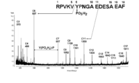
- the MALDI-ISD spectrum of the phosphorylated peptide when using a 5-ASA matrix is shown.
- the MALDI-ISD spectrum of a 2-phosphorylated peptide when using a 5-ASA matrix is shown.
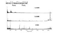
- the comparison of the contamination peak in the MALDI-ISD spectrum of a peptide when using 1,5-DAN (1,5-diaminonaphthalene), 2,5-DHB (2,5-dihyrdoxybenzoic acid) or 5-ASA matrix is shown.
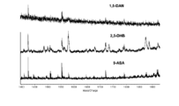
- the comparison of the peak resolution in the MALDI-ISD spectrum of the peptide when using 1,5-DAN, 2,5-DHB or 5-ASA matrix is shown.
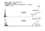
- Comparison of MALDI-ISD spectra of sulfonated peptide (CCK33) using 2,5-DHB (2,5-dihyrdoxybenzoic acid) and 5-ASA matrix is shown.
- Comparison of MALDI-ISD spectra in reflectron mode measurement of protein (myoglobin) using 2,5-DHB (2,5-dihyrdoxybenzoic acid) and 5-ASA matrix is shown.
- Comparison of MALDI-ISD spectrum part (m / z1500-5600) in reflectron mode measurement of protein (myoglobin) using 2,5-DHB (2,5-dihyrdoxybenzoic acid) and 5-ASA matrix Show.
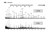
- Comparison of MALDI-ISD spectra of peptides (glucagon) using 2,5-DHB (2,5-dihyrdoxybenzoic acid) and 5-ASA matrix is shown.
- the present invention provides a method for specifically cleaving an N-C ⁇ bond of a peptide main chain, which comprises irradiating a peptide with a laser beam in the presence of 5-aminosalicylic acid (5-ASA).
- 5-aminosalicylic acid 5-aminosalicylic acid
- a peptide is generally a peptide in which two or more amino acids are linked by peptide bonds.
- a protein is a polypeptide in which about 20 L- ⁇ -amino acids are linked by peptide bonds.
- the number of amino acids constituting the peptide is not particularly limited, but is suitably 2 to 200, preferably 10 to 50, and more preferably 15 to 25. When the number of amino acids constituting the peptide is more than 100, digestion with an enzyme such as trypsin, separation and fractionation by liquid chromatography, and then the N-C ⁇ bond-specific cleavage method of the present invention It is good to use for.
- the type of amino acid constituting the peptide is not limited, and any amino acid may be used.
- amino acid may be modified such as acetylation, methylation, phosphorylation, glycosylation, and sulfonation.
- Peptides include proteins, polypeptides other than proteins, oligopeptides, glycopeptides, lipoproteins, and the like.
- 5-ASA is a known compound and is commercially available. 5-ASA is used as an active ingredient in mesalazine preparations for the treatment of ulcerative colitis and Crohn's disease.
- the amount of 5-ASA used is suitably 5000 to 50000 moles, preferably 5000 to 10,000 moles per mole of peptide.
- an ultraviolet laser such as a nitrogen laser having a wavelength of 337 nm or a neodymium YAG laser having a wavelength of 226 nm can be used, and among these, a nitrogen laser having a wavelength of 337 nm is preferable.
- the acceleration voltage of ions to be generated is suitably about 20 to 25 kV, but is not limited to this.
- the N-C ⁇ bond of the peptide main chain can be specifically cleaved by irradiating the peptide with laser light in the presence of 5-ASA. Accordingly, the present invention encompasses a specific cleavage reagent for peptide backbone N-C ⁇ binding, including 5-ASA.
- the amino acid sequence of the peptide can be determined by subjecting the peptide degradation product generated by the specific cleavage method of the peptide main chain N-C ⁇ bond of the present invention to a mass spectrometer and taking a mass spectrum.
- the present invention also includes a method for determining the amino acid sequence of a peptide, which comprises irradiating the peptide with a laser beam in the presence of 5-ASA to specifically cleave the N-C ⁇ bond of the peptide main chain.
- 5-ASA can be used as a matrix for mass spectrometry, preferably matrix-assisted laser desorption / ionization mass spectrometry (MALDI-MS), more preferably MALDI-ISD.
- MALDI-MS matrix-assisted laser desorption / ionization mass spectrometry
- the present invention also includes a matrix reagent for MALDI containing 5-ASA.
- the generated ions at this time are c- ions and z- ions (FIG. 2).
- Degradation of peptides that occurs in the ionization chamber at the same time or immediately after ionization, that is, fragmentation, is called in-source fragmentation or in-source decay (ISD).
- the peptide fragment ions generated by MALDI-ISD mainly consist of a ladder-like c-ion peak group in which the N-terminus is conserved. In these peak groups, since the mass difference ⁇ (m / z) between adjacent peaks means the mass of amino acid residues, partial amino acid sequences can be obtained by sequentially arranging the mass differences of ladder-shaped peak groups (FIG. 3). ).
- 5-ASA has both MALDI matrix function (peptide ionization ability) and hydrogen radical releasing ability. Moreover, 5-ASA has a high hydrogen radical transfer reaction (ISD) ability. Accordingly, the present invention includes a hydrogen radical releasing reagent comprising 5-ASA. The present invention also includes a peptide ionization reagent for MALDI containing 5-ASA.
- 5-ASA When 5-ASA is used as the matrix for MALDI, it can be dissolved in an aqueous acetonitrile solution (40-70% (preferably 50%), v / v) containing 0.05-0.5% (preferably 0.3% TFA).
- the saturated solution may be used as a matrix solution, although 5-ASA is poorly soluble in hydrophilic solvents, but the solubility can be significantly increased by the addition of 0.3% -0.5% TFA.
- the concentration of 5-ASA in the matrix solution is suitably 1-20 pmol / ⁇ L, preferably 5-10 pmol / ⁇ L.
- a buffer, picolinic acid or the like may be added to the matrix solution.
- the molar ratio of 5-ASA to peptide is about 5000 to 50000, preferably about 5000 to 10000.
- a mixed solution of the matrix solution and the peptide sample may be dropped on the plate, air dried, and the solvent removed.
- 5-ASA is used, acicular crystals grow, and the peptide molecules now cover the crystal surface of the matrix. Since the hydroxyl group of the 5-ASA molecule is exposed on the surface of the matrix crystal, the peptide molecule is covered so as to ride on the hydroxyl group.
- the present invention includes a matrix for MALDI containing 5-ASA.
- the matrix may contain a buffer component, picolinic acid and the like in addition to 5-ASA.
- the present invention can be used for mass spectrometry, particularly for direct sequencing of peptides using MALDI-TOF MS, and can be applied to the field of proteomics and protein chemistry.
- -5-ASA has a peptide ionization ability and a hydrogen radical releasing ability in MALDI-TOF MS, and does not exhibit special toxicity. Since the internal energy of the product ions can be kept low, no metastable peak is given, and no multivalent ion peak is given. Furthermore, since the sharpness of peaks important for amino acid sequence analysis is ensured, the information reading accuracy is high. Therefore, most of the conventional problems to be solved are solved and the superiority is clear.
- the present invention also provides a MALDI kit containing 5-ASA.
- This kit further includes other matrix reagents (eg, ⁇ -cyano-4-hydroxycinnamic acid (CHCA), sinapinic acid, 2,5-dihydroxybenzoic acid (2,5-DHB), 3-hydroxypicolinic acid, ferulic acid Etc.), buffer, picolinic acid, how to use the kit, precautions, instructions describing the contents, standard peptides, etc. may be included.
- matrix reagents eg, ⁇ -cyano-4-hydroxycinnamic acid (CHCA), sinapinic acid, 2,5-dihydroxybenzoic acid (2,5-DHB), 3-hydroxypicolinic acid, ferulic acid Etc.
- buffer e.g, picolinic acid, how to use the kit, precautions, instructions describing the contents, standard peptides, etc. may be included.
- Example 1 experimental method: The mass spectrometer uses a time-of-flight (TOF) mass spectrometer AXIMA-CFR (Shimadzu Corporation, Kyoto) equipped with matrix-assisted laser desorption / ionization (MALDI), and the laser is a nitrogen laser with a wavelength of 337 nm. Was used. The acceleration voltage of the generated ions was 20 kV.
- TOF time-of-flight
- MALDI matrix-assisted laser desorption / ionization
- 5 ⁇ L of the aqueous solution of the peptide sample was mixed with 5 ⁇ L of the matrix solution (saturated solution of 50% acetonitrile aqueous solution of each matrix), 1 ⁇ L of the mixed sample solution was applied to the sample target, and sample crystals were prepared by natural drying.
- FIG. 4 shows the MALDI-ISD spectrum of peptide ACTH18-39 when 5-ASA is used as the matrix.
- 5-ASA is not only useful as a MALDI ionization matrix but also has the function of ISD.
- c6 and c18 were not observed because complete cleavage did not occur on the N-terminal side of the proline residue.
- Diphosphorylated ACTH18-35 which is a diphosphorylated peptide
- was measured with three types of matrices 1,5-DAN, 2,5-DHB, and 5-ASA, and MALDI-ISD spectra were compared (FIG. 6).
- 1,5-DAN was able to read the amino acid sequence from c7 to c17, but the signal intensity was weak.
- c4 and c5 were not observed, phosphorylation of the 6th tyrosine could not be confirmed. This is because, in 1,5-DAN, matrix cluster peaks appear at m / z 800 or less, which hinders the appearance of signal peaks.
- Example 2 Example: CCK33 (sulfonated peptide) (Peptide Laboratories (Mino, Osaka)) was used as the sample peptide, and a saturated solution of 50% acetonitrile aqueous solution (containing 0.3% TFA) of each matrix was used as the matrix solution. The same experiment as 1 was performed. The results are shown in FIGS.
- FIG. 9 shows a comparison of MALDI-ISD spectra of the sulfonated peptide CCK33 when 2,5-DHB and 5-ASA are used in the matrix.
- FIG. 10 shows a spectrum with the horizontal axis enlarged. In the spectrum, not only the protonated molecule [M + H] + but also the c-ion on the N-terminal side is observed from c6 to c27, except for the ion (c17) cleaved at the N-terminal side of the proline residue. The amino acid sequence could be read. In particular, in the spectrum using 5-ASA, the intensity of the metastable peak (indicated by *), which is an interference peak, was lower than that of 2,5-DHB.
- * the intensity of the metastable peak
- FIG. 10 shows a comparison of MALDI-ISD spectra of the protein myoglobin when 2,5-DHB and 5-ASA are used as matrices and the mass spectrometer is measured in the linear mode.
- FIG. 11 shows a partial spectrum in which the horizontal axis is enlarged.
- FIG. 13 shows a comparison of MALDI-ISD spectra of the protein myoglobin when measured using 2,5-DHB and 5-ASA as matrices and the mass spectrometer in reflectron mode.
- FIG. 14 shows a partial spectrum whose horizontal axis is enlarged.
- FIG. 15 shows a comparison of MALDI-ISD spectra of glucagon having a helix structure as a hormonal peptide when 2,5-DHB and 5-ASA are used as a matrix.
- FIG. 16 shows a partial spectrum in which the horizontal axis is enlarged. In the spectrum, not only protonated molecule [M + H] + but also c-ion on the N-terminal side was observed from c6 to c27, and the amino acid sequence could be read. In the case of the 2,5-DHB matrix, an oxidant ([M + O + H] + ) peak was observed (FIG. 15), and a sodium ion adduct (FIG. 16) peak was also observed. In -ASA, neither oxidant nor sodium adduct was observed, indicating excellent analytical performance.
- the present invention can be used in the proteomics field and protein chemistry field.
- SEQ ID NO: 1 shows the amino acid sequence of the adrenocorticotropic hormone (ACTH) fragment ACTH18-39.
- SEQ ID NO: 2 shows the amino acid sequence of the adrenocorticotropic hormone (ACTH) fragment ACTH18-35.
- Origin Equine Amino acid sequence: GLSDEGWQQVLNVWGKVEADIAGHGQEVLIRLFTGHPETLEKFDKFKHLKTEAEMKASEDLKKHGTVVLTALGGILKKKGHHEAELKPLAQSHATKHKIPIKYLEFISDAIIHVLHSKHPGDFGADAQGAMTKALELFRNDIAAKYKELG FQG ⁇ SEQ ID NO: 5> SEQ ID NO: 5 shows the amino acid sequence of glucagon (hormonal peptide). Origin: Porcine Amino acid sequence: HSQGTFTSDYSKYLDSRRAQDFVQWLMNT
- Tsugita Int. J. Mass Spectrom. 181 (1998) L1.
- M. Takayama J.Am.Soc.Mass Spectrom., 12 (2001) 420.
- M. Takayama J.Am.Soc.Mass.Spectrom., 12 (2001) 1044.
- Y.Fukuyama et al J.Mass Spectrom., 41 (2006) 191.
- K. Demeure et al Anal. Chem., 79 (2007) 8678.
Landscapes
- Life Sciences & Earth Sciences (AREA)
- Health & Medical Sciences (AREA)
- Engineering & Computer Science (AREA)
- Molecular Biology (AREA)
- Physics & Mathematics (AREA)
- Hematology (AREA)
- Bioinformatics & Cheminformatics (AREA)
- Bioinformatics & Computational Biology (AREA)
- Chemical & Material Sciences (AREA)
- Biomedical Technology (AREA)
- Urology & Nephrology (AREA)
- Immunology (AREA)
- Biophysics (AREA)
- Analytical Chemistry (AREA)
- Microbiology (AREA)
- Biotechnology (AREA)
- Proteomics, Peptides & Aminoacids (AREA)
- Food Science & Technology (AREA)
- Medicinal Chemistry (AREA)
- Cell Biology (AREA)
- Biochemistry (AREA)
- General Health & Medical Sciences (AREA)
- General Physics & Mathematics (AREA)
- Pathology (AREA)
- Spectroscopy & Molecular Physics (AREA)
- Optics & Photonics (AREA)
- Other Investigation Or Analysis Of Materials By Electrical Means (AREA)
Abstract
1)発がん性などの著しい毒性がないこと、2)過度の分解特性により準安定ピークを与えないこと、3)妨害ピークである多価イオンピークを与えないこと、4)ピークの分離とシャープネスが確保できることといった特性を持ったペプチド分解試薬を提供する。上記分解試薬を用いて、ペプチド主鎖N-Cα結合を特異的に切断する方法、また、この特異的切断を利用して、ペプチドのアミノ酸配列を決定する方法も提供する。 5-アミノサリチル酸の存在下で、ペプチドにレーザー光を照射することを含む、ペプチド主鎖のN-Cα結合を特異的に切断する方法。5-アミノサリチル酸の存在下で、ペプチドにレーザー光を照射して、ペプチド主鎖のN-Cα結合を特異的に切断することを含む、ペプチドのアミノ酸配列決定方法。ペプチド主鎖N-Cα結合の特異的切断試薬、水素ラジカル放出試薬、MALDI-ISD用マトリックス試薬、MALDI-ISD用マトリックス、MALDI-ISD用ペプチドイオン化試薬及びMALDI-ISD用キット。
Description
本発明は、ペプチド主鎖N-Cα結合の特異的切断方法、より詳細には、5-アミノサリチル酸とレーザー光を利用したペプチド主鎖N-Cα結合の特異的切断方法に関する。
タンパク質やポリペプチドなどのペプチド主鎖は -CαH-CO-NH-CαH- を単位とし,α位の炭素 -Cα- にアミノ酸 R が結合したポリマーである。アミノ酸のナンバリングはアミノ (N)-末端側から始まり,カルボキシ (C)-末端を最終とする。一文字表記法でn個のアミノ酸残基から成るペプチドまたはタンパク質を表すと,NH2-R1-R2-R3-----Rn-1-Rn-COOH のように書くことができる。タンパク質を同定する際に最も役に立つ情報はこのアミノ酸配列であり,N-末端側でもC-末端側でも,あるいは内部配列でもその部分配列の10残基程度の情報があれば,データベースを使って簡単に同定することができる。各種アミノ酸配列解析法が開発されているが,質量分析法 (mass spectrometry, MS) もその一つに数えられている。質量分析法では、高電圧をかけた真空中で試料をイオン化し、このイオンを質量/電荷数(m/z)に従って分離し、各イオンの相対強度を測定する。イオン化の方法としては、電子イオン化法(EI, electron ionization)、化学イオン化法(CI, chemical ionization)、電界脱離法(FD, field desorption)、高速原子衝突法(FAB, fast atom bombardment)、マトリックス支援レーザー脱離イオン化法(MALDI, matrix associated laser desorption ionization)、エレクトロスプレーイオン化法(ESI, electrospray ionization)など様々な手法が開発されている。イオン化された試料を分離する分析部では、磁場偏向型、四重極型、イオントラップ型、飛行時間型(TOF, time-of-flight)、フーリエ変換イオンサイクロトロン共鳴型などの質量分析計が用いられる。MALDI-TOFの組合せは、高分子量まで適用できるイオン源と高分子量まで適用できる分析計の組合せであるため、ポリマーやタンパク質などの高分子の分析によく用いられている。
マトリックス支援レーザー脱離イオン化飛行時間型質量分析(MALDI-TOF MS)を使うタンパク質の直接シーケンシングを最初に報告したのは,R.S.BrownとJ.J.Lennonである(非特許文献1)。その後,種々のタンパク質の直接シーケンシングに応用されたが(非特許文献2~6),なぜレーザー照射によってタンパク質のアミノ酸配列解析を可能にするような特異的な開裂反応が起こるのか,その機構はまったく理解されていなかった。本発明者らもBrown等の最初の報告とは独立にこの現象に遭遇し,この現象がイオン化とは独立に起こることを示した(非特許文献7)。すなわち,N-Cα結合の開裂は,MALDI条件下でペプチドやタンパク質がプロトンを受け取り生成したプロトン化分子 [M+H]+ が開裂するのではなく,レーザー光照射によって中性分子 M が開裂するのである。開裂はイオン化室 (ion source) の内部で起こるため,この現象を in-source decay (ISD) と呼ぶ。
MALDIにおけるイオン化は,マトリックスと試料を混合し形成した結晶系にレーザー光を照射したとき,大過剰のマトリックスが光子を吸収し爆発的に気化する際のイオン分子反応によって起こる。マトリックスは,プロトンの供給源であるとともにプロトン受容体にもなるイオン化試薬である。ペプチドやタンパク質のN-Cα結合を特異的に開裂させるマトリックスには2,5-dihydroxybenzoic acid(2,5-DHB)が適していることが報告されていた(非特許文献8)。その後、島津製作所の田中耕一氏のグループより優れた試薬1,5-diaminonaphthalene (1,5-DAN) が発表され(非特許文献9)、海外グループによって確認された (非特許文献10)。しかし、2,5-DHBは過度に分解を促すホットな性質を持ち、1,5-DANは昇華性と発がん性があるとして、別のさらにすぐれたマトリックスが要望されていた。
本発明は、以下のような特性を持った分解試薬(マトリックス)を提供することを目的とする。
発がん性などの著しい毒性がないこと。
過度の分解特性により準安定ピークを与えないこと。
妨害ピークである多価イオンピークを与えないこと。
ピークの分離とシャープネスが確保できること。
過度の分解特性により準安定ピークを与えないこと。
妨害ピークである多価イオンピークを与えないこと。
ピークの分離とシャープネスが確保できること。
また、本発明は、上記分解試薬を用いて、ペプチド主鎖N-Cα結合を特異的に切断する方法、また、この特異的切断を利用して、ペプチドのアミノ酸配列を決定する方法を提供することを目的とする。
本発明者らは、5-アミノサリチル酸(5-ASA, 5-amino salicylic acid)の存在下で、ペプチドにレーザー光を照射したところ、ペプチド主鎖のN-Cα結合だけが特異的に開裂反応を起こした。反応生成物をMALDI-TOF MSで解析したところアミノ酸配列を決定することができた。本発明は、これらの知見に基づいて完成されたものである。
本発明の要旨は以下の通りである。
(1)5-アミノサリチル酸の存在下で、ペプチドにレーザー光を照射することを含む、ペプチド主鎖のN-Cα結合を特異的に切断する方法。
(2)5-アミノサリチル酸の存在下で、ペプチドにレーザー光を照射して、ペプチド主鎖のN-Cα結合を特異的に切断することを含む、ペプチドのアミノ酸配列決定方法。
(3)5-アミノサリチル酸がマトリックス支援レーザー脱離イオン化質量分析のマトリックスとして使用される(2)記載の方法。
(4)5-アミノサリチル酸を含む、ペプチド主鎖N-Cα結合の特異的切断試薬。
(5)5-アミノサリチル酸を含む、水素ラジカル放出試薬。
(6)5-アミノサリチル酸を含む、マトリックス支援レーザー脱離イオン化質量分析用マトリックス試薬。
(7)5-アミノサリチル酸を含む、マトリックス支援レーザー脱離イオン化質量分析用マトリックス。
(8)5-アミノサリチル酸を含む、マトリックス支援レーザー脱離イオン化質量分析用ペプチドイオン化試薬。
(9)5-アミノサリチル酸を含む、マトリックス支援レーザー脱離イオン化質量分析用キット。
(1)5-アミノサリチル酸の存在下で、ペプチドにレーザー光を照射することを含む、ペプチド主鎖のN-Cα結合を特異的に切断する方法。
(2)5-アミノサリチル酸の存在下で、ペプチドにレーザー光を照射して、ペプチド主鎖のN-Cα結合を特異的に切断することを含む、ペプチドのアミノ酸配列決定方法。
(3)5-アミノサリチル酸がマトリックス支援レーザー脱離イオン化質量分析のマトリックスとして使用される(2)記載の方法。
(4)5-アミノサリチル酸を含む、ペプチド主鎖N-Cα結合の特異的切断試薬。
(5)5-アミノサリチル酸を含む、水素ラジカル放出試薬。
(6)5-アミノサリチル酸を含む、マトリックス支援レーザー脱離イオン化質量分析用マトリックス試薬。
(7)5-アミノサリチル酸を含む、マトリックス支援レーザー脱離イオン化質量分析用マトリックス。
(8)5-アミノサリチル酸を含む、マトリックス支援レーザー脱離イオン化質量分析用ペプチドイオン化試薬。
(9)5-アミノサリチル酸を含む、マトリックス支援レーザー脱離イオン化質量分析用キット。
5-ASAは、マトリックス支援レーザー脱離イオン化質量分析(MALDI)において、ペプチドのイオン化能を有しながら、かつ水素ラジカル放出能を有しており、特別な毒性を示さない。生成イオンの内部エネルギーを低く抑えることができるため、準安定ピークを与えず、多価イオンのピークも与えない。さらに、アミノ酸配列解析にとって重要なピークのシャープネスが確保されているため、情報の読み取り精度が高い。
本明細書は、本願の優先権の基礎である日本国特許出願、特願2009‐167550の明細書および/または図面に記載される内容を包含する。
以下、本発明の実施の形態についてより詳細に説明する。
本発明は、5-アミノサリチル酸(5-ASA)の存在下で、ペプチドにレーザー光を照射することを含む、ペプチド主鎖のN-Cα結合を特異的に切断する方法を提供する。
ペプチドは、一般に、2個以上のアミノ酸がペプチド結合により結合したものである。タンパク質は、約20種のL-α-アミノ酸がペプチド結合により結合したポリペプチドである。ペプチドを構成するアミノ酸の数は、特に限定されるものではないが、2~200個が適当であり、10~50個が好ましく、15~25個がより好ましい。ペプチドを構成するアミノ酸の数が100個よりも多い場合には、トリプシンなどの酵素で消化処理を行い、液体クラマトグラフィで分離・分取してから、本発明のN-Cα結合特異的切断方法に用いるとよい。ペプチドを構成するアミノ酸の種類は限定されるものではなく、いかなるアミノ酸であってもよい。また、アミノ酸は、アセチル化、メチル化、リン酸化、グリコシル化、スルホン酸化などの修飾がなされていてもよい。ペプチドには、タンパク質、タンパク質以外のポリペプチド、オリゴペプチド、グリコペプチド、リポタンパク質などが含まれる。
5-ASAは公知の化合物であり、市販されている。5-ASAは、潰瘍性大腸炎やクローン病の治療薬であるメサラジン製剤の有効成分として利用されている。
本発明のペプチド主鎖N-Cα結合の特異的切断方法において、5-ASAの使用量は、ペプチド1モルに対して、5000~50000モルが適当であり、5000~10000モルが好ましい。
レーザーとしては、波長337 nmの窒素レーザー、波長226nmのネオジウムYAGレーザーなどの紫外レーザーを用いることができ、このうち、波長337 nmの窒素レーザーが好ましい。生成するイオンの加速電圧は20~25 kV程度が適当であるが、これに限定されるわけではない。
5-ASAの存在下で、ペプチドにレーザー光を照射することにより、ペプチド主鎖のN-Cα結合を特異的に切断することができる。従って、本発明は、5-ASAを含む、ペプチド主鎖N-Cα結合の特異的切断試薬を包含する。
本発明のペプチド主鎖N-Cα結合の特異的切断方法によって生成したペプチド分解物を質量分析計にかけ、マススペクトルをとることにより、ペプチドのアミノ酸配列を決定することができる。本発明は、5-ASAの存在下で、ペプチドにレーザー光を照射して、ペプチド主鎖のN-Cα結合を特異的に切断することを含む、ペプチドのアミノ酸配列決定方法も包含する。
5-ASAは、質量分析、好ましくはマトリックス支援レーザー脱離イオン化質量分析(MALDI MS)、より好ましくはMALDI-ISDのマトリックスとして使用することができる。本発明は、5-ASAを含む、MALDI用マトリックス試薬も包含する。
MALDIのマトリックスとして5-ASAを使用する場合、サンプル(ペプチド)とマトリックス(5-ASA)の混合物(混晶)にレーザー光を照射する。ペプチドは5-ASAの結晶表面にあり、ペプチド主鎖上のカルボニル酸素は5-ASAの水酸基とあらかじめ水素結合している。5-ASAがレーザー光を吸収すると励起して水素原子とフェノキシラジカルに解離し、水素原子はそのままペプチド主鎖上のカルボニル酸素に結合して、カルボニル酸素が水酸基に、カルボニル炭素がラジカルになる。そして、ラジカル部位に対してα位にあるNH-Cα結合が単純開裂を生じる(α開裂)(図1)。このときの生成イオンはc-イオンとz-イオンである(図2)。このようにイオン化と同時又は直後にイオン化室で生じるペプチドの分解、すなわち、フラグメンテーションはin-source fragmentation又はin-source decay(ISD)と呼ばれる。MALDI-ISDによって生じるペプチドのフラグメントイオンは、主としてN-末端が保存されたラダー状のc-イオンピーク群からなる。それらのピーク群において、隣り合うピーク間の質量差Δ(m/z)がアミノ酸残基の質量を意味するため、ラダー状ピーク群の質量差を順次並べると部分アミノ酸配列が得られる(図3)。
MALDIのマトリックスとして5-ASAを使用した場合、これまでのMALDI-ISDデータより各ピークの分解能において優れていた。
5-ASAは、MALDIマトリックス機能(ペプチドのイオン化能)と水素ラジカル放出能を併せ持つ。しかも、5-ASAは水素ラジカル移動反応(ISD)能が高い。従って、本発明は、5-ASAを含む、水素ラジカル放出試薬を包含する。また、本発明は、5-ASAを含む、MALDI用ペプチドイオン化試薬を包含する。
MALDIのマトリックスとして5-ASAを使用する場合には、0.05~0.5%(好ましくは、0.3%のTFAを含むアセトニトリル水溶液(40~70%(好ましくは、50%), v/v)に溶解し、その飽和溶液をマトリックス溶液として使用するとよい。5-ASAは親水性溶媒に難溶であるが、0.3%-0.5%のTFAの添加により、溶解度を著しく高めることができる。
マトリックス溶液中の5-ASA濃度は、1~20 pmol/μLが適当であり、5~10 pmol/μLが好ましい。マトリックス溶液には、バッファー、ピコリン酸などを添加してもよい。ペプチド試料との混合に際しては、5-ASAとペプチドのモル比が、5000~50000程度、好ましくは、5000~10000程度になるように調製するとよい。マトリックス溶液とペプチド試料との混合溶液をプレートに滴下し、自然乾燥させ、溶媒を除去するとよい。5-ASAを使うと針状結晶が成長し、この時、ペプチド分子はマトリックスの結晶表面を覆うようになる。マトリックス結晶表面には5-ASA分子の水酸基が露出しているため、ペプチド分子はそれら水酸基群の上に乗るような形で覆っている。本発明は、5-ASAを含む、MALDI用のマトリックスを包含する。マトリックスには、5-ASAの他、バッファー成分、ピコリン酸などが含まれていてもよい。
マトリックス溶液中の5-ASA濃度は、1~20 pmol/μLが適当であり、5~10 pmol/μLが好ましい。マトリックス溶液には、バッファー、ピコリン酸などを添加してもよい。ペプチド試料との混合に際しては、5-ASAとペプチドのモル比が、5000~50000程度、好ましくは、5000~10000程度になるように調製するとよい。マトリックス溶液とペプチド試料との混合溶液をプレートに滴下し、自然乾燥させ、溶媒を除去するとよい。5-ASAを使うと針状結晶が成長し、この時、ペプチド分子はマトリックスの結晶表面を覆うようになる。マトリックス結晶表面には5-ASA分子の水酸基が露出しているため、ペプチド分子はそれら水酸基群の上に乗るような形で覆っている。本発明は、5-ASAを含む、MALDI用のマトリックスを包含する。マトリックスには、5-ASAの他、バッファー成分、ピコリン酸などが含まれていてもよい。
本発明は、質量分析、特に、MALDI-TOF MSを利用するペプチドの直接シーケンシングに利用可能であり、プロテオミスク分野やタンパク質化学分野に応用できる。
5-ASAは、MALDI-TOF MSにおいて、ペプチドのイオン化能を有しながら、かつ水素ラジカル放出能を有しており、特別な毒性を示さない。生成イオンの内部エネルギーを低く抑えることができるため、準安定ピークを与えず、多価イオンのピークも与えない。さらに、アミノ酸配列解析にとって重要なピークのシャープネスが確保されているため、情報の読み取り精度が高い。従って、従来の解決課題のほとんどが解決され、その優位性は明らかである。
また、本発明は、5-ASAを含む、MALDI用キットを提供する。
このキットには、さらに、他のマトリックス試薬(例えば、α-cyano-4-hydroxycinnamic acid(CHCA)、sinapinic acid、2,5-dihydroxybenzoic acid(2,5-DHB)、3-hydroxypicolinic acid、ferulic acidなど)、バッファー、ピコリン酸、キットの使用方法、注意書、内容物などを記した説明書、標準ペプチドなどを含めてもよい。
以下、実施例に基づいて本発明を詳細に説明するが、本発明はこれらの実施例に限定されるものではない。
〔実施例1〕
実験方法:
質量分析装置には、マトリックス支援レーザー脱離イオン化(MALDI)法を搭載した飛行時間型(TOF)の質量分析計 AXIMA-CFR(島津製作所、京都)を使用し、レーザーには波長337nmの窒素レーザーを用いた。生成したイオンの加速電圧は20kVであった。試料ペプチドには副腎皮質刺激ホルモン(ACTH)のフラグメント ACTH18-39 (Mr 2465.7)、およびACTH18-35 (Mr 1979.1)の1リン酸化体(Mr 2059.1)と2リン酸化体(Mr 2139.1)を用いた。これらペプチドはペプチド研究所(箕面、大阪)から購入したものをそのまま使用した。マトリックスである2,5-DHBと 1,5-DANはSigma Aldrich (Steinheim, Germany)から購入、5-ASA は東京化成(東京)から購入し、再結晶することなく使用した。ペプチド試料の水溶液は、その5μLをマトリックス溶液(各マトリックスの50%アセトニトリル水溶液の飽和溶液)5μLと混合し、混合後の試料溶液1μLを試料ターゲットに塗布し、自然乾燥により試料結晶を作成した。試料結晶を塗布したターゲットを質量分析計に導入した。
実験方法:
質量分析装置には、マトリックス支援レーザー脱離イオン化(MALDI)法を搭載した飛行時間型(TOF)の質量分析計 AXIMA-CFR(島津製作所、京都)を使用し、レーザーには波長337nmの窒素レーザーを用いた。生成したイオンの加速電圧は20kVであった。試料ペプチドには副腎皮質刺激ホルモン(ACTH)のフラグメント ACTH18-39 (Mr 2465.7)、およびACTH18-35 (Mr 1979.1)の1リン酸化体(Mr 2059.1)と2リン酸化体(Mr 2139.1)を用いた。これらペプチドはペプチド研究所(箕面、大阪)から購入したものをそのまま使用した。マトリックスである2,5-DHBと 1,5-DANはSigma Aldrich (Steinheim, Germany)から購入、5-ASA は東京化成(東京)から購入し、再結晶することなく使用した。ペプチド試料の水溶液は、その5μLをマトリックス溶液(各マトリックスの50%アセトニトリル水溶液の飽和溶液)5μLと混合し、混合後の試料溶液1μLを試料ターゲットに塗布し、自然乾燥により試料結晶を作成した。試料結晶を塗布したターゲットを質量分析計に導入した。
結果と考察:
5-ASAをマトリックスに用いたときのペプチド ACTH18-39 のMALDI-ISDスペクトルを図4に示す。スペクトル中には、プロトン化分子 [M+H]+ のみならず、N-末端側のc-イオンが c3からc21まで観測され、アミノ酸配列を読むことができた。このことは、5-ASAがMALDIのイオン化マトリックスとして有用なだけでなく、ISDの機能も有することを示している。但し、プロリン残基のN-末端側は完全な切断が起こらないため、c6とc18は観測されなかった。本マトリックスが翻訳後修飾ペプチドにも有効か否か確認するため、6番目のチロシンがリン酸化された1リン酸化ACTH18-35のMALDI-ISDスペクトルを得た(図5)。得られたスペクトルには、プロリン残基のN-末端側が切断したイオン(c6)を除き、c3からc17まで観測されアミノ酸配列を読むことができた。さらにリン酸基の分解脱離がまったく起こらないため、c6とc8の間の質量差からリン酸基の存在と部位を特定することができた。
5-ASAをマトリックスに用いたときのペプチド ACTH18-39 のMALDI-ISDスペクトルを図4に示す。スペクトル中には、プロトン化分子 [M+H]+ のみならず、N-末端側のc-イオンが c3からc21まで観測され、アミノ酸配列を読むことができた。このことは、5-ASAがMALDIのイオン化マトリックスとして有用なだけでなく、ISDの機能も有することを示している。但し、プロリン残基のN-末端側は完全な切断が起こらないため、c6とc18は観測されなかった。本マトリックスが翻訳後修飾ペプチドにも有効か否か確認するため、6番目のチロシンがリン酸化された1リン酸化ACTH18-35のMALDI-ISDスペクトルを得た(図5)。得られたスペクトルには、プロリン残基のN-末端側が切断したイオン(c6)を除き、c3からc17まで観測されアミノ酸配列を読むことができた。さらにリン酸基の分解脱離がまったく起こらないため、c6とc8の間の質量差からリン酸基の存在と部位を特定することができた。
2リン酸化ペプチドである2リン酸化ACTH18-35を3種類のマトリックス 1,5-DAN, 2,5-DHB, 5-ASAで測定し、各MALDI-ISDスペクトルを比較した(図6)。1,5-DANではc7からc17までアミノ酸配列を読むことができたがシグナル強度は弱かった。さらにc4とc5が観測されなかったため、6番目のチロシンのリン酸化を確認することができなかった。これは、1,5-DANではm/z800以下にマトリックスクラスターのピークが出現し、シグナルピークの出現を妨害するためである。2,5-DHBではc5からc17まで配列情報が得られたが、シグナル強度が弱くピークの分離が不明瞭であるために解析が困難であった。一方、5-ASAを使った場合、フラグメントイオンがc3からc17まで明瞭に観測され、さらにリン酸の脱離が起こらないため、ピーク間の質量差からリン酸基の存在と部位を特定することができた。また、図7に示すように、特に2,5-DHBでは幅広の準安定ピークが観測され解析を妨害している。これは、2,5-DHBでは生成したイオンの内部エネルギーが多く、過剰な分解が起こることを示すものである。このことと関連して、プロトン化分子 [M+H]+ のピーク付近の同位体ピークの分離能が5-ASAでは著しく高く、高い解析精度を可能にしている(図8参照)。
〔実施例2〕
試料ペプチドにはCCK33(スルホン酸化ペプチド)(ペプチド研究所(箕面、大阪))を用い、マトリックス溶液には各マトリックスの50%アセトニトリル水溶液(0.3% TFA含有)の飽和溶液を用いた他は実施例1と同様の実験を行った。
結果を図9及び10に示す。
試料ペプチドにはCCK33(スルホン酸化ペプチド)(ペプチド研究所(箕面、大阪))を用い、マトリックス溶液には各マトリックスの50%アセトニトリル水溶液(0.3% TFA含有)の飽和溶液を用いた他は実施例1と同様の実験を行った。
結果を図9及び10に示す。
結果と考察:
2,5-DHBと5-ASAをマトリックスに用いたときのスルホン酸化ペプチド CCK33のMALDI-ISDスペクトルの比較を図9に示す。また、図10には横軸を拡大したスペクトルを示す。スペクトル中には、プロトン化分子 [M+H]+ のみならず、プロリン残基のN-末端側が切断したイオン(c17)を除き、N-末端側のc-イオンが c6からc27まで観測され、アミノ酸配列を読むことができた。特に、5-ASAを使用したスペクトルは2,5-DHBの場合より妨害ピークである準安定ピーク(*で示す)の強度が低くなった。このことは、5-ASAが2,5-DHBより妨害ピークを少なくする意味で優れていることを示す。どちらのマトリックスでもスルホン酸の脱離が観測されたが(図9)、27番目のチロシン(Tyr27)に結合したスルホン酸はc27-イオンに結合したまま観測され、スルホン酸の位置の決定が可能であった。
2,5-DHBと5-ASAをマトリックスに用いたときのスルホン酸化ペプチド CCK33のMALDI-ISDスペクトルの比較を図9に示す。また、図10には横軸を拡大したスペクトルを示す。スペクトル中には、プロトン化分子 [M+H]+ のみならず、プロリン残基のN-末端側が切断したイオン(c17)を除き、N-末端側のc-イオンが c6からc27まで観測され、アミノ酸配列を読むことができた。特に、5-ASAを使用したスペクトルは2,5-DHBの場合より妨害ピークである準安定ピーク(*で示す)の強度が低くなった。このことは、5-ASAが2,5-DHBより妨害ピークを少なくする意味で優れていることを示す。どちらのマトリックスでもスルホン酸の脱離が観測されたが(図9)、27番目のチロシン(Tyr27)に結合したスルホン酸はc27-イオンに結合したまま観測され、スルホン酸の位置の決定が可能であった。
〔実施例3〕
試料ペプチドにはmyoglobin(馬心筋タンパク質)Mr (16951.4)(Sigma Aldrich (Steinheim, Germany))(PDB data: http://www.pdb.org/pdb/explore/explore.do?structureId=1WLA)を用い、マトリックス溶液には各マトリックスの50%アセトニトリル水溶液(0.3% TFA含有)の飽和溶液を用いた他は実施例1と同様の実験を行った。
結果を図11、12、13及び14に示す。
試料ペプチドにはmyoglobin(馬心筋タンパク質)Mr (16951.4)(Sigma Aldrich (Steinheim, Germany))(PDB data: http://www.pdb.org/pdb/explore/explore.do?structureId=1WLA)を用い、マトリックス溶液には各マトリックスの50%アセトニトリル水溶液(0.3% TFA含有)の飽和溶液を用いた他は実施例1と同様の実験を行った。
結果を図11、12、13及び14に示す。
結果と考察:
2,5-DHBと5-ASAをマトリックスに用い、質量分析計をリニアモードにして測定したときのタンパク質 myoglobinのMALDI-ISDスペクトルの比較を図10に示す。また、図11には横軸を拡大した部分スペクトルを示す。図13と2,5-DHBと5-ASAをマトリックスに用い、質量分析計をリフレクトロンモードにして測定したときのタンパク質 myoglobinのMALDI-ISDスペクトルの比較を示す。図14にはその横軸を拡大した部分スペクトルを示す。上記のスペクトル中には、プロトン化分子 [M+H]+ のみならず、N-末端側のc-イオンが c39からc65まで、C-末端側のy-イオンと(z+2)-イオンも観測されアミノ酸配列を読むことができた。以上より、5-ASAはペプチドのみならずタンパク質の分析にも適用可能であることを示す。
2,5-DHBと5-ASAをマトリックスに用い、質量分析計をリニアモードにして測定したときのタンパク質 myoglobinのMALDI-ISDスペクトルの比較を図10に示す。また、図11には横軸を拡大した部分スペクトルを示す。図13と2,5-DHBと5-ASAをマトリックスに用い、質量分析計をリフレクトロンモードにして測定したときのタンパク質 myoglobinのMALDI-ISDスペクトルの比較を示す。図14にはその横軸を拡大した部分スペクトルを示す。上記のスペクトル中には、プロトン化分子 [M+H]+ のみならず、N-末端側のc-イオンが c39からc65まで、C-末端側のy-イオンと(z+2)-イオンも観測されアミノ酸配列を読むことができた。以上より、5-ASAはペプチドのみならずタンパク質の分析にも適用可能であることを示す。
〔実施例4〕
試料ペプチドにはglucagon(ホルモン性ペプチド)Mr(3480.5)(ペプチド研究所(箕面、大阪))(PDB data: http://www.pdb.org/pdb/explore/explore.do?structureId=1GCN)を用い、マトリックス溶液には各マトリックスの50%アセトニトリル水溶液(0.3% TFA含有)の飽和溶液を用いた他は実施例1と同様の実験を行った。
結果を図15及び16に示す。
試料ペプチドにはglucagon(ホルモン性ペプチド)Mr(3480.5)(ペプチド研究所(箕面、大阪))(PDB data: http://www.pdb.org/pdb/explore/explore.do?structureId=1GCN)を用い、マトリックス溶液には各マトリックスの50%アセトニトリル水溶液(0.3% TFA含有)の飽和溶液を用いた他は実施例1と同様の実験を行った。
結果を図15及び16に示す。
結果と考察:
2,5-DHBと5-ASAをマトリックスに用いたときのホルモン性ペプチドでへリックス構造を有する glucagonのMALDI-ISDスペクトルの比較を図15に示す。また、図16には横軸を拡大した部分スペクトルを示す。スペクトル中には、プロトン化分子 [M+H]+ のみならず、N-末端側のc-イオンが c6からc27まで観測されアミノ酸配列を読むことができた。2,5-DHBマトリックスの場合には酸化体([M+O+H]+)のピークが観測され(図15)、さらにナトリウムイオン付加体(図16)のピークも観測されたが、5-ASAでは酸化体もナトリウム付加体も観測されず、解析性に優れた点を示した。
2,5-DHBと5-ASAをマトリックスに用いたときのホルモン性ペプチドでへリックス構造を有する glucagonのMALDI-ISDスペクトルの比較を図15に示す。また、図16には横軸を拡大した部分スペクトルを示す。スペクトル中には、プロトン化分子 [M+H]+ のみならず、N-末端側のc-イオンが c6からc27まで観測されアミノ酸配列を読むことができた。2,5-DHBマトリックスの場合には酸化体([M+O+H]+)のピークが観測され(図15)、さらにナトリウムイオン付加体(図16)のピークも観測されたが、5-ASAでは酸化体もナトリウム付加体も観測されず、解析性に優れた点を示した。
5-ASAの特長と優位性を以下にまとめる。
1.発がん性など顕著な毒性が無い。
2.解析を妨害する準安定ピークを与えない。
3.解析を妨害する多価イオンピークを与えない。
4.ピークの分離特性に優れていて、情報の読み取り精度が高い。
1.発がん性など顕著な毒性が無い。
2.解析を妨害する準安定ピークを与えない。
3.解析を妨害する多価イオンピークを与えない。
4.ピークの分離特性に優れていて、情報の読み取り精度が高い。
本明細書で引用した全ての刊行物、特許および特許出願をそのまま参考として本明細書にとり入れるものとする。
本発明は、プロテオミクス分野やタンパク質化学分野に利用可能である。
<配列番号1>
配列番号1は、副腎皮質刺激ホルモン(ACTH)のフラグメントACTH18-39のアミノ酸配列を示す。
由来:Human
アミノ酸配列:RPVKV YPNGA EDESA EAFPL EF
<配列番号2>
配列番号2は、副腎皮質刺激ホルモン(ACTH)のフラグメントACTH18-35のアミノ酸配列を示す。
由来:Human
アミノ酸配列:RPVKV YPNGA EDESA EAF
<配列番号3>
配列番号3は、CCK33のアミノ酸配列を示す。
由来:Human
アミノ酸配列:KAPSGRMSIVKNLQNLDPSHRISDRDY(SO3H)MGWMDF-NH2
<配列番号4>
配列番号4は、myoglobin(馬心筋タンパク質)のアミノ酸配列を示す。
由来:Equine
アミノ酸配列:GLSDEGWQQVLNVWGKVEADIAGHGQEVLIRLFTGHPETLEKFDKFKHLKTEAEMKASEDLKKHGTVVLTALGGILKKKGHHEAELKPLAQSHATKHKIPIKYLEFISDAIIHVLHSKHPGDFGADAQGAMTKALELFRNDIAAKYKELG FQG
<配列番号5>
配列番号5は、glucagon(ホルモン性ペプチド)のアミノ酸配列を示す。
由来:Porcine
アミノ酸配列:HSQGTFTSDYSKYLDSRRAQDFVQWLMNT
配列番号1は、副腎皮質刺激ホルモン(ACTH)のフラグメントACTH18-39のアミノ酸配列を示す。
由来:Human
アミノ酸配列:RPVKV YPNGA EDESA EAFPL EF
<配列番号2>
配列番号2は、副腎皮質刺激ホルモン(ACTH)のフラグメントACTH18-35のアミノ酸配列を示す。
由来:Human
アミノ酸配列:RPVKV YPNGA EDESA EAF
<配列番号3>
配列番号3は、CCK33のアミノ酸配列を示す。
由来:Human
アミノ酸配列:KAPSGRMSIVKNLQNLDPSHRISDRDY(SO3H)MGWMDF-NH2
<配列番号4>
配列番号4は、myoglobin(馬心筋タンパク質)のアミノ酸配列を示す。
由来:Equine
アミノ酸配列:GLSDEGWQQVLNVWGKVEADIAGHGQEVLIRLFTGHPETLEKFDKFKHLKTEAEMKASEDLKKHGTVVLTALGGILKKKGHHEAELKPLAQSHATKHKIPIKYLEFISDAIIHVLHSKHPGDFGADAQGAMTKALELFRNDIAAKYKELG FQG
<配列番号5>
配列番号5は、glucagon(ホルモン性ペプチド)のアミノ酸配列を示す。
由来:Porcine
アミノ酸配列:HSQGTFTSDYSKYLDSRRAQDFVQWLMNT
R. R. Brown, and J. J. Lennon: Anal. Chem. 67 (1995) 3990.
J. J. Lennon, and K. A. Walsh: Protein Science 6 (1997) 2446.
D. C. Reiber, T. A. Grover, and R. S. Brown: Anal. Chem. 70 (1998) 673.
V. Katta, D. T. Chow, and M. F. Rohde: Anal. Chem. 70 (1998) 4410.
J. J. Lennon, and K.A. Walsh: Protein Science 8 (1999) 2487.
M. Takayama, and A. Tsugita: Electrophoresis 21 (2000) 1670.)8-12.
M. Takayama, and A. Tsugita: Int. J. Mass Spectrom. 181 (1998) L1.
M.Takayama: J.Am.Soc.Mass Spectrom., 12(2001)420.
M.Takayama: J.Am.Soc.Mass.Spectrom., 12(2001)1044.
Y.Fukuyama et al: J.Mass Spectrom., 41(2006)191.
K.Demeure et al: Anal. Chem., 79(2007)8678.
Claims (9)
- 5-アミノサリチル酸の存在下で、ペプチドにレーザー光を照射することを含む、ペプチド主鎖のN-Cα結合を特異的に切断する方法。
- 5-アミノサリチル酸の存在下で、ペプチドにレーザー光を照射して、ペプチド主鎖のN-Cα結合を特異的に切断することを含む、ペプチドのアミノ酸配列決定方法。
- 5-アミノサリチル酸がマトリックス支援レーザー脱離イオン化質量分析のマトリックスとして使用される請求項2記載の方法。
- 5-アミノサリチル酸を含む、ペプチド主鎖N-Cα結合の特異的切断試薬。
- 5-アミノサリチル酸を含む、水素ラジカル放出試薬。
- 5-アミノサリチル酸を含む、マトリックス支援レーザー脱離イオン化質量分析用マトリックス試薬。
- 5-アミノサリチル酸を含む、マトリックス支援レーザー脱離イオン化質量分析用マトリックス。
- 5-アミノサリチル酸を含む、マトリックス支援レーザー脱離イオン化質量分析用ペプチドイオン化試薬。
- 5-アミノサリチル酸を含む、マトリックス支援レーザー脱離イオン化質量分析用キット。
Priority Applications (3)
| Application Number | Priority Date | Filing Date | Title |
|---|---|---|---|
| US13/381,061 US20120276643A1 (en) | 2009-07-16 | 2010-07-12 | Method for Specific Cleavage of N-CA Bond in Peptide Main Chain |
| JP2011522797A JPWO2011007743A1 (ja) | 2009-07-16 | 2010-07-12 | ペプチド主鎖N−Cα結合の特異的切断方法 |
| EP10799801A EP2455750A4 (en) | 2009-07-16 | 2010-07-12 | METHOD FOR SPECIFIC CLIPPING OF AN N-C-ALPHA BINDING IN A PEPTIDE MAIN CHAIN |
Applications Claiming Priority (2)
| Application Number | Priority Date | Filing Date | Title |
|---|---|---|---|
| JP2009167550 | 2009-07-16 | ||
| JP2009-167550 | 2009-07-16 |
Publications (1)
| Publication Number | Publication Date |
|---|---|
| WO2011007743A1 true WO2011007743A1 (ja) | 2011-01-20 |
Family
ID=43449350
Family Applications (1)
| Application Number | Title | Priority Date | Filing Date |
|---|---|---|---|
| PCT/JP2010/061744 Ceased WO2011007743A1 (ja) | 2009-07-16 | 2010-07-12 | ペプチド主鎖N-Cα結合の特異的切断方法 |
Country Status (4)
| Country | Link |
|---|---|
| US (1) | US20120276643A1 (ja) |
| EP (1) | EP2455750A4 (ja) |
| JP (1) | JPWO2011007743A1 (ja) |
| WO (1) | WO2011007743A1 (ja) |
Cited By (5)
| Publication number | Priority date | Publication date | Assignee | Title |
|---|---|---|---|---|
| JP2013130570A (ja) * | 2011-11-21 | 2013-07-04 | Yokohama City Univ | タンパク質とリン酸化ペプチドのペプチド主鎖のN−Cα結合又はCα−C結合の特異的切断方法 |
| JP2015121500A (ja) * | 2013-12-25 | 2015-07-02 | 株式会社島津製作所 | 質量分析方法及び質量分析装置 |
| JP2016099304A (ja) * | 2014-11-26 | 2016-05-30 | 株式会社島津製作所 | 質量分析を用いた糖タンパク質の分析方法 |
| EP3246706A1 (en) | 2016-05-16 | 2017-11-22 | Shimadzu Corporation | Method for specific cleavage of c(alpha)-c bond and side chain of protein and peptide, and method for determining amino acid sequence |
| JP2019211441A (ja) * | 2018-06-08 | 2019-12-12 | 株式会社島津製作所 | 分析方法、マトリックス添加剤、質量分析用キット、糖鎖類分析用マトリックス添加剤および糖鎖類質量分析用キット |
Citations (1)
| Publication number | Priority date | Publication date | Assignee | Title |
|---|---|---|---|---|
| JP2009167550A (ja) | 2008-01-15 | 2009-07-30 | Toyo Tire & Rubber Co Ltd | 有機繊維コードの熱処理方法及び有機繊維コードの熱処理装置 |
-
2010
- 2010-07-12 WO PCT/JP2010/061744 patent/WO2011007743A1/ja not_active Ceased
- 2010-07-12 US US13/381,061 patent/US20120276643A1/en not_active Abandoned
- 2010-07-12 JP JP2011522797A patent/JPWO2011007743A1/ja active Pending
- 2010-07-12 EP EP10799801A patent/EP2455750A4/en not_active Withdrawn
Patent Citations (1)
| Publication number | Priority date | Publication date | Assignee | Title |
|---|---|---|---|---|
| JP2009167550A (ja) | 2008-01-15 | 2009-07-30 | Toyo Tire & Rubber Co Ltd | 有機繊維コードの熱処理方法及び有機繊維コードの熱処理装置 |
Non-Patent Citations (6)
Cited By (7)
| Publication number | Priority date | Publication date | Assignee | Title |
|---|---|---|---|---|
| JP2013130570A (ja) * | 2011-11-21 | 2013-07-04 | Yokohama City Univ | タンパク質とリン酸化ペプチドのペプチド主鎖のN−Cα結合又はCα−C結合の特異的切断方法 |
| JP2015121500A (ja) * | 2013-12-25 | 2015-07-02 | 株式会社島津製作所 | 質量分析方法及び質量分析装置 |
| JP2016099304A (ja) * | 2014-11-26 | 2016-05-30 | 株式会社島津製作所 | 質量分析を用いた糖タンパク質の分析方法 |
| EP3246706A1 (en) | 2016-05-16 | 2017-11-22 | Shimadzu Corporation | Method for specific cleavage of c(alpha)-c bond and side chain of protein and peptide, and method for determining amino acid sequence |
| US10626141B2 (en) | 2016-05-16 | 2020-04-21 | Shimadzu Corporation | Method for specific cleavage of C alpha-C bond and side chain of protein and peptide, and method for determining amino acid sequence |
| JP2019211441A (ja) * | 2018-06-08 | 2019-12-12 | 株式会社島津製作所 | 分析方法、マトリックス添加剤、質量分析用キット、糖鎖類分析用マトリックス添加剤および糖鎖類質量分析用キット |
| JP7305926B2 (ja) | 2018-06-08 | 2023-07-11 | 株式会社島津製作所 | 分析方法、質量分析用試料調製用材料、質量分析用キット、糖鎖類分析用材料および糖鎖類質量分析用キット |
Also Published As
| Publication number | Publication date |
|---|---|
| EP2455750A1 (en) | 2012-05-23 |
| US20120276643A1 (en) | 2012-11-01 |
| JPWO2011007743A1 (ja) | 2012-12-27 |
| EP2455750A4 (en) | 2013-01-23 |
Similar Documents
| Publication | Publication Date | Title |
|---|---|---|
| Hardouin | Protein sequence information by matrix‐assisted laser desorption/ionization in‐source decay mass spectrometry | |
| Griffiths et al. | Electrospray and tandem mass spectrometry in biochemistry | |
| Petersson et al. | Investigation of tyrosine nitration in proteins by mass spectrometry | |
| Chen | Review of a current role of mass spectrometry for proteome research | |
| Loo et al. | Multiply charged negative ions by electrospray ionization of polypeptides and proteins | |
| Asakawa | Principles of hydrogen radical mediated peptide/protein fragmentation during matrix‐assisted laser desorption/ionization mass spectrometry | |
| Inutan et al. | Laserspray ionization, a new method for protein analysis directly from tissue at atmospheric pressure with ultrahigh mass resolution and electron transfer dissociation | |
| Liu et al. | Electron capture dissociation of tyrosine O-sulfated peptides complexed with divalent metal cations | |
| WO2012058248A2 (en) | Systems and methods extending the laserspray ionization mass spectrometry concept from atmospheric pressure to vacuum | |
| WO2011007743A1 (ja) | ペプチド主鎖N-Cα結合の特異的切断方法 | |
| Sakakura et al. | In-source decay and fragmentation characteristics of peptides using 5-aminosalicylic acid as a matrix in matrix-assisted laser desorption/ionization mass spectrometry | |
| US20050224710A1 (en) | Method for measuring hydrophobic peptides using maldi mass spectrometer | |
| JP6083595B2 (ja) | タンパク質とリン酸化ペプチドのペプチド主鎖のN−Cα結合又はCα−C結合の特異的切断方法 | |
| Cook et al. | Comparison of CID, ETD and metastable atom‐activated dissociation (MAD) of doubly and triply charged phosphorylated tau peptides | |
| Yang et al. | Determination of peptide and protein disulfide linkages by MALDI mass spectrometry | |
| Zhang et al. | Peptide photodissociation with 157 nm light in a commercial tandem time-of-flight mass spectrometer | |
| Moyer et al. | Fragmentation of cationized phosphotyrosine containing peptides by atmospheric pressure MALDI/Ion trap mass spectrometry | |
| Matthiesen et al. | Introduction to proteomics | |
| Raska et al. | Pseudo-MS3 in a MALDI orthogonal quadrupole-time of flight mass spectrometer | |
| Gross et al. | Protein and peptide mass spectrometry in drug discovery | |
| Asakawa et al. | Mass spectrometric characterization of phosphorylated peptides using MALDI in‐source decay via redox reactions | |
| Strahler et al. | Development of methods for the charge-derivatization of peptides in polyacrylamide gels and membranes for their direct analysis using matrix-assisted laser desorption-ionization mass spectrometry | |
| Cantel et al. | An innovative strategy for sulfopeptides analysis using MALDI‐TOF MS reflectron positive ion mode | |
| Asakawa | 5-Nitrosalicylic acid as a novel matrix for in-source decay in matrix-assisted laser desorption/ionization mass spectrometry | |
| Kitanaka et al. | N-Terminal derivatization with structures having high proton affinity for discrimination between Leu and Ile residues in peptides by high-energy collision-induced dissociation |
Legal Events
| Date | Code | Title | Description |
|---|---|---|---|
| 121 | Ep: the epo has been informed by wipo that ep was designated in this application |
Ref document number: 10799801 Country of ref document: EP Kind code of ref document: A1 |
|
| WWE | Wipo information: entry into national phase |
Ref document number: 2011522797 Country of ref document: JP |
|
| NENP | Non-entry into the national phase |
Ref country code: DE |
|
| WWE | Wipo information: entry into national phase |
Ref document number: 2010799801 Country of ref document: EP |
|
| WWE | Wipo information: entry into national phase |
Ref document number: 13381061 Country of ref document: US |