WO2010131785A1 - 비선형 광학현미경을 이용한 혈관 내 지질의 병리적 변화 진단시스템 - Google Patents
비선형 광학현미경을 이용한 혈관 내 지질의 병리적 변화 진단시스템 Download PDFInfo
- Publication number
- WO2010131785A1 WO2010131785A1 PCT/KR2009/002478 KR2009002478W WO2010131785A1 WO 2010131785 A1 WO2010131785 A1 WO 2010131785A1 KR 2009002478 W KR2009002478 W KR 2009002478W WO 2010131785 A1 WO2010131785 A1 WO 2010131785A1
- Authority
- WO
- WIPO (PCT)
- Prior art keywords
- cars
- lipids
- blood vessels
- lipid
- sample
- Prior art date
- Legal status (The legal status is an assumption and is not a legal conclusion. Google has not performed a legal analysis and makes no representation as to the accuracy of the status listed.)
- Ceased
Links
Images
Classifications
-
- G—PHYSICS
- G02—OPTICS
- G02B—OPTICAL ELEMENTS, SYSTEMS OR APPARATUS
- G02B21/00—Microscopes
- G02B21/0004—Microscopes specially adapted for specific applications
- G02B21/002—Scanning microscopes
-
- G—PHYSICS
- G01—MEASURING; TESTING
- G01J—MEASUREMENT OF INTENSITY, VELOCITY, SPECTRAL CONTENT, POLARISATION, PHASE OR PULSE CHARACTERISTICS OF INFRARED, VISIBLE OR ULTRAVIOLET LIGHT; COLORIMETRY; RADIATION PYROMETRY
- G01J3/00—Spectrometry; Spectrophotometry; Monochromators; Measuring colours
- G01J3/02—Details
- G01J3/10—Arrangements of light sources specially adapted for spectrometry or colorimetry
-
- G—PHYSICS
- G01—MEASURING; TESTING
- G01J—MEASUREMENT OF INTENSITY, VELOCITY, SPECTRAL CONTENT, POLARISATION, PHASE OR PULSE CHARACTERISTICS OF INFRARED, VISIBLE OR ULTRAVIOLET LIGHT; COLORIMETRY; RADIATION PYROMETRY
- G01J3/00—Spectrometry; Spectrophotometry; Monochromators; Measuring colours
- G01J3/28—Investigating the spectrum
- G01J3/44—Raman spectrometry; Scattering spectrometry ; Fluorescence spectrometry
-
- G—PHYSICS
- G01—MEASURING; TESTING
- G01N—INVESTIGATING OR ANALYSING MATERIALS BY DETERMINING THEIR CHEMICAL OR PHYSICAL PROPERTIES
- G01N21/00—Investigating or analysing materials by the use of optical means, i.e. using sub-millimetre waves, infrared, visible or ultraviolet light
- G01N21/62—Systems in which the material investigated is excited whereby it emits light or causes a change in wavelength of the incident light
- G01N21/63—Systems in which the material investigated is excited whereby it emits light or causes a change in wavelength of the incident light optically excited
- G01N21/65—Raman scattering
-
- G—PHYSICS
- G02—OPTICS
- G02B—OPTICAL ELEMENTS, SYSTEMS OR APPARATUS
- G02B21/00—Microscopes
- G02B21/16—Microscopes adapted for ultraviolet illumination ; Fluorescence microscopes
-
- G—PHYSICS
- G01—MEASURING; TESTING
- G01J—MEASUREMENT OF INTENSITY, VELOCITY, SPECTRAL CONTENT, POLARISATION, PHASE OR PULSE CHARACTERISTICS OF INFRARED, VISIBLE OR ULTRAVIOLET LIGHT; COLORIMETRY; RADIATION PYROMETRY
- G01J3/00—Spectrometry; Spectrophotometry; Monochromators; Measuring colours
- G01J3/02—Details
- G01J3/0205—Optical elements not provided otherwise, e.g. optical manifolds, diffusers, windows
- G01J3/0248—Optical elements not provided otherwise, e.g. optical manifolds, diffusers, windows using a sighting port, e.g. camera or human eye
-
- G—PHYSICS
- G01—MEASURING; TESTING
- G01N—INVESTIGATING OR ANALYSING MATERIALS BY DETERMINING THEIR CHEMICAL OR PHYSICAL PROPERTIES
- G01N21/00—Investigating or analysing materials by the use of optical means, i.e. using sub-millimetre waves, infrared, visible or ultraviolet light
- G01N21/62—Systems in which the material investigated is excited whereby it emits light or causes a change in wavelength of the incident light
- G01N21/63—Systems in which the material investigated is excited whereby it emits light or causes a change in wavelength of the incident light optically excited
- G01N21/65—Raman scattering
- G01N2021/653—Coherent methods [CARS]
-
- G—PHYSICS
- G02—OPTICS
- G02B—OPTICAL ELEMENTS, SYSTEMS OR APPARATUS
- G02B21/00—Microscopes
- G02B21/0004—Microscopes specially adapted for specific applications
- G02B21/0088—Inverse microscopes
Definitions
- the present invention relates to a system for diagnosing pathological changes of lipids in blood vessels using a nonlinear optical microscope, and more particularly, to image microlipids abnormally deposited on the inner wall of blood vessels without any labeling or destruction of blood vessels using a nonlinear optical microscope.
- the present invention relates to a system for diagnosing pathological changes of lipids in blood vessels using a non-linear optical microscope that can determine the progression of lipid-related diseases by diagnosing microscopic pathological changes of blood vessels by analyzing the components of the lipids imaged.
- Lipids are associated with the progression of atherosclerosis. Lipid retention in the early stages of atherosclerosis is thought to be a major initiation event. The specifics of the so-called “response-retention" hypothesis have not been elucidated, but they emphasize that atherosclerotic lipoproteins accumulate in the lining and induce atheroogenesis. According to this model, the infiltrated lipoproteins bind to extracellular matrix (ECM), especially proteoglycans, and these lipoprotein-proteoglycan complexes are composed of foamed cells containing secreted cytokines and lipids. Induces atherosclerosis such as recruitment of macrophages by differentiation.
- ECM extracellular matrix
- Lipid content plays a critical role in determining atherosclerotic lesion vulnerability at an advanced stage.
- the vulnerable lesions include soft gruel stages in the lipid-rich core instead of the collagen-rich core.
- Advanced atherostous cores include cholesterol (both free and esterified), phospholipids, triacylglycerols, and fatty acids.
- Cholesterol the main constituent, can exist in crystalline forms with various appearances, such as plate-like, needle-like, and sometimes helix-like. Unlike cell membrane cholesterol, the cholesterol crystal structure observed in advanced lesions is inactive as extracellular lipids.
- Virmani et al. Reported that ruptured lesions potentially exhibit lesion fragility, including cholesterol cleft or crystals in the center of the ganglia rather than erosion or stabilization of the lesion in the cut coronary artery.
- Atherosclerotic lesions were measured by assessing the narrowed arterial lumen rather than the morphological and chemical composition of the individual atherosclerotic lesions because there was no appropriate imaging modality.
- arterial sclerosis was diagnosed by systemic imaging by reading a filling filling (luminal filling defect) by administering an angiography agent, etc., but recently, heterogeneity of each lesion individual has been recognized, Imaging is very necessary.
- micropathological readings of blood vessel walls require staining resulting in tissue damage. Also, since only cross-sectional images can be obtained, reading in the state present in the tissue is very difficult. In addition, in any staining, it was impossible to analyze the components of each lipid on an image.
- CARS microcopy Coherent anti-Stokes Raman scattering (CARS) microcopy has recently emerged as the most practical means for three-dimensional chemical imaging of tissues in vivo by tracking molecular vibrations in vivo without labeling and immobilizing the target molecules. It became. CARS microscopy has been used for full-scale biological studies of lipid metabolism in living organisms, demonstrating the undesirable bias of fluorescent labeling techniques. Recently, video rate CARS microscopy has been developed for imaging skin tissue in vivo. Because of the nonlinear nature of the CARS process, fast scanning of dense focus on the specimen allows real-time collection of images according to vibration differences in three-dimensional ultrafine segmentation, unlike conventional Raman microscopes.
- CARS microscopy is particularly suitable for the selective image of lipids, as it is rich in hydrocarbon bonds while exhibiting strong and characteristic vibrational signatures in the CARS spectrum of 2700 to 3000 cm ⁇ 1 compared to surrounding tissue.
- CARS measurements detailed chemical analysis of simple oscillatory histology beyond lipid compositions is still limited to CARS measurements.
- An object of the present invention is to provide a system for diagnosing microscopic pathological changes by en-face micro-imaging imaging of the inside of a blood vessel wall and directly analyzing chemical components of each structure.
- Another object of the present invention is to provide a method for diagnosing pathological changes of lipids in blood vessels using the system.
- a near-infrared pulsed laser unit for generating a combined laser beam by selectively irradiating stokes light, pump light and probe light of different wavelengths;
- a wideband multiplex CARS brown rice spectroscopy unit for detecting a spectrum by collecting a CARS signal generated from the sample
- An En face CARS image mode detection unit for collecting a CARS signal generated from the sample and providing a stereoscopic image
- a system for diagnosing pathological changes in lipids in blood vessels comprising a dichroic mirror disposed between the wideband multiplex CARS microscopic spectroscopy unit and the En face CARS image mode detection unit to selectively transmit the CARS signal generated from the sample to each unit.
- the invention also relates to the invention.
- It provides a method for diagnosing non-destructive pathological changes of lipids in blood vessels comprising analyzing the structure of lipids in the image.
- the invention also relates to the invention.
- CARS coherent anti-Stokes Raman scattering
- It provides a method for diagnosing non-destructive pathological changes of lipids in blood vessels, including analyzing spectral peaks of lipid structures in the spectra.
- the present invention can diagnose the progression of atherosclerosis by selectively imaging blood vessel lipids in three dimensions without tissue damage following staining or cleavage and without labeling.
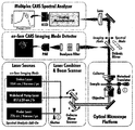
- FIG. 1 shows a CARS microscopy platform capable of lipid selective three dimensional imaging and point spectral analysis of the present invention.
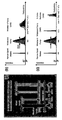
- FIG. 2 shows an energy plot of a three primary multiplex CARS with broadband pump laser excitation, a) a two primary excitation anti-stocks generated by multiple lipid-related Raman resonances for fast lipid-window imaging, without a probe laser beam. Broadband integrated detection of the signal is shown and b) shows multiplex CARS spectral analysis performed with the addition of each separate probe laser.
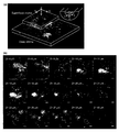
- FIG. 3 shows the results of unlabeled lipid selective CARS imaging for atherosclerotic lesions
- a) shows the results of three-dimensional reconstruction of serial en face images of atherosclerotic lesions by CARS
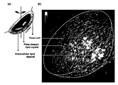
- FIG. 4 shows the unlabeled lipid selective CARS imaging results for a single atherosclerotic lesion, a) depicting a three dimensional representation of the CARS image, b) in the bubble layer in the superficial layer, and in the deep intima.
- the white border shows the semi-spherical shape of the atherosclerotic lesion in the three-dimensional CARS image.
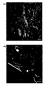
- Figure 5 shows a CARS image of the carotid artery of human atherosclerosis, where a) is lipid-rich foam cells in the surface region with a dark internal space corresponding to the nucleus, and b) plate-shaped and needle-shaped at the center of the gangrene Represents the lipid crystals of
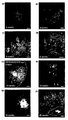
- FIG. 6 Apolipoprotein E characterized by CARS Expression inhibition (ApoE -/- The progression of arteriosclerosis in mice is shown.
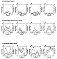
- Figure 7 shows the volume analysis according to the progression of the atherosclerosis by CARS, showing the lipid accumulation and the size of each lipid structure in the initial (a), middle (b) and deepening (c) step.
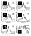
- Figure 8 shows the on-site spectrum analysis of atherosclerotic lipids by CARS
- af is a classification of atherosclerotic lipids according to the morphological differences, intracellular (a), extracellular (b), plate-form (c), needle form (d), non-artery (e) from connective tissue, and non-lipid (f) from matrix, and the inserted image shows the general shape used for spectral analysis.
- the present invention is a.
- a near-infrared pulsed laser unit for generating a combined laser beam by selectively irradiating stokes light, pump light and probe light of different wavelengths;
- a wideband multiplex CARS brown rice spectroscopy unit for detecting a spectrum by collecting a CARS signal generated from the sample
- An En face CARS image mode detection unit for collecting a CARS signal generated from the sample and providing a stereoscopic image
- a system for diagnosing pathological changes in lipids in blood vessels comprising a dichroic mirror disposed between the wideband multiplex CARS microscopic spectroscopy unit and the En face CARS image mode detection unit to selectively transmit the CARS signal generated from the sample to each unit. It is about.
- the system for diagnosing pathological changes of lipids in blood vessels is characterized in that lipid-selective three-dimensional imaging and point spectral analysis, in which images are distinguished through vibrations of hydrocarbons in lipids, are possible.
- the near-infrared pulsed laser unit may generate a combined laser beam by selectively irradiating stokes light, pump light and probe light of different wavelengths, and the beam vibrates a hydrocarbon portion of the lipid to thereby not only a three-dimensional image but also a Raman shift. Can be evaluated simultaneously.
- the Stokes light and the pump light are irradiated to the sample for the three-dimensional imaging of the lipid, and the probe light can be used for the spectral analysis of the lipid and can be blocked by the mechanical shutter during the three-dimensional imaging.
- the bandwidth is preferably 2700 to 3050 cm ⁇ 1 so that the CARS signal of the lipids obtained through the excitation beam of the near infrared pulse laser unit contains the oscillating portion of the entire hydrocarbon.
- the CARS signal collection time is 1.0s / frame
- the spatial density is preferably 0.4 ⁇ m in the left and right side, 1.3 ⁇ m in the axial direction.
- the multiplex CARS brown rice spectroscopy unit may detect the spectrum by collecting the CARS signal generated from the sample, it may be used in the Republic of Korea Patent Publication No. 2009-0024965, but is not particularly limited thereto.
- the system for diagnosing pathological changes in lipids of blood vessels of the present invention is disposed between the wideband multiplex CARS microscopic spectroscopy unit and the En face CARS image mode detection unit to selectively transfer CARS signals generated from the sample to each unit. It includes a dichroic mirror.
- the dichroic mirror reflects wavelengths less than 1000 nm and transmits more wavelengths.
- a stock signal and a pump light are irradiated onto the sample to separate the CARS lipid signal in the range of 645 to 675 nm through a band pass filter, and to detect it through the en face CARS imaging mode detection unit. To obtain a three-dimensional image.
- a wideband multiplex CARS microscopic spectroscopy unit is set up and the laser-scanner is adjusted in point-scan mode to irradiate the sample with probe light for 50 to 150 ms to generate a multiplex CARS signal
- the signal may be transmitted through a lattice monochromator to enable spectral analysis.
- the probe light preferably has a narrow band wavelength of 3.5 cm ⁇ 1 or less, and the anti-stock signal generated therefrom may appear in the range of 620 to 640 nm.
- sample used in the pathological change diagnosis system of lipids in the blood vessel of the present invention is not fixed or stained, and any tissue extracted from the animal is not particularly limited, and may be, for example, cardiovascular tissue of the animal.
- the thickness of the sample that can be analyzed through the pathological change diagnosis system of lipids in the blood vessel of the present invention enables three-dimensional imaging in the range of 100 to 150 ⁇ m.
- the pathological changes of the blood vessel lipids that can be diagnosed through the pathological change diagnosis system of the blood vessel lipids of the present invention may be atherosclerotic lesions.
- lipid droplets are observed in the surface lining, and the intermediate stage
- the number of lipid droplets increased significantly, extracellular lipid droplets were deposited deep in the vessel wall, and lipid droplets were observed in the form of multilayered plates in the deep inner membrane.
- the gangrene center is enlarged toward the lumen, the cholesterol crystal layer is predominantly observed, the foam cells are markedly reduced, and the fibrous thickening is observed.
- the invention also relates to the invention.
- the present invention relates to a method for diagnosing non-destructive pathological changes of lipids in blood vessels including analyzing the structure of lipids in the image.
- the sample is not subjected to any fixation or staining, and is not particularly limited as long as it is tissue extracted from an animal, and may be, for example, cardiovascular tissue of an animal.
- the signal may be collected through a band pass filter and detected through an en face CARS imaging mode detection unit to obtain a three-dimensional image.
- the three-dimensional image of the lipid is observed in the structure of the lipid, for example, in the form of lipid droplets, plates, needles, and when imaging the atherosclerotic vessels of the animal, the specific lipid structure according to the progression of atherosclerosis is observed In addition, the volume and size of lipids can be analyzed to diagnose the progression of atherosclerosis.
- the invention also relates to the invention.
- CARS coherent anti-Stokes Raman scattering
- the present invention relates to a method for diagnosing non-destructive pathological changes of lipids in blood vessels including analyzing spectral peaks of lipid structures in the spectrum.
- the sample is not subjected to any fixation or staining, and is not particularly limited as long as it is tissue extracted from an animal, and may be, for example, cardiovascular tissue of an animal.
- the signal can penetrate the lattice monochromator and can be detected spectrally through a broadband multiplex CARS microscopic spectroscopy unit.
- the spectra of extracellular and intracellular lipid droplets show one major peak at 2845 cm ⁇ 1 and four major peaks for plate-shaped lipids, namely 2880, 2905, 2920, 2950 cm -1 , and in the case of needle-shaped lipids have a weak peak at 2905, 2920, 2950 cm -1 , so that pathological changes of lipids can be diagnosed according to the peaks.
- the chemical profile of lipids can be applied to diagnose the progression of atherosclerosis.
- a broadband multiplex CARS microscopic spectrometer and a laser-scanning CARS microscope were simultaneously installed on the same platform for lipid selective three-dimensional microscopic imaging and point spectral analysis of cardiovascular tissue with atherosclerotic lesions.
- the laser-scanning CARS microscope is equipped with a modified laser-scanning confocal microscope (IX81 / FV300; Olympus, Japan) equipped with a grating monochromator (Triax320; Horiba Jobin Yvon). It consists of a near-IR pulse laser system that generates a CARS excitation beam that can generate three colors simultaneously.
- a modified laser-scanning confocal microscope IX81 / FV300; Olympus, Japan
- Triax320 Horiba Jobin Yvon
- Output pulse train and resonator stabilized feedback servo (SynchroLock-AP; Coherent, Inc.) active simultaneously with 800 mW average power, pulse bandwidth adjusted to approximately 35 nm, 1064 nm CARS Stokes beam to maintain typical repetition rate at 76 MHZ .)
- SynchroLock-AP Coherent, Inc.
- the three CARS excitation beams were then superimposed collinearly in the air using two beam combining optics in sequence: the pump and probe beams were 50:50 broadband beam splitters (CVI Melles Griot, Albuquerque, NM), Stokes beams have high reflectance for near infrared wavelengths in the range of 730-960 nm, and high transmittance for Stokes beams at 1064 nm (dichroic mirror, Chroma Technologies Corp., Rockingham, VT).
- CVI Melles Griot Albuquerque, NM
- Stokes beams have high reflectance for near infrared wavelengths in the range of 730-960 nm, and high transmittance for Stokes beams at 1064 nm (dichroic mirror, Chroma Technologies Corp., Rockingham, VT).
- the combined laser beam is subjected to a 1.2 NA 60X water-immersion microscope objective through a two-axis beam scanning unit (FV300) consisting of a pair of galvanometer-mount gold mirrors with a transmission of about 95% for wavelengths greater than 600 nm.
- FV300 two-axis beam scanning unit
- UPlanSApo UIS2; Olympus the power of each laser output was attenuated with a ND filter (neutral density filter) to limit the average power of the combined laser beam illuminating the sample to a total of 40 mW or less.
- the bandwidth of the beam is undergoing various modifications by extending the beam's bandwidth to a range of 2700 to 3050 cm ⁇ 1 in the Raman shift to cover the entire CH oscillation portion.
- Atherosclerotic lipids were imaged and chemically analyzed.
- the collection time is 1.0 s / frame compared to conventional Raman microscopy for unlabeled bio imaging, the spatial resolution is 0.4 ⁇ m on the left and right sides (xy) and the z-direction is With 1.3 ⁇ m, two-dimensional imaging with a maximum field of 250 ⁇ 250 ⁇ m 2 was improved.
- CARS microscopy made it easy to convert to a broadband multiplex CARS setup for spectral analysis of atherosclerotic lipids. After three-dimensional lipid selective imaging, points for CARS spectral analysis were selected and analyzed by 50-150 ms exposure.
- a carotid endarterectomy specimen was obtained from a carotid artery stenosis patient (63-81 years) who had undergone surgery at Samsung Medical Center (SMC). Samples for CARS analysis were immediately immersed in phosphate-buffered saline (PBS). In addition, two internal mammary artery specimens were obtained from a coronary bypass graft patient for use as a reference. The present invention was approved by SMC's Institutional Review Board according to the Helsinki Guidelines Declaration, and informed consent was obtained from all subjects (IRB 2006-02-11).
- mice Genetically engineered mice (ApoE -/- ) without apolipoprotein E expression were purchased from the Jackson Laboratory, Bar Harbor, ME, and were tested for specific pathogens at the Samsung Animal Science Laboratory Laboratory Animal Research Center. Adapted for one week in a sterile environment free of nutrients. 22 male ApoE ⁇ / ⁇ mice, 8 weeks old, were fed 0.15% high fat high cholesterol diet (HFHC) for 2-20 weeks. Two weeks later, 4-6 mice were sacrificed by inhaling CO 2 every week. The heart and aorta were sprayed with PBS for 10 minutes for CARS imaging and then quickly removed. All animal experiments were in compliance with the regulations of the Animal Experiment Ethics Committee of Samsung Life Science Research Laboratory.
- HFHC high fat high cholesterol diet
- samples were prepared for CARS imaging.
- the connective tissue was carefully removed and stored in cold PBS to analyze the chemical profile of lipids.
- the aorta was incised vertically from the ascending aorta to the descending aorta of the thoracic duct and then incised into four pieces: one piece containing the small portion of the aortic bow, two pieces containing the left and right arteries, and the remaining thoracic descending aorta. Included. Arteries branched from the aorta were carefully dissected with a microsurgery. Pieces prepared using PBS without chemical mounting solution or any fixative for en face CARS imaging were mounted on the coverslip with the lumen-side down.
- FIG. 3 shows a three-dimensional reconstruction of an en face image slice of an atherosclerotic lesion from the lumen to the deep intima.
- the bright spot shows characteristic CH molecular vibrations ranging from 2700 to 3050 cm ⁇ 1 . It shows a high concentration of lipids.
- This demonstrates the typical three-dimensional microscopic characterization of atherosclerotic lipids dependent on lesion depth.
- foam cells containing intracellular lipid droplets are clearly imaged, while lipid crystals do not interfere with their volume structure, but deep intima portions (> 25 ⁇ m deep). Imaged from.
- Lipid structures in atherosclerotic lesions can be morphologically classified into 1) intracellular droplets, 2) extracellular droplets, 3) multilayered crystal plates, and 4) needle shaped crystal structures.
- Foam cells were found only in the shallow portion (3-4 ⁇ m deep) of the surface lining.
- Multilayer plate lipids on the other hand, were observed in deep intima well separated from foam cells.
- Some types of multilayered lipid crystals were broadly arranged in parallel or inclined plates on the intima surface.
- needle-like lipid structures were deposited in the deep intima with plate-shaped lipid crystals.
- kidneys rich in cholesterol crystals in lipid droplets (bubble cells), superficial and extracellular lipid deposits, and deep intima, as shown in the hemispherical three-dimensional CARS image of the single atherosclerotic lesion in FIG. 4. Microanatomical components were observed, including extended cells.
- CARS microscopy was applied to human atherosclerotic carotid artery using the same imaging protocol (FIG. 5). Foam cells were successfully imaged to a depth of 40 ⁇ m at the surface and lipid crystals were found in the deep intima (> 80 ⁇ m), as in mice. The maximum depth of CARS imaging on human tissue was 100 to 150 ⁇ m.
- FIG. 6A In the two week old atherosclerotic mouse model, little lipid droplets bound to the ECM were observed (FIG. 6A). In the 4-week old atherosclerotic mouse model, lipid droplets were observed only at the surface lining (the depth of penetration into the vascular wall ⁇ 10 ⁇ m), and under the ECM they regrouped into craters towards the medium (FIG. 6B). In the 6 week old arteriosclerosis mouse model, the number of lipid droplets was significantly increased compared to 2 week old mice (FIG. 6C). In particular, extracellular lipid droplets blocked by ECM were deposited at 30 penetration depths in the vessel wall.
- lipid droplets are distributed in the typical form of cells with a dark inner space corresponding to the nucleus, which gives the appearance of foam cells containing lipids.
- the atherosclerotic lesions exhibited enhanced pathogenic features such as crystalline lipid structures in the deep intima (FIG. 6D). Foam cells were still observed only at the surface. However, the structure of the foam cells was clearer than the 6-week-old atherosclerotic mouse model (white arrow in FIG. 6D).
- the total volume of the necrotic core was measurable in three-dimensional CARS imaging: 100-120 ⁇ m.
- Lipid distribution in three-dimensional CARS imaging was quantitatively analyzed in three main steps (early, middle and deep phases, FIG. 7). Typical features of the progression of atherosclerosis are analyzed by the volume of lipid fraction (calculated as a two-dimensional coverage in z-stack) and the size of the lipid structure.
- Week 2-6 Figure 7a i-iv
- i-iv the initial phase
- FIG. 7B i-iv
- the lipid deposits migrated deeply to the z-stack.
- it increased in lipid size or deep intima to form plate-shaped lipid crystals.
- the deepening phase week 16, FIG.
- both lipid coverage and size increased.
- Atherosclerotic lipids have penetrated to a depth of 30 ⁇ m and even a single lipid structure of 90 ⁇ m 2 size is detected directly, a feature that is considered a serious atherosclerotic lesion.
- Multiplex CARS can be used to analyze chemical differences in various forms of atherosclerotic lipids based on spectral morphology.
- the lipids analyzed were classified into four main types, ie extracellular and intracellular lipid droplets, plate- and needle-type lipids, based on the morphological differences detected in the en face images.
- Spectra of extracellular and intracellular lipid droplets showed one main peak (2845 cm ⁇ 1 ) in symmetrical CH 2 oscillations (FIG. 8).
- the chemical profile of the plate-shaped lipids was significantly different from the lipid droplets.
- the spectra of plate-shaped lipids in the deep intima showed four extra peaks (2880, 2905, 2920, 2950 cm ⁇ 1 ) in their spectral profile compared to the lipid droplets.
- the extra peaks correspond to CH 2 asymmetry, hydrocarbons, CH 3 symmetry, and CH 3 asymmetry oscillations, respectively.
- the needle form of the lipid in crystalline form showed a weaker peak at 2905, 2920, 2950 cm ⁇ 1 compared to the spectrum of the multilayer plate-shaped lipid.
- the present invention is very useful in the medical device industry because it is possible to diagnose the progression of atherosclerosis by selectively imaging blood vessel lipids three-dimensionally without tissue damage following staining or cutting and without labeling.
Landscapes
- Physics & Mathematics (AREA)
- Spectroscopy & Molecular Physics (AREA)
- General Physics & Mathematics (AREA)
- Chemical & Material Sciences (AREA)
- Analytical Chemistry (AREA)
- Optics & Photonics (AREA)
- Health & Medical Sciences (AREA)
- Nuclear Medicine, Radiotherapy & Molecular Imaging (AREA)
- Life Sciences & Earth Sciences (AREA)
- Biochemistry (AREA)
- General Health & Medical Sciences (AREA)
- Immunology (AREA)
- Pathology (AREA)
- Investigating, Analyzing Materials By Fluorescence Or Luminescence (AREA)
- Microscoopes, Condenser (AREA)
Abstract
Description
Claims (12)
- 서로 다른 파장의 스톡스광, 펌프광 및 탐침광을 선택적으로 조사하여 컴바인드 레이저 빔을 발생시키는 근적외선 펄스 레이저 유니트;상기 근적외선 펄스 레이저 유니트로부터 전달된 컴바인드 레이저 빔이 조사되는 시료가 장착된 플랫홈;상기 시료에서 발생된 CARS 신호를 수집하여 스펙트럼을 검출하는 광대역 멀티플렉스 CARS 현미 분광 유니트;상기 시료에서 발생된 CARS 신호를 수집하여 입체 영상을 제공하는 En face CARS 이미지 모드 검출 유니트; 및상기 광대역 멀티플렉스 CARS 현미 분광 유니트와 En face CARS 이미지 모드 검출 유니트 사이에 배치되어, 상기 시료에서 발생된 CARS 신호를 선택적으로 각 유니트로 전달하는 이색 미러를 포함하는 혈관 내 지질의 병리적 변화 진단 시스템.
- 제1항에 있어서,상기 시료가 동물의 심혈관 조직인 혈관 내 지질의 병리적 변화 진단 시스템.
- 제1항에 있어서,상기 CARS 신호의 대역은 2700 내지 3050cm-1인 혈관 내 지질의 병리적 변화 진단 시스템.
- 제1항에 있어서,상기 CARS 신호 수집 시간은 1.0s/frame이고, 공간적 밀도는 좌우 측면이 0.4㎛, 축방향이 1.3㎛인 혈관 내 지질의 병리적 변화 진단 시스템.
- 제1항에 있어서,상기 이색 미러는 1000nm 미만의 파장을 반사시키고, 그 이상의 파장을 투과시키는 혈관 내 지질의 병리적 변화 진단 시스템.
- 제1항에 있어서,혈관 내 지질의 병리적 변화가 동맥경화 병반인 것인 혈관 내 지질의 병리적 변화 진단 시스템.
- 스톡스광과 펌프광을 시료에 조사하여 산란되는 CARS(coherent anti-Stokes Raman scattering) 지질 신호의 파장 및 세기를 측정하는 단계;상기 신호를 3 차원 이미지로 검출하는 단계; 및상기 이미지에서 지질의 구조를 분석하는 단계를 포함하는 혈관 내 지질의 비파괴적 병리적 변화 진단 방법.
- 제7항에 있어서,시료는 동물의 심혈관 조직인 혈관 내 지질의 비파괴적 병리적 변화 진단 방법.
- 제7항에 있어서,혈관 내 지질의 병리적 변화가 동맥경화 병반인 혈관 내 지질의 비파괴적 병리적 변화 진단 방법.
- 탐침광을 시료에 조사하여 산란되는 CARS(coherent anti-Stokes Raman scattering) 지질 신호의 파장 및 세기를 측정하는 단계;상기 신호를 스펙트럼으로 검출하는 단계; 및상기 스펙트럼에서 지질 구조의 스펙트럼 피크를 분석하는 단계를 포함하는 혈관 내 지질의 비파괴적 병리적 변화 진단 방법.
- 제10항에 있어서,시료는 동물의 심혈관 조직인 혈관 내 지질의 비파괴적 병리적 변화 진단 방법.
- 제10항에 있어서,혈관 내 지질의 병리적 변화가 동맥경화 병반인 혈관 내 지질의 비파괴적 병리적 변화 진단 방법.
Priority Applications (3)
| Application Number | Priority Date | Filing Date | Title |
|---|---|---|---|
| US13/320,014 US20120050720A1 (en) | 2009-05-11 | 2009-05-11 | System for diagnosing pathological change of lipid in blood vessels using non-linear optical microscopy |
| JP2012510724A JP5444457B2 (ja) | 2009-05-11 | 2009-05-11 | 非線形光学顕微鏡を用いた血管内脂質の非破壊的病理的変化診断プログラム |
| PCT/KR2009/002478 WO2010131785A1 (ko) | 2009-05-11 | 2009-05-11 | 비선형 광학현미경을 이용한 혈관 내 지질의 병리적 변화 진단시스템 |
Applications Claiming Priority (1)
| Application Number | Priority Date | Filing Date | Title |
|---|---|---|---|
| PCT/KR2009/002478 WO2010131785A1 (ko) | 2009-05-11 | 2009-05-11 | 비선형 광학현미경을 이용한 혈관 내 지질의 병리적 변화 진단시스템 |
Publications (1)
| Publication Number | Publication Date |
|---|---|
| WO2010131785A1 true WO2010131785A1 (ko) | 2010-11-18 |
Family
ID=43085147
Family Applications (1)
| Application Number | Title | Priority Date | Filing Date |
|---|---|---|---|
| PCT/KR2009/002478 Ceased WO2010131785A1 (ko) | 2009-05-11 | 2009-05-11 | 비선형 광학현미경을 이용한 혈관 내 지질의 병리적 변화 진단시스템 |
Country Status (3)
| Country | Link |
|---|---|
| US (1) | US20120050720A1 (ko) |
| JP (1) | JP5444457B2 (ko) |
| WO (1) | WO2010131785A1 (ko) |
Families Citing this family (8)
| Publication number | Priority date | Publication date | Assignee | Title |
|---|---|---|---|---|
| US9581497B2 (en) * | 2012-10-19 | 2017-02-28 | Hitachi, Ltd. | Cars microscope |
| CN109171761A (zh) * | 2013-05-02 | 2019-01-11 | Atonarp株式会社 | 对生物体进行监视的监视器和系统 |
| JP6436452B2 (ja) * | 2013-08-16 | 2018-12-12 | 国立研究開発法人農業・食品産業技術総合研究機構 | 食品中の豚肉を検出する方法 |
| WO2015025389A1 (ja) * | 2013-08-22 | 2015-02-26 | 株式会社日立製作所 | Cars顕微鏡 |
| JP6427890B2 (ja) * | 2014-02-13 | 2018-11-28 | 株式会社ニコン | 検出装置、顕微鏡および処理装置 |
| CN113631897B (zh) * | 2019-04-29 | 2024-07-16 | Atonarp株式会社 | 混合光学系统 |
| CN113795746A (zh) * | 2019-04-30 | 2021-12-14 | Atonarp株式会社 | 测量系统 |
| WO2025160496A1 (en) * | 2024-01-25 | 2025-07-31 | Trustees Of Boston University | Stimulated raman photothermal microscope with optical parametric amplifier source |
Citations (3)
| Publication number | Priority date | Publication date | Assignee | Title |
|---|---|---|---|---|
| KR100781745B1 (ko) * | 1998-10-14 | 2007-12-04 | 조뱅 본 에스. 에이. | 박막 샘플을 광학적으로 특성분석하는 장치 |
| KR100826593B1 (ko) * | 2007-02-13 | 2008-04-30 | 한국표준과학연구원 | 연속신호생성 방식 비선형 간섭성 반스톡스 라만산란이미징 장치 |
| KR20090024965A (ko) * | 2007-09-05 | 2009-03-10 | 한국표준과학연구원 | Cars 현미경을 이용한 유기 박막의 화학종 및 두께동시 광학분석장치 |
Family Cites Families (8)
| Publication number | Priority date | Publication date | Assignee | Title |
|---|---|---|---|---|
| JPS639828A (ja) * | 1986-07-01 | 1988-01-16 | Hitachi Ltd | カ−ス光学装置 |
| WO2005124336A1 (en) * | 2004-06-17 | 2005-12-29 | Koninklijke Philips Electronics N. V. | Combined ultrasonic imaging and spectroscopic molecular analysis |
| JP2008249945A (ja) * | 2007-03-30 | 2008-10-16 | Bunkoh-Keiki Co Ltd | 顕微鏡接続用分光器 |
| US7847933B2 (en) * | 2007-05-22 | 2010-12-07 | Stevens Institute Of Technology | Cars microscopy and spectroscopy using ultrafast chirped pulses |
| JP2009047435A (ja) * | 2007-08-13 | 2009-03-05 | Olympus Corp | レーザ顕微鏡 |
| WO2011152834A1 (en) * | 2009-06-05 | 2011-12-08 | The Penn State Research Foundation | Coherent anti-stokes raman holography |
| US8879058B2 (en) * | 2010-11-05 | 2014-11-04 | The University Of Ottawa | Miniaturized multimodal cars endoscope |
| JP5703907B2 (ja) * | 2011-03-31 | 2015-04-22 | ソニー株式会社 | 非線形ラマン分光装置、非線形ラマン分光システム及び非線形ラマン分光方法 |
-
2009
- 2009-05-11 WO PCT/KR2009/002478 patent/WO2010131785A1/ko not_active Ceased
- 2009-05-11 JP JP2012510724A patent/JP5444457B2/ja not_active Expired - Fee Related
- 2009-05-11 US US13/320,014 patent/US20120050720A1/en not_active Abandoned
Patent Citations (3)
| Publication number | Priority date | Publication date | Assignee | Title |
|---|---|---|---|---|
| KR100781745B1 (ko) * | 1998-10-14 | 2007-12-04 | 조뱅 본 에스. 에이. | 박막 샘플을 광학적으로 특성분석하는 장치 |
| KR100826593B1 (ko) * | 2007-02-13 | 2008-04-30 | 한국표준과학연구원 | 연속신호생성 방식 비선형 간섭성 반스톡스 라만산란이미징 장치 |
| KR20090024965A (ko) * | 2007-09-05 | 2009-03-10 | 한국표준과학연구원 | Cars 현미경을 이용한 유기 박막의 화학종 및 두께동시 광학분석장치 |
Also Published As
| Publication number | Publication date |
|---|---|
| JP5444457B2 (ja) | 2014-03-19 |
| US20120050720A1 (en) | 2012-03-01 |
| JP2012526982A (ja) | 2012-11-01 |
Similar Documents
| Publication | Publication Date | Title |
|---|---|---|
| WO2010131785A1 (ko) | 비선형 광학현미경을 이용한 혈관 내 지질의 병리적 변화 진단시스템 | |
| Le et al. | Label-free molecular imaging of atherosclerotic lesions using multimodal nonlinear optical microscopy | |
| Wang et al. | Label-free imaging of arterial cells and extracellular matrix using a multimodal CARS microscope | |
| Seeger et al. | Multimodal optoacoustic and multiphoton microscopy of human carotid atheroma | |
| KR101100030B1 (ko) | 비선형광학현미경을 이용한 혈관 내 지질의 병리적 변화 진단 시스템 | |
| Lee et al. | Nanosecond SRS fiber amplifier for label-free near-infrared photoacoustic microscopy of lipids | |
| Lim et al. | Multimodal CARS microscopy determination of the impact of diet on macrophage infiltration and lipid accumulation on plaque formation in ApoE-deficient mice | |
| Yserbyt et al. | Perspectives using probe-based confocal laser endomicroscopy of the respiratory tract | |
| WO2010131713A1 (ja) | 血管内壁分析装置及び血管内壁分析方法 | |
| Mehravar et al. | Label-free multi-photon imaging of dysplasia in Barrett’s esophagus | |
| Yan et al. | A pilot study of using multiphoton microscopy to diagnose gastric cancer | |
| Meyer et al. | CARS-imaging guidance for fs-laser ablation precision surgery | |
| Bae et al. | Feasibility studies of multimodal nonlinear endoscopy using multicore fiber bundles for remote scanning from tissue sections to bulk organs | |
| Ko et al. | Multimodal nonlinear optical imaging of atherosclerotic plaque development in myocardial infarction-prone rabbits | |
| Haudenschild et al. | Nondestructive fluorescence lifetime imaging and time-resolved fluorescence spectroscopy detect cartilage matrix depletion and correlate with mechanical properties | |
| Nie et al. | Differentiating the two main histologic categories of fibroadenoma tissue from normal breast tissue by using multiphoton microscopy | |
| Galli et al. | Vibrational spectroscopy and multiphoton microscopy for label-free visualization of nervous system degeneration and regeneration | |
| CA2335246A1 (en) | Detection of cancer using cellular autofluorescence | |
| Mytskaniuk et al. | Implementation of a coherent anti-stokes Raman scattering (CARS) system on a Ti: Sapphire and OPO laser based standard laser scanning microscope | |
| Baria et al. | Improved label‐free diagnostics and pathological assessment of atherosclerotic plaques through nonlinear microscopy | |
| Peres et al. | Commercially derived versatile optical architecture for two-photon STED, wavelength mixing and label-free microscopy | |
| Dave et al. | Defining the zone of acute peripheral nerve injury using fluorescence lifetime imaging in a crush injury sheep Model | |
| Cicchi et al. | Multidimensional custom-made non-linear microscope: from ex-vivo to in-vivo imaging | |
| Wu et al. | Automatic and Label-Free Analysis of the Microstructure Feature Differences Between Normal Brain Tissue, Low-Grade, and High-Grade Gliomas Using the Combination of Multiphoton Microscopy and Image Analysis | |
| Büttner et al. | Heart valve stenosis in laser spotlights: insights into a complex disease |
Legal Events
| Date | Code | Title | Description |
|---|---|---|---|
| 121 | Ep: the epo has been informed by wipo that ep was designated in this application |
Ref document number: 09844659 Country of ref document: EP Kind code of ref document: A1 |
|
| WWE | Wipo information: entry into national phase |
Ref document number: 13320014 Country of ref document: US |
|
| NENP | Non-entry into the national phase |
Ref country code: DE |
|
| WWE | Wipo information: entry into national phase |
Ref document number: 2012510724 Country of ref document: JP |
|
| 122 | Ep: pct application non-entry in european phase |
Ref document number: 09844659 Country of ref document: EP Kind code of ref document: A1 |