WO2009108376A2 - Contrast agents for applications including perfusion imaging - Google Patents
Contrast agents for applications including perfusion imaging Download PDFInfo
- Publication number
- WO2009108376A2 WO2009108376A2 PCT/US2009/001296 US2009001296W WO2009108376A2 WO 2009108376 A2 WO2009108376 A2 WO 2009108376A2 US 2009001296 W US2009001296 W US 2009001296W WO 2009108376 A2 WO2009108376 A2 WO 2009108376A2
- Authority
- WO
- WIPO (PCT)
- Prior art keywords
- subject
- imaging
- brain
- cns
- contrast agent
- Prior art date
Links
- 0 COC(c1ccc(COCC*)cc1)=O Chemical compound COC(c1ccc(COCC*)cc1)=O 0.000 description 3
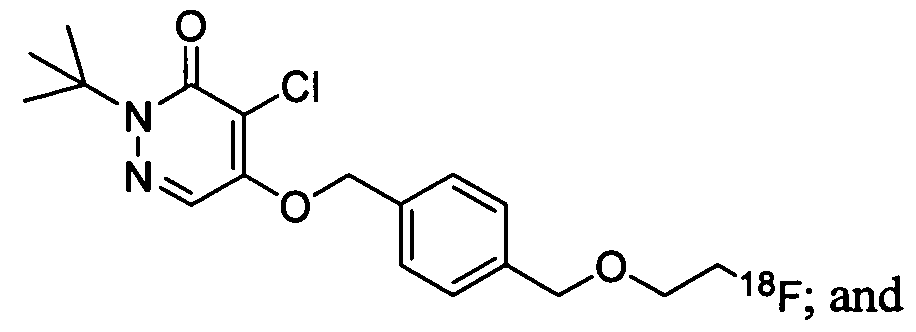
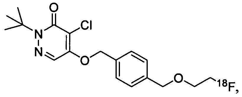
- RMXZKEPDYBTFOS-UHFFFAOYSA-N CC(C)(C)N(C1=O)N=CC(OCc2ccc(COCCF)cc2)=C1Cl Chemical compound CC(C)(C)N(C1=O)N=CC(OCc2ccc(COCCF)cc2)=C1Cl RMXZKEPDYBTFOS-UHFFFAOYSA-N 0.000 description 1
Classifications
-
- A—HUMAN NECESSITIES
- A61—MEDICAL OR VETERINARY SCIENCE; HYGIENE
- A61K—PREPARATIONS FOR MEDICAL, DENTAL OR TOILETRY PURPOSES
- A61K51/00—Preparations containing radioactive substances for use in therapy or testing in vivo
- A61K51/02—Preparations containing radioactive substances for use in therapy or testing in vivo characterised by the carrier, i.e. characterised by the agent or material covalently linked or complexing the radioactive nucleus
- A61K51/04—Organic compounds
- A61K51/041—Heterocyclic compounds
- A61K51/044—Heterocyclic compounds having nitrogen as a ring hetero atom, e.g. guanethidine, rifamycins
- A61K51/0459—Heterocyclic compounds having nitrogen as a ring hetero atom, e.g. guanethidine, rifamycins having six-membered rings with two nitrogen atoms as the only ring hetero atoms, e.g. piperazine
-
- A—HUMAN NECESSITIES
- A61—MEDICAL OR VETERINARY SCIENCE; HYGIENE
- A61P—SPECIFIC THERAPEUTIC ACTIVITY OF CHEMICAL COMPOUNDS OR MEDICINAL PREPARATIONS
- A61P25/00—Drugs for disorders of the nervous system
- A61P25/28—Drugs for disorders of the nervous system for treating neurodegenerative disorders of the central nervous system, e.g. nootropic agents, cognition enhancers, drugs for treating Alzheimer's disease or other forms of dementia
-
- A—HUMAN NECESSITIES
- A61—MEDICAL OR VETERINARY SCIENCE; HYGIENE
- A61P—SPECIFIC THERAPEUTIC ACTIVITY OF CHEMICAL COMPOUNDS OR MEDICINAL PREPARATIONS
- A61P35/00—Antineoplastic agents
Abstract
The present invention is directed, in part, to compounds and methods for imaging the brain or central nervous system, or portions thereof, comprising administering to a subject a contrast agent of the invention, and scanning the subject using diagnostic imaging.
Description
CONTRAST AGENTS FOR APPLICATIONS INCLUDING PERFUSION IMAGING
Related Applications
This application claims priority under 35 U.S.C. § 119(e) to co-pending United States Provisional Application Serial No. 61/067,593, filed February 29, 2008, the contents of which are incorporated herein by reference.
Field of the Invention
The present invention relates to compounds comprising imaging moieties, and their use in imaging and/or diagnosing certain disorders in a subject.
Background of the Invention Mitochondria are membrane-enclosed organelles distributed through the cytosol of most eukaryotic cells. Mitochondria levels are elevated in tissues that require greater energy to function. Examples of such tissue include the brain and central nervous system.
Complex 1 ("MC-I") is a membrane-bound protein complex of 46 dissimilar subunits. This enzyme complex is one of three energy-transducing complexes that constitute the respiratory chain in mammalian mitochondria. This NADH-ubiquinone oxidoreductase is the point of entry for the majority of electrons that traverse the respiratory chain, eventually resulting in the reduction of oxygen to water (Q. Rev. Biophys. 1992, 25, 253-324). Known inhibitors of MC-I include deguelin, piericidin A, ubicidin-3, rolliniastatin-1, rolliniastatin-2 (bullatacin), capsaicin, pyridaben, fenpyroximate, amytal, MPP+, quinolines, and quinolones (BBA 1998, 1364, 222-235).
Summary of the Invention
The present invention relates to the recognition that interrupting the normal function of mitochondria may advantageously concentrate certain compounds in the mitochondria, and, hence, in mitochondria-rich tissue. As described herein, such compounds may be labeled with at least one imaging moiety, such that mitochondrial build-up may be determined, thereby providing valuable diagnostic markers for brain imaging. For purposes of this specification, a compound is referred to as "labeled" when an imaging moiety is attached to (e.g. bound to) the compound.
In some embodiments, the present invention provides methods of imaging at least a portion of the brain (e.g., brain tissue) or central nervous system, comprising administering to a subject a contrast agent which comprises an imaging moiety and a
compound bound to the imaging moiety, a compound of the invention; and scanning the subject using diagnostic imaging to produce an image of at least a portion of the brain or central nervous system (CNS). The image may be used in the diagnosis of a subject, or to determine the stage of a disease.
In one set of embodiments, the contrast agent is

In any of the forgoing aspects and embodiments, the contrast agent may be provided in the presence of a pharmaceutically acceptable salt, as disclosed herein.
In any of the forgoing aspects and embodiments, the contrast agent may be provided in the presence of a counterion, or, in the absence of a counterion (e.g., as a free base).
In some embodiments, the present invention provides methods for synthesizing any of the foregoing contrast agents according to the methods described herein. In some embodiments, the method may comprise reacting a compound with an imaging moiety precursor to form a contrast agent. In another embodiment, the method may comprise reacting an intermediate molecule to produce a contrast agent of the invention. In some embodiments, the method may further comprise isolating and/or purifying the intermediate molecule and/or contrast agent. The method may also comprise characterization of the intermediate molecule and/or contrast agent. In some embodiments, the present invention also provides methods for medical imaging; intravenous use in imaging; imaging at least a portion of the brain or central nervous system of a subject; infusion or injection; delivering an imaging agent to the brain; imaging perfusion in a body region or structure (e.g., brain, CNS); determining the level of mitochondria and/or mitochondrial density in a subject or portion of a subject; diagnosing a disease in a subject, including diagnosing the onset, progression, and/or regression of a disease; determining the stage of a disease in a subject; passing a contrast agent of the invention through the blood brain barrier of a subject; or monitoring the accumulation of a contrast agent of the invention in the brain of a subject. In some embodiments, methods of the invention can be used to assess efficacy of a treatment, for example, the brain or CNS can be visualized using contrast agents of the invention
before, during, and/or after treatment of a condition affecting the brain or CNS of a subject. The method may comprise administering a contrast agent as described herein to a subject. In some embodiments, the method comprises passing a contrast agent of the invention through the blood brain barrier of a subject. In some embodiments, the method comprises monitoring the accumulation of a contrast agent of the invention in the brain of a subject. All features disclosed in the specification may be used in combination with such methods.
In some embodiments, the present invention provides pharmaceutical compositions for medical imaging; intravenous use in imaging; imaging at least a portion of the brain or central nervous system of a subject; infusion or injection; delivering an imaging agent to the brain; imaging perfusion in a body region or structure (e.g., brain, CNS); determining the level of mitochondria and/or mitochondrial density in a subject or portion of a subject; diagnosing a disease in a subject, including diagnosing the onset, progression, and/or regression of a disease; determining the stage of a disease in a subject; passing a contrast agent of the invention through the blood brain barrier of a subject; or monitoring the accumulation of a contrast agent of the invention in the brain of a subject. In some embodiments, the pharmaceutical composition comprises a contrast agents as described herein, and one or more pharmaceutically acceptable carriers, additives, and/or diluents. All features disclosed in the specification may be used in combination with such pharmaceutical compositions.
In some embodiments, the present invention relates to the use of any of the contrast agents described herein in the preparation of a medicament for medical imaging; intravenous use in imaging; imaging at least a portion of the brain or central nervous system; infusion or injection; delivering an imaging agent to the brain; imaging perfusion in a body region or structure (e.g., brain, CNS); determining the level of mitochondria and/or mitochondrial density in a subject or portion of a subject; diagnosing a disease in a subject, including diagnosing the onset, progression, and/or regression of a disease; determining the stage of a disease in a subject; passing a contrast agent of the invention through the blood brain barrier of a subject; or monitoring the accumulation of a contrast agent of the invention in the brain of a subject. Any of the uses described herein may comprise the use of a contrast agent of the present invention. All features disclosed in the specification may be used in combination with such uses.
In some embodiments, the present invention provides methods of treating a patient. The method may comprise the steps of administering to the patient a contrast agent as in any foregoing embodiments; and acquiring an image of a site of concentration of the contrast agent in the patient by a diagnostic imaging technique. The present invention also provides method for acquiring an image, or constructing an image, of at least a portion of the brain or central nervous system of a subject.
Any of the foregoing aspects and embodiments may comprise contacting at least a portion of the brain or central nervous system of a subject with a contrast agent of the invention. In certain embodiments, the contacting may occur via administration of the contrast agent to the subject. In one set of embodiments, the contacting may occur via intravenous administration of the contrast agent to the subject.
In any of the foregoing aspects and embodiments, the disease may be a CNS disorder or condition, as described herein. In any of the foregoing aspect and embodiments, the subject can be otherwise free of indications for perfusion imaging, such as myocardial perfusion imaging, for example.
Other aspects of the invention may include suitable combinations of embodiments and aspects disclosed herein. Brief Description of the Drawings
FIG. 1 shows representative images of the (a) transverse, (b) coronal, and (c) saggittal planes of a nonhuman primate brain, with 2-ter/-butyl-4-chloro-5-[4-(2-
[ F]fluoro-ethoxymethyl)-benzyloy]-2H-pyridazin-3-one in a normal NΗP, where the whiter portions indicate localization of the contrast agent. FIG. 2 A shows representative images of the transverse (left image) and sagittal
(right image) sections of a rat brain imaged using 2-tert-Butyl-4-chloro-5-[4-(2-
(18F)fluoroethoxymethyl)-benzyloxy]-2Η-pyridazin-3-one (Agent 2), where the whiter portions indicate localization of the contrast agent.
FIG. 2B shows representative images of the transverse (left image) and sagittal (right image) sections of a rat brain imaged using 2-terf-Butyl-4-chloro-5-[4-(3-
(18F)fluoropropoxy)-benzyloxy]-2H-pyridazin-3-one (Agent 3), where the whiter portions indicate localization of the contrast agent.
FIG. 3 A shows representative tomographic images of the transverse (left image) and sagittal (right image) sections of a NHP brain imaged using Agent 2, where the whiter portions indicate localization of the contrast agent.
FIG. 3 B shows representative tomographic images of the transverse (left image) and sagittal sections (right image) of a NHP brain imaged using 2-tert-butyl-4-chloro-5-
1 fi
[4-(4-[ F]fluoro-butyl)-benzyloxy]-2H-pyridazin-3-one (Agent 1), where the whiter portions indicate localization of the contrast agent.
Detailed Description The present invention generally relates to methods for using contrast agents in imaging including perfusion imaging. In some embodiments, methods of the invention may be useful in imaging a location within a subject (e.g., mammal), including the brain or central nervous system, or portions thereof. Some embodiments of the invention may provide contrast agents, and related methods, that are selective for high energy demand tissues within a subject, in addition to a broad uptake mechanism. In some cases, contrast agents and methods described herein advantageously exhibit high avidity for an intracellular target with a relatively low off rate, which may be useful in targeting processes associated with mitochondria. Imaging moieties
Examples of nuclear medicine contrast agents suitable for use in the present invention include, but are not limited to, 11C, 13N, 18F, 123I, and 125I. In some cases, 11C - Palmitate may be used to probe fatty acid oxidation and nC-acetate may be used to assess oxidative metabolism in the myocardium {Circulation 1987, 76, 687-696). Agents based on F may, in some cases, be useful as imaging agents {Drugs of the Future 2002, 27, 655-667). In one set of embodiments, the imaging moiety employed in contrast agents of the present invention is 18F. In some embodiments, imaging moieties of the present invention may comprise one or more X-ray absorbing or "heavy" atoms having an atomic number of 20 or greater. In some cases, the contrast agent may further comprise an optional linking moiety, L, positioned between the parent molecular moiety and one or more X-ray absorbing atoms. A non-limiting example of a heavy atom used as X-ray contrast agents is iodine.
In some cases, methods for imaging central nervous system tissue, which consumes a disproportionate amount of energy, are provided. The blood-brain barrier (BBB) is a physical entity that can prevent the indiscriminate passage of agents into the
brain. Current agents that can image mitochondrial density are lipophilic monocations, and are typically excluded by the BBB from CNS uptake. In some cases, methods described herein provide agents that are capable of selectively imaging brain tissue and crossing the blood brain barrier. Such methods may be useful in imaging the topography and blood flow to the brain, as well as perfusion imaging in the brain.
Generally, the contrast agents described herein are capable of imaging and mapping mitochondrial density and function in tissues. Mitochondrial function has been indicated as causative or correlative in Alzheimer' Disease (AD; Wang, et al. Free Radical Biology and Medicine, 2007, 43, 1569-1573, incorporated herein by reference in its entirety), Parkinson's Disease (Higgin and Greenamyre, Journal ofNeuroscience, 1996, /6(12), 3807-3816, incorporated herein by reference in its entirety), as well as neuronal dysfunction and temporal lobe epilepsy (Kann and Kovacs, Am. J. Physiol. Cell Physiol. 2007, 292, C641-C657, incorporated herein by reference in its entirety). Agents such as those described herein can be used for the imaging of disease diagnosis, including, but not limited to, onset, progression, regression, and staging.
In some embodiments, the contrast agent comprises an imaging moiety and a compound bound to the imaging moiety. For example, the imaging agent may be a 18F atom covalently bound to a compound. Methods of Synthesizing Contrast Agents Typically, contrast agents described herein may be synthesized by reacting at least a first component and a second component, such that a bond is formed therebetween. For example, F labeled compounds may be synthesized by reacting two components via Sn2 displacement of an appropriate leaving group associated with at least one component. Examples of such leaving groups include sulfonic acid esters such as toluenesulfonate (tosylate, TsO-), methanesulfonate (mesylate, MsO-), or trifluoromethanesulfonate (triflate, TfO-). The leaving group may also be a halide, a phosphineoxide (via Mitsunobu reaction), or an internal leaving group (such as an epoxide or cyclic sulfate). In some embodiments, such compounds can be synthesized from highly activated, dry K18F, that is made more reactive by the addition of potassium sequestering cryptands such as krytofix[2.2.2]. Purification is generally performed via salt removal by reverse-phase chromatography (SepPak™).
Representative methods of making the contrast agents are described in the following examples. The foregoing chemical transformations may be conducted using
techniques which would be readily apparent to one of ordinary skill in the art, in combination with the teachings described herein. In some cases, methods of synthesizing the contrast agents may include the use of one or more reaction solvents. Representative reaction solvents include, for example, DMF, NMP, DMSO, THF, ethyl acetate, dichloromethane, and chloroform. The reaction solution may be kept neutral or basic by the addition of an amine such as triethylamine or DIEA. In some cases, the chemical transformations (e.g., reactions) may be carried out at ambient temperatures and protected from oxygen and water with a nitrogen, argon or helium atmosphere.
In some embodiments, temporary protecting groups may be used to prevent other reactive functionality, such as amines, thiols, alcohols, phenols, and carboxylic acids, from participating or interfering in the reaction. Representative amine protecting groups include, for example, terf-butoxycarbonyl and trityl (removed under mild acidic conditions), Fmoc (removed by the use of secondary amines such as piperidine), and benzyloxycarbonyl (removed by strong acid or by catalytic hydrogenolysis). The trityl group may also used for the protection of thiols, phenols, and alcohols. In certain embodiments the carboxylic acid protecting groups include, for example, tert-butyl ester (removed by mild acid), benzyl ester (usually removed by catalytic hydrogenolysis), and alkyl esters such as methyl or ethyl (usually removed by mild base). AU protecting groups may be removed at the conclusion of synthesis using the conditions described above for the individual protecting groups, and the final product may be purified by techniques which would be readily apparent to one of ordinary skill in the art, in combination with the teachings described herein. Use of Contrast Agents
The contrast agents of the present invention may be used in methods of imaging, including methods of imaging in a subject. For example, the method may comprise administering the contrast agent to the subject by injection (e.g., intravenous injection), infusion, or any other known method, and imaging the area of the subject wherein an event of interest is located.
The useful dosage to be administered and the particular mode of administration will vary depending upon such factors as age, weight, and particular region to be imaged, as well as the particular contrast agent used, the diagnostic use contemplated, and the form of the formulation, for example, suspension, emulsion, microsphere, liposome, or the like, as will be readily apparent to those of ordinary skill in the art.
Typically, dosage is administered at lower levels and increased until the desirable diagnostic effect (e.g., production of an image) is achieved. In one embodiment, the above-described contrast agents may be administered by intravenous injection, usually in saline solution, at a dose of about 0.1 to about 100 mCi per 70 kg body weight (and all combinations and subcombinations of dosage ranges and specific dosages therein), or, in some embodiments, at a dose of about 0.5 to about 50 mCi. Imaging is performed using techniques well known to the ordinarily skilled artisan.
In some cases, for use as nuclear medicine contrast agents, the compositions of the present invention, dosages, administered by intravenous injection, may be in the range from about 0.5 μmol/kg to about 1.5 mmol/kg (and all combinations and subcombinations of dosage ranges and specific dosages therein), and, in some embodiments, about 0.8 μmol/kg to about 1.2 mmol/kg.
Another aspect of the present invention provides diagnostic kits for the preparation of diagnostic agents for determining (e.g., detecting), imaging, and/or monitoring at least a portion of the brain or central nervous system. Diagnostic kits of the present invention may comprise one or more vials containing a sterile, non-pyrogenic, formulation comprising a predetermined amount of a reagent (e.g., contrast agent precursor) of the present invention, and optionally other components such as chelating agents, solvents, buffers, neutralization aids, lyophilization aids, stabilization aids, solubilization aids and bacteriostats, as described more fully below.
Some non-limiting examples of buffers useful in the preparation of contrast agents and kits include, for example, phosphate, citrate, subsalicylate, and acetate buffers. A more complete list can be found in the United States Pharmacopoeia.
Some non-limiting examples of lyophilization aids useful in the preparation of contrast agents and kits include, for example, mannitol, lactose, sorbitol, dextran, FICOLL® polymer, and polyvinylpyrrolidine (PVP).
Some non-limiting examples of stabilization aids useful in the preparation of contrast agents and kits include, for example, ethanol, ascorbic acid, ethanol, cysteine, monothioglycerol, sodium bisulfite, sodium metabisulfite, gentisic acid, and inositol. Some non-limiting examples of solubilization aids useful in the preparation of contrast agents and kits include, for example, ethanol, glycerin, polyethylene glycol, propylene glycol, polyoxyethylene sorbitan monooleate, sorbitan monoloeate, polysorbates, poly(oxyethylene)-poly(oxypropylene)-poly(oxyethylene) block
copolymers ("Pluronics®") and lecithin.
Some non-limiting examples of bacteriostats useful in the preparation of contrast agents and kits include, for example, benzyl alcohol, benzalkonium chloride, chlorobutanol, and methyl, propyl, or butyl paraben. A component in a diagnostic kit of the invention can also serve more than one function. For example, a solubilization aid may serve as a stabilizer.
Many geometric isomers of olefins, C=N double bonds, and the like can be present in the compounds described herein, and all such stable isomers are contemplated in the present invention. Imaging methods for detecting CNS disorders and conditions
Imaging methods of the invention can be used to diagnose and assess CNS disorders or conditions based on the determination of levels and/or density of mitochondria in tissues, tissue regions, and subjects through in vivo imaging. Determination of levels or mitochondria and/or mitochondrial density in tissues in a subject permits the diagnosis and assessment of disorders associated with altered levels of mitochondria or mitochondrial density. Differences in levels of mitochondria and/or mitochondrial density in tissues of a subject compared to levels of mitochondrial and/or mitochondrial density in normal tissues (e.g. non-diseased) tissues can be used to diagnose or to aid in the diagnosis in the subject of disorders or conditions that exhibit (e.g., are associated with) altered levels of mitochondria and/or mitochondrial density. Particular types of disorders and conditions that can be assessed using imaging methods of the invention include CNS disorders and conditions. Imaging methods of the invention may be used in diagnostic methods alone or in conjunction with other diagnostic methods known in the art. One aspect of the present invention relates to the use of a contrast agent of the invention for detecting mitochondrial levels in a subject. This method involves administering to a subject a contrast agent that localizes in mitochondria, thus permitting detection in the subject of regions or tissues with altered or abnormal levels of mitochondria.
Methods of the invention can be used to assess or screen patients for diseases associated with the presence of increased or decreased levels of mitochondrial density in tissues. As used herein, the term "increased" means higher, for example higher versus a control level. As used herein, the term "decreased" means lower, for example decreased versus a control level. Methods of the invention may be used to identify the status of
disorders associated with abnormal levels of mitochondria in tissues or regions. The amount of mitochondria in a tissue or region, as compared to a control, can be used to determine the presence or absence of a particular CNS disorder. Methods of the invention can be used to obtain useful prognostic information by providing an indicator of a CNS disorder in a subject, which can be used to select a therapy for the subject.
Imaging methods of the invention can be used to detect levels of mitochondria and/or mitochondrial density in subjects already diagnosed as having a CNS disorder or condition. In other instances, methods of the invention can be used to obtain measurements that provide a diagnosis or aid in providing a diagnosis of a CNS disorder or condition. In some instances, a subject may be already be undergoing drug therapy for a CNS disorder or condition, while in other instances a subject may be without present therapy for a CNS disorder or condition. In some embodiments, the method can be used to assess efficacy of a treatment. For example, the brain or CNS can be visualized using contrast agents of the invention before, during, and/or after treatment of a condition affecting the brain or CNS of a subject.
According to the present invention, some subjects may be free of symptoms otherwise calling for treatment with a particular therapy, and imaging methods of the invention may identify the subject as needing treatment. This means that absent the use of the imaging methods of the invention to assess levels of mitochondria and/or mitochondrial density, the subject would not according to convention as of the date of the filing of the present application have symptoms calling for treatment with a particular therapy. As a result of measuring the level of mitochondria and/or mitochondrial density of tissues or body regions of the subject using methods of the invention, the subject becomes a candidate for treatment with a particular therapy. Thus, for example, a subject determined using imaging methods of the invention, to have an above-normal level of mitochondria and/or mitochondrial density in a tissue or body region may be determined to have a CNS disorder or condition and these results may be used to selected or aid in the selection of a treatment for the CNS disorder or condition.
As will be understood by those of ordinary skill in the art, imaging using methods of the invention may include full body imaging of a subject, or imaging of a specific body region or tissue of interest. For example, if a subject is known or suspected of having a CNS disorder in the brain, methods of the invention may be used to image the brain of a subject. In some embodiments, imaging may be limited to the CNS and/or to a
specific region of the CNS. For example, in a subject with temporal lobe epilepsy, the temporal lobes may be imaged using methods of the invention and for a subject for whom stroke or cerebral infarction is suspected or confirmed, imaging may include imaging of the entire brain. In some aspects of the invention, imaging methods may include imaging of a specific tissue, region, or structure and in some aspects may include imaging of perfusion of a body region or structure. For example, methods of the invention may be used to image perfusion of the brain, or part of the brain, e.g., one or more brain structures. Perfusion of the brain will be understood by those of ordinary skill in the art to reflect the blood flow through the brain. Perfusion of the brain using methods of the invention may be useful to image regions of damage to the brain or regions of recovery of a previously damaged brain. Non-limiting examples of the use of perfusion methods of the invention include its use to image brain regions with reduced or obstructed blood flow resulting from an occlusion of blood vessels in the brain and also include its use to image brain regions with excessive blood flow, for example, resulting from a hemorrhagic event.
Some aspects of the invention include methods of administering to a subject an amount of a contrast agent effective to image a specific CNS region in the subject. Contrast agents of the invention, when administered to a subject, preferentially localize to mitochondria. The localization of contrast agents to mitochondria permits determination of relative levels of mitochondria in tissues and regions in the subject. An increased amount of contrast agent of the invention localizes to tissues and/or regions with higher levels of mitochondria and/or higher mitochondrial density versus the amount of contrast agent that localizes in tissues or regions having a lower level of mitochondria and/or lower mitochondrial density in the tissue or region. The level or intensity of an imaging signal localized to a tissue or body region of a subject following administration of a contrast agent in a method of the invention, indicates the level of mitochondria and/or mitochondrial density in that tissue or body region. Similarly, a decreased amount of contrast agent of the invention localizes to tissues and/or regions with lower levels of mitochondria or mitochondrial density versus the amount of contrast agent that localizes to tissues or regions having a higher level of mitochondria and/or mitochondrial density. The level or intensity of an imaging signal localized to a tissue or body region of a subject following administration of a contrast agent in a method of the invention, indicates the level of mitochondria and/or mitochondrial density in that tissue
or body region. This ability to quantify the uptake of the agent into tissue of interest is inherent in the physics of PET, which allows for relatively precise and accurate calculations of uptake into tissues compared to the injected dose of imaging agent. Comparison of this uptake versus levels that are expected from normal tissues allows for assessment and diagnosis of the subject.
Information on mitochondria levels in tissues or body regions that is obtained using imaging methods of the invention may be used for diagnosis of or to aid in the diagnosis of CNS disorders or conditions. In disorders characterized by increased levels or density of mitochondria in tissues compared to healthy tissues, an increase in imaging intensity in the tissues when using an imaging method of the invention may indicate the presence of the disorder. Similarly, in disorders characterized by decreased levels or density of mitochondria in tissues compared to healthy tissues, a decrease in imaging intensity in the tissues when using an imaging method of the invention may indicate the presence of the disorder. Those of ordinary skill in the art will recognize that disorders characterized by increased mitochondria density and disorders characterized by decreased mitochondrial density can both be assessed using methods of the invention.
Imaging methods of the invention may be used to assess a CNS disorder or condition and to select an appropriate treatment for a subject. In addition, imaging methods set forth herein are also useful to monitor changes in a subject with respect to a CNS disorder or condition over time; for example, to assess the onset, progression, or regression of a CNS disorder or condition in a subject over a period of time. The mitochondrial level in a tissue of a subject with a CNS disorder may be determined using imaging methods of the invention at one, two, three, four, or more separate times. The level of mitochondria in a specific CNS region in the subject at the different times may be compared and changes in the mitochondrial levels over time may be used to assess the status and stage of the CNS disorder or condition in the subject and/or the effect of a treatment strategy on the CNS disorder or condition in the subject. Imaging methods of the invention can also be used to evaluate a treatment for a CNS disorder or condition in a subject. An increase or decrease in the level of mitochondria or mitochondrial density in a tissue resulting from a treatment may be used to evaluate the efficacy of the treatment.
In some aspects of the invention, changes in a CNS disorder or condition in a subject resulting from treatment of a CNS disorder in a subject can be determined using
methods of the invention to provide a determination of the efficacy of a treatment or therapeutic protocol in the subject. For example, a level of mitochondria and/or mitochondrial density in a region of the CNS can be obtained using imaging methods of the invention prior to the start of a therapeutic regimen (either prophylactic or as a treatment of the CNS disorder or condition); during the treatment regimen; and/or after a treatment regimen, thus providing information on changes in the status of the CNS disorder or condition over the course of the treatment. Similarly, determinations made using imaging methods of the invention at two or more time points before, during, and/or after treatment for a CNS disorder or condition may be useful to assess the efficacy of the therapeutic regimen for the CNS disorder or condition.
It will be understood that a therapeutic regimen may be either prophylactic or a treatment of a CNS disorder or condition in a subject. Thus, methods of the invention may be used to monitor a subject's response to prophylactic therapy and/or treatment provided to a patient having or at risk of having a CNS disorder. Methods of the invention may also be used in a variety of assays based upon detecting levels of mitochondria in tissues or regions. Non-limiting examples of assays include (1) evaluating a treatment of a CNS disorder in a subject; (2) selecting a treatment for a CNS disorder based at least in part on the imaging of mitochondrial levels in a tissue or body region of the subject; and (3) determining the status of a CNS disorder in the subject. Thus, subjects can be characterized, treatment regimens can be monitored, treatments can be selected and diseases status can be better understood using methods of the present invention.
Methods described herein include the use of contrast agents of the invention and may involve determining levels of mitochondria or mitochondria density in tissues and/or regions of a subject. Levels of mitochondria and mitochondrial density in a tissue or region in a subject can be determined in a number of ways when carrying out the various methods of the invention. In one particularly important measurement, a level of mitochondria and/or mitochondrial density is measured in relation to a control level of mitochondria and/or mitochondrial density in a tissue or region of a subject. One possible measurement of the level of mitochondria and/or mitochondrial density is a measurement of absolute levels of mitochondria and/or mitochondrial density. This could be expressed, for example, in mitochondria and/or mitochondrial density unit of cells or tissue. Another measurement of the level of mitochondria and/or mitochondrial
density is a measurement of the change in the level of mitochondria and/or mitochondrial density over time. This may be expressed in an absolute amount or may be expressed in terms of a percentage increase or decrease over time. Controls Importantly, levels of mitochondria and/or mitochondrial density can be determined using imaging methods of the invention and are advantageously compared to controls according to the invention. A control may be a predetermined value, which can take a variety of forms. It can be a single cut-off value, such as a median or mean. It can be established based upon comparative groups, such as in groups having normal levels of mitochondria and/or mitochondrial density and groups having abnormal levels of mitochondria and/or mitochondrial density. Another example of comparative groups may be groups having symptoms of a CNS disorder or condition and groups not having symptoms of a CNS disorder or condition. Another comparative group may be a group with a family history of a CNS disorder or condition and a group without such a family history. A predetermined value can be arranged, for example, where a tested population is divided equally (or unequally) into groups, such as a low-risk group, a medium-risk group and a high-risk group or into quadrants or quintiles, the lowest quadrant or quintile being individuals with the lowest risk (e.g. of a CNS disorder or condition) and lowest levels of mitochondria and/or mitochondrial density and the highest quadrant or quintile being individuals with the highest risk (e.g. of a CNS disorder or condition) and highest levels of mitochondria and/or mitochondrial density. It will be understood by those of ordinary skill in the art that some CNS disorders or conditions are associated with a higher level of mitochondria and/or mitochondrial density and other CNS disorders or conditions are associated with a lower level of mitochondria and/or mitochondrial density. One of ordinary skill in the art will be able to assign the population and risk groupings based on the specific CNS disorder or condition of interest.
The predetermined value, of course, will depend upon the particular population selected. For example, an apparently healthy population will have a different "normal" range than will a population that is known to have a condition related to abnormal mitochondria and/or mitochondrial density. Accordingly, the predetermined value selected may take into account the category in which an individual or tissue falls. Appropriate ranges and categories can be selected with no more than routine experimentation by those of ordinary skill in the art. As used herein, "abnormal" means
not normal as compared to a control. By abnormally high it is meant high relative to a selected control. By abnormally low it is meant low relative to a selected control. Typically a control will be based on apparently healthy tissue or individuals in an appropriate age bracket or apparently healthy tissues. It will be understood that controls according to the invention may be, in addition to predetermined values, subjects imaged under the substantially similar conditions with the test subject. In some aspects of the invention, a control image for a subject may be a prior image from the same subject.
As mentioned above, it is also possible to use the imaging methods of the invention to characterize mitochondria and/or mitochondrial density levels by monitoring changes in the amount of mitochondria and/or mitochondrial density over time. For example, it is expected that in some disorders or conditions a decrease in mitochondria and/or mitochondrial density correlates with improvement of the disorder or condition and in other disorders or conditions an increase in mitochondria and/or mitochondrial density correlates with improvement of the disorder or condition. Accordingly one can monitor levels of mitochondria and/or mitochondrial density over time to determine if there is a change in the subject's disorder or condition status. Changes in levels of mitochondria and/or mitochondrial density greater than 0.1% may indicate an abnormality. Preferably, the change (in some disorders an increase and in other disorders a decrease) in mitochondria and/or mitochondrial density, which indicates an abnormality, is a change greater than 0.2%, greater than 0.5%, greater than 1.0%, 2.0%, 3.0%, 4.0%, 5.0%, 7.0%, 10%, 15%, 20%, 25%, 30%, 40%, 50%, or more. Changes in the amount of mitochondria and/or mitochondrial density over time may indicate a change in the status of the disorder or condition in the subject.
Imaging methods of the invention may also be used in diagnostic methods to determine the effectiveness of treatments for a CNS disorder or condition. "Evaluation of treatment" as used herein, means the comparison of a subject's levels of mitochondria and/or mitochondrial density measured in a subject at different imaging times, preferably at least one day apart. In some embodiments, the time at which the subject is administered a contrast agent and imaged using a method of the invention and is at least 1, 2, 3, 4, 5, 6, 7, 8, 9, 10, 11, 12, 13, 14, 15, 16, 17, 18, 19, 20, 21, 22, 23, 24, 36, 48, 72, 96, 120, or more hours (including all times between) after obtaining the first sample from the subject. In some embodiments, the time at which the subject is administered a contrast agent and imaged using a method of the invention is at least 5, 10, 15, 20, 30,
50, 80, 100, 200, 500, 1000, or more days after the previous image (including all times between).
Imaging methods of the invention may be used to allow the comparison of levels of mitochondria and/or mitochondrial density in two or more samples, taken at different times, which may be used to detect the status of a CNS disorder or condition in a subject and allows evaluation of a treatment of the CNS disorder or condition. The comparison of a subject's levels of mitochondria and/or mitochondrial density determined using methods of the invention at different times and/or on different days provides a measure of the status of the CNS disorder or condition that can be used to determine the effectiveness of any treatment of the CNS disorder or condition in a subject. Kits
In some aspects of the invention, kits are provided. Kits containing contrast and imaging agents of the invention can be prepared for in vivo diagnosis, prognosis and/or monitoring the level of mitochondria and/or mitochondrial density in tissues, and/or subjects using methods described herein. Components of the kits can be packaged as pure solid or liquids, in aqueous medium, in organic solutions or in lyophilized form. When the contrast agent of the invention are used in the kits in the form of conjugates in which an imaging moiety is attached, such as a radioactive element, the components of such conjugates can be supplied either in fully conjugated form, in the form of intermediates or as separate moieties to be conjugated by the user or the kit.
A kit may comprise a carrier being compartmentalized to receive in close confinement therein one or more container means or series of container means such as test tubes, vials, flasks, bottles, syringes, or the like. A first of said container means or series of container means may contain a contrast agent precursor. A second container may contain adjuvents for facilitating the conversion of the contrast agent precursor to the contrast agent and its subsequent manipulation into a suitable dosage form.
A kit of the invention may also include instructions. Instructions typically will be in written form and will provide guidance for carrying-out the synthesis of the imaging agent by the kit and for formulating a suitable dose from the results of said synthesis. Definitions
For convenience, certain terms employed in the specification, examples, and appended claims are listed here.
A "bacteriostat" is a component that inhibits the growth of bacteria in a
formulation either during its storage before use of after a diagnostic kit is used to synthesize a radiopharmaceutical.
The terms "brain" and "central nervous system" as used herein are intended to be interchangeable and are not to be construed as mutually exclusive. The term "contrast agent," as used herein, refers to an agent used to highlight specific areas so that organs, blood vessels, and/or tissues are more visible using methods such as . By increasing the visibility of the surfaces being studied, the presence and extent of disease and/or injury can be determined.
The term "determining" or "determination," as used herein, generally refers to the analysis of a species or signal (e.g., image), for example, quantitatively or qualitatively, and/or the detection of the presence or absence of the species or signals.
"Determining" may also refer to the analysis of an interaction between two or more species or signals, for example, quantitatively or qualitatively, and/or by detecting the presence or absence of the interaction. The term "diagnostic imaging," as used herein, refers to a procedure used to detect a contrast agent.
A "diagnostic kit" or "kit" comprises a collection of components, termed the formulation, in one or more vials which are used by the practicing end user in a clinical or pharmacy setting to synthesize diagnostic radiopharmaceuticals. In some embodiments, the kit may provide all the requisite components to synthesize and use the diagnostic pharmaceutical except those that are commonly available to the practicing end user, such as water or saline for injection, a solution of the radionuclide, equipment for processing the kit during the synthesis and manipulation of the radiopharmaceutical, if required, equipment necessary for administering the radiopharmaceutical to the subject such as syringes, shielding, imaging equipment, and the like. In some embodiments, contrast agents may be provided to the end user in their final form in a formulation contained typically in one vial or syringe, as either a lyophilized solid or an aqueous solution.
The term "imaging moiety," as used herein, refer to a portion or portions of a molecule that allow for the detection, imaging, and/or monitoring of the presence and/or progression of a condition(s), pathological disorder(s), and/or disease(s).
The term "linking group," as used herein, refers to a portion of a molecule that serves as a spacer between two other portions of the molecule. Linking groups may also
serve other functions as described herein. Examples of linking groups include linear, branched, or cyclic alkyl, aryl, ether, polyhydroxy, polyether, polyamine, heterocyclic, aromatic, hydrazide, peptide, peptoid, or other physiologically compatible covalent linkages or combinations thereof. A "lyophilization aid" is a component that has favorable physical properties for lyophilization, such as the glass transition temperature, and is generally added to the formulation to improve the physical properties of the combination of all the components of the formulation for lyophilization.
As used herein, the phrase "pharmaceutically acceptable" refers to those compounds, materials, compositions, and/or dosage forms that are, within the scope of sound medical judgment, suitable for use in contact with the tissues of human beings and animals without excessive toxicity, irritation, allergic response, or other problem or complication, commensurate with a reasonable benefit/risk ratio.
The term "pharmaceutically acceptable salt," as used herein, represents salts or zwitterionic forms of the compounds of the present invention which are water or oil- soluble or dispersible, which are, within the scope of sound medical judgment, suitable for use in contact with the tissues of subjects without excessive toxicity, irritation, allergic response, or other problem or complication commensurate with a reasonable benefit/risk ratio, and are effective for their intended use The salts can be prepared during the final isolation and purification of the compounds or separately by reacting a suitable nitrogen atom with a suitable acid. Representative acid addition salts include acetate, adipate, alginate, citrate, aspartate, benzoate, benzenesulfonate, bisulfate, butyrate, camphorate, camphorsulfonate; digluconate, glycerophosphate, hemisulfate, heptanoate, hexanoate, formate, fumarate, hydrochloride, hydrobromide, hydroiodide, 2- hydroxyethanesulfonate, lactate, maleate, mesitylenesulfonate, methanesulfonate, naphthylenesulfonate, nicotinate, 2-naphthalenesulfonate, oxalate, palmoate, pectinate, persulfate, 3-phenylpropionate, picrate, pivalate, propionate, succinate, tartrate, trichloroacetate, trifiuoroacetate, phosphate, glutamate, bicarbonate, para- toluenesulfonate, and undecanoate. Examples of acids which can be employed to form pharmaceutically acceptable addition salts include inorganic acids such as hydrochloric, hydrobromic, sulfuric, and phosphoric, and organic acids such as oxalic, maleic, succinic, and citric.
As used herein, a "portion of a brain" refers to a particular region of the brain,
location in the brain, or structure of the brain.
As used herein, a "portion of the CNS" refers to a particular region of the CNS, location in the CNS, or structure of the CNS.
As used herein, a "portion of a subject" refers to a particular region of a subject, location of the subject. For example, a portion of a subject may be the brain of a subject.
The phrase "protecting group" as used herein refers to temporary substituents which protect a potentially reactive functional group from undesired chemical transformations. Examples of such protecting groups include esters of carboxylic acids, silyl ethers of alcohols, and acetals and ketals of aldehydes and ketones, respectively. The field of protecting group chemistry has been reviewed (Greene, T. W.; Wuts, P. G. M. Protective Groups in Organic Synthesis, 2nd ed.; Wiley: New York, 1991).
By "reagent" is meant a compound of this disclosure capable of direct transformation into a metallopharmaceutical of this disclosure. Reagents may be utilized directly for the preparation of the metallopharmaceuticals of this disclosure or may be a component in a kit of this disclosure.
As used herein, the term "react" or "reacting" refers to the formation of a bond between two or more components to produce a stable, isolable compound. For example, a first component and a second component may react to form one reaction product (e.g., contrast agent) comprising substantial portions of or the entirety of the first component and the second component joined by a covalent bond. That is, the term "reacting" does not refer to the interaction of solvents, catalysts, bases, ligands, or other materials which may serve to promote the occurrence of the reaction with the component(s).
A "stable, isolable compound" refers to isolated reaction products and does not refer to unstable intermediates or transition states. A "stabilization aid" is a component that is typically added to the metallopharmaceutical or to the diagnostic kit either to stabilize the metallopharmaceutical or to prolong the shelf-life of the kit before it must be used. Stabilization aids can be antioxidants, reducing agents or radical scavengers and can provide improved stability by reacting preferentially with species that degrade other components or the metallopharmaceuticals.
By "stable compound" or "stable structure" is meant herein a compound that is sufficiently robust to survive isolation to a useful degree of purity from a reaction mixture, and formulation into an efficacious pharmaceutical agent.
A "solubilization aid" is a component that improves the solubility of one or more other components in the medium required for the formulation.
As used herein, the term "subject" refers to a human or non-human mammal or animal. Non-human mammals include livestock animals, companion animals, laboratory animals, and non-human primates. Non-human subjects also specifically include, without limitation, horses, cows, pigs, goats, dogs, cats, mice, rats, guinea pigs, gerbils, hamsters, mink, and rabbits. As used herein, the term "patient" refers to a subject who is under the care of a physician or other health care worker, including, but not limited to, someone who has consulted with, received advice from or received a prescription or other recommendation from a physician or other health care worker. A patient is typically a subject having or at risk of having CNS disorder or condition.
Some subjects to which the present invention can be applied are subjects with CNS disorders or conditions. The term "subject with a CNS disorder or condition" as used herein, means an individual who, at the time the imaging, has been diagnosed as having a CNS disorder or condition respectively. Methods of the invention may also be used to detect abnormal levels or density of mitochondria in tissues or regions in subjects that are not yet diagnosed with a CNS disorder or condition and thus are useful for initial or confirmatory diagnosis of a CNS disorder or condition in a subject.
As used herein, the term "CNS disorder or condition" includes, but is not limited to, epilepsy, aging, stress disorder, schizophrenia, Huntington's disease, Alzheimer's disease, Parkinson's disease, cerebral hypoxia, cerebral infarction and/or neural cell injury associated with a stroke, Guillian Barre, arachnoiditis, brain abscess, CNS infection, cerebral palsy, corticobasal ganglionic degeneration (CBGD), Creutzfeldt- Jakob syndrome, Dandy- Walker syndrome, dementia, encephalitis, Herpes Simplex, encephalomyelitis, essential tremor , Friedreich Ataxia, Gerstmann-Straussler-Scheinker disease, hydrocephalus, Fatal Familial Insomnia, Kuru, Landau-Kleffher Syndrome, Lewy Body disease, Machado-Joseph disease, Meige Syndrome, meningitis (viral or bacterial), migraine disorders, movement disorders, Multiple System Atrophy, myelitis, Olivopontocerebellar atrophies, pantothenate kinase-associated neurodegeneration, poliomyelitis, postpoliomyelitis syndrome, prion diseases, pseudotumor cerebri, Shy- Drager syndrome, spinal cord diseases, Supranuclear Palsy, Syringomyelia, thalamic diseases, tic disorders, Tourette syndrome, Uveomeningoencephalitic syndrome.
Examples of categories of CNS disorders or conditions include, but are not limited to lesions of either the central (including spinal cord, brain) or peripheral nervous systems such as: (1) ischemic lesions, in which a lack of oxygen in a portion of the nervous system results in neuronal injury or death, including cerebral infarction or ischemia, or spinal cord infarction or ischemia; (2) traumatic lesions, including lesions caused by physical injury or associated with surgery, for example, lesions which sever a portion of the nervous system, or compression injuries; (3) malignant lesions, in which a portion of the nervous system is destroyed or injured by malignant tissue which is either a nervous system associated malignancy or a malignancy derived from non-nervous system tissue; (4) infectious lesions, in which a portion of the nervous system is destroyed or injured as a result of infection, for example, by an abscess or associated with infection by human immunodeficiency virus, herpes zoster, or herpes simplex virus or with Lyme disease, tuberculosis, syphilis; (5) degenerative lesions, in which a portion of the nervous system is destroyed or injured as a result of a degenerative process including but not limited to degeneration associated with Parkinson's disease,
Alzheimer's disease, Huntington's chorea, or amyotrophic lateral sclerosis (ALS); (6) lesions associated with nutritional diseases, disorders, and/or conditions, in which a portion of the nervous system is destroyed or injured by a nutritional disorder or disorder of metabolism including but not limited to, vitamin B 12 deficiency, folic acid deficiency, Wernicke disease, tobacco-alcohol amblyopia, Marchiafava-Bignami disease (primary degeneration of the corpus callosum), and alcoholic cerebellar degeneration; (7) neurological lesions associated with systemic diseases including, but not limited to, diabetes (diabetic neuropathy, Bell's palsy), systemic lupus erythematosus, carcinoma, or sarcoidosis; (8) lesions caused by toxic substances including alcohol, lead, or particular neurotoxins; and (9) demyelinated lesions in which a portion of the nervous system is destroyed or injured by a demyelinating disease including, but not limited to, multiple sclerosis, human immunodeficiency virus-associated myelopathy, transverse myelopathy or various etiologies, progressive multifocal leukoencephalopathy, and central pontine myelinolysis. EXAMPLES
The present invention will now be described in connection with certain embodiments which are not intended to limit its scope. On the contrary, the present invention covers all alternatives, modifications, and equivalents as can be included
within the scope of the claims. Thus, the following examples will illustrate one practice of the present invention, it being understood that the examples are for the purposes of illustration of certain embodiments and are presented to provide what is believed to be the most useful and readily understood description of its procedures and conceptual aspects.
Example 1: Synthesis of 2-tert-butyl-4-chloro-5-[4-(2-fluoro-ethoxymethyl)- benzyloyl -2H-pyridazin-3-one
Example IA
Synthesis of 4-(2-hydroxyethoxymethyl)benzoic acid methyl ester

To a two-neck round bottom flask, which was equipped with a Dewar condenser, a solution of 4-hydroxymethylbenzoic acid methyl ester (2.50 g, 0.015 mol) in anhydrous dichloromethane (30 mL) was cooled to -10 0C in a salt/ice bath. Ethylene oxide (1.10 mL) was added to the cooled stirring solution dropwise followed by the addition of boron trifluoride etherate (0.51 ml). The reaction mixture was stirred for 45 minutes and then warmed to room temperature for 30 minutes to boil off any excess of ethylene oxide in the reaction mixture. The reaction mixture was then diluted with brine. The aqueous layer was extracted with dichloromethane (3 times). All of the organic layers were combined, dried over Na2SO4, filtered, and concentrated to provide an oil. The crude material was purified using silica gel chromatography (4:1 pentane:ethyl acetate) to provide the desired product (537 mg, 2.56 mmol) in 17% yield. 1H (CDC138.36, 600 MHz): δ (2H, d, J=8.4 Hz), 7.41 (2H, d, J=8.5 Hz), 4.62 (3H5 s), 3.92 (2H, s), 3.78 (m, 2H), 3.63 (2H, m); 13C (CDC13167.1, 143.5, 130.0, 129.8, 127.5, 72.9, 72.0,, 150 MHz): δ 62.1, 52.3.
Example IB
Synthesis of 4- [2-(i't?ri'-butyldimethylsilanyloxy)ethoxy methyl] benzoic acid methyl ester

To a solution of the product of Example IA (544.5 mg, 2.59 mmol) in anhydrous DMF (26 mL) was added imidazole (264 mg, 3.89 mmol) and TBDMS-Cl (586 mg, 3.89 mmol). The reaction mixture stirred at room temperature overnight and was quenched with water. The aqueous layer was extracted with ethyl acetate (3x). All combined organic layers were dried over Na2SO4, filtered, and concentrated. The crude material was purified using silica gel chromatography (4:1 pentane: ethyl acetate) to provide the desired product (677.5 mg, 2.19 mmol) in 84% yield. 1H (CDC138.O1, 600 MHz): δ (2H, d, J=8.3 Hz), 7.42 (2H, d, J=8.4 Hz), 4.63 (2H, s), 3.91 (2H, s), 3.82 (2H, t, J=5.0), 3.58 (2H, t, J=5.1 Hz), 0.91 (9H, s), 0.07 (6H, s); 13C (CDC13166.5, 143.5, 129.2, 128.8, 126.5, 72.1, 71.6,, 150 MHz): δ 62.3, 51.5, 25.4, 17.9, -5.8.
Example 1C Synthesis of {4-[2-(/er/-butyldimethylsilanyloxy)ethoxymethyl]phenyl}methanol

To a solution of the product of Example IB (670 mg, 2.18 mmol) dissolved in anhydrous THF (22 mL) was added a solution of LAH (1.0 M solution in THF, 2.18 mL, 2.18 mmol) dropwise. After completion of addition the reaction mixture was stirred at room temperature for 3 hours. The reaction mixture was diluted with water. The aqueous layer was extracted with ethyl acetate (3x). All combined organic layers were dried over Na2SO4, filtered, and concentrated to provide an oil (587 mg, 1.98 mmol), which was used in the next step without any further purification (91% yield). 1H (CDCl3 7.34 (4H, s), 4.68 (2H, s), 4.57 (2H, s), 3.80, 600 MHz): δ (2H, t, J=5.2 Hz), 3.56 (2H, t, J=5.3 Hz), 1.69 (IH, br s), 0.90 (9H, s), 0.07 (6H, s); 13C (CDCl3 140.4, 138.3, 128.0, 127.2, 73.2, 71.9, 65.4,, 150 MHz): δ 63.0, 26.2, 18.6, -5.0s
Example ID Synthesis of 2-tert-butyl-5-{4-[2-(tert-
butyldimethylsilanyloxyJethoxymethyllbenzyloxyJ^-chloro^JΪ-pyridazin-S-one

To solution of the product of Example 1C (437 mg, 1.48 mmol) and 2-ter/-butyl- 4-chloro-5-hydroxy-2H-pyridazin-3-one (250 mg, 1.23 mmol) dissolved in anhydrous TΗF (12 mL) was added solid PPh3 (485 mg, 1.85 mmol) and diisopropyl azodicarboxylate (DIAD, 0.358 mL, 1.85 mmol). After completion of addition the reaction mixture continued to stir at room temperature. After 20 hours, the reaction mixture was diluted with water. The aqueous layer was separated and extracted with ethyl acetate (3x). All combined organic layers were dried over Na2SO4, filtered, and concentrated to provide an oil. The crude material was purified using silica gel chromatography (4:1 pentane: ethyl acetate) to provide the desired product 528 mg, 1.10 mmol) in 89% yield. 1H (CDCl3 7.70 (IH, s), 7.38 (4H, m), 5.30 (2H, s), 4.58, 600 MHz): δ (2H, s), 3.80 (2H, t, J= 5.4 Hz), 3.57 (2H, t, J=5.4 Hz), 1.63 (9H, br s), 0.90 (9H, s), 0.07 (6H, s); 13C (CDC13159.O, 153.7, 138.8, 134.4, 128.3, 127.3,, 150 MHz): δ 125.1, 118.5, 72.8, 71.7, 71.6, 66.4, 61.9, 29.7, 27.9, 25.6, -5.1.; HRMS calcd for C24H37ClN2O4Si: 481.228389, found 481.2282.
Example IE Synthesis of 2- fer^butyl-4-chloro-5-[4-(2-hydroxyethoxymethyl)benzyloxy]-2//- pyridazin-3-one

To a solution of the product of Example ID (528 mg, 1.09 mmol) dissolved in anhydrous THF (11 mL) was added a solution of TBAF (1.0 M solution in THF, 1.65 mL, 1.65 mmol) dropwise. After completion of addition the reaction was stirred at room temperature for 1 hour and then quenched with water. The aqueous layer was separated and extracted with ethyl acetate (3x). All combined organic layers were dried over Na2SO4, filtered, and concentrated to provide an oil. The crude material was purified
using silica gel chromatography (4:1 hexanes: ethyl acetate) to provide the desired product (311 mg, 0.850 mmol) in 78% yield. 1H (CDCl3, 600 MHz): δ 7.70 (IH, s), 7.38 (4H, m), 5.30 (2H, s), 4.56 (2H, s), 3.76 (2H, t, J=4.9 Hz), 3.60 (2H, t, J=4.8 Hz), 2.00 (IH, br s), 1.61 (9H, br s); 13C (CDC13159.0, 153.6,, 150 MHz): δ 138.8, 134.4, 128.2, 127.2, 125.1, 118.3, 72.8, 71.6, 71.6, 66.4, 61.9, 27.8; HRMS calcd for Ci8H23ClN2O4: 367.141911, found 367.1419.
Example IF
Synthesis of toluene-4-sulfonic acid 2-[4-(l-terf-butyl-5-chloro-6-oxo-l,6-dihydro- pyridazin-4-yloxymethyl)-benzyloxy]-ethyl ester

To a solution of the product of Example IE (200 mg, 0.546 mmol) dissolved in anhydrous dichloromethane (5.50 mL) was added TsCl (125 mg, 0.656 mmol), DMAP (100 mg, 0.819 mmol) and triethylamine (0.091 mL, 0.656 mmol). The reaction mixture continued stirring at room temperature. After 22 hours the reaction mixture was diluted with water. The aqueous layer was separated and extracted with ethyl acetate (3x). All combined organic layers were dried over Na2SO4, filtered, and concentrated to provide an oil. The crude material was purified using silica gel chromatography (3:2 pentane:ethyl acetate) to provide the desired product (232 mg, 0.447 mmol) in 82% yield. 1H (CDC137.79, 600 MHz): δ (2H, d, J=8.3 Hz), 7.71 (IH, s), 7.38 (2H, d, J=8.2 Hz), 7.32 (4H, m), 5.30 (2H, s), 4.50 (2H, s), 4.21 (2H, m), 3.69 (2H, m), 2.43 (3H, s), 1.63 (9H, br s); 13C (CDCl3 159.0, 153.7, 144.8, 138.8,, 150 MHz): δ 134.4, 133.1, 129.8, 128.1, 128.0, 127.2, 125.1, 118.4, 72.8, 71.7, 69.2, 67.8, 66.4, 27.9, 21.6; HRMS calcd for C25H29ClN2O6: 521.150762, found 521.1503.
Example IG
Synthesis of 2-i*ert-butyl-4-chloro-5-[4-(2-fluoro-ethoxymethyl)-benzyloy]-2^r- pyridazin-3-one

To a solution of the product of Example IF (50 mg, 0.096 mmol) in anhydrous
acetonitrile (1.0 mL) was added KF (11.2 mg, 0.192 mmol) and Kryptofix (72.4 mg, 0.192 mmol). After completion of addition the reaction mixture was heated to 90 0C. After 10 minutes, the reaction mixture was cooled down to room temperature and diluted with water. The aqueous layer was separated and extracted with ethyl acetate (3x). All combined organic layers were dried over Na2SO4, filtered, and concentrated to provide an oil. The crude material was purified using silica gel chromatography (4: 1 pentane: ethyl acetate) to provide the desired product (28 mg, 0.076 mmol) in 79% yield. 1H (DMSO-d*, 600 MHz): δ 8.22 (IH, s), 7.45 (2H, d, J=8.20 Hz), 7.39 (2H, d, J=8.24 Hz), 5.42 (2H, s), 4.60 (IH, m), 4.54 (2H, s), 4.52 (IH, m), 3.71 (IH, m), 3.66 (IH, m), 1.57 (9H, s); 13 157.8, 153.8, 138.6,C (DMSO-d6, 150 MHz): δ 134.6, 127.8, 127.7,
126.2, 115.6, 83.5 (82.4), 71.6, 71.2, 69.1 (69.0), 65.3, 27.4; 19F (DMSO-d6-221.74 (IF, m)., 564 MHz): δ HRMS calcd for Ci8H22ClFN2O3: 369.137575, found 369.1377. Example 2: General Radiosynthetic and Purification Procedure for Preparation of Contrast Agents Radiolabeled with the Fluorine-18 Radionuclide The Fluorine- 18 (18F) used in these Examples was produced via the proton bombardment of enriched Oxygen- 18 (18O) as H2 18O with approximately 10 MeV protons by PETnet (Woburn, MA). The expression for this nuclear reaction is: O18( p, Y)18F.
For all of the radiosynthetic reactions, a similar procedure was used. All glassware was silanized to preclude adhesion of the material to the vessel walls and to optimize transfers. A dedicated, specific HPLC unit was used for purification for all compounds. A dedicated specific HPLC unit was used for radioanalytical analyses of final product.
The 18F typically was received from the supplier deposited on a processed column (18F column) encased in lead shielding. The 18F column contained the sodium salt coordinated to either alumina or a quaternary ammonium salt housed in a glass column. The column ends are connected to Tygon™ tubing with male and female Luer™ lock fittings. The 18F was removed from the column using the following method. 1. A solution of 15 mg of potassium carbonate (K2CO3) in 1 mL of distilled/deionized water (H2O) and a solution of 90 mg of 4,7, 13 , 16,21 ,24-hexaoxa- l,10-diazabicyclo[8.8.8]hexacosane (Kryptofix™ ; K222) dissolved in 4 mL of anhydrous acetonitrile (CH3CN) were combined and gently stirred, ensuring the layers did not separate, forming the column eluting solution (CES).
2. A one mL aliquot of the CES was extracted from the vial described in step three using a 3 mL syringe and the syringe was attached to the male Luer™ lock of the Tygon™ tubing connected to the 18F column.
3. A narrow gauge needle was attached to the female Luer™ lock of the other
1 fi Tygon™ tubing connected to the F column, and the needle was inserted through the rubber septum fitted to a 15 mL 24/40 Pyrex™ pear-shaped glass flask.
4. The 15 mL pear shaped flask was vented with a needle and the flask was flushed with dry nitrogen. The flushing needle was connected to a vacuum line and the flow
1 fi adjusted such that CES was slowly drawn through the F column into the 15 mL pear- shaped flask.
5. The vacuum and N2 gas flow were adjusted such that the contents of the flask were reduced to dryness. Anhydrous CH3CN (1 mL) was added via syringe to the flask, using vacuum to drive the transfer. The vacuum and N2 gas flow were balanced to remove the acetonitrile. This procedure was repeated twice, after which point the vacuum was removed.
6. The contents of the flask were removed via syringe and the radioactivity was quantified. The 18F solution was used directly in radiolabeling syntheses.
The next steps describe the radiolabeling of a compound with 18F. The following reaction scheme depicts a synthesis of a contrast agent of the invention:

7. The toluenesulfonate ester precursor to the desired contrast agent (2.5 mg) was dissolved in CH3CN (0.5 mL) in a conical silanized 5 mL Wheaton™ glass vial with a magnetic stirring bar. The vial was immersed in a oil bath heated at 9O0C. The solution of the 18F described above was added to the reaction vial the resultant mixture was heated at 90° C for 30 minutes.
8. The contents were transferred to a 50 mL silanized round bottom flask containing distilled/deionized water (25 mL), and the contents of the flask are removed via syringe, and deposited on a Waters™ Oasis HLB ( hydrophilic-lipophilic balance) column, allowing unreacted fluoride and undesired salts to pass through with the eluate.
9. The organic components were eluted from the column into a conical 5 mL vial using dichloromethane, (3 mL, CH2Cl2). The eluant was purified via preparative HPLC (Phenomenex LUNA C- 18 column 250 x 10 mm, 5u particle, IOOA pore, gradient elution 90/10 H2O/CH3CN - CH3CN). The appropriate fractions were concentrated and analyzed for radiochemical yield and radiochemical purity (analytical HPLC). The solution was concentrated to dryness in vacuo, and dissolved in the appropriate volume of 10% ethanolic saline for injection and/ or biological studies. Example 3: Imaging with 2-tert-butyl-4-chloro-5-[4-(2-[18F]fluoro-ethoxymethyl)- benzyloy]-2//-pyridazin-3-one in Normal Animals Imaging was performed with a microPET camera (Focus220, MICROPET) in anesthetized rats, rabbits and nonhuman primates (NHP) following the intravenous administration of 1, 2 and 3 mCi of 18F labeled 2-tert-butyl-4-chloro-5-[4-(2-fiuoro- ethoxymethyl)-benzyloy]-2H-pyridazin-3-one, also referred to herein as Agent 2. After count acquisition, images were constructed and manually re-orientated as a series of tomographic views. FIG. 1 shows representative images of the (a) transverse, (b) coronal, and (c) saggittal planes of a brain, with 2-tert-butyl-4-chloro-5-[4-(2- [18F]fluoro-ethoxymethyl)-benzyloy]-2H-pyridazin-3-one in a normal NΗP . These images were acquired 30 minutes post injection (mpi) of 5.ImCi of 2-tert-butyl-4- chloro-5-[4-(2-[18F]fluoro-ethoxymethyl)-benzyloy]-2H-pyridazin-3-one and were decay corrected. Intravenous injection of 2-ter/-butyl-4-chloro-5-[4-(2-[18F]fluoro- ethoxymethyl)-benzyloy]-2H-pyridazin-3-one did not induce changes in heart rate and ECG waveforms and all animals survived the image acquisition period with no adverse effects. It is apparent by the uptake and resolution of the images that Agent 2 is efficiently transported into the brain, providing useful images for the assessment of mitochondrial density function and brain perfusion.
Example 4: Imaging with various Contrast Agents in Nonhuman Primates
In this example, imaging studies were performed using the three contrast agents listed in Table 1 below. Table 1. Contrast Agents utilized in imaging study.
I Agent Chemical Name Chemical Structure |

After anesthesia, about 1 mCi of Agent 2 or Agent 3 was injected into a rat intravenously and the rat brain was imaged in a microPET scanner. Following the image acquisition, the images were reconstructed into tomographic views. FIG. 2A shows representative images of the transverse (left image) and sagittal (right image) sections of a rat brain imaged using Agent 2, while FIG. 2B shows representative images of the transverse (left image) and sagittal sections (right image) of a rat brain imaged using Agent 3. The results suggest that, unlike Agent 3, Agent 2 is capable of passing the blood brain barrier and accumulating in the brain.
Similarly, in nonhuman primates (NHP), about 3 mCi of Agent 1 or Agent 2 was injected intravenously and the brain of NHP was imaged in a microPET. FIG. 3 A shows representative tomographic images of the transverse (left image) and sagittal (right image) sections of a NHP brain imaged using Agent 2, while FIG. 3B shows representative tomographic images of the transverse (left image) and sagittal sections (right image) of a NHP brain imaged using Agent 1. The NHP brain was not visible when imaged with Agent 1. However, the NHP brain was visible when imaged with Agent 2, indicating that Agent 2 is capable of passing through blood brain barrier and accumulating in the brain.
The structure-activity relationship (SAR) study described in this example indicate that the presence and/or position of a heteroatom (e.g., oxygen atom) in the side chain of the contrast agent can affect its ability to diffuse through blood brain barrier. While omission of a heteroatom in the side chain of Agent 1 increased the lipophilicity of Agent 1 (Log P value: 4.84 vs. 2.73 of Agent 2 calculated with ACD/ChemSketch
v.11.02 software, Advanced Chemistry Development, Inc., Toronto ON), it exhibited decreased penetration into the brain, relative to Agent 2.
It will be evident to one skilled in the art that the present invention is not limited to the foregoing illustrative examples, and that it can be embodied in other specific forms without departing from the essential attributes thereof. It is therefore desired that the examples be considered in all respects as illustrative and not restrictive, reference being made to the appended claims, rather than to the foregoing examples, and all changes which come within the meaning and range of equivalency of the claims are therefore intended to be embraced therein.
What is claimed is:
Claims
1. A method of imaging at least a portion of the central nervous system (CNS) in a subject, comprising: administering to the subject a contrast agent having the following structure,
2. A method of imaging perfusion in the brain in a subject, comprising: administering to the subject a contrast agent having the following structure,

3. A method as in any preceding claim, wherein the image is used in the diagnosis of a subject.
4. A method as in any preceding claim, wherein the diagnosis is a diagnosis of a CNS disorder or condition.
5. A method as in any preceding claim, wherein the image is used to determine the stage of a disease.
6. A method as in any preceding claim, wherein the disease is a CNS disorder or condition.
7. A method as in claim 1 , further comprising selecting a treatment for a central nervous system (CNS) disorder or condition in the subject based at least in part on the image of at least a portion of the CNS of the subject.
8. A method as in claim 1, further comprising evaluating a treatment of a central nervous system (CNS) disorder or condition in the subject based at least in part on the image of at least a portion of the CNS of the subject.
9. A method as in claim 2, further comprising selecting a treatment of a central nervous system (CNS) disorder or condition in the subject based at least in part on the image of at least a portion of the brain.
10. A method as in claim 2, further comprising evaluating a treatment of a central nervous system (CNS) disorder or condition in the subject based at least in part on the image of at least a portion of the brain.
11. A pharmaceutical composition for medical imaging, comprising: a contrast agent having the following structure,

12. A pharmaceutical composition for intravenous use in imaging, comprising: a contrast agent having the following structure,

13. A pharmaceutical composition for delivering an imaging agent to the brain, comprising: a contrast agent having the following structure,

14. Use of a contrast agent having the following structure,
15. Use of a contrast agent having the following structure,

16. Use of a contrast agent having the following structure,

17. A method as in any preceding claim, wherein the subject is otherwise free of indications for myocardial perfusion imaging.
Applications Claiming Priority (2)
| Application Number | Priority Date | Filing Date | Title |
|---|---|---|---|
| US6759308P | 2008-02-29 | 2008-02-29 | |
| US61/067,593 | 2008-02-29 |
Publications (2)
| Publication Number | Publication Date |
|---|---|
| WO2009108376A2 true WO2009108376A2 (en) | 2009-09-03 |
| WO2009108376A3 WO2009108376A3 (en) | 2009-11-19 |
Family
ID=41016660
Family Applications (2)
| Application Number | Title | Priority Date | Filing Date |
|---|---|---|---|
| PCT/US2009/001296 WO2009108376A2 (en) | 2008-02-29 | 2009-02-27 | Contrast agents for applications including perfusion imaging |
| PCT/US2009/001247 WO2009110984A2 (en) | 2008-02-29 | 2009-02-27 | Contrast agents for applications including perfusion imaging |
Family Applications After (1)
| Application Number | Title | Priority Date | Filing Date |
|---|---|---|---|
| PCT/US2009/001247 WO2009110984A2 (en) | 2008-02-29 | 2009-02-27 | Contrast agents for applications including perfusion imaging |
Country Status (12)
| Country | Link |
|---|---|
| US (2) | US9408927B2 (en) |
| EP (1) | EP2257315B1 (en) |
| JP (3) | JP5946992B2 (en) |
| KR (1) | KR101616139B1 (en) |
| CN (1) | CN102014969B (en) |
| AU (1) | AU2009220244B2 (en) |
| CA (2) | CA2967254C (en) |
| DK (1) | DK2257315T3 (en) |
| ES (1) | ES2767973T3 (en) |
| IL (1) | IL207853A (en) |
| PT (1) | PT2257315T (en) |
| WO (2) | WO2009108376A2 (en) |
Cited By (7)
| Publication number | Priority date | Publication date | Assignee | Title |
|---|---|---|---|---|
| WO2011097649A3 (en) * | 2010-02-08 | 2011-12-15 | Lantheus Medical Imaging, Inc. | Methods and apparatus for synthesizing imaging agents, and intermediates thereof |
| US8226929B2 (en) | 2004-02-13 | 2012-07-24 | Lantheus Medical Imaging, Inc. | Contrast agents for myocardial perfusion imaging |
| US8263042B2 (en) | 2004-04-28 | 2012-09-11 | Lantheus Medical Imaging, Inc. | Contrast agents for myocardial perfusion imaging |
| WO2014026079A2 (en) | 2012-08-10 | 2014-02-13 | Lantheus Medical Imaging, Inc. | Compositions, methods, and systems of the synthesis and use of imaging agents |
| US9408927B2 (en) | 2008-02-29 | 2016-08-09 | Lantheus Medical Imaging, Inc. | Contrast agents for applications including perfusion imaging |
| US9550000B2 (en) | 2011-09-09 | 2017-01-24 | Lantheus Medical Imaging, Inc. | Compositions, methods, and systems for the synthesis and use of imaging agents |
| US9687571B2 (en) | 2009-04-15 | 2017-06-27 | Lantheus Medical Imaging, Inc. | Stabilization of radiopharmaceutical compositions using ascorbic acid |
Families Citing this family (11)
| Publication number | Priority date | Publication date | Assignee | Title |
|---|---|---|---|---|
| US20100241001A1 (en) * | 2009-03-20 | 2010-09-23 | Palmeri Mark L | Ultrasound Methods, Systems and Computer Program Products for Imaging Fluids |
| CA2852395C (en) * | 2011-10-21 | 2020-04-28 | Lantheus Medical Imaging, Inc. | Compositions comprising ascorbic acid and pyridaben and pyridaben analogs attached to an imaging moiety and related methods |
| CN103254140B (en) * | 2012-02-17 | 2016-02-17 | 北京师范大学 | Novel 18f marks substituted quinazoline compounds and preparation method thereof and applies with PET tumor imaging |
| AU2013263297B2 (en) | 2012-04-10 | 2017-11-30 | Lantheus Medical Imaging, Inc. | Radiopharmaceutical synthesis methods |
| US20150246143A1 (en) * | 2012-08-13 | 2015-09-03 | Lantheus Medical Imaging, Inc. | System and method for quantitative mapping of mitochondrial complex 1 |
| WO2014030709A1 (en) | 2012-08-24 | 2014-02-27 | 浜松ホトニクス株式会社 | Compound suitable for detection of mitochondrial complex-1 |
| EP3077501B1 (en) | 2013-12-03 | 2021-07-28 | Genomatica, Inc. | Microorganisms and methods for improving product yields on methanol using acetyl-coa synthesis |
| US20170245817A1 (en) * | 2016-02-10 | 2017-08-31 | The Charles Stark Draper Laboratory, Inc. | Systems and methods for imaging |
| GB201805253D0 (en) * | 2018-03-29 | 2018-05-16 | Ge Healthcare Ltd Ip | Solid phase extraction |
| CN110305010B (en) * | 2019-07-17 | 2022-05-13 | 江苏中旗科技股份有限公司 | Preparation method of 2, 5-dimethylphenylacetic acid |
| TW202311275A (en) * | 2021-08-05 | 2023-03-16 | 日商積水醫療股份有限公司 | Method for producing alkylsilyloxy substituted benzyl compound |
Citations (4)
| Publication number | Priority date | Publication date | Assignee | Title |
|---|---|---|---|---|
| US20030124054A1 (en) * | 2001-01-23 | 2003-07-03 | Jun Toyohara | Drugs for the diagnosis of tissue-reproductive activity or the treatment of proliferative diseases |
| US20050191238A1 (en) * | 2004-02-13 | 2005-09-01 | Casebier David S. | Contrast agents for myocardial perfusion imaging |
| US20050244332A1 (en) * | 2004-04-28 | 2005-11-03 | Radeke Heike S | Contrast agents for myocardial perfusion imaging |
| US20070082879A1 (en) * | 2005-06-23 | 2007-04-12 | Emory University | Imaging Agents |
Family Cites Families (105)
| Publication number | Priority date | Publication date | Assignee | Title |
|---|---|---|---|---|
| GB1073999A (en) | 1965-01-29 | 1967-06-28 | Ncr Co | Process for forming stable images in photochromic material |
| US4510125A (en) | 1982-12-08 | 1985-04-09 | Mallinckrodt, Inc. | Process for making lyophilized radiographic imaging kit |
| JPS604173A (en) | 1983-06-23 | 1985-01-10 | Nissan Chem Ind Ltd | Pyridazinone derivative, its preparation and insecticidal, miticidal and fungicidal agent |
| DE3574870D1 (en) | 1984-06-23 | 1990-01-25 | Nissan Chemical Ind Ltd | METHOD FOR PRODUCING 2-TERT.-BUTYL-4,5-DICHLORO-3 (2H) -PYRIDAZINONE. |
| JPH07116161B2 (en) | 1984-07-04 | 1995-12-13 | 日産化学工業株式会社 | Pyridazinone derivative |
| JPS61130275A (en) | 1984-11-29 | 1986-06-18 | Nissan Chem Ind Ltd | Pyridazinone derivative, preparation thereof, and insecticidal, miticidal and fungicidal agent |
| EP0183212B1 (en) | 1984-11-29 | 1990-06-20 | Nissan Chemical Industries Ltd. | Pyridazinone derivatives, preparation thereof, and insecticidal, acaricidal, nematicidal, fungicidal compositions |
| JPH0641455B2 (en) | 1984-12-10 | 1994-06-01 | 日産化学工業株式会社 | Pyridazinone derivative |
| US5393512A (en) | 1985-01-14 | 1995-02-28 | Vanderheyden; Jean-Luc | Stable therapeutic radionuclide compositions and methods for preparation thereof |
| JPH0641454B2 (en) | 1985-02-27 | 1994-06-01 | 日産化学工業株式会社 | Pyridazinone derivative |
| JPS61260018A (en) | 1985-05-14 | 1986-11-18 | Nissan Chem Ind Ltd | Anti-allergic agent |
| YU134686A (en) | 1985-07-30 | 1988-02-29 | Nissan Chemical Ind Ltd | Process for preparing new derivatives 3-(2h)-pyridazinone |
| JPH0739397B2 (en) | 1985-07-30 | 1995-05-01 | 日産化学工業株式会社 | Pyridazinone derivatives and pest control agents |
| WO1987002893A1 (en) | 1985-11-18 | 1987-05-21 | Board Of Regents, The University Of Texas System | Polychelating agents for image and spectral enhancement (and spectral shift) |
| JPS6315974A (en) | 1986-07-09 | 1988-01-23 | 小泉コンピユ−タ−株式会社 | Balling game score table display apparatus |
| US5252317A (en) | 1986-11-10 | 1993-10-12 | The State Of Oregon Acting By And Through The State Board Of Higher Education On Behalf Of The University Of Oregon | Amplifier molecules for diagnosis and therapy derived from 3,5-bis[1-(3-amino-2,2-bis (aminomethyl)-propyl) oxymethyl] benzoic acid |
| US5567411A (en) | 1986-11-10 | 1996-10-22 | State Of Oregon Acting By And Through The State Board Of Higher Education On Behalf Of The University Of Oregon | Dendritic amplifier molecules having multiple terminal active groups stemming from a benzyl core group |
| JPS63159372A (en) | 1986-12-24 | 1988-07-02 | Nissan Chem Ind Ltd | Pyridazinone compound and insecticide, acaricide and nematocide |
| JPS63159374A (en) | 1986-12-24 | 1988-07-02 | Nissan Chem Ind Ltd | Pyridazinone derivative and insecticide, acaricide and nematocide |
| JPS63159373A (en) | 1986-12-24 | 1988-07-02 | Nissan Chem Ind Ltd | Pyridazinones and insecticide, acaricide and nematocide |
| DE3824210A1 (en) | 1988-07-16 | 1990-01-18 | Basf Ag | 3 (2H) -PYRIDAZINONE DERIVATIVES FOR CONTROLLING SNAILS |
| IT1229684B (en) | 1989-04-05 | 1991-09-06 | Mini Ricerca Scient Tecnolog | PYRIDAZINONE WITH INSECTICIDE AND ACARICIDE ACTIVITY |
| JPH02279676A (en) | 1989-04-19 | 1990-11-15 | Otsuka Chem Co Ltd | Pyridazinone derivative |
| US5087440A (en) | 1989-07-31 | 1992-02-11 | Salutar, Inc. | Heterocyclic derivatives of DTPA used for magnetic resonance imaging |
| GB8923843D0 (en) | 1989-10-23 | 1989-12-13 | Salutar Inc | Compounds |
| US5377681A (en) * | 1989-11-13 | 1995-01-03 | University Of Florida | Method of diagnosing impaired blood flow |
| US5088499A (en) | 1989-12-22 | 1992-02-18 | Unger Evan C | Liposomes as contrast agents for ultrasonic imaging and methods for preparing the same |
| US5585112A (en) | 1989-12-22 | 1996-12-17 | Imarx Pharmaceutical Corp. | Method of preparing gas and gaseous precursor-filled microspheres |
| US5228446A (en) | 1989-12-22 | 1993-07-20 | Unger Evan C | Gas filled liposomes and their use as ultrasonic contrast agents |
| US5679810A (en) | 1990-01-19 | 1997-10-21 | Salutar, Inc. | Linear oligomeric polychelant compounds |
| JPH03220177A (en) | 1990-01-25 | 1991-09-27 | Nissan Chem Ind Ltd | Insecticidal/miticidal agent composition |
| GB9006977D0 (en) | 1990-03-28 | 1990-05-23 | Nycomed As | Compositions |
| WO1992017215A1 (en) | 1990-03-28 | 1992-10-15 | Nycomed Salutar, Inc. | Contrast media |
| JPH04235975A (en) | 1991-01-21 | 1992-08-25 | Nissan Chem Ind Ltd | Pyridazinone drivative and insect pest control agent |
| US5205290A (en) | 1991-04-05 | 1993-04-27 | Unger Evan C | Low density microspheres and their use as contrast agents for computed tomography |
| US5093105A (en) | 1991-04-09 | 1992-03-03 | Merck Frosst Canada, Inc. | Radiopharmaceutical bacteriostats |
| US5306482A (en) | 1991-04-09 | 1994-04-26 | Merck Frosst Canada, Inc. | Radiopharmaceutical bacteriostats |
| WO1993004702A1 (en) | 1991-08-29 | 1993-03-18 | Mallinckrodt Medical, Inc. | Use of gentisic acid or gentisyl alcohol for stabilising radiolabeled peptides and proteins |
| US5169942A (en) | 1991-11-21 | 1992-12-08 | General Electric Company | Method for making 2-(18F)fluoro-2-deoxy-D-glucose |
| US5527798A (en) | 1992-12-03 | 1996-06-18 | Otsuka Kaguku Kabushiki Kaisha | Pyridazinone derivatives, and insecticidal and miticidal composition |
| US5760191A (en) | 1993-02-05 | 1998-06-02 | Nycomed Imaging As | Macrocyclic complexing agents and targeting immunoreagents useful in therapeutic and diagnostic compositions and methods |
| CA2134549A1 (en) | 1993-03-22 | 1994-09-29 | Bruce F. Johnson | Method for making 2-fluoro-2 deoxyglucose |
| CA2158249A1 (en) | 1993-03-31 | 1994-10-13 | James W. Brodack | Radiopharmaceutical formulations having non-stannous reductants |
| US5417959A (en) | 1993-10-04 | 1995-05-23 | Mallinckrodt Medical, Inc. | Functionalized aza-crytand ligands for diagnostic imaging applications |
| US5493026A (en) | 1993-10-25 | 1996-02-20 | Organix, Inc. | Substituted 2-carboxyalkyl-3-(fluorophenyl)-8-(3-halopropen-2-yl) nortropanes and their use as imaging for agents for neurodegenerative disorders |
| EP0665223A1 (en) | 1994-01-28 | 1995-08-02 | Takeda Chemical Industries, Ltd. | Antitumor agent, novel 3(2H)-pyridazinone derivatives and their preparation |
| JPH07252236A (en) | 1994-01-28 | 1995-10-03 | Takeda Chem Ind Ltd | Anticancer agent |
| AU7312694A (en) | 1994-06-03 | 1996-01-04 | Mallinckrodt Medical, Inc. | Rapidly clearing technetium-99m phosphonate skeletal imaging agents |
| US5520904A (en) | 1995-01-27 | 1996-05-28 | Mallinckrodt Medical, Inc. | Calcium/oxyanion-containing particles with a polymerical alkoxy coating for use in medical diagnostic imaging |
| EP0727225A3 (en) | 1995-02-14 | 1997-01-15 | Sonus Pharma Inc | Compositions and methods for directed ultrasound imaging |
| US5587491A (en) | 1995-03-15 | 1996-12-24 | Regents Of The University Of Minnesota | Method for the synthesis of bis-tetrahydrofuranyl Annonaceous acetogenins |
| US5801228A (en) | 1995-06-07 | 1998-09-01 | Nycomed Imaging As | Polymeric contrast agents for medical imaging |
| US5811073A (en) | 1995-06-19 | 1998-09-22 | President And Fellows Of Harvard College | Method for radioisotopic detection and localization of inflammation in a host |
| US5827073A (en) | 1995-07-05 | 1998-10-27 | Ludwig Institute For Cancer Research | Photoreactive peptide derivatives |
| US6066309A (en) | 1996-02-02 | 2000-05-23 | Rhomed Incorporated | Post-labeling stabilization of radiolabeled proteins and peptides |
| US5804161A (en) | 1996-05-14 | 1998-09-08 | Nycomed Salutar Inc. | Contrast agents |
| US5846517A (en) | 1996-09-11 | 1998-12-08 | Imarx Pharmaceutical Corp. | Methods for diagnostic imaging using a renal contrast agent and a vasodilator |
| US6565889B2 (en) | 1996-12-02 | 2003-05-20 | The Regents Of The University Of California | Bilayer structure which encapsulates multiple containment units and uses thereof |
| US5961955A (en) | 1997-06-03 | 1999-10-05 | Coulter Pharmaceutical, Inc. | Radioprotectant for peptides labeled with radioisotope |
| US6056939A (en) | 1998-08-28 | 2000-05-02 | Desreux; Jean F. | Self-assembling heteropolymetallic chelates as imaging agents and radiopharmaceuticals |
| AU1309100A (en) | 1998-09-29 | 2000-04-17 | Merck & Co., Inc. | Radiolabeled neurokinin-1 receptor antagonists |
| US6645508B1 (en) | 1999-06-18 | 2003-11-11 | Jivn-Ren Chen | Stable L-ascorbic acid composition |
| AU2001261728A1 (en) | 2000-05-17 | 2001-11-26 | Bristol-Myers Squibb Pharma Company | Use of small molecule radioligands for diagnostic imaging |
| WO2002020008A1 (en) | 2000-09-06 | 2002-03-14 | The Scripps Research Institute | Inhibitors of nadh:ubiquinone oxidoreductase |
| GB0115927D0 (en) | 2001-06-29 | 2001-08-22 | Nycomed Amersham Plc | Solid-phase nucleophilic fluorination |
| US20030044354A1 (en) | 2001-08-16 | 2003-03-06 | Carpenter Alan P. | Gas microsphere liposome composites for ultrasound imaging and ultrasound stimulated drug release |
| US20030082191A1 (en) | 2001-08-29 | 2003-05-01 | Poduslo Joseph F. | Treatment for central nervous system disorders |
| WO2003032912A2 (en) * | 2001-10-16 | 2003-04-24 | Hypnion, Inc. | Treatment of cns disorders using cns target modulators |
| CA2474142C (en) | 2002-02-06 | 2013-02-05 | The Johns Hopkins University | Non-invasive diagnostic imaging technology for mitochondria dysfunction using radiolabeled lipophilic salts |
| US7517517B2 (en) | 2002-03-29 | 2009-04-14 | Janssen Pharmaceutica N.V. | Radiolabelled quinoline and quinolinone derivatives and their use as metabotropic glutamate receptor ligands |
| AU2003224747A1 (en) | 2002-04-08 | 2003-10-27 | Biostream, Inc. | Technetium-labeled rotenone derivatives, and methods of use thereof |
| GB0229683D0 (en) | 2002-12-20 | 2003-01-29 | Imaging Res Solutions Ltd | Preparation of radiopharmaceuticals |
| WO2005007632A1 (en) | 2003-07-18 | 2005-01-27 | Pharmacia Corporation | Substituted pyridazinones as inhibitors of p38 |
| CA2526556C (en) | 2003-07-24 | 2012-09-25 | Bracco Imaging S.P.A. | Stable radiopharmaceutical compositions and methods for their preparation |
| GB0317920D0 (en) | 2003-07-31 | 2003-09-03 | Amersham Plc | Solid-phase synthesis |
| US7927616B2 (en) | 2004-01-16 | 2011-04-19 | Thomas T. Yamashita | Pesticide compositions and methods for their use |
| EP2610234A1 (en) | 2004-02-24 | 2013-07-03 | The General Hospital Corporation | Catalytic radiofluorination |
| JPWO2005085200A1 (en) | 2004-03-05 | 2008-01-17 | 萬有製薬株式会社 | Pyridone derivative |
| AT500838B1 (en) | 2004-04-20 | 2007-11-15 | Veterinaermedizinische Uni Wie | MULTIPLE HSE |
| CN1976728A (en) * | 2004-04-28 | 2007-06-06 | 布里斯托尔-迈尔斯斯奎布药品公司 | Contrast agents for myocardial perfusion imaging. |
| US20060083681A1 (en) | 2004-10-18 | 2006-04-20 | Ajay Purohit | Compounds for myocardial perfusion imaging |
| KR100789847B1 (en) | 2004-12-15 | 2007-12-28 | (주)퓨쳐켐 | A Preparation Method of Organo Fluoro Compounds in Alcohol Solvents |
| US7824659B2 (en) | 2005-08-10 | 2010-11-02 | Lantheus Medical Imaging, Inc. | Methods of making radiolabeled tracers and precursors thereof |
| JP2007112725A (en) | 2005-10-18 | 2007-05-10 | Inst Nuclear Energy Research Rocaec | Method for producing amino acid o-(2-[18f] fluoroethyl)-l-tyrosine labeled with 18f of non-substrate |
| US7871623B2 (en) | 2005-12-21 | 2011-01-18 | The Board Of Trustees Of The Leland Stanford Junior University | Compositions and methods for imaging pain and stress in vivo |
| US7700616B2 (en) | 2006-05-08 | 2010-04-20 | Molecular Neuroimaging, Llc. | Compounds and amyloid probes thereof for therapeutic and imaging uses |
| MX2008016344A (en) | 2006-06-21 | 2009-02-12 | Ge Healthcare Ltd | Radiopharmaceutical products. |
| WO2008022979A1 (en) | 2006-08-25 | 2008-02-28 | Boehringer Ingelheim International Gmbh | New pyridone derivatives with mch antagonistic activity and medicaments comprising these compounds |
| JP5504563B2 (en) | 2006-12-27 | 2014-05-28 | 住友化学株式会社 | Composition and light-emitting device using the composition |
| GB0718386D0 (en) | 2007-09-21 | 2007-10-31 | Ge Healthcare As | Improved radiopharmaceutical formulation |
| KR101068835B1 (en) | 2007-10-26 | 2011-09-30 | 한국과학기술연구원 | Isoindolones with high binding affinity to ?-amyloid aggregates and fibrils, and preparation method thereof |
| CL2008003785A1 (en) | 2007-12-21 | 2009-10-09 | Du Pont | Pyridazine derived compounds; herbicidal compositions comprising said compounds; and method of controlling the growth of unwanted vegetation. |
| WO2009103478A1 (en) | 2008-02-19 | 2009-08-27 | Boehringer Ingelheim International Gmbh | Pyridone and pyridazinone derivatives as mch antagonists |
| WO2010011367A2 (en) | 2008-02-22 | 2010-01-28 | Illinois Institute Of Technology | Bimodal ligands with macrocyclic and acyclic binding moieties, complexes and compositions thereof, and methods of using |
| PT2257315T (en) | 2008-02-29 | 2020-01-27 | Lantheus Medical Imaging Inc | Contrast agents for applications including perfusion imaging |
| US8258134B2 (en) | 2008-04-16 | 2012-09-04 | Hoffmann-La Roche Inc. | Pyridazinone glucokinase activators |
| EP2406233B1 (en) | 2009-03-09 | 2013-11-13 | Bristol-Myers Squibb Company | Aza pyridone analogs useful as melanin concentrating hormone receptor-1 antagonists |
| CA2758883C (en) | 2009-04-15 | 2020-03-10 | Lantheus Medical Imaging, Inc. | Stabilization of radiopharmaceutical compositions using ascorbic acid |
| CN101555232B (en) | 2009-05-21 | 2011-01-05 | 北京师范大学 | Pyridazinone compound marked by fluorine-18, preparation method and applications |
| JP2012532833A (en) | 2009-07-11 | 2012-12-20 | バイエル ファーマ アクチエンゲゼルシャフト | Nonpolar and polar leaving groups |
| EP2534136B1 (en) | 2010-02-08 | 2017-09-06 | Lantheus Medical Imaging, Inc. | Methods for synthesizing imaging agents, and intermediates thereof |
| JP5842594B2 (en) | 2010-12-27 | 2016-01-13 | 住友化学株式会社 | Pyridazinone compounds, herbicides containing them, and harmful arthropod control agents |
| BR122019017259B1 (en) | 2011-09-09 | 2022-05-10 | Lantheus Medical Imaging, Inc | Compounds useful as imaging agents, compositions comprising them, methods, and use of imaging agents |
| CA2852395C (en) | 2011-10-21 | 2020-04-28 | Lantheus Medical Imaging, Inc. | Compositions comprising ascorbic acid and pyridaben and pyridaben analogs attached to an imaging moiety and related methods |
| AU2013203000B9 (en) | 2012-08-10 | 2017-02-02 | Lantheus Medical Imaging, Inc. | Compositions, methods, and systems for the synthesis and use of imaging agents |
-
2009
- 2009-02-27 PT PT97165286T patent/PT2257315T/en unknown
- 2009-02-27 CA CA2967254A patent/CA2967254C/en active Active
- 2009-02-27 KR KR1020107021556A patent/KR101616139B1/en active IP Right Grant
- 2009-02-27 CN CN200980115704.0A patent/CN102014969B/en active Active
- 2009-02-27 US US12/919,600 patent/US9408927B2/en active Active
- 2009-02-27 ES ES09716528T patent/ES2767973T3/en active Active
- 2009-02-27 DK DK09716528.6T patent/DK2257315T3/en active
- 2009-02-27 EP EP09716528.6A patent/EP2257315B1/en active Active
- 2009-02-27 WO PCT/US2009/001296 patent/WO2009108376A2/en active Application Filing
- 2009-02-27 JP JP2010548730A patent/JP5946992B2/en active Active
- 2009-02-27 AU AU2009220244A patent/AU2009220244B2/en active Active
- 2009-02-27 CA CA2716354A patent/CA2716354C/en active Active
- 2009-02-27 WO PCT/US2009/001247 patent/WO2009110984A2/en active Application Filing
-
2010
- 2010-08-29 IL IL207853A patent/IL207853A/en active IP Right Grant
-
2014
- 2014-10-23 JP JP2014216665A patent/JP5922207B2/en active Active
-
2016
- 2016-04-13 JP JP2016080401A patent/JP2016164182A/en active Pending
- 2016-05-12 US US15/153,502 patent/US10245332B2/en active Active
Patent Citations (4)
| Publication number | Priority date | Publication date | Assignee | Title |
|---|---|---|---|---|
| US20030124054A1 (en) * | 2001-01-23 | 2003-07-03 | Jun Toyohara | Drugs for the diagnosis of tissue-reproductive activity or the treatment of proliferative diseases |
| US20050191238A1 (en) * | 2004-02-13 | 2005-09-01 | Casebier David S. | Contrast agents for myocardial perfusion imaging |
| US20050244332A1 (en) * | 2004-04-28 | 2005-11-03 | Radeke Heike S | Contrast agents for myocardial perfusion imaging |
| US20070082879A1 (en) * | 2005-06-23 | 2007-04-12 | Emory University | Imaging Agents |
Cited By (23)
| Publication number | Priority date | Publication date | Assignee | Title |
|---|---|---|---|---|
| US9161997B2 (en) | 2001-08-16 | 2015-10-20 | David S. Casebier | Contrast agents for myocardial perfusion imaging |
| US8226929B2 (en) | 2004-02-13 | 2012-07-24 | Lantheus Medical Imaging, Inc. | Contrast agents for myocardial perfusion imaging |
| US10889550B2 (en) | 2004-02-13 | 2021-01-12 | Lantheus Medical Imaging, Inc. | Contrast agents for myocardial perfusion imaging |
| US10125106B2 (en) | 2004-02-13 | 2018-11-13 | Lantheus Medical Imaging, Inc. | Contrast agents for myocardial perfusion imaging |
| US9718786B2 (en) | 2004-02-13 | 2017-08-01 | Lantheus Medical Imaging, Inc. | Contrast agents for myocardial perfusion imaging |
| US8263042B2 (en) | 2004-04-28 | 2012-09-11 | Lantheus Medical Imaging, Inc. | Contrast agents for myocardial perfusion imaging |
| US10245332B2 (en) | 2008-02-29 | 2019-04-02 | Lantheus Medical Imaging, Inc. | Contrast agents for applications including perfusion imaging |
| US9408927B2 (en) | 2008-02-29 | 2016-08-09 | Lantheus Medical Imaging, Inc. | Contrast agents for applications including perfusion imaging |
| US9687571B2 (en) | 2009-04-15 | 2017-06-27 | Lantheus Medical Imaging, Inc. | Stabilization of radiopharmaceutical compositions using ascorbic acid |
| US9603951B2 (en) | 2010-02-08 | 2017-03-28 | Lantheus Medical Imaging, Inc. | Methods and apparatus for synthesizing imaging agents, and intermediates thereof |
| US8936777B2 (en) | 2010-02-08 | 2015-01-20 | Lantheus Medical Imaging, Inc. | Methods and apparatus for synthesizing imaging agents, and intermediates thereof |
| JP2013518913A (en) * | 2010-02-08 | 2013-05-23 | ランセウス メディカル イメージング, インコーポレイテッド | Method and apparatus for synthesizing contrast agents and intermediates thereof |
| WO2011097649A3 (en) * | 2010-02-08 | 2011-12-15 | Lantheus Medical Imaging, Inc. | Methods and apparatus for synthesizing imaging agents, and intermediates thereof |
| RU2631500C2 (en) * | 2010-02-08 | 2017-09-25 | Лантеус Медикал Имаджинг, Инк. | Methods and apparatus for synthesizing imaging agents, and intermediates thereof |
| US10842892B2 (en) | 2010-02-08 | 2020-11-24 | Lantheus Medical Imaging, Inc. | Methods and apparatus for synthesizing imaging agents, and intermediates thereof |
| US10022462B2 (en) | 2010-02-08 | 2018-07-17 | Lantheus Medical Imaging, Inc. | Methods and apparatus for synthesizing imaging agents, and intermediates thereof |
| US9550000B2 (en) | 2011-09-09 | 2017-01-24 | Lantheus Medical Imaging, Inc. | Compositions, methods, and systems for the synthesis and use of imaging agents |
| EP2882719A4 (en) * | 2012-08-10 | 2016-10-05 | Lantheus Medical Imaging Inc | Compositions, methods, and systems for the synthesis and use of imaging agents |
| WO2014026079A2 (en) | 2012-08-10 | 2014-02-13 | Lantheus Medical Imaging, Inc. | Compositions, methods, and systems of the synthesis and use of imaging agents |
| US10500293B2 (en) | 2012-08-10 | 2019-12-10 | Lantheus Medical Imaging, Inc. | Compositions, methods, and systems for the synthesis and use of imaging agents |
| US9919064B2 (en) | 2012-08-10 | 2018-03-20 | Lantheus Medical Imaging, Inc. | Compositions, methods, and systems for the synthesis and use of imaging agents |
| US9713651B2 (en) | 2012-08-10 | 2017-07-25 | Lantheus Medical Imaging, Inc. | Compositions, methods, and systems for the synthesis and use of imaging agents |
| US11744906B2 (en) | 2012-08-10 | 2023-09-05 | Lantheus Medical Imaging, Inc. | Compositions, methods, and systems for the synthesis and use of imaging agents |
Also Published As
| Publication number | Publication date |
|---|---|
| JP5946992B2 (en) | 2016-07-06 |
| AU2009220244A1 (en) | 2009-09-11 |
| US20180133347A9 (en) | 2018-05-17 |
| CA2716354C (en) | 2017-06-13 |
| CN102014969A (en) | 2011-04-13 |
| WO2009110984A3 (en) | 2009-12-10 |
| DK2257315T3 (en) | 2020-01-27 |
| CN102014969B (en) | 2014-07-09 |
| JP2015061842A (en) | 2015-04-02 |
| US20110091374A1 (en) | 2011-04-21 |
| US10245332B2 (en) | 2019-04-02 |
| JP2016164182A (en) | 2016-09-08 |
| IL207853A (en) | 2015-05-31 |
| EP2257315A2 (en) | 2010-12-08 |
| KR20100121530A (en) | 2010-11-17 |
| US9408927B2 (en) | 2016-08-09 |
| WO2009110984A2 (en) | 2009-09-11 |
| EP2257315A4 (en) | 2013-04-17 |
| ES2767973T3 (en) | 2020-06-19 |
| WO2009108376A3 (en) | 2009-11-19 |
| US20160361448A1 (en) | 2016-12-15 |
| CA2967254C (en) | 2019-03-26 |
| JP2011513306A (en) | 2011-04-28 |
| CA2716354A1 (en) | 2009-09-11 |
| PT2257315T (en) | 2020-01-27 |
| IL207853A0 (en) | 2010-12-30 |
| AU2009220244B2 (en) | 2014-10-09 |
| JP5922207B2 (en) | 2016-05-24 |
| EP2257315B1 (en) | 2019-10-23 |
| KR101616139B1 (en) | 2016-04-27 |
| CA2967254A1 (en) | 2009-09-11 |
Similar Documents
| Publication | Publication Date | Title |
|---|---|---|
| WO2009108376A2 (en) | Contrast agents for applications including perfusion imaging | |
| KR20110119670A (en) | Synthesis of 18f-radiolabeled styrylpyridines from tosylate precursors and stable pharmaceutical compositions thereof | |
| US10842891B2 (en) | Imaging histone deacetylases with a radiotracer using positron emission tomography | |
| EP2218463A1 (en) | Use of novel compound having affinity for amyloid, and process for production of the same | |
| Chen et al. | Synthesis of 11C-labeled DNA polymerase-β inhibitor 5-methoxyflavone and PET/CT imaging thereof | |
| AU2015200027B2 (en) | Contrast agents for applications including perfusion imaging | |
| Gulyás et al. | Cerebral uptake of [ethyl-11C] vinpocetine and 1-[11C] ethanol in cynomolgous monkeys: a comparative preclinical PET study | |
| US20070092442A1 (en) | F-18-fluorinated phosphonium cation imaging agents and methods of synthesis | |
| Bender et al. | PET evaluation of the uptake of N-[11C] methyl CP-643,051, an NK1 receptor antagonist, in the living porcine brain | |
| KR20240054325A (en) | Compounds useful for PET-imaging of Bruton's tyrosine kinase | |
| JP2021116238A (en) | New compound and use thereof | |
| EP3835293A1 (en) | Monoamine oxidase b imaging probe | |
| JP2021102593A (en) | New compound imaging tau | |
| EP2377852A1 (en) | Labelled huprine derivatives and their use in medical imaging | |
| Bauwens et al. | Radioiodinated Phenylalkyl Malonic Acid Derivatives as pH-S nsitive SPECT |
Legal Events
| Date | Code | Title | Description |
|---|---|---|---|
| 121 | Ep: the epo has been informed by wipo that ep was designated in this application |
Ref document number: 09714704 Country of ref document: EP Kind code of ref document: A2 |
|
| NENP | Non-entry into the national phase |
Ref country code: DE |
|
| 122 | Ep: pct application non-entry in european phase |
Ref document number: 09714704 Country of ref document: EP Kind code of ref document: A2 |