WO2009110984A2 - Contrast agents for applications including perfusion imaging - Google Patents
Contrast agents for applications including perfusion imaging Download PDFInfo
- Publication number
- WO2009110984A2 WO2009110984A2 PCT/US2009/001247 US2009001247W WO2009110984A2 WO 2009110984 A2 WO2009110984 A2 WO 2009110984A2 US 2009001247 W US2009001247 W US 2009001247W WO 2009110984 A2 WO2009110984 A2 WO 2009110984A2
- Authority
- WO
- WIPO (PCT)
- Prior art keywords
- optionally substituted
- alkyl
- hydrogen
- imaging moiety
- imaging
- Prior art date
Links
- 0 *CCOCc1ccc(*)cc1 Chemical compound *CCOCc1ccc(*)cc1 0.000 description 14
- HYHVRDPIPYBIRC-UHFFFAOYSA-N CC(C)(C)N(C1=O)N=CC(O)=C1Cl Chemical compound CC(C)(C)N(C1=O)N=CC(O)=C1Cl HYHVRDPIPYBIRC-UHFFFAOYSA-N 0.000 description 2
- DQFMXSWTKHWCBP-UHFFFAOYSA-N CCCCN(C1=O)N=CC(OC(CO)c2ccc(C(C)(C)C)cc2)=C1Cl Chemical compound CCCCN(C1=O)N=CC(OC(CO)c2ccc(C(C)(C)C)cc2)=C1Cl DQFMXSWTKHWCBP-UHFFFAOYSA-N 0.000 description 1
- JXKOGZHDDHKQJU-UHFFFAOYSA-N COC(c(cc1)ccc1OCCCO)=O Chemical compound COC(c(cc1)ccc1OCCCO)=O JXKOGZHDDHKQJU-UHFFFAOYSA-N 0.000 description 1
- DAFHXTWTKVEOMP-UHFFFAOYSA-N COC(c1ccc(COCCO)cc1)=O Chemical compound COC(c1ccc(COCCO)cc1)=O DAFHXTWTKVEOMP-UHFFFAOYSA-N 0.000 description 1
- ZYJCWMLQFWBERE-UHFFFAOYSA-N COCc(cc1)ccc1OCCCO Chemical compound COCc(cc1)ccc1OCCCO ZYJCWMLQFWBERE-UHFFFAOYSA-N 0.000 description 1
Classifications
-
- A—HUMAN NECESSITIES
- A61—MEDICAL OR VETERINARY SCIENCE; HYGIENE
- A61K—PREPARATIONS FOR MEDICAL, DENTAL OR TOILETRY PURPOSES
- A61K51/00—Preparations containing radioactive substances for use in therapy or testing in vivo
- A61K51/02—Preparations containing radioactive substances for use in therapy or testing in vivo characterised by the carrier, i.e. characterised by the agent or material covalently linked or complexing the radioactive nucleus
- A61K51/04—Organic compounds
- A61K51/041—Heterocyclic compounds
- A61K51/044—Heterocyclic compounds having nitrogen as a ring hetero atom, e.g. guanethidine, rifamycins
- A61K51/0459—Heterocyclic compounds having nitrogen as a ring hetero atom, e.g. guanethidine, rifamycins having six-membered rings with two nitrogen atoms as the only ring hetero atoms, e.g. piperazine
-
- A—HUMAN NECESSITIES
- A61—MEDICAL OR VETERINARY SCIENCE; HYGIENE
- A61P—SPECIFIC THERAPEUTIC ACTIVITY OF CHEMICAL COMPOUNDS OR MEDICINAL PREPARATIONS
- A61P25/00—Drugs for disorders of the nervous system
- A61P25/28—Drugs for disorders of the nervous system for treating neurodegenerative disorders of the central nervous system, e.g. nootropic agents, cognition enhancers, drugs for treating Alzheimer's disease or other forms of dementia
-
- A—HUMAN NECESSITIES
- A61—MEDICAL OR VETERINARY SCIENCE; HYGIENE
- A61P—SPECIFIC THERAPEUTIC ACTIVITY OF CHEMICAL COMPOUNDS OR MEDICINAL PREPARATIONS
- A61P35/00—Antineoplastic agents
Definitions
- the present invention relates to compounds comprising imaging moieties, and their use in imaging and/or diagnosing certain disorders in a subject.
- Mitochondria are membrane-enclosed organelles distributed through the cytosol of most eukaryotic cells. Mitochondria levels are elevated in tissues that require greater energy to function. Examples of such tissue include brain, central nervous system, and cancerous tissues.
- MC-I Complex 1
- This enzyme complex is one of three energy-transducing complexes that constitute the respiratory chain in mammalian mitochondria.
- This NADH-ubiquinone oxidoreductase is the point of entry for the majority of electrons that traverse the respiratory chain, eventually resulting in the reduction of oxygen to water (Q. Rev. Biophys. 1992, 25, 253-324).
- Known inhibitors of MC-I include deguelin, piericidin A, ubicidin-3, rolliniastatin-1, rolliniastatin-2 (bullatacin), capsaicin, pyridaben, fenpyroximate, amytal, MPP+, quinolines, and quinolones (BBA 1998, 1364, 222-235).
- F-fluorodeoxyglucose may be useful in imaging cancer in a subject.
- elevated demand by tissues for energy can preferentially retain F-fluorodeoxyglucose in cancer cells.
- PET active due to the mechanism of uptake for F-fluorodeoxyglucose, not all cancers are "PET active," in the use of FDG.
- the present invention relates to the recognition that interrupting the normal function of mitochondria may advantageously concentrate certain compounds in the mitochondria, and, hence, in mitochondria-rich tissue.
- such compounds may be labeled with at least one imaging moiety, such that mitochondrial build-up may be determined, thereby providing valuable diagnostic markers for brain and cancer imaging.
- a compound is referred to as "labeled" when an imaging moiety is attached to (e.g. bound to) the compound.
- the present invention provides methods of imaging at least a portion of the brain (e.g., brain tissue), central nervous system, or a cancer, comprising administering to a subject a contrast agent which comprises an imaging moiety and a compound bound to the imaging moiety, the compound selected from pyridaben, fenazaquin, a pyridaben analog, a pyridimifen analog, a tebufenpyrad analog, and an fenazaquin analog; and scanning the subject using diagnostic imaging to produce an image of at least a portion of the brain, central nervous system (CNS), or a cancer (e.g., a non-CNS cancer).
- the image may be used in the diagnosis of a subject, or to determine the stage of a disease.
- the present invention provides a contrast agent comprising an imaging moiety and a compound bound to the imaging moiety, the compound selected from pyridaben, fenazaquin, a pyridaben analog, a pyridimifen analog, a tebufenpyrad analog, and a fenazaquin analog.
- the present invention provides a contrast agent comprising an imaging moiety and a compound bound to the imaging moiety, the compound selected from pyridaben, fenazaquin, a pyridaben analog, and a fenazaquin analog.
- the imaging moiety is a radioisotope for nuclear medicine imaging.
- the radioisotope for nuclear medicine imaging is 1 1 C, 13 N, 18 F, 123 I, 125 I. In one set of embodiments, the imaging moiety is 18 F.
- the contrast agent comprises an imaging moiety and a compound bound to the imaging moiety, the compound selected from pyridaben, fenazaquin, a deguelin analog, a pyridaben analog, a pyridimifen analog, a tebufenpyrad analog, and a fenazaquin analog wherein the contrast agent has a structure as in Formula (I),
- R 27 , R 30 , R 31 , R 32 , R 33 , and R 34 are independently selected from hydrogen, alkyl, optionally substituted, and an imaging moiety;
- R 28 when present, is selected from hydrogen and alkyl, optionally substituted,
- R 29 when present, is alkyl, optionally substituted, provided that when ⁇ is a double bond, R 29 is absent;
- R , 3"5, r R> 36 , D R37 , inconvenience R3 J 8 8 , a plausiblend, r R> 3 j 9 y are independently selected from hydrogen, alkyl, optionally substituted, and an imaging moiety;
- P' when present, is hydrogen, , provided that when ⁇ is a double bond, P' is absent; or, P and P' together form an oxo group;
- - A - Q is halo or haloalkyl;
- K and L when present, are independently selected from hydrogen, alkoxyalkyl, alkyloxy, aryl, alkyl, heteroaryl, and an imaging moiety, each of which is optionally substituted;
- M is selected from hydrogen, alkoxyalkyl, alkyloxy, aryl, alkyl, heteroaryl, and an imaging moiety, each of which is optionally substituted, or
- n 0, 1, 2, or 3;
- R 21 , R 22 , R 23 , R 24 , R 25 , and R 26 are independently selected from hydrogen, alkyl, optionally substituted, and an imaging moiety, each of which is optionally substituted; and Y is selected from a bond, carbon, and oxygen; provided that when Y is a bond,
- K and L are absent and M is selected from aryl and heteroaryl, each of which is optionally substituted; and provided that when Y is oxygen, K and L are absent and M is selected from hydrogen, alkoxyalkyl, aryl, alkyl, and heteroaryl, each of which is optionally substituted, wherein at least one imaging moiety is present in Formula (I).
- K and L when present, are independently selected from hydrogen, alkoxyalkyl, alkyloxy, aryl, heteroaryl, and an imaging moiety, each of which is optionally substituted.
- M is selected from hydrogen, alkoxyalkyl, alkyloxy, aryl, heteroaryl, and an imaging moiety, each of which is optionally substituted.
- L and M together with the atom to which they are attached, form a three- or four-membered carbocyclic ring, optionally substituted.
- R 29 is Ci-C 6 alkyl.
- Ci-C 6 alkyl may be ter t-buty ⁇ .
- R 28 is Ci-C 6 alkyl.
- Ci-C 6 alkyl may be methyl.
- any group may be optionally substituted with an imaging moiety
- K, L, or M are independently alkoxyalkyl, alkyloxy, aryl, or heteroaryl, optionally substituted with an imaging moiety.
- K, L, or M are independently alkoxyalkyl, optionally substituted with an imaging moiety.
- M is alkoxyalkyl, optionally substituted with an imaging moiety.
- the contrast agent comprises an imaging moiety and a compound bound to the imaging moiety, the compound selected from deguelin, pyridaben, pyridimifen, tebufenpyrad, fenazaquin, a deguelin analog, a pyridaben analog, a pyridimifen analog, a tebufenpyrad analog, and an fenazaquin analog wherein the contrast agent has a structure as in Formula (II),
- K and L when present, are independently selected from hydrogen, alkoxyalkyl, alkyloxy, aryl, alkyl, heteroaryl, and an imaging moiety, each of which is optionally substituted;
- M is selected from hydrogen, alkoxyalkyl, alkyloxy, aryl, alkyl, heteroaryl, and an imaging moiety, each of which is optionally substituted, or L and M, together with the atom to which they are attached, form a ring, optionally substituted;
- Q is halo or haloalkyl
- n is 0, 1, 2, or 3
- R 21 , R 22 , R 23 , R 24 , R 25 , R 26 , and R 27 are independently selected from hydrogen, alkyl, optionally substituted, and an imaging moiety
- R 29 is alkyl, optionally substituted
- Y is selected from a bond, carbon, and oxygen; provided that when Y is a bond, K and L are absent and M is selected from aryl and heteroaryl, each of which is optionally substituted; and provided that when Y is oxygen, K and L are absent and M is selected from hydrogen, alkoxyalkyl, aryl, alkyl, and heteroaryl, each of which is optionally substituted, wherein at least one imaging moiety is present in Formula (II).
- K and L when present, are independently selected from hydrogen, alkoxyalkyl, alkyloxy, aryl, heteroaryl, and an imaging moiety, each of which is optionally substituted.
- M is selected from hydrogen, alkoxyalkyl, alkyloxy, aryl, heteroaryl, and an imaging moiety, each of which is optionally substituted.
- J is O and R 29 is Ci-C 6 alkyl.
- Ci-C 6 alkyl may be ter t-buty ⁇ .
- any group may be optionally substituted with an imaging moiety.
- K, L, or M are independently alkoxyalkyl, alkyloxy, aryl, or heteroaryl, optionally substituted with an imaging moiety.
- K, L, or M are independently alkoxyalkyl, optionally substituted with an imaging moiety.
- M is alkoxyalkyl, optionally substituted with an imaging moiety.
- the contrast agent is selected from the following group:
- the contrast agent is N-(2-aminoethyl)-2-aminoethyl-N-(2-aminoethyl)-2-aminoethyl-N-(2-aminoethyl)-2-aminoethyl-N-(2-aminoethyl)-2-aminoethyl-N-(2-aminoethyl)-2-aminoethyl-N-(2-aminoethyl)-2-aminoethyl-N-(2-aminoethyl)-2-aminoethyl-N-(2-aminoethyl)-2-aminoethyl-N-(2-aminoethyl)-2-aminoethyl-N-(2-aminoethyl)-2-aminoethyl-N-(2-aminoethyl)-2-aminoethyl-N
- the contrast agent comprises an imaging moiety and a compound bound to the imaging moiety, the compound selected from deguelin, pyridaben, pyridimifen, tebufenpyrad, fenazaquin a deguelin analog, a pyridaben analog, a pyridimifen analog, a tebufenpyrad analog, and an fenazaquin analog wherein the contrast agent has a structure as in Formula (III),
- K is selected from hydrogen, alkoxyalkyl, alkyloxy, aryl, alkyl, heteroaryl, and an imaging moiety, each of which is optionally substituted;
- L when present, is selected from hydrogen, alkoxyalkyl, alkyloxy, aryl, alkyl, heteroaryl, and an imaging moiety, each of which is optionally substituted;
- M when present, is selected from hydrogen, alkoxyalkyl, alkyloxy, aryl, alkyl, heteroaryl, and an imaging moiety, each of which is optionally substituted, or
- T and U are independently selected from hydrogen, alkoxy, alkoxyalkyl, alkyl , halo, and an imaging moiety, each of which is optionally substituted or, T and U, together with the carbon atoms to which they are attached, form a five- to six-membered aromatic or non-aromatic ring containing zero to two heterotoms selected from oxygen, nitrogen, and sulfur, wherein said ring is optionally substituted with one, two, or three substituents independently selected from alkyl, optionally substituted, and an imaging moiety; n is 0, 1, 2, or 3; and
- R 21 , R 22 , R 23 , R 24 , R 25 , R 26 , R 27 , and R 34 are independently selected from hydrogen, alkyl, optionally substituted, and an imaging moiety; and Y is selected from a bond, carbon, and oxygen, provided that when Y is a bond,
- K and L are absent and M is selected from aryl and heteroaryl, each of which is optionally substituted; and provided that when Y is oxygen, K and L are absent and M is selected from hydrogen, alkoxyalkyl, aryl, alkyl, and heteroaryl, each of which is optionally substituted, wherein at least one imaging moiety is present in Formula (III).
- K and L when present, are independently selected from hydrogen, alkoxyalkyl, alkyloxy, aryl, heteroaryl, and an imaging moiety, each of which is optionally substituted.
- M is selected from hydrogen, alkoxyalkyl, alkyloxy, aryl, heteroaryl, and an imaging moiety, each of which is optionally substituted.
- L and M together with the atom to which they are attached, form a three- or four-membered carbocyclic ring, optionally substituted.
- J is O.
- any group may be optionally substituted with an imaging moiety.
- K, L, or M are independently alkoxyalkyl, alkyloxy, aryl, or heteroaryl, optionally substituted with an imaging moiety.
- K, L, or M are independently alkoxyalkyl, optionally substituted with an imaging moiety.
- M is alkoxyalkyl, optionally substituted with an imaging moiety.
- the contrast agent is selected from the following group:
- an alkyl group may be Ci -2O alkyl, C MO alkyl, or Ci -6 alkyl, optionally substituted. In some embodiments, the alkyl group is Ci -6 alkyl, optionally substituted. In some embodiments, the alkyl group is Ci -6 alkyl, optionally substituted with an imaging moiety.
- the contrast agent may be provided in the presence of a pharmaceutically acceptable salt, as disclosed herein. In any of the forgoing aspects and embodiments, the contrast agent may be provided in the presence of a counterion, or, in the absence of a counterion (e.g., as a free base).
- the present invention provides methods for synthesizing any of the foregoing contrast agents according to the methods described herein.
- the method may comprise reacting a compound with an imaging moiety precursor to form a contrast agent.
- the method may comprise reacting an intermediate molecule to produce a contrast agent of the invention.
- the method may further comprise isolating and/or purifying the intermediate molecule and/or contrast agent.
- the method may also comprise characterization of the intermediate molecule and/or contrast agent.
- the present invention also provides methods for medical imaging; intravenous use in imaging; imaging at least a portion of the brain, central nervous system, or a cancer of a subject; infusion or injection; delivering an imaging agent to the brain or a tumor; imaging perfusion in a body region or structure (e.g., brain, CNS, tumor); determining the level of mitochondria and/or mitochondrial density in a subject or portion of a subject; diagnosing a disease in a subject, including diagnosing the onset, progression, and/or regression of a disease; determining the stage of a disease in a subject; passing a contrast agent of the invention through the blood brain barrier of a subject; monitoring the accumulation of a contrast agent of the invention in the brain of a subject; or treating a tumor, such as a solid tumor.
- a body region or structure e.g., brain, CNS, tumor
- diagnosing a disease in a subject including diagnosing the onset, progression, and/or regression of a disease
- methods of the invention can be used to assess efficacy of a treatment, for example, the brain, CNS, or a cancer can be visualized using contrast agents of the invention before, during, and/or after treatment of a condition affecting the brain, CNS, or cancer of a subject.
- the method may comprise administering a contrast agent as described herein to a subject.
- the method comprises passing a contrast agent of the invention through the blood brain barrier of a subject.
- the method comprises monitoring the accumulation of a contrast agent of the invention in the brain of a subject. All features disclosed in the specification may be used in combination with such methods.
- the present invention provides pharmaceutical compositions for medical imaging; intravenous use in imaging; imaging at least a portion of the brain, central nervous system, or a cancer of a subject; infusion or injection; delivering an imaging agent to the brain or a tumor; imaging perfusion in a body region or structure (e.g., brain, CNS, tumor); determining the level of mitochondria and/or mitochondrial density in a subject or portion of a subject; diagnosing a disease in a subject, including diagnosing the onset, progression, and/or regression of a disease; determining the stage of a disease in a subject; passing a contrast agent of the invention through the blood brain barrier of a subject; monitoring the accumulation of a contrast agent of the invention in the brain of a subject; or treating a tumor, such as a solid tumor.
- a body region or structure e.g., brain, CNS, tumor
- diagnosing a disease in a subject including diagnosing the onset, progression, and/or regression of a disease
- the pharmaceutical composition comprises a contrast agents as described herein, and one or more pharmaceutically acceptable carriers, additives, and/or diluents.
- AU features disclosed in the specification may be used in combination with such pharmaceutical compositions.
- the present invention relates to the use of any of the contrast agents described herein in the preparation of a medicament for medical imaging; intravenous use in imaging; imaging at least a portion of the brain, central nervous system, or a cancer of a subject; infusion or injection; delivering an imaging agent to the brain or a tumor; imaging perfusion in a body region or structure (e.g., brain, CNS, tumor); determining the level of mitochondria and/or mitochondrial density in a subject or portion of a subject; diagnosing a disease in a subject, including diagnosing the onset, progression, and/or regression of a disease; determining the stage of a disease in a subject; passing a contrast agent of the invention through the blood brain barrier of a subject; monitoring the accumulation of a contrast agent of the invention
- the present invention provides methods of treating a patient.
- the method may comprise the steps of administering to the patient a contrast agent as in any foregoing embodiments; and acquiring an image of a site of concentration of the contrast agent in the patient by a diagnostic imaging technique.
- the present invention also provides method for acquiring an image, or constructing an image, of at least a portion of the brain, central nervous system, or a cancer of a subject.
- any of the foregoing aspects and embodiments may comprise contacting at least a portion of the brain, central nervous system, or a cancer of a subject with a contrast agent of the invention.
- the contacting may occur via administration of the contrast agent to the subject.
- the contacting may occur via intravenous administration of the contrast agent to the subject.
- the disease may be a CNS disorder or condition, as described herein.
- the subject can be otherwise free of indications for perfusion imaging, such as myocardial perfusion imaging, for example.
- FIG. 1 shows representative images of the (a) transverse, (b) coronal, and (c) saggittal planes of a nonhuman primate brain, with 2-ter;-butyl-4-chloro-5-[4-(2- [ 18 F]fluoro-ethoxymethyl)-benzyloy]-2H-pyridazin-3-one in a normal N ⁇ P, where the whiter portions indicate localization of the contrast agent.
- FIG. 2A shows representative images of the transverse (left image) and sagittal (right image) sections of a rat brain imaged using 2-terM3utyl-4-chloro-5-[4-(2- ( 18 F)fluoroethoxymethyl)-benzyloxy]-2 ⁇ -pyridazin-3-one (Agent 2), where the whiter portions indicate localization of the contrast agent.
- FIG. 2B shows representative images of the transverse (left image) and sagittal (right image) sections of a rat brain imaged using 2-tert-Butyl-4-chloro-5-[4-(3- ( F)fluoropropoxy)-benzyloxy]-2H-pyridazin-3-one (Agent 3), where the whiter portions indicate localization of the contrast agent.
- FIG. 3 A shows representative tomographic images of the transverse (left image) and sagittal (right image) sections of a NHP brain imaged using Agent 2, where the whiter portions indicate localization of the contrast agent.
- FIG. 1 shows representative images of the transverse (left image) and sagittal (right image) sections of a rat brain imaged using 2-tert-Butyl-4-chloro-5-[4-(3- ( F)fluoropropoxy)-benzyloxy]-2H-pyridazin-3-one (Agent 3), where the whiter portions indicate localization of the contrast agent.
- FIG. 3B shows representative tomographic images of the transverse (left image) and sagittal sections (right image) of a NHP brain imaged using 2-tert-butyl-4-chloro-5- [4-(4-[ 18 F]fluoro-butyl)-benzyloxy]-2H-pyridazin-3-one (Agent 1), where the whiter portions indicate localization of the contrast agent.
- FIG. 4 shows representative transverse (left image) and coronal (right image) images of a c-neu ONCO mouse imaged with Agent 2, where the whiter portions indicate localization of the contrast agent.
- the present invention generally relates to methods for using contrast agents in imaging including perfusion imaging.
- methods of the invention may be useful in imaging a location within a subject (e.g., mammal), including the brain, central nervous system, cancer, or portions thereof.
- Some embodiments of the invention may provide contrast agents, and related methods, that are selective for high energy demand tissues within a subject, in addition to a broad uptake mechanism.
- contrast agents and methods described herein advantageously exhibit high avidity for an intracellular target with a relatively low off rate, which may be useful in targeting processes associated with mitochondria.
- Imaging moieties Examples of nuclear medicine contrast agents suitable for use in the present invention include, but are not limited to, 1 1 C, 13 N, 18 F, 123 I, and 125 I.
- 1 1 C - Palmitate may be used to probe fatty acid oxidation and ' 'C-acetate may be used to assess oxidative metabolism in the myocardium ⁇ Circulation 1987, 76, 687-696).
- Agents based on F may, in some cases, be useful as imaging agents for hypoxia and cancer (Drugs of the Future 2002, 27, 655-667).
- the imaging moiety employed in contrast agents of the present invention is 18 F.
- imaging moieties of the present invention may comprise one or more X- ray absorbing or "heavy" atoms having an atomic number of 20 or greater.
- the contrast agent may further comprise an optional linking moiety, L, positioned between the parent molecular moiety and one or more X-ray absorbing atoms.
- L an optional linking moiety
- a heavy atom used as X-ray contrast agents is iodine.
- Some embodiments of the invention may be useful in imaging a cancer present within a subject. Many malignant cancers may be characterized by rapid undifferentiated cell growth. The energy to facilitate this growth is high, but therapeutic interruption of energy consumption may be fatal to the subject. Some embodiments of the invention may provide the ability to image such energy consumption on a tracer level to provide a tomography of high-energy demand tissues. Additionally, methods of the invention allow for the imaging of primary tumors as well as metastatic neoplasia.
- the blood-brain barrier is a physical entity that can prevent the indiscriminate passage of agents into the brain.
- Current agents that can image mitochondrial density are lipophilic monocations, and are typically excluded by the BBB from CNS uptake.
- methods described herein provide agents that are capable of selectively imaging brain tissue and crossing the blood brain barrier. Such methods may be useful in imaging the topography and blood flow to the brain, as well as perfusion imaging in the brain.
- the contrast agents described herein are capable of imaging and mapping mitochondrial density and function in tissues. Mitochondrial function has been indicated as causative or correlative in Alzheimer' Disease (AD; Wang, et al. Free Radical Biology and Medicine, 2007, 43, 1569-1573, incorporated herein by reference in its entirety), Parkinson's Disease (Higgin and Greenamyre, Journal of Neuroscience, 1996, 16 ⁇ 2), 3807-3816, incorporated herein by reference in its entirety), as well as neuronal dysfunction and temporal lobe epilepsy (Kann and Kovacs, Am. J. Physiol. Cell Physiol. 2007, 292, C641-C657, incorporated herein by reference in its entirety). Agents such as those described herein can be used for the imaging of disease diagnosis, including, but not limited to, onset, progression, regression, and staging.
- the contrast agent comprises an imaging moiety and a compound bound to the imaging moiety.
- the imaging agent may be bound to the compound via a bond, such as a covalent bond, an ionic bond, a hydrogen bond, a dative bond (e.g. complexation or chelation between metal ions and monodentate or multidentate ligands), or the like.
- the imaging agent may be a 18 F atom covalently bound to a compound.
- the compound can be selected from, for example, pyridaben, fenazaquin, a pyridaben analog, a pyridimifen analog, a tebufenpyrad analog, and an fenazaquin analog.
- contrast agents described herein may be synthesized by reacting at least a first component and a second component, such that a bond is formed therebetween.
- 18 F labeled compounds may be synthesized by reacting two components via S n 2 displacement of an appropriate leaving group associated with at least one component.
- leaving groups include sulfonic acid esters such as toluenesulfonate (tosylate, TsO-), methanesulfonate (mesylate, MsO-), or trifluoromethanesulfonate (triflate, TfO-).
- the leaving group may also be a halide, a phosphineoxide (via Mitsunobu reaction), or an internal leaving group (such as an epoxide or cyclic sulfate), hi some embodiments, such compounds can be synthesized from highly activated, dry K 18 F, that is made more reactive by the addition of potassium sequestering cryptands such as krytofix[2.2.2]. Purification is generally performed via salt removal by reverse-phase chromatography (SepPakTM).
- methods of synthesizing the contrast agents may include the use of one or more reaction solvents.
- Representative reaction solvents include, for example, DMF, NMP, DMSO, THF, ethyl acetate, dichloromethane, and chloroform.
- the reaction solution may be kept neutral or basic by the addition of an amine such as triethylamine or DIEA.
- the chemical transformations e.g., reactions
- temporary protecting groups may be used to prevent other reactive functionality, such as amines, thiols, alcohols, phenols, and carboxylic acids, from participating or interfering in the reaction.
- Representative amine protecting groups include, for example, tert-butoxycarbonyl and trityl (removed under mild acidic conditions), Fmoc (removed by the use of secondary amines such as piperidine), and benzyloxycarbonyl (removed by strong acid or by catalytic hydrogenolysis).
- the trityl group may also used for the protection of thiols, phenols, and alcohols.
- the carboxylic acid protecting groups include, for example, tert-butyl ester (removed by mild acid), benzyl ester (usually removed by catalytic hydrogenolysis), and alkyl esters such as methyl or ethyl (usually removed by mild base). All protecting groups may be removed at the conclusion of synthesis using the conditions described above for the individual protecting groups, and the final product may be purified by techniques which would be readily apparent to one of ordinary skill in the art, in combination with the teachings described herein. Use of Contrast Agents
- the contrast agents of the present invention may be used in methods of imaging, including methods of imaging in a subject.
- the method may comprise administering the contrast agent to the subject by injection (e.g., intravenous injection), infusion, or any other known method, and imaging the area of the subject wherein an event of interest is located.
- the useful dosage to be administered and the particular mode of administration will vary depending upon such factors as age, weight, and particular region to be imaged, as well as the particular contrast agent used, the diagnostic use contemplated, and the form of the formulation, for example, suspension, emulsion, microsphere, liposome, or the like, as will be readily apparent to those of ordinary skill in the art.
- dosage is administered at lower levels and increased until the desirable diagnostic effect (e.g., production of an image) is achieved.
- the above-described contrast agents may be administered by intravenous injection, usually in saline solution, at a dose of about 0.1 to about 100 mCi per 70 kg body weight (and all combinations and subcombinations of dosage ranges and specific dosages therein), or, in some embodiments, at a dose of about 0.5 to about 50 mCi. Imaging is performed using techniques well known to the ordinarily skilled artisan.
- compositions of the present invention may be in the range from about 0.5 ⁇ mol/kg to about 1.5 mmol/kg (and all combinations and subcombinations of dosage ranges and specific dosages therein), and, in some embodiments, about 0.8 ⁇ mol/kg to about 1.2 mmol/kg.
- Diagnostic kits of the present invention may comprise one or more vials containing a sterile, non-pyrogenic, formulation comprising a predetermined amount of a reagent (e.g., contrast agent precursor) of the present invention, and optionally other components such as chelating agents, solvents, buffers, neutralization aids, lyophilization aids, stabilization aids, solubilization aids and bacteriostats, as described more fully below.
- a reagent e.g., contrast agent precursor
- buffers useful in the preparation of contrast agents and kits include, for example, phosphate, citrate, subsalicylate, and acetate buffers. A more complete list can be found in the United States Pharmacopoeia.
- lyophilization aids useful in the preparation of contrast agents and kits include, for example, mannitol, lactose, sorbitol, dextran, FICOLL ® polymer, and polyvinylpyrrolidine (PVP).
- stabilization aids useful in the preparation of contrast agents and kits include, for example, ethanol, ascorbic acid, ethanol, cysteine, monothioglycerol, sodium bisulfite, sodium metabisulfite, gentisic acid, and inositol.
- solubilization aids useful in the preparation of contrast agents and kits include, for example, ethanol, glycerin, polyethylene glycol, propylene glycol, polyoxyethylene sorbitan monooleate, sorbitan monoloeate, polysorbates, poly(oxyethylene)-poly(oxypropylene)-poly(oxyethylene) block copolymers ("Pluronics®”) and lecithin.
- bacteriostats useful in the preparation of contrast agents and kits include, for example, benzyl alcohol, benzalkonium chloride, chlorobutanol, and methyl, propyl, or butyl paraben.
- a component in a diagnostic kit of the invention can also serve more than one function.
- a solubilization aid may serve as a stabilizer.
- connection points are not depicted.
- said group(s) may optionally be substituted with up to two R , and R at each occurrence in each group is selected independently from the defined list of possible R 80 . Also, by way of example, for the group -N(R 8 ') 2 , each of the two R 81 substituents on N is independently selected from the defined list of possible R .
- Imaging methods of the invention can be used to diagnose and assess cancer and CNS disorders or conditions based on the determination of levels and/or density of mitochondria in tissues, tissue regions, and subjects through in vivo imaging. Determination of levels or mitochondria and/or mitochondrial density in tissues in a subject permits the diagnosis and assessment of disorders associated with altered levels of mitochondria or mitochondrial density. Differences in levels of mitochondria and/or mitochondrial density in tissues of a subject compared to levels of mitochondrial and/or mitochondrial density in normal tissues (e.g. non-diseased) tissues can be used to diagnose or to aid in the diagnosis in the subject of disorders or conditions that exhibit (e.g., are associated with) altered levels of mitochondria and/or mitochondrial density.
- Imaging methods of the invention may be used in diagnostic methods alone or in conjunction with other diagnostic methods known in the art.
- One aspect of the present invention relates to the use of contrast agent comprising an imaging moiety and a compound selected from pyridaben, fenazaquin, a pyridaben analog, a pyridimifen analog, a tebufenpyrad analog, or a fenazaquin analog for detecting mitochondrial levels in a subject.
- This method involves administering to a subject a contrast agent that localizes in mitochondria, thus permitting detection in the subject of regions or tissues with altered or abnormal levels of mitochondria.
- Methods of the invention can be used to assess or screen patients for diseases associated with the presence of increased or decreased levels of mitochondrial density in tissues.
- the term “increased” means higher, for example higher versus a control level.
- the term “decreased” means lower, for example decreased versus a control level.
- Methods of the invention may be used to identify the status of disorders associated with abnormal levels of mitochondria in tissues or regions. The amount of mitochondria in a tissue or region, as compared to a control, can be used to determine the presence or absence of a particular CNS disorder or cancer. Methods of the invention can be used to obtain useful prognostic information by providing an indicator of a CNS disorder or cancer in a subject, which can be used to select a therapy for the subject.
- Imaging methods of the invention can be used to detect levels of mitochondria and/or mitochondrial density in subjects already diagnosed as having cancer or a CNS disorder or condition.
- methods of the invention can be used to obtain measurements that provide a diagnosis or aid in providing a diagnosis of a cancer or a CNS disorder or condition.
- a subject may be already be undergoing drug therapy for cancer or for a CNS disorder or condition, while in other instances a subject may be without present cancer therapy or therapy for a CNS disorder or condition.
- the method can be used to assess efficacy of a treatment.
- the brain, CNS, or a cancer can be visualized using contrast agents of the invention before, during, and/or after treatment of a condition affecting the brain, CNS, or cancer of a subject.
- some subjects may be free of symptoms otherwise calling for treatment with a particular therapy, and imaging methods of the invention may identify the subject as needing treatment.
- imaging methods of the invention may identify the subject as needing treatment.
- the subject would not according to convention as of the date of the filing of the present application have symptoms calling for treatment with a particular therapy.
- the subject becomes a candidate for treatment with a particular therapy.
- a subject determined using imaging methods of the invention, to have an above-normal level of mitochondria and/or mitochondrial density in a tissue or body region may be determined to have cancer and these results may be used to selected or aid in the selection of a treatment for the cancer.
- imaging using methods of the invention may include full body imaging of a subject, or imaging of a specific body region or tissue of interest.
- methods of the invention may be used to image the tumor and lung.
- imaging may be limited to the CNS and/or to a specific region of the CNS.
- the temporal lobes may be imaged using methods of the invention and for a subject for whom stroke or cerebral infarction is suspected or confirmed, imaging may include imaging of the entire brain.
- imaging methods may include imaging of a specific tissue, region, or structure (e.g., a tumor) and in some aspects may include imaging of perfusion of a body region or structure.
- methods of the invention may be used to image a tumor or cancer in a subject, and may also be used to image perfusion of the brain, or part of the brain, e.g., one or more brain structures. Perfusion of the brain will be understood by those of ordinary skill in the art to reflect the blood flow through the brain. Perfusion of the brain using methods of the invention may be useful to image regions of damage to the brain or regions of recovery of a previously damaged brain.
- Non-limiting examples of the use of perfusion methods of the invention include its use to image brain regions with reduced or obstructed blood flow resulting from an occlusion of blood vessels in the brain and also include its use to image brain regions with excessive blood flow, for example, resulting from a hemorrhagic event.
- Some aspects of the invention include methods of administering to a subject an amount of a contrast agent effective to image a cancer in the subject. Some aspects of the invention include methods of administering to a subject an amount of a contrast agent effective to image a specific CNS region in the subject. Contrast agents of the invention, when administered to a subject, preferentially localize to mitochondria. The localization of contrast agents to mitochondria permits determination of relative levels of mitochondria in tissues and regions in the subject. An increased amount of contrast agent of the invention localizes to tissues and/or regions with higher levels of mitochondria and/or higher mitochondrial density versus the amount of contrast agent that localizes in tissues or regions having a lower level of mitochondria and/or lower mitochondrial density in the tissue or region.
- the level or intensity of an imaging signal localized to a tissue or body region of a subject following administration of a contrast agent in a method of the invention indicates the level of mitochondria and/or mitochondrial density in that tissue or body region.
- a decreased amount of contrast agent of the invention localizes to tissues and/or regions with lower levels of mitochondria or mitochondrial density versus the amount of contrast agent that localizes to tissues or regions having a higher level of mitochondria and/or mitochondrial density.
- the level or intensity of an imaging signal localized to a tissue or body region of a subject following administration of a contrast agent in a method of the invention indicates the level of mitochondria and/or mitochondrial density in that tissue or body region.
- Information on mitochondria levels in tissues or body regions that is obtained using imaging methods of the invention may be used for diagnosis of or to aid in the diagnosis of CNS disorders or conditions. Such information may also be used for diagnosis of or to aid in the diagnosis of cancer in a subject.
- an increase in imaging intensity in the tissues when using an imaging method of the invention may indicate the presence of the disorder.
- a decrease in imaging intensity in the tissues when using an imaging method of the invention may indicate the presence of the disorder.
- Imaging methods of the invention may be used to assess cancer or a CNS disorder or condition and to select an appropriate treatment for a subject.
- imaging methods set forth herein are also useful to monitor changes in a subject with respect to cancer or a CNS disorder or condition over time; for example, to assess the onset, progression, or regression of a cancer or a CNS disorder or condition in a subject over a period of time.
- the mitochondrial level in a tissue of a subject with a CNS disorder or a cancer may be determined using imaging methods of the invention at one, two, three, four, or more separate times.
- the level of mitochondria in a specific CNS region or cancer in the subject at the different times may be compared and changes in the mitochondrial levels over time may be used to assess the status and stage of the cancer or CNS disorder or condition in the subject and/or the effect of a treatment strategy on the cancer or CNS disorder or condition in the subject.
- Imaging methods of the invention can also be used to evaluate a treatment for a cancer or a CNS disorder or condition in a subject.
- An increase or decrease in the level of mitochondria or mitochondrial density in a tissue resulting from a treatment may be used to evaluate the efficacy of the treatment.
- changes in a cancer or CNS disorder or a condition in a subject resulting from treatment of a CNS disorder or cancer in a subject can be determined using methods of the invention to provide a determination of the efficacy of a treatment or therapeutic protocol in the subject.
- a level of mitochondria and/or mitochondrial density in a region of the CNS can be obtained using imaging methods of the invention prior to the start of a therapeutic regimen (either prophylactic or as a treatment of the CNS disorder or condition); during the treatment regimen; and/or after a treatment regimen, thus providing information on changes in the status of the CNS disorder or condition over the course of the treatment.
- determinations made using imaging methods of the invention at two or more time points before, during, and/or after treatment for a cancer may be useful to assess the efficacy of the therapeutic regimen for the cancer.
- a therapeutic regimen may be either prophylactic or a treatment of a cancer or CNS disorder or condition in a subject.
- methods of the invention may be used to monitor a subject's response to prophylactic therapy and/or treatment provided to a patient having or at risk of having a CNS disorder or a cancer.
- Methods of the invention may also be used in a variety of assays based upon detecting levels of mitochondria in tissues or regions.
- Non-limiting examples of assays include (1) evaluating a treatment of a CNS disorder or cancer in a subject; (2) selecting a treatment for a CNS disorder or a cancer based at least in part on the imaging of mitochondrial levels in a tissue or body region of the subject; and (3) determining the status of a CNS disorder or cancer in the subject.
- subjects can be characterized, treatment regimens can be monitored, treatments can be selected and diseases status can be better understood using methods of the present invention.
- Methods described herein include the use of contrast agents of the invention and may involve determining levels of mitochondria or mitochondria density in tissues and/or regions of a subject.
- Levels of mitochondria and mitochondrial density in a tissue or region in a subject can be determined in a number of ways when carrying out the various methods of the invention.
- a level of mitochondria and/or mitochondrial density is measured in relation to a control level of mitochondria and/or mitochondrial density in a tissue or region of a subject.
- One possible measurement of the level of mitochondria and/or mitochondrial density is a measurement of absolute levels of mitochondria and/or mitochondrial density. This could be expressed, for example, in mitochondria and/or mitochondrial density unit of cells or tissue.
- Another measurement of the level of mitochondria and/or mitochondrial density is a measurement of the change in the level of mitochondria and/or mitochondrial density over time. This may be expressed in an absolute amount or may be expressed in terms of a percentage increase or decrease over time.
- Controls Importantly, levels of mitochondria and/or mitochondrial density can be determined using imaging methods of the invention and are advantageously compared to controls according to the invention.
- a control may be a predetermined value, which can take a variety of forms. It can be a single cut-off value, such as a median or mean. It can be established based upon comparative groups, such as in groups having normal levels of mitochondria and/or mitochondrial density and groups having abnormal levels of mitochondria and/or mitochondrial density.
- comparative groups may be groups having cancer or cancer symptoms and groups without cancer or cancer symptoms or groups having symptoms of a CNS disorder or condition and groups not having symptoms of a CNS disorder or condition.
- Another comparative group may be a group with a family history of cancer or a CNS disorder or condition and a group without such a family history.
- a predetermined value can be arranged, for example, where a tested population is divided equally (or unequally) into groups, such as a low-risk group, a medium-risk group and a high-risk group or into quadrants or quintiles, the lowest quadrant or quintile being individuals with the lowest risk (e.g.
- CNS disorders or conditions are associated with a higher level of mitochondria and/or mitochondrial density and other CNS disorders or conditions are associated with a lower level of mitochondria and/or mitochondrial density.
- One of ordinary skill in the art will be able to assign the population and risk groupings based on the specific CNS disorder or condition of interest. The predetermined value, of course, will depend upon the particular population selected.
- an apparently healthy population will have a different 'normal' range than will a population that is known to have a condition related to abnormal mitochondria and/or mitochondrial density.
- the predetermined value selected may take into account the category in which an individual or tissue falls. Appropriate ranges and categories can be selected with no more than routine experimentation by those of ordinary skill in the art.
- "abnormal" means not normal as compared to a control. By abnormally high it is meant high relative to a selected control. By abnormally low it is meant low relative to a selected control. Typically a control will be based on apparently healthy tissue or individuals in an appropriate age bracket or apparently healthy tissues.
- controls according to the invention may be, in addition to predetermined values, subjects imaged under the substantially similar conditions with the test subject.
- a control image for a subject may be a prior image from the same subject.
- Changes in levels of mitochondria and/or mitochondrial density greater than 0.1% may indicate an abnormality.
- the change (in some disorders an increase and in other disorders a decrease) in mitochondria and/or mitochondrial density, which indicates an abnormality is a change greater than 0.2%, greater than 0.5%, greater than 1.0%, 2.0%, 3.0%, 4.0%, 5.0%, 7.0%, 10%, 15%, 20%, 25%, 30%, 40%, 50%, or more.
- Changes in the amount of mitochondria and/or mitochondrial density over time may indicate a change in the status of the disorder or condition in the subject.
- Imaging methods of the invention may also be used in diagnostic methods to determine the effectiveness of treatments for cancer or a CNS disorder or condition.
- evaluation of treatment means the comparison of a subject's levels of mitochondria and/or mitochondrial density measured in a subject at different imaging times, preferably at least one day apart.
- the time at which the subject is administered a contrast agent and imaged using a method of the invention is at least 1, 2, 3, 4, 5, 6, 7, 8, 9, 10, 11, 12, 13, 14, 15, 16, 17, 18, 19, 20, 21, 22, 23, 24, 36, 48, 72, 96, 120, or more hours (including all times between) after obtaining the first sample from the subject.
- the time at which the subject is administered a contrast agent and imaged using a method of the invention is at least 5, 10, 15, 20, 30, 50, 80, 100, 200, 500, 1000, or more days after the previous image (including all times between).
- Imaging methods of the invention may be used to allow the comparison of levels of mitochondria and/or mitochondrial density in two or more samples, taken at different times, which may be used to detect the status of a cancer or a CNS disorder or condition in a subject and allows evaluation of a cancer treatment or treatment of the CNS disorder or condition.
- kits are provided.
- Kits containing contrast and imaging agents of the invention can be prepared for in vivo diagnosis, prognosis and/or monitoring the level of mitochondria and/or mitochondrial density in tissues, and/or subjects using methods described herein.
- Components of the kits can be packaged as pure solid or liquids, in aqueous medium, in organic solutions or in lyophilized form.
- the components of such conjugates can be supplied either in fully conjugated form, in the form of intermediates or as separate moieties to be conjugated by the user or the kit.
- a kit may comprise a carrier being compartmentalized to receive in close confinement therein one or more container means or series of container means such as test tubes, vials, flasks, bottles, syringes, or the like.
- a first of said container means or series of container means may contain a contrast agent precursor.
- a second container may contain adjuvents for facilitating the conversion of the contrast agent precursor to the contrast agent and its subsequent manipulation into a suitable dosage form.
- a kit of the invention may also include instructions. Instructions typically will be in written form and will provide guidance for carrying-out the synthesis of the imaging agent by the kit and for formulating a suitable dose from the results of said synthesis. Definitions
- C 6 -Cioaryl denotes an aryl group containing from six to ten carbon atoms
- C 6 -Cioaryl-Ci-Ci O alkyl refers to an aryl group of six to ten carbon atoms attached to the parent molecular moiety through an alkyl group of one to ten carbon atoms.
- alkenyl refers to a straight or branched chain hydrocarbon containing at least one carbon-carbon double bond.
- alkoxy refers to a C]-C 6 alkyl group attached to the parent molecular moiety through an oxygen atom.
- alkoxyalkyl refers to a Ci-C 6 alkyl group substituted with one, two, or three alkoxy groups.
- alkyl refers to a group derived from a straight or branched chain saturated hydrocarbon.
- alkylaryl refers to an alkyl group attached to the parent molecular moiety through an aryl group.
- alkylene refers to a divalent group derived from a straight or branched chain saturated hydrocarbon.
- alkyloxy refers to a Ci-C 6 alkyl group attached to the parent molecular moiety through an oxygen atom.
- analog moiety refers to the compounds of the present invention excluding the imaging moiety or moieties.
- aryl refers to a phenyl group, or a bicyclic fused ring system wherein one or more of the rings is a phenyl group.
- Bicyclic fused ring systems consist of a phenyl group fused to a monocyclic cycloalkenyl group, a monocyclic cycloalkyl group, or another phenyl group.
- the aryl groups of the present invention can be attached to the parent molecular moiety through any substitutable carbon atom in the group.
- aryl groups include, but are not limited to, anthracenyl, azulenyl, fluorenyl, indanyl, indenyl, naphthyl, phenyl, and tetrahydronaphthyl .
- arylalkyl refers to an alkyl group substituted with one, two, or three aryl groups.
- arylalkylene refers to a divalent arylalkyl group, where one point of attachment to the parent molecular moiety is on the aryl portion and the other is on the alkyl portion.
- arylene refers to a divalent aryl group.
- a "bacteriostat” is a component that inhibits the growth of bacteria in a formulation either during its storage before use of after a diagnostic kit is used to synthesize a radiopharmaceutical.
- cancer refers to neoplasia, oncologic growths, malignant tumors, benign tumors, metastases, or undifferentiated cellular growths.
- contrast agent refers to an agent used to highlight specific areas so that organs, blood vessels, and/or tissues are more visible using methods such as . By increasing the visibility of the surfaces being studied, the presence and extent of disease and/or injury can be determined.
- cycloalkenyl refers to a non-aromatic, partially unsaturated monocyclic, bicyclic, or tricyclic ring system having three to fourteen carbon atoms and zero heteroatoms.
- Representative examples of cycloalkenyl groups include, but are not limited to, cyclohexenyl, octahydronaphthalenyl, and norbornylenyl.
- cycloalkyl refers to a saturated monocyclic, bicyclic, or tricyclic hydrocarbon ring system having three to fourteen carbon atoms and zero heteroatoms.
- Representative examples of cycloalkyl groups include, but are not limited to, cyclopropyl, cyclopentyl, bicyclo[3.1.1]heptyl, and adamantyl.
- C 3 -C io cycloalkyl ene refers to a divalent cycloalkyl group containing from three to ten carbon atoms.
- determining generally refers to the analysis of a species or signal (e.g., image), for example, quantitatively or qualitatively, and/or the detection of the presence or absence of the species or signals. “Determining” may also refer to the analysis of an interaction between two or more species or signals, for example, quantitatively or qualitatively, and/or by detecting the presence or absence of the interaction.
- diagnostic imaging refers to a procedure used to detect a contrast agent.
- a “diagnostic kit” or “kit” comprises a collection of components, termed the formulation, in one or more vials which are used by the practicing end user in a clinical or pharmacy setting to synthesize diagnostic radiopharmaceuticals.
- the kit may provide all the requisite components to synthesize and use the diagnostic pharmaceutical except those that are commonly available to the practicing end user, such as water or saline for injection, a solution of the radionuclide, equipment for processing the kit during the synthesis and manipulation of the radiopharmaceutical, if required, equipment necessary for administering the radiopharmaceutical to the subject such as syringes, shielding, imaging equipment, and the like.
- contrast agents may be provided to the end user in their final form in a formulation contained typically in one vial or syringe, as either a lyophilized solid or an aqueous solution.
- halo and halogen, as used herein, refer to F, Cl, Br, or I.
- haloalkyl refers to a CpC 6 alkyl group substituted by one, two, three, or four halogen atoms.
- heteroaryl refers to an aromatic five- or six- membered ring where at least one atom is selected from N, O, and S, and the remaining atoms are carbon.
- heteroaryl also includes bicyclic systems where a heteroaryl ring is fused to a four- to six-membered aromatic or non-aromatic ring containing zero, one, or two additional heteroatoms selected from N, O, and S.
- the heteroaryl groups are attached to the parent molecular moiety through any substitutable carbon or nitrogen atom in the group.
- heteroaryl groups include, but are not limited to, benzoxadiazolyl, benzoxazolyl, benzofuranyl, benzothienyl, furanyl, imidazolyl, indazolyl, indolyl, isoxazolyl, isoquinolinyl, isothiazolyl, naphthyridinyl, oxadiazolyl, oxazolyl, pyridinyl, pyridazinyl, pyrimidinyl, pyrazinyl, pyrazolyl, pyrrolyl, quinolinyl, thiazolyl, thienopyridinyl, thienyl, triazolyl, thiadiazolyl, and triazinyl.
- heterocyclyl refers to a five-, six-, or seven- membered ring containing one, two, or three heteroatoms independently selected from the group consisting of nitrogen, oxygen, and sulfur.
- the five-membered ring has zero to two double bonds and the six- and seven-membered rings have zero to three double bonds.
- heterocyclyl also includes bicyclic groups in which the heterocyclyl ring is fused to a phenyl group, a monocyclic cycloalkenyl group, a monocyclic cycloalkyl group, or another monocyclic heterocyclyl group.
- heterocyclyl groups of the present invention can be attached to the parent molecular moiety through a carbon atom or a nitrogen atom in the group.
- heterocyclyl groups include, but are not limited to, benzothienyl, furyl, imidazolyl, indolinyl, indolyl, isothiazolyl, isoxazolyl, morpholinyl, oxazolyl, piperazinyl, piperidinyl, pyrazolyl, pyridinyl, pyrrolidinyl, pyrrolopyridinyl, pyrrolyl, thiazolyl, thienyl, and thiomorpholinyl.
- heterocyclylalkyl refers to an alkyl group substituted with one, two, or three heterocyclyl groups.
- heterocyclylalkylene refers to a divalent heterocyclylalkyl group, where one point of attachment to the parent molecular moiety is on the heterocyclyl portion and the other is on the alkyl portion.
- heterocyclylene refers to a divalent heterocyclyl group.
- hydroxy refers to -OH.
- imaging moiety refers to a portion or portions of a molecule that allow for the detection, imaging, and/or monitoring of the presence and/or progression of a condition(s), pathological disorder(s), and/or disease(s).
- linking group refers to a portion of a molecule that serves as a spacer between two other portions of the molecule. Linking groups may also serve other functions as described herein. Examples of linking groups include linear, branched, or cyclic alkyl, aryl, ether, polyhydroxy, polyether, polyamine, heterocyclic, aromatic, hydrazide, peptide, peptoid, or other physiologically compatible covalent linkages or combinations thereof.
- a “lyophilization aid” is a component that has favorable physical properties for lyophilization, such as the glass transition temperature, and is generally added to the formulation to improve the physical properties of the combination of all the components of the formulation for lyophilization.
- the contrast agent may be substituted with an imaging agent.
- pyridaben analog refers to analogs of pyridaben, including, but not limited to, the contrast agents of Formula (II), as described herein.
- fluenazaquin is given its ordinary meaning in the art and refers to a compound having the structure,
- fenazaquin analog refers to analogs of fenazaquin, including, but not limited to, the contrast agents of Formula (III), as described herein.
- pharmaceutically acceptable refers to those compounds, materials, compositions, and/or dosage forms that are, within the scope of sound medical judgment, suitable for use in contact with the tissues of human beings and animals without excessive toxicity, irritation, allergic response, or other problem or complication, commensurate with a reasonable benefit/risk ratio.
- salts or zwitterionic forms of the compounds of the present invention which are water or oil- soluble or dispersible, which are, within the scope of sound medical judgment, suitable for use in contact with the tissues of subjects without excessive toxicity, irritation, allergic response, or other problem or complication commensurate with a reasonable benefit/risk ratio, and are effective for their intended use
- the salts can be prepared during the final isolation and purification of the compounds or separately by reacting a suitable nitrogen atom with a suitable acid.
- Representative acid addition salts include acetate, adipate, alginate, citrate, aspartate, benzoate, benzenesulfonate, bisulfate, butyrate, camphorate, camphorsulfonate; digluconate, glycerophosphate, hemisulfate, heptanoate, hexanoate, formate, fumarate, hydrochloride, hydrobromide, hydroiodide, 2- hydroxyethanesulfonate, lactate, maleate, mesitylenesulfonate, methanesulfonate, naphthylenesulfonate, nicotinate, 2-naphthalenesulfonate, oxalate, palmoate, pectinate, persulfate, 3-phenylpropionate, pi crate, pivalate, propionate, succinate, tartrate, trichloroacetate, trifluoroacetate, phosphate, glutamate
- a "portion of a brain” refers to a particular region of the brain, location in the brain, or structure of the brain.
- a “portion of the CNS” refers to a particular region of the CNS, location in the CNS, or structure of the CNS.
- a "portion of a subject” refers to a particular region of a subject, location in the subject, or structure of the subject. For example, a portion of a subject may be the brain of a subject.
- protecting group refers to temporary substituents which protect a potentially reactive functional group from undesired chemical transformations. Examples of such protecting groups include esters of carboxylic acids, silyl ethers of alcohols, and acetals and ketals of aldehydes and ketones, respectively.
- the field of protecting group chemistry has been reviewed (Greene, T. W.; Wuts, P. G. M. Protective Groups in Organic Synthesis, 2 nd ed.; Wiley: New York, 1991).
- reagent is meant a compound of this disclosure capable of direct transformation into a metallopharmaceutical of this disclosure. Reagents may be utilized directly for the preparation of the metallopharmaceuticals of this disclosure or may be a component in a kit of this disclosure.
- react refers to the formation of a bond between two or more components to produce a stable, isolable compound.
- a first component and a second component may react to form one reaction product (e.g., contrast agent) comprising substantial portions of or the entirety of the first component and the second component joined by a covalent bond. That is, the term “reacting” does not refer to the interaction of solvents, catalysts, bases, ligands, or other materials which may serve to promote the occurrence of the reaction with the component(s).
- stable, isolable compound refers to isolated reaction products and does not refer to unstable intermediates or transition states.
- a “stabilization aid” is a component that is typically added to the metallopharmaceutical or to the diagnostic kit either to stabilize the metallopharmaceutical or to prolong the shelf-life of the kit before it must be used.
- Stabilization aids can be antioxidants, reducing agents or radical scavengers and can provide improved stability by reacting preferentially with species that degrade other components or the metallopharmaceuticals.
- stable compound or “stable structure” is meant herein a compound that is sufficiently robust to survive isolation to a useful degree of purity from a reaction mixture, and formulation into an efficacious pharmaceutical agent.
- a “solubilization aid” is a component that improves the solubility of one or more other components in the medium required for the formulation.
- thiol protecting group refers to a group intended to protect a thiol group against undesirable reactions during synthetic procedures. Any thiol protecting group known in the art may be used. Examples of thiol protecting groups include, but are not limited to, the following: acetamidom ethyl, benzamidomethyl, 1-ethoxyethyl, benzoyl, and triphenylmethyl.
- the term "subject” refers to a human or non-human mammal or animal.
- Non-human mammals include livestock animals, companion animals, laboratory animals, and non-human primates.
- Non-human subjects also specifically include, without limitation, horses, cows, pigs, goats, dogs, cats, mice, rats, guinea pigs, gerbils, hamsters, mink, and rabbits.
- the term "patient” refers to a subject who is under the care of a physician or other health care worker, including, but not limited to, someone who has consulted with, received advice from or received a prescription or other recommendation from a physician or other health care worker.
- a patient is typically a subject having or at risk of having cancer or a CNS disorder or condition.
- Some subjects to which the present invention can be applied are subjects with CNS disorders or conditions or subjects with cancer.
- the terms "subject with cancer” or “subject with a CNS disorder or condition” as used herein, means an individual who, at the time the imaging, has been diagnosed as having cancer or a CNS disorder or condition respectively.
- Methods of the invention may also be used to detect abnormal levels or density of mitochondria in tissues or regions in subjects that are not yet diagnosed with cancer or a CNS disorder or condition and thus are useful for initial or confirmatory diagnosis of cancer or of a CNS disorder or condition in a subject.
- CNS disorder or condition includes, but is not limited to, epilepsy, aging, stress disorder, schizophrenia, Huntington's disease, Alzheimer's disease, Parkinson's disease, cerebral hypoxia, cerebral infarction and/or neural cell injury associated with a stroke, Guillian Barre, arachnoiditis, brain abscess, CNS infection, cerebral palsy, corticobasal ganglionic degeneration (CBGD), Creutzfeldt- Jakob syndrome, Dandy- Walker syndrome, dementia, encephalitis, Herpes Simplex, encephalomyelitis, essential tremor , Friedreich Ataxia, Gerstmann-Straussler-Scheinker disease, hydrocephalus, Fatal Familial Insomnia, Kuru, Landau-Kleffner Syndrome, Lewy Body disease, Machado-Joseph disease, Meige Syndrome, meningitis (viral or bacterial), migraine disorders, movement disorders, Multiple System Atrophy, mye
- CNS disorders or conditions include, but are not limited to lesions of either the central (including spinal cord, brain) or peripheral nervous systems such as: (1) ischemic lesions, in which a lack of oxygen in a portion of the nervous system results in neuronal injury or death, including cerebral infarction or ischemia, or spinal cord infarction or ischemia; (2) traumatic lesions, including lesions caused by physical injury or associated with surgery, for example, lesions which sever a portion of the nervous system, or compression injuries; (3) malignant lesions, in which a portion of the nervous system is destroyed or injured by malignant tissue which is either a nervous system associated malignancy or a malignancy derived from non-nervous system tissue; (4) infectious lesions, in which a portion of the nervous system is destroyed or injured as a result of infection, for example, by an abscess or associated with infection by human immunodeficiency virus, herpes zoster, or herpes simplex virus or with Lyme disease, tuberculosis, s
- cancer refers to an uncontrolled growth of cells that may interfere with the normal functioning of the bodily organs and systems, and includes both primary and metastatic tumors.
- Primary tumors or cancers that migrate from their original location and seed vital organs can eventually lead to the death of the subject through the functional deterioration of the affected organs.
- a metastasis is a cancer cell or group of cancer cells, distinct from the primary tumor location, resulting from the dissemination of cancer cells from the primary tumor to other parts of the body. Metastases may eventually result in death of a subject. Imaging methods of the invention may also be used to assess the status of precancerous conditions, (e.g., conditions if left untreated are likely to lead to cancer in a subject)
- cancer includes, but is not limited to, the following types of cancer: breast cancer (including carcinoma in situ), biliary tract cancer; bladder cancer; brain cancer including glioblastomas and medulloblastomas; cervical cancer; choriocarcinoma; colon cancer; endometrial cancer; esophageal cancer; gastric cancer; hematological neoplasms including acute lymphocytic and myelogenous leukemia; T- cell acute lymphoblastic leukemia/lymphoma; hairy cell leukemia; chromic myelogenous leukemia, multiple myeloma; AIDS-associated leukemias and adult T-cell leukemia lymphoma; intraepithelial neoplasms including Bowen's disease and Paget's disease; liver cancer; lung cancer; lymphomas including Hodgkin's disease and lymphocytic lymphomas; mesothelioma, neuroblastomas; oral cancer including
- Non-limiting examples of precancerous conditions include dysplasia, premalignant lesions, adenomatous colon polyp, and carcinoma in-situ such as Ductal carcinoma in-situ (DCIS), etc.
- DCIS Ductal carcinoma in-situ
- Other cancers that can be imaged with methods of the invention will be known to those of ordinary skill in the art.
- the organic layer was washed with a saturated solution of sodium bicarbonate, brine and then dried over magnesium sulfate. Filtration and concentration of the filtrate under reduced pressure provided a crude yellow oil.
- the oil was suspended in anhydrous methanol (100 mL) and sodium metal was added to the mixture until a pH of 9 was obtained. The mixture was stirred for 3 hours. The volume was reduced and then diluted with ethyl acetate. The solution was transferred to a separatory funnel and washed with aqueous 0.05 N hydrochloric acid, brine and dried over magnesium sulfate. The solution was concentrated under reduced pressure to give a crude yellow oil with a mass of 5.88 g.
- Example IA A mixture of Example IA (2.50 g, 1 1 mmol), 10 % Pd/C (0.25 g, 0.23 mmol of
- Example IB 800 mg, 3.6 mmol
- anhydrous tetrahydrofuran (10 mL) was added dropwise using the addition funnel.
- the reaction was stirred for 3.5 hours.
- the reaction was diluted with ethyl acetate and quenched by the addition of aqueous 0.1 N hydrochloric acid. The mixture was transferred to a separatory funnel and washed with brine.
- Example 1C Li aluminum hydride (233 mg, 6.0 mmol) and anhydrous diethyl ether (3 mL). The mixture was cooled with an ice bath. A solution of Example 1C (538 mg, 1.54 mmol) in anhydrous diethyl ether (3 mL) was slowly added with vigorous stirring. The bath was removed and the slurry was stirred for 15 minutes. The reaction was quenched with water (0.233 mL), aqueous 15 % sodium hydroxide (0.233 mL) and water (0.699 mL). The white solid was filtered and the filtrate was dried over magnesium sulfate, filtered, and concentrated under reduced pressure to give a clear oil.
- a dry 5 mL flask was fitted with a reflux condenser. To the flask was added potassium fluoride (6.1 mg, 0.1 mmol), kryptofix (40 mg, 0.1 mmol) and anhydrous acetonitrile (0.5 mL). To the resulting solution was added a solution of Example IE (25 mg, 0.05 mmol) in anhydrous acetonitrile (1 mL). The flask was placed in a 90 0 C oil bath. The solution was stirred for 1 hour.
- Example 2B To Example 2B (1.0 g, 4.8 mmol) was added anhydrous dimethylformamide (10 mL), imidazole (0.5 g, 7.2 mmol) and tert-butyldimethylsilyl chloride (1.08 g, 7.3 mmol). The solution was stirred in a water bath for 2 hours. The reaction mixture was diluted with ethyl acetate, poured into a separatory funnel, washed with water (20 mL, 5x) then washed with a saturated sodium bicarbonate solution (20 mL, 2x). The organic layer was dried with magnesium sulfate, filtered, and concentrated under reduced pressure to give the desired product (1.17 g, 75 %) which was used without further purification in the next step.
- Example 2C To Example 2C (1.17 g, 3.6 mmol) was added anhydrous diethyl ether (14 mL). The solution was cooled to 0 0 C with an ice bath. Lithium aluminum hydride (0.28 g, 7.2 mmol) was added to the solution in portions. The mixture was stirred for 1 hour. To the reaction mixture was added distilled water (0.28 mL) and the mixture was stirred for 5 minutes. Next was added an aqueous 15% sodium hydroxide solution and the mixture was stirred for 5 minutes. Lastly distilled water (0.84 mL) was added and the mixture was stirred for 5 minutes. The white solid was removed by filtration.
- Example 2E To the product of Example 2E (594 mg, 1.45 mmol) was added anhydrous tetrahydrofuran (3 mL) and a 1.0 M solution of tert-butylammonium fluoride in tetrahydrofuran (2.9 mL, 2.9 mmol). The solution was stirred for 1 hour then concentrated under reduced pressure. Column chromatography [silica gel; eluent pentane-ethyl acetate (1.8:1)] provided the desired product (410 mg, 77%).
- Example 2F To a 5 mL round bottom flask was added the product of Example 2F (200 mg, 0.55 mmol), p-toluenesulfonyl chloride (125 mg, 0.66 mmol), 4- (dimethylamino)pyridine (80 mg, 0.66 mmol), diisopropylethylamine (85 mg, 0.66 mmol) and anhydrous dichloromethane (2 mL). The resulting solution was stirred for 2 hours. The reaction mixture was diluted with ethyl acetate, transferred to a separatory funnel and washed with a solution of aqueous 0.1 N hydrochloric acid and then washed with brine.
- Example 2G The product of Example 2G (57 mg, 0.10 mmol) was dissolved in 1 mL acetonitrile and to this was added a mixture of KF-K222 (1 :1 ; 0.164 mmol) dissolved in 1 mL acetonitrile. The entire mixture was then immersed in an oil bath at 90 0 C and heated at reflux for 15 minutes at which point the reaction was shown to be complete by TLC. The volatile components were removed in vacuo and the crude oil was purified by flash silica gel chromatography (hexanes-ethyl acetate (4:1)) to provide 28 mg of the desired product as a oil which solidified upon standing.
- Example 3 Synthesis of ( ⁇ )-2-tert-butyl-4-ehloro 5-(4-(l-fluoro-but-2- oxy)benzyl)oxy-3(2H)-pyridazinone
- Example 3A Synthesis of ( ⁇ )-2-tert-butyl-4-ehloro 5-(4-(l-fluoro-but-2- oxy)benzyl)oxy-3(2H)-pyridazinone
- Diisopropyl azodicarboxylate (0.488g, 2.417 mmol, 0.468 mL) was then added via a syringe and the reaction was stirred for two hours after which time it was shown to be complete by TLC. The contents of the flask were then concentrated in vacuo and the crude oil obtained was purified by flash chromatography using silica gel (hexanes: ethyl acetate) to obtain 0.33 g of the desired compound as an oil.
- Example 3D To the product of Example 3D (0.3 g, 0.6 mmol) in a 10 mL round bottom flask was added tetrahydrofuran (2 mL). Upon solution, tetrabutyl ammonium fluoride (1.8 mmol, 1.8 mL, IM solution in THF) was added and the reaction mixture was stirred for 90 minutes. The contents were then concentrated under reduced pressure and the crude mixture purified by flash chromatography using silica gel (hexanes: ethyl acetate) to obtain 185 mg (80%) of pure desired product.
- silica gel hexanes: ethyl acetate
- Example 3E Into a 10 mL round bottom flask was added the product of Example 3E (0.05g, 0.13 mmol) followed by dichloromethane (2 mL). Toluenesulfonyl chloride (0.075g, 0.39 mmol), 4-N,N-dimethylaminopyridine (0.048g, 0.39 mmol) and diisopropylethylamine (0.05g, 0.39 mmol, 68.7 ⁇ l) were then added in succession to the reaction mixture and this was stirred for 35 minutes. Water was then added to the mixture and the solution poured into a separatory funnel and the layers separated. The organic layer was washed with water and brine and dried over magnesium sulfate.
- Toluenesulfonyl chloride 0.075g, 0.39 mmol
- 4-N,N-dimethylaminopyridine 0.048g, 0.39 mmol
- diisopropylethylamine 0.05g, 0.39
- Example 3F The product of Example 3F (28mg, 52.4 ⁇ mol) was dissolved in 0.5 mL acetonitrile in a 5 mL flask and to this was added a solution of potassium fluoride (4.5 mg, 78.6 ⁇ mol) and Kryptofix 222 (29.6 mg, 78.6 ⁇ mol) in 0.5 mL acetonitrile. The above solution was then immersed in a oil bath preheated to 90 °C. The reaction was allowed to stir for 90 minutes after which all the volatiles were removed under reduced pressure and the crude mixture purified by preparative thin layer chromatography to obtain 13 mg (65%) of pure desired compound.
- potassium fluoride 4.5 mg, 78.6 ⁇ mol
- Kryptofix 222 29.6 mg, 78.6 ⁇ mol
- Example 4A 300 mg, 1.4 mmol
- anhydrous dimethylformamide 4 mL
- tert-butyldimethylsilyl chloride 317 mg, 2.1 mmol
- imidazole 146 mg, 2.1 mmol
- Example 4B (396 mg, 1.22 mmol) was added to a dry 50 mL flask along with anhydrous diethyl ether (1O mL). The flask was lowered into an ice bath. Lithium aluminum hydride (93 mg, 2.44 mmol) was added in portions to the reaction flask. The mixture was allowed to stir in the bath for 2 hours. The reaction was quenched with water (0.093 mL), aqueous 15 % sodium hydroxide (0.093 mL) then water (0.279 mL). The white solid was filtered off and the filtrate was dried over magnesium sulfate, filtered, and concentrated to give the desired product (291 mg, 80 %).
- Example 4C 211 mg, 0.71 mmol
- anhydrous tetrahydrofuran 3 mL
- triphenylphosphine 187 mg, 0.71 mmol
- 2-tert-butyl-4-chloro-5- hydroxy-2H-pyridazin-3-one 142 mg, 0.71 mmol
- diisopropyl azodicarboxylate 144 mg, 0.71 mmol was added.
- the reaction mixture was allowed to stir in the ice bath for 1 hour. At this point the mixture was diluted with diethyl ether and transferred to a separatory funnel.
- Example 4D 100 mg, 0.21 mmol
- anhydrous tetrahydrofuran (2 mL).
- a solution of 1.0 M tetrabutyl ammonium fluoride in tetrahydrofuran (0.42 mL, 0.42 mmol).
- the solution was stirred for 2 hours. At this point the reaction was concentrated under reduced pressure.
- Preparatory thin layer chromatography [silica gel; eluent hexanes-ethyl acetate (1 :1)] provided the desired product (57.8 mg, 76 %).
- Example 4E 40 mg, 0.11 mmol
- 4-methyl- benzenesulfonyl chloride 31 mg, 0.16 mmol
- 4-(dimethylamino)pyridine 20 mg, 0.16 mmol
- diisopropylethylamine 16.6 mg, 0.16 mmol
- anhydrous dichloromethane 0.6 mL
- the resulting solution was stirred for 1 hour.
- the reaction mixture was concentrated under reduced pressure.
- Preparatory thin layer chromatography [silica gel; eluent pentane-ethyl acetate (3:2)] provided the desired product (18.6 mg, 33%).
- Example 5A To a solution of the product of Example 5A (544.5 mg, 2.59 mmol) in anhydrous DMF (26 mL) was added imidazole (264 mg, 3.89 mmol) and TBDMS-Cl (586 mg, 3.89 mmol). The reaction mixture stirred at room temperature overnight and was quenched with water. The aqueous layer was extracted with ethyl acetate (3x). All combined organic layers were dried over Na 2 SO 4 , filtered, and concentrated. The crude material was purified using silica gel chromatography (4:1 pentane:ethyl acetate) to provide the desired product (677.5 mg, 2.19 mmol) in 84% yield.
- Example 5D To a solution of the product of Example 5D (528 mg, 1.09 mmol) dissolved in anhydrous THF (11 mL) was added a solution of TBAF (1.0 M solution in THF, 1.65 mL, 1.65 mmol) dropwise. After completion of addition the reaction was stirred at room temperature for 1 hour and then quenched with water. The aqueous layer was separated and extracted with ethyl acetate (3x). All combined organic layers were dried over Na 2 SO 4 , filtered, and concentrated to provide an oil. The crude material was purified using silica gel chromatography (4:1 hexanes: ethyl acetate) to provide the desired product (31 1 mg, 0.850 mmol) in 78% yield.
- the crude material was purified using a preparative silica gel thin layer chromatography plate (4: 1 pentane: ethyl acetate) to isolate the desired product (12.5 mg, 0.034 mmol) in 41% yield (based on recovered starting material), in addition to unreacted starting material (5.8 mg, 0.011 mmol).
- Example 7B To a solution of the product of Example 7B (200 mg, 0.548 mmol) in pyridine (10 mL) was added p-toluenesulfonyl chloride (209 mg, 1.096 mmol). The reaction was stirred at room temperature overnight under nitrogen atmosphere. Monitoring by LC- MS showed a 1 :1 mixture of starting material and product. The reaction was diluted with ethyl acetate and washed with 5% CuSO 4 until a light blue aqueous solution was maintained. The organic layer was then dried over Na 2 SO 4 , filtered, and concentrated.
- the crude material was purified by flash column chromatography (3:1 hexane:ethyl acetate to 100% ethyl acetate) to recover the starting material (90 mg) and the product as a clear oil (74 mg, 47 % yield based on recovered starting material).
- Example 8 Synthesis of toluene-4-sulfonic acid 2-[4-(l-tert-butyI-5-chloro-6-oxo- 1 ,6-dihvdro-pyridazin-4-vIoxymethyl)-benzyloxyl ethyl ester hexadeuterate
- Example 8A To a solution of the product of Example 8A (500 mg, 2.334 mmol) in DMF (23 mL) was added tert-butyldimethylsilyl chloride (528 mg, 3.501 mmol) and imidazole (238 mg, 3.501). The reaction was stirred at room temperature for 5 hours under nitrogen atmosphere, monitoring by TLC (3:1 pentane:ethyl acetate). Another 0.5 eq. portion of tert-butyldimethylsilyl chloride (176 mg) and imidazole (79 mg) were added and the resultant mixture stirred at room temperature overnight. The majority of the starting material was consumed in 16 hours, as indicated by thin layer chromatography.
- Example 8E To a solution of the product of Example 8E (250 mg, 0.670 mmol) in dichloromethane (7 mL) was added p-toluenesulfonyl chloride (153 mg, 0.805 mmol), N,N-dimethylaminopyridine (DMAP, 98 mg, 0.805 mmol), and triethylamine (140 ⁇ L, 1.005 mmol). The reaction was stirred at room temperature overnight under nitrogen atmosphere. Thin layer chromatography (1 :1 hexane:ethyl acetate) indicated almost complete consumption of the alcohol.
- the reaction was concentrated under reduced pressure and the crude material was purified by flash chromatography (2:1 hexane:ethyl acetate to 1 :1 hexane:ethyl acetate to 100% ethyl acetate) to recover the starting material (9 mg) and the product (261 mg, 77 % yield based on recovered starting material) as a clear oil.
- the Fluorine- 18 ( 18 F) used in these Examples was produced via the proton bombardment of enriched Oxygen-18 ( 18 O) as H 2 18 O with approximately 10 MeV protons by PETnet (Woburn, MA).
- the expression for this nuclear reaction is:
- the F typically was received from the supplier deposited on a processed column ( 18 F column) encased in lead shielding.
- the 18 F column contained the sodium salt coordinated to either alumina or a quaternary ammonium salt housed in a glass column.
- the column ends are connected to TygonTM tubing with male and female
- a narrow gauge needle was attached to the female LuerTM lock of the other TygonTM tubing connected to the 18 F column, and the needle was inserted through the rubber septum fitted to a 15 mL 24/40 PyrexTM pear-shaped glass flask. 4. The 15 mL pear shaped flask was vented with a needle and the flask was flushed with dry nitrogen. The flushing needle was connected to a vacuum line and the flow adjusted such that CES was slowly drawn through the 18 F column into the 15 mL pear- shaped flask. 5. The vacuum and N 2 gas flow were adjusted such that the contents of the flask were reduced to dryness.
- the toluenesulfonate ester precursor to the desired fenazaquin or pyridaben analog (2.5 mg) was dissolved in CH 3 CN (0.5 mL) in a conical silanized 5 mL WheatonTM glass vial with a magnetic stirring bar. The vial was immersed in a oil bath heated at 9O 0 C. The solution of the 18 F described above was added to the reaction vial the resultant mixture was heated at 90° C for 30 minutes.
- the organic components were eluted from the column into a conical 5 mL vial using dichloromethane, (3 mL, CH 2 Cl 2 ).
- the eluant was purified via preparative HPLC (Phenomenex LUNA C-18 column 250 x 10 mm, 5u particle, IOOA pore, gradient elution 90/10 H 2 O/CH 3 CN - CH 3 CN).
- the appropriate fractions were concentrated and analyzed for radiochemical yield and radiochemical purity (analytical HPLC).
- the solution was concentrated to dryness in vacuo, and dissolved in the appropriate volume of 10% ethanolic saline for injection and/ or biological studies.
- Aqueous 18 F (16 mCi, 0.1 ml) is added to a vacutainer containing 5 ⁇ l of tetrabutyl ammonium hydroxide (40% wt sol. in water). The mixture is concentrated under nitrogen in an oil bath and 250 ⁇ l of acetonitrile is added and this too is concentrated under nitrogen. 100 ⁇ l of THF is then added to it followed by 5 mg of 2- tert-butyl-4-chloro-5-(2-p-toluenesulfonyloxy-l-(4-tert-butylphenyl)-l-ethyl)oxy-3(2H)- pyridazinone. The mixture is then heated in an oil bath at 70°C for 30 minutes. This is then diluted with water, applied to a Cl 8 Sep-Pak and eluted with acetonitrile to get the above mentioned compound.
- tetrabutylammonium fluoride solution (IM TBAF in THF, 4.58 ml, 4.58 mmol).
- IM TBAF tetrabutylammonium fluoride solution
- the solution is stirred for 2 hours after which water is added to the reaction and this is extracted in ethyl acetate.
- the organic layer is then washed with water, dried and concentrated.
- the residue obtained is purified by flash chromatography (silica gel; ethyl acetate/hexanes).
- Example 13 Imaging with 2-tert-butyl-4-chloro-5-[4-(2-[ l8 F]fluoro-ethoxymethyl)- benzyloy]-2//-pyridazin-3-one in Normal Animals
- Imaging was performed with a microPET camera (Focus220, MICROPET) in anesthetized rats, rabbits and nonhuman primates (NHP) following the intravenous administration of 1 , 2 and 3 mCi of 18 F labeled 2-tert-butyl-4-chloro-5-[4-(2-fluoro- ethoxymethyl)-benzyloy]-2H-pyridazin-3-one, also referred to herein as Agent 2.
- Agent 2 the intravenous administration of 1 , 2 and 3 mCi of 18 F labeled 2-tert-butyl-4-chloro-5-[4-(2-fluoro- ethoxymethyl)-benzyloy]-2H-pyridazin-3-one, also referred to herein as Agent 2.
- FIG. 1 shows representative images of the (a) transverse, (b) coronal, and (c) saggittal planes of a brain, with 2-t ⁇ r/-butyl-4-chloro-5-[4-(2- [ 18 F]fluoro-ethoxymethyl)-benzyloy]-2H-pyridazin-3-one in a normal N ⁇ P .
- These images were acquired 30 minutes post injection (mpi) of 5.ImCi of 2-tert-buty ⁇ -4- chloro-5-[4-(2-[ 18 F]fluoro-ethoxymethyl)-benzyloy]-2H-pyridazin-3-one and were decay corrected.
- FIG. 2A shows representative images of the transverse (left image) and sagittal (right image) sections of a rat brain imaged using Agent 2
- FIG. 2B shows representative images of the transverse (left image) and sagittal sections (right image) of a rat brain imaged using Agent 3.
- Agent 2 is capable of passing the blood brain barrier and accumulating in the brain.
- FIG. 3A shows representative tomographic images of the transverse (left image) and sagittal (right image) sections of a NHP brain imaged using Agent 2
- FIG. 3B shows representative tomographic images of the transverse (left image) and sagittal sections (right image) of a NHP brain imaged using Agent 1.
- the NHP brain was not visible when imaged with Agent 1.
- the NHP brain was visible when imaged with Agent 2, indicating that Agent 2 is capable of passing through blood brain barrier and accumulating in the brain.
- the structure-activity relationship (SAR) study described in this example indicate that the presence and/or position of a heteroatom (e.g., oxygen atom) in the side chain of the contrast agent can affect its ability to diffuse through blood brain barrier. While omission of a heteroatom in the side chain of Agent 1 increased the lipophilicity of Agent 1 (Log P value: 4.84 vs. 2.73 of Agent 2 calculated with ACD/ChemSketch v.l 1.02 software, Advanced Chemistry Development, Inc., Toronto ON), it exhibited decreased penetration into the brain, relative to Agent 2.
- a heteroatom e.g., oxygen atom
- FIG. 4 shows representative transverse (left image) and coronal (right image) images of a c-neu ONCO mouse, where the tumor was visible when imaged with Agent 2.
- tumor uptake of Agent 2 was measured in the mouse models after imaging. The uptake was detectable in a range from 1-4% injected dose per gram tissue.
Abstract
Description


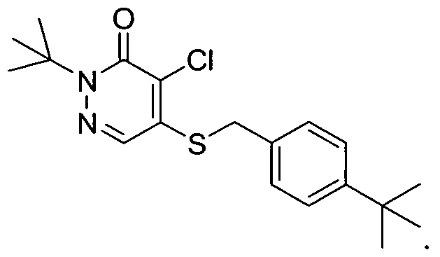
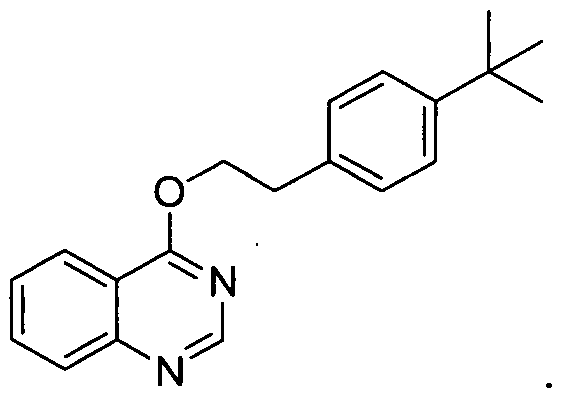


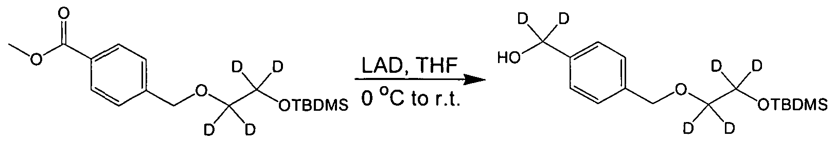


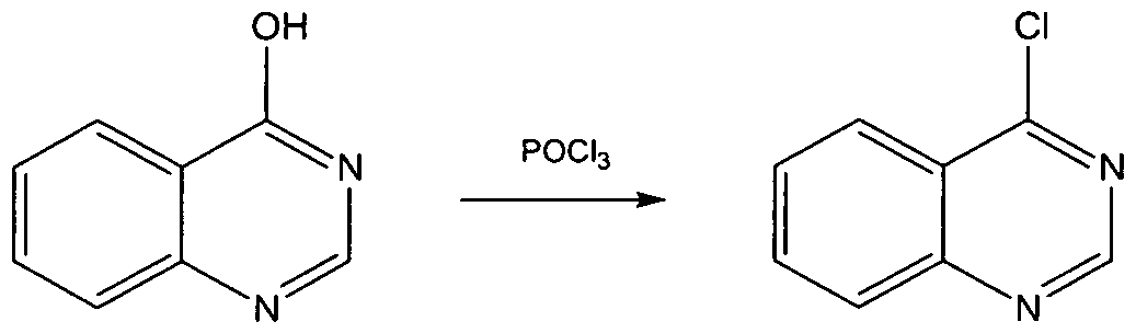


Claims


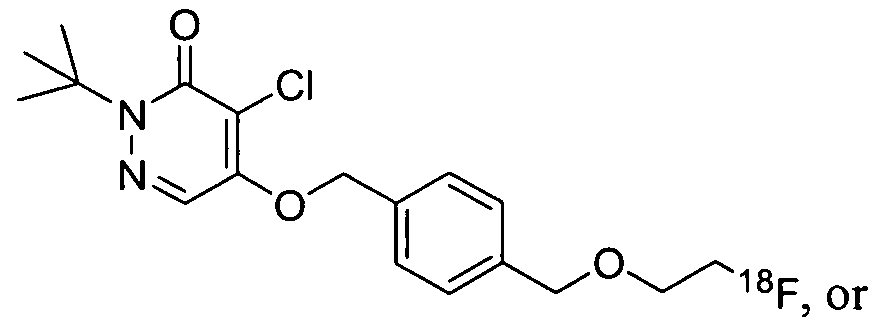


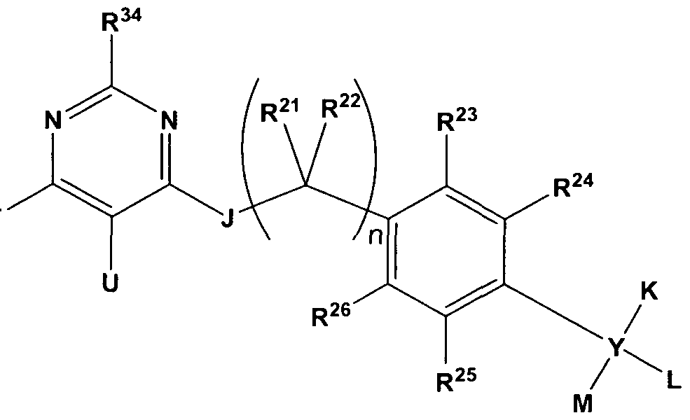


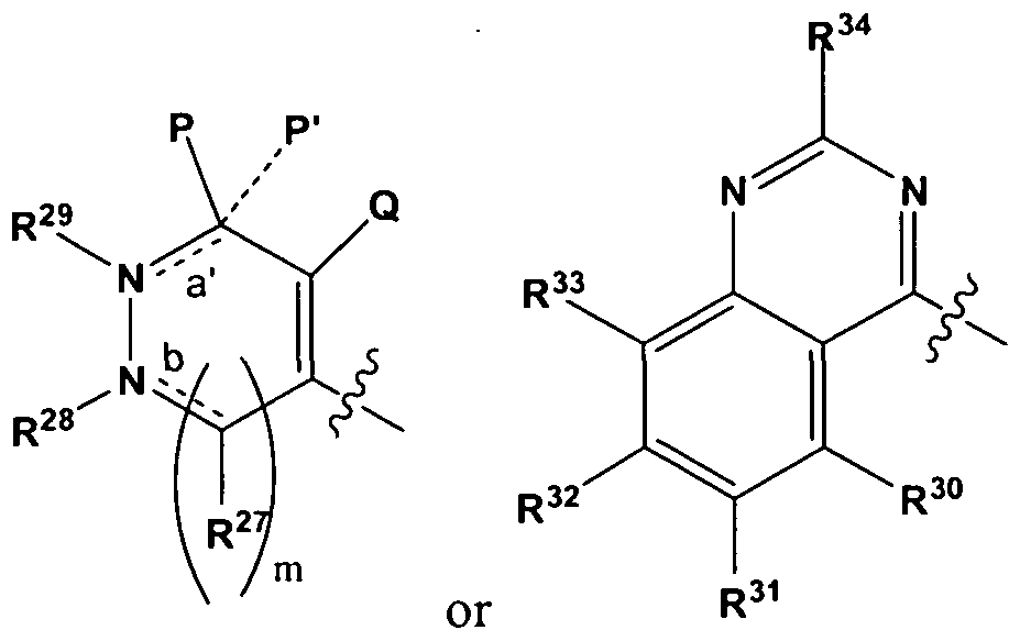


Priority Applications (10)
| Application Number | Priority Date | Filing Date | Title |
|---|---|---|---|
| CN200980115704.0A CN102014969B (en) | 2008-02-29 | 2009-02-27 | Contrast agents for applications including perfusion imaging |
| EP09716528.6A EP2257315B1 (en) | 2008-02-29 | 2009-02-27 | Contrast agents for applications including perfusion imaging |
| DK09716528.6T DK2257315T3 (en) | 2008-02-29 | 2009-02-27 | CONTRACTORS FOR APPLICATIONS COMPREHENSIVE PERFUSION IMAGE |
| ES09716528T ES2767973T3 (en) | 2008-02-29 | 2009-02-27 | Contrast agents for applications involving perfusion imaging |
| CA2716354A CA2716354C (en) | 2008-02-29 | 2009-02-27 | Contrast agents for applications including perfusion imaging |
| AU2009220244A AU2009220244B2 (en) | 2008-02-29 | 2009-02-27 | Contrast agents for applications including perfusion imaging |
| US12/919,600 US9408927B2 (en) | 2008-02-29 | 2009-02-27 | Contrast agents for applications including perfusion imaging |
| JP2010548730A JP5946992B2 (en) | 2008-02-29 | 2009-02-27 | Contrast agents for applications involving perfusion imaging |
| IL207853A IL207853A (en) | 2008-02-29 | 2010-08-29 | Contrast agents for applications including perfusion imaging |
| US15/153,502 US10245332B2 (en) | 2008-02-29 | 2016-05-12 | Contrast agents for applications including perfusion imaging |
Applications Claiming Priority (2)
| Application Number | Priority Date | Filing Date | Title |
|---|---|---|---|
| US6759308P | 2008-02-29 | 2008-02-29 | |
| US61/067,593 | 2008-02-29 |
Related Child Applications (2)
| Application Number | Title | Priority Date | Filing Date |
|---|---|---|---|
| US12/919,600 A-371-Of-International US9408927B2 (en) | 2008-02-29 | 2009-02-27 | Contrast agents for applications including perfusion imaging |
| US15/153,502 Continuation US10245332B2 (en) | 2008-02-29 | 2016-05-12 | Contrast agents for applications including perfusion imaging |
Publications (2)
| Publication Number | Publication Date |
|---|---|
| WO2009110984A2 true WO2009110984A2 (en) | 2009-09-11 |
| WO2009110984A3 WO2009110984A3 (en) | 2009-12-10 |
Family
ID=41016660
Family Applications (2)
| Application Number | Title | Priority Date | Filing Date |
|---|---|---|---|
| PCT/US2009/001296 WO2009108376A2 (en) | 2008-02-29 | 2009-02-27 | Contrast agents for applications including perfusion imaging |
| PCT/US2009/001247 WO2009110984A2 (en) | 2008-02-29 | 2009-02-27 | Contrast agents for applications including perfusion imaging |
Family Applications Before (1)
| Application Number | Title | Priority Date | Filing Date |
|---|---|---|---|
| PCT/US2009/001296 WO2009108376A2 (en) | 2008-02-29 | 2009-02-27 | Contrast agents for applications including perfusion imaging |
Country Status (12)
| Country | Link |
|---|---|
| US (2) | US9408927B2 (en) |
| EP (1) | EP2257315B1 (en) |
| JP (3) | JP5946992B2 (en) |
| KR (1) | KR101616139B1 (en) |
| CN (1) | CN102014969B (en) |
| AU (1) | AU2009220244B2 (en) |
| CA (2) | CA2716354C (en) |
| DK (1) | DK2257315T3 (en) |
| ES (1) | ES2767973T3 (en) |
| IL (1) | IL207853A (en) |
| PT (1) | PT2257315T (en) |
| WO (2) | WO2009108376A2 (en) |
Cited By (7)
| Publication number | Priority date | Publication date | Assignee | Title |
|---|---|---|---|---|
| WO2011097649A3 (en) * | 2010-02-08 | 2011-12-15 | Lantheus Medical Imaging, Inc. | Methods and apparatus for synthesizing imaging agents, and intermediates thereof |
| US8226929B2 (en) | 2004-02-13 | 2012-07-24 | Lantheus Medical Imaging, Inc. | Contrast agents for myocardial perfusion imaging |
| US8263042B2 (en) | 2004-04-28 | 2012-09-11 | Lantheus Medical Imaging, Inc. | Contrast agents for myocardial perfusion imaging |
| US9408927B2 (en) | 2008-02-29 | 2016-08-09 | Lantheus Medical Imaging, Inc. | Contrast agents for applications including perfusion imaging |
| AU2013203000B2 (en) * | 2012-08-10 | 2016-12-15 | Lantheus Medical Imaging, Inc. | Compositions, methods, and systems for the synthesis and use of imaging agents |
| US9550000B2 (en) | 2011-09-09 | 2017-01-24 | Lantheus Medical Imaging, Inc. | Compositions, methods, and systems for the synthesis and use of imaging agents |
| US9687571B2 (en) | 2009-04-15 | 2017-06-27 | Lantheus Medical Imaging, Inc. | Stabilization of radiopharmaceutical compositions using ascorbic acid |
Families Citing this family (10)
| Publication number | Priority date | Publication date | Assignee | Title |
|---|---|---|---|---|
| US20100241001A1 (en) * | 2009-03-20 | 2010-09-23 | Palmeri Mark L | Ultrasound Methods, Systems and Computer Program Products for Imaging Fluids |
| AU2011379346B2 (en) * | 2011-10-21 | 2017-08-31 | Lantheus Medical Imaging, Inc. | Compositions comprising ascorbic acid and an imaging agent and related methods |
| CN103254140B (en) * | 2012-02-17 | 2016-02-17 | 北京师范大学 | Novel 18f marks substituted quinazoline compounds and preparation method thereof and applies with PET tumor imaging |
| WO2013173004A2 (en) | 2012-04-10 | 2013-11-21 | Lantheus Medical Imaging, Inc. | Radiopharmaceutical synthesis methods |
| US20150246143A1 (en) * | 2012-08-13 | 2015-09-03 | Lantheus Medical Imaging, Inc. | System and method for quantitative mapping of mitochondrial complex 1 |
| CN104583192B (en) * | 2012-08-24 | 2016-10-12 | 浜松光子学株式会社 | It is suitable for the compound of Mitochondria complex-1 detection |
| SI3077501T1 (en) | 2013-12-03 | 2022-02-28 | Genomatica, Inc. | Microorganisms and methods for improving product yields on methanol using acetyl-coa synthesis |
| US20170245817A1 (en) * | 2016-02-10 | 2017-08-31 | The Charles Stark Draper Laboratory, Inc. | Systems and methods for imaging |
| CN110305010B (en) * | 2019-07-17 | 2022-05-13 | 江苏中旗科技股份有限公司 | Preparation method of 2, 5-dimethylphenylacetic acid |
| WO2023013757A1 (en) * | 2021-08-05 | 2023-02-09 | 積水メディカル株式会社 | Method for producing alkylsilyloxy substituted benzyl compound |
Family Cites Families (109)
| Publication number | Priority date | Publication date | Assignee | Title |
|---|---|---|---|---|
| GB1073999A (en) * | 1965-01-29 | 1967-06-28 | Ncr Co | Process for forming stable images in photochromic material |
| US4510125A (en) | 1982-12-08 | 1985-04-09 | Mallinckrodt, Inc. | Process for making lyophilized radiographic imaging kit |
| JPS604173A (en) | 1983-06-23 | 1985-01-10 | Nissan Chem Ind Ltd | Pyridazinone derivative, its preparation and insecticidal, miticidal and fungicidal agent |
| DE3574870D1 (en) | 1984-06-23 | 1990-01-25 | Nissan Chemical Ind Ltd | METHOD FOR PRODUCING 2-TERT.-BUTYL-4,5-DICHLORO-3 (2H) -PYRIDAZINONE. |
| JPH07116161B2 (en) | 1984-07-04 | 1995-12-13 | 日産化学工業株式会社 | Pyridazinone derivative |
| DE3578304D1 (en) | 1984-11-29 | 1990-07-26 | Nissan Chemical Ind Ltd | PYRIDAZINE DERIVATIVES, THEIR PRODUCTION AND INSECTICIDAL, ACARICIDIC, NEMATICIDIC, FUNGICIDIC COMPOSITIONS. |
| JPS61130275A (en) | 1984-11-29 | 1986-06-18 | Nissan Chem Ind Ltd | Pyridazinone derivative, preparation thereof, and insecticidal, miticidal and fungicidal agent |
| JPH0641455B2 (en) | 1984-12-10 | 1994-06-01 | 日産化学工業株式会社 | Pyridazinone derivative |
| US5393512A (en) * | 1985-01-14 | 1995-02-28 | Vanderheyden; Jean-Luc | Stable therapeutic radionuclide compositions and methods for preparation thereof |
| JPH0641454B2 (en) | 1985-02-27 | 1994-06-01 | 日産化学工業株式会社 | Pyridazinone derivative |
| JPS61260018A (en) | 1985-05-14 | 1986-11-18 | Nissan Chem Ind Ltd | Anti-allergic agent |
| TR22638A (en) | 1985-07-30 | 1988-01-29 | Nissan Chemical Ind Ltd | PIRIDAZINON TUEREVLER, FOR THE PREPARATION OF THESE AND DAMAGE OELDUERUECUE COMPOUNDS |
| JPH0739397B2 (en) | 1985-07-30 | 1995-05-01 | 日産化学工業株式会社 | Pyridazinone derivatives and pest control agents |
| WO1987002893A1 (en) * | 1985-11-18 | 1987-05-21 | Board Of Regents, The University Of Texas System | Polychelating agents for image and spectral enhancement (and spectral shift) |
| JPS6315974A (en) | 1986-07-09 | 1988-01-23 | 小泉コンピユ−タ−株式会社 | Balling game score table display apparatus |
| US5252317A (en) * | 1986-11-10 | 1993-10-12 | The State Of Oregon Acting By And Through The State Board Of Higher Education On Behalf Of The University Of Oregon | Amplifier molecules for diagnosis and therapy derived from 3,5-bis[1-(3-amino-2,2-bis (aminomethyl)-propyl) oxymethyl] benzoic acid |
| US5567411A (en) * | 1986-11-10 | 1996-10-22 | State Of Oregon Acting By And Through The State Board Of Higher Education On Behalf Of The University Of Oregon | Dendritic amplifier molecules having multiple terminal active groups stemming from a benzyl core group |
| JPS63159372A (en) | 1986-12-24 | 1988-07-02 | Nissan Chem Ind Ltd | Pyridazinone compound and insecticide, acaricide and nematocide |
| JPS63159373A (en) | 1986-12-24 | 1988-07-02 | Nissan Chem Ind Ltd | Pyridazinones and insecticide, acaricide and nematocide |
| JPS63159374A (en) | 1986-12-24 | 1988-07-02 | Nissan Chem Ind Ltd | Pyridazinone derivative and insecticide, acaricide and nematocide |
| DE3824210A1 (en) | 1988-07-16 | 1990-01-18 | Basf Ag | 3 (2H) -PYRIDAZINONE DERIVATIVES FOR CONTROLLING SNAILS |
| IT1229684B (en) * | 1989-04-05 | 1991-09-06 | Mini Ricerca Scient Tecnolog | PYRIDAZINONE WITH INSECTICIDE AND ACARICIDE ACTIVITY |
| JPH02279676A (en) | 1989-04-19 | 1990-11-15 | Otsuka Chem Co Ltd | Pyridazinone derivative |
| US5087440A (en) * | 1989-07-31 | 1992-02-11 | Salutar, Inc. | Heterocyclic derivatives of DTPA used for magnetic resonance imaging |
| GB8923843D0 (en) * | 1989-10-23 | 1989-12-13 | Salutar Inc | Compounds |
| US5377681A (en) * | 1989-11-13 | 1995-01-03 | University Of Florida | Method of diagnosing impaired blood flow |
| US5228446A (en) * | 1989-12-22 | 1993-07-20 | Unger Evan C | Gas filled liposomes and their use as ultrasonic contrast agents |
| US5088499A (en) * | 1989-12-22 | 1992-02-18 | Unger Evan C | Liposomes as contrast agents for ultrasonic imaging and methods for preparing the same |
| US5585112A (en) * | 1989-12-22 | 1996-12-17 | Imarx Pharmaceutical Corp. | Method of preparing gas and gaseous precursor-filled microspheres |
| US5679810A (en) * | 1990-01-19 | 1997-10-21 | Salutar, Inc. | Linear oligomeric polychelant compounds |
| JPH03220177A (en) | 1990-01-25 | 1991-09-27 | Nissan Chem Ind Ltd | Insecticidal/miticidal agent composition |
| GB9006977D0 (en) | 1990-03-28 | 1990-05-23 | Nycomed As | Compositions |
| WO1992017215A1 (en) | 1990-03-28 | 1992-10-15 | Nycomed Salutar, Inc. | Contrast media |
| JPH04235975A (en) | 1991-01-21 | 1992-08-25 | Nissan Chem Ind Ltd | Pyridazinone drivative and insect pest control agent |
| US5205290A (en) * | 1991-04-05 | 1993-04-27 | Unger Evan C | Low density microspheres and their use as contrast agents for computed tomography |
| US5306482A (en) * | 1991-04-09 | 1994-04-26 | Merck Frosst Canada, Inc. | Radiopharmaceutical bacteriostats |
| US5093105A (en) * | 1991-04-09 | 1992-03-03 | Merck Frosst Canada, Inc. | Radiopharmaceutical bacteriostats |
| CA2113995C (en) * | 1991-08-29 | 2003-04-22 | Edward A. Deutsch | Use of gentisic acid or gentisyl alcohol for stabilising radiolabeled peptides and proteins |
| US5169942A (en) * | 1991-11-21 | 1992-12-08 | General Electric Company | Method for making 2-(18F)fluoro-2-deoxy-D-glucose |
| EP0627424A4 (en) | 1992-12-03 | 1995-03-29 | Otsuka Kagaku Kk | Pyridazinone derivative and insecticidal and miticidal agent. |
| US5760191A (en) * | 1993-02-05 | 1998-06-02 | Nycomed Imaging As | Macrocyclic complexing agents and targeting immunoreagents useful in therapeutic and diagnostic compositions and methods |
| EP0641350A4 (en) * | 1993-03-22 | 1995-08-23 | Gen Electric | Method for making 2-fluoro-2-deoxyglucose. |
| MXPA94002323A (en) | 1993-03-31 | 2002-06-18 | Mallinckrodt Medical Inc | Radiopharmaceutical formulations having non-stannous reducers. |
| US5417959A (en) * | 1993-10-04 | 1995-05-23 | Mallinckrodt Medical, Inc. | Functionalized aza-crytand ligands for diagnostic imaging applications |
| US5493026A (en) | 1993-10-25 | 1996-02-20 | Organix, Inc. | Substituted 2-carboxyalkyl-3-(fluorophenyl)-8-(3-halopropen-2-yl) nortropanes and their use as imaging for agents for neurodegenerative disorders |
| EP0665223A1 (en) | 1994-01-28 | 1995-08-02 | Takeda Chemical Industries, Ltd. | Antitumor agent, novel 3(2H)-pyridazinone derivatives and their preparation |
| JPH07252236A (en) | 1994-01-28 | 1995-10-03 | Takeda Chem Ind Ltd | Anticancer agent |
| AU7312694A (en) | 1994-06-03 | 1996-01-04 | Mallinckrodt Medical, Inc. | Rapidly clearing technetium-99m phosphonate skeletal imaging agents |
| US5520904A (en) * | 1995-01-27 | 1996-05-28 | Mallinckrodt Medical, Inc. | Calcium/oxyanion-containing particles with a polymerical alkoxy coating for use in medical diagnostic imaging |
| EP0727225A3 (en) | 1995-02-14 | 1997-01-15 | Sonus Pharma Inc | Compositions and methods for directed ultrasound imaging |
| US5587491A (en) * | 1995-03-15 | 1996-12-24 | Regents Of The University Of Minnesota | Method for the synthesis of bis-tetrahydrofuranyl Annonaceous acetogenins |
| US5801228A (en) * | 1995-06-07 | 1998-09-01 | Nycomed Imaging As | Polymeric contrast agents for medical imaging |
| US5811073A (en) | 1995-06-19 | 1998-09-22 | President And Fellows Of Harvard College | Method for radioisotopic detection and localization of inflammation in a host |
| US5827073A (en) | 1995-07-05 | 1998-10-27 | Ludwig Institute For Cancer Research | Photoreactive peptide derivatives |
| US6066309A (en) * | 1996-02-02 | 2000-05-23 | Rhomed Incorporated | Post-labeling stabilization of radiolabeled proteins and peptides |
| US5804161A (en) * | 1996-05-14 | 1998-09-08 | Nycomed Salutar Inc. | Contrast agents |
| US5846517A (en) * | 1996-09-11 | 1998-12-08 | Imarx Pharmaceutical Corp. | Methods for diagnostic imaging using a renal contrast agent and a vasodilator |
| US6565889B2 (en) * | 1996-12-02 | 2003-05-20 | The Regents Of The University Of California | Bilayer structure which encapsulates multiple containment units and uses thereof |
| US5961955A (en) * | 1997-06-03 | 1999-10-05 | Coulter Pharmaceutical, Inc. | Radioprotectant for peptides labeled with radioisotope |
| US6056939A (en) * | 1998-08-28 | 2000-05-02 | Desreux; Jean F. | Self-assembling heteropolymetallic chelates as imaging agents and radiopharmaceuticals |
| CA2343106A1 (en) * | 1998-09-29 | 2000-04-06 | Merck & Co., Inc. | Radiolabeled neurokinin-1 receptor antagonists |
| US6645508B1 (en) | 1999-06-18 | 2003-11-11 | Jivn-Ren Chen | Stable L-ascorbic acid composition |
| US6878363B2 (en) | 2000-05-17 | 2005-04-12 | Bristol-Myers Squibb Pharma Company | Use of small molecule radioligands to discover inhibitors of amyloid-beta peptide production and for diagnostic imaging |
| AU2001288899A1 (en) | 2000-09-06 | 2002-03-22 | The Scripps Research Institute | Inhibitors of nadh:ubiquinone oxidoreductase |
| TWI247609B (en) * | 2001-01-23 | 2006-01-21 | Nihon Mediphysics Co Ltd | Agent for diagnosis of tissue proliferation activity or the treatment of proliferative disease |
| GB0115927D0 (en) | 2001-06-29 | 2001-08-22 | Nycomed Amersham Plc | Solid-phase nucleophilic fluorination |
| US20030044354A1 (en) * | 2001-08-16 | 2003-03-06 | Carpenter Alan P. | Gas microsphere liposome composites for ultrasound imaging and ultrasound stimulated drug release |
| US7344702B2 (en) * | 2004-02-13 | 2008-03-18 | Bristol-Myers Squibb Pharma Company | Contrast agents for myocardial perfusion imaging |
| US20030082191A1 (en) * | 2001-08-29 | 2003-05-01 | Poduslo Joseph F. | Treatment for central nervous system disorders |
| AU2002347906A2 (en) * | 2001-10-16 | 2003-04-28 | Hypnion, Inc. | Treatment of CNS disorders using CNS target modulators |
| EP1474177A4 (en) * | 2002-02-06 | 2010-07-21 | Univ Johns Hopkins | Non-invasive diagnostic imaging technology for mitochondria using radiolabeled lipophilic salts |
| UA78548C2 (en) | 2002-03-29 | 2007-04-10 | Janssen Pharmaceutica Nv | Radiolabelled quinoline and quinolinone derivatives and use thereof as metabotropic ligands of glutamate receptor |
| WO2003086476A1 (en) | 2002-04-08 | 2003-10-23 | Biostream, Inc. | Technetium-labeled rotenone derivatives, and methods of use thereof |
| GB0229683D0 (en) | 2002-12-20 | 2003-01-29 | Imaging Res Solutions Ltd | Preparation of radiopharmaceuticals |
| WO2005007632A1 (en) | 2003-07-18 | 2005-01-27 | Pharmacia Corporation | Substituted pyridazinones as inhibitors of p38 |
| CA2783275A1 (en) | 2003-07-24 | 2005-02-03 | Bracco Imaging S.P.A. | Stable radiopharmaceutical compositions and methods for their preparation |
| GB0317920D0 (en) | 2003-07-31 | 2003-09-03 | Amersham Plc | Solid-phase synthesis |
| US7927616B2 (en) | 2004-01-16 | 2011-04-19 | Thomas T. Yamashita | Pesticide compositions and methods for their use |
| JP5070038B2 (en) | 2004-02-24 | 2012-11-07 | ザ ジェネラル ホスピタル コーポレーション | Catalytic radiofluorination |
| US7732456B2 (en) | 2004-03-05 | 2010-06-08 | Banyu Pharmaceutical Co., Ltd. | Pyridone derivative |
| AT500838B1 (en) | 2004-04-20 | 2007-11-15 | Veterinaermedizinische Uni Wie | MULTIPLE HSE |
| CN1976728A (en) * | 2004-04-28 | 2007-06-06 | 布里斯托尔-迈尔斯斯奎布药品公司 | Contrast agents for myocardial perfusion imaging. |
| US7485283B2 (en) | 2004-04-28 | 2009-02-03 | Lantheus Medical Imaging | Contrast agents for myocardial perfusion imaging |
| US20060083681A1 (en) * | 2004-10-18 | 2006-04-20 | Ajay Purohit | Compounds for myocardial perfusion imaging |
| KR100789847B1 (en) * | 2004-12-15 | 2007-12-28 | (주)퓨쳐켐 | A Preparation Method of Organo Fluoro Compounds in Alcohol Solvents |
| JP2009505947A (en) * | 2005-06-23 | 2009-02-12 | エモリー・ユニバーシティ | Imaging agent |
| US7824659B2 (en) * | 2005-08-10 | 2010-11-02 | Lantheus Medical Imaging, Inc. | Methods of making radiolabeled tracers and precursors thereof |
| JP2007112725A (en) | 2005-10-18 | 2007-05-10 | Inst Nuclear Energy Research Rocaec | Method for producing amino acid o-(2-[18f] fluoroethyl)-l-tyrosine labeled with 18f of non-substrate |
| US7871623B2 (en) | 2005-12-21 | 2011-01-18 | The Board Of Trustees Of The Leland Stanford Junior University | Compositions and methods for imaging pain and stress in vivo |
| WO2008118122A2 (en) | 2006-05-08 | 2008-10-02 | Molecular Neuroimaging, Llc | Compounds and amyloid probes thereof for therapeutic and imaging uses |
| CA2654369C (en) * | 2006-06-21 | 2014-10-14 | Ge Healthcare Limited | Radiopharmaceutical products |
| EP2383259A1 (en) | 2006-08-25 | 2011-11-02 | Boehringer Ingelheim International GmbH | New pyridone derivatives with MCH antagonistic activity and medicaments comprising these compounds |
| JP5504563B2 (en) | 2006-12-27 | 2014-05-28 | 住友化学株式会社 | Composition and light-emitting device using the composition |
| GB0718386D0 (en) * | 2007-09-21 | 2007-10-31 | Ge Healthcare As | Improved radiopharmaceutical formulation |
| KR101068835B1 (en) | 2007-10-26 | 2011-09-30 | 한국과학기술연구원 | Isoindolones with high binding affinity to ?-amyloid aggregates and fibrils, and preparation method thereof |
| CL2008003785A1 (en) | 2007-12-21 | 2009-10-09 | Du Pont | Pyridazine derived compounds; herbicidal compositions comprising said compounds; and method of controlling the growth of unwanted vegetation. |
| WO2009103478A1 (en) | 2008-02-19 | 2009-08-27 | Boehringer Ingelheim International Gmbh | Pyridone and pyridazinone derivatives as mch antagonists |
| WO2010011367A2 (en) | 2008-02-22 | 2010-01-28 | Illinois Institute Of Technology | Bimodal ligands with macrocyclic and acyclic binding moieties, complexes and compositions thereof, and methods of using |
| EP2257315B1 (en) | 2008-02-29 | 2019-10-23 | Lantheus Medical Imaging, Inc. | Contrast agents for applications including perfusion imaging |
| US8258134B2 (en) | 2008-04-16 | 2012-09-04 | Hoffmann-La Roche Inc. | Pyridazinone glucokinase activators |
| US8278316B2 (en) | 2009-03-09 | 2012-10-02 | Bristol-Myers Squibb Company | Aza pyridone analogs useful as melanin concentrating hormone receptor-1 antagonists |
| WO2010120368A2 (en) * | 2009-04-15 | 2010-10-21 | Lantheus Medical Imaging, Inc. | Stabilization of radiopharmaceutical compositions using ascorbic acid |
| CN101555232B (en) | 2009-05-21 | 2011-01-05 | 北京师范大学 | Pyridazinone compound marked by fluorine-18, preparation method and applications |
| KR20120051641A (en) | 2009-07-11 | 2012-05-22 | 바이엘 파마 악티엔게젤샤프트 | Non-polar and polar leaving groups |
| KR20170033873A (en) * | 2010-02-08 | 2017-03-27 | 랜티우스 메디컬 이메징, 인크. | Methods and apparatus for synthesizing imaging agents, and intermediates thereof |
| JP5842594B2 (en) | 2010-12-27 | 2016-01-13 | 住友化学株式会社 | Pyridazinone compounds, herbicides containing them, and harmful arthropod control agents |
| US9550000B2 (en) | 2011-09-09 | 2017-01-24 | Lantheus Medical Imaging, Inc. | Compositions, methods, and systems for the synthesis and use of imaging agents |
| AU2011379346B2 (en) | 2011-10-21 | 2017-08-31 | Lantheus Medical Imaging, Inc. | Compositions comprising ascorbic acid and an imaging agent and related methods |
| AU2013203000B9 (en) | 2012-08-10 | 2017-02-02 | Lantheus Medical Imaging, Inc. | Compositions, methods, and systems for the synthesis and use of imaging agents |
-
2009
- 2009-02-27 EP EP09716528.6A patent/EP2257315B1/en active Active
- 2009-02-27 KR KR1020107021556A patent/KR101616139B1/en active IP Right Grant
- 2009-02-27 AU AU2009220244A patent/AU2009220244B2/en active Active
- 2009-02-27 WO PCT/US2009/001296 patent/WO2009108376A2/en active Application Filing
- 2009-02-27 CA CA2716354A patent/CA2716354C/en active Active
- 2009-02-27 CA CA2967254A patent/CA2967254C/en active Active
- 2009-02-27 WO PCT/US2009/001247 patent/WO2009110984A2/en active Application Filing
- 2009-02-27 PT PT97165286T patent/PT2257315T/en unknown
- 2009-02-27 ES ES09716528T patent/ES2767973T3/en active Active
- 2009-02-27 CN CN200980115704.0A patent/CN102014969B/en active Active
- 2009-02-27 DK DK09716528.6T patent/DK2257315T3/en active
- 2009-02-27 US US12/919,600 patent/US9408927B2/en active Active
- 2009-02-27 JP JP2010548730A patent/JP5946992B2/en active Active
-
2010
- 2010-08-29 IL IL207853A patent/IL207853A/en active IP Right Grant
-
2014
- 2014-10-23 JP JP2014216665A patent/JP5922207B2/en active Active
-
2016
- 2016-04-13 JP JP2016080401A patent/JP2016164182A/en active Pending
- 2016-05-12 US US15/153,502 patent/US10245332B2/en active Active
Non-Patent Citations (4)
| Title |
|---|
| HIGGIN; GREENAMYRE, JOURNAL OF NEUROSCIENCE, vol. 16, no. 12, 1996, pages 3807 - 3816 |
| KANN; KOVACS, AM. J PHYSIOL. CELL PHYSIOL., vol. 292, 2007, pages C641 - C657 |
| See also references of EP2257315A4 |
| WANG ET AL., FREE RADICAL BIOLOGY AND MEDICINE, vol. 43, 2007, pages 1569 - 1573 |
Cited By (37)
| Publication number | Priority date | Publication date | Assignee | Title |
|---|---|---|---|---|
| US9161997B2 (en) | 2001-08-16 | 2015-10-20 | David S. Casebier | Contrast agents for myocardial perfusion imaging |
| US8226929B2 (en) | 2004-02-13 | 2012-07-24 | Lantheus Medical Imaging, Inc. | Contrast agents for myocardial perfusion imaging |
| US10889550B2 (en) | 2004-02-13 | 2021-01-12 | Lantheus Medical Imaging, Inc. | Contrast agents for myocardial perfusion imaging |
| US10125106B2 (en) | 2004-02-13 | 2018-11-13 | Lantheus Medical Imaging, Inc. | Contrast agents for myocardial perfusion imaging |
| US9718786B2 (en) | 2004-02-13 | 2017-08-01 | Lantheus Medical Imaging, Inc. | Contrast agents for myocardial perfusion imaging |
| US8263042B2 (en) | 2004-04-28 | 2012-09-11 | Lantheus Medical Imaging, Inc. | Contrast agents for myocardial perfusion imaging |
| US9408927B2 (en) | 2008-02-29 | 2016-08-09 | Lantheus Medical Imaging, Inc. | Contrast agents for applications including perfusion imaging |
| US10245332B2 (en) | 2008-02-29 | 2019-04-02 | Lantheus Medical Imaging, Inc. | Contrast agents for applications including perfusion imaging |
| US9687571B2 (en) | 2009-04-15 | 2017-06-27 | Lantheus Medical Imaging, Inc. | Stabilization of radiopharmaceutical compositions using ascorbic acid |
| TWI664169B (en) * | 2010-02-08 | 2019-07-01 | 藍瑟斯醫學影像公司 | Methods and apparatus for synthesizing imaging agents, and intermediates thereof |
| EP3323810A3 (en) * | 2010-02-08 | 2018-07-04 | Lantheus Medical Imaging, Inc. | Automated reaction system, cassette and apparatus for synthesizing imaging agents |
| KR102438133B1 (en) * | 2010-02-08 | 2022-08-31 | 랜티우스 메디컬 이메징, 인크. | Methods and apparatus for synthesizing imaging agents, and intermediates thereof |
| KR20120130293A (en) * | 2010-02-08 | 2012-11-30 | 랜티우스 메디컬 이메징, 인크. | Methods and apparatus for synthesizing imaging agents, and intermediates thereof |
| KR101709372B1 (en) | 2010-02-08 | 2017-02-22 | 랜티우스 메디컬 이메징, 인크. | Methods and apparatus for synthesizing imaging agents, and intermediates thereof |
| US9603951B2 (en) | 2010-02-08 | 2017-03-28 | Lantheus Medical Imaging, Inc. | Methods and apparatus for synthesizing imaging agents, and intermediates thereof |
| JP2016029059A (en) * | 2010-02-08 | 2016-03-03 | ランセウス メディカル イメージング, インコーポレイテッド | Methods and apparatus for synthesizing imaging agents and intermediates thereof |
| AU2011213568B2 (en) * | 2010-02-08 | 2016-05-12 | Lantheus Medical Imaging, Inc. | Methods and apparatus for synthesizing imaging agents, and intermediates thereof |
| US8936777B2 (en) | 2010-02-08 | 2015-01-20 | Lantheus Medical Imaging, Inc. | Methods and apparatus for synthesizing imaging agents, and intermediates thereof |
| EP4046990A1 (en) * | 2010-02-08 | 2022-08-24 | Lantheus Medical Imaging, Inc. | Methods and apparatus for synthesizing imaging agents, and intermediates thereof |
| JP2018076328A (en) * | 2010-02-08 | 2018-05-17 | ランセウス メディカル イメージング, インコーポレイテッド | Methods and apparatus for synthesizing imaging agents and intermediates thereof |
| KR20210132243A (en) * | 2010-02-08 | 2021-11-03 | 랜티우스 메디컬 이메징, 인크. | Methods and apparatus for synthesizing imaging agents, and intermediates thereof |
| US10022462B2 (en) | 2010-02-08 | 2018-07-17 | Lantheus Medical Imaging, Inc. | Methods and apparatus for synthesizing imaging agents, and intermediates thereof |
| CN102858752B (en) * | 2010-02-08 | 2018-10-16 | 兰休斯医疗成像公司 | Method and apparatus for synthesizing developer and wherein mesosome |
| JP2013518913A (en) * | 2010-02-08 | 2013-05-23 | ランセウス メディカル イメージング, インコーポレイテッド | Method and apparatus for synthesizing contrast agents and intermediates thereof |
| CN109200296A (en) * | 2010-02-08 | 2019-01-15 | 兰休斯医疗成像公司 | For synthesizing imaging agent and the wherein method and apparatus of mesosome |
| CN102858752A (en) * | 2010-02-08 | 2013-01-02 | 兰休斯医疗成像公司 | Methods and apparatus for synthesizing imaging agents, and intermediates thereof |
| TWI660947B (en) * | 2010-02-08 | 2019-06-01 | 藍瑟斯醫學影像公司 | Methods and apparatus for synthesizing imaging agents, and intermediates thereof |
| WO2011097649A3 (en) * | 2010-02-08 | 2011-12-15 | Lantheus Medical Imaging, Inc. | Methods and apparatus for synthesizing imaging agents, and intermediates thereof |
| CN109200296B (en) * | 2010-02-08 | 2021-12-14 | 兰休斯医疗成像公司 | Method and apparatus for synthesizing imaging agents and intermediates thereof |
| TWI686370B (en) * | 2010-02-08 | 2020-03-01 | 美商藍瑟斯醫學影像公司 | Methods and apparatus for synthesizing imaging agents, and intermediates thereof |
| US10842892B2 (en) | 2010-02-08 | 2020-11-24 | Lantheus Medical Imaging, Inc. | Methods and apparatus for synthesizing imaging agents, and intermediates thereof |
| US9550000B2 (en) | 2011-09-09 | 2017-01-24 | Lantheus Medical Imaging, Inc. | Compositions, methods, and systems for the synthesis and use of imaging agents |
| US9713651B2 (en) | 2012-08-10 | 2017-07-25 | Lantheus Medical Imaging, Inc. | Compositions, methods, and systems for the synthesis and use of imaging agents |
| US10500293B2 (en) | 2012-08-10 | 2019-12-10 | Lantheus Medical Imaging, Inc. | Compositions, methods, and systems for the synthesis and use of imaging agents |
| US9919064B2 (en) | 2012-08-10 | 2018-03-20 | Lantheus Medical Imaging, Inc. | Compositions, methods, and systems for the synthesis and use of imaging agents |
| AU2013203000B2 (en) * | 2012-08-10 | 2016-12-15 | Lantheus Medical Imaging, Inc. | Compositions, methods, and systems for the synthesis and use of imaging agents |
| US11744906B2 (en) | 2012-08-10 | 2023-09-05 | Lantheus Medical Imaging, Inc. | Compositions, methods, and systems for the synthesis and use of imaging agents |
Also Published As
| Publication number | Publication date |
|---|---|
| US9408927B2 (en) | 2016-08-09 |
| EP2257315A4 (en) | 2013-04-17 |
| IL207853A (en) | 2015-05-31 |
| ES2767973T3 (en) | 2020-06-19 |
| WO2009108376A3 (en) | 2009-11-19 |
| KR101616139B1 (en) | 2016-04-27 |
| CA2967254A1 (en) | 2009-09-11 |
| DK2257315T3 (en) | 2020-01-27 |
| WO2009108376A2 (en) | 2009-09-03 |
| AU2009220244A1 (en) | 2009-09-11 |
| US20160361448A1 (en) | 2016-12-15 |
| WO2009110984A3 (en) | 2009-12-10 |
| AU2009220244B2 (en) | 2014-10-09 |
| IL207853A0 (en) | 2010-12-30 |
| JP2011513306A (en) | 2011-04-28 |
| KR20100121530A (en) | 2010-11-17 |
| CN102014969A (en) | 2011-04-13 |
| CN102014969B (en) | 2014-07-09 |
| EP2257315A2 (en) | 2010-12-08 |
| US10245332B2 (en) | 2019-04-02 |
| JP5922207B2 (en) | 2016-05-24 |
| CA2716354C (en) | 2017-06-13 |
| JP2015061842A (en) | 2015-04-02 |
| PT2257315T (en) | 2020-01-27 |
| US20180133347A9 (en) | 2018-05-17 |
| CA2967254C (en) | 2019-03-26 |
| US20110091374A1 (en) | 2011-04-21 |
| CA2716354A1 (en) | 2009-09-11 |
| JP5946992B2 (en) | 2016-07-06 |
| JP2016164182A (en) | 2016-09-08 |
| EP2257315B1 (en) | 2019-10-23 |
Similar Documents
| Publication | Publication Date | Title |
|---|---|---|
| US10245332B2 (en) | Contrast agents for applications including perfusion imaging | |
| KR102527562B1 (en) | Compositions, methods, and systems for the synthesis and use of imaging agents | |
| CN113677400B (en) | Dual mode radiotracer and therapeutic agent that bind PSMA | |
| Pichika et al. | Nicotinic α4β2 receptor imaging agents: part II. Synthesis and biological evaluation of 2-[18F] fluoro-3-[2-((S)-3-pyrrolinyl) methoxy] pyridine (18F-nifene) in rodents and imaging by PET in nonhuman primate | |
| KR20210153036A (en) | Cancer diagnostic imaging agents | |
| US7737183B2 (en) | β-amyloid and neurofibrillary tangle imaging agents | |
| Singh et al. | A homodimeric bivalent radioligand derived from 1-(2-methoxyphenyl) piperazine with high affinity for in vivo 5-HT1A receptor imaging | |
| AU2015200027B2 (en) | Contrast agents for applications including perfusion imaging | |
| EP3177598B1 (en) | Macrocyclic complexes, their process of preparation and use as pet imaging agents | |
| Zeng et al. | Synthesis and evaluation of [18F]-fluoromethyl triphenylphosphonium cation as a novel mitochondria-specific positron emission tomography tracer | |
| Huang et al. | One-step 18 F-labeling of estradiol derivative for PET imaging of breast cancer | |
| Henderson et al. | 3-Pyridyl ethers as SPECT radioligands for imaging nicotinic acetylcholine receptors | |
| WO2011124713A1 (en) | Labelled huprine derivatives and their use in medical imaging |
Legal Events
| Date | Code | Title | Description |
|---|---|---|---|
| WWE | Wipo information: entry into national phase |
Ref document number: 200980115704.0 Country of ref document: CN |
|
| 121 | Ep: the epo has been informed by wipo that ep was designated in this application |
Ref document number: 09716528 Country of ref document: EP Kind code of ref document: A2 |
|
| WWE | Wipo information: entry into national phase |
Ref document number: 2716354 Country of ref document: CA |
|
| WWE | Wipo information: entry into national phase |
Ref document number: 207853 Country of ref document: IL |
|
| NENP | Non-entry into the national phase |
Ref country code: DE |
|
| WWE | Wipo information: entry into national phase |
Ref document number: 2010548730 Country of ref document: JP |
|
| WWE | Wipo information: entry into national phase |
Ref document number: 2009220244 Country of ref document: AU |
|
| WWE | Wipo information: entry into national phase |
Ref document number: 2009716528 Country of ref document: EP |
|
| ENP | Entry into the national phase |
Ref document number: 2009220244 Country of ref document: AU Date of ref document: 20090227 Kind code of ref document: A |
|
| ENP | Entry into the national phase |
Ref document number: 20107021556 Country of ref document: KR Kind code of ref document: A |
|
| WWE | Wipo information: entry into national phase |
Ref document number: 12919600 Country of ref document: US |