WO2009037644A2 - Method and apparatus for estimating the content of an analyte in a multi-layer medium - Google Patents
Method and apparatus for estimating the content of an analyte in a multi-layer medium Download PDFInfo
- Publication number
- WO2009037644A2 WO2009037644A2 PCT/IB2008/053761 IB2008053761W WO2009037644A2 WO 2009037644 A2 WO2009037644 A2 WO 2009037644A2 IB 2008053761 W IB2008053761 W IB 2008053761W WO 2009037644 A2 WO2009037644 A2 WO 2009037644A2
- Authority
- WO
- WIPO (PCT)
- Prior art keywords
- source
- layer medium
- analyte
- detector
- radiation
- Prior art date
Links
- 239000012491 analyte Substances 0.000 title claims abstract description 47
- 238000000034 method Methods 0.000 title claims description 18
- 230000005855 radiation Effects 0.000 claims abstract description 40
- 238000001228 spectrum Methods 0.000 claims abstract description 40
- 238000001514 detection method Methods 0.000 claims abstract description 25
- 238000012545 processing Methods 0.000 claims abstract description 18
- 230000005670 electromagnetic radiation Effects 0.000 claims abstract description 12
- 238000005259 measurement Methods 0.000 claims abstract description 12
- 230000003595 spectral effect Effects 0.000 claims abstract description 11
- 239000000835 fiber Substances 0.000 claims description 41
- XLYOFNOQVPJJNP-UHFFFAOYSA-N water Substances O XLYOFNOQVPJJNP-UHFFFAOYSA-N 0.000 claims description 10
- 230000035515 penetration Effects 0.000 claims description 7
- 238000004590 computer program Methods 0.000 claims description 5
- 230000001678 irradiating effect Effects 0.000 claims description 4
- 239000004065 semiconductor Substances 0.000 claims description 3
- 210000001519 tissue Anatomy 0.000 description 17
- 238000010521 absorption reaction Methods 0.000 description 12
- 210000003491 skin Anatomy 0.000 description 12
- 210000004207 dermis Anatomy 0.000 description 5
- 238000010586 diagram Methods 0.000 description 5
- 239000000523 sample Substances 0.000 description 5
- 238000004458 analytical method Methods 0.000 description 4
- 230000018044 dehydration Effects 0.000 description 3
- 238000006297 dehydration reaction Methods 0.000 description 3
- 238000012417 linear regression Methods 0.000 description 3
- 150000002632 lipids Chemical class 0.000 description 3
- 238000011002 quantification Methods 0.000 description 3
- 210000000577 adipose tissue Anatomy 0.000 description 2
- 239000000470 constituent Substances 0.000 description 2
- 210000002615 epidermis Anatomy 0.000 description 2
- 230000005284 excitation Effects 0.000 description 2
- 230000002452 interceptive effect Effects 0.000 description 2
- 230000004048 modification Effects 0.000 description 2
- 238000012986 modification Methods 0.000 description 2
- 206010033675 panniculitis Diseases 0.000 description 2
- 238000007920 subcutaneous administration Methods 0.000 description 2
- 210000004304 subcutaneous tissue Anatomy 0.000 description 2
- 210000001789 adipocyte Anatomy 0.000 description 1
- 230000032683 aging Effects 0.000 description 1
- 230000003321 amplification Effects 0.000 description 1
- 210000003484 anatomy Anatomy 0.000 description 1
- 230000002238 attenuated effect Effects 0.000 description 1
- 230000005540 biological transmission Effects 0.000 description 1
- 239000007822 coupling agent Substances 0.000 description 1
- 238000009795 derivation Methods 0.000 description 1
- 238000013461 design Methods 0.000 description 1
- 238000005516 engineering process Methods 0.000 description 1
- 238000000695 excitation spectrum Methods 0.000 description 1
- 230000036571 hydration Effects 0.000 description 1
- 238000006703 hydration reaction Methods 0.000 description 1
- 230000005906 menstruation Effects 0.000 description 1
- 238000012544 monitoring process Methods 0.000 description 1
- 238000003199 nucleic acid amplification method Methods 0.000 description 1
- 230000003287 optical effect Effects 0.000 description 1
- 238000004611 spectroscopical analysis Methods 0.000 description 1
- 239000000126 substance Substances 0.000 description 1
- 239000000758 substrate Substances 0.000 description 1
Classifications
-
- G—PHYSICS
- G01—MEASURING; TESTING
- G01N—INVESTIGATING OR ANALYSING MATERIALS BY DETERMINING THEIR CHEMICAL OR PHYSICAL PROPERTIES
- G01N21/00—Investigating or analysing materials by the use of optical means, i.e. using sub-millimetre waves, infrared, visible or ultraviolet light
- G01N21/17—Systems in which incident light is modified in accordance with the properties of the material investigated
- G01N21/47—Scattering, i.e. diffuse reflection
- G01N21/49—Scattering, i.e. diffuse reflection within a body or fluid
-
- A—HUMAN NECESSITIES
- A61—MEDICAL OR VETERINARY SCIENCE; HYGIENE
- A61B—DIAGNOSIS; SURGERY; IDENTIFICATION
- A61B5/00—Measuring for diagnostic purposes; Identification of persons
- A61B5/0059—Measuring for diagnostic purposes; Identification of persons using light, e.g. diagnosis by transillumination, diascopy, fluorescence
-
- A—HUMAN NECESSITIES
- A61—MEDICAL OR VETERINARY SCIENCE; HYGIENE
- A61B—DIAGNOSIS; SURGERY; IDENTIFICATION
- A61B5/00—Measuring for diagnostic purposes; Identification of persons
- A61B5/44—Detecting, measuring or recording for evaluating the integumentary system, e.g. skin, hair or nails
- A61B5/441—Skin evaluation, e.g. for skin disorder diagnosis
-
- A—HUMAN NECESSITIES
- A61—MEDICAL OR VETERINARY SCIENCE; HYGIENE
- A61B—DIAGNOSIS; SURGERY; IDENTIFICATION
- A61B5/00—Measuring for diagnostic purposes; Identification of persons
- A61B5/72—Signal processing specially adapted for physiological signals or for diagnostic purposes
- A61B5/7235—Details of waveform analysis
- A61B5/7239—Details of waveform analysis using differentiation including higher order derivatives
-
- G—PHYSICS
- G01—MEASURING; TESTING
- G01N—INVESTIGATING OR ANALYSING MATERIALS BY DETERMINING THEIR CHEMICAL OR PHYSICAL PROPERTIES
- G01N21/00—Investigating or analysing materials by the use of optical means, i.e. using sub-millimetre waves, infrared, visible or ultraviolet light
- G01N21/17—Systems in which incident light is modified in accordance with the properties of the material investigated
- G01N21/25—Colour; Spectral properties, i.e. comparison of effect of material on the light at two or more different wavelengths or wavelength bands
- G01N21/31—Investigating relative effect of material at wavelengths characteristic of specific elements or molecules, e.g. atomic absorption spectrometry
- G01N21/35—Investigating relative effect of material at wavelengths characteristic of specific elements or molecules, e.g. atomic absorption spectrometry using infrared light
- G01N21/359—Investigating relative effect of material at wavelengths characteristic of specific elements or molecules, e.g. atomic absorption spectrometry using infrared light using near infrared light
Definitions
- the invention relates to non-invasively monitoring of analytes in a medium. More particularly, the invention relates to an apparatus and method for estimating the content of an analyte in a multi-layer medium in a non-invasive manner, based on irradiation of a multi-layer medium, such as skin tissue, with near infrared light energy.
- a multi-layer medium such as skin tissue
- Near infrared tissue spectroscopy is a promising non-invasive technology that bases measurements on the irradiation of a tissue site with near infrared energy in the 700-2500 nanometer wavelength range.
- the energy is focused onto an area of the skin and propagates according to the scattering and absorption properties of the skin tissue. Therefore, the reflected or transmitted energy is detected and provides information about the tissue volume that is encountered.
- Prior art document WO2007060583 discloses a method of non-invasively measuring the concentration of at least one analyte in a turbid medium with an effective attenuation coefficient ⁇ eff ( ⁇ ) .
- the scattering of the light in a turbid medium increases the effective optical path length, causing the effective attenuation coefficient to be much larger than the absorption coefficient.
- the derived attenuation will not be proportional to the sum of the absorption coefficients of the constituents. A simple linear regression analysis therefore is not valid.
- the method in WO2007060583 comprises the steps of: irradiating the turbid medium with a spectrum of electromagnetic radiation by means of a number of radiation sources; detecting a reflection spectrum of said electromagnetic radiation reflected from the turbid medium by means of a number of detectors, such that the reflection spectrum detected has at least two different source-detector distances p x 2 , said distances being chosen such that p x 2 » l/ j U e/f ; determining a first quantity representative of a relative change in reflection corresponding to the source-detector distance (p 1 2 ) an d deriving from said first quantity a second quantity representative of the effective attenuation coefficient ⁇ eff ; and determining said concentration from said second quantity.
- the proposed method effectively makes the measured spectrum linear in the various analytes present in the turbid medium, allowing for a simple linear regression analysis. This is accomplished by a judicious choice of the effective medium thickness and an alternative definition of the effective absorption coefficient.
- the method disclosed in WO2007060583 does not address problems when it is used to estimate the analyte in a multi-layer medium in a living body, for example the water in skin tissue, in which the situation is much more complex.
- the thickness of dermis is not constant. Besides dehydration, factors that contribute to skin thickness may include aging, menstruation, etc.
- the subcutaneous lipocytes contain much less water than the dermis, and the thickness of lipid layers is highly individualized.
- an object of the invention to provide an apparatus for non- invasively estimating the content of an analyte in a multi-layer medium that improves the precision of quantification.
- this invention provides an apparatus for non-invasively estimating the content of an analyte in a multi-layer medium, based on spectral measurements, comprising: a radiation source adapted to generate spectrums of electromagnetic radiation corresponding to a plurality of incident wavelengths and to transmit the spectrums to the multi-layer medium; a detector adapted to detect spectrums of reflected radiation from the multilayer medium and to generate detection signals representing the detected radiation, wherein the irradiation areas of the radiation source and detection areas of the detector on the multilayer medium are arranged to generate detection signals corresponding to at least two different source-detector distances for each of the plurality of incident wavelengths, the source-detector distances being defined as the respective distances between the irradiation areas and the detection areas and each of the source-detector distances being far larger than the reciprocal of the effective attenuation coefficient of the multi-layer medium; and a data processing means adapted to derive from the detected signals a plurality of quantities representing the effective attenuation
- the invention improves the precision of quantification.
- the source-detector distances are defined such that the radiation penetration depths between the irradiation areas and the detection areas are adjustable to reach different layers of the multi-layer medium.
- the invention further improves the precision of the estimation of the analyte in the multi-layer medium.
- this invention provides a method of estimating the content of an analyte in a multi-layer medium, based on spectral measurements, comprising the steps of: irradiating the multi-layer medium with a spectrum of electromagnetic radiation corresponding to a plurality of different incident wavelengths by means of a radiation source; detecting corresponding signals representing spectrums of radiation reflected from the multi-layer medium by means of a detector such that for each of the plurality of incident wavelengths the reflection spectrums are detected at two different source-detector distances and the source-detector distances are chosen such that each of source-detector distances is much larger than the reciprocal of the effective attenuation coefficient of the multi-layer medium; deriving from the detected signals a plurality of quantities representing the effective attenuation coefficients of the multi-layer medium that correspond to a plurality of incident wavelengths and source-detector distances; and estimating the content of the analyte from the plurality of quantities based on a regression model.
- the invention provides a computer program product to be loaded by a computer arrangement, comprising instructions for estimating the content of an analyte in a multi-layer medium, based on spectral measurements, the computer arrangement comprising a processing unit and a memory, the computer program product, after being loaded, providing said processing unit with the capability to carry out the following tasks: acquiring detection data representing spectrums of radiation reflected from the multi-layer medium corresponding to a plurality of incident wavelengths and; deriving from the detection data a plurality of quantities representing the effective attenuation coefficients of the multi-layer medium that correspond to a plurality of incident wavelengths and source-detector distances; and estimating the content of the analyte from the plurality of quantities based on a regression model.
- Fig.l is a schematic diagram of a non-invasive biological analyte monitor in accordance with the invention.
- Fig. 2 is a schematic diagram of a first arrangement of waveguide fibers in the non-invasive biological analyte monitor in accordance with the invention.
- Fig. 3 is a schematic diagram of a second arrangement of waveguide fibers in the non-invasive biological analyte monitor in accordance with the invention.
- Fig. 4 is a flowchart showing an exemplary embodiment of a method in accordance with the invention.
- Fig.1 shows a schematic diagram of a non-invasive biological analyte monitor 1 in accordance with the present invention.
- the non- invasive biological analyte monitor 1 comprises a monitor console 2 and a probe 3 positioned on the multi-layer medium 4, i.e. on a boundary 5 thereof, for example the skin of a patient in the case of a dehydration monitor for skin tissue.
- the skin tissue 4 comprises 3 layers: epidermis 4-1, dermis 4-2 and subcutaneous tissue 4-3 (lipid, etc).
- a coupling agent e.g. a highly scattering gel (not shown) can be provided.
- the monitor console 2 comprises a light source, e.g. a lamp, the light of the lamp 6 is focused via optics 7 onto waveguide fiber bundles 8-1, 8-2, 8-3, 8-4, 8-5 and 8-6 (source fiber bundles) which essentially extend to the boundary 5 of the multi-layer medium 4. Between the optics 7 and an excitation extremity 8c of the waveguide fiber bundles 8- 1 , 8- 2, 8-3, 8-4, 8-5 and 8-6, there is provided a shutter switch 9 controllable for exciting any one of 8-1, 8-2, 8-3, 8-4, 8-5 and 8-6 at a point in time.
- optics will be performed in the wavelength range from 930 to 1750 nm. Light with shorter wavelengths outside this range can be filtered out using a semiconductor filter formed of a Si substrate (not shown in the figure) arranged in the path of light between the lamp 6 and the excitation extremity 8c of the waveguide fiber bundles.
- the non-invasive biological analyte monitor 1 further comprises a waveguide fiber bundle 10 (detect fiber) connected at a first extremity with the probe 3 and thus extending essentially to the boundary 5 of the multi-layer medium 4.
- the second extremity of waveguide fiber bundle 10 is connected with a monochromator 11 comprised within the monitor console 2.
- the monochromator 11 is operatively connected with a detector array 12, and the latter is further connected with an electronic unit 13.
- the electronic unit 13 further comprises a data processing unit 15 adapted to operate on the output of the detector array 12.
- the monitor console 2 is operatively connected to an output means such as a display 14 and to an input means 16, e.g.
- a CD-ROM drive or a network card adapted to read a suitable data carrier medium 17, e.g. a CD-ROM and a data stream from a computer network, respectively, for providing a computer program product with executable instructions for the data processing means 15, which will become apparent hereafter.
- Fig. 2 shows a schematic diagram of a first arrangement of waveguide fibers in the probe 3 of the non-invasive biological analyte monitor in accordance with Fig.l.
- the probe 3 is shown as seen from below, i.e., the side facing the boundary 5 of the multi-layer medium 4 in Fig.2.
- the source fiber bundles 8-1, 8-2, 8-3, 8-4, 8-5 and 8-6 are arranged concentrically around a central detector fiber 10, such that the respective source-detector distances between the source radiation areas and the detection areas P 1 , p 2 , p 3 , p 4 , p 5 , and p 6 are different between the source bundles 8-1 , 8-2, 8-3, 8-4, 8-5 and 8-
- the detector fiber 10 is a multimode fiber with a core diameter of d .
- the source fiber bundles 8-1, 8-2, 8-3, 8-4, 8-5 and 8-6 are chosen such that the source-detector distances are much larger than the reciprocal of the effective attenuation coefficient ⁇ eff of the multi-layer medium, e.g. p ⁇ » 1/ ' ⁇ eff , particularly by a factor of about 10.
- each of the source fiber bundles consists of a multitude of multimode fibers (not explicitly shown) arranged in said circular fashion around the central detector fiber 10.
- the ring radius should be large enough so that P 1 ⁇ ⁇ eff » 1 but small enough to retrieve as much signal as possible.
- the lamp 6 is used as a primary source of radiation to irradiate the multi-layer medium 4 via the waveguide fiber bundles 8-1, 8-2, 8-3, 8-4, 8-5 and 8-6 according to a switching state of the shutter switch 9. This creates irradiation areas according to the arrangement of the source bundles 8-1, 8-2, 8-3, 8-4, 8-5 and 8-6 excited in turn on the boundary 5 of the multi-layer medium 4.
- the radiated light is attenuated, i.e. absorbed and scattered, in the multi-layer medium 4 and reflected therefrom in various directions.
- Part of the light reflected by the multilayer medium is collected by fiber bundle 10, which is connected to monochromator 11 and detector array 12 for measuring a reflection spectrum R ⁇ ), e.g. intensity reflection coefficient or reflectance, of the reflection radiation from the multi-layer medium 4.
- the output of the detector array 12 is transferred to the electronics unit 13 which will take care of the amplification, signal processing by means of the data processing means 15 and user display via display 14.
- the data processing means 15 is adapted, e.g. by providing a suitable program code by means of the input means 16 and the medium 17 for execution by the data processing means, to operate on the output signal of the detector array 12 to derive a quantity representing an effective attenuation coefficient ⁇ eff of the multi-layer medium.
- the quantity S ⁇ ,p) can be defined as: (2) wherein the scattering (expressed by reduced scattering coefficient ⁇ [ ) dominates over absorption of the constituents (expressed by absorption coefficient ⁇ a ) and the source-detector distance p is large enough, e.g. satisfying /? » ⁇ j ' ⁇ eff .
- the quantity S(A 1 , P 1 ) can be calculated according to Eq.l and Eq.3.
- the absorption coefficient changes with incident wavelength, particularly absorption peaks occur at some specific wavelengths.
- the source-detector distance is relevant to the photon penetration depth that affects the absorption of the spectrum. The larger the source-detector, the higher the probability for a photon to reach a deeper layer of the multi-layer medium and vice versa. Furthermore, the anatomical structure of the multi-medium in a living body has an influence on the behavior of photons.
- the water content is different for the epidermis, dermis and subcutaneous tissue.
- the incident wavelengths are chosen such that each of the incident wavelengths corresponds to a feature spectral line in the spectrums of reflected radiation from the multi-layer medium.
- the recommended incident wavelengths A 1 , A 2 and A 3 can be selected from range 930-1 OOOnm, 1150-1250nm and 1400-
- the reference wavelength A 0 for example 780nm
- the reference wavelength A 0 for example 780nm
- every two source-detector distances, ⁇ p x : ⁇ p 2 : p 3 ,p 4 ⁇ and ⁇ p 3 : p 5 ,p 6 ⁇ , e.g., every two neighboring fiber bundles, can be grouped together for generating a pair of signals of reflection spectrum R[A 1 , p ⁇ J and then calculating S[A 1 , p ⁇ ) .
- water content can be estimated according to a linear regression model, which is represented by the following equation: wherein a t ⁇ denotes regression parameters that are adjustable and determinable based on calibration, the number of incident wavelengths for estimation is 3 and the number of pairs of source-detectors for measuring is 3, A 0 denotes an incident wavelength for reference. The contribution of absorption from different source-detector paths is weighted to adjust the hydration status assessment.
- incident wavelengths For regression to work properly, one may also have the wavelengths of interfering analytes. For instance the analyte of interest has peaks at incident wavelengths A 1 , A 2 and A 3 . An interfering analyte has absorption at A 2 and A 4 , the regression coefficient should then be negative at A 4 and therefore one should also include it in the excitation spectrum.
- the scattering coefficients in different layers, the dermis and subcutaneous fatty tissues are different; a distinguishable change is expected when photons move across the boundary between different layers of the multi-layer tissue.
- Such changes on the boundary can be used to define regression coefficients in calibration.
- measures can be taken on tissues, phantoms or samples with different levels of water content.
- the water content of those tissues or samples can be precisely quantified using wet chemical analysis equipment, scale weighting, or other analysis tools.
- the water content of the phantoms can be specific when the phantoms are prepared. The water content will become the parameter of Y in Eq.4.
- a distinguishable change of the spectrum of reflected radiation from the multi-layer medium is expected when photons move across the boundary between different layers of the multi-layer tissue, because the scattering coefficients in different layers are different.
- the changes may be used to estimate the number of layers of the medium in the reachable penetration depth.
- the lipid layer has stronger spectral lines for fatty tissue.
- the non-uniform content in different layers provides the foundation to estimate the number of layers.
- the changes and estimated number of layers may be used to select the detected signals and source-detector distances for estimation of the content of the analyte so as to further improve the precision of the analyte content in the multi-layer medium.
- Fig. 3 shows a second arrangement of waveguide fibers in the non-invasive biological analyte monitor in accordance with the invention.
- every two neighboring fiber bundles for example p ⁇ ,p ⁇ +1
- in the 6 concentrically circular arrangements form 5 pairs of fiber bundles for different source-detector distances, which can provide more detailed information, particularly for more layers of the medium.
- fiber bundles (?)of a pair of fiber bundles may be not continuous. Of course, more or fewer concentrically circular arrangements may be used in practical applications.
- a source waveguide fiber can be arranged in the central area and around the source central waveguide fiber, rings of waveguide fiber bundles can be arranged for detection. In this way, similar measurements can be achieved by adjusting the shutter switch accordingly.
- the radiation source and detector fibers are replaced by semiconductor near infrared (NIR) light emitting and sensing elements.
- the radiation source can be any one of LEDs, photo-diodes or phototransistors. Source-detector distances are defined such that the radiation penetration depths between the irradiation areas and detection areas are adjustable to reach different layers of the multi-layer medium.
- Fig.4 shows a flowchart regarding estimating the content of an analyte in a multi-layer medium, based on spectral measurements.
- step 10 first a number of radiation sources are arranged to irradiate the multi-layer medium with spectrums of electromagnetic radiation with at least two different incident wavelengths.
- a detector is arranged to detect a reflection spectrum of the electromagnetic radiation reflected from the multi-layer medium (R[A 1 , p J). For each of the predefined incident wavelengths, the reflection spectrums are detected at two different source-detector distances.
- the source-detector distances are chosen such that each of the source-detector distances is far larger than the reciprocal of the effective attenuation coefficient of the multi-layer medium corresponding to the plurality of source-detector distances and incident wavelengths, e.g., p » l/ j U e/f .
- step 30 a plurality of quantities S[A 1 , p ⁇ J representing the effective attenuation coefficients corresponding to the plurality of source-detector distances and incident wavelengths are derived according to Eq.1 and 3 and then the content Y of the analyte is estimated from the plurality of quantities S[A 1 , p J according to EqA
- the regression parameters a t ⁇ in Eq.4 are determined during calibration with the calibration method described above.
- the data processing in step 30 can be implemented by software.
Abstract
The invention relates to an apparatus for non-invasively estimating the content of an analyte in a multi-layer medium, based on spectral measurements. The apparatus comprises: a radiation source adapted to generate spectrums of electromagnetic radiation and to transmit the spectrums to the multi-layer medium; a detector adapted to detect spectrums of reflected radiation from the multi-layer medium and to generate detection signals representing the detected radiation; and a data processing means adapted to derive from the detected signals a plurality of quantities representing the effective attenuation coefficients of the multi-layer medium, and to estimate the content of the analyte from the plurality of quantities, based on a regression model. As the estimation of the content of the analyte is based on a plurality of quantities representing the effective attenuation coefficients, which change with the incident wavelengths of the spectrum of the electromagnetic radiation and the source-detector distance between irradiation areas and detection areas, the invention improves the precision of the estimation of the analyte content in a multi-layer medium.
Description
METHOD AND APPARATUS FOR ESTIMATING THE CONTENT OF AN ANALYTE IN A
MULTI-LAYER MEDIUM
FIELD OF THE INVENTION
The invention relates to non-invasively monitoring of analytes in a medium. More particularly, the invention relates to an apparatus and method for estimating the content of an analyte in a multi-layer medium in a non-invasive manner, based on irradiation of a multi-layer medium, such as skin tissue, with near infrared light energy.
BACKGROUND OF THE INVENTION
Near infrared tissue spectroscopy is a promising non-invasive technology that bases measurements on the irradiation of a tissue site with near infrared energy in the 700-2500 nanometer wavelength range. The energy is focused onto an area of the skin and propagates according to the scattering and absorption properties of the skin tissue. Therefore, the reflected or transmitted energy is detected and provides information about the tissue volume that is encountered.
Prior art document WO2007060583 discloses a method of non-invasively measuring the concentration of at least one analyte in a turbid medium with an effective attenuation coefficient μeff (λ) . According to WO2007060583, the scattering of the light in a turbid medium increases the effective optical path length, causing the effective attenuation coefficient to be much larger than the absorption coefficient. In a spectroscopic transmission or reflection measurement on a turbid medium, the derived attenuation will not be proportional to the sum of the absorption coefficients of the constituents. A simple linear regression analysis therefore is not valid. The method in WO2007060583 comprises the steps of: irradiating the turbid medium with a spectrum of electromagnetic radiation by means of a number of radiation sources; detecting a reflection spectrum of said electromagnetic radiation reflected from the turbid medium by means of a number of detectors, such that the reflection
spectrum detected has at least two different source-detector distances px 2 , said distances being chosen such that px 2 » l/jUe/f ; determining a first quantity representative of a relative change in reflection corresponding to the source-detector distance (p1 2) and deriving from said first quantity a second quantity representative of the effective attenuation coefficient μeff ; and determining said concentration from said second quantity. Thus, the proposed method effectively makes the measured spectrum linear in the various analytes present in the turbid medium, allowing for a simple linear regression analysis. This is accomplished by a judicious choice of the effective medium thickness and an alternative definition of the effective absorption coefficient.
However, the method disclosed in WO2007060583 does not address problems when it is used to estimate the analyte in a multi-layer medium in a living body, for example the water in skin tissue, in which the situation is much more complex. First of all, the thickness of dermis is not constant. Besides dehydration, factors that contribute to skin thickness may include aging, menstruation, etc. The subcutaneous lipocytes contain much less water than the dermis, and the thickness of lipid layers is highly individualized. These complexities should be carefully considered; otherwise significant errors could be introduced into the measurements.
SUMMARY OF THE INVENTION
Amongst others it is an object of the invention to provide an apparatus for non- invasively estimating the content of an analyte in a multi-layer medium that improves the precision of quantification.
To this end this invention provides an apparatus for non-invasively estimating the content of an analyte in a multi-layer medium, based on spectral measurements, comprising: a radiation source adapted to generate spectrums of electromagnetic radiation corresponding to a plurality of incident wavelengths and to transmit the spectrums to the multi-layer medium;
a detector adapted to detect spectrums of reflected radiation from the multilayer medium and to generate detection signals representing the detected radiation, wherein the irradiation areas of the radiation source and detection areas of the detector on the multilayer medium are arranged to generate detection signals corresponding to at least two different source-detector distances for each of the plurality of incident wavelengths, the source-detector distances being defined as the respective distances between the irradiation areas and the detection areas and each of the source-detector distances being far larger than the reciprocal of the effective attenuation coefficient of the multi-layer medium; and a data processing means adapted to derive from the detected signals a plurality of quantities representing the effective attenuation coefficients of the multi-layer medium, and to estimate the content of the analyte from the plurality of quantities based on a regression model.
As the estimation of the content of the analyte is based on a plurality of quantities representing the effective attenuation coefficients, which change with the incident wavelengths of the spectrum of the electromagnetic radiation, the invention improves the precision of quantification.
In an embodiment of the invention, the source-detector distances are defined such that the radiation penetration depths between the irradiation areas and the detection areas are adjustable to reach different layers of the multi-layer medium. By considering the contribution of different multi-layers, the invention further improves the precision of the estimation of the analyte in the multi-layer medium.
It is another object of the invention to provide a method of non-invasively estimating the content of an analyte in a multi-layer medium that improves the precision of quantification.
To this end this invention provides a method of estimating the content of an analyte in a multi-layer medium, based on spectral measurements, comprising the steps of: irradiating the multi-layer medium with a spectrum of electromagnetic radiation corresponding to a plurality of different incident wavelengths by means of a radiation source;
detecting corresponding signals representing spectrums of radiation reflected from the multi-layer medium by means of a detector such that for each of the plurality of incident wavelengths the reflection spectrums are detected at two different source-detector distances and the source-detector distances are chosen such that each of source-detector distances is much larger than the reciprocal of the effective attenuation coefficient of the multi-layer medium; deriving from the detected signals a plurality of quantities representing the effective attenuation coefficients of the multi-layer medium that correspond to a plurality of incident wavelengths and source-detector distances; and estimating the content of the analyte from the plurality of quantities based on a regression model.
Furthermore, the invention provides a computer program product to be loaded by a computer arrangement, comprising instructions for estimating the content of an analyte in a multi-layer medium, based on spectral measurements, the computer arrangement comprising a processing unit and a memory, the computer program product, after being loaded, providing said processing unit with the capability to carry out the following tasks: acquiring detection data representing spectrums of radiation reflected from the multi-layer medium corresponding to a plurality of incident wavelengths and; deriving from the detection data a plurality of quantities representing the effective attenuation coefficients of the multi-layer medium that correspond to a plurality of incident wavelengths and source-detector distances; and estimating the content of the analyte from the plurality of quantities based on a regression model.
Modifications and variations thereof, of the apparatus, method and computer, which correspond to modifications of the apparatus and variations thereof, being described, can be carried out by a skilled person on basis of the present description.
DESCRIPTION OF THE DRAWINGS
The above and other objects and features of the present invention will become more apparent from the following detailed description considered in connection with the accompanying drawings, in which:
Fig.l is a schematic diagram of a non-invasive biological analyte monitor in accordance with the invention.
Fig. 2 is a schematic diagram of a first arrangement of waveguide fibers in the non-invasive biological analyte monitor in accordance with the invention.
Fig. 3 is a schematic diagram of a second arrangement of waveguide fibers in the non-invasive biological analyte monitor in accordance with the invention.
Fig. 4 is a flowchart showing an exemplary embodiment of a method in accordance with the invention.
The same reference numerals are used to denote similar parts throughout the figures.
DETAILED DESCRIPTION
Fig.1 shows a schematic diagram of a non-invasive biological analyte monitor 1 in accordance with the present invention. The non- invasive biological analyte monitor 1 comprises a monitor console 2 and a probe 3 positioned on the multi-layer medium 4, i.e. on a boundary 5 thereof, for example the skin of a patient in the case of a dehydration monitor for skin tissue. The skin tissue 4 comprises 3 layers: epidermis 4-1, dermis 4-2 and subcutaneous tissue 4-3 (lipid, etc). Between the probe 3 and the boundary 5, a coupling agent, e.g. a highly scattering gel (not shown) can be provided.
The monitor console 2 comprises a light source, e.g. a lamp, the light of the lamp 6 is focused via optics 7 onto waveguide fiber bundles 8-1, 8-2, 8-3, 8-4, 8-5 and 8-6 (source fiber bundles) which essentially extend to the boundary 5 of the multi-layer medium 4. Between the optics 7 and an excitation extremity 8c of the waveguide fiber bundles 8- 1 , 8- 2, 8-3, 8-4, 8-5 and 8-6, there is provided a shutter switch 9 controllable for exciting any one of 8-1, 8-2, 8-3, 8-4, 8-5 and 8-6 at a point in time. For a dehydration monitor, optics will be performed in the wavelength range from 930 to 1750 nm. Light with shorter wavelengths
outside this range can be filtered out using a semiconductor filter formed of a Si substrate (not shown in the figure) arranged in the path of light between the lamp 6 and the excitation extremity 8c of the waveguide fiber bundles.
In the detection path, the non-invasive biological analyte monitor 1 further comprises a waveguide fiber bundle 10 (detect fiber) connected at a first extremity with the probe 3 and thus extending essentially to the boundary 5 of the multi-layer medium 4. The second extremity of waveguide fiber bundle 10 is connected with a monochromator 11 comprised within the monitor console 2. The monochromator 11 is operatively connected with a detector array 12, and the latter is further connected with an electronic unit 13. The electronic unit 13 further comprises a data processing unit 15 adapted to operate on the output of the detector array 12. In addition, the monitor console 2 is operatively connected to an output means such as a display 14 and to an input means 16, e.g. a CD-ROM drive or a network card, adapted to read a suitable data carrier medium 17, e.g. a CD-ROM and a data stream from a computer network, respectively, for providing a computer program product with executable instructions for the data processing means 15, which will become apparent hereafter.
Fig. 2 shows a schematic diagram of a first arrangement of waveguide fibers in the probe 3 of the non-invasive biological analyte monitor in accordance with Fig.l. In Fig.2, the probe 3 is shown as seen from below, i.e., the side facing the boundary 5 of the multi-layer medium 4 in Fig.2. In the depicted embodiment, the source fiber bundles 8-1, 8-2, 8-3, 8-4, 8-5 and 8-6 are arranged concentrically around a central detector fiber 10, such that the respective source-detector distances between the source radiation areas and the detection areas P1 , p2, p3 , p4 , p5 , and p6 are different between the source bundles 8-1 , 8-2, 8-3, 8-4, 8-5 and 8-
6 and the central detector fiber 10.
The detector fiber 10 is a multimode fiber with a core diameter of d . The source fiber bundles 8-1, 8-2, 8-3, 8-4, 8-5 and 8-6 are chosen such that the source-detector distances are much larger than the reciprocal of the effective attenuation coefficient μeff of the multi-layer medium, e.g. pτ » 1/ ' μeff , particularly by a factor of about 10. Furthermore, each of the source fiber bundles consists of a multitude of multimode fibers (not explicitly shown) arranged in said
circular fashion around the central detector fiber 10. The ring radius should be large enough so that P1 ■ μeff » 1 but small enough to retrieve as much signal as possible.
In operation, the lamp 6 is used as a primary source of radiation to irradiate the multi-layer medium 4 via the waveguide fiber bundles 8-1, 8-2, 8-3, 8-4, 8-5 and 8-6 according to a switching state of the shutter switch 9. This creates irradiation areas according to the arrangement of the source bundles 8-1, 8-2, 8-3, 8-4, 8-5 and 8-6 excited in turn on the boundary 5 of the multi-layer medium 4.
The radiated light is attenuated, i.e. absorbed and scattered, in the multi-layer medium 4 and reflected therefrom in various directions. Part of the light reflected by the multilayer medium is collected by fiber bundle 10, which is connected to monochromator 11 and detector array 12 for measuring a reflection spectrum R{λ), e.g. intensity reflection coefficient or reflectance, of the reflection radiation from the multi-layer medium 4. The output of the detector array 12 is transferred to the electronics unit 13 which will take care of the amplification, signal processing by means of the data processing means 15 and user display via display 14. To this end, the data processing means 15 is adapted, e.g. by providing a suitable program code by means of the input means 16 and the medium 17 for execution by the data processing means, to operate on the output signal of the detector array 12 to derive a quantity representing an effective attenuation coefficient μeff of the multi-layer medium.
The detailed method of deriving the quantity representing the effective attenuation coefficients of the multi-layer medium is described in Ref 1. Assuming μa denotes a total absorption coefficient and μs' denotes a reduced scattering coefficient, according to the derivation of Ref. 1, particularly in combination with Eqs.7, 11 and 12 in WO2007060583, the quantity S\λ,p) , can be defined as:
 (2)
wherein the scattering (expressed by reduced scattering coefficient μ[ ) dominates over absorption of the constituents (expressed by absorption coefficient μa ) and the source-detector distance p is large enough, e.g. satisfying /? » \j 'μeff .
(2)
wherein the scattering (expressed by reduced scattering coefficient μ[ ) dominates over absorption of the constituents (expressed by absorption coefficient μa ) and the source-detector distance p is large enough, e.g. satisfying /? » \j 'μeff .
To calculate S (λ, p) , two incident wavelength spectrums irradiate a first radiation area and a second radiation area and the corresponding reflection spectrums in the detection area are measured by monochromator 11 and then detector array 12, corresponding to two different source-detector distances pγ and p2 with difference px . The derivative of ln(i?(/l)) is approximated according to the following equation:
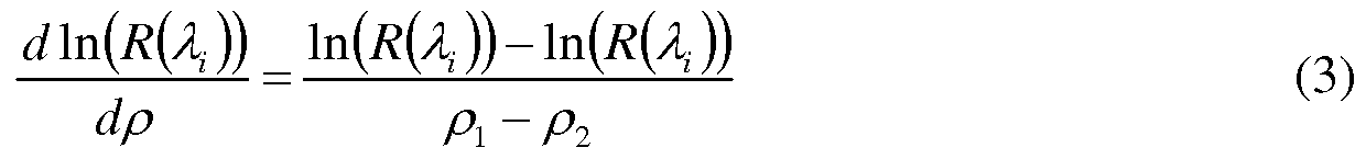
Correspondingly, the quantity S(A1, P1) can be calculated according to Eq.l and Eq.3.
There are many factors that affect the effective attenuation coefficient μeff and thus the quantity S{λ, p) . On the one hand, the absorption coefficient changes with incident wavelength, particularly absorption peaks occur at some specific wavelengths. On the other hand, the source-detector distance is relevant to the photon penetration depth that affects the absorption of the spectrum. The larger the source-detector, the higher the probability for a photon to reach a deeper layer of the multi-layer medium and vice versa. Furthermore, the anatomical structure of the multi-medium in a living body has an influence on the behavior of photons.
Taking human skin tissue as an example, the water content is different for the epidermis, dermis and subcutaneous tissue. When estimating the water content in skin tissue, it is important to carefully define the source-detector distances, so that the radiation penetration depths between the irradiation areas and detection areas are adjustable to reach different layers of the skin tissue.
By defining and adjusting the incident wavelength of the incident spectrums and the source-detector distances, one can get a series of quantities S[A1 , p} J representing the effective attenuation coefficients corresponding to each of the incident spectrums and the source- detector distances.
In general the incident wavelengths are chosen such that each of the incident wavelengths corresponds to a feature spectral line in the spectrums of reflected radiation from the multi-layer medium. To reach three layers of human skin tissue, the recommended incident wavelengths A1 , A2 and A3 can be selected from range 930-1 OOOnm, 1150-1250nm and 1400-
1500nm, the reference wavelength A0 , for example 780nm, can be selected for reference. Meanwhile, every two source-detector distances, {px :
 {p2 : p3,p4}and {p3 : p5,p6}, e.g., every two neighboring fiber bundles, can be grouped together for generating a pair of signals of reflection spectrum R[A1 , p} J and then calculating S[A1 , p} ) .
Once a plurality of S[A1 , p} ) , i = 0,1,2,3 , j = 1,2,3 are available, water content can be estimated according to a linear regression model, which is represented by the following equation:
 wherein at } denotes regression parameters that are adjustable and determinable based on calibration, the number of incident wavelengths for estimation is 3 and the number of pairs of source-detectors for measuring is 3, A0 denotes an incident wavelength for reference. The contribution of absorption from different source-detector paths is weighted to adjust the hydration status assessment.
It should be noted that the selection of incident wavelengths is experiential. For regression to work properly, one may also have the wavelengths of interfering analytes. For instance the analyte of interest has peaks at incident wavelengths A1 , A2 and A3. An interfering analyte has absorption at A2 and A4 , the regression coefficient should then be negative at A4 and therefore one should also include it in the excitation spectrum.
Since the scattering coefficients in different layers, the dermis and subcutaneous fatty tissues are different; a distinguishable change is expected when photons move across the boundary between different layers of the multi-layer tissue. Such changes on the boundary can be used to define regression coefficients in calibration. In detail, measures can be taken on tissues, phantoms or samples with different levels of water content. The water content of those tissues or
samples can be precisely quantified using wet chemical analysis equipment, scale weighting, or other analysis tools. The water content of the phantoms can be specific when the phantoms are prepared. The water content will become the parameter of Y in Eq.4. The measures obtained by the detector, with different source-detector distances, by irradiating spectrums directed to the boundary of the tissues, phantoms or samples corresponding to different incident wavelengths gives the results R[A1 ,p}), from which the quantities S[A1 , p} J , i = 0,1,2,3 , j = 1,2,3 in Eq.4 can be derived. Substituting parameter Y , and quantities S[A^p J into Eq.4, the regression parameters Ci1 } can be calculated.
As explained above, a distinguishable change of the spectrum of reflected radiation from the multi-layer medium is expected when photons move across the boundary between different layers of the multi-layer tissue, because the scattering coefficients in different layers are different. The changes may be used to estimate the number of layers of the medium in the reachable penetration depth. For example, the lipid layer has stronger spectral lines for fatty tissue. The non-uniform content in different layers provides the foundation to estimate the number of layers. Furthermore, the changes and estimated number of layers may be used to select the detected signals and source-detector distances for estimation of the content of the analyte so as to further improve the precision of the analyte content in the multi-layer medium.
Fig. 3 shows a second arrangement of waveguide fibers in the non-invasive biological analyte monitor in accordance with the invention. In Fig.3, every two neighboring fiber bundles, for example pτ ,pι+1 , in the 6 concentrically circular arrangements form 5 pairs of fiber bundles for different source-detector distances, which can provide more detailed information, particularly for more layers of the medium. It is advantageous that the source- detector distances are equidistant, for example p2 - P1 = p3 - p2.
The skilled person in the art will appreciate that the fiber bundles (?)of a pair of fiber bundles may be not continuous. Of course, more or fewer concentrically circular arrangements may be used in practical applications.
The skilled person in the art will appreciate that the arrangements of source fiber bundles and detector fiber bundles are exchangeable. Referring to Fig.2 and Fig.3, a source waveguide fiber can be arranged in the central area and around the source central waveguide fiber,
rings of waveguide fiber bundles can be arranged for detection. In this way, similar measurements can be achieved by adjusting the shutter switch accordingly.
In another embodiment of the invention, the radiation source and detector fibers are replaced by semiconductor near infrared (NIR) light emitting and sensing elements. The radiation source can be any one of LEDs, photo-diodes or phototransistors. Source-detector distances are defined such that the radiation penetration depths between the irradiation areas and detection areas are adjustable to reach different layers of the multi-layer medium.
Fig.4 shows a flowchart regarding estimating the content of an analyte in a multi-layer medium, based on spectral measurements. In the process of the method, in step 10, first a number of radiation sources are arranged to irradiate the multi-layer medium with spectrums of electromagnetic radiation with at least two different incident wavelengths.
In step 20, a detector is arranged to detect a reflection spectrum of the electromagnetic radiation reflected from the multi-layer medium (R[A1, p J). For each of the predefined incident wavelengths, the reflection spectrums are detected at two different source-detector distances. The source-detector distances are chosen such that each of the source-detector distances is far larger than the reciprocal of the effective attenuation coefficient of the multi-layer medium corresponding to the plurality of source-detector distances and incident wavelengths, e.g., p » l/jUe/f .
In step 30, a plurality of quantities S[A1 , p} J representing the effective attenuation coefficients corresponding to the plurality of source-detector distances and incident wavelengths are derived according to Eq.1 and 3 and then the content Y of the analyte is estimated from the plurality of quantities S[A1 , p J according to EqA The regression parameters at } in Eq.4 are determined during calibration with the calibration method described above. The data processing in step 30 can be implemented by software.
It should be noted that the above-mentioned embodiments illustrate rather than limit the invention and that those skilled in the art will be able to design alternative embodiments without departing from the scope of the appended claims. In the claims, any reference signs placed between parentheses shall not be construed as limiting the claim. The
word "comprising" does not exclude the presence of elements or steps not listed in a claim or in the description. The word "a" or "an" preceding an element does not exclude the presence of a plurality of such elements. The invention can be implemented by means of hardware comprising several distinct elements and by means of a programmed computer. In the system claims enumerating several units, several of these units can be embodied by one and the same item of hardware or software. The usage of the words first, second and third, etcetera, does not indicate any ordering. These words are to be interpreted as names.
Claims
1. An apparatus for non-invasively estimating the content of an analyte in a multi-layer medium, based on spectral measurements, comprising: a radiation source adapted to generate spectrums of electromagnetic radiation corresponding to a plurality of incident wavelengths and to transmit the spectrums to the multi-layer medium; a detector adapted to detect spectrums of reflected radiation from the multilayer medium and to generate detection signals representing the detected radiation, wherein the irradiation areas of the radiation source and detection areas of the detector on the multilayer medium are arranged to generate detection signals corresponding to at least two different source-detector distances for each of the plurality of incident wavelengths, the source-detector distances being defined as the respective distances between the irradiation areas and detection areas and each of the source-detector distances being far larger than the reciprocal of the effective attenuation coefficient of the multi-layer medium; and a data processing means adapted to derive from the detected signals a plurality of quantities representing the effective attenuation coefficients of the multi-layer medium, and to estimate the content of the analyte from the plurality of quantities based on a regression model.
2. An apparatus as claimed in claim 1 , wherein the source-detector distances are defined such that the radiation penetration depths between the irradiation areas and detection areas are adjustable to reach different layers of the multi-layer medium.
3. An apparatus as claimed in claim 2, wherein the data processing means is adapted to determine the plurality of quantities according to the following equation:
s{A,p})= i ≥ OJ ≥ l dPj wherein R[A1 ) denotes the intensity reflection coefficient corresponding to one of the plurality of incident wavelengths A1 , p denotes one of the plurality of source-detector distances, ln[/?(/l; )]
denotes the natural logarithm of R[A ) and ' denotes the derivative of ln[/?(/l )] dP] corresponding to the source-detector distances p} , S[A1 ,p} J denotes the quantity representing the content of the analyte corresponding to the incident wavelength A1 and the source-detector distances p} .
4. An apparatus as claimed in claim 3, wherein the data processing means is further adapted to estimate the content of the analyte from the plurality of quantities according to the following equation:  wherein aτ } denotes regression parameters that are adjustable and determinable based on calibration, M and N respectively denote the number of incident wavelengths and the number of source-detector distances for measuring, A0 denotes an incident wavelength for reference, and Y denotes an estimation of the content of the analyte.
5. An apparatus as claimed in claim 4, wherein the plurality of incident wavelengths are chosen such that each of the incident wavelengths corresponds to a feature spectral line in the spectrums of reflected radiation from the multi-layer medium.
6. An apparatus as claimed in claim 4, wherein the number of the layers in the multilayer medium is estimated based on changes of the spectrums of reflected radiation from the medium, the number of the layers and the changes of the spectrums being used to select the source-detector distances and the detected signals being used to estimate the content of the analyte.
7. An apparatus as claimed in claim 1, wherein the multi-layer medium is the skin tissue of a living body and the analyte is water in the skin tissue.
8. An apparatus as claimed in claim 1, wherein the radiation source comprises at least three concentrically circular arrangements having a respective plurality of waveguide fibers essentially extending to the boundary of the multi-layer medium and arranged around a common central detection area on the boundary, the detector comprises at least one central waveguide fiber essentially extending to the central detection area on the boundary and being in operative connection with the data processing means.
9. An apparatus as claimed in claim 1, wherein the radiation source comprises an arrangement having at least one central waveguide fiber essentially extending to the boundary of the multi-layer medium, and the detector comprises at least three concentrically circular arrangements having a respective plurality of waveguide fibers essentially extending to the boundary of the multi-layer medium, arranged around a common central irradiation area irradiated by the at least one central waveguide fiber and in operative connection with the data processing means.
10. An apparatus as claimed in claim 8, wherein the radiation source further comprises a light source and a switching means, the light source being coupled with the arrangements of waveguide fibers in the radiation source by means of the switching means adapted for selectively exciting either one of the arrangements of waveguide fibers.
11. An apparatus as claimed in claim 8 or 9, wherein the light source is adjustable for generating an incident spectrum of electromagnetic radiation corresponding to a plurality of predefined incident wavelengths.
12. An apparatus as claimed in claim 8 or 9, wherein a diameter of the central waveguide fiber is chosen such that the diameter is much smaller than the reciprocal of each of the effective attenuation coefficients corresponding to the plurality of incident wavelengths and source-detector distances.
13. An apparatus as claimed in claim in claim 8 or 9, wherein the radiation source and detector are respectively semiconductor near infrared (NIR) light emitting and sensing elements.
14. A method of non-invasively estimating the content of an analyte in a multi-layer medium, based on spectral measurements, comprising the steps of: irradiating the multi-layer medium with a spectrum of electromagnetic radiation corresponding to a plurality of different incident wavelengths by means of a radiation source; detecting corresponding signals representing spectrums of radiation reflected from the multi-layer medium by means of a detector such that for each of the plurality of incident wavelengths the reflection spectrums are detected at two different source-detector distances and the source-detector distances are chosen such that each of the source-detector distances is much larger than the reciprocal of the effective attenuation coefficient of the multi-layer medium; deriving from the detected signals a plurality of quantities representing the effective attenuation coefficients of the multi-layer medium that correspond to a plurality of incident wavelengths and source-detector distances; and estimating the content of the analyte from the plurality of quantities based on a regression model.
15. A method as claimed in claim 14, wherein the source-detector distances are defined such that the radiation penetration depths between the areas for irradiation and the areas for detection are adjustable to reach different layers of the multi-layer medium.
16. A method as claimed in claim 15, wherein the plurality of quantities is determined according to the following equation: d In[R[A1 )] s(\ >p, )= i ≥ OJ ≥ l dp, wherein R[A1 ) denotes the intensity reflection coefficient corresponding to one of the plurality of incident wavelengths A1 , p denotes one of the plurality of source-detector distances, ln[/?(/l; )]
denotes the natural logarithm of R[A ) and ' denotes the derivative of ln[/?(/l )] dP] corresponding to the source-detector distances p} , S[A1 ,p} J denotes the quantity representing the content of the analyte corresponding to the incident wavelength A1 and the source-detector distances p} .
17. An apparatus as claimed in claim 16, wherein the content of the analyte from the plurality of quantities is estimated according to the following equation: v ^ s(λ, ,P])
wherein Ci1 } denotes regression parameters that are adjustable and determinable based on calibration, M and N respectively denote the number of incident wavelengths for estimation and the number of source-detectors for measuring, A0 denotes an incident wavelength for reference, and Y denotes an estimation of the content of the analyte.
18. A computer program product to be loaded by a computer arrangement, comprising instructions for estimating the content of an analyte in a multi-layer medium, based on spectral measurements, the computer arrangement comprising a processing unit and a memory, the computer program product, after being loaded, providing said processing unit with the capability to carry out the following tasks: acquiring detection data representing spectrums of radiation reflected from the multi-layer medium corresponding to a plurality of incident wavelengths and; deriving from the detection data a plurality of quantities representing the effective attenuation coefficients of the multi-layer medium that correspond to a plurality of incident wavelengths and source-detector distances; and estimating the content of the analyte from the plurality of quantities based on a regression model.
Applications Claiming Priority (2)
| Application Number | Priority Date | Filing Date | Title |
|---|---|---|---|
| CN200710161411.9 | 2007-09-20 | ||
| CN200710161411 | 2007-09-20 |
Publications (2)
| Publication Number | Publication Date |
|---|---|
| WO2009037644A2 true WO2009037644A2 (en) | 2009-03-26 |
| WO2009037644A3 WO2009037644A3 (en) | 2009-06-25 |
Family
ID=40468527
Family Applications (1)
| Application Number | Title | Priority Date | Filing Date |
|---|---|---|---|
| PCT/IB2008/053761 WO2009037644A2 (en) | 2007-09-20 | 2008-09-17 | Method and apparatus for estimating the content of an analyte in a multi-layer medium |
Country Status (1)
| Country | Link |
|---|---|
| WO (1) | WO2009037644A2 (en) |
Cited By (2)
| Publication number | Priority date | Publication date | Assignee | Title |
|---|---|---|---|---|
| WO2010136728A1 (en) * | 2009-05-28 | 2010-12-02 | Indatech | Spectroscopy device and method for implementing same |
| US20130035570A1 (en) * | 2011-08-05 | 2013-02-07 | Canon Kabushiki Kaisha | Apparatus and method for acquiring information on subject |
Citations (1)
| Publication number | Priority date | Publication date | Assignee | Title |
|---|---|---|---|---|
| WO2007060583A2 (en) | 2005-11-28 | 2007-05-31 | Koninklijke Philips Electronics N.V. | Method and apparatus for determining concentrations of analytes in a turbid medium |
Family Cites Families (3)
| Publication number | Priority date | Publication date | Assignee | Title |
|---|---|---|---|---|
| US6850656B1 (en) * | 1998-10-07 | 2005-02-01 | Ecole Polytechnique Federale De Lausanne | Method and apparatus for measuring locally and superficially the scattering and absorption properties of turbid media |
| US6353226B1 (en) * | 1998-11-23 | 2002-03-05 | Abbott Laboratories | Non-invasive sensor capable of determining optical parameters in a sample having multiple layers |
| US6442408B1 (en) * | 1999-07-22 | 2002-08-27 | Instrumentation Metrics, Inc. | Method for quantification of stratum corneum hydration using diffuse reflectance spectroscopy |
-
2008
- 2008-09-17 WO PCT/IB2008/053761 patent/WO2009037644A2/en active Application Filing
Patent Citations (1)
| Publication number | Priority date | Publication date | Assignee | Title |
|---|---|---|---|---|
| WO2007060583A2 (en) | 2005-11-28 | 2007-05-31 | Koninklijke Philips Electronics N.V. | Method and apparatus for determining concentrations of analytes in a turbid medium |
Cited By (2)
| Publication number | Priority date | Publication date | Assignee | Title |
|---|---|---|---|---|
| WO2010136728A1 (en) * | 2009-05-28 | 2010-12-02 | Indatech | Spectroscopy device and method for implementing same |
| US20130035570A1 (en) * | 2011-08-05 | 2013-02-07 | Canon Kabushiki Kaisha | Apparatus and method for acquiring information on subject |
Also Published As
| Publication number | Publication date |
|---|---|
| WO2009037644A3 (en) | 2009-06-25 |
Similar Documents
| Publication | Publication Date | Title |
|---|---|---|
| US7440659B2 (en) | Depth-resolved reflectance instrument and method for its use | |
| TW570768B (en) | Classification and characterization of tissue through features related to adipose tissue | |
| US6668181B2 (en) | Method for quantification of stratum corneum hydration using diffuse reflectance spectroscopy | |
| TW483745B (en) | Classification system for sex determination and tissue characterization | |
| US6675029B2 (en) | Apparatus and method for quantification of tissue hydration using diffuse reflectance spectroscopy | |
| JP5463545B2 (en) | Concentration determination apparatus, concentration determination method and program | |
| JP6775660B2 (en) | Multiposition diffusion spectrum data processing, modeling, prediction methods and processing equipment | |
| JP6108705B2 (en) | Subject information acquisition apparatus and subject information acquisition method | |
| US8301216B2 (en) | Method and apparatus for quantification of optical properties of superficial volumes using small source-to-detector separations | |
| US7818154B2 (en) | Monte Carlo based model of fluorescence in turbid media and methods and systems for using same to determine intrinsic fluorescence of turbid media | |
| JP2006516207A (en) | Photoacoustic analysis method and apparatus | |
| JP5773578B2 (en) | SUBJECT INFORMATION ACQUISITION DEVICE, CONTROL METHOD AND PROGRAM FOR SUBJECT INFORMATION ACQUISITION DEVICE | |
| Churmakov et al. | Amending of fluorescence sensor signal localization in human skin by matching of the refractive index | |
| Kurakina et al. | Probing depth in diffuse reflectance spectroscopy of biotissues: a Monte Carlo study | |
| Sowa et al. | Precision of Raman depolarization and optical attenuation measurements of sound tooth enamel | |
| US20160341668A1 (en) | Angled confocal spectroscopy | |
| WO2007060583A2 (en) | Method and apparatus for determining concentrations of analytes in a turbid medium | |
| JP2010005047A (en) | Method for generating calibration model for optically measuring biological component and non-invasive apparatus for measuring blood glucose value | |
| JP2019201760A (en) | Blood vessel detection device and method thereof | |
| WO2001050948A2 (en) | Non-invasive method of determining skin thickness and characterizing skin tissue layers | |
| WO2009037644A2 (en) | Method and apparatus for estimating the content of an analyte in a multi-layer medium | |
| JP3304559B2 (en) | Optical measurement method and device | |
| TWI588492B (en) | Near-field array detection method for detecting optically high scatter material | |
| US7565249B2 (en) | Method for the determination of a light transport parameter in a biological matrix | |
| TW200914814A (en) | Method and apparatus for estimating the content of an analyte in a multi-layer medium |
Legal Events
| Date | Code | Title | Description |
|---|---|---|---|
| NENP | Non-entry into the national phase in: |
Ref country code: DE |
|
| 122 | Ep: pct app. not ent. europ. phase |
Ref document number: 08807687 Country of ref document: EP Kind code of ref document: A2 |