US9433707B2 - Bone graft material containment structures - Google Patents
Bone graft material containment structures Download PDFInfo
- Publication number
- US9433707B2 US9433707B2 US12/712,949 US71294910A US9433707B2 US 9433707 B2 US9433707 B2 US 9433707B2 US 71294910 A US71294910 A US 71294910A US 9433707 B2 US9433707 B2 US 9433707B2
- Authority
- US
- United States
- Prior art keywords
- bone
- bone graft
- graft material
- porous material
- microns
- Prior art date
- Legal status (The legal status is an assumption and is not a legal conclusion. Google has not performed a legal analysis and makes no representation as to the accuracy of the status listed.)
- Active, expires
Links
- 210000000988 bone and bone Anatomy 0.000 title claims abstract description 424
- 239000000463 material Substances 0.000 title claims abstract description 296
- 239000011148 porous material Substances 0.000 claims abstract description 140
- 238000000034 method Methods 0.000 claims description 50
- 210000004872 soft tissue Anatomy 0.000 claims description 33
- 229910052751 metal Inorganic materials 0.000 claims description 29
- 239000002184 metal Substances 0.000 claims description 29
- 102000004169 proteins and genes Human genes 0.000 claims description 25
- 108090000623 proteins and genes Proteins 0.000 claims description 25
- -1 polyethylene Polymers 0.000 claims description 24
- 239000003102 growth factor Substances 0.000 claims description 22
- 229920000642 polymer Polymers 0.000 claims description 22
- 210000004027 cell Anatomy 0.000 claims description 13
- 102000010780 Platelet-Derived Growth Factor Human genes 0.000 claims description 12
- 108010038512 Platelet-Derived Growth Factor Proteins 0.000 claims description 12
- QORWJWZARLRLPR-UHFFFAOYSA-H tricalcium bis(phosphate) Chemical compound [Ca+2].[Ca+2].[Ca+2].[O-]P([O-])([O-])=O.[O-]P([O-])([O-])=O QORWJWZARLRLPR-UHFFFAOYSA-H 0.000 claims description 12
- 229910052588 hydroxylapatite Inorganic materials 0.000 claims description 11
- XYJRXVWERLGGKC-UHFFFAOYSA-D pentacalcium;hydroxide;triphosphate Chemical compound [OH-].[Ca+2].[Ca+2].[Ca+2].[Ca+2].[Ca+2].[O-]P([O-])([O-])=O.[O-]P([O-])([O-])=O.[O-]P([O-])([O-])=O XYJRXVWERLGGKC-UHFFFAOYSA-D 0.000 claims description 11
- 239000004698 Polyethylene Substances 0.000 claims description 10
- 229920000573 polyethylene Polymers 0.000 claims description 10
- 210000004204 blood vessel Anatomy 0.000 claims description 9
- 210000004623 platelet-rich plasma Anatomy 0.000 claims description 9
- 102000055006 Calcitonin Human genes 0.000 claims description 8
- 108060001064 Calcitonin Proteins 0.000 claims description 8
- 102000004887 Transforming Growth Factor beta Human genes 0.000 claims description 8
- 108090001012 Transforming Growth Factor beta Proteins 0.000 claims description 8
- BBBFJLBPOGFECG-VJVYQDLKSA-N calcitonin Chemical compound N([C@H](C(=O)N[C@@H](CC(C)C)C(=O)NCC(=O)N[C@@H](CCCCN)C(=O)N[C@@H](CC(C)C)C(=O)N[C@@H](CO)C(=O)N[C@@H](CCC(N)=O)C(=O)N[C@@H](CCC(O)=O)C(=O)N[C@@H](CC(C)C)C(=O)N[C@@H](CC=1NC=NC=1)C(=O)N[C@@H](CCCCN)C(=O)N[C@@H](CC(C)C)C(=O)N[C@@H](CCC(N)=O)C(=O)N[C@@H]([C@@H](C)O)C(=O)N[C@@H](CC=1C=CC(O)=CC=1)C(=O)N1[C@@H](CCC1)C(=O)N[C@@H](CCCNC(N)=N)C(=O)N[C@@H]([C@@H](C)O)C(=O)N[C@@H](CC(N)=O)C(=O)N[C@@H]([C@@H](C)O)C(=O)NCC(=O)N[C@@H](CO)C(=O)NCC(=O)N[C@@H]([C@@H](C)O)C(=O)N1[C@@H](CCC1)C(N)=O)C(C)C)C(=O)[C@@H]1CSSC[C@H](N)C(=O)N[C@@H](CO)C(=O)N[C@@H](CC(N)=O)C(=O)N[C@@H](CC(C)C)C(=O)N[C@@H](CO)C(=O)N[C@@H]([C@@H](C)O)C(=O)N1 BBBFJLBPOGFECG-VJVYQDLKSA-N 0.000 claims description 8
- 229960004015 calcitonin Drugs 0.000 claims description 8
- 230000012010 growth Effects 0.000 claims description 8
- 230000000921 morphogenic effect Effects 0.000 claims description 8
- ZRKFYGHZFMAOKI-QMGMOQQFSA-N tgfbeta Chemical compound C([C@H](NC(=O)[C@H](C(C)C)NC(=O)CNC(=O)[C@H](CCC(O)=O)NC(=O)[C@H](CCCNC(N)=N)NC(=O)[C@H](CC(N)=O)NC(=O)[C@H](CC(C)C)NC(=O)[C@H]([C@@H](C)O)NC(=O)[C@H](CCC(O)=O)NC(=O)[C@H]([C@@H](C)O)NC(=O)[C@H](CC(C)C)NC(=O)CNC(=O)[C@H](C)NC(=O)[C@H](CO)NC(=O)[C@H](CCC(N)=O)NC(=O)[C@@H](NC(=O)[C@H](C)NC(=O)[C@H](C)NC(=O)[C@@H](NC(=O)[C@H](CC(C)C)NC(=O)[C@@H](N)CCSC)C(C)C)[C@@H](C)CC)C(=O)N[C@@H]([C@@H](C)O)C(=O)N[C@@H](C(C)C)C(=O)N[C@@H](CC=1C=CC=CC=1)C(=O)N[C@@H](C)C(=O)N1[C@@H](CCC1)C(=O)N[C@@H]([C@@H](C)O)C(=O)N[C@@H](CC(N)=O)C(=O)N[C@@H](CCC(O)=O)C(=O)N[C@@H](C)C(=O)N[C@@H](CC=1C=CC=CC=1)C(=O)N[C@@H](CCCNC(N)=N)C(=O)N[C@@H](C)C(=O)N[C@@H](CC(C)C)C(=O)N1[C@@H](CCC1)C(=O)N1[C@@H](CCC1)C(=O)N[C@@H](CCCNC(N)=N)C(=O)N[C@@H](CCC(O)=O)C(=O)N[C@@H](CCCNC(N)=N)C(=O)N[C@@H](CO)C(=O)N[C@@H](CCCNC(N)=N)C(=O)N[C@@H](CC(C)C)C(=O)N[C@@H](CC(C)C)C(O)=O)C1=CC=C(O)C=C1 ZRKFYGHZFMAOKI-QMGMOQQFSA-N 0.000 claims description 8
- 102000018233 Fibroblast Growth Factor Human genes 0.000 claims description 7
- 108050007372 Fibroblast Growth Factor Proteins 0.000 claims description 7
- 239000002245 particle Substances 0.000 claims description 7
- 239000001506 calcium phosphate Substances 0.000 claims description 6
- OSGAYBCDTDRGGQ-UHFFFAOYSA-L calcium sulfate Chemical compound [Ca+2].[O-]S([O-])(=O)=O OSGAYBCDTDRGGQ-UHFFFAOYSA-L 0.000 claims description 6
- 239000011159 matrix material Substances 0.000 claims description 6
- 229920002994 synthetic fiber Polymers 0.000 claims description 6
- 241001465754 Metazoa Species 0.000 claims description 5
- 108090000723 Insulin-Like Growth Factor I Proteins 0.000 claims description 4
- 102000013275 Somatomedins Human genes 0.000 claims description 4
- 239000005313 bioactive glass Substances 0.000 claims description 4
- 230000004069 differentiation Effects 0.000 claims description 4
- 210000002901 mesenchymal stem cell Anatomy 0.000 claims description 4
- 229910000389 calcium phosphate Inorganic materials 0.000 claims description 3
- 235000011010 calcium phosphates Nutrition 0.000 claims description 3
- 239000000919 ceramic Substances 0.000 claims description 3
- 210000000130 stem cell Anatomy 0.000 claims description 3
- 238000005245 sintering Methods 0.000 claims description 2
- 238000005452 bending Methods 0.000 claims 2
- 210000002798 bone marrow cell Anatomy 0.000 claims 1
- 229940126864 fibroblast growth factor Drugs 0.000 claims 1
- 238000007493 shaping process Methods 0.000 claims 1
- 210000001519 tissue Anatomy 0.000 abstract description 63
- 230000008468 bone growth Effects 0.000 abstract description 30
- 230000002792 vascular Effects 0.000 abstract description 21
- 230000001497 fibrovascular Effects 0.000 abstract description 13
- 230000010354 integration Effects 0.000 abstract description 5
- 230000000638 stimulation Effects 0.000 abstract description 2
- 230000007547 defect Effects 0.000 description 50
- 210000004373 mandible Anatomy 0.000 description 41
- 239000007943 implant Substances 0.000 description 25
- 230000035876 healing Effects 0.000 description 24
- 239000000515 collagen sponge Substances 0.000 description 18
- 238000001356 surgical procedure Methods 0.000 description 18
- RTAQQCXQSZGOHL-UHFFFAOYSA-N Titanium Chemical compound [Ti] RTAQQCXQSZGOHL-UHFFFAOYSA-N 0.000 description 17
- 239000004053 dental implant Substances 0.000 description 17
- 210000002050 maxilla Anatomy 0.000 description 13
- 230000008439 repair process Effects 0.000 description 13
- 238000013461 design Methods 0.000 description 12
- 102000008186 Collagen Human genes 0.000 description 11
- 108010035532 Collagen Proteins 0.000 description 11
- 239000004696 Poly ether ether ketone Substances 0.000 description 11
- 229920001436 collagen Polymers 0.000 description 11
- 230000002950 deficient Effects 0.000 description 11
- 229920002530 polyetherether ketone Polymers 0.000 description 11
- 239000004743 Polypropylene Substances 0.000 description 10
- 229920001155 polypropylene Polymers 0.000 description 10
- 230000004888 barrier function Effects 0.000 description 9
- 210000004379 membrane Anatomy 0.000 description 8
- 239000012528 membrane Substances 0.000 description 8
- 230000011164 ossification Effects 0.000 description 8
- 239000010936 titanium Substances 0.000 description 8
- 229910052719 titanium Inorganic materials 0.000 description 8
- 238000006073 displacement reaction Methods 0.000 description 7
- 229920001903 high density polyethylene Polymers 0.000 description 7
- 239000004700 high-density polyethylene Substances 0.000 description 7
- 238000004519 manufacturing process Methods 0.000 description 7
- 230000004927 fusion Effects 0.000 description 6
- 238000003780 insertion Methods 0.000 description 6
- 230000037431 insertion Effects 0.000 description 6
- 230000001788 irregular Effects 0.000 description 6
- 230000033001 locomotion Effects 0.000 description 6
- 239000000203 mixture Substances 0.000 description 6
- 125000006850 spacer group Chemical group 0.000 description 6
- 210000002303 tibia Anatomy 0.000 description 6
- 208000010392 Bone Fractures Diseases 0.000 description 5
- 206010028980 Neoplasm Diseases 0.000 description 5
- 210000001909 alveolar process Anatomy 0.000 description 5
- 230000037182 bone density Effects 0.000 description 5
- 239000000835 fiber Substances 0.000 description 5
- 210000004195 gingiva Anatomy 0.000 description 5
- 208000014674 injury Diseases 0.000 description 5
- 239000007788 liquid Substances 0.000 description 5
- 231100000241 scar Toxicity 0.000 description 5
- 239000002904 solvent Substances 0.000 description 5
- 210000003781 tooth socket Anatomy 0.000 description 5
- 230000008733 trauma Effects 0.000 description 5
- 206010017076 Fracture Diseases 0.000 description 4
- 229920000544 Gore-Tex Polymers 0.000 description 4
- 239000004677 Nylon Substances 0.000 description 4
- 239000004699 Ultra-high molecular weight polyethylene Substances 0.000 description 4
- 230000008901 benefit Effects 0.000 description 4
- 229920000249 biocompatible polymer Polymers 0.000 description 4
- 230000015572 biosynthetic process Effects 0.000 description 4
- 230000036770 blood supply Effects 0.000 description 4
- 210000001185 bone marrow Anatomy 0.000 description 4
- 230000015556 catabolic process Effects 0.000 description 4
- 230000007812 deficiency Effects 0.000 description 4
- 230000003628 erosive effect Effects 0.000 description 4
- 230000001976 improved effect Effects 0.000 description 4
- 238000012986 modification Methods 0.000 description 4
- 230000004048 modification Effects 0.000 description 4
- 229920001778 nylon Polymers 0.000 description 4
- 230000000399 orthopedic effect Effects 0.000 description 4
- 210000000963 osteoblast Anatomy 0.000 description 4
- 230000003239 periodontal effect Effects 0.000 description 4
- 229920003023 plastic Polymers 0.000 description 4
- 239000004033 plastic Substances 0.000 description 4
- 229920000139 polyethylene terephthalate Polymers 0.000 description 4
- 239000005020 polyethylene terephthalate Substances 0.000 description 4
- 239000002861 polymer material Substances 0.000 description 4
- 239000007787 solid Substances 0.000 description 4
- 229920000785 ultra high molecular weight polyethylene Polymers 0.000 description 4
- 102000007350 Bone Morphogenetic Proteins Human genes 0.000 description 3
- 108010007726 Bone Morphogenetic Proteins Proteins 0.000 description 3
- 208000032170 Congenital Abnormalities Diseases 0.000 description 3
- 102100035379 Growth/differentiation factor 5 Human genes 0.000 description 3
- 101001023988 Homo sapiens Growth/differentiation factor 5 Proteins 0.000 description 3
- 210000003484 anatomy Anatomy 0.000 description 3
- 230000003190 augmentative effect Effects 0.000 description 3
- 210000004369 blood Anatomy 0.000 description 3
- 239000008280 blood Substances 0.000 description 3
- 229940112869 bone morphogenetic protein Drugs 0.000 description 3
- 230000010478 bone regeneration Effects 0.000 description 3
- 239000003292 glue Substances 0.000 description 3
- 239000008187 granular material Substances 0.000 description 3
- 230000036541 health Effects 0.000 description 3
- 230000006698 induction Effects 0.000 description 3
- 230000008595 infiltration Effects 0.000 description 3
- 238000001764 infiltration Methods 0.000 description 3
- 230000007794 irritation Effects 0.000 description 3
- 210000000214 mouth Anatomy 0.000 description 3
- 210000004197 pelvis Anatomy 0.000 description 3
- 208000028169 periodontal disease Diseases 0.000 description 3
- 239000002243 precursor Substances 0.000 description 3
- 230000008569 process Effects 0.000 description 3
- 238000001878 scanning electron micrograph Methods 0.000 description 3
- 210000003625 skull Anatomy 0.000 description 3
- 229920005992 thermoplastic resin Polymers 0.000 description 3
- 229910000391 tricalcium phosphate Inorganic materials 0.000 description 3
- 235000019731 tricalcium phosphate Nutrition 0.000 description 3
- 229940078499 tricalcium phosphate Drugs 0.000 description 3
- LCSKNASZPVZHEG-UHFFFAOYSA-N 3,6-dimethyl-1,4-dioxane-2,5-dione;1,4-dioxane-2,5-dione Chemical group O=C1COC(=O)CO1.CC1OC(=O)C(C)OC1=O LCSKNASZPVZHEG-UHFFFAOYSA-N 0.000 description 2
- 206010065687 Bone loss Diseases 0.000 description 2
- 208000024779 Comminuted Fractures Diseases 0.000 description 2
- 208000025962 Crush injury Diseases 0.000 description 2
- 241000195493 Cryptophyta Species 0.000 description 2
- 241001269524 Dura Species 0.000 description 2
- 206010061218 Inflammation Diseases 0.000 description 2
- 208000001132 Osteoporosis Diseases 0.000 description 2
- 208000006735 Periostitis Diseases 0.000 description 2
- 206010039203 Road traffic accident Diseases 0.000 description 2
- 229920010741 Ultra High Molecular Weight Polyethylene (UHMWPE) Polymers 0.000 description 2
- 238000007792 addition Methods 0.000 description 2
- 150000001338 aliphatic hydrocarbons Chemical class 0.000 description 2
- 210000003423 ankle Anatomy 0.000 description 2
- 230000009286 beneficial effect Effects 0.000 description 2
- 230000000740 bleeding effect Effects 0.000 description 2
- 201000011510 cancer Diseases 0.000 description 2
- 239000003795 chemical substances by application Substances 0.000 description 2
- 239000002131 composite material Substances 0.000 description 2
- 230000006835 compression Effects 0.000 description 2
- 238000007906 compression Methods 0.000 description 2
- 230000008602 contraction Effects 0.000 description 2
- 238000006731 degradation reaction Methods 0.000 description 2
- 238000005137 deposition process Methods 0.000 description 2
- 201000010099 disease Diseases 0.000 description 2
- 208000037265 diseases, disorders, signs and symptoms Diseases 0.000 description 2
- 238000005553 drilling Methods 0.000 description 2
- 230000000694 effects Effects 0.000 description 2
- 238000011156 evaluation Methods 0.000 description 2
- 230000001815 facial effect Effects 0.000 description 2
- 210000001983 hard palate Anatomy 0.000 description 2
- 210000002758 humerus Anatomy 0.000 description 2
- 230000007062 hydrolysis Effects 0.000 description 2
- 238000006460 hydrolysis reaction Methods 0.000 description 2
- 210000001621 ilium bone Anatomy 0.000 description 2
- 230000004054 inflammatory process Effects 0.000 description 2
- 230000009545 invasion Effects 0.000 description 2
- 230000000670 limiting effect Effects 0.000 description 2
- 230000003211 malignant effect Effects 0.000 description 2
- 238000005259 measurement Methods 0.000 description 2
- 150000002739 metals Chemical class 0.000 description 2
- 230000035515 penetration Effects 0.000 description 2
- 210000003460 periosteum Anatomy 0.000 description 2
- 229920000098 polyolefin Polymers 0.000 description 2
- 229920001343 polytetrafluoroethylene Polymers 0.000 description 2
- 238000002278 reconstructive surgery Methods 0.000 description 2
- 230000002829 reductive effect Effects 0.000 description 2
- 230000002787 reinforcement Effects 0.000 description 2
- 230000036573 scar formation Effects 0.000 description 2
- 239000007779 soft material Substances 0.000 description 2
- 239000003381 stabilizer Substances 0.000 description 2
- 229910001220 stainless steel Inorganic materials 0.000 description 2
- 239000010935 stainless steel Substances 0.000 description 2
- 239000003351 stiffener Substances 0.000 description 2
- 238000005728 strengthening Methods 0.000 description 2
- 230000008093 supporting effect Effects 0.000 description 2
- 229910052715 tantalum Inorganic materials 0.000 description 2
- GUVRBAGPIYLISA-UHFFFAOYSA-N tantalum atom Chemical compound [Ta] GUVRBAGPIYLISA-UHFFFAOYSA-N 0.000 description 2
- 239000010409 thin film Substances 0.000 description 2
- 230000008467 tissue growth Effects 0.000 description 2
- 230000017423 tissue regeneration Effects 0.000 description 2
- 210000001364 upper extremity Anatomy 0.000 description 2
- 210000000689 upper leg Anatomy 0.000 description 2
- RKDVKSZUMVYZHH-UHFFFAOYSA-N 1,4-dioxane-2,5-dione Chemical compound O=C1COC(=O)CO1 RKDVKSZUMVYZHH-UHFFFAOYSA-N 0.000 description 1
- 125000003821 2-(trimethylsilyl)ethoxymethyl group Chemical group [H]C([H])([H])[Si](C([H])([H])[H])(C([H])([H])[H])C([H])([H])C(OC([H])([H])[*])([H])[H] 0.000 description 1
- 208000006386 Bone Resorption Diseases 0.000 description 1
- 206010011732 Cyst Diseases 0.000 description 1
- IAYPIBMASNFSPL-UHFFFAOYSA-N Ethylene oxide Chemical compound C1CO1 IAYPIBMASNFSPL-UHFFFAOYSA-N 0.000 description 1
- 201000003200 Goldenhar Syndrome Diseases 0.000 description 1
- 206010062575 Muscle contracture Diseases 0.000 description 1
- 206010029113 Neovascularisation Diseases 0.000 description 1
- 208000008312 Tooth Loss Diseases 0.000 description 1
- 239000002253 acid Substances 0.000 description 1
- 239000000853 adhesive Substances 0.000 description 1
- 230000001070 adhesive effect Effects 0.000 description 1
- 230000002411 adverse Effects 0.000 description 1
- 230000000735 allogeneic effect Effects 0.000 description 1
- 230000004075 alteration Effects 0.000 description 1
- 238000013459 approach Methods 0.000 description 1
- 230000003416 augmentation Effects 0.000 description 1
- 239000000560 biocompatible material Substances 0.000 description 1
- 239000012620 biological material Substances 0.000 description 1
- 230000024279 bone resorption Effects 0.000 description 1
- 239000000316 bone substitute Substances 0.000 description 1
- 210000004556 brain Anatomy 0.000 description 1
- 230000004663 cell proliferation Effects 0.000 description 1
- 238000012993 chemical processing Methods 0.000 description 1
- 210000000038 chest Anatomy 0.000 description 1
- 150000001875 compounds Chemical class 0.000 description 1
- 210000002808 connective tissue Anatomy 0.000 description 1
- 238000011109 contamination Methods 0.000 description 1
- 208000006111 contracture Diseases 0.000 description 1
- 229920001577 copolymer Polymers 0.000 description 1
- 239000002537 cosmetic Substances 0.000 description 1
- 238000007428 craniotomy Methods 0.000 description 1
- 208000031513 cyst Diseases 0.000 description 1
- 230000003247 decreasing effect Effects 0.000 description 1
- 230000007850 degeneration Effects 0.000 description 1
- 238000012217 deletion Methods 0.000 description 1
- 230000037430 deletion Effects 0.000 description 1
- 238000000151 deposition Methods 0.000 description 1
- 230000001627 detrimental effect Effects 0.000 description 1
- 238000002224 dissection Methods 0.000 description 1
- 238000009826 distribution Methods 0.000 description 1
- 238000001035 drying Methods 0.000 description 1
- 238000005516 engineering process Methods 0.000 description 1
- 238000001704 evaporation Methods 0.000 description 1
- 230000008020 evaporation Effects 0.000 description 1
- 239000004744 fabric Substances 0.000 description 1
- 210000003746 feather Anatomy 0.000 description 1
- 210000002082 fibula Anatomy 0.000 description 1
- 238000001914 filtration Methods 0.000 description 1
- 210000003811 finger Anatomy 0.000 description 1
- 210000002683 foot Anatomy 0.000 description 1
- 239000012634 fragment Substances 0.000 description 1
- 210000002454 frontal bone Anatomy 0.000 description 1
- 238000009963 fulling Methods 0.000 description 1
- 230000006870 function Effects 0.000 description 1
- 230000008571 general function Effects 0.000 description 1
- 208000017918 hemifacial microsomia Diseases 0.000 description 1
- 230000001939 inductive effect Effects 0.000 description 1
- 208000015181 infectious disease Diseases 0.000 description 1
- 230000001524 infective effect Effects 0.000 description 1
- JJTUDXZGHPGLLC-UHFFFAOYSA-N lactide Chemical compound CC1OC(=O)C(C)OC1=O JJTUDXZGHPGLLC-UHFFFAOYSA-N 0.000 description 1
- 238000000608 laser ablation Methods 0.000 description 1
- 238000002386 leaching Methods 0.000 description 1
- 229920004889 linear high-density polyethylene Polymers 0.000 description 1
- 238000003754 machining Methods 0.000 description 1
- 238000012423 maintenance Methods 0.000 description 1
- 230000014759 maintenance of location Effects 0.000 description 1
- 210000004086 maxillary sinus Anatomy 0.000 description 1
- QSHDDOUJBYECFT-UHFFFAOYSA-N mercury Chemical compound [Hg] QSHDDOUJBYECFT-UHFFFAOYSA-N 0.000 description 1
- 229910052753 mercury Inorganic materials 0.000 description 1
- 238000001000 micrograph Methods 0.000 description 1
- 230000005012 migration Effects 0.000 description 1
- 238000013508 migration Methods 0.000 description 1
- 238000000465 moulding Methods 0.000 description 1
- 210000004400 mucous membrane Anatomy 0.000 description 1
- 210000001331 nose Anatomy 0.000 description 1
- 210000000103 occipital bone Anatomy 0.000 description 1
- 210000003455 parietal bone Anatomy 0.000 description 1
- 230000001936 parietal effect Effects 0.000 description 1
- 230000036961 partial effect Effects 0.000 description 1
- 230000002093 peripheral effect Effects 0.000 description 1
- 239000002985 plastic film Substances 0.000 description 1
- 229920006254 polymer film Polymers 0.000 description 1
- 238000006116 polymerization reaction Methods 0.000 description 1
- 239000004810 polytetrafluoroethylene Substances 0.000 description 1
- 238000002459 porosimetry Methods 0.000 description 1
- 239000000843 powder Substances 0.000 description 1
- 230000001172 regenerating effect Effects 0.000 description 1
- 238000002271 resection Methods 0.000 description 1
- 150000003839 salts Chemical class 0.000 description 1
- 238000004626 scanning electron microscopy Methods 0.000 description 1
- 210000001991 scapula Anatomy 0.000 description 1
- 238000000110 selective laser sintering Methods 0.000 description 1
- 230000000391 smoking effect Effects 0.000 description 1
- 210000001032 spinal nerve Anatomy 0.000 description 1
- 230000006641 stabilisation Effects 0.000 description 1
- 238000011105 stabilization Methods 0.000 description 1
- 239000000725 suspension Substances 0.000 description 1
- 210000003582 temporal bone Anatomy 0.000 description 1
- 229920001169 thermoplastic Polymers 0.000 description 1
- 239000004416 thermosoftening plastic Substances 0.000 description 1
- 238000000427 thin-film deposition Methods 0.000 description 1
- 210000003371 toe Anatomy 0.000 description 1
- 210000000623 ulna Anatomy 0.000 description 1
- XLYOFNOQVPJJNP-UHFFFAOYSA-N water Substances O XLYOFNOQVPJJNP-UHFFFAOYSA-N 0.000 description 1
- 210000000707 wrist Anatomy 0.000 description 1
- 210000000216 zygoma Anatomy 0.000 description 1
Images


Classifications
-
- A—HUMAN NECESSITIES
- A61—MEDICAL OR VETERINARY SCIENCE; HYGIENE
- A61L—METHODS OR APPARATUS FOR STERILISING MATERIALS OR OBJECTS IN GENERAL; DISINFECTION, STERILISATION OR DEODORISATION OF AIR; CHEMICAL ASPECTS OF BANDAGES, DRESSINGS, ABSORBENT PADS OR SURGICAL ARTICLES; MATERIALS FOR BANDAGES, DRESSINGS, ABSORBENT PADS OR SURGICAL ARTICLES
- A61L27/00—Materials for grafts or prostheses or for coating grafts or prostheses
- A61L27/50—Materials characterised by their function or physical properties, e.g. injectable or lubricating compositions, shape-memory materials, surface modified materials
- A61L27/56—Porous materials, e.g. foams or sponges
-
- A—HUMAN NECESSITIES
- A61—MEDICAL OR VETERINARY SCIENCE; HYGIENE
- A61F—FILTERS IMPLANTABLE INTO BLOOD VESSELS; PROSTHESES; DEVICES PROVIDING PATENCY TO, OR PREVENTING COLLAPSING OF, TUBULAR STRUCTURES OF THE BODY, e.g. STENTS; ORTHOPAEDIC, NURSING OR CONTRACEPTIVE DEVICES; FOMENTATION; TREATMENT OR PROTECTION OF EYES OR EARS; BANDAGES, DRESSINGS OR ABSORBENT PADS; FIRST-AID KITS
- A61F2/00—Filters implantable into blood vessels; Prostheses, i.e. artificial substitutes or replacements for parts of the body; Appliances for connecting them with the body; Devices providing patency to, or preventing collapsing of, tubular structures of the body, e.g. stents
- A61F2/02—Prostheses implantable into the body
- A61F2/28—Bones
- A61F2/2803—Bones for mandibular reconstruction
-
- A—HUMAN NECESSITIES
- A61—MEDICAL OR VETERINARY SCIENCE; HYGIENE
- A61F—FILTERS IMPLANTABLE INTO BLOOD VESSELS; PROSTHESES; DEVICES PROVIDING PATENCY TO, OR PREVENTING COLLAPSING OF, TUBULAR STRUCTURES OF THE BODY, e.g. STENTS; ORTHOPAEDIC, NURSING OR CONTRACEPTIVE DEVICES; FOMENTATION; TREATMENT OR PROTECTION OF EYES OR EARS; BANDAGES, DRESSINGS OR ABSORBENT PADS; FIRST-AID KITS
- A61F2/00—Filters implantable into blood vessels; Prostheses, i.e. artificial substitutes or replacements for parts of the body; Appliances for connecting them with the body; Devices providing patency to, or preventing collapsing of, tubular structures of the body, e.g. stents
- A61F2/02—Prostheses implantable into the body
- A61F2/28—Bones
- A61F2/2846—Support means for bone substitute or for bone graft implants, e.g. membranes or plates for covering bone defects
-
- A—HUMAN NECESSITIES
- A61—MEDICAL OR VETERINARY SCIENCE; HYGIENE
- A61L—METHODS OR APPARATUS FOR STERILISING MATERIALS OR OBJECTS IN GENERAL; DISINFECTION, STERILISATION OR DEODORISATION OF AIR; CHEMICAL ASPECTS OF BANDAGES, DRESSINGS, ABSORBENT PADS OR SURGICAL ARTICLES; MATERIALS FOR BANDAGES, DRESSINGS, ABSORBENT PADS OR SURGICAL ARTICLES
- A61L27/00—Materials for grafts or prostheses or for coating grafts or prostheses
- A61L27/02—Inorganic materials
- A61L27/12—Phosphorus-containing materials, e.g. apatite
-
- A—HUMAN NECESSITIES
- A61—MEDICAL OR VETERINARY SCIENCE; HYGIENE
- A61L—METHODS OR APPARATUS FOR STERILISING MATERIALS OR OBJECTS IN GENERAL; DISINFECTION, STERILISATION OR DEODORISATION OF AIR; CHEMICAL ASPECTS OF BANDAGES, DRESSINGS, ABSORBENT PADS OR SURGICAL ARTICLES; MATERIALS FOR BANDAGES, DRESSINGS, ABSORBENT PADS OR SURGICAL ARTICLES
- A61L27/00—Materials for grafts or prostheses or for coating grafts or prostheses
- A61L27/14—Macromolecular materials
- A61L27/16—Macromolecular materials obtained by reactions only involving carbon-to-carbon unsaturated bonds
-
- A—HUMAN NECESSITIES
- A61—MEDICAL OR VETERINARY SCIENCE; HYGIENE
- A61L—METHODS OR APPARATUS FOR STERILISING MATERIALS OR OBJECTS IN GENERAL; DISINFECTION, STERILISATION OR DEODORISATION OF AIR; CHEMICAL ASPECTS OF BANDAGES, DRESSINGS, ABSORBENT PADS OR SURGICAL ARTICLES; MATERIALS FOR BANDAGES, DRESSINGS, ABSORBENT PADS OR SURGICAL ARTICLES
- A61L27/00—Materials for grafts or prostheses or for coating grafts or prostheses
- A61L27/14—Macromolecular materials
- A61L27/22—Polypeptides or derivatives thereof, e.g. degradation products
- A61L27/227—Other specific proteins or polypeptides not covered by A61L27/222, A61L27/225 or A61L27/24
-
- A—HUMAN NECESSITIES
- A61—MEDICAL OR VETERINARY SCIENCE; HYGIENE
- A61L—METHODS OR APPARATUS FOR STERILISING MATERIALS OR OBJECTS IN GENERAL; DISINFECTION, STERILISATION OR DEODORISATION OF AIR; CHEMICAL ASPECTS OF BANDAGES, DRESSINGS, ABSORBENT PADS OR SURGICAL ARTICLES; MATERIALS FOR BANDAGES, DRESSINGS, ABSORBENT PADS OR SURGICAL ARTICLES
- A61L27/00—Materials for grafts or prostheses or for coating grafts or prostheses
- A61L27/36—Materials for grafts or prostheses or for coating grafts or prostheses containing ingredients of undetermined constitution or reaction products thereof, e.g. transplant tissue, natural bone, extracellular matrix
- A61L27/3604—Materials for grafts or prostheses or for coating grafts or prostheses containing ingredients of undetermined constitution or reaction products thereof, e.g. transplant tissue, natural bone, extracellular matrix characterised by the human or animal origin of the biological material, e.g. hair, fascia, fish scales, silk, shellac, pericardium, pleura, renal tissue, amniotic membrane, parenchymal tissue, fetal tissue, muscle tissue, fat tissue, enamel
- A61L27/3608—Bone, e.g. demineralised bone matrix [DBM], bone powder
-
- A—HUMAN NECESSITIES
- A61—MEDICAL OR VETERINARY SCIENCE; HYGIENE
- A61L—METHODS OR APPARATUS FOR STERILISING MATERIALS OR OBJECTS IN GENERAL; DISINFECTION, STERILISATION OR DEODORISATION OF AIR; CHEMICAL ASPECTS OF BANDAGES, DRESSINGS, ABSORBENT PADS OR SURGICAL ARTICLES; MATERIALS FOR BANDAGES, DRESSINGS, ABSORBENT PADS OR SURGICAL ARTICLES
- A61L27/00—Materials for grafts or prostheses or for coating grafts or prostheses
- A61L27/36—Materials for grafts or prostheses or for coating grafts or prostheses containing ingredients of undetermined constitution or reaction products thereof, e.g. transplant tissue, natural bone, extracellular matrix
- A61L27/3641—Materials for grafts or prostheses or for coating grafts or prostheses containing ingredients of undetermined constitution or reaction products thereof, e.g. transplant tissue, natural bone, extracellular matrix characterised by the site of application in the body
- A61L27/3645—Connective tissue
- A61L27/365—Bones
-
- A—HUMAN NECESSITIES
- A61—MEDICAL OR VETERINARY SCIENCE; HYGIENE
- A61L—METHODS OR APPARATUS FOR STERILISING MATERIALS OR OBJECTS IN GENERAL; DISINFECTION, STERILISATION OR DEODORISATION OF AIR; CHEMICAL ASPECTS OF BANDAGES, DRESSINGS, ABSORBENT PADS OR SURGICAL ARTICLES; MATERIALS FOR BANDAGES, DRESSINGS, ABSORBENT PADS OR SURGICAL ARTICLES
- A61L27/00—Materials for grafts or prostheses or for coating grafts or prostheses
- A61L27/36—Materials for grafts or prostheses or for coating grafts or prostheses containing ingredients of undetermined constitution or reaction products thereof, e.g. transplant tissue, natural bone, extracellular matrix
- A61L27/38—Materials for grafts or prostheses or for coating grafts or prostheses containing ingredients of undetermined constitution or reaction products thereof, e.g. transplant tissue, natural bone, extracellular matrix containing added animal cells
-
- A—HUMAN NECESSITIES
- A61—MEDICAL OR VETERINARY SCIENCE; HYGIENE
- A61L—METHODS OR APPARATUS FOR STERILISING MATERIALS OR OBJECTS IN GENERAL; DISINFECTION, STERILISATION OR DEODORISATION OF AIR; CHEMICAL ASPECTS OF BANDAGES, DRESSINGS, ABSORBENT PADS OR SURGICAL ARTICLES; MATERIALS FOR BANDAGES, DRESSINGS, ABSORBENT PADS OR SURGICAL ARTICLES
- A61L27/00—Materials for grafts or prostheses or for coating grafts or prostheses
- A61L27/50—Materials characterised by their function or physical properties, e.g. injectable or lubricating compositions, shape-memory materials, surface modified materials
- A61L27/54—Biologically active materials, e.g. therapeutic substances
-
- C—CHEMISTRY; METALLURGY
- C08—ORGANIC MACROMOLECULAR COMPOUNDS; THEIR PREPARATION OR CHEMICAL WORKING-UP; COMPOSITIONS BASED THEREON
- C08L—COMPOSITIONS OF MACROMOLECULAR COMPOUNDS
- C08L23/00—Compositions of homopolymers or copolymers of unsaturated aliphatic hydrocarbons having only one carbon-to-carbon double bond; Compositions of derivatives of such polymers
- C08L23/02—Compositions of homopolymers or copolymers of unsaturated aliphatic hydrocarbons having only one carbon-to-carbon double bond; Compositions of derivatives of such polymers not modified by chemical after-treatment
- C08L23/04—Homopolymers or copolymers of ethene
- C08L23/06—Polyethene
-
- A—HUMAN NECESSITIES
- A61—MEDICAL OR VETERINARY SCIENCE; HYGIENE
- A61F—FILTERS IMPLANTABLE INTO BLOOD VESSELS; PROSTHESES; DEVICES PROVIDING PATENCY TO, OR PREVENTING COLLAPSING OF, TUBULAR STRUCTURES OF THE BODY, e.g. STENTS; ORTHOPAEDIC, NURSING OR CONTRACEPTIVE DEVICES; FOMENTATION; TREATMENT OR PROTECTION OF EYES OR EARS; BANDAGES, DRESSINGS OR ABSORBENT PADS; FIRST-AID KITS
- A61F2/00—Filters implantable into blood vessels; Prostheses, i.e. artificial substitutes or replacements for parts of the body; Appliances for connecting them with the body; Devices providing patency to, or preventing collapsing of, tubular structures of the body, e.g. stents
- A61F2/02—Prostheses implantable into the body
- A61F2/30—Joints
- A61F2/30721—Accessories
- A61F2/30724—Spacers for centering an implant in a bone cavity, e.g. in a cement-receiving cavity
-
- A—HUMAN NECESSITIES
- A61—MEDICAL OR VETERINARY SCIENCE; HYGIENE
- A61F—FILTERS IMPLANTABLE INTO BLOOD VESSELS; PROSTHESES; DEVICES PROVIDING PATENCY TO, OR PREVENTING COLLAPSING OF, TUBULAR STRUCTURES OF THE BODY, e.g. STENTS; ORTHOPAEDIC, NURSING OR CONTRACEPTIVE DEVICES; FOMENTATION; TREATMENT OR PROTECTION OF EYES OR EARS; BANDAGES, DRESSINGS OR ABSORBENT PADS; FIRST-AID KITS
- A61F2/00—Filters implantable into blood vessels; Prostheses, i.e. artificial substitutes or replacements for parts of the body; Appliances for connecting them with the body; Devices providing patency to, or preventing collapsing of, tubular structures of the body, e.g. stents
- A61F2/02—Prostheses implantable into the body
- A61F2/30—Joints
- A61F2/44—Joints for the spine, e.g. vertebrae, spinal discs
- A61F2/4455—Joints for the spine, e.g. vertebrae, spinal discs for the fusion of spinal bodies, e.g. intervertebral fusion of adjacent spinal bodies, e.g. fusion cages
-
- A—HUMAN NECESSITIES
- A61—MEDICAL OR VETERINARY SCIENCE; HYGIENE
- A61F—FILTERS IMPLANTABLE INTO BLOOD VESSELS; PROSTHESES; DEVICES PROVIDING PATENCY TO, OR PREVENTING COLLAPSING OF, TUBULAR STRUCTURES OF THE BODY, e.g. STENTS; ORTHOPAEDIC, NURSING OR CONTRACEPTIVE DEVICES; FOMENTATION; TREATMENT OR PROTECTION OF EYES OR EARS; BANDAGES, DRESSINGS OR ABSORBENT PADS; FIRST-AID KITS
- A61F2/00—Filters implantable into blood vessels; Prostheses, i.e. artificial substitutes or replacements for parts of the body; Appliances for connecting them with the body; Devices providing patency to, or preventing collapsing of, tubular structures of the body, e.g. stents
- A61F2/02—Prostheses implantable into the body
- A61F2/28—Bones
- A61F2002/2817—Bone stimulation by chemical reactions or by osteogenic or biological products for enhancing ossification, e.g. by bone morphogenetic or morphogenic proteins [BMP] or by transforming growth factors [TGF]
-
- A—HUMAN NECESSITIES
- A61—MEDICAL OR VETERINARY SCIENCE; HYGIENE
- A61F—FILTERS IMPLANTABLE INTO BLOOD VESSELS; PROSTHESES; DEVICES PROVIDING PATENCY TO, OR PREVENTING COLLAPSING OF, TUBULAR STRUCTURES OF THE BODY, e.g. STENTS; ORTHOPAEDIC, NURSING OR CONTRACEPTIVE DEVICES; FOMENTATION; TREATMENT OR PROTECTION OF EYES OR EARS; BANDAGES, DRESSINGS OR ABSORBENT PADS; FIRST-AID KITS
- A61F2/00—Filters implantable into blood vessels; Prostheses, i.e. artificial substitutes or replacements for parts of the body; Appliances for connecting them with the body; Devices providing patency to, or preventing collapsing of, tubular structures of the body, e.g. stents
- A61F2/02—Prostheses implantable into the body
- A61F2/28—Bones
- A61F2002/2835—Bone graft implants for filling a bony defect or an endoprosthesis cavity, e.g. by synthetic material or biological material
-
- A—HUMAN NECESSITIES
- A61—MEDICAL OR VETERINARY SCIENCE; HYGIENE
- A61F—FILTERS IMPLANTABLE INTO BLOOD VESSELS; PROSTHESES; DEVICES PROVIDING PATENCY TO, OR PREVENTING COLLAPSING OF, TUBULAR STRUCTURES OF THE BODY, e.g. STENTS; ORTHOPAEDIC, NURSING OR CONTRACEPTIVE DEVICES; FOMENTATION; TREATMENT OR PROTECTION OF EYES OR EARS; BANDAGES, DRESSINGS OR ABSORBENT PADS; FIRST-AID KITS
- A61F2/00—Filters implantable into blood vessels; Prostheses, i.e. artificial substitutes or replacements for parts of the body; Appliances for connecting them with the body; Devices providing patency to, or preventing collapsing of, tubular structures of the body, e.g. stents
- A61F2/02—Prostheses implantable into the body
- A61F2/30—Joints
- A61F2002/30001—Additional features of subject-matter classified in A61F2/28, A61F2/30 and subgroups thereof
- A61F2002/30316—The prosthesis having different structural features at different locations within the same prosthesis; Connections between prosthetic parts; Special structural features of bone or joint prostheses not otherwise provided for
- A61F2002/30535—Special structural features of bone or joint prostheses not otherwise provided for
- A61F2002/30576—Special structural features of bone or joint prostheses not otherwise provided for with extending fixation tabs
-
- A—HUMAN NECESSITIES
- A61—MEDICAL OR VETERINARY SCIENCE; HYGIENE
- A61F—FILTERS IMPLANTABLE INTO BLOOD VESSELS; PROSTHESES; DEVICES PROVIDING PATENCY TO, OR PREVENTING COLLAPSING OF, TUBULAR STRUCTURES OF THE BODY, e.g. STENTS; ORTHOPAEDIC, NURSING OR CONTRACEPTIVE DEVICES; FOMENTATION; TREATMENT OR PROTECTION OF EYES OR EARS; BANDAGES, DRESSINGS OR ABSORBENT PADS; FIRST-AID KITS
- A61F2/00—Filters implantable into blood vessels; Prostheses, i.e. artificial substitutes or replacements for parts of the body; Appliances for connecting them with the body; Devices providing patency to, or preventing collapsing of, tubular structures of the body, e.g. stents
- A61F2/02—Prostheses implantable into the body
- A61F2/30—Joints
- A61F2/30767—Special external or bone-contacting surface, e.g. coating for improving bone ingrowth
- A61F2/30907—Nets or sleeves applied to surface of prostheses or in cement
- A61F2002/30909—Nets
- A61F2002/30915—Nets made of a stack of bonded perforated sheets, grids or wire meshes
-
- A—HUMAN NECESSITIES
- A61—MEDICAL OR VETERINARY SCIENCE; HYGIENE
- A61F—FILTERS IMPLANTABLE INTO BLOOD VESSELS; PROSTHESES; DEVICES PROVIDING PATENCY TO, OR PREVENTING COLLAPSING OF, TUBULAR STRUCTURES OF THE BODY, e.g. STENTS; ORTHOPAEDIC, NURSING OR CONTRACEPTIVE DEVICES; FOMENTATION; TREATMENT OR PROTECTION OF EYES OR EARS; BANDAGES, DRESSINGS OR ABSORBENT PADS; FIRST-AID KITS
- A61F2/00—Filters implantable into blood vessels; Prostheses, i.e. artificial substitutes or replacements for parts of the body; Appliances for connecting them with the body; Devices providing patency to, or preventing collapsing of, tubular structures of the body, e.g. stents
- A61F2/02—Prostheses implantable into the body
- A61F2/30—Joints
- A61F2/30767—Special external or bone-contacting surface, e.g. coating for improving bone ingrowth
- A61F2002/3092—Special external or bone-contacting surface, e.g. coating for improving bone ingrowth having an open-celled or open-pored structure
-
- A—HUMAN NECESSITIES
- A61—MEDICAL OR VETERINARY SCIENCE; HYGIENE
- A61F—FILTERS IMPLANTABLE INTO BLOOD VESSELS; PROSTHESES; DEVICES PROVIDING PATENCY TO, OR PREVENTING COLLAPSING OF, TUBULAR STRUCTURES OF THE BODY, e.g. STENTS; ORTHOPAEDIC, NURSING OR CONTRACEPTIVE DEVICES; FOMENTATION; TREATMENT OR PROTECTION OF EYES OR EARS; BANDAGES, DRESSINGS OR ABSORBENT PADS; FIRST-AID KITS
- A61F2310/00—Prostheses classified in A61F2/28 or A61F2/30 - A61F2/44 being constructed from or coated with a particular material
- A61F2310/00005—The prosthesis being constructed from a particular material
- A61F2310/00011—Metals or alloys
-
- A—HUMAN NECESSITIES
- A61—MEDICAL OR VETERINARY SCIENCE; HYGIENE
- A61L—METHODS OR APPARATUS FOR STERILISING MATERIALS OR OBJECTS IN GENERAL; DISINFECTION, STERILISATION OR DEODORISATION OF AIR; CHEMICAL ASPECTS OF BANDAGES, DRESSINGS, ABSORBENT PADS OR SURGICAL ARTICLES; MATERIALS FOR BANDAGES, DRESSINGS, ABSORBENT PADS OR SURGICAL ARTICLES
- A61L2300/00—Biologically active materials used in bandages, wound dressings, absorbent pads or medical devices
- A61L2300/40—Biologically active materials used in bandages, wound dressings, absorbent pads or medical devices characterised by a specific therapeutic activity or mode of action
- A61L2300/412—Tissue-regenerating or healing or proliferative agents
- A61L2300/414—Growth factors
-
- A—HUMAN NECESSITIES
- A61—MEDICAL OR VETERINARY SCIENCE; HYGIENE
- A61L—METHODS OR APPARATUS FOR STERILISING MATERIALS OR OBJECTS IN GENERAL; DISINFECTION, STERILISATION OR DEODORISATION OF AIR; CHEMICAL ASPECTS OF BANDAGES, DRESSINGS, ABSORBENT PADS OR SURGICAL ARTICLES; MATERIALS FOR BANDAGES, DRESSINGS, ABSORBENT PADS OR SURGICAL ARTICLES
- A61L2300/00—Biologically active materials used in bandages, wound dressings, absorbent pads or medical devices
- A61L2300/60—Biologically active materials used in bandages, wound dressings, absorbent pads or medical devices characterised by a special physical form
- A61L2300/64—Animal cells
-
- A—HUMAN NECESSITIES
- A61—MEDICAL OR VETERINARY SCIENCE; HYGIENE
- A61L—METHODS OR APPARATUS FOR STERILISING MATERIALS OR OBJECTS IN GENERAL; DISINFECTION, STERILISATION OR DEODORISATION OF AIR; CHEMICAL ASPECTS OF BANDAGES, DRESSINGS, ABSORBENT PADS OR SURGICAL ARTICLES; MATERIALS FOR BANDAGES, DRESSINGS, ABSORBENT PADS OR SURGICAL ARTICLES
- A61L2430/00—Materials or treatment for tissue regeneration
- A61L2430/02—Materials or treatment for tissue regeneration for reconstruction of bones; weight-bearing implants
Definitions
- Embodiments of the present invention relate generally to containment structures used in connection with bone graft materials, as well as devices, methods, and systems for containing bone graft material in place during reconstructive surgery.
- Certain embodiments provide a tissue-integrating or porous polymer material that can be formed by a surgeon, provided as sheets, provided as pre-shaped structures, or that can be provided in semi-custom or custom forms, in order to maintain the tissue space shape and form required for a bone graft of the craniofacial or appendicular skeleton.
- Other embodiments relate to surgical methods that are used to encourage bone growth using a bone graft material and porous polymer material. Specific embodiments are particularly useful for various oral surgery and/or dental applications.
- Surgical reconstruction of bone defects is becoming increasingly common. Reconstruction may be needed due to tooth loss, infections, trauma, congenital defects, tumors, malignant diseases such as cancer, periodontal disease, or for a multitude of other reasons.
- the bone defect may be located in a patient's oral cavity or in the maxillary, mandibular, or palatine bones.
- Some surgical techniques seek to encourage bone growth (“osteogenesis”) as well as reconstruct the bone.
- encouraging new bone growth may help provide a fixation point for a permanent implant (e.g., in order to fix a dental implant in place), or it may be necessary when the defect is too large to repair with fixation. This is especially true in the context of oral, facial, and maxillofacial surgery.
- bone grafting can allow osteogenesis to occur across a gap that would not otherwise be bridged by new bone.
- Bone graft materials have been used to attempt to establish new bone in a bony defect area of the body.
- Non-limiting examples of such bone graft materials include autologous bone, autologous bone particulate, allogenic bone graft material, human cadaver bone, xenograft bone graft material, animal bone, or synthetic materials such as hydroxyapatite, tricalcium phosphate, bioactive glass, growth factors and others.
- the patient's blood or autologous bone particles are mixed with cadaver, animal, or synthetic materials to accelerate the healing process. This technique is designed to encourage the body's normal bone healing process to extend from existing viable bone through the material and result in new bone in the area of the graft material.
- adjacent fibrous or soft tissue will often attempt to heal or migrate into the area of the graft material.
- the resulting healed tissue will be fibrous tissue rather than bone tissue, because fibrous tissue invades and heals more quickly than bone.
- a complete barrier material over the bone graft material before covering it with the overlying fibrous or soft tissue in order to prevent the ingrowth of fibrous tissue.
- Such barriers are intended to exclude cells and newly growing blood vessels, allowing the bone graft material to heal from the areas where it contacts bone, thus encouraging only bone healing and excluding fibrous or soft tissue healing from the site.
- ePTFE expanded polytetrafluroethylene
- This membrane typically must be removed after a few months in an additional surgical procedure.
- a specific commercially available material that provides a cell barrier for periodontal tissue regeneration is the GORE-TEX® Regenerative Membrane (W.L. Gore and Associates, Newark, Del.). This periodontal material is made of ePTFE and is used to provide a cell barrier between the gingiva and a periodontal defect. It is intended to preserve the necessary space between the surface of the defect and the desired contours of the subsequently regenerated surface.
- GORE-TEX®'s pore size is about 20 microns, which may allow blood infiltration and some invasion of fibrous connective tissue, but this pore size is not large enough to allow vascular infiltration (e.g., fibrovascular tissue growth into and through the material). Blood vessels are not able to penetrate the GORE-TEX® material and thus, do not provide a vascular supply to the bone graft material.
- Such barrier materials are desired for certain bone grafts in order to exclude fibrovascular tissue from the bone healing site, and to allow bone to heal from the edges of the bony defect into the bone graft material.
- resorbable membrane materials have also been used, which do not always have to be removed.
- VICRYL® Periodontal Mesh from Johnson & Johnson, made of woven fibers of a bioabsorbable copolymer (about 90% glycolide and 10% lactide). VICRYL® mesh is similar to a fabric.
- One of its drawbacks is that it does not have the stiffness to maintain a specific shape. It can also cause undesirable hydrolysis or an inflammatory reaction during the process of being resorbed. This material has not enjoyed widespread use as a bone graft material containment system.
- Yet another drawback for bioresorbable materials is that acid generated by resorbable materials during degradation inhibits bone growth.
- a resorbable material may eliminate the need for a second surgical procedure, one general problem that may be experienced is an inflammatory reaction that is a necessary part of the resorption or degradation process associated with resorbable materials.
- a resorbable material does not need to be surgically removed, the body still has to remove it by hydrolysis or by metabolizing it, and this can cause problems.
- protein and/or growth factors such as one or more bone morphogenic proteins (BMP), and bone graft material at a certain surgical site.
- BMP bone morphogenic proteins
- the protein and/or growth factor is deposited on a collagen matrix with a sponge-like quality.
- the protein and/or growth factor material adsorbs to the collagen sponge material.
- the collagen sponge is then placed in a site in the patient where the growth of additional bone is desired, either alone or mixed with another type of bone growth material, such as an allogeneic, autogenous, xenograft, alloplastic, or synthetic matrix.
- the implant material containing a growth factor is used to reconstruct areas where new bone growth is desired.
- the growth factor is BMP
- the BMP attracts stem cells and induces them to convert to osteoblasts to make new bone. It is thus not necessary to exclude the invasion of fibrous or vascular tissue into the site, because the BMP will recruit stem cells from the soft tissue sites and convert them to bone.
- a vascular blood supply from the surrounding soft tissue is beneficial to the newly growing bone, so a membrane that excludes cells and vascular ingrowth is undesirable (and even potentially detrimental) to the process of growing new bone.
- a barrier such as expanded (e)PTFE is used, it will not allow vascular access to the site from the surrounding tissues, and it must be removed at a later date, disrupting any peripheral vascularization that is supporting the new bone.
- bone graft materials mentioned above are rigid and may have adequate compressive strength to resist collapse due to pressure from the overlying tissues and scar contracture during the healing process.
- other materials such as a collagen sponge treated with a protein and/or growth factor may not be resistant to compression, and will not adequately maintain the space for bone reconstruction unless protected by a supporting structure. Adequate protection may exist inside a tooth socket, but in the case of missing bone around the socket or missing portions of the alveolar ridge, a support structure is needed to keep the collagen sponge from collapsing.
- these grafts although inductive in nature, are not provided in forms that will maintain a desired shape.
- metal mesh such as titanium mesh
- they may also add autologous or cadaver materials, such as pieces of lamellar bone, to contain the collagen sponge or bone graft material and maintain the space for bone regeneration.
- Soft flexible membranes such as ePTFE are generally not stiff enough to accomplish this support function.
- resorbable membranes One advantage of a titanium mesh is that it has relatively large openings to allow for vascular access to the implant material from the surrounding soft tissue.
- titanium mesh is somewhat bulky and has relatively sharp edges, which risks gingival irritation and eventual erosion through the overlying soft tissue and exposure to the oral cavity. Such exposure can be a nidus for the establishment of infective agents.
- Bone graft material may also extend through or migrate out of some of the mesh openings. Titanium mesh may also lack flexibility during surgery and it can be difficult for the surgeon to modify its size and shape during a procedure.
- the titanium mesh is often felt by the patient as an irregular surface beneath the gum tissue. It can also result in an unnatural appearance of the overlying tissue, because the pattern of the mesh may be visible or palpable under the overlying soft tissue. Also, the mesh is typically dark in color or anodized to have a bright or dark surface color, making it more visible through the overlying tissue. Therefore the mesh is often removed after the bone healing takes place, resulting in an additional surgical procedure, which increases costs and patient discomfort.
- the large flat surfaces and large open spaces associated with metal mesh may allow overlying tissues to move relative to the fixed mesh covering the bone graft site. Such movement disrupts the formation and penetration of new blood vessels into the bone graft site, and may cause tissue breakdown resulting in exposure of the metal mesh to outside contamination. Said movement may also lead to the formation of scar tissue at the tissue-metal mesh interface, reducing vascular access to the bone graft.
- soft and flexible materials such as GORE-TEX®, may allow the overlying tissues to move relative to the graft surface, resulting in tissue breakdown or excessive scar formation over the grafted site.
- vascular tissue invading the bone graft is desirable to provide a blood supply to the bone graft.
- a porous structure is desirable that has greater surface area than is provided by direct through holes in a containment material.
- a porous structure with a relatively random, omnidirectional interconnected pore structure, and which still maintains openings large enough to allow formation and passage of blood vessels through the pores and into the graft site, will provide greater surface area for the ingrowth of blood vessels and for the migration of bone forming cells into the site of desired bone formation.
- Such a relatively high surface area structure also helps to immobilize the overlying tissue due to the greater contact between the tissue and the structures surfaces and by initial ingrowth that integrates the overlying tissue into the many varied surfaces and openings presented by the material.
- previous attempts for bone grafting addressed only certain types of bone graft materials, and did not seek to provide solutions that could adequately contain and support a bone graft material that does not maintain its shape (for example, a sponge treated with a protein and/or growth factor, or a particulate bone graft material that is not placed in a protected area such as inside an extracted tooth socket).
- a bone graft material that does not maintain its shape (for example, a sponge treated with a protein and/or growth factor, or a particulate bone graft material that is not placed in a protected area such as inside an extracted tooth socket).
- the prior art methods used either a barrier-type soft and flexible membrane that does not allow vascular ingrowth (in order to isolate the bone graft material from the surrounding soft tissues) or a metal or alloplastic mesh or tray with large openings (which may allow the bone graft material to migrate out of the openings, may allow movement of the overlying tissue resulting in reduced neovascularization and greater scar formation, may present comfort and appearance problems including visibility through the overlying tissues once healing has taken place, may require a second surgery for removal, and are not as efficient at allowing or conducting vascular access to the graft site).
- such a material it is also desirable for such a material to have an omnidirectional and/or multidimensional open pore structure large enough to provide for growth and penetration of new blood vessels, and with greater surface area than provided by straight through holes or openings in the containment structure. It is also desirable for such a material to be rigid enough to prevent collapse or displacement of a growth factor-treated collagen sponge alone or combined with other bone graft material. It is also desirable for such a material to be rigid enough to prevent collapse or displacement of a particulate bone graft material placed inside the containment structure due to tension from the overlying soft tissue closure or scar contraction. It is desirable for such a material to be rigid enough to prevent collapse or displacement of a bone graft material due to movement of adjacent structures such as the lips and tongue. It is also desirable that the implant does not have an adverse effect on the bone growth.
- Embodiments of the present invention provide various biocompatible porous polymer containment structures for containing bone graft material.
- these biocompatible porous polymer containment structures have interconnected pores.
- these biocompatible porous polymer containment structures have omnidirectional, multidimensional, interconnected pores. These structures may be used for any bone graft materials that need containment.
- the containment structures may be formed into desired shapes by the health care professional before use. The desired shape depends on the specific application and anatomical site of desired bone growth.
- the containment structures may be preformed into a tent, crib, trough, U-shape, pre-shaped sheet, or other shapes such as cylindrical, tubular, rectangular, square, ellipsoidal, box-shaped (e.g., a flat box or double sided thin box for cranial defects), or in the anatomical shape of any area of bone to be replaced or augmented in the body, or any other appropriate shape in order to hold and contain bone graft material in the desired location, such as against a bone defect to be treated to encourage new bone growth.
- a tent, crib, trough, U-shape, pre-shaped sheet, or other shapes such as cylindrical, tubular, rectangular, square, ellipsoidal, box-shaped (e.g., a flat box or double sided thin box for cranial defects), or in the anatomical shape of any area of bone to be replaced or augmented in the body, or any other appropriate shape in order to hold and contain bone graft material in the desired location, such as against a bone defect to be
- biocompatible containment structures may be designed of various types of materials, such as biocompatible polymers that have interconnected pore structures, thermoplastic resins, various types of polyethylenes (such as high density polyethylene (HDPE)), ultra high molecular weight polyethylene (UHMWPE), high molecular weight polyolefins, polyether ether ketone (PEEK), polyethylene terephthalate (PETE), nylon, polypropylene, or any polymer of aliphatic hydrocarbons containing one or more double bonds, composites of any of the above materials, or any other appropriate porous material that can be bent or otherwise formed into a desired shape.
- the containment structure is made of sinterable thermoplastic resins having a low melt flow index.
- melt flow index For example, for polyethylene, the MFI should be below 2.0 g/10 minutes at 190° C. at a 2.16 kg load based on ASTM D1238. For polypropylene the MFI should be below 10.0 g/10 minutes at 230° C. at a 2.16 kg load based on ASTM D1238. For PEEK, the MFI should be below 10.0 g/10 minutes at 400° C. at 2.16 kg load based on ASTM D1238.
- the containment structure is made of high density polyethylene. In another embodiment, the containment structure is made of ultra high molecular weight polyethylene. In yet another embodiment, the containment structure is made of polypropylene. Without limitation, the containment structure is made of any other biocompatible sinterable polymer, or a biocompatible metal, such as titanium, tantalum or surgical grade stainless steel.
- the biocompatible containment structures may be made of non-woven or woven webs made from polyethylene, HDPE, UHMWPE, polypropylene, PEEK or nylon. These biocompatible containment structures have a porosity (also called openness) between about 20 to about 60 percent and similar pore size ranges to other biocompatible containment structures described herein.
- the desired containment structure may be made using soluble solid displacement materials such as soluble salt or sugar granules placed in a mold and into which a liquid or plastic biocompatible polymer or metal is poured or injected, after which the soluble displacement material is leached or washed away.
- soluble solid displacement materials such as soluble salt or sugar granules placed in a mold and into which a liquid or plastic biocompatible polymer or metal is poured or injected, after which the soluble displacement material is leached or washed away.
- a solid sheet or molded form may be mechanically drilled with various drill diameters at varied drill angles to provide a matrix of through holes that interconnect to form an omnidirectional open pore structure.
- the drill bit or the part may be rotated or translated in space during drilling to further vary the pattern and distribution of the pores.
- the same effect may be accomplished using various other techniques known to the art, such as laser ablation, water jet drilling or any other means appropriate to remove material from the solid sheet or molded form.
- the desired pore structure may be formed by various deposition processes known to the art, such as laser polymerized stereolithography, in which laser light is used to polymerize successive liquid polymer films to form a three dimensional interconnected porous structure, laser polymerized polymer powder in which successive layers of polymer granules are sintered by the energy of laser light (otherwise known as selective laser sintering), polymer deposition processes in which thin strands or droplets of molten or plastic polymer are selectively deposited in successive layers, thin film deposition in which successive thin films of material are layered and successively machined away, or any other deposition method of creating the desired pore structure.
- laser polymerized stereolithography in which laser light is used to polymerize successive liquid polymer films to form a three dimensional interconnected porous structure
- laser polymerized polymer powder in which successive layers of polymer granules are sintered by the energy of laser light (otherwise known as selective laser sintering)
- polymer deposition processes in which thin strands or
- the desired pore structure may be formed by phase inversion polymerization, where a soluble polymer dispersed in a volatile solvent is mixed with another liquid with which it is insoluble, spread into a thin film, and where during evaporation of the volatile solvent the non solvent liquid coalesces into connecting droplets, leaving an open porous polymer structure surrounded by the non solvent liquid, which is then evaporated by heat or washed away by another volatile solvent.
- any of the above methods, or any other method which produces an open pore structure with interconnecting open pore structure of appropriate pore size range, may be used to fabricate the bone graft material containment structures of the invention.
- any of the above methods, or any other method which produces an open pore structure with omnidirectional, multidimensional, interconnecting open pore structure of appropriate pore size range, may be used to fabricate the bone graft material containment structures of the invention.
- Various embodiments of the structures may have pore sizes ranging in average diameter size from about 40 to about 1000 microns, from about 100 to about 800 microns, from about 100 to about 500 microns, from about 50 to about 500 microns, from about 50 to about 500 microns, from about 60 to about 500 microns, from about 60 to about 400 microns, from about 200 to about 400 microns, from about 100 to about 200 microns, or pore sizes that are generally above about 100 microns, or pores that are generally about 200 microns.
- the pore sizes are generally about 60 microns or greater.
- Various embodiments of the containment structures may have thicknesses of about 0.1 to about 2.0 mm, about 0.1 to about 0.9 mm, about 0.1 to about 0.8 mm, about 0.2 to about 0.4 mm, about 0.2 to about 0.3 mm, or about 0.25 mm.
- Various embodiments of the containment structures may be used to contain any type of bone graft material, such as autologous bone, autologous bone particulate, allogenic bone graft material (e.g., allogenic decellularized bone), human cadaver bone, xenograft bone graft material, animal bone, any type of bone chips or material, or synthetic materials or bone substitutes such as hydroxyapatite, calcium phosphate (such as tricalcium phosphate, synthetic hydroxyapatite, or coralline hydroxyapatite), ceramics, bioactive glass, calcium sulfate, polymer-based bone graft substitutes, growth factors that stimulate osteoblast precursors to proliferate, bone morphogenic proteins (BMP), BMP mimetics, recombinant human bone morphogenetic protein (rhBMP-2), platelet-rich plasma (PRP), transforming growth factor-beta (TGF-beta), platelet-derived growth factor (PDGF), such as recombinant human platelet derived
- bone graft material Such materials will generally be referred to as “bone graft material” throughout this specification, and any types of the bone graft materials described herein are intended for use with the bone graft material containment structures described herein. Various embodiments of the structures may be used any time a surgeon treats a bone fracture or defect and wishes to encourage new bone growth at the treated site.
- FIG. 1 For example, the structures may be positioned near the bone defect, filled with bone graft material, and secured closed or otherwise secured to the surgical site. Soft tissue may then be placed over the structure and secured for the healing and bone formation process.
- FIG. 1 shows a side perspective view of a normal mandible (without teeth).
- FIG. 2 shows a side perspective view of a mandible with bone deficiencies.
- FIG. 3 shows a side perspective view of one embodiment of a bone graft material containment structure having a preformed porous polymer tent shape designed to hold and contain bone graft material.
- FIG. 4 shows a side perspective view of the design of FIG. 3 in place in a patient's mandible.
- FIG. 5 shows a side view of a normal mandible (with teeth).
- FIG. 6 shows a side view of a mandible with a portion of bone removed.
- FIG. 7 shows a side perspective view of one embodiment of a bone graft material containment structure having a preformed open pore structure crib designed to hold and contain bone graft material.
- FIG. 8 shows a side view of the design of FIG. 7 in place in a patient's mandible, along with a bone plate stabilizer to stabilize the bone during healing.
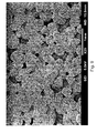
- FIG. 9 shows a scanning electron micrograph (SEM) of the material of a bone graft material containment structure according to one embodiment, showing an omnidirectional pore structure.
- This embodiment is a 0.25 mm thick sheet of MEDPOR shown at a magnification of 25 ⁇ (MEDPOR® manufactured by Porex Surgical Products Group (Newnan, Ga.).
- FIG. 10 shows two views of one embodiment of a bone graft material containment structure having a spacer feature.
- FIG. 10A is a view from above while FIG. 10B shows a cross section of the bone graft material containment structure having the spacer feature.
- FIGS. 11 A-E show the structure of FIG. 10 being shaped by a surgeon and being positioned in the patient.
- FIG. 12 shows a side view of one embodiment of a bone graft material containment structure having a cylindrical shape with an open lower portion.
- FIG. 13 is a schematic representation of a porous plastic containment sheet 130 located on two sides of a bone morphogenic protein containing media 132 (BMP media).
- BMP media bone morphogenic protein containing media
- FIG. 14 is a schematic representation of a porous plastic containment sheet 140 containing a metal mesh 141 , the sheet being located on two sides of a bone morphogenic protein containing media 142 (BMP media).
- BMP media bone morphogenic protein containing media
- Various embodiments of the present invention provide bone graft material containment structures designed to be used in connection with bone graft materials, including proteins and/or growth factors.
- the structures are designed to contain bone graft material in place, as well as maintain the tissue space shape and form required for the bone graft.
- the structures may be provided in sheets that can be cut and that have specific features to enable their use as bone graft material containment structures.
- the structures may be provided in a pre-formed shape and then further shaped or cut by a surgeon to a specific desired shape.
- the structures may be provided in semi-custom or custom forms.
- Bone repair is generally intended to treat defects due to trauma, congenital defects, malignant diseases that affect bone (such as various cancers), periodontal disease, or for a multitude of other reasons.
- the surgeon may wish to encourage the growth of new bone in addition to or instead of repairing the damaged bone. This may be the case with any type of bone repair surgery, for example, in the craniofacial area, the pelvis, the spine, the thorax, any of the long bones (such as the femur, tibia, humerus), or any smaller bones (such as the bones of the ankle, fingers, or toes).
- the devices and methods described herein may be used to contain any type of bone graft material against any bone of the skeleton, and particularly, the appendicular skeleton.
- one embodiment may be used in treating or repairing craniofacial bone that is deficient or missing, such as repair of a cranial bone defect or maxillofacial bone defect.
- Typical maxillofacial surgery applications include but are not limited to surgery to correct the mandible, maxilla, or alveolar ridge in order to establish new bone to support dental implants or dentures.
- Other applications may include treating spinal bone defects or spinal fusion procedures where bone is induced to grow between vertebrae.
- Further applications include treating comminuted fractures.
- Further applications include treatment of the bones of pelvis.
- facial applications will be discussed in more detail below, but it should be understood that the methods and devices described herein are not limited to these surgical uses.
- Embodiments of the invention relate to bone graft material containment structures designed to contain bone graft materials.
- Bone graft materials are known to one of ordinary skill in the art.
- Non-limiting examples of potential bone graft materials include autologous bone, autologous bone particulate, allogenic bone graft material (e.g., allogenic decellularized bone), human cadaver bone, xenograft bone graft material, animal bone, any type of bone chips or material, or synthetic materials such as hydroxyapatite, calcium phosphate (such as tricalcium phosphate, synthetic hydroxyapatite, or coralline hydroxyapatite), ceramics, bioactive glass, calcium sulfate, polymer-based bone graft substitutes, or growth factors that stimulate osteoblast precursors to proliferate, bone morphogenic proteins (BMP), BMP mimetics, recombinant human bone morphogenetic protein (rhBMP-2), platelet-rich plasma (PRP), transforming growth factor-bet
- the proteins and/or growth factors described may be used in addition to or in place of autologous, allogenic, xenograft, or synthetic bone graft materials.
- a protein and/or growth factor that has been adsorbed onto a sponge-like or soft material (referred to as a “treated sponge” throughout the remainder this application) may be used.
- the sponge-like or soft material may be any appropriate carrier that can support and adsorb a protein and/or growth factor, one non-limiting example of which includes a collagen sponge.
- the treated sponge may be treated with any type of material that is designed to encourage or stimulate bone growth.
- appropriate proteins and/or growth factors that may be used in connection with the embodiments described include the above-described bone graft materials, and specifically may include growth factors that stimulate osteoblast precursors to proliferate, bone morphogenic proteins (BMP), BMP mimetics, recombinant human bone morphogenetic protein (rhBMP-2), platelet-rich plasma (PRP), transforming growth factor-beta (TGF-beta), platelet-derived growth factor (PDGF), such as recombinant human platelet derived growth factor (rhPDGF), insulin-like growth factors, fibroblast growth factors (FGF), xenographic bone proteins, growth differentiation factor (e.g., GDF5), calcitonin, calcitonin mimetics, Curasan Cerasorb®, Medtronic INFUSE® Bone Graft (Medtronic, Minneapolis, Minn.), marrow cells containing mesenchymal stem
- the improved bone graft material containment structures and methods described herein are particularly designed to retain bone graft materials and to maintain the space where bone is to be restored during the bone healing process.
- the material of the structure should be biocompatible, and may be any appropriate tissue-integrating or porous polymer, or other biocompatible material.
- biocompatible polymers that have interconnected pore structures, thermoplastic resins, various types of polyethylenes (such as high density polyethylene), ultra high molecular weight polyethylene (UHMWPE), polyolefins, polyether ether ketone (PEEK), polyethylene terephthalate (PETE), nylon, polypropylene, or any polymer of aliphatic hydrocarbons containing one or more double bonds, composites of any of the above materials, or any other appropriate porous material that can be bent or otherwise formed into a desired shape.
- the containment structure is made of high density polyethylene.
- the containment structure is made of ultra high molecular weight polyethylene.
- the containment structure is made of polypropylene.
- Biocompatible metals include but are not limited to surgical grade stainless steel, tantalum and titanium. In one embodiment, these metals may be sintered to make the porous bone graft material containment structures. In another embodiment, multiple layers of metal mesh or metal wire are sintered together to make the porous bone graft material containment structures.
- the biocompatible containment structures may be made of non-woven or woven webs made from polyethylene, HDPE, UHMWPE, polypropylene, PEEK or nylon. These biocompatible containment structures have a porosity (also called openness) between about 20 to about 60 percent and similar pore size ranges to other biocompatible containment structures described herein.
- the structure is substantially or relatively smooth on the outside surface, such that the surface helps prevent snagging of soft tissue over the structure, tissue erosion, or irritation to the patient.
- the smoothness also contributes to relative external invisibility post-healing.
- the material of the structure has pores that are sized to allow vascular access to the bone healing site, but small enough to maintain the bone graft material in place against the defect to be treated, and have generally an interconnected open pore structure, generally consisting of various pore sizes within a desired size range distributed throughout the material.
- this pore structure is omnidirectional and/or multidimensional.
- the term multidimensional signifies pores of different sizes within a desired size range.
- the term omnidirectional signifies that the pores are oriented in many directions within the porous material. An example is provided in FIG. 9 .
- the structure is formed of a material having a porous structure, with pores in the range of about 40 to about 1000 microns in average diameter.
- the pores may range from about 100 to about 800 microns in average diameter. In another embodiment, the pores may range from about 100 to about 500 microns in average diameter. In another embodiment, the pores may range from about 50 to about 500 microns in average diameter. In another embodiment, the pores may range from about 60 to about 400 microns in average diameter. In another embodiment, the pores may range from about 200 to about 400 microns in average diameter. In a further embodiment, the pores may be generally between about 100 to about 200 microns in average diameter. In an even further embodiment, the pores may be generally above about 100 microns. In an even further embodiment, the pores may be generally about 200 microns. In an even further embodiment, the pore sizes are generally about 60 microns or above. Generally, pore sizes above about 60 microns allow blood vessels to grow into and through the material. It is contemplated that any numerical pore size or pore size range within these stated ranges can be used.
- pore size there may be variation in pore size, such that some pores are as small as about 10 or 5 microns, with other pores being much larger.
- the averages given above are intended to be examples of various average pore size ranges throughout the material. Due to the interconnected pore structure of the desired material used, the pore sizes are somewhat irregular, providing a number of variously sized surfaces to encourage fibrovascular ingrowth and integration.
- MEDPOR® manufactured by Porex Surgical Products Group (Newnan, Ga.) and is shown in FIG. 9 .
- proteins and/or growth factors such as BMP
- bone proteins supplied by graft materials such as cancellous bone from the patient's body, or from some external source like bone allograft
- graft materials such as cancellous bone from the patient's body, or from some external source like bone allograft
- Guided tissue regeneration using barrier materials may be sufficient for small defects in direct contact with freshly bleeding bone, however, larger defects need a blood supply through the bone graft containment material.
- the pore sizes of the bone graft material containment structures described herein to be adequate to allow fibrovascular infiltration and vascularization of the bone graft material contained within the bone graft material containment structure.
- the pores are also typically provided in an interconnected omnidirectional pore structure, an example of which is shown in FIG. 9 .
- This is a scanning electron micrograph of a 0.25 mm thick sheet of MEDPOR shown at a magnification of 50 ⁇ (MEDPOR® manufactured by Porex Surgical Products Group (Newnan, Ga.).
- this pore structure provides high internal surface area and high pore volume relative to the pore size.
- various embodiments of the present invention provide a pore structure that is irregular and omnidirectional. This provides many surfaces for vascular tissue ingrowth through the bone graft material containment structure and into the graft site.
- the interconnecting porous structure provides a number of benefits.
- the pore structure conducts fibrovascular tissue to the bone regeneration site by way of its large surface area and accommodating pore size.
- Second, the larger surface area associated with this pore structure is a benefit over the bone induction trays of the prior art.
- the Leake tray shows large holes through the tray that will allow fibrovascular tissue to access the bone material
- the interconnected omnidirectional pore structure described and shown herein provides more surfaces to direct the fibrovascular tissue to the bone graft site. Cells need a surface on which to grow.
- the greater surface area provided by the omnidirectional pores of the described bone graft material containment structures provides additional surfaces for cell proliferation and vascular ingrowth as compared to the prior art designs.
- the described irregular open pore structure also provides for tissue integration, or tissue attachment and stabilization relative to the containment material.
- This tissue integration minimizes movement of the overlying tissue during the initial healing phase and thereafter.
- the tissue integration is beneficial to rapid healing because movement of the tissue relative to the graft containment material can disrupt the formation of new vessels and lead to scar tissue, minimal vascularization of the bone graft material and tissue breakdown over the graft.
- the bone graft material containment structures provided are also rigid or inherently stiff enough to prevent collapse due to tension from the overlying soft tissue closure or scar contraction. At the same time, they are also thin enough to have sufficient flexibility to be adapted or bent to fit the specific surgical site for the purpose of holding the space open for a bone graft material, while allowing rapid vascular access from overlying soft tissue to the bone regeneration site through the porous structure.
- the general function of the structure is to contain and support bone graft material.
- one type of bone graft material that may need a specific support is a treated sponge, such as a collagen/BMP sponge.
- a bone graft material such as beta tricalcium phosphate granules require containment at a desired site.
- the bone graft material containment structures are not intended to provide the rigidity necessary to support the mandible during the healing phase, as some of the prior art trays described above must do.
- a titanium plate is needed to stabilize the entire reconstructed area of the bone during the healing process, as shown by FIG. 4 .
- the prior art bone induction trays described above are designed to have an initial rigidity similar to the mandible bone itself and must be thick enough to provide that structural rigidity.
- the bone graft material containment structures are formed of a sheet material having a thickness of about 0.1 to about 3.0 mm, about 0.1 to about 2.0 mm, about 0.1 to about 1.0 mm, about 0.1 to about 0.9 mm, about 0.1 to about 0.8 mm and in another embodiment, a thickness of about 0.2 to about 0.4 mm. In another embodiment, the thickness is about 0.2 to about 0.3 mm, and in another embodiment, the thickness is about 0.25 mm. It is contemplated that any numerical thickness within these stated ranges can be used. In some cases the material may be thin in some areas and thicker in other areas to confer additional strength to the material where needed.
- the porous material in a very thin design, for example, between about 0.1 to about 1.0 mm thick, and in more specific embodiments, about 0.2 to about 0.5 mm thick, and by providing an open omnidirectional pore structure with pores that are sized in the above-described ranges, the desired fibrovascular ingrowth and blood vessel ingrowth can be obtained.
- the irregular morphology of the pores, as well as their sizes provide more surfaces to direct fibrovascular ingrowth and thus vascular supply into the bone graft material containment structure, while still supplying the space maintenance feature to keep the bone graft material in the desired shape and location to establish new bone.
- the sheet may be provided in a general approximate shape and size of the anatomical region of use.
- the material can then be cut to size by the surgeon, bent by hand to cover a surgical site containing bone graft material, and, if necessary fixed into the site with surgical tacks, screws, sutures or other means.
- One specific example of a material that may be used to form the bone graft material containment structures described herein is MEDPOR® Biomaterial provided by Porex Surgical Products Group (Newnan, Ga., USA).
- the MEDPOR® surgical implants are manufactured from linear high-density polyethylene.
- the material allows for tissue ingrowth because of its interconnecting open pore structure, an example of which is shown in the attached FIG. 9 .
- the pores are provided in an omnidirectional interconnecting pore structure, with an average pore size greater than about 100 microns.
- the pore volume may be greater than about 10%, greater than about 15%, greater than about 20%, or greater than about 30%.
- the pore volume may range from about 10% to about 60%, from about 20% to about 60%, from about 30% to about 60%, or from about 40% to about 50%. In some embodiments, the pore volume may be about 50% in further embodiments.
- assessments of pore volume may be made by one of ordinary skill in the art using mercury intrusion porosimetry measurements or using direct measurements of pores in micrographs, such as SEMs.
- the bone graft containment material has a thickness of about 0.25 mm, about 0.35 mm, about 0.45 mm, or about 0.85 mm, all with average pore sizes of about 200 microns.
- larger through holes of approximately 1 mm to 2 mm diameter may be added to allow faster vascularization into the underlying bone graft material. These through holes would not replace the porosity of the omnidirectional open pore structure of the rest of the containment structure.
- the containment structure has metal or other reinforcement in mesh or rebar form.
- the porous structure has metal wires embedded in a polymer structure for reinforcement, or short elongated metal strands located throughout the structure to provide additional support and help the material hold its shape when bent.
- the containment structure has collagen integrated into the porous containment structure or onto the surfaces of the containment structure.
- the collagen attached to the surfaces of the containment structure can serve as a carrier for growth factors such as rhBMP.
- the attached collagen includes a porous collagen layer approximately 0.1 to 1.0 mm thick on the soft tissue side of the containment structure. This porous collagen layer provides a soft but stable support structure for fibrovascular ingrowth into the bone graft material, while adding bulk to the overlying tissue, and further minimizing the chance for erosion of the containment structure through the overlying tissue.
- Collagen may be integrated into or onto the porous containment structure using methods known to one of ordinary skill in the art, including preparing a suspension of collagen fibers and filtering the collagen fibers through the porous containment structure followed by drying. Different forms of collagen may be used.
- MEDPOR® surgical implants may be manufactured in a wide variety of shapes and sizes for use as bone graft material containment structures for reconstructive surgery.
- the material for containment structures may be provided as sheets or as pre-formed shapes intended to align with the desired bone contour.
- the properties of the MEDPOR® material allow it to be cut and trimmed while maintaining the interconnectivity and the structure of the pores. They may also be provided in custom or semi-custom shapes.
- FIG. 3 shows an embodiment in which the structure is preformed in a tent-like shape to hold bone graft material in place against the defect to be treated.
- FIG. 7 shows an embodiment in which the structure is hinged or creased at the top to provide a container or crib for the desired material.
- another potential embodiment is to leave out the “lid” from FIG. 7 and provide the bone graft material containment structure in a trough-like or U shape.
- FIG. 12 shows an embodiment in which the structure is cylindrical with an open end in order to contain bone material within the cylinder and allow the material to contact bone at the bottom of the cylinder.
- the structures are also generally intended to be provided in various lengths and sizes. In one embodiment, the structures are shaped like a tooth-whitening tray, which can be used to treat a patient with periodontal disease.
- the structures may alternatively be provided as a sheet-like material that can be secured to existing bone and wrapped around or spanned over the bone graft material and secured to a second surface to create, for example, a sling-like or bridge-like design.
- FIGS. 11 and 13 show an embodiment in which the bone graft material containment material is provided as a sheet-like form.
- the structures may also be provided having a spacer bar, as illustrated in FIGS. 10 and 11 , described further below.
- FIG. 1 shows a normal, healthy mandible (without teeth).
- FIG. 2 shows a diseased or damaged mandible, which could occur, for example, from bone resorption, excision of diseased bone, a trauma to the face, or for any other reason.
- the arrows in FIG. 2 indicate areas of bone loss.
- FIG. 3 shows one embodiment of a bone graft material containment structure 32 that is provided in a tent-like shape to hold bone graft material 34 .
- the structure 32 has side portions, a top portion and an interior portion into which bone graft material 34 is placed as indicated by the arrow.
- FIG. 4 shows the bone graft material containment structure 42 containing the bone graft material (not shown) located in position on the mandible.
- FIG. 5 shows a normal, healthy mandible (with teeth).
- FIG. 6 illustrates a mandible having a portion of the bone removed (arrow), for example, due to excision of a tumor or removal of cancerous bone, a congenital defect, or for any other reason.
- FIG. 7 shows one embodiment of a bone graft material containment structure that is provided in a crib-like shape 70 to hold bone graft material.
- the structure has a lower portion 72 , side portions 74 , a top “lid” portion 76 , and an interior portion 77 into which bone graft material is placed.
- the structure is shown having rounded sides, but it may be provided with more angled sides if desired, depending upon the intended use.
- FIG. 8 shows a side view of the bone graft material containment structure that is provided in a crib-like shape shown in FIG. 7 in place in a patient's mandible, along with a bone plate stabilizer 82 to stabilize the bone during healing.
- dental implants can be placed in the newly formed bone.
- the bone plate (such as a titanium bone plate with openings 82 ) may be secured into place in order to bridge the missing portion of the mandible.
- the structure may then be sutured, tacked, screwed, or otherwise positioned into place against the portion of the patient's bone defect to be treated.
- Soft tissue may be closed over the bone graft material containment structure.
- the bone graft material containment structure may be provided as a bone graft containment sheet that accommodates an upper tack portion, a middle wrap portion, and a lower tack portion which can be shaped by the surgeon by affixing it to the bone or soft tissues at the desired site. Following the fixation of one side of the sheet, the bone graft material may be added to the sheet which wraps around it and the second edge of the sheet is then affixed into place.
- the bone graft material containment structure may be provided as a bone graft containment sheet having a predetermined shape that accommodates an upper tack portion, a middle wrap portion, and a lower tack portion.
- the structure is provided pre-formed into the shape of various potential and expected bony defect sites.
- the compound curves of these specific shapes may lend additional stiffness to the thin, preformed shape.
- examples of such shapes may be tented shapes (for example, to cover and augment the alveolar ridge), trough shapes (for example, to support collagen sponge material to replace missing portions of the mandible), and anatomic shapes to augment craniofacial bone.
- the bone graft material containment structures in forms that are shapes such as cylindrical, tubular, rectangular, square, ellipsoidal, box-shaped (e.g., a flat box or double sided thin box for cranial defects), or in the anatomical shape of any area of bone to be replaced or augmented in the body, or any other appropriate shape in order to hold and contain bone graft material in the desired location, such as against a bone defect to be treated to encourage new bone growth. It is also envisioned that the bone graft material containment structures may be custom shaped.
- FIG. 10 shows an example of a bone graft material containment structure that has a spacer portion.
- FIG. 10A provides a view from above.
- FIG. 10B shows a cross section.
- the spacer portion design is essentially a bone graft material containment sheet with a “space bar” 100 on one end of the sheet 102 that is provided in order to maintain space on the maxilla or mandible using the technique described above for augmenting bone on the mandible or maxilla. It is usually placed on the labial side of the bone and allows the surgeon to create extra room for bone formation to support the dental implant.
- the arrow in FIG. 10B shows the side of the sheet which is applied to the bone.
- FIG. 11 illustrates the space bar 110 having its edges 112 , 114 cut away or shaved (e.g., with a scalpel) at an angle (or “feathered”) so that there is a raised area in the middle portion 116 with wedge-shaped shaved edges, as shown in FIGS. 11C-D .
- This raised area raises the rest of the sheet up against the desired surface in order to provide additional space underneath the sheet (between the bone and the sheet).
- the raised area 110 holds the sheet away from the bone so that more bone graft material can be inserted underneath the sheet.
- the sides of the sheet portion 116 (the portions below the area where a wedge-shaped portion of material has been removed) can then be pressed or otherwise secured down to contain the bone graft material in a bed-like area in the desired location 118 .
- the raised area acts as a “raised pillow” and the remaining lower portion of the sheet 117 provides a “cover” for the bone graft material.
- this embodiment gives the surgeon the option of encouraging bone growth on a labial surface of the bone 119 , as shown for example in FIG. 11E .
- the space bar portion may have the above-described pore structures or it may be more solid-like.
- the step of the space bar portion may be about 2 mm to about 3 mm, although any dimensions are possible, depending upon the specific use and anatomy to be treated.
- FIG. 12 shows one embodiment of a bone graft material containment structure that is provided in a cylindrical-like shape to hold bone graft material.
- the structure is shown as having rounded sides 120 , a closed upper portion 122 , and an open lower portion 124 .
- the closed upper portion 122 may be provided as a lid that may be opened and optionally secured closed, or that it may be provided pre-sealed.
- Bone graft material may be placed against the bone in the open end. This embodiment can be particularly useful for encouraging bone growth in the oral cavity, e.g., in the location of a missing tooth.
- Options for treating other bones include bone cages for spinal fusion procedures, tubular or semitubular shapes for appendicular defects, (long bones), and bone/implant interface areas such as for artificial joints.
- Other options include bone graft material containment shells for cosmetic or reconstructive bone onlay grafts such as for the midface, chin, nose, or periorbital area.
- Embodiments may also be used for repair of craniotomy defects, full or partial thickness defects of the cranium due to trauma, cancer resection, or lost bone flaps after procedures on the brain.
- the structure is molded into a shape to augment any desired bone, for example the cranium, craniofacial bones, the spine, the scapula, the ribs, the humerus, the radius, the ulna, bones of the wrist, the phalanges, the pelvis, the coccyx, the femur, the tibia, the fibula, bones of the foot or ankle, or any other bone.
- any desired bone for example the cranium, craniofacial bones, the spine, the scapula, the ribs, the humerus, the radius, the ulna, bones of the wrist, the phalanges, the pelvis, the coccyx, the femur, the tibia, the fibula, bones of the foot or ankle, or any other bone.
- the structure is molded into a shape to augment existing craniofacial bone, including but not limited to the mandible, maxilla, palatine bone, zygoma or the frontal, parietal, occipital or temporal bones.
- the material may be molded into a shape to contain bone graft material over or within a bony defect of the long bones, such as for a poorly healing or comminuted fracture.
- the material may be molded to the shape of a particular defect for a particular patient, utilizing CT or MRI data or a physical impression of the defect area.
- the sheet or molded shapes have attached extensions or spacer bars to hold the sheet or molded shape away from the existing bone surface, in order to provide additional space for the bone graft material to establish new bone.
- thicker ribs of thermoplastic or another material may be incorporated into the material for added strength.
- stiffening agents are provided to increase the strength, such as biocompatible fibers or metal rods inside the polymer material, outside the polymer material, and/or both.
- Stiffening agents may include titanium wire, titanium rods, biocompatible polymer solid rods or fibers, polymer mesh, or any other appropriate stiffening materials.
- a metal mesh may be molded into the material so that the porous material surrounds the metal mesh and fills the interstices of the metal mesh or plate to provide support to the bony defect area while the bone heals.
- An example of molding of a porous material over a metal mesh is described in U.S. Pat. No. 7,655,047 and in co-pending application Ser. No. 11/445,560.
- An alternate method for manufacturing various implants is to coin curves into a flat sheet (such as a flat polyethylene sintered sheet) for improved anatomic shapes. This is particularly useful because it may be easier to make material as a flat sheet rather than manufacture it as a curved design.
- the bone graft material containment structures are provided as pre-shaped structures, they may be pre-curved or pre-shaped so that the structures more accurately fit the anatomy of the site where the stimulation of bone growth is desired. Even though the implants are typically malleable by hand due to their thin nature, structures that are pre-shaped and hold the intended pre-shaped design are included within the scope of this description. This can help reduce operating room time, because the structure is already shaped appropriately.
- the parameters of the designs described herein may be configured and/or modified depending upon the use of the implant. Specifically, the thickness of the material, the stiffness of the material, the type of material, and/or the porosity of the material may be adjusted depending upon the particular desired use or application. For example, a smaller defect may call for a structure having a thinner material than a larger defect.
- a surgical site was prepared to receive a bone graft material containment sheet.
- the bone graft material containment sheet was a 0.25 mm thick sheet of MEDPOR® material (Porex Surgical, Newnan, Ga.) having an average pore size of about 200 microns, and a porosity of about 40%.
- the sheet was cut to the desired size and tacked to the maxilla.
- the upper portion of the sheet can also be provided in a T-like shape to provide more surface area for the tacks to be placed.
- the remainder of the sheet was left long enough so that it wrapped around the bone graft material to be placed inside the shaped sheet.
- the bone graft material was Medtronic INFUSE® Bone Graft material (which is rhBMP absorbed onto collagen sponge material, Medtronic, Minneapolis, Minn.) mixed with autologous bone chips. Other appropriate bone graft material may be used, including but not limited to those described above, as well as various combinations of such materials.
- the bone graft material was placed in the defect site.
- the bone graft material containment sheet was wrapped around the bone graft material and secured to the lingual surface of the maxilla by tucking it between the periosteum and bone. The bone graft material containment sheet was tacked into position again.
- the soft tissue closure over the sheet along with its inherent rigidity may all that is needed to hold the bone graft material containment sheet in place.
- the gingival tissues were closed over the bone graft material containment sheet with sutures. Examination of the site two weeks after surgery demonstrated good retention of the bone graft material. Examination of the site two months after surgery showed tissue growth into the undersurface of the bone graft material containment sheet.
- Portions of the sheet may be removed from the implant, but the remaining sheet is tissue integrated and does not have to be removed, eliminating any harmful stripping of the vascular supply from the regenerated bone. Later, the crestal portion of the sheet was removed, a temporary gingival abutment was inserted, the gingival tissue was closed around the temporary abutment with sutures. Most of the sheet implant remained in place under the gingival tissue, where it was now integrated with the tissue. The sheet implant remained invisible under the tissue and did not disrupt the gingival tissue or detrimentally affect its aesthetic appearance. The fibrovascular tissue that grew through the porous implant supplied blood to the underlying bone graft.
- the bone graft material containment sheet contained the bone graft material in the desired defect site in the maxilla so that new bone was formed. This new bone was used to help stabilize a previously placed prosthesis for insertion of a dental implant.
- the bone was deficient in both height and width in the area where dental implants would be placed. This was called a knife edge ridge because the bone was so thin in this area.
- the oral surgeon wanted to increase bone width and height in this area.
- Soft tissues were incised and dissected away from the underlying bone.
- rhBMP/collagen sponge and synthetic bone graft material called C graft were used to graft new bone around the knife edge ridge.
- C graft is a brand name for a synthetic bone graft material made from a marine algae skeleton that has been converted to hydroxyapatite through chemical processing (Algisorb, Osseous Technologies of America, Newport Beach Calif.
- C graft was mixed with the collagen sponge and rhBMP to provide extra bulk and a “scaffold” for new bone growth.
- the oral surgeon cut the sponge of the BMP sponge graft into small pieces and then mixed it with the C graft and used it for the grafted area.
- Other materials that may be used in this way are autologous bone chips, cadaver bone, xenographic bone particulates or other synthetic bone graft materials, among others.
- a piece of 0.45 mm thick MEDPOR porous sheet was used to contain the graft mixture and hold it in place. Surgical tacks were placed at the superior edges of the sheet just under the retracted soft tissue.
- This graft was placed in conjunction with a sinus lift procedure. An opening was made in the bone above the upper teeth, the open maxillary sinus was entered with dissection of the mucous membrane in the sinus to lift it up and away from the thin bone over the teeth, and then bone graft material was placed in the area to thicken the bone over the missing teeth. The soft tissue was sutured over the graft site and allowed to heal (not shown). The site was inspected and two months later, bone was formed.
- the gingiva was dissected away from the bone. There was a deficiency of bone on the anterior surface and the ridge where the tooth was missing.
- the oral surgeon planned to place a dental implant into the bone in this area, but the bone was not wide enough to support an implant.
- Recombinant human BMP (rhBMP) was adsorbed to an acellular collagen sponge used as a bone graft material in the site of missing bone.
- An omnidirectional interconnected open pore structure sheet of MEDPOR (0.25 mm in thickness) was placed over the bone graft material. The gingiva was then closed with suture over the end of the implant and the graft site.
- the bone was deficient in both height and width in the area where dental implants would be placed. This was called a knife edge ridge because the bone was so thin in this area.
- the oral surgeon wanted to increase bone width and height in this area.
- Soft tissues were incised and dissected away from the underlying bone.
- rhBMP/collagen sponge and synthetic bone graft material called C graft were used to graft new bone around the knife edge ridge.
- C graft was mixed with the collagen sponge and rhBMP to provide extra bulk and a scaffold for new bone growth.
- the oral surgeon cut the sponge of the BMP sponge graft into small pieces and then mixed it with the C graft and used it for the grafted area.
- Other materials that may be used in this way are autologous bone chips, cadaver bone, xenographic bone particulates or other synthetic bone graft materials, among others.
- a piece of 0.45 mm thick MEDPOR porous sheet was used to contain the graft mixture and hold it in place. Surgical tacks were placed at the superior edges of the sheet just under the retracted soft tissue. The soft tissue was sutured over the graft site and allowed to heal (not shown). The site was inspected and two months later, bone was formed.
- the tibial bone is deficient in one region.
- the orthopedic surgeon decides to augment the bone in this deficient region to facilitate healing and strengthening of the bone.
- the surgeon obtains a sheet of the bone containment structure of the present invention, in this case a sheet of MEDPOR.
- the surgeon grafts the defect with the bone growth material and covers it with a sheet of MEDPOR CONTAIN Mesh which is then screwed or tacked to the bone to prevent displacement of the graft.
- the bone graft material is located between the bone and the MEDPOR sheet. Radiographs are taken several weeks later and show new bone formation in the tibia where the bone growth material was contained by the MEDPOR sheet.
- the tibial bone is deficient in one region.
- the orthopedic surgeon decides to augment the bone in this deficient region to facilitate healing and strengthening of the bone.
- the surgeon obtains a sheet of the bone containment structure of the present invention, in this case a sheet of PEEK.
- the surgeon obtains a bone growth material and applies it to the sheet of PEEK which is then placed on the bone and secured into place with screws or tacks.
- the bone graft material is located between the bone and the PEEK sheet. Radiographs are taken several weeks later and show new bone formation in the tibia where the bone growth material was contained by the PEEK sheet.
- a patient presents in a dental office requesting insertion of an artificial tooth into the mandible.
- the dentist examines the socket and obtains radiographs of the site. The dentist concludes that the site has inadequate bone to support the insertion of the artificial tooth.
- the dentist refers the patient to an oral surgeon who obtains a preformed cylinder with one open end and one closed end of MEDPOR Implant material with wall thickness of approximately 0.3 mm and approximately 40-50% pore volume, and approximately 200 micron pore size.
- the oral surgeon fills the socket and the cylinder with bone graft material comprising beta tricalcium phosphate mixed with platelet rich plasma, places the cylinder over the socket and leaves additional sheet material in the form of small flaps in order to attach the flaps to the mandible with tacks or screws.
- Radiographs taken four weeks later show increased bone density in the tooth socket adjacent to the mandible where the bone graft material is contained by the MEDPOR cylinder. Later, the oral surgeon inserts a dental implant into the newly formed bone. The dental implant is more stable due to the increased bone density.
- a patient with a long history of smoking presents to an orthopedic surgeon with pain radiating down his upper extremities.
- the surgeon obtains radiographs and observes osteoporosis of several cervical vertebrae and degeneration of the intervertebral discs with resulting compression of cervical spinal nerves.
- the surgeon recommends spinal fusion of the C5 and C6 cervical vertebrae but is concerned that the osteoporosis may have caused sufficient bone loss to create problems during the spinal fusion and after.
- the surgeon obtains several strips of MEDPOR sheets and applies them to both lamina of each vertebrae, adhering each strip on three sides with tacks and glue.
- the strips span the intervertebral space.
- the surgeon inserts a syringe containing a mixture of rhBMP/acellular collagen sponge mixed with hydroxyapatite under the unsecured side of each strip and infuses the mixture into the space between the strip and the lamina of each vertebrae and also between the strip and the intervertebral disc to promote bone growth and improve spinal fusion.
- the unsecured side is then secured with tacks and glue.
- Radiographs taken months later show enhanced bone formation at C5, C6 and in the intervertebral space between C5 and C6.
- Physical examination reveals decreased vertebral rotation in the C5 and C6 area and reduced pain in the upper extremity.
- a patient presents with craniofacial trauma resulting in missing bone in the areas of the inferior orbital rim and lateral orbital rim.
- the surgeon takes a bone graft from the outer table of the patient's calvarium to use in repair of the defect.
- the donor site which consists of the inner table of the calvarium without the outer table, is filled with rhBMP/ACS.
- An interconnected open pore structure material made from laser sintered titanium particles is placed over the outer table defect and the underlying graft material and secured to the outer table with screws.
- the laser sintered porous material is fabricated in the curvature of the cranium in the area of the defect to closely fit the contour of the cranium.
- the periosteum and soft tissues are closed over the porous structure.
- the porous structure prevents the soft tissue from retracting into the defect area which would collapse the rhBMP/ACS graft material.
- a patient presents with a crush injury to a finger in which one phalanx is completely crushed with comminuted bone but the joints are preserved.
- the surgeon wraps an omnidirectional interconnected open pore structure plastic sheet with embedded titanium mesh around the comminuted bone fragments, adding bone marrow aspirate from the iliac bone, and secures the ends of the formed cylinder to the edges of the joint area with titanium screws.
- Soft tissues are closed over the construct and it is splinted during initial healing. Radiographs taken at 1 and 2 months demonstrate increased bone density at the crush site.
- the surgeon has a prefabricated thin shell of omnidirectional interconnected open pore structure polyethylene material made to the shape of the contralateral mandible outer surface.
- the surgeon exposes the deficient mandible outer surface, drills multiple holes through the outer table of the mandible surface to access bleeding cancellous bone, fills the porous shell with a mixture of rhBMP/ACS and mineralized freeze dried bone particles, and secures the shell to the outer surface of the deficient mandible with low profile craniofacial screws.
- Soft tissues are closed over the site to allow healing. Radiographs taken at 1 and 2 months demonstrate increased bone density at the right mandibular angle.
- a patient presents with a full thickness calvarial defect in the temporal-parietal area due to benign cyst excision.
- the surgeon exposes the defect and covers the dura with a thin omnidirectional interconnected open pore structure sheet of polypropylene with a barrier surface adjacent to the dura.
- Bone graft material consisting of cancellous bone from the iliac crest mixed with rhBMP/ACS is placed to fill the defect, and covered with a thin omnidirectional interconnected open pore structure sheet of polypropylene, which has been reinforced with embedded titanium mesh.
- the outer sheet is fixed to the outer table of the calvarium with craniofacial screws. The overlying tissues are closed. Radiographs taken at 2 and 3 months demonstrate increased bone density at the site of the calvarial defect.
- a continuity defect of the mandible is created due to excision of osseonecrotic tissue of the mandible.
- the defect is from the mandible angle to the mentum.
- CNC Computer numeric controlled
- Polypropylene mesh is tightly wrapped around the mandible shape in multiple layers to create an omnidirectional interconnected pore structure due to the multiple layers mesh built up on the metal mandible form, overlapping the edges of the defect area.
- the entire construct is placed in an oven and heated to sintering temperature to fuse the layers of the mesh together without disrupting the porous structure.
- the metal form is cut near one edge of the defect, and the omnidirectional interconnected open pore structure is slipped off the metal mold.
- the structure is packaged and sterilized with ethylene oxide.
- the structure is filled with a mixture of rhBMP/acellular collagen sponge, cancellous bone from the patient's iliac bone, and marine algae-derived particulate hydroxyapatite particles.
- the construct with the bone graft material is inserted into place, and the edges are trimmed as needed and slipped over the ends of the remaining mandible segments.
- a titanium mandible reconstruction plate is placed over the entire construct to achieve rigid fixation during healing. The plate is removed approximately 6 months later after the new bone has formed.
Landscapes
- Health & Medical Sciences (AREA)
- Life Sciences & Earth Sciences (AREA)
- Chemical & Material Sciences (AREA)
- Transplantation (AREA)
- Oral & Maxillofacial Surgery (AREA)
- Veterinary Medicine (AREA)
- Public Health (AREA)
- General Health & Medical Sciences (AREA)
- Animal Behavior & Ethology (AREA)
- Medicinal Chemistry (AREA)
- Engineering & Computer Science (AREA)
- Biomedical Technology (AREA)
- Epidemiology (AREA)
- Dermatology (AREA)
- Chemical Kinetics & Catalysis (AREA)
- Orthopedic Medicine & Surgery (AREA)
- Botany (AREA)
- Vascular Medicine (AREA)
- Cardiology (AREA)
- Zoology (AREA)
- Heart & Thoracic Surgery (AREA)
- Molecular Biology (AREA)
- Plastic & Reconstructive Surgery (AREA)
- Cell Biology (AREA)
- Dispersion Chemistry (AREA)
- Urology & Nephrology (AREA)
- Inorganic Chemistry (AREA)
- Prostheses (AREA)
- Materials For Medical Uses (AREA)
- Polymers & Plastics (AREA)
- Organic Chemistry (AREA)
Abstract
Description
Claims (21)
Priority Applications (1)
| Application Number | Priority Date | Filing Date | Title |
|---|---|---|---|
| US12/712,949 US9433707B2 (en) | 2009-02-25 | 2010-02-25 | Bone graft material containment structures |
Applications Claiming Priority (2)
| Application Number | Priority Date | Filing Date | Title |
|---|---|---|---|
| US20853909P | 2009-02-25 | 2009-02-25 | |
| US12/712,949 US9433707B2 (en) | 2009-02-25 | 2010-02-25 | Bone graft material containment structures |
Publications (2)
| Publication Number | Publication Date |
|---|---|
| US20100215718A1 US20100215718A1 (en) | 2010-08-26 |
| US9433707B2 true US9433707B2 (en) | 2016-09-06 |
Family
ID=42238253
Family Applications (1)
| Application Number | Title | Priority Date | Filing Date |
|---|---|---|---|
| US12/712,949 Active 2032-12-18 US9433707B2 (en) | 2009-02-25 | 2010-02-25 | Bone graft material containment structures |
Country Status (3)
| Country | Link |
|---|---|
| US (1) | US9433707B2 (en) |
| EP (1) | EP2401000A2 (en) |
| WO (1) | WO2010099333A2 (en) |
Cited By (22)
| Publication number | Priority date | Publication date | Assignee | Title |
|---|---|---|---|---|
| US10183442B1 (en) | 2018-03-02 | 2019-01-22 | Additive Device, Inc. | Medical devices and methods for producing the same |
| USD870888S1 (en) | 2018-03-02 | 2019-12-24 | Restor3D, Inc. | Accordion airway stent |
| USD870889S1 (en) | 2018-03-02 | 2019-12-24 | Restor3D, Inc. | Cutout airway stent |
| USD870890S1 (en) | 2018-03-02 | 2019-12-24 | Restor3D, Inc. | Spiral airway stent |
| USD871577S1 (en) | 2018-03-02 | 2019-12-31 | Restor3D, Inc. | Studded airway stent |
| US20200281725A1 (en) * | 2011-02-28 | 2020-09-10 | DePuy Synthes Products, Inc. | Modular tissue scaffolds |
| US10772732B1 (en) | 2020-01-08 | 2020-09-15 | Restor3D, Inc. | Sheet based triply periodic minimal surface implants for promoting osseointegration and methods for producing same |
| WO2020205388A1 (en) | 2019-04-04 | 2020-10-08 | Dentsply Sirona Inc. | Custom dental membrane |
| US10889053B1 (en) | 2019-03-25 | 2021-01-12 | Restor3D, Inc. | Custom surgical devices and method for manufacturing the same |
| USD920516S1 (en) | 2020-01-08 | 2021-05-25 | Restor3D, Inc. | Osteotomy wedge |
| USD920517S1 (en) | 2020-01-08 | 2021-05-25 | Restor3D, Inc. | Osteotomy wedge |
| USD920515S1 (en) | 2020-01-08 | 2021-05-25 | Restor3D, Inc. | Spinal implant |
| US11213394B2 (en) | 2018-02-26 | 2022-01-04 | The Board Of Trustees Of The Leland Stanford Junior University | Graft for segmental bone defect repair |
| US11213403B2 (en) | 2019-03-14 | 2022-01-04 | Medos International Sarl | Devices and methods for optimized spinal fixation |
| US11364057B2 (en) | 2009-03-27 | 2022-06-21 | Spinal Elements, Inc. | Flanged interbody fusion device |
| US11369474B2 (en) | 2017-07-17 | 2022-06-28 | Warsaw Orthopedic, Inc. | Bone implant having a mesh |
| US11382769B2 (en) | 2018-09-20 | 2022-07-12 | Spinal Elements, Inc. | Spinal implant device |
| RU212720U1 (en) * | 2022-03-17 | 2022-08-03 | федеральное государственное автономное образовательное учреждение высшего образования Первый Московский государственный медицинский университет имени И.М. Сеченова Министерства здравоохранения Российской Федерации (Сеченовский Университет) (ФГАОУ ВО Первый МГМУ им. М.И. Сеченова Минздрава России (Се | Device for elimination of alveolar deformity in the area of tooth loss for any area of the jaw |
| US11806028B1 (en) | 2022-10-04 | 2023-11-07 | Restor3D, Inc. | Surgical guides and processes for producing and using the same |
| US11850144B1 (en) | 2022-09-28 | 2023-12-26 | Restor3D, Inc. | Ligament docking implants and processes for making and using same |
| US11911284B2 (en) | 2020-11-19 | 2024-02-27 | Spinal Elements, Inc. | Curved expandable interbody devices and deployment tools |
| US11960266B1 (en) | 2023-08-23 | 2024-04-16 | Restor3D, Inc. | Patient-specific medical devices and additive manufacturing processes for producing the same |
Families Citing this family (31)
| Publication number | Priority date | Publication date | Assignee | Title |
|---|---|---|---|---|
| WO2009004070A1 (en) * | 2007-07-03 | 2009-01-08 | Vlaamse Instelling Voor Technologisch Onderzoek (Vito) | Surgical implant composed of a porous core and a dense surface layer |
| US9861482B2 (en) | 2008-08-26 | 2018-01-09 | Andy Boiangiu | Dental bone implant and implant method |
| US9259320B2 (en) | 2008-08-26 | 2016-02-16 | Andy Boiangiu | Apparatus and method for bone augmentation |
| WO2010093955A1 (en) * | 2009-02-12 | 2010-08-19 | Osteotech,Inc. | Segmented delivery system |
| US8353967B2 (en) * | 2009-08-10 | 2013-01-15 | Osseous Technologies Of America | Self-supporting collagen tunnel for guided tissue regeneration and method of using same |
| AU2010328427B2 (en) | 2009-12-13 | 2014-06-05 | Advanced Biologics, Inc. | Bioactive grafts and composites |
| US8574302B2 (en) | 2010-12-20 | 2013-11-05 | Warsaw Orthopedic, Inc. | Bone augmentation device |
| ITRM20110168A1 (en) * | 2011-03-31 | 2012-10-01 | Alma Mater Studiorum Uni D I Bologna | SURGICAL DEVICE FOR THE RECONSTRUCTION OF BONE STRUCTURES. |
| US20140051771A1 (en) * | 2012-08-20 | 2014-02-20 | Ticona Llc | Biomedical Devices Comprising Molded Polyethylene Components |
| US20140052264A1 (en) * | 2012-08-20 | 2014-02-20 | Ticona Llc | Porous, Stabilized Craniomaxillofacial Implants and Methods and Kits Relating Thereto |
| DE202012009597U1 (en) * | 2012-10-06 | 2012-10-26 | Rosi Schneider | Prosthesis with nerve recording |
| US9427328B2 (en) | 2014-03-10 | 2016-08-30 | Warsaw Orthopedic, Inc. | Interbody implant system and method |
| WO2015138657A1 (en) * | 2014-03-11 | 2015-09-17 | Ohio State Innovation Foundation | Methods, devices, and manufacture of the devices for musculoskeletal reconstructive surgery |
| US9724198B2 (en) | 2014-07-17 | 2017-08-08 | Poriferous, LLC | Orbital floor sheet |
| AU2016200179B2 (en) * | 2015-01-14 | 2020-09-17 | Stryker European Operations Holdings Llc | Spinal implant with porous and solid surfaces |
| US10993810B2 (en) * | 2015-02-16 | 2021-05-04 | Cossington Limited | Containment body for a spacer device and method of making thereof |
| DE102015102597A1 (en) * | 2015-02-24 | 2016-08-25 | botiss biomaterials GmbH | Implant for covering bone defects in the jaw area and method for its production |
| WO2017010811A1 (en) * | 2015-07-13 | 2017-01-19 | 한양대학교 산학협력단 | Personalized alveolar bone tissue and method for manufacturing same |
| CN106267354A (en) * | 2016-09-30 | 2017-01-04 | 广州赛莱拉干细胞科技股份有限公司 | A kind of dental pulp mescenchymal stem cell preparation and its preparation method and application |
| WO2018085526A2 (en) * | 2016-11-03 | 2018-05-11 | DePuy Synthes Products, Inc. | Fold-up containment device for bone defects |
| WO2018107121A2 (en) * | 2016-12-08 | 2018-06-14 | Wang Chan Qian | A bone craft plug with an integrated collagen shell and methods thereof |
| CN106473787A (en) * | 2016-12-12 | 2017-03-08 | 运怡(北京)医疗器械有限公司 | Porous PEEK anchor nail |
| WO2018117266A1 (en) * | 2016-12-22 | 2018-06-28 | 株式会社ダイセル | Bone fixing material |
| EP3560530A4 (en) | 2016-12-22 | 2020-08-19 | Daicel Corporation | Blood filter |
| AU2018215359A1 (en) * | 2017-02-06 | 2019-08-01 | DePuy Synthes Products, Inc. | Bendable graft containment cage |
| US10064726B1 (en) * | 2017-04-18 | 2018-09-04 | Warsaw Orthopedic, Inc. | 3D printing of mesh implants for bone delivery |
| US10357367B2 (en) * | 2017-09-11 | 2019-07-23 | DePuy Synthes Products, Inc. | Patient-specific mandible graft cage |
| KR200489588Y1 (en) | 2017-09-20 | 2019-07-09 | 주식회사 티앤알바이오팹 | Surgical implant for transsphenoidal approach |
| US11491262B2 (en) * | 2018-04-19 | 2022-11-08 | Paulo Jose PASQUALI | Method of bone regeneration |
| US11607317B2 (en) * | 2019-09-19 | 2023-03-21 | Osteogenics Biomedical, Inc. | Modeling devices used in guided bone and tissue regeneration |
| CN111494061A (en) * | 2020-04-22 | 2020-08-07 | 西安交通大学 | Personalized mandible substitute compounded with autologous bone |
Citations (29)
| Publication number | Priority date | Publication date | Assignee | Title |
|---|---|---|---|---|
| US3849805A (en) | 1972-11-01 | 1974-11-26 | Attending Staff Ass Los Angele | Bone induction in an alloplastic tray |
| US4455256A (en) | 1981-05-05 | 1984-06-19 | The Regents Of The University Of California | Bone morphogenetic protein |
| US4472840A (en) | 1981-09-21 | 1984-09-25 | Jefferies Steven R | Method of inducing osseous formation by implanting bone graft material |
| US4636215A (en) | 1984-01-11 | 1987-01-13 | Rei, Inc. | Combination tray and condylar prosthesis for mandibular reconstruction and the like |
| US4693721A (en) * | 1984-10-17 | 1987-09-15 | Paul Ducheyne | Porous flexible metal fiber material for surgical implantation |
| US4976737A (en) | 1988-01-19 | 1990-12-11 | Research And Education Institute, Inc. | Bone reconstruction |
| US5380329A (en) | 1992-07-28 | 1995-01-10 | Dental Marketing Specialists, Inc. | Bone augmentation method and apparatus |
| US5989289A (en) * | 1995-10-16 | 1999-11-23 | Sdgi Holdings, Inc. | Bone grafts |
| US6031148A (en) | 1990-12-06 | 2000-02-29 | W. L. Gore & Associates, Inc. | Implantable bioabsorbable article |
| US6113640A (en) * | 1997-06-11 | 2000-09-05 | Bionx Implants Oy | Reconstructive bioabsorbable joint prosthesis |
| US6132214A (en) * | 1995-12-18 | 2000-10-17 | Jouko Suhonen | Medical implant |
| US6136029A (en) * | 1997-10-01 | 2000-10-24 | Phillips-Origen Ceramic Technology, Llc | Bone substitute materials |
| US6139574A (en) * | 1993-10-18 | 2000-10-31 | Children's Medical Center Corporation | Vascularized tissue regeneration matrices formed by solid free form fabrication techniques |
| US20010020188A1 (en) * | 1997-10-27 | 2001-09-06 | Tom Sander | Selective uptake of materials by bone implants |
| US20010049560A1 (en) * | 1998-01-30 | 2001-12-06 | Paul David C. | Intervertebral allograft spacer |
| US6409764B1 (en) | 1998-12-03 | 2002-06-25 | Charles F. White | Methods and articles for regenerating bone or peridontal tissue |
| US6530955B2 (en) * | 1999-06-08 | 2003-03-11 | Osteotech, Inc. | Ramp-shaped intervertebral implant |
| US20040167625A1 (en) * | 1999-01-27 | 2004-08-26 | Disc-O-Tech Orthopedic Technologies Inc. | Spacer filler |
| US20050043733A1 (en) * | 2001-02-28 | 2005-02-24 | Lukas Eisermann | Woven orthopedic implants |
| US20050214340A1 (en) * | 2004-02-03 | 2005-09-29 | Erbe Erik M | Pliable conformable bone restorative |
| US20050281856A1 (en) * | 2004-05-10 | 2005-12-22 | Mcglohorn Jonathan | Implantable biostructure comprising an osteoconductive member and an osteoinductive material |
| US20050288790A1 (en) * | 2003-04-16 | 2005-12-29 | Porex Surgical, Inc | Craniofacial implant |
| US6989029B2 (en) * | 1991-08-12 | 2006-01-24 | Bonutti Ip, Llc | Tissue cage |
| US7066962B2 (en) * | 2002-07-23 | 2006-06-27 | Porex Surgical, Inc. | Composite surgical implant made from macroporous synthetic resin and bioglass particles |
| US20060224242A1 (en) | 2003-04-16 | 2006-10-05 | Porex Surgical, Inc. | Craniofacial implant |
| US7192450B2 (en) | 2003-05-21 | 2007-03-20 | Dexcom, Inc. | Porous membranes for use with implantable devices |
| US20070129804A1 (en) * | 2005-12-07 | 2007-06-07 | Zimmer Spine, Inc. | Transforaminal lumbar interbody fusion spacers |
| CN101134118A (en) | 2007-07-13 | 2008-03-05 | 中国人民解放军第二军医大学 | Method of producing organization engineered cartilage with inner support |
| US20080167686A1 (en) * | 2007-01-05 | 2008-07-10 | Warsaw Orthopedic, Inc. | Non-Rigid Intervertebral Spacers |
-
2010
- 2010-02-25 US US12/712,949 patent/US9433707B2/en active Active
- 2010-02-25 EP EP10706106A patent/EP2401000A2/en not_active Withdrawn
- 2010-02-25 WO PCT/US2010/025431 patent/WO2010099333A2/en active Application Filing
Patent Citations (29)
| Publication number | Priority date | Publication date | Assignee | Title |
|---|---|---|---|---|
| US3849805A (en) | 1972-11-01 | 1974-11-26 | Attending Staff Ass Los Angele | Bone induction in an alloplastic tray |
| US4455256A (en) | 1981-05-05 | 1984-06-19 | The Regents Of The University Of California | Bone morphogenetic protein |
| US4472840A (en) | 1981-09-21 | 1984-09-25 | Jefferies Steven R | Method of inducing osseous formation by implanting bone graft material |
| US4636215A (en) | 1984-01-11 | 1987-01-13 | Rei, Inc. | Combination tray and condylar prosthesis for mandibular reconstruction and the like |
| US4693721A (en) * | 1984-10-17 | 1987-09-15 | Paul Ducheyne | Porous flexible metal fiber material for surgical implantation |
| US4976737A (en) | 1988-01-19 | 1990-12-11 | Research And Education Institute, Inc. | Bone reconstruction |
| US6031148A (en) | 1990-12-06 | 2000-02-29 | W. L. Gore & Associates, Inc. | Implantable bioabsorbable article |
| US6989029B2 (en) * | 1991-08-12 | 2006-01-24 | Bonutti Ip, Llc | Tissue cage |
| US5380329A (en) | 1992-07-28 | 1995-01-10 | Dental Marketing Specialists, Inc. | Bone augmentation method and apparatus |
| US6139574A (en) * | 1993-10-18 | 2000-10-31 | Children's Medical Center Corporation | Vascularized tissue regeneration matrices formed by solid free form fabrication techniques |
| US5989289A (en) * | 1995-10-16 | 1999-11-23 | Sdgi Holdings, Inc. | Bone grafts |
| US6132214A (en) * | 1995-12-18 | 2000-10-17 | Jouko Suhonen | Medical implant |
| US6113640A (en) * | 1997-06-11 | 2000-09-05 | Bionx Implants Oy | Reconstructive bioabsorbable joint prosthesis |
| US6136029A (en) * | 1997-10-01 | 2000-10-24 | Phillips-Origen Ceramic Technology, Llc | Bone substitute materials |
| US20010020188A1 (en) * | 1997-10-27 | 2001-09-06 | Tom Sander | Selective uptake of materials by bone implants |
| US20010049560A1 (en) * | 1998-01-30 | 2001-12-06 | Paul David C. | Intervertebral allograft spacer |
| US6409764B1 (en) | 1998-12-03 | 2002-06-25 | Charles F. White | Methods and articles for regenerating bone or peridontal tissue |
| US20040167625A1 (en) * | 1999-01-27 | 2004-08-26 | Disc-O-Tech Orthopedic Technologies Inc. | Spacer filler |
| US6530955B2 (en) * | 1999-06-08 | 2003-03-11 | Osteotech, Inc. | Ramp-shaped intervertebral implant |
| US20050043733A1 (en) * | 2001-02-28 | 2005-02-24 | Lukas Eisermann | Woven orthopedic implants |
| US7066962B2 (en) * | 2002-07-23 | 2006-06-27 | Porex Surgical, Inc. | Composite surgical implant made from macroporous synthetic resin and bioglass particles |
| US20060224242A1 (en) | 2003-04-16 | 2006-10-05 | Porex Surgical, Inc. | Craniofacial implant |
| US20050288790A1 (en) * | 2003-04-16 | 2005-12-29 | Porex Surgical, Inc | Craniofacial implant |
| US7192450B2 (en) | 2003-05-21 | 2007-03-20 | Dexcom, Inc. | Porous membranes for use with implantable devices |
| US20050214340A1 (en) * | 2004-02-03 | 2005-09-29 | Erbe Erik M | Pliable conformable bone restorative |
| US20050281856A1 (en) * | 2004-05-10 | 2005-12-22 | Mcglohorn Jonathan | Implantable biostructure comprising an osteoconductive member and an osteoinductive material |
| US20070129804A1 (en) * | 2005-12-07 | 2007-06-07 | Zimmer Spine, Inc. | Transforaminal lumbar interbody fusion spacers |
| US20080167686A1 (en) * | 2007-01-05 | 2008-07-10 | Warsaw Orthopedic, Inc. | Non-Rigid Intervertebral Spacers |
| CN101134118A (en) | 2007-07-13 | 2008-03-05 | 中国人民解放军第二军医大学 | Method of producing organization engineered cartilage with inner support |
Non-Patent Citations (14)
| Title |
|---|
| Boyne et al., J Oral Maxillofac Surg., 63(12), 1693-707, Dec. 2005-Abstract. |
| Florellini et al., J. Periodontol, 76(4), 605-613, Apr. 2005-Abstract. |
| International Search Report and Written Opinion, PCT/US2010/025431, Dated Mar. 30, 2011. |
| Karnes et al, Aesthetic Surg. J., vol. 20, No. 1 pp. 26-30 (2000). * |
| Lie et al., Neurosurg. Focus, 16(3):Clinical Pearl 1 (2004). * |
| Medpor, Biomaterial 2007-2008 Oculoplastics update, pp. 1-12 (2007). * |
| Naik Milind N. et al. : "Comparison of Ascularization of Medpor and Medpor-Plus Orbital Implants: A Prospective, Randomized Study." Ophthalmic Plastic and Reconstructive Surgery, vol. 23, No. 6, Nov. 1, 2007, pp. 463-467, XP002627046. |
| Osseonews.com-Dental Implant Information and Discussion, www.osseonews.com, date unknown. |
| Porex, 2007-2008 Cranial/Neurosurgical Update (2007). * |
| Rah, Yonsei Med. J., vol. 41, No. 6, pp. 756-765 (2000). * |
| Synthes, chronOS Bone Void Filler, technical brochure (2008). * |
| Synthes, Spine Biomaterial Solutions (2008). * |
| Triplett et al., J Oral Maxillofac Surg., 67(9), 1947-1960, Sep. 2009-Abstract. |
| W.L. Gore & Associates, Gore-Tex Regenerative Membrane, Instructional broshure, Feb. 2003. |
Cited By (32)
| Publication number | Priority date | Publication date | Assignee | Title |
|---|---|---|---|---|
| US11364057B2 (en) | 2009-03-27 | 2022-06-21 | Spinal Elements, Inc. | Flanged interbody fusion device |
| US12023251B2 (en) * | 2011-02-28 | 2024-07-02 | DePuy Synthes Products, Inc. | Modular tissue scaffolds |
| US11793644B2 (en) | 2011-02-28 | 2023-10-24 | DePuy Synthes Products, Inc. | Modular tissue scaffolds |
| US20200281725A1 (en) * | 2011-02-28 | 2020-09-10 | DePuy Synthes Products, Inc. | Modular tissue scaffolds |
| US11369474B2 (en) | 2017-07-17 | 2022-06-28 | Warsaw Orthopedic, Inc. | Bone implant having a mesh |
| US11213394B2 (en) | 2018-02-26 | 2022-01-04 | The Board Of Trustees Of The Leland Stanford Junior University | Graft for segmental bone defect repair |
| USD870890S1 (en) | 2018-03-02 | 2019-12-24 | Restor3D, Inc. | Spiral airway stent |
| USD871577S1 (en) | 2018-03-02 | 2019-12-31 | Restor3D, Inc. | Studded airway stent |
| US10850442B1 (en) | 2018-03-02 | 2020-12-01 | Restor3D, Inc. | Medical devices and methods for producing the same |
| USD870889S1 (en) | 2018-03-02 | 2019-12-24 | Restor3D, Inc. | Cutout airway stent |
| USD870888S1 (en) | 2018-03-02 | 2019-12-24 | Restor3D, Inc. | Accordion airway stent |
| US10183442B1 (en) | 2018-03-02 | 2019-01-22 | Additive Device, Inc. | Medical devices and methods for producing the same |
| US11382769B2 (en) | 2018-09-20 | 2022-07-12 | Spinal Elements, Inc. | Spinal implant device |
| US11213403B2 (en) | 2019-03-14 | 2022-01-04 | Medos International Sarl | Devices and methods for optimized spinal fixation |
| US10889053B1 (en) | 2019-03-25 | 2021-01-12 | Restor3D, Inc. | Custom surgical devices and method for manufacturing the same |
| WO2020205388A1 (en) | 2019-04-04 | 2020-10-08 | Dentsply Sirona Inc. | Custom dental membrane |
| US11123163B2 (en) | 2019-04-04 | 2021-09-21 | Dentsply Sirona Inc. | Custom dental membrane |
| USD992116S1 (en) | 2020-01-08 | 2023-07-11 | Restor3D, Inc. | Osteotomy wedge |
| USD1013876S1 (en) | 2020-01-08 | 2024-02-06 | Restor3D, Inc. | Osteotomy wedge |
| USD920517S1 (en) | 2020-01-08 | 2021-05-25 | Restor3D, Inc. | Osteotomy wedge |
| US10772732B1 (en) | 2020-01-08 | 2020-09-15 | Restor3D, Inc. | Sheet based triply periodic minimal surface implants for promoting osseointegration and methods for producing same |
| US11484413B1 (en) | 2020-01-08 | 2022-11-01 | Restor3D, Inc. | Sheet based triply periodic minimal surface implants for promoting osseointegration and methods for producing same |
| USD920515S1 (en) | 2020-01-08 | 2021-05-25 | Restor3D, Inc. | Spinal implant |
| US11026798B1 (en) | 2020-01-08 | 2021-06-08 | Restor3D, Inc. | Sheet based triply periodic minimal surface implants for promoting osseointegration and methods for producing same |
| USD920516S1 (en) | 2020-01-08 | 2021-05-25 | Restor3D, Inc. | Osteotomy wedge |
| USD1013875S1 (en) | 2020-01-08 | 2024-02-06 | Restor3D, Inc. | Spinal implant |
| US11911284B2 (en) | 2020-11-19 | 2024-02-27 | Spinal Elements, Inc. | Curved expandable interbody devices and deployment tools |
| RU212720U1 (en) * | 2022-03-17 | 2022-08-03 | федеральное государственное автономное образовательное учреждение высшего образования Первый Московский государственный медицинский университет имени И.М. Сеченова Министерства здравоохранения Российской Федерации (Сеченовский Университет) (ФГАОУ ВО Первый МГМУ им. М.И. Сеченова Минздрава России (Се | Device for elimination of alveolar deformity in the area of tooth loss for any area of the jaw |
| US11850144B1 (en) | 2022-09-28 | 2023-12-26 | Restor3D, Inc. | Ligament docking implants and processes for making and using same |
| US11806028B1 (en) | 2022-10-04 | 2023-11-07 | Restor3D, Inc. | Surgical guides and processes for producing and using the same |
| US12042159B1 (en) | 2022-10-04 | 2024-07-23 | Restor3D, Inc. | Surgical guides and processes for producing and using the same |
| US11960266B1 (en) | 2023-08-23 | 2024-04-16 | Restor3D, Inc. | Patient-specific medical devices and additive manufacturing processes for producing the same |
Also Published As
| Publication number | Publication date |
|---|---|
| US20100215718A1 (en) | 2010-08-26 |
| WO2010099333A3 (en) | 2011-05-26 |
| EP2401000A2 (en) | 2012-01-04 |
| WO2010099333A2 (en) | 2010-09-02 |
Similar Documents
| Publication | Publication Date | Title |
|---|---|---|
| US9433707B2 (en) | Bone graft material containment structures | |
| CA2327789C (en) | Membrane with tissue-guiding surface corrugations | |
| Rakhmatia et al. | Current barrier membranes: titanium mesh and other membranes for guided bone regeneration in dental applications | |
| US6332779B1 (en) | Method of hard tissue repair | |
| US5919234A (en) | Resorbable, macro-porous, non-collapsing and flexible membrane barrier for skeletal repair and regeneration | |
| Linde et al. | Creation of new bone by an osteopromotive membrane technique: an experimental study in rats | |
| Vuyk et al. | Biomaterials in rhinoplasty | |
| US8398720B2 (en) | Craniofacial implant | |
| EP1957128B1 (en) | Craniofacial implant | |
| EP2428233B1 (en) | Medical implant sheets useful for tissue regeneration | |
| KR20060003852A (en) | Composite surgical implant made from macroporous synthetic resin and bioglass particles | |
| Ahn et al. | Effect of guided bone regeneration with or without pericardium bioabsorbable membrane on bone formation | |
| Grecchi et al. | Reconstruction of the zygomatic bone with smartbone®: Case report | |
| US20020136696A1 (en) | Orthopedic and dental ceramic implants | |
| RU2595087C1 (en) | Method for lower jaw bone repair with homogeneous transplant with one-stage dental implantation (versions) | |
| Kinoshita et al. | Functional reconstruction of the jaw bones using poly (l-lactide) mesh and autogenic particulate cancellous bone and marrow | |
| Yogishwarappa et al. | Customized osteomesh cranioplasty | |
| Stavropoulos et al. | Guided bone regeneration | |
| Warren et al. | Biomaterials for skin and bone replacement and repair in plastic surgery | |
| Vaquette et al. | Current Approaches in Vertical Bone Augmentation and Large Bone Deficiencies in the Orofacial Region | |
| Kargozar et al. | Scaffolds for the repair of orbital wall defects | |
| Vashisht et al. | Directing the growth of new bone–a narrative review on guided bone regeneration | |
| Kargozar et al. | 1Department of Modern Sciences and Technologies, School of Medicine, Mashhad University of Medical Sciences, Mashhad, Iran; 2Cellular and Molecular Research Centre, Iran University of Medical Sciences, Tehran, Iran; 3Department of Tissue Engineering and Regenerative Medicine, Faculty of Advanced Technologies in Medicine, Iran University of Medical Sciences | |
| Dubois et al. | HA-TCP Augmented Cage-Role on Fusion in Cervical Spine | |
| Kummoona | Introductory Chapter: Bone Grafting and Its Application in Cranial-Maxillofacial Surgery. The Role of Mesenchymal Stem Cells |
Legal Events
| Date | Code | Title | Description |
|---|---|---|---|
| AS | Assignment |
Owner name: GENERAL ELECTRIC CAPITAL CORPORATION, AS US AGENT, Free format text: SECURITY AGREEMENT;ASSIGNOR:POREX SURGICAL, INC.;REEL/FRAME:024185/0236 Effective date: 20100401 |
|
| AS | Assignment |
Owner name: POREX SURGICAL, INC., GEORGIA Free format text: ASSIGNMENT OF ASSIGNORS INTEREST;ASSIGNORS:SWORDS, GREG ALAN;SPAGNOLI, DANIEL BRUCE;SIGNING DATES FROM 20100326 TO 20100329;REEL/FRAME:024357/0331 |
|
| AS | Assignment |
Owner name: POREX SURGICAL, INC., GEORGIA Free format text: RELEASE OF PATENT SECURITY AGREEMENT;ASSIGNOR:GENERAL ELECTRIC CAPITAL CORPORATION, AS US AGENT;REEL/FRAME:025217/0801 Effective date: 20101029 |
|
| AS | Assignment |
Owner name: HOWMEDICA OSTEONICS CORP., NEW JERSEY Free format text: ASSIGNMENT OF ASSIGNORS INTEREST;ASSIGNOR:POREX SURGICAL, INC.;REEL/FRAME:025227/0255 Effective date: 20101029 |
|
| AS | Assignment |
Owner name: ORTHOVITA, INC., PENNSYLVANIA Free format text: ASSIGNMENT OF ASSIGNORS INTEREST;ASSIGNOR:HOWMEDICA OSTEONICS CORP.;REEL/FRAME:029582/0418 Effective date: 20121217 |
|
| STCF | Information on status: patent grant |
Free format text: PATENTED CASE |
|
| MAFP | Maintenance fee payment |
Free format text: PAYMENT OF MAINTENANCE FEE, 4TH YEAR, LARGE ENTITY (ORIGINAL EVENT CODE: M1551); ENTITY STATUS OF PATENT OWNER: LARGE ENTITY Year of fee payment: 4 |
|
| FEPP | Fee payment procedure |
Free format text: MAINTENANCE FEE REMINDER MAILED (ORIGINAL EVENT CODE: REM.); ENTITY STATUS OF PATENT OWNER: LARGE ENTITY |