US9254341B2 - Methods of manufacture of pteroyl-amino acid-fluorescent dyes - Google Patents
Methods of manufacture of pteroyl-amino acid-fluorescent dyes Download PDFInfo
- Publication number
- US9254341B2 US9254341B2 US14/046,916 US201314046916A US9254341B2 US 9254341 B2 US9254341 B2 US 9254341B2 US 201314046916 A US201314046916 A US 201314046916A US 9254341 B2 US9254341 B2 US 9254341B2
- Authority
- US
- United States
- Prior art keywords
- compound
- formula
- group
- tyr
- otl
- Prior art date
- Legal status (The legal status is an assumption and is not a legal conclusion. Google has not performed a legal analysis and makes no representation as to the accuracy of the status listed.)
- Active
Links
- 238000000034 method Methods 0.000 title claims description 52
- 238000004519 manufacturing process Methods 0.000 title abstract description 5
- 239000007850 fluorescent dye Substances 0.000 title description 26
- 150000001875 compounds Chemical class 0.000 claims abstract description 171
- HEMHJVSKTPXQMS-UHFFFAOYSA-M Sodium hydroxide Chemical compound [OH-].[Na+] HEMHJVSKTPXQMS-UHFFFAOYSA-M 0.000 claims abstract description 63
- DTQVDTLACAAQTR-UHFFFAOYSA-N Trifluoroacetic acid Chemical compound OC(=O)C(F)(F)F DTQVDTLACAAQTR-UHFFFAOYSA-N 0.000 claims abstract description 59
- 239000000203 mixture Substances 0.000 claims abstract description 20
- 239000002798 polar solvent Substances 0.000 claims abstract description 5
- ZMXDDKWLCZADIW-UHFFFAOYSA-N N,N-Dimethylformamide Chemical compound CN(C)C=O ZMXDDKWLCZADIW-UHFFFAOYSA-N 0.000 claims description 73
- XLYOFNOQVPJJNP-UHFFFAOYSA-N water Substances O XLYOFNOQVPJJNP-UHFFFAOYSA-N 0.000 claims description 50
- 229910052739 hydrogen Inorganic materials 0.000 claims description 22
- IAZDPXIOMUYVGZ-UHFFFAOYSA-N Dimethylsulphoxide Chemical compound CS(C)=O IAZDPXIOMUYVGZ-UHFFFAOYSA-N 0.000 claims description 14
- 230000002194 synthesizing effect Effects 0.000 claims description 11
- -1 triisopropylsilyl alcohol Chemical compound 0.000 claims description 10
- 239000007790 solid phase Substances 0.000 claims description 9
- 229910052799 carbon Inorganic materials 0.000 claims description 8
- 238000010532 solid phase synthesis reaction Methods 0.000 claims description 8
- OKTJSMMVPCPJKN-UHFFFAOYSA-N Carbon Chemical compound [C] OKTJSMMVPCPJKN-UHFFFAOYSA-N 0.000 claims description 7
- 125000000816 ethylene group Chemical group [H]C([H])([*:1])C([H])([H])[*:2] 0.000 claims description 7
- 229910052760 oxygen Inorganic materials 0.000 claims description 5
- 229910052717 sulfur Inorganic materials 0.000 claims description 5
- 239000012508 resin bead Substances 0.000 claims description 3
- 108090000765 processed proteins & peptides Proteins 0.000 claims description 2
- 230000000155 isotopic effect Effects 0.000 claims 10
- UFHFLCQGNIYNRP-UHFFFAOYSA-N Hydrogen Chemical compound [H][H] UFHFLCQGNIYNRP-UHFFFAOYSA-N 0.000 claims 5
- 239000001257 hydrogen Substances 0.000 claims 5
- 206010028980 Neoplasm Diseases 0.000 description 87
- CSCPPACGZOOCGX-UHFFFAOYSA-N Acetone Chemical compound CC(C)=O CSCPPACGZOOCGX-UHFFFAOYSA-N 0.000 description 84
- KFZMGEQAYNKOFK-UHFFFAOYSA-N Isopropanol Chemical compound CC(C)O KFZMGEQAYNKOFK-UHFFFAOYSA-N 0.000 description 78
- 210000001519 tissue Anatomy 0.000 description 63
- WEVYAHXRMPXWCK-UHFFFAOYSA-N Acetonitrile Chemical compound CC#N WEVYAHXRMPXWCK-UHFFFAOYSA-N 0.000 description 57
- OUYCCCASQSFEME-QMMMGPOBSA-N L-tyrosine Chemical compound OC(=O)[C@@H](N)CC1=CC=C(O)C=C1 OUYCCCASQSFEME-QMMMGPOBSA-N 0.000 description 57
- 239000000975 dye Substances 0.000 description 54
- 238000003384 imaging method Methods 0.000 description 52
- OVBPIULPVIDEAO-LBPRGKRZSA-N folic acid Chemical compound C=1N=C2NC(N)=NC(=O)C2=NC=1CNC1=CC=C(C(=O)N[C@@H](CCC(O)=O)C(O)=O)C=C1 OVBPIULPVIDEAO-LBPRGKRZSA-N 0.000 description 49
- 239000000243 solution Substances 0.000 description 42
- 230000015572 biosynthetic process Effects 0.000 description 36
- 241001465754 Metazoa Species 0.000 description 34
- 102000006815 folate receptor Human genes 0.000 description 34
- 108020005243 folate receptor Proteins 0.000 description 34
- 235000019152 folic acid Nutrition 0.000 description 34
- 239000011724 folic acid Substances 0.000 description 34
- 239000002244 precipitate Substances 0.000 description 31
- 239000000725 suspension Substances 0.000 description 31
- 229940024606 amino acid Drugs 0.000 description 29
- 235000001014 amino acid Nutrition 0.000 description 29
- 210000004027 cell Anatomy 0.000 description 28
- 238000006243 chemical reaction Methods 0.000 description 28
- 238000003786 synthesis reaction Methods 0.000 description 26
- JGFZNNIVVJXRND-UHFFFAOYSA-N N,N-Diisopropylethylamine (DIPEA) Chemical compound CCN(C(C)C)C(C)C JGFZNNIVVJXRND-UHFFFAOYSA-N 0.000 description 25
- 210000003734 kidney Anatomy 0.000 description 24
- 241000699670 Mus sp. Species 0.000 description 23
- 229940014144 folate Drugs 0.000 description 23
- 239000003446 ligand Substances 0.000 description 22
- YMWUJEATGCHHMB-UHFFFAOYSA-N Dichloromethane Chemical compound ClCCl YMWUJEATGCHHMB-UHFFFAOYSA-N 0.000 description 18
- 150000001413 amino acids Chemical class 0.000 description 18
- 238000004895 liquid chromatography mass spectrometry Methods 0.000 description 18
- 238000002360 preparation method Methods 0.000 description 17
- 239000007787 solid Substances 0.000 description 17
- 229960004441 tyrosine Drugs 0.000 description 17
- 239000000047 product Substances 0.000 description 16
- 238000000746 purification Methods 0.000 description 16
- OUYCCCASQSFEME-UHFFFAOYSA-N tyrosine Natural products OC(=O)C(N)CC1=CC=C(O)C=C1 OUYCCCASQSFEME-UHFFFAOYSA-N 0.000 description 16
- JOAQINSXLLMRCV-UHFFFAOYSA-N 4-{[(2-amino-4-hydroxypteridin-6-yl)methyl]amino}benzoic acid Chemical compound C1=NC2=NC(N)=NC(O)=C2N=C1CNC1=CC=C(C(O)=O)C=C1 JOAQINSXLLMRCV-UHFFFAOYSA-N 0.000 description 15
- 230000003211 malignant effect Effects 0.000 description 15
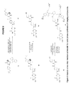
- 125000001747 pteroyl group Chemical group [H]C1=C([H])C(C(=O)[*])=C([H])C([H])=C1N([H])C([H])([H])C1=C([H])N=C2N([H])C(N([H])[H])=NC(=O)C2=N1 0.000 description 15
- VEXZGXHMUGYJMC-UHFFFAOYSA-N Hydrochloric acid Chemical compound Cl VEXZGXHMUGYJMC-UHFFFAOYSA-N 0.000 description 14
- 0 *C(NC1=O)=Nc2c1nc(CN(C(C(F)(F)F)=O)c(cc1)ccc1C(N**(O)=O)=O)cn2 Chemical compound *C(NC1=O)=Nc2c1nc(CN(C(C(F)(F)F)=O)c(cc1)ccc1C(N**(O)=O)=O)cn2 0.000 description 13
- 238000004128 high performance liquid chromatography Methods 0.000 description 13
- CURLTUGMZLYLDI-UHFFFAOYSA-N Carbon dioxide Chemical compound O=C=O CURLTUGMZLYLDI-UHFFFAOYSA-N 0.000 description 12
- BZLVMXJERCGZMT-UHFFFAOYSA-N Methyl tert-butyl ether Chemical compound COC(C)(C)C BZLVMXJERCGZMT-UHFFFAOYSA-N 0.000 description 12
- 239000011347 resin Substances 0.000 description 12
- 229920005989 resin Polymers 0.000 description 12
- OVBPIULPVIDEAO-UHFFFAOYSA-N N-Pteroyl-L-glutaminsaeure Natural products C=1N=C2NC(N)=NC(=O)C2=NC=1CNC1=CC=C(C(=O)NC(CCC(O)=O)C(O)=O)C=C1 OVBPIULPVIDEAO-UHFFFAOYSA-N 0.000 description 11
- 229960000304 folic acid Drugs 0.000 description 11
- 239000002904 solvent Substances 0.000 description 11
- 125000000999 tert-butyl group Chemical group [H]C([H])([H])C(*)(C([H])([H])[H])C([H])([H])[H] 0.000 description 11
- 238000005406 washing Methods 0.000 description 11
- RTZKZFJDLAIYFH-UHFFFAOYSA-N Diethyl ether Chemical compound CCOCC RTZKZFJDLAIYFH-UHFFFAOYSA-N 0.000 description 10
- 238000001035 drying Methods 0.000 description 10
- 230000005284 excitation Effects 0.000 description 10
- 238000001914 filtration Methods 0.000 description 10
- 125000005647 linker group Chemical group 0.000 description 10
- 230000008685 targeting Effects 0.000 description 10
- ZGYICYBLPGRURT-UHFFFAOYSA-N tri(propan-2-yl)silicon Chemical compound CC(C)[Si](C(C)C)C(C)C ZGYICYBLPGRURT-UHFFFAOYSA-N 0.000 description 10
- 239000007821 HATU Substances 0.000 description 9
- 230000008901 benefit Effects 0.000 description 9
- 239000012043 crude product Substances 0.000 description 9
- 238000010511 deprotection reaction Methods 0.000 description 9
- MOFVSTNWEDAEEK-UHFFFAOYSA-M indocyanine green Chemical compound [Na+].[O-]S(=O)(=O)CCCCN1C2=CC=C3C=CC=CC3=C2C(C)(C)C1=CC=CC=CC=CC1=[N+](CCCCS([O-])(=O)=O)C2=CC=C(C=CC=C3)C3=C2C1(C)C MOFVSTNWEDAEEK-UHFFFAOYSA-M 0.000 description 9
- 229960004657 indocyanine green Drugs 0.000 description 9
- 238000011068 loading method Methods 0.000 description 9
- 125000002924 primary amino group Chemical group [H]N([H])* 0.000 description 9
- 241000699666 Mus <mouse, genus> Species 0.000 description 8
- 239000006227 byproduct Substances 0.000 description 8
- 201000011510 cancer Diseases 0.000 description 8
- 229910002092 carbon dioxide Inorganic materials 0.000 description 8
- 239000012071 phase Substances 0.000 description 8
- 239000011541 reaction mixture Substances 0.000 description 8
- UCSJYZPVAKXKNQ-HZYVHMACSA-N streptomycin Chemical compound CN[C@H]1[C@H](O)[C@@H](O)[C@H](CO)O[C@H]1O[C@@H]1[C@](C=O)(O)[C@H](C)O[C@H]1O[C@@H]1[C@@H](NC(N)=N)[C@H](O)[C@@H](NC(N)=N)[C@H](O)[C@H]1O UCSJYZPVAKXKNQ-HZYVHMACSA-N 0.000 description 8
- 239000000126 substance Substances 0.000 description 8
- 238000001356 surgical procedure Methods 0.000 description 8
- USFZMSVCRYTOJT-UHFFFAOYSA-N Ammonium acetate Chemical compound N.CC(O)=O USFZMSVCRYTOJT-UHFFFAOYSA-N 0.000 description 7
- 229920000742 Cotton Polymers 0.000 description 7
- XUJNEKJLAYXESH-REOHCLBHSA-N L-Cysteine Chemical compound SC[C@H](N)C(O)=O XUJNEKJLAYXESH-REOHCLBHSA-N 0.000 description 7
- KWYUFKZDYYNOTN-UHFFFAOYSA-M Potassium hydroxide Chemical group [OH-].[K+] KWYUFKZDYYNOTN-UHFFFAOYSA-M 0.000 description 7
- LLSKHMWMKBAOIB-MSSQZGJESA-N [6-[[4-[[(2s)-1-[(2-methylpropan-2-yl)oxy]-1-oxo-3-[2,3,5,6-tetradeuterio-4-[(2-methylpropan-2-yl)oxy]phenyl]propan-2-yl]carbamoyl]-n-(2,2,2-trifluoroacetyl)anilino]methyl]-4-oxo-1h-pteridin-2-yl]azanium;chloride Chemical compound [Cl-].[2H]C1=C([2H])C(OC(C)(C)C)=C([2H])C([2H])=C1C[C@@H](C(=O)OC(C)(C)C)NC(=O)C1=CC=C(N(CC=2N=C3C(=O)N=C([NH3+])NC3=NC=2)C(=O)C(F)(F)F)C=C1 LLSKHMWMKBAOIB-MSSQZGJESA-N 0.000 description 7
- 238000004458 analytical method Methods 0.000 description 7
- 239000007853 buffer solution Substances 0.000 description 7
- 230000021615 conjugation Effects 0.000 description 7
- 238000005859 coupling reaction Methods 0.000 description 7
- 201000010099 disease Diseases 0.000 description 7
- 208000037265 diseases, disorders, signs and symptoms Diseases 0.000 description 7
- 230000000694 effects Effects 0.000 description 7
- 238000000295 emission spectrum Methods 0.000 description 7
- GNBHRKFJIUUOQI-UHFFFAOYSA-N fluorescein Chemical compound O1C(=O)C2=CC=CC=C2C21C1=CC=C(O)C=C1OC1=CC(O)=CC=C21 GNBHRKFJIUUOQI-UHFFFAOYSA-N 0.000 description 7
- 230000007062 hydrolysis Effects 0.000 description 7
- 238000006460 hydrolysis reaction Methods 0.000 description 7
- 230000003902 lesion Effects 0.000 description 7
- 239000002609 medium Substances 0.000 description 7
- 239000003607 modifier Substances 0.000 description 7
- 238000003756 stirring Methods 0.000 description 7
- XEKOWRVHYACXOJ-UHFFFAOYSA-N Ethyl acetate Chemical compound CCOC(C)=O XEKOWRVHYACXOJ-UHFFFAOYSA-N 0.000 description 6
- NQRYJNQNLNOLGT-UHFFFAOYSA-N Piperidine Chemical compound C1CCNCC1 NQRYJNQNLNOLGT-UHFFFAOYSA-N 0.000 description 6
- 150000003862 amino acid derivatives Chemical class 0.000 description 6
- 230000008878 coupling Effects 0.000 description 6
- 238000010168 coupling process Methods 0.000 description 6
- 238000002474 experimental method Methods 0.000 description 6
- 238000005286 illumination Methods 0.000 description 6
- 210000001165 lymph node Anatomy 0.000 description 6
- 239000000463 material Substances 0.000 description 6
- 238000002156 mixing Methods 0.000 description 6
- BWHMMNNQKKPAPP-UHFFFAOYSA-L potassium carbonate Chemical compound [K+].[K+].[O-]C([O-])=O BWHMMNNQKKPAPP-UHFFFAOYSA-L 0.000 description 6
- 125000006239 protecting group Chemical group 0.000 description 6
- MTCFGRXMJLQNBG-REOHCLBHSA-N (2S)-2-Amino-3-hydroxypropansäure Chemical compound OC[C@H](N)C(O)=O MTCFGRXMJLQNBG-REOHCLBHSA-N 0.000 description 5
- HNXQXTQTPAJEJL-UHFFFAOYSA-N 2-aminopteridin-4-ol Chemical class C1=CN=C2NC(N)=NC(=O)C2=N1 HNXQXTQTPAJEJL-UHFFFAOYSA-N 0.000 description 5
- LJJGQZSXKKBILJ-KMNMKRJZSA-N CC1(C)C2=C(C=CC(S(=O)(=O)CO)=C2)[N+](CCCCS(=O)(=O)O)=C1/C=C/C1=C(OC2=CC=C(C[C@H](NC(=O)C3=CC=C(NCC4=NC5=C(N=C4)N=C(N)NC5=O)C=C3)C(=O)O[W])C=C2)/C(=C/C=C2/N(CCCCS(C)(=O)=O)C3=C(C=C(S(C)(=O)=O)C=C3)C2(C)C)CCC1 Chemical compound CC1(C)C2=C(C=CC(S(=O)(=O)CO)=C2)[N+](CCCCS(=O)(=O)O)=C1/C=C/C1=C(OC2=CC=C(C[C@H](NC(=O)C3=CC=C(NCC4=NC5=C(N=C4)N=C(N)NC5=O)C=C3)C(=O)O[W])C=C2)/C(=C/C=C2/N(CCCCS(C)(=O)=O)C3=C(C=C(S(C)(=O)=O)C=C3)C2(C)C)CCC1 LJJGQZSXKKBILJ-KMNMKRJZSA-N 0.000 description 5
- KDXKERNSBIXSRK-UHFFFAOYSA-N Lysine Natural products NCCCCC(N)C(O)=O KDXKERNSBIXSRK-UHFFFAOYSA-N 0.000 description 5
- 239000004472 Lysine Substances 0.000 description 5
- VMHLLURERBWHNL-UHFFFAOYSA-M Sodium acetate Chemical compound [Na+].CC([O-])=O VMHLLURERBWHNL-UHFFFAOYSA-M 0.000 description 5
- UMGDCJDMYOKAJW-UHFFFAOYSA-N aminothiocarboxamide Natural products NC(N)=S UMGDCJDMYOKAJW-UHFFFAOYSA-N 0.000 description 5
- 230000009286 beneficial effect Effects 0.000 description 5
- 239000000872 buffer Substances 0.000 description 5
- 235000018417 cysteine Nutrition 0.000 description 5
- XUJNEKJLAYXESH-UHFFFAOYSA-N cysteine Natural products SCC(N)C(O)=O XUJNEKJLAYXESH-UHFFFAOYSA-N 0.000 description 5
- 238000001514 detection method Methods 0.000 description 5
- 239000007788 liquid Substances 0.000 description 5
- 210000004072 lung Anatomy 0.000 description 5
- 235000018977 lysine Nutrition 0.000 description 5
- 239000002953 phosphate buffered saline Substances 0.000 description 5
- 239000000523 sample Substances 0.000 description 5
- 238000012360 testing method Methods 0.000 description 5
- 239000005695 Ammonium acetate Substances 0.000 description 4
- XKRFYHLGVUSROY-UHFFFAOYSA-N Argon Chemical compound [Ar] XKRFYHLGVUSROY-UHFFFAOYSA-N 0.000 description 4
- 206010003497 Asphyxia Diseases 0.000 description 4
- 108091003079 Bovine Serum Albumin Proteins 0.000 description 4
- VGSPXMJFNPJMOK-FERBBOLQSA-M CC1=CC=C(C[C@H](NC(=O)C2=CC=C(N(CC3=NC4=C(N=C3)N=C(N)NC4=O)C(=O)C(F)(F)F)C=C2)C(=O)O[W])C=C1 Chemical compound CC1=CC=C(C[C@H](NC(=O)C2=CC=C(N(CC3=NC4=C(N=C3)N=C(N)NC4=O)C(=O)C(F)(F)F)C=C2)C(=O)O[W])C=C1 VGSPXMJFNPJMOK-FERBBOLQSA-M 0.000 description 4
- VEXZGXHMUGYJMC-UHFFFAOYSA-M Chloride anion Chemical compound [Cl-] VEXZGXHMUGYJMC-UHFFFAOYSA-M 0.000 description 4
- KDXKERNSBIXSRK-YFKPBYRVSA-N L-lysine Chemical compound NCCCC[C@H](N)C(O)=O KDXKERNSBIXSRK-YFKPBYRVSA-N 0.000 description 4
- 241000699660 Mus musculus Species 0.000 description 4
- 229930182555 Penicillin Natural products 0.000 description 4
- JGSARLDLIJGVTE-MBNYWOFBSA-N Penicillin G Chemical compound N([C@H]1[C@H]2SC([C@@H](N2C1=O)C(O)=O)(C)C)C(=O)CC1=CC=CC=C1 JGSARLDLIJGVTE-MBNYWOFBSA-N 0.000 description 4
- XBDQKXXYIPTUBI-UHFFFAOYSA-N Propionic acid Chemical compound CCC(O)=O XBDQKXXYIPTUBI-UHFFFAOYSA-N 0.000 description 4
- 239000012979 RPMI medium Substances 0.000 description 4
- XSQUKJJJFZCRTK-UHFFFAOYSA-N Urea Natural products NC(N)=O XSQUKJJJFZCRTK-UHFFFAOYSA-N 0.000 description 4
- 229940043376 ammonium acetate Drugs 0.000 description 4
- 235000019257 ammonium acetate Nutrition 0.000 description 4
- 238000013459 approach Methods 0.000 description 4
- 238000003556 assay Methods 0.000 description 4
- 239000012298 atmosphere Substances 0.000 description 4
- 239000001569 carbon dioxide Substances 0.000 description 4
- BNIILDVGGAEEIG-UHFFFAOYSA-L disodium hydrogen phosphate Chemical compound [Na+].[Na+].OP([O-])([O-])=O BNIILDVGGAEEIG-UHFFFAOYSA-L 0.000 description 4
- 229910000397 disodium phosphate Inorganic materials 0.000 description 4
- 235000019800 disodium phosphate Nutrition 0.000 description 4
- 239000012091 fetal bovine serum Substances 0.000 description 4
- 238000004108 freeze drying Methods 0.000 description 4
- 238000001727 in vivo Methods 0.000 description 4
- 210000004185 liver Anatomy 0.000 description 4
- 238000011580 nude mouse model Methods 0.000 description 4
- 210000000056 organ Anatomy 0.000 description 4
- 229940049954 penicillin Drugs 0.000 description 4
- 238000001556 precipitation Methods 0.000 description 4
- 238000010992 reflux Methods 0.000 description 4
- 238000002271 resection Methods 0.000 description 4
- 239000002356 single layer Substances 0.000 description 4
- 239000001632 sodium acetate Substances 0.000 description 4
- 235000017281 sodium acetate Nutrition 0.000 description 4
- AJPJDKMHJJGVTQ-UHFFFAOYSA-M sodium dihydrogen phosphate Chemical compound [Na+].OP(O)([O-])=O AJPJDKMHJJGVTQ-UHFFFAOYSA-M 0.000 description 4
- 229960005322 streptomycin Drugs 0.000 description 4
- 239000006228 supernatant Substances 0.000 description 4
- 125000004213 tert-butoxy group Chemical group [H]C([H])([H])C(O*)(C([H])([H])[H])C([H])([H])[H] 0.000 description 4
- SWZCTMTWRHEBIN-QFIPXVFZSA-N (2s)-2-(9h-fluoren-9-ylmethoxycarbonylamino)-3-(4-hydroxyphenyl)propanoic acid Chemical compound C([C@@H](C(=O)O)NC(=O)OCC1C2=CC=CC=C2C2=CC=CC=C21)C1=CC=C(O)C=C1 SWZCTMTWRHEBIN-QFIPXVFZSA-N 0.000 description 3
- OBPZZXUMVFJDTJ-UHFFFAOYSA-M C=S(C)(=O)C1=CC2=C(C=C1)N(CCCCS(=O)(=O)O[Y])/C(=C/C=C1\CCCC(/C=C/C3=[N+](CCCCS(=O)(=O)[O-])C4=C(C=C(S(=O)(=O)CO)C=C4)C3(C)C)=C1Cl)C2(C)C Chemical compound C=S(C)(=O)C1=CC2=C(C=C1)N(CCCCS(=O)(=O)O[Y])/C(=C/C=C1\CCCC(/C=C/C3=[N+](CCCCS(=O)(=O)[O-])C4=C(C=C(S(=O)(=O)CO)C=C4)C3(C)C)=C1Cl)C2(C)C OBPZZXUMVFJDTJ-UHFFFAOYSA-M 0.000 description 3
- 102000004190 Enzymes Human genes 0.000 description 3
- 108090000790 Enzymes Proteins 0.000 description 3
- 102000001554 Hemoglobins Human genes 0.000 description 3
- 108010054147 Hemoglobins Proteins 0.000 description 3
- 108091005804 Peptidases Proteins 0.000 description 3
- 239000004365 Protease Substances 0.000 description 3
- 239000012980 RPMI-1640 medium Substances 0.000 description 3
- MTCFGRXMJLQNBG-UHFFFAOYSA-N Serine Natural products OCC(N)C(O)=O MTCFGRXMJLQNBG-UHFFFAOYSA-N 0.000 description 3
- 238000010521 absorption reaction Methods 0.000 description 3
- 238000000862 absorption spectrum Methods 0.000 description 3
- 239000002253 acid Substances 0.000 description 3
- 125000003277 amino group Chemical group 0.000 description 3
- 230000003466 anti-cipated effect Effects 0.000 description 3
- 230000004888 barrier function Effects 0.000 description 3
- 230000037396 body weight Effects 0.000 description 3
- 239000003153 chemical reaction reagent Substances 0.000 description 3
- 238000001816 cooling Methods 0.000 description 3
- 238000007865 diluting Methods 0.000 description 3
- 239000003085 diluting agent Substances 0.000 description 3
- LOKCTEFSRHRXRJ-UHFFFAOYSA-I dipotassium trisodium dihydrogen phosphate hydrogen phosphate dichloride Chemical compound P(=O)(O)(O)[O-].[K+].P(=O)(O)([O-])[O-].[Na+].[Na+].[Cl-].[K+].[Cl-].[Na+] LOKCTEFSRHRXRJ-UHFFFAOYSA-I 0.000 description 3
- 238000002265 electronic spectrum Methods 0.000 description 3
- 238000000695 excitation spectrum Methods 0.000 description 3
- 150000002224 folic acids Chemical class 0.000 description 3
- 235000013305 food Nutrition 0.000 description 3
- 210000002216 heart Anatomy 0.000 description 3
- 239000012216 imaging agent Substances 0.000 description 3
- 210000002429 large intestine Anatomy 0.000 description 3
- 239000012528 membrane Substances 0.000 description 3
- 125000002496 methyl group Chemical group [H]C([H])([H])* 0.000 description 3
- 238000012544 monitoring process Methods 0.000 description 3
- 210000003205 muscle Anatomy 0.000 description 3
- 238000012634 optical imaging Methods 0.000 description 3
- 230000036961 partial effect Effects 0.000 description 3
- 230000037361 pathway Effects 0.000 description 3
- 229910000027 potassium carbonate Inorganic materials 0.000 description 3
- 210000000512 proximal kidney tubule Anatomy 0.000 description 3
- 239000000376 reactant Substances 0.000 description 3
- 102000005962 receptors Human genes 0.000 description 3
- 210000003491 skin Anatomy 0.000 description 3
- 210000000813 small intestine Anatomy 0.000 description 3
- 230000003595 spectral effect Effects 0.000 description 3
- 210000000952 spleen Anatomy 0.000 description 3
- 210000002784 stomach Anatomy 0.000 description 3
- 230000008961 swelling Effects 0.000 description 3
- 235000020679 tap water Nutrition 0.000 description 3
- 239000008399 tap water Substances 0.000 description 3
- 238000011282 treatment Methods 0.000 description 3
- 150000003668 tyrosines Chemical class 0.000 description 3
- LOOZZTFGSTZNRX-UHFFFAOYSA-N 2-azaniumyl-4-(4-hydroxyphenyl)butanoate Chemical compound OC(=O)C(N)CCC1=CC=C(O)C=C1 LOOZZTFGSTZNRX-UHFFFAOYSA-N 0.000 description 2
- IJGIHDXKYQLIMA-UHFFFAOYSA-N 4-[(2-amino-4-oxo-1h-pteridin-6-yl)methyl-(2,2,2-trifluoroacetyl)amino]benzoic acid Chemical compound C=1N=C2NC(N)=NC(=O)C2=NC=1CN(C(=O)C(F)(F)F)C1=CC=C(C(O)=O)C=C1 IJGIHDXKYQLIMA-UHFFFAOYSA-N 0.000 description 2
- IJGRMHOSHXDMSA-UHFFFAOYSA-N Atomic nitrogen Chemical compound N#N IJGRMHOSHXDMSA-UHFFFAOYSA-N 0.000 description 2
- KMSCRNGLEUMULY-XUUVOBQHSA-N CC(=O)N[C@H](CC1=CC=C(OC(C)C)C=C1)C(=O)CNC(C)C.CC(C)C[C@H](CCC1=CC=C(OC(C)C)C=C1)C(=O)O.CC(C)NCCC1=CC=C(OC(C)C)C=C1.CC(C)N[C@@H](CC1=CC=C(OC(C)C)C=C1)C(=O)O.CC(C)N[C@@H](CC1=CC=C(OC(C)C)C=C1)C(=O)OCC1=CC=CC=C1.CC(C)N[C@H](CC(=O)O)CC1=CC=C(OC(C)C)C=C1.CC(C)N[C@H](CC1=CC=C(OC(C)C)C=C1)C(=O)O.CC(C)OC1=CC=C(C[C@@H](C(=O)O)N(C)C(C)C)C=C1.COC(=O)[C@H](CC1=CC=C(OC(C)C)C=C1)NC(C)C Chemical compound CC(=O)N[C@H](CC1=CC=C(OC(C)C)C=C1)C(=O)CNC(C)C.CC(C)C[C@H](CCC1=CC=C(OC(C)C)C=C1)C(=O)O.CC(C)NCCC1=CC=C(OC(C)C)C=C1.CC(C)N[C@@H](CC1=CC=C(OC(C)C)C=C1)C(=O)O.CC(C)N[C@@H](CC1=CC=C(OC(C)C)C=C1)C(=O)OCC1=CC=CC=C1.CC(C)N[C@H](CC(=O)O)CC1=CC=C(OC(C)C)C=C1.CC(C)N[C@H](CC1=CC=C(OC(C)C)C=C1)C(=O)O.CC(C)OC1=CC=C(C[C@@H](C(=O)O)N(C)C(C)C)C=C1.COC(=O)[C@H](CC1=CC=C(OC(C)C)C=C1)NC(C)C KMSCRNGLEUMULY-XUUVOBQHSA-N 0.000 description 2
- RHYCCVHQFGKDST-FQEVSTJZSA-N CC(=O)[C@H](CC1=CC=C(C)C=C1)NC(=O)C1=CC=C(N(CC2=NC3=C(N=C2)N=C(N)NC3=O)C(=O)C(F)(F)F)C=C1 Chemical compound CC(=O)[C@H](CC1=CC=C(C)C=C1)NC(=O)C1=CC=C(N(CC2=NC3=C(N=C2)N=C(N)NC3=O)C(=O)C(F)(F)F)C=C1 RHYCCVHQFGKDST-FQEVSTJZSA-N 0.000 description 2
- SRJRMRGVXXHEFD-JEMQRPTFSA-M CC1(C)C2=C(C=CC([Na])=C2)[N+](CCCS(=O)(=O)[O-])=C1/C=C/C1=C(O[13C]2=[13CH][13CH]=[13C]([13CH2][13C@H](NC(=O)C3=CC=C(NCC4=NC5=C(N=C4)N=C(N)NC5=O)C=C3)[13C](=O)O)[13CH]=C2)/C(=C/C=C2/N(CCCCS(=O)(=O)O[Na])C3=C(C=C([Na])C=C3)C2(C)C)CCC1.NC1=NC2=C(N=C(CNC3=CC=C(C(=O)N[13C@@H]([13CH2][13C]4=[13CH][13CH]=[13C](O)C=[13CH]4)[13C](=O)O)C=C3)C=N2)C(=O)N1.NC1=NC2=C(N=C(CNC3=CC=C(C(=O)N[13C@@H]([13CH2][13C]4=[13CH][13CH]=[13C](O)C=[13CH]4)[13C](=O)O)C=C3)C=N2)C(=O)N1.O=C(O)C(F)(F)F.O=C(O[Na])C(F)(F)F.O=S(=O)=O.O=S(=O)=O.[Cl-].[NH3+]C1=NC2=C(N=C(CN(C(=O)C(F)(F)F)C3=CC=C(C(=O)N[13C@@H]([13CH2][13C]4=[13CH][13CH]=[13C](O)C=[13CH]4)[13C](=O)O)C=C3)C=N2)C(=O)N1.[Na+] Chemical compound CC1(C)C2=C(C=CC([Na])=C2)[N+](CCCS(=O)(=O)[O-])=C1/C=C/C1=C(O[13C]2=[13CH][13CH]=[13C]([13CH2][13C@H](NC(=O)C3=CC=C(NCC4=NC5=C(N=C4)N=C(N)NC5=O)C=C3)[13C](=O)O)[13CH]=C2)/C(=C/C=C2/N(CCCCS(=O)(=O)O[Na])C3=C(C=C([Na])C=C3)C2(C)C)CCC1.NC1=NC2=C(N=C(CNC3=CC=C(C(=O)N[13C@@H]([13CH2][13C]4=[13CH][13CH]=[13C](O)C=[13CH]4)[13C](=O)O)C=C3)C=N2)C(=O)N1.NC1=NC2=C(N=C(CNC3=CC=C(C(=O)N[13C@@H]([13CH2][13C]4=[13CH][13CH]=[13C](O)C=[13CH]4)[13C](=O)O)C=C3)C=N2)C(=O)N1.O=C(O)C(F)(F)F.O=C(O[Na])C(F)(F)F.O=S(=O)=O.O=S(=O)=O.[Cl-].[NH3+]C1=NC2=C(N=C(CN(C(=O)C(F)(F)F)C3=CC=C(C(=O)N[13C@@H]([13CH2][13C]4=[13CH][13CH]=[13C](O)C=[13CH]4)[13C](=O)O)C=C3)C=N2)C(=O)N1.[Na+] SRJRMRGVXXHEFD-JEMQRPTFSA-M 0.000 description 2
- QHWINAPNEVCIKL-SXVDFRLUSA-N CC1=CC=C(C[C@@H](NC(=O)C2=CC=C(N(CC3=NC4=C(C=C(Cl)NC4=O)N=C3)C(=O)C(F)(F)F)C=C2)C(=O)O)C=C1.N.N.O=C(N[C@@H](CC1=CC=C(O)C=C1)C(=O)O)C1=CC=C(N(CC2=NC3=C(C=C(Cl)NC3=O)N=C2)C(=O)C(F)(F)F)C=C1 Chemical compound CC1=CC=C(C[C@@H](NC(=O)C2=CC=C(N(CC3=NC4=C(C=C(Cl)NC4=O)N=C3)C(=O)C(F)(F)F)C=C2)C(=O)O)C=C1.N.N.O=C(N[C@@H](CC1=CC=C(O)C=C1)C(=O)O)C1=CC=C(N(CC2=NC3=C(C=C(Cl)NC3=O)N=C2)C(=O)C(F)(F)F)C=C1 QHWINAPNEVCIKL-SXVDFRLUSA-N 0.000 description 2
- PMUAKQAMYFDESD-AFZBCKAHSA-N CC1=CC=C(C[C@@H](NC(=O)C2=CC=C(N(CC3=NC4=C(N=C3)N=C(Cl)NC4=O)C(=O)C(F)(F)F)C=C2)C(=O)O)C=C1.N.N.O=C(N[C@@H](CC1=CC=C(O)C=C1)C(=O)O)C1=CC=C(N(CC2=NC3=C(N=C2)N=C(Cl)NC3=O)C(=O)C(F)(F)F)C=C1 Chemical compound CC1=CC=C(C[C@@H](NC(=O)C2=CC=C(N(CC3=NC4=C(N=C3)N=C(Cl)NC4=O)C(=O)C(F)(F)F)C=C2)C(=O)O)C=C1.N.N.O=C(N[C@@H](CC1=CC=C(O)C=C1)C(=O)O)C1=CC=C(N(CC2=NC3=C(N=C2)N=C(Cl)NC3=O)C(=O)C(F)(F)F)C=C1 PMUAKQAMYFDESD-AFZBCKAHSA-N 0.000 description 2
- OIKUVIUSFCKUFG-FERBBOLQSA-M CC1=CC=C(C[C@H](NC(=O)C2=CC=C(N(CC3=NC4=C(N=C3)N=C(Cl)NC4=O)C(=O)C(F)(F)F)C=C2)C(=O)O[W])C=C1.N Chemical compound CC1=CC=C(C[C@H](NC(=O)C2=CC=C(N(CC3=NC4=C(N=C3)N=C(Cl)NC4=O)C(=O)C(F)(F)F)C=C2)C(=O)O[W])C=C1.N OIKUVIUSFCKUFG-FERBBOLQSA-M 0.000 description 2
- OMXDAFZCPSQSQI-UHFFFAOYSA-N CCC(=O)C1=CC=C(NCC2=NC3=C(C=C(N)NC3=O)N=C2)C=C1 Chemical compound CCC(=O)C1=CC=C(NCC2=NC3=C(C=C(N)NC3=O)N=C2)C=C1 OMXDAFZCPSQSQI-UHFFFAOYSA-N 0.000 description 2
- 102000008186 Collagen Human genes 0.000 description 2
- 108010035532 Collagen Proteins 0.000 description 2
- 102100041003 Glutamate carboxypeptidase 2 Human genes 0.000 description 2
- 101000892862 Homo sapiens Glutamate carboxypeptidase 2 Proteins 0.000 description 2
- SIKJAQJRHWYJAI-UHFFFAOYSA-N Indole Chemical compound C1=CC=C2NC=CC2=C1 SIKJAQJRHWYJAI-UHFFFAOYSA-N 0.000 description 2
- 206010054107 Nodule Diseases 0.000 description 2
- 206010033128 Ovarian cancer Diseases 0.000 description 2
- 206010061535 Ovarian neoplasm Diseases 0.000 description 2
- 208000000236 Prostatic Neoplasms Diseases 0.000 description 2
- 102100037486 Reverse transcriptase/ribonuclease H Human genes 0.000 description 2
- FAPWRFPIFSIZLT-UHFFFAOYSA-M Sodium chloride Chemical compound [Na+].[Cl-] FAPWRFPIFSIZLT-UHFFFAOYSA-M 0.000 description 2
- 230000032900 absorption of visible light Effects 0.000 description 2
- 238000009825 accumulation Methods 0.000 description 2
- 230000004913 activation Effects 0.000 description 2
- 125000003275 alpha amino acid group Chemical group 0.000 description 2
- 229910052786 argon Inorganic materials 0.000 description 2
- 239000012472 biological sample Substances 0.000 description 2
- 210000000481 breast Anatomy 0.000 description 2
- 238000004113 cell culture Methods 0.000 description 2
- 230000008859 change Effects 0.000 description 2
- 229920001436 collagen Polymers 0.000 description 2
- 210000001072 colon Anatomy 0.000 description 2
- 238000004440 column chromatography Methods 0.000 description 2
- 229940126214 compound 3 Drugs 0.000 description 2
- HGCIXCUEYOPUTN-UHFFFAOYSA-N cyclohexene Chemical compound C1CCC=CC1 HGCIXCUEYOPUTN-UHFFFAOYSA-N 0.000 description 2
- 238000000354 decomposition reaction Methods 0.000 description 2
- 230000002950 deficient Effects 0.000 description 2
- 235000005911 diet Nutrition 0.000 description 2
- 230000037213 diet Effects 0.000 description 2
- 238000005516 engineering process Methods 0.000 description 2
- 238000001704 evaporation Methods 0.000 description 2
- 230000008020 evaporation Effects 0.000 description 2
- 239000011552 falling film Substances 0.000 description 2
- 239000007789 gas Substances 0.000 description 2
- 230000002055 immunohistochemical effect Effects 0.000 description 2
- JMMWKPVZQRWMSS-UHFFFAOYSA-N isopropanol acetate Natural products CC(C)OC(C)=O JMMWKPVZQRWMSS-UHFFFAOYSA-N 0.000 description 2
- 230000031700 light absorption Effects 0.000 description 2
- 230000000670 limiting effect Effects 0.000 description 2
- 208000037819 metastatic cancer Diseases 0.000 description 2
- 208000011575 metastatic malignant neoplasm Diseases 0.000 description 2
- 238000012986 modification Methods 0.000 description 2
- 230000004048 modification Effects 0.000 description 2
- 229910052757 nitrogen Inorganic materials 0.000 description 2
- 230000007170 pathology Effects 0.000 description 2
- 238000010647 peptide synthesis reaction Methods 0.000 description 2
- 230000002085 persistent effect Effects 0.000 description 2
- ISWSIDIOOBJBQZ-UHFFFAOYSA-N phenol group Chemical group C1(=CC=CC=C1)O ISWSIDIOOBJBQZ-UHFFFAOYSA-N 0.000 description 2
- 230000000704 physical effect Effects 0.000 description 2
- 108020003175 receptors Proteins 0.000 description 2
- 238000002390 rotary evaporation Methods 0.000 description 2
- 150000003839 salts Chemical class 0.000 description 2
- 238000001228 spectrum Methods 0.000 description 2
- 239000000758 substrate Substances 0.000 description 2
- 150000003667 tyrosine derivatives Chemical class 0.000 description 2
- SOAPXKSPJAZNGO-WDSKDSINSA-N (2s)-2-[[(1s)-1,3-dicarboxypropyl]carbamoylamino]pentanedioic acid Chemical compound OC(=O)CC[C@@H](C(O)=O)NC(=O)N[C@H](C(O)=O)CCC(O)=O SOAPXKSPJAZNGO-WDSKDSINSA-N 0.000 description 1
- 125000003088 (fluoren-9-ylmethoxy)carbonyl group Chemical group 0.000 description 1
- HIYWOHBEPVGIQN-UHFFFAOYSA-N 1h-benzo[g]indole Chemical compound C1=CC=CC2=C(NC=C3)C3=CC=C21 HIYWOHBEPVGIQN-UHFFFAOYSA-N 0.000 description 1
- 239000004475 Arginine Substances 0.000 description 1
- DCXYFEDJOCDNAF-UHFFFAOYSA-N Asparagine Natural products OC(=O)C(N)CC(N)=O DCXYFEDJOCDNAF-UHFFFAOYSA-N 0.000 description 1
- 206010006187 Breast cancer Diseases 0.000 description 1
- 208000026310 Breast neoplasm Diseases 0.000 description 1
- NFDWOZPDKAQACT-UHFFFAOYSA-R C.C.[H]N([H])C1=NC2=C(N=C(CN(C(=O)C(F)(F)F)C3=CC=C(C(=O)O)C=C3)C=N2)C(=O)N1.[H][N+]([H])(Cl)C1=NC2=C(N=C(CN(C(=O)C(F)(F)F)C3=CC=C(C(C)=O)C=C3)C=N2)C(=O)N1.[H][N+]([H])(Cl)C1=NC2=C(N=C(CN(C(=O)C(F)(F)F)C3=CC=C(C(C)=O)C=C3)C=N2)C(=O)N1.[H][N+]([H])(Cl)C1=NC2=C(N=C(CN(C(=O)C(F)(F)F)C3=CC=C(C(C)=O)C=C3)C=N2)C(=O)N1.[H][N+]([H])(Cl)C1=NC2=C(N=C(CN(C(=O)C(F)(F)F)C3=CC=C(C(C)=O)C=C3)C=N2)C(=O)N1 Chemical compound C.C.[H]N([H])C1=NC2=C(N=C(CN(C(=O)C(F)(F)F)C3=CC=C(C(=O)O)C=C3)C=N2)C(=O)N1.[H][N+]([H])(Cl)C1=NC2=C(N=C(CN(C(=O)C(F)(F)F)C3=CC=C(C(C)=O)C=C3)C=N2)C(=O)N1.[H][N+]([H])(Cl)C1=NC2=C(N=C(CN(C(=O)C(F)(F)F)C3=CC=C(C(C)=O)C=C3)C=N2)C(=O)N1.[H][N+]([H])(Cl)C1=NC2=C(N=C(CN(C(=O)C(F)(F)F)C3=CC=C(C(C)=O)C=C3)C=N2)C(=O)N1.[H][N+]([H])(Cl)C1=NC2=C(N=C(CN(C(=O)C(F)(F)F)C3=CC=C(C(C)=O)C=C3)C=N2)C(=O)N1 NFDWOZPDKAQACT-UHFFFAOYSA-R 0.000 description 1
- STOKGEHJIRNNOV-MBUOHKHNSA-N C.CC(C)(C)OC(=O)[C@@H](Cl)CC1=CC=C(OC(C)(C)C)C=C1.CC1=CC=C(C[C@@H](Cl)C(=O)OC(C)(C)C)C=C1.N.N Chemical compound C.CC(C)(C)OC(=O)[C@@H](Cl)CC1=CC=C(OC(C)(C)C)C=C1.CC1=CC=C(C[C@@H](Cl)C(=O)OC(C)(C)C)C=C1.N.N STOKGEHJIRNNOV-MBUOHKHNSA-N 0.000 description 1
- XAVHDEZFJNMQTL-QLXVSJFJSA-F C.O=C(O[Na])C(F)(F)F.[H]N([H])C1=NC2=C(N=C(CNC3=CC=C(C(=O)N[C@@H](CC4=CC=C(CC5=C(/C=C/C6C(CCCCS(=O)(=O)O)C7=C(C=C(S(=O)(=O)[Na]O)C=C7)C6(C)C)CCC/C5=C\C=C5\N(CCCCS(=O)(=O)O[Na])C6=C(C=C(S(=O)(=O)O[Na])C=C6)C5(C)C)C=C4)C(=O)O[Na])C=C3)C=N2)C(=O)N1.[H]N([H])C1=NC2=C(N=C(CNC3=CC=C(C(=O)N[C@@H](CC4=CC=C(O[Na])C=C4)C(=O)O[Na])C=C3)C=N2)C(=O)N1.[H]N([H])C1=NC2=C(N=C(CNC3=CC=C(C(=O)N[C@@H](CC4=CC=C(O[Na])C=C4)C(=O)O[Na])C=C3)C=N2)C(=O)N1.[H][N+]([H])([H])C1=NC2=C(N=C(CN(C(=O)C(F)(F)F)C3=CC=C(C(=O)N[C@@H](CC4=CC=C(O)C=C4)C(=O)O)C=C3)C=N2)C(=O)N1 Chemical compound C.O=C(O[Na])C(F)(F)F.[H]N([H])C1=NC2=C(N=C(CNC3=CC=C(C(=O)N[C@@H](CC4=CC=C(CC5=C(/C=C/C6C(CCCCS(=O)(=O)O)C7=C(C=C(S(=O)(=O)[Na]O)C=C7)C6(C)C)CCC/C5=C\C=C5\N(CCCCS(=O)(=O)O[Na])C6=C(C=C(S(=O)(=O)O[Na])C=C6)C5(C)C)C=C4)C(=O)O[Na])C=C3)C=N2)C(=O)N1.[H]N([H])C1=NC2=C(N=C(CNC3=CC=C(C(=O)N[C@@H](CC4=CC=C(O[Na])C=C4)C(=O)O[Na])C=C3)C=N2)C(=O)N1.[H]N([H])C1=NC2=C(N=C(CNC3=CC=C(C(=O)N[C@@H](CC4=CC=C(O[Na])C=C4)C(=O)O[Na])C=C3)C=N2)C(=O)N1.[H][N+]([H])([H])C1=NC2=C(N=C(CN(C(=O)C(F)(F)F)C3=CC=C(C(=O)N[C@@H](CC4=CC=C(O)C=C4)C(=O)O)C=C3)C=N2)C(=O)N1 XAVHDEZFJNMQTL-QLXVSJFJSA-F 0.000 description 1
- IOZBAVFEJMDEEJ-UAALDIBHSA-N C1CCC([NH2+]C2CCCCC2)CC1.C1CCC([NH2+]C2CCCCC2)CC1.CN(C(=O)OC(C)(C)C)[C@@H](CC1=CC=C(O)C=C1)C(=O)[O-].CN[C@@H](CC1=CC=C(O)C=C1)C(=O)[O-] Chemical compound C1CCC([NH2+]C2CCCCC2)CC1.C1CCC([NH2+]C2CCCCC2)CC1.CN(C(=O)OC(C)(C)C)[C@@H](CC1=CC=C(O)C=C1)C(=O)[O-].CN[C@@H](CC1=CC=C(O)C=C1)C(=O)[O-] IOZBAVFEJMDEEJ-UAALDIBHSA-N 0.000 description 1
- AKOSOEKOHYAAQL-FLGGMVDVSA-N C1CCC([NH2+]C2CCCCC2)CC1.C1CCC([NH2+]C2CCCCC2)CC1.C[C@@H](CCC1=CC=C(O)C=C1)C(=O)[O-].N[C@@H](CCC1=CC=C(O)C=C1)C(=O)[O-] Chemical compound C1CCC([NH2+]C2CCCCC2)CC1.C1CCC([NH2+]C2CCCCC2)CC1.C[C@@H](CCC1=CC=C(O)C=C1)C(=O)[O-].N[C@@H](CCC1=CC=C(O)C=C1)C(=O)[O-] AKOSOEKOHYAAQL-FLGGMVDVSA-N 0.000 description 1
- OXCROOPNMDLZRO-UHFFFAOYSA-N C=S(=O)(O)C1=CC2=C(C=C1)N(CCCCS(=O)(=O)O)/C(=C/C=C1\CCCC(/C=C/C3=[N+](CCCCS(=O)(=O)[O-])C4=C(C=C(S(=O)(=O)O)C=C4)C3(C)C)=C1Cl)C2(C)C.C=S(=O)(O)C1=CC2=C(C=C1)N(CCCCS(=O)(=O)O)/C(=C/C=C1\CCOC(/C=C/C3=[N+](CCCCS(=O)(=O)[O-])C4=C(C=C(S(=O)(=O)O)C=C4)C3(C)C)=C1Cl)C2(C)C.CC1(C)C2=C(C=CC=C2)[N+](CCCCS(=O)(=O)[O-])=C1/C=C/C1=C(Cl)/C(=C/C=C2/N(CCCCS(=O)(=O)O)C3=C(C=CC=C3)C2(C)C)CCC1 Chemical compound C=S(=O)(O)C1=CC2=C(C=C1)N(CCCCS(=O)(=O)O)/C(=C/C=C1\CCCC(/C=C/C3=[N+](CCCCS(=O)(=O)[O-])C4=C(C=C(S(=O)(=O)O)C=C4)C3(C)C)=C1Cl)C2(C)C.C=S(=O)(O)C1=CC2=C(C=C1)N(CCCCS(=O)(=O)O)/C(=C/C=C1\CCOC(/C=C/C3=[N+](CCCCS(=O)(=O)[O-])C4=C(C=C(S(=O)(=O)O)C=C4)C3(C)C)=C1Cl)C2(C)C.CC1(C)C2=C(C=CC=C2)[N+](CCCCS(=O)(=O)[O-])=C1/C=C/C1=C(Cl)/C(=C/C=C2/N(CCCCS(=O)(=O)O)C3=C(C=CC=C3)C2(C)C)CCC1 OXCROOPNMDLZRO-UHFFFAOYSA-N 0.000 description 1
- FODYEQDKQSGGDY-DKPJYZTKSA-N CC(=O)C(CC1=CC=C(OC(C)(C)C)C=C1)NC(=O)CC1C2=C(C=CC=C2)C2=C1C=CC=C2.CC(=O)[C@H](CC1=CC=C(OC(C)(C)C)C=C1)NC(=O)C1=CC=C(N(CC2=NC3C(=O)NC(N)=NC3N=C2)C(=O)C(F)(F)F)C=C1.CC(=O)[C@H](CC1=CC=C(OC(C)(C)C)C=C1)NC(=O)C1=CC=C(N(CC2=NC3C(=O)NC(N)=NC3N=C2)C(=O)C(F)(F)F)C=C1.NC1=NC2N=CC(CN(C(=O)C(F)(F)F)C3=CC=C(C(=O)C[C@@H](CC4=CC=C(O)C=C4)C(=O)O)C=C3)=NC2C(=O)N1 Chemical compound CC(=O)C(CC1=CC=C(OC(C)(C)C)C=C1)NC(=O)CC1C2=C(C=CC=C2)C2=C1C=CC=C2.CC(=O)[C@H](CC1=CC=C(OC(C)(C)C)C=C1)NC(=O)C1=CC=C(N(CC2=NC3C(=O)NC(N)=NC3N=C2)C(=O)C(F)(F)F)C=C1.CC(=O)[C@H](CC1=CC=C(OC(C)(C)C)C=C1)NC(=O)C1=CC=C(N(CC2=NC3C(=O)NC(N)=NC3N=C2)C(=O)C(F)(F)F)C=C1.NC1=NC2N=CC(CN(C(=O)C(F)(F)F)C3=CC=C(C(=O)C[C@@H](CC4=CC=C(O)C=C4)C(=O)O)C=C3)=NC2C(=O)N1 FODYEQDKQSGGDY-DKPJYZTKSA-N 0.000 description 1
- FBPXWPFWFVOISU-FGJQBABTSA-P CC(=O)C([NH3+])CC1=CC=C(C)C=C1.CC1=CC=C(C[C@H](NC(=O)C2=CC=C(N(CC3=NC4=C(N=C3)N=C([NH3+])NC4=O)C(=O)C(F)(F)F)C=C2)C(=O)OC(C)(C)C)C=C1.NC1=NC2=C(N=C(CN(C(=O)C(F)(F)F)C3=CC=C(C(=O)O)C=C3)C=N2)C(=O)N1.[Cl-].[Cl-] Chemical compound CC(=O)C([NH3+])CC1=CC=C(C)C=C1.CC1=CC=C(C[C@H](NC(=O)C2=CC=C(N(CC3=NC4=C(N=C3)N=C([NH3+])NC4=O)C(=O)C(F)(F)F)C=C2)C(=O)OC(C)(C)C)C=C1.NC1=NC2=C(N=C(CN(C(=O)C(F)(F)F)C3=CC=C(C(=O)O)C=C3)C=N2)C(=O)N1.[Cl-].[Cl-] FBPXWPFWFVOISU-FGJQBABTSA-P 0.000 description 1
- HCEQIYSAHSVLAC-UHFFFAOYSA-N CC(=O)C1=CC=C(N(CC2=NC3=C(N=C2)N=C(N)NC3=O)C(=O)C(F)(F)F)C=C1 Chemical compound CC(=O)C1=CC=C(N(CC2=NC3=C(N=C2)N=C(N)NC3=O)C(=O)C(F)(F)F)C=C1 HCEQIYSAHSVLAC-UHFFFAOYSA-N 0.000 description 1
- JSLRASCTEDLQIS-BGMWXCKGSA-N CC(=O)N[C@@H](CC1=CC=C(O)C=C1)C(=O)CN.CC(=O)N[C@@H](CC1=CC=C(O)C=C1)C(=O)CNC(=O)C1=CC=C(N(CC2=NC3=C(N=C2)N=C(N)NC3=O)C(=O)C(F)(F)F)C=C1.NC1=NC2=C(N=C(CN(C(=O)C(F)(F)F)C3=CC=C(C(=O)O)C=C3)C=N2)C(=O)N1.O=S(=O)=O.O=S(=O)=O.[H]C1=CC2=C(C=C1)[N+](CCCCS(=O)(=O)[O-])=C(/C=C/C1=C(OC3=CC=C(C[C@H](NC(C)=O)C(=O)CNC(=O)C4=CC=C(NCC5=NC6=C(N=C5)N=C(N)NC6=O)C=C4)C=C3)/C(=C/C=C3/N(CCCCS(=O)(=O)O)C4=C(C=C([H])C=C4)C3(C)C)CCC1)C2(C)C Chemical compound CC(=O)N[C@@H](CC1=CC=C(O)C=C1)C(=O)CN.CC(=O)N[C@@H](CC1=CC=C(O)C=C1)C(=O)CNC(=O)C1=CC=C(N(CC2=NC3=C(N=C2)N=C(N)NC3=O)C(=O)C(F)(F)F)C=C1.NC1=NC2=C(N=C(CN(C(=O)C(F)(F)F)C3=CC=C(C(=O)O)C=C3)C=N2)C(=O)N1.O=S(=O)=O.O=S(=O)=O.[H]C1=CC2=C(C=C1)[N+](CCCCS(=O)(=O)[O-])=C(/C=C/C1=C(OC3=CC=C(C[C@H](NC(C)=O)C(=O)CNC(=O)C4=CC=C(NCC5=NC6=C(N=C5)N=C(N)NC6=O)C=C4)C=C3)/C(=C/C=C3/N(CCCCS(=O)(=O)O)C4=C(C=C([H])C=C4)C3(C)C)CCC1)C2(C)C JSLRASCTEDLQIS-BGMWXCKGSA-N 0.000 description 1
- MHZJSOZSZLXPMK-VWLOTQADSA-N CC(=O)[C@H](CC1=CC=C(C)C=C1)NC(=O)OCC1C2=C(C=CC=C2)C2=C1/C=C\C=C/2 Chemical compound CC(=O)[C@H](CC1=CC=C(C)C=C1)NC(=O)OCC1C2=C(C=CC=C2)C2=C1/C=C\C=C/2 MHZJSOZSZLXPMK-VWLOTQADSA-N 0.000 description 1
- YFDLYPZKNPOJGV-OKZOQINVSA-N CC(=O)[C@H](CC1=CC=C(OC(C)(C)C)C=C1)NC(=O)C1=CC=C(N(CC2=NC3=C(N=C2)N=C(N)NC3=O)C(=O)C(F)(F)F)C=C1.CC(=O)[C@H](CC1=CC=C(OC(C)(C)C)C=C1)NC(=O)OCC1C2=C(C=CC=C2)C2=C1C=CC=C2.NC1=NC2=C(N=C(CN(C(=O)C(F)(F)F)C3=CC=C(C(=O)N[C@@H](CC4=CC=C(O)C=C4)C(=O)O)C=C3)C=N2)C(=O)N1 Chemical compound CC(=O)[C@H](CC1=CC=C(OC(C)(C)C)C=C1)NC(=O)C1=CC=C(N(CC2=NC3=C(N=C2)N=C(N)NC3=O)C(=O)C(F)(F)F)C=C1.CC(=O)[C@H](CC1=CC=C(OC(C)(C)C)C=C1)NC(=O)OCC1C2=C(C=CC=C2)C2=C1C=CC=C2.NC1=NC2=C(N=C(CN(C(=O)C(F)(F)F)C3=CC=C(C(=O)N[C@@H](CC4=CC=C(O)C=C4)C(=O)O)C=C3)C=N2)C(=O)N1 YFDLYPZKNPOJGV-OKZOQINVSA-N 0.000 description 1
- ASAUWAIROFREOC-SMWIJIBUSA-N CC(C)(C)OC(=O)[C@@H](Cl)CC1=CC=C(OC(C)(C)C)C=C1.CC1=CC=C(C[C@@H](Cl)C(=O)OC(C)(C)C)C=C1.N.N Chemical compound CC(C)(C)OC(=O)[C@@H](Cl)CC1=CC=C(OC(C)(C)C)C=C1.CC1=CC=C(C[C@@H](Cl)C(=O)OC(C)(C)C)C=C1.N.N ASAUWAIROFREOC-SMWIJIBUSA-N 0.000 description 1
- UWDOKSYRWXIBMB-FQJMDOGSSA-N CC(C)(C)OC(=O)[C@H](CC1=CC=C(OC(C)(C)C)C=C1)NC(=O)C1=CC=C(N(CC2=NC3=C(N=C2)N=C(Cl=N)NC3=O)C(=O)C(F)(F)F)C=C1.CC(C)(C)OC(=O)[C@H](CC1=CC=C(OC(C)(C)C)C=C1)[Cl-][NH3+].NC1=NC2=C(N=C(CN(C(=O)C(F)(F)F)C3=CC=C(C(=O)O)C=C3)C=N2)C(=O)N1 Chemical compound CC(C)(C)OC(=O)[C@H](CC1=CC=C(OC(C)(C)C)C=C1)NC(=O)C1=CC=C(N(CC2=NC3=C(N=C2)N=C(Cl=N)NC3=O)C(=O)C(F)(F)F)C=C1.CC(C)(C)OC(=O)[C@H](CC1=CC=C(OC(C)(C)C)C=C1)[Cl-][NH3+].NC1=NC2=C(N=C(CN(C(=O)C(F)(F)F)C3=CC=C(C(=O)O)C=C3)C=N2)C(=O)N1 UWDOKSYRWXIBMB-FQJMDOGSSA-N 0.000 description 1
- XXWBKUBZGDQGQF-DLHXETPVSA-P CC(C)(C)O[13C](=O)[13C@@H]([NH3+])[13CH2][13C]1=[13CH][13CH]=[13C](OC(C)(C)C)C=[13CH]1.CC(C)(C)O[13C](=O)[13C@H]([13CH2][13C]1=[13CH][13CH]=[13C](OC(C)(C)C)C=[13CH]1)NC(=O)C1=CC=C(N(CC2=NC3=C(N=C2)N=C([NH3+])NC3=O)C(=O)C(F)(F)F)C=C1.NC1=NC2=C(N=C(CN(C(=O)C(F)(F)F)C3=CC=C(C(=O)O)C=C3)C=N2)C(=O)N1.[Cl-].[Cl-] Chemical compound CC(C)(C)O[13C](=O)[13C@@H]([NH3+])[13CH2][13C]1=[13CH][13CH]=[13C](OC(C)(C)C)C=[13CH]1.CC(C)(C)O[13C](=O)[13C@H]([13CH2][13C]1=[13CH][13CH]=[13C](OC(C)(C)C)C=[13CH]1)NC(=O)C1=CC=C(N(CC2=NC3=C(N=C2)N=C([NH3+])NC3=O)C(=O)C(F)(F)F)C=C1.NC1=NC2=C(N=C(CN(C(=O)C(F)(F)F)C3=CC=C(C(=O)O)C=C3)C=N2)C(=O)N1.[Cl-].[Cl-] XXWBKUBZGDQGQF-DLHXETPVSA-P 0.000 description 1
- KZQUZZBNEZQJOZ-IOIFWVEKSA-P CC(C)(C)O[13C](=O)[13C@H]([13CH2][13C]1=[13CH][13CH]=[13C](OC(C)(C)C)C=[13CH]1)NC(=O)C1=CC=C(N(CC2=NC3=C(N=C2)N=C([NH3+])NC3=O)C(=O)C(F)(F)F)C=C1.[Cl-].[Cl-].[NH3+]C1=NC2=C(N=C(CN(C(=O)C(F)(F)F)C3=CC=C(C(=O)N[13C@@H]([13CH2][13C]4=[13CH][13CH]=[13C](O)C=[13CH]4)[13C](=O)O)C=C3)C=N2)C(=O)N1 Chemical compound CC(C)(C)O[13C](=O)[13C@H]([13CH2][13C]1=[13CH][13CH]=[13C](OC(C)(C)C)C=[13CH]1)NC(=O)C1=CC=C(N(CC2=NC3=C(N=C2)N=C([NH3+])NC3=O)C(=O)C(F)(F)F)C=C1.[Cl-].[Cl-].[NH3+]C1=NC2=C(N=C(CN(C(=O)C(F)(F)F)C3=CC=C(C(=O)N[13C@@H]([13CH2][13C]4=[13CH][13CH]=[13C](O)C=[13CH]4)[13C](=O)O)C=C3)C=N2)C(=O)N1 KZQUZZBNEZQJOZ-IOIFWVEKSA-P 0.000 description 1
- XXWBKUBZGDQGQF-ASRNNCIESA-P CC(C)(C)O[14C](=O)[14C@@H]([NH3+])[14CH2][14C]1=[14CH][14CH]=[14C](OC(C)(C)C)C=[14CH]1.CC(C)(C)O[14C](=O)[14C@H]([14CH2][14C]1=[14CH][14CH]=[14C](OC(C)(C)C)C=[14CH]1)NC(=O)C1=CC=C(N(CC2=NC3=C(N=C2)N=C([NH3+])NC3=O)C(=O)C(F)(F)F)C=C1.NC1=NC2=C(N=C(CN(C(=O)C(F)(F)F)C3=CC=C(C(=O)O)C=C3)C=N2)C(=O)N1.[Cl-].[Cl-] Chemical compound CC(C)(C)O[14C](=O)[14C@@H]([NH3+])[14CH2][14C]1=[14CH][14CH]=[14C](OC(C)(C)C)C=[14CH]1.CC(C)(C)O[14C](=O)[14C@H]([14CH2][14C]1=[14CH][14CH]=[14C](OC(C)(C)C)C=[14CH]1)NC(=O)C1=CC=C(N(CC2=NC3=C(N=C2)N=C([NH3+])NC3=O)C(=O)C(F)(F)F)C=C1.NC1=NC2=C(N=C(CN(C(=O)C(F)(F)F)C3=CC=C(C(=O)O)C=C3)C=N2)C(=O)N1.[Cl-].[Cl-] XXWBKUBZGDQGQF-ASRNNCIESA-P 0.000 description 1
- KZQUZZBNEZQJOZ-YGSFADTHSA-P CC(C)(C)O[14C](=O)[14C@H]([14CH2][14C]1=[14CH][14CH]=[14C](OC(C)(C)C)C=[14CH]1)NC(=O)C1=CC=C(N(CC2=NC3=C(N=C2)N=C([NH3+])NC3=O)C(=O)C(F)(F)F)C=C1.[Cl-].[Cl-].[NH3+]C1=NC2=C(N=C(CN(C(=O)C(F)(F)F)C3=CC=C(C(=O)N[14C@@H]([14CH2][14C]4=[14CH][14CH]=[14C](O)C=[14CH]4)[14C](=O)O)C=C3)C=N2)C(=O)N1 Chemical compound CC(C)(C)O[14C](=O)[14C@H]([14CH2][14C]1=[14CH][14CH]=[14C](OC(C)(C)C)C=[14CH]1)NC(=O)C1=CC=C(N(CC2=NC3=C(N=C2)N=C([NH3+])NC3=O)C(=O)C(F)(F)F)C=C1.[Cl-].[Cl-].[NH3+]C1=NC2=C(N=C(CN(C(=O)C(F)(F)F)C3=CC=C(C(=O)N[14C@@H]([14CH2][14C]4=[14CH][14CH]=[14C](O)C=[14CH]4)[14C](=O)O)C=C3)C=N2)C(=O)N1 KZQUZZBNEZQJOZ-YGSFADTHSA-P 0.000 description 1
- PPHOUPVJEOTILP-UHFFFAOYSA-N CC(C)C1=CC=C(NCC2=CN=C3/C=C(/N)NC(=O)C3=N2)C=C1 Chemical compound CC(C)C1=CC=C(NCC2=CN=C3/C=C(/N)NC(=O)C3=N2)C=C1 PPHOUPVJEOTILP-UHFFFAOYSA-N 0.000 description 1
- BPALWGMGTXCJPB-IKFJUQJOSA-N CC(C)N[C@@H](CC1=CC=C(OC(C)C)C=C1)C(=O)O.CC(C)N[C@H](CC1=CC=C(OC(C)C)C=C1)C(=O)O Chemical compound CC(C)N[C@@H](CC1=CC=C(OC(C)C)C=C1)C(=O)O.CC(C)N[C@H](CC1=CC=C(OC(C)C)C=C1)C(=O)O BPALWGMGTXCJPB-IKFJUQJOSA-N 0.000 description 1
- ITAOHXXEDDDXFH-WWBYODHNSA-M CC1(C)C2=C(C=CC(S(=O)(=O)O)=C2)N(CCCCCS(=O)(=O)O)/C1=C\C=C1/CCCC(/C=C/C2=[N-](CCCCS(=O)(=O)[O-])C3C=CC(S(=O)(=O)O)=CC3C2(C)C)=C1OC1=CC=C(C[C@H](NC(=O)C2=CC=C(NCC3=CN=C4N=C(N)NC(=O)C4=N3)C=C2)C(=O)O)C=C1 Chemical compound CC1(C)C2=C(C=CC(S(=O)(=O)O)=C2)N(CCCCCS(=O)(=O)O)/C1=C\C=C1/CCCC(/C=C/C2=[N-](CCCCS(=O)(=O)[O-])C3C=CC(S(=O)(=O)O)=CC3C2(C)C)=C1OC1=CC=C(C[C@H](NC(=O)C2=CC=C(NCC3=CN=C4N=C(N)NC(=O)C4=N3)C=C2)C(=O)O)C=C1 ITAOHXXEDDDXFH-WWBYODHNSA-M 0.000 description 1
- SBFMKYLSMSDBSP-AMBORWLNSA-M CC1(C)C2=C(C=CC([Na])=C2)[N+](CCCCS(=O)(=O)[O-])=C1/C=C/C1=C(OC2=CC=C(C[C@H](NC(=O)C3=CC=C(NCC4=NC5=C(N=C4)N=C(N)NC5=O)C=C3)C(=O)O[Na])C=C2)/C(=C/C=C2/N(CCCCS(=O)(=O)O)C3=C(C=C([Na])C=C3)C2(C)C)CCC1.O=S(=O)=O.O=S(=O)=O.O=S(=O)=O.O=S(=O)=O.[H]C1=CC2=C(C=C1)[N+](CCCCS(=O)(=O)[O-])=C(/C=C/C1=C(OC3=CC=C(C[C@H](NC(=O)C4=CC=C(NCC5=NC6=C(N=C5)N=C(N)NC6=O)C=C4)C(=O)OCC4=CC=CC=C4)C=C3)/C(=C/C=C3/N(CCCCS(=O)(=O)O)C4=C(C=C([H])C=C4)C3(C)C)CCC1)C2(C)C Chemical compound CC1(C)C2=C(C=CC([Na])=C2)[N+](CCCCS(=O)(=O)[O-])=C1/C=C/C1=C(OC2=CC=C(C[C@H](NC(=O)C3=CC=C(NCC4=NC5=C(N=C4)N=C(N)NC5=O)C=C3)C(=O)O[Na])C=C2)/C(=C/C=C2/N(CCCCS(=O)(=O)O)C3=C(C=C([Na])C=C3)C2(C)C)CCC1.O=S(=O)=O.O=S(=O)=O.O=S(=O)=O.O=S(=O)=O.[H]C1=CC2=C(C=C1)[N+](CCCCS(=O)(=O)[O-])=C(/C=C/C1=C(OC3=CC=C(C[C@H](NC(=O)C4=CC=C(NCC5=NC6=C(N=C5)N=C(N)NC6=O)C=C4)C(=O)OCC4=CC=CC=C4)C=C3)/C(=C/C=C3/N(CCCCS(=O)(=O)O)C4=C(C=C([H])C=C4)C3(C)C)CCC1)C2(C)C SBFMKYLSMSDBSP-AMBORWLNSA-M 0.000 description 1
- AVWBNXHYGZKUBU-UHFFFAOYSA-O CC1=C(/C=C/C2=[N+](\CCS(=O)(=O)O)C3=C(C=C(S(=O)(=O)O)C=C3)C2(C)C)CCC/C1=C\C=C1\N(CCS(=O)(=O)O)C2=C(C=C(S(=O)(=O)O)C=C2)C1(C)C Chemical compound CC1=C(/C=C/C2=[N+](\CCS(=O)(=O)O)C3=C(C=C(S(=O)(=O)O)C=C3)C2(C)C)CCC/C1=C\C=C1\N(CCS(=O)(=O)O)C2=C(C=C(S(=O)(=O)O)C=C2)C1(C)C AVWBNXHYGZKUBU-UHFFFAOYSA-O 0.000 description 1
- KYCODYTVUXDEEW-WTUKUHAWSA-L CC1=CC2=C(C=C1)N(CCCCS(=O)(=O)O[Y])/C(=C/C=C1\CCCC(/C=C/C3=[N+](CCCCS(=O)(=O)[O-])C4=C(C=C(CS(=O)(=O)O)C=C4)C3(C)C)=C1OC1=CC=C(C[C@H](NC(=O)C3=CC=C(NCC4=NC5=C(N=C4)N=C(N)NC5=O)C=C3)C(=O)O[W])C=C1)C2(C)C Chemical compound CC1=CC2=C(C=C1)N(CCCCS(=O)(=O)O[Y])/C(=C/C=C1\CCCC(/C=C/C3=[N+](CCCCS(=O)(=O)[O-])C4=C(C=C(CS(=O)(=O)O)C=C4)C3(C)C)=C1OC1=CC=C(C[C@H](NC(=O)C3=CC=C(NCC4=NC5=C(N=C4)N=C(N)NC5=O)C=C3)C(=O)O[W])C=C1)C2(C)C KYCODYTVUXDEEW-WTUKUHAWSA-L 0.000 description 1
- HKMVKHOJNHROON-OYPRFMNXSA-L CC1=CC2=C(C=C1)N(CCCCS(=O)(=O)O[Y])/C(=C/C=C1\CCCC(/C=C/C3=[N+](CCCCS(=O)(=O)[O-])C4=C(C=C(CS(=O)(=O)O)C=C4)C3(C)C)=C1O[13C]1=[13CH][13CH]=[13C]([13CH2][13C@H](NC(=O)C3=CC=C(NCC4=NC5=C(N=C4)N=C(N)NC5=O)C=C3)[13C](=O)O[W])[13CH]=[13CH]1)C2(C)C.[HH] Chemical compound CC1=CC2=C(C=C1)N(CCCCS(=O)(=O)O[Y])/C(=C/C=C1\CCCC(/C=C/C3=[N+](CCCCS(=O)(=O)[O-])C4=C(C=C(CS(=O)(=O)O)C=C4)C3(C)C)=C1O[13C]1=[13CH][13CH]=[13C]([13CH2][13C@H](NC(=O)C3=CC=C(NCC4=NC5=C(N=C4)N=C(N)NC5=O)C=C3)[13C](=O)O[W])[13CH]=[13CH]1)C2(C)C.[HH] HKMVKHOJNHROON-OYPRFMNXSA-L 0.000 description 1
- KYCODYTVUXDEEW-OXRYRLECSA-L CC1=CC2=C(C=C1)N(CCCCS(=O)(=O)O[Y])/C(=C/C=C1\CCCC(/C=C/C3=[N+](CCCCS(=O)(=O)[O-])C4=C(C=C(CS(=O)(=O)O)C=C4)C3(C)C)=C1O[13C]1=[13CH][13CH]=[13C]([13CH2][C@H](NC(=O)C3=CC=C(NCC4=NC5=C(N=C4)N=C(N)NC5=O)C=C3)C(=O)O[W])[13CH]=C1)C2(C)C Chemical compound CC1=CC2=C(C=C1)N(CCCCS(=O)(=O)O[Y])/C(=C/C=C1\CCCC(/C=C/C3=[N+](CCCCS(=O)(=O)[O-])C4=C(C=C(CS(=O)(=O)O)C=C4)C3(C)C)=C1O[13C]1=[13CH][13CH]=[13C]([13CH2][C@H](NC(=O)C3=CC=C(NCC4=NC5=C(N=C4)N=C(N)NC5=O)C=C3)C(=O)O[W])[13CH]=C1)C2(C)C KYCODYTVUXDEEW-OXRYRLECSA-L 0.000 description 1
- XXBXEBRGTQVXJF-LBPRGKRZSA-N CC1=CC=C(C[C@H](Cl)C(=O)OC(C)(C)C)C=C1.N Chemical compound CC1=CC=C(C[C@H](Cl)C(=O)OC(C)(C)C)C=C1.N XXBXEBRGTQVXJF-LBPRGKRZSA-N 0.000 description 1
- KHOUYYPITPDVNG-FVGYRXGTSA-M CC1=CC=C(C[C@H](N)C(=O)O[W])C=C1 Chemical compound CC1=CC=C(C[C@H](N)C(=O)O[W])C=C1 KHOUYYPITPDVNG-FVGYRXGTSA-M 0.000 description 1
- DGYPAMGORDVRFT-UHFFFAOYSA-N CCC(=O)C1=CC=C(NCC2=NC3=C(N=C2)N=C(N)NC3=O)C=C1 Chemical compound CCC(=O)C1=CC=C(NCC2=NC3=C(N=C2)N=C(N)NC3=O)C=C1 DGYPAMGORDVRFT-UHFFFAOYSA-N 0.000 description 1
- NTZSEZFSAPSQNU-UHFFFAOYSA-N CCCCCN1C2=C(/C=C\C=C/2)C(C)(C)/C1=C\C=C1/CCCC(/C=C/C2=[N+](CCCCS(=O)(=O)[O-])C3=C(C=CC=C3)C2(C)C)=C1Cl.O=S(=O)=O.O=S(=O)=O.[H]C1=CC2=C(C=C1)C1=C(C=C2)[N+](CCC(C)S(=O)(=O)[O-])=C(/C=C/C2=C(Cl)/C(=C/C=C3/N(CCC(C)C)C4=C(C5=C(C=C([H])C=C5)/C=C\4)C3(C)C)CCC2)C1(C)C Chemical compound CCCCCN1C2=C(/C=C\C=C/2)C(C)(C)/C1=C\C=C1/CCCC(/C=C/C2=[N+](CCCCS(=O)(=O)[O-])C3=C(C=CC=C3)C2(C)C)=C1Cl.O=S(=O)=O.O=S(=O)=O.[H]C1=CC2=C(C=C1)C1=C(C=C2)[N+](CCC(C)S(=O)(=O)[O-])=C(/C=C/C2=C(Cl)/C(=C/C=C3/N(CCC(C)C)C4=C(C5=C(C=C([H])C=C5)/C=C\4)C3(C)C)CCC2)C1(C)C NTZSEZFSAPSQNU-UHFFFAOYSA-N 0.000 description 1
- GXRFSJHCUZTONV-MIKVKPLQSA-N CN(C(=O)C1=CC=C(N(CC2=NC3=C(N=C2)N=C(N)NC3=O)C(=O)C(F)(F)F)C=C1)[C@@H](CC1=CC=C(O)C=C1)C(=O)O.CN[C@@H](CC1=CC=C(O)C=C1)C(=O)OC.NC1=NC2=C(N=C(CN(C(=O)C(F)(F)F)C3=CC=C(C(=O)O)C=C3)C=N2)C(=O)N1.O=S(=O)=O.O=S(=O)=O.[H]C1=CC2=C(C=C1)[N+](CCCCS(=O)(=O)[O-])=C(/C=C/C1=C(OC3=CC=C(C[C@@H](C(=O)O)N(C)C(=O)C4=CC=C(NCC5=NC6=C(N=C5)N=C(N)NC6=O)C=C4)C=C3)/C(=C/C=C3/N(CCCCS(=O)(=O)O)C4=C(C=C([H])C=C4)C3(C)C)CCC1)C2(C)C Chemical compound CN(C(=O)C1=CC=C(N(CC2=NC3=C(N=C2)N=C(N)NC3=O)C(=O)C(F)(F)F)C=C1)[C@@H](CC1=CC=C(O)C=C1)C(=O)O.CN[C@@H](CC1=CC=C(O)C=C1)C(=O)OC.NC1=NC2=C(N=C(CN(C(=O)C(F)(F)F)C3=CC=C(C(=O)O)C=C3)C=N2)C(=O)N1.O=S(=O)=O.O=S(=O)=O.[H]C1=CC2=C(C=C1)[N+](CCCCS(=O)(=O)[O-])=C(/C=C/C1=C(OC3=CC=C(C[C@@H](C(=O)O)N(C)C(=O)C4=CC=C(NCC5=NC6=C(N=C5)N=C(N)NC6=O)C=C4)C=C3)/C(=C/C=C3/N(CCCCS(=O)(=O)O)C4=C(C=C([H])C=C4)C3(C)C)CCC1)C2(C)C GXRFSJHCUZTONV-MIKVKPLQSA-N 0.000 description 1
- NFUPFKIRGCTDRG-SGZVIHCFSA-N COC(=O)[C@@H](C)CC1=CC=C(O)C=C1.COC(=O)[C@@H](N)CC1=CC=C(O)C=C1 Chemical compound COC(=O)[C@@H](C)CC1=CC=C(O)C=C1.COC(=O)[C@@H](N)CC1=CC=C(O)C=C1 NFUPFKIRGCTDRG-SGZVIHCFSA-N 0.000 description 1
- GZKLUKLJIGOUMI-RZGUYNOGSA-N COC(=O)[C@@H](N)CC1=CC=C(O)C=C1.COC(=O)[C@H](CC1=CC=C(O)C=C1)NC(=O)C1=CC=C(N(CC2=NC3=C(N=C2)N=C(N)NC3=O)C(=O)C(F)(F)F)C=C1.NC1=NC2=C(N=C(CN(C(=O)C(F)(F)F)C3=CC=C(C(=O)O)C=C3)C=N2)C(=O)N1.O=S(=O)=O.O=S(=O)=O.[H]C1=CC2=C(C=C1)[N+](CCCCS(=O)(=O)[O-])=C(/C=C/C1=C(OC3=CC=C(C[C@H](NC(=O)C4=CC=C(NCC5=NC6=C(N=C5)N=C(N)NC6=O)C=C4)C(=O)OC)C=C3)/C(=C/C=C3/N(CCCCS(=O)(=O)O)C4=C(C=C([H])C=C4)C3(C)C)CCC1)C2(C)C Chemical compound COC(=O)[C@@H](N)CC1=CC=C(O)C=C1.COC(=O)[C@H](CC1=CC=C(O)C=C1)NC(=O)C1=CC=C(N(CC2=NC3=C(N=C2)N=C(N)NC3=O)C(=O)C(F)(F)F)C=C1.NC1=NC2=C(N=C(CN(C(=O)C(F)(F)F)C3=CC=C(C(=O)O)C=C3)C=N2)C(=O)N1.O=S(=O)=O.O=S(=O)=O.[H]C1=CC2=C(C=C1)[N+](CCCCS(=O)(=O)[O-])=C(/C=C/C1=C(OC3=CC=C(C[C@H](NC(=O)C4=CC=C(NCC5=NC6=C(N=C5)N=C(N)NC6=O)C=C4)C(=O)OC)C=C3)/C(=C/C=C3/N(CCCCS(=O)(=O)O)C4=C(C=C([H])C=C4)C3(C)C)CCC1)C2(C)C GZKLUKLJIGOUMI-RZGUYNOGSA-N 0.000 description 1
- PXEYQHILHNDUHC-QGBYCWOYSA-M COC(=O)[C@H](CC1=CC=C(OC2=C(/C=C/C3=[N+](CCCCS(=O)(=O)[O-]C)C4=C(C=C(CS(=O)(=O)[O-])C=C4)C3(C)C)CCC/C2=C\C=C2\N(CCCCS(=O)(=O)[O-]C)C3=C(C=C(CS(=O)(=O)[O-])C=C3)C2(C)C)C=C1)NC(=O)C1=CC=C(NCC2=NC3C(=O)NC(N)=NC3N=C2)C=C1.NC1=NC2N=CC(CN(C(=O)C(F)(F)F)C3=CC=C(C(=O)C[C@@H](CC4=CC=C(O)C=C4)C(=O)O)C=C3)=NC2C(=O)N1 Chemical compound COC(=O)[C@H](CC1=CC=C(OC2=C(/C=C/C3=[N+](CCCCS(=O)(=O)[O-]C)C4=C(C=C(CS(=O)(=O)[O-])C=C4)C3(C)C)CCC/C2=C\C=C2\N(CCCCS(=O)(=O)[O-]C)C3=C(C=C(CS(=O)(=O)[O-])C=C3)C2(C)C)C=C1)NC(=O)C1=CC=C(NCC2=NC3C(=O)NC(N)=NC3N=C2)C=C1.NC1=NC2N=CC(CN(C(=O)C(F)(F)F)C3=CC=C(C(=O)C[C@@H](CC4=CC=C(O)C=C4)C(=O)O)C=C3)=NC2C(=O)N1 PXEYQHILHNDUHC-QGBYCWOYSA-M 0.000 description 1
- JGYRXQMTOMNEOJ-YNLFSNDKSA-N C[C@H](CC1=CC=C(O)C=C1)C(=O)OCC1=CC=CC=C1.N[C@H](CC1=CC=C(O)C=C1)C(=O)OCC1=CC=CC=C1 Chemical compound C[C@H](CC1=CC=C(O)C=C1)C(=O)OCC1=CC=CC=C1.N[C@H](CC1=CC=C(O)C=C1)C(=O)OCC1=CC=CC=C1 JGYRXQMTOMNEOJ-YNLFSNDKSA-N 0.000 description 1
- 201000009030 Carcinoma Diseases 0.000 description 1
- 102000011727 Caspases Human genes 0.000 description 1
- 108010076667 Caspases Proteins 0.000 description 1
- 102000005600 Cathepsins Human genes 0.000 description 1
- 108010084457 Cathepsins Proteins 0.000 description 1
- 108010001857 Cell Surface Receptors Proteins 0.000 description 1
- 206010009944 Colon cancer Diseases 0.000 description 1
- DYHSDKLCOJIUFX-UHFFFAOYSA-N Di-tert-butyl dicarbonate Substances CC(C)(C)OC(=O)OC(=O)OC(C)(C)C DYHSDKLCOJIUFX-UHFFFAOYSA-N 0.000 description 1
- 206010061819 Disease recurrence Diseases 0.000 description 1
- WHUUTDBJXJRKMK-UHFFFAOYSA-N Glutamic acid Natural products OC(=O)C(N)CCC(O)=O WHUUTDBJXJRKMK-UHFFFAOYSA-N 0.000 description 1
- 108090000604 Hydrolases Proteins 0.000 description 1
- 102000004157 Hydrolases Human genes 0.000 description 1
- 206010061218 Inflammation Diseases 0.000 description 1
- ODKSFYDXXFIFQN-BYPYZUCNSA-P L-argininium(2+) Chemical compound NC(=[NH2+])NCCC[C@H]([NH3+])C(O)=O ODKSFYDXXFIFQN-BYPYZUCNSA-P 0.000 description 1
- DCXYFEDJOCDNAF-REOHCLBHSA-N L-asparagine Chemical compound OC(=O)[C@@H](N)CC(N)=O DCXYFEDJOCDNAF-REOHCLBHSA-N 0.000 description 1
- CKLJMWTZIZZHCS-REOHCLBHSA-N L-aspartic acid Chemical compound OC(=O)[C@@H](N)CC(O)=O CKLJMWTZIZZHCS-REOHCLBHSA-N 0.000 description 1
- WHUUTDBJXJRKMK-VKHMYHEASA-N L-glutamic acid Chemical compound OC(=O)[C@@H](N)CCC(O)=O WHUUTDBJXJRKMK-VKHMYHEASA-N 0.000 description 1
- ZDXPYRJPNDTMRX-VKHMYHEASA-N L-glutamine Chemical compound OC(=O)[C@@H](N)CCC(N)=O ZDXPYRJPNDTMRX-VKHMYHEASA-N 0.000 description 1
- HNDVDQJCIGZPNO-YFKPBYRVSA-N L-histidine Chemical compound OC(=O)[C@@H](N)CC1=CN=CN1 HNDVDQJCIGZPNO-YFKPBYRVSA-N 0.000 description 1
- FFEARJCKVFRZRR-BYPYZUCNSA-N L-methionine Chemical compound CSCC[C@H](N)C(O)=O FFEARJCKVFRZRR-BYPYZUCNSA-N 0.000 description 1
- COLNVLDHVKWLRT-QMMMGPOBSA-N L-phenylalanine Chemical compound OC(=O)[C@@H](N)CC1=CC=CC=C1 COLNVLDHVKWLRT-QMMMGPOBSA-N 0.000 description 1
- AYFVYJQAPQTCCC-GBXIJSLDSA-N L-threonine Chemical compound C[C@@H](O)[C@H](N)C(O)=O AYFVYJQAPQTCCC-GBXIJSLDSA-N 0.000 description 1
- QIVBCDIJIAJPQS-VIFPVBQESA-N L-tryptophane Chemical compound C1=CC=C2C(C[C@H](N)C(O)=O)=CNC2=C1 QIVBCDIJIAJPQS-VIFPVBQESA-N 0.000 description 1
- 206010058467 Lung neoplasm malignant Diseases 0.000 description 1
- 102000002274 Matrix Metalloproteinases Human genes 0.000 description 1
- 108010000684 Matrix Metalloproteinases Proteins 0.000 description 1
- 102000018697 Membrane Proteins Human genes 0.000 description 1
- 108010052285 Membrane Proteins Proteins 0.000 description 1
- RXRVICTULBYJBJ-UHFFFAOYSA-M N/C1=C/C2=NC=C(CN(C(=O)C(F)(F)F)C3=CC=C(C(=O)O[W])C=C3)N=C2C(=O)N1 Chemical compound N/C1=C/C2=NC=C(CN(C(=O)C(F)(F)F)C3=CC=C(C(=O)O[W])C=C3)N=C2C(=O)N1 RXRVICTULBYJBJ-UHFFFAOYSA-M 0.000 description 1
- MWGZJABRYHLAQA-UHFFFAOYSA-N NC1=CC2=C(N=C(CN(C(=O)C(F)(F)F)C3=CC=C(C(=O)O)C=C3)C=N2)C(=O)N1 Chemical compound NC1=CC2=C(N=C(CN(C(=O)C(F)(F)F)C3=CC=C(C(=O)O)C=C3)C=N2)C(=O)N1 MWGZJABRYHLAQA-UHFFFAOYSA-N 0.000 description 1
- UUZNGCSJCZJENY-VZZVKVHJSA-N NC1=NC2=C(N=C(CN(C(=O)C(F)(F)F)C3=CC=C(C(=O)N[C@@H](CC4=CC=C(O)C=C4)C(=O)OCC4=CC=CC=C4)C=C3)C=N2)C(=O)N1.NC1=NC2=C(N=C(CN(C(=O)C(F)(F)F)C3=CC=C(C(=O)O)C=C3)C=N2)C(=O)N1.N[C@H](CC1=CC=C(O)C=C1)C(=O)OCC1=CC=CC=C1.O=S(=O)=O.O=S(=O)=O.[H]C1=CC2=C(C=C1)[N+](CCCCS(=O)(=O)[O-])=C(/C=C/C1=C(OC3=CC=C(C[C@H](NC(=O)C4=CC=C(NCC5=NC6=C(N=C5)N=C(N)NC6=O)C=C4)C(=O)OCC4=CC=CC=C4)C=C3)/C(=C/C=C3/N(CCCCS(=O)(=O)O)C4=C(C=C([H])C=C4)C3(C)C)CCC1)C2(C)C Chemical compound NC1=NC2=C(N=C(CN(C(=O)C(F)(F)F)C3=CC=C(C(=O)N[C@@H](CC4=CC=C(O)C=C4)C(=O)OCC4=CC=CC=C4)C=C3)C=N2)C(=O)N1.NC1=NC2=C(N=C(CN(C(=O)C(F)(F)F)C3=CC=C(C(=O)O)C=C3)C=N2)C(=O)N1.N[C@H](CC1=CC=C(O)C=C1)C(=O)OCC1=CC=CC=C1.O=S(=O)=O.O=S(=O)=O.[H]C1=CC2=C(C=C1)[N+](CCCCS(=O)(=O)[O-])=C(/C=C/C1=C(OC3=CC=C(C[C@H](NC(=O)C4=CC=C(NCC5=NC6=C(N=C5)N=C(N)NC6=O)C=C4)C(=O)OCC4=CC=CC=C4)C=C3)/C(=C/C=C3/N(CCCCS(=O)(=O)O)C4=C(C=C([H])C=C4)C3(C)C)CCC1)C2(C)C UUZNGCSJCZJENY-VZZVKVHJSA-N 0.000 description 1
- VOMAHAQBMFKPFN-IPWMJLADSA-N NC1=NC2=C(N=C(CN(C(=O)C(F)(F)F)C3=CC=C(C(=O)N[C@@H](CCC4=CC=C(O)C=C4)C(=O)O)C=C3)C=N2)C(=O)N1.NC1=NC2=C(N=C(CN(C(=O)C(F)(F)F)C3=CC=C(C(=O)O)C=C3)C=N2)C(=O)N1.N[C@@H](CCC1=CC=C(O)C=C1)C(=O)O.O=S(=O)=O.[H]C1=CC2=C(C=C1)[N+](CCCCS(=O)(=O)[O-])=C(/C=C/C1=C(OC3=CC=C(CC[C@H](NC(=O)C4=CC=C(NCC5=NC6=C(N=C5)N=C(N)NC6=O)C=C4)C(=O)O)C=C3)/C(=C/C=C3/C(CCCCS(=O)(=O)O)C4=C(C=C(SOON=O)C=C4)C3(C)C)CCC1)C2(C)C Chemical compound NC1=NC2=C(N=C(CN(C(=O)C(F)(F)F)C3=CC=C(C(=O)N[C@@H](CCC4=CC=C(O)C=C4)C(=O)O)C=C3)C=N2)C(=O)N1.NC1=NC2=C(N=C(CN(C(=O)C(F)(F)F)C3=CC=C(C(=O)O)C=C3)C=N2)C(=O)N1.N[C@@H](CCC1=CC=C(O)C=C1)C(=O)O.O=S(=O)=O.[H]C1=CC2=C(C=C1)[N+](CCCCS(=O)(=O)[O-])=C(/C=C/C1=C(OC3=CC=C(CC[C@H](NC(=O)C4=CC=C(NCC5=NC6=C(N=C5)N=C(N)NC6=O)C=C4)C(=O)O)C=C3)/C(=C/C=C3/C(CCCCS(=O)(=O)O)C4=C(C=C(SOON=O)C=C4)C3(C)C)CCC1)C2(C)C VOMAHAQBMFKPFN-IPWMJLADSA-N 0.000 description 1
- XXWBKUBZGDQGQF-UOLVQDJKSA-P NC1=NC2=C(N=C(CN(C(=O)C(F)(F)F)C3=CC=C(C(=O)O)C=C3)C=N2)C(=O)N1.[2H]C1=C([2H])C(OC(C)(C)C)=C([2H])C([2H])=C1C[C@H](NC(=O)C1=CC=C(N(CC2=NC3=C(N=C2)N=C([NH3+])NC3=O)C(=O)C(F)(F)F)C=C1)C(=O)OC(C)(C)C.[2H]C1=C([2H])C(OC(C)(C)C)=C([2H])C([2H])=C1C[C@H]([NH3+])C(=O)OC(C)(C)C.[Cl-].[Cl-] Chemical compound NC1=NC2=C(N=C(CN(C(=O)C(F)(F)F)C3=CC=C(C(=O)O)C=C3)C=N2)C(=O)N1.[2H]C1=C([2H])C(OC(C)(C)C)=C([2H])C([2H])=C1C[C@H](NC(=O)C1=CC=C(N(CC2=NC3=C(N=C2)N=C([NH3+])NC3=O)C(=O)C(F)(F)F)C=C1)C(=O)OC(C)(C)C.[2H]C1=C([2H])C(OC(C)(C)C)=C([2H])C([2H])=C1C[C@H]([NH3+])C(=O)OC(C)(C)C.[Cl-].[Cl-] XXWBKUBZGDQGQF-UOLVQDJKSA-P 0.000 description 1
- WZNFMVCISLJVQR-UHFFFAOYSA-N NC1=NC2=C(N=C(CNC3=CC=C(C(=O)C[Y])C=C3)C=N2)C(=O)N1 Chemical compound NC1=NC2=C(N=C(CNC3=CC=C(C(=O)C[Y])C=C3)C=N2)C(=O)N1 WZNFMVCISLJVQR-UHFFFAOYSA-N 0.000 description 1
- JYPZAYQAIOMCQL-UYWGWSHMSA-O NCCCC[C@H](NC(=O)CC[C@H](NC(=O)C1=CC=C(N(CC2=CN=C3N=C(N)NC(=O)C3=N2)C(=O)C(F)(F)F)C=C1)C(=O)O)C(=O)O.O=S(=O)=O.O=S(=O)=O.O=S(=O)=O.O=S(=O)=O.O=S(=O)=O.[H]C1=CC2=C(C=C1)C1=C(C=C2)N(CCC(C)S(=O)(=O)O)/C(=C/C=C2\CCCC(/C=C/C3=[N+](CCC(C)C)C4=C(C5=C(C=C4)C=C(S(=O)(=O)O)C=C5)C3(C)C)=C2OC2=CC=C(CCC(=O)NS)C=C2)C1(C)C.[H]C1=CC2=C(C=C1)C1=C(C=C2)[N+](CCC(C)S(=O)(=O)[O-])=C(/C=C/C2=C(NCCCC[C@H](CC(=O)CC[C@H](NC(=O)C3=CC=C(N(CC4=CN=C5N=C(N)NC(=O)C5=N4)C(=O)C(F)(F)F)C=C3)C(=O)O)C(=O)O)/C(=C/C=C3/N(CCC(C)S(=O)(=O)O)C4=C(C5=C(C=C4)C=C([H])C=C5)C3(C)C)CCC2)C1(C)C.[H]C1=CC2=C(C=C1)C1=C(C=C2)[N+](CCC(C)S(=O)(=O)[O-])=C(/C=C/C2=C(OC3=CC=C(CCC(=O)CCCCC[C@H](CC(=O)CC[C@H](NC(=O)C4=CC=C(N(CC5=CN=C6N=C(N)NC(=O)C6=N5)C(=O)C(F)(F)F)C=C4)C(=O)O)C(=O)O)C=C3)/C(=C/C=C3/N(CCC(C)S(=O)(=O)O)C4=C(C5=C(C=C4)C=C([H])C=C5)C3(C)C)CCC2)C1(C)C Chemical compound NCCCC[C@H](NC(=O)CC[C@H](NC(=O)C1=CC=C(N(CC2=CN=C3N=C(N)NC(=O)C3=N2)C(=O)C(F)(F)F)C=C1)C(=O)O)C(=O)O.O=S(=O)=O.O=S(=O)=O.O=S(=O)=O.O=S(=O)=O.O=S(=O)=O.[H]C1=CC2=C(C=C1)C1=C(C=C2)N(CCC(C)S(=O)(=O)O)/C(=C/C=C2\CCCC(/C=C/C3=[N+](CCC(C)C)C4=C(C5=C(C=C4)C=C(S(=O)(=O)O)C=C5)C3(C)C)=C2OC2=CC=C(CCC(=O)NS)C=C2)C1(C)C.[H]C1=CC2=C(C=C1)C1=C(C=C2)[N+](CCC(C)S(=O)(=O)[O-])=C(/C=C/C2=C(NCCCC[C@H](CC(=O)CC[C@H](NC(=O)C3=CC=C(N(CC4=CN=C5N=C(N)NC(=O)C5=N4)C(=O)C(F)(F)F)C=C3)C(=O)O)C(=O)O)/C(=C/C=C3/N(CCC(C)S(=O)(=O)O)C4=C(C5=C(C=C4)C=C([H])C=C5)C3(C)C)CCC2)C1(C)C.[H]C1=CC2=C(C=C1)C1=C(C=C2)[N+](CCC(C)S(=O)(=O)[O-])=C(/C=C/C2=C(OC3=CC=C(CCC(=O)CCCCC[C@H](CC(=O)CC[C@H](NC(=O)C4=CC=C(N(CC5=CN=C6N=C(N)NC(=O)C6=N5)C(=O)C(F)(F)F)C=C4)C(=O)O)C(=O)O)C=C3)/C(=C/C=C3/N(CCC(C)S(=O)(=O)O)C4=C(C5=C(C=C4)C=C([H])C=C5)C3(C)C)CCC2)C1(C)C JYPZAYQAIOMCQL-UYWGWSHMSA-O 0.000 description 1
- SBCIBHZFLNQEBU-QVNHAAQCSA-G O=C(O)C(F)(F)F.O=C(O[Na])C(F)(F)F.[H]N([H])C1=NC2=C(N=C(CNC3=CC=C(C(=O)N[C@@H](CC4=C([2H])C([2H])=C(OC5=C(/C=C/C6=[N+](CCCCS(=O)(=O)O)C7=C(C=C(S(=O)(=O)[Na]O)C=C7)C6(C)C)CCC/C5=C\C=C5\N(CCCCS(=O)(=O)O[Na])C6=C(C=C(S(=O)(=O)O[Na])C=C6)C5(C)C)C([2H])=C4[2H])C(=O)O[Na])C=C3)C=N2)C(=O)N1.[H]N([H])C1=NC2=C(N=C(CNC3=CC=C(C(=O)N[C@@H](CC4=C([2H])C([2H])=C(O[Na])C([2H])=C4[2H])C(=O)O[Na])C=C3)C=N2)C(=O)N1.[H]N([H])C1=NC2=C(N=C(CNC3=CC=C(C(=O)N[C@@H](CC4=C([2H])C([2H])=C(O[Na])C([2H])=C4[2H])C(=O)O[Na])C=C3)C=N2)C(=O)N1.[H][N+]([H])([H])C1=NC2=C(N=C(CN(C(=O)C(F)(F)F)C3=CC=C(C(=O)N[C@@H](CC4=C([2H])C([2H])=C(O)C([2H])=C4[2H])C(=O)O)C=C3)C=N2)C(=O)N1.[Na+] Chemical compound O=C(O)C(F)(F)F.O=C(O[Na])C(F)(F)F.[H]N([H])C1=NC2=C(N=C(CNC3=CC=C(C(=O)N[C@@H](CC4=C([2H])C([2H])=C(OC5=C(/C=C/C6=[N+](CCCCS(=O)(=O)O)C7=C(C=C(S(=O)(=O)[Na]O)C=C7)C6(C)C)CCC/C5=C\C=C5\N(CCCCS(=O)(=O)O[Na])C6=C(C=C(S(=O)(=O)O[Na])C=C6)C5(C)C)C([2H])=C4[2H])C(=O)O[Na])C=C3)C=N2)C(=O)N1.[H]N([H])C1=NC2=C(N=C(CNC3=CC=C(C(=O)N[C@@H](CC4=C([2H])C([2H])=C(O[Na])C([2H])=C4[2H])C(=O)O[Na])C=C3)C=N2)C(=O)N1.[H]N([H])C1=NC2=C(N=C(CNC3=CC=C(C(=O)N[C@@H](CC4=C([2H])C([2H])=C(O[Na])C([2H])=C4[2H])C(=O)O[Na])C=C3)C=N2)C(=O)N1.[H][N+]([H])([H])C1=NC2=C(N=C(CN(C(=O)C(F)(F)F)C3=CC=C(C(=O)N[C@@H](CC4=C([2H])C([2H])=C(O)C([2H])=C4[2H])C(=O)O)C=C3)C=N2)C(=O)N1.[Na+] SBCIBHZFLNQEBU-QVNHAAQCSA-G 0.000 description 1
- CSDDNPMXQNLJEB-UHFFFAOYSA-N O=S(=O)=O.O=S(=O)=O.O=S(=O)=O.O=S(=O)=O.[H]C1=CC2=C(C=C1)[N+](CCCCS(=O)(=O)[O-])=C(/C=C/C1=C(Cl)/C(=C/C=C3/N(CCCCC)C4=C(/C=C([H])\C=C/4)C3(C)C)CCC1)C2(C)C.[H]C1=CC2=C(C=C1)[N+](CCCCS(=O)(=O)[O-])=C(/C=C/C1=C(Cl)/C(=C/C=C3/N(CCCCC)C4=C(/C=C([H])\C=C/4)C3(C)C)CCO1)C2(C)C Chemical compound O=S(=O)=O.O=S(=O)=O.O=S(=O)=O.O=S(=O)=O.[H]C1=CC2=C(C=C1)[N+](CCCCS(=O)(=O)[O-])=C(/C=C/C1=C(Cl)/C(=C/C=C3/N(CCCCC)C4=C(/C=C([H])\C=C/4)C3(C)C)CCC1)C2(C)C.[H]C1=CC2=C(C=C1)[N+](CCCCS(=O)(=O)[O-])=C(/C=C/C1=C(Cl)/C(=C/C=C3/N(CCCCC)C4=C(/C=C([H])\C=C/4)C3(C)C)CCO1)C2(C)C CSDDNPMXQNLJEB-UHFFFAOYSA-N 0.000 description 1
- UESITIQDEUOTDF-UHFFFAOYSA-N O=S(=O)=O.O=S(=O)=O.[H]C1=CC2=C(C=C1)C1=C(C=C2)[N+](CCCS(=O)(=O)[O-])=C(/C=C/C2=C(Cl)/C(=C/C=C3/N(CCCS(=O)(=O)O)C4=C(C5=C(C=C([H])C=C5)/C=C\4)C3(C)C)CCC2)C1(C)C Chemical compound O=S(=O)=O.O=S(=O)=O.[H]C1=CC2=C(C=C1)C1=C(C=C2)[N+](CCCS(=O)(=O)[O-])=C(/C=C/C2=C(Cl)/C(=C/C=C3/N(CCCS(=O)(=O)O)C4=C(C5=C(C=C([H])C=C5)/C=C\4)C3(C)C)CCC2)C1(C)C UESITIQDEUOTDF-UHFFFAOYSA-N 0.000 description 1
- 206010061902 Pancreatic neoplasm Diseases 0.000 description 1
- 102000035195 Peptidases Human genes 0.000 description 1
- 206010060862 Prostate cancer Diseases 0.000 description 1
- NINIDFKCEFEMDL-UHFFFAOYSA-N Sulfur Chemical compound [S] NINIDFKCEFEMDL-UHFFFAOYSA-N 0.000 description 1
- AYFVYJQAPQTCCC-UHFFFAOYSA-N Threonine Natural products CC(O)C(N)C(O)=O AYFVYJQAPQTCCC-UHFFFAOYSA-N 0.000 description 1
- 239000004473 Threonine Substances 0.000 description 1
- QIVBCDIJIAJPQS-UHFFFAOYSA-N Tryptophan Natural products C1=CC=C2C(CC(N)C(O)=O)=CNC2=C1 QIVBCDIJIAJPQS-UHFFFAOYSA-N 0.000 description 1
- KZQUZZBNEZQJOZ-HGYQQYTDSA-P [2H]C1=C([2H])C(C[C@H](NC(=O)C2=CC=C(N(CC3=NC4=C(N=C3)N=C([NH3+])NC4=O)C(=O)C(F)(F)F)C=C2)C(=O)O)=C([2H])C([2H])=C1O.[2H]C1=C([2H])C(OC(C)(C)C)=C([2H])C([2H])=C1C[C@H](NC(=O)C1=CC=C(N(CC2=NC3=C(N=C2)N=C([NH3+])NC3=O)C(=O)C(F)(F)F)C=C1)C(=O)OC(C)(C)C.[Cl-].[Cl-] Chemical compound [2H]C1=C([2H])C(C[C@H](NC(=O)C2=CC=C(N(CC3=NC4=C(N=C3)N=C([NH3+])NC4=O)C(=O)C(F)(F)F)C=C2)C(=O)O)=C([2H])C([2H])=C1O.[2H]C1=C([2H])C(OC(C)(C)C)=C([2H])C([2H])=C1C[C@H](NC(=O)C1=CC=C(N(CC2=NC3=C(N=C2)N=C([NH3+])NC3=O)C(=O)C(F)(F)F)C=C1)C(=O)OC(C)(C)C.[Cl-].[Cl-] KZQUZZBNEZQJOZ-HGYQQYTDSA-P 0.000 description 1
- IKYVLXQCNRFYES-UHFFFAOYSA-K [CH3+].[CH3+].[CH3+].[CH3+].[H]N([H])C1=NC2=C(N=C(CNC3=CC=C(C(=O)CC4=C(/C=C/C5=[N+](CCCCS(=O)(=O)[O-])C6=C(C=C(S(=O)(=O)[O-])C=C6)C5(C)C)CCC/C4=C/C=C4\N(CCCCS(=O)(=O)[O-])C5=C(C=C(S(=O)(=O)[O-])C=C5)C4(C)C)C=C3)C=N2)C(=O)N1 Chemical compound [CH3+].[CH3+].[CH3+].[CH3+].[H]N([H])C1=NC2=C(N=C(CNC3=CC=C(C(=O)CC4=C(/C=C/C5=[N+](CCCCS(=O)(=O)[O-])C6=C(C=C(S(=O)(=O)[O-])C=C6)C5(C)C)CCC/C4=C/C=C4\N(CCCCS(=O)(=O)[O-])C5=C(C=C(S(=O)(=O)[O-])C=C5)C4(C)C)C=C3)C=N2)C(=O)N1 IKYVLXQCNRFYES-UHFFFAOYSA-K 0.000 description 1
- LBGBBNXZEUJPAM-AOBGHNDMSA-K [CH3+].[CH3+].[CH3+].[CH3+].[H]N([H])C1=NC2=C(N=C(CNC3=CC=C(C(=O)CC4=C(/C=C/C5=[N+](CCCCS(=O)(=O)[O-])C6=C(C=C(S(=O)(=O)[O-])C=C6)C5(C)C)CCC/C4=C/C=C4\N(CCCCS(=O)(=O)[O-])C5=C(C=C(S(=O)(=O)[O-])C=C5)C4(C)C)C=C3)C=N2)C(=O)N1.[H]N([H])C1=NC2N=CC(CN(C(=O)C(F)(F)F)C3=CC=C(C(=O)C[C@@H](CC4=CC=C(O)C=C4)C(=O)O)C=C3)=NC2C(=O)N1 Chemical compound [CH3+].[CH3+].[CH3+].[CH3+].[H]N([H])C1=NC2=C(N=C(CNC3=CC=C(C(=O)CC4=C(/C=C/C5=[N+](CCCCS(=O)(=O)[O-])C6=C(C=C(S(=O)(=O)[O-])C=C6)C5(C)C)CCC/C4=C/C=C4\N(CCCCS(=O)(=O)[O-])C5=C(C=C(S(=O)(=O)[O-])C=C5)C4(C)C)C=C3)C=N2)C(=O)N1.[H]N([H])C1=NC2N=CC(CN(C(=O)C(F)(F)F)C3=CC=C(C(=O)C[C@@H](CC4=CC=C(O)C=C4)C(=O)O)C=C3)=NC2C(=O)N1 LBGBBNXZEUJPAM-AOBGHNDMSA-K 0.000 description 1
- KHXHANIZTANIPC-WTUKUHAWSA-L [H]N([H])C1=NC2=C(N=C(CNC3=CC=C(C(=O)N[C@@H](CC4=CC=C(OC5=C(/C=C/C6=[N+](CCCCS(=O)(=O)[O-])C7=C(C=C(S(=O)(=O)OC)C=C7)C6(C)C)CCC/C5=C\C=C5\N(CCCCS(=O)(=O)O[Y])C6=C(C=C(S(C)(=O)=O)C=C6)C5(C)C)C=C4)C(=O)O[W])C=C3)C=N2)C(=O)N1 Chemical compound [H]N([H])C1=NC2=C(N=C(CNC3=CC=C(C(=O)N[C@@H](CC4=CC=C(OC5=C(/C=C/C6=[N+](CCCCS(=O)(=O)[O-])C7=C(C=C(S(=O)(=O)OC)C=C7)C6(C)C)CCC/C5=C\C=C5\N(CCCCS(=O)(=O)O[Y])C6=C(C=C(S(C)(=O)=O)C=C6)C5(C)C)C=C4)C(=O)O[W])C=C3)C=N2)C(=O)N1 KHXHANIZTANIPC-WTUKUHAWSA-L 0.000 description 1
- 230000002159 abnormal effect Effects 0.000 description 1
- 230000003213 activating effect Effects 0.000 description 1
- 150000001412 amines Chemical class 0.000 description 1
- 125000000539 amino acid group Chemical group 0.000 description 1
- 239000007864 aqueous solution Substances 0.000 description 1
- ODKSFYDXXFIFQN-UHFFFAOYSA-N arginine Natural products OC(=O)C(N)CCCNC(N)=N ODKSFYDXXFIFQN-UHFFFAOYSA-N 0.000 description 1
- 235000009697 arginine Nutrition 0.000 description 1
- 125000003118 aryl group Chemical group 0.000 description 1
- 235000009582 asparagine Nutrition 0.000 description 1
- 229960001230 asparagine Drugs 0.000 description 1
- 235000003704 aspartic acid Nutrition 0.000 description 1
- QVGXLLKOCUKJST-UHFFFAOYSA-N atomic oxygen Chemical compound [O] QVGXLLKOCUKJST-UHFFFAOYSA-N 0.000 description 1
- OQFSQFPPLPISGP-UHFFFAOYSA-N beta-carboxyaspartic acid Natural products OC(=O)C(N)C(C(O)=O)C(O)=O OQFSQFPPLPISGP-UHFFFAOYSA-N 0.000 description 1
- 230000004071 biological effect Effects 0.000 description 1
- 230000008033 biological extinction Effects 0.000 description 1
- 230000005540 biological transmission Effects 0.000 description 1
- 125000003917 carbamoyl group Chemical group [H]N([H])C(*)=O 0.000 description 1
- 230000015556 catabolic process Effects 0.000 description 1
- 238000012512 characterization method Methods 0.000 description 1
- 239000003795 chemical substances by application Substances 0.000 description 1
- 238000002512 chemotherapy Methods 0.000 description 1
- 208000029742 colonic neoplasm Diseases 0.000 description 1
- 238000010835 comparative analysis Methods 0.000 description 1
- 229940125898 compound 5 Drugs 0.000 description 1
- 239000012141 concentrate Substances 0.000 description 1
- 230000001268 conjugating effect Effects 0.000 description 1
- 238000010276 construction Methods 0.000 description 1
- 238000007796 conventional method Methods 0.000 description 1
- 238000012258 culturing Methods 0.000 description 1
- 125000004122 cyclic group Chemical group 0.000 description 1
- SHQSVMDWKBRBGB-UHFFFAOYSA-N cyclobutanone Chemical compound O=C1CCC1 SHQSVMDWKBRBGB-UHFFFAOYSA-N 0.000 description 1
- 150000001944 cysteine derivatives Chemical class 0.000 description 1
- 150000001945 cysteines Chemical class 0.000 description 1
- 230000002435 cytoreductive effect Effects 0.000 description 1
- 238000007405 data analysis Methods 0.000 description 1
- 238000006731 degradation reaction Methods 0.000 description 1
- 230000001419 dependent effect Effects 0.000 description 1
- 238000011033 desalting Methods 0.000 description 1
- 238000011161 development Methods 0.000 description 1
- 238000002059 diagnostic imaging Methods 0.000 description 1
- 238000010494 dissociation reaction Methods 0.000 description 1
- 230000005593 dissociations Effects 0.000 description 1
- 238000009826 distribution Methods 0.000 description 1
- 238000001663 electronic absorption spectrum Methods 0.000 description 1
- 238000010828 elution Methods 0.000 description 1
- 210000004696 endometrium Anatomy 0.000 description 1
- 208000037828 epithelial carcinoma Diseases 0.000 description 1
- 238000011067 equilibration Methods 0.000 description 1
- 150000002170 ethers Chemical class 0.000 description 1
- 230000029142 excretion Effects 0.000 description 1
- 239000000706 filtrate Substances 0.000 description 1
- 239000012467 final product Substances 0.000 description 1
- 239000010419 fine particle Substances 0.000 description 1
- 125000005519 fluorenylmethyloxycarbonyl group Chemical group 0.000 description 1
- 238000002073 fluorescence micrograph Methods 0.000 description 1
- 238000000799 fluorescence microscopy Methods 0.000 description 1
- 239000012737 fresh medium Substances 0.000 description 1
- 235000013922 glutamic acid Nutrition 0.000 description 1
- 239000004220 glutamic acid Substances 0.000 description 1
- ZDXPYRJPNDTMRX-UHFFFAOYSA-N glutamine Natural products OC(=O)C(N)CCC(N)=O ZDXPYRJPNDTMRX-UHFFFAOYSA-N 0.000 description 1
- 235000004554 glutamine Nutrition 0.000 description 1
- 238000003306 harvesting Methods 0.000 description 1
- 125000001072 heteroaryl group Chemical group 0.000 description 1
- HNDVDQJCIGZPNO-UHFFFAOYSA-N histidine Natural products OC(=O)C(N)CC1=CN=CN1 HNDVDQJCIGZPNO-UHFFFAOYSA-N 0.000 description 1
- 235000014304 histidine Nutrition 0.000 description 1
- 238000002675 image-guided surgery Methods 0.000 description 1
- 239000012535 impurity Substances 0.000 description 1
- 238000000338 in vitro Methods 0.000 description 1
- 238000011503 in vivo imaging Methods 0.000 description 1
- 238000010348 incorporation Methods 0.000 description 1
- PZOUSPYUWWUPPK-UHFFFAOYSA-N indole Natural products CC1=CC=CC2=C1C=CN2 PZOUSPYUWWUPPK-UHFFFAOYSA-N 0.000 description 1
- RKJUIXBNRJVNHR-UHFFFAOYSA-N indolenine Natural products C1=CC=C2CC=NC2=C1 RKJUIXBNRJVNHR-UHFFFAOYSA-N 0.000 description 1
- 230000004054 inflammatory process Effects 0.000 description 1
- 238000011221 initial treatment Methods 0.000 description 1
- 238000002347 injection Methods 0.000 description 1
- 239000007924 injection Substances 0.000 description 1
- 238000001990 intravenous administration Methods 0.000 description 1
- 238000010253 intravenous injection Methods 0.000 description 1
- 238000011835 investigation Methods 0.000 description 1
- 230000001788 irregular Effects 0.000 description 1
- 125000001449 isopropyl group Chemical group [H]C([H])([H])C([H])(*)C([H])([H])[H] 0.000 description 1
- 150000002632 lipids Chemical class 0.000 description 1
- 239000007791 liquid phase Substances 0.000 description 1
- 201000005202 lung cancer Diseases 0.000 description 1
- 208000020816 lung neoplasm Diseases 0.000 description 1
- 208000015486 malignant pancreatic neoplasm Diseases 0.000 description 1
- 238000001819 mass spectrum Methods 0.000 description 1
- 238000005259 measurement Methods 0.000 description 1
- 230000007246 mechanism Effects 0.000 description 1
- 230000001404 mediated effect Effects 0.000 description 1
- 229930182817 methionine Natural products 0.000 description 1
- 230000008450 motivation Effects 0.000 description 1
- DUWWHGPELOTTOE-UHFFFAOYSA-N n-(5-chloro-2,4-dimethoxyphenyl)-3-oxobutanamide Chemical compound COC1=CC(OC)=C(NC(=O)CC(C)=O)C=C1Cl DUWWHGPELOTTOE-UHFFFAOYSA-N 0.000 description 1
- 235000021590 normal diet Nutrition 0.000 description 1
- 230000008520 organization Effects 0.000 description 1
- 230000002611 ovarian Effects 0.000 description 1
- 210000001672 ovary Anatomy 0.000 description 1
- 239000001301 oxygen Substances 0.000 description 1
- 201000002528 pancreatic cancer Diseases 0.000 description 1
- 208000008443 pancreatic carcinoma Diseases 0.000 description 1
- 230000035515 penetration Effects 0.000 description 1
- 239000012466 permeate Substances 0.000 description 1
- COLNVLDHVKWLRT-UHFFFAOYSA-N phenylalanine Natural products OC(=O)C(N)CC1=CC=CC=C1 COLNVLDHVKWLRT-UHFFFAOYSA-N 0.000 description 1
- 238000011248 postoperative chemotherapy Methods 0.000 description 1
- 229910052700 potassium Inorganic materials 0.000 description 1
- 235000019260 propionic acid Nutrition 0.000 description 1
- 210000002307 prostate Anatomy 0.000 description 1
- 238000006862 quantum yield reaction Methods 0.000 description 1
- 238000010791 quenching Methods 0.000 description 1
- 230000000171 quenching effect Effects 0.000 description 1
- 230000005855 radiation Effects 0.000 description 1
- 239000012857 radioactive material Substances 0.000 description 1
- 238000001959 radiotherapy Methods 0.000 description 1
- 239000002994 raw material Substances 0.000 description 1
- 230000035484 reaction time Effects 0.000 description 1
- 230000009257 reactivity Effects 0.000 description 1
- 230000002829 reductive effect Effects 0.000 description 1
- 238000007634 remodeling Methods 0.000 description 1
- 230000000717 retained effect Effects 0.000 description 1
- 230000002441 reversible effect Effects 0.000 description 1
- 238000011896 sensitive detection Methods 0.000 description 1
- 239000011734 sodium Substances 0.000 description 1
- 229910052708 sodium Inorganic materials 0.000 description 1
- 239000011780 sodium chloride Substances 0.000 description 1
- 229910000162 sodium phosphate Inorganic materials 0.000 description 1
- 239000001488 sodium phosphate Substances 0.000 description 1
- 235000011008 sodium phosphates Nutrition 0.000 description 1
- 159000000000 sodium salts Chemical class 0.000 description 1
- MFRIHAYPQRLWNB-UHFFFAOYSA-N sodium tert-butoxide Chemical compound [Na+].CC(C)(C)[O-] MFRIHAYPQRLWNB-UHFFFAOYSA-N 0.000 description 1
- UYCAUPASBSROMS-UHFFFAOYSA-M sodium;2,2,2-trifluoroacetate Chemical compound [Na+].[O-]C(=O)C(F)(F)F UYCAUPASBSROMS-UHFFFAOYSA-M 0.000 description 1
- 230000003381 solubilizing effect Effects 0.000 description 1
- 238000003860 storage Methods 0.000 description 1
- 239000011593 sulfur Substances 0.000 description 1
- 238000010189 synthetic method Methods 0.000 description 1
- 238000012911 target assessment Methods 0.000 description 1
- 230000001225 therapeutic effect Effects 0.000 description 1
- 238000002560 therapeutic procedure Methods 0.000 description 1
- 150000003585 thioureas Chemical class 0.000 description 1
- 230000001988 toxicity Effects 0.000 description 1
- 231100000419 toxicity Toxicity 0.000 description 1
- 125000004044 trifluoroacetyl group Chemical group FC(C(=O)*)(F)F 0.000 description 1
- RYFMWSXOAZQYPI-UHFFFAOYSA-K trisodium phosphate Chemical compound [Na+].[Na+].[Na+].[O-]P([O-])([O-])=O RYFMWSXOAZQYPI-UHFFFAOYSA-K 0.000 description 1
- 210000004881 tumor cell Anatomy 0.000 description 1
- 238000007039 two-step reaction Methods 0.000 description 1
- 125000001493 tyrosinyl group Chemical group [H]OC1=C([H])C([H])=C(C([H])=C1[H])C([H])([H])C([H])(N([H])[H])C(*)=O 0.000 description 1
- 210000003932 urinary bladder Anatomy 0.000 description 1
- 238000001429 visible spectrum Methods 0.000 description 1
- 238000012800 visualization Methods 0.000 description 1
Images
Classifications
-
- A—HUMAN NECESSITIES
- A61—MEDICAL OR VETERINARY SCIENCE; HYGIENE
- A61K—PREPARATIONS FOR MEDICAL, DENTAL OR TOILETRY PURPOSES
- A61K49/00—Preparations for testing in vivo
- A61K49/001—Preparation for luminescence or biological staining
- A61K49/0013—Luminescence
- A61K49/0017—Fluorescence in vivo
- A61K49/005—Fluorescence in vivo characterised by the carrier molecule carrying the fluorescent agent
- A61K49/0052—Small organic molecules
-
- A—HUMAN NECESSITIES
- A61—MEDICAL OR VETERINARY SCIENCE; HYGIENE
- A61K—PREPARATIONS FOR MEDICAL, DENTAL OR TOILETRY PURPOSES
- A61K49/00—Preparations for testing in vivo
- A61K49/001—Preparation for luminescence or biological staining
- A61K49/0013—Luminescence
- A61K49/0017—Fluorescence in vivo
- A61K49/0019—Fluorescence in vivo characterised by the fluorescent group, e.g. oligomeric, polymeric or dendritic molecules
- A61K49/0021—Fluorescence in vivo characterised by the fluorescent group, e.g. oligomeric, polymeric or dendritic molecules the fluorescent group being a small organic molecule
- A61K49/0032—Methine dyes, e.g. cyanine dyes
- A61K49/0034—Indocyanine green, i.e. ICG, cardiogreen
-
- A—HUMAN NECESSITIES
- A61—MEDICAL OR VETERINARY SCIENCE; HYGIENE
- A61P—SPECIFIC THERAPEUTIC ACTIVITY OF CHEMICAL COMPOUNDS OR MEDICINAL PREPARATIONS
- A61P35/00—Antineoplastic agents
-
- A—HUMAN NECESSITIES
- A61—MEDICAL OR VETERINARY SCIENCE; HYGIENE
- A61P—SPECIFIC THERAPEUTIC ACTIVITY OF CHEMICAL COMPOUNDS OR MEDICINAL PREPARATIONS
- A61P37/00—Drugs for immunological or allergic disorders
-
- C—CHEMISTRY; METALLURGY
- C07—ORGANIC CHEMISTRY
- C07D—HETEROCYCLIC COMPOUNDS
- C07D475/00—Heterocyclic compounds containing pteridine ring systems
- C07D475/02—Heterocyclic compounds containing pteridine ring systems with an oxygen atom directly attached in position 4
- C07D475/04—Heterocyclic compounds containing pteridine ring systems with an oxygen atom directly attached in position 4 with a nitrogen atom directly attached in position 2
-
- G—PHYSICS
- G01—MEASURING; TESTING
- G01N—INVESTIGATING OR ANALYSING MATERIALS BY DETERMINING THEIR CHEMICAL OR PHYSICAL PROPERTIES
- G01N21/00—Investigating or analysing materials by the use of optical means, i.e. using sub-millimetre waves, infrared, visible or ultraviolet light
- G01N21/62—Systems in which the material investigated is excited whereby it emits light or causes a change in wavelength of the incident light
- G01N21/63—Systems in which the material investigated is excited whereby it emits light or causes a change in wavelength of the incident light optically excited
- G01N21/64—Fluorescence; Phosphorescence
- G01N21/6428—Measuring fluorescence of fluorescent products of reactions or of fluorochrome labelled reactive substances, e.g. measuring quenching effects, using measuring "optrodes"
-
- G—PHYSICS
- G01—MEASURING; TESTING
- G01N—INVESTIGATING OR ANALYSING MATERIALS BY DETERMINING THEIR CHEMICAL OR PHYSICAL PROPERTIES
- G01N33/00—Investigating or analysing materials by specific methods not covered by groups G01N1/00 - G01N31/00
- G01N33/48—Biological material, e.g. blood, urine; Haemocytometers
- G01N33/50—Chemical analysis of biological material, e.g. blood, urine; Testing involving biospecific ligand binding methods; Immunological testing
- G01N33/5005—Chemical analysis of biological material, e.g. blood, urine; Testing involving biospecific ligand binding methods; Immunological testing involving human or animal cells
- G01N33/5091—Chemical analysis of biological material, e.g. blood, urine; Testing involving biospecific ligand binding methods; Immunological testing involving human or animal cells for testing the pathological state of an organism
-
- G—PHYSICS
- G01—MEASURING; TESTING
- G01N—INVESTIGATING OR ANALYSING MATERIALS BY DETERMINING THEIR CHEMICAL OR PHYSICAL PROPERTIES
- G01N33/00—Investigating or analysing materials by specific methods not covered by groups G01N1/00 - G01N31/00
- G01N33/48—Biological material, e.g. blood, urine; Haemocytometers
- G01N33/50—Chemical analysis of biological material, e.g. blood, urine; Testing involving biospecific ligand binding methods; Immunological testing
- G01N33/53—Immunoassay; Biospecific binding assay; Materials therefor
- G01N33/566—Immunoassay; Biospecific binding assay; Materials therefor using specific carrier or receptor proteins as ligand binding reagents where possible specific carrier or receptor proteins are classified with their target compounds
-
- G—PHYSICS
- G01—MEASURING; TESTING
- G01N—INVESTIGATING OR ANALYSING MATERIALS BY DETERMINING THEIR CHEMICAL OR PHYSICAL PROPERTIES
- G01N33/00—Investigating or analysing materials by specific methods not covered by groups G01N1/00 - G01N31/00
- G01N33/48—Biological material, e.g. blood, urine; Haemocytometers
- G01N33/50—Chemical analysis of biological material, e.g. blood, urine; Testing involving biospecific ligand binding methods; Immunological testing
- G01N33/53—Immunoassay; Biospecific binding assay; Materials therefor
- G01N33/569—Immunoassay; Biospecific binding assay; Materials therefor for microorganisms, e.g. protozoa, bacteria, viruses
- G01N33/56966—Animal cells
-
- G—PHYSICS
- G01—MEASURING; TESTING
- G01N—INVESTIGATING OR ANALYSING MATERIALS BY DETERMINING THEIR CHEMICAL OR PHYSICAL PROPERTIES
- G01N33/00—Investigating or analysing materials by specific methods not covered by groups G01N1/00 - G01N31/00
- G01N33/48—Biological material, e.g. blood, urine; Haemocytometers
- G01N33/50—Chemical analysis of biological material, e.g. blood, urine; Testing involving biospecific ligand binding methods; Immunological testing
- G01N33/53—Immunoassay; Biospecific binding assay; Materials therefor
- G01N33/574—Immunoassay; Biospecific binding assay; Materials therefor for cancer
- G01N33/57484—Immunoassay; Biospecific binding assay; Materials therefor for cancer involving compounds serving as markers for tumor, cancer, neoplasia, e.g. cellular determinants, receptors, heat shock/stress proteins, A-protein, oligosaccharides, metabolites
- G01N33/57492—Immunoassay; Biospecific binding assay; Materials therefor for cancer involving compounds serving as markers for tumor, cancer, neoplasia, e.g. cellular determinants, receptors, heat shock/stress proteins, A-protein, oligosaccharides, metabolites involving compounds localized on the membrane of tumor or cancer cells
-
- G—PHYSICS
- G01—MEASURING; TESTING
- G01N—INVESTIGATING OR ANALYSING MATERIALS BY DETERMINING THEIR CHEMICAL OR PHYSICAL PROPERTIES
- G01N33/00—Investigating or analysing materials by specific methods not covered by groups G01N1/00 - G01N31/00
- G01N33/48—Biological material, e.g. blood, urine; Haemocytometers
- G01N33/50—Chemical analysis of biological material, e.g. blood, urine; Testing involving biospecific ligand binding methods; Immunological testing
- G01N33/58—Chemical analysis of biological material, e.g. blood, urine; Testing involving biospecific ligand binding methods; Immunological testing involving labelled substances
- G01N33/582—Chemical analysis of biological material, e.g. blood, urine; Testing involving biospecific ligand binding methods; Immunological testing involving labelled substances with fluorescent label
-
- G—PHYSICS
- G01—MEASURING; TESTING
- G01N—INVESTIGATING OR ANALYSING MATERIALS BY DETERMINING THEIR CHEMICAL OR PHYSICAL PROPERTIES
- G01N33/00—Investigating or analysing materials by specific methods not covered by groups G01N1/00 - G01N31/00
- G01N33/48—Biological material, e.g. blood, urine; Haemocytometers
- G01N33/50—Chemical analysis of biological material, e.g. blood, urine; Testing involving biospecific ligand binding methods; Immunological testing
- G01N33/68—Chemical analysis of biological material, e.g. blood, urine; Testing involving biospecific ligand binding methods; Immunological testing involving proteins, peptides or amino acids
- G01N33/6893—Chemical analysis of biological material, e.g. blood, urine; Testing involving biospecific ligand binding methods; Immunological testing involving proteins, peptides or amino acids related to diseases not provided for elsewhere
-
- A—HUMAN NECESSITIES
- A61—MEDICAL OR VETERINARY SCIENCE; HYGIENE
- A61K—PREPARATIONS FOR MEDICAL, DENTAL OR TOILETRY PURPOSES
- A61K49/00—Preparations for testing in vivo
- A61K49/001—Preparation for luminescence or biological staining
-
- G—PHYSICS
- G01—MEASURING; TESTING
- G01N—INVESTIGATING OR ANALYSING MATERIALS BY DETERMINING THEIR CHEMICAL OR PHYSICAL PROPERTIES
- G01N21/00—Investigating or analysing materials by the use of optical means, i.e. using sub-millimetre waves, infrared, visible or ultraviolet light
- G01N21/62—Systems in which the material investigated is excited whereby it emits light or causes a change in wavelength of the incident light
- G01N21/63—Systems in which the material investigated is excited whereby it emits light or causes a change in wavelength of the incident light optically excited
- G01N21/64—Fluorescence; Phosphorescence
- G01N21/6428—Measuring fluorescence of fluorescent products of reactions or of fluorochrome labelled reactive substances, e.g. measuring quenching effects, using measuring "optrodes"
- G01N2021/6432—Quenching
-
- G—PHYSICS
- G01—MEASURING; TESTING
- G01N—INVESTIGATING OR ANALYSING MATERIALS BY DETERMINING THEIR CHEMICAL OR PHYSICAL PROPERTIES
- G01N21/00—Investigating or analysing materials by the use of optical means, i.e. using sub-millimetre waves, infrared, visible or ultraviolet light
- G01N21/62—Systems in which the material investigated is excited whereby it emits light or causes a change in wavelength of the incident light
- G01N21/63—Systems in which the material investigated is excited whereby it emits light or causes a change in wavelength of the incident light optically excited
- G01N21/64—Fluorescence; Phosphorescence
- G01N21/6428—Measuring fluorescence of fluorescent products of reactions or of fluorochrome labelled reactive substances, e.g. measuring quenching effects, using measuring "optrodes"
- G01N2021/6439—Measuring fluorescence of fluorescent products of reactions or of fluorochrome labelled reactive substances, e.g. measuring quenching effects, using measuring "optrodes" with indicators, stains, dyes, tags, labels, marks
-
- G—PHYSICS
- G01—MEASURING; TESTING
- G01N—INVESTIGATING OR ANALYSING MATERIALS BY DETERMINING THEIR CHEMICAL OR PHYSICAL PROPERTIES
- G01N2201/00—Features of devices classified in G01N21/00
- G01N2201/06—Illumination; Optics
- G01N2201/061—Sources
-
- G—PHYSICS
- G01—MEASURING; TESTING
- G01N—INVESTIGATING OR ANALYSING MATERIALS BY DETERMINING THEIR CHEMICAL OR PHYSICAL PROPERTIES
- G01N2333/00—Assays involving biological materials from specific organisms or of a specific nature
- G01N2333/435—Assays involving biological materials from specific organisms or of a specific nature from animals; from humans
- G01N2333/705—Assays involving receptors, cell surface antigens or cell surface determinants
-
- G—PHYSICS
- G01—MEASURING; TESTING
- G01N—INVESTIGATING OR ANALYSING MATERIALS BY DETERMINING THEIR CHEMICAL OR PHYSICAL PROPERTIES
- G01N2800/00—Detection or diagnosis of diseases
- G01N2800/24—Immunology or allergic disorders
Definitions
- the present disclosure is in the area of diagnostics.
- This disclosure provides methods of synthesizing and utilizing amino acid linking groups that are conjugated to a compound used for the targeted imaging of tumors. Conjugation of the amino acid linking groups increase specificity and detection of the compound. Methods of manufacture and synthesis of the compounds for use thereof in diagnostic imaging are contemplated.
- Surgical removal of malignant disease constitutes one of the most common and effective therapeutic for primary treatment for cancer.
- Surgery is one of the best therapies for all the solid tumors, such prostate, ovarian, lung, breast, colon, and pancreatic cancer. While surgery cures 50% of patients with solid tumors in the US, chemo- and radiotherapy cure less than 5% of all cancer patients.
- ligand targeted fluorescence dyes e.g., EC17: Folate-EDA-FITC
- EC17: Folate-EDA-FITC Folate-EDA-FITC
- fluorescein-based dyes have the disadvantages that of low shelf-life stability.
- EC17 easily decomposes as a result of the instability of the thiourea bridge in that compound.
- fluorescein which has the drawback of a relatively high level of nonspecific background noise from collagen in the tissues surrounding the imaging site.
- the absorption of visible light by biological chromophores, in particular hemoglobin further limits the usefulness of dyes that incorporate fluorescein. This means that conventional dyes cannot readily detect tumors that may be buried deeper than a few millimeters in the tissue.
- fluorescence from fluorescein is quenched at low pH (below pH 5).
- a few cancer foci can be located intraoperatively using fluorescent dyes that flow passively from the primary tumor into draining lymph nodes.
- fluorescent lymph nodes can be visually identified, resected and examined to determine whether cancer cells have metastasized to these lymph nodes.
- a quenched fluorescent dye is injected systemically into the tumor-bearing animal, and release of the quenching moiety by a tumor-specific enzyme, pH change, or change in redox potential is exploited to selectively activate fluorescence within the malignant mass.
- a fluorescent dye is conjugated to a tumor-specific targeting ligand that causes the attached dye to accumulate in cancers that overexpress the ligand's receptor.
- tumor targeting ligands used for this latter purpose include folic acid, which exhibits specificity for folate receptor (FR) positive cancers of the ovary, kidney, lung, endometrium, breast, and colon, and DUPA, which can deliver attached fluorescent dyes selectively to cells expressing prostate-specific membrane antigen (PSMA), i.e. prostate cancers and the neovasculature of other solid tumors.
- PSMA prostate-specific membrane antigen
- EC17 fluorescein
- fluorescein fluorescein
- EC17 fluorescein isothiocyanate
- thiourea linker It is well known that thiourea compounds have low shelf life due to the instability of the thiourea linkage. Thus, a compound such as EC17 is not optimal for use in optical imaging because of this instability and the related decomposition of thiourea bridge.
- a suitable alternative to dyes that emit light in the visible range would be to develop dyes that can be used in the near infra red (NIR) range because light in the near infrared region induces very little autofluorescence and permeates tissue much more efficiently.
- NIR near infra red
- Another benefit to near-IR fluorescent technology is that the background from the scattered light from the excitation source is greatly reduced since the scattering intensity is proportional to the inverse fourth power of the wavelength. Low background fluorescence is necessary for highly sensitive detection.
- the optically transparent window in the near-IR region (650 nm to 900 nm) in biological tissue makes NIR fluorescence a valuable technology for in vivo imaging and subcellular detection applications that require the transmission of light through
- NIR imaging dyes currently used in the art suffer from a number of challenges and disadvantages such as a susceptibility to photobleach, poor chemical stability, absorbance and emission spectra that fall within the same range as many physiological molecules (resulting in high background signal and autofluorescence).
- most of the NIR dyes are not stable during the synthesis, especially conjugating to a ligand with an amine linker, leading to multiple unwanted side products. Therefore, taking ligand-targeted NIR imaging agent for clinic can be expensive.
- current imaging methods that utilize NIR fluorescent probes are not effective in deep tissue imaging (>5 mm from the surface), in quantifying fluorescence signal in mammalian tissues, or in production cost that increase preclinical-to-clinical translational time.
- an activatable NIR fluorescent probe which is minimally fluorescent in the steady state due to its proximity to an attached quencher, becomes highly fluorescent upon release of the quencher in malignant tissue.
- One of the most commonly used release mechanisms involves incorporation of a peptide sequence between the dye and the quencher that can be specifically cleaved by a tumor-enriched protease (i.e. cathepsins, caspases and matrix metalloproteinases).
- a tumor-enriched protease i.e. cathepsins, caspases and matrix metalloproteinases.
- kidneys, bladder, liver to remain nonfluorescent unless they fortuitously express the cleaving enzyme.
- Such tumor-activated NIR dyes can also generate substantial fluorescence in the tumor mass as long as the malignant lesion is enriched in the cleaving protease and the released dye is retained in the tumor.
- the major disadvantage of this methodology arises from the poor tumor specificities of many of the relevant hydrolases (most of which are also expressed in healthy tissues undergoing natural remodeling or experiencing inflammation).
- the abundance of the desired proteases may vary among tumor masses, leading to slow or no activation of fluorescence in some malignant lesions and rapid development of fluorescence in others.
- This disclosure provides a method for synthesizing amino acid linking groups that are conjugated to a compound used for the targeted imaging of tumors and lymph nodes.
- this disclosure relates to a method of synthesizing a compound having the formula
- X is an amino acid or a derivative thereof
- Y is a dye that has a fluorescence excitation and emission spectra in the near infra red (NIR) range, and the compound maintains or enhances the fluorescence of the dye, comprising the steps of a) mixing an a pterin derivative compound and amino acids in the presence of (-(7-azabenzotriazol-1-yl)-N,N,N′,N′-tetramethyluronium hexafluorophosphate (HATU), Hunig's base (DIPEA) and a polar solvent; b1) adding strong acid to form a precipitate; b2) dissolving the resulting precipitate in TFA:TIPS:H 2 O (95:2.5:2.5) solvent to form a suspension; c) transferring via cannula the suspension as a steady stream to methyl tertiary-butyl ether (MTBE) or dieth
- the amino acid of the compound may be selected from the group consisting of tyrosine, cysteine, lysine, a derivative of tyrosine, a derivative of cysteine and a derivative of lysine.
- the amino acid compound is tyrosine, and in a more particular embodiment, the amino acid compound is a derivative of tyrosine selected from the group consisting of:
- the dye Y of the compound may have the formula:
- X′ is independently selected from the group consisting of O, S, N and C
- R′ is independently selected from the group consisting of CH 2 and CH 2 CH 2
- the dye Y is selected from the group consisting of
- the pterin derivative may be selected from a group consisting of folate and pteroic acid.
- the polar solvent may be dimethylformamide (DMF) or anhydrous dimethylsulfoxide (DMSO).
- the method of this embodiment may further comprise an additional step of purifying the compound, comprising the steps of l) dissolving the precipitate amino add-fluorescent dye compound in water to resuspend the compound; m) filtering a resulting suspension through cotton; n) adding the filtered suspension as a steady stream to isopropyl alcohol (IPA); o) decanting a supernatant; p) diluting the residual suspension with isopropyl alcohol (IPA); q) filtered the diluent under high vacuum conditions; r) washing the solid with isopropyl alcohol (IPA) and acetone; and s) drying the purified amino acid-fluorescent dye compound.
- IPA isopropyl alcohol
- IPA isopropyl alcohol
- IPA isopropyl alcohol
- IPA isopropyl alcohol
- IPA isopropyl alcohol
- IPA isopropyl alcohol
- IPA isopropyl alcohol
- IPA isopropyl alcohol
- this method may further comprise a low-pressure purification of the compound, comprising the steps of l) dissolving the precipitate amino acid-fluorescent dye compound crude product into water buffered with a modifier at a pH range of about 5- to about 10; m) loading the buffered precipitate solution onto a column; n) eluting the column with a gradient comprising acetonitrile and a buffer including a range from about 0% to about 50% acetonitrile to equilibrate the column; o) removing the excess water buffer solution; and p) isolating a desired fraction of the compound.
- the modifier of step m) is selected from a group consisting of sodium acetate, ammonium acetate, sodium phosphate monobasic, and sodium phosphate dibasic.
- this method may further comprise a high-pressure purification of the compound, comprising the steps of l) dissolving the precipitate amino acid-fluorescent dye compound crude product in water; m) loading the precipitate solution onto a column; n) eluting the column with a gradient comprising a buffered water and acetonitrile; o) removing the excess water buffer solution; and p) isolating a desired fraction of the compound.
- this disclosure provides a method of synthesizing a compound having the formula:
- the amino acid of the compound is (L)-Tyr(-O t Bu)-O t Bu•HCl.
- steps in this embodiment may be carried out in chronological order. Additionally, steps f) and h) may be combined.
- the method of this embodiment may further comprise an additional step of purifying the Pteroyl-Tyr-S0456 compound, comprising the steps of: l) dissolving the precipitate Pteroyl-Tyr-S0456 compound in water to resuspend the compound; m) filtering a resulting suspension through cotton; n) adding the filtered suspension as a steady stream to isopropyl alcohol (IPA); o) decanting a supernatant; p) diluting the residual suspension with isopropyl alcohol (IPA); q) filtered the diluent under high vacuum conditions; r) washing the solid with isopropyl alcohol (IPA) and acetone; and s) drying the purified Pteroyl-Tyr-S0456 compound.
- IPA isopropyl alcohol
- IPA isopropyl alcohol
- this method may further comprise a low-pressure purification of the compound, comprising the steps of l) dissolving the precipitate amino acid-fluorescent dye compound crude product into water buffered with a modifier at a pH range of about 5 to about 10; m) loading the buffered precipitate solution onto a column; n) eluting the column with a gradient comprising acetonitrile and a buffer of from about 0% to about 20% acetonitrile to equilibrate the column; o) removing the excess water buffer solution; and p) isolating a desired fraction of the compound.
- the modifier of step m) is selected from a group consisting of sodium acetate, ammonium acetate, sodium phosphate monobasic, and sodium phosphate dibasic.
- this method may further comprise a high-pressure purification of the compound, comprising the steps of l) dissolving the precipitate amino acid-fluorescent dye compound crude product in water; m) loading the precipitate solution onto a column; n) eluting the column with a gradient comprising a buffered water and acetonitrile; o) removing the excess water buffer solution; and p) isolating a desired fraction of the compound.
- this disclosure provides a method for synthesizing a compound in a solid phase having the formula:
- W, X, Y, Z each are H, Na, K + or NH 4 + comprising the steps of: a) swelling a Fmoc-Tyr( t Bu)-Wang Resin with piperidine, dichloromethane (DCM), and dimethylformamide (DMF) in a solid phase peptide synthesis vessel; b) adding a solution of N 10 -(Trifluoroacetyl)pteroic acid in the presence of (-(7-azabenzotriazol-1-yl)-N,N,N′,N′-tetramethyluronium hexafluorophosphate (HATU), Hunig's base (DIPEA) and dimethylformamide (DMF) to the resin; c) washing the resin with dimethylformamide (DMF) and isopropyl alcohol (IPA); d) swelling the resin with dichloromethane (DCM); e) drying the resin under argon; and f) cleaving a resulting T
- the method of this embodiment may further comprise additional steps of purifying the Pteroyl-Tyr-S0456 compound, comprising the steps of: l) dissolving the precipitate Pteroyl-Tyr-S0456 compound in water to resuspend the compound; m) filtering a resulting suspension through cotton; n) adding the filtered suspension as a steady stream to isopropyl alcohol (IPA); o) decanting a supernatant; p) diluting the residual suspension with isopropyl alcohol (IPA); q) filtered the diluent under high vacuum conditions; r) washing the solid with isopropyl alcohol (IPA) and acetone; and s) drying the purified Pteroyl-Tyr-S0456 compound.
- additional steps of purifying the Pteroyl-Tyr-S0456 compound comprising the steps of: l) dissolving the precipitate Pteroyl-Tyr-S0456 compound in water to res
- this method may further comprise a low-pressure purification of the compound, comprising the steps of l) dissolving the precipitate amino acid-fluorescent dye compound crude product into water buffered with a modifier at a pH range of about 5 to about 10; m) loading the buffered precipitate solution onto a column; n) eluting the column with a gradient comprising acetonitrile and a buffer of from about 0% to about 20% acetonitrile to equilibrate the column; o) removing the excess water buffer solution; and p) isolating a desired fraction of the compound.
- the modifier of step m) is selected from a group consisting of sodium acetate, ammonium acetate, sodium phosphate monobasic, and sodium phosphate dibasic.
- this method may further comprise a high-pressure purification of the compound, comprising the steps of l) dissolving the precipitate amino acid-fluorescent dye compound crude product in water; m) loading the precipitate solution onto a column; n) eluting the column with a gradient comprising a buffered water and acetonitrile; o) removing the excess water buffer solution; and p) isolating a desired fraction of the compound.
- this disclosure provides a method of synthesizing a compound having the formula
- this disclosure provides a method of synthesizing a compound having the formula
- C′ is any carbon isotope.
- this disclosure provides a method of synthesizing a compound having the formula
- the amino acid linker is selected from a group consisting of methyl 2-di-tert-butyl dicarbonate-amino-3-(4-phenyl) propanoate, 3-(4-hydroxyphenyl)-2-(di-tert-butyl-dicarbonate methylamino)propanoic acid, 2-amino-4-(4-hydroxyphenyl)butanoic acid, and Tert-butyl (2-di-tert-butyl dicarbonate-amino)-3-(4-hydroxyphenyl)propanoate.
- the aqueous base is potassium hydroxide (KOH).
- the method of this embodiment may also further include purifying the compound by preparatory HPLC.
- FIG. 1 Rational of Pte-L-Tyr-S0456 NIR dye (OTL-0038) compound.
- tyrosine is a linker that improves the binding affinity and specificity of ligand (pterin derivative). It also enhances the brightness of the NIR dye.
- FIG. 2 depicts the relative binding affinity of OTL-0038, OTL-0039, and folic acid for folate receptors.
- FIG. 2A is a plot which depicts the binding curve of each compound for folate receptors.
- FIG. 2B is a table illustrating the binding affinity and relative binding affinity of all three compounds.
- FIG. 3 shows whole body fluorescent images and ex vivo tissue biodistribution of mice injected 10 nmol of Pte-Tyr-S0456.
- FIG. 3A illustrates fluorescent images of nude mice with KB tumor xenografts 2 hours following intravenous injection of 10 nmol folate receptor targeted-NIR compounds (overlay of Fluorescent and white light images).
- FIG. 3B illustrates ex vivo tissue biodistribution of compounds following harvesting tissues from previously imaged mice of FIG. 3A .
- FIG. 4 shows head-to-head comparison of Pte-L-Try-S0456 (OTL-0038) with 2nd generation folate-NIR compounds.
- FIG. 4A illustrates whole body fluorescent images of head-to-head comparison of Pte-L-Try-S0456 (OTL-0038) with 2nd generation folate-NIR compounds.
- FIG. 4B shows ex vivo tissue biodistribution illustrating head-to-head comparison of Pte-L-Try-S0456 (OTL-0038) with folate-ethylene diamine bridged-NIR conjugates. Dissected (sliced) tumors showed homogeneous uptake of the targeted imaging agents in the tumors.
- FIG. 4C shows Tumor and kidney images 2 h after administering conjugates (10 nmol) to nude mice illustrating head-to-head comparison of Pte-L-Try-S0456 (OTL-0038) with folate-ethylene diamine bridged-NIR conjugates. Dissected (sliced) tumors showed homogeneous uptake of the targeted imaging agents in the tumors.
- FIG. 4D illustrates Folate-EDA-LS288 (OTL-0001).
- FIG. 4E illustrates Folate-EDA-IR800 (OTL-0002).
- FIG. 4F illustrates Folate-EDA-ZW800 (OTL-0003).
- FIG. 5 illustrates the whole body fluorescence imaging of nude mice with KB tumor xenografts injected with 1 nmol of OTL-0038 ( 1/10 of normal dose). After 2.5 hours, animals were euthanized by CO 2 asphyxiation. Whole body imaging experiments were then performed using a Caliper IVIS Lumina II Imaging Station with Living Image 4.0 software.
- FIG. 6 depicts the whole body fluorescence image of mice bearing tumor xenografts negative for folate receptors (A549 tumor xenografts). Whole body imaging was performed 2.5 hours after administration of 10 nmol of OTL-0038.
- FIG. 7 illustrates invasive tumor and kidney uptake of OTL-0038, by folate receptor-negative tumor xenografts (A549 tumor xenografts) and folate receptor-positive kidneys. Data analysis was performed 2.5 hours post injection.
- FIG. 8 illustrates a solid-phase synthesis of TFA-Pteroyl-Tyr LCMS of the crude TFA-Pteroyl_Tyr (0-50B pH 7).
- FIG. 9 depicts a two step reaction schematic for solid phase synthesis of imaging compounds.
- FIG. 10 displays a preparative chromatogram profile of coupling reaction for OTL-0038.
- FIG. 11 displays a chromatogram and a mass spectrum from an LC/MS and a UV profile of purified OTL-0038.
- FIG. 12 illustrates monitoring of reaction progress of (A) Pte-Tyr-S0456 (OTL-0038) and (B) folate-EDA-IR800CW by LC/MS.
- FIG. 12A illustrates monitoring of reaction progress of Pte-Tyr-S0456 (OTL-0038) by LC/MS.
- FIG. 12B illustrates monitoring of reaction progress of folate-EDA-IR800CW by LC/MS.
- the present disclosure provides pteroyl compounds of near infrared dyes that are stable, fluoresce in the infrared range, and penetrate deep within targeted tissue to produce a specific and bright identification of areas of tissue that express folate receptor. More specifically, the pteroyl compounds are linked to the near infrared dyes through an amino acid linker. Even more specifically, it has been found that where the amino acid linker is tyrosine or a derivative of tyrosine, the intensity of the fluorescence of the dye is maintained or even enhanced.
- such pteroyl compounds specifically target to tumor cells within a tissue.
- the intensity of the fluorescence in greater than the intensity of previously observed with other near infrared dyes that are targeted with folate for folate receptor positive tumors.
- This increased intensity allows the targeting and clear identification of smaller areas of biological samples (e.g., smaller tumors) from a tissue being monitored.
- the increased intensity of the compounds of the present disclosure provides the added advantage that lower doses/quantities of the dye can be administered and still produces meaningful results.
- the compounds of the present disclosure lead to more economical imaging techniques.
- a lower dose of the compounds of the disclosure as compared to conventional imaging compounds minimizes the toxicity and other side effects that are attendant with administration of foreign materials to a body.
- a pteroyl compound of a near infrared dye with cysteine or cysteine derivatives also may be useful.
- a direct linkage of the pteroyl or folate moiety to the dye or linkage of the dye to pteroic acid or folic acid through an amine linker also produces a loss of intensity of the fluorescence from the compound whereas the presence of the tyrosine or tyrosine derivative as the linking moiety between the pteroyl (targeting moiety) and the near infrared dye (the fluorescing moiety) is beneficial to maintain or enhance the fluorescence of the conjugated compound.
- Tyrosine-based compounds of the disclosure do not require an extra amine linker to compound the S0456 and further because conjugation through the phenol moiety of the tyrosine leads to enhanced fluorescence.
- the compounds can be used with fluorescence-mediated molecular tomographic imaging systems, such as those designed to detect near-infrared fluorescence activation in deep tissues.
- the compounds provide molecular and tissue specificity, yield high fluorescence contrast, brighter fluorescence signal, and reduce background autofluorescence, allowing for improved early detection and molecular target assessment of diseased tissue in vivo (e.g., cancers).
- the compounds can be used for deep tissue three dimensional imaging, targeted surgery, and methods for quantifying the amount of a target cell type in a biological sample.
- X is an amino acid or a derivative thereof
- Y is a dye that has a fluorescence excitation and emission spectra in the near infra red range, and said compound maintains or enhances the fluorescence of Y.
- the amino acid or amino acid derivative induces a shift in the electronic emission spectrum, the electronic absorption spectrum, or both the electronic emission and absorption spectrum, relative to the electronic spectra of the unmodified dye molecule.
- the shift in the electronic spectrum is a bathochromic shift (i.e., shift to longer wavelength/lower frequency) that helps to improve the detection of the compound in the near infrared (NIR) spectral window and/or reduce the amount of background signal, autofluorescence, interferences from the tissue surrounding the area being visualized. More specifically, this shift in electronic spectrum is particularly observed with NIR dyes that comprise electronegative atoms that are incorporated into the 6-membered ring.
- the amino acid or amino acid (X) derivative comprises an electron-rich moiety such as, for example, oxygen, sulfur, or nitrogen.
- amino acids can include cysteine, methionine, threonine, serine, tyrosine, phenylalanine, tryptophan, histidine, lysine, arginine, aspartic acid, glutamic acid, asparagine, and glutamine, or derivatives thereof.
- the disclosure provides compounds of Formulas I(a), I(b), I(c), and I(d):
- Tyr, Cys, Ser, and Lys groups indicate a tyrosine, a cysteine, a serine, and a lysine amino acid residue, respectively, or derivatives thereof
- L is preferably a pteroyl or folate and Rx each comprises an independently selected solubilizing group that is optionally absent.
- the disclosure provides compounds of Formulas I(a1), I(b1), I(c1), and I(d1):
- Tyr, Cys, Ser, and Lys groups indicate a tyrosine, a cysteine, a serine, and a lysine amino acid residue, respectively, or derivatives thereof, and L is preferably a pteroyl or folate. Preferably, L is pteroyl.
- the disclosure provides a compound of Formula I(a), wherein Tyr is selected from the group consisting of:
- the compounds disclosed herein have a maximum light absorption wavelengths in the near infrared region of between about 650 nm and 1000 nm, for example and preferably, at approximately 800 nm.
- the compounds disclosed herein include a ligand (L) that is effective to target the compound to a particular cell or tissue type and allow for imaging of that targeted cell or tissue.
- L ligand
- the L is either pteroyl moiety or folate moiety and more preferable that L is pteroyl moiety.
- the skilled person may use some other ligand L to target the compounds to a particular cell surface protein or receptor protein of interest.
- the ligand comprises pteroyl:
- the compounds disclosed herein can be made using conventional methods known in the literature. See for example, the dye compounds were synthesized as previously reported.
- the present disclosure provides more efficient synthetic methods for generating the compounds described herein (i.e., Compounds of Formula I).
- the compounds having formulae I(a)-I(d) can be prepared in accordance to the general schemes outlined in each of Schemes I, II, and III below.
- Scheme I illustrates a synthetic scheme previously used to generate compounds of Formula I where the target ligand comprises a pterin derivative, such as folate or pteroic acid.
- the compounds of Formula I where the target ligand comprises folate linked through an amino acid (lysine) to the dye molecule are particularly illustrated by Scheme I.
- the folate ligand modified by attachment to the amino group of the amino acid is reacted with a bridged ether derivative of the dye under conditions to yield products (3) and (4).
- compound 3 is the preferred desirably compound but the synthetic pathway lead to presence of undesired by-product 4 as major product that does not have NIR properties.
- its spectral properties are pH dependant.
- this scheme demonstrates the major drawback of ether bridged dyes. In the conventional production of these dyes, 30-60% of the yield is of the desired product and whereas 40-70% of the yield is of the undesired byproduct.
- Scheme II provides a synthetic route that includes only three reaction steps and provides the product compound (5) in high yields (above 98%).
- the targeting ligand (1) illustrated in Scheme II with a pteroyl group
- an amino acid or amino acid derivative (2) that optionally includes protecting groups to avoid undesired reactivity with groups other than the amino group of the amino acid are mixed in a HATU[(O-(7-azabenzotriazol-1-yl)-N,N,N′,N′-tetramethyluronium hexafluorophosphate)]/DIPEA (Diisopropylethylamine)/DMF (dimethylformamide) solvent system and reacted at a room temperature and for a sufficient time (5 minutes) to allow coupling of (2) through the amino functionality to ligand (1) to provide (3).
- DIPEA Diisopropylethylamine
- DMF dimethylformamide
- Compound (3) can be advantageously precipitated by adding dilute acid to the reaction mixture, including other solvents such as dimethylsulfoxide (DMSO). More specifically, Compound 3 was precipitated in 1N HCl (hydrochloric acid) to get final compound over 98% purity, in these embodiments, the costly HPLC or column chromatography steps are avoided. Compound (3) is reacted to remove the protecting groups on the amino acid portion of the compound by reacting the compound at room temperature in TFA (trifluoroacetic acid):water:TIPS (triisopropylsilane) solvent system for provide compound (4).
- TFA trifluoroacetic acid
- TIPS triisopropylsilane
- the compound 4 was purified by precipitation with diethyl ether or methyl-t-butyl ether to yield over 98% purity without HPLC (High performance liquid chromatography) or column chromatography.
- Compound (4) is reacted in a basic aqueous system (e.g., NaOH, sodium hydroxide) in order to remove the protecting group functionalities and is subsequently reacted, in slight molar excess, with the dye (S0456) in water for a time of 15 minutes and at a temperature of 80-100° C. that allows for coupling between the dye and (4), to yield final compound (5).
- Compound 5 was precipitated with acetone to give over 98% pure Pte-Tyr-S0456. When NaOH is used the sodium salt of Pte-Tyr-S0456 is produced.
- Scheme III provides an alternative solid phase synthetic route to produce the compounds disclosed herein and provide similar yields as described in Scheme II. Briefly, an amino acid bound to a substrate (1) (illustrated in Scheme III below as protected tyrosine attached to a resin bead) is reacted to remove the Fmoc (Fluorenylmethyloxycarbonyl) protecting group in 20% piperidine in DMF, and is subsequently reacted with the targeting ligand (again illustrated by pteroyl below) in HATU/DIPEA/DMF for a time and at a temperature sufficient to allow coupling of the ligand to the amine functional group of the amino acid to provide (2).
- Fmoc Fluorenylmethyloxycarbonyl
- Compound (2) is reacted to remove the substrate and any protecting groups on the amino acid in a series of reactions in a TFA:Water:TIPS solvent system to provide (3).
- compound (3) is reacted in a basic aqueous system in order to remove the protecting group functionalities and is subsequently reacted, in slight molar excess, with the dye (S0456) in water for a time and at a temperature that allows for coupling between the dye and (3), to yield final compound (4).
- the absorption/emission wavelengths of the dye portion of the compounds can be modulated by adjusting the length of the polymethine chain and selecting the appropriate aryl or heteroaryl groups (e.g., indole vs. benzoindole) as well as linking amino acid groups.
- the extinction coefficient and fluorescence intensity of the dye can be varied by adjusting the rigidity of the polymethine chain (e.g., by introducing a ring system into the polymethine chain such as cyclohexene, cyclobutanone, among others) as is generally known in the art. Accordingly, one of skill in the art will be able to modify the synthesis by selecting the appropriate reagents to make any of the compounds disclosed herein and optionally being able to vary particular physical properties of the compounds.
- the reaction mixture was cannulated as a steady stream to a stirred solution of aq. HCl (2.0 L, 0.28 M) over the period of 30 minutes to give light yellow precipitate of Pte_N 10 (TFA)_L_Tyr(O t Bu)-O t Bu.HCl.
- the precipitated Pte_N 10 (TFA)_L_Tyr(-O t Bu)-O t Bu.HCl was filtered using sintered funnel under aspirator vacuum, washed with water (8 ⁇ 300 mL) until the pH of the filtrate is between 3 and 4. The wet solid was allowed to dry under high vacuum for 12 hours on the sintered funnel.
- the wet solid (58 g) was assumed to contain 29.40 mmol of the desired compound (3) (i.e. quantitative yield for the step I).
- Pte_TFA_L_Tyr The precipitated Pte_TFA_L_Tyr was filtered using sintered funnel under aspirator vacuum, washed with MTBE (6 ⁇ 300 mL) and dried under high vacuum for 8 hours to obtain Pte_TFA_L_Tyr (14.98 g, 83.98% over two steps) as a pale yellow solid.
- the MTBE washing was tested for absence of residual TFA utilizing wet pH paper (pH between 3-4).
- the yield of the reaction was between 80-85% in different batches.
- the deacylated side product was detected in 3.6% as judged by LC/MS. For the different batches this impurity was never more than 5%.
- the precipitated OTL-0038 product could also be crashed out by adding the reaction solution steady dropwise to acetone, acetonitrile, isopropanol or ethyl acetate/acetone mixture. Acetone yields optimal results. However, viscous reactions could be slower due to partial insolubility and/or crashing out of S0456. In this reaction, the equivalence of the aqueous base is significant. Excess base will efficiently drive reaction forward with potential hydrolysis of S0456.
- This solution phase synthesis provides Pte_N 10 (TFA)_Tyr-OH•HCI salt and desires approximately 4.1 to approximately 4.8 equiv base as a source to hydrolyze the product.
- Source Purity Linker S0456 Base Solvent Duration % Conversion Solution 67.3% 1 equiv 1 equiv 3 equiv + H 2 O 30 min 100% phase 2 equiv [0.0875 M] Solution 67.3% 1 equiv 1 equiv 3 equiv + H 2 O 2 h 100% phase 1 equiv [0.02 M] Mode of Addition:
- TFA-Pteroyl_Tyr The solid-phase synthesis of TFA-Pteroyl_Tyr was conducted on 1 g and 5 g scale of resin (loading 0.56 meq/g). The reaction was optimized with respect to various parameters such as washing time, washing volumes, volume of the wash solutions, and presence or absence of isopropanol wash equivalence of reagents, and the amount and/or temperature of the ether for precipitation.
- TFA:H 2 O:TIPS (95:2.5:2.5) cocktail 3 ⁇ 25 mL, 45 min
- Purification and coupling to S0456 may be done as similar to the Step III in Example 15.
- the yield of the TFA-Pteroyl_Tyr for various batches was in the range of 68-83% and the purity was 63-91%.
- OTL-0038 (31 g) quantitatively with 92.8% purity.
- the additional mass in the final product can be attributed to residual NaCl, CF 3 COONa, NaOH and water. These salts can be removed by using desalting column.
- the Pte-L Tyrosine-S0456 (OTL-0038) product was purified and collected by reverse phase-high pressure liquid chromatography (RP-HPLC) methods (yielding a 87% conversion). Purification may be done as similar to the method in Example 15.
- the precipitated OTL-0038 product could also be crashed out by adding the reaction solution steady dropwise to acetone, acetonitrile, isopropanol or ethyl acetate/acetone mixture (wherein acetone will yield the best results).
- viscous reactions could be slower due to partial insolubility and/or crashing out of S0456.
- the equivalence of the aqueous base is crucial. Excess base will efficiently drive reaction forward with potential hydrolysis of S0456. Normally 3.3 equiv base is enough for a clean reaction without side products.
- Source Purity Linker S0456 Base Solvent Duration % Conversion Solid phase 63% 1 equiv 1 equiv 3 equiv + H 2 O 28 h 50% 3 equiv [0.125 M] Mode of Addition:
- the lyophilized product lost 4.5% at 270 nm and 1.1% at 774 nm; at room temperature the lyophilized product was stable at the first month and was stable at less than room temperature for the first month.
- Step I Preparation of Pte_N 10 -TFA — 13 C9_L-Tyr(O t Bu)-O t Bu (8)
- Step II Preparation of Pte — 13 C9-L-Tyr-OH (9)
- Step III Preparation of 13 C analog of Pte-L Tyrosine-S0456 (OTL-0040) (11)
- Step I Preparation of Pte_N 10 -TFA — 14 C9-L-Tyr(O t Bu)-O t Bu (13)
- Step III Preparation of 14 C analog of Pte-L Tyrosine-S0456 (OTL-0041) (16)
- Step I Preparation of (S)-6-((N-(4-((1-(tert-butoxy)-1-oxo-3-(2,3,5,6-tetradeutero-4-(tert-butoxy)phenyl)propan-2-yl)carbamoyl)phenyl)-2,2,2-trifluoroacetamido)methyl)-4-oxo-3,4-dihydropteridin-2-aminium chloride (3)
- Step II Preparation of (S)-6-((N-(4-((1-(hydroxy)-1-oxo-3-(2,3,5,6-tetradeutero-4-(hydroxy)pheny-l)propan-2-yl)carbamoyl)phenyl)-2,2,2-trifluoroacetamido)methyl)-4-oxo-3,4-dihydropteridin-2-aminium chloride (4)
- Step I Preparation and Boc deprotection of methyl 2-di-tert-butyl dicarbonate-amino-3-(4-phenyl)propanoate (1) as shown in the following schematic
- Step II Conjugation methyl 2-di-tert-butyl dicarbonate-amino-3-(4-phenyl)propanoate to Pteroic acid
- Step II Conjugation of 2-amino-4-(4-hydroxyphenyl)butanoic acid to Pteroic acid
- Step II Conjugation of Tyr(NNH 2 )—NHOCH 3 to Pteroic acid
- Step III Synthesis of Pte_(Homo)Tyr(NHNH)_S0456
- Step II Conjugation of Tert-butyl (2-di-tert-butyl dicarbonate-amino)-3-(4-hydroxyphenyl)propanoate to Pteroic acid
- Step III Synthesis of Pte_Tyr(OBn)_S0456
- Pte_L_Tyr_S0456 (31 g) was dissolved in water (250 mL) and stirred for 30 minutes. This dark green opaque solution was filtered through cotton and rinsed the flask and cotton with water (50 mL). This solution was then added as a steady stream to stirred IPA (3.0 L) over the period of 30 min. The precipitated green solid was allowed to settle for 1 h. The colored (orange/brown) supematant was decanted (.about.2.5 L) and the residual suspension was diluted with 300 mL) of IPA, filtered through the sintered funnel under aspirator vacuum.
- Pte_L_Tyr_S0456 (OTL-0038) (30.3 g) was dissolved in water (250 mL) and stirred for 30 minutes. This dark green opaque solution was filtered through cotton and rinsed the flask and cotton with water (50 mL). This solution was then added as a steady stream to stirred IPA (3.7 L) over the period of 30 min. The precipitated green solid was allowed to settle for 2 h. No settling of precipitated solid was observed at this point. Acetone (1 L) was added and stored the solution was stored at ⁇ 20° C. for 15 h.
- the dark green supematant was decanted ( ⁇ 4 L) and the residual suspension was diluted with acetone (500 mL) and filtered through sintered funnel under aspirator vacuum. Filtration was very slow due the fine particle size of the precipitated solid. Washed the solid with acetone (3 ⁇ 300 mL). Partially dried solid was transferred to 250 mL RB flask and dried under high vacuum for 18 hours to obtain 26.3 g Pte_L_Tyr_S0456 (OTL-0038) in 96.9% purity.
- Crude product as described above, such as Example 1 is dissolved into water buffered with a modifier such as sodium acetate, ammonium acetate, sodium phosphate monobasic, or sodium phosphate dibasic at a pH range of about 5- to about 10.
- a modifier such as sodium acetate, ammonium acetate, sodium phosphate monobasic, or sodium phosphate dibasic at a pH range of about 5- to about 10.
- the solution is loaded onto a column and is eluted with a gradient comprised of acetonitrile and buffer including a proportion of 0% acetonitrile to 20% acetonitrile.
- a gradient comprised of acetonitrile and buffer including a proportion of 0% acetonitrile to 20% acetonitrile.
- the crude product may also be loaded in a buffered water solution and then eluted with water, water/acetonitrile 0%-20% followed by equilibration with water and buffer following the elution of the product.
- Desired fractions are isolated via removal of excess water by usual techniques including but not limited to rotary evaporation, lyophilization and falling film evaporation. Fractions not meeting acceptance criteria may be recycled using the above purification.
- the crude product is dissolved in water (9:1) and is injected (i.e., approximately 5 to approximately 10 grams) onto a 1.4 kg C4 10 micron (or a bonded phase up to C18) column.
- the product is eluted using a gradient 0-50% comprising a buffered water (10 .mu.m sodium phosphate at pH 6.5) and acetonitrile. Desired fractions are isolated via removal of excess water by techniques well developed in the art, including but not limited to rotary evaporation, lyophilization and falling film evaporation. Fractions not meeting acceptance criteria may be recycled using the same purification method. Isolated fractions may be desalted per the low pressure purification technique outlined above.
- OTL-0038 and OTL-0039 Two ligand-NIR compounds were developed and designated OTL-0038 and OTL-0039.
- OTL-0038 compound refers to PTE-L-Tyr-S0456, where pteroyl, the ligand is conjugated to L-tyrosine, which is linked to S0456.
- OTL-0039 is the D-isomer of OTL-0038. The binding affinity and binding specificity of both compounds for folate receptors were examined in comparison to folic acid, the compound ligand for folate receptors.
- KB cells (a human nasopharyngeal cell line) were obtained from American type culture collection (Rockville, Md.) and grown as a monolayer using folate free 1640 RPMI medium containing (Gibco, N.Y.) 10% heat-inactivated fetal bovine serum (Atlanta Biological, Ga.) and 1% penicillin streptomycin (Gibco, N.Y.) in a 5% carbon dioxide: 95% air-humidified atmosphere at 37° C. for at least six passages before they were used for the assays.
- folate free 1640 RPMI medium containing (Gibco, N.Y.) 10% heat-inactivated fetal bovine serum (Atlanta Biological, Ga.) and 1% penicillin streptomycin (Gibco, N.Y.) in a 5% carbon dioxide: 95% air-humidified atmosphere at 37° C. for at least six passages before they were used for the assays.
- KB cells that overexpress FR-.alpha. were seeded in 24-well (100,000 cells/well) Falcon plates (BD Biosciences, Calif.) and allowed to form monolayers over a period of 12 hours. Spent medium in each well was combined with 10 nM of [ 3 H] folic acid (tritiated folic acid) in the presence of increasing concentration (0.1 nM-1 .mu.M) of the OTL-0039 (D-isomer) and OTL-0038 (L-isomer), or folic acid (Sigma-Aldrich, Mo.) in fresh medium (0.5 mL).
- the dissociation constants (K d ) derived from the studies was calculated to be 81.8 nM, 10.4 nM, and 7.4 nM for OTL-0039, OTL-0038, or folic acid respectively.
- Relative binding affinities were calculated to be 0.09, 0.71, and 1 for OTL-0039, OTL-0038, and folic acid respectively. All three test articles competed quantitatively with [ 3 H] folic acid.
- OTL-0038 has affinity for folate receptor and it compares well with the binding affinity of folic acid (10.4 nM Vs 7.4 nM). On the other hand, OTL-0039 has lower affinity for folate receptor when compared to folic acid and OTL-0038. OTL-0038 competed well with [ 3 H] folic acid indicating that folate receptor constitutes the sole OTL-0038 binding site on cancer cells and it is highly specific for folate receptor.
- OTL-0038 PTE-L-Tyr-S0456
- OTL-0039 PTE-D-Tyr-S0456
- KB cells (a human nasopharyngeal cell line) were obtained from American type culture collection (Rockville, Md.) and grown as a monolayer using folate free 1640 RPMI medium containing (Gibco, N.Y.) 10% heat-inactivated fetal bovine serum (Atlanta Biological, Ga.) and 1% penicillin streptomycin (Gibco, N.Y.) in a 5% carbon dioxide: 95% air-humidified atmosphere at 37° C. for at least six passages before they were used for the assays.
- folate free 1640 RPMI medium containing (Gibco, N.Y.) 10% heat-inactivated fetal bovine serum (Atlanta Biological, Ga.) and 1% penicillin streptomycin (Gibco, N.Y.) in a 5% carbon dioxide: 95% air-humidified atmosphere at 37° C. for at least six passages before they were used for the assays.
- mice Athymic female nude (nu/nu) mice (5 weeks old, 18-20 g) were purchased from Harlan Laboratories (Indianapolis, Ind.) and maintained on gamma-irradiated folate-deficient special diet (Teklad, Wis.) for at least 2 weeks before the start of the study. Animals were housed 5/cage in a barrier, pathogen-free cloaked rack. Autoclaved tap water and food were given as needed. The animals were housed in a sterile environment on a standard 12 hour light-dark cycle for the duration of the study. Mice were identified individually by ear punch. All animal procedures were approved by Purdue Animal Care and Use Committee. Animal care and studies were performed according to national and international guidelines for the humane treatment of animals.
- OTL-0038 accumulated predominantly in the folate receptor positive tumors, with no substantial fluorescence activity in the other tissues.
- OTL-0038 mainly accumulated in folate receptor positive tumor xenografts and kidneys. All the other normal tissues displayed minimal levels or no uptake, resulting in excellent tumor-to-normal tissue fluorescence ratios.
- OTL-0038 The whole body imaging and tissue biodistribution of OTL-0038 was compared to folate-LS288, folate-IR800, and folate-ZW800. These compounds were conjugated to folate and commercially available near-infrared dyes, LS288, IR800, and ZW800.
- KB cells (a human nasopharyngeal cell line) were obtained from American type culture collection (Rockville, Md.) and grown as a monolayer using folate free 1640 RPMI medium containing (Gibco, N.Y.) 10% heat-inactivated fetal bovine serum (Atlanta Biological, Ga.) and 1% penicillin streptomycin (Gibco, N.Y.) in a 5% carbon dioxide: 95% air-humidified atmosphere at 37° C. for at least six passages before they were used for the assays.
- folate free 1640 RPMI medium containing (Gibco, N.Y.) 10% heat-inactivated fetal bovine serum (Atlanta Biological, Ga.) and 1% penicillin streptomycin (Gibco, N.Y.) in a 5% carbon dioxide: 95% air-humidified atmosphere at 37° C. for at least six passages before they were used for the assays.
- Athymic female nude mice (5 weeks old, 18-20 g) were purchased from Harlan Laboratories (Indianapolis, Ind.) and maintained on gamma-irradiated folate-deficient special diet (Teklad, Wis.) for at least 2 weeks before the start of the study. Animals were housed 5/cage in a barrier, pathogen-free cloaked rack. Autoclaved tap water and food were given as needed. The animals were housed in a sterile environment on a standard 12 hour light-dark cycle for the duration of the study. Mice were identified individually by ear punch. All animal procedures were approved by Purdue Animal Care and Use Committee. Animal care and studies were performed according to national and international guidelines for the humane treatment of animals.
- OTL-0038 (L-isomer), folate-LS288, folate-IR800, folate-ZW800 accumulated predominantly in the folate receptor positive tumors, with no substantial fluorescence activity in the other tissues.
- direct comparison demonstrated that tumor fluorescence intensity OTL-0038 injected mice were brighter (higher) than the mice treated with the other folate-conjugated near IR dyes ( FIG. 6 ).
- OTL-0038 has beneficial aspects relative to folate-LS288, folate-IR800, and folate-ZW800 in tumor accumulated fluorescence intensity.
- OTL-0038 may be brighter than other commercially available near IR dyes such as LS288, IR800, and ZW800.
- A549 cells (a alveolar basal epithelial carcinoma cell line) were obtained from American type culture collection (Rockville, Md.) and grown as a monolayer using 1640 RPMI medium containing (Gibco, N.Y.) 10% heat-inactivated fetal bovine serum (Atlanta Biological, Ga.) and 1% penicillin streptomycin (Gibco, N.Y.) in a 5% carbon dioxide: 95% air-humidified atmosphere at 37° C. for at least six passages before they were used for the assays.
- 1640 RPMI medium containing (Gibco, N.Y.) 10% heat-inactivated fetal bovine serum (Atlanta Biological, Ga.) and 1% penicillin streptomycin (Gibco, N.Y.) in a 5% carbon dioxide: 95% air-humidified atmosphere at 37° C. for at least six passages before they were used for the assays.
- mice Athymic female nude (nu/nu) mice (6 weeks old, 18-20 g) were purchased from Harlan Laboratories (Indianapolis, Ind.) and maintained on normal diet (Teklad, Wis.). Animals were housed 5/cage in a barrier, pathogen-free cloaked rack. Autoclaved tap water and food were given as needed. The animals were housed in a sterile environment on a standard 12 hours light-dark cycle for the duration of the study. Mice were identified individually by ear punch. All animal procedures were approved by Purdue Animal Care and Use Committee. Animal care and studies were performed according to national and international guidelines for the humane treatment of animals.
- OTL-0038 did not accumulated in the folate receptor negative tumors and there was no substantial fluorescence activity in the other tissues except kidneys.
- OTL-0038 is highly specific for folate receptor.
Landscapes
- Health & Medical Sciences (AREA)
- Life Sciences & Earth Sciences (AREA)
- Engineering & Computer Science (AREA)
- Chemical & Material Sciences (AREA)
- Immunology (AREA)
- Biomedical Technology (AREA)
- General Health & Medical Sciences (AREA)
- Urology & Nephrology (AREA)
- Molecular Biology (AREA)
- Hematology (AREA)
- Cell Biology (AREA)
- Physics & Mathematics (AREA)
- Medicinal Chemistry (AREA)
- Pathology (AREA)
- General Physics & Mathematics (AREA)
- Biochemistry (AREA)
- Analytical Chemistry (AREA)
- Animal Behavior & Ethology (AREA)
- Public Health (AREA)
- Veterinary Medicine (AREA)
- Biotechnology (AREA)
- Microbiology (AREA)
- Food Science & Technology (AREA)
- Organic Chemistry (AREA)
- Epidemiology (AREA)
- Tropical Medicine & Parasitology (AREA)
- Nuclear Medicine, Radiotherapy & Molecular Imaging (AREA)
- Chemical Kinetics & Catalysis (AREA)
- Oncology (AREA)
- Physiology (AREA)
- Proteomics, Peptides & Aminoacids (AREA)
- General Chemical & Material Sciences (AREA)
- Pharmacology & Pharmacy (AREA)
- Virology (AREA)
- Zoology (AREA)
- Hospice & Palliative Care (AREA)
- Optics & Photonics (AREA)
- Bioinformatics & Cheminformatics (AREA)
- Medicines Containing Antibodies Or Antigens For Use As Internal Diagnostic Agents (AREA)
- Pharmaceuticals Containing Other Organic And Inorganic Compounds (AREA)
Abstract

Including Reacting a compound of the formula 1:

with a compound of the formula:

in the presence of a polar solvent and trifluoroacetic acid to provide a compound of the formula:

or a racemic mixture thereof; and further reacting the compound of the formula:

or a racemic mixture thereof; with sodium hydroxide and a compound of the formula:

wherein
R1 and R2 are as defined above.
Description


and racemic mixtures thereof.


LS288, IR800, and derivatives thereof.

comprising the steps of: a) mixing pteroic acid and an amino acid in the presence of (-(7-azabenzotriazol-1-yl)-N,N,N′,N′-tetramethyluronium hexafluorophosphate (HATU), Hunig's base (DIPEA) and a dimethylformamide (DMF); b1) adding strong acid to form a precipitate; b2) dissolving the resulting precipitate product to TFA:TIPS:H2O (95:2.5:2.5) solvent to form a suspension; c) transferring by cannula the suspension as a steady stream to Methyl tertiary-butyl ether (MTBE) to precipitate Pte_TFA_L_Tyr; d) filtering and washing Pte_TFA_L_Tyr precipitate with Methyl tertiary-butyl ether (MTBE); e) drying the Pte_TFA_L_Tyr solution under high vacuum conditions; f) suspending the resulting Pte_TFA_L_Tyr with water; g) adding aqueous sodium hydroxide (NaOH) to adjust the pH; h) mixing the aqueous Pte_TFA_L_Tyr solution with S0456 fluorescent dye and water to obtain a resulting mixture in an oil bath; i) cooling the resulting mixture to room temperature; j) adding the resulting mixture to a stirred acetone to obtain a Pteroyl-Tyr-S0456 compound; j2) filtering the Pteroyl-Tyr-S0456 compound under aspirator vacuum on sintered funnel washed with acetone, and k) drying the Pteroyl-Tyr-S0456 compound with acetone under high vacuum conditions.

where W, X, Y, Z each are H, Na, K+ or NH4 + comprising the steps of: a) swelling a Fmoc-Tyr(tBu)-Wang Resin with piperidine, dichloromethane (DCM), and dimethylformamide (DMF) in a solid phase peptide synthesis vessel; b) adding a solution of N10-(Trifluoroacetyl)pteroic acid in the presence of (-(7-azabenzotriazol-1-yl)-N,N,N′,N′-tetramethyluronium hexafluorophosphate (HATU), Hunig's base (DIPEA) and dimethylformamide (DMF) to the resin; c) washing the resin with dimethylformamide (DMF) and isopropyl alcohol (IPA); d) swelling the resin with dichloromethane (DCM); e) drying the resin under argon; and f) cleaving a resulting TFA-Pteroyl_Tyr compound from the resin with TFA:H2O:TIPS (95:2.5:2.5) under high vacuum conditions; g) mixing TFA-Pteroyl_Tyr precipitate with S0456 fluorescent dye and water to form a suspension; h) adding an aqueous sodium hydroxide (NaOH) to the suspension to adjust the pH of a resulting mixture; i) cooling the resulting mixture to room temperature; j) cannulating the resulting mixture to a stirred acetone to obtain a Pteroyl-Tyr-S0456 compound; and k) drying the a Pteroyl-Tyr-S0456 compound with acetone under high vacuum conditions.


wherein C′ is any carbon isotope.

wherein C′ is any carbon isotope. In this embodiment, the amino acid linker is selected from a group consisting of methyl 2-di-tert-butyl dicarbonate-amino-3-(4-phenyl) propanoate, 3-(4-hydroxyphenyl)-2-(di-tert-butyl-dicarbonate methylamino)propanoic acid, 2-amino-4-(4-hydroxyphenyl)butanoic acid, and Tert-butyl (2-di-tert-butyl dicarbonate-amino)-3-(4-hydroxyphenyl)propanoate. In a particular embodiment, the aqueous base is potassium hydroxide (KOH). The method of this embodiment may also further include purifying the compound by preparatory HPLC.


and derivatives thereof.


| M. W. | Density | ||||||
| Chemicals | Purity (%) | (g/mol) | (g/mL) | Equiv | Qty (g) | Qty (mL) | Mmol |
| Pteroic acid (1) | 408.08 | 1.0 | 12.00 | 29.40 | |||
| (L)-Tyr(—OtBu)— | 99 | 329.8 | 1.2 | 11.63 | 35.28 | ||
| OtBu•HCl (2) | |||||||
| HATU | 381.3 | 1.2 | 13.45 | 35.28 | |||
| DIPEA | 129.24 | 0.742 | 4.0 | 20.48 | 117.62 | ||
| DMF | 0.2 |
147 | |||||
| M. W. | Density | ||||||
| Chemicals | Purity (%) | (g/mole) | (g/mL) | Equiv | Qty (g) | Qty (mL) | mMol |
| Pte_N10(TFA)_L_Tyr | 720.14 | 1.0 | 58 g | 29.40 | |||
| (—OtBu)—OtBu•HCl | |||||||
| (3) | |||||||
| TFA:TIPS:H2O | Exss | 200 | |||||
| 95:2.5:2.5 | |||||||
| M. W. | Density | ||||||
| Chemicals | Purity (%) | (g/mole) | (g/mL) | Equiv | Qty (g) | Qty (mL) | mMol |
| Pte_TFA_L_Tyr- | 607.93 | 1.000 | 13.85 | 22.78 | |||
| HCl (4) | |||||||
| S0456 | 953.44 | 0.950 | 20.63 | 21.64 | |||
| NaOH | 88 | 40.00 | 4.300 | 26.12 | 97.96 | ||
| (aq. 3.75 M) | |||||||
| H2O | 275 | ||||||
| TABLE 1 |
| Separate TFA deprotection via Manion formation; |
| S0456 @ ° C. |
| Temp | Conversion | |||||
| Base | Equiv | (° C.) | t (h) | (%) | ||
| KOH/25 uL H2O | 3 | 23 | 2 | 85 | ||
| 4.3 | 23 | 1.5 | 100 | |||
| 4.3 | 80 | 0.45 | 100 | |||
| K2CO3 | 3.3 | 23 | 2 | 50 | ||
| 5 | 80 | 2 | 100 | |||
| NaOtBu | 3.3 | 23 | 2 | 10 | ||
| 6.6 | 23 | 2 | 15 | |||
| 6.6 | 100 | 4 | <98 | |||
| NaOAc | 3.3 | 23 | 12 | >5 | ||
| 13.3 | 23 | 12 | >5 | |||
| 13.3 | 100 | 8 | <98 | |||
| Source | Purity | Linker | S0456 | Base | Solvent | Duration | % Conversion |
| Solution | 67.3% | 1 | 1 | 3 equiv + | H2O | 30 | 100 |
| phase | |||||||
| 2 equiv | [0.0875 M] | ||||||
| Solution | 67.3% | 1 | 1 | 3 equiv + | H2O | 2 | 100 |
| phase | |||||||
| 1 equiv | [0.02 M] | ||||||
Mode of Addition:
| Sol- | % Con- | ||||||
| Source | Purity | Linker | S0456 | Base | vent | | version |
| Solution | |||||||
| 95% | 1 equiv | 0.95 | 4.3-4.6 | H2O | 15 | 100% | |
| phase | equiv | equiv | |||||
| | |||||||
| Solution | |||||||
| 95% | 1 equiv | 0.95 | 4.3 equiv | H2O | 15 | 100% | |
| phase | equiv | K2CO3 | |||||
Stability data of Pte-L Tyrosine-S0456 (OTL-0038):
| Liquid 5 mg per ml, 2 ml fill, PBS |
| −20 | 5 | 25 | 40 | ||
| Achiral | Achiral | Achiral | Achiral | ||
| HPLC | | HPLC | HPLC | ||
| 1 month | 95.80% | 95.88% | 94.43% | 87.17% | |
| (270 nm) | |||||
| 1 month | 94.59% | 94.29% | 94.05% | 93.56% | |
| (774 nm) | |||||
Liquid Analysis:
| % Con- | |||||||
| Source | Purity | Linker | S0456 | Base | Solvent | | version |
| Solution | |||||||
| 95% | 1 equiv | 0.95 | 4.3-4.6 | H2O | 15 |
100% | |
| phase | equiv | equiv | |||||
| K2CO3 | |||||||

| Volume | |||||||
| Resin | Weight | Weight for | for | ||||
| Loading | used | M. W. | addition | addition | |||
| Reagents | (mmole/g) | (g's) | (g/mol) | Moles | Equivalents | (g) | (mL) |
| Fmoc-Tyr(tBu)- | 0.56 | 1.000 | 0.00056 | 5 | |||
| Wang Resin | |||||||
| DIPEA | 129.5 | 0.000875 | 4.0 | 1.95 | |||
| (d = 0.742 g/ml) | |||||||
| HATU | 380.23 | 0.000875 | 1.2 | 2.129 | |||
| N10- | 408.29 | 0.000525 | 1.2 | 1.371 | |||
| (Trifluoroacetyl) | |||||||
| pteroic Acid | |||||||
| TFA:H2O:TIPS | 25 mL × | ||||||
| 3 | |||||||
Operations:
| Source | Purity | Linker | S0456 | Base | Solvent | Duration | % Conversion |
| Solid phase | 63% | 1 | 1 | 3 equiv + | H2O | 28 h | 50% |
| 3 equiv | [0.125 M] | ||||||
Mode of Addition:
| Source | Purity | Linker | S0456 | Base | Solvent | Duration | % Conversion |
| Solid phase | 63% | 1 | 1 | 3 equiv | H2O | 15 h | 59% |
| [0.0625 M] | |||||||
Stability data of Pte-L Tyrosine-S0456 (OTL-0038):
| |
| −20 | 5 | 25 | 40 | ||
| Achiral | Achiral | Achiral | Achiral | ||
| HPLC | | HPLC | HPLC | ||
| 1 month | 95.80% | 95.88% | 94.43% | 87.17% | |
| (270 nm) | |||||
| 1 month | 94.59% | 94.29% | 94.05% | 93.56% | |
| (774 nm) | |||||
Lyophilization Analysis:


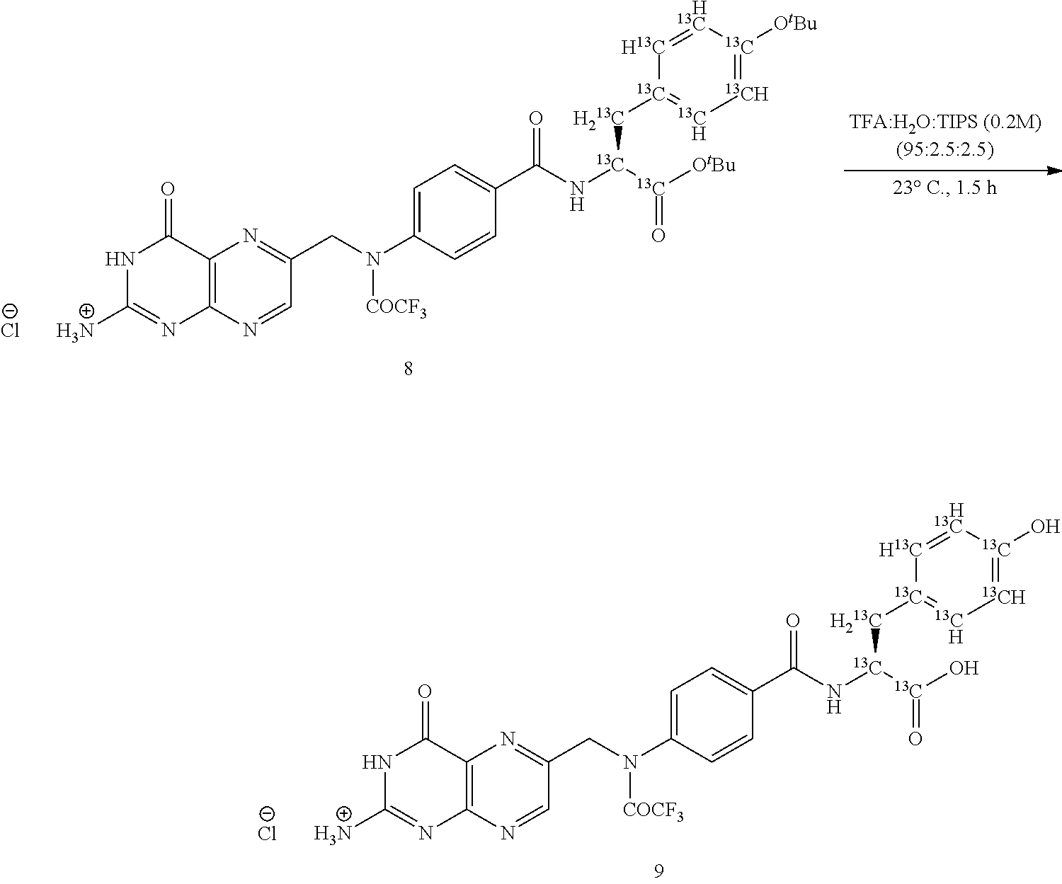

Synthesis of 14C Analog of Pte-L Tyrosine-S0456 (OTL-0041)


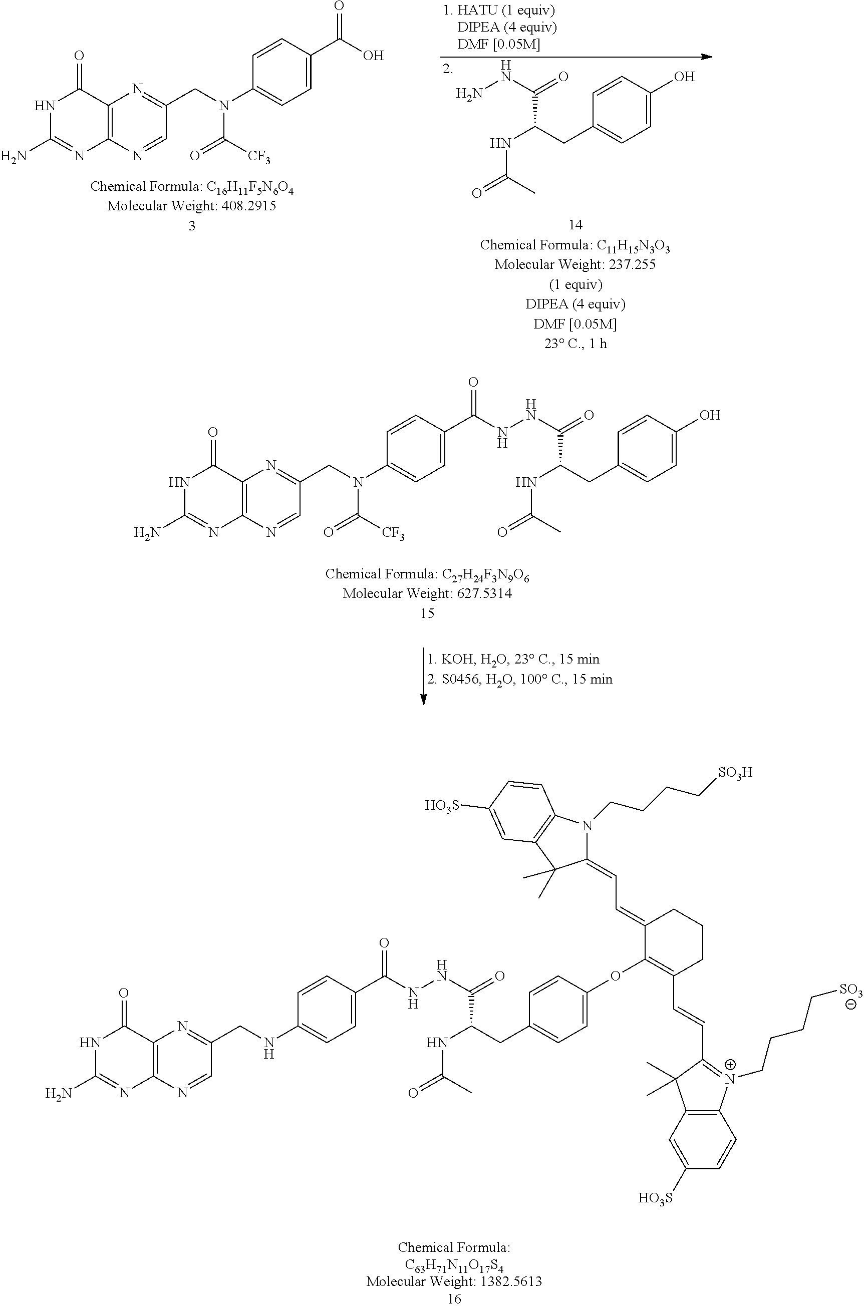


Claims (6)


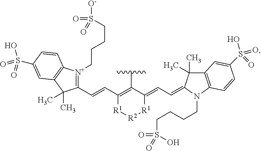


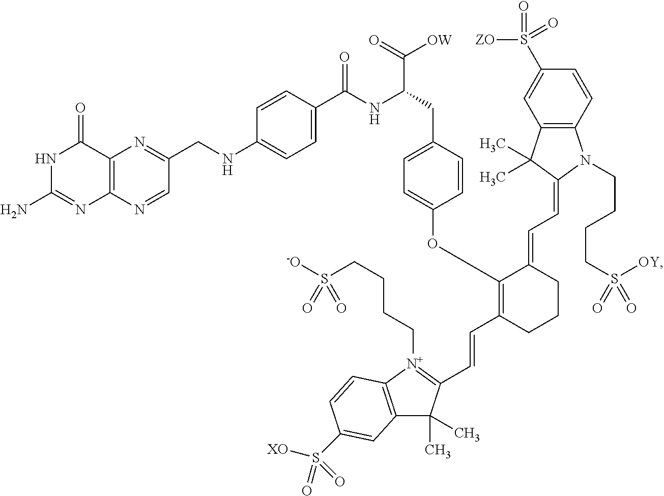


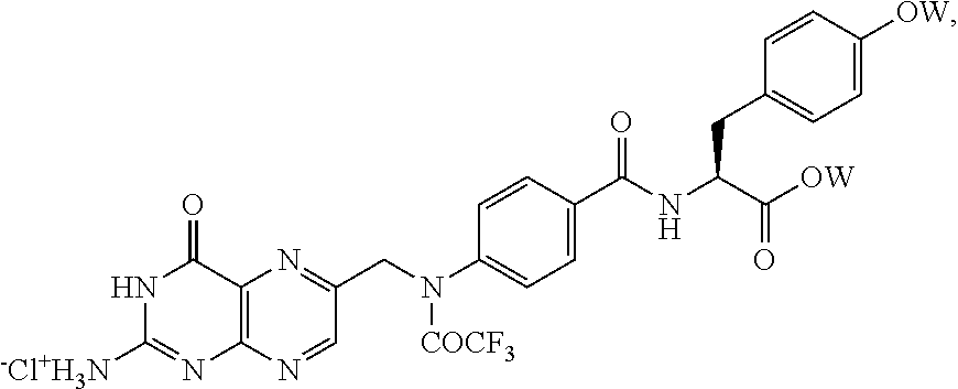


Priority Applications (11)
| Application Number | Priority Date | Filing Date | Title |
|---|---|---|---|
| US14/046,916 US9254341B2 (en) | 2013-03-15 | 2013-10-04 | Methods of manufacture of pteroyl-amino acid-fluorescent dyes |
| CN201380074662.7A CN105492905B (en) | 2013-03-15 | 2013-12-19 | The fluorescence imaging of inflammatory disease |
| AU2013381391A AU2013381391A1 (en) | 2013-03-15 | 2013-12-19 | Fluorescence imaging of inflammatory diseases |
| EP13877792.5A EP2972320B1 (en) | 2013-03-15 | 2013-12-19 | Methods of imaging inflammatory diseases by ligands conjugated to fluorescent compounds |
| CA2902205A CA2902205A1 (en) | 2013-03-15 | 2013-12-19 | Fluorescence imaging of inflammatory diseases |
| JP2016500131A JP2016512240A (en) | 2013-03-15 | 2013-12-19 | Fluorescence imaging of inflammatory diseases with ligands conjugated with fluorescent compounds |
| ES13877792T ES2765896T3 (en) | 2013-03-15 | 2013-12-19 | Imaging methods of inflammatory diseases using ligands conjugated with fluorescent compounds |
| PCT/US2013/076659 WO2014143309A1 (en) | 2013-03-15 | 2013-12-19 | Fluorescence imaging of inflammatory diseases |
| MX2015013219A MX2015013219A (en) | 2013-03-15 | 2013-12-19 | Fluorescence imaging of inflammatory diseases. |
| IL240509A IL240509B (en) | 2013-03-15 | 2015-08-12 | Fluorescence imaging of inflammatory diseases |
| JP2017170413A JP2017214425A (en) | 2013-03-15 | 2017-09-05 | Synthesis and composition of amino acid-linking group conjugated to compound used for targeted imaging of tumor |
Applications Claiming Priority (2)
| Application Number | Priority Date | Filing Date | Title |
|---|---|---|---|
| US201361791921P | 2013-03-15 | 2013-03-15 | |
| US14/046,916 US9254341B2 (en) | 2013-03-15 | 2013-10-04 | Methods of manufacture of pteroyl-amino acid-fluorescent dyes |
Publications (2)
| Publication Number | Publication Date |
|---|---|
| US20140275533A1 US20140275533A1 (en) | 2014-09-18 |
| US9254341B2 true US9254341B2 (en) | 2016-02-09 |
Family
ID=51527878
Family Applications (7)
| Application Number | Title | Priority Date | Filing Date |
|---|---|---|---|
| US14/010,098 Active US9333270B2 (en) | 2013-03-15 | 2013-08-26 | Synthesis and composition of amino acid linking groups conjugated to compounds used for the targeted imaging of tumors |
| US14/046,916 Active US9254341B2 (en) | 2013-03-15 | 2013-10-04 | Methods of manufacture of pteroyl-amino acid-fluorescent dyes |
| US14/135,099 Active US9233175B2 (en) | 2013-03-15 | 2013-12-19 | Methods of imaging inflammatory diseases by ligands conjugated to fluorescent compounds |
| US14/207,130 Active US9061057B2 (en) | 2013-03-15 | 2014-03-12 | Synthesis and composition of amino acid linking groups conjugated to compounds used for the targeted imaging of tumors |
| US14/715,799 Active US9341629B2 (en) | 2013-03-15 | 2015-05-19 | Synthesis and composition of amino acid linking groups conjugated to compounds used for the targeted imaging of tumors |
| US14/953,928 Active US9782497B2 (en) | 2013-03-15 | 2015-11-30 | Synthesis and composition of amino acid linking groups conjugated to compounds used for the targeted imaging of tumors |
| US15/259,719 Active US9789208B2 (en) | 2013-03-15 | 2016-09-08 | Synthesis and composition of amino acid linking groups conjugated to compounds used for the targeted imaging of tumors |
Family Applications Before (1)
| Application Number | Title | Priority Date | Filing Date |
|---|---|---|---|
| US14/010,098 Active US9333270B2 (en) | 2013-03-15 | 2013-08-26 | Synthesis and composition of amino acid linking groups conjugated to compounds used for the targeted imaging of tumors |
Family Applications After (5)
| Application Number | Title | Priority Date | Filing Date |
|---|---|---|---|
| US14/135,099 Active US9233175B2 (en) | 2013-03-15 | 2013-12-19 | Methods of imaging inflammatory diseases by ligands conjugated to fluorescent compounds |
| US14/207,130 Active US9061057B2 (en) | 2013-03-15 | 2014-03-12 | Synthesis and composition of amino acid linking groups conjugated to compounds used for the targeted imaging of tumors |
| US14/715,799 Active US9341629B2 (en) | 2013-03-15 | 2015-05-19 | Synthesis and composition of amino acid linking groups conjugated to compounds used for the targeted imaging of tumors |
| US14/953,928 Active US9782497B2 (en) | 2013-03-15 | 2015-11-30 | Synthesis and composition of amino acid linking groups conjugated to compounds used for the targeted imaging of tumors |
| US15/259,719 Active US9789208B2 (en) | 2013-03-15 | 2016-09-08 | Synthesis and composition of amino acid linking groups conjugated to compounds used for the targeted imaging of tumors |
Country Status (13)
| Country | Link |
|---|---|
| US (7) | US9333270B2 (en) |
| EP (3) | EP2968335B1 (en) |
| JP (5) | JP6192799B2 (en) |
| CN (3) | CN105228628B (en) |
| AU (4) | AU2013383382B2 (en) |
| BR (1) | BR112015022810B1 (en) |
| CA (3) | CA2903994C (en) |
| EA (1) | EA029738B1 (en) |
| ES (1) | ES2765896T3 (en) |
| HK (1) | HK1218257A1 (en) |
| IL (3) | IL240509B (en) |
| MX (3) | MX358184B (en) |
| WO (3) | WO2014149069A1 (en) |
Cited By (2)
| Publication number | Priority date | Publication date | Assignee | Title |
|---|---|---|---|---|
| WO2018049132A1 (en) * | 2016-09-09 | 2018-03-15 | On Target Laboratories, LLC | Psma-targeted nir dyes and their uses |
| US20230263911A1 (en) * | 2022-02-23 | 2023-08-24 | On Target Laboratories, Llc. | Compositions including pafolacianine for the identification of malignant lesions |
Families Citing this family (39)
| Publication number | Priority date | Publication date | Assignee | Title |
|---|---|---|---|---|
| WO2008119036A2 (en) | 2007-03-27 | 2008-10-02 | Radiomedix Inc. | Compositions for targeted imaging and therapy |
| CA2843313C (en) | 2011-06-20 | 2018-02-13 | Radiomedix Inc. | Compositions, methods of synthesis and use of carbohydrate targeted agents |
| US20170232119A1 (en) * | 2013-03-15 | 2017-08-17 | Purdue Research Foundation | Synthesis and composition of amino acid linking groups conjugated to compounds used for the targeted imaging of tumors |
| CN105228628B (en) * | 2013-03-15 | 2019-10-18 | 普渡研究基金会 | Synthesis and composition with the amino acid linker of the compound conjugation for cancer target to be imaged |
| US11162937B2 (en) * | 2013-11-19 | 2021-11-02 | Purdue Research Foundation | Patient selection method for inflammation |
| WO2016072984A1 (en) * | 2014-11-05 | 2016-05-12 | The United States Of America, As Represented By The Secretary, Department Of Health And Human Services | A new class of stable heptamethine cyanine fluorophores and biomedical applications thereof |
| WO2016183351A1 (en) | 2015-05-12 | 2016-11-17 | Medibeacon Inc. | Compositions and methods for assessing eye vasculature |
| FR3036621B1 (en) * | 2015-05-25 | 2017-05-19 | Fluoptics | MULTIVALENT TARGETING FLUORESCENT PLATTER IN THE NEAR INFRARED FOR OPTICAL IMAGING. |
| FR3036622B1 (en) * | 2015-05-25 | 2017-05-19 | Fluoptics | MULTIVALENT TARGETING FLUORESCENT TARGETER. |
| US10842887B2 (en) | 2015-09-09 | 2020-11-24 | On Target Laboratories, LLC | PSMA-targeted NIR dyes and their uses |
| US9808538B2 (en) | 2015-09-09 | 2017-11-07 | On Target Laboratories, LLC | PSMA-targeted NIR dyes and their uses |
| JP7285537B2 (en) * | 2016-03-16 | 2023-06-02 | オン ターゲット ラボラトリーズ エルエルシー | CA IX Targeted NIR Dyes and Their Use |
| US11555821B2 (en) | 2016-03-16 | 2023-01-17 | On Target Laboratories, Inc. | CA IX-NIR dyes and their uses |
| WO2018026965A1 (en) | 2016-08-02 | 2018-02-08 | Isi Life Sciences, Inc. | Compositions and methods for detecting cancer cells in a tissue sample |
| EP3496762A1 (en) | 2016-08-11 | 2019-06-19 | The U.S.A. as represented by the Secretary, Department of Health and Human Services | Near-ir light-cleavable conjugates and conjugate precursors |
| WO2018049129A1 (en) * | 2016-09-09 | 2018-03-15 | On Target Laboratories, LLC | Cholecystokinin 2 receptor targeted nir imaging and use thereof |
| JP2019534286A (en) * | 2016-10-26 | 2019-11-28 | カオ グループ、インク. | Cancer-binding colored peptides targeted for decay by radiant energy |
| WO2018101473A1 (en) * | 2016-12-02 | 2018-06-07 | 国立大学法人東京大学 | Compound, folate receptor visualization fluorescent probe, and use of these |
| WO2018111989A1 (en) * | 2016-12-14 | 2018-06-21 | Purdue Research Foundation | Fibroblast activation protein (fap)-targeted imaging and therapy |
| US10539567B2 (en) * | 2017-05-15 | 2020-01-21 | Indicator Systems International, Inc. | Biopsy methods and devices |
| US10753942B2 (en) | 2017-05-15 | 2020-08-25 | Indicator Systems International, Inc. | Methods to detect remnant cancer cells |
| CA3091423A1 (en) * | 2018-02-15 | 2019-08-22 | Children's National Medical Center | Heptamethine cyanines for use as fluorescent markers of the biliary and renal systems |
| CN108840815A (en) * | 2018-05-29 | 2018-11-20 | 苏州百源基因技术有限公司 | A kind of amino acid of fluorescent marker and its preparation method and application |
| CN108727245B (en) * | 2018-07-02 | 2021-09-28 | 广州中医药大学(广州中医药研究院) | Salicylic acid compound and preparation method and application thereof |
| KR102150415B1 (en) | 2018-12-07 | 2020-09-01 | 원광대학교 산학협력단 | Targeting compound of cancer and use thereof |
| JP7160694B2 (en) | 2019-01-08 | 2022-10-25 | 日立Geニュークリア・エナジー株式会社 | FLUID CONTACT MEMBER AND METHOD FOR MANUFACTURING FLUID CONTACT MEMBER |
| US11754500B2 (en) | 2019-06-20 | 2023-09-12 | Cilag Gmbh International | Minimizing image sensor input/output in a pulsed fluorescence imaging system |
| US11516387B2 (en) | 2019-06-20 | 2022-11-29 | Cilag Gmbh International | Image synchronization without input clock and data transmission clock in a pulsed hyperspectral, fluorescence, and laser mapping imaging system |
| US11986160B2 (en) | 2019-06-20 | 2024-05-21 | Cllag GmbH International | Image synchronization without input clock and data transmission clock in a pulsed hyperspectral imaging system |
| US11716533B2 (en) | 2019-06-20 | 2023-08-01 | Cilag Gmbh International | Image synchronization without input clock and data transmission clock in a pulsed fluorescence imaging system |
| US11892403B2 (en) | 2019-06-20 | 2024-02-06 | Cilag Gmbh International | Image synchronization without input clock and data transmission clock in a pulsed fluorescence imaging system |
| US20210353151A1 (en) * | 2020-05-12 | 2021-11-18 | On Target Laboratories, LLC | Targeted fluorescent markers in combination with a flexible probe |
| CN112010862B (en) * | 2020-10-23 | 2021-03-30 | 南京诺源医疗器械有限公司 | Active targeting folic acid receptor near-infrared fluorescent molecule and preparation method thereof |
| CN114014843B (en) * | 2021-11-17 | 2022-09-20 | 北京大学第一医院 | PSMA targeted nuclide/fluorescent bimodal ligand, molecular probe and application |
| CN114751907A (en) * | 2022-03-17 | 2022-07-15 | 南京诺源医疗器械有限公司 | Active targeting folic acid receptor near-infrared fluorescent molecule and preparation method and application thereof |
| CN115504984B (en) * | 2022-09-09 | 2023-07-25 | 南京诺源医疗器械有限公司 | Pemetic near infrared fluorescent molecule targeting alpha-folic acid receptor and preparation method and application thereof |
| CN115490672B (en) * | 2022-09-30 | 2023-11-17 | 南华大学 | Photosensitizer with photothermal and photodynamic effects as well as preparation method and application thereof |
| CN116621823B (en) * | 2023-07-06 | 2023-10-31 | 南京诺源医疗器械有限公司 | Near infrared fluorescent tracer for diagnosing metastatic lymph nodes, synthetic method and application |
| CN118078219A (en) * | 2024-04-24 | 2024-05-28 | 新斗生物科技(苏州)有限公司 | Near infrared fluorescence capture system, device and application |
Citations (6)
| Publication number | Priority date | Publication date | Assignee | Title |
|---|---|---|---|---|
| US4337339A (en) | 1979-04-30 | 1982-06-29 | Baker Instruments Corp. | Process for preparation of folic acid derivatives |
| US6534041B1 (en) | 1997-04-23 | 2003-03-18 | Institute For Diagnostic Research Gmbh Of The Free University Of Berlin | Acid-labile and enzymatically divisible dye compounds for diagnosis with near infrared light and for therapy |
| US20070009434A1 (en) | 2005-07-05 | 2007-01-11 | Low Philip S | Imaging and therapeutic method using monocytes |
| US20110286933A1 (en) | 2008-11-07 | 2011-11-24 | The General Hospital Corporation | Monofunctional carbocyanine dyes for in vivo and in vitro imaging |
| WO2012032068A2 (en) | 2010-09-06 | 2012-03-15 | Modpro Ab | Compounds and methods |
| WO2013126797A1 (en) | 2012-02-24 | 2013-08-29 | Purdue Research Foundation | Cholecystokinin b receptor targeting for imaging and therapy |
Family Cites Families (24)
| Publication number | Priority date | Publication date | Assignee | Title |
|---|---|---|---|---|
| US5716596A (en) | 1992-06-23 | 1998-02-10 | Diatide, Inc. | Radioactively labeled somatostatin-derived peptides for imaging and therapeutic uses |
| US5871711A (en) | 1992-06-23 | 1999-02-16 | Diatide, Inc. | Radioactively-labeled somatostatin-derived peptides for imaging and therapeutic uses |
| US7547721B1 (en) * | 1998-09-18 | 2009-06-16 | Bayer Schering Pharma Ag | Near infrared fluorescent contrast agent and fluorescence imaging |
| JP2000095758A (en) | 1998-09-18 | 2000-04-04 | Schering Ag | Near-infrared, fluorescent contrast medium, and its production |
| TR200201567T2 (en) | 1999-12-15 | 2002-11-21 | Schering Ag | Active fluorescent contrast agent and fluorescence imaging in the near infrared light region. |
| US7033594B2 (en) | 2000-03-31 | 2006-04-25 | Purdue Research Foundation | Method of treatment using ligand-immunogen conjugates |
| US7597878B2 (en) * | 2000-09-19 | 2009-10-06 | Li-Cor, Inc. | Optical fluorescent imaging |
| CA2424454C (en) * | 2000-09-19 | 2010-07-20 | Li-Cor, Inc. | Cyanine dyes |
| DE60214134T2 (en) | 2001-02-07 | 2007-07-19 | Beth Israel Deaconess Medical Center, Boston | MODIFIED PSMA LIGANDS AND THEIR USE |
| WO2002087424A2 (en) * | 2001-05-02 | 2002-11-07 | Purdue Research Foundation | Treatment and diagnosis of macrophage mediated disease |
| US8044200B2 (en) * | 2005-03-16 | 2011-10-25 | Endocyte, Inc. | Synthesis and purification of pteroic acid and conjugates thereof |
| WO2007100755A1 (en) * | 2006-02-27 | 2007-09-07 | Nabi Biopharmaceuticals | Method for decreasing the toxic effects of nicotine on fetuses in pregnant women |
| US20100226967A1 (en) * | 2006-05-23 | 2010-09-09 | Purdue Research Foundation | Imaging and therapeutic method using progenitor cells |
| EP2087337A4 (en) | 2006-11-03 | 2010-09-08 | Purdue Research Foundation | Ex vivo flow cytometry method and device |
| US8168804B2 (en) * | 2007-06-29 | 2012-05-01 | Vanderbilt University | Large stoke shift NIR dyes |
| CN102026671B (en) * | 2008-03-14 | 2014-09-03 | Visen医药公司 | Integrin targeting agents and in-vivo and in-vitro imaging methods using the same |
| CA2737496A1 (en) | 2008-09-17 | 2010-03-25 | Endocyte, Inc. | Folate receptor binding conjugates of antifolates |
| CN101440282B (en) * | 2008-12-18 | 2012-06-27 | 中国药科大学 | Near-infrared fluorescent molecular probe, synthesizing method and use thereof |
| CA2754492A1 (en) * | 2009-03-05 | 2010-09-10 | Purdue Research Foundation | Method for early imaging of atherosclerosis |
| WO2010121163A2 (en) * | 2009-04-17 | 2010-10-21 | Li-Cor, Inc. | Fluorescent imaging with substituted cyanine dyes |
| CA2800693A1 (en) | 2010-05-28 | 2011-12-01 | Purdue Research Foundation | Delivery of therapeutic agents to inflamed tissues using folate-targeted agents |
| WO2012054784A1 (en) * | 2010-10-20 | 2012-04-26 | Li-Cor, Inc. | Fluorescent imaging with substituted cyanine dyes |
| US9629918B2 (en) * | 2012-02-29 | 2017-04-25 | Purdue Research Foundation | Folate receptor alpha binding ligands |
| CN105228628B (en) * | 2013-03-15 | 2019-10-18 | 普渡研究基金会 | Synthesis and composition with the amino acid linker of the compound conjugation for cancer target to be imaged |
-
2013
- 2013-08-26 CN CN201380074692.8A patent/CN105228628B/en active Active
- 2013-08-26 AU AU2013383382A patent/AU2013383382B2/en active Active
- 2013-08-26 MX MX2015013223A patent/MX358184B/en active IP Right Grant
- 2013-08-26 EA EA201591802A patent/EA029738B1/en not_active IP Right Cessation
- 2013-08-26 WO PCT/US2013/056629 patent/WO2014149069A1/en active Application Filing
- 2013-08-26 EP EP13878809.6A patent/EP2968335B1/en active Active
- 2013-08-26 US US14/010,098 patent/US9333270B2/en active Active
- 2013-08-26 CA CA2903994A patent/CA2903994C/en active Active
- 2013-08-26 BR BR112015022810-0A patent/BR112015022810B1/en active IP Right Grant
- 2013-08-26 JP JP2016500088A patent/JP6192799B2/en active Active
- 2013-10-04 MX MX2015011830A patent/MX2015011830A/en unknown
- 2013-10-04 US US14/046,916 patent/US9254341B2/en active Active
- 2013-10-04 WO PCT/US2013/063593 patent/WO2014149073A1/en active Application Filing
- 2013-10-04 CN CN201380074694.7A patent/CN105120903A/en active Pending
- 2013-10-04 AU AU2013383386A patent/AU2013383386A1/en not_active Abandoned
- 2013-10-04 CA CA2903727A patent/CA2903727C/en active Active
- 2013-10-04 EP EP13878632.2A patent/EP2968614B1/en active Active
- 2013-10-04 JP JP2016500094A patent/JP6270981B2/en active Active
- 2013-12-19 EP EP13877792.5A patent/EP2972320B1/en active Active
- 2013-12-19 CN CN201380074662.7A patent/CN105492905B/en active Active
- 2013-12-19 MX MX2015013219A patent/MX2015013219A/en unknown
- 2013-12-19 ES ES13877792T patent/ES2765896T3/en active Active
- 2013-12-19 US US14/135,099 patent/US9233175B2/en active Active
- 2013-12-19 AU AU2013381391A patent/AU2013381391A1/en not_active Abandoned
- 2013-12-19 CA CA2902205A patent/CA2902205A1/en not_active Abandoned
- 2013-12-19 WO PCT/US2013/076659 patent/WO2014143309A1/en active Application Filing
- 2013-12-19 JP JP2016500131A patent/JP2016512240A/en active Pending
-
2014
- 2014-03-12 US US14/207,130 patent/US9061057B2/en active Active
-
2015
- 2015-05-19 US US14/715,799 patent/US9341629B2/en active Active
- 2015-08-12 IL IL240509A patent/IL240509B/en active IP Right Grant
- 2015-08-18 IL IL240673A patent/IL240673B/en active IP Right Grant
- 2015-08-25 IL IL240828A patent/IL240828A/en active IP Right Grant
- 2015-11-30 US US14/953,928 patent/US9782497B2/en active Active
-
2016
- 2016-06-02 HK HK16106296.0A patent/HK1218257A1/en unknown
- 2016-09-08 US US15/259,719 patent/US9789208B2/en active Active
-
2017
- 2017-05-18 AU AU2017203340A patent/AU2017203340B2/en active Active
- 2017-05-31 JP JP2017107741A patent/JP2017149776A/en active Pending
- 2017-09-05 JP JP2017170413A patent/JP2017214425A/en active Pending
Patent Citations (6)
| Publication number | Priority date | Publication date | Assignee | Title |
|---|---|---|---|---|
| US4337339A (en) | 1979-04-30 | 1982-06-29 | Baker Instruments Corp. | Process for preparation of folic acid derivatives |
| US6534041B1 (en) | 1997-04-23 | 2003-03-18 | Institute For Diagnostic Research Gmbh Of The Free University Of Berlin | Acid-labile and enzymatically divisible dye compounds for diagnosis with near infrared light and for therapy |
| US20070009434A1 (en) | 2005-07-05 | 2007-01-11 | Low Philip S | Imaging and therapeutic method using monocytes |
| US20110286933A1 (en) | 2008-11-07 | 2011-11-24 | The General Hospital Corporation | Monofunctional carbocyanine dyes for in vivo and in vitro imaging |
| WO2012032068A2 (en) | 2010-09-06 | 2012-03-15 | Modpro Ab | Compounds and methods |
| WO2013126797A1 (en) | 2012-02-24 | 2013-08-29 | Purdue Research Foundation | Cholecystokinin b receptor targeting for imaging and therapy |
Non-Patent Citations (7)
| Title |
|---|
| Mahalingham et al., 329-Evaluation of pteroyl-amino acid-NIR dye conjugates for tumor targeted fluorescence guided surgery. 246th ACS National Meeting and Exposition, Sep. 8-12, 2013. Indianapolis, Indiana. [Retrievedo n Jan. 31, 2014]. Retrieved from the Internet. . entire document. |
| Mahalingham et al., 329-Evaluation of pteroyl-amino acid-NIR dye conjugates for tumor targeted fluorescence guided surgery. 246th ACS National Meeting and Exposition, Sep. 8-12, 2013. Indianapolis, Indiana. [Retrievedo n Jan. 31, 2014]. Retrieved from the Internet. <URL:http://abstracts.acs.org/chem/246nm/program/view.php>. entire document. |
| Milstein, et al., Statistical approach for detection and localization of a fluorescing mouse tumor in Intralipid, Applied Optics, vol. 44, No. 12, Apr. 20, 2005. |
| Moon, et al., Enhanced Tumor Detection Using a Folate Receptor-Targeted Near-Infrared Fluorochrome Conjugate, Center for Molecular Imaging Reserach, Massachusetts General Hospital, Harvard Medical School, Charlestown, MA, Jan. 22, 2003, Revised Mar. 3, 2003. |
| PCT, Notification of Transmittal of the International Search Report and the Written Opinion of the International Searching Authority, or the Declaration, in International application No. PCT/US2013/063593, dated Feb. 18, 2014. (7 pages). |
| Yang et al., Characterization of the pH of Folate Receptor-Containing Endosomes and the Rate of Hydrolysis of Internalized Acid-Labile Folate-Drug Conjugates. J Pharmacol Exp. Ther, 321 (2): 462-468, (2007). [retrieved on Jan. 27, 2014]. Retrieved from the Internet. . entire document. |
| Yang et al., Characterization of the pH of Folate Receptor-Containing Endosomes and the Rate of Hydrolysis of Internalized Acid-Labile Folate-Drug Conjugates. J Pharmacol Exp. Ther, 321 (2): 462-468, (2007). [retrieved on Jan. 27, 2014]. Retrieved from the Internet. <URL: http://jpet.aspetjournals.org/content/321/462.full.pdf>. entire document. |
Cited By (4)
| Publication number | Priority date | Publication date | Assignee | Title |
|---|---|---|---|---|
| WO2018049132A1 (en) * | 2016-09-09 | 2018-03-15 | On Target Laboratories, LLC | Psma-targeted nir dyes and their uses |
| US10308606B2 (en) | 2016-09-09 | 2019-06-04 | On Target Laboratories, LLC | PSMA-targeted NIR dyes and their uses |
| US20230263911A1 (en) * | 2022-02-23 | 2023-08-24 | On Target Laboratories, Llc. | Compositions including pafolacianine for the identification of malignant lesions |
| US11986540B2 (en) * | 2022-02-23 | 2024-05-21 | On Target Laboratories, LLC | Compositions including pafolacianine for the identification of malignant lesions |
Also Published As
Similar Documents
| Publication | Publication Date | Title |
|---|---|---|
| US9254341B2 (en) | Methods of manufacture of pteroyl-amino acid-fluorescent dyes | |
| US10746741B2 (en) | CA IX—NIR dyes and their uses | |
| EP3510399B1 (en) | Psma-targeted nir dyes and their uses | |
| US20210275694A1 (en) | Synthesis and composition of amino acid linking groups conjugated to compounds used for the targeted imaging of tumors | |
| US11555821B2 (en) | CA IX-NIR dyes and their uses |
Legal Events
| Date | Code | Title | Description |
|---|---|---|---|
| STCF | Information on status: patent grant |
Free format text: PATENTED CASE |
|
| AS | Assignment |
Owner name: PENSION FUND OF THE CHRISTIAN CHURCH (DISCIPLES OF Free format text: SECURITY INTEREST;ASSIGNOR:ON TARGET LABORATORIES, LLC;REEL/FRAME:037924/0675 Effective date: 20160301 |
|
| AS | Assignment |
Owner name: PURDUE RESEARCH FOUNDATION, INDIANA Free format text: ASSIGNMENT OF ASSIGNORS INTEREST;ASSIGNORS:KULARATNE, SUMITH A.;GAGARE, PRAVIN;REEL/FRAME:043272/0405 Effective date: 20170811 Owner name: ON TARGET LABORATORIES, LLC, INDIANA Free format text: EMPLOYEE AGREEMENT;ASSIGNOR:NOSHI, MOHAMMAD;REEL/FRAME:043532/0628 Effective date: 20131212 |
|
| AS | Assignment |
Owner name: ON TARGET LABORATORIES, LLC, INDIANA Free format text: RELEASE BY SECURED PARTY;ASSIGNOR:PENSION FUND OF THE CHRISTIAN CHURCH (DISCIPLES OF CHIRST), AS TRUSTEE FOR THE PENSION FUND OF THE CHRISTIAN CHURCH GROUP TRUST;REEL/FRAME:043730/0223 Effective date: 20170926 |
|
| MAFP | Maintenance fee payment |
Free format text: PAYMENT OF MAINTENANCE FEE, 4TH YR, SMALL ENTITY (ORIGINAL EVENT CODE: M2551); ENTITY STATUS OF PATENT OWNER: SMALL ENTITY Year of fee payment: 4 |
|
| MAFP | Maintenance fee payment |
Free format text: PAYMENT OF MAINTENANCE FEE, 8TH YR, SMALL ENTITY (ORIGINAL EVENT CODE: M2552); ENTITY STATUS OF PATENT OWNER: SMALL ENTITY Year of fee payment: 8 |
|
| AS | Assignment |
Owner name: ON TARGET LABORATORIES, INC., INDIANA Free format text: CHANGE OF NAME;ASSIGNOR:ON TARGET LABORATORIES, LLC;REEL/FRAME:067419/0833 Effective date: 20170927 |