US9233175B2 - Methods of imaging inflammatory diseases by ligands conjugated to fluorescent compounds - Google Patents
Methods of imaging inflammatory diseases by ligands conjugated to fluorescent compounds Download PDFInfo
- Publication number
- US9233175B2 US9233175B2 US14/135,099 US201314135099A US9233175B2 US 9233175 B2 US9233175 B2 US 9233175B2 US 201314135099 A US201314135099 A US 201314135099A US 9233175 B2 US9233175 B2 US 9233175B2
- Authority
- US
- United States
- Prior art keywords
- compound
- inflammatory disease
- disease
- inflammatory
- imaging
- Prior art date
- Legal status (The legal status is an assumption and is not a legal conclusion. Google has not performed a legal analysis and makes no representation as to the accuracy of the status listed.)
- Active
Links
- 150000001875 compounds Chemical class 0.000 title claims abstract description 146
- 238000000034 method Methods 0.000 title claims abstract description 71
- 208000027866 inflammatory disease Diseases 0.000 title claims abstract description 52
- 238000003384 imaging method Methods 0.000 title claims abstract description 41
- 239000003446 ligand Substances 0.000 title abstract description 29
- 150000001413 amino acids Chemical class 0.000 claims abstract description 22
- 235000001014 amino acid Nutrition 0.000 claims description 27
- 230000005284 excitation Effects 0.000 claims description 24
- 210000004969 inflammatory cell Anatomy 0.000 claims description 21
- 210000002540 macrophage Anatomy 0.000 claims description 21
- 238000002347 injection Methods 0.000 claims description 20
- 239000007924 injection Substances 0.000 claims description 20
- 201000001320 Atherosclerosis Diseases 0.000 claims description 15
- 206010009900 Colitis ulcerative Diseases 0.000 claims description 14
- 201000006704 Ulcerative Colitis Diseases 0.000 claims description 14
- 206010039073 rheumatoid arthritis Diseases 0.000 claims description 14
- OUYCCCASQSFEME-QMMMGPOBSA-N L-tyrosine Chemical compound OC(=O)[C@@H](N)CC1=CC=C(O)C=C1 OUYCCCASQSFEME-QMMMGPOBSA-N 0.000 claims description 11
- 230000004054 inflammatory process Effects 0.000 claims description 11
- 239000000203 mixture Substances 0.000 claims description 11
- 208000005069 pulmonary fibrosis Diseases 0.000 claims description 11
- OUYCCCASQSFEME-UHFFFAOYSA-N tyrosine Natural products OC(=O)C(N)CC1=CC=C(O)C=C1 OUYCCCASQSFEME-UHFFFAOYSA-N 0.000 claims description 11
- 206010061218 Inflammation Diseases 0.000 claims description 10
- 201000004681 Psoriasis Diseases 0.000 claims description 9
- 150000003862 amino acid derivatives Chemical class 0.000 claims description 9
- 208000009329 Graft vs Host Disease Diseases 0.000 claims description 8
- 208000024908 graft versus host disease Diseases 0.000 claims description 8
- 210000000130 stem cell Anatomy 0.000 claims description 8
- KDXKERNSBIXSRK-YFKPBYRVSA-N L-lysine Chemical compound NCCCC[C@H](N)C(O)=O KDXKERNSBIXSRK-YFKPBYRVSA-N 0.000 claims description 7
- 238000000295 emission spectrum Methods 0.000 claims description 7
- 201000008482 osteoarthritis Diseases 0.000 claims description 7
- 230000027455 binding Effects 0.000 claims description 6
- 208000037976 chronic inflammation Diseases 0.000 claims description 6
- 206010025135 lupus erythematosus Diseases 0.000 claims description 6
- 208000011231 Crohn disease Diseases 0.000 claims description 5
- 208000001640 Fibromyalgia Diseases 0.000 claims description 5
- 206010018364 Glomerulonephritis Diseases 0.000 claims description 5
- KDXKERNSBIXSRK-UHFFFAOYSA-N Lysine Natural products NCCCCC(N)C(O)=O KDXKERNSBIXSRK-UHFFFAOYSA-N 0.000 claims description 5
- 239000004472 Lysine Substances 0.000 claims description 5
- 206010031252 Osteomyelitis Diseases 0.000 claims description 5
- 206010038934 Retinopathy proliferative Diseases 0.000 claims description 5
- 208000021386 Sjogren Syndrome Diseases 0.000 claims description 5
- 201000009594 Systemic Scleroderma Diseases 0.000 claims description 5
- 206010042953 Systemic sclerosis Diseases 0.000 claims description 5
- 230000006020 chronic inflammation Effects 0.000 claims description 5
- 238000000695 excitation spectrum Methods 0.000 claims description 5
- 238000007912 intraperitoneal administration Methods 0.000 claims description 5
- 210000001616 monocyte Anatomy 0.000 claims description 5
- 201000006417 multiple sclerosis Diseases 0.000 claims description 5
- 208000037803 restenosis Diseases 0.000 claims description 5
- 201000000306 sarcoidosis Diseases 0.000 claims description 5
- 150000003668 tyrosines Chemical class 0.000 claims description 5
- XUJNEKJLAYXESH-REOHCLBHSA-N L-Cysteine Chemical compound SC[C@H](N)C(O)=O XUJNEKJLAYXESH-REOHCLBHSA-N 0.000 claims description 4
- 235000018417 cysteine Nutrition 0.000 claims description 4
- XUJNEKJLAYXESH-UHFFFAOYSA-N cysteine Natural products SCC(N)C(O)=O XUJNEKJLAYXESH-UHFFFAOYSA-N 0.000 claims description 4
- 238000012632 fluorescent imaging Methods 0.000 claims description 4
- 229910052739 hydrogen Inorganic materials 0.000 claims description 4
- 210000004072 lung Anatomy 0.000 claims description 4
- 150000003839 salts Chemical class 0.000 claims description 4
- 229910052708 sodium Inorganic materials 0.000 claims description 4
- 210000001035 gastrointestinal tract Anatomy 0.000 claims description 3
- 238000001990 intravenous administration Methods 0.000 claims description 3
- 229910052757 nitrogen Inorganic materials 0.000 claims description 3
- 229910052760 oxygen Inorganic materials 0.000 claims description 3
- 229910052717 sulfur Inorganic materials 0.000 claims description 3
- MTCFGRXMJLQNBG-REOHCLBHSA-N (2S)-2-Amino-3-hydroxypropansäure Chemical compound OC[C@H](N)C(O)=O MTCFGRXMJLQNBG-REOHCLBHSA-N 0.000 claims description 2
- MTCFGRXMJLQNBG-UHFFFAOYSA-N Serine Natural products OCC(N)C(O)=O MTCFGRXMJLQNBG-UHFFFAOYSA-N 0.000 claims description 2
- 229910052799 carbon Inorganic materials 0.000 claims description 2
- 125000000816 ethylene group Chemical group [H]C([H])([*:1])C([H])([H])[*:2] 0.000 claims description 2
- 229910052700 potassium Inorganic materials 0.000 claims description 2
- 210000001367 artery Anatomy 0.000 claims 2
- 210000001503 joint Anatomy 0.000 claims 2
- 201000005569 Gout Diseases 0.000 claims 1
- 210000000544 articulatio talocruralis Anatomy 0.000 claims 1
- 150000001945 cysteines Chemical class 0.000 claims 1
- 210000002310 elbow joint Anatomy 0.000 claims 1
- 210000001145 finger joint Anatomy 0.000 claims 1
- 125000000524 functional group Chemical group 0.000 claims 1
- 210000000629 knee joint Anatomy 0.000 claims 1
- 210000000878 metatarsophalangeal joint Anatomy 0.000 claims 1
- 150000003355 serines Chemical class 0.000 claims 1
- 210000000323 shoulder joint Anatomy 0.000 claims 1
- 210000003857 wrist joint Anatomy 0.000 claims 1
- 125000001747 pteroyl group Chemical group [H]C1=C([H])C(C(=O)[*])=C([H])C([H])=C1N([H])C([H])([H])C1=C([H])N=C2N([H])C(N([H])[H])=NC(=O)C2=N1 0.000 abstract description 14
- 102000005962 receptors Human genes 0.000 abstract description 8
- 108020003175 receptors Proteins 0.000 abstract description 7
- 239000000523 sample Substances 0.000 abstract description 3
- 150000001412 amines Chemical class 0.000 abstract 1
- 210000001519 tissue Anatomy 0.000 description 105
- 230000008685 targeting Effects 0.000 description 59
- 208000037265 diseases, disorders, signs and symptoms Diseases 0.000 description 44
- 239000000975 dye Substances 0.000 description 44
- 201000010099 disease Diseases 0.000 description 43
- OVBPIULPVIDEAO-LBPRGKRZSA-N folic acid Chemical compound C=1N=C2NC(N)=NC(=O)C2=NC=1CNC1=CC=C(C(=O)N[C@@H](CCC(O)=O)C(O)=O)C=C1 OVBPIULPVIDEAO-LBPRGKRZSA-N 0.000 description 25
- 229940024606 amino acid Drugs 0.000 description 23
- 235000019152 folic acid Nutrition 0.000 description 21
- 239000011724 folic acid Substances 0.000 description 21
- 210000004027 cell Anatomy 0.000 description 20
- 241000699670 Mus sp. Species 0.000 description 18
- 241000699666 Mus <mouse, genus> Species 0.000 description 17
- 229940014144 folate Drugs 0.000 description 17
- 102000006815 folate receptor Human genes 0.000 description 17
- 108020005243 folate receptor Proteins 0.000 description 17
- 239000012216 imaging agent Substances 0.000 description 14
- 208000009386 Experimental Arthritis Diseases 0.000 description 11
- 238000001727 in vivo Methods 0.000 description 11
- 206010028980 Neoplasm Diseases 0.000 description 9
- HEMHJVSKTPXQMS-UHFFFAOYSA-M Sodium hydroxide Chemical compound [OH-].[Na+] HEMHJVSKTPXQMS-UHFFFAOYSA-M 0.000 description 9
- 238000009825 accumulation Methods 0.000 description 9
- 239000000427 antigen Substances 0.000 description 9
- 102000036639 antigens Human genes 0.000 description 9
- 108091007433 antigens Proteins 0.000 description 9
- 230000008901 benefit Effects 0.000 description 9
- 238000011282 treatment Methods 0.000 description 9
- 238000001514 detection method Methods 0.000 description 8
- ZMXDDKWLCZADIW-UHFFFAOYSA-N N,N-Dimethylformamide Chemical compound CN(C)C=O ZMXDDKWLCZADIW-UHFFFAOYSA-N 0.000 description 7
- 230000002159 abnormal effect Effects 0.000 description 7
- 206010003246 arthritis Diseases 0.000 description 7
- 230000015572 biosynthetic process Effects 0.000 description 7
- 238000001228 spectrum Methods 0.000 description 7
- JGFZNNIVVJXRND-UHFFFAOYSA-N N,N-Diisopropylethylamine (DIPEA) Chemical compound CCN(C(C)C)C(C)C JGFZNNIVVJXRND-UHFFFAOYSA-N 0.000 description 6
- 238000003745 diagnosis Methods 0.000 description 6
- 238000002405 diagnostic procedure Methods 0.000 description 6
- 230000028993 immune response Effects 0.000 description 6
- 238000004519 manufacturing process Methods 0.000 description 6
- 125000006239 protecting group Chemical group 0.000 description 6
- 239000002904 solvent Substances 0.000 description 6
- 238000001356 surgical procedure Methods 0.000 description 6
- RTZKZFJDLAIYFH-UHFFFAOYSA-N Diethyl ether Chemical compound CCOCC RTZKZFJDLAIYFH-UHFFFAOYSA-N 0.000 description 5
- 102000010449 Folate receptor beta Human genes 0.000 description 5
- 108050001930 Folate receptor beta Proteins 0.000 description 5
- -1 amino acid compound Chemical class 0.000 description 5
- 239000012472 biological sample Substances 0.000 description 5
- 238000012634 optical imaging Methods 0.000 description 5
- 244000052769 pathogen Species 0.000 description 5
- 230000008569 process Effects 0.000 description 5
- 238000003786 synthesis reaction Methods 0.000 description 5
- XLYOFNOQVPJJNP-UHFFFAOYSA-N water Substances O XLYOFNOQVPJJNP-UHFFFAOYSA-N 0.000 description 5
- KYCODYTVUXDEEW-WTUKUHAWSA-L CC1=CC2=C(C=C1)N(CCCCS(=O)(=O)O[Y])/C(=C/C=C1\CCCC(/C=C/C3=[N+](CCCCS(=O)(=O)[O-])C4=C(C=C(CS(=O)(=O)O)C=C4)C3(C)C)=C1OC1=CC=C(C[C@H](NC(=O)C3=CC=C(NCC4=NC5=C(N=C4)N=C(N)NC5=O)C=C3)C(=O)O[W])C=C1)C2(C)C Chemical compound CC1=CC2=C(C=C1)N(CCCCS(=O)(=O)O[Y])/C(=C/C=C1\CCCC(/C=C/C3=[N+](CCCCS(=O)(=O)[O-])C4=C(C=C(CS(=O)(=O)O)C=C4)C3(C)C)=C1OC1=CC=C(C[C@H](NC(=O)C3=CC=C(NCC4=NC5=C(N=C4)N=C(N)NC5=O)C=C3)C(=O)O[W])C=C1)C2(C)C KYCODYTVUXDEEW-WTUKUHAWSA-L 0.000 description 4
- IAZDPXIOMUYVGZ-UHFFFAOYSA-N Dimethylsulphoxide Chemical compound CS(C)=O IAZDPXIOMUYVGZ-UHFFFAOYSA-N 0.000 description 4
- OVBPIULPVIDEAO-UHFFFAOYSA-N N-Pteroyl-L-glutaminsaeure Natural products C=1N=C2NC(N)=NC(=O)C2=NC=1CNC1=CC=C(C(=O)NC(CCC(O)=O)C(O)=O)C=C1 OVBPIULPVIDEAO-UHFFFAOYSA-N 0.000 description 4
- WZNFMVCISLJVQR-UHFFFAOYSA-N NC1=NC2=C(N=C(CNC3=CC=C(C(=O)C[Y])C=C3)C=N2)C(=O)N1 Chemical compound NC1=NC2=C(N=C(CNC3=CC=C(C(=O)C[Y])C=C3)C=N2)C(=O)N1 WZNFMVCISLJVQR-UHFFFAOYSA-N 0.000 description 4
- 102000001708 Protein Isoforms Human genes 0.000 description 4
- 108010029485 Protein Isoforms Proteins 0.000 description 4
- FAPWRFPIFSIZLT-UHFFFAOYSA-M Sodium chloride Chemical compound [Na+].[Cl-] FAPWRFPIFSIZLT-UHFFFAOYSA-M 0.000 description 4
- 239000003795 chemical substances by application Substances 0.000 description 4
- 230000008878 coupling Effects 0.000 description 4
- 238000010168 coupling process Methods 0.000 description 4
- 238000005859 coupling reaction Methods 0.000 description 4
- 235000005911 diet Nutrition 0.000 description 4
- 230000037213 diet Effects 0.000 description 4
- 230000003511 endothelial effect Effects 0.000 description 4
- 229960000304 folic acid Drugs 0.000 description 4
- 235000018977 lysine Nutrition 0.000 description 4
- 239000000047 product Substances 0.000 description 4
- 239000011780 sodium chloride Substances 0.000 description 4
- 230000003595 spectral effect Effects 0.000 description 4
- 238000007910 systemic administration Methods 0.000 description 4
- 230000001225 therapeutic effect Effects 0.000 description 4
- 238000002560 therapeutic procedure Methods 0.000 description 4
- CSCPPACGZOOCGX-UHFFFAOYSA-N Acetone Chemical compound CC(C)=O CSCPPACGZOOCGX-UHFFFAOYSA-N 0.000 description 3
- 208000023275 Autoimmune disease Diseases 0.000 description 3
- KMSCRNGLEUMULY-XUUVOBQHSA-N CC(=O)N[C@H](CC1=CC=C(OC(C)C)C=C1)C(=O)CNC(C)C.CC(C)C[C@H](CCC1=CC=C(OC(C)C)C=C1)C(=O)O.CC(C)NCCC1=CC=C(OC(C)C)C=C1.CC(C)N[C@@H](CC1=CC=C(OC(C)C)C=C1)C(=O)O.CC(C)N[C@@H](CC1=CC=C(OC(C)C)C=C1)C(=O)OCC1=CC=CC=C1.CC(C)N[C@H](CC(=O)O)CC1=CC=C(OC(C)C)C=C1.CC(C)N[C@H](CC1=CC=C(OC(C)C)C=C1)C(=O)O.CC(C)OC1=CC=C(C[C@@H](C(=O)O)N(C)C(C)C)C=C1.COC(=O)[C@H](CC1=CC=C(OC(C)C)C=C1)NC(C)C Chemical compound CC(=O)N[C@H](CC1=CC=C(OC(C)C)C=C1)C(=O)CNC(C)C.CC(C)C[C@H](CCC1=CC=C(OC(C)C)C=C1)C(=O)O.CC(C)NCCC1=CC=C(OC(C)C)C=C1.CC(C)N[C@@H](CC1=CC=C(OC(C)C)C=C1)C(=O)O.CC(C)N[C@@H](CC1=CC=C(OC(C)C)C=C1)C(=O)OCC1=CC=CC=C1.CC(C)N[C@H](CC(=O)O)CC1=CC=C(OC(C)C)C=C1.CC(C)N[C@H](CC1=CC=C(OC(C)C)C=C1)C(=O)O.CC(C)OC1=CC=C(C[C@@H](C(=O)O)N(C)C(C)C)C=C1.COC(=O)[C@H](CC1=CC=C(OC(C)C)C=C1)NC(C)C KMSCRNGLEUMULY-XUUVOBQHSA-N 0.000 description 3
- VEXZGXHMUGYJMC-UHFFFAOYSA-N Hydrochloric acid Chemical compound Cl VEXZGXHMUGYJMC-UHFFFAOYSA-N 0.000 description 3
- 125000003277 amino group Chemical group 0.000 description 3
- 239000006227 byproduct Substances 0.000 description 3
- 230000021615 conjugation Effects 0.000 description 3
- 230000002950 deficient Effects 0.000 description 3
- 238000002265 electronic spectrum Methods 0.000 description 3
- 239000007850 fluorescent dye Substances 0.000 description 3
- 238000004128 high performance liquid chromatography Methods 0.000 description 3
- 208000015181 infectious disease Diseases 0.000 description 3
- 230000004968 inflammatory condition Effects 0.000 description 3
- 230000002757 inflammatory effect Effects 0.000 description 3
- 238000011835 investigation Methods 0.000 description 3
- 125000005647 linker group Chemical group 0.000 description 3
- 210000001165 lymph node Anatomy 0.000 description 3
- 235000018102 proteins Nutrition 0.000 description 3
- 102000004169 proteins and genes Human genes 0.000 description 3
- 108090000623 proteins and genes Proteins 0.000 description 3
- 238000011160 research Methods 0.000 description 3
- 230000004044 response Effects 0.000 description 3
- 238000002603 single-photon emission computed tomography Methods 0.000 description 3
- 239000011734 sodium Substances 0.000 description 3
- 208000024891 symptom Diseases 0.000 description 3
- ZGYICYBLPGRURT-UHFFFAOYSA-N tri(propan-2-yl)silicon Chemical compound CC(C)[Si](C(C)C)C(C)C ZGYICYBLPGRURT-UHFFFAOYSA-N 0.000 description 3
- 238000012800 visualization Methods 0.000 description 3
- 229940088594 vitamin Drugs 0.000 description 3
- 235000013343 vitamin Nutrition 0.000 description 3
- 239000011782 vitamin Substances 0.000 description 3
- 229930003231 vitamin Natural products 0.000 description 3
- 150000003722 vitamin derivatives Chemical class 0.000 description 3
- PDXNSXLPXJFETD-DYVQZXGMSA-N (2e)-2-[(2e)-2-[2-[4-[(2s)-2-[[4-[(2-amino-4-oxo-1h-pteridin-6-yl)methylamino]benzoyl]amino]-2-carboxyethyl]phenoxy]-3-[(e)-2-[3,3-dimethyl-5-sulfo-1-(4-sulfobutyl)indol-1-ium-2-yl]ethenyl]cyclohex-2-en-1-ylidene]ethylidene]-3,3-dimethyl-1-(4-sulfobutyl)i Chemical compound OS(=O)(=O)CCCCN1C2=CC=C(S([O-])(=O)=O)C=C2C(C)(C)\C1=C/C=C\1C(OC=2C=CC(C[C@H](NC(=O)C=3C=CC(NCC=4N=C5C(=O)N=C(N)NC5=NC=4)=CC=3)C(O)=O)=CC=2)=C(\C=C\C=2C(C3=CC(=CC=C3[N+]=2CCCCS(O)(=O)=O)S(O)(=O)=O)(C)C)CCC/1 PDXNSXLPXJFETD-DYVQZXGMSA-N 0.000 description 2
- HNXQXTQTPAJEJL-UHFFFAOYSA-N 2-aminopteridin-4-ol Chemical class C1=CN=C2NC(N)=NC(=O)C2=N1 HNXQXTQTPAJEJL-UHFFFAOYSA-N 0.000 description 2
- JOAQINSXLLMRCV-UHFFFAOYSA-N 4-{[(2-amino-4-hydroxypteridin-6-yl)methyl]amino}benzoic acid Chemical compound C1=NC2=NC(N)=NC(O)=C2N=C1CNC1=CC=C(C(O)=O)C=C1 JOAQINSXLLMRCV-UHFFFAOYSA-N 0.000 description 2
- IJGRMHOSHXDMSA-UHFFFAOYSA-N Atomic nitrogen Chemical compound N#N IJGRMHOSHXDMSA-UHFFFAOYSA-N 0.000 description 2
- 230000003844 B-cell-activation Effects 0.000 description 2
- 208000035143 Bacterial infection Diseases 0.000 description 2
- 241000283690 Bos taurus Species 0.000 description 2
- 0 CC1=CC2=C(C=C1)[N+](CCCCS(=O)(=O)[O-])=C(/C=C/C1=C(*C(=O)C3=CC=C(NCC4=NC5C(=O)NC(N)=NC5N=C4)C=C3)/C(=C/C=C3/N(CCCCS(=O)(=O)[O-])C4=C(C=C(C)C=C4)C3(C)C)CCC1)C2(C)C.[CH3+].[CH3+].[CH3+].[CH3+] Chemical compound CC1=CC2=C(C=C1)[N+](CCCCS(=O)(=O)[O-])=C(/C=C/C1=C(*C(=O)C3=CC=C(NCC4=NC5C(=O)NC(N)=NC5N=C4)C=C3)/C(=C/C=C3/N(CCCCS(=O)(=O)[O-])C4=C(C=C(C)C=C4)C3(C)C)CCC1)C2(C)C.[CH3+].[CH3+].[CH3+].[CH3+] 0.000 description 2
- 102000000503 Collagen Type II Human genes 0.000 description 2
- 108010041390 Collagen Type II Proteins 0.000 description 2
- 208000035473 Communicable disease Diseases 0.000 description 2
- 102000010451 Folate receptor alpha Human genes 0.000 description 2
- 108050001931 Folate receptor alpha Proteins 0.000 description 2
- 239000007821 HATU Substances 0.000 description 2
- 102000001554 Hemoglobins Human genes 0.000 description 2
- 108010054147 Hemoglobins Proteins 0.000 description 2
- SIKJAQJRHWYJAI-UHFFFAOYSA-N Indole Chemical compound C1=CC=C2NC=CC2=C1 SIKJAQJRHWYJAI-UHFFFAOYSA-N 0.000 description 2
- 241001465754 Metazoa Species 0.000 description 2
- 229910003202 NH4 Inorganic materials 0.000 description 2
- 238000012879 PET imaging Methods 0.000 description 2
- NQRYJNQNLNOLGT-UHFFFAOYSA-N Piperidine Chemical compound C1CCNCC1 NQRYJNQNLNOLGT-UHFFFAOYSA-N 0.000 description 2
- 102000002114 Reduced Folate Carrier Human genes 0.000 description 2
- 108050009454 Reduced Folate Carrier Proteins 0.000 description 2
- 230000006044 T cell activation Effects 0.000 description 2
- 210000001744 T-lymphocyte Anatomy 0.000 description 2
- 206010052779 Transplant rejections Diseases 0.000 description 2
- 238000010521 absorption reaction Methods 0.000 description 2
- 238000000862 absorption spectrum Methods 0.000 description 2
- 239000002253 acid Substances 0.000 description 2
- 230000004913 activation Effects 0.000 description 2
- 239000002671 adjuvant Substances 0.000 description 2
- 238000013459 approach Methods 0.000 description 2
- 230000002917 arthritic effect Effects 0.000 description 2
- 210000003719 b-lymphocyte Anatomy 0.000 description 2
- 208000022362 bacterial infectious disease Diseases 0.000 description 2
- 230000009286 beneficial effect Effects 0.000 description 2
- 238000001574 biopsy Methods 0.000 description 2
- 210000000481 breast Anatomy 0.000 description 2
- 201000011510 cancer Diseases 0.000 description 2
- 230000015556 catabolic process Effects 0.000 description 2
- 238000006243 chemical reaction Methods 0.000 description 2
- 239000003086 colorant Substances 0.000 description 2
- 238000004440 column chromatography Methods 0.000 description 2
- 229940126214 compound 3 Drugs 0.000 description 2
- 239000012141 concentrate Substances 0.000 description 2
- HGCIXCUEYOPUTN-UHFFFAOYSA-N cyclohexene Chemical compound C1CCC=CC1 HGCIXCUEYOPUTN-UHFFFAOYSA-N 0.000 description 2
- 150000001944 cysteine derivatives Chemical class 0.000 description 2
- 238000006731 degradation reaction Methods 0.000 description 2
- 229920003045 dextran sodium sulfate Polymers 0.000 description 2
- 238000002059 diagnostic imaging Methods 0.000 description 2
- 239000002552 dosage form Substances 0.000 description 2
- 239000003651 drinking water Substances 0.000 description 2
- 235000020188 drinking water Nutrition 0.000 description 2
- 229940079593 drug Drugs 0.000 description 2
- 239000003814 drug Substances 0.000 description 2
- 239000002158 endotoxin Substances 0.000 description 2
- 238000005516 engineering process Methods 0.000 description 2
- 150000002224 folic acids Chemical class 0.000 description 2
- 230000003301 hydrolyzing effect Effects 0.000 description 2
- 238000009169 immunotherapy Methods 0.000 description 2
- 238000011503 in vivo imaging Methods 0.000 description 2
- MOFVSTNWEDAEEK-UHFFFAOYSA-M indocyanine green Chemical compound [Na+].[O-]S(=O)(=O)CCCCN1C2=CC=C3C=CC=CC3=C2C(C)(C)C1=CC=CC=CC=CC1=[N+](CCCCS([O-])(=O)=O)C2=CC=C(C=CC=C3)C3=C2C1(C)C MOFVSTNWEDAEEK-UHFFFAOYSA-M 0.000 description 2
- 229960004657 indocyanine green Drugs 0.000 description 2
- 229940102213 injectable suspension Drugs 0.000 description 2
- 239000007928 intraperitoneal injection Substances 0.000 description 2
- 230000001678 irradiating effect Effects 0.000 description 2
- 230000000302 ischemic effect Effects 0.000 description 2
- 210000003734 kidney Anatomy 0.000 description 2
- 210000002429 large intestine Anatomy 0.000 description 2
- 230000031700 light absorption Effects 0.000 description 2
- 229920006008 lipopolysaccharide Polymers 0.000 description 2
- 230000001404 mediated effect Effects 0.000 description 2
- 238000012986 modification Methods 0.000 description 2
- 230000004048 modification Effects 0.000 description 2
- 230000001338 necrotic effect Effects 0.000 description 2
- 239000000346 nonvolatile oil Substances 0.000 description 2
- 210000000056 organ Anatomy 0.000 description 2
- 230000002611 ovarian Effects 0.000 description 2
- 230000001590 oxidative effect Effects 0.000 description 2
- 230000008506 pathogenesis Effects 0.000 description 2
- 230000001717 pathogenic effect Effects 0.000 description 2
- 230000007310 pathophysiology Effects 0.000 description 2
- 230000035515 penetration Effects 0.000 description 2
- 230000000704 physical effect Effects 0.000 description 2
- 238000002360 preparation method Methods 0.000 description 2
- 102000004196 processed proteins & peptides Human genes 0.000 description 2
- 108090000765 processed proteins & peptides Proteins 0.000 description 2
- 239000012217 radiopharmaceutical Substances 0.000 description 2
- 229940121896 radiopharmaceutical Drugs 0.000 description 2
- 230000002799 radiopharmaceutical effect Effects 0.000 description 2
- 238000002271 resection Methods 0.000 description 2
- 230000009870 specific binding Effects 0.000 description 2
- 230000000638 stimulation Effects 0.000 description 2
- 239000000126 substance Substances 0.000 description 2
- 239000000758 substrate Substances 0.000 description 2
- 230000000153 supplemental effect Effects 0.000 description 2
- 230000009885 systemic effect Effects 0.000 description 2
- 201000000596 systemic lupus erythematosus Diseases 0.000 description 2
- 230000000699 topical effect Effects 0.000 description 2
- 230000001988 toxicity Effects 0.000 description 2
- 231100000419 toxicity Toxicity 0.000 description 2
- 150000003667 tyrosine derivatives Chemical class 0.000 description 2
- QGKMIGUHVLGJBR-UHFFFAOYSA-M (4z)-1-(3-methylbutyl)-4-[[1-(3-methylbutyl)quinolin-1-ium-4-yl]methylidene]quinoline;iodide Chemical compound [I-].C12=CC=CC=C2N(CCC(C)C)C=CC1=CC1=CC=[N+](CCC(C)C)C2=CC=CC=C12 QGKMIGUHVLGJBR-UHFFFAOYSA-M 0.000 description 1
- WRIDQFICGBMAFQ-UHFFFAOYSA-N (E)-8-Octadecenoic acid Natural products CCCCCCCCCC=CCCCCCCC(O)=O WRIDQFICGBMAFQ-UHFFFAOYSA-N 0.000 description 1
- 125000003088 (fluoren-9-ylmethoxy)carbonyl group Chemical group 0.000 description 1
- HIYWOHBEPVGIQN-UHFFFAOYSA-N 1h-benzo[g]indole Chemical compound C1=CC=CC2=C(NC=C3)C3=CC=C21 HIYWOHBEPVGIQN-UHFFFAOYSA-N 0.000 description 1
- LQJBNNIYVWPHFW-UHFFFAOYSA-N 20:1omega9c fatty acid Natural products CCCCCCCCCCC=CCCCCCCCC(O)=O LQJBNNIYVWPHFW-UHFFFAOYSA-N 0.000 description 1
- QSBYPNXLFMSGKH-UHFFFAOYSA-N 9-Heptadecensaeure Natural products CCCCCCCC=CCCCCCCCC(O)=O QSBYPNXLFMSGKH-UHFFFAOYSA-N 0.000 description 1
- 239000004475 Arginine Substances 0.000 description 1
- DCXYFEDJOCDNAF-UHFFFAOYSA-N Asparagine Natural products OC(=O)C(N)CC(N)=O DCXYFEDJOCDNAF-UHFFFAOYSA-N 0.000 description 1
- 238000011725 BALB/c mouse Methods 0.000 description 1
- 108010006654 Bleomycin Proteins 0.000 description 1
- 238000011740 C57BL/6 mouse Methods 0.000 description 1
- TTZBSXOGCILIBL-UHFFFAOYSA-R CC(=O)C1=CC=C(N(CC2=NC3C(=O)NC(Cl=[NH2+])=NC3N=C2)C(=O)C(F)(F)F)C=C1.CC(=O)C1=CC=C(N(CC2=NC3C(=O)NC(Cl=[NH2+])=NC3N=C2)C(=O)C(F)(F)F)C=C1.CC(=O)C1=CC=C(N(CC2=NC3C(=O)NC(Cl=[NH2+])=NC3N=C2)C(=O)C(F)(F)F)C=C1.CC(=O)C1=CC=C(N(CC2=NC3C(=O)NC(Cl=[NH2+])=NC3N=C2)C(=O)C(F)(F)F)C=C1.NC1=NC2N=CC(CN(C(=O)C(F)(F)F)C3=CC=C(C(=O)CO)C=C3)=NC2C(=O)N1 Chemical compound CC(=O)C1=CC=C(N(CC2=NC3C(=O)NC(Cl=[NH2+])=NC3N=C2)C(=O)C(F)(F)F)C=C1.CC(=O)C1=CC=C(N(CC2=NC3C(=O)NC(Cl=[NH2+])=NC3N=C2)C(=O)C(F)(F)F)C=C1.CC(=O)C1=CC=C(N(CC2=NC3C(=O)NC(Cl=[NH2+])=NC3N=C2)C(=O)C(F)(F)F)C=C1.CC(=O)C1=CC=C(N(CC2=NC3C(=O)NC(Cl=[NH2+])=NC3N=C2)C(=O)C(F)(F)F)C=C1.NC1=NC2N=CC(CN(C(=O)C(F)(F)F)C3=CC=C(C(=O)CO)C=C3)=NC2C(=O)N1 TTZBSXOGCILIBL-UHFFFAOYSA-R 0.000 description 1
- JFSYHRHKWOBPPM-XBYOZEIWSA-N CC(=O)[C@H](CC1=CC=C(OC(C)(C)C)C=C1)NC(=O)C1=CC=C(N(CC2=NC3C(=O)NC(N)=NC3N=C2)C(=O)C(F)(F)F)C=C1.CC(=O)[C@H](CC1=CC=C(OC(C)(C)C)C=C1)NC(=O)C1=CC=C(N(CC2=NC3C(=O)NC(N)=NC3N=C2)C(=O)C(F)(F)F)C=C1.CC(=O)[C@H](CC1=CC=C(OC(C)(C)C)C=C1)NC(=O)OCC1C2=C(C=CC=C2)C2=C1C=CC=C2.NC1=NC2N=CC(CN(C(=O)C(F)(F)F)C3=CC=C(C(=O)C[C@@H](CC4=CC=C(O)C=C4)C(=O)O)C=C3)=NC2C(=O)N1.NC1=NC2N=CC(CN(C(=O)C(F)(F)F)C3=CC=C(C(=O)C[C@@H](CC4=CC=C(O)C=C4)C(=O)O)C=C3)=NC2C(=O)N1 Chemical compound CC(=O)[C@H](CC1=CC=C(OC(C)(C)C)C=C1)NC(=O)C1=CC=C(N(CC2=NC3C(=O)NC(N)=NC3N=C2)C(=O)C(F)(F)F)C=C1.CC(=O)[C@H](CC1=CC=C(OC(C)(C)C)C=C1)NC(=O)C1=CC=C(N(CC2=NC3C(=O)NC(N)=NC3N=C2)C(=O)C(F)(F)F)C=C1.CC(=O)[C@H](CC1=CC=C(OC(C)(C)C)C=C1)NC(=O)OCC1C2=C(C=CC=C2)C2=C1C=CC=C2.NC1=NC2N=CC(CN(C(=O)C(F)(F)F)C3=CC=C(C(=O)C[C@@H](CC4=CC=C(O)C=C4)C(=O)O)C=C3)=NC2C(=O)N1.NC1=NC2N=CC(CN(C(=O)C(F)(F)F)C3=CC=C(C(=O)C[C@@H](CC4=CC=C(O)C=C4)C(=O)O)C=C3)=NC2C(=O)N1 JFSYHRHKWOBPPM-XBYOZEIWSA-N 0.000 description 1
- UWWKCMAGGVAWNN-UHFFFAOYSA-N CC(C)C1=CC=C(NCC2=CN=C3N=C(N)NC(=O)C3=N2)C=C1 Chemical compound CC(C)C1=CC=C(NCC2=CN=C3N=C(N)NC(=O)C3=N2)C=C1 UWWKCMAGGVAWNN-UHFFFAOYSA-N 0.000 description 1
- 208000024172 Cardiovascular disease Diseases 0.000 description 1
- 108010001857 Cell Surface Receptors Proteins 0.000 description 1
- 102000008186 Collagen Human genes 0.000 description 1
- 108010035532 Collagen Proteins 0.000 description 1
- 102100024748 E3 ubiquitin-protein ligase UHRF2 Human genes 0.000 description 1
- 101710131422 E3 ubiquitin-protein ligase UHRF2 Proteins 0.000 description 1
- LVGKNOAMLMIIKO-UHFFFAOYSA-N Elaidinsaeure-aethylester Natural products CCCCCCCCC=CCCCCCCCC(=O)OCC LVGKNOAMLMIIKO-UHFFFAOYSA-N 0.000 description 1
- 102000010453 Folate receptor gamma Human genes 0.000 description 1
- 108050001933 Folate receptor gamma Proteins 0.000 description 1
- 206010017533 Fungal infection Diseases 0.000 description 1
- 102000002702 GPI-Linked Proteins Human genes 0.000 description 1
- 108010043685 GPI-Linked Proteins Proteins 0.000 description 1
- WHUUTDBJXJRKMK-UHFFFAOYSA-N Glutamic acid Natural products OC(=O)C(N)CCC(O)=O WHUUTDBJXJRKMK-UHFFFAOYSA-N 0.000 description 1
- 208000002250 Hematologic Neoplasms Diseases 0.000 description 1
- 208000028782 Hereditary disease Diseases 0.000 description 1
- 101000883515 Homo sapiens Chitinase-3-like protein 1 Proteins 0.000 description 1
- 208000029534 Infectious Bone disease Diseases 0.000 description 1
- PIWKPBJCKXDKJR-UHFFFAOYSA-N Isoflurane Chemical compound FC(F)OC(Cl)C(F)(F)F PIWKPBJCKXDKJR-UHFFFAOYSA-N 0.000 description 1
- ODKSFYDXXFIFQN-BYPYZUCNSA-P L-argininium(2+) Chemical compound NC(=[NH2+])NCCC[C@H]([NH3+])C(O)=O ODKSFYDXXFIFQN-BYPYZUCNSA-P 0.000 description 1
- DCXYFEDJOCDNAF-REOHCLBHSA-N L-asparagine Chemical compound OC(=O)[C@@H](N)CC(N)=O DCXYFEDJOCDNAF-REOHCLBHSA-N 0.000 description 1
- CKLJMWTZIZZHCS-REOHCLBHSA-N L-aspartic acid Chemical compound OC(=O)[C@@H](N)CC(O)=O CKLJMWTZIZZHCS-REOHCLBHSA-N 0.000 description 1
- WHUUTDBJXJRKMK-VKHMYHEASA-N L-glutamic acid Chemical compound OC(=O)[C@@H](N)CCC(O)=O WHUUTDBJXJRKMK-VKHMYHEASA-N 0.000 description 1
- ZDXPYRJPNDTMRX-VKHMYHEASA-N L-glutamine Chemical compound OC(=O)[C@@H](N)CCC(N)=O ZDXPYRJPNDTMRX-VKHMYHEASA-N 0.000 description 1
- HNDVDQJCIGZPNO-YFKPBYRVSA-N L-histidine Chemical compound OC(=O)[C@@H](N)CC1=CN=CN1 HNDVDQJCIGZPNO-YFKPBYRVSA-N 0.000 description 1
- FFEARJCKVFRZRR-BYPYZUCNSA-N L-methionine Chemical compound CSCC[C@H](N)C(O)=O FFEARJCKVFRZRR-BYPYZUCNSA-N 0.000 description 1
- COLNVLDHVKWLRT-QMMMGPOBSA-N L-phenylalanine Chemical compound OC(=O)[C@@H](N)CC1=CC=CC=C1 COLNVLDHVKWLRT-QMMMGPOBSA-N 0.000 description 1
- AYFVYJQAPQTCCC-GBXIJSLDSA-N L-threonine Chemical compound C[C@@H](O)[C@H](N)C(O)=O AYFVYJQAPQTCCC-GBXIJSLDSA-N 0.000 description 1
- QIVBCDIJIAJPQS-VIFPVBQESA-N L-tryptophane Chemical compound C1=CC=C2C(C[C@H](N)C(O)=O)=CNC2=C1 QIVBCDIJIAJPQS-VIFPVBQESA-N 0.000 description 1
- 241000124008 Mammalia Species 0.000 description 1
- 102000018697 Membrane Proteins Human genes 0.000 description 1
- 108010052285 Membrane Proteins Proteins 0.000 description 1
- BZLVMXJERCGZMT-UHFFFAOYSA-N Methyl tert-butyl ether Chemical compound COC(C)(C)C BZLVMXJERCGZMT-UHFFFAOYSA-N 0.000 description 1
- 208000031888 Mycoses Diseases 0.000 description 1
- IADAYUATOMRPLN-XHELTQRFSA-O NCCCC[C@H](CC(=O)CC[C@H](NC(=O)C1=CC=C(N(CC2=CN=C3N=C(N)NC(=O)C3=N2)C(=O)C(F)(F)F)C=C1)C(=O)O)C(=O)O.O=S(=O)=O.O=S(=O)=O.O=S(=O)=O.O=S(=O)=O.O=S(=O)=O.[H]C1=CC2=C(C=C1)C1=C(C=C2)N(CCC(C)S(=O)(=O)O)/C(=C/C=C2\CCCC(/C=C/C3=[N+](CCC(C)C)C4=C(C5=C(C=C4)C=C(S(=O)(=O)O)C=C5)C3(C)C)=C2OC2=CC=C(CCC(=O)NS)C=C2)C1(C)C.[H]C1=CC2=C(C=C1)C1=C(C=C2)[N+](CCC(C)S(=O)(=O)[O-])=C(/C=C/C2=C(NCCCC[C@H](CC(=O)CC[C@H](NC(=O)C3=CC=C(N(CC4=CN=C5N=C(N)NC(=O)C5=N4)C(=O)C(F)(F)F)C=C3)C(=O)O)C(=O)O)/C(=C/C=C3/N(CCC(C)S(=O)(=O)O)C4=C(C5=C(C=C4)C=C([H])C=C5)C3(C)C)CCC2)C1(C)C.[H]C1=CC2=C(C=C1)C1=C(C=C2)[N+](CCC(C)S(=O)(=O)[O-])=C(/C=C/C2=C(OC3=CC=C(CCC(=O)CCCCC[C@H](CC(=O)CC[C@H](NC(=O)C4=CC=C(N(CC5=CN=C6N=C(N)NC(=O)C6=N5)C(=O)C(F)(F)F)C=C4)C(=O)O)C(=O)O)C=C3)/C(=C/C=C3/N(CCC(C)S(=O)(=O)O)C4=C(C5=C(C=C4)C=C([H])C=C5)C3(C)C)CCC2)C1(C)C Chemical compound NCCCC[C@H](CC(=O)CC[C@H](NC(=O)C1=CC=C(N(CC2=CN=C3N=C(N)NC(=O)C3=N2)C(=O)C(F)(F)F)C=C1)C(=O)O)C(=O)O.O=S(=O)=O.O=S(=O)=O.O=S(=O)=O.O=S(=O)=O.O=S(=O)=O.[H]C1=CC2=C(C=C1)C1=C(C=C2)N(CCC(C)S(=O)(=O)O)/C(=C/C=C2\CCCC(/C=C/C3=[N+](CCC(C)C)C4=C(C5=C(C=C4)C=C(S(=O)(=O)O)C=C5)C3(C)C)=C2OC2=CC=C(CCC(=O)NS)C=C2)C1(C)C.[H]C1=CC2=C(C=C1)C1=C(C=C2)[N+](CCC(C)S(=O)(=O)[O-])=C(/C=C/C2=C(NCCCC[C@H](CC(=O)CC[C@H](NC(=O)C3=CC=C(N(CC4=CN=C5N=C(N)NC(=O)C5=N4)C(=O)C(F)(F)F)C=C3)C(=O)O)C(=O)O)/C(=C/C=C3/N(CCC(C)S(=O)(=O)O)C4=C(C5=C(C=C4)C=C([H])C=C5)C3(C)C)CCC2)C1(C)C.[H]C1=CC2=C(C=C1)C1=C(C=C2)[N+](CCC(C)S(=O)(=O)[O-])=C(/C=C/C2=C(OC3=CC=C(CCC(=O)CCCCC[C@H](CC(=O)CC[C@H](NC(=O)C4=CC=C(N(CC5=CN=C6N=C(N)NC(=O)C6=N5)C(=O)C(F)(F)F)C=C4)C(=O)O)C(=O)O)C=C3)/C(=C/C=C3/N(CCC(C)S(=O)(=O)O)C4=C(C5=C(C=C4)C=C([H])C=C5)C3(C)C)CCC2)C1(C)C IADAYUATOMRPLN-XHELTQRFSA-O 0.000 description 1
- YUMOWIXZIZHMMT-UHFFFAOYSA-N O=S(=O)=O.O=S(=O)=O.[H]CCCCN1C2=C(C=C([H])C=C2)C(C)(C)/C1=C\C=C1/CCCC(/C=C/C2=[N+](\CCCCS(=O)(=O)[O-])C3=C(C=C(S(=O)(=O)O)C=C3)C2(C)C)=C1C Chemical compound O=S(=O)=O.O=S(=O)=O.[H]CCCCN1C2=C(C=C([H])C=C2)C(C)(C)/C1=C\C=C1/CCCC(/C=C/C2=[N+](\CCCCS(=O)(=O)[O-])C3=C(C=C(S(=O)(=O)O)C=C3)C2(C)C)=C1C YUMOWIXZIZHMMT-UHFFFAOYSA-N 0.000 description 1
- HQKPOJVYPGDXRY-LQSYJACTSA-N O=S(=O)=O.O=S(=O)=O.[H]CCCCN1C2=C(C=C([H])C=C2)C(C)(C)/C1=C\C=C1/CCCC(CCC2=[N+](CCCCS(=O)(=O)[O-])C3=C(C=C(S(=O)(=O)O)C=C3)C2(C)C)=C1C(C)(C)C Chemical compound O=S(=O)=O.O=S(=O)=O.[H]CCCCN1C2=C(C=C([H])C=C2)C(C)(C)/C1=C\C=C1/CCCC(CCC2=[N+](CCCCS(=O)(=O)[O-])C3=C(C=C(S(=O)(=O)O)C=C3)C2(C)C)=C1C(C)(C)C HQKPOJVYPGDXRY-LQSYJACTSA-N 0.000 description 1
- ZQPPMHVWECSIRJ-UHFFFAOYSA-N Oleic acid Natural products CCCCCCCCC=CCCCCCCCC(O)=O ZQPPMHVWECSIRJ-UHFFFAOYSA-N 0.000 description 1
- 239000005642 Oleic acid Substances 0.000 description 1
- 235000019483 Peanut oil Nutrition 0.000 description 1
- 108091005804 Peptidases Proteins 0.000 description 1
- 208000006994 Precancerous Conditions Diseases 0.000 description 1
- 239000004365 Protease Substances 0.000 description 1
- 241000700159 Rattus Species 0.000 description 1
- 102100037486 Reverse transcriptase/ribonuclease H Human genes 0.000 description 1
- 102100023119 Sperm-egg fusion protein Juno Human genes 0.000 description 1
- 101710093389 Sperm-egg fusion protein Juno Proteins 0.000 description 1
- 241000191967 Staphylococcus aureus Species 0.000 description 1
- NINIDFKCEFEMDL-UHFFFAOYSA-N Sulfur Chemical compound [S] NINIDFKCEFEMDL-UHFFFAOYSA-N 0.000 description 1
- AYFVYJQAPQTCCC-UHFFFAOYSA-N Threonine Natural products CC(O)C(N)C(O)=O AYFVYJQAPQTCCC-UHFFFAOYSA-N 0.000 description 1
- 239000004473 Threonine Substances 0.000 description 1
- QIVBCDIJIAJPQS-UHFFFAOYSA-N Tryptophan Natural products C1=CC=C2C(CC(N)C(O)=O)=CNC2=C1 QIVBCDIJIAJPQS-UHFFFAOYSA-N 0.000 description 1
- 208000036142 Viral infection Diseases 0.000 description 1
- 230000032900 absorption of visible light Effects 0.000 description 1
- 125000000539 amino acid group Chemical group 0.000 description 1
- 239000003963 antioxidant agent Substances 0.000 description 1
- ODKSFYDXXFIFQN-UHFFFAOYSA-N arginine Natural products OC(=O)C(N)CCCNC(N)=N ODKSFYDXXFIFQN-UHFFFAOYSA-N 0.000 description 1
- 235000009697 arginine Nutrition 0.000 description 1
- 125000003118 aryl group Chemical group 0.000 description 1
- 235000009582 asparagine Nutrition 0.000 description 1
- 229960001230 asparagine Drugs 0.000 description 1
- 235000003704 aspartic acid Nutrition 0.000 description 1
- 238000003556 assay Methods 0.000 description 1
- QVGXLLKOCUKJST-UHFFFAOYSA-N atomic oxygen Chemical compound [O] QVGXLLKOCUKJST-UHFFFAOYSA-N 0.000 description 1
- 230000001580 bacterial effect Effects 0.000 description 1
- OQFSQFPPLPISGP-UHFFFAOYSA-N beta-carboxyaspartic acid Natural products OC(=O)C(N)C(C(O)=O)C(O)=O OQFSQFPPLPISGP-UHFFFAOYSA-N 0.000 description 1
- 230000008033 biological extinction Effects 0.000 description 1
- 230000005540 biological transmission Effects 0.000 description 1
- 238000001815 biotherapy Methods 0.000 description 1
- 229960001561 bleomycin Drugs 0.000 description 1
- OYVAGSVQBOHSSS-UAPAGMARSA-O bleomycin A2 Chemical compound N([C@H](C(=O)N[C@H](C)[C@@H](O)[C@H](C)C(=O)N[C@@H]([C@H](O)C)C(=O)NCCC=1SC=C(N=1)C=1SC=C(N=1)C(=O)NCCC[S+](C)C)[C@@H](O[C@H]1[C@H]([C@@H](O)[C@H](O)[C@H](CO)O1)O[C@@H]1[C@H]([C@@H](OC(N)=O)[C@H](O)[C@@H](CO)O1)O)C=1N=CNC=1)C(=O)C1=NC([C@H](CC(N)=O)NC[C@H](N)C(N)=O)=NC(N)=C1C OYVAGSVQBOHSSS-UAPAGMARSA-O 0.000 description 1
- 230000037396 body weight Effects 0.000 description 1
- 239000000872 buffer Substances 0.000 description 1
- CDQSJQSWAWPGKG-UHFFFAOYSA-N butane-1,1-diol Chemical compound CCCC(O)O CDQSJQSWAWPGKG-UHFFFAOYSA-N 0.000 description 1
- 230000000747 cardiac effect Effects 0.000 description 1
- 210000000170 cell membrane Anatomy 0.000 description 1
- 239000003153 chemical reaction reagent Substances 0.000 description 1
- 208000037893 chronic inflammatory disorder Diseases 0.000 description 1
- 238000003776 cleavage reaction Methods 0.000 description 1
- 239000003240 coconut oil Substances 0.000 description 1
- 235000019864 coconut oil Nutrition 0.000 description 1
- 229920001436 collagen Polymers 0.000 description 1
- 229940125898 compound 5 Drugs 0.000 description 1
- 230000001268 conjugating effect Effects 0.000 description 1
- 208000018631 connective tissue disease Diseases 0.000 description 1
- 239000002872 contrast media Substances 0.000 description 1
- 238000007796 conventional method Methods 0.000 description 1
- 235000012343 cottonseed oil Nutrition 0.000 description 1
- 239000002385 cottonseed oil Substances 0.000 description 1
- 125000004122 cyclic group Chemical group 0.000 description 1
- DFLRGCFWSRELEL-UHFFFAOYSA-N cyclobut-2-en-1-one Chemical compound O=C1CC=C1 DFLRGCFWSRELEL-UHFFFAOYSA-N 0.000 description 1
- 230000006378 damage Effects 0.000 description 1
- 230000009849 deactivation Effects 0.000 description 1
- 230000007123 defense Effects 0.000 description 1
- 230000007812 deficiency Effects 0.000 description 1
- 230000001419 dependent effect Effects 0.000 description 1
- 235000014113 dietary fatty acids Nutrition 0.000 description 1
- 239000003085 diluting agent Substances 0.000 description 1
- SSHNSRNNDCAWRR-VZSZUNHLSA-L dipotassium;(2e)-2-[(2e,4e)-5-[5-[2-[2-[2-[[(4s)-4-[[4-[(2-amino-4-oxo-1h-pteridin-6-yl)methylamino]benzoyl]amino]-4-carboxybutanoyl]amino]ethoxy]ethoxy]ethylcarbamoyl]-3,3-dimethyl-1-(4-sulfonatobutyl)indol-1-ium-2-yl]penta-2,4-dienylidene]-3-ethyl-1,1-d Chemical compound [K+].[K+].C1=CC2=C(S([O-])(=O)=O)C=C(S([O-])(=O)=O)C=C2C(C2(C)C)=C1N(CC)\C2=C\C=C\C=C\C1=[N+](CCCCS([O-])(=O)=O)C2=CC=C(C(=O)NCCOCCOCCNC(=O)CC[C@H](NC(=O)C=3C=CC(NCC=4N=C5C(=O)N=C(N)NC5=NC=4)=CC=3)C(O)=O)C=C2C1(C)C SSHNSRNNDCAWRR-VZSZUNHLSA-L 0.000 description 1
- 230000009266 disease activity Effects 0.000 description 1
- 239000002270 dispersing agent Substances 0.000 description 1
- 238000009826 distribution Methods 0.000 description 1
- 238000004980 dosimetry Methods 0.000 description 1
- 230000002526 effect on cardiovascular system Effects 0.000 description 1
- 230000000694 effects Effects 0.000 description 1
- 238000001663 electronic absorption spectrum Methods 0.000 description 1
- 230000008030 elimination Effects 0.000 description 1
- 238000003379 elimination reaction Methods 0.000 description 1
- 230000002124 endocrine Effects 0.000 description 1
- 230000012202 endocytosis Effects 0.000 description 1
- 230000007613 environmental effect Effects 0.000 description 1
- 150000002170 ethers Chemical class 0.000 description 1
- LVGKNOAMLMIIKO-QXMHVHEDSA-N ethyl oleate Chemical compound CCCCCCCC\C=C/CCCCCCCC(=O)OCC LVGKNOAMLMIIKO-QXMHVHEDSA-N 0.000 description 1
- 229940093471 ethyl oleate Drugs 0.000 description 1
- 238000002474 experimental method Methods 0.000 description 1
- 235000019197 fats Nutrition 0.000 description 1
- 229930195729 fatty acid Natural products 0.000 description 1
- 239000000194 fatty acid Substances 0.000 description 1
- 150000004665 fatty acids Chemical class 0.000 description 1
- 238000001914 filtration Methods 0.000 description 1
- 125000005519 fluorenylmethyloxycarbonyl group Chemical group 0.000 description 1
- 238000009472 formulation Methods 0.000 description 1
- 239000012634 fragment Substances 0.000 description 1
- 238000011990 functional testing Methods 0.000 description 1
- 230000002538 fungal effect Effects 0.000 description 1
- 230000002496 gastric effect Effects 0.000 description 1
- 230000014509 gene expression Effects 0.000 description 1
- 235000013922 glutamic acid Nutrition 0.000 description 1
- 239000004220 glutamic acid Substances 0.000 description 1
- ZDXPYRJPNDTMRX-UHFFFAOYSA-N glutamine Natural products OC(=O)C(N)CCC(N)=O ZDXPYRJPNDTMRX-UHFFFAOYSA-N 0.000 description 1
- 235000004554 glutamine Nutrition 0.000 description 1
- 208000019622 heart disease Diseases 0.000 description 1
- 230000002440 hepatic effect Effects 0.000 description 1
- 125000001072 heteroaryl group Chemical group 0.000 description 1
- HNDVDQJCIGZPNO-UHFFFAOYSA-N histidine Natural products OC(=O)C(N)CC1=CN=CN1 HNDVDQJCIGZPNO-UHFFFAOYSA-N 0.000 description 1
- 235000014304 histidine Nutrition 0.000 description 1
- 102000054350 human CHI3L1 Human genes 0.000 description 1
- 230000002519 immonomodulatory effect Effects 0.000 description 1
- 210000000987 immune system Anatomy 0.000 description 1
- 208000026278 immune system disease Diseases 0.000 description 1
- PZOUSPYUWWUPPK-UHFFFAOYSA-N indole Natural products CC1=CC=CC2=C1C=CN2 PZOUSPYUWWUPPK-UHFFFAOYSA-N 0.000 description 1
- RKJUIXBNRJVNHR-UHFFFAOYSA-N indolenine Natural products C1=CC=C2CC=NC2=C1 RKJUIXBNRJVNHR-UHFFFAOYSA-N 0.000 description 1
- 230000006698 induction Effects 0.000 description 1
- 238000002329 infrared spectrum Methods 0.000 description 1
- 229940102223 injectable solution Drugs 0.000 description 1
- 230000003993 interaction Effects 0.000 description 1
- 230000003834 intracellular effect Effects 0.000 description 1
- 229960002725 isoflurane Drugs 0.000 description 1
- QXJSBBXBKPUZAA-UHFFFAOYSA-N isooleic acid Natural products CCCCCCCC=CCCCCCCCCC(O)=O QXJSBBXBKPUZAA-UHFFFAOYSA-N 0.000 description 1
- 238000011813 knockout mouse model Methods 0.000 description 1
- 230000003902 lesion Effects 0.000 description 1
- 150000002632 lipids Chemical class 0.000 description 1
- 239000002502 liposome Substances 0.000 description 1
- 230000004807 localization Effects 0.000 description 1
- 230000007774 longterm Effects 0.000 description 1
- 230000036210 malignancy Effects 0.000 description 1
- 230000003211 malignant effect Effects 0.000 description 1
- 239000003550 marker Substances 0.000 description 1
- 239000000463 material Substances 0.000 description 1
- 230000007246 mechanism Effects 0.000 description 1
- 208000030159 metabolic disease Diseases 0.000 description 1
- 229930182817 methionine Natural products 0.000 description 1
- 238000012544 monitoring process Methods 0.000 description 1
- 238000002625 monoclonal antibody therapy Methods 0.000 description 1
- 230000004770 neurodegeneration Effects 0.000 description 1
- 208000015122 neurodegenerative disease Diseases 0.000 description 1
- 231100000252 nontoxic Toxicity 0.000 description 1
- 230000003000 nontoxic effect Effects 0.000 description 1
- 238000009206 nuclear medicine Methods 0.000 description 1
- ZQPPMHVWECSIRJ-KTKRTIGZSA-N oleic acid Chemical compound CCCCCCCC\C=C/CCCCCCCC(O)=O ZQPPMHVWECSIRJ-KTKRTIGZSA-N 0.000 description 1
- 230000003287 optical effect Effects 0.000 description 1
- 230000002018 overexpression Effects 0.000 description 1
- 239000001301 oxygen Substances 0.000 description 1
- 230000037361 pathway Effects 0.000 description 1
- 239000000312 peanut oil Substances 0.000 description 1
- 239000012466 permeate Substances 0.000 description 1
- 239000000546 pharmaceutical excipient Substances 0.000 description 1
- ISWSIDIOOBJBQZ-UHFFFAOYSA-N phenol group Chemical group C1(=CC=CC=C1)O ISWSIDIOOBJBQZ-UHFFFAOYSA-N 0.000 description 1
- COLNVLDHVKWLRT-UHFFFAOYSA-N phenylalanine Natural products OC(=O)C(N)CC1=CC=CC=C1 COLNVLDHVKWLRT-UHFFFAOYSA-N 0.000 description 1
- 210000002826 placenta Anatomy 0.000 description 1
- 238000012636 positron electron tomography Methods 0.000 description 1
- 238000001556 precipitation Methods 0.000 description 1
- 239000003755 preservative agent Substances 0.000 description 1
- 125000002924 primary amino group Chemical group [H]N([H])* 0.000 description 1
- 238000012545 processing Methods 0.000 description 1
- 230000002035 prolonged effect Effects 0.000 description 1
- 238000000746 purification Methods 0.000 description 1
- 238000001959 radiotherapy Methods 0.000 description 1
- 239000011541 reaction mixture Substances 0.000 description 1
- 230000009257 reactivity Effects 0.000 description 1
- 230000010837 receptor-mediated endocytosis Effects 0.000 description 1
- 239000012508 resin bead Substances 0.000 description 1
- 208000023504 respiratory system disease Diseases 0.000 description 1
- 230000000284 resting effect Effects 0.000 description 1
- 230000007017 scission Effects 0.000 description 1
- 238000011896 sensitive detection Methods 0.000 description 1
- 230000035945 sensitivity Effects 0.000 description 1
- 239000008159 sesame oil Substances 0.000 description 1
- 235000011803 sesame oil Nutrition 0.000 description 1
- 210000000813 small intestine Anatomy 0.000 description 1
- 150000003384 small molecules Chemical class 0.000 description 1
- 159000000000 sodium salts Chemical class 0.000 description 1
- PUZPDOWCWNUUKD-ULWFUOSBSA-M sodium;fluorine-18(1-) Chemical compound [18F-].[Na+] PUZPDOWCWNUUKD-ULWFUOSBSA-M 0.000 description 1
- 239000007790 solid phase Substances 0.000 description 1
- 239000000243 solution Substances 0.000 description 1
- 238000003860 storage Methods 0.000 description 1
- 239000011593 sulfur Substances 0.000 description 1
- 239000000375 suspending agent Substances 0.000 description 1
- 239000000725 suspension Substances 0.000 description 1
- 230000001360 synchronised effect Effects 0.000 description 1
- 238000010189 synthetic method Methods 0.000 description 1
- 238000012911 target assessment Methods 0.000 description 1
- 125000001493 tyrosinyl group Chemical group [H]OC1=C([H])C([H])=C(C([H])=C1[H])C([H])([H])C([H])(N([H])[H])C(*)=O 0.000 description 1
- 238000002255 vaccination Methods 0.000 description 1
- 230000002792 vascular Effects 0.000 description 1
- 235000015112 vegetable and seed oil Nutrition 0.000 description 1
- 239000008158 vegetable oil Substances 0.000 description 1
- 239000003981 vehicle Substances 0.000 description 1
- 230000009385 viral infection Effects 0.000 description 1
- 238000001429 visible spectrum Methods 0.000 description 1
- 230000000007 visual effect Effects 0.000 description 1
- 239000000080 wetting agent Substances 0.000 description 1
Images


Classifications
-
- A—HUMAN NECESSITIES
- A61—MEDICAL OR VETERINARY SCIENCE; HYGIENE
- A61K—PREPARATIONS FOR MEDICAL, DENTAL OR TOILETRY PURPOSES
- A61K49/00—Preparations for testing in vivo
- A61K49/001—Preparation for luminescence or biological staining
- A61K49/0013—Luminescence
- A61K49/0017—Fluorescence in vivo
- A61K49/005—Fluorescence in vivo characterised by the carrier molecule carrying the fluorescent agent
- A61K49/0052—Small organic molecules
-
- A—HUMAN NECESSITIES
- A61—MEDICAL OR VETERINARY SCIENCE; HYGIENE
- A61K—PREPARATIONS FOR MEDICAL, DENTAL OR TOILETRY PURPOSES
- A61K49/00—Preparations for testing in vivo
- A61K49/001—Preparation for luminescence or biological staining
- A61K49/0013—Luminescence
- A61K49/0017—Fluorescence in vivo
- A61K49/0019—Fluorescence in vivo characterised by the fluorescent group, e.g. oligomeric, polymeric or dendritic molecules
- A61K49/0021—Fluorescence in vivo characterised by the fluorescent group, e.g. oligomeric, polymeric or dendritic molecules the fluorescent group being a small organic molecule
- A61K49/0032—Methine dyes, e.g. cyanine dyes
- A61K49/0034—Indocyanine green, i.e. ICG, cardiogreen
-
- A—HUMAN NECESSITIES
- A61—MEDICAL OR VETERINARY SCIENCE; HYGIENE
- A61P—SPECIFIC THERAPEUTIC ACTIVITY OF CHEMICAL COMPOUNDS OR MEDICINAL PREPARATIONS
- A61P35/00—Antineoplastic agents
-
- A—HUMAN NECESSITIES
- A61—MEDICAL OR VETERINARY SCIENCE; HYGIENE
- A61P—SPECIFIC THERAPEUTIC ACTIVITY OF CHEMICAL COMPOUNDS OR MEDICINAL PREPARATIONS
- A61P37/00—Drugs for immunological or allergic disorders
-
- C—CHEMISTRY; METALLURGY
- C07—ORGANIC CHEMISTRY
- C07D—HETEROCYCLIC COMPOUNDS
- C07D475/00—Heterocyclic compounds containing pteridine ring systems
- C07D475/02—Heterocyclic compounds containing pteridine ring systems with an oxygen atom directly attached in position 4
- C07D475/04—Heterocyclic compounds containing pteridine ring systems with an oxygen atom directly attached in position 4 with a nitrogen atom directly attached in position 2
-
- G—PHYSICS
- G01—MEASURING; TESTING
- G01N—INVESTIGATING OR ANALYSING MATERIALS BY DETERMINING THEIR CHEMICAL OR PHYSICAL PROPERTIES
- G01N21/00—Investigating or analysing materials by the use of optical means, i.e. using sub-millimetre waves, infrared, visible or ultraviolet light
- G01N21/62—Systems in which the material investigated is excited whereby it emits light or causes a change in wavelength of the incident light
- G01N21/63—Systems in which the material investigated is excited whereby it emits light or causes a change in wavelength of the incident light optically excited
- G01N21/64—Fluorescence; Phosphorescence
- G01N21/6428—Measuring fluorescence of fluorescent products of reactions or of fluorochrome labelled reactive substances, e.g. measuring quenching effects, using measuring "optrodes"
-
- G—PHYSICS
- G01—MEASURING; TESTING
- G01N—INVESTIGATING OR ANALYSING MATERIALS BY DETERMINING THEIR CHEMICAL OR PHYSICAL PROPERTIES
- G01N33/00—Investigating or analysing materials by specific methods not covered by groups G01N1/00 - G01N31/00
- G01N33/48—Biological material, e.g. blood, urine; Haemocytometers
- G01N33/50—Chemical analysis of biological material, e.g. blood, urine; Testing involving biospecific ligand binding methods; Immunological testing
- G01N33/5005—Chemical analysis of biological material, e.g. blood, urine; Testing involving biospecific ligand binding methods; Immunological testing involving human or animal cells
- G01N33/5091—Chemical analysis of biological material, e.g. blood, urine; Testing involving biospecific ligand binding methods; Immunological testing involving human or animal cells for testing the pathological state of an organism
-
- G—PHYSICS
- G01—MEASURING; TESTING
- G01N—INVESTIGATING OR ANALYSING MATERIALS BY DETERMINING THEIR CHEMICAL OR PHYSICAL PROPERTIES
- G01N33/00—Investigating or analysing materials by specific methods not covered by groups G01N1/00 - G01N31/00
- G01N33/48—Biological material, e.g. blood, urine; Haemocytometers
- G01N33/50—Chemical analysis of biological material, e.g. blood, urine; Testing involving biospecific ligand binding methods; Immunological testing
- G01N33/53—Immunoassay; Biospecific binding assay; Materials therefor
- G01N33/566—Immunoassay; Biospecific binding assay; Materials therefor using specific carrier or receptor proteins as ligand binding reagents where possible specific carrier or receptor proteins are classified with their target compounds
-
- G—PHYSICS
- G01—MEASURING; TESTING
- G01N—INVESTIGATING OR ANALYSING MATERIALS BY DETERMINING THEIR CHEMICAL OR PHYSICAL PROPERTIES
- G01N33/00—Investigating or analysing materials by specific methods not covered by groups G01N1/00 - G01N31/00
- G01N33/48—Biological material, e.g. blood, urine; Haemocytometers
- G01N33/50—Chemical analysis of biological material, e.g. blood, urine; Testing involving biospecific ligand binding methods; Immunological testing
- G01N33/53—Immunoassay; Biospecific binding assay; Materials therefor
- G01N33/569—Immunoassay; Biospecific binding assay; Materials therefor for microorganisms, e.g. protozoa, bacteria, viruses
- G01N33/56966—Animal cells
-
- G—PHYSICS
- G01—MEASURING; TESTING
- G01N—INVESTIGATING OR ANALYSING MATERIALS BY DETERMINING THEIR CHEMICAL OR PHYSICAL PROPERTIES
- G01N33/00—Investigating or analysing materials by specific methods not covered by groups G01N1/00 - G01N31/00
- G01N33/48—Biological material, e.g. blood, urine; Haemocytometers
- G01N33/50—Chemical analysis of biological material, e.g. blood, urine; Testing involving biospecific ligand binding methods; Immunological testing
- G01N33/53—Immunoassay; Biospecific binding assay; Materials therefor
- G01N33/574—Immunoassay; Biospecific binding assay; Materials therefor for cancer
- G01N33/57484—Immunoassay; Biospecific binding assay; Materials therefor for cancer involving compounds serving as markers for tumor, cancer, neoplasia, e.g. cellular determinants, receptors, heat shock/stress proteins, A-protein, oligosaccharides, metabolites
- G01N33/57492—Immunoassay; Biospecific binding assay; Materials therefor for cancer involving compounds serving as markers for tumor, cancer, neoplasia, e.g. cellular determinants, receptors, heat shock/stress proteins, A-protein, oligosaccharides, metabolites involving compounds localized on the membrane of tumor or cancer cells
-
- G—PHYSICS
- G01—MEASURING; TESTING
- G01N—INVESTIGATING OR ANALYSING MATERIALS BY DETERMINING THEIR CHEMICAL OR PHYSICAL PROPERTIES
- G01N33/00—Investigating or analysing materials by specific methods not covered by groups G01N1/00 - G01N31/00
- G01N33/48—Biological material, e.g. blood, urine; Haemocytometers
- G01N33/50—Chemical analysis of biological material, e.g. blood, urine; Testing involving biospecific ligand binding methods; Immunological testing
- G01N33/58—Chemical analysis of biological material, e.g. blood, urine; Testing involving biospecific ligand binding methods; Immunological testing involving labelled substances
- G01N33/582—Chemical analysis of biological material, e.g. blood, urine; Testing involving biospecific ligand binding methods; Immunological testing involving labelled substances with fluorescent label
-
- G—PHYSICS
- G01—MEASURING; TESTING
- G01N—INVESTIGATING OR ANALYSING MATERIALS BY DETERMINING THEIR CHEMICAL OR PHYSICAL PROPERTIES
- G01N33/00—Investigating or analysing materials by specific methods not covered by groups G01N1/00 - G01N31/00
- G01N33/48—Biological material, e.g. blood, urine; Haemocytometers
- G01N33/50—Chemical analysis of biological material, e.g. blood, urine; Testing involving biospecific ligand binding methods; Immunological testing
- G01N33/68—Chemical analysis of biological material, e.g. blood, urine; Testing involving biospecific ligand binding methods; Immunological testing involving proteins, peptides or amino acids
- G01N33/6893—Chemical analysis of biological material, e.g. blood, urine; Testing involving biospecific ligand binding methods; Immunological testing involving proteins, peptides or amino acids related to diseases not provided for elsewhere
-
- A—HUMAN NECESSITIES
- A61—MEDICAL OR VETERINARY SCIENCE; HYGIENE
- A61K—PREPARATIONS FOR MEDICAL, DENTAL OR TOILETRY PURPOSES
- A61K49/00—Preparations for testing in vivo
- A61K49/001—Preparation for luminescence or biological staining
-
- G—PHYSICS
- G01—MEASURING; TESTING
- G01N—INVESTIGATING OR ANALYSING MATERIALS BY DETERMINING THEIR CHEMICAL OR PHYSICAL PROPERTIES
- G01N21/00—Investigating or analysing materials by the use of optical means, i.e. using sub-millimetre waves, infrared, visible or ultraviolet light
- G01N21/62—Systems in which the material investigated is excited whereby it emits light or causes a change in wavelength of the incident light
- G01N21/63—Systems in which the material investigated is excited whereby it emits light or causes a change in wavelength of the incident light optically excited
- G01N21/64—Fluorescence; Phosphorescence
- G01N21/6428—Measuring fluorescence of fluorescent products of reactions or of fluorochrome labelled reactive substances, e.g. measuring quenching effects, using measuring "optrodes"
- G01N2021/6432—Quenching
-
- G—PHYSICS
- G01—MEASURING; TESTING
- G01N—INVESTIGATING OR ANALYSING MATERIALS BY DETERMINING THEIR CHEMICAL OR PHYSICAL PROPERTIES
- G01N21/00—Investigating or analysing materials by the use of optical means, i.e. using sub-millimetre waves, infrared, visible or ultraviolet light
- G01N21/62—Systems in which the material investigated is excited whereby it emits light or causes a change in wavelength of the incident light
- G01N21/63—Systems in which the material investigated is excited whereby it emits light or causes a change in wavelength of the incident light optically excited
- G01N21/64—Fluorescence; Phosphorescence
- G01N21/6428—Measuring fluorescence of fluorescent products of reactions or of fluorochrome labelled reactive substances, e.g. measuring quenching effects, using measuring "optrodes"
- G01N2021/6439—Measuring fluorescence of fluorescent products of reactions or of fluorochrome labelled reactive substances, e.g. measuring quenching effects, using measuring "optrodes" with indicators, stains, dyes, tags, labels, marks
-
- G—PHYSICS
- G01—MEASURING; TESTING
- G01N—INVESTIGATING OR ANALYSING MATERIALS BY DETERMINING THEIR CHEMICAL OR PHYSICAL PROPERTIES
- G01N2201/00—Features of devices classified in G01N21/00
- G01N2201/06—Illumination; Optics
- G01N2201/061—Sources
-
- G—PHYSICS
- G01—MEASURING; TESTING
- G01N—INVESTIGATING OR ANALYSING MATERIALS BY DETERMINING THEIR CHEMICAL OR PHYSICAL PROPERTIES
- G01N2333/00—Assays involving biological materials from specific organisms or of a specific nature
- G01N2333/435—Assays involving biological materials from specific organisms or of a specific nature from animals; from humans
- G01N2333/705—Assays involving receptors, cell surface antigens or cell surface determinants
-
- G—PHYSICS
- G01—MEASURING; TESTING
- G01N—INVESTIGATING OR ANALYSING MATERIALS BY DETERMINING THEIR CHEMICAL OR PHYSICAL PROPERTIES
- G01N2800/00—Detection or diagnosis of diseases
- G01N2800/24—Immunology or allergic disorders
Definitions
- the present disclosure relates to methods of treating inflammatory diseases, and compositions and compounds for use therein.
- This disclosure provides methods of utilizing pterin derivative ligands conjugated to dyes for the targeted imaging of inflammatory diseases. Conjugation of pterin derivative ligands to dyes using amino acid linking groups increase specificity and detection of the compound. Methods of treatment of inflammatory diseases using the conjugated compounds involving use thereof in diagnostic imaging are contemplated.
- autoimmune diseases such as multiple sclerosis, lupus erythematosus, psoriasis, pulmonary fibrosis, and rheumatoid arthritis and diseases in which the immune response contributes to pathogenesis such as atherosclerosis, inflammatory diseases, osteomyelitis, ulcerative colitis, Crohn's disease, and graft versus host disease (GVHD) often resulting in organ transplant rejection.
- GVHD graft versus host disease
- Additional exemplary disease states include fibromyalgia, osteoarthritis, sarcoidosis, systemic sclerosis, Sjogren's syndrome, inflammations of the skin (e.g., psoriasis), glomerulonephritis, proliferative retinopathy, restenosis, and chronic inflammations.
- Activated inflammatory cells can contribute to the pathophysiology of disease in some instances.
- Activated inflammatory cells can nonspecifically engulf and kill foreign pathogens within the cells by hydrolytic and oxidative attack resulting in degradation of the pathogen.
- Peptides from degraded proteins can be displayed on the inflammatory cell surface where they can be recognized by T cells, and they can directly interact with antibodies on the B cell surface, resulting in T and B cell activation and further stimulation of the immune response.
- Inflammatory cell types that may be associated with inflammatory disease states include macrophages, monocytes, and progenitor cells, including endothelial progenitor cells.
- the folate receptor is a 38 KDa GPI-anchored protein that binds the vitamin folic acid with high affinity ( ⁇ 1 nM). Following receptor binding, rapid endocytosis delivers the vitamin into the cell, where it is unloaded in an endosomal compartment at low pH. Importantly, covalent conjugation of small molecules, proteins, and even liposomes to folic acid does not alter the vitamin's ability to bind the folate receptor, and therefore, folate-drug conjugates can readily enter cells by receptor-mediated endocytosis.
- FR- ⁇ overexpressed in 80% of all malignant cell types (e.g., ovarian, lung, breast, kidney) and FR- ⁇ , expressed in unique subsets of activated macrophages and certain hematological malignancies, constitute the two primary FR isoforms exploited for delivery of targeted therapeutic and imaging payloads. It has also been reported that FR- ⁇ , the nonepithelial isoform of the folate receptor, is expressed on activated (but not resting) synovial macrophages.
- a folate-targeted single-photon emission computed tomography (SPECT) imaging agent (EC20) has been recently evaluated by Mary Jo Turk et al. (Arthritis & Rheumatism, Vol. 46, No. 7, July 2002, pp 1947-1955) to determine whether overexpression of a high-affinity folate receptor beta (FR- ⁇ ) on activated macrophages can be exploited to selectively target imaging agents to sites of inflammation in rats with adjuvant-induced arthritis (AIA).
- Preclinical studies with such radio imaging agents emphasized the value of imaging arthritic tissues in vivo. The results suggest that it may also be useful for assaying the participation of activated macrophages in inflammatory processes such as rheumatoid arthritis.
- radio imaging agent (EC20) was more recently used to image atherosclerosis by audioradiography in apoliprotein E knockout mice by Wilfredo Ayalo Lopez et al. (The Journal of Nuclear Medicine, Vol. 51, No. 5, May 2010, pp 768-774) for the early detection of heart disease.
- some of the current radio imaging agents are not readily available and expensive, are difficult to model may lack the sensitivity for receptor-based imaging or may have undesirable half-lives or relatively short relaxation times.
- EC-20 has also been used to detect FR+ macrophages accumulated at sites of infectious disease by gamma scintigraphic imaging of bacterial infection foci after infecting BALB/c mice with Staphylococcus aureus
- EC20 also has been used in the clinic to evaluate the RA joints in RA patients (Assessment of disease activity in rheumatoid arthritis using a novel folate targeted radiopharmaceutical Folatescan. Matteson E L, Lowe V J, Prendergast F G, Crowson C S, Moder K G, Morgenstern D E, Messmann R A, Low P S. Clin Exp Rheumatol. 2009 March-April; 27(2):253-9.)
- SPECT scans are inexpensive, SPECT scans provide low resolution images when compared with PET scans. Therefore, multiple folate-targeted PET imaging agents are being evaluated in the preclinical stage, including several fluorine-18-labeled agents.
- the radionuclides used in PET imaging could possess either potential dosimetry issues due to their long half-lives [e.g. 86Y (t 1/2 ⁇ 14.7 h), 64 Cu (t 1/2 ⁇ 12.7 h), and 66 Ga (t 1/2 ⁇ 9.49 h)] or their requirement for on-site production due to their short half-lives [e.g. 11 C (t 1/2 ⁇ 22 minutes), 13 N (t 1/2 ⁇ 10 min), and 15 O (t 1/2 ⁇ 2 min)].
- radio imaging agents can damage the DNA that can lead to cancer, use of multiple doses of radio imaging agents are undesirable when monitoring response to therapy.
- Another disadvantage of radio imaging agents is that the patient has to stay in the hospital until the radio imaging agent clears through the body.
- radioimaging agents can be expensive, may require on-site production, may not be able to store due to half-lives of the radionuclides. Therefore, there is a high medical demand to develop better and safer modalities for detection.
- optical imaging offers many advantages over radio imaging, for instance, ease of synthesis, high purity, long term stability during storage, stability during the preparation, and a reasonable procedure for its synthesis and purification.
- optical imaging agents are safe in clinical use.
- a suitable alternative to dyes that emit light in the visible range would be to develop dyes that can be used in the near infra red (NIR) range because light in the near infrared region induces very little autofluorescence and permeates tissue much more efficiently.
- NIR near infra red
- Another benefit to near-IR fluorescent technology is that the background from the scattered light from the excitation source is greatly reduced since the scattering intensity is proportional to the inverse fourth power of the wavelength. Low background fluorescence is necessary for highly sensitive detection.
- the optically transparent window in the near-IR region (650 nm to 900 nm) in biological tissue makes NIR fluorescence a valuable technology for in vivo imaging and subcellular detection applications that require the transmission of light through
- the NIR imaging dyes currently used in the art suffer from a number of challenges and disadvantages. These include a susceptibility to photobleaching, poor chemical stability, absorbance and emission spectra that fall within the same range as many physiological molecules (resulting in high background signal and autofluorescence). Moreover, most of the NIR dyes are not stable during the synthesis, not least due to the fact that conjugating to a ligand with an amine linker leads to multiple unwanted side products. Therefore, taking ligand-targeted NIR imaging agent for clinic can be expensive.
- NIR fluorescent probes for treatment of inflammatory diseases suffer from several drawbacks including being ineffective for deep tissue imaging (>5 mm from the surface); ineffective in quantifying fluorescence signal in mammalian tissues, and in production cost that increase preclinical-to-clinical translational time.
- NIR fluorescent dye-linker-ligand conjugate that comprise alternative folic acid derivatives as ligands with linkers to make better and brighter dyes have not been investigated for targeting inflammatory diseased tissue.
- This disclosure provides methods for treating inflammatory diseases by using effective amounts of amino acid linking groups that are conjugated to a compound used for the targeted imaging of inflamed tissue and lymph nodes.
- this disclosure relates to a method of imaging an inflammatory disease comprising the steps of: a) administering to a patient in need of such a process an effective amount of a compound capable of binding to an inflammatory cell having the formula:
- the amino acid of the compound may be selected from the group consisting of tyrosine, cysteine, lysine, a derivative of tyrosine, a derivative of cysteine and a derivative of lysine.
- the amino acid compound is tyrosine, and in a more particular embodiment, the amino acid compound is a derivative of tyrosine selected from the group consisting of:
- the dye Y of the compound may have the formula:
- X′ is independently selected from the group consisting of O, S, N and C
- R′ is independently selected from the group consisting of CH2 and CH2CH2.
- the dye Y is selected from the group consisting of LS288, IR800, SP054, S0121, KODAK, IRD28, S2076, S0456 and derivatives thereof. In a more particular embodiment, the dye Y is S0456.
- the inflammatory disease to be imaged may be selected from the group comprising ulcerative colitis, rheumatoid arthritis, pulmonary fibrosis, and atherosclerosis.
- the administration of the compound may be by any route conventionally employed.
- it may be by, intravenous (I.V.) injection, intraperitoneal (I.P.) injection, oral administration, etc. into a body part affected by an inflammatory disease.
- the route of administration can be topical, enteral, or parenteral.
- this disclosure provides a method of imaging an inflammatory disease comprising administering to a patient in need of such a process an effective amount of a compound having the formula:
- W, X, Y, Z each are H, Na, K + or NH 4 + , and fluorescent imaging of an area of the inflammatory disease in the patient's body where the compound has been bound to an inflammatory cell.
- the inflammatory disease to be imaged by this method may be, for example, ulcerative colitis, rheumatoid arthritis, pulmonary fibrosis, and atherosclerosis.
- the administration of the compound may be by intravenous (I.V.) injection, intraperitoneal (I.P.) injection, oral administration, into a body part affected by an inflammatory disease.
- the effective amount of the compound may be between about 1 ng/kg to about 1 mg/kg, and in more particular embodiments 1 ⁇ g/kg to about 500 ⁇ g/kg, and in a particular embodiment the effective amount of the compound is between about 100 ⁇ g/kg to about 400 ⁇ g/kg.
- FIG. 1A illustrates a comparison of disease accumulation and disease specificity accumulation and disease specificity after injection of 10 nmol Pte-Tyr-S0456 (OTL-0038) in a healthy mouse (left) and a mouse having ulcerative colitis.
- FIG. 1B illustrates a comparison of disease accumulation and disease specificity in the whole gastrointestinal tract including small and large intestine after injection of 10 nmol Pte-Tyr-50456 (OTL-0038) in a healthy mouse (left) and a mouse having ulcerative colitis.
- FIG. 1C illustrates a comparison of disease accumulation and disease specificity in the large intestine after injection of 10 nmol Pte-Tyr-S0456 (OTL-0038) in a healthy mouse (left) and a mouse having ulcerative colitis.
- FIG. 2A illustrates a comparison of disease accumulation and disease specificity after injection of 10 nmol Pte-Tyr-S0456 (OTL-0038) in a healthy mouse (right) and a diseased mouse having collagen-induced arthritis (CIA) (left).
- FIG. 2B illustrates a comparison of disease accumulation and disease specificity after injection of 10 nmol Pte-Tyr-S0456 (OTL-0038) in a healthy mouse and a mouse having collagen-induced arthritis (CIA).
- FIG. 3A illustrates a comparison of disease accumulation and disease specificity after injection of 10 nmol Pte-Tyr-SO456 (OTL-0038) in a healthy mouse (right) and a mouse having atherosclerosis (left).
- FIG. 3B illustrates a comparison of disease accumulation and disease specificity after injection of 10 nmol Pte-Tyr-S0456 (OTL-0038) in a healthy mouse (right) and a mouse having atherosclerosis (left).
- FIG. 4 illustrates a comparison of disease accumulation and disease specificity after injection of 10 nmol Pte-Tyr-S0456 (OTL-0038) in healthy mouse (left) and mouse having pulmonary fibrosis (right).
- Applicants have utilized folate-linked compounds potentially capable of altering the function of inflammatory cells, to treat inflammatory cell-mediated disease states.
- the patient may be suffering from a disease state selected from the group consisting of multiple sclerosis, lupus erythematosus, psoriasis and other inflammations of the skin, pulmonary fibrosis, rheumatoid arthritis, atherosclerosis, inflammatory lesions, osteomyelitis, ulcerative colitis, Crohn's disease, organ transplant rejection, fibromyalgia, osteoarthritis, sarcoidosis, systemic sclerosis, Sjogren's syndrome, glomerulonephritis, proliferative retinopathy, restenosis, and chronic inflammation.
- a disease state selected from the group consisting of multiple sclerosis, lupus erythematosus, psoriasis and other inflammations of the skin, pulmonary fibrosis, rheumatoid arthritis, atherosclerosis, inflammatory lesions, osteomyelitis, ulcerative colitis, Crohn's disease, organ transplant rejection, fibromyalgia
- W, X, Y, Z each are H, Na or NH 4 + .
- the present disclosure provides methods of imaging of inflammatory diseases, including multiple sclerosis, lupus erythematosus, psoriasis, pulmonary fibrosis, and rheumatoid/collagen-induced arthritis (CIA) and diseases in which the immune response contributes to pathogenesis such as atherosclerosis, inflammatory diseases, osteomyelitis, ulcerative colitis, Crohn's disease, graft versus host disease (GVHD), fibromyalgia, osteoarthritis, sarcoidosis, systemic sclerosis, Sjogren's syndrome, inflammations of the skin (e.g., psoriasis), glomerulonephritis, proliferative retinopathy, restenosis, and chronic inflammations, by pteroyl compounds of near infrared dyes that are stable, fluoresce in the infrared range, and penetrate deep within targeted tissue to produce a specific and bright identification of areas of tissue that express folate receptor.
- inflammatory diseases including
- the pteroyl compounds may be linked to the near infrared dyes through an amino acid linker. Even more specifically, it has been found that where the amino acid linker is tyrosine or a derivative of tyrosine, the intensity of the fluorescence of the dye is maintained or even enhanced.
- Activated macrophages can contribute to the pathophysiology of disease in some instances. Activated macrophages can nonspecifically engulf and kill foreign pathogens within the cells by hydrolytic and oxidative attack resulting in degradation of the pathogen. Peptides from degraded proteins can be displayed on the inflammatory cell surface where they can be recognized by T cells, and they can directly interact with antibodies on the B cell surface, resulting in T and B cell activation and further stimulation of the immune response.
- inflammatory cell types that may be associated with inflammatory disease states include monocytes, and progenitor cells, including endothelial progenitor cells.
- Such disease states can be imaged by administering to a patient suffering from such disease state an effective amount of a composition comprising a conjugate of the general formula
- conjugates when administered to a patient suffering from inflammation, work to concentrate and associate the conjugated dye with the population of inflammatory cells. Elimination or deactivation of the inflammatory cell population works to stop or reduce the symptoms characteristic of the disease state being treated. Imaging of inflammatory conditions is carried out by local administration of an effective amount of the compound, including administration by injection directly into the body part to be irradiated by the excitation light (e.g., intracavitarily). Conjugate administration is typically continued until symptoms of the disease state are reduced or eliminated.
- the intensity of the fluorescence is greater than the intensity of previously observed with other near infrared dyes that are targeted with folate for FR isoforms.
- This increased intensity allows the targeting and clear identification of smaller areas of biological samples (e.g., smaller affected areas of inflammation) from a tissue being monitored.
- the increased intensity of the compounds and methods of imaging of the present disclosure provides the added advantage that lower doses/quantities of the dye can be administered and still produces meaningful results.
- the methods of imaging of inflammatory diseases with the compounds of the present disclosure lead to more economical imaging techniques.
- a lower dose of the compounds of the disclosure as compared to conventional imaging compounds minimizes the toxicity and other side effects that are attendant with administration of foreign materials to a body.
- lysine as the linker resulted in loss of near infrared fluorescence.
- a pteroyl compound of a near infrared dye with cysteine or cysteine derivatives also may be useful.
- a direct linkage of the pteroyl or folate moiety to the dye or linkage of the dye to pteroic acid or folic acid through an amine linker also produces a loss of intensity of the fluorescence from the compound whereas the presence of the tyrosine or tyrosine derivative as the linking moiety between the pteroyl (targeting moiety) and the near infrared dye (the fluorescing moiety) is beneficial to maintain or enhance the fluorescence of the conjugated compound.
- Tyrosine-based compounds of the disclosure do not require an extra amine linker to compound the S0456 and further because conjugation through the phenol moiety of the tyrosine leads to enhanced fluorescence.
- the compounds can be used with fluorescence-mediated molecular tomographic imaging systems, such as those designed to detect near-infrared fluorescence activation in deep tissues.
- the compounds provide molecular and tissue specificity, yield high fluorescence contrast, brighter fluorescence signal, and reduce background autofluorescence, allowing for improved early detection and molecular target assessment of diseased tissue in vivo (e.g., cancers or inflammatory disease).
- the compounds can be used for deep tissue three dimensional imaging, targeted surgery, and methods for quantifying the amount of a target cell type in a biological sample.
- X is an amino acid or a derivative thereof
- Y is a dye that has a fluorescence excitation and emission spectra in the near infra red range, and said compound maintains or enhances the fluorescence of Y.
- the amino acid or amino acid derivative induces a shift in the electronic emission spectrum, the electronic absorption spectrum, or both the electronic emission and absorption spectrum, relative to the electronic spectra of the unmodified dye molecule.
- the shift in the electronic spectrum is a bathochromic shift (i.e., shift to longer wavelength/lower frequency) that helps to improve the detection of the compound in the near infrared (NIR) spectral window and/or reduce the amount of background signal, autofluorescence, interferences from the tissue surrounding the area being visualized. More specifically, this shift in electronic spectrum is particularly observed with NIR dyes that comprise electronegative atoms that are incorporated into the 6-membered ring.
- the amino acid or amino acid (X) derivative comprises an electron-rich moiety such as, for example, oxygen, sulfur, or nitrogen.
- amino acids can include cysteine, methionine, threonine, serine, tyrosine, phenylalanine, tryptophan, histidine, lysine, arginine, aspartic acid, glutamic acid, asparagine, and glutamine, or derivatives thereof.
- the disclosure provides a compound of Formula I, wherein Tyr is selected from the group consisting of:
- the compounds disclosed herein have a maximum light absorption wavelengths in the near infrared region of between about 650 nm and 1000 nm, for example and preferably, at approximately 800 nm.
- the compounds disclosed herein include a ligand (L) that is effective to target the compound to a particular cell or tissue type and allow for imaging of that targeted cell or tissue.
- L ligand
- the L is either pteroyl moiety or folate moiety and more preferable that L is pteroyl moiety.
- the skilled person may use some other ligand L to target the compounds to a particular cell surface protein or receptor protein of interest.
- the ligand comprises pteroyl:
- the compounds disclosed herein can be made using conventional methods known in the literature. See for example, the dye compounds were synthesized as previously reported.
- the present disclosure provides more efficient synthetic methods for generating the compounds described herein (i.e., Compounds of Formula I), including the compound having the formula
- Scheme I illustrates a synthetic scheme previously used to generate compounds of Formula I where the target ligand comprises an aromatic pterin moiety, such as folate or pteroic acid.
- the compounds of Formula I where the target ligand comprises folate linked through an amino acid (lysine) to the dye molecule are particularly illustrated by Scheme I.
- the folate ligand modified by attachment to the amino group of the amino acid is reacted with a bridged ether derivative of the dye under conditions to yield products (3) and (4).
- compound 3 is the preferred desirably compound but the synthetic pathway lead to presence of undesired by-product 4 as major product that does not have NIR properties.
- its spectral properties are pH dependant.
- this scheme demonstrates the major drawback of ether bridged dyes. In the conventional production of these dyes, 30-60% of the yield is of the desired product and whereas 40-70% of the yield is of the undesired byproduct.
- Scheme II provides a synthetic route that includes only three reaction steps and provides the product compound (5) in high yields (above 98%).
- the targeting ligand (1) illustrated in Scheme II with a pteroyl group
- an amino acid or amino acid derivative (2) that optionally includes protecting groups to avoid undesired reactivity with groups other than the amino group of the amino acid are mixed in a HATU[(O-(7-azabenzotriazol-1-yl)-N,N,N′,N′-tetramethyluronium hexafluorophosphate) ⁇ /DIPEA (Diisopropylethylamine)/DMF (dimethylformamide) solvent system and reacted at a room temperature and for a sufficient time (5 minutes) to allow coupling of (2) through the amino functionality to ligand (1) to provide (3).
- DIPEA Diisopropylethylamine
- DMF dimethylformamide
- Compound (3) can be advantageously precipitated by adding dilute acid to the reaction mixture, including other solvents such as dimethylsulfoxide (DMSO). More specifically, Compound 3 was precipitated in 1N HCl (hydrochloric acid) to get final compound over 98% purity, in these embodiments, the costly HPLC or column chromatography steps are avoided. Compound (3) is reacted to remove the protecting groups on the amino acid portion of the compound by reacting the compound at room temperature in TFA (trifluoroacitic acid):water:TIPS (triisopropylsilane) solvent system for provide compound (4).
- TFA trifluoroacitic acid
- TIPS triisopropylsilane
- the compound 4 was purified by precipitation with diethyl ether or methyl-t-butyl ether to yield over 98% purity without HPLC (High performance liquid chromatography) or column chromatography.
- Compound (4) is reacted in a basic aqueous system (e.g., NaOH, sodium hydroxide) in order to remove the protecting group functionalities and is subsequently reacted, in slight molar excess, with the dye (S0456) in water for a time of 15 minutes and at a temperature of 70-100° C. that allows for coupling between the dye and (4), to yield final compound (5).
- Compound 5 was precipitated with acetone to give over 98% pure Pte-Tyr-S0456. When NaOH is used the sodium salt of Pte-Tyr-SO456 is produced.
- Scheme III provides an alternative solid phase synthetic route to produce the compounds disclosed herein and provide similar yields as described in Scheme II. Briefly, an amino acid bound to a substrate (1) (illustrated in Scheme III below as protected tyrosine attached to a resin bead) is reacted to remove the Fmoc (Fluorenylmethyloxycarbonyl) protecting group in 20% piperidine in DMF, and is subsequently reacted with the targeting ligand (again illustrated by pteroyl below) in HATU/DIPEA/DMF for a time and at a temperature sufficient to allow coupling of the ligand to the amine functional group of the amino acid to provide (2).
- Fmoc Fluorenylmethyloxycarbonyl
- Compound (2) is reacted to remove the substrate and any protecting groups on the amino acid in a series of reactions in a TFA:Water:TIPS solvent system to provide (3).
- compound (3) is reacted in a basic aqueous system in order to remove the protecting group functionalities and is subsequently reacted, in slight molar excess, with the dye (S0456) in water for a time and at a temperature that allows for coupling between the dye and (3), to yield final compound (4).
- the absorption/emission wavelengths of the dye portion of the compounds can be modulated by adjusting the length of the polymethine chain and selecting the appropriate aryl or heteroaryl groups (e.g., indole vs. benzindole) as well as linking amino acid groups.
- the extinction coefficient and fluorescence intensity of the dye can be varied by adjusting the rigidity of the polymethine chain (e.g., by introducing a ring system into the polymethine chain such as cyclohexene, cyclobutenone, among others) as is generally known in the art. Accordingly, one of skill in the art will be able to modify the synthesis by selecting the appropriate reagents to make any of the compounds disclosed herein and optionally being able to vary particular physical properties of the compounds.
- the compounds may be used in imaging techniques and to assist in the diagnosis and therapeutic intervention of inflammatory disease.
- the compounds provided herein are useful as dyes and imaging agents in the NIR region of the light spectrum. As such, the compounds have broad applicability to any number of imaging, diagnostic, and targeted therapeutic methods.
- the present disclosure relates to methods a method of treating an inflammatory disease comprising administering to a patient in need of such a process an effective amount of a compound that incorporate at least one of the compounds disclosed herein (e.g., of Formula I can be used to specifically and sensitively identify inflammatory disease within a tissue.
- this disclosure relates to the use of a compound designated Pte-Tyr-S0456 (OTL-0038) for diagnoses and imaging related to image-guided 1) lymph node imaging, 2) inflammatory diseases, 3) atherosclerosis, 4) infection diseases, and/or 5) surgery as well as other uses.
- the Pte-Tyr-S0456 derivative can be Pte-D-Tyr-S0456, Pte-homo-Tyr-S0456, Pte- ⁇ -homo-Tyr-S0456, Pte-(NMe)-Tyr-S0456, Pte-Tyr(OMe)-S0456, Pte-Tyr(OBn)-S0456, Pte-NHNH-Tyr-OAc-S0456, salts, or derivatives thereof.
- the compounds of the present disclosure may be useful in fluorescence guided imaging of systemic inflammation, and if necessary, surgical resection of disease states, lymph nodes, and the like.
- the compounds of the present disclosure may readily be used in whole body imaging in which the compound is administered to a subject and the localization of the fluorescence facilitates identification of an inflamed tissue or cancer site.
- the compounds of the present disclosure can be used for the in vivo identification of diseased tissue in a subject in need thereof.
- the disclosure method includes irradiating an in vivo body part of the subject containing diseased tissue with light having at least one excitation wavelength in the near infrared range from about 600 nm to about 1000 nm. Fluorescence emanating from a compound of the present disclosure administered to the subject and which has specifically bound to and/or been taken up by the diseased tissue in the body part, in response to the at least one excitation wavelength is directly viewed to determine the location and/or surface area of the diseased tissue in the subject.
- the excitation light used in practice of the disclosure diagnostic methods will contain at least one wavelength of light to illuminates the tissue at the infrared wavelength to excite the compounds in order that the fluorescence obtained from the area having uptake of the compounds of the present disclosure is clearly visible and distinct from the auto-fluorescence of the surrounding tissue.
- the excitation light may be monochromatic or polychromatic.
- the compounds of the present disclosure are advantageous as they eliminate the need for use of filtering mechanisms that would be used to obtain a desired diagnostic image if the fluorescent probe is one that fluoresces at wavelengths below about 600 nm. In this manner, the compounds of the present disclosure avoid obscured diagnostic images that are produced as a result of excitation light of wavelengths that would be reflected from healthy tissue and cause loss of resolution of the fluorescent image.
- Diagnostic labs, physicians' offices and operating rooms for surgical procedures can be equipped with an overhead light that produces wavelengths of light in the optical emitting spectrum useful in practice of disclosure diagnostic methods, such as lamps that produce light in the appropriate wavelength.
- Such a light can be utilized in the practice of the disclosure diagnostic methods merely by turning out the other lights in the operating room (to eliminate extraneous light that would be visibly reflected from tissue in the body part under investigation) and shining the excitation light of near infrared wavelength into the body cavity or surgically created opening so that the fluorescent image received directly by the eye of the observer (e.g., the surgeon) is predominantly the fluorescent image emanating from the fluorophore(s) in the field of vision.
- Light emanating from a source in the 600 nm and 850 nm range, preferably 750 nm-850 nm range would be used in accomplishing the goal of direct visualization by the observer so that light reflecting from the body part, other than that from the fluorescing moiet(ies), is minimized or eliminated.
- the diseased tissue (and bound or taken-up targeting construct) is “exposed” to the excitation light (e.g., by surgically created opening or endoscopic delivery of the light to an interior location.
- the disclosure of these methods of imaging is particularly suited to in vivo detection of diseased tissue located at an interior site in the subject, such as within a natural body cavity or a surgically created opening, where the diseased tissue is “in plain view” (i.e., exposed to the human eye) to facilitate a procedure of biopsy or surgical excision of the area that has been highlighted by uptake of the compounds of the present disclosure.
- the methods employing the compounds of the present disclosure provide a valuable guide to pathologists, immunologists, technicians and surgeons alike, who needs to “see” in real time the exact outlines, size, etc. of the mass of the inflamed areas for diagnosis and imaging, and if necessary, surgery.
- the present disclosure entails optical imaging of a biological tissue that expresses a folate receptor through administration to a patient having an inflammatory disease by contacting the tissue with a composition comprising an effective amount of compounds of the present disclosure (e.g., compounds of Formula I) and allowing time for the compound in the composition to distribute within the tissue and interact with the site of folate receptor. After a sufficient time for such interaction has passed, the tissue is illuminated with an excitation light to cause the compound in the composition to fluoresce. The fluorescence is then detected as and where such fluorescence is observed is an area that contains the folate receptor.
- compounds of the present disclosure e.g., compounds of Formula I
- the compounds of the present disclosure are used to identify a target cell type in a biological sample by contacting the biological sample with such compounds for a time and under conditions that allow for binding of the compound to at least one cell of the target cell type.
- the bound compound is then optically detected such that presence of fluorescence of the near infrared wavelength emanating from the bound, targeted compound of the present disclosure indicated that the target cell type is present in the biological sample.
- This method thus provides an image of the targeted cell type in the tissue being assessed.
- the targeted cell type is an activated inflammatory cell, including but not limited to macrophages, monocytes, and progenitor cells, including endothelial progenitor cells.
- the most suitable route for administration of an effective amount of the conjugated compounds disclosed herein will vary depending upon the disease state to be treated, or the location of the suspected condition to be diagnosed. This includes but is not limited to parentally, e.g., intradermally, subcutaneously, intramuscularly, intraperitoneally, or intravenously.
- the conjugate may be administered to the patient by other medically useful processes, and any effective dose and suitable therapeutic dosage form, including prolonged release dosage forms, can be used.
- the method described herein may be used in combination with biological therapies such as other immunotherapies including, but not limited to, monoclonal antibody therapy, treatment with immunomodulatory agents, and vaccination.
- local administration including administration by injection directly into the body part to be irradiated by the excitation light (e.g., intracavitarily) provides the advantage that the targeting construct (e.g., fluorescently tagged antibodies) can be administered in a high concentration without risk of the complications that may accompany systemic administration thereof.
- the targeting construct e.g., fluorescently tagged antibodies
- oral, topical and parenteral applications can also be envisioned.
- These methods advantageously provide an improved method of performing image-guided diagnosis and treatment of inflammatory disease on a subject as the administration of a composition comprising the compound of the disclosure under conditions and for a time sufficient for said compound to accumulate at a given tissue site will assist a physician in visualizing the tissue to be treated.
- an endoscopic device can be optionally used to deliver the excitation light to the site, to receive fluorescence emanating from the site within a body cavity, and to aid in formation of a direct image of the fluorescence from the diseased tissue.
- a lens in the endoscopic device can be used to focus the detected fluorescence as an aid in formation of the image.
- endoscope-delivered fluorescence is said to be “directly viewed” by the practitioner and the tissue to which the targeting construct binds or in which it is taken up must be “in plain view” to the endoscope since the light used in the disclosure diagnostic procedure will not contain wavelengths of light that penetrate tissue, such as wavelengths in the near infrared range.
- the excitation light may be directed by any convenient means into a body cavity or surgical opening containing a targeting construct administered as described herein and the fluorescent image so produced can be directly visualized by the eye of the observer without aid from an endoscope.
- the fluorescent image produced by the disclosure method is such that it can be viewed without aid of an image processing device, such as a CCD camera, TV monitor, photon collecting device, and the like.
- the diagnostic or imaging methods of the present disclosure allow the surgeon/practitioner to contemporaneously see/view/visualize diseased or abnormal tissue through a surgical opening to facilitate a procedure of biopsy or surgical excision.
- the diagnostic procedure of the disclosure employing the compounds described herein the disclosure method is a valuable guide to the surgeon, who needs to know the exact outlines, size, etc. of the mass, for example, for resection as the surgery proceeds.
- the compounds of the disclosure fluorescence in the near infrared range to a greater intensity than those previously described. As such, advantageously, it is contemplated that less of the compound will be needed to achieve diagnostic imaging.
- the compounds of the present disclosure penetrate deep into the tissue and hence the disclosure advantageously allows a greater accuracy that the proper course of treatment of the inflammatory disease is taken.
- a single type of fluorescent moiety is relied upon for generating fluorescence emanating from the irradiated body part (i.e., from the fluorescent targeting construct that binds to or is taken up by diseased tissue) and subjecting the targeting construct with a source of light from the near infrared spectrum.
- a plurality. (i.e., two, three, four, or more) targeting constructs are used to obtain a diagnostic image.
- additional targeting constructs may be additional compounds of the present disclosure distinct from the first such compound.
- the additional targeting constructs may comprise the dyes described herein but with the pteroyl moiety being replaced by a ligand for another receptor other than folate receptor.
- the additional targeting moieties may be other fluorescing targeting constructs (e.g., antibodies, or biologically active fragments thereof, having attached fluorophores) that bind to other receptors or antigens on the tumor or tissue (e.g., a site of atherosclerosis, infection, cardiovascular diseases, neurodegenerative diseases, immunologic diseases, autoimmune diseases, respiratory diseases, metabolic diseases, inherited diseases, infectious diseases, bone diseases, and environmental diseases or the like) to be imaged.
- fluorescing targeting constructs e.g., antibodies, or biologically active fragments thereof, having attached fluorophores
- bind to other receptors or antigens on the tumor or tissue e.g., a site of atherosclerosis, infection, cardiovascular diseases, neurodegenerative diseases, immunologic diseases, autoimmune diseases, respiratory diseases, metabolic diseases, inherited diseases, infectious diseases, bone diseases, and environmental diseases or the like
- Any additional targeting moiety that specifically targets the tumor or specific site on the tissue may be used provided that it is specific for the site to be monitored.
- the purpose of the additional fluorescing targeting construct is to increase the intensity of fluorescence at the site to be monitored thereby aiding in detection of diseased or abnormal tissue in the body part.
- a given diseased tissue may have numerous markers and in addition to the compounds of the present disclosure a cocktail of fluorescent moieties is provided which are specific for that given site such that the signal emanating from the tissue is generated by more than one compound or fluorescent moiety that has targeted and become localized to the tissue site of interest.
- the skilled person would administer a compound of the present disclosure either alone or as part of a cocktail of targeting detectable moieties and allow these compounds and targeting moieties to bind to and/or be taken up by any targeting tissue that may be present at the site under investigation and then provide a supply of the light source.
- the compounds of the present disclosure and any additional targeting moieties will be administered prior to surgery for a time and in compositions that allow the fluorescent compounds of the present disclosure as well as any additional fluorescent constructs to be taken up by the target tissue.
- fluorescing targeting constructs each of which specifically binds to the target site. It is preferable that all of the fluorescing targeting constructs used in such cocktails to identify the target tissue comprise fluorophores that fluoresce within the same wavelength band or at the same wave length as does the compound of the present disclosure (e.g. a fluorescing sensitive to near infrared wavelength of light in the compounds of the present disclosure) to minimize the number of different light sources that need to be employed to excite simultaneous fluorescence from all of the different targeting constructs used in practice of the disclosure method.
- fluorescing targeting constructs used in such cocktails to identify the target tissue comprise fluorophores that fluoresce within the same wavelength band or at the same wave length as does the compound of the present disclosure (e.g. a fluorescing sensitive to near infrared wavelength of light in the compounds of the present disclosure) to minimize the number of different light sources that need to be employed to excite simultaneous fluorescence from all of the different targeting constructs used in practice of the disclosure method.
- the additional targeting moieties other than the compounds of the present disclosure may fluorescence in response to the irradiating light at a different color (i.e., has a different wavelength) than that from the florescent compounds of the present disclosure.
- the difference in the colors of the fluorescence emanating from the compounds of the present disclosure and those of the additional targeting compounds may aid the observer in determining the location and size of the diseased tissue.
- any natural fluorescence emanating from normal tissue is obscured by the fluorescence emanating from fluorophore(s) in supplemental targeting constructs targeted to the normal tissue in the body part.
- the greater the difference in color between the fluorescence emanating from normal and target tissue the easier it is for the observer to visualize the outlines and size of the target tissue.
- targeting a fluorescing targeting construct comprising a fluorophore producing infrared light from the compounds of the present disclosure to the target tissue (i.e., abnormal tissue) and a fluorophore producing green light to healthy tissue aids the observer in distinguishing the target tissue from the normal tissue.
- the spectrum of light used in the practice of the disclosure method is selected to contain at least one wavelength that corresponds to the predominate excitation wavelength of the targeting construct, or of a biologically compatible fluorescing moiety contained within the targeting construct.
- the excitation light used in practice of the disclosure method comprises at least one excitation wavelength of light in the near infrared wavelength range from about 600 nm to about 850 nm
- the spectrum of the excitation light must be broad enough to provide at least one excitation wavelength for each of the fluorophores used.
- the excitation spectrum of the light(s) include excitation wavelengths for the fluorophores targeted to normal and target tissue.
- the compounds of the present disclosure are specifically targeted to the folate receptor by way of pteroyl or folate ligand being part of the compounds of the present disclosure.
- the targeting construct of such an additional targeting moiety is selected to bind to and/or be taken up specifically by the target tissue of interest, for example to an antigen or other surface feature contained on or within a cell that characterizes a disease or abnormal state in the target tissue.
- the targeting construct it is desirable for the targeting construct to bind to or be taken up by the target tissue selectively or to an antigen associated with the disease or abnormal state; however, targeting constructs containing ligand moieties that also bind to or are taken up by healthy tissue or cell structures can be used in the practice of the disclosure method so long as the concentration of the antigen in the target tissue or the affinity of the targeting construct for the target tissue is sufficiently greater than for healthy tissue in the field of vision so that a fluorescent image representing the target tissue can be clearly visualized as distinct from any fluorescence coming from healthy tissue or structures in the field of vision.
- the disease or abnormal state detected by the disclosure method can be any type characterized by the presence of a known target tissue for which a specific binding ligand is known.
- a target tissue for which a specific binding ligand is known.
- various heart conditions are characterized by production of necrotic or ischemic tissue or production of artherosclerotic tissue for which specific binding ligands are known.
- the target tissue may be characterized by cells that produce either a surface antigen for which a binding ligand is known, or an intracellular marker (i.e. antigen), since many targeting constructs penetrate the cell membrane.
- Representative disease states that can be identified using the disclosure method include such various conditions as different types of tumors, bacterial, fungal and viral infections, and the like.
- abnormal tissue includes precancerous conditions, necrotic or ischemic tissue, and tissue associated with connective tissue diseases, and auto-immune disorders, and the like.
- target tissue suitable for diagnosis or examination using the disclosure method include cardiac, breast, ovarian, uterine, lung, endothelial, vascular, gastrointestinal, colorectal, prostatic tissue, endocrine tissue, and the like, as well as combinations of any two or more thereof.
- antigens for some common malignancies and the body locations in which they are commonly found are known to those of skill in the art
- targeting ligands, such as antibodies or for these antigens or indeed ligands where the antigens are receptors are known in the art.
- the targeting constructs and supplemental targeting constructs used in practice of the disclosure method can be administered by any route known to those of skill in the art, such as topically, intraarticularly, intracisternally, intraocularly, intraventricularly, intrathecally, intravenously, intramuscularly, intraperitoneally, intradermally, intratracheally, intracavitarily, and the like, as well as by any combination of any two or more thereof.
- the most suitable route for administration will vary depending upon the disease state to be treated, or the location of the suspected condition or tumor to be diagnosed.
- local administration including administration by injection directly into the body part to be irradiated by the excitation light (e.g., intracavitarily) provides the advantage that the targeting construct (e.g., fluorescently tagged antibodies) can be administered in a high concentration without risk of the complications that may accompany systemic administration thereof.
- the targeting construct e.g., fluorescently tagged antibodies
- an effective amount is the quantity of a targeting construct necessary to aid in direct visualization of any target tissue located in the body part under investigation in a subject.
- a “subject” as the term is used herein is contemplated to include any mammal, such as a domesticated pet, farm animal, or zoo animal, but preferably is a human. Amounts effective for diagnostic use will, of course, depend on the size and location of the body part to be investigated, the affinity of the targeting construct for the target tissue, the type of target tissue, as well as the route of administration. Local administration of the targeting construct will typically require a smaller dosage than any mode of systemic administration, although the local concentration of the targeting construct may, in some cases, be higher following local administration than can be achieved with safety upon systemic administration.
- an effective amount of the conjugate compound to be administered will be dependent on the patient's condition, the disease state being treated, the molecular weight of the conjugate, its route of administration and tissue distribution, and the possibility of co-usage with therapeutic treatments such as radiation therapy.
- the effective amount to be administered to a patient is based on body surface area, patient weight, and physician assessment of patient condition.
- an effective dose amount may be done with or without an excipient/carrier, including but not limited to saline.
- each targeting construct has its unique diagnostic characteristics, including, affinity of the targeting construct for the target, rate of clearance of the targeting construct by bodily processes, the properties of the fluorophore contained therein, and the like, the skilled practitioner will weigh the factors and vary the dosages accordingly.
- the compounds of the present disclosure as well as cocktails comprising these compounds can be formulated as a sterile injectable suspension according to known methods using suitable dispersing or wetting agents and suspending agents.
- the sterile injectable preparation may also be a sterile injectable solution or suspension in a non-toxic parenterally-acceptable diluent or solvent, for example, as a solution in 1-4, butanediol.
- Sterile, fixed oils are conventionally employed as a solvent or suspending medium.
- any bland fixed oil may be employed, including synthetic mono- or diglycerides, fatty acids (including oleic acid), naturally occurring vegetable oils like sesame oil, coconut oil, peanut oil, cottonseed oil, etc., or synthetic fatty vehicles like ethyl oleate, or the like. Buffers, preservatives, antioxidants, and the like, can be incorporated as required, or, alternatively, can comprise the formulation.
- mice Seven weeks old Balb/c mice (Harlan Laboratories) maintained on a folate-deficient diet were administered 5% dextran sodium sulfate (DSS) in their drinking water to induce ulcerative colitis. Healthy control mice received normal drinking water.
- DSS dextran sodium sulfate
- mice were intraperitoneally injected with 10 nmol OTL-0038. After 4 hours, the mice were euthanized and imaged using an IVIS lumina II (as shown in FIGS. 1A-C ).
- mice 6-7 weeks old female DBA/1 mice (Jackson Laboratories) were maintained on folate-deficient diet (Harlan-Teklad). Mice were immunized at the base of the tail with 100 ⁇ g bovine type II collagen emulsified in complete Freund's adjuvant (Chondrex, Inc., Redmond, Wash., USA). Mice were then boosted 21 days later with a similar injection of 100 ⁇ g bovine type II collagen emulsified in incomplete Freund's adjuvant. After four days (Day 25), onset of arthritis was synchronized in all mice with an intraperitoneal injection of 25 ⁇ g lipopolysaccharide (LPS) dissolved in saline. Healthy control mice did not undergo arthritis induction.
- LPS lipopolysaccharide
- mice received an intraperitoneal injection of 10 nmol OTL-0038. After 4 hours, mice were euthanized and imaged on an IVIS lumina II (as shown in FIGS. 2A-2B ).
- mice Five weeks old male ApoE ⁇ / ⁇ mice were purchased from Jackson Laboratories and placed on an adjusted calorie diet (42% from fat, Harlan Laboratories). Healthy control mice were maintained on normal chow.
- mice received an i.p. injection of 10 nmol OTL38. After 4 hours, mice were euthanized and imaged on an IVIS lumina II (as shown in FIGS. 3A-3B ).
- mice 6 weeks old female C57BL/6 mice (Harlan Laboratories), maintained on a folate-deficient diet were anaesthetized with isoflurane, and 50 ⁇ L of bleomycin (2 U/kg body weight) dissolved in saline was intratracheally instilled into each mouse. Healthy control mice were similarly intratracheally instilled with 50 ⁇ L saline.
- mice received an i.p. injection of 10 nmol OTL38. After 4 hours, mice were euthanized and imaged on an IVIS lumina II (as shown in FIG. 4 ).
Abstract
Description


and racemic mixtures thereof.


wherein W, X, Y, Z each are H, Na, K+ or NH4 +, and
fluorescent imaging of an area of the inflammatory disease in the patient's body where the compound has been bound to an inflammatory cell.

wherein W, X, Y, Z each are H, Na or NH4 +.

or a pharmaceutically acceptable salt or isotope thereof, wherein X is an amino acid or a derivative thereof, and Y is a dye that has a fluorescence excitation and emission spectra in the near infra red (NIR) range, and the compound maintains or enhances the fluorescence of the dye. Such conjugates, when administered to a patient suffering from inflammation, work to concentrate and associate the conjugated dye with the population of inflammatory cells. Elimination or deactivation of the inflammatory cell population works to stop or reduce the symptoms characteristic of the disease state being treated. Imaging of inflammatory conditions is carried out by local administration of an effective amount of the compound, including administration by injection directly into the body part to be irradiated by the excitation light (e.g., intracavitarily). Conjugate administration is typically continued until symptoms of the disease state are reduced or eliminated.


and racemic mixtures and derivatives thereof.


wherein W, X, Y, Z each are H, Na or NH4 +, which can be prepared in accordance to the general schemes outlined in each of Schemes I, II, and III below.


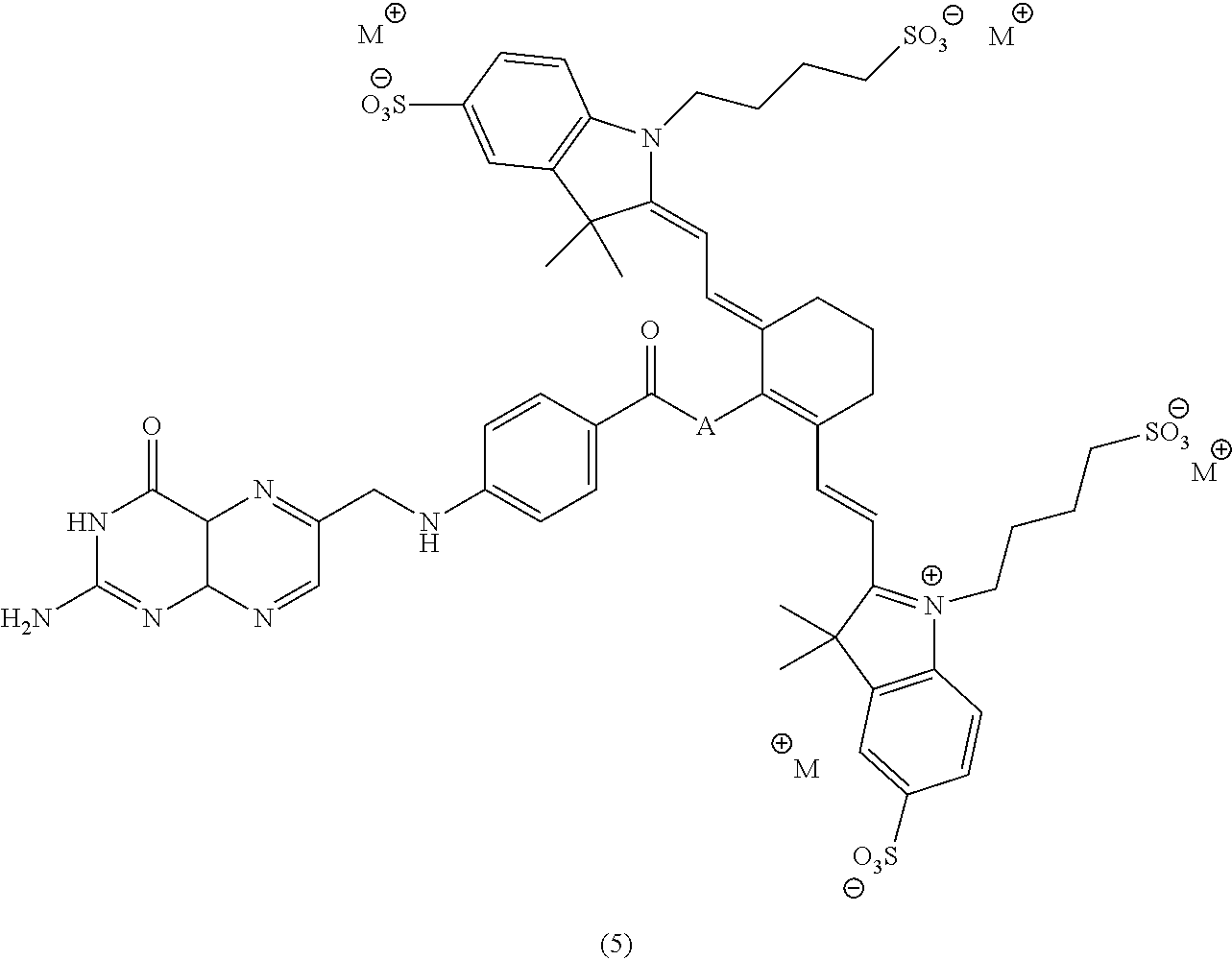


Claims (27)


Priority Applications (1)
| Application Number | Priority Date | Filing Date | Title |
|---|---|---|---|
| US14/135,099 US9233175B2 (en) | 2013-03-15 | 2013-12-19 | Methods of imaging inflammatory diseases by ligands conjugated to fluorescent compounds |
Applications Claiming Priority (2)
| Application Number | Priority Date | Filing Date | Title |
|---|---|---|---|
| US201361791921P | 2013-03-15 | 2013-03-15 | |
| US14/135,099 US9233175B2 (en) | 2013-03-15 | 2013-12-19 | Methods of imaging inflammatory diseases by ligands conjugated to fluorescent compounds |
Publications (2)
| Publication Number | Publication Date |
|---|---|
| US20140271484A1 US20140271484A1 (en) | 2014-09-18 |
| US9233175B2 true US9233175B2 (en) | 2016-01-12 |
Family
ID=51527878
Family Applications (7)
| Application Number | Title | Priority Date | Filing Date |
|---|---|---|---|
| US14/010,098 Active US9333270B2 (en) | 2013-03-15 | 2013-08-26 | Synthesis and composition of amino acid linking groups conjugated to compounds used for the targeted imaging of tumors |
| US14/046,916 Active US9254341B2 (en) | 2013-03-15 | 2013-10-04 | Methods of manufacture of pteroyl-amino acid-fluorescent dyes |
| US14/135,099 Active US9233175B2 (en) | 2013-03-15 | 2013-12-19 | Methods of imaging inflammatory diseases by ligands conjugated to fluorescent compounds |
| US14/207,130 Active US9061057B2 (en) | 2013-03-15 | 2014-03-12 | Synthesis and composition of amino acid linking groups conjugated to compounds used for the targeted imaging of tumors |
| US14/715,799 Active US9341629B2 (en) | 2013-03-15 | 2015-05-19 | Synthesis and composition of amino acid linking groups conjugated to compounds used for the targeted imaging of tumors |
| US14/953,928 Active US9782497B2 (en) | 2013-03-15 | 2015-11-30 | Synthesis and composition of amino acid linking groups conjugated to compounds used for the targeted imaging of tumors |
| US15/259,719 Active US9789208B2 (en) | 2013-03-15 | 2016-09-08 | Synthesis and composition of amino acid linking groups conjugated to compounds used for the targeted imaging of tumors |
Family Applications Before (2)
| Application Number | Title | Priority Date | Filing Date |
|---|---|---|---|
| US14/010,098 Active US9333270B2 (en) | 2013-03-15 | 2013-08-26 | Synthesis and composition of amino acid linking groups conjugated to compounds used for the targeted imaging of tumors |
| US14/046,916 Active US9254341B2 (en) | 2013-03-15 | 2013-10-04 | Methods of manufacture of pteroyl-amino acid-fluorescent dyes |
Family Applications After (4)
| Application Number | Title | Priority Date | Filing Date |
|---|---|---|---|
| US14/207,130 Active US9061057B2 (en) | 2013-03-15 | 2014-03-12 | Synthesis and composition of amino acid linking groups conjugated to compounds used for the targeted imaging of tumors |
| US14/715,799 Active US9341629B2 (en) | 2013-03-15 | 2015-05-19 | Synthesis and composition of amino acid linking groups conjugated to compounds used for the targeted imaging of tumors |
| US14/953,928 Active US9782497B2 (en) | 2013-03-15 | 2015-11-30 | Synthesis and composition of amino acid linking groups conjugated to compounds used for the targeted imaging of tumors |
| US15/259,719 Active US9789208B2 (en) | 2013-03-15 | 2016-09-08 | Synthesis and composition of amino acid linking groups conjugated to compounds used for the targeted imaging of tumors |
Country Status (12)
| Country | Link |
|---|---|
| US (7) | US9333270B2 (en) |
| EP (3) | EP2968335B1 (en) |
| JP (5) | JP6192799B2 (en) |
| CN (3) | CN105228628B (en) |
| AU (4) | AU2013383382B2 (en) |
| CA (3) | CA2903994C (en) |
| EA (1) | EA029738B1 (en) |
| ES (1) | ES2765896T3 (en) |
| HK (1) | HK1218257A1 (en) |
| IL (3) | IL240509B (en) |
| MX (3) | MX358184B (en) |
| WO (3) | WO2014149069A1 (en) |
Cited By (3)
| Publication number | Priority date | Publication date | Assignee | Title |
|---|---|---|---|---|
| US20160274090A1 (en) * | 2013-11-19 | 2016-09-22 | Purdue Research Foundation | Patient selection method for inflammation |
| US20210275694A1 (en) * | 2013-03-15 | 2021-09-09 | Purdue Research Foundation | Synthesis and composition of amino acid linking groups conjugated to compounds used for the targeted imaging of tumors |
| US20230263911A1 (en) * | 2022-02-23 | 2023-08-24 | On Target Laboratories, Llc. | Compositions including pafolacianine for the identification of malignant lesions |
Families Citing this family (36)
| Publication number | Priority date | Publication date | Assignee | Title |
|---|---|---|---|---|
| US9408928B2 (en) | 2007-03-27 | 2016-08-09 | Radiomedix, Inc. | Compositions for targeted imaging and therapy |
| CA2843313C (en) | 2011-06-20 | 2018-02-13 | Radiomedix Inc. | Compositions, methods of synthesis and use of carbohydrate targeted agents |
| AU2013383382B2 (en) * | 2013-03-15 | 2017-02-23 | Purdue Research Foundation | Synthesis and composition of amino acid linking groups conjugated to compounds used for the targeted imaging of tumors |
| WO2016072984A1 (en) * | 2014-11-05 | 2016-05-12 | The United States Of America, As Represented By The Secretary, Department Of Health And Human Services | A new class of stable heptamethine cyanine fluorophores and biomedical applications thereof |
| US10525149B2 (en) | 2015-05-12 | 2020-01-07 | Medibeacon Inc. | Compositions and methods for assessing eye vasculature |
| FR3036621B1 (en) * | 2015-05-25 | 2017-05-19 | Fluoptics | MULTIVALENT TARGETING FLUORESCENT PLATTER IN THE NEAR INFRARED FOR OPTICAL IMAGING. |
| FR3036622B1 (en) * | 2015-05-25 | 2017-05-19 | Fluoptics | MULTIVALENT TARGETING FLUORESCENT TARGETER. |
| US9808538B2 (en) | 2015-09-09 | 2017-11-07 | On Target Laboratories, LLC | PSMA-targeted NIR dyes and their uses |
| US10842887B2 (en) | 2015-09-09 | 2020-11-24 | On Target Laboratories, LLC | PSMA-targeted NIR dyes and their uses |
| US11555821B2 (en) | 2016-03-16 | 2023-01-17 | On Target Laboratories, Inc. | CA IX-NIR dyes and their uses |
| AU2017234681B2 (en) * | 2016-03-16 | 2021-07-01 | On Target Laboratories, LLC | CA IX-target NIR dyes and their uses |
| EP3493855A4 (en) | 2016-08-02 | 2020-04-01 | ISI Life Sciences, Inc. | Compositions and methods for detecting cancer cells in a tissue sample |
| US10561729B2 (en) | 2016-08-11 | 2020-02-18 | The United States Of America, As Represented By The Secretary, Department Of Health And Human Services | Near-IR light-cleavable conjugates and conjugate precursors |
| US10308606B2 (en) | 2016-09-09 | 2019-06-04 | On Target Laboratories, LLC | PSMA-targeted NIR dyes and their uses |
| US10786580B2 (en) * | 2016-09-09 | 2020-09-29 | On Target Laboratories, LLC | Cholecystokinin 2 receptor targeted NIR imaging and use thereof |
| WO2018081254A1 (en) * | 2016-10-26 | 2018-05-03 | Cao Group, Inc. | Cancer binding chromatic peptides that are targeted for disintegration by radiant energy |
| WO2018101473A1 (en) * | 2016-12-02 | 2018-06-07 | 国立大学法人東京大学 | Compound, folate receptor visualization fluorescent probe, and use of these |
| EP3555627B1 (en) | 2016-12-14 | 2023-11-22 | Purdue Research Foundation | Fibroblast activation protein (fap)-targeted imaging and therapy |
| US10753942B2 (en) | 2017-05-15 | 2020-08-25 | Indicator Systems International, Inc. | Methods to detect remnant cancer cells |
| US10539567B2 (en) * | 2017-05-15 | 2020-01-21 | Indicator Systems International, Inc. | Biopsy methods and devices |
| EP3752486A4 (en) * | 2018-02-15 | 2022-07-06 | Children's National Medical Center | Heptamethine cyanines for use as fluorescent markers of the biliary and renal systems |
| CN108840815A (en) * | 2018-05-29 | 2018-11-20 | 苏州百源基因技术有限公司 | A kind of amino acid of fluorescent marker and its preparation method and application |
| CN108727245B (en) * | 2018-07-02 | 2021-09-28 | 广州中医药大学(广州中医药研究院) | Salicylic acid compound and preparation method and application thereof |
| KR102150415B1 (en) | 2018-12-07 | 2020-09-01 | 원광대학교 산학협력단 | Targeting compound of cancer and use thereof |
| JP7160694B2 (en) | 2019-01-08 | 2022-10-25 | 日立Geニュークリア・エナジー株式会社 | FLUID CONTACT MEMBER AND METHOD FOR MANUFACTURING FLUID CONTACT MEMBER |
| US11716533B2 (en) | 2019-06-20 | 2023-08-01 | Cilag Gmbh International | Image synchronization without input clock and data transmission clock in a pulsed fluorescence imaging system |
| US11754500B2 (en) | 2019-06-20 | 2023-09-12 | Cilag Gmbh International | Minimizing image sensor input/output in a pulsed fluorescence imaging system |
| US11516387B2 (en) | 2019-06-20 | 2022-11-29 | Cilag Gmbh International | Image synchronization without input clock and data transmission clock in a pulsed hyperspectral, fluorescence, and laser mapping imaging system |
| US11892403B2 (en) | 2019-06-20 | 2024-02-06 | Cilag Gmbh International | Image synchronization without input clock and data transmission clock in a pulsed fluorescence imaging system |
| US20210353151A1 (en) * | 2020-05-12 | 2021-11-18 | On Target Laboratories, LLC | Targeted fluorescent markers in combination with a flexible probe |
| CN112010862B (en) * | 2020-10-23 | 2021-03-30 | 南京诺源医疗器械有限公司 | Active targeting folic acid receptor near-infrared fluorescent molecule and preparation method thereof |
| CN114014843B (en) * | 2021-11-17 | 2022-09-20 | 北京大学第一医院 | PSMA targeted nuclide/fluorescent bimodal ligand, molecular probe and application |
| CN114751907A (en) * | 2022-03-17 | 2022-07-15 | 南京诺源医疗器械有限公司 | Active targeting folic acid receptor near-infrared fluorescent molecule and preparation method and application thereof |
| CN115504984B (en) * | 2022-09-09 | 2023-07-25 | 南京诺源医疗器械有限公司 | Pemetic near infrared fluorescent molecule targeting alpha-folic acid receptor and preparation method and application thereof |
| CN115490672B (en) * | 2022-09-30 | 2023-11-17 | 南华大学 | Photosensitizer with photothermal and photodynamic effects as well as preparation method and application thereof |
| CN116621823B (en) * | 2023-07-06 | 2023-10-31 | 南京诺源医疗器械有限公司 | Near infrared fluorescent tracer for diagnosing metastatic lymph nodes, synthetic method and application |
Citations (9)
| Publication number | Priority date | Publication date | Assignee | Title |
|---|---|---|---|---|
| US4337339A (en) * | 1979-04-30 | 1982-06-29 | Baker Instruments Corp. | Process for preparation of folic acid derivatives |
| WO2007139815A2 (en) | 2006-05-23 | 2007-12-06 | Purdue Research Foundation | Imaging and therapeutic method using progenitor cells |
| US7547721B1 (en) | 1998-09-18 | 2009-06-16 | Bayer Schering Pharma Ag | Near infrared fluorescent contrast agent and fluorescence imaging |
| US20110165075A1 (en) | 2008-03-14 | 2011-07-07 | Milind Rajopadhye | Integrin targeting agents and in-vivo and in-vitro imaging methods using the same |
| US8044200B2 (en) | 2005-03-16 | 2011-10-25 | Endocyte, Inc. | Synthesis and purification of pteroic acid and conjugates thereof |
| US20120003151A1 (en) | 2009-03-05 | 2012-01-05 | Purdue Research Foundation | Method for early imaging of atherosclerosis |
| US20120164635A1 (en) | 2007-06-29 | 2012-06-28 | Vanderbilt University | Large stoke shift nir dyes |
| US20130039860A1 (en) * | 2010-10-20 | 2013-02-14 | Li-Cor, Inc. | Fluorescent imaging with substituted cyanine dyes |
| WO2013130776A1 (en) | 2012-02-29 | 2013-09-06 | Purdue Research Foundation | Folate receptor alpha binding ligands |
Family Cites Families (21)
| Publication number | Priority date | Publication date | Assignee | Title |
|---|---|---|---|---|
| US5871711A (en) | 1992-06-23 | 1999-02-16 | Diatide, Inc. | Radioactively-labeled somatostatin-derived peptides for imaging and therapeutic uses |
| US5716596A (en) | 1992-06-23 | 1998-02-10 | Diatide, Inc. | Radioactively labeled somatostatin-derived peptides for imaging and therapeutic uses |
| DE19717904A1 (en) * | 1997-04-23 | 1998-10-29 | Diagnostikforschung Inst | Acid-labile and enzymatically cleavable dye constructs for diagnostics with near infrared light and for therapy |
| JP2000095758A (en) | 1998-09-18 | 2000-04-04 | Schering Ag | Near-infrared, fluorescent contrast medium, and its production |
| AU2097500A (en) | 1999-12-15 | 2001-06-25 | Fuji Photo Film Co., Ltd. | Near infrared fluorescent contrast agent and fluorescence imaging |
| BR0109704A (en) | 2000-03-31 | 2003-04-29 | Purdue Research Foundation | Method of Treatment Using Ligand Immunogen Conjugates |
| DE60116510T2 (en) * | 2000-09-19 | 2006-07-13 | Li-Cor, Inc., Lincoln | cyanine |
| US7597878B2 (en) | 2000-09-19 | 2009-10-06 | Li-Cor, Inc. | Optical fluorescent imaging |
| JP2005507857A (en) | 2001-02-07 | 2005-03-24 | ベズ イズレイル ディーコネス メディカル センター | Modified PSMA ligands and uses related thereto |
| HUP0401127A3 (en) * | 2001-05-02 | 2006-03-28 | Purdue Research Foundation | Treatment and diagnosis of macrophage disease |
| WO2007006041A2 (en) * | 2005-07-05 | 2007-01-11 | Purdue Research Foundation | Imaging and therapeutic method using monocytes |
| WO2007100755A1 (en) * | 2006-02-27 | 2007-09-07 | Nabi Biopharmaceuticals | Method for decreasing the toxic effects of nicotine on fetuses in pregnant women |
| EP2087337A4 (en) | 2006-11-03 | 2010-09-08 | Purdue Research Foundation | Ex vivo flow cytometry method and device |
| CN102215844A (en) | 2008-09-17 | 2011-10-12 | 恩多塞特公司 | Folate receptor binding conjugates of antifolates |
| WO2010054330A2 (en) * | 2008-11-07 | 2010-05-14 | The General Hospital Corporation | Monofunctional carbocyanine dyes for in vivo and in vitro imaging |
| CN101440282B (en) * | 2008-12-18 | 2012-06-27 | 中国药科大学 | Near-infrared fluorescent molecular probe, synthesizing method and use thereof |
| JP2012524153A (en) * | 2009-04-17 | 2012-10-11 | ライコア インコーポレイテッド | Fluorescence imaging using substituted cyanine dyes |
| CA2800693A1 (en) | 2010-05-28 | 2011-12-01 | Purdue Research Foundation | Delivery of therapeutic agents to inflamed tissues using folate-targeted agents |
| WO2012032068A2 (en) * | 2010-09-06 | 2012-03-15 | Modpro Ab | Compounds and methods |
| US10080805B2 (en) * | 2012-02-24 | 2018-09-25 | Purdue Research Foundation | Cholecystokinin B receptor targeting for imaging and therapy |
| AU2013383382B2 (en) * | 2013-03-15 | 2017-02-23 | Purdue Research Foundation | Synthesis and composition of amino acid linking groups conjugated to compounds used for the targeted imaging of tumors |
-
2013
- 2013-08-26 AU AU2013383382A patent/AU2013383382B2/en active Active
- 2013-08-26 CA CA2903994A patent/CA2903994C/en active Active
- 2013-08-26 WO PCT/US2013/056629 patent/WO2014149069A1/en active Application Filing
- 2013-08-26 EA EA201591802A patent/EA029738B1/en not_active IP Right Cessation
- 2013-08-26 EP EP13878809.6A patent/EP2968335B1/en active Active
- 2013-08-26 MX MX2015013223A patent/MX358184B/en active IP Right Grant
- 2013-08-26 JP JP2016500088A patent/JP6192799B2/en active Active
- 2013-08-26 US US14/010,098 patent/US9333270B2/en active Active
- 2013-08-26 CN CN201380074692.8A patent/CN105228628B/en active Active
- 2013-10-04 US US14/046,916 patent/US9254341B2/en active Active
- 2013-10-04 EP EP13878632.2A patent/EP2968614B1/en active Active
- 2013-10-04 CA CA2903727A patent/CA2903727C/en active Active
- 2013-10-04 MX MX2015011830A patent/MX2015011830A/en unknown
- 2013-10-04 JP JP2016500094A patent/JP6270981B2/en active Active
- 2013-10-04 CN CN201380074694.7A patent/CN105120903A/en active Pending
- 2013-10-04 WO PCT/US2013/063593 patent/WO2014149073A1/en active Application Filing
- 2013-10-04 AU AU2013383386A patent/AU2013383386A1/en not_active Abandoned
- 2013-12-19 CN CN201380074662.7A patent/CN105492905B/en active Active
- 2013-12-19 US US14/135,099 patent/US9233175B2/en active Active
- 2013-12-19 JP JP2016500131A patent/JP2016512240A/en active Pending
- 2013-12-19 EP EP13877792.5A patent/EP2972320B1/en active Active
- 2013-12-19 AU AU2013381391A patent/AU2013381391A1/en not_active Abandoned
- 2013-12-19 CA CA2902205A patent/CA2902205A1/en not_active Abandoned
- 2013-12-19 WO PCT/US2013/076659 patent/WO2014143309A1/en active Application Filing
- 2013-12-19 ES ES13877792T patent/ES2765896T3/en active Active
- 2013-12-19 MX MX2015013219A patent/MX2015013219A/en unknown
-
2014
- 2014-03-12 US US14/207,130 patent/US9061057B2/en active Active
-
2015
- 2015-05-19 US US14/715,799 patent/US9341629B2/en active Active
- 2015-08-12 IL IL240509A patent/IL240509B/en active IP Right Grant
- 2015-08-18 IL IL240673A patent/IL240673B/en active IP Right Grant
- 2015-08-25 IL IL240828A patent/IL240828A/en active IP Right Grant
- 2015-11-30 US US14/953,928 patent/US9782497B2/en active Active
-
2016
- 2016-06-02 HK HK16106296.0A patent/HK1218257A1/en unknown
- 2016-09-08 US US15/259,719 patent/US9789208B2/en active Active
-
2017
- 2017-05-18 AU AU2017203340A patent/AU2017203340B2/en active Active
- 2017-05-31 JP JP2017107741A patent/JP2017149776A/en active Pending
- 2017-09-05 JP JP2017170413A patent/JP2017214425A/en active Pending
Patent Citations (9)
| Publication number | Priority date | Publication date | Assignee | Title |
|---|---|---|---|---|
| US4337339A (en) * | 1979-04-30 | 1982-06-29 | Baker Instruments Corp. | Process for preparation of folic acid derivatives |
| US7547721B1 (en) | 1998-09-18 | 2009-06-16 | Bayer Schering Pharma Ag | Near infrared fluorescent contrast agent and fluorescence imaging |
| US8044200B2 (en) | 2005-03-16 | 2011-10-25 | Endocyte, Inc. | Synthesis and purification of pteroic acid and conjugates thereof |
| WO2007139815A2 (en) | 2006-05-23 | 2007-12-06 | Purdue Research Foundation | Imaging and therapeutic method using progenitor cells |
| US20120164635A1 (en) | 2007-06-29 | 2012-06-28 | Vanderbilt University | Large stoke shift nir dyes |
| US20110165075A1 (en) | 2008-03-14 | 2011-07-07 | Milind Rajopadhye | Integrin targeting agents and in-vivo and in-vitro imaging methods using the same |
| US20120003151A1 (en) | 2009-03-05 | 2012-01-05 | Purdue Research Foundation | Method for early imaging of atherosclerosis |
| US20130039860A1 (en) * | 2010-10-20 | 2013-02-14 | Li-Cor, Inc. | Fluorescent imaging with substituted cyanine dyes |
| WO2013130776A1 (en) | 2012-02-29 | 2013-09-06 | Purdue Research Foundation | Folate receptor alpha binding ligands |
Non-Patent Citations (2)
| Title |
|---|
| International Search Report in PCT/US13/56629 dated May 13, 2014. |
| PCT, Notification of Transmittal of the International Search Report and the Written Opinion of the International Searching Authority, or the Declaration, in International application No. PCT/US13/76659 dated Apr. 4, 2014. (21 pages). |
Cited By (4)
| Publication number | Priority date | Publication date | Assignee | Title |
|---|---|---|---|---|
| US20210275694A1 (en) * | 2013-03-15 | 2021-09-09 | Purdue Research Foundation | Synthesis and composition of amino acid linking groups conjugated to compounds used for the targeted imaging of tumors |
| US20160274090A1 (en) * | 2013-11-19 | 2016-09-22 | Purdue Research Foundation | Patient selection method for inflammation |
| US11162937B2 (en) * | 2013-11-19 | 2021-11-02 | Purdue Research Foundation | Patient selection method for inflammation |
| US20230263911A1 (en) * | 2022-02-23 | 2023-08-24 | On Target Laboratories, Llc. | Compositions including pafolacianine for the identification of malignant lesions |
Also Published As
Similar Documents
| Publication | Publication Date | Title |
|---|---|---|
| US9233175B2 (en) | Methods of imaging inflammatory diseases by ligands conjugated to fluorescent compounds | |
| US20220395588A1 (en) | Synthesis and composition of amino acid linking groups conjugated to compounds used for the targeted imaging of tumors | |
| US20200174009A1 (en) | Ca ix - nir dyes and their uses | |
| US20210063406A1 (en) | Ca ix - nir dyes and their uses |
Legal Events
| Date | Code | Title | Description |
|---|---|---|---|
| AS | Assignment |
Owner name: PURDUE RESEARCH FOUNDATION, INDIANA Free format text: ASSIGNMENT OF ASSIGNORS INTEREST;ASSIGNORS:LOW, PHILIP S.;KULARATNE, SUMITH A.;KELDERHOUSE, LINDSAY E.;AND OTHERS;SIGNING DATES FROM 20140813 TO 20140816;REEL/FRAME:033552/0732 |
|
| STCF | Information on status: patent grant |
Free format text: PATENTED CASE |
|
| CC | Certificate of correction | ||
| MAFP | Maintenance fee payment |
Free format text: PAYMENT OF MAINTENANCE FEE, 4TH YEAR, LARGE ENTITY (ORIGINAL EVENT CODE: M1551); ENTITY STATUS OF PATENT OWNER: LARGE ENTITY Year of fee payment: 4 |
|
| MAFP | Maintenance fee payment |
Free format text: PAYMENT OF MAINTENANCE FEE, 8TH YEAR, LARGE ENTITY (ORIGINAL EVENT CODE: M1552); ENTITY STATUS OF PATENT OWNER: LARGE ENTITY Year of fee payment: 8 |