US9095353B2 - Surgical tools facilitating increased accuracy, speed and simplicity in performing joint arthroplasty - Google Patents
Surgical tools facilitating increased accuracy, speed and simplicity in performing joint arthroplasty Download PDFInfo
- Publication number
- US9095353B2 US9095353B2 US13/754,133 US201313754133A US9095353B2 US 9095353 B2 US9095353 B2 US 9095353B2 US 201313754133 A US201313754133 A US 201313754133A US 9095353 B2 US9095353 B2 US 9095353B2
- Authority
- US
- United States
- Prior art keywords
- patient
- implant
- joint
- mold
- patella
- Prior art date
- Legal status (The legal status is an assumption and is not a legal conclusion. Google has not performed a legal analysis and makes no representation as to the accuracy of the status listed.)
- Expired - Lifetime, expires
Links
Images


Classifications
-
- A—HUMAN NECESSITIES
- A61—MEDICAL OR VETERINARY SCIENCE; HYGIENE
- A61B—DIAGNOSIS; SURGERY; IDENTIFICATION
- A61B17/00—Surgical instruments, devices or methods, e.g. tourniquets
- A61B17/14—Surgical saws ; Accessories therefor
- A61B17/15—Guides therefor
- A61B17/154—Guides therefor for preparing bone for knee prosthesis
- A61B17/158—Cutting patella
-
- A—HUMAN NECESSITIES
- A61—MEDICAL OR VETERINARY SCIENCE; HYGIENE
- A61B—DIAGNOSIS; SURGERY; IDENTIFICATION
- A61B17/00—Surgical instruments, devices or methods, e.g. tourniquets
- A61B17/14—Surgical saws ; Accessories therefor
- A61B17/15—Guides therefor
- A61B17/154—Guides therefor for preparing bone for knee prosthesis
- A61B17/155—Cutting femur
-
- A—HUMAN NECESSITIES
- A61—MEDICAL OR VETERINARY SCIENCE; HYGIENE
- A61B—DIAGNOSIS; SURGERY; IDENTIFICATION
- A61B17/00—Surgical instruments, devices or methods, e.g. tourniquets
- A61B17/14—Surgical saws ; Accessories therefor
- A61B17/15—Guides therefor
- A61B17/154—Guides therefor for preparing bone for knee prosthesis
- A61B17/157—Cutting tibia
-
- A—HUMAN NECESSITIES
- A61—MEDICAL OR VETERINARY SCIENCE; HYGIENE
- A61B—DIAGNOSIS; SURGERY; IDENTIFICATION
- A61B17/00—Surgical instruments, devices or methods, e.g. tourniquets
- A61B17/16—Bone cutting, breaking or removal means other than saws, e.g. Osteoclasts; Drills or chisels for bones; Trepans
- A61B17/1662—Bone cutting, breaking or removal means other than saws, e.g. Osteoclasts; Drills or chisels for bones; Trepans for particular parts of the body
- A61B17/1675—Bone cutting, breaking or removal means other than saws, e.g. Osteoclasts; Drills or chisels for bones; Trepans for particular parts of the body for the knee
- A61B17/1677—Bone cutting, breaking or removal means other than saws, e.g. Osteoclasts; Drills or chisels for bones; Trepans for particular parts of the body for the knee for the patella
-
- A—HUMAN NECESSITIES
- A61—MEDICAL OR VETERINARY SCIENCE; HYGIENE
- A61B—DIAGNOSIS; SURGERY; IDENTIFICATION
- A61B17/00—Surgical instruments, devices or methods, e.g. tourniquets
- A61B17/16—Bone cutting, breaking or removal means other than saws, e.g. Osteoclasts; Drills or chisels for bones; Trepans
- A61B17/17—Guides or aligning means for drills, mills, pins or wires
- A61B17/1739—Guides or aligning means for drills, mills, pins or wires specially adapted for particular parts of the body
- A61B17/1764—Guides or aligning means for drills, mills, pins or wires specially adapted for particular parts of the body for the knee
- A61B17/1767—Guides or aligning means for drills, mills, pins or wires specially adapted for particular parts of the body for the knee for the patella
-
- A—HUMAN NECESSITIES
- A61—MEDICAL OR VETERINARY SCIENCE; HYGIENE
- A61B—DIAGNOSIS; SURGERY; IDENTIFICATION
- A61B17/00—Surgical instruments, devices or methods, e.g. tourniquets
- A61B2017/0023—Surgical instruments, devices or methods, e.g. tourniquets disposable
-
- A—HUMAN NECESSITIES
- A61—MEDICAL OR VETERINARY SCIENCE; HYGIENE
- A61B—DIAGNOSIS; SURGERY; IDENTIFICATION
- A61B17/00—Surgical instruments, devices or methods, e.g. tourniquets
- A61B17/56—Surgical instruments or methods for treatment of bones or joints; Devices specially adapted therefor
- A61B2017/568—Surgical instruments or methods for treatment of bones or joints; Devices specially adapted therefor produced with shape and dimensions specific for an individual patient
-
- A—HUMAN NECESSITIES
- A61—MEDICAL OR VETERINARY SCIENCE; HYGIENE
- A61B—DIAGNOSIS; SURGERY; IDENTIFICATION
- A61B5/00—Measuring for diagnostic purposes; Identification of persons
- A61B5/45—For evaluating or diagnosing the musculoskeletal system or teeth
- A61B5/4504—Bones
-
- A—HUMAN NECESSITIES
- A61—MEDICAL OR VETERINARY SCIENCE; HYGIENE
- A61B—DIAGNOSIS; SURGERY; IDENTIFICATION
- A61B5/00—Measuring for diagnostic purposes; Identification of persons
- A61B5/45—For evaluating or diagnosing the musculoskeletal system or teeth
- A61B5/4514—Cartilage
-
- A—HUMAN NECESSITIES
- A61—MEDICAL OR VETERINARY SCIENCE; HYGIENE
- A61B—DIAGNOSIS; SURGERY; IDENTIFICATION
- A61B5/00—Measuring for diagnostic purposes; Identification of persons
- A61B5/45—For evaluating or diagnosing the musculoskeletal system or teeth
- A61B5/4528—Joints
-
- Y—GENERAL TAGGING OF NEW TECHNOLOGICAL DEVELOPMENTS; GENERAL TAGGING OF CROSS-SECTIONAL TECHNOLOGIES SPANNING OVER SEVERAL SECTIONS OF THE IPC; TECHNICAL SUBJECTS COVERED BY FORMER USPC CROSS-REFERENCE ART COLLECTIONS [XRACs] AND DIGESTS
- Y10—TECHNICAL SUBJECTS COVERED BY FORMER USPC
- Y10T—TECHNICAL SUBJECTS COVERED BY FORMER US CLASSIFICATION
- Y10T29/00—Metal working
- Y10T29/49—Method of mechanical manufacture
- Y10T29/4998—Combined manufacture including applying or shaping of fluent material
-
- Y—GENERAL TAGGING OF NEW TECHNOLOGICAL DEVELOPMENTS; GENERAL TAGGING OF CROSS-SECTIONAL TECHNOLOGIES SPANNING OVER SEVERAL SECTIONS OF THE IPC; TECHNICAL SUBJECTS COVERED BY FORMER USPC CROSS-REFERENCE ART COLLECTIONS [XRACs] AND DIGESTS
- Y10—TECHNICAL SUBJECTS COVERED BY FORMER USPC
- Y10T—TECHNICAL SUBJECTS COVERED BY FORMER US CLASSIFICATION
- Y10T29/00—Metal working
- Y10T29/49—Method of mechanical manufacture
- Y10T29/49995—Shaping one-piece blank by removing material
Definitions
- the present invention relates to methods, systems and devices for articular resurfacing.
- the present invention includes surgical molds designed to achieve optimal cut planes in a joint in preparation for installation of a joint implant.
- U.S. Pat. No. 4,501,266 to McDaniel issued Feb. 26, 1985 discloses a knee distraction device that facilitates knee arthroplasty.
- the device has an adjustable force calibration mechanism that enables the device to accommodate controlled selection of the ligament-tensioning force to be applied to the respective, opposing sides of the knee.
- U.S. Pat. No. 5,002,547 to Poggie et al. issued Mar. 26, 1991 discloses a modular apparatus for use in preparing the bone surface for implantation of a modular total knee prosthesis.
- the apparatus has cutting guides, templates, alignment devices along with a distractor and clamping instruments that provide modularity and facilitate bone resection and prosthesis implantation.
- U.S. Pat. No. 5,250,050 to Poggie et al. issued Oct. 5, 1993 is also directed to a modular apparatus for use in preparing a bone surface for the implantation of a modular total knee prosthesis.
- U.S. Pat. No. 5,387,216 to Thornhill et al. issued Feb. 7, 1995 discloses instrumentation for use in knee revision surgery.
- a bearing sleeve is provided that is inserted into the damaged canal in order to take up additional volume.
- the rod passes through the sleeve and is positioned to meet the natural canal of the bone.
- the rod is then held in a fixed position by the bearing sleeve.
- a cutting guide can then be mounted on the rod for cutting the bone and to provide a mounting surface for the implant.
- Pat. No. 6,056,756 to Eng et al. issued May 2, 2000 discloses a tool for preparing the distal femoral end for a prosthetic implant.
- the tool lays out the resection for prosthetic replacement and includes a jack for pivotally supporting an opposing bone such that the jack raises the opposing bone in flexion to the spacing of the intended prosthesis.
- U.S. Pat. No. 6,106,529 to Techiera issued Aug. 22, 2000 discloses an epicondylar axis referencing drill guide for use in resection to prepare a bone end for prosthetic joint replacement.
- U.S. Pat. No. 5,578,037 to Sanders et al. issued Nov. 26, 1996 discloses a surgical guide for femoral resection.
- the guide enables a surgeon to resect a femoral neck during a hip arthroplasty procedure so that the femoral prosthesis can be implanted to preserve or closely approximate the anatomic center of rotation of the hip.
- surgical tools for preparing a joint to receive an implant are described, for example a tool comprising one or more surfaces or members that conform at least partially to the shape of the articular surfaces of the joint (e.g., a femoral condyle and/or tibial plateau of a knee joint).
- the tool comprises Lucite silastic and/or other polymers or suitable materials.
- the tool can be re-useable or single-use.
- the tool can be comprised of a single component or multiple components.
- the tool comprises an array of adjustable, closely spaced pins.
- the tool comprises: a mold having a surface for engaging a joint surface; a block that communicates with the mold; and at least one guide aperture in the block.
- Another tool is disclosed that is formed at least partially in situ and comprises: a mold formed in situ using at least one of an inflatable hollow device or a retaining device to conform to the joint surface on at least one surface having a surface for engaging a joint surface; a block that communicates with the mold; and at least one guide aperture in the block.
- the joint can be a knee, shoulder, hip, vertebrae, elbow, ankle, wrist etc.
- FIG. 1A illustrates a femur, tibia and fibula along with the mechanical and anatomic axes.
- FIGS. 1B-E illustrate the tibia with the anatomic and mechanical axis used to create a cutting plane along with a cut femur and tibia.
- FIG. 1F illustrates the proximal end of the femur including the head of the femur.
- FIG. 2 shows an example of a surgical tool having one surface matching the geometry of an articular surface of the joint. Also shown is an aperture in the tool capable of controlling drill depth and width of the hole and allowing implantation of an insertion of implant having a press-fit design.
- FIG. 3 is a flow chart depicting various methods of the invention used to create a mold for preparing a patient's joint for arthroscopic surgery.
- FIG. 4A depicts, in cross-section, an example of a surgical tool containing an aperture through which a surgical drill or saw can fit.
- the aperture guides the drill or saw to make the proper hole or cut in the underlying bone.
- Dotted lines represent where the cut corresponding to the aperture will be made in bone.
- FIG. 4B depicts, in cross-section, an example of a surgical tool containing apertures through which a surgical drill or saw can fit and which guide the drill or saw to make cuts or holes in the bone. Dotted lines represent where the cuts corresponding to the apertures will be made in bone.
- FIGS. 5A-Q illustrate tibial cutting blocks and molds used to create a surface perpendicular to the anatomic axis for receiving the tibial portion of a knee implant.
- FIGS. 6A-O illustrate femur cutting blocks and molds used to create a surface for receiving the femoral portion of a knee implant.
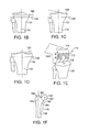
- FIG. 7A-G illustrate patellar cutting blocks and molds used to prepare the patella for receiving a portion of a knee implant.
- FIG. 8A-H illustrate femoral head cutting blocks and molds used to create a surface for receiving the femoral portion of a knee implant.
- FIG. 9A-D illustrate acetabulum cutting blocks and molds used to create a surface for a hip implant.
- FIG. 10A illustrates a patella modeled from CT data.
- FIGS. 10B-D illustrate a mold guide, and then the mold guide placed on an articular surface of the patella.
- FIG. 10E illustrates a drill placed into a patella through mold drill guide.
- FIG. 10F illustrates a reamer used to prepare the patella.
- FIG. 11A illustrates a reamer made for each patella size.
- FIG. 11B illustrates a reamed patella ready for patella implantation.
- FIG. 12A-F illustrate a recessed patella implanted on a patella.
- the practice of the present invention employs, unless otherwise indicated, conventional methods of x-ray imaging and processing, x-ray tomosynthesis, ultrasound including A-scan, B-scan and C-scan, computed tomography (CT scan), magnetic resonance imaging (MRI), optical coherence tomography, single photon emission tomography (SPECT) and positron emission tomography (PET) within the skill of the art.
- CT scan computed tomography
- MRI magnetic resonance imaging
- SPECT single photon emission tomography
- PET positron emission tomography
- repair systems including surgical instruments, guides and molds, of various sizes, curvatures and thicknesses can be obtained.
- These repair systems, including surgical instruments, guides and molds can be catalogued and stored to create a library of systems from which an appropriate system for an individual patient can then be selected.
- a defect, or an articular surface is assessed in a particular subject and a pre-existing repair system, including surgical instruments, guides and molds, having a suitable shape and size is selected from the library for further manipulation (e.g., shaping) and implantation.
- the center of the hip 102 (located at the head 130 of the femur 132 ), the center of the knee 104 (located at the notch where the intercondular tubercle 134 of the tibia 136 meet the femur) and ankle 106 lie approximately in a straight line 110 which defines the mechanical axis of the lower extremity.
- the anatomic axis 120 aligns 5-7° offset ⁇ from the mechanical axis in the valgus, or outward, direction.
- the long axis of the tibia 136 is collinear with the mechanical axis of the lower extremity 110 .
- the lower extremity of the body ideally functions within a single plane known as the median anterior-posterior plane (MAP-plane) throughout the flexion-extension arc.
- MAP-plane median anterior-posterior plane
- the femoral head 130 , the mechanical axis of the femur, the patellar groove, the intercondylar notch, the patellar articular crest, the tibia and the ankle remain within the MAP-plane during the flexion-extension movement.
- the tibia rotates as the knee flexes and extends in the epicondylar axis which is perpendicular to the MAP-plane.
- a variety of image slices can be taken at each individual joint, e.g., the knee joint 150 - 150 n , and the hip joint 152 - 150 n . These image slices can be used as described above in Section I along with an image of the full leg to ascertain the axis.
- Implanting a total knee joint such as the PFC Sigma RP Knee System by Johnson & Johnson, requires that a series of resections be made to the surfaces forming the knee joint in order to facilitate installation of the artificial knee.
- the resections should be made to enable the installed artificial knee to achieve flexion-extension movement within the MAP-plane and to optimize the patient's anatomical and mechanical axis of the lower extremity.
- the tibia 130 is resected to create a flat surface to accept the tibial component of the implant.
- the tibial surface is resected perpendicular to the long axis of the tibia in the coronal plane, but is typically sloped 4-7° posteriorly in the sagittal plane to match the normal slope of the tibia.
- the sagittal slope can be 0° where the device to be implanted does not require a sloped tibial cut.
- the resection line 158 is perpendicular to the mechanical axis 110 , but the angle between the resection line and the surface plane of the plateau 160 varies depending on the amount of damage to the knee.
- FIGS. 1B-D illustrate an anterior view of a resection of an anatomically normal tibial component, a tibial component in a varus knee, and a tibial component in a valgus knee, respectively.
- the mechanical axis 110 extends vertically through the bone and the resection line 158 is perpendicular to the mechanical axis 110 in the coronal plane, varying from the surface line formed by the joint depending on the amount of damage to the joint.
- FIG. 1B illustrates a normal knee wherein the line corresponding to the surface of the joint 160 is parallel to the resection line 158 .
- FIG. 1C illustrates a varus knee wherein the line corresponding to the surface of the joint 160 is not parallel to the resection line 158 .
- FIG. 1D illustrates a valgus knee wherein the line corresponding to the surface of the joint 160 is not parallel to the resection line 158 .
- the plateau of the femur 170 is resected to provide flat surfaces that communicate with the interior of the femoral prosthesis.
- the cuts made to the femur are based on the overall height of the gap to be created between the tibia and the femur. Typically, a 20 mm gap is desirable to provide the implanted prosthesis adequate room to achieve full range of motion.
- the bone is resected at a 5-7° angle valgus to the mechanical axis of the femur.
- Resected surface 172 forms a flat plane with an angular relationship to adjoining surfaces 174 , 176 .
- the angle ⁇ ′, ⁇ ′′ between the surfaces 172 - 174 , and 172 - 176 varies according to the design of the implant.
- the external geometry of the proximal femur includes the head 180 , the neck 182 , the lesser trochanter 184 , the greater trochanter 186 and the proximal femoral diaphysis.
- the relative positions of the trochanters 184 , 186 , the femoral head center 102 and the femoral shaft 188 are correlated with the inclination of the neck-shaft angle.
- the mechanical axis 110 and anatomic axis 120 are also shown. Assessment of these relationships can change the reaming direction to achieve neutral alignment of the prosthesis with the femoral canal.
- measurements are made of the proximal and distal geometry to determine the size and optimal design of the implant.
- the femoral neck 182 is resected, e.g. along the line 190 .
- the medullary canal is reamed. Reaming can be accomplished, for example, with a conical or straight reamer, or a flexible reamer. The depth of reaming is dictated by the specific design of the implant.
- the proximal reamer is prepared by serial rasping, with the rasp directed down into the canal.
- surgical assistance can be provided by using a device applied to the outer surface of the articular cartilage or the bone, including the subchondral bone, in order to match the alignment of the articular repair system and the recipient site or the joint.
- the device can be round, circular, oval, ellipsoid, curved or irregular in shape.
- the shape can be selected or adjusted to match or enclose an area of diseased cartilage or an area slightly larger than the area of diseased cartilage or substantially larger than the diseased cartilage.
- the area can encompass the entire articular surface or the weight bearing surface.
- Such devices are typically preferred when replacement of a majority or an entire articular surface is contemplated.
- Mechanical devices can be used for surgical assistance (e.g., surgical tools), for example using gels, molds, plastics or metal.
- One or more electronic images or intraoperative measurements can be obtained providing object coordinates that define the articular and/or bone surface and shape. These objects' coordinates can be utilized to either shape the device, e.g. using a CAD/CAM technique, to be adapted to a patient's articular anatomy or, alternatively, to select a typically pre-made device that has a good fit with a patient's articular anatomy.
- the device can have a surface and shape that will match all or portions of the articular or bone surface and shape, e.g. similar to a “mirror image.”
- the device can include apertures, slots and/or holes to accommodate surgical instruments such as drills, reamers, curettes, k-wires, screws and saws.
- a position will be chosen that will result in an anatomically desirable cut plane, drill hole, or general instrument orientation for subsequent placement of an articular repair system or for facilitating placement of the articular repair system.
- the device can be designed so that the depth of the drill, reamer or other surgical instrument can be controlled, e.g., the drill cannot go any deeper into the tissue than defined by the device, and the size of the hole in the block can be designed to essentially match the size of the implant. Information about other joints or axis and alignment information of a joint or extremity can be included when selecting the position of these slots or holes. Alternatively, the openings in the device can be made larger than needed to accommodate these instruments.
- the device can also be configured to conform to the articular shape.
- the apertures, or openings, provided can be wide enough to allow for varying the position or angle of the surgical instrument, e.g., reamers, saws, drills, curettes and other surgical instruments.
- An instrument guide typically comprised of a relatively hard material, can then be applied to the device. The device helps orient the instrument guide relative to the three-dimensional anatomy of the joint.
- the surgeon can, optionally, make fine adjustments between the alignment device and the instrument guide. In this manner, an optimal compromise can be found, for example, between biomechanical alignment and joint laxity or biomechanical alignment and joint function, e.g. in a knee joint flexion gap and extension gap.
- an optimal compromise can be found, for example, between biomechanical alignment and joint laxity or biomechanical alignment and joint function, e.g. in a knee joint flexion gap and extension gap.
- the surgeon can utilize the instruments and insert them in the instrument guide without damaging the alignment guide.
- the alignment guide is made of plastic, debris will not be introduced into the joint.
- the position and orientation between the alignment guide and the instrument guide can be also be optimized with the use of, for example, interposed spacers, wedges, screws and other mechanical or electrical methods known in the art.
- a surgeon may desire to influence joint laxity as well as joint alignment. This can be optimized for different flexion and extension, abduction, or adduction, internal and external rotation angles.
- spacers can be introduced that are attached or that are in contact with one or more molds.
- the surgeon can intraoperatively evaluate the laxity or tightness of a joint using spacers with different thickness or one or more spacers with the same thickness.
- spacers can be applied in a knee joint in the presence of one or more molds and the flexion gap can be evaluated with the knee joint in flexion. The knee joint can then be extended and the extension gap can be evaluated.
- the surgeon will select an optimal combination of spacers for a given joint and mold.
- a surgical cut guide can be applied to the mold with the spacers optionally interposed between the mold and the cut guide.
- the exact position of the surgical cuts can be influenced and can be adjusted to achieve an optimal result.
- someone skilled in the art will recognize other means for optimizing the position of the surgical cuts.
- expandable or ratchet-like devices can be utilized that can be inserted into the joint or that can be attached or that can touch the mold. Hinge-like mechanisms are applicable.
- jack-like mechanisms are useful. In principal, any mechanical or electrical device useful for fine-tuning the position of the cut guide relative to the molds can be used.
- the molds and any related instrumentation such as spacers or ratchets can be combined with a tensiometer to provide a better intraoperative assessment of the joint.
- the tensiometer can be utilized to further optimize the anatomic alignment and tightness of the joint and to improve post-operative function and outcomes.
- local contact pressures may be evaluated intraoperatively, for example using a sensor like the ones manufactured by Tekscan, South Boston, Mass.
- the mold or alignment guide can be made of a plastic or polymer.
- the mold or portions of the mold can be made of metal.
- Metal inserts may be applied to plastic components.
- a plastic mold may have an opening to accept a reaming device or a saw.
- a metal insert may be used to provide a hard wall to accept the reamer or saw. Using this or similar designs can be useful to avoid the accumulation of plastic or other debris in the joint when the saw or other surgical instruments may get in contact with the mold.
- the molds may not only be used for assisting the surgical technique and guiding the placement and direction of surgical instruments.
- the molds can be utilized for guiding the placement of the implant or implant components.
- tilting of the acetabular component is a frequent problem with total hip arthroplasty.
- a mold can be applied to the acetabular wall with an opening in the center large enough to accommodate the acetabular component that the surgeon intends to place.
- the mold can have receptacles or notches that match the shape of small extensions that can be part of the implant or that can be applied to the implant.
- the implant can have small members or extensions applied to the twelve o'clock and six o'clock positions. See, for example, FIG. 9A-D , discussed below.
- One or more molds can be used during the surgery.
- a mold in the hip, a mold can be initially applied to the proximal femur that closely approximates the 3D anatomy prior to the resection of the femoral head.
- the mold can include an opening to accommodate a saw (see FIGS. 8-9 ). The opening is positioned to achieve an optimally placed surgical cut for subsequent reaming and placement of the prosthesis.
- a second mold can then be applied to the proximal femur after the surgical cut has been made.
- the second mold can be useful for guiding the direction of a reamer prior to placement of the prosthesis.
- molds can be made for joints prior to any surgical intervention.
- molds that are designed to fit to a bone or portions of a joint after the surgeon has already performed selected surgical procedures, such as cutting, reaming, drilling, etc.
- the mold can account for the shape of the bone or the joint resulting from these procedures.
- the surgical assistance device comprises an array of adjustable, closely spaced pins (e.g., plurality of individually moveable mechanical elements).
- One or more electronic images or intraoperative measurements can be obtained providing object coordinates that define the articular and/or bone surface and shape. These objects' coordinates can be entered or transferred into the device, for example manually or electronically, and the information can be used to create a surface and shape that will match all or portions of the articular and/or bone surface and shape by moving one or more of the elements, e.g. similar to an “image.”
- the device can include slots and holes to accommodate surgical instruments such as drills, cureftes, k-wires, screws and saws. The position of these slots and holes can be adjusted by moving one or more of the mechanical elements.
- a position will be chosen that will result in an anatomically desirable cut plane, reaming direction, or drill hole or instrument orientation for subsequent placement of an articular repair system or for facilitating the placement of an articular repair system.
- Information about other joints or axis and alignment information will be chosen that will result in an anatomically desirable cut plane, reaming direction, or drill hole or instrument orientation for subsequent placement of an articular repair system or for facilitating the placement of an articular repair system.
- FIG. 2 shows an example of a surgical tool 200 having one surface 210 matching the geometry of an articular surface of the joint. Also shown is an aperture 215 in the tool 200 capable of controlling drill depth and width of the hole and allowing implantation or insertion of implant 220 having a press-fit design.
- a frame can be applied to the bone or the cartilage in areas other than the diseased bone or cartilage.
- the frame can include holders and guides for surgical instruments.
- the frame can be attached to one or preferably more previously defined anatomic reference points.
- the position of the frame can be cross-registered relative to one, or more, anatomic landmarks, using an imaging test or intraoperative measurement, for example one or more fluoroscopic images acquired intraoperatively.
- One or more electronic images or intraoperative measurements including using mechanical devices can be obtained providing object coordinates that define the articular and/or bone surface and shape. These objects' coordinates can be entered or transferred into the device, for example manually or electronically, and the information can be used to move one or more of the holders or guides for surgical instruments.
- a position will be chosen that will result in a surgically or anatomically desirable cut plane or drill hole orientation for subsequent placement of an articular repair system.
- Information about other joints or axis and alignment information of a joint or extremity can be included when selecting the position of these slots or holes.
- re-useable tools e.g., molds
- re-useable materials include putties and other deformable materials (e.g., an array of adjustable closely spaced pins that can be configured to match the topography of a joint surface).
- the molds may be made using balloons.
- the balloons can optionally be filled with a hardening material.
- a surface can be created or can be incorporated in the balloon that allows for placement of a surgical cut guide, reaming guide, drill guide or placement of other surgical tools.
- the balloon or other deformable material can be shaped intraoperatively to conform to at least one articular surface.
- Other surfaces can be shaped in order to be parallel or perpendicular to anatomic or biomechanical axes. The anatomic or biomechanical axes can be found using an intraoperative imaging test or surgical tools commonly used for this purpose in hip, knee or other arthroplasties.
- the mold can be created directly from the joint during surgery or, alternatively, created from an image of the joint, for example, using one or more computer programs to determine object coordinates defining the surface contour of the joint and transferring (e.g., dialing-in) these co-ordinates to the tool. Subsequently, the tool can be aligned accurately over the joint and, accordingly, the surgical instrument guide or the implant will be more accurately placed in or over the articular surface.
- the tool can be designed so that the instrument controls the depth and/or direction of the drill, i.e., the drill cannot go any deeper into the tissue than the instrument allows, and the size of the hole or aperture in the instrument can be designed to essentially match the size of the implant.
- the tool can be used for general prosthesis implantation, including, but not limited to, the articular repair implants described herein and for reaming the marrow in the case of a total arthroplasty.
- These surgical tools can also be used to remove an area of diseased cartilage and underlying bone or an area slightly larger than the diseased cartilage and underlying bone.
- the device can be used on a “donor,” e.g., a cadaveric specimen, to obtain implantable repair material.
- the device is typically positioned in the same general anatomic area in which the tissue was removed in the recipient.
- the shape of the device is then used to identify a donor site providing a seamless or near seamless match between the donor tissue sample and the recipient site. This can be achieved by identifying the position of the device in which the articular surface in the donor, e.g. a cadaveric specimen, has a seamless or near seamless contact with the inner surface when applied to the cartilage.
- the device can be molded, machined or formed based on the size of the area of diseased cartilage and based on the curvature of the cartilage or the underlying subchondral bone or a combination of both.
- the molding can take into consideration surgical removal of, for example, the meniscus, in arriving at a joint surface configuration.
- the device can then be applied to the donor, (e.g., a cadaveric specimen) and the donor tissue can be obtained with use of a blade or saw or other tissue removing device.
- the device can then be applied to the recipient in the area of the diseased cartilage and the diseased cartilage and underlying bone can be removed with use of a blade or saw or other tissue cutting device whereby the size and shape of the removed tissue containing the diseased cartilage will closely resemble the size and shape of the donor tissue.
- the donor tissue can then be attached to the recipient site.
- said attachment can be achieved with use of screws or pins (e.g., metallic, non-metallic or bioresorable) or other fixation means including but not limited to a tissue adhesive. Attachment can be through the cartilage surface or alternatively, through the marrow space.
- the implant site can be prepared with use of a robotic device.
- the robotic device can use information from an electronic image for preparing the recipient site.
- Identification and preparation of the implant site and insertion of the implant can be supported by a surgical navigation system.
- the position or orientation of a surgical instrument with respect to the patient's anatomy can be tracked in real-time in one or more 2D or 3D images. These 2D or 3D images can be calculated from images that were acquired preoperatively, such as MR or CT images.
- Non-image based surgical navigation systems that find axes or anatomical structures, for example with use of joint motion, can also be used.
- the position and orientation of the surgical instrument as well as the mold including alignment guides, surgical instrument guides, reaming guides, drill guides, saw guides, etc. can be determined from markers attached to these devices. These markers can be located by a detector using, for example, optical, acoustical or electromagnetic signals.
- Identification and preparation of the implant site and insertion of the implant can also be supported with use of a C-arm system.
- the C-arm system can afford imaging of the joint in one or, preferably, multiple planes.
- the multiplanar imaging capability can aid in defining the shape of an articular surface. This information can be used to selected an implant with a good fit to the articular surface.
- C-arm systems also afford cross-sectional imaging capability, for example for identification and preparation of the implant site and insertion of the implant.
- C-arm imaging can be combined with administration of radiographic contrast.
- the surgical devices described herein can include one or more materials that harden to form a mold of the articular surface.
- materials that harden in situ have been described above including polymers that can be triggered to undergo a phase change, for example polymers that are liquid or semi-liquid and harden to solids or gels upon exposure to air, application of ultraviolet light, visible light, exposure to blood, water or other ionic changes. (See, also, U.S. Pat. No. 6,443,988 to Felt et al. issued Sep. 3, 2002 and documents cited therein).
- suitable curable and hardening materials include polyurethane materials (e.g., U.S. Pat. No.
- the curable materials can be used in conjunction with a surgical tool as described herein.
- the surgical tool can include one or more apertures therein adapted to receive injections and the curable materials can be injected through the apertures.
- the materials Prior to solidifying in situ the materials will conform to the articular surface facing the surgical tool and, accordingly, will form a mirror image impression of the surface upon hardening, thereby recreating a normal or near normal articular surface.
- curable materials or surgical tools can also be used in conjunction with any of the imaging tests and analysis described herein, for example by molding these materials or surgical tools based on an image of a joint.
- FIG. 3 is a flow chart illustrating the steps involved in designing a mold for use in preparing a joint surface.
- the first step is to measure the size of the area of the diseased cartilage or cartilage loss 300 .
- the user can measure the thickness of the adjacent cartilage 320 , prior to measuring the curvature of the articular surface and/or the subchondral bone 330 .
- the user can skip the step of measuring the thickness of the adjacent cartilage 302 .
- either a mold can be selected from a library of molds 332 or a patient specific mold can be generated 334 .
- the implantation site is then prepared 340 and implantation is performed 342 . Any of these steps can be repeated by the optional repeat steps 301 , 321 , 331 , 333 , 335 , 341 .
- a variety of techniques can be used to derive the shape of the mold. For example, a few selected CT slices through the hip joint, along with a full spiral CT through the knee joint and a few selected slices through the ankle joint can be used to help define the axes if surgery is contemplated of the knee joint. Once the axes are defined, the shape of the subchondral bone can be derived, followed by applying standardized cartilage loss. Other more sophisticated scanning procedures can be used to derive this information without departing from the scope of the invention.
- the patient can undergo an imaging test, as discussed in more detail above, that will demonstrate the articular anatomy of a knee joint, e.g. width of the femoral condyles, the tibial plateau etc. Additionally, other joints can be included in the imaging test thereby yielding information on femoral and tibial axes, deformities such as varus and valgus and other articular alignment.
- the imaging test can be an x-ray image, preferably in standing, load-bearing position, a CT scan or an MRI scan or combinations thereof.
- the articular surface and shape as well as alignment information generated with the imaging test can be used to shape the surgical assistance device, to select the surgical assistance device from a library of different devices with pre-made shapes and sizes, or can be entered into the surgical assistance device and can be used to define the preferred location and orientation of saw guides or drill holes or guides for reaming devices or other surgical instruments.
- the surgical assistance device is applied to the tibial plateau and subsequently the femoral condyle(s) by matching its surface with the articular surface or by attaching it to anatomic reference points on the bone or cartilage. The surgeon can then introduce a reamer or saw through the guides and prepare the joint for the implantation.
- the surgical assistance device can greatly reduce the number of surgical instruments needed for total or unicompartmental knee arthroplasty.
- the use of one or more surgical assistance devices can help make joint arthroplasty more accurate, improve postoperative results, improve long-term implant survival, reduce cost by reducing the number of surgical instruments used.
- the use of one or more surgical assistance device can help lower the technical difficulty of the procedure and can help decrease operating room (“OR”) times.
- surgical tools described herein can also be designed and used to control drill alignment, depth and width, for example when preparing a site to receive an implant.
- the tools described herein which typically conform to the joint surface, can provide for improved drill alignment and more accurate placement of any implant.
- An anatomically correct tool can be constructed by a number of methods and can be made of any material, preferably a translucent material such as plastic, Lucite, silastic, SLA or the like, and typically is a block-like shape prior to molding.
- FIG. 4A depicts, in cross-section, an example of a mold 400 for use on the tibial surface having an upper surface 420 .
- the mold 400 contains an aperture 425 through which a surgical drill or saw can fit.
- the aperture guides the drill or saw to make the proper hole or cut in the underlying bone 410 as illustrated in FIGS. 1B-D .
- Dotted lines 432 illustrate where the cut corresponding to the aperture will be made in bone.
- FIG. 4B depicts, a mold 408 suitable for use on the femur.
- additional apertures are provided to enable additional cuts to the bone surface.
- the apertures 405 enable cuts 406 to the surface of the femur.
- the resulting shape of the femur corresponds to the shape of the interior surface of the femoral implant, typically as shown in FIG. 1E . Additional shapes can be achieved, if desired, by changing the size, orientation and placement of the apertures. Such changes would be desired where, for example, the interior shape of the femoral component of the implant requires a different shape of the prepared femur surface.
- FIG. 5A illustrates the tibial cutting block 500 in conjunction with a tibia 502 that has not been resected.
- the cutting block 500 consists of at least two pieces.
- the first piece is a patient specific interior piece 510 or mold that is designed on its inferior surface 512 to mate, or substantially mate, with the existing geography of the patient's tibia 502 .
- the superior surface 514 and side surfaces 516 of the first piece 510 are configured to mate within the interior of an exterior piece 520 .
- the reusable exterior piece 520 fits over the interior piece 510 .
- the system can be configured to hold the mold onto the bone.
- the reusable exterior piece has a superior surface 522 and an inferior surface 524 that mates with the first piece 510 .
- the reusable exterior piece 520 includes cutting guides 528 , to assist the surgeon in performing the tibial surface cut described above. As shown herein a plurality of cutting guides can be provided to provide the surgeon a variety of locations to choose from in making the tibial cut. If necessary, additional spacers can be provided that fit between the first patient configured, or molded, piece 510 and the second reusable exterior piece, or cutting block, 520 .
- variable nature of the interior piece facilitates obtaining the most accurate cut despite the level of disease of the joint because it positions the exterior piece 520 such that it can achieve a cut that is perpendicular to the mechanical axis.
- Either the interior piece 510 or the exterior piece 520 can be formed out of any of the materials discussed above in Section II, or any other suitable material. Additionally, a person of skill in the art will appreciate that the invention is not limited to the two piece configuration described herein.
- the reusable exterior piece 520 and the patient specific interior piece 510 can be a single piece that is either patient specific (where manufacturing costs of materials support such a product) or is reusable based on a library of substantially defect conforming shapes developed in response to known or common tibial surface sizes and defects.
- the interior piece 510 is typically molded to the tibia including the subchondral bone and/or the cartilage. The surgeon will typically remove any residual meniscal tissue prior to applying the mold.
- the interior surface 512 of the mold can include shape information of portions or all of the menisci.
- FIG. 5B-D a variety of views of the removable exterior piece 520 .
- the top surface 522 of the exterior piece can be relatively flat.
- the lower surface 524 which abuts the interior piece conforms to the shape of the upper surface of the interior piece.
- the upper surface of the interior piece is flat, therefore the lower surface 524 of the reusable exterior surface is also flat to provide an optimal mating surface.
- a guide plate 526 is provided that extends along the side of at least a portion of the exterior piece 520 .
- the guide plate 526 provides one or more slots or guides 528 through which a saw blade can be inserted to achieve the cut desired of the tibial surface.
- the slot, or guide can be configured so that the saw blade cuts at a line perpendicular to the mechanical axis, or so that it cuts at a line that is perpendicular to the mechanical axis, but has a 4-7° slope in the sagittal plane to match the normal slope of the tibia.
- a central bore 530 can be provided that, for example, enables a drill to ream a hole into the bone for the stem of the tibial component of the knee implant.
- FIGS. 5E-H illustrate the interior, patient specific, piece 510 from a variety of perspectives.
- FIG. 55E shows a side view of the piece showing the uniform superior surface 514 and the uniform side surfaces 516 along with the irregular inferior surface 516 .
- the inferior surface mates with the irregular surface of the tibia 502 .
- FIG. 5F illustrates a superior view of the interior, patient, specific piece of the mold 510 .
- FIG. 5G illustrates an inferior view of the interior patient specific mold piece 510 further illustrating the irregular surface which includes convex and concave portions to the surface, as necessary to achieve optimal mating with the surface of the tibia.
- FIG. 5H illustrates cross-sectional views of the interior patient specific mold piece 510 . As can be seen in the cross-sections, the surface of the interior surface changes along its length.
- the length of the guide plate 526 can be such that it extends along all or part of the tibial plateau, e.g. where the guide plate 526 is asymmetrically positioned as shown in FIG. 5B or symmetrical as in FIG. 3D . If total knee arthroplasty is contemplated, the length of the guide plate 526 typically extends along all of the tibial plateau. If unicompartmental arthroplasty is contemplated, the length of the guide plate typically extends along the length of the compartment that the surgeon will operate on.
- the length of the molded, interior piece 510 typically extends along all of the tibial plateau; it can include one or both tibial spines. If unicompartmental arthroplasty is contemplated, the length of the molded interior piece typically extends along the length of the compartment that the surgeon will operate on; it can optionally include a tibial spine.
- the aperture 530 features lateral protrusions to accommodate using a reamer or punch to create an opening in the bone that accepts a stem having flanges.
- FIGS. 5J and M depict alternative embodiments of the invention designed to control the movement and rotation of the cutting block 520 relative to the mold 510 .
- a series of protrusions illustrated as pegs 540 , are provided that extend from the superior surface of the mold.
- pegs 540 are provided that extend from the superior surface of the mold.
- one or more pegs or protrusions can be used without departing from the scope of the invention.
- two pegs have been shown in FIG. 5J .
- the pegs 540 are configured to fit within, for example, a curved slot 542 that enables rotational adjustment as illustrated in FIG.
- the recess 544 can be sized to snugly encompass the peg or can be sized larger than the peg to allow limited lateral and rotational movement.
- the surface of the mold 510 can be configured such that the upper surface forms a convex dome 550 that fits within a concave well 552 provided on the interior surface of the cutting block 520 .
- This configuration enables greater rotational movement about the mechanical axis while limiting lateral movement or translation.
- the patient specific interior piece 510 can be two pieces that are configured to form a single piece when placed on the tibia.
- the exterior piece 520 can be two components.
- the first component can have, for example, the cutting guide apertures 528 .
- the exterior piece 520 can be removed and a secondary exterior piece 520 ′ can be used which does not have the guide plate 526 with the cutting guide apertures 528 , but has the aperture 530 which facilitates boring into the tibial surface an aperture to receive a stem of the tibial component of the knee implant.
- Any of these designs could also feature the surface configurations shown in FIGS. 5J-M , if desired.
- FIG. 5N illustrates an alternative design of the cutting block 520 that provides additional structures 560 to protect, for example, the cruciate ligaments, from being cut during the preparation of the tibial plateau.
- additional structures can be in the form of indented guides 560 , as shown in FIG. 5N or other suitable structures.
- FIG. 5O illustrates a cross-section of a system having anchoring pegs 562 on the surface of the interior piece 510 that anchor the interior piece 510 into the cartilage or meniscal area.
- FIGS. 5P AND Q illustrate a device 500 configured to cover half of a tibial plateau such that it is unicompartmental.
- FIG. 6 a femoral mold system is depicted that facilitates preparing the surface of the femur such that the finally implanted femoral implant will achieve optimal mechanical and anatomical axis alignment.
- FIG. 6A illustrates the femur 600 with a first portion 610 of the mold placed thereon.
- the top surface of the mold 612 is provided with a plurality of apertures.
- the apertures consist of a pair of rectangular apertures 614 , a pair of square apertures 616 , a central bore aperture 618 and a long rectangular aperture 620 .
- the side surface 622 of the first portion 610 also has a rectangular aperture 624 .
- Each of the apertures is larger than the eventual cuts to be made on the femur so that, in the event the material the first portion of the mold is manufactured from a soft material, such as plastic, it will not be inadvertently cut during the joint surface preparation process.
- the shapes can be adjusted, e.g., rectangular shapes made trapezoidal, to give a greater flexibility to the cut length along one area, without increasing flexibility in another area.
- other shapes for the apertures, or orifices can be changed without departing from the scope of the invention.
- FIG. 6B illustrates a side view of the first portion 610 from the perspective of the side surface 622 illustrating the aperture 624 .
- the exterior surface 611 has a uniform surface which is flat, or relatively flat configuration while the interior surface 613 has an irregular surface that conforms, or substantially conforms, with the surface of the femur.
- FIG. 6C illustrates another side view of the first, patient specific molded, portion 610 , more particularly illustrating the irregular surface 613 of the interior.
- FIG. 6D illustrates the first portion 610 from a top view.
- the center bore aperture 618 is optionally provided to facilitate positioning the first piece and to prevent central rotation.
- FIG. 6D illustrates a top view of the first portion 610 .
- the bottom of the illustration corresponds to an anterior location relative to the knee joint. From the top view, each of the apertures is illustrated as described above. As will be appreciated by those of skill in the art, the apertures can be shaped differently without departing from the scope of the invention.
- FIG. 6E the femur 600 with a first portion 610 of the cutting block placed on the femur and a second, exterior, portion 640 placed over the first portion 610 is illustrated.
- the second, exterior, portion 640 features a series of rectangular grooves ( 642 - 650 ) that facilitate inserting a saw blade therethrough to make the cuts necessary to achieve the femur shape illustrated in FIG. 1E .
- These grooves can enable the blade to access at a 90° angle to the surface of the exterior portion, or, for example, at a 45° angle. Other angles are also possible without departing from the scope of the invention.
- the grooves ( 642 - 650 ) of the second portion 640 overlay the apertures of the first layer.
- FIG. 6F illustrates a side view of the second, exterior, cutting block portion 640 .
- a single aperture 650 is provided to access the femur cut.
- FIG. 6G is another side view of the second, exterior, portion 640 showing the location and relative angles of the rectangular grooves.
- the orientation of the grooves 642 , 648 and 650 is perpendicular to at least one surface of the second, exterior, portion 640 .
- the orientation of the grooves 644 , 646 is at an angle that is not perpendicular to at least one surface of the second, exterior portion 640 .
- These grooves ( 644 , 646 ) facilitate making the angled chamfer cuts to the femur.
- 6H is a top view of the second, exterior portion 640 .
- the location and orientation of the grooves will change depending upon the design of the femoral implant and the shape required of the femur to communicate with the implant.
- FIG. 6I illustrates a spacer 601 for use between the first portion 610 and the second portion 640 .
- the spacer 601 raises the second portion relative to the first portion, thus raising the area at which the cut through groove 624 is made relative to the surface of the femur.
- Spacers can also be used for making the tibial cuts.
- Optional grooves or channels 603 can be provided to accommodate, for example, pins 660 shown in FIG. 6J .
- FIG. 6J a series of protrusions, illustrated as pegs 660 , are provided that extend from the superior surface of the mold. These pegs or protrusions can be telescoping to facilitate the use of molds if necessary. As will be appreciated by those of skill in the art, one or more pegs or protrusions can be used without departing from the scope of the invention. For purposes of illustration, two pegs have been shown in FIG. 66J .
- the pegs 660 are configured to fit within, for example, a curved slot that enables rotational adjustment similar to the slots illustrated in FIG. 5K or within a recess that conforms in shape to the peg, similar to that shown in FIG. 5L and described with respect to the tibial cutting system.
- the recess 662 can be sized to snugly encompass the peg or can be sized larger than the peg to allow limited lateral and rotational movement.
- the surface of the mold 610 can be configured such that the upper surface forms a convex dome 664 that fits within a concave well 666 provided on the interior surface of the cutting block 640 .
- This configuration enables greater rotational movement about the mechanical axis while limiting lateral movement or translation.
- tibial surface is cut using a tibial block, such as those shown in FIG. 6 .
- the patient specific mold is placed on the femur.
- the knee is then placed in extension and spacers 670 , such as those shown in FIG. 6I , or shims are used, if required, until the joint optimal function is achieved in both extension and flexion.
- the spacers, or shims are typically of an incremental size, e.g., 5 mm thick to provide increasing distance as the leg is placed in extension and flexion.
- a tensiometer can be used to assist in this determination or can be incorporated into the mold or spacers in order to provide optimal results.
- the design of tensiometers are known in the art and are not included herein to avoid obscuring the invention. Suitable designs include, for example, those described in U.S. Pat. No. 5,630,820 to Todd issued May 20, 1997.
- the interior surface 613 of the mold 610 can include small teeth 665 or extensions that can help stabilize the mold against the cartilage 666 or subchondral bone 667 .
- FIGS. 7A-C illustrates the patellar cutting block 700 in conjunction with a patella 702 that has not been resected.
- the cutting block 700 can consist of only one piece or a plurality of pieces, if desired.
- the inner surface 703 is patient specific and designed to mate, or substantially mate, with the existing geography of the patient's patella 702 . Small openings are present 707 to accept the saw.
- the mold or block can have only one or multiple openings. The openings can be larger than the saw in order to allow for some rotation or other fine adjustments.
- FIG. 7A is a view in the sagittal plane A. The quadriceps tendon 704 and patellar tendon 705 are shown.
- FIG. 7B is a view in the axial plane A.
- the cartilage 706 is shown.
- the mold can be molded to the cartilage or the subchondral bone or combinations thereof.
- FIG. 7C is a frontal view F of the mold demonstrating the opening for the saw 707 .
- the dashed line indicates the relative position of the patella 702 .
- FIGS. 7D (sagittal view) and E (axial view) illustrate a patellar cutting block 708 in conjunction with a patella 702 that has not been resected.
- the cutting block 708 consists of at least two pieces.
- the first piece is a patient specific interior piece 710 or mold that is designed on its inferior surface 712 to mate, or substantially mate, with the existing geography of the patient's patella 702 .
- the posterior surface 714 and side surfaces 716 of the first piece 710 are configured to mate within the interior of an exterior piece 720 .
- the reusable exterior piece 720 fits over the interior piece 710 and holds it onto the patella.
- the reusable exterior piece has an interior surface 724 that mates with the first piece 710 .
- the reusable exterior piece 720 includes cutting guides 707 , to assist the surgeon in performing the patellar surface cut.
- a plurality of cutting guides can be provided to provide the surgeon a variety of locations to choose from in making the patellar cut. If necessary, additional spacers can be provided that fit between the first patient configured, or molded, piece 710 and the second reusable exterior piece, or cutting block, 720 .
- the second reusable exterior piece, or cutting block, 720 can have grooves 722 and extensions 725 designed to mate with surgical instruments such as a patellar clamp 726 .
- the patellar clamp 726 can have ring shaped graspers 728 and locking mechanisms, for example ratchet-like 730 .
- the opening 732 in the grasper fits onto the extension 725 of the second reusable exterior piece 720 .
- Portions of a first portion of the handle of the grasper can be at an oblique angle 734 relative to the second portion of the handle, or curved (not shown), in order to facilitate insertion.
- the portion of the grasper that will be facing towards the intra-articular side will have an oblique or curved shaped thereby allowing a slightly smaller incision.
- variable nature of the interior piece facilitates obtaining the most accurate cut despite the level of disease of the joint because it positions the exterior piece 720 in the desired plane.
- Either the interior piece 710 or the exterior piece 720 can be formed out of any of the materials discussed above in Section II, or any other suitable material. Additionally, a person of skill in the art will appreciate that the invention is not limited to the two piece configuration described herein.
- the reusable exterior piece 720 and the patient specific interior piece 710 can be a single piece that is either patient specific (where manufacturing costs of materials support such a product) or is reusable based on a library of substantially defect conforming shapes developed in response to known or common tibial surface sizes and defects.
- the interior piece 710 is typically molded to the patella including the subchondral bone and/or the cartilage.
- FIG. 8A illustrates femur 810 with a mold and cutting block system 820 placed to provide a cutting plane 830 across the femoral neck 812 to facilitate removal of the head 814 of the femur and creation of a surface 816 for the hip ball prosthesis.
- FIG. 8B illustrates a top view of the cutting block system 820 .
- the cutting block system 820 includes an interior, patient specific, molded section 824 and an exterior cutting block surface 822 .
- the interior, patient specific, molded section 824 can include a canal 826 to facilitate placing the interior section 824 over the neck of the femur.
- the width of the canal will vary depending upon the rigidity of the material used to make the interior molded section.
- the exterior cutting block surface 822 is configured to fit snugly around the interior section. Additional structures can be provided, similar to those described above with respect to the knee cutting block system, that control movement of the exterior cutting block 824 relative to interior mold section 822 , as will be appreciated by those of skill in the art.
- the cutting block system can be configured such that it aids in removal of the femoral head once the cut has been made by, for example, providing a handle 801 .
- FIG. 8C illustrates a second cutting block system 850 that can be placed over the cut femur to provide a guide for reaming after the femoral head has been removed using the cutting block shown in FIG. 8A .
- FIG. 8D is a top view of the cutting block shown in FIG. 8C .
- the cutting block shown in FIG. 8C-D can be one or more pieces.
- the aperture 852 can be configured such that it enables the reaming for the post of the implant to be at a 90° angle relative to the surface of femur.
- the aperture 852 can be configured to provide an angle other than 90° for reaming, if desired.
- FIGS. 9A (sagittal view) and 9 B (frontal view, down onto mold) illustrates a mold system 955 for the acetabulum 957 .
- the mold can have grooves 959 that stabilize it against the acetabular rim 960 .
- Surgical instruments e.g. reamers, can be passed through an opening in the mold 956 .
- the side wall of the opening 962 can guide the direction of the reamer or other surgical instruments.
- Metal sleeves 964 can be inserted into the side wall 962 thereby protecting the side wall of the mold from damage.
- the metal sleeves 964 can have lips 966 or overhanging edges that secure the sleeve against the mold and help avoid movement of the sleeve against the articular surface.
- FIG. 9C is a frontal view of the same mold system shown in FIGS. 9A and 9B .
- a groove 970 has been added at the 6 and 12 o'clock positions.
- the groove can be used for accurate positioning or placement of surgical instruments.
- the groove can be useful for accurate placement of the acetabular component without rotational error.
- more than one groove or internal guide can be used in order to not only reduce rotational error but also error related to tilting of the implant.
- the implant 975 can have little extensions 977 matching the grooves thereby guiding the implant placement.
- the extensions 977 can be a permanent part of the implant design or they can be detachable.
- FIG. 9D illustrates a cross-section of a system where the interior surface 960 of the molded section 924 has teeth 962 or grooves to facilitate grasping the neck of the femur.
- cartilage defect locator device After identification of the cartilage defect and marking of the skin surface using the proprietary U-shaped cartilage defect locator device as described herein, a 3 cm incision is placed and the tissue retractors are inserted. The cartilage defect is visualized.
- a first Lucite block matching the 3D surface of the femoral condyle is placed over the cartilage defect.
- the central portion of the Lucite block contains a drill hole with an inner diameter of, for example, 1.5 cm, corresponding to the diameter of the base plate of the implant.
- a standard surgical drill with a drill guide for depth control is inserted through the Lucite block, and the recipient site is prepared for the base component of the implant. The drill and the Lucite block are then removed.
- a second Lucite block of identical outer dimensions is then placed over the implant recipient site.
- the second Lucite block has a rounded, cylindrical extension matching the size of the first drill hole (and matching the shape of the base component of the implant), with a diameter 0.1 mm smaller than the first drill hole and 0.2 mm smaller than that of the base of the implant.
- the cylindrical extension is placed inside the first drill hole.
- the second Lucite block contains a drill hole extending from the external surface of the block to the cylindrical extension.
- the inner diameter of the second drill hole matches the diameter of the distal portion of the fin-shaped stabilizer strut of the implant, e.g. 3 mm.
- a drill e.g. with 3 mm diameter, with a drill guide for depth control is inserted into the second hole and the recipient site is prepared for the stabilizer strut with a four fin and step design. The drill and the Lucite block are then removed.
- a plastic model/trial implant matching the 3-D shape of the final implant with a diameter of the base component of 0.2 mm less than that of the final implant and a cylindrical rather than tapered strut stabilizer with a diameter of 0.1 mm less than the distal portion of the final implant is then placed inside the cartilage defect.
- the plastic model/trial implant is used to confirm alignment of the implant surface with the surrounding cartilage. The surgeon then performs final adjustments.
- the implant is subsequently placed inside the recipient site.
- the anterior fin of the implant is marked with red color and labeled “A.”
- the posterior fin is marked green with a label “P” and the medial fin is color coded yellow with a label “M.”
- the Lucite block is then placed over the implant.
- a plastic hammer is utilized to advance the implant slowly into the recipient site.
- a press fit is achieved with help of the tapered and four fin design of the strut, as well as the slightly greater diameter (0.1 mm) of the base component relative to the drill hole.
- the Lucite block is removed.
- the tissue retractors are then removed. Standard surgical technique is used to close the 3 cm incision.
- the same procedure described above for the medial femoral condyle can also be applied to the lateral femoral condyle, the medial tibial plateau, the lateral tibial plateau and the patella. Immediate stabilization of the device can be achieved by combining it with bone cement if desired.
- FIG. 10A illustrates a patella 1000 having a patellar ridge 1002 , patellar facets 1004 , 1004 . Also depicted are the superior 1010 , inferior 1012 , lateral 1014 , and medial 1016 surfaces.
- FIG. 10B illustrates a mold drill guide 1020 from the perspective of the patella matching surface 1022 .
- the mold drill guide 1020 is configured so that it is substantially a round cylinder.
- other shapes can be employed without departing from the scope of the invention. Such shapes can be strictly geometrical, e.g. ovoid, or non-geometrical.
- the patella matching surface 1022 has an articular surface that matches, or closely conforms to, the surface of the patella.
- the design is proposed such that the guide is molded to precisely fit the anatomy of the articular surface of the patella for each patient, thus providing precise location of the patella planing needed.
- an exact or precise fit is desired, deviations from a precise fit can occur without departing from the scope of the invention. Thus, it is anticipated that a certain amount of error in the design can be tolerated.
- FIG. 10C illustrates the guide 1020 from the opposite perspective.
- the planar guide surface 1024 is depicted as flat, or substantially flat. However, as will be appreciated by those of skill in the art, other surface configurations can be employed without departing from the scope of the invention.
- Both FIGS. 10A and B depict apertures 1030 , 1032 .
- a central aperture 1030 is provided that accommodates, for example, a 1 ⁇ 8 drill bit.
- the central aperture 1030 can be located such that it is centered within the guide, off-centered, or slightly off-centered, without departing from the scope of the invention.
- An off-center or slightly off-center configure could be used with the round cylindrical configuration, but could also be used with the other configurations as well.
- One or more additional apertures 1032 can be provided to enable peg holes to be drilled.
- the apertures 1032 can be configured to have a larger diameter as the first aperture 1030 , a smaller diameter, or an identical diameter.
- FIG. 10D the mold drill guide is fitted onto the articular surface of the patella. Because the articular facing surface (shown in FIG. 10A ) is configured to match or substantially match the articular surface of the patella, the drill guide mates with the patellar surface to enable the drill holes to line-up in the desired place for the implant.
- FIG. 10E illustrates the mold drill guide fitted onto the articular surface of the patella with a 1 ⁇ 8′′ drill 1050 positioned within the central aperture 1030 .
- a patella reamer 1060 is used to resurface the patella 1000 .
- the reamer 1060 has a guide 1062 , which fits within the aperture 1018 , and a reamer 1064 having a planing surface or blade surface 1066 .
- FIG. 11A the reamer 1060 is shown.
- the planing surface 1066 is configured to provide dual planing surfaces in order to recess the patella and clear surrounding bone. Providing dual planing surfaces helps to insure poly-metal articulation only.
- FIG. 11B illustrates the reamer relative to a patella. An area is prepared 1062 for a 30 mm patella insert, and a surrounding area 1061 is reamed.
- FIG. 12A illustrates a patella implant 1200 .
- the inferior surface of the implant 1200 has one or more pegs 1210 .
- the inferior surface 1202 is depicted with three pegs 1210 .
- the implant 1200 is positioned on a patella as shown in FIG. 12C such that a protuberance 1220 on the superior surface 1204 of the implant is positioned approximately at the apex of the natural patella.
- FIGS. 12D-F illustrate the implant superimposed within a patella, more clearly showing the protuberance corresponding to the apex of the natural patella.
- kits comprising one or more of the methods, systems and/or compositions described herein.
- a kit can include one or more of the following: instructions (methods) of obtaining electronic images; systems or instructions for evaluating electronic images; one or more computer means capable of analyzing or processing the electronic images; and/or one or more surgical tools for implanting an articular repair system.
- the kits can include other materials, for example, instructions, reagents, containers and/or imaging aids (e.g., films, holders, digitizers, etc.).
Landscapes
- Health & Medical Sciences (AREA)
- Surgery (AREA)
- Life Sciences & Earth Sciences (AREA)
- Medical Informatics (AREA)
- Animal Behavior & Ethology (AREA)
- Orthopedic Medicine & Surgery (AREA)
- Oral & Maxillofacial Surgery (AREA)
- Engineering & Computer Science (AREA)
- Biomedical Technology (AREA)
- Heart & Thoracic Surgery (AREA)
- Dentistry (AREA)
- Molecular Biology (AREA)
- Nuclear Medicine, Radiotherapy & Molecular Imaging (AREA)
- General Health & Medical Sciences (AREA)
- Public Health (AREA)
- Veterinary Medicine (AREA)
- Physical Education & Sports Medicine (AREA)
- Transplantation (AREA)
- Surgical Instruments (AREA)
- Prostheses (AREA)
Abstract
Description
Claims (10)
Priority Applications (1)
| Application Number | Priority Date | Filing Date | Title |
|---|---|---|---|
| US13/754,133 US9095353B2 (en) | 2001-05-25 | 2013-01-30 | Surgical tools facilitating increased accuracy, speed and simplicity in performing joint arthroplasty |
Applications Claiming Priority (11)
| Application Number | Priority Date | Filing Date | Title |
|---|---|---|---|
| US29348801P | 2001-05-25 | 2001-05-25 | |
| US36352702P | 2002-03-12 | 2002-03-12 | |
| US38069202P | 2002-05-14 | 2002-05-14 | |
| US38069502P | 2002-05-14 | 2002-05-14 | |
| US10/160,667 US20030055502A1 (en) | 2001-05-25 | 2002-05-28 | Methods and compositions for articular resurfacing |
| US10/305,652 US7468075B2 (en) | 2001-05-25 | 2002-11-27 | Methods and compositions for articular repair |
| US10/724,010 US7618451B2 (en) | 2001-05-25 | 2003-11-25 | Patient selectable joint arthroplasty devices and surgical tools facilitating increased accuracy, speed and simplicity in performing total and partial joint arthroplasty |
| US11/002,573 US7534263B2 (en) | 2001-05-25 | 2004-12-02 | Surgical tools facilitating increased accuracy, speed and simplicity in performing joint arthroplasty |
| US12/361,213 US8122582B2 (en) | 2001-05-25 | 2009-01-28 | Surgical tools facilitating increased accuracy, speed and simplicity in performing joint arthroplasty |
| US12/776,701 US8366771B2 (en) | 2001-05-25 | 2010-05-10 | Surgical tools facilitating increased accuracy, speed and simplicity in performing joint arthroplasty |
| US13/754,133 US9095353B2 (en) | 2001-05-25 | 2013-01-30 | Surgical tools facilitating increased accuracy, speed and simplicity in performing joint arthroplasty |
Related Parent Applications (1)
| Application Number | Title | Priority Date | Filing Date |
|---|---|---|---|
| US12/776,701 Continuation US8366771B2 (en) | 2001-05-25 | 2010-05-10 | Surgical tools facilitating increased accuracy, speed and simplicity in performing joint arthroplasty |
Publications (2)
| Publication Number | Publication Date |
|---|---|
| US20130211409A1 US20130211409A1 (en) | 2013-08-15 |
| US9095353B2 true US9095353B2 (en) | 2015-08-04 |
Family
ID=38255656
Family Applications (5)
| Application Number | Title | Priority Date | Filing Date |
|---|---|---|---|
| US11/002,573 Expired - Lifetime US7534263B2 (en) | 1997-01-08 | 2004-12-02 | Surgical tools facilitating increased accuracy, speed and simplicity in performing joint arthroplasty |
| US12/361,213 Expired - Lifetime US8122582B2 (en) | 2001-05-25 | 2009-01-28 | Surgical tools facilitating increased accuracy, speed and simplicity in performing joint arthroplasty |
| US12/776,701 Expired - Lifetime US8366771B2 (en) | 2001-05-25 | 2010-05-10 | Surgical tools facilitating increased accuracy, speed and simplicity in performing joint arthroplasty |
| US13/405,843 Expired - Lifetime US9066728B2 (en) | 2001-05-25 | 2012-02-27 | Surgical tools facilitating increased accuracy, speed and simplicity in performing joint arthroplasty |
| US13/754,133 Expired - Lifetime US9095353B2 (en) | 2001-05-25 | 2013-01-30 | Surgical tools facilitating increased accuracy, speed and simplicity in performing joint arthroplasty |
Family Applications Before (4)
| Application Number | Title | Priority Date | Filing Date |
|---|---|---|---|
| US11/002,573 Expired - Lifetime US7534263B2 (en) | 1997-01-08 | 2004-12-02 | Surgical tools facilitating increased accuracy, speed and simplicity in performing joint arthroplasty |
| US12/361,213 Expired - Lifetime US8122582B2 (en) | 2001-05-25 | 2009-01-28 | Surgical tools facilitating increased accuracy, speed and simplicity in performing joint arthroplasty |
| US12/776,701 Expired - Lifetime US8366771B2 (en) | 2001-05-25 | 2010-05-10 | Surgical tools facilitating increased accuracy, speed and simplicity in performing joint arthroplasty |
| US13/405,843 Expired - Lifetime US9066728B2 (en) | 2001-05-25 | 2012-02-27 | Surgical tools facilitating increased accuracy, speed and simplicity in performing joint arthroplasty |
Country Status (10)
| Country | Link |
|---|---|
| US (5) | US7534263B2 (en) |
| EP (2) | EP2316357A1 (en) |
| JP (1) | JP5148284B2 (en) |
| CN (1) | CN101111197A (en) |
| AT (1) | ATE504245T1 (en) |
| AU (1) | AU2005311558A1 (en) |
| CA (1) | CA2588907A1 (en) |
| DE (1) | DE602005027391D1 (en) |
| HK (1) | HK1104776A1 (en) |
| WO (1) | WO2006060795A1 (en) |
Cited By (29)
| Publication number | Priority date | Publication date | Assignee | Title |
|---|---|---|---|---|
| US9579110B2 (en) | 2001-05-25 | 2017-02-28 | Conformis, Inc. | Patient selectable joint arthroplasty devices and surgical tools |
| US9603711B2 (en) | 2001-05-25 | 2017-03-28 | Conformis, Inc. | Patient-adapted and improved articular implants, designs and related guide tools |
| US9675471B2 (en) | 2012-06-11 | 2017-06-13 | Conformis, Inc. | Devices, techniques and methods for assessing joint spacing, balancing soft tissues and obtaining desired kinematics for joint implant components |
| US9737406B2 (en) | 2013-08-21 | 2017-08-22 | Laboratories Bodycad Inc. | Anatomically adapted orthopedic implant and method of manufacturing same |
| USD808524S1 (en) | 2016-11-29 | 2018-01-23 | Laboratoires Bodycad Inc. | Femoral implant |
| US9877790B2 (en) | 2001-05-25 | 2018-01-30 | Conformis, Inc. | Tibial implant and systems with variable slope |
| US9913723B2 (en) | 2001-05-25 | 2018-03-13 | Conformis, Inc. | Patient selectable knee arthroplasty devices |
| US10405993B2 (en) | 2013-11-13 | 2019-09-10 | Tornier Sas | Shoulder patient specific instrument |
| US10667829B2 (en) | 2013-08-21 | 2020-06-02 | Laboratoires Bodycad Inc. | Bone resection guide and method |
| US10716676B2 (en) | 2008-06-20 | 2020-07-21 | Tornier Sas | Method for modeling a glenoid surface of a scapula, apparatus for implanting a glenoid component of a shoulder prosthesis, and method for producing such a component |
| US10828046B2 (en) | 2007-09-30 | 2020-11-10 | DePuy Synthes Products, Inc. | Apparatus and method for fabricating a customized patient-specific orthopaedic instrument |
| US10959742B2 (en) | 2017-07-11 | 2021-03-30 | Tornier, Inc. | Patient specific humeral cutting guides |
| US10966732B2 (en) | 2012-04-18 | 2021-04-06 | Conformis, Inc. | Tibial guides, tools and techniques for resecting the tibial plateau |
| US11051829B2 (en) | 2018-06-26 | 2021-07-06 | DePuy Synthes Products, Inc. | Customized patient-specific orthopaedic surgical instrument |
| US11065016B2 (en) | 2015-12-16 | 2021-07-20 | Howmedica Osteonics Corp. | Patient specific instruments and methods for joint prosthesis |
| US11147568B2 (en) | 2003-11-25 | 2021-10-19 | Conformis, Inc. | Patient selectable joint arthroplasty devices and surgical tools |
| US11166733B2 (en) | 2017-07-11 | 2021-11-09 | Howmedica Osteonics Corp. | Guides and instruments for improving accuracy of glenoid implant placement |
| US11173048B2 (en) | 2017-11-07 | 2021-11-16 | Howmedica Osteonics Corp. | Robotic system for shoulder arthroplasty using stemless implant components |
| US11241285B2 (en) | 2017-11-07 | 2022-02-08 | Mako Surgical Corp. | Robotic system for shoulder arthroplasty using stemless implant components |
| US11324525B1 (en) | 2021-06-30 | 2022-05-10 | Kinos Medical Inc. | Surgical alignment guide assembly for total ankle replacement and method of using the same |
| US11432945B2 (en) | 2017-11-07 | 2022-09-06 | Howmedica Osteonics Corp. | Robotic system for shoulder arthroplasty using stemless implant components |
| WO2023046982A1 (en) * | 2021-09-27 | 2023-03-30 | Episurf Ip-Management Ab | Patellar implant |
| US11806028B1 (en) | 2022-10-04 | 2023-11-07 | Restor3D, Inc. | Surgical guides and processes for producing and using the same |
| USD1013875S1 (en) | 2020-01-08 | 2024-02-06 | Restor3D, Inc. | Spinal implant |
| USD1013876S1 (en) | 2020-01-08 | 2024-02-06 | Restor3D, Inc. | Osteotomy wedge |
| US11931106B2 (en) | 2019-09-13 | 2024-03-19 | Treace Medical Concepts, Inc. | Patient-specific surgical methods and instrumentation |
| US11960266B1 (en) | 2023-08-23 | 2024-04-16 | Restor3D, Inc. | Patient-specific medical devices and additive manufacturing processes for producing the same |
| US11986251B2 (en) | 2019-09-13 | 2024-05-21 | Treace Medical Concepts, Inc. | Patient-specific osteotomy instrumentation |
| US12097129B2 (en) | 2021-10-20 | 2024-09-24 | Tornier Sas | Shoulder patient specific instrument |
Families Citing this family (302)
| Publication number | Priority date | Publication date | Assignee | Title |
|---|---|---|---|---|
| US7618451B2 (en) * | 2001-05-25 | 2009-11-17 | Conformis, Inc. | Patient selectable joint arthroplasty devices and surgical tools facilitating increased accuracy, speed and simplicity in performing total and partial joint arthroplasty |
| US20070233269A1 (en) * | 2001-05-25 | 2007-10-04 | Conformis, Inc. | Interpositional Joint Implant |
| US8771365B2 (en) | 2009-02-25 | 2014-07-08 | Conformis, Inc. | Patient-adapted and improved orthopedic implants, designs, and related tools |
| US8556983B2 (en) | 2001-05-25 | 2013-10-15 | Conformis, Inc. | Patient-adapted and improved orthopedic implants, designs and related tools |
| US8234097B2 (en) | 2001-05-25 | 2012-07-31 | Conformis, Inc. | Automated systems for manufacturing patient-specific orthopedic implants and instrumentation |
| US20110071802A1 (en) * | 2009-02-25 | 2011-03-24 | Ray Bojarski | Patient-adapted and improved articular implants, designs and related guide tools |
| US7468075B2 (en) | 2001-05-25 | 2008-12-23 | Conformis, Inc. | Methods and compositions for articular repair |
| US20110071645A1 (en) * | 2009-02-25 | 2011-03-24 | Ray Bojarski | Patient-adapted and improved articular implants, designs and related guide tools |
| US7534263B2 (en) | 2001-05-25 | 2009-05-19 | Conformis, Inc. | Surgical tools facilitating increased accuracy, speed and simplicity in performing joint arthroplasty |
| US8735773B2 (en) | 2007-02-14 | 2014-05-27 | Conformis, Inc. | Implant device and method for manufacture |
| US8882847B2 (en) * | 2001-05-25 | 2014-11-11 | Conformis, Inc. | Patient selectable knee joint arthroplasty devices |
| US8083745B2 (en) | 2001-05-25 | 2011-12-27 | Conformis, Inc. | Surgical tools for arthroplasty |
| US20090222103A1 (en) * | 2001-05-25 | 2009-09-03 | Conformis, Inc. | Articular Implants Providing Lower Adjacent Cartilage Wear |
| US7239908B1 (en) | 1998-09-14 | 2007-07-03 | The Board Of Trustees Of The Leland Stanford Junior University | Assessing the condition of a joint and devising treatment |
| AU772012B2 (en) | 1998-09-14 | 2004-04-08 | Board Of Trustees Of The Leland Stanford Junior University | Assessing the condition of a joint and preventing damage |
| US9289153B2 (en) * | 1998-09-14 | 2016-03-22 | The Board Of Trustees Of The Leland Stanford Junior University | Joint and cartilage diagnosis, assessment and modeling |
| US7184814B2 (en) | 1998-09-14 | 2007-02-27 | The Board Of Trustees Of The Leland Stanford Junior University | Assessing the condition of a joint and assessing cartilage loss |
| US7635390B1 (en) | 2000-01-14 | 2009-12-22 | Marctec, Llc | Joint replacement component having a modular articulating surface |
| US6712856B1 (en) * | 2000-03-17 | 2004-03-30 | Kinamed, Inc. | Custom replacement device for resurfacing a femur and method of making the same |
| US6520964B2 (en) | 2000-05-01 | 2003-02-18 | Std Manufacturing, Inc. | System and method for joint resurface repair |
| US6610067B2 (en) | 2000-05-01 | 2003-08-26 | Arthrosurface, Incorporated | System and method for joint resurface repair |
| WO2002022014A1 (en) | 2000-09-14 | 2002-03-21 | The Board Of Trustees Of The Leland Stanford Junior University | Assessing the condition of a joint and devising treatment |
| ATE431111T1 (en) * | 2001-02-27 | 2009-05-15 | Smith & Nephew Inc | DEVICE FOR TOTAL KNEE CONSTRUCTION |
| US8951260B2 (en) | 2001-05-25 | 2015-02-10 | Conformis, Inc. | Surgical cutting guide |
| CA2447694A1 (en) | 2001-05-25 | 2002-12-05 | Imaging Therapeutics, Inc. | Methods and compositions for articular resurfacing |
| US7708741B1 (en) | 2001-08-28 | 2010-05-04 | Marctec, Llc | Method of preparing bones for knee replacement surgery |
| US9155544B2 (en) | 2002-03-20 | 2015-10-13 | P Tech, Llc | Robotic systems and methods |
| US8801720B2 (en) | 2002-05-15 | 2014-08-12 | Otismed Corporation | Total joint arthroplasty system |
| DE60336002D1 (en) | 2002-10-07 | 2011-03-24 | Conformis Inc | MINIMALLY INVASIVE JOINT IMPLANT WITH A THREE-DIMENSIONAL GEOMETRY TAILORED TO THE JOINTS |
| US7796791B2 (en) | 2002-11-07 | 2010-09-14 | Conformis, Inc. | Methods for determining meniscal size and shape and for devising treatment |
| CA2506849A1 (en) | 2002-12-04 | 2004-06-17 | Konstantinos Tsougarakis | Fusion of multiple imaging planes for isotropic imaging in mri and quantitative image analysis using isotropic or near-isotropic imaging |
| US8388624B2 (en) * | 2003-02-24 | 2013-03-05 | Arthrosurface Incorporated | Trochlear resurfacing system and method |
| US7074224B2 (en) | 2003-06-25 | 2006-07-11 | Depuy Products, Inc. | Modular tapered reamer for bone preparation and associated method |
| US7297166B2 (en) | 2003-06-25 | 2007-11-20 | Depuy Products, Inc. | Assembly tool for modular implants and associated method |
| US8998919B2 (en) | 2003-06-25 | 2015-04-07 | DePuy Synthes Products, LLC | Assembly tool for modular implants, kit and associated method |
| US7582092B2 (en) | 2003-06-25 | 2009-09-01 | Depuy Products, Inc. | Assembly tool for modular implants and associated method |
| US7785328B2 (en) * | 2003-12-30 | 2010-08-31 | Depuy Products, Inc. | Minimally invasive bone miller apparatus |
| EP1765201A4 (en) | 2004-06-28 | 2013-01-23 | Arthrosurface Inc | System for articular surface replacement |
| US9421019B2 (en) * | 2005-04-07 | 2016-08-23 | Omnilife Science, Inc. | Robotic guide assembly for use in computer-aided surgery |
| US9301845B2 (en) | 2005-06-15 | 2016-04-05 | P Tech, Llc | Implant for knee replacement |
| US7983777B2 (en) * | 2005-08-19 | 2011-07-19 | Mark Melton | System for biomedical implant creation and procurement |
| EP2520255B1 (en) * | 2005-11-21 | 2016-06-15 | Vertegen, Inc. | Methods for treating facet joints, uncovertebral joints, costovertebral joints and other joints |
| US8623026B2 (en) | 2006-02-06 | 2014-01-07 | Conformis, Inc. | Patient selectable joint arthroplasty devices and surgical tools incorporating anatomical relief |
| EP2007291A2 (en) * | 2006-02-15 | 2008-12-31 | Otismed Corp. | Arthroplasty jigs and related methods |
| TW200730138A (en) * | 2006-02-15 | 2007-08-16 | Univ Chung Yuan Christian | Image analysis methods for gleno-humeral joint morphology |
| US9808262B2 (en) | 2006-02-15 | 2017-11-07 | Howmedica Osteonics Corporation | Arthroplasty devices and related methods |
| US8608748B2 (en) | 2006-02-27 | 2013-12-17 | Biomet Manufacturing, Llc | Patient specific guides |
| US8473305B2 (en) | 2007-04-17 | 2013-06-25 | Biomet Manufacturing Corp. | Method and apparatus for manufacturing an implant |
| US8298237B2 (en) | 2006-06-09 | 2012-10-30 | Biomet Manufacturing Corp. | Patient-specific alignment guide for multiple incisions |
| US8377066B2 (en) | 2006-02-27 | 2013-02-19 | Biomet Manufacturing Corp. | Patient-specific elbow guides and associated methods |
| US9345548B2 (en) | 2006-02-27 | 2016-05-24 | Biomet Manufacturing, Llc | Patient-specific pre-operative planning |
| US9173661B2 (en) | 2006-02-27 | 2015-11-03 | Biomet Manufacturing, Llc | Patient specific alignment guide with cutting surface and laser indicator |
| US8608749B2 (en) | 2006-02-27 | 2013-12-17 | Biomet Manufacturing, Llc | Patient-specific acetabular guides and associated instruments |
| US8092465B2 (en) | 2006-06-09 | 2012-01-10 | Biomet Manufacturing Corp. | Patient specific knee alignment guide and associated method |
| US8070752B2 (en) | 2006-02-27 | 2011-12-06 | Biomet Manufacturing Corp. | Patient specific alignment guide and inter-operative adjustment |
| US9339278B2 (en) | 2006-02-27 | 2016-05-17 | Biomet Manufacturing, Llc | Patient-specific acetabular guides and associated instruments |
| US7967868B2 (en) | 2007-04-17 | 2011-06-28 | Biomet Manufacturing Corp. | Patient-modified implant and associated method |
| US8133234B2 (en) | 2006-02-27 | 2012-03-13 | Biomet Manufacturing Corp. | Patient specific acetabular guide and method |
| US10278711B2 (en) | 2006-02-27 | 2019-05-07 | Biomet Manufacturing, Llc | Patient-specific femoral guide |
| US8858561B2 (en) | 2006-06-09 | 2014-10-14 | Blomet Manufacturing, LLC | Patient-specific alignment guide |
| US9113971B2 (en) | 2006-02-27 | 2015-08-25 | Biomet Manufacturing, Llc | Femoral acetabular impingement guide |
| US8603180B2 (en) | 2006-02-27 | 2013-12-10 | Biomet Manufacturing, Llc | Patient-specific acetabular alignment guides |
| US8864769B2 (en) | 2006-02-27 | 2014-10-21 | Biomet Manufacturing, Llc | Alignment guides with patient-specific anchoring elements |
| US8535387B2 (en) | 2006-02-27 | 2013-09-17 | Biomet Manufacturing, Llc | Patient-specific tools and implants |
| US20150335438A1 (en) | 2006-02-27 | 2015-11-26 | Biomet Manufacturing, Llc. | Patient-specific augments |
| US8241293B2 (en) | 2006-02-27 | 2012-08-14 | Biomet Manufacturing Corp. | Patient specific high tibia osteotomy |
| US9289253B2 (en) | 2006-02-27 | 2016-03-22 | Biomet Manufacturing, Llc | Patient-specific shoulder guide |
| US8282646B2 (en) | 2006-02-27 | 2012-10-09 | Biomet Manufacturing Corp. | Patient specific knee alignment guide and associated method |
| US8591516B2 (en) | 2006-02-27 | 2013-11-26 | Biomet Manufacturing, Llc | Patient-specific orthopedic instruments |
| US9918740B2 (en) | 2006-02-27 | 2018-03-20 | Biomet Manufacturing, Llc | Backup surgical instrument system and method |
| US9907659B2 (en) | 2007-04-17 | 2018-03-06 | Biomet Manufacturing, Llc | Method and apparatus for manufacturing an implant |
| US8568487B2 (en) | 2006-02-27 | 2013-10-29 | Biomet Manufacturing, Llc | Patient-specific hip joint devices |
| US8407067B2 (en) | 2007-04-17 | 2013-03-26 | Biomet Manufacturing Corp. | Method and apparatus for manufacturing an implant |
| US8323290B2 (en) * | 2006-03-03 | 2012-12-04 | Biomet Manufacturing Corp. | Tensor for use in surgical navigation |
| US7704253B2 (en) * | 2006-03-06 | 2010-04-27 | Howmedica Osteonics Corp. | Single use resection guide |
| US9795399B2 (en) | 2006-06-09 | 2017-10-24 | Biomet Manufacturing, Llc | Patient-specific knee alignment guide and associated method |
| US7678115B2 (en) * | 2006-06-21 | 2010-03-16 | Howmedia Osteonics Corp. | Unicondylar knee implants and insertion methods therefor |
| US20120150243A9 (en) * | 2006-08-31 | 2012-06-14 | Catholic Healthcare West (Chw) | Computerized Planning Tool For Spine Surgery and Method and Device for Creating a Customized Guide for Implantations |
| JP5851080B2 (en) | 2006-09-06 | 2016-02-03 | スミス アンド ネフュー インコーポレーテッド | Implants with transition surfaces and related processes |
| US8597298B2 (en) | 2006-09-29 | 2013-12-03 | DePuy Synthes Products, LLC | Proximal reamer |
| GB2442441B (en) * | 2006-10-03 | 2011-11-09 | Biomet Uk Ltd | Surgical instrument |
| US20080097606A1 (en) * | 2006-10-19 | 2008-04-24 | Cragg Andrew H | Knee joint prosthesis and hyaluronate compositions for treatment of osteoarthritis |
| AU2007332787A1 (en) | 2006-12-11 | 2008-06-19 | Arthrosurface Incorporated | Retrograde resection apparatus and method |
| US8460302B2 (en) * | 2006-12-18 | 2013-06-11 | Otismed Corporation | Arthroplasty devices and related methods |
| US20080161824A1 (en) * | 2006-12-27 | 2008-07-03 | Howmedica Osteonics Corp. | System and method for performing femoral sizing through navigation |
| US8814874B2 (en) | 2007-02-13 | 2014-08-26 | Medtronic Navigation, Inc. | Navigated cut guide for total knee reconstruction |
| WO2008101090A2 (en) | 2007-02-14 | 2008-08-21 | Conformis, Inc. | Implant device and method for manufacture |
| DE102007011485A1 (en) * | 2007-03-07 | 2008-09-11 | Grünenthal GmbH | Dosage form with more difficult abuse |
| WO2008112996A1 (en) * | 2007-03-14 | 2008-09-18 | Conformis, Inc. | Surgical tools for arthroplasty |
| GB2447702A (en) * | 2007-03-23 | 2008-09-24 | Univ Leeds | Surgical bone cutting template |
| IN2009DN07395A (en) * | 2007-05-14 | 2015-07-24 | Univ Kingston | |
| US9351744B2 (en) | 2007-05-14 | 2016-05-31 | Queen's University At Kingston | Patient-specific surgical guidance tool and method of use |
| GB0712290D0 (en) * | 2007-06-25 | 2007-08-01 | Depuy Orthopaedie Gmbh | Surgical instrument |
| CA2692889A1 (en) * | 2007-07-11 | 2009-01-15 | Robert J. Daley | Methods and apparatus for determining pin placement during hip surgery |
| AU2014262242B2 (en) * | 2007-07-11 | 2016-09-22 | Smith & Nephew, Inc. | Methods and apparatus for determining pin placement during hip surgery |
| US8831302B2 (en) | 2007-08-17 | 2014-09-09 | Mohamed Rashwan Mahfouz | Implant design analysis suite |
| US8265949B2 (en) | 2007-09-27 | 2012-09-11 | Depuy Products, Inc. | Customized patient surgical plan |
| US8979855B2 (en) | 2007-09-30 | 2015-03-17 | DePuy Synthes Products, Inc. | Customized patient-specific bone cutting blocks |
| US8357111B2 (en) | 2007-09-30 | 2013-01-22 | Depuy Products, Inc. | Method and system for designing patient-specific orthopaedic surgical instruments |
| US9173662B2 (en) * | 2007-09-30 | 2015-11-03 | DePuy Synthes Products, Inc. | Customized patient-specific tibial cutting blocks |
| AU2014262239A1 (en) * | 2007-09-30 | 2014-12-04 | Depuy Products, Inc. | Customized patient-specific orthopaedic surgical instrumentation |
| US20100249943A1 (en) * | 2007-10-01 | 2010-09-30 | Smith & Nephew, Inc. | Modular necks for orthopaedic devices |
| US8460303B2 (en) | 2007-10-25 | 2013-06-11 | Otismed Corporation | Arthroplasty systems and devices, and related methods |
| USD642263S1 (en) | 2007-10-25 | 2011-07-26 | Otismed Corporation | Arthroplasty jig blank |
| US8556912B2 (en) | 2007-10-30 | 2013-10-15 | DePuy Synthes Products, LLC | Taper disengagement tool |
| US8518050B2 (en) | 2007-10-31 | 2013-08-27 | DePuy Synthes Products, LLC | Modular taper assembly device |
| US10582934B2 (en) | 2007-11-27 | 2020-03-10 | Howmedica Osteonics Corporation | Generating MRI images usable for the creation of 3D bone models employed to make customized arthroplasty jigs |
| WO2009076296A2 (en) | 2007-12-06 | 2009-06-18 | Smith & Nephew, Inc. | Systems and methods for determining the mechanical axis of a femur |
| US8480679B2 (en) | 2008-04-29 | 2013-07-09 | Otismed Corporation | Generation of a computerized bone model representative of a pre-degenerated state and useable in the design and manufacture of arthroplasty devices |
| US8221430B2 (en) | 2007-12-18 | 2012-07-17 | Otismed Corporation | System and method for manufacturing arthroplasty jigs |
| US8777875B2 (en) | 2008-07-23 | 2014-07-15 | Otismed Corporation | System and method for manufacturing arthroplasty jigs having improved mating accuracy |
| US8311306B2 (en) * | 2008-04-30 | 2012-11-13 | Otismed Corporation | System and method for image segmentation in generating computer models of a joint to undergo arthroplasty |
| US8617171B2 (en) * | 2007-12-18 | 2013-12-31 | Otismed Corporation | Preoperatively planning an arthroplasty procedure and generating a corresponding patient specific arthroplasty resection guide |
| US8715291B2 (en) | 2007-12-18 | 2014-05-06 | Otismed Corporation | Arthroplasty system and related methods |
| US8160345B2 (en) | 2008-04-30 | 2012-04-17 | Otismed Corporation | System and method for image segmentation in generating computer models of a joint to undergo arthroplasty |
| US8545509B2 (en) | 2007-12-18 | 2013-10-01 | Otismed Corporation | Arthroplasty system and related methods |
| US8737700B2 (en) | 2007-12-18 | 2014-05-27 | Otismed Corporation | Preoperatively planning an arthroplasty procedure and generating a corresponding patient specific arthroplasty resection guide |
| US8734455B2 (en) | 2008-02-29 | 2014-05-27 | Otismed Corporation | Hip resurfacing surgical guide tool |
| US8682052B2 (en) | 2008-03-05 | 2014-03-25 | Conformis, Inc. | Implants for altering wear patterns of articular surfaces |
| CA2717760C (en) * | 2008-03-05 | 2016-03-29 | Conformis, Inc. | Edge-matched articular implant |
| US8152846B2 (en) * | 2008-03-06 | 2012-04-10 | Musculoskeletal Transplant Foundation | Instrumentation and method for repair of meniscus tissue |
| AU2009246474B2 (en) | 2008-05-12 | 2015-04-16 | Conformis, Inc. | Devices and methods for treatment of facet and other joints |
| US8617175B2 (en) * | 2008-12-16 | 2013-12-31 | Otismed Corporation | Unicompartmental customized arthroplasty cutting jigs and methods of making the same |
| US8078440B2 (en) | 2008-09-19 | 2011-12-13 | Smith & Nephew, Inc. | Operatively tuning implants for increased performance |
| US8992538B2 (en) * | 2008-09-30 | 2015-03-31 | DePuy Synthes Products, Inc. | Customized patient-specific acetabular orthopaedic surgical instrument and method of use and fabrication |
| US8167882B2 (en) | 2008-09-30 | 2012-05-01 | Depuy Products, Inc. | Minimally invasive bone miller apparatus |
| US20100084766A1 (en) | 2008-10-08 | 2010-04-08 | International Business Machines Corporation | Surface repair structure and process for interconnect applications |
| US20100185202A1 (en) * | 2009-01-16 | 2010-07-22 | Lester Mark B | Customized patient-specific patella resectioning guide |
| AU2014202549B2 (en) * | 2009-01-16 | 2015-11-05 | Depuy Products, Inc. | Customized patient-specific patella resectioning guide |
| US8170641B2 (en) | 2009-02-20 | 2012-05-01 | Biomet Manufacturing Corp. | Method of imaging an extremity of a patient |
| US8808303B2 (en) | 2009-02-24 | 2014-08-19 | Microport Orthopedics Holdings Inc. | Orthopedic surgical guide |
| US9017334B2 (en) | 2009-02-24 | 2015-04-28 | Microport Orthopedics Holdings Inc. | Patient specific surgical guide locator and mount |
| US8808297B2 (en) | 2009-02-24 | 2014-08-19 | Microport Orthopedics Holdings Inc. | Orthopedic surgical guide |
| WO2010099360A1 (en) | 2009-02-25 | 2010-09-02 | Mohamed Rashwan Mahfouz | Customized orthopaedic implants and related methods |
| US9078755B2 (en) | 2009-02-25 | 2015-07-14 | Zimmer, Inc. | Ethnic-specific orthopaedic implants and custom cutting jigs |
| EP2419035B1 (en) | 2009-04-16 | 2017-07-05 | ConforMIS, Inc. | Patient-specific joint arthroplasty methods for ligament repair |
| BRPI1014961A2 (en) | 2009-04-17 | 2016-04-26 | Arthrosurface Inc | "system and method for repairing a defect in a part of a joint surface of a patient glenoid" |
| WO2010121250A1 (en) | 2009-04-17 | 2010-10-21 | Arthrosurface Incorporated | Glenoid resurfacing system and method |
| WO2016154393A1 (en) | 2009-04-17 | 2016-09-29 | Arthrosurface Incorporated | Glenoid repair system and methods of use thereof |
| US8617053B2 (en) * | 2009-04-24 | 2013-12-31 | Warsaw Othropedic, Inc. | Remote position control for surgical apparatus |
| EP2429422A1 (en) * | 2009-05-07 | 2012-03-21 | Smith&Nephew, Inc. | Patient specific alignment guide for a proximal femur |
| CA2763772A1 (en) | 2009-05-29 | 2010-12-02 | Smith & Nephew, Inc. | Methods and apparatus for performing knee arthroplasty |
| US7955971B2 (en) * | 2009-06-11 | 2011-06-07 | International Business Machines Corporation | Hybrid metallic wire and methods of fabricating same |
| DE102009028503B4 (en) | 2009-08-13 | 2013-11-14 | Biomet Manufacturing Corp. | Resection template for the resection of bones, method for producing such a resection template and operation set for performing knee joint surgery |
| BR112012005527A2 (en) * | 2009-09-10 | 2016-04-26 | Blue Ortho | alignment guide for use in computer aided orthopedic surgery to prepare a bone element for an implant |
| TWI397397B (en) * | 2009-10-21 | 2013-06-01 | Univ Chang Gung | Method of manufacturing guide device |
| CA2778057C (en) | 2009-10-29 | 2019-02-19 | Zimmer, Inc. | Patient-specific mill guide |
| US8403935B2 (en) | 2009-11-10 | 2013-03-26 | Wright Medical Technology, Inc. | Adjustable revision guide |
| JP2013510692A (en) | 2009-11-17 | 2013-03-28 | クィーンズ ユニバーシティー アット キングストン | Patient-specific guide for acetabular cup placement |
| AU2010327987B2 (en) | 2009-12-11 | 2015-04-02 | Conformis, Inc. | Patient-specific and patient-engineered orthopedic implants |
| US9498234B2 (en) | 2009-12-29 | 2016-11-22 | Mobelife Nv | Customized surgical guides, methods for manufacturing and uses thereof |
| GB0922640D0 (en) * | 2009-12-29 | 2010-02-10 | Mobelife Nv | Customized surgical guides, methods for manufacturing and uses thereof |
| CN102821717B (en) | 2010-01-29 | 2016-01-20 | 史密夫和内修有限公司 | Cross maintenance knee prostheses |
| WO2011103370A1 (en) * | 2010-02-17 | 2011-08-25 | Reprise Technologies, Llc | System and method for image-guided arthroscopy |
| EP2538855A4 (en) | 2010-02-25 | 2016-08-03 | Depuy Products Inc | Customized patient-specific tibial cutting blocks |
| US10149722B2 (en) * | 2010-02-25 | 2018-12-11 | DePuy Synthes Products, Inc. | Method of fabricating customized patient-specific bone cutting blocks |
| ES2704658T3 (en) | 2010-02-25 | 2019-03-19 | Depuy Products Inc | Custom patient-specific bone cutting blocks |
| US8632547B2 (en) | 2010-02-26 | 2014-01-21 | Biomet Sports Medicine, Llc | Patient-specific osteotomy devices and methods |
| US9066727B2 (en) | 2010-03-04 | 2015-06-30 | Materialise Nv | Patient-specific computed tomography guides |
| EP2542165A4 (en) | 2010-03-05 | 2015-10-07 | Arthrosurface Inc | Tibial resurfacing system and method |
| US20110218550A1 (en) * | 2010-03-08 | 2011-09-08 | Tyco Healthcare Group Lp | System and method for determining and adjusting positioning and orientation of a surgical device |
| US9579106B2 (en) | 2010-03-31 | 2017-02-28 | New York Society For The Relief Of The Ruptured And Crippled, Maintaining The Hospital For Special Surgery | Shoulder arthroplasty instrumentation |
| JP5849299B2 (en) * | 2010-05-28 | 2016-01-27 | 国立大学法人秋田大学 | Screw guide template, screw guide template system, drilling method, and spinal fusion surgery method |
| US9615834B2 (en) | 2010-06-11 | 2017-04-11 | Smith & Nephew, Inc. | Systems and methods utilizing patient-matched instruments |
| US8533921B2 (en) | 2010-06-15 | 2013-09-17 | DePuy Synthes Products, LLC | Spiral assembly tool |
| US8932299B2 (en) | 2010-06-18 | 2015-01-13 | Howmedica Osteonics Corp. | Patient-specific total hip arthroplasty |
| US8908937B2 (en) | 2010-07-08 | 2014-12-09 | Biomet Manufacturing, Llc | Method and device for digital image templating |
| US8808302B2 (en) * | 2010-08-12 | 2014-08-19 | DePuy Synthes Products, LLC | Customized patient-specific acetabular orthopaedic surgical instrument and method of use and fabrication |
| JP5935218B2 (en) * | 2010-08-12 | 2016-06-15 | 国立大学法人富山大学 | Three-dimensional guide for insertion hole preparation wire for pedicle screw |
| WO2012027402A2 (en) | 2010-08-23 | 2012-03-01 | Luke Andrew Gibson | Patient-matched instruments |
| US9095452B2 (en) | 2010-09-01 | 2015-08-04 | DePuy Synthes Products, Inc. | Disassembly tool |
| US9271744B2 (en) | 2010-09-29 | 2016-03-01 | Biomet Manufacturing, Llc | Patient-specific guide for partial acetabular socket replacement |
| EP2627266B1 (en) * | 2010-10-14 | 2019-12-11 | Smith&Nephew, Inc. | Patient-matched instrumentation |
| CA2816337C (en) | 2010-10-29 | 2019-10-22 | The Cleveland Clinic Foundation | System and method for association of a guiding aid with a patient tissue |
| CA2815654C (en) | 2010-10-29 | 2019-02-19 | The Cleveland Clinic Foundation | System and method for assisting with attachment of a stock implant to a patient tissue |
| US9717508B2 (en) | 2010-10-29 | 2017-08-01 | The Cleveland Clinic Foundation | System of preoperative planning and provision of patient-specific surgical aids |
| CA2815655A1 (en) * | 2010-10-29 | 2012-05-03 | The Cleveland Clinic Foundation | System and method for assisting with arrangement of a stock instrument with respect to a patient tissue |
| US8911444B2 (en) | 2010-11-02 | 2014-12-16 | Zimmer, Inc. | Composite surgical instrument |
| WO2012064513A1 (en) * | 2010-11-11 | 2012-05-18 | Zimmer, Inc. | Patient-specific instruments for total hip arthroplasty |
| US9968376B2 (en) | 2010-11-29 | 2018-05-15 | Biomet Manufacturing, Llc | Patient-specific orthopedic instruments |
| US8917290B2 (en) | 2011-01-31 | 2014-12-23 | Biomet Manufacturing, Llc | Digital image templating |
| US9675399B2 (en) * | 2011-02-14 | 2017-06-13 | Michael D. Ries | Patient specific implants and instrumentation for patellar prostheses |
| SG193484A1 (en) | 2011-02-15 | 2013-10-30 | Conformis Inc | Patent-adapted and improved articular implants, designs, surgical procedures and related guide tools |
| CN107126256B (en) * | 2011-02-25 | 2021-01-08 | 科瑞恩公司 | Method comprising computer-implemented steps for providing alignment information data |
| US9241745B2 (en) | 2011-03-07 | 2016-01-26 | Biomet Manufacturing, Llc | Patient-specific femoral version guide |
| GB2489033B (en) * | 2011-03-17 | 2016-05-25 | Biomet Uk Healthcare Ltd | Guide tool |
| US9066716B2 (en) | 2011-03-30 | 2015-06-30 | Arthrosurface Incorporated | Suture coil and suture sheath for tissue repair |
| JP5894259B2 (en) | 2011-04-06 | 2016-03-23 | デピュイ・シンセス・プロダクツ・インコーポレイテッド | Instrument assembly for implantation of an improved hip prosthesis and orthopedic procedure for using the same |
| US8715289B2 (en) | 2011-04-15 | 2014-05-06 | Biomet Manufacturing, Llc | Patient-specific numerically controlled instrument |
| US9675400B2 (en) | 2011-04-19 | 2017-06-13 | Biomet Manufacturing, Llc | Patient-specific fracture fixation instrumentation and method |
| US8956364B2 (en) | 2011-04-29 | 2015-02-17 | Biomet Manufacturing, Llc | Patient-specific partial knee guides and other instruments |
| US8668700B2 (en) | 2011-04-29 | 2014-03-11 | Biomet Manufacturing, Llc | Patient-specific convertible guides |
| WO2012154914A1 (en) | 2011-05-11 | 2012-11-15 | The Cleveland Clinic Foundation | Generating patient specific instruments for use as surgical aids |
| GB201108078D0 (en) * | 2011-05-16 | 2011-06-29 | Materialise Nv | Surgical guides and methods for manufacturing thereof |
| CH704354B1 (en) | 2011-05-17 | 2012-07-13 | Medacta Int Sa | Equipment to align and balance the ligaments of the knee. |
| US8992539B2 (en) * | 2011-05-19 | 2015-03-31 | The Cleveland Clinic Foundation | Apparatus and method for providing a reference indication to a patient tissue |
| US8532807B2 (en) | 2011-06-06 | 2013-09-10 | Biomet Manufacturing, Llc | Pre-operative planning and manufacturing method for orthopedic procedure |
| US9084618B2 (en) | 2011-06-13 | 2015-07-21 | Biomet Manufacturing, Llc | Drill guides for confirming alignment of patient-specific alignment guides |
| JP6121406B2 (en) | 2011-06-16 | 2017-04-26 | スミス アンド ネフュー インコーポレイテッド | Surgical alignment using criteria |
| AU2012273645B2 (en) | 2011-06-22 | 2016-11-17 | Medacta International S.A. | Device for patellar resurfacing |
| US8641721B2 (en) | 2011-06-30 | 2014-02-04 | DePuy Synthes Products, LLC | Customized patient-specific orthopaedic pin guides |
| US20130001121A1 (en) | 2011-07-01 | 2013-01-03 | Biomet Manufacturing Corp. | Backup kit for a patient-specific arthroplasty kit assembly |
| US8764760B2 (en) | 2011-07-01 | 2014-07-01 | Biomet Manufacturing, Llc | Patient-specific bone-cutting guidance instruments and methods |
| WO2013017647A1 (en) * | 2011-08-02 | 2013-02-07 | Materialise Nv | Additive manufacturing of openings with reduced dimensions |
| US8597365B2 (en) | 2011-08-04 | 2013-12-03 | Biomet Manufacturing, Llc | Patient-specific pelvic implants for acetabular reconstruction |
| SG11201400064YA (en) | 2011-08-15 | 2014-09-26 | Conformis Inc | Revision systems, tools and methods for revising joint arthroplasty implants |
| US20130053855A1 (en) * | 2011-08-29 | 2013-02-28 | Morton Bertram, III | Bony balancing apparatus and method for total knee replacement |
| US9066734B2 (en) | 2011-08-31 | 2015-06-30 | Biomet Manufacturing, Llc | Patient-specific sacroiliac guides and associated methods |
| US9295497B2 (en) | 2011-08-31 | 2016-03-29 | Biomet Manufacturing, Llc | Patient-specific sacroiliac and pedicle guides |
| EP2757977A1 (en) * | 2011-09-20 | 2014-07-30 | The Cleveland Clinic Foundation | Method and system for producing at least one patient-specific surgical aid |
| US9386993B2 (en) | 2011-09-29 | 2016-07-12 | Biomet Manufacturing, Llc | Patient-specific femoroacetabular impingement instruments and methods |
| WO2013062848A1 (en) | 2011-10-27 | 2013-05-02 | Biomet Manufacturing Corporation | Patient-specific glenoid guides |
| US9451973B2 (en) | 2011-10-27 | 2016-09-27 | Biomet Manufacturing, Llc | Patient specific glenoid guide |
| US9554910B2 (en) | 2011-10-27 | 2017-01-31 | Biomet Manufacturing, Llc | Patient-specific glenoid guide and implants |
| US9301812B2 (en) | 2011-10-27 | 2016-04-05 | Biomet Manufacturing, Llc | Methods for patient-specific shoulder arthroplasty |
| KR20130046337A (en) | 2011-10-27 | 2013-05-07 | 삼성전자주식회사 | Multi-view device and contol method thereof, display apparatus and contol method thereof, and display system |
| US10610299B2 (en) | 2011-12-14 | 2020-04-07 | Stryker European Holdings I, Llc | Technique for generating a bone plate design |
| US20130165982A1 (en) | 2011-12-22 | 2013-06-27 | Arthrosurface Incorporated | System and Method for Bone Fixation |
| EP2806810B1 (en) | 2012-01-24 | 2018-03-28 | Zimmer, Inc. | Method and system for creating patient-specific instrumentation for chondral graft transfer |
| US9237950B2 (en) | 2012-02-02 | 2016-01-19 | Biomet Manufacturing, Llc | Implant with patient-specific porous structure |
| WO2013136302A1 (en) * | 2012-03-16 | 2013-09-19 | Custom Med Orthopaedics Proprietary Limited | A bone cutting guide and a bone cutting guide system |
| WO2013136303A1 (en) * | 2012-03-16 | 2013-09-19 | Custom Med Orthopaedics Proprietary Limited | A positioning guide for positioning a bone cutting guide assembly and a bone cutting guide system |
| EP2830521B1 (en) | 2012-03-28 | 2017-08-02 | Orthosoft, Inc. | Glenoid implant surgery using patient specific instrumentation |
| US10206792B2 (en) | 2012-03-31 | 2019-02-19 | Depuy Ireland Unlimited Company | Orthopaedic surgical system for determining joint forces of a patients knee joint |
| US10070973B2 (en) * | 2012-03-31 | 2018-09-11 | Depuy Ireland Unlimited Company | Orthopaedic sensor module and system for determining joint forces of a patient's knee joint |
| GB2501494A (en) * | 2012-04-24 | 2013-10-30 | Xiros Ltd | Surgical template with cartilage compensated location |
| CA2871950C (en) | 2012-05-24 | 2020-08-25 | Zimmer, Inc. | Patient-specific instrumentation and method for articular joint repair |
| DE112013003358T5 (en) | 2012-07-03 | 2015-03-19 | Arthrosurface, Inc. | System and procedure for joint surface replacement and repair |
| ES2643061T3 (en) | 2012-07-23 | 2017-11-21 | Orthosoft, Inc. | Patient-specific instrumentation for implant correction surgery |
| AU2013296108B2 (en) | 2012-07-24 | 2017-08-31 | Orthosoft Ulc | Patient specific instrumentation with mems in surgery |
| US20150223941A1 (en) * | 2012-08-27 | 2015-08-13 | Conformis, Inc. | Methods, Devices and Techniques for Improved Placement and Fixation of Shoulder Implant Components |
| US9402637B2 (en) | 2012-10-11 | 2016-08-02 | Howmedica Osteonics Corporation | Customized arthroplasty cutting guides and surgical methods using the same |
| EP2908780B1 (en) | 2012-10-18 | 2021-09-29 | Smith&Nephew, Inc. | Alignment devices and methods of manufacturing |
| US9060788B2 (en) | 2012-12-11 | 2015-06-23 | Biomet Manufacturing, Llc | Patient-specific acetabular guide for anterior approach |
| US9204977B2 (en) | 2012-12-11 | 2015-12-08 | Biomet Manufacturing, Llc | Patient-specific acetabular guide for anterior approach |
| US9839438B2 (en) | 2013-03-11 | 2017-12-12 | Biomet Manufacturing, Llc | Patient-specific glenoid guide with a reusable guide holder |
| US9579107B2 (en) | 2013-03-12 | 2017-02-28 | Biomet Manufacturing, Llc | Multi-point fit for patient specific guide |
| US9526514B2 (en) | 2013-03-13 | 2016-12-27 | University Of Cincinnati | Patient-specific assemblies, jigs, and methods for a personalized total hip arthroplasty system |
| US9826981B2 (en) | 2013-03-13 | 2017-11-28 | Biomet Manufacturing, Llc | Tangential fit of patient-specific guides |
| US9498233B2 (en) | 2013-03-13 | 2016-11-22 | Biomet Manufacturing, Llc. | Universal acetabular guide and associated hardware |
| US9517145B2 (en) | 2013-03-15 | 2016-12-13 | Biomet Manufacturing, Llc | Guide alignment system and method |
| US9492200B2 (en) | 2013-04-16 | 2016-11-15 | Arthrosurface Incorporated | Suture system and method |
| WO2014176118A1 (en) * | 2013-04-25 | 2014-10-30 | The Cleveland Clinic Foundation | Method and system for producing at least one patient-specific surgical aid |
| KR102280888B1 (en) * | 2013-06-11 | 2021-07-23 | 마흐모우드 알름 엘 딘 하페즈 | Device and method for fitting an artificial knee joint using universal electronic templates which can be adapted to all artificial joints |
| WO2014197988A1 (en) | 2013-06-11 | 2014-12-18 | Orthosoft Inc. | Acetabular cup prosthesis positioning instrument and method |
| US10124124B2 (en) | 2013-06-11 | 2018-11-13 | Zimmer, Inc. | Computer assisted subchondral injection |
| FR3010628B1 (en) | 2013-09-18 | 2015-10-16 | Medicrea International | METHOD FOR REALIZING THE IDEAL CURVATURE OF A ROD OF A VERTEBRAL OSTEOSYNTHESIS EQUIPMENT FOR STRENGTHENING THE VERTEBRAL COLUMN OF A PATIENT |
| US9924950B2 (en) | 2013-09-25 | 2018-03-27 | Zimmer, Inc. | Patient specific instrumentation (PSI) for orthopedic surgery and systems and methods for using X-rays to produce same |
| FR3012030B1 (en) | 2013-10-18 | 2015-12-25 | Medicrea International | METHOD FOR REALIZING THE IDEAL CURVATURE OF A ROD OF A VERTEBRAL OSTEOSYNTHESIS EQUIPMENT FOR STRENGTHENING THE VERTEBRAL COLUMN OF A PATIENT |
| US20150112349A1 (en) | 2013-10-21 | 2015-04-23 | Biomet Manufacturing, Llc | Ligament Guide Registration |
| GB201320745D0 (en) * | 2013-11-25 | 2014-01-08 | Darwood Alastair | A method and apparatus for the intraoperative production of a surgical guide |
| KR101553311B1 (en) * | 2013-12-24 | 2015-09-15 | 주식회사 코렌텍 | Patient specific surgical instrument for tibia |
| US9962265B2 (en) | 2014-03-07 | 2018-05-08 | Arthrosurface Incorporated | System and method for repairing articular surfaces |
| US10624748B2 (en) | 2014-03-07 | 2020-04-21 | Arthrosurface Incorporated | System and method for repairing articular surfaces |
| US11607319B2 (en) | 2014-03-07 | 2023-03-21 | Arthrosurface Incorporated | System and method for repairing articular surfaces |
| US10282488B2 (en) | 2014-04-25 | 2019-05-07 | Biomet Manufacturing, Llc | HTO guide with optional guided ACL/PCL tunnels |
| EP3137019B1 (en) | 2014-04-30 | 2019-03-20 | Zimmer, Inc. | Acetabular cup impacting using patient-specific instrumentation |
| US9408616B2 (en) | 2014-05-12 | 2016-08-09 | Biomet Manufacturing, Llc | Humeral cut guide |
| US9561040B2 (en) | 2014-06-03 | 2017-02-07 | Biomet Manufacturing, Llc | Patient-specific glenoid depth control |
| WO2015187822A1 (en) | 2014-06-03 | 2015-12-10 | Zimmer, Inc. | Patient-specific cutting block and method of manufacturing same |
| US9839436B2 (en) | 2014-06-03 | 2017-12-12 | Biomet Manufacturing, Llc | Patient-specific glenoid depth control |
| US9826994B2 (en) | 2014-09-29 | 2017-11-28 | Biomet Manufacturing, Llc | Adjustable glenoid pin insertion guide |
| US9833245B2 (en) | 2014-09-29 | 2017-12-05 | Biomet Sports Medicine, Llc | Tibial tubercule osteotomy |
| CN107205783B (en) | 2015-02-02 | 2020-04-14 | 奥尔索夫特无限责任公司 | Acetabular rim digitization device and method |
| EP3273893B1 (en) | 2015-03-25 | 2023-03-01 | Orthosoft ULC | Method and system for assisting implant placement in thin bones such as scapula |
| US9820868B2 (en) | 2015-03-30 | 2017-11-21 | Biomet Manufacturing, Llc | Method and apparatus for a pin apparatus |
| US10271858B2 (en) | 2015-05-28 | 2019-04-30 | Zimmer, Inc. | Patient-specific bone grafting system and method |
| US10226262B2 (en) | 2015-06-25 | 2019-03-12 | Biomet Manufacturing, Llc | Patient-specific humeral guide designs |
| US10568647B2 (en) | 2015-06-25 | 2020-02-25 | Biomet Manufacturing, Llc | Patient-specific humeral guide designs |
| EP3302281A4 (en) | 2015-07-08 | 2019-04-03 | Zimmer, Inc. | Patient-specific instrumentation for implant revision surgery |
| CN108348340B (en) | 2015-09-30 | 2021-08-10 | 捷迈有限公司 | Patient-specific instruments and methods for patellar resurfacing surgery |
| ITUB20154673A1 (en) * | 2015-10-14 | 2017-04-14 | Riva Gian Guido | 4-IN-1 CUTTING MASK FOR FEMORAL FINISHING |
| US10058393B2 (en) | 2015-10-21 | 2018-08-28 | P Tech, Llc | Systems and methods for navigation and visualization |
| US10034753B2 (en) | 2015-10-22 | 2018-07-31 | DePuy Synthes Products, Inc. | Customized patient-specific orthopaedic instruments for component placement in a total hip arthroplasty |
| EP3370657B1 (en) | 2015-11-04 | 2023-12-27 | Medicrea International | Apparatus for spinal reconstructive surgery and measuring spinal length |
| US10624764B2 (en) | 2015-11-26 | 2020-04-21 | Orthosoft Ulc | System and method for the registration of an anatomical feature |
| US10004564B1 (en) | 2016-01-06 | 2018-06-26 | Paul Beck | Accurate radiographic calibration using multiple images |
| US10010372B1 (en) | 2016-01-06 | 2018-07-03 | Paul Beck | Marker Positioning Apparatus |
| US10792154B2 (en) | 2016-06-17 | 2020-10-06 | Socovar, L.P. | Limb sparing in mammals using patient-specific endoprostheses and cutting guides |
| CA2969998A1 (en) | 2016-06-17 | 2017-12-17 | Socovar, L.P. | Limb sparing in mammals using patient-specific endoprostheses and cutting guides |
| WO2018109556A1 (en) | 2016-12-12 | 2018-06-21 | Medicrea International | Systems and methods for patient-specific spinal implants |
| US10874404B2 (en) | 2016-12-30 | 2020-12-29 | DePuy Synthes Products, Inc. | Customized patient-specific surgical instruments and method |
| US10251654B2 (en) | 2016-12-30 | 2019-04-09 | DePuy Synthes Products, Inc. | Customized patient-specific surgical instrument with metallic insert |
| US10722310B2 (en) | 2017-03-13 | 2020-07-28 | Zimmer Biomet CMF and Thoracic, LLC | Virtual surgery planning system and method |
| EP3612125A1 (en) | 2017-04-21 | 2020-02-26 | Medicrea International | A system for providing intraoperative tracking to assist spinal surgery |
| WO2019028344A1 (en) | 2017-08-04 | 2019-02-07 | Arthrosurface Incorporated | Multicomponent articular surface implant |
| US10918422B2 (en) | 2017-12-01 | 2021-02-16 | Medicrea International | Method and apparatus for inhibiting proximal junctional failure |
| US11576725B2 (en) | 2017-12-12 | 2023-02-14 | Orthosoft Ulc | Patient-specific instrumentation for implant revision surgery |
| US10716581B2 (en) | 2018-01-24 | 2020-07-21 | DePuy Synthes Products, Inc. | Method of designing and manufacturing low-profile customized patient-specific orthopaedic surgical instruments |
| US10537343B2 (en) | 2018-01-24 | 2020-01-21 | DePuy Synthes Products, Inc. | Low-profile metallic customized patient-specific orthopaedic surgical instruments |
| US10631878B2 (en) | 2018-01-24 | 2020-04-28 | DePuy Synthes Products, Inc. | Customized patient-specific anterior-posterior chamfer block and method |
| CA3098898A1 (en) | 2018-04-30 | 2019-11-07 | Paragon 28, Inc. | Laser-based implant alignment and resection guide systems and related methods |
| AU2019289081B2 (en) | 2018-06-19 | 2022-02-24 | Howmedica Osteonics Corp. | Mixed reality-aided education related to orthopedic surgical procedures |
| CA3122542A1 (en) | 2018-12-13 | 2020-06-18 | Paragon 28, Inc. | Alignment instruments and methods for use in total ankle replacement |
| AU2019398462A1 (en) | 2018-12-13 | 2021-07-29 | Paragon 28, Inc. | Patient specific instruments and methods of use |
| US11925333B2 (en) | 2019-02-01 | 2024-03-12 | Covidien Lp | System for fluoroscopic tracking of a catheter to update the relative position of a target and the catheter in a 3D model of a luminal network |
| GB2616360B (en) | 2019-03-12 | 2023-11-29 | Arthrosurface Inc | Humeral and glenoid articular surface implant systems and methods |
| WO2020201353A1 (en) | 2019-04-02 | 2020-10-08 | Medicrea International | Systems, methods, and devices for developing patient-specific spinal implants, treatments, operations, and/or procedures |
| US11925417B2 (en) | 2019-04-02 | 2024-03-12 | Medicrea International | Systems, methods, and devices for developing patient-specific spinal implants, treatments, operations, and/or procedures |
| WO2021072544A1 (en) | 2019-10-18 | 2021-04-22 | Orthosoft Ulc | Patient-specific instrumentation and methods for ankle arthroplasty |
| US11849962B2 (en) | 2019-12-24 | 2023-12-26 | Stryker European Operations Holdings Llc | Cutting guide with protective insert |
| US11769251B2 (en) | 2019-12-26 | 2023-09-26 | Medicrea International | Systems and methods for medical image analysis |
Citations (7)
| Publication number | Priority date | Publication date | Assignee | Title |
|---|---|---|---|---|
| US4759350A (en) * | 1986-10-17 | 1988-07-26 | Dunn Harold K | Instruments for shaping distal femoral and proximal tibial surfaces |
| US5690637A (en) * | 1995-04-13 | 1997-11-25 | France Bloc | Guide for patellar cuts |
| US7048741B2 (en) * | 2002-05-10 | 2006-05-23 | Swanson Todd V | Method and apparatus for minimally invasive knee arthroplasty |
| US20070208349A1 (en) * | 2006-03-06 | 2007-09-06 | Howmedica Osteonics Corp. | Single use resection guide |
| US20080097450A1 (en) * | 2006-09-14 | 2008-04-24 | Zimmer Technology, Inc. | Patella clamp |
| US20090108912A1 (en) * | 2007-10-24 | 2009-04-30 | Honeywell International Inc. | Circuit Architecture for Radiation Resilience |
| US7758651B2 (en) * | 2006-10-18 | 2010-07-20 | Howmedica Osteonics Corp. | Mis patellar preparation |
Family Cites Families (414)
| Publication number | Priority date | Publication date | Assignee | Title |
|---|---|---|---|---|
| US557837A (en) * | 1896-04-07 | Apparatus foe regulating and balancing weight of boring rods | ||
| US3314420A (en) | 1961-10-23 | 1967-04-18 | Haeger Potteries Inc | Prosthetic parts and methods of making the same |
| US3605123A (en) | 1969-04-29 | 1971-09-20 | Melpar Inc | Bone implant |
| CA962806A (en) | 1970-06-04 | 1975-02-18 | Ontario Research Foundation | Surgical prosthetic device |
| US3938198A (en) | 1970-08-04 | 1976-02-17 | Cutter Laboratories, Inc. | Hip joint prosthesis |
| US3798679A (en) * | 1971-07-09 | 1974-03-26 | Ewald Frederick | Joint prostheses |
| US3808606A (en) | 1972-02-22 | 1974-05-07 | R Tronzo | Bone implant with porous exterior surface |
| DE2306552B2 (en) | 1973-02-10 | 1975-07-03 | Friedrichsfeld Gmbh Steinzeug- Und Kunststoffwerke, 6800 Mannheim | Joint endoprosthesis |
| US3843975A (en) | 1973-04-09 | 1974-10-29 | R Tronzo | Prosthesis for femoral shaft |
| DE2340546A1 (en) | 1973-08-10 | 1975-02-27 | Pfaudler Werke Ag | METALLIC IMPLANT AND PROCEDURE FOR ITS MANUFACTURING |
| US4085466A (en) * | 1974-11-18 | 1978-04-25 | National Research Development Corporation | Prosthetic joint device |
| US4055862A (en) | 1976-01-23 | 1977-11-01 | Zimmer Usa, Inc. | Human body implant of graphitic carbon fiber reinforced ultra-high molecular weight polyethylene |
| US4052753A (en) | 1976-08-02 | 1977-10-11 | Dedo Richard G | Knee spacer and method of reforming sliding body surfaces |
| US4098626A (en) | 1976-11-15 | 1978-07-04 | Thiokol Corporation | Hydroxy terminated polybutadiene based polyurethane bound propellant grains |
| US4203444A (en) * | 1977-11-07 | 1980-05-20 | Dyonics, Inc. | Surgical instrument suitable for closed surgery such as of the knee |
| US4213816A (en) | 1978-06-12 | 1980-07-22 | Glasrock Products, Inc. | Method for bonding porous coating to rigid structural member |
| US4340978A (en) * | 1979-07-02 | 1982-07-27 | Biomedical Engineering Corp. | New Jersey meniscal bearing knee replacement |
| US4368040A (en) | 1981-06-01 | 1983-01-11 | Ipco Corporation | Dental impression tray for forming a dental prosthesis in situ |
| US4502161A (en) * | 1981-09-21 | 1985-03-05 | Wall W H | Prosthetic meniscus for the repair of joints |
| US4436684A (en) * | 1982-06-03 | 1984-03-13 | Contour Med Partners, Ltd. | Method of forming implantable prostheses for reconstructive surgery |
| US4501266A (en) | 1983-03-04 | 1985-02-26 | Biomet, Inc. | Knee distraction device |
| US4601290A (en) * | 1983-10-11 | 1986-07-22 | Cabot Medical Corporation | Surgical instrument for cutting body tissue from a body area having a restricted space |
| DE8406730U1 (en) * | 1984-03-05 | 1984-04-26 | Waldemar Link (Gmbh & Co), 2000 Hamburg | Surgical chisel |
| US4609551A (en) | 1984-03-20 | 1986-09-02 | Arnold Caplan | Process of and material for stimulating growth of cartilage and bony tissue at anatomical sites |
| US4594380A (en) | 1985-05-01 | 1986-06-10 | At&T Bell Laboratories | Elastomeric controlled release formulation and article comprising same |
| DE3516743A1 (en) | 1985-05-09 | 1986-11-13 | orthoplant Endoprothetik GmbH, 2800 Bremen | Endoprosthesis for a femoral head |
| US4627853A (en) | 1985-05-29 | 1986-12-09 | American Hospital Supply Corporation | Method of producing prostheses for replacement of articular cartilage and prostheses so produced |
| US4715860A (en) * | 1985-08-23 | 1987-12-29 | The Regents Of The University Of California | Porous acetabular hip resurfacing |
| DE3535112A1 (en) | 1985-10-02 | 1987-04-16 | Witzel Ulrich | TIBI PLATE PART OF A KNEE-KNEE ENDOPROTHESIS |
| FR2589720A1 (en) | 1985-11-14 | 1987-05-15 | Aubaniac Jean | KNEE JOINT PROSTHETIC ASSEMBLY |
| US4721104A (en) * | 1985-12-02 | 1988-01-26 | Dow Corning Wright Corporation | Femoral surface shaping apparatus for posterior-stabilized knee implants |
| US4936862A (en) * | 1986-05-30 | 1990-06-26 | Walker Peter S | Method of designing and manufacturing a human joint prosthesis |
| US4769040A (en) | 1986-11-18 | 1988-09-06 | Queen's University At Kingston | Tibial prosthesis |
| US5041138A (en) | 1986-11-20 | 1991-08-20 | Massachusetts Institute Of Technology | Neomorphogenesis of cartilage in vivo from cell culture |
| US5250050A (en) | 1987-02-07 | 1993-10-05 | Pfizer Hospital Products Group, Inc. | Apparatus for knee prosthesis |
| US5002547A (en) | 1987-02-07 | 1991-03-26 | Pfizer Hospital Products Group, Inc. | Apparatus for knee prosthesis |
| US4841975A (en) * | 1987-04-15 | 1989-06-27 | Cemax, Inc. | Preoperative planning of bone cuts and joint replacement using radiant energy scan imaging |
| US4846835A (en) * | 1987-06-15 | 1989-07-11 | Grande Daniel A | Technique for healing lesions in cartilage |
| US5007934A (en) | 1987-07-20 | 1991-04-16 | Regen Corporation | Prosthetic meniscus |
| US5681353A (en) | 1987-07-20 | 1997-10-28 | Regen Biologics, Inc. | Meniscal augmentation device |
| US4880429A (en) | 1987-07-20 | 1989-11-14 | Stone Kevin R | Prosthetic meniscus |
| US5306311A (en) * | 1987-07-20 | 1994-04-26 | Regen Corporation | Prosthetic articular cartilage |
| US5303148A (en) * | 1987-11-27 | 1994-04-12 | Picker International, Inc. | Voice actuated volume image controller and display controller |
| GB8802671D0 (en) * | 1988-02-05 | 1988-03-02 | Goodfellow J W | Orthopaedic joint components tools & methods |
| US5007936A (en) | 1988-02-18 | 1991-04-16 | Cemax, Inc. | Surgical method for hip joint replacement |
| JP2784766B2 (en) | 1988-03-30 | 1998-08-06 | 京セラ株式会社 | Artificial knee joint |
| FR2629339B1 (en) | 1988-04-01 | 1997-09-12 | Broc Christian | LAYING MATERIAL FOR PARTICULARLY A TIBIAL AND / OR FEMORAL ELEMENT OF A BI-COMPARTMENTAL KNEE JOINT PROSTHESIS |
| US4979949A (en) | 1988-04-26 | 1990-12-25 | The Board Of Regents Of The University Of Washington | Robot-aided system for surgery |
| US4886258A (en) | 1988-08-24 | 1989-12-12 | Scott James W | Well leg operative support |
| US5162430A (en) | 1988-11-21 | 1992-11-10 | Collagen Corporation | Collagen-polymer conjugates |
| US5053039A (en) * | 1989-09-14 | 1991-10-01 | Intermedics Orthopedics | Upper tibial osteotomy system |
| US5234433A (en) | 1989-09-26 | 1993-08-10 | Kirschner Medical Corporation | Method and instrumentation for unicompartmental total knee arthroplasty |
| US5122144A (en) | 1989-09-26 | 1992-06-16 | Kirschner Medical Corporation | Method and instrumentation for unicompartmental total knee arthroplasty |
| US5059216A (en) | 1989-09-29 | 1991-10-22 | Winters Thomas F | Knee joint replacement apparatus |
| US5067964A (en) | 1989-12-13 | 1991-11-26 | Stryker Corporation | Articular surface repair |
| EP0528080A1 (en) | 1989-12-13 | 1993-02-24 | Stryker Corporation | Articular cartilage repair piece |
| US5129908A (en) * | 1990-01-23 | 1992-07-14 | Petersen Thomas D | Method and instruments for resection of the patella |
| US5171322A (en) | 1990-02-13 | 1992-12-15 | Kenny Charles H | Stabilized meniscus prosthesis |
| US5246530A (en) | 1990-04-20 | 1993-09-21 | Dynamet Incorporated | Method of producing porous metal surface |
| US5086401A (en) | 1990-05-11 | 1992-02-04 | International Business Machines Corporation | Image-directed robotic system for precise robotic surgery including redundant consistency checking |
| US5523843A (en) | 1990-07-09 | 1996-06-04 | Canon Kabushiki Kaisha | Position detecting system |
| US5197985A (en) * | 1990-11-16 | 1993-03-30 | Caplan Arnold I | Method for enhancing the implantation and differentiation of marrow-derived mesenchymal cells |
| US5226914A (en) | 1990-11-16 | 1993-07-13 | Caplan Arnold I | Method for treating connective tissue disorders |
| US5853746A (en) | 1991-01-31 | 1998-12-29 | Robert Francis Shaw | Methods and compositions for the treatment and repair of defects or lesions in cartilage or bone using functional barrier |
| US5206023A (en) | 1991-01-31 | 1993-04-27 | Robert F. Shaw | Method and compositions for the treatment and repair of defects or lesions in cartilage |
| GB9102348D0 (en) | 1991-02-04 | 1991-03-20 | Inst Of Orthopaedics The | Prosthesis for knee replacement |
| CA2041532C (en) | 1991-04-30 | 2002-01-01 | Hamdy Khalil | Urethane sealant having improved sag properties |
| US5133759A (en) | 1991-05-24 | 1992-07-28 | Turner Richard H | Asymmetrical femoral condye total knee arthroplasty prosthesis |
| US5270300A (en) | 1991-09-06 | 1993-12-14 | Robert Francis Shaw | Methods and compositions for the treatment and repair of defects or lesions in cartilage or bone |
| GB2261672A (en) | 1991-11-18 | 1993-05-26 | Michael Braden | The use of biomaterials for tissue repair |
| US5344459A (en) | 1991-12-03 | 1994-09-06 | Swartz Stephen J | Arthroscopically implantable prosthesis |
| US5383939A (en) * | 1991-12-05 | 1995-01-24 | James; Kelvin B. | System for controlling artificial knee joint action in an above knee prosthesis |
| DE4202717C1 (en) | 1991-12-11 | 1993-06-17 | Dietmar Prof. Dr. 3350 Kreiensen De Kubein-Meesenburg | |
| US5344423A (en) * | 1992-02-06 | 1994-09-06 | Zimmer, Inc. | Apparatus and method for milling bone |
| US5258032A (en) | 1992-04-03 | 1993-11-02 | Bertin Kim C | Knee prosthesis provisional apparatus and resection guide and method of use in knee replacement surgery |
| US5503162A (en) * | 1992-04-21 | 1996-04-02 | Board Of Regents, University Of Texas System | Arthroscopic cartilage evaluator and method for using the same |
| DE4213598A1 (en) * | 1992-04-24 | 1993-10-28 | Klaus Draenert | Cementless femoral prosthesis component and method of manufacture |
| US5365996A (en) | 1992-06-10 | 1994-11-22 | Amei Technologies Inc. | Method and apparatus for making customized fixation devices |
| DE4219939C2 (en) | 1992-06-18 | 1995-10-19 | Klaus Dipl Ing Radermacher | Device for aligning, positioning and guiding machining tools, machining or measuring devices for machining a bony structure and method for producing this device |
| US5478739A (en) | 1992-10-23 | 1995-12-26 | Advanced Tissue Sciences, Inc. | Three-dimensional stromal cell and tissue culture system |
| WO1994010914A1 (en) * | 1992-11-16 | 1994-05-26 | Wright Medical Technology, Inc. | System and method for profiling a patella |
| ATE181814T1 (en) * | 1992-11-20 | 1999-07-15 | Sulzer Orthopaedie Ag | BODY FOR DISTRIBUTING BONE CEMENT FOR ANCHORING IMPLANTS |
| US5360446A (en) | 1992-12-18 | 1994-11-01 | Zimmer, Inc. | Interactive prosthesis design system for implantable prosthesis |
| US5728162A (en) * | 1993-01-28 | 1998-03-17 | Board Of Regents Of University Of Colorado | Asymmetric condylar and trochlear femoral knee component |
| US5387216A (en) | 1993-02-18 | 1995-02-07 | Thornhill; Thomas S. | Intramedullary based instrument systems for total knee revision |
| US6001895A (en) | 1993-03-22 | 1999-12-14 | Johnson & Johnson Medical, Inc. | Composite surgical material |
| US5724970A (en) | 1993-04-06 | 1998-03-10 | Fonar Corporation | Multipositional MRI for kinematic studies of movable joints |
| US5474559A (en) | 1993-07-06 | 1995-12-12 | Zimmer, Inc. | Femoral milling instrumentation for use in total knee arthroplasty with optional cutting guide attachment |
| CA2126627C (en) | 1993-07-06 | 2005-01-25 | Kim C. Bertin | Femoral milling instrumentation for use in total knee arthroplasty with optional cutting guide attachment |
| US5741215A (en) | 1993-09-10 | 1998-04-21 | The University Of Queensland | Stereolithographic anatomical modelling process |
| JP3333211B2 (en) | 1994-01-26 | 2002-10-15 | レイリー,マーク・エイ | Improved expandable device for use in a surgical method for bone treatment |
| US5437676A (en) * | 1994-01-27 | 1995-08-01 | Developpement D'implants Orthopediques Et Medicaux | Kneecap cutting device for the fitting of a total knee replacement |
| US5885298A (en) * | 1994-02-23 | 1999-03-23 | Biomet, Inc. | Patellar clamp and reamer with adjustable stop |
| GB9407153D0 (en) | 1994-04-11 | 1994-06-01 | Corin Medical Ltd | Unicompartmental knee prosthesis |
| BE1008372A3 (en) | 1994-04-19 | 1996-04-02 | Materialise Nv | METHOD FOR MANUFACTURING A perfected MEDICAL MODEL BASED ON DIGITAL IMAGE INFORMATION OF A BODY. |
| FR2719466B1 (en) | 1994-05-04 | 1997-06-06 | Ysebaert Sa | Knee prosthesis with movable meniscus. |
| US5723331A (en) | 1994-05-05 | 1998-03-03 | Genzyme Corporation | Methods and compositions for the repair of articular cartilage defects in mammals |
| US5556429A (en) | 1994-05-06 | 1996-09-17 | Advanced Bio Surfaces, Inc. | Joint resurfacing system |
| US5888220A (en) | 1994-05-06 | 1999-03-30 | Advanced Bio Surfaces, Inc. | Articulating joint repair |
| US5616146A (en) | 1994-05-16 | 1997-04-01 | Murray; William M. | Method and apparatus for machining bone to fit an orthopedic surgical implant |
| GB9413607D0 (en) | 1994-07-06 | 1994-08-24 | Goodfellow John W | Endoprosthetic knee joint device |
| FR2722392A1 (en) * | 1994-07-12 | 1996-01-19 | Biomicron | APPARATUS FOR RESECTING KNEE CONDYLES FOR PLACING A PROSTHESIS AND METHOD FOR PLACING SUCH AN APPARATUS |
| US5632745A (en) | 1995-02-07 | 1997-05-27 | R&D Biologicals, Inc. | Surgical implantation of cartilage repair unit |
| US5769899A (en) * | 1994-08-12 | 1998-06-23 | Matrix Biotechnologies, Inc. | Cartilage repair unit |
| US6695848B2 (en) * | 1994-09-02 | 2004-02-24 | Hudson Surgical Design, Inc. | Methods for femoral and tibial resection |
| US5810827A (en) | 1994-09-02 | 1998-09-22 | Hudson Surgical Design, Inc. | Method and apparatus for bony material removal |
| US5597379A (en) * | 1994-09-02 | 1997-01-28 | Hudson Surgical Design, Inc. | Method and apparatus for femoral resection alignment |
| DE4434539C2 (en) | 1994-09-27 | 1998-06-04 | Luis Dr Med Schuster | Process for the production of an endoprosthesis as a joint replacement for knee joints |
| CH690021A5 (en) * | 1994-09-28 | 2000-03-31 | Precifar Sa | Cutter holder and cutter set for surgery. |
| CA2160198C (en) | 1994-10-27 | 2003-12-30 | Michael J. Pappas | Prosthesis fixturing device |
| EP0843529B1 (en) | 1994-10-28 | 2002-06-05 | LaserSight Technologies, Inc. | Multi-camera corneal analysis system |
| US5578037A (en) | 1994-11-14 | 1996-11-26 | Johnson & Johnson Professional, Inc. | Surgical guide for femoral resection |
| US5630820A (en) | 1994-12-05 | 1997-05-20 | Sulzer Orthopedics Inc. | Surgical bicompartmental tensiometer for revision knee surgery |
| JP3490520B2 (en) | 1994-12-12 | 2004-01-26 | 株式会社ニデック | Ophthalmic equipment |
| JP3419931B2 (en) | 1994-12-26 | 2003-06-23 | 京セラ株式会社 | Artificial knee joint |
| US5540696A (en) | 1995-01-06 | 1996-07-30 | Zimmer, Inc. | Instrumentation for use in orthopaedic surgery |
| US5749874A (en) * | 1995-02-07 | 1998-05-12 | Matrix Biotechnologies, Inc. | Cartilage repair unit and method of assembling same |
| US5501180A (en) * | 1995-02-08 | 1996-03-26 | Beere Tool Company, Inc. | Elastic and flexible animal collar |
| US5575793A (en) | 1995-02-15 | 1996-11-19 | Smith & Nephew Richards Inc. | Patella clamp apparatus |
| US5593450A (en) | 1995-02-27 | 1997-01-14 | Johnson & Johnson Professional, Inc. | Oval domed shaped patella prosthesis |
| US5683468A (en) | 1995-03-13 | 1997-11-04 | Pappas; Michael J. | Mobile bearing total joint replacement |
| US5906934A (en) * | 1995-03-14 | 1999-05-25 | Morphogen Pharmaceuticals, Inc. | Mesenchymal stem cells for cartilage repair |
| US5766259A (en) * | 1995-03-14 | 1998-06-16 | Sammarco; Giacomo J. | Total ankle prosthesis and method |
| US5900245A (en) * | 1996-03-22 | 1999-05-04 | Focal, Inc. | Compliant tissue sealants |
| US5542947A (en) | 1995-05-12 | 1996-08-06 | Huwmedica Inc. | Slotted patella resection guide and stylus |
| US6132463A (en) | 1995-05-19 | 2000-10-17 | Etex Corporation | Cell seeding of ceramic compositions |
| US5776137A (en) * | 1995-05-31 | 1998-07-07 | Katz; Lawrence | Method and apparatus for locating bone cuts at the distal condylar femur region to receive a knee prosthesis |
| US6077270A (en) | 1995-05-31 | 2000-06-20 | Katz; Lawrence | Method and apparatus for locating bone cuts at the distal condylar femur region to receive a femoral prothesis and to coordinate tibial and patellar resection and replacement with femoral resection and replacement |
| US5737506A (en) | 1995-06-01 | 1998-04-07 | Medical Media Systems | Anatomical visualization system |
| US6046379A (en) * | 1995-06-07 | 2000-04-04 | Stone; Kevin R. | Meniscal xenografts |
| US5865849A (en) | 1995-06-07 | 1999-02-02 | Crosscart, Inc. | Meniscal heterografts |
| AU721773B2 (en) | 1995-06-12 | 2000-07-13 | Yeda Research And Development Co. Ltd. | FGF9 as a specific ligand for FGFR3 |
| US5613970A (en) * | 1995-07-06 | 1997-03-25 | Zimmer, Inc. | Orthopaedic instrumentation assembly having an offset bushing |
| US5649929A (en) | 1995-07-10 | 1997-07-22 | Callaway; George Hadley | Knee joint flexion-gap distraction device |
| US5968051A (en) | 1995-07-27 | 1999-10-19 | Johnson & Johnson Professional, Inc. | Patella clamping device |
| US5840443A (en) | 1995-07-31 | 1998-11-24 | Midwest Research Institute | Redox polymer electrodes for advanced batteries |
| US5671741A (en) | 1995-08-04 | 1997-09-30 | The Regents Of The University Of California | Magnetic resonance imaging technique for tissue characterization |
| FR2737967B1 (en) | 1995-08-24 | 1997-11-28 | Benoist Girard & Cie | KNEE PROSTHESIS CORRECTION APPARATUS |
| US5601563A (en) * | 1995-08-25 | 1997-02-11 | Zimmer, Inc. | Orthopaedic milling template with attachable cutting guide |
| US20020143402A1 (en) | 1995-09-04 | 2002-10-03 | Limber Ltd. | Hip joint prostheses |
| US5658291A (en) | 1995-09-29 | 1997-08-19 | Johnson & Johnson Medical, Inc. | Median ridge referencing patella cutting system |
| US5871546A (en) * | 1995-09-29 | 1999-02-16 | Johnson & Johnson Professional, Inc. | Femoral component condyle design for knee prosthesis |
| GB2306653B (en) | 1995-10-23 | 1999-12-15 | Finsbury | Surgical tool |
| US5716361A (en) * | 1995-11-02 | 1998-02-10 | Masini; Michael A. | Bone cutting guides for use in the implantation of prosthetic joint components |
| US5682886A (en) * | 1995-12-26 | 1997-11-04 | Musculographics Inc | Computer-assisted surgical system |
| US6200606B1 (en) | 1996-01-16 | 2001-03-13 | Depuy Orthopaedics, Inc. | Isolation of precursor cells from hematopoietic and nonhematopoietic tissues and their use in vivo bone and cartilage regeneration |
| CA2168283A1 (en) | 1996-01-29 | 1997-07-30 | John Michael Lee | Preparation of biological material for implants |
| JP2965137B2 (en) | 1996-02-02 | 1999-10-18 | 瑞穂医科工業株式会社 | Artificial knee joint |
| US5681354A (en) | 1996-02-20 | 1997-10-28 | Board Of Regents, University Of Colorado | Asymmetrical femoral component for knee prosthesis |
| US5842477A (en) | 1996-02-21 | 1998-12-01 | Advanced Tissue Sciences, Inc. | Method for repairing cartilage |
| US6352558B1 (en) * | 1996-02-22 | 2002-03-05 | Ed. Geistlich Soehne Ag Fuer Chemische Industrie | Method for promoting regeneration of surface cartilage in a damage joint |
| HU219444B (en) * | 1996-02-26 | 2001-04-28 | Gábor Krakovits | Sliding surface for knee-joint prothesis |
| US5876217A (en) * | 1996-03-14 | 1999-03-02 | Molex Incorporated | Electric connector assembly with improved retention characteristics |
| US5683466A (en) | 1996-03-26 | 1997-11-04 | Vitale; Glenn C. | Joint surface replacement system |
| CA2201057C (en) | 1996-03-29 | 2002-01-01 | Kenji Morimoto | A method of processing a sectional image of a sample bone including a cortical bone portion and a cancellous bone portion |
| US6299905B1 (en) | 1996-04-16 | 2001-10-09 | Depuy Orthopaedics, Inc. | Bioerodable polymeric adhesives for tissue repair |
| GB9611060D0 (en) | 1996-05-28 | 1996-07-31 | Howmedica | Tibial element for a replacment knee prosthesis |
| GB9611074D0 (en) * | 1996-05-28 | 1996-07-31 | Howmedica | Surgical apparatus |
| US5939323A (en) | 1996-05-28 | 1999-08-17 | Brown University | Hyaluronan based biodegradable scaffolds for tissue repair |
| US5779710A (en) | 1996-06-21 | 1998-07-14 | Matsen, Iii; Frederick A. | Joint replacement method and apparatus |
| US6126690A (en) | 1996-07-03 | 2000-10-03 | The Trustees Of Columbia University In The City Of New York | Anatomically correct prosthesis and method and apparatus for manufacturing prosthesis |
| US5964808A (en) | 1996-07-11 | 1999-10-12 | Wright Medical Technology, Inc. | Knee prosthesis |
| US5681316A (en) * | 1996-08-22 | 1997-10-28 | Johnson & Johnson Professional, Inc. | Tibial resection guide |
| US5989269A (en) | 1996-08-30 | 1999-11-23 | Vts Holdings L.L.C. | Method, instruments and kit for autologous transplantation |
| US6569172B2 (en) | 1996-08-30 | 2003-05-27 | Verigen Transplantation Service International (Vtsi) | Method, instruments, and kit for autologous transplantation |
| SE9603540D0 (en) | 1996-09-27 | 1996-09-27 | Ingvar Eriksson | Orthopedic device |
| US5830216A (en) | 1996-10-30 | 1998-11-03 | Bristol-Myers Squibb Company | Apparatus and method for knee implantation |
| DE19646891A1 (en) | 1996-11-13 | 1998-05-14 | Kubein Meesenburg Dietmar | Artificial joint, especially an endoprosthesis to replace natural joints |
| EP1230902A1 (en) | 1996-11-15 | 2002-08-14 | Advanced Bio Surfaces, Inc. | Biomaterial system for in situ tissue repair |
| AU1178497A (en) * | 1996-12-09 | 1998-07-03 | Jacques Afriat | Complete knee joint prosthesis |
| US6989115B2 (en) | 1996-12-20 | 2006-01-24 | Z Corporation | Method and apparatus for prototyping a three-dimensional object |
| US8882847B2 (en) | 2001-05-25 | 2014-11-11 | Conformis, Inc. | Patient selectable knee joint arthroplasty devices |
| US7618451B2 (en) | 2001-05-25 | 2009-11-17 | Conformis, Inc. | Patient selectable joint arthroplasty devices and surgical tools facilitating increased accuracy, speed and simplicity in performing total and partial joint arthroplasty |
| US8735773B2 (en) | 2007-02-14 | 2014-05-27 | Conformis, Inc. | Implant device and method for manufacture |
| US7468075B2 (en) | 2001-05-25 | 2008-12-23 | Conformis, Inc. | Methods and compositions for articular repair |
| US8556983B2 (en) | 2001-05-25 | 2013-10-15 | Conformis, Inc. | Patient-adapted and improved orthopedic implants, designs and related tools |
| US8234097B2 (en) | 2001-05-25 | 2012-07-31 | Conformis, Inc. | Automated systems for manufacturing patient-specific orthopedic implants and instrumentation |
| US8545569B2 (en) | 2001-05-25 | 2013-10-01 | Conformis, Inc. | Patient selectable knee arthroplasty devices |
| US7534263B2 (en) | 2001-05-25 | 2009-05-19 | Conformis, Inc. | Surgical tools facilitating increased accuracy, speed and simplicity in performing joint arthroplasty |
| US8771365B2 (en) | 2009-02-25 | 2014-07-08 | Conformis, Inc. | Patient-adapted and improved orthopedic implants, designs, and related tools |
| US8480754B2 (en) | 2001-05-25 | 2013-07-09 | Conformis, Inc. | Patient-adapted and improved articular implants, designs and related guide tools |
| US8066708B2 (en) | 2001-05-25 | 2011-11-29 | Conformis, Inc. | Patient selectable joint arthroplasty devices and surgical tools |
| US8083745B2 (en) | 2001-05-25 | 2011-12-27 | Conformis, Inc. | Surgical tools for arthroplasty |
| GB9700508D0 (en) | 1997-01-11 | 1997-02-26 | Smith & Nephew | Hydrogels |
| US5866165A (en) | 1997-01-15 | 1999-02-02 | Orquest, Inc. | Collagen-polysaccharide matrix for bone and cartilage repair |
| CA2278780C (en) * | 1997-01-28 | 2003-12-02 | Albert H. Burstein | Method and apparatus for femoral resection |
| CA2280931C (en) | 1997-02-07 | 2009-05-05 | Stryker Corporation | Matrix-free osteogenic devices, implants and methods of use thereof |
| US6146385A (en) * | 1997-02-11 | 2000-11-14 | Smith & Nephew, Inc. | Repairing cartilage |
| US6205411B1 (en) | 1997-02-21 | 2001-03-20 | Carnegie Mellon University | Computer-assisted surgery planner and intra-operative guidance system |
| US5880976A (en) | 1997-02-21 | 1999-03-09 | Carnegie Mellon University | Apparatus and method for facilitating the implantation of artificial components in joints |
| US6110209A (en) | 1997-08-07 | 2000-08-29 | Stone; Kevin R. | Method and paste for articular cartilage transplantation |
| DE69714035T2 (en) | 1997-08-14 | 2003-03-06 | Sulzer Innotec Ag, Winterthur | Composition and device for repairing cartilage tissue in vivo consisting of nanocapsules with osteoinductive and / or chondroinductive factors |
| AU766783B2 (en) | 1997-08-19 | 2003-10-23 | Philipp Lang | Ultrasonic transmission films and devices, particularly for hygienic transducer surfaces |
| JPH11178837A (en) | 1997-10-06 | 1999-07-06 | General Electric Co <Ge> | Reference structure constitution system and reference structure assembly |
| FR2769826B1 (en) | 1997-10-21 | 1999-12-03 | Aesculap Sa | KNEE PROSTHESIS COMPRISING A TIBIAL THICKNESS |
| US6161080A (en) | 1997-11-17 | 2000-12-12 | The Trustees Of Columbia University In The City Of New York | Three dimensional multibody modeling of anatomical joints |
| US6344043B1 (en) * | 1997-11-18 | 2002-02-05 | Michael J. Pappas | Anterior-posterior femoral resection guide with set of detachable collets |
| US6082364A (en) | 1997-12-15 | 2000-07-04 | Musculoskeletal Development Enterprises, Llc | Pluripotential bone marrow cell line and methods of using the same |
| US5916220A (en) * | 1998-02-02 | 1999-06-29 | Medidea, Llc | Bone cutting guide and method to accommodate different-sized implants |
| ES2143324T3 (en) * | 1998-02-11 | 2000-05-01 | Plus Endoprothetik Ag | PROTECTION OF FEMORAL HIP JOINT. |
| WO1999040864A1 (en) | 1998-02-12 | 1999-08-19 | Midwest Orthopaedic Research Foundation | Tibial resection guide |
| DE19807603A1 (en) | 1998-02-17 | 1999-08-19 | Krehl | Inlet for knee joint endoprosthesis adjusts flexible to radius of femur |
| US6057927A (en) | 1998-02-25 | 2000-05-02 | American Iron And Steel Institute | Laser-ultrasound spectroscopy apparatus and method with detection of shear resonances for measuring anisotropy, thickness, and other properties |
| WO1999047186A1 (en) | 1998-03-18 | 1999-09-23 | University Of Pittsburgh | Chitosan-based composite materials containing glycosaminoglycan for cartilage repair |
| US6219571B1 (en) * | 1998-04-06 | 2001-04-17 | Board Of Trustees Of The Leland Stanford Junior University | Magnetic resonance imaging using driven equilibrium fourier transform |
| US5882929A (en) | 1998-04-07 | 1999-03-16 | Tissue Engineering, Inc. | Methods and apparatus for the conditioning of cartilage replacement tissue |
| US5997582A (en) | 1998-05-01 | 1999-12-07 | Weiss; James M. | Hip replacement methods and apparatus |
| US6090144A (en) | 1998-05-12 | 2000-07-18 | Letot; Patrick | Synthetic knee system |
| US6007537A (en) * | 1998-06-15 | 1999-12-28 | Sulzer Orthopedics Inc. | Nested cutting block |
| US6010509A (en) | 1998-07-01 | 2000-01-04 | The Dana Center For Orthopaedic Implants | Patella resection drill and prosthesis implantation device |
| US6056756A (en) | 1998-08-11 | 2000-05-02 | Johnson & Johnson Professional, Inc. | Femoral tensing and sizing device |
| TR200200089T2 (en) | 1998-08-14 | 2002-06-21 | Verigen Transplantation Service International (Vtsi) Ag | Methods for chondrocyte cell transplantation; instruments and substances |
| US6530956B1 (en) | 1998-09-10 | 2003-03-11 | Kevin A. Mansmann | Resorbable scaffolds to promote cartilage regeneration |
| US6132468A (en) | 1998-09-10 | 2000-10-17 | Mansmann; Kevin A. | Arthroscopic replacement of cartilage using flexible inflatable envelopes |
| AU772012B2 (en) | 1998-09-14 | 2004-04-08 | Board Of Trustees Of The Leland Stanford Junior University | Assessing the condition of a joint and preventing damage |
| US7184814B2 (en) * | 1998-09-14 | 2007-02-27 | The Board Of Trustees Of The Leland Stanford Junior University | Assessing the condition of a joint and assessing cartilage loss |
| US9289153B2 (en) | 1998-09-14 | 2016-03-22 | The Board Of Trustees Of The Leland Stanford Junior University | Joint and cartilage diagnosis, assessment and modeling |
| US7239908B1 (en) * | 1998-09-14 | 2007-07-03 | The Board Of Trustees Of The Leland Stanford Junior University | Assessing the condition of a joint and devising treatment |
| US6443991B1 (en) | 1998-09-21 | 2002-09-03 | Depuy Orthopaedics, Inc. | Posterior stabilized mobile bearing knee |
| JP4215400B2 (en) | 1998-10-02 | 2009-01-28 | ジンテーズ ゲゼルシャフト ミト ベシュレンクテル ハフツング | Spinal disc space distractor |
| US6063091A (en) | 1998-10-13 | 2000-05-16 | Stryker Technologies Corporation | Methods and tools for tibial intermedullary revision surgery and associated tibial components |
| US6328765B1 (en) | 1998-12-03 | 2001-12-11 | Gore Enterprise Holdings, Inc. | Methods and articles for regenerating living tissue |
| US6106529A (en) | 1998-12-18 | 2000-08-22 | Johnson & Johnson Professional, Inc. | Epicondylar axis referencing drill guide |
| US6096043A (en) * | 1998-12-18 | 2000-08-01 | Depuy Orthopaedics, Inc. | Epicondylar axis alignment-femoral positioning drill guide |
| US6156069A (en) | 1999-02-04 | 2000-12-05 | Amstutz; Harlan C. | Precision hip joint replacement method |
| EP1161201A4 (en) | 1999-02-16 | 2006-07-19 | Zimmer Orthobiologics Inc | Device and method for regeneration and repair of cartilage lesions |
| GB2348373B (en) | 1999-03-09 | 2001-03-14 | Corin Medical Ltd | A knee prosthesis |
| US6120541A (en) | 1999-03-23 | 2000-09-19 | Johnson; Lanny L. | Apparatus for use in grafting articular cartilage |
| JP5031144B2 (en) | 1999-03-25 | 2012-09-19 | メタボリックス,インコーポレイテッド | Medical devices and medical applications of polyhydroxyalkanoate polymers |
| US6206927B1 (en) * | 1999-04-02 | 2001-03-27 | Barry M. Fell | Surgically implantable knee prothesis |
| CN1183884C (en) | 1999-04-02 | 2005-01-12 | 巴里M·费尔 | Surgically implantable knee prosthesis |
| US6558421B1 (en) * | 2000-09-19 | 2003-05-06 | Barry M. Fell | Surgically implantable knee prosthesis |
| US6855165B2 (en) * | 1999-05-10 | 2005-02-15 | Barry M. Fell | Surgically implantable knee prosthesis having enlarged femoral surface |
| US6866684B2 (en) * | 1999-05-10 | 2005-03-15 | Barry M. Fell | Surgically implantable knee prosthesis having different tibial and femoral surface profiles |
| US6923831B2 (en) * | 1999-05-10 | 2005-08-02 | Barry M. Fell | Surgically implantable knee prosthesis having attachment apertures |
| US6911044B2 (en) * | 1999-05-10 | 2005-06-28 | Barry M. Fell | Surgically implantable knee prosthesis having medially shifted tibial surface |
| US6966928B2 (en) * | 1999-05-10 | 2005-11-22 | Fell Barry M | Surgically implantable knee prosthesis having keels |
| US6893463B2 (en) * | 1999-05-10 | 2005-05-17 | Barry M. Fell | Surgically implantable knee prosthesis having two-piece keyed components |
| US6251143B1 (en) * | 1999-06-04 | 2001-06-26 | Depuy Orthopaedics, Inc. | Cartilage repair unit |
| US6203546B1 (en) * | 1999-07-27 | 2001-03-20 | Macmahon Edward B | Method and apparatus for medial tibial osteotomy |
| DE19936682C1 (en) | 1999-08-04 | 2001-05-10 | Luis Schuster | Process for the production of an endoprosthesis as a joint replacement for knee joints |
| GB9918884D0 (en) | 1999-08-10 | 1999-10-13 | Novarticulate Bv | Method and apparatus for delivering cement to bones |
| US6322588B1 (en) | 1999-08-17 | 2001-11-27 | St. Jude Medical, Inc. | Medical devices with metal/polymer composites |
| US6429013B1 (en) | 1999-08-19 | 2002-08-06 | Artecel Science, Inc. | Use of adipose tissue-derived stromal cells for chondrocyte differentiation and cartilage repair |
| FR2798671A1 (en) | 1999-09-16 | 2001-03-23 | Univ Paris Curie | CHONDROCYTE COMPOSITIONS, PREPARATION AND USES |
| US6322563B1 (en) | 1999-09-17 | 2001-11-27 | Genzyme Corporation | Small tissue and membrane fixation apparatus and methods for use thereof |
| US6673116B2 (en) | 1999-10-22 | 2004-01-06 | Mark A. Reiley | Intramedullary guidance systems and methods for installing ankle replacement prostheses |
| ES2250205T3 (en) * | 1999-11-01 | 2006-04-16 | Arthrovision, Inc. | EVALUATION OF THE PROGRESSION OF A DISEASE USING NUCLEAR MAGNETIC RESONANCE. |
| US20030173695A1 (en) | 1999-11-12 | 2003-09-18 | Therics, Inc. | Rapid prototyping and manufacturing process |
| AU1618201A (en) | 1999-11-19 | 2001-05-30 | Children's Medical Center Corporation | Methods for inducing chondrogenesis and producing de novo cartilage in vitro |
| US6623963B1 (en) | 1999-12-20 | 2003-09-23 | Verigen Ag | Cellular matrix |
| US6702821B2 (en) | 2000-01-14 | 2004-03-09 | The Bonutti 2003 Trust A | Instrumentation for minimally invasive joint replacement and methods for using same |
| US7635390B1 (en) | 2000-01-14 | 2009-12-22 | Marctec, Llc | Joint replacement component having a modular articulating surface |
| US7104996B2 (en) * | 2000-01-14 | 2006-09-12 | Marctec. Llc | Method of performing surgery |
| US6508821B1 (en) | 2000-01-28 | 2003-01-21 | Depuy Orthopaedics, Inc. | Soft tissue repair material fixation apparatus and method |
| US6382028B1 (en) | 2000-02-23 | 2002-05-07 | Massachusetts Institute Of Technology | Ultrasonic defect detection system |
| US6371958B1 (en) * | 2000-03-02 | 2002-04-16 | Ethicon, Inc. | Scaffold fixation device for use in articular cartilage repair |
| US6332894B1 (en) | 2000-03-07 | 2001-12-25 | Zimmer, Inc. | Polymer filled spinal fusion cage |
| AU4034501A (en) | 2000-03-10 | 2001-09-17 | Smith & Nephew Inc | Apparatus for use in arthroplasty of the knees |
| WO2001068800A1 (en) | 2000-03-11 | 2001-09-20 | The Trustees Of Columbia University In The City Of New York | Bioreactor for generating functional cartilaginous tissue |
| US6626945B2 (en) | 2000-03-14 | 2003-09-30 | Chondrosite, Llc | Cartilage repair plug |
| US6712856B1 (en) * | 2000-03-17 | 2004-03-30 | Kinamed, Inc. | Custom replacement device for resurfacing a femur and method of making the same |
| GB0007392D0 (en) | 2000-03-27 | 2000-05-17 | Benoist Girard & Cie | Prosthetic femoral component |
| US6772026B2 (en) * | 2000-04-05 | 2004-08-03 | Therics, Inc. | System and method for rapidly customizing design, manufacture and/or selection of biomedical devices |
| US6676706B1 (en) | 2000-04-26 | 2004-01-13 | Zimmer Technology, Inc. | Method and apparatus for performing a minimally invasive total hip arthroplasty |
| US6375658B1 (en) * | 2000-04-28 | 2002-04-23 | Smith & Nephew, Inc. | Cartilage grafting |
| DE60138275D1 (en) | 2000-05-01 | 2009-05-20 | Arthro Surface Inc | SYSTEM FOR REPAIRING A JOINT SURFACE |
| US6679917B2 (en) * | 2000-05-01 | 2004-01-20 | Arthrosurface, Incorporated | System and method for joint resurface repair |
| US6520964B2 (en) * | 2000-05-01 | 2003-02-18 | Std Manufacturing, Inc. | System and method for joint resurface repair |
| US6373250B1 (en) | 2000-05-19 | 2002-04-16 | Ramot University Authority For Applied Research And Industrial Development Ltd. | Method of magnetic resonance imaging |
| GB0015430D0 (en) | 2000-06-24 | 2000-08-16 | Victrex Mfg Ltd | Bio-compatible polymeric materials |
| GB0015424D0 (en) | 2000-06-24 | 2000-08-16 | Victrex Mfg Ltd | Bio-compatible polymeric materials |
| GB0015433D0 (en) | 2000-06-24 | 2000-08-16 | Victrex Mfg Ltd | Bio-compatible polymeric materials |
| US6478799B1 (en) | 2000-06-29 | 2002-11-12 | Richard V. Williamson | Instruments and methods for use in performing knee surgery |
| US6296646B1 (en) | 2000-06-29 | 2001-10-02 | Richard V. Williamson | Instruments and methods for use in performing knee surgery |
| US6479996B1 (en) | 2000-07-10 | 2002-11-12 | Koninklijke Philips Electronics | Magnetic resonance imaging of several volumes |
| DK177997B1 (en) * | 2000-07-19 | 2015-02-23 | Ed Geistlich Söhne Ag Für Chemische Ind | Bone material and collagen combination for healing of damaged joints |
| JP2004521666A (en) | 2000-08-28 | 2004-07-22 | アドバンスト バイオ サーフェイシズ,インコーポレイティド | Methods and systems for enhancing mammalian joints |
| US6904123B2 (en) | 2000-08-29 | 2005-06-07 | Imaging Therapeutics, Inc. | Methods and devices for quantitative analysis of x-ray images |
| US7050534B2 (en) | 2000-08-29 | 2006-05-23 | Imaging Therapeutics, Inc. | Methods and devices for quantitative analysis of x-ray images |
| US7467892B2 (en) | 2000-08-29 | 2008-12-23 | Imaging Therapeutics, Inc. | Calibration devices and methods of use thereof |
| ES2254519T3 (en) * | 2000-08-31 | 2006-06-16 | Plus Orthopedics Ag | DETERMINATION DEVICE OF A LOADING AXLE OF AN EXTREMITY. |
| DE60136549D1 (en) | 2000-09-14 | 2008-12-24 | Univ R | METHOD FOR MANIPULATING MEDICAL IMAGES |
| WO2002022014A1 (en) | 2000-09-14 | 2002-03-21 | The Board Of Trustees Of The Leland Stanford Junior University | Assessing the condition of a joint and devising treatment |
| JP2002102236A (en) * | 2000-10-02 | 2002-04-09 | Koseki Ika Kk | Drill guide for patella |
| US7106479B2 (en) | 2000-10-10 | 2006-09-12 | Stryker Corporation | Systems and methods for enhancing the viewing of medical images |
| CA2426784A1 (en) | 2000-10-25 | 2002-05-02 | Sdgi Holdings, Inc. | Self-forming orthopedic implants |
| NZ525435A (en) | 2000-10-31 | 2004-06-25 | Depuy Acromed Inc | Mineralized collagen-polysaccharide matrix for bone and cartilage repair |
| US6510334B1 (en) * | 2000-11-14 | 2003-01-21 | Luis Schuster | Method of producing an endoprosthesis as a joint substitute for a knee joint |
| US6494914B2 (en) * | 2000-12-05 | 2002-12-17 | Biomet, Inc. | Unicondylar femoral prosthesis and instruments |
| US20020072821A1 (en) | 2000-12-11 | 2002-06-13 | Baker Gregg S. | Parametric input to a design and production system |
| US6503280B2 (en) * | 2000-12-26 | 2003-01-07 | John A. Repicci | Prosthetic knee and method of inserting |
| US6589281B2 (en) | 2001-01-16 | 2003-07-08 | Edward R. Hyde, Jr. | Transosseous core approach and instrumentation for joint replacement and repair |
| FR2819714B1 (en) | 2001-01-19 | 2004-02-06 | Frederic Fortin | INTERVERTEBRAL DISC PROSTHESIS AND ITS IMPLEMENTATION METHOD |
| WO2002061688A2 (en) | 2001-01-29 | 2002-08-08 | The Acrobot Company Limited | Modelling for surgery |
| AU2001228255B2 (en) | 2001-02-07 | 2005-05-19 | Ao Technology Ag | Method for establishing a three-dimensional representation of bone X-ray images |
| US6743232B2 (en) | 2001-02-26 | 2004-06-01 | David W. Overaker | Tissue scaffold anchor for cartilage repair |
| US6575986B2 (en) | 2001-02-26 | 2003-06-10 | Ethicon, Inc. | Scaffold fixation device for use in articular cartilage repair |
| US7547307B2 (en) | 2001-02-27 | 2009-06-16 | Smith & Nephew, Inc. | Computer assisted knee arthroplasty instrumentation, systems, and processes |
| US6632235B2 (en) | 2001-04-19 | 2003-10-14 | Synthes (U.S.A.) | Inflatable device and method for reducing fractures in bone and in treating the spine |
| EP1252870A1 (en) | 2001-04-25 | 2002-10-30 | Waldemar Link (GmbH & Co.) | Knee prosthesis with a bending hinge |
| US6444222B1 (en) | 2001-05-08 | 2002-09-03 | Verigen Transplantation Services International Ag | Reinforced matrices |
| CA2447694A1 (en) | 2001-05-25 | 2002-12-05 | Imaging Therapeutics, Inc. | Methods and compositions for articular resurfacing |
| US8951260B2 (en) | 2001-05-25 | 2015-02-10 | Conformis, Inc. | Surgical cutting guide |
| US8439926B2 (en) | 2001-05-25 | 2013-05-14 | Conformis, Inc. | Patient selectable joint arthroplasty devices and surgical tools |
| US6482209B1 (en) | 2001-06-14 | 2002-11-19 | Gerard A. Engh | Apparatus and method for sculpting the surface of a joint |
| US6632225B2 (en) * | 2001-06-20 | 2003-10-14 | Zimmer, Inc. | Method and apparatus for resecting a distal femur and a proximal tibia in preparation for implanting a partial knee prosthesis |
| US20050027307A1 (en) | 2001-07-16 | 2005-02-03 | Schwartz Herbert Eugene | Unitary surgical device and method |
| KR20090082511A (en) * | 2001-11-02 | 2009-07-30 | 유진 쉐리 | Apparatus for bone surgery |
| GB0127658D0 (en) | 2001-11-19 | 2002-01-09 | Acrobot Company The Ltd | Apparatus for surgical instrument location |
| WO2003045256A2 (en) | 2001-11-28 | 2003-06-05 | Wright Medical Technology, Inc. | Instrumentation for minimally invasive unicompartmental knee replacement |
| AU2002365379A1 (en) * | 2001-11-28 | 2003-06-10 | Wright Medical Technology, Inc. | Knee joint prostheses |
| US7141053B2 (en) * | 2001-11-28 | 2006-11-28 | Wright Medical Technology, Inc. | Methods of minimally invasive unicompartmental knee replacement |
| ES2337886T3 (en) | 2001-12-04 | 2010-04-30 | Active Implants Corporation | IMPLANTS THAT CARRY A PAD FOR LOAD SUPPORT APPLICATIONS. |
| EP1460977B1 (en) | 2001-12-21 | 2006-08-30 | Smith & Nephew, Inc. | Hinged joint system |
| CN2519658Y (en) * | 2001-12-29 | 2002-11-06 | 上海复升医疗器械有限公司 | Apparatus for installing femur neck protector |
| US20020106625A1 (en) | 2002-02-07 | 2002-08-08 | Hung Clark T. | Bioreactor for generating functional cartilaginous tissue |
| NO20020647A (en) | 2002-02-08 | 2003-07-28 | Scandinavian Customized Prosthesis Asa | System and procedure for preparation and transfer of specifications for patient-adapted prostheses |
| US6689139B2 (en) | 2002-02-15 | 2004-02-10 | Paul C. Horn | Long oblique ulna shortening osteotomy jig |
| JP4193177B2 (en) | 2002-02-20 | 2008-12-10 | ジンマー インコーポレーテッド | Prosthesis and tibial implant device for knee arthroplasty |
| US7344540B2 (en) | 2002-02-26 | 2008-03-18 | Zimmer, Inc. | Patella resection guide |
| WO2003090022A2 (en) | 2002-04-16 | 2003-10-30 | Noble Philip C | Computer-based training methods for surgical procedures |
| AU2003221664A1 (en) | 2002-04-30 | 2003-11-17 | Orthosoft Inc. | Determining femoral cuts in knee surgery |
| US6946001B2 (en) | 2003-02-03 | 2005-09-20 | Zimmer Technology, Inc. | Mobile bearing unicompartmental knee |
| US8801720B2 (en) | 2002-05-15 | 2014-08-12 | Otismed Corporation | Total joint arthroplasty system |
| US7615081B2 (en) | 2002-05-24 | 2009-11-10 | Zimmer, Inc. | Femoral components for knee arthroplasty |
| US7922772B2 (en) * | 2002-05-24 | 2011-04-12 | Zimmer, Inc. | Implants and related methods and apparatus for securing an implant on an articulating surface of an orthopedic joint |
| US6875218B2 (en) | 2002-06-10 | 2005-04-05 | Zimmer Austin, Inc. | Elongated driving bit attachable to a driving instrument and method of use for minimally invasive hip surgery |
| US8211113B2 (en) | 2002-06-21 | 2012-07-03 | Depuy Products, Inc. | Prosthesis cutting guide, cutting tool and method |
| DE60336002D1 (en) | 2002-10-07 | 2011-03-24 | Conformis Inc | MINIMALLY INVASIVE JOINT IMPLANT WITH A THREE-DIMENSIONAL GEOMETRY TAILORED TO THE JOINTS |
| US7796791B2 (en) | 2002-11-07 | 2010-09-14 | Conformis, Inc. | Methods for determining meniscal size and shape and for devising treatment |
| US20040102852A1 (en) * | 2002-11-22 | 2004-05-27 | Johnson Erin M. | Modular knee prosthesis |
| CA2506849A1 (en) | 2002-12-04 | 2004-06-17 | Konstantinos Tsougarakis | Fusion of multiple imaging planes for isotropic imaging in mri and quantitative image analysis using isotropic or near-isotropic imaging |
| US6869447B2 (en) * | 2002-12-20 | 2005-03-22 | Depuy Products, Inc. | Prosthetic knee implant with modular augment |
| US7008430B2 (en) * | 2003-01-31 | 2006-03-07 | Howmedica Osteonics Corp. | Adjustable reamer with tip tracker linkage |
| US7033397B2 (en) | 2003-02-03 | 2006-04-25 | Zimmer Technology, Inc. | Mobile bearing unicondylar tibial knee prosthesis |
| US6916324B2 (en) | 2003-02-04 | 2005-07-12 | Zimmer Technology, Inc. | Provisional orthopedic prosthesis for partially resected bone |
| US7309339B2 (en) * | 2003-02-04 | 2007-12-18 | Howmedica Osteonics Corp. | Apparatus for aligning an instrument during a surgical procedure |
| US20040162561A1 (en) * | 2003-02-13 | 2004-08-19 | Howmedica Osteonics Corp. | Modular patella instrument |
| US6916341B2 (en) | 2003-02-20 | 2005-07-12 | Lindsey R. Rolston | Device and method for bicompartmental arthroplasty |
| DE20303498U1 (en) | 2003-02-26 | 2003-07-03 | Aesculap AG & Co. KG, 78532 Tuttlingen | Surgical adjusting and holding device for tool guiding arrangement, in particular for performance of operation at femur or tibia |
| EP1470786B1 (en) | 2003-04-25 | 2005-10-05 | Zimmer GmbH | Device for preparation of a femoral condyle |
| EP1638459A2 (en) | 2003-06-11 | 2006-03-29 | Case Western Reserve University | Computer-aided-design of skeletal implants |
| US7104997B2 (en) * | 2003-06-19 | 2006-09-12 | Lionberger Jr David R | Cutting guide apparatus and surgical method for use in knee arthroplasty |
| KR100508746B1 (en) * | 2003-07-23 | 2005-08-17 | 삼성전자주식회사 | analog front end circuits and method for calibrating DC off-set thereof |
| AU2003904379A0 (en) * | 2003-08-18 | 2003-08-28 | David John Wood | Two thirds prosthetic arthroplasty |
| US7905924B2 (en) | 2003-09-03 | 2011-03-15 | Ralph Richard White | Extracapsular surgical procedure |
| US8290564B2 (en) | 2003-09-19 | 2012-10-16 | Imatx, Inc. | Method for bone structure prognosis and simulated bone remodeling |
| US7799085B2 (en) | 2003-11-18 | 2010-09-21 | Depuy Products, Inc. | Modular implant system with fully porous coated sleeve |
| US7282054B2 (en) | 2003-12-26 | 2007-10-16 | Zimmer Technology, Inc. | Adjustable cut block |
| US8236060B2 (en) * | 2003-12-30 | 2012-08-07 | Zimmer, Inc. | Tethered joint bearing implants and systems |
| US8175683B2 (en) | 2003-12-30 | 2012-05-08 | Depuy Products, Inc. | System and method of designing and manufacturing customized instrumentation for accurate implantation of prosthesis by utilizing computed tomography data |
| US20050171545A1 (en) | 2004-01-30 | 2005-08-04 | Howmedica Osteonics Corp. | Knee computer-aided navigation instruments |
| US7442196B2 (en) | 2004-02-06 | 2008-10-28 | Synvasive Technology, Inc. | Dynamic knee balancer |
| US20050192588A1 (en) | 2004-02-27 | 2005-09-01 | Garcia Daniel X. | Instrumentation and method for prosthetic knee |
| WO2005087125A2 (en) | 2004-03-10 | 2005-09-22 | Depuy International Ltd | Orthopaedic operating systems, methods, implants and instruments |
| EP1737375B1 (en) | 2004-04-21 | 2021-08-11 | Smith & Nephew, Inc | Computer-aided navigation systems for shoulder arthroplasty |
| US8167888B2 (en) | 2004-08-06 | 2012-05-01 | Zimmer Technology, Inc. | Tibial spacer blocks and femoral cutting guide |
| US20060069318A1 (en) | 2004-09-30 | 2006-03-30 | The Regents Of The University Of California | Method for assessment of the structure-function characteristics of structures in a human or animal body |
| DE102004063977A1 (en) | 2004-10-19 | 2006-06-22 | Mathys Ag Bettlach | Ligament Tension Device, Cutting Guide and Osteotomy Technique |
| US20060111722A1 (en) * | 2004-11-19 | 2006-05-25 | Hacene Bouadi | Surgical cutting tool |
| US7766913B2 (en) | 2004-12-07 | 2010-08-03 | Depuy Products, Inc. | Bone shaping instrument and method for using the same |
| US7458975B2 (en) | 2004-12-21 | 2008-12-02 | Johnson & Johnson | Method of replacing an anterior cruciate ligament in the knee |
| US20060200162A1 (en) | 2005-02-21 | 2006-09-07 | Zimmer Technology, Inc. | Total knee arthroplasty instruments |
| US7695477B2 (en) | 2005-05-26 | 2010-04-13 | Zimmer, Inc. | Milling system and methods for resecting a joint articulation surface |
| US7983777B2 (en) | 2005-08-19 | 2011-07-19 | Mark Melton | System for biomedical implant creation and procurement |
| EP3187153A3 (en) | 2005-09-30 | 2017-09-20 | ConforMIS, Inc. | Bearing implant |
| CA2572095C (en) | 2005-12-30 | 2009-12-08 | Howmedica Osteonics Corp. | Laser-produced implants |
| US10034674B2 (en) | 2006-02-02 | 2018-07-31 | Steven C Chudik | Universal anterior cruciate ligament repair and reconstruction system |
| US8623026B2 (en) | 2006-02-06 | 2014-01-07 | Conformis, Inc. | Patient selectable joint arthroplasty devices and surgical tools incorporating anatomical relief |
| EP2007291A2 (en) | 2006-02-15 | 2008-12-31 | Otismed Corp. | Arthroplasty jigs and related methods |
| US20070233156A1 (en) | 2006-02-16 | 2007-10-04 | Robert Metzger | Surgical instrument |
| US7967868B2 (en) | 2007-04-17 | 2011-06-28 | Biomet Manufacturing Corp. | Patient-modified implant and associated method |
| US8092465B2 (en) | 2006-06-09 | 2012-01-10 | Biomet Manufacturing Corp. | Patient specific knee alignment guide and associated method |
| US8858561B2 (en) | 2006-06-09 | 2014-10-14 | Blomet Manufacturing, LLC | Patient-specific alignment guide |
| US8377066B2 (en) | 2006-02-27 | 2013-02-19 | Biomet Manufacturing Corp. | Patient-specific elbow guides and associated methods |
| US8608748B2 (en) | 2006-02-27 | 2013-12-17 | Biomet Manufacturing, Llc | Patient specific guides |
| US8282646B2 (en) * | 2006-02-27 | 2012-10-09 | Biomet Manufacturing Corp. | Patient specific knee alignment guide and associated method |
| US8864769B2 (en) | 2006-02-27 | 2014-10-21 | Biomet Manufacturing, Llc | Alignment guides with patient-specific anchoring elements |
| US8298237B2 (en) | 2006-06-09 | 2012-10-30 | Biomet Manufacturing Corp. | Patient-specific alignment guide for multiple incisions |
| CA2645559C (en) * | 2006-03-13 | 2016-04-12 | Mako Surgical Corp. | Prosthetic device and system and method for implanting prosthetic device |
| EP2001411B1 (en) | 2006-03-17 | 2013-05-01 | Zimmer, Inc. | Methods of predetermining the contour of a resected bone surface and assessing the fit of a prosthesis on the bone |
| WO2007139949A2 (en) | 2006-05-25 | 2007-12-06 | Spinemedica Corporation | Patient-specific spinal implants and related systems and methods |
| EP1878586B1 (en) * | 2006-07-13 | 2009-09-16 | Montres Breguet S.A. | Laser marking method |
| US7842093B2 (en) | 2006-07-18 | 2010-11-30 | Biomet Manufacturing Corp. | Method and apparatus for a knee implant |
| DE502006005408D1 (en) | 2006-08-08 | 2009-12-31 | Brainlab Ag | Planning procedure and system for free-form implant adaptation |
| EP2061376A2 (en) | 2006-09-15 | 2009-05-27 | Imaging Therapeutics, Inc. | Method and system for providing fracture/no fracture classification |
| US8214016B2 (en) | 2006-12-12 | 2012-07-03 | Perception Raisonnement Action En Medecine | System and method for determining an optimal type and position of an implant |
| US8460302B2 (en) | 2006-12-18 | 2013-06-11 | Otismed Corporation | Arthroplasty devices and related methods |
| US7603192B2 (en) | 2007-02-13 | 2009-10-13 | Orthohelix Surgical Designs, Inc. | Method of making orthopedic implants and the orthopedic implants |
| WO2008101090A2 (en) | 2007-02-14 | 2008-08-21 | Conformis, Inc. | Implant device and method for manufacture |
| WO2008112996A1 (en) | 2007-03-14 | 2008-09-18 | Conformis, Inc. | Surgical tools for arthroplasty |
| GB2447702A (en) | 2007-03-23 | 2008-09-24 | Univ Leeds | Surgical bone cutting template |
| FR2918554B1 (en) | 2007-07-09 | 2010-06-18 | Amplitude | VIEWPER OR DRILLING GUIDE FOR LIGAMENTOPLASTY. |
| CA2692889A1 (en) | 2007-07-11 | 2009-01-15 | Robert J. Daley | Methods and apparatus for determining pin placement during hip surgery |
| US20090088763A1 (en) | 2007-09-30 | 2009-04-02 | Aram Luke J | Customized Patient-Specific Bone Cutting Block with External Reference |
| US8682052B2 (en) | 2008-03-05 | 2014-03-25 | Conformis, Inc. | Implants for altering wear patterns of articular surfaces |
| WO2009111639A1 (en) | 2008-03-05 | 2009-09-11 | Conformis, Inc. | Patient selectable joint arthroplasty devices and surgical tools |
| US8377073B2 (en) | 2008-04-21 | 2013-02-19 | Ray Wasielewski | Method of designing orthopedic implants using in vivo data |
| US8123753B2 (en) | 2008-04-28 | 2012-02-28 | Depuy (Ireland) Ltd. | Cutting guide assembly |
| US8206451B2 (en) | 2008-06-30 | 2012-06-26 | Depuy Products, Inc. | Posterior stabilized orthopaedic prosthesis |
| WO2010120346A1 (en) * | 2009-04-13 | 2010-10-21 | George John Lian | Custom radiographically designed cutting guides and instruments for use in total ankle replacement surgery |
| US8475463B2 (en) | 2009-04-13 | 2013-07-02 | George J. Lian | Systems and instrumentalities for use in total ankle replacement surgery |
| EP2419035B1 (en) | 2009-04-16 | 2017-07-05 | ConforMIS, Inc. | Patient-specific joint arthroplasty methods for ligament repair |
| US8906105B2 (en) | 2009-08-11 | 2014-12-09 | Michael D. Ries | Systems and methods for mobile bearing prosthetic knee |
| US9615834B2 (en) | 2010-06-11 | 2017-04-11 | Smith & Nephew, Inc. | Systems and methods utilizing patient-matched instruments |
| SG193484A1 (en) | 2011-02-15 | 2013-10-30 | Conformis Inc | Patent-adapted and improved articular implants, designs, surgical procedures and related guide tools |
| SG11201400064YA (en) | 2011-08-15 | 2014-09-26 | Conformis Inc | Revision systems, tools and methods for revising joint arthroplasty implants |
| US20130184713A1 (en) | 2011-12-23 | 2013-07-18 | Conformis, Inc. | Anatomical Alignment Systems and Methods |
| WO2013119865A1 (en) | 2012-02-07 | 2013-08-15 | Conformis Inc | Joint arthroplasty devices, systems, and methods |
| US20140188240A1 (en) | 2012-02-07 | 2014-07-03 | Conformis, Inc. | Methods and devices related to patient-adapted hip joint implants |
| US9486226B2 (en) | 2012-04-18 | 2016-11-08 | Conformis, Inc. | Tibial guides, tools, and techniques for resecting the tibial plateau |
| US20130289570A1 (en) | 2012-04-27 | 2013-10-31 | Conformis, Inc. | Tibial Template and Punch System, Tools and Methods for Preparing the Tibia |
| US9675471B2 (en) | 2012-06-11 | 2017-06-13 | Conformis, Inc. | Devices, techniques and methods for assessing joint spacing, balancing soft tissues and obtaining desired kinematics for joint implant components |
-
2004
- 2004-12-02 US US11/002,573 patent/US7534263B2/en not_active Expired - Lifetime
-
2005
- 2005-12-02 CN CNA2005800460057A patent/CN101111197A/en active Pending
- 2005-12-02 AT AT05853044T patent/ATE504245T1/en not_active IP Right Cessation
- 2005-12-02 AU AU2005311558A patent/AU2005311558A1/en not_active Abandoned
- 2005-12-02 CA CA002588907A patent/CA2588907A1/en not_active Abandoned
- 2005-12-02 EP EP10181743A patent/EP2316357A1/en not_active Withdrawn
- 2005-12-02 JP JP2007544608A patent/JP5148284B2/en active Active
- 2005-12-02 DE DE602005027391T patent/DE602005027391D1/en active Active
- 2005-12-02 WO PCT/US2005/044008 patent/WO2006060795A1/en active Application Filing
- 2005-12-02 EP EP05853044A patent/EP1833387B1/en not_active Not-in-force
-
2007
- 2007-12-03 HK HK07113188.8A patent/HK1104776A1/en not_active IP Right Cessation
-
2009
- 2009-01-28 US US12/361,213 patent/US8122582B2/en not_active Expired - Lifetime
-
2010
- 2010-05-10 US US12/776,701 patent/US8366771B2/en not_active Expired - Lifetime
-
2012
- 2012-02-27 US US13/405,843 patent/US9066728B2/en not_active Expired - Lifetime
-
2013
- 2013-01-30 US US13/754,133 patent/US9095353B2/en not_active Expired - Lifetime
Patent Citations (7)
| Publication number | Priority date | Publication date | Assignee | Title |
|---|---|---|---|---|
| US4759350A (en) * | 1986-10-17 | 1988-07-26 | Dunn Harold K | Instruments for shaping distal femoral and proximal tibial surfaces |
| US5690637A (en) * | 1995-04-13 | 1997-11-25 | France Bloc | Guide for patellar cuts |
| US7048741B2 (en) * | 2002-05-10 | 2006-05-23 | Swanson Todd V | Method and apparatus for minimally invasive knee arthroplasty |
| US20070208349A1 (en) * | 2006-03-06 | 2007-09-06 | Howmedica Osteonics Corp. | Single use resection guide |
| US20080097450A1 (en) * | 2006-09-14 | 2008-04-24 | Zimmer Technology, Inc. | Patella clamp |
| US7758651B2 (en) * | 2006-10-18 | 2010-07-20 | Howmedica Osteonics Corp. | Mis patellar preparation |
| US20090108912A1 (en) * | 2007-10-24 | 2009-04-30 | Honeywell International Inc. | Circuit Architecture for Radiation Resilience |
Cited By (45)
| Publication number | Priority date | Publication date | Assignee | Title |
|---|---|---|---|---|
| US9877790B2 (en) | 2001-05-25 | 2018-01-30 | Conformis, Inc. | Tibial implant and systems with variable slope |
| US9603711B2 (en) | 2001-05-25 | 2017-03-28 | Conformis, Inc. | Patient-adapted and improved articular implants, designs and related guide tools |
| US9579110B2 (en) | 2001-05-25 | 2017-02-28 | Conformis, Inc. | Patient selectable joint arthroplasty devices and surgical tools |
| US9913723B2 (en) | 2001-05-25 | 2018-03-13 | Conformis, Inc. | Patient selectable knee arthroplasty devices |
| US11147568B2 (en) | 2003-11-25 | 2021-10-19 | Conformis, Inc. | Patient selectable joint arthroplasty devices and surgical tools |
| US10828046B2 (en) | 2007-09-30 | 2020-11-10 | DePuy Synthes Products, Inc. | Apparatus and method for fabricating a customized patient-specific orthopaedic instrument |
| US11931049B2 (en) | 2007-09-30 | 2024-03-19 | DePuy Synthes Products, Inc. | Apparatus and method for fabricating a customized patient-specific orthopaedic instrument |
| US11696768B2 (en) | 2007-09-30 | 2023-07-11 | DePuy Synthes Products, Inc. | Apparatus and method for fabricating a customized patient-specific orthopaedic instrument |
| US10716676B2 (en) | 2008-06-20 | 2020-07-21 | Tornier Sas | Method for modeling a glenoid surface of a scapula, apparatus for implanting a glenoid component of a shoulder prosthesis, and method for producing such a component |
| US11432930B2 (en) | 2008-06-20 | 2022-09-06 | Tornier Sas | Method for modeling a glenoid surface of a scapula, apparatus for implanting a glenoid component of a shoulder prosthesis, and method for producing such a component |
| US10966732B2 (en) | 2012-04-18 | 2021-04-06 | Conformis, Inc. | Tibial guides, tools and techniques for resecting the tibial plateau |
| US9675471B2 (en) | 2012-06-11 | 2017-06-13 | Conformis, Inc. | Devices, techniques and methods for assessing joint spacing, balancing soft tissues and obtaining desired kinematics for joint implant components |
| US9737406B2 (en) | 2013-08-21 | 2017-08-22 | Laboratories Bodycad Inc. | Anatomically adapted orthopedic implant and method of manufacturing same |
| US10667829B2 (en) | 2013-08-21 | 2020-06-02 | Laboratoires Bodycad Inc. | Bone resection guide and method |
| US11583298B2 (en) | 2013-08-21 | 2023-02-21 | Laboratoires Bodycad Inc. | Bone resection guide and method |
| US10405993B2 (en) | 2013-11-13 | 2019-09-10 | Tornier Sas | Shoulder patient specific instrument |
| US11179249B2 (en) | 2013-11-13 | 2021-11-23 | Tornier Sas | Shoulder patient specific instrument |
| US11980377B2 (en) | 2015-12-16 | 2024-05-14 | Howmedica Osteonics Corp. | Patient specific instruments and methods for joint prosthesis |
| US11065016B2 (en) | 2015-12-16 | 2021-07-20 | Howmedica Osteonics Corp. | Patient specific instruments and methods for joint prosthesis |
| USD808524S1 (en) | 2016-11-29 | 2018-01-23 | Laboratoires Bodycad Inc. | Femoral implant |
| US11278299B2 (en) | 2017-07-11 | 2022-03-22 | Howmedica Osteonics Corp | Guides and instruments for improving accuracy of glenoid implant placement |
| US10959742B2 (en) | 2017-07-11 | 2021-03-30 | Tornier, Inc. | Patient specific humeral cutting guides |
| US11234721B2 (en) | 2017-07-11 | 2022-02-01 | Howmedica Osteonics Corp. | Guides and instruments for improving accuracy of glenoid implant placement |
| US12035929B2 (en) | 2017-07-11 | 2024-07-16 | Howmedica Osteonics Corp. | Patient specific humeral cutting guides |
| US11399851B2 (en) | 2017-07-11 | 2022-08-02 | Howmedica Osteonics Corp. | Guides and instruments for improving accuracy of glenoid implant placement |
| US11918239B2 (en) | 2017-07-11 | 2024-03-05 | Howmedica Osteonics Corp. | Guides and instruments for improving accuracy of glenoid implant placement |
| US11076873B2 (en) | 2017-07-11 | 2021-08-03 | Howmedica Osteonics Corp. | Patient specific humeral cutting guides |
| US11166733B2 (en) | 2017-07-11 | 2021-11-09 | Howmedica Osteonics Corp. | Guides and instruments for improving accuracy of glenoid implant placement |
| US11173048B2 (en) | 2017-11-07 | 2021-11-16 | Howmedica Osteonics Corp. | Robotic system for shoulder arthroplasty using stemless implant components |
| US11432945B2 (en) | 2017-11-07 | 2022-09-06 | Howmedica Osteonics Corp. | Robotic system for shoulder arthroplasty using stemless implant components |
| US12064185B2 (en) | 2017-11-07 | 2024-08-20 | Mako Surgical Corp. | Robotic system for shoulder arthroplasty using stemless implant components |
| US11241285B2 (en) | 2017-11-07 | 2022-02-08 | Mako Surgical Corp. | Robotic system for shoulder arthroplasty using stemless implant components |
| US11950786B2 (en) | 2018-06-26 | 2024-04-09 | DePuy Synthes Products, Inc. | Customized patient-specific orthopaedic surgical instrument |
| US11051829B2 (en) | 2018-06-26 | 2021-07-06 | DePuy Synthes Products, Inc. | Customized patient-specific orthopaedic surgical instrument |
| US11986251B2 (en) | 2019-09-13 | 2024-05-21 | Treace Medical Concepts, Inc. | Patient-specific osteotomy instrumentation |
| US11931106B2 (en) | 2019-09-13 | 2024-03-19 | Treace Medical Concepts, Inc. | Patient-specific surgical methods and instrumentation |
| USD1013876S1 (en) | 2020-01-08 | 2024-02-06 | Restor3D, Inc. | Osteotomy wedge |
| USD1013875S1 (en) | 2020-01-08 | 2024-02-06 | Restor3D, Inc. | Spinal implant |
| US11839388B2 (en) | 2021-06-30 | 2023-12-12 | Restor3D, Inc. | Surgical alignment guide assembly for total ankle replacement and method of using the same |
| US11324525B1 (en) | 2021-06-30 | 2022-05-10 | Kinos Medical Inc. | Surgical alignment guide assembly for total ankle replacement and method of using the same |
| WO2023046982A1 (en) * | 2021-09-27 | 2023-03-30 | Episurf Ip-Management Ab | Patellar implant |
| US12097129B2 (en) | 2021-10-20 | 2024-09-24 | Tornier Sas | Shoulder patient specific instrument |
| US12042159B1 (en) | 2022-10-04 | 2024-07-23 | Restor3D, Inc. | Surgical guides and processes for producing and using the same |
| US11806028B1 (en) | 2022-10-04 | 2023-11-07 | Restor3D, Inc. | Surgical guides and processes for producing and using the same |
| US11960266B1 (en) | 2023-08-23 | 2024-04-16 | Restor3D, Inc. | Patient-specific medical devices and additive manufacturing processes for producing the same |
Also Published As
| Publication number | Publication date |
|---|---|
| US8366771B2 (en) | 2013-02-05 |
| US20100281678A1 (en) | 2010-11-11 |
| US20120158001A1 (en) | 2012-06-21 |
| JP5148284B2 (en) | 2013-02-20 |
| US7534263B2 (en) | 2009-05-19 |
| EP2316357A1 (en) | 2011-05-04 |
| DE602005027391D1 (en) | 2011-05-19 |
| WO2006060795A1 (en) | 2006-06-08 |
| CN101111197A (en) | 2008-01-23 |
| US20090307893A1 (en) | 2009-12-17 |
| US20050234461A1 (en) | 2005-10-20 |
| EP1833387B1 (en) | 2011-04-06 |
| ATE504245T1 (en) | 2011-04-15 |
| US20130211409A1 (en) | 2013-08-15 |
| EP1833387A1 (en) | 2007-09-19 |
| US8122582B2 (en) | 2012-02-28 |
| HK1104776A1 (en) | 2008-01-25 |
| US9066728B2 (en) | 2015-06-30 |
| AU2005311558A1 (en) | 2006-06-08 |
| CA2588907A1 (en) | 2006-06-08 |
| JP2008522665A (en) | 2008-07-03 |
Similar Documents
| Publication | Publication Date | Title |
|---|---|---|
| US9095353B2 (en) | Surgical tools facilitating increased accuracy, speed and simplicity in performing joint arthroplasty | |
| US20210361437A1 (en) | Patient Adapted Joint Arthroplasty Systems, Devices, Surgical Tools and Methods of Use | |
| US9579110B2 (en) | Patient selectable joint arthroplasty devices and surgical tools | |
| US20200390452A1 (en) | Patient Selectable Joint Arthroplasty Devices and Surgical Tools | |
| US8585708B2 (en) | Patient selectable joint arthroplasty devices and surgical tools | |
| EP2265199A1 (en) | Patient selectable joint arthroplasty devices and surgical tools | |
| US20170164957A1 (en) | Patient Selectable Joint Arthroplasty Devices and Surgical Tools | |
| CA2641241A1 (en) | Patient selectable joint arthroplasty devices and surgical tools |
Legal Events
| Date | Code | Title | Description |
|---|---|---|---|
| AS | Assignment |
Owner name: CONFORMIS, INC., MASSACHUSETTS Free format text: ASSIGNMENT OF ASSIGNORS INTEREST;ASSIGNORS:BURDULIS, ALBERT G., JR;FITZ, WOLFGANG;VARGAS-VORACEK, RENE;AND OTHERS;SIGNING DATES FROM 20090422 TO 20090828;REEL/FRAME:032293/0733 |
|
| STCF | Information on status: patent grant |
Free format text: PATENTED CASE |
|
| AS | Assignment |
Owner name: OXFORD FINANCE LLC, AS COLLATERAL AGENT, VIRGINIA Free format text: SECURITY INTEREST;ASSIGNOR:CONFORMIS, INC.;REEL/FRAME:046676/0152 Effective date: 20180731 |
|
| MAFP | Maintenance fee payment |
Free format text: PAYMENT OF MAINTENANCE FEE, 4TH YR, SMALL ENTITY (ORIGINAL EVENT CODE: M2551); ENTITY STATUS OF PATENT OWNER: SMALL ENTITY Year of fee payment: 4 |
|
| AS | Assignment |
Owner name: INNOVATUS LIFE SCIENCES LENDING FUND I, LP, AS COL Free format text: SECURITY INTEREST;ASSIGNORS:CONFORMIS, INC.;IMATX, INC.;CONFORMIS CARES LLC;REEL/FRAME:049588/0288 Effective date: 20190625 Owner name: INNOVATUS LIFE SCIENCES LENDING FUND I, LP, AS COLLATERAL AGENT, NEW YORK Free format text: SECURITY INTEREST;ASSIGNORS:CONFORMIS, INC.;IMATX, INC.;CONFORMIS CARES LLC;REEL/FRAME:049588/0288 Effective date: 20190625 |
|
| AS | Assignment |
Owner name: CONFORMIS, INC., MASSACHUSETTS Free format text: RELEASE BY SECURED PARTY;ASSIGNOR:OXFORD FINANCE LLC, AS COLLATERAL AGENT;REEL/FRAME:049599/0084 Effective date: 20190625 |
|
| AS | Assignment |
Owner name: MIDCAP FINANCIAL TRUST, AS AGENT, MARYLAND Free format text: SECURITY INTEREST;ASSIGNORS:CONFORMIS, INC.;IMATX, INC.;REEL/FRAME:058219/0346 Effective date: 20211122 |
|
| AS | Assignment |
Owner name: INNOVATUS LIFE SCIENCES LENDING FUND I, LP, NEW YORK Free format text: RELEASE BY SECURED PARTY;ASSIGNORS:CONFORMIS, INC.;IMATX, INC.;CONFORMIS CARES LLC;REEL/FRAME:058234/0292 Effective date: 20211122 |
|
| MAFP | Maintenance fee payment |
Free format text: PAYMENT OF MAINTENANCE FEE, 8TH YR, SMALL ENTITY (ORIGINAL EVENT CODE: M2552); ENTITY STATUS OF PATENT OWNER: SMALL ENTITY Year of fee payment: 8 |
|
| AS | Assignment |
Owner name: IMATX, INC., NORTH CAROLINA Free format text: RELEASE BY SECURED PARTY;ASSIGNOR:MIDCAP FINANCIAL TRUST, AS AGENT FOR LENDERS;REEL/FRAME:064818/0713 Effective date: 20230905 Owner name: CONFORMIS, INC., NORTH CAROLINA Free format text: RELEASE BY SECURED PARTY;ASSIGNOR:MIDCAP FINANCIAL TRUST, AS AGENT FOR LENDERS;REEL/FRAME:064818/0713 Effective date: 20230905 |