US9072592B2 - Methods for producing and using silk nanofiber nerve conduits - Google Patents
Methods for producing and using silk nanofiber nerve conduits Download PDFInfo
- Publication number
- US9072592B2 US9072592B2 US13/389,787 US201013389787A US9072592B2 US 9072592 B2 US9072592 B2 US 9072592B2 US 201013389787 A US201013389787 A US 201013389787A US 9072592 B2 US9072592 B2 US 9072592B2
- Authority
- US
- United States
- Prior art keywords
- conduit
- nerve
- silk
- silk nanofiber
- nanofiber
- Prior art date
- Legal status (The legal status is an assumption and is not a legal conclusion. Google has not performed a legal analysis and makes no representation as to the accuracy of the status listed.)
- Active, expires
Links
Images


Classifications
-
- A—HUMAN NECESSITIES
- A61—MEDICAL OR VETERINARY SCIENCE; HYGIENE
- A61F—FILTERS IMPLANTABLE INTO BLOOD VESSELS; PROSTHESES; DEVICES PROVIDING PATENCY TO, OR PREVENTING COLLAPSING OF, TUBULAR STRUCTURES OF THE BODY, e.g. STENTS; ORTHOPAEDIC, NURSING OR CONTRACEPTIVE DEVICES; FOMENTATION; TREATMENT OR PROTECTION OF EYES OR EARS; BANDAGES, DRESSINGS OR ABSORBENT PADS; FIRST-AID KITS
- A61F2/00—Filters implantable into blood vessels; Prostheses, i.e. artificial substitutes or replacements for parts of the body; Appliances for connecting them with the body; Devices providing patency to, or preventing collapsing of, tubular structures of the body, e.g. stents
- A61F2/02—Prostheses implantable into the body
- A61F2/04—Hollow or tubular parts of organs, e.g. bladders, tracheae, bronchi or bile ducts
-
- A—HUMAN NECESSITIES
- A61—MEDICAL OR VETERINARY SCIENCE; HYGIENE
- A61L—METHODS OR APPARATUS FOR STERILISING MATERIALS OR OBJECTS IN GENERAL; DISINFECTION, STERILISATION OR DEODORISATION OF AIR; CHEMICAL ASPECTS OF BANDAGES, DRESSINGS, ABSORBENT PADS OR SURGICAL ARTICLES; MATERIALS FOR BANDAGES, DRESSINGS, ABSORBENT PADS OR SURGICAL ARTICLES
- A61L27/00—Materials for grafts or prostheses or for coating grafts or prostheses
- A61L27/36—Materials for grafts or prostheses or for coating grafts or prostheses containing ingredients of undetermined constitution or reaction products thereof, e.g. transplant tissue, natural bone, extracellular matrix
- A61L27/3604—Materials for grafts or prostheses or for coating grafts or prostheses containing ingredients of undetermined constitution or reaction products thereof, e.g. transplant tissue, natural bone, extracellular matrix characterised by the human or animal origin of the biological material, e.g. hair, fascia, fish scales, silk, shellac, pericardium, pleura, renal tissue, amniotic membrane, parenchymal tissue, fetal tissue, muscle tissue, fat tissue, enamel
-
- A—HUMAN NECESSITIES
- A61—MEDICAL OR VETERINARY SCIENCE; HYGIENE
- A61L—METHODS OR APPARATUS FOR STERILISING MATERIALS OR OBJECTS IN GENERAL; DISINFECTION, STERILISATION OR DEODORISATION OF AIR; CHEMICAL ASPECTS OF BANDAGES, DRESSINGS, ABSORBENT PADS OR SURGICAL ARTICLES; MATERIALS FOR BANDAGES, DRESSINGS, ABSORBENT PADS OR SURGICAL ARTICLES
- A61L27/00—Materials for grafts or prostheses or for coating grafts or prostheses
- A61L27/50—Materials characterised by their function or physical properties, e.g. injectable or lubricating compositions, shape-memory materials, surface modified materials
- A61L27/56—Porous materials, e.g. foams or sponges
-
- D—TEXTILES; PAPER
- D01—NATURAL OR MAN-MADE THREADS OR FIBRES; SPINNING
- D01D—MECHANICAL METHODS OR APPARATUS IN THE MANUFACTURE OF ARTIFICIAL FILAMENTS, THREADS, FIBRES, BRISTLES OR RIBBONS
- D01D1/00—Treatment of filament-forming or like material
- D01D1/02—Preparation of spinning solutions
-
- D—TEXTILES; PAPER
- D01—NATURAL OR MAN-MADE THREADS OR FIBRES; SPINNING
- D01D—MECHANICAL METHODS OR APPARATUS IN THE MANUFACTURE OF ARTIFICIAL FILAMENTS, THREADS, FIBRES, BRISTLES OR RIBBONS
- D01D1/00—Treatment of filament-forming or like material
- D01D1/06—Feeding liquid to the spinning head
-
- D—TEXTILES; PAPER
- D01—NATURAL OR MAN-MADE THREADS OR FIBRES; SPINNING
- D01D—MECHANICAL METHODS OR APPARATUS IN THE MANUFACTURE OF ARTIFICIAL FILAMENTS, THREADS, FIBRES, BRISTLES OR RIBBONS
- D01D5/00—Formation of filaments, threads, or the like
- D01D5/0007—Electro-spinning
- D01D5/0015—Electro-spinning characterised by the initial state of the material
- D01D5/003—Electro-spinning characterised by the initial state of the material the material being a polymer solution or dispersion
- D01D5/0046—Electro-spinning characterised by the initial state of the material the material being a polymer solution or dispersion the fibre formed by coagulation, i.e. wet electro-spinning
-
- D—TEXTILES; PAPER
- D01—NATURAL OR MAN-MADE THREADS OR FIBRES; SPINNING
- D01D—MECHANICAL METHODS OR APPARATUS IN THE MANUFACTURE OF ARTIFICIAL FILAMENTS, THREADS, FIBRES, BRISTLES OR RIBBONS
- D01D5/00—Formation of filaments, threads, or the like
- D01D5/0007—Electro-spinning
- D01D5/0061—Electro-spinning characterised by the electro-spinning apparatus
- D01D5/0076—Electro-spinning characterised by the electro-spinning apparatus characterised by the collecting device, e.g. drum, wheel, endless belt, plate or grid
-
- D—TEXTILES; PAPER
- D01—NATURAL OR MAN-MADE THREADS OR FIBRES; SPINNING
- D01F—CHEMICAL FEATURES IN THE MANUFACTURE OF ARTIFICIAL FILAMENTS, THREADS, FIBRES, BRISTLES OR RIBBONS; APPARATUS SPECIALLY ADAPTED FOR THE MANUFACTURE OF CARBON FILAMENTS
- D01F4/00—Monocomponent artificial filaments or the like of proteins; Manufacture thereof
- D01F4/02—Monocomponent artificial filaments or the like of proteins; Manufacture thereof from fibroin
-
- A—HUMAN NECESSITIES
- A61—MEDICAL OR VETERINARY SCIENCE; HYGIENE
- A61L—METHODS OR APPARATUS FOR STERILISING MATERIALS OR OBJECTS IN GENERAL; DISINFECTION, STERILISATION OR DEODORISATION OF AIR; CHEMICAL ASPECTS OF BANDAGES, DRESSINGS, ABSORBENT PADS OR SURGICAL ARTICLES; MATERIALS FOR BANDAGES, DRESSINGS, ABSORBENT PADS OR SURGICAL ARTICLES
- A61L2400/00—Materials characterised by their function or physical properties
- A61L2400/12—Nanosized materials, e.g. nanofibres, nanoparticles, nanowires, nanotubes; Nanostructured surfaces
-
- A—HUMAN NECESSITIES
- A61—MEDICAL OR VETERINARY SCIENCE; HYGIENE
- A61L—METHODS OR APPARATUS FOR STERILISING MATERIALS OR OBJECTS IN GENERAL; DISINFECTION, STERILISATION OR DEODORISATION OF AIR; CHEMICAL ASPECTS OF BANDAGES, DRESSINGS, ABSORBENT PADS OR SURGICAL ARTICLES; MATERIALS FOR BANDAGES, DRESSINGS, ABSORBENT PADS OR SURGICAL ARTICLES
- A61L2430/00—Materials or treatment for tissue regeneration
- A61L2430/32—Materials or treatment for tissue regeneration for nerve reconstruction
Definitions
- the present invention relates to a silk nanofiber nerve conduit characterized in that fibroin nanofibers having a diameter of 200-400 nm, originated from silk fiber, are stacked layer upon layer to form a porous conduit-shape and a method for producing thereof.
- Nerv tissue is mainly caused by car accidents emerged from the development of the transportation, cancer according to the environmental pollution and side effects of the operations related to nerve system (B. Schlosshauer et al., “Rat Schwann cells inbioresorbable nerve guides to promote and accelerate axonal regeneration,” Brain Res., 963, 321-326, 2003).
- the injury of nerve tissue results in incapacity of a motor skill and sensory paralysis of the muscle which used to be controlled by the injured nerve.
- a method connecting severed nerve to nerve and a method connecting injured tissue to a normal nerve obtained from the body part which is hardly used or less important have been mainly used.
- the method connecting nerve to nerve directly can be only applied to the nerve tissue having minor injury or very short length. This method cannot be applied to the specific length of nerve which is often treated in the practical operations.
- autografting normal nerve tissue obtained from the body part to the injured nerve tissue another nerve injury can be occurred in the body part from which the normal nerve tissue was removed. Also, this method has difficulty for fitting the thickness and shape of the autografted nerve tissue into those of the injured nerve tissue.
- a nerve conduit is a tube inducing nerve connection to the inside of an artificial tube to which the tip of severed nerves is fixed.
- the method for using the nerve conduit has some advantages. First, the method can block penetration of scar tissue which disturbs nerve generation from the environment. Second, the method can lead the growth of neuraxis (an essential constituent of nerve for communication) to appropriate direction. Last, the method can help regeneration accelerator substances which are secreted from nerve itself to be inside of the tube, while the substances which disturb nerve generation are blocked from the outside.
- body tissues e.g., vein, epineurium, muscle tissue removed of cell, etc.
- natural polymers e.g., collagen, chitosan, etc.
- synthetic polymers e.g., silicon, polylactic acid (PLA), polyglycolic acid (PGA), polylactic acid-co-glycolic acid (PLGA), polycaprolactone, poly lactic acid-co-caprolactone, poly hydroxybutyric acid-co-hydroxyvaleric acid, poly phosphoester, etc.
- nerve conduit it is essential to meet the following requirements.
- inner space of nerve conduit has to be maintained during regeneration and restoration of nerve tissue.
- nerve conduit has to have proper elasticity and tensile strength in order to keep the sutures of nerve endings stable with the movement of the surgical region.
- nerve conduit has to be biodegradable according to the regeneration stage of the nerve tissue in order to reduce additional removal procedure.
- tissue rejection against nerve conduit has to be avoided or minimized.
- nerve conduit does not have infectious factors such as virus or bacteria, etc.
- nerve conduit needs to have proper permeability to exchange body fluid in and out of nerve conduit.
- breakdown products of nerve conduit need to have non-toxic properties.
- non-degradable silicon tube is commonly used, but since this tube is remained in the body even though nerves are regenerated, it causes disadvantages such as chronic inflammation, calcium products of silicon tube and several pains originated from the pressed regenerated nerves; therefore, use thereof has been gradually decreasing.
- nerve conduit comprising biodegradable polymers, which melt in the body, has been developed and applied in clinics (e.g., Neurotuve-® by Neuroregen, LLC, USA). This is made of polyglycolic acid (PGA) which is typically used as surgical suture and developed for restoration of isolated sensory nerve within 3 cm of length. The outside of this product has been reformed to a woven structure to maintain inner shape of the product against bending.
- PGA polyglycolic acid
- a nerve conduit comprising the same PGA has been developed in Japan for longer isolated nerves, wherein inside of the nerve conduit is filled with collagen sponge which has high-tissue affinity and outside of the nerve conduit is coated with collagen solution several times.
- collagen sponge which has high-tissue affinity
- outside of the nerve conduit is coated with collagen solution several times.
- the nerve conduit has following problems. Preparation and storage of the collagen is complicate since the collagen of the nerve conduit has to be extracted from animals. Producing large quantity of the collagen is not suitable and the cost of the nerve conduit is very expensive. These disadvantages limit utilizing the nerve conduit in clinics.
- the collagen-based nerve conduit has fast velocity of dissolution in the body; causes immune reaction and infectious disease; and has weak tensile strength in the body.
- the synthetic polymer nerve conduit such as polylactic acid (PLA), polylactic acid-co-glycolic acid (PLGA), etc.
- PLA polylactic acid
- PLGA polylactic acid-co-glycolic acid
- the nerve conduit has excellent structure safety and tensile strength, due to polymer tubular shape thereof, the body fluid can not be easily exchangeable and inflammation can be occurred because the biodegradation period is longer than 2 years and the breakdown products are acidic.
- the inventors of the present invention have researched a new kind of nerve conduit which can be used as a replacement of the existing synthetic polymers nerve conduit and found that silk nano-fiber nerve conduit has excellent biocompatibility, allows the body fluid to be exchanged in and out of the conduit through pores of the conduit, and has a proper elasticity, tensile strength and tear strength. Also, since the nerve conduit remarkably regenerates injured nerves to recover a motor skill and sensory function of the injured nerves, the inventors concluded that silk nano-fiber nerve conduit can be used to treat injured nerves as a replacement of the existing synthetic polymers nerve conduit and completed the present invention.
- the present invention aims to provide silk nano-fiber nerve conduit having excellent biocompatibility and showing remarkable nerve regeneration effect, and method for producing thereof.
- the present invention provides silk nano-fiber nerve conduct characterized in that fibroin nano-fibers having a diameter of 200-400 nm, originated from silk fibers, are stacked layer upon layer to form a porous conduit-shape.
- the present invention provides a method for producing a silk nanofiber nerve conduit comprising steps of: preparing a fibrous spinning solution (Step 1); producing a silk nanofiber of conduit-shape by electrospinning the fibrous spinning solution prepared in step 1 into the cylindrical collecting part coated with polyethyleneoxide (Step 2); and separating a silk nanofiber of conduit-shape produced in step 2 from the collecting part (Step 3).
- Silk nanofiber nerve conduit of the present invention has excellent biocompatibility; allows the body fluid to be exchanged in and out of conduit through pores of the conduit; and has a proper elasticity, tensile strength, and tear strength. Due to these properties, the silk nanofiber nerve conduit of the present invention helps the regeneration of the nerve injury to recover a motor skill and a sensory function, and shows an excellent effect of nerve regeneration. Therefore, the silk nanofiber nerve conduit of the present invention may be used in treating a nerve injury as a replacement of an existing synthetic polymeric nerve conduit.
- FIG. 1 presents an image of silk nanofiber nerve conduit according to the present invention
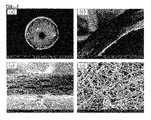
- FIG. 2 presents electro-microscopic images of silk nanofiber nerve conduit according to the present invention, in which ((a) is a cross-section image, (b) is a ⁇ 100 magnified cross-section image, (c) is a ⁇ 1000 magnified cross-section image, and (d) is a fiber structure of silk nanofiber nerve conduit surface);
- FIG. 3 presents electro-microscopic images of inside of the nerve conduit after 10 weeks from when silk nanofiber nerve conduit was autografted into mouse, in which ((a) is inside of the nerve conduit, (b) is an magnified image of (a) in which an arrow indicates regenerated nerve bundle, (c) is an image magnified the cross section of regenerated nerve bundle indicated by an arrow in (b), (d) is an image magnified the side of regenerated nerve bundle indicated by an arrow in (b)); and
- FIG. 4 presents a histological analyzed image of inside of the nerve conduit after 10 weeks from when silk nanofiber nerve conduit was autografted into mouse acquired by ((a) hematoxylin-eosin staining as basic staining, (b) Luxol fast blue staining image specifically staining nerve bundle, and (c) is an image magnified Example 1 of (b) in which the blue line in the conduit indicates regenerated nerve bundle).
- the present invention provides a silk nanofiber nerve conduit characterized in that fibroin nanofibers having a diameter of 200-400 nm, originated from silk fiber, are stacked layer upon layer to form a porous conduit-shape.
- the silk nanofiber is made from the fiber from the reeled-off cocoon which is grown by mulberry.
- the silk nanofiber has been used as a high-quality fiber material for the past decades because of its high-tensile strength, glossiness, excellent dyeing properties, etc.
- the silk fiber has a structure in which two threads of fibroin are covered with the external membrane of sericin.
- the fibroin has excellent bioaffinity and does not affect any other tissues around it. Accordingly, the fibroin may be variously used for food, medicine, medical supplies, etc., in various forms including powder, gel aqueous solution, etc.
- the thickness of the silk nanofiber nerve conduit is preferably 0.1 to 0.3 mm. If the thickness is less than 0.1 mm, inside of nerve conduit is pressed under external pressure during surgical procedures and if the thickness is more than 0.3 mm, the bending elasticity is decreased.
- the silk nanofiber nerve conduit may preferably have a structure in which fine fibers are stacked layer upon layer and the silk nanofibers having a diameter of approximately 200 to 400 nm are irregularly intertwined, forming pores therein.
- the body fluid may be exchanged in and out of conduit and the limitation of the existing synthetic polymer nerve conduit has been overcome, wherein the body fluid can not be exchanged in and out of the conduit since there are no pores.
- the size of the formed pores is preferably 50 to 250 ⁇ m. If the size is less than 50 ⁇ m, the size of pores is too small to exchange the body fluid in and out of the conduit and if the size is more than 250 ⁇ m, regeneration accelerator substances formed inside of the conduit are leaked and scar tissue of the outside of conduit is passed through.
- the silk nanofiber nerve conduit may preferably have sufficient elasticity, tensile strength and tear strength to connect severed nerve to nerve with surgical suture under wet condition.
- the present invention provides producing method of the silk nanofiber nerve conduit.
- the present invention provides a method for producing a silk nanofiber nerve conduit, including steps of: preparing a fibrous spinning solution (Step 1); producing a silk nanofiber of conduit-shape by electrospinning the fibrous spinning solution prepared in step 1 into the cylindrical collecting part coated with polyethyleneoxide (PEO) (Step 2); and separating a silk nanofiber of conduit-shape produced in step 2 from the collecting part (Step 3).
- step 1 of the present invention prepares fibrous spinning solution.
- the fibrous spinning solution may be prepared by: removing sericin from the silk fiber (Step a); preparing silk fibroin solution by washing, drying, and dialyzing silk fiber from which sericin is removed (Step b); preparing silk fibroin sponge by lyophilizing the silk fibroin solution (Step c); and filtering the silk fibroin sponge after dissolving in designated solvent (Step d).
- the silk fiber has a structure in which two threads of fibroin are covered by the external membrane of sericin. Since the fibroin has excellent bioaffinity and does not affect any body tissues, the fibroin may be fabricated into various forms such as powder, gel aqueous solution, etc. and diversely used in forms of food, medicine, medical supplies, etc. If the spinning solution is prepared with the silk fiber, sericin is preferably removed from the silk fiber to maximize biocompatibility. Sericin may be removed by submerging silk fiber in sodium oleate, sodium carbonate solution and heating the same.
- the silk fibroin solution may be produced by: repeatedly washing sericin-removed silk nanofiber with distilled water, drying, dissolving in the mixed solution of calcium chloride, water and ethanol, placing in dialyzing diaphragm, and dialyzing with distilled water.
- the dialyzing diaphragm may preferably have 12 to 14 kDa of MWCO.
- silk fibrous spinning solution may be produced by lyophilizing the silk fibroin solution, preparing silk fibroin sponge, dissolving the sponge in formic acid, and filtering the product.
- 12 to 18% concentration is preferable for the efficient spinning process and to obtain regular form of fiber.
- concentration when the concentration is increased, the thickness of the fiber is increased. If the concentration is less than 12%, the content of the silk in the spinning solution becomes too low to form a fiber, and if the concentration is more than 18%, the viscosity of the solution becomes too high to form a fiber with electricity, therefore, stable electro-spinning may not be continued.
- a silk nanofiber of conduit-shape is prepared by electro-spinning the fibrous spinning solution prepared in step 1 into the cylindrical collecting part coated with polyethyleneoxide (PEO).
- PEO polyethyleneoxide
- the cylindrical collecting part coated with PEO may preferably be a metallic rod coated with 5 to 15% of PEO. If the metallic rod is coated less than 5% of PEO, the coating may not be uniformly formed over the entire surface of the rod. If the metallic rod is coated more than 15% of PEO, PEO layers may become too thick.
- a silk nanofiber of conduit-shape may be prepared by placing the fibrous spinning solution prepared in step 1 in a sealed container such as syringe and releasing with a regular speed by using controlled volume pump. At this time, it is preferable to use tubular spinneret made of metal such as a needle of syringe.
- the electro-spinning may be performed by using electro-spinning device which may include power supply device, electrode, spinneret, ground contact part, spinning solution delivery part and collecting part.
- the electro-spinning device operates to apply voltage such as direct current (DC) voltage, wherein the range of the voltage is preferably between 5 and 20 kV.
- the electrode is connected from output part of the power supply device to the spinneret (i.e., the needle of syringe). If metallic conduit is used as releasing part, the metallic conduit may also work as electrodes.
- the ground part may include two parts, that is, the ground for preventing malfunction caused by leaking high voltage of control volume pump which controls the volume of the spinning solution, and the ground of the collecting part.
- a circular metallic rod made of stainless steel may be used to prepare tubular structure, and the metallic rods with various diameters may be used depending on the desirable inner diameter of the conduit.
- the collecting part may preferably spin at 100 to 300 rpm to spread nanofiber on the surface uniformly while electro-spinning is processed.
- step 3 the silk nanofilber having conduit-shape produced in step 2 is sperated from the collecting part.
- the collecting part may be submerged in methanol or ethanol and insolubilized. Without this process, the silk nanofiber can hardly be used as a nerve conduit since the silk nanofiber is weak against moisture. Later, the silk nanofiber may be separated from the collecting part by submerging the collecting part in the water. The separated silk nanofiber nerve conduit may be washed with distilled water additionally.
- Step 2 Preparing Silk Nanofiber Having Conduit-Shape
- Electro-spinning was performed by the collecting part, which was the 1.65 mm of metallic rod coated with 10% of polyethileneoxide, wherein, the fibrous spinning solution produced in step 1 was electro-spun under the condition of 10 kV of applied voltage, 0.2 ml/time of releasing speed and 200 rpm of metallic rod spinning speed.
- the electro-spinning was ended, and the collected silk nanofiber was placed and dried in the atmosphere so that formic acid vaporized. Therefore, silk nanofiber having conduit-shape was prepared.
- the collecting part on which the silk nanofiber was integrated in conduit-shape from step 2 was submerged in ethanol for 1 hour and insolubilized, and submerged in water so that the silk nanofiber was separated from the collecting part.
- the separated silk nanofiber was washed with distilled water and a silk nanofiber nerve conduit was obtained.
- FIG. 1 presents photographic images of silk nanofiber nerve conduit of the Example 1.
- FIG. 2 presents electro-microscopic images of silk nanofiber nerve conduit regarding the present invention. For more details, (a) in FIG. 2 presents the cross-section of silk nanofiber nerve conduit, (b) and (c) of FIG. 2 present magnified cross section images, and (d) of FIG. 2 presents the fabric structure of the silk nanofiber nerve conduit surface.
- silk nanofiber nerve conduit of Example 1 of the present invention had circle-shaped cross section.
- the inside of the silk nanofiber nerve conduit was hollow and the thickness of the conduit was approximately 0.2 mm.
- the cross section of silk nanofiber nerve conduit of Example 1 of the present invention had a structure in which fine fibers were stacked layer upon layer and silk nanofiber having a diameter of 200 to 400 nm were irregularly intertwined. Accordingly, silk nanofiber nerve conduit of Example 1 of the present invention had pores formed by the irregular intertwining of the fine fibers.
- silk nanofiber nerve conduit of the present invention may exchange the body fluid in and out of conduit through the pores therein. This characteristic may solve the limitation caused by the absence of the existing pores in synthetic polymers nerve conduit wherein body fluid cannot be exchanged.
- the conduit having 4 cm length was prepared. Inner diameter of the nerve conduit was 1.65 mm and the thickness of conduit wall was average 200 ⁇ m. Therefore, cross sectional area was 1.16 mm 2 ([ ⁇ (1.65+0.4)/2 ⁇ 2 ⁇ (1.65/2) 2 ]* ⁇ ).
- Tensile strength test was performed after submerging the conduit in normal saline solution for 1 hour. The ending of the sample which was swollen in the solution was fixed on test device. With 2 cm gauge length and 10 mm/min speed, the strength and elongation of severance was measured until desired position.
- the silk nanofiber nerve conduit of Example 1 of the present invention may be sufficient to connect severed nerve to nerve as a surgical suture. Therefore, silk nanofiber nerve conduit of the present invention may be used as a surgical suture.
- paw pressure test was performed, wherein upon covering the eyes of rat, the paws of the rat were placed under the sharp pressure tip of PP and with increments of pressure, the pressure was measured under which the rats had pain and cried. At this time, the subject showing freezing reaction due to phobia was excluded from the experiment. The result is as shown in Table 2 and FIG. 3 .
- ASA Ankle Stance Angle Test
- ASA indicates the extended angle in the middle of walking and the value is 90° for normal rats.
- the result is as shown as Table 2 below.
- PP indicates the moment under which the rats feel pain. As the pressure (g) is higher, sensory function decreases. The result is as shown as Table 2 below.
- negative control group had 42° in week 10 (47% of normal rat)
- positive control group autografted autogenous nerve had 57° of improved function (63% of normal rat)
- experiment group which was autografted silk nanofiber nerve conduit of Example 1 of the present invention had 62° (69% of normal rat) and showed the best improvement for a motor skill among the three groups. Therefore, it was observed that the experiment group had better improvement of motor skill than negative control group and excellent improvement of motor skill as compared to the positive control group.
- silk nanofiber nerve conduit of the present invention may be valuably used for nerve injury patients.
- Self-cleaving one of the common symptoms of the injury of peripheral nerve, causes extreme pain as a result of forming neuroma. Therefore, observing the degree of self-cleaving may reflect the degree of pain which animal feels.
- Step 1 was the self-cleaving at the level of claw
- step 2 was the self-cleaving at the level of toe
- step 3 was self-cleaving at the level of toe to sole and self-cleaving was at the level of more than sole over step 3. Since step 1 was perfectly recovered, more than step 2 which was impossibly recoverable step was regarded as serious condition. At this time, the subject showing extreme pain due to serious self-cleaving was excluded from the experiment.
- step 1(%) the self-cleaving of the claw level.
- Negative control group 0 20 80 60 60 60 60 60 60 60 Positive control group 50 50 50 50 50 50 50 50 75 75 75 Example 1 0 0 0 17 33 33 50 67 83 83 Incidence ratio of step 2(%): the self-cleaving of the toe level.
- the silk nanofiber nerve conduit of the present invention can be used to treat injured nerve.
- the following experiment was performed in order to recognize cytological change of the ischiadic nerve and the degree of myelinated nerve when silk nanofiber nerve conduit of the present invention was autografted in injured ischiadic nerve.
- ischiadic nerve sample was prepared as paraffin block and sliced 10 ⁇ m finely by microtome. Staining with Hematoxylin-Eosin stain (H&E) and Luxol fast blue stain (LFB) were carried out, respectively. After staining, the tissue was sealed for long term preservation. The images of the tissue were scanned by using OLYMPUS microscopy (BX-51 microscopy, Japan) and analyzed by image analyzer (KAPPA, USA). For H&E staining, after removing paraffin from the tissue, the tissue was stained in Hematoxylin solution for 10 min and Eosin solution for 1 min in order.
- the tissue was placed in the luxol fast blue solution (95% ethanol including 0.1% of luxol fast blue MSBN and 0.05% of acetic acid) and reacted under 57° C. for 24 hours. After that, the tissue was washed with 95% alcohol and distilled water one by one and placed in 0.1% of cresyl violet solution under 37° C. for 1 hour in order to seperate the dying property of medulla and cortex of nerve cell. After staining, the tissue was sealed for safe preservation of tissue staining property. At that time, myelinated layer was stained blue and nerve cells were stained pink or purple.
- the result of H&E staining is shown (a) in FIG. 4
- the result of LFB staining is shown (b) in FIG. 4
- the result of example 1 of (b) in FIG. 4 is magnified and shown in (c) of FIG. 4 .
- nerve fibers were hold together where neuroma was placed.
- the cells filled inside of the conduit were also stained blue and it was recognized that the cells were nerve cells having myelin sheath, which were myelinated cells.
- nerve cells were regenerated from the growing direction of the blue-stained cell, i.e., from the middle to the end with directionality.
- silk nanofiber nerve conduit was autografted in Example 1, nerve cells were regenerated from the middle to the end with directionality.
- the silk nanofiber nerve conduit may be used to treat injured nerve.
- the autograft of the silk nanofiber nerve conduit of the present invention has excellent biocompatibility; allows the body fluid to be exchanged in and out of conduit through pores of the conduit; and has a proper elasticity, tensile strength, and tear strength. Due to these properties, the silk nanofiber nerve conduit of the present invention helps the regeneration of the nerve injury to recover a motor skill and a sensory function, and shows an excellent effect of nerve regeneration. Therefore, the silk nanofiber nerve conduit of the present invention may be used in treating a nerve injury as a replacement of an existing synthetic polymeric nerve conduit. Also, it is expected that the incidence of pain associated with nerve disease which is defined as an important complication of nerve injury may be minimized.
Landscapes
- Health & Medical Sciences (AREA)
- Engineering & Computer Science (AREA)
- Chemical & Material Sciences (AREA)
- Life Sciences & Earth Sciences (AREA)
- Textile Engineering (AREA)
- Biomedical Technology (AREA)
- Public Health (AREA)
- Oral & Maxillofacial Surgery (AREA)
- Transplantation (AREA)
- Veterinary Medicine (AREA)
- Animal Behavior & Ethology (AREA)
- General Health & Medical Sciences (AREA)
- Mechanical Engineering (AREA)
- Dispersion Chemistry (AREA)
- Medicinal Chemistry (AREA)
- Chemical Kinetics & Catalysis (AREA)
- Dermatology (AREA)
- Epidemiology (AREA)
- Heart & Thoracic Surgery (AREA)
- Cardiology (AREA)
- Vascular Medicine (AREA)
- Zoology (AREA)
- Botany (AREA)
- Urology & Nephrology (AREA)
- General Chemical & Material Sciences (AREA)
- Molecular Biology (AREA)
- Gastroenterology & Hepatology (AREA)
- Pulmonology (AREA)
- Materials For Medical Uses (AREA)
Applications Claiming Priority (3)
| Application Number | Priority Date | Filing Date | Title |
|---|---|---|---|
| KR10-2009-0074161 | 2009-08-12 | ||
| KR1020090074161A KR101116237B1 (ko) | 2009-08-12 | 2009-08-12 | 실크 나노섬유 신경도관 및 이의 제조방법 |
| PCT/KR2010/005280 WO2011019211A2 (fr) | 2009-08-12 | 2010-08-11 | Conduit nerveux à nanofibres de soie et procédé pour sa production |
Publications (2)
| Publication Number | Publication Date |
|---|---|
| US20120150205A1 US20120150205A1 (en) | 2012-06-14 |
| US9072592B2 true US9072592B2 (en) | 2015-07-07 |
Family
ID=43586650
Family Applications (1)
| Application Number | Title | Priority Date | Filing Date |
|---|---|---|---|
| US13/389,787 Active 2032-06-29 US9072592B2 (en) | 2009-08-12 | 2010-08-11 | Methods for producing and using silk nanofiber nerve conduits |
Country Status (4)
| Country | Link |
|---|---|
| US (1) | US9072592B2 (fr) |
| EP (1) | EP2465472B1 (fr) |
| KR (1) | KR101116237B1 (fr) |
| WO (1) | WO2011019211A2 (fr) |
Cited By (2)
| Publication number | Priority date | Publication date | Assignee | Title |
|---|---|---|---|---|
| US10845360B2 (en) | 2016-02-25 | 2020-11-24 | Massachusetts Institue Of Technology | Neuronal axon mimetics for in vitro analysis of neurological diseases, myelination, and drug screening |
| US11535826B2 (en) | 2017-05-10 | 2022-12-27 | Massachusetts Institute Of Technology | Engineered 3D-printed artificial axons |
Families Citing this family (17)
| Publication number | Priority date | Publication date | Assignee | Title |
|---|---|---|---|---|
| KR101387886B1 (ko) * | 2013-05-20 | 2014-04-24 | 한림대학교 산학협력단 | 두께 조절 및 공극 크기 조절이 가능한 나노섬유 지지체의 제조방법, 이에 의하여 제조된 나노섬유 지지체 및 이에 사용되는 나노섬유 지지체의 제조장치 |
| KR101634205B1 (ko) * | 2015-03-31 | 2016-06-28 | 한림대학교 산학협력단 | 효소가 포접된 실크 피브로인을 이용한 휴대형 복막투석액 재생 시스템용 필터 및 그 제조 방법 |
| WO2016192733A1 (fr) * | 2015-05-29 | 2016-12-08 | Aarhus Universitet | Conduit de régénération de matériau biologique |
| CN105748171B (zh) * | 2016-02-21 | 2017-09-15 | 新乡医学院 | 生物型神经导管 |
| CN106474543B (zh) * | 2016-09-22 | 2019-05-17 | 武汉纺织大学 | 一种增强型韧带组织工程支架及其制备方法 |
| CN106983915B (zh) * | 2017-05-24 | 2019-09-06 | 南通市第一人民医院 | 蚕丝微纤维-明胶-聚乙二醇医用导管及其制备方法 |
| CN108103598A (zh) * | 2018-02-09 | 2018-06-01 | 郑州大学 | 一种制备沿轴取向管状组织工程材料的静电纺丝接收装置 |
| WO2019166087A1 (fr) | 2018-02-28 | 2019-09-06 | Fundación Tekniker | Conduit de guidage de nerf implantable pour réparation de nerf |
| CN109513041B (zh) * | 2018-12-29 | 2023-10-03 | 南通纺织丝绸产业技术研究院 | 一种镁丝蚕丝复合编织结构神经导管及其制备方法 |
| CN110115645A (zh) * | 2019-04-18 | 2019-08-13 | 新疆医科大学第一附属医院 | 多通道人工神经导管及其制备方法 |
| CN110975008B (zh) * | 2019-12-18 | 2021-06-08 | 武汉理工大学 | 一种具有电刺激和促血管生成作用的神经修复载药系统的制备方法 |
| CN111714247A (zh) * | 2020-05-29 | 2020-09-29 | 广州新诚生物科技有限公司 | 一种人工神经导管及其制备方法 |
| WO2023108469A1 (fr) * | 2021-12-15 | 2023-06-22 | 深圳先进技术研究院 | Matériau tubulaire à base de nanofibres et son procédé de préparation |
| CN114732956A (zh) * | 2022-04-20 | 2022-07-12 | 西南大学 | 一种体内降解与神经再生高度匹配的神经支架的制备方法 |
| CN114886603B (zh) * | 2022-05-11 | 2023-04-18 | 纽生(天津)生物科技有限公司 | 一种抗弯折神经导管及其制备方法和应用 |
| CN115944785B (zh) * | 2022-12-16 | 2024-08-13 | 上海工程技术大学 | 一种匀质纤维管状支架的制备方法 |
| CN116899014B (zh) * | 2023-07-24 | 2024-05-07 | 上海工程技术大学 | 一种三维拓扑结构多通道神经导管及其制备方法 |
Citations (10)
| Publication number | Priority date | Publication date | Assignee | Title |
|---|---|---|---|---|
| US1818549A (en) * | 1929-03-21 | 1931-08-11 | Thames Silk Company | Apparatus and method for spinning fibers in precipitating baths |
| US4808324A (en) * | 1986-04-04 | 1989-02-28 | Lonza Ltd. | Lubricant system for sheet and section rolling mills |
| US20030100108A1 (en) | 2001-11-16 | 2003-05-29 | Altman Gregory H. | Matrix for the production of tissue engineered ligaments, tendons and other tissue |
| US20030155670A1 (en) * | 2002-01-09 | 2003-08-21 | O'brien John P. | Polypeptide fibers and processes for making them |
| US20040110439A1 (en) * | 2001-04-20 | 2004-06-10 | Chaikof Elliot L | Native protein mimetic fibers, fiber networks and fabrics for medical use |
| KR100718073B1 (ko) | 2004-03-25 | 2007-05-16 | 재단법인서울대학교산학협력재단 | 전기방사법을 이용한 생분해성 고분자 신경도관의 제조방법 |
| WO2008004356A1 (fr) * | 2006-07-04 | 2008-01-10 | National University Corporation Tokyo University Of Agriculture And Technology | Composition d'ensimage en solution, procédé de production de fibre de soie régénérée avec ladite composition, et fibre de soie régénérée obtenue par ledit procédé |
| KR20080049095A (ko) | 2005-09-28 | 2008-06-03 | 난퉁유니버시티 | 실크 파이브로인을 포함하는 의학적 인공 신경 이식조직과이의 제조방법 |
| KR20090041271A (ko) | 2007-10-23 | 2009-04-28 | 재단법인서울대학교산학협력재단 | 조직공학용 나노섬유 지지체의 공극 구조 조절 방법 및그를 이용하여 제조된 조직공학용 지지체 |
| US20110287082A1 (en) * | 2008-01-25 | 2011-11-24 | Jennifer Margaret Smith | Multilayer Scaffold |
Family Cites Families (1)
| Publication number | Priority date | Publication date | Assignee | Title |
|---|---|---|---|---|
| EP1558444B1 (fr) * | 2002-06-24 | 2016-09-21 | Tufts University | Biomateriaux a base de soie et leurs methodes d'utilisation |
-
2009
- 2009-08-12 KR KR1020090074161A patent/KR101116237B1/ko active IP Right Grant
-
2010
- 2010-08-11 WO PCT/KR2010/005280 patent/WO2011019211A2/fr active Application Filing
- 2010-08-11 US US13/389,787 patent/US9072592B2/en active Active
- 2010-08-11 EP EP10808360.1A patent/EP2465472B1/fr active Active
Patent Citations (11)
| Publication number | Priority date | Publication date | Assignee | Title |
|---|---|---|---|---|
| US1818549A (en) * | 1929-03-21 | 1931-08-11 | Thames Silk Company | Apparatus and method for spinning fibers in precipitating baths |
| US4808324A (en) * | 1986-04-04 | 1989-02-28 | Lonza Ltd. | Lubricant system for sheet and section rolling mills |
| US20040110439A1 (en) * | 2001-04-20 | 2004-06-10 | Chaikof Elliot L | Native protein mimetic fibers, fiber networks and fabrics for medical use |
| US20030100108A1 (en) | 2001-11-16 | 2003-05-29 | Altman Gregory H. | Matrix for the production of tissue engineered ligaments, tendons and other tissue |
| US20030155670A1 (en) * | 2002-01-09 | 2003-08-21 | O'brien John P. | Polypeptide fibers and processes for making them |
| KR100718073B1 (ko) | 2004-03-25 | 2007-05-16 | 재단법인서울대학교산학협력재단 | 전기방사법을 이용한 생분해성 고분자 신경도관의 제조방법 |
| KR20080049095A (ko) | 2005-09-28 | 2008-06-03 | 난퉁유니버시티 | 실크 파이브로인을 포함하는 의학적 인공 신경 이식조직과이의 제조방법 |
| WO2008004356A1 (fr) * | 2006-07-04 | 2008-01-10 | National University Corporation Tokyo University Of Agriculture And Technology | Composition d'ensimage en solution, procédé de production de fibre de soie régénérée avec ladite composition, et fibre de soie régénérée obtenue par ledit procédé |
| US20090318963A1 (en) * | 2006-07-04 | 2009-12-24 | Nat Univ Corp Tokyo Univ Of Agrigulture And Tech | Spinning solution composition, process for producing regenerated silk fiber using the composition, and regenerated silk fiber produced by the process |
| KR20090041271A (ko) | 2007-10-23 | 2009-04-28 | 재단법인서울대학교산학협력재단 | 조직공학용 나노섬유 지지체의 공극 구조 조절 방법 및그를 이용하여 제조된 조직공학용 지지체 |
| US20110287082A1 (en) * | 2008-01-25 | 2011-11-24 | Jennifer Margaret Smith | Multilayer Scaffold |
Non-Patent Citations (4)
| Title |
|---|
| A.C. Lee et al., Controlled release of nerve growth factor enhances sciatic nerve regeneration, Exp. Neurol., 184, 295-303, 2003. |
| Aldini et al. (1996), "Effectiveness of a bioabsorbable conduit in the repair of peripheral nerves," Biomaterials, vol. 17, No. 10, pp. 959-962. |
| B. Schlosshauer et al., "Rat Schwann cells in bioresorbable nerve guides to promote and accelerate axonal regeneration," Brain Res., 963,321-326, 2003. |
| International Search Report for PCT/KR2010/005280, mailed Apr. 26, 2011. |
Cited By (2)
| Publication number | Priority date | Publication date | Assignee | Title |
|---|---|---|---|---|
| US10845360B2 (en) | 2016-02-25 | 2020-11-24 | Massachusetts Institue Of Technology | Neuronal axon mimetics for in vitro analysis of neurological diseases, myelination, and drug screening |
| US11535826B2 (en) | 2017-05-10 | 2022-12-27 | Massachusetts Institute Of Technology | Engineered 3D-printed artificial axons |
Also Published As
| Publication number | Publication date |
|---|---|
| EP2465472A2 (fr) | 2012-06-20 |
| WO2011019211A2 (fr) | 2011-02-17 |
| KR101116237B1 (ko) | 2012-03-09 |
| EP2465472B1 (fr) | 2016-08-03 |
| KR20110016581A (ko) | 2011-02-18 |
| US20120150205A1 (en) | 2012-06-14 |
| EP2465472A4 (fr) | 2015-01-21 |
| WO2011019211A3 (fr) | 2011-06-30 |
Similar Documents
| Publication | Publication Date | Title |
|---|---|---|
| US9072592B2 (en) | Methods for producing and using silk nanofiber nerve conduits | |
| US20240131218A1 (en) | Electrospun biocompatible fiber compositions | |
| EP3015120B1 (fr) | Support de réparation de tissu, son procédé de préparation et sa raison | |
| CN102525689B (zh) | 取向纳米纤维仿生神经导管及其制作方法 | |
| EP2373354B1 (fr) | Échafaudage de réparation tissulaire | |
| WO2017088818A1 (fr) | Membrane à fibre de réparation tissulaire, son procédé de préparation et application, et produit de réparation tissulaire | |
| CN112553785B (zh) | 一种双层引导组织再生膜及其制备方法 | |
| CN104414773A (zh) | 防粘连组织修复膜及其制备方法 | |
| WO2016192733A1 (fr) | Conduit de régénération de matériau biologique | |
| CN105919694A (zh) | 一种复层电纺膜及其用途 | |
| CN108478866A (zh) | 组织修复支架、其制备方法和用途 | |
| CN107929803A (zh) | 一种纳米纤维纱线手术线及其制备方法 | |
| JP2022524714A (ja) | マイクロフルイディック押出 | |
| CN111632193B (zh) | 壳聚糖基神经纤维膜及制备方法、神经导管和应用 | |
| CN104287869B (zh) | 一种用于气管移植的新型纳米纤维膜/纱支架及其制备方法 | |
| Wang et al. | Electrospun, reinforcing network-containing, silk fibroin-based nerve guidance conduits for peripheral nerve repair | |
| CN109758197B (zh) | 一种组织修复套管及其制备方法和应用 | |
| Mohammadzadehmoghadam et al. | Electrospinning of silk fibroin-based nanofibers and their applications in tissue engineering | |
| CN103741262B (zh) | 一种用于硬脑膜或硬脊膜修补的几丁糖纳米纤维膜及其制备方法与应用 | |
| KR102115167B1 (ko) | 강도가 증가된 콜라겐/키틴 나노섬유 멤브레인 및 이의 제조 방법 | |
| CN109260524B (zh) | 一种组织修复用纳米短纤维材料及其制备方法和应用 | |
| JPWO2019054407A1 (ja) | 神経保護材 | |
| CN109847104B (zh) | 一种神经修复膜及其制备方法和应用 | |
| WO2024149413A2 (fr) | Échafaudage neuronal composite cylindrique, son procédé de préparation et son utilisation | |
| CN115364283A (zh) | 一种仿生界面多层复合神经支架及其制备方法与应用 |
Legal Events
| Date | Code | Title | Description |
|---|---|---|---|
| AS | Assignment |
Owner name: SNU R&DB FOUNDATION, KOREA, REPUBLIC OF Free format text: ASSIGNMENT OF ASSIGNORS INTEREST;ASSIGNORS:PARK, YOUNG HWAN;KI, CHANG SEOK;KIM, HYUN JEUNG;AND OTHERS;REEL/FRAME:027696/0818 Effective date: 20120206 |
|
| AS | Assignment |
Owner name: KONICA MINOLTA HOLDINGS, INC., JAPAN Free format text: ASSIGNMENT OF ASSIGNORS INTEREST;ASSIGNORS:HIRAO, YUYA;OKADA, MASAKAZU;REEL/FRAME:028301/0488 Effective date: 20111222 |
|
| STCF | Information on status: patent grant |
Free format text: PATENTED CASE |
|
| FEPP | Fee payment procedure |
Free format text: PAYOR NUMBER ASSIGNED (ORIGINAL EVENT CODE: ASPN); ENTITY STATUS OF PATENT OWNER: SMALL ENTITY |
|
| MAFP | Maintenance fee payment |
Free format text: PAYMENT OF MAINTENANCE FEE, 4TH YR, SMALL ENTITY (ORIGINAL EVENT CODE: M2551); ENTITY STATUS OF PATENT OWNER: SMALL ENTITY Year of fee payment: 4 |
|
| MAFP | Maintenance fee payment |
Free format text: PAYMENT OF MAINTENANCE FEE, 8TH YR, SMALL ENTITY (ORIGINAL EVENT CODE: M2552); ENTITY STATUS OF PATENT OWNER: SMALL ENTITY Year of fee payment: 8 |