US8900146B2 - Three-dimensional (3D) ultrasound imaging system for assessing scoliosis - Google Patents
Three-dimensional (3D) ultrasound imaging system for assessing scoliosis Download PDFInfo
- Publication number
- US8900146B2 US8900146B2 US12/509,705 US50970509A US8900146B2 US 8900146 B2 US8900146 B2 US 8900146B2 US 50970509 A US50970509 A US 50970509A US 8900146 B2 US8900146 B2 US 8900146B2
- Authority
- US
- United States
- Prior art keywords
- image
- features
- vertebra
- images
- ultrasound
- Prior art date
- Legal status (The legal status is an assumption and is not a legal conclusion. Google has not performed a legal analysis and makes no representation as to the accuracy of the status listed.)
- Active, expires
Links
Images


Classifications
-
- A—HUMAN NECESSITIES
- A61—MEDICAL OR VETERINARY SCIENCE; HYGIENE
- A61B—DIAGNOSIS; SURGERY; IDENTIFICATION
- A61B5/00—Measuring for diagnostic purposes; Identification of persons
- A61B5/103—Detecting, measuring or recording devices for testing the shape, pattern, colour, size or movement of the body or parts thereof, for diagnostic purposes
- A61B5/107—Measuring physical dimensions, e.g. size of the entire body or parts thereof
- A61B5/1071—Measuring physical dimensions, e.g. size of the entire body or parts thereof measuring angles, e.g. using goniometers
-
- A—HUMAN NECESSITIES
- A61—MEDICAL OR VETERINARY SCIENCE; HYGIENE
- A61B—DIAGNOSIS; SURGERY; IDENTIFICATION
- A61B5/00—Measuring for diagnostic purposes; Identification of persons
- A61B5/45—For evaluating or diagnosing the musculoskeletal system or teeth
- A61B5/4538—Evaluating a particular part of the muscoloskeletal system or a particular medical condition
- A61B5/4561—Evaluating static posture, e.g. undesirable back curvature
-
- A—HUMAN NECESSITIES
- A61—MEDICAL OR VETERINARY SCIENCE; HYGIENE
- A61B—DIAGNOSIS; SURGERY; IDENTIFICATION
- A61B8/00—Diagnosis using ultrasonic, sonic or infrasonic waves
- A61B8/08—Detecting organic movements or changes, e.g. tumours, cysts, swellings
- A61B8/0875—Detecting organic movements or changes, e.g. tumours, cysts, swellings for diagnosis of bone
-
- A—HUMAN NECESSITIES
- A61—MEDICAL OR VETERINARY SCIENCE; HYGIENE
- A61B—DIAGNOSIS; SURGERY; IDENTIFICATION
- A61B8/00—Diagnosis using ultrasonic, sonic or infrasonic waves
- A61B8/42—Details of probe positioning or probe attachment to the patient
- A61B8/4245—Details of probe positioning or probe attachment to the patient involving determining the position of the probe, e.g. with respect to an external reference frame or to the patient
- A61B8/4254—Details of probe positioning or probe attachment to the patient involving determining the position of the probe, e.g. with respect to an external reference frame or to the patient using sensors mounted on the probe
-
- A—HUMAN NECESSITIES
- A61—MEDICAL OR VETERINARY SCIENCE; HYGIENE
- A61B—DIAGNOSIS; SURGERY; IDENTIFICATION
- A61B8/00—Diagnosis using ultrasonic, sonic or infrasonic waves
- A61B8/48—Diagnostic techniques
- A61B8/483—Diagnostic techniques involving the acquisition of a 3D volume of data
-
- G—PHYSICS
- G06—COMPUTING; CALCULATING OR COUNTING
- G06T—IMAGE DATA PROCESSING OR GENERATION, IN GENERAL
- G06T7/00—Image analysis
- G06T7/0002—Inspection of images, e.g. flaw detection
- G06T7/0012—Biomedical image inspection
-
- G06K2209/055—
-
- G—PHYSICS
- G06—COMPUTING; CALCULATING OR COUNTING
- G06T—IMAGE DATA PROCESSING OR GENERATION, IN GENERAL
- G06T2200/00—Indexing scheme for image data processing or generation, in general
- G06T2200/04—Indexing scheme for image data processing or generation, in general involving 3D image data
-
- G—PHYSICS
- G06—COMPUTING; CALCULATING OR COUNTING
- G06T—IMAGE DATA PROCESSING OR GENERATION, IN GENERAL
- G06T2207/00—Indexing scheme for image analysis or image enhancement
- G06T2207/10—Image acquisition modality
- G06T2207/10016—Video; Image sequence
-
- G—PHYSICS
- G06—COMPUTING; CALCULATING OR COUNTING
- G06T—IMAGE DATA PROCESSING OR GENERATION, IN GENERAL
- G06T2207/00—Indexing scheme for image analysis or image enhancement
- G06T2207/10—Image acquisition modality
- G06T2207/10132—Ultrasound image
- G06T2207/10136—3D ultrasound image
-
- G—PHYSICS
- G06—COMPUTING; CALCULATING OR COUNTING
- G06T—IMAGE DATA PROCESSING OR GENERATION, IN GENERAL
- G06T2207/00—Indexing scheme for image analysis or image enhancement
- G06T2207/20—Special algorithmic details
- G06T2207/20092—Interactive image processing based on input by user
- G06T2207/20096—Interactive definition of curve of interest
-
- G—PHYSICS
- G06—COMPUTING; CALCULATING OR COUNTING
- G06T—IMAGE DATA PROCESSING OR GENERATION, IN GENERAL
- G06T2207/00—Indexing scheme for image analysis or image enhancement
- G06T2207/30—Subject of image; Context of image processing
- G06T2207/30004—Biomedical image processing
- G06T2207/30008—Bone
- G06T2207/30012—Spine; Backbone
-
- G—PHYSICS
- G06—COMPUTING; CALCULATING OR COUNTING
- G06V—IMAGE OR VIDEO RECOGNITION OR UNDERSTANDING
- G06V2201/00—Indexing scheme relating to image or video recognition or understanding
- G06V2201/03—Recognition of patterns in medical or anatomical images
- G06V2201/033—Recognition of patterns in medical or anatomical images of skeletal patterns
Definitions
- the invention concerns a three-dimensional (3D) ultrasound imaging system for assessing scoliosis.
- Scoliosis is a medical condition in which the spine of a person is curved from side to side, and may also be rotated. X-ray assessment is commonly used to determine scoliosis. Other techniques for determining scoliosis include Moire-fringe mapping, raster-based systems, 360° torso profile scanning and stereo-photogrammetric systems.
- FBT Adam's forward blend test
- a scoliometer is a ruler-like handheld tool. It is an inclinometer to measure trunk asymmetry or axial trunk rotation (ATR) which is also known as rib hump deformity.
- ATR axial trunk rotation
- the scoliometer provides a quantitative measurement to assess the degree of scoliosis. Different studies have found that the measurement from a scoliometer resulted in high intra-rater and inter-rater variations of ATR values and a high false positive rate. In addition, the scoliometer measurement does not correlate well with the Cobb method. Earlier studies have suggested that a scoliometer should not be exclusively used as a diagnostic tool.
- Moire-fringe mapping is used to obtain the 3D shape of the back of a patient. Moire-fringes are generated by a grating projected on the target. The images of the fringes are captured by a video system. A contour line system and a sectional shape of the object are then automatically reconstructed and displayed on monitor by computer. Moire fringe mapping can produce very accurate data with a resolution up to 10 microns. Surfaces at a large angle are not measurable when the fringe density becomes too dense. In addition, the patient's position, body-build, and fat folds are other factors causing inaccuracy to the surface topography. Due to the lack of clinical experience on this technique, there is a poor correlation between the observed body and the underlying scoliosis.
- Quantec spinal image system is popular in the United Kingdom.
- the quantec spinal image system is based on Moire topography and raster-stereo photography. This system uses raster stereography to create an image of a fringe pattern and projected onto the patient's back. The system then produces a Q angle, a coronal plane measurement quantifying the coronal asymmetry reflected from the patient's images. However, this system is complex and relies on the surface topography that is a factor of inaccuracy.
- Photogrammetric method systems are based on laser scanning or photography technique. The laser scanning and video system offers a fast and accurate 3D measurement of scoliotic deformities which can be spatially recorded within a minute.
- the output of a digital 3D model provides a resolution up to 1 mm.
- spinal deformations information such as the Cobb angle is derived.
- the Ortelius system developed by OrthoScan Technologies is a radiation-free spatial data capturing system to diagnose and monitor spinal deformities.
- the examiner palpates the patient's back to locate the spinous process of each vertebra and records the position of spinous process for all vertebrae using a 3D spatial sensor.
- the data can then be reconstructed into a computer model for calculating the spinal deformation indices.
- the position of transverse process cannot be obtained.
- the spinal column rotation cannot be considered.
- the patient needs to be repeatedly palpated during the examination and the process may lead to a certain degree of discomfort. Even though the location of transverse processes are recorded by the 3D spatial sensor, it is manually determined by the operator based on body surface palpation, and this is subjective.
- a three-dimensional (3D) ultrasound imaging system for assessing scoliosis.
- the system includes an ultrasound scanner to capture ultrasound images.
- the system also includes a spatial sensor to record the position and orientation of the captured ultrasound images.
- the system also includes a software module to mark features of vertebra in the captured ultrasound images, and the marked features are connected with lines in order to calculate angles and distances between the marked features for the calculation of the Cobb angle and spinal rotation angle based on the calculated angles and distances.
- the marked features are a reflection of the surfaces of the vertebra.
- the software module may include an image enhancement module to enhance bony surface details in the captured images.
- the software module may include an image marking module to identify captured images that contain marked features.
- the software module may include an image magnifying module to magnify captured images for the identification of features of the vertebra.
- the software module may include an image removal module to remove captured images that do not contain marked features.
- the features of the vertebra may include edges, apexes of spinous and transverse processes.
- the software module may include a virtual model generator to connect the marked features with lines to form a frame based skeleton virtual model of the spine.
- the virtual model generator may re-size and place corresponding vertebra segments in 3D space according to the features of the vertebra.
- the ultrasound scanner may have a probe which is swiped over the back of a patient.
- the probe may have a width of about 10 to 20 centimeters to enable scanning of all spinal processes in a single swipe.
- the spatial sensor may comprise a transmitter and a receiver, and the receiver is operatively attached to the probe.
- the spatial sensor may comprise a transmitter and a receiver, and the transmitter is operatively attached to the probe.
- the system may further comprise a chest board.
- the system may further comprise a height adjustable handrail to help a patient maintain a steady position.
- a method for assessing scoliosis includes capturing ultrasound images.
- the method also includes recording the position and orientation of the captured ultrasound images.
- the method also includes marking features of vertebra in the captured ultrasound images, and the marked features are connected with lines in order to calculate angles and distances between the marked features for the calculation of the Cobb angle and spinal rotation angle based on the calculated angles and distances.
- the marked features are a reflection of the surfaces of the vertebra.
- the method may further comprise enhancing bony surface details in the captured images.
- the method may further comprise identifying captured images that contain marked features.
- the method may further comprise magnifying captured images for the identification of features of the vertebra.
- the method may further comprise removing captured images that do not contain marked features.
- the method may further comprise forming a frame based skeleton virtual model of the spine using the lines connecting the marked features.
- the method may further comprise re-sizing and placing corresponding vertebra segments in 3D space according to the features of the vertebra.
- the method may further comprise displaying a projection image of marked features with the ultrasound images in 3D space.
- the method may further comprise combining an X-ray projection image with the ultrasound images in 3D space.
- a computer-implemented method for automatically marking features of vertebra to assess scoliosis comprising:
- the method may further comprise discarding the image if no bone reflection is detected.
- the method may further comprise analysing the location of markers for an identical process and detecting a peak of the process based on the 3D contour formed by the markers.
- the marker which corresponds to a feature of a vertebra with the smallest tissue depth is considered the peak of the process.
- the 3D ultrasound system locates all spinous processes and also provides information relating to transverse processes. All processes that are located are in exact geometric order and dimension.
- the present invention advantageously provides unlimited frequency of usage in assessment of scoliosis. On-site screening and mass screening for children is also made possible since no X-ray is necessary.
- the present invention provides long term monitoring for scoliosis treatment.
- the present invention is safer and more accurate than traditional techniques of assessing scoliosis.
- the present invention is also cost effective because it does not require radiation specific equipment or highly skilled and experienced operators.
- the present invention is also compact and can fit in small clinic.
- FIG. 1 is a block diagram of a 3D ultrasound system in accordance with an embodiment of the present invention
- FIG. 2 is a set of ultrasound images captured by the system of FIG. 1 which are preprocessed to identify landmarks;
- FIG. 3 is a virtual model of a patient's spine formed from the identified landmarks of FIG. 2 ;
- FIG. 4 is a final result generated by the system of FIG. 1 showing the Cobb angle, spine rotation and angle and image of the patient's spine;
- FIG. 5 is a process flow diagram of a method for scoliosis assessment in accordance with an embodiment of the present invention
- FIG. 6 is a set of two images, the left image is an original B-mode image and the right image is an enhanced image where the bone surface has been enhanced using a bone surface extraction filter;
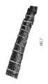
- FIG. 7 is a selection of all candidate images from an original image set captured by the system of FIG. 1 ;
- FIG. 8 is a B-mode image showing a vertebrae with markers placed on apex
- FIG. 9 is a sequence of images with landmarks that have been marked on the images.
- FIG. 10 is a sequence of images with lines connecting the landmarks within one image
- FIG. 11 is a set of projection images with marked features in 3D space.
- FIG. 12 is a set of projection images along side ultrasound images.
- a 3D ultrasound system 10 for scoliosis assessment is provided.
- the system 10 generally comprises an ultrasound scanner 11 with an ultrasound brightness mode (US B-mode) probe 12 , a 3D spatial sensor control unit 13 , a framework 14 , and a computer 15 .
- USB B-mode ultrasound brightness mode
- the framework 14 is adjustable in height and able to conveniently disassemble.
- a chest board 16 is operatively attached to the framework 14 .
- the chest board 16 is a reference for the spatial sensor 13 which enables the physical distance between the chest board 16 and transmitter 13 B of the spatial sensor 13 to be determined. The value of the distance is used to verify internal parameters for the calibration of the spatial sensor 13 .
- the chest board 16 provides a resting surface for the patient to lean on. During scanning with the probe 12 , the patient may be moved forward by a force from the examiner. If this occurs, the chest board 16 helps to prevent the patient from moving forward too much and therefore minimise inaccuracies in the measurements taken.
- a handrail 17 is provided which may be operatively attached to the framework 14 to help the patient maintain a steady position during examination.
- the ultrasound scanner 11 has a wide probe 12 (10 cm or above). This enables an examiner to obtain a set of spine images via a single swipe over the patient's spine. In contrast, the examiner needs to swipe two to three times with a normal probe (around 5 cm or less with width) to capture a complete set of images covering all spinous and transverse processes.
- the system 10 measures the angle and dimension of spine using the spatial sensor 13 in true values instead of measuring from the projection of chest or spine X-ray film. This is more accurate because they are not relative values. The degree of spine rotation can also be obtained in the same examination.
- the spatial sensor control unit 13 is able to determine the position of probe 12 at any moment in time.
- the unit 13 consists of a small cube-shaped transmitter 13 B and a tiny peanut size receiver 13 A which is normally attached to the probe 12 .
- the transmitter 13 B may be operatively attached to the probe 12 .
- the transmitter 13 B generates a magnetic field in space.
- the receiver 13 A senses the strength of magnetic field and the change of magnitude of the magnetic field.
- the results are processed by the spatial sensor control unit 13 to compute the position and orientation of the receiver 13 A.
- the spatial information is sent to the computer 15 periodically.
- the position of the probe 12 is computed using a specific computational method and the spatial information.
- the computational method to obtain the position and orientation information of probe 12 and its generated B-mode image's pixels in the physical world a series of rigid transforms are performed. Before this is done, the probe 12 must be calibrated to obtain a spatial and orientation relationship between the probe 12 and the receiver 13 A. This is the first rigid transform matrix. Also, a second rigid transform matrix is defined which can be chosen in any position and orientation. This matrix is known as a system rigid transform.
- the spatial control unit 13 provides a final rigid transform matrix which defines the current position and orientation between the transmitter 13 B and the receiver 13 A. By multiplying these matrices, the position and orientation information of the probe 12 is obtained. Its B-mode image's pixels are obtained by multiplying the coordinate of pixels relative to the B-mode image.
- Novel ultrasound scanning procedures image processing techniques such as gaussian, sobel filtering, 3D virtualization methods such as OpenGL and Visualization Toolkit, and angle calculation approaches are used together to calculate the degree of spine's deformation in term of true distances and angles instead of an approximation or projected from a standard chest X-ray film.
- All pixels in the B-mode image can be transformed to a physical world location and orientation. If the distance is measured between two pixels in any B-mode images, the physical distance between the objects is obtained which are represented by these pixels. Similarly, the angle between two selected lines 42 is obtained, each of which can be defined by two pixels.
- the system 10 is set up ( 50 ) by deploying the framework 14 at a location and positioning the ultrasound scanner 11 , spatial sensor 13 and computer 15 .
- the patient is asked to stand in a proper position and is given instructions by the examiner.
- Ultrasound coupling gel or liquid is applied on the patient which covers the body area to be scanned.
- a gel pad or liquid bag can also be used to cover the body area and the ultrasound scanning can be conducted above the surface of the gel pad or liquid bag. This is particularly useful when the soft tissue layer covering the bone is very thin.
- the settings of the ultrasound scanner 13 are adjusted such as viewing depth, brightness, focus, gain, transmitting power, etc.
- the spatial sensor 13 is activated. B-mode images and corresponding spatial data are captured and then sent to the computer 15 .
- the patient's spine is scanned ( 51 ) with the B-mode probe 12 of the scanner 11 to capture ultrasound images.
- the scan commences from the L5 to T1 of the spine, or any selected portion of the spine.
- the scanning length may be shortened depending on the area of curvature.
- the total number of scanned images are around 500 to 1500. The patient is asked to stand still and hold the breath during the scanning process.
- These captured images are processed by a software module 21 executing on the computer 15 via a video or USB interface in real-time.
- the images are displayed ( 52 ) in 3D space on the screen of the computer 15 in real-time as they are captured.
- the display of the captured images is depicted in FIG. 2 .
- the images form a long image stack. The examiner performs a preliminarily check of the image consistency. If the images are fine, the patient may leave. Otherwise, the patient has to stand again for re-scanning.
- the set of ultrasound images captured may be preprocessed by various kinds of image processing filters 19 .
- a real-time filter 18 is used on the images.
- the real-time filter 18 enhances the bony surface in the image and the enhanced images guide the examiner to easily move the probe 12 to make the vertebrae locate in a proper position in the image.
- the left image in FIG. 6 is the original ultrasound image and the right image in FIG. 6 is the left image which has been enhanced by the real-time filter 18 .
- the real-time filter 18 extracts useful bone shape by enhancing the maximum or gradient change of pixels in the vertical direction (A-mode direction).
- pseudo color coding can be used to enhance the visualization of the vertebrae, with the bone interface highlighted with a selected color and other regions represented in grey levels.
- These enhancements enable easier identification of landmarks during image capture. For example, the shape of the vertebra in FIG. 6 can then be identified by manual or automatic marking procedures to find the apex of spinous process and transverse processes of an individual image.
- Automatic marking procedures may be performed by the computer 15 via an automatic marker module 26 of the software module 21 .
- the automatic marker module 26 extracts the bone (the surfaces of the vertebra) reflection from the image or removes all feature of the image except the bone reflection using image processing techniques. These image processing techniques include maximum intensity reflection, maximum gradient, active contour, or image registration.
- the automatic marker module 26 is then able to locate the position of bone and automatically mark them. If no bone reflection is detected in an image, the image is discarded because there is no useful information in this image. After the images without landmarks are discarded, one spinal process may still correspond to a series of images. The location of the landmarks in different images for the same process is analyzed and the peak of the process is automatically detected based on the 3D contour formed by the landmarks.
- One approach is to use the depth of the landmarks as a criteria. The landmark with the smallest tissue depth is the peak of the process. After the processes for all vertebrae are obtained, the Cobb angle and rotational angle are automatically calculated as later described.
- Captured images which contain landmarks are selected ( 53 ). These are referred to as candidate images because they are images which contain at least one landmark.
- candidate images which potentially contain landmarks are selected by viewing the image stack. The user can use a computer mouse to navigate the image stack freely on the computer screen. The selected candidate image is enlarged for a better view by the examiner which can be displayed where the image is or in a separated location as depicted by the image 30 shown the bottom right corner in FIG. 2 . If the examiner finds a candidate image, it may be picked from the image stack by clicking it. The selected candidate image is highlighted. The user can repeat the process until all candidate images have been found in the image stack. However, if the user finds difficulty in locating the candidate images from navigating the image stack alone, tools are designed to help viewing the image stack such as volume slice, re-slice, and preview plane. Nevertheless, the user can discard the unselected images in the image stack.
- Captured images without landmarks are discarded ( 54 ) to save on storage space.
- Candidate images potentially containing a vertebra apex or landmark are saved to disk. Therefore, the size of useful data is minimized and the operational speed of the system 10 improves.
- the set of images initially captured by the scanner 11 is very large in size because they are high resolution images.
- the images without landmarks are removed by an image removal module 22 of the software module 21 . What remains are the images containing the landmarks as depicted in FIG. 7 .
- Each of the candidate images is selected ( 55 ).
- the landmarks in the image are identified and marked by markers 41 as depicted in FIG. 3 .
- the landmarks represent the important features of vertebra including edges, spinous and transverse processes.
- Each vertebra such as c1, c2, c3, etc may contain multiple landmarks usually from two to five landmarks.
- the system 10 requires about two or three landmarks from each vertebra for the purpose of generating the virtual model 44 .
- the landmarks that are marked in the system 10 correlate to actual bone surfaces.
- the actual physical position of the landmark is known from the information provided by the spatial sensor 13 . Knowing the actual physical position enables an accurate virtual model 44 to be constructed for the spine geometrical structure. Both the Cobb angle and angle of rotation of the spine are accurately measured at the same time based on the virtual model 44 .
- the examiner must determine ( 56 ) if the landmarks that are marked are visually clear. If they are not, a different imaging method is used to enhance the quality of image and manifest the landmarks. Various non-real-time filters 20 can also be used further to enhance the apexes in the image. Referring to FIG. 8 , in some images, these apexes are very obvious which makes it unnecessary to perform any non-real-time filtering 20 . If a filter is applied ( 57 ), the computer 15 enhances the image and the landmarks. Filters such as brightness filter, contrast filter and edge filter can be used for enhancing the quality of an individual image or all candidate images. The filtering process is repeated until a desirable image is obtained.
- Landmarks are marked ( 58 ) by the examiner selecting the image landmark indicator on the computer 15 . Then, a sphere or a marker 41 of any shape is placed onto the landmark in the 3D position. This step is repeated until all landmarks in the image are found. The sphere 41 indicates the position and existence of the landmark in the image. Referring to FIG. 9 , all candidate images are marked with landmarks. In some cases, one spinal process can be viewed in a series of images. In these cases, a representative image can be selected, such as the one in the middle of that series, or a local volume image can be formed and the peak of the process can be identified in the local volume image. An image marking module 24 of the software module 21 allows examiners to identify images that contain markers 41 . The software module 21 also includes an image magnifying module 25 to assist examiners in identifying landmarks during image marking.
- the image stack is hidden ( 60 ) so that only the markers 41 are displayed.
- the markers 41 in the image are connected with lines 42 to form a frame based skeleton virtual model 44 of the spine using a virtual model generator 23 of the software module 21 as depicted in FIGS. 3 , 4 and 10 .
- the landmarks from the same B-mode image are connected in sequence with lines 42 .
- the lines 42 and markers 41 become a frame-based skeleton virtual model 44 of the patient's spine. Since, the actual dimensions and angles are obtained by the spatial sensor and calculations. All landmarks pose into their exact positions.
- the distance between landmarks and the angles between lines 42 formed by landmarks can then be measured based on the spatial information of each selected landmarks.
- the Cobb angle can then be calculated manually or automatically.
- the information of the markers 41 can be used to re-size and place corresponding virtual vertebra segments on the 3D space to enhance the visualization.
- the virtual model generator 23 may use this information to re-size and place corresponding vertebra segments in 3D space.
- Angles among the landmarks are measured ( 61 ) by the examiner clicking pairs of landmarks from different vertebras with maximum tilt difference. This process can be performed automatically by the computer 15 if necessary.
- the Cobb angle is the angle formed between a line drawn parallel to the superior endplate of one vertebra above the fracture and a line drawn parallel to the inferior endplate of the vertebra one level below the fracture.
- the Cobb angle is computed ( 62 ) from the maximum tilt angles among the pairs of landmarks. Since, the Cobb angle is defined as the projection of spine curve angle from the sagittal plane; the angle still needs to be computed and projected onto a fixed plane before the Cobb angle can be computed.
- the landmarks of transverse processes from the same vertebrae which are the two most tilted vertebrae from different ends of the spine are connected to form a 3D vector line. Similarly, the vector is obtained by connecting the landmarks of transverse processes from the other most tilted vertebrae from other end of the spine.
- the system 10 does not require harmful radiation to operate, it can be used for any patient without limitation on time or frequency.
- the system 10 is a radiation free system which means that it does not require a radiation safe room, expensive X-ray equipment or certified X-ray technician. The initial cost and operational cost is dramatically reduced for scoliosis assessment.
- the system 10 is not restricted in the place or time it must be used. Therefore, the rate of usage is increased and enables on-site and mass screening.
- the operation of the system 10 in a small and non-radiation safe room becomes viable using the system 10 . This is because the ultrasound scanner 11 and spatial sensor 13 are small enough to be moved around or carried by hand.
- the framework 14 can also be assembled and disassembled so as to move into compact room. It is safe to operate by any trained staff at any place.
- the system 10 may receive greater acceptance from experienced examiners if it can also provide a projection image. Both X-ray assessment and the system 10 may be used together where the system 10 is frequently used for long term studies. Therefore, the system 10 has the ability to view a projection image of marked features together with the ultrasound images. Furthermore, the system 10 has the ability for X-ray images to be fused or combined together with the ultrasound measurement.
- the 3D ultrasound imaging system 10 offers a tool to potentially measure different kinds of musculoskeletal structures.
- an electromagnetic spatial sensor has been described, it is envisaged that other types of spatial sensing techniques may be used. These include: marker tracking using an optical visible or infrared camera, acoustic locating, and mechanical spatial locating using multiple articulating joints, etc.
Landscapes
- Health & Medical Sciences (AREA)
- Life Sciences & Earth Sciences (AREA)
- Engineering & Computer Science (AREA)
- General Health & Medical Sciences (AREA)
- Medical Informatics (AREA)
- Physics & Mathematics (AREA)
- Veterinary Medicine (AREA)
- Public Health (AREA)
- Biomedical Technology (AREA)
- Heart & Thoracic Surgery (AREA)
- Biophysics (AREA)
- Molecular Biology (AREA)
- Surgery (AREA)
- Animal Behavior & Ethology (AREA)
- Pathology (AREA)
- Radiology & Medical Imaging (AREA)
- Nuclear Medicine, Radiotherapy & Molecular Imaging (AREA)
- Oral & Maxillofacial Surgery (AREA)
- Dentistry (AREA)
- Physical Education & Sports Medicine (AREA)
- Orthopedic Medicine & Surgery (AREA)
- Rheumatology (AREA)
- Quality & Reliability (AREA)
- Computer Vision & Pattern Recognition (AREA)
- General Physics & Mathematics (AREA)
- Theoretical Computer Science (AREA)
- Ultra Sonic Daignosis Equipment (AREA)
- Apparatus For Radiation Diagnosis (AREA)
- Measurement Of The Respiration, Hearing Ability, Form, And Blood Characteristics Of Living Organisms (AREA)
- Image Processing (AREA)
Priority Applications (12)
| Application Number | Priority Date | Filing Date | Title |
|---|---|---|---|
| US12/509,705 US8900146B2 (en) | 2009-07-27 | 2009-07-27 | Three-dimensional (3D) ultrasound imaging system for assessing scoliosis |
| PCT/CN2010/075287 WO2011012055A1 (en) | 2009-07-27 | 2010-07-20 | Three-dimensional (3d) ultrasound imaging system for assessing scoliosis |
| CA2769150A CA2769150C (en) | 2009-07-27 | 2010-07-20 | Three-dimensional (3d) ultrasound imaging system for assessing scoliosis |
| PL10803888T PL2459073T3 (pl) | 2009-07-27 | 2010-07-20 | System obrazowania trójwymiarowego (3D) ultradźwiękowego do oceny skoliozy |
| EP10803888.6A EP2459073B1 (en) | 2009-07-27 | 2010-07-20 | Three-dimensional (3d) ultrasound imaging system for assessing scoliosis |
| RSP20191277 RS59355B1 (sr) | 2009-07-27 | 2010-07-20 | Trodimenzionalni (3d) sistem za ultrazvučno snimanje za procenu skolioze |
| AU2010278526A AU2010278526B2 (en) | 2009-07-27 | 2010-07-20 | Three-dimensional (3D) ultrasound imaging system for assessing scoliosis |
| CN201080040696.0A CN102497821B (zh) | 2009-07-27 | 2010-07-20 | 用于评估脊柱侧凸的三维(3d)超声成像系统 |
| JP2012521946A JP5849048B2 (ja) | 2009-07-27 | 2010-07-20 | 側弯症評価のための三次元(3d)超音波撮像システム |
| PT108038886T PT2459073T (pt) | 2009-07-27 | 2010-07-20 | Sistema de imagem de ultrassons tridimensional (3d) para avaliação da escoliose. |
| ES10803888T ES2747371T3 (es) | 2009-07-27 | 2010-07-20 | Sistema de diagnóstico por imagen tridimensional (3D) por ultrasonidos para evaluar la escoliosis |
| HRP20191718 HRP20191718T1 (hr) | 2009-07-27 | 2019-09-20 | Trodimenzionalan (3d) sustav ultrazvučne slikovne dijagnostike za procjenu skolioze |
Applications Claiming Priority (1)
| Application Number | Priority Date | Filing Date | Title |
|---|---|---|---|
| US12/509,705 US8900146B2 (en) | 2009-07-27 | 2009-07-27 | Three-dimensional (3D) ultrasound imaging system for assessing scoliosis |
Publications (2)
| Publication Number | Publication Date |
|---|---|
| US20110021914A1 US20110021914A1 (en) | 2011-01-27 |
| US8900146B2 true US8900146B2 (en) | 2014-12-02 |
Family
ID=43497923
Family Applications (1)
| Application Number | Title | Priority Date | Filing Date |
|---|---|---|---|
| US12/509,705 Active 2032-06-01 US8900146B2 (en) | 2009-07-27 | 2009-07-27 | Three-dimensional (3D) ultrasound imaging system for assessing scoliosis |
Country Status (12)
| Country | Link |
|---|---|
| US (1) | US8900146B2 (ja) |
| EP (1) | EP2459073B1 (ja) |
| JP (1) | JP5849048B2 (ja) |
| CN (1) | CN102497821B (ja) |
| AU (1) | AU2010278526B2 (ja) |
| CA (1) | CA2769150C (ja) |
| ES (1) | ES2747371T3 (ja) |
| HR (1) | HRP20191718T1 (ja) |
| PL (1) | PL2459073T3 (ja) |
| PT (1) | PT2459073T (ja) |
| RS (1) | RS59355B1 (ja) |
| WO (1) | WO2011012055A1 (ja) |
Cited By (2)
| Publication number | Priority date | Publication date | Assignee | Title |
|---|---|---|---|---|
| US11344279B2 (en) * | 2017-07-11 | 2022-05-31 | Telefield Medical Imaging Limited | Imaging method for obtaining human skeleton |
| US11357579B2 (en) * | 2014-06-17 | 2022-06-14 | Nuvasive, Inc. | Systems and methods for planning, performing, and assessing spinal correction during surgery |
Families Citing this family (46)
| Publication number | Priority date | Publication date | Assignee | Title |
|---|---|---|---|---|
| US8777854B2 (en) | 2011-09-06 | 2014-07-15 | General Electric Company | Method and system for ultrasound based automated detection, quantification and tracking of pathologies |
| WO2013068883A1 (en) * | 2011-11-10 | 2013-05-16 | Koninklijke Philips Electronics N.V. | Improving large volume three-dimensional ultrasound imaging |
| US9713508B2 (en) * | 2012-04-30 | 2017-07-25 | Christopher Schlenger | Ultrasonic systems and methods for examining and treating spinal conditions |
| US9675321B2 (en) * | 2012-04-30 | 2017-06-13 | Christopher Schlenger | Ultrasonographic systems and methods for examining and treating spinal conditions |
| CN103417243B (zh) * | 2012-05-24 | 2015-05-27 | 中慧医学成像有限公司 | 一种三维超声成像装置、系统和方法 |
| CN104095651B (zh) * | 2013-04-02 | 2016-01-13 | 中慧医学成像有限公司 | 三维超声成像系统 |
| CN102743158B (zh) * | 2012-07-23 | 2013-11-27 | 中南大学湘雅医院 | 一种脊柱数字化重建方法及系统 |
| CN103565470B (zh) * | 2012-08-07 | 2015-07-29 | 香港理工大学 | 基于三维虚拟图像的超声图像自动标注方法及系统 |
| US9241634B2 (en) * | 2012-08-30 | 2016-01-26 | The Regents Of The University Of Michigan | Analytic morphomics: high speed medical image automated analysis method |
| JP6297289B2 (ja) * | 2012-09-20 | 2018-03-20 | キヤノンメディカルシステムズ株式会社 | 画像処理システム、x線診断装置及び画像処理装置の作動方法 |
| DE102012111385B4 (de) * | 2012-11-23 | 2018-05-03 | Diers Engineering Gmbh | Bestimmen der räumlichen Lage und Orientierung der Wirbelkörper der Wirbelsäule |
| JP6173686B2 (ja) * | 2012-12-25 | 2017-08-02 | 東芝メディカルシステムズ株式会社 | 超音波診断装置 |
| CN103054584A (zh) * | 2013-01-21 | 2013-04-24 | 福州合亿医疗设备有限公司 | 一种骨架姿态偏移测量方法及装置 |
| CN203089150U (zh) * | 2013-01-25 | 2013-07-31 | 中慧医学成像有限公司 | 具有平衡板的医学成像系统 |
| JP2016514531A (ja) * | 2013-03-28 | 2016-05-23 | コーニンクレッカ フィリップス エヌ ヴェKoninklijke Philips N.V. | 誘導式の高線量率小線源治療における機器の位置特定 |
| EP2807978A1 (en) * | 2013-05-28 | 2014-12-03 | Universität Bern | Method and system for 3D acquisition of ultrasound images |
| CN203468632U (zh) * | 2013-08-29 | 2014-03-12 | 中慧医学成像有限公司 | 具有机械臂的医学成像系统 |
| CN105719273A (zh) * | 2014-12-05 | 2016-06-29 | Ge医疗系统环球技术有限公司 | 一种在医学影像上测量脊椎的旋转参数的方法及装置 |
| CN105982674B (zh) * | 2015-01-27 | 2019-09-20 | 中慧医学成像有限公司 | 一种测量脊柱弯曲角度的方法 |
| JP6401083B2 (ja) * | 2015-03-12 | 2018-10-03 | 富士フイルム株式会社 | 医用画像処理装置、方法およびプログラム |
| US20160317127A1 (en) * | 2015-04-28 | 2016-11-03 | Qualcomm Incorporated | Smart device for ultrasound imaging |
| US20200121279A1 (en) * | 2016-04-25 | 2020-04-23 | Telefield Medical Imaging Limited | Method and device for measuring spinal column curvature |
| AU2016404850B2 (en) * | 2016-04-26 | 2019-11-14 | Telefield Medical Imaging Limited | Imaging method and device |
| WO2017209662A1 (en) | 2016-05-30 | 2017-12-07 | Prismatic Sensors Ab | X-ray imaging for enabling assessment of scoliosis |
| CN109561875B (zh) * | 2016-08-18 | 2022-03-04 | 瑞文那医疗有限责任公司 | 用于超声脊椎阴影特征检测及其成像的系统和方法 |
| CN106310610A (zh) * | 2016-10-21 | 2017-01-11 | 上海交通大学医学院附属新华医院 | 一种脊柱侧凸特定运动疗法 |
| CN107296713B (zh) * | 2017-05-27 | 2023-05-02 | 温州医科大学眼视光研究院 | 一种手术床以及背板弧度调节方法 |
| US20200114174A1 (en) * | 2017-06-21 | 2020-04-16 | The Hong Kong Polytechnic University | Apparatus and method for ultrasound spinal cord stimulation |
| CN109223045A (zh) * | 2017-07-11 | 2019-01-18 | 中慧医学成像有限公司 | 一种矫形支具的调整方法 |
| CN109242947B (zh) * | 2017-07-11 | 2023-07-21 | 中慧医学成像有限公司 | 三维超声图像显示方法 |
| CN109223032B (zh) * | 2017-07-11 | 2022-02-08 | 中慧医学成像有限公司 | 一种三维超声成像检测脊柱变形的方法 |
| CN107595387B (zh) * | 2017-07-28 | 2020-08-07 | 浙江大学 | 一种基于超声拓片技术的脊椎图像生成系统以及脊柱手术导航定位系统 |
| US11504095B2 (en) * | 2018-01-08 | 2022-11-22 | Rivanna Medical, Inc. | Three-dimensional imaging and modeling of ultrasound image data |
| CN108510584B (zh) * | 2018-04-04 | 2022-02-18 | 深圳零动医疗科技有限公司 | 脊椎骨旋转角度计算方法 |
| CN108648229B (zh) * | 2018-05-18 | 2020-07-28 | 四川效率未来科技有限公司 | 基于Kinect相机的人体背部特征点提取方法 |
| CN108670302B (zh) * | 2018-06-06 | 2020-11-06 | 西北工业大学 | 一种基于2.5维超声宽景成像的脊柱三维结构再现方法 |
| CN110966923B (zh) * | 2018-09-29 | 2021-08-31 | 深圳市掌网科技股份有限公司 | 室内三维扫描与危险排除系统 |
| CN109965910B (zh) * | 2019-04-12 | 2021-07-02 | 东南大学 | 一种基于三维超声脊柱体数据的矢状面投影成像方法 |
| CN110881978B (zh) * | 2019-10-25 | 2022-03-11 | 浙江中医药大学附属第三医院 | 一种脊柱侧弯测量仪 |
| EP3815609A1 (en) * | 2019-10-31 | 2021-05-05 | Koninklijke Philips N.V. | Apparatus for determining an orientation of a patient's chest |
| CN111789634B (zh) * | 2020-06-09 | 2021-04-20 | 浙江大学 | 一种人体脊柱自动化超声扫查的路径规划方法 |
| KR102610915B1 (ko) | 2020-06-22 | 2023-12-06 | 한국전자통신연구원 | 의료영상 분석 방법 및 장치 |
| CN112617900B (zh) * | 2020-12-18 | 2022-06-03 | 常州市中医医院 | 一种脊柱指标测量仪器及其使用方法 |
| CN112686854B (zh) * | 2020-12-25 | 2023-01-24 | 四川大学华西医院 | 一种自动测量脊柱侧弯Cobb角的方法及其系统 |
| CN112587124B (zh) * | 2020-12-29 | 2024-02-09 | 苏州半鱼健康科技服务有限公司 | 测量脊柱三维数据的测量装置及测量方法 |
| CN113139962B (zh) * | 2021-05-26 | 2021-11-30 | 北京欧应信息技术有限公司 | 用于脊柱侧凸概率评估的系统和方法 |
Citations (16)
| Publication number | Priority date | Publication date | Assignee | Title |
|---|---|---|---|---|
| US4437468A (en) | 1982-09-03 | 1984-03-20 | Medtronic, Inc. | Ultrasound scanning system with semi-independent transducer array |
| US4457311A (en) | 1982-09-03 | 1984-07-03 | Medtronic, Inc. | Ultrasound imaging system for scanning the human back |
| US4458689A (en) | 1982-09-03 | 1984-07-10 | Medtronic, Inc. | Ultrasound scanner with mapped data storage |
| US4476873A (en) | 1982-09-03 | 1984-10-16 | Medtronic, Inc. | Ultrasound scanning system for skeletal imaging |
| US4489729A (en) | 1982-09-03 | 1984-12-25 | Medtronic, Inc. | Ultrasound imaging system |
| US5709206A (en) * | 1995-11-27 | 1998-01-20 | Teboul; Michel | Imaging system for breast sonography |
| US6058527A (en) * | 1997-03-03 | 2000-05-09 | Charpin; Xavier | Mobile and pivoting circular platform for transferring a handicapped person |
| WO2000063719A1 (en) | 1999-04-20 | 2000-10-26 | Synthes Ag Chur | Device for the percutaneous obtainment of 3d-coordinates on the surface of a human or animal organ |
| US6193657B1 (en) | 1998-12-31 | 2001-02-27 | Ge Medical Systems Global Technology Company, Llc | Image based probe position and orientation detection |
| US20020133098A1 (en) * | 2001-03-19 | 2002-09-19 | Orthoscan Technologies Inc. | Contour mapping system and method particularly useful as a spine analyzer and probe therefor |
| US6668083B1 (en) * | 1998-10-09 | 2003-12-23 | Koninklijke Philips Electronics N.V. | Deriving geometrical data of a structure from an image |
| US20050070783A1 (en) * | 2003-09-30 | 2005-03-31 | Akiko Yanagita | Medical image processing apparatus |
| US20050213849A1 (en) * | 2001-07-30 | 2005-09-29 | Accuimage Diagnostics Corp. | Methods and systems for intensity matching of a plurality of radiographic images |
| CN1973776A (zh) | 2005-11-28 | 2007-06-06 | 香港理工大学 | 三维超声波检测方法 |
| CN100998511A (zh) | 2006-01-11 | 2007-07-18 | 中国科学院自动化研究所 | 一种实时自由臂三维超声成像系统及其方法 |
| US20070276243A1 (en) * | 2003-12-22 | 2007-11-29 | Koninklijke Philips Electronics, N.V. | System for guiding a medical instrument in a patient body |
Family Cites Families (4)
| Publication number | Priority date | Publication date | Assignee | Title |
|---|---|---|---|---|
| EP0991015B1 (fr) * | 1998-09-29 | 2004-12-01 | Koninklijke Philips Electronics N.V. | Procédé de traitement d'images médicales d'ultrasons de structures osseuses et dispositif pour chirurgie assistée par ordinateur |
| US7127090B2 (en) * | 2001-07-30 | 2006-10-24 | Accuimage Diagnostics Corp | Methods and systems for combining a plurality of radiographic images |
| WO2003057001A2 (en) * | 2002-01-07 | 2003-07-17 | Medson Ltd. | A system and method of mapping irregularities of hard tissue |
| DE10310331B3 (de) | 2003-03-10 | 2004-11-18 | Zebris Medizintechnik Gmbh | Vorrichtung zur Bestimmung der Beweglichkeit von Funktionssegmenten der Wirbelsäule |
-
2009
- 2009-07-27 US US12/509,705 patent/US8900146B2/en active Active
-
2010
- 2010-07-20 EP EP10803888.6A patent/EP2459073B1/en active Active
- 2010-07-20 RS RSP20191277 patent/RS59355B1/sr unknown
- 2010-07-20 CN CN201080040696.0A patent/CN102497821B/zh active Active
- 2010-07-20 JP JP2012521946A patent/JP5849048B2/ja active Active
- 2010-07-20 AU AU2010278526A patent/AU2010278526B2/en active Active
- 2010-07-20 ES ES10803888T patent/ES2747371T3/es active Active
- 2010-07-20 PT PT108038886T patent/PT2459073T/pt unknown
- 2010-07-20 PL PL10803888T patent/PL2459073T3/pl unknown
- 2010-07-20 CA CA2769150A patent/CA2769150C/en active Active
- 2010-07-20 WO PCT/CN2010/075287 patent/WO2011012055A1/en active Application Filing
-
2019
- 2019-09-20 HR HRP20191718 patent/HRP20191718T1/hr unknown
Patent Citations (16)
| Publication number | Priority date | Publication date | Assignee | Title |
|---|---|---|---|---|
| US4437468A (en) | 1982-09-03 | 1984-03-20 | Medtronic, Inc. | Ultrasound scanning system with semi-independent transducer array |
| US4457311A (en) | 1982-09-03 | 1984-07-03 | Medtronic, Inc. | Ultrasound imaging system for scanning the human back |
| US4458689A (en) | 1982-09-03 | 1984-07-10 | Medtronic, Inc. | Ultrasound scanner with mapped data storage |
| US4476873A (en) | 1982-09-03 | 1984-10-16 | Medtronic, Inc. | Ultrasound scanning system for skeletal imaging |
| US4489729A (en) | 1982-09-03 | 1984-12-25 | Medtronic, Inc. | Ultrasound imaging system |
| US5709206A (en) * | 1995-11-27 | 1998-01-20 | Teboul; Michel | Imaging system for breast sonography |
| US6058527A (en) * | 1997-03-03 | 2000-05-09 | Charpin; Xavier | Mobile and pivoting circular platform for transferring a handicapped person |
| US6668083B1 (en) * | 1998-10-09 | 2003-12-23 | Koninklijke Philips Electronics N.V. | Deriving geometrical data of a structure from an image |
| US6193657B1 (en) | 1998-12-31 | 2001-02-27 | Ge Medical Systems Global Technology Company, Llc | Image based probe position and orientation detection |
| WO2000063719A1 (en) | 1999-04-20 | 2000-10-26 | Synthes Ag Chur | Device for the percutaneous obtainment of 3d-coordinates on the surface of a human or animal organ |
| US20020133098A1 (en) * | 2001-03-19 | 2002-09-19 | Orthoscan Technologies Inc. | Contour mapping system and method particularly useful as a spine analyzer and probe therefor |
| US20050213849A1 (en) * | 2001-07-30 | 2005-09-29 | Accuimage Diagnostics Corp. | Methods and systems for intensity matching of a plurality of radiographic images |
| US20050070783A1 (en) * | 2003-09-30 | 2005-03-31 | Akiko Yanagita | Medical image processing apparatus |
| US20070276243A1 (en) * | 2003-12-22 | 2007-11-29 | Koninklijke Philips Electronics, N.V. | System for guiding a medical instrument in a patient body |
| CN1973776A (zh) | 2005-11-28 | 2007-06-06 | 香港理工大学 | 三维超声波检测方法 |
| CN100998511A (zh) | 2006-01-11 | 2007-07-18 | 中国科学院自动化研究所 | 一种实时自由臂三维超声成像系统及其方法 |
Non-Patent Citations (2)
| Title |
|---|
| "Chinese Application Serial No. 201080040696.0, Office Action mailed Dec. 28, 2012", 8 pgs. |
| "International Application Serial No. PCT/CN2010/075287, International Search Report mailed Nov. 4, 2010", 3 pgs. |
Cited By (2)
| Publication number | Priority date | Publication date | Assignee | Title |
|---|---|---|---|---|
| US11357579B2 (en) * | 2014-06-17 | 2022-06-14 | Nuvasive, Inc. | Systems and methods for planning, performing, and assessing spinal correction during surgery |
| US11344279B2 (en) * | 2017-07-11 | 2022-05-31 | Telefield Medical Imaging Limited | Imaging method for obtaining human skeleton |
Also Published As
| Publication number | Publication date |
|---|---|
| AU2010278526B2 (en) | 2016-02-18 |
| EP2459073A1 (en) | 2012-06-06 |
| US20110021914A1 (en) | 2011-01-27 |
| RS59355B1 (sr) | 2019-10-31 |
| CA2769150A1 (en) | 2011-02-03 |
| JP2013500089A (ja) | 2013-01-07 |
| WO2011012055A1 (en) | 2011-02-03 |
| CA2769150C (en) | 2017-10-24 |
| CN102497821B (zh) | 2015-04-08 |
| PT2459073T (pt) | 2019-10-24 |
| ES2747371T3 (es) | 2020-03-10 |
| HRP20191718T1 (hr) | 2019-12-13 |
| JP5849048B2 (ja) | 2016-01-27 |
| CN102497821A (zh) | 2012-06-13 |
| EP2459073A4 (en) | 2014-08-27 |
| AU2010278526A1 (en) | 2012-03-08 |
| EP2459073B1 (en) | 2019-07-03 |
| PL2459073T3 (pl) | 2019-12-31 |
Similar Documents
| Publication | Publication Date | Title |
|---|---|---|
| US8900146B2 (en) | Three-dimensional (3D) ultrasound imaging system for assessing scoliosis | |
| Douglas | Image processing for craniofacial landmark identification and measurement: a review of photogrammetry and cephalometry | |
| EP1718206B1 (en) | Time-dependent three-dimensional musculo-skeletal modeling based on dynamic surface measurements | |
| US20140303522A1 (en) | Scoliosis evaluation system and evaluation apparatus applied to the same system | |
| EP0871394A2 (en) | Method and apparatus for photogrammetric assessment of biological tissue | |
| KR20120123370A (ko) | 환자 척주의 특징을 측정하는 장치 및 방법 | |
| KR102357001B1 (ko) | 3차원 심도 카메라를 이용한 척추 측만 진단 방법 및 시스템 | |
| Bonnet et al. | Automatic estimate of back anatomical landmarks and 3D spine curve from a Kinect sensor | |
| RU2392855C1 (ru) | Способ цифровой диагностики деформаций позвоночника | |
| KR101508178B1 (ko) | 척추 측만증 분석시스템 및 분석방법 | |
| Linney et al. | Three-dimensional visualization of data on human anatomy: diagnosis and surgical planning | |
| Chang et al. | A Photogrammetric system for 3D reconstruction of a scoliotic torso | |
| CN113012112A (zh) | 一种血栓检测的评估方法及系统 | |
| Enciso et al. | Precision, repeatability and validation of indirect 3D anthropometric measurements with light-based imaging techniques | |
| García-Cano et al. | A freehand ultrasound framework for spine assessment in 3d: a preliminary study | |
| Galantucci et al. | New 3D digitizer for human faces based on digital close range photogrammetry: Application to face symmetry analysis | |
| US20220198659A1 (en) | System for the Obtaining of Data of use for Body Morphometric Analysis and Associated Method | |
| Pearson et al. | Automated measurement of human body shape and curvature using computer vision | |
| Cheung | Development of 3D ultrasound system for assessing adolescent idiopathic scoliosis | |
| Nagao et al. | Quantification and visualization of alveolar bone resorption from 3D dental CT images | |
| Hobson et al. | An automated visual system for the measurement of human spinal deformities | |
| UA139727U (uk) | Спосіб тривимірної діагностики деформації хребта, грудної клітки та кісток таза | |
| BAOPING | Registration of multimodal dental and facial images | |
| de Moura | Unsupervised Geometrical Modelling of the Scoliotic Spine from Radiographs | |
| PEARSON et al. | Three Dimensional Analysis of Spinal Deformities 6 M. D'Amico et al.(Eds.) IOS Press, 1995 |
Legal Events
| Date | Code | Title | Description |
|---|---|---|---|
| AS | Assignment |
Owner name: THE HONG KONG POLYTECHNIC UNIVERSITY, HONG KONG Free format text: ASSIGNMENT OF ASSIGNORS INTEREST;ASSIGNORS:ZHENG, YONGPING;CHEUNG, JAMES CHUNG WAIT;REEL/FRAME:023009/0794 Effective date: 20090724 |
|
| AS | Assignment |
Owner name: HONG KONG POLYTECHNIC UNIVERSITY, THE, HONG KONG Free format text: RECORD TO CORRECT NAME OF THE SECOND NAMED INVENTOR, PREVIOUSLY RECORDED COVER SHEET FOR ASSIGNMENT AT REEL 023009 AND FRAMES 07940-098;ASSIGNORS:ZHENG, YONGPING;CHEUNG, JAMES CHUNG WAI;REEL/FRAME:023076/0059 Effective date: 20090724 |
|
| FEPP | Fee payment procedure |
Free format text: PAYOR NUMBER ASSIGNED (ORIGINAL EVENT CODE: ASPN); ENTITY STATUS OF PATENT OWNER: SMALL ENTITY |
|
| STCF | Information on status: patent grant |
Free format text: PATENTED CASE |
|
| FEPP | Fee payment procedure |
Free format text: ENTITY STATUS SET TO SMALL (ORIGINAL EVENT CODE: SMAL) |
|
| MAFP | Maintenance fee payment |
Free format text: PAYMENT OF MAINTENANCE FEE, 4TH YR, SMALL ENTITY (ORIGINAL EVENT CODE: M2551) Year of fee payment: 4 |
|
| MAFP | Maintenance fee payment |
Free format text: PAYMENT OF MAINTENANCE FEE, 8TH YR, SMALL ENTITY (ORIGINAL EVENT CODE: M2552); ENTITY STATUS OF PATENT OWNER: SMALL ENTITY Year of fee payment: 8 |