US8465936B2 - Method for determining the sensitivity of patients suffering from a cancer disease to biological therapy - Google Patents
Method for determining the sensitivity of patients suffering from a cancer disease to biological therapy Download PDFInfo
- Publication number
- US8465936B2 US8465936B2 US12/863,517 US86351709A US8465936B2 US 8465936 B2 US8465936 B2 US 8465936B2 US 86351709 A US86351709 A US 86351709A US 8465936 B2 US8465936 B2 US 8465936B2
- Authority
- US
- United States
- Prior art keywords
- kinase
- protein
- biomarker
- total
- antibody
- Prior art date
- Legal status (The legal status is an assumption and is not a legal conclusion. Google has not performed a legal analysis and makes no representation as to the accuracy of the status listed.)
- Expired - Fee Related, expires
Links
Images


Classifications
-
- G—PHYSICS
- G01—MEASURING; TESTING
- G01N—INVESTIGATING OR ANALYSING MATERIALS BY DETERMINING THEIR CHEMICAL OR PHYSICAL PROPERTIES
- G01N33/00—Investigating or analysing materials by specific methods not covered by groups G01N1/00 - G01N31/00
- G01N33/48—Biological material, e.g. blood, urine; Haemocytometers
- G01N33/50—Chemical analysis of biological material, e.g. blood, urine; Testing involving biospecific ligand binding methods; Immunological testing
- G01N33/53—Immunoassay; Biospecific binding assay; Materials therefor
- G01N33/574—Immunoassay; Biospecific binding assay; Materials therefor for cancer
- G01N33/57484—Immunoassay; Biospecific binding assay; Materials therefor for cancer involving compounds serving as markers for tumor, cancer, neoplasia, e.g. cellular determinants, receptors, heat shock/stress proteins, A-protein, oligosaccharides, metabolites
-
- G—PHYSICS
- G01—MEASURING; TESTING
- G01N—INVESTIGATING OR ANALYSING MATERIALS BY DETERMINING THEIR CHEMICAL OR PHYSICAL PROPERTIES
- G01N2333/00—Assays involving biological materials from specific organisms or of a specific nature
- G01N2333/90—Enzymes; Proenzymes
- G01N2333/91—Transferases (2.)
- G01N2333/912—Transferases (2.) transferring phosphorus containing groups, e.g. kinases (2.7)
-
- G—PHYSICS
- G01—MEASURING; TESTING
- G01N—INVESTIGATING OR ANALYSING MATERIALS BY DETERMINING THEIR CHEMICAL OR PHYSICAL PROPERTIES
- G01N2800/00—Detection or diagnosis of diseases
- G01N2800/52—Predicting or monitoring the response to treatment, e.g. for selection of therapy based on assay results in personalised medicine; Prognosis
Definitions
- the invention relates to a method for determining the sensitivity of patients suffering from a cancer disease to biological therapy based on the inhibition of HER receptor family signaling pathway.
- HER-2 positive carcinomas In approximately 20 to 25% of the patients the breast carcinoma shows an excessive expression of HER-2 receptor (hereinafter reported as HER-2 positive carcinomas) that usually originates on the basis of amplification of the gene. Those carcinomas are characterized by high degree of aggressiveness and, therefore, adverse prognosis. On the other hand, there exists an efficient therapy, targeted against the HER-2 receptor. It is for instance the monoclonal antibody trastuzumab that is indicated at present for adjuvant and palliative treatment of HER-2 positive breast carcinoma patients.
- this antibody brings, in case of adjuvant treatment, a significant decrease of the disease recurrence and, consequently, an increase of the number of cured patients, or a significant extension of life if administered to the patients with disseminated disease.
- trastuzumab is administered to a narrowly selected group of HER-2 positive carcinoma patients, the clinical response is variable, namely, both as to the tumor response and to period of duration. In general, it occurs in approximately 40% of the cases.
- the causes of resistance of the disease to trastuzumab are not determined unambiguously. It may be a damaging of HER-2 receptor that is not then capable of its own kinase activity or binding of the monoclonal antibody of trastuzumab. Alterated may also be the individual signaling pathways of the HER-2 receptor or their regulators.
- the problems of the need of rational indication of targeted treatment are solved in a similar manner also in the therapy directed at the other members of the HER gene family, e.g., HER-1 receptor and its inhibition by cetuximab, gefitineb or erlotinib.
- HER-1 receptor e.g., HER-1 receptor and its inhibition by cetuximab, gefitineb or erlotinib.
- Those medicaments are indicated for other tumor diseases, e.g., cetuximab for HER-1 positive carcinoma of the colorectum, head and neck, or gefitinib or erlotinib for carcinoma of the lungs, pancreas and in other indications.
- the object of the present invention is a method for determining the sensitivity of a patient suffering from a cancer disease to biological therapy based on the inhibition of HER receptor family signaling pathway, performed with a biological material taken from the patient's body, wherein the expression of the biomarker S6 kinase or its post-translationally modified form or of S6 kinase activation biomarkers or their post-translationally modified forms is determined in the tumor.
- the tumor is resistant towards the biological therapy based on the inhibition of the HER receptor family signaling pathway, and, vice versa, when these biomarkers or their post-translationally modified forms are not expressed in a tumor, the tumor is sensitive to said biological therapy and the patient has a positive overall prognosis.
- the biological therapy consists in the administration of a specific low-molecular (usually a synthetic organic molecule) or high-molecular (usually a protein, typically an antibody) inhibitor of cell-signaling, e.g.:
- HER-2 receptor inhibitors such as lapatinib
- HER-1 protein analogues of the HER family
- HER-4 protein analogues of the HER family
- gefitinib erlotinib
- HER-2 receptor inhibitors such as trastuzumab
- HER-1 protein analogues of the HER family
- HER-4 protein analogues of the HER family
- cetuximab panitumumab
- the biological material is a bioptic sample of the tumor or of another tissue or a body liquid, in which the presence of the tested biomarker can be determined.
- the biomarkers of S6 kinase activation are selected from the group comprising co-regulated proteins, for instance but not limited to: ribosomal protein S6, protein eIF4B and IRS-1.
- the cancer disease is a malign or a benign tumor of human or animal origin, preferably selected from the group comprising haemopoietic tumors, tumors of epithelial, mesenchymal and neuroectodermal origin, namely malign breast, colorectal, lung, pancreatic, head, neck, brain, prostate or skin neoplasms.
- the determination of the expression is performed using a detection method selected from the group comprising immunoanalytical methods, immunohistochemical methods, immunocytochemical methods, immunofluorescence techniques, enzyme analysis, radiometric analysis, scintigraphic analysis, positrone emission spectrometry, chemiluminiscence analysis, fluorimetric analysis, immunoprecipitation techniques and methods based on the principles of mass spectrometry.
- a detection method selected from the group comprising immunoanalytical methods, immunohistochemical methods, immunocytochemical methods, immunofluorescence techniques, enzyme analysis, radiometric analysis, scintigraphic analysis, positrone emission spectrometry, chemiluminiscence analysis, fluorimetric analysis, immunoprecipitation techniques and methods based on the principles of mass spectrometry.
- S6 kinase 1 (synonymous to p70S6K, S6K1) is a serine/threonine kinase belonging to the S6K protein family (Jastrzebski K. at al., Growth Factors. 2007; 25 (4): 209-26, Mamane Y., et al., Oncogene. 2006; 25 (48): 6416-22, Manning B. M., J Cell Biol. 2004; 167 (3):399-403.).
- Human genome contains two different genes, encoding two forms of S6K: S6K1 and S6K2 (synonymous to S6 Kb). Both forms of S6K show a high degree of sequence homology and have a similar biological function.
- S6K p70 or p85 forms of the S6K1 protein can be detected, based on post-translational modifications of S6K1. While p70 S6K1 (hereinafter referred to as S6K p70 ) is found in cell cytoplasm, p85 S6K1 occurs in the nucleus. Similarly, there are two forms of the S6K2 protein, p54 and p56, both of them appear in the cell nucleus. The S6K1 encoding gene as well as the HER-2 receptor gene are found on chromosome 17. The amplification of the chromosomal region containing this gene and the excessive expression of S6K1 was found in many tumor cell lines, including breast carcinoma.
- S6K1 contains two different non-identical catalytic domains in the C-terminal part of the molecule, in the so-called T-loop kinase domain.
- the major function of S6K1 is phosphorylation of ribosomal S6 protein and eIF4B protein, thereby inducing proteosynthesis.
- Recent findings have shown that S6K1 also plays a role in the regulation of glucose metabolism by means of IRS-1 protein.
- the activity of S6K1 results in contributing to the increase of cell proliferation and survival.
- the anti-apoptotic effect of S6K1 is further increased by the regulatory activity of S6K to the Bcl-2 protein activity through the BAD protein phosphorylation.
- S6K1 kinase is a part of the signaling pathway PI3K/AKT/mTOR (mammalian target of rapamycin), wherein it functions as the main effector of the signal transmission from mTOR.
- PI3K/AKT signaling pathway is activated as a result of the stimulation of growth factor receptors by their ligands.
- HER-2 positive breast carcinoma it is namely by HER-2 receptor and also by EGFR, HER-3 and IGF-R receptors.
- mTOR is activated by the action of PI3K and AKT kinases, which inhibit tuberous sclerosis proteins TSC1 and TSC2 that negatively regulate the mTOR activity.
- the ability of mTOR to phosphorylate (activate) S6K1 is further dependent on the formation of a protein complex composed of three proteins: rapamycin-sensitive adaptor protein, mTOR (raptor), G protein ⁇ -subunit-like protein (G ⁇ L) and prolin-rich substrate of 40 kDa protein-kinase B (PRAS40).
- mTOR Complex1 This complex, designated “mTOR Complex1” (mTORC1) subsequently phosphorylates S6K1 on at minimum two protein residues, whereas namely the phosphorylation of threonine in the position 389 is essential for further functioning of S6K1, which induces the phosphorylation of further aminoacid residues on S6K1 by means of PDK1 kinase.
- S6K is a promising predictive marker of the response of HER-2 positive breast carcinoma to the targeted trastuzumab therapy or to the therapy by another HER-2 receptor inhibitor.
- S6K may be the general predictive marker of response to the therapeutic inhibition of the signaling pathway of the HER receptor family.
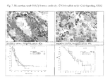
- the figures show for each biomarker examples of immunohistochemical determination of the individual biomarker expression in tumors using the light microscopy according to example 1, one microphotograph of the breast tumor with biomarker expression (positive tumor) and one microphotograph of the tumor without biomarker expression (negative tumor).
- FIG. 1 Biomarker: total Act (pan) kinase; antibody: (11E7) rabbit mAb (Cell Signaling, USA)
- FIG. 2 Biomarker: pSer 473 Act kinase; antibody: (587F11) mouse mAb (Cell Signaling, USA)
- FIG. 3 Biomarker: pSer/Thr Act kinase; antibody: substrate, rabbit mAb (Cell Signaling, USA)
- FIG. 4 Biomarker: pThr 308 Act kinase; antibody: (244F9H2) rabbit mAb (Cell Signaling, USA)
- FIG. 5 Biomarker: total ERK 1 ⁇ 2 kinase; antibody: p44/42MAP rabbit mAb (Cell Signaling, USA)
- FIG. 6 Biomarker: pERK 1 ⁇ 2 kinase; antibody: p44/42MAPK (Thr202/Tyr204) (20G11) rabbit mAb (Cell Signaling, USA)
- FIG. 7 Biomarker: total GSK3 ⁇ kinase; antibody: (27C10) rabbit mAb (Cell Signaling, USA)
- FIG. 8 Biomarker: pSer 9 GSK3 ⁇ kinase; antibody: rabbit mAb (Cell Signaling, USA)
- FIG. 9 Biomarker: total mTOR kinase; antibody: (7C10) rabbit mAb (Cell Signaling, USA)
- FIG. 10 Biomarker: pSer 2448 mTOR kinase; antibody: (49F9) rabbit mAb (Cell Signaling, USA)
- FIG. 11 Biomarker: total anti MUC4 protein; antibody: (1G8) mouse mAb (Zymed, USA)
- FIG. 12 Biomarker: total PTEN protein; antibody: (138G6) rabbit mAb (Cell Signaling, USA)
- FIG. 13 Biomarker: total S6K protein; antibody: S6 Ribosomal Protein (5G10) rabbit mAb (Cell Signaling, USA)
- FIG. 14 Biomarker: pSer 235/236 S6K protein; antibody: phospho—S6 Ribosomal Protein (91B2) rabbit mAb (Cell Signaling, USA)
- Used for the immunohistochemical detection may be bioptic materials processed and sliced using various technique, most frequently formalin fixed tissues embedded in paraffin.
- the immunohistochemical reaction represents the visualization of the binding of the tissue antigen with the primary antibody. According to the particular method used it is also followed by the specific reaction of the secondary, or tertiary antibody, as the case may be, or another signal-amplifying system. Depending on the type of the used label, most frequently enzyme label, the antigen is visualized by the relevant chromogene at the point where the specific binding occurs. The binding of the antigen with the antibody proceeds without visible reaction.
- chromogenes or fluorochromes In order to enable the observation of the preparation by common methods of light or fluorescent microscopy, for the visualization of antigen localization and molecules bound to it, the so-called chromogenes or fluorochromes must be used.
- FITC fluorescein-isothiocyanate
- immunofluorescence In immunoenzyme reactions the antibodies are labeled by the conjugation with enzyme, e.g., horse-radish peroxidase or alkaline phosphatase.
- enzyme e.g., horse-radish peroxidase or alkaline phosphatase.
- the counterstaining of sections e.g., by haematoxylin or fluorescent dyes of Hoechst type, is used frequently in immunohistochemistry or immunofluorescence.
- the blocking of non-specific binding sites is performed by incubation in 0.5 ml per slide of blocking solution (e.g., 3% fat-free milk in Tris buffered saline pH 7.2-7.4, TBS) usually for 1 hour at room temperature. After pouring off the blocking solution the slides are rinsed shortly 2 ⁇ in TBS. An appropriately diluted primary antibody was applied usually in concentration 1-10 ⁇ g/ml either in blocking solution, or in another suitable vehicle recommended by the manufacturer. The slides were incubated in a moist chamber usually for 2 hours at room temperature or overnight in the refrigerator.
- blocking solution e.g., 3% fat-free milk in Tris buffered saline pH 7.2-7.4, TBS
- TBS Tris buffered saline pH 7.2-7.4, TBS
- An appropriately diluted primary antibody was applied usually in concentration 1-10 ⁇ g/ml either in blocking solution, or in another suitable vehicle recommended by the manufacturer.
- the slides were incubated in a moist chamber usually for 2 hours at room temperature
- the primary antibodies were washed off by quick rinsing 2 ⁇ in TBS and the sample was then incubated 3 ⁇ for 5 minutes in TBS, while stirring slightly in the shaker. Then an appropriately diluted secondary antibody labeled, e.g., by peroxidase, was applied, usually again in blocking solution. At room temperature, the sample was incubated in moist chamber for 10-60 minutes. Subsequently the secondary antibodies were washed off by quick rinsing 2 ⁇ in TBS and then incubate 3 ⁇ for 5 minutes in TBS, mixing slightly in the shaker.
- the slides were poured over with freshly prepared substrate chromogen solution, e.g., with the content of diaminobenzidin (10 mg/ml and 0.3% hydrogen peroxide) in case of using detection system based on peroxidase for the period of 10-20 minutes.
- the reaction was stopped by pouring off the substrate solution and rinsing 2 ⁇ in TBS.
- the preparations were counterstained by haematoxylin solution usually for 1 minute and rinsed in tap water.
- the preparation was dehydrated, mounted in and observed under a light microscope.
- the percentage of positive cells in the target population, or subcellular localization of the antigen were analyzed semi-quantitatively. The results were assessed as follows: 1.
- the progression-free survival is determined as the time from the beginning of the treatment to the confirmed progression of disease or the date of last contact with the patient.
- Overall survival is the time from the diagnosis to the death or the last contact.
- the curves of survival were created by means of Kaplan-Meier method (Kaplan, J Am Stat Assoc 1958). For the identification of risk factors associated with the progression-free survival and overall survival, univariation analysis was performed. The difference between the curves of survival was compared by means of log rank test (Peto, J R Stat Soc 1972).
Landscapes
- Health & Medical Sciences (AREA)
- Life Sciences & Earth Sciences (AREA)
- Immunology (AREA)
- Engineering & Computer Science (AREA)
- Urology & Nephrology (AREA)
- Molecular Biology (AREA)
- Chemical & Material Sciences (AREA)
- Biomedical Technology (AREA)
- Cell Biology (AREA)
- Hematology (AREA)
- Medicinal Chemistry (AREA)
- Analytical Chemistry (AREA)
- Biotechnology (AREA)
- Hospice & Palliative Care (AREA)
- Food Science & Technology (AREA)
- Oncology (AREA)
- Physics & Mathematics (AREA)
- Microbiology (AREA)
- Biochemistry (AREA)
- General Health & Medical Sciences (AREA)
- General Physics & Mathematics (AREA)
- Pathology (AREA)
- Measuring Or Testing Involving Enzymes Or Micro-Organisms (AREA)
- Investigating Or Analysing Biological Materials (AREA)
- Medicines That Contain Protein Lipid Enzymes And Other Medicines (AREA)
- Medicines Containing Antibodies Or Antigens For Use As Internal Diagnostic Agents (AREA)
Applications Claiming Priority (3)
| Application Number | Priority Date | Filing Date | Title |
|---|---|---|---|
| CZ20080040A CZ302709B6 (cs) | 2008-01-25 | 2008-01-25 | Zpusob zjištení senzitivity pacientu s nádorovým onemocnením na lécbu inhibitory HER-2 receptoru |
| CZPV2008-40 | 2008-01-25 | ||
| PCT/CZ2009/000006 WO2009092338A1 (en) | 2008-01-25 | 2009-01-23 | Method for determining the sensitivity of patients suffering from a cancer disease to biological therapy |
Publications (2)
| Publication Number | Publication Date |
|---|---|
| US20110014637A1 US20110014637A1 (en) | 2011-01-20 |
| US8465936B2 true US8465936B2 (en) | 2013-06-18 |
Family
ID=40451258
Family Applications (1)
| Application Number | Title | Priority Date | Filing Date |
|---|---|---|---|
| US12/863,517 Expired - Fee Related US8465936B2 (en) | 2008-01-25 | 2009-01-23 | Method for determining the sensitivity of patients suffering from a cancer disease to biological therapy |
Country Status (5)
| Country | Link |
|---|---|
| US (1) | US8465936B2 (cs) |
| EP (2) | EP2235538A1 (cs) |
| JP (1) | JP5295268B2 (cs) |
| CZ (1) | CZ302709B6 (cs) |
| WO (1) | WO2009092338A1 (cs) |
Families Citing this family (2)
| Publication number | Priority date | Publication date | Assignee | Title |
|---|---|---|---|---|
| ES2786753T3 (es) * | 2009-12-22 | 2020-10-13 | Expression Pathology Inc | Ensayo de SRM/MRM de la proteína del sustrato 1 del receptor de insulina (IRS1) |
| CN114121150A (zh) * | 2020-08-27 | 2022-03-01 | 中国科学院分子细胞科学卓越创新中心 | 癌症药物敏感性预测方法、系统、存储介质及终端 |
Family Cites Families (4)
| Publication number | Priority date | Publication date | Assignee | Title |
|---|---|---|---|---|
| EP1570273B1 (en) * | 2002-12-11 | 2018-05-30 | Ventana Medical Systems, Inc. | Method for predicting the response to her2-directed therapy |
| US7862995B2 (en) * | 2004-12-10 | 2011-01-04 | Targeted Molecular Diagnostics | Methods and materials for predicting responsiveness to treatment with dual tyrosine kinase inhibitor |
| WO2007090032A2 (en) * | 2006-01-27 | 2007-08-09 | Serenex, Inc. | Ribosomal protein s6 as a pharmacodynamic marker for hsp90 inhibition |
| WO2007109571A2 (en) * | 2006-03-17 | 2007-09-27 | Prometheus Laboratories, Inc. | Methods of predicting and monitoring tyrosine kinase inhibitor therapy |
-
2008
- 2008-01-25 CZ CZ20080040A patent/CZ302709B6/cs not_active IP Right Cessation
-
2009
- 2009-01-23 EP EP09704221A patent/EP2235538A1/en not_active Withdrawn
- 2009-01-23 WO PCT/CZ2009/000006 patent/WO2009092338A1/en active Application Filing
- 2009-01-23 JP JP2010543368A patent/JP5295268B2/ja not_active Expired - Fee Related
- 2009-01-23 EP EP10100001.6A patent/EP2241890B1/en not_active Not-in-force
- 2009-01-23 US US12/863,517 patent/US8465936B2/en not_active Expired - Fee Related
Non-Patent Citations (7)
| Title |
|---|
| Barlund et al (J national Cancer Institute, 2000, 92:1252-1259). * |
| Cell Signaling technology ErbB/HER Signaling map, printed Feb. 2013. * |
| Fenton et al (The International Journal of Biochemistry and Cell Biology, 2011, 43:47-59). * |
| Lyzogubov et al (Exp Oncol 2005, 27:141-144). * |
| Neve et al (Proc Amer Assoc Cancer Res, 2006, 47: abstract #3378). * |
| Noh et al (Clinical Cancer Research, 2004, 10:1013-1023). * |
| Rojo et al (Clinical Cancer Research, Jan. 2, 2007, 13:81-89). * |
Also Published As
| Publication number | Publication date |
|---|---|
| EP2235538A1 (en) | 2010-10-06 |
| JP2011510306A (ja) | 2011-03-31 |
| US20110014637A1 (en) | 2011-01-20 |
| EP2241890A3 (en) | 2010-10-27 |
| CZ302709B6 (cs) | 2011-09-14 |
| JP5295268B2 (ja) | 2013-09-18 |
| CZ200840A3 (cs) | 2009-08-05 |
| WO2009092338A1 (en) | 2009-07-30 |
| EP2241890A2 (en) | 2010-10-20 |
| EP2241890B1 (en) | 2014-04-09 |
Similar Documents
| Publication | Publication Date | Title |
|---|---|---|
| Khan et al. | Early diagnostic value of survivin and its alternative splice variants in breast cancer | |
| JP5656406B2 (ja) | 治療効力を予測するためのマーカーとしての活性化her3 | |
| Edgerton et al. | erbB-2 (HER-2) and breast cancer progression | |
| Faried et al. | Predictive and prognostic role of activated mammalian target of rapamycin in cervical cancer treated with cisplatin-based neoadjuvant chemotherapy | |
| CN101990577A (zh) | 鉴定肺病药物开发的新途径 | |
| KR20070061893A (ko) | 유방암 예후를 평가하기 위한 방법 및 조성물 | |
| CA2504042A1 (en) | Methods and materials for examining pathways associated with glioblastoma progression | |
| JP2012103264A (ja) | 癌診断用抗体 | |
| JP2010536367A5 (cs) | ||
| Newby et al. | Immunohistochemical assay for epidermal growth factor receptor on paraffin-embedded sections: validation against ligand-binding assay and clinical relevance in breast cancer | |
| Fasig et al. | Immunohistochemical analysis of receptor tyrosine kinase signal transduction activity in chordoma | |
| CN109879956A (zh) | 一种肿瘤免疫生物标志物及其应用 | |
| CN110836974A (zh) | Flt3lg蛋白在制备肺腺癌术后预后评估试剂或者试剂盒中的应用 | |
| Mallick et al. | PCNA and anti-apoptotic Mcl-1 proteins predict disease-free survival in oral cancer patients treated with definitive radiotherapy | |
| US9005907B2 (en) | Methods and compositions for typing molecular subgroups of medulloblastoma | |
| Aufderklamm et al. | XPA-210: a new proliferation marker determines locally advanced prostate cancer and is a predictor of biochemical recurrence | |
| Fine et al. | Androgen and c-Kit receptors in desmoplastic small round cell tumors resistant to chemotherapy: novel targets for therapy | |
| Takemura et al. | Gamma‐synuclein is a novel prognostic marker that promotes tumor cell migration in biliary tract carcinoma | |
| US8465936B2 (en) | Method for determining the sensitivity of patients suffering from a cancer disease to biological therapy | |
| Green et al. | Alterations in the p53 pathway and prognosis in advanced ovarian cancer: a multi-factorial analysis of the EORTC Gynaecological Cancer group (study 55865) | |
| Mittelbronn et al. | EGR‐1 is regulated by N‐methyl‐D‐aspartate‐receptor stimulation and associated with patient survival in human high grade astrocytomas | |
| Chen et al. | Evaluation of trastuzumab anti-tumor efficacy and its correlation with HER-2 status in patient-derived gastric adenocarcinoma xenograft models | |
| Singhai et al. | Cancer biomarker HER-2/neu in breast cancer in Indian women | |
| Qiao et al. | Loss of protein tyrosine phosphatase receptor J expression predicts an aggressive clinical course in patients with esophageal squamous cell carcinoma | |
| Hammerich et al. | Cellular interactions of the phosphorylated form of AKT in prostate cancer |
Legal Events
| Date | Code | Title | Description |
|---|---|---|---|
| AS | Assignment |
Owner name: MASARYKUV ONKOLOGICKY USTAV, CZECH REPUBLIC Free format text: ASSIGNMENT OF ASSIGNORS INTEREST;ASSIGNORS:HAJDUCH, MARIAN;DZIECHCIARKOVA, MARTA;RADOVA, LENKA;AND OTHERS;REEL/FRAME:025013/0906 Effective date: 20100909 Owner name: UNIVERZITA PALACKEHO V OLOMOUCI, LEKARSKA FAKULTA, Free format text: ASSIGNMENT OF ASSIGNORS INTEREST;ASSIGNORS:HAJDUCH, MARIAN;DZIECHCIARKOVA, MARTA;RADOVA, LENKA;AND OTHERS;REEL/FRAME:025013/0906 Effective date: 20100909 |
|
| STCF | Information on status: patent grant |
Free format text: PATENTED CASE |
|
| FPAY | Fee payment |
Year of fee payment: 4 |
|
| FEPP | Fee payment procedure |
Free format text: MAINTENANCE FEE REMINDER MAILED (ORIGINAL EVENT CODE: REM.); ENTITY STATUS OF PATENT OWNER: SMALL ENTITY |
|
| LAPS | Lapse for failure to pay maintenance fees |
Free format text: PATENT EXPIRED FOR FAILURE TO PAY MAINTENANCE FEES (ORIGINAL EVENT CODE: EXP.); ENTITY STATUS OF PATENT OWNER: SMALL ENTITY |
|
| STCH | Information on status: patent discontinuation |
Free format text: PATENT EXPIRED DUE TO NONPAYMENT OF MAINTENANCE FEES UNDER 37 CFR 1.362 |
|
| FP | Lapsed due to failure to pay maintenance fee |
Effective date: 20210618 |