US8211131B2 - Apparatus and methods for anastomosis - Google Patents
Apparatus and methods for anastomosis Download PDFInfo
- Publication number
- US8211131B2 US8211131B2 US13/080,394 US201113080394A US8211131B2 US 8211131 B2 US8211131 B2 US 8211131B2 US 201113080394 A US201113080394 A US 201113080394A US 8211131 B2 US8211131 B2 US 8211131B2
- Authority
- US
- United States
- Prior art keywords
- fastener
- clip
- vessel
- release
- anastomosis
- Prior art date
- Legal status (The legal status is an assumption and is not a legal conclusion. Google has not performed a legal analysis and makes no representation as to the accuracy of the status listed.)
- Expired - Lifetime
Links
- 230000003872 anastomosis Effects 0.000 title abstract description 50
- 238000000034 method Methods 0.000 title abstract description 46
- 230000007246 mechanism Effects 0.000 claims description 36
- 239000008280 blood Substances 0.000 abstract description 16
- 210000004369 blood Anatomy 0.000 abstract description 16
- 239000012530 fluid Substances 0.000 abstract description 3
- 239000012528 membrane Substances 0.000 description 43
- 210000001519 tissue Anatomy 0.000 description 38
- 239000000463 material Substances 0.000 description 20
- 238000005520 cutting process Methods 0.000 description 18
- 210000001367 artery Anatomy 0.000 description 16
- 230000017531 blood circulation Effects 0.000 description 14
- 230000023597 hemostasis Effects 0.000 description 13
- 229910001000 nickel titanium Inorganic materials 0.000 description 13
- HLXZNVUGXRDIFK-UHFFFAOYSA-N nickel titanium Chemical compound [Ti].[Ti].[Ti].[Ti].[Ti].[Ti].[Ti].[Ti].[Ti].[Ti].[Ti].[Ni].[Ni].[Ni].[Ni].[Ni].[Ni].[Ni].[Ni].[Ni].[Ni].[Ni].[Ni].[Ni].[Ni] HLXZNVUGXRDIFK-UHFFFAOYSA-N 0.000 description 11
- 229910001285 shape-memory alloy Inorganic materials 0.000 description 10
- 230000000452 restraining effect Effects 0.000 description 9
- 210000000709 aorta Anatomy 0.000 description 7
- 230000006835 compression Effects 0.000 description 7
- 238000007906 compression Methods 0.000 description 7
- 239000010935 stainless steel Substances 0.000 description 7
- 229910001220 stainless steel Inorganic materials 0.000 description 7
- 238000001356 surgical procedure Methods 0.000 description 7
- 238000007789 sealing Methods 0.000 description 6
- 229910000734 martensite Inorganic materials 0.000 description 5
- 229920000642 polymer Polymers 0.000 description 5
- 229920002635 polyurethane Polymers 0.000 description 5
- 239000004814 polyurethane Substances 0.000 description 5
- 230000009466 transformation Effects 0.000 description 5
- 230000002792 vascular Effects 0.000 description 5
- 239000003292 glue Substances 0.000 description 4
- 208000014674 injury Diseases 0.000 description 4
- 229910052751 metal Inorganic materials 0.000 description 4
- 239000002184 metal Substances 0.000 description 4
- 230000000149 penetrating effect Effects 0.000 description 4
- 230000008733 trauma Effects 0.000 description 4
- 208000006011 Stroke Diseases 0.000 description 3
- 230000015572 biosynthetic process Effects 0.000 description 3
- 238000010276 construction Methods 0.000 description 3
- 230000008878 coupling Effects 0.000 description 3
- 238000010168 coupling process Methods 0.000 description 3
- 238000005859 coupling reaction Methods 0.000 description 3
- 230000006870 function Effects 0.000 description 3
- 230000037361 pathway Effects 0.000 description 3
- -1 polypropylenes Polymers 0.000 description 3
- 238000011084 recovery Methods 0.000 description 3
- CURLTUGMZLYLDI-UHFFFAOYSA-N Carbon dioxide Chemical compound O=C=O CURLTUGMZLYLDI-UHFFFAOYSA-N 0.000 description 2
- HZEWFHLRYVTOIW-UHFFFAOYSA-N [Ti].[Ni] Chemical compound [Ti].[Ni] HZEWFHLRYVTOIW-UHFFFAOYSA-N 0.000 description 2
- 239000004676 acrylonitrile butadiene styrene Substances 0.000 description 2
- 229910045601 alloy Inorganic materials 0.000 description 2
- 239000000956 alloy Substances 0.000 description 2
- 238000013459 approach Methods 0.000 description 2
- 238000010009 beating Methods 0.000 description 2
- 230000006399 behavior Effects 0.000 description 2
- 230000008901 benefit Effects 0.000 description 2
- 238000006243 chemical reaction Methods 0.000 description 2
- 210000004351 coronary vessel Anatomy 0.000 description 2
- 230000002093 peripheral effect Effects 0.000 description 2
- 239000004033 plastic Substances 0.000 description 2
- 229920003023 plastic Polymers 0.000 description 2
- 239000004417 polycarbonate Substances 0.000 description 2
- 229920000515 polycarbonate Polymers 0.000 description 2
- 229920000139 polyethylene terephthalate Polymers 0.000 description 2
- 239000005020 polyethylene terephthalate Substances 0.000 description 2
- 238000002360 preparation method Methods 0.000 description 2
- 239000007787 solid Substances 0.000 description 2
- 239000003356 suture material Substances 0.000 description 2
- 230000001360 synchronised effect Effects 0.000 description 2
- 238000003466 welding Methods 0.000 description 2
- 206010002383 Angina Pectoris Diseases 0.000 description 1
- 241001631457 Cannula Species 0.000 description 1
- 239000004952 Polyamide Substances 0.000 description 1
- 239000004698 Polyethylene Substances 0.000 description 1
- 239000004743 Polypropylene Substances 0.000 description 1
- 229910001260 Pt alloy Inorganic materials 0.000 description 1
- 229910001069 Ti alloy Inorganic materials 0.000 description 1
- 229920000122 acrylonitrile butadiene styrene Polymers 0.000 description 1
- 230000009471 action Effects 0.000 description 1
- 238000004026 adhesive bonding Methods 0.000 description 1
- 210000003484 anatomy Anatomy 0.000 description 1
- 229910001566 austenite Inorganic materials 0.000 description 1
- 210000004204 blood vessel Anatomy 0.000 description 1
- 229910002092 carbon dioxide Inorganic materials 0.000 description 1
- 239000001569 carbon dioxide Substances 0.000 description 1
- 230000002612 cardiopulmonary effect Effects 0.000 description 1
- 238000013131 cardiovascular procedure Methods 0.000 description 1
- 208000029078 coronary artery disease Diseases 0.000 description 1
- 230000034994 death Effects 0.000 description 1
- 230000007812 deficiency Effects 0.000 description 1
- 238000013461 design Methods 0.000 description 1
- 239000013013 elastic material Substances 0.000 description 1
- 238000005516 engineering process Methods 0.000 description 1
- 230000004927 fusion Effects 0.000 description 1
- 238000010438 heat treatment Methods 0.000 description 1
- 238000003780 insertion Methods 0.000 description 1
- 230000037431 insertion Effects 0.000 description 1
- 208000028867 ischemia Diseases 0.000 description 1
- 239000004816 latex Substances 0.000 description 1
- 229920000126 latex Polymers 0.000 description 1
- 230000007774 longterm Effects 0.000 description 1
- 238000003754 machining Methods 0.000 description 1
- 238000004519 manufacturing process Methods 0.000 description 1
- 238000002844 melting Methods 0.000 description 1
- 230000008018 melting Effects 0.000 description 1
- 150000002739 metals Chemical class 0.000 description 1
- 230000004048 modification Effects 0.000 description 1
- 238000012986 modification Methods 0.000 description 1
- 230000002107 myocardial effect Effects 0.000 description 1
- 230000017074 necrotic cell death Effects 0.000 description 1
- 238000006213 oxygenation reaction Methods 0.000 description 1
- 239000002245 particle Substances 0.000 description 1
- 229920002647 polyamide Polymers 0.000 description 1
- 229920000573 polyethylene Polymers 0.000 description 1
- 229920001155 polypropylene Polymers 0.000 description 1
- 229920001296 polysiloxane Polymers 0.000 description 1
- 230000008569 process Effects 0.000 description 1
- 230000000750 progressive effect Effects 0.000 description 1
- 239000012781 shape memory material Substances 0.000 description 1
- 238000004513 sizing Methods 0.000 description 1
- 210000001562 sternum Anatomy 0.000 description 1
- 230000001131 transforming effect Effects 0.000 description 1
- 238000007514 turning Methods 0.000 description 1
- 210000005166 vasculature Anatomy 0.000 description 1
- 210000001835 viscera Anatomy 0.000 description 1
Images


Classifications
-
- A—HUMAN NECESSITIES
- A61—MEDICAL OR VETERINARY SCIENCE; HYGIENE
- A61B—DIAGNOSIS; SURGERY; IDENTIFICATION
- A61B17/00—Surgical instruments, devices or methods, e.g. tourniquets
- A61B17/32—Surgical cutting instruments
- A61B17/3205—Excision instruments
- A61B17/32053—Punch like cutting instruments, e.g. using a cylindrical or oval knife
-
- A—HUMAN NECESSITIES
- A61—MEDICAL OR VETERINARY SCIENCE; HYGIENE
- A61B—DIAGNOSIS; SURGERY; IDENTIFICATION
- A61B17/00—Surgical instruments, devices or methods, e.g. tourniquets
- A61B17/0057—Implements for plugging an opening in the wall of a hollow or tubular organ, e.g. for sealing a vessel puncture or closing a cardiac septal defect
-
- A—HUMAN NECESSITIES
- A61—MEDICAL OR VETERINARY SCIENCE; HYGIENE
- A61B—DIAGNOSIS; SURGERY; IDENTIFICATION
- A61B17/00—Surgical instruments, devices or methods, e.g. tourniquets
- A61B17/04—Surgical instruments, devices or methods, e.g. tourniquets for suturing wounds; Holders or packages for needles or suture materials
- A61B17/06—Needles ; Sutures; Needle-suture combinations; Holders or packages for needles or suture materials
-
- A—HUMAN NECESSITIES
- A61—MEDICAL OR VETERINARY SCIENCE; HYGIENE
- A61B—DIAGNOSIS; SURGERY; IDENTIFICATION
- A61B17/00—Surgical instruments, devices or methods, e.g. tourniquets
- A61B17/064—Surgical staples, i.e. penetrating the tissue
-
- A—HUMAN NECESSITIES
- A61—MEDICAL OR VETERINARY SCIENCE; HYGIENE
- A61B—DIAGNOSIS; SURGERY; IDENTIFICATION
- A61B17/00—Surgical instruments, devices or methods, e.g. tourniquets
- A61B17/11—Surgical instruments, devices or methods, e.g. tourniquets for performing anastomosis; Buttons for anastomosis
-
- A—HUMAN NECESSITIES
- A61—MEDICAL OR VETERINARY SCIENCE; HYGIENE
- A61B—DIAGNOSIS; SURGERY; IDENTIFICATION
- A61B17/00—Surgical instruments, devices or methods, e.g. tourniquets
- A61B17/064—Surgical staples, i.e. penetrating the tissue
- A61B17/0644—Surgical staples, i.e. penetrating the tissue penetrating the tissue, deformable to closed position
-
- A—HUMAN NECESSITIES
- A61—MEDICAL OR VETERINARY SCIENCE; HYGIENE
- A61B—DIAGNOSIS; SURGERY; IDENTIFICATION
- A61B17/00—Surgical instruments, devices or methods, e.g. tourniquets
- A61B17/0057—Implements for plugging an opening in the wall of a hollow or tubular organ, e.g. for sealing a vessel puncture or closing a cardiac septal defect
- A61B2017/00575—Implements for plugging an opening in the wall of a hollow or tubular organ, e.g. for sealing a vessel puncture or closing a cardiac septal defect for closure at remote site, e.g. closing atrial septum defects
- A61B2017/00619—Locking means for locking the implement in expanded state
-
- A—HUMAN NECESSITIES
- A61—MEDICAL OR VETERINARY SCIENCE; HYGIENE
- A61B—DIAGNOSIS; SURGERY; IDENTIFICATION
- A61B17/00—Surgical instruments, devices or methods, e.g. tourniquets
- A61B17/04—Surgical instruments, devices or methods, e.g. tourniquets for suturing wounds; Holders or packages for needles or suture materials
- A61B17/06—Needles ; Sutures; Needle-suture combinations; Holders or packages for needles or suture materials
- A61B17/06004—Means for attaching suture to needle
- A61B2017/06009—Means for attaching suture to needle having additional means for releasably clamping the suture to the needle, e.g. actuating rod slideable within the needle
-
- A—HUMAN NECESSITIES
- A61—MEDICAL OR VETERINARY SCIENCE; HYGIENE
- A61B—DIAGNOSIS; SURGERY; IDENTIFICATION
- A61B17/00—Surgical instruments, devices or methods, e.g. tourniquets
- A61B17/04—Surgical instruments, devices or methods, e.g. tourniquets for suturing wounds; Holders or packages for needles or suture materials
- A61B17/06—Needles ; Sutures; Needle-suture combinations; Holders or packages for needles or suture materials
- A61B2017/06057—Double-armed sutures, i.e. sutures having a needle attached to each end
-
- A—HUMAN NECESSITIES
- A61—MEDICAL OR VETERINARY SCIENCE; HYGIENE
- A61B—DIAGNOSIS; SURGERY; IDENTIFICATION
- A61B17/00—Surgical instruments, devices or methods, e.g. tourniquets
- A61B17/11—Surgical instruments, devices or methods, e.g. tourniquets for performing anastomosis; Buttons for anastomosis
- A61B2017/1135—End-to-side connections, e.g. T- or Y-connections
-
- A—HUMAN NECESSITIES
- A61—MEDICAL OR VETERINARY SCIENCE; HYGIENE
- A61B—DIAGNOSIS; SURGERY; IDENTIFICATION
- A61B17/00—Surgical instruments, devices or methods, e.g. tourniquets
- A61B17/32—Surgical cutting instruments
- A61B2017/320064—Surgical cutting instruments with tissue or sample retaining means
Definitions
- the present invention relates to apparatus and methods for performing an anastomosis.
- the apparatus may be used to perform a single or multiple anastomosis with the ability of maintaining fluid flow (e.g., blood) through the anastomosis vessel.
- fluid flow e.g., blood
- occlusion of the arteries can lead to insufficient blood flow resulting in discomfort and risks of angina and ischemia.
- Significant blockage of blood flow in the coronary artery can result in damage to the myocardial tissue or death of the patient.
- occlusion of the artery results from progressive long term deposits of plaque along the artery wall. While such deposits may be concentrated and occlude the artery at a particular site, the deposits are most certainly present throughout the arteries and the vascular system.
- Coronary artery bypass graft (CABG) surgery is a surgical procedure performed in severe cases of coronary blockages.
- CABG procedures involve anastomosing an artery to a vascular graft which restores the flow of blood by establishing another pathway around the occluded vasculature.
- One problem encountered in the procedure is the need of performing the procedure while simultaneously maintaining sufficient function of the patient's circulatory system.
- a CABG procedure may involve arresting the heart so that blood flow is diverted from the vessel to be anastomosed.
- the patient's blood circulation is maintained by a cardiopulmonary bypass (CPB).
- This bypass is accomplished by diverting the blood flow at selected arterial locations.
- the blood is diverted to the bypass system for release of carbon dioxide and subsequent oxygenation.
- the blood is returned to the patient via a pump.
- Examples of these procedures are found in U.S. Pat. No. 5,799,661 to Boyd et al. which discloses a device and method for performing CABG surgery for multi-vessel coronary artery disease through port-access or closed-chest thorascopic methods; and U.S. Pat. No. 5,452,733 to Sterman et al. which discusses performing grafts with an efficacy equal to or greater than conventional open surgical bypass techniques.
- Another CABG procedure involves operating on a beating heart and eliminates the need for a CPB. However, the procedure still requires diverting blood flow for a proximal anastomosis, such as one which attaches graft material (e.g., a graft vessel) to the ascending aorta.
- a proximal anastomosis such as one which attaches graft material (e.g., a graft vessel) to the ascending aorta.
- the blood flow may be diverted by aortic clamping.
- aortic clamping Among the drawbacks associated with aortic clamping are an increased chance of trauma to the arteries caused by ligatures at the clamped site and the possible dislodging of plaque within the clamped vessel wall.
- the arterial bypass may be required due to the deposits of plaque which have occluded the vessel.
- the plaque is typically present throughout the artery and is not limited to the occluded location. Clamping the artery creates a risk of plaque being released into the blood stream. This release of plaque has the potential of causing a stroke, occlusion of a smaller peripheral vessel, or other vascular trauma.
- the blood flow may be diverted by the use of a balloon catheter within the aorta, such as described in U.S. Pat. No. 5,868,702, for example.
- Drawbacks of using a balloon catheter in creating a seal to divert blood flow include the possibility of disturbing plaque deposits and creating particles in the blood stream, the chance that the balloon catheter may move within the aorta disrupting the seal and resulting in blood loss, and trauma to aortic tissue caused by the pressure needed to create the seal.
- CABG procedure Yet another problem related to CABG procedure lies in the procedure of suturing the vessels to create a tight seal.
- the graft and vessel to be joined must be precisely aligned with respect to each other. If one of the tissues is affixed too close to its edge, the suture can tear through the tissue and impair both the tissue and the anastomosis.
- Another problem is that, even after proper alignment of the tissue, it is difficult and time consuming to pass the needle through the tissues, form the knot with the suture material, and ensure that the suture material does not become entangled. These difficulties are exacerbated by the small size of the artery and graft. Another factor contributing to the difficulty of the CABG procedure is the limited time available to complete the procedure.
- surgeons must complete the graft in as little time possible due to the absence of blood flowing through the artery. If blood flow is not promptly restored, sometimes in as little as 30 minutes, the tissues the artery supplies may experience significant damage or necrosis. As mentioned above, surgeons are under pressure to reduce the cross-clamp time, yet, an incomplete suture may result in a leak in the tissue approximation between the vessel and graft. Moreover, the tissue approximation must be smooth and open. Hence, the suture cannot be hastily performed.
- Houser et al. in U.S. Pat. No. 5,989,276, discloses various devices and techniques for performing bypass, one of which includes a device which can be intralumenally originated.
- Various other clamping arrangements are provided for securing a graft to a vessel without the use of sutures or other fasteners.
- the present invention involves apparatus and methods for connecting tissue.
- This invention may, for example, be used to secure one vessel to another, such as in a vascular anastomosis while maintaining blood flow within the vessel.
- an anastomosis apparatus comprising a tubular member having an end with an edge adapted to form an opening in a vessel wall and an anchor member, which comprises a flexible shaft and a piercing member, slidably coupled to the tubular member.
- an anchor member which comprises a flexible shaft and a piercing member, slidably coupled to the tubular member.
- a generally circular centering disk with a plurality of spikes is also provided to slide down along the flexible shaft of the anchor member.
- a physician pushes the shaft of the anchor member to insert the piercing member through the vessel wall.
- the piercing member is then pulled back against the vessel wall.
- the centering disk is put in place and pushed against the outer wall of the vessel, thereby clamping the vessel wall between the centering disk and the piercing member.
- the tubular member is then moved over the shaft, centered by the disk and rotated to cut an opening through the vessel wall. Thereafter, the piercing member is used to coordinate with the centering disk to retain and retrieve the tissue cut by the tubular member.
- an occlusion member is advanced within the tubular member through the opening in the vessel wall in order to substantially occlude such opening and form an area of hemostasis.
- the tubular member can then be retrieved, leaving the occlusion member in place, after which a graft is slid down along the occlusion member shaft for creating an anastomosis with the vessel.
- the occlusion member comprises a cannula.
- the occlusion member comprises a radially expandable member, which can be an expandable brush-like member, an umbrella, a flexible cup or an inflatable balloon.
- a cannula having an end portion adapted to pierce through a vessel.
- the cannula may be passed through a vessel wall and a graft secured to the vessel adjacent to the cannula to facilitate an anastomosis without clamping the vessel, thereby avoiding the risk of dislodgment of plaque from the inner wall of the vessel which is incurred with clamping.
- a trocar adapted to receive the cannula and allow it to pivot is provided.
- the cannula thus, may be repositioned to pierce the vessel wall at different locations and perform multiple anastomoses without relocation of the trocar in the vessel wall.
- a surgical fastener or clip cartridge is provided.
- One or more surgical fasteners may be loaded in the cartridge with ends of each fastener selectively releasable from the cartridge to anchor in either the graft or vessel to which the graft is to be attached.
- first and second tubular members are slidably coupled to one another and at least one surgical fastener placed therein with one end of the fastener engaging the first tubular member and another end of the fastener engaging the second tubular member.
- a dual-stage release fastener which includes a clip movable between an open and a closed configuration and having a memory biased to the closed configuration.
- a first mechanical restraint is coupled to a first portion of the clip and is adapted to bias the first portion toward the open configuration.
- a second mechanical restraint is coupled to a second portion of the clip and is adapted to bias the second portion toward the open configuration.
- the first and second mechanical restraints may be independently manipulatable to allow independent closure of the first and second portions of the clip.
- an anastomosis apparatus comprising a tubular member having an end with an edge adapted to form an opening in a vessel wall and an anchor member, which comprises a flexible shaft and a piercing member, slidably coupled to the tubular member.
- an anchor member which comprises a flexible shaft and a piercing member, slidably coupled to the tubular member.
- a generally circular centering disk with a plurality of spikes is also provided to slide down along the flexible shaft of the anchor member.
- An adapter is mounted on the tubular member that forms the opening.
- the adapter includes wells in which needles are held in a predetermined orientation adapted for piercing through the vessel wall from the inside out, upon passing the adapter into the vessel and then pulling it back to perform the piercing action.
- Two-stage release fasteners may be used in conjunction with the adapter so as to affix the fasteners to the vessel after piercing it with the needles and then fixing the fasteners to a graf
- FIG. 1 is a sectional view of a heart illustrating an example of an anastomosis and trocar placement site.
- FIG. 2 is a sectional view of a cannula constructed according to the principles of the present invention and disposed within a trocar having a configuration according to another aspect of the invention.
- FIG. 3 is a partial sectional view of the trocar of FIG. 2 .
- FIG. 4 is a side view of the trocar positioned on a vessel wall.
- FIG. 5 is a sectional view of another cannula of the present invention disposed in the trocar of FIGS. 2-4 .
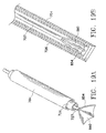
- FIG. 6A is a partial sectional view of a configuration of a surgical fastener cartridge according to the present invention.
- FIG. 6B is a front view of the cartridge of FIG. 6A .
- FIG. 6C is a plan view of one of the surgical fasteners of FIGS. 6A and 6B in a released state.
- FIGS. 7-11 show a method of preparing an anastomosis in accordance with the principles of the present invention.
- FIG. 7 shows a sectional view of the trocar and cannula of FIG. 2 positioned in an artery with the piercing element removed.
- FIG. 8 shows a sectional view of the placement of a graft over the cartridge of FIG. 6A .
- FIG. 9 shows a sectional view of the partial release of the assembly of FIG. 8 with one end of the fasteners released and anchored to the graft.
- FIG. 10 illustrates the complete removal of the tubular member which restrains the released fastener end.
- FIG. 11 shows aligning and introducing the cartridge into the cannula.
- FIG. 12 shows the cartridge in place in the cannula and the released fastener ends engage the vessel.
- FIG. 13 shows the release of the locking member for the other ends of the fasteners.
- FIG. 14 shows withdrawing the cannula and second tubular member to release the second ends of the fasteners so they may engage the inner wall of the vessel.
- FIG. 15A is a sectional view of another proximal anastomosis device with a punch, an anchor member and a centering disk.
- FIG. 15B is a sectional view of the device of FIG. 15A after an opening is created in the vessel wall by the punch and the tissue cut is being retrieved.
- FIG. 15C is a sectional view of the device of FIG. 15A with an occlusion member comprising two coaxial cylinders and a radially expandable member attached to the distal end of the inner cylinder.
- FIG. 15D is a sectional view of the device of FIG. 15A with the punch retrieved and a graft slid over the radially expandable member to be attached to the vessel by a plurality of self-closing fasteners.
- FIG. 15E is a perspective view of the device of FIG. 15A with a punch, an anchor member and a centering disk.
- FIG. 15F is a perspective view of the device shown in FIG. 15C .
- FIG. 16A is a sectional view of another proximal anastomosis device, with a radially expandable member comprising a plurality of bristles.
- FIG. 16B is a sectional view of the device of FIG. 16A , with the plurality of bristles radially expanded to occlude the opening in the vessel wall after the outer cylinder is withdrawn.
- FIG. 16C is an end view of the device of FIG. 16A with a retaining ring attached to the inner cylinder to bind a bundle of bristles.
- FIG. 16D is an end view of the device of FIG. 16A with the bristles attached to the inner wall of the hollow inner cylinder.
- FIG. 16E is a partial sectional view of the device of FIG. 16A with the punch withdrawn, the occlusion device in place with the bristles expanded or deployed, and a plurality of fasteners inserted to attach a graft to the vessel wall.
- FIG. 16F is a sectional view of the device of FIG. 16A with the outer cylinder slid down over the bristles after an anastomosis has been formed.
- FIG. 16G is a sectional view of the device of FIG. 16A with the occlusion device being withdrawn.
- FIG. 17A is a sectional view of another proximal anastomosis device, with a radially expandable member comprising an inflatable membrane and a plurality of fasteners attached to the membrane.
- FIG. 17B is a sectional view of the device of FIG. 17A with the membrane inflated to occlude the opening in the vessel wall.
- FIG. 17C is a sectional view of the device of FIG. 17A with the fasteners piercing through a graft to attach the graft to the vessel.
- FIG. 17D is a sectional view of the device of FIG. 17A with the membrane deflated after the anastomosis is formed.
- FIGS. 17E-17K are detailed drawings of a fastener and portions thereof, which may be used in connecting grafts to vessels, according to the present invention.
- FIG. 18A is a sectional view of another proximal anastomosis device, with a flexible sheath capable of expanding with the membrane.
- FIG. 18B is a sectional view of the device of FIG. 18A with sheath expanded with the inflated membrane.
- FIG. 18C is a view of a roll-up sheath with an expandable membrane therein.
- FIG. 18D is a sectional view of the device of FIG. 18A , with a graft being slid down for anastomosis.
- FIG. 18E is a sectional view of the device of FIG. 18A with the graft attached to the vessel by a plurality of fasteners.
- FIG. 19A is a perspective view of another proximal anastomosis device comprising a skeleton which supports an umbrella-like occlusion member.
- FIG. 19B is a sectional view of the device of FIG. 19A with the umbrella-like occlusion member in a folded state within the outer cylinder.
- FIG. 20A is a sectional view of another example of a proximal anastomosis device with a punch, an anchor member and a centering disk.
- FIG. 20B is a sectional view of the device of FIG. 20A after the tissue cut has been retrieved and the punch or cutting member has been further inserted within the vessel.
- FIG. 20C is a sectional view of the device of FIG. 20A with an occlusion member inserted into the vessel and the cutting member having been pulled back to push the protruding needles through the vessel wall.
- FIG. 20D is a sectional view of the device of FIG. 20A with the punch or cutting member retrieved and the occluding member in place, in preparation for placement of a graft. 2
- FIG. 20E is a sectional view of the device of FIG. 20A with the punch or cutting member retrieved and a graft in place and partially anastomosed to the vessel.
- FIG. 20F is a sectional view of the device of FIG. 20A with the graft having been anastomosed to the vessel and with the occluding device being withdrawn.
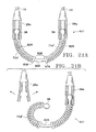
- FIG. 21A is a partial schematic drawing of a two-stage release fastener for use with the proximal anastomosis device of FIGS. 20A-20F , for example.
- FIG. 21B is a partial schematic view of the fastener of FIG. 21A after one end of the fastener has been released.
- FIG. 21C is a partial schematic view of the fastener of FIG. 21A after both ends of the fastener have been released.
- the present invention generally involves methods and apparatus for performing an anastomosis and may be used, for example, in bypass procedures.
- graft includes any of the following: hemografts, autologous grafts, xenografts, allografts, alloplastic materials, and combinations of the foregoing.
- a heart ( 100 ) is shown in section with the aorta designated with reference numeral ( 102 ).
- One possible anastomosis site is on aorta ( 102 ).
- Two openings, generally designated with reference numeral ( 104 ) indicate where a cannula (described below) may be positioned to facilitate the anastomosis. It should be understood, however, that although an aortic anastomosis site is shown, the invention can be used in other areas of the human anatomy where one desires to connect a graft to tissue, or tubular structure.
- a cannula having a distal portion adapted to pierce at least one wall portion of a vessel and a proximal portion.
- the cannula may be used, for example, to create an area of hemostasis within the vessel and facilitate an anastomosis while allowing blood to flow through the vessel.
- the cannula is passed through a vessel wall from the interior to the exterior of the vessel to create a hemostasis area within the cannula and within the vessel hole created thereby, as will be described in more detail below.
- a cannula constructed according to the principles of the present invention is shown and generally designated with reference numeral ( 200 ).
- Cannula ( 200 ) generally includes a distal portion ( 202 ) adapted to pierce tissue and a proximal portion ( 204 ).
- Cannula ( 200 ) includes lumen ( 206 ) which extends the entire length of proximal portion ( 204 ).
- Lumen ( 206 ) allows access inside or through a vessel, while cannula ( 200 ) forms an area of hemostasis upon insertion of cannula ( 200 ) through a portion of a vessel wall.
- the distal portion of the cannula is configured to pierce tissue, and may comprise a member having a generally cylindrical outer surface and a tapered inner surface which forms a generally frustoconically shaped cavity ( 208 ).
- the distal end ( 210 ) of distal portion ( 202 ) tapers to an annular point.
- a proximal end ( 204 ) of the cannula is shown to have an expanded diameter.
- the length of the cannula ( 200 ) may vary depending on for example, the size of the anastomosed vessels. It is further noted that although the distal and proximal portions ( 202 , 204 ) are shown as separate elements, they may be integrally formed as a single unit to form the cannula ( 200 ).
- the proximal and distal portions may be constructed from a variety of materials.
- the proximal portion may be plastic. It also may be constructed to be flexible so that it may be delivered endovascularly.
- the distal portion may be metal such as stainless steel.
- the cannula may be introduced endovascularly or passed through a vessel wall to position it within a vessel.
- a trocar may be used.
- a trocar having a varying diameter lumen may be used to allow the angular position of the cannula to be changed.
- a trocar ( 300 ) which may facilitate such movement is shown.
- Trocar ( 300 ) generally includes an annular member ( 302 ) which forms an annular passageway or lumen ( 304 ) which is shown with a varying diameter along a longitudinal direction of the trocar ( 300 ).
- the diameter of the lumen of the embodiment shown increases in both directions away from the central portion ( 306 ) of member ( 302 ).
- FIG. 4 A portion of the increasing diameter of the lumen is shown in FIG. 4 .
- the size of the lumen depends on the diameter of the cannula to be used therewith.
- a flame ( 308 ) may be provided with the trocar ( 300 ) to enhance the securement to the vessel as is conventional to the art.
- a seal ( 310 ) is located within the passageway ( 304 ).
- FIG. 4 illustrates a front view of the trocar ( 300 ).
- Flange ( 308 ) may be attached to the vessel wall ( 312 ) with sutures ( 314 ) with the seal ( 310 ) positioned outside the vessel.
- FIG. 5 another cannula generally designated with reference numeral ( 500 ) is shown positioned within trocar ( 300 ) according to another embodiment of the present invention. It should be understood, however, that cannulas ( 200 , 500 ) can be used alone or with other trocars.
- Cannula ( 500 ) is the same as cannula ( 200 ) with the exception that distal portion ( 502 ) varies in construction.
- Proximal portion ( 504 ) is the same as proximal portion ( 204 ) and may include enlarged diameter portion ( 507 ) to act as a stop and prevent entry of the distal end into the trocar as in the case of enlarged diameter portion ( 207 ).
- Distal portion ( 502 ) includes a cylindrical portion ( 508 ) forming a cavity ( 509 ) and having an annular edge ( 510 ), that may be squared as shown in the drawing. Adjacent to cylindrical portion ( 508 ) is reduced diameter cylindrical portion ( 512 ) which is adapted to fit in the lumen ( 506 ). Cylindrical portion ( 512 ) includes a bore ( 514 ) for receiving a rod ( 516 ) from which pointed member or conical punch ( 518 ) and ring holder ( 520 ) extend. The portion of head ( 518 ) which is adjacent to edge ( 510 ) has an outer diameter greater than the inner diameter of the edge, so that the pierced tissue passes over the head and is pressed against the edge.
- a surgical fastener or clip cartridge or retainer is provided and may be used with any cannula described above to make an anastomosis.
- FIG. 6A one clip surgical fastener cartridge ( 600 ) is shown in accordance with the present invention.
- Cartridge ( 600 ) generally includes a plurality of fasteners ( 603 ), which may comprise super-elastic or shape memory material, within a first tubular member or sleeve ( 604 ) and a second tubular member or sleeve ( 602 ).
- the tubular members are slidably coupled to one another.
- One end of the fasteners ( 603 ) are biased against the sleeve ( 602 ) and the other ends biased against the sleeve ( 604 ) by placement between a rod ( 610 ) and annular protrusion ( 612 ).
- FIG. 6B shows a front view of the cartridge of FIG. 6A .
- the cartridge ( 600 ) is shown to have 6 fasteners or clips ( 603 ), the number of fasteners or clips ( 603 ) may vary depending on the particular procedure.
- FIG. 6A also illustrates rod ( 610 ) extending through a proximal end ( 614 ) of the cartridge ( 600 ). It is also within the scope of this invention to have rod ( 610 ) extending through the opposite end of the cartridge retainer ( 600 ).
- FIG. 6C illustrates a fastener or clip ( 603 ) in accordance with the invention.
- the clip is illustrated in an unrestrained shape.
- the clip ( 603 ) is placed in the cartridge ( 600 ) and restrained to a second shape as shown in FIG. 6A .
- a fastener or clip comprises a deformable wire made from a shape memory alloy.
- a nickel titanium (Nitinol) based alloy may be used, for example.
- the Nitinol may include additional elements which affect the yield strength or the temperature at which particular pseudoelastic or shape transformation characteristics occur.
- the transformation temperature may be defined as the temperature at which a shape memory alloy finishes transforming from martensite to austenite upon heating (i.e. A f temperature).
- the shape memory alloy preferably exhibits pseudoelastic (e.g., superelastic) behavior when deformed at a temperature slightly above its transformation temperature. At least a portion of the shape memory alloy is converted from its austenitic phase to its martensitic phase when the wire is in its deformed configuration. As the stress is removed, the material undergoes a martensitic to austenitic conversion and springs back to its original undeformed configuration.
- pseudoelastic e.g., superelastic
- the clip In order for the pseudoelastic fastener or clip to retain sufficient compression force in its undeformed configuration, the clip should not be stressed past its yield point in its deformed configuration to allow complete recovery of the wire to its undeformed configuration. It is to be understood that the fastener or clip may comprise other materials as well.
- FIGS. 7-14 a method of preparing an anastomosis in accordance with the principles of the present invention will be described.
- a trocar ( 300 ) is placed into a wall of a vessel.
- the piercing cannula ( 200 ) is passed through the trocar ( 300 ) and advanced through another wall portion of the vessel.
- the distal portion ( 202 ) may then be removed from the cannula.
- a tubular graft (G) is slid over first tubular member ( 602 ) of cartridge ( 600 ).
- Tubular member ( 602 ) then is withdrawn to remove the bias against one end of the fasteners and allow them to penetrate the graft while moving toward a relaxed state ( FIG. 9 ).
- the first tubular member ( 602 ) may then be completely removed ( FIG. 10 ).
- the second tubular member ( 604 ) and graft are then aligned with cannula proximal end portion ( 204 ) with rod ( 610 ), ( FIG. 11 ) and the second tubular member seated against the cannula proximal portion ( 292 ) ( FIG. 12 ).
- Rod ( 610 ) which extends through the cannula proximal portion and out from its other end, is pulled to unlock and allow release of other ends of the fasteners ( FIG. 13 ), which are released when cannula proximal portion ( 204 ) is withdrawn with second tubular members ( 604 ) fractionally fit therein ( FIG. 14 ). Both ends of the clips are then anchored on the inner and outer walls of the vessel, thereby securing the graft thereto.
- a tubular punch or cutting tool ( 704 ) having a distal end ( 705 ) adapted to create an opening in a vessel wall ( 714 ) is provided.
- distal end ( 705 ) includes a knife edge for cutting though the tissue of the vessel wall.
- Other types of cutting edges could be employed, such as serrated edges, etc, as would be apparent to those of ordinary skill in the art.
- An anchor member ( 728 ) is slidably located within the cutting tool ( 704 ).
- Anchor member ( 728 ) has a flexible shaft ( 707 ) with a harpoon-like piercing member ( 708 ) attached or integral with a distal end thereof.
- the piercing member ( 708 ) is configured to enable relative ease in piercing the vessel wall, while at the same time making it difficult to pull the piercing member back out of the vessel wall.
- the “harpoon-like” design may include one or more barbs ( 709 ) extending from or near the distal end of the shaft ( 707 ).
- the piercing member may be configured to be expandable after it passes through the vessel wall, e.g., the piercing member may have an “umbrella-like” actuation (not shown), in which struts expand so as to form an area much larger than the original outside diameter of the piercing member, and approaching an area equal to the area of tissue to be excised. Once opened, the umbrella like member would then be pulled flush against the inner wall of the vessel in preparation for clamping with a disk ( 720 ) (described below).
- the shaft ( 707 ) may be formed from medically acceptable polymers, stainless steel or Nitinol, for example and is flexible, yet stiff enough to have sufficient column strength to push the piercing member through the vessel wall.
- the tubular punch ( 704 ) preferably has an inner diameter between about 3.0-6.0 mm and a wall thickness between about 0.150-0.200 mm.
- the piercing member ( 708 ) is tapered, and preferably pointed, in its “pre-entry” configuration, in order to pierce the vessel wall ( FIG. 15E ).
- the piercing member may be affixed to the flexible shaft ( 707 ) by glue or equivalent fixation methods, or can be an integral part of the shaft itself.
- a generally circular centering disk ( 720 ) may be slid down along the flexible shaft ( 707 ), as shown in FIG. 15A .
- the shaft ( 707 ) may be provided with serrations ( 711 ) similar to a tie lock, so that once the disk ( 720 ) passes over the serrations, it is prevented or locked from being slid back in the opposite direction.
- the disk ( 720 ) is slid down into position and locked to sandwich the tissue of the vessel wall and no backsliding of the disk will occur.
- a firm fixation of the tissue is made for an accurate and cleaner cut.
- Other locking arrangements may be used such as following the disk with a friction tubing (not shown) which is difficult to remove from the shaft ( 707 ) and forms a friction stop against which the disk ( 720 ) abuts, thereby preventing back sliding or movement of the disk.
- Other friction or locking arrangements could be substituted to perform the same function, as would be readily apparent to those of ordinary skill in the art.
- the centering disk ( 720 ) may also function to keep the flexible shaft ( 707 ) at the center of the tubular punch ( 704 ) and coordinate with the piercing member to retain and retrieve the tissue cut ( 734 ), as shown in FIG. 15B .
- the centering disk may have one or more spikes ( 729 ) extending out from its surface and adapted to partially pierce or at least frictionally engage the outer wall of the vessel, in order to assist the fixation of the tissue section to be cut.
- the lengths of the spikes are preferably less than about 5 mm, so as to not completely pierce through the vessel wall and possibly cause blood loss therefrom.
- the spikes are preferably formed of stainless steel or Nitinol.
- the disk ( 720 ) may be formed of Nitinol or stainless steel and the spikes may then be integrally formed therewith, the disk is preferably formed from a polymer such as ABS or polyurethane or other medically acceptable polymers suitable for this purpose, in which case the spikes may be glued, molded in, threaded or otherwise affixed to the disk.
- the physician pushes the shaft to insert the piercing member ( 708 ) through the vessel wall, and after the piercing member is deployed (when using an umbrella-type actuator), or the barb type piercing member has been rotated, he or she will twist the punch ( 704 ) to create an opening in the vessel wall. Thereafter, the severed tissue ( 734 ) is held in place between the piercing member ( 708 ) and the centering disk ( 720 ), and retrieved through the inner lumen of the punch.
- the occlusion device comprises two coaxial cylinders ( 726 , 727 ) as shown in FIG. 15C .
- the inner cylinder ( 727 ) which can be either solid or hollow, is slidably located within the outer cylinder ( 726 ).
- a radially expandable member ( 730 ) is attached to an distal end of the inner cylinder ( 727 ) and is configured to substantially occlude the opening of the vessel wall once it is inserted into the vessel and extended outside the outer cylinder ( 726 ).
- the radially expandable member 730 is shown in its fully expanded state in FIG. 15F .
- the outer diameters of the inner and outer cylinders are less than about 3 mm and about 6 mm respectively.
- the inner cylinder comprises polymer (polethylenes, polyurethanes, polyamides, polypropylenes) or other acceptable preferred materials, or stainless steel) materials such that it is flexible enough to allow side-to-side movement, and yet has sufficient column strength for the physician to push or pull it through.
- Disk ( 720 ) may have a friction feature or ratchet (e.g., like a tie wrap) or some type of self-locking mechanism against shaft ( 707 ) to frictionally engage or otherwise resist backwards movement of the disk along the shaft.
- a friction feature or ratchet e.g., like a tie wrap
- some type of self-locking mechanism against shaft ( 707 ) to frictionally engage or otherwise resist backwards movement of the disk along the shaft.
- the radially expandable member of the occlusion device comprises a plurality of bristles ( 702 ) attached to the distal end of the inner cylinder ( 727 ).
- the bristles ( 702 ) have memory shapes that flare outwardly from the distal end of the inner cylinder ( 727 ) once the restraint upon the bristles is removed, in order to create a hemostasis area at the opening of the vessel wall as further described below.
- the punch ( 704 ) creates an opening in the vessel wall ( 714 ) and the severed tissue is retrieved by the anchor member ( 728 ) along with the centering disk ( 720 )
- the inner cylinder ( 727 ) is slid down along with the outer cylinder ( 726 ) towards the vessel, with the bristles ( 702 ) at its end restrained from expanding by the outer cylinder.
- the bristles are at least substantially inserted within the vessel ( FIG. 16B )
- the punch ( 704 ) and outer cylinder ( 726 ) are pulled back from the vessel and withdrawn.
- the bristles ( 702 ) turn into their memory shape and expand outwardly to form an area of hemostasis at the opening of the vessel wall. Therefore, the vessel does not need to be clamped while an anastomosis is being performed adjacent to the hemostasis area and blood can continue to flow within the vessel.
- the bristles ( 702 ) can be constrained to the inner cylinder through a retaining ring ( 709 ).
- the retaining ring ( 709 ), binding around a bundle of bristles, is itself attached to the distal end of the inner cylinder ( 727 ) by glue or other conventional mechanical fixing approaches known in the art.
- the bristles are glued to the inside wall of the inner cylinder ( 727 ) as shown in FIG. 16D .
- a graft ( 712 ) is brought down over the outer cylinder ( 726 ) in place to form an anastomosis ( FIG. 16E ).
- Such self-closing fasteners are described in detail in copending and commonly assigned U.S. application Ser. No.
- the outer cylinder ( 726 ) is brought down over the bristles ( 702 ) to force them back into a compressed state within the outer cylinder ( FIG. 16F ) and therefore, dissolving the temporary seal formed at the opening in the vessel wall.
- the inner cylinder ( 727 ) is retrieved through the outer cylinder ( 726 ) along with the bristles ( FIG. 16G ).
- the bristles ( 702 ) comprises deformable wires made from a shape memory alloy.
- a nickel titanium (Nitinol) based alloy may be used, for example.
- the Nitinol may include additional elements which affect the yield strength or the temperature at which particular pseudoelastic or shape transformation characteristics occur.
- the shape memory alloy preferably exhibits pseudoelastic (e.g., superelastic) behavior when deformed at a temperature slightly above its transformation temperature. At least a portion of the shape memory alloy is converted from its austenitic phase to its martensitic phase when the wire is in its deformed configuration. As the stress is removed, the material undergoes a martensitic to austenitic conversion and springs back to its original undeformed configuration.
- the bristles When the bristles are positioned within the outer cylinder ( 726 ) in their deformed configuration, a stress is present to maintain the bristles tightly therein. In order for the pseudoelastic bristles to retain sufficient compression force in their undeformed configuration, the bristles should not be stressed past their yield point in their deformed configuration to allow complete recovery of the bristles to their undeformed configuration. It is to be understood that the bristles may comprise other materials as well. Depending upon the size of the vessel and the graft, the length of the bristles extending from the distal end of the inner cylinder ( 727 ) can range from about 5 mm to about 20 mm.
- the radially expandable member comprises an inflatable membrane ( 800 ) attached to the distal end of the inner cylinder ( 727 ).
- the inner cylinder has an internal lumen so that air can be pumped into the membrane ( 800 ) therethrough.
- the anchor member ( 708 ) and the optional centering disk ( 720 ) are withdrawn from the vessel along with the severed tissue.
- the physician then inserts through the punch the occlusion member which comprises two generally coaxial cylinders ( 726 , 727 ) and the membrane ( 800 ) in a deflated state.
- the membrane ( 800 ) is at least substantially within the vessel ( FIG. 17B )
- the punch ( 704 ) and the outer cylinder ( 726 ) are withdrawn and the membrane is inflated to occlude the opening and form an area of hemostasis.
- Each of the fasteners used with this embodiment of the occlusion device has two needles ( 722 , 725 ), one at each end.
- the fasteners are removably attached to the membrane and inserted into the vessel together with the membrane.
- the fasteners ( 721 ) can be glued to the membrane or attached to the membrane through a mechanical fitting such as a sliding or snap fit made of an acceptable material which may be mounted or insert molded into the membrane.
- Each fastener is so removably attached to the membrane that only one needle ( 722 ) remains inside the vessel after the membrane ( 800 ) is inflated. As the membrane is slightly pulled back by the physician, the needle inside the vessel pierces through the vessel wall. Such needle ( 722 ) is then pulled completely out of the vessel. As the needle ( 722 ) is being so pulled, it causes the fastener to separate from the membrane and moves the suture ( 723 ) and the memory coil ( 721 ) along with it ( FIG. 17C ). Meanwhile, a graft ( 712 ) is brought down to be attached to the vessel. The other needle ( 725 ) that has not been inserted into the vessel is then manipulated to pierce through the graft.
- Such needle ( 725 ) leads the suture ( 723 ) and the memory coil ( 721 ) through the graft.
- the memory coil self closes upon a release of the locking mechanism and both the suture ( 723 ) and the needles ( 722 , 725 ) are released and removed ( FIG. 17D ).
- an anastomosis is successfully performed.
- the membrane is then deflated and moved out along with the cylinder ( 727 ) through the graft.
- FIGS. 17E-17K are detailed drawings of a fastener ( 721 ) and portions thereof, which may be used in connecting the graft ( 712 ) to the vessel, as described above and below. Although described in conjunction with the embodiment of FIG. 17A , it is noted that fasteners of this type may be used with any of the other embodiments of the present invention described herein.
- a tissue connector assembly ( 721 ) having multiple piercing members is shown.
- two piercing members ( 722 ) and ( 725 ), each of which may comprise a needle, are releasably coupled to a fastener.
- the coupling between the flexible member ( 18 , 19 ), and thus, the piercing member ( 722 , 725 ) and the fastener ( 721 ) may be constructed to actuate closure of the fastener ( 721 ) upon release of the flexible member ( 18 , 19 ) (or piercing member) ( 722 , 725 ).
- the coupling may hold a compression spring ( 26 ) (which is positioned around a fastener) in a compressed state to brace the fastener open and releasably lock or secure the fastener to the flexible member (or piercing member).
- a compression spring 26
- the coupling may hold a compression spring ( 26 ) (which is positioned around a fastener) in a compressed state to brace the fastener open and releasably lock or secure the fastener to the flexible member (or piercing member).
- a tissue connector assembly ( 721 ) which generally comprises tissue piercing or penetrating members ( 722 ) and ( 725 ), flexible members ( 18 ) and ( 19 ), and a fastener ( 20 ) (e.g., a surgical slip) is shown.
- a restraining device generally indicated at ( 24 ) and comprising a spring (or coil) ( 26 ) and a locking device (or coupling member) generally indicated at ( 28 ) and ( 29 ), are connected to fastener ( 20 ) for holding the fastener in a deformed or open configuration as will be further described below.
- Penetrating or piercing member ( 725 ) may be made in accordance with the description provided above in connection with penetrating member ( 721 ), and, thus may, for example, be in the form of a needle (such as a 7-0 or 8-0 needle) having a sharp pointed tip ( 31 ) at its distal end for penetrating tissue.
- Members ( 722 ) and ( 725 ) are preferably the same but may differ from one another.
- Flexible members ( 18 ) and ( 19 ) and tapered portions ( 2 ) and ( 3 ) also may have the same construction.
- a release mechanism ( 28 a ) also may be used as release mechanism ( 29 ) to releasably couple the other end of the fastener to another flexible member such as flexible member ( 19 ), which in turn, in coupled to a needle such as needle ( 725 ) as shown in FIG. 17E .
- a member or stopper ( 115 ) which may be annular, is secured to the other end of the fastener or wire ( 34 ) to prevent enlarged portion ( 36 ) from passing through the compression spring upon release from release mechanism ( 28 a ).
- FIGS. 17G and 17H illustrate a synchronized fastener release system.
- One release mechanism may correspond to mechanism ( 28 a ).
- a release mechanism which responds to the compressive state of coil ( 26 ) and releases the fastener or wire ( 34 ) upon release of compressive forces on the coil is shown and generally designated with reference numeral ( 29 a ).
- release mechanism ( 29 a ) comprises two members ( 121 ), each having a recess ( 122 ) formed therein and arranged to form chamber ( 124 ) when members ( 121 ) are aligned as shown in FIG. 17G .
- Recesses ( 122 ) are configured to retain enlarged portions ( 36 ), which are shown with a cylindrical configuration, but may have a spherical or other suitable shape for operatively associating with a suitably configured chamber. Further, members ( 121 ) may have semicircular transverse cross sections or some other combination of transverse shapes that can collectively provide the desired chamber to retain enlarged portion ( 36 ). The number of members ( 121 ) also may vary as would be apparent to one of ordinary skill.
- Release mechanism members ( 121 ) have tapered ends ( 126 ), which are configured for positioning between coil ( 26 ) and fastener wire ( 34 ) as shown in FIG. 17G .
- tapered ends ( 126 ) are so positioned and coil ( 26 ) is in a compressed state, coil ( 26 ) holds tapered ends ( 126 ), which are normally biased away from each other, sufficiently together to retain enlarged portion ( 36 ) within chamber ( 124 ).
- release mechanism ( 28 a ) When release mechanism ( 28 a ) is actuated (e.g., radially compressed) to release enlarged portion ( 36 ) of fastener wire ( 34 ), coil ( 26 ) assumes its relaxed state, thereby releasing tapered ends ( 126 ) of release mechanism ( 29 a ) from the coil and allowing the tapered ends to radially expand and release enlarged portion ( 36 ) of fastener wire ( 34 ). Accordingly, both needles and flexible members may be decoupled from the fastener when release mechanism ( 28 a ) is actuated. Further description of the release mechanisms described above are included in copending, commonly assigned application Ser. Nos.
- FIGS. 17I-17K show another synchronized fastener system which is the same as the system described with reference to FIGS. 17F-17H with the exception of release mechanism ( 29 b ) and the cooperating portion of the fastener or wire ( 34 ) being substituted for release mechanism ( 29 a ).
- a member or stopper ( 115 ) which may be annular, is slidably coupled to fastener wire ( 34 ).
- Member ( 115 ) is configured to resist passage of coil ( 26 ) thereover. Accordingly, member ( 115 ) may have an outer diameter slightly greater than at least the portion of the coil adjacent thereto.
- a tapered or frustoconical member ( 3 ′) is secured to an end of fastener wire ( 34 ), which need not include an enlarged portion.
- Member ( 3 ′) is the same as member ( 3 ) with the exception that member ( 3 ′) has a channel ( 134 ) for receiving flexible member or suture ( 19 ), as shown in FIGS. 17I and K.
- Channel ( 134 ) extends radially outward from bore ( 132 ), which is formed through member ( 3 ′), for receiving the fastener or wire ( 34 ).
- Flexible member ( 19 ) is threaded through channel ( 134 ) and between tapered member ( 3 ′) and annular member ( 115 ).
- coil ( 26 ) When coil ( 26 ) is in a compressed state as shown in FIG. 17I , the coil urges member ( 115 ) toward tapered member ( 3 ′) and compresses flexible member ( 19 ) therebetween. In this manner, flexible member ( 19 ) is secured to the fastener or wire ( 34 ).
- release mechanism ( 28 c ) When release mechanism ( 28 c ) is actuated (e.g., radially compressed) to release enlarged portion ( 38 ) of the fastener or wire ( 34 ), coil ( 26 ) assumes its relaxed state so that annular member ( 115 ) may slide away from tapered member ( 3 ′) and release flexible member ( 19 ). Accordingly, both needles and flexible members may be removed from the fastener when release mechanism ( 28 c ) is actuated.
- a metal flexible member may be used, a polymeric flexible member may be preferred.
- coil ( 26 ) comprises a helical wire forming a plurality of loops which define a longitudinal opening for receiving a shape memory alloy wire ( 34 ).
- Coil ( 26 ) may be formed from a platinum alloy wire having a cross-sectional diameter of approximately 0.0005-0.005 inch, for example.
- the helical wire may have other cross-sectional shapes and be formed of different materials, such as Nitinol, for example.
- Coil ( 26 ) is preferably sized so that when in its free (uncompressed state) it extends the length of wire ( 34 ) with one end adjacent an enlarged portion ( 36 or 38 ) at the proximal end of the wire and the other end adjacent enlarged portion ( 38 or 36 ) at the distal end of the wire. It is to be understood that the coil may not extend the full length of the wire. For example, a flange or similar device may be provided on an intermediate portion of wire ( 34 ) to limit movement of the coil along the length of the wire.
- coil ( 26 ) When coil ( 26 ) is in its free state (with the wire in its undeformed configuration), loops of the coil are generally spaced from one another and do not exert any significant force on the wire ( 34 ).
- the coil ( 26 ) When the coil ( 26 ) is compressed (with the wire ( 34 ) in its deformed configuration), loops of the coil on the inner portion of the coil are squeezed together with a tight pitch so that the loops are contiguous with one another while loops on the outer portion of the coil are spaced from one another. This is due to the compressed inner arc length of coil ( 26 ) and the expanded outer arc length of the coil.
- the compression of the loops on the inner portion of coil ( 26 ) exerts a force on the inner side of wire ( 34 ) which forces the wire to spread open (i.e., tends to straighten the wire from its closed configuration to its open configuration).
- a cylindrical sheath ( 850 ) is used to protect the membrane ( 800 ) from being punctured by the needles ( 722 , 725 ), if the fasteners ( 721 ) are not attached to the membrane ( 800 ) and need to be inserted after the membrane is inflated.
- the membrane ( 800 ) (see FIG. 18A , for example) is preferably made of polymeric materials such as polyethylene terephthalate, polyethylene and polyurethane. It is preferable that the membrane be compliant, yet tough enough to allow manipulation without failure.
- the membrane is fixed (by gluing or similar method) to the inner cylinder ( 727 ) at its proximal end and the sheath ( 850 ) is axially fixed to the membrane ( 800 ).
- the sheath ( 850 ) overlaps itself in a roll-up fashion, as shown in FIG. 18C .
- the sheath ( 850 ) is expanded and unrolled by the expansion of the membrane ( 800 ).
- the sheath ( 850 ) unrolls so as to cover the circumference of the expanded membrane ( 800 ) as it expands, so as to fully cover the membrane ( 800 ) in its expanded position ( FIG. 18B ).
- the punch ( 704 ) and the outer cylinder ( 726 ) can be withdrawn and a graft ( 712 ) is slid down ( FIG. 18D ).
- a plurality of fasteners ( 721 ) are inserted to attach the graft to the vessel ( FIG. 18E ).
- the membrane ( 800 ) is then deflated and withdrawn along with the sheath ( 850 ).
- the sheath ( 850 ) may be made of superelastic materials such as Nitinol or stainless steel, etc. as described above.
- the sheath may be made of polymeric materials, e.g., thin polycarbonate, polyurethane, etc.
- the sheath has a wall thickness of less than about 0.5 mm.
- the radially expandable member comprises a plurality of expansion members ( 854 ), preferably made from a superelastic material, e.g. Nitinol, attached to the distal end of the inner cylinder ( 727 ). Solid attachment may be achieved by use of glue materials or by welding or fusion of the two.
- FIG. 19B illustrates the radially expandable member in a compressed state within the outer cylinder ( 726 ).
- Each expansion member is attached to its adjacent members by a membrane ( 860 ).
- the membrane is held to the expansion member by e.g., glue, sandwich construction or other equivalent known techniques.
- the expansion members are arranged in a radially projecting pattern from the distal end of the inner cylinder ( 726 ) so that they can expand outwardly and form an umbrella when they exit the outer cylinder ( 726 ) ( FIG. 18B ). This creates an occlusion of the vessel wall opening.
- the expansion members are curved to present a convex surface so as to better withstand the pressure inserted by the blood flow.
- the membrane ( 860 ) can be made in any suitable material which can block flow of fluid, particularly blood, therethrough.
- the membrane may comprise latex, silicone, PET, etc.
- an occlusion device is provided with an adapter ( 794 ) that temporarily holds a plurality of needles ( 725 ′) to be driven through the wall of the vessel ( 714 ).
- the procedure for preparing the opening in the wall of the vessel ( 714 ) is essentially the same as that described above with regard to FIGS. 15A and 15B , for example, with the exception that the tube of the cutting member or punch ( 704 ′) is much shorter than the tube of the cutting member or punch ( 704 ), and is adapted to receive an adapter, preferably a tubular adapter ( 740 ) as shown in FIG. 20A .
- the cutting member ( 704 ′) is preferably made of a biocompatible metal such as stainless steel, Nitinol, titanium alloys, or the like, much the same as the cutting member ( 704 ), and similarly has a distal cutting edge.
- the adapter ( 740 ) is preferably made of biocompatible plastics, such as ABS, polyurethanes, polycarbonates, or other medically compatible and acceptable polymers, for ease and less expense in manufacturing, although the adapter ( 740 ) may also be formed of the same metals used in making the cutting member ( 704 ′).
- the adapter ( 740 ) has about the same outside diameter as the tube of the cutting member ( 704 ′), or if slightly larger in diameter, is tapered ( 742 ) so as to facilitate the insertability of the adapter through the opening in the vessel ( 714 ).
- the walls of the adapter ( 740 ) are provided with bores or wells ( 744 ) that are dimensioned to snugly hold needles ( 725 ′) therein.
- the wells ( 744 ) may be mechanically formed in the adapter ( 740 ) as by boring, melting, electrodischarge machining, etc., or may be molded in during the formation of the adapter, for example.
- the wells ( 744 ) are formed so as to fit closely enough with the needles ( 725 ′) so as to maintain the needles at the desired angular orientation with respect to the longitudinal axis of the adapter ( 740 ), but not so tight as to form an impediment to their removal by hand or with a hand tool at the appropriate time during the procedure.
- the adapter orients the needles at an angle of between about 10 and 45 degrees. In one preferred example, the angle is about 30 degrees.
- the adapter ( 740 ) is driven to insert the cutting member ( 704 ′) further and entirely within the vessel ( 714 ) and to insert a portion of the adapter ( 740 ) into the vessel ( 714 ), to the extent that the needles ( 725 ′) are within the vessel ( 714 ) and positioned to pierce the inner wall of the vessel ( 714 ), as shown in FIG. 20B .
- the needles ( 725 ′) flex inwardly and approach the wall of the adapter as they pass through the opening in the vessel, and then resume their previous orientation once they have completed cleared the wall of the vessel ( 714 ) as shown in FIG. 20B .
- the adapter ( 740 ) preferably holds four to eight needles ( 725 ′) which are equidistantly spaced circumferentially around the adapter, although certainly more than eight, and likely less than four needles could be successfully used, as would be apparent to one of ordinary skill in the art, after reading the present disclosure.
- the needles ( 725 ′) preferably, although not necessarily, differ from needles ( 721 ) in that the needles ( 725 ′) are preferably substantially straight, while needles ( 721 ) are preferably curved. Further, this embodiment preferably, although not necessarily, uses two-stage release fasteners ( 821 ) for sequentially fastening the anastomosis as further described below.
- FIG. 21A a partial schematic view of a two-stage release fastener ( 821 ) is shown.
- the fastener ( 821 ) includes a deformable wire ( 825 ) made of a shape memory alloy, such as that described above with regard to fasteners ( 721 ).
- the deformable wire preferably includes enlarged portions ( 36 ) at opposite ends and which are substantially identical to one another.
- Retainers ( 836 ) are swedged or welded onto the wire ( 825 ) after compression of coils ( 724 ′) as described further below.
- Coils ( 724 ′) are substantially the same as any of the coils ( 724 ) or ( 26 ) described above with the exception of being shorter to allow serial placement of two coils ( 724 ′) on a single wire ( 825 ) to enable separate and independent actuation and closing of opposite sides of the fastener ( 721 ).
- Release mechanisms ( 28 a ) which are preferably, but not necessarily the same, are releasably coupled to the respective enlarged ends ( 36 ) at opposite ends of the wire ( 825 ), and are, in turn coupled to the needles ( 722 ) and ( 725 ) by flexible members ( 18 ) and ( 19 ) respectively.
- the release mechanisms ( 28 a ) in this embodiment are preferably cable type release mechanisms, as described in copending application Ser. No. 09/259,705 entitled “Tissue Connector Apparatus with Cable Release”, and as shown above in FIG. 17F , for example, although other release mechanisms such as described herein and in those documents incorporated by reference can also be used.
- Assembly of the fastener ( 821 ) into the open position as shown in FIG. 21A is performed by first forming the wire ( 825 ) to set its shape memory and to form an enlarged portion ( 36 ) at one end thereof, preferably by an electric discharge technique.
- Other methods of forming the enlarged portion, as well as methods of setting the memory shape of the wire ( 825 ) are described in detail in commonly assigned application Ser. No. 09/541,397 entitled “Multiple Bias Surgical Fastener” which is being filed concurrently herewith and therefor does not yet have an application serial number, and in copending and commonly assigned application Ser. No. 09/090,305.
- a first coil ( 724 ′) is next slid over the wire ( 825 ) and an end of the coil ( 724 ′) is abutted against or placed adjacent to the enlarged portion ( 36 ).
- a first retainer ( 836 ) is slid over the wire ( 825 ) and abutted against the opposite end of the coil ( 724 ′), after which a second retainer ( 836 ) is slid over the wire and placed adjacent the first retainer ( 836 ).
- a second coil ( 724 ′) is then slid over the wire ( 825 ) and a first end is abutted against the second retainer ( 836 ), while the second end of the second coil ( 724 ′) is used to determine where the wire ( 825 ) is cut to length.
- the wire ( 825 ) is cut so that it extends far enough, so that once the enlarged portion is formed at the end, the first and second portions are symmetric.
- the second end of the wire ( 825 ) is formed into an enlarged portion ( 836 ), preferably by electrodischarge.
- a first release mechanism ( 28 a ) is next fitted over the first enlarged portion ( 36 ), preferably according to the techniques described in copending application Ser. No. 09/259,705 with regard to the cable release.
- the first retainer ( 836 ) is then slid against the coil ( 724 ′) to compress it to a position that forces the first side of the fastener into its open configuration.
- the first retainer ( 836 ) is then swedged at its location to maintain the first portion of the fastener in the open position. Although swedging is preferred, welding or other equivalent methods of fixation may also be used to fix the retainer in place.
- a second release mechanism ( 28 a ) is fitted over the second enlarged portion ( 36 ), and the second retainer ( 836 ) is then slid against the second coil ( 724 ′) to compress it to a position that forces the second side of the fastener into its open configuration.
- the second retainer ( 836 ) is then swedged at its location to maintain the second portion of the fastener in the open position, which results in the entire fastener ( 821 ) now being in its open position, as shown in FIG. 21A .
- the placement and fixation of the retainers ( 836 ) results in the formation of a gap ( 838 ) therebetween.
- Actuation of the first release mechanism ( 28 a ) is by squeezing or compression of a portion of the mechanism, which causes it to open, thereby releasing the enlarged portion ( 36 ).
- This allows a re-expansion of the coil ( 724 ′) thereby relieving the opening stresses against the first portion of the wire ( 825 ), thereby allowing the memory set of the wire ( 825 ) (and the memory set of the coil ( 724 ′) when a double memory configuration is used) to return the first portion of the wire ( 825 ) to the closed or memory position, as shown in FIG. 21B .
- Actuation of the second release mechanism ( 28 a ) is performed independently of the first release mechanism, and therefor allows an independent closing of the second portion of the fastener ( 821 ), as shown in FIG. 21C .
- the mechanism for closing the second portion is preferably the same as that described above for closing of the first portion.
- the adapter and thus the entire assembly, is next pulled back some so as to pierce the wall of the vessel ( 714 ) with each of the needles ( 725 ′).
- the adapter is maintained in its position, and the needles ( 725 ′) are pulled all the way through the wall of the vessel ( 714 ), one at a time, to position the fasteners ( 821 ), an example of which is shown in FIG. 20C .
- a flexible sealing member such as radially expandable member ( 730 ), for example, or cylindrical sheath ( 850 ) and membrane ( 800 ), or a cylindrical restraining sheath ( 900 ) and expandable, flexible sealing member ( 950 ) ( FIG. 20C ), or other occluding member as described herein, is then inserted.
- the restraining sheath ( 950 ) may be formed of the same materials as described above with regard to sheath ( 850 ) and the flexible sealing member may be formed from the same materials as described above with regard to radially expandable member ( 730 ) or membrane ( 860 ).
- the straight needles ( 725 ′) and flexible members ( 19 ) are then removed from the fasteners ( 821 ) by releasing the release mechanisms ( 28 a ) attached thereto, from the respective enlarged portions ( 36 ) as described above. This closes the first portion of each fastener ( 821 ) thereby fixing them in position in the location of the vessel wall, as exemplified in FIG. 20D .
- the flexible sealing member ( 900 ) is expanded to establish the occlusion by removing the restraining sheath ( 950 ) from its restraining position, as shown in FIG. 20D .
- the tube cutter assembly, including the cutting member or punch ( 704 ′) and the adapter ( 740 ) are then withdrawn from the vessel, as shown in FIG. 20D , and removed from overlying the restraining sheath ( 950 ).
- a graft ( 712 ) is positioned over the restraining sheath ( 950 ) and into position at the anastomosis site as shown in FIG. 20E .
- the needles ( 722 ) are next used to pierce the walls of the graft ( 712 ) from inside to outside, and then pulled through entirely to position the second portions of the fasteners ( 821 ) in the wall of the graft ( 712 ).
- the needles ( 722 ) and flexible members ( 18 ) are removed from the respective fasteners ( 821 ) to close the second portions of the fasteners ( 821 ) in the same manner as described above with regard to dosing of the first portions of the fasteners ( 821 ).
- the restraining sheath ( 950 ) is slid back down over the flexible sealing member ( 900 ) to compress it and surround it as shown in FIG. 20F .
- the restraining sheath ( 950 ), together with the flexible sealing member ( 900 ) therein, are then pulled away together so as to be removed from the anastomosis site and the graft. This completes the anastomosis procedure.
Abstract
Apparatus and methods for performing an anastomosis. More particularly, the apparatus may be used to perform a single or multiple anastomosis with the ability of maintaining fluid flow (e.g., blood) through the anastomosis vessel according to one aspect of the invention.
Description
This application is a divisional of U.S. patent application Ser. No. 10/715,797, filed Nov. 18, 2003, now U.S. Pat. No. 7,938,840, which is a continuation of U.S. patent application Ser. No. 09/540,636, entitled Apparatus and methods for Anastomosis and filed Mar. 31, 2000 now U.S. Pat. No. 6,695,859, which application is incorporated by reference in its entirety and to which we claim priority under 35 U.S.C. §120 and which application claims priority to U.S. Provisional Application 60/127,862, which was filed on Apr. 5, 1999, and which was incorporated therein, by reference thereto, in its entirety.
The present invention relates to apparatus and methods for performing an anastomosis. The apparatus may be used to perform a single or multiple anastomosis with the ability of maintaining fluid flow (e.g., blood) through the anastomosis vessel.
The occlusion of the arteries can lead to insufficient blood flow resulting in discomfort and risks of angina and ischemia. Significant blockage of blood flow in the coronary artery can result in damage to the myocardial tissue or death of the patient. In most cases, occlusion of the artery results from progressive long term deposits of plaque along the artery wall. While such deposits may be concentrated and occlude the artery at a particular site, the deposits are most certainly present throughout the arteries and the vascular system.
Coronary artery bypass graft (CABG) surgery is a surgical procedure performed in severe cases of coronary blockages. CABG procedures involve anastomosing an artery to a vascular graft which restores the flow of blood by establishing another pathway around the occluded vasculature. One problem encountered in the procedure is the need of performing the procedure while simultaneously maintaining sufficient function of the patient's circulatory system.
A CABG procedure may involve arresting the heart so that blood flow is diverted from the vessel to be anastomosed. The patient's blood circulation is maintained by a cardiopulmonary bypass (CPB). This bypass is accomplished by diverting the blood flow at selected arterial locations. The blood is diverted to the bypass system for release of carbon dioxide and subsequent oxygenation. Then, the blood is returned to the patient via a pump. Examples of these procedures are found in U.S. Pat. No. 5,799,661 to Boyd et al. which discloses a device and method for performing CABG surgery for multi-vessel coronary artery disease through port-access or closed-chest thorascopic methods; and U.S. Pat. No. 5,452,733 to Sterman et al. which discusses performing grafts with an efficacy equal to or greater than conventional open surgical bypass techniques.
Another CABG procedure involves operating on a beating heart and eliminates the need for a CPB. However, the procedure still requires diverting blood flow for a proximal anastomosis, such as one which attaches graft material (e.g., a graft vessel) to the ascending aorta.
First, the blood flow may be diverted by aortic clamping. Among the drawbacks associated with aortic clamping are an increased chance of trauma to the arteries caused by ligatures at the clamped site and the possible dislodging of plaque within the clamped vessel wall. As mentioned above, the arterial bypass may be required due to the deposits of plaque which have occluded the vessel. However, the plaque is typically present throughout the artery and is not limited to the occluded location. Clamping the artery creates a risk of plaque being released into the blood stream. This release of plaque has the potential of causing a stroke, occlusion of a smaller peripheral vessel, or other vascular trauma. In a beating heart procedure, full clamping (i.e., cross clamping) of the aorta for graft attachment at the proximal anastomosis is not feasible. Therefore a side biting clamp is used to clamp off only a portion of the cross-section of the aorta, where the proximal anastomosis is performed. This type of clamping procedure poses the same risks described above with regard to cross clamping, e.g., the risk of release of plaque and resultant cause of a stroke, occlusion of a smaller peripheral vessel, or other vascular trauma.
Second, the blood flow may be diverted by the use of a balloon catheter within the aorta, such as described in U.S. Pat. No. 5,868,702, for example. Drawbacks of using a balloon catheter in creating a seal to divert blood flow include the possibility of disturbing plaque deposits and creating particles in the blood stream, the chance that the balloon catheter may move within the aorta disrupting the seal and resulting in blood loss, and trauma to aortic tissue caused by the pressure needed to create the seal.
PCT Patent WO 98/52475 to Nobles et al. attempts to address problems associated with diverting the blood flow. Nobles et al. provides a method and device for creating an area of hemostasis within a blood vessel without interrupting the flow of blood through the vessel which eliminates the need to clamp the vessel. However, the Nobles et al. device requires the withdrawal of the hemostasis device prior to obtaining a tight seal between the graft and vessel. Therefore, since the area of hemostasis is lost upon the retrieval of the hemostasis device, the artery is open and blood is lost until the sutures are tightened.
Yet another problem related to CABG procedure lies in the procedure of suturing the vessels to create a tight seal. To ensure the integrity and patency of the anastomosis, the graft and vessel to be joined must be precisely aligned with respect to each other. If one of the tissues is affixed too close to its edge, the suture can tear through the tissue and impair both the tissue and the anastomosis. Another problem is that, even after proper alignment of the tissue, it is difficult and time consuming to pass the needle through the tissues, form the knot with the suture material, and ensure that the suture material does not become entangled. These difficulties are exacerbated by the small size of the artery and graft. Another factor contributing to the difficulty of the CABG procedure is the limited time available to complete the procedure. The surgeon must complete the graft in as little time possible due to the absence of blood flowing through the artery. If blood flow is not promptly restored, sometimes in as little as 30 minutes, the tissues the artery supplies may experience significant damage or necrosis. As mentioned above, surgeons are under pressure to reduce the cross-clamp time, yet, an incomplete suture may result in a leak in the tissue approximation between the vessel and graft. Moreover, the tissue approximation must be smooth and open. Hence, the suture cannot be hastily performed.
Additionally, the difficulty of suturing a graft to an artery using minimally invasive surgical techniques, where the surgeon uses ports to access the internal organs to perform the procedure, has effectively prevented the safe use of complicated suturing technology in cardiovascular surgical procedures. Accordingly, many procedures are performed invasively and require a sternotomy, an opening of the sternum. As a result, the recovery times for patients is significantly increased. U.S. Pat. No. 5,868,763 to Spence et al. attempts to circumvent the suturing process by attaching the vessels to a cuff device. Spence et al. utilizes a passageway for continued blood flow so there is no clamping of the artery.
Houser et al., in U.S. Pat. No. 5,989,276, discloses various devices and techniques for performing bypass, one of which includes a device which can be intralumenally originated. Various other clamping arrangements are provided for securing a graft to a vessel without the use of sutures or other fasteners.
All of the problems discussed above are multiplied in those cases where a multiple anastomosis is required. In those cases where multiple bypass procedures are performed, the patient will naturally be subject to increased risks as multiple grafts must be sutured to perform the bypass.
There remains a need for improved anastomosis systems.
The present invention involves apparatus and methods for connecting tissue. This invention may, for example, be used to secure one vessel to another, such as in a vascular anastomosis while maintaining blood flow within the vessel.
According to one aspect of the invention, an anastomosis apparatus is provided comprising a tubular member having an end with an edge adapted to form an opening in a vessel wall and an anchor member, which comprises a flexible shaft and a piercing member, slidably coupled to the tubular member. Preferably, a generally circular centering disk with a plurality of spikes is also provided to slide down along the flexible shaft of the anchor member.
During the surgery, a physician pushes the shaft of the anchor member to insert the piercing member through the vessel wall. The piercing member is then pulled back against the vessel wall. Once the piercing member is in place against the inner wall of the vessel, the centering disk is put in place and pushed against the outer wall of the vessel, thereby clamping the vessel wall between the centering disk and the piercing member. The tubular member is then moved over the shaft, centered by the disk and rotated to cut an opening through the vessel wall. Thereafter, the piercing member is used to coordinate with the centering disk to retain and retrieve the tissue cut by the tubular member. Next, an occlusion member is advanced within the tubular member through the opening in the vessel wall in order to substantially occlude such opening and form an area of hemostasis. The tubular member can then be retrieved, leaving the occlusion member in place, after which a graft is slid down along the occlusion member shaft for creating an anastomosis with the vessel.
According to one embodiment, the occlusion member comprises a cannula. According to another embodiment, the occlusion member comprises a radially expandable member, which can be an expandable brush-like member, an umbrella, a flexible cup or an inflatable balloon.
According to another aspect of the invention a cannula having an end portion adapted to pierce through a vessel is provided. The cannula may be passed through a vessel wall and a graft secured to the vessel adjacent to the cannula to facilitate an anastomosis without clamping the vessel, thereby avoiding the risk of dislodgment of plaque from the inner wall of the vessel which is incurred with clamping.
According to another aspect of the invention, a trocar adapted to receive the cannula and allow it to pivot is provided. The cannula, thus, may be repositioned to pierce the vessel wall at different locations and perform multiple anastomoses without relocation of the trocar in the vessel wall.
According to another aspect of the invention, a surgical fastener or clip cartridge is provided. One or more surgical fasteners may be loaded in the cartridge with ends of each fastener selectively releasable from the cartridge to anchor in either the graft or vessel to which the graft is to be attached. According to one cartridge embodiment, first and second tubular members are slidably coupled to one another and at least one surgical fastener placed therein with one end of the fastener engaging the first tubular member and another end of the fastener engaging the second tubular member.
According to yet another aspect of the invention, a dual-stage release fastener is provided which includes a clip movable between an open and a closed configuration and having a memory biased to the closed configuration. A first mechanical restraint is coupled to a first portion of the clip and is adapted to bias the first portion toward the open configuration. A second mechanical restraint is coupled to a second portion of the clip and is adapted to bias the second portion toward the open configuration.
The first and second mechanical restraints may be independently manipulatable to allow independent closure of the first and second portions of the clip.
According to still another aspect of the invention, an anastomosis apparatus is provided comprising a tubular member having an end with an edge adapted to form an opening in a vessel wall and an anchor member, which comprises a flexible shaft and a piercing member, slidably coupled to the tubular member. Preferably, a generally circular centering disk with a plurality of spikes is also provided to slide down along the flexible shaft of the anchor member. An adapter is mounted on the tubular member that forms the opening. The adapter includes wells in which needles are held in a predetermined orientation adapted for piercing through the vessel wall from the inside out, upon passing the adapter into the vessel and then pulling it back to perform the piercing action. Two-stage release fasteners may be used in conjunction with the adapter so as to affix the fasteners to the vessel after piercing it with the needles and then fixing the fasteners to a graft in a later operation.
The above is a brief description of some advantages of the present invention and deficiencies in the prior art. Other features, advantages, and embodiments of the invention will be apparent to those skilled in the art from the following description, accompanying drawings, and claims.
The present invention generally involves methods and apparatus for performing an anastomosis and may be used, for example, in bypass procedures. As used herein, the term graft includes any of the following: hemografts, autologous grafts, xenografts, allografts, alloplastic materials, and combinations of the foregoing.
Referring to FIG. 1 , a heart (100) is shown in section with the aorta designated with reference numeral (102). One possible anastomosis site is on aorta (102). Two openings, generally designated with reference numeral (104), indicate where a cannula (described below) may be positioned to facilitate the anastomosis. It should be understood, however, that although an aortic anastomosis site is shown, the invention can be used in other areas of the human anatomy where one desires to connect a graft to tissue, or tubular structure.
According to one aspect of the invention, a cannula having a distal portion adapted to pierce at least one wall portion of a vessel and a proximal portion is provided. The cannula may be used, for example, to create an area of hemostasis within the vessel and facilitate an anastomosis while allowing blood to flow through the vessel. In a preferred method, the cannula is passed through a vessel wall from the interior to the exterior of the vessel to create a hemostasis area within the cannula and within the vessel hole created thereby, as will be described in more detail below.
Referring to FIG. 2 , a cannula constructed according to the principles of the present invention is shown and generally designated with reference numeral (200). Cannula (200) generally includes a distal portion (202) adapted to pierce tissue and a proximal portion (204). Cannula (200) includes lumen (206) which extends the entire length of proximal portion (204). Lumen (206) allows access inside or through a vessel, while cannula (200) forms an area of hemostasis upon insertion of cannula (200) through a portion of a vessel wall. The distal portion of the cannula is configured to pierce tissue, and may comprise a member having a generally cylindrical outer surface and a tapered inner surface which forms a generally frustoconically shaped cavity (208). The distal end (210) of distal portion (202) tapers to an annular point. A proximal end (204) of the cannula is shown to have an expanded diameter. The length of the cannula (200) may vary depending on for example, the size of the anastomosed vessels. It is further noted that although the distal and proximal portions (202, 204) are shown as separate elements, they may be integrally formed as a single unit to form the cannula (200). The proximal and distal portions may be constructed from a variety of materials. For example, the proximal portion may be plastic. It also may be constructed to be flexible so that it may be delivered endovascularly. In addition, the distal portion may be metal such as stainless steel.
The cannula may be introduced endovascularly or passed through a vessel wall to position it within a vessel. In the latter case, a trocar may be used. According to another aspect of the invention, a trocar having a varying diameter lumen may be used to allow the angular position of the cannula to be changed. Referring to FIG. 3 a trocar (300), which may facilitate such movement is shown. Trocar (300) generally includes an annular member (302) which forms an annular passageway or lumen (304) which is shown with a varying diameter along a longitudinal direction of the trocar (300). The diameter of the lumen of the embodiment shown increases in both directions away from the central portion (306) of member (302). A portion of the increasing diameter of the lumen is shown in FIG. 4 . The size of the lumen depends on the diameter of the cannula to be used therewith. A flame (308) may be provided with the trocar (300) to enhance the securement to the vessel as is conventional to the art. A seal (310) is located within the passageway (304).
Referring to FIG. 5 , another cannula generally designated with reference numeral (500) is shown positioned within trocar (300) according to another embodiment of the present invention. It should be understood, however, that cannulas (200, 500) can be used alone or with other trocars. Cannula (500) is the same as cannula (200) with the exception that distal portion (502) varies in construction. Proximal portion (504) is the same as proximal portion (204) and may include enlarged diameter portion (507) to act as a stop and prevent entry of the distal end into the trocar as in the case of enlarged diameter portion (207). Distal portion (502) includes a cylindrical portion (508) forming a cavity (509) and having an annular edge (510), that may be squared as shown in the drawing. Adjacent to cylindrical portion (508) is reduced diameter cylindrical portion (512) which is adapted to fit in the lumen (506). Cylindrical portion (512) includes a bore (514) for receiving a rod (516) from which pointed member or conical punch (518) and ring holder (520) extend. The portion of head (518) which is adjacent to edge (510) has an outer diameter greater than the inner diameter of the edge, so that the pierced tissue passes over the head and is pressed against the edge.
According to another aspect of the invention, a surgical fastener or clip cartridge or retainer is provided and may be used with any cannula described above to make an anastomosis. Referring to FIG. 6A , one clip surgical fastener cartridge (600) is shown in accordance with the present invention. Cartridge (600) generally includes a plurality of fasteners (603), which may comprise super-elastic or shape memory material, within a first tubular member or sleeve (604) and a second tubular member or sleeve (602). The tubular members are slidably coupled to one another. One end of the fasteners (603) are biased against the sleeve (602) and the other ends biased against the sleeve (604) by placement between a rod (610) and annular protrusion (612).
Referring to FIGS. 7-14 a method of preparing an anastomosis in accordance with the principles of the present invention will be described. In FIG. 7 , a trocar (300) is placed into a wall of a vessel. The piercing cannula (200) is passed through the trocar (300) and advanced through another wall portion of the vessel. The distal portion (202) may then be removed from the cannula.
Referring to FIGS. 8-10 , a tubular graft (G) is slid over first tubular member (602) of cartridge (600). Tubular member (602) then is withdrawn to remove the bias against one end of the fasteners and allow them to penetrate the graft while moving toward a relaxed state (FIG. 9 ). The first tubular member (602) may then be completely removed (FIG. 10 ). The second tubular member (604) and graft are then aligned with cannula proximal end portion (204) with rod (610), (FIG. 11 ) and the second tubular member seated against the cannula proximal portion (292) (FIG. 12 ).
Rod (610), which extends through the cannula proximal portion and out from its other end, is pulled to unlock and allow release of other ends of the fasteners (FIG. 13 ), which are released when cannula proximal portion (204) is withdrawn with second tubular members (604) fractionally fit therein (FIG. 14 ). Both ends of the clips are then anchored on the inner and outer walls of the vessel, thereby securing the graft thereto.
According to another aspect of the invention as shown in FIGS. 15A-15D , a tubular punch or cutting tool (704) having a distal end (705) adapted to create an opening in a vessel wall (714) is provided. In the example shown, distal end (705) includes a knife edge for cutting though the tissue of the vessel wall. Alternatively, other types of cutting edges could be employed, such as serrated edges, etc, as would be apparent to those of ordinary skill in the art. An anchor member (728) is slidably located within the cutting tool (704). Anchor member (728) has a flexible shaft (707) with a harpoon-like piercing member (708) attached or integral with a distal end thereof. The piercing member (708) is configured to enable relative ease in piercing the vessel wall, while at the same time making it difficult to pull the piercing member back out of the vessel wall. For example, the “harpoon-like” design may include one or more barbs (709) extending from or near the distal end of the shaft (707). It is preferable, when piercing the vessel wall with the “harpoon-like” piercing member (708) to rotate the shaft (707) somewhat after the piercing member (708) has passed through the vessel wall, to make it more difficult for the barbs (709) to find their way into a return pathway which would be the same pathway through which they entered. Alternatively, the piercing member may be configured to be expandable after it passes through the vessel wall, e.g., the piercing member may have an “umbrella-like” actuation (not shown), in which struts expand so as to form an area much larger than the original outside diameter of the piercing member, and approaching an area equal to the area of tissue to be excised. Once opened, the umbrella like member would then be pulled flush against the inner wall of the vessel in preparation for clamping with a disk (720) (described below).
The shaft (707) may be formed from medically acceptable polymers, stainless steel or Nitinol, for example and is flexible, yet stiff enough to have sufficient column strength to push the piercing member through the vessel wall. For normal CABG surgery, the tubular punch (704) preferably has an inner diameter between about 3.0-6.0 mm and a wall thickness between about 0.150-0.200 mm.
Whether barbed, umbrella-like, or some other expanding configuration, the piercing member (708) is tapered, and preferably pointed, in its “pre-entry” configuration, in order to pierce the vessel wall (FIG. 15E ). The piercing member may be affixed to the flexible shaft (707) by glue or equivalent fixation methods, or can be an integral part of the shaft itself.
Once piercing member (708) has pierced the wall of vessel (714) and the operator has either rotated the shaft (707) (barbed configuration) or expanded the piercing member and pulled it back against the inner wall of the vessel (714) (umbrella-like or other expanding configuration), a generally circular centering disk (720) may be slid down along the flexible shaft (707), as shown in FIG. 15A . The shaft (707) may be provided with serrations (711) similar to a tie lock, so that once the disk (720) passes over the serrations, it is prevented or locked from being slid back in the opposite direction. In this way, the disk (720) is slid down into position and locked to sandwich the tissue of the vessel wall and no backsliding of the disk will occur. Thus a firm fixation of the tissue is made for an accurate and cleaner cut. Other locking arrangements may be used such as following the disk with a friction tubing (not shown) which is difficult to remove from the shaft (707) and forms a friction stop against which the disk (720) abuts, thereby preventing back sliding or movement of the disk. Other friction or locking arrangements could be substituted to perform the same function, as would be readily apparent to those of ordinary skill in the art.
The centering disk (720) may also function to keep the flexible shaft (707) at the center of the tubular punch (704) and coordinate with the piercing member to retain and retrieve the tissue cut (734), as shown in FIG. 15B . To that end, the centering disk may have one or more spikes (729) extending out from its surface and adapted to partially pierce or at least frictionally engage the outer wall of the vessel, in order to assist the fixation of the tissue section to be cut. The lengths of the spikes are preferably less than about 5 mm, so as to not completely pierce through the vessel wall and possibly cause blood loss therefrom. Further, the sizing of the spikes are such that they are not long enough to dislodge plaque, as some lengths may not completely pierce the vessel wall, but would extend far enough into the wall to dislodge plaque, thereby raising the risk of stroke, etc., as described previously. The spikes are preferably formed of stainless steel or Nitinol. Although the disk (720) may be formed of Nitinol or stainless steel and the spikes may then be integrally formed therewith, the disk is preferably formed from a polymer such as ABS or polyurethane or other medically acceptable polymers suitable for this purpose, in which case the spikes may be glued, molded in, threaded or otherwise affixed to the disk.
With regard to the operation procedure, after the physician pushes the shaft to insert the piercing member (708) through the vessel wall, and after the piercing member is deployed (when using an umbrella-type actuator), or the barb type piercing member has been rotated, he or she will twist the punch (704) to create an opening in the vessel wall. Thereafter, the severed tissue (734) is held in place between the piercing member (708) and the centering disk (720), and retrieved through the inner lumen of the punch.
After the retrieval of the severed tissue (734), an occlusion device is inserted in order to prevent blood loss through the opening in the vessel wall. The occlusion device comprises two coaxial cylinders (726, 727) as shown in FIG. 15C . The inner cylinder (727), which can be either solid or hollow, is slidably located within the outer cylinder (726). A radially expandable member (730) is attached to an distal end of the inner cylinder (727) and is configured to substantially occlude the opening of the vessel wall once it is inserted into the vessel and extended outside the outer cylinder (726). The radially expandable member 730 is shown in its fully expanded state in FIG. 15F .
For normal CABG surgery, the outer diameters of the inner and outer cylinders are less than about 3 mm and about 6 mm respectively. Preferably, the inner cylinder comprises polymer (polethylenes, polyurethanes, polyamides, polypropylenes) or other acceptable preferred materials, or stainless steel) materials such that it is flexible enough to allow side-to-side movement, and yet has sufficient column strength for the physician to push or pull it through.
Disk (720) may have a friction feature or ratchet (e.g., like a tie wrap) or some type of self-locking mechanism against shaft (707) to frictionally engage or otherwise resist backwards movement of the disk along the shaft. Thus, once the disk is engaged against the tissue of the vessel wall, it sandwiches the tissue cut (734), in cooperation with the piercing member on the opposite side of the vessel wall.
According to one embodiment of the occlusion device illustrated in FIG. 16A , the radially expandable member of the occlusion device comprises a plurality of bristles (702) attached to the distal end of the inner cylinder (727). The bristles (702) have memory shapes that flare outwardly from the distal end of the inner cylinder (727) once the restraint upon the bristles is removed, in order to create a hemostasis area at the opening of the vessel wall as further described below.
Referring to FIG. 16A , after the punch (704) creates an opening in the vessel wall (714) and the severed tissue is retrieved by the anchor member (728) along with the centering disk (720), the inner cylinder (727) is slid down along with the outer cylinder (726) towards the vessel, with the bristles (702) at its end restrained from expanding by the outer cylinder. After the bristles are at least substantially inserted within the vessel (FIG. 16B ), the punch (704) and outer cylinder (726) are pulled back from the vessel and withdrawn. As the restraint is removed, the bristles (702) turn into their memory shape and expand outwardly to form an area of hemostasis at the opening of the vessel wall. Therefore, the vessel does not need to be clamped while an anastomosis is being performed adjacent to the hemostasis area and blood can continue to flow within the vessel.
As shown in FIG. 16C , the bristles (702) can be constrained to the inner cylinder through a retaining ring (709). The retaining ring (709), binding around a bundle of bristles, is itself attached to the distal end of the inner cylinder (727) by glue or other conventional mechanical fixing approaches known in the art. Alternatively, the bristles are glued to the inside wall of the inner cylinder (727) as shown in FIG. 16D .
Once an area of hemostasis is created with the bristles (702) in place, a graft (712) is brought down over the outer cylinder (726) in place to form an anastomosis (FIG. 16E ). A plurality of self-closing fasteners (721), each of which includes a memory coil (724), a deformable wire (not shown, as it is surrounded by memory coil (724)) made of a shape memory alloy, a suture (723) and a needle (722), are used to attach the graft to the vessel. Such self-closing fasteners are described in detail in copending and commonly assigned U.S. application Ser. No. 09/090,305 filed Jun. 3, 1998; WO 99/62406; WO 99/62409; and a copending and commonly assigned application Ser. No. 09/541,397 entitled “Multiple Bias Surgical Fastener” which is being filed concurrently herewith and therefor does not yet have an application serial number. Each of the aforementioned documents are hereby incorporated by reference herein in their entireties. Such fasteners can be manipulated by a pair of conventional tweezers known in the art. For grafts and vessels with diameters within the range of 2.5 mm and 6 mm, it is preferred that twelve fasteners be put in before the bristles are retrieved. The same types of fasteners may be used in closing the anastomosis with the device described above with regard to FIGS. 15A-15E .
After the anastomosis is formed, the outer cylinder (726) is brought down over the bristles (702) to force them back into a compressed state within the outer cylinder (FIG. 16F ) and therefore, dissolving the temporary seal formed at the opening in the vessel wall. Finally, the inner cylinder (727) is retrieved through the outer cylinder (726) along with the bristles (FIG. 16G ).
One embodiment of the bristles (702) comprises deformable wires made from a shape memory alloy. A nickel titanium (Nitinol) based alloy may be used, for example. The Nitinol may include additional elements which affect the yield strength or the temperature at which particular pseudoelastic or shape transformation characteristics occur. The shape memory alloy preferably exhibits pseudoelastic (e.g., superelastic) behavior when deformed at a temperature slightly above its transformation temperature. At least a portion of the shape memory alloy is converted from its austenitic phase to its martensitic phase when the wire is in its deformed configuration. As the stress is removed, the material undergoes a martensitic to austenitic conversion and springs back to its original undeformed configuration.
When the bristles are positioned within the outer cylinder (726) in their deformed configuration, a stress is present to maintain the bristles tightly therein. In order for the pseudoelastic bristles to retain sufficient compression force in their undeformed configuration, the bristles should not be stressed past their yield point in their deformed configuration to allow complete recovery of the bristles to their undeformed configuration. It is to be understood that the bristles may comprise other materials as well. Depending upon the size of the vessel and the graft, the length of the bristles extending from the distal end of the inner cylinder (727) can range from about 5 mm to about 20 mm.
According to another embodiment of the occlusion device as shown in FIG. 17A , the radially expandable member comprises an inflatable membrane (800) attached to the distal end of the inner cylinder (727). The inner cylinder has an internal lumen so that air can be pumped into the membrane (800) therethrough.
After the opening in the vessel wall is created, the anchor member (708) and the optional centering disk (720) are withdrawn from the vessel along with the severed tissue. The physician then inserts through the punch the occlusion member which comprises two generally coaxial cylinders (726, 727) and the membrane (800) in a deflated state. When the membrane (800) is at least substantially within the vessel (FIG. 17B ), the punch (704) and the outer cylinder (726) are withdrawn and the membrane is inflated to occlude the opening and form an area of hemostasis.
Each of the fasteners used with this embodiment of the occlusion device has two needles (722, 725), one at each end. The fasteners are removably attached to the membrane and inserted into the vessel together with the membrane. The fasteners (721) can be glued to the membrane or attached to the membrane through a mechanical fitting such as a sliding or snap fit made of an acceptable material which may be mounted or insert molded into the membrane.
Each fastener is so removably attached to the membrane that only one needle (722) remains inside the vessel after the membrane (800) is inflated. As the membrane is slightly pulled back by the physician, the needle inside the vessel pierces through the vessel wall. Such needle (722) is then pulled completely out of the vessel. As the needle (722) is being so pulled, it causes the fastener to separate from the membrane and moves the suture (723) and the memory coil (721) along with it (FIG. 17C ). Meanwhile, a graft (712) is brought down to be attached to the vessel. The other needle (725) that has not been inserted into the vessel is then manipulated to pierce through the graft. Such needle (725) leads the suture (723) and the memory coil (721) through the graft. After both ends of the memory coil are outside the graft and the vessel, the memory coil self closes upon a release of the locking mechanism and both the suture (723) and the needles (722, 725) are released and removed (FIG. 17D ). When sufficient number of fasteners are securely placed between the graft and the vessel, an anastomosis is successfully performed. The membrane is then deflated and moved out along with the cylinder (727) through the graft.
Referring to FIG. 17E , a tissue connector assembly (721) having multiple piercing members is shown. In a preferred embodiment, two piercing members (722) and (725), each of which may comprise a needle, are releasably coupled to a fastener. The coupling between the flexible member (18, 19), and thus, the piercing member (722, 725) and the fastener (721) may be constructed to actuate closure of the fastener (721) upon release of the flexible member (18, 19) (or piercing member) (722, 725). For example, the coupling may hold a compression spring (26) (which is positioned around a fastener) in a compressed state to brace the fastener open and releasably lock or secure the fastener to the flexible member (or piercing member).
As shown in FIG. 17E , a tissue connector assembly (721) which generally comprises tissue piercing or penetrating members (722) and (725), flexible members (18) and (19), and a fastener (20) (e.g., a surgical slip) is shown. A restraining device, generally indicated at (24) and comprising a spring (or coil) (26) and a locking device (or coupling member) generally indicated at (28) and (29), are connected to fastener (20) for holding the fastener in a deformed or open configuration as will be further described below. Penetrating or piercing member (725) may be made in accordance with the description provided above in connection with penetrating member (721), and, thus may, for example, be in the form of a needle (such as a 7-0 or 8-0 needle) having a sharp pointed tip (31) at its distal end for penetrating tissue. Members (722) and (725) are preferably the same but may differ from one another. Flexible members (18) and (19) and tapered portions (2) and (3) also may have the same construction.
Referring to FIG. 17F , a release mechanism (28 a) also may be used as release mechanism (29) to releasably couple the other end of the fastener to another flexible member such as flexible member (19), which in turn, in coupled to a needle such as needle (725) as shown in FIG. 17E . In this arrangement, a member or stopper (115), which may be annular, is secured to the other end of the fastener or wire (34) to prevent enlarged portion (36) from passing through the compression spring upon release from release mechanism (28 a).
Release mechanism members (121) have tapered ends (126), which are configured for positioning between coil (26) and fastener wire (34) as shown in FIG. 17G . When tapered ends (126) are so positioned and coil (26) is in a compressed state, coil (26) holds tapered ends (126), which are normally biased away from each other, sufficiently together to retain enlarged portion (36) within chamber (124). When release mechanism (28 a) is actuated (e.g., radially compressed) to release enlarged portion (36) of fastener wire (34), coil (26) assumes its relaxed state, thereby releasing tapered ends (126) of release mechanism (29 a) from the coil and allowing the tapered ends to radially expand and release enlarged portion (36) of fastener wire (34). Accordingly, both needles and flexible members may be decoupled from the fastener when release mechanism (28 a) is actuated. Further description of the release mechanisms described above are included in copending, commonly assigned application Ser. Nos. 09/259,705 and 09/260,623, entitled “Tissue Connector Apparatus with Cable Release” and “Tissue Connector Apparatus and Methods”, respectively, both of which were filed on Mar. 1, 1999, and both of which are incorporated by reference thereto, herein in their entireties. Additionally, copending and commonly assigned application Ser. No. 09/541,397 entitled “Multiple Bias Surgical Fastener”, which is being filed concurrently herewith and therefor does not yet have an application serial number, and which was incorporated by reference above) describes additional double needle fasteners, including, for example, a multiple bias fastener having double needles, which may be used in the present examples.
Flexible member (19) is threaded through channel (134) and between tapered member (3′) and annular member (115). When coil (26) is in a compressed state as shown in FIG. 17I , the coil urges member (115) toward tapered member (3′) and compresses flexible member (19) therebetween. In this manner, flexible member (19) is secured to the fastener or wire (34). When release mechanism (28 c) is actuated (e.g., radially compressed) to release enlarged portion (38) of the fastener or wire (34), coil (26) assumes its relaxed state so that annular member (115) may slide away from tapered member (3′) and release flexible member (19). Accordingly, both needles and flexible members may be removed from the fastener when release mechanism (28 c) is actuated. Although a metal flexible member may be used, a polymeric flexible member may be preferred.
As shown in FIG. 17I , for example, coil (26) comprises a helical wire forming a plurality of loops which define a longitudinal opening for receiving a shape memory alloy wire (34). Coil (26) may be formed from a platinum alloy wire having a cross-sectional diameter of approximately 0.0005-0.005 inch, for example. The helical wire may have other cross-sectional shapes and be formed of different materials, such as Nitinol, for example. Coil (26) is preferably sized so that when in its free (uncompressed state) it extends the length of wire (34) with one end adjacent an enlarged portion (36 or 38) at the proximal end of the wire and the other end adjacent enlarged portion (38 or 36) at the distal end of the wire. It is to be understood that the coil may not extend the full length of the wire. For example, a flange or similar device may be provided on an intermediate portion of wire (34) to limit movement of the coil along the length of the wire.
When coil (26) is in its free state (with the wire in its undeformed configuration), loops of the coil are generally spaced from one another and do not exert any significant force on the wire (34). When the coil (26) is compressed (with the wire (34) in its deformed configuration), loops of the coil on the inner portion of the coil are squeezed together with a tight pitch so that the loops are contiguous with one another while loops on the outer portion of the coil are spaced from one another. This is due to the compressed inner arc length of coil (26) and the expanded outer arc length of the coil. The compression of the loops on the inner portion of coil (26) exerts a force on the inner side of wire (34) which forces the wire to spread open (i.e., tends to straighten the wire from its closed configuration to its open configuration).
According to a further aspect of the occlusion device as shown in FIGS. 18A-18B , a cylindrical sheath (850) is used to protect the membrane (800) from being punctured by the needles (722, 725), if the fasteners (721) are not attached to the membrane (800) and need to be inserted after the membrane is inflated. In order to be sufficiently flexible and yet to have the necessary tensile strength, the membrane (800) (see FIG. 18A , for example) is preferably made of polymeric materials such as polyethylene terephthalate, polyethylene and polyurethane. It is preferable that the membrane be compliant, yet tough enough to allow manipulation without failure. The membrane is fixed (by gluing or similar method) to the inner cylinder (727) at its proximal end and the sheath (850) is axially fixed to the membrane (800).
In order to facilitate the expansion of the sheath (850) with the membrane to occlude the opening in the vessel wall, the sheath (850) overlaps itself in a roll-up fashion, as shown in FIG. 18C . The sheath (850) is expanded and unrolled by the expansion of the membrane (800). The sheath (850) unrolls so as to cover the circumference of the expanded membrane (800) as it expands, so as to fully cover the membrane (800) in its expanded position (FIG. 18B ).
After occlusion of the opening by the membrane (800) and the sheath (850), the punch (704) and the outer cylinder (726) can be withdrawn and a graft (712) is slid down (FIG. 18D ). A plurality of fasteners (721) are inserted to attach the graft to the vessel (FIG. 18E ). As the final step, the membrane (800) is then deflated and withdrawn along with the sheath (850).
The sheath (850) may be made of superelastic materials such as Nitinol or stainless steel, etc. as described above. Alternatively, the sheath may be made of polymeric materials, e.g., thin polycarbonate, polyurethane, etc. In order to facilitate the expansion of the sheath with the membrane, it is preferable that the sheath has a wall thickness of less than about 0.5 mm.
According to another further aspect of the occlusion device as illustrated in FIG. 19A , the radially expandable member comprises a plurality of expansion members (854), preferably made from a superelastic material, e.g. Nitinol, attached to the distal end of the inner cylinder (727). Solid attachment may be achieved by use of glue materials or by welding or fusion of the two. FIG. 19B illustrates the radially expandable member in a compressed state within the outer cylinder (726).
Each expansion member is attached to its adjacent members by a membrane (860). The membrane is held to the expansion member by e.g., glue, sandwich construction or other equivalent known techniques. The expansion members are arranged in a radially projecting pattern from the distal end of the inner cylinder (726) so that they can expand outwardly and form an umbrella when they exit the outer cylinder (726) (FIG. 18B ). This creates an occlusion of the vessel wall opening. The expansion members are curved to present a convex surface so as to better withstand the pressure inserted by the blood flow.
The membrane (860) can be made in any suitable material which can block flow of fluid, particularly blood, therethrough. In choosing the material for the membrane, factors such as strength, flexibility, or bonding to the expansion elements should be considered. For example, the membrane may comprise latex, silicone, PET, etc.
In another example of the present invention shown in FIGS. 20A-20F , an occlusion device is provided with an adapter (794) that temporarily holds a plurality of needles (725′) to be driven through the wall of the vessel (714). The procedure for preparing the opening in the wall of the vessel (714) is essentially the same as that described above with regard to FIGS. 15A and 15B , for example, with the exception that the tube of the cutting member or punch (704′) is much shorter than the tube of the cutting member or punch (704), and is adapted to receive an adapter, preferably a tubular adapter (740) as shown in FIG. 20A . The cutting member (704′) is preferably made of a biocompatible metal such as stainless steel, Nitinol, titanium alloys, or the like, much the same as the cutting member (704), and similarly has a distal cutting edge.
The adapter (740), on the other hand, is preferably made of biocompatible plastics, such as ABS, polyurethanes, polycarbonates, or other medically compatible and acceptable polymers, for ease and less expense in manufacturing, although the adapter (740) may also be formed of the same metals used in making the cutting member (704′). The adapter (740) has about the same outside diameter as the tube of the cutting member (704′), or if slightly larger in diameter, is tapered (742) so as to facilitate the insertability of the adapter through the opening in the vessel (714). The walls of the adapter (740) are provided with bores or wells (744) that are dimensioned to snugly hold needles (725′) therein. The wells (744) may be mechanically formed in the adapter (740) as by boring, melting, electrodischarge machining, etc., or may be molded in during the formation of the adapter, for example. The wells (744) are formed so as to fit closely enough with the needles (725′) so as to maintain the needles at the desired angular orientation with respect to the longitudinal axis of the adapter (740), but not so tight as to form an impediment to their removal by hand or with a hand tool at the appropriate time during the procedure. The adapter orients the needles at an angle of between about 10 and 45 degrees. In one preferred example, the angle is about 30 degrees.
After formation of the opening in the vessel (714) wall and removal of the tissue plug (734) using anchor member (728) and disk (720), as shown in FIG. 20A and described above, the adapter (740) is driven to insert the cutting member (704′) further and entirely within the vessel (714) and to insert a portion of the adapter (740) into the vessel (714), to the extent that the needles (725′) are within the vessel (714) and positioned to pierce the inner wall of the vessel (714), as shown in FIG. 20B . The needles (725′) flex inwardly and approach the wall of the adapter as they pass through the opening in the vessel, and then resume their previous orientation once they have completed cleared the wall of the vessel (714) as shown in FIG. 20B . The adapter (740) preferably holds four to eight needles (725′) which are equidistantly spaced circumferentially around the adapter, although certainly more than eight, and likely less than four needles could be successfully used, as would be apparent to one of ordinary skill in the art, after reading the present disclosure.
In this embodiment, the needles (725′) preferably, although not necessarily, differ from needles (721) in that the needles (725′) are preferably substantially straight, while needles (721) are preferably curved. Further, this embodiment preferably, although not necessarily, uses two-stage release fasteners (821) for sequentially fastening the anastomosis as further described below.
Turning to FIG. 21A , a partial schematic view of a two-stage release fastener (821) is shown. The fastener (821) includes a deformable wire (825) made of a shape memory alloy, such as that described above with regard to fasteners (721). The deformable wire preferably includes enlarged portions (36) at opposite ends and which are substantially identical to one another. Retainers (836) are swedged or welded onto the wire (825) after compression of coils (724′) as described further below.
Coils (724′) are substantially the same as any of the coils (724) or (26) described above with the exception of being shorter to allow serial placement of two coils (724′) on a single wire (825) to enable separate and independent actuation and closing of opposite sides of the fastener (721). Release mechanisms (28 a), which are preferably, but not necessarily the same, are releasably coupled to the respective enlarged ends (36) at opposite ends of the wire (825), and are, in turn coupled to the needles (722) and (725) by flexible members (18) and (19) respectively. The release mechanisms (28 a) in this embodiment are preferably cable type release mechanisms, as described in copending application Ser. No. 09/259,705 entitled “Tissue Connector Apparatus with Cable Release”, and as shown above in FIG. 17F , for example, although other release mechanisms such as described herein and in those documents incorporated by reference can also be used.
Assembly of the fastener (821) into the open position as shown in FIG. 21A is performed by first forming the wire (825) to set its shape memory and to form an enlarged portion (36) at one end thereof, preferably by an electric discharge technique. Other methods of forming the enlarged portion, as well as methods of setting the memory shape of the wire (825) are described in detail in commonly assigned application Ser. No. 09/541,397 entitled “Multiple Bias Surgical Fastener” which is being filed concurrently herewith and therefor does not yet have an application serial number, and in copending and commonly assigned application Ser. No. 09/090,305.
A first coil (724′) is next slid over the wire (825) and an end of the coil (724′) is abutted against or placed adjacent to the enlarged portion (36). Next, a first retainer (836) is slid over the wire (825) and abutted against the opposite end of the coil (724′), after which a second retainer (836) is slid over the wire and placed adjacent the first retainer (836). A second coil (724′) is then slid over the wire (825) and a first end is abutted against the second retainer (836), while the second end of the second coil (724′) is used to determine where the wire (825) is cut to length. Generally, the wire (825) is cut so that it extends far enough, so that once the enlarged portion is formed at the end, the first and second portions are symmetric.
After cutting to length, the second end of the wire (825) is formed into an enlarged portion (836), preferably by electrodischarge. A first release mechanism (28 a) is next fitted over the first enlarged portion (36), preferably according to the techniques described in copending application Ser. No. 09/259,705 with regard to the cable release. The first retainer (836) is then slid against the coil (724′) to compress it to a position that forces the first side of the fastener into its open configuration. The first retainer (836) is then swedged at its location to maintain the first portion of the fastener in the open position. Although swedging is preferred, welding or other equivalent methods of fixation may also be used to fix the retainer in place.
Likewise, a second release mechanism (28 a) is fitted over the second enlarged portion (36), and the second retainer (836) is then slid against the second coil (724′) to compress it to a position that forces the second side of the fastener into its open configuration. The second retainer (836) is then swedged at its location to maintain the second portion of the fastener in the open position, which results in the entire fastener (821) now being in its open position, as shown in FIG. 21A . The placement and fixation of the retainers (836) results in the formation of a gap (838) therebetween.
Actuation of the first release mechanism (28 a) is by squeezing or compression of a portion of the mechanism, which causes it to open, thereby releasing the enlarged portion (36). This, in turn allows a re-expansion of the coil (724′) thereby relieving the opening stresses against the first portion of the wire (825), thereby allowing the memory set of the wire (825) (and the memory set of the coil (724′) when a double memory configuration is used) to return the first portion of the wire (825) to the closed or memory position, as shown in FIG. 21B .
Actuation of the second release mechanism (28 a) is performed independently of the first release mechanism, and therefor allows an independent closing of the second portion of the fastener (821), as shown in FIG. 21C . The mechanism for closing the second portion is preferably the same as that described above for closing of the first portion.
Returning to FIG. 20B , the adapter, and thus the entire assembly, is next pulled back some so as to pierce the wall of the vessel (714) with each of the needles (725′). Once the needles have fully penetrated the vessel wall and protrude through the outside wail of the vessel, the adapter is maintained in its position, and the needles (725′) are pulled all the way through the wall of the vessel (714), one at a time, to position the fasteners (821), an example of which is shown in FIG. 20C . A flexible sealing member, such as radially expandable member (730), for example, or cylindrical sheath (850) and membrane (800), or a cylindrical restraining sheath (900) and expandable, flexible sealing member (950) (FIG. 20C ), or other occluding member as described herein, is then inserted. The restraining sheath (950) may be formed of the same materials as described above with regard to sheath (850) and the flexible sealing member may be formed from the same materials as described above with regard to radially expandable member (730) or membrane (860). The straight needles (725′) and flexible members (19) are then removed from the fasteners (821) by releasing the release mechanisms (28 a) attached thereto, from the respective enlarged portions (36) as described above. This closes the first portion of each fastener (821) thereby fixing them in position in the location of the vessel wall, as exemplified in FIG. 20D .
Once the fasteners (821) have been fixed into the wall of the vessel (714), the flexible sealing member (900) is expanded to establish the occlusion by removing the restraining sheath (950) from its restraining position, as shown in FIG. 20D . The tube cutter assembly, including the cutting member or punch (704′) and the adapter (740) are then withdrawn from the vessel, as shown in FIG. 20D , and removed from overlying the restraining sheath (950).
After removal of the tube cutter assembly, a graft (712) is positioned over the restraining sheath (950) and into position at the anastomosis site as shown in FIG. 20E . The needles (722) are next used to pierce the walls of the graft (712) from inside to outside, and then pulled through entirely to position the second portions of the fasteners (821) in the wall of the graft (712). Once the fasteners are positioned, the needles (722) and flexible members (18) are removed from the respective fasteners (821) to close the second portions of the fasteners (821) in the same manner as described above with regard to dosing of the first portions of the fasteners (821).
After all of the fasteners (821) have been fully closed to complete the approximation and anastomosis of the vessel (714) and graft (712), the restraining sheath (950) is slid back down over the flexible sealing member (900) to compress it and surround it as shown in FIG. 20F . The restraining sheath (950), together with the flexible sealing member (900) therein, are then pulled away together so as to be removed from the anastomosis site and the graft. This completes the anastomosis procedure.
The above is a detailed description of particular embodiments of the invention. It is recognized that departures from the disclosed embodiments may be made within the scope of the invention and that obvious modifications will occur to a person skilled in the art. The full scope of the invention is set out in the claims that follow and their equivalents. Accordingly, the claims and specification should not be construed to unduly narrow the full scope of protection to which the invention is entitled.
Claims (14)
1. A dual-stage release fastener comprising:
a clip movable between an open and a closed configuration and having a memory biased to said closed configuration, said clip having a first portion including a first end portion and a second portion including a second end portion, wherein said clip is a wire;
a first mechanical restraint coupled to said first portion of said clip and adapted to bias said first portion toward said open configuration; and
a second mechanical restraint coupled to said second portion of said clip and adapted to bias said second portion toward said open configuration.
2. The fastener of claim 1 , wherein said first and second mechanical restraints are independently manipulatable to allow independent closure of said first and second portions of said clip.
3. The fastener of claim 1 , further comprising an enlarged first end portion on said first portion and an enlarged second end portion on said second portion.
4. The fastener of claim 3 , further comprising first and second retainers fixed on said clip and abutting said first and second mechanical restraints respectively.
5. The fastener of claim 4 , further comprising first and second release mechanisms releasably fastened to said first and second enlarged end portions respectively, said release mechanisms being independently operable to close a respective first or second portion of said clip upon release from a respective first or second enlarged end portion.
6. The fastener of claim 5 , further comprising a first needle fixed to said first release mechanism.
7. The fastener of claim 6 , further comprising a flexible member interconnecting said first needle and said first release mechanism.
8. The fastener of claim 6 , further comprising a second needle fixed to said second release mechanism.
9. The fastener of claim 8 , further comprising a flexible member interconnecting said second needle and said second release mechanism.
10. The fastener of claim 1 , wherein said first and second mechanical restraints comprise first and second coils surrounding at least a portion of said first and second portions of said clip, respectively.
11. The fastener of claim 10 , wherein said first and second coils each comprise a memory biased to said closed configuration and a memory shape substantially conforming to a memory shape of said first and second portions of said clip, respectively.
12. The fastener of claim 1 , wherein said first mechanical constraint surrounds a segment of said first portion, and wherein said second mechanical constraint surrounds a segment of said second portion.
13. A dual-stage release fastener comprising:
a clip movable between an open and a closed configuration and having a memory biased to said closed configuration, said clip having a first portion including a first end portion and a second portion including a second end portion;
a first mechanical restraint coupled to said first portion of said clip and adapted to bias said first portion toward said open configuration; and
a second mechanical restraint coupled to said second portion of said clip and adapted to bias said second portion toward said open configuration;
wherein said first and second mechanical restraints are independently manipulatable to allow independent closure of said first and second portions of said clip.
14. A dual-stage release fastener comprising:
a clip movable a clip movable between an open and a closed configuration and having a memory biased to said closed configuration, said clip having a first portion including a first end portion and a second portion including a second end portion;
a first coil surrounding at least a segment of said first portion and adapted to bias said first portion toward said open configuration; and
second coil surrounding at least a segment of said second portion and adapted to bias said second portion toward said open configuration.
Priority Applications (2)
| Application Number | Priority Date | Filing Date | Title |
|---|---|---|---|
| US13/080,394 US8211131B2 (en) | 1999-04-05 | 2011-04-05 | Apparatus and methods for anastomosis |
| US13/540,805 US20130006281A1 (en) | 1999-04-05 | 2012-07-03 | Apparatus and Methods for Anastomosis |
Applications Claiming Priority (4)
| Application Number | Priority Date | Filing Date | Title |
|---|---|---|---|
| US12786299P | 1999-04-05 | 1999-04-05 | |
| US09/540,636 US6695859B1 (en) | 1999-04-05 | 2000-03-31 | Apparatus and methods for anastomosis |
| US10/715,797 US7938840B2 (en) | 1999-04-05 | 2003-11-18 | Apparatus and methods for anastomosis |
| US13/080,394 US8211131B2 (en) | 1999-04-05 | 2011-04-05 | Apparatus and methods for anastomosis |
Related Parent Applications (1)
| Application Number | Title | Priority Date | Filing Date |
|---|---|---|---|
| US10/715,797 Division US7938840B2 (en) | 1999-04-05 | 2003-11-18 | Apparatus and methods for anastomosis |
Related Child Applications (1)
| Application Number | Title | Priority Date | Filing Date |
|---|---|---|---|
| US13/540,805 Continuation US20130006281A1 (en) | 1999-04-05 | 2012-07-03 | Apparatus and Methods for Anastomosis |
Publications (2)
| Publication Number | Publication Date |
|---|---|
| US20110184442A1 US20110184442A1 (en) | 2011-07-28 |
| US8211131B2 true US8211131B2 (en) | 2012-07-03 |
Family
ID=26826042
Family Applications (4)
| Application Number | Title | Priority Date | Filing Date |
|---|---|---|---|
| US09/540,636 Expired - Fee Related US6695859B1 (en) | 1999-04-05 | 2000-03-31 | Apparatus and methods for anastomosis |
| US10/715,797 Active 2024-08-23 US7938840B2 (en) | 1999-04-05 | 2003-11-18 | Apparatus and methods for anastomosis |
| US13/080,394 Expired - Lifetime US8211131B2 (en) | 1999-04-05 | 2011-04-05 | Apparatus and methods for anastomosis |
| US13/540,805 Abandoned US20130006281A1 (en) | 1999-04-05 | 2012-07-03 | Apparatus and Methods for Anastomosis |
Family Applications Before (2)
| Application Number | Title | Priority Date | Filing Date |
|---|---|---|---|
| US09/540,636 Expired - Fee Related US6695859B1 (en) | 1999-04-05 | 2000-03-31 | Apparatus and methods for anastomosis |
| US10/715,797 Active 2024-08-23 US7938840B2 (en) | 1999-04-05 | 2003-11-18 | Apparatus and methods for anastomosis |
Family Applications After (1)
| Application Number | Title | Priority Date | Filing Date |
|---|---|---|---|
| US13/540,805 Abandoned US20130006281A1 (en) | 1999-04-05 | 2012-07-03 | Apparatus and Methods for Anastomosis |
Country Status (2)
| Country | Link |
|---|---|
| US (4) | US6695859B1 (en) |
| WO (1) | WO2000059380A2 (en) |
Cited By (1)
| Publication number | Priority date | Publication date | Assignee | Title |
|---|---|---|---|---|
| US20130037596A1 (en) * | 2011-08-10 | 2013-02-14 | Brian W. Bear | Device for applying adjunct in endoscopic procedure |
Families Citing this family (190)
| Publication number | Priority date | Publication date | Assignee | Title |
|---|---|---|---|---|
| US6562052B2 (en) * | 1995-08-24 | 2003-05-13 | Sutura, Inc. | Suturing device and method |
| US6607541B1 (en) * | 1998-06-03 | 2003-08-19 | Coalescent Surgical, Inc. | Tissue connector apparatus and methods |
| US6945980B2 (en) * | 1998-06-03 | 2005-09-20 | Medtronic, Inc. | Multiple loop tissue connector apparatus and methods |
| US6613059B2 (en) * | 1999-03-01 | 2003-09-02 | Coalescent Surgical, Inc. | Tissue connector apparatus and methods |
| US6641593B1 (en) | 1998-06-03 | 2003-11-04 | Coalescent Surgical, Inc. | Tissue connector apparatus and methods |
| US8118822B2 (en) * | 1999-03-01 | 2012-02-21 | Medtronic, Inc. | Bridge clip tissue connector apparatus and methods |
| US6695859B1 (en) * | 1999-04-05 | 2004-02-24 | Coalescent Surgical, Inc. | Apparatus and methods for anastomosis |
| US6673088B1 (en) * | 1999-05-18 | 2004-01-06 | Cardica, Inc. | Tissue punch |
| US7048751B2 (en) * | 2001-12-06 | 2006-05-23 | Cardica, Inc. | Implantable medical device such as an anastomosis device |
| AU5150600A (en) * | 1999-05-18 | 2000-12-05 | Vascular Innovations, Inc. | Tissue punch |
| US6428550B1 (en) * | 1999-05-18 | 2002-08-06 | Cardica, Inc. | Sutureless closure and deployment system for connecting blood vessels |
| EP1196093B1 (en) | 1999-07-02 | 2006-06-14 | Quickpass, Inc. | Suturing device |
| US7300444B1 (en) | 1999-07-28 | 2007-11-27 | Cardica, Inc. | Surgical system and method for connecting hollow tissue structures |
| US7303570B2 (en) | 1999-07-28 | 2007-12-04 | Cardica, Inc. | Anastomosis tool having a connector holder |
| DE60008072T2 (en) * | 1999-08-05 | 2004-08-05 | Broncus Technologies, Inc., Mountain View | METHOD AND DEVICES FOR PRODUCING COLLATERAL CHANNELS IN THE LUNG |
| US8529583B1 (en) | 1999-09-03 | 2013-09-10 | Medtronic, Inc. | Surgical clip removal apparatus |
| US6926730B1 (en) * | 2000-10-10 | 2005-08-09 | Medtronic, Inc. | Minimally invasive valve repair procedure and apparatus |
| AU778318B2 (en) * | 2000-02-03 | 2004-11-25 | Tissuemed Limited | Device for the closure of a surgical puncture |
| US6551332B1 (en) | 2000-03-31 | 2003-04-22 | Coalescent Surgical, Inc. | Multiple bias surgical fastener |
| US6918917B1 (en) * | 2000-10-10 | 2005-07-19 | Medtronic, Inc. | Minimally invasive annuloplasty procedure and apparatus |
| US6776785B1 (en) * | 2000-10-12 | 2004-08-17 | Cardica, Inc. | Implantable superelastic anastomosis device |
| GB0026236D0 (en) * | 2000-10-26 | 2000-12-13 | Anson Medical Ltd | Fixation instrument for arterial graft |
| JP2002200088A (en) * | 2000-12-28 | 2002-07-16 | Terumo Corp | Hemostatic member and hemostatic instrument |
| US8091556B2 (en) | 2001-04-20 | 2012-01-10 | V-Wave Ltd. | Methods and apparatus for reducing localized circulatory system pressure |
| US20020173803A1 (en) * | 2001-05-01 | 2002-11-21 | Stephen Ainsworth | Self-closing surgical clip for tissue |
| US20060293701A1 (en) * | 2001-05-02 | 2006-12-28 | Medtronic, Inc. | Self-closing surgical clip for tissue |
| US6814743B2 (en) | 2001-12-26 | 2004-11-09 | Origin Medsystems, Inc. | Temporary seal and method for facilitating anastomosis |
| US7201771B2 (en) | 2001-12-27 | 2007-04-10 | Arbor Surgical Technologies, Inc. | Bioprosthetic heart valve |
| US20110306997A9 (en) * | 2002-02-21 | 2011-12-15 | Roschak Edmund J | Devices for creating passages and sensing for blood vessels |
| WO2003088848A2 (en) * | 2002-04-16 | 2003-10-30 | Tyco Healthcare Group Lp | Method and apparatus for anastomosis including an expandable anchor |
| US20030199974A1 (en) * | 2002-04-18 | 2003-10-23 | Coalescent Surgical, Inc. | Annuloplasty apparatus and methods |
| US7258694B1 (en) | 2002-06-17 | 2007-08-21 | Origin Medsystems, Inc. | Medical punch and surgical procedure |
| US8083804B2 (en) * | 2002-06-19 | 2011-12-27 | Tyco Healthcare Group Lp | Method and apparatus for anastomosis including annular joining member |
| EP1519688B1 (en) * | 2002-06-20 | 2010-06-16 | Tyco Healthcare Group Lp | Apparatus for anastomosis including an anchoring sleeve |
| US7520883B2 (en) * | 2002-06-20 | 2009-04-21 | Tyco Healthcare Group Lp | Method and apparatus for anastomosis including an anchoring sleeve |
| US7959674B2 (en) * | 2002-07-16 | 2011-06-14 | Medtronic, Inc. | Suture locking assembly and method of use |
| AU2002950736A0 (en) * | 2002-08-13 | 2002-09-12 | Mark Alexander John Newman | Occlusion device and method of performing an anastomosis |
| US8066724B2 (en) * | 2002-09-12 | 2011-11-29 | Medtronic, Inc. | Anastomosis apparatus and methods |
| US8105345B2 (en) * | 2002-10-04 | 2012-01-31 | Medtronic, Inc. | Anastomosis apparatus and methods |
| EP1590034B1 (en) * | 2002-10-07 | 2014-05-14 | Biovalve Technologies, Inc. | Microneedle array patch |
| US8551162B2 (en) | 2002-12-20 | 2013-10-08 | Medtronic, Inc. | Biologically implantable prosthesis |
| US7223266B2 (en) | 2003-02-04 | 2007-05-29 | Cardiodex Ltd. | Methods and apparatus for hemostasis following arterial catheterization |
| WO2004082486A1 (en) * | 2003-03-18 | 2004-09-30 | Anke Gasche | Apparatus and method for colonoscopic appendectomy |
| US20060271081A1 (en) * | 2003-03-30 | 2006-11-30 | Fidel Realyvasquez | Apparatus and methods for valve repair |
| US20050075659A1 (en) * | 2003-03-30 | 2005-04-07 | Fidel Realyvasquez | Apparatus and methods for minimally invasive valve surgery |
| KR20060003872A (en) | 2003-04-04 | 2006-01-11 | 티슈메드 리미티드 | Tissue-adhesive formulations |
| US7794471B1 (en) | 2003-06-26 | 2010-09-14 | Cardica, Inc. | Compliant anastomosis system |
| US8574246B1 (en) * | 2004-06-25 | 2013-11-05 | Cardica, Inc. | Compliant anastomosis system utilizing suture |
| US8308682B2 (en) * | 2003-07-18 | 2012-11-13 | Broncus Medical Inc. | Devices for maintaining patency of surgically created channels in tissue |
| US7182769B2 (en) * | 2003-07-25 | 2007-02-27 | Medtronic, Inc. | Sealing clip, delivery systems, and methods |
| US7204824B2 (en) * | 2003-07-29 | 2007-04-17 | Harry Moulis | Medical liquid delivery device |
| US8021421B2 (en) | 2003-08-22 | 2011-09-20 | Medtronic, Inc. | Prosthesis heart valve fixturing device |
| US20050043749A1 (en) * | 2003-08-22 | 2005-02-24 | Coalescent Surgical, Inc. | Eversion apparatus and methods |
| US8394114B2 (en) * | 2003-09-26 | 2013-03-12 | Medtronic, Inc. | Surgical connection apparatus and methods |
| US7556647B2 (en) * | 2003-10-08 | 2009-07-07 | Arbor Surgical Technologies, Inc. | Attachment device and methods of using the same |
| US7879047B2 (en) | 2003-12-10 | 2011-02-01 | Medtronic, Inc. | Surgical connection apparatus and methods |
| US20050165427A1 (en) * | 2004-01-22 | 2005-07-28 | Jahns Scott E. | Vessel sealing devices |
| CA2554595C (en) | 2004-02-03 | 2016-03-29 | Atria Medical Inc. | Device and method for controlling in-vivo pressure |
| WO2005094525A2 (en) * | 2004-03-23 | 2005-10-13 | Correx, Inc. | Apparatus and method for connecting a conduit to a hollow organ |
| US7799041B2 (en) * | 2004-03-23 | 2010-09-21 | Correx, Inc. | Apparatus and method for forming a hole in a hollow organ |
| RU2269315C2 (en) * | 2004-05-24 | 2006-02-10 | Рашид Кямиль Оглы Гусейнов | Method for connecting blood vessels and device for sealing blood vessel incision and cutting blood vessel wall |
| US7879055B1 (en) * | 2004-06-23 | 2011-02-01 | Biomet Sports Medicine, Llc | Method and apparatus for sizing a material |
| US8409167B2 (en) | 2004-07-19 | 2013-04-02 | Broncus Medical Inc | Devices for delivering substances through an extra-anatomic opening created in an airway |
| KR20070051297A (en) | 2004-08-03 | 2007-05-17 | 티슈메드 리미티드 | Tissue-adhesive materials |
| US9138228B2 (en) | 2004-08-11 | 2015-09-22 | Emory University | Vascular conduit device and system for implanting |
| WO2006023415A2 (en) * | 2004-08-18 | 2006-03-02 | Dakov Pepi | Annular cutter for body organs |
| ITBO20040642A1 (en) * | 2004-10-19 | 2005-01-19 | I & S Idee & Sviluppo S R L | VASCULAR JOINT |
| JP5068662B2 (en) | 2004-11-22 | 2012-11-07 | カーディオデックス リミテッド | Heat treatment technology for varicose veins |
| US7763037B2 (en) * | 2005-03-18 | 2010-07-27 | Castlewood Surgical, Inc. | System and method for attaching a vein, an artery, or a tube in a vascular environment |
| US7513909B2 (en) * | 2005-04-08 | 2009-04-07 | Arbor Surgical Technologies, Inc. | Two-piece prosthetic valves with snap-in connection and methods for use |
| US8211169B2 (en) | 2005-05-27 | 2012-07-03 | Medtronic, Inc. | Gasket with collar for prosthetic heart valves and methods for using them |
| EP1909655A2 (en) | 2005-06-20 | 2008-04-16 | Sutura, Inc. | Method and apparatus for applying a knot to a suture |
| US10368899B2 (en) * | 2006-01-13 | 2019-08-06 | Heartware, Inc. | Surgical tool for coring precise holes and providing for retrieval of tissue |
| US20070167969A1 (en) * | 2006-01-13 | 2007-07-19 | Rajesh Pandey | Surgical cutting tool for making precise and accurate incisions |
| WO2007083288A2 (en) | 2006-01-23 | 2007-07-26 | Atria Medical Inc. | Heart anchor device |
| US7967857B2 (en) | 2006-01-27 | 2011-06-28 | Medtronic, Inc. | Gasket with spring collar for prosthetic heart valves and methods for making and using them |
| CA2640629A1 (en) | 2006-02-03 | 2007-08-09 | Tissuemed Limited | Tissue-adhesive materials |
| US20070203516A1 (en) * | 2006-02-24 | 2007-08-30 | Medtronic Vascular (A Delaware Corporation) | Apparatus and Methods for Creating an Opening in a Tissue Membrane |
| US20090018575A1 (en) * | 2006-03-01 | 2009-01-15 | Tissuemed Limited | Tissue-adhesive formulations |
| JP5102279B2 (en) * | 2006-03-10 | 2012-12-19 | メドトロニック,インコーポレイテッド | Artificial valve introducer, method for producing the same and method for using the same |
| US7955380B2 (en) * | 2006-03-17 | 2011-06-07 | Medtronic Vascular, Inc. | Prosthesis fixation apparatus and methods |
| US8406901B2 (en) * | 2006-04-27 | 2013-03-26 | Medtronic, Inc. | Sutureless implantable medical device fixation |
| WO2007130881A2 (en) | 2006-04-29 | 2007-11-15 | Arbor Surgical Technologies, Inc. | Multiple component prosthetic heart valve assemblies and apparatus and methods for delivering them |
| US9232997B2 (en) | 2006-11-07 | 2016-01-12 | Corvia Medical, Inc. | Devices and methods for retrievable intra-atrial implants |
| US10413284B2 (en) | 2006-11-07 | 2019-09-17 | Corvia Medical, Inc. | Atrial pressure regulation with control, sensing, monitoring and therapy delivery |
| US8745845B2 (en) | 2006-11-07 | 2014-06-10 | Dc Devices, Inc. | Methods for mounting a prosthesis onto a delivery device |
| EP2097012A4 (en) | 2006-11-07 | 2012-08-15 | David Stephen Celermajer | Devices and methods for the treatment of heart failure |
| US20110257723A1 (en) | 2006-11-07 | 2011-10-20 | Dc Devices, Inc. | Devices and methods for coronary sinus pressure relief |
| WO2008063935A2 (en) * | 2006-11-22 | 2008-05-29 | Broncus Technologies, Inc. | Devices for creating passages and sensing for blood vessels |
| US9492657B2 (en) | 2006-11-30 | 2016-11-15 | Medtronic, Inc. | Method of implanting a medical device including a fixation element |
| US7765012B2 (en) * | 2006-11-30 | 2010-07-27 | Medtronic, Inc. | Implantable medical device including a conductive fixation element |
| EP2120732A2 (en) * | 2007-01-05 | 2009-11-25 | Medtronic, Inc. | Anastomosis systems and methods |
| WO2008121738A2 (en) | 2007-03-29 | 2008-10-09 | Sutura, Inc. | Suturing devices and methods for closing a patent foramen ovale |
| US7846123B2 (en) | 2007-04-24 | 2010-12-07 | Emory University | Conduit device and system for implanting a conduit device in a tissue wall |
| GB0715514D0 (en) * | 2007-08-10 | 2007-09-19 | Tissuemed Ltd | Coated medical devices |
| EP2182875A4 (en) | 2007-08-15 | 2011-08-24 | Cardiodex Ltd | Systems and methods for puncture closure |
| US8728101B2 (en) * | 2007-08-21 | 2014-05-20 | Castlewood Surgical, Inc. | System and method for providing an obturator for enhanced directional capabilities in a vascular environment |
| US8486094B2 (en) | 2007-08-21 | 2013-07-16 | Castlewood Surgical, Inc. | System and method for providing an obturator for enhanced directional capabilities in a vascular environment |
| US8070036B1 (en) | 2007-09-06 | 2011-12-06 | Cardica, Inc | True multi-fire surgical stapler configured to fire staples of different sizes |
| US7988026B2 (en) | 2007-09-06 | 2011-08-02 | Cardica, Inc. | Endocutter with staple feed |
| US9168039B1 (en) | 2007-09-06 | 2015-10-27 | Cardica, Inc. | Surgical stapler with staples of different sizes |
| DE102007046397A1 (en) * | 2007-09-21 | 2009-04-02 | Karl Storz Gmbh & Co. Kg | Medical instrument for punching out tissue |
| WO2009075869A1 (en) * | 2007-12-11 | 2009-06-18 | John Lonsbury | Coupon dispensing methods and systems |
| US8177836B2 (en) * | 2008-03-10 | 2012-05-15 | Medtronic, Inc. | Apparatus and methods for minimally invasive valve repair |
| US8771296B2 (en) | 2008-05-09 | 2014-07-08 | Nobles Medical Technologies Inc. | Suturing devices and methods for suturing an anatomic valve |
| US8388349B2 (en) * | 2009-01-14 | 2013-03-05 | Ams Research Corporation | Anastomosis deployment force training tool |
| WO2010100780A1 (en) * | 2009-03-05 | 2010-09-10 | オリンパスメディカルシステムズ株式会社 | Biopsy tissue collecting and treating device |
| US8052741B2 (en) * | 2009-03-23 | 2011-11-08 | Medtronic Vascular, Inc. | Branch vessel prosthesis with a roll-up sealing assembly |
| US8518060B2 (en) | 2009-04-09 | 2013-08-27 | Medtronic, Inc. | Medical clip with radial tines, system and method of using same |
| US8668704B2 (en) | 2009-04-24 | 2014-03-11 | Medtronic, Inc. | Medical clip with tines, system and method of using same |
| EP2427143B1 (en) | 2009-05-04 | 2017-08-02 | V-Wave Ltd. | Device for regulating pressure in a heart chamber |
| US9034034B2 (en) | 2010-12-22 | 2015-05-19 | V-Wave Ltd. | Devices for reducing left atrial pressure, and methods of making and using same |
| US10076403B1 (en) | 2009-05-04 | 2018-09-18 | V-Wave Ltd. | Shunt for redistributing atrial blood volume |
| US20210161637A1 (en) | 2009-05-04 | 2021-06-03 | V-Wave Ltd. | Shunt for redistributing atrial blood volume |
| US9757107B2 (en) | 2009-09-04 | 2017-09-12 | Corvia Medical, Inc. | Methods and devices for intra-atrial shunts having adjustable sizes |
| US9682180B2 (en) * | 2009-11-15 | 2017-06-20 | Thoratec Corporation | Attachment system, device and method |
| US20110118829A1 (en) * | 2009-11-15 | 2011-05-19 | Thoratec Corporation | Attachment device and method |
| US20110118833A1 (en) * | 2009-11-15 | 2011-05-19 | Thoratec Corporation | Attachment device and method |
| US8740970B2 (en) * | 2009-12-02 | 2014-06-03 | Castlewood Surgical, Inc. | System and method for attaching a vessel in a vascular environment |
| US8926508B2 (en) * | 2009-12-17 | 2015-01-06 | Covidien Lp | Access assembly with dual anchor and seal capabilities |
| AU2011210741B2 (en) | 2010-01-29 | 2013-08-15 | Corvia Medical, Inc. | Devices and methods for reducing venous pressure |
| US10112045B2 (en) | 2010-12-29 | 2018-10-30 | Medtronic, Inc. | Implantable medical device fixation |
| US9775982B2 (en) | 2010-12-29 | 2017-10-03 | Medtronic, Inc. | Implantable medical device fixation |
| JP6130302B2 (en) | 2011-01-28 | 2017-05-17 | アピカ カーディオヴァスキュラー リミテッド | System for sealing tissue wall stings |
| EP2670346A2 (en) | 2011-02-01 | 2013-12-11 | Georgia Tech Research Corporation | Systems for implanting and using a conduit within a tissue wall |
| EP3275390B1 (en) * | 2011-02-10 | 2019-06-19 | Corvia Medical, Inc. | Apparatus to create and maintain an intra-atrial pressure relief opening |
| AU2012222088A1 (en) | 2011-02-25 | 2013-09-12 | Thoratec Corporation | Coupling system, applicator tool, attachment ring and method for connecting a conduit to biological tissue |
| EP3644194B1 (en) | 2011-04-15 | 2022-12-07 | Heartstitch, Inc. | Suturing devices for suturing an anatomic valve |
| EP2706940B1 (en) | 2011-05-13 | 2016-12-14 | Broncus Medical, Inc. | Methods and devices for ablation of tissue |
| US8709034B2 (en) | 2011-05-13 | 2014-04-29 | Broncus Medical Inc. | Methods and devices for diagnosing, monitoring, or treating medical conditions through an opening through an airway wall |
| US8827887B2 (en) | 2011-05-18 | 2014-09-09 | Thoratec Corporation | Assembly and method of implanting a heart assist system |
| US9044236B2 (en) | 2011-05-18 | 2015-06-02 | Thoratec Corporation | Coring knife |
| US11135054B2 (en) | 2011-07-28 | 2021-10-05 | V-Wave Ltd. | Interatrial shunts having biodegradable material, and methods of making and using same |
| US9629715B2 (en) | 2011-07-28 | 2017-04-25 | V-Wave Ltd. | Devices for reducing left atrial pressure having biodegradable constriction, and methods of making and using same |
| US9370447B2 (en) * | 2011-10-10 | 2016-06-21 | Cygnus LP | Probes for use in ophthalmic and vitreoretinal surgery |
| US20160302970A1 (en) * | 2011-10-10 | 2016-10-20 | Cygnus LP | Probes for Use In Ophthalmic and Vitreoretinal Surgery |
| WO2013078235A1 (en) | 2011-11-23 | 2013-05-30 | Broncus Medical Inc | Methods and devices for diagnosing, monitoring, or treating medical conditions through an opening through an airway wall |
| WO2013096965A1 (en) | 2011-12-22 | 2013-06-27 | Dc Devices, Inc. | Methods and devices for intra-atrial devices having selectable flow rates |
| US9833625B2 (en) | 2012-03-26 | 2017-12-05 | Medtronic, Inc. | Implantable medical device delivery with inner and outer sheaths |
| US9854982B2 (en) | 2012-03-26 | 2018-01-02 | Medtronic, Inc. | Implantable medical device deployment within a vessel |
| US9339197B2 (en) | 2012-03-26 | 2016-05-17 | Medtronic, Inc. | Intravascular implantable medical device introduction |
| US9717421B2 (en) | 2012-03-26 | 2017-08-01 | Medtronic, Inc. | Implantable medical device delivery catheter with tether |
| US10485435B2 (en) | 2012-03-26 | 2019-11-26 | Medtronic, Inc. | Pass-through implantable medical device delivery catheter with removeable distal tip |
| US9220906B2 (en) | 2012-03-26 | 2015-12-29 | Medtronic, Inc. | Tethered implantable medical device deployment |
| WO2013162741A1 (en) | 2012-04-23 | 2013-10-31 | Thoratec Corporation | Engagement device and method for deployment of anastomotic clips |
| WO2013170081A1 (en) | 2012-05-11 | 2013-11-14 | Heartstitch, Inc. | Suturing devices and methods for suturing an anatomic structure |
| US9351648B2 (en) | 2012-08-24 | 2016-05-31 | Medtronic, Inc. | Implantable medical device electrode assembly |
| EP2948104B1 (en) | 2013-01-25 | 2019-07-24 | Apica Cardiovascular Limited | Systems for percutaneous access, stabilization and closure of organs |
| JP6302992B2 (en) | 2013-03-15 | 2018-03-28 | エーピーケー アドバンスド メディカル テクノロジーズ,インコーポレイテッド | Connector for implantation into the tissue wall |
| US20160106421A1 (en) | 2013-04-29 | 2016-04-21 | Keren Medical Ltd. | A shaft-pushed fastenable suture |
| EP2999412B1 (en) | 2013-05-21 | 2020-05-06 | V-Wave Ltd. | Apparatus for delivering devices for reducing left atrial pressure |
| US10258408B2 (en) | 2013-10-31 | 2019-04-16 | Sentreheart, Inc. | Devices and methods for left atrial appendage closure |
| JP6469109B2 (en) | 2013-12-06 | 2019-02-13 | メッド − ベンチャー インベストメンツ、エルエルシー | Suture method and apparatus |
| US8834660B1 (en) * | 2014-01-07 | 2014-09-16 | Hutchinson Technology Incorporated | Visco pad placement in disk drives |
| ES2854173T3 (en) * | 2014-03-11 | 2021-09-20 | Stryker European Holdings I Llc | Tissue Extractor for Intramedullary Autografts |
| US10675450B2 (en) | 2014-03-12 | 2020-06-09 | Corvia Medical, Inc. | Devices and methods for treating heart failure |
| WO2015192109A1 (en) * | 2014-06-13 | 2015-12-17 | InterShunt Technologies, Inc. | Method and catheter for creating an interatrial aperture |
| US11369405B2 (en) | 2014-06-13 | 2022-06-28 | InterShunt Technologies, Inc. | Method and septostomy device for creating an interatrial aperture |
| US20160270810A1 (en) | 2014-06-13 | 2016-09-22 | InterShunt Technologies, Inc. | Method and catheter for creating an interatrial aperture |
| US10993735B2 (en) | 2014-06-13 | 2021-05-04 | InterShunt Technologies, Inc. | Method and catheter for creating an interatrial aperture |
| US10178993B2 (en) | 2014-07-11 | 2019-01-15 | Cardio Medical Solutions, Inc. | Device and method for assisting end-to-side anastomosis |
| EP3171786B1 (en) | 2014-07-23 | 2020-05-13 | Corvia Medical, Inc. | Devices for treating heart failure |
| EP3212250A4 (en) | 2014-10-31 | 2018-07-11 | Thoratec Corporation | Apical connectors and instruments for use in a heart wall |
| US10940296B2 (en) | 2015-05-07 | 2021-03-09 | The Medical Research, Infrastructure and Health Services Fund of the Tel Aviv Medical Center | Temporary interatrial shunts |
| WO2017003782A1 (en) | 2015-06-30 | 2017-01-05 | Hutchinson Technology Incorporated | Disk drive head suspension structures having improved gold-dielectric joint reliability |
| WO2017118920A1 (en) * | 2016-01-04 | 2017-07-13 | Cardiopass Ltd. | Cutting device with expandable anvil |
| US20190069920A1 (en) * | 2016-03-14 | 2019-03-07 | Université Catholique de Louvain | Device for clean excision of a heart valve |
| US10687801B2 (en) | 2016-04-11 | 2020-06-23 | Nobles Medical Technologies Ii, Inc. | Suture spools for tissue suturing device |
| US10835394B2 (en) | 2016-05-31 | 2020-11-17 | V-Wave, Ltd. | Systems and methods for making encapsulated hourglass shaped stents |
| US20170340460A1 (en) | 2016-05-31 | 2017-11-30 | V-Wave Ltd. | Systems and methods for making encapsulated hourglass shaped stents |
| US11291807B2 (en) | 2017-03-03 | 2022-04-05 | V-Wave Ltd. | Asymmetric shunt for redistributing atrial blood volume |
| AU2018228451B2 (en) | 2017-03-03 | 2022-12-08 | V-Wave Ltd. | Shunt for redistributing atrial blood volume |
| EP3641663B1 (en) | 2017-06-19 | 2022-03-02 | Heartstitch, Inc. | Suturing systems and methods for suturing body tissue |
| US11839370B2 (en) | 2017-06-19 | 2023-12-12 | Heartstitch, Inc. | Suturing devices and methods for suturing an opening in the apex of the heart |
| EP3668415B1 (en) | 2017-08-18 | 2023-10-25 | Nobles Medical Technologies II, Inc. | Apparatus for applying a knot to a suture |
| US10898698B1 (en) | 2020-05-04 | 2021-01-26 | V-Wave Ltd. | Devices with dimensions that can be reduced and increased in vivo, and methods of making and using the same |
| US11458287B2 (en) | 2018-01-20 | 2022-10-04 | V-Wave Ltd. | Devices with dimensions that can be reduced and increased in vivo, and methods of making and using the same |
| US11744589B2 (en) | 2018-01-20 | 2023-09-05 | V-Wave Ltd. | Devices and methods for providing passage between heart chambers |
| US20200029993A1 (en) * | 2018-07-26 | 2020-01-30 | Shoichi Nakamura | Tissue excision instrument |
| US10874850B2 (en) | 2018-09-28 | 2020-12-29 | Medtronic, Inc. | Impedance-based verification for delivery of implantable medical devices |
| EP3876846A2 (en) | 2018-11-07 | 2021-09-15 | Vascular Graft Solutions Ltd. | Sheath for use in performing surgical vascular anastomotic procedures, and applications thereof |
| US11033271B2 (en) * | 2019-03-27 | 2021-06-15 | Gyrus Acmi, Inc. | Removable anastomosis suturing support |
| US11612385B2 (en) | 2019-04-03 | 2023-03-28 | V-Wave Ltd. | Systems and methods for delivering implantable devices across an atrial septum |
| US11331475B2 (en) | 2019-05-07 | 2022-05-17 | Medtronic, Inc. | Tether assemblies for medical device delivery systems |
| CN114096205A (en) | 2019-05-20 | 2022-02-25 | V-波有限责任公司 | System and method for creating room shunt tubes |
| EP4203847A4 (en) | 2020-08-25 | 2024-02-28 | Shifamed Holdings Llc | Adjustable interatrial shunts and associated systems and methods |
| US11857197B2 (en) | 2020-11-12 | 2024-01-02 | Shifamed Holdings, Llc | Adjustable implantable devices and associated methods |
| US11234702B1 (en) | 2020-11-13 | 2022-02-01 | V-Wave Ltd. | Interatrial shunt having physiologic sensor |
| CN114831673A (en) * | 2021-02-02 | 2022-08-02 | 杭州诺生医疗科技有限公司 | Interatrial septum stoma device and system |
| WO2023199267A1 (en) | 2022-04-14 | 2023-10-19 | V-Wave Ltd. | Interatrial shunt with expanded neck region |
Citations (531)
| Publication number | Priority date | Publication date | Assignee | Title |
|---|---|---|---|---|
| US43098A (en) | 1864-06-14 | Improvement in suture-instruments | ||
| US636728A (en) | 1899-05-24 | 1899-11-07 | George J Kindel | Tape-needle. |
| US655190A (en) | 1899-11-27 | 1900-08-07 | Louis Bramson | Suturating instrument. |
| US1087186A (en) | 1909-03-22 | 1914-02-17 | Socrates Scholfield | Illustrative educational device. |
| US1167014A (en) | 1915-06-25 | 1916-01-04 | William R O'brien | Veterinary surgical instrument. |
| US1539221A (en) | 1923-10-30 | 1925-05-26 | Tennant John William | Welder's rod holder |
| US1583271A (en) | 1925-01-14 | 1926-05-04 | Biro Ladislaus | Surgical instrument |
| US1625602A (en) | 1926-04-06 | 1927-04-19 | Harold G Gould | Surgical appliance |
| US1867624A (en) | 1930-04-01 | 1932-07-19 | Memorial Hospital For The Trea | Device for obtaining biopsy specimens |
| US2201610A (en) | 1938-05-20 | 1940-05-21 | Jr James C Dawson | Wound clip |
| US2240330A (en) | 1938-07-11 | 1941-04-29 | Andrew B Flagg | Surgical needle or the like |
| US2256382A (en) | 1939-03-06 | 1941-09-16 | Edward H Kruse | Surgical ligature applicator |
| US2264679A (en) | 1940-10-29 | 1941-12-02 | Ravel Vincent Marvin | Surgical instrument |
| US2413142A (en) | 1945-06-11 | 1946-12-24 | Edwin M Jones | Suturing assembly and method |
| US2430293A (en) | 1944-10-21 | 1947-11-04 | John N M Howells | Means for applying fastening devices |
| US2505358A (en) | 1949-04-20 | 1950-04-25 | Sklar Mfg Co Inc J | Double-cutting biopsy bistoury |
| US2516710A (en) | 1947-05-12 | 1950-07-25 | Delfino P Mascolo | Double point surgical needle |
| US2715486A (en) | 1955-08-16 | Fast-threading needls with trailing flexible link | ||
| US2890519A (en) | 1955-08-01 | 1959-06-16 | Storz Instr Co | Surgical spring clip forceps |
| US2940452A (en) | 1958-11-07 | 1960-06-14 | Ca Nat Research Council | Vascular evertor |
| US3055689A (en) | 1960-06-29 | 1962-09-25 | Clarence H Jorgensen | Holding device for doors and the like |
| US3057355A (en) | 1960-07-08 | 1962-10-09 | Canadian Patents Dev | Vascular everter |
| US3082426A (en) | 1960-06-17 | 1963-03-26 | George Oliver Halsted | Surgical stapling device |
| US3143742A (en) | 1963-03-19 | 1964-08-11 | Surgitool Inc | Prosthetic sutureless heart valve |
| US3150379A (en) | 1962-03-01 | 1964-09-29 | Ernest C Wood | Single clip disposable applicator |
| US3180337A (en) | 1963-04-25 | 1965-04-27 | Ca Nat Research Council | Vascular everting device |
| US3249104A (en) | 1963-06-21 | 1966-05-03 | George J Hohnstein | Surgical needle |
| US3274658A (en) | 1963-12-05 | 1966-09-27 | Down Bros And Mayer & Phelps L | Clamping devices |
| US3452742A (en) | 1966-05-31 | 1969-07-01 | Us Catheter & Instr Corp | Controlled vascular curvable spring guide |
| US3480017A (en) | 1966-04-27 | 1969-11-25 | Wallace B Shute | Cervical dilator |
| US3506012A (en) | 1967-08-01 | 1970-04-14 | Ivan E Brown | Polyp clamp and applier therefor |
| US3509882A (en) | 1967-09-18 | 1970-05-05 | American Hospital Supply Corp | Parallel jaw spring clip and applicator |
| US3547103A (en) | 1965-10-29 | 1970-12-15 | William A Cook | Coil spring guide |
| US3570497A (en) | 1969-01-16 | 1971-03-16 | Gerald M Lemole | Suture apparatus and methods |
| US3608095A (en) | 1970-03-05 | 1971-09-28 | Federal Tool Eng Co | Method of fixing hair pieces to scalps |
| US3638654A (en) | 1969-07-11 | 1972-02-01 | Uche Akuba | Suturing instrument |
| US3656185A (en) | 1969-02-04 | 1972-04-18 | Rhone Poulenc Sa | Cardiac valvular support prosthesis |
| USRE27391E (en) | 1970-10-12 | 1972-06-20 | Connector for holding articles together | |
| US3753438A (en) | 1972-04-25 | 1973-08-21 | E Wood | Suture clip |
| US3776237A (en) | 1972-05-11 | 1973-12-04 | Tecna Corp | Surgical tool and method of providing a surgical opening |
| US3802438A (en) | 1972-03-31 | 1974-04-09 | Technibiotics | Surgical instrument |
| US3825009A (en) | 1973-03-19 | 1974-07-23 | J Williams | Method of surgically repairing damaged ear drums |
| US3837345A (en) | 1973-08-31 | 1974-09-24 | A Matar | Venous valve snipper |
| US3874388A (en) | 1973-02-12 | 1975-04-01 | Ochsner Med Found Alton | Shunt defect closure system |
| US3875648A (en) | 1973-04-04 | 1975-04-08 | Dennison Mfg Co | Fastener attachment apparatus and method |
| US3905403A (en) | 1973-12-26 | 1975-09-16 | Kuhlman Corp | Methods of manufacturing dual interlocked safety spring assemblies |
| US3908662A (en) | 1973-07-04 | 1975-09-30 | Mikhail Mikhailovich Razgulov | Device for the eversion of hollow organs and vascular stapling instrument incorporating same |
| US3910281A (en) | 1973-10-09 | 1975-10-07 | Bio Medicus Inc | Suture anchor |
| US3958576A (en) | 1973-11-14 | 1976-05-25 | Olympus Optical Co., Ltd. | Surgical instrument for clipping any affected portion of a body cavity |
| US3976079A (en) | 1974-08-01 | 1976-08-24 | Samuels Peter B | Securing devices for sutures |
| US3995619A (en) | 1975-10-14 | 1976-12-07 | Glatzer Stephen G | Combination subcutaneous suture remover, biopsy sampler and syringe |
| US4006747A (en) | 1975-04-23 | 1977-02-08 | Ethicon, Inc. | Surgical method |
| US4018228A (en) | 1975-02-24 | 1977-04-19 | Goosen Carl C | Surgical punch apparatus |
| US4038725A (en) | 1975-03-20 | 1977-08-02 | Monarch Marking Systems, Inc. | Fastener and method of using same |
| US4042979A (en) | 1976-07-12 | 1977-08-23 | Angell William W | Valvuloplasty ring and prosthetic method |
| SU577022A1 (en) | 1976-06-25 | 1977-10-30 | Всесоюзный Научно-Исследовательский Институт Клинической И Экспериментальной Хирургии | Cardiac valve prosthesis |
| US4073179A (en) | 1976-06-01 | 1978-02-14 | Codman & Shurtleff, Inc. | Clip removing device |
| US4103690A (en) | 1977-03-21 | 1978-08-01 | Cordis Corporation | Self-suturing cardiac pacer lead |
| DE2703529A1 (en) | 1977-01-28 | 1978-08-03 | Krupp Gmbh | IMPLANT TO CONNECT SEPARATION SITES IN LIVING TISSUE |
| US4111206A (en) | 1975-05-04 | 1978-09-05 | Vishnevsky Alexandr A | Surgical instrument for applying metal staples to organs and tissues and for simultaneous division thereof |
| US4129059A (en) | 1974-11-07 | 1978-12-12 | Eck William F Van | Staple-type fastener |
| US4137922A (en) | 1973-03-07 | 1979-02-06 | Ortho Pharmaceutical Corp. | Dilator for cervical canal |
| US4140125A (en) | 1976-02-25 | 1979-02-20 | Med-Pro, Ltd. | Surgical tape device |
| US4170990A (en) | 1977-01-28 | 1979-10-16 | Fried. Krupp Gesellschaft Mit Beschrankter Haftung | Method for implanting and subsequently removing mechanical connecting elements from living tissue |
| US4185636A (en) | 1977-12-29 | 1980-01-29 | Albert Einstein College Of Medicine Of Yeshiva University | Suture organizer, prosthetic device holder, and related surgical procedures |
| US4192315A (en) | 1976-12-23 | 1980-03-11 | Aesculap-Werke Aktiengesellschaft Vormals Jetter & Scheerer | Clip for surgical purposes |
| US4214587A (en) | 1979-02-12 | 1980-07-29 | Sakura Chester Y Jr | Anastomosis device and method |
| US4217902A (en) | 1977-05-02 | 1980-08-19 | March Alfred L | Hemostatic clip |
| US4243048A (en) | 1976-09-21 | 1981-01-06 | Jim Zegeer | Biopsy device |
| US4324248A (en) | 1980-05-30 | 1982-04-13 | Metatech Corporation | Microsurgical clip |
| US4345601A (en) | 1980-04-07 | 1982-08-24 | Mamoru Fukuda | Continuous suturing device |
| US4352358A (en) | 1979-12-28 | 1982-10-05 | Angelchik Jean P | Apparatus for effecting anastomotic procedures |
| DE3203410A1 (en) | 1981-05-08 | 1982-11-25 | VEB Kombinat Wälzlager und Normteile, DDR 9022 Karl-Marx-Stadt | Closure body and method for its production |
| US4366819A (en) | 1980-11-17 | 1983-01-04 | Kaster Robert L | Anastomotic fitting |
| US4396139A (en) | 1980-02-15 | 1983-08-02 | Technalytics, Inc. | Surgical stapling system, apparatus and staple |
| US4416266A (en) | 1981-05-15 | 1983-11-22 | The United States Of America As Represented By The Administrator Of The National Aeronautics And Space Administration | Medical clip |
| US4456017A (en) | 1982-11-22 | 1984-06-26 | Cordis Corporation | Coil spring guide with deflectable tip |
| US4465071A (en) | 1981-10-26 | 1984-08-14 | Samuels Peter B | Method of applying skin clips |
| US4470533A (en) | 1982-08-13 | 1984-09-11 | Ethicon, Inc. | Surgical instrument for suturing tissues and organs |
| US4470415A (en) | 1982-08-19 | 1984-09-11 | The Johns Hopkins University | Sutureless vascular anastomosis means and method |
| US4474181A (en) | 1982-02-18 | 1984-10-02 | Schenck Robert R | Method and apparatus for anastomosing small blood vessels |
| EP0122046A1 (en) | 1983-03-11 | 1984-10-17 | Ethicon, Inc. | Absorbable fastening device with internal locking means |
| US4485816A (en) | 1981-06-25 | 1984-12-04 | Alchemia | Shape-memory surgical staple apparatus and method for use in surgical suturing |
| US4492229A (en) | 1982-09-03 | 1985-01-08 | Grunwald Ronald P | Suture guide holder |
| US4522207A (en) | 1981-02-06 | 1985-06-11 | Charles H. Klieman | Spring activated hemostatic clip applicator |
| US4523592A (en) | 1983-04-25 | 1985-06-18 | Rollin K. Daniel P.S.C. | Anastomotic coupling means capable of end-to-end and end-to-side anastomosis |
| US4532927A (en) | 1983-06-20 | 1985-08-06 | Ethicon, Inc. | Two-piece tissue fastener with non-reentry bent leg staple and retaining receiver |
| US4535764A (en) | 1983-04-15 | 1985-08-20 | Tayco Developments, Inc. | Surgical bone tie |
| DE3504202A1 (en) | 1984-02-07 | 1985-08-29 | Michael M. Dr. 2000 Wedel Schikorski | Ligation material for ligating tubular organs in human or animal bodies during surgical interventions |
| DE3227984C2 (en) | 1982-07-27 | 1985-10-17 | Abdoll-Hossein Dr. med. 4330 Mülheim Towfigh | Device for producing a tendon butt seam |
| SU1186199A1 (en) | 1983-02-15 | 1985-10-23 | Tyumenskij Med I | Method of making anastomoses |
| US4549545A (en) | 1984-03-05 | 1985-10-29 | Ethicon Inc. | Segmented polyurethane surgical buttressing pledgets |
| US4576605A (en) | 1985-03-01 | 1986-03-18 | Nauchno-Issledovatelsky Institut Khirurgii Imeni A.V. Vishnevskogo | Cardiac valve prosthesis |
| US4586503A (en) | 1983-12-01 | 1986-05-06 | University Of New Mexico | Surgical microclip |
| US4586502A (en) | 1982-02-03 | 1986-05-06 | Ethicon, Inc. | Surgical instrument actuator with non-collinear hydraulic pistons |
| US4593693A (en) | 1985-04-26 | 1986-06-10 | Schenck Robert R | Methods and apparatus for anastomosing living vessels |
| US4595007A (en) | 1983-03-14 | 1986-06-17 | Ethicon, Inc. | Split ring type tissue fastener |
| EP0072232B1 (en) | 1981-08-10 | 1986-07-30 | Ethicon, Inc. | Polyolefin surgical devices and method for producing the same |
| EP0140557A3 (en) | 1983-09-12 | 1986-09-03 | Edward Lee Blackwood | Surgical instrument |
| US4612932A (en) | 1983-10-04 | 1986-09-23 | Aesculap-Werke Ag Vormals Jetter & Scheerer | Magazine for C-shaped scalp clips |
| US4622970A (en) | 1985-08-29 | 1986-11-18 | The Johns Hopkins University | Vascular everting instrument |
| US4624255A (en) | 1982-02-18 | 1986-11-25 | Schenck Robert R | Apparatus for anastomosing living vessels |
| US4637380A (en) | 1985-06-24 | 1987-01-20 | Orejola Wilmo C | Surgical wound closures |
| US4641652A (en) | 1984-04-12 | 1987-02-10 | Richard Wolf Gmbh | Applicator for tying sewing threads |
| US4665917A (en) | 1985-01-28 | 1987-05-19 | Ethicon, Inc. | Tissue gripper for use with intraluminal stapling device |
| US4665906A (en) | 1983-10-14 | 1987-05-19 | Raychem Corporation | Medical devices incorporating sim alloy elements |
| US4683895A (en) | 1985-07-25 | 1987-08-04 | Cordis Corporation | Suture sleeve anchoring device |
| US4706362A (en) | 1984-09-25 | 1987-11-17 | Monarch Marking Systems, Inc. | Method of attaching tags |
| EP0129441B1 (en) | 1983-06-20 | 1987-11-19 | Ethicon, Inc. | Two-piece tissue fastener with coinable leg staple and retaining receiver and method and instrument for applying same |
| EP0130037B1 (en) | 1983-06-20 | 1987-11-25 | Ethicon, Inc. | Mesh tissue fasteners |
| US4719924A (en) | 1986-09-09 | 1988-01-19 | C. R. Bard, Inc. | Small diameter steerable guidewire with adjustable tip |
| US4719917A (en) | 1987-02-17 | 1988-01-19 | Minnesota Mining And Manufacturing Company | Surgical staple |
| US4730615A (en) | 1986-03-03 | 1988-03-15 | Pfizer Hospital Products Group, Inc. | Sternum closure device |
| US4732151A (en) | 1986-10-08 | 1988-03-22 | Patent Research & Development Corp. | Low trauma suturing |
| SU1456109A1 (en) | 1986-07-23 | 1989-02-07 | Сибирский физико-технический институт им.В.Д.Кузнецова при Томском государственном университете | Arrangement for bringing together the wound edges |
| US4809695A (en) | 1981-10-21 | 1989-03-07 | Owen M. Gwathmey | Suturing assembly and method |
| US4820298A (en) | 1987-11-20 | 1989-04-11 | Leveen Eric G | Internal vascular prosthesis |
| US4844318A (en) | 1988-09-06 | 1989-07-04 | Kunreuther Steven J | Needle assembly |
| US4873975A (en) | 1984-04-10 | 1989-10-17 | Walsh Manufacturing (Mississauga) Limited | Anastomosis device and method |
| US4890615A (en) | 1987-11-05 | 1990-01-02 | Concept, Inc. | Arthroscopic suturing instrument |
| US4896668A (en) | 1986-04-10 | 1990-01-30 | Peters | Plate set for osteal fixation, equipped with suture strands |
| US4899744A (en) | 1988-12-15 | 1990-02-13 | Tatsuo Fujitsuka | Apparatus for anastomosing digestive tract |
| US4901721A (en) | 1988-08-02 | 1990-02-20 | Hakki Samir I | Suturing device |
| GB2223410A (en) | 1988-08-31 | 1990-04-11 | Fixit | Surgical device for wound closure and retraction |
| SU1560133A1 (en) | 1988-07-11 | 1990-04-30 | Сибирский Физико-Технический Институт При Томском Государственном Университете Им.В.В.Куйбышева | Device for connecting lips of wound |
| US4924866A (en) | 1988-10-26 | 1990-05-15 | Inbae Yoon | Wound-closing device |
| US4926860A (en) | 1988-02-05 | 1990-05-22 | Flexmedics Corporation | ARthroscopic instrumentation and method |
| US4929240A (en) | 1983-12-01 | 1990-05-29 | University Of New Mexico | Surgical clip and applier |
| US4930674A (en) | 1989-02-24 | 1990-06-05 | Abiomed, Inc. | Surgical stapler |
| US4932955A (en) | 1984-06-29 | 1990-06-12 | Baxter International Inc. | Clip |
| US4935027A (en) | 1989-08-21 | 1990-06-19 | Inbae Yoon | Surgical suture instrument with remotely controllable suture material advancement |
| US4950285A (en) | 1989-11-27 | 1990-08-21 | Wilk Peter J | Suture device |
| US4950015A (en) | 1989-01-23 | 1990-08-21 | Design Specialties Laboratories, Inc. | Syringe cap clamp tool |
| US4950283A (en) | 1988-12-29 | 1990-08-21 | John Lezdey | Surgical clip |
| US4957498A (en) | 1987-11-05 | 1990-09-18 | Concept, Inc. | Suturing instrument |
| US4983176A (en) | 1989-03-06 | 1991-01-08 | University Of New Mexico | Deformable plastic surgical clip |
| EP0432692A1 (en) | 1989-12-11 | 1991-06-19 | Edward Weck Incorporated | Grip surgical clip |
| US5026379A (en) | 1989-12-05 | 1991-06-25 | Inbae Yoon | Multi-functional instruments and stretchable ligating and occluding devices |
| US5032127A (en) | 1990-03-07 | 1991-07-16 | Frazee John G | Hemostatic clip and applicator therefor |
| US5035692A (en) | 1990-02-13 | 1991-07-30 | Nicholas Herbert | Hemostasis clip applicator |
| US5035702A (en) | 1990-06-18 | 1991-07-30 | Taheri Syde A | Method and apparatus for providing an anastomosis |
| US5042707A (en) | 1990-10-16 | 1991-08-27 | Taheri Syde A | Intravascular stapler, and method of operating same |
| US5047047A (en) | 1988-10-26 | 1991-09-10 | Inbae Yoon | Wound closing device |
| US5053047A (en) | 1989-05-16 | 1991-10-01 | Inbae Yoon | Suture devices particularly useful in endoscopic surgery and methods of suturing |
| US5064431A (en) | 1991-01-16 | 1991-11-12 | St. Jude Medical Incorporated | Annuloplasty ring |
| US5074874A (en) | 1989-05-16 | 1991-12-24 | Inbae Yoon | Suture devices particularly useful in endoscopic surgery |
| US5088692A (en) | 1990-09-04 | 1992-02-18 | Weiler Raywood C | Heavy duty staple remover |
| US5100418A (en) | 1987-05-14 | 1992-03-31 | Inbae Yoon | Suture tie device system and applicator therefor |
| US5100421A (en) | 1991-02-05 | 1992-03-31 | Cyprus Endosurgical Tools, Inc. | Christoudias curved needle suture assembly |
| US5104407A (en) | 1989-02-13 | 1992-04-14 | Baxter International Inc. | Selectively flexible annuloplasty ring |
| US5119983A (en) | 1987-05-26 | 1992-06-09 | United States Surgical Corporation | Surgical stapler apparatus |
| US5127413A (en) | 1990-08-09 | 1992-07-07 | Ebert Edward A | Sinous suture |
| US5129913A (en) | 1990-10-04 | 1992-07-14 | Norbert Ruppert | Surgical punch apparatus |
| US5152769A (en) | 1991-11-04 | 1992-10-06 | Will Baber | Apparatus for laparoscopic suturing with improved suture needle |
| US5154189A (en) | 1988-06-03 | 1992-10-13 | Oberlander Michael A | Method for repairing a torn meniscus |
| US5158566A (en) | 1990-01-15 | 1992-10-27 | Francesco Pianetti | Metal clip with four points which converge in pairs, for the simultaneous suture of the cutaneous tissue and subcutaneous tissue |
| US5171250A (en) | 1987-05-14 | 1992-12-15 | Inbae Yoon | Surgical clips and surgical clip applicator and cutting and transection device |
| US5171252A (en) | 1991-02-05 | 1992-12-15 | Friedland Thomas W | Surgical fastening clip formed of a shape memory alloy, a method of making such a clip and a method of using such a clip |
| US5174087A (en) | 1992-02-19 | 1992-12-29 | Mitek Surgical Products, Inc. | Suture anchor assembly packaging system |
| US5178634A (en) | 1989-03-31 | 1993-01-12 | Wilson Ramos Martinez | Aortic valved tubes for human implants |
| DE4133800C1 (en) | 1991-10-12 | 1993-01-21 | Ethicon Gmbh & Co Kg, 2000 Norderstedt, De | |
| US5192294A (en) | 1989-05-02 | 1993-03-09 | Blake Joseph W Iii | Disposable vascular punch |
| US5196022A (en) | 1988-12-12 | 1993-03-23 | Ethicon, Inc. | Ligature system for use in endoscopic surgery, ligature and handling instrument for said system |
| US5201880A (en) | 1992-01-27 | 1993-04-13 | Pioneering Technologies, Inc. | Mitral and tricuspid annuloplasty rings |
| US5207694A (en) | 1992-06-18 | 1993-05-04 | Surgical Invent Ab | Method for performing a surgical occlusion, and kit and applicator for carrying out the method |
| US5217027A (en) | 1991-05-30 | 1993-06-08 | Medtronic, Inc. | Temporary cardiac lead |
| US5219358A (en) | 1991-08-29 | 1993-06-15 | Ethicon, Inc. | Shape memory effect surgical needles |
| US5221259A (en) | 1990-12-27 | 1993-06-22 | Novoste Corporation | Wound treating device and method of using same |
| US5222976A (en) | 1989-05-16 | 1993-06-29 | Inbae Yoon | Suture devices particularly useful in endoscopic surgery |
| US5222961A (en) | 1989-12-26 | 1993-06-29 | Naomi Nakao | Endoscopic stapling device and related staple |
| US5236440A (en) | 1992-04-14 | 1993-08-17 | American Cyanamid Company | Surgical fastener |
| US5242457A (en) | 1992-05-08 | 1993-09-07 | Ethicon, Inc. | Surgical instrument and staples for applying purse string sutures |
| US5242456A (en) | 1991-11-21 | 1993-09-07 | Kensey Nash Corporation | Apparatus and methods for clamping tissue and reflecting the same |
| US5246443A (en) | 1990-10-30 | 1993-09-21 | Christian Mai | Clip and osteosynthesis plate with dynamic compression and self-retention |
| US5250053A (en) | 1992-05-29 | 1993-10-05 | Linvatec Corporation | Suture shuttle device |
| US5258000A (en) | 1991-11-25 | 1993-11-02 | Cook Incorporated | Tissue aperture repair device |
| US5258011A (en) | 1987-01-12 | 1993-11-02 | Drews Robert C | Corneal rivet |
| US5261917A (en) | 1992-02-19 | 1993-11-16 | Hasson Harrith M | Suture tying forceps with a plurality of suture holders and method of tying a suture |
| US5269783A (en) | 1991-05-13 | 1993-12-14 | United States Surgical Corporation | Device and method for repairing torn tissue |
| US5269809A (en) | 1990-07-02 | 1993-12-14 | American Cyanamid Company | Locking mechanism for use with a slotted suture anchor |
| US5282825A (en) | 1993-06-02 | 1994-02-01 | Muck Kin C | Surgical ligaturing and animal restraining device |
| US5290289A (en) | 1990-05-22 | 1994-03-01 | Sanders Albert E | Nitinol spinal instrumentation and method for surgically treating scoliosis |
| US5304204A (en) | 1993-02-09 | 1994-04-19 | Ethicon, Inc. | Receiverless surgical fasteners |
| US5304117A (en) | 1992-11-27 | 1994-04-19 | Wilk Peter J | Closure method for use in laparoscopic surgery |
| US5306296A (en) | 1992-08-21 | 1994-04-26 | Medtronic, Inc. | Annuloplasty and suture rings |
| US5312436A (en) | 1993-03-11 | 1994-05-17 | Coffey William R | Suture for use in endoscopic surgery |
| US5334196A (en) | 1992-10-05 | 1994-08-02 | United States Surgical Corporation | Endoscopic fastener remover |
| US5336233A (en) | 1989-01-26 | 1994-08-09 | Chen Fusen H | Anastomotic device |
| US5336239A (en) | 1993-01-15 | 1994-08-09 | Gimpelson Richard J | Surgical needle |
| US5346459A (en) | 1993-04-14 | 1994-09-13 | Minnesota Mining And Manufacturing Company | Trocar |
| US5350420A (en) | 1989-07-31 | 1994-09-27 | Baxter International Inc. | Flexible annuloplasty ring and holder |
| US5353804A (en) | 1990-09-18 | 1994-10-11 | Peb Biopsy Corporation | Method and device for percutaneous exisional breast biopsy |
| US5355897A (en) | 1992-04-16 | 1994-10-18 | Ethicon, Inc. | Method of performing a pyloroplasty/pylorectomy using a stapler having a shield |
| US5356424A (en) | 1993-02-05 | 1994-10-18 | American Cyanamid Co. | Laparoscopic suturing device |
| US5364406A (en) | 1991-07-22 | 1994-11-15 | Sewell Jr Frank | Laparoscopic surgical staple |
| US5366462A (en) | 1990-08-28 | 1994-11-22 | Robert L. Kaster | Method of side-to-end vascular anastomotic stapling |
| US5366479A (en) | 1991-10-18 | 1994-11-22 | United States Surgical Corporation | Surgical staple for attaching an object to body tissue |
| US5366478A (en) | 1993-07-27 | 1994-11-22 | Ethicon, Inc. | Endoscopic surgical sealing device |
| US5366459A (en) | 1987-05-14 | 1994-11-22 | Inbae Yoon | Surgical clip and clip application procedures |
| EP0326426B1 (en) | 1988-01-28 | 1994-12-21 | JMS Co., Ltd. | Plastic molded articles with shape memory property |
| US5376096A (en) | 1993-12-17 | 1994-12-27 | Vance Products Inc. | Medical instrument for driving a suture needle |
| EP0419597B1 (en) | 1989-02-08 | 1994-12-28 | Mitek Surgical Products, Inc | Suture anchor and suture anchor installation tool |
| US5382259A (en) | 1992-10-26 | 1995-01-17 | Target Therapeutics, Inc. | Vasoocclusion coil with attached tubular woven or braided fibrous covering |
| US5383896A (en) | 1993-05-25 | 1995-01-24 | Gershony; Gary | Vascular sealing device |
| US5383904A (en) | 1992-10-13 | 1995-01-24 | United States Surgical Corporation | Stiffened surgical device |
| US5385606A (en) | 1992-07-06 | 1995-01-31 | Kowanko; Nicholas | Adhesive composition and method |
| US5387227A (en) | 1992-09-10 | 1995-02-07 | Grice; O. Drew | Method for use of a laparo-suture needle |
| US5403338A (en) | 1992-01-21 | 1995-04-04 | Scanlan International, Inc. | Punch for opening passages between two compartments |
| US5403346A (en) | 1992-12-31 | 1995-04-04 | Loeser; Edward A. | Self-affixing suture assembly |
| US5403331A (en) | 1993-03-12 | 1995-04-04 | United States Surgical Corporation | Looped suture ligating device containing a heat-shrinkable element |
| DE4402058C1 (en) | 1994-01-25 | 1995-04-13 | Andreas Dr Med Asch | Implantable temporary electrode cable |
| US5413584A (en) | 1992-05-11 | 1995-05-09 | Ethicon, Inc. | "Omega"-shaped staple for surgical, especially endoscopic, purposes |
| US5417684A (en) | 1993-03-11 | 1995-05-23 | Wilson Greatbatch Ltd. | Laparoscopic surgical grasper with a loop with gripping formations |
| US5417700A (en) | 1992-03-30 | 1995-05-23 | Thomas D. Egan | Automatic suturing and ligating device |
| US5423821A (en) | 1994-01-18 | 1995-06-13 | Pasque; Michael K. | Sternal closure device |
| US5431666A (en) | 1994-02-24 | 1995-07-11 | Lasersurge, Inc. | Surgical suture instrument |
| US5437680A (en) | 1987-05-14 | 1995-08-01 | Yoon; Inbae | Suturing method, apparatus and system for use in endoscopic procedures |
| US5437681A (en) | 1994-01-13 | 1995-08-01 | Suturtek Inc. | Suturing instrument with thread management |
| US5437685A (en) | 1993-01-25 | 1995-08-01 | Blasnik; William | Sternum banding assembly |
| US5439479A (en) | 1990-12-20 | 1995-08-08 | United States Surigcal Corporation | Surgical clip |
| US5450860A (en) | 1993-08-31 | 1995-09-19 | W. L. Gore & Associates, Inc. | Device for tissue repair and method for employing same |
| US5451231A (en) | 1994-03-11 | 1995-09-19 | Ryder International Corporation | Surgical staple remover |
| US5452733A (en) | 1993-02-22 | 1995-09-26 | Stanford Surgical Technologies, Inc. | Methods for performing thoracoscopic coronary artery bypass |
| US5454834A (en) | 1992-03-12 | 1995-10-03 | Richard Wolf Gmbh | Surgical suture material |
| US5456246A (en) | 1994-02-23 | 1995-10-10 | Arthrex, Inc. | Fat pad retractor |
| US5462561A (en) | 1993-08-05 | 1995-10-31 | Voda; Jan K. | Suture device |
| US5464447A (en) | 1994-01-28 | 1995-11-07 | Sony Corporation | Implantable defibrillator electrodes |
| US5474557A (en) | 1993-09-21 | 1995-12-12 | Mai; Christian | Multibranch osteosynthesis clip with dynamic compression and self-retention |
| US5486197A (en) | 1994-03-24 | 1996-01-23 | Ethicon, Inc. | Two-piece suture anchor with barbs |
| US5486187A (en) | 1994-01-04 | 1996-01-23 | Schenck; Robert R. | Anastomosis device and method |
| US5488958A (en) | 1992-11-09 | 1996-02-06 | Vance Products Incorporated | Surgical cutting instrument for coring tissue affixed thereto |
| US5496334A (en) | 1993-03-31 | 1996-03-05 | J. Stro/ bel & Sohne GmbH & Co. | Suturing apparatus |
| US5500000A (en) | 1993-07-01 | 1996-03-19 | United States Surgical Corporation | Soft tissue repair system and method |
| US5499990A (en) | 1992-05-23 | 1996-03-19 | Forschungszentrum Karlsruhe Gmbh | Suturing instrument |
| US5522884A (en) | 1993-02-19 | 1996-06-04 | Medtronic, Inc. | Holder for adjustable mitral & tricuspid annuloplasty rings |
| US5527342A (en) | 1993-12-14 | 1996-06-18 | Pietrzak; William S. | Method and apparatus for securing soft tissues, tendons and ligaments to bone |
| US5533236A (en) | 1995-08-08 | 1996-07-09 | Chun-Hsiung Hsu | Paper holder |
| US5538509A (en) | 1994-01-31 | 1996-07-23 | Richard-Allan Medical Industries, Inc. | Trocar assembly |
| US5545214A (en) | 1991-07-16 | 1996-08-13 | Heartport, Inc. | Endovascular aortic valve replacement |
| US5549619A (en) | 1991-06-04 | 1996-08-27 | Clinical Product Development Limited | Medical/surgical devices |
| US5556411A (en) | 1993-07-09 | 1996-09-17 | Nissho Corporation | Trocar assembly having a cannula retaining member |
| US5562685A (en) | 1994-09-16 | 1996-10-08 | General Surgical Innovations, Inc. | Surgical instrument for placing suture or fasteners |
| US5569301A (en) | 1993-10-08 | 1996-10-29 | United States Surgical Corporation | Surgical incision members for endoscopic suturing apparatus |
| US5569274A (en) | 1993-02-22 | 1996-10-29 | Heartport, Inc. | Endoscopic vascular clamping system and method |
| US5569205A (en) | 1994-07-14 | 1996-10-29 | Hart; Charles C. | Multiport trocar |
| US5571175A (en) | 1995-06-07 | 1996-11-05 | St. Jude Medical, Inc. | Suture guard for prosthetic heart valve |
| US5571119A (en) | 1993-10-25 | 1996-11-05 | Children's Medical Center Corporation | Retractable suture needle with self-contained driver |
| US5582619A (en) | 1995-06-30 | 1996-12-10 | Target Therapeutics, Inc. | Stretch resistant vaso-occlusive coils |
| US5582616A (en) | 1994-08-05 | 1996-12-10 | Origin Medsystems, Inc. | Surgical helical fastener with applicator |
| US5584879A (en) | 1993-12-13 | 1996-12-17 | Brigham & Women's Hospital | Aortic valve supporting device |
| US5586983A (en) | 1990-05-22 | 1996-12-24 | Sanders; Albert E. | Bone clamp of shape memory material |
| US5591179A (en) | 1995-04-19 | 1997-01-07 | Applied Medical Resources Corporation | Anastomosis suturing device and method |
| US5593414A (en) | 1993-08-25 | 1997-01-14 | Apollo Camera, L.L.C. | Method of applying a surgical ligation clip |
| US5593424A (en) | 1994-08-10 | 1997-01-14 | Segmed, Inc. | Apparatus and method for reducing and stabilizing the circumference of a vascular structure |
| US5597378A (en) | 1983-10-14 | 1997-01-28 | Raychem Corporation | Medical devices incorporating SIM alloy elements |
| US5601571A (en) | 1994-05-17 | 1997-02-11 | Moss; Gerald | Surgical fastener implantation device |
| US5601600A (en) | 1995-09-08 | 1997-02-11 | Conceptus, Inc. | Endoluminal coil delivery system having a mechanical release mechanism |
| US5601572A (en) | 1989-08-16 | 1997-02-11 | Raychem Corporation | Device or apparatus for manipulating matter having a elastic ring clip |
| US5603718A (en) | 1994-03-31 | 1997-02-18 | Terumo Kabushiki Kaisha | Suturing device |
| US5609608A (en) | 1995-10-27 | 1997-03-11 | Regents Of The University Of California | Miniature plastic gripper and fabrication method |
| US5628757A (en) | 1992-02-04 | 1997-05-13 | Hasson; Harrith M. | Surgical instrument for holding a needle |
| US5630540A (en) | 1995-05-24 | 1997-05-20 | United States Surgical Corporation | Surgical staple and staple drive member |
| US5632753A (en) | 1992-12-31 | 1997-05-27 | Loeser; Edward A. | Surgical procedures |
| US5632752A (en) | 1993-10-12 | 1997-05-27 | Urohealth Systems, Inc. | Surgical suturing device |
| US5643305A (en) | 1994-11-18 | 1997-07-01 | Al-Tameem; Moshin | Device for excision of a fistula |
| US5643295A (en) | 1994-12-29 | 1997-07-01 | Yoon; Inbae | Methods and apparatus for suturing tissue |
| US5645568A (en) | 1995-11-20 | 1997-07-08 | Medicinelodge, Inc. | Expandable body suture |
| US5653718A (en) | 1994-05-16 | 1997-08-05 | Yoon; Inbae | Cannula anchoring system |
| US5653716A (en) | 1994-12-29 | 1997-08-05 | Acufex Microsurgical, Inc. | Suture manipulating instrument with grasping members |
| US5658312A (en) | 1990-10-22 | 1997-08-19 | United States Surgical Corporation | Skin fastener |
| US5660186A (en) | 1995-06-07 | 1997-08-26 | Marshfield Clinic | Spiral biopsy stylet |
| US5665109A (en) | 1994-12-29 | 1997-09-09 | Yoon; Inbae | Methods and apparatus for suturing tissue |
| DE19547617C1 (en) | 1995-12-20 | 1997-09-18 | Malte Neus | Appliance for inserting and replacing surgical implant |
| US5669918A (en) | 1995-03-16 | 1997-09-23 | Deutsche Forschungsanstalt Fur Luft-Und Raumfahrt E.V. | Surgical instrument for preparing an anastomosis in minimally invasive surgery |
| US5676670A (en) | 1996-06-14 | 1997-10-14 | Beth Israel Deaconess Medical Center | Catheter apparatus and method for creating a vascular bypass in-vivo |
| US5683417A (en) | 1996-08-14 | 1997-11-04 | Cooper; William I. | Suture and method for endoscopic surgery |
| US5690674A (en) | 1996-07-02 | 1997-11-25 | Cordis Corporation | Wound closure with plug |
| US5690662A (en) | 1995-10-12 | 1997-11-25 | The Trustees Of Columbia University In The City Of New York | Device and method to create a smooth opening on a tubular structure such as an artery or a vein |
| US5695504A (en) | 1995-02-24 | 1997-12-09 | Heartport, Inc. | Devices and methods for performing a vascular anastomosis |
| US5695505A (en) * | 1995-03-09 | 1997-12-09 | Yoon; Inbae | Multifunctional spring clips and cartridges and applicators therefor |
| US5697943A (en) | 1993-11-12 | 1997-12-16 | United States Surgical Corporation | Apparatus and method for performing compressional anastomoses |
| US5697913A (en) | 1996-08-09 | 1997-12-16 | Ethicon Endo-Surgery, Inc. | Trocar including cannula with stepped region |
| US5700270A (en) | 1995-10-20 | 1997-12-23 | United States Surgical Corporation | Surgical clip applier |
| US5700271A (en) | 1995-10-20 | 1997-12-23 | United States Surgical Corporation | Apparatus for applying surgical clips |
| US5702412A (en) | 1995-10-03 | 1997-12-30 | Cedars-Sinai Medical Center | Method and devices for performing vascular anastomosis |
| US5707362A (en) | 1992-04-15 | 1998-01-13 | Yoon; Inbae | Penetrating instrument having an expandable anchoring portion for triggering protrusion of a safety member and/or retraction of a penetrating member |
| US5707380A (en) | 1996-07-23 | 1998-01-13 | United States Surgical Corporation | Anastomosis instrument and method |
| US5709693A (en) | 1996-02-20 | 1998-01-20 | Cardiothoracic System, Inc. | Stitcher |
| US5715987A (en) | 1994-04-05 | 1998-02-10 | Tracor Incorporated | Constant width, adjustable grip, staple apparatus and method |
| US5716392A (en) | 1996-01-05 | 1998-02-10 | Medtronic, Inc. | Minimally invasive medical electrical lead |
| US5720755A (en) | 1995-01-18 | 1998-02-24 | Dakov; Pepi | Tubular suturing device and methods of use |
| US5725539A (en) | 1993-12-01 | 1998-03-10 | Klinikum Der Albert-Ludwigs-Universitat Freiburg | Surgical instrument used to clamp a vessel or the like |
| US5728135A (en) | 1996-02-09 | 1998-03-17 | Ethicon, Inc. | Stiffened suture for use in a suturing device |
| US5732872A (en) | 1994-06-17 | 1998-03-31 | Heartport, Inc. | Surgical stapling instrument |
| US5735290A (en) | 1993-02-22 | 1998-04-07 | Heartport, Inc. | Methods and systems for performing thoracoscopic coronary bypass and other procedures |
| US5746753A (en) | 1996-05-13 | 1998-05-05 | Boston Scientific Corporation | Needle grasping apparatus |
| RU2110222C1 (en) | 1996-01-18 | 1998-05-10 | Устинова Елена Юрьевна | Device for forming of intestinal end-to-end anastomosis |
| US5755778A (en) | 1996-10-16 | 1998-05-26 | Nitinol Medical Technologies, Inc. | Anastomosis device |
| US5766151A (en) | 1991-07-16 | 1998-06-16 | Heartport, Inc. | Endovascular system for arresting the heart |
| US5766189A (en) | 1996-02-29 | 1998-06-16 | Olympus Optical Co., Ltd. | Clip device |
| US5769870A (en) | 1996-02-20 | 1998-06-23 | Cardiothoracic Systems, Inc. | Perfusion device for maintaining blood flow in a vessel while isolating an anastomosis |
| US5776154A (en) | 1996-02-20 | 1998-07-07 | Cardiothoracic Systems, Inc. | Surgical instruments for making precise incisions in a cardiac vessel |
| US5779718A (en) | 1992-10-09 | 1998-07-14 | United States Surgical Corporation | Method of anastomosing a vessel using a surgical clip applier |
| US5782844A (en) | 1996-03-05 | 1998-07-21 | Inbae Yoon | Suture spring device applicator |
| US5782397A (en) | 1994-01-04 | 1998-07-21 | Alpha Surgical Technologies, Inc. | Stapling device |
| US5797933A (en) | 1996-07-16 | 1998-08-25 | Heartport, Inc. | Coronary shunt and method of use |
| US5797934A (en) | 1993-12-23 | 1998-08-25 | Oticon A/S | Method, instrument and anastomotic fitting for use when performing an end-to-side anastomosis |
| US5797960A (en) | 1993-02-22 | 1998-08-25 | Stevens; John H. | Method and apparatus for thoracoscopic intracardiac procedures |
| US5797920A (en) | 1996-06-14 | 1998-08-25 | Beth Israel Deaconess Medical Center | Catheter apparatus and method using a shape-memory alloy cuff for creating a bypass graft in-vivo |
| US5799857A (en) | 1993-10-07 | 1998-09-01 | United States Surgical Corporation | Circular anastomosis device |
| US5799661A (en) | 1993-02-22 | 1998-09-01 | Heartport, Inc. | Devices and methods for port-access multivessel coronary artery bypass surgery |
| US5810851A (en) | 1996-03-05 | 1998-09-22 | Yoon; Inbae | Suture spring device |
| US5810848A (en) | 1996-08-21 | 1998-09-22 | Hayhurst; John O. | Suturing system |
| US5810853A (en) | 1996-01-16 | 1998-09-22 | Yoon; Inbae | Knotting element for use in suturing anatomical tissue and methods therefor |
| US5820631A (en) | 1996-08-01 | 1998-10-13 | Nr Medical, Inc. | Device and method for suturing tissue adjacent to a blood vessel |
| US5824002A (en) | 1995-09-22 | 1998-10-20 | Conmed Corporation | Trocar-cannula device |
| US5827316A (en) | 1997-06-05 | 1998-10-27 | Atrion Medical Products, Inc. | Rotating aortic punch |
| US5827265A (en) | 1996-02-07 | 1998-10-27 | Regents Of The University Of California | Intraluminal tissue welding for anastomosis |
| US5830221A (en) | 1996-09-20 | 1998-11-03 | United States Surgical Corporation | Coil fastener applier |
| US5830222A (en) | 1995-10-13 | 1998-11-03 | Transvascular, Inc. | Device, system and method for intersititial transvascular intervention |
| US5833698A (en) | 1996-07-23 | 1998-11-10 | United States Surgical Corporation | Anastomosis instrument and method |
| DE19732234A1 (en) | 1997-07-26 | 1999-01-28 | Georg Dr Med Quatchadze | Surgical endoscopic instrument |
| US5868763A (en) | 1996-09-16 | 1999-02-09 | Guidant Corporation | Means and methods for performing an anastomosis |
| US5868702A (en) | 1991-07-16 | 1999-02-09 | Heartport, Inc. | System for cardiac procedures |
| EP0895753A1 (en) | 1997-07-31 | 1999-02-10 | Academisch Ziekenhuis Utrecht | Temporary vascular seal for anastomosis |
| US5871528A (en) | 1996-06-28 | 1999-02-16 | Medtronic, Inc. | Temporary bipolar heart wire |
| US5875782A (en) | 1996-11-14 | 1999-03-02 | Cardiothoracic Systems, Inc. | Methods and devices for minimally invasive coronary artery revascularization on a beating heart without cardiopulmonary bypass |
| US5879371A (en) | 1997-01-09 | 1999-03-09 | Elective Vascular Interventions, Inc. | Ferruled loop surgical fasteners, instruments, and methods for minimally invasive vascular and endoscopic surgery |
| US5881943A (en) | 1994-06-17 | 1999-03-16 | Heartport, Inc. | Surgical anastomosis apparatus and method thereof |
| US5891160A (en) | 1996-02-23 | 1999-04-06 | Cardiovascular Technologies, Llc | Fastener delivery and deployment mechanism and method for placing the fastener in minimally invasive surgery |
| US5891130A (en) | 1992-11-13 | 1999-04-06 | Target Therapeutics, Inc. | Axially detachable embolic coil assembly |
| US5893886A (en) | 1996-08-05 | 1999-04-13 | Association Rene Leriche | Vascular prosthesis |
| US5893369A (en) | 1997-02-24 | 1999-04-13 | Lemole; Gerald M. | Procedure for bypassing an occlusion in a blood vessel |
| US5893865A (en) | 1997-09-23 | 1999-04-13 | Baxter Research Medical, Inc. | Apparatus and method for improved aortic incision |
| US5895394A (en) | 1996-09-24 | 1999-04-20 | Aesculap Ag & Co. Kg | Surgical Applicator for U-shaped clips |
| US5904697A (en) | 1995-02-24 | 1999-05-18 | Heartport, Inc. | Devices and methods for performing a vascular anastomosis |
| US5908428A (en) | 1997-05-27 | 1999-06-01 | United States Surgical Corporation | Stitching devices for heart valve replacement surgery |
| US5911352A (en) | 1996-12-17 | 1999-06-15 | United States Surgical Corporation | Surgical stapling apparatus |
| US5919207A (en) | 1998-06-02 | 1999-07-06 | Taheri; Syde A. | Percutaneous arterial closure with staples |
| US5931842A (en) | 1996-11-07 | 1999-08-03 | Vascular Science Inc. | Methods and apparatus for handling tubing used in medical procedures |
| US5941908A (en) | 1997-04-23 | 1999-08-24 | Vascular Science, Inc. | Artificial medical graft with a releasable retainer |
| US5941888A (en) | 1998-02-18 | 1999-08-24 | Target Therapeutics, Inc. | Vaso-occlusive member assembly with multiple detaching points |
| US5941434A (en) | 1996-10-11 | 1999-08-24 | Green; Mark R. | Multi-strap holder |
| US5941442A (en) | 1995-10-27 | 1999-08-24 | United States Surgical | Surgical stapler |
| US5944730A (en) | 1997-05-19 | 1999-08-31 | Cardio Medical Solutions, Inc. | Device and method for assisting end-to-side anastomosis |
| US5947991A (en) | 1997-01-07 | 1999-09-07 | Cowan; Robert K. | Single balloon device for cervix |
| US5951600A (en) | 1996-06-12 | 1999-09-14 | Lemelson; Jerome H. | Medical devices using electrosensitive gels |
| US5951576A (en) | 1998-03-02 | 1999-09-14 | Wakabayashi; Akio | End-to-side vascular anastomosing stapling device |
| US5954735A (en) | 1996-02-29 | 1999-09-21 | Oticon A/S | Method and anastomotic instrument for use when performing an end-to-side anastomosis |
| US5957940A (en) | 1997-06-30 | 1999-09-28 | Eva Corporation | Fasteners for use in the surgical repair of aneurysms |
| US5957938A (en) | 1997-02-05 | 1999-09-28 | United States Surgical Corporation | Tissue everting needle |
| US5957363A (en) | 1994-06-17 | 1999-09-28 | Elf Atochem S.A. | Method of performing vascular anastomosis |
| US5961539A (en) | 1997-01-17 | 1999-10-05 | Segmed, Inc. | Method and apparatus for sizing, stabilizing and/or reducing the circumference of an anatomical structure |
| US5964782A (en) | 1997-09-18 | 1999-10-12 | Scimed Life Systems, Inc. | Closure device and method |
| US5972024A (en) | 1996-12-24 | 1999-10-26 | Metacardia, Inc. | Suture-staple apparatus and method |
| US5972017A (en) | 1997-04-23 | 1999-10-26 | Vascular Science Inc. | Method of installing tubular medical graft connectors |
| US5976161A (en) | 1998-01-07 | 1999-11-02 | University Of New Mexico | Tissue everting apparatus and method |
| US5976069A (en) | 1996-07-25 | 1999-11-02 | Guidant Corporation | Epicardial immobilization device |
| US5976164A (en) | 1996-09-13 | 1999-11-02 | Eclipse Surgical Technologies, Inc. | Method and apparatus for myocardial revascularization and/or biopsy of the heart |
| US5976159A (en) | 1995-02-24 | 1999-11-02 | Heartport, Inc. | Surgical clips and methods for tissue approximation |
| US5984959A (en) | 1997-09-19 | 1999-11-16 | United States Surgical | Heart valve replacement tools and procedures |
| US5984917A (en) | 1995-06-07 | 1999-11-16 | Ep Technologies, Inc. | Device and method for remote insertion of a closed loop |
| US5989276A (en) | 1996-11-08 | 1999-11-23 | Advanced Bypass Technologies, Inc. | Percutaneous bypass graft and securing system |
| US5989268A (en) | 1997-10-28 | 1999-11-23 | Boston Scientific Corporation | Endoscopic hemostatic clipping device |
| US5989278A (en) | 1996-09-13 | 1999-11-23 | Eclipse Surgical Technologies, Inc. | Method and apparatus for mechanical transmyocardial revascularization of the heart |
| US5989242A (en) | 1995-06-26 | 1999-11-23 | Trimedyne, Inc. | Therapeutic appliance releasing device |
| US5993468A (en) | 1995-10-31 | 1999-11-30 | Oticon A/S | Method and anastomotic instrument for use when performing an end-to-side anastomosis |
| US5997556A (en) | 1997-06-30 | 1999-12-07 | Eva Corporation | Surgical fastener |
| US6001110A (en) | 1997-06-20 | 1999-12-14 | Boston Scientific Corporation | Hemostatic clips |
| US6007544A (en) | 1996-06-14 | 1999-12-28 | Beth Israel Deaconess Medical Center | Catheter apparatus having an improved shape-memory alloy cuff and inflatable on-demand balloon for creating a bypass graft in-vivo |
| US6010531A (en) | 1993-02-22 | 2000-01-04 | Heartport, Inc. | Less-invasive devices and methods for cardiac valve surgery |
| US6013084A (en) | 1995-06-30 | 2000-01-11 | Target Therapeutics, Inc. | Stretch resistant vaso-occlusive coils (II) |
| US6022367A (en) | 1997-06-18 | 2000-02-08 | United States Surgical | Surgical apparatus for forming a hole in a blood vessel |
| US6024748A (en) | 1996-07-23 | 2000-02-15 | United States Surgical Corporation | Singleshot anastomosis instrument with detachable loading unit and method |
| US6026814A (en) | 1997-03-06 | 2000-02-22 | Scimed Life Systems, Inc. | System and method for percutaneous coronary artery bypass |
| US6032849A (en) | 1995-08-28 | 2000-03-07 | United States Surgical | Surgical stapler |
| US6033419A (en) | 1998-05-15 | 2000-03-07 | Sulzer Carbomedics Inc. | Apparatus and method for cutting a heart valve annulus |
| US6036699A (en) | 1992-12-10 | 2000-03-14 | Perclose, Inc. | Device and method for suturing tissue |
| US6036703A (en) | 1998-02-06 | 2000-03-14 | Ethicon Endo-Surgery Inc. | Method and apparatus for establishing anastomotic passageways |
| US6036710A (en) | 1996-10-04 | 2000-03-14 | United States Surgical | Apparatus for formation of a hole in a blood vessel |
| US6042607A (en) | 1996-02-23 | 2000-03-28 | Cardiovascular Technologies Llc | Means and method of replacing a heart valve in a minimally invasive manner |
| US6056751A (en) | 1998-04-16 | 2000-05-02 | Axya Medical, Inc. | Sutureless soft tissue fixation assembly |
| US6063070A (en) | 1997-08-05 | 2000-05-16 | Target Therapeutics, Inc. | Detachable aneurysm neck bridge (II) |
| US6066148A (en) | 1996-04-30 | 2000-05-23 | Oticon A/S | Method and anastomotic instrument for use when performing an end-to-side anastomosis |
| US6068608A (en) | 1997-05-01 | 2000-05-30 | Chase Medical, Inc. | Method of using integral aortic arch infusion clamp |
| US6068637A (en) | 1995-10-03 | 2000-05-30 | Cedar Sinai Medical Center | Method and devices for performing vascular anastomosis |
| US6071295A (en) | 1997-02-27 | 2000-06-06 | Medivas Opcab, Inc. | Device to hold an anastomotic site of coronary artery motionless and bloodless for the bypass operation |
| US6074401A (en) | 1997-01-09 | 2000-06-13 | Coalescent Surgical, Inc. | Pinned retainer surgical fasteners, instruments and methods for minimally invasive vascular and endoscopic surgery |
| US6074418A (en) | 1998-04-20 | 2000-06-13 | St. Jude Medical, Inc. | Driver tool for heart valve prosthesis fasteners |
| US6077291A (en) | 1992-01-21 | 2000-06-20 | Regents Of The University Of Minnesota | Septal defect closure device |
| US6080114A (en) | 1995-03-09 | 2000-06-27 | Russin; Lincoln D. | Method for coaxial breast biopsy |
| US6080175A (en) | 1998-07-29 | 2000-06-27 | Corvascular, Inc. | Surgical cutting instrument and method of use |
| US6083237A (en) | 1998-10-23 | 2000-07-04 | Ethico Endo-Surgery, Inc. | Biopsy instrument with tissue penetrating spiral |
| US6106538A (en) | 1984-05-14 | 2000-08-22 | Shiber; Samuel | Method for forming an internal coronary bypass |
| US6110188A (en) | 1998-03-09 | 2000-08-29 | Corvascular, Inc. | Anastomosis method |
| US6113611A (en) | 1998-05-28 | 2000-09-05 | Advanced Vascular Technologies, Llc | Surgical fastener and delivery system |
| US6120436A (en) | 1997-05-16 | 2000-09-19 | Cardiothoracic Systems, Inc. | Apparatus and method for cardiac stabilization and arterial occlusion |
| US6120524A (en) | 1999-02-16 | 2000-09-19 | Taheri; Syde A. | Device for closing an arterial puncture and method |
| US6132397A (en) | 1997-05-01 | 2000-10-17 | Chase Medical Inc. | Integral aortic arch infusion clamp catheter |
| US6139540A (en) | 1997-10-30 | 2000-10-31 | Lake Region Manufacturing, Inc. | Guidewire with disposition to coil |
| US6143004A (en) | 1998-08-18 | 2000-11-07 | Atrion Medical Products, Inc. | Suturing device |
| US6149658A (en) | 1997-01-09 | 2000-11-21 | Coalescent Surgical, Inc. | Sutured staple surgical fasteners, instruments and methods for minimally invasive vascular and endoscopic surgery |
| US6152935A (en) | 1996-12-11 | 2000-11-28 | Ethicon, Inc. | Meniscal repair device having integral spring member |
| US6152937A (en) | 1998-11-06 | 2000-11-28 | St. Jude Medical Cardiovascular Group, Inc. | Medical graft connector and methods of making and installing same |
| US6159165A (en) | 1997-12-05 | 2000-12-12 | Micrus Corporation | Three dimensional spherical micro-coils manufactured from radiopaque nickel-titanium microstrand |
| US6165185A (en) | 1999-07-28 | 2000-12-26 | Vasconnect, Inc. | Method for interconnecting vessels in a patient |
| US6165196A (en) | 1997-09-26 | 2000-12-26 | Corvascular Surgical Systems, Inc. | Perfusion-occlusion apparatus |
| US6165183A (en) | 1998-07-15 | 2000-12-26 | St. Jude Medical, Inc. | Mitral and tricuspid valve repair |
| US6171320B1 (en) | 1996-12-25 | 2001-01-09 | Niti Alloys Technologies Ltd. | Surgical clip |
| US6176864B1 (en) | 1998-03-09 | 2001-01-23 | Corvascular, Inc. | Anastomosis device and method |
| US6179849B1 (en) | 1999-06-10 | 2001-01-30 | Vascular Innovations, Inc. | Sutureless closure for connecting a bypass graft to a target vessel |
| US6179848B1 (en) | 1996-07-24 | 2001-01-30 | Jan Otto Solem | Anastomotic fitting |
| US6180848B1 (en) | 1997-08-27 | 2001-01-30 | Ethicon, Inc. | Prosthesis obturating device for the obturation of a hernial canal |
| US6179840B1 (en) | 1999-07-23 | 2001-01-30 | Ethicon, Inc. | Graft fixation device and method |
| US6183512B1 (en) | 1999-04-16 | 2001-02-06 | Edwards Lifesciences Corporation | Flexible annuloplasty system |
| US6193734B1 (en) | 1998-01-23 | 2001-02-27 | Heartport, Inc. | System for performing vascular anastomoses |
| US6197037B1 (en) | 1999-07-29 | 2001-03-06 | John Hunter Hair | Surgical fastener for joining adjacent bone portions |
| US6217611B1 (en) | 1999-05-26 | 2001-04-17 | Sulzer Carbomedics Inc. | Modular heart valve prothesis |
| US6221083B1 (en) | 1998-11-16 | 2001-04-24 | Paul W. Mayer | Synchronized stapler/needle driver/forceps for motion in all planes |
| US6234995B1 (en) | 1998-11-12 | 2001-05-22 | Advanced Interventional Technologies, Inc. | Apparatus and method for selectively isolating a proximal anastomosis site from blood in an aorta |
| US6241741B1 (en) | 1998-03-09 | 2001-06-05 | Corvascular Surgical Systems, Inc. | Anastomosis device and method |
| US6241738B1 (en) | 1995-11-07 | 2001-06-05 | Jean-Pierre G. E. Dereume | Retrieval device for insertion into a body lumen |
| US6248117B1 (en) | 1999-04-16 | 2001-06-19 | Vital Access Corp | Anastomosis apparatus for use in intraluminally directed vascular anastomosis |
| US6250308B1 (en) | 1998-06-16 | 2001-06-26 | Cardiac Concepts, Inc. | Mitral valve annuloplasty ring and method of implanting |
| US6254570B1 (en) | 1997-04-07 | 2001-07-03 | Vance Products, Inc. | Back-up retention member drainage catheter |
| US6269819B1 (en) | 1997-06-27 | 2001-08-07 | The Trustees Of Columbia University In The City Of New York | Method and apparatus for circulatory valve repair |
| US6280460B1 (en) | 1998-02-13 | 2001-08-28 | Heartport, Inc. | Devices and methods for performing vascular anastomosis |
| US20010018592A1 (en) | 1999-03-01 | 2001-08-30 | Laurent Schaller | Bridge clip tissue connector apparatus and methods |
| US20010018593A1 (en) | 1998-06-03 | 2001-08-30 | Nguyen John D. | Multiple loop tissue connector apparatus and methods |
| US20010018611A1 (en) | 1999-06-30 | 2001-08-30 | Solem Jan Otto | Method and device for treatment of mitral insufficiency |
| US6283993B1 (en) | 1989-07-31 | 2001-09-04 | Edwards Lifesciences Corporation | Annuloplasty ring delivery system |
| US6283979B1 (en) | 1998-01-14 | 2001-09-04 | William Charles Mers Kelly | Suturing device with tensioning adjustment member |
| US6296622B1 (en) | 1998-12-21 | 2001-10-02 | Micrus Corporation | Endoluminal device delivery system using axially recovering shape memory material |
| US6299598B1 (en) | 1997-02-04 | 2001-10-09 | Cook Urological, Incorporated | Drainage catheter |
| US20010047181A1 (en) | 1999-03-01 | 2001-11-29 | Liem Ho | Tissue connector apparatus with cable release |
| US6331158B1 (en) | 1999-05-04 | 2001-12-18 | Cardiothoracic Systems, Inc. | Surgical retractor apparatus for operating on the heart through an incision |
| US6332468B1 (en) | 1995-04-10 | 2001-12-25 | Cardiothoracic Systems, Inc. | Method for coronary artery bypass |
| US6332893B1 (en) | 1997-12-17 | 2001-12-25 | Myocor, Inc. | Valve to myocardium tension members device and method |
| US20020010490A1 (en) | 1999-03-01 | 2002-01-24 | Laurent Schaller | Tissue connector apparatus and methods |
| US6346074B1 (en) | 1993-02-22 | 2002-02-12 | Heartport, Inc. | Devices for less invasive intracardiac interventions |
| US6350269B1 (en) | 1999-03-01 | 2002-02-26 | Apollo Camera, L.L.C. | Ligation clip and clip applier |
| US6352543B1 (en) | 2000-04-29 | 2002-03-05 | Ventrica, Inc. | Methods for forming anastomoses using magnetic force |
| US6358258B1 (en) | 1999-09-14 | 2002-03-19 | Abbott Laboratories | Device and method for performing end-to-side anastomosis |
| US6361559B1 (en) | 1998-06-10 | 2002-03-26 | Converge Medical, Inc. | Thermal securing anastomosis systems |
| US6368348B1 (en) | 2000-05-15 | 2002-04-09 | Shlomo Gabbay | Annuloplasty prosthesis for supporting an annulus of a heart valve |
| US20020042623A1 (en) | 1999-04-16 | 2002-04-11 | Blatter Duane D. | Soft anvil apparatus for cutting anastomosis fenestra |
| US6371964B1 (en) | 1999-05-18 | 2002-04-16 | Vascular Innovations, Inc. | Trocar for use in deploying an anastomosis device and method of performing anastomosis |
| US6391038B2 (en) | 1999-07-28 | 2002-05-21 | Cardica, Inc. | Anastomosis system and method for controlling a tissue site |
| US6402764B1 (en) | 1999-11-15 | 2002-06-11 | Cardica, Inc. | Everter and threadthrough system for attaching graft vessel to anastomosis device |
| US6406493B1 (en) | 2000-06-02 | 2002-06-18 | Hosheng Tu | Expandable annuloplasty ring and methods of use |
| US6406492B1 (en) | 1999-04-08 | 2002-06-18 | Sulzer Carbomedics Inc. | Annuloplasty ring holder |
| US6409739B1 (en) | 1997-05-19 | 2002-06-25 | Cardio Medical Solutions, Inc. | Device and method for assisting end-to side anastomosis |
| US6409758B2 (en) | 2000-07-27 | 2002-06-25 | Edwards Lifesciences Corporation | Heart valve holder for constricting the valve commissures and methods of use |
| US20020082614A1 (en) | 1999-06-04 | 2002-06-27 | St. Jude Medical Cardiovascular Group, Inc. | Medical grafting apparatus and methods |
| US6416527B1 (en) | 1998-01-28 | 2002-07-09 | St. Jude Medical Cardiovascular Group, Inc. | Vessel cutting device |
| US6419695B1 (en) | 2000-05-22 | 2002-07-16 | Shlomo Gabbay | Cardiac prosthesis for helping improve operation of a heart valve |
| US6418597B1 (en) | 1998-07-15 | 2002-07-16 | Avery Dennison Corporation | Plastic fastener, fastener clip, fastener dispensing tool and method of fastening objects |
| US6419658B1 (en) | 1998-01-20 | 2002-07-16 | Sergio Restelli | Disposable safety syringe |
| US20020099395A1 (en) | 2001-01-19 | 2002-07-25 | Sergio Acampora | Device for fastening a cranial flap to the cranial vault |
| US6425900B1 (en) | 2000-10-19 | 2002-07-30 | Ethicon Endo-Surgery | Method for attaching hernia mesh |
| US6428555B1 (en) | 2000-01-28 | 2002-08-06 | J. Kenneth Koster, Jr. | Anastomosis punch device and method |
| US6451048B1 (en) | 1997-10-09 | 2002-09-17 | St. Jude Medical Atg, Inc. | Wire connector structures for tubular grafts |
| US6461320B1 (en) | 1998-08-12 | 2002-10-08 | Cardica, Inc. | Method and system for attaching a graft to a blood vessel |
| US20020151916A1 (en) | 2001-02-05 | 2002-10-17 | Junichi Muramatsu | Apparatus for ligating living tissues |
| US6475222B1 (en) | 1998-11-06 | 2002-11-05 | St. Jude Medical Atg, Inc. | Minimally invasive revascularization apparatus and methods |
| US20020165561A1 (en) | 2001-05-01 | 2002-11-07 | Stephen Ainsworth | Self-closing surgical clip for tissue |
| US6485496B1 (en) | 1997-10-24 | 2002-11-26 | Wilhelmus Joseph Leonardus Suyker | Mechanical anastomosis system for hollow structures |
| US6491707B2 (en) | 1997-06-28 | 2002-12-10 | Transvascular, Inc. | Transluminal methods and devices for closing, forming attachments to, and/or forming anastomotic junctions in, luminal anatomical structures |
| US6497671B2 (en) | 1997-12-05 | 2002-12-24 | Micrus Corporation | Coated superelastic stent |
| US6497710B2 (en) | 1998-08-12 | 2002-12-24 | Cardica, Inc. | Method and system for attaching a graft to a blood vessel |
| US6517558B2 (en) | 1999-01-15 | 2003-02-11 | Ventrica, Inc. | Methods and devices for forming vascular anastomoses |
| US6524338B1 (en) | 2000-08-25 | 2003-02-25 | Steven R. Gundry | Method and apparatus for stapling an annuloplasty band in-situ |
| US6533812B2 (en) | 1998-11-06 | 2003-03-18 | St. Jude Medical Atg, Inc. | Medical anastomosis apparatus |
| US6537288B2 (en) | 1999-05-18 | 2003-03-25 | Cardica, Inc. | Implantable medical device such as an anastomosis device |
| US6547799B2 (en) | 2001-06-26 | 2003-04-15 | Ethicon, Inc. | Vessel eversion instrument with pressurizable membrane |
| US20030074012A1 (en) | 2000-10-10 | 2003-04-17 | Coalescent Surgical, Inc. | Minimally invasive annuloplasty procedure and apparatus |
| US6551332B1 (en) | 2000-03-31 | 2003-04-22 | Coalescent Surgical, Inc. | Multiple bias surgical fastener |
| US20030083742A1 (en) | 2000-02-02 | 2003-05-01 | Paul A. Spence | Heart valve repair apparatus and methods |
| US6562053B2 (en) | 2001-05-21 | 2003-05-13 | Ethicon, Inc. | Curved mandrel for assisting vessel eversion |
| US6575985B2 (en) | 2001-09-10 | 2003-06-10 | Ethicon, Inc. | Vessel eversion instrument with conical holder |
| US20030125755A1 (en) | 1998-06-03 | 2003-07-03 | Laurent Schaller | Tissue connector apparatus and methods |
| US6589255B2 (en) | 2001-06-28 | 2003-07-08 | Ethicon, Inc. | Vessel eversion instrument with filament elements |
| US6607542B1 (en) | 1998-12-11 | 2003-08-19 | Andrew Michael Wild | Surgical apparatus and method for occluding or encircling a body passageway |
| US6607541B1 (en) | 1998-06-03 | 2003-08-19 | Coalescent Surgical, Inc. | Tissue connector apparatus and methods |
| US6629988B2 (en) | 2001-08-28 | 2003-10-07 | Ethicon, Inc. | Composite staple for completing an anastomosis |
| US6635214B2 (en) | 1999-09-10 | 2003-10-21 | Ventrica, Inc. | Manufacturing conduits for use in placing a target vessel in fluid communication with a source of blood |
| US20030199974A1 (en) | 2002-04-18 | 2003-10-23 | Coalescent Surgical, Inc. | Annuloplasty apparatus and methods |
| US6651670B2 (en) | 1998-02-13 | 2003-11-25 | Ventrica, Inc. | Delivering a conduit into a heart wall to place a coronary vessel in communication with a heart chamber and removing tissue from the vessel or heart wall to facilitate such communication |
| US6660015B1 (en) | 1998-01-30 | 2003-12-09 | St. Jude Medical Atg, Inc. | Medical graft connector or plug structures, and methods of making and installing same |
| US6682540B1 (en) | 1999-11-05 | 2004-01-27 | Onux Medical, Inc. | Apparatus and method for placing multiple sutures |
| US6695859B1 (en) | 1999-04-05 | 2004-02-24 | Coalescent Surgical, Inc. | Apparatus and methods for anastomosis |
| US6702826B2 (en) | 2000-06-23 | 2004-03-09 | Viacor, Inc. | Automated annular plication for mitral valve repair |
| US20040050393A1 (en) | 2002-09-12 | 2004-03-18 | Steve Golden | Anastomosis apparatus and methods |
| US6709442B2 (en) | 2000-09-01 | 2004-03-23 | Onux Medical, Inc. | Vascular bypass grafting instrument and method |
| US6712829B2 (en) | 2001-09-10 | 2004-03-30 | Ethicon, Inc. | Vessel eversion instrument with conical, expandable mandrel |
| US20040068276A1 (en) | 2002-10-04 | 2004-04-08 | Steve Golden | Anastomosis apparatus and methods |
| US6743243B1 (en) | 1998-03-20 | 2004-06-01 | Sumit Roy | Support device for endoscopic suturless anastomosis |
| US20040111099A1 (en) | 2000-10-10 | 2004-06-10 | Coalescent Surgical, Inc. | Minimally invasive valve repair procedure and apparatus |
| US6749622B2 (en) | 1999-09-13 | 2004-06-15 | Rex Medical, L.P. | Vascular closure |
| US20040138685A1 (en) | 2003-01-14 | 2004-07-15 | Clague Cynthia T. | Methods and apparatus for making precise incisions in body vessels |
| US6776785B1 (en) | 2000-10-12 | 2004-08-17 | Cardica, Inc. | Implantable superelastic anastomosis device |
| US6776784B2 (en) | 2001-09-06 | 2004-08-17 | Core Medical, Inc. | Clip apparatus for closing septal defects and methods of use |
| US6776782B2 (en) | 2001-06-26 | 2004-08-17 | Ethicon, Inc. | Vessel eversion instrument with wiping element |
| US6786915B2 (en) | 2000-04-19 | 2004-09-07 | Radi Medical Systems Ab | Reinforced absorbable medical sealing device |
| US20040176663A1 (en) | 2002-12-16 | 2004-09-09 | Edrich Vascular Devices, Inc. | Endovascular stapler |
| US20040193259A1 (en) | 2003-03-25 | 2004-09-30 | Shlomo Gabbay | Sizing apparatus for cardiac prostheses and method of using same |
| US6802847B1 (en) | 2000-04-29 | 2004-10-12 | Ventrica, Inc. | Devices and methods for forming magnetic anastomoses and ports in vessels |
| US20040215231A1 (en) | 2000-02-03 | 2004-10-28 | David Fortune | Device for the closure of a surgical puncture |
| US6821286B1 (en) | 2002-01-23 | 2004-11-23 | Cardica, Inc. | System for preparing a graft vessel for anastomosis |
| US20050004582A1 (en) | 2002-12-16 | 2005-01-06 | Edoga John K. | Endovascular stapler |
| US20050021054A1 (en) | 2003-07-25 | 2005-01-27 | Coalescent Surgical, Inc. | Sealing clip, delivery systems, and methods |
| US20050043749A1 (en) | 2003-08-22 | 2005-02-24 | Coalescent Surgical, Inc. | Eversion apparatus and methods |
| US6869444B2 (en) | 2000-05-22 | 2005-03-22 | Shlomo Gabbay | Low invasive implantable cardiac prosthesis and method for helping improve operation of a heart valve |
| US20050070924A1 (en) | 2003-09-26 | 2005-03-31 | Coalescent Surgical, Inc. | Surgical connection apparatus and methods |
| US20050075659A1 (en) | 2003-03-30 | 2005-04-07 | Fidel Realyvasquez | Apparatus and methods for minimally invasive valve surgery |
| US20050080454A1 (en) | 2003-10-08 | 2005-04-14 | Drews Michael J. | Attachment device and methods of using the same |
| US20050107871A1 (en) | 2003-03-30 | 2005-05-19 | Fidel Realyvasquez | Apparatus and methods for valve repair |
| US20050131429A1 (en) | 2003-12-10 | 2005-06-16 | Liem Ho | Surgical connection apparatus and methods |
| EP0409569B2 (en) | 1989-07-18 | 2005-09-07 | United States Surgical Corporation | Apparatus for applying surgical clips in laparoscopic or endoscopic procedures |
| WO2005072624A3 (en) | 2004-01-22 | 2005-11-10 | Medtronic Inc | Vessel sealing devices |
| US20050267572A1 (en) | 2004-05-14 | 2005-12-01 | St. Jude Medical, Inc. | Systems and methods for holding annuloplasty rings |
| US6979338B1 (en) | 1998-05-29 | 2005-12-27 | By-Pass Inc. | Low profile anastomosis connector |
| US6979337B2 (en) | 2000-11-27 | 2005-12-27 | Terumo Kabushiki Kaisha | Instrument for extroverting blood vessel |
| US7022131B1 (en) | 1998-05-29 | 2006-04-04 | By-Pass Inc. | Methods and devices for vascular surgery |
| US7056330B2 (en) | 2002-05-31 | 2006-06-06 | Ethicon Endo-Surgery, Inc. | Method for applying tissue fastener |
| US7063711B1 (en) | 1998-05-29 | 2006-06-20 | By-Pass, Inc. | Vascular surgery |
| US7070618B2 (en) | 2000-10-25 | 2006-07-04 | Viacor, Inc. | Mitral shield |
| US20060271081A1 (en) | 2003-03-30 | 2006-11-30 | Fidel Realyvasquez | Apparatus and methods for valve repair |
| US20060293701A1 (en) | 2001-05-02 | 2006-12-28 | Medtronic, Inc. | Self-closing surgical clip for tissue |
| US7182869B2 (en) | 2004-10-07 | 2007-02-27 | Ti Group Automotive Systems, L.L.C. | Fuel filter arrangement |
| US7220268B2 (en) | 1999-04-16 | 2007-05-22 | Integrated Vascular Interventional Technologies, L.C. (Ivit Lc) | Methods for anastomosis of a graft vessel to a side of a receiving vessel |
Family Cites Families (34)
| Publication number | Priority date | Publication date | Assignee | Title |
|---|---|---|---|---|
| US3081786A (en) | 1959-11-05 | 1963-03-19 | Edwards Miles Loweil | Modulating controller for a flow control device |
| US4368730A (en) * | 1981-02-12 | 1983-01-18 | Nigel Sharrock | Intravenous catheter |
| US5002562A (en) | 1988-06-03 | 1991-03-26 | Oberlander Michael A | Surgical clip |
| US4990152A (en) | 1988-10-12 | 1991-02-05 | Inbae Yoon | Applicator device housing multiple elastic ligatures in series and for dilating and applying elastic ligatures onto anatomical tissue |
| US4994069A (en) | 1988-11-02 | 1991-02-19 | Target Therapeutics | Vaso-occlusion coil and method |
| US4997439A (en) | 1989-01-26 | 1991-03-05 | Chen Fusen H | Surgical closure or anastomotic device |
| US5007920A (en) | 1989-03-24 | 1991-04-16 | Torre Randall J | Tendon sectioning support clamp |
| US5020713A (en) | 1989-05-04 | 1991-06-04 | Kunreuther Steven J | Assembly of attachments and device for attaching same |
| US5002550A (en) | 1989-06-06 | 1991-03-26 | Mitek Surgical Products, Inc. | Suture anchor installation tool |
| US5011481A (en) | 1989-07-17 | 1991-04-30 | Medtronic, Inc. | Holder for annuloplasty ring |
| US5061274A (en) * | 1989-12-04 | 1991-10-29 | Kensey Nash Corporation | Plug device for sealing openings and method of use |
| US4991567A (en) | 1990-01-16 | 1991-02-12 | Mccuen Ii Brooks W | Micro-iris retractor |
| US5002563A (en) | 1990-02-22 | 1991-03-26 | Raychem Corporation | Sutures utilizing shape memory alloys |
| US5234447A (en) * | 1990-08-28 | 1993-08-10 | Robert L. Kaster | Side-to-end vascular anastomotic staple apparatus |
| CA2049103C (en) | 1990-09-06 | 1996-10-01 | Royce Lewis | Implant assist apparatus |
| US5259846A (en) | 1991-01-07 | 1993-11-09 | United States Surgical Corporation | Loop threaded combined surgical needle-suture device |
| DE9114443U1 (en) | 1991-01-25 | 1992-01-23 | Wisap Gesellschaft Fuer Wissenschaftlichen Apparatebau Mbh, 8029 Sauerlach, De | |
| US5108420A (en) * | 1991-02-01 | 1992-04-28 | Temple University | Aperture occlusion device |
| US5178629A (en) | 1992-03-03 | 1993-01-12 | Ethicon, Inc. | Method of forming a suture knot |
| US5484451A (en) | 1992-05-08 | 1996-01-16 | Ethicon, Inc. | Endoscopic surgical instrument and staples for applying purse string sutures |
| CA2133687C (en) | 1992-10-09 | 2007-03-27 | David T. Green | Surgical clip applier |
| US5409498A (en) | 1992-11-05 | 1995-04-25 | Ethicon, Inc. | Rotatable articulating endoscopic fastening instrument |
| US5573547A (en) * | 1993-10-19 | 1996-11-12 | Leveen; Harry H. | Brush fixation method for attachment of tissues and occlusion of blood vessels |
| US5571090A (en) | 1994-10-07 | 1996-11-05 | United States Surgical Corporation | Vascular suturing apparatus |
| CA2157744C (en) | 1994-10-07 | 2005-08-23 | Charles R. Sherts | Endoscopic vascular suturing apparatus |
| CA2162620A1 (en) | 1994-11-11 | 1996-05-12 | Gene Samson | Microcatheter-less coil delivery device |
| CA2173023C (en) | 1995-03-30 | 2001-08-28 | David Kupiecki | Liquid coils with secondary shape |
| US5645566A (en) * | 1995-09-15 | 1997-07-08 | Sub Q Inc. | Apparatus and method for percutaneous sealing of blood vessel punctures |
| US6616675B1 (en) | 1996-02-02 | 2003-09-09 | Transvascular, Inc. | Methods and apparatus for connecting openings formed in adjacent blood vessels or other anatomical structures |
| US5658308A (en) | 1995-12-04 | 1997-08-19 | Target Therapeutics, Inc. | Bioactive occlusion coil |
| JP3304346B2 (en) | 1996-06-20 | 2002-07-22 | ニプロ株式会社 | Suture for endocardial suture surgery |
| EP0915680B1 (en) | 1997-04-25 | 2002-10-23 | Sulzer Orthopädie AG | Device for producing endochondral and osteochondral drilled holes |
| US6443158B1 (en) * | 1997-06-19 | 2002-09-03 | Scimed Life Systems, Inc. | Percutaneous coronary artery bypass through a venous vessel |
| WO1999012484A1 (en) | 1997-09-05 | 1999-03-18 | Neurovasx, Inc. | Vessel occlusion device |
-
2000
- 2000-03-31 US US09/540,636 patent/US6695859B1/en not_active Expired - Fee Related
- 2000-04-05 WO PCT/US2000/009092 patent/WO2000059380A2/en active Application Filing
-
2003
- 2003-11-18 US US10/715,797 patent/US7938840B2/en active Active
-
2011
- 2011-04-05 US US13/080,394 patent/US8211131B2/en not_active Expired - Lifetime
-
2012
- 2012-07-03 US US13/540,805 patent/US20130006281A1/en not_active Abandoned
Patent Citations (611)
| Publication number | Priority date | Publication date | Assignee | Title |
|---|---|---|---|---|
| US43098A (en) | 1864-06-14 | Improvement in suture-instruments | ||
| US2715486A (en) | 1955-08-16 | Fast-threading needls with trailing flexible link | ||
| US636728A (en) | 1899-05-24 | 1899-11-07 | George J Kindel | Tape-needle. |
| US655190A (en) | 1899-11-27 | 1900-08-07 | Louis Bramson | Suturating instrument. |
| US1087186A (en) | 1909-03-22 | 1914-02-17 | Socrates Scholfield | Illustrative educational device. |
| US1167014A (en) | 1915-06-25 | 1916-01-04 | William R O'brien | Veterinary surgical instrument. |
| US1539221A (en) | 1923-10-30 | 1925-05-26 | Tennant John William | Welder's rod holder |
| US1583271A (en) | 1925-01-14 | 1926-05-04 | Biro Ladislaus | Surgical instrument |
| US1625602A (en) | 1926-04-06 | 1927-04-19 | Harold G Gould | Surgical appliance |
| US1867624A (en) | 1930-04-01 | 1932-07-19 | Memorial Hospital For The Trea | Device for obtaining biopsy specimens |
| US2201610A (en) | 1938-05-20 | 1940-05-21 | Jr James C Dawson | Wound clip |
| US2240330A (en) | 1938-07-11 | 1941-04-29 | Andrew B Flagg | Surgical needle or the like |
| US2256382A (en) | 1939-03-06 | 1941-09-16 | Edward H Kruse | Surgical ligature applicator |
| US2264679A (en) | 1940-10-29 | 1941-12-02 | Ravel Vincent Marvin | Surgical instrument |
| US2430293A (en) | 1944-10-21 | 1947-11-04 | John N M Howells | Means for applying fastening devices |
| US2413142A (en) | 1945-06-11 | 1946-12-24 | Edwin M Jones | Suturing assembly and method |
| US2516710A (en) | 1947-05-12 | 1950-07-25 | Delfino P Mascolo | Double point surgical needle |
| US2505358A (en) | 1949-04-20 | 1950-04-25 | Sklar Mfg Co Inc J | Double-cutting biopsy bistoury |
| US2890519A (en) | 1955-08-01 | 1959-06-16 | Storz Instr Co | Surgical spring clip forceps |
| US2940452A (en) | 1958-11-07 | 1960-06-14 | Ca Nat Research Council | Vascular evertor |
| US3082426A (en) | 1960-06-17 | 1963-03-26 | George Oliver Halsted | Surgical stapling device |
| US3055689A (en) | 1960-06-29 | 1962-09-25 | Clarence H Jorgensen | Holding device for doors and the like |
| US3057355A (en) | 1960-07-08 | 1962-10-09 | Canadian Patents Dev | Vascular everter |
| US3150379A (en) | 1962-03-01 | 1964-09-29 | Ernest C Wood | Single clip disposable applicator |
| US3143742A (en) | 1963-03-19 | 1964-08-11 | Surgitool Inc | Prosthetic sutureless heart valve |
| US3180337A (en) | 1963-04-25 | 1965-04-27 | Ca Nat Research Council | Vascular everting device |
| US3249104A (en) | 1963-06-21 | 1966-05-03 | George J Hohnstein | Surgical needle |
| US3274658A (en) | 1963-12-05 | 1966-09-27 | Down Bros And Mayer & Phelps L | Clamping devices |
| US3547103A (en) | 1965-10-29 | 1970-12-15 | William A Cook | Coil spring guide |
| US3480017A (en) | 1966-04-27 | 1969-11-25 | Wallace B Shute | Cervical dilator |
| US3452742A (en) | 1966-05-31 | 1969-07-01 | Us Catheter & Instr Corp | Controlled vascular curvable spring guide |
| US3506012A (en) | 1967-08-01 | 1970-04-14 | Ivan E Brown | Polyp clamp and applier therefor |
| US3509882A (en) | 1967-09-18 | 1970-05-05 | American Hospital Supply Corp | Parallel jaw spring clip and applicator |
| US3570497A (en) | 1969-01-16 | 1971-03-16 | Gerald M Lemole | Suture apparatus and methods |
| US3656185A (en) | 1969-02-04 | 1972-04-18 | Rhone Poulenc Sa | Cardiac valvular support prosthesis |
| US3638654A (en) | 1969-07-11 | 1972-02-01 | Uche Akuba | Suturing instrument |
| US3608095A (en) | 1970-03-05 | 1971-09-28 | Federal Tool Eng Co | Method of fixing hair pieces to scalps |
| USRE27391E (en) | 1970-10-12 | 1972-06-20 | Connector for holding articles together | |
| US3802438A (en) | 1972-03-31 | 1974-04-09 | Technibiotics | Surgical instrument |
| US3753438A (en) | 1972-04-25 | 1973-08-21 | E Wood | Suture clip |
| US3776237A (en) | 1972-05-11 | 1973-12-04 | Tecna Corp | Surgical tool and method of providing a surgical opening |
| US3874388A (en) | 1973-02-12 | 1975-04-01 | Ochsner Med Found Alton | Shunt defect closure system |
| US4137922A (en) | 1973-03-07 | 1979-02-06 | Ortho Pharmaceutical Corp. | Dilator for cervical canal |
| US3825009A (en) | 1973-03-19 | 1974-07-23 | J Williams | Method of surgically repairing damaged ear drums |
| US3875648A (en) | 1973-04-04 | 1975-04-08 | Dennison Mfg Co | Fastener attachment apparatus and method |
| US3908662A (en) | 1973-07-04 | 1975-09-30 | Mikhail Mikhailovich Razgulov | Device for the eversion of hollow organs and vascular stapling instrument incorporating same |
| US3837345A (en) | 1973-08-31 | 1974-09-24 | A Matar | Venous valve snipper |
| US3910281A (en) | 1973-10-09 | 1975-10-07 | Bio Medicus Inc | Suture anchor |
| US3958576A (en) | 1973-11-14 | 1976-05-25 | Olympus Optical Co., Ltd. | Surgical instrument for clipping any affected portion of a body cavity |
| US3905403A (en) | 1973-12-26 | 1975-09-16 | Kuhlman Corp | Methods of manufacturing dual interlocked safety spring assemblies |
| US3976079A (en) | 1974-08-01 | 1976-08-24 | Samuels Peter B | Securing devices for sutures |
| US4129059A (en) | 1974-11-07 | 1978-12-12 | Eck William F Van | Staple-type fastener |
| US4018228A (en) | 1975-02-24 | 1977-04-19 | Goosen Carl C | Surgical punch apparatus |
| US4038725A (en) | 1975-03-20 | 1977-08-02 | Monarch Marking Systems, Inc. | Fastener and method of using same |
| US4006747A (en) | 1975-04-23 | 1977-02-08 | Ethicon, Inc. | Surgical method |
| US4111206A (en) | 1975-05-04 | 1978-09-05 | Vishnevsky Alexandr A | Surgical instrument for applying metal staples to organs and tissues and for simultaneous division thereof |
| US3995619A (en) | 1975-10-14 | 1976-12-07 | Glatzer Stephen G | Combination subcutaneous suture remover, biopsy sampler and syringe |
| US4140125A (en) | 1976-02-25 | 1979-02-20 | Med-Pro, Ltd. | Surgical tape device |
| US4073179A (en) | 1976-06-01 | 1978-02-14 | Codman & Shurtleff, Inc. | Clip removing device |
| SU577022A1 (en) | 1976-06-25 | 1977-10-30 | Всесоюзный Научно-Исследовательский Институт Клинической И Экспериментальной Хирургии | Cardiac valve prosthesis |
| US4042979A (en) | 1976-07-12 | 1977-08-23 | Angell William W | Valvuloplasty ring and prosthetic method |
| US4243048A (en) | 1976-09-21 | 1981-01-06 | Jim Zegeer | Biopsy device |
| US4192315A (en) | 1976-12-23 | 1980-03-11 | Aesculap-Werke Aktiengesellschaft Vormals Jetter & Scheerer | Clip for surgical purposes |
| DE2703529A1 (en) | 1977-01-28 | 1978-08-03 | Krupp Gmbh | IMPLANT TO CONNECT SEPARATION SITES IN LIVING TISSUE |
| US4170990A (en) | 1977-01-28 | 1979-10-16 | Fried. Krupp Gesellschaft Mit Beschrankter Haftung | Method for implanting and subsequently removing mechanical connecting elements from living tissue |
| US4103690A (en) | 1977-03-21 | 1978-08-01 | Cordis Corporation | Self-suturing cardiac pacer lead |
| US4217902A (en) | 1977-05-02 | 1980-08-19 | March Alfred L | Hemostatic clip |
| US4185636A (en) | 1977-12-29 | 1980-01-29 | Albert Einstein College Of Medicine Of Yeshiva University | Suture organizer, prosthetic device holder, and related surgical procedures |
| US4214587A (en) | 1979-02-12 | 1980-07-29 | Sakura Chester Y Jr | Anastomosis device and method |
| US4352358A (en) | 1979-12-28 | 1982-10-05 | Angelchik Jean P | Apparatus for effecting anastomotic procedures |
| US4396139A (en) | 1980-02-15 | 1983-08-02 | Technalytics, Inc. | Surgical stapling system, apparatus and staple |
| US4345601A (en) | 1980-04-07 | 1982-08-24 | Mamoru Fukuda | Continuous suturing device |
| US4324248A (en) | 1980-05-30 | 1982-04-13 | Metatech Corporation | Microsurgical clip |
| US4366819A (en) | 1980-11-17 | 1983-01-04 | Kaster Robert L | Anastomotic fitting |
| US4522207A (en) | 1981-02-06 | 1985-06-11 | Charles H. Klieman | Spring activated hemostatic clip applicator |
| DE3203410A1 (en) | 1981-05-08 | 1982-11-25 | VEB Kombinat Wälzlager und Normteile, DDR 9022 Karl-Marx-Stadt | Closure body and method for its production |
| US4416266A (en) | 1981-05-15 | 1983-11-22 | The United States Of America As Represented By The Administrator Of The National Aeronautics And Space Administration | Medical clip |
| US4485816A (en) | 1981-06-25 | 1984-12-04 | Alchemia | Shape-memory surgical staple apparatus and method for use in surgical suturing |
| EP0072232B1 (en) | 1981-08-10 | 1986-07-30 | Ethicon, Inc. | Polyolefin surgical devices and method for producing the same |
| US4809695A (en) | 1981-10-21 | 1989-03-07 | Owen M. Gwathmey | Suturing assembly and method |
| US4465071A (en) | 1981-10-26 | 1984-08-14 | Samuels Peter B | Method of applying skin clips |
| US4586502A (en) | 1982-02-03 | 1986-05-06 | Ethicon, Inc. | Surgical instrument actuator with non-collinear hydraulic pistons |
| US4474181A (en) | 1982-02-18 | 1984-10-02 | Schenck Robert R | Method and apparatus for anastomosing small blood vessels |
| US4624255A (en) | 1982-02-18 | 1986-11-25 | Schenck Robert R | Apparatus for anastomosing living vessels |
| US4553542A (en) | 1982-02-18 | 1985-11-19 | Schenck Robert R | Methods and apparatus for joining anatomical structures |
| DE3227984C2 (en) | 1982-07-27 | 1985-10-17 | Abdoll-Hossein Dr. med. 4330 Mülheim Towfigh | Device for producing a tendon butt seam |
| US4470533A (en) | 1982-08-13 | 1984-09-11 | Ethicon, Inc. | Surgical instrument for suturing tissues and organs |
| US4470415A (en) | 1982-08-19 | 1984-09-11 | The Johns Hopkins University | Sutureless vascular anastomosis means and method |
| US4492229A (en) | 1982-09-03 | 1985-01-08 | Grunwald Ronald P | Suture guide holder |
| US4456017A (en) | 1982-11-22 | 1984-06-26 | Cordis Corporation | Coil spring guide with deflectable tip |
| SU1186199A1 (en) | 1983-02-15 | 1985-10-23 | Tyumenskij Med I | Method of making anastomoses |
| EP0122046A1 (en) | 1983-03-11 | 1984-10-17 | Ethicon, Inc. | Absorbable fastening device with internal locking means |
| EP0121362B1 (en) | 1983-03-14 | 1987-09-09 | Ethicon, Inc. | Split-ring type tissue fastener |
| US4595007A (en) | 1983-03-14 | 1986-06-17 | Ethicon, Inc. | Split ring type tissue fastener |
| US4535764A (en) | 1983-04-15 | 1985-08-20 | Tayco Developments, Inc. | Surgical bone tie |
| US4523592A (en) | 1983-04-25 | 1985-06-18 | Rollin K. Daniel P.S.C. | Anastomotic coupling means capable of end-to-end and end-to-side anastomosis |
| US4532927A (en) | 1983-06-20 | 1985-08-06 | Ethicon, Inc. | Two-piece tissue fastener with non-reentry bent leg staple and retaining receiver |
| EP0130037B1 (en) | 1983-06-20 | 1987-11-25 | Ethicon, Inc. | Mesh tissue fasteners |
| EP0129441B1 (en) | 1983-06-20 | 1987-11-19 | Ethicon, Inc. | Two-piece tissue fastener with coinable leg staple and retaining receiver and method and instrument for applying same |
| EP0140557A3 (en) | 1983-09-12 | 1986-09-03 | Edward Lee Blackwood | Surgical instrument |
| US4612932A (en) | 1983-10-04 | 1986-09-23 | Aesculap-Werke Ag Vormals Jetter & Scheerer | Magazine for C-shaped scalp clips |
| US5597378A (en) | 1983-10-14 | 1997-01-28 | Raychem Corporation | Medical devices incorporating SIM alloy elements |
| US6306141B1 (en) | 1983-10-14 | 2001-10-23 | Medtronic, Inc. | Medical devices incorporating SIM alloy elements |
| US4665906A (en) | 1983-10-14 | 1987-05-19 | Raychem Corporation | Medical devices incorporating sim alloy elements |
| US4586503A (en) | 1983-12-01 | 1986-05-06 | University Of New Mexico | Surgical microclip |
| US4929240A (en) | 1983-12-01 | 1990-05-29 | University Of New Mexico | Surgical clip and applier |
| DE3504202A1 (en) | 1984-02-07 | 1985-08-29 | Michael M. Dr. 2000 Wedel Schikorski | Ligation material for ligating tubular organs in human or animal bodies during surgical interventions |
| US4549545A (en) | 1984-03-05 | 1985-10-29 | Ethicon Inc. | Segmented polyurethane surgical buttressing pledgets |
| US4873975A (en) | 1984-04-10 | 1989-10-17 | Walsh Manufacturing (Mississauga) Limited | Anastomosis device and method |
| US4641652A (en) | 1984-04-12 | 1987-02-10 | Richard Wolf Gmbh | Applicator for tying sewing threads |
| US6106538A (en) | 1984-05-14 | 2000-08-22 | Shiber; Samuel | Method for forming an internal coronary bypass |
| US4932955A (en) | 1984-06-29 | 1990-06-12 | Baxter International Inc. | Clip |
| US4706362A (en) | 1984-09-25 | 1987-11-17 | Monarch Marking Systems, Inc. | Method of attaching tags |
| US4665917A (en) | 1985-01-28 | 1987-05-19 | Ethicon, Inc. | Tissue gripper for use with intraluminal stapling device |
| US4576605A (en) | 1985-03-01 | 1986-03-18 | Nauchno-Issledovatelsky Institut Khirurgii Imeni A.V. Vishnevskogo | Cardiac valve prosthesis |
| US4593693A (en) | 1985-04-26 | 1986-06-10 | Schenck Robert R | Methods and apparatus for anastomosing living vessels |
| US4637380A (en) | 1985-06-24 | 1987-01-20 | Orejola Wilmo C | Surgical wound closures |
| US4683895A (en) | 1985-07-25 | 1987-08-04 | Cordis Corporation | Suture sleeve anchoring device |
| US4622970A (en) | 1985-08-29 | 1986-11-18 | The Johns Hopkins University | Vascular everting instrument |
| US4730615A (en) | 1986-03-03 | 1988-03-15 | Pfizer Hospital Products Group, Inc. | Sternum closure device |
| US4896668A (en) | 1986-04-10 | 1990-01-30 | Peters | Plate set for osteal fixation, equipped with suture strands |
| SU1456109A1 (en) | 1986-07-23 | 1989-02-07 | Сибирский физико-технический институт им.В.Д.Кузнецова при Томском государственном университете | Arrangement for bringing together the wound edges |
| US4719924A (en) | 1986-09-09 | 1988-01-19 | C. R. Bard, Inc. | Small diameter steerable guidewire with adjustable tip |
| US4732151A (en) | 1986-10-08 | 1988-03-22 | Patent Research & Development Corp. | Low trauma suturing |
| US5258011A (en) | 1987-01-12 | 1993-11-02 | Drews Robert C | Corneal rivet |
| US4719917A (en) | 1987-02-17 | 1988-01-19 | Minnesota Mining And Manufacturing Company | Surgical staple |
| US5437680A (en) | 1987-05-14 | 1995-08-01 | Yoon; Inbae | Suturing method, apparatus and system for use in endoscopic procedures |
| US5100418A (en) | 1987-05-14 | 1992-03-31 | Inbae Yoon | Suture tie device system and applicator therefor |
| US5445167A (en) | 1987-05-14 | 1995-08-29 | Yoon; Inbae | Methods of applying surgical chips and suture tie devices to bodily tissue during endoscopic procedures |
| US5366459A (en) | 1987-05-14 | 1994-11-22 | Inbae Yoon | Surgical clip and clip application procedures |
| US5480405A (en) | 1987-05-14 | 1996-01-02 | Yoon; Inbae | Anchor applier instrument for use in suturing tissue |
| US5171250A (en) | 1987-05-14 | 1992-12-15 | Inbae Yoon | Surgical clips and surgical clip applicator and cutting and transection device |
| US5119983A (en) | 1987-05-26 | 1992-06-09 | United States Surgical Corporation | Surgical stapler apparatus |
| US4923461B1 (en) | 1987-11-05 | 1994-10-18 | Linvatec Corp | Method of arthroscopic suturing of tissue |
| US4890615B1 (en) | 1987-11-05 | 1993-11-16 | Linvatec Corporation | Arthroscopic suturing instrument |
| US4923461A (en) | 1987-11-05 | 1990-05-08 | Concept, Inc. | Method of arthroscopic suturing of tissue |
| US4957498A (en) | 1987-11-05 | 1990-09-18 | Concept, Inc. | Suturing instrument |
| US4923461B2 (en) | 1987-11-05 | 1995-06-20 | Linvatec Corp | Method of arthroscopic suturing |
| US4890615A (en) | 1987-11-05 | 1990-01-02 | Concept, Inc. | Arthroscopic suturing instrument |
| US4820298A (en) | 1987-11-20 | 1989-04-11 | Leveen Eric G | Internal vascular prosthesis |
| EP0326426B1 (en) | 1988-01-28 | 1994-12-21 | JMS Co., Ltd. | Plastic molded articles with shape memory property |
| US4926860A (en) | 1988-02-05 | 1990-05-22 | Flexmedics Corporation | ARthroscopic instrumentation and method |
| US5154189A (en) | 1988-06-03 | 1992-10-13 | Oberlander Michael A | Method for repairing a torn meniscus |
| SU1560133A1 (en) | 1988-07-11 | 1990-04-30 | Сибирский Физико-Технический Институт При Томском Государственном Университете Им.В.В.Куйбышева | Device for connecting lips of wound |
| US4901721A (en) | 1988-08-02 | 1990-02-20 | Hakki Samir I | Suturing device |
| GB2223410A (en) | 1988-08-31 | 1990-04-11 | Fixit | Surgical device for wound closure and retraction |
| US4844318A (en) | 1988-09-06 | 1989-07-04 | Kunreuther Steven J | Needle assembly |
| US5047047A (en) | 1988-10-26 | 1991-09-10 | Inbae Yoon | Wound closing device |
| US4924866A (en) | 1988-10-26 | 1990-05-15 | Inbae Yoon | Wound-closing device |
| US5196022A (en) | 1988-12-12 | 1993-03-23 | Ethicon, Inc. | Ligature system for use in endoscopic surgery, ligature and handling instrument for said system |
| US4899744A (en) | 1988-12-15 | 1990-02-13 | Tatsuo Fujitsuka | Apparatus for anastomosing digestive tract |
| US4950283A (en) | 1988-12-29 | 1990-08-21 | John Lezdey | Surgical clip |
| US4950015A (en) | 1989-01-23 | 1990-08-21 | Design Specialties Laboratories, Inc. | Syringe cap clamp tool |
| US5336233A (en) | 1989-01-26 | 1994-08-09 | Chen Fusen H | Anastomotic device |
| EP0419597B1 (en) | 1989-02-08 | 1994-12-28 | Mitek Surgical Products, Inc | Suture anchor and suture anchor installation tool |
| US5104407B1 (en) | 1989-02-13 | 1999-09-21 | Baxter Int | Selectively flexible annuloplasty ring |
| US5104407A (en) | 1989-02-13 | 1992-04-14 | Baxter International Inc. | Selectively flexible annuloplasty ring |
| US4930674A (en) | 1989-02-24 | 1990-06-05 | Abiomed, Inc. | Surgical stapler |
| US4983176A (en) | 1989-03-06 | 1991-01-08 | University Of New Mexico | Deformable plastic surgical clip |
| US5178634A (en) | 1989-03-31 | 1993-01-12 | Wilson Ramos Martinez | Aortic valved tubes for human implants |
| US5314468A (en) | 1989-03-31 | 1994-05-24 | Wilson Ramos Martinez | Aortic valved tubes for human implants |
| US5192294A (en) | 1989-05-02 | 1993-03-09 | Blake Joseph W Iii | Disposable vascular punch |
| US5222976A (en) | 1989-05-16 | 1993-06-29 | Inbae Yoon | Suture devices particularly useful in endoscopic surgery |
| US5074874A (en) | 1989-05-16 | 1991-12-24 | Inbae Yoon | Suture devices particularly useful in endoscopic surgery |
| US5330503A (en) | 1989-05-16 | 1994-07-19 | Inbae Yoon | Spiral suture needle for joining tissue |
| US5053047A (en) | 1989-05-16 | 1991-10-01 | Inbae Yoon | Suture devices particularly useful in endoscopic surgery and methods of suturing |
| EP0409569B2 (en) | 1989-07-18 | 2005-09-07 | United States Surgical Corporation | Apparatus for applying surgical clips in laparoscopic or endoscopic procedures |
| US5350420A (en) | 1989-07-31 | 1994-09-27 | Baxter International Inc. | Flexible annuloplasty ring and holder |
| US6283993B1 (en) | 1989-07-31 | 2001-09-04 | Edwards Lifesciences Corporation | Annuloplasty ring delivery system |
| US5601572A (en) | 1989-08-16 | 1997-02-11 | Raychem Corporation | Device or apparatus for manipulating matter having a elastic ring clip |
| US4935027A (en) | 1989-08-21 | 1990-06-19 | Inbae Yoon | Surgical suture instrument with remotely controllable suture material advancement |
| US4950285A (en) | 1989-11-27 | 1990-08-21 | Wilk Peter J | Suture device |
| US5026379A (en) | 1989-12-05 | 1991-06-25 | Inbae Yoon | Multi-functional instruments and stretchable ligating and occluding devices |
| EP0432692A1 (en) | 1989-12-11 | 1991-06-19 | Edward Weck Incorporated | Grip surgical clip |
| US5222961A (en) | 1989-12-26 | 1993-06-29 | Naomi Nakao | Endoscopic stapling device and related staple |
| US5158566A (en) | 1990-01-15 | 1992-10-27 | Francesco Pianetti | Metal clip with four points which converge in pairs, for the simultaneous suture of the cutaneous tissue and subcutaneous tissue |
| US5035692A (en) | 1990-02-13 | 1991-07-30 | Nicholas Herbert | Hemostasis clip applicator |
| US5032127A (en) | 1990-03-07 | 1991-07-16 | Frazee John G | Hemostatic clip and applicator therefor |
| US5586983A (en) | 1990-05-22 | 1996-12-24 | Sanders; Albert E. | Bone clamp of shape memory material |
| US5290289A (en) | 1990-05-22 | 1994-03-01 | Sanders Albert E | Nitinol spinal instrumentation and method for surgically treating scoliosis |
| US5035702A (en) | 1990-06-18 | 1991-07-30 | Taheri Syde A | Method and apparatus for providing an anastomosis |
| US5269809A (en) | 1990-07-02 | 1993-12-14 | American Cyanamid Company | Locking mechanism for use with a slotted suture anchor |
| US5127413A (en) | 1990-08-09 | 1992-07-07 | Ebert Edward A | Sinous suture |
| US5366462A (en) | 1990-08-28 | 1994-11-22 | Robert L. Kaster | Method of side-to-end vascular anastomotic stapling |
| US5403333A (en) | 1990-08-28 | 1995-04-04 | Robert L. Kaster | Side-to-end vascular anastomotic staple apparatus |
| US5088692A (en) | 1990-09-04 | 1992-02-18 | Weiler Raywood C | Heavy duty staple remover |
| US5353804A (en) | 1990-09-18 | 1994-10-11 | Peb Biopsy Corporation | Method and device for percutaneous exisional breast biopsy |
| US5129913A (en) | 1990-10-04 | 1992-07-14 | Norbert Ruppert | Surgical punch apparatus |
| US5042707A (en) | 1990-10-16 | 1991-08-27 | Taheri Syde A | Intravascular stapler, and method of operating same |
| US5658312A (en) | 1990-10-22 | 1997-08-19 | United States Surgical Corporation | Skin fastener |
| US5246443A (en) | 1990-10-30 | 1993-09-21 | Christian Mai | Clip and osteosynthesis plate with dynamic compression and self-retention |
| US5439479A (en) | 1990-12-20 | 1995-08-08 | United States Surigcal Corporation | Surgical clip |
| US5221259A (en) | 1990-12-27 | 1993-06-22 | Novoste Corporation | Wound treating device and method of using same |
| US5064431A (en) | 1991-01-16 | 1991-11-12 | St. Jude Medical Incorporated | Annuloplasty ring |
| US5100421A (en) | 1991-02-05 | 1992-03-31 | Cyprus Endosurgical Tools, Inc. | Christoudias curved needle suture assembly |
| US5171252A (en) | 1991-02-05 | 1992-12-15 | Friedland Thomas W | Surgical fastening clip formed of a shape memory alloy, a method of making such a clip and a method of using such a clip |
| US5374268A (en) | 1991-05-13 | 1994-12-20 | United States Surgical Corporation | Device and method for repairing torn tissue |
| US5269783A (en) | 1991-05-13 | 1993-12-14 | United States Surgical Corporation | Device and method for repairing torn tissue |
| US5217027A (en) | 1991-05-30 | 1993-06-08 | Medtronic, Inc. | Temporary cardiac lead |
| US5549619A (en) | 1991-06-04 | 1996-08-27 | Clinical Product Development Limited | Medical/surgical devices |
| US5545214A (en) | 1991-07-16 | 1996-08-13 | Heartport, Inc. | Endovascular aortic valve replacement |
| US5868702A (en) | 1991-07-16 | 1999-02-09 | Heartport, Inc. | System for cardiac procedures |
| US5766151A (en) | 1991-07-16 | 1998-06-16 | Heartport, Inc. | Endovascular system for arresting the heart |
| US5364406A (en) | 1991-07-22 | 1994-11-15 | Sewell Jr Frank | Laparoscopic surgical staple |
| US5219358A (en) | 1991-08-29 | 1993-06-15 | Ethicon, Inc. | Shape memory effect surgical needles |
| DE4133800C1 (en) | 1991-10-12 | 1993-01-21 | Ethicon Gmbh & Co Kg, 2000 Norderstedt, De | |
| US5366479A (en) | 1991-10-18 | 1994-11-22 | United States Surgical Corporation | Surgical staple for attaching an object to body tissue |
| US5152769A (en) | 1991-11-04 | 1992-10-06 | Will Baber | Apparatus for laparoscopic suturing with improved suture needle |
| US5242456A (en) | 1991-11-21 | 1993-09-07 | Kensey Nash Corporation | Apparatus and methods for clamping tissue and reflecting the same |
| US5258000A (en) | 1991-11-25 | 1993-11-02 | Cook Incorporated | Tissue aperture repair device |
| US5403338A (en) | 1992-01-21 | 1995-04-04 | Scanlan International, Inc. | Punch for opening passages between two compartments |
| US6077291A (en) | 1992-01-21 | 2000-06-20 | Regents Of The University Of Minnesota | Septal defect closure device |
| US5201880A (en) | 1992-01-27 | 1993-04-13 | Pioneering Technologies, Inc. | Mitral and tricuspid annuloplasty rings |
| US5628757A (en) | 1992-02-04 | 1997-05-13 | Hasson; Harrith M. | Surgical instrument for holding a needle |
| US5174087A (en) | 1992-02-19 | 1992-12-29 | Mitek Surgical Products, Inc. | Suture anchor assembly packaging system |
| US5261917A (en) | 1992-02-19 | 1993-11-16 | Hasson Harrith M | Suture tying forceps with a plurality of suture holders and method of tying a suture |
| US5454834A (en) | 1992-03-12 | 1995-10-03 | Richard Wolf Gmbh | Surgical suture material |
| US5417700A (en) | 1992-03-30 | 1995-05-23 | Thomas D. Egan | Automatic suturing and ligating device |
| US5236440A (en) | 1992-04-14 | 1993-08-17 | American Cyanamid Company | Surgical fastener |
| US5882340A (en) | 1992-04-15 | 1999-03-16 | Yoon; Inbae | Penetrating instrument having an expandable anchoring portion for triggering protrusion of a safety member and/or retraction of a penetrating member |
| US5707362A (en) | 1992-04-15 | 1998-01-13 | Yoon; Inbae | Penetrating instrument having an expandable anchoring portion for triggering protrusion of a safety member and/or retraction of a penetrating member |
| US5355897A (en) | 1992-04-16 | 1994-10-18 | Ethicon, Inc. | Method of performing a pyloroplasty/pylorectomy using a stapler having a shield |
| US5445644A (en) | 1992-04-16 | 1995-08-29 | Ethicon, Inc. | Pyloroplasty/pylorectomy shield |
| US5242457A (en) | 1992-05-08 | 1993-09-07 | Ethicon, Inc. | Surgical instrument and staples for applying purse string sutures |
| US5413584A (en) | 1992-05-11 | 1995-05-09 | Ethicon, Inc. | "Omega"-shaped staple for surgical, especially endoscopic, purposes |
| US5499990A (en) | 1992-05-23 | 1996-03-19 | Forschungszentrum Karlsruhe Gmbh | Suturing instrument |
| US5250053A (en) | 1992-05-29 | 1993-10-05 | Linvatec Corporation | Suture shuttle device |
| US5207694A (en) | 1992-06-18 | 1993-05-04 | Surgical Invent Ab | Method for performing a surgical occlusion, and kit and applicator for carrying out the method |
| US5385606A (en) | 1992-07-06 | 1995-01-31 | Kowanko; Nicholas | Adhesive composition and method |
| US5306296A (en) | 1992-08-21 | 1994-04-26 | Medtronic, Inc. | Annuloplasty and suture rings |
| US5387227A (en) | 1992-09-10 | 1995-02-07 | Grice; O. Drew | Method for use of a laparo-suture needle |
| US5334196A (en) | 1992-10-05 | 1994-08-02 | United States Surgical Corporation | Endoscopic fastener remover |
| US5779718A (en) | 1992-10-09 | 1998-07-14 | United States Surgical Corporation | Method of anastomosing a vessel using a surgical clip applier |
| US5383904A (en) | 1992-10-13 | 1995-01-24 | United States Surgical Corporation | Stiffened surgical device |
| US5382259A (en) | 1992-10-26 | 1995-01-17 | Target Therapeutics, Inc. | Vasoocclusion coil with attached tubular woven or braided fibrous covering |
| US5488958A (en) | 1992-11-09 | 1996-02-06 | Vance Products Incorporated | Surgical cutting instrument for coring tissue affixed thereto |
| US5891130A (en) | 1992-11-13 | 1999-04-06 | Target Therapeutics, Inc. | Axially detachable embolic coil assembly |
| US6190373B1 (en) | 1992-11-13 | 2001-02-20 | Scimed Life Systems, Inc. | Axially detachable embolic coil assembly |
| US5304117A (en) | 1992-11-27 | 1994-04-19 | Wilk Peter J | Closure method for use in laparoscopic surgery |
| US6036699A (en) | 1992-12-10 | 2000-03-14 | Perclose, Inc. | Device and method for suturing tissue |
| US5632753A (en) | 1992-12-31 | 1997-05-27 | Loeser; Edward A. | Surgical procedures |
| US5403346A (en) | 1992-12-31 | 1995-04-04 | Loeser; Edward A. | Self-affixing suture assembly |
| US5336239A (en) | 1993-01-15 | 1994-08-09 | Gimpelson Richard J | Surgical needle |
| US5437685A (en) | 1993-01-25 | 1995-08-01 | Blasnik; William | Sternum banding assembly |
| US5356424A (en) | 1993-02-05 | 1994-10-18 | American Cyanamid Co. | Laparoscopic suturing device |
| US5304204A (en) | 1993-02-09 | 1994-04-19 | Ethicon, Inc. | Receiverless surgical fasteners |
| US5522884A (en) | 1993-02-19 | 1996-06-04 | Medtronic, Inc. | Holder for adjustable mitral & tricuspid annuloplasty rings |
| US5569274A (en) | 1993-02-22 | 1996-10-29 | Heartport, Inc. | Endoscopic vascular clamping system and method |
| US5452733A (en) | 1993-02-22 | 1995-09-26 | Stanford Surgical Technologies, Inc. | Methods for performing thoracoscopic coronary artery bypass |
| US5855614A (en) | 1993-02-22 | 1999-01-05 | Heartport, Inc. | Method and apparatus for thoracoscopic intracardiac procedures |
| US5961481A (en) | 1993-02-22 | 1999-10-05 | Heartport, Inc. | Systems for coronary bypass procedures |
| US5797960A (en) | 1993-02-22 | 1998-08-25 | Stevens; John H. | Method and apparatus for thoracoscopic intracardiac procedures |
| US6010531A (en) | 1993-02-22 | 2000-01-04 | Heartport, Inc. | Less-invasive devices and methods for cardiac valve surgery |
| US6346074B1 (en) | 1993-02-22 | 2002-02-12 | Heartport, Inc. | Devices for less invasive intracardiac interventions |
| US5735290A (en) | 1993-02-22 | 1998-04-07 | Heartport, Inc. | Methods and systems for performing thoracoscopic coronary bypass and other procedures |
| US6651672B2 (en) | 1993-02-22 | 2003-11-25 | Heartport, Inc. | Devices for less-invasive intracardiac interventions |
| US5799661A (en) | 1993-02-22 | 1998-09-01 | Heartport, Inc. | Devices and methods for port-access multivessel coronary artery bypass surgery |
| US5312436A (en) | 1993-03-11 | 1994-05-17 | Coffey William R | Suture for use in endoscopic surgery |
| US5417684A (en) | 1993-03-11 | 1995-05-23 | Wilson Greatbatch Ltd. | Laparoscopic surgical grasper with a loop with gripping formations |
| US5403331A (en) | 1993-03-12 | 1995-04-04 | United States Surgical Corporation | Looped suture ligating device containing a heat-shrinkable element |
| US5496334A (en) | 1993-03-31 | 1996-03-05 | J. Stro/ bel & Sohne GmbH & Co. | Suturing apparatus |
| US5346459A (en) | 1993-04-14 | 1994-09-13 | Minnesota Mining And Manufacturing Company | Trocar |
| US5851216A (en) | 1993-04-14 | 1998-12-22 | Origin Medsystems | Trocar |
| US5383896A (en) | 1993-05-25 | 1995-01-24 | Gershony; Gary | Vascular sealing device |
| US5282825A (en) | 1993-06-02 | 1994-02-01 | Muck Kin C | Surgical ligaturing and animal restraining device |
| US5500000A (en) | 1993-07-01 | 1996-03-19 | United States Surgical Corporation | Soft tissue repair system and method |
| US5556411A (en) | 1993-07-09 | 1996-09-17 | Nissho Corporation | Trocar assembly having a cannula retaining member |
| US5366478A (en) | 1993-07-27 | 1994-11-22 | Ethicon, Inc. | Endoscopic surgical sealing device |
| US5462561A (en) | 1993-08-05 | 1995-10-31 | Voda; Jan K. | Suture device |
| US5593414A (en) | 1993-08-25 | 1997-01-14 | Apollo Camera, L.L.C. | Method of applying a surgical ligation clip |
| US5450860A (en) | 1993-08-31 | 1995-09-19 | W. L. Gore & Associates, Inc. | Device for tissue repair and method for employing same |
| US5474557A (en) | 1993-09-21 | 1995-12-12 | Mai; Christian | Multibranch osteosynthesis clip with dynamic compression and self-retention |
| US5799857A (en) | 1993-10-07 | 1998-09-01 | United States Surgical Corporation | Circular anastomosis device |
| US5569301A (en) | 1993-10-08 | 1996-10-29 | United States Surgical Corporation | Surgical incision members for endoscopic suturing apparatus |
| US5632752A (en) | 1993-10-12 | 1997-05-27 | Urohealth Systems, Inc. | Surgical suturing device |
| US5571119A (en) | 1993-10-25 | 1996-11-05 | Children's Medical Center Corporation | Retractable suture needle with self-contained driver |
| US5697943A (en) | 1993-11-12 | 1997-12-16 | United States Surgical Corporation | Apparatus and method for performing compressional anastomoses |
| US5725539A (en) | 1993-12-01 | 1998-03-10 | Klinikum Der Albert-Ludwigs-Universitat Freiburg | Surgical instrument used to clamp a vessel or the like |
| US5584879A (en) | 1993-12-13 | 1996-12-17 | Brigham & Women's Hospital | Aortic valve supporting device |
| US5527342A (en) | 1993-12-14 | 1996-06-18 | Pietrzak; William S. | Method and apparatus for securing soft tissues, tendons and ligaments to bone |
| US5376096A (en) | 1993-12-17 | 1994-12-27 | Vance Products Inc. | Medical instrument for driving a suture needle |
| US5868770A (en) | 1993-12-23 | 1999-02-09 | Oticon A/S | Method and instrument for establishing the receiving site of a coronary artery bypass graft |
| US5797934A (en) | 1993-12-23 | 1998-08-25 | Oticon A/S | Method, instrument and anastomotic fitting for use when performing an end-to-side anastomosis |
| US5782397A (en) | 1994-01-04 | 1998-07-21 | Alpha Surgical Technologies, Inc. | Stapling device |
| US5486187A (en) | 1994-01-04 | 1996-01-23 | Schenck; Robert R. | Anastomosis device and method |
| US5437681A (en) | 1994-01-13 | 1995-08-01 | Suturtek Inc. | Suturing instrument with thread management |
| US5423821A (en) | 1994-01-18 | 1995-06-13 | Pasque; Michael K. | Sternal closure device |
| DE4402058C1 (en) | 1994-01-25 | 1995-04-13 | Andreas Dr Med Asch | Implantable temporary electrode cable |
| US5464447A (en) | 1994-01-28 | 1995-11-07 | Sony Corporation | Implantable defibrillator electrodes |
| US5538509A (en) | 1994-01-31 | 1996-07-23 | Richard-Allan Medical Industries, Inc. | Trocar assembly |
| US5456246A (en) | 1994-02-23 | 1995-10-10 | Arthrex, Inc. | Fat pad retractor |
| US5431666A (en) | 1994-02-24 | 1995-07-11 | Lasersurge, Inc. | Surgical suture instrument |
| US5451231A (en) | 1994-03-11 | 1995-09-19 | Ryder International Corporation | Surgical staple remover |
| US5486197A (en) | 1994-03-24 | 1996-01-23 | Ethicon, Inc. | Two-piece suture anchor with barbs |
| US5603718A (en) | 1994-03-31 | 1997-02-18 | Terumo Kabushiki Kaisha | Suturing device |
| US5715987A (en) | 1994-04-05 | 1998-02-10 | Tracor Incorporated | Constant width, adjustable grip, staple apparatus and method |
| US5653718A (en) | 1994-05-16 | 1997-08-05 | Yoon; Inbae | Cannula anchoring system |
| US5601571A (en) | 1994-05-17 | 1997-02-11 | Moss; Gerald | Surgical fastener implantation device |
| US5957363A (en) | 1994-06-17 | 1999-09-28 | Elf Atochem S.A. | Method of performing vascular anastomosis |
| US5732872A (en) | 1994-06-17 | 1998-03-31 | Heartport, Inc. | Surgical stapling instrument |
| US6176413B1 (en) | 1994-06-17 | 2001-01-23 | Heartport, Inc. | Surgical anastomosis apparatus and method thereof |
| US5881943A (en) | 1994-06-17 | 1999-03-16 | Heartport, Inc. | Surgical anastomosis apparatus and method thereof |
| US5569205A (en) | 1994-07-14 | 1996-10-29 | Hart; Charles C. | Multiport trocar |
| US5582616A (en) | 1994-08-05 | 1996-12-10 | Origin Medsystems, Inc. | Surgical helical fastener with applicator |
| US5810882A (en) | 1994-08-05 | 1998-09-22 | Origin Medsystems, Inc. | Surgical helical fastener with applicator and method of use |
| US6296656B1 (en) | 1994-08-05 | 2001-10-02 | Origin Medsystems, Inc. | Surgical helical fastener with applicator |
| US5824008A (en) | 1994-08-05 | 1998-10-20 | Origin Medsystems, Inc. | System for applying fasteners to tissue |
| US5964772A (en) | 1994-08-05 | 1999-10-12 | Origin Medsystems, Inc. | Applicator for attaching fasteners to tissue |
| US5709695A (en) | 1994-08-10 | 1998-01-20 | Segmed, Inc. | Apparatus for reducing the circumference of a vascular structure |
| US5593424A (en) | 1994-08-10 | 1997-01-14 | Segmed, Inc. | Apparatus and method for reducing and stabilizing the circumference of a vascular structure |
| US5562685A (en) | 1994-09-16 | 1996-10-08 | General Surgical Innovations, Inc. | Surgical instrument for placing suture or fasteners |
| US5643305A (en) | 1994-11-18 | 1997-07-01 | Al-Tameem; Moshin | Device for excision of a fistula |
| US5653716A (en) | 1994-12-29 | 1997-08-05 | Acufex Microsurgical, Inc. | Suture manipulating instrument with grasping members |
| US5643295A (en) | 1994-12-29 | 1997-07-01 | Yoon; Inbae | Methods and apparatus for suturing tissue |
| US5665109A (en) | 1994-12-29 | 1997-09-09 | Yoon; Inbae | Methods and apparatus for suturing tissue |
| US5720755A (en) | 1995-01-18 | 1998-02-24 | Dakov; Pepi | Tubular suturing device and methods of use |
| US5695504A (en) | 1995-02-24 | 1997-12-09 | Heartport, Inc. | Devices and methods for performing a vascular anastomosis |
| US5976159A (en) | 1995-02-24 | 1999-11-02 | Heartport, Inc. | Surgical clips and methods for tissue approximation |
| US5904697A (en) | 1995-02-24 | 1999-05-18 | Heartport, Inc. | Devices and methods for performing a vascular anastomosis |
| US6171321B1 (en) | 1995-02-24 | 2001-01-09 | Heartport, Inc. | Devices and methods for performing a vascular anastomosis |
| US6254615B1 (en) | 1995-02-24 | 2001-07-03 | Heartport, Inc. | Surgical clips and methods for tissue approximation |
| US6387105B1 (en) | 1995-02-24 | 2002-05-14 | Gifford, Iii Hanson S. | Devices and methods for performing a vascular anastomosis |
| US5817113A (en) | 1995-02-24 | 1998-10-06 | Heartport, Inc. | Devices and methods for performing a vascular anastomosis |
| US5725542A (en) | 1995-03-09 | 1998-03-10 | Yoon; Inbae | Multifunctional spring clips and cartridges and applicators therefor |
| US6080114A (en) | 1995-03-09 | 2000-06-27 | Russin; Lincoln D. | Method for coaxial breast biopsy |
| US5849019A (en) | 1995-03-09 | 1998-12-15 | Yoon; Inbae | Multifunctional spring clips and cartridges and applications therefor |
| US5695505A (en) * | 1995-03-09 | 1997-12-09 | Yoon; Inbae | Multifunctional spring clips and cartridges and applicators therefor |
| US5669918A (en) | 1995-03-16 | 1997-09-23 | Deutsche Forschungsanstalt Fur Luft-Und Raumfahrt E.V. | Surgical instrument for preparing an anastomosis in minimally invasive surgery |
| US6332468B1 (en) | 1995-04-10 | 2001-12-25 | Cardiothoracic Systems, Inc. | Method for coronary artery bypass |
| US5591179A (en) | 1995-04-19 | 1997-01-07 | Applied Medical Resources Corporation | Anastomosis suturing device and method |
| US5630540A (en) | 1995-05-24 | 1997-05-20 | United States Surgical Corporation | Surgical staple and staple drive member |
| US5984917A (en) | 1995-06-07 | 1999-11-16 | Ep Technologies, Inc. | Device and method for remote insertion of a closed loop |
| US6132438A (en) | 1995-06-07 | 2000-10-17 | Ep Technologies, Inc. | Devices for installing stasis reducing means in body tissue |
| US5660186A (en) | 1995-06-07 | 1997-08-26 | Marshfield Clinic | Spiral biopsy stylet |
| US5571175A (en) | 1995-06-07 | 1996-11-05 | St. Jude Medical, Inc. | Suture guard for prosthetic heart valve |
| US5989242A (en) | 1995-06-26 | 1999-11-23 | Trimedyne, Inc. | Therapeutic appliance releasing device |
| US6013084A (en) | 1995-06-30 | 2000-01-11 | Target Therapeutics, Inc. | Stretch resistant vaso-occlusive coils (II) |
| US5582619A (en) | 1995-06-30 | 1996-12-10 | Target Therapeutics, Inc. | Stretch resistant vaso-occlusive coils |
| US5533236A (en) | 1995-08-08 | 1996-07-09 | Chun-Hsiung Hsu | Paper holder |
| US6032849A (en) | 1995-08-28 | 2000-03-07 | United States Surgical | Surgical stapler |
| US5601600A (en) | 1995-09-08 | 1997-02-11 | Conceptus, Inc. | Endoluminal coil delivery system having a mechanical release mechanism |
| US5824002A (en) | 1995-09-22 | 1998-10-20 | Conmed Corporation | Trocar-cannula device |
| US6068637A (en) | 1995-10-03 | 2000-05-30 | Cedar Sinai Medical Center | Method and devices for performing vascular anastomosis |
| US5702412A (en) | 1995-10-03 | 1997-12-30 | Cedars-Sinai Medical Center | Method and devices for performing vascular anastomosis |
| US5690662A (en) | 1995-10-12 | 1997-11-25 | The Trustees Of Columbia University In The City Of New York | Device and method to create a smooth opening on a tubular structure such as an artery or a vein |
| US6159225A (en) | 1995-10-13 | 2000-12-12 | Transvascular, Inc. | Device for interstitial transvascular intervention and revascularization |
| US5830222A (en) | 1995-10-13 | 1998-11-03 | Transvascular, Inc. | Device, system and method for intersititial transvascular intervention |
| US5700271A (en) | 1995-10-20 | 1997-12-23 | United States Surgical Corporation | Apparatus for applying surgical clips |
| US5700270A (en) | 1995-10-20 | 1997-12-23 | United States Surgical Corporation | Surgical clip applier |
| US5941442A (en) | 1995-10-27 | 1999-08-24 | United States Surgical | Surgical stapler |
| US5609608A (en) | 1995-10-27 | 1997-03-11 | Regents Of The University Of California | Miniature plastic gripper and fabrication method |
| US5993468A (en) | 1995-10-31 | 1999-11-30 | Oticon A/S | Method and anastomotic instrument for use when performing an end-to-side anastomosis |
| US6241738B1 (en) | 1995-11-07 | 2001-06-05 | Jean-Pierre G. E. Dereume | Retrieval device for insertion into a body lumen |
| US5645568A (en) | 1995-11-20 | 1997-07-08 | Medicinelodge, Inc. | Expandable body suture |
| DE19547617C1 (en) | 1995-12-20 | 1997-09-18 | Malte Neus | Appliance for inserting and replacing surgical implant |
| US5716392A (en) | 1996-01-05 | 1998-02-10 | Medtronic, Inc. | Minimally invasive medical electrical lead |
| US5810853A (en) | 1996-01-16 | 1998-09-22 | Yoon; Inbae | Knotting element for use in suturing anatomical tissue and methods therefor |
| RU2110222C1 (en) | 1996-01-18 | 1998-05-10 | Устинова Елена Юрьевна | Device for forming of intestinal end-to-end anastomosis |
| US5827265A (en) | 1996-02-07 | 1998-10-27 | Regents Of The University Of California | Intraluminal tissue welding for anastomosis |
| US5728135A (en) | 1996-02-09 | 1998-03-17 | Ethicon, Inc. | Stiffened suture for use in a suturing device |
| US5709693A (en) | 1996-02-20 | 1998-01-20 | Cardiothoracic System, Inc. | Stitcher |
| US5776154A (en) | 1996-02-20 | 1998-07-07 | Cardiothoracic Systems, Inc. | Surgical instruments for making precise incisions in a cardiac vessel |
| US5769870A (en) | 1996-02-20 | 1998-06-23 | Cardiothoracic Systems, Inc. | Perfusion device for maintaining blood flow in a vessel while isolating an anastomosis |
| US5891160A (en) | 1996-02-23 | 1999-04-06 | Cardiovascular Technologies, Llc | Fastener delivery and deployment mechanism and method for placing the fastener in minimally invasive surgery |
| US6042607A (en) | 1996-02-23 | 2000-03-28 | Cardiovascular Technologies Llc | Means and method of replacing a heart valve in a minimally invasive manner |
| US5954735A (en) | 1996-02-29 | 1999-09-21 | Oticon A/S | Method and anastomotic instrument for use when performing an end-to-side anastomosis |
| US5766189A (en) | 1996-02-29 | 1998-06-16 | Olympus Optical Co., Ltd. | Clip device |
| US5782844A (en) | 1996-03-05 | 1998-07-21 | Inbae Yoon | Suture spring device applicator |
| US5810851A (en) | 1996-03-05 | 1998-09-22 | Yoon; Inbae | Suture spring device |
| US6066148A (en) | 1996-04-30 | 2000-05-23 | Oticon A/S | Method and anastomotic instrument for use when performing an end-to-side anastomosis |
| US5746753A (en) | 1996-05-13 | 1998-05-05 | Boston Scientific Corporation | Needle grasping apparatus |
| US5951600A (en) | 1996-06-12 | 1999-09-14 | Lemelson; Jerome H. | Medical devices using electrosensitive gels |
| US5676670A (en) | 1996-06-14 | 1997-10-14 | Beth Israel Deaconess Medical Center | Catheter apparatus and method for creating a vascular bypass in-vivo |
| US5797920A (en) | 1996-06-14 | 1998-08-25 | Beth Israel Deaconess Medical Center | Catheter apparatus and method using a shape-memory alloy cuff for creating a bypass graft in-vivo |
| US6007544A (en) | 1996-06-14 | 1999-12-28 | Beth Israel Deaconess Medical Center | Catheter apparatus having an improved shape-memory alloy cuff and inflatable on-demand balloon for creating a bypass graft in-vivo |
| US5871528A (en) | 1996-06-28 | 1999-02-16 | Medtronic, Inc. | Temporary bipolar heart wire |
| US5690674A (en) | 1996-07-02 | 1997-11-25 | Cordis Corporation | Wound closure with plug |
| US5797933A (en) | 1996-07-16 | 1998-08-25 | Heartport, Inc. | Coronary shunt and method of use |
| US5833698A (en) | 1996-07-23 | 1998-11-10 | United States Surgical Corporation | Anastomosis instrument and method |
| US6024748A (en) | 1996-07-23 | 2000-02-15 | United States Surgical Corporation | Singleshot anastomosis instrument with detachable loading unit and method |
| US5707380A (en) | 1996-07-23 | 1998-01-13 | United States Surgical Corporation | Anastomosis instrument and method |
| US6179848B1 (en) | 1996-07-24 | 2001-01-30 | Jan Otto Solem | Anastomotic fitting |
| US5976069A (en) | 1996-07-25 | 1999-11-02 | Guidant Corporation | Epicardial immobilization device |
| US5820631A (en) | 1996-08-01 | 1998-10-13 | Nr Medical, Inc. | Device and method for suturing tissue adjacent to a blood vessel |
| US5893886A (en) | 1996-08-05 | 1999-04-13 | Association Rene Leriche | Vascular prosthesis |
| US5697913A (en) | 1996-08-09 | 1997-12-16 | Ethicon Endo-Surgery, Inc. | Trocar including cannula with stepped region |
| US5683417A (en) | 1996-08-14 | 1997-11-04 | Cooper; William I. | Suture and method for endoscopic surgery |
| US5810848A (en) | 1996-08-21 | 1998-09-22 | Hayhurst; John O. | Suturing system |
| US5989278A (en) | 1996-09-13 | 1999-11-23 | Eclipse Surgical Technologies, Inc. | Method and apparatus for mechanical transmyocardial revascularization of the heart |
| US5976164A (en) | 1996-09-13 | 1999-11-02 | Eclipse Surgical Technologies, Inc. | Method and apparatus for myocardial revascularization and/or biopsy of the heart |
| US5868763A (en) | 1996-09-16 | 1999-02-09 | Guidant Corporation | Means and methods for performing an anastomosis |
| US5830221A (en) | 1996-09-20 | 1998-11-03 | United States Surgical Corporation | Coil fastener applier |
| US5895394A (en) | 1996-09-24 | 1999-04-20 | Aesculap Ag & Co. Kg | Surgical Applicator for U-shaped clips |
| US6036710A (en) | 1996-10-04 | 2000-03-14 | United States Surgical | Apparatus for formation of a hole in a blood vessel |
| US5941434A (en) | 1996-10-11 | 1999-08-24 | Green; Mark R. | Multi-strap holder |
| US5755778A (en) | 1996-10-16 | 1998-05-26 | Nitinol Medical Technologies, Inc. | Anastomosis device |
| US5976178A (en) | 1996-11-07 | 1999-11-02 | Vascular Science Inc. | Medical grafting methods |
| US5931842A (en) | 1996-11-07 | 1999-08-03 | Vascular Science Inc. | Methods and apparatus for handling tubing used in medical procedures |
| US5989276A (en) | 1996-11-08 | 1999-11-23 | Advanced Bypass Technologies, Inc. | Percutaneous bypass graft and securing system |
| US5875782A (en) | 1996-11-14 | 1999-03-02 | Cardiothoracic Systems, Inc. | Methods and devices for minimally invasive coronary artery revascularization on a beating heart without cardiopulmonary bypass |
| US6152935A (en) | 1996-12-11 | 2000-11-28 | Ethicon, Inc. | Meniscal repair device having integral spring member |
| US5911352A (en) | 1996-12-17 | 1999-06-15 | United States Surgical Corporation | Surgical stapling apparatus |
| US5972024A (en) | 1996-12-24 | 1999-10-26 | Metacardia, Inc. | Suture-staple apparatus and method |
| US6171320B1 (en) | 1996-12-25 | 2001-01-09 | Niti Alloys Technologies Ltd. | Surgical clip |
| US5947991A (en) | 1997-01-07 | 1999-09-07 | Cowan; Robert K. | Single balloon device for cervix |
| US5879371A (en) | 1997-01-09 | 1999-03-09 | Elective Vascular Interventions, Inc. | Ferruled loop surgical fasteners, instruments, and methods for minimally invasive vascular and endoscopic surgery |
| US6074401A (en) | 1997-01-09 | 2000-06-13 | Coalescent Surgical, Inc. | Pinned retainer surgical fasteners, instruments and methods for minimally invasive vascular and endoscopic surgery |
| US6149658A (en) | 1997-01-09 | 2000-11-21 | Coalescent Surgical, Inc. | Sutured staple surgical fasteners, instruments and methods for minimally invasive vascular and endoscopic surgery |
| US5961539A (en) | 1997-01-17 | 1999-10-05 | Segmed, Inc. | Method and apparatus for sizing, stabilizing and/or reducing the circumference of an anatomical structure |
| US6299598B1 (en) | 1997-02-04 | 2001-10-09 | Cook Urological, Incorporated | Drainage catheter |
| US5957938A (en) | 1997-02-05 | 1999-09-28 | United States Surgical Corporation | Tissue everting needle |
| US5893369A (en) | 1997-02-24 | 1999-04-13 | Lemole; Gerald M. | Procedure for bypassing an occlusion in a blood vessel |
| US6071295A (en) | 1997-02-27 | 2000-06-06 | Medivas Opcab, Inc. | Device to hold an anastomotic site of coronary artery motionless and bloodless for the bypass operation |
| US6026814A (en) | 1997-03-06 | 2000-02-22 | Scimed Life Systems, Inc. | System and method for percutaneous coronary artery bypass |
| US6254570B1 (en) | 1997-04-07 | 2001-07-03 | Vance Products, Inc. | Back-up retention member drainage catheter |
| US5941908A (en) | 1997-04-23 | 1999-08-24 | Vascular Science, Inc. | Artificial medical graft with a releasable retainer |
| US5972017A (en) | 1997-04-23 | 1999-10-26 | Vascular Science Inc. | Method of installing tubular medical graft connectors |
| US6132397A (en) | 1997-05-01 | 2000-10-17 | Chase Medical Inc. | Integral aortic arch infusion clamp catheter |
| US6068608A (en) | 1997-05-01 | 2000-05-30 | Chase Medical, Inc. | Method of using integral aortic arch infusion clamp |
| US6120436A (en) | 1997-05-16 | 2000-09-19 | Cardiothoracic Systems, Inc. | Apparatus and method for cardiac stabilization and arterial occlusion |
| US6409739B1 (en) | 1997-05-19 | 2002-06-25 | Cardio Medical Solutions, Inc. | Device and method for assisting end-to side anastomosis |
| US6171319B1 (en) | 1997-05-19 | 2001-01-09 | Cardio Medical Solutions, Inc. | Anastomosis device with hole punch |
| US5944730A (en) | 1997-05-19 | 1999-08-31 | Cardio Medical Solutions, Inc. | Device and method for assisting end-to-side anastomosis |
| US5908428A (en) | 1997-05-27 | 1999-06-01 | United States Surgical Corporation | Stitching devices for heart valve replacement surgery |
| US5827316A (en) | 1997-06-05 | 1998-10-27 | Atrion Medical Products, Inc. | Rotating aortic punch |
| US6022367A (en) | 1997-06-18 | 2000-02-08 | United States Surgical | Surgical apparatus for forming a hole in a blood vessel |
| US6001110A (en) | 1997-06-20 | 1999-12-14 | Boston Scientific Corporation | Hemostatic clips |
| US6193733B1 (en) | 1997-06-20 | 2001-02-27 | Boston Scientific Corporation | Hemostatic clips |
| US6346112B2 (en) | 1997-06-20 | 2002-02-12 | Boston Scientific Corporation | Hemostatic clips |
| US6269819B1 (en) | 1997-06-27 | 2001-08-07 | The Trustees Of Columbia University In The City Of New York | Method and apparatus for circulatory valve repair |
| US6491707B2 (en) | 1997-06-28 | 2002-12-10 | Transvascular, Inc. | Transluminal methods and devices for closing, forming attachments to, and/or forming anastomotic junctions in, luminal anatomical structures |
| US5957940A (en) | 1997-06-30 | 1999-09-28 | Eva Corporation | Fasteners for use in the surgical repair of aneurysms |
| US5997556A (en) | 1997-06-30 | 1999-12-07 | Eva Corporation | Surgical fastener |
| DE19732234A1 (en) | 1997-07-26 | 1999-01-28 | Georg Dr Med Quatchadze | Surgical endoscopic instrument |
| EP0895753A1 (en) | 1997-07-31 | 1999-02-10 | Academisch Ziekenhuis Utrecht | Temporary vascular seal for anastomosis |
| US6395015B1 (en) | 1997-07-31 | 2002-05-28 | Medtronic Inc. | Temporary vascular seal for anastomosis |
| US6063070A (en) | 1997-08-05 | 2000-05-16 | Target Therapeutics, Inc. | Detachable aneurysm neck bridge (II) |
| US6180848B1 (en) | 1997-08-27 | 2001-01-30 | Ethicon, Inc. | Prosthesis obturating device for the obturation of a hernial canal |
| US5964782A (en) | 1997-09-18 | 1999-10-12 | Scimed Life Systems, Inc. | Closure device and method |
| US5984959A (en) | 1997-09-19 | 1999-11-16 | United States Surgical | Heart valve replacement tools and procedures |
| US5893865A (en) | 1997-09-23 | 1999-04-13 | Baxter Research Medical, Inc. | Apparatus and method for improved aortic incision |
| US6165196A (en) | 1997-09-26 | 2000-12-26 | Corvascular Surgical Systems, Inc. | Perfusion-occlusion apparatus |
| US6451048B1 (en) | 1997-10-09 | 2002-09-17 | St. Jude Medical Atg, Inc. | Wire connector structures for tubular grafts |
| US6485496B1 (en) | 1997-10-24 | 2002-11-26 | Wilhelmus Joseph Leonardus Suyker | Mechanical anastomosis system for hollow structures |
| US5989268A (en) | 1997-10-28 | 1999-11-23 | Boston Scientific Corporation | Endoscopic hemostatic clipping device |
| US6139540A (en) | 1997-10-30 | 2000-10-31 | Lake Region Manufacturing, Inc. | Guidewire with disposition to coil |
| US6497671B2 (en) | 1997-12-05 | 2002-12-24 | Micrus Corporation | Coated superelastic stent |
| US6159165A (en) | 1997-12-05 | 2000-12-12 | Micrus Corporation | Three dimensional spherical micro-coils manufactured from radiopaque nickel-titanium microstrand |
| US6332893B1 (en) | 1997-12-17 | 2001-12-25 | Myocor, Inc. | Valve to myocardium tension members device and method |
| US5976161A (en) | 1998-01-07 | 1999-11-02 | University Of New Mexico | Tissue everting apparatus and method |
| US6283979B1 (en) | 1998-01-14 | 2001-09-04 | William Charles Mers Kelly | Suturing device with tensioning adjustment member |
| US6419658B1 (en) | 1998-01-20 | 2002-07-16 | Sergio Restelli | Disposable safety syringe |
| US6193734B1 (en) | 1998-01-23 | 2001-02-27 | Heartport, Inc. | System for performing vascular anastomoses |
| US6416527B1 (en) | 1998-01-28 | 2002-07-09 | St. Jude Medical Cardiovascular Group, Inc. | Vessel cutting device |
| US6660015B1 (en) | 1998-01-30 | 2003-12-09 | St. Jude Medical Atg, Inc. | Medical graft connector or plug structures, and methods of making and installing same |
| US6036703A (en) | 1998-02-06 | 2000-03-14 | Ethicon Endo-Surgery Inc. | Method and apparatus for establishing anastomotic passageways |
| US20010021856A1 (en) | 1998-02-13 | 2001-09-13 | Bolduc Lee R. | Devices and methods for performing vascular anastomosis |
| US6652540B1 (en) | 1998-02-13 | 2003-11-25 | Ventrica, Inc. | Methods for forming magnetic vascular anastomoses |
| US6280460B1 (en) | 1998-02-13 | 2001-08-28 | Heartport, Inc. | Devices and methods for performing vascular anastomosis |
| US6651670B2 (en) | 1998-02-13 | 2003-11-25 | Ventrica, Inc. | Delivering a conduit into a heart wall to place a coronary vessel in communication with a heart chamber and removing tissue from the vessel or heart wall to facilitate such communication |
| US5941888A (en) | 1998-02-18 | 1999-08-24 | Target Therapeutics, Inc. | Vaso-occlusive member assembly with multiple detaching points |
| US5951576A (en) | 1998-03-02 | 1999-09-14 | Wakabayashi; Akio | End-to-side vascular anastomosing stapling device |
| US6110188A (en) | 1998-03-09 | 2000-08-29 | Corvascular, Inc. | Anastomosis method |
| US6176864B1 (en) | 1998-03-09 | 2001-01-23 | Corvascular, Inc. | Anastomosis device and method |
| US6241741B1 (en) | 1998-03-09 | 2001-06-05 | Corvascular Surgical Systems, Inc. | Anastomosis device and method |
| US6743243B1 (en) | 1998-03-20 | 2004-06-01 | Sumit Roy | Support device for endoscopic suturless anastomosis |
| US6056751A (en) | 1998-04-16 | 2000-05-02 | Axya Medical, Inc. | Sutureless soft tissue fixation assembly |
| US6074418A (en) | 1998-04-20 | 2000-06-13 | St. Jude Medical, Inc. | Driver tool for heart valve prosthesis fasteners |
| US6033419A (en) | 1998-05-15 | 2000-03-07 | Sulzer Carbomedics Inc. | Apparatus and method for cutting a heart valve annulus |
| US6113611A (en) | 1998-05-28 | 2000-09-05 | Advanced Vascular Technologies, Llc | Surgical fastener and delivery system |
| US7063711B1 (en) | 1998-05-29 | 2006-06-20 | By-Pass, Inc. | Vascular surgery |
| US7022131B1 (en) | 1998-05-29 | 2006-04-04 | By-Pass Inc. | Methods and devices for vascular surgery |
| US6979338B1 (en) | 1998-05-29 | 2005-12-27 | By-Pass Inc. | Low profile anastomosis connector |
| US5919207A (en) | 1998-06-02 | 1999-07-06 | Taheri; Syde A. | Percutaneous arterial closure with staples |
| US20010018593A1 (en) | 1998-06-03 | 2001-08-30 | Nguyen John D. | Multiple loop tissue connector apparatus and methods |
| US6945980B2 (en) | 1998-06-03 | 2005-09-20 | Medtronic, Inc. | Multiple loop tissue connector apparatus and methods |
| US20030125755A1 (en) | 1998-06-03 | 2003-07-03 | Laurent Schaller | Tissue connector apparatus and methods |
| US6641593B1 (en) | 1998-06-03 | 2003-11-04 | Coalescent Surgical, Inc. | Tissue connector apparatus and methods |
| US20030195531A1 (en) | 1998-06-03 | 2003-10-16 | Barry Gardiner | Tissue connector apparatus and methods |
| US20070027461A1 (en) | 1998-06-03 | 2007-02-01 | Barry Gardiner | Tissue connector apparatus and methods |
| US6607541B1 (en) | 1998-06-03 | 2003-08-19 | Coalescent Surgical, Inc. | Tissue connector apparatus and methods |
| US20060004389A1 (en) | 1998-06-03 | 2006-01-05 | Medtronic, Inc. | Multiple loop tissue connector apparatus and methods |
| US6648900B2 (en) | 1998-06-10 | 2003-11-18 | Converge Medical, Inc. | Anastomosis systems |
| US6361559B1 (en) | 1998-06-10 | 2002-03-26 | Converge Medical, Inc. | Thermal securing anastomosis systems |
| US6250308B1 (en) | 1998-06-16 | 2001-06-26 | Cardiac Concepts, Inc. | Mitral valve annuloplasty ring and method of implanting |
| US6418597B1 (en) | 1998-07-15 | 2002-07-16 | Avery Dennison Corporation | Plastic fastener, fastener clip, fastener dispensing tool and method of fastening objects |
| US6165183A (en) | 1998-07-15 | 2000-12-26 | St. Jude Medical, Inc. | Mitral and tricuspid valve repair |
| US6080175A (en) | 1998-07-29 | 2000-06-27 | Corvascular, Inc. | Surgical cutting instrument and method of use |
| US6461320B1 (en) | 1998-08-12 | 2002-10-08 | Cardica, Inc. | Method and system for attaching a graft to a blood vessel |
| US6497710B2 (en) | 1998-08-12 | 2002-12-24 | Cardica, Inc. | Method and system for attaching a graft to a blood vessel |
| US6143004A (en) | 1998-08-18 | 2000-11-07 | Atrion Medical Products, Inc. | Suturing device |
| US6083237A (en) | 1998-10-23 | 2000-07-04 | Ethico Endo-Surgery, Inc. | Biopsy instrument with tissue penetrating spiral |
| US6152937A (en) | 1998-11-06 | 2000-11-28 | St. Jude Medical Cardiovascular Group, Inc. | Medical graft connector and methods of making and installing same |
| US6475222B1 (en) | 1998-11-06 | 2002-11-05 | St. Jude Medical Atg, Inc. | Minimally invasive revascularization apparatus and methods |
| US6533812B2 (en) | 1998-11-06 | 2003-03-18 | St. Jude Medical Atg, Inc. | Medical anastomosis apparatus |
| US6234995B1 (en) | 1998-11-12 | 2001-05-22 | Advanced Interventional Technologies, Inc. | Apparatus and method for selectively isolating a proximal anastomosis site from blood in an aorta |
| US6221083B1 (en) | 1998-11-16 | 2001-04-24 | Paul W. Mayer | Synchronized stapler/needle driver/forceps for motion in all planes |
| US6607542B1 (en) | 1998-12-11 | 2003-08-19 | Andrew Michael Wild | Surgical apparatus and method for occluding or encircling a body passageway |
| US6296622B1 (en) | 1998-12-21 | 2001-10-02 | Micrus Corporation | Endoluminal device delivery system using axially recovering shape memory material |
| US6517558B2 (en) | 1999-01-15 | 2003-02-11 | Ventrica, Inc. | Methods and devices for forming vascular anastomoses |
| US6120524A (en) | 1999-02-16 | 2000-09-19 | Taheri; Syde A. | Device for closing an arterial puncture and method |
| US6514265B2 (en) | 1999-03-01 | 2003-02-04 | Coalescent Surgical, Inc. | Tissue connector apparatus with cable release |
| US6960221B2 (en) | 1999-03-01 | 2005-11-01 | Medtronic, Inc. | Tissue connector apparatus with cable release |
| US20020010490A1 (en) | 1999-03-01 | 2002-01-24 | Laurent Schaller | Tissue connector apparatus and methods |
| US20010047181A1 (en) | 1999-03-01 | 2001-11-29 | Liem Ho | Tissue connector apparatus with cable release |
| US20030093118A1 (en) | 1999-03-01 | 2003-05-15 | Coalescent Surgical, Inc. | Tissue connector apparatus with cable release |
| US6350269B1 (en) | 1999-03-01 | 2002-02-26 | Apollo Camera, L.L.C. | Ligation clip and clip applier |
| US20050075667A1 (en) | 1999-03-01 | 2005-04-07 | Laurent Schaller | Tissue connector apparatus and methods |
| US20010018592A1 (en) | 1999-03-01 | 2001-08-30 | Laurent Schaller | Bridge clip tissue connector apparatus and methods |
| US20030078603A1 (en) | 1999-03-01 | 2003-04-24 | Coalescent Surgical, Inc. | Tissue connector apparatus and methods |
| US6613059B2 (en) | 1999-03-01 | 2003-09-02 | Coalescent Surgical, Inc. | Tissue connector apparatus and methods |
| US6695859B1 (en) | 1999-04-05 | 2004-02-24 | Coalescent Surgical, Inc. | Apparatus and methods for anastomosis |
| US20040102797A1 (en) | 1999-04-05 | 2004-05-27 | Coalescent Surgical, Inc. | Apparatus and methods for anastomosis |
| US6406492B1 (en) | 1999-04-08 | 2002-06-18 | Sulzer Carbomedics Inc. | Annuloplasty ring holder |
| US20020042623A1 (en) | 1999-04-16 | 2002-04-11 | Blatter Duane D. | Soft anvil apparatus for cutting anastomosis fenestra |
| US6183512B1 (en) | 1999-04-16 | 2001-02-06 | Edwards Lifesciences Corporation | Flexible annuloplasty system |
| US7220268B2 (en) | 1999-04-16 | 2007-05-22 | Integrated Vascular Interventional Technologies, L.C. (Ivit Lc) | Methods for anastomosis of a graft vessel to a side of a receiving vessel |
| US6248117B1 (en) | 1999-04-16 | 2001-06-19 | Vital Access Corp | Anastomosis apparatus for use in intraluminally directed vascular anastomosis |
| US6331158B1 (en) | 1999-05-04 | 2001-12-18 | Cardiothoracic Systems, Inc. | Surgical retractor apparatus for operating on the heart through an incision |
| US6537288B2 (en) | 1999-05-18 | 2003-03-25 | Cardica, Inc. | Implantable medical device such as an anastomosis device |
| US6371964B1 (en) | 1999-05-18 | 2002-04-16 | Vascular Innovations, Inc. | Trocar for use in deploying an anastomosis device and method of performing anastomosis |
| US6652541B1 (en) | 1999-05-18 | 2003-11-25 | Cardica, Inc | Method of sutureless closure for connecting blood vessels |
| US6419681B1 (en) | 1999-05-18 | 2002-07-16 | Cardica, Inc. | Implantable medical device such as an anastomosis device |
| US6428550B1 (en) | 1999-05-18 | 2002-08-06 | Cardica, Inc. | Sutureless closure and deployment system for connecting blood vessels |
| US6217611B1 (en) | 1999-05-26 | 2001-04-17 | Sulzer Carbomedics Inc. | Modular heart valve prothesis |
| US20020082614A1 (en) | 1999-06-04 | 2002-06-27 | St. Jude Medical Cardiovascular Group, Inc. | Medical grafting apparatus and methods |
| US6179849B1 (en) | 1999-06-10 | 2001-01-30 | Vascular Innovations, Inc. | Sutureless closure for connecting a bypass graft to a target vessel |
| US20010018611A1 (en) | 1999-06-30 | 2001-08-30 | Solem Jan Otto | Method and device for treatment of mitral insufficiency |
| US6179840B1 (en) | 1999-07-23 | 2001-01-30 | Ethicon, Inc. | Graft fixation device and method |
| US6165185A (en) | 1999-07-28 | 2000-12-26 | Vasconnect, Inc. | Method for interconnecting vessels in a patient |
| US6391038B2 (en) | 1999-07-28 | 2002-05-21 | Cardica, Inc. | Anastomosis system and method for controlling a tissue site |
| US6478804B2 (en) | 1999-07-28 | 2002-11-12 | Cardica, Inc. | Anastomosis system and method for controlling a tissue site |
| US6197037B1 (en) | 1999-07-29 | 2001-03-06 | John Hunter Hair | Surgical fastener for joining adjacent bone portions |
| US6635214B2 (en) | 1999-09-10 | 2003-10-21 | Ventrica, Inc. | Manufacturing conduits for use in placing a target vessel in fluid communication with a source of blood |
| US6749622B2 (en) | 1999-09-13 | 2004-06-15 | Rex Medical, L.P. | Vascular closure |
| US6358258B1 (en) | 1999-09-14 | 2002-03-19 | Abbott Laboratories | Device and method for performing end-to-side anastomosis |
| US6682540B1 (en) | 1999-11-05 | 2004-01-27 | Onux Medical, Inc. | Apparatus and method for placing multiple sutures |
| US6955679B1 (en) | 1999-11-15 | 2005-10-18 | Cardica, Inc. | Everter and threadthrough system for attaching graft vessel to anastomosis device |
| US6402764B1 (en) | 1999-11-15 | 2002-06-11 | Cardica, Inc. | Everter and threadthrough system for attaching graft vessel to anastomosis device |
| US6428555B1 (en) | 2000-01-28 | 2002-08-06 | J. Kenneth Koster, Jr. | Anastomosis punch device and method |
| US20030083742A1 (en) | 2000-02-02 | 2003-05-01 | Paul A. Spence | Heart valve repair apparatus and methods |
| US20040215231A1 (en) | 2000-02-03 | 2004-10-28 | David Fortune | Device for the closure of a surgical puncture |
| US20030191481A1 (en) | 2000-03-31 | 2003-10-09 | John Nguyen | Multiple bias surgical fastener |
| US6551332B1 (en) | 2000-03-31 | 2003-04-22 | Coalescent Surgical, Inc. | Multiple bias surgical fastener |
| US6786915B2 (en) | 2000-04-19 | 2004-09-07 | Radi Medical Systems Ab | Reinforced absorbable medical sealing device |
| US6802847B1 (en) | 2000-04-29 | 2004-10-12 | Ventrica, Inc. | Devices and methods for forming magnetic anastomoses and ports in vessels |
| US6352543B1 (en) | 2000-04-29 | 2002-03-05 | Ventrica, Inc. | Methods for forming anastomoses using magnetic force |
| US6719768B1 (en) | 2000-04-29 | 2004-04-13 | Ventrica, Inc. | Magnetic components for use in forming anastomoses, creating ports in vessels and closing openings in tissue |
| US6368348B1 (en) | 2000-05-15 | 2002-04-09 | Shlomo Gabbay | Annuloplasty prosthesis for supporting an annulus of a heart valve |
| US6419695B1 (en) | 2000-05-22 | 2002-07-16 | Shlomo Gabbay | Cardiac prosthesis for helping improve operation of a heart valve |
| US6869444B2 (en) | 2000-05-22 | 2005-03-22 | Shlomo Gabbay | Low invasive implantable cardiac prosthesis and method for helping improve operation of a heart valve |
| US6406493B1 (en) | 2000-06-02 | 2002-06-18 | Hosheng Tu | Expandable annuloplasty ring and methods of use |
| US6702826B2 (en) | 2000-06-23 | 2004-03-09 | Viacor, Inc. | Automated annular plication for mitral valve repair |
| US6409758B2 (en) | 2000-07-27 | 2002-06-25 | Edwards Lifesciences Corporation | Heart valve holder for constricting the valve commissures and methods of use |
| US6524338B1 (en) | 2000-08-25 | 2003-02-25 | Steven R. Gundry | Method and apparatus for stapling an annuloplasty band in-situ |
| US6709442B2 (en) | 2000-09-01 | 2004-03-23 | Onux Medical, Inc. | Vascular bypass grafting instrument and method |
| US20040111099A1 (en) | 2000-10-10 | 2004-06-10 | Coalescent Surgical, Inc. | Minimally invasive valve repair procedure and apparatus |
| US20030074012A1 (en) | 2000-10-10 | 2003-04-17 | Coalescent Surgical, Inc. | Minimally invasive annuloplasty procedure and apparatus |
| US6926730B1 (en) | 2000-10-10 | 2005-08-09 | Medtronic, Inc. | Minimally invasive valve repair procedure and apparatus |
| US20050101975A1 (en) | 2000-10-10 | 2005-05-12 | Medtronic, Inc. | Minimally invasive valve repair procedure and apparatus |
| US6921407B2 (en) | 2000-10-10 | 2005-07-26 | Medtronic, Inc. | Minimally invasive annuloplasty procedure and apparatus |
| US6918917B1 (en) | 2000-10-10 | 2005-07-19 | Medtronic, Inc. | Minimally invasive annuloplasty procedure and apparatus |
| US6776785B1 (en) | 2000-10-12 | 2004-08-17 | Cardica, Inc. | Implantable superelastic anastomosis device |
| US6425900B1 (en) | 2000-10-19 | 2002-07-30 | Ethicon Endo-Surgery | Method for attaching hernia mesh |
| US7070618B2 (en) | 2000-10-25 | 2006-07-04 | Viacor, Inc. | Mitral shield |
| US6979337B2 (en) | 2000-11-27 | 2005-12-27 | Terumo Kabushiki Kaisha | Instrument for extroverting blood vessel |
| US20020099395A1 (en) | 2001-01-19 | 2002-07-25 | Sergio Acampora | Device for fastening a cranial flap to the cranial vault |
| US20020151916A1 (en) | 2001-02-05 | 2002-10-17 | Junichi Muramatsu | Apparatus for ligating living tissues |
| US20020165561A1 (en) | 2001-05-01 | 2002-11-07 | Stephen Ainsworth | Self-closing surgical clip for tissue |
| US20020173803A1 (en) | 2001-05-01 | 2002-11-21 | Stephen Ainsworth | Self-closing surgical clip for tissue |
| US6913607B2 (en) | 2001-05-01 | 2005-07-05 | Medtronic, Inc. | Self-closing surgical clip for tissue |
| US20060293701A1 (en) | 2001-05-02 | 2006-12-28 | Medtronic, Inc. | Self-closing surgical clip for tissue |
| US6562053B2 (en) | 2001-05-21 | 2003-05-13 | Ethicon, Inc. | Curved mandrel for assisting vessel eversion |
| US6547799B2 (en) | 2001-06-26 | 2003-04-15 | Ethicon, Inc. | Vessel eversion instrument with pressurizable membrane |
| US6776782B2 (en) | 2001-06-26 | 2004-08-17 | Ethicon, Inc. | Vessel eversion instrument with wiping element |
| US6589255B2 (en) | 2001-06-28 | 2003-07-08 | Ethicon, Inc. | Vessel eversion instrument with filament elements |
| US6629988B2 (en) | 2001-08-28 | 2003-10-07 | Ethicon, Inc. | Composite staple for completing an anastomosis |
| US6776784B2 (en) | 2001-09-06 | 2004-08-17 | Core Medical, Inc. | Clip apparatus for closing septal defects and methods of use |
| US6712829B2 (en) | 2001-09-10 | 2004-03-30 | Ethicon, Inc. | Vessel eversion instrument with conical, expandable mandrel |
| US6575985B2 (en) | 2001-09-10 | 2003-06-10 | Ethicon, Inc. | Vessel eversion instrument with conical holder |
| US6821286B1 (en) | 2002-01-23 | 2004-11-23 | Cardica, Inc. | System for preparing a graft vessel for anastomosis |
| US20030199974A1 (en) | 2002-04-18 | 2003-10-23 | Coalescent Surgical, Inc. | Annuloplasty apparatus and methods |
| US20050065601A1 (en) | 2002-04-18 | 2005-03-24 | Coalescent Surgical, Inc. | Annuloplasty apparatus and methods |
| US7056330B2 (en) | 2002-05-31 | 2006-06-06 | Ethicon Endo-Surgery, Inc. | Method for applying tissue fastener |
| US20040050393A1 (en) | 2002-09-12 | 2004-03-18 | Steve Golden | Anastomosis apparatus and methods |
| US20070106313A1 (en) | 2002-09-12 | 2007-05-10 | Steve Golden | Anastomosis apparatus and methods |
| US20040068276A1 (en) | 2002-10-04 | 2004-04-08 | Steve Golden | Anastomosis apparatus and methods |
| US20060253143A1 (en) | 2002-12-16 | 2006-11-09 | Edrich Health Technologies, Inc. | Endovascular stapler |
| US20050004582A1 (en) | 2002-12-16 | 2005-01-06 | Edoga John K. | Endovascular stapler |
| US20040176663A1 (en) | 2002-12-16 | 2004-09-09 | Edrich Vascular Devices, Inc. | Endovascular stapler |
| US20040138685A1 (en) | 2003-01-14 | 2004-07-15 | Clague Cynthia T. | Methods and apparatus for making precise incisions in body vessels |
| US20040193259A1 (en) | 2003-03-25 | 2004-09-30 | Shlomo Gabbay | Sizing apparatus for cardiac prostheses and method of using same |
| US20050107871A1 (en) | 2003-03-30 | 2005-05-19 | Fidel Realyvasquez | Apparatus and methods for valve repair |
| US20050075659A1 (en) | 2003-03-30 | 2005-04-07 | Fidel Realyvasquez | Apparatus and methods for minimally invasive valve surgery |
| US20060271081A1 (en) | 2003-03-30 | 2006-11-30 | Fidel Realyvasquez | Apparatus and methods for valve repair |
| WO2005011468A3 (en) | 2003-07-25 | 2006-03-09 | Coalescent Surgical Inc | Sealing clip, delivery systems, and methods |
| US20070142848A1 (en) | 2003-07-25 | 2007-06-21 | Stephen Ainsworth | Sealing clip, delivery systems, and methods |
| US7182769B2 (en) | 2003-07-25 | 2007-02-27 | Medtronic, Inc. | Sealing clip, delivery systems, and methods |
| US20050021054A1 (en) | 2003-07-25 | 2005-01-27 | Coalescent Surgical, Inc. | Sealing clip, delivery systems, and methods |
| US20050043749A1 (en) | 2003-08-22 | 2005-02-24 | Coalescent Surgical, Inc. | Eversion apparatus and methods |
| US20070010835A1 (en) | 2003-08-22 | 2007-01-11 | Tom Breton | Eversion apparatus and methods |
| US20050070924A1 (en) | 2003-09-26 | 2005-03-31 | Coalescent Surgical, Inc. | Surgical connection apparatus and methods |
| US20050080454A1 (en) | 2003-10-08 | 2005-04-14 | Drews Michael J. | Attachment device and methods of using the same |
| WO2005058170A1 (en) | 2003-12-10 | 2005-06-30 | Medtronic, Inc. | Surgical connection apparatus and methods |
| US20050131429A1 (en) | 2003-12-10 | 2005-06-16 | Liem Ho | Surgical connection apparatus and methods |
| US20070073343A1 (en) | 2004-01-22 | 2007-03-29 | Jahns Scott E | Vessel sealing devices |
| US20070073344A1 (en) | 2004-01-22 | 2007-03-29 | Jahns Scott E | Vessel sealing devices |
| WO2005072624A3 (en) | 2004-01-22 | 2005-11-10 | Medtronic Inc | Vessel sealing devices |
| US20100174281A1 (en) | 2004-01-22 | 2010-07-08 | Jahns Scott E | Vessel sealing devices |
| US20050267572A1 (en) | 2004-05-14 | 2005-12-01 | St. Jude Medical, Inc. | Systems and methods for holding annuloplasty rings |
| US7182869B2 (en) | 2004-10-07 | 2007-02-27 | Ti Group Automotive Systems, L.L.C. | Fuel filter arrangement |
Non-Patent Citations (13)
| Title |
|---|
| "VCS Clip Applier System," published in 1995 by Auto Suture Company, a Division of U.S. Surgical Corporation. |
| Chitwood Jr., Mitral Valve Repair: Ischemic, Mastery of Cardiothoracic Surgery, Lippencott-Raven Publishers, 1998, Chapter 32, pp. 309-321. |
| Emery, et al., Suture Techniques for MIDCAB Surgery, Techniques for Minimally Invasive Direct Coronary Artery Bypass (MIDCAB) Surgery, R.W. Emery ed., Hanley & Belfus, Inc.: Philadelphia, PA, Chapter 12, 1997, pp. 87-91. |
| Grondin, et al., Carpentier's Annulus and De Vega's Annuloplasty: The end of the tricuspid challenge, Nov. 1975, vol. 70, pp. 852-861. |
| Holper, et al., Surgery for Tricuspid Insufficiency: Long Term Follow-Up After De Vega Annuloplasty, Thorac Cardiovasc Surgeon, 41, 1993. |
| Maisano, et al., The Double Orifice Technique as a Standardized Approach to Treat Mitral Regurgitation Due to Severe Myxomatous Disease: Surgical Technique, European Journal of Cardiothoracic Surgery, vol. 17, 2000, 201-205. |
| Rabago, et al., The New De Vega Technique in Tricuspid Annuloplasty: Results in 150 patients, J. Cardiovas Surg. 1980, 21 pp. 231-238. |
| Rivera, et al., Carpentier's Flexible Ring Versus De Vega's Annuloplasty, J Thorac Cardiovas Surg, Feb. 1985, 89 pp. 196-203. |
| US 6,503,260, 01/2003, Schaller et al. (withdrawn). |
| Wei, et al., De Vega's Semicircular Annuloplasty for Tricuspid Valve Regurgitation, Ann Thorac Surg, 1993, 55: pp. 482-485. |
| Wylie, et al., Manual of Vascular Surgery, R. H. Egdahl ed. Spring-Verlag: New York, vol. II, 1986, Table of Contents only. |
| Wylie, et al., Manual of Vascular Surgery, Springer-Verlag New York, vol. I, 1980, Table of Contents only. |
| Yun, et al., Mitral Valve Replacement, Mastery of Cardiothoracic Surgery, Lippencott-Raven Publishers, 1998, Chapter 34, pp. 329-341. |
Cited By (2)
| Publication number | Priority date | Publication date | Assignee | Title |
|---|---|---|---|---|
| US20130037596A1 (en) * | 2011-08-10 | 2013-02-14 | Brian W. Bear | Device for applying adjunct in endoscopic procedure |
| US9492170B2 (en) * | 2011-08-10 | 2016-11-15 | Ethicon Endo-Surgery, Inc. | Device for applying adjunct in endoscopic procedure |
Also Published As
| Publication number | Publication date |
|---|---|
| US7938840B2 (en) | 2011-05-10 |
| US20130006281A1 (en) | 2013-01-03 |
| US20110184442A1 (en) | 2011-07-28 |
| US6695859B1 (en) | 2004-02-24 |
| WO2000059380A3 (en) | 2001-07-26 |
| US20040102797A1 (en) | 2004-05-27 |
| WO2000059380A2 (en) | 2000-10-12 |
Similar Documents
| Publication | Publication Date | Title |
|---|---|---|
| US8211131B2 (en) | Apparatus and methods for anastomosis | |
| WO2000059380A9 (en) | Apparatus and methods for anastomosis | |
| US8066724B2 (en) | Anastomosis apparatus and methods | |
| US8105345B2 (en) | Anastomosis apparatus and methods | |
| JP4166947B2 (en) | Tissue connector device and method | |
| US6709442B2 (en) | Vascular bypass grafting instrument and method | |
| US8353921B2 (en) | Tissue connector apparatus and methods | |
| US6607541B1 (en) | Tissue connector apparatus and methods | |
| US8118822B2 (en) | Bridge clip tissue connector apparatus and methods | |
| US20030125755A1 (en) | Tissue connector apparatus and methods | |
| US20010001825A1 (en) | Device and method for minimizing heart displacements during a beating heart surgical procedure | |
| JP2002518082A (en) | Sutureless anastomosis system | |
| JPH11500642A (en) | Apparatus and method for performing vascular anastomosis | |
| JP4102024B2 (en) | Tissue connector device and method |
Legal Events
| Date | Code | Title | Description |
|---|---|---|---|
| STCF | Information on status: patent grant |
Free format text: PATENTED CASE |
|
| FPAY | Fee payment |
Year of fee payment: 4 |
|
| MAFP | Maintenance fee payment |
Free format text: PAYMENT OF MAINTENANCE FEE, 8TH YEAR, LARGE ENTITY (ORIGINAL EVENT CODE: M1552); ENTITY STATUS OF PATENT OWNER: LARGE ENTITY Year of fee payment: 8 |
|
| FEPP | Fee payment procedure |
Free format text: MAINTENANCE FEE REMINDER MAILED (ORIGINAL EVENT CODE: REM.); ENTITY STATUS OF PATENT OWNER: LARGE ENTITY |