TECHNICAL FIELD
The present invention relates to a transgenic mammal carrying a GANP gene transferred thereinto and utilization thereof. More specifically, the present invention relates to a transgenic mammal that expresses a high level of GANP and is capable of producing high affinity antibodies; a method of producing a high affinity antibody using the transgenic mammal; and utilization of the resultant high affinity antibody.
BACKGROUND ART
The functions of the immune system are classified into the function based on cellular immune responses caused mainly by the effect of T cells and the function based on humoral immunity caused mainly by the effect of antibodies. Actually, these two functions co-operate with each other to perform immune responses. Antibodies are present as cell surface receptors on the surfaces of B cells produced in the bone marrow. It is said that the number of diverse antigens recognized by the first antibody produced in the living body reaches the order of 109 to 1011. Such antibodies (antigen receptors) recognize all antigenic determinants that may exist in environments. However, these diverse antigen receptors are generally low in their ability to bind to antigens, and in many occasions, low affinity antibodies are produced. Such antibodies can not cause sufficient immune responses.
Lymphocytes, especially B cells/immunoglobulins (antibodies) are used in various applications based on their immune responses, e.g. they are used in kits for detecting the antigens of pathogens, or as diagnostics or therapeutics. If an antibody that has high reactivity with antigen is used in such antigen-detecting drugs or various therapeutics, sensitivity to antigen will be excellent and efficacy as a therapeutic at a same dose will be great. However, no means to enhance the affinity of antibodies have been known.
When pathogens or foreign substances have entered the living body, the body recognizes them as antigens and induces highly frequent somatic mutations in the genes of the V regions of antibodies which bind directly to those antigens. Such changes require stimulation from T cells, and it is considered that stimulation is provided from activated T cells in the germinal center region. Recently, the present inventors have found a molecule designated GANP whose expression increases selectively in activated B cells of this region (WO 00/50611). This molecule directly binds to a molecule called MCM (minichromosome maintenance) having DNA helicase activity, and has RNA primase activity. Therefore, it is suggested that this molecule GANP is involved in DNA replication. However, functions of GANP in the immune system have not yet been elucidated.
DISCLOSURE OF THE INVENTION
It is an object of the present invention to provide a high affinity antibody effective as a diagnostic or therapeutic for various diseases; a transgenic mammal for producing the high affinity antibody; and a medicine comprising the high affinity antibody or a cell producing the high affinity antibody.
As a result of extensive and intensive researches toward the solution of the above-described problems, the present inventor has found that a GANP gene-transferred transgenic animal is capable of producing a high affinity antibody when immunized with an antigen. Thus, the present invention has been achieved.
The present invention relates to the following.
(1) A transgenic mammal carrying a GANP gene transferred thereinto or its progeny.
The transferred GANP gene is capable of being expressed in B cells. The transgenic mammal of the invention or its progeny may be generated from GANP gene-infected ES cells. As the mammal, mouse may be given, for example.
(2) A part of the above-described transgenic mammal or its progeny.
(3) A method of producing a high affinity antibody, comprising administering an antigen to the above-described transgenic mammal or its progeny and recovering the antibody from the resultant mammal or progeny.
(4) A high affinity antibody obtainable by the method of (3) above, or a fragment thereof.
The antibody of the present invention is 1×10⁻⁷ M or less as expressed as a dissociation constant. The antibody of the present invention may be either a polyclonal antibody or a monoclonal antibody.
(5) A humanized antibody or human antibody, or a fragment thereof, comprising the V region of the above-described antibody or a fragment thereof.
(6) A pharmaceutical composition comprising at least one selected from the group consisting of the above-described antibody or a fragment thereof, and the above-described humanized antibody or human antibody, or a fragment thereof.
(7) A high affinity antibody-producing cell which is taken from the transgenic mammal according to any one of claims 1 to 4 or its progeny, wherein the transgenic mammal or its progeny has been administered an antigen.
BRIEF DESCRIPTION OF THE DRAWINGS
The patent or application file contains at least one drawing executed in color. Copies of this patent or patent application publication with color drawing(s) will be provided by the Office upon request and payment of the necessary fee.
FIG. 1 shows the results of immunohistochemical analyses using anti-GANP monoclonal antibody and ALP-conjugated anti-rat Ig antibody. Scale bar is 100 μm.
FIG. 2 shows the rates of appearance of GANPhi cells in popliteal lymph nodes of female NZB mice. Scale bar is 100 μm.
FIG. 3 shows the rates of appearance of GANPhi cells in the spleens of female NZB mice. Scale bar is 100 μm.
FIG. 4 shows the results of the staining of plural lineage mice-derived spleen sections with anti-GANP monoclonal antibody. RP: red pulp; F: follicles. Scale bar is 100 μm.
FIG. 5 shows the identification of GANPhi cells in the spleen red pulp.
FIG. 6 shows the identification of plasma cell markers in GANPhi cells. Scale bar is 100 μm.
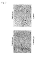
FIG. 7 shows the appearance of GANPhi cells in the red pulp region of the spleens of C57BL/6 mice as a result of immunization with TD-Ag. Scale bar is 100 μm.
FIG. 8A-C shows somatic mutations in Daudi cell transfectants which are engineered to express mouse GANP stably.
FIG. 9A-C shows an outline of the preparation of a transgenic mouse which is engineered to overexpress GANP in its B cells.
FIG. 10 (SEQ ID NOS: 37-72) shows the results of analyses of somatic mutations in transgenic (Tg) mice overexpressing GANP and wild-type mice.
FIG. 11A-E shows an outline of the preparation of a B cell-specific GANP deficient mouse (B-GANP−/−).
FIG. 12 shows the results of analyses (flowcytometry) of cell surface staining using the B cell-specific GANP deficient mouse (B-GANP−/−).
FIG. 13 shows the results of B cell proliferation assays. Almost no difference was observed, but only the proliferation caused by anti-CD40 antibody stimulation was decreased to about ½.
FIG. 14 shows antibody titers in the sera from non-immunized Cre-flox/+ mice and B-GANP−/− mice. No difference was observed among the antibody titers of individual isotypes.
FIG. 15 shows the results of measurement of antibody production in B-GANP−/− mice.
FIG. 16 shows the results of the staining of GC with peanut agglutinin.
FIG. 17 shows the results of measurement of antigen-specific antibody production in B-GANP−/− mice.
FIG. 18 shows the results of measurement by differential ELISA of the degrees of maturation of affinity in mice 14 and 35 days after immunization with 100 μg of NP-GC.
FIG. 19 shows the results of flowcytometry on GC-B cells.
FIG. 20A-F shows the results of sequence analyses of VH186.2 in Cre-flox/+ mice after PCR amplification (sequences (SEQ ID NOS: 73-88) continue from A to F in this order).
FIG. 20G-L shows the results of sequence analyses of VH186.2 in Cre-flox/+ mice after PCR amplification (sequences (SEQ ID NOS: 89-105) continue from G to L in this order).
FIG. 21 shows the frequencies of IgG1 mutation in Cre-flox/+ mice and B-GANP−/− mice.
FIG. 22 shows the frequencies of 33W to L mutation in VH186.2 in Cre-flox/+ mice and B-GANP−/− mice.
FIG. 23 shows the results of measurement of activation-induced cell death (AICD) and the results of apoptosis inhibition.
FIG. 24 shows the results of measurement of the apoptosis sensitivities of cells to anti-CD40 and anti-CD95 stimulations.
FIG. 25 shows the results of detection of apoptosis cells by TUNEL assay.
FIG. 26 shows the results of detection of apoptosis cells by TUNEL assay.
FIG. 27 shows the RNA expression levels of Bcl-2 family involved in apoptosis inhibition.
FIG. 28 shows the results of production of a high affinity antibody using a GANP transgenic mouse.
FIG. 29 shows the results of production of a high affinity antibody using the GANP transgenic mouse-derived hybridoma clones.
FIG. 30 shows association-dissociation curves obtained with Biacore on culture supernatants of the GANP transgenic mouse-derived hybridoma clones.
FIG. 31 shows association-dissociation curves obtained with Biacore on culture supernatants of the GANP transgenic mouse-derived hybridoma clones.
FIG. 32 shows an outline of the structure of GANP-GST fusion protein.
FIG. 33 shows the results of a pull-down assay for determining the region of GANP which directly binds to MCM. Shown on the left side of each panel are the positions of size standards.
FIG. 34 shows the results of a pull-down assay using in vitro translated MCM.
FIG. 35 shows the binding of individual GANP constructs to MCM by immunoprecipitation.
FIG. 36A-B shows the binding of individual GANP constructs to MCM by immunoprecipitation.
FIG. 37 shows an outline of the structures of GANP constructs and their intracellular distributions.
FIG. 38 shows intracellular distributions of GANP constructs.
FIG. 39 shows the nuclear localization of MCM3.
FIG. 40 shows the cytoplasmic localization of MCM3 induced by GANP expression.
FIG. 41 shows a control protein localized in the nucleus.
FIG. 42 shows the effect of GANP construct in the localization of MCM3 mutants.
FIG. 43 shows the nucleus-cytoplasm shuttling of MCM3 detected by a heterokaryon assay.
FIG. 44 shows the localization of GANP during the cell cycle.
BEST MODE FOR CARRYING OUT THE INVENTION
Hereinbelow, the present invention will be described in detail.
The present invention has been achieved based on a finding that it is possible to obtain a high affinity antibody by preparing a transgenic animal by transferring a GANP gene into a non-human mammal and immunizing the resultant transgenic animal with an antigen.
1. GANP
GANP which is called “germinal center-associated nuclear protein” is a 210 kDa nuclear protein having homology to yeast Sac3 protein (WO 00/50611). SAC3 is characterized as an inhibitory substance against actin formation. It is known that GANP is selectively up-regulated in germinal center (GC) B cells surrounded by follicular dendritic cells: FDC), has phosphorylation-dependent RNA primase activity, and is involved in the regulation of the cell cycle of B cells (Kuwahara, K. et al., (2000) Blood 95: 2321-2328).
In the present invention, the amino acid sequence for mouse GANP protein is shown in SEQ ID NO:2 and the amino acid sequence for human GANP protein is shown in SEQ ID NO: 4. With respect to the gene encoding the GANP protein (hereinafter, referred to as “GANP gene”), the nucleotide sequence for mouse GANP gene is shown in SEQ ID NO: 1 and the nucleotide sequence for human GANP gene is shown in SEQ ID NO: 3. The above-mentioned amino acid sequences and nucleotide sequences are also described in WO 00/50611.
GANP proteins may be mutant proteins; they may be those proteins which consist of the amino acid sequence as shown in SEQ ID NO: 2 or 4 wherein one or a plurality of amino acids have been deleted, substituted or added and have RNA primase activity. For example, a GANP mutant protein may also be used which consists of the amino acid sequence as shown in SEQ ID NO: 2 or 4 wherein one or a plurality of amino acids (preferably, one or several (e.g. one to ten, more preferably one to five) amino acids) have been deleted, one or a plurality of amino acids (preferably, one or several (e.g. one to ten, more preferably one to five) amino acids) have been substituted with other amino acids, and/or one or a plurality of other amino acids (preferably, one or several (e.g. one to ten, more preferably one to five) amino acids) have been added thereto, and yet has the same RNA primase activity as that of the above-described GANP protein.
“RNA primase activity” means the enzyme activity synthesizing a short primer RNA which will be a starting point for strand elongation when a strand extending opposite to the 5′→3′ direction (lagging strand) is synthesized. Usually, a molecule called α primase which binds to DNA polymerase α is used. In germinal center B cells, GANP primase which is the second primase is also induced.
GANP protein includes a protein having the amino acid sequence as shown in SEQ ID NO: 2 or 4, or a mutant amino acid sequence thereof, and a protein having a part of the N-terminal sequence of those sequences (e.g. positions 1-600, preferably 139-566 of the amino acid sequence as shown in SEQ ID NO: 2) or a mutant amino acid sequence thereof.
In the present invention, a GANP gene to be transferred into an animal may be a gene encoding the above-described GANP protein, a part of the N-terminal sequence of the GANP protein, or a mutant GANP protein. Specific examples of such a gene include a gene having the nucleotide sequence as shown in SEQ ID NO: 1 or 3. A gene having only the coding region of the nucleotide sequence as shown in SEQ ID NO: 1 or 3 may also be used. Alternatively, it is also possible to use a gene that has a sequence hybridizable to a complementary sequence to the nucleotide sequence as shown in SEQ ID NO: 1 or 3 under stringent conditions, and encodes a protein having RNA primase activity.
“Stringent conditions” refers to washing conditions after hybridization; specifically, the salt (sodium) concentration is 150-900 mM and the temperature is 55-75° C., preferably salt (sodium) concentration is 250-450 mM and the temperature is 68° C.
Introduction of mutations into a gene may be performed according to known techniques such as the Kunkel method or the gapped duplex method, using mutation introducing kits utilizing site-directed mutagenesis, such as GeneTailor™ Site-Directed Mutagenesis System (Invitrogen) or TaKaRa Site-Directed Mutagenesis System (Mutan-K, Mutan-Super Express Km, etc.; Takara Bio).
Details of mutant genes and methods for obtaining the same are also described in WO 00/50611.
In vitro stimulation of B cells with anti-μ antibody and anti-CD40 monoclonal antibody induces not only the up-regulation of GANP expression but also the phosphorylation of a specific serine residue in the amino acid sequence of GANP protein (e.g. serine at position 502: S502). This reaction is a key reaction for the RNA primase activity of GANP (Kuwahara, K. et al. (2001) Proc. Natl. Acad. Sci. USA, 98, 10279-10283). The N-terminal primase domain of GANP protein contains a serine residue whose phosphorylation is catalyzed by Cdk2 in vitro. GANP binds to MCM3 replication licensing factor due to its C-terminal domain (Kuwahara, K. et al., (2000) Blood 95: 2321-2328; Abe, E. et al., (2000) Gene 255: 219-227).
2. Transgenic Mammal Carrying GANP Gene Transferred Thereinto
The present invention relates to a transgenic mammal carrying a GANP gene transferred thereinto. Preferably, the transgenic mammal is capable of expressing the transferred GANP gene in its B cells.
- (1) GANP Gene and its Related Molecules
Complexes formed by GANP gene and its related molecules are needed directly or indirectly in the process of induction of mutations in genes. When repairing genetic mutations, GANP protein has the ability to promote induction of mutations in the V region so that high affinity antibodies are obtained. Therefore, the transgenic mammal of the invention carrying the GANP gene or a mutant thereof transferred thereinto is capable of promoting the production of high affinity antibodies of acquired immunity. Further, a transgenic non-human mammal overexpressing this GANP gene is capable of promptly producing an antibody with high binding strength to an antigen. Therefore, by immunizing the above-described transgenic non-human mammal with a specific antigen, it is possible to obtain easily an antibody with a high affinity that has been unachievable by conventional methods. As a result, it becomes possible to obtain polyclonal or monoclonal antibodies capable of eliminating obstinate pathogenic microorganisms or foreign substances. Further, by preparing humanized antibodies using the transgenic mammal of the invention, or by preparing single chain antibodies comprising the V region of the antibody produced by the transgenic mammal of the invention, it becomes possible to sharply increase the effect of antibody therapy.
Because of the GANP gene or its mutant transferred thereinto, the transgenic mammal of the invention is capable of promoting the production of high affinity antibodies in B cells, and the high affinity antibody-producing cells have resistance to apoptosis induction signals.
In order to confirm that GANP is a molecule functioning in the antibody production in acquired immune responses, the present inventors have created a GANP gene deficient mouse so that GANP is deficient B cell selectively. The results revealed that the deficiency of GANP gene did not influence the development, differentiation and proliferation of cells in the immune system and that no big change is observed in the total yield of antibodies.
It should be noted here that only when B cells have reacted with limited types of antigens, they proliferate and differentiate into antibody-producing cells without T cells. For producing antibodies to ordinary antigens, co-existence of T cells is necessary. Antigens to which antibodies are produced even in the absence of T cells are called T cell-independent antigens. On the other hand, general antigens other than T cell-independent antigens are called T cell-dependent antigens. When B cells have reacted with T cell-dependent antigens, the differentiation of B cells into antibody-producing cells is assisted by helper T cells.
Many of the antigenic determinants (also called antigenic epitopes) of pathogenic viruses are weak in immunogenicity by themselves and activated by the peptide antigens of carrier proteins recognized by T cells.
In the present invention, in order to examine that GANP gene-transferred animals are capable of producing high affinity antibodies highly frequently in those antibody-producing responses to soluble antigens where ordinary animals cannot produce strong antibodies, an antigen designated NP-CG was prepared by coupling a nitrophenyl group (NP group) (which has been extensively analyzed as a hapten) to chicken gamma globulin, followed by examination of responses to T cell-dependent antigen.
It is known that C57BL/6 mice's generate high affinity antibody to NP only when utilized a single V region. This response is dominated by only the V region of IgG heavy chain (called VH186.2) and lambda 1 light chain of an antibody. With this system, it is possible for antibodies of IgG1 isotype to examine genetic mutations in high affinity antibodies by analyzing the amino acid sequence of VH186.2. Furthermore, it is reported that the highest affinity is induced when the amino acid residue tryptophan (W) at position 33 of the amino acid sequence of the heavy chain V region (VH186.2) has been mutated into leucine (L) (W33 to L mutation).
Then, the present inventor examined whether high affinity antibodies could be induced in GANP gene deficient mice and its defect might be associated with W33 to L mutation event or not. As a result, high affinity antibody production was hardly observed in GANP gene deficient mice, compared to the control Cre-flox/+ mice. Therefore, it has been demonstrated that GANP gene has a key function in the production of high affinity antibodies. To investigate this function further, the inventor has created GANP gene-overexpressing mice. Overexpression of GANP gene was achieved by linking a mouse immunoglobulin promoter moiety and a human immunoglobulin gene intron enhancer moiety upstream (5′) of GANP gene so that the gene is expressed selectively in B cells.
The GANP-overexpressing mice were born normally, and no particular change was observed in the development, differentiation and proliferation of their lymph tissues. However, a remarkable increase was observed in the high affinity type V region gene (W33 to L) in responses to NP-CG. Although the functional role of RNA primase activity here has not yet been established, it is believed that the RNA primase activity of GANP gene or the phosphorylation of the 502 serine residue involved in the primase activity is related to the production of high affinity antibodies in view of the following: (i) the phosphorylation of serine residue at position 502 (which is an indicator for the primase activity of GANP molecule) is high in cells present at the region of the germinal center where high affinity B cells are produced (centrocytes), and (ii) the frequency of the mutation at the V region induced by experiments to transfer a ganp gene into Daudi cells is high. These results show that high expression of GANP molecule and activation of RNA primase activity are necessary for high affinity antibody production by immune response.
- (2) Mammals for Use in GANP Gene Transfer
The term “mammal” used in the present invention means any of non-human mammals such as bovine, horse, pig, goat, rabbit, dog, cat, mouse, rat, hamster and guinea pig. Preferably, mouse, rabbit, rat or hamster is used. Most preferably, mouse is used.
The transgenic mammal of the invention may be prepared by introducing a GANP gene into fertilized eggs, unfertilized eggs, embryonic cells comprising spermatozoa and protocells thereof, preferably into cells of embryogenesis stage (more preferably, the single cell or fertilized egg cell stage and yet generally before eight-cell stage) in the development of non-human mammals, by a method such as the calcium phosphate method, electric pulsing, lipofection, aggregation, microinjection, the particle gun method, or the DEAE-dextran method. Further, it is also possible to transfer a GANP gene of interest into somatic cells, organs of the living body, tissue cells, etc. by the above-mentioned gene transfer methods to use the resultant cells, etc. for cell culture or tissue culture. Further, it is possible to create transgenic mammals by fusing these cells with the above-described embryonic cells according to known cell fusion methods.
When a GANP gene is transferred into an animal of interest, it is preferred that the gene be transferred in the form of a gene construct in which the gene is ligated downstream of a promoter capable of directing expression of this gene in cells of the animal of interest. Specifically, a vector in which a GANP gene is ligated downstream of various promoters capable of directing expression of the GANP gene derived from various mammals may be microinjected into fertilized eggs of the mammal of interest (e.g. mouse fertilized eggs) to thereby create a transgenic mammal capable of high expression of the GANP gene of interest.
Examples of expression vectors for GANP gene include plasmids derived from Escherichia coli; plasmids derived from Bacillus subtilis; plasmids derived from yeast; bacteriophages such as λ-phage; retroviruses such as Moloney leukemia virus; and animal or insect viruses such as vaccinia virus or baculovirus.
As promoters for regulating gene expression, promoters of viruses-derived genes; promoters of various mammals (such as human, rabbit, dog, cat, guinea pig, hamster, rat and mouse)-derived genes; and promoters of birds (such as chicken)-derived genes may be used.
Examples of promoters of viruses-derived genes include promoters of cytomegalovirus-, Moloney leukemia virus-, JC virus- or breast cancer virus-derived genes.
Examples of promoters of various mammals- and birds-derived genes include promoters of such as albumin, insulin II, erythropoietin, endothelin, osteocalcin, muscle creatine kinase, platelet-derived growth factor β, keratin K1, K10 and K14, collagen type I and type II, atrial natriuretic factor, dopamine β-hydroxylase, endothelial receptor tyrosine kinase, sodium/potassium-dependent adenosinetriphosphatase, neurofilament light chain, metallothionein I and IIA, metalloproteinase I tissue inhibitor, MHC Class I antigen, smooth muscle α-actin, polypeptide chain elongation factor 1α (EF-1α), β-actin, α- and β-myosin heavy chains, myosin light chains 1 and 2, myelin basic polypeptide, serum amyloid P component, myoglobin and renin genes.
The above-described vector may have a terminator which terminates the transcription of a messenger RNA of interest in a transgenic mammal. For the purpose of achieving still higher expression of GANP gene, the splicing signal of each gene, enhancer region, or a part of an intron of an eukaryotic gene may be ligated upstream (5′) of the promoter region, between the promoter region and the translation region, or downstream (3′) of the translation region, if desired.
In a preferred embodiment of the invention, it is possible to allow selective expression of the transferred GANP gene in B cells by ligating the GANP gene downstream of an immunoglobulin promoter or by ligating a human immunoglobulin gene intron enhancer moiety upstream (5′) of the GANP gene.
- (4) Transfer of GANP Gene
The transfer of GANP gene at the fertilized egg cell stage is preferably carried out in such a manner that excessive presence of GANP gene is secured in all the embryonic cells and somatic cells of the mammal of interest. Excessive presence of GANP gene in the embryo cells of the created animal after gene transfer means that all the progeny of that animal has excessive GANP gene in all the embryonic cells and somatic cells. The progeny of this kind of animal which inherited the GANP gene has excessive GANP protein in all the embryonic cells and somatic cells.
In the present invention, first, heterozygotes which have the transferred GANP gene in one of the homologous chromosomes are prepared; then, homozygotes which have the transferred GANP gene in both of the homologous chromosomes are obtained by mating the heterozygotes with each other. Subsequently, by mating female homozygotes with male homozygotes, all the resultant progeny retains the transferred GANP gene stably. After confirmation of the excessive presence of GANP gene, the progeny may be sub-bred in usual breeding environments.
Fertilized eggs of a non-human mammal of interest (preferably, mouse) or its ancestor (back-crossing) to be used for transferring a foreign GANP gene different from the endogenous gene of the mammal of interest are obtained by mating allogenic male and female mammals.
Although fertilized eggs may be obtained by natural mating, it is preferred that female mammals after artificial adjustment of their sexual cycle be mated with male mammals. As a method for artificially adjusting the sexual cycle of female mammals, such a method may be used preferably in which follicle-stimulating hormone (pregnant mare serum gonadotropin (PMSG)) and then luteinizing hormone (human chorionic gonadotropin (hCG)) are administered by, e.g., intraperitoneal injection.
After the transfer of a foreign GANP gene into the resultant fertilized eggs by the methods described above, the eggs are artificially transferred/implanted in female mammals. As a result, non-human mammals having a foreign gene-integrated DNA are obtained. In a preferable method, fertilized eggs are transferred/implanted artificially in pseudo-pregnant female mammals in which fertility has been induced by mating with male mammals after administration of luteinizing hormone-releasing hormone (LHRH). As totipotent cells into which a GANP gene is to be transferred, fertilized eggs or early embryos may be used if the mammal of interest is mouse. As a method of gene transfer into cultured cells, DNA microinjection is preferable in view of the production efficiency of transgenic mammal individuals and the transmittance efficiency of the transgene to the subsequent generation.
Subsequently, the gene-injected fertilized eggs are transplanted into the oviduct of a recipient female mammal. Those animals which have developed from the eggs up to individuals and have been successively born are bred under foster parents. Then, DNA is extracted from a part of their bodies (e.g. the tail end in the case of mouse) and subjected to Southern analysis, PCR, etc. Thus, it is possible to confirm the presence of the transgene. Those animals in which the presence of the transgene has been confirmed are designated founder animals. The transgene is transmitted to 50% of their offspring (F1). Further, by mating F1 individuals with wild-type animals or other F1 individuals, F2 individuals which have the transgene in one (heterozygote) or both (homozygote) of the diploid chromosomes can be produced.
Alternatively, transgenic mammals expressing high levels of GANP protein may also be created by introducing the above-described GANP gene into ES (embryonic stem) cells. For example, the GANP gene is introduced into HPRT negative (i.e. lacking hypoxanthine-guanine phosphoribosyltransferase gene) ES cells derived from normal mouse blastocysts. Then, those ES cells in which the GANP gene has been integrated through homologous recombination induced in a mouse endogenous gene are selected by HAT selection. The thus selected ES cells are microinjected into fertilized eggs (blastocysts) obtained from other normal mouse. The resultant blastocysts are transferred into the uterus of other normal mouse as a recipient. Subsequently, chimeric transgenic mice are born from the recipient mouse. By mating these chimeric transgenic mice with normal mice, heterotransgenic mice can be obtained. Further, by mating the heterotransgenic mice with each other, homotransgenic mice can be obtained.
The present invention encompasses not only the above-described transgenic mammal but also its progeny and a part of the transgenic mammal or its progeny in the scope of the invention. As a part of the transgenic mammal, a tissue, organ, cell or the like of the transgenic mammal or its progeny may be enumerated. Specific examples of organs or tissues include the spleen, thymus, lymph nodes, bone marrow or tonsil; and specific examples of cells include B cells.
The transgenic mammal of the invention may be mated with a mammal that further activates B cells. As a result of such mating, antibodies of still higher affinity can be produced.
Recently, it has been reported that when B cells are activated in peripheral lymph nodes in MRL/lpr mouse, induction of mutations in the V region is further increased in the T cell region after B cells passed through the germinal center. The inventors have also found that non-immunized MRL/lpr mouse shows high expression of GANP equivalent to the GANP expression observed in ganp transgenic mouse which was created by ligating a GANP gene downstream of Ig promoter and enhancer. This suggests a possibility that, while high affinity antibodies are not produced against autoantigens normally, high affinity antibodies to autoantigens may be produced in this autoimmune disease mouse because of the abnormal activation of GANP molecule.
Still higher induction of mutations can be expected if such mouse as MRL/lpr, NZB or (NZB×NZW)F1 (all of them are considered as autoimmune disease mice) is used as the above-mentioned animal that still activates B cells.
By creating a GANP transgenic mouse from MRL/lpr mouse utilizing what has been described above, it may be possible to create a super high affinity antibody-producing mouse. In other words, by mating the GANP gene overexpressing transgenic mammal of the invention with various autoimmune disease model animals, it is possible to create mammals capable of producing high affinity antibodies.
3. Preparation of High Affinity Antibodies
The term “antibody” used in the invention means a protein having activity to specifically bind to an antigen, preferably a protein produced by B cells. In the present invention, an antibody having high reactivity with an antigen is called high affinity antibody. The term “high affinity” used herein means that the ability of an antibody to bind to an antigen is high. In the present invention, a high affinity antibody refers to an antibody which has higher ability to bind to an antigen than those antibodies prepared using conventional animals such as mouse, and which is slow in dissociating from that antigen. This means that such an antibody is high and specific in the ability to bind to an antigenic determinant (epitope) sterically and closely. Besides, the binding of such an antibody to the antigenic determinant induces changes not only in the determinant but also the structure of the antigen itself, to thereby show strong activities eventually (e.g. biological activities such as neutralization of toxicity, prevention of viral infection, deactivation of pathogens, promotion of elimination of pathogens from the body, or induction of denaturation in antigen molecules).
The binding ability of an antibody (i.e. affinity) may be measured as a dissociation constant (KD), dissociation rate constant (Kdiss) or association rate constant (Kass) by Scatchard analysis or with a surface plasmon resonance sensor called Biacore. Biacore systems in which three technologies of sensor chip, microflow system and SPR detection system are integrated are to measure the strength, rate and selectivity of molecular binding. This apparatus enables real time detection of biological molecules and monitoring of interactions among a plurality of molecules without using labels. Specific examples of useful Biacore systems include Biacore 3000, Biacore 2000, Biacore X, Biacore J and Biacore Q (all of them are manufactured by Biacore).
With the above-described Biacore system, parameters showing the affinity of antibodies, i.e. dissociation constant (KD), dissociation rate constant (Kdiss) (1/Sec) and association rate constant (Kass) (1/M.Sec) are measured.
Antibodies with smaller dissociation constant (KD) values are preferable because the smaller the dissociation constant value, the higher the affinity. The binding ability of an antibody (affinity) is determined by the two parameters of Kdiss and Kass, and is represented by the following formula:
KD (M)=Kdiss/Kass
Although the affinity of the resultant antibody varies depending on a plurality of factor such as the type of the antigen, generally, its KD value is preferably 1×10⁻⁷ (M) or less. For example, preferable KD values are 1×10⁻⁸ (M) or less, 1×10⁻¹⁰ (M) or less, or 1×10⁻¹¹ (M) or less.
In the present invention, when the resultant antibody reveals any of the above-described effects or natures, the antibody is judged as a “high affinity” antibody.
Enhancement in the affinity of antibody molecules is produced by inducing somatic hypermutations (SHM) in genes of the variable regions (V region) of antibodies. Although specificities of antibodies to antigens are recognized from the beginning of immunization of the living body with antigens, most of early antibodies are IgM class antibodies; their binding affinity to antigens is not high and their ability to remove or deactivate pathogens or foreign substances is low. However, if an antigen is administered to the living body to give several boosters, the binding affinity of antibody to the antigen is enhanced. At this time, B cells need stimulation from T cells, and this activation is considered to take place in the germinal center region in peripheral lymph tissues. Recently, the RNA editing molecule AID expressed in the germinal center has been reported as a molecule necessary to induce mutations in V region genes. Further, it is reported that uracil DNA glycosidase and, as DNA polymerases necessary for DNA replication, DNA polymerases zeta (ζ) and iota (ι) which easily produce errors are also involved in the above activation. However, the molecule(s) which control(s) these functions has/have not been elucidated. The function of GANP molecule as a novel SHM-inducing molecule has been elucidated. Increase in the expression of this molecule plays a key role in SHM induction. Among all, it has been demonstrated that GANP molecule is important in producing high affinity antibodies.
Antibodies induced by immunizing C57BL/6 mice with nitrophenyl-chicken γ globulin as a hapten carrier antigen have VH186.2 locus as the H chain and λ1 as the L chain. In this system, it is known that antibodies obtained after boosters were given are IgG1 antibodies, and that the mutation induced in the V region sequence of those antibodies with particularly high binding affinity among them is mutation from tryptophan to leucine at position 33. In the Examples of the present specification, this high affinity-type V region mutation is induced highly. This can be said definite evidence at the molecule level showing that high affinity antibodies have been induced.
Therefore, it is possible to obtain high affinity antibodies by administering an antigen to the above-described transgenic mammal or its progeny and letting the resultant mammal or progeny produce antibodies. Briefly, an antigen of interest is administered by conventional methods to an animal that is engineered to express high levels of GANP protein. Then, high affinity antibodies may be prepared form lymphocytes of a tissue such as blood or spleen (not limited to these tissues) of the immunized animal. These high affinity antibodies may be either polyclonal or monoclonal antibodies.
As a method for producing polyclonal antibodies, for example, polyclonal antibodies may be obtained by administering an antigen to the transgenic mammal of the invention, taking blood from the immunized mammal, and then separating and purifying antibodies from the resultant blood.
Methods of immunization are known to those skilled in the art. For example, immunization may be performed by administering an antigen once or more.
The types of the antigen are not particularly limited. All substances which may have a steric structure as an antigenic determinant fall under antigen. In addition to all biological components such as proteins, enzymes, peptides, sugars, lipids, DNAs, RNAs and prions, any substance such as cancer antigens, virus antigens, organic or inorganic synthetic antigens may be used.
The antigen may be administered, for example, two or three times at intervals of 7 to 30 days. The dose may be, for example, about 0.05 to 2 mg of the antigen per administration. The route of administration is not particularly limited. For example, subcutaneous administration, dermal administration, intraperitoneal administration, intravenous administration or intramuscular administration may be selected appropriately. Preferably, the antigen is administered by intravenous, intraperitoneal or subcutaneous injection. The antigen may be used in solution in an appropriate buffer, e.g. a buffer containing conventional adjuvants such as complete Freund's adjuvant or aluminium hydroxide, but the antigen may be used without adjuvant depending on the administration route or other conditions.
After immunized mammals have been bred for a specific period of time, serum samples are obtained from them and antibody titers thereof are measured. When the antibody titer begins to rise, boosters may be given using, for example, 100 μg to 1000 μg of the antigen. One to two months after the final administration, blood is taken from the immunized mammals and subjected to various conventional methods used for protein isolation, e.g. centrifugation, precipitation using ammonium sulfate or polyethylene glycol, and chromatography such as gel filtration chromatography, ion exchange chromatography or affinity chromatography. Thus, polyclonal antibodies may be obtained as polyclonal anti-sera.
As a method for producing monoclonal antibodies, the hybridoma method may be used. First, a peptide constituting an antigen of interest is suspended in an adjuvant. The resultant suspension is administered subcutaneously or intradermally into animals to be immunized (i.e. the transgenic mammal of the invention). The types of the antigen used here are the same as described above. Examples of the adjuvant used here include complete Freund's adjuvant, BCG, trehalose dimycolate (TDM), lipopolysaccharide (LPS), alum adjuvant and silica adjuvant. Preferably, a combination of complete Freund's adjuvant (CFA) and incomplete Freund's adjuvant (IFA) is used in view of the ability to induce antibodies.
In the production of monoclonal antibodies, preferably, animals which have undergone the first immunization with an antigen are boosted several times; after passage of appropriate number of days, blood samples are taken and antibody titers thereof are measured. Since antibodies produced by the method of the invention are high affinity antibodies, the first immunization may be sufficient without booster. Antibody titers may be measured by known methods such as enzyme-linked immunosorbent assay (hereinafter, referred to as ELISA).
Subsequently, the spleens are removed from the immunization-completed animals to obtain B cells. Obtaining B cells capable of binding to antigens is preferable because it could reduce subsequent screening. The B cells obtained at this point are high affinity antibody-producing cells, which may be used as an immunopotentiator without any processing. It is also possible to obtain V region genes directly from these B cells and to measure somatic hypermutations in the V region.
Subsequently, the resultant B cells are fused with myeloma cells by conventional methods to thereby prepare an antibody-producing hybridoma. For example, if the animal is mouse, the spleen is removed and placed in a solution such as Hanks' balanced salt solution (HBSS). Cells are pushed out with tweezers to obtain spleen lymphocytes (B cells). The resultant spleen lymphocytes are stained with trypanblue or the like to count the number of viable cells, and then fused with myeloma cells to prepare a hybridoma.
The myeloma cell used for the cell fusion is not particularly limited. Known myeloma cells such as P3-X63.Ag8 (X63), P3-X63.Ag8.U1 (P3U1), P3/NS I/1-Ag4-1(NSI) or Sp2/0-Ag14(Sp2/0) may be used. In the selection of the myeloma cell, compatibility with antibody-producing cells should be considered appropriately.
Cell fusion is carried out as described below. Briefly, 1×10⁶-1×10⁷ cells/ml of antibody-producing cells are mixed with 2×10⁵-2×10⁶ cells/ml of myeloma cells (preferable cell ratio of antibody-producing cells to myeloma cells is 5:1) in an animal cell culture medium such as serum-free DMEM or RPMI-1640 and fused in the presence of a cell fusion promoter.
As the method of cell fusion, any of the methods known in the art (the Sendai virus method, the polyethylene glycol method, or the protoplast method) may be selected. Preferably, the polyethylene glycol method is used in view of relatively low cytotoxicity and simple fusion operations. Polyethylene glycol with a mean molecular weight of 1000-6000 daltons may be used as a cell fusion promoter. When production of a large quantity of antibodies is desired, a hybridoma prepared by fusing antibody-producing cells stimulated with a vinyl pyridine derivative with myeloma cells is used preferably.
The resultant hybridoma is cultured in HAT medium (containing hypoxanthine, aminopterin and thymidine) for an appropriate period of time according to conventional methods, followed by selection of hybridoma clones. Subsequently, those hybridoma clones producing an antibody of interest are screened, followed by cloning of the hybridoma clones.
As the screening method, known methods for antibody detection, such as ELISA, radio immunoassay (hereinafter, referred to as RIA), the plaque method, or the aggregation reaction method, may be used. As the cloning method, known methods in the art such as the limiting dilution-culture method, the soft agar method or FACS, may be used. The resultant hybridoma is cultured in an appropriate culture broth, or administered into the abdominal cavity of an animal (e.g. mouse) compatible with the hybridoma. From the thus obtained culture broth or abdominal dropsy, the monoclonal antibody of interest may be isolated and purified by methods such as salting out, ion exchange chromatography, gel filtration or affinity chromatography.
It should be noted that fragments and single chain antibodies of the V region of the above-described antibody are also within the scope of the present invention. A fragment of the antibody means a portion of the above-described polyclonal or monoclonal antibody. Specific examples of such a fragment include F(ab′)2, Fab′, Fab, Fv (variable fragment of antibody), sFv, dsFv (disulphide stabilized Fv) or dAb (single domain antibody). F(ab′)2 and Fab′ mean those antibody fragments which are prepared by treating an immunoglobulin (monoclonal antibody) with proteolytic enzymes pepsin and papain, respectively, and are generated through digestion around the disulfide bond present between the two H chains in the hinge region. For example, when IgG is treated with papain, this molecule is cut upstream of the disulfide bond present between the two H chains in the hinge region to yield two homologous antibody fragments in which an L chain consisting of VL (L chain variable region) and CL (L chain constant region) and an H chain fragment consisting of VH (H chain variable region) and CHγ1 (γ1 region in H chain constant region) are coupled by a disulfide bond in the C-terminal region. Each of these two homologous antibody fragments is called Fab′. When IgG is treated with pepsin, this molecule is cut downstream of the disulfide bond present between the two H chains in the hinge region to yield an antibody fragment which is slightly larger than the above-described two Fab′ fragments ligated at the hinge region. This antibody fragment is called F(ab′)2. A single chain antibody has a structure in which VL and VH are linked by a linker.
The high affinity antibody of the invention may be a humanized antibody or human antibody. These human antibodies may be prepared by using mammals whose immune system has been replaced with the human immune system. After immunizing such mammals, human antibodies may be prepared directly in the same manner as used in the preparation of conventional monoclonal antibodies.
For the preparation of humanized antibodies, reconstructed variable regions consisting of human-derived framework regions and mouse-derived CDRs (complementarity determining regions) is prepared by transferring the CDRs of the variable regions in a mouse antibody into the human variable regions.
Subsequently, these humanized, reconstructed human variable regions are ligated to human constant regions. Portions derived from non-human amino acid sequences in the finally reconstructed humanized antibody are only CDRs and extremely small parts of FRs. CDRs are composed of hyper-variable amino acid sequences. Since these sequences do not show species specific sequences, it is possible to use humanized antibodies having mouse CDRs. Methods for preparing humanized antibodies are well-known in the art.
Human antibodies may be produced using any animal (e.g. mouse, rat, etc.) in terms of structure, though generally the antigen binding site in the variable region (i.e. hyper variable region) may raise some problem with respect to specificity and binding affinity. On the other hand, it is desirable that the structures of the remaining portion of the variable region and the constant region should be the same as the structures in human antibodies. With respect to genetic sequences common in human, genetic engineering techniques to prepare them have been established.
The isotype of the antibody of the invention is not particularly limited. The antibody of the invention may have any isotype, e.g. IgG (IgG1, IgG2, IgG3, IgG4), IgM, IgA (IgA1, IgA2), IgD or IgE.
4. Use of High Affinity Antibodies
The high affinity antibody of the invention is useful as a drug for diagnosing, treating or preventing diseases.
- (1) Diagnosis of Diseases
Diagnosis of various diseases using the antibody of the invention is carried out as described below. Briefly, samples (e.g. sera) taken from subjects suspected of having various diseases are bound to the antibody of the invention by antigen-antibody reaction. Then, the amount of an antigen of interest in the sample is detected from the amount of bound antibody. The detection of the amount of bound antibody may be performed by conventional immunological measuring methods. For example, immunoprecipitation, immunoaggregation, labeled immunoassay, immunonephelometry, immunoturbidimetry, or the like may be used. Labeled immunoassay is especially preferable from the viewpoint of simplicity and high sensitivity. In labeled immunoassay, antibody titers in samples may be expressed directly as the amounts of label detected using a labeled antibody. Alternatively, antibody titers may be expressed relatively using as a standard solution an antibody of known concentration or known titer. Briefly, the standard solution and a sample may be measured simultaneously in the same measuring system, followed by expression of the antibody titer in the sample relatively based on the value of the standard solution.
In labeled immunoassay, any of known measurement methods, such as ELISA, RIA, fluoroimmunoassay, or chemiluminescence immunoassay, may be used. The labeling substance may be appropriately selected depending on the above-mentioned assay method; for example, an enzyme, radioisotope, fluorescent compound, or chemiluminescent compound may be selected. Specific examples of the enzyme useful in the invention include peroxidase, alkaline phosphatase, acid phosphatase and glucose oxidase. Detection sensitivity of the above-mentioned labeling substances may be increased by using avidin-biotin complex. As a specific example of the radioisotope useful in the invention, 125I may be given at first. Specific examples of the fluorescent compound useful in the invention include fluoresceine isothiocyanate (FITC) and tetramethylrhodamine isothiocyanate (TRITC). Specific examples of the chemiluminescent compound useful in the invention include lophine, luminol and lucigenin. The labeling of antibodies with the above-mentioned substances may be performed according to conventional methods. Hereinbelow, labeled immunoassay using labeled antibodies will be described.
As a method of detection of various diseases according to labeled immunoassay, a method using a known non-competitive reaction system or competitive reaction system may be possible. Non-competitive reaction systems require solid phase (solid phase method). Competitive reaction systems do not necessarily require solid phase (liquid phase method), but use of solid phase is preferable since that will make measuring operations simple. Specific examples of materials for the solid phase include polystyrene, nylon, glass, silicon rubber and cellulose. As the shape of the solid phase, spheres, wells, tubes, sheets, or the like may be enumerated. However, the material and the shape useful in the invention are not limited to those enumerated above. Known materials and shapes used in labeled immunoassay may be used at discretion.
In non-competitive reaction systems, measurement operations are carried out as follows. Briefly, a sample or the antibody of the invention is immobilized on a solid support and then reacted with the antibody of the invention or a sample. Subsequently, a pre-labeled anti-immunoglobulin antibody (secondary antibody) is added to react with the above antibody reacting with the immobilized sample. With the labeling substance of this secondary antibody, it is possible to detect the amount of the antibody bound to the sample. Since the amount of the labeled secondary antibody detected is directly correlated with the amount of the antigen of interest in the sample, the amount of this antigen can be obtained from the amount of the labeled secondary antibody.
In competitive reaction systems, a sample and a specific amount of an antigen of interest are reacted with a specific amount of an antibody. For example, after immobilization of a sample on a solid support, the sample is reacted with the antibody of the invention which has been pre-reacted with an antigen of interest. Subsequently, the antibody which has reacted with the immobilized sample is reacted with a pre-labeled anti-immunoglobulin antibody (secondary antibody), followed by detection of the amount of the antibody by the labeling substance. The amount of the labeling substance is inversely correlated with the amount of the antigen of interest added. Other types of competitive reaction systems may also be used where the antibody of the invention is immobilized, reacted with a sample, and then reacted with a pre-labeled antigen of interest. The amount of the labeling substance detected is inversely correlated with the amount of GANP protein in the sample bound to the antibody.
As the method of immobilization of an antigen or antibody on a solid support, known methods such as physical adsorption, covalent binding, ionic bonding or crosslinking may be used. Physical adsorption is especially preferable because of its simplicity. As examples of the anti-immunoglobulin antibody (secondary antibody) useful in the invention, anti-IgG antibody or anti-IgM antibody may be given. These antibodies may be used as an entire molecule. Alternatively, antibody fragments Fab, Fab′ and F(ab′)2 comprising the antigen binding site obtained by treating antibodies with enzymes may be used. Further, instead of the labeled anti-immunoglobulin antibody, a substance having specific affinity for antibody molecules (e.g. protein A which has specific affinity for IgG) may be labeled and used.
As a preferable example of the above-described labeled immunoassay, ELISA may be given which is an immunoassay using an enzyme as a label. Briefly, a sample or a dilution thereof is placed in 96-well plates or the like and incubated at 4° C. to room temperature overnight or at 37° C. for about 1-3 hrs so that GANP protein to be detected is adsorbed and immobilized on the plates. Then, the antibody of the invention is reacted. Subsequently, an enzyme-preconjugated anti-immunoglobulin antibody (secondary antibody) is reacted. Finally, an appropriate color-developing substrate reactive with the enzyme (e.g. if the enzyme is phosphatase, p-nitrophenylphosphate or the like) is added to thereby detect the antibody with its color development.
By using the high affinity antibody of the invention, it is possible to evaluate the efficacies of therapeutics for various diseases. The evaluation method using the high affinity antibody of the invention is performed as follows. Briefly, a drug is administered to various disease patients or disease model animals. Then, using the antibody of the invention, the amounts of the antigen (such as virus) in these living bodies are detected. By comparing the amounts, the efficacy of the drug as a therapeutic for various diseases can be evaluated based on the amounts of the antigen in living bodies.
The high affinity antibody of the invention may be provided in the form of a diagnosis kit for various diseases. This kit may be used in the diagnosis method of the invention or the efficacy evaluation method of the invention. The kit of the invention comprises as least one selected from the following (a) and (b).
(a) the antibody of the invention or that antibody labeled
(b) immobilized reagent in which the antibody or labeled antibody of (a) above is immobilized on a solid support
The “labeled antibody” means an antibody labeled with an enzyme, radioisotope, fluorescent compound or chemiluminescent compound. As the material of a solid support on which the antibody or labeled antibody is immobilized in the kit of the invention, polystyrene, nylon, glass, silicon rubber, cellulose or the like may be used. As the shape of such a solid support, spheres, wells, tubes or sheets may be enumerated. However, the material and the shape useful in the invention are not limited to these ones. Instead of the immobilized reagent, a solid phase and an immobilizing agent may be attached to the kit. As the immobilizing agent, if immobilization by physical adsorption is intended, a coating liquid such as 50 mM carbonate buffer (pH 9.6), 10 mM Tris-HCl buffer (pH 8.5, containing 100 mM sodium chloride) or PBS and, if necessary, a blocking liquid (which is a coating liquid containing 0.5% gelatin) may be enumerated, for example.
The antibody contained in the kit of the invention may be in a state of solution in PBS or the like, or in a state where the antibody is linked to a gel (hereinafter, abbreviated to “absorption gel”). This absorption gel may be pre-packed in 0.5-2 ml microcentrifuge-precipitation tubes for absorption by the batch method. Alternatively, the absorption gel may be pre-packed in 0.1-5 ml mini-columns for absorption by the column method.
In addition to the above-described components, the kit of the invention may contain other reagents for carrying out the detection of the invention, e.g. the substrate of an enzyme (color developing substrate, etc.), the substrate in solution, enzymatic reaction-terminating liquid or the like when the labeling substance is an enzyme, and diluents for samples. Specific examples of diluents for samples include 20 mM Tris-HCl buffer (pH 7.4) containing PBS (phosphate-buffered physiological saline, pH 7.4), 137 mM sodium chloride and 3 mM potassium chloride (hereinafter abbreviated to “TBS”); and PBS or TBS containing 0.05% Tween 20 and 0.1-1% BSA. These diluents for samples may be used for diluting other substances such as antibodies.
- (2) Pharmaceutical Compositions for Treating or Preventing Diseases
When the high affinity antibody of the invention has an effect of neutralizing the activity of an antigen which will become the pathogen of a disease, the antibody of the invention is useful in a pharmaceutical composition for treating or preventing the disease. The pharmaceutical composition of the invention comprises the high affinity antibody of the invention or a fragment thereof as an active ingredient and, is provided, preferably, in the form of a pharmaceutical composition comprising a pharmacologically acceptable carrier.
The “pharmacologically acceptable carrier” used herein includes excipients, diluents, fillers, disintegrants, stabilizers, antiseptics, buffers, emulsifiers, aromatics, coloring agents, sweetening agents, thickening agents, flavoring agents, dissolution aids and other additives. By using one or more of these carriers, various forms of pharmaceutical compositions may be prepared, e.g. tablets, pills, powders, granules, injections, solutions, capsules, troches, elixirs, suspensions, emulsions and syrups. These pharmaceutical compositions may be administered orally or parenterally. Other forms for parenteral administration include solutions for external use which comprise one or more active substances and are prescribed by conventional methods, suppositories for enteric administration, and pessaries.
The dose of the pharmaceutical composition of the invention varies depending on the age, sex, body weight and conditions of the patient, treatment effect, the method of administration, time period for treatment, or the type of the high affinity antibody (the active ingredient) contained in the composition. Usually, the pharmaceutical composition of the invention may be administered to adult patients in the range from 10 μg to 1000 mg per administration, preferably in the range from 10 μg to 100 mg per administration. However, the dose is not limited to this range.
For example, in the case of injections, the pharmaceutical composition of the invention may be dissolved or suspended in a pharmacologically acceptable carrier (such as physiological saline or commercial distilled water for injection) so that the concentration of the antibody in the carrier is from 0.1 μg /mil to 10 mg/ml. The thus prepared injection may be administered to human patients in need of treatment at a rate of 1 μg −100 mg/kg body weight, preferably at a rate of 50 μg −50 mg/kg body weight, per administration once to several times per day. The route of administration may be intravenous injection, subcutaneous injection, intradermal injection, intramuscular injection or intraperitoneal injection, for example. Among all, intravenous injection is preferable. Optionally, injections may be prepared in the form of a non-aqueous diluent (e.g. propylene glycol, polyethylene glycol, vegetable oil such as olive oil, alcohol such as ethanol), suspension or emulsion. Sterilization of such injections may be performed by filter-sterilization through a bacteria removal filter, addition of antiseptics, or irradiation. Injections may take a form that is prepared into an injection at the time of use. Briefly, a solid composition is prepared by lyophilization or the like, and this solid composition may be dissolved in aseptic distilled water for injection or other solvent at the time of use.
5. Application of the Present Invention
The present inventors have induced overexpression of GANP in B cell tumor strains and analyzed them. As a result, the B cell tumor strains showed that GANP gene transfer has a remarkable effect in inducing somatic hypermutations in V region genes. Since this effect is not observed when a mutant gene in which phosphorylation of serine at position 502 (required for the primase activity of GANP) does not occur is used, it is suggested that RNA primase activity is necessary for the remarkable induction of somatic hypermutations in V region genes. These results demonstrate that GANP has an effect of enhancing the production of specific antibodies as a clinical, supplemental immunopotentiator.
It is also effective for clinical, supplemental immunopotentiation to use a retrovirus vector as a vector and a combination of GANP and a stimulation mediated by TNF family molecules such as DC40 or BAFF. Further, by transferring a GANP gene at the bone marrow cell level, induction of high affinity binding in T cells is also expected. It is expected that this gene transfer will manifest an excellent effect in such diseases as AIDS, hepatitis C, adult T cell leukemia or Bovine Spongeform Encepharopathy where high affinity antibodies are not obtained or, even if obtained, the production of high affinity antibodies cannot be maintained because mutations promptly occur in antigens.
The GANP gene overexpressing mammal of the invention is useful in developing monoclonal antibodies useful in the preparation of biological research reagents and clinical test reagents. For example, the preparation of a monoclonal antibody to a specific signal transduction molecule in a functional domain- or functional motif-specific manner and as a high affinity antibody with high binding ability easily is very widely applicable. Since many antibodies are not screened many times, sometimes it is impossible to use them in Western analysis and immunoprecipitation. When the transgenic mammal of the invention is used for antibody production, high affinity antibody-producing cells may be selected from a relatively small number of clones. Thus, the effect of the present invention in the reduction of cost, time and labor is great. In particular, the preparation of phosphorylated antibodies and specific antibodies to mutated sites of genes is applicable to diagnostics, or the selective injection method for medicines using antibodies. The production of high affinity antibodies which selectively bind to a specific gene sequence or nucleotide portion will also become possible.
A part of the steric structure of any substance (such as inorganic substance, carbohydrate, or chemically synthesized substance) is recognized as an antigen motif. Although no high affinity antibodies have been obtained to date, mice created by mating with autoimmune mice are effective for obtaining high affinity antibodies to all antigens. There is a possibility that high affinity antibodies whose binding ability is on the order of 10−11 M might be obtained by this method. By introducing the developed technology of ELISA, it is possible to develop a technology to detect trace substances easily.
According to the present invention, it is also possible to provide a gene therapeutic for allergic diseases or autoimmune diseases, comprising an RNA primase inactivated-type GANP gene. The “RNA primase inactivated-type GANP gene” means a GANP gene in which the RNA primase domain is deficient or mutated. Due to mutations of the serine residue at position 502 and neighboring residues in the gene, the structure and function of GANP molecule encoded by this gene has been altered.
The gene therapeutic of the invention may be prepared by combining a recombinant vector comprising an RNA primase inactivated-type GANP gene with a base to be used in the gene therapeutic. As a vector for use in the construction of the recombinant vector, a viral vector such as retrovirus vector, adenovirus vector, adeno-associated vector, vaculovirus vector or vaccinia virus vector may be enumerated. Alternatively, an animal expression plasmid may be used. Preferably, the vector is a viral vector. When an RNA primase inactivated-type GANP gene has been integrated into a viral vector, viral particles containing the recombinant protein may be produced and combined with a base for the gene therapeutic to thereby prepare the gene therapeutic.
Specific examples of the base to be used in the gene therapeutic include those bases conventionally used in injections, e.g. distilled water; solution of sodium chloride or solution of a mixture of sodium chloride and inorganic salt; solution of mannitol, lactose, dextran or glucose; solution of amino acid such as glycine or arginine; mixed solution consisting of organic acid solution or salt solution and glucose solution. Alternatively, injections may be prepared as solutions, suspensions or dispersions by combining those bases with auxiliary agents such as osmoregulator, pH regulator, vegetable oil, surfactant, etc. according to conventional methods well known to those skilled in the art. It is also possible to powder or lyophilize these injections and dissolve them at the time of use.
The gene therapeutic of the invention may be administered systemically by conventional intravenous or intra-arterial administration, or administered locally by local injection or oral administration. The dose of the gene therapeutic of the invention varies depending on the age, sex, conditions of the patient, the route of administration, the number of times of administration, and the dosage form. Generally, the gene therapeutic of the invention may be administered to adult patients in the range from 1 μg/kg to 1000 mg/kg per day, preferably in the range from 10 μg/kg to 100 mg/kg per day, in the amount of the recombinant gene. The number of times of administration per day is not particularly limited.
EXAMPLES
Hereinbelow, the present invention will be described more specifically with reference to the following Examples which should not be construed as limiting the present invention.
Example 1
Expression and Function of GANP in Autoimmune Disease Model Animals
(Materials and Methods)
NZB, NZW, B/WF1, MRL/1pr and BXSB mice were purchased from Japan SLC Co.
C57BL/6 and BALB/c mice were purchased from Charles River Japan. NOD mice were kindly supplied from Dr. Miyazaki, the graduate school of Osaka University.
- 2. Antibodies and Reagents
Rat monoclonal antibodies to mouse B220 (RA3-6B2), mouse IgM (AM/3) and mouse IgD (CS/15) were purified from hybridoma culture supernatant and labeled with D-biotin-N-hydroxysuccinimide ester (Roche diagnostics, Branchburg, N.J.). Biotin-labeled rat anti-mouse Syndecan-1 and anti-mouse CD5 monoclonal antibodies were purchased (BD PharMingen, San Diego, Calif.). Biotin-labeled peanut agglutinin (PNA) was purchased from Vector Laboratories (Burlingame, Calif.).
Trinitrophenyl keyhole limpet hemocyanin (TNP-KLH) and TNP-Ficoll were purchased from Biosearch Technologies (Novato, Calif.). Briefly, 100 μg of TNP-KLH emulsified in complete Freund's adjuvant or 25 μg of TNP-Ficoll was injected into the abdominal cavity of the mouse. Fourteen days thereafter, lymph organs were removed and frozen with OCT compound to be used in immunohistological analysis.
- 4. Immunohistological Analysis
Six-micrometer cryosections of organs were fixed in acetone for 5 min, blocked with 3% BSA in PBS for 15 min, and incubated for 1 hr with rat anti-mouse GANP monoclonal antibody (42-23) [Kuwahara, K. et al., 2000, Blood 95: 2321-2328] or rat anti-pSer502 GANP monoclonal antibody (PG/103) [Kuwahara, K. et al., 2001, Proc. Natl. Acad. Sci. USA 98: 10279-10283]. Sections were mounted on slide glasses, which were washed with PBS several times and then incubated with alkali phosphatase (ALP)-conjugated goat anti-rat IgG antibody (ICN Pharmaceuticals, Costa Mesa, Calif.). Color development was carried out with Vector Blue kit (Vector). Double staining was carried out using biotin-labeled antibodies in combination with horse radish peroxidase (HRP)-conjugated streptavidin (Kirkegaard & Perry Laboratories, Gaithersburg, Md.). After color development with 3,3′-diaminobenzidine tetrahydrochloride (DAB; Dojin Kagaku), sections were fixed in 1% glutaraldehyde in PBS for 1 min. For mounting, Aquatex (Merck, Darmstadt, Germany) was used. In order to detect cells with proliferation activity in vivo, bromodeoxyuridine (BrdU) (Sigma Chemicals Co., St. Louis, Mo.; 1 mg/mouse) was injected intravenously 2 hrs before slaughter. Cells which synthesize DNA were stained with a combination of anti-BrdU monoclonal antibody (BD PharMingen) and ALP-conjugated goat anti-mouse Ig antibody (sigma), followed by color development with Vector Red (Vector) for detection. PAS staining was carried out as described previously [Jiang, Y. et al., 1997, J. Immunol. 158: 992-997].
(1) Appearance of GANPhi Cells in MRL/lpr Mouse Lymph Nodes
GANP is expressed highly in autoimmune-prone, highly active B cells. High level GANP-expressing lymphocytes (GANPhi cells) appear spontaneously in peripheral lymph nodes of MRL/lpr mice in a non-immunized state.
Immunohistochemical analysis was performed on popliteal lymph nodes from autoimmune disease model (MRL/lpr and NZB) female mice and normal C57BL/6 female mice using anti-GANP monoclonal antibody and ALP-conjugated anti-rat Ig antibody.
The results are shown in FIG. 1. While GANPhi cells stained with Vector Blue (ALP substrate) were observed in lymph nodes of MRL/lpr mice at week 7, such cells were not observed in NZB mice of the same age and appeared at week 40 (FIG. 1). In normal C57BL/6 mice, an extremely small number of GANPhi cells were observed throughout the period of experiment.
Compared to C57BL/6 mice, autoimmune disease model mice revealed a remarkable increase in lymphocytes but showed no GANPhi cells under non-immunized conditions (FIG. 1). The appearance of such GANPhi cells was examined in lymph nodes of NZB mice which develop autoimmune conditions little by little as they get older. While young NZB mice (7 week old) did not have GANPhi cells in their popliteal lymph nodes, aged NZB mice (40 week old) had a great number of GANPhi cells.
It is considered that GANP RNA primase activity may play an important role in the activation and differentiation of B cells. Then, inventor compared the states of phosphorylation of Ser502 (which is a key phosphorylation site for RNA primase activity) in NZB mice using anti-pSer502 monoclonal antibody.
The expressions of GANP and pSer502 GANP were compared in lymph nodes of NZB mice. Briefly, pSer502 GANP was detected with anti-pSer502 GANP (PG/103) monoclonal antibody (blue) and all sections were stained with biotin-labeled anti-B220 monoclonal antibody, followed by detection with a combination of HRP-conjugated streptavidin and DAB (brown). Representative data obtained from two independent experiments are shown in FIG. 2.
In FIG. 2, the bottom panel (graph) shows the time course of the numbers of GANPhi cells (black column) and pSer502 GANPhi cells (column with slant lines) in extrafollicular regions.
GANP expression is remarkable at week 8; GANPhi cells were detected throughout the experiment period up to week 32 (FIG. 2, upper panel). In contrast, pSer502 positive cells reached the peak at week 8 and then sharply decreased (FIG. 2, middle panel). The numbers of reactive cells based on peak age obtained by microscopic observation are shown (FIG. 2, bottom panel). From these results, it is understood that GANP expression is accompanied by RNA primase activity at the beginning but this activity is not regulated for a long period of time.
(2) Spontaneous Appearance of GANPhi Cells in the Red Pulp of the Spleen in Autoimmune-Prone Mice
Whether or not the GANPhi cells detected in popliteal lymph nodes of autoimmune-prone NZB mice appear in the spleen under non-immunized conditions was examined.
Immuno-staining was carried out in the same manner as described in (1) above (FIG. 2). Representative data from three independent experiments are shown in FIG. 3.
GANPhi cells appeared in the spleen at week 4. The cell count reached its maximum at week 12 but GANPhi cells disappeared at week 24 (FIG. 3, upper panel). The expression of pSer502 GANP was also detected at weeks 8 and 12 (FIG. 3, middle panel). From the results of comparison with relative cell counts in the red pulp, it is understood that the GANPhi cells which had appeared in the spleen moved to peripheral lymph nodes 12 weeks thereafter. The increase of GANPhi cells is proportional to the yield of autoantibody prior to the occurrence of autoimmune disease (FIGS. 2 and 3; Theofilopoulos, A. N. et al., 1985, Adv. Immunol. 37: 269-390).
The appearance of GANPhi cells may be associated with abnormalities in B cells in autoimmune-prone mice. Therefore, the appearance of GANPhi cells was examined in various autoimmune-prone mice (8 week old) under non-immunized conditions.
The results are shown in FIG. 4. GANPhi cells appeared remarkably in the red pulps of MRL/lpr, NZB and B/WF1 mice.
Although the number of GANPhi cells did not show a remarkable increase in the spleens of BXSB and NOD mice (both are SLE model mice), the number showed an increase when compared to the control mice, i.e. BALB/c mouse (FIG. 4) and C57BL/6 mouse (FIG. 1). Spleen sections showed, as a GC-like structure, or immature association of PNA+ B cells. GANP expression in the GC-like region was not high compared to GANP expression in the GC which was created by immunizing normal C57BL/6 mouse and BALB/c mouse with T cell-dependent antigens (TD-Ags). However, GANPhi cells appeared remarkably in the red pulp region in autoimmune-prone mice (FIG. 4).
Further, GANPhi cell population was analyzed with markers of lymphoid cells.
Spleen sections from NZB mice were double-stained with biotin-labeled B220 monoclonal antibody, biotin-labeled Syndecan-1 monoclonal antibody, biotin-labeled IgM monoclonal antibody and anti-IgG antibody to thereby identify GANPhi cells.
The results are shown in FIG. 5. The photographs in the left side panel of FIG. 5 show sections when biotin-labeled IgM monoclonal antibody, anti-IgG antibody, biotin-labeled B220 monoclonal antibody and biotin-labeled Syndecan-1 monoclonal antibody were used, respectively. The photographs in the central panel show GANP expression in the same sections as described above. The right side panel is a superposition of the left side panel and the central panel. Those cells which are double-stained in the right side panel indicate that GANPhi cells are B220− Syndecan1+ IgM+. GANP expression is shown in red when IgM, IgG and B220 antibodies were used, and shown in green when Syndecan-1 antibody was used. Markers are indicated in green when IgM, IgG and B220 antibodies were used, and indicated in red when Syndecan-1 antibody was used.
GANPhi cells show the phenotype of B220− Syndecan-1+ and express a large quantity of IgM within cells (FIG. 5). GANPhi cells are negative with respect to CR1, Thy-1, GL-7, CD23 and PNA. From these results, it is shown that GANPhi cells are B-lineage cells of late maturing stage, perhaps plasma cells. In order to examine whether or not these GANPhi cells are proliferative plasmablast cells, BrdU (1 mg/mouse) was intravenously injected into NZB mice, which were then incubated for 2 hrs so that BrdU was taken in vivo. Subsequently, spleen sections were prepared from the resultant mice.
Sections were double-stained with anti-GANP monoclonal antibody (blue) and anti-BrdU monoclonal antibody (red). PAS staining was carried out according to conventional methods.
The results are shown in FIG. 6. GC represents germinal center (left panel). GANP singly positive cells are shown with arrows; and PAS singly positive cells are shown with arrow heads (central panel).
Also, sections were stained with biotin-labeled anti-CD-5 monoclonal antibody. The PALS region represents the periarterial sheath in lymph nodes (right panel). FIG. 6 shows representative data obtained from three independent experiments.
Since GANPhi cells are not positive with respect to BrdU intake (FIG. 6), it is suggested that these cells are not proliferative and are more mature than plasmablast stage.
As abnormal differentiation of B-1 cells, Mott cell formation is observed in autoimmune-prone mice. Mott cell is an abnormal morphology of plasma cell; a large number of IgM molecules are accumulated in rough-surfaced endoplasmic reticulum-associated follicles which are detected as intracytoplasmic Russell bodies by PAS staining [Jiang, Y. et al., 1997, J. Immunol. 158: 992-997]. GANPhi cells are not stained by PAS staining (FIG. 6), and thus can be distinguished from Mott cells which are B-1 cell-derived plasma cells. Since the GANPhi polulation in the spleen was negative in CD5 expression (FIG. 6) and peritoneal cells obtained from NZB mice (12 week) were negative with respect to GANPhi cells, it is suggested that B-1 cells are not expressing a large quantity of GANP. From these results, GANPhi cells are classified into highly active B cells of autoimmune sate, and it is suggested that this population is of a lineage whose origin is different from the origin of B-1 cells.
(3) Induction of GANPhi Cells in Normal Mice by Immunization with TD-Ag
Whether or not the appearance of GANPhi plasma cells in secondary lymph organs is limited to autoimmune-prone mice was examined.
Female C57BL/6 mice (7 week old) were immunized intraperitoneally with TNP-Ficoll (TI-2-Ag) or TNP-KLH (TD-Ag). Their spleens were removed on day 14. Those mice immunized with TNP-Ficoll did not show GANPhi cells in the red pulp region when counter-stained with biotin-labeled anti-IgD monoclonal antibody (FIG. 7, left panel). Those mice immunized with TNP-KLH showed the induction of GANPhi cells in the red pulp region (FIG. 7, right panel). In FIG. 7, GANPhi cells are marked with arrows. WP represents the white pulp region.
GANPhi plasma cell population is also induced in the spleens of normal C57BL/6 and BALB/c mice by immunization with TD-Ag, though the number of cells is very small (FIG. 7). Immunization with T cell-independent Ag (TI-Ag) has only a small effect in inducing such cells. The GANPhi cell population showed a phenotype similar to B220loIgMhiIgDloGL-7loPNAloCD5loCD40lo, but was Syndecan-1+.
These results indicate that the generation of GANPhi plasma cells in autoimmune-prone mice is induced by stimulation similar to the stimulation supplied for immune responses to TD-Ag. Ag-driven B cells which have undergone proliferation and differentiation in the GC may be localized in the red pulp region as the plasma cell stage for a longer period, while expressing GANP.
Example 2
Excessive Expression of GANP
(Methods)
- 1. Stable Transfection into Daudi Cells
Ten micrograms of linearized pCXN-2 mouse GANP or GANPS/A502 cDNA was electroporated into Daudi cells with Gene Pulser II (Bio-Rad). After 48 hrs, selection started with G418 (Promega; 1 mg/ml) to thereby obtain Daudi cells which express mouse GANP stably.
- 2. Analysis of the IgVH Transcript of Daudi Transfectants
Total RNA was extracted from total cells with Trizol (Invitrogen). cDNA was obtained as described previously (Kuwahara, K. et al., Blood 95, 2321-2328 (2000)). LVH3-CH1Cμ transcript was amplified using the following primers and the reaction solution. For amplification, Pfu Turbo (Stratagene) was used.
| | 5′-CTATAACCATGGACCATGGACATACTTTGTTCC-3′ | |
| | |
| | 3′-XbaI-CH1-Cμ primer: |
| | 5′-TGCATGCATTCTAGAGTTGCCGTTGGGGTGCTGGAC-3′ | |
Composition of the Reaction Solution:
| | |
| | cDNA | 0.5 μl |
| | 10x buffer | 2.5 μl |
| | 10 mM dNTP mix | 0.5 μl |
| | 5′-LVH3 primer (10 μM) | 1 μl |
| | 3′-Xba I-CH1-Cμ primer (10 μM) | 1 μl |
| | Pfu Turbo | 0.5 μl |
| | dH2O | 19.5 μl |
| | |
Reaction Conditions:
-
- 94° C. for 1 min
- [94° C. for 1 min; 62° C. for 1 min; 72° C. for 1 min]×35 cycles
- 72° C. for 10 min
- 4° C.
The resultant PCR product was digested with NcoI and XbaI, purified in a gel, and ligated to a plasmid digested with NcoI-XbaI. After transformation into competent bacterial cells, a small quantity of plasmid DNA was prepared with QIAprep kit (Qiagen). The nucleotide sequence of this plasmid DNA was determined with an automated sequencer (Applied Biosystems).
- 3. Preparation of GANP-Transgenic (Tg) Mouse
A transgene was prepared by inserting a 5.6 kb mouse GANP gene into the XhoI site of pLG vector. This vector having a human immunoglobulin intron enhancer domain (2 kb EcoRI fragment) is a specific vector that directs strong expression in B cells. This gene was linearized and transferred into mice. Briefly, a linearized pLG vector (Koike, M. et al., Int. Immunol. 7, 21-30 (1995)) comprising the full-length mouse GANP cDNA was micro-injected into fertilized eggs of C57BL/6 mice. The presence of the transferred gene was screened using genomic DNA obtained from mouse tail vein, the following primers and the reaction solution.
| | 1-5′ primer: 5′-TCCCGCCTTCCAGCT GTGAC-3′ | |
| | |
| | 1-3′ primer: 5′-GTGCTGCTGTGTTATGTCCT-3′ | |
Composition of the Reaction Solution:
| | |
| | DNA (50 ng/μl) | 1 μl |
| | 10x buffer | 2.0 μl |
| | 2.5 mM dNTP mix | 2.0 μl |
| | 1-5′ primer (10 μM) | 0.8 μl |
| | 1-3′ primer (10 μM) | 0.8 μl |
| | Z-Taq DNA polymerase | 0.1 μl |
| | dH2O | 13.3 μl |
| | |
Reaction Conditions:
-
- [98° C. for 5 sec; 59° C. for 5 sec; 72° C. for 10 sec]×35 cycles
- 4° C.
- 4. RT-PCR
Total RNA was extracted from the spleen or spleen B cells using Trizol (Invitrogen). RT-PCR was performed with two primers (1-5′ primer and 1-3′ primer) to synthesize cDNA (Kuwahara, K. et al., Blood 95, 2321-2328 (2000)). GANP transcript was detected by agarose gel electrophoresis. β-actin transcript was used as a control.
(1) Somatic Hypermutations (SHMs) in V Region Genes of Daudi Transfectants Expressing GANP Stably
A GANP gene was transferred into various human B lymphocytes used in SHM analysis in vitro (Rogozin, I. B., et al., Nat. Immunol. 2: 530-536 (2001); Kuwahara, K. et al. Blood 95: 2321-2328 (2000); and Denepoux, S. et al., Immunity 6: 35-46 (1997)). Although a great number of B cell strains were incapable of transfection, it was possible to transfer a GANP gene into Daudi B cells which express AID that usually does not generate SHMs while maintained.
The resultant clones showed highly frequent SHMs (5×10⁻⁴/bp) in the V regions, compared to wild-type cells and pseudo-transfectants.
VH3-CH1Cμ fragment was amplified by PCR and subcloned into a plasmid, followed by sequencing.
Schematic diagrams of somatic hypermutations are shown in FIG. 8: A-C. Vertical line “|” represents a silent mutation (where the amino acid is not changed), and the other mark (short vertical line with black circle) represents a mutation where the amino acid is replaced. While Daudi/mock shows few mutations, four clones of Daudi/DANP-14, -15, -17 and -21 more of less show a great number of mutations. The efficiency of inducing mutations is decreased in the transfectant into which a mutant (GANP S/A) has been introduced; in this mutant, Ser502 involved in the control of DNA primase activity is replaced with alanine.
SHMs were not induced in constant region genes (FIG. 8: A-C). The RNA primase activity of GANP is regulated by the phosphorylation of S502, and this phosphorylation can be detected with a specific monoclonal antibody (Kuwahara, K. et al., Proc. Natl. Acad Sci. USA 98: 10279-10283 (2001)). Since both in vivo and in vitro stimulation of B cells induce the phosphorylation of Ser502 (Kuwahara, K. et al., Proc. Natl. Acad. Sci. USA 98: 10279-10283 (2001)), whether or not this phosphorylation is involved in the generation of SHMs in Daudi B cells was examined.
When a non-phosphorylated GANP mutant (GANP-S502A) was introduced, SHMs were not induced (FIG. 8A). Therefore, it is suggested that the phosphorylation of S502 is important for the generation of SHMs in GC-B cells.
(2) Transgenic Mouse Overexpressing GANP in B Cells
In order to examine the involvement of GANP in immune responses, GANP-transgenic (Tg) mouse which overexpresses GANP under the control of human Ig enhancer and promoter was created (FIG. 9: A-B). Enhancement in GANP mRNA expression was confirmed by RT-PCT.
This mouse showed an increase in GANP expression in B cells (FIG. 9C), and showed normal differentiation of B lineage cells in surface marker analysis of bone marrow, spleen and lymph node cells.
In order to investigate into the in vivo role of GANP in SHMs, the VH186.2 region was examined after immunization with NP-CG (which is TD-Ag). Briefly, 50 μg of NP-CG was administered to GANP-overexpressing transgenic (Tg) mice three times at intervals of two weeks. Then, the VH186.2 region was amplified by PCR, followed by analysis of somatic hypermutations.
The results are shown in FIG. 10. The number of mutations was slightly increased in Tg mice. However, the somatic hypermutation of W33 to L which indicates high affinity was increased almost 3-fold in Tg mice. “CDR” represents complementarity determining region.
The VH186.2 locus shows SHMs of a peculiar pattern for high affinity IgG(γ1λ1)NP response. The sequence analysis on the total spleen B cells after immunization with NP-CG revealed that SHM frequency is slightly increased in GANP-Tg mice compared to wild-type mice (FIG. 10).
It has been shown previously that these somatic hypermutations are important for affinity maturation of hapten specific B cells (Allen, D. et al., EMBO J. 1995-2001 (1988)).
Example 3
Preparation of B Cell Specific GANP-Deficient Mice (B-GANP−/− Mice)
(Methods)
- 1. Establishment of CD19-Cre/+GANP Flox Mice
Using genomic DNA encoding GANP, neomycin resistance gene (neo) was inserted downstream of exon II to thereby construct a targeting vector. LoxP sites were introduced into the 3′ flanking region to neo and the intron between exons I and II, respectively.
Briefly, flox mice in which GANP exon II is sandwiched by two loxP sequences were prepared. These mice were crossed with CD19-Cre mice to thereby establish B cell specific GANP-deficient mice (FIG. 11: A and B).
The targeting vector was linearized and electroporated into TT2 ES cells (Yagi, T. et al., Anal. Biochem. 214: 70-76 (1993)) for transfection. After selection with G418, ES colonies were picked up and incubated with proteinase K. Homologous recombinants were screened for with the following neo2 primer and CGK3′-2 primer.
| neo2 primer: 5′-GCCTGCTTGCCGAATATCATGGTGGAAAAT-3′ |
|
| |
| CGK3′-2 primer: 5′-GGCACCAAGCATGCACGGAGTACACAGA-3′ |
|
Homologous recombination was confirmed by analyzing the BamHI-digested DNAs of ES clones by Southern blotting using probe A. Using three positive clones showing a 4 kb band, microinjection into ICR blastocysts was carried out to prepare chimeric first generation mice. The absence of GANP expression in B cells was confirmed by Southern blotting, RT-PCR and cell staining (FIG. 11: C, D and E).
GANP flox/+ mice were backcrossed with C57BL/6 mice at least 10 times. In order to delete GANP gene in B cells, GANP-floxed mice were crossed with CD19-Cre knock-in mice (Rickert, R. C., et al., Nucleic Acids Res. 25, 1317-1318 (1997)).
Lymph organ-derived single cell suspensions were stained with each biotin-labeled monoclonal antibody for 1 hr on ice. After washing with staining buffer, cells were incubated with FITC-conjugated streptavidin (Amersham Bioscience) and PE-conjugated monoclonal antibody for 1 hr. Lymphocytes were analyzed with FACScan (Becton Dickinson) using Cell Quest software.
- 3. Purification of B Cells
Spleen cells were isolated from Cre-flox/+ mice and B-GANP−/− mice (7 to 8 week old) and treated with 0.15 M ammonium chloride buffer to remove erythrocytes. After incubation at 37° C. for 30 min on plastic dishes, unadhered cells were recovered as lymphocytes. Then, T cells were removed therefrom using Dynabeads-anti-mouse Thy1.2 monoclonal antibody (Dynal) according to the protocol attached thereto. The purity of B cells (90% or more) was confirmed by cell surface staining with FITC-conjugated B220 monoclonal antibody (BD Pharmingen).
- 4. In vitro Proliferation Assay
Purified B cells were incubated in RPMI-1640 medium (with or without cell division promoter) containing 10% thermo-inactivated FCS (JRH Biosciences), 2 mM L-glutamine and 5×10⁻⁵ M 2-mercaptoethanol in 96-well microplates at 2×10⁵ cells/well for 48 hrs. Cells were recovered after pulsing with [3H]-thymidine at 0.2 μCi/well for 16 hrs. Then, the radioactivity taken up was measured with a scintillation counter.
As the cell division promoter, affinity-purified goat anti-mouse μ-chain-specific antibody (10 μg/ml) [F(ab′)2] (ICN), rat anti-mouse CD40 monoclonal antibody (LB429; 10 μg/ml) and LPS (Sigma; 10 μg/ml) were used.
- 5. Antigens and Immunization
TNP-KLH, TNP-Ficoll and nitrophenyl-chicken γ globulin (NP-CG) (23:1) were purchased from Biosearch Technologies. Fifty micrograms of TNP-KLH and NP-CG (precipitated with aluminium) or 25 μg of TNP-Ficoll (dissolved in PBS) was injected into the abdominal cavities of Cre-flox/+ mice and B-GANP−/− mice.
- 6. Measurement of Antigen Specific Antibody Production
At day 10 or 14 after the immunization, sera were recovered from immunized mice. ELISA plates were coated with 5 μg/well of TNP-BSA (Biosearch Technology). Each well was blocked with 3% BSA in PBS, and incubated with serially diluted serum. After washing with PBS-0.1% Tween 20, each well was incubated with biotin-conjugated isotype-specific monoclonal antibody and alkaline phosphatase (ALP)-conjugated streptavidin (Southern Biotechnology). Color development was performed in the presence of substrates.
In order to determine the affinity of NP-binding antibodies in the sera, the ratio of NP2-binding antibody to NP25-binding antibody was calculated by differential ELISA using NP2-BSA (two NPs are bound to BSA per molecule) and NP25-BSA (25 NPs are bound to BSA per molecule) (Biosearch Technology) as coated antigens.
Spleen sections (8 μm) from immunized mice were fixed lightly in acetone. These samples were blocked with 3% BSA in PBS-Tween 20 and incubated with anti-IgD monoclonal antibody and ALP-conjugated anti-rat IgG (ICN) antibody. The first color development was performed with Vector Blue kit (Vector). The second color development was performed by incubating the sample with biotin-conjugated peanut agglutinin (PNA) (Vector) and horseradish peroxidase-conjugated streptavidin (Kirkegaard & Perry) and then incubating with 3,3′-diaminobenzidine tetrahydrochloride (Dojindo). Samples were fixed with 1% glutaraldehyde in PBS and then mounted with Aquatex (Merck).
- 8. Sequence Analysis of VH186.2 Gene
NP-binding IgG1dullCD38low B cells from NP-CG-immunized Cre-flox/+ and B-GANP−/− mice were fractioned with FACS Vantage (Becton Dickinson Biosciences) using (4-hydroxy-5-iode-3-nitrophenyl)acetyl (NIP) and incubated with proteinase K at 37° C. overnight. Using the resultant lysate, PCR was performed two times as described previously (Takahashi, Y. et al., Immunity 14: 181-192 (2001)). The genetic DNA of VH186.2 was ligated to pBluescript, followed by determination of the sequence with an automated sequencer.
- 9. Detection of Apoptotic Cells
B cells purified from Cre-flox/+ and B-GANP−/− mice were stimulated with various reagents for 40 hrs (Watanabe, N. et al., (1998) Scand. J. Immunol. 47: 541-547). For detection of AICD, anti-μ antibody (50 μg/ml) was immobilized on 24-well plates. For detection of other types of apoptosis, purified B cells was stimulated with various stimulants and the incubated with anti-Fas monoclonal antibody (Jo2; BD Pharmingen) for 4 hrs (Wang, J. et al., (1996) J. Exp. Med. 184, 831-838). Cells were incubated in propidium iodide (PI) solution (50 μg/ml PI, 0.1% Triton X-100, 0.1% sodium citrate) at room temperature for 1 hr, and apoptotic cells were calculated (percent) as sub-G1 area by FACScan. Further, apoptotic cells were also confirmed microscopically after trypan blue staining.
Cre-flox/+ and B-GANP−/− mice were immunized with SRBC (sheep red blood cells). Spleen cryosections were prepared therefrom and fixed in 4% paraformaldehyde in PBS. Section samples were treated with MEBSTAIN Apoptosis Kit II (MBL) and counter-stained with PI. For use in an experiment conducted together with TdT-mediated dUTP-biotin nick-end labeling (TUNEL) assay, section samples were also stained with ant-IgG1 monoclonal antibody (BD Pharmingen) and Alexa 546-conjugated goat anti-rat IgG antibody (Molecular Probes). Positive signals were detected and the results were confirmed with a fluorescence microscope (BX51; Olympus).
(1) The Role of RNA Primase GANP
In order to investigate into the role of RNA primase GANP, B-GANP−/− mice which are deficient in GANP gene in their CD19+ B cells were prepared using Cre-loxP system (FIG. 11: A and B). The GANP gene of the B-GANP−/− mice lacked most of exon II (FIG. 11C). B-GANP−/− cells did not express GANP mRNA (FIG. 11D) and, according to immunostaining, expressed little GANP protein (FIG. 11E). B-GANP−/− mice grew normally, showing normal numbers of lymphocytes in the bone marrow, spleen, thymus and lymph nodes. According to flow cytometry, B-GANP−/− mice showed surface marker profiles on cells of the bone marrow, spleen and lymph nodes similar to those observed in the control Cre-flox/+ mice (FIG. 12); there was no difference between B-GANP−/− mice and Cre-flox/+ mice.
The number of mature B cells expressing sIgMlowsIgDhigh (IgM+IgD+) was decreased in the lymph nodes of B-GANP−/− mice. B cells from B-GANP−/− mice showed normal proliferation responses after in vitro stimulation with anti-μ antibody, anti-μ antibody+anti-CD40 monoclonal antibody, or lipopolysaccharide (FIG. 13: white column represents B-GANP−/− and black column represents Cre-flox/+). On the other hand, B cells from B-GANP−/− mice showed a decrease in proliferation activity after stimulation with anti-CD40 monoclonal antibody (5 and 10 μg/ml) (FIG. 13). This indicates that responses to CD40/CD145 interaction are slightly impaired in B cell proliferation in B-GANP−/− mice. The amounts of serum Ig in B-GANP−/− mice were similar to those in Cre-flox/+ mice (FIG. 14).
(2) Antigen Specific Antibody Production in B-GANP−/− Mice
Immunoresponses of B-GANP−/− mice after immunization of TI-Ag or TD-Ag were examined. At day 14 after immunization with a TI antigen trinitrophenyl (TNP)-Ficoll, anti-TNP antibody titers were measured by ELISA. As a result, TNP-Ficoll induced similar responses in B-GANP−/− mice and Cre-flox/+ mice; no particular difference was observed (FIG. 15).
When germinal center (GC) formation was examined, mutant mice showed delayed GC formation in response to TD-Ags such as TNP-keyhole limpet hemocyanin (KLH) or NP-CG, compared to Cre-flox/+ mice.
With respect to the peak response on GC formation, Cre-flox/+ mice showed large matured GCs stained with peanut agglutinin (a marker for GC-B cells) at day 10 (arrow marks in FIG. 16). At day 10 after immunization, GC formation in B-GANP−/− mice was slightly less. However, B-GANP−/− mice showed more GC formation than Cre-flox/+ mice at day 14, and they still showed vigorous GC formation even at day 20 (arrow marks in FIG. 16).
Since B-GANP−/− mice showed definite GC formation at day 14, their antigen specific antibody responses were measured (FIG. 17). When immunized with TNP-KLH (a TD antigen), B-GANP−/− mice did not show definite GCs until day 10 after the immunization; no difference was observed in antibody titers between B-GANP−/− mice and Cre-flox/+ mice. At day 14, however, B-GANP−/− mice showed gradual increase and expansion of GCs (FIG. 17). Mutant mice showed antibody responses to TNP-KLH similar to those shown by Cre-flox/+ mice.
(3) Obstacles to Affinity Maturation in B-GANP−/− Mice
In order to further investigate into the characteristic of the GC in B-GANP−/− mice (i.e. antibody response is of low affinity), antigen specific IgG1+ GC-B cells were examined after immunization with NP-CG
By differential ELISA using conjugates of NP hapten with different molecular weights and a protein, responses to NP2-BSA conjugate were compared to responses to multi-hapten NP25-BSA conjugate.
In B-GANP−/− mice, antibody responses to NP2-BSA conjugate were of low affinity (13%) at day 35 after immunization with NP-CG. This value was remarkably lower than the value of Cre-flox/+ mice (42%) (FIG. 18).
Further, as shown in FIG. 19, NP-specific IgG1dullCD38low B cells were remarkably decreased in B-GANP−/− mice. Specifically, while the ratio of these B cells was 1,164 cells/106 cells in Cre-flox/+ mice at day 10 after immunization, it was 88 cells/106 cells in B-GANP−/− mice. At day 14, while Cre-flox/+ mice had 879 cells/106 cells, B-GANP−/−mice had 83 cells/106 cells. This tendency was unchanged at day 20.
In contrast, IgG1highCD38high memory B cells were not decreased. These results indicate that the mutation of no GANP expression caused defect in B cell differentiation at the stage of IgG1highCD38low GC-B cells.
In order to confirm the reduced affinity maturation in antibodies of B-GANP−/− mice, the sequence of the VH186.2 region in spleen B cells was examined after immunization with NP-CG.
Since somatic hypermutations occur at this stage of B cell differentiation, a variety of purified B cells were examined on SHMs in VH186.2 locus. It should be noted that VH186.2 locus is used for high affinity IgG (γ1λ1) NP-responses (Cumano, A. & Rajewsky, K. (1985) Eur. J. Immunol. 15, 512-520). With respect to IgM locus, no difference was observed between B-GANP−/− mice and Cre-flox/+ mice.
Subsequently, B-GANP−/− mice or Cre-flox/+ mice were immunized with NP-CG, followed by sorting for NP-binding IgG1 weakly positive CD38 weakly positive GC-B cells (i.e. Ag-binding IgG1 B cells) (FIG. 19). After the sorting, genomic DNA was extracted from the resultant cells. VH186.2 was amplified by PCR and subjected to sequence analysis. Then, VH186.2 sequences were compared (FIG. 20: A-L). Panels A-F in FIG. 20 show comparison of VH186.2 sequences of Cre-flox/+ mice. Panels G-L in FIG. 20 show comparison of VH186.2 sequences of B-GANP−/− mice.
In B-GANP−/− mice, the frequency of mutations in the entire IgV region sequence was 14×10−3, showing a decrease compared to Cre-flox/+ mice (21×10−3) (FIG. 21). Further, the high affinity type mutation of W33 to L (i.e. mutation of the 33rd amino acid residue tryptophan to lysine, which is observed remarkably in C57BL/6 mice) was 13% (2/15 V regions), showing a remarkable decrease compared to Ce-flox/+ mice (71%, 10/14 V regions) and the lowering of affinity to ⅓ (FIG. 22).
From these results, it was demonstrated that GANP is essential for the affinity maturation of antibodies.
(4) Protective Function from Apoptosis in B-GANP−/− Mouse B Cells
It is considered that the decrease in high affinity antibody production in B-GANP−/− mice is caused because B cells after antigen stimulation are unstable. Then, in order to examine the suceptibility of B cells, the apoptosis of B cells in vitro was studied.
In normal B cells, activation-induced cell death (AICD) was induced by strongly cross-linked B cell antigen receptor, and this AICD was prevented by stimulation by CD40. In B-GANP−/− B cells, though the susceptibility to AICD stimulation was equivalent to that of normal B cells (control), inhibition of anti-CD40-mediated apoptosis was inferior to Cre-flox/+ control B cells (FIG. 23). This means that B-GANP−/− mice lack the protective function for antigen-reactive B cells during GC formation.
In GCs, B cells stimulated with Ag and CD40/CD 154 interaction induce the surface expression of Fas/CD95 and become susceptible to Fas-induced apoptosis. Then, the inventor measured the susceptibility of B-GANP−/− B cells to anti-CD95 stimulation.
First, spleen B cells were stimulated with anti-CD40 monoclonal antibody (LB429), anti-μ antibody+anti-CD40 monoclonal antibody, IL-4+anti-CD40 monoclonal antibody, and anti-μ antibody+IL4+anti-CD40 monoclonal antibody for 48 hrs, and then anti-CD95 monoclonal antibody was added to the culture medium.
As a result, apoptotic responses of B-GANP−/− mouse B cells were similar to the responses of Cre-flox/+ mouse B cells; no difference was observed between B-GANP−/− mice and Cre-flox/+ mice (FIG. 24).
As described above, the stimulation with anti-CD95 after anti-CD40 (LB429) treatment did not show any difference between B-GANP−/− mice and Cre-flox/+ mice in the induction of expression. This suggests that B-GANP−/− B cells may be susceptible to the apoptotic stimulation normally received by GC-B cells in vivo. Therefore, TUNEL assay was carried out using tissue sections from mice immunized with SRBC as TD-Ag.
As a result, TUNEL-positive cells increased in the GC region of B-GANP−/− mice, and most of them also showed IgG1 expression (FIGS. 25 and 26). These results revealed that most of the apoptotic cells of B-GANP−/− mice are GC-B cells (FIGS. 25 and 26).
Subsequently, the inventor examined the RNA expression of Bcl-2 family members which are recognized to be the molecules necessary for CD40-mediated inhibition of apoptosis of various malignant lymphoma cells and normal B cells.
Stimulation with anti-μ antibody+IL-4 induced an apparent increase in bcl-2 transcription in Cre-flox/+ B cells, and anti-CD40 mAb further up-regulated this expression (FIG. 27). The B-GANP−/− B cells showed similar up-regulation of bcl-2 transcripts by stimulation with anti-μ antibody, but the response to anti-CD40 mAb (anti-CD40 mAb alone or anti-μ Ab+IL-4+anti-CD40 mAb) was not as high as the response in Cre-flox/+ B cells (FIG. 27). In other words, the RNA expression levels of Bcl-2 family involved in apoptosis inhibition were decreased in B-GANP−/− B cells compared to the control (FIG. 27).
With respect to bcl-XL, bax and bad in mutant B cells, the expression levels were equivalent to those in Cre-flox/+ B cells.
These results suggest that GANP regulates the signal transduction of CD40-mediated induction of Bcl-2 expression in GC-B cells, which greatly contributes to the survival of high-affinity BCR+ B cells in vivo.
(5) Conclusion
The results obtained from B-GANP−/− mice and GANP-Tg mice demonstrate that GANP is involved in the generation of high affinity B cells after immunization with TD-Ag. As a role of GANP, GANP may mediate efficient recruit and regulation of DNA polymerase in GC-B cells. Once GC-B cells with V-region SHMs have acquired high-affinity BCRs, they should be positively selected and further SHMs in the V regions might be suppressed to thereby guarantee the production of high affinity antibodies in vivo. Since AID expression in GC-B cells may generate DNA mutations continuously, regulation of AID activity might be necessary for maintaining high affinity BCRs in B cells. The results obtained from B-GANP−/− mice suggest that GANP is necessary for SHM process.
Example 4
Production of High Affinity Antibodies Using GANP Transgenic Mice
- 1. Comparison of Antibody Titers by Differential ELISA
Each two wild-type (WT) mice and GANP transgenic (Tg) mice were immunized with 100 μg of NP-CG. At day 28 after the immunization, serum samples were taken from them and subjected to ELISA. Briefly, ELISA plates were coated with 20 μg/ml of NP2-BSA or NP17-BSA overnight at 4° C. Then, the plates were blocked with 3% BSA/PBS-0.1% Tween 20 for 1 hr, followed by reaction with the serum for 1 hr. After washing with PBS-0.1% Tween 20 three times, biotin-labeled anti-mouse IgG1 antibody (Southern Biotechnology) was reacted for 1 hr. Then, after washing with PBS-0.1% Tween 20 three times, alkaline phosphatase-labeled streptavidin (Southern Biotechnology) was reacted for 30 min. After washing with PBS-0.1% Tween 20 three times and with TBS once, color was developed using p-nitrophenyl phosphate as a substrate. Absorbance was measured at 405 nm.
The results are shown in FIG. 28. From these results, it is understood that a high affinity antibody is produced by using GANP transgenic mice.
- 2. Analysis of Antigen-Antibody Binding Affinity Using ELISA and Biacore
Wild-type (WT) mice and GANP transgenic (Tg) mice were immunized with NP-CG. Cells from them were subjected to cell fusion to obtain hybridomas. Using the culture supernatants of positive hybridoma clones, binding curves of antibodies responding to the antigen were obtained by ELISA and with Biacore. The utility of Tg mice was shown from the resultant binding curves.
(1) Materials
Wild-type (WT) mice and GANP transgenic (Tg) mice.
- (b) Antibodies and Reagents
NP16-CG (16 NPs are coupled to CG (chicken immunoglobulin) per molecule), NP2-BSA (2 NPs are coupled to BSA (bovine serum albumin) per molecule), NP17-BSA (17 NPs are coupled to BSA per molecule), HRP-labeled anti-mouse IgG, IgA and IgM were used.
(2) Methods and Results
Each five wild-type (WT) mice and GANP transgenic (Tg) mice were immunized with NP16-CG three times at intervals of two weeks. After the 3rd immunization, the mice were exsanguinated, and antibody titers were compared using antisera. The results also confirmed the utility of GANP-Tg mice as the results described in (1) above.
Spleen cells from those mice which showed a high valence among them were fused with P3U1 myeloma cells, and plated at a density of 1×10⁵ cells/well based on the numbers of spleen cells from GANP-Tg mice (6.0×10⁷) and from WT mice (4.8×10⁷). GANP-Tg mice-derived 600 hybridoma clones and WT mice-derived 480 hybridoma clones were cultured in HAT medium.
At day 9 of HAT culture, the culture supernatant was recovered and subjected to ELISA using NP2-BSA (1 μg/ml) as an immobilized antigen. Upper 2.5% clones showing production of high affinity antibodies as determined by measurement of absorbance in ELISA were selected from both culture supernatants derived from GANP-Tg mice and WT mice. Then, cloning was carried out using HT medium.
At day 9 of HT culture, culture supernatants were recovered and subjected to ELISA using NP2-BSA (1 μg/ml) as an immobilized antigen. As a result, 6 hybridoma clones (G2-6, G2-9, G2-12, G2-14, G2-15 and G2-16) were established from GANP-Tg mice and one hybridoma clone (W2-7) from WT mice.
Individual clones from GANP-Tg mice and WT mice were cultured in RPMI medium, and 1 ml each of culture supernatant appropriate for use in the following experiment was secured. Using this culture supernatant, the following evaluation and examination were carried out.
For the evaluation of antibody titers, antigens different in nature (i.e. substances different in NP content per CG molecule) were used, and antibody titers were evaluated based on the ratios of ELISA reactivities.
This method is useful for measuring the affinity of NP. It is simple and capable of testing a large number of samples. Therefore, this method is appropriate and reliable as primary screening.
First, NP2-BSA (1 μg/ml) and NP17-BSA (1 μg/ml) were separately immobilized as antigens at 4° C. overnight. The antigen-immobilized plates were washed with PBS-Tween 20 and blocked with skim milk-PBS-Tween 20. After washing further with PBS-Tween 20, RPMI culture supernatants from GANP-Tg mice-derived 6 clones (G2-6, G2-9, G2-12, G2-14, G2-15 and G2-16) and WT mice-derived 1 clone (W2-7) (stock solution to 256-fold dilution) were reacted with the immobilized antigen at room temperature for 1 hr. Subsequently, the plates were washed with PBS-Tween 20. Then, HRP-conjugated anti-mouse IgG, IgA and IgM were reacted at room temperature for 1 hr. After washing with PBS-Tween 20, color was developed with ortho-phenylene diamine (OPD) for 5 min, followed by termination of the reaction with 2N sulfuric acid.
Absorbance was meatured with an ELISA reader at 490 mm.
The results of ELISA are shown in FIG. 29. From these results, it is understood that high affinity antibodies are produced by using GANP-Tg mice.
- (b) High Affinity Analysis Using Biacore
Using the clone which is predicted to be most high in affinity from the results of ELISA described above, physicochemical binding ability was examined with Biacore.
Analysis with Biacore was performed as described below. Briefly, NP2-BSA (1 μg/ml) was bound to Biacore chip as a ligand. As analyte solutions, RPMI culture supernatants from clone Tg (G2-9) which was predicted to be highest in affinity, clone Tg (G2-15) which was predicted to be lowest in affinity, and clone WT (W2-7) were used. Association rate constant (k ass), dissociation rate constant (k diss) and dissociation constant KD (KD=k diss/k ass) that is an indicator of affinity were calculated for each of the above culture supernatants. The smaller the KD value is, the higher the affinity is evaluated.
As a result, the Biacore pattern of G2-9 (ELISA: 82.9% NP2/NP17 ratio) is shown in FIG. 30. Curves (a) to (e) appearing in FIG. 30 correspond to antibody concentrations of 26.6, 13.3, 6.65, 3.33 and 1.66 nM, respectively. From the above results, the following values were obtained: association rate constant (k ass)=1.48×10⁵, dissociation rate constant (k diss)=9.63×10⁻⁴, and dissociation constant (indicator of affinity) KD (KD=k diss/k ass)=6.50×10⁻⁹.
On the other hand, the Biacore pattern of G2-15 (ELISA: 33.9% NP2/NP17 ratio) which is predicted to be relatively low in affinity from the results of ELISA is shown in FIG. 31. Curves (a) to (e) appearing in FIG. 31 correspond to antibody concentrations of 23.0, 11.5, 5.75, 2.88 and 1.44 nM, respectively.
The following values were obtained: association rate constant (k ass)=5.33×10⁴, dissociation rate constant (k diss)=1.56×10⁻², and dissociation constant (indicator of affinity) KD (KD=k diss/k ass)=2.92×10⁻⁷. This KD value was close to the KD value of 1.67×10⁻⁷ shown by W2-7 which also showed an equivalent affinity in ELISA (ELISA: 31.6% NP2/NP 17 ratio).
From what have been described above, it is clear that high affinity antibodies are produced by using GANP transgenic (Tg) mice.
Example 5
Association of GANP with MCM3, and Shuttling Between Nucleus and Cytoplasm During Cell Cycle
1. Outline
In this Example, the present inventor determined the MCM3 binding domain of GANP by using truncated-type mutant GANPs, and characterized the localization of GANP in NIH-3T3 cells using a monoclonal antibody specific to the phosphorylation of serine at position 502 (pSer502) in the GANP specific domain.
The binding of a primase to MCM is a linked function, and the molecular complex resulting from their binding has an action of unwinding the DNA double strand. Therefore, it is believed that if a GANP partial fragment has bound to MCM, that GAMP fragment also reveals primase activity and has an action of producing high affinity antibodies.
Then, the localization of GANP and partial fragments thereof, Map80 and MCM3 in the nucleus/cytoplasm compartment was analyzed by cDNA transfection and cell fusion experiment.
The resultant data show that GANP binds to MCM3 and that the localization of GANP is influenced by MCM3 expression. GANP associates with MCM3 by a binding mode different from that by which Map80 associates with MCM3. These results suggest that GANP bound to MCM3 mediates a unique function different from the function of Map80/MCM3AP.
2. Materials and Methods
- 2.1. Cells and Cell Cultures
NIH-3T3, COS7, HeLa and Swiss-3T3 cells were maintained in D-MEM medium (Invitrogen) supplemented with 10% thermo-inactivated FCS (Dainippon Pharmaceutical). 2 mM L-glutamine (Biowhittaker), 100 μg/ml streptomycin, 100 U/ml penicillin and 50 μM 2-mercaptoethanol at 37° C. under 5% CO2 (Takei, Y. et al., (1998) J. Biol. Chem. 273, 22177-22180; Sakaguchi, N. et al., (1988) EMBO J. 7, 3457-3464, Kimura, H. et al., (1995) Nucl. Acids Res. 23, 2097-2104). BAL17 cells were cultured in RPMI-1640 medium (Invitrogen).
- 2.2. Intracellular Localization of Phosphorylated GANP and MCM3
NIH-3T3 cells were fixed in 3.7% paraformaldehyde in PBS (pH 7.4) for 5 min and made transparent using 0.2% Triton X-100 (Kimura, H. et al., (1994) EMBO J. 13, 4311-4320). As primary antibodies, rat anti-pSer502 GANP monoclonal antibody (Kuwahara, K. et al., (2001) Proc. Natl. Acad. Sci. USA 98, 10279-10283) and rabbit anti-MCM3 antibody (Kimura, H. et al., (1994) EMBO J. 13, 4311-4320) were used. As secondary antibodies, Alexa 488-conjugated goat anti-rat IgG antibody (Molecular Probes) was used against GANP and Alexa 546-conjugated goat anti-rabbit IgG antibody (Molecular Probes) was used against MCM3. Counter-staining was carried out using TOTO-3 iodide (Molecular Probes), followed by observation with a confocal laser scanning microscope (FV500; Olympus).
- 2.3. cDNA Constructs for Expression
pSRα-MCM3-HA is described in the literature (Kimura, H. et al., (1995) Nucl. Acids Res. 23, 2097-2104). A vector pECFP-Nuc carrying the three nuclear localization signals (NLSs) of SV40 T-Ag was purchased from Clontech. PCR fragments obtained by using the following combinations of 3′ and 5′ primers were introduced into pGEX-4T-1 (Amersham). Using the resultant plasmids, different forms of mouse ganp cDNAs were expressed as fusion proteins with glutathione-S-transferase (GST).
| GANP1-5′: 5′-GGGGATCCATACCCGG TGAACCCCTT-3′ |
(SEQ ID NO: 11) |
|
| |
| GANP1-3′: 5′-GGGTCGACGCGCACAGACTTTCCCCTGA-3′ |
(SEQ ID NO: 12) |
| |
| GANP2-5′: 5′-GGGAATTCTCCCGCCTTCCAGCTGTGAC-3′ |
(SEQ ID NO: 13) |
| |
| GANP2-3′: 5′-GGGTCGACGTGCTGCTGTGTTATGTCCT-3′ |
(SEQ ID NO: 14) |
| |
| GANP3-5′: 5′-GGGAATTCCATGAGCT GAGACCCTCAGC-3′ |
(SEQ ID NO: 15) |
| |
| GANP3-3′: 5′-GGGTCGACTGAGGATGCAGGAGGCGGCT-3′ |
(SEQ ID NO: 16) |
| |
| GANP4-5′: 5′-GGGAATTCTACGTTGGAGAGAGCCTGGC-3′ |
(SEQ ID NO: 17) |
| |
| GANP4-3′: 5′-GGGTCGACCATGCTGTCATCTCCTGTGA-3′ |
(SEQ ID NO: 18) |
| |
| GANP5-5′: 5′-GGGAATTCGAGAA CCTGGCCAAGGGTCT-3′ |
(SEQ ID NO: 19) |
| |
| GANP5-3′: 5′-GGGTCGACGAAAAACCGACGGCTGA ACT-3′ |
(SEQ ID NO: 20) |
| |
| GANP6-5′: 5′-GGGAATTCAAGCCCTTCCAGCCTGCCCT-3′ |
(SEQ ID NO: 21) |
| |
| GANP6-3′: 5′-GGGTCGACCGAGGGAACGTGGTATTTTC-3′ |
(SEQ ID NO: 22) |
| |
| GANP7-5′: 5′-GGCCCGGGCC CGTGGGATGACATCATCA-3′ |
(SEQ ID NO: 23) |
| |
| GANP7-3′: 5′-GGCTCGAGCATGTCCACCATCTC CAGCA-3′ |
(SEQ ID NO: 24) |
cDNA constructs were prepared by introducing PCR fragments into pSVEGFP pA to thereby obtain green fluorescence protein (GFP)-tagged Ganp mutants (Kuwata, N. et al., (1999) J. Immunol. 163, 6355-6359). Subsequently, these constructs were introduced into a mammalian expression vector pCXN2 (Niwa, H. et al., (1991) Gene 108, 193-200). Primer sequences were designed as described below so that they encode Ganp.
| Gp-gfp-5′: |
|
|
| 5′-GGGGATCCGAATTCCACCATGGCAGTCTTCA |
(SEQ ID NO: 25) |
| AACCGATA CC-3′ |
| |
| Gp-gfp-3′: |
| 5′-GCAGGGGCTCCTCCTGATCT-3′ |
(SEQ ID NO: 26) |
| |
| Gsac-gfp-5′: |
| 5′-GGGGATC CGAATTCCACCATGTCCGAGGGC |
(SEQ ID NO: 27) |
| CTTGGTTCTTG-3′ |
| |
| Gsac-gfp-3′: |
| 5′-CTGTCTT GTTTCTAAGCCGC-3′ |
(SEQ ID NO: 28) |
| |
| Gmap80-gfp-5′: |
| 5′-GGGGATCCGAATTCCACCATGGAGA ACCTG |
(SEQ ID NO: 29) |
| GCCAAGGGTCT-3′ |
| |
| Gmap80-gfp-3′: |
| 5′-GAGGACTTGTAGATGTTTTCAC CATGG-3′ |
(SEQ ID NO: 30) |
FLAG-tagged Ganp mutants were prepared by introducing into pCXN2 the cDNA fragments obtained by PCR using the following primers.
| FLAG-Gp-5′: |
|
|
| 5′-GGGAATTCCACCATGGATTACAAGGATGACG |
(SEQ ID NO: 31) |
| ACGATAAGGCAGTCTTCAA CCGATACC-3′ |
| |
| FLAG-Gp-3′: |
| 5′-GGGAATTCCTCCGGGTCTCCCTCAAGTA-3′ |
(SEQ ID NO: 32) |
| |
| FLAG-Gsac-5′: |
| 5′-GGGAATTCCACCATGGATTACAAGGATGACG |
(SEQ ID NO: 33) |
| ACGATAAGTCCGAGGGCCTTGGTTCTTG-3′ |
| |
| FLAGGsac-3′: |
| 5′-GGGAATTCGCTGTCTTGTTTCTAAGCCG-3′ |
(SEQ ID NO: 34) |
| |
| FLAG-Gmap-5′: |
| 5′-GGGAATTCCACCATGGATTACAAGGATGACG |
(SEQ ID NO: 35) |
| ACGATAAGGAGAACCTGGCCAAGGGTCT-3′ |
| |
| FLAG-Gmap-3′: |
| 5′-GGGAATTCTGAGGACTTGTAGATGTTTT-3′ |
(SEQ ID NO: 36) |
Internal deletion mutant GpΔNLS-GFP and I3 mutant (MCMΔNLS-HA) were prepared as described in the literature (Imai, Y. et al., (1991) Nucl. Acids Res. 19, 2785-2785). All of the constructs were sequenced to confirm that they have the proper orientation and that the reading frame of codons will be correct when they are expressed as tagged fusion proteins. Thus, their quality was controlled. Expression vectors comprising a mutant RNA/DNA primase domain (PD) are described in the literature (Gp mutant from Ser502 to Ala [GpS502A] or Glu [GpS502E]) (Kuwahara, K. et al., (2001). Proc. Natl. Acad. Sci. USA 98, 10279-10283).
- 2.4. Detection of Transgene Product with Confocal Microscope
NIH-3T3 cells were transfected with pCXN2-ganp-gfp and/or pSRα-MCM3-HA using FuGENE 6 (Roche Diagnostics). Sixteen hours before fixation, leptomycin B (LMB) (Kudo, N. et al., (1999) Proc. Natl. Acad. Sci. USA 96, 9112-9117) was added to the medium. In the co-transfection experiment, rabbit anti-HA antibody (Santa Cruz) and Alexa 546-conjugated goat anti-rabbit IgG antibody were used. In the single transfection experiment, Alexa 488-conjugated goat anti-rabbit IgG antibody (Molecular Probes) was used. Thus, exogenous MCM3 protein was stained. Nuclear acid was counter-stained with TOTO-3 iodide in the co-transfection experiment and with propidium iodide (PI; Sigma) in the single transfection experiment.
GST fusion proteins were purified as described in the literature (Kuwahara, K. et al., (2000) Blood 95, 2321-2328). Various GST fusion proteins (5 μg each) immobilized on glutathione-Sepharose beads (Amersham) were incubated with BAL17 lysate prepared with TNE buffer (10 mM Tris-HCl [pH 7.8], 150 mM NaCl, 1 mM EDTA, 1% Nonidet P-40, 10 μg of aprotinin, 1 mM phenylmethyl-sulfonylfluoride [PMSF]). Bound proteins were separated by 8% SDS-PAGE, transferred onto a nitrocellulose filter and blocked. Subsequently, the filter was incubated with rabbit anti-mouse MCM3 antibody (Kimura, H. et al., (1994) EMBO J. 13, 4311-4320) and then with peroxidase-labeled protein A (Amersham) serially. Finally, signals were visualized with ECL detection kit (Amersham). For direct binding assay, radio-labeled MCM3 was prepared with 35S-methionin (Amersham) using In vitro Transcription and Translation Binding System (Novagen) according to the manufacturer's instructions. Thus, [35S]-labeled MCM3 was detected by autoradiography.
- 2.6. Immunoprecipitation and Western Blotting of Transgene Product
COS7 cells were transfected with pCXN2-FLAG-ganp and/or pSRα-MCM3-HA using FuGENE 6. After 26 hrs, cells were lysed in TNE buffer. The resultant lysate was incubated with a combination of protein A-Sepharose (Amersham) and anti-HA antibody. The resultant immunoprecipitates were separated by 8% SDS-PAGE, transferred on a nitrocellulose filter, and blocked. Subsequently, the filter was incubated with anti-mouse FLAG M2 antibody (Stratagene) and then with peroxidase-labeled goat anti-mouse IgG (H+L) antibody (Zymed). For the detection of Gp-GFP and mutants thereof, the blotted filter was probed with rabbit anti-GFP antibody (Santa Cruz) and peroxidase-conjugated protein A (Zymed).
HeLa cells were transfected with pSRα-MCM3-HA using FuGENE 6. After 20 hrs, transfected HeLa cells and untransfected mouse Swiss-3T3 cells were treated with trypsin and seeded in culture dishes at a ratio of 1:1. After 24 hrs, cells were fused using polyethylene glycol 1500 (Roche Diagnostics) at room temperature for 2 min (Schmidt-Zachmann, M. S. et al., (1993) Cell 74, 493-504). The culture dishes were washed with the medium 4 times. Then, cycloheximide-containing medium was added thereto (at a final concentration of 20 μg/ml), and the cells were incubated in CO2 incubator at 37° C. for 5 hrs. Subsequently, the cells were fixed with 4% paraformaldehyde in 250 mM HEPES-NaOH (pH 7.4) for 20 min, made transparent using 0.5% Triton X-100 in PBS for 30 min, and washed with PBS. The cells were stained using anti-HA antibody (12CA5; Covence Research Products) and Cy3-conjugated donkey anti-mouse Ig antibody (Jackson). Also, DNA was counter-stained with 100 ng/ml of Hoechst 33342 (Sigma) in PBS for 20 min. Images were collected using Zeiss Axioplan equipped with 100× PlanNeofluar phase-contrast objective lens (NA 1.3) and SpotII CCD.
3. Results and Observations
- 3.1. Association of GANP with MCM3
The interaction between GANP and MCM3 in B cell lineage has been already demonstrated by immunoprecipitation (Kuwahara, K. et al., (2000) Blood 95, 2321-2328). Since a C-terminal domain of GANP is identical with total Map80 protein, it is predicted that GANP associates with MCM3 at this domain. In order to determine which domain of GANP associates with MCM3, the present inventor performed a pull down assay using GST fusion proteins containing the various truncated GANP proteins as shown in FIG. 32. Briefly, GST was fused to the N-terminus of each of the truncated GANP proteins designated 1 to 7 and 5-7′ in FIG. 32. In the lower panel of FIG. 33, Map80 domain (designated GANP5-7′) pulled down MCM3 from cell extract as described previously (Kimura, H. et al., (1994) EMBO J. 13, 4311-4320).
Surprisingly, GANP1 and GANP2 (which are partial fragments of GANP) also pulled down MCM3 (FIG. 33: upper and lower panels).
Subsequently, this binding was examined using MCM3 synthesized in vitro in a reticulocyte lysate system (FIG. 34). GST-GANP1 and GST-GANP2 also pulled down [35S]-MCM3 from the in vitro translation cocktail.
GST alone (negative control: first lane) or GST fused with an irrelevant protein did not show any signal. Further, the binding to GST-GANP1 was stronger than the binding to Map80 domain (GST-GANP5-7′). This binding was also confirmed in cells by a DNA transfection experiment using FLAG-tagged constructs (FIG. 35).
Briefly, COS7 cells were co-transfected with pCXN2-FLAG-Ganp, pCXN2-FLAG-Gp and pCXN2-FLAG-Gmap80 in combination with pSRα-MCM3-HA or pSRa-I3-HA. After immunoprecipitation with anti-HA antibody, Western blotting was performed using anti-FLAG monoclonal antibody. The predicted sizes of FLAG-labeled proteins are shown in individual lanes with triangle marks. In the left and right panels, the migration of bands is similar, but the light exposure for ECL detection was 1 min for the left panel and 3 min for the right panel (FIG. 35).
FLAG-Ganp, FLAG-Gp and FLAG-Gmap80 bound to wild-type MCM3-HA (HA epitope-tagged MCM3) (FIG. 35: left panel). Only FLAG-Gsac did not bind thereto. With respect to the binding with I3 mutant MCM3 (MCM3ΔNLS), only FLAG-Gmap80 showed a positive result (FIG. 35: right panel). Gp domain carrying the N-terminal NLS associates with MCM3 consistently in cells containing a large quantity of MCM3 (FIG. 35: left panel). These results suggest that GANP associates with the NLS domain of MCM3 through Gp domain.
The present inventor further examined whether the state of phosphorylation of Ser502 in Gp domain influences the binding GANP to MCM3 or not. A GANP mutant lacking primase site (GanpΔPD-GFP) and other GANP mutants prepared as GanpS502A and GanpS502E having a mutation at Ser502 were fused with GFP (FIG. 36A). Cells were co-transfected with pCXN2-Ganp-gfp and pSRα-MCM3-HA, and cell lysate was used in immunoprecipitation with anti-HA antibody.
GFP signals were detected with anti-GFP antibody (FIG. 36B: upper panel). This means that GANP has bound to MCM.
Co-transfection using Ganp-GFP mutants was also performed in the same manner. In order to determine the predicted position of each protein, lysates were separated by SDS-PAGE and blotted with anti-GFP antibody in the same manner (FIG. 36B: lower panel).
The non-phosphorylated mutant (GanpS502A-GFP) bound to MCM3 as wild-type Ganp-GFP and GanpS502E-GFP (a mutant much resembling phosphoserine) did (FIG. 36B: upper panel). Interestingly, GanpΔPD-GFP does not co-precipitate with MCM3-HA (FIG. 36B: upper panel).
Regardless of the latent binding activity of Map80 domain, GANP molecule as a whole needs RNA primase domain (PD) for its binding to MCM3. Open triangle in FIG. 36B indicates the position of GanpΔPD-GFP. The size of Ganp-GFP, which is equal to the sizes of Ganp S502A-GFP and Ganp S502E-GFP, is indicated with filled triangle (FIG. 36B: lower panel). These results suggest that the binding of GANP to MCM3 is mediated by its PD domain, but phosphorylation at Ser502 does not influence this binding.
The experiment using truncated constructs revealed the association of GANP with MCM3 in a wide region. However, the association of the entire GANP (involving its N-terminal 600 amino acid region) with MCM3 requires the NLS of MCM3. NLS-deficient MCM3 mutant was unable to effectively associate with entire GANP molecule in cells. Map80 domain bound to NLS-negative MCM3, suggesting that GANP mainly binds to a domain of MCM3 other than the domain required for the interaction with Map80. Although Map80 is considered to be an MCM3 import factor, GANP may play a different role in cooperation with MCM3. It seems that GANP has many potential phosphorylation sites and has many association components in cells (Kuwahara, K. et al., (2000) Blood 95, 2321-2328). Therefore, it will be necessary to specify a domain whose state of phosphorylation influences the GANP/MCM3 association and transport between the cytoplasm and the nuclear compartment.
- 3.2. Intracellular Localization of Map80 and Ganp Mutants Shown by Transfection
GANP has two potential NLSs. One is located in the N-terminal primase domain and the other in the C-terminal Map80 domain. GANP also has two nuclear export signal (NES)-like motifs. On is located between SAC3 homologous domain and Map80 domain and the other within Map80 domain.
NIH-3T3 cells were transfected with pCXN2-Ganp-gfp or pCXN2-Gmap80-gfp, followed by fixation 48 hrs later. LMB was added 16 hrs before the fixation. Nuclei were pre-stained with PI, and images were collected with a confocal microscope. Representative expression properties are shown in FIG. 37. The numbers of cells were counted by property and expressed in % (FIG. 37).
It was found that Ganp-GFP (almost full-length GANP tagged with GFP) is present in both the cytoplasm and the nuclear compartment, though the ratio of cells showing nuclear dominant expression (N and N>C: 38%) or cells showing cytoplasm dominant expression (C and C>N: 31%) was varied (% from total 500 cells) (FIG. 37: Ganp-GFP, LMB-). In contrast, Gmap80-GFP was found in the cytoplasm for the most part, showing no nuclear dominant expression according to the inventor's classification (N>C, 0%; N=C, 35%; C and C>N, 65%) (FIG. 37: Gmap80-GFP, LMB-). The localization of Ganp-GFP is different from the localization of Gmap80-GFP.
In order to examine whether the N-terminal NLS motif is functional or not, 5′ 1-kb DNA fragment comprising RNA/DNA primase domain and the N-terminal NLS (but not NES-like motif) was fused with GFP (FIG. 38: Gp-GFP). Although this Gp-GFP product was present in the nucleus alone (N and N>C: 94%) (FIG. 38), NLS-deficient mutant GpGFP (GpΔNLS-GFP; as shown in FIG. 38, amino acids from position 497 to 500 are deleted) was found to be cytoplasmic. Thus, it was confirmed that the N-terminal NLS is involved in the nuclear localization.
The present inventor examined whether or not the mutation of the adjacent Ser502 to alanine (GpS502A-GFP; non-phosphorylated type) or to glutamic acid (GpS502E-GFP; phosphoserine-mimic type) influences this localization (FIG. 38). Then, the present inventor observed that these mutations do not alter the localization of Gp. This suggests that the N-terminal NLS is functional regardless of the state of phosphorylation of Ser502 (FIG. 38). In contrast, it seems that Gac-GFP having neither N-terminal NLS nor C-terminal NLS is present in the cytoplasm for the most part (N and N>C: 0%; N=C: 3%; C and C>N: 97%) (FIG. 38).
These results suggest that the N-terminal NLS plays a functional role for Ganp to enter into the nucleus. However, the NLS may not be so strong to maintain GANP expression within the nucleus, because Ganp-GFP is also present in the cytoplasm (FIG. 37). In order to examine this issue further, cells were treated with leptomycin B (LMB) after cDNA transfection in order to inhibit the Crm1-mediated export to the nucleus (Kudo, N. et al., (1999) Proc. Natl. Acad. Sci. USA 96, 9112-9117).
In LMB-treated cells, Ganp-GFP localized in the nucleus for most of the transfectants (FIG. 37). The cell fraction showing cytoplasm dominant expression decreased from 31% to 4%, while the cell fraction showing nuclear dominant expression increased from 38% to 81%. Therefore, it appears that the movement of Ganp to the cytoplasm is inhibited by LMB.
The localization of Gmap80-GFP has also changed dramatically after LMB treatment (FIG. 37). The cell fraction showing cytoplasm dominant expression decreased from 65% to 37%, and the cell fraction showing nuclear dominant expression increased from 0% to 41%. These findings were reproduced in other cell systems including COS7 and Ltk− cells, suggesting that the export of GANP from the nucleus to the cytoplasm is regulated by Crm1-dependent passway. Therefore, GANP and Map80 seem to shuttle between the nucleus and the cytoplasm, and their localization seem to depend on the balance between nuclear import and export mechanisms maintained in cooperation with other molecules.
- 3.3. Localization of MCM3 and GANP in Cotransfected Cells
Subsequently, the present inventor examined whether or not the movement of GANP is related to MCM3 expression. Mammal MCM3 alters the state of binding with chromatin during cell cycle, but it is present only in the nucleus throughout the interphase (Kimura, H. et al., (1994) EMBO J. 13, 4311-4320). NIH-3T3 cells were transfected with pSRα-MCM3-HA or pSRα-I3-HA, fixed, immunolabeled with anti-HA antibody (Alexa 488) and stained with PI.
MCM3-HA in transfected cells agreed with the representative presence of NLS (Kimura, H. et al., (1994) EMBO J. 13, 4311-4320, Takei, Y. et al., (1998) J. Biol. Chem. 273, 22177-22180) and localized in the nucleus (FIG. 39). This nuclear localization was dependent on the NLS of MCM3, because an MCM3 mutant lacking this NLS (I3; MCM3ΔNLS-HA) was expressed only in the cytoplasm (FIG. 39: right panel).
Cells were cotransfected with pCXN2-Ganp-gfp or pCXN2-Gmap80-gfp and pSRα-MCM3-HA, fixed and immunolabeled with anti-HA antibody (Alexa 546), and nuclei were pre-stained with TOTO-3 (FIG. 40). Cell counts are shown below the panel (FIG. 40).
Interestingly, when cells were cotransfected with Ganp-GFP, cytoplasmic localization of MCM3 was induced in 17% of the cells (FIG. 40: marked with white arrows). When cells were cotransfected with Gmap80-GFP or Gp-GFP, such a result was not observed.
In order to prove that the effect of Ganp on MCM3 is specific, expression of ECFP-Nuc which appears in the nucleus was examined before and after transfection using different ganp-gfp constructs. Representative images obtained from transfection with Ganp-GFP are shown in FIG. 41. The localization of ECFP-Nuc in the nucleus was not influenced by any cotransfection using Ganp-GFP (FIG. 41) or Gmap80-GFP or Gp-GFP.
Coexpression of Ganp and MCM3 has also altered the localization of GANP. Compared to the transfection with Ganp-GFP alone (38%) or with Gmap80-GFP alone (0%) (FIG. 37), cotransfection using MCM3 raised the nuclear expression levels of Ganp-GFP (74%) and Gmap80-GFP (64%) (FIG. 40). MCM3 retained GANP and Map80 within the nucleus, but overexpression of Ganp alone enhanced the expression of MCM3 in the cytoplasm (FIG. 40: 17% by Ganp-GFP expression). On the other hand, Gmap80 did not enhance the expression of MCM3 in the cytoplasm (4% by Gmap80-GFP expression). The mutation of MDM3 at its NLS (I3; MCM3ΔNLS-HA) (as a result, MCM3 is present in the cytoplasm) did not induce the accumulation of Ganp-GFP or Gmap80-GFP in the nucleus (FIG. 42).
Unlike wild-type MCM3, I3 mutant (MCM3ΔNLS-HA) does not associate with Ganp or Ga (FIG. 35). Considering this fact together, it is suggested that the NLS motif of MCM3 is necessary for the functional association with GANP and for the transport of GANP between the nucleus and the cytoplasm.
DNA transfection experiments demonstrated that Ganp-GFP is accumulated in the nuclear compartment when co-introduced with MCM3, suggesting the formation of GANP/MCM3 complex in the nucleus. MCM3 does not contain a definite common NES-like motif recognizable by Crm1. Therefore, the export of MCM3 from the nucleus probably depends on other binding molecules having an NES-like motif or a different export mechanism. The two NES-like motifs on GANP seems to be involved in an LMB sensitive, Crm1 dependent export passway (FIG. 37). The two NES-like motifs carried by GANP (these might be recognized by Crm1) might possibly be involved in the transport of the complex.
Recently, it was shown that yeast SAC3 carrying a GANP homologous domain is involved in the export of a specific protein from the nucleus and associates with a component of nuclear pore complex (Jones, A. L. et al., (2000) Proc. Natl. Acad. Sci. USA 97, 3224-3229). Coexpression with GANP altered the localization of MCM3 in the cytoplasmic compartment.
The nuclear-cytoplasmic shuttling of MCM3 was examined using cell fusion techniques (Schmidt-Zachmann, M. S. et al., (1993) Cell 74, 493-504). HeLa cells were transfected with MCM3-HA and then fused with untransfected mouse Swiss-3T3 cells. After a 5-hour incubation in the presence of cycloheximide to inhibit protein synthesis, cells were fixed and immunolabeled with MCM3-HA. Heterokaryons were examined by Hoechst staining. This staining discriminates mouse nuclei (marked with arrows) with “mottled” heterochromatins from human HeLa nuclei.
As representative images are shown in FIG. 43, MCM3-HA was found in both human nuclei and mouse nuclei in heterokaryons. Unfused mouse cells do not exhibit such staining. This suggests that MCM3-HA has been exported from the HeLa nucleus to the cytoplasm, and then imported into the mouse nucleus.
From these results, it is concluded that MCM3-HA is a shuttling protein. It should be noted here that proving the movement of an endogenous protein from the nucleus to the cytoplasm with a sensitivity similar to the sensitivity achieved when transgene products are handled is often difficult (Kimura, H., Ohtomo, T. et al., (1996) Genes Cells 1, 977-993; Mizuno, T. et al., (1999) Mol. Cell. Biol. 19, 7886-7896). That was the case with the results shown in the present Example. It is also difficult to prove the movement of endogenous MCM protein from the nucleus to the cytoplasm in mammal cells with a sensitivity achieved in more primitive cells such as yeast.
However, the results of the present inventor suggest that the nuclear-cytoplasmic shuttling of MCM protein is probably important in untreated cells (though experiments were performed by DNA transfection). To facilitate definite understanding of the nuclear-cytoplasmic shuttling of the MCM complex during cell cycle, discovery of a further component may be necessary.
- 3.4. Localization of GANP During Cell Cycle
Using a monoclonal antibody specific to the epitope of RNA/DNA primase domain (pSer502 GANP) peculiar to GANP, the localization of GANP in NIH-3T3 cells was examined under a confocal laser scanning microscope (Kuwahara, K. et al., (2001) Proc. Natl. Acad. Sci. USA 98, 10279-10283). NIH-3T3 cells at different stages of cell cycle were immunostained with anti-pSer502 GANP (Alexa 488; green) and anti-MCM3 (Alexa 546; red) antibodies. Nuclei were pre-stained with TOTO-3 iodide (blue). During the interphase, the above-described monoclonal antibody reacted everything within the nucleus except for the nucleolus (FIG. 44).
By the triple labeling with anti-MCM3 antibody and TOTO-3 for staining nucleic acid, the localization of GANP during mitosis was analyzed in detail. As cells proceed from the prometaphase to the metaphase, GANP seems to be dissociated from concentrated chromatin (FIG. 44). The yellow signal in the superimposed image indicates colocalization of GANP and MCM3, but some blue staining shows that GANP alone is also observed in the central part of the prometaphase image. At this stage, GANP and MCM3 are not superimposed with the concentrated chromosome. In metaphase cells, GANP is detected in the spindle region. This signal decreases when chromosomes are separated into two daughter cells in the anaphase.
In the anaphase of mitosis, most of GANP molecules are found in the cytoplasmic compartment until nuclei are formed (telophase). These results suggest that the behaviors of GANP and MCM3 are similar and that they are almost colocalized in the nucleus throughout the interphase. This is consistent with the interassociation of these two molecules. However, as shown by the confocal microscopic examination during mitosis, GANP and MCM3 may be present separately (FIG. 44).
The biological meaning of the nuclear-cytoplasmic shuttling of GANP with respect to the second type RNA/DNA primase remains to be investigated. The shuttling may be associated with the generation of RNA primer at the final stage of DNA repairing. Although the expression level of GANP is low in normal cells, GANP expression is up-regulated in the germinal center where cells rapidly proliferate (Kuwahara, K. et al., (2000) Blood 95, 2321-2328; Kuwahara, K. et al., (2001). Proc. Natl. Acad. Sci. USA 98, 10279-10283). Further, GANP is expressed at higher levels in certain types of cells having rapid cell cycle. This suggests the possibility that association into MCM complex may stimulate DNA replication (Kuwahara, K. et al., (2001). Proc. Natl. Acad. Sci. USA 98, 10279-10283). The expression of GANP having RNA/DNA primase activity, MCM3 binding ability and an acetyltransferase domain (Takei, Y. et al., (2001) EMBO Rep. 2, 119-123) may be involved in the regulation of cell cycle progress.
Sequence Listing Free Text
SEQ ID NO: 5: primer
SEQ ID NO: 6: primer
SEQ ID NO: 7: primer
SEQ ID NO: 8: primer
SEQ ID NO: 9: primer
SEQ ID NO: 10: primer
SEQ ID NO: 11: primer
SEQ ID NO: 12: primer
SEQ ID NO: 13: primer
SEQ ID NO: 14: primer
SEQ ID NO: 15: primer
SEQ ID NO: 16: primer
SEQ ID NO: 17: primer
SEQ ID NO: 18: primer
SEQ ID NO: 19: primer
SEQ ID NO: 20: primer
SEQ ID NO: 21: primer
SEQ ID NO: 22: primer
SEQ ID NO: 23: primer
SEQ ID NO: 24: primer
SEQ ID NO: 25: primer
SEQ ID NO: 26: primer
SEQ ID NO: 27: primer
SEQ ID NO: 28: primer
SEQ ID NO: 29: primer
SEQ ID NO: 30: primer
SEQ ID NO: 31: primer
SEQ ID NO: 32: primer
SEQ ID NO: 33: primer
SEQ ID NO: 34: primer
SEQ ID NO: 35: primer
SEQ ID NO: 36: primer
INDUSTRIAL APPLICABILITY
By using the GANP overexpressing mouse of the invention, it is possible to rapidly prepare antibodies specific to viral antigens and having high affinity therefor, which could not be obtained by conventional methods. Therefore, it is expected that specific and potent antibodies can be obtained rapidly enough to keep up with the mutations of viral antigens in order to prevent the worsening of conditions caused by prolonged infection such as AIDS or hepatitis C. Further, with the transgenic animal of the invention, it is possible to prepare tailored, specific antibodies corresponding to the mutations of viral antigens from infected patients. The period of immunization necessary for antibody preparation is about only 10 days, and the efficiency of producing antibodies with high affinity mutations reaches almost 60%. High affinity antibody production protocol using bed side patients' samples is expected to become a new immunotherapy that will take the place of vaccine therapy.