US6248560B1 - Analogs of macrophage stimulating protein - Google Patents
Analogs of macrophage stimulating protein Download PDFInfo
- Publication number
- US6248560B1 US6248560B1 US09/296,219 US29621999A US6248560B1 US 6248560 B1 US6248560 B1 US 6248560B1 US 29621999 A US29621999 A US 29621999A US 6248560 B1 US6248560 B1 US 6248560B1
- Authority
- US
- United States
- Prior art keywords
- msp
- gly
- leu
- val
- pro
- Prior art date
- Legal status (The legal status is an assumption and is not a legal conclusion. Google has not performed a legal analysis and makes no representation as to the accuracy of the status listed.)
- Expired - Fee Related
Links
Images


Classifications
-
- C—CHEMISTRY; METALLURGY
- C07—ORGANIC CHEMISTRY
- C07K—PEPTIDES
- C07K14/00—Peptides having more than 20 amino acids; Gastrins; Somatostatins; Melanotropins; Derivatives thereof
- C07K14/435—Peptides having more than 20 amino acids; Gastrins; Somatostatins; Melanotropins; Derivatives thereof from animals; from humans
- C07K14/475—Growth factors; Growth regulators
- C07K14/4753—Hepatocyte growth factor; Scatter factor; Tumor cytotoxic factor II
-
- A—HUMAN NECESSITIES
- A61—MEDICAL OR VETERINARY SCIENCE; HYGIENE
- A61K—PREPARATIONS FOR MEDICAL, DENTAL OR TOILETRY PURPOSES
- A61K38/00—Medicinal preparations containing peptides
Definitions
- the invention generally relates to analogs of macrophage stimulating protein, or MSP. More particularly, the invention relates to analogs of MSP which promote multimer formation and have enhanced biological activity.
- Macrophage Stimulating Protein has been previously identified as an activity present in mammalian blood plasma which makes mouse peritoneal macrophages responsive to chemoattractants such as complement C5a (Leonard et al. Exp. Cell. Res. 102, 434 (1976); Leonard et al. Exp. Cell Res. 114, 117 (1978).
- MSP was purified from human serum as described in U.S. Pat. No. 5,219,991 and the DNA sequence encoding human MSP was reported in U.S. Pat. No. 5,315,000.
- MSP is synthesized in a prepro form which is secreted as a single chain polypeptide.
- the pro form is proteolytically cleaved to form a disulfide-linked heterodimer having an ⁇ and ⁇ chain of molecular weights 53 kDa and 25 kDa, respectively.
- the heterodimer is the biologically active form of MSP. It has not been established which protease is responsible for the in vivo activation of MSP, but several proteases, such as human plasma kallikrein are reported to efficiently activate MSP in vitro (Wang et al. J. Biol. Chem. 269, 3436-3440 (1994)).
- MSP is a member of a family of proteins having triple disulfide loop structures, or kringle domains (Donate et al. Protein Science 3, 2378-2394 (1994)). Family members include plasminogen and hepatocyte growth factor (HGF). MSP also exhibits sequence homology to both plasminogen and HGF and its proteolytic activation occurs at Arg-Val residues which are also conserved in other family members.
- HGF hepatocyte growth factor
- MSP in vitro biological activities have been reported for MSP.
- MSP was initially purified based upon stimulation of a chemotactic response of mouse resident peritoneal macrophages (Leonard et al., supra) and was believed to play a role in cell motility.
- MSP stimulated megakaryocyte maturation and thrombocyte production from isolated bone marrow preparations (PCT Application No. WO96/14082). The in vivo activity of MSP remains to be elucidated.
- MSP is a ligand for RON, a cell membrane protein tyrosine kinase which is a member of the c-met family of protein tyrosine kinases (Wang et al. Science 266, 117-119 (1994); Gaudino et al. EMBO J. 13, 3524-3532 (1994); Ronsin et al. Oncogene 8, 1195-1202 (1993)).
- the expression of RON in human tissues and cell lines was examined (Gaudino et al., supra) and RON was found to be expressed in colon, skin, lung and bone marrow, and in granulocytes and adherent monocytes.
- MSP induced tyrosine phosphorylation of RON and stimulated DNA synthesis in a mammary carcinoma cell line.
- MSP useful biological activities exhibited by MSP. It is desirable to find forms of MSP which have enhanced biological activity. Such forms could provide a more favorable therapeutic regimen in that they can be administered at lower dosages and/or less frequently than MSP.
- FIG. 1 shows a diagram of the major structural domains of human MSP and human plasminogen, namely the kringle domains and the serine protease domain. Dotted lines show intra and interchain disulfide bridges. Cysteine residues at positions 527, 562 and 672 of MSP represent additional unpaired cysteine which are not conserved in human plasminogen. The asterisk indicates the activation cleavage site which separates the ⁇ (kringle) and ⁇ (serine protease) domains.
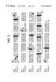
- FIG. 2 shows the configuration of conserved Cys residues in the serine protease domains of human MSP (SEQ ID NO:11), plasminogen (SEQ ID NO:12), and trypsin (SEQ ID NO:13). Solid lines indicate the pattern of intrachain disulfide bonds.
- FIG. 3 shows a three-dimensional representation of the serine protease domain of human trypsin. Indicated with labels are the analogous positions of the unpaired and intersubunit cysteine residues of MSP in the three-dimensional structure of human trypsin.
- FIG. 4 shows 10% PAGE of purified murine (mMSP) or human (hMSP) with Coomassie Blue staining. Some samples were treated with kallikrein (+). Disulfide bonds in some samples were reduced (+ ⁇ -mercaptoethanol).
- FIG. 5 shows activity of purified murine and C677A MSP in crypt attachment assay.
- Purified mMSP mutant C677A protein was assayed in parallel with wild-type mMSP for biological activity in murine crypt attachment assay.
- Samples were treated with Kallikrein at 15 ⁇ g/ml for 30 minutes at 37° C., then added to mouse colonic crypts at the final concentrations as shown.
- Treated crypts were plated in the wells of collagen-coated tissue culture plates, and were allowed to incubate overnight, followed by staining and counting of attached crypts. Values are plotted as fold stimulation over untreated wells.
- Purified bovine MSP was used as a positive control in this assay Kallikrein-cleaved mutant MSP gives approximately 10-fold higher specific activity than cleaved wild type MSP in this assay.
- FIG. 6 shows activity of purified human MSP and C672A MSP mutant in crypt attachment assay.
- FIG. 7 shows 3 H thymidine uptake by NIH 3T3 cells expressing RON at the cell surface by purified human MSP and C672A MSP mutant.
- MSP macrophage stimulating protein
- MSP refers to a protein having kringle domains characteristic of those found in a family that includes plasminogen, prothrombin, and HGF. Macrophage stimulating protein refers to the prepro, pro or mature forms and may be produced recombinantly or by chemical synthesis. MSP may be a single chain precursor or a heterodimer. References to positions in the MSP amino acid sequence are according to the murine and human sequences provided in GenBank accession nos. M74180 and L11924, respectively (also SEQ ID NO:1 and SEQ ID NO:2 for murine and human MSP, respectively).
- an MSP heterodimer refers to a polypeptide having one or more changes in the amino acid sequence of MSP which enhances heterodimer formation.
- An MSP heterodimer comprises an ⁇ chain of kringle domains linked to a ⁇ chain having a serine protease domain.
- recombinant murine MSP in transfected CHO cells was carried out as described in Example 1. After activation in vitro with kallikrein, the resultant material had low specific activity, compared to a sample of active MSP which was purified from bovine serum-containing conditioned media as described in co-owned and co-pending U.S. Ser. No. 08/622,720. Active MSP isolated from human plasma was reported to be a disulfide-linked heterodimer after in vitro kallikrein activation. (Wang et al. supra.) However, recombinant human or mouse MSP was ⁇ 5% disulfide linked, as judged by SDS-PAGE performed under nonreducing conditions, suggesting that the low activity of recombinant MSP was due to reduced dimer formation.
- MSP analogs were constructed that exhibited highly efficient dimer formation. These analogs were constructed with the aid of a model for MSP structure based upon the homology of MSP with plasminogen and other related family members.
- a comparison of the serine protease ( ⁇ ) domain and disulfide structures of MSP and plasminogen revealed the presence of unpaired cysteine residues in the serine protease domain of MSP, but not in plasminogen.
- the additional unpaired cysteine residues were also not conserved in HGF. It was postulated that one of the additional unpaired cysteine residues may be interfering with proper intersubunit disulfide bond formation.
- the position of the MSP cysteines was overlaid onto the three-dimensional structure of human trypsin (the structure of diisopropylfluorophosphate-inhibited human trypsin is available from the Brookhaven Protein Database and is reproduced in FIG. 3 ). Trypsin has only a serine protease domain and lacks kringle regions. As shown in FIG. 3, the use of human trypsin as a framework for visualizing the spatial arrangment of MSP cysteines in the serine protease domain reveals close proximity of Cys 672 with Cys 588, the latter known to be involved in intersubunit disulfide bonding.
- the invention provides for the first time biologically active MSP analogs.
- the MSP analogs form heterodimers with greater efficiency than human MSP.
- the MSP analogs have at least one cysteine residue substituted with another amino acid such that interchain disulfide bonds will form efficiently and promote MSP heterodimers. Any unpaired cysteine residue in MSP which interferes with interchain disulfide bonding may be replaced with another amino acid, however it is preferred that a cysteine residue located at positions 677 of murine MSP (SEQ ID NO:1) and position 672 of human MSP (SEQ ID NO:2) be altered.
- MSP analogs are constructed and expressed using standard recombinant DNA techniques as described in Examples 4 and 5 of the specification.
- Unpaired cysteine residues may be replaced by any other amino acid provided the substitution does not perturb the secondary or teritary structure of MSP. It is preferred that substitutions are conservative ones, such as cysteine to serine or alanine.
- Expression vectors containing nucleic acid sequences encoding MSP analogs, host cells transformed with said vectors and methods for the production of MSP analogs are also provided by the invention.
- An overview of expression of recombinant proteins is found in Methods of Enzymology v. 185 (Goeddel, D. V. ed.) Academic Press (1990).
- Host cells for the production of MSP analogs include procaryotic host cells, such as bacterial, yeast, plant, insect and mammalian host cells. Bacterial strains such as E. coli HB101 or JM101 are suitable for expression. Preferred mammalian host cells include COS, CHOd-, 293, CV-1, 3T3, baby hamster kidney (BHK) cells and others. Mammalian host cells are preferred when post-translational modifications, such as glycosylation and polypeptide processing, are important for MSP activity. Mammalian expression allows for the production of secreted polypeptides which may be recovered from the growth medium.
- Vectors for the expression of MSP analogs contain at a minimum sequences required for vector propagation and for expression of the cloned insert. These sequences include a replication origin, selection marker, promoter, ribosome binding site, enhancer sequences, RNA splice sites and transcription termination site. Vectors suitable for expression in the mammalian, bacterial, plant, yeast, insect host cells are readily available and the nucleic acids of the invention are inserted into the vectors using standard recombinant DNA techniques. Vectors for tissue-specific expression of an MSP analog are also included. Such vectors include promoters which function specifically in liver, kidney or other organs for production in mice, and viral vectors for the expression of an MSP analog in targeted human cells.
- MSP analogs are produced recombinantly by culturing a host cell transformed with an expression vector containing nucleic acid sequences encoding MSP under conditions such that MSP is produced, and isolating the product of expression.
- MSP is produced in the supernatant of transfected mammalian cells or in inclusion bodies of transformed bacterial host cells. MSP so produced may be purified by procedures known to one skilled in the art as described below.
- the expression of MSP analogs is described in Example 4 and 5 below. It is anticipated that the specific plasmids and host cells described are for illustrative purpose and that other available plasmids and host cells could also be used to express the polypeptides.
- the invention also provides for purified and isolated MSP analogs.
- the polypeptides of the invention are purified from other polypeptides present in transformed host cells expressing an MSP analog, or are purified from components in cell cultures containing the secreted protein. In one embodiment, the polypeptide is free from association with other human proteins, such as the expression product of a bacterial host cell.
- the purified protein may be a pro form of an MSP analog, a heterodimer, or isolated ⁇ and ⁇ chains.
- Modifications of MSP analog polypeptides are encompassed by the invention and include post-translational modifications (e.g., N-linked or O-linked carbohydrate chains, processing of N-terminal or C-terminal ends), attachment of chemical moieties to the amino acid backbone, chemical modifications of N-linked or O-linked carbohydrate chains, and addition of an N-terminal methionine residue as a result of procaryotic host cell expression.
- the polypeptides may also be modified with a detectable label, such as an enzymatic, fluorescent, isotopic or affinity label to allow for detection and isolation of the protein.
- the invention provides for MSP analog chimeric proteins wherein an analog is fused to a heterologous amino acid sequence.
- the heterologous sequence may be any sequence which allows the resulting fusion protein to retain the activity of MSP.
- the heterologous sequences include for example, immunoglobulin fusions, such as Fc fusions, which may aid in purification of the protein.
- a heterologous sequence which promotes formation of MSP heterodimers is preferred.
- a method for the purification of MSP analogs is also included.
- the purification process may employ one or more standard protein purification steps in an appropriate order to obtain purified protein.
- the chromatography steps can include ion exchange, gel filtration, hydrophobic interaction, reverse phase, chromatofocusing, and affinity chromatography.
- MSP analogs are used advantageously for the treatment of any condition requiring MSP. Examples of such conditions include gastrointestinal disorders and hematopoietic disorders. As the analogs of the present invention have a higher activity compared to human MSP, the analogs may be therapeutically effective with a smaller dosage and/or less frequent administration than human MSP.
- the invention provides for the treatment of disorders of the lining of the gastrointestinal tract by administration of a therapeutically effective amount of an MSP analog.
- the treatment provided herein is particularly useful for disorders involving the intestinal epithelium.
- the factors of the present invention can modulate the proliferation or differentiation of intestinal epithelium, thereby protecting healthy epithelium from damage and inducing repair and/or regeneration of damaged or depleted epithelium.
- Administration of an MSP analog may occur prior to, concurrent with, or after the onset of a disorder of the gastrointestinal tract lining for a time and a concentration sufficient to protect, repair and/or regenerate the gut lining.
- a “therapeutically effective amount” refers to that amount of MSP which provides a therapeutic effect for a given condition and administrative regimen. Said amount may vary from 0.1 ⁇ g/kg body weight to 1000 mg/kg body weight and may be more precisely determined by one skilled in the art.
- MSP may help maintain or repair epithelial cell linings in the intestinal tract and thereby prevent or reduce the occurrences of reduction or cessation of therapy.
- Certain disease states may also lead to damage or depletion of intestinal epithelium and may be treated by administration of MSP.
- MSP inflammatory bowel disease
- a class of diseases including ulcerative colitis and Crohn's disease, duodenal ulcers or infections.
- Administration of MSP will help restore normal intestinal mucosa where damage has occurred.
- MSP may be used alone or in conjunction with other factors for the treatment of intestinal epithelial disorders.
- MSP is used in conjunction with a therapeutically effective amount of a factor which promotes epithelial cell growth.
- factors include insulin growth factor-1 (IGF-1), insulin growth factor-2 (IGF-2), epidermal growth factor (EGF), transforming growth factor-a (TGF-a), acidic and basic fibroblast growth factor (FGF), platelet derived growth factor (PDGF), keratinocyte growth factor (KGF), interleukin-6 (IL-6) or interleukin-11 (IL-11).
- the invention provides for the treatment of hematopoietic disorders involving a deficiency in megakaryocytes or thrombocytes by administering a therapeutically effective amount of an MSP analog.
- an MSP analog may be used to treat thrombocytopenia resulting from exposure to radiation or chemotherapy.
- An MSP analog may be used alone or in conjunction with other hematopoietic factors which stimulate megakaryocyte or thrombocyte levels.
- Hematopoietic factors to be used in conjunction with MSP include erythropoietin (EPO), granulocyte colony stimulating factor (G-CSF), megakaryocyte growth and differentiation factor (MGDF), granulocyte macrophage colony stimulating factor (GM-CSF), stem cell factor (SCF), interleukin-3 (IL-3) or interleukin-6 (IL-6).
- EPO erythropoietin
- G-CSF granulocyte colony stimulating factor
- MGDF megakaryocyte growth and differentiation factor
- GM-CSF granulocyte macrophage colony stimulating factor
- SCF stem cell factor
- IL-3 interleukin-3
- IL-6 interleukin-6
- MSP may be administered by a variety of routes including parenteral, oral, nasal or rectal administration.
- Parenteral administration may occur by intravenous, subcutaneous, intradermal, intramuscular, intraarcticular and intrathecal injection.
- Oral administration involving adsorption through the gastrointestinal tract uses compressed tablets, capsules, pills, troches, cahcets and pellets.
- Adminstration by the nasal or oral respiratory route may employ powdered or liquid polypeptide delivered as an aerosol.
- Nasal delivery includes administration by drops or sprays.
- Rectal administration may employ suppositories.
- the route of administration to be chosen will depend upon several variables, including the pharmacokinetic properties of MSP and the nature and severity of the condition being treated.
- the invention provides for a pharmaceutical composition
- a pharmaceutical composition comprising a therapeutically effective amount of an MSP analog and a pharmaceutically acceptable diluent, carrier, preservative, emulsifier, and/or solubilizer.
- Diluents include Tris, acetate or phosphate buffers; solubilizers include Tween, Polysorbate; carriers include human serum albumin; preservatives include thimerosol and benzyl alcohol; and anti-oxidants include ascorbic acid.
- MSP analogs may also be conjugated with water soluble polymers (e.g, polyethylene glycol) using materials and method available to one skilled in the art in order to improve solubility, serum half-life, stability and bioavailability.
- MSP analogs may be present in formulations for use in particular delivery systems.
- MSP analogs may be formulated for controlled delivery over a period of time.
- Such formulations include but are not limited to the following: encapsulation in a water insoluble polymer of hardened gelatin, methyl and ethyl celluloses, polyhydroxymethacrylate, hydroxypropylcellulose, polyvinylacetate and various waxes used alone or in combination; dispersion in an inert polymeric matrix of insoluble plastic, hydrophilic polymers, or fatty compounds; and coating with a water soluble polymer such as a shellac, wax, starch, cellulose acetate phthalate or polyvinylpyrrolidone.
- MSP analogs may also be formulated for a targeted delivery system by entrapment within phospholipid vesicles.
- MSP analogs may be incoporated in a cocoa butter or polyethylene glycol base for inclusion in a suppository for rectal delivery.
- MSP analogs may be incorporated into a colon-specific drug release formulation such as that described in PCT Application No. W095/28963.
- the sliced colon pieces were washed several times with buffer A in a 50 ml conical tube.
- the clean tissue was washed with the extraction buffer (0.5 mM DTT, 2 mM EDTA in buffer A), and then incubated with 10 ml of fresh extraction buffer for 1 hour.
- the extraction buffer was then removed, and tissue was washed with Solution A.
- the crypts were harvested by shaking the tissue in 5 ml of Solution A.
- the colonies were stained with McManus' Periodic Acid-Schiff method and and Trichosantes kirilowii as described (Carson, Histotechnology: A Self - Instructional Text American Society of Clinical Pathologist Press pp. 158-160 (1990); (Falk et al. Am. J. Physiol. 266, G987-1003 (1994)). The colonies were compared to mouse colon paraffin sections stained with the same methods. The results of crypt cell staining revealed that both methods are specific for epithelial cells in the colon sections and stained positive for the colonies.
- Bovine MSP was isolated from bovine fetal serum as described in U.S. Ser. No. 08/622,720.
- Recombinant murine MSP was prepared as follows: A 2266 bp fragment was amplified from cDNA made from mouse liver poly(A)+ RNA by using the following oligonucleotide primers:
- the primers were based on the published mouse MSP sequence (Degen et al. Biochemistry 30, 9781-9791 (1991)). Due to the presence of several mutations in the coding region of the PCR product, the cloned fragment was used as a probe to screen a mouse liver cDNA library (Clonetech). A positive clone with 2.2 kb insert was isolated and sequenced. The DNA sequence that was obtained indicated that this clone contains the coding region of mouse MSP except for the first two amino acids. To obtain the full-length cDNA, an adaptor including the optimal context for initiation of translation and the missing nucleotides was synthesized based upon the published sequence, and ligated to the 2.2 kb insert.
- the cDNA was subcloned into pcDNA3 vector (Invitrogen).
- the mouse MSP plasmid DNA was tranfected into COS-7 cells with lipofectamine transfection system (GIBCO BRL). Serum-free condition media were harvested two days after transfection.
- Murine MSP was purified as described in Example 6 below.
- the domain and disulfide structures of plasminogen and plasminogen-related growth factors are shown in FIG. 1 .
- the domain structure of plasminogen may be summarized to contain a secretion signal peptide, an N-terminal “hairpin” domain, 5 kringle domains, and a serine proteinase domain (SPD).
- SPD serine proteinase domain
- the domain structures of MSP and HGF are very similiar to that of plasminogen. The main difference is that MSP and HGF have only four kringles due to the deletion of kringle 5 of plasminogen.
- the disulfide structure of plasminogen contains intra and inter- domain disulfide bonds:
- the intradomain disulfide bonds may be listed as follows: the hairpin domain contains two disulfides, each kringle contains 3 disulfides and the SPD contains 4 disulfides. There is an interdomain disulfide between the second and third kringle, and two disulfides between the last kringle of the ⁇ subunit and the SPD. As shown in FIG. 2 of Thery et al. (Dev. Genetics 17, 90-101 (1995) and in FIG.
- HGF and MSP have only a single disulfide between the kringle-containing ( ⁇ ) subunit and the serine proteinase domain ( ⁇ ).
- HGF from chicken, mouse and human contain no extra Cys or disulfides other than those which are homologous to plasminogen.
- MSP contains extra Cys, some of which are conserved in chicken, mouse, and human.
- murine MSP has a Cys residue in the signal sequence
- MSP from mouse and human contain an extra Cys in the hairpin domain.
- MSP from all three species contain 3 extra conserved Cys in the SPD compared to both plasminogen and HGF.
- Trypsin has ten Cys residues that form five disulfide bonds. Four of the trypsin disulfide bonds have homologs in MSP (and plasminogen).
- the residue numbering system for human trypsin in the structure file from Brookhaven (1trn) is based upon the conventional chymotrypsinogen relative amino acid numbering (Gaboriaud et al. J. Mol. Biol. 259, 995-1010 (1996)) and differs from the residue numbering system in the human trypsin sequence file from Swiss-Prot (try 1_human.swiss). Therefore, the residue numbers from the various database files are correlated in Table 2 for clarification.
- murine MSP plasmid template was amplified with a mutant primer (which incorporated a change from TG to GC at nucleotides 2029 and 2030, resulting in a Cys to Ala mutation) and a downstream primer complementary to vector pCDNA3.
- Primer sequences are:
- Cycling conditions were: 96° C., 30 sec.; 62° C., 30 sec; 72° C., 1 min for 5 cycles, followed by an additional 15 cycles at 96° C., 30 sec; 67° C., 30 sec; 72° C., 1 min.
- a primary 334 base pair product was purified from an agarose gel.
- the primary PCR product (containing the Cys to Ala mutation) was combined with a small amount of the original MSP plasmid and cycled in the absence of oligonucleotide primers for 5 cycles. This allowed the upstream and downstream extension of the mutated PCR product.
- Primers corresponding to an upstream region (nucleotides 1735-1759) of mMSP and the downstream vector primer (above) were added, and 20 more cycles were performed at 96° C., 30 sec; 67° C., 30 sec; 72° C., 1 min.
- Upstream primer sequence is
- Mutated fragment was subcloned into pCDNA3/mMSP for transient expression in 293/E11 cells and into pDSR ⁇ 2/mMSP for stable expression in CHO D- cells. Both constructs were sequenced to verify the presence of Cys to Ala mutation. Nucleotide positions refer to those of murine MSP RNA sequence, Genbank accession number M74181.
- 293/E1 cells were seeded at a density of 1 ⁇ 10 6 cells per 10 cm dish in complete medium (DMEM, high glucose, supplemented with 10% FBS and 0.3 mg/ml L-glutamine) and allowed to incubate overnight at 37°, 5% CO 2 . Shortly before transfecting, complete medium was removed and replaced with DMEM+5% FBS+0.3 mg/ml L-glutamine, 4 ml per dish.
- DMEM high glucose, supplemented with 10% FBS and 0.3 mg/ml L-glutamine
- Plasmid DNAs pCDNA3/mMSP C677A and pCDNA3/mMSP wild type were diluted to 10 ug per 500 ul serum-free DMEM and filter sterilized. A mock sample containing no DNA was also prepared in parallel.
- Lipofectamine reagent (Life Sciences, Inc., Gaithersburg, Md.) was diluted to 0.2 mg/ml in serum-free DMEM and combined with filter-sterilized plasmid DNA; final concentration for each sample was 10 ug/ml DNA and 0.1 mg/ml lipofectamine in 1 ml each.
- DNA/lipofectamine mixtures were incubated at room temperature, 30 minutes, then added to cell monolayers. Treated cells were returned to 37° C. for approximately 6 hours, then medium was removed and replaced with fresh DMEM+5% FBS+0.3 mg/ml L-glutamine and cells were allowed to recover overnight.
- Transfected cells were then washed once with serum-free DMEM and conditioned for 48 hours in serum-free DMEM+0.3 mg/ml L-glutamine at 9 ml per dish.
- Conditioned media were harvested, filtered to remove cell debris, and concentrated to 5 ⁇ in Centriprep-10 concentration units (Amicon, Inc., Beverly, Mass.). Expression of MSP was verified for C677A and wild type samples by Western blot; no expression was seen in mock sample. All three conditioned media samples were assayed for biological activity in murine crypt attachment assay.
- CHO D- cells were seeded at 8 ⁇ 10 5 cells per 60 mm dish in complete medium (DMEM, high glucose, with 10% FBS, 1 ⁇ PSG, 1 ⁇ NEAA and 1 ⁇ HT supplement) in 5 ml per dish and allowed to attach overnight at 37° C. in 5% CO 2 .
- DMEM high glucose, with 10% FBS, 1 ⁇ PSG, 1 ⁇ NEAA and 1 ⁇ HT supplement
- Medium was replaced with 5 ml fresh complete medium approximately 3 hours prior to transfection.
- Plasmid DNA pDSRa2/mMSP C677A was diluted to 60 ng/ul in 0.25M CaCl 2 and filter sterilized. A mock sample containing no DNA was also prepared in parallel. Following sterilization, 250 ul of each sample was combined with 250 ul of 2 ⁇ HEPES-buffered saline and incubated at room temperature for 30 minutes to allow CaPO 4 precipitates to form. Medium was aspirated and CaPO 4 /DNA samples were added to cells; following a 30 minute incubation at room temperature, cells were fed with 5 ml complete medium per plate and allowed to recover overnight at 37° C. Cells were re-fed with fresh complete medium the next day.
- Conditioned medium was generated from individual colonies plated into 24-well dishes; serum-free DMEM containing 1 ⁇ NEAA and 1 ⁇ PSG was incubated on 80% confluent monolayers at 400 ul per well for 72 hours, then harvested and filtered to remove cellular debris. Following concentration in Microcon-10 concentration units, the equivalent of 30 ul of 1 ⁇ conditioned medium per well was run on an 8% SDS-PAGE reducing gel. Proteins were electrophoretically transferred to nitrocellulose membrane and blotted with a rabbit polyclonal antibody raised against MSP. Blot was then exposed to horseradish peroxidase-conjugated anti-rabbit secondary antibody and visualized using Enhanced Chemiluminescence (ECL) system (Amersham, Inc.). The highest expression of MSP was seen for clone 2, which was selected for further expansion.
- ECL Enhanced Chemiluminescence
- CHO/C677A clone 2 cells were seeded into 100 roller bottles in 50% DMEM/50% Ham's F-12 medium supplemented with 5% FBS, 1 ⁇ NEAA and 1 ⁇ PSG. When monolayers reached 80% confluency, cells were washed with PBS to remove residual serum and conditioned in serum-free 50% DMEM/50% F-12 with 1 ⁇ NEAA and 1 ⁇ PSG for 4 days. A total of 20 liters of conditioned medium was harvested for purification.
- Cycling conditions were: 96° C., 30 sec.; 62° C., 30 sec.; 72° C., 1′ for 5 cycles, followed by an additional 15 cycles at 96° C., 30 sec; 67° C., 30 sec; 72° C., 1 min.
- the primary 323 base pair product was isolated from an agarose gel and purified to remove agarose.
- the primary PCR product containing the Cys to Ala mutation was combined with a small amount of the original MSP plasmid template and cycled in the absence of oligonucleotide primers for 5 cycles. This allowed the upstream and downstream extension of the mutated PCR product. Primers corresponding to an upstream region (nucleotides 1576-1598) of huMSP and the downstream vector primer (above) were added, and 20 more cycles were performed at 96° C., 30 sec; 67° C., 30 sec; 72° C., 1 min. Upstream primer sequence is
- the mutated fragment was subcloned into pCDNA3/huMSP for transient expression in 293/E1 cells and into pDSR ⁇ 2/huMSP for stable expression in CHO D- cells. Both constructs were sequenced to verify the presence of Cys to Ala mutation. Nucleotide positions refer to those of published human MSP cDNA sequence having GenBank accession number L11924 (Yoshimura et al, J. Biol. Chem 268 15461-15468 (1993)).
- 293/E1 cells were seeded at a density of 1 ⁇ 10 6 cells per 10 cm dish in complete medium (DMEM, high glucose, supplemented with 10% FBS and 0.3 mg/ml L-glutamine) and allowed to incubate overnight at 37°, 5% CO 2 . Shortly before transfecting, complete medium was removed and replaced with DMEM+5% FBS +0.3 mg/ml L-glutamine, 4 ml per dish.
- DMEM high glucose, supplemented with 10% FBS and 0.3 mg/ml L-glutamine
- Plasmid DNAs pCDNA3/huMSP C672A and pCDNA3/huMSP wild type were diluted to 10 ug per 500 ul serum-free DMEM and filter sterilized. A mock sample containing no DNA was also prepared in parallel.
- Lipofectamine reagent (Life Sciences, Inc., Gaithersburg, Md.) was diluted to 0.2 mg/ml in serum-free DMEM and combined with filter-sterilized plasmid DNA; final concentration for each sample was 10 ug/ml DNA and 0.1 mg/ml lipofectamine in 1 ml each.
- DNA/lipofectamine mixtures were incubated at room temperature, 30 minutes, then added to cell monolayers. Treated cells were returned to 37° C. for approximately 6 hours, then medium was removed and replaced with fresh DMEM+5% FBS+0.3 mg/ml L-glutamine and cells were allowed to recover overnight.
- Transfected cells were then washed once with serum-free DMEM and conditioned for 48 hours in serum-free DMEM+0.3 mg/ml L-glutamine at 9 ml per dish.
- Conditioned media were harvested, filtered to remove cell debris, and concentrated to 5 ⁇ in Centriprep-10 concentration units (Amicon, Inc., Beverly, Mass.). Expression of MSP was verified for C672A and wild type samples by Western blot; no expression was seen in mock sample. All three conditioned media samples were assayed for biological activity in murine crypt attachment assay.
- CHO D- cells were seeded at 8 ⁇ 10 5 cells per 60 mm dish in complete medium (DMEM, high glucose, with 10% FBS, 1 ⁇ PSG, 1 ⁇ NEAA and 1 ⁇ HT supplement) in 5 ml per dish and allowed to attach overnight at 37° C. in 5% CO 2 .
- DMEM high glucose, with 10% FBS, 1 ⁇ PSG, 1 ⁇ NEAA and 1 ⁇ HT supplement
- Medium was replaced with 5 ml fresh complete medium approximately 3 hours prior to transfection.
- Plasmid DNA pDSR ⁇ 2/huMSP C672A was diluted to 60 ng/ul in 0.25M CaCl 2 and filter sterilized. A mock sample containing no DNA was also prepared in parallel. Following sterilization, 250 ul of each sample was combined with 250 ul of 2 ⁇ HEPES-buffered saline and incubated at room temperature for 30 minutes to allow CaPO 4 precipitates to form. Medium was aspirated and CaPO 4 /DNA samples were added to cells; following a 30 minute incubation at room temperature, cells were fed with 5 ml complete medium per plate and allowed to recover overnight at 37° C. Cells were re-fed with fresh complete medium the next day.
- Conditioned medium was generated from individual colonies plated into 24-well dishes; serum-free DMEM containing 1 ⁇ NEAA and 1 ⁇ PSG was incubated on 80% confluent monolayers at 400 ul per well for 48 hours, then harvested and filtered to remove cellular debris. Following concentration in Microcon-10 concentration units, the equivalent of 30 ul of 1 ⁇ conditioned medium per well was run on an 8% SDS-PAGE reducing gel. Proteins were electrophoretically transferred to nitrocellulose membrane and blotted with a mouse monoclonal antibody raised against MSP. Blot was then exposed to horseradish peroxidase-conjugated anti-mouse secondary antibody and visualized using Enhanced Chemiluminescence (ECL) system (Amersham, Inc.). The highest expression of MSP was seen for clone 3, which was selected for further expansion.
- ECL Enhanced Chemiluminescence
- Conditioned media with or without concentration by diafiltration, and without salt or pH adjustment, was chromatographed by absorption onto heparin-Sepharose (Pharmacia), and elution with a salt gradient in 20 mM sodium phosphate, pH 7. MSP eluted at 0.4M NaCl. Pooled fractions were dialyzed with 0.02M Tris, pH 8.5 and chromatographed by absorption on Q Sepharose HP (Pharmacia), and elution with a salt gradient in 0.02 M Tris pH 8.5. MSP eluted at 0.1M salt.
- MSP was activated by either by incubation at 37° C. for one hour with 10 ⁇ g/ml human kallikrein (Enzyme System Products), followed by addition of 1 mM pefabloc (Boeringher Mannheim) or by passing through a column of kallikrein-Sepharose (1 mg/ml human kallikrein/ml cyanogen bromide activated Sepharose, 4 ml total). Active samples were dialyzed versus phosphate buffered saline
- MSP was analyzed with 10% PAGE gels from Novex. Non reduced samples were mixed with sample buffer containing SDS but were not heated. Reduced samples were heated at 90° C. for three minutes in sample buffer which contained SDS and 5% ⁇ -mercaptoethanol. The results are shown in FIG. 4 . Purified recombinant murine and human MSP and murine C677A and human C672 analogs appear as a proform consisting of a single band of about 80 kDa when analyzed by SDS-PAGE under reducing (lanes 1 or 3) or nonreducing conditions (lanes 5 or 7), with Coomassie blue staining.
- Kallikrein treatment efficiently cleaves proMSP or proMSP analog between the ⁇ and ⁇ subunits, as shown by SDS-PAGE under reducing conditions (lanes 2 and 6). However, the subunits of kallikrein-treated, recombinant, nonmutant MSP do not remain linked during SDS-PAGE under nonreducing conditions (lane 4). The kallikrein-treated analog of MSP described does remain linked during SDS-PAGE under nonreducing conditions, apparently due to the sparing of the intersubunit disulfide bond (lane 8).
- Recombinant murine MSP and C677A analog expressed and purified as described in Example 4 and recombinant human MSP and C672A analog expressed and purified as described in Example 5 were assayed for colony forming activity as described in Example 1.
- the results for murine MSP are shown in FIG. 5 and the results for human MSP are shown in FIG. 6 .
- Recombinant human MSP and the C672A analog were assayed for stimulation of 3 H thymidine uptake by cells expressing the stk/RON receptor.
- the full length cDNA for murine stk/RON (GenBank accession number x74736) was cloned using standard techniques, subcloned into the mammalian expression vector, pEV7, and tritiated thymidine uptake was measured in NIH 3T3 cells expressing stk/RON as described (Zhang et al. J. Biol. Chem. 271, 3884-3890 (1996)). The results are shown in FIG. 7 .
Abstract
MSP analogs which have increased heterodimer formation and enhanced biological activity compared recombinant MSP are provided. The analogs are constructed by substituting unpaired cysteine residues which may interfere with interchain disulfide bonding. DNA sequences encoding MSP analogs, vectors and host cells for the expresssion of MSP analogs, and pharmaceutical compositons are also provided. The analogs may be used to treat conditions treatable by MSP such as gastrointestinal or hematopoietic disorders.
Description
This application is a continuation of Ser. No. 08/766,982, now U.S. Pat. No. 5,948,892, filed Dec. 16, 1996.
The invention generally relates to analogs of macrophage stimulating protein, or MSP. More particularly, the invention relates to analogs of MSP which promote multimer formation and have enhanced biological activity.
Macrophage Stimulating Protein, or MSP, has been previously identified as an activity present in mammalian blood plasma which makes mouse peritoneal macrophages responsive to chemoattractants such as complement C5a (Leonard et al. Exp. Cell. Res. 102, 434 (1976); Leonard et al. Exp. Cell Res. 114, 117 (1978). MSP was purified from human serum as described in U.S. Pat. No. 5,219,991 and the DNA sequence encoding human MSP was reported in U.S. Pat. No. 5,315,000. MSP is synthesized in a prepro form which is secreted as a single chain polypeptide. The pro form is proteolytically cleaved to form a disulfide-linked heterodimer having an α and β chain of molecular weights 53 kDa and 25 kDa, respectively. The heterodimer is the biologically active form of MSP. It has not been established which protease is responsible for the in vivo activation of MSP, but several proteases, such as human plasma kallikrein are reported to efficiently activate MSP in vitro (Wang et al. J. Biol. Chem. 269, 3436-3440 (1994)).
MSP is a member of a family of proteins having triple disulfide loop structures, or kringle domains (Donate et al. Protein Science 3, 2378-2394 (1994)). Family members include plasminogen and hepatocyte growth factor (HGF). MSP also exhibits sequence homology to both plasminogen and HGF and its proteolytic activation occurs at Arg-Val residues which are also conserved in other family members.
A variety of in vitro biological activities have been reported for MSP. MSP was initially purified based upon stimulation of a chemotactic response of mouse resident peritoneal macrophages (Leonard et al., supra) and was believed to play a role in cell motility. MSP stimulated megakaryocyte maturation and thrombocyte production from isolated bone marrow preparations (PCT Application No. WO96/14082). The in vivo activity of MSP remains to be elucidated.
Recently, it has been reported that MSP is a ligand for RON, a cell membrane protein tyrosine kinase which is a member of the c-met family of protein tyrosine kinases (Wang et al. Science 266, 117-119 (1994); Gaudino et al. EMBO J. 13, 3524-3532 (1994); Ronsin et al. Oncogene 8, 1195-1202 (1993)). The expression of RON in human tissues and cell lines was examined (Gaudino et al., supra) and RON was found to be expressed in colon, skin, lung and bone marrow, and in granulocytes and adherent monocytes. Epithelial cell lines derived from gastric, pancreatic and mammary carcinoma, and hematopoietic cell lines also showed RON expression. MSP induced tyrosine phosphorylation of RON and stimulated DNA synthesis in a mammary carcinoma cell line. These observations suggest that MSP may act on a variety of cell types. MSP promotes colony formation by mouse colon crypts as shown in co-owned U.S. Pat. No. 5,814,308, suggesting that MSP may be useful in protecting and regenerating the intestinal epithelium.
In view of the useful biological activities exhibited by MSP. it is desirable to find forms of MSP which have enhanced biological activity. Such forms could provide a more favorable therapeutic regimen in that they can be administered at lower dosages and/or less frequently than MSP.
The invention provides for analogs of MSP which have increased heterodimer formation and enchanced biological activity compared to recombinant human MSP. The analogs of the present invention are constructed by substituting a cysteine residue with another amino acid such that interchain disulfide bonds will form efficiently and promote MSP heterodimer formation. The resulting MSP analog forms heterodimers of one α (kringle-containing) chain and one β (serine protease) chain to a greater extent than recombinant human MSP. DNA sequences encoding MSP analogs, expression vectors comprising the DNA sequences and modified host cells which express MSP analogs are also provided by the invention. Pharmaceutical compositions of MSP analogs may be used to treat conditions treatable by MSP including gastrointestinal and hematopoietic disorders.
FIG. 1 shows a diagram of the major structural domains of human MSP and human plasminogen, namely the kringle domains and the serine protease domain. Dotted lines show intra and interchain disulfide bridges. Cysteine residues at positions 527, 562 and 672 of MSP represent additional unpaired cysteine which are not conserved in human plasminogen. The asterisk indicates the activation cleavage site which separates the α (kringle) and β (serine protease) domains.
FIG. 2 shows the configuration of conserved Cys residues in the serine protease domains of human MSP (SEQ ID NO:11), plasminogen (SEQ ID NO:12), and trypsin (SEQ ID NO:13). Solid lines indicate the pattern of intrachain disulfide bonds.
FIG. 3 shows a three-dimensional representation of the serine protease domain of human trypsin. Indicated with labels are the analogous positions of the unpaired and intersubunit cysteine residues of MSP in the three-dimensional structure of human trypsin.
FIG. 4 shows 10% PAGE of purified murine (mMSP) or human (hMSP) with Coomassie Blue staining. Some samples were treated with kallikrein (+). Disulfide bonds in some samples were reduced (+β-mercaptoethanol).
FIG. 5 shows activity of purified murine and C677A MSP in crypt attachment assay. Purified mMSP mutant C677A protein was assayed in parallel with wild-type mMSP for biological activity in murine crypt attachment assay. Samples were treated with Kallikrein at 15 μg/ml for 30 minutes at 37° C., then added to mouse colonic crypts at the final concentrations as shown. Treated crypts were plated in the wells of collagen-coated tissue culture plates, and were allowed to incubate overnight, followed by staining and counting of attached crypts. Values are plotted as fold stimulation over untreated wells. Purified bovine MSP was used as a positive control in this assay Kallikrein-cleaved mutant MSP gives approximately 10-fold higher specific activity than cleaved wild type MSP in this assay.
FIG. 6 shows activity of purified human MSP and C672A MSP mutant in crypt attachment assay.
FIG. 7 shows 3H thymidine uptake by NIH 3T3 cells expressing RON at the cell surface by purified human MSP and C672A MSP mutant.
As used herein, the term “macrophage stimulating protein” or MSP, refers to a protein having kringle domains characteristic of those found in a family that includes plasminogen, prothrombin, and HGF. Macrophage stimulating protein refers to the prepro, pro or mature forms and may be produced recombinantly or by chemical synthesis. MSP may be a single chain precursor or a heterodimer. References to positions in the MSP amino acid sequence are according to the murine and human sequences provided in GenBank accession nos. M74180 and L11924, respectively (also SEQ ID NO:1 and SEQ ID NO:2 for murine and human MSP, respectively).
The term “analog of macrophage stimulating protein” refers to a polypeptide having one or more changes in the amino acid sequence of MSP which enhances heterodimer formation. An MSP heterodimer comprises an α chain of kringle domains linked to a β chain having a serine protease domain.
Expression of recombinant murine MSP in transfected CHO cells was carried out as described in Example 1. After activation in vitro with kallikrein, the resultant material had low specific activity, compared to a sample of active MSP which was purified from bovine serum-containing conditioned media as described in co-owned and co-pending U.S. Ser. No. 08/622,720. Active MSP isolated from human plasma was reported to be a disulfide-linked heterodimer after in vitro kallikrein activation. (Wang et al. supra.) However, recombinant human or mouse MSP was <5% disulfide linked, as judged by SDS-PAGE performed under nonreducing conditions, suggesting that the low activity of recombinant MSP was due to reduced dimer formation.
In order to generate highly active recombinant MSP, MSP analogs were constructed that exhibited highly efficient dimer formation. These analogs were constructed with the aid of a model for MSP structure based upon the homology of MSP with plasminogen and other related family members. A comparison of the serine protease (β) domain and disulfide structures of MSP and plasminogen revealed the presence of unpaired cysteine residues in the serine protease domain of MSP, but not in plasminogen. The additional unpaired cysteine residues were also not conserved in HGF. It was postulated that one of the additional unpaired cysteine residues may be interfering with proper intersubunit disulfide bond formation. To determine which residues might be involved, the position of the MSP cysteines was overlaid onto the three-dimensional structure of human trypsin (the structure of diisopropylfluorophosphate-inhibited human trypsin is available from the Brookhaven Protein Database and is reproduced in FIG. 3). Trypsin has only a serine protease domain and lacks kringle regions. As shown in FIG. 3, the use of human trypsin as a framework for visualizing the spatial arrangment of MSP cysteines in the serine protease domain reveals close proximity of Cys 672 with Cys 588, the latter known to be involved in intersubunit disulfide bonding.
Substitution of cysteine residues at postion 672 in human MSP and position 677 in murine MSP is described in Examples 4 and 5. The resulting purified analogs showed enhanced activity in a mouse colon crypt assay compared to recombinant human or murine MSP. In addition, the human analog shows greater stimulation of 3H thymidine uptake in RON-expressing cells than human MSP (Example 7).
Accordingly, the invention provides for the first time biologically active MSP analogs. The MSP analogs form heterodimers with greater efficiency than human MSP. In the present embodiment, the MSP analogs have at least one cysteine residue substituted with another amino acid such that interchain disulfide bonds will form efficiently and promote MSP heterodimers. Any unpaired cysteine residue in MSP which interferes with interchain disulfide bonding may be replaced with another amino acid, however it is preferred that a cysteine residue located at positions 677 of murine MSP (SEQ ID NO:1) and position 672 of human MSP (SEQ ID NO:2) be altered.
MSP analogs are constructed and expressed using standard recombinant DNA techniques as described in Examples 4 and 5 of the specification. Unpaired cysteine residues may be replaced by any other amino acid provided the substitution does not perturb the secondary or teritary structure of MSP. It is preferred that substitutions are conservative ones, such as cysteine to serine or alanine.
Expression vectors containing nucleic acid sequences encoding MSP analogs, host cells transformed with said vectors and methods for the production of MSP analogs are also provided by the invention. An overview of expression of recombinant proteins is found in Methods of Enzymology v. 185 (Goeddel, D. V. ed.) Academic Press (1990).
Host cells for the production of MSP analogs include procaryotic host cells, such as bacterial, yeast, plant, insect and mammalian host cells. Bacterial strains such as E. coli HB101 or JM101 are suitable for expression. Preferred mammalian host cells include COS, CHOd-, 293, CV-1, 3T3, baby hamster kidney (BHK) cells and others. Mammalian host cells are preferred when post-translational modifications, such as glycosylation and polypeptide processing, are important for MSP activity. Mammalian expression allows for the production of secreted polypeptides which may be recovered from the growth medium.
Vectors for the expression of MSP analogs contain at a minimum sequences required for vector propagation and for expression of the cloned insert. These sequences include a replication origin, selection marker, promoter, ribosome binding site, enhancer sequences, RNA splice sites and transcription termination site. Vectors suitable for expression in the mammalian, bacterial, plant, yeast, insect host cells are readily available and the nucleic acids of the invention are inserted into the vectors using standard recombinant DNA techniques. Vectors for tissue-specific expression of an MSP analog are also included. Such vectors include promoters which function specifically in liver, kidney or other organs for production in mice, and viral vectors for the expression of an MSP analog in targeted human cells.
Using an appropriate host-vector system, MSP analogs are produced recombinantly by culturing a host cell transformed with an expression vector containing nucleic acid sequences encoding MSP under conditions such that MSP is produced, and isolating the product of expression. MSP is produced in the supernatant of transfected mammalian cells or in inclusion bodies of transformed bacterial host cells. MSP so produced may be purified by procedures known to one skilled in the art as described below. The expression of MSP analogs is described in Example 4 and 5 below. It is anticipated that the specific plasmids and host cells described are for illustrative purpose and that other available plasmids and host cells could also be used to express the polypeptides.
The invention also provides for purified and isolated MSP analogs. The polypeptides of the invention are purified from other polypeptides present in transformed host cells expressing an MSP analog, or are purified from components in cell cultures containing the secreted protein. In one embodiment, the polypeptide is free from association with other human proteins, such as the expression product of a bacterial host cell. The purified protein may be a pro form of an MSP analog, a heterodimer, or isolated α and β chains.
Modifications of MSP analog polypeptides are encompassed by the invention and include post-translational modifications (e.g., N-linked or O-linked carbohydrate chains, processing of N-terminal or C-terminal ends), attachment of chemical moieties to the amino acid backbone, chemical modifications of N-linked or O-linked carbohydrate chains, and addition of an N-terminal methionine residue as a result of procaryotic host cell expression. The polypeptides may also be modified with a detectable label, such as an enzymatic, fluorescent, isotopic or affinity label to allow for detection and isolation of the protein.
Also provided by the invention are chemically modified derivatives of MSP analogs which may provide additional advantages such as increasing stability and circulating time of the polypeptide, or decreasing immunogenicity (see U.S. Pat. No. 4,179,337). The chemical moieties for derivitization may be selected from water soluble polymers such as polyethylene glycol, ethylene glycol/propylene glycol copolymers, carboxymethylcellulose, dextran, polyvinyl alcohol and the like. The polypeptides may be modified at random positions within the molecule, or at predetermined positions within the molecule, and may include one, two, three or more attached chemical moieties. In a preferred embodiment, the MSP analogs are selectively derivatized at the amino-terminus of the polypeptide.
The invention provides for MSP analog chimeric proteins wherein an analog is fused to a heterologous amino acid sequence. The heterologous sequence may be any sequence which allows the resulting fusion protein to retain the activity of MSP. The heterologous sequences include for example, immunoglobulin fusions, such as Fc fusions, which may aid in purification of the protein. A heterologous sequence which promotes formation of MSP heterodimers is preferred.
A method for the purification of MSP analogs is also included. The purification process may employ one or more standard protein purification steps in an appropriate order to obtain purified protein. The chromatography steps can include ion exchange, gel filtration, hydrophobic interaction, reverse phase, chromatofocusing, and affinity chromatography.
MSP analogs are used advantageously for the treatment of any condition requiring MSP. Examples of such conditions include gastrointestinal disorders and hematopoietic disorders. As the analogs of the present invention have a higher activity compared to human MSP, the analogs may be therapeutically effective with a smaller dosage and/or less frequent administration than human MSP.
The invention provides for the treatment of disorders of the lining of the gastrointestinal tract by administration of a therapeutically effective amount of an MSP analog. The treatment provided herein is particularly useful for disorders involving the intestinal epithelium. The factors of the present invention can modulate the proliferation or differentiation of intestinal epithelium, thereby protecting healthy epithelium from damage and inducing repair and/or regeneration of damaged or depleted epithelium. Administration of an MSP analog may occur prior to, concurrent with, or after the onset of a disorder of the gastrointestinal tract lining for a time and a concentration sufficient to protect, repair and/or regenerate the gut lining.
As used herein, a “therapeutically effective amount” refers to that amount of MSP which provides a therapeutic effect for a given condition and administrative regimen. Said amount may vary from 0.1 μg/kg body weight to 1000 mg/kg body weight and may be more precisely determined by one skilled in the art.
Efforts to aggressively treat cancer have led to the administration of higher doses of chemotherapeutic agents or the use of whole body radiation, but such regimens can lead first to bone marrow toxicity (depletion of red blood cells and white blood cells) followed by gut toxicity (depletion of intestinal epithelium). It is usual that a dose reduction or a cessation of therapy occurs until the toxicity is overcome. A preferred method of treatment is the use of MSP as an adjunct to chemotherapy or radiation therapy, either prior to or concurrent with such therapy. MSP may help maintain or repair epithelial cell linings in the intestinal tract and thereby prevent or reduce the occurrences of reduction or cessation of therapy.
Certain disease states may also lead to damage or depletion of intestinal epithelium and may be treated by administration of MSP. Examples include inflammatory bowel disease, a class of diseases including ulcerative colitis and Crohn's disease, duodenal ulcers or infections. Administration of MSP will help restore normal intestinal mucosa where damage has occurred.
It is understood that MSP may be used alone or in conjunction with other factors for the treatment of intestinal epithelial disorders. In one embodiment, MSP is used in conjunction with a therapeutically effective amount of a factor which promotes epithelial cell growth. Such factors include insulin growth factor-1 (IGF-1), insulin growth factor-2 (IGF-2), epidermal growth factor (EGF), transforming growth factor-a (TGF-a), acidic and basic fibroblast growth factor (FGF), platelet derived growth factor (PDGF), keratinocyte growth factor (KGF), interleukin-6 (IL-6) or interleukin-11 (IL-11).
The invention provides for the treatment of hematopoietic disorders involving a deficiency in megakaryocytes or thrombocytes by administering a therapeutically effective amount of an MSP analog. Such conditions can arise from disease or exposure to myelosuppressive agents. In one embodiment, an MSP analog may be used to treat thrombocytopenia resulting from exposure to radiation or chemotherapy. An MSP analog may be used alone or in conjunction with other hematopoietic factors which stimulate megakaryocyte or thrombocyte levels. Hematopoietic factors to be used in conjunction with MSP include erythropoietin (EPO), granulocyte colony stimulating factor (G-CSF), megakaryocyte growth and differentiation factor (MGDF), granulocyte macrophage colony stimulating factor (GM-CSF), stem cell factor (SCF), interleukin-3 (IL-3) or interleukin-6 (IL-6).
MSP may be administered by a variety of routes including parenteral, oral, nasal or rectal administration. Parenteral administration may occur by intravenous, subcutaneous, intradermal, intramuscular, intraarcticular and intrathecal injection. Oral administration involving adsorption through the gastrointestinal tract uses compressed tablets, capsules, pills, troches, cahcets and pellets. Adminstration by the nasal or oral respiratory route may employ powdered or liquid polypeptide delivered as an aerosol. Nasal delivery includes administration by drops or sprays. Rectal administration may employ suppositories. The route of administration to be chosen will depend upon several variables, including the pharmacokinetic properties of MSP and the nature and severity of the condition being treated.
The invention provides for a pharmaceutical composition comprising a therapeutically effective amount of an MSP analog and a pharmaceutically acceptable diluent, carrier, preservative, emulsifier, and/or solubilizer. Diluents include Tris, acetate or phosphate buffers; solubilizers include Tween, Polysorbate; carriers include human serum albumin; preservatives include thimerosol and benzyl alcohol; and anti-oxidants include ascorbic acid. MSP analogs may also be conjugated with water soluble polymers (e.g, polyethylene glycol) using materials and method available to one skilled in the art in order to improve solubility, serum half-life, stability and bioavailability.
MSP analogs may be present in formulations for use in particular delivery systems. As an example, MSP analogs may be formulated for controlled delivery over a period of time. Such formulations include but are not limited to the following: encapsulation in a water insoluble polymer of hardened gelatin, methyl and ethyl celluloses, polyhydroxymethacrylate, hydroxypropylcellulose, polyvinylacetate and various waxes used alone or in combination; dispersion in an inert polymeric matrix of insoluble plastic, hydrophilic polymers, or fatty compounds; and coating with a water soluble polymer such as a shellac, wax, starch, cellulose acetate phthalate or polyvinylpyrrolidone. MSP analogs may also be formulated for a targeted delivery system by entrapment within phospholipid vesicles. In a preferred embodiment, MSP analogs may be incoporated in a cocoa butter or polyethylene glycol base for inclusion in a suppository for rectal delivery. In another preferred embodiment, MSP analogs may be incorporated into a colon-specific drug release formulation such as that described in PCT Application No. W095/28963.
A more extensive survey of components commonly found in pharmaceutical compositions and formulations is presented in Remington's Pharmaceutical Sciences, 18th ed. A. R. Gennaro, ed. Mack, Easton, Pa. (1990), the relevant portions of which are incorporated by reference.
The following examples are offered to more fully illustrate the invention, but are not construed as limiting the scope thereof.
An assay for colony formation by isolated mouse colon crypts was previously described in co-pending and co-owned U.S. Ser. No. 08/622,720 hereby incorporated by reference. The assay is performed as follows. Mouse colon crypts were prepared as described in Whitehead et al. (In Vitro Cellular & Developmental Biology, 23, 436-442 (1987)). Mice were sacrificed with lethal dose of CO2, and large intestines were isolated. The large intestine was cut longitudinally, rinsed with PBS containing 0.3 mg/ml L-Glutamine, 100 units/ml penicillin, 100 units/ml streptomycin (Buffer A), and sliced to 0.5 cm pieces. The sliced colon pieces were washed several times with buffer A in a 50 ml conical tube. The clean tissue was washed with the extraction buffer (0.5 mM DTT, 2 mM EDTA in buffer A), and then incubated with 10 ml of fresh extraction buffer for 1 hour. The extraction buffer was then removed, and tissue was washed with Solution A. The crypts were harvested by shaking the tissue in 5 ml of Solution A.
Harvested crypts were plated on collagen type IV coated 6 well plates (Collaborative Biomedical Products, Bedford, Mass.) at a density of 500 crypts per well in 4 ml medium (RPMI 1640, 0.3 mg/ml L-Glutamine, 100 units/ml penicillin, 100 units/ml streptomycin, and 10% fetal bovine serum (FBS; GIBCO-BRL. Gaithersburg, Md.). After 24 hours incubation at 37° C., colonies of attached cells were stained with crystal violet, and counted under microscope. To confirm that the cells in the colonies are derived from crypt epithelium, the colonies were stained with McManus' Periodic Acid-Schiff method and and Trichosantes kirilowii as described (Carson, Histotechnology: A Self-Instructional Text American Society of Clinical Pathologist Press pp. 158-160 (1990); (Falk et al. Am. J. Physiol. 266, G987-1003 (1994)). The colonies were compared to mouse colon paraffin sections stained with the same methods. The results of crypt cell staining revealed that both methods are specific for epithelial cells in the colon sections and stained positive for the colonies.
The colony forming activity of natural bovine MSP and recombinant murine MSP was compared in the mouse colon crypt assay described in Example 1. Bovine MSP was isolated from bovine fetal serum as described in U.S. Ser. No. 08/622,720. Recombinant murine MSP was prepared as follows: A 2266 bp fragment was amplified from cDNA made from mouse liver poly(A)+ RNA by using the following oligonucleotide primers:
ATCCTGAAGGGACAGATTTC (SEQ ID NO:3) and
TTTGAGAAGTCTTGACATCTC (SEQ ID NO:4)
The primers were based on the published mouse MSP sequence (Degen et al. Biochemistry 30, 9781-9791 (1991)). Due to the presence of several mutations in the coding region of the PCR product, the cloned fragment was used as a probe to screen a mouse liver cDNA library (Clonetech). A positive clone with 2.2 kb insert was isolated and sequenced. The DNA sequence that was obtained indicated that this clone contains the coding region of mouse MSP except for the first two amino acids. To obtain the full-length cDNA, an adaptor including the optimal context for initiation of translation and the missing nucleotides was synthesized based upon the published sequence, and ligated to the 2.2 kb insert. The cDNA was subcloned into pcDNA3 vector (Invitrogen). The mouse MSP plasmid DNA was tranfected into COS-7 cells with lipofectamine transfection system (GIBCO BRL). Serum-free condition media were harvested two days after transfection. Murine MSP was purified as described in Example 6 below.
The colony forming activity of natural bovine MSP and recombinant murine MSP was assayed at 2 ng/ml and 10 ng/ml and the results shown in Table 1. Recombinant murine MSP stimulated colony formation by murine crypts about 3 to 7-fold lower than natural bovine MSP.
| TABLE 1 |
| Comparison of crypt colony forming activity |
| of native bovine MSP and recombinant murine MSP |
| Sample | concentration | # of colonies | ||
| no addition | not applicable | 16 | ||
| fetal |
10% | 118 | ||
| native |
2 ng/ml | 234 | ||
| native |
10 ng/ml | 336 | ||
| recombinant |
2 ng/ml | 28 | ||
| recombinant |
10 ng/ml | 118 | ||
The domain and disulfide structures of plasminogen and plasminogen-related growth factors are shown in FIG. 1. From the N-terminus, the domain structure of plasminogen may be summarized to contain a secretion signal peptide, an N-terminal “hairpin” domain, 5 kringle domains, and a serine proteinase domain (SPD). The domain structures of MSP and HGF are very similiar to that of plasminogen. The main difference is that MSP and HGF have only four kringles due to the deletion of kringle 5 of plasminogen. The disulfide structure of plasminogen contains intra and inter- domain disulfide bonds: The intradomain disulfide bonds may be listed as follows: the hairpin domain contains two disulfides, each kringle contains 3 disulfides and the SPD contains 4 disulfides. There is an interdomain disulfide between the second and third kringle, and two disulfides between the last kringle of the α subunit and the SPD. As shown in FIG. 2 of Thery et al. (Dev. Genetics 17, 90-101 (1995) and in FIG. 1 below, every disulfide that is present in plasminogen is also present in HGF and MSP, with one exception:HGF and MSP have only a single disulfide between the kringle-containing (α) subunit and the serine proteinase domain (β). HGF from chicken, mouse and human contain no extra Cys or disulfides other than those which are homologous to plasminogen. MSP, however contains extra Cys, some of which are conserved in chicken, mouse, and human. Thus, murine MSP has a Cys residue in the signal sequence, and MSP from mouse and human contain an extra Cys in the hairpin domain. MSP from all three species contain 3 extra conserved Cys in the SPD compared to both plasminogen and HGF. Since recombinant HGF of high specific activity is available commercially and recombinant MSP is not, we considered whether the extra conserved Cys residues of MSP were involved with the disufide bonding defect that we observed with our recombinant MSP preparation.
Although high resolution structural information is not available for plasminogen, MSP or HGF, this information is available for other serine proteinases, such as trypsin. Mature trypsin contains only a serine proteinase domain, which is formed by the removal of a 15 residue signal peptide and a 9 residue activation peptide. Since trypsin has very high sequence homology to the serine proteinase domain of MSP (or plasminogen), we used the structure of trypsin as a surrogate for the SPD of MSP. The Brookhaven data file 1trn, which is a structure of diisopropylfluorophosphate-inhibited human trypsin, was used for our modelling (See FIG. 3). Trypsin has ten Cys residues that form five disulfide bonds. Four of the trypsin disulfide bonds have homologs in MSP (and plasminogen). The residue numbering system for human trypsin in the structure file from Brookhaven (1trn) is based upon the conventional chymotrypsinogen relative amino acid numbering (Gaboriaud et al. J. Mol. Biol. 259, 995-1010 (1996)) and differs from the residue numbering system in the human trypsin sequence file from Swiss-Prot (try1_human.swiss). Therefore, the residue numbers from the various database files are correlated in Table 2 for clarification.
| TABLE 2 |
| Correlation of residue numbers of human MSP and trypsin |
| from various database files Cysteines of interest in |
| human MSP are compared to chosen homologous residues of |
| human trypsin. |
| Human MSPa | Cys527 | Cys562 | Cys588 | Cys672 | ||
| Human trypsinb | Lys66 | Arg95 | Ser127 | Gln209 | ||
| Human trypsinc | Lys60 | Arg90 | Ser122 | Gln204 | ||
| aResidue numbers as in Swiss-Prot database file HGF1_human.swiss | ||||||
| bResidue numbers as in Swiss-Prot database file tyr1_human.swiss | ||||||
| cResidue numbers as in Brookhaven database file 1trn | ||||||
Using the the numbering from the structural file, the following trypsin residues were chosen as homologs of the three “extra” Cys residues in MSP: Cys537=trypsin Lys60, Cys562=trypsin Arg90, Cys672=trypsin Gln209, and the intersubunit Cys588=trypsin Ser122. Lys60 and Arg90 are located on the surface of trypsin on the opposite side of the protein from the Ser122. Gln209 and Ser122 are located on the surface in very close proximity, the distance between the α carbons of these residues is 6.1 A. For comparison, the distances between the α carbons of the disulfide bonded Cys residues of trypsin range from 4.2 to 6.2 A. Thus, the suggested close proximity of Cys672 to Cys588 suggests that intra subunit disulfide formation between these two Cys might interfere with intersubunit disulfide formation by Cys 588. It should be noted that Cys672 apparently has no other Cys residue other than Cys588 with which to interact.
Construction of C677A Mutant of Murine MSP
To mutate the cysteine residue which is suspected of interfering with interchain disulfide bond formation, we employed a two-step PCR process. First, murine MSP plasmid template was amplified with a mutant primer (which incorporated a change from TG to GC at nucleotides 2029 and 2030, resulting in a Cys to Ala mutation) and a downstream primer complementary to vector pCDNA3. Primer sequences are:
Mutant: 5′ CCA TGA CGC CTG GGT CCT ACA G 3′ (SEQ ID NO: 5)
Downstream: 5′ CTG GCA ACT AGA AGG CAC AGT CG 3′ (SEQ ID NO: 9)
Cycling conditions were: 96° C., 30 sec.; 62° C., 30 sec; 72° C., 1 min for 5 cycles, followed by an additional 15 cycles at 96° C., 30 sec; 67° C., 30 sec; 72° C., 1 min. A primary 334 base pair product was purified from an agarose gel.
Next, the primary PCR product (containing the Cys to Ala mutation) was combined with a small amount of the original MSP plasmid and cycled in the absence of oligonucleotide primers for 5 cycles. This allowed the upstream and downstream extension of the mutated PCR product. Primers corresponding to an upstream region (nucleotides 1735-1759) of mMSP and the downstream vector primer (above) were added, and 20 more cycles were performed at 96° C., 30 sec; 67° C., 30 sec; 72° C., 1 min. Upstream primer sequence is
5′ CTG GAG AGA CCT GTG ATC CTG AAC C 3′ (SEQ ID NO: 7) Secondary product of 621 base pairs was isolated from agarose gel as above, then digested with KpnI and XbaI to generate 474 base pair mutated MSP fragment corresponding to nucleotides 1804 through 2290.
Mutated fragment was subcloned into pCDNA3/mMSP for transient expression in 293/E11 cells and into pDSRα2/mMSP for stable expression in CHO D- cells. Both constructs were sequenced to verify the presence of Cys to Ala mutation. Nucleotide positions refer to those of murine MSP RNA sequence, Genbank accession number M74181.
Transient Expression of Murine MSP C677A Mutant in 293/E1 Cells
293/E1 cells were seeded at a density of 1×10⁶ cells per 10 cm dish in complete medium (DMEM, high glucose, supplemented with 10% FBS and 0.3 mg/ml L-glutamine) and allowed to incubate overnight at 37°, 5% CO2. Shortly before transfecting, complete medium was removed and replaced with DMEM+5% FBS+0.3 mg/ml L-glutamine, 4 ml per dish.
Plasmid DNAs pCDNA3/mMSP C677A and pCDNA3/mMSP wild type were diluted to 10 ug per 500 ul serum-free DMEM and filter sterilized. A mock sample containing no DNA was also prepared in parallel. Lipofectamine reagent (Life Sciences, Inc., Gaithersburg, Md.) was diluted to 0.2 mg/ml in serum-free DMEM and combined with filter-sterilized plasmid DNA; final concentration for each sample was 10 ug/ml DNA and 0.1 mg/ml lipofectamine in 1 ml each. DNA/lipofectamine mixtures were incubated at room temperature, 30 minutes, then added to cell monolayers. Treated cells were returned to 37° C. for approximately 6 hours, then medium was removed and replaced with fresh DMEM+5% FBS+0.3 mg/ml L-glutamine and cells were allowed to recover overnight.
Transfected cells were then washed once with serum-free DMEM and conditioned for 48 hours in serum-free DMEM+0.3 mg/ml L-glutamine at 9 ml per dish. Conditioned media were harvested, filtered to remove cell debris, and concentrated to 5× in Centriprep-10 concentration units (Amicon, Inc., Beverly, Mass.). Expression of MSP was verified for C677A and wild type samples by Western blot; no expression was seen in mock sample. All three conditioned media samples were assayed for biological activity in murine crypt attachment assay.
Stable Expression of C677A in CHO D- Cells
CHO D- cells were seeded at 8×10⁵ cells per 60 mm dish in complete medium (DMEM, high glucose, with 10% FBS, 1×PSG, 1×NEAA and 1×HT supplement) in 5 ml per dish and allowed to attach overnight at 37° C. in 5% CO2. Medium was replaced with 5 ml fresh complete medium approximately 3 hours prior to transfection.
Plasmid DNA pDSRa2/mMSP C677A was diluted to 60 ng/ul in 0.25M CaCl2 and filter sterilized. A mock sample containing no DNA was also prepared in parallel. Following sterilization, 250 ul of each sample was combined with 250 ul of 2×HEPES-buffered saline and incubated at room temperature for 30 minutes to allow CaPO4 precipitates to form. Medium was aspirated and CaPO4/DNA samples were added to cells; following a 30 minute incubation at room temperature, cells were fed with 5 ml complete medium per plate and allowed to recover overnight at 37° C. Cells were re-fed with fresh complete medium the next day.
At approximately 72 hours post-transfection, cells were split into selective medium (DMEM with 5% dialyzed FBS, 1×PSG, and 1×NEAA) at a ratio of 1:20 in 10 cm dishes. Viable colonies appeared after about 10 days, and were isolated by ring cloning and expanded for analysis. Mock-transfected cells produced no viable colonies.
Conditioned medium was generated from individual colonies plated into 24-well dishes; serum-free DMEM containing 1×NEAA and 1×PSG was incubated on 80% confluent monolayers at 400 ul per well for 72 hours, then harvested and filtered to remove cellular debris. Following concentration in Microcon-10 concentration units, the equivalent of 30 ul of 1× conditioned medium per well was run on an 8% SDS-PAGE reducing gel. Proteins were electrophoretically transferred to nitrocellulose membrane and blotted with a rabbit polyclonal antibody raised against MSP. Blot was then exposed to horseradish peroxidase-conjugated anti-rabbit secondary antibody and visualized using Enhanced Chemiluminescence (ECL) system (Amersham, Inc.). The highest expression of MSP was seen for clone 2, which was selected for further expansion.
To generate large scale amounts of mMSP C677A mutant protein, CHO/C677A clone 2 cells were seeded into 100 roller bottles in 50% DMEM/50% Ham's F-12 medium supplemented with 5% FBS, 1×NEAA and 1×PSG. When monolayers reached 80% confluency, cells were washed with PBS to remove residual serum and conditioned in serum-free 50% DMEM/50% F-12 with 1×NEAA and 1×PSG for 4 days. A total of 20 liters of conditioned medium was harvested for purification.
Construction of C672A Mutant of Human MSP
To mutate the Cys residue suspected of interfering with interchain disulfide bond formation, a two-step PCR process was employed. First, human MSP plasmid template was amplified with a mutant primer (which incorporated a change from TG to GC at nucleotides 2024 and 2025, resulting in a Cys to Ala mutation) and a downstream primer complementary to vector pCDNA3 (Invitrogen, San Diego, Calif.). Primer sequences are:
Mutant: 5′ CAC AAC GCC TGG GTC CTG GAA G 3′ (SEQ ID NO: 8)
Downstream: 5′ CTG GCA ACT AGA AGG CAC AGT CG 3′ (SEQ ID NO: 9)
Cycling conditions were: 96° C., 30 sec.; 62° C., 30 sec.; 72° C., 1′ for 5 cycles, followed by an additional 15 cycles at 96° C., 30 sec; 67° C., 30 sec; 72° C., 1 min. The primary 323 base pair product was isolated from an agarose gel and purified to remove agarose.
The primary PCR product containing the Cys to Ala mutation was combined with a small amount of the original MSP plasmid template and cycled in the absence of oligonucleotide primers for 5 cycles. This allowed the upstream and downstream extension of the mutated PCR product. Primers corresponding to an upstream region (nucleotides 1576-1598) of huMSP and the downstream vector primer (above) were added, and 20 more cycles were performed at 96° C., 30 sec; 67° C., 30 sec; 72° C., 1 min. Upstream primer sequence is
5′ GTG CTT CTC CTC CTG CCA TAT GC 3′ (SEQ ID NO: 10)
Secondary product of 765 base pairs was isolated from agarose gel as above, then digested with Bgl II and Xba I to generate a 528 base pair mutated MSP fragment corresponding to nucleotides 1736 through 2219, plus a portion of the pCDNA3 multiple cloning site
The mutated fragment was subcloned into pCDNA3/huMSP for transient expression in 293/E1 cells and into pDSRα2/huMSP for stable expression in CHO D- cells. Both constructs were sequenced to verify the presence of Cys to Ala mutation. Nucleotide positions refer to those of published human MSP cDNA sequence having GenBank accession number L11924 (Yoshimura et al, J. Biol. Chem 268 15461-15468 (1993)).
Transient Expression of Human MSP C672A Mutant in 293/E1 Cells
293/E1 cells were seeded at a density of 1×10⁶ cells per 10 cm dish in complete medium (DMEM, high glucose, supplemented with 10% FBS and 0.3 mg/ml L-glutamine) and allowed to incubate overnight at 37°, 5% CO2. Shortly before transfecting, complete medium was removed and replaced with DMEM+5% FBS +0.3 mg/ml L-glutamine, 4 ml per dish.
Plasmid DNAs pCDNA3/huMSP C672A and pCDNA3/huMSP wild type were diluted to 10 ug per 500 ul serum-free DMEM and filter sterilized. A mock sample containing no DNA was also prepared in parallel. Lipofectamine reagent (Life Sciences, Inc., Gaithersburg, Md.) was diluted to 0.2 mg/ml in serum-free DMEM and combined with filter-sterilized plasmid DNA; final concentration for each sample was 10 ug/ml DNA and 0.1 mg/ml lipofectamine in 1 ml each. DNA/lipofectamine mixtures were incubated at room temperature, 30 minutes, then added to cell monolayers. Treated cells were returned to 37° C. for approximately 6 hours, then medium was removed and replaced with fresh DMEM+5% FBS+0.3 mg/ml L-glutamine and cells were allowed to recover overnight.
Transfected cells were then washed once with serum-free DMEM and conditioned for 48 hours in serum-free DMEM+0.3 mg/ml L-glutamine at 9 ml per dish. Conditioned media were harvested, filtered to remove cell debris, and concentrated to 5× in Centriprep-10 concentration units (Amicon, Inc., Beverly, Mass.). Expression of MSP was verified for C672A and wild type samples by Western blot; no expression was seen in mock sample. All three conditioned media samples were assayed for biological activity in murine crypt attachment assay.
Stable Expression of C672A in CHO D- Cells
CHO D- cells were seeded at 8×10⁵ cells per 60 mm dish in complete medium (DMEM, high glucose, with 10% FBS, 1×PSG, 1×NEAA and 1×HT supplement) in 5 ml per dish and allowed to attach overnight at 37° C. in 5% CO2. Medium was replaced with 5 ml fresh complete medium approximately 3 hours prior to transfection.
Plasmid DNA pDSRα2/huMSP C672A was diluted to 60 ng/ul in 0.25M CaCl2 and filter sterilized. A mock sample containing no DNA was also prepared in parallel. Following sterilization, 250 ul of each sample was combined with 250 ul of 2×HEPES-buffered saline and incubated at room temperature for 30 minutes to allow CaPO4 precipitates to form. Medium was aspirated and CaPO4/DNA samples were added to cells; following a 30 minute incubation at room temperature, cells were fed with 5 ml complete medium per plate and allowed to recover overnight at 37° C. Cells were re-fed with fresh complete medium the next day.
At approximately 72 hours post-transfection, cells were split into selective medium (DMEM with 5% dialyzed FBS, 1×PSG, and 1×NEAA) at a ratio of 1:20 in 10 cm dishes. Viable colonies appeared after about 10 days, and were isolated by ring cloning and expanded for analysis. Mock-transfected cells produced no viable colonies.
Conditioned medium was generated from individual colonies plated into 24-well dishes; serum-free DMEM containing 1×NEAA and 1×PSG was incubated on 80% confluent monolayers at 400 ul per well for 48 hours, then harvested and filtered to remove cellular debris. Following concentration in Microcon-10 concentration units, the equivalent of 30 ul of 1× conditioned medium per well was run on an 8% SDS-PAGE reducing gel. Proteins were electrophoretically transferred to nitrocellulose membrane and blotted with a mouse monoclonal antibody raised against MSP. Blot was then exposed to horseradish peroxidase-conjugated anti-mouse secondary antibody and visualized using Enhanced Chemiluminescence (ECL) system (Amersham, Inc.). The highest expression of MSP was seen for clone 3, which was selected for further expansion.
Conditioned media, with or without concentration by diafiltration, and without salt or pH adjustment, was chromatographed by absorption onto heparin-Sepharose (Pharmacia), and elution with a salt gradient in 20 mM sodium phosphate, pH 7. MSP eluted at 0.4M NaCl. Pooled fractions were dialyzed with 0.02M Tris, pH 8.5 and chromatographed by absorption on Q Sepharose HP (Pharmacia), and elution with a salt gradient in 0.02 M Tris pH 8.5. MSP eluted at 0.1M salt.
MSP was activated by either by incubation at 37° C. for one hour with 10 μg/ml human kallikrein (Enzyme System Products), followed by addition of 1 mM pefabloc (Boeringher Mannheim) or by passing through a column of kallikrein-Sepharose (1 mg/ml human kallikrein/ml cyanogen bromide activated Sepharose, 4 ml total). Active samples were dialyzed versus phosphate buffered saline
MSP was analyzed with 10% PAGE gels from Novex. Non reduced samples were mixed with sample buffer containing SDS but were not heated. Reduced samples were heated at 90° C. for three minutes in sample buffer which contained SDS and 5% β-mercaptoethanol. The results are shown in FIG. 4. Purified recombinant murine and human MSP and murine C677A and human C672 analogs appear as a proform consisting of a single band of about 80 kDa when analyzed by SDS-PAGE under reducing (lanes 1 or 3) or nonreducing conditions (lanes 5 or 7), with Coomassie blue staining. Kallikrein treatment efficiently cleaves proMSP or proMSP analog between the α and β subunits, as shown by SDS-PAGE under reducing conditions (lanes 2 and 6). However, the subunits of kallikrein-treated, recombinant, nonmutant MSP do not remain linked during SDS-PAGE under nonreducing conditions (lane 4). The kallikrein-treated analog of MSP described does remain linked during SDS-PAGE under nonreducing conditions, apparently due to the sparing of the intersubunit disulfide bond (lane 8).
Recombinant murine MSP and C677A analog expressed and purified as described in Example 4 and recombinant human MSP and C672A analog expressed and purified as described in Example 5 were assayed for colony forming activity as described in Example 1. The results for murine MSP are shown in FIG. 5 and the results for human MSP are shown in FIG. 6.
Recombinant human MSP and the C672A analog were assayed for stimulation of 3H thymidine uptake by cells expressing the stk/RON receptor. The full length cDNA for murine stk/RON (GenBank accession number x74736) was cloned using standard techniques, subcloned into the mammalian expression vector, pEV7, and tritiated thymidine uptake was measured in NIH 3T3 cells expressing stk/RON as described (Zhang et al. J. Biol. Chem. 271, 3884-3890 (1996)). The results are shown in FIG. 7.
While the invention has been described in what it considered to be its preferred embodiments, it is not limited to the disclosed embodiments, but on the contrary, is intended to cover various modifications and equivalents included within the spirit and scope of the appended claims, which scope is to be accorded the broadest interpretation so as to encompass all such modifications and equivalents.
Claims (12)
1. An isolated nucleic acid encoding a macrophage stimulating protein (MSP) analog, wherein said MSP analog is made by substituting at least one cysteine residue in mature MSP with another amino acid thereby enhancing formation of heterodimers.
2. The nucleic acid of claim 1 which is cDNA, genomic DNA, synthetic DNA or RNA.
3. The nucleic acid of claim 1 having a detectable label attached thereto.
4. The nucleic acid of claim 1 wherein the analog has a cysteine residue at position 672 of SEQ ID NO: 2 substituted with another amino acid.
5. An expression vector comprising the nucleic acid of claim 1.
6. The expression vector of claim 5 wherein the nucleic acid comprises the polypeptide-encoding region as shown in SEQ ID NO: 2.
7. A host cell transformed or transfected with the expression vector of claim 5.
8. The host cell of claim 7 which is a eucaryotic cell.
9. The host cell of claim 8 which is selected from the group consisting of CHO, COS, 293, 3T3, CV-1 and BHK cells.
10. The host cell of claim 7 which is a procaryotic cell.
11. The host cell of claim 10 which is Escherichia coli.
12. A process for the production of an MSP analog comprising:
growing under suitable nutrient conditions host cells transformed or transfected with the nucleic acid of claim 1; and
isolating the MSP analog expressed by the nucleic acid.
Priority Applications (2)
| Application Number | Priority Date | Filing Date | Title |
|---|---|---|---|
| US09/296,219 US6248560B1 (en) | 1996-12-16 | 1999-04-22 | Analogs of macrophage stimulating protein |
| US10/409,554 US20040260064A1 (en) | 1996-12-16 | 2003-04-07 | Antibodies to analogs of macrophage stimulating protein and uses thereof |
Applications Claiming Priority (2)
| Application Number | Priority Date | Filing Date | Title |
|---|---|---|---|
| US08/766,982 US5948892A (en) | 1996-12-16 | 1996-12-16 | Analogs of macrophage stimulating protein |
| US09/296,219 US6248560B1 (en) | 1996-12-16 | 1999-04-22 | Analogs of macrophage stimulating protein |
Related Parent Applications (1)
| Application Number | Title | Priority Date | Filing Date |
|---|---|---|---|
| US08/766,982 Continuation US5948892A (en) | 1996-12-16 | 1996-12-16 | Analogs of macrophage stimulating protein |
Related Child Applications (1)
| Application Number | Title | Priority Date | Filing Date |
|---|---|---|---|
| US83536001A Division | 1996-12-16 | 2001-04-17 |
Publications (1)
| Publication Number | Publication Date |
|---|---|
| US6248560B1 true US6248560B1 (en) | 2001-06-19 |
Family
ID=25078115
Family Applications (3)
| Application Number | Title | Priority Date | Filing Date |
|---|---|---|---|
| US08/766,982 Expired - Fee Related US5948892A (en) | 1996-12-16 | 1996-12-16 | Analogs of macrophage stimulating protein |
| US09/296,219 Expired - Fee Related US6248560B1 (en) | 1996-12-16 | 1999-04-22 | Analogs of macrophage stimulating protein |
| US10/409,554 Abandoned US20040260064A1 (en) | 1996-12-16 | 2003-04-07 | Antibodies to analogs of macrophage stimulating protein and uses thereof |
Family Applications Before (1)
| Application Number | Title | Priority Date | Filing Date |
|---|---|---|---|
| US08/766,982 Expired - Fee Related US5948892A (en) | 1996-12-16 | 1996-12-16 | Analogs of macrophage stimulating protein |
Family Applications After (1)
| Application Number | Title | Priority Date | Filing Date |
|---|---|---|---|
| US10/409,554 Abandoned US20040260064A1 (en) | 1996-12-16 | 2003-04-07 | Antibodies to analogs of macrophage stimulating protein and uses thereof |
Country Status (1)
| Country | Link |
|---|---|
| US (3) | US5948892A (en) |
Cited By (2)
| Publication number | Priority date | Publication date | Assignee | Title |
|---|---|---|---|---|
| US20030073656A1 (en) * | 2001-04-13 | 2003-04-17 | Children's Hospital Medical Center | Methods for the treatment of hepatic disorders |
| WO2004002417A2 (en) | 2002-06-28 | 2004-01-08 | Centocor, Inc. | Mammalian ch1 deleted mimetibodies, compositions, methods and uses |
Families Citing this family (4)
| Publication number | Priority date | Publication date | Assignee | Title |
|---|---|---|---|---|
| US6485726B1 (en) * | 1995-01-17 | 2002-11-26 | The Brigham And Women's Hospital, Inc. | Receptor specific transepithelial transport of therapeutics |
| EP1409550A1 (en) * | 2001-06-29 | 2004-04-21 | Incyte Genomics, Inc. | Extracellular messengers |
| WO2010014751A2 (en) * | 2008-07-29 | 2010-02-04 | Texas Tech University | Monoclonal antibody zt/2f2 in targeted therapy of cancers overexpressing the ron receptor tyrosine kinase |
| RU2015125642A (en) * | 2012-11-30 | 2017-01-11 | Дженентек, Инк. | RON COMPOSITIONS AND WAYS OF THEIR APPLICATION |
Citations (6)
| Publication number | Priority date | Publication date | Assignee | Title |
|---|---|---|---|---|
| US4179337A (en) | 1973-07-20 | 1979-12-18 | Davis Frank F | Non-immunogenic polypeptides |
| US5219991A (en) | 1990-09-21 | 1993-06-15 | The United States Of America As Represented By The Department Of Health And Human Services | Macrophage stimulating protein |
| US5315000A (en) * | 1992-05-14 | 1994-05-24 | Children's Hospital Medical Center | Gene encoding for a L5/3 growth factor and its CDNA |
| WO1995028963A1 (en) | 1994-04-22 | 1995-11-02 | Yamanouchi Pharmaceutical Co., Ltd. | Colon-specific drug release system |
| WO1996014082A1 (en) | 1994-11-03 | 1996-05-17 | Genentech, Inc. | Methods and kits using macrophage stimulating protein |
| EP0750040A2 (en) | 1995-06-20 | 1996-12-27 | Toyo Boseki Kabushiki Kaisha | Macrophage stimulating protein variant and method for producing the same |
Family Cites Families (2)
| Publication number | Priority date | Publication date | Assignee | Title |
|---|---|---|---|---|
| US5156758A (en) * | 1990-03-19 | 1992-10-20 | Exxon Research And Engineering Company | Polymeric amides for multifunctional VI improvers |
| US5328837A (en) * | 1992-05-18 | 1994-07-12 | Genentech, Inc. | Hepatocyte growth factor protease domain variants |
-
1996
- 1996-12-16 US US08/766,982 patent/US5948892A/en not_active Expired - Fee Related
-
1999
- 1999-04-22 US US09/296,219 patent/US6248560B1/en not_active Expired - Fee Related
-
2003
- 2003-04-07 US US10/409,554 patent/US20040260064A1/en not_active Abandoned
Patent Citations (7)
| Publication number | Priority date | Publication date | Assignee | Title |
|---|---|---|---|---|
| US4179337A (en) | 1973-07-20 | 1979-12-18 | Davis Frank F | Non-immunogenic polypeptides |
| US5219991A (en) | 1990-09-21 | 1993-06-15 | The United States Of America As Represented By The Department Of Health And Human Services | Macrophage stimulating protein |
| US5527685A (en) | 1990-09-21 | 1996-06-18 | The United States Of America As Represented By The Secretary Of The Department Of Health And Human Services | Antibodies to macrophage stimulating protein, bioassay and immunopurification method |
| US5315000A (en) * | 1992-05-14 | 1994-05-24 | Children's Hospital Medical Center | Gene encoding for a L5/3 growth factor and its CDNA |
| WO1995028963A1 (en) | 1994-04-22 | 1995-11-02 | Yamanouchi Pharmaceutical Co., Ltd. | Colon-specific drug release system |
| WO1996014082A1 (en) | 1994-11-03 | 1996-05-17 | Genentech, Inc. | Methods and kits using macrophage stimulating protein |
| EP0750040A2 (en) | 1995-06-20 | 1996-12-27 | Toyo Boseki Kabushiki Kaisha | Macrophage stimulating protein variant and method for producing the same |
Non-Patent Citations (10)
| Title |
|---|
| Campbell, Ailsa M. in: Monoclonal Antibody Technology. ed. Campbell. Elsevier Science Publishers, Amsterdam, NL. pp. 1-32, 1985.* |
| Carson, Histotechnology: A Self-Instructional Text American Society of Clinical Pathologist Press pp. 158-160 (1990). |
| Degen et al. Biochemistry 30, 9781-9791 (1991). |
| Donate et al. Protein Science 3, 2378-2394 (1994). |
| Falk et al. Am. J. Physiol. 266, G987-1003 (1994). |
| Friezner-Degen, SJ et al. Biochemistry. 30:9781-9791, Oct. 1991.* |
| Gaboriaud et al. J. Mol. Biol. 259, 995-1010 (1996). |
| Han, S et al. Biochemistry. 30:9768-9780, Oct. 1991.* |
| Leonard et al. Exp. Cell Res. 114, 117 (1978). |
| U.S. application No. 08/622,720, filed Oct. 20, 1992. |
Cited By (3)
| Publication number | Priority date | Publication date | Assignee | Title |
|---|---|---|---|---|
| US20030073656A1 (en) * | 2001-04-13 | 2003-04-17 | Children's Hospital Medical Center | Methods for the treatment of hepatic disorders |
| US7235523B2 (en) | 2001-04-13 | 2007-06-26 | Children's Hospital Medical Center | Methods for the treatment of hepatic disorders |
| WO2004002417A2 (en) | 2002-06-28 | 2004-01-08 | Centocor, Inc. | Mammalian ch1 deleted mimetibodies, compositions, methods and uses |
Also Published As
| Publication number | Publication date |
|---|---|
| US5948892A (en) | 1999-09-07 |
| US20040260064A1 (en) | 2004-12-23 |
Similar Documents
| Publication | Publication Date | Title |
|---|---|---|
| Gentry et al. | The pro domain of pre-pro-transforming growth factor. beta. 1 when independently expressed is a functional binding protein for the mature growth factor | |
| US5227158A (en) | Method of stimulating hepatocyte proliferation by administration of hepatocyte growth factor and gamma-interferon | |
| US8282931B2 (en) | Vascular endothelial growth factor C (VEGF-C) protein and gene, mutants thereof, and uses thereof | |
| Tashiro et al. | Deduced primary structure of rat hepatocyte growth factor and expression of the mRNA in rat tissues. | |
| AU748369B2 (en) | Vascular endothelial growth factor C (VEGF-C) protein and gene, mutants thereof,and uses thereof | |
| US6169167B1 (en) | Antibodies to ligands for HEK4 receptors | |
| KR100303810B1 (en) | Compositions and Methods for Stimulating Megakaryotic Fields and Differentiation | |
| CA2026783C (en) | Use of il-7 in providing medications for stimulation of platelet production | |
| JP2004000236A (en) | Hepatocyte growth factor variant | |
| US6248560B1 (en) | Analogs of macrophage stimulating protein | |
| AU691410B2 (en) | MPL ligand analogs | |
| KR100492452B1 (en) | MPL ligand analog | |
| US5989538A (en) | Mpl ligand analogs | |
| FI104424B (en) | Process for the preparation of an oncostatin M mutant | |
| RU2158603C2 (en) | Methods and preparations for stimulating growth and differentiation of megacariocytes | |
| AU2002300880B2 (en) | Vascular Endothelial Growth Factor C (VEGF-C) Protein and Gene, Mutants Thereof, and Uses Thereof | |
| MXPA00002467A (en) | Mpl ligand analogs | |
| MXPA99010004A (en) | Chimeric opg polypeptides | |
| MXPA97005624A (en) | Ligandos for receivers similar to the |
Legal Events
| Date | Code | Title | Description |
|---|---|---|---|
| CC | Certificate of correction | ||
| FPAY | Fee payment |
Year of fee payment: 4 |
|
| FPAY | Fee payment |
Year of fee payment: 8 |
|
| REMI | Maintenance fee reminder mailed | ||
| LAPS | Lapse for failure to pay maintenance fees | ||
| STCH | Information on status: patent discontinuation |
Free format text: PATENT EXPIRED DUE TO NONPAYMENT OF MAINTENANCE FEES UNDER 37 CFR 1.362 |
|
| FP | Lapsed due to failure to pay maintenance fee |
Effective date: 20130619 |