US11738116B2 - Expanded nanofiber structures comprising electrospun nanofibers and a plurality of holes and methods of making and use thereof - Google Patents
Expanded nanofiber structures comprising electrospun nanofibers and a plurality of holes and methods of making and use thereof Download PDFInfo
- Publication number
- US11738116B2 US11738116B2 US16/611,415 US201816611415A US11738116B2 US 11738116 B2 US11738116 B2 US 11738116B2 US 201816611415 A US201816611415 A US 201816611415A US 11738116 B2 US11738116 B2 US 11738116B2
- Authority
- US
- United States
- Prior art keywords
- nanofiber
- holes
- nanofibrous structure
- nanofibers
- gelatin
- Prior art date
- Legal status (The legal status is an assumption and is not a legal conclusion. Google has not performed a legal analysis and makes no representation as to the accuracy of the status listed.)
- Active, expires
Links
Images


Classifications
-
- A—HUMAN NECESSITIES
- A61—MEDICAL OR VETERINARY SCIENCE; HYGIENE
- A61L—METHODS OR APPARATUS FOR STERILISING MATERIALS OR OBJECTS IN GENERAL; DISINFECTION, STERILISATION OR DEODORISATION OF AIR; CHEMICAL ASPECTS OF BANDAGES, DRESSINGS, ABSORBENT PADS OR SURGICAL ARTICLES; MATERIALS FOR BANDAGES, DRESSINGS, ABSORBENT PADS OR SURGICAL ARTICLES
- A61L27/00—Materials for grafts or prostheses or for coating grafts or prostheses
- A61L27/36—Materials for grafts or prostheses or for coating grafts or prostheses containing ingredients of undetermined constitution or reaction products thereof, e.g. transplant tissue, natural bone, extracellular matrix
- A61L27/38—Materials for grafts or prostheses or for coating grafts or prostheses containing ingredients of undetermined constitution or reaction products thereof, e.g. transplant tissue, natural bone, extracellular matrix containing added animal cells
- A61L27/3804—Materials for grafts or prostheses or for coating grafts or prostheses containing ingredients of undetermined constitution or reaction products thereof, e.g. transplant tissue, natural bone, extracellular matrix containing added animal cells characterised by specific cells or progenitors thereof, e.g. fibroblasts, connective tissue cells, kidney cells
- A61L27/3834—Cells able to produce different cell types, e.g. hematopoietic stem cells, mesenchymal stem cells, marrow stromal cells, embryonic stem cells
-
- B—PERFORMING OPERATIONS; TRANSPORTING
- B29—WORKING OF PLASTICS; WORKING OF SUBSTANCES IN A PLASTIC STATE IN GENERAL
- B29D—PRODUCING PARTICULAR ARTICLES FROM PLASTICS OR FROM SUBSTANCES IN A PLASTIC STATE
- B29D99/00—Subject matter not provided for in other groups of this subclass
- B29D99/0078—Producing filamentary materials
-
- A—HUMAN NECESSITIES
- A61—MEDICAL OR VETERINARY SCIENCE; HYGIENE
- A61K—PREPARATIONS FOR MEDICAL, DENTAL OR TOILETRY PURPOSES
- A61K35/00—Medicinal preparations containing materials or reaction products thereof with undetermined constitution
- A61K35/12—Materials from mammals; Compositions comprising non-specified tissues or cells; Compositions comprising non-embryonic stem cells; Genetically modified cells
- A61K35/28—Bone marrow; Haematopoietic stem cells; Mesenchymal stem cells of any origin, e.g. adipose-derived stem cells
-
- A—HUMAN NECESSITIES
- A61—MEDICAL OR VETERINARY SCIENCE; HYGIENE
- A61K—PREPARATIONS FOR MEDICAL, DENTAL OR TOILETRY PURPOSES
- A61K35/00—Medicinal preparations containing materials or reaction products thereof with undetermined constitution
- A61K35/12—Materials from mammals; Compositions comprising non-specified tissues or cells; Compositions comprising non-embryonic stem cells; Genetically modified cells
- A61K35/33—Fibroblasts
-
- A—HUMAN NECESSITIES
- A61—MEDICAL OR VETERINARY SCIENCE; HYGIENE
- A61K—PREPARATIONS FOR MEDICAL, DENTAL OR TOILETRY PURPOSES
- A61K45/00—Medicinal preparations containing active ingredients not provided for in groups A61K31/00 - A61K41/00
- A61K45/06—Mixtures of active ingredients without chemical characterisation, e.g. antiphlogistics and cardiaca
-
- A—HUMAN NECESSITIES
- A61—MEDICAL OR VETERINARY SCIENCE; HYGIENE
- A61L—METHODS OR APPARATUS FOR STERILISING MATERIALS OR OBJECTS IN GENERAL; DISINFECTION, STERILISATION OR DEODORISATION OF AIR; CHEMICAL ASPECTS OF BANDAGES, DRESSINGS, ABSORBENT PADS OR SURGICAL ARTICLES; MATERIALS FOR BANDAGES, DRESSINGS, ABSORBENT PADS OR SURGICAL ARTICLES
- A61L2/00—Methods or apparatus for disinfecting or sterilising materials or objects other than foodstuffs or contact lenses; Accessories therefor
- A61L2/0005—Methods or apparatus for disinfecting or sterilising materials or objects other than foodstuffs or contact lenses; Accessories therefor for pharmaceuticals, biologicals or living parts
-
- A—HUMAN NECESSITIES
- A61—MEDICAL OR VETERINARY SCIENCE; HYGIENE
- A61L—METHODS OR APPARATUS FOR STERILISING MATERIALS OR OBJECTS IN GENERAL; DISINFECTION, STERILISATION OR DEODORISATION OF AIR; CHEMICAL ASPECTS OF BANDAGES, DRESSINGS, ABSORBENT PADS OR SURGICAL ARTICLES; MATERIALS FOR BANDAGES, DRESSINGS, ABSORBENT PADS OR SURGICAL ARTICLES
- A61L27/00—Materials for grafts or prostheses or for coating grafts or prostheses
- A61L27/14—Macromolecular materials
- A61L27/22—Polypeptides or derivatives thereof, e.g. degradation products
- A61L27/222—Gelatin
-
- A—HUMAN NECESSITIES
- A61—MEDICAL OR VETERINARY SCIENCE; HYGIENE
- A61L—METHODS OR APPARATUS FOR STERILISING MATERIALS OR OBJECTS IN GENERAL; DISINFECTION, STERILISATION OR DEODORISATION OF AIR; CHEMICAL ASPECTS OF BANDAGES, DRESSINGS, ABSORBENT PADS OR SURGICAL ARTICLES; MATERIALS FOR BANDAGES, DRESSINGS, ABSORBENT PADS OR SURGICAL ARTICLES
- A61L27/00—Materials for grafts or prostheses or for coating grafts or prostheses
- A61L27/36—Materials for grafts or prostheses or for coating grafts or prostheses containing ingredients of undetermined constitution or reaction products thereof, e.g. transplant tissue, natural bone, extracellular matrix
- A61L27/38—Materials for grafts or prostheses or for coating grafts or prostheses containing ingredients of undetermined constitution or reaction products thereof, e.g. transplant tissue, natural bone, extracellular matrix containing added animal cells
- A61L27/3804—Materials for grafts or prostheses or for coating grafts or prostheses containing ingredients of undetermined constitution or reaction products thereof, e.g. transplant tissue, natural bone, extracellular matrix containing added animal cells characterised by specific cells or progenitors thereof, e.g. fibroblasts, connective tissue cells, kidney cells
-
- A—HUMAN NECESSITIES
- A61—MEDICAL OR VETERINARY SCIENCE; HYGIENE
- A61L—METHODS OR APPARATUS FOR STERILISING MATERIALS OR OBJECTS IN GENERAL; DISINFECTION, STERILISATION OR DEODORISATION OF AIR; CHEMICAL ASPECTS OF BANDAGES, DRESSINGS, ABSORBENT PADS OR SURGICAL ARTICLES; MATERIALS FOR BANDAGES, DRESSINGS, ABSORBENT PADS OR SURGICAL ARTICLES
- A61L27/00—Materials for grafts or prostheses or for coating grafts or prostheses
- A61L27/50—Materials characterised by their function or physical properties, e.g. injectable or lubricating compositions, shape-memory materials, surface modified materials
- A61L27/54—Biologically active materials, e.g. therapeutic substances
-
- A—HUMAN NECESSITIES
- A61—MEDICAL OR VETERINARY SCIENCE; HYGIENE
- A61L—METHODS OR APPARATUS FOR STERILISING MATERIALS OR OBJECTS IN GENERAL; DISINFECTION, STERILISATION OR DEODORISATION OF AIR; CHEMICAL ASPECTS OF BANDAGES, DRESSINGS, ABSORBENT PADS OR SURGICAL ARTICLES; MATERIALS FOR BANDAGES, DRESSINGS, ABSORBENT PADS OR SURGICAL ARTICLES
- A61L27/00—Materials for grafts or prostheses or for coating grafts or prostheses
- A61L27/50—Materials characterised by their function or physical properties, e.g. injectable or lubricating compositions, shape-memory materials, surface modified materials
- A61L27/60—Materials for use in artificial skin
-
- B—PERFORMING OPERATIONS; TRANSPORTING
- B82—NANOTECHNOLOGY
- B82Y—SPECIFIC USES OR APPLICATIONS OF NANOSTRUCTURES; MEASUREMENT OR ANALYSIS OF NANOSTRUCTURES; MANUFACTURE OR TREATMENT OF NANOSTRUCTURES
- B82Y5/00—Nanobiotechnology or nanomedicine, e.g. protein engineering or drug delivery
-
- D—TEXTILES; PAPER
- D01—NATURAL OR MAN-MADE THREADS OR FIBRES; SPINNING
- D01D—MECHANICAL METHODS OR APPARATUS IN THE MANUFACTURE OF ARTIFICIAL FILAMENTS, THREADS, FIBRES, BRISTLES OR RIBBONS
- D01D1/00—Treatment of filament-forming or like material
- D01D1/02—Preparation of spinning solutions
-
- D—TEXTILES; PAPER
- D01—NATURAL OR MAN-MADE THREADS OR FIBRES; SPINNING
- D01D—MECHANICAL METHODS OR APPARATUS IN THE MANUFACTURE OF ARTIFICIAL FILAMENTS, THREADS, FIBRES, BRISTLES OR RIBBONS
- D01D5/00—Formation of filaments, threads, or the like
- D01D5/0007—Electro-spinning
- D01D5/0015—Electro-spinning characterised by the initial state of the material
- D01D5/003—Electro-spinning characterised by the initial state of the material the material being a polymer solution or dispersion
-
- A—HUMAN NECESSITIES
- A61—MEDICAL OR VETERINARY SCIENCE; HYGIENE
- A61L—METHODS OR APPARATUS FOR STERILISING MATERIALS OR OBJECTS IN GENERAL; DISINFECTION, STERILISATION OR DEODORISATION OF AIR; CHEMICAL ASPECTS OF BANDAGES, DRESSINGS, ABSORBENT PADS OR SURGICAL ARTICLES; MATERIALS FOR BANDAGES, DRESSINGS, ABSORBENT PADS OR SURGICAL ARTICLES
- A61L2202/00—Aspects relating to methods or apparatus for disinfecting or sterilising materials or objects
- A61L2202/20—Targets to be treated
- A61L2202/21—Pharmaceuticals, e.g. medicaments, artificial body parts
-
- A—HUMAN NECESSITIES
- A61—MEDICAL OR VETERINARY SCIENCE; HYGIENE
- A61L—METHODS OR APPARATUS FOR STERILISING MATERIALS OR OBJECTS IN GENERAL; DISINFECTION, STERILISATION OR DEODORISATION OF AIR; CHEMICAL ASPECTS OF BANDAGES, DRESSINGS, ABSORBENT PADS OR SURGICAL ARTICLES; MATERIALS FOR BANDAGES, DRESSINGS, ABSORBENT PADS OR SURGICAL ARTICLES
- A61L2300/00—Biologically active materials used in bandages, wound dressings, absorbent pads or medical devices
- A61L2300/20—Biologically active materials used in bandages, wound dressings, absorbent pads or medical devices containing or releasing organic materials
- A61L2300/23—Carbohydrates
- A61L2300/232—Monosaccharides, disaccharides, polysaccharides, lipopolysaccharides
-
- A—HUMAN NECESSITIES
- A61—MEDICAL OR VETERINARY SCIENCE; HYGIENE
- A61L—METHODS OR APPARATUS FOR STERILISING MATERIALS OR OBJECTS IN GENERAL; DISINFECTION, STERILISATION OR DEODORISATION OF AIR; CHEMICAL ASPECTS OF BANDAGES, DRESSINGS, ABSORBENT PADS OR SURGICAL ARTICLES; MATERIALS FOR BANDAGES, DRESSINGS, ABSORBENT PADS OR SURGICAL ARTICLES
- A61L2300/00—Biologically active materials used in bandages, wound dressings, absorbent pads or medical devices
- A61L2300/40—Biologically active materials used in bandages, wound dressings, absorbent pads or medical devices characterised by a specific therapeutic activity or mode of action
- A61L2300/404—Biocides, antimicrobial agents, antiseptic agents
-
- A—HUMAN NECESSITIES
- A61—MEDICAL OR VETERINARY SCIENCE; HYGIENE
- A61L—METHODS OR APPARATUS FOR STERILISING MATERIALS OR OBJECTS IN GENERAL; DISINFECTION, STERILISATION OR DEODORISATION OF AIR; CHEMICAL ASPECTS OF BANDAGES, DRESSINGS, ABSORBENT PADS OR SURGICAL ARTICLES; MATERIALS FOR BANDAGES, DRESSINGS, ABSORBENT PADS OR SURGICAL ARTICLES
- A61L2300/00—Biologically active materials used in bandages, wound dressings, absorbent pads or medical devices
- A61L2300/40—Biologically active materials used in bandages, wound dressings, absorbent pads or medical devices characterised by a specific therapeutic activity or mode of action
- A61L2300/41—Anti-inflammatory agents, e.g. NSAIDs
-
- A—HUMAN NECESSITIES
- A61—MEDICAL OR VETERINARY SCIENCE; HYGIENE
- A61L—METHODS OR APPARATUS FOR STERILISING MATERIALS OR OBJECTS IN GENERAL; DISINFECTION, STERILISATION OR DEODORISATION OF AIR; CHEMICAL ASPECTS OF BANDAGES, DRESSINGS, ABSORBENT PADS OR SURGICAL ARTICLES; MATERIALS FOR BANDAGES, DRESSINGS, ABSORBENT PADS OR SURGICAL ARTICLES
- A61L2300/00—Biologically active materials used in bandages, wound dressings, absorbent pads or medical devices
- A61L2300/40—Biologically active materials used in bandages, wound dressings, absorbent pads or medical devices characterised by a specific therapeutic activity or mode of action
- A61L2300/412—Tissue-regenerating or healing or proliferative agents
- A61L2300/414—Growth factors
-
- A—HUMAN NECESSITIES
- A61—MEDICAL OR VETERINARY SCIENCE; HYGIENE
- A61L—METHODS OR APPARATUS FOR STERILISING MATERIALS OR OBJECTS IN GENERAL; DISINFECTION, STERILISATION OR DEODORISATION OF AIR; CHEMICAL ASPECTS OF BANDAGES, DRESSINGS, ABSORBENT PADS OR SURGICAL ARTICLES; MATERIALS FOR BANDAGES, DRESSINGS, ABSORBENT PADS OR SURGICAL ARTICLES
- A61L2300/00—Biologically active materials used in bandages, wound dressings, absorbent pads or medical devices
- A61L2300/60—Biologically active materials used in bandages, wound dressings, absorbent pads or medical devices characterised by a special physical form
- A61L2300/64—Animal cells
-
- A—HUMAN NECESSITIES
- A61—MEDICAL OR VETERINARY SCIENCE; HYGIENE
- A61L—METHODS OR APPARATUS FOR STERILISING MATERIALS OR OBJECTS IN GENERAL; DISINFECTION, STERILISATION OR DEODORISATION OF AIR; CHEMICAL ASPECTS OF BANDAGES, DRESSINGS, ABSORBENT PADS OR SURGICAL ARTICLES; MATERIALS FOR BANDAGES, DRESSINGS, ABSORBENT PADS OR SURGICAL ARTICLES
- A61L2300/00—Biologically active materials used in bandages, wound dressings, absorbent pads or medical devices
- A61L2300/80—Biologically active materials used in bandages, wound dressings, absorbent pads or medical devices characterised by a special chemical form
- A61L2300/802—Additives, excipients, e.g. cyclodextrins, fatty acids, surfactants
-
- A—HUMAN NECESSITIES
- A61—MEDICAL OR VETERINARY SCIENCE; HYGIENE
- A61L—METHODS OR APPARATUS FOR STERILISING MATERIALS OR OBJECTS IN GENERAL; DISINFECTION, STERILISATION OR DEODORISATION OF AIR; CHEMICAL ASPECTS OF BANDAGES, DRESSINGS, ABSORBENT PADS OR SURGICAL ARTICLES; MATERIALS FOR BANDAGES, DRESSINGS, ABSORBENT PADS OR SURGICAL ARTICLES
- A61L2400/00—Materials characterised by their function or physical properties
- A61L2400/12—Nanosized materials, e.g. nanofibres, nanoparticles, nanowires, nanotubes; Nanostructured surfaces
-
- B—PERFORMING OPERATIONS; TRANSPORTING
- B82—NANOTECHNOLOGY
- B82Y—SPECIFIC USES OR APPLICATIONS OF NANOSTRUCTURES; MEASUREMENT OR ANALYSIS OF NANOSTRUCTURES; MANUFACTURE OR TREATMENT OF NANOSTRUCTURES
- B82Y30/00—Nanotechnology for materials or surface science, e.g. nanocomposites
-
- B—PERFORMING OPERATIONS; TRANSPORTING
- B82—NANOTECHNOLOGY
- B82Y—SPECIFIC USES OR APPLICATIONS OF NANOSTRUCTURES; MEASUREMENT OR ANALYSIS OF NANOSTRUCTURES; MANUFACTURE OR TREATMENT OF NANOSTRUCTURES
- B82Y40/00—Manufacture or treatment of nanostructures
Definitions
- This application relates to the fields of nanofibers and nanofiber structures. More specifically, this invention provides absorbent nanofiber structures and methods of use thereof, particularly with regard to wound healing.
- nanofiber/nanofibrous structures are provided.
- the nanofiber/nanofibrous structures comprise an expanded, nanofiber structure comprising a plurality of nanofibers.
- the nanofiber structure comprises a plurality of holes, particularly an array of holes.
- the holes of the nanofiber structure comprise cells and/or tissue.
- the nanofiber structure has been expanded by exposure to gas bubbles.
- the gas bubbles may be generated by a chemical reaction and/or physical means.
- the gas bubbles are generated as a product of a chemical reaction (e.g., the hydrolysis of sodium borohydride).
- the nanofiber structure may comprise a plurality of nanofibers (e.g., uniaxially-aligned, random, entangled, and/or electrospun fibers) prior to exposure to the gas bubbles.
- the nanofiber structure may also comprise a material that enhances water absorption, such as gelatin, chitosan, or collagen.
- the nanofiber structure is crosslinked.
- the nanofiber structure may also comprise one or more agents or compounds such as therapeutic agents. Methods of synthesizing the nanofiber structure are also provided.
- the nanofiber structures may be used to enhance wound healing, tissue engineering, and/or promote tissue regeneration.
- FIGS. 1 A- 1 D provide schematics illustrating the fabrication of nanofiber skin grafts and the mechanism for chronic wound healing.
- FIG. 1 A Nanofiber membrane generated by electrospinning.
- FIG. 1 B Punch arrayed holes throughout nanofiber membranes and expansion of the membrane in the third dimension using the modified gas-foaming method.
- FIG. 1 C Seed minced skin tissues to the arrayed holes to form nanofiber skin grafts ready for implantation to the chronic wound.
- FIG. 1 D After implantation, cells migrated from minced skin tissues to the surrounding areas to form 3D tissue constructs and heal the chronic wound.
- the nanofibers were fabricated by electrospinning 10% ( FIGS. 2 A, 2 C ) and 5% ( FIGS. 2 B, 2 D ) PCL/gelatin (50:50) in hexafluoro-2-proponal (HFIP).
- FIG. 3 B FFT analysis of random PCL/gelatin fibers in FIG. 3 A .
- FIG. 3 D FFT analysis of aligned PCL/gelatin fibers in FIG. 3 C .
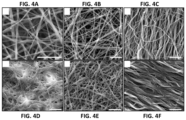
- FIGS. 4 A- 4 F provide SEM images of electrospun nanofibers before and after water treatment.
- FIGS. 4 A, 4 D Non-crosslinked fibers before ( FIG. 4 A ) and after ( FIG. 4 D ) water treatment.
- FIGS. 4 B, 4 C, 4 E, and 4 F Crosslinked fibers before ( FIGS. 4 B, 4 C ) and after ( FIGS. 4 E, 4 F ) water treatment.
- Scale bar 10 ⁇ m.
- FIG. 5 provides a graph of the relative growth rates of green fluorescent protein-labeled human dermal fibroblasts (GFP-HDF) treated with extractions of electrospun scaffolds following ISO10993-5 tests (biological evaluation of medical devices) for in vitro cytotoxicity.
- GFP-HDF green fluorescent protein-labeled human dermal fibroblasts
- FIG. 6 A provides the mold design.
- FIGS. 7 A- 7 F provide the morphological characterizations of 3D PCL/gelatin (50:50) nanofiber scaffolds with arrayed holes.
- FIG. 7 F provides a corresponding highly magnified image of FIG. 7 E showing nanofiber layers after expansion.
- Scale bar 10 ⁇ m.
- FIG. 10 A provides images of the implantation of the PCL nanofiber scaffolds with arrayed holes in rats.
- FIGS. 10 B- 10 E provide images of the in vivo response of expanded nanofiber scaffolds with punched holes.
- FIG. 10 B hematoxylin and eosin (H & E) staining.
- FIG. 10 C Masson trichrome staining.
- FIG. 10 D Highly magnified images of FIG. 10 B showing blood vessels.
- FIG. 10 E Highly magnified images of FIG. 10 B showing giant cells.
- FIG. 10 F provides a graph of the quantification of blood vessel formation per mm 2 .
- FIG. 10 G provides a graph of the quantification of giant cells per implant.
- FIG. 11 provides images of immunohistological staining of PCL nanofiber scaffolds and surrounding tissues against CD68 (a surface marker for pan macrophages), CD206 (a surface marker for macrophages in M2 phase), and CCR7 (a surface marker for macrophages in M1 phase).
- the nanofiber scaffolds were subcutaneously implanted to rats for 1 week, 2 weeks, and 4 weeks.
- FIG. 12 provides graphs of the quantification of immunhistological analysis of PCL nanofiber scaffolds after subcutaneous implantation.
- the number of CD68, CCR7 (M1), and CD206 (M2) immunpositve cells are shown as well as the ratio of number of CD163 positive cells (M2)/number of CCR7 positive cells (M1).
- the values were obtained by measuring six scanning images at 40 ⁇ (objective lens) magnification for each specimen.
- FIG. 13 provides images of multinucleated giant cells after nanofiber scaffold implantation.
- the rats were scarified at week 1, 2, and 4 after surgery.
- the multinucleated giant cells were stained against CD68, CD206, and CCR7. Arrows indicate multinucleated giant cells.
- FIG. 14 provides a schematic illustrating the cell infiltration and spatiotemporal distributions of M1 macrophages (light grey), M2 (grey) macrophages (top panel) and multinucleated giant cells (bottom panel) within the 3D PCL nanofiber scaffolds after subcutaneous implantation.
- the cell-infiltrated area is labeled in dark grey.
- Tissue engineered skin grafts may provide an optimized solution to improved healing of chronic wounds.
- the fabrication of a “sandwich-type” nanofiber-based skin graft has been demonstrated through seeding minced skin tissues onto the microwells of nanofiber membrane and covering with a radially aligned nanofiber membrane (Ma, et al., Biomaterials (2014) 35:630-641).
- nanofiber membranes were able to direct cell migration and achieve the full cell coverage on the surface of membranes in a short period of time, the nanofiber membranes used were two dimensional (2D) and cells only migrated on their surface.
- a novel type nanofiber skin graft for wound healing is provided.
- a modified gas-foaming technique has been used to expand 2D nanofiber membranes in the third dimension with controlled thickness and highly porous structures (Jiang, et al., ACS Biomaterials Sci. & Eng. (2015) 1:991-1001; Jiang, et al., Adv. Healthcare Mater. (2016) 5:2993-3003; Jiang et al., Acta Biomater. (2016) 68: 237-248).
- 3D scaffolds composed of PCL and gelatin nanofibers were fabricated by a combination of electrospinning and modified gas-foaming.
- Arrayed holes throughout the scaffold were created using a punch under cryo conditions.
- the scaffolds were also crosslinked with glutaraldehyde vapor to improve the water stability of the scaffolds.
- Cell spheroids of green fluorescent protein-labeled human dermal fibroblasts (GFP-HDF) were prepared and seeded into the holes. It was found that the fibroblasts adhered well on the surface of nanofibers and migrated into the scaffolds and proliferate due to the porous structures.
- the 3D nanofiber scaffolds of the instant invention can be used for engineering tissue constructs or models for various applications.
- the 3D nanofiber scaffolds with arrayed holes of the instant invention are able to provide the physical support and biological response to cell proliferation and infiltration. This clinically relieves the lack of autograft donor sites.
- the instant invention has demonstrated the development of a nanofiber skin graft by combining 3D nanofiber scaffolds with arrayed holes and cellular inserts (e.g., minced tissues (e.g., skin tissues), bone fragments) for chronic wound healing ( FIG. 1 ). Indeed, after implantation the cells migrate out from arrayed holes to the surrounding space to heal the wound.
- the 3D nanofiber scaffolds with arrayed holes of the instant invention have a highly porous 3D network which allows the cells to migrate and infiltrate through the whole scaffold.
- 2D nanofiber membranes were fabricated using electrospinning (Ma, et al., Biomaterials (2014) 35:630-641; Xie, et al., Acta Biomater. (2013) 9:5698-5707).
- PCL has shown degradability, hydrophobicity, good biocompatibility and high mechanical strength, resulting in FDA approval of many medical and drug application.
- PCL easily forms electrospun nanofibrous scaffolds with different natural and synthetic polymers (Gautam, et al., Mater. Sci. Engr. C (2013) 33:1228; Chaisri, et al., Biotech. J. (2013) 8:1323; Kim, et al., J. Nanomater. (2012) 2012:635212).
- composition of scaffolds a blend of PCL (a synthetic biodegradable and biocompatible polymer) and gelatin (a natural polymer), both approved by FDA for certain clinical applications (Kweon, et al., Biomaterials (2003) 24:801-808; Ungerleider, et al., Stem Cells Transl. Med. (2014) 3:1090-1099), was selected.
- PCL a synthetic biodegradable and biocompatible polymer
- gelatin a natural polymer
- nanofiber structures (sometimes referred to as scaffolds or nanofibrous herein) are provided.
- the nanofibers of the instant invention can be fabricated by any method.
- the nanofiber structures comprise electrospun nanofibers.
- the nanofiber structure comprises uniaxially aligned fibers, random fibers, and/or entangled fibers. While the application generally describes nanofiber (fibers having a diameter less than about 1 ⁇ m (e.g., average diameter)) structures and the synthesis of three-dimensional nanofibrous structures, the instant invention also encompasses microfiber (fibers having a diameter greater than about 1 ⁇ m (e.g., average diameter)) structures and the synthesis of three-dimensional microfibrous structures.
- the nanofibrous structures are expanded, such as produced by a gas-foaming technique.
- nanofiber structures e.g., mats
- the nanofiber structure may be crosslinked (e.g., prior to expansion).
- the nanofiber scaffolds of the present invention can be formed and manufactured into a variety of shapes (ex. round, square, rectangular), sizes, and thicknesses.
- the nanofiber structure may be cut or shaped prior to expansion.
- the expanded nanofiber scaffold is from about 1 to about 20 mm thick.
- the expanded nanofiber scaffold is from about 1 to about 10 mm thick.
- the expanded nanofiber scaffold is from about 1 to about 5 mm thick.
- the nanofibers of the instant invention may comprise any polymer.
- the polymer is biocompatible.
- the polymer may be biodegradable or non-biodegradable.
- the polymer is a biodegradable polymer.
- the polymer may by hydrophobic, hydrophilic, or amphiphilic.
- the polymer is hydrophobic.
- the polymer may be, for example, a homopolymer, random copolymer, blended polymer, copolymer, or a block copolymer. Block copolymers are most simply defined as conjugates of at least two different polymer segments or blocks.
- the polymer may be, for example, linear, star-like, graft, branched, dendrimer based, or hyper-branched (e.g., at least two points of branching).
- the polymer of the invention may have from about 2 to about 10,000, about 2 to about 1000, about 2 to about 500, about 2 to about 250, or about 2 to about 100 repeating units or monomers.
- the polymers of the instant invention may comprise capping termini.
- hydrophobic polymers include, without limitation: polyvinyl alcohol (PVA), poly(hydroxyethyl methacrylate), poly(N-isopropyl acrylamide), poly(lactic acid) (PLA (or PDLA)), poly(lactide-co-glycolide) (PLG), poly(lactic-co-glycolic acid) (PLGA), polyglycolide or polyglycolic acid (PGA), polycaprolactone (PCL), poly(aspartic acid), polyoxazolines (e.g., butyl, propyl, pentyl, nonyl, or phenyl poly(2-oxazolines)), polyoxypropylene, poly(glutamic acid), poly(propylene fumarate) (PPF), poly(trimethylene carbonate), polycyanoacrylate, polyurethane, polyorthoesters (POE), polyanhydride, polyester, poly(propylene oxide), poly(caprolactonefumarate), poly(1,2-butylene
- hydrophilic polymers include, without limitation: polyvinylpyrrolidone (PVP), poly(ethylene glycol) and poly(ethylene oxide) (PEO), chitosan, collagen, chondroitin sulfate, sodium alginate, gelatin, elastin, hyaluronic acid, silk fibroin, sodium alginate/PEO, silk/PEO, silk fibroin/chitosan, hyaluronic acid/gelatin, collagen/chitosan, chondroitin sulfate/collagen, and chitosan/PEO.
- PVP polyvinylpyrrolidone
- PEO poly(ethylene glycol)
- chitosan collagen, chondroitin sulfate, sodium alginate, gelatin, elastin, hyaluronic acid, silk fibroin, sodium alginate/PEO, silk/PEO, silk fibroin/chitosan, hyaluronic acid/gelatin, collagen/chitos
- Amphiphilic copolymers may comprise a hydrophilic polymer (e.g., segment) and a hydrophobic polymer (e.g., segment) from those listed above (e.g., gelatin/PVA, PCL/collagen, chitosan/PVA, gelatin/elastin/PLGA, PDO/elastin, PHBV/collagen, PLA/hyaluronic acid, PLGA/hyaluronic acid, PCL/hyaluronic acid, PCL/collagen/hyaluronic acid, gelatin/siloxane, PLLA/MWNTs/hyaluronic acid).
- a hydrophilic polymer e.g., segment
- a hydrophobic polymer e.g., segment from those listed above (e.g., gelatin/PVA, PCL/collagen, chitosan/PVA, gelatin/elastin/PLGA, PDO/elastin, PHBV/collagen, PLA/hy
- Examples of polymers particularly useful for electrospinning are provided in Xie et al. (Macromol. Rapid Commun. (2008) 29:1775-1792; incorporated by reference herein; see e.g., Table 1).
- Examples of compounds or polymers for use in the fibers of the instant invention, particularly for electrospun nanofibers include, without limitation: natural polymers (e.g., chitosan, gelatin, collagen type I, II, and/or III, elastin, hyaluronic acid, cellulose, silk fibroin, phospholipids (Lecithin), fibrinogen, hemoglobin, fibrous calf thymus Na-DNA, virus M13 viruses), synthetic polymers (e.g., PLGA, PLA, PCL, PHBV, PDO, PGA, PLCL, PLLA-DLA, PEUU, cellulose acetate, PEG-b-PLA, EVOH, PVA, PEO, PVP), blended (e.g.,
- the nanofiber comprises polymethacrylate, poly vinyl phenol, polyvinylchloride, cellulose, polyvinyl alcohol, polyacrylamide, PLGA, collagen, polycaprolactone, polyurethanes, polyvinyl fluoride, polyamide, silk, nylon, polybennzimidazole, polycarbonate, polyacrylonitrile, polyvinyl alcohol, polylactic acid, polyethylene-co-vinyl acetate, polyethylene oxide, polyaniline, polystyrene, polyvinylcarbazole, polyethylene terephthalate, polyacrylic acid-polypyrene methanol, poly(2-hydroxyethyl methacrylate), polyether imide, polyethylene gricol, polyethylene glycol, poly(ethylene-co-vinyl alcohol), polyacrylnitrile, polyvinyl pyrrolidone, polymetha-phenylene isophthalamide, gelatin, chitosan, starch, pectin, cellulose, methylcellulose,
- the nanofiber structures comprise a material that enhances the nanofiber structure's ability to absorb fluids, particularly aqueous solutions (e.g., blood).
- the nanofibers comprise a polymer and the material which enhances the absorption properties.
- the nanofiber structures are coated with the material which enhances the absorption properties.
- the term “coat” refers to a layer of a substance/material on the surface of a structure. Coatings may, but need not, also impregnate the nanofiber structure.
- a coating may cover 100% of the nanofiber structure
- a coating may also cover less than 100% of the surface of the nanofiber structure (e.g., at least about 75%, at least about 80%, at least about 85%, at least about 90%, at least about 95%, at least about 98%, or more the surface may be coated).
- Materials which enhance the absorption properties of the expanded nanofiber structures include, without limitation: gelatin, chitosan, collagen, starch, pectin, cellulose, methylcellulose, sodium polyacrylate, starch-acrylonitrile co-polymers, other natural or synthetic hydrogels, and derivatives thereof (e.g., del Valle et al., Gels (2017) 3:27).
- the material is a hydrogel (e.g., a polymer matrix able to retain water, particularly large amounts of water, in a swollen state).
- the material is gelatin.
- the expanded nanofiber structures are coated with about 0.05% to about 10% coating material (e.g., gelatin), particularly about 0.1% to about 10% coating material (e.g., gelatin) or about 0.1% to about 1% coating material (e.g., gelatin).
- the material e.g., hydrogel
- the nanofiber structures of the instant invention are crosslinked.
- the nanofiber structures of the instant invention may be crosslinked with a crosslinker such as, without limitation: formaldehyde, paraformaldehyde, acetaldehyde, glutaraldehyde, a photocrosslinker, genipin, and natural phenolic compounds (Mazaki, et al., Sci. Rep. (2014) 4:4457; Bigi, et al., Biomaterials (2002) 23:4827-4832; Zhang, et al., Biomacromolecules (2010) 11:1125-1132; incorporated herein by reference).
- the crosslinker may be a bifunctional, trifunctional, or multifunctional crosslinking reagent.
- the crosslinker is glutaraldehyde.
- the nanofiber structures of the instant invention are expanded. Electrospun nanofibers are usually deposited on a substrate to form a nanofiber mat. However, the nanofiber mats are often dense and hard. These nanofiber mats can be expanded by making use of bubbles (e.g., generated by chemical reactions in an aqueous solution (e.g., a gas foaming technique)). The gas bubbles may be formed by any chemical reaction and/or physical mean.
- bubbles e.g., generated by chemical reactions in an aqueous solution (e.g., a gas foaming technique).
- the gas bubbles may be formed by any chemical reaction and/or physical mean.
- the bubbles may be generated, without limitation, using a gas-production chemical reaction; by dissolved gas in a liquid under a high pressure and/or a low temperature; pressurized gas (e.g., CO 2 ) liquid; and/or physical means (e.g., laser (e.g., pulsed laser), acoustic induced, or flow induced).
- the nanofiber structure is submerged or immersed in a bubble/gas producing chemical reaction or physical manipulation. Generally, the longer the exposure to the bubbles, the greater the thickness and porosity of the nanofiber structure increases. Examples of methods of expanding nanofiber structures are provided in PCT/US2015/052858 (incorporated herein by reference).
- the gas bubbles of the instant invention can be made by any method known in the art.
- the bubbles may be generated, for example, by chemical reactions or by physical approaches.
- the chemical reaction or physical manipulation does not damage or alter or does not substantially damage or alter the nanofibers (e.g., the nanofibers are inert within the chemical reaction and not chemically modified).
- the nanofiber structure may be submerged or immersed in a liquid comprising the reagents of the bubble-generating chemical reaction.
- the chemical reaction is the hydrolysis of NaBH 4 (e.g., NaBH 4 +
- Examples of physical approaches for generating bubbles of the instant invention include, without limitation: 1) create high pressure (fill gas)/heat in a sealed chamber and suddenly reduce pressure; 2) dissolve gas in liquid/water in high pressure and reduce pressure to release gas bubbles; 3) use supercritical fluids (reduce pressure) like supercritical CO 2 ; 4) use gas liquid (then reduce pressure) (e.g., liquid CO 2 , liquid propane and isobutane); 5) fluid flow; 6) apply acoustic energy or ultrasound to liquid/water; 7) apply a laser (e.g., to a liquid or water); 8) boiling; 9) reduce pressure boiling (e.g., with ethanol); and 10) apply radiation (e.g., ionizing radiation on liquid or water).
- the nanofiber structure may be submerged or immersed in a liquid of the bubble-generating physical manipulation.
- the nanofiber structures of the instant invention may also be treated with air plasma prior to exposure to gas bubbles (e.g., to increase hydrophilicity).
- the nanofiber structure may also be expanded within a mold (e.g., made of a metal, plastic, or other material that does not expand in the presence of gas bubbles) such that the expanded nanofiber structure forms a desired shape (e.g., pads, tubes, beads, etc.).
- a mold e.g., made of a metal, plastic, or other material that does not expand in the presence of gas bubbles
- the mold is synthesized by a 3D printer.
- the mold is as depicted in FIG. 6 .
- the mold may contain holes that allow for punching corresponding holes in the nanofiber structure.
- the nanofiber structures of the instant invention may also be manipulated after expansion to form a desired shape (e.g., pads, tubes, beads, etc.).
- the nanofiber structures of the instant invention also comprise holes or wells.
- the wells/holes may be made in the nanofiber scaffold before or after expansion of the nanofiber scaffold.
- the holes of the nanofiber structures are inserted prior to expansion.
- the nanofiber structure is frozen (e.g., in liquid nitrogen) prior to insertion or punching of the holes.
- the holes of the nanofiber structure may be any shape (e.g., square, circle).
- the holes of the nanofiber structure can be any size.
- the holes/wells have a length/dimension or diameter of about 0.1 to about 5 mm, particularly about 0.5 to about 3 mm or about 1.0 mm.
- the holes may be organized within the nanofiber structure in an array (e.g., a square array).
- the holes of the nanofiber structure are generally equidistant from each other.
- the holes/wells of the nanofiber structures may all be the same size or may be various sizes. Any number of wells may be made in the nanofiber scaffolds. In one embodiment, the number of wells is between about 1 and about 200.
- the wells may be made using a variety of methods.
- a mold with preset holes is used as a template to punch wells/holes into the nanofiber scaffold.
- the template may be made using a variety of techniques including but not limited to 3D printing.
- the nanofiber structure may be washed or rinsed in water and/or a desired carrier or buffer (e.g., a pharmaceutically or biologically acceptable carrier). Trapped gas bubbles may be removed by applying a vacuum to the nanofiber structure.
- the expanded nanofiber structure may be submerged or immersed in a liquid (e.g., water and/or a desired carrier or buffer) and a vacuum may be applied to rapidly remove the gas bubbles.
- the nanofiber structures may be lyophilized and/or freeze-dried.
- the nanofiber structures of the instant invention may also be sterilized.
- the nanofiber structures can be chemically sterilized (e.g., by treating with ethylene oxide).
- the holes/wells of the nanofiber structure of the instant invention may comprise cells or tissue.
- the cells are autologous to the subject to be treated with the nanofiber structure. Any cell type can be added to the holes/wells.
- the cells comprise stem cells.
- the cells comprise dermal fibroblasts.
- the holes/wells contain cell spheroids.
- the holes/wells comprise tissue samples (e.g., minced tissue), such as skin tissue samples or bone samples.
- the tissue samples have a length/dimension of diameter of about 0.1 to about 5 mm, particularly about 0.5 to about 3 mm or about 1.0 mm.
- the cells or tissue may be cultured with in the holes/wells of the nanofiber structure (e.g., the cells or tissue may be cultured for sufficient time to allow for infiltration into the nanofiber structure).
- the cells or tissue may be cultured in the nanofiber structure for 1 day, 2 days, 3 days, 4 days, 5 days, or more.
- the nanofiber structures of the instant invention may comprise or encapsulate at least one agent, particularly a bioactive agent such as a drug or therapeutic agent (e.g., analgesic, growth factor, anti-inflammatory, signaling molecule, cytokine, antimicrobial (e.g., antibacterial, antibiotic, antiviral, and/or antifungal), blood clotting agent, factor, or protein, etc.).
- a drug or therapeutic agent e.g., analgesic, growth factor, anti-inflammatory, signaling molecule, cytokine, antimicrobial (e.g., antibacterial, antibiotic, antiviral, and/or antifungal), blood clotting agent, factor, or protein, etc.
- the agent may be added to the nanofiber structures during synthesis and/or after synthesis.
- the agent may be conjugated to the nanofiber structure and/or coating material, encapsulated by the nanofiber structure, and/or coated on the nanofiber structure (e.g., with, underneath, and/or on top of the coating that
- the agents enhance tissue regeneration, tissue growth, and wound healing (e.g., growth factors).
- the agent treats/prevents infections (e.g., antimicrobials such as antibacterials, antivirals and/or antifungals).
- the agent is an antimicrobial, particularly an antibacterial.
- the agent enhances wound healing and/or enhances tissue regeneration (e.g., bone, tendon, cartilage, skin, nerve, and/or blood vessel).
- Such agents include, for example, growth factors and small molecules.
- Growth factors include, without limitation: platelet derived growth factor (PDGF), vascular endothelial growth factor (VEGF), epidermal growth factor (EGF), fibroblast growth factor (FGF, multiple isotypes; e.g. basic fibroblast growth factor (bFGF)), insulin-like growth factor (IGF-1 and/or IGF-2), bone morphogenetic protein (e.g., BMP-2, BMP-7, BMP-12, BMP-9), transforming growth factor (e.g., TGF ⁇ , TGF ⁇ 3), nerve growth factor (NGF), neurotrophic factor, glial cell-derived neurotrophic factor (GDNF), and/or keratinocyte growth factor (KGF).
- Small molecules include, without limitation, simvastatin, kartogenin, retinoic acid, paclitaxel, vitamin D3, etc.
- the method comprises electrospinning a nanofiber structure or mat, crosslinking the nanofiber structure or mat (optional), freezing (e.g., with liquid nitrogen) the nanofiber structure or mat (optional), inserting or punching holes into the nanofiber structure, expanding the nanofiber structure or mat with gas, washing and/or sterilizing the expanded nanofiber structure (optional), and seeding cells and/or tissue into the holes or wells of the expanded nanofiber structure.
- the method further comprises plasma treatment of the nanofiber mat or structure prior to expansion.
- the holes are punched into the nanofiber structure after gas expansion.
- the method further comprises culturing the cells within the nanofiber structure (e.g., allowing the cells to infiltrate the nanofiber structure from the holes/wells).
- the nanofiber structures of the instant invention can be used to create complex tissue architectures for a variety of application including, without limitation: wound healing, tissue engineering, tissue growth, tissue repair, tissue regeneration, and engineering 3D in vitro tissue models.
- the nanofiber structures can also be combined with a variety of hydrogels or biological matrices/cues to form 3D hybrid scaffolds that can release biologically functional molecules.
- the tissue constructs can be used for regeneration of many tissue defects (e.g., skin, bone) and healing of various wounds (e.g., injuries, diabetic wounds, venous ulcer, pressure ulcer, burns).
- the nanofiber structures may be used ex vivo to generate tissue or tissue constructs/models.
- the nanofiber structures may also be used in vivo in patients (e.g., human or animal) for the treatment of various diseases, disorders, and wounds.
- the nanofiber structure stimulates the growth of existing tissue and/or repair of a wound or defect when applied in vivo.
- the nanofiber scaffolds can be used for engineering, growing, and/or regeneration of a variety of tissues including but not limited to skin, bone, cartilage, muscle, nervous tissue, and organs (or portions thereof).
- the nanofiber structures may be used in inducing and/or improving/enhancing wound healing and inducing and/or improving/enhancing tissue regeneration.
- the nanofiber structures of the present invention can be used for the treatment, inhibition, and/or prevention of any injury or wound.
- the nanofiber structures can be used to induce, improve, or enhance wound healing associated with surgery (including non-elective (e.g., emergency) surgical procedures or elective surgical procedures).
- Elective surgical procedures include, without limitation: liver resection, partial nephrectomy, cholecystectomy, vascular suture line reinforcement and neurosurgical procedures.
- Non-elective surgical procedures include, without limitation: severe epistaxis, splenic injury, liver fracture, cavitary wounds, minor cuts, punctures, gunshot wounds, and shrapnel wounds.
- the nanofiber structures of the present invention can also be incorporated into delivery devices (e.g., a syringe) that allow for their injection/delivery directly into a desired location (e.g., a wound such as a gunshot wound).
- the nanofiber structures also may be delivered directly into a cavity (such as the peritoneal cavity) using a pressurized cannula.
- methods for inducing and/or improving/enhancing wound healing in a subject are also provided.
- Methods of inducing and/or improving/enhancing tissue regeneration e.g., blood vessel growth, neural tissue regeneration, and bone regeneration
- the methods of the instant invention comprise administering or applying a nanofiber structure of the instant invention to the subject (e.g., at or in a wound).
- the method comprises administering a nanofiber structure comprising an agent as described hereinabove.
- the method comprises administering a nanofiber structure to the subject and an agent as described hereinabove (i.e., the agent is not contained within the nanofiber structure).
- the nanofiber structure may be administered simultaneously and/or sequentially with the agent.
- the methods may comprise the administration of one or more nanofiber structures. When more than one nanofiber structure is administered, the nanofiber structures may be administered simultaneously and/or sequentially.
- electrospun fibers i.e., electrospun fibers
- micro- or nano-sized fibers from a solution or melt using interactions between fluid dynamics and charged surfaces (e.g., by streaming a solution or melt through an orifice in response to an electric field).
- electrospun nanofibers include, without limitation, branched nanofibers, tubes, ribbons and split nanofibers, nanofiber yarns, surface-coated nanofibers (e.g., with carbon, metals, etc.), nanofibers produced in a vacuum, and the like.
- the production of electrospun fibers is described, for example, in Gibson et al. (1999) AlChE J., 45:190-195.
- “Pharmaceutically acceptable” indicates approval by a regulatory agency of the Federal or a state government or listed in the U.S. Pharmacopeia or other generally recognized pharmacopeia for use in animals, and more particularly in humans.
- a “carrier” refers to, for example, a diluent, adjuvant, preservative (e.g., Thimersol, benzyl alcohol), anti-oxidant (e.g., ascorbic acid, sodium metabisulfite), solubilizer (e.g., polysorbate 80), emulsifier, buffer (e.g., TrisHCl, acetate, phosphate), water, aqueous solutions, oils, bulking substance (e.g., lactose, mannitol), excipient, auxiliary agent or vehicle with which an active agent of the present invention is administered.
- Suitable pharmaceutical carriers are described in “Remington's Pharmaceutical Sciences” by E. W.
- polymer denotes molecules formed from the chemical union of two or more repeating units or monomers.
- block copolymer most simply refers to conjugates of at least two different polymer segments, wherein each polymer segment comprises two or more adjacent units of the same kind.
- Hydrophobic designates a preference for apolar environments (e.g., a hydrophobic substance or moiety is more readily dissolved in or wetted by non-polar solvents, such as hydrocarbons, than by water).
- hydrophobic polymers may have aqueous solubility less than about 1% wt. at 37° C.
- polymers that at 1% solution in bi-distilled water have a cloud point below about 37° C., particularly below about 34° C., may be considered hydrophobic.
- hydrophilic means the ability to dissolve in water.
- polymers that at 1% solution in bi-distilled water have a cloud point above about 37° C., particularly above about 40° C., may be considered hydrophilic.
- amphiphilic means the ability to dissolve in both water and lipids/apolar environments.
- an amphiphilic compound comprises a hydrophilic portion and a hydrophobic portion.
- antimicrobials indicates a substance that kills or inhibits the growth of microorganisms such as bacteria, fungi, viruses, or protozoans.
- an antiviral refers to a substance that destroys a virus and/or suppresses replication (reproduction) of the virus.
- an antiviral may inhibit and or prevent: production of viral particles, maturation of viral particles, viral attachment, viral uptake into cells, viral assembly, viral release/budding, viral integration, etc.
- antibiotic refers to antibacterial agents for use in mammalian, particularly human, therapy.
- Antibiotics include, without limitation, beta-lactams (e.g., penicillin, ampicillin, oxacillin, cloxacillin, methicillin, and cephalosporin), carbacephems, cephamycins, carbapenems, monobactams, aminoglycosides (e.g., gentamycin, tobramycin), glycopeptides (e.g., vancomycin), quinolones (e.g., ciprofloxacin), moenomycin, tetracyclines, macrolides (e.g., erythromycin), fluoroquinolones, oxazolidinones (e.g., linezolid), lipopetides (e.g., daptomycin), aminocoumarin (e.g., novobiocin), co-trimoxazole (e.g.,
- an “anti-inflammatory agent” refers to compounds for the treatment or inhibition of inflammation.
- Anti-inflammatory agents include, without limitation, non-steroidal anti-inflammatory drugs (NSAIDs; e.g., aspirin, ibuprofen, naproxen, methyl salicylate, diflunisal, indomethacin, sulindac, diclofenac, ketoprofen, ketorolac, carprofen, fenoprofen, mefenamic acid, piroxicam, meloxicam, methotrexate, celecoxib, valdecoxib, parecoxib, etoricoxib, and nimesulide), corticosteroids (e.g., prednisone, betamethasone, budesonide, cortisone, dexamethasone, hydrocortisone, methylprednisolone, prednisolone, tramcinolone, and fluticasone), rapamycin, ace
- the term “subject” refers to an animal, particularly a mammal, particularly a human.
- the term “prevent” refers to the prophylactic treatment of a subject who is at risk of developing a condition resulting in a decrease in the probability that the subject will develop the condition.
- treat refers to any type of treatment that imparts a benefit to a patient afflicted with a disease, including improvement in the condition of the patient (e.g., in one or more symptoms), delay in the progression of the condition, etc.
- analgesic refers to an agent that lessens, alleviates, reduces, relieves, or extinguishes pain in an area of a subject's body (i.e., an analgesic has the ability to reduce or eliminate pain and/or the perception of pain).
- small molecule refers to a substance or compound that has a relatively low molecular weight (e.g., less than 2,000). Typically, small molecules are organic, but are not proteins, polypeptides, or nucleic acids.
- hydrogel refers to a water-swellable, insoluble polymeric matrix (e.g., hydrophilic polymers) comprising a network of macromolecules, optionally crosslinked, that can absorb water to form a gel.
- hydrophilic polymers e.g., hydrophilic polymers
- crosslink refers to a bond or chain of atoms attached between and linking two different molecules (e.g., polymer chains).
- crosslinker refers to a molecule capable of forming a covalent linkage between compounds.
- a “photocrosslinker” refers to a molecule capable of forming a covalent linkage between compounds after photoinduction (e.g., exposure to electromagnetic radiation in the visible and near-visible range).
- Crosslinkers are well known in the art (e.g., formaldehyde, paraformaldehyde, acetaldehyde, glutaraldehyde, etc.).
- the crosslinker may be a bifunctional, trifunctional, or multifunctional crosslinking reagent.
- SEM scanning electron microscopy
- the PCL/gelatin nanofiber mats were cross-linked for 24 hours with glutaraldehyde vapor in ethanol at room temperature.
- plasma treatment Hard Plasma Inc., Ithaca, N.Y.
- RF medium radiofrequency
- the arrayed holes on the nanofiber mats were punched in liquid nitrogen.
- the nanofiber mats with arrayed holes were then expanded to form 3D scaffolds using a modified gas-foaming solution.
- a 1% NaBH 4 solution was used to prepare gas foamed electrospun 3D scaffolds.
- the nanofibrous mats were immersed in freshly prepared NaBH 4 solution at room ambient.
- the mats were gently rinsed 3 times with distilled water for 10 minutes each after 5-30 minutes gas foaming. NaBH 4 solution was discarded after dilution.
- the expanded 3D PCL/gelatin scaffolds were freeze-dried overnight. The scaffolds were sterilized with ethylene oxide prior to cell culture. The morphology of the nanofiber scaffold was examined using SEM. One drop of distilled water was added to the surface of fibers and kept for at least 5 minutes. The water stability of PCL/gelatin nanofibers before and after cross-linking was examined using SEM.
- GFP-HDF cell spheroids (Foty, R., J. Vis. Exp. (2011) (51):2720; Fennema, et al., Trends Biotechn. (2013) 31:108-115). Each drop contained 20 ⁇ L of cell suspension the concentration of which was 10 6 cells/mL. Cell spheroids of GFP-HDF were seeded to the surface of 2D PCL/gelatin scaffolds and the holes of 3D scaffolds. The samples were observed after 5 days of incubation. A fixative, which contains 2% glutaraldehyde and 2% paraformaldehyde in 0.1 M Sorensen's phosphate buffer (SPB), was used to fix the samples at 4° C.
- SPB Sorensen's phosphate buffer
- GFP-HDF was cultured in DMEM with 10% FBS at 37° C. in a 5% CO 2 incubator. Before cell seeding, scaffolds were sterilized with ethylene oxide (EtO) for 12 hours. After sterilization, the scaffolds were incubated with PBS for 48 hours to obtain extracted solutions. Subsequently, the extraction (100 ⁇ L) of PCL, PCL/gelatin before crosslinking and PCL/gelatin after crosslinking were mixed with 100 ⁇ L fresh DMEM media and incubated in 96-well culture plate with GFP-HDF at a density of 10 5 cell per well for 24 hours at 37° C. in 5% CO 2 incubator.
- EtO ethylene oxide
- the 3D mold made of acrylonitrile butadiene styrene (ABS) was printed by a TAZ 5 3D printer (LulzBot, Loveland, Colo.).
- PCL/gelatin nanofibers were generated by electrospinning under different conditions ( FIG. 2 ). It appears that the fiber diameter gradually became uniform with decreasing the PCL/gelatin concentration from 10% to 5% (w/v). The smooth and uniform fibers were obtained when the flow rate was 0.4 mL/hour and the PCL/gelatin concentration was 5% (w/v) ( FIG. 2 ). By controlling rotating speed of the drum collector, aligned and random fibers were obtained ( FIG. 3 ). The orientation of fibers was confirmed by a fast Fourier transform (FFT) analysis ( FIG. 3 ). The gelatin is water soluble, necessitating crosslinking to preserve the fiber morphology in an aqueous environment.
- FFT fast Fourier transform
- the glutaraldehyde vapor was used to crosslink PCL/gelatin nanofibers for maintaining their structural integrity since glutaraldehyde has been widely applied to covalently crosslink functional groups in natural polymers, such as gelatin and collagen (Kim, et al., Macromol. Biosci. (2010) 10:91-100). It is found that the fiber morphology of PCL/gelatin nanofibers was mainly preserved except for some minor swelling after water treatment ( FIG. 4 ). To examine the potential cytotoxicity of cross-linked PCL/gelatin nanofibers, relative growth rates (RGR) of GFP-HDF were quantified when incubating with extractions of various fiber samples ( FIG. 5 ).
- RGR relative growth rates
- RGR of cross-linked and non-cross-linked PCL/gelatin nanofibers were higher than 80%, indicating a marginal cytotoxicity.
- a more benign cross-linking method can be used such as photo-crosslinker, genipin or natural phenolic compounds (Mazaki, et al., Sci. Rep. (2014) 4:4457; Bigi, et al., Biomaterials (2002) 23:4827-4832; Zhang, et al., Biomacromolecules (2010) 11:1125-1132).
- Nanofiber membranes were first immersed into liquid nitrogen to make the materials brittle (below the glass transition temperature) and then created holes with a punch.
- Nanofiber membranes were expanded with square arrayed holes in a mold generated by 3D printer using a modified gas foaming technique ( FIG. 6 ) (Jiang, et al., ACS Biomaterials Sci. & Eng. (2015) 1:991-1001; Jiang, et al., Adv. Healthcare Mater. (2016) 5:2993-3003).
- the nanofiber membrane was initially 0.4 mm thick and became about 4 mm thick after expansion.
- FIG. 7 shows the morphology of a 3D nanofiber scaffold with arrayed holes. As expected, the inner surface of punched holes displayed a layered structure ( FIG. 7 C ).
- the SEM image of cross-sections indicated a layered and highly porous structure ( FIGS. 7 D- 7 F ).
- GFP-HDF cell spheroids were prepared using a hanging drop method (Foty, R., J. Vis. Exp. (2011) (51):2720). An average diameter of 442.35 ⁇ 13.62 ⁇ m for cell spheroids was obtained after culture for 24 hours. Cell spheroids were seeded to the 2D and 3D nanofiber scaffolds. In order to prevent the falling of the cell spheroids, cell spheroids seeded on the scaffolds were immersed in a shallow level of cell culture medium overnight. After attached to the scaffolds, more cell culture medium was added to each well.
- FIGS. 8 A and 8 B After 5 days culture, it is observed that fibroblasts that were migrated from the cell spheroids seeded to 2D nanofiber membranes adhered and proliferated on the surface only ( FIGS. 8 A and 8 B ). In contrast, fibroblasts that were migrated from cell spheroids seeded into the holes of 3D nanofiber scaffolds adhered to the surface and surrounding nanofiber walls of holes ( FIGS. 8 C, 8 D, and 9 ). Cells could further infiltrate into the scaffolds through the gaps between layers due to the layered and highly porous structures after expansion. The arrayed holes seeded with 3D cell spheroids showed a simultaneously cell infiltration and proliferation in three dimensions.
- this 3D scaffolds with arrayed holes is ideal for wound treatment, both in chronic wound healing and acute wound healing such as 2nd degree and 3rd degree burns. Furthermore, the 3D cell spheroids had a comparable migration and proliferation of fibroblast comparing to typical 2D fibrous scaffolds.
- these 3D nanofiber scaffolds together with cell spheroids or minced tissues can create complex tissue architectures for wound healing, tissue regeneration, and engineering 3D in vitro tissue models (Cesarz, et al., Stem Cells Int. (2016) 9176357; Zanoni, et al., Sci. Rep. (2016) 6:19103).
- the scaffolds can also be combined with a variety of hydrogels or biological matrices/cues to form 3D hybrid scaffolds with eliciting biologically functional molecules (Chaisri, et al., Biotech. J. (2013) 8:1323; Franco, et al., J. Mater. Sci. Mater. Med.
- the GFP-HDF can be changed to other cells, such as stem cells and cancer cells, which can produce extracellular matrix.
- the 3D scaffolds would biomimetic a physiological microenvironment with nanotopographic cues for hosting organotypic-like cell cultures.
- these novel 3D scaffolds with cost-effective fabrication could likely lead to the drastic change in the utilization of electrospun nanofibers.
- FIG. 10 A the transformed PCL nanofiber scaffolds with arrayed holes were implanted subcutaneously in rats for 1 week, 2 weeks, and 4 weeks ( FIG. 10 A ).
- Cells grew into the punched holes and then penetrated into the space between nanofiber thin layers within expanded nanofiber scaffolds ( FIG. 10 B ).
- Masson trichrome staining shows the collagen deposition, indicated by arrows, from infiltrated cells in the punched holes and in the gaps between nanofiber thin layers ( FIG. 10 C ). Many blood vessels were formed within the newly formed tissues in the holes or gaps between nanofiber layers ( FIG. 10 D ). Multinucleated giant cells are also present ( FIG. 10 E ).
- Numbers of blood vessels per mm 2 are approximately 40, 65, and 19 at week 1, 2 and 4, respectively ( FIG. 10 F ). The presence of more blood vessels form at week 2 can be attributed to the early inflammatory response. Numbers of multinucleated giant cells per implant are 15, 50, and 140 at week 1, 2 and 4, respectively ( FIG. 10 G ).
- multinucleated giant cells are heterogeneous, expressing CCR7, CD206, and CD68 markers, can promote new blood vessel formation and tissue regeneration.
- cellular infiltration and spatiotemporal distributions of M1 macrophages, M2 macrophages, and multinucleated giant cells within the scaffold after implantation for 1, 2, and 4 weeks are proposed as seen in FIG. 14 .
- Cells infiltrate into the punched holes within 1 week and continue penetrating to the scaffolds through the gaps between nanofiber layers.
- the macrophage infiltration shows the similar trend.
- M1 macrophages at week 1 and 2 but more M2 macrophages at week 4.
- multinucleated giant cells are mostly located on the surface of holes.
- some giant cells are formed either on the surface of holes or on the infiltrated fiber layers.
- multinucleated giant cells are relatively evenly distributed throughout the infiltrated areas.
Abstract
Description
NaBH4+2H2O═NaBO2+4H2
NaBH4+4H2O=4H2(g)+H3BO3+NaOH
HCO3 −+H+=CO2+H2O
NH4 ++NO2 −═N2+2H2O
H2CO3=H2O+CO2
2H++S2−═H2S
2H2O2=O2+2H2O
3HNO2=2NO+HNO3+H2O
HO2CCH2COCH2CO2H=2CO2+CH3COCH3
2H2O2=2H2+O2
CaC2+H2O═C2H2
Zn+2HCl=H2+ZnCl2
2KMnO4+16HCl=2KCl+2MnCl2+H2O+5Cl2
In a particular embodiment, the chemical reaction is the hydrolysis of NaBH4 (e.g., NaBH4+2H2O═NaBO2+4H2). In a particular embodiment, CO2 gas bubbles (generated chemically or physically (see below)) are used (e.g., for hydrophilic polymers).
Claims (20)
Priority Applications (1)
| Application Number | Priority Date | Filing Date | Title |
|---|---|---|---|
| US16/611,415 US11738116B2 (en) | 2017-06-09 | 2018-06-08 | Expanded nanofiber structures comprising electrospun nanofibers and a plurality of holes and methods of making and use thereof |
Applications Claiming Priority (3)
| Application Number | Priority Date | Filing Date | Title |
|---|---|---|---|
| US201762517310P | 2017-06-09 | 2017-06-09 | |
| US16/611,415 US11738116B2 (en) | 2017-06-09 | 2018-06-08 | Expanded nanofiber structures comprising electrospun nanofibers and a plurality of holes and methods of making and use thereof |
| PCT/US2018/036647 WO2018227078A1 (en) | 2017-06-09 | 2018-06-08 | Nanofiber structures and methods of use thereof |
Publications (2)
| Publication Number | Publication Date |
|---|---|
| US20200164107A1 US20200164107A1 (en) | 2020-05-28 |
| US11738116B2 true US11738116B2 (en) | 2023-08-29 |
Family
ID=64566756
Family Applications (1)
| Application Number | Title | Priority Date | Filing Date |
|---|---|---|---|
| US16/611,415 Active 2040-06-16 US11738116B2 (en) | 2017-06-09 | 2018-06-08 | Expanded nanofiber structures comprising electrospun nanofibers and a plurality of holes and methods of making and use thereof |
Country Status (2)
| Country | Link |
|---|---|
| US (1) | US11738116B2 (en) |
| WO (1) | WO2018227078A1 (en) |
Families Citing this family (8)
| Publication number | Priority date | Publication date | Assignee | Title |
|---|---|---|---|---|
| US11033659B2 (en) | 2014-09-29 | 2021-06-15 | Board Of Regents Of The University Of Nebraska | Nanofiber structures and methods of synthesis and use thereof |
| JP2019529050A (en) | 2016-09-28 | 2019-10-17 | ボード オブ リージェンツ オブ ザ ユニバーシティ オブ ネブラスカ | Nanofiber structure and method of using the same |
| EP3684726A4 (en) | 2017-09-19 | 2021-06-30 | Board of Regents of the University of Nebraska | Nanofiber structures and methods of use thereof |
| US20220023496A1 (en) * | 2018-12-14 | 2022-01-27 | Board Of Regents Of The University Of Nebraska | Nanofiber structures and methods of manufacture and use thereof |
| US20230130357A1 (en) * | 2020-04-30 | 2023-04-27 | Board Of Regents Of The University Of Nebraska | Multi-layer hernia meshes and methods of manufacture and use thereof |
| WO2022087519A1 (en) * | 2020-10-23 | 2022-04-28 | Board Of Regents Of The University Of Nebraska | Surface modified scaffolds and methods of use thereof |
| KR102598611B1 (en) * | 2021-06-18 | 2023-11-03 | 우석대학교 산학협력단 | A method for manufacturing 3d transparent nanofibers for artificial retina and the 3d transparent nanofibers artificial retina thereof |
| CN113750291A (en) * | 2021-10-18 | 2021-12-07 | 诺一迈尔(苏州)生命科技有限公司 | Articular cartilage scaffold and preparation method thereof |
Citations (53)
| Publication number | Priority date | Publication date | Assignee | Title |
|---|---|---|---|---|
| WO2000050104A1 (en) | 1999-02-25 | 2000-08-31 | Degradable Solutions Ag | Biodegradable, porous shaped bodies |
| US6653005B1 (en) | 2000-05-10 | 2003-11-25 | University Of Central Florida | Portable hydrogen generator-fuel cell apparatus |
| US20050084532A1 (en) | 2002-03-13 | 2005-04-21 | Howdle Steven M. | Polymer composition loaded with cells |
| US20050187330A1 (en) | 2004-02-20 | 2005-08-25 | Wayne State University | Method of delaminating aggregated particles with a coating agent in a substantially supercritical fluid |
| EP1611877A1 (en) | 2004-06-28 | 2006-01-04 | Universidade de Coimbra | Method for preparing sustained-release therapeutic ophthalmic articles using compressed fluids for impregnation of drugs |
| US20060002978A1 (en) | 2004-06-10 | 2006-01-05 | Shea Lonnie D | Biodegradable scaffolds and uses thereof |
| WO2006019600A2 (en) | 2004-07-16 | 2006-02-23 | Poly-Med, Inc. | Hemostatix microfibrous constructs |
| JP2006169497A (en) | 2004-11-19 | 2006-06-29 | Tokyo Univ Of Agriculture & Technology | Porous body and manufacturing method thereof |
| US20070077272A1 (en) | 2005-09-22 | 2007-04-05 | Medivas, Llc | Solid polymer delivery compositions and methods for use thereof |
| JP2007160691A (en) | 2005-12-13 | 2007-06-28 | Mitsubishi Plastics Ind Ltd | Method for manufacturing porous body and porous body |
| JP2007222477A (en) | 2006-02-24 | 2007-09-06 | Yasuharu Noisshiki | Fibrous medical material containing in vivo absorbent material |
| US20080112998A1 (en) | 2006-11-14 | 2008-05-15 | Hongjun Wang | Innovative bottom-up cell assembly approach to three-dimensional tissue formation using nano-or micro-fibers |
| WO2009011658A1 (en) | 2007-07-18 | 2009-01-22 | Nanyang Technological University | Hollow porous microspheres |
| WO2009088777A1 (en) | 2007-12-31 | 2009-07-16 | Armark Authentication Technologies, Llc | Article and method for focused delivery of therapeutic and/or diagnostic materials |
| US7704740B2 (en) | 2003-11-05 | 2010-04-27 | Michigan State University | Nanofibrillar structure and applications including cell and tissue culture |
| US20100183699A1 (en) * | 2009-01-21 | 2010-07-22 | Wankei Wan | Compositions and methods to cross link polymer fibers |
| JP4656320B2 (en) | 2006-02-24 | 2011-03-23 | 三菱レイヨン株式会社 | Nanofiber, method for producing the same, and fiber product |
| US20110070151A1 (en) | 2009-07-23 | 2011-03-24 | Daniel Braithwaite | Hydrogen generator and product conditioning method |
| CN102071485A (en) | 2010-12-06 | 2011-05-25 | 北京化工大学常州先进材料研究院 | Method for preparing nanofiber containing pore structure |
| CN102068716A (en) | 2010-12-29 | 2011-05-25 | 中国科学院长春应用化学研究所 | Method for preparing tissue engineering frame |
| US20110195123A1 (en) | 2008-06-30 | 2011-08-11 | Silenseed Ltd. | Methods, compositions and systems for local delivery of drugs |
| US20110293685A1 (en) | 2008-10-03 | 2011-12-01 | Trustees Of Tufts College | Scaffolds for tissue engineering and regenerative medicine |
| US20120040581A1 (en) | 2009-04-01 | 2012-02-16 | Centro De Estudios Investigaciones Tecnicas De Gipuzkoa | Template-supported method of forming patterns of nanofibers in the electrospinning process and uses of said nanofibers |
| US20120226295A1 (en) | 2009-06-22 | 2012-09-06 | University Of South Carolina | Fiber-Reinforced Laminated Hydrogel / Hydroxyapatite Nanocomposites |
| CN102703996A (en) | 2012-06-07 | 2012-10-03 | 苏州大学 | Electrostatic spinning device |
| US20130095167A1 (en) | 2011-10-11 | 2013-04-18 | Bond University Ltd | Customized compositions and uses thereof |
| US20130112625A1 (en) | 2011-11-09 | 2013-05-09 | Pradipkumar Bahukudumbi | Consolidated nanofiber nonwovens and consolidated nanofiber nonwoven composites containing roped fiber bundles |
| CN103382625A (en) | 2013-08-06 | 2013-11-06 | 苏州大学 | Preparation method of nano film |
| US20140024760A1 (en) | 2011-03-29 | 2014-01-23 | University-Industry Cooperation Group Of Kyung-Hee University Et Al | Three-dimensional nanofiber scaffold for tissue repair and preparation method thereof |
| US20140051169A1 (en) | 2010-11-24 | 2014-02-20 | Spin Plant GmbH | Product of crosslinked material and method for producing the same |
| WO2014037651A1 (en) | 2012-09-04 | 2014-03-13 | Jean-Marie Andre | Treatment process for producing implants or prostheses of polymers for controlled release of active ingredients |
| WO2014191739A1 (en) | 2013-05-30 | 2014-12-04 | Medtrade Products Limited | Degradable haemostat composition |
| EP2813212A1 (en) | 2013-06-10 | 2014-12-17 | Zentiva, a.s. | Drug formulation using API in nanofibers |
| KR101493444B1 (en) | 2014-02-24 | 2015-02-16 | 충남대학교산학협력단 | Polycaprolactone nano-fibers comprising physiological active substance and manufacturing method thereof |
| CN104464712A (en) | 2014-12-10 | 2015-03-25 | 东华大学 | Preparation method of nano-fiber-foam-based acoustic material |
| WO2015051042A2 (en) | 2013-10-01 | 2015-04-09 | Harvard Apparatus Regenerative Technology, Inc. | Meshes and patches for tissue repair |
| CN105012991A (en) | 2015-07-17 | 2015-11-04 | 清华大学 | Antibacterial-hemostatic material with non-woven fabric fiber fabric structure and production method of antibacterial-hemostatic material |
| US20160015792A1 (en) | 2013-03-07 | 2016-01-21 | Profibrix Bv | Powder formulation comprising thrombin and fibrinogen |
| US20160015952A1 (en) | 2013-03-12 | 2016-01-21 | Takeda Pharmaceutical Company Limited | A microneedle patch |
| WO2016053988A1 (en) * | 2014-09-29 | 2016-04-07 | Board Of Regents Of The University Of Nebraska | Nanofiber structures and methods of synthesis and use thereof |
| US20160106548A1 (en) | 2006-09-27 | 2016-04-21 | The United States of America, as represented by the Secretary, Department of Health & Human Servic | Cell-nanofiber composite and cell-nanofiber-hydrogel composite amalgam based engineered intervertebral disc |
| US20160176714A1 (en) | 2013-08-21 | 2016-06-23 | Hanwha Chemical Corporation | Method and apparatus for modifying graphene |
| US9403958B2 (en) | 2010-11-05 | 2016-08-02 | Covestro Deutschland Ag | Process for producing a foamed material,composition in the form of emulsion used therein and foamed material obtainable therefrom |
| CN106421898A (en) | 2016-11-07 | 2017-02-22 | 王淑芳 | Preparation method of polylactide coglycolide scaffolds for tissue engineering |
| CN106492289A (en) | 2016-11-07 | 2017-03-15 | 王淑芳 | A kind of preparation method of polycaprolactone tissue engineering bracket |
| CN106563172A (en) | 2016-11-07 | 2017-04-19 | 王淑芳 | Preparation method for preparing polylactic acid tissue engineering scaffold |
| CN106620881A (en) | 2016-11-07 | 2017-05-10 | 王淑芳 | Preparation method of poly hydroxybutyrate caproic-acid copolyester tissue engineering scaffold |
| US9655995B2 (en) | 2012-01-16 | 2017-05-23 | Marshall University Research Corporation | Nanofiber scaffolds and methods for repairing damaged cardiac tissue |
| WO2018017929A1 (en) | 2016-07-21 | 2018-01-25 | Board Of Regents Of The University Of Nebraska | Ring and tubular structures and methods of synthesis and use thereof |
| US9913862B2 (en) | 2011-10-26 | 2018-03-13 | Trustees Of Boston University | Methods of treating gram-negative microbial infections |
| WO2018064281A1 (en) | 2016-09-28 | 2018-04-05 | Board Of Regents Of The University Of Nebraska | Nanofiber structures and methods of use thereof |
| WO2019060393A1 (en) | 2017-09-19 | 2019-03-28 | Board Of Regents Of The University Of Nebraska | Nanofiber structures and methods of use thereof |
| WO2019209762A1 (en) | 2018-04-23 | 2019-10-31 | Board Of Regents Of The University Of Nebraska | Nanofiber microspheres and methods of use thereof |
Family Cites Families (2)
| Publication number | Priority date | Publication date | Assignee | Title |
|---|---|---|---|---|
| US20100053988A1 (en) * | 2008-08-29 | 2010-03-04 | Strazzanti Michael A | Vehicle Projector Lamp |
| US11350669B2 (en) * | 2014-08-22 | 2022-06-07 | Njoy, Llc | Heating control for vaporizing device |
-
2018
- 2018-06-08 WO PCT/US2018/036647 patent/WO2018227078A1/en active Application Filing
- 2018-06-08 US US16/611,415 patent/US11738116B2/en active Active
Patent Citations (62)
| Publication number | Priority date | Publication date | Assignee | Title |
|---|---|---|---|---|
| WO2000050104A1 (en) | 1999-02-25 | 2000-08-31 | Degradable Solutions Ag | Biodegradable, porous shaped bodies |
| US6653005B1 (en) | 2000-05-10 | 2003-11-25 | University Of Central Florida | Portable hydrogen generator-fuel cell apparatus |
| US20050084532A1 (en) | 2002-03-13 | 2005-04-21 | Howdle Steven M. | Polymer composition loaded with cells |
| US7704740B2 (en) | 2003-11-05 | 2010-04-27 | Michigan State University | Nanofibrillar structure and applications including cell and tissue culture |
| US20050187330A1 (en) | 2004-02-20 | 2005-08-25 | Wayne State University | Method of delaminating aggregated particles with a coating agent in a substantially supercritical fluid |
| US20060002978A1 (en) | 2004-06-10 | 2006-01-05 | Shea Lonnie D | Biodegradable scaffolds and uses thereof |
| EP1611877A1 (en) | 2004-06-28 | 2006-01-04 | Universidade de Coimbra | Method for preparing sustained-release therapeutic ophthalmic articles using compressed fluids for impregnation of drugs |
| WO2006019600A2 (en) | 2004-07-16 | 2006-02-23 | Poly-Med, Inc. | Hemostatix microfibrous constructs |
| JP2006169497A (en) | 2004-11-19 | 2006-06-29 | Tokyo Univ Of Agriculture & Technology | Porous body and manufacturing method thereof |
| US20070077272A1 (en) | 2005-09-22 | 2007-04-05 | Medivas, Llc | Solid polymer delivery compositions and methods for use thereof |
| JP2007160691A (en) | 2005-12-13 | 2007-06-28 | Mitsubishi Plastics Ind Ltd | Method for manufacturing porous body and porous body |
| JP4656320B2 (en) | 2006-02-24 | 2011-03-23 | 三菱レイヨン株式会社 | Nanofiber, method for producing the same, and fiber product |
| JP2007222477A (en) | 2006-02-24 | 2007-09-06 | Yasuharu Noisshiki | Fibrous medical material containing in vivo absorbent material |
| US20160106548A1 (en) | 2006-09-27 | 2016-04-21 | The United States of America, as represented by the Secretary, Department of Health & Human Servic | Cell-nanofiber composite and cell-nanofiber-hydrogel composite amalgam based engineered intervertebral disc |
| US20080112998A1 (en) | 2006-11-14 | 2008-05-15 | Hongjun Wang | Innovative bottom-up cell assembly approach to three-dimensional tissue formation using nano-or micro-fibers |
| WO2009011658A1 (en) | 2007-07-18 | 2009-01-22 | Nanyang Technological University | Hollow porous microspheres |
| WO2009088777A1 (en) | 2007-12-31 | 2009-07-16 | Armark Authentication Technologies, Llc | Article and method for focused delivery of therapeutic and/or diagnostic materials |
| US20110195123A1 (en) | 2008-06-30 | 2011-08-11 | Silenseed Ltd. | Methods, compositions and systems for local delivery of drugs |
| US20110293685A1 (en) | 2008-10-03 | 2011-12-01 | Trustees Of Tufts College | Scaffolds for tissue engineering and regenerative medicine |
| US20100183699A1 (en) * | 2009-01-21 | 2010-07-22 | Wankei Wan | Compositions and methods to cross link polymer fibers |
| US20120040581A1 (en) | 2009-04-01 | 2012-02-16 | Centro De Estudios Investigaciones Tecnicas De Gipuzkoa | Template-supported method of forming patterns of nanofibers in the electrospinning process and uses of said nanofibers |
| US20120226295A1 (en) | 2009-06-22 | 2012-09-06 | University Of South Carolina | Fiber-Reinforced Laminated Hydrogel / Hydroxyapatite Nanocomposites |
| US20110070151A1 (en) | 2009-07-23 | 2011-03-24 | Daniel Braithwaite | Hydrogen generator and product conditioning method |
| US9403958B2 (en) | 2010-11-05 | 2016-08-02 | Covestro Deutschland Ag | Process for producing a foamed material,composition in the form of emulsion used therein and foamed material obtainable therefrom |
| US20140051169A1 (en) | 2010-11-24 | 2014-02-20 | Spin Plant GmbH | Product of crosslinked material and method for producing the same |
| CN102071485A (en) | 2010-12-06 | 2011-05-25 | 北京化工大学常州先进材料研究院 | Method for preparing nanofiber containing pore structure |
| CN102068716A (en) | 2010-12-29 | 2011-05-25 | 中国科学院长春应用化学研究所 | Method for preparing tissue engineering frame |
| US20140024760A1 (en) | 2011-03-29 | 2014-01-23 | University-Industry Cooperation Group Of Kyung-Hee University Et Al | Three-dimensional nanofiber scaffold for tissue repair and preparation method thereof |
| US20130095167A1 (en) | 2011-10-11 | 2013-04-18 | Bond University Ltd | Customized compositions and uses thereof |
| US9913862B2 (en) | 2011-10-26 | 2018-03-13 | Trustees Of Boston University | Methods of treating gram-negative microbial infections |
| US20130112625A1 (en) | 2011-11-09 | 2013-05-09 | Pradipkumar Bahukudumbi | Consolidated nanofiber nonwovens and consolidated nanofiber nonwoven composites containing roped fiber bundles |
| US9655995B2 (en) | 2012-01-16 | 2017-05-23 | Marshall University Research Corporation | Nanofiber scaffolds and methods for repairing damaged cardiac tissue |
| CN102703996A (en) | 2012-06-07 | 2012-10-03 | 苏州大学 | Electrostatic spinning device |
| WO2014037651A1 (en) | 2012-09-04 | 2014-03-13 | Jean-Marie Andre | Treatment process for producing implants or prostheses of polymers for controlled release of active ingredients |
| US20160015792A1 (en) | 2013-03-07 | 2016-01-21 | Profibrix Bv | Powder formulation comprising thrombin and fibrinogen |
| US20160015952A1 (en) | 2013-03-12 | 2016-01-21 | Takeda Pharmaceutical Company Limited | A microneedle patch |
| WO2014191739A1 (en) | 2013-05-30 | 2014-12-04 | Medtrade Products Limited | Degradable haemostat composition |
| EP2813212A1 (en) | 2013-06-10 | 2014-12-17 | Zentiva, a.s. | Drug formulation using API in nanofibers |
| CN103382625A (en) | 2013-08-06 | 2013-11-06 | 苏州大学 | Preparation method of nano film |
| US20160176714A1 (en) | 2013-08-21 | 2016-06-23 | Hanwha Chemical Corporation | Method and apparatus for modifying graphene |
| WO2015051042A2 (en) | 2013-10-01 | 2015-04-09 | Harvard Apparatus Regenerative Technology, Inc. | Meshes and patches for tissue repair |
| KR101493444B1 (en) | 2014-02-24 | 2015-02-16 | 충남대학교산학협력단 | Polycaprolactone nano-fibers comprising physiological active substance and manufacturing method thereof |
| US20170296703A1 (en) | 2014-09-29 | 2017-10-19 | The Board Of Regents Of The University Of Nebraska | Nanofiber Structures and Methods of Synthesis and Use Thereof |
| US20210268154A1 (en) | 2014-09-29 | 2021-09-02 | Board Of Regents Of The University Of Nebraska | Nanofiber Structures and Methods of Synthesis and Use Thereof |
| US11033659B2 (en) | 2014-09-29 | 2021-06-15 | Board Of Regents Of The University Of Nebraska | Nanofiber structures and methods of synthesis and use thereof |
| WO2016053988A1 (en) * | 2014-09-29 | 2016-04-07 | Board Of Regents Of The University Of Nebraska | Nanofiber structures and methods of synthesis and use thereof |
| CN104464712A (en) | 2014-12-10 | 2015-03-25 | 东华大学 | Preparation method of nano-fiber-foam-based acoustic material |
| CN105012991A (en) | 2015-07-17 | 2015-11-04 | 清华大学 | Antibacterial-hemostatic material with non-woven fabric fiber fabric structure and production method of antibacterial-hemostatic material |
| US10799620B2 (en) | 2016-07-21 | 2020-10-13 | Board Of Regents Of The University Of Nebraska | Ring and tubular structures and methods of synthesis and use thereof |
| WO2018017929A1 (en) | 2016-07-21 | 2018-01-25 | Board Of Regents Of The University Of Nebraska | Ring and tubular structures and methods of synthesis and use thereof |
| WO2018064281A1 (en) | 2016-09-28 | 2018-04-05 | Board Of Regents Of The University Of Nebraska | Nanofiber structures and methods of use thereof |
| US20190209732A1 (en) | 2016-09-28 | 2019-07-11 | Board Of Regents Of The University Of Nebraska | Nanofiber Structures and Methods of Use Thereof |
| US11318224B2 (en) * | 2016-09-28 | 2022-05-03 | Board Of Regents Of The University Of Nebraska | Nanofiber structures and methods of use thereof |
| US20220226537A1 (en) | 2016-09-28 | 2022-07-21 | Board Of Regents Of The University Of Nebraska | Nanofiber Structures and Methods of Use Thereof |
| CN106421898A (en) | 2016-11-07 | 2017-02-22 | 王淑芳 | Preparation method of polylactide coglycolide scaffolds for tissue engineering |
| CN106620881A (en) | 2016-11-07 | 2017-05-10 | 王淑芳 | Preparation method of poly hydroxybutyrate caproic-acid copolyester tissue engineering scaffold |
| CN106563172A (en) | 2016-11-07 | 2017-04-19 | 王淑芳 | Preparation method for preparing polylactic acid tissue engineering scaffold |
| CN106492289A (en) | 2016-11-07 | 2017-03-15 | 王淑芳 | A kind of preparation method of polycaprolactone tissue engineering bracket |
| WO2019060393A1 (en) | 2017-09-19 | 2019-03-28 | Board Of Regents Of The University Of Nebraska | Nanofiber structures and methods of use thereof |
| US20200277711A1 (en) | 2017-09-19 | 2020-09-03 | Board Of Regents Of The University Of Nebraska | Nanofiber structures and methods of use thereof |
| US11427936B2 (en) | 2017-09-19 | 2022-08-30 | Board Of Regents Of The University Of Nebraska | Methods for producing a nanofiber or microfiber structure |
| WO2019209762A1 (en) | 2018-04-23 | 2019-10-31 | Board Of Regents Of The University Of Nebraska | Nanofiber microspheres and methods of use thereof |
Non-Patent Citations (38)
Also Published As
| Publication number | Publication date |
|---|---|
| US20200164107A1 (en) | 2020-05-28 |
| WO2018227078A1 (en) | 2018-12-13 |
Similar Documents
| Publication | Publication Date | Title |
|---|---|---|
| US11738116B2 (en) | Expanded nanofiber structures comprising electrospun nanofibers and a plurality of holes and methods of making and use thereof | |
| US11946164B2 (en) | Nanofiber structures and methods of use thereof | |
| Yao et al. | Novel bilayer wound dressing based on electrospun gelatin/keratin nanofibrous mats for skin wound repair | |
| US9211180B2 (en) | Method for treating defective dura mater | |
| He et al. | Engineering of biomimetic nanofibrous matrices for drug delivery and tissue engineering | |
| Wang et al. | Hyaluronic acid-based scaffold for central neural tissue engineering | |
| Zhang et al. | Advanced technology-driven therapeutic interventions for prevention of tendon adhesion: Design, intrinsic and extrinsic factor considerations | |
| Lukanina et al. | Multi-hierarchical tissue-engineering ECM-like scaffolds based on cellulose acetate with collagen and chitosan fillers | |
| Ashammakhi et al. | Advancing tissue engineering by using electrospun nanofibers | |
| Zhang et al. | Silk fibroin-based scaffolds with controlled delivery order of VEGF and BDNF for cavernous nerve regeneration | |
| WO2015074176A1 (en) | Hydrophilic electrospinning biological composite stent material used for tissue regeneration and preparation method and application thereof | |
| US20220023496A1 (en) | Nanofiber structures and methods of manufacture and use thereof | |
| Santos Jr | Bioresorbable polymers for tissue engineering | |
| Zhang et al. | Bilayer membrane composed of mineralized collagen and chitosan cast film coated with berberine-loaded PCL/PVP electrospun nanofiber promotes bone regeneration | |
| US20220096706A1 (en) | Electrospun nanofiber-based dressings and methods of manufacture and use thereof | |
| Feng et al. | Electrospun nanofibers with core–shell structure for treatment of bladder regeneration | |
| WO2009042768A1 (en) | Triggerably dissolvable hollow fibers for controlled delivery | |
| Salles et al. | In vitro and in vivo evaluation of electrospun membranes of poly (ε-caprolactone) and poly (rotaxane) | |
| Parker et al. | Soft tissue response to microtextured silicone and poly-L-lactic acid implants: Fibronectin pre-coating vs. radio-frequency glow discharge treatment | |
| Yu et al. | Features of a simvastatin‑loaded multi‑layered co‑electrospun barrier membrane for guided bone regeneration | |
| US20230390456A1 (en) | Surface modified scaffolds and methods of use thereof | |
| US20230302196A1 (en) | Methods of fabricating 3d hierarchical nanofiber scaffolds with structural and/or compositional gradients | |
| Leung | Engineering design of nanofibre wound dressings | |
| Jiang | The application of electrospun biofunctional scaffolds for stem cell differentiation and nerve regeneration |
Legal Events
| Date | Code | Title | Description |
|---|---|---|---|
| FEPP | Fee payment procedure |
Free format text: ENTITY STATUS SET TO UNDISCOUNTED (ORIGINAL EVENT CODE: BIG.); ENTITY STATUS OF PATENT OWNER: SMALL ENTITY |
|
| FEPP | Fee payment procedure |
Free format text: ENTITY STATUS SET TO SMALL (ORIGINAL EVENT CODE: SMAL); ENTITY STATUS OF PATENT OWNER: SMALL ENTITY |
|
| AS | Assignment |
Owner name: BOARD OF REGENTS OF THE UNIVERSITY OF NEBRASKA, NEBRASKA Free format text: ASSIGNMENT OF ASSIGNORS INTEREST;ASSIGNORS:CARLSON, MARK;XIE, JINGWEI;CHEN, SHIXUAN;SIGNING DATES FROM 20180613 TO 20180614;REEL/FRAME:051052/0146 |
|
| AS | Assignment |
Owner name: BOARD OF REGENTS OF THE UNIVERSITY OF NEBRASKA, NEBRASKA Free format text: ASSIGNMENT OF ASSIGNORS INTEREST;ASSIGNORS:CARLSON, MARK;XIE, JINGWEI;CHEN, SHIXUAN;SIGNING DATES FROM 20180613 TO 20180614;REEL/FRAME:051240/0626 |
|
| STPP | Information on status: patent application and granting procedure in general |
Free format text: APPLICATION DISPATCHED FROM PREEXAM, NOT YET DOCKETED |
|
| AS | Assignment |
Owner name: BOARD OF REGENTS OF THE UNIVERSITY OF NEBRASKA, NEBRASKA Free format text: TYPOGRAPHICAL ERROR IN COVER SHEET;ASSIGNORS:CARLSON, MARK;XIE, JINGWEI;CHEN, SHIXUAN;SIGNING DATES FROM 20180613 TO 20180614;REEL/FRAME:052677/0882 |
|
| STPP | Information on status: patent application and granting procedure in general |
Free format text: DOCKETED NEW CASE - READY FOR EXAMINATION |
|
| STPP | Information on status: patent application and granting procedure in general |
Free format text: RESPONSE TO NON-FINAL OFFICE ACTION ENTERED AND FORWARDED TO EXAMINER |
|
| STPP | Information on status: patent application and granting procedure in general |
Free format text: NON FINAL ACTION MAILED |
|
| STPP | Information on status: patent application and granting procedure in general |
Free format text: NON FINAL ACTION MAILED |
|
| STPP | Information on status: patent application and granting procedure in general |
Free format text: RESPONSE TO NON-FINAL OFFICE ACTION ENTERED AND FORWARDED TO EXAMINER |
|
| STPP | Information on status: patent application and granting procedure in general |
Free format text: FINAL REJECTION MAILED |
|
| STPP | Information on status: patent application and granting procedure in general |
Free format text: NOTICE OF ALLOWANCE MAILED -- APPLICATION RECEIVED IN OFFICE OF PUBLICATIONS |
|
| STPP | Information on status: patent application and granting procedure in general |
Free format text: PUBLICATIONS -- ISSUE FEE PAYMENT VERIFIED |
|
| STCF | Information on status: patent grant |
Free format text: PATENTED CASE |