US11534457B2 - Use of dextran sulfate - Google Patents
Use of dextran sulfate Download PDFInfo
- Publication number
- US11534457B2 US11534457B2 US16/644,575 US201816644575A US11534457B2 US 11534457 B2 US11534457 B2 US 11534457B2 US 201816644575 A US201816644575 A US 201816644575A US 11534457 B2 US11534457 B2 US 11534457B2
- Authority
- US
- United States
- Prior art keywords
- dextran sulfate
- cells
- pharmaceutically acceptable
- lmw
- cell
- Prior art date
- Legal status (The legal status is an assumption and is not a legal conclusion. Google has not performed a legal analysis and makes no representation as to the accuracy of the status listed.)
- Active
Links
Images




Classifications
-
- A—HUMAN NECESSITIES
- A61—MEDICAL OR VETERINARY SCIENCE; HYGIENE
- A61K—PREPARATIONS FOR MEDICAL, DENTAL OR TOILETRY PURPOSES
- A61K31/00—Medicinal preparations containing organic active ingredients
- A61K31/70—Carbohydrates; Sugars; Derivatives thereof
- A61K31/715—Polysaccharides, i.e. having more than five saccharide radicals attached to each other by glycosidic linkages; Derivatives thereof, e.g. ethers, esters
- A61K31/737—Sulfated polysaccharides, e.g. chondroitin sulfate, dermatan sulfate
-
- A—HUMAN NECESSITIES
- A61—MEDICAL OR VETERINARY SCIENCE; HYGIENE
- A61K—PREPARATIONS FOR MEDICAL, DENTAL OR TOILETRY PURPOSES
- A61K31/00—Medicinal preparations containing organic active ingredients
- A61K31/70—Carbohydrates; Sugars; Derivatives thereof
- A61K31/715—Polysaccharides, i.e. having more than five saccharide radicals attached to each other by glycosidic linkages; Derivatives thereof, e.g. ethers, esters
- A61K31/716—Glucans
- A61K31/721—Dextrans
-
- A—HUMAN NECESSITIES
- A61—MEDICAL OR VETERINARY SCIENCE; HYGIENE
- A61P—SPECIFIC THERAPEUTIC ACTIVITY OF CHEMICAL COMPOUNDS OR MEDICINAL PREPARATIONS
- A61P25/00—Drugs for disorders of the nervous system
- A61P25/28—Drugs for disorders of the nervous system for treating neurodegenerative disorders of the central nervous system, e.g. nootropic agents, cognition enhancers, drugs for treating Alzheimer's disease or other forms of dementia
-
- A—HUMAN NECESSITIES
- A61—MEDICAL OR VETERINARY SCIENCE; HYGIENE
- A61K—PREPARATIONS FOR MEDICAL, DENTAL OR TOILETRY PURPOSES
- A61K47/00—Medicinal preparations characterised by the non-active ingredients used, e.g. carriers or inert additives; Targeting or modifying agents chemically bound to the active ingredient
- A61K47/06—Organic compounds, e.g. natural or synthetic hydrocarbons, polyolefins, mineral oil, petrolatum or ozokerite
- A61K47/26—Carbohydrates, e.g. sugar alcohols, amino sugars, nucleic acids, mono-, di- or oligo-saccharides; Derivatives thereof, e.g. polysorbates, sorbitan fatty acid esters or glycyrrhizin
-
- A—HUMAN NECESSITIES
- A61—MEDICAL OR VETERINARY SCIENCE; HYGIENE
- A61K—PREPARATIONS FOR MEDICAL, DENTAL OR TOILETRY PURPOSES
- A61K9/00—Medicinal preparations characterised by special physical form
- A61K9/0012—Galenical forms characterised by the site of application
- A61K9/0019—Injectable compositions; Intramuscular, intravenous, arterial, subcutaneous administration; Compositions to be administered through the skin in an invasive manner
-
- A—HUMAN NECESSITIES
- A61—MEDICAL OR VETERINARY SCIENCE; HYGIENE
- A61K—PREPARATIONS FOR MEDICAL, DENTAL OR TOILETRY PURPOSES
- A61K9/00—Medicinal preparations characterised by special physical form
- A61K9/08—Solutions
-
- Y—GENERAL TAGGING OF NEW TECHNOLOGICAL DEVELOPMENTS; GENERAL TAGGING OF CROSS-SECTIONAL TECHNOLOGIES SPANNING OVER SEVERAL SECTIONS OF THE IPC; TECHNICAL SUBJECTS COVERED BY FORMER USPC CROSS-REFERENCE ART COLLECTIONS [XRACs] AND DIGESTS
- Y02—TECHNOLOGIES OR APPLICATIONS FOR MITIGATION OR ADAPTATION AGAINST CLIMATE CHANGE
- Y02A—TECHNOLOGIES FOR ADAPTATION TO CLIMATE CHANGE
- Y02A50/00—TECHNOLOGIES FOR ADAPTATION TO CLIMATE CHANGE in human health protection, e.g. against extreme weather
- Y02A50/30—Against vector-borne diseases, e.g. mosquito-borne, fly-borne, tick-borne or waterborne diseases whose impact is exacerbated by climate change
Definitions
- US 2011/0014701 relates to the use of polysulfated polysaccharides to improve the viability of progenitor cells.
- the U.S. patent application also discloses the use of polysulfated polysaccharides to regulate differentiation of progenitor cells.
- the present embodiments are directed towards dextran sulfate, or a pharmaceutically acceptable derivative thereof, having several advantageous effects to patients suffering from neurological and/or fibrotic diseases, disorders or conditions.
- Dextran sulfate, or the pharmaceutically acceptable derivative thereof is, among others, capable of inducing differentiation of glial cells and neurons, reducing oxidative stress in neurons and glial cells, reducing glutamate excitotoxicity, improving metabolic function and energy metabolism in mitochondria of neurons and glial cells, and activating the intrinsic repair mechanism of the body.
- Dextran sulfate, or the pharmaceutically acceptable derivative thereof is also capable preventing fibrogenesis by inhibiting fibrogenic factors like TGF- ⁇ and activating fibrolysis, thereby dissolving existing scar tissue, inducing a tissue remodeling and a viable healing of tissue.
- Dextran sulfate, or the pharmaceutically acceptable derivative thereof also had effect in various inflammatory and auto-immune conditions, including neuroinflammatory conditions by resolving the immune or inflammatory response.
- FIG. 1 illustrates propidium iodine (PI) content of mouse cortical neurons.
- the cells were stained with PI, which binds to DNA. Based on DNA content, the cells can be grouped into different phases of the cell cycle. As DNA content varies during the cell cycle, PI staining can be indicative of cell cycle progression. Data indicated that most cells remained in the G1 phase of the cell cycle (dashed arrows), although low molecular weight dextran sulfate (LMW-DS) appeared to increase the number of cells in the G2/M phase (full arrows).
- LMW-DS low molecular weight dextran sulfate
- FIG. 2 illustrates PI content of human motor neurons. Data indicated that most cells remained in the G1 phase of the cell cycle (dashed arrows), although LMW-DS appeared to increase the number of cells in the G2/M phase (full arrows).
- FIG. 3 illustrates PI content of human Schwann cells. Data indicated that most cells remained in the G1 phase of the cell cycle (dashed arrows), although LMW-DS appeared to increase the number of cells in the G2/M phase (full arrows).
- FIG. 4 are representative pictures of ⁇ III-tubulin expression in mouse cortical neurons.
- FIGS. 5 A and 5 B illustrate the effects of LMW-DS on ⁇ III-tubulin expression in mouse cortical neurons.
- the graphs show total intensity ( FIG. 5 A ) and mean size of the positive cells ( FIG. 5 B ).
- FIGS. 6 A and 6 B illustrate the effects of LMW-DS on ⁇ III-tubulin expression in human motor neurons.
- the graphs show total intensity ( FIG. 6 A ) and mean size of the positive cells ( FIG. 6 B ).
- FIG. 7 are representative pictures of ⁇ III-tubulin expression in human motor neurons.
- FIGS. 8 A and 8 B illustrate the effects of LMW-DS on myelin basic protein (MBP) expression in human Schwann cells.
- the graphs show total intensity ( FIG. 8 A ) and mean size of the positive cells ( FIG. 8 B ).
- FIG. 9 are representative pictures of MBP expression in human Schwann cells.
- FIG. 10 is a diagram illustrating mean experimental autoimmune encephalomyelitis (EAE) severity scores following EAE induction in mice for negative control (vehicle), positive control cyclosporine A (cyclo) and LMW-DS.
- EAE autoimmune encephalomyelitis
- FIG. 11 is a diagram illustrating mean EAE severity scores following EAE induction in mice for negative control (vehicle) and HGF. The arrow indicates the start of the treatment.
- FIG. 12 is a diagram illustrating changes in brain glutamate levels.
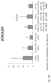
- FIGS. 13 A- 13 D are diagrams illustrating changed levels of adenine nucleotides (ATP, ADP, AMP) and ATP/ADP ratio as a measurement of mitochondrial phosphorylating capacity.
- FIGS. 14 A- 14 D are diagrams illustrating changed levels of oxidative and reduced nicotinic coenzymes.
- FIGS. 15 A- 150 are diagrams illustrating changed levels of biomarkers representative of oxidative stress.
- FIG. 16 is a diagram illustrating changed levels of nitrate as a measurement of NO-mediated nitrosative stress.
- FIGS. 17 A- 170 are diagrams illustrating changed levels of N-acetylaspartate (NAA) and its substrates.
- FIG. 18 schematically illustrates the effect on oxidative stress on mitochondrial (dys)function.
- FIG. 20 is a diagram illustrating changes in laminin immunoreactivity in the angle in subjects suffering from primary open-angle glaucoma (POAG) and treated with saline control or LMW-DS.
- POAG primary open-angle glaucoma
- FIG. 21 is a diagram illustrating changes in fibronectin immunoreactivity in the angle in subjects suffering from POAG and treated with saline control or LMW-DS.
- FIG. 22 illustrates amyloid- ⁇ monomer and oligomer preparation.
- the gels were loaded with 50 pmoles (lane 5), 100 pmoles (lanes 1, 3 and 6) or 200 pmoles (lanes 2, 4 and 7) of the respective 20 peptide preparation. Proteins on the arising Western blot were immuno-labelled with anti-amyloid- ⁇ . Predicted oligomers and the molecular weight markers are indicated.
- FIG. 23 illustrates dextran sulfate sodium salt (DSSS) and LMW-DS competition for the protein-protein interaction between amyloid- ⁇ and PrP c .
- DSSS dextran sulfate sodium salt
- FIG. 24 illustrates concentrations of NAA measured in deproteinized brain homogenates of rats sacrificed at 2 days post-TBI without and with a single administration of increasing doses of LWM-DS (1, 5 and 15 mg/kg b.w.), performed 30 minutes after trauma induction.
- Controls are represented by sham-operated animals. Values are the mean of 12 animals. Standard deviations are represented by vertical bars. *significantly different from controls, p ⁇ 0.01. **significantly different from sTBI 2 days, p ⁇ 0.01.
- FIG. 25 illustrates concentrations of ATP measured in deproteinized brain homogenates of rats sacrificed at 7 days post-sTBI, without and with administration of increasing doses of LWM-DS (single administration of 1, 5 and 15 mg/kg b.w. and repeated administration of 15 mg/kg b.w.).
- Controls are represented by sham-operated animals. Values are the mean of 12 animals. Standard deviations are represented by vertical bars. *significantly different from controls, p ⁇ 0.01. **significantly different from sTBI 2 days, p ⁇ 0.01.
- FIG. 26 illustrates concentrations of ascorbic acid measured in deproteinized brain homogenates of rats sacrificed at 7 days post-sTBI, without and with administration of increasing doses of LWM-DS (single administration of 1, 5 and 15 mg/kg b.w. and repeated administration of 15 mg/kg b.w.).
- Controls are represented by sham-operated animals. Values are the mean of 12 animals. Standard deviations are represented by vertical bars. *significantly different from controls, p ⁇ 0.01. **significantly different from sTBI 2 days, p ⁇ 0.01.
- FIG. 27 illustrates concentrations of glutathione (GSH) measured in deproteinized brain homogenates of rats sacrificed at 7 days post-sTBI, without and with administration of increasing doses of LWM-DS (single administration of 1, 5 and 15 mg/kg b.w. and repeated administration of 15 mg/kg b.w.).
- Controls are represented by sham-operated animals. Values are the mean of 12 animals. Standard deviations are represented by vertical bars. *significantly different from controls, p ⁇ 0.01. **significantly different from sTBI 2 days, p ⁇ 0.01.
- FIG. 28 illustrates concentrations of NAA measured in deproteinized brain homogenates of rats sacrificed at 7 days post-sTBI, without and with administration of increasing doses of LWM-DS (single administration of 1, 5 and 15 mg/kg b.w. and repeated administration of 15 mg/kg b.w.).
- Controls are represented by sham-operated animals. Values are the mean of 12 animals. Standard deviations are represented by vertical bars. *significantly different from controls, p ⁇ 0.01. **significantly different from sTBI 2 days, p ⁇ 0.01.
- the present embodiments generally relate to neurological and fibrotic conditions, and in particular to the use of dextran sulfate in combating such conditions.
- a neurological disorder is any disorder of the body nervous system, i.e., the brain, spine and the nerves that connect them. Structural, biochemical or electrical abnormalities in the brain, spinal cord or other nerves can result in a range of symptoms. Although the brain and spinal cord are surrounded by tough membranes, enclosed in the bones of the skull and spinal vertebrae, and chemically isolated by the blood-brain barrier, they are very susceptible if compromised. Nerves tend to lie deep under the skin but can still become exposed to damage. Individual neurons, and the neural networks and nerves into which they form, are susceptible to electrochemical and structural disruption. Neuroregeneration may occur in the peripheral nervous system and, thus, overcome or work around injuries to some extent, but it is thought to be rare in the brain and spinal cord.
- neurological problems vary, but can include genetic disorders, congenital abnormalities or disorders, infections, lifestyle or environmental health problems including malnutrition, and brain injury, spinal cord injury or nerve injury.
- the problem may start in another body system that interacts with the nervous system.
- cerebrovascular disorders involve brain injury due to problems with the blood vessels, i.e., the cardiovascular system, supplying the brain; autoimmune disorders involve damage caused by the body's own immune system; lysosomal storage diseases, such as Niemann-Pick disease, can lead to neurological deterioration.
- a neurodegenerative disease, disorder or condition is a disease, disorder or condition causing progressive loss of structure and/or function of neurons, including death of neurons.
- Non-limiting examples of such neurodegenerative diseases, disorders or conditions include Alzheimer's disease (AD), Parkinson's disease (PD), Huntington's disease (HD) and amyotrophic lateral sclerosis (ALS).
- AD Alzheimer's disease
- PD Parkinson's disease
- HD Huntington's disease
- ALS amyotrophic lateral sclerosis
- AD is characterized by loss of neurons and synapses in the cerebral cortex and subcortical regions.
- the classic neuropathologic findings in AD include amyloid plaques, neurofibrillary tangles, and synaptic and neuronal cell death.
- White matter disease (WMD) is frequently seen in AD at neuropathological examination. It is defined as a subtotal tissue loss with a reduction of myelin, axons and oligodendrocytes as well as astrocytosis.
- PD is a neurodegenerative disorder of the CNS.
- the motor symptoms of PD result from the death of dopamine-generating cells in the substantia nigra.
- the myelin sheath surrounding the axon begins to erode.
- Neuroinflammation is a pathological hallmark in PD and is characterized by activated microglia and infiltrating T cells at sites of neuronal injury.
- HD is a neurodegenerative disorder that affects muscle coordination and leads to cognitive decline and psychiatric problems.
- the disease is caused by an autosomal dominant mutation in a gene called Huntingtin.
- Part of this gene is a repeated section called trinucleotide repeat, which varies in length between individuals. When the length of this repeated section reaches a certain threshold, it produces an altered form of the protein.
- the protein (Htt) encoded by the Huntingtin gene interacts with over 100 other proteins and has multiple biological functions. The mutated form of Htt is toxic to certain cell types, particularly in the brain.
- HD is characterized by damages to the myelin sheath on the nerves. Increased activated T cells in the peripheral blood have been identified in HD patients.
- ALS also referred to as Lou Gehrig's disease
- ALS is a debilitating disease with varied etiology characterized by rapidly progressive weakness, muscle atrophy and fasciculations, muscle spasticity, dysarthria, dysphagia and dyspnea.
- ALS is the most common of the motor neuron diseases (ALS, hereditary spastic paraplegia (HSP), primary lateral sclerosis (PLS), progressive muscular atrophy (PMA), progressive bulbar palsy (PBP) and pseudobulbar palsy).
- HSP hereditary spastic paraplegia
- PLS primary lateral sclerosis
- PMA progressive muscular atrophy
- PBP progressive bulbar palsy
- pseudobulbar palsy The principle characteristic in the pathology of ALS is loss of motor nerve cells in the anterior horns of the spinal cord and in the motor nuclei of the brain stem. This results in secondary atrophy of the corresponding muscles (amyotrophy).
- Neuroinflammation is a pathological hallmark of ALS and is characterized by activated microglia and infiltrating T cells at sites of neuronal injury.
- “Lateral sclerosis” refers to corticospinal tract degeneration (lateral in location in the spinal cord). In fact, myelin loss occurs in the corticospinal tract.
- the sclerosis of ALS, the hardening involves the lateral columns, or corticospinal tracts and is a secondary phenomenon.
- a neurological disease, disorder or condition may be a demyelinating disease, disorder or condition.
- a demyelinating disease, disorder or condition is a disease of the nervous system in which the myelin sheath of neurons is damaged. Such damage impairs the conduction of signals in the affected nerves and thereby causing deficiency in sensation, movement, cognition and other functions depending on the nerves involved in the damage.
- demyelinating diseases, disorders or conditions include multiple sclerosis (MS), acute disseminated encephalomyelitis (ADEM), central nervous system (CNS) neuropathies, central pontine myelinolysis (CPM), myelopathies, leukoencephalopathies and leukodystrophies (all affecting the CNS), and Guillain-Barré syndrome (GBS), peripheral neuropathies and Charcot-Marie-Tooth (CMT) disease (all affecting the peripheral nervous system (PNS)).
- MS multiple sclerosis
- ADAM acute disseminated encephalomyelitis
- CNS central nervous system
- CCM central pontine myelinolysis
- myelopathies myelopathies
- leukoencephalopathies and leukodystrophies all affecting the CNS
- GBS Guillain-Barré syndrome
- CMT Charcot-Marie-Tooth
- MS is an inflammatory disease in which the fatty myelin sheaths around axons of the brain and the spinal cord are damaged, leading to demyelination and scarring as well as a broad spectrum of signs and symptoms.
- MS involves T cells that induce an immune response against the white matter of the brain and spinal cord.
- MS is a disease of myelin, not primarily of nerve cells. Since myelin occurs throughout the nervous system, lesions can be and typically are at multiple sites. The disease, however, affects only central myelin, not the myelin of peripheral nerves. Therefore, the symptoms are specifically of a CNS disorder.
- ADEM is an immune mediated disease of the brain. It usually occurs following a viral, bacterial or parasitic infection, or even appears spontaneously. ADEM attacks the nerves of the CNS and damages their myelin insulation, which, as a result, destroys the white matter. As it involves autoimmune demyelination, it is similar to MS, and is considered part of the MS borderline diseases. ADEM produces multiple inflammatory lesions in the brain and spinal cord, particularly in the white matter. ADEM involves cytokines secreted by myelin-reactive T cells.
- Neuropathies including CNS neuropathies and peripheral neuropathies, is a group of damages to or diseases affecting nerves, which may impair sensation, movement, gland or organ function, or other aspects of health, depending on the type of nerve affected.
- Common causes include systemic diseases, such as diabetes or leprosy; vitamin deficiency; medication, e.g., chemotherapy, or commonly prescribed antibiotics; traumatic injury; ischemia; radiation therapy; excessive alcohol consumption; immune system disease; Coeliac disease; or viral infection.
- Neuropathy may be or acute.
- Acute neuropathies demand urgent diagnosis. Motor nerves that control muscles, sensory nerves, or autonomic nerves that control automatic functions, such as heart rate, body temperature, and breathing, may be affected. More than one type of nerve may be affected at the same time.
- CPM is a neurological disease caused by severe damage of the myelin sheath of nerve cells in the brainstem, more precisely in the area termed the pons, predominately of iatrogenic etiology. It is characterized by acute paralysis, dysphagia, and dysarthria, and other neurological symptoms.
- Myelopathy describes any neurologic deficit related to the spinal cord. When due to trauma, it is generally known as spinal cord injury (SCI), when inflammatory, it is generally known as myelitis, and when the disease that is vascular in nature it is known as vascular myelopathy.
- SCI spinal cord injury
- myelitis when the disease that is vascular in nature it is known as vascular myelopathy.
- CSM cervical spondylotic myelopathy
- CSM cervical spondylotic myelopathy
- Leukoencephalopathy is a broad term for leukodystrophy-like diseases. It is applied to all brain white matter diseases, whether their molecular cause is known or not. Leukoencephalopathy can refer specifically to any of these diseases progressive multifocal leukoencephalopathy, toxic leukoencephalopathy, leukoencephalopathy with vanishing white matter, leukoencephalopathy with neuroaxonal spheroids, reversible posterior leukoencephalopathy syndrome, megalencephalic leukoencephalopathy with subcortical cysts.
- Leukodystrophy is one of a group of disorders characterized by degeneration of the white matter in the brain.
- the leukodystrophies are caused by imperfect growth or development of the myelin sheath, the fatty covering that acts as an insulator around nerve fibers.
- immune responses can lead to inflammation in the CNS, along with loss of myelin.
- Leukodystrophy is characterized by specific symptoms including decreased motor function, muscle rigidity, and eventually degeneration of sight and hearing.
- leukodystrophies include adrenomyeloneuropathy, Alexander disease, cerebrotendineous xanthomatosis, hereditary CNS demyelinating disease, Krabbe disease, metachromatic leukodystrophy, Pelizaeus-Merzbacher disease, Canavan disease, leukoencephalopathy with vanishing white matter, adrenoleukodystrophy and Refsum disease.
- GBS also referred to as Landry's paralysis or Guillan-Barré-Strohl syndrome
- GBS is an acute polyneuropathy affecting the PNS.
- immune cells attack the myelin sheath—the fatty substance covering nerve fibers.
- Ascending paralysis is a common symptom.
- GBS is thought to be an immune-mediated disease involving an abnormal T cell response precipitated by an infection.
- Cellular and humoral immune mechanisms probably play a role in its development.
- Many of the identified infectious agents are thought to induce production of antibodies that cross-react with specific gangliosides and glycolipids, such as GM1 and GD1b, which are distributed throughout the myelin in the peripheral nervous system.
- CMT is one of the hereditary motor and sensory neuropathies, a group of varied inherited disorders of the peripheral nervous system characterized by progressive loss of muscle tissue and touch sensation across various parts of the body. CMT was previously classified as a subtype of muscular dystrophy.
- oligodendrocytes and Schwann cells In neurological disorders loss of differentiation of neurons and glial cells, such as oligodendrocytes and Schwann cells, is one of the first stages in the disease progress. Generally, the disorders subsequently progress with cell death of such neurons and glial cells.
- a drug that is capable of promoting differentiation of neuronal and glial cells would be beneficial to patients suffering from neurological diseases, disorders or conditions.
- Such a differentiation-inducing drug could be neuroprotective and may, for instance, be useful in the treatment of neurological diseases, disorders or conditions.
- dextran sulfate of the embodiments is capable of inducing differentiation of neurons and glial cells. This effect of dextran sulfate is seen both for cortical neurons and motor neurons and for neurons from both mouse and human origin. Correspondingly, dextran sulfate is capable of inducing differentiation of Schwann cells that constitute a type of glial cells.
- Dextran sulfate of the embodiments additionally showed positive effects in an in vivo model of inflammatory demyelinating disease of the CNS, which is the currently most widely accepted animal model of MS and ADEM.
- Neurons also referred to as nerve cells, are electrically excitable cells that process and transmit information through electrical and chemical signals. These signals between neurons occur via synapses, specialized connections with other cells. Neurons can connect to each other to form neural networks. Neurons are the core components of the brain and spinal cord of the CNS, and of the ganglia of the PNS. Specialized types of neurons include: sensory neurons which respond to touch, sound, light and all other stimuli affecting the cells of the sensory organs that then send signals to the spinal cord and brain, motor neurons that receive signals from the brain and spinal cord to cause muscle contractions and affect glandular outputs, and interneurons which connect neurons to other neurons within the same region of the brain, or spinal cord in neural networks.
- a typical neuron consists of a cell body (soma), dendrites, and an axon.
- the term neurite is used to describe either a dendrite or an axon, particularly in its undifferentiated stage.
- Dendrites are thin structures that arise from the cell body, often extending for hundreds of micrometers and branching multiple times, giving rise to a complex “dendritic tree”.
- An axon also called a nerve fiber when myelinated, is a special cellular extension that arises from the cell body at a site called the axon hillock and travels for a distance. Nerve fibers are often bundled into fascicles, and in the PNS, bundles of fascicles make up nerves. At the majority of synapses, signals are sent from the axon of one neuron to a dendrite of another.
- Neurons do not undergo cell division. In most cases, neurons are generated by special types of stem cells. Astrocytes are star-shaped glial cells that have also been observed to turn into neurons by virtue of the stem cell characteristic pluripotency. In humans, neurogenesis largely ceases during adulthood; but in two brain areas, the hippocampus and olfactory bulb, there is strong evidence for generation of substantial numbers of new neurons.
- Dextran sulfate of the embodiments is capable of inducing an increase in beta-tubulin, in particular ⁇ III-tubulin, expression in the neurons.
- ⁇ III-tubulin also referred to as class III ⁇ -tubulin, is a microtubule element expressed exclusively in neurons.
- the microtubule cytoskeleton is essential for the development and survival of neurons.
- Microtubules are assembled from tubulin heterodimers, which contain different tubulin isotypes. Microtubules are polarized and, in neurons, their ‘minus-ends’ are usually oriented towards the centrosome in the cell body, whereas their ‘plus-ends’ project towards the tips of axons. Microtubule polarity serves important functions in both differentiating and adult neurons.
- tubulin is increased in the cell and builds up microtubule which allow the differentiating neurons to extend or retract growing axons in response to guidance cues in order to maintain directional growth towards post-synaptic targets.
- Their activities are essential for cell migration, axon development and guidance, and are also required for the function and viability of adult neurons (Bioscience Reports (2010), 30: 319-330).
- the increased expression of the ⁇ III-tubulin in neurons indicates that dextran sulfate of the embodiments acts as a differentiation factor for these cells.
- the neurons are selected from a group consisting of cortical neurons and motor neurons.
- a motor neuron is a nerve cell whose cell body is located in the spinal cord and whose axon projects outside the spinal cord to directly or indirectly control effector organs, mainly muscles and glands.
- the axons of motor neurons are efferent nerve fibers that carry signals from the spinal cord to the effectors to produce effects.
- MND motor neuron disease
- HSP also referred to as hereditary spastic paraparesis, familial spastic paraplegia, French settlement disease, or Strumpell-Lorrain disease
- HSP is a group of inherited diseases whose main feature is a progressive gait disorder.
- the disease presents with progressive stiffness (spasticity) and contraction in the lower limbs.
- the symptoms are a result of dysfunction of long axons in the spinal cord.
- the affected cells are the primary motor neurons, therefore the disease is an upper motor neuron disease.
- HSP is caused by defects in transport of proteins, structural proteins, cell maintaining proteins, lipids, and other substances through the cell.
- PLS is a rare neuromuscular disease characterized by progressive muscle weakness in the voluntary muscles. PLS only affects upper motor neurons.
- PMA also known as Duchenne-Aran muscular atrophy
- Duchenne-Aran muscular atrophy is a rare subtype of MND that affects only the lower motor neurons.
- PBP is a disease that attacks the nerves supplying the bulbar muscles. These disorders are characterized by the degeneration of motor neurons in the cerebral cortex, spinal cord, brain stem, and pyramidal tracts. This specifically involves the glossopharyngeal nerve (IX), vagus nerve (X), and hypoglossal nerve (XII).
- Pseudobulbar palsy is a medical condition characterized by the inability to control facial movements, such as chewing and speaking, and caused by a variety of neurological disorders. Patients experience difficulty chewing and swallowing, have increased reflexes and spasticity in tongue and the bulbar region, and demonstrate slurred speech, sometimes also demonstrating uncontrolled emotional outbursts.
- the condition is usually caused by the damage, bilateral degeneration, to the neurons of the brain stem, specifically to the corticobulbar tract (upper motor neuron tract to cranial nerve motor nuclei).
- SMA also called autosomal recessive proximal spinal muscular atrophy and 5q spinal muscular atrophy
- SMA is a rare neuromuscular disorder characterized by loss of motor neurons and progressive muscle wasting, often leading to early death.
- the disorder is caused by a genetic defect in the SMN1 gene, which encodes SMN, a protein widely expressed in all eukaryotic cells and necessary for survival of motor neurons. Lower levels of the protein results in loss of function of neuronal cells in the anterior horn of the spinal cord and subsequent system-wide atrophy of skeletal muscles.
- PPS also referred to as post-poliomyelitis syndrome or post-polio sequelae
- PPS is a condition that affects approximately 25 to 40% of people who have previously survived an acute attack of poliomyelitis—a viral infection of the nervous system—after the initial infection. Symptoms include acute or increased muscular weakness, pain in the muscles, and fatigue. The same symptoms may also occur years after a nonparalytic polio (NPP) infection.
- NPP nonparalytic polio
- the precise mechanism that causes PPS is unknown. It shares many features with chronic fatigue syndrome, but unlike that disorder, it tends to be progressive, and can cause loss of muscle strength.
- Cortical neurons are the cells of the cerebral cortex in the brain. Most of the complex activity of the brain enabling thought, perception, and voluntary movement is connected to the activity of cortical neurons.
- Glial cells sometimes referred to as neuroglia, are non-neuronal cells that maintain homeostasis, form myelin, and provide support and protection for neurons in the CNS and the PNS. Glial cells have four key functions; surrounding neurons and hold them in place, supplying nutrients and oxygen to neurons, insulating neurons from each other and destroying pathogens and removing dead neurons.
- Glial cell types present in the CNS include astrocytes, oligodendrocytes, ependymal cells, radial glia and microglia.
- Glial cell types present in the PNS include Schwann cells, satellite cells and enteric glial cells.
- Astrocytes also referred to as astroglia, are the most abundant type of macroglial cell in the CNS. Astrocytes have numerous projections that anchor neurons to their blood supply. They regulate the external chemical environment of neurons by removing excess ions and recycling neurotransmitters released during synaptic transmission. Astrocytes may regulate vasoconstriction and vasodilation by producing substances, such as arachidonic acid, whose metabolites are vasoactive.
- Oligodendrocytes are cells that coat axons in the CNS with their cell membrane, forming a specialized membrane differentiation called myelin, producing the so-called myelin sheath.
- the myelin sheath provides insulation to the axon that allows electrical signals to propagate more efficiently.
- Ependymal cells also referred to as ependymocytes, line the spinal cord and the ventricular system of the brain. These cells are involved in the creation and secretion of cerebrospinal fluid (CSF) and beat their cilia to help circulate the CSF and make up the blood-CSF barrier. They are also thought to act as neural stem cells.
- CSF cerebrospinal fluid
- Microglia are a type of neuroglia located throughout the brain and spinal cord. As the resident macrophage cells, they act as the first and main form of active immune defense in the CNS. Microglia are key cells in overall brain maintenance, they are constantly scavenging the CNS for plaques, damaged or unnecessary neurons and synapses, and infectious agents.
- Schwann cells are similar in function to oligodendrocytes but are present in the PNS instead of the CNS. Thus, Schwann cells provide myelination to axons in the PNS. They also have phagocytotic activity and clear cellular debris that allows for regrowth of PNS neurons.
- Satellite glial cells are small cells that surround neurons in sensory, sympathetic, and parasympathetic ganglia. These cells help regulate the external chemical environment. They are highly sensitive to injury and inflammation, and appear to contribute to pathological states, such as chronic pain. Enteric glial cells are found in the intrinsic ganglia of the digestive system. They are thought to have many roles in the enteric system, some related to homeostasis and muscular digestive processes.
- Dextran sulfate of the embodiments further induces an increase in myelin basic protein (MBP) expression in the glial cells.
- MBP myelin basic protein
- MBP is a protein that is important in the process of myelination of nerves in the nervous system and is a major constituent of the myelin sheath of oligodendrocytes and Schwann cells. MBP maintains the correct structure of myelin, interacting with the lipids in the myelin membrane. Interest in MBP has centered on its role in demyelinating diseases, in particular MS.
- Axonal myelination is an essential process for normal functioning of vertebrate CNS.
- myelin is formed by the differentiation of the plasma membrane of Schwann cells. Loss of axonal contact, as occurs after nerve injury, leads to the down-regulation of myelin gene expression ( Progress in Neurobiology (2000), 61: 267-304). The differentiation of Schwann cells and increase in MBP in injured peripheral nerves is critical for regeneration after injury ( Frontiers in Neuroscience (2015), 9: Article 298, 1-13).
- MBP dextran sulfate of the embodiments acts as a differentiation factor for these cells
- the glial cells are myelinating cells, i.e., cells creating a myelin sheath that is wrapped around one or more axons of adjacent neurons.
- the glial cells are selected from a group consisting of Schwann cells and oligodendrocytes.
- Dextran sulfate of the embodiment does not only induce differentiation of cells of the CNS and PNS, which is beneficial in neurological diseases, disorders and conditions.
- Experimental data as presented herein indicates that dextran sulfate of the embodiments has positive effect in combating metabolic modifications that are seen in neurological diseases, disorders and conditions, such as traumatic brain injury (TBI).
- TBI traumatic brain injury
- many neurological diseases, disorders and conditions are characterized by modifications of various metabolites connected to the cell energy state and mitochondrial functions.
- modifications in amino acid metabolisms are seen in many neurological diseases, disorders and conditions.
- These metabolic modifications are early cellular signals that influence changes in enzymatic activities and gene and protein expressions indicative of a pathological tissue response.
- Dextran sulfate of the embodiments acts to positively regulate cellular metabolism in the compromised tissues, thereby inhibiting or at least suppressing any subsequent modifications in enzyme activity and gene and protein expression that contribute to adverse outcomes.
- dextran sulfate of the embodiments was capable of reducing levels of glutamate excitotoxicity and ameliorated adverse changes in metabolic hemostastis, thereby efficiently protecting mitochondrial function and providing a neuroprotective effect.
- Dextran sulfate of the embodiments positively affected various compounds related to energy metabolism and mitochondrial functions. Particularly interesting are the concentrations of adenine nucleotides and ATP/ADP ratio as measurement of mitochondrial phosphorylating capacity.
- Dextran sulfate of the embodiments also led to a significant reduction in oxidative stress.
- the levels of ascorbic acid, as the main water-soluble brain antioxidant, and glutathione (GSH), as the major intracellular-sulfhydryl group (SH) donor were significantly improved.
- GSH glutathione
- MDA malondialdehyde
- ROS reactive oxygen species
- Dextran sulfate administration also significantly decreased the nitrate concentrations in both acute and chronic phases of neurological diseases, disorders and conditions. Accordingly, dextran sulfate of the embodiments has a positive effect on NO-mediated nitrosative stress.
- N-acetylaspartate is a brain specific metabolite and a valuable biochemical marker for monitoring deterioration or recovery after neurological diseases, disorders and conditions, such as TBI.
- NAA is synthesized in neurons from aspartate and acetyl-CoA by aspartate N-acetyltransferase. Dextran sulfate of the embodiment showed significant improvements in NAA levels.
- dextran sulfate of the embodiments can thereby protect against the cell loss that occurs due to oxidative stress and/or glutamate excitotoxicity in the diseased and damaged nervous system.
- dextran sulfate of the embodiments may be a useful protective treatment in many degenerative conditions where cells are progressively lost due to ischemic, oxidative or traumatic damage, such as stroke, ALS, MND, MS, dementia, TBI, SCI, retinal damage, etc.
- ischemic, oxidative or traumatic damage such as stroke, ALS, MND, MS, dementia, TBI, SCI, retinal damage, etc.
- Dextran sulfate of the embodiments induces scavenging of released glutamate in glial cells and thereby prevent accumulation of toxic amounts of glutamate in the neuronal clefts. This will be useful in all neurodegenerative diseases, disorders and conditions, both acute and chronic, where neurons are dying.
- Excitotoxicity is the pathological process by which nerve cells are damaged or killed by excessive stimulation by neurotransmitters, in particular glutamate. This occurs when receptors for the excitatory neurotransmitter glutamate, such as the N-methyl-D-aspartate (NMDA) receptor and ⁇ -amino-3-hydroxy-5-methyl-4-isoxazolepropionic acid (AMPA) receptor are overactivated by glutamatergic storm or when neurons are damaged or dies, releasing their content of glutamate.
- NMDA N-methyl-D-aspartate
- AMPA ⁇ -amino-3-hydroxy-5-methyl-4-isoxazolepropionic acid
- Excitotoxicity may be involved in SCI, stroke, TBI, hearing loss (through noise overexposure or ototoxicity), and in neurodegenerative diseases of the CNS, such as MS, AD, ALS, PD, alcoholism or alcohol withdrawal and especially over-rapid benzodiazepine withdrawal, and also HS.
- Other common conditions that cause excessive glutamate concentrations around neurons are hypoglycemia.
- glutamate concentration can be increased up to 1 mM in the synaptic cleft, which is rapidly decreased in the lapse of milliseconds.
- glutamate concentration around the synaptic cleft cannot be decreased or reaches higher levels, the neuron kills itself by a process called apoptosis.
- This pathologic phenomenon can also occur after brain injury, such as in TBI, and SCI.
- damaged neural cells within the lesion site spill glutamate into the extracellular space where glutamate can stimulate presynaptic glutamate receptors to enhance the release of additional glutamate. Brain trauma or stroke can cause ischemia, in which blood flow is reduced to inadequate levels.
- Ischemia is followed by accumulation of glutamate in the extracellular fluid, causing cell death, which is aggravated by lack of oxygen and glucose.
- the biochemical cascade resulting from ischemia and involving excitotoxicity is called the ischemic cascade. Because of the events resulting from ischemia and glutamate receptor activation, a deep chemical coma may be induced in patients with brain injury to reduce the metabolic rate of the brain, its need for oxygen and glucose, and save energy to be used to remove glutamate actively.
- NMDA N-methyl-D-aspartate
- NMDA N-methyl-D-aspartate
- One of the damaging results of excess calcium in the cytosol is initiating apoptosis through cleaved caspase processing.
- Another damaging result of excess calcium in the cytosol is the opening of the mitochondrial permeability transition pore, a pore in the membranes of mitochondria that opens when the organelles absorb too much calcium. Opening of the pore may cause mitochondria to swell and release reactive oxygen species and various proteins that can lead to apoptosis. The pore can also cause mitochondria to release more calcium.
- production of adenosine triphosphate (ATP) may be stopped, and ATP synthase may in fact begin hydrolyzing ATP instead of producing it.
- ATP adenosine triphosphate
- Inadequate ATP production resulting from brain trauma can eliminate electrochemical gradients of certain ions.
- Glutamate transporters require the maintenance of these ion gradients to remove glutamate from the extracellular space. The loss of ion gradients results in not only the halting of glutamate uptake, but also the reversal of the transporters.
- the Na + -glutamate transporters on neurons and astrocytes can reverse their glutamate transport and start secreting glutamate at a concentration capable of inducing excitotoxicity. This results in a buildup of glutamate and further damaging activation of glutamate receptors.
- the activation of glutamate transporter in glial cells by dextran sulfate of the embodiments to prevent or at least inhibit accumulation of toxic levels of glutamate will effectively protect surrounding neurons from glutamate excitotoxicity.
- dextran sulfate of the embodiments protect neurons from damages and cell death that is otherwise the result of this glutamate excitotoxicity.
- dextran sulfate of the embodiments was effective in restoring mitochondrial related energy metabolism, profoundly imbalanced in subject suffering from brain damages, such as severe TBI (sTBI), with positive effects on the concentration of triphosphates purine and pyrimidine nucleotides.
- ATP levels were only 16% lower than the value of healthy control subjects, whilst in untreated sTBI subjects a 35% decrease was found.
- NAA concentration in sTBI subjects treated with dextran sulfate was only 16% lower than the value of healthy control subjects, whilst sTBI subjects showed 48% lower values of this compound. This finding once again strongly confirms the strict connection between the homeostasis of NAA and correct mitochondrial energy metabolism, and underlines the importance of pharmacological interventions capable to act positively on mitochondrial functioning.
- dextran sulfate administration also involved nicotinic coenzymes and metabolism of free CoA-SH and CoA-SH derivatives. This implies that dextran sulfate treated subjects, notwithstanding submitted to sTBI, had quasi-normal coenzymes to ensure correct oxido-reductive reactions and to allow a good functioning of the TCA cycle.
- dextran sulfate affected sulfur-containing amino acids. Possibly, this effect might be related to the dextran sulfate molecule that contains S atoms. Increasing the bioavailability of this atom might have produced a net increase in the biosynthesis of these amino acids, one of them (MET) is crucial in the methylation reaction and in the so called methyl cycle.
- Dextran sulfate of the embodiments further has an affinity to compete for the protein-protein interaction between oligomeric amyloid- ⁇ and PrP c , which will have a beneficial effect in subjects suffering from AD, prion diseases or amyloidosis.
- Gene-expression data as presented herein indicates that dextran sulfate of the embodiments has a role in Schwann cells, neurons and in human umbilical vein endothelial cells (HUVECs) in protection against apoptosis; induction of angiogenesis (in HUVECs); increased migration and movement of cells; increased cell viability and survival; and induction of cellular differentiation.
- HUVECs human umbilical vein endothelial cells
- dextran sulfate of the embodiments can protect against cell damage and promotes the development of new blood vessels in injured or diseased tissue, such as following stroke or other ischemic conditions.
- dextran sulfate of the embodiments also induced expression of a protein complex of calmodulin (CALM), which is a multifunctional intermediate calcium-binding messenger protein; G beta-gamma complex (G ⁇ ), which is a tightly bound dimeric G protein complex composed of one G ⁇ and one G ⁇ subunit; metabotropic glutamate receptor 7 (GRM7); and protein interacting with C kinase-1 (PICK1).
- CLM calmodulin
- G ⁇ G beta-gamma complex
- G ⁇ G beta-gamma complex
- G ⁇ G ⁇
- G ⁇ metabotropic glutamate receptor 7
- PICK1 protein interacting with C kinase-1
- dextran sulfate of the embodiment is capable of preventing and inhibiting apoptosis, preventing amyloid- ⁇ and Lewy body pathology and its negative effects on mitochondrial fragmentation and dysfunction, and subsequent damage and inhibiting fatty acid oxidation.
- Dextran sulfate of the embodiments also improved mitochondrial function, reduced the mitochondrial level of H 2 O 2 and reactive oxygen species.
- dextran sulfate of the embodiments enhanced the effect of existing growth factors on cells.
- dextran sulfate of the embodiments was capable of modulating the effect of several growth factors by either increasing their activation or by reducing their inhibition.
- This means that dextran sulfate of the embodiments has potential use in diseases, disorders and conditions in which an increase of the activity or a reduction of the inhibition of these growth factors would be beneficial to the patient.
- diseases, disorders and conditions include ALS; stroke; SCI; depression and other psychiatric disorders, such as mood disorders and bipolar disease; and metabolic disorders.
- dextran sulfate binds to the growth factor molecules and facilitates binding to their receptors. This hypothesis is also supported by the observation that the dextran sulfate-induced differential gene expression in HUVECs, where the normal control medium already contained heparin, was relatively smaller than in the Schwann cells where the normal control medium did not contain heparin. This mechanism of action also explains why dextran sulfate is mainly effective in the acute stage of TBI, when growth factors are present, but less effective at later stage when the initial repair attempt has already diminished.
- dextran sulfate of the embodiments depends on existing repair mechanisms, which are amplified by it.
- dextran sulfate may be a useful protective treatment in many degenerative conditions where cells are progressively lost due to ischemic, oxidative or traumatic damage.
- degenerative conditions include stroke, ALS, MS, dementia, TBI, SCI, retinal damage, AD, etc.
- Dextran sulfate of the embodiments may help the damaged tissues to recover some lost function as it enhances the residual intrinsic repair mechanisms.
- the gene-expression data therefore confirms the potential therapeutic usefulness of dextran sulfate of the embodiments in compromised states of the CNS and PNS, by promoting revascularisation, reducing secondary tissue damage, and promoting repair, and for neurodegenerative diseases, disorders and conditions, where it could promote neuronal survival, differentiation and ultimately repair.
- a further interesting effect of dextran sulfate of the embodiments is that it affects cell adhesion.
- Cell adhesion was affected mainly in neurons and Schwann cells, where dextran sulfate of the embodiments promoted cell detachment and movement.
- the effect on cell adhesion was mainly due to the expression of metalloproteinase-type enzymes.
- This finding would also explain an anti-scarring effect of dextran sulfate of the embodiments.
- the results suggest that an anti-scarring effect mediated by dextran sulfate of the embodiments by activating degrading enzymes that help tissue remodeling and block the fibrogenic (scarring) signals in damaged tissues.
- the metalloproteinase-type enzymes that are activated by dextran sulfate of the embodiments specifically act by dissolving the fibrous molecules that make up the scar, see Table 10-11. These enzymes are released by cells that migrate into damaged tissues. Accordingly, by allowing these cells to be more mobile, reducing their adhesion, dextran sulfate of the embodiments is permitting them to migrate better, release scar dissolving enzymes and remodel the tissue for better repair.
- the anti-scarring actions of dextran sulfate of the embodiments indicate a potential use to treat fibroproliferative (scarring) conditions.
- fibroproliferative include, for instance, glaucoma, proliferative vitreoretinopathy, brain and spinal trauma injuries, sub-arachnoid hemorrhage in the brain, invasive surgical procedures, surgical adhesions, rotator cuff injuries, burns, reconstructive surgery, ulcerative conditions (diabetes), etc.
- fibrotic diseases and conditions include fibrosis in the lungs, such as pulmonary fibrosis, cystic fibrosis, idiopathic pulmonary fibrosis, progressive massive fibrosis and radiation-induced lung injury following treatment for cancer; in the liver, such as cirrhosis and biliary atresia; fibrosis in the heart, such as atrial fibrosis, endomyocardial fibrosis, old myocardial infarction; fibrosis in the brain, such as glial scar; pancreatitis; arthrofibrosis; Crohn's disease; Dupuytren's contracture; keloid; mediastinal fibrosis; myelofibrosis; Peyronie's disease; nephrogenic systemic fibrosis; retroperitoneal fibrosis; scleroderma or systemic sclerosis.
- fibrosis in the lungs such as pulmonary fibrosis, cystic fibrosis, i
- Fibrosis may also occur in connection with organ transplantation, such as of kidneys, lungs, livers, hearts, etc., and in connection with cell therapies and cell transplantation, such as of islet of Langerhans, hepatocytes, insulin producing cells, stem cells, progenitor cells, etc.
- organ transplantation such as of kidneys, lungs, livers, hearts, etc.
- cell therapies and cell transplantation such as of islet of Langerhans, hepatocytes, insulin producing cells, stem cells, progenitor cells, etc.
- the gene expression data also shows that dextran sulfate of the embodiments activates the production of a natural scar reducing molecule called decorin, which further blocks scar production by ‘mopping up’ the growth factors that stimulate scar production by fibroblasts.
- Decorin is a glycoprotein of on average 90-140 kD molecular weight. It belongs to the small-leucine rich proteoglycan (SLRP) family and consists of a protein core containing leucine repeats with a glucosaminoglycan (GAG) chain consisting of either chondroitin sulphate or dermatan sulphate. It binds to type I collagen fibrils through the decorin type I collagen binding region.
- SLRP small-leucine rich proteoglycan
- GAG glucosaminoglycan
- Decorin acts as a transforming growth factor beta 1 or 2 (TGF- ⁇ 1/2) antagonist and reduces scarring. Reports show that in acute scarring the dominant effect of decorin is anti-fibrogenic through suppression of inflammatory fibrosis by neutralization of TGF- ⁇ 1/2. Decorin also binds directly to collagen and one of its functions is to influence on the organization of collagen during wound healing.
- TGF- ⁇ 1/2 transforming growth factor beta 1 or 2
- Decorin has previously been described in inhibition of scarring in a model of cerebral lesion, hydrocephalus, and chronic spinal cord wounds. Decorin also induces fibrolysis of existing trabecular meshwork scars in a glaucoma model.
- dextran sulfate of the embodiments Taken together the anti-scarring actions of dextran sulfate of the embodiments indicate the potential for use to treat all clinical conditions where scarring is a problem. Dextran sulfate should work on both old and new scars. This is confirmed in the experimental data showing that dextran sulfate of the embodiments was capable of inducing dissolution of already established scar elements in the trabecular meshwork in glaucomatous eyes. This is a significant advantage of dextran sulfate of the embodiments since it cannot only be used to inhibit or at least suppress fibrosis and deleterious scar formation but also dissolve already established scars. This means that dextran sulfate of the embodiments allows for a scar dissolving and tissue remodeling for a better repair.
- Dextran sulfate was assessed in a panel of human primary cell-based assays modeling complex tissue and disease biology and general tissue biology. The results from the assay indicate that dextran sulfate plays a role in regulating immune activation and/or immune resolution responses in the context of inflammation and wound healing biology.
- the modulations of the inflammatory markers indicate utility of dextran sulfate in treating multiple chronic and acute inflammatory conditions and diseases including inflammatory components, such as ALS.
- the innate/proinflammatory response and selected components of the acquired immune response are up-regulated to maintain a defense against foreign pathogens, clear tissue debris present at the injury site, and orchestrate tissue remodeling, cell proliferation and angiogenic processes associated with the wound response.
- this initial inflammatory response has to be regulated or shut down so as to allow for the reestablishment of matrix, recellularization and tissue remodeling.
- Such immune resolving activities were induced by dextran sulfate, including activation of MMP-1, PAR-1 and uPAR, indicating an induced immune resolution having utility in treating tissue damaged by trauma, including neurotrauma, which otherwise would result in deleterious fibrosis formation.
- the effect in inflammation resolution of dextran sulfate as shown in the experimental data indicates that dextran sulfate would be useful in preventing, treating or at least inhibiting auto-immune diseases, and in particular auto-immune diseases effecting the central and/or peripheral nervous system.
- the inflammation resolution of dextran sulfate is also important in terms of blocking fibrogenesis.
- resolution of inflammation and suppression of microglial responses as seen from the experimental data are also important in neurodegenerative diseases, disorders and conditions.
- the dextran sulfate, or the pharmacologically acceptable derivative thereof would be useful preventing, treating or at least inhibiting neuroinflammation and neuroinflammatory conditions.
- neuroinflammatory conditions include PD, ALS, MS, ADEM, myelitis and GDS.
- dextran sulfate seemed to normalize and resolve the inflammation present in tissue after trauma or a disease and these results are thereby consistent with the effects of dextran sulfate seen in gene array and animal studies.
- the function of the nervous system depends on the number of nerve cells, a healthy energy metabolism of the nerve cells and healthy connections between the nerve cells.
- Neurodegenerative diseases and disorders, and injuries causing neurodegeneration typically have different triggers and causes but all lead to the same end-results, i.e., neurodegeneration.
- the functional effects of such diseases, disorders or injuries are often seen only after a comparatively large number of nerve cells are dead, whereas the triggers of the diseases or disorders may be present years before the symptoms occur.
- a new approach is needed to treat or inhibit neurodegeneration.
- Such an approach should involve enhancing viable functions of the nervous system including a healthy energy metabolism of the nerve cells and healthy connections between the nerve cells.
- further neurodegeneration should be prevented or at least slowed down by reducing the triggers that lead to neuronal death and prevent further pathology even if triggers are present.
- the regenerative potential of the nervous system should be enhanced.
- triggers of neuron apoptosis that all contribute to neuronal loss during neurodegeneration and damage.
- These triggers include dysregulation of neurotransmitters leading to glutamate excitotoxicity and oxidative stress leading to mitochondrial dysfunction, thereby limiting the energy supply to neurons.
- dysregulated neurofilaments lead to reduced motility and restricted supply of factors needed for neuron survival.
- Further triggers include release of inflammatory mediators causing secondary cell damage and scarring.
- vascular defects are common in neurodegenerative conditions.
- Glutamate is produced in neurons and is pivotal for signaling mechanisms that support learning and memory in neurons. Excess glutamate released is in healthy brain tissue mopped up by glial cells to prevent toxic levels. Dextran sulfate induces an increased glutamate uptake by glia cells, whereas the glutamate production in neurons is not altered by dextran sulfate. Hence, the glutamate needed for learning and memory is not affected by dextran sulfate administrations, whereas harmful toxic amounts of glutamate is mopped up by glial cells. Accordingly, dextran sulfate attenuates the dysregulation of neurotransmitters leading to glutamate excitotoxicity.
- Dextran sulfate reduced production of molecules that induce oxidative stress, including amyloid- ⁇ and Lewy bodies, and reduced oxidative stress.
- dextran sulfate prevents neuronal death induced by oxidative stress and prevents mitochondrial dysfunction in neurons.
- dextran sulfate promotes a normalization of mitochondrial function in presence of oxidative stress and prevents energy crisis in neurons in presence of such oxidative stress. Accordingly, dextran sulfate attenuates oxidative stress in neurodegeneration that otherwise would lead to mitochondrial dysfunction.
- a further trigger in neurodegeneration is dysregulated neurofilaments, which lead to reduced motility and restricted supply of survival factors.
- Dextran sulfate enhances the effect of growth factors present in neurons, increases migration and movement of nerve cells, reduces the production of degeneration-related protein products and induces cellular differentiation. Accordingly, dextran sulfate attenuates dysregulated neurofilaments.
- Neurodegeneration also induces release of inflammatory mediators causing secondary cell damage and scarring.
- inflammatory cytokines in particular TGF- ⁇ .
- Dextran sulfate induces metallopeptidase expression, induces expression of the natural anti-scarring molecule decorin and inhibits TGF- ⁇ activity.
- dextran sulfate inhibits immune cell adhesion, cell aggregation, cell activation and fibrosis even in the presence of excessive TGF- ⁇ . Accordingly, dextran sulfate attenuates the negative effects, including scarring, caused by release inflammatory mediators.
- Dextran sulfate also acts to inhibit fibrogenesis as well as activating fibrolysis, which in combination leads to the beneficial effects seen by dextran sulfate in attenuating or even dissolving scarring.
- Dextran sulfate protects HUVECs against apoptosis, induced angiogenesis and increased migration and movement of the endothelial cells. Accordingly, dextran sulfate enhances the physiological repair response in hypoxic tissues caused by neurodegenerative diseases, disorders or injuries but does not affect the normal healthy vasculature.
- an aspect of the embodiments relates to a method of inducing differentiation of cells selected from a group consisting of glial cells and neurons.
- the method comprises contacting the cells with dextran sulfate, or a pharmaceutically acceptable derivative thereof, in order to induce differentiation of the cells.
- the method is an in vitro method.
- contacting the cells comprises contacting the cells in vitro with the dextran sulfate, or the pharmaceutically acceptable derivative thereof.
- the cells are treated with and interacts in vitro with dextran sulfate, or the pharmaceutically acceptable derivative thereof.
- the method may also be an ex vivo method, in which the neurons and/or glial cells have been extracted from a subject and is to be contacted with the dextran sulfate, or the pharmaceutically acceptable derivative thereof, outside of the subject's body.
- the neurons and/or glial cells treated by the dextran sulfate, or the pharmaceutically acceptable derivative thereof, in the above described in vitro or ex vivo method to induce differentiation may be transplanted into a subject.
- the differentiated neurons and/or glial cells should then exert their desired function in the subject's body.
- the subject may be suffering from a neurological disease as is further described herein.
- the dextran sulfate, or the pharmaceutically acceptable derivative thereof is, in an alternative embodiment, administered to a subject, such as a subject suffering from a neurological disease, disorder or condition.
- the dextran sulfate, of the pharmaceutically acceptable derivative thereof will then contact neurons and/or glial cells inside the subject's body to induce cell differentiation.
- the method is an in vivo method.
- Another aspect of the embodiments relates to dextran sulfate, or a pharmaceutically acceptable derivative thereof, for use in inducing differentiation of cells selected from a group consisting of glial cells and neurons.
- the dextran sulfate, of the pharmaceutically acceptable derivative thereof is for use in inducing differentiation of the cells in a subject suffering from a neurological disease, disorder or condition.
- the subject is a human subject suffering from a neurodegenerative disease, disorder or condition selected from a group consisting of AD, PD, HD and ALS.
- the subject is a human subject suffering from a demyelinating disease, disorder or condition selected from a group consisting of MS, ADEM, a CNS neuropathy, CPM, a myelopathy, a leukoencephalopathy, a leukodystrophy, GBS, a peripheral neuropathy and Charcot-Marie-Tooth disease, preferably selected from a group consisting of MS, ADEM, CPM and GBS.
- a demyelinating disease, disorder or condition selected from a group consisting of MS, ADEM, a CNS neuropathy, CPM, a myelopathy, a leukoencephalopathy, a leukodystrophy, GBS, a peripheral neuropathy and Charcot-Marie-Tooth disease, preferably selected from a group consisting of MS, ADEM, CPM and GBS.
- the dextran sulfate, or the pharmaceutically acceptable derivative thereof may also, or alternatively, be used in inducing differentiation of the cells in other types of neurological diseases, disorders or conditions.
- neurological diseases, disorders or conditions include neuro ischemic diseases, such as stroke, cerebral ischemic conditions and critical limb ischemia (CLI); neuromuscular disorders, such as ALS, botulism, congenital myasthenic syndromes, congenital myopathies, cramp-fasciculation syndrome, cerebral palsy, elevated creatine kinase, fasciculations, inclusion-body myositis, Lambert-Eaton syndrome, mitochondrial myopathy, motor neuron disease, muscle disorders, muscular dystrophy, myasthenia gravis, myotonic dystrophy, neuromuscular junction disorders, neuromyotonia, peripheral neuropathy and polymyositis; traumatic nerve injuries and post-operative neurological conditions.
- neuro ischemic diseases such as stroke, cerebral ischemic conditions and critical limb ischemia (CLI)
- a further aspect of the embodiments relates to dextran sulfate, or a pharmaceutically acceptable derivative, for use in treating, inhibiting or preventing glutamate excitotoxicity in a subject.
- This aspect also relates to a method of treating, inhibiting or preventing glutamate excitotoxicity.
- the method comprises administering dextran sulfate, or a pharmaceutically acceptable derivative thereof, to a subject in order to treat, inhibit or prevent glutamate excitotoxicity
- Other aspects of the embodiments relates to dextran sulfate, or a pharmaceutically acceptable derivative thereof, for use in protecting neurons from oxidative stress induced by a neurological disease, disorder or condition, for use in ameliorating adverse changes in metabolic hemostasis in neurons induced by a neurological disease, disorder or condition, protecting mitochondrial function and mitochondrial energy metabolism in neurons in a subject suffering from a neurological disease, disorder or condition.
- Dextran sulfate or the pharmaceutically acceptable derivative thereof, can thereby be used to treat, inhibit or prevent a neurological disease, disorder or condition as described herein.
- Dextran sulfate can also be used to treat, inhibit or prevent ischemic, oxidative or traumatic damage to neurons and the CNS, or PNS, such as stroke, ALS, MND, MS, dementia, TBI, SCI, retinal damage, etc.
- a further aspect relates to dextran sulfate, or a pharmaceutically acceptable derivative thereof, for use in treating, inhibiting or preventing fibrosis in a subject, and in particular for use in treating or inhibiting, such as by dissolving, established scars in a subject suffering from fibrosis or a fibrotic disease, disorder or condition.
- dextran sulfate of the embodiments having an anti-scarring effect would be effective in wound treatment and tissue remodeling, in which there is a need for dissolving already established scars in order to enable a correct wound healing.
- This anti-scarring effect of dextran sulfate of the embodiments is thought to be a consequence of the previously described mechanisms of action of dextran sulfate including, for instance, inhibition of cell adhesion, induction of cell mobilization, induction of metalloproteases and scar dissolving enzymes, and inhibition of TGF ⁇ , in particular TGF ⁇ 1, through the induction of decorin.
- This latter effect obtained with dextran sulfate of the embodiments is further of relevance in preventing or at least inhibiting fibrosis and scar formation through the induction of decorin.
- Another aspect relates to dextran sulfate, or a pharmaceutically acceptable derivative thereof, for use in treating, inhibiting or prevent neuroinflammation in a subject, in particular in a subject suffering from a neurological disease, disorder or condition causing neuroinflammation.
- Relates aspect of the embodiments define use of dextran sulfate, or a pharmaceutically acceptable derivative thereof, for the manufacture of a medicament for the various medical applications as disclosed herein, e.g., for treating, inhibiting or prevent any of the diseases, disorders or conditions as disclosed herein.
- dextran sulfate or the pharmaceutically acceptable derivative thereof.
- dextran sulfate, or the pharmaceutically acceptable derivative thereof is administered to the subject to treat, inhibit or prevent the disease, disorder or condition as disclosed herein.
- the pharmaceutically acceptable derivative of dextran sulfate preferably has the average molecular weight and sulfur content as discussed in the following embodiments.
- Dextran sulfate outside of the preferred ranges of the embodiments are believed to have inferior effect and/or causing negative side effects to the cells or subject.
- dextran sulfate of a molecular weight exceeding 10,000 Da (10 kDa) generally has a lower effect vs. side effect profile as compared to dextran sulfate having a lower average molecular weight.
- This means that the maximum dose of dextran sulfate that can be safely administered to a subject is lower for larger dextran sulfate molecules (>10,000 Da) as compared to dextran sulfate molecules having an average molecular weight within the preferred ranges.
- larger dextran sulfate molecules are less appropriate in clinical uses when the dextran sulfate is to be administered to subjects in vivo.
- Dextran sulfate is a sulfated polysaccharide and in particular a sulfated glucan, i.e., polysaccharide made of many glucose molecules.
- Average molecular weight as defined herein indicates that individual sulfated polysaccharides may have a molecular weight different from this average molecular weight but that the average molecular weight represents the mean molecular weight of the sulfated polysaccharides. This further implies that there will be a natural distribution of molecular weights around this average molecular weight for a dextran sulfate sample.
- Average molecular weight, or more correctly weight average molecular weight (M w ), of dextran sulfate is typically determined using indirect methods such as gel exclusion/penetration chromatography, light scattering or viscosity. Determination of average molecular weight using such indirect methods will depend on a number of factors, including choice of column and eluent, flow rate, calibration procedures, etc.
- the parameter N i indicates the number of dextran sulfate molecules having a molecular weight of M i in a sample or batch.
- the dextran sulfate or the pharmaceutically acceptable derivative thereof has a M w equal to or below 10,000 Da. In a particular embodiment, the dextran sulfate or the pharmaceutically acceptable derivative thereof has a M w within an interval of from 2,000 Da to 10,000 Da.
- the dextran sulfate or the pharmaceutically acceptable derivative thereof has a M w within an interval of from 2,500 Da to 10,000 Da, preferably within an interval of from 3,000 Da to 10,000 Da. In a particular embodiment, the dextran sulfate or the pharmaceutically acceptable derivative thereof has a M w within an interval of from 3,500 Da to 9,500 Da, such as within an interval of from 3,500 Da to 8,000 Da.
- the dextran sulfate or the pharmaceutically acceptable derivative thereof has a M w equal to or below 10,000 Da, equal to or below 9,500 Da, equal to or below 9,000 Da, equal to or below 8,500 Da, equal to or below 8,000 Da, equal to or below 7,500 Da, equal to or below 7,000 Da, equal to or below 6,500 Da, equal to or below 6,000 Da, or equal to or below 5,500 Da.
- the dextran sulfate or the pharmaceutically acceptable derivative thereof has a M w equal to or above 1,000 Da, equal to or above 1,500 Da, equal to or above 2,000 Da, equal to or above 2,500 Da, equal to or above 3,000 Da, equal to or above 3,500 Da, equal to or above 4,000 Da. or equal to or above 4,500 Da. Any of these embodiments may be combined with any of the above presented embodiments defining upper limits of the M w , such combined with the upper limit of equal to or below 10,000 Da.
- NMR nuclear magnetic resonance
- the dextran sulfate, of the pharmaceutically acceptable derivative thereof has a M n as measured by NMR spectroscopy within an interval of from 1,850 to 3,500 Da.
- the dextran sulfate, of the pharmaceutically acceptable derivative thereof has a M n as measured by NMR spectroscopy within an interval of from 1,850 Da to 2,500 Da, preferably within an interval of from 1,850 Da to 2,300 Da, such as within an interval of from 1,850 Da to 2,000 Da.
- the dextran sulfate or the pharmaceutically acceptable derivative thereof has a M n equal to or below 3,500 Da, equal to or below 3,250 Da, equal to or below 3,000 Da, equal to or below 2,750 Da, equal to or below 2,500 Da, equal to or below 2,250 Da, or equal to or below 2,000 Da.
- the dextran sulfate or the pharmaceutically acceptable derivative thereof has a M n equal to or above 1,850 Da.
- the dextran sulfate, or the pharmaceutically acceptable derivative thereof has an average sulfate number per glucose unit within an interval of from 2.5 to 3.0.
- the dextran sulfate, or the pharmaceutically acceptable derivative thereof has an average sulfate number per glucose unit within an interval of from 2.5 to 2.8, preferably within an interval of from 2.6 to 2.7.
- the dextran sulfate, or the pharmaceutically acceptable derivative thereof has an average number of glucose units within an interval of from 4.0 to 6.0.
- the dextran sulfate, or the pharmaceutically acceptable derivative thereof has an average number of glucose units within an interval of from 4.5 to 5.5, preferably within an interval of from 5.0 to 5.2.
- the dextran sulfate has a M n as measured by NMR spectroscopy within an interval of from 1,850 to 3,500 Da, an average sulfate number per glucose unit within an interval of from 2.5 to 3.0, and an average sulfation of C2 position in the glucose units of the dextran sulfate is at least 90%.
- the dextran sulfate has an average number of glucose units of about 5.1, an average sulfate number per glucose unit within an interval of from 2.6 to 2.7 and a M n within an interval of from 1,850 Da and 2,000 Da.
- the pharmaceutically acceptable derivative of dextran sulfate is a sodium salt of dextran sulfate.
- the sodium salt of dextran sulfate has an average number of glucose units of about 5.1, an average sulfate number per glucose unit within an interval of from 2.6 to 2.7 and a M n including the Na + counter ion within an interval of from 2,100 Da to 2,300 Da.
- the dextran sulfate has an average number of glucose units of 5.1, an average sulfate number per glucose unit of 2.7, an average M n without Na + as measured by NMR spectroscopy of about 1,900-1,950 Da and an average M n with Na + as measured by NMR spectroscopy of about 2,200-2,250 Da.
- the dextran sulfate according to the embodiments can be provided as a pharmaceutically acceptable derivative of dextran sulfate, such as a pharmaceutically active derivative of dextran sulfate.
- pharmaceutically acceptable derivatives include pharmaceutically acceptable salts and pharmaceutically acceptable solvates of dextran sulfate, e.g., a sodium or potassium salt.
- the subject is preferably a mammalian subject, more preferably a primate and in particular a human subject.
- the dextran sulfate, or the pharmaceutically acceptable derivative thereof can, however, be used also in veterinary applications.
- animal subjects include primate, cat, dog, pig, horse, mouse, rat.
- the dextran sulfate, or the pharmaceutically acceptable derivative thereof is preferably administered by injection to the subject and in particular by intravenous (i.v.) injection, subcutaneous (s.c.) injection or (i.p.) intraperitoneal injection, preferably i.v. or s.c. injection.
- Other parenteral administration routes that can be used include intramuscular and intraarticular injection.
- Injection of the dextran sulfate, or the pharmaceutically acceptable derivative thereof could alternatively, or in addition, take place directly in, for instance, a tissue or organ or other site in the subject body, at which the target effects are to take place.
- the dextran sulfate, or the pharmaceutically acceptable derivative thereof may alternatively, or in addition, be administered intrathecally.
- the dextran sulfate, or the pharmaceutically acceptable derivative thereof can be injected together with a suitable aqueous carrier or solution into the spinal canal, or into the subarachnoid space so that it reaches the cerebrospinal fluid (CSF).
- CSF cerebrospinal fluid
- a further administration route is intraocular administration.
- the dextran sulfate, or the pharmaceutically acceptable derivative thereof, of the embodiments is preferably formulated as an aqueous injection solution with a selected solvent or excipient.
- the solvent is advantageously an aqueous solvent and in particular a buffer solution.
- a non-limiting example of such a buffer solution is a citric acid buffer, such as citric acid monohydrate (CAM) buffer, or a phosphate buffer.
- CAM citric acid monohydrate
- phosphate buffer phosphate buffer
- dextran sulfate of the embodiments can be dissolved in saline, such as 0.9% NaCl saline, and then optionally buffered with 75 mM CAM and adjusting the pH to about 5.9 using sodium hydroxide.
- non-buffered solutions are possible, including aqueous injection solutions, such as saline, i.e., NaCl (aq).
- other buffer systems than CAM could be used if a buffered solution are desired.
- the embodiments are not limited to injections and other administration routes can alternatively be used including orally, nasally, bucally, rectally, dermally, tracheally, bronchially, or topically.
- the active compound, dextran sulfate is then formulated with a suitable excipient or carrier that is selected based on the particular administration route.
- Suitable dose ranges for the dextran sulfate, or the pharmaceutically acceptable derivative thereof may vary according to the application, such as in vitro versus in vivo, the size and weight of the subject, the condition for which the subject is treated, and other considerations.
- a possible dosage range could be from 1 ⁇ g/kg to 100 mg/kg of body weight, preferably from 10 ⁇ g/kg to 50 mg/kg of body weight.
- the dextran sulfate, or the pharmaceutically acceptable derivative thereof is formulated to be administered at a dosage in a range from 0.05 to 50 mg/kg of body weight of the subject, preferably from 0.05 or 0.1 to 40 mg/kg of body weight of the subject, and more preferably from 0.05 or 0.1 to 30 mg/kg, or 0.1 to 25 mg/kg or from 0.1 to 15 mg/kg or 0.1 to 10 mg/kg body weight of the subject.
- the dextran sulfate, or the pharmaceutically acceptable derivative thereof does not necessarily have to be limited to treatment or inhibition of a present disease, disorder or condition but could alternatively, or in addition, be used for prophylaxis.
- the dextran sulfate, or the pharmaceutically acceptable derivative thereof could be administered to a subject that will undergo a medical procedure, such as surgery, that may cause nerve injuries or damage and/or fibrosis.
- the dextran sulfate, or the pharmaceutically acceptable derivative thereof may also be used to prevent, inhibit or alleviate post-operative neurological complications and conditions in a subject that is about to undergo a medical procedure, such as surgery, and/or fibrosis.
- the dextran sulfate, or the pharmaceutically acceptable derivative thereof can be administered at a single administration occasion, such as in the form of a single bolus injection.
- This bolus dose can be injected quite quickly to the subject but is advantageously infused over time so that the dextran sulfate solution is infused over a few minutes of time to the patient, such as during 5 to 10 minutes.
- the dextran sulfate, or the pharmaceutically acceptable derivative thereof can be administered at multiple, i.e., at least two, occasions during a treatment period.
- the dextran sulfate, or the pharmaceutically acceptable derivative thereof can be administered together with other active agents, either sequentially, simultaneously or in the form of a composition comprising the dextran sulfate, or the pharmaceutically acceptable derivative thereof, and at least one other active agent.
- the at least one active agent can be selected among any agent useful in any of the above mentioned diseases, disorders or conditions.
- the at least one active agent could also be in the form of cells in cell therapy, such as stem cells including, but not limited to, embryonic stem cells (ESCs) and mesenchymal stromal cells (MSCs).
- stem cells have been conducted on animal models of brain degeneration, such as in Parkinson's disease, MS, ALS, and Alzheimer's disease. Furthermore clinical and animal studies have been conducted into the use of stem cells in cases of TBI.
- the dextran sulfate, or the pharmaceutically acceptable derivative thereof has beneficial effects to cells in vitro as shown in the experimental data.
- the dextran sulfate, or the pharmaceutically acceptable derivative thereof protects the cells from oxidative stress, restores metabolic hemostasis in the cells, which is beneficial for the energy metabolism in the cells, and may act as a differentiation factor for the cells.
- These beneficial effects of the dextran sulfate, or the pharmaceutically acceptable derivative thereof may also find uses in other types of cell therapy, i.e., not necessarily limited to stem cell therapy.
- Non-limiting, but illustrative, examples of such other types of cell therapy include myocardial cells, liver cells, connective tissue cells, optic nerve cells, lymphocytes, macrophages, glial cells, Schwann cells, neurons, etc.
- the cells may be treated with the dextran sulfate, or the pharmaceutically acceptable derivative thereof, in vitro prior to administration into a subject.
- the cells may be administered together with the dextran sulfate, or the pharmaceutically acceptable derivative thereof.
- dextran sulfate or the pharmaceutically acceptable derivative thereof
- treatment of tissue and organs in vitro or ex vivo with the dextran sulfate, or the pharmaceutically acceptable derivative thereof could be useful to benefit from the positive effects of dextran sulfate of the embodiment, for instance protection against oxidative stress and restoration of metabolic hemostasis.
- treatment of cells, tissue and organs, in addition or as an alternative, following transplantation with the dextran sulfate, or the pharmaceutically acceptable derivative thereof would be possible.
- dextran sulfate is advantageously administered to the subject at an early or acute state following a damage causing the disease, disorder or conditions, such as TBI, or at an early or acute state following diagnosis of the disease, disorder or condition.
- a damage causing the disease, disorder or conditions such as TBI
- dextran sulfate of the embodiments is its capability of boosting and amplifying the intrinsic repair mechanism in the CNS and PNS. This is in particular relevant for treatment or inhibition of neurological diseases.
- the anti-scarring effect as seen by dextran sulfate of the embodiments indicates that the dextran sulfate will be effective also in dissolving already existing scar tissue and elements.
- dextran sulfate of the embodiments will have a therapeutic effect also during a late or chronic state.
- LMW-DS low molecular weight dextran sulfate
- the study aims were to evaluate the effect of LMW-DS on cell survival and expression of differentiation proteins in three cell types, cerebral cortical neurons, motor neurons and Schwann cells, using two concentrations 0.01 and 0.1 mg/ml of LMW-DS.
- Neurons were cultured as 40,000 cells per well and Schwann cells 3,000 cells per. Cells were treated after 24 hours. The number of cells per well depended on the growth phenotype, proliferative capacity, etc.
- 96-well plates were coated by adding 100 ⁇ l per well of a solution of 50 ⁇ g/ml poly-d-lysine (Sigma) in Hanks' Balanced Salt Solution (HBSS, Sigma) and incubating overnight at 37° C. in the dark. Plates were washed with cell culture water (Fisher) and air-dried for 30 min in the dark.
- HBSS Hanks' Balanced Salt Solution
- PNGM was prepared by adding PNGM Singlequots (Lonza) to PNBM medium and pre-warmed to 37° C. Cells were thawed in a 37° C. water bath for no longer than 2 min and gently transferred into a 15 ml tube. 5 ml of medium was gently added drop-wise. Cell suspension was mixed by inverting the tube carefully twice. Cells were counted with a Cellometer AUTO T4 (Nexcelom Bioscience). 40,000 cells per well were seeded in previously coated 96-well plates. Cells were incubated at 37° C. with 5% CO 2 . After at 2-hour incubation 80 ⁇ l of medium was removed and replaced with 80 ⁇ l of fresh medium and cells were allowed to settle for 24 hours before drug treatment.
- NeuroBlast was pre-warmed to 37° C. Cells were thawed in a 37° C. water bath for no longer than 2 min. 1 ml of media was gently added drop-wise. Cells were resuspended and transferred to a 15 ml tube containing 9 ml of medium. Cells were centrifuged at 200 relative centrifugal force (RCF) for 5 min. Pellet was resuspended in 5 ml of medium and cells were counted with the Cellometer. 40,000 cells per well were seeded in previously coated 96-well plates. Cells were incubated at 37° C. with 5% CO 2 . Cells were allowed to settle for 24 hours before drug treatment. After 24 hours NeuroBlast medium was replaced with MotorBlast medium (Lonzo).
- MotorBlast medium MotorBlast medium
- Schwann cells growth medium was prepared by adding 10% of fetal bovine serum (FBS, PAA) to high-glucose DMEM and pre-warmed to 37° C. Cells were thawed in a 37° C. water bath for no longer than 2 min. Cells were gently transferred to a tube containing 10 ml of medium and centrifuged at 200 RCF for 5 min. Pellet was resuspended in 5 ml of medium and cells were counted with the Cellometer. 3,000 cells per well were seeded in previously coated 96-well plates. Cells were incubated at 37° C. with 5% CO 2 . Cells were allowed to settle for 24 hours before drug treatment.
- FBS fetal bovine serum
- LMW-DS was prepared in the culture media of choice for each cell line and added to the respective wells in the doses 0.01 and 0.1 mg/ml.
- PBST Myelin Basic Protein
- the Acumen cytometer allows the direct cytometric analysis of attached cells without prior detachment. Therefore cells were imaged in situ and based on DNA content (PI) categorized in different phases of cell cycle or deemed apoptotic or polyploid.
- PI DNA content
- the protein content of the cells can also be directly measured and expressed as either ‘total protein content’ or ‘mean protein content’.
- the DNA histograms in FIG. 1 showed that the PI uptake of the cells were altered, shift of histograms to the right, which indicated that LMW-DS treatment had an effect on the cortical neurons.
- the cell population (G2/M phase) that started to divide is indicated in the figure.
- the data for motor neurons was similar to the cortical neurons, with a shift in PI uptake ( FIG. 2 ) and a small increase in proliferation within a population of very small cells.
- Tubulin is a family of proteins that are important building blocks in the cytoskeleton of cells.
- the ⁇ III-tubulin is expressed solely by neurons.
- the intensity of tubulin was significantly increased in the cells treated with LMW-DS ( FIG. 5 A ).
- Analysis of the positive cells showed that these cells were larger than the positive cells in the control culture ( FIG. 5 B ).
- LMW-DS treatment led to an apparent cell loss in the cultures. It is believed that this effect of LMW-DS treatment was not due to a toxic effect. It is more likely that LMW-DS affected neuronal attachment. For instance, even the maximum measurements of apoptotic fraction (after the adjustment for the PI shift) did not explain by far the loss of cells in the cultures and the apparent cell loss was much greater in the immunostained preparations (more washes) than in the PI preparations.
- LMW-DS appeared to promote the differentiation of both neuronal and Schwann cells within a very short period of time (48 hours).
- AD trauma-related neurodegeneration
- post-stroke dementia are associated with the reactivation of cell cycle related phenomena in neurons.
- differentiation-inducing drugs have been proposed to be neuroprotective.
- Drugs supporting differentiation of Schwann cells would also be good candidates for the treatment of diseases associated with demyelination.
- EAE Error Encephalomyelitis
- An EAE model in mice is the currently most widely accepted animal model of MS and ADEM in humans ( Annals of Neurology, 60: 12-21, 2006).
- EAE is induced in mice with a single injection of peptides and proteins, including Myelin Oligodendrocyte Glycoprotein 35-55 (MOG 35-55 ) emulsified with adjuvant, which triggers an immune reaction against myelin.
- MOG 35-55 Myelin Oligodendrocyte Glycoprotein 35-55
- Inflammatory lesions of the CNS causing peripheral paralysis are characteristics of EAE in mice. Disease progression in the mice is followed by daily evaluation of disease symptoms using a well-recognized and evaluated scoring system ( International Immunology, 10: 333-340, 1998).
- mice females, 8-10 weeks were obtained from Harlan Europe. Mice were housed in the conventional animal facility, Lund University, Sweden, and kept at 12 h light/dark cycles in polystyrene cages (type IIL cages, max 7 mice per cage) containing wood shavings and fed with standard rodent chow and water ad libitum.
- polystyrene cages type IIL cages, max 7 mice per cage
- EAE was induced day 0 by a s.c. injection at the flank of an emulsion containing 150 ⁇ g MOG 35-55 and 300 ⁇ g H37RA in a volume of 100 ⁇ l per mouse.
- the emulsion was prepared by mixing complete Freund's adjuvant (CFA) (H37RA in IFA at a concentration of 6 mg/ml) and MOG 35-55 (dissolved in PBS to a concentration of 3 mg/ml) on ice. Mice were anesthetized during immunization to ascertain correct location of the injection.
- CFA complete Freund's adjuvant
- Pertussis toxin was re-suspended in mqH 2 O at a concentration of 50 ⁇ g/ml and diluted to a final concentration of 1 ⁇ g/ml in PBS. Mice received a booster injection of 200 ng PTX i.p. on day 0 and day 2.
- LMW-DS dilutions were prepared on day 0 and 14 for group 3.
- LMW-DS was diluted in 0.9% saline solution and sterile filtered through a 0.2 ⁇ M filter, according to doses described in Table 2 below. Vehicle given was 0.9% saline solution.
- Recombinant HGF was reconstituted in 1 ml 0.1% bovine serum albumin (BSA) in PBS at a concentration of 25 ⁇ g/ml and further diluted in PBS to 1 ⁇ g/ml.
- BSA bovine serum albumin
- Cyclosporine A was prepared by dissolving 50 mg in 1 ml 70% ethanol and diluted in HPMC to final concentration of 0.98 mg/ml.
- Treatment was initiated day 0 for group 2-3, which was administered i.p. in group 2 and s.c. in group 3 three times weekly. Treatment was initiated day 18 for remaining groups. Animals in group 4 were administered every other day i.v., with a total of three injections. The treatment groups were mixed within cages to avoid cage effects and systemic errors caused by unequal housing.
- Clinical disease was monitored daily where the disease is graded according to a scale ranging from 0-8.
- FIG. 10 illustrates the EAE development in mice in control groups (vehicle and Cyclosporine A) and a group treated with LMW-DS s.c. three times weekly, where Cyclosporine A had significantly (*) lower mean score on day 13, 14, 16, 20, 21 and 25-27 compared with vehicle control. Animals treated with 10 mg/kg dextran sulfate s.c. three times weekly had significantly (#) lower mean score on day 13, 14, 16, 17, 19, 21 and 26 compared with vehicle control.
- FIG. 11 illustrates vehicle and mice treated with 100 ng/dose HGF i.v. every other day for five days stating at day 18 (see arrow). HGF did not result in any significant difference as compared to vehicle.
- LMW-DS thus, resulted in a significantly lower mean score compared with vehicle control in the EAE model. Accordingly, the results indicate that LMW-DS has positive effects in neurodegenerative and demyelinating diseases of the CNS, such as MS and ADEM.
- LMW-DS The effects of daily sub-cutaneous injections of LMW-DS on glutamate excitotoxicity and mitochondrial function after severe traumatic brain injury (sTBI) in rats were evaluated by high-performance liquid chromatography (HPLC) analysis of frozen brain samples. The results suggest that LMW-DS interferes with mitochondrial function to improve energy metabolism and also decreases glutamate excitotoxicity.
- n 6 animals subjected to sTBI, with drug administration after 30 minutes and sacrifice at 2 days post-TBI (Acute phase 1)
- n 6 animals subjected to severe-TBI, with drug administration after 30 minutes and sacrifice at 7 days post-TBI (Acute phase 2).
- n 6 animals subjected to severe-TBI, with drug administration after 3 days and sacrifice at 7 days post-TBI (Chronic phase).
- mice received 35 mg/kg b.w. ketamine and 0.25 mg/kg b.w. midazolam by i.p. injection.
- sTBI was induced by dropping a 450 g weight from 2 m height on to the rat head that had been protected by a metal disk previously fixed on the skull, according to the “weight drop” impact acceleration model (Marmarou et al., A new model of diffuse brain injury in rats. Part I: Pathophysiology and biomechanics. J Neurosurg. 1994; 80: 291-300). Rats that suffered from skull fracture, seizures, nasal bleeding, or did not survive the impacts, were excluded from the study. At the end of each period of treatment, rats were anesthetized again and then immediately sacrificed.
- the drug treatment was a subcutaneous injection of 0.5 ml of LMW-DS (15 mg/kg) and administered according to the aforementioned schematic protocol.
- tissue preparation was affected as previously disclosed (Tavazzi et al., Cerebral oxidative stress and depression of energy metabolism correlate with severity of diffuse brain injury in rats. Neurosurgery. 2005; 56: 582-589; Vagnozzi et al., Temporal window of metabolic brain vulnerability to concussions: mitochondrial-related impairment-part I. Neurosurgery.
- whole brain homogenization was performed with 7 ml of ice-cold, nitrogen-saturated, precipitating solution composed by CH 3 CN+10 mM KH 2 PO 4 , pH 7.40, (3:1; v:v), and using an Ultra-Turrax set at 24,000 rpm/min (Janke & Kunkel, Staufen, Germany). After centrifugation at 20,690 ⁇ g, for 10 min at 4° C., the clear supernatants were saved, pellets were supplemented with 3 ml of the precipitating solution and homogenized again as described above.
- a second centrifugation was performed (20,690 ⁇ g, for 10 min at 4° C.), pellets were saved, supernatants combined with those previously obtained, extracted by vigorous agitation with a double volume of HPLC-grade CHCl 3 and centrifuged as above.
- the upper aqueous phases containing water-soluble low-molecular weight compounds were collected, subjected to chloroform washings for two more times (this procedure allowed the removal of all the organic solvent and of any lipid soluble compound from the buffered tissue extracts), adjusted in volumes with 10 mM KH 2 PO 4 , pH 7.40, to have ultimately aqueous 10% tissue homogenates and saved at ⁇ 80° C. until assayed.
- Metabolites belonging to the purine-pyrimidine profiles (listed below) and related to tissue energy state, mitochondrial function and relative to oxidative-nitrosative stresses were separated, in a single chromatographic run, according to slight modifications of existing ion-pairing HPLC methods (Lazzarino et al., Single-sample preparation for simultaneous cellular redox and energy state determination. Anal Biochem. 2003; 322: 51-59; Tavazzi et al., Simultaneous high performance liquid chromatographic separation of purines, pyrimidines, N-acetylated amino acids, and dicarboxylic acids for the chemical diagnosis of inborn errors of metabolism. Clin Biochem. 2005; 38: 997-1008).
- the derivatization mixture composed by 25 mmol/l OPA, 1% MPA, 237.5 mmol/l sodium borate, pH 9.8 was prepared daily and placed in the autosampler.
- HPLC column Hypersil C-18, 250 ⁇ 4.6 mm, 5 ⁇ m particle size, thermostated at 21° C.
- deproteinized brain extracts were diluted 20 times with HPLC-grade H 2 O prior to the derivatization procedure and subsequent injection.
- LMW-DS positively affected various compounds related to energy metabolism and mitochondrial functions. Particularly interesting are the concentrations of adenine nucleotides and ATP/ADP ratio as measurement of mitochondrial phosphorylating capacity ( FIG. 13 ).
- MDA as end product of polyunsaturated fatty acids of membrane phospholipids and therefore taken as a marker of ROS-mediated lipid peroxidation, was also measured. MDA levels showed a significant reduction after administration of LMW-DS. The oxidative stress markers described above all indicated an improvement in the recovery of antioxidant status after treatment with LMW-DS ( FIG. 15 ).
- NAA is a brain specific metabolite and a valuable biochemical marker for monitoring deterioration or recovery after TBI.
- NM is synthesized in neurons from aspartate and acetyl-CoA by aspartate N-acetyltransferase.
- ASPA aspartoacylase
- An upregulation of the catabolic enzyme ASPA and an NAA decrease in order to supply the availability of the substrates asparate and acetyl-CoA are an indication of the status of metabolic impairment.
- NAA and its substrates were measured after sTBI and showed significant improvements in levels after LMW-DS administration ( FIG. 17 ).
- LMW-DS has a potential to be used in the treatment or inhibition of TBI, including STBI.
- n 8 ⁇ 25 cm 2 culture flasks were set up. Two flasks were harvested for each cell type on the day of treatment (24 hours after seeding). This represents the Day0 time point. From the remaining flasks, three flasks were treated with Control Medium and three were treated with Culture Medium (CM) containing LMW-DS to give a final concentration of 0.01 mg/ml. Cells from the treated flasks were collected after 48 hours. Therefore the collected data represent (a) untreated cells (Day0 Controls and Day2 Controls) and (b) cells treated with LMW-DS for 48 hours (Day2 LMW-DS treated).
- 25 cm 2 flasks were coated by adding 2 ml per flask of a solution of 50 ⁇ g/ml poly-d-lysine in Hank's balanced salt solution (HBSS) and incubating overnight at 37° C. in the dark. Flasks were washed with cell culture water and air-dried for 30 min in the dark. Flasks were coated by adding 1 ml per flask of a solution of 25 ⁇ g/ml laminin in phosphate-buffered saline (PBS) and incubating for 2 hour at 37° C. in the dark. The laminin flasks were washed with PBS three times before plating cells.
- PBS phosphate-buffered saline
- VECs Human Umbilical Vein Endothelial Cells
- M200+LVES additive 1:50 was prepared and pre-warmed to 37° C. Cells were thawed in a 37° C. water bath for no longer than 2 min and gently transferred into a 50 ml tube containing 20 ml Dulbecco's Modified Eagle Medium, Nutrient Mixture F-12 (DMEM-F12). The cell suspension was mixed by inverting the tube carefully twice. Cells were spun at 400 ⁇ g for 10 minutes. Supernatant removed and cells were resuspended in 10 ml of culture media (M200+LVES additive).
- M200+LVES additive Dulbecco's Modified Eagle Medium, Nutrient Mixture F-12
- Medium was prepared by adding 10 ml B-27 Serum-Free Supplement and 2.5 ml GlutaMAXTM-1 Supplement to 500 ml of Neurobasal medium. The medium was pre-warmed to 37° C. Cells from 12 vials were thawed sequentially in a 37° C. water bath for no longer than 2 min and gently transferred into a 15 ml tube. 9 ml of medium was gently added drop-wise to each. The cell suspension was mixed by inverting the tubes carefully twice.
- the culture medium was prepared according to Table 5.
- CM was aspirated into a clean and labelled 15 ml Falcon tube.
- the flasks (without culture medium) were placed into the ⁇ 80° C. freezer for 30 minutes.
- the CM in the Falcon tubes were spun at 3000 ⁇ g for 5 minutes. Supernatant was removed and the small pellet was re-suspended in 2.5 ml Trizol:Water (4:1) solution at room temperature (RT, ⁇ 22° C.).
- the mixture separated into three layers: a lower red phenol-chloroform phase, an interphase and a colorless upper aqueous phase.
- the RNA remained in the top aqueous phase, DNA in the white middle (interphase) phase and protein in the pink bottom (organic) phase.
- the top 3 ⁇ 4 of the aqueous phase was transferred to a new clean Eppendorf tube.
- the expression data were downloaded into separate files for each cell line.
- the ‘Background corrected’ expression is the data from the “gProcessedSignal” of the arrays that is the result of the background signal extracted from the actual signal of the relevant probe. This is the most often used variable in array analysis.
- the background corrected signal was log 2 transformed for all samples for statistical analysis. To reduce the false discovery rate in the samples, the signals that were below ‘expression level’ were removed. The ‘below expression’ level was set at 5 for the log 2 transformed expression values.
- Seeding densities were calculated from the cell counts retrieved from the cell stocks for the Schwann cells.
- the HUVECS were seeded at their optimum density.
- the molecular functions influenced by these genes relate to cell morphology (1.43E-08-8.39E-04); cellular movement (1.4E-07-9.6E-04); post-translational modification (3.93E-07-6.71E-05); protein synthesis (3.93E-07-1.08E-04); protein trafficking (3.93E-07-1.26E-06); cell death and survival (2.13E-06-8.65E-04); cellular assembly and organization (7.46E-06-8.24E-04); DNA replication, recombination, and repair (7.46E-06-7.46E-06); cellular function and maintenance (9.53E-06-6.46E-04); gene expression (1.27E-05-4.92E-04); cellular development (1.29E-05-9.06E-04); cellular growth and proliferation (1.29E-05-9.06E-04); cell-to
- the molecular functions influenced by these genes relate to cell morphology (1.49E-07-5.62E-03); cellular assembly and organization (1.49E-07-5.95E-03); cellular movement (7.24E-07-6.06E-03); cell death and survival (9.41E-06-5.95E-03); amino acid metabolism (2.56E-05-3.7E-03); post-translational modification (2.56E-05-1.05E-03); small molecule biochemistry (2.56E-05-3.7E-03); cell-to-cell signaling and interaction (5.05E-05-5.76E-03); gene expression (7.18E-05-4.94E-03); cell cycle (1.06E-04-5.95E-03); cellular development (1.06E-04-5.95E-03); cellular function and maintenance (1.96E-04-5.95E-03); cellular
- the mechanistic molecular network model simulates the effect of the differentially regulated molecules by LMW-DS enabling the functional consequences of these changes to be evaluated.
- the in silico model indicated that LMW-DS inhibits neuronal cell death; apoptosis; and synthesis of protein and activates angiogenesis; migration of cells; cell viability; cell survival; cell movement; proliferation of cells; differentiation of cells; cellular homeostasis; cell cycle progression; cell transformation; and expression of RNA.
- 1551 genes were differentially expressed in HUVEC cultures when comparing the D0 control to the D2 control samples.
- the molecular functions influenced by these genes relate to cellular assembly and organization (2.55E-15-1.29E-03); cellular function and maintenance (2.55E-15-1.29E-03); cell cycle (1.98E-11-1.32E-03); cell morphology (3.18E-10-1.29E-03); gene expression (1.05E-08-2.01E-04); cellular development (1.66E-07-1.37E-03); cellular growth and proliferation (1.66E-07-1.37E-03); DNA replication, recombination, and repair (2.04E-07-9.84E-04); cell death and survival (2.09E-07-1.3E-03); RNA post-transcriptional modification (4.86E-06-6.53E-04); cellular movement (9.9E-06-1.18E-03); post-translational modification (1.92E-05-1.34E-03); cell-to-cell signaling and
- the molecular functions influenced by these genes relate to cellular assembly and organization (4.14E-17-9.7E-04); cellular function and maintenance (4.14E-17-8.05E-04); cell cycle (5.83E-14-9.85E-04); cell morphology (1.69E-10-7.48E-04); gene expression (7.99E-09-8.62E-04); cell death and survival (2E-08-8.4E-04); cellular development (1.28E-07-8.88E-04); cellular growth and proliferation (1.28E-07-8.88E-04); DNA replication, recombination, and repair (3.07E-07-9.7E-04); RNA post-transcriptional modification (1.13E-06-6.31E-04); cellular movement (1.42E-06-8.34E-04); post-translational modification (3.4E-05-9.17E-04); cell-to-cell signaling and interaction (6
- the molecular functions influenced by these genes relate to DNA replication, recombination, and repair (9.62E-05-2.57E-02); cell cycle (1.22E-04-2.4E-02); cellular development (1.59E-04-2.67E-02); cell morphology (4.64E-04-2.42E-02); cellular function and maintenance (4.64E-04-2.57E-02); lipid metabolism (9.49E-04-1.07E-02); molecular transport (9.49E-04-1.61E-02); small molecule biochemistry (9.49E-04-1.87E-02); cellular compromise (1.6E-03-2.62E-02); cell death and survival (2.06E-03-2.67E-02); amino acid metabolism (2.7E-03-2.7E-03); carbohydrate metabolism (2.7E-03-1.07E-02); cell-to-cell signaling and interaction
- LMW-DS inhibits neuronal cell death; apoptosis; and synthesis of protein and activates angiogenesis; migration of cells; cell viability; cell survival; cell movement; proliferation of cells; differentiation of cells; cellular homeostasis; cell cycle progression; cell transformation; and expression of RNA.
- the HUVEC control cultures comprise growth factors.
- LMW-DS was added to the culture medium that already contained growth factors.
- Table 7 summarizes the results of the gene expression changes in the cultured HUVECs. 67 genes that have altered expression in the Control cultures in the two days (under the effect of the growth factors) did not show any changes at all in the LMW-DS treated cultures during the same two days. 4 genes that had increased expression in the control cultures with the growth factors were downregulated in the LMW-DS treated cultures during the same two days. 11 genes that were downregulated by the growth factors in the control cultures were upregulated in the LMW-DS treated cultures during the two days. 120 genes were significantly downregulated by growth factors and this downregulation was even stronger in the LMW-DS treated cultures. 229 genes were upregulated in the Control cultures and the addition of LMW-DS made this upregulation significantly stronger.
- LMW-DS The effect of LMW-DS on several molecular pathways that are important for different disease conditions and therapeutic applications were analyzed.
- the effects of adding LMW-DS on gene expression was compared to that seen in cells in CM and the functional effects were predicted based on the observed changes in the expression patterns.
- the ‘below expression’ level was set at 5 for the log 2 transformed expression values. This left 12,240 unique probes where the expression threshold was met by at least three samples in the series. In the next step, the three sets of data were analyzed to establish the effect of the CM on the cells and the differences induced by the LMW-DS.
- the changes in gene expression under normal culture conditions mimic the normal developmental processes of the motor neurons, when from a dissociated set of cells they develop a motor neuron phenotype.
- the growth factors in the normal culture medium are those necessary for these cells to differentiate.
- the stress factor present in these cultures is the oxidative stress (normal for tissue culture conditions).
- genes were differentially expressed in motor neuron cultures when comparing the D0 control to the D2 control samples.
- the molecular functions influenced by these genes relate to cell death and survival (1.99E-17-1.98E-04); cellular movement (1.14E-16-1.91E-04); cellular assembly and organization (1.22E-16-1.93E-04); cellular function and maintenance (1.22E-16-1.95E-04); cell morphology (6.46E-16-1.74E-04); cell-to-cell signaling and interaction (3.16E-12-1.95E-04); cellular development (1.59E-10-1.93E-04); cellular growth and proliferation (1.59E-10-1.9E-04); molecular transport (4.27E-10-1.89E-04); protein synthesis (9.85E-09-5.03E-05); lipid metabolism (1.08E-08-1.61E-04); small molecule biochemistry (1.08E-08-1.89E-04); gene expression (8.45E-08-3.8E-05); cell cycle (4.55E-07-1.09E-04); free
- the molecular functions influenced by these genes relate to cell death and survival (6.54E-08-9.06E-03), cellular movement (8.21E-08-5.42E-03); cellular assembly and organization (8.36E-08-9.01E-03); cellular function and maintenance (8.36E-08-9.01E-03); cell morphology (2.9E-06-8.75E-03); cellular development (1.04E-05-9.01E-03); cellular growth and proliferation (1.04E-05-7.83E-03); DNA replication, recombination, and repair (2.79E-05-8.01E-03); cell-to-cell signaling and interaction (8.18E-05-7.11E-03); post-translational modification (1.32E-04-7.56E-03); protein degradation (1.32E-04-4.35E-03); protein synthesis (1.32E-04-5
- the molecular functions influenced by these genes relate to cell death and survival (2.87E-08-6.27E-03); cellular movement (4.73E-07-6.47E-03); cell morphology (4.95E-07-7.47E-03); cellular development (1.02E-06-7.13E-03); cellular growth and proliferation (1.02E-06-7.48E-03); cellular assembly and organization (7.03E-06-7.47E-03); cellular function and maintenance (7.03E-06-7.47E-03); gene expression (1.95E-05-6.18E-03); cell cycle (2.88E-05-7.48E-03); DNA replication, recombination, and repair (3.39E-05-5.16E-03); amino acid metabolism (7.75E-05-4.68E-03); small molecule biochemistry (7.75E-05-4.68E-
- the ‘below expression’ level was set at 5 for the log 2 transformed expression values. This left 10,653 unique probes where the expression threshold was met by at least three samples in the series. In the next step, the three sets of data were analyzed to establish the effect of the CM on the cells and the differences induced by the LMW-DS.
- the changes in gene expression under normal culture conditions mimic the normal developmental processes of the cortical neurons, when from a dissociated set of cells they develop a cortical neuron phenotype.
- the growth factors in the normal culture medium are those necessary for these cells to differentiate.
- the stress factor present in these cultures is the oxidative stress (normal for tissue culture conditions).
- 1101 genes were differentially expressed in motor neuron cultures when comparing the D0 control to the D2 control samples.
- the molecular functions influenced by these genes relate to cellular assembly and organization (3.57E-25-6.65E-04); cellular function and maintenance (3.57E-25-6.65E-04); cell morphology (4.28E-22-6.36E-04); cellular development (4.28E-22-6.53E-04); cellular growth and proliferation (4.28E-22-6.6E-04); cell-to-cell signaling and interaction (2.16E-13-6.65E-04); molecular transport (5.18E-12-4.95E-04); cellular movement (1.86E-11-6.65E-04); cell death and survival (3.37E-11-6.41E-04); gene expression (1.27E-08-8.96E-05); protein synthesis (3.84E-07-8.69E-05); small molecule biochemistry (6.65E-07-5.18E-04); cellular compromise (7.12E-06-4.54E-04); protein degradation (1.62E-05-1.62
- the molecular functions influenced by these genes relate to cellular assembly and organization (3.91E-15-1.83E-03); cellular function and maintenance (3.91E-15-1.83E-03); cell morphology (2.53E-13-1.43E-03); cellular development (2.53E-13-1.81E-03); cellular growth and proliferation (2.53E-13-1.83E-03); cellular movement (4.95E-09-1.2E-03); cell-to-cell signaling and interaction (5.96E-09-1.47E-03); cell death and survival (2.25E-08-1.77E-03); molecular transport (7.08E-08-1.79E-03); DNA replication, recombination, and repair (3.03E-06-1.71E-03); cellular compromise (9.23E-06-7.65E-04); amino acid metabolism (1.75E-05-1.64E-
- the molecular functions influenced by these genes relate to cell morphology (6.01E-08-1.01E-02); cellular development (7.46E-08-1.01E-02); cellular growth and proliferation (7.46E-08-1.01E-02); cell death and survival (4.23E-07-1.01E-02); cellular movement (2.69E-06-9.91E-03); cellular assembly and organization (1.57E-05-1.01E-02); cellular function and maintenance (1.57E-05-1.01E-02); cell cycle (1.01E-04-1.01E-02); cell-to-cell signaling and interaction (1.01E-04-1.01E-02); lipid metabolism (1.56E-04-1.01E-02); small molecule biochemistry (1.56E-04-1.01E-02); gene expression (2.28E-04-3.38E-03); RNA damage and repair
- oxidative stress pathways occurring in mitochondria are important not just for cancer but also for ageing and age-related degenerative diseases. Normal growth conditions trigger a certain amount of oxidative stress in cells, which contributes to both the in vivo and the in vitro ageing process.
- Complex III is the third complex in the electron transport chain (EC 1.10.2.2), playing a critical role in biochemical generation of ATP (oxidative phosphorylation).
- Complex III is a multi-subunit transmembrane protein encoded by both the mitochondrial (cytochrome b) and the nuclear genomes (all other subunits). Complex III is present in the mitochondria of all animals and all aerobic eukaryotes and the inner membranes of most eubacteria. Mutations in Complex III cause exercise intolerance as well as multisystem disorders.
- the bc1 complex contains 11 subunits, 3 respiratory subunits (cytochrome B, cytochrome C1, Rieske protein), 2 core proteins and 6 low-molecular weight proteins.
- the motor neurons appear to suffer from significant oxidative stress. This leads to the activation of some apoptotic mechanisms, marked as F in FIG. 18 and involving activation of cytochrome C, AIF, Caspase 3, 8 and 9.
- F apoptotic mechanism
- the motor neurons are characterized by production of amyloid- ⁇ in the cells, marked as E in FIG. 18 , further exacerbating oxidative stress and mitochondrial fragmentation, via FIAS1, as well as the oxidation of fatty acids, marked as G in FIG. 18 .
- Complex V marked as Din FIG. 18 , was activated.
- LMW-DS The addition of LMW-DS to the cultures ameliorated these negative effects by preventing and inhibiting apoptosis by shutting down the reaction path marked as F in FIG. 18 , preventing amyloid- ⁇ production and its negative effects on mitochondrial fragmentation and dysfunction, marked as E in FIG. 18 , and subsequent damage and by inhibiting fatty acid oxidation, marked as G in FIG. 18 .
- LMD-DS also inhibited the reaction path marked as H in FIG. 18 involving TRAK1 and PINK1, thereby contributing to improved mitochondrial function.
- LMW-DS further reduced the level of H 2 O 2 indicated by I in FIG. 18 .
- a further effect was the inhibition of HtrA2, marked as J in FIG. 18 , contributing to inhibition of apoptosis.
- Glutamate is an essential excitatory amino acid involved in long-term potentiation (LTP), i.e., learning and memory functions.
- LTP long-term potentiation
- glutamate is also associated with excitotoxicity, leading to neuronal death.
- This later phenomenon is hypothesized to be involved in the neuronal death triggered in chronic neurodegenerative conditions but also in TBI.
- the genes involved in glutamate signaling are not expressed in HUVECs but are present in the Schwann and neuron cell lines used in this study, see FIG. 19 .
- Glutamate production was inhibited by the baseline conditions in the motor neuron cultures. The inhibition was not affected by LMW-DS. Glutamate production was elevated in the cortical neurons at baseline. The addition of LMW-DS dis not alter the glutamate production in these cells.
- LMW-DS The addition of LMW-DS to the CM of the Schwan cells induced the expression of a protein complex (CALM, G ⁇ , GRM7, PICK1), marked as A in FIG. 19 . More importantly, LMW-DS increased activity and/or levels of glutamate transporters in the Schwann cells, and in particular of SLC1A2/3, thereby leading to a scavenging of glutamate produced by and released from the presynaptic neuron. Accordingly, LMW-DS induced the Schwann cells to remove the toxic glutamate from the synaptic cleft, thereby preventing it from exerting its excitotoxicity.
- a protein complex CALM, G ⁇ , GRM7, PICK1
- SLC1A3 solute carrier family 1 (glial high-affinity glutamate transporter), member 3, is a protein that, in humans, is encoded by the SLC1A3 gene.
- SLC1A3 is also often called the GLutamate ASpartate Transporter (GLAST) or Excitatory Amino Acid Transporter 1 (EAAT1).
- GLAST GLutamate ASpartate Transporter
- EAAT1 Excitatory Amino Acid Transporter 1
- SLC1A3 is predominantly expressed in the plasma membrane, allowing it to remove glutamate from the extracellular space. It has also been localized in the inner mitochondrial membrane as part of the malate-aspartate shuttle.
- SLC1A3 functions in vivo as a homotrimer.
- SLC1A3 mediates the transport of glutamic and aspartic acid with the cotransport of three Na + and one H + cations and counter transport of one K + cation. This co-transport coupling (or symport) allows the transport of glutamate into cells against a concentration gradient.
- SLC1A3 is expressed throughout the CNS, and is highly expressed in astrocytes and Bergmann glia in the cerebellum. In the retina, SLC1A3 is expressed in Muller cells. SLC1A3 is also expressed in a number of other tissues including cardiac myocytes.
- SLC1A2 solute carrier family 1 member 2, also known as excitatory amino acid transporter 2 (EAAT2) and glutamate transporter 1 (GLT-1), is a protein that in humans is encoded by the SLC1A2 gene.
- SLC1A2 is a member of a family of the solute carrier family of proteins.
- the membrane-bound protein is the principal transporter that clears the excitatory neurotransmitter glutamate from the extracellular space at synapses in the CNS. Glutamate clearance is necessary for proper synaptic activation and to prevent neuronal damage from excessive activation of glutamate receptors.
- SLC1A2 is responsible for over 90% of glutamate reuptake within the brain.
- LMW-DS may be useful for the prevention of glutamate excitotoxicity in conditions where its high extracellular levels is harmful, like after TBI.
- MMPs matrix metalloproteinases
- the results explain the cell-type-specific effects of LMW-DS on cell adhesion.
- the findings are also relevant for an anti-scarring effect of LMW-DS (see Example 5) by reducing the signals of tissue fibrosis and adhesion of immune cells.
- LMW-DS activated molecules that are the downstream effector of GDF7 indicating that the effect of this growth factor was enhanced by LMW-DS.
- GDF7 is a powerful differentiation factor for neurons, and the additional activation of these growth factors, to the activation of BDNF and NT3, provide a good explanation for the enhanced differentiation of these cells in culture.
- the normal culture conditions for HUVECs mimics the environment following tissue hypoxia and reperfusion, containing a high nutrient content and growth factors also supplemented with heparin.
- the LMW-DS-treated cultures mimicked the effect of LMW-DS added after 24 hours of hypoxia and reperfusion.
- the real life scenario this relates to is that of angiogenesis following ischemic conditions, such as stroke.
- the normal culture conditions for the neurons, both motor neurons and cortical neurons, with high nutrient content and growth factors mimic the environment during normal neuronal differentiation.
- the only negative effect in these cultures is the oxidative stress the cells suffer.
- the real life scenario this relates to is the degenerative conditions driven by oxidative stress in the presence of ample growth and differentiation factors. This corresponds to an early stage of a neurodegenerative disease or condition where oxidative stress plays a privotal role.
- the results from the HUVEC cell model indicates that LMW-DS can protect against cell damage and promotes the development of new blood vessels in injured or diseased tissue, such as following stroke.
- the results from the Schwann cells indicate that LMW-DS can protect against cell loss in a diseased or damaged nervous system, such as due to TBI or a neurodegenerative disease.
- LMW-DS reduced the effect of oxidative stress on mitochondria and increased the uptake of glutamate.
- the results in Schwann cells indicate that LMW-DS can protect against cell loss that occurs due to oxidative stress and glutamate excitotoxicity in the diseased or damaged nervous system, which is of relevance in, for instance, neurodegenerative diseases and TBI.
- LMW-DS increased the glutamate uptake in glia cells, as presented by Schwann cells.
- LMW-DS did not alter the production of glutamate by neurons. This is important since glutamate is needed for LTP, learning and memory. Thus, it is beneficial that LMW-DS did not alter production of glutamate by neurons since this glutamate is needed for the normal neurotransmission in the above mentioned processed.
- the increased levels of glutamate released from damaged or dying cells will be effectively taken up by surrounding glial cells due to the effects of LMW-DS.
- the activation of glutamate transporters in the glial cells caused by LMW-DS effectively removed the glutamate released by the damaged or dying neurons from the neural cleft. This in turn prevented the glutamate from exerting its excitotoxicity and thereby damaging further neurons. Accordingly, LMW-DS induced the uptake of the potentially harmful neurotoxic amounts of glutamate by the glial cells.
- the results in the neurons therefore confirm the potential therapeutic usefulness of LMW-DS in neurodegenerative diseases, disorders and conditions by reducing secondary tissue damage due to oxidative stress, promoting repair, and reducing degeneration-related protein accumulation.
- Cell adhesion was affected mainly in neurons and Schwann cells, where LMW-DS promoted cell detachment and movement. In HUVECs, cell adhesion was not affected. The effect on cell adhesion was mainly due to the expression of metalloproteinase-type enzymes, but the modulation of other adhesion molecules contributed to this effect as well.
- Example 5 This finding would also explain an anti-scarring effect of LMW-DS as seen in Example 5.
- the result suggests that the anti-scarring effect seen in Example 5 is mediated by LMW-DS activating degrading enzymes that help tissue remodeling and block the fibrogenic (scarring) signals in damaged tissues.
- TGF ⁇ induces a large interconnected network of 171 molecules causing adhesion of immune cells, activation of cells, cell movement, aggregation of cells, fibrosis and induction of TGF ⁇ .
- Administration of LMW-DS totally abolished the TGF ⁇ -induced effect in adhesion of immune cells, activation of cells, aggregation of cells, fibrosis and self-activation of TGF ⁇ .
- the effects revealed by the gene expression data support the phenotypic changes seen in Example 1 with regard to cell attachment, as well as on differentiation and cell survival.
- LMW-DS upstream regulators of the genes regulated by LMW-DS indicated that LMW-DS enhanced the effect of existing growth factors on cells, similar to the effect of heparin.
- a hypothesis is that LMW-DS binds to the growth factor molecules and facilitates binding to their receptors.
- LMW-DS depends on existing repair mechanisms, which are amplified by it.
- LMW-DS it is generally recommended that in any neurodegenerative condition LMW-DS is given in the early stage of the disease or condition when there is enough repair potential in the tissue.
- LMW-DS may be a useful protective treatment in many degenerative conditions where cells are progressively lost due to ischemic, oxidative or traumatic damage.
- Non-limiting, but illustrative, examples of such degenerative conditions include stroke, ALS, MS, dementia, TBI, SCI, retinal damage, AD, etc.
- LMW-DS may help those damaged tissues to recover some lost function as it enhances the residual intrinsic repair mechanisms.
- LMW-DS The anti-scarring actions of LMW-DS indicate a potential use to treat fibroproliferative (scarring) conditions. These include, for instance, glaucoma, proliferative vitreoretinopathy, SAH, brain and spinal trauma injuries, invasive surgical procedures, surgical adhesions, rotator cuff injuries, burns, reconstructive surgery, ulcerative conditions (diabetes), etc.
- the experimental results support the role of LMW-DS in both preventing the development of fibroproliferative (scarring) conditions and resolving already established fibrotic scars in such fibroproliferative (scarring) conditions.
- the present experiment investigated the effect of LMW-DS on trabecular meshwork (TM) scarring on glaucomatous eyes
- Glaucoma was induced in adult male Sprague Dawley rats by repeat twice weekly intracameral (IC) injections of transforming growth factor- ⁇ (TGF- ⁇ ) to increase intraocular pressure (IOP). Sustained increases in IOP (after two weeks) leads to death of retinal ganglion cells (30-40%).
- LMW-DS was administered at 15 mg/kg by daily subcutaneous injection from the start of the experiment to assess RGC protection compared to controls.
- Group 3 n 8 rats; 8 eyes IOP+intact (uninjured eye) and 8 eyes IOP+IC PBS daily for 28 days.
- Rats were killed by exposure to increasing concentrations of CO 2 and transcardially perfused with 100 ml of phosphate-buffered saline (PBS) to wash out blood before further perfusion with 100 ml 4% paraformaldehyde (PFA) in PBS at pH 7.4.
- PBS phosphate-buffered saline
- PFA paraformaldehyde
- Dissected eyes for IHC were post-fixed by immersion in 4% PFA in PBS for 2 h at 4° C. before cryoprotection by immersion in increasing concentrations of sucrose solutions (PBS with 10%, 20% and 30% sucrose; all from Sigma, Poole, UK) for 24 h each at 4° C. then embedded in optimal cutting temperature embedding medium (Thermo Shandon, Runcorn, UK) in peel-away mold containers (Agar Scientific, Essex, UK).
- Eyes immersed in optimal cutting temperature embedding medium were rapidly frozen in crushed dry ice before storage at ⁇ 80° C. and later sectioned in the parasagittal plane through the optic nerve head at ⁇ 22° C. using a Bright cryostat microtome (Bright, Huntingdon, UK) at a thickness of 15 ⁇ m. Sections were mounted on positively charged glass slides (Superfrost plus; Fisher Scientific, Pittsburgh, USA), left for 2 h to dry at 37° C. and stored at ⁇ 20° C.
- Frozen sections were left to thaw for 30 min before 3 ⁇ 5 min washing in PBS followed by a 20 min permeabilization with 0.1% Triton X-100 (Sigma). Sections were blocked for 30 min in 0.5% bovine serum albumin (BSA) and 0.3% Tween-20 (all from Sigma) in PBS and were incubated overnight in primary antibody (Table 11) before washing 3 ⁇ 5 min in PBS and incubating for 1 h at room temperature (RT; 20-25° C.) with secondary antibody (Table 11). Sections were then washed 3 ⁇ 5 min in PBS and mounted in Vectorshield mounting medium containing 4′,6-diamidino-2-phenylindole (DAPI) (Vector Laboratories). Control tissue sections incubated with secondary antibody alone were all negatively stained (not shown).
- BSA bovine serum albumin
- Tween-20 all from Sigma
- the region of interest used for quantitation of TM fibrosis was defined by a quadrant of the same prescribed size for all eyes/treatments within the TM, and ECM deposition was quantified within this defined quadrant of the TM and the % immunofluorescent pixels above a standardized background threshold calculated using ImageJ software (National Institutes of Health, USA).
- ImageJ software National Institutes of Health, USA.
- the threshold level of brightness in the area of the TM was set using intact untreated eye sections to define the reference level for test group analysis of pixel intensity. Images were assigned randomized numbers to ensure blinding of treatment groups during quantification by the assessor.
- RPBMS + /DAPI + RGC were counted in 15 ⁇ m thick parasagittal sections of retina from a 250 ⁇ m linear portion from the ganglion cell layer at either side of the optic nerve.
- LMW-DS treatment significantly attenuated TM scarring, as evidenced by significantly reduced (P ⁇ 0.001 laminin; P ⁇ 0.01 fibronectin) levels of immunoreactive laminin ( FIG. 20 ) and fibronectin ( FIG. 21 ) in the angle.
- LMW-DS treatment induced dissolution of established TM scar elements as levels of laminin and fibronectin were significantly lower in the angle of dextran sulfate treated rats. This anti-scarring effect of LMW-DS thereby indicates that the drug can be used to dissolve already established scars and thereby enable a tissue remodeling and wound healing in, for instance fibrotic conditions.
- AD Alzheimer's disease
- a pathological characteristic of AD is the presence of senile plaques that are composed of ⁇ -amyloid protein.
- the ⁇ -amyloid protein oligomerizes to negatively impact physiological neurotransmission as well as forming neurotoxic complexes.
- Part of the detrimental action of oligomeric ⁇ -amyloid protein is mediated via a protein-protein interaction with cellular prion protein (PrP).
- PrP cellular prion protein
- the current study investigated the ability of LMW-DS to inhibit the protein-protein interaction between oligomeric ⁇ -amyloid and PrP c in an attempt to reveal therapeutic disease modifying potential to treat AD.
- Streptavidin HRP was from BioLegend; ⁇ -amyloid-(1-42)-biotin was from Innovagen; normal human cellular prion protein (PrP) was from Merck; TMB was from eBioscience; 1,1,1,3,3,3-hexafluoro-2-propanol (HFIP) was from Sigma; anti-amyloid ⁇ antibody clone 6E10 was from BioLegend; anti-mouse HRP was from Cell Signaling; dextran sulphate sodium salt (DSSS) with an average M.W.>500,000 Da was from Sigma; dextran (M.W. 450,000-650,000 Da) was from Sigma; Maxisorp plates were from Sigma.
- DSSS dextran sulphate sodium salt
- the DMSO solution was diluted in ice-cold DMEM medium to a final concentration of 100 ⁇ M and incubated 37° C. ( ⁇ -amyloid-biotin) for 16 hours.
- the DMSO solution was diluted in ice-cold 18 MOhm water to a final concentration of 100 ⁇ M and used immediately.
- Preparations optimized to generate monomers or oligomers of amyloid- ⁇ were solubilized in non-reducing gel sample buffer containing 5% SDS. Proteins were run on a 15% Bis-Tris gel using non-reducing MES running buffer. Gels were transferred to PVDF, blocked in 10% non-fat milk, before incubation with anti-amyloid- ⁇ antibody overnight at 4° C. and developed with anti-mouse HRP followed by ECL and exposed to film.
- PrP c was diluted to 10 ⁇ the coating amount (in 100 ⁇ l; final amount of 500 ng PrP c per well) in carbonate coating buffer and applied to Maxisorp plates. Plates were then sealed and left overnight at 4° C. Coated plates were carefully washed in PBS-Tween 20 and blocked with 2% BSA in PBS. Plates were washed and 100 ⁇ l of oligomeric amyloid- ⁇ -biotin peptide preparation (final concentration 200 nM) carefully mixed with test compound before adding to each well. Plates were incubated for 60 minutes at room temperature, washed and treated with streptavidin-HRP and after further washes the color was developed using TMB (reaction stopped with 2N H 2 SO 4 ). Absorbance was read at 450 nm within 30 minutes.
- Amyloid- ⁇ -biotin binding to PrP c was calculated as described by Aimi et al., J Neurochem. 2015, 134: 611-617.
- Quantitative pharmacological analysis was performed by iterative curve fitting to a floating four parameter logistic equation.
- Amyloid- ⁇ monomers and oligomers were prepared via an optimized protocol and resulted in successful oligomerization to a greater apparent efficiency ( FIG. 22 ) compared to the results described by Aimi et al., J Neurochem. 2015, 134: 611-617.
- DSSS competed for the protein-protein interaction between oligomeric amyloid- ⁇ and PrP c in a concentration dependent manner as did LMW-DS ( FIG. 23 ; Table 12).
- dextran failed to compete appreciably for the protein-protein interaction between oligomeric amyloid- ⁇ and PrP c .
- DSSS High-molecular weight dextran sulfate
- DSSS and LMW-DS displayed comparable affinity to compete for the protein-protein interaction between oligomeric amyloid- ⁇ and PrP c , yielding IC 50 values of 0.62 ⁇ 0.07 and 0.42 ⁇ 0.16 ⁇ g/mL, respectively.
- Hill analysis of the nature of the competition indicated that LMW-DS displayed shallower competition curves in comparison to the relatively high Hill coefficients associated with DSSS, which provides evidence for a differential pharmacological action between DSSS and LMW-DS.
- LMW-DS thereby competes for the protein-protein interaction between oligomeric amyloid- ⁇ and PrP c and can thereby be used to prevent or at least inhibit this protein-protein interaction.
- This effect as seen with LMW-DS has potentials in diseases and disorders involving protein-protein interaction between oligomeric amyloid- ⁇ and PrP c , such as AD.
- the aim of this study was to evaluate the potential neuroprotective effects of LMW-DS on biochemical, molecular and histo-anatomical damages produced by the experimental model of closed-head diffuse severe TBI (sTBI) in the rat.
- results were obtained through HPLC analyses of low molecular weight metabolites representative of energy metabolism, oxidative/nitrosative stress, antioxidants and free amino acids in cerebral tissue extracts of treated animals.
- Severe TBI was induced by dropping a 450 g weight from 2 meters height onto the rat head protected by a helmet (metal disk previously fixed on the skull using dental cement) in order to uniformly distribute the mechanical force to the brain.
- Rats were placed prone on a bed of specific polyurethane foam inserted in a special container; this foam dissipates the major part of the potential energy (deriving from the mechanical forces) and prevents any rebound of the animal after the impact that could produce spinal damages.
- the drug treatment consisted in a subcutaneous injection of 0.5 ml of LMW-DS (Tikomed) and administered at 3 different concentrations (1, 5 and 15 mg/kg body weight), according to the schematic protocol described below.
- Sham-operated animals underwent the same procedure of anesthesia but TBI and were used as the control group.
- Rats used in this study were divided into 4 groups in order to carry out a study on the efficacy of three different concentrations of LMW-DS at two different times post TBI. As subsequently specified, in each group there were animals subjected to a specific treatment for metabolic analyses and other animals intended to histo-morphological studies, according to the procedures described below.
- Rats subjected to sTBI with no pharmacological treatment were divided into the following subgroups:
- an in vivo craniectomy was performed in all animals during anesthesia.
- the rat skull was carefully removed, the brain was exposed, sharply cut along the sagittal fissure and the two hemispheres were separated.
- the hemispheres dedicated to biochemical analyses were freeze-clamped by aluminum tongues pre-cooled in liquid nitrogen and then immersed in liquid nitrogen. The freeze-clamping procedure was introduced to accelerate freezing of the tissue, thus minimizing potential metabolite loss.
- RNAlater® Solution (Invitrogen Life Technologies), a RNA stabilization solution that stabilize and protect RNA from degradation. Brain samples were stored at 4° C. overnight to allow the solution completely penetrate tissue.
- Tissue homogenization for metabolite analyses was effected as described below. After the wet weight (w.w.) determination, the frozen hemispheres were placed into 7 ml of ice-cold, nitrogen-saturated, precipitating solution (1:10 w/v) composed by CH 3 CN+10 mM KH 2 PO 4 , pH 7.40, (3:1; v:v), and the homogenization was performed using an Ultra-Turrax homogenizer set at 24,000 rpm/min (Janke & Kunkel, Staufen, Germany).
- the upper aqueous phases (containing water-soluble low-molecular weight compounds) were collected, subjected to chloroform washings for two more times (this procedure allowed the removal of all the organic solvent and of any lipid soluble compound from the buffered tissue extracts), adjusted in volumes with 10 mM KH 2 PO 4 , pH 7.40, to have ultimately aqueous 10% tissue homogenates and saved at ⁇ 80° C. until assayed.
- Metabolites (listed below) related to tissue energy state, mitochondrial function antioxidants and representative of oxidative/nitrosative stress were separated, in a single chromatographic run, according to slight modifications of existing ion-pairing HPLC methods formerly (Lazzarino et al., Anal Biochem. 2003, 322: 51-59; Tavazzi et al., Clin Biochem. 2005, 38: 997-1008). Assignment and calculations of the compounds of interest in chromatographic runs of tissue extracts were carried out at the proper wavelengths (206, 234 and 260 nm) by comparing retention times, absorption spectra and areas of peaks with those of peaks of chromatographic runs of freshly-prepared ultra-pure standard mixtures with known concentrations.
- the simultaneous determination of primary free amino acids (FAA) and amino group containing compounds (AGCC) was performed using the precolumn derivatization of the sample with a mixture of OPA and MPA, as described in (Amorini et al., J Cell Mol Med. 2017, 21(3): 530-542; Amorino et al., Mol Cell Biochem. 2012, 359: 205-216). Briefly, the derivatization mixture composed by 25 mmol/l OPA, 1% MPA, 237.5 mmol/l sodium borate, pH 9.8 was prepared daily and placed in the autosampler. The automated precolumn derivatization of the samples (15 ⁇ l) with OPA-MPA was carried out at 24° C.
- Table 13 summarizes values referring to phosphorylated high-energy purine and pyrimidine compounds. It is particularly evident the depletion of triphosphate nucleotides (ATP, GTP, UTP and CTP) caused by sTBI, that was accompanied by an increase in ADP and in the N-acetylated derivatives of UDP-glucose (UDP-GlcNac) and UDP-galactose (UDP-GalNac).
- Table 15 reports data referring to free CoA-SH and CoA-SH derivatives.
- Particularly Acetyl-CoA is a crucial compound for mitochondrial metabolism allowing correct functioning of the tricarboxylic acid cycle (TCA cycle), thus ensuring continuous electron supply for the electron transfer chain (ETC).
- TCA is the major cell cycle for the generation of reduced coenzymes (NADH and FADH 2 ) which, by transferring their electrons to mitochondrial complexes I and II, respectively, are the fuel for ETC and oxidative metabolism. All compounds, particularly Acetyl-CoA, are significantly affected by sTBI. A partial rescue of this compound was observed when 5 or 15 mg/kg b.w. LWM-DS was administered to animals 30 minutes post injury.
- Table 16 shows the concentrations of the main water-soluble brain antioxidants (ascorbic acid and GSH) and of biomarkers of oxidative (MDA) and nitrosative stress (—NO 2 ⁇ and —NO 3 ⁇ ).
- Malondialdehyde (MDA) originates from decomposition of unsaturated fatty acids of membrane phospholipids as a consequence of ROS-mediated lipid peroxidation.
- Nitrites (—NO 2 ⁇ ) and nitrates (—NO 3 ⁇ ) are stable end products of nitric oxide (NO) metabolism which, under pathological conditions, is generated in excess by an inducible form of nitric oxide synthase (iNOS) and gives raise to reactive nitrogen species (RNS) through the reaction with ROS:
- NAA N-Acetylaspartate
- LMW-DS produced beneficial effects on NM concentrations when administered at 5 or 15 mg/kg b.w. Although significantly lower than controls, NAA in rats administered with either one of the two drug dosages was significantly higher than values found in sTBI rats, with highest NM levels found in rats receiving the highest dose of LMW-DS.
- GLU amino acids directly (GLU, GABA) of indirectly (GLN, ASP, ASN, GLY, SER, THR, ALA) involved in neurotransmission.
- GLU is the main excitatory amino acid, counteracted in its action by GABA.
- Excitotoxicity of GLU is modulated by SER, GLY, THR and ALA and it is linked to the function of the GLU-GLN cycle involving neurons and astrocytes.
- Free amino acids reported in Table 18 are involved either in the so called methyl cycle, regulating the homeostasis of compounds acting as methyl donors in cell metabolism, or in the formation of cysteine, the sole amino acid having a free —SH group. Severe head trauma caused significant changes in the main actors of this important metabolic pathway. Restoration of methionine was accomplished by LWM-DS at any dose tested. Drug treatment was partly effective in normalizing the other amino acids. Comments to changes in L-Cystathionine (L-Cystat) will be given in the corresponding Table at 7 days post impact.
- Table 19 illustrates concentrations of the free amino acids directly involved in the generation of NO, in the reaction catalyzed by nitric oxide synthases (NOS), a family of enzymes existing in three isoforms: endothelial NOS (eNOS), neuronal NOS (nNOS), inducible NOS (iNOS).
- eNOS endothelial NOS
- nNOS neuronal NOS
- iNOS inducible NOS
- the last isoform (iNOS) is the one involved in nitrosative stress.
- Nitric oxide is generated through a complex reaction in which arginine (ARG) donates a nitrogen atom undergoing a partial oxidation and forming citrulline (CITR) and NO.
- ARG arginine
- CITR citrulline
- the free amino acids reported in Table 20 represents a source of carbon skeleton useful to generate ⁇ -ketoacids that cells use to replenish the TCA cycle.
- ILE isoleucine
- Table 22 summarizes values referring to phosphorylated high-energy purine and pyrimidine compounds. It is particularly evident the no amelioration of the depletion of triphosphate nucleotides (ATP, GTP, UTP and CTP) was observed at 7 days post sTBI. Concomitant increase in AMP and ADP was accompanied by significant changes in the concentrations of UDP derivatives (UDP-Glc, UDP-Gal, UDP-GlcNac and UDP-GalNac). In general, it should be underlined that longer times post injury were often characterized by worsening of the biochemical, metabolic, molecular alterations induced by sTBI.
- Table 24 reports data referring to free CoA-SH and CoA-SH derivatives. Remarkable positive effects of the administration of 5 or 15 mg/kg b.w. (this dose both as a single and repeat administration) were detected both for CoA-SH and Acetyl-CoA, suggesting much more favorable metabolic conditions for the functioning of the TCA cycle.
- Table 25 shows the concentrations of the main water-soluble brain antioxidants (ascorbic acid and GSH) and of biomarkers of oxidative (MDA) and nitrosative stress (—NO 2 ⁇ and —NO 3 ⁇ ).
- MDA oxidative
- nitrosative stress —NO 2 ⁇ and —NO 3 ⁇ .
- the effects of the administration of the highest single and repeat dose of LWM-DS were particularly beneficial to rescue the concentrations of both ascorbic acid and reduced glutathione (GSH) with evident decrease of cerebral tissue nitrites and nitrates. These effects were also significant when 5 mg kg/b.w. where used.
- NAA N-Acetylaspartate
- NAA homeostasis causes an irreversible modification in NAA homeostasis.
- whole brain NAA was about 50% lower than that measured in control rats, see FIG. 28 Interestingly, a dose dependent increase in NAA was detected in rats receiving increasing doses of single LMW-DS or repeat administrations of the maximal dose tested.
- Table 29 illustrates concentrations of the free amino acids directly involved in the generation of NO. Animals at 7 days post sTBI showed still concomitant decrease in ARG and increase in CITR, in line with data showing increase in the stable NO end products nitrites and nitrates (Table 15). Administration of LMW-DS was particularly effective when 5 or 15 mg/kg b.w. (single and repeat) were used.
- TBI is one of the most common neurodegenerative diseases and represents the leading cause of death under 45 years of age in Western countries. Its incidence is on the rise and by 2020 the World Health Organization estimates that TBI will be the largest cause of disability worldwide. Depending on the severity of the symptoms related to TBI (evaluated by the Glasgow Coma Scale), it is possible to identify three different types of TBI: mild TBI (mTBI), moderate TBI and severe TBI (sTBI). It has been calculated that the ratio in the occurrence of mTBI to sTBI is approximately 22 to 1. Unfortunately, the consequences of a TBI are often invalidating and possibly leading to permanent or temporary impairment of cognitive, physical and psychosocial functions, with an associated diminished or altered state of consciousness. Thus, patients are affected in some important aspects, primarily the ability to be independent, to correctly work and to maintain social relationships.
- TBI is considered a complicated pathological process consisting of a primary insult (the impact force acting on the brain tissue) directly inducing a scarcely predictable secondary insult characterized by a cascade of biochemical, metabolic and molecular changes causing profound mitochondrial malfunctioning in cerebral cells.
- the severity of the damage depends on the impact force acting on the cerebral tissue; in fact, this event induces a stretching of axonal and neuronal fibers, triggering the biochemical and molecular events, which are not simultaneous with the insurgence of clinical symptoms.
- N-acetylated amino acid N-acetylaspartate is a reliable surrogate biomarker useful to monitor in vivo the state of the energetic metabolism.
- LMW-DS was effective in restoring mitochondrial related energy metabolism, profoundly imbalanced in sTBI animals with no treatment, with positive effects on the concentration of triphosphates purine and pyrimidine nucleotides. Particularly, ATP levels, at 7 days post impact, were only 16% lower than the value of controls, whilst in sTBI rats a 35% decrease was found (Table 21 and FIG. 25 ). Remarkably, NAA concentration in animals treated with LMW-DS at the same time point was only 16% lower than the value of controls, whilst sTBI animals showed 48% lower values of this compound. This finding once again strongly confirms the strict connection between the homeostasis of NAA and correct mitochondrial energy metabolism, and underlines the importance of pharmacological interventions capable to act positively on mitochondrial functioning.
- the aforementioned improvement of brain metabolism certainly contributed to the other remarkable drug effect, i.e., the abolishment of GLU excitotoxicity.
- the drug affected sulphur-containing amino acids. Possibly, this effect might be related to the drug molecule that contains S atoms. Increasing the bioavailability of this atom might have produced a net increase in the biosynthesis of these amino acids, one of them (MET) is crucial in the methylation reaction and in the so called methyl cycle.
- the BioMAP® panel consists of human primary cell-based systems designed to model different aspects of the human body in an in vitro format.
- the 12 systems in the BioMAP® Diversity PLUS panel (Table 32) allow test agent characterization in an unbiased way across a broad set of systems modeling various human disease states.
- the BioMA®P systems are constructed with one or more primary cell types from healthy human donors, with stimuli, such as cytokines or growth factors, added to capture relevant signaling networks that naturally occur in human tissue or pathological conditions.
- Th1 vascular biology is modeled in both a Th1 (3C system) and a Th2 (4H system) inflammatory environment, as well as in a Th1 inflammatory state specific to arterial smooth muscle cells (CASM3C system).
- Additional systems recapitulate aspects of the systemic immune response including monocyte-driven Th1 inflammation (LPS system) or T cell stimulation (SAg system), chronic Th1 inflammation driven by macrophage activation (IMphg system) and the T cell-dependent activation of B cells that occurs in germinal centers (BT system).
- LPS system monocyte-driven Th1 inflammation
- SAg system T cell stimulation
- IMphg system chronic Th1 inflammation driven by macrophage activation
- BT system germinal centers
- the BE3C system (Th1) and the BF4T system (Th2) represent airway inflammation of the lung, while the MyoF system models myofibroblast-lung tissue remodeling.
- KF3CT system modeling Th1 cutaneous inflammation and the HDF3CGF system modeling wound healing.
- BioMAP® profile Each test agent generates a signature BioMAP® profile that is created from the changes in protein biomarker readouts within individual system environments.
- Biomarker readouts (7-17 per system) are selected for therapeutic and biological relevance, are predictive for disease outcomes or specific drug effects and are validated using agents with known mechanism of action (MoA).
- MoA mechanism of action
- Each readout is measured quantitatively by immune-based methods that detect protein, e.g., ELISA, or functional assays that measure proliferation and viability.
- BioMAP® readouts are diverse and include cell surface receptors, cytokines, chemokines, matrix molecules and enzymes. In total, the BioMAP® Diversity PLUS panel contains 148 biomarker readouts that capture biological changes that occur within the physiological context of the particular BioMAP® system.
- Human blood derived CD14 + monocytes are differentiated into macrophages in vitro before being added to the /Mphg system. Abbreviations are used as follows: Human umbilical vein endothelial cells (HUVEC), Peripheral blood mononuclear cells (PBMC), Human neonatal dermal fibroblasts (HDFn), B cell receptor (BCR), T cell receptor (TCR) and Toll-like receptor (TLR).
- HUVEC Human umbilical vein endothelial cells
- PBMC Peripheral blood mononuclear cells
- HDFn Human neonatal dermal fibroblasts
- BCR B cell receptor
- TCR T cell receptor
- TLR Toll-like receptor
- 3C system [HUVEC+(IL-1 ⁇ , TNF ⁇ and IFN ⁇ )], 4H system [HUVEC+(IL-4 and histamine)], LPS system [PBMC and HUVEC+LPS (TLR4 ligand)], SAg system [PBMC and HUVEC+TCR ligands], BT system [CD19 + B cells and PBMC+(a-IgM and TCR ligands)], BF4T system [bronchial epithelial cells and HDFn+(TNF ⁇ and IL-4)], BE3C system [bronchial epithelial cells+(IL-1 ⁇ , TNF ⁇ and IFN ⁇ )], CASM3C system [coronary artery smooth muscle cells+(IL-1 ⁇ , TNF ⁇ and IFN ⁇ )], HDF3CGF system [HDFn+(IL-1 ⁇ , TNF ⁇ , IFN ⁇ , EGF, bFGF and PDGF-BB)], KF3CT system [keratin
- BT systems are derived from either single cell types or co-culture systems.
- Adherent cell types are cultured in 96 or 384-well plates until confluence, followed by the addition of PBMC (SAg and LPS systems).
- the BT system consists of CD19+ B cells co-cultured with PBMC and stimulated with a BCR activator and low levels of TCR stimulation.
- Test agents prepared in either DMSO (small molecules; final concentration 0.1%) or PBS (biologics) are added at the indicated concentrations 1-hr before stimulation, and remain in culture for 24-hrs or as otherwise indicated (48-hrs, MyoF system; 72-hrs, BT system (soluble readouts); 168-hrs, BT system (secreted IgG)).
- Each plate contains drug controls (e.g., legacy control test agent colchicine at 1.1 ⁇ M), negative controls (e.g., non-stimulated conditions) and vehicle controls (e.g., 0.1% DMSO) appropriate for each system.
- Direct ELISA is used to measure biomarker levels of cell-associated and cell membrane targets. Soluble factors from supernatants are quantified using either HTRF® detection, bead-based multiplex immunoassay or capture ELISA. Overt adverse effects of test agents on cell proliferation and viability (cytotoxicity) are detected by sulforhodamine B (SRB) staining, for adherent cells, and alamarBlue® reduction for cells in suspension.
- SRB sulforhodamine B
- Biomarker measurements in a test agent-treated sample are divided by the average of control samples (at least 6 vehicle controls from the same plate) to generate a ratio that is then log 10 transformed.
- Significance prediction envelopes are calculated using historical vehicle control data at a 95% confidence interval.
- Biomarker activities are annotated when 2 or more consecutive concentrations change in the same direction relative to vehicle controls, are outside of the significance envelope and have at least one concentration with an effect size>20% (
- Antiproliferative effects are defined by an SRB or alamarBlue log 10 ratio value ⁇ 0.1 from cells plated at a lower density and are indicated by grey arrows above the X-axis. Cytotoxicity and antiproliferative arrows only require one concentration to meet the indicated threshold for profile annotation.
- biomarker readouts are annotated when the readout for both profiles is outside of the significance envelope with an effect size >20% in the same direction. Differentiating biomarkers are annotated when one profile has a readout outside of the significance envelope with an effect size >20%, and the readout for the other profile is either inside the envelope or in the opposite direction. Unless specified, the top non-cytotoxic concentration of both the test agent and benchmark agent are included in the benchmark overlay analysis.
- BioMAP Z-Standard is a combinatorial approach that has improved performance in mechanism classification of reference agents compared to other measures tested (including Pearson's and Spearman's correlation coefficients). This approach more effectively accounts for variations in the number of data points, systems, active biomarker readouts and the amplitude of biomarker readout changes that are characteristic features of BioMAP® profiles.
- a Pearson's correlation coefficient (r) is first generated to measure the linear association between two profiles that is based on the similarity in the direction and magnitude of the relationship.
- Tanimoto metric Since the Pearson's correlation can be influenced by the magnitude of any biomarker activity, a per-system weighted average Tanimoto metric is used as a filter to account for underrepresentation of less robust systems.
- the Tanimoto metric does not consider the amplitude of biomarker activity, but addresses whether the identity and number of readouts are in common on a weighted, per system basis.
- a real-value Tanimoto metric is calculated first by normalizing each profile to the unit vector
- a larger BioMAP® Z-Standard value corresponds to a higher confidence level, and this is the metric used to rank similarity results.
- Cluster analysis uses the results of pairwise correlation analysis to project the “proximity” of agent profiles from multi-dimensional space into two dimensions. Functional clustering of the agent profiles generated during this analysis uses Pearson correlation values for pairwise comparisons of the profiles for each agent at each concentration, and then subjects the pairwise correlation data to multidimensional scaling. Profiles that are similar with a Pearson's correlation coefficient (r) 0.7 are connected by lines. Agents that do not cluster with one another are interpreted as mechanistically distinct. This analysis is performed for projects with 3 or more agents tested. Cytotoxic concentrations are excluded from cluster analysis.
- Mechanism HeatMAP analysis provides a visualization of the test compound and 19 consensus mechanisms allowing comparison of biomarker activities across all compound concentrations and consensus mechanisms.
- the synthetic consensus profiles used in the Mechanism HeatMAP analysis are representative BioMAP® profiles of the average of multiple compounds from structurally distinct chemical classes. Profiles were calculated by averaging the values for each biomarker endpoint for all profiles selected (multiple agents at different concentrations) to build the consensus mechanism profile. Biomarker activities are colored in the heatmap for consensus mechanisms and compounds when they have expression relative to vehicle controls outside of the significance envelope. Red represents increased protein expression, blue represents decreased expression and white indicates levels that were unchanged or within filtering conditions. Darker shades of color represent greater change in biomarker activity relative to vehicle control.
- the Mechanism HeatMAP was prepared using R and the gplots package for R.
- a BioMAP® assay includes the multi-parameter data sets generated by the BioMAP® platform for agents tested in the systems that make up the Diversity PLUS panel. Assays contain drug controls (e.g., legacy control test agent colchicine), negative controls (e.g., non-stimulated conditions), and vehicle controls (e.g., DMSO) appropriate for each system. BioMAP® assays are plate-based, and data acceptance criteria depend on both plate performance (% CV of vehicle control wells) and system performance across historical controls for that system. The QA/QC Pearson Test is performed by first establishing the 1% false negative Pearson cutoff from the reference dataset of historical positive controls.
- drug controls e.g., legacy control test agent colchicine
- negative controls e.g., non-stimulated conditions
- vehicle controls e.g., DMSO
- BioMAP® assays are plate-based, and data acceptance criteria depend on both plate performance (% CV of vehicle control wells) and system performance across historical controls for that system.
- the QA/QC Pearson Test is
- the process iterates through every profile of system biomarker readouts in the positive control reference dataset, calculating Pearson values between each profile and the mean of the remaining profiles in the dataset.
- the overall number of Pearson values used to determine the 1% false negative cutoff is the total number of profiles present in the reference dataset.
- the Pearson value at the one percentile of all values calculated is the 1% false negative Pearson cutoff.
- a system will pass if the Pearson value between the experimental plate's negative control or drug control profile and the mean of the historical control profiles in the reference dataset exceeds this 1% false negative Pearson cutoff. Overall assays are accepted when each individual system passes the Pearson test and 95% of all project plates have % CV ⁇ 20%.
- the BioMAP® Diversity PLUS panel contained 12 individual BioMAP human primary cell-based co-culture system as shown in Table 32.
- CXCL8/IL-8 Decorin, MMP-1, Fibrosis, Matrix PAI-I, SRB, TMP-I, ⁇ -SM Actin Remodeling, Wound Healing SAg Autoimmune Peripheral blood CCL2/MCP-1, CD38, CD40, CD62E/E- Disease, Chronic mononuclear Selectin, CD69, CXCL8/IL-8, CXCL9/MIG, Inflammation cells+ Venular PBMC Cytotoxicity, Proliferation, SRB endothelial cells /Mphg Cardiovascular Macrophages+ CCL2/MCP-1, CCL3/MIP-1 ⁇ , CD106/VCAM-I, Disease, Chronic Venular CD40, CD62E/E-Selectin, CD69, Inflammation, endothelial cells CXCL8/IL8, IL-1 ⁇ , M-CSF, sIL-10, SRB, Restenosis SRB-Mphg
- Biomarker activities were annotated when two or more consecutive concentrations changed in the same direction relative to vehicle controls, were outside of the 95% significance envelope, and had at least one concentration with an effect size >20% (
- LMW-DS was active with 25 annotated readouts. LMW-DS was not cytotoxic for any of the human primary cells at the concentrations tested in this study. LMW-DS mediated changes in key biomarker activities included inflammation-related activities in the form of decreased vascular cell adhesion molecule 1 (VCAM-1), monocyte chemoattractant protein-1 (MCP-1), soluble tumor necrosis factor alpha (sTNF ⁇ ), interferon-inducible T cell alpha chemoattractant (I-TAC), monokine induced by gamma interferon (MIG), and interferon gamma-induced protein 10 (IP-10) and increased Eotaxin 3 (Eot3), and interleukin 8 (IL-8).
- VCAM-1 vascular cell adhesion molecule 1
- MCP-1 monocyte chemoattractant protein-1
- sTNF ⁇ soluble tumor necrosis factor alpha
- I-TAC interferon-inducible T cell alpha chemoattractant
- LMW-DS also had immunomodulatory activities in the form of decreased secreted immunoglobulin G (sIgG) and macrophage colony-stimulating factor (M-CSF) and increased soluble IL-17A (sIL-17A), and cluster of differentiation 69 (CD69).
- LMW-DS also showed tissue remodeling activities in the form of increased matrix metalloproteinase-1 (MMP-1), plasminogen activator inhibitor-1 (PAI-1), urokinase plasminogen activator receptor (uPAR) and epidermal growth factor receptor (EGFR), and hemostasis-related activities in the form of increased thrombomodulin (TM).
- MMP-1 matrix metalloproteinase-1
- PAI-1 plasminogen activator inhibitor-1
- uPAR urokinase plasminogen activator receptor
- EGFR epidermal growth factor receptor
- TM thrombomodulin
- BioMAP® Reference Database contains >4,500 BioMAP® profiles of bioactive agents (biologics, approved drugs, chemicals and experimental agents) and can be used to classify and identify the most similar profiles.
- Clexane enoxaparin sodium
- heparin is an anticoagulant used to treat deep vein thrombosis (DVT).
- BT sIgG, sIL-17A
- CASM3C MIG
- HDF3CGF VCAM-1, IP-10
- LMW-DS was characterized by profiling in the BioMAP® Diversity PLUS panel of human primary cell-based assays modeling complex tissue and disease biology of organs (vasculature, immune system, skin, lung) and general tissue biology.
- the BioMAP® Diversity PLUS panel evaluated the biological impact of LMW-DS in conditions that preserve the complex crosstalk and feedback mechanisms that are relevant to in vivo outcomes.
- LMW-DS was active and noncytotoxic at the concentrations tested in this study. LMW-DS was modestly and selectively antiproliferative to human primary endothelial cells at the top concentration only (4 ⁇ M). LMW-DS profiles had 25 annotated readouts indicating modulation of immune and inflammation-related readouts as well as matrix related biomarkers. Specific activities included decreased inflammation-related VCAM-1, MCP-1, sTNF ⁇ , I-TAC, MIG, and IP-10 as well as increased IL-8. Modestly increased Eotaxin-3 was observed in the BF4T system at the lower concentrations only.
- Immunomodulatory activities included decreased sIgG and IL-17A and IL-17F in the BT system, but without any antiproliferative effects on B cells. Decreased M-CSF and increased CD69 were also identified. LMW-DS also modulated tissue remodeling biomarkers including increased MMP-1, PAI-1, uPAR, EGFR, and the hemostasis-related TM. Key inflammation biomarkers including MIG, VCAM, IP-10 and ITAC were decreased over all tested concentrations in the CASM3C and HDF3CGF systems, while an increase in the chemotactic factor IL-8 was noted in multiple systems. Together these data indicate that LMW-DS plays a role in regulating immune activation and/or immune resolution responses in the context of inflammation and wound healing biology.
- the modulations of the inflammatory markers indicate utility of LMW-DS in treating multiple chronic and acute inflammatory conditions and diseases including inflammatory components, such as ALS.
- the innate/proinflammatory response and selected components of the acquired immune response are up-regulated to maintain a defense against foreign pathogens, clear tissue debris present at the injury site, and orchestrate tissue remodeling, cell proliferation and angiogenic processes associated with the wound response.
- this initial inflammatory response has to be regulated or shut down so as to allow for the reestablishment of matrix, recellularization and tissue remodeling.
- Such immune resolving activities were induced by LMW-DS, including activation of MMP-1, PAR-1 and uPAR, indicating an induced immune resolution having utility in treating tissue damaged by trauma, including neurotrauma, which otherwise would result in deleterious fibrosis formation.
- LMW-DS modulated a lot of biomarker activities in the HDF3CGF system but merely IL-8 in the MyoF system. Both systems include fibroblasts but HDF3CGF models wound healing and matrix remodeling in connection with such wound healing, whereas MyoF is more a fibrosis model of collagen deposition. The results thereby indicate that LMW-DS had immunomodulatory and tissue remodeling activities but without inducing undesired collagen fibrosis, which could result in deleterious fibrosis deposition.
- LMW-DS seems to normalize and resolve the inflammation present in tissue after trauma or a disease and these results are thereby consistent with the effects of LMW-DS seen in foregoing Examples.
- the aim of this Example was to determine the neuroprotective effects of different doses of LMW-DS (1, 5 and 15 mg/kg) in sTBI using gene expression studies followed by functional analysis of the differentially regulated genes.
- mice Male Wistar rats of 300-350 g body weight were fed with standard laboratory diet and water ad libitum in a controlled environment. As the anesthetic mixture, the animals received 35 mg/kg b.w. ketamine and 0.25 mg/kg body weight midazolam by i.p. injection. Severe traumatic brain injury (sTBI) was induced by dropping a 450 g weight from 2 m height on to the rat head that had been protected by a metal disk previously fixed on the skull, according to the “weight drop” impact acceleration model (Marmarou et al., A new model of diffuse brain injury in rats.
- sTBI Severe traumatic brain injury
- LMW-DS (Tikomed AB) was provided at a stock concentration of 20 mg/ml and was kept in a temperature-monitored refrigerator at 4° C. LMW-DS aliquots were diluted to the appropriate dosing concentration in sterile saline prior to delivery of a single subcutaneous injection.
- LMW-DS Three doses of LMW-DS were administered subcutaneously 30 minutes post-TBI. The animals were sacrificed at 2 days post-TBI. The animals were divided into the following subgroups:
- n 4 animals subjected to sTBI and receiving a subcutaneous injection of 0.5 ml of LMW-DS at a concentration of 15 mg/kg
- n 4 animals subjected to sTBI and receiving a subcutaneous injection of 0.5 ml of LMW-DS at a concentration of 5 mg/kg
- n 4 animals subjected to sTBI and receiving a subcutaneous injection of 0.5 ml of LMW-DS at a concentration of 1 mg/kg
- LMW-DS Three doses of LMW-DS were administered subcutaneously 30 minutes post-TBI. The animals were sacrificed at 7 days post-TBI. The animals were divided into the following subgroups:
- n 4 animals subjected to sTBI and receiving a subcutaneous injection of 0.5 ml of LMW-DS at a concentration of 15 mg/kg
- n 4 animals subjected to sTBI and receiving a subcutaneous injection of 0.5 ml of LMW-DS at a concentration of 5 mg/kg
- n 4 animals subjected to sTBI and receiving a subcutaneous injection of 0.5 ml of LMW-DS at a concentration of 1 mg/kg
- n 4 animals subjected to sTBI and receiving three repeated subcutaneous injections of 0.5 ml of LMW-DS at a concentration of 15 mg/kg
- n 4 animals subjected to sTBI only and sacrificed at 7 days post-TBI
- RNA extraction and array processing was carried out by SourceBioscience.
- the arrays used were the Agilent Rat expression arrays.
- the administration of 1 mg/kg LMW-DS within 30 minutes after injury altered the TBI-specific gene expression in 372 genes
- the administration of 5 mg/kg LMW-DS within 30 minutes after TBI altered the TBI-specific gene expression in 702 genes
- the administration of 15 mg/kg within 30 minutes after TBI alters the TBI-specific gene expression in 247 genes within 2 days of sTBI.
- the LMW-DS treated animals differed from the healthy controls in the expression of 209 genes (1 mg/kg LMW-DS), 258 genes (5 mg/kg LMW-DS) and 47 genes (15 mg/kg LMW-DS).
- the administration of 1 mg/kg LMW-DS within 30 minutes after injury altered the TBI-specific gene expression in 3602 genes
- the administration of 5 mg/kg LMW-DS within 30 minutes after TBI altered the TBI-specific gene expression in 3852 genes
- the administration of 15 mg/kg within 30 minutes after TBI alters the TBI-specific gene expression in 3901 genes within 7 days of sTBI.
- the LMW-DS treated animals differed from the healthy controls in the expression of 282 genes (1 mg/kg LMW-DS), 398 genes (5 mg/kg LMW-DS) and 158 genes (15 mg/kg LMW-DS).
- the LMW-DS treated animals (3 repeated doses of 15 mg/kg LMW-DS) differed from the healthy controls in the expression of 234 genes.
- Pathway analysis of the differentially regulated genes was carried out using the Ingenuity pathway analysis package. The analysis was performed with special reference to pathways and molecular processes and diseases associated with neurodegenerative disease, including dementia, Alzheimer's disease, ALS, TBI and stroke, and with scarring and fibrosis, including glaucoma and normal pressure hydrocephalus (NPH) after subarachnoid haemorrhage.
- neurodegenerative disease including dementia, Alzheimer's disease, ALS, TBI and stroke
- scarring and fibrosis including glaucoma and normal pressure hydrocephalus (NPH) after subarachnoid haemorrhage.
- TBI DS DS DS MAPKKK cascade ⁇ 2.236 Apoptosis of tumor cell lines 4.41E ⁇ 93 5.28E ⁇ 155 ⁇ 2.077 0.09 Abdominal carcinoma ⁇ 1.98 ⁇ 1.715 ⁇ 2.631 Carcinoma ⁇ 1.941 ⁇ 0.127 ⁇ 2.071 Synthesis of cyclic AMP ⁇ 1.794 Cell death of tumor cell lines 3.79E ⁇ 88 5.76E ⁇ 159 ⁇ 1.705 ⁇ 1.947 Survival of organism 1.39E ⁇ 73 3.6E ⁇ 208 ⁇ 1.599 ⁇ 0.095 Paired-pulse facilitation ⁇ 1.4 Resorption of bone ⁇ 1.353 ⁇ 0.478 Pro
- LMW-DS was able to counteract and reverse the effects of TBI in most pathways and molecular process.
Landscapes
- Health & Medical Sciences (AREA)
- Life Sciences & Earth Sciences (AREA)
- Chemical & Material Sciences (AREA)
- Medicinal Chemistry (AREA)
- Pharmacology & Pharmacy (AREA)
- Animal Behavior & Ethology (AREA)
- General Health & Medical Sciences (AREA)
- Public Health (AREA)
- Veterinary Medicine (AREA)
- Epidemiology (AREA)
- Engineering & Computer Science (AREA)
- Neurosurgery (AREA)
- Neurology (AREA)
- Bioinformatics & Cheminformatics (AREA)
- Biomedical Technology (AREA)
- Molecular Biology (AREA)
- Dermatology (AREA)
- Chemical Kinetics & Catalysis (AREA)
- General Chemical & Material Sciences (AREA)
- Organic Chemistry (AREA)
- Hospice & Palliative Care (AREA)
- Psychiatry (AREA)
- Nuclear Medicine, Radiotherapy & Molecular Imaging (AREA)
- Oil, Petroleum & Natural Gas (AREA)
- Biochemistry (AREA)
- Pharmaceuticals Containing Other Organic And Inorganic Compounds (AREA)
- Medicines That Contain Protein Lipid Enzymes And Other Medicines (AREA)
- Medicinal Preparation (AREA)
Priority Applications (1)
| Application Number | Priority Date | Filing Date | Title |
|---|---|---|---|
| US16/644,575 US11534457B2 (en) | 2017-09-08 | 2018-09-07 | Use of dextran sulfate |
Applications Claiming Priority (3)
| Application Number | Priority Date | Filing Date | Title |
|---|---|---|---|
| US201762555848P | 2017-09-08 | 2017-09-08 | |
| US16/644,575 US11534457B2 (en) | 2017-09-08 | 2018-09-07 | Use of dextran sulfate |
| PCT/SE2018/050898 WO2019050460A1 (en) | 2017-09-08 | 2018-09-07 | NEW USE OF DEXTRANE SULFATE |
Related Parent Applications (1)
| Application Number | Title | Priority Date | Filing Date |
|---|---|---|---|
| PCT/SE2018/050898 A-371-Of-International WO2019050460A1 (en) | 2017-09-08 | 2018-09-07 | NEW USE OF DEXTRANE SULFATE |
Related Child Applications (1)
| Application Number | Title | Priority Date | Filing Date |
|---|---|---|---|
| US18/056,301 Division US12076337B2 (en) | 2017-09-08 | 2022-11-17 | Method for dissolving scars with dextran sulfate |
Publications (2)
| Publication Number | Publication Date |
|---|---|
| US20210060057A1 US20210060057A1 (en) | 2021-03-04 |
| US11534457B2 true US11534457B2 (en) | 2022-12-27 |
Family
ID=65634268
Family Applications (3)
| Application Number | Title | Priority Date | Filing Date |
|---|---|---|---|
| US16/644,575 Active US11534457B2 (en) | 2017-09-08 | 2018-09-07 | Use of dextran sulfate |
| US18/056,301 Active US12076337B2 (en) | 2017-09-08 | 2022-11-17 | Method for dissolving scars with dextran sulfate |
| US18/781,181 Pending US20240390412A1 (en) | 2017-09-08 | 2024-07-23 | Use of dextran sulfate |
Family Applications After (2)
| Application Number | Title | Priority Date | Filing Date |
|---|---|---|---|
| US18/056,301 Active US12076337B2 (en) | 2017-09-08 | 2022-11-17 | Method for dissolving scars with dextran sulfate |
| US18/781,181 Pending US20240390412A1 (en) | 2017-09-08 | 2024-07-23 | Use of dextran sulfate |
Country Status (9)
| Country | Link |
|---|---|
| US (3) | US11534457B2 (he) |
| EP (1) | EP3678675B1 (he) |
| JP (3) | JP2020533281A (he) |
| CN (1) | CN111263636A (he) |
| AU (1) | AU2018329430B2 (he) |
| CA (1) | CA3074987A1 (he) |
| ES (1) | ES2999371T3 (he) |
| IL (1) | IL272935B2 (he) |
| WO (1) | WO2019050460A1 (he) |
Families Citing this family (9)
| Publication number | Priority date | Publication date | Assignee | Title |
|---|---|---|---|---|
| SE543275C2 (en) * | 2019-03-07 | 2020-11-10 | Tx Medic Ab | Treatment efficiency evaluation |
| EP4095255A4 (en) * | 2020-01-23 | 2023-10-11 | Shanghai Synvida Biotechnology Co. Ltd. | RELATED TARGET FOR TREATING FIBROTIC DISEASES AND USES THEREOF |
| CN113234136A (zh) * | 2020-01-23 | 2021-08-10 | 上海循曜生物科技有限公司 | 一种治疗纤维化性疾病的相关靶点及其应用 |
| CN115461060A (zh) | 2020-04-15 | 2022-12-09 | Tx医生公司 | 冠状病毒感染的治疗 |
| SE544447C2 (en) * | 2020-09-29 | 2022-05-31 | Tx Medic Ab | Treatment of fatty liver diseases |
| US20240082296A1 (en) * | 2020-10-09 | 2024-03-14 | Tx Medic Ab | Treatment of muscle atrophy using dextran sulfate |
| US20240189343A1 (en) * | 2021-04-14 | 2024-06-13 | Tx Medic Ab | Treatment of virus infections |
| CN114410582B (zh) * | 2022-01-26 | 2023-12-15 | 贵州医科大学 | 一种胶质细胞和神经元共培养方法 |
| CN115317507A (zh) * | 2022-09-19 | 2022-11-11 | 中国科学院海洋研究所 | 低分子量褐藻多糖硫酸酯在制备抗多动症药物中的应用 |
Citations (9)
| Publication number | Priority date | Publication date | Assignee | Title |
|---|---|---|---|---|
| US5705178A (en) | 1991-05-31 | 1998-01-06 | Gliatech, Inc. | Methods and compositions based on inhibition of cell invasion and fibrosis by anionic polymers |
| WO1999026633A1 (en) | 1997-11-26 | 1999-06-03 | Interneuron Pharmaceuticals, Inc. | Citicoline to treat motor neuron diseases and demyelinating diseases |
| US6756362B2 (en) | 1991-05-31 | 2004-06-29 | Dikla Roufa | Methods and compositions based on inhibition of cell invasion and fibrosis by anionic polymers |
| WO2008134430A1 (en) | 2007-04-24 | 2008-11-06 | Novelmed Therapeutics Inc. | Methods and compositions of inhibiting complement and cellular activation with dextran sulfate |
| WO2015190989A1 (en) | 2014-06-12 | 2015-12-17 | Tx Medic Ab | The use of dextran sulfate having an average molecular weight below 10000 da for inducing angiogenisis in a subject |
| WO2016076780A1 (en) | 2014-11-11 | 2016-05-19 | Tx Medic Ab | New dextran sulfate |
| JP2016132619A (ja) | 2015-01-16 | 2016-07-25 | 株式会社Lttバイオファーマ | アルツハイマー病治療剤 |
| WO2017018922A1 (en) | 2015-07-30 | 2017-02-02 | Tx Medic Ab | New use of dextran sulfate |
| JP2017518322A (ja) | 2014-06-12 | 2017-07-06 | ティーエックス メディック エービー | 被験体において血管新生を誘導するための10000da未満の平均分子量を有するデキストラン硫酸の使用 |
Family Cites Families (3)
| Publication number | Priority date | Publication date | Assignee | Title |
|---|---|---|---|---|
| SE525461C3 (sv) * | 2002-11-28 | 2005-03-23 | Prophymed Ab | Ny användning av dextransulfat |
| KR101995016B1 (ko) | 2007-12-04 | 2019-06-28 | 프로테오바이오엑티브스 피티와이 엘티디 | 전구 세포의 보호 및 그의 분화 조절 |
| ES2777836T3 (es) * | 2017-05-17 | 2020-08-06 | Tx Medic Ab | Tratamiento del glaucoma |
-
2018
- 2018-09-07 IL IL272935A patent/IL272935B2/he unknown
- 2018-09-07 EP EP18853547.0A patent/EP3678675B1/en active Active
- 2018-09-07 JP JP2020511484A patent/JP2020533281A/ja active Pending
- 2018-09-07 AU AU2018329430A patent/AU2018329430B2/en active Active
- 2018-09-07 CN CN201880066563.7A patent/CN111263636A/zh active Pending
- 2018-09-07 CA CA3074987A patent/CA3074987A1/en active Pending
- 2018-09-07 US US16/644,575 patent/US11534457B2/en active Active
- 2018-09-07 ES ES18853547T patent/ES2999371T3/es active Active
- 2018-09-07 WO PCT/SE2018/050898 patent/WO2019050460A1/en not_active Ceased
-
2022
- 2022-11-17 US US18/056,301 patent/US12076337B2/en active Active
- 2022-12-05 JP JP2022194397A patent/JP7745261B2/ja active Active
-
2024
- 2024-07-23 US US18/781,181 patent/US20240390412A1/en active Pending
-
2025
- 2025-01-09 JP JP2025003489A patent/JP2025077046A/ja active Pending
Patent Citations (10)
| Publication number | Priority date | Publication date | Assignee | Title |
|---|---|---|---|---|
| US5705178A (en) | 1991-05-31 | 1998-01-06 | Gliatech, Inc. | Methods and compositions based on inhibition of cell invasion and fibrosis by anionic polymers |
| US6756362B2 (en) | 1991-05-31 | 2004-06-29 | Dikla Roufa | Methods and compositions based on inhibition of cell invasion and fibrosis by anionic polymers |
| WO1999026633A1 (en) | 1997-11-26 | 1999-06-03 | Interneuron Pharmaceuticals, Inc. | Citicoline to treat motor neuron diseases and demyelinating diseases |
| WO2008134430A1 (en) | 2007-04-24 | 2008-11-06 | Novelmed Therapeutics Inc. | Methods and compositions of inhibiting complement and cellular activation with dextran sulfate |
| WO2015190989A1 (en) | 2014-06-12 | 2015-12-17 | Tx Medic Ab | The use of dextran sulfate having an average molecular weight below 10000 da for inducing angiogenisis in a subject |
| JP2017518322A (ja) | 2014-06-12 | 2017-07-06 | ティーエックス メディック エービー | 被験体において血管新生を誘導するための10000da未満の平均分子量を有するデキストラン硫酸の使用 |
| US20190381090A1 (en) | 2014-06-12 | 2019-12-19 | Tx Medic Ab | Use of dextran sulfate |
| WO2016076780A1 (en) | 2014-11-11 | 2016-05-19 | Tx Medic Ab | New dextran sulfate |
| JP2016132619A (ja) | 2015-01-16 | 2016-07-25 | 株式会社Lttバイオファーマ | アルツハイマー病治療剤 |
| WO2017018922A1 (en) | 2015-07-30 | 2017-02-02 | Tx Medic Ab | New use of dextran sulfate |
Non-Patent Citations (11)
| Title |
|---|
| Abrams, Ann Intern Med. Feb. 1, 1989; 110(3):183-8, abstract only. (Year: 1989). * |
| Aimi, Takahiro et al., Dextran sulfate sodium inhibits amyloid-B oligomer binding to cellular prion protein, J. Neurochem, vol. 134, pp. 611-617 (2015). |
| Chicoine, Linda M. et al., Excitotoxic Protection by Polyanionic Polysaccharide: Evidence of a Cell Survival Pathway Involving AMPA Receptor-MAPK Interactions, Journal of Neuroscience Research, vol. 85, pp. 294-302 (2007). |
| Inhibit Definition & Meaning—Merriam-Webster, internet article, https://www.merriam-webster.com/dictionary/inhibit, downloaded from the internet Dec. 26, 2021. (Year: 2021). * |
| Ishikawa et al, "Constant blood flow reduction in premotor frontal lobe regions in ALS with dementia—a SPECT study with 3D-SSP," Acta Neurol. Scand. 116: 340-344 (2007). |
| Klebe, Robert J. et al., Effect of Glycosaminoglycans on Fibronectin-Medicated Cell Attachment, Journal of Cellular Physiology, vol. 112, pp. 5-9 (1982). |
| Klegeris, Andis et al., Effects of C-reactive protein and pentosan polysulphate on human complement activation, Immunology, vol. 106, pp. 381-388 (2002). |
| Office Action from corresponding Japanese Patent Application No. 2020-511484 dated Oct. 11, 2022, with English Translation. |
| Toru, JP2016132619A, Jul. 25, 2016, machine translation. (Year: 2016). * |
| Tradtrantip, Lukmanee et al., Potential Therapeutic Benefit of C1-Esterase Inhibitor in Neuromyelitis Optica Evaluated In Vitro and in an Experimental Rat Model, Plos One, vol. 9, No. 9, e106824, pp. 1-8 (Sep. 2014). |
| Yarchoan, Mark et al., Association of Plasma C-Reactive Protein Levels with Diagnosis of Alzheimer's Disease, J. Neurol Sci., vol. 333, No. 0, .doi:10.1016/j.jns.2013.05.028, pp. 1-12 (Oct. 15, 2013). |
Also Published As
| Publication number | Publication date |
|---|---|
| US20210060057A1 (en) | 2021-03-04 |
| US12076337B2 (en) | 2024-09-03 |
| IL272935B1 (he) | 2024-12-01 |
| JP2025077046A (ja) | 2025-05-16 |
| EP3678675A1 (en) | 2020-07-15 |
| JP7745261B2 (ja) | 2025-09-29 |
| ES2999371T3 (en) | 2025-02-25 |
| EP3678675C0 (en) | 2024-11-20 |
| US20230120722A1 (en) | 2023-04-20 |
| US20240390412A1 (en) | 2024-11-28 |
| CA3074987A1 (en) | 2019-03-14 |
| JP2020533281A (ja) | 2020-11-19 |
| AU2018329430A1 (en) | 2020-03-05 |
| EP3678675B1 (en) | 2024-11-20 |
| EP3678675A4 (en) | 2021-07-07 |
| AU2018329430B2 (en) | 2024-05-02 |
| CN111263636A (zh) | 2020-06-09 |
| IL272935B2 (he) | 2025-04-01 |
| JP2023025198A (ja) | 2023-02-21 |
| WO2019050460A1 (en) | 2019-03-14 |
| IL272935A (he) | 2020-04-30 |
Similar Documents
| Publication | Publication Date | Title |
|---|---|---|
| US12076337B2 (en) | Method for dissolving scars with dextran sulfate | |
| Hanani et al. | Satellite glial cells and astrocytes, a comparative review | |
| Tang et al. | Regulation of adult neural progenitor cell functions by purinergic signaling | |
| Pasquali et al. | Autophagy, lithium, and amyotrophic lateral sclerosis | |
| Giaume et al. | From a glial syncytium to a more restricted and specific glial networking | |
| Wu et al. | DISC1-related signaling pathways in adult neurogenesis of the hippocampus | |
| US20230346827A1 (en) | Treatment of coronavirus infections | |
| Samson et al. | Dual switch mechanism of erythropoietin as an antiapoptotic and pro-angiogenic determinant in the retina | |
| Koh et al. | Human umbilical tissue-derived cells promote synapse formation and neurite outgrowth via thrombospondin family proteins | |
| Choi et al. | Synergistic effect of interleukin-6 and hyaluronic acid on cell migration and ERK activation in human keratinocytes | |
| Camici et al. | Inborn errors of purine salvage and catabolism | |
| Yamasaki | Connexins in health and disease | |
| Chow et al. | Low‐density lipoprotein receptor‐related protein 6 cell surface availability regulates fuel metabolism in astrocytes | |
| Harjuhaahto et al. | Dose-dependent CHCHD10 dysregulation dictates motor neuron disease severity and alters creatine metabolism | |
| Risner et al. | Intrinsic morphologic and physiologic development of human derived retinal ganglion cells in vitro | |
| EP3935388B1 (en) | Treatment efficiency evaluation | |
| Kuo et al. | Tyrosine phosphatases Shp1 and Shp2 have unique and opposing roles in oligodendrocyte development | |
| Huang et al. | Suramin enhances proliferation, migration, and tendon gene expression of human supraspinatus tenocytes | |
| Esper et al. | Satellite Cells in Regeneration and Disease | |
| Privitera et al. | Carnosine protects human microglia against Aβ oligomers through a multimodal mechanism of action: inhibition of oxidative stress, rescue of cellular energy status, and enhancement of phagocytosis | |
| Aardema Faigh | Characterization of the Effects of ATP on Primary Brainstem Glial Cells | |
| Márquez Ropero | Fagozitosiaren ondorio metabolikoak eta funtzionalak mikroglian | |
| Mukhopadhyaya et al. | Lithium induced neural plasticity | |
| Fielding | Cellular Processes in the Induction of Embryonic Stem Cell Differentiation Into Neural Crest Cell Derivatives | |
| Kebir et al. | OR. 56. Preferential Recruitment of IL-17 and IFN-γ Co-expressing CD4+ T Lymphocytes Into the Central Nervous System |
Legal Events
| Date | Code | Title | Description |
|---|---|---|---|
| FEPP | Fee payment procedure |
Free format text: ENTITY STATUS SET TO UNDISCOUNTED (ORIGINAL EVENT CODE: BIG.); ENTITY STATUS OF PATENT OWNER: SMALL ENTITY |
|
| FEPP | Fee payment procedure |
Free format text: ENTITY STATUS SET TO SMALL (ORIGINAL EVENT CODE: SMAL); ENTITY STATUS OF PATENT OWNER: SMALL ENTITY |
|
| AS | Assignment |
Owner name: TX MEDIC AB, SWEDEN Free format text: ASSIGNMENT OF ASSIGNORS INTEREST;ASSIGNOR:BRUCE, LARS;REEL/FRAME:052379/0300 Effective date: 20200406 |
|
| STPP | Information on status: patent application and granting procedure in general |
Free format text: APPLICATION DISPATCHED FROM PREEXAM, NOT YET DOCKETED |
|
| STPP | Information on status: patent application and granting procedure in general |
Free format text: DOCKETED NEW CASE - READY FOR EXAMINATION |
|
| STPP | Information on status: patent application and granting procedure in general |
Free format text: NON FINAL ACTION MAILED |
|
| STPP | Information on status: patent application and granting procedure in general |
Free format text: NON FINAL ACTION MAILED |
|
| STPP | Information on status: patent application and granting procedure in general |
Free format text: RESPONSE TO NON-FINAL OFFICE ACTION ENTERED AND FORWARDED TO EXAMINER |
|
| STPP | Information on status: patent application and granting procedure in general |
Free format text: FINAL REJECTION MAILED |
|
| STPP | Information on status: patent application and granting procedure in general |
Free format text: DOCKETED NEW CASE - READY FOR EXAMINATION |
|
| STPP | Information on status: patent application and granting procedure in general |
Free format text: NON FINAL ACTION MAILED |
|
| STPP | Information on status: patent application and granting procedure in general |
Free format text: RESPONSE TO NON-FINAL OFFICE ACTION ENTERED AND FORWARDED TO EXAMINER |
|
| STPP | Information on status: patent application and granting procedure in general |
Free format text: NOTICE OF ALLOWANCE MAILED -- APPLICATION RECEIVED IN OFFICE OF PUBLICATIONS |
|
| STPP | Information on status: patent application and granting procedure in general |
Free format text: NOTICE OF ALLOWANCE MAILED -- APPLICATION RECEIVED IN OFFICE OF PUBLICATIONS |
|
| STPP | Information on status: patent application and granting procedure in general |
Free format text: PUBLICATIONS -- ISSUE FEE PAYMENT RECEIVED |
|
| STPP | Information on status: patent application and granting procedure in general |
Free format text: PUBLICATIONS -- ISSUE FEE PAYMENT VERIFIED |
|
| STCF | Information on status: patent grant |
Free format text: PATENTED CASE |
|
| MAFP | Maintenance fee payment |
Free format text: PAYMENT OF MAINTENANCE FEE, 4TH YR, SMALL ENTITY (ORIGINAL EVENT CODE: M2551); ENTITY STATUS OF PATENT OWNER: SMALL ENTITY Year of fee payment: 4 |