US11472880B2 - Humanized antibodies for CD3 - Google Patents
Humanized antibodies for CD3 Download PDFInfo
- Publication number
- US11472880B2 US11472880B2 US16/635,303 US201816635303A US11472880B2 US 11472880 B2 US11472880 B2 US 11472880B2 US 201816635303 A US201816635303 A US 201816635303A US 11472880 B2 US11472880 B2 US 11472880B2
- Authority
- US
- United States
- Prior art keywords
- antibody
- seq
- region
- antibody fragment
- present disclosure
- Prior art date
- Legal status (The legal status is an assumption and is not a legal conclusion. Google has not performed a legal analysis and makes no representation as to the accuracy of the status listed.)
- Active, expires
Links
Images


Classifications
-
- C—CHEMISTRY; METALLURGY
- C07—ORGANIC CHEMISTRY
- C07K—PEPTIDES
- C07K16/00—Immunoglobulins [IGs], e.g. monoclonal or polyclonal antibodies
- C07K16/18—Immunoglobulins [IGs], e.g. monoclonal or polyclonal antibodies against material from animals or humans
- C07K16/28—Immunoglobulins [IGs], e.g. monoclonal or polyclonal antibodies against material from animals or humans against receptors, cell surface antigens or cell surface determinants
- C07K16/2803—Immunoglobulins [IGs], e.g. monoclonal or polyclonal antibodies against material from animals or humans against receptors, cell surface antigens or cell surface determinants against the immunoglobulin superfamily
- C07K16/2809—Immunoglobulins [IGs], e.g. monoclonal or polyclonal antibodies against material from animals or humans against receptors, cell surface antigens or cell surface determinants against the immunoglobulin superfamily against the T-cell receptor (TcR)-CD3 complex
-
- C—CHEMISTRY; METALLURGY
- C07—ORGANIC CHEMISTRY
- C07K—PEPTIDES
- C07K16/00—Immunoglobulins [IGs], e.g. monoclonal or polyclonal antibodies
- C07K16/18—Immunoglobulins [IGs], e.g. monoclonal or polyclonal antibodies against material from animals or humans
- C07K16/32—Immunoglobulins [IGs], e.g. monoclonal or polyclonal antibodies against material from animals or humans against translation products of oncogenes
-
- C—CHEMISTRY; METALLURGY
- C07—ORGANIC CHEMISTRY
- C07K—PEPTIDES
- C07K2317/00—Immunoglobulins specific features
- C07K2317/20—Immunoglobulins specific features characterized by taxonomic origin
- C07K2317/24—Immunoglobulins specific features characterized by taxonomic origin containing regions, domains or residues from different species, e.g. chimeric, humanized or veneered
-
- C—CHEMISTRY; METALLURGY
- C07—ORGANIC CHEMISTRY
- C07K—PEPTIDES
- C07K2317/00—Immunoglobulins specific features
- C07K2317/30—Immunoglobulins specific features characterized by aspects of specificity or valency
- C07K2317/31—Immunoglobulins specific features characterized by aspects of specificity or valency multispecific
-
- C—CHEMISTRY; METALLURGY
- C07—ORGANIC CHEMISTRY
- C07K—PEPTIDES
- C07K2317/00—Immunoglobulins specific features
- C07K2317/30—Immunoglobulins specific features characterized by aspects of specificity or valency
- C07K2317/33—Crossreactivity, e.g. for species or epitope, or lack of said crossreactivity
-
- C—CHEMISTRY; METALLURGY
- C07—ORGANIC CHEMISTRY
- C07K—PEPTIDES
- C07K2317/00—Immunoglobulins specific features
- C07K2317/40—Immunoglobulins specific features characterized by post-translational modification
- C07K2317/41—Glycosylation, sialylation, or fucosylation
-
- C—CHEMISTRY; METALLURGY
- C07—ORGANIC CHEMISTRY
- C07K—PEPTIDES
- C07K2317/00—Immunoglobulins specific features
- C07K2317/50—Immunoglobulins specific features characterized by immunoglobulin fragments
- C07K2317/55—Fab or Fab'
-
- C—CHEMISTRY; METALLURGY
- C07—ORGANIC CHEMISTRY
- C07K—PEPTIDES
- C07K2317/00—Immunoglobulins specific features
- C07K2317/50—Immunoglobulins specific features characterized by immunoglobulin fragments
- C07K2317/56—Immunoglobulins specific features characterized by immunoglobulin fragments variable (Fv) region, i.e. VH and/or VL
- C07K2317/567—Framework region [FR]
-
- C—CHEMISTRY; METALLURGY
- C07—ORGANIC CHEMISTRY
- C07K—PEPTIDES
- C07K2317/00—Immunoglobulins specific features
- C07K2317/60—Immunoglobulins specific features characterized by non-natural combinations of immunoglobulin fragments
- C07K2317/62—Immunoglobulins specific features characterized by non-natural combinations of immunoglobulin fragments comprising only variable region components
- C07K2317/622—Single chain antibody (scFv)
-
- C—CHEMISTRY; METALLURGY
- C07—ORGANIC CHEMISTRY
- C07K—PEPTIDES
- C07K2317/00—Immunoglobulins specific features
- C07K2317/60—Immunoglobulins specific features characterized by non-natural combinations of immunoglobulin fragments
- C07K2317/64—Immunoglobulins specific features characterized by non-natural combinations of immunoglobulin fragments comprising a combination of variable region and constant region components
-
- C—CHEMISTRY; METALLURGY
- C07—ORGANIC CHEMISTRY
- C07K—PEPTIDES
- C07K2317/00—Immunoglobulins specific features
- C07K2317/70—Immunoglobulins specific features characterized by effect upon binding to a cell or to an antigen
-
- C—CHEMISTRY; METALLURGY
- C07—ORGANIC CHEMISTRY
- C07K—PEPTIDES
- C07K2317/00—Immunoglobulins specific features
- C07K2317/70—Immunoglobulins specific features characterized by effect upon binding to a cell or to an antigen
- C07K2317/71—Decreased effector function due to an Fc-modification
-
- C—CHEMISTRY; METALLURGY
- C07—ORGANIC CHEMISTRY
- C07K—PEPTIDES
- C07K2317/00—Immunoglobulins specific features
- C07K2317/70—Immunoglobulins specific features characterized by effect upon binding to a cell or to an antigen
- C07K2317/73—Inducing cell death, e.g. apoptosis, necrosis or inhibition of cell proliferation
-
- C—CHEMISTRY; METALLURGY
- C07—ORGANIC CHEMISTRY
- C07K—PEPTIDES
- C07K2317/00—Immunoglobulins specific features
- C07K2317/90—Immunoglobulins specific features characterized by (pharmaco)kinetic aspects or by stability of the immunoglobulin
- C07K2317/92—Affinity (KD), association rate (Ka), dissociation rate (Kd) or EC50 value
Definitions
- the present disclosure relates to humanized antibodies and antibody fragments which interact with the human epsilon chain of CD3.
- the present disclosure also provides bispecific antibodies comprising such humanized antibodies.
- the disclosure also relates to nucleic acids, vectors and host cells capable of expressing such antibodies, pharmaceutical compositions comprising said antibodies and uses of said antibodies and pharmaceutical compositions for the treatment of specific diseases.
- CD3 is a homodimeric or heterodimeric antigen expressed on T cells in association with the T cell receptor complex (TCR) and is required for T cell activation.
- CD3 is formed by the dimeric association of two of four different chains: epsilon, zeta, delta and gamma.
- the CD3 dimeric arrangements include gamma/epsilon, delta/epsilon and zeta/zeta.
- Antibodies against CD3 have been shown to cluster CD3 on T cells, thereby causing T cell activation in a manner similar to the engagement of the TCR by peptide-loaded MHC molecules. Thus, CD3 specific antibodies have been proposed for therapies involving the activation of T cells.
- bispecific antibodies that co-engage CD3 and a tumor antigen target have been generated to redirect T cells to attack and lyse targeted tumor cells.
- examples include the BITE and DART formats, which monovalently engage CD3 and a tumor antigen but also IgG like formats, such as BEAT antibodies, where one binding arm binds to the tumor antigen and the second arm binds to CD3 on T-cells.
- cytokines e.g., tumor necrosis factor alpha (TNF ⁇ ), interferon- ⁇ , interleukins IL-2, IL-3, IL-4, IL-6, IL-10 and granulocyte-macrophage colony-stimulating factor (Masharani, U. B. et al.
- TNF ⁇ tumor necrosis factor alpha
- interferon- ⁇ interleukins IL-2, IL-3, IL-4, IL-6, IL-10
- granulocyte-macrophage colony-stimulating factor granulocyte-macrophage colony-stimulating factor
- One way to minimize cytokine production while retaining T cell activation and subsequent target cell depletion is the reduction of the affinity of the CD3 specific binding domain while retaining the affinity of the tumor antigen specific binding domain.
- bispecific antibodies comprising CD3 specific binding domains with reduced affinity towards CD3 may have limited capability to mediate redirected T cell cytotoxic killing of target antigen low expressing cells such as present on healthy tissue but high potency in depleting target antigen high expressing cancer cells.
- formats such as BITE and DART do not contain Fc domains and therefore show very short serum half-lives in patients.
- CD3 specific antibodies having reduced side effects while maintaining efficacy and desirable pharmacokinetic (PK) are advantageous in particular when employed in a bispecific therapy.
- An antibody specific for human CD3 with cross reactivity to non-human primate CD3 is the mouse monoclonal antibody SP34 (Yoshino N. et al., Exp. Anim 49:97-110, 2000; Conrad M L. et al., Cytometry 71A:925-33, 2007), which binds specifically to human CD3 in denatured form and in native form (Pressano, S. The EMBO J. 4:337-344, 1985; Alarcon, B. EMBO J. 10:903-912, 1991).
- SP34 also binds to CD3 epsilon singly transfected COS cells as well as CD3epsilon/gamma or CD3c/8 double transfectants (Salmeron A. et al., J. Immunol. 147:3047-52, 1991). SP34 recognizes an N-terminal 1-27 amino acid residue polypeptide fragment of the extracellular domain of CD3 epsilon. Because of its cross reactivity to non-human primate CD3, humanized variants of SP34 can be used both for preclinical evaluation of safety, activity and/or pharmacokinetic profile of these in primates and—in the identical form—as drugs in humans. SP34 activate T cells when being cross-linked (Yang et al., J. Immunol.
- one aspect of the present invention relates to humanized SP34 antibodies having reduced immunogenicity in humans.
- the humanized SP34 binding domains describe in the art were designed to exhibit an equivalent or even better binding affinity towards human and cynomolgus CD3 when compared to the parental antibody SP34 and/or to preserve its productivity and stability. This is typically achieved by an iterative process of back-mutating human residues with the amino acids at the same position in the donor antibody. The back mutation process can result in further non-human amino acid residues being reintroduced into the humanised antibody and as such, the risk for induction of an immunogenic reaction in humans is increased again. Such immunogenic reaction can result in the restoration of an unwanted bivalent or multivalent CD3 binding for antibody formats employing only monovalent CD3 binding.
- bispecific antibody constructs comprising such “high affinity” binding domains reveal a T cell mediated killing of target antigen low expressing cells (such as present on healthy tissue) similar or comparable to the killing of target antigen high expressing cells rendering such constructs less favourable in terms of inducing potential side effects.
- the present disclosure provides novel humanized antibodies and antibody fragments specific for CD3, which are superior to the humanized antibodies described in the art.
- the humanized antibodies of the present disclosure display weaker binding affinities to human and non-human primate CD3 when compared to the parental murine antibody SP34 resulting in the reduced activation of T cells and associated release of inflammatory cytokines.
- bispecific antibodies comprising the humanized SP34 binding domains of the present disclosure have limited capability to mediate redirected T cell killing of target antigen low expressing cells such as present on healthy tissue. Together with their low immunogenicity risk in humans, the antibodies of the present disclosure combine favorable functional and safety properties never observed before.
- the present disclosure provides humanized antibodies and antigen-binding fragments that specifically bind to human CD3.
- the humanized antibodies or antibody fragments according to the present disclosure are used for targeting T cells expressing CD3 (CD3 expressing T cells) and stimulating T cell activation, e.g., under circumstances where T cell-mediated killing is beneficial or desirable.
- CD3 specific antibodies or antibody fragments of the present disclosure or antigen-binding fragments may be used in bispecific formats that direct CD3 mediated T cell activation to specific cell types such as tumor cells.
- the disclosure also relates to methods of reducing or eliminating tumor burden and controlling the toxic side effects that may be associated with tumor immunotherapy.
- the present disclosure provides humanized CD3 antibodies or antibody fragments, which specifically bind to CD3 with an optimized affinity compared to the murine antibody SP34 with K D values typically in the double digit nanomolar range as determined in an in vitro affinity binding assay.
- the present disclosure relates to humanized antibodies or antibody fragments specifically binding to human and non-human CD3, and in particular to such antibodies or antibody fragments that are cross-reactive with CD3 epsilon of a non-human primate such as cynomolgus monkey.
- the disclosed humanized antibodies are superior to the CD3 specific antibodies described in the prior art in terms of safety and pharmacokinetic (PK) properties and provide well suited and promising compounds for the treatment of humans suffering particular from diseases such like cancer.
- PK pharmacokinetic
- the present disclosure provides humanized antibodies or antibody fragments comprising CD3 specific binding domains that are “optimized affinity” binding domains to CD3 as measured using for instance a Biacore® (scientific, electrical, optical and measuring apparatus and instruments) assay.
- the present disclosure provides antibodies comprising humanized CD3 specific binding domains that have an “optimized affinity” CD3 epsilon binding when compared to the murine antibody SP34.
- the present disclosure provides bispecific antibodies comprising humanized CD3 specific binding domains that have an “optimized affinity” CD3 epsilon binding when compared to the murine antibody SP34 and consequently display a weaker potency in killing target antigen low expressing cells but an equivalent, similar or comparable potency in killing target antigen high expressing cancer cells.
- the bispecific antibodies of the present disclosure may not mediate redirected T cell cytotoxicity (RTCC) killing of target antigens expressed on healthy tissue.
- the present disclosure also comprises full length IgGs for improving the pharmacokinetics (PK) and potentially lowering the immunogenicity of the molecules.
- the present disclosure provides an antibody or antibody fragment specific for cluster of differentiation 3 (CD3), wherein said antibody or antibody fragment specifically binds to human CD3 and to non-human primate CD3, wherein said antibody or antibody fragment comprises
- the present disclosure provides an antibody or antibody fragment specific for CD3, wherein said antibody or antibody fragment specifically binds to human CD3 and cynomolgus monkey CD3.
- the present disclosure provides an antibody or antibody fragment specific for CD3, wherein said antibody or antibody fragment specifically binds to human CD3 epsilon and cynomolgus monkey CD3 epsilon.
- the present disclosure provides an antibody or antibody fragment specific for CD3, wherein said antibody or antibody fragment is a humanized or chimeric antibody or antibody fragment thereof.
- the present disclosure provides an antibody or antibody fragment specific for CD3, wherein said antibody or antibody fragment comprises
- the present disclosure provides an antibody or antibody fragment specific for CD3, wherein the variable heavy chain and variable light chain are selected from the group consisting of:
- variable heavy chain having SEQ ID NO: 9 and variable light chain having SEQ ID NO: 21 are the variable heavy chain having SEQ ID NO: 9 and the variable light chain having SEQ ID NO: 21.
- the present disclosure provides an antibody or antibody fragment specific for CD3, wherein the variable heavy chain and variable light chain are selected from the group consisting of:
- variable heavy chain domain having SEQ ID NO: 7 and the variable light chain having SEQ ID NO: 19.
- the antibody or antibody fragment comprises the variable heavy chain having SEQ ID NO: 7 and the variable light chain having SEQ ID NO: 17.
- said antibody or antibody fragment specific for CD3 is an isolated antibody or antibody fragment.
- said antibody or antibody fragment specific for CD3 is a recombinant antibody or antibody fragment.
- the antibody or antibody fragment is a monoclonal antibody or antibody fragment.
- said antibody or antibody fragment specific for CD3 is a full-length IgG.
- said antibody or antibody fragment specific for CD3 is a full-length IgG of an isotype selected from the group consisting of IgG1, IgG2, IgG3, and IgG4.
- the present disclosure provides an antibody or antibody fragment specific for CD3, wherein the full-length IgG comprises an Fc region that has reduced effector function relative to that of a wild type Fc-receptor.
- the present disclosure provides an antibody or antibody fragment specific for CD3, wherein said antibody comprises a Fc region, wherein in at least 5 amino acids in the positions corresponding to positions L234, L235, D237, N330, P331 in a human IgG1 heavy chain, are mutated to A, E, A, S, and S, respectively.
- the present disclosure provides an antibody or antibody fragment specific for CD3, wherein said antibody fragment is a Fab fragment.
- the present disclosure provides an antibody or antibody fragment specific for CD3, wherein the antibody is a single chain antibody.
- the present disclosure provides a bispecific antibody comprising a first antigen binding domain of an antibody or antibody fragment specific for CD3 according to the present disclosure, and a second antigen binding domain which binds a different target antigen than said first antigen binding domain.
- the present disclosure provides a bispecific antibody, wherein said second binding domain specifically binds a cell surface target antigen.
- the present disclosure provides a bispecific antibody, wherein said cell surface target antigen is a tumor antigen.
- the present disclosure provides a bispecific antibody, wherein said bispecific antibody comprises a Fc region modified according to the present disclosure.
- the present disclosure provides a bispecific antibody, wherein said bispecific antibody comprises an Fc region that has reduced effector function relative to that of a wild type Fc-receptor.
- the present disclosure provides a bispecific antibody, wherein said bispecific antibody comprises an Fc region, wherein in at least 5 amino acids in the positions corresponding to positions L234, L235, D237, N330, P331 in a human IgG1 heavy chain, are mutated to A, E, A, S, and S, respectively.
- the present disclosure provides a nucleic acid composition comprising a nucleic acid sequence or a plurality of nucleic acid sequences encoding the antibody or antibody fragment according to any one of the preceding claims.
- the present disclosure provides a vector composition comprising a vector or a plurality of vectors comprising the nucleic acid sequence or plurality of nucleic acid sequences according to the present disclosure.
- the present disclosure provides a host cell comprising the vector composition according to the present disclosure.
- said host cell is mammalian cell.
- said host cell is prokaryotic cell.
- the present disclosure provides an antibody or antibody fragment specific for CD3 for use in the treatment of a subject in need thereof.
- the present disclosure provides an antibody or antibody fragment specific for CD3 for use as a medicament.
- the present disclosure provides a pharmaceutical composition
- a pharmaceutical composition comprising the antibody or antibody fragment according to the present disclosure and a pharmaceutically acceptable carrier or excipient.
- Utilization of the claimed antibodies or antibody fragments is to target T cells expressing CD3, and for stimulating T cell activation, e.g., under circumstances where T cell-mediated killing is beneficial or desirable.
- the claimed antibodies or antibody fragments are for therapeutic use, such as the treatment of cancer.
- FIG. 1 Amino acid sequence alignments of the variable light chain of the murine antibody SP34 and 6 generated humanized VL variants thereof.
- FIG. 2 Amino acid sequence alignments of the variable heavy chain of the murine antibody SP34 and 2 generated humanized VH variants thereof.
- FIG. 3 ELISA binding of 7 mammalian produced IgGs comprising humanized variable heavy and light chains of SP34 according to the present disclosure.
- FIG. 3A depicts binding to human CD3 epsilon and
- FIG. 3B binding to cynomolgus monkey CD3 epsilon in comparison to control IgG SP34 and RefmAb #1.
- FIG. 4 Cell binding of 7 mammalian produced IgGs comprising humanized variable heavy and light chains of SP34 according to the present disclosure.
- FIG. 4A shows binding as a function of antibody concentration as determined on CD3 positive Jurkat cells by flow cytometry. As positive controls, SP34 IgG (m/h chimera) and RefmAb #1 were included.
- FIG. 4B depicts the same as FIG. 4A with the difference that binding on cynomolgus derived PBMCs is shown.
- FIG. 5 Cell binding of 3 HER2-IgG ⁇ CD3-scFv bispecific antibodies comprising humanized variable heavy and light chains of SP34 according to the present disclosure in comparison to positive control BsAb #RefMab #1 and negative control bsAbs #neg.control.
- FIG. 5A depicts binding to human PBMCs derived from one donor as a function of antibody concentration as determined by flow cytometry.
- FIG. 5B depicts binding to cynomolgus derived PBMCs.
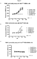
- FIG. 6 T-cell activation assay for 3 HER2-IgG ⁇ CD3-scFv bispecific antibodies comprising humanized variable heavy and light chains of SP34 according to the present disclosure. Activation of T-cells is determined by evaluation of CD69 expression on CD4 positive T-cells ( FIG. 6B ) or CD8 positive T-cells ( FIG. 6A ) as assessed by flow cytometry.
- FIG. 6A is a graph showing the percentage of CD69+ activated CD8+ T cells derived from 7 different donors as a function of antibody concentration.
- FIG. 6B is a graph showing the percentage of CD69+ activated CD4+ T cells derived from 7 different donors as a function of antibody concentration.
- FIG. 7 Cytotoxicity assay for 3 HER2-IgGxCD3-scFv bispecific antibodies comprising humanized variable heavy and light chains of SP34 according to the present disclosure on HER2 high expressing SKBR3 cells, HER2 low expressing A498 cells and HER2 negative cell line MDA-MB-468 in presence of human derived PBMCs. Cytotoxic activity of PBMCs is assessed by measuring incorporated CellToxGreenTM (Real-Time Cell Death Assay with Multiplexing Compatibility) fluorescence.
- FIG. 7A is a graph showing the relative fluorescence of HER2 high expressing SKBR3 cells as a function of antibody concentration.
- FIG. 7B indicates the same as FIG. 7A but with results obtained from HER2 low expressing A498 cells.
- FIG. 7C indicates the same as FIG. 7A but with results obtained from HER2 negative MDA-MB-468 cells.
- FIG. 8 Cytotoxicity assay for 3 HER2-IgG ⁇ CD3-scFv bispecific antibodies comprising humanized variable heavy and light chains of SP34 according to the present disclosure in the presence of HER2 high expressing SKBR3 cells. Cytotoxic activity is assessed by measuring release of Interferon-gamma from T-cells using the commercially available Human IFN-gamma Duo SET ELISA purchased form R&D Systems.
- FIG. 8 is a graph indicating the concentration of released Interferon-gamma from T-cells derived from one donor as a function of antibody concentration. As positive control BsAb #RefMAb #1 was included.
- FIG. 9 Cytotoxicity assay for 7 CD3-IgGxHER2-scFv bispecific antibodies comprising humanized variable heavy and light chains of SP34 according to the present disclosure tested on HER2 high expressing SKBR3 cells ( FIG. 9A ) and HER2 low expressing A498 cells ( FIG. 9B ). Cytotoxic activity of PBMCs is assessed by measuring incorporated CellToxGreenTM fluorescence.
- CD3 refers to an antigen which is expressed on T cells as part of the multi-molecular T cell receptor (TCR) and which consists of a homodimer or heterodimer formed from the association of two of four receptor chains: CD3-epsilon, CD3-delta, CD3-zeta, and CD3-gamma.
- Human CD3 Epsilon has the Amino Acid Sequence of UniProt P07766
- the extracellular domain of human CD3 epsilon without the signal sequence comprises amino acid residues 23-126 and has the amino acid sequence of as set forth in SEQ ID NO: 1 of TABLE 1.
- Cynomolgus CD3 Epsilon has the Amino Acid Sequence of UniProt Q95L15
- the extracellular domain of cynomolgus monkey CD3 epsilon without the signal sequence comprises amino acid residues 22-117 and has the amino acid sequence of as set forth in SEQ ID NO: 2 of TABLE 1.
- antibody refers to a protein comprising at least two heavy (H) chains and two light (L) chains inter-connected by disulfide bonds which interacts with an antigen.
- Each heavy chain is comprised of a heavy chain variable region or domain (abbreviated herein as VH) and a heavy chain constant region.
- the heavy chain constant region is comprised of three domains, CH1, CH2 and CH3.
- Each light chain is comprised of a light chain variable region or domain (abbreviated herein as VL) and a light chain constant region.
- the light chain constant region is comprised of one domain, CL.
- VH and VL regions can be further subdivided into regions of hypervariability, termed complementarity determining regions (CDR), interspersed with regions that are more conserved, termed framework regions (FR).
- CDR complementarity determining regions
- FR framework regions
- Each VH and VL is composed of three CDRs and four FR's arranged from amino-terminus to carboxy-terminus in the following order: FR1, CDR1, FR2, CDR2, FR3, CDR3, and FR4.
- the variable regions of the heavy and light chains contain a binding domain that interacts with an antigen.
- the constant regions of the antibodies may mediate the binding of the immunoglobulin to host tissues or factors, including various cells of the immune system (e.g., effector cells) and the first component (Clq) of the classical complement system.
- antibody includes for example, monoclonal antibodies, human antibodies, humanized antibodies, camelised antibodies and chimeric antibodies.
- the antibodies can be of any isotype (e.g., IgG, IgE, IgM, IgD, IgA and IgY), class (e.g., IgG1, IgG2, IgG3, IgG4, IgA1 and IgA2) or subclass. Both the light and heavy chains are divided into regions of structural and functional homology.
- antibody fragment refers to one or more portions of an antibody that retain the ability to specifically interact with (e.g., by binding, steric hindrance, stabilizing spatial distribution) an antigen.
- binding fragments include, but are not limited to, a Fab fragment, a monovalent fragment consisting of the VL, VH, CL and CH1 domains; a F(ab)2 fragment, a bivalent fragment comprising two Fab fragments linked by a disulfide bridge at the hinge region; a Fd fragment consisting of the VH and CH1 domains; a Fv fragment consisting of the VL and VH domains of a single arm of an antibody; a dAb fragment (Ward et al., (1989) Nature 341:544-546), which consists of a VH domain; and an isolated complementarity determining region (CDR).
- a Fab fragment a monovalent fragment consisting of the VL, VH, CL and CH1 domains
- F(ab)2 fragment a bivalent
- the two domains of the Fv fragment, VL and VH are coded for by separate genes, they can be joined, using recombinant methods, by a synthetic linker that enables them to be made as a single protein chain in which the VL and VH regions pair to form monovalent molecules (known as single chain Fv (scFv); see e.g., Bird et al., (1988) Science 242:423-426; and Huston et al., (1988) Proc. Natl. Acad. Sci. 85:5879-5883).
- single chain Fv single chain Fv
- Such single chain antibodies are also intended to be encompassed within the term “antibody fragment”.
- Antibody fragments are obtained using conventional techniques known to those of skill in the art, and the fragments are screened for utility in the same manner as are intact antibodies.
- Antibody fragments can also be incorporated into single domain antibodies, maxibodies, minibodies, intrabodies, diabodies, triabodies, tetrabodies, v-NAR and bis-scFv (see, e.g., Hollinger and Hudson, (2005) Nature Biotechnology 23:1126-1136).
- Antibody fragments can be grafted into scaffolds based on polypeptides such as Fibronectin type III (Fn3) (see U.S. Pat. No. 6,703,199, which describes fibronectin polypeptide monobodies).
- Fn3 Fibronectin type III
- Antibody fragments can be incorporated into single chain molecules comprising a pair of tandem Fv segments (VH-CH1-VH-CH1) which, together with complementary light chain polypeptides, form a pair of antigen-binding sites (Zapata et al., (1995) Protein Eng. 8:1057-1062; and U.S. Pat. No. 5,641,870).
- a “human antibody” or “human antibody fragment”, as used herein, includes antibodies and antibody fragments having variable regions in which both the framework and CDR regions are derived from sequences of human origin. Furthermore, if the antibody contains a constant region, the constant region also is derived from such sequences.
- Human origin includes, e.g., human germline sequences, or mutated versions of human germline sequences or antibody containing consensus framework sequences derived from human framework sequences analysis, for example, as described in Knappik et al., (2000) J Mol Biol 296:57-86).
- said human antibody can be obtained from technology platforms which comprise antibodies derived from human germline genes either generated by PCR-amplification of VHA/L repertoire isolated from B-cells or are generated synthetically.
- Technology platforms include library based approaches comprising human immunoglobulin genes displayed on phage, ribosome or yeast. Respective display technologies are standard in the scientific community. Furthermore immunization of a transgenic mouse carrying human immunoglobulin repertoire is another approach to generate human antibodies against an antigen of interest. Antibodies or fragments thereof selected from an antibody library based on the MorphoSys HuCAL® concept (Knappik et al., (2000) J Mol Biol 296:57-86) are considered as fully human.
- a “humanized antibody” or “humanized antibody fragment” is defined herein as an antibody or antibody fragment which has constant antibody regions derived from sequences of human origin and the variable antibody regions or parts thereof or only the CDRs are derived from another species.
- a humanized antibody can be CDR-grafted, wherein the CDRs of the variable domain are from a non-human origin, while one or more frameworks of the variable domain are of human origin and the constant domain (if any) is of human origin.
- chimeric antibody or “chimeric antibody fragment” is defined herein as an antibody molecule which has constant antibody regions derived from, or corresponding to, sequences found in one species and variable antibody regions derived from another species.
- the constant antibody regions are derived from, or corresponding to, sequences found in humans
- the variable antibody regions are derived from sequences found in a non-human animal, e.g. a mouse, rat, rabbit or hamster.
- immunoglobulin variable domains e.g., CDRs
- CDRs may be defined using well known numbering schemes, e.g., the Kabat numbering scheme, the Chothia numbering scheme, or a combination of Kabat and Chothia (see, e.g., Sequences of Proteins of Immunological Interest, U.S. Department of Health and Human Services (1991), eds. Kabat et al.; Lazikani et al., (1997) J. Mol. Bio. 273:927-948); Kabat et al., (1991) Sequences of Proteins of Immunological Interest, 5th edit., NIH Publication no. 91-3242 U.S.
- isolated refers to a compound, which can be e.g. an antibody or antibody fragment, that is substantially free of other antibodies or antibody fragments having different antigenic specificities.
- antibodies provided are isolated antibodies which have been separated from antibodies with a different specificity.
- An isolated antibody may be a monoclonal antibody.
- An isolated antibody may be a recombinant monoclonal antibody.
- An isolated antibody that specifically binds to an epitope, isoform or variant of a target may, however, have cross-reactivity to other related antigens, e.g., from other species (e.g., species homologs).
- recombinant antibody includes all antibodies that are prepared, expressed, created or segregated by means not existing in nature. For example antibodies isolated from a host cell transformed to express the antibody, antibodies selected and isolated from a recombinant, combinatorial human antibody library, and antibodies prepared, expressed, created or isolated by any other means that involve splicing of all or a portion of a human immunoglobulin gene, sequences to other DNA sequences or antibodies isolated from an animal (e.g., a mouse) that is transgenic or transchromosomal for human immunoglobulin genes or a hybridoma prepared therefrom.
- recombinant antibodies have variable regions in which the framework and CDR regions are derived from human germline immunoglobulin sequences.
- such recombinant human antibodies can be subjected to in vitro mutagenesis (or, when an animal transgenic for human Ig sequences is used, in vivo somatic mutagenesis) and thus the amino acid sequences of the VH and VL regions of the recombinant antibodies are sequences that, while derived from and related to human germline VH and VL sequences, may not naturally exist within the human antibody germline repertoire in vivo.
- a recombinant antibody may be a monoclonal antibody.
- the term “monoclonal antibody” refers to an antibody that is derived from a single clone, including any eukaryotic, prokaryotic, or phage clone, and not the method by which it is produced. Monoclonal antibodies as disclosed herein may be made by the hybridoma method as described in Kohler et al.; Nature, 256:495 (1975) or may be isolated from phage libraries using the techniques as described herein, for example.
- an antibody “binds specifically to”, “specifically binds to”, is “specific to/for” or “specifically recognizes” an antigen if such antibody is able to discriminate between such antigen and one or more reference antigen(s), since binding specificity is not an absolute, but a relative property.
- determination of binding specificity is performed by using not a single reference antigen, but a set of about three to five unrelated antigens, such as milk powder, BSA, transferrin or the like.
- an antibody that binds to CD3 or an “anti-CD3 antibody” or “antibody specific for CD3” includes antibodies and antibody fragments that specifically recognize one or more CD3 subunit (e.g., epsilon, delta, gamma or zeta), as well as antibodies and antibody fragments that specifically recognize a dimeric complex of two CD3 subunits (e.g., gamma/epsilon, delta/epsilon, and zeta/zeta CD3 dimers).
- the antibodies and antibody fragments of the present disclosure may bind soluble CD3 and/or cell surface expressed CD3.
- Soluble CD3 includes natural CD3 proteins as well as recombinant CD3 protein variants such as, e.g., monomeric and dimeric CD3 constructs, that lack a transmembrane domain or are otherwise unassociated with a cell membrane.
- cell surface-expressed CD3 means one or more CD3 protein(s) that is/are expressed on the surface of a cell in vitro or in vivo, such that at least a portion of a CD3 protein is exposed to the extracellular side of the cell membrane and is accessible to an antigen-binding portion of an antibody.
- Cell surface-expressed CD3 includes CD3 proteins contained within the context of a functional T cell receptor in the membrane of a cell.
- cell surface-expressed CD3 includes CD3 protein expressed as part of a homodimer or heterodimer on the surface of a cell (e.g., gamma/epsilon, delta/epsilon, and zeta/zeta CD3 dimers).
- the expression, “cell surface-expressed CD3” also includes a CD3 chain (e.g., CD3-epsilon, CD3-delta or CD3-gamma) that is expressed by itself, without other CD3 chain types, on the surface of a cell.
- a “cell surface-expressed CD3” can comprise or consist of a CD3 protein expressed on the surface of a cell which normally expresses CD3 protein.
- “cell surface-expressed CD3” can comprise or consist of CD3 protein expressed on the surface of a cell that normally does not express human CD3 on its surface but has been artificially engineered to express CD3 on its surface.
- affinity refers to the strength of interaction between the polypeptide and its target at a single site. Within each site, the binding region of the polypeptide interacts through weak non-covalent forces with its target at numerous sites; the more interactions, the stronger the affinity.
- K D refers to the dissociation constant, which is obtained from the ratio of K d to K a (i.e. K d /K a ) and is expressed as a molar concentration (M).
- K D values for antigen binding moieties like e.g. monoclonal antibodies can be determined using methods well established in the art. Methods for determining the K D of an antigen binding moiety like e.g. a monoclonal antibody are SET (soluble equilibrium titration) or surface plasmon resonance using a biosensor system such as a Biacore® system.
- an antibody specific to the CD3 epsilon polypeptide typically has a dissociation rate constant (K D ) (k off /k on ) of less than 5 ⁇ 10 ⁇ 2 M, less than 10 ⁇ 2 M, less than 5 ⁇ 10 ⁇ 3 M, less than 10 ⁇ 3 M, less than 5 ⁇ 10 ⁇ 4 M, less than 10 ⁇ 4 M, less than 5 ⁇ 10 ⁇ 5 M, less than 10 ⁇ 5 M, less than 5 ⁇ 10 ⁇ 6 M, less than 10 ⁇ 6 M, less than 5 ⁇ 10 ⁇ 7 M, less than 10 ⁇ 7 M, less than 5 ⁇ 10 ⁇ 8 M, less than 10 ⁇ 8 M, less than 5 ⁇ 10 ⁇ 9 M, less than 10 ⁇ 9 M, less than 5 ⁇ 10 ⁇ 10 M, less than 10 ⁇ 10 M, less than 5 ⁇ 10 ⁇ 11 M, less than 10 ⁇ 11 M, less than 5 ⁇ 10 ⁇ 12 M, less than 10 ⁇ 12 M, less than 5 ⁇ 10 ⁇ 13 M, less than 10
- epitope includes any proteinacious region which is specifically recognized by an antibody or fragment thereof or a T-cell receptor or otherwise interacts with a molecule.
- epitopes are of chemically active surface groupings of molecules such as amino acids or carbohydrate or sugar side chains and generally may have specific three-dimensional structural characteristics, as well as specific charge characteristics. As will be appreciated by one of skill in the art, practically anything to which an antibody can specifically bind could be an epitope.
- “Binds the same epitope as” means the ability of an antibody, antibody fragment or other antigen-binding moiety to bind to a specific antigen and binding to the same epitope as the exemplified antibody when using the same epitope mapping technique for comparing the antibodies.
- the epitopes of the exemplified antibody and other antibodies can be determined using epitope mapping techniques.
- Epitope mapping techniques are well known in the art. For example, conformational epitopes are readily identified by determining spatial conformation of amino acids such as by, e.g., hydrogen/deuterium exchange, x-ray crystallography and two-dimensional nuclear magnetic resonance.
- compositions of the present disclosure may be used for therapeutic or prophylactic applications.
- the present disclosure therefore, includes a pharmaceutical composition containing an antibody (or functional antibody fragment) as disclosed herein and a pharmaceutically acceptable carrier or excipient therefor.
- the present disclosure provides a method for treating cancer or an inflammatory disorder. Such method contains the steps of administering to a subject in need thereof an effective amount of the pharmaceutical composition that contains an antibody (or functional antibody fragment) as described or contemplated herein.
- the present disclosure provides therapeutic methods comprising the administration of a therapeutically effective amount of a humanized CD3 specific antibody or antibody fragment as disclosed to a subject in need of such treatment.
- a “therapeutically effective amount” or “effective amount”, as used herein, refers to the amount of an anti-CD3 antibody necessary to elicit the desired biological response.
- the therapeutic effective amount is the amount of a CD3 specific antibody or antibody fragment necessary to treat and/or prevent a disease.
- “Species”, as used in this context refers to any mammal, including rodents, such as mouse or rat, and primates, such as cynomolgus monkey ( Macaca fascicularis ), rhesus monkey ( Macaca mulatta ) or humans ( Homo sapiens ).
- rodents such as mouse or rat
- primates such as cynomolgus monkey ( Macaca fascicularis ), rhesus monkey ( Macaca mulatta ) or humans ( Homo sapiens ).
- the subject is a primate, most preferably a human.
- non-chimpanzee primate species may be understood within the meaning of the disclosure to be a lemur, a tarsier, a gibbon, a marmoset (belonging to New World Monkeys of the family Cebidae) or an Old-World Monkey (belonging to the superfamily Cercopithecoidea).
- an “Old-World Monkey” comprises any monkey falling in the superfamily Cercopithecoidea, itself subdivided into the families: the Cercopithecinae, which are mainly African but include the diverse genus of macaques which are Asian and North African; and the Colobinae, which include most of the Asian genera but also the African colobus monkeys.
- acceptor human framework for the purposes herein is a framework comprising the amino acid sequence of a light chain variable domain (VL) framework or a heavy chain variable domain (VH) framework derived from a human immunoglobulin framework or a human consensus framework, as defined below.
- An acceptor human framework “derived from” a human immunoglobulin framework or a human consensus framework may comprise the same amino acid sequence thereof, or it may contain amino acid sequence changes. In some embodiments, the number of amino acid changes are 10 or less, 9 or less, 8 or less, 7 or less, 6 or less, 5 or less, 4 or less, 3 or less, or 2 or less.
- the VL acceptor human framework is identical in sequence to the VL human immunoglobulin framework sequence or human consensus framework sequence.
- a “human consensus framework” is a framework which represents the most commonly occurring amino acid residues in a selection of human immunoglobulin VL or VH framework sequences.
- the selection of human immunoglobulin VL or VH sequences is from a subgroup of variable domain sequences.
- the subgroup of sequences is a subgroup as in Kabat et at., Sequences of Proteins of Immunological Interest, Fifth Edition, NIH Publication 9 1-3242, Bethesda Md. (1991), vols. 1-3.
- a “disorder” is any condition that would benefit from treatment including, but not limited to, chronic and acute disorders or diseases including those pathological conditions which predispose the mammal to the disorder in question.
- cell proliferative disorder and “proliferative disorder” refer to disorders that are associated with some degree of abnormal cell proliferation.
- the cell proliferative disorder is cancer.
- the cell proliferative disorder is a tumor.
- cancer and “cancerous” refer to or describe the physiological condition in mammals that is typically characterized by unregulated cell growth.
- “Effector functions” refer to those biological activities attributable to the Fc region of an antibody, which vary with the antibody isotype. Examples of antibody effector functions include: Clq binding and complement dependent cytotoxicity (CDC); Fc receptor binding; antibody-dependent cell-mediated cytotoxicity (ADCC); phagocytosis; down regulation of cell surface receptors (e.g. B cell receptor); and B cell activation.
- an “effective amount” of a compound for example, an CD3 specific antibody of the disclosure or a composition (e.g., pharmaceutical composition) thereof, is at least the minimum amount required to achieve the desired therapeutic or prophylactic result, such as a measurable improvement or prevention of a particular disorder (e.g., a cell proliferative disorder, e.g., cancer).
- An effective amount herein may vary according to factors such as the disease state, age, sex, and weight of the patient, and the ability of the antibody to elicit a desired response in the individual.
- An effective amount is also one in which any toxic or detrimental effects of the treatment are outweighed by the therapeutically beneficial effects.
- beneficial or desired results include results such as eliminating or reducing the risk, lessening the severity, or delaying the onset of the disease, including biochemical, histological and/or behavioral symptoms of the disease, its complications and intermediate pathological phenotypes presenting during development of the disease.
- beneficial or desired results include clinical results such as decreasing one or more symptoms resulting from the disease, increasing the quality of life of those suffering from the disease, decreasing the dose of other medications required to treat the disease, enhancing effect of another medication such as via targeting, delaying the progression of the disease, and/or prolonging survival.
- an effective amount of the drug may have the effect in reducing the number of cancer cells; reducing the tumor size; inhibiting (i.e., slow to some extent or desirably stop) cancer cell infiltration into peripheral organs; inhibit (i.e., slow to some extent and desirably stop) tumor metastasis; inhibiting to some extent tumor growth; and/or relieving to some extent one or more of the symptoms associated with the disorder.
- An effective amount can be administered in one or more administrations.
- an effective amount of drug, compound, or pharmaceutical composition is an amount sufficient to accomplish prophylactic or therapeutic treatment either directly or indirectly.
- an effective amount of a drug, compound, or pharmaceutical composition may or may not be achieved in conjunction with another drug, compound, or pharmaceutical composition.
- an “effective amount” may be considered in the context of administering one or more therapeutic agents, and a single agent may be considered to be given in an effective amount if, in conjunction with one or more other agents, a desirable result may be or is achieved.
- “delaying progression” of a disorder or disease means to defer, hinder, slow, retard, stabilize, and/or postpone development of the disease or disorder (e.g., a cell proliferative disorder, e.g., cancer).
- This delay can be of varying lengths of time, depending on the history of the disease and/or individual being treated.
- a sufficient or significant delay can, in effect, encompass prevention, in that the individual does not develop the disease.
- a late stage cancer such as development of metastasis, may be delayed.
- reduce or “inhibit” is meant the ability to cause an overall decrease, for example, of 20% or greater, of 50% or greater, or of 75%, 85%, 90%, 95%, or greater.
- increase is meant the ability to cause an overall increase, for example, of 20% or greater, of 50% or greater, or of 75%, 85%, 90%, 95%, or greater.
- EC 50 refers to the concentration of an antibody or an antibody fragment which induces a response in an assays half way between the baseline and maximum. It therefore represents the antibody concentration at which 50% of the maximal effect is observed.
- IC 50 refers to the concentration of an inhibitor (e.g. an antibody or antibody fragment) that inhibits a response in an assay half way between the maximal response and the baseline. It represents the antibody concentration that reduces a given response by 50%.
- an inhibitor e.g. an antibody or antibody fragment
- inhibitors or “inhibit” or “reduction” or “reduce” or “neutralization” or “neutralize” refer to a decrease or cessation of any phenotypic characteristic (such as binding, a biological activity or function) or to the decrease or cessation in the incidence, degree, or likelihood of that characteristic.
- the “inhibition”, “reduction” or “neutralization” needs not to be complete as long as it is detectable using an appropriate assay.
- by “reduce” or “inhibit” is meant the ability to cause a decrease of 20% or greater.
- by “reduce” or “inhibit” is meant the ability to cause a decrease of 50% or greater.
- by “reduce” or “inhibit” is meant the ability to cause an overall decrease of 75%, 85%, 90%, 95%, or greater.
- administering refers to contact of an exogenous pharmaceutical, therapeutic, diagnostic agent, or composition to the animal, human, subject, cell, tissue, organ, or biological fluid.
- administering can refer, e.g., to therapeutic, pharmacokinetic, diagnostic, research, and experimental methods. Treatment of a cell encompasses contact of a reagent to the cell, as well as contact of a reagent to a fluid, where the fluid is in contact with the cell.
- administering also means in vitro and ex vivo treatments, e.g., of a cell, by a reagent, diagnostic, binding composition, or by another cell.
- Treatment refers to therapeutic treatment, prophylactic or preventative measures, to research and diagnostic applications.
- Treatment as it applies to a human, veterinary, or research subject, or cell, tissue, or organ, encompasses contact of an agent with animal subject, a cell, tissue, physiological compartment, or physiological fluid.
- Treatment of a cell also encompasses situations where the agent contacts PILR, e.g., in the fluid phase or colloidal phase, but also situations where the agonist or antagonist does not contact the cell or the receptor.
- the CD3 specific antibodies or antibody fragments of the present disclosure can be linked to or co-expressed with another functional molecule, e.g., another peptide or protein.
- another functional molecule e.g., another peptide or protein.
- an antibody or antibody fragment thereof can be functionally linked (e.g., by chemical coupling, genetic fusion, noncovalent association or otherwise) to one or more other molecular entities, such as another antibody or antibody fragment to produce a bispecific antibody with a second binding specificity.
- the antibodies of the present disclosure may be monospecific or bispecific antibodies.
- a bispecific antibody may be specific for different epitopes of one target antigen or may contain antigen-binding domains specific for more than one target antigen. See, e.g., Tutt et al., 1991, J. Immunol. 147:60-69; Kufer et al., 2004, Trends Biotechnol. 22:238-244.
- bispecific antibodies are intended to include both monospecific CD3 specific antibodies as well as bispecific antibodies.
- Bispecific antibodies according to the present disclosure were designed using an IgG-scFv format comprising a monoclonal IgG1 binding moiety and a scFv binding moiety, with the N-terminus of each scFv VL domain fused to the C-terminal end of each IgG heavy chain via a peptide linker. Both Fabs arms of the IgG may bind to the cell surface target antigen while the scFv is specific for CD3 (both scFv are identical and display the same specificity for CD3).
- bispecific antibodies can be generated, wherein the two scFv molecules were specific for cell surface target antigen and the Fab arms of the IgG1 portion are specific for CD3.
- CD3 specific binding domains of the CD3 specific antibodies or antibody fragments of the present disclosure can comprise any of the heavy or light chain variable regions or CDR amino acid sequences as set forth in Tables 3-5 as disclosed herein.
- the cell surface target antigen can be a cancer-associated antigen.
- cancer related antigens include, e.g., an antigen that is expressed on the surface of a tumor.
- bispecific antibody format or technology may be used to make the bispecific antigen-binding molecules of the present disclosure.
- an antibody or fragment thereof having a first antigen binding specificity can be functionally linked (e.g., by chemical coupling, genetic fusion, noncovalent association or otherwise) to one or more other molecular entities, such as another antibody or antibody fragment having a second antigen-binding specificity to produce a bispecific antigen-binding molecule.
- bispecific formats that can be used in the context of the present disclosure include, without limitation, e.g., scFv-based or diabody bispecific formats, IgG-scFv fusions, dual variable domain (DVD)-Ig, Quadroma, knobs-into-holes, common light chain (e.g., common light chain with knobs-into-holes, etc.), CrossMab, CrossFab, (SEED)body, leucine zipper, Duobody, IgG1/IgG2, dual acting Fab (DAF)-IgG, and Mab.sup.2 bispecific formats (see, e.g., Klein et al. 2012, mAbs 4:6, 1-11, and references cited therein, for a review of the foregoing formats).
- the present disclosure refers to an monoclonal antibody or antibody fragment specific for CD3 comprising the variable heavy chain (VH) and the variable light chain (VL) of any one of the antibodies disclosed in Table 5.
- the present disclosure refers to an monoclonal antibody or antibody fragment specific for CD3 comprising the heavy chain (HC) and the light chain (LC) of any one of the antibodies disclosed in Tables 5-7.
- the present disclosure refers to a monoclonal antibody or antibody fragment specific for CD3 comprising 6 CDRs defined by Kabat of any one of the antibodies disclosed in Tables 5.
- the present disclosure refers to a monoclonal antibody or antibody fragment specific for CD3 comprising 6 CDRs of any one of the antibodies disclosed in Table 5.
- the present disclosure provides an antibody or antibody fragment specific for cluster of differentiation 3 (CD3), wherein said antibody or antibody fragment specifically binds to human CD3 and to non-human primate CD3, wherein said antibody or antibody fragment comprises the HCDR1 region of SEQ ID NO: 23, the HCDR2 region of SEQ ID NO: 24, the HCDR3 region of SEQ ID NO: 25, the LCDR1 region of SEQ ID NO: 26, the LCDR2 region of SEQ ID NO: 27 and the LCDR3 region of SEQ ID NO: 28 or the HCDR1 region of SEQ ID NO: 23, the HCDR2 region of SEQ ID NO: 24, the HCDR3 region of SEQ ID NO: 25, the LCDR1 region of SEQ ID NO: 26, the LCDR2 region of SEQ ID NO: 65 and the LCDR3 region of SEQ ID NO: 28.
- CD3 region of SEQ ID NO: 23 the HCDR2 region of SEQ ID NO: 24
- the HCDR3 region of SEQ ID NO: 25 the LCDR1 region of SEQ ID
- the present disclosure provides an antibody or antibody fragment specific for CD3, wherein said antibody or antibody fragment specifically binds to human CD3 epsilon and to non-human primate CD3 epsilon, wherein said antibody or antibody fragment comprises the HCDR1 region of SEQ ID NO: 23, the HCDR2 region of SEQ ID NO: 24, the HCDR3 region of SEQ ID NO: 25, the LCDR1 region of SEQ ID NO: 26, the LCDR2 region of SEQ ID NO: 27 and the LCDR3 region of SEQ ID NO: 28 or the HCDR1 region of SEQ ID NO: 23, the HCDR2 region of SEQ ID NO: 24, the HCDR3 region of SEQ ID NO: 25, the LCDR1 region of SEQ ID NO: 26, the LCDR2 region of SEQ ID NO: 65 and the LCDR3 region of SEQ ID NO: 28.
- the present disclosure provides an antibody or antibody fragment specific for CD3, wherein said antibody or antibody fragment specifically binds to human CD3 and to non-human primate CD3, wherein said antibody or antibody fragment comprises the HCDR1 region of SEQ ID NO: 23, the HCDR2 region of SEQ ID NO: 24, the HCDR3 region of SEQ ID NO: 25, the LCDR1 region of SEQ ID NO: 26, the LCDR2 region of SEQ ID NO: 27 and the LCDR3 region of SEQ ID NO: 28
- the present disclosure provides an antibody or antibody fragment specific for CD3, wherein said antibody or antibody fragment specifically binds to human CD3 and to non-human primate CD3, wherein said antibody or antibody fragment comprises the HCDR1 region of SEQ ID NO: 23, the HCDR2 region of SEQ ID NO: 24, the HCDR3 region of SEQ ID NO: 25, the LCDR1 region of SEQ ID NO: 26, the LCDR2 region of SEQ ID NO: 65 and the LCDR3 region of SEQ ID NO: 28.
- the present disclosure provides an antibody or antibody fragment specific for cluster of differentiation 3 (CD3), wherein said antibody or antibody fragment specifically binds to human CD3 and to non-human primate CD3, wherein said antibody or antibody fragment comprises the HCDR1 region comprising the amino acid sequence of SEQ ID NO: 23, the HCDR2 region comprising the amino acid sequence of SEQ ID NO: 24, the HCDR3 region comprising the amino acid sequence of SEQ ID NO: 25, the LCDR1 region comprising the amino acid sequence of SEQ ID NO: 26, the LCDR2 region comprising the amino acid sequence of SEQ ID NO: 27 and the LCDR3 region comprising the amino acid sequence of SEQ ID NO: 28 or the HCDR1 region comprising the amino acid sequence of SEQ ID NO: 23, the HCDR2 region comprising the amino acid sequence of SEQ ID NO: 24, the HCDR3 region comprising the amino acid sequence of SEQ ID NO: 25, the LCDR1 region comprising the amino acid sequence of SEQ ID NO: 26, the LCDR2 region comprising the amino acid sequence of the amino acid
- the antibody or antibody fragment specific for CD3 according to the present disclosure is a humanized, chimeric or synthetic antibody or antibody fragment.
- the antibody or antibody fragment according to the present disclosure is an isolated antibody or antibody fragment. In an embodiment, the antibody or antibody fragment according to the present disclosure is a recombinant antibody or antibody fragment. In another embodiment of the present disclosure the antibody or antibody fragment is a monoclonal antibody or antibody fragment. In an embodiment, said antibody or antibody fragment is a humanized monoclonal human antibody or antibody fragment.
- the antibody of the present disclosure is a full-length IgG. In an embodiment, the antibody of the present disclosure is a full-length IgG of an isotype selected from the group consisting of IgG1, IgG2, IgG3, and IgG4. In another embodiment the antibody is of the IgG1 isotype. In an embodiment, the antibody is of the human IgG1 isotype.
- the antibody of the present disclosure comprises a Fc region that has reduced effector function relative to that of a wild type Fc-receptor.
- the antibody of the present disclosure comprises a Fc region, wherein in at least 5 amino acids in the positions corresponding to positions L234, L235, D237, N330, P331 in a human IgG1 heavy chain, are mutated to A, E, A, S, and S, respectively.
- the antibody fragment is selected from the group consisting of a Fab, a Fab′, a Fv, a scFv.
- the antibody or antibody fragment of the present disclosure is a Fab fragment.
- the antibody or antibody fragment of the present disclosure is a single chain antibody.
- the antibody or antibody fragment specifically binds to human CD3 and cynomolgus CD3. In a further embodiment, the antibody or antibody fragment according to the present disclosure specifically binds to human CD3 epsilon and cynomolgus CD3 epsilon.
- the antibody or antibody fragment according to the present disclosure is a humanized or chimeric antibody or antibody fragment thereof.
- the present disclosure provides an antibody or antibody fragment specific for CD3, wherein said antibody or antibody fragment specifically binds to human CD3 and to non-human primate CD3, wherein said antibody or antibody fragment comprises
- the HCDR1 region of SEQ ID NO: 23 the HCDR2 region of SEQ ID NO: 24, the HCDR3 region of SEQ ID NO: 25, the LCDR1 region of SEQ ID NO: 26, the LCDR2 region of SEQ ID NO: 27 and the LCDR3 region of SEQ ID NO: 28, and further comprises the heavy chain variable region having SEQ ID NO: 30 and a light chain variable region having SEQ ID NO: 29
- the HCDR1 region of SEQ ID NO: 23 the HCDR2 region of SEQ ID NO: 24, the HCDR3 region of SEQ ID NO: 25, the LCDR1 region of SEQ ID NO: 26, the LCDR2 region of SEQ ID NO: 27 and the LCDR3 region of SEQ ID NO: 28, and further comprises the heavy chain variable region having SEQ ID NO: 46 and a light chain variable region having SEQ ID NO: 45
- the HCDR1 region of SEQ ID NO: 23 the HCDR2 region of SEQ ID NO: 24, the HCDR3 region of SEQ ID NO: 25, the LCDR1 region of SEQ ID NO: 26, the LCDR2 region of SEQ ID NO: 27 and the LCDR3 region of SEQ ID NO: 28, and further comprises the heavy chain variable region having SEQ ID NO: 56 and a light chain variable region having SEQ ID NO: 55
- the HCDR1 region of SEQ ID NO: 23 the HCDR2 region of SEQ ID NO: 24, the HCDR3 region of SEQ ID NO: 25, the LCDR1 region of SEQ ID NO: 26, the LCDR2 region of SEQ ID NO: 65 and the LCDR3 region of SEQ ID NO: 28, and further comprises the heavy chain variable region having SEQ ID NO: 68 and a light chain variable region having SEQ ID NO: 67
- the HCDR1 region of SEQ ID NO: 23 the HCDR2 region of SEQ ID NO: 24, the HCDR3 region of SEQ ID NO: 25, the LCDR1 region of SEQ ID NO: 26, the LCDR2 region of SEQ ID NO: 65 and the LCDR3 region of SEQ ID NO: 28, and further comprises the heavy chain variable region having SEQ ID NO: 78 and a light chain variable region having SEQ ID NO: 77
- the HCDR1 region of SEQ ID NO: 23 the HCDR2 region of SEQ ID NO: 24, the HCDR3 region of SEQ ID NO: 25, the LCDR1 region of SEQ ID NO: 26, the LCDR2 region of SEQ ID NO: 65 and the LCDR3 region of SEQ ID NO: 28, and further comprises the heavy chain variable region having SEQ ID NO: 86 and a light chain variable region having SEQ ID NO: 85
- the present disclosure provides an antibody or antibody fragment specific for CD3, wherein said antibody or antibody fragment specifically binds to human CD3 and to non-human primate CD3, wherein said antibody or antibody fragment comprises
- the HCDR1 region of SEQ ID NO: 23 the HCDR2 region of SEQ ID NO: 24, the HCDR3 region of SEQ ID NO: 25, the LCDR1 region of SEQ ID NO: 26, the LCDR2 region of SEQ ID NO: 27 and the LCDR3 region of SEQ ID NO: 28, and further comprises the heavy chain variable region having SEQ ID NO: 9 and a light chain variable region having SEQ ID NO: 11
- the HCDR1 region of SEQ ID NO: 23 the HCDR2 region of SEQ ID NO: 24, the HCDR3 region of SEQ ID NO: 25, the LCDR1 region of SEQ ID NO: 26, the LCDR2 region of SEQ ID NO: 27 and the LCDR3 region of SEQ ID NO: 28, and further comprises the heavy chain variable region having SEQ ID NO: 9 and a light chain variable region having SEQ ID NO: 13
- the HCDR1 region of SEQ ID NO: 23 the HCDR2 region of SEQ ID NO: 24, the HCDR3 region of SEQ ID NO: 25, the LCDR1 region of SEQ ID NO: 26, the LCDR2 region of SEQ ID NO: 27 and the LCDR3 region of SEQ ID NO: 28, and further comprises the heavy chain variable region having SEQ ID NO: 7 and a light chain variable region having SEQ ID NO: 15
- the HCDR1 region of SEQ ID NO: 23 the HCDR2 region of SEQ ID NO: 24, the HCDR3 region of SEQ ID NO: 25, the LCDR1 region of SEQ ID NO: 26, the LCDR2 region of SEQ ID NO: 65 and the LCDR3 region of SEQ ID NO: 28, and further comprises the heavy chain variable region having SEQ ID NO: 7 and a light chain variable region having SEQ ID NO: 17
- the HCDR1 region of SEQ ID NO: 23 the HCDR2 region of SEQ ID NO: 24, the HCDR3 region of SEQ ID NO: 25, the LCDR1 region of SEQ ID NO: 26, the LCDR2 region of SEQ ID NO: 65 and the LCDR3 region of SEQ ID NO: 28, and further comprises the heavy chain variable region having SEQ ID NO: 7 and a light chain variable region having SEQ ID NO: 19
- the HCDR1 region of SEQ ID NO: 23 the HCDR2 region of SEQ ID NO: 24, the HCDR3 region of SEQ ID NO: 25, the LCDR1 region of SEQ ID NO: 26, the LCDR2 region of SEQ ID NO: 65 and the LCDR3 region of SEQ ID NO: 28, and further comprises the heavy chain variable region having SEQ ID NO: 9 and a light chain variable region having SEQ ID NO: 19
- the HCDR1 region of SEQ ID NO: 23 the HCDR2 region of SEQ ID NO: 24, the HCDR3 region of SEQ ID NO: 25, the LCDR1 region of SEQ ID NO: 26, the LCDR2 region of SEQ ID NO: 27 and the LCDR3 region of SEQ ID NO: 28, and further comprises the heavy chain variable region having SEQ ID NO: 9 and a light chain variable region having SEQ ID NO: 21
- the present disclosure provides an antibody or antibody fragment specific for CD3, wherein said antibody or antibody fragment specifically binds to human CD3 and to non-human primate CD3, wherein said antibody or antibody fragment comprises the HCDR1 region of SEQ ID NO: 23, the HCDR2 region of SEQ ID NO: 24, the HCDR3 region of SEQ ID NO: 25, the LCDR1 region of SEQ ID NO: 26, the LCDR2 region of SEQ ID NO: 65 and the LCDR3 region of SEQ ID NO: 28, and further comprises the heavy chain variable region having SEQ ID NO: 68 and a light chain variable region having SEQ ID NO: 67.
- the present disclosure provides an antibody or antibody fragment specific for CD3, wherein said antibody or antibody fragment specifically binds to human CD3 and to non-human primate CD3, wherein said antibody or antibody fragment comprises the HCDR1 region of SEQ ID NO: 23, the HCDR2 region of SEQ ID NO: 24, the HCDR3 region of SEQ ID NO: 25, the LCDR1 region of SEQ ID NO: 26, the LCDR2 region of SEQ ID NO: 65 and the LCDR3 region of SEQ ID NO: 28, and further comprises the heavy chain variable region having SEQ ID NO: 7 and a light chain variable region having SEQ ID NO: 17.
- the present disclosure provides an antibody or antibody fragment specific for CD3, wherein said antibody or antibody fragment comprises
- the present disclosure refers to an antibody or antibody fragment specific for CD3, wherein said antibody or antibody fragment comprises
- variable heavy chain and a variable light chain that has at least 60%, at least 70%, at least 80%, at least 90% or at least 95% identity to the variable heavy chain of SEQ ID NO: 17, 28, 39 or 50 and to the variable light chain of SEQ ID NO: 18, 29, 40 or 51.
- the present disclosure provides an antibody or antibody fragment specific for CD3, wherein the variable heavy chain consists of SEQ ID NO: 7 and the variable light chain consist of SEQ ID NO: 11.
- the present disclosure provides an antibody or antibody fragment specific for CD3, wherein the variable heavy chain consists of SEQ ID NO: 7 and the variable light chain consist of SEQ ID NO: 13.
- the present disclosure provides an antibody or antibody fragment specific for CD3, wherein the variable heavy chain consists of SEQ ID NO: 7 and the variable light chain consist of SEQ ID NO: 15.
- the present disclosure provides an antibody or antibody fragment specific for CD3, wherein the variable heavy chain consists of SEQ ID NO: 7 and the variable light chain consist of SEQ ID NO 17.
- the present disclosure provides an antibody or antibody fragment specific for CD3, wherein the variable heavy chain consists of SEQ ID NO: 7 and the variable light chain consist of SEQ ID NO: 19.
- the present disclosure provides an antibody or antibody fragment specific for CD3, wherein the variable heavy chain consists of SEQ ID NO: 7 and the variable light chain consist of SEQ ID NO: 21.
- the present disclosure provides an antibody or antibody fragment specific for CD3, wherein the variable heavy chain consists of SEQ ID NO: 9 and the variable light chain consist of SEQ ID NO: 11.
- the present disclosure provides an antibody or antibody fragment specific for CD3, wherein the variable heavy chain consists of SEQ ID NO: 9 and the variable light chain consist of SEQ ID NO: 13.
- the present disclosure provides an antibody or antibody fragment specific for CD3, wherein the variable heavy chain consists of SEQ ID NO: 9 and the variable light chain consist of SEQ ID NO: 15.
- the present disclosure provides an antibody or antibody fragment specific for CD3, wherein the variable heavy chain consists of SEQ ID NO: 9 and the variable light chain consist of SEQ ID NO: 17.
- the present disclosure provides an antibody or antibody fragment specific for CD3, wherein the variable heavy chain consists of SEQ ID NO: 9 and the variable light chain consist of SEQ ID NO: 19.
- the present disclosure provides an antibody or antibody fragment specific for CD3, wherein the variable heavy chain consists of SEQ ID NO: 9 and the variable light chain consist of SEQ ID NO: 21.
- the present disclosure provides an antibody or antibody fragment specific for CD3, wherein the variable heavy chain and variable light chain are selected from the group consisting of:
- variable heavy chain domain having SEQ ID NO: 7 and the variable light chain having SEQ ID NO: 19.
- the present disclosure provides an antibody or antibody fragment specific for CD3, wherein the variable heavy chain comprises SEQ ID NO: 7 and the variable light chain comprises SEQ ID NO 17 or a sequence having at least 60%, at least 70%, at least 80%, at least 90%, or at least 95% identity with these sequences but which retains the same activity as the said antibody or antibody fragment.
- the present disclosure provides an antibody or antibody fragment specific for CD3, wherein the variable heavy chain comprises SEQ ID NO: 7 and the variable light chain comprises SEQ ID NO 17.
- the present disclosure provides a bispecific antibody comprising a first antigen binding domain specific for CD3 and a second binding domain which binds a different target than said first antigen binding region, wherein said first binding domain specific for CD3 comprises
- the HCDR1 region of SEQ ID NO: 23 the HCDR2 region of SEQ ID NO: 24, the HCDR3 region of SEQ ID NO: 25, the LCDR1 region of SEQ ID NO: 26, the LCDR2 region of SEQ ID NO: 27 and the LCDR3 region of SEQ ID NO: 28
- the HCDR1 region of SEQ ID NO: 23 the HCDR2 region of SEQ ID NO: 24, the HCDR3 region of SEQ ID NO: 25, the LCDR1 region of SEQ ID NO: 26, the LCDR2 region of SEQ ID NO: 65 and the LCDR3 region of SEQ ID NO: 28.
- the present disclosure provides a bispecific antibody comprising a first antigen binding domain specific for CD3 and a second binding domain, wherein said second binding domain specifically binds a cell surface target antigen and wherein said binding domain specific for CD3 comprises
- the HCDR1 region of SEQ ID NO: 23 the HCDR2 region of SEQ ID NO: 24, the HCDR3 region of SEQ ID NO: 25, the LCDR1 region of SEQ ID NO: 26, the LCDR2 region of SEQ ID NO: 27 and the LCDR3 region of SEQ ID NO: 28
- the HCDR1 region of SEQ ID NO: 23 the HCDR2 region of SEQ ID NO: 24, the HCDR3 region of SEQ ID NO: 25, the LCDR1 region of SEQ ID NO: 26, the LCDR2 region of SEQ ID NO: 65 and the LCDR3 region of SEQ ID NO: 28.
- the bispecific antibody of the present disclosure binds to CD3 located on an immune effector cell and a cell surface target antigen that is expressed on a target cell other than the immune effector cell, wherein the bispecific antibody comprises an binding domain specific for CD3 comprising
- the HCDR1 region of SEQ ID NO: 23 the HCDR2 region of SEQ ID NO: 24, the HCDR3 region of SEQ ID NO: 25, the LCDR1 region of SEQ ID NO: 26, the LCDR2 region of SEQ ID NO: 27 and the LCDR3 region of SEQ ID NO: 28
- the HCDR1 region of SEQ ID NO: 23 the HCDR2 region of SEQ ID NO: 24, the HCDR3 region of SEQ ID NO: 25, the LCDR1 region of SEQ ID NO: 26, the LCDR2 region of SEQ ID NO: 65 and the LCDR3 region of SEQ ID NO: 28.
- the cell surface target antigen is a tumor antigen.
- said immune effector cell is a cytotoxic T-lymphocytes.
- the present disclosure provides a bispecific antibody comprising a first antigen binding domain specific for CD3 and a second binding domain, wherein said second binding domain specifically binds a cell surface target antigen and wherein said bispecific antibody comprises a modified Fc region wherein in at least 5 amino acids in the positions corresponding to positions L234, L235, D237, N330, P331 in a human IgG1 heavy chain, are mutated to A, E, A, S, and S, respectively.
- the present disclosure provides a bispecific antibody comprising a first antigen binding domain specific for CD3 and a second binding domain, wherein said second binding domain specifically binds a cell surface target antigen and wherein said bispecific antibody comprises a modified Fc region wherein in at least 5 amino acids in the positions corresponding to positions L234, L235, D237, N330, P331 in a human IgG1 heavy chain, are mutated to A, E, A, S, and S, respectively and wherein said binding domain specific for CD3 comprises
- the HCDR1 region of SEQ ID NO: 23 the HCDR2 region of SEQ ID NO: 24, the HCDR3 region of SEQ ID NO: 25, the LCDR1 region of SEQ ID NO: 26, the LCDR2 region of SEQ ID NO: 27 and the LCDR3 region of SEQ ID NO: 28
- the HCDR1 region of SEQ ID NO: 23 the HCDR2 region of SEQ ID NO: 24, the HCDR3 region of SEQ ID NO: 25, the LCDR1 region of SEQ ID NO: 26, the LCDR2 region of SEQ ID NO: 65 and the LCDR3 region of SEQ ID NO: 28.
- said bispecific antibody according to the present comprises a first antigen binding domain, wherein said first antigen binding domain comprises an antibody or antibody fragment specific for CD3, wherein said antibody or antibody fragment comprises
- said bispecific antibody according to the present comprises a first antigen binding domain, wherein said first antigen binding domain comprises an antibody or antibody fragment specific for CD3, wherein said antibody or antibody fragment comprises the variable heavy chain of SEQ ID NO: 7 and the variable light chain of SEQ ID NO: 17 or a variable heavy chain and a variable light chain that has at least 60%, at least 70%, at least 80%, at least 90% or at least 95% identity to the variable heavy chain of SEQ ID NO: 7 and to the variable light chain of SEQ ID NO: 17.
- the present disclosure provides a nucleic acid sequence or a plurality of nucleic acid sequences encoding an antibody or antibody fragment according to the present disclosure which specifically binds to CD3.
- the present disclosure provides a nucleic acid composition comprising a nucleic acid sequence or a plurality of nucleic acid sequences encoding the antibody or antibody fragment specific for CD3 according to the present disclosure, wherein said antibody or antibody fragment comprises the
- HCDR1 region of SEQ ID NO: 23 the HCDR2 region of SEQ ID NO: 24, the HCDR3 region of SEQ ID NO: 25, the LCDR1 region of SEQ ID NO: 26, the LCDR2 region of SEQ ID NO: 27 and the LCDR3 region of SEQ ID NO: 28
- the HCDR1 region of SEQ ID NO: 23 the HCDR2 region of SEQ ID NO: 24, the HCDR3 region of SEQ ID NO: 25, the LCDR1 region of SEQ ID NO: 26, the LCDR2 region of SEQ ID NO: 65 and the LCDR3 region of SEQ ID NO: 28.
- the present disclosure refers to an isolated nucleic acid or a plurality of nucleic acid sequences encoding a heavy chain sequence and/or light chain sequence of an antibody or antibody fragment specific for CD3, the nucleic acid comprising
- the present disclosure refers to an isolated nucleic acid or a plurality of nucleic acid sequences encoding a heavy chain sequence and/or light chain sequence of an antibody or antibody fragment specific for CD3, the nucleic acid comprising
- the present disclosure refers to a nucleic acid sequence or a plurality of nucleic acid sequences encoding an antibody or antibody fragment specific for CD3, wherein the nucleic acid sequence or plurality of nucleic acid sequences comprises the VH of SEQ ID NO: 70 and/or the VL of SEQ ID NO: 69, or the VH and/or the VL that has at least 60%, at least 70%, at least 80%, at least 90% or at least 95% identity to the VH of SEQ ID NO: 70 and the VL of SEQ ID NO: 69.
- the present disclosure refers to a nucleic acid sequence or a plurality of nucleic acid sequences encoding an antibody or antibody fragment specific for CD3, wherein the nucleic acid sequence or plurality of nucleic acid sequences comprises the VH of SEQ ID NO: 8 and/or the VL of SEQ ID NO: 18, or the VH and/or the VL that has at least 60%, at least 70%, at least 80%, at least 90% or at least 95% identity to the VH of SEQ ID NO: 8 and the VL of SEQ ID NO: 18.
- the present disclosure provides a vector composition comprising a vector or a plurality of vectors comprising the nucleic acid sequence or plurality of nucleic acid sequences encoding an antibody or antibody fragment as disclosed in TABLE 5-7.
- the present disclosure refers to a vector composition comprising a vector or a plurality of vectors comprising a nucleic acid sequence or plurality of nucleic acid sequences disclosed in Tables 3-7.
- the present disclosure provides a vector comprising a nucleic acid encoding an antibody or antibody fragment specific for CD3 according to the present disclosure.
- the present disclosure provides a vector comprising a nucleic acid disclosed in Tables 3-7.
- the present disclosure provides a vector comprising a nucleic acid encoding an antibody or antibody fragment disclosed in Tables 3-7.
- the present disclosure refers to a host cell comprising a vector composition comprising a vector or a plurality of vectors comprising the nucleic acid sequence or plurality of nucleic acid sequences encoding an antibody or antibody fragment as disclosed in Tables 5-7.
- the present disclosure provides a host cell comprising a vector comprising a nucleic acid encoding an antibody or antibody fragment specific for CD3 according to the present disclosure. In an embodiment, the present disclosure provides a host cell comprising a nucleic acid encoding an antibody or antibody fragment specific for CD3 of the present disclosure. In another embodiment, the present disclosure provides an isolated host cell comprising a vector comprising a nucleic acid disclosed in Tables 3-7. In an embodiment, the host cell of the present disclosure is a mammalian cell. In an embodiment, the host cell of the present disclosure is a prokaryotic cell.
- additional amino acid residues, polypeptides or moieties are added to the antibody or antibody fragment of the present disclosure, for example to aid in the expression or purification or to increase the stability of the antibody or antibody fragment of the present disclosure.
- the present disclosure provides an antibody or antibody fragment specific for CD3 for use in the treatment of a subject in need thereof, wherein said antibody or antibody fragment comprises the
- HCDR1 region of SEQ ID NO: 23 the HCDR2 region of SEQ ID NO: 24, the HCDR3 region of SEQ ID NO: 25, the LCDR1 region of SEQ ID NO: 26, the LCDR2 region of SEQ ID NO: 27 and the LCDR3 region of SEQ ID NO: 28
- the HCDR1 region of SEQ ID NO: 23 the HCDR2 region of SEQ ID NO: 24, the HCDR3 region of SEQ ID NO: 25, the LCDR1 region of SEQ ID NO: 26, the LCDR2 region of SEQ ID NO: 65 and the LCDR3 region of SEQ ID NO: 28.
- said antibody or antibody fragment specific for CD3 comprises a variable heavy chain of SEQ ID NO: 7 and the variable light chain of SEQ ID NO 17 or a sequence having at least 60%, at least 70%, at least 80%, at least 90%, or at least 95% identity with these sequences but which retains the same activity as the said antibody or antibody fragment.
- said antibody or antibody fragment specific for CD3 comprises a variable heavy chain of SEQ ID NO: 7 and the variable light chain of SEQ ID NO 17.
- the present disclosure provides an antibody or antibody fragment specific for CD3 for use as a medicament, wherein said antibody or antibody fragment comprises the
- HCDR1 region of SEQ ID NO: 23 the HCDR2 region of SEQ ID NO: 24, the HCDR3 region of SEQ ID NO: 25, the LCDR1 region of SEQ ID NO: 26, the LCDR2 region of SEQ ID NO: 27 and the LCDR3 region of SEQ ID NO: 28
- the HCDR1 region of SEQ ID NO: 23 the HCDR2 region of SEQ ID NO: 24, the HCDR3 region of SEQ ID NO: 25, the LCDR1 region of SEQ ID NO: 26, the LCDR2 region of SEQ ID NO: 65 and the LCDR3 region of SEQ ID NO: 28.
- said antibody or antibody fragment specific for CD3 comprises a variable heavy chain of SEQ ID NO: 7 and the variable light chain of SEQ ID NO 17.
- the present disclosure provides an antibody or antibody fragment specific for CD3 for use in treating or delaying progression of a cell proliferative disorder or an autoimmune disorder in a subject in need thereof.
- the present disclosure provides an antibody or antibody fragment specific for CD3 for use in treating or delaying progression of a cell proliferative disorder or an autoimmune disorder in a subject in need thereof, wherein said antibody or antibody fragment comprises
- the HCDR1 region of SEQ ID NO: 23 the HCDR2 region of SEQ ID NO: 24, the HCDR3 region of SEQ ID NO: 25, the LCDR1 region of SEQ ID NO: 26, the LCDR2 region of SEQ ID NO: 27 and the LCDR3 region of SEQ ID NO: 28
- the HCDR1 region of SEQ ID NO: 23 the HCDR2 region of SEQ ID NO: 24, the HCDR3 region of SEQ ID NO: 25, the LCDR1 region of SEQ ID NO: 26, the LCDR2 region of SEQ ID NO: 65 and the LCDR3 region of SEQ ID NO: 28.
- said antibody or antibody fragment specific for CD3 comprises a variable heavy chain of SEQ ID NO: 7 and the variable light chain of SEQ ID NO 17.
- the present disclosure provides an antibody or antibody fragment specific for CD3 for use in enhancing immune function in a subject having a cell proliferative disorder or an autoimmune disorder.
- the present disclosure provides an antibody or antibody fragment specific for CD3 for use in enhancing immune function in a subject having a cell proliferative disorder wherein the cell proliferative disorder is a cancer.
- the cancer is selected from the group consisting of esophageal cancer, stomach cancer, small intestine cancer, large intestine cancer, colorectal cancer, breast cancer, non-small cell lung cancer, non-Hodgkin's lymphoma (NHL), B cell lymphoma, B cell leukemia, multiple myeloma, renal cancer, prostate cancer, liver cancer, head and neck cancer, melanoma, ovarian cancer, mesothelioma, glioblastoma, germinal-center B-cell-like (GCB) DLBCL, activated B-cell-like (ABC) DLBCL, follicular lymphoma (FL), mantle cell lymphoma (MCL), acute myeloid leukemia (AML), chronic lymphoid leukemia (CLL), marginal zone lymphoma (MZL), small lymphocytic leukemia (SLL), lymphoplasmacytic lymphoma (LL), Waldenstrom macroglobulinemia (NHL),
- the autoimmune disorder is selected from the group consisting of rheumatoid arthritis, juvenile rheumatoid arthritis, systemic lupus erythematosus (SUE), Wegener's disease, inflammatory bowel disease, idiopathic thrombocytopenic purpura (ITP), thrombotic thrombocytopenic purpura (TTP), autoimmune thrombocytopenia, multiple sclerosis, psoriasis, IgA nephropathy, IgM polyneuropathies, myasthenia gravis, vasculitis, diabetes mellitus, Reynaud's syndrome, Sjorgen's syndrome, glomerulonephritis, Neuromyelitis Optica (NMO) and IgG neuropathy.
- SUE systemic lupus erythematosus
- ITP thrombotic thrombocytopenic purpura
- autoimmune thrombocytopenia multiple sclerosis, psoriasis
- the present disclosure also provides methods for treating a subject suffering from a disorder, such as cancer, by administering to said subject an effective amount of an antibody or antibody according to the present disclosure.
- a subject is a human.
- said antibody or antibody fragment specific for CD3 comprises a variable heavy chain of SEQ ID NO: 7 and the variable light chain of SEQ ID NO 17.
- the present disclosure provides a pharmaceutical composition
- a pharmaceutical composition comprising an antibody or antibody fragment as disclosed in Tables 5-7 and a pharmaceutically acceptable carrier or excipient.
- said antibody or antibody fragment specific for CD3 comprises a variable heavy chain of SEQ ID NO: 7 and the variable light chain of SEQ ID NO 17.
- the present disclosure also provides methods for treating a subject suffering from a disorder, such as cancer or an autoimmune disorder, by administering to said subject an effective amount of an antibody or antibody fragment as disclosed in Table 5-7.
- said antibody or antibody fragment specific for CD3 comprises a variable heavy chain of SEQ ID NO: 7 and the variable light chain of SEQ ID NO 17.
- said subject is a human.
- the present disclosure provides an antibody or antibody fragment which specifically binds to human or cynomolgus CD3 epsilon with an EC 50 concentration of greater than 1 pM, 2 pM, 3 pM, 4 pM, 5 pM, 6 pM, 7 pM, 8 pM, 9 pM, 10 pM, 20 pM, 30 pM, 40 pM, 50 pM, 60 pM, 70 pM, 80 pM, 90 pM, 100 pM, 1 nM, 2 nM, 3 nM, 4 nM, 5 nM, 6 nM, 7 nM, 8 nM, 9 nM, 10 nM, 20 nM, 30 nM, 40 nM, 50 nM, 60 nM, 70 nM, 80 nM, 90 nM, 100 nM as determined in an ELISA assay.
- said antibody or antibody fragment specifically binds to human CD3 epsilon with an EC 50 of concentration of greater 500 pM. In an embodiment, said antibody or antibody fragment specifically binds to cynomolgus CD3 epsilon with an EC 50 of concentration of greater 1 nM as determined in an ELISA assay.
- said antibody or antibody fragment specifically binds to human CD3 epsilon with an EC 50 of concentration of greater 500 pM and to cynomolgus CD3 epsilon with an EC 50 concentration of greater 1 nM as determined in an ELISA assay.
- said antibody or antibody fragment specifically binds to human or cynomolgus CD3 epsilon with an EC 50 of concentration of greater 100 pM, 200 pM, 300 pM, 400 pM, 500 pM, 600 pM, 700 pM, 800 pM, 900 pM, 1 nM or 2 nM as determined in an ELISA assay.
- said antibody or antibody fragment specifically binds to human or cynomolgus CD3 epsilon with an EC 50 of concentration of greater 500 pM as determined in an ELISA assay.
- said antibody or antibody fragment specifically binds to human or cynomolgus CD3 epsilon with an EC 50 of concentration of greater 100 pm as determined in an ELISA assay.
- the present disclosure provides an antibody or antibody fragment which specifically binds to human or cynomolgus CD3 epsilon with an EC 50 concentration which is higher compared to the EC 50 concentration of any one of reference antibodies RefMAb #1 or SP34 as determined in an ELISA assay.
- the present disclosure provides an antibody or antibody fragment which specifically binds to human or cynomolgus CD3 epsilon with an EC 50 concentration which is at least 0.5 fold or higher, 1 fold or higher, 1.5 fold or higher, 2 fold or higher, 3 fold or higher, 4 fold or higher, 5 fold or higher, 6 fold or higher, 7 fold or higher. 8 fold or higher, 9 fold or higher, 10 fold or higher, 20 fold or higher, 50 fold or higher, 100 fold or higher as the EC 50 concentration of any one of reference antibodies RefMAb #1 or SP34 as determined in an ELISA assay.
- the present disclosure provides an antibody or antibody fragment which specifically binds to recombinant human or cynomolgus CD3 epsilon with an EC 50 concentration of greater than 1 pM, 2 pM, 3 pM, 4 pM, 5 pM, 6 pM, 7 pM, 8 pM, 9 pM, 10 pM, 20 pM, 30 pM, 40 pM, 50 pM, 60 pM, 70 pM, 80 pM, 90 pM, 100 pM, 1 nM, 2 nM, 3 nM, 4 nM, 5 nM, 6 nM, 7 nM, 8 nM, 9 nM, 10 nM, 20 nM, 30 nM, 40 nM, 50 nM, 60 nM, 70 nM, 80 nM, 90 nM, 100 nM as determined in an ELISA assay.
- said antibody or antibody fragment specifically binds to recombinant human or cynomolgus CD3 epsilon with an EC 50 of concentration of greater 100 pM, 200 pM, 300 pM, 400 pM, 500 pM, 600 pM, 700 pM, 800 pM, 900 pM, 1 nM or 2 nM as determined in an ELISA assay.
- said antibody or antibody fragment specifically binds to recombinant human or cynomolgus CD3 epsilon with an EC 50 of concentration of greater 100 pm as determined in an ELISA assay.
- the present disclosure provides an antibody or antibody fragment which specifically binds to recombinant human or cynomolgus CD3 epsilon with an EC 50 concentration which is at least 0.5 fold or higher, 1 fold or higher, 1.5 fold or higher, 2 fold or higher, 3 fold or higher, 4 fold or higher, 5 fold or higher, 10 fold or higher, 20 fold or higher, 50 fold or higher, 100 fold or higher as the EC 50 concentration of any one of reference antibodies RefMAb #1 or SP34 as determined in an ELISA assay.
- said antibody or antibody fragment specific for CD3 comprises
- said antibody or antibody fragment specific for CD3 comprises
- said antibody or antibody fragment specific for CD3 comprises the variable heavy chain of SEQ ID NO: 7 and the variable light chain of SEQ ID NO: 17.
- said human CD3 epsilon is hCD3 epsilon 22-49/Fc.
- said cynomolgus CD3 epsilon is cyCD3 epsilon 22-49/Fc.
- said EC 50 concentration is determined by an ELISA assay as described herein in Example 4 using soluble CD3 epsilon/Fc.
- the present disclosure provides an antibody or antibody fragment which specifically binds to CD3 expressed on cells with an EC 50 concentration of greater than 1 pM, 2 pM, 3 pM, 4 pM, 5 pM, 6 pM, 7 pM, 8 pM, 9 pM, 10 pM, 20 pM, 30 pM, 40 pM, 50 pM, 60 pM, 70 pM, 80 pM, 90 pM, 100 pM, 1 nM, 2 nM, 3 nM, 4 nM, 5 nM, 6 nM, 7 nM, 8 nM, 9 nM, 10 nM, 20 nM, 30 nM, 40 nM, 50 nM, 60 nM, 70 nM, 80 nM, 90 nM, 100 nM as determined in an FACS assay.
- said antibody or antibody fragment which specifically binds to CD3 expressed on cells with an EC 50 of concentration of greater 0.5 nM, 1 nM, 2.5 nM, 5 nM, 10 nM or 20 nM or 100 nM as determined in a FACS assay.
- said antibody or antibody fragment specifically binds to CD3 expressed on cells with an EC 50 of concentration of greater 0.9 nM as determined in an FACS assay.
- said antibody or antibody fragment specifically binds to CD3 expressed on cells with an EC 50 of concentration of greater 40 nM as determined in an FACS assay.
- said CD3 is human CD3. In other aspect said CD3 is cynomolgus CD3. In other aspects said cell is an immune cell. In an embodiment, said cells is a Jurkat cells. In embodiments, said EC 50 concentration is determined by an FACS assay as described herein in Example 5 or Example 9 using Jurkat cells.
- the present disclosure provides an antibody or antibody fragment which specifically binds to CD3 expressed on cells with an EC 50 concentration which is higher as the EC 50 concentration of any one of the control antibodies RefMab #1, BsAB #RefMab #1 or SP34 as determined in a FACS assay.
- the present disclosure provides an antibody or antibody fragment which specifically binds to CD3 expressed on cells with an EC 50 concentration which is at least 0.4 fold or higher, 1 fold or higher, 2 fold or higher, 3 fold or higher, 4 fold or higher, 5 fold or higher, 10 fold or higher, 20 fold or higher, 50 fold or higher, 100 fold or higher as the EC 50 concentration of any one of the control antibodies RefMab #1, BsAB #RefMab #1 or SP34 as determined in a FACS assay.
- said antibody or antibody fragment specific for CD3 comprises
- said antibody or antibody fragment specific for CD3, comprises the variable heavy chain of SEQ ID NO: 7 and the variable light chain of SEQ ID NO: 17.
- said CD3 is human CD3.
- said CD3 is human CD3 epsilon.
- said EC 50 concentration is determined by an FACS assay as described herein in Example 5 using Jurkat cells.
- the present disclosure provides an antibody or antibody fragment which specifically binds to CD3 with a K D of 1 nM or higher, 10 nM or higher, 20 nM or higher, 30 nM or higher, 40 nM or higher, 50 nM or higher, 60 nM or higher, 70 nM or higher, 80 nM or higher, 90 nM or higher, 100 nM or higher, 250 nM or higher, 500 nM or higher.
- the present disclosure provides an antibody or antibody fragment which specifically binds to CD3 with a K D of 1 nM or higher. In certain embodiments, the present disclosure provides an antibody or antibody fragment which specifically binds to CD3 with a K D of 50 nM or higher.
- the present disclosure provides an antibody or antibody fragment which specifically binds to CD3 with a K D which is higher when compared to the K D of any one of the control antibodies RefMab #1, BsAB #RefMab #1 or SP34.
- the present disclosure provides an antibody or antibody fragment which specifically binds to CD3 with a K D which is at least 0 fold or higher, 1 fold or higher, 2 fold or higher, 3 fold or higher, 4 fold or higher, 5 fold or higher, 10 fold or higher, 20 fold or higher, 30 fold or higher, 40 fold or higher, 50 fold or higher, 100 fold or higher of any one of the control antibodies RefMab #1, BsAB #RefMab #1 or SP34.
- the K D is determined by Biacore®. In some embodiments, said K D is determined by Biacore® as described herein in Example 6 or Example 14.
- the human CD3 is human CD3 epsilon. In some embodiments, the human CD3 is hCD epsilon 22-49 Fc. In some embodiments, the human CD3 is hCD3e(22-118)_F-chLys_avi.
- said antibody or antibody fragment specific for CD3 is an antibody or antibody fragment as disclosed in Tables 5-7.
- said antibody or antibody fragment specific for CD3, comprises
- said antibody or antibody fragment specific for CD3 comprises the variable heavy chain of SEQ ID NO: 7 and the variable light chain of SEQ ID NO: 17.
- the present disclosure provides an antibody or antibody fragment which specifically binds to CD3 expressed on immune effector cells and does not activate T-cells.
- the present disclosure provides an antibody or antibody fragment which specifically binds to CD3 expressed on immune effector cells and does not activate T-cells in the absence of a cell surface target antigen expressing cell.
- said antibody or antibody fragment binds bivalently to CD3 expressed on immune effector cells.
- said antibody or antibody fragment binds bivalently to CD3 expressed on immune effector cells.
- the activation of T-cells can be determined by the method as described herein in Example 11.
- the present disclosure provides an antibody or antibody fragment which specifically binds to CD3 expressed on immune effector cells and does not induce upregulation of CD69 expression on CD4 positive and/or CD8 positive T cells
- the present disclosure provides an antibody or antibody fragment which specifically binds to CD3 expressed on immune effector cells and does not induce upregulation of CD69 expression on CD4 positive and CD8 positive T cells in the absence of a cell surface target antigen expressing cell. In certain embodiments, the present disclosure provides an antibody or antibody fragment which specifically binds to CD3 expressed on immune effector cells and does not induce upregulation of CD69 expression on CD4 positive T cells in the absence of a cell surface target antigen expressing cell. In certain embodiments, the present disclosure provides an antibody or antibody fragment which specifically binds to CD3 expressed on immune effector cells and does not induce upregulation of CD69 expression on CD8 positive T cells in the absence of a cell surface target antigen expressing cell. In certain embodiments, said antibody or antibody fragment binds bivalently to CD3 expressed said immune effector cells In certain embodiments, said antibody or antibody fragment binds monovalently to CD3 expressed on said immune effector cells
- the present disclosure provides an antibody or antibody fragment which specifically binds to CD3 expressed on immune effector cells and induces lower extent of upregulation of CD69 expression on CD4 positive and/or CD8 positive T cells in the absence of a cell surface target antigen expressing cell when compared to any one of the control antibodies RefMab #1, BsAB #RefMab #1 or SP34.
- said antibody or antibody fragment binds bivalently and/or monovalently to CD3 expressed on said immune effector cells
- the present disclosure provides an antibody or antibody fragment which specifically binds to CD3 expressed on immune effector cells and induces upregulation of CD69 expression in a lower number of CD4 positive and/or CD8 positive T cells in the absence of a cell surface target antigen expressing cell when compared to any one of the control antibodies RefMab #1, BsAB #RefMab #1 or SP34.
- said antibody or antibody fragment binds bivalently and/or monovalently to CD3 expressed on said immune effector cells
- the upregulation of CD69 expression can be determined by the method as described herein in Example 11.
- said antibody is a bispecific antibody.
- said bispecific antibody binds bivalently and/or monovalently to CD3 expressed on immune effector cells
- said bispecific antibody binds bivalently to CD3 expressed on immune effector cells
- the present disclosure provides a bispecific antibody comprising an antibody or antibody fragment which specifically binds to CD3 expressed on immune effector cells and wherein the bispecific antibody does not activate T-cells in the absence of a target antigen expressing cells.
- said antibody or antibody fragment binds bivalently and/or monovalently to CD3 expressed on immune effector cells
- said antibody or antibody fragment binds bivalently to CD3 expressed on immune effector cells.
- the present disclosure provides a bispecific antibody comprising an antibody or antibody fragment which specifically binds to CD3 expressed on immune effector cells and wherein the bispecific antibody does not induce upregulation of CD69 expression on CD4 positive and CD8 positive T cells in the absence of a target antigen expressing cell.
- the present disclosure provides a bispecific antibody comprising an antibody or antibody fragment, which specifically binds to CD3 expressed on immune effector cells and wherein the bispecific antibody does not induce upregulation of CD69 expression on CD4 positive and/or CD8 positive T cells in the absence of a target antigen expressing cell.
- the present disclosure provides a bispecific antibody which specifically binds to CD3 expressed on immune effector cells and induces lower extent of upregulation of CD69 expression on CD4 positive and/or CD8 positive T cells in the absence of a cell surface target antigen expressing cell when compared to any one of the control antibodies RefMab #1, BsAB #RefMab #1 or SP34.
- said antibody or antibody fragment binds bivalently or monovalently to CD3 expressed on immune effector cells
- said antibody or antibody fragment binds bivalently to CD3 expressed on immune effector cells
- the present disclosure provides a bispecific antibody which specifically binds to CD3 expressed on immune effector cells and induces upregulation of CD69 expression in a lower number of CD4 positive and/or CD8 positive T cells in the absence of a cell surface target antigen expressing cell when compared to any one of the control antibodies RefMab #1, BsAB #RefMab #1 or SP34.
- said antibody or antibody fragment binds bivalently and/or monovalently to CD3 expressed on said immune effector cells
- said bispecific antibody is a bispecific antibody as disclosed in TABLE 6 and TABLE 7.
- said antibody or antibody fragment specific for CD3 is an antibody or antibody fragment as disclosed in Tables 5-7,
- said antibody or antibody fragment specific for CD3, comprises
- said antibody or antibody fragment specific for CD3 comprises the variable heavy chain of SEQ ID NO: 7 and the variable light chain of SEQ ID NO: 17.
- the upregulation of CD69 expression can be determined by the method as described herein in Example 11.
- the present disclosure provides a bispecific antibody comprising an antibody or antibody fragment which specifically binds to CD3 expressed on immune effector cells and wherein said bispecific antibody induces human T cell proliferation in the presence of a cell surface target antigen expressing cell.
- the present disclosure provides a bispecific antibody comprising an antibody or antibody fragment which specifically binds to CD3 expressed on immune effector cells wherein the bispecific antibody mediates target cell killing of target antigen high expressing cells.
- the present disclosure provides a bispecific antibody comprising a first antigen binding domain, wherein said first antigen binding domain specifically binds to CD3 expressed on immune effector cells and a second antigen binding domain, wherein said second antigen binding domain binds a cell surface target antigen, wherein said bispecific antibody mediates target cell killing, wherein the target cell killing of target antigen low expressing cells is significant (at least 2 fold) weaker when compared to the target cell killing of target antigen high expressing cells.
- said target cell killing of target antigen low expressing cells is at least 1.5 fold weaker, at least 2 fold weaker, at least 3 fold weaker, at least 5 fold weaker when compared to the target cell killing mediated by BsAB #RefMab #1 or to a bispecific antibody comprising the CD3 specific antigen binding domains of SP34.
- the present disclosure provides a bispecific antibody comprising an antibody or antibody fragment which specifically binds to CD3 expressed on immune effector cells wherein the bispecific antibody mediates target cell killing of target antigen high expressing cells similar to BsAB #RefMab #1 or a bispecific antibody comprising the CD3 specific antigen binding domains of SP34 but which mediates a significant weaker target cell killing of target antigen low expressing cells compared to BsAB #RefMab #1 or to a bispecific antibody comprising the CD3 specific antigen binding domains of SP34.
- target cell killing can be determined by the method as described herein in Example 12.
- the present disclosure provides a bispecific antibody comprising an antibody or antibody fragment which specifically binds to CD3 expressed on immune effector cells and wherein the bispecific antibody mediates killing of target antigen low expressing cells with an IC 50 concentration which is at least 100 fold or lower, 50 fold or lower, 40 fold or lower, 30 fold or lower, 20 fold or lower, 10 fold or lower, 5 fold or lower, 4 fold or lower, 3 fold or lower, 2 fold or lower, 1 fold lower as the IC 50 concentration of any one of control antibodies RefMab #1, BsAB #RefMab #1 or SP34 as determined in Example 12 or Example 13.
- the present disclosure provides a bispecific antibody comprising an antibody or antibody fragment which specifically binds to CD3 expressed on immune effector cells and wherein the bispecific antibody mediates killing of target antigen low expressing cells with an IC 50 concentration which is at least 1 fold higher, 2 fold higher, 3 fold higher, 4 fold higher, 5 fold or higher, 10 fold or higher, 15 fold higher, 20 fold higher, 30 fold higher, 40 fold higher, 50 fold higher, or 100 fold higher as the IC 50 concentration of any one of control antibodies RefMab #1, BsAB #RefMab #1 or SP34 as determined in Example 12 or Example 13.
- the present disclosure provides a bispecific antibody comprising an antibody or antibody fragment which specifically binds to CD3 expressed on immune effector cells, wherein said bispecific antibody induces interferon gamma release by T-cells in the presence target high expressing cells but not in the presence of target low expressing cells.
- the present disclosure provides a bispecific antibody comprising an antibody or antibody fragment which specifically binds to CD3 expressed on immune effector cells and wherein the bispecific antibody induces interferon gamma release by T-cells in the presence of target antigen low expressing cells with an IC 50 concentration which is at least 100 fold or lower, 50 fold or lower, 40 fold or lower, 30 fold or lower, 20 fold or lower, 10 fold or lower, 5 fold or lower, 4 fold or lower, 3 fold or lower, 2 fold or lower, 1 fold lower as the IC 50 concentration of any one of control antibodies RefMab #1, BsAB #RefMab #1 or SP34 as determined in Example 12.
- said bispecific antibody comprises an antibody or antibody fragment which specifically binds to CD3 expressed on immune effector cells comprising the the HCDR1 region of SEQ ID NO: 23, the HCDR2 region of SEQ ID NO: 24, the HCDR3 region of SEQ ID NO: 25, the LCDR1 region of SEQ ID NO: 26, the LCDR2 region of SEQ ID NO: 27 and the LCDR3 region of SEQ ID NO: 28 or the HCDR1 region of SEQ ID NO: 23, the HCDR2 region of SEQ ID NO: 24, the HCDR3 region of SEQ ID NO: 25, the LCDR1 region of SEQ ID NO: 26, the LCDR2 region of SEQ ID NO: 65 and the LCDR3 region of SEQ ID NO: 28.
- said bispecific antibody is a bispecific antibody as disclosed in TABLE 6 and 7.
- said bispecific antibody comprises an antibody or antibody fragment which specifically binds to CD3 expressed on immune effector cells comprising a variable heavy chain selected from the group of SEQ ID NO: 7 and SEQ ID NO: 9 and a variable light chain selected from the group of SEQ ID NO: 11, SEQ ID NO: 13, SEQ ID NO: 15, SEQ ID NO: 17, SEQ ID NO: 19, SEQ ID NO: 21 or a variable heavy chain and a variable light chain that has at least 60%, at least 70%, at least 80%, at least 90% or at least 95% identity to the variable heavy chain of SEQ ID NO: 7 and 9 and to the variable light chain of SEQ ID NO: 11, 13, 15, 17, 19, 21.
- the disclosed antibody or antibody fragment is specific for human CD3 epsilon encoded by the amino acid sequence of SEQ ID NO: 1. In one embodiment the disclosed antibody or antibody fragment is specific for a polypeptide comprising the amino acid sequence of SEQ ID NO: 1. In a further embodiment said monoclonal antibody or antibody fragment is a monoclonal antibody or antibody fragment specific for a polypeptide consisting of the amino acid sequence of SEQ ID NO: 1.
- the antibody or antibody fragment of the present disclosure is cross-reactive to cynomolgus monkey CD3. In one embodiments, the antibody or antibody fragment according to the present disclosure is specific for cynomolgus monkey CD3. In an embodiment, the antibody or antibody fragment according to the present disclosure is specific for human and cynomolgus monkey CD3. In one embodiment, the disclosure refers to an isolated antibody or antibody fragment specific for CD3, wherein said antibody or antibody fragment binds to human CD3 and to cynomolgus monkey CD3. In an embodiment, the present disclosure refers to an humanized antibody or antibody fragment specific for CD3, wherein said antibody or antibody fragment binds to human CD3 and to cynomolgus CD3. In an embodiment, said CD3 is CD3 epsilon.
- the disclosed antibody or antibody fragment specific for CD3 epsilon is a monoclonal antibody or antibody fragment. In one embodiment the disclosed antibody or antibody fragment specific for CD3 epsilon is a humanized or chimeric antibody. In certain embodiments, said antibody or antibody fragment specific for CD3 epsilon is an isolated antibody or antibody fragment. In another embodiment said antibody or antibody fragment is a recombinant antibody or antibody fragment. In a further embodiment said antibody or antibody fragment is a recombinant human antibody or antibody fragment. In a further embodiment said recombinant human antibody or antibody fragment is an isolated recombinant human antibody or antibody fragment. In a further embodiment said recombinant human antibody or antibody fragment or isolated recombinant human antibody or antibody fragment is monoclonal.
- the present disclosure refers to an antibody or antibody fragment comprising 6 CDRs defined by Kabat of any of the antibodies in Table 5.
- the disclosure pertains to an isolated monoclonal antibody or fragment thereof comprising 6 CDRs defined by Kabat of each of the antibodies in Table 5.
- the disclosure pertains to an isolated monoclonal antibody or fragment comprising a VH and a VL of any of the antibodies in Table 5.
- the disclosure pertains to an isolated monoclonal antibody or fragment comprising a heavy chain and a light chain of any of the antibodies in Table 5.
- the disclosure pertains to a scFv antibody having an amino acid sequence as disclosed in Table 5.
- the disclosure pertains to a scFv antibody having a nucleotide sequence as disclosed in Table 5.
- the disclosure refers to a nucleic acid encoding an antibody or antibody fragment thereof wherein the nucleic acid comprises a VH and a VL of any of the antibodies in Table 5.
- the disclosure refers to a nucleic acid encoding an antibody or fragment thereof wherein the nucleic acid comprises a heavy chain and a light chain of any of the antibodies in Table 5.
- the disclosure refers to a nucleic acid encoding a scFv antibody wherein the scFv comprises a nucleic acid comprising a sequence as disclosed in Table 5.
- the disclosure refers to a method of producing an isolated monoclonal antibody or fragment thereof of any of the antibodies in Table 5.
- CDRs within heavy chain variable regions and light chain variable region amino acid sequences are well known in the art and can be used to identify CDRs within the specified within heavy chain variable regions and light chain variable region amino acid sequences disclosed herein.
- Exemplary conventions that can be used to identify the boundaries of CDRs include, e.g., the Kabat definition, the Chothia definition, and the AbM definition.
- the Kabat definition is based on sequence variability
- the Chothia definition is based on the location of the structural loop regions
- the AbM definition is a compromise between the Kabat and Chothia approaches. See, e.g., Kabat, “Sequences of Proteins of Immunological Interest,” National Institutes of Health, Bethesda, Md.
- the coding sequences for the heavy and light chains of the antibody or antibody fragment of the present disclosure can be recombinant DNA molecules, which are introduced into expression vectors by operatively linking the DNA to the necessary expression control regions (e.g. regulatory regions) required for gene expression
- the polynucleotides encoding the heavy or light chain can be cloned into different vectors or in the same vector. In a preferred embodiment, said polynucleotides are cloned in the same vector.
- the vectors can be introduced into the appropriate host cells such as prokaryotic (e.g., bacterial) or eukaryotic (e.g., yeast or mammalian) cells by methods well known in the art (see e.g., “Current Protocol in Molecular Biology”, Ausubel et al. (eds.), Greene Publishing Assoc. and John Wiley Interscience, New York, 1989 and 1992). Numerous cloning vectors are known to those of skill in the art, and the selection of an appropriate cloning vector is a matter of choice.
- prokaryotic e.g., bacterial
- eukaryotic e.g., yeast or mammalian cells
- Numerous cloning vectors are known to those of skill in the art, and the selection of an appropriate cloning vector is a matter of choice.
- the gene can be placed under the control of a promoter, ribosome binding site (for bacterial expression) and, optionally, an operator (collectively referred to herein as “control” elements), so that the DNA sequence encoding the desired protein is transcribed into RNA in the host cell transformed by a vector containing this expression construction.
- the coding sequence may or may not contain a signal peptide or leader sequence.
- steps can be achieved in different ways, as will be known by the person skilled in the art.
- steps typically include transforming or transfecting a suitable host cell with a nucleic acid or vector or an infectious particle which encodes the Fab molecule.
- steps typically include culturing said host cells under conditions suitable for the proliferation (multiplication, growth) of said host cells and a culturing step under conditions suitable for the production (expression, synthesis) of the encoded antibody or antibody fragments.
- the culturing of host cells under conditions suitable for proliferation or expression is typically accomplished in the presence of media comprising components suitable for cell growth or induction of expression.
- the methods for the production of antibody or antibody fragments of the present disclosure further comprise the step of isolating the produced antibody or antibody fragments from the host cells or medium.
- the antibody or antibody fragment of the present disclosure are produced by growing host cells transformed by an expression vector described above under conditions whereby the protein of interest is expressed. The protein is then isolated from the host cells and purified. If the expression system secretes the protein into growth media, the protein can be purified directly from the media. If the protein is not secreted, it is isolated from cell lysates or recovered from the cell membrane fraction. The selection of the appropriate growth conditions and recovery methods are within the skill of the art.
- the antibody or antibody fragment of the present disclosure can then be purified by a number of techniques as known to the person skilled in the art. It should be noted that Fabs of the disclosure are not naturally occurring proteins. Typically, the antibody or antibody fragment of the present disclosure is recombinant, synthetic or semi-synthetic amino acid sequence, polypeptide or protein.
- the present disclosure also provides recombinant expression vectors capable of expressing a polypeptide comprising a heavy or light chain variable region of an antibody or antibody fragment specific for CD3 according to the present disclosure.
- the present disclosure includes recombinant expression vectors comprising any of the nucleic acid molecules mentioned in Table 5-7, i.e., nucleic acid molecules encoding any of the VH, VL, and/or CDR sequences as set forth in Table 5.
- host cells into which such vectors have been introduced, as well as methods of producing the antibodies or portions thereof by culturing the host cells under conditions permitting production of the antibodies or antibody fragments, and recovering the antibodies and antibody fragments so produced.
- the present disclosure includes antibodies or antibody fragments specific for CD3 having a modified glycosylation pattern.
- modification to remove undesirable glycosylation sites may be useful, or an antibody lacking a fucose moiety present on the oligosaccharide chain, for example, to increase antibody dependent cellular cytotoxicity (ADCC) function (see Shield et al. (2002) JBC 277:26733).
- ADCC antibody dependent cellular cytotoxicity
- modification of galactosylation can be made in order to modify complement dependent cytotoxicity (CDC).
- the disclosure provides a pharmaceutical composition
- a pharmaceutical composition comprising a humanized antibody or fragment which specifically binds to CD3 and a pharmaceutically acceptable carrier.
- the disclosure features a composition which is a combination of an antibody or antibody fragment which specifically binds to CD3 and a second therapeutic agent.
- the second therapeutic agent is any agent that is advantageously combined with an antibody which specifically binds to CD3.
- agents that may be advantageously combined with an antibody or antibody fragment of the present disclosure include, without limitation, other agents that bind and/or activate CD3 signaling (including other antibodies or antigen-binding fragments thereof, etc.) and/or agents which do not directly bind CD3 but nonetheless activate or stimulate immune cell activation. Additional combination therapies and co-formulations involving the CD3 specific antibodies of the present disclosure are disclosed elsewhere herein.
- the disclosure provides therapeutic methods for stimulating T cell activation using an antibody or antibody fragment which specifically binds to CD3, wherein the therapeutic methods comprise administering a therapeutically effective amount of a pharmaceutical composition comprising an antibody or antibody fragment according to the present disclosure to a subject in need thereof.
- the disorder treated is any disease or condition which is improved, ameliorated, inhibited or prevented by stimulation of CD3 activity or signaling.
- the antibody or antibody fragment of the present disclosure which can be for use as a medicament.
- the present disclosure refers to an antibody or antibody fragments according to the present disclosure for use in medicine. In an embodiment, the present disclosure refers to an antibody or antibody fragment according to the present disclosure for use in the preparation of a medicament.
- the antibody or antibody fragment of the present disclosure can be for use in treating or delaying progression of a cell proliferative disorder or an autoimmune disorder in a subject in need thereof.
- the antibody or antibody fragment of the present disclosure can used in enhancing immune function in a subject having a cell proliferative disorder or an autoimmune disorder.
- the present disclosure provides the use of an antibody or antibody fragment as disclosed herein for the manufacture of a medicament for treating or delaying progression of a cell proliferative disorder or an autoimmune disorder.
- the present disclosure provides the use of an antibody or antibody fragment as discloses herein for the manufacture of a medicament for enhancing immune function in a subject having a cell proliferative disorder or an autoimmune disorder
- the present disclosure provides the use of an antibody or antibody fragment as disclosed herein for the manufacture of a medicament.
- the disclosure provides a method of treating a subject in need thereof with an antibody or antibody fragment as disclosed herein.
- a further aspect of the disclosure provides a method of treating or delaying the progression of a cell proliferative disorder or an autoimmune disorder in a subject in need thereof, the method comprising administering to the subject an effective amount any one of the antibody or antibody fragment as disclosed herein.
- a further aspect of the disclosure provides a method of enhancing immune function in a subject having a cell proliferative disorder or an autoimmune disorder, the method comprising administering to the subject any one of the antibody or antibody fragment as disclosed herein.
- bispecific antibody of the present disclosure binds to a) a CD3 molecule expressed on an immune effector cell and b) a second molecule located on a target cell other than the immune effector cell.
- said bispecific antibody activates the immune effector cell following binding to (a) and (b).
- the activated immune effector cell is capable of exerting a cytotoxic effect and/or an apoptotic effect on the target cell.
- the antibody or antibody fragment according to the present disclosure is administered to the subject in a dosage of 0.01 mg/kg to 10 mg/kg. In some embodiments, the antibody or antibody fragment according to the present disclosure is administered to the subject in a dosage of 0.1 mg/kg to 10 mg/kg. In some embodiments, the antibody or antibody fragment is administered to the subject in a dosage of 1 mg/kg.
- the antibody or antibody fragment according to the present disclosure is administered subcutaneously, intravenously, intramuscularly, topically, orally, transdermally, intraperitoneally, intraorbitally, by implantation, by inhalation, intrathecally, intraventricularly, or intranasally.
- the antibody or antibody fragment according to the present disclosure is administered subcutaneously.
- the antibody specific for CD3 is administered intravenously.
- compositions of the present disclosure are preferably pharmaceutical compositions comprising an antibody or antibody fragment specific for CD3 as disclosed herein and a pharmaceutically acceptable carrier, diluent or excipient, for the treatment of an autoimmune disorders or cancer.
- a pharmaceutically acceptable carrier, diluent or excipient for the treatment of an autoimmune disorders or cancer.
- Such carriers, diluents and excipients are well known in the art, and the skilled artisan will find a formulation and a route of administration best suited to treat a subject with the antibodies or antibody fragments specific for CD3 of the present disclosure.
- VH variable heavy chain amino acid and nucleotide sequences of SP34 SEQ ID Construct Chain NO: [aa]/[nt] mAb#2_VH VH SEQ ID EVQLVESGGGLVKPGGSLRLSC NO: 7 AASGFTFNTYAMNWVRQAPGKG LEWVGRIRSKYNNYATYYADSV KDRFTISRDDSKNTLYLQMNSL KTEDTAVYYCTTHGNFGNSYVS WFAYWGQGTLVTVSS mAb#2_VH VH SEQ ID gaagtgcagctggtggaatctg (DNA) NO: 8 gcggcggactcgtgaagcctgg ctgg cgg ctctgagactgagctgt gccgccagcggcttcaccttca acacctacgccatgaactgggt gcgccaggcccctggcaa
- Antibody# SEQ ID NO: [aa]/[nt] Mainz HCDR1 (Kabat) SEQ ID NO: 23 TYAMN HCDR2 (Kabat) SEQ ID NO: 24 RIRSKYNNYATYYADSVKD HCDR3 (Kabat) SEQ ID NO: 25 HGNFGNSYVSWFAY LCDR1 (Kabat) SEQ ID NO: 26 RSSTGAVTTSNYAN LCDR2 (Kabat) SEQ ID NO: 27 GTNKRAP LCDR3 (Kabat) SEQ ID NO: 28 ALWYSNLWV VL SEQ ID NO: 29 QAVVTQEPSLTVSPGGTVTLTCRSST SEQ ID NO: 11 GAVTTSNYANWVQQKPGQAPRGLIG GTNKRAPWTPARFSGSLLGGKAALTI TGAQAEDEADYYCAL
- the amino acid sequence of either the murine variable heavy chain (SEQ ID NO: 3) and the murine variable light chain (SEQ ID NO: 4) of SP34 were aligned to the repertoire of human VL or VH germline sequences (https with the extension ncbi.nlm.nih.gov/igblast of the world wide web) using the Vector NTI DNA analysis software.
- the human germline segments VL7-43 and VL7-46 were chosen as template sequences for the humanized SP34 VL variants.
- the human germline sequences VH3-15 and VH3-73 were chosen as template sequences.
- FIG. 1 and FIG. 2 depict the amino acid sequence alignments of the humanized VL and VH amino acid sequences in comparison to the corresponding murine SP34 sequences.
- the afore-mentioned humanized VH and VL regions of SP34 were gene synthesized by an external provider (GeneArt® (chemical preparations for laboratory analysis), ThermoFisher Scientific) and in-house cloned into a set of suitable prepared mammalian IgG expression vectors (two vector IgG expression system).
- Eukaryotic HKB-11 cells were transfected with the mammalian expression vector DNAs encoding the heavy and light chains of IgG, respectively.
- Cell culture supernatants were harvested on day 3 or 6 post transfection and subjected to standard Protein A affinity chromatography (MabSelect SURE® (chromatography chemicals)
- Protein concentrations were determined by UV-spectrophotometry and purities of IgG were analyzed under denaturing, reducing and non-reducing conditions using CE-SDS (LabChip® (biological chemical diagnostic test kits for scientific or research use, consisting of nucleic acid molecules, DNA molecules, slides and membranes for comparison testing of expression levels of biological samples) GXII
- Nucleotide sequences encoding VH and VL domains from “SP34” a murine monoclonal antibody as described by Yoshino et al. (Exp. Anim. 49(2), 97-110, 2000) as well as RefMAb #1, an antibody disclosed in WO2008119566A2. Both antibodies are reactive against the N-terminus of the epsilon chain of the T3 complex on human T lymphocyte cells.
- nucleotide sequences were gene synthesized as linear DNA fragments with appropriate flanking regions (e.g. suitable restriction enzyme recognition sites, linker sequences) either in-house or by an external provider. All synthesized DNA fragments were cloned into suited mammalian IgG expression vectors using standard molecular biology methods.
- Bound antibodies were detected using an alkaline phosphatase-conjugated detection antibody directed against human F(ab′)2 fragment (Jackson Immuno Research, #109-055-097). EC 50 values were calculated using 4-parameter non-linear regression analysis in Prism software (GrapPad Software Inc., version 5.04).
- CD3 epsilon specific IgGs of the present disclosure were tested for their ability to bind to Jurkat cells (a CD3 positive human T-cell line) and to the corresponding CD3 negative cell line J.RT3-T3.5., a derivative mutant of the parental Jurkat leukemia cell.
- Jurkat and J.RT3-T3.5 cells were resuspended and counted in Superblock® (chemical preparations for use in connection with analytical procedures in research and diagnostic applications and in the chemical and biotechnological industries; ThermoScientific, #37515) and blocked for 1 h on ice.
- Blocked cells were resuspended in anti-CD3 antibodies serially diluted in Superblock® and incubated for 1 h on ice. Cells were washed 2 times in D-PBS (Gibco) containing 3% fetal bovine serum (Sigma, #F7524).
- Bound antibodies were detected using R-Phycoerythrin-conjugated detection antibody directed against human F(ab) 2 fragment (Jackson Immuno Research, #109-116-097). Antibody staining was measured using FACS Array FACSArray® (Flow Cytometer; Beckton Dickinson) or IntelliCyt iQue® flow cytometer and analyzed in FlowJo® (Computer software for data analysis; version 7.6.5) or ForeCyt (version 4.1.5379, IntelliCyt) softwares, respectively. EC 50 values were calculated using 4-parameter non-linear regression analysis in Prism software (GraphPad Software Inc., version 5.04).
- High-capacity capture surfaces were prepared by covalently immobilizing MabSelect SuRe® ligand (GE Healthcare, 28-4018-60) onto all flowcells of a CM5 chip (GE Healthcare, BR-100530) using EDC/NHS chemistry.
- Each cycle of the kinetic experiment consisted of capture steps (of different antibody ligands on the specific flow cells 2, 3 and 4), followed by an analyte injection (on all flow cells, including reference, or flow cell 1).
- the sensor surface was regenerated by 2 consecutive injections of 10 mM Glycine/HCl pH1.5 (GE Healthcare, BR-100354) to remove captured antibody/antigen complexes, while maintaining the integrity of the capture surface.
- a blank injection of running buffer was used for referencing, i.e. correcting for dissociation of captured antibody. Association was recorded for 180 s and dissociation for 300 s at a flow rate of 30 ⁇ L/min.
- HBS-EP+ GE Healthcare, BR-100669; 10 fold stock diluted to 1 ⁇ with H 2 O was used as running buffer.
- Capture levels of antibodies were adjusted to approx. 75-185 RU to achieve saturation levels Rmax of approx. 20-60 RU by the hCD3e analyte.
- Six different analyte concentrations were used for analysis during kinetic experiments (pMAX_hCD3e(1-118)_F-chLys_avi; applied molarities 4.1-999 nM, in a 3 fold serial dilution series).
- Bispecific antibodies according to the present disclosure were designed using an IgG-scFv format comprising an aglycosylated monoclonal IgG1 binding moiety and a scFv binding moiety, with the N-terminus of each scFv VL domain fused to the C-terminal end of each IgG heavy chain via a peptide linker. Both Fabs arms of the IgG bind to HER2 while the scFv is specific for CD3 (both scFv are identical and display the same specificity for CD3). Alternatively, bispecific antibodies were generated, wherein the two scFv molecules were specific for HER2 and the Fab arms of the IgG1 portion were specific for CD3.
- Eukaryotic HKB-11 cells were transfected with mammalian expression vectors with DNA encoding all component of the various bispecific antibodies.
- Cell culture supernatants were harvested on day 3 or 6 post transfection and subjected to standard Protein A affinity chromatography (MabSelect SURE®
- Protein concentrations were determined by UV-spectrophotometry and purities of IgG were analyzed under denaturing, reducing and non-reducing conditions using CE-SDS (LabChip® GXII
- Diluted blood was transferred to SepMate® (cell separation equipment)-50 tubes (StemCell Technologies® (cell separation equipment), #86450) containing 15 ml Biocoll solution (Biochrom, #L6115) and centrifuged for 10 min at 1200 ⁇ g. Supernatant was transferred into a 50 ml conical tube, diluted to 45 ml with PBS and centrifuged for 8 min at 300 ⁇ g. Supernatant was discarded, cell pellet resuspended in 1 ml PBS and cells counted using a Neubauer chamber.
- HER2 IgG ⁇ CD3 scFv bispecific antibodies of the present disclosure and one control bispecific antibody comprising the variable domains of RefmAb #1 were tested for their ability to bind to Jurkat cells expressing CD3 or to SKBR3 cells expressing high levels of the target antigen HER2. Experiments were done as described in Example 5.
- Example 10 Cell Binding of CD3 Specific IgGs and HER2 ⁇ CD3 Bispecific Antibodies to Lymphocytes Expressing CD3
- HER2 IgG ⁇ CD3 scFv bispecific antibodies of the present disclosure and a control bispecific antibody comprising the variable domains of RefmAb #1 were tested by FACS for their ability to bind to human and cynomolgus lymphocytes expressing CD3.
- PBMCs peripheral blood mononuclear cells
- D-PBS D-PBS
- fetal bovine serum Sigma, #F7524
- Bound antibodies were detected using R-Phycoerythrin-conjugated detection antibody directed against human F(ab′)2 fragment (Jackson Immuno Research, #109-116-097).
- Antibody staining was measured using a FACSArray® flow cytometer (Beckton Dickinson) and analyzed using FlowJo® software (version 7.6.5). Lymphocytes were identified by morphological gating of forward and side scatters.
- EC 50 values were calculated using 4-parameter non-linear regression analysis in Prism software (GraphPad Software Inc., version 5.04).
- Example 11 T Cell Activation by HER2 ⁇ CD3 bsAbs in the Absence of Target Cancer Cells
- HER2 IgG ⁇ CD3 scFv bispecific antibodies of the present disclosure were tested for their ability to activate human T cells derived from different from human blood samples of different donors in the absence of target cancer cells.
- Human PBMCs were purified as described above. Cells were resuspended to a density of 1E+07 cells/mL in RPMI 1640 medium (Gibco, #31870-025) supplemented with GlutaMax® (cell culture supplement for adherent and suspension culture of mammalian cells all for research use or further manufacturing) (Gibco, #35050-038), non-essential amino acids (Gibco, #11140-035), HEPES buffer bolution (Gibco #15630-056), sodium pyruvate (Gibco, #11360-039), ⁇ -mercaptoethanol, Penicillin/Streptomycin (Gibco #15140-122) and human serum (Sigma, #H4522) and incubated for 48 h at 37° C.
- GlutaMax® cell culture supplement for adherent and suspension culture of mammalian cells all for research use or further manufacturing
- GlutaMax® cell culture supplement for adherent and suspension culture of mammalian cells all for research use
- Activation of T cells was assessed by evaluation of upregulation of CD69 expression on CD4 positive or CD8 positive T cells.
- PBMCs were stained with antibodies to CD69, CD4 and CD8 conjugated with APC, APC/Cy7 and PE/Cy7, respectively (Biolegend® (reagents for scientific or research use), #310910/#300518/#344712).
- Antibody staining was measured using NovoCyte® (instrumentation for measuring, detecting or analyzing chemical or biological samples, including biological cells, polymer beads of different sizes such as from nanometer to mm range or scale) 3000 flow cytometer (Acea Biosciences, Inc.) and analyzed using FlowJo® software (version 7.6.5).
- FIG. 6A results of the T cell activation experiments are shown in FIG. 6A (CD4 negative/CD8 positive T-cells) and FIG. 6B (CD4 positive/CD8 negative T-cells) at an antibody concentration of 10 nM.
- HER2 IgG ⁇ CD3 scFv bsAbs of the present disclosure were tested for their ability to mediate T cell dependent killing of HER2 tumor cell lines.
- HER2-expressing SKBR3, A498 cells and HER2-negative MDA-MB468 cells were suspended in culture medium (SKBR3: McCoy's 5A Medium (Gibco, #26600), 10% FCS (Sigma, #F7524); MDA-MB468: DMEM (Gibco, #10938), GlutaMax® (Gibco, #35050), 10% FCS, lx Sodium Pyruvate (Gibco, #11360-039)), seeded in black 96 well assay plates (Corning, #3340) and incubated over night at 37° C. and 5% CO 2 .
- SKBR3 McCoy's 5A Medium (Gibco, #26600), 10% FCS (Sigma, #F7524)
- MDA-MB468 DMEM (Gibco, #10938), GlutaMax® (Gibco, #35050), 10% FCS, lx Sodium Pyruvate (Gibco, #1136
- CellToxGreenTM dye Promega, #G8731
- serially diluted bispecific antibody constructs final concentration: 0.1-100 pM
- 100,000 purified PBMCs all diluted in assay medium comprising RPMI 1640 w/o Phenol red (Gibco, #32404-014), GlutaMax® and 10% fetal bovine serum, were added to the cells and incubated for 48 h at 37° C. and 5% CO 2 .
- Cytotoxic activity was assessed by measuring incorporated CellToxGreenTM fluorescence at 485 nm excitation and 535 nm emission using a Tecan Infinite F500 device. EC 50 values were calculated using 4-parameter non-linear regression analysis in Prism software (GraphPad Software Inc., version 5.04).
- T cell-mediated target cell killing was assessed by determined by secretion of the pro-inflammatory cytokine IFN-gamma using a commercially available kit (Human IFN-gamma Duo Set ELISA, R&D Systems, DY285).
- Results of the experiments for one exemplified doner are shown in FIG. 7A-C and reveal T-cell mediated and antibody dose dependent killing of HER2 high expressing SKBR3 cells by the bispecific antibodies of the present disclosure equivalent to that of the control antibody bsAb_RefMab #1.
- bispecific constructs of the present disclosure displayed a much weaker antibody dose dependent killing of HER2 low expressing A498 cells compared to that of the control antibody bsAb_RefMab #1. This clearly indicates a favorable safety profile for the antibodies of the present disclosure.
- the IC 50 values were higher for the humanized antibodies displaying weaker binding to CD3 as compared to the control antibody bsAb_RefMab #1, most likely because of a decreased activation of CD8+ T cells ( FIG. 7B ).
- FIG. 8 illustrates the antibody dose-dependent IFN-gamma release by T-cells in the presence of SKBR3 cells.
- the ELISA signals for bsAb #Freiburg and BsAb #refMAb #1 at an antibody concentration of 100 pM exceeded the detection limit. No IFN-gamma release could be observed in the presence of HER2 low expressing A498 cells (data not shown). This finding further supports the favorable safety profile of the humanized CD3 specific antibodies of the present disclosure.
- CD3 IgG ⁇ HER2 scFv bispecific antibodies of the present disclosure were tested for their ability to induce T-cell directed target cell killing as described in Example 12.
- Results of the experiments are depicted in FIG. 9A and FIG. 9B and reveal that the T-cell-mediated dose dependent killing of HER2 high expressing SKBR3 cells is equivalent to that mediated by the control antibody bsAb_RefMab #1 but significant weaker on HER2 low expressing A498 cells. Again, no IFN-gamma release could be observed in the presence of HER2 low expressing A498 cells (data not shown).
- the bispecific antibodies comprising humanized CD3 specific binding domains according to the present disclosure bind to human CD3 epsilon with significant weaker affinity when compared to the affinity of control antibody bsAb_RefMab #1.
Landscapes
- Health & Medical Sciences (AREA)
- Chemical & Material Sciences (AREA)
- Immunology (AREA)
- Organic Chemistry (AREA)
- Biophysics (AREA)
- Biochemistry (AREA)
- Life Sciences & Earth Sciences (AREA)
- General Health & Medical Sciences (AREA)
- Genetics & Genomics (AREA)
- Medicinal Chemistry (AREA)
- Molecular Biology (AREA)
- Proteomics, Peptides & Aminoacids (AREA)
- Oncology (AREA)
- Peptides Or Proteins (AREA)
Applications Claiming Priority (4)
| Application Number | Priority Date | Filing Date | Title |
|---|---|---|---|
| EP17186128.9 | 2017-08-14 | ||
| EP17186128 | 2017-08-14 | ||
| EP17186128 | 2017-08-14 | ||
| PCT/EP2018/071872 WO2019034580A1 (en) | 2017-08-14 | 2018-08-13 | HUMANIZED ANTIBODIES FOR CD3 |
Publications (2)
| Publication Number | Publication Date |
|---|---|
| US20200377594A1 US20200377594A1 (en) | 2020-12-03 |
| US11472880B2 true US11472880B2 (en) | 2022-10-18 |
Family
ID=59626511
Family Applications (1)
| Application Number | Title | Priority Date | Filing Date |
|---|---|---|---|
| US16/635,303 Active 2038-12-16 US11472880B2 (en) | 2017-08-14 | 2018-08-13 | Humanized antibodies for CD3 |
Country Status (3)
| Country | Link |
|---|---|
| US (1) | US11472880B2 (de) |
| EP (1) | EP3668898B1 (de) |
| WO (1) | WO2019034580A1 (de) |
Families Citing this family (17)
| Publication number | Priority date | Publication date | Assignee | Title |
|---|---|---|---|---|
| ES2776706T3 (es) * | 2013-07-05 | 2020-07-31 | Genmab As | Anticuerpos contra CD3 humanizados o quiméricos |
| WO2016110576A1 (en) * | 2015-01-08 | 2016-07-14 | Genmab A/S | Bispecific antibodies against cd3 and cd20 |
| KR102239781B1 (ko) * | 2019-04-08 | 2021-04-13 | 주식회사 녹십자 | Gpnmb 및 cd3에 특이적으로 결합하는 이중특이적 항체 및 이의 용도 |
| EP3973000A4 (de) * | 2019-06-07 | 2023-09-06 | Adimab, LLC | Anti-cd3-antikörper mit hoher affinität sowie verfahren zu ihrer herstellung und verwendung |
| US20230057263A1 (en) * | 2020-01-06 | 2023-02-23 | Cytomx Therapeutics, Inc. | Single-and multi-chain polypeptides that bind specifically to cd3 epsilon |
| EP4146175A1 (de) | 2020-05-08 | 2023-03-15 | Genmab A/S | Bispezifische antikörper gegen cd3 und cd20 |
| JP2023528350A (ja) | 2020-05-27 | 2023-07-04 | ヤンセン バイオテツク,インコーポレーテツド | Cd3抗原結合ドメインを含むタンパク質及びその使用 |
| PH12023500005A1 (en) | 2020-08-25 | 2023-08-14 | Gilead Sciences Inc | Multi-specific antigen binding molecules targeting hiv and methods of use |
| WO2022063819A1 (en) | 2020-09-24 | 2022-03-31 | Morphosys Ag | Novel human antibodies binding to human cd3 epsilon |
| WO2022135536A1 (zh) * | 2020-12-25 | 2022-06-30 | 江苏先声药业有限公司 | Cd3人源化抗体及其应用 |
| KR20240040097A (ko) * | 2021-08-04 | 2024-03-27 | 에이비프로 코퍼레이션 | Cd3 표적화 항체 및 이의 용도 |
| CA3228259A1 (en) * | 2021-08-04 | 2023-02-09 | Abpro Corporation | Anti-her2 antibodies and uses thereof |
| TW202317639A (zh) * | 2021-09-13 | 2023-05-01 | 大陸商江蘇先聲藥業有限公司 | 抗人cd3抗體及其應用 |
| CN118922448A (zh) * | 2022-03-22 | 2024-11-08 | 莫佛塞斯公司 | Cd3特异性的去免疫抗体 |
| WO2024152963A1 (zh) * | 2023-01-18 | 2024-07-25 | 贝达药业股份有限公司 | 抗cd3的人源化抗体及其应用 |
| WO2024236167A1 (en) | 2023-05-17 | 2024-11-21 | Morphosys Ag | Il-13 receptor alpha 2 binding polypeptides |
| CN118027196B (zh) * | 2023-06-25 | 2025-12-02 | 合肥天港免疫药物有限公司 | 抗cd3抗体及其应用 |
Citations (16)
| Publication number | Priority date | Publication date | Assignee | Title |
|---|---|---|---|---|
| WO2007042261A2 (en) | 2005-10-11 | 2007-04-19 | Micromet Ag | Compositions comprising cross-species-specific antibodies and uses thereof |
| WO2008119567A2 (en) | 2007-04-03 | 2008-10-09 | Micromet Ag | Cross-species-specific cd3-epsilon binding domain |
| WO2008119566A2 (en) | 2007-04-03 | 2008-10-09 | Micromet Ag | Cross-species-specific bispecific binders |
| WO2012158818A2 (en) | 2011-05-16 | 2012-11-22 | Fabion Pharmaceuticals, Inc. | Multi-specific fab fusion proteins and methods of use |
| WO2012162067A2 (en) | 2011-05-21 | 2012-11-29 | Macrogenics, Inc. | Cd3-binding molecules capable of binding to human and non-human cd3 |
| WO2012168199A1 (en) | 2011-06-06 | 2012-12-13 | Novo Nordisk A/S | Therapeutic antibodies |
| WO2014110601A1 (en) | 2013-01-14 | 2014-07-17 | Xencor, Inc. | Novel heterodimeric proteins |
| US20140302064A1 (en) * | 2013-03-12 | 2014-10-09 | Xencor, Inc. | Optimized anti-cd3 variable regions |
| US20140377270A1 (en) * | 2013-01-14 | 2014-12-25 | Gregory Moore | Optimized antibody variable regions |
| WO2015001085A1 (en) | 2013-07-05 | 2015-01-08 | Genmab B.V. | Humanized or chimeric cd3 antibodies |
| WO2015063339A1 (en) | 2013-11-04 | 2015-05-07 | Glenmark Pharmaceuticals S.A. | Production of t cell retargeting hetero-dimeric immunoglobulins |
| WO2016020444A1 (en) | 2014-08-07 | 2016-02-11 | Affimed Gmbh | Cd3 binding domain |
| WO2016036937A1 (en) | 2014-09-05 | 2016-03-10 | Janssen Pharmaceutica Nv | Cd123 binding agents and uses thereof |
| WO2016110576A1 (en) | 2015-01-08 | 2016-07-14 | Genmab A/S | Bispecific antibodies against cd3 and cd20 |
| US20170157251A1 (en) | 2015-12-04 | 2017-06-08 | Macrogenics, Inc. | Bi-Specific Monovalent Diabodies That are Capable of Binding CD19 and CD3, and Uses Thereof |
| WO2017136659A2 (en) | 2016-02-04 | 2017-08-10 | The California Institute For Biomedical Research | Humanized anti-cd3 antibodies, conjugates and uses thereof |
Family Cites Families (2)
| Publication number | Priority date | Publication date | Assignee | Title |
|---|---|---|---|---|
| US5641870A (en) | 1995-04-20 | 1997-06-24 | Genentech, Inc. | Low pH hydrophobic interaction chromatography for antibody purification |
| EP0985039B1 (de) | 1997-06-12 | 2008-02-20 | Novartis International Pharmaceutical Ltd. | Künstliche antikörperpolypeptide |
-
2018
- 2018-08-13 US US16/635,303 patent/US11472880B2/en active Active
- 2018-08-13 WO PCT/EP2018/071872 patent/WO2019034580A1/en not_active Ceased
- 2018-08-13 EP EP18765358.9A patent/EP3668898B1/de active Active
Patent Citations (16)
| Publication number | Priority date | Publication date | Assignee | Title |
|---|---|---|---|---|
| WO2007042261A2 (en) | 2005-10-11 | 2007-04-19 | Micromet Ag | Compositions comprising cross-species-specific antibodies and uses thereof |
| WO2008119567A2 (en) | 2007-04-03 | 2008-10-09 | Micromet Ag | Cross-species-specific cd3-epsilon binding domain |
| WO2008119566A2 (en) | 2007-04-03 | 2008-10-09 | Micromet Ag | Cross-species-specific bispecific binders |
| WO2012158818A2 (en) | 2011-05-16 | 2012-11-22 | Fabion Pharmaceuticals, Inc. | Multi-specific fab fusion proteins and methods of use |
| WO2012162067A2 (en) | 2011-05-21 | 2012-11-29 | Macrogenics, Inc. | Cd3-binding molecules capable of binding to human and non-human cd3 |
| WO2012168199A1 (en) | 2011-06-06 | 2012-12-13 | Novo Nordisk A/S | Therapeutic antibodies |
| US20140377270A1 (en) * | 2013-01-14 | 2014-12-25 | Gregory Moore | Optimized antibody variable regions |
| WO2014110601A1 (en) | 2013-01-14 | 2014-07-17 | Xencor, Inc. | Novel heterodimeric proteins |
| US20140302064A1 (en) * | 2013-03-12 | 2014-10-09 | Xencor, Inc. | Optimized anti-cd3 variable regions |
| WO2015001085A1 (en) | 2013-07-05 | 2015-01-08 | Genmab B.V. | Humanized or chimeric cd3 antibodies |
| WO2015063339A1 (en) | 2013-11-04 | 2015-05-07 | Glenmark Pharmaceuticals S.A. | Production of t cell retargeting hetero-dimeric immunoglobulins |
| WO2016020444A1 (en) | 2014-08-07 | 2016-02-11 | Affimed Gmbh | Cd3 binding domain |
| WO2016036937A1 (en) | 2014-09-05 | 2016-03-10 | Janssen Pharmaceutica Nv | Cd123 binding agents and uses thereof |
| WO2016110576A1 (en) | 2015-01-08 | 2016-07-14 | Genmab A/S | Bispecific antibodies against cd3 and cd20 |
| US20170157251A1 (en) | 2015-12-04 | 2017-06-08 | Macrogenics, Inc. | Bi-Specific Monovalent Diabodies That are Capable of Binding CD19 and CD3, and Uses Thereof |
| WO2017136659A2 (en) | 2016-02-04 | 2017-08-10 | The California Institute For Biomedical Research | Humanized anti-cd3 antibodies, conjugates and uses thereof |
Non-Patent Citations (14)
Also Published As
| Publication number | Publication date |
|---|---|
| WO2019034580A1 (en) | 2019-02-21 |
| EP3668898A1 (de) | 2020-06-24 |
| US20200377594A1 (en) | 2020-12-03 |
| EP3668898B1 (de) | 2023-07-05 |
Similar Documents
| Publication | Publication Date | Title |
|---|---|---|
| US11472880B2 (en) | Humanized antibodies for CD3 | |
| JP7202185B2 (ja) | 抗cd3抗体及び該抗体を含む分子 | |
| JP2023504016A (ja) | Cd3およびbcmaに対する抗体およびそれらから作製した二重特異性結合タンパク質 | |
| IL268836B1 (en) | Bispecific binding molecules that are capable of binding cd137 and tumor antigens, and uses thereof | |
| JP7596263B2 (ja) | 4-1bb及び腫瘍関連抗原に結合する抗体構築物ならびにその使用 | |
| US20230357398A1 (en) | Novel human antibodies binding to human cd3 epsilon | |
| CN113045661B (zh) | 新型抗cd4抗体 | |
| US20250206819A1 (en) | Deimmunized antibodies specific for cd3 | |
| US20250171528A1 (en) | Antibodies targeting il3 | |
| US20220363758A1 (en) | Heterodimeric Antibodies That Bind to CD38 and CD3 | |
| KR20250123199A (ko) | Cd28/ox40 이중특이적 항체 | |
| US20240010730A1 (en) | Bispecific antibody and use thereof | |
| US20230265202A1 (en) | Antibody constructs binding 4-1bb and folate receptor alpha and uses thereof | |
| RU2852663C1 (ru) | Антитела против cd3 и bcma и биспецифичные связывающие белки, полученные из них | |
| WO2024133890A1 (en) | Antibodies targeting cd45 | |
| HK40055449A (en) | Novel anti-cd4 antibodies | |
| CN115073597A (zh) | 抗人cd40抗体及其用途 | |
| WO2022012639A1 (zh) | Pd-1抗原结合蛋白及其应用 | |
| HK40009608B (zh) | 针对信号调控蛋白α的抗体和使用方法 |
Legal Events
| Date | Code | Title | Description |
|---|---|---|---|
| AS | Assignment |
Owner name: MORPHOSYS AG., GERMANY Free format text: ASSIGNMENT OF ASSIGNORS INTEREST;ASSIGNORS:NEUGEBAUER, JULIA;RUNZ, STEFFEN;BUELTMANN, ANDREAS;AND OTHERS;SIGNING DATES FROM 20200114 TO 20200121;REEL/FRAME:051673/0691 |
|
| FEPP | Fee payment procedure |
Free format text: ENTITY STATUS SET TO UNDISCOUNTED (ORIGINAL EVENT CODE: BIG.); ENTITY STATUS OF PATENT OWNER: LARGE ENTITY |
|
| FEPP | Fee payment procedure |
Free format text: ENTITY STATUS SET TO SMALL (ORIGINAL EVENT CODE: SMAL); ENTITY STATUS OF PATENT OWNER: LARGE ENTITY |
|
| STPP | Information on status: patent application and granting procedure in general |
Free format text: APPLICATION DISPATCHED FROM PREEXAM, NOT YET DOCKETED |
|
| FEPP | Fee payment procedure |
Free format text: ENTITY STATUS SET TO UNDISCOUNTED (ORIGINAL EVENT CODE: BIG.); ENTITY STATUS OF PATENT OWNER: LARGE ENTITY |
|
| STPP | Information on status: patent application and granting procedure in general |
Free format text: DOCKETED NEW CASE - READY FOR EXAMINATION |
|
| STPP | Information on status: patent application and granting procedure in general |
Free format text: NON FINAL ACTION MAILED |
|
| STPP | Information on status: patent application and granting procedure in general |
Free format text: RESPONSE TO NON-FINAL OFFICE ACTION ENTERED AND FORWARDED TO EXAMINER |
|
| STPP | Information on status: patent application and granting procedure in general |
Free format text: NOTICE OF ALLOWANCE MAILED -- APPLICATION RECEIVED IN OFFICE OF PUBLICATIONS |
|
| STPP | Information on status: patent application and granting procedure in general |
Free format text: PUBLICATIONS -- ISSUE FEE PAYMENT RECEIVED |
|
| STPP | Information on status: patent application and granting procedure in general |
Free format text: PUBLICATIONS -- ISSUE FEE PAYMENT VERIFIED |
|
| STCF | Information on status: patent grant |
Free format text: PATENTED CASE |
|
| AS | Assignment |
Owner name: MORPHOSYS GMBH, GERMANY Free format text: CHANGE OF NAME;ASSIGNOR:MORPHOSYS AG;REEL/FRAME:071149/0626 Effective date: 20241120 |