US11369635B2 - LMP1-expressing cells and methods of use thereof - Google Patents
LMP1-expressing cells and methods of use thereof Download PDFInfo
- Publication number
- US11369635B2 US11369635B2 US16/322,812 US201716322812A US11369635B2 US 11369635 B2 US11369635 B2 US 11369635B2 US 201716322812 A US201716322812 A US 201716322812A US 11369635 B2 US11369635 B2 US 11369635B2
- Authority
- US
- United States
- Prior art keywords
- cells
- lmp1
- cell
- mice
- expressing
- Prior art date
- Legal status (The legal status is an assumption and is not a legal conclusion. Google has not performed a legal analysis and makes no representation as to the accuracy of the status listed.)
- Active, expires
Links
- 238000000034 method Methods 0.000 title claims abstract description 54
- 101150113776 LMP1 gene Proteins 0.000 title abstract 2
- 210000001744 T-lymphocyte Anatomy 0.000 claims abstract description 213
- 206010028980 Neoplasm Diseases 0.000 claims abstract description 105
- 201000011510 cancer Diseases 0.000 claims abstract description 35
- 230000003213 activating effect Effects 0.000 claims abstract description 10
- 210000003719 b-lymphocyte Anatomy 0.000 claims description 265
- 206010025323 Lymphomas Diseases 0.000 claims description 95
- 239000013598 vector Substances 0.000 claims description 51
- 238000002560 therapeutic procedure Methods 0.000 claims description 48
- 241000701044 Human gammaherpesvirus 4 Species 0.000 claims description 44
- 150000007523 nucleic acids Chemical class 0.000 claims description 36
- 108090000765 processed proteins & peptides Proteins 0.000 claims description 31
- 102000039446 nucleic acids Human genes 0.000 claims description 28
- 108020004707 nucleic acids Proteins 0.000 claims description 28
- 102000004196 processed proteins & peptides Human genes 0.000 claims description 24
- 229920001184 polypeptide Polymers 0.000 claims description 19
- 230000008685 targeting Effects 0.000 claims description 17
- 102000037982 Immune checkpoint proteins Human genes 0.000 claims description 14
- 108091008036 Immune checkpoint proteins Proteins 0.000 claims description 14
- 229940030156 cell vaccine Drugs 0.000 claims description 13
- 101000851370 Homo sapiens Tumor necrosis factor receptor superfamily member 9 Proteins 0.000 claims description 12
- 102100036856 Tumor necrosis factor receptor superfamily member 9 Human genes 0.000 claims description 12
- 102100022153 Tumor necrosis factor receptor superfamily member 4 Human genes 0.000 claims description 11
- 101710165473 Tumor necrosis factor receptor superfamily member 4 Proteins 0.000 claims description 11
- 102100027207 CD27 antigen Human genes 0.000 claims description 10
- 101000914511 Homo sapiens CD27 antigen Proteins 0.000 claims description 10
- 239000000556 agonist Substances 0.000 claims description 10
- 210000004027 cell Anatomy 0.000 abstract description 333
- 230000002163 immunogen Effects 0.000 abstract description 24
- 101710192602 Latent membrane protein 1 Proteins 0.000 description 277
- 241000699670 Mus sp. Species 0.000 description 166
- 108091007433 antigens Proteins 0.000 description 135
- 102000036639 antigens Human genes 0.000 description 135
- 239000000427 antigen Substances 0.000 description 134
- 101000716102 Homo sapiens T-cell surface glycoprotein CD4 Proteins 0.000 description 101
- 102100036011 T-cell surface glycoprotein CD4 Human genes 0.000 description 101
- 230000011664 signaling Effects 0.000 description 72
- 230000014509 gene expression Effects 0.000 description 66
- 210000004970 cd4 cell Anatomy 0.000 description 56
- 230000001413 cellular effect Effects 0.000 description 52
- 210000001151 cytotoxic T lymphocyte Anatomy 0.000 description 46
- 238000002255 vaccination Methods 0.000 description 39
- 101150013553 CD40 gene Proteins 0.000 description 38
- 101100445364 Mus musculus Eomes gene Proteins 0.000 description 38
- 102100040245 Tumor necrosis factor receptor superfamily member 5 Human genes 0.000 description 38
- 101100445365 Xenopus laevis eomes gene Proteins 0.000 description 38
- 230000002147 killing effect Effects 0.000 description 36
- 108091008874 T cell receptors Proteins 0.000 description 35
- 230000001472 cytotoxic effect Effects 0.000 description 35
- 231100000433 cytotoxic Toxicity 0.000 description 34
- 108090000623 proteins and genes Proteins 0.000 description 31
- 239000012636 effector Substances 0.000 description 30
- 210000003289 regulatory T cell Anatomy 0.000 description 26
- 210000004881 tumor cell Anatomy 0.000 description 24
- 102100025221 CD70 antigen Human genes 0.000 description 23
- 102000016266 T-Cell Antigen Receptors Human genes 0.000 description 23
- 210000001185 bone marrow Anatomy 0.000 description 23
- 238000000338 in vitro Methods 0.000 description 23
- 238000012413 Fluorescence activated cell sorting analysis Methods 0.000 description 22
- 101000934356 Homo sapiens CD70 antigen Proteins 0.000 description 22
- 102000004473 OX40 Ligand Human genes 0.000 description 22
- 108010042215 OX40 Ligand Proteins 0.000 description 22
- 108010082808 4-1BB Ligand Proteins 0.000 description 21
- 102100032101 Tumor necrosis factor ligand superfamily member 9 Human genes 0.000 description 21
- 210000000612 antigen-presenting cell Anatomy 0.000 description 21
- 239000002671 adjuvant Substances 0.000 description 20
- 230000005867 T cell response Effects 0.000 description 19
- 208000003950 B-cell lymphoma Diseases 0.000 description 17
- 230000000903 blocking effect Effects 0.000 description 17
- 238000011282 treatment Methods 0.000 description 17
- 230000003612 virological effect Effects 0.000 description 17
- 102000001398 Granzyme Human genes 0.000 description 16
- 108060005986 Granzyme Proteins 0.000 description 16
- 101000713602 Homo sapiens T-box transcription factor TBX21 Proteins 0.000 description 16
- 102100036840 T-box transcription factor TBX21 Human genes 0.000 description 16
- 230000004913 activation Effects 0.000 description 16
- 230000030741 antigen processing and presentation Effects 0.000 description 16
- 239000013603 viral vector Substances 0.000 description 16
- 108090000695 Cytokines Proteins 0.000 description 14
- 238000003556 assay Methods 0.000 description 13
- 230000004069 differentiation Effects 0.000 description 13
- 201000001441 melanoma Diseases 0.000 description 13
- 102000004169 proteins and genes Human genes 0.000 description 13
- 210000000952 spleen Anatomy 0.000 description 13
- 102000004127 Cytokines Human genes 0.000 description 12
- 230000000875 corresponding effect Effects 0.000 description 12
- 238000002474 experimental method Methods 0.000 description 12
- 230000009258 tissue cross reactivity Effects 0.000 description 12
- 229960005486 vaccine Drugs 0.000 description 12
- 241000700605 Viruses Species 0.000 description 11
- 230000000694 effects Effects 0.000 description 11
- 239000013604 expression vector Substances 0.000 description 11
- 230000028993 immune response Effects 0.000 description 11
- 230000001404 mediated effect Effects 0.000 description 11
- 239000002245 particle Substances 0.000 description 11
- 230000004044 response Effects 0.000 description 11
- 238000012546 transfer Methods 0.000 description 11
- 241000282414 Homo sapiens Species 0.000 description 10
- 108010002687 Survivin Proteins 0.000 description 10
- 230000000259 anti-tumor effect Effects 0.000 description 10
- 230000003013 cytotoxicity Effects 0.000 description 10
- 231100000135 cytotoxicity Toxicity 0.000 description 10
- 239000012645 endogenous antigen Substances 0.000 description 10
- 230000006870 function Effects 0.000 description 10
- 230000012010 growth Effects 0.000 description 10
- 230000037451 immune surveillance Effects 0.000 description 10
- 230000037361 pathway Effects 0.000 description 10
- 230000003389 potentiating effect Effects 0.000 description 10
- 230000035755 proliferation Effects 0.000 description 10
- 102100037877 Intercellular adhesion molecule 1 Human genes 0.000 description 9
- 230000001270 agonistic effect Effects 0.000 description 9
- 150000001413 amino acids Chemical class 0.000 description 9
- 238000004458 analytical method Methods 0.000 description 9
- 238000001943 fluorescence-activated cell sorting Methods 0.000 description 9
- 230000001939 inductive effect Effects 0.000 description 9
- 239000000203 mixture Substances 0.000 description 9
- 230000002062 proliferating effect Effects 0.000 description 9
- 230000002829 reductive effect Effects 0.000 description 9
- 230000001177 retroviral effect Effects 0.000 description 9
- 210000001519 tissue Anatomy 0.000 description 9
- NHBKXEKEPDILRR-UHFFFAOYSA-N 2,3-bis(butanoylsulfanyl)propyl butanoate Chemical compound CCCC(=O)OCC(SC(=O)CCC)CSC(=O)CCC NHBKXEKEPDILRR-UHFFFAOYSA-N 0.000 description 8
- 102100021663 Baculoviral IAP repeat-containing protein 5 Human genes 0.000 description 8
- 102100037850 Interferon gamma Human genes 0.000 description 8
- 108010074328 Interferon-gamma Proteins 0.000 description 8
- 241000699666 Mus <mouse, genus> Species 0.000 description 8
- 238000013459 approach Methods 0.000 description 8
- 230000001684 chronic effect Effects 0.000 description 8
- 238000012258 culturing Methods 0.000 description 8
- 239000002158 endotoxin Substances 0.000 description 8
- 229920006008 lipopolysaccharide Polymers 0.000 description 8
- 230000003393 splenic effect Effects 0.000 description 8
- 230000001225 therapeutic effect Effects 0.000 description 8
- 102100024222 B-lymphocyte antigen CD19 Human genes 0.000 description 7
- 238000011740 C57BL/6 mouse Methods 0.000 description 7
- -1 CD86 Proteins 0.000 description 7
- 108091026890 Coding region Proteins 0.000 description 7
- 108020004414 DNA Proteins 0.000 description 7
- 101000980825 Homo sapiens B-lymphocyte antigen CD19 Proteins 0.000 description 7
- 108010064593 Intercellular Adhesion Molecule-1 Proteins 0.000 description 7
- 108091028043 Nucleic acid sequence Proteins 0.000 description 7
- 102100031988 Tumor necrosis factor ligand superfamily member 6 Human genes 0.000 description 7
- 108050002568 Tumor necrosis factor ligand superfamily member 6 Proteins 0.000 description 7
- 239000005557 antagonist Substances 0.000 description 7
- 238000004113 cell culture Methods 0.000 description 7
- 230000030833 cell death Effects 0.000 description 7
- 238000001727 in vivo Methods 0.000 description 7
- 238000004519 manufacturing process Methods 0.000 description 7
- 239000002773 nucleotide Substances 0.000 description 7
- 125000003729 nucleotide group Chemical group 0.000 description 7
- 108091033319 polynucleotide Proteins 0.000 description 7
- 102000040430 polynucleotide Human genes 0.000 description 7
- 239000002157 polynucleotide Substances 0.000 description 7
- 230000004083 survival effect Effects 0.000 description 7
- 108090000397 Caspase 3 Proteins 0.000 description 6
- 102100029855 Caspase-3 Human genes 0.000 description 6
- 241000282412 Homo Species 0.000 description 6
- 108010002350 Interleukin-2 Proteins 0.000 description 6
- 102000000588 Interleukin-2 Human genes 0.000 description 6
- NKANXQFJJICGDU-QPLCGJKRSA-N Tamoxifen Chemical compound C=1C=CC=CC=1C(/CC)=C(C=1C=CC(OCCN(C)C)=CC=1)/C1=CC=CC=C1 NKANXQFJJICGDU-QPLCGJKRSA-N 0.000 description 6
- 230000001640 apoptogenic effect Effects 0.000 description 6
- 230000008901 benefit Effects 0.000 description 6
- 238000001514 detection method Methods 0.000 description 6
- 210000002865 immune cell Anatomy 0.000 description 6
- 230000009885 systemic effect Effects 0.000 description 6
- 238000013518 transcription Methods 0.000 description 6
- 230000035897 transcription Effects 0.000 description 6
- 241001430294 unidentified retrovirus Species 0.000 description 6
- 102100025137 Early activation antigen CD69 Human genes 0.000 description 5
- 108700039691 Genetic Promoter Regions Proteins 0.000 description 5
- 101000934374 Homo sapiens Early activation antigen CD69 Proteins 0.000 description 5
- 101001057504 Homo sapiens Interferon-stimulated gene 20 kDa protein Proteins 0.000 description 5
- 101001055144 Homo sapiens Interleukin-2 receptor subunit alpha Proteins 0.000 description 5
- 101000914484 Homo sapiens T-lymphocyte activation antigen CD80 Proteins 0.000 description 5
- 102100027268 Interferon-stimulated gene 20 kDa protein Human genes 0.000 description 5
- 241001465754 Metazoa Species 0.000 description 5
- 108700026244 Open Reading Frames Proteins 0.000 description 5
- 102100027222 T-lymphocyte activation antigen CD80 Human genes 0.000 description 5
- 230000010261 cell growth Effects 0.000 description 5
- 230000002950 deficient Effects 0.000 description 5
- 238000011161 development Methods 0.000 description 5
- 230000018109 developmental process Effects 0.000 description 5
- 208000037265 diseases, disorders, signs and symptoms Diseases 0.000 description 5
- 238000000684 flow cytometry Methods 0.000 description 5
- 210000000987 immune system Anatomy 0.000 description 5
- 238000009169 immunotherapy Methods 0.000 description 5
- 239000003446 ligand Substances 0.000 description 5
- 230000000329 lymphopenic effect Effects 0.000 description 5
- 230000035772 mutation Effects 0.000 description 5
- 239000013612 plasmid Substances 0.000 description 5
- 230000008569 process Effects 0.000 description 5
- 238000001959 radiotherapy Methods 0.000 description 5
- 230000001105 regulatory effect Effects 0.000 description 5
- 238000011830 transgenic mouse model Methods 0.000 description 5
- 230000003827 upregulation Effects 0.000 description 5
- 108091032973 (ribonucleotides)n+m Proteins 0.000 description 4
- 208000030507 AIDS Diseases 0.000 description 4
- 108700028369 Alleles Proteins 0.000 description 4
- 101150115284 BIRC5 gene Proteins 0.000 description 4
- 229940045513 CTLA4 antagonist Drugs 0.000 description 4
- 108020004705 Codon Proteins 0.000 description 4
- 101000608935 Homo sapiens Leukosialin Proteins 0.000 description 4
- 102100040061 Indoleamine 2,3-dioxygenase 1 Human genes 0.000 description 4
- 102100039564 Leukosialin Human genes 0.000 description 4
- KHGNFPUMBJSZSM-UHFFFAOYSA-N Perforine Natural products COC1=C2CCC(O)C(CCC(C)(C)O)(OC)C2=NC2=C1C=CO2 KHGNFPUMBJSZSM-UHFFFAOYSA-N 0.000 description 4
- 230000002159 abnormal effect Effects 0.000 description 4
- 108091007497 betacoronavirus-specific marker domains Proteins 0.000 description 4
- 238000002619 cancer immunotherapy Methods 0.000 description 4
- 238000009172 cell transfer therapy Methods 0.000 description 4
- 238000003501 co-culture Methods 0.000 description 4
- 230000001934 delay Effects 0.000 description 4
- 210000004443 dendritic cell Anatomy 0.000 description 4
- 230000001419 dependent effect Effects 0.000 description 4
- 238000010586 diagram Methods 0.000 description 4
- 208000035475 disorder Diseases 0.000 description 4
- 239000003814 drug Substances 0.000 description 4
- 210000003162 effector t lymphocyte Anatomy 0.000 description 4
- 230000002349 favourable effect Effects 0.000 description 4
- 239000012634 fragment Substances 0.000 description 4
- 230000006698 induction Effects 0.000 description 4
- 239000003112 inhibitor Substances 0.000 description 4
- 239000002502 liposome Substances 0.000 description 4
- 230000015654 memory Effects 0.000 description 4
- 229930192851 perforin Natural products 0.000 description 4
- 208000017805 post-transplant lymphoproliferative disease Diseases 0.000 description 4
- 238000010186 staining Methods 0.000 description 4
- 238000012360 testing method Methods 0.000 description 4
- 230000009466 transformation Effects 0.000 description 4
- 238000011725 BALB/c mouse Methods 0.000 description 3
- 208000011691 Burkitt lymphomas Diseases 0.000 description 3
- 102000008203 CTLA-4 Antigen Human genes 0.000 description 3
- 108010021064 CTLA-4 Antigen Proteins 0.000 description 3
- 208000001382 Experimental Melanoma Diseases 0.000 description 3
- 101001066288 Gallus gallus GATA-binding factor 3 Proteins 0.000 description 3
- 102100028972 HLA class I histocompatibility antigen, A alpha chain Human genes 0.000 description 3
- 102100028976 HLA class I histocompatibility antigen, B alpha chain Human genes 0.000 description 3
- 102100028971 HLA class I histocompatibility antigen, C alpha chain Human genes 0.000 description 3
- 102100029966 HLA class II histocompatibility antigen, DP alpha 1 chain Human genes 0.000 description 3
- 102100031618 HLA class II histocompatibility antigen, DP beta 1 chain Human genes 0.000 description 3
- 102100036242 HLA class II histocompatibility antigen, DQ alpha 2 chain Human genes 0.000 description 3
- 102100036241 HLA class II histocompatibility antigen, DQ beta 1 chain Human genes 0.000 description 3
- 108010075704 HLA-A Antigens Proteins 0.000 description 3
- 108010058607 HLA-B Antigens Proteins 0.000 description 3
- 108010052199 HLA-C Antigens Proteins 0.000 description 3
- 108010093061 HLA-DPA1 antigen Proteins 0.000 description 3
- 108010045483 HLA-DPB1 antigen Proteins 0.000 description 3
- 108010086786 HLA-DQA1 antigen Proteins 0.000 description 3
- 108010065026 HLA-DQB1 antigen Proteins 0.000 description 3
- 102000008949 Histocompatibility Antigens Class I Human genes 0.000 description 3
- 208000017604 Hodgkin disease Diseases 0.000 description 3
- 208000021519 Hodgkin lymphoma Diseases 0.000 description 3
- 208000010747 Hodgkins lymphoma Diseases 0.000 description 3
- 101001023379 Homo sapiens Lysosome-associated membrane glycoprotein 1 Proteins 0.000 description 3
- 101000801234 Homo sapiens Tumor necrosis factor receptor superfamily member 18 Proteins 0.000 description 3
- 101000611023 Homo sapiens Tumor necrosis factor receptor superfamily member 6 Proteins 0.000 description 3
- 102100035133 Lysosome-associated membrane glycoprotein 1 Human genes 0.000 description 3
- 101100519207 Mus musculus Pdcd1 gene Proteins 0.000 description 3
- 208000002454 Nasopharyngeal Carcinoma Diseases 0.000 description 3
- 206010061306 Nasopharyngeal cancer Diseases 0.000 description 3
- 108091036414 Polyinosinic:polycytidylic acid Proteins 0.000 description 3
- 108010039918 Polylysine Proteins 0.000 description 3
- 208000005718 Stomach Neoplasms Diseases 0.000 description 3
- 238000000692 Student's t-test Methods 0.000 description 3
- 230000006052 T cell proliferation Effects 0.000 description 3
- 108700019146 Transgenes Proteins 0.000 description 3
- 102100033728 Tumor necrosis factor receptor superfamily member 18 Human genes 0.000 description 3
- 102100040403 Tumor necrosis factor receptor superfamily member 6 Human genes 0.000 description 3
- 230000001154 acute effect Effects 0.000 description 3
- 230000004663 cell proliferation Effects 0.000 description 3
- 230000007969 cellular immunity Effects 0.000 description 3
- 239000002299 complementary DNA Substances 0.000 description 3
- 150000001875 compounds Chemical class 0.000 description 3
- 230000006378 damage Effects 0.000 description 3
- 238000012217 deletion Methods 0.000 description 3
- 230000037430 deletion Effects 0.000 description 3
- 206010012818 diffuse large B-cell lymphoma Diseases 0.000 description 3
- 230000008030 elimination Effects 0.000 description 3
- 238000003379 elimination reaction Methods 0.000 description 3
- 206010017758 gastric cancer Diseases 0.000 description 3
- 230000005746 immune checkpoint blockade Effects 0.000 description 3
- 210000004698 lymphocyte Anatomy 0.000 description 3
- 210000002540 macrophage Anatomy 0.000 description 3
- 239000003550 marker Substances 0.000 description 3
- 239000000463 material Substances 0.000 description 3
- 238000010172 mouse model Methods 0.000 description 3
- 201000011216 nasopharynx carcinoma Diseases 0.000 description 3
- 230000001613 neoplastic effect Effects 0.000 description 3
- 210000004180 plasmocyte Anatomy 0.000 description 3
- 229940115272 polyinosinic:polycytidylic acid Drugs 0.000 description 3
- 229920000656 polylysine Polymers 0.000 description 3
- 229920000642 polymer Polymers 0.000 description 3
- 238000002360 preparation method Methods 0.000 description 3
- 230000009467 reduction Effects 0.000 description 3
- 230000010076 replication Effects 0.000 description 3
- 230000000284 resting effect Effects 0.000 description 3
- 201000011549 stomach cancer Diseases 0.000 description 3
- 229960001603 tamoxifen Drugs 0.000 description 3
- 229940124597 therapeutic agent Drugs 0.000 description 3
- 230000002103 transcriptional effect Effects 0.000 description 3
- 238000010361 transduction Methods 0.000 description 3
- 230000026683 transduction Effects 0.000 description 3
- 238000001890 transfection Methods 0.000 description 3
- 238000002054 transplantation Methods 0.000 description 3
- 229950005972 urelumab Drugs 0.000 description 3
- 208000004736 B-Cell Leukemia Diseases 0.000 description 2
- 206010006187 Breast cancer Diseases 0.000 description 2
- 208000026310 Breast neoplasm Diseases 0.000 description 2
- 241000282472 Canis lupus familiaris Species 0.000 description 2
- 208000005623 Carcinogenesis Diseases 0.000 description 2
- 102000004039 Caspase-9 Human genes 0.000 description 2
- 108090000566 Caspase-9 Proteins 0.000 description 2
- 206010007953 Central nervous system lymphoma Diseases 0.000 description 2
- 108010019670 Chimeric Antigen Receptors Proteins 0.000 description 2
- 241000702421 Dependoparvovirus Species 0.000 description 2
- 241000282326 Felis catus Species 0.000 description 2
- 108010088652 Histocompatibility Antigens Class I Proteins 0.000 description 2
- 101000983523 Homo sapiens Caspase-9 Proteins 0.000 description 2
- 101000599852 Homo sapiens Intercellular adhesion molecule 1 Proteins 0.000 description 2
- 101001137987 Homo sapiens Lymphocyte activation gene 3 protein Proteins 0.000 description 2
- 108020004684 Internal Ribosome Entry Sites Proteins 0.000 description 2
- 102000017578 LAG3 Human genes 0.000 description 2
- 208000031671 Large B-Cell Diffuse Lymphoma Diseases 0.000 description 2
- 241000222722 Leishmania <genus> Species 0.000 description 2
- 108091054437 MHC class I family Proteins 0.000 description 2
- 101000686985 Mouse mammary tumor virus (strain C3H) Protein PR73 Proteins 0.000 description 2
- 108010033276 Peptide Fragments Proteins 0.000 description 2
- 102000007079 Peptide Fragments Human genes 0.000 description 2
- 229920002873 Polyethylenimine Polymers 0.000 description 2
- 102100024216 Programmed cell death 1 ligand 1 Human genes 0.000 description 2
- 108091008778 RORγ2 Proteins 0.000 description 2
- 206010039491 Sarcoma Diseases 0.000 description 2
- 230000006044 T cell activation Effects 0.000 description 2
- 210000000447 Th1 cell Anatomy 0.000 description 2
- 101710136122 Tryptophan 2,3-dioxygenase Proteins 0.000 description 2
- 102000057288 Tryptophan 2,3-dioxygenases Human genes 0.000 description 2
- 108060008682 Tumor Necrosis Factor Proteins 0.000 description 2
- 102000000852 Tumor Necrosis Factor-alpha Human genes 0.000 description 2
- 208000002495 Uterine Neoplasms Diseases 0.000 description 2
- 239000004480 active ingredient Substances 0.000 description 2
- 230000000890 antigenic effect Effects 0.000 description 2
- 239000000872 buffer Substances 0.000 description 2
- 230000036952 cancer formation Effects 0.000 description 2
- 239000002775 capsule Substances 0.000 description 2
- 231100000504 carcinogenesis Toxicity 0.000 description 2
- 125000002091 cationic group Chemical group 0.000 description 2
- 230000022534 cell killing Effects 0.000 description 2
- 239000003795 chemical substances by application Substances 0.000 description 2
- 210000000349 chromosome Anatomy 0.000 description 2
- 238000002648 combination therapy Methods 0.000 description 2
- 230000001461 cytolytic effect Effects 0.000 description 2
- 230000003247 decreasing effect Effects 0.000 description 2
- 230000003111 delayed effect Effects 0.000 description 2
- 108700010919 epstein-barr-virus proteins Proteins 0.000 description 2
- 239000007850 fluorescent dye Substances 0.000 description 2
- 230000004927 fusion Effects 0.000 description 2
- 108020001507 fusion proteins Proteins 0.000 description 2
- 102000037865 fusion proteins Human genes 0.000 description 2
- 239000003102 growth factor Substances 0.000 description 2
- 230000003284 homeostatic effect Effects 0.000 description 2
- 230000013632 homeostatic process Effects 0.000 description 2
- 239000012642 immune effector Substances 0.000 description 2
- 230000006058 immune tolerance Effects 0.000 description 2
- 230000036039 immunity Effects 0.000 description 2
- 229940121354 immunomodulator Drugs 0.000 description 2
- 230000001506 immunosuppresive effect Effects 0.000 description 2
- 238000010348 incorporation Methods 0.000 description 2
- 238000011534 incubation Methods 0.000 description 2
- 208000015181 infectious disease Diseases 0.000 description 2
- 238000010212 intracellular staining Methods 0.000 description 2
- 229960005386 ipilimumab Drugs 0.000 description 2
- 208000032839 leukemia Diseases 0.000 description 2
- 230000000670 limiting effect Effects 0.000 description 2
- 150000002632 lipids Chemical class 0.000 description 2
- 244000144972 livestock Species 0.000 description 2
- 230000007246 mechanism Effects 0.000 description 2
- 210000003071 memory t lymphocyte Anatomy 0.000 description 2
- 108020004999 messenger RNA Proteins 0.000 description 2
- 238000010369 molecular cloning Methods 0.000 description 2
- 210000000822 natural killer cell Anatomy 0.000 description 2
- 230000007935 neutral effect Effects 0.000 description 2
- 230000003472 neutralizing effect Effects 0.000 description 2
- 238000011275 oncology therapy Methods 0.000 description 2
- 108091008819 oncoproteins Proteins 0.000 description 2
- 102000027450 oncoproteins Human genes 0.000 description 2
- 108700002563 poly ICLC Proteins 0.000 description 2
- 208000016800 primary central nervous system lymphoma Diseases 0.000 description 2
- 210000004777 protein coat Anatomy 0.000 description 2
- RXWNCPJZOCPEPQ-NVWDDTSBSA-N puromycin Chemical compound C1=CC(OC)=CC=C1C[C@H](N)C(=O)N[C@H]1[C@@H](O)[C@H](N2C3=NC=NC(=C3N=C2)N(C)C)O[C@@H]1CO RXWNCPJZOCPEPQ-NVWDDTSBSA-N 0.000 description 2
- 230000003362 replicative effect Effects 0.000 description 2
- 238000011160 research Methods 0.000 description 2
- 238000002741 site-directed mutagenesis Methods 0.000 description 2
- DAEPDZWVDSPTHF-UHFFFAOYSA-M sodium pyruvate Chemical compound [Na+].CC(=O)C([O-])=O DAEPDZWVDSPTHF-UHFFFAOYSA-M 0.000 description 2
- 210000004988 splenocyte Anatomy 0.000 description 2
- 230000004936 stimulating effect Effects 0.000 description 2
- 230000000638 stimulation Effects 0.000 description 2
- 238000006467 substitution reaction Methods 0.000 description 2
- 231100000617 superantigen Toxicity 0.000 description 2
- 208000024891 symptom Diseases 0.000 description 2
- 230000005909 tumor killing Effects 0.000 description 2
- 206010046766 uterine cancer Diseases 0.000 description 2
- ZADWXFSZEAPBJS-SNVBAGLBSA-N (2r)-2-amino-3-(1-methylindol-3-yl)propanoic acid Chemical compound C1=CC=C2N(C)C=C(C[C@@H](N)C(O)=O)C2=C1 ZADWXFSZEAPBJS-SNVBAGLBSA-N 0.000 description 1
- YPBKTZBXSBLTDK-PKNBQFBNSA-N (3e)-3-[(3-bromo-4-fluoroanilino)-nitrosomethylidene]-4-[2-(sulfamoylamino)ethylamino]-1,2,5-oxadiazole Chemical group NS(=O)(=O)NCCNC1=NON\C1=C(N=O)/NC1=CC=C(F)C(Br)=C1 YPBKTZBXSBLTDK-PKNBQFBNSA-N 0.000 description 1
- MZOFCQQQCNRIBI-VMXHOPILSA-N (3s)-4-[[(2s)-1-[[(2s)-1-[[(1s)-1-carboxy-2-hydroxyethyl]amino]-4-methyl-1-oxopentan-2-yl]amino]-5-(diaminomethylideneamino)-1-oxopentan-2-yl]amino]-3-[[2-[[(2s)-2,6-diaminohexanoyl]amino]acetyl]amino]-4-oxobutanoic acid Chemical compound OC[C@@H](C(O)=O)NC(=O)[C@H](CC(C)C)NC(=O)[C@H](CCCN=C(N)N)NC(=O)[C@H](CC(O)=O)NC(=O)CNC(=O)[C@@H](N)CCCCN MZOFCQQQCNRIBI-VMXHOPILSA-N 0.000 description 1
- SNKAWJBJQDLSFF-NVKMUCNASA-N 1,2-dioleoyl-sn-glycero-3-phosphocholine Chemical compound CCCCCCCC\C=C/CCCCCCCC(=O)OC[C@H](COP([O-])(=O)OCC[N+](C)(C)C)OC(=O)CCCCCCC\C=C/CCCCCCCC SNKAWJBJQDLSFF-NVKMUCNASA-N 0.000 description 1
- PRDFBSVERLRRMY-UHFFFAOYSA-N 2'-(4-ethoxyphenyl)-5-(4-methylpiperazin-1-yl)-2,5'-bibenzimidazole Chemical compound C1=CC(OCC)=CC=C1C1=NC2=CC=C(C=3NC4=CC(=CC=C4N=3)N3CCN(C)CC3)C=C2N1 PRDFBSVERLRRMY-UHFFFAOYSA-N 0.000 description 1
- VGONTNSXDCQUGY-RRKCRQDMSA-N 2'-deoxyinosine Chemical group C1[C@H](O)[C@@H](CO)O[C@H]1N1C(N=CNC2=O)=C2N=C1 VGONTNSXDCQUGY-RRKCRQDMSA-N 0.000 description 1
- JKMHFZQWWAIEOD-UHFFFAOYSA-N 2-[4-(2-hydroxyethyl)piperazin-1-yl]ethanesulfonic acid Chemical compound OCC[NH+]1CCN(CCS([O-])(=O)=O)CC1 JKMHFZQWWAIEOD-UHFFFAOYSA-N 0.000 description 1
- FWMNVWWHGCHHJJ-SKKKGAJSSA-N 4-amino-1-[(2r)-6-amino-2-[[(2r)-2-[[(2r)-2-[[(2r)-2-amino-3-phenylpropanoyl]amino]-3-phenylpropanoyl]amino]-4-methylpentanoyl]amino]hexanoyl]piperidine-4-carboxylic acid Chemical compound C([C@H](C(=O)N[C@H](CC(C)C)C(=O)N[C@H](CCCCN)C(=O)N1CCC(N)(CC1)C(O)=O)NC(=O)[C@H](N)CC=1C=CC=CC=1)C1=CC=CC=C1 FWMNVWWHGCHHJJ-SKKKGAJSSA-N 0.000 description 1
- GUCPYIYFQVTFSI-UHFFFAOYSA-N 4-methoxybenzamide Chemical compound COC1=CC=C(C(N)=O)C=C1 GUCPYIYFQVTFSI-UHFFFAOYSA-N 0.000 description 1
- WOVKYSAHUYNSMH-RRKCRQDMSA-N 5-bromodeoxyuridine Chemical compound C1[C@H](O)[C@@H](CO)O[C@H]1N1C(=O)NC(=O)C(Br)=C1 WOVKYSAHUYNSMH-RRKCRQDMSA-N 0.000 description 1
- 108010042708 Acetylmuramyl-Alanyl-Isoglutamine Proteins 0.000 description 1
- 206010003571 Astrocytoma Diseases 0.000 description 1
- 102100021631 B-cell lymphoma 6 protein Human genes 0.000 description 1
- 102100027314 Beta-2-microglobulin Human genes 0.000 description 1
- 206010005003 Bladder cancer Diseases 0.000 description 1
- 206010005949 Bone cancer Diseases 0.000 description 1
- 208000018084 Bone neoplasm Diseases 0.000 description 1
- 241000283690 Bos taurus Species 0.000 description 1
- 108091003079 Bovine Serum Albumin Proteins 0.000 description 1
- 208000003174 Brain Neoplasms Diseases 0.000 description 1
- 101710149863 C-C chemokine receptor type 4 Proteins 0.000 description 1
- 102000002164 CARD domains Human genes 0.000 description 1
- 108050009503 CARD domains Proteins 0.000 description 1
- 102100032976 CCR4-NOT transcription complex subunit 6 Human genes 0.000 description 1
- 108010046080 CD27 Ligand Proteins 0.000 description 1
- 102100032912 CD44 antigen Human genes 0.000 description 1
- 108090000565 Capsid Proteins Proteins 0.000 description 1
- 201000009030 Carcinoma Diseases 0.000 description 1
- 108010076667 Caspases Proteins 0.000 description 1
- 102000011727 Caspases Human genes 0.000 description 1
- 102100023321 Ceruloplasmin Human genes 0.000 description 1
- 206010008342 Cervix carcinoma Diseases 0.000 description 1
- 229920001661 Chitosan Polymers 0.000 description 1
- 108010049048 Cholera Toxin Proteins 0.000 description 1
- 102000009016 Cholera Toxin Human genes 0.000 description 1
- 108091062157 Cis-regulatory element Proteins 0.000 description 1
- 206010009944 Colon cancer Diseases 0.000 description 1
- 241000759568 Corixa Species 0.000 description 1
- 102000053602 DNA Human genes 0.000 description 1
- 102000004163 DNA-directed RNA polymerases Human genes 0.000 description 1
- 108090000626 DNA-directed RNA polymerases Proteins 0.000 description 1
- 102000004190 Enzymes Human genes 0.000 description 1
- 108090000790 Enzymes Proteins 0.000 description 1
- 101710201246 Eomesodermin Proteins 0.000 description 1
- 102100030751 Eomesodermin homolog Human genes 0.000 description 1
- 108010069621 Epstein-Barr virus EBV-associated membrane antigen Proteins 0.000 description 1
- 206010015108 Epstein-Barr virus infection Diseases 0.000 description 1
- 241000283086 Equidae Species 0.000 description 1
- 108700024394 Exon Proteins 0.000 description 1
- 102000015212 Fas Ligand Protein Human genes 0.000 description 1
- 108010039471 Fas Ligand Protein Proteins 0.000 description 1
- 206010017993 Gastrointestinal neoplasms Diseases 0.000 description 1
- 239000007995 HEPES buffer Substances 0.000 description 1
- 102100040505 HLA class II histocompatibility antigen, DR alpha chain Human genes 0.000 description 1
- 102100040485 HLA class II histocompatibility antigen, DRB1 beta chain Human genes 0.000 description 1
- 108010067802 HLA-DR alpha-Chains Proteins 0.000 description 1
- 108010039343 HLA-DRB1 Chains Proteins 0.000 description 1
- 101000971234 Homo sapiens B-cell lymphoma 6 protein Proteins 0.000 description 1
- 101100099884 Homo sapiens CD40 gene Proteins 0.000 description 1
- 101000868273 Homo sapiens CD44 antigen Proteins 0.000 description 1
- 101000852964 Homo sapiens Interleukin-27 subunit beta Proteins 0.000 description 1
- 101001117317 Homo sapiens Programmed cell death 1 ligand 1 Proteins 0.000 description 1
- 101000819111 Homo sapiens Trans-acting T-cell-specific transcription factor GATA-3 Proteins 0.000 description 1
- 229940076838 Immune checkpoint inhibitor Drugs 0.000 description 1
- 102000001706 Immunoglobulin Fab Fragments Human genes 0.000 description 1
- 108010054477 Immunoglobulin Fab Fragments Proteins 0.000 description 1
- 206010062016 Immunosuppression Diseases 0.000 description 1
- 108091008026 Inhibitory immune checkpoint proteins Proteins 0.000 description 1
- 102000037984 Inhibitory immune checkpoint proteins Human genes 0.000 description 1
- 108010002352 Interleukin-1 Proteins 0.000 description 1
- 102000000589 Interleukin-1 Human genes 0.000 description 1
- 102000013462 Interleukin-12 Human genes 0.000 description 1
- 108010065805 Interleukin-12 Proteins 0.000 description 1
- 108090000172 Interleukin-15 Proteins 0.000 description 1
- 102000003812 Interleukin-15 Human genes 0.000 description 1
- 102000013691 Interleukin-17 Human genes 0.000 description 1
- 108050003558 Interleukin-17 Proteins 0.000 description 1
- 102000003810 Interleukin-18 Human genes 0.000 description 1
- 108090000171 Interleukin-18 Proteins 0.000 description 1
- 108010066979 Interleukin-27 Proteins 0.000 description 1
- 102100036678 Interleukin-27 subunit alpha Human genes 0.000 description 1
- 102100036712 Interleukin-27 subunit beta Human genes 0.000 description 1
- 108090000978 Interleukin-4 Proteins 0.000 description 1
- 108090001005 Interleukin-6 Proteins 0.000 description 1
- 102000004889 Interleukin-6 Human genes 0.000 description 1
- 108091092195 Intron Proteins 0.000 description 1
- 208000008839 Kidney Neoplasms Diseases 0.000 description 1
- 208000032420 Latent Infection Diseases 0.000 description 1
- 241000713666 Lentivirus Species 0.000 description 1
- 206010058467 Lung neoplasm malignant Diseases 0.000 description 1
- 102000043129 MHC class I family Human genes 0.000 description 1
- 102000043131 MHC class II family Human genes 0.000 description 1
- 108091054438 MHC class II family Proteins 0.000 description 1
- 206010064912 Malignant transformation Diseases 0.000 description 1
- 241000124008 Mammalia Species 0.000 description 1
- 206010027476 Metastases Diseases 0.000 description 1
- 241001529936 Murinae Species 0.000 description 1
- 241000699660 Mus musculus Species 0.000 description 1
- 102000003945 NF-kappa B Human genes 0.000 description 1
- 108010057466 NF-kappa B Proteins 0.000 description 1
- 206010029260 Neuroblastoma Diseases 0.000 description 1
- YGACXVRLDHEXKY-WXRXAMBDSA-N O[C@H](C[C@H]1c2c(cccc2F)-c2cncn12)[C@H]1CC[C@H](O)CC1 Chemical compound O[C@H](C[C@H]1c2c(cccc2F)-c2cncn12)[C@H]1CC[C@H](O)CC1 YGACXVRLDHEXKY-WXRXAMBDSA-N 0.000 description 1
- 108091034117 Oligonucleotide Proteins 0.000 description 1
- 108700020796 Oncogene Proteins 0.000 description 1
- 206010033128 Ovarian cancer Diseases 0.000 description 1
- 206010061535 Ovarian neoplasm Diseases 0.000 description 1
- 239000012648 POLY-ICLC Substances 0.000 description 1
- 206010061902 Pancreatic neoplasm Diseases 0.000 description 1
- 229930182555 Penicillin Natural products 0.000 description 1
- JGSARLDLIJGVTE-MBNYWOFBSA-N Penicillin G Chemical compound N([C@H]1[C@H]2SC([C@@H](N2C1=O)C(O)=O)(C)C)C(=O)CC1=CC=CC=C1 JGSARLDLIJGVTE-MBNYWOFBSA-N 0.000 description 1
- 102000035195 Peptidases Human genes 0.000 description 1
- 108091005804 Peptidases Proteins 0.000 description 1
- 102000002508 Peptide Elongation Factors Human genes 0.000 description 1
- 108010068204 Peptide Elongation Factors Proteins 0.000 description 1
- 108010081690 Pertussis Toxin Proteins 0.000 description 1
- 108010047620 Phytohemagglutinins Proteins 0.000 description 1
- 239000002202 Polyethylene glycol Substances 0.000 description 1
- 101710094000 Programmed cell death 1 ligand 1 Proteins 0.000 description 1
- 206010060862 Prostate cancer Diseases 0.000 description 1
- 208000000236 Prostatic Neoplasms Diseases 0.000 description 1
- 108010007568 Protamines Proteins 0.000 description 1
- 102000007327 Protamines Human genes 0.000 description 1
- 239000004365 Protease Substances 0.000 description 1
- 241001454523 Quillaja saponaria Species 0.000 description 1
- 239000012980 RPMI-1640 medium Substances 0.000 description 1
- 108020004511 Recombinant DNA Proteins 0.000 description 1
- 206010038389 Renal cancer Diseases 0.000 description 1
- 201000000582 Retinoblastoma Diseases 0.000 description 1
- 108010039491 Ricin Proteins 0.000 description 1
- 241000283984 Rodentia Species 0.000 description 1
- 239000006146 Roswell Park Memorial Institute medium Substances 0.000 description 1
- 108091081024 Start codon Proteins 0.000 description 1
- 230000017274 T cell anergy Effects 0.000 description 1
- 230000020385 T cell costimulation Effects 0.000 description 1
- 230000024932 T cell mediated immunity Effects 0.000 description 1
- 229940126547 T-cell immunoglobulin mucin-3 Drugs 0.000 description 1
- 208000024313 Testicular Neoplasms Diseases 0.000 description 1
- 206010057644 Testis cancer Diseases 0.000 description 1
- IQFYYKKMVGJFEH-XLPZGREQSA-N Thymidine Chemical compound O=C1NC(=O)C(C)=CN1[C@@H]1O[C@H](CO)[C@@H](O)C1 IQFYYKKMVGJFEH-XLPZGREQSA-N 0.000 description 1
- 102100021386 Trans-acting T-cell-specific transcription factor GATA-3 Human genes 0.000 description 1
- 208000007097 Urinary Bladder Neoplasms Diseases 0.000 description 1
- 208000006105 Uterine Cervical Neoplasms Diseases 0.000 description 1
- 241000251539 Vertebrata <Metazoa> Species 0.000 description 1
- JLCPHMBAVCMARE-UHFFFAOYSA-N [3-[[3-[[3-[[3-[[3-[[3-[[3-[[3-[[3-[[3-[[3-[[5-(2-amino-6-oxo-1H-purin-9-yl)-3-[[3-[[3-[[3-[[3-[[3-[[5-(2-amino-6-oxo-1H-purin-9-yl)-3-[[5-(2-amino-6-oxo-1H-purin-9-yl)-3-hydroxyoxolan-2-yl]methoxy-hydroxyphosphoryl]oxyoxolan-2-yl]methoxy-hydroxyphosphoryl]oxy-5-(5-methyl-2,4-dioxopyrimidin-1-yl)oxolan-2-yl]methoxy-hydroxyphosphoryl]oxy-5-(6-aminopurin-9-yl)oxolan-2-yl]methoxy-hydroxyphosphoryl]oxy-5-(6-aminopurin-9-yl)oxolan-2-yl]methoxy-hydroxyphosphoryl]oxy-5-(6-aminopurin-9-yl)oxolan-2-yl]methoxy-hydroxyphosphoryl]oxy-5-(6-aminopurin-9-yl)oxolan-2-yl]methoxy-hydroxyphosphoryl]oxyoxolan-2-yl]methoxy-hydroxyphosphoryl]oxy-5-(5-methyl-2,4-dioxopyrimidin-1-yl)oxolan-2-yl]methoxy-hydroxyphosphoryl]oxy-5-(4-amino-2-oxopyrimidin-1-yl)oxolan-2-yl]methoxy-hydroxyphosphoryl]oxy-5-(5-methyl-2,4-dioxopyrimidin-1-yl)oxolan-2-yl]methoxy-hydroxyphosphoryl]oxy-5-(5-methyl-2,4-dioxopyrimidin-1-yl)oxolan-2-yl]methoxy-hydroxyphosphoryl]oxy-5-(6-aminopurin-9-yl)oxolan-2-yl]methoxy-hydroxyphosphoryl]oxy-5-(6-aminopurin-9-yl)oxolan-2-yl]methoxy-hydroxyphosphoryl]oxy-5-(4-amino-2-oxopyrimidin-1-yl)oxolan-2-yl]methoxy-hydroxyphosphoryl]oxy-5-(4-amino-2-oxopyrimidin-1-yl)oxolan-2-yl]methoxy-hydroxyphosphoryl]oxy-5-(4-amino-2-oxopyrimidin-1-yl)oxolan-2-yl]methoxy-hydroxyphosphoryl]oxy-5-(6-aminopurin-9-yl)oxolan-2-yl]methoxy-hydroxyphosphoryl]oxy-5-(4-amino-2-oxopyrimidin-1-yl)oxolan-2-yl]methyl [5-(6-aminopurin-9-yl)-2-(hydroxymethyl)oxolan-3-yl] hydrogen phosphate Polymers Cc1cn(C2CC(OP(O)(=O)OCC3OC(CC3OP(O)(=O)OCC3OC(CC3O)n3cnc4c3nc(N)[nH]c4=O)n3cnc4c3nc(N)[nH]c4=O)C(COP(O)(=O)OC3CC(OC3COP(O)(=O)OC3CC(OC3COP(O)(=O)OC3CC(OC3COP(O)(=O)OC3CC(OC3COP(O)(=O)OC3CC(OC3COP(O)(=O)OC3CC(OC3COP(O)(=O)OC3CC(OC3COP(O)(=O)OC3CC(OC3COP(O)(=O)OC3CC(OC3COP(O)(=O)OC3CC(OC3COP(O)(=O)OC3CC(OC3COP(O)(=O)OC3CC(OC3COP(O)(=O)OC3CC(OC3COP(O)(=O)OC3CC(OC3COP(O)(=O)OC3CC(OC3COP(O)(=O)OC3CC(OC3COP(O)(=O)OC3CC(OC3CO)n3cnc4c(N)ncnc34)n3ccc(N)nc3=O)n3cnc4c(N)ncnc34)n3ccc(N)nc3=O)n3ccc(N)nc3=O)n3ccc(N)nc3=O)n3cnc4c(N)ncnc34)n3cnc4c(N)ncnc34)n3cc(C)c(=O)[nH]c3=O)n3cc(C)c(=O)[nH]c3=O)n3ccc(N)nc3=O)n3cc(C)c(=O)[nH]c3=O)n3cnc4c3nc(N)[nH]c4=O)n3cnc4c(N)ncnc34)n3cnc4c(N)ncnc34)n3cnc4c(N)ncnc34)n3cnc4c(N)ncnc34)O2)c(=O)[nH]c1=O JLCPHMBAVCMARE-UHFFFAOYSA-N 0.000 description 1
- 238000002679 ablation Methods 0.000 description 1
- 238000009825 accumulation Methods 0.000 description 1
- 230000009471 action Effects 0.000 description 1
- 230000003044 adaptive effect Effects 0.000 description 1
- 230000004721 adaptive immunity Effects 0.000 description 1
- 101150063416 add gene Proteins 0.000 description 1
- 125000000539 amino acid group Chemical group 0.000 description 1
- 210000004102 animal cell Anatomy 0.000 description 1
- 125000000129 anionic group Chemical group 0.000 description 1
- 230000005809 anti-tumor immunity Effects 0.000 description 1
- 230000005975 antitumor immune response Effects 0.000 description 1
- 230000005775 apoptotic pathway Effects 0.000 description 1
- 238000003491 array Methods 0.000 description 1
- 108010045569 atelocollagen Proteins 0.000 description 1
- 229960003852 atezolizumab Drugs 0.000 description 1
- 230000005812 autoimmune toxicity Effects 0.000 description 1
- 231100001152 autoimmune toxicity Toxicity 0.000 description 1
- 229950002916 avelumab Drugs 0.000 description 1
- 230000009286 beneficial effect Effects 0.000 description 1
- 108010081355 beta 2-Microglobulin Proteins 0.000 description 1
- MSWZFWKMSRAUBD-UHFFFAOYSA-N beta-D-galactosamine Natural products NC1C(O)OC(CO)C(O)C1O MSWZFWKMSRAUBD-UHFFFAOYSA-N 0.000 description 1
- 230000031018 biological processes and functions Effects 0.000 description 1
- 210000004369 blood Anatomy 0.000 description 1
- 239000008280 blood Substances 0.000 description 1
- 210000000988 bone and bone Anatomy 0.000 description 1
- 210000004556 brain Anatomy 0.000 description 1
- 210000000481 breast Anatomy 0.000 description 1
- 238000009395 breeding Methods 0.000 description 1
- 230000001488 breeding effect Effects 0.000 description 1
- 239000003710 calcium ionophore Substances 0.000 description 1
- 210000000234 capsid Anatomy 0.000 description 1
- 229920006317 cationic polymer Polymers 0.000 description 1
- 230000011712 cell development Effects 0.000 description 1
- 230000024245 cell differentiation Effects 0.000 description 1
- 230000032823 cell division Effects 0.000 description 1
- 230000003915 cell function Effects 0.000 description 1
- 239000013592 cell lysate Substances 0.000 description 1
- 230000011748 cell maturation Effects 0.000 description 1
- 210000000170 cell membrane Anatomy 0.000 description 1
- 238000001516 cell proliferation assay Methods 0.000 description 1
- 230000005859 cell recognition Effects 0.000 description 1
- 239000006285 cell suspension Substances 0.000 description 1
- 230000010307 cell transformation Effects 0.000 description 1
- 201000010881 cervical cancer Diseases 0.000 description 1
- 238000012512 characterization method Methods 0.000 description 1
- 239000003153 chemical reaction reagent Substances 0.000 description 1
- 230000000973 chemotherapeutic effect Effects 0.000 description 1
- 230000000663 chemotropic effect Effects 0.000 description 1
- 238000010367 cloning Methods 0.000 description 1
- 238000011260 co-administration Methods 0.000 description 1
- 210000001072 colon Anatomy 0.000 description 1
- 208000029742 colonic neoplasm Diseases 0.000 description 1
- 238000011284 combination treatment Methods 0.000 description 1
- 230000000295 complement effect Effects 0.000 description 1
- 230000002596 correlated effect Effects 0.000 description 1
- 108091008034 costimulatory receptors Proteins 0.000 description 1
- 208000031513 cyst Diseases 0.000 description 1
- 230000007402 cytotoxic response Effects 0.000 description 1
- 230000002498 deadly effect Effects 0.000 description 1
- 230000002074 deregulated effect Effects 0.000 description 1
- 238000010790 dilution Methods 0.000 description 1
- 239000012895 dilution Substances 0.000 description 1
- 238000006471 dimerization reaction Methods 0.000 description 1
- 230000003292 diminished effect Effects 0.000 description 1
- 201000010099 disease Diseases 0.000 description 1
- 208000037771 disease arising from reactivation of latent virus Diseases 0.000 description 1
- 229940079593 drug Drugs 0.000 description 1
- 229950009791 durvalumab Drugs 0.000 description 1
- 238000004520 electroporation Methods 0.000 description 1
- 210000002889 endothelial cell Anatomy 0.000 description 1
- 239000003623 enhancer Substances 0.000 description 1
- 230000002708 enhancing effect Effects 0.000 description 1
- 230000002255 enzymatic effect Effects 0.000 description 1
- 229950006370 epacadostat Drugs 0.000 description 1
- 210000002919 epithelial cell Anatomy 0.000 description 1
- 239000002360 explosive Substances 0.000 description 1
- 239000012091 fetal bovine serum Substances 0.000 description 1
- 210000002950 fibroblast Anatomy 0.000 description 1
- 238000000799 fluorescence microscopy Methods 0.000 description 1
- 239000012737 fresh medium Substances 0.000 description 1
- 230000002496 gastric effect Effects 0.000 description 1
- 238000011223 gene expression profiling Methods 0.000 description 1
- 208000005017 glioblastoma Diseases 0.000 description 1
- 229960002442 glucosamine Drugs 0.000 description 1
- 210000002443 helper t lymphocyte Anatomy 0.000 description 1
- 210000003958 hematopoietic stem cell Anatomy 0.000 description 1
- 238000003384 imaging method Methods 0.000 description 1
- 210000001822 immobilized cell Anatomy 0.000 description 1
- 229940126546 immune checkpoint molecule Drugs 0.000 description 1
- 230000001900 immune effect Effects 0.000 description 1
- 239000012274 immune-checkpoint protein inhibitor Substances 0.000 description 1
- 230000005847 immunogenicity Effects 0.000 description 1
- 230000016784 immunoglobulin production Effects 0.000 description 1
- 239000002955 immunomodulating agent Substances 0.000 description 1
- 230000004957 immunoregulator effect Effects 0.000 description 1
- 230000003308 immunostimulating effect Effects 0.000 description 1
- 230000001024 immunotherapeutic effect Effects 0.000 description 1
- 238000002513 implantation Methods 0.000 description 1
- 230000001976 improved effect Effects 0.000 description 1
- 230000002779 inactivation Effects 0.000 description 1
- 229950009034 indoximod Drugs 0.000 description 1
- 230000000977 initiatory effect Effects 0.000 description 1
- 230000003834 intracellular effect Effects 0.000 description 1
- 230000004068 intracellular signaling Effects 0.000 description 1
- 238000010253 intravenous injection Methods 0.000 description 1
- 230000009545 invasion Effects 0.000 description 1
- 230000001678 irradiating effect Effects 0.000 description 1
- 201000010982 kidney cancer Diseases 0.000 description 1
- 210000000265 leukocyte Anatomy 0.000 description 1
- KRTIYQIPSAGSBP-KLAILNCOSA-N linrodostat Chemical compound C1(CCC(CC1)C1=C2C=C(F)C=CC2=NC=C1)[C@@H](C)C(=O)NC1=CC=C(Cl)C=C1 KRTIYQIPSAGSBP-KLAILNCOSA-N 0.000 description 1
- GZQKNULLWNGMCW-PWQABINMSA-N lipid A (E. coli) Chemical compound O1[C@H](CO)[C@@H](OP(O)(O)=O)[C@H](OC(=O)C[C@@H](CCCCCCCCCCC)OC(=O)CCCCCCCCCCCCC)[C@@H](NC(=O)C[C@@H](CCCCCCCCCCC)OC(=O)CCCCCCCCCCC)[C@@H]1OC[C@@H]1[C@@H](O)[C@H](OC(=O)C[C@H](O)CCCCCCCCCCC)[C@@H](NC(=O)C[C@H](O)CCCCCCCCCCC)[C@@H](OP(O)(O)=O)O1 GZQKNULLWNGMCW-PWQABINMSA-N 0.000 description 1
- 239000002479 lipoplex Substances 0.000 description 1
- 239000007788 liquid Substances 0.000 description 1
- 230000004807 localization Effects 0.000 description 1
- 238000001325 log-rank test Methods 0.000 description 1
- 230000007787 long-term memory Effects 0.000 description 1
- 230000007774 longterm Effects 0.000 description 1
- 210000004072 lung Anatomy 0.000 description 1
- 201000005202 lung cancer Diseases 0.000 description 1
- 208000020816 lung neoplasm Diseases 0.000 description 1
- 210000002751 lymph Anatomy 0.000 description 1
- 238000002794 lymphocyte assay Methods 0.000 description 1
- 239000006166 lysate Substances 0.000 description 1
- 229920002521 macromolecule Polymers 0.000 description 1
- 238000002826 magnetic-activated cell sorting Methods 0.000 description 1
- 230000036212 malign transformation Effects 0.000 description 1
- 230000036210 malignancy Effects 0.000 description 1
- 230000003211 malignant effect Effects 0.000 description 1
- 208000015486 malignant pancreatic neoplasm Diseases 0.000 description 1
- 231100001142 manageable toxicity Toxicity 0.000 description 1
- 230000035800 maturation Effects 0.000 description 1
- 239000002609 medium Substances 0.000 description 1
- 230000009401 metastasis Effects 0.000 description 1
- 238000002493 microarray Methods 0.000 description 1
- 238000010208 microarray analysis Methods 0.000 description 1
- 239000011325 microbead Substances 0.000 description 1
- 238000000386 microscopy Methods 0.000 description 1
- 238000012986 modification Methods 0.000 description 1
- 230000004048 modification Effects 0.000 description 1
- 230000009456 molecular mechanism Effects 0.000 description 1
- 229940035032 monophosphoryl lipid a Drugs 0.000 description 1
- BSOQXXWZTUDTEL-ZUYCGGNHSA-N muramyl dipeptide Chemical compound OC(=O)CC[C@H](C(N)=O)NC(=O)[C@H](C)NC(=O)[C@@H](C)O[C@H]1[C@H](O)[C@@H](CO)O[C@@H](O)[C@@H]1NC(C)=O BSOQXXWZTUDTEL-ZUYCGGNHSA-N 0.000 description 1
- GKTNLYAAZKKMTQ-UHFFFAOYSA-N n-[bis(dimethylamino)phosphinimyl]-n-methylmethanamine Chemical compound CN(C)P(=N)(N(C)C)N(C)C GKTNLYAAZKKMTQ-UHFFFAOYSA-N 0.000 description 1
- 210000000581 natural killer T-cell Anatomy 0.000 description 1
- 239000013642 negative control Substances 0.000 description 1
- 229960003301 nivolumab Drugs 0.000 description 1
- 231100000252 nontoxic Toxicity 0.000 description 1
- 230000003000 nontoxic effect Effects 0.000 description 1
- 238000007899 nucleic acid hybridization Methods 0.000 description 1
- 238000006384 oligomerization reaction Methods 0.000 description 1
- 238000002515 oligonucleotide synthesis Methods 0.000 description 1
- 231100000590 oncogenic Toxicity 0.000 description 1
- 230000002246 oncogenic effect Effects 0.000 description 1
- 230000004650 oncogenic pathway Effects 0.000 description 1
- 230000006548 oncogenic transformation Effects 0.000 description 1
- 210000000056 organ Anatomy 0.000 description 1
- 230000002611 ovarian Effects 0.000 description 1
- 230000002018 overexpression Effects 0.000 description 1
- 201000002528 pancreatic cancer Diseases 0.000 description 1
- 208000008443 pancreatic carcinoma Diseases 0.000 description 1
- UNEIHNMKASENIG-UHFFFAOYSA-N para-chlorophenylpiperazine Chemical compound C1=CC(Cl)=CC=C1N1CCNCC1 UNEIHNMKASENIG-UHFFFAOYSA-N 0.000 description 1
- 230000007170 pathology Effects 0.000 description 1
- 229960002621 pembrolizumab Drugs 0.000 description 1
- 229940049954 penicillin Drugs 0.000 description 1
- 210000005259 peripheral blood Anatomy 0.000 description 1
- 239000011886 peripheral blood Substances 0.000 description 1
- 230000002093 peripheral effect Effects 0.000 description 1
- 230000008823 permeabilization Effects 0.000 description 1
- 230000001885 phytohemagglutinin Effects 0.000 description 1
- 229950010773 pidilizumab Drugs 0.000 description 1
- 229940115270 poly iclc Drugs 0.000 description 1
- 229920001223 polyethylene glycol Polymers 0.000 description 1
- 229920001282 polysaccharide Polymers 0.000 description 1
- 239000005017 polysaccharide Substances 0.000 description 1
- 230000002265 prevention Effects 0.000 description 1
- 230000037452 priming Effects 0.000 description 1
- 238000012545 processing Methods 0.000 description 1
- 230000000770 proinflammatory effect Effects 0.000 description 1
- 230000009696 proliferative response Effects 0.000 description 1
- 230000002035 prolonged effect Effects 0.000 description 1
- 230000001737 promoting effect Effects 0.000 description 1
- 238000011321 prophylaxis Methods 0.000 description 1
- 210000002307 prostate Anatomy 0.000 description 1
- 229940048914 protamine Drugs 0.000 description 1
- 239000012474 protein marker Substances 0.000 description 1
- 229950010131 puromycin Drugs 0.000 description 1
- 230000005855 radiation Effects 0.000 description 1
- 238000002708 random mutagenesis Methods 0.000 description 1
- 230000006798 recombination Effects 0.000 description 1
- 238000005215 recombination Methods 0.000 description 1
- 238000011084 recovery Methods 0.000 description 1
- 230000003252 repetitive effect Effects 0.000 description 1
- 238000002271 resection Methods 0.000 description 1
- 229920002477 rna polymer Polymers 0.000 description 1
- 239000000523 sample Substances 0.000 description 1
- 229930182490 saponin Natural products 0.000 description 1
- 150000007949 saponins Chemical class 0.000 description 1
- 235000017709 saponins Nutrition 0.000 description 1
- 238000012216 screening Methods 0.000 description 1
- 238000007423 screening assay Methods 0.000 description 1
- 230000001235 sensitizing effect Effects 0.000 description 1
- 230000009131 signaling function Effects 0.000 description 1
- 229940054269 sodium pyruvate Drugs 0.000 description 1
- 229950007213 spartalizumab Drugs 0.000 description 1
- 238000007619 statistical method Methods 0.000 description 1
- 210000000130 stem cell Anatomy 0.000 description 1
- 230000003319 supportive effect Effects 0.000 description 1
- 238000001356 surgical procedure Methods 0.000 description 1
- 230000002381 testicular Effects 0.000 description 1
- 201000003120 testicular cancer Diseases 0.000 description 1
- 229940021747 therapeutic vaccine Drugs 0.000 description 1
- 230000001988 toxicity Effects 0.000 description 1
- 231100000419 toxicity Toxicity 0.000 description 1
- 239000003053 toxin Substances 0.000 description 1
- 231100000765 toxin Toxicity 0.000 description 1
- 238000013519 translation Methods 0.000 description 1
- 238000011269 treatment regimen Methods 0.000 description 1
- 230000001960 triggered effect Effects 0.000 description 1
- 230000004614 tumor growth Effects 0.000 description 1
- 241000701161 unidentified adenovirus Species 0.000 description 1
- 238000011144 upstream manufacturing Methods 0.000 description 1
- 201000005112 urinary bladder cancer Diseases 0.000 description 1
- 229950001067 varlilumab Drugs 0.000 description 1
- DGVVWUTYPXICAM-UHFFFAOYSA-N β‐Mercaptoethanol Chemical compound OCCS DGVVWUTYPXICAM-UHFFFAOYSA-N 0.000 description 1
Images


Classifications
-
- A—HUMAN NECESSITIES
- A61—MEDICAL OR VETERINARY SCIENCE; HYGIENE
- A61K—PREPARATIONS FOR MEDICAL, DENTAL OR TOILETRY PURPOSES
- A61K35/00—Medicinal preparations containing materials or reaction products thereof with undetermined constitution
- A61K35/12—Materials from mammals; Compositions comprising non-specified tissues or cells; Compositions comprising non-embryonic stem cells; Genetically modified cells
- A61K35/14—Blood; Artificial blood
- A61K35/17—Lymphocytes; B-cells; T-cells; Natural killer cells; Interferon-activated or cytokine-activated lymphocytes
-
- A—HUMAN NECESSITIES
- A01—AGRICULTURE; FORESTRY; ANIMAL HUSBANDRY; HUNTING; TRAPPING; FISHING
- A01K—ANIMAL HUSBANDRY; CARE OF BIRDS, FISHES, INSECTS; FISHING; REARING OR BREEDING ANIMALS, NOT OTHERWISE PROVIDED FOR; NEW BREEDS OF ANIMALS
- A01K67/00—Rearing or breeding animals, not otherwise provided for; New breeds of animals
- A01K67/027—New breeds of vertebrates
- A01K67/0275—Genetically modified vertebrates, e.g. transgenic
- A01K67/0278—Humanized animals, e.g. knockin
-
- A—HUMAN NECESSITIES
- A61—MEDICAL OR VETERINARY SCIENCE; HYGIENE
- A61K—PREPARATIONS FOR MEDICAL, DENTAL OR TOILETRY PURPOSES
- A61K35/00—Medicinal preparations containing materials or reaction products thereof with undetermined constitution
- A61K35/66—Microorganisms or materials therefrom
- A61K35/76—Viruses; Subviral particles; Bacteriophages
- A61K35/768—Oncolytic viruses not provided for in groups A61K35/761 - A61K35/766
-
- A—HUMAN NECESSITIES
- A61—MEDICAL OR VETERINARY SCIENCE; HYGIENE
- A61K—PREPARATIONS FOR MEDICAL, DENTAL OR TOILETRY PURPOSES
- A61K39/00—Medicinal preparations containing antigens or antibodies
- A61K39/46—Cellular immunotherapy
- A61K39/461—Cellular immunotherapy characterised by the cell type used
- A61K39/4611—T-cells, e.g. tumor infiltrating lymphocytes [TIL], lymphokine-activated killer cells [LAK] or regulatory T cells [Treg]
-
- A—HUMAN NECESSITIES
- A61—MEDICAL OR VETERINARY SCIENCE; HYGIENE
- A61K—PREPARATIONS FOR MEDICAL, DENTAL OR TOILETRY PURPOSES
- A61K39/00—Medicinal preparations containing antigens or antibodies
- A61K39/46—Cellular immunotherapy
- A61K39/463—Cellular immunotherapy characterised by recombinant expression
- A61K39/4632—T-cell receptors [TCR]; antibody T-cell receptor constructs
-
- A—HUMAN NECESSITIES
- A61—MEDICAL OR VETERINARY SCIENCE; HYGIENE
- A61K—PREPARATIONS FOR MEDICAL, DENTAL OR TOILETRY PURPOSES
- A61K39/00—Medicinal preparations containing antigens or antibodies
- A61K39/46—Cellular immunotherapy
- A61K39/464—Cellular immunotherapy characterised by the antigen targeted or presented
- A61K39/4643—Vertebrate antigens
- A61K39/4644—Cancer antigens
-
- A—HUMAN NECESSITIES
- A61—MEDICAL OR VETERINARY SCIENCE; HYGIENE
- A61K—PREPARATIONS FOR MEDICAL, DENTAL OR TOILETRY PURPOSES
- A61K45/00—Medicinal preparations containing active ingredients not provided for in groups A61K31/00 - A61K41/00
- A61K45/06—Mixtures of active ingredients without chemical characterisation, e.g. antiphlogistics and cardiaca
-
- A—HUMAN NECESSITIES
- A61—MEDICAL OR VETERINARY SCIENCE; HYGIENE
- A61P—SPECIFIC THERAPEUTIC ACTIVITY OF CHEMICAL COMPOUNDS OR MEDICINAL PREPARATIONS
- A61P35/00—Antineoplastic agents
-
- C—CHEMISTRY; METALLURGY
- C07—ORGANIC CHEMISTRY
- C07K—PEPTIDES
- C07K14/00—Peptides having more than 20 amino acids; Gastrins; Somatostatins; Melanotropins; Derivatives thereof
- C07K14/005—Peptides having more than 20 amino acids; Gastrins; Somatostatins; Melanotropins; Derivatives thereof from viruses
-
- C—CHEMISTRY; METALLURGY
- C12—BIOCHEMISTRY; BEER; SPIRITS; WINE; VINEGAR; MICROBIOLOGY; ENZYMOLOGY; MUTATION OR GENETIC ENGINEERING
- C12N—MICROORGANISMS OR ENZYMES; COMPOSITIONS THEREOF; PROPAGATING, PRESERVING, OR MAINTAINING MICROORGANISMS; MUTATION OR GENETIC ENGINEERING; CULTURE MEDIA
- C12N15/00—Mutation or genetic engineering; DNA or RNA concerning genetic engineering, vectors, e.g. plasmids, or their isolation, preparation or purification; Use of hosts therefor
- C12N15/09—Recombinant DNA-technology
- C12N15/11—DNA or RNA fragments; Modified forms thereof; Non-coding nucleic acids having a biological activity
- C12N15/62—DNA sequences coding for fusion proteins
-
- C—CHEMISTRY; METALLURGY
- C12—BIOCHEMISTRY; BEER; SPIRITS; WINE; VINEGAR; MICROBIOLOGY; ENZYMOLOGY; MUTATION OR GENETIC ENGINEERING
- C12N—MICROORGANISMS OR ENZYMES; COMPOSITIONS THEREOF; PROPAGATING, PRESERVING, OR MAINTAINING MICROORGANISMS; MUTATION OR GENETIC ENGINEERING; CULTURE MEDIA
- C12N15/00—Mutation or genetic engineering; DNA or RNA concerning genetic engineering, vectors, e.g. plasmids, or their isolation, preparation or purification; Use of hosts therefor
- C12N15/09—Recombinant DNA-technology
- C12N15/63—Introduction of foreign genetic material using vectors; Vectors; Use of hosts therefor; Regulation of expression
- C12N15/79—Vectors or expression systems specially adapted for eukaryotic hosts
- C12N15/85—Vectors or expression systems specially adapted for eukaryotic hosts for animal cells
-
- A—HUMAN NECESSITIES
- A01—AGRICULTURE; FORESTRY; ANIMAL HUSBANDRY; HUNTING; TRAPPING; FISHING
- A01K—ANIMAL HUSBANDRY; CARE OF BIRDS, FISHES, INSECTS; FISHING; REARING OR BREEDING ANIMALS, NOT OTHERWISE PROVIDED FOR; NEW BREEDS OF ANIMALS
- A01K2217/00—Genetically modified animals
- A01K2217/07—Animals genetically altered by homologous recombination
- A01K2217/072—Animals genetically altered by homologous recombination maintaining or altering function, i.e. knock in
-
- A—HUMAN NECESSITIES
- A01—AGRICULTURE; FORESTRY; ANIMAL HUSBANDRY; HUNTING; TRAPPING; FISHING
- A01K—ANIMAL HUSBANDRY; CARE OF BIRDS, FISHES, INSECTS; FISHING; REARING OR BREEDING ANIMALS, NOT OTHERWISE PROVIDED FOR; NEW BREEDS OF ANIMALS
- A01K2227/00—Animals characterised by species
- A01K2227/10—Mammal
- A01K2227/105—Murine
-
- A—HUMAN NECESSITIES
- A01—AGRICULTURE; FORESTRY; ANIMAL HUSBANDRY; HUNTING; TRAPPING; FISHING
- A01K—ANIMAL HUSBANDRY; CARE OF BIRDS, FISHES, INSECTS; FISHING; REARING OR BREEDING ANIMALS, NOT OTHERWISE PROVIDED FOR; NEW BREEDS OF ANIMALS
- A01K2267/00—Animals characterised by purpose
- A01K2267/03—Animal model, e.g. for test or diseases
- A01K2267/0331—Animal model for proliferative diseases
-
- A—HUMAN NECESSITIES
- A61—MEDICAL OR VETERINARY SCIENCE; HYGIENE
- A61K—PREPARATIONS FOR MEDICAL, DENTAL OR TOILETRY PURPOSES
- A61K2239/00—Indexing codes associated with cellular immunotherapy of group A61K39/46
- A61K2239/31—Indexing codes associated with cellular immunotherapy of group A61K39/46 characterized by the route of administration
-
- A—HUMAN NECESSITIES
- A61—MEDICAL OR VETERINARY SCIENCE; HYGIENE
- A61K—PREPARATIONS FOR MEDICAL, DENTAL OR TOILETRY PURPOSES
- A61K2239/00—Indexing codes associated with cellular immunotherapy of group A61K39/46
- A61K2239/38—Indexing codes associated with cellular immunotherapy of group A61K39/46 characterised by the dose, timing or administration schedule
-
- A—HUMAN NECESSITIES
- A61—MEDICAL OR VETERINARY SCIENCE; HYGIENE
- A61K—PREPARATIONS FOR MEDICAL, DENTAL OR TOILETRY PURPOSES
- A61K2239/00—Indexing codes associated with cellular immunotherapy of group A61K39/46
- A61K2239/46—Indexing codes associated with cellular immunotherapy of group A61K39/46 characterised by the cancer treated
- A61K2239/48—Blood cells, e.g. leukemia or lymphoma
-
- C—CHEMISTRY; METALLURGY
- C12—BIOCHEMISTRY; BEER; SPIRITS; WINE; VINEGAR; MICROBIOLOGY; ENZYMOLOGY; MUTATION OR GENETIC ENGINEERING
- C12N—MICROORGANISMS OR ENZYMES; COMPOSITIONS THEREOF; PROPAGATING, PRESERVING, OR MAINTAINING MICROORGANISMS; MUTATION OR GENETIC ENGINEERING; CULTURE MEDIA
- C12N2710/00—MICROORGANISMS OR ENZYMES; COMPOSITIONS THEREOF; PROPAGATING, PRESERVING, OR MAINTAINING MICROORGANISMS; MUTATION OR GENETIC ENGINEERING; CULTURE MEDIA dsDNA viruses
- C12N2710/00011—Details
- C12N2710/16011—Herpesviridae
- C12N2710/16211—Lymphocryptovirus, e.g. human herpesvirus 4, Epstein-Barr Virus
- C12N2710/16222—New viral proteins or individual genes, new structural or functional aspects of known viral proteins or genes
-
- C—CHEMISTRY; METALLURGY
- C12—BIOCHEMISTRY; BEER; SPIRITS; WINE; VINEGAR; MICROBIOLOGY; ENZYMOLOGY; MUTATION OR GENETIC ENGINEERING
- C12N—MICROORGANISMS OR ENZYMES; COMPOSITIONS THEREOF; PROPAGATING, PRESERVING, OR MAINTAINING MICROORGANISMS; MUTATION OR GENETIC ENGINEERING; CULTURE MEDIA
- C12N2740/00—Reverse transcribing RNA viruses
- C12N2740/00011—Details
- C12N2740/10011—Retroviridae
- C12N2740/10041—Use of virus, viral particle or viral elements as a vector
- C12N2740/10043—Use of virus, viral particle or viral elements as a vector viral genome or elements thereof as genetic vector
Definitions
- the present invention relates generally to methods of immunotherapy strategy, more specifically Adoptive Cell Transfer Therapy strategy and Vaccination strategy, for treatment of cancer.
- the present invention also relates to methods of activating and expanding cytotoxic T cells with diverse TCR repertoire against a broad array of tumor-associated antigens (TAAs) and neoantigens in a simple and speedy way using an isolated cell engineered to express LMP1.
- TAAs tumor-associated antigens
- Epstein-Barr virus also known as human herpes virus 4 (HHV-4)
- HHV-4 human herpes virus 4
- EBV specifically infects and transforms human B cells, but also some epithelial cells.
- EBV-infected B cells are rapidly eliminated by T cells, but EBV acquires a dormant state in a minute fraction of B cells, establishing a life-long latent infection in more than 90% of human beings. Under conditions of immunosuppression, EBV can spread from these few cells, resulting in explosive expansion of infected B cells and their malignant transformation.
- LMP1 latent membrane protein 1
- LMP1 latent membrane protein 1
- the present disclosure provides methods of immunotherapy strategy, more specifically Adaptive Cell Transfer Therapy strategy and Vaccination strategy, for treatment of cancer.
- the present disclosure provides a vector comprising a nucleic acid, wherein the nucleic acid encodes a polypeptide comprising a sequence at least 90% identical to SEQ ID NO: 1, wherein at least 50% of an Epstein-Barr virus (EBV) genome is absent from the vector.
- EBV Epstein-Barr virus
- the vector comprises a promoter operably linked to the nucleic acid encoding the polypeptide comprising a sequence at least 90% identical to SEQ ID NO: 1.
- the vector is an expression vector.
- the vector is a non-viral vector.
- the vector is a viral vector.
- the viral vector is selected from the group consisting of an adenoviral vector, an adeno-associated viral vector, and a retroviral vector.
- the retroviral vector is a lentiviral vector.
- the retroviral vector is a murine stem cell virus (MSCV) vector.
- the present disclosure provides a viral particle comprising the viral vector as described herein.
- the present disclosure provides a method of producing an immunogenic cell, the method comprising contacting an isolated cell with a vector described herein, thereby producing an immunogenic cell.
- the isolated cell is a B cell.
- the B cell is a na ⁇ ve B cell.
- the B cell is a neoplastic B cell.
- the B cell is a B cell lymphoma cell or B cell leukemia cell.
- the B cell is isolated from a subject with a pathology selected from the group consisting of Hodgkin's lymphoma, Burkitt's lymphoma, and AIDS-associated B cell lymphoma, a central nervous system lymphoma, a post-transplant lymphoproliferative disorder (PTLD), and a diffuse large B cell lymphoma.
- the B cell is an A20 lymphoma cell.
- the immunogenic cell comprises at least one antigen on the surface.
- the antigen is a tumor-associated antigen (TAA).
- the isolated cell is a non-B cell.
- the non-B cell is a cancer cell.
- the cancer is selected from the group consisting of melanoma, gastric cancer, and nasopharyngeal carcinoma.
- the cancer cell is a solid tumor cell.
- the solid tumor cell is a B16 melanoma cell.
- the immunogenic cell comprises at least one antigen on the surface.
- the antigen is selected from the group consisting of a TAA and a neoantigen.
- the TAA is selected from the group consisting of Cdkn1a (p21), Birc5 (Survivin), Epha2, Kif20a. In some embodiments, the TAA is a peptide comprising at least 8 contiguous amino acids of a sequence selected from the group consisting of SEQ ID NOs: 2-5.
- the antigen is conjugated to an MHC.
- the MHC is selected from the group consisting of MHC I, MHC II, HLA-A, HLA-B, HLA-C, HLA-DPA1, HLA-DPB1, HLA-DQA1, HLA-DQB1, HLA-DR ⁇ , and HLA-DR ⁇ .
- the MHC is a MHC-I.
- the MHC-I is H-2D b and H-2K b .
- the MHC is a MHC-II.
- the MHC-II is I-A b .
- the isolate cell has reduced proliferative capacity. In some embodiments, proliferation of the isolated cell is ceased. In some embodiments, the isolated cell is irradiated.
- the immunogenic cell has reduced proliferative capacity. In some embodiments, proliferation of the immunogenic cell is ceased. In some embodiments, the immunogenic cell is irradiated.
- LMP1 signaling activates endogenous antigen processing and presenting function in the cell.
- the immunogenic cell expresses an enhanced level of a co-stimulatory molecule and/or an adhesion molecule relative to an isolated cell not contacted with the vector or viral particle.
- the co-stimulatory molecule is selected from the group consisting of CD80, CD86, CD70, OX40 ligand, and 4-1BB ligand.
- the adhesion molecule is CD54 (ICAM-1).
- LMP1 signaling increases the amount of CD95/Fas on the cell surface.
- the present disclosure provides an immunogenic cell produced by a method of producing immunogenic cells as described herein.
- the present disclosure provides an isolated cell comprising a vector as described herein.
- the instant disclosure provides an isolated cell comprising a viral particle as described herein.
- the cell is a B cell. In some embodiments, the B cell is a na ⁇ ve B cell. In some embodiments, the B cell is a neoplastic B cell. In some embodiments, the B cell is a B cell lymphoma cell isolated from a subject with a B cell lymphoma or a B cell isolated from a subject with a B cell leukemia. In some embodiments, the B cell is isolated from a subject with Hodgkin's lymphoma, Burkitt's lymphoma, and AIDS-associated B cell lymphoma, a central nervous system lymphoma, a post-transplant lymphoproliferative disorder (PTLD), and diffuse large B cell lymphoma. In some embodiments, the B cell is an A20 lymphoma cell. In some embodiments, the cell comprises at least one antigen on the surface. In some embodiments, the antigen is a TAA.
- the cell is a non-B cell. In some embodiments, the non-B cell is a cancer cell. In some embodiments, the cancer is selected from the group consisting of melanoma, gastric cancer, and nasopharyngeal carcinoma. In some embodiments, the cancer cell is a solid tumor cell. In some embodiments, the solid tumor cell is a B16 melanoma cell. In some embodiments, the cell comprises at least one antigen on the surface. In some embodiments, the antigen is selected from the group consisting of a TAA and a neoantigen.
- the TAA is selected from the group consisting of Cdkn1a (p21), Birc5 (Survivin), Epha2, Kif20a. In some embodiments, the TAA is a peptide comprising at least 8 contiguous amino acids of a sequence selected from the group consisting of SEQ ID NOs: 2-5.
- the antigen is conjugated to an MHC.
- the MHC is selected from the group consisting of MHC I, MHC II, HLA-A, HLA-B, HLA-C, HLA-DPA1, HLA-DPB1, HLA-DQA1, HLA-DQB1, HLA-DR ⁇ , and HLA-DR ⁇ .
- the MHC is a MHC-I.
- the MHC-I is H-2D b and H-2K b .
- the MHC is a MHC-II.
- the MHC-II is I-A b .
- the cell has reduced proliferative capacity. In some embodiments, cell proliferation is ceased. In some embodiments, the cell is irradiated.
- LMP1 signaling activates endogenous antigen processing and presenting function in the cell.
- the isolated cell expresses an enhanced level of a co-stimulatory molecule and/or an adhesion molecule relative to an isolated cell not comprising the vector or viral particle.
- the co-stimulatory molecule is selected from the group consisting of CD80, CD86, CD70, CD27, OX40 ligand, OX40, 4-1BB ligand, 4-1BB, and GITR.
- the adhesion molecule is CD54 (ICAM-1).
- LMP1 signaling increases the amount of CD95/Fas on the cell surface.
- the present disclosure provides a vaccine comprising a cell (e.g., isolated cell, immunogenic cell) as described herein.
- the vaccine further comprises an adjuvant.
- the present disclosure provides a method of activating a T cell, the method comprising contacting the T cell with (a) one or more isolated cells as described herein, or (b) a vaccine as described herein.
- the T cell is activated and becomes a cytotoxic T cell.
- the activated T cell expresses a T cell receptor (TCR) that binds to a TAA and/or a neoantigen.
- TCR T cell receptor
- the T cell is a CD4 + T cell.
- the T cell is a CD8 + T cell.
- the cytotoxic T cell is cultured under suitable conditions that allow proliferation of the cytotoxic T cell. In some embodiments, the cytotoxic T cell is cultured for 3-14 days.
- the T cell is contacted with the isolated cells ex vivo. In some embodiments, the method further comprises administering the T cell to a subject in need thereof. In some embodiments, the subject has cancer. In some embodiments, the cancer is a lymphoma. In some embodiments, the T cell is autologous to the subject. In some embodiments, the T cell is from an MHC matched donor of the subject. In some embodiments, the isolated cell is autologous to the subject. In some embodiments, the isolated cell is from an MHC matched donor of the subject. In some embodiments, the subject is a human.
- the present disclosure provides a T cell activated by a method of activating a T cell as described herein.
- the present disclosure provides a method of treating a subject in need thereof, the method comprising administering to the subject (a) one or more isolated cells as described herein, or (b) a vaccine as described herein.
- the method further comprises irradiating the isolated cell.
- the subject has cancer.
- the cancer is a lymphoma.
- the isolated cell is autologous to the subject.
- the isolated cell is from an MHC matched donor of the subject.
- the subject is a human.
- the method further comprises administering to the subject an adjuvant.
- the method further comprises administering to the subject an immune co-stimulation therapy.
- the immune co-stimulation therapy is selected from the group consisting of an agonist of CD27 (e.g., an agonistic antibody that specifically binds CD27), an agonist of OX40 (e.g., an agonistic antibody that specifically binds OX40), and an agonist of 4-1BB (e.g., an agonistic antibody that specifically binds 4-1BB).
- the method further comprises administering to the subject an immune checkpoint targeting therapy.
- the method further comprises administering to the subject a Treg modulating therapy.
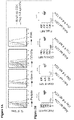
- FIG. 1A is a schematic diagram showing that LMP1 signaling in B cells (e.g., primary B cells) induces expression and presentation of cellular antigens (including many TAAs), and enhances co-stimulation function, thereby eliciting potent polyclonal cytotoxic T cell responses.
- B cells constitutive LMP1 signaling induces massive cellular gene expression. This leads to upregulation of cellular machinery involved in antigen processing and presentation (e.g., MHCs), induction of strong co-stimulation signals (B7-1, B7-2, ICAM-1, and particularly CD70, OX40L and 4-1BBL), and induced and/or enhanced expression of certain cellular antigens (including a wide range of TAAs).
- LMP1 signaling-induced cellular antigens and simultaneous co-stimulations drive activation and cytotoxic differentiation of CD4 + and CD8 + T cells specific to these antigens.
- LMP1 signaling makes B cells hyperimmunogenic antigen-presenting cells (APCs).
- FIG. 1B is a schematic diagram showing that LMP1 signaling in lymphoma B cells enhances presentation of lymphoma inherent TAAs and neoantigens.
- Some of these lymphoma inherent TAAs are LMP1 signaling-induced TAAs, whose expression is enhanced by LMP1 signaling, whereas other lymphoma inherent TAAs are not.
- the increased antigen presentation along with enhanced co-stimulation signals leads to cytotoxic T cell responses against these tumor antigens.
- LMP1 signaling turns lymphoma B cells into hyperimmunogenic antigen-presenting cells (APCs).
- APCs hyperimmunogenic antigen-presenting cells
- FIG. 2A is a schematic diagram showing an expression cassette of LMP1 used in generating CD19-cre;LMP1 flSTOP (CL) transgenic mice.
- FIG. 2B is a schematic diagram demonstrating the role of LMP1 in the surveillance and transformation of LMP1-expressing (EBV-infected) B cells.
- FIG. 3A is a graph showing dynamics of LMP1-expressing B cells (CD19 + Fas + ; Fas is induced by LMP1 signaling and consequently used as a reporter for LMP1 expression in B cells) and activated (CD69 + ) CD4 and CD8 T cells, analyzed by FACS, in the spleen of CL mice compared to those in CD19-cre/+ control (‘C’) mice.
- the respective mean values of at least three mice of each genotype, at each time point are plotted.
- FIG. 3B is a graph showing dynamics of LMP1-expressing B cells and activated (CD69 + ) CD4 and CD8 T cells, analyzed by FACS, in the bone marrow (BM) of CL mice compared to those in CD19-cre/+ control (‘C’) mice. The respective mean values of at least three mice of each genotype, at each time point are plotted.
- FIG. 4 is a graph showing cytolytic activity of CD4 + and CD8 + T cells to LMP1-expressing B cells.
- CD4 and CD8 T cells from day 6-8 CL mice kill LMP1-expressing lymphoma cells, upon co-culture for 4 hours. E:T ratio, effector to target cell ratios.
- FIG. 5A shows FACS analysis of the indicated effector molecules in primary CD4 T cells isolated from day 6-8 CL mice spleen, compared to primary CD4 T cells from adult CL spleen, demonstrating tumor-killing T cells express key cytotoxic molecules.
- FIG. 5B shows mean fluorescence intensities (MFI) of the indicated effector molecules detected as in the FIG. 5A FACS analysis.
- FIG. 5C shows FACS analysis of the indicated effector molecules in primary CD8 T cells isolated from day 6-8 CL mice spleen, compared to primary CD8 T cells from adult CL spleen, demonstrating tumor-killing T cells express key cytotoxic molecules.
- FIG. 5D shows mean fluorescence intensities (MFI) of the indicated effector molecules detected as in the FIG. 5C FACS analysis.
- FIG. 6A is a graph showing cytotoxicity of the indicated T cells assayed on LMP1-expressing lymphoma cells as targets.
- CD4 and CD8 T cells were from adult (day 42-84) CL mice BM; the adoptive CD4 T cells were those initially isolated from adult CL mice BM, adoptively transferred (along with LMP1-expressing lymphoma cells) into Rag2 ⁇ / ⁇ ⁇ c ⁇ / ⁇ recipients, and then recovered from the latter. Representative data from three independent experiments are shown. All mice used here are on a (C57BL/6 ⁇ BALB/c) F1 (CB6F1) background, while the lymphoma cells are on a C57BL/6 ⁇ BALB/c mixed background.
- FIG. 6B is a representative series of graphs showing the flow cytometry analysis of the indicated effector molecules in the adoptive CD4 cells compared to primary CD4 cells from adult CL mice BM (chronic state) and spleens (negative control).
- FIG. 6C is a set of survival curves showing the therapeutic efficacies of adoptive CD4 and CD8 cells in combination with radiation therapy (RT) in mice bearing aggressive LMP1-driven primary lymphomas.
- TCR ⁇ ⁇ / ⁇ ⁇ ⁇ / ⁇ CL mice on a C57BL/6 ⁇ BALB/c mixed background at 8-week old were treated with 500 Rad of irradiation.
- One day later some mice were further treated (by intravenous injection) with the indicated T cells isolated from CL mice on a CB6F1 background at the dose of 1 ⁇ 10 6 cells/recipient. Survival curves were compared using the log-rank test.
- FIG. 7A is a bar graph showing TCR V ⁇ chains in CD8 T cells from the indicated mice that were stained with a panel of monoclonal antibodies for the indicated TCR V ⁇ chains. These V ⁇ specific antibodies collectively detected 85-95% of TCRs in all the samples. Control d8, day 8 old CD19-cre/+ mice. Data are shown as mean ⁇ SEM.
- FIG. 7B is a bar graph showing TCR V ⁇ chains in CD4 T cells (excluding CD25 + Foxp3 + Tregs) from the indicated mice that were stained with a panel of monoclonal antibodies for the indicated TCR V ⁇ chains. These V ⁇ specific antibodies collectively detected 85-95% of TCRs in all the samples.
- Control d8 day 8 old CD19-cre/+ mice; the adoptive CD4 T cells were those initially isolated from adult CL mice BM, adoptively transferred (along with LMP1-expressing lymphoma cells) into Rag2 ⁇ / ⁇ ⁇ c ⁇ / ⁇ recipients, and then recovered from the latter. Data are shown as mean ⁇ SEM.
- FIG. 7C is a graph showing in vitro killing activity of the indicated CD4 T cells from day 6-8 CL mice, assayed on LMP1-expressing lymphoma cells. Data are shown as mean ⁇ SEM of duplicates. Representative data from two independent experiments are shown. CL and control mice used here are on a CB6F1 background.
- FIG. 8 shows FACS analysis of na ⁇ ve B cells, CD40-activated B cells from wild-type (WT) mice, LMP1-expressing lymphoma B cells and B cells from LMP1 flSTOP mice treated with TAT-Cre to turn on LMP1 expression in vitro (LMP1-expressing B cells).
- FIG. 9A shows fluorescent microscopy imaging of B cells expressing LMP1-GFP fusion, LMP1 TM1m -GFP fusion or GFP, respectively. Note that wild-type LMP1 aggregates into large complexes on cell membrane, while the mutant LMP1 TM1m loses its ability to aggregate.
- FIG. 9B is a pair of graphs showing CD4 T cells (left panel) and CD8 T cells (right panel) from day 6-8 CL mice assayed for killing activity on B cells (from WT B6 mice) transduced with retroviral vectors expressing wild-type LMP1 or a signaling-dead mutant LMP1 TM1m . B cells untransduced or transduced with the empty vector as controls.
- FIG. 10A is a pair of graphs showing that CD4 and CD8 T cells from day 6-8 CL mice lyse LMP1-expressing B cells/lymphoma cells as well as anti-CD40 pretreated WT B cells, but not na ⁇ ve B cells.
- FIG. 10B is a graph showing the results of an in vitro killing assay performed with CD4 T cells from day 6-8 CL mice on CD40-activated WT B cells (from B6 mice), in the presence of Fas-Fc (to block FasL-mediated killing) and/or MHCII blocking antibody.
- FIG. 11 shows FACS analysis of CD4 + effector/memory T cells (excluding Tregs) from Foxp3 GFP CL male mice that recognize and proliferate on CD40-activated WT B cells in an MHC-II restricted manner.
- FIG. 12A shows FACS analysis of CD40 expression on LMP1-expressing B cells from a 6-day old CL mouse, compared to that on B cells from a littermate control (CD19-cre/+). Note that LMP1 signaling in B cells upregulates CD40.
- FIG. 12B shows FACS analysis of CD40 expression on B cells from the indicated mice at 6 weeks old. Note that the B cells in CL and CD40 ⁇ / ⁇ ;CL mice represent residual B cells (which do not express LMP1) after clearance of LMP1-expressing B cells.
- FIG. 12C shows FACS analysis of B cells and T cells in spleens of the indicated mice at 6 weeks old.
- FIG. 12D shows FACS analysis of activation marker CD69 on CD4 and CD8 T cells from the BM of the indicated mice at 6 weeks old. Data in (A-D) represent 2-3 mice analyzed for each genotype.
- FIG. 13A is a heat map showing expression of co-stimulatory and co-inhibitory molecules in LMP1-expressing B cells compared to control B cells.
- Splenic B cells from LMP1 flSTOP /YFP flSTOP and YFP flSTOP/+ mice both on a CB6F1 background
- All treated B cells were collected at day 2 post-treatment for array analysis.
- FIG. 13B shows FACS plots (upper panel) and mean fluorescence intensities (MFI; lower panel) of the indicated co-stimulatory ligands in LMP1-expressing B cells from day 6-8 CL mice, compared to splenic B cells from WT control (ctr) mice.
- Data are representative of 2-6 mice analyzed for each group.
- the mice (CL and control) are on a CB6F1 background.
- Each symbol represents an individual mouse; bars show the respective mean values; ****, p ⁇ 0.0001; ***, p ⁇ 0.001 (unpaired two-tailed student's t-test).
- FIG. 13C is a heat map showing cytokine genes expressed in LMP1-expressing B cells compared to control B cells.
- Splenic B cells from LMP1 flSTOP /YFP flSTOP and YFP flSTOP/+ mice were treated with TAT-Cre to generate LMP1-expressing B cells and YFP control B cells. All treated B cells were collected at day 2 post-treatment for array analysis. Mean-centered log 2 gene expression ratios are depicted by color scale.
- FIG. 14A shows FACS analysis of Eomes and GzmB expression in CD4 T cells from day 6-8 CL mice and WT control (ctr) mice. GzmB levels in Eomes + CD4 cells from CL mice were compared to that in total CD4 cells from control mice and shown on the right.
- FIG. 14B shows FACS analysis of Eomes vs. T-bet (upper panel) and GzmB vs. IFN- ⁇ (lower panel) in CD4 T cells from day 6-8 CL mice and WT control (ctr) mice. The frequencies (mean ⁇ SEM) of indicated populations are shown within the gates.
- FIG. 14C shows FACS analysis of Eomes vs. T-bet (upper panel) and GzmB vs. IFN- ⁇ (lower panel) in CD8 cells from day 6-8 CL mice and WT control (ctr) mice.
- Data in (A-C) are representative of 3-4 mice of each group (all on a CB6F1 background), analyzed in two independent experiments.
- FIG. 15A shows FACS analysis of Eomes vs. GATA-3 in CD4 cells from day 6-8 CL mice and WT control (ctr) mice. Data are representative of 3-4 mice of each group (all on a CB6F1 background), analyzed in two independent experiments.
- FIG. 15B shows FACS analysis of Eomes vs. ROR ⁇ t in CD4 cells from day 6-8 CL mice and WT control (ctr) mice. Data are representative of 3-4 mice of each group (all on a CB6F1 background), analyzed in two independent experiments.
- FIG. 16A is a graph showing numbers (mean ⁇ SEM) of recovered T cells after co-culturing for 7 days with B cells expressing LMP1 or LMP1 TM1m .
- the cell culture was begun with 1.5 ⁇ 10 6 purified CD4 T cells together with the indicated B cells (irradiated at 500 RAD before co-culturing) at 1:1 ratio in triplicate wells of 12-well plates. No exogenous cytokines were added. ***, p ⁇ 0.001 (unpaired two-tailed student's t-test).
- B cells and T cells are from 2-3 months old na ⁇ ve WT B6 mice spleens.
- FIG. 16B shows FACS analysis of Eomes and T-bet expression in CD4 cells co-cultured with the indicated B cells (as in (A)).
- FIG. 16C is a graph showing cytotoxicity of CD4 cells expanded on LMP1-B cells (as in (A)) against B cells transduced with the MSCV-LMP1-IRES-GFP retrovirus, which contained GFP + (LMP1-B cells) and GFP ⁇ cells (not successfully transduced cells and thus representing LPS-activated B cells, see Materials and Methods; these cells served as control).
- FIG. 16D shows proliferation of CD4 T cells expanded on LMP1-B cells (as in (A)) assayed on CD40-activated B cells from WT or CIITA ⁇ / ⁇ mice.
- Data in (A-D) are representative of 2-4 independent experiments using splenic B cells and T cells from 2-3 months old na ⁇ ve WT B6 mice.
- FIG. 16E shows FACS analysis of Eomes expression in CD4 cells either freshly isolated from na ⁇ ve B6 mice (Ex vivo), or after co-culturing for 7 days with LMP1-B cells in the presence of the indicated blocking antibodies or corresponding isotype controls. Representative data from one of triplicate wells are shown, with the frequency of Eomes + cells in the gate.
- FIG. 16F shows numbers (mean ⁇ SEM) of Eomes + CD4 cells recovered from culture wells treated with the indicated blocking antibodies relative to those from corresponding isotype control treated wells.
- FIG. 16G shows numbers (mean ⁇ SEM) of recovered CD4 cells after co-culturing for 7 days with LMP1 + B cells in the presence of the indicated blocking antibodies or corresponding isotype controls.
- the cell culture was begun with 1 ⁇ 10 6 purified CD4 T cells in triplicate wells of 24-well plates.
- FIG. 16H shows FACS analysis of Eomes expression in CD8 cells either freshly isolated from na ⁇ ve B6 mice, or after co-culturing for 3 days with LMP1-B cells in the presence of the indicated blocking antibodies or corresponding isotype controls. Representative data from one of triplicate wells are shown, with the frequency of Eomes + cells in the gate.
- FIG. 16I shows numbers (mean ⁇ SEM) of Eomes + CD8 cells recovered from culture wells treated with the indicated blocking antibodies relative to those from corresponding isotype control treated wells.
- FIG. 16J shows numbers (mean ⁇ SEM) of recovered CD8 cells after co-culturing for 3 days with LMP1 + B cells in the presence of the indicated blocking antibodies or corresponding isotype controls.
- the cell culture was begun with 0.5 ⁇ 10 6 purified CD8 T cells in triplicate wells of 24-well plates.
- FIG. 17 is a representative flow cytometry analysis that shows detection of specific T cell response to a TAA expressed by LMP1-expressing B cells.
- CD8 T cells reactive to a Survivin-derived epitope were detected by MHC-I H-2D b tetramers bearing the Survivin 20-28 epitope peptide in CD19-cre ERT2 ;LMP1 flSTOP (C ERT2 L) and CD19-cre ERT2 (C ERT2 ) control mice at day 5 following Tamoxifen treatment (to turn on LMP1 expression initially in a small fraction of B cells).
- the frequencies of Survivin-tetramer + CD8 T cells are shown within the gates. All mice are on a CB6F1 background.
- FIG. 18B shows analysis of the frequency of CD4 Tregs in the CD4 + T cells in adult (day 42-84) CL mice BM (left panel) or in recipient mice transplanted with adult CL mice BM CD4 + T cells and LMP1 + lymphoma cells (right panel). CD4 + T cells were recovered from recipients at day 10 post-transfer for FACS analysis.
- FIG. 18C shows direct killing activity of the indicated T cells isolated from adult Foxp3 DTR/GFP CL male mice (on a CB6F1 background), assayed using LMP1 + lymphoma cells as targets.
- FIG. 18D shows direct killing activity of the CD8 T cells isolated from adult Foxp3 DTR/GFP ;CL male mice (on a CB6F1 background), with or without addition (at 1:1 ratio) of CD4 Tregs from the same mice, assayed using CD40-activated WT B cells (on a B6 background) as targets.
- FIG. 19A shows a scheme depicting the use of LMP1-expressing cells to activate/expand T cells for adoptive cell transfer (ACT) therapy for cancers.
- FIG. 19B shows a scheme of ACT in which CD8 and/or CD4 T cells primed by LMP1-expressing B cells are used to treat tumor-bearing mice.
- mice receive 600 Rad of total body irradiation to create a lymphopenic condition favorable for adoptive T cell expansion.
- FIG. 19C is a graph showing that ACT of CD8 T cells primed by LMP1-expressing B cells delays tumor (A20) growth. Control mice received no ACT. Error bars represent means ⁇ SEM.
- FIG. 19D is a graph showing that ACT of CD4 T cells primed by LMP1-expressing B cells delays tumor (A20) growth. Control mice received no ACT. Error bars represent means ⁇ SEM.
- FIG. 20B shows a vaccination scheme in which lymphoma cells are transduced to express LMP1 and used as vaccine to treat the unmodified (parental) B cell lymphoma.
- FIG. 20C is a graph showing that vaccination with LMP1-expressing A20 lymphoma cells markedly delays tumor (A20) growth.
- A20 lymphoma cells expressing the signaling-dead mutant LMP1 TM1m serve as control vaccine.
- FIG. 20D shows a vaccination scheme in which tumor cells (B16-F10) are transduced to express LMP1 and used as vaccine to treat the unmodified (parental) tumor (melanoma).
- FIG. 20E is a graph showing that vaccination with LMP1-expressing B16-F10 melanoma cells markedly delays tumor (melanoma) growth.
- B16-F10 cells expressing the signaling-dead mutant LMP1 TM1m or transduced with the empty vector serve as control vaccine.
- the vector is an expression vector.
- the vector comprises a transcription regulatory element (e.g., a promoter and/or an enhancer) operably linked to the nucleic acid encoding the polypeptide.
- the vector is a viral vector. In certain embodiments, the vector is a replication incompetent viral vector. In certain embodiments, the viral vector is packaged with one or more capsid proteins into a viral particle. In certain embodiments, the vector or the viral particle further comprises a polynucleotide encoding a polypeptide capable of inducing cell death. In certain embodiments, the polypeptide is a chimeric polypeptide comprising a multimerization (e.g., dimerization or oligomerization) region and a cell death-inducing region, wherein the cell death-inducing region is activated by multimerization.
- a multimerization e.g., dimerization or oligomerization
- the cell death-inducing region comprises a sequence of a caspase (e.g., caspase-9) that has protease activity.
- the cell death-inducing region comprises the full-length human caspase-9 polypeptide.
- the cell death-inducing region comprises a truncated human caspase-9 polypeptide (e.g., wherein the CARD domain of caspase-9 is deleted).
- the present disclosure provides a method of producing an immunogenic cell, the method comprising contacting an isolated cell with a vector (e.g., expression vector) described herein, thereby producing an immunogenic cell.
- a vector e.g., expression vector
- the present disclosure provides an isolated cell comprising a vector (e.g., expression vector) described herein.
- Such cells exhibit superior efficiency of antigen presentation, because LMP1 signaling increases the expression of cellular machinery involved in antigen processing and presentation. Moreover, these cells are hyperimmunogenic, as LMP1 signaling increases the co-stimulation signals (e.g., CD70, OX40L, and 4-1BBL) on the cell surface.
- LMP1 in an isolated cell described herein leads to expression and/or presentation of one or more antigens on the cell surface.
- Cytotoxic T cells can be generated by contacting with the isolated cell.
- the antigens include without limitation (1) LMP1 signaling-induced cellular antigens, which include many TAAs; (2) tumor (e.g., lymphoma) inherent TAAs; and (3) neoantigens, a group of mutation-derived tumor antigens which arise from tumor-specific mutations in expressed proteins.
- LMP1 signaling induces and/or enhances the expression of LMP1 signaling-induced cellular antigens, which includes many TAAs.
- LMP1-expressing primary B cells increasingly express and present LMP1 signaling-induced cellular antigens on their surface, and are useful for activating T cells that express TCRs that bind to these antigens ( FIG. 1A ).
- LMP1 signaling increases the expression of LMP1 signaling-induced TAAs, a subgroup of lymphoma inherent TAAs.
- the expression of the other lymphoma inherent TAAs, as well as the neoantigens, is not induced or enhanced. Regardless of the expression levels, however, all these antigens are increasingly presented on the surface of LMP1-expressing lymphoma B cells, relative to the corresponding unmodified (LMP1-negative) lymphoma B cells. Therefore, LMP1-expressing lymphoma B cells are useful for activating T cells that express TCRs that bind to these lymphoma inherent neoantigens and TAAs ( FIG. 1B ).
- the present disclosure provides a method of activating a T cell, the method comprising contacting the T cell with one or more isolated cells described herein.
- the method is used for cancer immunotherapy.
- the isolated cell is a B cell.
- LMP1 represents the first foreign protein capable of breaking immune tolerance when expressed as a transgene starting from early development.
- Constitutive LMP1 signaling in B cells induces massive cellular genes, leading to upregulation of antigen presenting function (MHCs), strong co-stimulatory signals (B7-1, B7-2, ICAM-1, and particularly CD70, OX40 ligand, and 4-1BB ligand), and induced and/or enhanced expression of certain cellular antigens (termed here as LMP1 signaling-induced cellular antigens).
- MHCs antigen presenting function
- B7-1, B7-2, ICAM-1, and particularly CD70, OX40 ligand, and 4-1BB ligand strong co-stimulatory signals
- induced and/or enhanced expression of certain cellular antigens termed here as LMP1 signaling-induced cellular antigens.
- LMP1 signaling-induced cellular antigens on MHCs (HLAs in humans) and simultaneous co-stimulation through CD70, OX40 ligand, and 4-1BB ligand drive activation and cytotoxic differentiation of CD4 and CD8 T cells specific to these antigens.
- LMP1 is the key oncoprotein for EBV-driven tumorigenesis
- the LMP1 signaling-induced cellular antigens that are targeted by T cells would be various Tumor-Associated Antigens (TAAs, a group of non-mutated cellular antigens recognizable by T cells in certain tumors).
- TAAs Tumor-Associated Antigens
- the isolated cells described herein express antigens (e.g., TAAs and neoantigens), which can be presented by MHCs (e.g., HLAs). Accordingly, in some embodiments, the isolated cells can be used to generate cytotoxic T cells with diverse TCR repertoire against wide range of TAAs and neoantigens in a simple and speedy way, without the need of identifying such TAAs and pairing with particular MHCs (e.g., HLAs). In certain embodiments, the isolated cells are patient-derived B cells or lymphoma cells. The unique strength of the therapeutic strategies described herein is that they can also be combined with immune co-stimulation therapies and/or immune checkpoint targeting therapies.
- MHCs e.g., HLAs
- Immune co-stimulation therapies and immune checkpoint targeting therapies rely on pre-existing tumor antigen-specific T cells, lack of which may have caused the failure of such therapies in many cancer patients. Therefore, the use of LMP1-expressing cells to activate T cells can bring more effective treatment to those who otherwise would fail immune co-stimulation therapies or immune checkpoint targeting therapies alone.
- LMP1-expressing cells e.g., B cells
- the activation of T cells by LMP1-expressing cells could be dependent on the ability of CD70, OX40L, and 4-1BBL to engage CD27, OX40, and 4-1BB, respectively, on the T cells.
- these stimulatory proteins may be down-regulated or defective.
- a vaccination therapy using LMP1-expressing cells e.g., B cells or tumor cells
- an adoptive cell transfer therapy (ACT) using T cells activated by LMP1-expressing cells e.g., B cells or tumor cells
- the agonist is an agonistic antibody that specifically binds to CD27, OX40, or 4-1BB.
- the agonistic antibody can be in any format (e.g., tetrameric antibody comprising two heavy chains and two light chains, single-chain Fv, Fab fragment, F(ab′) 2 fragment, bispecific antibody).
- the agonistic anti-CD27 antibody is varlilumab.
- the agonistic anti-OX40 antibody is selected from the group consisting of MOXR0916 (Genentech), MEDI6383 (MedImmune), and INCAGN1949 (Agenus).
- the agonistic anti-4-1BB antibody is selected from the group consisting of urelumab/BMS-663513 (BMS) and PF-05082566 (Pfizer). In some embodiments, one, two, or three of these agonists are administered to a patient in need thereof.
- the immune checkpoint targeting therapy is selected from the group consisting of an antagonist anti-PD-1 antibody, an antagonist anti-PD-L1 antibody, an antagonist anti-PD-L2 antibody, an antagonist anti-CTLA-4 antibody, an antagonist anti-TIM-3 antibody, an antagonist anti-LAG-3 antibody, an antagonist anti-CEACAM1 antibody and an IDO inhibitor, i.e., an agent that inhibits the enzymatic activity of IDO (indoleamine-(2,3)-dioxygenase) and/or TDO (tryptophan 2,3-dioxygenase).
- IDO inhibitor i.e., an agent that inhibits the enzymatic activity of IDO (indoleamine-(2,3)-dioxygenase) and/or TDO (tryptophan 2,3-dioxygenase).
- the immune checkpoint targeting therapy is an anti-PD-1 antibody, optionally wherein the anti-PD-1 antibody is pembrolizumab, nivolumab, Pidilizumab, MEDI0680, PDR001, REGN2810, PF-06801591, BGB-A317, TSR-042, or SHR-1210.
- the immune checkpoint targeting therapy is an anti-PD-L1 antibody, optionally wherein the anti-PD-L1 antibody is atezolizumab, durvalumab, avelumab (MSB0010718C), MDX-1105, or AMP-224.
- the immune checkpoint targeting therapy is an anti-CTLA-4 antibody, optionally wherein the anti-CTLA-4 antibody is ipilimumab. In some embodiments, the immune checkpoint targeting therapy is an IDO inhibitor, optionally wherein the IDO inhibitor is epacadostat, F001287, indoximod, or NLG919.
- LMP1-expressing cells e.g., B cells
- Tregs e.g., CD4 Tregs
- the amount and activity of Tregs may be higher than in healthy individuals, and may be triggered at the earlier acute phase, which may limit the efficacy of a vaccination therapy using LMP1-expressing cells (e.g., B cells) or an adoptive cell transfer (ACT) therapy using T cells activated by LMP1-expressing cells (e.g., B cells).
- ACT adoptive cell transfer
- a subject receiving or to receive the vaccination or ACT therapy can further receive administration of a Treg modulating therapy to inhibit or decrease the amount and activity of Tregs.
- Treg modulating therapies are known in the art, and include without limitation antibodies (e.g., full antibodies, and antigen-binding fragments thereof) that specifically bind to CTLA-4, GITR, CCR4, PD-1, LAG3, CD25, or CD15s.
- the Treg modulating therapy can be administered prior to, contemporaneously with (e.g., during the same doctor visit), or subsequent to the administration of the vaccination or ACT therapy. If the Treg modulating therapy is administered subsequent to the administration of the vaccination or ACT therapy, the patient's response to the vaccination or ACT therapy can be examined to determine the necessity and dose of the Treg modulating therapy.
- the isolated cells or T cells contacted therewith are administered in combination with an adjuvant.
- adjuvants may be employed, including, for example, systemic adjuvants and mucosal adjuvants.
- a systemic adjuvant is an adjuvant that can be delivered parenterally.
- Systemic adjuvants include adjuvants that create a depot effect, adjuvants that stimulate the immune system and adjuvants that do both.
- An adjuvant that creates a depot effect is an adjuvant that causes the antigen to be slowly released in the body, thus prolonging the exposure of immune cells to the antigen.
- the adjuvant stimulate the immune system, for instance, cause an immune cell to produce and secrete cytokines or IgG.
- This class of adjuvants includes immunostimulatory nucleic acids, such as CpG oligonucleotides; saponins purified from the bark of the Q. saponaria tree, such as QS-21; poly[di(carboxylatophenoxy)phosphazene (PCPP polymer; Virus Research Institute, USA); RNA mimetics such as polyinosinic:polycytidylic acid (poly I:C) or poly I:C stabilized with poly-lysine (poly-ICLC [Hiltonol®; Oncovir, Inc.]; derivatives of lipopolysaccharides (LPS) such as monophosphoryl lipid A (MPL; Ribi ImmunoChem Research, Inc., Hamilton, Mont.), muramyl dipeptide (MDP; Ribi) and threonyl-muramyl dipeptide (t-MDP; Ribi); OM-174 (a glucosamine disaccharide related to lipid A; OM
- the adjuvant is administered prior to, at about the same time as, or subsequent to the administration of the isolated cells or T cells. In some embodiments, the adjuvant is administered within the same patient visit as the administration of the isolated cells or T cells. In some embodiments, the adjuvant is administered in the same composition (e.g., vaccine) as the isolated cells or T cells. In some embodiments, the adjuvant is administered in a different composition from the isolated cells or T cells.
- the disclosure relates to expressing LMP1 using replication incompetent viral vectors or transfection in patient-derived B cells or lymphoma cells and using them to activate/expand T cells autologous or derived from a donor for Adoptive Cell Transfer (ACT) therapy.
- the ACT is employed to a subject with EBV-associated B cell lymphoma.
- the ACT is employed to an immunosuppressed patient, such as post-transplant and AIDS patients.
- the subject has EBV-associated B cell lymphoma cells expressing LMP1, which may present the same array of antigens on their surface.
- the cells are irradiated to have reduced proliferative capacity, as LMP1 is a potent oncogene. In certain embodiments, the proliferative capacity of the cells is reduced by irradiation.
- the ACT strategy described herein can be similarly applied to EBV-associated B cell lymphomas in immunocompetent hosts, such as Burkitt lymphoma and Hodgkin lymphoma, or EBV-unrelated B cell lymphomas. These lymphoma cells share some TAAs with LMP1-expressing autologous B cells/lymphoma cells used for T cell activation/expansion. As described herein, an ACT strategy using LMP1-expressing lymphoma cells for producing therapeutic T cells, and for treating EBV-unrelated B cell lymphomas, can generate anti-tumor T cells against the array of lymphoma inherent TAAs and neoantigens ( FIG.
- ACT strategies are suitable for generating therapeutic T cells against these lymphoma inherent antigens, because LMP1 signaling would enhance cell endogenous antigen presentation and co-stimulation, i.e., turning the lymphoma cells into hyperimmunogenic APCs.
- ACT uses in vitro generated tumor antigen-reactive T cells to treat cancers.
- the strategy for ACT production has evolved over time, but has always involved complicated in vitro manipulations prior to the instant disclosure.
- Such manipulations include, for example, isolating tumor-reactive cytotoxic T lymphocytes (CTLs) from patients and subjecting them to extensive in vitro expansion/differentiation; introducing tumor-reactive TCRs into autologous T cells by means of gene transfer; or engineering T cells to express a chimeric antigen receptor specific for a tumor antigen.
- CTLs tumor-reactive cytotoxic T lymphocytes
- ACT therapies with TCR targeting a single TAA have limited efficacy, yet abundant autoimmune toxicity.
- CAR-T therapy so far the most successfully targeted tumors are those derived from B cells due to their unique expression of the CD19 antigen (these CAR-T cells also eliminate patient's normal B cells, an unwanted but manageable toxicity). Still, a sizable fraction of patients fail in such therapy due to the escaping of epitope-loss variants. There has been little success for CAR-T therapy in solid tumors. Although CAR-T therapies targeting a single TAA or two TAAs simultaneously have been attempted, tumor escaping and on-target/off-tumor toxicity remain major problems. Thus, the CAR-T therapy for solid tumors is mainly limited by the ability to identify antigens (ideally multiple) that are specifically expressed on tumor cell surface, but not in normal cells.
- Neoantigens which term is used interchangeably with “mutation-derived antigens,” are ideal for this purpose; however, the vast majority of neoantigens in cancers are “private” events, i.e., events rarely shared in multiple patients. Thus, identifying such neoantigens and generating CARs against these antigens is not practical.
- EBV-transformed B cells often called lymphoblastoid cell lines (LCLs)
- LCLs lymphoblastoid cell lines
- EBV latent antigens viral antigens
- EBV-specific CTLs generated in vitro by repetitive stimulation of autologous or donor-derived T cells with EBV-LCLs have been used in clinic to treat EBV-associated B cell lymphomas and were effective in about 50% of patients. This T cell preparation process typically takes 2-3 months, while the tumor is often aggressive and thus necessitates urgent treatment.
- EBV-transformed B cells are additionally transduced to increase EBV latent antigens expression/presentation, including a truncated and signaling-dead form of LMP1.
- LMP1 mutant when expressed in lymphoma cells or other tumor cells, had been shown able to activate/enhance presentation of transduced model antigens, but restrict presentation of its own epitopes unless its signaling function is disabled. Contrary to this rationale, the present disclosure shows that it is because LMP1 signaling-induced massive cellular antigens dilute or mask LMP1-derived epitopes.
- LMP1-expressing B cells have advantages over LCLs in the brevity of T cell production protocol.
- the production of cytotoxic T cells from LMP1-expressing B cells takes only about 11 days (including the time for preparation of LMP1-expressing B cells and subsequent generation of antigen-specific T cells), in sharp contrast to 2-3 months required by lymphoblastoid cell line (LCL)-based protocols.
- LCL lymphoblastoid cell line
- the method can further comprise culturing the T cell with a B cell or vaccine (e.g., the B cell or vaccine as disclosed herein) under suitable conditions to allow proliferation of the T cell.
- suitable conditions can include certain factors that promote or enhance the survival, proliferation, or differentiation of T cells.
- exemplary factors include cytokines (e.g., IL-2, IL-1, IL-6, IL-12, or IL-18), anti-CD3 antibodies, anti-CD28 antibodies, phytohemagglutinin, calcium ionophores, inhibitors to cell death (e.g., FasL/Fas neutralizing antibodies), and cells that can facilitate T cell activation (e.g., macrophages or dendritic cells).
- the method disclosed herein can take about 11 days for preparation of LMP1-B cells and subsequent generation of antigen-specific T cells.
- the T cell is cultured for a suitable length of time (e.g., about 3-5 days, 5-7 days, 3-7 days, or 7-14 days; equal to or less than 3, 5, 7, or 10 days; or, equal to or less than 1, 2, 3, or 4 weeks).
- the T cell can be co-cultured with the B cell during the entire length of time or a portion thereof.
- the B cell that is contacted with the T cell is replenished (e.g., every 2-3 days, 3-4 days, or 4-5 days).
- the factors can be added and withdrawn anytime in the course of the culture. For example, IL-2 may be added from day 3 onward.
- the present disclosure relates to vaccination strategy for treatment of cancer.
- LMP1-expressing autologous B cells/lymphoma cells are used as an “LMP1-cell vaccine,” after irradiation, to activate/expand T cells in vivo to treat these lymphoma patients.
- vaccination regimens mostly aimed at a single TAA have produced rare clinical benefit, partly due to the escaping of antigen/epitope-loss variants.
- Another known strategy to target multiple TAAs is to load dendritic cells (DCs) with tumor cell lysates. This strategy is currently under clinical testing, yet may encounter several obstacles. While the clinical efficacy of tumor neoantigen vaccination awaits further report, identification of tumor neoantigen is a laborious process, and the vast majority of these neoantigens are “private” events (rarely shared in multiple patients).
- the vaccination strategies described herein utilize LMP1 signaling-induced cellular antigens expression, presentation, and co-stimulation to activate T cell immunity against a broad spectrum of TAAs and neoantigens in a simple and expeditious way.
- the target antigens of the vaccination strategy using LMP1-expressing primary B cells, as described herein are LMP1 signaling-induced cellular antigens (including many TAAs) ( FIG. 1A ).
- the vaccination strategy using LMP1-expressing lymphoma cells as described herein, can generate anti-tumor T cells against lymphoma inherent TAAs and neoantigens ( FIG. 1B ).
- LMP1-expressing primary and lymphoma cells for vaccination obviates the need to identify the specific antigens and pair them with particular MHCs (e.g., HLAs). Therefore, vaccination strategies described herein generates polyclonal cytotoxic T cells against lymphoma inherent TAAs and neoantigens. Such vaccination strategies are suitable for eliciting T cell responses to lymphoma inherent antigens, because LMP1 signaling would enhance cell endogenous antigen presentation and co-stimulation, i.e., turning the lymphoma cells into hyperimmunogenic APCs.
- LMP1 signaling in other lineages of cells can be used to enhance cell endogenous antigen presentation and co-stimulation, and thus LMP1-expressing patient-derived tumor cells can be used to activate/expand T cells in both in vitro ACT strategies and in vivo vaccination strategies to treat the corresponding tumor patients.
- the target antigens of the ACT and vaccination strategies with LMP1-expressing tumor cells of non-B lineages, as described herein, include the tumor inherent TAAs and neoantigens.
- the ACT and vaccination strategies described herein using LMP1-expressing B cells can be applied to non-EBV-associated cancers that share one or more TAAs with LMP1-expressing B cells.
- the non-EBV-associated cancer may express one or more tumor-associated antigens (TAAs) that are also expressed by the LMP1-expressing B cells or LMP1-expressing non-B cells.
- TAAs tumor-associated antigens
- LMP1-expressing lymphoma cells may provide some advantages in that anti-tumor T cells against the lymphoma inherent TAAs and neoantigens can be generated, as LMP1 signaling would enhance cell endogenous antigen presentation and co-stimulation, i.e., turning the lymphoma cells into hyperimmunogenic APCs (see FIG. 1B ).
- LMP1 signaling would enhance cell endogenous antigen presentation and co-stimulation, i.e., turning the lymphoma cells into hyperimmunogenic APCs (see FIG. 1B ).
- some lymphomas maybe suboptimal in co-stimulation function and may not be easily accessible, while autologous B cells (non-tumorous) would be intact in such function and easy to obtain from peripheral blood.
- LMP1-expressing autologous B cells or LMP1-expressing lymphoma cells will be tailored to patient-specific conditions.
- patient-derived cancer cells are easier to obtain and grow than normal cells of the same lineages and thus are preferred.
- Both the ACT and vaccination strategies described herein fulfill several most desired features for effective cancer immunotherapy: (1) eliciting both cytotoxic CD4 and cytotoxic CD8 T cell responses; (2) targeting a large array of TAAs, and neoantigens when LMP1-expressing tumor cells are used; (3) being simple and fast.
- cytotoxic anti-tumor CD4 cells represent a unique feature of the ACT and vaccination strategies described herein, considering that (i) recent work from us and others have shown great potential of cytotoxic CD4 cells in treating various cancers; (ii) these cells would be particularly important in fighting cancers that escape CD8 killing; (iii) a general approach for rapid generation of tumor antigen-specific cytotoxic CD4 cells was not available prior to the present invention.
- cytotoxicity of T cells is examined using an in vitro killing assay.
- CD4 + and CD8 + T cells were isolated by Fluorescence-activated cell sorting (FACS) from CD19-cre;LMP1 flSTOP mice on a CB6F1 background.
- the T cells were co-cultured with 4 ⁇ 10 3 target cells at various effector:target ratios for 4 hours in 96-well plates, followed by active Caspase-3 staining (BD) (He et al. J. Immunol. Methods 304: 43-59 (2005)).
- effector-target mixtures in U-bottom 96-well plates were spun at 200 rpm for 2 min before moving to incubator, and cultures were stained with anti-CD19, anti-CD4, and anti-CD8 to identify target cells (B cells or lymphoma cells) and effector cells.
- Active Caspase-3 positive CD19 + cells represent apoptotic target cells.
- % specific killing % apoptotic target cells of cultures with both effectors and targets ⁇ % apoptotic target cells of cultures with targets alone.
- an effector of in vitro killing assay encompasses, but is not limited to, a cytotoxic CD4 + and/or CD8 + T cell
- a target of in vitro killing assay encompasses, but is not limited to, a LMP1-expressing B cell.
- a B cell specific LMP1 transgene expression is enabled with CD19-cre.
- the CD19 promoter specifically directs Cre expression early in (starting at the pro-B stage) and continuing throughout B-lymphocyte development.
- a Cre cassette is inserted into the CD19 exon 2, functionally disrupting the gene.
- Homozygous mice are CD19-deficient, whereas heterozygous mice are phenotypically normal and can be used for specific deletion of foxed cassette from conditional alleles, leading to activation or inactivation of target genes, in B-lymphocytes.
- a B cell encompasses a cell modified or derived from a B-lymphocyte.
- a non-B cell encompasses, but is not limited to, a cell modified or derived from a solid tumor cell.
- TAA-tetramers or pentamers
- tetramers are made with H-2D b , H-2K b and I-A b .
- Predicted peptides loaded on B6 splenocytes or CpG-activated B cells are used to test T cells response by proliferation or cytokine assays. Confirmed tetramers are used to monitor the corresponding antigen-specific T cells in mice under therapeutic studies to characterize/optimize “LMP1-cell vaccine” and ACT approaches.
- LMP1-A20 lymphoma cell vaccine and LMP1-B cell vaccine are compared for their efficacies in treating A20 lymphoma-bearing mice using the method described below.
- vaccination efficacies with or without antibody-mediated pre-depletion of CD4 + and CD8 + T cells may be compared to demonstrate the contribution of CD4 + and CD8 + T cells in the tumor control.
- vaccination efficacy can be tested with a poorly immunogenic tumor cell. Poorly immunogenic tumor cells encompass, but are not limited to, A20 lymphoma cells and B16 melanoma cells.
- the ACT or vaccination strategy described herein can be administered with an immune co-stimulation therapy and/or an immune checkpoint targeting therapy as a part of a combination therapy.
- An immune checkpoint targeting therapy encompasses, but is not limited to, anti-PD1 and/or -CTLA4.
- T cells can be expanded on LMP1-expressing cells under suitable conditions.
- na ⁇ ve T cells CD4 + or CD8 +
- CD8 + T cell expansion can be enhanced by addition of IL-2 from day-3 onward.
- LMP1-expressing cells can be irradiated to abrogate their ability to proliferate. Any effective type of radiation may be used. According to other embodiments, any effective method to prevent proliferation of these cells may be used.
- both ACT and vaccination strategies described herein can be validated and optimized in preclinical cancer model.
- Preclinical cancer model encompasses, but is not limited to, lymphoma and melanoma models.
- both ACT and vaccination strategies described herein can be validated and optimized in preclinical cancer model in combination with checkpoint blockade.
- human T cells can be primed with a LMP1-expressing autologous cell.
- the LMP1-expressing autologous cell encompasses, but is not limited to, a LMP1-expressing B cell, a LMP1-expressing lymphoma cell, and a LMP1-expressing melanoma cell.
- LMP1 NCBI Gene ID No. is 3783750.
- Mouse CD40 NCBI Gene ID No. is 21939.
- Human CD40 NCBI Gene ID No. is 958.
- an antigen is defined as a molecule that provokes an immune response. This immune response may involve either antibody production, or the activation of specific immunologically competent cells, or both.
- An antigen can be derived from organisms, subunits of proteins/antigens, killed or inactivated whole cells or lysates. Exemplary organisms include but not limited to Epstein-Barr virus (EBV) and cells infected by EBV. Any macromolecules, including virtually all proteins or peptides, can serve as antigens.
- antigens can be derived from recombinant or genomic DNA. In certain embodiments, an antigen includes a fragment of a protein that elicits an immune response.
- LMP1 refers to Epstein-Barr virus (EBV) latent membrane protein 1.
- LMP1 is a 100% identical to the previously known polypeptide sequences (GenBank Accession No. YP_401722).
- LMP1 has the amino acid sequence of SEQ ID NO: 1.
- LMP1 is a polypeptide with a sequence identity ranging from 70% to 80%, from 81% to 85%, from 86% to 90%, from 91% to 95%, from 96% to 100%, or 100% to SEQ ID NO. 1.
- LMP1 is a polypeptide with a sequence identity of at least 75, 80, 85, 90, 95, 96, 97, 98 or 99% to SEQ ID NO. 1.
- LMP1 polypeptide sequence from GenBank Accession No. YP_401722 SEQ ID NO: 1 MEHDLERGPPGPRRPPRGPPLSSSLGLALLLLLLALLFWLYIVMSDWTGG ALLVLYSFALMLIIIILIIFIFRRDLLCPLGALCILLLMITLLLIALWNL HGQALFLGIVLFIFGCLLVLGIWIYLLEMLWRLGATIWQLLAFFLAFFLD LILLIIALYLQQNWWTLLVDLLWLLLFLAILIWMYYHGQRHSDEHHHIDD SLPHPQQATDDSGHESDSNSNEGRHHLLVSGAGDGPPLCSQNLGAPGGGP DNGPQDPDNTDDNGPQDPDNTDDNGPHDPLPQDPDNTDDNGPQDPDNTDD NGPHDPLPHSPSDSAGNDGGPPQLTEEVENKGGDQGPPLMTDGGGGHSHD SGHGGGDPHLPTLLLGSSGSGGDDDDPHGPVQLSYYD.
- LMP1 signaling-induced cellular antigen refers to a cellular antigen whose expression is induced and/or enhanced by LMP1 signaling, and encompasses, but is not limited to, Tumor-Associated Antigens (TAAs), a group of non-mutated cellular antigens recognizable by T cells in certain tumors.
- TAAs Tumor-Associated Antigens
- Exemplary LMP1 signaling-induced cellular antigens include, but are not limited to, Cdkn1a/p21 (GenBank Accession No.: NP_001104569), Birc5/Survivin (GenBank Accession No.: NP_033819), Epha2 (GenBank Accession No.: NP_034269), and Kif20a (GenBank Accession No.: NP_001159878).
- T cells specific to TAAs presented by LMP1-expressing cells can be identified with TAA-tetramers in C ERT2 L and CL mice on, but not limited to, CB6F1 background.
- TAA loaded on B6 splenocytes or CpG-activated B cells can be used to test T cell response by proliferation and cytokine assays.
- LMP1-cell vaccine described herein is defined as a cell, upon LMP1 expression, capable of processing and presenting LMP1 signaling-induced cellular antigens/TAAs, as well as individual tumor specific TAAs and neoantigens. LMP1-cell vaccine induces cytotoxic T cell responses against above described antigens.
- an antigen-presenting cell is any of a variety of cells capable of displaying, acquiring, and/or presenting at least one antigen or antigenic fragment on its cell surface.
- an antigen-presenting cell can be any cell that induces and/or enhances an immune response against an antigen or antigenic composition.
- a cell that displays or presents an antigen normally or preferentially with a class II major histocompatibility (MHC-II) molecule or complex to an immune cell is a professional APC.
- the immune cell to which an APC displays or presents an antigen is a CD4 + or a CD8 + T cell.
- Full activation of na ⁇ ve T cells can be achieved by an antigen displayed by an APC in the form of a peptide bound to an MHC, which provides specificity to the response, and a co-stimulatory signal, which is antigen nonspecific and facilitates the development of an effective immune response of adaptive immunity.
- T cell co-stimulation increases T cell proliferation, differentiation and survival. Activation of T cells without co-stimulation may lead to T cell anergy, T cell deletion or the development of immune tolerance.
- Additional molecules expressed by the APC or other immune cells that may aid or enhance an immune response include secreted molecules, such as cytokines and cytotoxic molecules.
- MHC refers to “major histocompatibility antigen.”
- HLA human leukocyte antigen
- HLA human leukocyte antigen
- the most studied HLA genes are the nine so-called classical MHC genes: HLA-A, HLA-B, HLA-C, HLA-DPA1, HLA-DPB1, HLA-DQA1, HLA-DQB1, HLA-DRA, and HLA-DRB1.
- the MHCs include at least three regions: Class I, II, and III.
- the A, B, and C genes belong to MHC class I, whereas the six D genes belong to class II.
- MHC class I molecules are made of a single polymorphic chain containing 3 domains (alpha 1, 2 and 3), which associates with beta 2 microglobulin at cell surface.
- Class II molecules are made of 2 polymorphic chains, each containing 2 domains (alpha 1 and 2, and beta 1 and 2).
- Class I MHC molecules are expressed on virtually all nucleated cells. Peptide fragments presented in the context of class I MHC molecules are recognized by CD8 + T lymphocytes (traditionally called cytotoxic T lymphocytes or CTLs). CD8 + T lymphocytes frequently mature into cytotoxic effectors which can lyse cells bearing the stimulating antigen. Class II MHC molecules are expressed primarily on activated lymphocytes and professional APCs. CD4 + T lymphocytes (traditionally called helper T lymphocytes or HTLs) are activated with recognition of a unique peptide fragment presented by a class II MHC molecule, usually found on an APC, like a macrophage, dendritic cell or B cell.
- CD4 + T lymphocytes proliferate and secrete cytokines that either support an antibody-mediated response through the production of IL-4 or support a cell-mediated response through the production of IL-2 and IFN-gamma, or acquire direct killing activity (cytotoxicity).
- immune co-stimulatory molecule refers to molecules on APCs or T cells that provide a non-antigen-specific signal for T cell proliferation and functional differentiation.
- Representative immune co-stimulatory molecules include, but are not limited to, CD80/B7-1, CD86/B7-2, CD70, CD27, OX40 ligand, OX40, 4-1BB ligand, 4-1BB, and GITR.
- immunore-stimulation therapies include without limitation agonistic antibodies that specifically bind an immune co-stimulatory molecule.
- cytokine is defined as growth, differentiation or chemotropic factors secreted by immune or other cells, whose action is on cells of the immune system, such as, but not limited to, T cells, B cells, NK cells and macrophages or other cell types, such as endothelial cells, hematopoietic cells, etc.
- Representative cytokines include, but are not limited to, the group consisting of IFN- ⁇ , TNF- ⁇ , IL-2 and IL-17.
- sequence identity or “sequence homology” of two sequences when used herein relates to the number of positions with identical nucleotides or amino acids divided by the number of nucleotides or amino acids in the shorter of the sequences, when the two sequences are aligned.
- sequence identity is from 70% to 80%, from 81% to 85%, from 86% to 90%, from 91% to 95%, from 96% to 100%, or 100%.
- sequence identity is at least 80, 81, 82, 83, 84, 85, 86, 87, 88, 89, 90, 91, 92, 93, 94, 95, 96, 97, 98, or 99%.
- cancer as used herein is defined as a hyperproliferation of cells whose unique trait—loss of normal controls—results in unregulated growth, lack of differentiation, local tissue invasion, and metastasis. Examples include, but are not limited to, melanoma, hepatocarcinoma, leukemia, lymphoma, retinoblastoma, astrocytoma, glioblastoma, neuroblastoma, sarcoma, lung, breast, uterine, pancreatic, prostate, renal, bone, testicular, uterine, ovarian, cervical, gastrointestinal, brain, colon, or bladder cancer.
- immunotherapeutics In the context of cancer treatment, immunotherapeutics, generally, rely on the use of immune effector cells and molecules to target and destroy cancer cells.
- the immune effector may be, for example, an antibody specific for some marker on the surface of a tumor cell.
- the antibody alone may serve as an effector of therapy or it may recruit other cells to actually affect cell killing.
- the antibody also may be conjugated to a drug or toxin (chemotherapeutic, radionuclide, ricin A chain, cholera toxin, pertussis toxin, etc.) and serve merely as a targeting agent.
- the effector may be a lymphocyte carrying a surface molecule that interacts, either directly or indirectly, with a tumor cell target (e.g.
- a LMP1 signaling-induced cellular antigen a lymphoma inherent TAA, or a tumor neoantigen.
- Various effector cells include CD8 + T cells, CD4 + T cells and NK cells.
- ACT as described herein.
- vaccination strategy as described herein.
- cytotoxic T cell refers to T lymphocytes that can kill cells expressing a MHC-presented antigen such as cells infected by viruses or transformed cancer cells.
- the cytotoxic T cells include CD8 + T cells (the traditionally referred CTLs or CD8 + CTLs) and a subtype of CD4 + T cells (CD4 + CTLs) that have direct killing activity as described in the instant disclosure.
- CTLs have specificity for peptide antigens that are presented in association with proteins encoded by the MHC genes and which are expressed on the surfaces of cells.
- CTLs lyse cells infected with microbes (e.g., such as viruses), inducing and promoting the destruction of intracellular microbes. In certain embodiments, CTLs lyse cancer cells.
- T cells can be expanded on LMP1-expressing cells under suitable conditions.
- suitable conditions comprises co-culturing of T cells with LMP1-expressing cells, which may be replenished every 4-5 days.
- IL-2 may be added from day 3 onward.
- cell refers to progeny, which are any and all subsequent generations. It is understood that all progeny may not be identical due to deliberate or inadvertent mutations.
- B cell refers to a type of lymphocyte, developed in bone marrow, that circulates in the blood and lymph. Upon encountering a particular foreign antigen, B cells differentiate into a clone of plasma cells that secrete a specific antibody and a clone of memory cells that differentiate into plasma cells making the antibody upon re-encountering the antigen.
- na ⁇ ve B cell refers to a B cell that has not been exposed to a foreign antigen so that it has not committed differentiation into a clone of memory or plasma cells.
- Neoplastic B cell refers to a B cell that undergoes an abnormal pattern of growth.
- a “vector” is a composition of matter which comprises an isolated nucleic acid and which can be used to deliver the isolated nucleic acid to the interior of a cell.
- vectors are known in the art including, but not limited to, linear polynucleotides, polynucleotides associated with ionic or amphiphilic compounds, plasmids, and viruses.
- the term “vector” includes an autonomously replicating plasmid or a virus.
- the term should also be construed to include non-plasmid and non-viral compounds which facilitate transfer of nucleic acid into cells, such as, for example, polylysine compounds, liposomes, and the like.
- viral vectors include, but are not limited to, adenoviral vectors, adeno-associated virus vectors, retroviral vectors, lentiviral vectors, and the like.
- expression vector refers to an exogenous vector comprising a recombinant polynucleotide comprising expression control sequences operatively linked to a nucleotide sequence to be expressed.
- An expression vector comprises sufficient cis-acting elements for expression; other elements for expression can be supplied by the host cell or in an in vitro expression system.
- Expression vectors include all those known in the art, such as cosmids, plasmids (e.g., naked or contained in liposomes) and viruses (e.g., lentiviruses, retroviruses, adenoviruses, and adeno-associated viruses) that incorporate the recombinant polynucleotide.
- the expression vector lacks at least 50%, 55%, 60%, 65%, 70%, 75%, 80%, 85%, 90%, 91%, 92%, 93%, 94%, 95%, 96%, 97%, 98%, 99% or 100% of an EBV genome, thereby incapable of replicating EBV viral genome.
- host cell means any cell type that is susceptible to transformation, transfection, transduction, or the like with a nucleic acid construct or expression vector comprising a polynucleotide.
- host cell encompasses any progeny of a parent cell that may not be identical to the parent cell due to mutations that occur during replication.
- viral vector encompasses vector DNA/RNA as well as viral particles generated thereof. Viral vectors can be replication-competent, or can be genetically disabled so as to be replication-defective or replication-impaired.
- viral particle refers to the viral genome as well as a protein coat around the viral genome, referred to herein as the “capsid”. In certain embodiments, the viral particle also includes an envelope of lipids that surrounds the protein coat.
- the viral genome comprises the nucleotide sequence that is located between the LTRs in the expression vector used for the production of the viral vector particles.
- a variety of viral vectors, such as an adenoviral vector, an adeno-associated viral vector, a lentiviral vector, and a retroviral vector, known in the art can be modified to express or carry a nucleotide sequence.
- Non-viral vectors include, but are not limited to liposomes and lipoplexes, polymers and peptides, synthetic particles and the like.
- a liposome or lipoplex has a neutral, negative or positive charge and can comprise cardolipin, anisamide-conjugated polyethylene glycol, dioleoyl phosphatidylcholine, or a variety of other neutral, anionic, or cationic lipids or lipid conjugates.
- a vector can be complexed to cationic polymers (e.g., polyethylenimine (PEI)), biodegradable cationic polysaccharide (e.g., chitosan), or cationic polypeptides (e.g., atelocollagen, poly lysine, and protamine).
- cationic polymers e.g., polyethylenimine (PEI)
- biodegradable cationic polysaccharide e.g., chitosan
- cationic polypeptides e.g., atelocollagen, poly lysine, and protamine
- transfection or “transduction” as used herein refers to a process by which exogenous nucleic acid is transferred or introduced into the host cell.
- a “transfected” or “transduced” cell is one which has been transfected or transduced with exogenous nucleic acid.
- the cell includes the primary subject cell and its progeny.
- the term “plurality” refers to two or more of anything, such as cells or antigens.
- the terms “a”, “an” or “the” refers to one or more of anything, such as a cell or the cell or an antigen or the antigen.
- a plurality of anything may be homogenous or heterogeneous.
- the term “homogenous” refers to a plurality of identical anything, such as cells or antigens.
- heterogeneous refers to a plurality of anything in which there are least two different types of anything, such as cells or antigens.
- exogenous refers to any nucleic acid that does not originate from that particular cell as found in nature.
- a non-naturally-occurring nucleic acid is considered to be exogenous to a cell once introduced into the cell.
- Nucleic acid that is naturally occurring also can be exogenous to a particular cell.
- an entire chromosome isolated from a cell of subject X is an exogenous nucleic acid with respect to a cell of subject Y once that chromosome is introduced into Y's cell.
- nucleic acid or “polynucleotide” refers to deoxyribonucleic acids or ribonucleic acids and polymers thereof in either single- or double-stranded form. Unless specifically limited, the term encompasses nucleic acids containing known analogues of natural nucleotides that have similar binding properties as the reference nucleic acid and are metabolized in a manner similar to naturally occurring nucleotides. Unless otherwise indicated, a particular nucleic acid sequence also implicitly encompasses conservatively modified variants thereof (e.g., degenerate codon substitutions), alleles, orthologs, SNPs, and complementary sequences as well as the sequence explicitly indicated.
- conservatively modified variants thereof e.g., degenerate codon substitutions
- alleles e.g., orthologs, SNPs, and complementary sequences as well as the sequence explicitly indicated.
- degenerate codon substitutions may be achieved by generating sequences in which the third position of one or more selected (or all) codons is substituted with mixed-base and/or deoxyinosine residues (Batzer et al., Nucleic Acid Res. 19:5081 (1991); Ohtsuka et al., J. Biol. Chem. 260:2605-2608 (1985); and Rossolini et al., Mol. Cell. Probes 8:91-98 (1994))
- promoter refers to a nucleic acid sequence, usually found upstream (5′) to a coding sequence, which directs transcription of a nucleic acid sequence into mRNA.
- the promoter or promoter region typically provide a recognition site for RNA polymerase and the other factors necessary for proper initiation of transcription.
- a promoter or promoter region includes variations of promoters derived by inserting or deleting regulatory regions, subjecting the promoter to random or site-directed mutagenesis, etc.
- the activity or strength of a promoter may be measured in terms of the amounts of RNA it produces, or the amount of protein accumulation in a cell or tissue, relative to a promoter whose transcriptional activity has been previously assessed.
- expression cassette relates particularly to a nucleic acid molecule and a region of a nucleic acid molecule, respectively, containing a regulatory element or promoter being positioned in front of the coding region, a coding region and an open reading frame, respectively, as well as a transcriptional termination element lying behind the coding region.
- the regulatory element and the promoter, respectively, residing in front of the coding region can be a constitutive, i.e., a promoter permanently activating the transcription (e.g. MSCV promoter), or a regulatable promoter, i.e. a promoter which can be switched on and/or off.
- the coding region of the expression cassette can be a continuous open reading frame as in the case of a cDNA having a start codon at the 5′ end and a stop codon at the 3′ end.
- the coding region can consist of a genomic or a newly combined arrangement of coding exons and interspersed non-coding introns.
- the coding region of the expression cassette can consist of several open reading frames, separated by so called IRES (Internal Ribosome Entry Sites).
- the expression cassette comprises a nucleic acid sequence encoding a polypeptide with sequence identity ranging from 70% to 80%, from 81% to 85%, from 86% to 90%, from 91% to 95%, from 96% to 100%, or 100% to SEQ ID NO. 1.
- operably linked refers to the functional spatial arrangement of two or more nucleic acid regions or nucleic acid sequences.
- a promoter region may be positioned relative to a nucleic acid sequence such that transcription of the nucleic acid sequence is directed by the promoter region.
- the promoter region is “operably linked” to the nucleic acid sequence.
- autologous is meant to refer to any material derived from the same subject to whom it is later to be re-introduced into the subject.
- polypeptide is defined as a chain of amino acid residues, usually having a defined sequence.
- polypeptide is interchangeable with the terms “peptides” and “proteins.”
- the term “treating” includes prophylaxis of the specific disorder or condition, or alleviation of one or more symptoms associated with a specific disorder or condition and/or preventing or eliminating the symptoms.
- an “effective” amount or a “therapeutically effective amount” of a pharmaceutical refers to a nontoxic but sufficient amount of the pharmaceutical to provide the desired effect. For example one desired effect would be the prevention or treatment of breast cancer.
- the amount that is “effective” will vary from subject to subject, depending on the age and general condition of the individual, mode of administration, and the like. Thus, it is not always possible to specify an exact “effective amount.” However, an appropriate “effective” amount in any individual case may be determined by one of ordinary skill in the art using routine experimentation.
- in vivo refers to a process taking place inside a living subject.
- in vitro refers to a process taking place outside a living subject.
- proliferative capacity refers to the ability of cells to undergo cell division.
- the proliferative capacity of cells may be measured by any method known in the art including, but not limited to, the enumeration of cells before and after stimulation with a suitable growth factor, fluorescent dye assays, incorporation of BrdU in the DNA of proliferating cells, incorporation of radio-labeled analogues such as 3H-thymidine into the DNA of proliferating cells and/or the detection of cellular markers of proliferation.
- a subject encompasses, but is not limited to, a mammal, e g a human, a domestic animal or a livestock including a cat, a dog, a cattle and a horse.
- the term “patient” without further designation is intended to encompass any warm blooded vertebrate domesticated animal (including for example, but not limited to livestock, horses, cats, dogs and other pets) and humans.
- Surgical resection encompasses, but is not limited to, a surgical procedure to remove an abnormal tissue, wherein a normal surrounding tissue may be removed at the same time.
- An abnormal tissue includes but is not limited to a tumor.
- combination therapy means the administration of two or more therapeutic agents to treat a therapeutic condition or disorder described in the present disclosure. Such administration encompasses co-administration of these therapeutic agents in a substantially simultaneous manner, such as in a single capsule having a fixed ratio of active ingredients or in multiple, separate capsules for each active ingredient. In addition, such administration also encompasses use of each type of therapeutic agent in a sequential manner. In either case, the treatment regimen will provide beneficial effects of the treatment combination in treating the conditions or disorders described herein.
- solid tumor refers to an abnormal mass of tissue.
- the mass of tissue does not contain cysts or liquid areas.
- Solid tumors may be benign or malignant. Examples of solid tumors are sarcomas, carcinomas. Leukemias and lymphomas generally do not form solid tumors. In certain embodiments, melanoma, gastric cancer, and nasopharyngeal carcinoma form solid tumors.
- Immune checkpoints means a group of molecules on the cell surface of CD4 and CD8 T cells or other cells, such as tumor cells or other immunoregulatory cells. These molecules effectively serve as “brakes” to down-modulate or inhibit an anti-tumor immune response.
- Immune checkpoint molecules include, but are not limited to, Programmed Death 1 (PD-1), Cytotoxic T-Lymphocyte Antigen 4 (CTLA-4), B7-H1 (also known as PDL1), and LAG3, which directly inhibit immune cells.
- Immunotherapeutic agents which can act as immune checkpoint inhibitors useful in the methods of the present application, include, but are not limited to, anti-PD1, anti-B7-H1, anti-CTLA-4 (ipilimumab) and anti-LAG3.
- mice C57BL/6J (B6), CD19-cre, CIITA ⁇ / ⁇ , CD40 ⁇ / ⁇ , Foxp3 DTR/GFP and YFP flSTOP (all on a B6 background) were obtained from the Jackson Laboratory.
- Rag2 ⁇ / ⁇ common ⁇ chain ⁇ / ⁇ (Rag2 ⁇ / ⁇ ⁇ c ⁇ / ⁇ ) mice were bred in our mouse colony or purchased from Taconic.
- LMP1 flSTOP allele on a BALB/c background has been described previously (B. Zhang et al., Immune surveillance and therapy of lymphomas driven by Epstein-Barr virus protein LMP1 in a mouse model. Cell 148, 739 (Feb. 17, 2012)).
- Foxp3 DTR/GFP ;CD19-cre;LMP1 flSTOP (Foxp3 DTR/GFP ;CL) mice on a (C57BL/6 ⁇ BALB/c) F1 (CB6F1) background were generated by crossing CD19-cre;Foxp3 DTR/GFP to LMP1 flSTOP mice. Only male Foxp3 DTR/GFP ;CL mice were used in experiments.
- CD40 +/ ⁇ ;CD19-cre mice were crossed with CD40 +/ ⁇ ;LMP1 flSTOP mice to generate CD40 ⁇ / ⁇ ;CL mice and their corresponding controls. All mice were bred and maintained in animal facilities under specific pathogen-free conditions. All animal experiments were conducted according to protocols approved by the DFCI Institutional Animal Care and Use Committee.
- Lymphoid single-cell suspensions were stained with the following monoclonal antibodies specific for CD3e (145-2C11), CD4 (L3T4), CD8 (53-6.7), CD19 (1D3), CD25 (PC61.5), CD40 (3/23), CD43 (S7), CD69 (H1.2F3), CD70 (FR70), CD80 (16-10A1), CD86 (GL1), 4-1BBL (TKS-1), OX40L (RM134L), Fas (Jo2), H-2Kb (AF6-88.5), I-Ab (AF6-120.1), ICAM-1 (3E2), TCRb (H57-597), TCR Vb5 (MR9-4), TCR Vb11 (RR3-15), TCR Vb12 (MR11-1), IFN-g (XMG1.2), Granzyme B (GzmB, NGZB), Perforin (eBioOMAK-D), CD107a (1D4B), FasL (MFL3), TRAIL (N2B2), Foxp3 (F
- Topro3 Invitrogen or eFluor 506 (eBioscience) was used to exclude dead cells.
- TCR V ⁇ repertoire was analyzed with the mouse V ⁇ TCR screening panel (BD Biosciences) according to the manufacturer's instructions. All samples were acquired on a FACSCanto II (BD Biosciences), and analyzed by FlowJo software (Tree Star).
- Fluorescence-activated cell sorting was performed using a FACSAria II (BD Biosciences). In all T cell sorting experiments, CD1d tetramer (NIH tetramer facility) was employed to exclude natural killer T cells.
- LMP1 cDNA was cloned into the MSCV-IRES-GFP or MSCV-Puro retroviral vector to generate MSCV-LMP1-IRES-GFP or MSCV-LMP1-Puro.
- LMP1 TM1m amino acids FWLY(38-41) of the transmembrane domain 1 (TM1) of LMP1 were altered to AALA by QuikChange site-directed mutagenesis (Stratagene), and the resultant mutant was cloned into the MSCV-IRES-GFP or MSCV-Puro retroviral vector.
- CD43-depleted by using anti-CD43 microbeads from Miltenyi Biotec splenic B cells were activated in vitro by 20 ⁇ g/ml lipopolysaccharide (LPS, Sigma) for 24 hrs, infected with retroviruses, and continually cultured in the presence of LPS.
- LPS lipopolysaccharide
- B cells transduced with Puro-carrying retroviruses at 24 hrs post-infection the cells were selected with Puromycin (6 ⁇ g/ml; Sigma) for 18 hrs, followed by extensive wash and recovery in fresh medium for 1 day before using in downstream experiments.
- Puromycin 6 ⁇ g/ml; Sigma
- CD4 or CD8 T cells were purified from the bone marrow (BM) or spleen of mice by FACS sorting. The T cells were then co-cultured with 2 ⁇ 10 3 target cells at different effector:target ratios for 4 hrs (on LMP1-expressing B cells/lymphoma cells and corresponding control cells) or 6 hrs (on CD40-activated B cells and resting B cells) in 96-well round-bottomed plates, followed by active Caspase-3 staining (BD Biosciences) (B. Zhang et al., Immune surveillance and therapy of lymphomas driven by Epstein-Barr virus protein LMP1 in a mouse model.
- the target cells were pre-incubated with anti-IA/IE (M5/114.15.2) blocking antibody or isotype control rat IgG2b (both at 10 ⁇ g/ml; Biolegend) for 20 min at 37° C., whereas the CD4 T cells were pre-incubated with Fas-ligand neutralizing fusion protein rmFas-Fc or isotype control human IgG1 (both at 10 ⁇ g/ml; R&D Systems) under the same conditions.
- anti-IA/IE M5/114.15.2
- isotype control rat IgG2b both at 10 ⁇ g/ml; Biolegend
- CD4 T cells were pre-incubated with Fas-ligand neutralizing fusion protein rmFas-Fc or isotype control human IgG1 (both at 10 ⁇ g/ml; R&D Systems) under the same conditions.
- effector-target mixtures in 96-well plates were spun down at 200 rpm for 2 min prior to the incubation at 37° C., and cultures were stained for CD4 or CD8 to exclude effector cells and analyzed for active Caspase-3 levels in CellTrace-labeled target cells.
- Active Caspase-3 + CellTrace + cells represent apoptotic target cells.
- % specific killing % apoptotic target cells of cultures with both effectors and targets ⁇ % apoptotic target cells of cultures with targets alone.
- CD43-depleted splenic B cells were isolated from wild-type (WT) or CIITA ⁇ / ⁇ mice (both on a C57BL6 background) and activated by anti-CD40 antibody (HM40-3, eBioscience) at 1 ⁇ g/ml for 48 hrs.
- CD4 effector T cells (excluding GFP + regulatory T cells (Tregs)) from the BM of adult Foxp3 DTR/GFP CL mice or CD4 T cells primed in vitro by LMP1-expressing B cells were sorted and stained with CellTrace (Invitrogen), followed by a 6 hrs incubation in fresh RPMI media to ensure the T cells were at rest before co-culture with target cells.
- CD4 T cells (1 ⁇ 10 5 cells) were subsequently co-cultured with target cells, CD40-activated WT or CIITA ⁇ / ⁇ B cells (1 ⁇ 10 5 cells), in 96-well U-bottom plate for 4 days, followed by staining with Topro3, anti-TCR ⁇ , -CD4 and -CD19 and FACS analysis of CellTrace dilution in CD4 cells.
- LMP1 or LMP1 TM1m cDNA was each subcloned into the pCAG-GFP vector (Addgene, #11150) to obtain C-terminally GFP-tagged constructs.
- the plasmids (pCAG-LMP1-GFP, pCAG-LMP1 TM1m -GFP or vector control pCAG-GFP) were then electroporated into mouse lymphoma B cells (line 775) (B. Zhang et al., An oncogenic role for alternative NF-kappaB signaling in DLBCL revealed upon deregulated BCL6 expression. Cell reports 11, 715 (May 5, 2015)). 24 hrs after electroporation, the cells were counterstained with the DNA-specific fluorescent dye Hoechst 33342 (blue, Sigma) and imaged with fluorescence microscopy.
- B cells were isolated from spleens of YFP flSTOP/+ and LMP1 flSTOP /YFP flSTOP mice by CD43 depletion using magnetic-activated cell sorting (Miltenyi Biotec) and treated with TAT-Cre as previously described (S. B. Koralov et al., Dicer ablation affects antibody diversity and cell survival in the B lymphocyte lineage. Cell 132, 860 (Mar. 7, 2008)).
- total RNA was extracted from the cells with TRIzol reagent (Invitrogen) according to manufacturer's specifications, followed by microarray analysis at the Molecular Biology Core Facility at DFCI, using GeneChip Mouse Gene 2.0 ST arrays (Affymetrix).
- CD4 T cells from the spleens of na ⁇ ve B6 mice were plated in 12-well plates at 1.5 ⁇ 10 6 per well with irradiated (500 Rad) LMP1 + or LMP1 TM1m+ B cells at a 1:1 ratio. Five days later, the CD4 T cells were re-stimulated with 0.75 ⁇ 10 6 of the same target B cells for an additional 2 days.
- All cells were cultured in RPMI 1640 medium (Gibco) supplemented with 10% fetal bovine serum (Sigma), 100 IU/ml penicillin (Gibco), 10 mM HEPES (Corning), 1 ⁇ nonessential amino acids (Corning), 1 mM sodium pyruvate (Gibco) and 50 ⁇ M ⁇ -mercaptoethanol (Sigma), and without addition of any growth factors or cytokines.
- Irradiated LMP1-expressing B cells were pre-incubated with blocking antibodies against CD70 (FR70, rat IgG2b), OX40L (RM134L, rat IgG2b) and/or 4-1BBL (TKS-1, rat IgG2a), or the corresponding isotype controls (all at 10 ⁇ g/ml; Biolegend), for 50 min at 37° C.
- Splenic CD4 (1 ⁇ 10 6 ) or CD8 cells (0.5 ⁇ 10 6 ) sorted from na ⁇ ve B6 mice were subsequently co-cultured with the target B cells at 1:1 ratio in 24-well plates.
- the CD8 T cells were harvested for FACS analysis after 3 days of co-culture, whereas the CD4 T cells were re-stimulated at day 5 with 0.5 ⁇ 10 6 of the same target B cells for an additional 2 days, followed by FACS analysis.
- the LMP1 flSTOP strain was generated from BALB/c-derived embryonic stem (ES) cells. Splenic B cells isolated from LMP1 flSTOP mice expressed LMP1 following treatment with TAT-Cre and proliferated in cell culture, whereas TAT-Cre treated wild-type B cells died over time. The induction of LMP1 was accompanied by the upregulation of CD95/Fas. Subsequently, Fas was used as a reporter for LMP1 expression in B cells.
- LMP1 flSTOP BALB/c
- CD19-cre C57BL/6 strain
- Homozygous CD19-cre mice were crossed with homozygous or heterozygous LMP1 flSTOP or BALB/c mice to produce CD19-cre;LMP1 flSTOP mice (hereafter referred as “CL”) or CD19-cre/+ control mice (hereafter referred to as “C”), all on a CB6F1 background (F1 offspring of a cross between C57BL/6 ⁇ BALB/c).
- CD4 and CD8 cells displayed potent killing activity on LMP1-expressing lymphoma cells (derived from T cell-deficient CL mice) ex vivo ( FIG. 4 ).
- CD4 cells isolated from day 6-8 CL mice expressed perforin, granzyme B (GzmB), and CD107a, at levels similar to those of the CD8 cells ( FIGS. 5A-D ).
- these cells expressed high levels of Fas ligand (FasL) but not TRAIL ( FIGS.
- CD4 and CD8 cells in the BM of adult CL mice remain an activated state (CD69 + ), these CD4 cells exhibited little cytotoxicity, in contrast to CD8 cells from the same mice ( FIG. 6A ).
- CD4 cells were co-transferred with LMP1-expressing lymphoma cells into lymphopenic hosts, they exhibited superior anti-tumor activity relative to that of the CD8 cells, and their antitumor activity remained intact in the presence of antibodies blocking IFN ⁇ and TNF ⁇ .
- CD4 cells that were recovered from the adoptive hosts displayed potent killing activity ex vivo ( FIG. 6A ), associated with expression of cytotoxic molecules—perforin, granzyme B, CD107a and FasL, in sharp contrast to the donor cells prior to transfer ( FIG. 6B ).
- CD4 cells upon developing into cytotoxic effectors, can be superior to CD8 cells in tumor control, as demonstrated in this primary lymphoma model.
- TCR V ⁇ repertoire on CD4 excluding CD25 + Foxp3 + Tregs
- CD8 cells from day 6-8 CL mice these cells have high killing activity and express the effector memory marker CD44
- CD19-cre/+ we also examined T cells from the BM of adult CL mice, in which CD4 cells exhibit minimum killing activity, while CD8 cells retain good killing activity (the majority of these CD4 and CD8 cells are antigen-specific).
- CD8 cells from day 6-8 and adult CL mice displayed polyclonal V ⁇ s (day 6-8 CL mice showed a modest increase in V ⁇ 13, while in adult CL mice V ⁇ 13 levels were similar to those in control mice; FIG. 7A ).
- CD4 cells from day 6-8 CL mice also displayed a grossly polyclonal response, though a few V ⁇ TCRs (V ⁇ 5, -11 and -12) showed variable degrees of enrichment compared to those in control mice ( FIG. 7B ).
- V ⁇ 5, -11 and -12 TCRs displayed similar killing activity as cells carrying the other TCRs ( FIG. 7C ), indicating that the killing activity of CD4 cells in CL mice is not associated with restricted TCR V ⁇ chains, and making it unlikely that the response is mediated by a superantigen.
- LMP1 has been characterized as a functional analog of constitutively active CD40, which is a major co-stimulatory receptor for the functional maturation of antigen-presenting cells (APCs).
- APCs antigen-presenting cells
- LMP1 TM1m transmembrane domain 1
- cytotoxic CD4 and CD8 T cells from day 6-8 CL mice efficiently recognized and killed B cells expressing wild-type LMP1 but not B cells expressing the signaling-dead mutant LMP1 TM1m , or the vector-transduced or untransduced control B cells (the latter cells are in fact LPS-activated B cells) ( FIG. 9B ).
- T cell recognition of LMP1-expressing B cells requires LMP1 signaling, which renders the B cells highly immunogenic.
- LMP1 is a functional analog of constitutively active CD40, and because LMP1 and CD40 both activate the immunogenicity of B cells and possibly enhance endogenous antigen presentation (see above), we tested whether primed T cells from CL mice recognize CD40-activated wild-type (WT) B cells via the cellular antigens that they share with LMP1-expressing B cells.
- WT wild-type
- CD4 T cell killing activity of CD40-activated WT B cells was suppressed by blocking recognition of MHC class II ( FIG. 10B ). Killing could also be decreased by blocking the FasL-Fas apoptotic pathway (CD40-activated B cells express Fas, as do LMP1-expressing B cells ( FIG. 8 )), and blocking both MHC-II and FasL led to a more substantial reduction in the killing activity ( FIG. 10B ).
- cytotoxic T cells target LMP1-expressing B cells by recognizing self-peptide/MHC complex and exert their cytolytic activity by perforin-granzyme and FasL-Fas dependent pathways.
- T cells recognize and lyse LMP1-expressing B cells via cellular antigens, some of which are also presented on WT B cells that are activated through the analogous CD40 pathway ( FIGS. 10-11 ). Because the cytotoxic T cells from CL mice do not lyse resting B cells ( FIG. 10A ) nor WT B cells activated by LPS (through a pathway unrelated to LMP1 signaling; FIG. 9B ), it appears that cellular antigens induced by LMP1 signaling, rather than common B cell antigens, are the main targets of T cells.
- the T cells target a large number of LMP1 signaling-induced cellular antigens, but not a superantigen.
- LMP1-derived peptides in the T cell response in CL mice.
- such response might be too small to be detectable with our previous peptide screening assay.
- LMP1 signaling and constitutive CD40 activation enhanced cellular antigen presentation as well as co-stimulation to a certain degree, immune surveillance was only seen in mice whose B cells expressed LMP1, but not in mice whose B cells expressed an LMP1-CD40 fusion protein (LMP1 transmembrane region fused to the intracellular signaling domain of CD40, thereby making CD40 pathway constitutively active; both mouse models used the same gene expression strategy, namely knocking-in to the Rosa26 locus) (Homig-Holzel et al., 2008; Zhang et al., 2012). These results suggest that the LMP1 signaling domain is distinct from that of CD40, in its ability to induce immune surveillance.
- CD8 T cells inherently develop cytotoxic capacity upon priming with antigens and various co-stimulatory signals
- CD4 T cells are multipotential yet uniquely polarized towards the cytotoxic phenotype in our system, we thus focused on identifying co-stimulatory molecules that were expressed on LMP1-expressing B cells and able to induce the cytotoxic differentiation of CD4 cells.
- cytotoxic CD4 T cells have been described, whose differentiation is fully dependent on the T-box transcription factor Eomesodermin (Eomes), but not on the Th1 polarizing T-bet (Curran et al., 2013; Qui et al., 2011; Swain et al., 2012). Furthermore, systemic activation of 4-1BB and/or OX40 co-stimulatory pathways (by agonist antibodies) induces high levels of Eomes in antigen-primed CD4 cells, which then drives their cytotoxic differentiation (Curran et al., 2013; Qui et al., 2011).
- T-bet expression in the CD4 cells was examined: analysis of Eomes and T-bet expression by CD4 cells from CL mice revealed three populations of effector cells—Eomes + T-bet ⁇ , Eomes + T-bet + , and Eomes ⁇ T-bet + —in sharp contrast to CD4 cells from control na ⁇ ve mice ( FIG. 14B ). Furthermore, CD4 cells from CL mice expressed GzmB and/or IFN- ⁇ , in contrast to those from control na ⁇ ve mice ( FIG. 14B ).
- GzmB expression depends on Eomes (but not T-bet) (Curran et al., 2013; Qui et al., 2011), while IFN- ⁇ is mainly driven by T-bet (Swain et al., 2012); thus, our FACS analyses revealed three subtypes of effector CD4 cells in CL mice: (i) Eomes/GzmB-featured cytotoxic cells (similar to those described in (Curran et at, 2013)); (ii) T-bet/IFN- ⁇ featured Th1 cells (Swain et al., 2012); (iii) a population that displayed features of both the cells described in (i) and (ii) (these cells were similar to the ‘cytotoxic CD4 Th1 cells’ described in (Qui et al., 2011)).
- CD4 cells from CL mice exhibited no expression of GATA3 or ROR ⁇ t ( FIG. 15A-B ), indicating no commitment towards the Th2 or Th17 subsets.
- the co-stimulation pathways may similarly affect CD8 cells (Curran et at, 2013; Qui et at, 2011), but in contrast to their CD4 counterparts, the CD8 cells in day 6-8 CL mice developed into a single, nearly uniform population, that was Eomes + T-bet + GzmB + IFN- ⁇ + ( FIG. 14C ).
- LMP1 + B cells efficiently present cellular antigens, and simultaneously provide high levels of co-stimulatory ligands (4-1BBL, OX40L and CD70) that are implicated in cytotoxic T cell programming, suggests that these B cells may suffice, as an APC system, to induce CTL responses to cellular antigens. Indeed, we found that upon a short period (7 days) of co-culture with LMP1 + B cells in vitro (without addition of any exogenous cytokine), a sizable fraction of CD4 T cells from na ⁇ ve WT mice was activated/expanded; this effect depended on LMP1 signaling in B cells, as CD4 cells failed to expand on LMP1 TM1m -expressing B cells ( FIG.
- FIG. 16A A sizable fraction of CD4 cells activated/expanded by LMP1-B cells turned on the Eomes and/or T-bet programs ( FIG. 16B ), developed cytotoxicity ( FIG. 16C ), and recognized CD40-activated WT B cells in an MHC-II dependent manner ( FIG. 16D ).
- FIGS. 16F OX40L blockade led to a slight reduction in the fraction of Eomes + cells, but a significant decrease in the number; and CD70 blockade caused an even more severe reduction of the fraction and total number of Eomes + CD4 cells ( FIGS. 16E, 16F, and 16G ).
- blocking OX40L and CD70 each reduced the frequency and number of the Eomes + population, to an extent similar to that seen with CD4 cells; however, 4-1BBL blockade also reduced the frequency and significantly decreased the number of Eomes + CD8 cells ( FIGS. 16H, 16I, and 16J ), in sharp contrast to the lack of effect seen with the CD4 cells.
- FIGS. 16H, 16I, and 16J show that LMP1-expressing B cells drive the differentiation and expansion of CD4 CTLs via CD70 and OX40L mediated co-stimulation, and of CD8 CTLs via CD70, OX40L, as well as 4-1BBL.
- CD70 has a more pronounced role in the generation of both types of CTLs.
- LMP1 signaling turns B cells into highly immunogenic APCs, by enhancing endogenous antigen presentation and potent co-stimulation (via CD70, OX40L and 4-1BBL), and drives cytotoxic CD4 and CD8 T cell responses.
- the target antigens appear to comprise a large array of LMP1 signaling-induced cellular antigens (see schematic in FIG. 1A ).
- Example 7 A Novel Concept: LMP1 Signaling Induces Potent Tumor Immunity Mediated by CD4 + and CD8 + Cytotoxic T Cells against Wide Range of TAAs
- LMP1 signaling activates B cells to present cellular antigens and simultaneously provide co-stimulatory signals through CD70, OX40 ligand and 4-1BB ligand, resulting in the induction of cytotoxic CD4 and CD8 T cells that kill LMP1-expressing B cells.
- This work provides a mechanism whereby T cells can recognize and eliminate EBV-infected or transformed cells via cellular as well as viral antigens.
- Cytotoxic T cells recognize LMP1 + B cells (and LMP1-driven lymphoma cells) through diverse cellular antigens, which appear mainly induced by LMP1 signaling. Because LMP1 is the key oncoprotein for EBV-driven tumorigenesis (Kaye, et al. (1993) Proc Natl Acad Sci USA. 90(19):9150-54), the cellular antigens induced by LMP1 and recognized by T cells would be TAAs belonging to the subgroup of “overexpression antigens” (Coulie et al. (2014) Nat Rev Cancer 14(2):135-46).
- T cell targets presented by LMP1-expressing B cells were also induced in normal B cells upon constitutive CD40 signaling.
- ⁇ 2,120 genes were upregulated>2 folds in LMP1-expressing B cells, and ⁇ 50% of those genes were also upregulated in CD40-activated B cells.
- LMP1 signaling-induced cellular antigens included many known TAAs.
- a few of such TAAs were chosen to demonstrate that LMP1 signaling-induced cellular antigens, particularly TAAs, were indeed T cell target antigens (Table 1).
- Their potential epitopes bound to MHC-I H-2D b were either known from literature (for Survivin) or predicted through IEDB (www.immuneepitope.org). Tetramers or Pentamers loaded with a Survivin epitope peptide (ATFKNWPFL) were obtained from the NIH Tetramer Facility or ProImmune Ltd., respectively.
- C ERT2 L CD19-cre ERT2 ;LMP1 flSTOP (C ERT2 L) model system.
- the inducible C ERT2 L system allows for LMP1 expression to be turned on initially in a small fraction of B cells upon Tamoxifen treatment, thus mimicking primary EBV infection (Yasuda et al., 2013).
- Flow cytometry analysis with the Survivin-Tetramers (or pentamers) clearly identified a population of CD8 T cells in C ERT2 L mice which peaked at day 5 after Tamoxifen treatment, but not in treated control mice ( FIG. 17 and data not shown).
- T cells have low/medium affinity to the Survivin peptide/MHC complex, as expected for T cells specific to TAAs (Blankenstein et al., 2012); the detection of a small population of T cells recognizing a single Survivin epitope is consistent with the finding that LMP1-expressing B cells elicit polyclonal T cell responses and further strengthens our prediction that wide range of LMP1 signaling-induced cellular antigens/TAAs are targeted by T cells.
- the broadly autoreactive cytotoxic T cells ensure rapid elimination of LMP1-expressing B cells, but may also damage other host tissues.
- the immune system After clearing the first wave of LMP1-expressing B cells, the immune system returns to a homeostatic state, as observed in adult CL mice in which the newly developing LMP1-expressing B cells are under constant surveillance.
- CD4 Tregs which are critical players in peripheral tolerance.
- CD4 Tregs were inversely correlated with the killing activity of bulk CD4 cells from CL mice: during the acute phase (day 6-8) of the immune response, CD4 cells displayed a high killing activity ( FIG. 4 ) and a low frequency ( ⁇ 7%) of Tregs ( FIG.
- FIG. 18A whereas during the chronic phase (in adult CL mice BM), CD4 cells exhibited minimum killing activity ( FIG. 6 ) and a strikingly high frequency ( ⁇ 50%) of Tregs ( FIG. 18B , left panel); moreover, when co-transferred with LMP1-expressing lymphoma cells into lymphopenic hosts, chronic phase CD4 cells regained killing activity ( FIG. 6 ), and also displayed a sharp decrease of CD4 Tregs ( FIG. 18B , right panel).
- CD4 Tregs control the cytotoxicity of CD4 and CD8 effectors in the chronic state: CD4 cells from the BM of adult CL mice exhibited pronounced cytotoxicity on LMP1-expressing B cells, but only after removing CD4 Tregs ( FIG. 18C ), whereas killing of CD40-activated WT B cells by CD8 cells was suppressed by adding CD4 Tregs to the cell culture ( FIG. 18D ).
- chronic state CD4 Tregs control the autoreactive effector T cells, allowing the effector cells to continuously eliminate newly arising LMP1-expressing B cells, but preventing the destruction of self tissues.
- patient-derived primary or lymphoma B cells upon LMP1 expression, could be used (after irradiation) to activate and expand autologous or donor-derived T cells for ACT to treat EBV-associated B cell lymphomas in immunocompetent hosts and immunosuppressed hosts (e.g., post-transplant and AIDS patients).
- the EBV-infected lymphoma cells express LMP1, and thus would present the same array of antigens on the surface as the antigens recognized by the infused T cells.
- the ACT strategy described herein could be similarly applied to EBV-unrelated B cell lymphomas by generating T cells targeting shared LMP1 signaling-induced TAAs, lymphoma inherent TAAs, and neoantigens, thereby eliciting anti-tumor cellular immunity.
- Other lineages (i.e., non-B lineage) of cells (e.g., tumor cells) expressing LMP1 could also be used in the ACT strategy described herein ( FIG. 19A ).
- a single dose of CD8 T cells (containing ⁇ 50% of Eomes + cytotoxic effectors) reduced the growth of the A20 lymphoma ( FIG. 19C ).
- a single dose of CD4 T cells (containing ⁇ 10% of Eomes + cytotoxic effectors) reduced the growth of the A20 lymphoma ( FIG. 19D ).
- LMP1-expressing autologous primary or lymphoma B cells could be used as a “LMP1-cell vaccine” to prime T cells in vivo to treat EBV-associated B cell lymphomas in immunocompetent hosts.
- the EBV-infected lymphoma cells express LMP1, and thus would present the same array of antigens on the surface as the antigens presented by the LMP1-cell vaccine.
- the T cells activated by the vaccine would exhibit cytotoxicity to the EBV-infected lymphoma cells.
- the vaccination strategy described herein could be similarly applied to EBV-unrelated B cell lymphomas by eliciting anti-tumor T cell immunity in vivo against shared LMP1 signaling-induced TAAs, lymphoma inherent TAAs, and neoantigens.
- Other lineages (i.e., non-B lineage) of cells (e.g., tumor cells) expressing LMP1 could also be used for generating LMP1-cell vaccines as described herein ( FIG. 20A ).
- A20 lymphoma cells were transduced with wild-type LMP1 or the signaling-dead mutant LMP1 TM1m (as control).
- Syngeneic BALB/c mice were transplanted with 4 ⁇ 10 5 live A20 lymphoma cells subcutaneously (S.C). Following the transplantation, the mice were vaccinated with A20 cells expressing LMP1 or LMP1 TM1m at various time points (1 ⁇ 10 6 irradiated cells/S.C.) ( FIG. 20B ).
- Vaccination with A20 lymphoma cells expressing wide-type LMP1 markedly delayed A20 lymphoma growth ( FIG. 20C ).
- B16-F10 melanoma cells were transduced with LMP1, LMP1 TM1m or vector control.
- Syngeneic C57BL6 mice were transplanted with 1 ⁇ 10 5 live B16-F10 melanoma cells subcutaneously. Following the transplantation, the mice were vaccinated with B16-F10 cells expressing LMP1, LMP1 TM1m or vector control at various time points (1 ⁇ 10 6 irradiated cells/S.C.) ( FIG. 20D ).
- Vaccination with B16-F10 cells expressing wild-type LMP1 markedly delayed or abrogated B16-F10 melanoma tumor growth ( FIG. 20E ).
Landscapes
- Health & Medical Sciences (AREA)
- Life Sciences & Earth Sciences (AREA)
- Chemical & Material Sciences (AREA)
- General Health & Medical Sciences (AREA)
- Genetics & Genomics (AREA)
- Engineering & Computer Science (AREA)
- Medicinal Chemistry (AREA)
- Veterinary Medicine (AREA)
- Animal Behavior & Ethology (AREA)
- Organic Chemistry (AREA)
- Pharmacology & Pharmacy (AREA)
- Public Health (AREA)
- Biomedical Technology (AREA)
- Zoology (AREA)
- Biotechnology (AREA)
- Epidemiology (AREA)
- Virology (AREA)
- Microbiology (AREA)
- Molecular Biology (AREA)
- Cell Biology (AREA)
- Immunology (AREA)
- General Engineering & Computer Science (AREA)
- Wood Science & Technology (AREA)
- Bioinformatics & Cheminformatics (AREA)
- Biochemistry (AREA)
- Biophysics (AREA)
- Mycology (AREA)
- Environmental Sciences (AREA)
- Plant Pathology (AREA)
- Physics & Mathematics (AREA)
- Oncology (AREA)
- Biodiversity & Conservation Biology (AREA)
- Developmental Biology & Embryology (AREA)
- Hematology (AREA)
- Proteomics, Peptides & Aminoacids (AREA)
- Gastroenterology & Hepatology (AREA)
- Animal Husbandry (AREA)
- Chemical Kinetics & Catalysis (AREA)
- General Chemical & Material Sciences (AREA)
- Nuclear Medicine, Radiotherapy & Molecular Imaging (AREA)
Priority Applications (1)
| Application Number | Priority Date | Filing Date | Title |
|---|---|---|---|
| US16/322,812 US11369635B2 (en) | 2016-08-02 | 2017-08-02 | LMP1-expressing cells and methods of use thereof |
Applications Claiming Priority (5)
| Application Number | Priority Date | Filing Date | Title |
|---|---|---|---|
| US201662370011P | 2016-08-02 | 2016-08-02 | |
| US201762506281P | 2017-05-15 | 2017-05-15 | |
| US201762532622P | 2017-07-14 | 2017-07-14 | |
| US16/322,812 US11369635B2 (en) | 2016-08-02 | 2017-08-02 | LMP1-expressing cells and methods of use thereof |
| PCT/US2017/045089 WO2018026911A1 (en) | 2016-08-02 | 2017-08-02 | Lmp1-expressing cells and methods of use thereof |
Related Parent Applications (1)
| Application Number | Title | Priority Date | Filing Date |
|---|---|---|---|
| PCT/US2017/045089 A-371-Of-International WO2018026911A1 (en) | 2016-08-02 | 2017-08-02 | Lmp1-expressing cells and methods of use thereof |
Related Child Applications (1)
| Application Number | Title | Priority Date | Filing Date |
|---|---|---|---|
| US17/663,344 Continuation US20220362296A1 (en) | 2016-08-02 | 2022-05-13 | Lmp-1 expressing cells and methods of use thereof |
Publications (2)
| Publication Number | Publication Date |
|---|---|
| US20190192572A1 US20190192572A1 (en) | 2019-06-27 |
| US11369635B2 true US11369635B2 (en) | 2022-06-28 |
Family
ID=61073082
Family Applications (2)
| Application Number | Title | Priority Date | Filing Date |
|---|---|---|---|
| US16/322,812 Active 2038-08-19 US11369635B2 (en) | 2016-08-02 | 2017-08-02 | LMP1-expressing cells and methods of use thereof |
| US17/663,344 Pending US20220362296A1 (en) | 2016-08-02 | 2022-05-13 | Lmp-1 expressing cells and methods of use thereof |
Family Applications After (1)
| Application Number | Title | Priority Date | Filing Date |
|---|---|---|---|
| US17/663,344 Pending US20220362296A1 (en) | 2016-08-02 | 2022-05-13 | Lmp-1 expressing cells and methods of use thereof |
Country Status (6)
| Country | Link |
|---|---|
| US (2) | US11369635B2 (de) |
| EP (1) | EP3494217A4 (de) |
| CN (1) | CN109477092A (de) |
| AU (1) | AU2017306301B2 (de) |
| CA (1) | CA3031725A1 (de) |
| WO (1) | WO2018026911A1 (de) |
Families Citing this family (4)
| Publication number | Priority date | Publication date | Assignee | Title |
|---|---|---|---|---|
| WO2020006098A2 (en) * | 2018-06-26 | 2020-01-02 | Children's National Medical Center | Tumor-associated markers for detection of minimal residual disease using digital droplet pcr |
| WO2020023350A1 (en) * | 2018-07-22 | 2020-01-30 | Health Research, Inc. | Major histocompatibility complex class ll-expressing cancer cell vaccine and methods of use for producing integrated immune responses |
| CA3131777A1 (en) * | 2019-03-01 | 2020-09-10 | Flow Pharma Inc. | Design, manufacture, and use of personalized cancer vaccines |
| CN112390860A (zh) * | 2020-11-12 | 2021-02-23 | 天津大学 | Eb病毒抗原表位及其应用 |
Citations (4)
| Publication number | Priority date | Publication date | Assignee | Title |
|---|---|---|---|---|
| US20070196389A1 (en) | 2005-11-18 | 2007-08-23 | The Ohio State University Research Foundation | Viral gene products and methods for vaccination to prevent viral associated diseases |
| WO2007131182A2 (en) | 2006-05-04 | 2007-11-15 | Board Of Regents Of The University Of Nebraska | System for high production of natural and personalized interferons |
| US20120107319A1 (en) | 2008-08-08 | 2012-05-03 | Centre National De La Recherche Scientifique(Cnrs) | Pharmaceutical compositions comprising antibodies binding to the intracellular domain of EBV (Epstein-Barr virus) latent membrane protein-1 (LMP1) |
| DE102014001771A1 (de) | 2014-02-09 | 2015-08-13 | Mannalabs Gbr (Vertretungsberechtigter Gesellschafter: Dr. Dr. Rüdiger Marcus Flaig, 82223 Eichenau) | Optimierte Pseudovirionen (Ω-Virionen) als Basis für Vakzine und individualisierte Tumortherapie |
-
2017
- 2017-08-02 WO PCT/US2017/045089 patent/WO2018026911A1/en unknown
- 2017-08-02 AU AU2017306301A patent/AU2017306301B2/en active Active
- 2017-08-02 US US16/322,812 patent/US11369635B2/en active Active
- 2017-08-02 EP EP17837602.6A patent/EP3494217A4/de active Pending
- 2017-08-02 CN CN201780043095.7A patent/CN109477092A/zh active Pending
- 2017-08-02 CA CA3031725A patent/CA3031725A1/en active Pending
-
2022
- 2022-05-13 US US17/663,344 patent/US20220362296A1/en active Pending
Patent Citations (4)
| Publication number | Priority date | Publication date | Assignee | Title |
|---|---|---|---|---|
| US20070196389A1 (en) | 2005-11-18 | 2007-08-23 | The Ohio State University Research Foundation | Viral gene products and methods for vaccination to prevent viral associated diseases |
| WO2007131182A2 (en) | 2006-05-04 | 2007-11-15 | Board Of Regents Of The University Of Nebraska | System for high production of natural and personalized interferons |
| US20120107319A1 (en) | 2008-08-08 | 2012-05-03 | Centre National De La Recherche Scientifique(Cnrs) | Pharmaceutical compositions comprising antibodies binding to the intracellular domain of EBV (Epstein-Barr virus) latent membrane protein-1 (LMP1) |
| DE102014001771A1 (de) | 2014-02-09 | 2015-08-13 | Mannalabs Gbr (Vertretungsberechtigter Gesellschafter: Dr. Dr. Rüdiger Marcus Flaig, 82223 Eichenau) | Optimierte Pseudovirionen (Ω-Virionen) als Basis für Vakzine und individualisierte Tumortherapie |
Non-Patent Citations (35)
| Title |
|---|
| Batzer et al. (1991) "Enhanced evolutionary PCR using oligonucleotides with inosine at the 3′-terminus," Nucleic Acid Research. 19(18):5081. |
| Blankenstein et al. (2012) "The determinants of tumour immunogenicity," Nat Rev Cancer. 12(4):307-313. |
| Cheng et al. (Gene Therapy (1997) 4, 1013-1022). (Year: 1997). * |
| Coulie et al. (2014) "Tumour antigens recognized by T lymphocytes: at the core of cancer immunotherapy," Nat Rev Cancer. 14(2):135-46. |
| Curran et al. (2013) "Systemic 4-1BB activation induces a novel T cell phenotype driven by high expression of Eomesodermin," The Journal of experimental medicine 210:743-755. |
| European Search Report and Search Opinion Received for EP Application No. 17837602.6, dated Dec. 3, 2019, 7 pages. |
| Genbank Accession No. NP_001104569 updated Mar. 19, 2019. |
| Genbank Accession No. NP_001159878 updated Feb. 22, 2019. |
| Genbank Accession No. NP_033819 updated Feb. 9, 2019. |
| Genbank Accession No. NP_034269 updated Feb. 23, 2019. |
| Genbank Accession No. YP_401722 updated Aug. 13, 2018. |
| He et al. (2005) "A sensitive flow cytometry-based cytotoxic T-lymphocyte assay through detection of cleaved caspase 3 in target cells," J Immunol Methods. 304(1-2):43-59. |
| Homig-Holzel et al. (2008) "Constitutive CD40 signaling in B cells selectively activates the noncanonical NF-kappaB pathway and promotes lymphomagenesis," The Journal of Experimental Medicine 205:1317-1329. |
| International Search Report with Written Opinion for PCT/US2017/045089 dated Oct. 12, 2017. |
| James Termini (PhD dissertation. University of Miami, published Apr. 28, 2014. "Latent Membrane Protein-1 as a Vacciine Adjuvant"). (Year: 2014). * |
| Kaye et al. (1993) "Epstein-Barr virus latent membrane protein 1 is essential for B-lymphocyte growth transformation," PNAS USA 90(19)9150-9154. |
| Koralov et al. (2008) "Dicer ablation affects antibody diversity and cell survival in the B lymphocytic lineage," Cell. 132:860. |
| Mehl et al. (The Journal of Biological Chemistry vol. 276, No. 2, Issue of Jan. 12, pp. 984-992, 2001) (Year: 2001). * |
| NCBI Gene ID No. 21939 updated Mar. 5, 2019. |
| NCBI Gene ID No. 3783750 updated Mar. 17, 2019. |
| NCBI Gene ID No. 958 updated Feb. 13, 2019. |
| Ohtsuka et al. (1985) "An alternative approach to deoxyoligonucleotides as hybridization probes by insertion of deoxyinosine at ambiguous codon positions," J Biol Chem. 260:2605-2608. |
| Qui et al. (2011) "CD134 plus CD137 dual costimulation induces Eomesodermin in CD4T cells to program cytotoxic Th1 differentiation," Journal of immunology 187:3555-3564. |
| Rossolini et al. (1994) "Use of deoxyinosine-containing primers vs degenerate primers for polymerase chain reaction based on ambiguous sequence information," Mol Cell Probes. 8:91-98. |
| Rowe et al. (1995)."Restoration of endogenous antigen processing in Burkitt's lymphoma cells by Epstein-Barr virus latent membrane protein-1: coordinate up-regulation of peptide transporters and HLA-class I antigen expression," European Journal of Immunology 25:1374-1384. |
| Schultze et al. (1995) "Follicular lymphomas can be induced to present alloantigen efficiently: a conceptual model to improve their tumor immunogenicity," Proceedings of the National Academy of Sciences of the United States of America 92:8200-8204. |
| Schultze et al. (1997). "Autologous tumor infiltrating T cells cytotoxic for follicular lymphoma cells can be expanded in vitro." Blood 89:3806-3816. |
| Smith et al., (2009) "Discerning regulation of cis- and trans-presentation of CD8+T-cell epitopes by EBV-encoded oncogene LMP-1 through self-aggregation." Blood 113:6148-6152. |
| Swain et al. (2012) "Epanding roles for CD4(+)T cells in immunity to viruses," Nature reviews. Immunology 12:136-148. |
| Yasuda et al. (2013) "Studying Epstein-Barr Virus Pathologies and Immune Surveillance by Reconstructing EBV Infection in Mice," Cold Spring Harbor symposia on quantitative biology. 78:259-263. |
| Yasui et al. (2004) "Latent infection membrane protein transmembrane FWLY is critical for intermolecular interaction, raft localization, and signaling," Proceedings of the National Academy of Sciences of the United States of America 101:278-283. |
| Yu et al. ("A Precise Excision of the Complete Epstein-Barr Virus Genome in a Plasmid Based on a Bacterial Artificial Chromosome." Journal of Virology Methods. Jun. 23, 2011; vol. 176, No. 1-2; pp. 103-107; DOI: 10.1016/j.jviromet.2011.06.015). (Year: 2011). * |
| Yu et al. (2011) A precise excision of the complete Epstein-Barr virus genome in a plasmid based on a bacterial artificial chromosome. J Virol Methods. 176(1-2):103-107. |
| Zhang et al. (2012) "Immune surveillance and therapy of lymphomas driven by Epstein-Barr virus protein LMP1 in a mouse model," Cell. 148(4)739-51. |
| Zhang et al. (2015) "An oncogenic role for alternative NF-kappaB signaling in DLBCL revealed upon deregulated BCL6 expression," Cell Reports. 11 (5):715-726. |
Also Published As
| Publication number | Publication date |
|---|---|
| EP3494217A4 (de) | 2020-01-01 |
| AU2017306301A1 (en) | 2019-01-24 |
| US20220362296A1 (en) | 2022-11-17 |
| AU2017306301B2 (en) | 2023-12-07 |
| CA3031725A1 (en) | 2018-02-08 |
| US20190192572A1 (en) | 2019-06-27 |
| WO2018026911A1 (en) | 2018-02-08 |
| CN109477092A (zh) | 2019-03-15 |
| EP3494217A1 (de) | 2019-06-12 |
Similar Documents
| Publication | Publication Date | Title |
|---|---|---|
| US20220362296A1 (en) | Lmp-1 expressing cells and methods of use thereof | |
| US11154572B2 (en) | Methods of treatment with natural killer cells matched for killer immunoglobulin receptor type | |
| JP7289028B2 (ja) | 複数の主要組織適合遺伝子複合体分子に制限したny-eso-1-特異的t細胞受容体の組成物 | |
| US9365641B2 (en) | Compositions and methods for targeting stromal cells for the treatment of cancer | |
| JP2018183175A (ja) | Wt−1に特異的なt細胞免疫治療 | |
| Heslop et al. | Immunotherapy of hematologic malignancy | |
| JP2023055758A (ja) | 同種異系腫瘍細胞ワクチン | |
| KR20140135715A (ko) | 항종양 활성 및 car 지속을 증진시키기 위한 icos 기반 car의 용도 | |
| US20100292309A1 (en) | Inducing immune-mediated tumor cell death | |
| KR20140127829A (ko) | 2세대 키메라 항원 수용체에서 cd2 시그널링 도메인의 용도 | |
| JP2002531133A (ja) | 複数の共刺激分子を発現する組換えベクターおよびその利用 | |
| RU2330884C2 (ru) | Т-клетки с трансдуцированным в них антигеном, применяемые в качестве системы доставки антигенов | |
| WO2011009173A1 (en) | Cancer immunotherapy | |
| US20210040469A1 (en) | Engineered cell death-inducing enzymes and methods of use | |
| Roskrow et al. | Autoimmune disease induced by dendritic cell immunization against leukemia | |
| CN113164545A (zh) | 免疫原性精氨酸酶2多肽 | |
| Xia et al. | CD8+ cytotoxic T-APC stimulate central memory CD8+ T cell responses via acquired peptide-MHC class I complexes and CD80 costimulation, and IL-2 secretion | |
| JP2019520840A (ja) | マルチウイルス特異的t細胞免疫療法 | |
| US9808504B2 (en) | Immunogenic epitopes as targets for universal cancer vaccines | |
| AU2018316253A1 (en) | LMP1-expressing cells and methods of use thereof | |
| WO2023088437A1 (zh) | 重组武装溶瘤病毒组合物及其在til过继治疗中的用途 | |
| WO2020145222A1 (ja) | 新規ネオアンチゲン及びそれらを用いたがん免疫治療薬 | |
| US20030072743A1 (en) | Genetic manipulation of phagocytes for modulation of antigen processing and the immune response therefrom | |
| WO2023019251A1 (en) | Engineered soluble decoy receptors to enhance cancer immunotherapy | |
| WO2023019128A1 (en) | Optimizing t cell differentiation state with micrornas |
Legal Events
| Date | Code | Title | Description |
|---|---|---|---|
| FEPP | Fee payment procedure |
Free format text: ENTITY STATUS SET TO UNDISCOUNTED (ORIGINAL EVENT CODE: BIG.); ENTITY STATUS OF PATENT OWNER: SMALL ENTITY |
|
| FEPP | Fee payment procedure |
Free format text: ENTITY STATUS SET TO SMALL (ORIGINAL EVENT CODE: SMAL); ENTITY STATUS OF PATENT OWNER: SMALL ENTITY |
|
| AS | Assignment |
Owner name: DANA-FARBER CANCER INSTITUTE, INC., MASSACHUSETTS Free format text: ASSIGNMENT OF ASSIGNORS INTEREST;ASSIGNORS:ZHANG, BAOCHUN;CHOI, IL-KYU;WANG, ZHE;REEL/FRAME:048785/0409 Effective date: 20170803 |
|
| STPP | Information on status: patent application and granting procedure in general |
Free format text: APPLICATION DISPATCHED FROM PREEXAM, NOT YET DOCKETED |
|
| STPP | Information on status: patent application and granting procedure in general |
Free format text: DOCKETED NEW CASE - READY FOR EXAMINATION |
|
| STPP | Information on status: patent application and granting procedure in general |
Free format text: NON FINAL ACTION MAILED |
|
| STPP | Information on status: patent application and granting procedure in general |
Free format text: RESPONSE TO NON-FINAL OFFICE ACTION ENTERED AND FORWARDED TO EXAMINER |
|
| STPP | Information on status: patent application and granting procedure in general |
Free format text: FINAL REJECTION MAILED |
|
| STPP | Information on status: patent application and granting procedure in general |
Free format text: RESPONSE AFTER FINAL ACTION FORWARDED TO EXAMINER |
|
| STPP | Information on status: patent application and granting procedure in general |
Free format text: ADVISORY ACTION MAILED |
|
| STPP | Information on status: patent application and granting procedure in general |
Free format text: NOTICE OF ALLOWANCE MAILED -- APPLICATION RECEIVED IN OFFICE OF PUBLICATIONS |
|
| STPP | Information on status: patent application and granting procedure in general |
Free format text: PUBLICATIONS -- ISSUE FEE PAYMENT VERIFIED |
|
| STCF | Information on status: patent grant |
Free format text: PATENTED CASE |