US10633653B2 - Bioactive conjugates for oligonucleotide delivery - Google Patents
Bioactive conjugates for oligonucleotide delivery Download PDFInfo
- Publication number
- US10633653B2 US10633653B2 US15/236,051 US201615236051A US10633653B2 US 10633653 B2 US10633653 B2 US 10633653B2 US 201615236051 A US201615236051 A US 201615236051A US 10633653 B2 US10633653 B2 US 10633653B2
- Authority
- US
- United States
- Prior art keywords
- oligonucleotide
- compound
- dha
- hsirna
- hydrogen
- Prior art date
- Legal status (The legal status is an assumption and is not a legal conclusion. Google has not performed a legal analysis and makes no representation as to the accuracy of the status listed.)
- Active, expires
Links
- 108091034117 Oligonucleotide Proteins 0.000 title claims abstract description 197
- 238000012384 transportation and delivery Methods 0.000 title description 15
- 230000000975 bioactive effect Effects 0.000 title description 6
- 125000003729 nucleotide group Chemical group 0.000 claims description 153
- MBMBGCFOFBJSGT-KUBAVDMBSA-N all-cis-docosa-4,7,10,13,16,19-hexaenoic acid Chemical compound CC\C=C/C\C=C/C\C=C/C\C=C/C\C=C/C\C=C/CCC(O)=O MBMBGCFOFBJSGT-KUBAVDMBSA-N 0.000 claims description 142
- 239000002773 nucleotide Substances 0.000 claims description 142
- 150000001875 compounds Chemical class 0.000 claims description 125
- 235000020669 docosahexaenoic acid Nutrition 0.000 claims description 73
- 229940090949 docosahexaenoic acid Drugs 0.000 claims description 70
- 150000007523 nucleic acids Chemical class 0.000 claims description 64
- 102000039446 nucleic acids Human genes 0.000 claims description 61
- 108020004707 nucleic acids Proteins 0.000 claims description 61
- 239000001257 hydrogen Substances 0.000 claims description 46
- 229910052739 hydrogen Inorganic materials 0.000 claims description 46
- 239000002336 ribonucleotide Substances 0.000 claims description 44
- 125000004435 hydrogen atom Chemical group [H]* 0.000 claims description 39
- 108090000765 processed proteins & peptides Proteins 0.000 claims description 37
- 150000002632 lipids Chemical class 0.000 claims description 30
- 230000002209 hydrophobic effect Effects 0.000 claims description 28
- JAZBEHYOTPTENJ-JLNKQSITSA-N all-cis-5,8,11,14,17-icosapentaenoic acid Chemical compound CC\C=C/C\C=C/C\C=C/C\C=C/C\C=C/CCCC(O)=O JAZBEHYOTPTENJ-JLNKQSITSA-N 0.000 claims description 24
- 150000004713 phosphodiesters Chemical class 0.000 claims description 24
- JAZBEHYOTPTENJ-UHFFFAOYSA-N eicosapentaenoic acid Natural products CCC=CCC=CCC=CCC=CCC=CCCCC(O)=O JAZBEHYOTPTENJ-UHFFFAOYSA-N 0.000 claims description 23
- 230000000295 complement effect Effects 0.000 claims description 21
- 229910019142 PO4 Chemical group 0.000 claims description 20
- NBIIXXVUZAFLBC-UHFFFAOYSA-N Phosphoric acid Chemical group OP(O)(O)=O NBIIXXVUZAFLBC-UHFFFAOYSA-N 0.000 claims description 20
- RYYWUUFWQRZTIU-UHFFFAOYSA-K thiophosphate Chemical compound [O-]P([O-])([O-])=S RYYWUUFWQRZTIU-UHFFFAOYSA-K 0.000 claims description 19
- 229960002061 ergocalciferol Drugs 0.000 claims description 18
- ZTWTYVWXUKTLCP-UHFFFAOYSA-L ethenyl-dioxido-oxo-$l^{5}-phosphane Chemical group [O-]P([O-])(=O)C=C ZTWTYVWXUKTLCP-UHFFFAOYSA-L 0.000 claims description 18
- 239000010452 phosphate Chemical group 0.000 claims description 18
- 239000011653 vitamin D2 Substances 0.000 claims description 18
- UKMSUNONTOPOIO-UHFFFAOYSA-N docosanoic acid Chemical compound CCCCCCCCCCCCCCCCCCCCCC(O)=O UKMSUNONTOPOIO-UHFFFAOYSA-N 0.000 claims description 16
- NBIIXXVUZAFLBC-UHFFFAOYSA-K phosphate Chemical group [O-]P([O-])([O-])=O NBIIXXVUZAFLBC-UHFFFAOYSA-K 0.000 claims description 16
- MECHNRXZTMCUDQ-UHFFFAOYSA-N Vitamin D2 Natural products C1CCC2(C)C(C(C)C=CC(C)C(C)C)CCC2C1=CC=C1CC(O)CCC1=C MECHNRXZTMCUDQ-UHFFFAOYSA-N 0.000 claims description 13
- MECHNRXZTMCUDQ-RKHKHRCZSA-N vitamin D2 Chemical compound C1(/[C@@H]2CC[C@@H]([C@]2(CCC1)C)[C@H](C)/C=C/[C@H](C)C(C)C)=C\C=C1\C[C@@H](O)CCC1=C MECHNRXZTMCUDQ-RKHKHRCZSA-N 0.000 claims description 13
- 235000001892 vitamin D2 Nutrition 0.000 claims description 13
- 230000003993 interaction Effects 0.000 claims description 12
- POULHZVOKOAJMA-UHFFFAOYSA-N methyl undecanoic acid Natural products CCCCCCCCCCCC(O)=O POULHZVOKOAJMA-UHFFFAOYSA-N 0.000 claims description 9
- 235000021357 Behenic acid Nutrition 0.000 claims description 8
- 229940116226 behenic acid Drugs 0.000 claims description 8
- 125000000217 alkyl group Chemical group 0.000 claims description 7
- KFEVDPWXEVUUMW-UHFFFAOYSA-N docosanoic acid Natural products CCCCCCCCCCCCCCCCCCCCCC(=O)OCCC1=CC=C(O)C=C1 KFEVDPWXEVUUMW-UHFFFAOYSA-N 0.000 claims description 7
- 239000002777 nucleoside Chemical class 0.000 claims description 7
- 150000003833 nucleoside derivatives Chemical class 0.000 claims description 7
- 150000003431 steroids Chemical class 0.000 claims description 7
- 235000014113 dietary fatty acids Nutrition 0.000 claims description 6
- 229930195729 fatty acid Natural products 0.000 claims description 6
- 239000000194 fatty acid Substances 0.000 claims description 6
- 150000001408 amides Chemical class 0.000 claims description 5
- 150000001200 N-acyl ethanolamides Chemical class 0.000 claims description 4
- 239000002621 endocannabinoid Substances 0.000 claims description 4
- 150000004665 fatty acids Chemical class 0.000 claims description 4
- 229920006395 saturated elastomer Polymers 0.000 claims description 4
- 150000003338 secosteroids Chemical class 0.000 claims description 4
- 229910052717 sulfur Inorganic materials 0.000 claims description 4
- DVSZKTAMJJTWFG-SKCDLICFSA-N (2e,4e,6e,8e,10e,12e)-docosa-2,4,6,8,10,12-hexaenoic acid Chemical compound CCCCCCCCC\C=C\C=C\C=C\C=C\C=C\C=C\C(O)=O DVSZKTAMJJTWFG-SKCDLICFSA-N 0.000 claims description 3
- GZJLLYHBALOKEX-UHFFFAOYSA-N 6-Ketone, O18-Me-Ussuriedine Natural products CC=CCC=CCC=CCC=CCC=CCC=CCCCC(O)=O GZJLLYHBALOKEX-UHFFFAOYSA-N 0.000 claims description 3
- KAUVQQXNCKESLC-UHFFFAOYSA-N docosahexaenoic acid (DHA) Natural products COC(=O)C(C)NOCC1=CC=CC=C1 KAUVQQXNCKESLC-UHFFFAOYSA-N 0.000 claims description 3
- KXDHJXZQYSOELW-UHFFFAOYSA-M Carbamate Chemical compound NC([O-])=O KXDHJXZQYSOELW-UHFFFAOYSA-M 0.000 claims description 2
- LYCAIKOWRPUZTN-UHFFFAOYSA-N Ethylene glycol Chemical group OCCO LYCAIKOWRPUZTN-UHFFFAOYSA-N 0.000 claims description 2
- 150000002270 gangliosides Chemical class 0.000 claims description 2
- 229910052760 oxygen Inorganic materials 0.000 claims description 2
- PTMHPRAIXMAOOB-UHFFFAOYSA-L phosphoramidate Chemical compound NP([O-])([O-])=O PTMHPRAIXMAOOB-UHFFFAOYSA-L 0.000 claims description 2
- 238000009826 distribution Methods 0.000 abstract description 64
- 239000000203 mixture Substances 0.000 abstract description 31
- JLCPHMBAVCMARE-UHFFFAOYSA-N [3-[[3-[[3-[[3-[[3-[[3-[[3-[[3-[[3-[[3-[[3-[[5-(2-amino-6-oxo-1H-purin-9-yl)-3-[[3-[[3-[[3-[[3-[[3-[[5-(2-amino-6-oxo-1H-purin-9-yl)-3-[[5-(2-amino-6-oxo-1H-purin-9-yl)-3-hydroxyoxolan-2-yl]methoxy-hydroxyphosphoryl]oxyoxolan-2-yl]methoxy-hydroxyphosphoryl]oxy-5-(5-methyl-2,4-dioxopyrimidin-1-yl)oxolan-2-yl]methoxy-hydroxyphosphoryl]oxy-5-(6-aminopurin-9-yl)oxolan-2-yl]methoxy-hydroxyphosphoryl]oxy-5-(6-aminopurin-9-yl)oxolan-2-yl]methoxy-hydroxyphosphoryl]oxy-5-(6-aminopurin-9-yl)oxolan-2-yl]methoxy-hydroxyphosphoryl]oxy-5-(6-aminopurin-9-yl)oxolan-2-yl]methoxy-hydroxyphosphoryl]oxyoxolan-2-yl]methoxy-hydroxyphosphoryl]oxy-5-(5-methyl-2,4-dioxopyrimidin-1-yl)oxolan-2-yl]methoxy-hydroxyphosphoryl]oxy-5-(4-amino-2-oxopyrimidin-1-yl)oxolan-2-yl]methoxy-hydroxyphosphoryl]oxy-5-(5-methyl-2,4-dioxopyrimidin-1-yl)oxolan-2-yl]methoxy-hydroxyphosphoryl]oxy-5-(5-methyl-2,4-dioxopyrimidin-1-yl)oxolan-2-yl]methoxy-hydroxyphosphoryl]oxy-5-(6-aminopurin-9-yl)oxolan-2-yl]methoxy-hydroxyphosphoryl]oxy-5-(6-aminopurin-9-yl)oxolan-2-yl]methoxy-hydroxyphosphoryl]oxy-5-(4-amino-2-oxopyrimidin-1-yl)oxolan-2-yl]methoxy-hydroxyphosphoryl]oxy-5-(4-amino-2-oxopyrimidin-1-yl)oxolan-2-yl]methoxy-hydroxyphosphoryl]oxy-5-(4-amino-2-oxopyrimidin-1-yl)oxolan-2-yl]methoxy-hydroxyphosphoryl]oxy-5-(6-aminopurin-9-yl)oxolan-2-yl]methoxy-hydroxyphosphoryl]oxy-5-(4-amino-2-oxopyrimidin-1-yl)oxolan-2-yl]methyl [5-(6-aminopurin-9-yl)-2-(hydroxymethyl)oxolan-3-yl] hydrogen phosphate Polymers Cc1cn(C2CC(OP(O)(=O)OCC3OC(CC3OP(O)(=O)OCC3OC(CC3O)n3cnc4c3nc(N)[nH]c4=O)n3cnc4c3nc(N)[nH]c4=O)C(COP(O)(=O)OC3CC(OC3COP(O)(=O)OC3CC(OC3COP(O)(=O)OC3CC(OC3COP(O)(=O)OC3CC(OC3COP(O)(=O)OC3CC(OC3COP(O)(=O)OC3CC(OC3COP(O)(=O)OC3CC(OC3COP(O)(=O)OC3CC(OC3COP(O)(=O)OC3CC(OC3COP(O)(=O)OC3CC(OC3COP(O)(=O)OC3CC(OC3COP(O)(=O)OC3CC(OC3COP(O)(=O)OC3CC(OC3COP(O)(=O)OC3CC(OC3COP(O)(=O)OC3CC(OC3COP(O)(=O)OC3CC(OC3COP(O)(=O)OC3CC(OC3CO)n3cnc4c(N)ncnc34)n3ccc(N)nc3=O)n3cnc4c(N)ncnc34)n3ccc(N)nc3=O)n3ccc(N)nc3=O)n3ccc(N)nc3=O)n3cnc4c(N)ncnc34)n3cnc4c(N)ncnc34)n3cc(C)c(=O)[nH]c3=O)n3cc(C)c(=O)[nH]c3=O)n3ccc(N)nc3=O)n3cc(C)c(=O)[nH]c3=O)n3cnc4c3nc(N)[nH]c4=O)n3cnc4c(N)ncnc34)n3cnc4c(N)ncnc34)n3cnc4c(N)ncnc34)n3cnc4c(N)ncnc34)O2)c(=O)[nH]c1=O JLCPHMBAVCMARE-UHFFFAOYSA-N 0.000 abstract description 25
- 230000004700 cellular uptake Effects 0.000 abstract description 9
- 238000009472 formulation Methods 0.000 abstract description 2
- 230000028993 immune response Effects 0.000 abstract description 2
- 230000009437 off-target effect Effects 0.000 abstract description 2
- WEVYAHXRMPXWCK-UHFFFAOYSA-N Acetonitrile Chemical compound CC#N WEVYAHXRMPXWCK-UHFFFAOYSA-N 0.000 description 112
- 239000000562 conjugate Substances 0.000 description 110
- 108020004459 Small interfering RNA Proteins 0.000 description 88
- 239000003795 chemical substances by application Substances 0.000 description 82
- HVYWMOMLDIMFJA-DPAQBDIFSA-N cholesterol Chemical compound C1C=C2C[C@@H](O)CC[C@]2(C)[C@@H]2[C@@H]1[C@@H]1CC[C@H]([C@H](C)CCCC(C)C)[C@@]1(C)CC2 HVYWMOMLDIMFJA-DPAQBDIFSA-N 0.000 description 76
- 230000009368 gene silencing by RNA Effects 0.000 description 76
- 208000037265 diseases, disorders, signs and symptoms Diseases 0.000 description 75
- YMWUJEATGCHHMB-UHFFFAOYSA-N Dichloromethane Chemical compound ClCCl YMWUJEATGCHHMB-UHFFFAOYSA-N 0.000 description 63
- 210000001519 tissue Anatomy 0.000 description 58
- 230000004048 modification Effects 0.000 description 57
- 238000012986 modification Methods 0.000 description 57
- 238000002347 injection Methods 0.000 description 47
- 239000007924 injection Substances 0.000 description 47
- 239000003446 ligand Substances 0.000 description 46
- ZMXDDKWLCZADIW-UHFFFAOYSA-N N,N-Dimethylformamide Chemical compound CN(C)C=O ZMXDDKWLCZADIW-UHFFFAOYSA-N 0.000 description 45
- 201000010099 disease Diseases 0.000 description 41
- 210000004556 brain Anatomy 0.000 description 40
- 239000002585 base Substances 0.000 description 39
- 229940107161 cholesterol Drugs 0.000 description 39
- 108020004999 messenger RNA Proteins 0.000 description 39
- 238000000034 method Methods 0.000 description 39
- 235000012000 cholesterol Nutrition 0.000 description 38
- 210000003734 kidney Anatomy 0.000 description 37
- -1 DCA Chemical compound 0.000 description 36
- 230000000692 anti-sense effect Effects 0.000 description 36
- 108090000623 proteins and genes Proteins 0.000 description 36
- 0 C*(C)O Chemical compound C*(C)O 0.000 description 35
- 210000004027 cell Anatomy 0.000 description 35
- 125000003088 (fluoren-9-ylmethoxy)carbonyl group Chemical group 0.000 description 34
- 208000035475 disorder Diseases 0.000 description 34
- 230000030279 gene silencing Effects 0.000 description 34
- 210000004185 liver Anatomy 0.000 description 32
- 108091081021 Sense strand Proteins 0.000 description 30
- 230000000694 effects Effects 0.000 description 27
- 210000001577 neostriatum Anatomy 0.000 description 27
- 210000000056 organ Anatomy 0.000 description 26
- RTZKZFJDLAIYFH-UHFFFAOYSA-N Diethyl ether Chemical compound CCOCC RTZKZFJDLAIYFH-UHFFFAOYSA-N 0.000 description 24
- 238000012228 RNA interference-mediated gene silencing Methods 0.000 description 22
- 239000000243 solution Substances 0.000 description 22
- 241000699670 Mus sp. Species 0.000 description 21
- PEDCQBHIVMGVHV-UHFFFAOYSA-N Glycerine Chemical compound OCC(O)CO PEDCQBHIVMGVHV-UHFFFAOYSA-N 0.000 description 20
- 230000008685 targeting Effects 0.000 description 20
- 108010010234 HDL Lipoproteins Proteins 0.000 description 19
- 102000015779 HDL Lipoproteins Human genes 0.000 description 19
- WFDIJRYMOXRFFG-UHFFFAOYSA-N Acetic anhydride Chemical compound CC(=O)OC(C)=O WFDIJRYMOXRFFG-UHFFFAOYSA-N 0.000 description 18
- JGFZNNIVVJXRND-UHFFFAOYSA-N N,N-Diisopropylethylamine (DIPEA) Chemical compound CCN(C(C)C)C(C)C JGFZNNIVVJXRND-UHFFFAOYSA-N 0.000 description 18
- 125000005647 linker group Chemical group 0.000 description 18
- NQRYJNQNLNOLGT-UHFFFAOYSA-N Piperidine Chemical compound C1CCNCC1 NQRYJNQNLNOLGT-UHFFFAOYSA-N 0.000 description 16
- 238000003556 assay Methods 0.000 description 16
- 108091032973 (ribonucleotides)n+m Proteins 0.000 description 15
- 108010007622 LDL Lipoproteins Proteins 0.000 description 15
- 102000007330 LDL Lipoproteins Human genes 0.000 description 15
- 241001465754 Metazoa Species 0.000 description 15
- OKKJLVBELUTLKV-UHFFFAOYSA-N Methanol Chemical compound OC OKKJLVBELUTLKV-UHFFFAOYSA-N 0.000 description 15
- 241000699666 Mus <mouse, genus> Species 0.000 description 15
- 108091093037 Peptide nucleic acid Proteins 0.000 description 15
- YXFVVABEGXRONW-UHFFFAOYSA-N Toluene Chemical compound CC1=CC=CC=C1 YXFVVABEGXRONW-UHFFFAOYSA-N 0.000 description 15
- 230000035508 accumulation Effects 0.000 description 15
- 238000009825 accumulation Methods 0.000 description 15
- 230000014759 maintenance of location Effects 0.000 description 15
- JUJWROOIHBZHMG-UHFFFAOYSA-N Pyridine Chemical compound C1=CC=NC=C1 JUJWROOIHBZHMG-UHFFFAOYSA-N 0.000 description 14
- HEDRZPFGACZZDS-MICDWDOJSA-N Trichloro(2H)methane Chemical compound [2H]C(Cl)(Cl)Cl HEDRZPFGACZZDS-MICDWDOJSA-N 0.000 description 14
- 230000027455 binding Effects 0.000 description 14
- 239000003814 drug Substances 0.000 description 14
- 210000002569 neuron Anatomy 0.000 description 14
- 102000008100 Human Serum Albumin Human genes 0.000 description 13
- 108091006905 Human Serum Albumin Proteins 0.000 description 13
- 230000015572 biosynthetic process Effects 0.000 description 13
- 238000001727 in vivo Methods 0.000 description 13
- 238000001990 intravenous administration Methods 0.000 description 13
- 230000001537 neural effect Effects 0.000 description 13
- 239000000126 substance Substances 0.000 description 13
- 125000002496 methyl group Chemical group [H]C([H])([H])* 0.000 description 12
- 239000008194 pharmaceutical composition Substances 0.000 description 12
- 102000004169 proteins and genes Human genes 0.000 description 12
- 210000002966 serum Anatomy 0.000 description 12
- 238000003786 synthesis reaction Methods 0.000 description 12
- 230000001988 toxicity Effects 0.000 description 12
- 231100000419 toxicity Toxicity 0.000 description 12
- DRTQHJPVMGBUCF-XVFCMESISA-N Uridine Chemical compound O[C@@H]1[C@H](O)[C@@H](CO)O[C@H]1N1C(=O)NC(=O)C=C1 DRTQHJPVMGBUCF-XVFCMESISA-N 0.000 description 11
- 238000013459 approach Methods 0.000 description 11
- 238000004128 high performance liquid chromatography Methods 0.000 description 11
- 238000009396 hybridization Methods 0.000 description 11
- 238000004007 reversed phase HPLC Methods 0.000 description 11
- 230000001225 therapeutic effect Effects 0.000 description 11
- 238000011282 treatment Methods 0.000 description 11
- 102000004895 Lipoproteins Human genes 0.000 description 10
- 108090001030 Lipoproteins Proteins 0.000 description 10
- 230000006870 function Effects 0.000 description 10
- JYGXADMDTFJGBT-VWUMJDOOSA-N hydrocortisone Chemical compound O=C1CC[C@]2(C)[C@H]3[C@@H](O)C[C@](C)([C@@](CC4)(O)C(=O)CO)[C@@H]4[C@@H]3CCC2=C1 JYGXADMDTFJGBT-VWUMJDOOSA-N 0.000 description 10
- 125000002652 ribonucleotide group Chemical group 0.000 description 10
- 150000001413 amino acids Chemical group 0.000 description 9
- 239000000872 buffer Substances 0.000 description 9
- 230000000875 corresponding effect Effects 0.000 description 9
- OVBPIULPVIDEAO-LBPRGKRZSA-N folic acid Chemical compound C=1N=C2NC(N)=NC(=O)C2=NC=1CNC1=CC=C(C(=O)N[C@@H](CCC(O)=O)C(O)=O)C=C1 OVBPIULPVIDEAO-LBPRGKRZSA-N 0.000 description 9
- 208000017169 kidney disease Diseases 0.000 description 9
- 239000000816 peptidomimetic Substances 0.000 description 9
- 238000006467 substitution reaction Methods 0.000 description 9
- XLYOFNOQVPJJNP-UHFFFAOYSA-N water Substances O XLYOFNOQVPJJNP-UHFFFAOYSA-N 0.000 description 9
- YBJHBAHKTGYVGT-ZKWXMUAHSA-N (+)-Biotin Chemical compound N1C(=O)N[C@@H]2[C@H](CCCCC(=O)O)SC[C@@H]21 YBJHBAHKTGYVGT-ZKWXMUAHSA-N 0.000 description 8
- VKIGAWAEXPTIOL-UHFFFAOYSA-N 2-hydroxyhexanenitrile Chemical compound CCCCC(O)C#N VKIGAWAEXPTIOL-UHFFFAOYSA-N 0.000 description 8
- 108700028369 Alleles Proteins 0.000 description 8
- XKRFYHLGVUSROY-UHFFFAOYSA-N Argon Chemical compound [Ar] XKRFYHLGVUSROY-UHFFFAOYSA-N 0.000 description 8
- OIRDTQYFTABQOQ-KQYNXXCUSA-N adenosine Chemical compound C1=NC=2C(N)=NC=NC=2N1[C@@H]1O[C@H](CO)[C@@H](O)[C@H]1O OIRDTQYFTABQOQ-KQYNXXCUSA-N 0.000 description 8
- 229940024606 amino acid Drugs 0.000 description 8
- 238000003776 cleavage reaction Methods 0.000 description 8
- 230000021615 conjugation Effects 0.000 description 8
- 210000002216 heart Anatomy 0.000 description 8
- 230000001965 increasing effect Effects 0.000 description 8
- 235000021317 phosphate Nutrition 0.000 description 8
- 229920000768 polyamine Polymers 0.000 description 8
- 230000007017 scission Effects 0.000 description 8
- 239000002904 solvent Substances 0.000 description 8
- 210000000952 spleen Anatomy 0.000 description 8
- 208000024891 symptom Diseases 0.000 description 8
- 229940045145 uridine Drugs 0.000 description 8
- 101000611202 Homo sapiens Peptidyl-prolyl cis-trans isomerase B Proteins 0.000 description 7
- 208000023105 Huntington disease Diseases 0.000 description 7
- 108010046315 IDL Lipoproteins Proteins 0.000 description 7
- 102100040283 Peptidyl-prolyl cis-trans isomerase B Human genes 0.000 description 7
- FAPWRFPIFSIZLT-UHFFFAOYSA-M Sodium chloride Chemical compound [Na+].[Cl-] FAPWRFPIFSIZLT-UHFFFAOYSA-M 0.000 description 7
- 150000001412 amines Chemical class 0.000 description 7
- 230000037396 body weight Effects 0.000 description 7
- 235000011187 glycerol Nutrition 0.000 description 7
- 238000010253 intravenous injection Methods 0.000 description 7
- 238000004895 liquid chromatography mass spectrometry Methods 0.000 description 7
- 239000013642 negative control Substances 0.000 description 7
- 229920001223 polyethylene glycol Polymers 0.000 description 7
- 235000020777 polyunsaturated fatty acids Nutrition 0.000 description 7
- 238000000746 purification Methods 0.000 description 7
- UMJSCPRVCHMLSP-UHFFFAOYSA-N pyridine Natural products COC1=CC=CN=C1 UMJSCPRVCHMLSP-UHFFFAOYSA-N 0.000 description 7
- 235000000346 sugar Nutrition 0.000 description 7
- 239000000725 suspension Substances 0.000 description 7
- JOXIMZWYDAKGHI-UHFFFAOYSA-N toluene-4-sulfonic acid Chemical compound CC1=CC=C(S(O)(=O)=O)C=C1 JOXIMZWYDAKGHI-UHFFFAOYSA-N 0.000 description 7
- 229940088594 vitamin Drugs 0.000 description 7
- 229930003231 vitamin Natural products 0.000 description 7
- 235000013343 vitamin Nutrition 0.000 description 7
- 239000011782 vitamin Substances 0.000 description 7
- GVJHHUAWPYXKBD-UHFFFAOYSA-N (±)-α-Tocopherol Chemical compound OC1=C(C)C(C)=C2OC(CCCC(C)CCCC(C)CCCC(C)C)(C)CCC2=C1C GVJHHUAWPYXKBD-UHFFFAOYSA-N 0.000 description 6
- MCTWTZJPVLRJOU-UHFFFAOYSA-N 1-methyl-1H-imidazole Chemical compound CN1C=CN=C1 MCTWTZJPVLRJOU-UHFFFAOYSA-N 0.000 description 6
- OISVCGZHLKNMSJ-UHFFFAOYSA-N 2,6-dimethylpyridine Chemical compound CC1=CC=CC(C)=N1 OISVCGZHLKNMSJ-UHFFFAOYSA-N 0.000 description 6
- 108020004414 DNA Proteins 0.000 description 6
- 102000012749 Dopamine and cAMP-Regulated Phosphoprotein 32 Human genes 0.000 description 6
- 108010090047 Dopamine and cAMP-Regulated Phosphoprotein 32 Proteins 0.000 description 6
- 108010039918 Polylysine Proteins 0.000 description 6
- DNIAPMSPPWPWGF-UHFFFAOYSA-N Propylene glycol Chemical compound CC(O)CO DNIAPMSPPWPWGF-UHFFFAOYSA-N 0.000 description 6
- 108091028664 Ribonucleotide Proteins 0.000 description 6
- YZXBAPSDXZZRGB-DOFZRALJSA-N arachidonic acid Chemical compound CCCCC\C=C/C\C=C/C\C=C/C\C=C/CCCC(O)=O YZXBAPSDXZZRGB-DOFZRALJSA-N 0.000 description 6
- DRTQHJPVMGBUCF-PSQAKQOGSA-N beta-L-uridine Natural products O[C@H]1[C@@H](O)[C@H](CO)O[C@@H]1N1C(=O)NC(=O)C=C1 DRTQHJPVMGBUCF-PSQAKQOGSA-N 0.000 description 6
- 230000001413 cellular effect Effects 0.000 description 6
- 210000001175 cerebrospinal fluid Anatomy 0.000 description 6
- 210000003618 cortical neuron Anatomy 0.000 description 6
- 238000010511 deprotection reaction Methods 0.000 description 6
- 230000000368 destabilizing effect Effects 0.000 description 6
- LOKCTEFSRHRXRJ-UHFFFAOYSA-I dipotassium trisodium dihydrogen phosphate hydrogen phosphate dichloride Chemical compound P(=O)(O)(O)[O-].[K+].P(=O)(O)([O-])[O-].[Na+].[Na+].[Cl-].[K+].[Cl-].[Na+] LOKCTEFSRHRXRJ-UHFFFAOYSA-I 0.000 description 6
- 239000003937 drug carrier Substances 0.000 description 6
- IPCSVZSSVZVIGE-UHFFFAOYSA-N hexadecanoic acid Chemical compound CCCCCCCCCCCCCCCC(O)=O IPCSVZSSVZVIGE-UHFFFAOYSA-N 0.000 description 6
- 229960003786 inosine Drugs 0.000 description 6
- 229910052740 iodine Inorganic materials 0.000 description 6
- 108091070501 miRNA Proteins 0.000 description 6
- 239000002679 microRNA Substances 0.000 description 6
- 230000003982 neuronal uptake Effects 0.000 description 6
- 239000002953 phosphate buffered saline Substances 0.000 description 6
- 210000002826 placenta Anatomy 0.000 description 6
- 229920000656 polylysine Polymers 0.000 description 6
- 238000002360 preparation method Methods 0.000 description 6
- 210000003491 skin Anatomy 0.000 description 6
- DRTQHJPVMGBUCF-UHFFFAOYSA-N uracil arabinoside Natural products OC1C(O)C(CO)OC1N1C(=O)NC(=O)C=C1 DRTQHJPVMGBUCF-UHFFFAOYSA-N 0.000 description 6
- IYMAXBFPHPZYIK-BQBZGAKWSA-N Arg-Gly-Asp Chemical compound NC(N)=NCCC[C@H](N)C(=O)NCC(=O)N[C@@H](CC(O)=O)C(O)=O IYMAXBFPHPZYIK-BQBZGAKWSA-N 0.000 description 5
- 108091032955 Bacterial small RNA Proteins 0.000 description 5
- NAVOTSCTMNHAAL-NVGFSGKESA-N C=C1NC(=O)C=CN1[C@@H]1O[C@H](/C=C/P(=O)(O)O)C(OC(C)C)[C@@H]1OC Chemical compound C=C1NC(=O)C=CN1[C@@H]1O[C@H](/C=C/P(=O)(O)O)C(OC(C)C)[C@@H]1OC NAVOTSCTMNHAAL-NVGFSGKESA-N 0.000 description 5
- XEKOWRVHYACXOJ-UHFFFAOYSA-N Ethyl acetate Chemical compound CCOC(C)=O XEKOWRVHYACXOJ-UHFFFAOYSA-N 0.000 description 5
- NYHBQMYGNKIUIF-UUOKFMHZSA-N Guanosine Chemical compound C1=NC=2C(=O)NC(N)=NC=2N1[C@@H]1O[C@H](CO)[C@@H](O)[C@H]1O NYHBQMYGNKIUIF-UUOKFMHZSA-N 0.000 description 5
- UGQMRVRMYYASKQ-KQYNXXCUSA-N Inosine Chemical compound O[C@@H]1[C@H](O)[C@@H](CO)O[C@H]1N1C2=NC=NC(O)=C2N=C1 UGQMRVRMYYASKQ-KQYNXXCUSA-N 0.000 description 5
- 239000004698 Polyethylene Substances 0.000 description 5
- 239000002202 Polyethylene glycol Substances 0.000 description 5
- 239000002253 acid Substances 0.000 description 5
- 229940126575 aminoglycoside Drugs 0.000 description 5
- 230000015556 catabolic process Effects 0.000 description 5
- 125000002091 cationic group Chemical group 0.000 description 5
- 238000004113 cell culture Methods 0.000 description 5
- 238000006731 degradation reaction Methods 0.000 description 5
- 238000013461 design Methods 0.000 description 5
- 235000019152 folic acid Nutrition 0.000 description 5
- 239000011724 folic acid Substances 0.000 description 5
- 229960000890 hydrocortisone Drugs 0.000 description 5
- 238000007913 intrathecal administration Methods 0.000 description 5
- 230000000670 limiting effect Effects 0.000 description 5
- 238000012423 maintenance Methods 0.000 description 5
- 230000001404 mediated effect Effects 0.000 description 5
- 125000002467 phosphate group Chemical group [H]OP(=O)(O[H])O[*] 0.000 description 5
- 230000003389 potentiating effect Effects 0.000 description 5
- 230000004044 response Effects 0.000 description 5
- 238000010532 solid phase synthesis reaction Methods 0.000 description 5
- BWZVCCNYKMEVEX-UHFFFAOYSA-N 2,4,6-Trimethylpyridine Chemical compound CC1=CC(C)=NC(C)=C1 BWZVCCNYKMEVEX-UHFFFAOYSA-N 0.000 description 4
- CUJUUWXZAQHCNC-DOFZRALJSA-N 2-arachidonyl glyceryl ether Chemical compound CCCCC\C=C/C\C=C/C\C=C/C\C=C/CCCCOC(CO)CO CUJUUWXZAQHCNC-DOFZRALJSA-N 0.000 description 4
- NHQDETIJWKXCTC-UHFFFAOYSA-N 3-chloroperbenzoic acid Chemical compound OOC(=O)C1=CC=CC(Cl)=C1 NHQDETIJWKXCTC-UHFFFAOYSA-N 0.000 description 4
- KDCGOANMDULRCW-UHFFFAOYSA-N 7H-purine Chemical compound N1=CNC2=NC=NC2=C1 KDCGOANMDULRCW-UHFFFAOYSA-N 0.000 description 4
- CIWBSHSKHKDKBQ-JLAZNSOCSA-N Ascorbic acid Chemical compound OC[C@H](O)[C@H]1OC(=O)C(O)=C1O CIWBSHSKHKDKBQ-JLAZNSOCSA-N 0.000 description 4
- 239000002126 C01EB10 - Adenosine Substances 0.000 description 4
- JGLVJGMUUPBUCX-ZVAWYAOSSA-N CC(C)(C)N[C@@H](COC(C)(C)C)C(=O)NCCCCC(CO)COC(C)(C)C Chemical compound CC(C)(C)N[C@@H](COC(C)(C)C)C(=O)NCCCCC(CO)COC(C)(C)C JGLVJGMUUPBUCX-ZVAWYAOSSA-N 0.000 description 4
- IAZDPXIOMUYVGZ-WFGJKAKNSA-N Dimethyl sulfoxide Chemical compound [2H]C([2H])([2H])S(=O)C([2H])([2H])[2H] IAZDPXIOMUYVGZ-WFGJKAKNSA-N 0.000 description 4
- KCXVZYZYPLLWCC-UHFFFAOYSA-N EDTA Chemical compound OC(=O)CN(CC(O)=O)CCN(CC(O)=O)CC(O)=O KCXVZYZYPLLWCC-UHFFFAOYSA-N 0.000 description 4
- LFQSCWFLJHTTHZ-UHFFFAOYSA-N Ethanol Chemical compound CCO LFQSCWFLJHTTHZ-UHFFFAOYSA-N 0.000 description 4
- ZHNUHDYFZUAESO-UHFFFAOYSA-N Formamide Chemical compound NC=O ZHNUHDYFZUAESO-UHFFFAOYSA-N 0.000 description 4
- 239000007821 HATU Substances 0.000 description 4
- 101000574648 Homo sapiens Retinoid-inducible serine carboxypeptidase Proteins 0.000 description 4
- 102100034170 Interferon-induced, double-stranded RNA-activated protein kinase Human genes 0.000 description 4
- 101710089751 Interferon-induced, double-stranded RNA-activated protein kinase Proteins 0.000 description 4
- SMEROWZSTRWXGI-UHFFFAOYSA-N Lithocholsaeure Natural products C1CC2CC(O)CCC2(C)C2C1C1CCC(C(CCC(O)=O)C)C1(C)CC2 SMEROWZSTRWXGI-UHFFFAOYSA-N 0.000 description 4
- SECXISVLQFMRJM-UHFFFAOYSA-N N-Methylpyrrolidone Chemical compound CN1CCCC1=O SECXISVLQFMRJM-UHFFFAOYSA-N 0.000 description 4
- OVBPIULPVIDEAO-UHFFFAOYSA-N N-Pteroyl-L-glutaminsaeure Natural products C=1N=C2NC(N)=NC(=O)C2=NC=1CNC1=CC=C(C(=O)NC(CCC(O)=O)C(O)=O)C=C1 OVBPIULPVIDEAO-UHFFFAOYSA-N 0.000 description 4
- MBLBDJOUHNCFQT-UHFFFAOYSA-N N-acetyl-D-galactosamine Natural products CC(=O)NC(C=O)C(O)C(O)C(O)CO MBLBDJOUHNCFQT-UHFFFAOYSA-N 0.000 description 4
- 101710163270 Nuclease Proteins 0.000 description 4
- RADKZDMFGJYCBB-UHFFFAOYSA-N Pyridoxal Chemical compound CC1=NC=C(CO)C(C=O)=C1O RADKZDMFGJYCBB-UHFFFAOYSA-N 0.000 description 4
- 102100025483 Retinoid-inducible serine carboxypeptidase Human genes 0.000 description 4
- 108010062497 VLDL Lipoproteins Proteins 0.000 description 4
- 238000010521 absorption reaction Methods 0.000 description 4
- DZBUGLKDJFMEHC-UHFFFAOYSA-N acridine Chemical compound C1=CC=CC2=CC3=CC=CC=C3N=C21 DZBUGLKDJFMEHC-UHFFFAOYSA-N 0.000 description 4
- 150000001251 acridines Chemical class 0.000 description 4
- 210000001642 activated microglia Anatomy 0.000 description 4
- 229960005305 adenosine Drugs 0.000 description 4
- 235000020661 alpha-linolenic acid Nutrition 0.000 description 4
- 229910052786 argon Inorganic materials 0.000 description 4
- 229960002685 biotin Drugs 0.000 description 4
- 235000020958 biotin Nutrition 0.000 description 4
- 239000011616 biotin Substances 0.000 description 4
- 210000004369 blood Anatomy 0.000 description 4
- 239000008280 blood Substances 0.000 description 4
- 210000005013 brain tissue Anatomy 0.000 description 4
- 150000001720 carbohydrates Chemical class 0.000 description 4
- 235000014633 carbohydrates Nutrition 0.000 description 4
- 229910052799 carbon Inorganic materials 0.000 description 4
- 229920001577 copolymer Polymers 0.000 description 4
- 101150030550 cpg-9 gene Proteins 0.000 description 4
- JQVDAXLFBXTEQA-UHFFFAOYSA-N dibutylamine Chemical compound CCCCNCCCC JQVDAXLFBXTEQA-UHFFFAOYSA-N 0.000 description 4
- 239000006185 dispersion Substances 0.000 description 4
- 239000000975 dye Substances 0.000 description 4
- 208000002296 eclampsia Diseases 0.000 description 4
- 235000020673 eicosapentaenoic acid Nutrition 0.000 description 4
- 229960005135 eicosapentaenoic acid Drugs 0.000 description 4
- 229960000304 folic acid Drugs 0.000 description 4
- 238000012226 gene silencing method Methods 0.000 description 4
- RAXXELZNTBOGNW-UHFFFAOYSA-N imidazole Natural products C1=CNC=N1 RAXXELZNTBOGNW-UHFFFAOYSA-N 0.000 description 4
- 238000000338 in vitro Methods 0.000 description 4
- 239000000138 intercalating agent Substances 0.000 description 4
- SMEROWZSTRWXGI-HVATVPOCSA-N lithocholic acid Chemical compound C([C@H]1CC2)[C@H](O)CC[C@]1(C)[C@@H]1[C@@H]2[C@@H]2CC[C@H]([C@@H](CCC(O)=O)C)[C@@]2(C)CC1 SMEROWZSTRWXGI-HVATVPOCSA-N 0.000 description 4
- 208000019423 liver disease Diseases 0.000 description 4
- 239000003550 marker Substances 0.000 description 4
- 230000007246 mechanism Effects 0.000 description 4
- 239000002105 nanoparticle Substances 0.000 description 4
- 201000008383 nephritis Diseases 0.000 description 4
- 210000005036 nerve Anatomy 0.000 description 4
- 230000007991 neuronal integrity Effects 0.000 description 4
- 102000004196 processed proteins & peptides Human genes 0.000 description 4
- 230000000069 prophylactic effect Effects 0.000 description 4
- 239000011541 reaction mixture Substances 0.000 description 4
- 229910001415 sodium ion Inorganic materials 0.000 description 4
- 239000007787 solid Substances 0.000 description 4
- 239000007790 solid phase Substances 0.000 description 4
- 238000007910 systemic administration Methods 0.000 description 4
- 125000003831 tetrazolyl group Chemical group 0.000 description 4
- 229940124597 therapeutic agent Drugs 0.000 description 4
- 230000032258 transport Effects 0.000 description 4
- ZIBGPFATKBEMQZ-UHFFFAOYSA-N triethylene glycol Chemical compound OCCOCCOCCO ZIBGPFATKBEMQZ-UHFFFAOYSA-N 0.000 description 4
- 230000035899 viability Effects 0.000 description 4
- YUFFSWGQGVEMMI-JLNKQSITSA-N (7Z,10Z,13Z,16Z,19Z)-docosapentaenoic acid Chemical compound CC\C=C/C\C=C/C\C=C/C\C=C/C\C=C/CCCCCC(O)=O YUFFSWGQGVEMMI-JLNKQSITSA-N 0.000 description 3
- CUXYLFPMQMFGPL-UHFFFAOYSA-N (9Z,11E,13E)-9,11,13-Octadecatrienoic acid Natural products CCCCC=CC=CC=CCCCCCCCC(O)=O CUXYLFPMQMFGPL-UHFFFAOYSA-N 0.000 description 3
- GZEFTKHSACGIBG-UGKPPGOTSA-N 1-[(2r,3r,4s,5r)-3,4-dihydroxy-5-(hydroxymethyl)-2-propyloxolan-2-yl]pyrimidine-2,4-dione Chemical compound C1=CC(=O)NC(=O)N1[C@]1(CCC)O[C@H](CO)[C@@H](O)[C@H]1O GZEFTKHSACGIBG-UGKPPGOTSA-N 0.000 description 3
- UHDGCWIWMRVCDJ-UHFFFAOYSA-N 1-beta-D-Xylofuranosyl-NH-Cytosine Natural products O=C1N=C(N)C=CN1C1C(O)C(O)C(CO)O1 UHDGCWIWMRVCDJ-UHFFFAOYSA-N 0.000 description 3
- FPIPGXGPPPQFEQ-UHFFFAOYSA-N 13-cis retinol Natural products OCC=C(C)C=CC=C(C)C=CC1=C(C)CCCC1(C)C FPIPGXGPPPQFEQ-UHFFFAOYSA-N 0.000 description 3
- 238000001644 13C nuclear magnetic resonance spectroscopy Methods 0.000 description 3
- 238000005160 1H NMR spectroscopy Methods 0.000 description 3
- DVGVMQVOCJNXNJ-UHFFFAOYSA-M 2-hydroxyethyl(trimethyl)azanium;4-methylbenzenesulfonate Chemical compound C[N+](C)(C)CCO.CC1=CC=C(S([O-])(=O)=O)C=C1 DVGVMQVOCJNXNJ-UHFFFAOYSA-M 0.000 description 3
- LQJBNNIYVWPHFW-UHFFFAOYSA-N 20:1omega9c fatty acid Natural products CCCCCCCCCCC=CCCCCCCCC(O)=O LQJBNNIYVWPHFW-UHFFFAOYSA-N 0.000 description 3
- JMTMSDXUXJISAY-UHFFFAOYSA-N 2H-benzotriazol-4-ol Chemical compound OC1=CC=CC2=C1N=NN2 JMTMSDXUXJISAY-UHFFFAOYSA-N 0.000 description 3
- 238000004679 31P NMR spectroscopy Methods 0.000 description 3
- AGFIRQJZCNVMCW-UAKXSSHOSA-N 5-bromouridine Chemical compound O[C@@H]1[C@H](O)[C@@H](CO)O[C@H]1N1C(=O)NC(=O)C(Br)=C1 AGFIRQJZCNVMCW-UAKXSSHOSA-N 0.000 description 3
- ZCYVEMRRCGMTRW-UHFFFAOYSA-N 7553-56-2 Chemical compound [I] ZCYVEMRRCGMTRW-UHFFFAOYSA-N 0.000 description 3
- WVDDGKGOMKODPV-UHFFFAOYSA-N Benzyl alcohol Chemical compound OCC1=CC=CC=C1 WVDDGKGOMKODPV-UHFFFAOYSA-N 0.000 description 3
- 108010006654 Bleomycin Proteins 0.000 description 3
- FCXONLLYIJIYRB-UHFFFAOYSA-N CCSCSBSCSBPCPBPC Chemical compound CCSCSBSCSBPCPBPC FCXONLLYIJIYRB-UHFFFAOYSA-N 0.000 description 3
- UHDGCWIWMRVCDJ-PSQAKQOGSA-N Cytidine Natural products O=C1N=C(N)C=CN1[C@@H]1[C@@H](O)[C@@H](O)[C@H](CO)O1 UHDGCWIWMRVCDJ-PSQAKQOGSA-N 0.000 description 3
- 108700039887 Essential Genes Proteins 0.000 description 3
- 208000004930 Fatty Liver Diseases 0.000 description 3
- 102000003886 Glycoproteins Human genes 0.000 description 3
- 108090000288 Glycoproteins Proteins 0.000 description 3
- 101000851018 Homo sapiens Vascular endothelial growth factor receptor 1 Proteins 0.000 description 3
- 229930010555 Inosine Natural products 0.000 description 3
- 108090001090 Lectins Proteins 0.000 description 3
- 102000004856 Lectins Human genes 0.000 description 3
- 206010028980 Neoplasm Diseases 0.000 description 3
- 235000021314 Palmitic acid Nutrition 0.000 description 3
- VYPSYNLAJGMNEJ-UHFFFAOYSA-N Silicium dioxide Chemical compound O=[Si]=O VYPSYNLAJGMNEJ-UHFFFAOYSA-N 0.000 description 3
- HEMHJVSKTPXQMS-UHFFFAOYSA-M Sodium hydroxide Chemical compound [OH-].[Na+] HEMHJVSKTPXQMS-UHFFFAOYSA-M 0.000 description 3
- RYYWUUFWQRZTIU-UHFFFAOYSA-N Thiophosphoric acid Chemical group OP(O)(S)=O RYYWUUFWQRZTIU-UHFFFAOYSA-N 0.000 description 3
- 108700019146 Transgenes Proteins 0.000 description 3
- FPIPGXGPPPQFEQ-BOOMUCAASA-N Vitamin A Natural products OC/C=C(/C)\C=C\C=C(\C)/C=C/C1=C(C)CCCC1(C)C FPIPGXGPPPQFEQ-BOOMUCAASA-N 0.000 description 3
- 229930003427 Vitamin E Natural products 0.000 description 3
- 230000009471 action Effects 0.000 description 3
- 239000012190 activator Substances 0.000 description 3
- 125000003342 alkenyl group Chemical group 0.000 description 3
- FPIPGXGPPPQFEQ-OVSJKPMPSA-N all-trans-retinol Chemical compound OC\C=C(/C)\C=C\C=C(/C)\C=C\C1=C(C)CCCC1(C)C FPIPGXGPPPQFEQ-OVSJKPMPSA-N 0.000 description 3
- WQZGKKKJIJFFOK-PHYPRBDBSA-N alpha-D-galactose Chemical compound OC[C@H]1O[C@H](O)[C@H](O)[C@@H](O)[C@H]1O WQZGKKKJIJFFOK-PHYPRBDBSA-N 0.000 description 3
- DTOSIQBPPRVQHS-PDBXOOCHSA-N alpha-linolenic acid Chemical compound CC\C=C/C\C=C/C\C=C/CCCCCCCC(O)=O DTOSIQBPPRVQHS-PDBXOOCHSA-N 0.000 description 3
- 229910000147 aluminium phosphate Inorganic materials 0.000 description 3
- 239000003242 anti bacterial agent Substances 0.000 description 3
- 239000007864 aqueous solution Substances 0.000 description 3
- 235000021342 arachidonic acid Nutrition 0.000 description 3
- 229940114079 arachidonic acid Drugs 0.000 description 3
- 230000008901 benefit Effects 0.000 description 3
- 230000004071 biological effect Effects 0.000 description 3
- 201000011510 cancer Diseases 0.000 description 3
- 210000003169 central nervous system Anatomy 0.000 description 3
- 239000002299 complementary DNA Substances 0.000 description 3
- 229940125898 compound 5 Drugs 0.000 description 3
- 230000008878 coupling Effects 0.000 description 3
- 238000010168 coupling process Methods 0.000 description 3
- 238000005859 coupling reaction Methods 0.000 description 3
- UHDGCWIWMRVCDJ-ZAKLUEHWSA-N cytidine Chemical compound O=C1N=C(N)C=CN1[C@H]1[C@H](O)[C@@H](O)[C@H](CO)O1 UHDGCWIWMRVCDJ-ZAKLUEHWSA-N 0.000 description 3
- 230000001086 cytosolic effect Effects 0.000 description 3
- 238000009792 diffusion process Methods 0.000 description 3
- 239000002612 dispersion medium Substances 0.000 description 3
- 229940079593 drug Drugs 0.000 description 3
- PRHHYVQTPBEDFE-UHFFFAOYSA-N eicosatrienoic acid Natural products CCCCCC=CCC=CCCCCC=CCCCC(O)=O PRHHYVQTPBEDFE-UHFFFAOYSA-N 0.000 description 3
- 239000003480 eluent Substances 0.000 description 3
- 210000002889 endothelial cell Anatomy 0.000 description 3
- 210000002615 epidermis Anatomy 0.000 description 3
- 239000000835 fiber Substances 0.000 description 3
- 229930182830 galactose Natural products 0.000 description 3
- WIGCFUFOHFEKBI-UHFFFAOYSA-N gamma-tocopherol Natural products CC(C)CCCC(C)CCCC(C)CCCC1CCC2C(C)C(O)C(C)C(C)C2O1 WIGCFUFOHFEKBI-UHFFFAOYSA-N 0.000 description 3
- 125000002887 hydroxy group Chemical group [H]O* 0.000 description 3
- 230000005934 immune activation Effects 0.000 description 3
- 239000004615 ingredient Substances 0.000 description 3
- 239000011630 iodine Substances 0.000 description 3
- 239000002523 lectin Substances 0.000 description 3
- 238000011068 loading method Methods 0.000 description 3
- 239000011159 matrix material Substances 0.000 description 3
- 210000004379 membrane Anatomy 0.000 description 3
- 239000012528 membrane Substances 0.000 description 3
- 229910021645 metal ion Inorganic materials 0.000 description 3
- VLKZOEOYAKHREP-UHFFFAOYSA-N n-Hexane Chemical compound CCCCCC VLKZOEOYAKHREP-UHFFFAOYSA-N 0.000 description 3
- WQEPLUUGTLDZJY-UHFFFAOYSA-N n-Pentadecanoic acid Natural products CCCCCCCCCCCCCCC(O)=O WQEPLUUGTLDZJY-UHFFFAOYSA-N 0.000 description 3
- 208000008338 non-alcoholic fatty liver disease Diseases 0.000 description 3
- 229920000771 poly (alkylcyanoacrylate) Polymers 0.000 description 3
- 108091033319 polynucleotide Proteins 0.000 description 3
- 102000040430 polynucleotide Human genes 0.000 description 3
- 239000002157 polynucleotide Substances 0.000 description 3
- 239000000843 powder Substances 0.000 description 3
- 101150105899 ppiB gene Proteins 0.000 description 3
- 201000011461 pre-eclampsia Diseases 0.000 description 3
- 210000002243 primary neuron Anatomy 0.000 description 3
- 230000002062 proliferating effect Effects 0.000 description 3
- 238000007388 punch biopsy Methods 0.000 description 3
- 108020003175 receptors Proteins 0.000 description 3
- 102000005962 receptors Human genes 0.000 description 3
- 230000002829 reductive effect Effects 0.000 description 3
- 201000010384 renal tubular acidosis Diseases 0.000 description 3
- 238000011160 research Methods 0.000 description 3
- 150000003839 salts Chemical class 0.000 description 3
- 150000004671 saturated fatty acids Chemical class 0.000 description 3
- 239000000741 silica gel Substances 0.000 description 3
- 229910002027 silica gel Inorganic materials 0.000 description 3
- 239000011734 sodium Substances 0.000 description 3
- 239000011780 sodium chloride Substances 0.000 description 3
- 241000894007 species Species 0.000 description 3
- 238000007920 subcutaneous administration Methods 0.000 description 3
- 238000010254 subcutaneous injection Methods 0.000 description 3
- 239000007929 subcutaneous injection Substances 0.000 description 3
- 208000011580 syndromic disease Diseases 0.000 description 3
- TUNFSRHWOTWDNC-HKGQFRNVSA-N tetradecanoic acid Chemical compound CCCCCCCCCCCCC[14C](O)=O TUNFSRHWOTWDNC-HKGQFRNVSA-N 0.000 description 3
- 231100000331 toxic Toxicity 0.000 description 3
- 230000002588 toxic effect Effects 0.000 description 3
- 210000002993 trophoblast Anatomy 0.000 description 3
- 210000003462 vein Anatomy 0.000 description 3
- 235000019155 vitamin A Nutrition 0.000 description 3
- 239000011719 vitamin A Substances 0.000 description 3
- 235000019165 vitamin E Nutrition 0.000 description 3
- 229940046009 vitamin E Drugs 0.000 description 3
- 239000011709 vitamin E Substances 0.000 description 3
- 229940045997 vitamin a Drugs 0.000 description 3
- 238000005406 washing Methods 0.000 description 3
- NOOLISFMXDJSKH-UTLUCORTSA-N (+)-Neomenthol Chemical group CC(C)[C@@H]1CC[C@@H](C)C[C@@H]1O NOOLISFMXDJSKH-UTLUCORTSA-N 0.000 description 2
- DTGKSKDOIYIVQL-WEDXCCLWSA-N (+)-borneol Chemical group C1C[C@@]2(C)[C@@H](O)C[C@@H]1C2(C)C DTGKSKDOIYIVQL-WEDXCCLWSA-N 0.000 description 2
- DNIAPMSPPWPWGF-VKHMYHEASA-N (+)-propylene glycol Chemical group C[C@H](O)CO DNIAPMSPPWPWGF-VKHMYHEASA-N 0.000 description 2
- REPVLJRCJUVQFA-UHFFFAOYSA-N (-)-isopinocampheol Chemical group C1C(O)C(C)C2C(C)(C)C1C2 REPVLJRCJUVQFA-UHFFFAOYSA-N 0.000 description 2
- SZQQHKQCCBDXCG-BAHYSTIISA-N (2e,4e,6e)-hexadeca-2,4,6-trienoic acid Chemical compound CCCCCCCCC\C=C\C=C\C=C\C(O)=O SZQQHKQCCBDXCG-BAHYSTIISA-N 0.000 description 2
- KXVFBCSUGDNXQF-DZDBOGACSA-N (2z,4z,6z,8z,10z)-tetracosa-2,4,6,8,10-pentaenoic acid Chemical compound CCCCCCCCCCCCC\C=C/C=C\C=C/C=C\C=C/C(O)=O KXVFBCSUGDNXQF-DZDBOGACSA-N 0.000 description 2
- BHQCQFFYRZLCQQ-UHFFFAOYSA-N (3alpha,5alpha,7alpha,12alpha)-3,7,12-trihydroxy-cholan-24-oic acid Natural products OC1CC2CC(O)CCC2(C)C2C1C1CCC(C(CCC(O)=O)C)C1(C)C(O)C2 BHQCQFFYRZLCQQ-UHFFFAOYSA-N 0.000 description 2
- HOBAELRKJCKHQD-UHFFFAOYSA-N (8Z,11Z,14Z)-8,11,14-eicosatrienoic acid Natural products CCCCCC=CCC=CCC=CCCCCCCC(O)=O HOBAELRKJCKHQD-UHFFFAOYSA-N 0.000 description 2
- CUXYLFPMQMFGPL-BGDVVUGTSA-N (9Z,11E,13Z)-octadecatrienoic acid Chemical compound CCCC\C=C/C=C/C=C\CCCCCCCC(O)=O CUXYLFPMQMFGPL-BGDVVUGTSA-N 0.000 description 2
- 125000004169 (C1-C6) alkyl group Chemical group 0.000 description 2
- WRIDQFICGBMAFQ-UHFFFAOYSA-N (E)-8-Octadecenoic acid Natural products CCCCCCCCCC=CCCCCCCC(O)=O WRIDQFICGBMAFQ-UHFFFAOYSA-N 0.000 description 2
- LRGBAYHCXYXYGW-YTUCBYASSA-N *.COC1[C@@H](OC(C)C)[C@@H](/C=C/P(=O)(O)O)O[C@H]1N1C=CC(=O)NC1=O.COC1[C@@H](OC(C)C)[C@@H](CO)O[C@H]1N1C=CC(=O)NC1=O.COC1[C@@H](OC(C)C)[C@@H](COP(=O)(O)O)O[C@H]1N1C=CC(=O)NC1=O.COC1[C@@H](OC(C)C)[C@@H]([C@@H](C)OP(=O)(O)O)O[C@H]1N1C=CC(=O)NC1=O Chemical compound *.COC1[C@@H](OC(C)C)[C@@H](/C=C/P(=O)(O)O)O[C@H]1N1C=CC(=O)NC1=O.COC1[C@@H](OC(C)C)[C@@H](CO)O[C@H]1N1C=CC(=O)NC1=O.COC1[C@@H](OC(C)C)[C@@H](COP(=O)(O)O)O[C@H]1N1C=CC(=O)NC1=O.COC1[C@@H](OC(C)C)[C@@H]([C@@H](C)OP(=O)(O)O)O[C@H]1N1C=CC(=O)NC1=O LRGBAYHCXYXYGW-YTUCBYASSA-N 0.000 description 2
- APZRDPKTVXIZHL-OMRONLTNSA-N *.COC1[C@@H](OC(C)C)[C@@H](/C=C/P(=O)(O)O)O[C@H]1N1C=CC(=O)NC1=O.COC1[C@@H](OC(C)C)[C@@H](CO)O[C@H]1N1C=CC(=O)NC1=O.COC1[C@@H](OC(C)C)[C@@H](COP(=O)(O)O)O[C@H]1N1C=CC(=O)NC1=O.COC1[C@@H](OC(C)C)[C@@H]([C@@H](C)OP(=O)(O)O)O[C@H]1N1C=CC(=O)NC1=O.COC1[C@@H](OC(C)C)[C@@H]([C@H](C)OP(=O)(O)O)O[C@H]1N1C=CC(=O)NC1=O.S Chemical compound *.COC1[C@@H](OC(C)C)[C@@H](/C=C/P(=O)(O)O)O[C@H]1N1C=CC(=O)NC1=O.COC1[C@@H](OC(C)C)[C@@H](CO)O[C@H]1N1C=CC(=O)NC1=O.COC1[C@@H](OC(C)C)[C@@H](COP(=O)(O)O)O[C@H]1N1C=CC(=O)NC1=O.COC1[C@@H](OC(C)C)[C@@H]([C@@H](C)OP(=O)(O)O)O[C@H]1N1C=CC(=O)NC1=O.COC1[C@@H](OC(C)C)[C@@H]([C@H](C)OP(=O)(O)O)O[C@H]1N1C=CC(=O)NC1=O.S APZRDPKTVXIZHL-OMRONLTNSA-N 0.000 description 2
- TZCPCKNHXULUIY-RGULYWFUSA-N 1,2-distearoyl-sn-glycero-3-phosphoserine Chemical compound CCCCCCCCCCCCCCCCCC(=O)OC[C@H](COP(O)(=O)OC[C@H](N)C(O)=O)OC(=O)CCCCCCCCCCCCCCCCC TZCPCKNHXULUIY-RGULYWFUSA-N 0.000 description 2
- AUHZEENZYGFFBQ-UHFFFAOYSA-N 1,3,5-Me3C6H3 Natural products CC1=CC(C)=CC(C)=C1 AUHZEENZYGFFBQ-UHFFFAOYSA-N 0.000 description 2
- YPFDHNVEDLHUCE-UHFFFAOYSA-N 1,3-propanediol Chemical group OCCCO YPFDHNVEDLHUCE-UHFFFAOYSA-N 0.000 description 2
- 229940035437 1,3-propanediol Drugs 0.000 description 2
- RYCNUMLMNKHWPZ-SNVBAGLBSA-N 1-acetyl-sn-glycero-3-phosphocholine Chemical compound CC(=O)OC[C@@H](O)COP([O-])(=O)OCC[N+](C)(C)C RYCNUMLMNKHWPZ-SNVBAGLBSA-N 0.000 description 2
- MZMNEDXVUJLQAF-UHFFFAOYSA-N 1-o-tert-butyl 2-o-methyl 4-hydroxypyrrolidine-1,2-dicarboxylate Chemical compound COC(=O)C1CC(O)CN1C(=O)OC(C)(C)C MZMNEDXVUJLQAF-UHFFFAOYSA-N 0.000 description 2
- TZMSYXZUNZXBOL-UHFFFAOYSA-N 10H-phenoxazine Chemical compound C1=CC=C2NC3=CC=CC=C3OC2=C1 TZMSYXZUNZXBOL-UHFFFAOYSA-N 0.000 description 2
- NVKAWKQGWWIWPM-ABEVXSGRSA-N 17-β-hydroxy-5-α-Androstan-3-one Chemical compound C1C(=O)CC[C@]2(C)[C@H]3CC[C@](C)([C@H](CC4)O)[C@@H]4[C@@H]3CC[C@H]21 NVKAWKQGWWIWPM-ABEVXSGRSA-N 0.000 description 2
- VGONTNSXDCQUGY-RRKCRQDMSA-N 2'-deoxyinosine Chemical compound C1[C@H](O)[C@@H](CO)O[C@H]1N1C(N=CNC2=O)=C2N=C1 VGONTNSXDCQUGY-RRKCRQDMSA-N 0.000 description 2
- PYTMBSDYMPHFOQ-UHFFFAOYSA-N 2,2,2-trichloro-n-[4-(trifluoromethyl)phenyl]acetamide Chemical compound FC(F)(F)C1=CC=C(NC(=O)C(Cl)(Cl)Cl)C=C1 PYTMBSDYMPHFOQ-UHFFFAOYSA-N 0.000 description 2
- RCRCTBLIHCHWDZ-UHFFFAOYSA-N 2-Arachidonoyl Glycerol Chemical compound CCCCCC=CCC=CCC=CCC=CCCCC(=O)OC(CO)CO RCRCTBLIHCHWDZ-UHFFFAOYSA-N 0.000 description 2
- IZHVBANLECCAGF-UHFFFAOYSA-N 2-hydroxy-3-(octadecanoyloxy)propyl octadecanoate Chemical compound CCCCCCCCCCCCCCCCCC(=O)OCC(O)COC(=O)CCCCCCCCCCCCCCCCC IZHVBANLECCAGF-UHFFFAOYSA-N 0.000 description 2
- XWKFPIODWVPXLX-UHFFFAOYSA-N 2-methyl-5-methylpyridine Natural products CC1=CC=C(C)N=C1 XWKFPIODWVPXLX-UHFFFAOYSA-N 0.000 description 2
- YOETUEMZNOLGDB-UHFFFAOYSA-N 2-methylpropyl carbonochloridate Chemical compound CC(C)COC(Cl)=O YOETUEMZNOLGDB-UHFFFAOYSA-N 0.000 description 2
- HIAJCGFYHIANNA-QIZZZRFXSA-N 3b-Hydroxy-5-cholenoic acid Chemical compound C1C=C2C[C@@H](O)CC[C@]2(C)[C@@H]2[C@@H]1[C@@H]1CC[C@H]([C@@H](CCC(O)=O)C)[C@@]1(C)CC2 HIAJCGFYHIANNA-QIZZZRFXSA-N 0.000 description 2
- FHIDNBAQOFJWCA-UAKXSSHOSA-N 5-fluorouridine Chemical compound O[C@@H]1[C@H](O)[C@@H](CO)O[C@H]1N1C(=O)NC(=O)C(F)=C1 FHIDNBAQOFJWCA-UAKXSSHOSA-N 0.000 description 2
- OQOCQFSPEWCSDO-JLNKQSITSA-N 6Z,9Z,12Z,15Z,18Z-Heneicosapentaenoic acid Chemical compound CC\C=C/C\C=C/C\C=C/C\C=C/C\C=C/CCCCC(O)=O OQOCQFSPEWCSDO-JLNKQSITSA-N 0.000 description 2
- ASUCSHXLTWZYBA-UMMCILCDSA-N 8-Bromoguanosine Chemical compound C1=2NC(N)=NC(=O)C=2N=C(Br)N1[C@@H]1O[C@H](CO)[C@@H](O)[C@H]1O ASUCSHXLTWZYBA-UMMCILCDSA-N 0.000 description 2
- QSBYPNXLFMSGKH-UHFFFAOYSA-N 9-Heptadecensaeure Natural products CCCCCCCC=CCCCCCCCC(O)=O QSBYPNXLFMSGKH-UHFFFAOYSA-N 0.000 description 2
- HDZZVAMISRMYHH-UHFFFAOYSA-N 9beta-Ribofuranosyl-7-deazaadenin Natural products C1=CC=2C(N)=NC=NC=2N1C1OC(CO)C(O)C1O HDZZVAMISRMYHH-UHFFFAOYSA-N 0.000 description 2
- 208000035657 Abasia Diseases 0.000 description 2
- 229930024421 Adenine Natural products 0.000 description 2
- GFFGJBXGBJISGV-UHFFFAOYSA-N Adenine Chemical compound NC1=NC=NC2=C1N=CN2 GFFGJBXGBJISGV-UHFFFAOYSA-N 0.000 description 2
- QGZKDVFQNNGYKY-UHFFFAOYSA-N Ammonia Chemical compound N QGZKDVFQNNGYKY-UHFFFAOYSA-N 0.000 description 2
- BSYNRYMUTXBXSQ-UHFFFAOYSA-N Aspirin Chemical compound CC(=O)OC1=CC=CC=C1C(O)=O BSYNRYMUTXBXSQ-UHFFFAOYSA-N 0.000 description 2
- IJGRMHOSHXDMSA-UHFFFAOYSA-N Atomic nitrogen Chemical compound N#N IJGRMHOSHXDMSA-UHFFFAOYSA-N 0.000 description 2
- 102000004506 Blood Proteins Human genes 0.000 description 2
- 108010017384 Blood Proteins Proteins 0.000 description 2
- JIECKBQLSBYTPZ-SZOUEMSFSA-N CC(C)(C)NCCCCC(CO)COC(C)(C)C.CC(C)(C)N[C@@H](COC(C)(C)C)C(=O)NCCCCC(CO)COC(C)(C)C Chemical compound CC(C)(C)NCCCCC(CO)COC(C)(C)C.CC(C)(C)N[C@@H](COC(C)(C)C)C(=O)NCCCCC(CO)COC(C)(C)C JIECKBQLSBYTPZ-SZOUEMSFSA-N 0.000 description 2
- SQDKAFTZOYRBBC-XFFLOZHTSA-N COC1[C@@H](OC(C)C)[C@@H](/C(C)=C/P(=O)(O)O)O[C@H]1N1C=CC(=O)NC1=O.COC1[C@@H](OC(C)C)[C@@H](C(C)OP(=O)(O)O)O[C@H]1N1C=CC(=O)NC1=O.COC1[C@@H](OC(C)C)[C@@H](CCP(=O)(O)O)O[C@H]1N1C=CC(=O)NC1=O Chemical compound COC1[C@@H](OC(C)C)[C@@H](/C(C)=C/P(=O)(O)O)O[C@H]1N1C=CC(=O)NC1=O.COC1[C@@H](OC(C)C)[C@@H](C(C)OP(=O)(O)O)O[C@H]1N1C=CC(=O)NC1=O.COC1[C@@H](OC(C)C)[C@@H](CCP(=O)(O)O)O[C@H]1N1C=CC(=O)NC1=O SQDKAFTZOYRBBC-XFFLOZHTSA-N 0.000 description 2
- VRARTFHFZZSMEQ-FUCFGMLRSA-N COC1[C@@H](OC(C)C)[C@@H](/C(C)=C/P(=O)(O)O)O[C@H]1N1C=CC(=O)NC1=O.COC1[C@@H](OC(C)C)[C@@H](C(C)OP(=O)(O)O)O[C@H]1N1C=CC(=O)NC1=O.COC1[C@@H](OC(C)C)[C@@H](CCP(=O)(O)O)O[C@H]1N1C=CC(=O)NC1=O.COC1[C@@H](OC(C)C)[C@@H]([C@H](C)OP(=O)(O)O)O[C@H]1N1C=CC(=O)NC1=O.S Chemical compound COC1[C@@H](OC(C)C)[C@@H](/C(C)=C/P(=O)(O)O)O[C@H]1N1C=CC(=O)NC1=O.COC1[C@@H](OC(C)C)[C@@H](C(C)OP(=O)(O)O)O[C@H]1N1C=CC(=O)NC1=O.COC1[C@@H](OC(C)C)[C@@H](CCP(=O)(O)O)O[C@H]1N1C=CC(=O)NC1=O.COC1[C@@H](OC(C)C)[C@@H]([C@H](C)OP(=O)(O)O)O[C@H]1N1C=CC(=O)NC1=O.S VRARTFHFZZSMEQ-FUCFGMLRSA-N 0.000 description 2
- OYPRJOBELJOOCE-UHFFFAOYSA-N Calcium Chemical compound [Ca] OYPRJOBELJOOCE-UHFFFAOYSA-N 0.000 description 2
- 239000004380 Cholic acid Substances 0.000 description 2
- 208000013725 Chronic Kidney Disease-Mineral and Bone disease Diseases 0.000 description 2
- 208000026372 Congenital cystic kidney disease Diseases 0.000 description 2
- 208000026292 Cystic Kidney disease Diseases 0.000 description 2
- SHZGCJCMOBCMKK-UHFFFAOYSA-N D-mannomethylose Natural products CC1OC(O)C(O)C(O)C1O SHZGCJCMOBCMKK-UHFFFAOYSA-N 0.000 description 2
- WQZGKKKJIJFFOK-QTVWNMPRSA-N D-mannopyranose Chemical compound OC[C@H]1OC(O)[C@@H](O)[C@@H](O)[C@@H]1O WQZGKKKJIJFFOK-QTVWNMPRSA-N 0.000 description 2
- NOOLISFMXDJSKH-UHFFFAOYSA-N DL-menthol Chemical group CC(C)C1CCC(C)CC1O NOOLISFMXDJSKH-UHFFFAOYSA-N 0.000 description 2
- 201000004624 Dermatitis Diseases 0.000 description 2
- 208000007342 Diabetic Nephropathies Diseases 0.000 description 2
- 235000021298 Dihomo-γ-linolenic acid Nutrition 0.000 description 2
- 235000021294 Docosapentaenoic acid Nutrition 0.000 description 2
- WSFSSNUMVMOOMR-UHFFFAOYSA-N Formaldehyde Chemical compound O=C WSFSSNUMVMOOMR-UHFFFAOYSA-N 0.000 description 2
- PNNNRSAQSRJVSB-SLPGGIOYSA-N Fucose Natural products C[C@H](O)[C@@H](O)[C@H](O)[C@H](O)C=O PNNNRSAQSRJVSB-SLPGGIOYSA-N 0.000 description 2
- OPGOLNDOMSBSCW-CLNHMMGSSA-N Fursultiamine hydrochloride Chemical compound Cl.C1CCOC1CSSC(\CCO)=C(/C)N(C=O)CC1=CN=C(C)N=C1N OPGOLNDOMSBSCW-CLNHMMGSSA-N 0.000 description 2
- 206010018364 Glomerulonephritis Diseases 0.000 description 2
- ZWZWYGMENQVNFU-UHFFFAOYSA-N Glycerophosphorylserin Natural products OC(=O)C(N)COP(O)(=O)OCC(O)CO ZWZWYGMENQVNFU-UHFFFAOYSA-N 0.000 description 2
- 201000005624 HELLP Syndrome Diseases 0.000 description 2
- 206010019708 Hepatic steatosis Diseases 0.000 description 2
- NTYJJOPFIAHURM-UHFFFAOYSA-N Histamine Chemical compound NCCC1=CN=CN1 NTYJJOPFIAHURM-UHFFFAOYSA-N 0.000 description 2
- 241000282412 Homo Species 0.000 description 2
- VEXZGXHMUGYJMC-UHFFFAOYSA-N Hydrochloric acid Chemical compound Cl VEXZGXHMUGYJMC-UHFFFAOYSA-N 0.000 description 2
- DGAQECJNVWCQMB-PUAWFVPOSA-M Ilexoside XXIX Chemical compound C[C@@H]1CC[C@@]2(CC[C@@]3(C(=CC[C@H]4[C@]3(CC[C@@H]5[C@@]4(CC[C@@H](C5(C)C)OS(=O)(=O)[O-])C)C)[C@@H]2[C@]1(C)O)C)C(=O)O[C@H]6[C@@H]([C@H]([C@@H]([C@H](O6)CO)O)O)O.[Na+] DGAQECJNVWCQMB-PUAWFVPOSA-M 0.000 description 2
- 208000008839 Kidney Neoplasms Diseases 0.000 description 2
- SHZGCJCMOBCMKK-DHVFOXMCSA-N L-fucopyranose Chemical compound C[C@@H]1OC(O)[C@@H](O)[C@H](O)[C@@H]1O SHZGCJCMOBCMKK-DHVFOXMCSA-N 0.000 description 2
- 238000008214 LDL Cholesterol Methods 0.000 description 2
- GUBGYTABKSRVRQ-QKKXKWKRSA-N Lactose Natural products OC[C@H]1O[C@@H](O[C@H]2[C@H](O)[C@@H](O)C(O)O[C@@H]2CO)[C@H](O)[C@@H](O)[C@H]1O GUBGYTABKSRVRQ-QKKXKWKRSA-N 0.000 description 2
- WINFHLHJTRGLCV-BZSNNMDCSA-N Lys-Tyr-Lys Chemical compound NCCCC[C@H](N)C(=O)N[C@H](C(=O)N[C@@H](CCCCN)C(O)=O)CC1=CC=C(O)C=C1 WINFHLHJTRGLCV-BZSNNMDCSA-N 0.000 description 2
- FYYHWMGAXLPEAU-UHFFFAOYSA-N Magnesium Chemical compound [Mg] FYYHWMGAXLPEAU-UHFFFAOYSA-N 0.000 description 2
- CSNNHWWHGAXBCP-UHFFFAOYSA-L Magnesium sulfate Chemical compound [Mg+2].[O-][S+2]([O-])([O-])[O-] CSNNHWWHGAXBCP-UHFFFAOYSA-L 0.000 description 2
- BAVYZALUXZFZLV-UHFFFAOYSA-N Methylamine Chemical compound NC BAVYZALUXZFZLV-UHFFFAOYSA-N 0.000 description 2
- 108010021466 Mutant Proteins Proteins 0.000 description 2
- 102000008300 Mutant Proteins Human genes 0.000 description 2
- 102000006386 Myelin Proteins Human genes 0.000 description 2
- 108010083674 Myelin Proteins Proteins 0.000 description 2
- NWIBSHFKIJFRCO-WUDYKRTCSA-N Mytomycin Chemical compound C1N2C(C(C(C)=C(N)C3=O)=O)=C3[C@@H](COC(N)=O)[C@@]2(OC)[C@@H]2[C@H]1N2 NWIBSHFKIJFRCO-WUDYKRTCSA-N 0.000 description 2
- VQAYFKKCNSOZKM-IOSLPCCCSA-N N(6)-methyladenosine Chemical compound C1=NC=2C(NC)=NC=NC=2N1[C@@H]1O[C@H](CO)[C@@H](O)[C@H]1O VQAYFKKCNSOZKM-IOSLPCCCSA-N 0.000 description 2
- OVRNDRQMDRJTHS-CBQIKETKSA-N N-Acetyl-D-Galactosamine Chemical compound CC(=O)N[C@H]1[C@@H](O)O[C@H](CO)[C@H](O)[C@@H]1O OVRNDRQMDRJTHS-CBQIKETKSA-N 0.000 description 2
- OVRNDRQMDRJTHS-UHFFFAOYSA-N N-acelyl-D-glucosamine Natural products CC(=O)NC1C(O)OC(CO)C(O)C1O OVRNDRQMDRJTHS-UHFFFAOYSA-N 0.000 description 2
- OVRNDRQMDRJTHS-KEWYIRBNSA-N N-acetyl-D-galactosamine Chemical compound CC(=O)N[C@H]1C(O)O[C@H](CO)[C@H](O)[C@@H]1O OVRNDRQMDRJTHS-KEWYIRBNSA-N 0.000 description 2
- OVRNDRQMDRJTHS-FMDGEEDCSA-N N-acetyl-beta-D-glucosamine Chemical compound CC(=O)N[C@H]1[C@H](O)O[C@H](CO)[C@@H](O)[C@@H]1O OVRNDRQMDRJTHS-FMDGEEDCSA-N 0.000 description 2
- MBLBDJOUHNCFQT-LXGUWJNJSA-N N-acetylglucosamine Natural products CC(=O)N[C@@H](C=O)[C@@H](O)[C@H](O)[C@H](O)CO MBLBDJOUHNCFQT-LXGUWJNJSA-N 0.000 description 2
- VQAYFKKCNSOZKM-UHFFFAOYSA-N NSC 29409 Natural products C1=NC=2C(NC)=NC=NC=2N1C1OC(CO)C(O)C1O VQAYFKKCNSOZKM-UHFFFAOYSA-N 0.000 description 2
- CMWTZPSULFXXJA-UHFFFAOYSA-N Naproxen Natural products C1=C(C(C)C(O)=O)C=CC2=CC(OC)=CC=C21 CMWTZPSULFXXJA-UHFFFAOYSA-N 0.000 description 2
- 206010029164 Nephrotic syndrome Diseases 0.000 description 2
- 208000012902 Nervous system disease Diseases 0.000 description 2
- 108091028043 Nucleic acid sequence Proteins 0.000 description 2
- 239000005642 Oleic acid Substances 0.000 description 2
- ZQPPMHVWECSIRJ-UHFFFAOYSA-N Oleic acid Natural products CCCCCCCCC=CCCCCCCCC(O)=O ZQPPMHVWECSIRJ-UHFFFAOYSA-N 0.000 description 2
- KPKZJLCSROULON-QKGLWVMZSA-N Phalloidin Chemical compound N1C(=O)[C@@H]([C@@H](O)C)NC(=O)[C@H](C)NC(=O)[C@H](C[C@@](C)(O)CO)NC(=O)[C@H](C2)NC(=O)[C@H](C)NC(=O)[C@@H]3C[C@H](O)CN3C(=O)[C@@H]1CSC1=C2C2=CC=CC=C2N1 KPKZJLCSROULON-QKGLWVMZSA-N 0.000 description 2
- ISWSIDIOOBJBQZ-UHFFFAOYSA-N Phenol Chemical compound OC1=CC=CC=C1 ISWSIDIOOBJBQZ-UHFFFAOYSA-N 0.000 description 2
- 208000019547 Placental disease Diseases 0.000 description 2
- 239000004372 Polyvinyl alcohol Substances 0.000 description 2
- ZLMJMSJWJFRBEC-UHFFFAOYSA-N Potassium Chemical compound [K] ZLMJMSJWJFRBEC-UHFFFAOYSA-N 0.000 description 2
- 206010038389 Renal cancer Diseases 0.000 description 2
- AUNGANRZJHBGPY-SCRDCRAPSA-N Riboflavin Chemical compound OC[C@@H](O)[C@@H](O)[C@@H](O)CN1C=2C=C(C)C(C)=CC=2N=C2C1=NC(=O)NC2=O AUNGANRZJHBGPY-SCRDCRAPSA-N 0.000 description 2
- GMBQZIIUCVWOCD-WWASVFFGSA-N Sarsapogenine Chemical compound O([C@@H]1[C@@H]([C@]2(CC[C@@H]3[C@@]4(C)CC[C@H](O)C[C@H]4CC[C@H]3[C@@H]2C1)C)[C@@H]1C)[C@]11CC[C@H](C)CO1 GMBQZIIUCVWOCD-WWASVFFGSA-N 0.000 description 2
- 108091027967 Small hairpin RNA Proteins 0.000 description 2
- UIIMBOGNXHQVGW-UHFFFAOYSA-M Sodium bicarbonate Chemical compound [Na+].OC([O-])=O UIIMBOGNXHQVGW-UHFFFAOYSA-M 0.000 description 2
- 235000021355 Stearic acid Nutrition 0.000 description 2
- NINIDFKCEFEMDL-UHFFFAOYSA-N Sulfur Chemical compound [S] NINIDFKCEFEMDL-UHFFFAOYSA-N 0.000 description 2
- IQFYYKKMVGJFEH-XLPZGREQSA-N Thymidine Chemical compound O=C1NC(=O)C(C)=CN1[C@@H]1O[C@H](CO)[C@@H](O)C1 IQFYYKKMVGJFEH-XLPZGREQSA-N 0.000 description 2
- 108060008682 Tumor Necrosis Factor Proteins 0.000 description 2
- 102000000852 Tumor Necrosis Factor-alpha Human genes 0.000 description 2
- ISAKRJDGNUQOIC-UHFFFAOYSA-N Uracil Chemical compound O=C1C=CNC(=O)N1 ISAKRJDGNUQOIC-UHFFFAOYSA-N 0.000 description 2
- 102000005789 Vascular Endothelial Growth Factors Human genes 0.000 description 2
- 108010019530 Vascular Endothelial Growth Factors Proteins 0.000 description 2
- 229930003448 Vitamin K Natural products 0.000 description 2
- DGEZNRSVGBDHLK-UHFFFAOYSA-N [1,10]phenanthroline Chemical compound C1=CN=C2C3=NC=CC=C3C=CC2=C1 DGEZNRSVGBDHLK-UHFFFAOYSA-N 0.000 description 2
- XVIYCJDWYLJQBG-UHFFFAOYSA-N acetic acid;adamantane Chemical compound CC(O)=O.C1C(C2)CC3CC1CC2C3 XVIYCJDWYLJQBG-UHFFFAOYSA-N 0.000 description 2
- 125000002777 acetyl group Chemical group [H]C([H])([H])C(*)=O 0.000 description 2
- 229960001138 acetylsalicylic acid Drugs 0.000 description 2
- 229960000643 adenine Drugs 0.000 description 2
- 230000002411 adverse Effects 0.000 description 2
- 229910052783 alkali metal Inorganic materials 0.000 description 2
- 150000001340 alkali metals Chemical class 0.000 description 2
- 229910052784 alkaline earth metal Inorganic materials 0.000 description 2
- 125000000304 alkynyl group Chemical group 0.000 description 2
- AHANXAKGNAKFSK-PDBXOOCHSA-N all-cis-icosa-11,14,17-trienoic acid Chemical compound CC\C=C/C\C=C/C\C=C/CCCCCCCCCC(O)=O AHANXAKGNAKFSK-PDBXOOCHSA-N 0.000 description 2
- 230000004075 alteration Effects 0.000 description 2
- 238000004458 analytical method Methods 0.000 description 2
- LGEQQWMQCRIYKG-DOFZRALJSA-N anandamide Chemical compound CCCCC\C=C/C\C=C/C\C=C/C\C=C/CCCC(=O)NCCO LGEQQWMQCRIYKG-DOFZRALJSA-N 0.000 description 2
- 229960003473 androstanolone Drugs 0.000 description 2
- 238000010171 animal model Methods 0.000 description 2
- 230000000844 anti-bacterial effect Effects 0.000 description 2
- 239000003429 antifungal agent Substances 0.000 description 2
- 229940121375 antifungal agent Drugs 0.000 description 2
- LGEQQWMQCRIYKG-UHFFFAOYSA-N arachidonic acid ethanolamide Natural products CCCCCC=CCC=CCC=CCC=CCCCC(=O)NCCO LGEQQWMQCRIYKG-UHFFFAOYSA-N 0.000 description 2
- 108010072041 arginyl-glycyl-aspartic acid Proteins 0.000 description 2
- 125000003118 aryl group Chemical group 0.000 description 2
- 235000010323 ascorbic acid Nutrition 0.000 description 2
- 239000011668 ascorbic acid Substances 0.000 description 2
- 229960005070 ascorbic acid Drugs 0.000 description 2
- 238000003149 assay kit Methods 0.000 description 2
- 210000001130 astrocyte Anatomy 0.000 description 2
- OYVAGSVQBOHSSS-UAPAGMARSA-O bleomycin A2 Chemical compound N([C@H](C(=O)N[C@H](C)[C@@H](O)[C@H](C)C(=O)N[C@@H]([C@H](O)C)C(=O)NCCC=1SC=C(N=1)C=1SC=C(N=1)C(=O)NCCC[S+](C)C)[C@@H](O[C@H]1[C@H]([C@@H](O)[C@H](O)[C@H](CO)O1)O[C@@H]1[C@H]([C@@H](OC(N)=O)[C@H](O)[C@@H](CO)O1)O)C=1N=CNC=1)C(=O)C1=NC([C@H](CC(N)=O)NC[C@H](N)C(N)=O)=NC(N)=C1C OYVAGSVQBOHSSS-UAPAGMARSA-O 0.000 description 2
- CKDOCTFBFTVPSN-UHFFFAOYSA-N borneol Chemical group C1CC2(C)C(C)CC1C2(C)C CKDOCTFBFTVPSN-UHFFFAOYSA-N 0.000 description 2
- 229940116229 borneol Drugs 0.000 description 2
- 239000011575 calcium Substances 0.000 description 2
- 229910052791 calcium Inorganic materials 0.000 description 2
- 210000001638 cerebellum Anatomy 0.000 description 2
- 238000007385 chemical modification Methods 0.000 description 2
- 238000006243 chemical reaction Methods 0.000 description 2
- OSASVXMJTNOKOY-UHFFFAOYSA-N chlorobutanol Chemical compound CC(C)(O)C(Cl)(Cl)Cl OSASVXMJTNOKOY-UHFFFAOYSA-N 0.000 description 2
- BHQCQFFYRZLCQQ-OELDTZBJSA-N cholic acid Chemical compound C([C@H]1C[C@H]2O)[C@H](O)CC[C@]1(C)[C@@H]1[C@@H]2[C@@H]2CC[C@H]([C@@H](CCC(O)=O)C)[C@@]2(C)[C@@H](O)C1 BHQCQFFYRZLCQQ-OELDTZBJSA-N 0.000 description 2
- 235000019416 cholic acid Nutrition 0.000 description 2
- 229960002471 cholic acid Drugs 0.000 description 2
- 238000000576 coating method Methods 0.000 description 2
- 238000004440 column chromatography Methods 0.000 description 2
- 101150014604 cpg-3 gene Proteins 0.000 description 2
- 101150087279 cpg-8 gene Proteins 0.000 description 2
- PAFZNILMFXTMIY-UHFFFAOYSA-N cyclohexylamine Chemical compound NC1CCCCC1 PAFZNILMFXTMIY-UHFFFAOYSA-N 0.000 description 2
- 230000006378 damage Effects 0.000 description 2
- GHVNFZFCNZKVNT-UHFFFAOYSA-N decanoic acid Chemical compound CCCCCCCCCC(O)=O GHVNFZFCNZKVNT-UHFFFAOYSA-N 0.000 description 2
- 230000003247 decreasing effect Effects 0.000 description 2
- 239000000412 dendrimer Substances 0.000 description 2
- 229920000736 dendritic polymer Polymers 0.000 description 2
- KXGVEGMKQFWNSR-UHFFFAOYSA-N deoxycholic acid Natural products C1CC2CC(O)CCC2(C)C2C1C1CCC(C(CCC(O)=O)C)C1(C)C(O)C2 KXGVEGMKQFWNSR-UHFFFAOYSA-N 0.000 description 2
- 239000005547 deoxyribonucleotide Substances 0.000 description 2
- 125000002637 deoxyribonucleotide group Chemical group 0.000 description 2
- VGONTNSXDCQUGY-UHFFFAOYSA-N desoxyinosine Natural products C1C(O)C(CO)OC1N1C(NC=NC2=O)=C2N=C1 VGONTNSXDCQUGY-UHFFFAOYSA-N 0.000 description 2
- 208000033679 diabetic kidney disease Diseases 0.000 description 2
- JXTHNDFMNIQAHM-UHFFFAOYSA-N dichloroacetic acid Chemical compound OC(=O)C(Cl)Cl JXTHNDFMNIQAHM-UHFFFAOYSA-N 0.000 description 2
- HOBAELRKJCKHQD-QNEBEIHSSA-N dihomo-γ-linolenic acid Chemical compound CCCCC\C=C/C\C=C/C\C=C/CCCCCCC(O)=O HOBAELRKJCKHQD-QNEBEIHSSA-N 0.000 description 2
- DTGKSKDOIYIVQL-UHFFFAOYSA-N dl-isoborneol Chemical group C1CC2(C)C(O)CC1C2(C)C DTGKSKDOIYIVQL-UHFFFAOYSA-N 0.000 description 2
- IQLUYYHUNSSHIY-HZUMYPAESA-N eicosatetraenoic acid Chemical compound CCCCCCCCCCC\C=C\C=C\C=C\C=C\C(O)=O IQLUYYHUNSSHIY-HZUMYPAESA-N 0.000 description 2
- 238000010828 elution Methods 0.000 description 2
- 238000005516 engineering process Methods 0.000 description 2
- 230000002708 enhancing effect Effects 0.000 description 2
- 230000007705 epithelial mesenchymal transition Effects 0.000 description 2
- 150000002148 esters Chemical class 0.000 description 2
- 235000019439 ethyl acetate Nutrition 0.000 description 2
- 238000011156 evaluation Methods 0.000 description 2
- 230000007717 exclusion Effects 0.000 description 2
- 238000002474 experimental method Methods 0.000 description 2
- 208000010706 fatty liver disease Diseases 0.000 description 2
- PGBHMTALBVVCIT-VCIWKGPPSA-N framycetin Chemical compound N[C@@H]1[C@@H](O)[C@H](O)[C@H](CN)O[C@@H]1O[C@H]1[C@@H](O)[C@H](O[C@H]2[C@@H]([C@@H](N)C[C@@H](N)[C@@H]2O)O[C@@H]2[C@@H]([C@@H](O)[C@H](O)[C@@H](CN)O2)N)O[C@@H]1CO PGBHMTALBVVCIT-VCIWKGPPSA-N 0.000 description 2
- 229960003704 framycetin Drugs 0.000 description 2
- 235000020664 gamma-linolenic acid Nutrition 0.000 description 2
- 150000002357 guanidines Chemical class 0.000 description 2
- ISNICOKBNZOJQG-UHFFFAOYSA-O guanidinium ion Chemical compound C[NH+]=C(N(C)C)N(C)C ISNICOKBNZOJQG-UHFFFAOYSA-O 0.000 description 2
- UYTPUPDQBNUYGX-UHFFFAOYSA-N guanine Chemical compound O=C1NC(N)=NC2=C1N=CN2 UYTPUPDQBNUYGX-UHFFFAOYSA-N 0.000 description 2
- 125000005843 halogen group Chemical group 0.000 description 2
- OQOCQFSPEWCSDO-UHFFFAOYSA-N heneicosapentaenoic acid Natural products CCC=CCC=CCC=CCC=CCC=CCCCCC(O)=O OQOCQFSPEWCSDO-UHFFFAOYSA-N 0.000 description 2
- XMHIUKTWLZUKEX-UHFFFAOYSA-N hexacosanoic acid Chemical compound CCCCCCCCCCCCCCCCCCCCCCCCCC(O)=O XMHIUKTWLZUKEX-UHFFFAOYSA-N 0.000 description 2
- VKOBVWXKNCXXDE-UHFFFAOYSA-N icosanoic acid Chemical compound CCCCCCCCCCCCCCCCCCCC(O)=O VKOBVWXKNCXXDE-UHFFFAOYSA-N 0.000 description 2
- 210000004713 immature microglia Anatomy 0.000 description 2
- 238000003364 immunohistochemistry Methods 0.000 description 2
- 230000005764 inhibitory process Effects 0.000 description 2
- 238000003780 insertion Methods 0.000 description 2
- 230000037431 insertion Effects 0.000 description 2
- 238000007918 intramuscular administration Methods 0.000 description 2
- 230000003447 ipsilateral effect Effects 0.000 description 2
- QXJSBBXBKPUZAA-UHFFFAOYSA-N isooleic acid Natural products CCCCCCCC=CCCCCCCCCC(O)=O QXJSBBXBKPUZAA-UHFFFAOYSA-N 0.000 description 2
- 239000007951 isotonicity adjuster Substances 0.000 description 2
- 201000010982 kidney cancer Diseases 0.000 description 2
- 210000003292 kidney cell Anatomy 0.000 description 2
- 238000002372 labelling Methods 0.000 description 2
- 239000008101 lactose Substances 0.000 description 2
- 229960004488 linolenic acid Drugs 0.000 description 2
- 230000004807 localization Effects 0.000 description 2
- 210000004072 lung Anatomy 0.000 description 2
- 206010025135 lupus erythematosus Diseases 0.000 description 2
- 108010045397 lysyl-tyrosyl-lysine Proteins 0.000 description 2
- 239000011777 magnesium Substances 0.000 description 2
- 229910052749 magnesium Inorganic materials 0.000 description 2
- 230000003211 malignant effect Effects 0.000 description 2
- 210000001161 mammalian embryo Anatomy 0.000 description 2
- 238000007726 management method Methods 0.000 description 2
- 125000002960 margaryl group Chemical group [H]C([*])([H])C([H])([H])C([H])([H])C([H])([H])C([H])([H])C([H])([H])C([H])([H])C([H])([H])C([H])([H])C([H])([H])C([H])([H])C([H])([H])C([H])([H])C([H])([H])C([H])([H])C([H])([H])C([H])([H])[H] 0.000 description 2
- 238000004949 mass spectrometry Methods 0.000 description 2
- 239000000463 material Substances 0.000 description 2
- 239000002609 medium Substances 0.000 description 2
- 238000002844 melting Methods 0.000 description 2
- 230000008018 melting Effects 0.000 description 2
- 229940041616 menthol Drugs 0.000 description 2
- 125000000956 methoxy group Chemical group [H]C([H])([H])O* 0.000 description 2
- 244000005700 microbiome Species 0.000 description 2
- 210000000274 microglia Anatomy 0.000 description 2
- 230000006724 microglial activation Effects 0.000 description 2
- 239000003607 modifier Substances 0.000 description 2
- 238000012544 monitoring process Methods 0.000 description 2
- 239000000178 monomer Substances 0.000 description 2
- 210000003205 muscle Anatomy 0.000 description 2
- 210000005012 myelin Anatomy 0.000 description 2
- 229950006780 n-acetylglucosamine Drugs 0.000 description 2
- 229960002009 naproxen Drugs 0.000 description 2
- CMWTZPSULFXXJA-VIFPVBQESA-N naproxen Chemical compound C1=C([C@H](C)C(O)=O)C=CC2=CC(OC)=CC=C21 CMWTZPSULFXXJA-VIFPVBQESA-N 0.000 description 2
- 210000003061 neural cell Anatomy 0.000 description 2
- 230000004770 neurodegeneration Effects 0.000 description 2
- 230000003961 neuronal insult Effects 0.000 description 2
- QTNLALDFXILRQO-UHFFFAOYSA-N nonadecane-1,2,3-triol Chemical group CCCCCCCCCCCCCCCCC(O)C(O)CO QTNLALDFXILRQO-UHFFFAOYSA-N 0.000 description 2
- QIQXTHQIDYTFRH-UHFFFAOYSA-N octadecanoic acid Chemical compound CCCCCCCCCCCCCCCCCC(O)=O QIQXTHQIDYTFRH-UHFFFAOYSA-N 0.000 description 2
- OQCDKBAXFALNLD-UHFFFAOYSA-N octadecanoic acid Natural products CCCCCCCC(C)CCCCCCCCC(O)=O OQCDKBAXFALNLD-UHFFFAOYSA-N 0.000 description 2
- WWZKQHOCKIZLMA-UHFFFAOYSA-N octanoic acid Chemical compound CCCCCCCC(O)=O WWZKQHOCKIZLMA-UHFFFAOYSA-N 0.000 description 2
- ZQPPMHVWECSIRJ-KTKRTIGZSA-N oleic acid Chemical compound CCCCCCCC\C=C/CCCCCCCC(O)=O ZQPPMHVWECSIRJ-KTKRTIGZSA-N 0.000 description 2
- 238000002515 oligonucleotide synthesis Methods 0.000 description 2
- 235000021315 omega 9 monounsaturated fatty acids Nutrition 0.000 description 2
- 235000020660 omega-3 fatty acid Nutrition 0.000 description 2
- 229940012843 omega-3 fatty acid Drugs 0.000 description 2
- 235000020665 omega-6 fatty acid Nutrition 0.000 description 2
- 229940033080 omega-6 fatty acid Drugs 0.000 description 2
- 150000002894 organic compounds Chemical class 0.000 description 2
- 230000003647 oxidation Effects 0.000 description 2
- 238000007254 oxidation reaction Methods 0.000 description 2
- 230000036961 partial effect Effects 0.000 description 2
- 239000002245 particle Substances 0.000 description 2
- 230000035699 permeability Effects 0.000 description 2
- 239000012071 phase Substances 0.000 description 2
- WTJKGGKOPKCXLL-RRHRGVEJSA-N phosphatidylcholine Chemical compound CCCCCCCCCCCCCCCC(=O)OC[C@H](COP([O-])(=O)OCC[N+](C)(C)C)OC(=O)CCCCCCCC=CCCCCCCCC WTJKGGKOPKCXLL-RRHRGVEJSA-N 0.000 description 2
- 150000008300 phosphoramidites Chemical class 0.000 description 2
- 150000003013 phosphoric acid derivatives Chemical class 0.000 description 2
- SHUZOJHMOBOZST-UHFFFAOYSA-N phylloquinone Natural products CC(C)CCCCC(C)CCC(C)CCCC(=CCC1=C(C)C(=O)c2ccccc2C1=O)C SHUZOJHMOBOZST-UHFFFAOYSA-N 0.000 description 2
- 229920001308 poly(aminoacid) Polymers 0.000 description 2
- 229920000166 polytrimethylene carbonate Chemical group 0.000 description 2
- 229920002451 polyvinyl alcohol Polymers 0.000 description 2
- 208000007232 portal hypertension Diseases 0.000 description 2
- 230000032361 posttranscriptional gene silencing Effects 0.000 description 2
- 239000011591 potassium Substances 0.000 description 2
- 229910052700 potassium Inorganic materials 0.000 description 2
- 230000035935 pregnancy Effects 0.000 description 2
- ZCCUUQDIBDJBTK-UHFFFAOYSA-N psoralen Chemical compound C1=C2OC(=O)C=CC2=CC2=C1OC=C2 ZCCUUQDIBDJBTK-UHFFFAOYSA-N 0.000 description 2
- BBEAQIROQSPTKN-UHFFFAOYSA-N pyrene Chemical compound C1=CC=C2C=CC3=CC=CC4=CC=C1C2=C43 BBEAQIROQSPTKN-UHFFFAOYSA-N 0.000 description 2
- 229960003581 pyridoxal Drugs 0.000 description 2
- 235000008164 pyridoxal Nutrition 0.000 description 2
- 239000011674 pyridoxal Substances 0.000 description 2
- 238000011002 quantification Methods 0.000 description 2
- 230000009467 reduction Effects 0.000 description 2
- 230000001105 regulatory effect Effects 0.000 description 2
- 230000000284 resting effect Effects 0.000 description 2
- 230000000717 retained effect Effects 0.000 description 2
- 238000004366 reverse phase liquid chromatography Methods 0.000 description 2
- 230000001743 silencing effect Effects 0.000 description 2
- 238000001542 size-exclusion chromatography Methods 0.000 description 2
- 229910052708 sodium Inorganic materials 0.000 description 2
- 125000006850 spacer group Chemical group 0.000 description 2
- ATHGHQPFGPMSJY-UHFFFAOYSA-N spermidine Chemical compound NCCCCNCCCN ATHGHQPFGPMSJY-UHFFFAOYSA-N 0.000 description 2
- PFNFFQXMRSDOHW-UHFFFAOYSA-N spermine Chemical compound NCCCNCCCCNCCCN PFNFFQXMRSDOHW-UHFFFAOYSA-N 0.000 description 2
- 210000000278 spinal cord Anatomy 0.000 description 2
- 238000010186 staining Methods 0.000 description 2
- 239000008117 stearic acid Substances 0.000 description 2
- JIWBIWFOSCKQMA-UHFFFAOYSA-N stearidonic acid Natural products CCC=CCC=CCC=CCC=CCCCCC(O)=O JIWBIWFOSCKQMA-UHFFFAOYSA-N 0.000 description 2
- 231100000240 steatosis hepatitis Toxicity 0.000 description 2
- 230000000638 stimulation Effects 0.000 description 2
- 150000008163 sugars Chemical class 0.000 description 2
- GFYHSKONPJXCDE-UHFFFAOYSA-N sym-collidine Natural products CC1=CN=C(C)C(C)=C1 GFYHSKONPJXCDE-UHFFFAOYSA-N 0.000 description 2
- 238000012385 systemic delivery Methods 0.000 description 2
- 230000009885 systemic effect Effects 0.000 description 2
- 238000012360 testing method Methods 0.000 description 2
- 238000002560 therapeutic procedure Methods 0.000 description 2
- 238000004809 thin layer chromatography Methods 0.000 description 2
- 230000000699 topical effect Effects 0.000 description 2
- ZGYICYBLPGRURT-UHFFFAOYSA-N tri(propan-2-yl)silicon Chemical compound CC(C)[Si](C(C)C)C(C)C ZGYICYBLPGRURT-UHFFFAOYSA-N 0.000 description 2
- HDZZVAMISRMYHH-KCGFPETGSA-N tubercidin Chemical compound C1=CC=2C(N)=NC=NC=2N1[C@@H]1O[C@H](CO)[C@@H](O)[C@H]1O HDZZVAMISRMYHH-KCGFPETGSA-N 0.000 description 2
- XUARCIYIVXVTAE-ZAPOICBTSA-N uvaol Chemical compound C1C[C@H](O)C(C)(C)[C@@H]2CC[C@@]3(C)[C@]4(C)CC[C@@]5(CO)CC[C@@H](C)[C@H](C)[C@H]5C4=CC[C@@H]3[C@]21C XUARCIYIVXVTAE-ZAPOICBTSA-N 0.000 description 2
- 235000019168 vitamin K Nutrition 0.000 description 2
- 239000011712 vitamin K Substances 0.000 description 2
- 150000003721 vitamin K derivatives Chemical class 0.000 description 2
- 150000003722 vitamin derivatives Chemical class 0.000 description 2
- 229940046010 vitamin k Drugs 0.000 description 2
- BITHHVVYSMSWAG-KTKRTIGZSA-N (11Z)-icos-11-enoic acid Chemical compound CCCCCCCC\C=C/CCCCCCCCCC(O)=O BITHHVVYSMSWAG-KTKRTIGZSA-N 0.000 description 1
- XSXIVVZCUAHUJO-AVQMFFATSA-N (11e,14e)-icosa-11,14-dienoic acid Chemical compound CCCCC\C=C\C\C=C\CCCCCCCCCC(O)=O XSXIVVZCUAHUJO-AVQMFFATSA-N 0.000 description 1
- GWHCXVQVJPWHRF-KTKRTIGZSA-N (15Z)-tetracosenoic acid Chemical compound CCCCCCCC\C=C/CCCCCCCCCCCCCC(O)=O GWHCXVQVJPWHRF-KTKRTIGZSA-N 0.000 description 1
- VJILEYKNALCDDV-OIISXLGYSA-N (1S,4aS,10aR)-1,2,3,4,4a,9,10,10a-octahydro-6-hydroxy-1,4a-dimethylphenanthrene-1-carboxylic acid Chemical compound C1=C(O)C=C2[C@@]3(C)CCC[C@@](C(O)=O)(C)[C@@H]3CCC2=C1 VJILEYKNALCDDV-OIISXLGYSA-N 0.000 description 1
- KIUKXJAPPMFGSW-DNGZLQJQSA-N (2S,3S,4S,5R,6R)-6-[(2S,3R,4R,5S,6R)-3-Acetamido-2-[(2S,3S,4R,5R,6R)-6-[(2R,3R,4R,5S,6R)-3-acetamido-2,5-dihydroxy-6-(hydroxymethyl)oxan-4-yl]oxy-2-carboxy-4,5-dihydroxyoxan-3-yl]oxy-5-hydroxy-6-(hydroxymethyl)oxan-4-yl]oxy-3,4,5-trihydroxyoxane-2-carboxylic acid Chemical compound CC(=O)N[C@H]1[C@H](O)O[C@H](CO)[C@@H](O)[C@@H]1O[C@H]1[C@H](O)[C@@H](O)[C@H](O[C@H]2[C@@H]([C@@H](O[C@H]3[C@@H]([C@@H](O)[C@H](O)[C@H](O3)C(O)=O)O)[C@H](O)[C@@H](CO)O2)NC(C)=O)[C@@H](C(O)=O)O1 KIUKXJAPPMFGSW-DNGZLQJQSA-N 0.000 description 1
- HPSWUFMMLKGKDS-DNKOKRCQSA-N (2e,4e,6e,8e,10e,12e)-tetracosa-2,4,6,8,10,12-hexaenoic acid Chemical compound CCCCCCCCCCC\C=C\C=C\C=C\C=C\C=C\C=C\C(O)=O HPSWUFMMLKGKDS-DNKOKRCQSA-N 0.000 description 1
- GCSQTDKOWUJPAX-GIWSHQQXSA-N (2r,3r,4r,5r)-3-amino-2-(6-aminopurin-9-yl)-5-(hydroxymethyl)oxolane-3,4-diol Chemical compound C1=NC=2C(N)=NC=NC=2N1[C@@H]1O[C@H](CO)[C@@H](O)[C@@]1(N)O GCSQTDKOWUJPAX-GIWSHQQXSA-N 0.000 description 1
- UUDVSZSQPFXQQM-GIWSHQQXSA-N (2r,3s,4r,5r)-2-(6-aminopurin-9-yl)-3-fluoro-5-(hydroxymethyl)oxolane-3,4-diol Chemical compound C1=NC=2C(N)=NC=NC=2N1[C@@H]1O[C@H](CO)[C@@H](O)[C@]1(O)F UUDVSZSQPFXQQM-GIWSHQQXSA-N 0.000 description 1
- REITVGIIZHFVGU-IBGZPJMESA-N (2s)-2-(9h-fluoren-9-ylmethoxycarbonylamino)-3-[(2-methylpropan-2-yl)oxy]propanoic acid Chemical compound C1=CC=C2C(COC(=O)N[C@@H](COC(C)(C)C)C(O)=O)C3=CC=CC=C3C2=C1 REITVGIIZHFVGU-IBGZPJMESA-N 0.000 description 1
- JZTKZVJMSCONAK-INIZCTEOSA-N (2s)-2-(9h-fluoren-9-ylmethoxycarbonylamino)-3-hydroxypropanoic acid Chemical compound C1=CC=C2C(COC(=O)N[C@@H](CO)C(O)=O)C3=CC=CC=C3C2=C1 JZTKZVJMSCONAK-INIZCTEOSA-N 0.000 description 1
- LOGFVTREOLYCPF-KXNHARMFSA-N (2s,3r)-2-[[(2r)-1-[(2s)-2,6-diaminohexanoyl]pyrrolidine-2-carbonyl]amino]-3-hydroxybutanoic acid Chemical compound C[C@@H](O)[C@@H](C(O)=O)NC(=O)[C@H]1CCCN1C(=O)[C@@H](N)CCCCN LOGFVTREOLYCPF-KXNHARMFSA-N 0.000 description 1
- HSINOMROUCMIEA-FGVHQWLLSA-N (2s,4r)-4-[(3r,5s,6r,7r,8s,9s,10s,13r,14s,17r)-6-ethyl-3,7-dihydroxy-10,13-dimethyl-2,3,4,5,6,7,8,9,11,12,14,15,16,17-tetradecahydro-1h-cyclopenta[a]phenanthren-17-yl]-2-methylpentanoic acid Chemical compound C([C@@]12C)C[C@@H](O)C[C@H]1[C@@H](CC)[C@@H](O)[C@@H]1[C@@H]2CC[C@]2(C)[C@@H]([C@H](C)C[C@H](C)C(O)=O)CC[C@H]21 HSINOMROUCMIEA-FGVHQWLLSA-N 0.000 description 1
- KJTPWUVVLPCPJD-AUWJEWJLSA-N (2z)-7-amino-2-[(4-hydroxy-3,5-dimethylphenyl)methylidene]-5,6-dimethoxy-3h-inden-1-one Chemical compound O=C1C=2C(N)=C(OC)C(OC)=CC=2C\C1=C\C1=CC(C)=C(O)C(C)=C1 KJTPWUVVLPCPJD-AUWJEWJLSA-N 0.000 description 1
- AVKOENOBFIYBSA-WMPRHZDHSA-N (4Z,7Z,10Z,13Z,16Z)-docosa-4,7,10,13,16-pentaenoic acid Chemical compound CCCCC\C=C/C\C=C/C\C=C/C\C=C/C\C=C/CCC(O)=O AVKOENOBFIYBSA-WMPRHZDHSA-N 0.000 description 1
- UNSRRHDPHVZAHH-YOILPLPUSA-N (5Z,8Z,11Z)-icosatrienoic acid Chemical compound CCCCCCCC\C=C/C\C=C/C\C=C/CCCC(O)=O UNSRRHDPHVZAHH-YOILPLPUSA-N 0.000 description 1
- JWZLRYCDDXHXDL-LCMHIRPZSA-N (5Z,8Z,11Z,14Z,17Z)-icosapentaenoyl-CoA Chemical compound O[C@@H]1[C@H](OP(O)(O)=O)[C@@H](COP(O)(=O)OP(O)(=O)OCC(C)(C)[C@@H](O)C(=O)NCCC(=O)NCCSC(=O)CCC\C=C/C\C=C/C\C=C/C\C=C/C\C=C/CC)O[C@H]1N1C2=NC=NC(N)=C2N=C1 JWZLRYCDDXHXDL-LCMHIRPZSA-N 0.000 description 1
- HXQHFNIKBKZGRP-URPRIDOGSA-N (5Z,9Z,12Z)-octadecatrienoic acid Chemical compound CCCCC\C=C/C\C=C/CC\C=C/CCCC(O)=O HXQHFNIKBKZGRP-URPRIDOGSA-N 0.000 description 1
- YHGJECVSSKXFCJ-KUBAVDMBSA-N (6Z,9Z,12Z,15Z,18Z,21Z)-tetracosahexaenoic acid Chemical compound CC\C=C/C\C=C/C\C=C/C\C=C/C\C=C/C\C=C/CCCCC(O)=O YHGJECVSSKXFCJ-KUBAVDMBSA-N 0.000 description 1
- FVXDCNFHHUUREB-UHFFFAOYSA-N (7Z,10Z,13Z,16Z)-1-amino-2-hydroxydocosa-7,10,13,16-tetraen-3-one Chemical compound C(CCCC=C/CC=C/CC=C/CC=C/CCCCC)(=O)C(O)CN FVXDCNFHHUUREB-UHFFFAOYSA-N 0.000 description 1
- TWSWSIQAPQLDBP-CGRWFSSPSA-N (7e,10e,13e,16e)-docosa-7,10,13,16-tetraenoic acid Chemical compound CCCCC\C=C\C\C=C\C\C=C\C\C=C\CCCCCC(O)=O TWSWSIQAPQLDBP-CGRWFSSPSA-N 0.000 description 1
- DQGMPXYVZZCNDQ-KBPWROHVSA-N (8E,10E,12Z)-octadecatrienoic acid Chemical compound CCCCC\C=C/C=C/C=C/CCCCCCC(O)=O DQGMPXYVZZCNDQ-KBPWROHVSA-N 0.000 description 1
- IJTNSXPMYKJZPR-ZSCYQOFPSA-N (9Z,11E,13E,15Z)-octadecatetraenoic acid Chemical compound CC\C=C/C=C/C=C/C=C\CCCCCCCC(O)=O IJTNSXPMYKJZPR-ZSCYQOFPSA-N 0.000 description 1
- WTMLOMJSCCOUNI-QQFSJYTNSA-N (9Z,11E,15Z)-octadeca-9,11,15-trienoic acid Chemical compound CC\C=C/CC\C=C\C=C/CCCCCCCC(O)=O WTMLOMJSCCOUNI-QQFSJYTNSA-N 0.000 description 1
- OYHQOLUKZRVURQ-NTGFUMLPSA-N (9Z,12Z)-9,10,12,13-tetratritiooctadeca-9,12-dienoic acid Chemical compound C(CCCCCCC\C(=C(/C\C(=C(/CCCCC)\[3H])\[3H])\[3H])\[3H])(=O)O OYHQOLUKZRVURQ-NTGFUMLPSA-N 0.000 description 1
- HVGRZDASOHMCSK-UHFFFAOYSA-N (Z,Z)-13,16-docosadienoic acid Natural products CCCCCC=CCC=CCCCCCCCCCCCC(O)=O HVGRZDASOHMCSK-UHFFFAOYSA-N 0.000 description 1
- 102000040650 (ribonucleotides)n+m Human genes 0.000 description 1
- DVMIRBCFZCFWKH-CCUYWAOCSA-N *.C=C1NC(=O)C=CN1[C@@H]1O[C@H](/C=C/P(=O)(O)O)[C@H](OC(C)C)C1OC.C=C1NC(=O)C=CN1[C@@H]1O[C@H](CO)[C@H](OC(C)C)C1OC.C=C1NC(=O)C=CN1[C@@H]1O[C@H](COP(=O)(O)O)[C@H](OC(C)C)C1OC.C=C1NC(=O)C=CN1[C@@H]1O[C@H]([C@@H](C)OP(=O)(O)O)[C@H](OC(C)C)C1OC Chemical compound *.C=C1NC(=O)C=CN1[C@@H]1O[C@H](/C=C/P(=O)(O)O)[C@H](OC(C)C)C1OC.C=C1NC(=O)C=CN1[C@@H]1O[C@H](CO)[C@H](OC(C)C)C1OC.C=C1NC(=O)C=CN1[C@@H]1O[C@H](COP(=O)(O)O)[C@H](OC(C)C)C1OC.C=C1NC(=O)C=CN1[C@@H]1O[C@H]([C@@H](C)OP(=O)(O)O)[C@H](OC(C)C)C1OC DVMIRBCFZCFWKH-CCUYWAOCSA-N 0.000 description 1
- ZIZMDHZLHJBNSQ-UHFFFAOYSA-N 1,2-dihydrophenazine Chemical compound C1=CC=C2N=C(C=CCC3)C3=NC2=C1 ZIZMDHZLHJBNSQ-UHFFFAOYSA-N 0.000 description 1
- MDAXKAUIABOHTD-UHFFFAOYSA-N 1,4,8,11-tetraazacyclotetradecane Chemical compound C1CNCCNCCCNCCNC1 MDAXKAUIABOHTD-UHFFFAOYSA-N 0.000 description 1
- 239000005059 1,4-Cyclohexyldiisocyanate Substances 0.000 description 1
- UTQUILVPBZEHTK-ZOQUXTDFSA-N 1-[(2r,3r,4s,5r)-3,4-dihydroxy-5-(hydroxymethyl)oxolan-2-yl]-3-methylpyrimidine-2,4-dione Chemical compound O=C1N(C)C(=O)C=CN1[C@H]1[C@H](O)[C@H](O)[C@@H](CO)O1 UTQUILVPBZEHTK-ZOQUXTDFSA-N 0.000 description 1
- RKSLVDIXBGWPIS-UAKXSSHOSA-N 1-[(2r,3r,4s,5r)-3,4-dihydroxy-5-(hydroxymethyl)oxolan-2-yl]-5-iodopyrimidine-2,4-dione Chemical compound O[C@@H]1[C@H](O)[C@@H](CO)O[C@H]1N1C(=O)NC(=O)C(I)=C1 RKSLVDIXBGWPIS-UAKXSSHOSA-N 0.000 description 1
- QPHRQMAYYMYWFW-FJGDRVTGSA-N 1-[(2r,3s,4r,5r)-3-fluoro-3,4-dihydroxy-5-(hydroxymethyl)oxolan-2-yl]pyrimidine-2,4-dione Chemical compound O[C@]1(F)[C@H](O)[C@@H](CO)O[C@H]1N1C(=O)NC(=O)C=C1 QPHRQMAYYMYWFW-FJGDRVTGSA-N 0.000 description 1
- IIZPXYDJLKNOIY-JXPKJXOSSA-N 1-palmitoyl-2-arachidonoyl-sn-glycero-3-phosphocholine Chemical compound CCCCCCCCCCCCCCCC(=O)OC[C@H](COP([O-])(=O)OCC[N+](C)(C)C)OC(=O)CCC\C=C/C\C=C/C\C=C/C\C=C/CCCCC IIZPXYDJLKNOIY-JXPKJXOSSA-N 0.000 description 1
- CQKMBZHLOYVGHW-UHFFFAOYSA-N 10407-64-4 Natural products NC1C(O)C(CO)OC1N1C2=NC=NC(N)=C2N=C1 CQKMBZHLOYVGHW-UHFFFAOYSA-N 0.000 description 1
- IQFYYKKMVGJFEH-BIIVOSGPSA-N 2'-deoxythymidine Natural products O=C1NC(=O)C(C)=CN1[C@@H]1O[C@@H](CO)[C@@H](O)C1 IQFYYKKMVGJFEH-BIIVOSGPSA-N 0.000 description 1
- IHPYMWDTONKSCO-UHFFFAOYSA-N 2,2'-piperazine-1,4-diylbisethanesulfonic acid Chemical compound OS(=O)(=O)CCN1CCN(CCS(O)(=O)=O)CC1 IHPYMWDTONKSCO-UHFFFAOYSA-N 0.000 description 1
- VEPOHXYIFQMVHW-XOZOLZJESA-N 2,3-dihydroxybutanedioic acid (2S,3S)-3,4-dimethyl-2-phenylmorpholine Chemical compound OC(C(O)C(O)=O)C(O)=O.C[C@H]1[C@@H](OCCN1C)c1ccccc1 VEPOHXYIFQMVHW-XOZOLZJESA-N 0.000 description 1
- JFJNVIPVOCESGZ-UHFFFAOYSA-N 2,3-dipyridin-2-ylpyridine Chemical compound N1=CC=CC=C1C1=CC=CN=C1C1=CC=CC=N1 JFJNVIPVOCESGZ-UHFFFAOYSA-N 0.000 description 1
- GVZJRBAUSGYWJI-UHFFFAOYSA-N 2,5-bis(3-dodecylthiophen-2-yl)thiophene Chemical compound C1=CSC(C=2SC(=CC=2)C2=C(C=CS2)CCCCCCCCCCCC)=C1CCCCCCCCCCCC GVZJRBAUSGYWJI-UHFFFAOYSA-N 0.000 description 1
- AZUHIVLOSAPWDM-UHFFFAOYSA-N 2-(1h-imidazol-2-yl)-1h-imidazole Chemical compound C1=CNC(C=2NC=CN=2)=N1 AZUHIVLOSAPWDM-UHFFFAOYSA-N 0.000 description 1
- PIINGYXNCHTJTF-UHFFFAOYSA-N 2-(2-azaniumylethylamino)acetate Chemical group NCCNCC(O)=O PIINGYXNCHTJTF-UHFFFAOYSA-N 0.000 description 1
- CVPAXTLJKOICFJ-UHFFFAOYSA-N 2-[2-[chloro(methoxy)phosphoryl]-2-(propan-2-ylamino)hydrazinyl]propane Chemical compound C(C)(C)NN(P(=O)(OC)Cl)NC(C)C CVPAXTLJKOICFJ-UHFFFAOYSA-N 0.000 description 1
- KZEYUNCYYKKCIX-UMMCILCDSA-N 2-amino-8-chloro-9-[(2r,3r,4s,5r)-3,4-dihydroxy-5-(hydroxymethyl)oxolan-2-yl]-3h-purin-6-one Chemical compound C1=2NC(N)=NC(=O)C=2N=C(Cl)N1[C@@H]1O[C@H](CO)[C@@H](O)[C@H]1O KZEYUNCYYKKCIX-UMMCILCDSA-N 0.000 description 1
- JHHVAMWVEXQFGC-AEHJODJJSA-N 2-amino-9-[(2r,3r,4r,5r)-3-amino-3,4-dihydroxy-5-(hydroxymethyl)oxolan-2-yl]-3h-purin-6-one Chemical compound C1=2NC(N)=NC(=O)C=2N=CN1[C@@H]1O[C@H](CO)[C@@H](O)[C@@]1(N)O JHHVAMWVEXQFGC-AEHJODJJSA-N 0.000 description 1
- GNYDOLMQTIJBOP-UMMCILCDSA-N 2-amino-9-[(2r,3r,4s,5r)-3,4-dihydroxy-5-(hydroxymethyl)oxolan-2-yl]-8-fluoro-3h-purin-6-one Chemical compound FC1=NC=2C(=O)NC(N)=NC=2N1[C@@H]1O[C@H](CO)[C@@H](O)[C@H]1O GNYDOLMQTIJBOP-UMMCILCDSA-N 0.000 description 1
- BGTXMQUSDNMLDW-AEHJODJJSA-N 2-amino-9-[(2r,3s,4r,5r)-3-fluoro-3,4-dihydroxy-5-(hydroxymethyl)oxolan-2-yl]-3h-purin-6-one Chemical compound C1=2NC(N)=NC(=O)C=2N=CN1[C@@H]1O[C@H](CO)[C@@H](O)[C@]1(O)F BGTXMQUSDNMLDW-AEHJODJJSA-N 0.000 description 1
- MWBWWFOAEOYUST-UHFFFAOYSA-N 2-aminopurine Chemical compound NC1=NC=C2N=CNC2=N1 MWBWWFOAEOYUST-UHFFFAOYSA-N 0.000 description 1
- 125000003903 2-propenyl group Chemical group [H]C([*])([H])C([H])=C([H])[H] 0.000 description 1
- 108020005345 3' Untranslated Regions Proteins 0.000 description 1
- DXMDWMBMSSCYKI-UHFFFAOYSA-N 3,10-dimethyl-1,3,5,8,10,12-hexazacyclotetradecane Chemical compound CN1CNCCNCN(C)CNCCNC1 DXMDWMBMSSCYKI-UHFFFAOYSA-N 0.000 description 1
- QWTBDIBOOIAZEF-UHFFFAOYSA-N 3-[chloro-[di(propan-2-yl)amino]phosphanyl]oxypropanenitrile Chemical compound CC(C)N(C(C)C)P(Cl)OCCC#N QWTBDIBOOIAZEF-UHFFFAOYSA-N 0.000 description 1
- VXGRJERITKFWPL-UHFFFAOYSA-N 4',5'-Dihydropsoralen Natural products C1=C2OC(=O)C=CC2=CC2=C1OCC2 VXGRJERITKFWPL-UHFFFAOYSA-N 0.000 description 1
- FWBHETKCLVMNFS-UHFFFAOYSA-N 4',6-Diamino-2-phenylindol Chemical compound C1=CC(C(=N)N)=CC=C1C1=CC2=CC=C(C(N)=N)C=C2N1 FWBHETKCLVMNFS-UHFFFAOYSA-N 0.000 description 1
- JRQLZCFSWYQHPI-UHFFFAOYSA-N 4,5-dichloro-2-cyclohexyl-1,2-thiazol-3-one Chemical compound O=C1C(Cl)=C(Cl)SN1C1CCCCC1 JRQLZCFSWYQHPI-UHFFFAOYSA-N 0.000 description 1
- PIFPCDRPHCQLSJ-WYIJOVFWSA-N 4,8,12,15,19-Docosapentaenoic acid Chemical compound CC\C=C\CC\C=C\C\C=C\CC\C=C\CC\C=C\CCC(O)=O PIFPCDRPHCQLSJ-WYIJOVFWSA-N 0.000 description 1
- ZLOIGESWDJYCTF-UHFFFAOYSA-N 4-Thiouridine Natural products OC1C(O)C(CO)OC1N1C(=O)NC(=S)C=C1 ZLOIGESWDJYCTF-UHFFFAOYSA-N 0.000 description 1
- LMZHZBVAKAMCEG-FJGDRVTGSA-N 4-amino-1-[(2r,3r,4r,5r)-3-amino-3,4-dihydroxy-5-(hydroxymethyl)oxolan-2-yl]pyrimidin-2-one Chemical compound O=C1N=C(N)C=CN1[C@H]1[C@@](O)(N)[C@H](O)[C@@H](CO)O1 LMZHZBVAKAMCEG-FJGDRVTGSA-N 0.000 description 1
- PJWBTAIPBFWVHX-FJGDRVTGSA-N 4-amino-1-[(2r,3s,4r,5r)-3-fluoro-3,4-dihydroxy-5-(hydroxymethyl)oxolan-2-yl]pyrimidin-2-one Chemical compound O=C1N=C(N)C=CN1[C@H]1[C@](F)(O)[C@H](O)[C@@H](CO)O1 PJWBTAIPBFWVHX-FJGDRVTGSA-N 0.000 description 1
- ZLOIGESWDJYCTF-XVFCMESISA-N 4-thiouridine Chemical compound O[C@@H]1[C@H](O)[C@@H](CO)O[C@H]1N1C(=O)NC(=S)C=C1 ZLOIGESWDJYCTF-XVFCMESISA-N 0.000 description 1
- 108020003589 5' Untranslated Regions Proteins 0.000 description 1
- 101710169336 5'-deoxyadenosine deaminase Proteins 0.000 description 1
- ZAYHVCMSTBRABG-UHFFFAOYSA-N 5-Methylcytidine Natural products O=C1N=C(N)C(C)=CN1C1C(O)C(O)C(CO)O1 ZAYHVCMSTBRABG-UHFFFAOYSA-N 0.000 description 1
- QYICDXXLKSTPLF-UHFFFAOYSA-N 5-aminoheptane-1,3-diol Chemical group CCC(N)CC(O)CCO QYICDXXLKSTPLF-UHFFFAOYSA-N 0.000 description 1
- GXGKKIPUFAHZIZ-UHFFFAOYSA-N 5-benzylsulfanyl-2h-tetrazole Chemical compound C=1C=CC=CC=1CSC=1N=NNN=1 GXGKKIPUFAHZIZ-UHFFFAOYSA-N 0.000 description 1
- STRZQWQNZQMHQR-UAKXSSHOSA-N 5-fluorocytidine Chemical compound C1=C(F)C(N)=NC(=O)N1[C@H]1[C@H](O)[C@H](O)[C@@H](CO)O1 STRZQWQNZQMHQR-UAKXSSHOSA-N 0.000 description 1
- ZAYHVCMSTBRABG-JXOAFFINSA-N 5-methylcytidine Chemical compound O=C1N=C(N)C(C)=CN1[C@H]1[C@H](O)[C@H](O)[C@@H](CO)O1 ZAYHVCMSTBRABG-JXOAFFINSA-N 0.000 description 1
- UNSRRHDPHVZAHH-UHFFFAOYSA-N 6beta,11alpha-Dihydroxy-3alpha,5alpha-cyclopregnan-20-on Natural products CCCCCCCCC=CCC=CCC=CCCCC(O)=O UNSRRHDPHVZAHH-UHFFFAOYSA-N 0.000 description 1
- DQGMPXYVZZCNDQ-KDQYYBQISA-N 8Z,10E,12Z-octadecatrienoic acid Chemical compound CCCCC\C=C/C=C/C=C\CCCCCCC(O)=O DQGMPXYVZZCNDQ-KDQYYBQISA-N 0.000 description 1
- YVCRYUJBJVEBAK-QYVSTXNMSA-N 9-[(2r,3r,4r,5r)-4-hydroxy-5-(hydroxymethyl)-3-(2-methoxyethoxy)oxolan-2-yl]-3h-purin-6-one Chemical compound COCCO[C@@H]1[C@H](O)[C@@H](CO)O[C@H]1N1C2=NC=NC(O)=C2N=C1 YVCRYUJBJVEBAK-QYVSTXNMSA-N 0.000 description 1
- UJQYABLJTNIQPG-BTDYQHFOSA-N 9-[(2r,4r,5r)-4-hydroxy-5-(hydroxymethyl)-1,3-oxazolidin-2-yl]-3h-purin-6-one Chemical compound N1[C@H](O)[C@@H](CO)O[C@H]1N1C(NC=NC2=O)=C2N=C1 UJQYABLJTNIQPG-BTDYQHFOSA-N 0.000 description 1
- MSSXOMSJDRHRMC-UHFFFAOYSA-N 9H-purine-2,6-diamine Chemical compound NC1=NC(N)=C2NC=NC2=N1 MSSXOMSJDRHRMC-UHFFFAOYSA-N 0.000 description 1
- 208000002874 Acne Vulgaris Diseases 0.000 description 1
- 208000009304 Acute Kidney Injury Diseases 0.000 description 1
- 208000007788 Acute Liver Failure Diseases 0.000 description 1
- 206010000804 Acute hepatic failure Diseases 0.000 description 1
- 206010072609 Adenine phosphoribosyl transferase deficiency Diseases 0.000 description 1
- 108700037006 Adenine phosphoribosyltransferase deficiency Proteins 0.000 description 1
- 102000055025 Adenosine deaminases Human genes 0.000 description 1
- 208000006888 Agnosia Diseases 0.000 description 1
- 241001047040 Agnosia Species 0.000 description 1
- 102000009027 Albumins Human genes 0.000 description 1
- 108010088751 Albumins Proteins 0.000 description 1
- 208000022309 Alcoholic Liver disease Diseases 0.000 description 1
- 102100040121 Allograft inflammatory factor 1 Human genes 0.000 description 1
- 201000004384 Alopecia Diseases 0.000 description 1
- 208000036022 Alpers' disease Diseases 0.000 description 1
- 208000024985 Alport syndrome Diseases 0.000 description 1
- 208000024827 Alzheimer disease Diseases 0.000 description 1
- 208000031277 Amaurotic familial idiocy Diseases 0.000 description 1
- 101800002011 Amphipathic peptide Proteins 0.000 description 1
- 206010002329 Aneurysm Diseases 0.000 description 1
- 208000009575 Angelman syndrome Diseases 0.000 description 1
- 206010002383 Angina Pectoris Diseases 0.000 description 1
- MVVPIAAVGAWJNQ-DOFZRALJSA-N Arachidonoyl dopamine Chemical compound CCCCC\C=C/C\C=C/C\C=C/C\C=C/CCCC(=O)NCCC1=CC=C(O)C(O)=C1 MVVPIAAVGAWJNQ-DOFZRALJSA-N 0.000 description 1
- 239000004475 Arginine Substances 0.000 description 1
- 208000036640 Asperger disease Diseases 0.000 description 1
- 201000006062 Asperger syndrome Diseases 0.000 description 1
- 208000006096 Attention Deficit Disorder with Hyperactivity Diseases 0.000 description 1
- 208000036864 Attention deficit/hyperactivity disease Diseases 0.000 description 1
- 206010003805 Autism Diseases 0.000 description 1
- 208000020706 Autistic disease Diseases 0.000 description 1
- 208000010061 Autosomal Dominant Polycystic Kidney Diseases 0.000 description 1
- 208000002814 Autosomal Recessive Polycystic Kidney Diseases 0.000 description 1
- 208000036075 Autosomal dominant tubulointerstitial kidney disease Diseases 0.000 description 1
- 208000017354 Autosomal recessive polycystic kidney disease Diseases 0.000 description 1
- 208000037157 Azotemia Diseases 0.000 description 1
- 208000004597 Bacillary angiomatosis Diseases 0.000 description 1
- 241000894006 Bacteria Species 0.000 description 1
- 208000004884 Balkan Nephropathy Diseases 0.000 description 1
- 208000006373 Bell palsy Diseases 0.000 description 1
- DWRXFEITVBNRMK-UHFFFAOYSA-N Beta-D-1-Arabinofuranosylthymine Natural products O=C1NC(=O)C(C)=CN1C1C(O)C(O)C(CO)O1 DWRXFEITVBNRMK-UHFFFAOYSA-N 0.000 description 1
- 208000008439 Biliary Liver Cirrhosis Diseases 0.000 description 1
- 208000033222 Biliary cirrhosis primary Diseases 0.000 description 1
- 229940122361 Bisphosphonate Drugs 0.000 description 1
- QQXWWCIEPUFZQL-YAJBEHDUSA-N Bosseopentaenoic acid Natural products CCCCCC=C/C=C/C=C/C=CCC=C/CCCC(=O)O QQXWWCIEPUFZQL-YAJBEHDUSA-N 0.000 description 1
- 208000002381 Brain Hypoxia Diseases 0.000 description 1
- 208000003174 Brain Neoplasms Diseases 0.000 description 1
- DPUOLQHDNGRHBS-UHFFFAOYSA-N Brassidinsaeure Natural products CCCCCCCCC=CCCCCCCCCCCCC(O)=O DPUOLQHDNGRHBS-UHFFFAOYSA-N 0.000 description 1
- 208000007257 Budd-Chiari syndrome Diseases 0.000 description 1
- BBTFEDPJIJIVHT-YMHJEBAYSA-N C.C=C1NC(=O)C=CN1[C@@H]1O[C@H](/C(C)=C/P(=O)(O)O)[C@H](OC(C)C)C1OC.C=C1NC(=O)C=CN1[C@@H]1O[C@H](C(C)OP(=O)(O)O)[C@H](OC(C)C)C1OC.C=C1NC(=O)C=CN1[C@@H]1O[C@H](CCP(=O)(O)O)[C@H](OC(C)C)C1OC.C=C1NC(=O)C=CN1[C@@H]1O[C@H]([C@H](C)OP(=O)(O)O)[C@H](OC(C)C)C1OC.S Chemical compound C.C=C1NC(=O)C=CN1[C@@H]1O[C@H](/C(C)=C/P(=O)(O)O)[C@H](OC(C)C)C1OC.C=C1NC(=O)C=CN1[C@@H]1O[C@H](C(C)OP(=O)(O)O)[C@H](OC(C)C)C1OC.C=C1NC(=O)C=CN1[C@@H]1O[C@H](CCP(=O)(O)O)[C@H](OC(C)C)C1OC.C=C1NC(=O)C=CN1[C@@H]1O[C@H]([C@H](C)OP(=O)(O)O)[C@H](OC(C)C)C1OC.S BBTFEDPJIJIVHT-YMHJEBAYSA-N 0.000 description 1
- PYMWTRRYOSTCCX-UHFFFAOYSA-N CBSCSBPCPBPC Chemical compound CBSCSBPCPBPC PYMWTRRYOSTCCX-UHFFFAOYSA-N 0.000 description 1
- YHAKORIJEJFCDE-UHFFFAOYSA-N CC(C)(C)NCCCCC(CO)COC(C)(C)C Chemical compound CC(C)(C)NCCCCC(CO)COC(C)(C)C YHAKORIJEJFCDE-UHFFFAOYSA-N 0.000 description 1
- RXJIHSJRLPXUIK-OWQRVXNPSA-N CC(C)(C)OC(=O)[C@H](CO)NC(=O)OCC1C2=C(C=CC=C2)C2=C1C=CC=C2.CC(C)(O)CCC#N.COP(Cl)N(C(C)C)C(C)C.COP(OC[C@H](NC(=O)OCC1C2=C(C=CC=C2)C2=C1C=CC=C2)C(=O)OC(C)(C)C)N(C(C)C)C(C)C Chemical compound CC(C)(C)OC(=O)[C@H](CO)NC(=O)OCC1C2=C(C=CC=C2)C2=C1C=CC=C2.CC(C)(O)CCC#N.COP(Cl)N(C(C)C)C(C)C.COP(OC[C@H](NC(=O)OCC1C2=C(C=CC=C2)C2=C1C=CC=C2)C(=O)OC(C)(C)C)N(C(C)C)C(C)C RXJIHSJRLPXUIK-OWQRVXNPSA-N 0.000 description 1
- NUSMVYLSSFYSHI-RDOOATOLSA-O CC(C)(O)CCC#N.CCSC1=NN=NN1.CCSC1=NN=NN1P(OC)OC[C@H](NC(=O)OCC1C2=C(C=CC=C2)C2=C1C=CC=C2)C(=O)OC(C)(C)C.CCSC1=NN=N[N-]1.COP(OC[C@H](NC(=O)OCC1C2=C(C=CC=C2)C2=C1C=CC=C2)C(=O)OC(C)(C)C)N(C(C)C)C(C)C.COP1OC[C@@H](C(=O)OC(C)(C)C)N1C(=O)OCC1C2=C(C=CC=C2)C2=C1C=CC=C2.C[N+](C)(C)CCO.[H]N(C(C)C)C(C)C.[H][N+](C(C)C)(C(C)C)P(OC)OC[C@H](NC(=O)OCC1C2=C(C=CC=C2)C2=C1C=CC=C2)C(=O)OC(C)(C)C Chemical compound CC(C)(O)CCC#N.CCSC1=NN=NN1.CCSC1=NN=NN1P(OC)OC[C@H](NC(=O)OCC1C2=C(C=CC=C2)C2=C1C=CC=C2)C(=O)OC(C)(C)C.CCSC1=NN=N[N-]1.COP(OC[C@H](NC(=O)OCC1C2=C(C=CC=C2)C2=C1C=CC=C2)C(=O)OC(C)(C)C)N(C(C)C)C(C)C.COP1OC[C@@H](C(=O)OC(C)(C)C)N1C(=O)OCC1C2=C(C=CC=C2)C2=C1C=CC=C2.C[N+](C)(C)CCO.[H]N(C(C)C)C(C)C.[H][N+](C(C)C)(C(C)C)P(OC)OC[C@H](NC(=O)OCC1C2=C(C=CC=C2)C2=C1C=CC=C2)C(=O)OC(C)(C)C NUSMVYLSSFYSHI-RDOOATOLSA-O 0.000 description 1
- NKIHKVFHKNTGDO-QSKUSFSFSA-P CC(C)(O)CCC#N.COP(=O)(OCC[N+](C)(C)C)OC[C@H](NC(=O)OCC1C2=C(C=CC=C2)C2=C1C=CC=C2)C(=O)O.COP(=O)(OCC[N+](C)(C)C)OC[C@H](NC(=O)OCC1C2=C(C=CC=C2)C2=C1C=CC=C2)C(=O)OC(C)(C)C.C[N+](C)(C)CCOP(=O)([O-])OC[C@H](NC(=O)OCC1C2=C(C=CC=C2)C2=C1C=CC=C2)C(=O)O Chemical compound CC(C)(O)CCC#N.COP(=O)(OCC[N+](C)(C)C)OC[C@H](NC(=O)OCC1C2=C(C=CC=C2)C2=C1C=CC=C2)C(=O)O.COP(=O)(OCC[N+](C)(C)C)OC[C@H](NC(=O)OCC1C2=C(C=CC=C2)C2=C1C=CC=C2)C(=O)OC(C)(C)C.C[N+](C)(C)CCOP(=O)([O-])OC[C@H](NC(=O)OCC1C2=C(C=CC=C2)C2=C1C=CC=C2)C(=O)O NKIHKVFHKNTGDO-QSKUSFSFSA-P 0.000 description 1
- BGLZFXUAJGULEU-MAKUGRNUSA-O CC(C)(O)CCC#N.COP(=O)(OCC[N+](C)(C)C)OC[C@H](NC(=O)OCC1C2=C(C=CC=C2)C2=C1C=CC=C2)C(=O)OC(C)(C)C.COP(OC[C@H](NC(=O)OCC1C2=C(C=CC=C2)C2=C1C=CC=C2)C(=O)OC(C)(C)C)N(C(C)C)C(C)C.C[N+](C)(C)CCO Chemical compound CC(C)(O)CCC#N.COP(=O)(OCC[N+](C)(C)C)OC[C@H](NC(=O)OCC1C2=C(C=CC=C2)C2=C1C=CC=C2)C(=O)OC(C)(C)C.COP(OC[C@H](NC(=O)OCC1C2=C(C=CC=C2)C2=C1C=CC=C2)C(=O)OC(C)(C)C)N(C(C)C)C(C)C.C[N+](C)(C)CCO BGLZFXUAJGULEU-MAKUGRNUSA-O 0.000 description 1
- RNQAZOQBZYMZPW-KUFMSKLCSA-O CC(C)OC(C1OC)[C@@H](/C=C/P(O)(O)=O)O[C@H]1N(C=CC(N1)=O)C1=[OH+] Chemical compound CC(C)OC(C1OC)[C@@H](/C=C/P(O)(O)=O)O[C@H]1N(C=CC(N1)=O)C1=[OH+] RNQAZOQBZYMZPW-KUFMSKLCSA-O 0.000 description 1
- RBSBOCGCDYQHTM-UHFFFAOYSA-N CCSCSBSCSBPCPBPCSBSC Chemical compound CCSCSBSCSBPCPBPCSBSC RBSBOCGCDYQHTM-UHFFFAOYSA-N 0.000 description 1
- 101100381481 Caenorhabditis elegans baz-2 gene Proteins 0.000 description 1
- 208000022526 Canavan disease Diseases 0.000 description 1
- 239000005632 Capric acid (CAS 334-48-5) Substances 0.000 description 1
- 239000005635 Caprylic acid (CAS 124-07-2) Substances 0.000 description 1
- 208000004990 Cardiorenal syndrome Diseases 0.000 description 1
- CUXYLFPMQMFGPL-WJTNUVGISA-N Catalpic acid Chemical compound CCCC\C=C/C=C/C=C/CCCCCCCC(O)=O CUXYLFPMQMFGPL-WJTNUVGISA-N 0.000 description 1
- 229920002101 Chitin Polymers 0.000 description 1
- 229920001661 Chitosan Polymers 0.000 description 1
- 206010063209 Chronic allograft nephropathy Diseases 0.000 description 1
- 206010057573 Chronic hepatic failure Diseases 0.000 description 1
- 206010009208 Cirrhosis alcoholic Diseases 0.000 description 1
- PIFPCDRPHCQLSJ-UHFFFAOYSA-N Clupanodonic acid Natural products CCC=CCCC=CCC=CCCC=CCCC=CCCC(O)=O PIFPCDRPHCQLSJ-UHFFFAOYSA-N 0.000 description 1
- 108091026890 Coding region Proteins 0.000 description 1
- 206010010071 Coma Diseases 0.000 description 1
- 206010010254 Concussion Diseases 0.000 description 1
- 206010056533 Congenital hepatic fibrosis Diseases 0.000 description 1
- 206010060737 Congenital nephrotic syndrome Diseases 0.000 description 1
- 208000020406 Creutzfeldt Jacob disease Diseases 0.000 description 1
- 208000003407 Creutzfeldt-Jakob Syndrome Diseases 0.000 description 1
- 208000010859 Creutzfeldt-Jakob disease Diseases 0.000 description 1
- 239000004971 Cross linker Substances 0.000 description 1
- MIKUYHXYGGJMLM-GIMIYPNGSA-N Crotonoside Natural products C1=NC2=C(N)NC(=O)N=C2N1[C@H]1O[C@@H](CO)[C@H](O)[C@@H]1O MIKUYHXYGGJMLM-GIMIYPNGSA-N 0.000 description 1
- JPVYNHNXODAKFH-UHFFFAOYSA-N Cu2+ Chemical compound [Cu+2] JPVYNHNXODAKFH-UHFFFAOYSA-N 0.000 description 1
- 229920000858 Cyclodextrin Polymers 0.000 description 1
- 201000003808 Cystic echinococcosis Diseases 0.000 description 1
- FBPFZTCFMRRESA-FSIIMWSLSA-N D-Glucitol Natural products OC[C@H](O)[C@H](O)[C@@H](O)[C@H](O)CO FBPFZTCFMRRESA-FSIIMWSLSA-N 0.000 description 1
- AUNGANRZJHBGPY-UHFFFAOYSA-N D-Lyxoflavin Natural products OCC(O)C(O)C(O)CN1C=2C=C(C)C(C)=CC=2N=C2C1=NC(=O)NC2=O AUNGANRZJHBGPY-UHFFFAOYSA-N 0.000 description 1
- FBPFZTCFMRRESA-KVTDHHQDSA-N D-Mannitol Chemical compound OC[C@@H](O)[C@@H](O)[C@H](O)[C@H](O)CO FBPFZTCFMRRESA-KVTDHHQDSA-N 0.000 description 1
- CKLJMWTZIZZHCS-UHFFFAOYSA-N D-OH-Asp Natural products OC(=O)C(N)CC(O)=O CKLJMWTZIZZHCS-UHFFFAOYSA-N 0.000 description 1
- 150000008574 D-amino acids Chemical class 0.000 description 1
- FBPFZTCFMRRESA-JGWLITMVSA-N D-glucitol Chemical compound OC[C@H](O)[C@@H](O)[C@H](O)[C@H](O)CO FBPFZTCFMRRESA-JGWLITMVSA-N 0.000 description 1
- NYHBQMYGNKIUIF-UHFFFAOYSA-N D-guanosine Natural products C1=2NC(N)=NC(=O)C=2N=CN1C1OC(CO)C(O)C1O NYHBQMYGNKIUIF-UHFFFAOYSA-N 0.000 description 1
- KVSNMTUIMXZPLU-UHFFFAOYSA-N D:A-friedo-oleanane Natural products CC12CCC3(C)C4CC(C)(C)CCC4(C)CCC3(C)C2CCC2(C)C1CCCC2C KVSNMTUIMXZPLU-UHFFFAOYSA-N 0.000 description 1
- 238000000116 DAPI staining Methods 0.000 description 1
- 208000002506 Darier Disease Diseases 0.000 description 1
- 206010012289 Dementia Diseases 0.000 description 1
- 208000024940 Dent disease Diseases 0.000 description 1
- 229920002307 Dextran Polymers 0.000 description 1
- 208000004986 Diffuse Cerebral Sclerosis of Schilder Diseases 0.000 description 1
- 208000007590 Disorders of Excessive Somnolence Diseases 0.000 description 1
- 235000021292 Docosatetraenoic acid Nutrition 0.000 description 1
- 102000015554 Dopamine receptor Human genes 0.000 description 1
- 108050004812 Dopamine receptor Proteins 0.000 description 1
- 201000007547 Dravet syndrome Diseases 0.000 description 1
- 241000255581 Drosophila <fruit fly, genus> Species 0.000 description 1
- 201000004315 EAST syndrome Diseases 0.000 description 1
- 206010014096 Echinococciasis Diseases 0.000 description 1
- 208000009366 Echinococcosis Diseases 0.000 description 1
- 235000021297 Eicosadienoic acid Nutrition 0.000 description 1
- 208000010334 End Stage Liver Disease Diseases 0.000 description 1
- 102000004533 Endonucleases Human genes 0.000 description 1
- 108010042407 Endonucleases Proteins 0.000 description 1
- 102000004190 Enzymes Human genes 0.000 description 1
- 108090000790 Enzymes Proteins 0.000 description 1
- URXZXNYJPAJJOQ-UHFFFAOYSA-N Erucic acid Natural products CCCCCCC=CCCCCCCCCCCCC(O)=O URXZXNYJPAJJOQ-UHFFFAOYSA-N 0.000 description 1
- 206010015218 Erythema multiforme Diseases 0.000 description 1
- 108060002716 Exonuclease Proteins 0.000 description 1
- 108010037362 Extracellular Matrix Proteins Proteins 0.000 description 1
- 102000010834 Extracellular Matrix Proteins Human genes 0.000 description 1
- 208000026019 Fanconi renotubular syndrome Diseases 0.000 description 1
- 201000006328 Fanconi syndrome Diseases 0.000 description 1
- 208000001948 Farber Lipogranulomatosis Diseases 0.000 description 1
- 208000002091 Febrile Seizures Diseases 0.000 description 1
- 208000007300 Fibrolamellar hepatocellular carcinoma Diseases 0.000 description 1
- 206010016654 Fibrosis Diseases 0.000 description 1
- 101150048336 Flt1 gene Proteins 0.000 description 1
- JUUHNUPNMCGYDT-UHFFFAOYSA-N Friedelin Natural products CC1CC2C(C)(CCC3(C)C4CC(C)(C)CCC4(C)CCC23C)C5CCC(=O)C(C)C15 JUUHNUPNMCGYDT-UHFFFAOYSA-N 0.000 description 1
- 208000024412 Friedreich ataxia Diseases 0.000 description 1
- 241000233866 Fungi Species 0.000 description 1
- 208000007522 Fused Kidney Diseases 0.000 description 1
- 208000009432 Galloway-Mowat syndrome Diseases 0.000 description 1
- 208000015872 Gaucher disease Diseases 0.000 description 1
- 108010010803 Gelatin Proteins 0.000 description 1
- 201000006004 Gitelman syndrome Diseases 0.000 description 1
- 102100039289 Glial fibrillary acidic protein Human genes 0.000 description 1
- 101710193519 Glial fibrillary acidic protein Proteins 0.000 description 1
- 108010044091 Globulins Proteins 0.000 description 1
- 102000006395 Globulins Human genes 0.000 description 1
- 208000022461 Glomerular disease Diseases 0.000 description 1
- 206010018404 Glucagonoma Diseases 0.000 description 1
- WQZGKKKJIJFFOK-GASJEMHNSA-N Glucose Natural products OC[C@H]1OC(O)[C@H](O)[C@@H](O)[C@@H]1O WQZGKKKJIJFFOK-GASJEMHNSA-N 0.000 description 1
- JZNWSCPGTDBMEW-UHFFFAOYSA-N Glycerophosphorylethanolamin Natural products NCCOP(O)(=O)OCC(O)CO JZNWSCPGTDBMEW-UHFFFAOYSA-N 0.000 description 1
- 208000024869 Goodpasture syndrome Diseases 0.000 description 1
- 208000006342 Hajdu-Cheney syndrome Diseases 0.000 description 1
- 206010019663 Hepatic failure Diseases 0.000 description 1
- 206010019799 Hepatitis viral Diseases 0.000 description 1
- 101000890626 Homo sapiens Allograft inflammatory factor 1 Proteins 0.000 description 1
- 101001030705 Homo sapiens Huntingtin Proteins 0.000 description 1
- 101150043003 Htt gene Proteins 0.000 description 1
- 108050004784 Huntingtin Proteins 0.000 description 1
- 102000016252 Huntingtin Human genes 0.000 description 1
- 206010020524 Hydronephrosis Diseases 0.000 description 1
- 208000037147 Hypercalcaemia Diseases 0.000 description 1
- 206010055171 Hypertensive nephropathy Diseases 0.000 description 1
- HEFNNWSXXWATRW-UHFFFAOYSA-N Ibuprofen Chemical compound CC(C)CC1=CC=C(C(C)C(O)=O)C=C1 HEFNNWSXXWATRW-UHFFFAOYSA-N 0.000 description 1
- 208000010159 IgA glomerulonephritis Diseases 0.000 description 1
- 206010021263 IgA nephropathy Diseases 0.000 description 1
- 206010061216 Infarction Diseases 0.000 description 1
- 102000008070 Interferon-gamma Human genes 0.000 description 1
- 108010074328 Interferon-gamma Proteins 0.000 description 1
- 102000014150 Interferons Human genes 0.000 description 1
- 108010050904 Interferons Proteins 0.000 description 1
- 108090000193 Interleukin-1 beta Proteins 0.000 description 1
- 102000003777 Interleukin-1 beta Human genes 0.000 description 1
- 229920001202 Inulin Polymers 0.000 description 1
- DQGMPXYVZZCNDQ-XUAYTHHASA-N Jacaric acid Natural products CCCCCC=C/C=C/C=CCCCCCCC(=O)O DQGMPXYVZZCNDQ-XUAYTHHASA-N 0.000 description 1
- 208000034946 Juvenile nephronophthisis Diseases 0.000 description 1
- 238000003109 Karl Fischer titration Methods 0.000 description 1
- 206010023369 Keratosis follicular Diseases 0.000 description 1
- CKLJMWTZIZZHCS-UWTATZPHSA-N L-Aspartic acid Natural products OC(=O)[C@H](N)CC(O)=O CKLJMWTZIZZHCS-UWTATZPHSA-N 0.000 description 1
- ODKSFYDXXFIFQN-BYPYZUCNSA-P L-argininium(2+) Chemical compound NC(=[NH2+])NCCC[C@H]([NH3+])C(O)=O ODKSFYDXXFIFQN-BYPYZUCNSA-P 0.000 description 1
- CKLJMWTZIZZHCS-REOHCLBHSA-N L-aspartic acid Chemical compound OC(=O)[C@@H](N)CC(O)=O CKLJMWTZIZZHCS-REOHCLBHSA-N 0.000 description 1
- OUYCCCASQSFEME-QMMMGPOBSA-N L-tyrosine Chemical compound OC(=O)[C@@H](N)CC1=CC=C(O)C=C1 OUYCCCASQSFEME-QMMMGPOBSA-N 0.000 description 1
- 239000005639 Lauric acid Substances 0.000 description 1
- 108091026898 Leader sequence (mRNA) Proteins 0.000 description 1
- 235000021353 Lignoceric acid Nutrition 0.000 description 1
- CQXMAMUUWHYSIY-UHFFFAOYSA-N Lignoceric acid Natural products CCCCCCCCCCCCCCCCCCCCCCCC(=O)OCCC1=CC=C(O)C=C1 CQXMAMUUWHYSIY-UHFFFAOYSA-N 0.000 description 1
- 206010024652 Liver abscess Diseases 0.000 description 1
- 208000005777 Lupus Nephritis Diseases 0.000 description 1
- 108700019249 Lyngstadaas syndrome Proteins 0.000 description 1
- 229930195725 Mannitol Natural products 0.000 description 1
- 108010007013 Melanocyte-Stimulating Hormones Proteins 0.000 description 1
- 206010027417 Metabolic acidosis Diseases 0.000 description 1
- 102000029749 Microtubule Human genes 0.000 description 1
- 108091022875 Microtubule Proteins 0.000 description 1
- 208000019695 Migraine disease Diseases 0.000 description 1
- 206010027635 Milk-alkali syndrome Diseases 0.000 description 1
- 102000015728 Mucins Human genes 0.000 description 1
- 108010063954 Mucins Proteins 0.000 description 1
- 208000007696 Multicystic Dysplastic Kidney Diseases 0.000 description 1
- 101001030698 Mus musculus Huntingtin Proteins 0.000 description 1
- 208000036572 Myoclonic epilepsy Diseases 0.000 description 1
- WVGPGNPCZPYCLK-WOUKDFQISA-N N(6),N(6)-dimethyladenosine Chemical compound C1=NC=2C(N(C)C)=NC=NC=2N1[C@@H]1O[C@H](CO)[C@@H](O)[C@H]1O WVGPGNPCZPYCLK-WOUKDFQISA-N 0.000 description 1
- WVGPGNPCZPYCLK-UHFFFAOYSA-N N-Dimethyladenosine Natural products C1=NC=2C(N(C)C)=NC=NC=2N1C1OC(CO)C(O)C1O WVGPGNPCZPYCLK-UHFFFAOYSA-N 0.000 description 1
- 108010057466 NF-kappa B Proteins 0.000 description 1
- 102000003945 NF-kappa B Human genes 0.000 description 1
- 238000005481 NMR spectroscopy Methods 0.000 description 1
- 201000005118 Nephrogenic diabetes insipidus Diseases 0.000 description 1
- 206010029155 Nephropathy toxic Diseases 0.000 description 1
- 206010029158 Nephroptosis Diseases 0.000 description 1
- XJXROGWVRIJYMO-SJDLZYGOSA-N Nervonic acid Natural products O=C(O)[C@@H](/C=C/CCCCCCCC)CCCCCCCCCCCC XJXROGWVRIJYMO-SJDLZYGOSA-N 0.000 description 1
- 208000025966 Neurological disease Diseases 0.000 description 1
- 208000002537 Neuronal Ceroid-Lipofuscinoses Diseases 0.000 description 1
- KYRVNWMVYQXFEU-UHFFFAOYSA-N Nocodazole Chemical compound C1=C2NC(NC(=O)OC)=NC2=CC=C1C(=O)C1=CC=CS1 KYRVNWMVYQXFEU-UHFFFAOYSA-N 0.000 description 1
- 241000557624 Nucifraga Species 0.000 description 1
- 239000007990 PIPES buffer Substances 0.000 description 1
- 208000018737 Parkinson disease Diseases 0.000 description 1
- 201000004602 Peliosis Hepatis Diseases 0.000 description 1
- 206010034277 Pemphigoid Diseases 0.000 description 1
- 201000011152 Pemphigus Diseases 0.000 description 1
- 108010043958 Peptoids Proteins 0.000 description 1
- 108010009711 Phalloidine Proteins 0.000 description 1
- PCNDJXKNXGMECE-UHFFFAOYSA-N Phenazine Natural products C1=CC=CC2=NC3=CC=CC=C3N=C21 PCNDJXKNXGMECE-UHFFFAOYSA-N 0.000 description 1
- ABLZXFCXXLZCGV-UHFFFAOYSA-N Phosphorous acid Chemical group OP(O)=O ABLZXFCXXLZCGV-UHFFFAOYSA-N 0.000 description 1
- 108010082093 Placenta Growth Factor Proteins 0.000 description 1
- 102100035194 Placenta growth factor Human genes 0.000 description 1
- VJILEYKNALCDDV-UHFFFAOYSA-N Podocarpic acid Natural products C1=C(O)C=C2C3(C)CCCC(C(O)=O)(C)C3CCC2=C1 VJILEYKNALCDDV-UHFFFAOYSA-N 0.000 description 1
- 239000004952 Polyamide Substances 0.000 description 1
- 229920002873 Polyethylenimine Polymers 0.000 description 1
- 108010020346 Polyglutamic Acid Proteins 0.000 description 1
- 208000004880 Polyuria Diseases 0.000 description 1
- 208000004777 Primary Hyperoxaluria Diseases 0.000 description 1
- 208000012654 Primary biliary cholangitis Diseases 0.000 description 1
- 201000002150 Progressive familial intrahepatic cholestasis Diseases 0.000 description 1
- 102000007327 Protamines Human genes 0.000 description 1
- 108010007568 Protamines Proteins 0.000 description 1
- 108010029485 Protein Isoforms Proteins 0.000 description 1
- 102000001708 Protein Isoforms Human genes 0.000 description 1
- 108090000412 Protein-Tyrosine Kinases Proteins 0.000 description 1
- 102000004022 Protein-Tyrosine Kinases Human genes 0.000 description 1
- 229930185560 Pseudouridine Natural products 0.000 description 1
- PTJWIQPHWPFNBW-UHFFFAOYSA-N Pseudouridine C Natural products OC1C(O)C(CO)OC1C1=CNC(=O)NC1=O PTJWIQPHWPFNBW-UHFFFAOYSA-N 0.000 description 1
- 201000004681 Psoriasis Diseases 0.000 description 1
- 239000004373 Pullulan Substances 0.000 description 1
- 229920001218 Pullulan Polymers 0.000 description 1
- 102000007615 Pulmonary Surfactant-Associated Protein A Human genes 0.000 description 1
- 108010007100 Pulmonary Surfactant-Associated Protein A Proteins 0.000 description 1
- 206010037596 Pyelonephritis Diseases 0.000 description 1
- 230000007022 RNA scission Effects 0.000 description 1
- 230000006819 RNA synthesis Effects 0.000 description 1
- HXQHFNIKBKZGRP-UHFFFAOYSA-N Ranuncelin-saeure-methylester Natural products CCCCCC=CCC=CCCC=CCCCC(O)=O HXQHFNIKBKZGRP-UHFFFAOYSA-N 0.000 description 1
- 101100372762 Rattus norvegicus Flt1 gene Proteins 0.000 description 1
- 208000004531 Renal Artery Obstruction Diseases 0.000 description 1
- 201000003604 Renal agenesis Diseases 0.000 description 1
- 206010064655 Renal aplasia Diseases 0.000 description 1
- 206010038378 Renal artery stenosis Diseases 0.000 description 1
- 201000003221 Renal coloboma syndrome Diseases 0.000 description 1
- 206010038423 Renal cyst Diseases 0.000 description 1
- 208000033626 Renal failure acute Diseases 0.000 description 1
- 206010068033 Renal fusion anomaly Diseases 0.000 description 1
- 206010063897 Renal ischaemia Diseases 0.000 description 1
- 206010038491 Renal papillary necrosis Diseases 0.000 description 1
- 206010038548 Renal vein thrombosis Diseases 0.000 description 1
- 108091027981 Response element Proteins 0.000 description 1
- IWUCXVSUMQZMFG-AFCXAGJDSA-N Ribavirin Chemical compound N1=C(C(=O)N)N=CN1[C@H]1[C@H](O)[C@H](O)[C@@H](CO)O1 IWUCXVSUMQZMFG-AFCXAGJDSA-N 0.000 description 1
- 102000006382 Ribonucleases Human genes 0.000 description 1
- 108010083644 Ribonucleases Proteins 0.000 description 1
- 208000034517 Saldino-Mainzer syndrome Diseases 0.000 description 1
- 206010039710 Scleroderma Diseases 0.000 description 1
- 206010073677 Severe myoclonic epilepsy of infancy Diseases 0.000 description 1
- 208000000453 Skin Neoplasms Diseases 0.000 description 1
- DWAQJAXMDSEUJJ-UHFFFAOYSA-M Sodium bisulfite Chemical compound [Na+].OS([O-])=O DWAQJAXMDSEUJJ-UHFFFAOYSA-M 0.000 description 1
- 208000025660 Steroid dehydrogenase deficiency-dental anomalies syndrome Diseases 0.000 description 1
- 229930182558 Sterol Natural products 0.000 description 1
- 206010042033 Stevens-Johnson syndrome Diseases 0.000 description 1
- 208000006011 Stroke Diseases 0.000 description 1
- 108010061174 Thyrotropin Proteins 0.000 description 1
- 102000011923 Thyrotropin Human genes 0.000 description 1
- RTMWIZOXNKJHRE-UHFFFAOYSA-N Tigogenin Natural products CC1COC2CC(C)(OC12)C3CCC4C5CCC6CC(O)CCC6(C)C5CCC34C RTMWIZOXNKJHRE-UHFFFAOYSA-N 0.000 description 1
- HXWJFEZDFPRLBG-UHFFFAOYSA-N Timnodonic acid Natural products CCCC=CC=CCC=CCC=CCC=CCCCC(O)=O HXWJFEZDFPRLBG-UHFFFAOYSA-N 0.000 description 1
- 206010044223 Toxic epidermal necrolysis Diseases 0.000 description 1
- 231100000087 Toxic epidermal necrolysis Toxicity 0.000 description 1
- 102000004338 Transferrin Human genes 0.000 description 1
- 108090000901 Transferrin Proteins 0.000 description 1
- 206010051446 Transient acantholytic dermatosis Diseases 0.000 description 1
- 208000030886 Traumatic Brain injury Diseases 0.000 description 1
- 206010044565 Tremor Diseases 0.000 description 1
- DWCSNWXARWMZTG-UHFFFAOYSA-N Trigonegenin A Natural products CC1C(C2(CCC3C4(C)CCC(O)C=C4CCC3C2C2)C)C2OC11CCC(C)CO1 DWCSNWXARWMZTG-UHFFFAOYSA-N 0.000 description 1
- 239000007983 Tris buffer Substances 0.000 description 1
- 206010069034 Tubulointerstitial nephritis and uveitis syndrome Diseases 0.000 description 1
- 208000036826 VIIth nerve paralysis Diseases 0.000 description 1
- 102100033178 Vascular endothelial growth factor receptor 1 Human genes 0.000 description 1
- JXLYSJRDGCGARV-WWYNWVTFSA-N Vinblastine Natural products O=C(O[C@H]1[C@](O)(C(=O)OC)[C@@H]2N(C)c3c(cc(c(OC)c3)[C@]3(C(=O)OC)c4[nH]c5c(c4CCN4C[C@](O)(CC)C[C@H](C3)C4)cccc5)[C@@]32[C@H]2[C@@]1(CC)C=CCN2CC3)C JXLYSJRDGCGARV-WWYNWVTFSA-N 0.000 description 1
- 229930003779 Vitamin B12 Natural products 0.000 description 1
- 206010047642 Vitiligo Diseases 0.000 description 1
- 208000027418 Wounds and injury Diseases 0.000 description 1
- 201000006083 Xeroderma Pigmentosum Diseases 0.000 description 1
- 206010048255 Zieve syndrome Diseases 0.000 description 1
- OWNKJJAVEHMKCW-XVFCMESISA-N [(2r,3s,4r,5r)-4-amino-5-(2,4-dioxopyrimidin-1-yl)-3-hydroxyoxolan-2-yl]methyl dihydrogen phosphate Chemical compound N[C@@H]1[C@H](O)[C@@H](COP(O)(O)=O)O[C@H]1N1C(=O)NC(=O)C=C1 OWNKJJAVEHMKCW-XVFCMESISA-N 0.000 description 1
- NBLHOLNNKJBEDC-XOGQCRKLSA-N [(2r,3s,4s,5r,6r)-2-[(2r,3s,4s,5s,6s)-2-[(1r,2s)-2-[[6-amino-2-[(1s)-3-amino-1-[[(2s)-2,3-diamino-3-oxopropyl]amino]-3-oxopropyl]-5-methylpyrimidine-4-carbonyl]amino]-3-[[(2r,3s,4s)-5-[[(2s,3r)-1-[2-[4-[4-[4-(diaminomethylideneamino)butylcarbamoyl]-1,3-th Chemical compound N([C@H](C(=O)N[C@H](C)[C@@H](O)[C@H](C)C(=O)N[C@@H]([C@H](O)C)C(=O)NCCC=1SC=C(N=1)C=1SC=C(N=1)C(=O)NCCCCN=C(N)N)[C@@H](O[C@H]1[C@H]([C@@H](O)[C@H](O)[C@H](CO)O1)O[C@@H]1[C@H]([C@@H](OC(N)=O)[C@H](O)[C@@H](CO)O1)O)C=1NC=NC=1)C(=O)C1=NC([C@H](CC(N)=O)NC[C@H](N)C(N)=O)=NC(N)=C1C NBLHOLNNKJBEDC-XOGQCRKLSA-N 0.000 description 1
- DXLSCOXGOOYYRB-MCDZGGTQSA-N [C@@H]1([C@H](O)[C@H](O)[C@@H](CO)O1)N1C=NC=2C(O)=NC=NC12.N1CCOCC1 Chemical compound [C@@H]1([C@H](O)[C@H](O)[C@@H](CO)O1)N1C=NC=2C(O)=NC=NC12.N1CCOCC1 DXLSCOXGOOYYRB-MCDZGGTQSA-N 0.000 description 1
- DJWIXTNOVZGRJH-UHFFFAOYSA-N [CH2+]C(COP([CH2-])(=O)OC)C(=O)[O-].[CH2-]P(=O)(O)OC.[CH2-]P(=O)(OC)OCC[N+](C)(C)C.[CH2-]P(=O)(OC)OCC[NH3+] Chemical compound [CH2+]C(COP([CH2-])(=O)OC)C(=O)[O-].[CH2-]P(=O)(O)OC.[CH2-]P(=O)(OC)OCC[N+](C)(C)C.[CH2-]P(=O)(OC)OCC[NH3+] DJWIXTNOVZGRJH-UHFFFAOYSA-N 0.000 description 1
- CVEYXDILDOPSLS-UHFFFAOYSA-O [CH2-]P(=O)(OC)OCC([NH3+])C(=O)[O-].[CH2-]P(=O)(OC)OCC[N+](C)(C)C.[CH2-]P(=O)(OC)OCC[NH3+] Chemical compound [CH2-]P(=O)(OC)OCC([NH3+])C(=O)[O-].[CH2-]P(=O)(OC)OCC[N+](C)(C)C.[CH2-]P(=O)(OC)OCC[NH3+] CVEYXDILDOPSLS-UHFFFAOYSA-O 0.000 description 1
- 238000002835 absorbance Methods 0.000 description 1
- 239000003070 absorption delaying agent Substances 0.000 description 1
- 150000001242 acetic acid derivatives Chemical class 0.000 description 1
- 230000002378 acidificating effect Effects 0.000 description 1
- 150000007513 acids Chemical class 0.000 description 1
- 206010000496 acne Diseases 0.000 description 1
- 206010000596 acrodermatitis enteropathica Diseases 0.000 description 1
- 208000007782 acroosteolysis dominant type Diseases 0.000 description 1
- 230000003213 activating effect Effects 0.000 description 1
- 239000004480 active ingredient Substances 0.000 description 1
- 239000013543 active substance Substances 0.000 description 1
- 208000002552 acute disseminated encephalomyelitis Diseases 0.000 description 1
- 208000005652 acute fatty liver of pregnancy Diseases 0.000 description 1
- 201000011040 acute kidney failure Diseases 0.000 description 1
- 231100000836 acute liver failure Toxicity 0.000 description 1
- 201000005638 acute proliferative glomerulonephritis Diseases 0.000 description 1
- 125000002252 acyl group Chemical group 0.000 description 1
- TWSWSIQAPQLDBP-UHFFFAOYSA-N adrenic acid Natural products CCCCCC=CCC=CCC=CCC=CCCCCCC(O)=O TWSWSIQAPQLDBP-UHFFFAOYSA-N 0.000 description 1
- 239000011543 agarose gel Substances 0.000 description 1
- 125000003158 alcohol group Chemical group 0.000 description 1
- 208000010002 alcoholic liver cirrhosis Diseases 0.000 description 1
- 150000001345 alkine derivatives Chemical class 0.000 description 1
- 239000002168 alkylating agent Substances 0.000 description 1
- 229940100198 alkylating agent Drugs 0.000 description 1
- CUXYLFPMQMFGPL-SUTYWZMXSA-N all-trans-octadeca-9,11,13-trienoic acid Chemical compound CCCC\C=C\C=C\C=C\CCCCCCCC(O)=O CUXYLFPMQMFGPL-SUTYWZMXSA-N 0.000 description 1
- 231100000360 alopecia Toxicity 0.000 description 1
- 208000006682 alpha 1-Antitrypsin Deficiency Diseases 0.000 description 1
- CUXYLFPMQMFGPL-FWSDQLJQSA-N alpha-Eleostearic acid Natural products CCCCC=CC=C\C=C\CCCCCCCC(O)=O CUXYLFPMQMFGPL-FWSDQLJQSA-N 0.000 description 1
- IJTNSXPMYKJZPR-WVRBZULHSA-N alpha-parinaric acid Natural products CCC=C/C=C/C=C/C=CCCCCCCCC(=O)O IJTNSXPMYKJZPR-WVRBZULHSA-N 0.000 description 1
- 150000001409 amidines Chemical class 0.000 description 1
- 150000003862 amino acid derivatives Chemical class 0.000 description 1
- 125000000539 amino acid group Chemical group 0.000 description 1
- 125000003277 amino group Chemical group 0.000 description 1
- CKJJHVZEWIAJMK-MCDZGGTQSA-N aminophosphonic acid;9-[(2r,3r,4s,5r)-3,4-dihydroxy-5-(hydroxymethyl)oxolan-2-yl]-3h-purin-6-one Chemical compound NP(O)(O)=O.O[C@@H]1[C@H](O)[C@@H](CO)O[C@H]1N1C2=NC=NC(O)=C2N=C1 CKJJHVZEWIAJMK-MCDZGGTQSA-N 0.000 description 1
- 230000003321 amplification Effects 0.000 description 1
- 206010002026 amyotrophic lateral sclerosis Diseases 0.000 description 1
- 230000033115 angiogenesis Effects 0.000 description 1
- 150000001450 anions Chemical class 0.000 description 1
- 230000003466 anti-cipated effect Effects 0.000 description 1
- 239000003963 antioxidant agent Substances 0.000 description 1
- 235000006708 antioxidants Nutrition 0.000 description 1
- 239000008346 aqueous phase Substances 0.000 description 1
- ODKSFYDXXFIFQN-UHFFFAOYSA-N arginine Natural products OC(=O)C(N)CCCNC(N)=N ODKSFYDXXFIFQN-UHFFFAOYSA-N 0.000 description 1
- 239000012300 argon atmosphere Substances 0.000 description 1
- 125000006615 aromatic heterocyclic group Chemical group 0.000 description 1
- 229960005261 aspartic acid Drugs 0.000 description 1
- 125000004429 atom Chemical group 0.000 description 1
- 208000010668 atopic eczema Diseases 0.000 description 1
- 208000015802 attention deficit-hyperactivity disease Diseases 0.000 description 1
- 208000022185 autosomal dominant polycystic kidney disease Diseases 0.000 description 1
- 208000021033 autosomal dominant polycystic liver disease Diseases 0.000 description 1
- 208000037929 bacillary peliosis Diseases 0.000 description 1
- 230000003385 bacteriostatic effect Effects 0.000 description 1
- 210000002469 basement membrane Anatomy 0.000 description 1
- 239000011324 bead Substances 0.000 description 1
- 125000003236 benzoyl group Chemical group [H]C1=C([H])C([H])=C(C([H])=C1[H])C(*)=O 0.000 description 1
- 235000019445 benzyl alcohol Nutrition 0.000 description 1
- WQZGKKKJIJFFOK-VFUOTHLCSA-N beta-D-glucose Chemical compound OC[C@H]1O[C@@H](O)[C@H](O)[C@@H](O)[C@@H]1O WQZGKKKJIJFFOK-VFUOTHLCSA-N 0.000 description 1
- IQFYYKKMVGJFEH-UHFFFAOYSA-N beta-L-thymidine Natural products O=C1NC(=O)C(C)=CN1C1OC(CO)C(O)C1 IQFYYKKMVGJFEH-UHFFFAOYSA-N 0.000 description 1
- WGDUUQDYDIIBKT-UHFFFAOYSA-N beta-Pseudouridine Natural products OC1OC(CN2C=CC(=O)NC2=O)C(O)C1O WGDUUQDYDIIBKT-UHFFFAOYSA-N 0.000 description 1
- DQGMPXYVZZCNDQ-OBWVEWQSSA-N beta-calendic acid Chemical compound CCCCC\C=C\C=C\C=C\CCCCCCC(O)=O DQGMPXYVZZCNDQ-OBWVEWQSSA-N 0.000 description 1
- 230000002146 bilateral effect Effects 0.000 description 1
- 239000003613 bile acid Substances 0.000 description 1
- 239000011230 binding agent Substances 0.000 description 1
- 238000001574 biopsy Methods 0.000 description 1
- 150000004663 bisphosphonates Chemical class 0.000 description 1
- 210000002459 blastocyst Anatomy 0.000 description 1
- 108700004675 bleomycetin Proteins 0.000 description 1
- 229960001561 bleomycin Drugs 0.000 description 1
- QYOAUOAXCQAEMW-UTXKDXHTSA-N bleomycin A5 Chemical compound N([C@H](C(=O)N[C@H](C)[C@@H](O)[C@H](C)C(=O)N[C@@H]([C@H](O)C)C(=O)NCCC=1SC=C(N=1)C=1SC=C(N=1)C(=O)NCCCNCCCCN)[C@@H](O[C@H]1[C@H]([C@@H](O)[C@H](O)[C@H](CO)O1)O[C@@H]1[C@H]([C@@H](OC(N)=O)[C@H](O)[C@@H](CO)O1)O)C=1N=CNC=1)C(=O)C1=NC([C@H](CC(N)=O)NC[C@H](N)C(N)=O)=NC(N)=C1C QYOAUOAXCQAEMW-UTXKDXHTSA-N 0.000 description 1
- NBLHOLNNKJBEDC-UHFFFAOYSA-N bleomycin B2 Natural products N=1C(C=2SC=C(N=2)C(=O)NCCCCN=C(N)N)=CSC=1CCNC(=O)C(C(O)C)NC(=O)C(C)C(O)C(C)NC(=O)C(C(OC1C(C(O)C(O)C(CO)O1)OC1C(C(OC(N)=O)C(O)C(CO)O1)O)C=1NC=NC=1)NC(=O)C1=NC(C(CC(N)=O)NCC(N)C(N)=O)=NC(N)=C1C NBLHOLNNKJBEDC-UHFFFAOYSA-N 0.000 description 1
- 210000002449 bone cell Anatomy 0.000 description 1
- 229910000085 borane Inorganic materials 0.000 description 1
- QQXWWCIEPUFZQL-JMFSJNRSSA-N bosseopentaenoic acid Chemical compound CCCCC\C=C/C=C/C=C/C=C\C\C=C/CCCC(O)=O QQXWWCIEPUFZQL-JMFSJNRSSA-N 0.000 description 1
- 239000012267 brine Substances 0.000 description 1
- 208000000594 bullous pemphigoid Diseases 0.000 description 1
- 208000025616 calcium-alkali syndrome Diseases 0.000 description 1
- 125000003917 carbamoyl group Chemical group [H]N([H])C(*)=O 0.000 description 1
- 238000001460 carbon-13 nuclear magnetic resonance spectrum Methods 0.000 description 1
- 125000003178 carboxy group Chemical group [H]OC(*)=O 0.000 description 1
- 150000001732 carboxylic acid derivatives Chemical class 0.000 description 1
- 238000012754 cardiac puncture Methods 0.000 description 1
- 239000000969 carrier Substances 0.000 description 1
- 239000002771 cell marker Substances 0.000 description 1
- 210000000170 cell membrane Anatomy 0.000 description 1
- 230000004663 cell proliferation Effects 0.000 description 1
- 206010008129 cerebral palsy Diseases 0.000 description 1
- 238000012512 characterization method Methods 0.000 description 1
- 239000002738 chelating agent Substances 0.000 description 1
- 239000003153 chemical reaction reagent Substances 0.000 description 1
- FZFAMSAMCHXGEF-UHFFFAOYSA-N chloro formate Chemical compound ClOC=O FZFAMSAMCHXGEF-UHFFFAOYSA-N 0.000 description 1
- 229960004926 chlorobutanol Drugs 0.000 description 1
- RPKLZQLYODPWTM-KBMWBBLPSA-N cholanoic acid Chemical compound C1CC2CCCC[C@]2(C)[C@@H]2[C@@H]1[C@@H]1CC[C@H]([C@@H](CCC(O)=O)C)[C@@]1(C)CC2 RPKLZQLYODPWTM-KBMWBBLPSA-N 0.000 description 1
- 208000020832 chronic kidney disease Diseases 0.000 description 1
- 208000011444 chronic liver failure Diseases 0.000 description 1
- 230000007882 cirrhosis Effects 0.000 description 1
- 208000019425 cirrhosis of liver Diseases 0.000 description 1
- GWHCXVQVJPWHRF-UHFFFAOYSA-N cis-tetracosenoic acid Natural products CCCCCCCCC=CCCCCCCCCCCCCCC(O)=O GWHCXVQVJPWHRF-UHFFFAOYSA-N 0.000 description 1
- 150000001860 citric acid derivatives Chemical class 0.000 description 1
- 238000010549 co-Evaporation Methods 0.000 description 1
- 239000011248 coating agent Substances 0.000 description 1
- AGVAZMGAQJOSFJ-WZHZPDAFSA-M cobalt(2+);[(2r,3s,4r,5s)-5-(5,6-dimethylbenzimidazol-1-yl)-4-hydroxy-2-(hydroxymethyl)oxolan-3-yl] [(2r)-1-[3-[(1r,2r,3r,4z,7s,9z,12s,13s,14z,17s,18s,19r)-2,13,18-tris(2-amino-2-oxoethyl)-7,12,17-tris(3-amino-3-oxopropyl)-3,5,8,8,13,15,18,19-octamethyl-2 Chemical compound [Co+2].N#[C-].[N-]([C@@H]1[C@H](CC(N)=O)[C@@]2(C)CCC(=O)NC[C@@H](C)OP(O)(=O)O[C@H]3[C@H]([C@H](O[C@@H]3CO)N3C4=CC(C)=C(C)C=C4N=C3)O)\C2=C(C)/C([C@H](C\2(C)C)CCC(N)=O)=N/C/2=C\C([C@H]([C@@]/2(CC(N)=O)C)CCC(N)=O)=N\C\2=C(C)/C2=N[C@]1(C)[C@@](C)(CC(N)=O)[C@@H]2CCC(N)=O AGVAZMGAQJOSFJ-WZHZPDAFSA-M 0.000 description 1
- 230000009514 concussion Effects 0.000 description 1
- 238000010226 confocal imaging Methods 0.000 description 1
- 230000001268 conjugating effect Effects 0.000 description 1
- 239000005289 controlled pore glass Substances 0.000 description 1
- 238000012937 correction Methods 0.000 description 1
- 230000002596 correlated effect Effects 0.000 description 1
- 238000004132 cross linking Methods 0.000 description 1
- 239000003431 cross linking reagent Substances 0.000 description 1
- 238000012258 culturing Methods 0.000 description 1
- JVHIPYJQMFNCEK-UHFFFAOYSA-N cytochalasin Natural products N1C(=O)C2(C(C=CC(C)CC(C)CC=C3)OC(C)=O)C3C(O)C(=C)C(C)C2C1CC1=CC=CC=C1 JVHIPYJQMFNCEK-UHFFFAOYSA-N 0.000 description 1
- ZMAODHOXRBLOQO-UHFFFAOYSA-N cytochalasin-A Natural products N1C(=O)C23OC(=O)C=CC(=O)CCCC(C)CC=CC3C(O)C(=C)C(C)C2C1CC1=CC=CC=C1 ZMAODHOXRBLOQO-UHFFFAOYSA-N 0.000 description 1
- 238000012303 cytoplasmic staining Methods 0.000 description 1
- 210000004292 cytoskeleton Anatomy 0.000 description 1
- 230000007547 defect Effects 0.000 description 1
- 230000001934 delay Effects 0.000 description 1
- 230000003111 delayed effect Effects 0.000 description 1
- 238000012217 deletion Methods 0.000 description 1
- 230000037430 deletion Effects 0.000 description 1
- CYQFCXCEBYINGO-IAGOWNOFSA-N delta1-THC Chemical compound C1=C(C)CC[C@H]2C(C)(C)OC3=CC(CCCCC)=CC(O)=C3[C@@H]21 CYQFCXCEBYINGO-IAGOWNOFSA-N 0.000 description 1
- 230000001419 dependent effect Effects 0.000 description 1
- 238000001212 derivatisation Methods 0.000 description 1
- 238000001514 detection method Methods 0.000 description 1
- 238000006642 detritylation reaction Methods 0.000 description 1
- 239000008121 dextrose Substances 0.000 description 1
- 229960005215 dichloroacetic acid Drugs 0.000 description 1
- 230000029087 digestion Effects 0.000 description 1
- IJKVHSBPTUYDLN-UHFFFAOYSA-N dihydroxy(oxo)silane Chemical compound O[Si](O)=O IJKVHSBPTUYDLN-UHFFFAOYSA-N 0.000 description 1
- UGMCXQCYOVCMTB-UHFFFAOYSA-K dihydroxy(stearato)aluminium Chemical compound CCCCCCCCCCCCCCCCCC(=O)O[Al](O)O UGMCXQCYOVCMTB-UHFFFAOYSA-K 0.000 description 1
- UAOMVDZJSHZZME-UHFFFAOYSA-N diisopropylamine Chemical class CC(C)NC(C)C UAOMVDZJSHZZME-UHFFFAOYSA-N 0.000 description 1
- 239000003085 diluting agent Substances 0.000 description 1
- WQLVFSAGQJTQCK-VKROHFNGSA-N diosgenin Chemical compound O([C@@H]1[C@@H]([C@]2(CC[C@@H]3[C@@]4(C)CC[C@H](O)CC4=CC[C@H]3[C@@H]2C1)C)[C@@H]1C)[C@]11CC[C@@H](C)CO1 WQLVFSAGQJTQCK-VKROHFNGSA-N 0.000 description 1
- WQLVFSAGQJTQCK-UHFFFAOYSA-N diosgenin Natural products CC1C(C2(CCC3C4(C)CCC(O)CC4=CCC3C2C2)C)C2OC11CCC(C)CO1 WQLVFSAGQJTQCK-UHFFFAOYSA-N 0.000 description 1
- IXLCRBHDOFCYRY-UHFFFAOYSA-N dioxido(dioxo)chromium;mercury(2+) Chemical compound [Hg+2].[O-][Cr]([O-])(=O)=O IXLCRBHDOFCYRY-UHFFFAOYSA-N 0.000 description 1
- 208000024637 distal renal tubular acidosis Diseases 0.000 description 1
- 230000009429 distress Effects 0.000 description 1
- 230000035619 diuresis Effects 0.000 description 1
- CVCXSNONTRFSEH-UHFFFAOYSA-N docosa-2,4-dienoic acid Chemical compound CCCCCCCCCCCCCCCCCC=CC=CC(O)=O CVCXSNONTRFSEH-UHFFFAOYSA-N 0.000 description 1
- 239000002552 dosage form Substances 0.000 description 1
- 231100000673 dose–response relationship Toxicity 0.000 description 1
- 208000027180 double uterus-hemivagina-renal agenesis syndrome Diseases 0.000 description 1
- 206010013932 dyslexia Diseases 0.000 description 1
- 208000002169 ectodermal dysplasia Diseases 0.000 description 1
- 208000031068 ectodermal dysplasia syndrome Diseases 0.000 description 1
- 229940108623 eicosenoic acid Drugs 0.000 description 1
- BITHHVVYSMSWAG-UHFFFAOYSA-N eicosenoic acid Natural products CCCCCCCCC=CCCCCCCCCCC(O)=O BITHHVVYSMSWAG-UHFFFAOYSA-N 0.000 description 1
- 238000000119 electrospray ionisation mass spectrum Methods 0.000 description 1
- 210000002257 embryonic structure Anatomy 0.000 description 1
- 206010014599 encephalitis Diseases 0.000 description 1
- 230000012202 endocytosis Effects 0.000 description 1
- 230000003511 endothelial effect Effects 0.000 description 1
- 239000002158 endotoxin Substances 0.000 description 1
- XCDQFROEGGNAER-PFOIMGGJSA-N epi-Friedelanol Chemical compound C([C@H]1[C@]2(C)CC[C@@]34C)C(C)(C)CC[C@]1(C)CC[C@]2(C)[C@H]4CC[C@@]1(C)[C@H]3CC[C@H](O)[C@@H]1C XCDQFROEGGNAER-PFOIMGGJSA-N 0.000 description 1
- FWTBRZMBHIYQSW-UHFFFAOYSA-N epifriedelanol Natural products CC1C(O)C(O)CC2C1(C)CCC3C2(C)CCC4(C)C5CC(C)(C)CCC5(C)C(O)CC34C FWTBRZMBHIYQSW-UHFFFAOYSA-N 0.000 description 1
- 206010015037 epilepsy Diseases 0.000 description 1
- DPUOLQHDNGRHBS-KTKRTIGZSA-N erucic acid Chemical compound CCCCCCCC\C=C/CCCCCCCCCCCC(O)=O DPUOLQHDNGRHBS-KTKRTIGZSA-N 0.000 description 1
- 208000025253 erythroderma desquamativum Diseases 0.000 description 1
- 125000004185 ester group Chemical group 0.000 description 1
- 150000002170 ethers Chemical class 0.000 description 1
- FARYTWBWLZAXNK-WAYWQWQTSA-N ethyl (z)-3-(methylamino)but-2-enoate Chemical compound CCOC(=O)\C=C(\C)NC FARYTWBWLZAXNK-WAYWQWQTSA-N 0.000 description 1
- 102000013165 exonuclease Human genes 0.000 description 1
- 238000013401 experimental design Methods 0.000 description 1
- 210000003722 extracellular fluid Anatomy 0.000 description 1
- 210000002744 extracellular matrix Anatomy 0.000 description 1
- 201000004954 familial nephrotic syndrome Diseases 0.000 description 1
- 201000004098 fibrolamellar carcinoma Diseases 0.000 description 1
- 238000003818 flash chromatography Methods 0.000 description 1
- 239000012530 fluid Substances 0.000 description 1
- GVEPBJHOBDJJJI-UHFFFAOYSA-N fluoranthrene Natural products C1=CC(C2=CC=CC=C22)=C3C2=CC=CC3=C1 GVEPBJHOBDJJJI-UHFFFAOYSA-N 0.000 description 1
- PTCGDEVVHUXTMP-UHFFFAOYSA-N flutolanil Chemical compound CC(C)OC1=CC=CC(NC(=O)C=2C(=CC=CC=2)C(F)(F)F)=C1 PTCGDEVVHUXTMP-UHFFFAOYSA-N 0.000 description 1
- 201000005206 focal segmental glomerulosclerosis Diseases 0.000 description 1
- 231100000854 focal segmental glomerulosclerosis Toxicity 0.000 description 1
- 229940014144 folate Drugs 0.000 description 1
- OFMXGFHWLZPCFL-SVRPQWSVSA-N friedelin Chemical compound C([C@H]1[C@]2(C)CC[C@@]34C)C(C)(C)CC[C@]1(C)CC[C@]2(C)[C@H]4CC[C@@]1(C)[C@H]3CCC(=O)[C@@H]1C OFMXGFHWLZPCFL-SVRPQWSVSA-N 0.000 description 1
- MFVJCHSUSSRHRH-UHFFFAOYSA-N friedeline Natural products CC1(C)CCC2(C)CCC3C4(C)CCC5C(C)(C)C(=O)CCC5(C)C4CCC3(C)C2C1 MFVJCHSUSSRHRH-UHFFFAOYSA-N 0.000 description 1
- 125000000524 functional group Chemical group 0.000 description 1
- VZCCETWTMQHEPK-UHFFFAOYSA-N gamma-Linolensaeure Natural products CCCCCC=CCC=CCC=CCCCCC(O)=O VZCCETWTMQHEPK-UHFFFAOYSA-N 0.000 description 1
- 229940044627 gamma-interferon Drugs 0.000 description 1
- VZCCETWTMQHEPK-QNEBEIHSSA-N gamma-linolenic acid Chemical compound CCCCC\C=C/C\C=C/C\C=C/CCCCC(O)=O VZCCETWTMQHEPK-QNEBEIHSSA-N 0.000 description 1
- 229920000370 gamma-poly(glutamate) polymer Polymers 0.000 description 1
- 229960002733 gamolenic acid Drugs 0.000 description 1
- 230000002496 gastric effect Effects 0.000 description 1
- 239000008273 gelatin Substances 0.000 description 1
- 229920000159 gelatin Polymers 0.000 description 1
- 235000019322 gelatine Nutrition 0.000 description 1
- 235000011852 gelatine desserts Nutrition 0.000 description 1
- 230000002068 genetic effect Effects 0.000 description 1
- 239000011521 glass Substances 0.000 description 1
- 210000005046 glial fibrillary acidic protein Anatomy 0.000 description 1
- 206010061989 glomerulosclerosis Diseases 0.000 description 1
- 229940074045 glyceryl distearate Drugs 0.000 description 1
- 125000003827 glycol group Chemical group 0.000 description 1
- 150000002339 glycosphingolipids Chemical class 0.000 description 1
- 239000001963 growth medium Substances 0.000 description 1
- ZRALSGWEFCBTJO-UHFFFAOYSA-N guanidine group Chemical group NC(=N)N ZRALSGWEFCBTJO-UHFFFAOYSA-N 0.000 description 1
- 229940029575 guanosine Drugs 0.000 description 1
- 230000036541 health Effects 0.000 description 1
- 210000005003 heart tissue Anatomy 0.000 description 1
- 208000007386 hepatic encephalopathy Diseases 0.000 description 1
- 201000004515 hepatopulmonary syndrome Diseases 0.000 description 1
- 206010019847 hepatosplenomegaly Diseases 0.000 description 1
- 208000003215 hereditary nephritis Diseases 0.000 description 1
- GNOIPBMMFNIUFM-UHFFFAOYSA-N hexamethylphosphoric triamide Chemical compound CN(C)P(=O)(N(C)C)N(C)C GNOIPBMMFNIUFM-UHFFFAOYSA-N 0.000 description 1
- 229960001340 histamine Drugs 0.000 description 1
- 229940088597 hormone Drugs 0.000 description 1
- 239000005556 hormone Substances 0.000 description 1
- 108091008039 hormone receptors Proteins 0.000 description 1
- 229920002674 hyaluronan Polymers 0.000 description 1
- 229960003160 hyaluronic acid Drugs 0.000 description 1
- 230000007062 hydrolysis Effects 0.000 description 1
- 238000006460 hydrolysis reaction Methods 0.000 description 1
- 125000001165 hydrophobic group Chemical group 0.000 description 1
- 206010020765 hypersomnia Diseases 0.000 description 1
- 229960001680 ibuprofen Drugs 0.000 description 1
- 206010021198 ichthyosis Diseases 0.000 description 1
- 150000002460 imidazoles Chemical class 0.000 description 1
- 230000006698 induction Effects 0.000 description 1
- 230000007574 infarction Effects 0.000 description 1
- 230000028709 inflammatory response Effects 0.000 description 1
- 230000002401 inhibitory effect Effects 0.000 description 1
- 239000007972 injectable composition Substances 0.000 description 1
- 208000014674 injury Diseases 0.000 description 1
- 230000015788 innate immune response Effects 0.000 description 1
- 210000005007 innate immune system Anatomy 0.000 description 1
- 102000006495 integrins Human genes 0.000 description 1
- 108010044426 integrins Proteins 0.000 description 1
- 229940079322 interferon Drugs 0.000 description 1
- 210000003963 intermediate filament Anatomy 0.000 description 1
- 201000006334 interstitial nephritis Diseases 0.000 description 1
- 238000007912 intraperitoneal administration Methods 0.000 description 1
- JYJIGFIDKWBXDU-MNNPPOADSA-N inulin Chemical compound O[C@H]1[C@H](O)[C@@H](CO)O[C@@]1(CO)OC[C@]1(OC[C@]2(OC[C@]3(OC[C@]4(OC[C@]5(OC[C@]6(OC[C@]7(OC[C@]8(OC[C@]9(OC[C@]%10(OC[C@]%11(OC[C@]%12(OC[C@]%13(OC[C@]%14(OC[C@]%15(OC[C@]%16(OC[C@]%17(OC[C@]%18(OC[C@]%19(OC[C@]%20(OC[C@]%21(OC[C@]%22(OC[C@]%23(OC[C@]%24(OC[C@]%25(OC[C@]%26(OC[C@]%27(OC[C@]%28(OC[C@]%29(OC[C@]%30(OC[C@]%31(OC[C@]%32(OC[C@]%33(OC[C@]%34(OC[C@]%35(OC[C@]%36(O[C@@H]%37[C@@H]([C@@H](O)[C@H](O)[C@@H](CO)O%37)O)[C@H]([C@H](O)[C@@H](CO)O%36)O)[C@H]([C@H](O)[C@@H](CO)O%35)O)[C@H]([C@H](O)[C@@H](CO)O%34)O)[C@H]([C@H](O)[C@@H](CO)O%33)O)[C@H]([C@H](O)[C@@H](CO)O%32)O)[C@H]([C@H](O)[C@@H](CO)O%31)O)[C@H]([C@H](O)[C@@H](CO)O%30)O)[C@H]([C@H](O)[C@@H](CO)O%29)O)[C@H]([C@H](O)[C@@H](CO)O%28)O)[C@H]([C@H](O)[C@@H](CO)O%27)O)[C@H]([C@H](O)[C@@H](CO)O%26)O)[C@H]([C@H](O)[C@@H](CO)O%25)O)[C@H]([C@H](O)[C@@H](CO)O%24)O)[C@H]([C@H](O)[C@@H](CO)O%23)O)[C@H]([C@H](O)[C@@H](CO)O%22)O)[C@H]([C@H](O)[C@@H](CO)O%21)O)[C@H]([C@H](O)[C@@H](CO)O%20)O)[C@H]([C@H](O)[C@@H](CO)O%19)O)[C@H]([C@H](O)[C@@H](CO)O%18)O)[C@H]([C@H](O)[C@@H](CO)O%17)O)[C@H]([C@H](O)[C@@H](CO)O%16)O)[C@H]([C@H](O)[C@@H](CO)O%15)O)[C@H]([C@H](O)[C@@H](CO)O%14)O)[C@H]([C@H](O)[C@@H](CO)O%13)O)[C@H]([C@H](O)[C@@H](CO)O%12)O)[C@H]([C@H](O)[C@@H](CO)O%11)O)[C@H]([C@H](O)[C@@H](CO)O%10)O)[C@H]([C@H](O)[C@@H](CO)O9)O)[C@H]([C@H](O)[C@@H](CO)O8)O)[C@H]([C@H](O)[C@@H](CO)O7)O)[C@H]([C@H](O)[C@@H](CO)O6)O)[C@H]([C@H](O)[C@@H](CO)O5)O)[C@H]([C@H](O)[C@@H](CO)O4)O)[C@H]([C@H](O)[C@@H](CO)O3)O)[C@H]([C@H](O)[C@@H](CO)O2)O)[C@@H](O)[C@H](O)[C@@H](CO)O1 JYJIGFIDKWBXDU-MNNPPOADSA-N 0.000 description 1
- 229940029339 inulin Drugs 0.000 description 1
- 238000005342 ion exchange Methods 0.000 description 1
- 150000002500 ions Chemical class 0.000 description 1
- 208000014861 isolated congenital hepatic fibrosis Diseases 0.000 description 1
- 208000017476 juvenile neuronal ceroid lipofuscinosis Diseases 0.000 description 1
- 229960000318 kanamycin Drugs 0.000 description 1
- SBUJHOSQTJFQJX-NOAMYHISSA-N kanamycin Chemical compound O[C@@H]1[C@@H](O)[C@H](O)[C@@H](CN)O[C@@H]1O[C@H]1[C@H](O)[C@@H](O[C@@H]2[C@@H]([C@@H](N)[C@H](O)[C@@H](CO)O2)O)[C@H](N)C[C@@H]1N SBUJHOSQTJFQJX-NOAMYHISSA-N 0.000 description 1
- 229930182823 kanamycin A Natural products 0.000 description 1
- 201000004607 keratosis follicularis Diseases 0.000 description 1
- 208000004343 lateral medullary syndrome Diseases 0.000 description 1
- DDVBPZROPPMBLW-ZJBINBEQSA-N latrunculin a Chemical compound C([C@H]1[C@@]2(O)C[C@H]3C[C@H](O2)CC[C@@H](/C=C\C=C/CC\C(C)=C/C(=O)O3)C)SC(=O)N1 DDVBPZROPPMBLW-ZJBINBEQSA-N 0.000 description 1
- DDVBPZROPPMBLW-UHFFFAOYSA-N latrunculin-A Natural products O1C(=O)C=C(C)CCC=CC=CC(C)CCC(O2)CC1CC2(O)C1CSC(=O)N1 DDVBPZROPPMBLW-UHFFFAOYSA-N 0.000 description 1
- 239000000787 lecithin Substances 0.000 description 1
- 229940067606 lecithin Drugs 0.000 description 1
- 235000010445 lecithin Nutrition 0.000 description 1
- 231100000518 lethal Toxicity 0.000 description 1
- 230000001665 lethal effect Effects 0.000 description 1
- KQQKGWQCNNTQJW-UHFFFAOYSA-N linolenic acid Natural products CC=CCCC=CCC=CCCCCCCCC(O)=O KQQKGWQCNNTQJW-UHFFFAOYSA-N 0.000 description 1
- 125000005481 linolenic acid group Chemical group 0.000 description 1
- 230000004576 lipid-binding Effects 0.000 description 1
- 150000002634 lipophilic molecules Chemical class 0.000 description 1
- 229920006008 lipopolysaccharide Polymers 0.000 description 1
- 239000007788 liquid Substances 0.000 description 1
- 201000007270 liver cancer Diseases 0.000 description 1
- 208000007903 liver failure Diseases 0.000 description 1
- 231100000835 liver failure Toxicity 0.000 description 1
- 208000014018 liver neoplasm Diseases 0.000 description 1
- 210000005228 liver tissue Anatomy 0.000 description 1
- 239000008176 lyophilized powder Substances 0.000 description 1
- 239000006166 lysate Substances 0.000 description 1
- 208000032345 macrothrombocytopenia and granulocyte inclusions with or without nephritis or sensorineural hearing loss Diseases 0.000 description 1
- 229910052943 magnesium sulfate Inorganic materials 0.000 description 1
- 235000019341 magnesium sulphate Nutrition 0.000 description 1
- 210000004962 mammalian cell Anatomy 0.000 description 1
- 239000000594 mannitol Substances 0.000 description 1
- 235000010355 mannitol Nutrition 0.000 description 1
- 238000004519 manufacturing process Methods 0.000 description 1
- 238000001819 mass spectrum Methods 0.000 description 1
- 230000008774 maternal effect Effects 0.000 description 1
- 208000009242 medullary sponge kidney Diseases 0.000 description 1
- 201000008350 membranous glomerulonephritis Diseases 0.000 description 1
- 235000010270 methyl p-hydroxybenzoate Nutrition 0.000 description 1
- 210000003632 microfilament Anatomy 0.000 description 1
- 230000002025 microglial effect Effects 0.000 description 1
- 238000000386 microscopy Methods 0.000 description 1
- 210000004688 microtubule Anatomy 0.000 description 1
- 206010027599 migraine Diseases 0.000 description 1
- 230000003278 mimic effect Effects 0.000 description 1
- 229960004857 mitomycin Drugs 0.000 description 1
- 238000010369 molecular cloning Methods 0.000 description 1
- 239000002808 molecular sieve Substances 0.000 description 1
- 239000001788 mono and diglycerides of fatty acids Substances 0.000 description 1
- 125000004573 morpholin-4-yl group Chemical group N1(CCOCC1)* 0.000 description 1
- 210000004877 mucosa Anatomy 0.000 description 1
- 201000006417 multiple sclerosis Diseases 0.000 description 1
- 230000035772 mutation Effects 0.000 description 1
- 210000003007 myelin sheath Anatomy 0.000 description 1
- 235000021290 n-3 DPA Nutrition 0.000 description 1
- 235000021288 n-6 DPA Nutrition 0.000 description 1
- QNILTEGFHQSKFF-UHFFFAOYSA-N n-propan-2-ylprop-2-enamide Chemical compound CC(C)NC(=O)C=C QNILTEGFHQSKFF-UHFFFAOYSA-N 0.000 description 1
- 201000003631 narcolepsy Diseases 0.000 description 1
- MRWXACSTFXYYMV-FDDDBJFASA-N nebularine Chemical compound O[C@@H]1[C@H](O)[C@@H](CO)O[C@H]1N1C2=NC=NC=C2N=C1 MRWXACSTFXYYMV-FDDDBJFASA-N 0.000 description 1
- 201000000173 nephrocalcinosis Diseases 0.000 description 1
- 201000001228 nephronophthisis 1 Diseases 0.000 description 1
- 208000011392 nephropathic cystinosis Diseases 0.000 description 1
- 201000009925 nephrosclerosis Diseases 0.000 description 1
- 208000009928 nephrosis Diseases 0.000 description 1
- 231100001027 nephrosis Toxicity 0.000 description 1
- 208000015122 neurodegenerative disease Diseases 0.000 description 1
- 210000004498 neuroglial cell Anatomy 0.000 description 1
- 230000000926 neurological effect Effects 0.000 description 1
- 230000003227 neuromodulating effect Effects 0.000 description 1
- 201000007607 neuronal ceroid lipofuscinosis 3 Diseases 0.000 description 1
- 230000005015 neuronal process Effects 0.000 description 1
- 230000007935 neutral effect Effects 0.000 description 1
- 229910052757 nitrogen Inorganic materials 0.000 description 1
- AKRYBBWYDSDZHG-UHFFFAOYSA-N nitrosobis(2-oxopropyl)amine Chemical compound CC(=O)CN(N=O)CC(C)=O AKRYBBWYDSDZHG-UHFFFAOYSA-N 0.000 description 1
- 231100000989 no adverse effect Toxicity 0.000 description 1
- 229950006344 nocodazole Drugs 0.000 description 1
- 231100000252 nontoxic Toxicity 0.000 description 1
- 230000003000 nontoxic effect Effects 0.000 description 1
- 231100000956 nontoxicity Toxicity 0.000 description 1
- 239000000346 nonvolatile oil Substances 0.000 description 1
- 238000003199 nucleic acid amplification method Methods 0.000 description 1
- 238000007899 nucleic acid hybridization Methods 0.000 description 1
- 235000015097 nutrients Nutrition 0.000 description 1
- 229960002446 octanoic acid Drugs 0.000 description 1
- 239000006014 omega-3 oil Substances 0.000 description 1
- 238000001543 one-way ANOVA Methods 0.000 description 1
- 239000012074 organic phase Substances 0.000 description 1
- 125000004430 oxygen atom Chemical group O* 0.000 description 1
- 102000002574 p38 Mitogen-Activated Protein Kinases Human genes 0.000 description 1
- 108010068338 p38 Mitogen-Activated Protein Kinases Proteins 0.000 description 1
- 210000004738 parenchymal cell Anatomy 0.000 description 1
- 230000007170 pathology Effects 0.000 description 1
- 239000008188 pellet Substances 0.000 description 1
- 201000001976 pemphigus vulgaris Diseases 0.000 description 1
- MCYTYTUNNNZWOK-LCLOTLQISA-N penetratin Chemical compound C([C@H](NC(=O)[C@H](CC=1C2=CC=CC=C2NC=1)NC(=O)[C@H]([C@@H](C)CC)NC(=O)[C@H](CCCCN)NC(=O)[C@@H](NC(=O)[C@H](CCC(N)=O)NC(=O)[C@@H](N)CCCNC(N)=N)[C@@H](C)CC)C(=O)N[C@@H](CCC(N)=O)C(=O)N[C@@H](CC(N)=O)C(=O)N[C@@H](CCCNC(N)=N)C(=O)N[C@@H](CCCNC(N)=N)C(=O)N[C@@H](CCSC)C(=O)N[C@@H](CCCCN)C(=O)N[C@@H](CC=1C2=CC=CC=C2NC=1)C(=O)N[C@@H](CCCCN)C(=O)N[C@@H](CCCCN)C(N)=O)C1=CC=CC=C1 MCYTYTUNNNZWOK-LCLOTLQISA-N 0.000 description 1
- 230000035515 penetration Effects 0.000 description 1
- 239000000863 peptide conjugate Substances 0.000 description 1
- 238000010647 peptide synthesis reaction Methods 0.000 description 1
- 230000002085 persistent effect Effects 0.000 description 1
- 238000002823 phage display Methods 0.000 description 1
- 229960003742 phenol Drugs 0.000 description 1
- 150000008104 phosphatidylethanolamines Chemical class 0.000 description 1
- UEZVMMHDMIWARA-UHFFFAOYSA-M phosphonate Chemical compound [O-]P(=O)=O UEZVMMHDMIWARA-UHFFFAOYSA-M 0.000 description 1
- XUYJLQHKOGNDPB-UHFFFAOYSA-N phosphonoacetic acid Chemical compound OC(=O)CP(O)(O)=O XUYJLQHKOGNDPB-UHFFFAOYSA-N 0.000 description 1
- 150000008298 phosphoramidates Chemical class 0.000 description 1
- 229910052698 phosphorus Inorganic materials 0.000 description 1
- 238000001394 phosphorus-31 nuclear magnetic resonance spectrum Methods 0.000 description 1
- 125000005642 phosphothioate group Chemical group 0.000 description 1
- 239000002504 physiological saline solution Substances 0.000 description 1
- 206010035116 pityriasis rubra pilaris Diseases 0.000 description 1
- 208000025934 placenta disease Diseases 0.000 description 1
- 230000003169 placental effect Effects 0.000 description 1
- 230000036470 plasma concentration Effects 0.000 description 1
- 229920003023 plastic Polymers 0.000 description 1
- 239000004033 plastic Substances 0.000 description 1
- 229920002627 poly(phosphazenes) Polymers 0.000 description 1
- 229920002647 polyamide Polymers 0.000 description 1
- 108010011110 polyarginine Proteins 0.000 description 1
- 108010064470 polyaspartate Proteins 0.000 description 1
- 125000005575 polycyclic aromatic hydrocarbon group Chemical group 0.000 description 1
- 125000003367 polycyclic group Chemical group 0.000 description 1
- 208000030761 polycystic kidney disease Diseases 0.000 description 1
- 201000006038 polycystic kidney disease 4 Diseases 0.000 description 1
- 208000028589 polycystic liver disease Diseases 0.000 description 1
- 239000008389 polyethoxylated castor oil Substances 0.000 description 1
- 229920002643 polyglutamic acid Polymers 0.000 description 1
- 108010040003 polyglutamine Proteins 0.000 description 1
- 229920000155 polyglutamine Polymers 0.000 description 1
- 229920000642 polymer Polymers 0.000 description 1
- 229920005862 polyol Polymers 0.000 description 1
- 150000003077 polyols Chemical class 0.000 description 1
- 208000001061 polyostotic fibrous dysplasia Diseases 0.000 description 1
- 229920001184 polypeptide Polymers 0.000 description 1
- 229920002635 polyurethane Polymers 0.000 description 1
- 239000004814 polyurethane Substances 0.000 description 1
- 208000003619 porokeratosis Diseases 0.000 description 1
- 239000011148 porous material Substances 0.000 description 1
- 150000004032 porphyrins Chemical class 0.000 description 1
- 238000001556 precipitation Methods 0.000 description 1
- 230000002265 prevention Effects 0.000 description 1
- 150000003141 primary amines Chemical class 0.000 description 1
- 125000002924 primary amino group Chemical group [H]N([H])* 0.000 description 1
- 230000008569 process Effects 0.000 description 1
- 230000035755 proliferation Effects 0.000 description 1
- 230000002035 prolonged effect Effects 0.000 description 1
- 229940048914 protamine Drugs 0.000 description 1
- 238000000425 proton nuclear magnetic resonance spectrum Methods 0.000 description 1
- 208000025109 proximal renal tubular acidosis Diseases 0.000 description 1
- 210000005234 proximal tubule cell Anatomy 0.000 description 1
- PTJWIQPHWPFNBW-GBNDHIKLSA-N pseudouridine Chemical compound O[C@@H]1[C@H](O)[C@@H](CO)O[C@H]1C1=CNC(=O)NC1=O PTJWIQPHWPFNBW-GBNDHIKLSA-N 0.000 description 1
- 235000019423 pullulan Nutrition 0.000 description 1
- 239000002213 purine nucleotide Substances 0.000 description 1
- 150000003212 purines Chemical class 0.000 description 1
- 210000000449 purkinje cell Anatomy 0.000 description 1
- 201000010914 pustulosis of palm and sole Diseases 0.000 description 1
- 208000011797 pustulosis palmaris et plantaris Diseases 0.000 description 1
- 208000002212 pyonephrosis Diseases 0.000 description 1
- 150000003220 pyrenes Chemical class 0.000 description 1
- 239000002719 pyrimidine nucleotide Substances 0.000 description 1
- 150000003230 pyrimidines Chemical class 0.000 description 1
- HNJBEVLQSNELDL-UHFFFAOYSA-N pyrrolidin-2-one Chemical compound O=C1CCCN1 HNJBEVLQSNELDL-UHFFFAOYSA-N 0.000 description 1
- 201000008158 rapidly progressive glomerulonephritis Diseases 0.000 description 1
- 239000000376 reactant Substances 0.000 description 1
- 201000006409 renal osteodystrophy Diseases 0.000 description 1
- 210000005084 renal tissue Anatomy 0.000 description 1
- 125000006853 reporter group Chemical group 0.000 description 1
- 239000013557 residual solvent Substances 0.000 description 1
- 230000009844 retrograde axon cargo transport Effects 0.000 description 1
- 229960000329 ribavirin Drugs 0.000 description 1
- HZCAHMRRMINHDJ-DBRKOABJSA-N ribavirin Natural products O[C@@H]1[C@H](O)[C@@H](CO)O[C@H]1N1N=CN=C1 HZCAHMRRMINHDJ-DBRKOABJSA-N 0.000 description 1
- 229960002477 riboflavin Drugs 0.000 description 1
- 235000019192 riboflavin Nutrition 0.000 description 1
- 239000002151 riboflavin Substances 0.000 description 1
- DWRXFEITVBNRMK-JXOAFFINSA-N ribothymidine Chemical compound O=C1NC(=O)C(C)=CN1[C@H]1[C@H](O)[C@H](O)[C@@H](CO)O1 DWRXFEITVBNRMK-JXOAFFINSA-N 0.000 description 1
- COFLCBMDHTVQRA-UHFFFAOYSA-N sapphyrin Chemical compound N1C(C=2NC(C=C3N=C(C=C4NC(=C5)C=C4)C=C3)=CC=2)=CC=C1C=C1C=CC5=N1 COFLCBMDHTVQRA-UHFFFAOYSA-N 0.000 description 1
- 235000003441 saturated fatty acids Nutrition 0.000 description 1
- HFHDHCJBZVLPGP-UHFFFAOYSA-N schardinger α-dextrin Chemical compound O1C(C(C2O)O)C(CO)OC2OC(C(C2O)O)C(CO)OC2OC(C(C2O)O)C(CO)OC2OC(C(O)C2O)C(CO)OC2OC(C(C2O)O)C(CO)OC2OC2C(O)C(O)C1OC2CO HFHDHCJBZVLPGP-UHFFFAOYSA-N 0.000 description 1
- 238000012216 screening Methods 0.000 description 1
- 201000003385 seborrheic keratosis Diseases 0.000 description 1
- 231100000004 severe toxicity Toxicity 0.000 description 1
- 201000002464 short-rib thoracic dysplasia 9 with or without polydactyly Diseases 0.000 description 1
- 238000007086 side reaction Methods 0.000 description 1
- 201000000849 skin cancer Diseases 0.000 description 1
- 239000004055 small Interfering RNA Substances 0.000 description 1
- 229940126586 small molecule drug Drugs 0.000 description 1
- URGAHOPLAPQHLN-UHFFFAOYSA-N sodium aluminosilicate Chemical compound [Na+].[Al+3].[O-][Si]([O-])=O.[O-][Si]([O-])=O URGAHOPLAPQHLN-UHFFFAOYSA-N 0.000 description 1
- 235000017557 sodium bicarbonate Nutrition 0.000 description 1
- 229910000030 sodium bicarbonate Inorganic materials 0.000 description 1
- 235000010267 sodium hydrogen sulphite Nutrition 0.000 description 1
- BAZAXWOYCMUHIX-UHFFFAOYSA-M sodium perchlorate Chemical compound [Na+].[O-]Cl(=O)(=O)=O BAZAXWOYCMUHIX-UHFFFAOYSA-M 0.000 description 1
- 229910001488 sodium perchlorate Inorganic materials 0.000 description 1
- HPALAKNZSZLMCH-UHFFFAOYSA-M sodium;chloride;hydrate Chemical compound O.[Na+].[Cl-] HPALAKNZSZLMCH-UHFFFAOYSA-M 0.000 description 1
- 239000000600 sorbitol Substances 0.000 description 1
- 229940063673 spermidine Drugs 0.000 description 1
- 229940063675 spermine Drugs 0.000 description 1
- 210000003594 spinal ganglia Anatomy 0.000 description 1
- 230000006641 stabilisation Effects 0.000 description 1
- 238000011105 stabilization Methods 0.000 description 1
- 230000000087 stabilizing effect Effects 0.000 description 1
- 238000010561 standard procedure Methods 0.000 description 1
- 230000000707 stereoselective effect Effects 0.000 description 1
- 230000001954 sterilising effect Effects 0.000 description 1
- 238000004659 sterilization and disinfection Methods 0.000 description 1
- 239000003270 steroid hormone Substances 0.000 description 1
- 235000003702 sterols Nutrition 0.000 description 1
- 238000003756 stirring Methods 0.000 description 1
- 238000003860 storage Methods 0.000 description 1
- 125000000547 substituted alkyl group Chemical group 0.000 description 1
- KDYFGRWQOYBRFD-UHFFFAOYSA-L succinate(2-) Chemical compound [O-]C(=O)CCC([O-])=O KDYFGRWQOYBRFD-UHFFFAOYSA-L 0.000 description 1
- 150000005846 sugar alcohols Polymers 0.000 description 1
- 125000000446 sulfanediyl group Chemical group *S* 0.000 description 1
- 239000011593 sulfur Substances 0.000 description 1
- 238000005987 sulfurization reaction Methods 0.000 description 1
- 239000004094 surface-active agent Substances 0.000 description 1
- 230000002459 sustained effect Effects 0.000 description 1
- RJVBVECTCMRNFG-ANKJNSLFSA-N swinholide a Chemical compound C1[C@H](OC)C[C@H](C)O[C@H]1CC[C@H](C)[C@H](O)[C@H](C)[C@@H]1[C@@H](C)[C@H](O)C[C@H](O)[C@H](C)[C@@H](OC)C[C@H](CC=C2)O[C@@H]2C[C@@H](O)C/C=C(\C)/C=C/C(=O)O[C@H]([C@@H](C)[C@@H](O)[C@@H](C)CC[C@@H]2O[C@@H](C)C[C@H](C2)OC)[C@@H](C)[C@H](O)C[C@H](O)[C@H](C)[C@@H](OC)C[C@H](CC=C2)O[C@@H]2C[C@@H](O)C/C=C(\C)/C=C/C(=O)O1 RJVBVECTCMRNFG-ANKJNSLFSA-N 0.000 description 1
- GDACDJNQZCXLNU-UHFFFAOYSA-N swinholide-A Natural products C1C(OC)CC(C)OC1CCC(C)C(O)C(C)C1C(C)C(O)CC(O)C(C)C(OC)CC(CC=C2)OC2CC(O)CC=C(C)C=CC(=O)O1 GDACDJNQZCXLNU-UHFFFAOYSA-N 0.000 description 1
- 229920001059 synthetic polymer Polymers 0.000 description 1
- 150000003505 terpenes Chemical class 0.000 description 1
- 235000007586 terpenes Nutrition 0.000 description 1
- RZHACVKGHNMWOP-ZWZRQGCWSA-N tetracosatetraenoic acid n-6 Chemical compound CCCCCCCCCCCCCCC\C=C\C=C\C=C\C=C\C(O)=O RZHACVKGHNMWOP-ZWZRQGCWSA-N 0.000 description 1
- 231100001274 therapeutic index Toxicity 0.000 description 1
- RTKIYNMVFMVABJ-UHFFFAOYSA-L thimerosal Chemical compound [Na+].CC[Hg]SC1=CC=CC=C1C([O-])=O RTKIYNMVFMVABJ-UHFFFAOYSA-L 0.000 description 1
- 229940033663 thimerosal Drugs 0.000 description 1
- 229960000874 thyrotropin Drugs 0.000 description 1
- 230000001748 thyrotropin Effects 0.000 description 1
- 230000003868 tissue accumulation Effects 0.000 description 1
- NLVFBUXFDBBNBW-PBSUHMDJSA-N tobramycin Chemical compound N[C@@H]1C[C@H](O)[C@@H](CN)O[C@@H]1O[C@H]1[C@H](O)[C@@H](O[C@@H]2[C@@H]([C@@H](N)[C@H](O)[C@@H](CO)O2)O)[C@H](N)C[C@@H]1N NLVFBUXFDBBNBW-PBSUHMDJSA-N 0.000 description 1
- 229960000707 tobramycin Drugs 0.000 description 1
- IJTNSXPMYKJZPR-BYFNFPHLSA-N trans-parinaric acid Chemical compound CC\C=C\C=C\C=C\C=C\CCCCCCCC(O)=O IJTNSXPMYKJZPR-BYFNFPHLSA-N 0.000 description 1
- IJTNSXPMYKJZPR-UHFFFAOYSA-N trans-parinaric acid Natural products CCC=CC=CC=CC=CCCCCCCCC(O)=O IJTNSXPMYKJZPR-UHFFFAOYSA-N 0.000 description 1
- 238000012546 transfer Methods 0.000 description 1
- 239000012581 transferrin Substances 0.000 description 1
- 230000001052 transient effect Effects 0.000 description 1
- 238000013519 translation Methods 0.000 description 1
- 230000005945 translocation Effects 0.000 description 1
- 230000009529 traumatic brain injury Effects 0.000 description 1
- 238000011269 treatment regimen Methods 0.000 description 1
- CUXYLFPMQMFGPL-UYWAGRGNSA-N trichosanic acid Natural products CCCCC=C/C=C/C=CCCCCCCCC(=O)O CUXYLFPMQMFGPL-UYWAGRGNSA-N 0.000 description 1
- 150000003648 triterpenes Chemical class 0.000 description 1
- 210000005233 tubule cell Anatomy 0.000 description 1
- 229940035893 uracil Drugs 0.000 description 1
- 208000009852 uremia Diseases 0.000 description 1
- SYFNOXYZEIYOSE-UHFFFAOYSA-N uvaol Natural products CC1CCC2(O)CCC3(C)C(=CCC4(C)C5(C)CCC(O)C(C)(C)C5CCC34C)C2C1C SYFNOXYZEIYOSE-UHFFFAOYSA-N 0.000 description 1
- 238000001291 vacuum drying Methods 0.000 description 1
- 238000009777 vacuum freeze-drying Methods 0.000 description 1
- 230000002792 vascular Effects 0.000 description 1
- 210000005166 vasculature Anatomy 0.000 description 1
- 239000003981 vehicle Substances 0.000 description 1
- 229960003048 vinblastine Drugs 0.000 description 1
- JXLYSJRDGCGARV-XQKSVPLYSA-N vincaleukoblastine Chemical compound C([C@@H](C[C@]1(C(=O)OC)C=2C(=CC3=C([C@]45[C@H]([C@@]([C@H](OC(C)=O)[C@]6(CC)C=CCN([C@H]56)CC4)(O)C(=O)OC)N3C)C=2)OC)C[C@@](C2)(O)CC)N2CCC2=C1NC1=CC=CC=C21 JXLYSJRDGCGARV-XQKSVPLYSA-N 0.000 description 1
- OGWKCGZFUXNPDA-XQKSVPLYSA-N vincristine Chemical compound C([N@]1C[C@@H](C[C@]2(C(=O)OC)C=3C(=CC4=C([C@]56[C@H]([C@@]([C@H](OC(C)=O)[C@]7(CC)C=CCN([C@H]67)CC5)(O)C(=O)OC)N4C=O)C=3)OC)C[C@@](C1)(O)CC)CC1=C2NC2=CC=CC=C12 OGWKCGZFUXNPDA-XQKSVPLYSA-N 0.000 description 1
- 229960004528 vincristine Drugs 0.000 description 1
- OGWKCGZFUXNPDA-UHFFFAOYSA-N vincristine Natural products C1C(CC)(O)CC(CC2(C(=O)OC)C=3C(=CC4=C(C56C(C(C(OC(C)=O)C7(CC)C=CCN(C67)CC5)(O)C(=O)OC)N4C=O)C=3)OC)CN1CCC1=C2NC2=CC=CC=C12 OGWKCGZFUXNPDA-UHFFFAOYSA-N 0.000 description 1
- 125000000391 vinyl group Chemical group [H]C([*])=C([H])[H] 0.000 description 1
- 229920002554 vinyl polymer Polymers 0.000 description 1
- 201000001862 viral hepatitis Diseases 0.000 description 1
- 230000003612 virological effect Effects 0.000 description 1
- 230000000007 visual effect Effects 0.000 description 1
- 235000019156 vitamin B Nutrition 0.000 description 1
- 239000011720 vitamin B Substances 0.000 description 1
- 235000019163 vitamin B12 Nutrition 0.000 description 1
- 239000011715 vitamin B12 Substances 0.000 description 1
- 239000008215 water for injection Substances 0.000 description 1
- SFVVQRJOGUKCEG-OPQSFPLASA-N β-MSH Chemical compound C1C[C@@H](O)[C@H]2C(COC(=O)[C@@](O)([C@@H](C)O)C(C)C)=CCN21 SFVVQRJOGUKCEG-OPQSFPLASA-N 0.000 description 1
Images


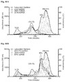


Classifications
-
- C—CHEMISTRY; METALLURGY
- C12—BIOCHEMISTRY; BEER; SPIRITS; WINE; VINEGAR; MICROBIOLOGY; ENZYMOLOGY; MUTATION OR GENETIC ENGINEERING
- C12N—MICROORGANISMS OR ENZYMES; COMPOSITIONS THEREOF; PROPAGATING, PRESERVING, OR MAINTAINING MICROORGANISMS; MUTATION OR GENETIC ENGINEERING; CULTURE MEDIA
- C12N15/00—Mutation or genetic engineering; DNA or RNA concerning genetic engineering, vectors, e.g. plasmids, or their isolation, preparation or purification; Use of hosts therefor
- C12N15/09—Recombinant DNA-technology
- C12N15/11—DNA or RNA fragments; Modified forms thereof; Non-coding nucleic acids having a biological activity
- C12N15/111—General methods applicable to biologically active non-coding nucleic acids
-
- A—HUMAN NECESSITIES
- A61—MEDICAL OR VETERINARY SCIENCE; HYGIENE
- A61K—PREPARATIONS FOR MEDICAL, DENTAL OR TOILETRY PURPOSES
- A61K47/00—Medicinal preparations characterised by the non-active ingredients used, e.g. carriers or inert additives; Targeting or modifying agents chemically bound to the active ingredient
- A61K47/50—Medicinal preparations characterised by the non-active ingredients used, e.g. carriers or inert additives; Targeting or modifying agents chemically bound to the active ingredient the non-active ingredient being chemically bound to the active ingredient, e.g. polymer-drug conjugates
- A61K47/51—Medicinal preparations characterised by the non-active ingredients used, e.g. carriers or inert additives; Targeting or modifying agents chemically bound to the active ingredient the non-active ingredient being chemically bound to the active ingredient, e.g. polymer-drug conjugates the non-active ingredient being a modifying agent
- A61K47/54—Medicinal preparations characterised by the non-active ingredients used, e.g. carriers or inert additives; Targeting or modifying agents chemically bound to the active ingredient the non-active ingredient being chemically bound to the active ingredient, e.g. polymer-drug conjugates the non-active ingredient being a modifying agent the modifying agent being an organic compound
- A61K47/542—Carboxylic acids, e.g. a fatty acid or an amino acid
-
- A—HUMAN NECESSITIES
- A61—MEDICAL OR VETERINARY SCIENCE; HYGIENE
- A61K—PREPARATIONS FOR MEDICAL, DENTAL OR TOILETRY PURPOSES
- A61K47/00—Medicinal preparations characterised by the non-active ingredients used, e.g. carriers or inert additives; Targeting or modifying agents chemically bound to the active ingredient
- A61K47/50—Medicinal preparations characterised by the non-active ingredients used, e.g. carriers or inert additives; Targeting or modifying agents chemically bound to the active ingredient the non-active ingredient being chemically bound to the active ingredient, e.g. polymer-drug conjugates
- A61K47/51—Medicinal preparations characterised by the non-active ingredients used, e.g. carriers or inert additives; Targeting or modifying agents chemically bound to the active ingredient the non-active ingredient being chemically bound to the active ingredient, e.g. polymer-drug conjugates the non-active ingredient being a modifying agent
- A61K47/54—Medicinal preparations characterised by the non-active ingredients used, e.g. carriers or inert additives; Targeting or modifying agents chemically bound to the active ingredient the non-active ingredient being chemically bound to the active ingredient, e.g. polymer-drug conjugates the non-active ingredient being a modifying agent the modifying agent being an organic compound
- A61K47/554—Medicinal preparations characterised by the non-active ingredients used, e.g. carriers or inert additives; Targeting or modifying agents chemically bound to the active ingredient the non-active ingredient being chemically bound to the active ingredient, e.g. polymer-drug conjugates the non-active ingredient being a modifying agent the modifying agent being an organic compound the modifying agent being a steroid plant sterol, glycyrrhetic acid, enoxolone or bile acid
-
- A—HUMAN NECESSITIES
- A61—MEDICAL OR VETERINARY SCIENCE; HYGIENE
- A61K—PREPARATIONS FOR MEDICAL, DENTAL OR TOILETRY PURPOSES
- A61K47/00—Medicinal preparations characterised by the non-active ingredients used, e.g. carriers or inert additives; Targeting or modifying agents chemically bound to the active ingredient
- A61K47/50—Medicinal preparations characterised by the non-active ingredients used, e.g. carriers or inert additives; Targeting or modifying agents chemically bound to the active ingredient the non-active ingredient being chemically bound to the active ingredient, e.g. polymer-drug conjugates
- A61K47/51—Medicinal preparations characterised by the non-active ingredients used, e.g. carriers or inert additives; Targeting or modifying agents chemically bound to the active ingredient the non-active ingredient being chemically bound to the active ingredient, e.g. polymer-drug conjugates the non-active ingredient being a modifying agent
- A61K47/56—Medicinal preparations characterised by the non-active ingredients used, e.g. carriers or inert additives; Targeting or modifying agents chemically bound to the active ingredient the non-active ingredient being chemically bound to the active ingredient, e.g. polymer-drug conjugates the non-active ingredient being a modifying agent the modifying agent being an organic macromolecular compound, e.g. an oligomeric, polymeric or dendrimeric molecule
- A61K47/61—Medicinal preparations characterised by the non-active ingredients used, e.g. carriers or inert additives; Targeting or modifying agents chemically bound to the active ingredient the non-active ingredient being chemically bound to the active ingredient, e.g. polymer-drug conjugates the non-active ingredient being a modifying agent the modifying agent being an organic macromolecular compound, e.g. an oligomeric, polymeric or dendrimeric molecule the organic macromolecular compound being a polysaccharide or a derivative thereof
-
- A—HUMAN NECESSITIES
- A61—MEDICAL OR VETERINARY SCIENCE; HYGIENE
- A61P—SPECIFIC THERAPEUTIC ACTIVITY OF CHEMICAL COMPOUNDS OR MEDICINAL PREPARATIONS
- A61P25/00—Drugs for disorders of the nervous system
- A61P25/14—Drugs for disorders of the nervous system for treating abnormal movements, e.g. chorea, dyskinesia
-
- A—HUMAN NECESSITIES
- A61—MEDICAL OR VETERINARY SCIENCE; HYGIENE
- A61P—SPECIFIC THERAPEUTIC ACTIVITY OF CHEMICAL COMPOUNDS OR MEDICINAL PREPARATIONS
- A61P43/00—Drugs for specific purposes, not provided for in groups A61P1/00-A61P41/00
-
- C—CHEMISTRY; METALLURGY
- C07—ORGANIC CHEMISTRY
- C07H—SUGARS; DERIVATIVES THEREOF; NUCLEOSIDES; NUCLEOTIDES; NUCLEIC ACIDS
- C07H21/00—Compounds containing two or more mononucleotide units having separate phosphate or polyphosphate groups linked by saccharide radicals of nucleoside groups, e.g. nucleic acids
-
- C—CHEMISTRY; METALLURGY
- C12—BIOCHEMISTRY; BEER; SPIRITS; WINE; VINEGAR; MICROBIOLOGY; ENZYMOLOGY; MUTATION OR GENETIC ENGINEERING
- C12N—MICROORGANISMS OR ENZYMES; COMPOSITIONS THEREOF; PROPAGATING, PRESERVING, OR MAINTAINING MICROORGANISMS; MUTATION OR GENETIC ENGINEERING; CULTURE MEDIA
- C12N2310/00—Structure or type of the nucleic acid
- C12N2310/10—Type of nucleic acid
- C12N2310/14—Type of nucleic acid interfering N.A.
-
- C—CHEMISTRY; METALLURGY
- C12—BIOCHEMISTRY; BEER; SPIRITS; WINE; VINEGAR; MICROBIOLOGY; ENZYMOLOGY; MUTATION OR GENETIC ENGINEERING
- C12N—MICROORGANISMS OR ENZYMES; COMPOSITIONS THEREOF; PROPAGATING, PRESERVING, OR MAINTAINING MICROORGANISMS; MUTATION OR GENETIC ENGINEERING; CULTURE MEDIA
- C12N2310/00—Structure or type of the nucleic acid
- C12N2310/30—Chemical structure
- C12N2310/35—Nature of the modification
- C12N2310/351—Conjugate
- C12N2310/3515—Lipophilic moiety, e.g. cholesterol
-
- C—CHEMISTRY; METALLURGY
- C12—BIOCHEMISTRY; BEER; SPIRITS; WINE; VINEGAR; MICROBIOLOGY; ENZYMOLOGY; MUTATION OR GENETIC ENGINEERING
- C12N—MICROORGANISMS OR ENZYMES; COMPOSITIONS THEREOF; PROPAGATING, PRESERVING, OR MAINTAINING MICROORGANISMS; MUTATION OR GENETIC ENGINEERING; CULTURE MEDIA
- C12N2320/00—Applications; Uses
- C12N2320/30—Special therapeutic applications
- C12N2320/32—Special delivery means, e.g. tissue-specific
Definitions
- RNA interference RNA interference
- the oligonucleotide conjugates are designed to achieve unexpectedly high efficacy, uptake and tissue distribution.
- RNA interference represents a simple and effective tool for inhibiting the function of genes.
- small therapeutic RNAs can only be delivered effectively to liver.
- a method for selectively delivering a compound of formula (1), or a disclosed embodiment thereof, to a particular organ in a patient comprising administering said compound to the patient, wherein the compound has a selective affinity for a serum lipoprotein.
- FIG. 1A shows a synthetic approach for cortisol-conjugated oligonucleotides.
- FIG. 1B shows a synthetic approach for calciferol-conjugated oligonucleotides.
- FIG. 1C shows a synthetic approach for DHA-conjugated oligonucleotides.
- FIG. 1D shows a synthetic approach for preparation of an alkynylated-oligonucleotide for click conjugation.
- FIG. 1E shows a synthetic approach for GM1-conjugated oligonucleotides.
- FIG. 1F shows a synthetic approach for lysophosphatidylcholine esterified DHA-hsiRNA conjugate (referred to as DHAPCL-hsiRNA, PC-DHA-hsiRNA, g2DHA-hsiRNA, or DHA-G2-hsiRNA).
- FIG. 1G shows a synthetic approach for an hsiRNA-Calciferol oligonucleotide (SEQ ID NO: 176).
- FIG. 1H shows an alternative synthetic approach for an hsiRNA-Calciferol oligonucleotide.
- FIG. 1I shows a representative analytical HPLC trace of a synthesized hsiRNA conjugate, and its stability at room temperature immediately after purification, after 24 hours at room temperature, and after 48 hours at room temperature; sFLT-g2DHA-Cy3-P2 is shown.
- FIG. 1J shows a representative analytical HPLC trace and an ESI-MS spectra of a synthesized hsiRNA conjugate; lysophosphatidylcholine esterified DHA -hsiRNA conjugate is shown.
- FIG. 1K shows a representative analytical HPLC trace of an hsiRNA conjugate prepared according to the synthetic approach of FIG. 1I ; hsiRNA-Calciferol shown.
- FIG. 1L shows a representative semi-prep reverse-phase-HPLC trace of a synthesized hsiRNA conjugate; Cy3-labeled sFLT-DHA conjugate (crude reaction mixture) shown.
- FIG. 1M shows a representative analytical reverse-phase-HPLC following purification of a synthesized hsiRNA conjugate as in FIG. 1L ; Cy3-labeled sFLT-DHA conjugate (pure product) shown.
- FIG. 1N shows a representative LC-MS profile following purification of a synthesized hsiRNA conjugate as in FIG. 1L ; Cy3-labeled sFLT-DHA conjugate (pure product) shown.
- FIG. 2A depicts exemplary hydrophobic moieties.
- FIG. 2B shows the differences in hydrophobicity profiles of synthesized siRNA conjugates as observed by reverse-phase HPLC (C8).
- FIG. 2C shows an exemplary LC-MS analysis of a synthesized hsiRNA conjugate; DHA-hsiRNA shown.
- FIG. 3A shows a biodistribution study protocol.
- FIG. 3B shows that the in vivo brain distribution of FMS-hsiRNA is defined by conjugation modality.
- FIG. 4 shows that calciferol and DHA-hsiRNA conjugates display a dramatically improved spread through the brain as well as robust neuronal uptake.
- FIG. 5 shows dramatic differences in patterns of CNS tissue distribution upon intrathecal (IT) injection of Cholesterol and DHA-hsiRNA conjugates.
- FIG. 6A shows that a single 25 ⁇ g injection of DHA-hsiRNA induced potent silencing not only in the striatum but also in the cortex.
- FIG. 6B shows that administration with as much as 200 ⁇ g DHA-hsiRNA conjugates induces no observable signs of neuronal damage.
- FIG. 7 shows accumulation in various tissues upon systemic administration of hsiRNA-conjugates. hsiRNA-conjugate structures and modifications are found in FIG. 9A-F . All compounds have the sequence of PPIB, as shown in FIG. 14 .
- FIG. 8 shows a series of targeting regions and corresponding sense- and guide-sequences (SEQ ID NOS 1-30, 27, 31-117, 1-30, 27, 31-58, 1-30, 27, 31-58, 118-147, 144, and 148-175, respectively, in order of columns).
- FIG. 9A-F show chemical structures of conjugated hsiRNAs.
- A Docosanoic (DCA)-conjugated hsiRNA.
- B Docosahexaenoic acid (DHA)-conjugated hsiRNA, 22:6 (n-3).
- C Phosphatidylcholine-DHA-conjugated hsiRNA (g2DHA-hsiRNA or DHAPCL-hsiRNA)
- D Eicosapentanoic acid (EPA)-conjugated hsiRNA, 20:5(n-3).
- E Cholesterol
- Chol Cholesterol
- Chol Cholesterol
- Chol Cholesterol-conjugated hsiRNA.
- F Cholesterol (Chol)-conjugated hsiRNA.
- hsiRNA conjugates represented to scale using PyMOL.
- FIG. 10 shows distribution following intravenous administration of Cy3 labeled cholesterol- or DCA-conjugated hsiRNAs.
- Mice were injected intravenously (tail vein) with two doses of 20 mg/kg on two consecutive days with either cholesterol- or DCA-conjugated hsiRNAs. 24 hours after the last dose, mice were euthanized, perfused with PBS and organs harvested for either immunohistochemistry or the PNA assay. Predominate distribution to the liver and red pulp of the spleen was observed for both conjugates.
- FIG. 11 shows distribution following intravenous administration of Cy3 labeled DHA- or EPA-conjugated hsiRNAs.
- Mice were injected intravenously (tail vein) with two doses of 20 mg/kg on two consecutive days with either DHA- or EPA-conjugated hsiRNAs. 24 hours after the last dose, mice were euthanized, perfused with PBS and organs harvested for either immunohistochemistry or the PNA assay. Significant distribution to the kidney, red pulp of the spleen, liver, and heart was observed.
- FIG. 12A-B shows PNA (Peptide Nucleic Acid) based assay for detection of hsiRNA guide strand in mouse tissues.
- A Tissues were lysed, debris separated by precipitation, PNA-guide strand duplex purified by HPLC (DNAPac P100, 50% water 50% acetonitrile, salt gradient 0-1M NaClO4).
- B Liver and kidney from mice injected with 40 mg/kg of either cholesterol, DCA, EPA, or DHA were used to quantify the guide strand after 48 hours, showing differential distribution of fatty acid conjugates.
- FIG. 13A-B shows efficacy of DHA-hsiRNA targeting sFLT1 after IV injection.
- A Schematic of experimental design.
- FIG. 14 shows modified oligonucleotide sequences (SEQ ID NOS 176-177, 176-177, 176-177, 176-177, 176-177, and 178-183, respectively, in order of appearance).
- Chemical modifications are abbreviated as follows, wherein “X” represents A, U, G, or C: fX (2′-fluoro), mX (2′-O-methyl), P (5′-phosphate), Chol (Cholesterol), ‘#’ (phosphorothioate backbone modification), ‘.’ (phosphodiester backbone).
- FIG. 15 shows the solid-phase synthesis of DHA-conjugated hsiRNA.
- FIG. 16 shows a representative LC-MS characterization of Cy3-DHA-hsiRNAHTT; Calculated: 6174.1 for [M ⁇ H] ⁇ , found: 6174.4.
- Buffer A 15 mM Dibutylamine/25 mM HFIP
- Buffer B 20% A in MeOH
- FIG. 17A-B shows neuronal uptake of DHA-hsiRNA, equivalent Huntingtin mRNA silencing, and reduced hydrophobicity compared to Chol-hsiRNA.
- FIG. 18A-C shows the efficacy and duration of effect of DHA-hsiRNA HTT following intrastriatal injection.
- DHA-siRNA was unilaterally injected into the striatum of WT mice.
- Punch biopsies of the striatum (A) and cortex (B) were collected after 5 days.
- For duration of effect studies (C) punch biopsies of the striatum were collected at times shown.
- NTC non-targeting control
- hsiRNA-conjugate structures, sequences, and modifications are shown in FIG. 9A-F and FIG. 14 .
- FIG. 20 shows targeted kidney delivery with DHA-PCL conjugated hsiRNA.
- hsiRNA-conjugate structures and modifications are shown in FIG. 9A-F and the HTT siRNA sequence is on FIG. 14 .
- FIG. 21A-B shows targeted placental delivery with polyunsaturated fatty acid chemical scaffolds.
- DHA-conjugated oligonucleotides can be delivered intravenously to the mother and show targeted delivery to maternal kidney, liver, and placenta with no observed oligonucleotide transfer or toxicity in embryos.
- A Intravenous injection of DHA-siRNA (15 mg/kg). Animals sacrificed 7 days post-injection. 63 ⁇ image of embryo and placenta showing Cy3-fluorescence of oligonucleotides.
- B Tissue punches taken from liver, kidney, and placenta. mRNA was quantified using Affymetrix Quantigene 2.0 as per Coles et al. 2015. hsiRNA-conjugate structures and modifications are shown in FIG. 9A-F and the sFLT1 sequence is shown in FIG. 8 .
- FIG. 22 shows efficient silencing in heart with a single DHA-PCL-hsiRNA injection (15 mg/kg).
- the sFLT1 sequence is shown in FIG. 8 .
- FIG. 23 shows potency and delivery to primary neurons of DHA-hsiRNA and g2DHA-hsiRNA. hsiRNA-conjugate structures and modifications are shown in FIG. 9A-F and the HTT siRNA sequence is shown in FIG. 14 .
- FIG. 24 shows data comparing the effect modified and unmodified hsiRNA on gene modulation in primary neurons. hsiRNA-conjugate structures and modifications are shown in FIG. 9A-F and the HTT siRNA sequence is on FIG. 14 .
- FIG. 25 shows brain retention and distribution of g2DHA-hsiRNA.
- FIG. 26A-C shows the effects upon single IS injection of g2DHA-hsiRNA: (A) experimental procedure; (B) approximately 80% silencing in mouse striatum; (C) approximately 80% silencing in mouse cortex. There was no indication of toxicity and silencing was limited to injected side of the brain. hsiRNA-conjugate structures and modifications are shown in FIG. 9A-F and the hsiRNA sequence is shown in FIG. 14 .
- FIG. 27 shows g2DHA support synthesis I.
- FIG. 28 shows g2DHA support synthesis II (see Example 9).
- FIG. 29 depicts exemplary values of X c .
- FIG. 30 depicts exemplary internucleotide linkages.
- FIG. 31 depicts exemplary internucleotide backbone linkages.
- FIG. 32 depicts exemplary sugar modifications.
- FIG. 33A-E depicts fully metabolically stabilized hsiRNAs (FM-hsiRNAs).
- A Schematics of partially and fully modified hsiRNAs.
- B hsiRNA and FM-hsiRNA have equal ability to enter RISC (HeLa, 72 hours, QuantiGene®). hsiRNA-conjugate structures, sequences, and modifications are found in FIG. 9A-F and FIG. 14 .
- D Metabolically stable 5′-E-VP (Vinylphosphonate) is as active as 5′-P (Phosphate).
- the antisense strand of the hsiRNAs are capped at the 5′ as follows: FM-hsiRNA-no P is capped with a 5′-OH; FM-hsiRNA is capped with a 5′ phosphate; FM-hsiRNA-EVP is capped with a 5′ vinyl phosphonate.
- E 5′-E-VP enables sustained delivery to distant tissues (7 days post injection, PNA assay).
- the antisense strand of the hsiRNAs are capped at the 5′ as follows: 5′P-hsiRNA is capped with a 5′ phosphate; 5VP′-hsiRNA is capped with a 5′ vinyl phosphonate.
- the hsiRNA sequence for FIG. 33 D-E is PPIB, found in FIG. 14 .
- FIG. 34 shows the optimized solution phase synthetic route to g2DHA-hsiRNA (1b).
- Reagents and conditions (a) 20% piperidine in DMF (2 ⁇ 15 min); (b) 2-cyanoethyl N,N-diisopropylchlorophosphoramidite, DIEA, DCM, 2 h, rt, 95%; (c) choline tosylate, ETT, MeCN, 2 h, rt, followed by mCPBA, 10 min, rt, 69%; (d) (e) TFA in dry DCM (1:1), triisopropylsilane, 2 h, rt then 10% diisopropylethylamine in MeCN, 1.5 h, rt 74% (f) 3, BOP, HOBt, DMF, 2,4,6-collidine, rt, 12 h; (g) 20% piperidine in DMF (2 ⁇ 15 min), rt; (h) DHA, HATU, D
- FIG. 35 shows the optimized solid-phase synthetic route to g2DHA-hsiRNA (1b). See also Example 11.
- FIG. 36 shows that intravenous injection of lipid-siRNA conjugates induces differential levels of gene silencing in the liver, which is directly proportional to the degree of accumulation.
- Intravenous injection (20 mg/kg) of each siRNA conjugate 20 mg/kg. Animals sacrificed 7 days post-injection. Tissue punches taken from the liver tissue. mRNA was quantified using Affymetrix Quantigene 2.0 as per Coles et al. 2015. hsiRNA-conjugate structures and modifications are shown in FIG. 9A-F and the PPIB hsiRNA sequence is shown in FIG. 14 .
- FIG. 37A-B depicts targeted kidney delivery with polyunsaturated fatty acid chemical scaffolds.
- A Intravenous injection of PBS, Chol-siRNA, or g2DHA-siRNA (20 mg/kg twice daily for two days). Animals sacrificed 7 days post-injection. 63 ⁇ image of kidney sections showing Cy3-fluorescence of oligonucleotides. hsiRNA-conjugate structures and modifications are shown in FIG. 9A-F and the sFLT1 sequence is on FIG. 8 .
- siRNA antisense strands present in liver and kidney were quantified using Cy3-labeled complimentary PNA to hybridize to the strand and HPLC to quantify ng of oligo per mg of tissue. hsiRNA-conjugate structures and modifications are shown in FIG. 9A-F and the PPIB sequence is on FIG. 14 .
- FIG. 38 shows that g2DHA-hsiRNA preferentially distributes to proximal convoluted tubule cells throughout the kidney following systemic administration (two IV injections of 20 mg/kg, 48 hours). This sharply contrasts with the predominant liver localization exhibited by most siRNA therapeutics in the clinic and opens the window to expand the clinical utility of siRNA beyond liver indications.
- FIG. 39 shows g2DHA-hsiRNA distributed to heart tissue following systemic administration (one intravenous injection,10 mg/kg). These tissues are not typically accessed by therapeutic siRNAs following intravenous administration.
- FIG. 40 shows g2DHA-hsiRNA distributed to muscle and fat tissue following systemic administration (one intravenous injection,10 mg/kg). These tissues are not typically accessed by therapeutic siRNAs following intravenous administration. hsiRNA-conjugate structures and modifications are shown in FIG. 9A-F and the sFLT1 sequence is on FIG. 8 .
- FIG. 41 shows Eicosapentanoic acid (EPA)-hsiRNA accumulation in the skin following subcutaneous injection. This can be directly compared to cholesterol-conjugated hsiRNA, which accumulates to a greater degree around the site of injection. This higher degree of accumulation may cause local toxicity and adverse effects, which is well documented for intrastriatal (CNS) administration.
- EPA Eicosapentanoic acid
- FIG. 42 shows that subcutaneous injection of EPA-hsiRNA induces gene silencing in the skin.
- Subcutaneous injection 40 mg/kg
- EPA-siRNA Animals sacrificed 7 days post-injection. Tissue punches taken from the center (skin from head to the center of the back), middle (skin around the midpoint of the animal), and tail skin.
- mRNA was quantified using Affymetrix Quantigene 2.0 as per Coles et al. 2015. hsiRNA sequence PPIB is found in FIG. 14 .
- FIG. 43A-B shows that a single injection of DHA- or g2DHA-siRNA is detected in both the straitum (A) and cortex (B) on the injected side.
- Alternative methods of injection including intracerebralventricular may also facilitate bilateral distribution with only one injection.
- Intrastriatal injection 2-4 nmols DHA- or g2DHA-siRNA.
- Animals sacrificed 7 days post-injection.
- siRNA antisense strands present in different brain regions were quantified using Cy3-labeled complimentary PNA to hybridize to the strand and HPLC to quantify ng of oligo per mg of tissue.
- aCSF Artificial CSF.
- hsiRNA-conjugate structures and modifications are found in FIG. 9A-F and the PPIB sequence is shown in FIG. 14 .
- FIG. 44 shows serum lipoprotein binding properties of lipid-conjugated siRNAs.
- FIG. 45 shows the lipoprotein profile of FVB/NJ mice.
- Whole mouse blood ( ⁇ 500 ⁇ L) was collected in a sterile EDTA-coated tube following cardiac puncture. Samples were spun at 10,000 RPM for 10 minutes. 50 ⁇ L of serum was directly injected on Superose 360 size exclusion column. Fractions were collected over 300 minutes and analyzed for cholesterol content by the HDL/LDL Cholesterol Assay Kit (Abcam).
- FIG. 46A-B depicts serum lipoprotein profile analysis of siRNA in mouse blood.
- hsiRNA-conjugate structures and modifications are found in FIG. 9A-F .
- hsiRNA conjugates (15 ⁇ M) were incubated in 50 ⁇ L of serum at room temperature for 30 minutes. 50 ⁇ L of serum was directly injected on Superose 360 size exclusion column. Fractions were collected over 300 minutes and analyzed for cholesterol content by the HDL/LDL Cholesterol Assay Kit (Abcam). The HTT sequence is shown in FIG. 14 .
- FIG. 47A-B shows chemical structures of novel hydrophobic siRNA constructs.
- Polyunsaturated fatty acids are typically circulated in the bloodstream in an esterified form, meaning they are linked to glycerol, long-chain aliphatic alcohols, amides, phosphatidylcholine, phosphatidylserine, phosphoric acid, and phosphatidylethanolamine, among others. Defining the path to synthesize metabolically stable analogs of these naturally existing circulating compounds is one way to improve polyunsaturated fatty acid-siRNA tissue distribution and cellular uptake.
- Y is a chemically stable trifunctional spacer or linker, which could be cleavable or not.
- Z is a naturally occurring ester linkage (e.g. phosphatidycholine, phosphatidylserine, phosphoric acid, see FIG. 47B )
- FIG. 48 shows examples of linkers, spacer, and branching moieties.
- the exact chemical composition of the linker is not essential for activity as long as the branching structure can be generated
- FIG. 49 shows hydrophobic siRNA conjugates with esterified phosphatidylcholine modifiers.
- compositions comprising small RNAs that are completely stable and fully active.
- these small RNAs were conjugated to several naturally occurring bioactive steroids, endocannabinoid-like lipids, and nucleoside analogs.
- the resulting conjugates selectively delivered small RNAs to a range of tissues, including heart, kidneys, muscle, placenta, vasculature, and brain.
- compositions described herein promote simple, efficient, non-toxic delivery of metabolically stable siRNA, and promote potent silencing of therapeutic targets in a range of tissues in vivo.
- a chemistry platform for targeting other tissues matching the performance and clinical impact of GalNAc conjugates in the liver Several bio-active steroids and endocannabinoid-like bioactive lipid conjugates were screened and identified. These compounds show unprecedented distribution, neuronal uptake, efficacy, and lack of toxicity in several tissues, including endothelia, kidneys, liver, spleen, heart, lung, mouse brain and spinal cord.
- the oligonucleotide conjugates of the invention were identified through a process involving: (1) providing a fully metabolically stable scaffolds (no RNA left); (2) selecting compounds which are biologically known to internalize inside the cells and identifying the ranges of hydrophobicities which allow efficient tissue distribution; (3) conjugating these hydrophobic compounds to the metabolically stable siRNAs; and (4) screening distribution, efficacy and toxicity in vivo.
- the discovery of the optimal range of hydrophobicity defines the chemical scaffold ranges expected to be efficacious. It was found that low hydrophobicity (cortisol like) was not sufficient to secure good tissue retention, whereas too much hydrophobicity (e.g., cholesterol) minimized distribution from the site of injection.
- the golden medium e.g., DHA, DHAg2, calciferol
- L comprises an ethylene glycol chain, an alkyl chain, a peptide, RNA, DNA, a phosphodiester, a phosphorothioate, a phosphoramidate, an amide, a carbamate, or a combination thereof; and wherein L is attached to O via the second oligonucleotide.
- L is a divalent linker.
- L is a trivalent linker.
- L corresponds to a linker of FIG. 48 .
- L is the trivalent linker L1, also referred to herein as C7:
- L is the divalent linker L2:
- an oxygen atom of L is bonded to the 3′ position of the second oligonucleotide by a phosphodiester for example, as shown in FIG. 1 j.
- X c has an affinity for low density lipoprotein and/or intermediate density lipoprotein. In a related embodiment, X c is a saturated or unsaturated moiety having fewer than three double bonds.
- X c has an affinity for high density lipoprotein.
- X c is a polyunsaturated moiety having at three or more double bonds (e.g., having three, four, five, six, seven, eight, nine or ten double bonds).
- X c is a polyunsaturated moiety having three double bonds.
- X c is a polyunsaturated moiety having four double bonds.
- X c is a polyunsaturated moiety having five double bonds.
- X c is a polyunsaturated moiety having six double bonds.
- X c is selected from the group consisting of fatty acids, steroids, secosteroids, lipids, ganglio sides and nucleoside analogs, and endocannabinoids.
- X c is a neuromodulatory lipid, e.g., an endocannabinoid.
- endocannabinoids include: Anandamide, Arachidonoylethanolamine, 2-Arachidonyl glyceryl ether (noladin ether), 2-Arachidonyl glyceryl ether (noladin ether), 2-Arachidonoylglycerol, and N-Arachidonoyl dopamine.
- X c is an omega-3 fatty acid.
- omega-3 fatty acids include: Hexadecatrienoic acid (HTA), Alpha-linolenic acid (ALA), Stearidonic acid (SDA), Eicosatrienoic acid (ETE), Eicosatetraenoic acid (ETA), Eicosapentaenoic acid (EPA, Timnodonic acid), Heneicosapentaenoic acid (HPA), Docosapentaenoic acid (DPA, Clupanodonic acid), Docosahexaenoic acid (DHA, Cervonic acid), Tetracosapentaenoic acid, and Tetracosahexaenoic acid (Nisinic acid).
- HTA Hexadecatrienoic acid
- ALA Alpha-linolenic acid
- SDA Stearidonic acid
- ETE Eicosatrienoic acid
- ETA Eicosatetraenoic acid
- X c is an omega-6 fatty acid.
- omega-6 fatty acids include: Linoleic acid, Gamma-linolenic acid (GLA), Eicosadienoic acid, Dihomo-gamma-linolenic acid (DGLA), Arachidonic acid (AA), Docosadienoic acid, Adrenic acid, Docosapentaenoic acid (Osbond acid), Tetracosatetraenoic acid, and Tetracosapentaenoic acid.
- GLA Gamma-linolenic acid
- DGLA Dihomo-gamma-linolenic acid
- AA Arachidonic acid
- Docosadienoic acid Adrenic acid
- Docosapentaenoic acid Osbond acid
- Tetracosatetraenoic acid Tetracosapentaenoic acid.
- X c is an omega-9 fatty acid.
- omega-9 fatty acids include: Oleic acid, Eicosenoic acid, Mead acid, Erucic acid, and Nervonic acid.
- X c is a conjugated linolenic acid.
- conjugated linolenic acids include: ⁇ -Calendic acid, ⁇ -Calendic acid, Jacaric acid, ⁇ -Eleostearic acid, ⁇ -Eleostearic acid, Catalpic acid, and Punicic acid.
- X c is a saturated fatty acid.
- saturated fatty acids include: Caprylic acid, Capric acid, Docosanoic acid, Lauric acid, Myristic acid, Palmitic acid, Stearic acid, Arachidic acid, Behenic acid, Lignoceric acid, and Cerotic acid.
- X c is an acid selected from the group consisting of: Rumelenic acid, ⁇ -Parinaric acid, ⁇ -Parinaric acid, Bosseopentaenoic acid, Pinolenic acid, and Podocarpic acid.
- X c is selected from the group consisting of: docosanoic acid (DCA), docosahexaenoic acid (DHA), and eicosapentaenoic acid (EPA).
- DCA docosanoic acid
- DHA docosahexaenoic acid
- EPA eicosapentaenoic acid
- X c is docosanoic acid (DCA).
- X c is DHA.
- X c is EPA.
- X c is a secosteroid. In a particular embodiment, X c is calciferol. In another embodiment, X c is a steroid other than cholesterol.
- X c is not cholesterol
- X c is an alkyl chain, a vitamin, a peptide, or a bioactive conjugate (including but not limited to: glycosphingolipids, polyunsaturated fatty acids, secosteroids, steroid hormones, sterol lipids.
- the hydrophobic moiety comprises a moiety depicted in FIGS. 2 a and 29 .
- X c is characterized by a clogP value in a range selected from: ⁇ 10 to ⁇ 9, ⁇ 9 to ⁇ 8, ⁇ 8 to ⁇ 7, ⁇ 7 to ⁇ 6, ⁇ 6 to ⁇ 5, ⁇ 5 to ⁇ 4, ⁇ 4 to ⁇ 3, ⁇ 3 to ⁇ 2, ⁇ 2 to ⁇ 1, ⁇ 1 to 0, 0 to 1, 1 to 2, 2 to 3, 3 to 4, 4 to 5, 5 to 6, 6 to 7, 7 to 8, 8 to 9, and 9 to 10.
- Z c is selected from the group consisting of Z c1 , Z c2 , Z c3 and Z c4 :
- X is O, S or BH 3 .
- Z c is Z c1 , In another particular embodiment, Z c is not Z c1 ,
- Z c is selected from the group consisting of Z c2 , Z c3 and Z c4 .
- Z c is Z c2 , In a particular embodiment, Z c is Z c3 , In a particular embodiment, Z c is Z c4 , In a particular embodiment, X is O. In a particular embodiment, X is S. In a particular embodiment, X is BH 3 .
- Z c when X c is DHA, Z c is not Z c1 . In another particular embodiment, when Z c is Z c1 , X c is not DHA.
- O comprises compound (I): an oligonucleotide of at least 16 contiguous nucleotides, said oligonucleotide having a 5′ end, a 3′ end and complementarity to a target.
- the oligonucleotide has sufficient complementarity to the target to hybridize.
- the complementarity is >95%, >90%, >85%, >80%, >75%, >70%, >65%, >60%, >55% or >50%.
- compound (Ia) has perfect complementarity to the target.
- O comprises compound (II): an oligonucleotide of at least 15 contiguous nucleotides, said oligonucleotide having a 5′ end, a 3′ end, and homology with a target, wherein the oligonucleotide is conjugated at the 3′ end to -L(X c )(Z c ), described above.
- compound (II) comprises one or more chemically-modified nucleotides.
- the oligonucleotide comprises alternating 2′-methoxy-nucleotides and 2′-fluoro-nucleotides.
- the nucleotides at positions 1 and 2 from the 3′ end of the oligonucleotide are connected to adjacent nucleotides via phosphorothioate linkages.
- nucleotides at positions 1 and 2 from the 3′ end of the oligonucleotide and the nucleotides at positions 1 and 2 from the 5′ end of the oligonucleotide are connected to adjacent nucleotides via phosphorothioate linkages.
- compound (II) has complete homology with the target.
- the target is mammalian or viral mRNA.
- the target is an intronic region of said mRNA.
- O is a double-stranded nucleic acid comprising a first oligonucleotide and a second oligonucleotide, wherein:
- the first oligonucleotide is compound (I), or any one of the previous embodiments thereof;
- the second oligonucleotide is compound (II), or any one of the previous embodiments thereof.
- a portion of the first oligonucleotide is complementary to a portion of the second oligonucleotide.
- the first oligonucleotide comprises at least 16 contiguous nucleotides, a 5′ end, a 3′ end, and has complementarity to a target, wherein:
- the first oligonucleotide comprises alternating 2′-methoxy-nucleotides and 2′-fluoro-nucleotides
- nucleotides at positions 2 and 14 from the 5′ end are not 2′-methoxy-nucleotides
- nucleotides are connected via phosphodiester or phosphorothioate linkages
- nucleotides at positions 1-6 from the 3′ end, or positions 1-7 from the 3′ end are connected to adjacent nucleotides via phosphorothioate linkages.
- the first oligonucleotide has perfect complementarity to the target.
- sequences of the first and second oligonucleotides are selected from the tables of FIG. 8 and FIG. 14
- compound (I) has the structure of Formula (Ia):
- X is selected from the group consisting of:
- A for each occurrence, independently is a 2′-methoxy-ribonucleotide
- B for each occurrence, independently is a 2′-fluoro-ribonucleotide
- K for each occurrence independently is a phosphodiester or phosphorothioate linker
- S is a phosphorothioate linker
- R is selected from hydrogen and a capping group (e.g., an acyl group such as acetyl);
- j 4, 5, 6 or 7;
- r is 2 or 3;
- t 0 or 1.
- the oligonucleotide of Formula (Ia) has the structure of Formula (Ib):
- X is as defined above;
- A for each occurrence, independently is a 2′-methoxy-ribonucleotide
- B for each occurrence, independently is a 2′-fluoro-ribonucleotide
- S is a phosphorothioate linker
- P is a phosphodiester linker
- R is as defined above;
- m is 1; n is 5; q is 1; r is 2; and t is 1.
- m is 1; n is 5; q is 0; r is 3; and t is 1.
- R is hydrogen.
- X is Xl.
- X is X3.
- O is a double-stranded, chemically-modified nucleic acid comprising a first oligonucleotide and a second oligonucleotide, wherein:
- the first oligonucleotide is selected from the oligonucleotides of Formulas (I), (Ia), and (Ib);
- a portion of the first oligonucleotide is complementary to a portion of the second oligonucleotide
- the second oligonucleotide is selected from the oligonucleotides of Formulas (II) and (IIa):
- the first oligonucleotide comprises alternating 2′-methoxy-ribonucleotides and 2′-fluoro-ribonucleotides, wherein each nucleotide is a 2′-methoxy-ribonucleotide or a 2′-fluoro-ribonucleotide; and the nucleotides at positions 2 and 14 from the 5′ end of the first oligonucleotide are not 2′-methoxy-ribonucleotides;
- the second oligonucleotide comprises alternating 2′-methoxy-ribonucleotides and 2′-fluoro-ribonucleotides, wherein each nucleotide is a 2′-methoxy-ribonucleotide or a 2′-fluoro-ribonucleotide; and the nucleotides at positions 2 and 14 from the 5′ end of the second oligonucleotide are 2′-methoxy-ribonucleotides;
- nucleotides of the first oligonucleotide are connected to adjacent nucleotides via phosphodiester or phosphorothioate linkages, wherein the nucleotides at positions 1-6 from the 3′ end, or positions 1-7 from the 3′ end are connected to adjacent nucleotides via phosphorothioate linkages;
- nucleotides of the second oligonucleotide are connected to adjacent nucleotides via phosphodiester or phosphorothioate linkages, wherein the nucleotides at positions 1 and 2 from the 3′ end are connected to adjacent nucleotides via phosphorothioate linkages.
- the first oligonucleotide has 3-7 more ribonucleotides than the second oligonucleotide.
- O comprises 11-16 base pair duplexes, wherein the nucleotides of each base pair duplex have different chemical modifications (e.g., one nucleotide has a 2′-fluoro modification and the other nucleotide has a 2′-methoxy).
- the first oligonucleotide has 3-7 more ribonucleotides than the second oligonucleotide.
- each R is hydrogen.
- the first oligonucleotide has structure: X(—S—B—S-A)(—P—B—P-A) 5 (—P—B—S-A)(—S—B—S-A) 2 (—S—B)—OR; and the second oligonucleotide has the structure: C-L-B(—S-A-S—B)(—P-A-P—B) 5 (—S-A)(—S—B)—OR.
- O has the structure of compound (IIIa):
- the first oligonucleotide comprises the sequence 5′ UAAAUUUGGAGAUCCGAGAG 3′ (SEQ ID NO: 124); the second oligonucleotide comprises the sequence 3′ AUUUAAACCUCUAGG 5′ (SEQ ID NO: 184); X is X3; X c is DHA and Z c is Z c1 .
- R is hydrogen.
- R′ is hydrogen.
- L is L1.
- the first oligonucleotide comprises the sequence 5′ UAUAAAUGGUAGCUAUGAUG 3′ (SEQ ID NO: 185); the second oligonucleotide comprises the sequence 3′ AUAUUUACCAUCGAU 5′ (SEQ ID NO: 186); X is X3; X c is DHA and Z c is Z c1 .
- R is hydrogen.
- R′ is hydrogen.
- L is L1.
- the first oligonucleotide comprises the sequence 5′ UUAAUCUCUUUACUGAUAUA 3′ (SEQ ID NO: 177); the second oligonucleotide comprises the sequence 3′ AAUUAGAGAAAUGAC 5′ (SEQ ID NO: 176); X is X3; X c is DHA and Z c is Z c1 .
- R is hydrogen.
- R′ is hydrogen.
- L is L1.
- the first oligonucleotide comprises the sequence 5′ UUAAUCUCUUUACUGAUAUA 3′ (SEQ ID NO: 177); the second oligonucleotide comprises the sequence 3′ AAUUAGAGAAAUGAC 5′ (SEQ ID NO: 176); X is X3; X c is DHA and Z c is Z c1 .
- R is hydrogen.
- R′ is hydrogen.
- L is L1.
- the first oligonucleotide comprises the sequence 5′ UUAAUCUCUUUACUGAUAUA 3′ (SEQ ID NO: 177); the second oligonucleotide comprises the sequence 3′ AAUUAGAGAAAUGAC 5′ (SEQ ID NO: 176); X is X3; X c is DHA and Z c is Z c1 .
- R is hydrogen.
- R′ is hydrogen.
- L is L1.
- the first oligonucleotide comprises the sequence 5′ UUAAUCUCUUUACUGAUAUA 3′ (SEQ ID NO: 177); the second oligonucleotide comprises the sequence 3′ AAUUAGAGAAAUGAC 5′ (SEQ ID NO: 176); X is X3; X c is DHA and Z c is Z c1 .
- R is hydrogen.
- R′ is hydrogen.
- L is L1.
- the first oligonucleotide comprises the sequence 5′ UUAAUCUCUUUACUGAUAUA 3′ (SEQ ID NO: 177); the second oligonucleotide comprises the sequence 3′ AAUUAGAGAAAUGAC 5′ (SEQ ID NO: 176); X is X3; and X c is cholesterol.
- R is hydrogen.
- R′ is hydrogen.
- L comprises triethylene glycol.
- the first oligonucleotide comprises the sequence 5′ UUAAUCUCUUUACUGAUAUA 3′ (SEQ ID NO: 177); the second oligonucleotide comprises the sequence 3′ AAUUAGAGAAAUGAC 5′ (SEQ ID NO: 176); X is X3; and X c is GalNAc.
- R is hydrogen.
- R′ is hydrogen.
- L comprises triethylene glycol.
- the first oligonucleotide has structure: X(—P—B—P-A) 6 (—P—B—S-A)(—S—B—S-A) 2 (—S—B)—OR; and the second oligonucleotide has the structure: C-L-B(—S-A-S—B)(—P-A-P—B) 6 —OR.
- O has the structure of compound (Mb):
- the first oligonucleotide comprises the sequence 5′ UAAAUUUGGAGAUCCGAGAG 3′ (SEQ ID NO: 124); the second oligonucleotide comprises the sequence 3′ AUUUAAACCUCUAGG 5′ (SEQ ID NO: 184); X is X3; X c is DHA and Z c is Z c1 .
- R is hydrogen.
- R′ is hydrogen.
- L is L1.
- the first oligonucleotide comprises the sequence 5′ UAUAAAUGGUAGCUAUGAUG 3′ (SEQ ID NO: 185); the second oligonucleotide comprises the sequence 3′ AUAUUUACCAUCGAU 5′ (SEQ ID NO: 186); X is X3; X c is DHA and Z c is Z c1 .
- R is hydrogen.
- R′ is hydrogen.
- L is L1.
- the first oligonucleotide comprises the sequence 5′ UUAAUCUCUUUACUGAUAUA 3′ (SEQ ID NO: 177); the second oligonucleotide comprises the sequence 3′ AAUUAGAGAAAUGAC 5′ (SEQ ID NO: 176); X is X3; X c is DHA and Z c is Z c1 .
- R is hydrogen.
- R′ is hydrogen.
- L is L1.
- the first oligonucleotide has structure: X(—S—B—S-A)(—P—B—P-A) 5 (—S—B—S-A) 3 (—S—B)—OR;
- the second oligonucleotide has structure: C-L-B(—S-A-S—B)(—P-A-P—B) 5 (—S-A-S—B)—OR; and
- O has the structure of Formula (IIIc):
- the first oligonucleotide comprises the sequence 5′ UUAAUCUCUUUACUGAUAUA 3′ (SEQ ID NO: 177); the second oligonucleotide comprises the sequence 3′ AAUUAGAGAAAUGAC 5′ (SEQ ID NO: 176); X is X3; X c is DHA and Z c is Z c1 .
- R is hydrogen.
- R′ is hydrogen.
- R′ is hydrogen.
- L is L1.
- the first oligonucleotide is the antisense strand and the second oligonucleotide is the sense strand.
- compounds (I), (Ia), (Ib), (II) and (IIa) comprise sequences of FIG. 8 .
- composition comprising a first nucleic acid of compound (IIIa), wherein the first oligonucleotide comprises the sequence 5′ UAAAUUUGGAGAUCCGAGAG 3′ (SEQ ID NO: 124); the second oligonucleotide comprises the sequence 3′ AUUUAAACCUCUAGG 5′ (SEQ ID NO: 184); X is X3; and C is cholesterol; and a second nucleic acid of compound (IIIa), wherein the first oligonucleotide comprises the sequence 5′ UAUAAAUGGUAGCUAUGAUG 3′ (SEQ ID NO: 185); the second oligonucleotide comprises the sequence 3′ AUAUUUACCAUCGAU 5′ (SEQ ID NO: 186); X is X3; and C is cholesterol.
- R is hydrogen, phosphate, vinylphosphonate, or a capping group.
- R′ is hydrogen, phosphate, vinylphosphonate, or
- composition comprising a first nucleic acid of compound (IIIa), wherein the first oligonucleotide comprises the sequence 5′ UAAAUUUGGAGAUCCGAGAG 3′ (SEQ ID NO: 124); the second oligonucleotide comprises the sequence 3′ AUUUAAACCUCUAGG 5′ (SEQ ID NO: 184); X is X3; X c is DHA, Z c is Zc1 and L is L1; and a second nucleic acid of compound (IIIa), wherein the first oligonucleotide comprises the sequence 5′ UAUAAAUGGUAGCUAUGAUG 3′ (SEQ ID NO: 185); the second oligonucleotide comprises the sequence 3′ AUAUUUACCAUCGAU 5′ (SEQ ID NO: 186); X is X3; X c is DHA, Z c is Zc1 and L is L1.
- R is hydrogen, phosphate
- a pharmaceutical composition comprising a therapeutically effective amount of one or more compound, oligonucleotide, or nucleic acid as described herein, and a pharmaceutically acceptable carrier.
- the pharmaceutical composition comprises one or more double-stranded, chemically-modified nucleic acid as described herein, and a pharmaceutically acceptable carrier.
- the pharmaceutical composition comprises one double-stranded, chemically-modified nucleic acid as described herein, and a pharmaceutically acceptable carrier.
- the pharmaceutical composition comprises two double-stranded, chemically-modified nucleic acids as described herein, and a pharmaceutically acceptable carrier.
- the pharmaceutical composition comprises a first nucleic acid of compound (IIIa), wherein the first oligonucleotide comprises the sequence 5′ UAAAUUUGGAGAUCCGAGAG 3′ (SEQ ID NO: 124); the second oligonucleotide comprises the sequence 3′ AUUUAAACCUCUAGG 5′ (SEQ ID NO: 184); X is X3; X c is DHA, Z c is Zc1 and L is L1; and a second nucleic acid of compound (IIIa), wherein the first oligonucleotide comprises the sequence 5′ UAUAAAUGGUAGCUAUGAUG 3′ (SEQ ID NO: 185); the second oligonucleotide comprises the sequence 3′ AUAUUUACCAUCGAU 5′ (SEQ ID NO: 186); X is X3; X c is DHA, Z c is Zc1 and L is L1.
- R is hydrogen, phosphate, vinylphosphonate
- the pharmaceutical composition comprises a first nucleic acid of compound (IIIa), wherein the first oligonucleotide comprises the sequence 5′ UAAAUUUGGAGAUCCGAGAG 3′ (SEQ ID NO: 124); the second oligonucleotide comprises the sequence 3′ AUUUAAACCUCUAGG 5′ (SEQ ID NO: 184); X is X3; and C is cholesterol; and a second nucleic acid of compound (IIIa), wherein the first oligonucleotide comprises the sequence 5′ UAUAAAUGGUAGCUAUGAUG 3′ (SEQ ID NO: 185); the second oligonucleotide comprises the sequence 3′ AUAUUUACCAUCGAU 5′ (SEQ ID NO: 186); X is X3; and C is cholesterol.
- R is hydrogen, phosphate, vinylphosphonate, or a capping group.
- R′ is hydrogen, phosphate, vinylphosphonate, or a capping group.
- the modulators e.g., RNAi agents
- Such compositions typically comprise the nucleic acid molecule, protein, antibody, or modulatory compound and a pharmaceutically acceptable carrier.
- pharmaceutically acceptable carrier is intended to include any and all solvents, dispersion media, coatings, antibacterial and antifungal agents, isotonic and absorption delaying agents, and the like, compatible with pharmaceutical administration.
- the use of such media and agents for pharmaceutically active substances is well known in the art. Except insofar as any conventional media or agent is incompatible with the active compound, use thereof in the compositions is contemplated. Supplementary active compounds can also be incorporated into the compositions.
- a pharmaceutical composition of the invention is formulated to be compatible with its intended route of administration.
- routes of administration include parenteral, e.g., intravenous (IV), intradermal, subcutaneous (SC or SQ), intraperitoneal, intramuscular, oral (e.g., inhalation), transdermal (topical), and transmucosal administration.
- Solutions or suspensions used for parenteral, intradermal, or subcutaneous application can include the following components: a sterile diluent such as water for injection, saline solution, fixed oils, polyethylene glycols, glycerine, propylene glycol or other synthetic solvents; antibacterial agents such as benzyl alcohol or methyl parabens; antioxidants such as ascorbic acid or sodium bisulfite; chelating agents such as ethylenediaminetetraacetic acid; buffers such as acetates, citrates or phosphates and agents for the adjustment of tonicity such as sodium chloride or dextrose. pH can be adjusted with acids or bases, such as hydrochloric acid or sodium hydroxide.
- the parenteral preparation can be enclosed in ampoules, disposable syringes or multiple dose vials made of glass or plastic.
- compositions suitable for injectable use include sterile aqueous solutions (where water soluble) or dispersions and sterile powders for the extemporaneous preparation of sterile injectable solutions or dispersion.
- suitable carriers include physiological saline, bacteriostatic water, Cremophor ELTM (BASF, Parsippany, N.J.) or phosphate buffered saline (PBS).
- the composition must be sterile and should be fluid to the extent that easy syringability exists. It must be stable under the conditions of manufacture and storage and must be preserved against the contaminating action of microorganisms such as bacteria and fungi.
- the carrier can be a solvent or dispersion medium containing, for example, water, ethanol, polyol (for example, glycerol, propylene glycol, and liquid polyethylene glycol, and the like), and suitable mixtures thereof.
- the proper fluidity can be maintained, for example, by the use of a coating such as lecithin, by the maintenance of the required particle size in the case of dispersion and by the use of surfactants.
- Prevention of the action of microorganisms can be achieved by various antibacterial and antifungal agents, for example, parabens, chlorobutanol, phenol, ascorbic acid, thimerosal, and the like.
- isotonic agents for example, sugars, polyalcohols such as mannitol, sorbitol, sodium chloride in the composition.
- Prolonged absorption of the injectable compositions can be brought about by including in the composition an agent which delays absorption, for example, aluminum monostearate and gelatin.
- Sterile injectable solutions can be prepared by incorporating the active compound in the required amount in an appropriate solvent with one or a combination of ingredients enumerated above, as required, followed by filtered sterilization.
- dispersions are prepared by incorporating the active compound into a sterile vehicle which contains a basic dispersion medium and the required other ingredients from those enumerated above.
- the preferred methods of preparation are vacuum drying and freeze-drying which yields a powder of the active ingredient plus any additional desired ingredient from a previously sterile-filtered solution thereof.
- Toxicity and therapeutic efficacy of such compounds can be determined by standard pharmaceutical procedures in cell cultures or experimental animals, e.g., for determining the LD50 (the dose lethal to 50% of the population) and the ED50 (the dose therapeutically effective in 50% of the population).
- the dose ratio between toxic and therapeutic effects is the therapeutic index and it can be expressed as the ratio LD50/ED50.
- Compounds that exhibit large therapeutic indices are preferred. Although compounds that exhibit toxic side effects may be used, care should be taken to design a delivery system that targets such compounds to the site of affected tissue in order to minimize potential damage to uninfected cells and, thereby, reduce side effects.
- the data obtained from the cell culture assays and animal studies can be used in formulating a range of dosage for use in humans.
- the dosage of such compounds lies preferably within a range of circulating concentrations that include the ED50 with little or no toxicity.
- the dosage may vary within this range depending upon the dosage form employed and the route of administration utilized.
- the therapeutically effective dose can be estimated initially from cell culture assays.
- a dose may be formulated in animal models to achieve a circulating plasma concentration range that includes the EC50 (i.e., the concentration of the test compound which achieves a half-maximal response) as determined in cell culture.
- Such information can be used to more accurately determine useful doses in humans.
- Levels in plasma may be measured, for example, by high performance liquid chromatography.
- a method for selectively delivering a compound of formula (1), or a disclosed embodiment thereof, to a particular organ in a patient comprising administering said compound to the patient, wherein the compound has a selective affinity for a serum lipoprotein.
- the organ is the kidneys and the compound has a selective affinity for high density lipoprotein versus low density lipoprotein and/or high density lipoprotein.
- the organ is the kidneys and X c is a polyunsaturated moiety having at three or more double bonds (e.g., DHA).
- the organ is the liver and the compound has a selective affinity for low density lipoprotein and/or high density lipoprotein versus high density lipoprotein.
- the organ is the liver and X c is a moiety that is saturated or has fewer than three double bonds.
- the organ is the brain and the compound has a selective affinity for high density lipoprotein versus low density lipoprotein and/or high density lipoprotein.
- the organ is the brain and X c is a polyunsaturated moiety having three or more double bonds (e.g., DHA).
- the organ is the epidermis and the compound has a selective affinity for high density lipoprotein versus low density lipoprotein and/or high density lipoprotein.
- the organ is the epidermis and X c is a polyunsaturated moiety having three or more double bonds (e.g., EPA).
- a method for selectively delivering a compound of formula (1), or a disclosed embodiment thereof, to the kidneys of a patient comprising administering said compound to the patient intravenously, wherein X c is a polyunsaturated moiety having three or more double bonds (e.g., DHA).
- a method for treating a disease or disorder of the kidneys in a patient in need of such treatment comprising administering to the patient a compound of formula (1), or a disclosed embodiment thereof,
- disease or disorders include: Abderhalden-Kaufmann-Lignac syndrome; Acute kidney injury; Acute proliferative glomerulonephritis; Adenine phosphoribosyltransferase deficiency; Alport syndrome; Analgesic nephropathy; Autosomal dominant polycystic kidney disease; Autosomal recessive polycystic kidney disease; Benign nephrosclerosis; Bright's disease; Cardiorenal syndrome; CFHRS nephropathy; Chronic kidney disease; Chronic kidney disease-mineral and bone disorder; Congenital nephrotic syndrome; Conorenal syndrome; Contrast-induced nephropathy; Cystic kidney disease; Danubian endemic familial nephropathy; Dent's disease;
- a method for selectively delivering a compound disclosed herein to the liver of a patient comprising administering said compound to the patient intravenously, wherein X c is a moiety that is saturated or has fewer than three double bonds.
- a method for treating a disease or disorder of the brain in a patient in need of such treatment comprising administering to the patient a compound of formula (1), or a disclosed embodiment thereof,
- disease or disorders include: Acute Disseminated Encephalomyelitis, Agnosia, Alpers' Disease, Angelman Syndrome, Asperger Syndrome, Alzheimer's Disease, Amyotrophic Lateral Sclerosis, Aneurysm, Attention Deficit Hyperactivity Disorder, Autism, Bell's Palsy, Batten Disease, Brain Cancer, Canavan Disease, Concussion, Coma, Cerebral Hypoxia, Cerebral Palsy, Creutzfeldt-Jakob Disease, Dementia, Dravet Syndrome, Dyslexia, Epilepsy, Encephalitis, Farber's Disease, Febrile Seizures, Friedreich's Ataxia, Gaucher Disease, Huntinton's Disease, Hypersomnia, Migraine, Multiple Sclerosis, Narcolepsy,
- a method for treating a disease or disorder of the epidermis in a patient in need of such treatment comprising administering to the patient a compound of formula (1), or a disclosed embodiment thereof,
- disease or disorders include: Ichthyosis, Ectodermal Dysplasia, Psoriasis, Eczema, Darier's Disease, Infantile acropustulosis, Acrokeratoelastoidosis, Pityriasis rubra pilaris, Glucagonoma Syndrome, Acrodermatitis enteropathica, Porokeratosis, Acne, Vitiligo, Skin Cancer, Grover's Disease, Alopecia, Dermatitis, Leiner's Disease, Xeroderma pigmentosum, Toxic Epidermal Necrolysis, Seborrheic Keratoses, Uticaria, Erythema Multiforme, Pemphigus Vulgaris, Bullous Pemphigoid,
- a method for treating a disease or disorder of the liver in a patient in need of such treatment comprising administering to the patient a compound of formula (1), or a disclosed embodiment thereof,
- diseases or disorders include: liver disease; acute fatty liver of pregnancy; acute liver failure; alcoholic liver disease; alpha-1 antitrypsin deficiency; alveolar hydatid disease; bacillary peliosis; Budd-Chiari syndrome; liver cancer; chronic liver disease; cirrhosis; congenital hepatic fibrosis; congestive hepatopathy; epithelial-mesenchymal transition; fatty liver; fibrolamellar hepatocellular carcinoma; focal fatty liver; gastric antral vascular extasia; hepatic encephalopathy; hepatolithiasis; hepatopulmonary syndrome; hapatorenal syndrome; hepatosplenomegaly; Laennec's cirrhosis;
- the present disclosure provides for both prophylactic and therapeutic methods of treating a subject at risk of (or susceptible to) a disease or disorder caused, in whole or in part, by secreted Flt1 protein.
- the disease or disorder is a liver disease or disorder.
- the disease or disorder is a kidney disease or disorder.
- the disease or disorder is a placental disease or disorder.
- the disease or disorder is a pregnancy-related disease or disorder.
- the disease or disorder is a disorder associated with the expression of soluble FM protein and in which amplified expression of the soluble FM protein leads to clinical manifestations of PE (preeclampsia), postpartum PE, eclampsia and/or HELLP (i.e., HELLP syndrome).
- PE preeclampsia
- postpartum PE postpartum PE
- eclampsia eclampsia
- HELLP i.e., HELLP syndrome
- the present invention provides for both prophylactic and therapeutic methods of treating a subject at risk of (or susceptible to) a disease or disorder caused, in whole or in part, by a gain of function mutant protein.
- the disease or disorder is a trinucleotide repeat disease or disorder.
- the disease or disorder is a polyglutamine disorder.
- the disease or disorder is a disorder associated with the expression of huntingtin and in which alteration of huntingtin, especially the amplification of CAG repeat copy number, leads to a defect in huntingtin gene (structure or function) or huntingtin protein (structure or function or expression), such that clinical manifestations include those seen in Huntington's disease patients.
- Treatment is defined as the application or administration of a therapeutic agent (e.g., a RNA agent or vector or transgene encoding same) to a patient, or application or administration of a therapeutic agent to an isolated tissue or cell line from a patient, who has the disease or disorder, a symptom of disease or disorder or a predisposition toward a disease or disorder, with the purpose to cure, heal, alleviate, relieve, alter, remedy, ameliorate, improve or affect the disease or disorder, the symptoms of the disease or disorder, or the predisposition toward disease.
- a therapeutic agent e.g., a RNA agent or vector or transgene encoding same
- the invention provides a method for preventing in a subject, a disease or disorder as described above, by administering to the subject a therapeutic agent (e.g., an RNAi agent or vector or transgene encoding same).
- a therapeutic agent e.g., an RNAi agent or vector or transgene encoding same.
- Subjects at risk for the disease can be identified by, for example, any or a combination of diagnostic or prognostic assays as described herein.
- Administration of a prophylactic agent can occur prior to the manifestation of symptoms characteristic of the disease or disorder, such that the disease or disorder is prevented or, alternatively, delayed in its progression.
- the modulatory method of the invention involves contacting a cell expressing a gain-of-function mutant with a therapeutic agent (e.g., a RNAi agent or vector or transgene encoding same) that is specific for one or more target sequences within the gene, such that sequence specific interference with the gene is achieved.
- a therapeutic agent e.g., a RNAi agent or vector or transgene encoding same
- These methods can be performed in vitro (e.g., by culturing the cell with the agent) or, alternatively, in vivo (e.g., by administering the agent to a subject).
- RNA silencing agent modified for enhance uptake into neural cells can be administered at a unit dose less than about 1.4 mg per kg of bodyweight, or less than 10, 5, 2, 1, 0.5, 0.1, 0.05, 0.01, 0.005, 0.001, 0.0005, 0.0001, 0.00005 or 0.00001 mg per kg of bodyweight, and less than 200 nmole of RNA agent (e.g., about 4.4 ⁇ 10 16 copies) per kg of bodyweight, or less than 1500, 750, 300, 150, 75, 15, 7.5, 1.5, 0.75, 0.15, 0.075, 0.015, 0.0075, 0.0015, 0.00075, 0.00015 nmole of RNA silencing agent per kg of bodyweight.
- RNA agent e.g., about 4.4 ⁇ 10 16 copies
- the unit dose for example, can be administered by injection (e.g., intravenous or intramuscular, intrathecally, or directly into the brain), an inhaled dose, or a topical application.
- Particularly preferred dosages are less than 2, 1, or 0.1 mg/kg of body weight.
- RNA silencing agent directly to an organ can be at a dosage on the order of about 0.00001 mg to about 3 mg per organ, or preferably about 0.0001-0.001 mg per organ, about 0.03-3.0 mg per organ, about 0.1-3.0 mg per eye or about 0.3-3.0 mg per organ.
- the dosage can be an amount effective to treat or prevent a neurological disease or disorder (e.g., Huntington's disease) or a liver-, kidney- or pregnancy-related disease or disorder (e.g., PE, postpartum PE, eclampsia and/or HELLP).
- a neurological disease or disorder e.g., Huntington's disease
- a liver-, kidney- or pregnancy-related disease or disorder e.g., PE, postpartum PE, eclampsia and/or HELLP.
- the unit dose is administered less frequently than once a day, e.g., less than every 2, 4, 8 or 30 days.
- the unit dose is not administered with a frequency (e.g., not a regular frequency).
- the unit dose may be administered a single time.
- the effective dose is administered with other traditional therapeutic modalities.
- a subject is administered an initial dose, and one or more maintenance doses of an RNA silencing agent.
- the maintenance dose or doses are generally lower than the initial dose, e.g., one-half less of the initial dose.
- a maintenance regimen can include treating the subject with a dose or doses ranging from 0.01 ⁇ g to 1.4 mg/kg of body weight per day, e.g., 10, 1, 0.1, 0.01, 0.001, or 0.00001 mg per kg of bodyweight per day.
- the maintenance doses are preferably administered no more than once every 5, 10, or 30 days. Further, the treatment regimen may last for a period of time which will vary depending upon the nature of the particular disease, its severity and the overall condition of the patient.
- the dosage may be delivered no more than once per day, e.g., no more than once per 24, 36, 48, or more hours, e.g., no more than once every 5 or 8 days.
- the patient can be monitored for changes in his condition and for alleviation of the symptoms of the disease state.
- the dosage of the compound may either be increased in the event the patient does not respond significantly to current dosage levels, or the dose may be decreased if an alleviation of the symptoms of the disease state is observed, if the disease state has been ablated, or if undesired side-effects are observed.
- a method of treating or managing preeclampsia, post-partum preeclampsia, eclampsia or HELLP syndrome comprising administering to a subject in need of such treatment or management a therapeutically effective amount of a compound, oligonucleotide, or nucleic acid as described herein, or a pharmaceutical composition comprising said compound, oligonucleotide, or nucleic acid.
- a method of treating or managing Huntington's disease comprising administering to a patient in need of such treatment or management a therapeutically effective amount of a compound, oligonucleotide, or nucleic acid as described herein, or a pharmaceutical composition comprising said compound, oligonucleotide, or nucleic acid.
- A represents a nucleoside comprising the base adenine (e.g., adenosine or a chemically-modified derivative thereof)
- G represents a nucleoside comprising the base guanine (e.g., guanosine or a chemically-modified derivative thereof)
- U represents a nucleoside comprising the base uracil (e.g., uridine or a chemically-modified derivative thereof)
- C represents a nucleoside comprising the base adenine (e.g., cytidine or a chemically-modified derivative thereof).
- DHAPCL-hsiRNA refers to an embodiment of compound (1) wherein X c is DHA, L is L1 and O is a fully chemically modified as described herein.
- capping group refers to a chemical moiety that replaces a hydrogen atom in a functional group such as an alcohol (ROH), a carboxylic acid (RCO 2 H), or an amine (RNH 2 ).
- a functional group such as an alcohol (ROH), a carboxylic acid (RCO 2 H), or an amine (RNH 2 ).
- capping groups include: alkyl (e.g., methyl, tertiary-butyl); alkenyl (e.g., vinyl, allyl); carboxyl (e.g., acetyl, benzoyl); carbamoyl; phosphate; and phosphonate (e.g., vinylphosphonate).
- alkyl e.g., methyl, tertiary-butyl
- alkenyl e.g., vinyl, allyl
- carboxyl e.g., acetyl, benzoyl
- carbamoyl phosphate
- soluble FLT1 (also known as sVEGF-R1) is meant a soluble form of the FLT1 receptor that has sFLT1 biological activity (e.g., e.g., sFlt1-i13 short, sFlt1-i13 long and/or sFlt1-i15a).
- the biological activity of an sFLT1 polypeptide may be assayed using any standard method, for example, by assaying for one or more clinical symptoms of PE, eclampsia and/or HELLP, by assaying sFLT1 mRNA and/or protein levels, by assaying sFLT1 binding to VEGF and the like.
- sFLT1 proteins lack the transmembrane domain and the cytoplasmic tyrosine kinase domain of the FLT1 receptor. sFLT1 proteins can bind to VEGF and PlGF bind with high affinity, but cannot induce proliferation or angiogenesis and are therefore functionally different from the Flt-1 and KDR receptors. sFLT1 was initially purified from human umbilical endothelial cells and later shown to be produced by trophoblast cells in vivo.
- sFlt-1 includes any sFlt-1 family member or isoform, e.g., sFLT1-i13 (e.g., FLT1-i13 short and/or sFLT1-i13 long (sFLT1_v1), sFlt1-i15a (sFLT1_v2), sFLT1-e15a, sFLT1_v3, sFLT1_v4 and the like.
- sFLT1-i13 e.g., FLT1-i13 short and/or sFLT1-i13 long (sFLT1_v1), sFlt1-i15a (sFLT1_v2), sFLT1-e15a, sFLT1_v3, sFLT1_v4 and the like.
- trophoblast is meant the mesectodermal cell layer covering the blastocyst that erodes the uterine mucosa and through which the embryo receives nourishment from the mother. Trophoblast cells contribute to the formation of the placenta.
- nucleotide analog or “altered nucleotide” or “modified nucleotide” refers to a non-standard nucleotide, including non-naturally occurring ribonucleotides or deoxyribonucleotides. Exemplary nucleotide analogs are modified at any position so as to alter certain chemical properties of the nucleotide yet retain the ability of the nucleotide analog to perform its intended function.
- positions of the nucleotide which may be derivatized include the 5 position, e.g., 5-(2-amino)propyl uridine, 5-bromo uridine, 5-propyne uridine, 5-propenyl uridine, etc.; the 6 position, e.g., 6-(2-amino)propyl uridine; the 8-position for adenosine and/or guanosines, e.g., 8-bromo guanosine, 8-chloro guanosine, 8-fluoroguanosine, etc.
- 5 position e.g., 5-(2-amino)propyl uridine, 5-bromo uridine, 5-propyne uridine, 5-propenyl uridine, etc.
- the 6 position e.g., 6-(2-amino)propyl uridine
- the 8-position for adenosine and/or guanosines e.g
- Nucleotide analogs also include deaza nucleotides, e.g., 7-deaza-adenosine; 0- and N-modified (e.g., alkylated, e.g., N6-methyl adenosine, or as otherwise known in the art) nucleotides; and other heterocyclically modified nucleotide analogs such as those described in Herdewijn, Antisense Nucleic Acid Drug Dev., 2000 Aug. 10(4):297-310.
- Linkers useful in conjugated compounds of the invention include glycol chains (e.g., polyethylene glycol), alkyl chains, peptides, RNA, DNA, and combinations thereof.
- glycol chains e.g., polyethylene glycol
- alkyl chains e.g., alkyl chains
- peptides e.g., RNA, DNA, and combinations thereof.
- TEG triethylene glycol
- Nucleotide analogs may also comprise modifications to the sugar portion of the nucleotides.
- the 2′ OH-group may be replaced by a group selected from H, OR, R, F, Cl, Br, I, SH, SR, NH 2 , NHR, NR 2 , COOR, or OR, wherein R is substituted or unsubstituted C 1 -C 6 alkyl, alkenyl, alkynyl, aryl, etc.
- Other possible modifications include those described in U.S. Pat. Nos. 5,858,988, and 6,291,438.
- the phosphate group of the nucleotide may also be modified, e.g., by substituting one or more of the oxygens of the phosphate group with sulfur (e.g., phosphorothioates), or by making other substitutions which allow the nucleotide to perform its intended function such as described in, for example, Eckstein, Antisense Nucleic Acid Drug Dev. 2000 April 10(2):117-21, Rusckowski et al. Antisense Nucleic Acid Drug Dev. 2000 Oct. 10(5):333-45, Stein, Antisense Nucleic Acid Drug Dev. 2001 October 11(5): 317-25, Vorobjev et al. Antisense Nucleic Acid Drug Dev. 2001 April 11(2):77-85, and U.S. Pat. No. 5,684,143. Certain of the above-referenced modifications (e.g., phosphate group modifications) preferably decrease the rate of hydrolysis of, for example, polynucleotides comprising said analogs in vivo or in vitro.
- the compounds, oligonucleotides and nucleic acids described herein may be modified to comprise the internucleotide linkages provided in FIG. 30 .
- the compounds, oligonucleotides and nucleic acids described herein comprise internuclotide linkages selected from phosphodiester and phosphorothioate.
- internucleotide linkages provided herein comprising, e.g., phosphodiester and phosphorothioate, comprise a formal charge of ⁇ 1 at physiological pH, and that said formal charge will be balanced by a cationic moiety, e.g., an alkali metal such as sodium or potassium, an alkali earth metal such as calcium or magnesium, or an ammonium or guanidinium ion.
- a cationic moiety e.g., an alkali metal such as sodium or potassium, an alkali earth metal such as calcium or magnesium, or an ammonium or guanidinium ion.
- Oligonucleotide backbones may comprise phosphates, phosphorothioates (a racemic mixture or stereospecific), diphosphorothioates, phosphoramidates, peptide nucleic acid, boranophosphate, 2′-5′phosphodiester, amides, phosphonoacetate, morpholino moieties, or a combination thereof.
- the compounds, oligonucleotides and nucleic acids described herein may be modified to comprise the internucleotide backbone linkages provided in FIG. 31 .
- a phosphate moiety e.g., X1, X4, X5 and X6
- a phosphonate moiety e.g., X3, X7 and X8
- a cationic moiety e.g., an alkali metal such as sodium or potassium, an alkali earth metal such as calcium or magnesium, or an ammonium or guanidinium ion.
- the compounds, oligonucleotides and nucleic acids described herein may be modified to comprise the sugar modifications provided in FIG. 32 .
- a method for selectively delivering a nucleic acid as described herein to a particular organ in a patient comprising administering said nucleic acid to the patient, wherein the nucleic acid comprises a bioactive molecule having an affinity for a receptor.
- the organ is the liver.
- the organ is the kidneys.
- the organ is the spleen.
- the organ is the heart.
- the organ is the brain.
- conjugated hydrophobic moiety e.g., DHA and EPA
- cholesterol and saturated fatty acid (e.g., DCA)-conjugated hsiRNA distributes preferentially to the liver and spleen.
- polyunsaturated fatty acid (e.g., DHA and EPA)-conjugated hsiRNA distributes preferentially to the kidneys and heart in addition to the liver and spleen.
- DHA-conjugated hsiRNA distributes preferentially to the kidneys.
- the delivery of DHA-conjugated hsiRNA to the kidneys is specific to proximal tubule cells, preferentially involved in a range of kidney diseases including diabetic nephropathy, renal cancer, and lupus.
- DHA-conjugated hsiRNA shows robust gene modulation in the liver and kidney after a single IV injection of 15 mg/kg.
- highly hydrophobic siRNA conjugates e.g. cholesterol, docosanoic acid
- Less hydrophobic siRNA conjugates e.g. polyunsaturated fatty acids such as docosahexaenoic acid and eicosapentaenoic acid
- g2DHA-siRNA shows preferential localization in the kidney following a single, intravenous injection, which directly contrasts the typical liver distribution observed for highly hydrophobic lipid-siRNA conjugates (e.g. cholesterol, DCA).
- highly hydrophobic lipid-siRNA conjugates e.g. cholesterol, DCA.
- the differences in the degree of accumulation was measured using a quantitative peptide nucleic acid hybridization assay.
- Serum lipoprotein complexes are responsible for trafficking endogenous fatty acids and lipids throughout the bloodstream. Lipid-conjugated siRNAs may avail themselves of this mechanism to achieve distribution to different tissues following intravenous administration.
- FIG. 44 describes the different lipid-binding and systemic distribution characteristics of each individual serum lipoprotein. Very low density lipoprotein (VLDL); Intermediate density lipoprotein (IDL); Low density lipoprotein (LDL); High density lipoprotein (HDL).
- VLDL Very low density lipoprotein
- IDL Intermediate density lipoprotein
- LDL Low density lipoprotein
- HDL High density lipoprotein
- the different tissue distribution patterns observed in vivo for each distinct siRNA conjugate are determined by their lipoprotein binding profiles. These profiles can be determined empirically using size exclusion chromatography and monitoring the absorbance at 280 nm (protein). As shown in FIG. 45 , protein peak fractions were collected and a cholesterol quantification assay was used to determine the identity of each peak in the trace. In wild-type FVB/NJ mice, cholesterol is primarily associated with HDL. From this, the albumin, HDL, LDL/IDL, and VLDL peaks were assigned.
- the serum lipoprotein progile of siRNA in mouse blood was analyzed.
- Cy3-labeled siRNA conjugates were incubated ex vivo with serum isolated from wild type mice and analyzed as described previously by size exclusion chromatography. This lipoprotein binding correlates with observed PK/PD and distribution to the liver, kidney, and spleen (primarily VLDL, LDL, and IDL binding) or kidney, liver, and heart (HDL binding).
- cholesterol, DCA, and GM1 conjugates preferentially associate with IDL and LDL
- EPA, DHA, and DHAg2 conjugates preferentially associate with HDL.
- a method for selectively delivering a nucleic acid as described herein to the kidneys of a patient comprising administering said nucleic acid to the patient intravenously, wherein the hydrophobic moiety is characterized by a clogP value in a range selected from: ⁇ 10 to ⁇ 9, ⁇ 9 to ⁇ 8, ⁇ 8 to ⁇ 7, ⁇ 7 to ⁇ 6, ⁇ 6 to ⁇ 5, ⁇ 5 to ⁇ 4, ⁇ 4 to ⁇ 3, ⁇ 3 to ⁇ 2, ⁇ 2 to ⁇ 1, ⁇ 1 to 0, 0 to 1, 1 to 2, 2 to 3, 3 to 4, 4 to 5, 5 to 6, 6 to 7, 7 to 8, 8 to 9, and 9 to 10.
- a method for selectively delivering a nucleic acid as described herein to the kidneys of a patient comprising administering said nucleic acid to the patient intravenously, wherein the hydrophobic moiety comprises DHA-G2 (also referred to as hsiRNA-DHAPCL (see FIG. 20 ).
- DHA-G2 also referred to as hsiRNA-DHAPCL (see FIG. 20 ).
- DHA-hsiRNA is delivered preferentially to proximal convoluted tubuoles.
- Direct conjugation of DHA to a fully chemically stabilized siRNA scaffold shows significant tissue retention with wide distribution and robust efficacy in mouse brain.
- DHA-hsiRNA conjugates do not elicit measurable microglial activation and have no adverse effect on neuronal viability at concentrations over 20-fold higher than the efficacious dose.
- DHA-hsiRNA alleviates one of the major obstacles to neurological applications of siRNA, which is achieving widespread brain distribution. Following a direct intrastriatal injection, DHA-hsiRNA distributed broadly throughout the striatum and cortex of the injected hemisphere, with no dramatic compound accumulation around the site of injection (a typical feature of Chol-hsiRNA). DHA-hsiRNA co-localizes with both neuronal (NeuN) and astrocyte (GFAP) markers. DHA-hsiRNA clearly localized to the perinuclear space in both striatal and cortical neurons (the cytoplasmic site of active RNAi).
- DHA-hsiRNA accumulates to a functional degree in both the striatum and cortex. Htt silencing is achieved at concentrations as low as 6 ⁇ g ( ⁇ 25% silencing) in the striatum and 12 ⁇ g ( ⁇ 30% silencing) in the cortex. In the study of Example 8, a maximal knockdown of 70% was seen following administration of 25 ⁇ g in the striatum. Duration of effect studies reveal persistent target silencing in mouse striatum up to four weeks after a single, 12 ⁇ g intrastriatal injection.
- Example 8 targeted Huntingtin, the causative gene of Huntington's disease (HD).
- HD Huntington's disease
- small molecule drugs for genetically defined neurodegenerative diseases such as Huntington's disease, seek to treat disease symptoms without addressing the underlying genetic cause.
- a major advantage of RNAi-based therapeutics is that it permits specific targeting of the gene(s) underlining the clinical pathology. It has been shown that transient modulation of both wild-type and mutant Htt alleles was sufficient to support reversal of disease phenotype.
- DHA-hsiRNA HTT demonstrates robust and durable silencing in both striatum and cortex, the brain regions primarily affected in HD.
- an siRNA molecule of the invention is a duplex consisting of a sense strand and complementary antisense strand, the antisense strand having sufficient complementary to an htt mRNA to mediate RNAi.
- the siRNA molecule has a length from about 10-50 or more nucleotides, i.e., each strand comprises 10-50 nucleotides (or nucleotide analogs). More preferably, the siRNA molecule has a length from about 16-30, e.g., 16, 17, 18, 19, 20, 21, 22, 23, 24, 25, 26, 27, 28, 29, or 30 nucleotides in each strand, wherein one of the strands is sufficiently complementary to a target region.
- the strands are aligned such that there are at least 1, 2, or 3 bases at the end of the strands which do not align (i.e., for which no complementary bases occur in the opposing strand) such that an overhang of 1, 2 or 3 residues occurs at one or both ends of the duplex when strands are annealed.
- the siRNA molecule has a length from about 10-50 or more nucleotides, i.e., each strand comprises 10-50 nucleotides (or nucleotide analogs).
- the siRNA molecule has a length from about 16-30, e.g., 16, 17, 18, 19, 20, 21, 22, 23, 24, 25, 26, 27, 28, 29, or 30 nucleotides in each strand, wherein one of the strands is substantially complementary to a target sequence, and the other strand is identical or substantially identical to the first strand.
- siRNAs can be designed by using any method known in the art, for instance, by using the following protocol:
- the siRNA should be specific for a target sequence.
- the target sequence is found in sFlt1.
- a target sequence is found in a mutant huntingtin (htt) allele, but not a wild-type huntingtin allele.
- a target sequence is found in both a mutant huntingtin (htt) allele, and a wild-type huntingtin allele.
- a target sequence is found in a wild-type huntingtin allele.
- the first strand should be complementary to the target sequence, and the other strand is substantially complementary to the first strand.
- the target sequence is outside the expanded CAG repeat of the mutant huntingin (htt) allele.
- the target sequence is outside a coding region of the target gene.
- exemplary target sequences are selected from the 5′ untranslated region (5′-UTR) or an intronic region of a target gene. Cleavage of mRNA at these sites should eliminate translation of corresponding mutant protein.
- Target sequences from other regions of the htt gene are also suitable for targeting.
- a sense strand is designed based on the target sequence.
- siRNAs with lower G/C content 35-55%) may be more active than those with G/C content higher than 55%.
- the invention includes nucleic acid molecules having 35-55% G/C content.
- the sense strand of the siRNA is designed based on the sequence of the selected target site.
- the sense strand includes about 19 to 25 nucleotides, e.g., 19, 20, 21, 22, 23, 24 or 25 nucleotides. More preferably, the sense strand includes 21, 22 or 23 nucleotides.
- siRNAs having a length of less than 19 nucleotides or greater than 25 nucleotides can also function to mediate RNAi. Accordingly, siRNAs of such length are also within the scope of the instant invention provided that they retain the ability to mediate RNAi.
- RNA silencing agents have been demonstrated to elicit an interferon or Protein Kinase R (PKR) response in certain mammalian cells which may be undesirable.
- PPKR Protein Kinase R
- the RNA silencing agents of the invention do not elicit a PKR response (i.e., are of a sufficiently short length).
- longer RNA silencing agents may be useful, for example, in cell types incapable of generating a PRK response or in situations where the PKR response has been down-regulated or dampened by alternative means.
- siRNA molecules of the invention have sufficient complementarity with the target sequence such that the siRNA can mediate RNAi.
- siRNA containing nucleotide sequences sufficiently identical to a target sequence portion of the target gene to effect RISC-mediated cleavage of the target gene are preferred.
- the sense strand of the siRNA is designed have to have a sequence sufficiently identical to a portion of the target.
- the sense strand may have 100% identity to the target site. However, 100% identity is not required.
- RNA sequence greater than 80% identity, e.g., 80%, 81%, 82%, 83%, 84%, 85%, 86%, 87%, 88%, 89%, 90%, 91%, 92%, 93%, 94%, 95%, 96%, 97%, 98%, 99% or even 100% identity, between the sense strand and the target RNA sequence is preferred.
- the invention has the advantage of being able to tolerate certain sequence variations to enhance efficiency and specificity of RNAi.
- the sense strand has 4, 3, 2, 1, or 0 mismatched nucleotide(s) with a target region, such as a target region that differs by at least one base pair between a wild-type and mutant allele, e.g., a target region comprising the gain-of-function mutation, and the other strand is identical or substantially identical to the first strand.
- a target region such as a target region that differs by at least one base pair between a wild-type and mutant allele, e.g., a target region comprising the gain-of-function mutation
- siRNA sequences with small insertions or deletions of 1 or 2 nucleotides may also be effective for mediating RNAi.
- siRNA sequences with nucleotide analog substitutions or insertions can be effective for inhibition.
- Sequence identity may be determined by sequence comparison and alignment algorithms known in the art. To determine the percent identity of two nucleic acid sequences (or of two amino acid sequences), the sequences are aligned for optimal comparison purposes (e.g., gaps can be introduced in the first sequence or second sequence for optimal alignment). The nucleotides (or amino acid residues) at corresponding nucleotide (or amino acid) positions are then compared. When a position in the first sequence is occupied by the same residue as the corresponding position in the second sequence, then the molecules are identical at that position.
- the comparison of sequences and determination of percent identity between two sequences can be accomplished using a mathematical algorithm.
- the alignment generated over a certain portion of the sequence aligned having sufficient identity but not over portions having low degree of identity i.e., a local alignment.
- a local alignment algorithm utilized for the comparison of sequences is the algorithm of Karlin and Altschul (1990) Proc. Natl. Acad. Sci. USA 87:2264-68, modified as in Karlin and Altschul (1993) Proc. Natl. Acad. Sci. USA 90:5873-77. Such an algorithm is incorporated into the BLAST programs (version 2.0) of Altschul, et al. (1990) J. Mol. Biol. 215:403-10.
- the alignment is optimized by introducing appropriate gaps and percent identity is determined over the length of the aligned sequences (i.e., a gapped alignment).
- Gapped BLAST can be utilized as described in Altschul et al., (1997) Nucleic Acids Res. 25(17):3389-3402.
- the alignment is optimized by introducing appropriate gaps and percent identity is determined over the entire length of the sequences aligned (i.e., a global alignment).
- a preferred, non-limiting example of a mathematical algorithm utilized for the global comparison of sequences is the algorithm of Myers and Miller, CABIOS (1989). Such an algorithm is incorporated into the ALIGN program (version 2.0) which is part of the GCG sequence alignment software package.
- a PAM120 weight residue table, a gap length penalty of 12, and a gap penalty of 4 can be used.
- the antisense or guide strand of the siRNA is routinely the same length as the sense strand and includes complementary nucleotides.
- the guide and sense strands are fully complementary, i.e., the strands are blunt-ended when aligned or annealed.
- the strands of the siRNA can be paired in such a way as to have a 3′ overhang of 1 to 4, e.g., 2, nucleotides. Overhangs can comprise (or consist of) nucleotides corresponding to the target gene sequence (or complement thereof).
- overhangs can comprise (or consist of) deoxyribonucleotides, for example dTs, or nucleotide analogs, or other suitable non-nucleotide material.
- the nucleic acid molecules may have a 3′ overhang of 2 nucleotides, such as TT.
- the overhanging nucleotides may be either RNA or DNA. As noted above, it is desirable to choose a target region wherein the mutant:wild type mismatch is a purine:purine mismatch.
- siRNA User Guide available at The Max-Plank-Institut fur Biophysikalishe Chemie website.
- the siRNA may be defined functionally as a nucleotide sequence (or oligonucleotide sequence) that is capable of hybridizing with the target sequence (e.g., 400 mM NaCl, 40 mM PIPES pH 6.4, 1 mM EDTA, 50° C. or 70° C. hybridization for 12-16 hours; followed by washing).
- the target sequence e.g., 400 mM NaCl, 40 mM PIPES pH 6.4, 1 mM EDTA, 50° C. or 70° C. hybridization for 12-16 hours; followed by washing).
- Additional preferred hybridization conditions include hybridization at 70° C. in 1 ⁇ SSC or 50° C. in 1 ⁇ SSC, 50% formamide followed by washing at 70° C. in 0.3 ⁇ SSC or hybridization at 70° C. in 4 ⁇ SSC or 50° C. in 4 ⁇ SSC, 50% formamide followed by washing at 67° C. in 1 ⁇ SSC.
- Negative control siRNAs should have the same nucleotide composition as the selected siRNA, but without significant sequence complementarity to the appropriate genome. Such negative controls may be designed by randomly scrambling the nucleotide sequence of the selected siRNA. A homology search can be performed to ensure that the negative control lacks homology to any other gene in the appropriate genome. In addition, negative control siRNAs can be designed by introducing one or more base mismatches into the sequence.
- siRNAs destroy target mRNAs (e.g., wild-type or mutant huntingtin mRNA)
- target cDNA e.g., huntingtin cDNA
- Radiolabeled with 32 P newly synthesized target mRNAs (e.g., huntingtin mRNA) are detected autoradiographically on an agarose gel. The presence of cleaved target mRNA indicates mRNA nuclease activity.
- Suitable controls include omission of siRNA and use of non-target cDNA.
- control siRNAs are selected having the same nucleotide composition as the selected siRNA, but without significant sequence complementarity to the appropriate target gene.
- Such negative controls can be designed by randomly scrambling the nucleotide sequence of the selected siRNA. A homology search can be performed to ensure that the negative control lacks homology to any other gene in the appropriate genome.
- negative control siRNAs can be designed by introducing one or more base mismatches into the sequence.
- siRNAs may be designed to target any of the target sequences described supra. Said siRNAs comprise an antisense strand which is sufficiently complementary with the target sequence to mediate silencing of the target sequence.
- the RNA silencing agent is a siRNA.
- the siRNA comprises a sense strand comprising a sequence set forth in FIG. 23 , and an antisense strand comprising a sequence set forth in FIG. 23 .
- Sites of siRNA-mRNA complementation are selected which result in optimal mRNA specificity and maximal mRNA cleavage.
- siRNA-like molecules of the invention have a sequence (i.e., have a strand having a sequence) that is “sufficiently complementary” to a target sequence of an mRNA (e.g. htt mRNA) to direct gene silencing either by RNAi or translational repression.
- siRNA-like molecules are designed in the same way as siRNA molecules, but the degree of sequence identity between the sense strand and target RNA approximates that observed between an miRNA and its target. In general, as the degree of sequence identity between a miRNA sequence and the corresponding target gene sequence is decreased, the tendency to mediate post-transcriptional gene silencing by translational repression rather than RNAi is increased.
- the miRNA sequence has partial complementarity with the target gene sequence.
- the miRNA sequence has partial complementarity with one or more short sequences (complementarity sites) dispersed within the target mRNA (e.g. within the 3′-UTR of the target mRNA) (Hutvagner and Zamore, Science, 2002; Zeng et al., Mol. Cell, 2002; Zeng et al., RNA, 2003; Doench et al., Genes & Dev., 2003). Since the mechanism of translational repression is cooperative, multiple complementarity sites (e.g., 2, 3, 4, 5, or 6) may be targeted in certain embodiments.
- the capacity of a siRNA-like duplex to mediate RNAi or translational repression may be predicted by the distribution of non-identical nucleotides between the target gene sequence and the nucleotide sequence of the silencing agent at the site of complementarity.
- at least one non-identical nucleotide is present in the central portion of the complementarity site so that duplex formed by the miRNA guide strand and the target mRNA contains a central “bulge” (Doench J G et al., Genes & Dev., 2003).
- 2, 3, 4, 5, or 6 contiguous or non-contiguous non-identical nucleotides are introduced.
- the non-identical nucleotide may be selected such that it forms a wobble base pair (e.g., G:U) or a mismatched base pair (G:A, C:A, C:U, G:G, A:A, C:C, U:U).
- the “bulge” is centered at nucleotide positions 12 and 13 from the 5′ end of the miRNA molecule.
- an RNA silencing agent (or any portion thereof) of the invention as described supra may be modified such that the activity of the agent is further improved.
- the RNA silencing agents described in above may be modified with any of the modifications described infra.
- the modifications can, in part, serve to further enhance target discrimination, to enhance stability of the agent (e.g., to prevent degradation), to promote cellular uptake, to enhance the target efficiency, to improve efficacy in binding (e.g., to the targets), to improve patient tolerance to the agent, and/or to reduce toxicity.
- the RNA silencing agents of the invention may be substituted with a destabilizing nucleotide to enhance single nucleotide target discrimination (see U.S. application Ser. No. 11/698,689, filed Jan. 25, 2007 and U.S. Provisional Application No. 60/762,225 filed Jan. 25, 2006, both of which are incorporated herein by reference).
- a modification may be sufficient to abolish the specificity of the RNA silencing agent for a non-target mRNA (e.g. wild-type mRNA), without appreciably affecting the specificity of the RNA silencing agent for a target mRNA (e.g. gain-of-function mutant mRNA).
- the RNA silencing agents of the invention are modified by the introduction of at least one universal nucleotide in the antisense strand thereof.
- Universal nucleotides comprise base portions that are capable of base pairing indiscriminately with any of the four conventional nucleotide bases (e.g. A, G, C, U).
- a universal nucleotide is preferred because it has relatively minor effect on the stability of the RNA duplex or the duplex formed by the guide strand of the RNA silencing agent and the target mRNA.
- Exemplary universal nucleotide include those having an inosine base portion or an inosine analog base portion selected from the group consisting of deoxyinosine (e.g.
- the universal nucleotide is an inosine residue or a naturally occurring analog thereof.
- the RNA silencing agents of the invention are modified by the introduction of at least one destabilizing nucleotide within 5 nucleotides from a specificity-determining nucleotide (i.e., the nucleotide which recognizes the disease-related polymorphism).
- the destabilizing nucleotide may be introduced at a position that is within 5, 4, 3, 2, or 1 nucleotide(s) from a specificity-determining nucleotide.
- the destabilizing nucleotide is introduced at a position which is 3 nucleotides from the specificity-determining nucleotide (i.e., such that there are 2 stabilizing nucleotides between the destablilizing nucleotide and the specificity-determining nucleotide).
- the destabilizing nucleotide may be introduced in the strand or strand portion that does not contain the specificity-determining nucleotide.
- the destabilizing nucleotide is introduced in the same strand or strand portion that contains the specificity-determining nucleotide.
- the RNA silencing agents of the invention may be altered to facilitate enhanced efficacy and specificity in mediating RNAi according to asymmetry design rules (see U.S. Pat. Nos. 8,309,704, 7,750,144, 8,304,530, 8,329,892 and 8,309,705).
- Such alterations facilitate entry of the antisense strand of the siRNA (e.g., a siRNA designed using the methods of the invention or an siRNA produced from a shRNA) into RISC in favor of the sense strand, such that the antisense strand preferentially guides cleavage or translational repression of a target mRNA, and thus increasing or improving the efficiency of target cleavage and silencing.
- asymmetry of an RNA silencing agent is enhanced by lessening the base pair strength between the antisense strand 5′ end (AS 5′) and the sense strand 3′ end (S 3′) of the RNA silencing agent relative to the bond strength or base pair strength between the antisense strand 3′ end (AS 3′) and the sense strand 5′ end (S ‘5) of said RNA silencing agent.
- the asymmetry of an RNA silencing agent of the invention may be enhanced such that there are fewer G:C base pairs between the 5’ end of the first or antisense strand and the 3′ end of the sense strand portion than between the 3′ end of the first or antisense strand and the 5′ end of the sense strand portion.
- the asymmetry of an RNA silencing agent of the invention may be enhanced such that there is at least one mismatched base pair between the 5′ end of the first or antisense strand and the 3′ end of the sense strand portion.
- the mismatched base pair is selected from the group consisting of G:A, C:A, C:U, G:G, A:A, C:C and U:U.
- the asymmetry of an RNA silencing agent of the invention may be enhanced such that there is at least one wobble base pair, e.g., G:U, between the 5′ end of the first or antisense strand and the 3′ end of the sense strand portion.
- the asymmetry of an RNA silencing agent of the invention may be enhanced such that there is at least one base pair comprising a rare nucleotide, e.g., inosine (I).
- the base pair is selected from the group consisting of an I:A, I:U and I:C.
- the asymmetry of an RNA silencing agent of the invention may be enhanced such that there is at least one base pair comprising a modified nucleotide.
- the modified nucleotide is selected from the group consisting of 2-amino-G, 2-amino-A, 2,6-diamino-G, and 2,6-diamino-A.
- RNA silencing agents of the present invention can be modified to improve stability in serum or in growth medium for cell cultures.
- the 3′-residues may be stabilized against degradation, e.g., they may be selected such that they consist of purine nucleotides, particularly adenosine or guanosine nucleotides.
- substitution of pyrimidine nucleotides by modified analogues, e.g., substitution of uridine by 2′-deoxythymidine is tolerated and does not affect the efficiency of RNA interference.
- RNA silencing agents that include first and second strands wherein the second strand and/or first strand is modified by the substitution of internal nucleotides with modified nucleotides, such that in vivo stability is enhanced as compared to a corresponding unmodified RNA silencing agent.
- an “internal” nucleotide is one occurring at any position other than the 5′ end or 3′ end of nucleic acid molecule, polynucleotide or oligonucleotide.
- An internal nucleotide can be within a single-stranded molecule or within a strand of a duplex or double-stranded molecule.
- the sense strand and/or antisense strand is modified by the substitution of at least one internal nucleotide. In another embodiment, the sense strand and/or antisense strand is modified by the substitution of at least 2, 3, 4, 5, 6, 7, 8, 9, 10, 11, 12, 13, 14, 15, 16, 17, 18, 19, 20, 21, 22, 23, 24, 25 or more internal nucleotides. In another embodiment, the sense strand and/or antisense strand is modified by the substitution of at least 5%, 10%, 15%, 20%, 25%, 30%, 35%, 40%, 45%, 50%, 55%, 60%, 65%, 70%, 75%, 80%, 85%, 90%, 95% or more of the internal nucleotides. In yet another embodiment, the sense strand and/or antisense strand is modified by the substitution of all of the internal nucleotides.
- the RNA silencing agents may contain at least one modified nucleotide analogue.
- the nucleotide analogues may be located at positions where the target-specific silencing activity, e.g., the RNAi mediating activity or translational repression activity is not substantially effected, e.g., in a region at the 5′-end and/or the 3′-end of the siRNA molecule.
- the ends may be stabilized by incorporating modified nucleotide analogues.
- Exemplary nucleotide analogues include sugar- and/or backbone-modified ribonucleotides (i.e., include modifications to the phosphate-sugar backbone).
- the phosphodiester linkages of natural RNA may be modified to include at least one of a nitrogen or sulfur heteroatom.
- the phosphoester group connecting to adjacent ribonucleotides is replaced by a modified group, e.g., of phosphothioate group.
- the 2′ OH-group is replaced by a group selected from H, OR, R, halo, SH, SR, NH 2 , NHR, NR 2 or ON, wherein R is C 1 -C 6 alkyl, alkenyl or alkynyl and halo is F, Cl, Br or I.
- the modifications are 2′-fluoro, 2′-amino and/or 2′-thio modifications.
- Particularly preferred modifications include 2′-fluoro-cytidine, 2′-fluoro-uridine, 2′-fluoro-adenosine, 2′-fluoro-guanosine, 2′-amino-cytidine, 2′-amino-uridine, 2′-amino-adenosine, 2′-amino-guanosine, 2,6-diaminopurine, 4-thio-uridine, and/or 5-amino-allyl-uridine.
- the 2′-fluoro ribonucleotides are every uridine and cytidine. Additional exemplary modifications include 5-bromo-uridine, 5-iodo-uridine, 5-methyl-cytidine, ribo-thymidine, 2-aminopurine, 2′-amino-butyryl-pyrene-uridine, 5-fluoro-cytidine, and 5-fluoro-uridine. 2′-deoxy-nucleotides and 2′-Ome nucleotides can also be used within modified RNA-silencing agents moities of the instant invention.
- Additional modified residues include, deoxy-abasic, inosine, N3-methyl-uridine, N6,N6-dimethyl-adenosine, pseudouridine, purine ribonucleoside and ribavirin.
- the 2′ moiety is a methyl group such that the linking moiety is a 2′-O-methyl oligonucleotide.
- the RNA silencing agent of the invention comprises Locked Nucleic Acids (LNAs).
- LNAs comprise sugar-modified nucleotides that resist nuclease activities (are highly stable) and possess single nucleotide discrimination for mRNA (Elmen et al., Nucleic Acids Res., (2005), 33(1): 439-447; Braasch et al. (2003) Biochemistry 42:7967-7975, Petersen et al. (2003) Trends Biotechnol 21:74-81). These molecules have 2′-0,4′-C-ethylene-bridged nucleic acids, with possible modifications such as 2′-deoxy-2′′-fluorouridine.
- the RNA silencing agent of the invention comprises Peptide Nucleic Acids (PNAs).
- PNAs comprise modified nucleotides in which the sugar-phosphate portion of the nucleotide is replaced with a neutral 2-amino ethylglycine moiety capable of forming a polyamide backbone which is highly resistant to nuclease digestion and imparts improved binding specificity to the molecule (Nielsen, et al., Science, (2001), 254: 1497-1500).
- nucleobase-modified ribonucleotides i.e., ribonucleotides, containing at least one non-naturally occurring nucleobase instead of a naturally occurring nucleobase.
- Bases may be modified to block the activity of adenosine deaminase.
- modified nucleobases include, but are not limited to, uridine and/or cytidine modified at the 5-position, e.g., 5-(2-amino)propyl uridine, 5-bromo uridine; adenosine and/or guanosines modified at the 8 position, e.g., 8-bromo guanosine; deaza nucleotides, e.g., 7-deaza-adenosine; 0- and N-alkylated nucleotides, e.g., N6-methyl adenosine are suitable. It should be noted that the above modifications may be combined.
- cross-linking can be employed to alter the pharmacokinetics of the RNA silencing agent, for example, to increase half-life in the body.
- the invention includes RNA silencing agents having two complementary strands of nucleic acid, wherein the two strands are crosslinked.
- the invention also includes RNA silencing agents which are conjugated or unconjugated (e.g., at its 3′ terminus) to another moiety (e.g. a non-nucleic acid moiety such as a peptide), an organic compound (e.g., a dye), or the like).
- Modifying siRNA derivatives in this way may improve cellular uptake or enhance cellular targeting activities of the resulting siRNA derivative as compared to the corresponding siRNA, are useful for tracing the siRNA derivative in the cell, or improve the stability of the siRNA derivative compared to the corresponding siRNA.
- modifications include: (a) 2′ modification, e.g., provision of a 2′ OMe moiety on a U in a sense or antisense strand, but especially on a sense strand, or provision of a 2′ OMe moiety in a 3′ overhang, e.g., at the 3′ terminus (3′ terminus means at the 3′ atom of the molecule or at the most 3′ moiety, e.g., the most 3′ P or 2′ position, as indicated by the context); (b) modification of the backbone, e.g., with the replacement of an 0 with an S, in the phosphate backbone, e.g., the provision of a phosphorothioate modification, on the U or the A or both, especially on an antisense strand; e.g., with the replacement of a P with an S; (c) replacement of the U with a C5 amino linker; (d) replacement of an A with a G (sequence changes are
- Exemplary embodiments are those in which one or more of these modifications are present on the sense but not the antisense strand, or embodiments where the antisense strand has fewer of such modifications.
- Yet other exemplary modifications include the use of a methylated P in a 3′ overhang, e.g., at the 3′ terminus; combination of a 2′ modification, e.g., provision of a 2′ O Me moiety and modification of the backbone, e.g., with the replacement of a P with an S, e.g., the provision of a phosphorothioate modification, or the use of a methylated P, in a 3′ overhang, e.g., at the 3′ terminus; modification with a 3′ alkyl; modification with an abasic pyrrolidone in a 3′ overhang, e.g., at the 3′ terminus; modification with naproxen, ibuprofen, or other moieties which inhibit degradation at the 3′ terminus
- RNA silencing agents may be modified with chemical moieties, for example, to enhance cellular uptake by target cells (e.g., neuronal cells).
- target cells e.g., neuronal cells
- the invention includes RNA silencing agents which are conjugated or unconjugated (e.g., at its 3′ terminus) to another moiety (e.g. a non-nucleic acid moiety such as a peptide), an organic compound (e.g., a dye), or the like.
- the conjugation can be accomplished by methods known in the art, e.g., using the methods of Lambert et al., Drug Deliv.
- an RNA silencing agent of invention is conjugated to a lipophilic moiety.
- the lipophilic moiety is a ligand that includes a cationic group.
- the lipophilic moiety is attached to one or both strands of an siRNA.
- the lipophilic moiety is attached to one end of the sense strand of the siRNA.
- the lipophilic moiety is attached to the 3′ end of the sense strand.
- the lipophilic moiety is selected from the group consisting of cholesterol, vitamin E, vitamin K, vitamin A, folic acid, or a cationic dye (e.g., Cy3).
- the lipophilic moiety is a cholesterol.
- Other lipophilic moieties include cholic acid, adamantane acetic acid, 1-pyrene butyric acid, dihydrotestosterone, 1,3-Bis-O(hexadecyl)glycerol, geranyloxyhexyl group, hexadecylglycerol, borneol, menthol, 1,3-propanediol, heptadecyl group, palmitic acid, myristic acid, 03-(oleoyl)lithocholic acid, 03-(oleoyl)cholenic acid, dimethoxytrityl, or phenoxazine.
- RNA silencing agent of the invention can be tethered to an RNA silencing agent of the invention.
- Ligands and associated modifications can also increase sequence specificity and consequently decrease off-site targeting.
- a tethered ligand can include one or more modified bases or sugars that can function as intercalators. These are preferably located in an internal region, such as in a bulge of RNA silencing agent/target duplex.
- the intercalator can be an aromatic, e.g., a polycyclic aromatic or heterocyclic aromatic compound.
- a polycyclic intercalator can have stacking capabilities, and can include systems with 2, 3, or 4 fused rings.
- the universal bases described herein can be included on a ligand.
- the ligand can include a cleaving group that contributes to target gene inhibition by cleavage of the target nucleic acid.
- the cleaving group can be, for example, a bleomycin (e.g., bleomycin-A5, bleomycin-A2, or bleomycin-B2), pyrene, phenanthroline (e.g., 0-phenanthroline), a polyamine, a tripeptide (e.g., lys-tyr-lys tripeptide), or metal ion chelating group.
- a bleomycin e.g., bleomycin-A5, bleomycin-A2, or bleomycin-B2
- phenanthroline e.g., 0-phenanthroline
- polyamine e.g., a tripeptide (e.g., lys-tyr-lys tripeptide), or metal ion chelating group.
- the metal ion chelating group can include, e.g., an Lu(III) or EU(III) macrocyclic complex, a Zn(II) 2,9-dimethylphenanthroline derivative, a Cu(II) terpyridine, or acridine, which can promote the selective cleavage of target RNA at the site of the bulge by free metal ions, such as Lu(III).
- a peptide ligand can be tethered to a RNA silencing agent to promote cleavage of the target RNA, e.g., at the bulge region.
- 1,8-dimethyl-1,3,6,8,10,13-hexaazacyclotetradecane can be conjugated to a peptide (e.g., by an amino acid derivative) to promote target RNA cleavage.
- a tethered ligand can be an aminoglycoside ligand, which can cause an RNA silencing agent to have improved hybridization properties or improved sequence specificity.
- Exemplary aminoglycosides include glycosylated polylysine, galactosylated polylysine, neomycin B, tobramycin, kanamycin A, and acridine conjugates of aminoglycosides, such as Neo-N-acridine, Neo-S-acridine, Neo-C-acridine, Tobra-N-acridine, and KanaA-N-acridine.
- Use of an acridine analog can increase sequence specificity.
- neomycin B has a high affinity for RNA as compared to DNA, but low sequence-specificity.
- an acridine analog, neo-5-acridine has an increased affinity for the HIV Rev-response element (RRE).
- the guanidine analog (the guanidinoglycoside) of an aminoglycoside ligand is tethered to an RNA silencing agent.
- the amine group on the amino acid is exchanged for a guanidine group.
- Attachment of a guanidine analog can enhance cell permeability of an RNA silencing agent.
- a tethered ligand can be a poly-arginine peptide, peptoid or peptidomimetic, which can enhance the cellular uptake of an oligonucleotide agent.
- Exemplary ligands are coupled, preferably covalently, either directly or indirectly via an intervening tether, to a ligand-conjugated carrier.
- the ligand is attached to the carrier via an intervening tether.
- a ligand alters the distribution, targeting or lifetime of an RNA silencing agent into which it is incorporated.
- a ligand provides an enhanced affinity for a selected target, e.g., molecule, cell or cell type, compartment, e.g., a cellular or organ compartment, tissue, organ or region of the body, as, e.g., compared to a species absent such a ligand.
- Exemplary ligands can improve transport, hybridization, and specificity properties and may also improve nuclease resistance of the resultant natural or modified RNA silencing agent, or a polymeric molecule comprising any combination of monomers described herein and/or natural or modified ribonucleotides.
- Ligands in general can include therapeutic modifiers, e.g., for enhancing uptake; diagnostic compounds or reporter groups e.g., for monitoring distribution; cross-linking agents; nuclease-resistance conferring moieties; and natural or unusual nucleobases.
- Lipophiles examples include lipophiles, lipids, steroids (e.g., uvaol, hecigenin, diosgenin), terpenes (e.g., triterpenes, e.g., sarsasapogenin, Friedelin, epifriedelanol derivatized lithocholic acid), vitamins (e.g., folic acid, vitamin A, biotin, pyridoxal), carbohydrates, proteins, protein binding agents, integrin targeting molecules, polycationics, peptides, polyamines, and peptide mimics.
- steroids e.g., uvaol, hecigenin, diosgenin
- terpenes e.g., triterpenes, e.g., sarsasapogenin, Friedelin, epifriedelanol derivatized lithocholic acid
- vitamins e.g., folic acid, vitamin A, biotin,
- Ligands can include a naturally occurring substance, (e.g., human serum albumin (HSA), low-density lipoprotein (LDL), or globulin); carbohydrate (e.g., a dextran, pullulan, chitin, chitosan, inulin, cyclodextrin or hyaluronic acid); amino acid, or a lipid.
- HSA human serum albumin
- LDL low-density lipoprotein
- globulin carbohydrate
- carbohydrate e.g., a dextran, pullulan, chitin, chitosan, inulin, cyclodextrin or hyaluronic acid
- amino acid or a lipid.
- the ligand may also be a recombinant or synthetic molecule, such as a synthetic polymer, e.g., a synthetic polyamino acid.
- polyamino acids examples include polyamino acid is a polylysine (PLL), poly L-aspartic acid, poly L-glutamic acid, styrene-maleic acid anhydride copolymer, poly(L-lactide-co-glycolied) copolymer, divinyl ether-maleic anhydride copolymer, N-(2-hydroxypropyl)methacrylamide copolymer (HMPA), polyethylene glycol (PEG), polyvinyl alcohol (PVA), polyurethane, poly(2-ethylacryllic acid), N-isopropylacrylamide polymers, or polyphosphazine.
- PLL polylysine
- poly L-aspartic acid poly L-glutamic acid
- styrene-maleic acid anhydride copolymer poly(L-lactide-co-glycolied) copolymer
- divinyl ether-maleic anhydride copolymer divinyl ether-
- polyamines include: polyethylenimine, polylysine (PLL), spermine, spermidine, polyamine, pseudopeptide-polyamine, peptidomimetic polyamine, dendrimer polyamine, arginine, amidine, protamine, cationic lipid, cationic porphyrin, quaternary salt of a polyamine, or an alpha helical peptide.
- Ligands can also include targeting groups, e.g., a cell or tissue targeting agent, e.g., a lectin, glycoprotein, lipid or protein, e.g., an antibody, that binds to a specified cell type such as a kidney cell.
- a cell or tissue targeting agent e.g., a lectin, glycoprotein, lipid or protein, e.g., an antibody, that binds to a specified cell type such as a kidney cell.
- a targeting group can be a thyrotropin, melanotropin, lectin, glycoprotein, surfactant protein A, mucin carbohydrate, multivalent lactose, multivalent galactose, N-acetyl-galactosamine, N-acetyl-glucosamine, multivalent mannose, multivalent fucose, glycosylated polyaminoacids, multivalent galactose, transferrin, bisphosphonate, polyglutamate, polyaspartate, a lipid, cholesterol, a steroid, bile acid, folate, vitamin B12, biotin, or an RGD peptide or RGD peptide mimetic.
- ligands include dyes, intercalating agents (e.g. acridines and substituted acridines), cross-linkers (e.g. psoralen, mitomycin C), porphyrins (TPPC4, texaphyrin, Sapphyrin), polycyclic aromatic hydrocarbons (e.g., phenazine, dihydrophenazine, phenanthroline, pyrenes), lys-tyr-lys tripeptide, aminoglycosides, guanidium aminoglycodies, artificial endonucleases (e.g.
- EDTA lipophilic molecules
- cholic acid cholanic acid, lithocholic acid, adamantane acetic acid, 1-pyrene butyric acid, dihydrotestosterone
- glycerol e.g., esters (e.g., mono, bis, or tris fatty acid esters, e.g., C 10 , C 11 , C 12 , C 13 , C 14 , C 15 , C 16 , C 17 , C 18 , C 19 , or C 20 fatty acids
- ethers thereof e.g., C 10 , C 11 , C 12 , C 13 , C 14 , C 15 , C 16 , C 17 , C 18 , C 19 , or C 20 alkyl
- 1,3-bis-O(hexadecyl)glycerol 1,3-bis-O(octaadecyl)
- biotin e.g., aspirin, naproxen, vitamin E, folic acid
- transport/absorption facilitators e.g., aspirin, naproxen, vitamin E, folic acid
- synthetic ribonucleases e.g., imidazole, bisimidazole, histamine, imidazole clusters, acridine-imidazole conjugates, Eu3+ complexes of tetraazamacrocycles), dinitrophenyl, HRP or AP.
- Ligands can be proteins, e.g., glycoproteins, or peptides, e.g., molecules having a specific affinity for a co-ligand, or antibodies e.g., an antibody, that binds to a specified cell type such as a cancer cell, endothelial cell, or bone cell.
- Ligands may also include hormones and hormone receptors. They can also include non-peptidic species, such as lipids, lectins, carbohydrates, vitamins, cofactors, multivalent lactose, multivalent galactose, N-acetyl-galactosamine, N-acetyl-glucosamine multivalent mannose, or multivalent fucose.
- the ligand can be, for example, a lipopolysaccharide, an activator of p38 MAP kinase, or an activator of NF- ⁇ B.
- the ligand can be a substance, e.g., a drug, which can increase the uptake of the RNA silencing agent into the cell, for example, by disrupting the cell's cytoskeleton, e.g., by disrupting the cell's microtubules, microfilaments, and/or intermediate filaments.
- the drug can be, for example, taxon, vincristine, vinblastine, cytochalasin, nocodazole, japlakinolide, latrunculin A, phalloidin, swinholide A, indanocine, or myoservin.
- the ligand can increase the uptake of the RNA silencing agent into the cell by activating an inflammatory response, for example.
- ligands that would have such an effect include tumor necrosis factor alpha (TNF ⁇ ), interleukin-1 beta, or gamma interferon.
- the ligand is a lipid or lipid-based molecule.
- a lipid or lipid-based molecule preferably binds a serum protein, e.g., human serum albumin (HSA).
- HSA binding ligand allows for distribution of the conjugate to a target tissue, e.g., a non-kidney target tissue of the body.
- the target tissue can be the liver, including parenchymal cells of the liver.
- Other molecules that can bind HSA can also be used as ligands. For example, neproxin or aspirin can be used.
- a lipid or lipid-based ligand can (a) increase resistance to degradation of the conjugate, (b) increase targeting or transport into a target cell or cell membrane, and/or (c) can be used to adjust binding to a serum protein, e.g., HSA.
- a lipid based ligand can be used to modulate, e.g., control the binding of the conjugate to a target tissue. For example, a lipid or lipid-based ligand that binds to HSA more strongly will be less likely to be targeted to the kidney and therefore less likely to be cleared from the body. A lipid or lipid-based ligand that binds to HSA less strongly can be used to target the conjugate to the kidney.
- the lipid based ligand binds HSA.
- a lipid-based ligand can bind HSA with a sufficient affinity such that the conjugate will be preferably distributed to a non-kidney tissue. However, it is preferred that the affinity not be so strong that the HSA-ligand binding cannot be reversed.
- the lipid based ligand binds HSA weakly or not at all, such that the conjugate will be preferably distributed to the kidney.
- Other moieties that target to kidney cells can also be used in place of or in addition to the lipid based ligand.
- the ligand is a moiety, e.g., a vitamin, which is taken up by a target cell, e.g., a proliferating cell.
- a target cell e.g., a proliferating cell.
- vitamins include vitamin A, E, and K.
- Other exemplary vitamins include are B vitamin, e.g., folic acid, B12, riboflavin, biotin, pyridoxal or other vitamins or nutrients taken up by cancer cells.
- the ligand is a cell-permeation agent, preferably a helical cell-permeation agent.
- the agent is amphipathic.
- An exemplary agent is a peptide such as tat or antennopedia. If the agent is a peptide, it can be modified, including a peptidylmimetic, invertomers, non-peptide or pseudo-peptide linkages, and use of D-amino acids.
- the helical agent is preferably an alpha-helical agent, which preferably has a lipophilic and a lipophobic phase.
- the ligand can be a peptide or peptidomimetic.
- a peptidomimetic also referred to herein as an oligopeptidomimetic is a molecule capable of folding into a defined three-dimensional structure similar to a natural peptide.
- the attachment of peptide and peptidomimetics to oligonucleotide agents can affect pharmacokinetic distribution of the RNA silencing agent, such as by enhancing cellular recognition and absorption.
- the peptide or peptidomimetic moiety can be about 5-50 amino acids long, e.g., about 5, 10, 15, 20, 25, 30, 35, 40, 45, or 50 amino acids long.
- a peptide or peptidomimetic can be, for example, a cell permeation peptide, cationic peptide, amphipathic peptide, or hydrophobic peptide (e.g., consisting primarily of Tyr, Trp or Phe).
- the peptide moiety can be a dendrimer peptide, constrained peptide or crosslinked peptide.
- the peptide moiety can be an L-peptide or D-peptide.
- the peptide moiety can include a hydrophobic membrane translocation sequence (MTS).
- a peptide or peptidomimetic can be encoded by a random sequence of DNA, such as a peptide identified from a phage-display library, or one-bead-one-compound (OBOC) combinatorial library (Lam et al., Nature 354:82-84, 1991).
- the peptide or peptidomimetic tethered to an RNA silencing agent via an incorporated monomer unit is a cell targeting peptide such as an arginine-glycine-aspartic acid (RGD)-peptide, or RGD mimic.
- RGD arginine-glycine-aspartic acid
- a peptide moiety can range in length from about 5 amino acids to about 40 amino acids.
- the peptide moieties can have a structural modification, such as to increase stability or direct conformational properties. Any of the structural modifications described below can be utilized.
- hsiRNAs covalently conjugated to cortisol, DHA, calciferol, cholesterol, and GM1 were synthesized.
- DHA was directly attached to the amino-modified linker using standard amide coupling conditions ( FIG. 1 c ).
- GM1 was attached post-synthetically by click chemistry through the reaction of GM1-azide with alkyne modified siRNA ( FIG. 1 d and FIG.
- the oligonucleotide-conjugates were purified by reverse-phase HPLC, and the purity was assessed by liquid chromatography-mass spectrometry (LC-MS).
- LC-MS liquid chromatography-mass spectrometry
- FIG. 1 i and FIG. 1 j Anagonucleotide-conjugates were purified by reverse-phase HPLC, and the purity was assessed by liquid chromatography-mass spectrometry (LC-MS).
- Conditions: for analytical ( FIG. 1 i and FIG. 1 j ) Analytic HPLC: HTT-g2DHA-Cy3-P2, Pure product, Gradient: 10% MeCN, 90% TEAA to 90% MeCN, 10% TEAA in 30 minutes, Temp: room temperature, C8); for semi-preparative RP-HPLC ( FIG. 1 l ) (Hamilton column, C18 HxSil 5 ⁇ m, 150 ⁇ 21.2 mm); for analytical RP-HPLC ( FIG.
- FIG. 2 b shows that the synthesized panel of conjugates encompasses a range of hydrophobicities: from cortisol (elution time of 4.5 min) to GM1 (elution time of 14 min).
- oligonucleotide conjugates were purified by reverse phase HPLC, and characterized by mass spectrometry (data for DHA-hsiRNA shown in FIG. 2 c ).
- the HPLC method was as follows: Reverse phase HPLC, C8; Buffer A: 100 mM NaAc and 5% acetonitrile, Buffer B: acetonitrile; Gradient: 5% B to 100% B over 15 minutes, 1.5 mL/min at 50° C.
- FIG. 3 a shows that chemically modified and fully stabilized hydrophobic siRNA (hsiRNA) conjugates are successfully internalized by neurons and glia in the brain after intrastriatal administration ( FIG. 3 b ). Furthermore, these data show a profound effect of conjugate chemistries on the pattern of in vivo brain distribution.
- less hydrophobic hsiRNA conjugates such as C7Linker- and TEGLinker-, show a more diffuse pattern with lower overall intensities.
- conjugates containing Calciferol- and DHA- show a distinct pattern of distribution characterized by a good spread throughout the section, which might be explained by potential receptor-mediated mechanism of uptake.
- hydrophobic hsiRNA conjugates such as hsiRNA-GM1, hsiRNA-Calciferol, hsiRNA-DHA, and hsiRNA-cholesterol, enabled distribution to neuronal nerve bundles in the striatum. This may potentially result in retrograde axonal transport to the cortex.
- FVBN WT mice (n-3 per chemistry) were injected with 25 ⁇ g of Cy3-hsiRNA variants (P2-stabilized siRNA Cy3 conjugates in aCSF) via intrastitial unilateral injection (2 nmol/2 ⁇ L).
- Cy3-hsiRNA variants P2-stabilized siRNA Cy3 conjugates in aCSF
- mice were perfused with PBS and 10% formalin. Brains were removed and post-fixed for 48 hours. 4 ⁇ m slices of coronal and sagittal sections were obtained, followed by DAPI staining. The samples were imaged (10 ⁇ ) on a Leica DM 5500 fluorescent microscope (Cy3 and DAPI); hsiRNA-FMS conjugates (Cy3—red), nuclei (DAPI—blue).
- Example 4 Nature of Hydrophobic Conjugate Significantly Affects hsiRNA Striatum Distribution—Calciferol Shows Preferential Neuronal Uptake
- DHA- and calciferol-hsiRNAs show the best distribution in brain tissue. Interestingly, clear differences in both the extent of tissue penetration and cell types that internalize each conjugate were observed. After intrastriatal injection, for example, cholesterol conjugates were taken up by neurons but primarily associated with myelinated fibers in the matrix ( FIG. 4 ), limiting diffusion from the site of administration. By contrast, calciferol-hsiRNAs are selectively internalized by NeuN-positive neurons (downward-left-pointed arrows, FIG. 4 ) and other cell types but do not associate with myelinated fibers in the tissue matrix (downward-right-pointed arrows, FIG. 4 ), resulting in efficient diffusion through the tissue. DHA conjugates show an intermediate distribution, with both neuronal uptake and some association with myelin fibers. Thus calciferol and DHA-hsiRNA conjugates show a dramatically improved spread through the brain and robust neuronal uptake.
- Example 5 Dramatic Differences in Patterns of CNS Tissue Distribution Upon Intrathecal (IT) Injection of Cholesterol and DHA hsiRNA Conjugates
- Example 6 Single IS Injection of DHA-conjugated hsiRNA-FMS Induces Potent HTT Silencing in Both Striatum and Cortex Tissue
- DHA-hsiRNA conjugates were undistinguishable from chol-hsiRNA in vitro in primary neurons.
- a single 25 ⁇ g injection of DHA-hsiRNA induced potent silencing not only in the striatum but also in the cortex ( FIG. 6 a ), consistent with observed wide distribution.
- DHA-hsiRNA there was a marked lack of toxicity with DHA-hsiRNA.
- hsiRNA variants were synthesized as described above and injected systemically (iv/sc) at 20 mg/kg.
- the level of accumulation of oligonucleotide in various tissues was determined by PNA Assay.
- the PNA (Peptide Nucleic Acid) hybridization assay directly measures an amount of intact guide strand in tissue lysates. This assay allows direct assessment of the rate of oligonucleotide clearance from CSF or blood as well as the degree of tissue distribution and accumulation (e.g., in different brain regions). This assay can detect both labeled and unlabeled compounds. Tissue accumulation of oligonucleotides above 10 ng/mg was sufficient to induce silencing.
- chemistries show preferential distribution to different tissues ( FIG. 7 ).
- PC-DHA shows accumulation in kidneys at above 2000 ng/mg levels and more compounds goes to kidney than to lung.
- Calciferol shows unprecedented distribution and preferential delivery to neurons in the brain.
- EPA shows the best skin distribution, relative to the compounds tested herein, where local injection delivers to a very wide area near the injection side.
- Example 8 Docosahexaenoic Acid (DHA)-siRNA Conjugates Demonstrate Robust Efficacy, Broad Distribution, and Safety in Mouse Brain
- siRNA having a functional sequence targeting both human and mouse huntingtin mRNA Htt.
- the asymmetric siRNA compounds were composed of a 20-nucleotide antisense (guide) strand and a 15-nucleotide sense (passenger) strand, stabilized with alternating 2′-O-methyl and 2′-fluoro sugar modifications. These modifications are essential for the evaluation of conjugate-mediated delivery in vivo, as partially modified or unmodified siRNAs are rapidly degraded and cleared from the circulation and brain, limiting the ability to evaluate the conjugate's RNAi activity (M. R. Hassler et al., 2015, manuscript submitted).
- the backbone of the terminal nucleotides is fully phosphorothioated to enhance stability against exonuclease-mediated degradation and to promote cellular internalization.
- the DHA moiety was conjugated through a commercially available C7 linker to the 3′-end of the sense strand via an amide bond (see FIG. 15 ).
- DHA-hsiRNA conjugates were synthesized on functionalized solid support bearing the DHA moiety (40 mol/gram) following standard solid-phase synthesis and deprotection protocols. Newly synthesized oligonucleotides were purified by high-performance liquid chromatography (HPLC) and characterized by liquid chromatography-mass spectrometry (LC-MS) (see FIG. 16 ).
- DHA-hsiRNA HTT is Internalized in Primary Cortical Neurons and Shows Potent Huntingtin mRNA Silencing.
- DHA-hsiRNA HTT Showed Widespread Distribution in the Mouse Brain Following Intrastriatal Injection.
- Chol-hsiRNA HTT and DHA-hsiRNA HTT were evaluated.
- Cy3-labeled Chol-hsiRNAHTT was primarily detected on the ipsilateral (injected) side of the brain. There is a steep gradient in distribution from the site of injection, however, with little detectable fluorescence present in the cortex or contralateral (non-injected) striatum.
- Chol-hsiRNA HTT retention in the striatum may result from strong hydrophobic interactions with lipid-rich substructures (e.g. myelin-coated nerve bundles) in this region.
- Chol-hsiRNA HTT mainly associates with hydrophobic myelin sheaths and appears to co-localize with striatal nerve bundles at the site of injection. Chol-hsiRNA HTT is effectively internalized by neurons, and also, but to a smaller extent, by astrocytes. In neurons, Chol-hsiRNA HTT is primarily observed in the neuronal processes, but also in the perinuclear area, the site of action of siRNAs.
- DHA-hsiRNA HTT distributed more broadly than Chol-hsiRNA HTT to both the ipsilateral striatum and cortex. This effect was specific to the DHA conjugate, as hsiRNA attached to the carbon linker alone was rapidly cleared. Although DHA-hsiRNA HTT also co-localizes with striatal nerve bundles, the pattern of distribution and neuronal internalization significantly differs from Chol-hsiRNA HTT . In both striatal and cortical neurons, DHA-hsiRNA HTT appears to primarily localize in the perinuclear area.
- DHA-hsiRNA HTT Demonstrates Significant, Durable Huntingtin mRNA Silencing in Both Striatum and Cortex Following an Intrastriatal Injection.
- levels of Huntingtin mRNA expression were measured by QuantiGene® assay, normalized to the housekeeping gene (Ppib) and presented as percent of an untreated control.
- Robust, dose-dependent Htt silencing in both the striatum and cortex was observed (see FIG. 18 a,b ). This degree of silencing in both cortex and striatum is consistent with the observed wide distribution pattern. While Chol-hsiRNA HTT Htt silencing was equally as effective in the striatum, there was no statistically significant Htt knockdown observed in the cortex.
- Htt silencing following a single, 12 ⁇ g DHA-hsiRNA HTT injection was measured at 7, 14 and 28-day timepoints.
- the level of Htt silencing reduced over time, from ⁇ 60% at 7 days to 24% after 28 days.
- the 7 and 14-day timepoints were significant assuming a nonparametric distribution using one-way ANOVA with Dunns multiplicity correction (see FIG. 18 c ).
- DHA-hsiRNA HTT does not Induce Measurable Immune Stimulation or Adverse Impact on Neuronal Viability Over a Broad Dosage Range.
- IBA-1 is a microglial-specific cell marker up-regulated following neuron injury
- IBA-1 staining is used to estimate levels of microglial activation following hsiRNA treatment by distinguishing between resting and activated microglia based on morphology.
- DARPP-32 is an established marker for striatal dopamine receptor activity and neuron viability.
- Partially modified Chol-hsiRNAs have no impact on DARPP-32 levels (neuronal viability) at efficacious levels, but induce a slight increase in the level of activated microglia using an IBA-1 marker.
- cholesterol was conjugated to fully modified scaffold utilized herein, severe toxicity was observed at doses higher than 25 ⁇ g, causing mortality in ⁇ 30% of injected animals. This pronounced increase in toxicity is attributed to poor distribution from the site of injection, with excess accumulation of the chemically stabilized hsiRNA causing neuronal loss, consistent with the hypothesis that a high local compound concentration is toxic within brain tissues.
- DHA-hsiRNA To evaluate the toxicity of DHA-hsiRNA in vivo, animals were injected with a broad range of DHA-hsiRNA concentrations (25-200 ⁇ g). Given the solubility limit of DHA-hsiRNA (10 mM in aCSF) and the injection volume (2 ⁇ L), 200 ⁇ g is the highest possible dose that can be administered intrastriatally, and 25 ⁇ g is four-fold higher than what is required for detectible silencing activity (see FIG. 18 ). No reduction in DARPP-32 levels (see FIG. 19 a ) or significant elevation of activated microglia was observed in coronal brain sections of mice treated at the highest dose level. Moreover, all injected animals appeared normal, with no signs of distress or toxicity. These results indicate that administration of DHA-hsiRNA has no measureable impact on neuronal integrity or innate immune system activation (see FIG. 19 ).
- CPG 8 (6.00 g, 330 mol, 1 equiv.) was first treated with 20% piperidine in dry DMF for 15 minutes. This procedure was repeated twice to ensure complete deprotection of the Fmoc group.
- the amine-bearing CPG 9 was filtered off and washed successively with DCM, ACN and ether and dried under vacuum. Then the CPG 9 was mixed with a mixture of DHA (0.65 g, 1.98 mmol, 6 equiv.), HATU (0.25 g, 0.66 mmol, 2 equiv.) and DIEA (449 ⁇ L, 2.64 mmol, 8 equiv.) in dry DMF (42 mL).
- the suspension was mixed on a rotary mixer for 24 h.
- the CPG was then filtered off and washed with DCM, ACN and ether and dried under vacuum.
- the CPG was capped with 16% N-methylimidazole in THF (CAP A) and acetic anhydride:pyridine:THF (1:2:2, v/v/v) (CAP B) (1:1, v/v) during 15 min and was washed with DCM, ACN and ether and dried under vacuum.
- a functionalized CPG (3, Scheme 2) was prepared and used for the solid-phase conjugation of DHA.
- the LCAA-CPG support (particle size 125-177 ⁇ m, pore diameter 500 ⁇ and primary amino loading 145 ⁇ mol/g) was activated and dried overnight according to published protocols.
- the commercially available 1-O-DMT-6-N-Fmoc-2-hydroxymethylhexane was converted to succinate and loaded on CPG following a reported procedure to afford 2.
- the linker loading was determined by DMT assay to be around 55 ⁇ mol/g.
- the Fmoc goup was removed from 2 using a solution of 20% piperidine in DMF for 15 minutes. This procedure was repeated twice to ensure complete deprotection of the Fmoc group.
- the amine-bearing CPG 3 was filtered off and washed successively with DCM, ACN and ether and dried under vacuum.
- 2,4,6-collidine (560 ⁇ L, 4.32 mmol, 12.42 equiv.) was added followed by 3 (6.55 g, loading of 55 ⁇ mol/g, 360 mol, 1 equiv.) and the suspension was mixed overnight on a rotary mixer.
- the CPG was filtered off and washed with DCM, ACN and ether and dried under vacuum.
- the CPG was capped with 16% N-methylimidazole in THF (CAP A) and acetic anhydride:pyridine:THF (1:2:2, v/v/v) (CAP B) (1:1, v/v) for 1 h and was washed with DCM, ACN and ether and dried under vacuum.
- CPG 8 (6.00 g, 330 ⁇ mol, 1 equiv.) was first treated with 20% piperidine in dry DMF for 15 minutes. This procedure was repeated twice to ensure complete deprotection of the Fmoc group.
- the amine-bearing CPG 9 was filtered off and washed successively with DCM, ACN and ether and dried under vacuum. Then the CPG 9 was mixed with a mixture of DHA (0.65 g, 1.98 mmol, 6 equiv.), HATU (0.25 g, 0.66 mmol, 2 equiv.) and DIEA (449 ⁇ L, 2.64 mmol, 8 equiv.) in dry DMF (42 mL).
- the suspension was mixed on a rotary mixer for 24 h.
- the CPG was then filtered off and washed with DCM, ACN and ether and dried under vacuum.
- the CPG was capped with 16% N-methylimidazole in THF (CAP A) and acetic anhydride:pyridine:THF (1:2:2, v/v/v) (CAP B) (1:1, v/v) during 15 min and was washed with DCM, ACN and ether and dried under vacuum.
- Oligonucleotides were synthesized on an Expedite ABI DNA/RNA Synthesizer following standard protocols. Each synthesis was done at a 1- ⁇ mole scale using DHA-conjugated CPG 10 for the sense strand and a Unylinker® terminus (ChemGenes, Wilmington, Mass.) for the antisense strand. Phosphoramidites were prepared as 0.15 M solutions for 2′-O-methyl (ChemGenes, Wilmington, Mass.) and Cy3 (Gene Pharma, Shanghai, China) and 0.13 M for 2′-fluoro (BioAutomation, Irving, Tex.) in ACN.
- Both sense and antisense strands were cleaved and deprotected using 1 mL of 40% aq. methylamine at 65° C. for 10 minutes.
- the oligonucleotide solutions were then cooled in a freezer for a few minutes and dried under vacuum in a Speedvac.
- the resulting pellets were suspended in 10 mL of triethylammonium acetate (TEAA) buffer (0.1 M, pH 7) and filtered through a 0.2 ⁇ m filter.
- TEAA triethylammonium acetate
- oligonucleotides The final purification of oligonucleotides was performed on an Agilent Prostar System (Agilent, Santa Clara, Calif.) equipped with a Hamilton HxSil C8 column (150 ⁇ 21.2) using the following conditions: buffer A: (0.1 M, TEAA, PH 7), B: (ACN), gradient: 90% A, 10% B to 10% A, 90% B in 30 minutes, temperature: 55° C., flow rate: 20 ml/min.
- buffer A (0.1 M, TEAA, PH 7)
- B ACN
- flow rate 20 ml/min.
- the pure oligonucleotides were collected and cation-exchanged on a HiTrap 5 ml SP HP column (GE Healthcare Life Sciences, Marlborough, Mass.) and lyophilized.
- N-Fmoc-L-serine 11 (0.38 g, 1.14 mmol) was placed in a round bottom flask and dried by coevaporation with toluene.
- Anhydrous ethyl acetate (3 mL) was delivered to the flask and the solution was cooled down to ⁇ 10° C.
- Isobutyl chloroformate (0.15 mL, 0.16 g, 1.16 mmol
- NMP N-methyl-2-pyrrolidone
- Linker 3 (0.08 mmol) was added under argon and the suspension was mixed on a rotary mixer for 12 h.
- the CPG was filtered off and washed with DCM, ACN and ether and dried under vacuum to afford 12. 12 was placed in a small peptide synthesis flask and rinsed twice with dry ACN and kept under argon.
- 2-cyanoethyl-N,N,N′N′-tetraisopropylphosphorodiamidite (0.61 mL, 1.91 mmol) and 4,5-dicyanoimmidazole (DCI) (7.65 mL of a 0.25 M solution in ACN, 1.91 mmol) were added and the suspension was mixed on a rotary mixer for 2 h.
- the phosphotriester group was oxidized with iodine solution (7.6 mL of a 0.02 M iodine in THF:pyridine:water 70:20:10, v/v/v, 0.15 mmol) for 5 minutes and capped with a mixture (1/1, v/v) of 16% N-methylimidazole in THF (CAP A) and THF:acetic anhydride:2,6-lutidine, (80:10:10, v/v/v) (CAP B) for 1 h.
- CAP A N-methylimidazole in THF
- CAP B THF:acetic anhydride:2,6-lutidine
- the Fmoc group of 14 was then removed by treating the CPG with 20% piperidine in DMF (2 ⁇ 15 minutes). Piperidine simultaneously removes the ⁇ -cyanoethyl protecting group generating a phosphodiester specie.
- the CPG was washed and dried again as previously described.
- the amine-bearing CPG was then added to a mixture of DHA (0.19 g, 0.20 mL, 0.568 mmol), HATU (0.07 g, 0.18 mmol, and DIEA (0.39 mL, 2.24 mL) in dry DMF and stirred overnight.
Abstract
Description
-
- O is a double-stranded nucleic acid comprising a first oligonucleotide and a second oligonucleotide, wherein:
- (1) the first oligonucleotide comprises at least 16 contiguous nucleotides, a 5′ end, a 3′ end and has complementarity to a target;
- (2) the second oligonucleotide comprises at least 15 contiguous nucleotides, a 5′ end, a 3′ end, and has homology with a target; and
- (3) a portion of the first oligonucleotide is complementary to a portion of the second oligonucleotide;
- L is a divalent or trivalent linker;
- Xc is a hydrophobic moiety; and
- Zc is a phosphodiester or phosphodiester derivative, or is absent.
- O is a double-stranded nucleic acid comprising a first oligonucleotide and a second oligonucleotide, wherein:
-
- O is a double-stranded nucleic acid comprising a first oligonucleotide and a second oligonucleotide, wherein:
- (1) the first oligonucleotide comprises at least 16 contiguous nucleotides, a 5′ end, a 3′ end and has complementarity to a target;
- (2) the second oligonucleotide comprises at least 15 contiguous nucleotides, a 5′ end, a 3′ end, and has homology with a target; and
- (3) a portion of the first oligonucleotide is complementary to a portion of the second oligonucleotide;
- L is a divalent or trivalent linker;
- Xc is a hydrophobic moiety; and
- Zc is a phosphodiester or phosphodiester derivative, or is absent.
Variable L
- O is a double-stranded nucleic acid comprising a first oligonucleotide and a second oligonucleotide, wherein:


wherein X is O, S or BH3.


-
- m is 0 or 1; n is 4, 5 or 6; q is 0 or 1; r is 2 or 3; and t is 0 or 1.
-
- wherein
- L; Xc; Zc; A; B; S; P are defined above
- m′ is 0 or 1; n′ is 4, 5 or 6; q′ is 0 or 1; r′ is 0 or 1; and t′ is 0 or 1.
-
- wherein each | represents a hydrogen bonding interaction (i.e., a base-pairing interaction).
-
- wherein each | represents a hydrogen bonding interaction (i.e., a base-pairing interaction).
-
- wherein each | represents a hydrogen bonding interaction (i.e., a base-pairing interaction).

- [1] M. J. Damha, P. A. Giannaris, S. V. Zabarylo, An improved procedure dor derivatization of controlled-pore glass beads for solid-phase oligonucleotide synthesis.
Nucleic acids research 1990, 18, 3813-3821. - [2] P. S. Nelson, M. Kent, S. Muthini,
Oligonucleotide labeling methods 3. Direct labeling of oligonucleotides employing a novel, non-nucleosidic, 2-aminobutyl-1,3-propanediol backbone.Nucleic acids research 1992, 20, 6253-6259.
Synthesis of 5


Synthesis of 7


Solid-Phase Synthesis of 9 and 10

Standard Solid-Phase Oligonucleotide Synthesis
Claims (18)


Priority Applications (2)
| Application Number | Priority Date | Filing Date | Title |
|---|---|---|---|
| US15/236,051 US10633653B2 (en) | 2015-08-14 | 2016-08-12 | Bioactive conjugates for oligonucleotide delivery |
| US16/812,714 US20200339983A1 (en) | 2015-08-14 | 2020-03-09 | Bioactive conjugates for oligonucleotide delivery |
Applications Claiming Priority (5)
| Application Number | Priority Date | Filing Date | Title |
|---|---|---|---|
| US201562205199P | 2015-08-14 | 2015-08-14 | |
| US201662286406P | 2016-01-24 | 2016-01-24 | |
| US201662287253P | 2016-01-26 | 2016-01-26 | |
| US201662317118P | 2016-04-01 | 2016-04-01 | |
| US15/236,051 US10633653B2 (en) | 2015-08-14 | 2016-08-12 | Bioactive conjugates for oligonucleotide delivery |
Related Child Applications (1)
| Application Number | Title | Priority Date | Filing Date |
|---|---|---|---|
| US16/812,714 Continuation US20200339983A1 (en) | 2015-08-14 | 2020-03-09 | Bioactive conjugates for oligonucleotide delivery |
Publications (2)
| Publication Number | Publication Date |
|---|---|
| US20170043024A1 US20170043024A1 (en) | 2017-02-16 |
| US10633653B2 true US10633653B2 (en) | 2020-04-28 |
Family
ID=57994152
Family Applications (2)
| Application Number | Title | Priority Date | Filing Date |
|---|---|---|---|
| US15/236,051 Active 2036-11-15 US10633653B2 (en) | 2015-08-14 | 2016-08-12 | Bioactive conjugates for oligonucleotide delivery |
| US16/812,714 Pending US20200339983A1 (en) | 2015-08-14 | 2020-03-09 | Bioactive conjugates for oligonucleotide delivery |
Family Applications After (1)
| Application Number | Title | Priority Date | Filing Date |
|---|---|---|---|
| US16/812,714 Pending US20200339983A1 (en) | 2015-08-14 | 2020-03-09 | Bioactive conjugates for oligonucleotide delivery |
Country Status (4)
| Country | Link |
|---|---|
| US (2) | US10633653B2 (en) |
| EP (1) | EP3334499A4 (en) |
| CA (1) | CA2995110A1 (en) |
| WO (1) | WO2017030973A1 (en) |
Cited By (5)
| Publication number | Priority date | Publication date | Assignee | Title |
|---|---|---|---|---|
| US11702659B2 (en) | 2021-06-23 | 2023-07-18 | University Of Massachusetts | Optimized anti-FLT1 oligonucleotide compounds for treatment of preeclampsia and other angiogenic disorders |
| US11753638B2 (en) | 2016-08-12 | 2023-09-12 | University Of Massachusetts | Conjugated oligonucleotides |
| US11827882B2 (en) | 2018-08-10 | 2023-11-28 | University Of Massachusetts | Modified oligonucleotides targeting SNPs |
| WO2024026258A2 (en) | 2022-07-25 | 2024-02-01 | Amgen Inc. | Rnai constructs and methods for inhibiting fam13a expression |
| US11896669B2 (en) | 2016-01-31 | 2024-02-13 | University Of Massachusetts | Branched oligonucleotides |
Families Citing this family (11)
| Publication number | Priority date | Publication date | Assignee | Title |
|---|---|---|---|---|
| DK3277815T3 (en) | 2015-04-03 | 2021-12-13 | Univ Massachusetts | OLIGONUCLEOTIDE COMPOUNDS FOR THE TREATMENT OF PRE-CLAMPSY AND OTHER ANGIOGENIC DISORDERS |
| US9809817B2 (en) | 2015-04-03 | 2017-11-07 | University Of Massachusetts | Oligonucleotide compounds for targeting huntingtin mRNA |
| CA3057292A1 (en) * | 2017-03-22 | 2018-09-27 | The Regents Of The University Of California | Modified oligonucleotides and therapeutic uses thereof |
| CN110520531A (en) * | 2017-04-05 | 2019-11-29 | 赛伦斯治疗有限责任公司 | Product and composition |
| CA3087771A1 (en) | 2018-01-08 | 2019-07-11 | Iovance Biotherapeutics, Inc. | Processes for generating til products enriched for tumor antigen-specific t-cells |
| US11713446B2 (en) | 2018-01-08 | 2023-08-01 | Iovance Biotherapeutics, Inc. | Processes for generating TIL products enriched for tumor antigen-specific T-cells |
| WO2019136459A1 (en) | 2018-01-08 | 2019-07-11 | Iovance Biotherapeutics, Inc. | Processes for generating til products enriched for tumor antigen-specific t-cells |
| WO2020041769A1 (en) | 2018-08-23 | 2020-02-27 | University Of Massachusetts | O-methyl rich fully stabilized oligonucleotides |
| CN113614232A (en) | 2019-01-18 | 2021-11-05 | 马萨诸塞大学 | Dynamic pharmacokinetic modified anchor |
| CN113583036A (en) * | 2021-08-02 | 2021-11-02 | 上海兆维科技发展有限公司 | Preparation method of compound |
| WO2023224979A1 (en) * | 2022-05-16 | 2023-11-23 | University Of Massachusetts | Optimized sirna scaffolds |
Citations (70)
| Publication number | Priority date | Publication date | Assignee | Title |
|---|---|---|---|---|
| US4522811A (en) | 1982-07-08 | 1985-06-11 | Syntex (U.S.A.) Inc. | Serial injection of muramyldipeptides and liposomes enhances the anti-infective activity of muramyldipeptides |
| US5328470A (en) | 1989-03-31 | 1994-07-12 | The Regents Of The University Of Michigan | Treatment of diseases by site-specific instillation of cells or site-specific transformation of cells and kits therefor |
| US5684143A (en) | 1996-02-21 | 1997-11-04 | Lynx Therapeutics, Inc. | Oligo-2'-fluoronucleotide N3'->P5' phosphoramidates |
| US5814014A (en) | 1996-04-30 | 1998-09-29 | Medtronic Incorporated | Techniques of treating neurodegenerative disorders by brain infusion |
| US5858988A (en) | 1993-02-24 | 1999-01-12 | Wang; Jui H. | Poly-substituted-phenyl-oligoribo nucleotides having enhanced stability and membrane permeability and methods of use |
| US6093180A (en) | 1995-04-28 | 2000-07-25 | Medtronic, Inc. | Intraparenchymal infusion catheter system |
| US6107094A (en) | 1996-06-06 | 2000-08-22 | Isis Pharmaceuticals, Inc. | Oligoribonucleotides and ribonucleases for cleaving RNA |
| US6168587B1 (en) | 1993-04-08 | 2001-01-02 | Powderject Research Limited | Needleless syringe using supersonic gas flow for particle delivery |
| US6177403B1 (en) | 1996-10-21 | 2001-01-23 | The Trustees Of The University Of Pennsylvania | Compositions, methods, and apparatus for delivery of a macromolecular assembly to an extravascular tissue of an animal |
| US6194389B1 (en) | 1989-11-16 | 2001-02-27 | Duke University | Particle-mediated bombardment of DNA sequences into tissue to induce an immune response |
| US6291438B1 (en) | 1993-02-24 | 2001-09-18 | Jui H. Wang | Antiviral anticancer poly-substituted phenyl derivatized oligoribonucleotides and methods for their use |
| US6471996B1 (en) | 1996-12-20 | 2002-10-29 | Aventis Pasteur Limited | Biodegradable targetable microparticle delivery system |
| US6472375B1 (en) | 1998-04-16 | 2002-10-29 | John Wayne Cancer Institute | DNA vaccine and methods for its use |
| US6489464B1 (en) | 1994-06-01 | 2002-12-03 | Hybridon, Inc. | Branched oligonucleotides as pathogen-inhibitory agents |
| WO2003029459A2 (en) | 2001-09-28 | 2003-04-10 | MAX-PLANCK-Gesellschaft zur Förderung der Wissenschaften e.V. | Micro-rna molecules |
| US20030143732A1 (en) | 2001-04-05 | 2003-07-31 | Kathy Fosnaugh | RNA interference mediated inhibition of adenosine A1 receptor (ADORA1) gene expression using short interfering RNA |
| WO2004008946A2 (en) | 2002-07-19 | 2004-01-29 | Beth Israel Deaconess Medical Center | Methods of diagnosing and treating pre-eclampsia or clamp |
| WO2004044136A2 (en) | 2002-11-05 | 2004-05-27 | Isis Pharmaceuticals, Inc. | Compositions comprising alternating 2’-modified nucleosides for use in gene modulation |
| US20040198640A1 (en) | 2003-04-02 | 2004-10-07 | Dharmacon, Inc. | Stabilized polynucleotides for use in RNA interference |
| US20050096284A1 (en) | 2002-02-20 | 2005-05-05 | Sirna Therapeutics, Inc. | RNA interference mediated treatment of polyglutamine (polyQ) repeat expansion diseases using short interfering nucleic acid (siNA) |
| US20050220766A1 (en) | 2002-04-30 | 2005-10-06 | Andrea Amalfitano | Viral vectors and methods for producing and using the same |
| US20060009409A1 (en) | 2002-02-01 | 2006-01-12 | Woolf Tod M | Double-stranded oligonucleotides |
| WO2006019430A2 (en) | 2004-04-20 | 2006-02-23 | Nastech Pharmaceutical Company Inc. | Methods and compositions for enhancing delivery of double-stranded rna or a double-stranded hybrid nucleic acid to regulate gene expression in mammalian cells |
| US20060078542A1 (en) | 2004-02-10 | 2006-04-13 | Mah Cathryn S | Gel-based delivery of recombinant adeno-associated virus vectors |
| US20060094032A1 (en) | 2004-03-12 | 2006-05-04 | Fougerolles Antonin D | iRNA agents targeting VEGF |
| US20070004664A1 (en) | 2002-09-05 | 2007-01-04 | Sirna Therapeutics, Inc. | RNA interference mediated inhibition of gene expression using chemically modified short interfering nucleic acid (siNA) |
| WO2007051045A2 (en) | 2005-10-28 | 2007-05-03 | Alnylam Pharmaceuticals, Inc. | Compositions and methods for inhibiting expression of huntingtin gene |
| US20070135372A1 (en) | 2005-11-02 | 2007-06-14 | Protiva Biotherapeutics, Inc. | Modified siRNA molecules and uses thereof |
| US7250496B2 (en) | 2002-11-14 | 2007-07-31 | Rosetta Genomics Ltd. | Bioinformatically detectable group of novel regulatory genes and uses thereof |
| US20070191273A1 (en) | 2006-02-01 | 2007-08-16 | The University Of Kentucky Research Foundation | Modulation of angiogenesis |
| WO2007094218A1 (en) | 2006-02-16 | 2007-08-23 | Gifu University | Modified oligonucleotide |
| WO2007112414A2 (en) | 2006-03-27 | 2007-10-04 | Isis Pharmaceuticals, Inc. | Conjugated double strand compositions for use in gene modulation |
| US20070259827A1 (en) | 2006-01-25 | 2007-11-08 | University Of Massachusetts | Compositions and methods for enhancing discriminatory RNA interference |
| US20080039415A1 (en) | 2006-08-11 | 2008-02-14 | Gregory Robert Stewart | Retrograde transport of sirna and therapeutic uses to treat neurologic disorders |
| US20080119427A1 (en) | 1996-06-06 | 2008-05-22 | Isis Pharmaceuticals, Inc. | Double Strand Compositions Comprising Differentially Modified Strands for Use in Gene Modulation |
| CN101199858A (en) | 2007-10-18 | 2008-06-18 | 广州拓谱基因技术有限公司 | Multiple target point small interference RNA cocktail agent for treating ophthalmic disease and preparing method thereof |
| US20080269149A1 (en) | 2004-12-15 | 2008-10-30 | Bowles Dawn E | Chimeric Vectors |
| US7459547B2 (en) | 2003-06-02 | 2008-12-02 | University Of Massachusetts | Methods and compositions for controlling efficacy of RNA silencing |
| WO2008154482A2 (en) | 2007-06-08 | 2008-12-18 | Sirnaomics, Inc. | Sirna compositions and methods of use in treatment of ocular diseases |
| WO2009054551A2 (en) | 2007-10-24 | 2009-04-30 | National Institute Of Advanced Industrial Science And Technology | Lipid-modified double-stranded rna having potent rna interference effect |
| US20090143322A1 (en) | 2003-12-19 | 2009-06-04 | Chiron Corporation | Cell transfecting formulations of small interfering rna related compositions and methods of making and use |
| WO2009099991A2 (en) | 2008-01-31 | 2009-08-13 | The Brigham And Women's Hospital, Inc. | Treatment of cancer |
| WO2009102427A2 (en) | 2008-02-11 | 2009-08-20 | Rxi Pharmaceuticals Corp. | Modified rnai polynucleotides and uses thereof |
| WO2010008582A2 (en) | 2008-07-18 | 2010-01-21 | Rxi Pharmaceuticals Corporation | Phagocytic cell drug delivery system |
| WO2010011346A1 (en) | 2008-07-24 | 2010-01-28 | Rxi Pharmaceuticals Corporation | Rnai constructs and uses therof |
| WO2010033247A2 (en) | 2008-09-22 | 2010-03-25 | Rxi Pharmaceuticals Corporation | Reduced size self-delivering rnai compounds |
| WO2010059226A2 (en) | 2008-11-19 | 2010-05-27 | Rxi Pharmaceuticals Corporation | Inhibition of map4k4 through rnai |
| US7750144B2 (en) | 2003-06-02 | 2010-07-06 | University Of Massachusetts | Methods and compositions for enhancing the efficacy and specificity of RNA silencing |
| WO2010078536A1 (en) | 2009-01-05 | 2010-07-08 | Rxi Pharmaceuticals Corporation | Inhibition of pcsk9 through rnai |
| US20100186103A1 (en) | 2008-05-28 | 2010-07-22 | University Of Massachusetts | Isolation Of Novel AAV'S And Uses Thereof |
| WO2010090762A1 (en) | 2009-02-04 | 2010-08-12 | Rxi Pharmaceuticals Corporation | Rna duplexes with single stranded phosphorothioate nucleotide regions for additional functionality |
| WO2011109698A1 (en) | 2010-03-04 | 2011-09-09 | Rxi Pharmaceuticals Corporation | Formulations and methods for targeted delivery to phagocyte cells |
| WO2011119871A1 (en) | 2010-03-24 | 2011-09-29 | Rxi Phrmaceuticals Corporation | Rna interference in ocular indications |
| WO2011119852A1 (en) | 2010-03-24 | 2011-09-29 | Rxi Pharmaceuticals Corporation | Reduced size self-delivering rnai compounds |
| WO2011119887A1 (en) | 2010-03-24 | 2011-09-29 | Rxi Pharmaceuticals Corporation | Rna interference in dermal and fibrotic indications |
| WO2012005898A2 (en) | 2010-06-15 | 2012-01-12 | Alnylam Pharmaceuticals, Inc. | Chinese hamster ovary (cho) cell transcriptome, corresponding sirnas and uses thereof |
| WO2012118911A1 (en) | 2011-03-03 | 2012-09-07 | Quark Pharmaceuticals, Inc. | Oligonucleotide modulators of the toll-like receptor pathway |
| US8309704B2 (en) | 2003-06-02 | 2012-11-13 | University Of Massachusetts | Methods and compositions for enhancing the efficacy and specificity of RNAi |
| US8431544B1 (en) | 2009-08-27 | 2013-04-30 | Idera Pharmaceuticals, Inc. | Compositions for inhibiting gene expression and uses thereof |
| WO2013165816A2 (en) | 2012-05-02 | 2013-11-07 | Merck Sharp & Dohme Corp. | SHORT INTERFERING NUCLEIC ACID (siNA) COMPOSITIONS |
| WO2014076195A1 (en) | 2012-11-15 | 2014-05-22 | Santaris Pharma A/S | Oligonucleotide conjugates |
| US20140296486A1 (en) | 2009-05-28 | 2014-10-02 | University Of Massachusetts | Novel aav's and uses thereof |
| EP2853597A1 (en) | 2012-05-22 | 2015-04-01 | BMT, Inc. | Rna-interference-inducing nucleic acid molecule able to penetrate into cells, and use therefor |
| US20150209441A1 (en) | 2012-07-10 | 2015-07-30 | Baseclick Gmbh | Anandamide-Modified Nucleic Acid Molecules |
| WO2015161184A1 (en) | 2014-04-18 | 2015-10-22 | University Of Massachusetts | Exosomal loading using hydrophobically modified oligonucleotides |
| WO2016161374A1 (en) | 2015-04-03 | 2016-10-06 | University Of Massachusetts | Oligonucleotide compounds for targeting huntingtin mrna |
| US20160319278A1 (en) | 2015-04-03 | 2016-11-03 | University Of Massachusetts | Fully stabilized asymmetric sirna |
| US20160355826A1 (en) | 2015-04-03 | 2016-12-08 | University Of Massachusetts | Oligonucleotide compounds for treatment of preeclampsia and other angiogenic disorders |
| WO2017015555A1 (en) | 2015-07-22 | 2017-01-26 | Wave Life Sciences Ltd. | Oligonucleotide compositions and methods thereof |
| US20170312367A1 (en) | 2016-01-31 | 2017-11-02 | University Of Massachusetts | Branched oligonucleotides |
Family Cites Families (2)
| Publication number | Priority date | Publication date | Assignee | Title |
|---|---|---|---|---|
| CN101835903A (en) * | 2007-08-23 | 2010-09-15 | 诺瓦提斯公司 | Methods for detecting oligonucleotides |
| IL309334A (en) * | 2021-06-23 | 2024-02-01 | Univ Massachusetts | Optimized anti-flt1 oligonucleotide compounds for treatment of preeclampsia and other angiogenic disorders |
-
2016
- 2016-08-12 US US15/236,051 patent/US10633653B2/en active Active
- 2016-08-12 EP EP16837593.9A patent/EP3334499A4/en active Pending
- 2016-08-12 CA CA2995110A patent/CA2995110A1/en active Pending
- 2016-08-12 WO PCT/US2016/046810 patent/WO2017030973A1/en active Application Filing
-
2020
- 2020-03-09 US US16/812,714 patent/US20200339983A1/en active Pending
Patent Citations (112)
| Publication number | Priority date | Publication date | Assignee | Title |
|---|---|---|---|---|
| US4522811A (en) | 1982-07-08 | 1985-06-11 | Syntex (U.S.A.) Inc. | Serial injection of muramyldipeptides and liposomes enhances the anti-infective activity of muramyldipeptides |
| US5328470A (en) | 1989-03-31 | 1994-07-12 | The Regents Of The University Of Michigan | Treatment of diseases by site-specific instillation of cells or site-specific transformation of cells and kits therefor |
| US6194389B1 (en) | 1989-11-16 | 2001-02-27 | Duke University | Particle-mediated bombardment of DNA sequences into tissue to induce an immune response |
| US6291438B1 (en) | 1993-02-24 | 2001-09-18 | Jui H. Wang | Antiviral anticancer poly-substituted phenyl derivatized oligoribonucleotides and methods for their use |
| US5858988A (en) | 1993-02-24 | 1999-01-12 | Wang; Jui H. | Poly-substituted-phenyl-oligoribo nucleotides having enhanced stability and membrane permeability and methods of use |
| US6168587B1 (en) | 1993-04-08 | 2001-01-02 | Powderject Research Limited | Needleless syringe using supersonic gas flow for particle delivery |
| US6489464B1 (en) | 1994-06-01 | 2002-12-03 | Hybridon, Inc. | Branched oligonucleotides as pathogen-inhibitory agents |
| US6093180A (en) | 1995-04-28 | 2000-07-25 | Medtronic, Inc. | Intraparenchymal infusion catheter system |
| US5684143A (en) | 1996-02-21 | 1997-11-04 | Lynx Therapeutics, Inc. | Oligo-2'-fluoronucleotide N3'->P5' phosphoramidates |
| US5814014A (en) | 1996-04-30 | 1998-09-29 | Medtronic Incorporated | Techniques of treating neurodegenerative disorders by brain infusion |
| US20080119427A1 (en) | 1996-06-06 | 2008-05-22 | Isis Pharmaceuticals, Inc. | Double Strand Compositions Comprising Differentially Modified Strands for Use in Gene Modulation |
| US6107094A (en) | 1996-06-06 | 2000-08-22 | Isis Pharmaceuticals, Inc. | Oligoribonucleotides and ribonucleases for cleaving RNA |
| US6177403B1 (en) | 1996-10-21 | 2001-01-23 | The Trustees Of The University Of Pennsylvania | Compositions, methods, and apparatus for delivery of a macromolecular assembly to an extravascular tissue of an animal |
| US6471996B1 (en) | 1996-12-20 | 2002-10-29 | Aventis Pasteur Limited | Biodegradable targetable microparticle delivery system |
| US6472375B1 (en) | 1998-04-16 | 2002-10-29 | John Wayne Cancer Institute | DNA vaccine and methods for its use |
| US20030143732A1 (en) | 2001-04-05 | 2003-07-31 | Kathy Fosnaugh | RNA interference mediated inhibition of adenosine A1 receptor (ADORA1) gene expression using short interfering RNA |
| WO2003029459A2 (en) | 2001-09-28 | 2003-04-10 | MAX-PLANCK-Gesellschaft zur Förderung der Wissenschaften e.V. | Micro-rna molecules |
| US20060009409A1 (en) | 2002-02-01 | 2006-01-12 | Woolf Tod M | Double-stranded oligonucleotides |
| US20050096284A1 (en) | 2002-02-20 | 2005-05-05 | Sirna Therapeutics, Inc. | RNA interference mediated treatment of polyglutamine (polyQ) repeat expansion diseases using short interfering nucleic acid (siNA) |
| US20050220766A1 (en) | 2002-04-30 | 2005-10-06 | Andrea Amalfitano | Viral vectors and methods for producing and using the same |
| WO2004008946A2 (en) | 2002-07-19 | 2004-01-29 | Beth Israel Deaconess Medical Center | Methods of diagnosing and treating pre-eclampsia or clamp |
| US20070004664A1 (en) | 2002-09-05 | 2007-01-04 | Sirna Therapeutics, Inc. | RNA interference mediated inhibition of gene expression using chemically modified short interfering nucleic acid (siNA) |
| WO2004044136A2 (en) | 2002-11-05 | 2004-05-27 | Isis Pharmaceuticals, Inc. | Compositions comprising alternating 2’-modified nucleosides for use in gene modulation |
| US7250496B2 (en) | 2002-11-14 | 2007-07-31 | Rosetta Genomics Ltd. | Bioinformatically detectable group of novel regulatory genes and uses thereof |
| US20040198640A1 (en) | 2003-04-02 | 2004-10-07 | Dharmacon, Inc. | Stabilized polynucleotides for use in RNA interference |
| US7732593B2 (en) | 2003-06-02 | 2010-06-08 | University Of Massachusetts | Methods and compositions for controlling efficacy of RNA silencing |
| US8304530B2 (en) | 2003-06-02 | 2012-11-06 | University Of Massachusetts | Methods and compositions for enhancing the efficacy and specificity of RNA silencing |
| US7772203B2 (en) | 2003-06-02 | 2010-08-10 | University Of Massachusetts | Methods and compositions for controlling efficacy of RNA silencing |
| US8309705B2 (en) | 2003-06-02 | 2012-11-13 | University Of Massachusetts | Methods and compositions for enhancing the efficacy and specificity of RNA silencing |
| US7750144B2 (en) | 2003-06-02 | 2010-07-06 | University Of Massachusetts | Methods and compositions for enhancing the efficacy and specificity of RNA silencing |
| US8309704B2 (en) | 2003-06-02 | 2012-11-13 | University Of Massachusetts | Methods and compositions for enhancing the efficacy and specificity of RNAi |
| US8329892B2 (en) | 2003-06-02 | 2012-12-11 | University Of Massachusetts | Methods and compositions for enhancing the efficacy and specificity of RNA silencing |
| US7459547B2 (en) | 2003-06-02 | 2008-12-02 | University Of Massachusetts | Methods and compositions for controlling efficacy of RNA silencing |
| US20090143322A1 (en) | 2003-12-19 | 2009-06-04 | Chiron Corporation | Cell transfecting formulations of small interfering rna related compositions and methods of making and use |
| US20060078542A1 (en) | 2004-02-10 | 2006-04-13 | Mah Cathryn S | Gel-based delivery of recombinant adeno-associated virus vectors |
| US20060094032A1 (en) | 2004-03-12 | 2006-05-04 | Fougerolles Antonin D | iRNA agents targeting VEGF |
| WO2006019430A2 (en) | 2004-04-20 | 2006-02-23 | Nastech Pharmaceutical Company Inc. | Methods and compositions for enhancing delivery of double-stranded rna or a double-stranded hybrid nucleic acid to regulate gene expression in mammalian cells |
| US20080269149A1 (en) | 2004-12-15 | 2008-10-30 | Bowles Dawn E | Chimeric Vectors |
| US20070099860A1 (en) | 2005-10-28 | 2007-05-03 | Sah Dinah W | Compositions and methods for inhibiting expression of huntingtin gene |
| WO2007051045A2 (en) | 2005-10-28 | 2007-05-03 | Alnylam Pharmaceuticals, Inc. | Compositions and methods for inhibiting expression of huntingtin gene |
| US20070135372A1 (en) | 2005-11-02 | 2007-06-14 | Protiva Biotherapeutics, Inc. | Modified siRNA molecules and uses thereof |
| US20070259827A1 (en) | 2006-01-25 | 2007-11-08 | University Of Massachusetts | Compositions and methods for enhancing discriminatory RNA interference |
| US20070191273A1 (en) | 2006-02-01 | 2007-08-16 | The University Of Kentucky Research Foundation | Modulation of angiogenesis |
| WO2007094218A1 (en) | 2006-02-16 | 2007-08-23 | Gifu University | Modified oligonucleotide |
| WO2007112414A2 (en) | 2006-03-27 | 2007-10-04 | Isis Pharmaceuticals, Inc. | Conjugated double strand compositions for use in gene modulation |
| US20080039415A1 (en) | 2006-08-11 | 2008-02-14 | Gregory Robert Stewart | Retrograde transport of sirna and therapeutic uses to treat neurologic disorders |
| WO2008154482A2 (en) | 2007-06-08 | 2008-12-18 | Sirnaomics, Inc. | Sirna compositions and methods of use in treatment of ocular diseases |
| CN101199858A (en) | 2007-10-18 | 2008-06-18 | 广州拓谱基因技术有限公司 | Multiple target point small interference RNA cocktail agent for treating ophthalmic disease and preparing method thereof |
| WO2009054551A2 (en) | 2007-10-24 | 2009-04-30 | National Institute Of Advanced Industrial Science And Technology | Lipid-modified double-stranded rna having potent rna interference effect |
| WO2009099991A2 (en) | 2008-01-31 | 2009-08-13 | The Brigham And Women's Hospital, Inc. | Treatment of cancer |
| WO2009102427A2 (en) | 2008-02-11 | 2009-08-20 | Rxi Pharmaceuticals Corp. | Modified rnai polynucleotides and uses thereof |
| US20110039914A1 (en) | 2008-02-11 | 2011-02-17 | Rxi Pharmaceuticals Corporation | Modified rnai polynucleotides and uses thereof |
| US20100186103A1 (en) | 2008-05-28 | 2010-07-22 | University Of Massachusetts | Isolation Of Novel AAV'S And Uses Thereof |
| US8815818B2 (en) | 2008-07-18 | 2014-08-26 | Rxi Pharmaceuticals Corporation | Phagocytic cell delivery of RNAI |
| WO2010008582A2 (en) | 2008-07-18 | 2010-01-21 | Rxi Pharmaceuticals Corporation | Phagocytic cell drug delivery system |
| US20120016005A1 (en) | 2008-07-18 | 2012-01-19 | Rxi Pharmaceuticals Corporation | Phagocytic cell delivery of rnai |
| WO2010011346A1 (en) | 2008-07-24 | 2010-01-28 | Rxi Pharmaceuticals Corporation | Rnai constructs and uses therof |
| US20110251258A1 (en) | 2008-07-24 | 2011-10-13 | Rxi Pharmaceuticals Corporation | Rnai constructs and uses thereof |
| US8796443B2 (en) | 2008-09-22 | 2014-08-05 | Rxi Pharmaceuticals Corporation | Reduced size self-delivering RNAi compounds |
| US20120040459A1 (en) | 2008-09-22 | 2012-02-16 | Rxi Pharmaceuticals Corporation | Reduced size self-delivering rnai compounds |
| US20110237522A1 (en) | 2008-09-22 | 2011-09-29 | Anastasia Khvorova | Neutral nanotransporters |
| US20170009239A1 (en) | 2008-09-22 | 2017-01-12 | Rxi Pharmaceuticals Corporation | Rna interference in skin indications |
| US20110237648A1 (en) | 2008-09-22 | 2011-09-29 | Rxi Pharmaceuticals Corporation | Rna interference in skin indications |
| US20160244765A1 (en) | 2008-09-22 | 2016-08-25 | Rxi Pharmaceuticals Corporation | Reduced size self-delivering rnai compounds |
| US20110263680A1 (en) | 2008-09-22 | 2011-10-27 | Rxi Pharmaceuticals Corporation | Reduced size self-delivering rnai compounds |
| WO2010033248A2 (en) | 2008-09-22 | 2010-03-25 | Rxi Pharmaceuticals Corporation | Neutral nanotransporters |
| US9303259B2 (en) | 2008-09-22 | 2016-04-05 | Rxi Pharmaceuticals Corporation | RNA interference in skin indications |
| US8664189B2 (en) | 2008-09-22 | 2014-03-04 | Rxi Pharmaceuticals Corporation | RNA interference in skin indications |
| WO2010033247A2 (en) | 2008-09-22 | 2010-03-25 | Rxi Pharmaceuticals Corporation | Reduced size self-delivering rnai compounds |
| US9175289B2 (en) | 2008-09-22 | 2015-11-03 | Rxi Pharmaceuticals Corporation | Reduced size self-delivering RNAi compounds |
| WO2010033246A1 (en) | 2008-09-22 | 2010-03-25 | Rxi Pharmaceuticals Corporation | Rna interference in skin indications |
| US20140364482A1 (en) | 2008-09-22 | 2014-12-11 | Rxi Pharmaceuticals Corporation | Reduced size self-delivering rnai compounds |
| US20140315974A1 (en) | 2008-09-22 | 2014-10-23 | Rxi Pharmaceuticals Corporation | Rna interference in skin indications |
| WO2010059226A2 (en) | 2008-11-19 | 2010-05-27 | Rxi Pharmaceuticals Corporation | Inhibition of map4k4 through rnai |
| US9074211B2 (en) | 2008-11-19 | 2015-07-07 | Rxi Pharmaceuticals Corporation | Inhibition of MAP4K4 through RNAI |
| US20120059046A1 (en) | 2008-11-19 | 2012-03-08 | Rxi Pharmaceuticals Corporation | Inhibition of map4k4 through rnai |
| US20160115484A1 (en) | 2008-11-19 | 2016-04-28 | Rxi Pharmaceuticals Corporation | Inhibition of map4k4 through rnai |
| WO2010078536A1 (en) | 2009-01-05 | 2010-07-08 | Rxi Pharmaceuticals Corporation | Inhibition of pcsk9 through rnai |
| US9493774B2 (en) | 2009-01-05 | 2016-11-15 | Rxi Pharmaceuticals Corporation | Inhibition of PCSK9 through RNAi |
| US20130197055A1 (en) | 2009-01-05 | 2013-08-01 | Rxi Pharmaceuticals Corporation | Inhibition of pcsk9 through rnai |
| US20120065243A1 (en) | 2009-02-04 | 2012-03-15 | Rxi Pharmaceuticals Corporation | Rna duplexes with single stranded phosphorothioate nucleotide regions for additional functionality |
| WO2010090762A1 (en) | 2009-02-04 | 2010-08-12 | Rxi Pharmaceuticals Corporation | Rna duplexes with single stranded phosphorothioate nucleotide regions for additional functionality |
| US20140296486A1 (en) | 2009-05-28 | 2014-10-02 | University Of Massachusetts | Novel aav's and uses thereof |
| US8431544B1 (en) | 2009-08-27 | 2013-04-30 | Idera Pharmaceuticals, Inc. | Compositions for inhibiting gene expression and uses thereof |
| WO2011109698A1 (en) | 2010-03-04 | 2011-09-09 | Rxi Pharmaceuticals Corporation | Formulations and methods for targeted delivery to phagocyte cells |
| WO2011119852A1 (en) | 2010-03-24 | 2011-09-29 | Rxi Pharmaceuticals Corporation | Reduced size self-delivering rnai compounds |
| US20160130578A1 (en) | 2010-03-24 | 2016-05-12 | Rxi Pharmaceuticals Corporation | Reduced size self-delivering rnai compounds |
| US20170067056A1 (en) | 2010-03-24 | 2017-03-09 | Rxi Pharmaceuticals Corporation | Rna interference in dermal and fibrotic indications |
| WO2011119887A1 (en) | 2010-03-24 | 2011-09-29 | Rxi Pharmaceuticals Corporation | Rna interference in dermal and fibrotic indications |
| WO2011119871A1 (en) | 2010-03-24 | 2011-09-29 | Rxi Phrmaceuticals Corporation | Rna interference in ocular indications |
| US9080171B2 (en) | 2010-03-24 | 2015-07-14 | RXi Parmaceuticals Corporation | Reduced size self-delivering RNAi compounds |
| US9340786B2 (en) | 2010-03-24 | 2016-05-17 | Rxi Pharmaceuticals Corporation | RNA interference in dermal and fibrotic indications |
| US9095504B2 (en) | 2010-03-24 | 2015-08-04 | Rxi Pharmaceuticals Corporation | RNA interference in ocular indications |
| US20130131142A1 (en) | 2010-03-24 | 2013-05-23 | Lyn Libertine | Rna interference in ocular indications |
| US20140113950A1 (en) | 2010-03-24 | 2014-04-24 | Rxi Pharmaceuticals Corporation | Interference in dermal and fibrotic indications |
| US20130131141A1 (en) | 2010-03-24 | 2013-05-23 | Anastasia Khvorova | Reduced size self-delivering rnai compounds |
| US20160115482A1 (en) | 2010-03-24 | 2016-04-28 | Rxi Pharmaceuticals Corporation | Rna interference in ocular indications |
| WO2012005898A2 (en) | 2010-06-15 | 2012-01-12 | Alnylam Pharmaceuticals, Inc. | Chinese hamster ovary (cho) cell transcriptome, corresponding sirnas and uses thereof |
| WO2012118911A1 (en) | 2011-03-03 | 2012-09-07 | Quark Pharmaceuticals, Inc. | Oligonucleotide modulators of the toll-like receptor pathway |
| WO2013165816A2 (en) | 2012-05-02 | 2013-11-07 | Merck Sharp & Dohme Corp. | SHORT INTERFERING NUCLEIC ACID (siNA) COMPOSITIONS |
| EP2853597A1 (en) | 2012-05-22 | 2015-04-01 | BMT, Inc. | Rna-interference-inducing nucleic acid molecule able to penetrate into cells, and use therefor |
| US20150209441A1 (en) | 2012-07-10 | 2015-07-30 | Baseclick Gmbh | Anandamide-Modified Nucleic Acid Molecules |
| WO2014076195A1 (en) | 2012-11-15 | 2014-05-22 | Santaris Pharma A/S | Oligonucleotide conjugates |
| WO2015161184A1 (en) | 2014-04-18 | 2015-10-22 | University Of Massachusetts | Exosomal loading using hydrophobically modified oligonucleotides |
| WO2016161374A1 (en) | 2015-04-03 | 2016-10-06 | University Of Massachusetts | Oligonucleotide compounds for targeting huntingtin mrna |
| US20160319278A1 (en) | 2015-04-03 | 2016-11-03 | University Of Massachusetts | Fully stabilized asymmetric sirna |
| US20160355826A1 (en) | 2015-04-03 | 2016-12-08 | University Of Massachusetts | Oligonucleotide compounds for treatment of preeclampsia and other angiogenic disorders |
| US20160355808A1 (en) | 2015-04-03 | 2016-12-08 | University Of Massachusetts | OLIGONUCLEOTIDE COMPOUNDS FOR TARGETING HUNTINGTIN mRNA |
| US9809817B2 (en) | 2015-04-03 | 2017-11-07 | University Of Massachusetts | Oligonucleotide compounds for targeting huntingtin mRNA |
| US9862952B2 (en) * | 2015-04-03 | 2018-01-09 | University Of Massachusetts | Oligonucleotide compounds for treatment of preeclampsia and other angiogenic disorders |
| WO2017015555A1 (en) | 2015-07-22 | 2017-01-26 | Wave Life Sciences Ltd. | Oligonucleotide compositions and methods thereof |
| US20170312367A1 (en) | 2016-01-31 | 2017-11-02 | University Of Massachusetts | Branched oligonucleotides |
Non-Patent Citations (120)
Cited By (5)
| Publication number | Priority date | Publication date | Assignee | Title |
|---|---|---|---|---|
| US11896669B2 (en) | 2016-01-31 | 2024-02-13 | University Of Massachusetts | Branched oligonucleotides |
| US11753638B2 (en) | 2016-08-12 | 2023-09-12 | University Of Massachusetts | Conjugated oligonucleotides |
| US11827882B2 (en) | 2018-08-10 | 2023-11-28 | University Of Massachusetts | Modified oligonucleotides targeting SNPs |
| US11702659B2 (en) | 2021-06-23 | 2023-07-18 | University Of Massachusetts | Optimized anti-FLT1 oligonucleotide compounds for treatment of preeclampsia and other angiogenic disorders |
| WO2024026258A2 (en) | 2022-07-25 | 2024-02-01 | Amgen Inc. | Rnai constructs and methods for inhibiting fam13a expression |
Also Published As
| Publication number | Publication date |
|---|---|
| US20170043024A1 (en) | 2017-02-16 |
| US20200339983A1 (en) | 2020-10-29 |
| EP3334499A4 (en) | 2019-04-17 |
| CA2995110A1 (en) | 2017-02-23 |
| WO2017030973A1 (en) | 2017-02-23 |
| EP3334499A1 (en) | 2018-06-20 |
Similar Documents
| Publication | Publication Date | Title |
|---|---|---|
| US20200339983A1 (en) | Bioactive conjugates for oligonucleotide delivery | |
| US20240084297A1 (en) | Conjugated oligonucleotides | |
| US11896669B2 (en) | Branched oligonucleotides | |
| JP7420391B2 (en) | Well-stabilized asymmetric SIRNA | |
| US11492619B2 (en) | Dynamic pharmacokinetic-modifying anchors | |
| US11279930B2 (en) | O-methyl rich fully stabilized oligonucleotides | |
| US20210115442A1 (en) | O-methyl rich fully stabilized oligonucleotides | |
| US20230061751A1 (en) | Universal dynamic pharmacokinetic-modifying anchors |
Legal Events
| Date | Code | Title | Description |
|---|---|---|---|
| AS | Assignment |
Owner name: UNIVERSITY OF MASSACHUSETTS, MASSACHUSETTS Free format text: ASSIGNMENT OF ASSIGNORS INTEREST;ASSIGNORS:KHVOROVA, ANASTASIA;NIKAN, MEHRAN;HASSLER, MATTHEW;AND OTHERS;SIGNING DATES FROM 20160907 TO 20161026;REEL/FRAME:040141/0272 |
|
| AS | Assignment |
Owner name: NATIONAL INSTITUTES OF HEALTH (NIH), U.S. DEPT. OF Free format text: CONFIRMATORY LICENSE;ASSIGNOR:UNIVERSITY OF MASSACHUSETTS MEDICAL SCH;REEL/FRAME:042170/0118 Effective date: 20170117 Owner name: NATIONAL INSTITUTES OF HEALTH (NIH), U.S. DEPT. OF HEALTH AND HUMAN SERVICES (DHHS), U.S. GOVERNMENT, MARYLAND Free format text: CONFIRMATORY LICENSE;ASSIGNOR:UNIVERSITY OF MASSACHUSETTS MEDICAL SCH;REEL/FRAME:042170/0118 Effective date: 20170117 |
|
| AS | Assignment |
Owner name: RXI PHARMACEUTICALS CORPORATION, MASSACHUSETTS Free format text: ASSIGNMENT OF ASSIGNORS INTEREST;ASSIGNOR:KHVOROVA, ANASTASIA;REEL/FRAME:043090/0179 Effective date: 20170705 |
|
| AS | Assignment |
Owner name: UNIVERSITY OF MASSACHUSETTS, MASSACHUSETTS Free format text: CORRECTION BY DECLARATION OF ASSIGNEE FOR APPLICATION NO. 15/236,051, INCORRECT ASSIGNMENT RECORDED AT REEL/FRAME 043090/0179;ASSIGNOR:UNIVERSITY OF MASSACHUSETTS;REEL/FRAME:043676/0296 Effective date: 20170823 |
|
| STCV | Information on status: appeal procedure |
Free format text: NOTICE OF APPEAL FILED |
|
| STPP | Information on status: patent application and granting procedure in general |
Free format text: DOCKETED NEW CASE - READY FOR EXAMINATION |
|
| STPP | Information on status: patent application and granting procedure in general |
Free format text: NOTICE OF ALLOWANCE MAILED -- APPLICATION RECEIVED IN OFFICE OF PUBLICATIONS |
|
| STCF | Information on status: patent grant |
Free format text: PATENTED CASE |
|
| AS | Assignment |
Owner name: UNIVERSITY OF MASSACHUSETTS, MASSACHUSETTS Free format text: INVENTION OWNERSHIP AGREEMENT AND CONSENT JUDGEMENT;ASSIGNOR:PHIO PHARMACEUTICALS CORP.;REEL/FRAME:056450/0387 Effective date: 20210506 |
|
| MAFP | Maintenance fee payment |
Free format text: PAYMENT OF MAINTENANCE FEE, 4TH YR, SMALL ENTITY (ORIGINAL EVENT CODE: M2551); ENTITY STATUS OF PATENT OWNER: SMALL ENTITY Year of fee payment: 4 |