US10597634B2 - Device and use thereof in cell experiments in vitro - Google Patents
Device and use thereof in cell experiments in vitro Download PDFInfo
- Publication number
- US10597634B2 US10597634B2 US15/540,700 US201515540700A US10597634B2 US 10597634 B2 US10597634 B2 US 10597634B2 US 201515540700 A US201515540700 A US 201515540700A US 10597634 B2 US10597634 B2 US 10597634B2
- Authority
- US
- United States
- Prior art keywords
- peptide
- pluripotent stem
- stem cells
- human pluripotent
- cells
- Prior art date
- Legal status (The legal status is an assumption and is not a legal conclusion. Google has not performed a legal analysis and makes no representation as to the accuracy of the status listed.)
- Expired - Fee Related
Links
- 238000000338 in vitro Methods 0.000 title claims abstract description 26
- 238000002474 experimental method Methods 0.000 title abstract description 35
- 210000001778 pluripotent stem cell Anatomy 0.000 claims abstract description 132
- 210000004027 cell Anatomy 0.000 claims abstract description 128
- 229920001690 polydopamine Polymers 0.000 claims abstract description 37
- 229920001661 Chitosan Polymers 0.000 claims abstract description 24
- 108090000765 processed proteins & peptides Proteins 0.000 claims description 140
- 239000002609 medium Substances 0.000 claims description 78
- 238000000034 method Methods 0.000 claims description 62
- 230000004069 differentiation Effects 0.000 claims description 51
- 108010091748 peptide A Proteins 0.000 claims description 46
- 230000006698 induction Effects 0.000 claims description 45
- 238000012258 culturing Methods 0.000 claims description 40
- 230000001737 promoting effect Effects 0.000 claims description 26
- 125000003275 alpha amino acid group Chemical group 0.000 claims description 25
- 230000001537 neural effect Effects 0.000 claims description 24
- 239000000758 substrate Substances 0.000 claims description 22
- CIWBSHSKHKDKBQ-JLAZNSOCSA-N Ascorbic acid Chemical compound OC[C@H](O)[C@H]1OC(=O)C(O)=C1O CIWBSHSKHKDKBQ-JLAZNSOCSA-N 0.000 claims description 18
- 125000002057 carboxymethyl group Chemical group [H]OC(=O)C([H])([H])[*] 0.000 claims description 18
- 239000006146 Roswell Park Memorial Institute medium Substances 0.000 claims description 17
- 210000004413 cardiac myocyte Anatomy 0.000 claims description 16
- 150000001875 compounds Chemical class 0.000 claims description 16
- 108010042135 bone forming peptide-1 Proteins 0.000 claims description 12
- 239000002243 precursor Substances 0.000 claims description 12
- 239000001963 growth medium Substances 0.000 claims description 11
- 210000001082 somatic cell Anatomy 0.000 claims description 11
- 102100024785 Fibroblast growth factor 2 Human genes 0.000 claims description 9
- 108090000379 Fibroblast growth factor 2 Proteins 0.000 claims description 9
- 108010023082 activin A Proteins 0.000 claims description 9
- 239000006144 Dulbecco’s modified Eagle's medium Substances 0.000 claims description 8
- 102100024505 Bone morphogenetic protein 4 Human genes 0.000 claims description 6
- ZZZCUOFIHGPKAK-UHFFFAOYSA-N D-erythro-ascorbic acid Natural products OCC1OC(=O)C(O)=C1O ZZZCUOFIHGPKAK-UHFFFAOYSA-N 0.000 claims description 6
- 229930003268 Vitamin C Natural products 0.000 claims description 6
- UREBDLICKHMUKA-CXSFZGCWSA-N dexamethasone Chemical compound C1CC2=CC(=O)C=C[C@]2(C)[C@]2(F)[C@@H]1[C@@H]1C[C@@H](C)[C@@](C(=O)CO)(O)[C@@]1(C)C[C@@H]2O UREBDLICKHMUKA-CXSFZGCWSA-N 0.000 claims description 6
- 229960003957 dexamethasone Drugs 0.000 claims description 6
- 230000001939 inductive effect Effects 0.000 claims description 6
- 235000019154 vitamin C Nutrition 0.000 claims description 6
- 239000011718 vitamin C Substances 0.000 claims description 6
- 210000002919 epithelial cell Anatomy 0.000 claims description 5
- 239000012091 fetal bovine serum Substances 0.000 claims description 5
- 210000003494 hepatocyte Anatomy 0.000 claims description 5
- 210000000963 osteoblast Anatomy 0.000 claims description 5
- 108010049955 Bone Morphogenetic Protein 4 Proteins 0.000 claims description 4
- 108091003079 Bovine Serum Albumin Proteins 0.000 claims description 4
- 239000003102 growth factor Substances 0.000 claims description 4
- 230000007514 neuronal growth Effects 0.000 claims description 4
- 102100024506 Bone morphogenetic protein 2 Human genes 0.000 claims description 3
- 102100028072 Fibroblast growth factor 4 Human genes 0.000 claims description 3
- 101000762366 Homo sapiens Bone morphogenetic protein 2 Proteins 0.000 claims description 3
- 101001060274 Homo sapiens Fibroblast growth factor 4 Proteins 0.000 claims description 3
- 101000808011 Homo sapiens Vascular endothelial growth factor A Proteins 0.000 claims description 3
- 102000004140 Oncostatin M Human genes 0.000 claims description 3
- 108090000630 Oncostatin M Proteins 0.000 claims description 3
- 239000007376 cm-medium Substances 0.000 claims description 3
- 102000058223 human VEGFA Human genes 0.000 claims description 3
- 238000004113 cell culture Methods 0.000 abstract description 19
- 230000001105 regulatory effect Effects 0.000 abstract description 8
- 230000006399 behavior Effects 0.000 abstract description 7
- 108010031318 Vitronectin Proteins 0.000 description 58
- 102100035140 Vitronectin Human genes 0.000 description 58
- 239000010410 layer Substances 0.000 description 48
- 239000000243 solution Substances 0.000 description 31
- 230000006870 function Effects 0.000 description 25
- 108010082117 matrigel Proteins 0.000 description 24
- XLYOFNOQVPJJNP-UHFFFAOYSA-N water Substances O XLYOFNOQVPJJNP-UHFFFAOYSA-N 0.000 description 20
- 230000007774 longterm Effects 0.000 description 18
- 102000004196 processed proteins & peptides Human genes 0.000 description 18
- 101710126211 POU domain, class 5, transcription factor 1 Proteins 0.000 description 17
- 150000001413 amino acids Chemical class 0.000 description 17
- 238000006243 chemical reaction Methods 0.000 description 17
- 102100035423 POU domain, class 5, transcription factor 1 Human genes 0.000 description 16
- -1 cell culture plates Substances 0.000 description 15
- 230000014509 gene expression Effects 0.000 description 15
- 239000000872 buffer Substances 0.000 description 14
- 210000002569 neuron Anatomy 0.000 description 14
- 108090000623 proteins and genes Proteins 0.000 description 14
- 210000002242 embryoid body Anatomy 0.000 description 13
- 239000000463 material Substances 0.000 description 13
- 206010043276 Teratoma Diseases 0.000 description 12
- VYFYYTLLBUKUHU-UHFFFAOYSA-N dopamine Chemical compound NCCC1=CC=C(O)C(O)=C1 VYFYYTLLBUKUHU-UHFFFAOYSA-N 0.000 description 12
- 210000001671 embryonic stem cell Anatomy 0.000 description 12
- 210000000130 stem cell Anatomy 0.000 description 12
- 238000005406 washing Methods 0.000 description 12
- OZFAFGSSMRRTDW-UHFFFAOYSA-N (2,4-dichlorophenyl) benzenesulfonate Chemical compound ClC1=CC(Cl)=CC=C1OS(=O)(=O)C1=CC=CC=C1 OZFAFGSSMRRTDW-UHFFFAOYSA-N 0.000 description 11
- 239000012591 Dulbecco’s Phosphate Buffered Saline Substances 0.000 description 11
- 239000004793 Polystyrene Substances 0.000 description 11
- 230000000694 effects Effects 0.000 description 11
- 230000008672 reprogramming Effects 0.000 description 11
- 238000006467 substitution reaction Methods 0.000 description 11
- KCXVZYZYPLLWCC-UHFFFAOYSA-N EDTA Chemical compound OC(=O)CN(CC(O)=O)CCN(CC(O)=O)CC(O)=O KCXVZYZYPLLWCC-UHFFFAOYSA-N 0.000 description 10
- IYOZTVGMEWJPKR-IJLUTSLNSA-N Y-27632 Chemical compound C1C[C@@H]([C@H](N)C)CC[C@@H]1C(=O)NC1=CC=NC=C1 IYOZTVGMEWJPKR-IJLUTSLNSA-N 0.000 description 10
- 238000010166 immunofluorescence Methods 0.000 description 10
- 230000029087 digestion Effects 0.000 description 9
- 238000000684 flow cytometry Methods 0.000 description 9
- 210000004263 induced pluripotent stem cell Anatomy 0.000 description 9
- 238000001914 filtration Methods 0.000 description 8
- 108020004707 nucleic acids Proteins 0.000 description 8
- 102000039446 nucleic acids Human genes 0.000 description 8
- 150000007523 nucleic acids Chemical class 0.000 description 8
- IDDDVXIUIXWAGJ-DDSAHXNVSA-N 4-[(1r)-1-aminoethyl]-n-pyridin-4-ylcyclohexane-1-carboxamide;dihydrochloride Chemical compound Cl.Cl.C1CC([C@H](N)C)CCC1C(=O)NC1=CC=NC=C1 IDDDVXIUIXWAGJ-DDSAHXNVSA-N 0.000 description 7
- 101100011750 Mus musculus Hsp90b1 gene Proteins 0.000 description 7
- 125000003178 carboxy group Chemical group [H]OC(*)=O 0.000 description 7
- 238000001514 detection method Methods 0.000 description 7
- 238000005516 engineering process Methods 0.000 description 7
- 230000012010 growth Effects 0.000 description 7
- 238000004519 manufacturing process Methods 0.000 description 7
- 230000002188 osteogenic effect Effects 0.000 description 7
- 230000035755 proliferation Effects 0.000 description 7
- 238000003757 reverse transcription PCR Methods 0.000 description 7
- 239000000126 substance Substances 0.000 description 7
- 101150117196 tra-1 gene Proteins 0.000 description 7
- 210000002700 urine Anatomy 0.000 description 7
- ZDXPYRJPNDTMRX-UHFFFAOYSA-N Glutamine Chemical compound OC(=O)C(N)CCC(N)=O ZDXPYRJPNDTMRX-UHFFFAOYSA-N 0.000 description 6
- OKKJLVBELUTLKV-UHFFFAOYSA-N Methanol Chemical compound OC OKKJLVBELUTLKV-UHFFFAOYSA-N 0.000 description 6
- 229960003638 dopamine Drugs 0.000 description 6
- 230000003203 everyday effect Effects 0.000 description 6
- NOESYZHRGYRDHS-UHFFFAOYSA-N insulin Chemical compound N1C(=O)C(NC(=O)C(CCC(N)=O)NC(=O)C(CCC(O)=O)NC(=O)C(C(C)C)NC(=O)C(NC(=O)CN)C(C)CC)CSSCC(C(NC(CO)C(=O)NC(CC(C)C)C(=O)NC(CC=2C=CC(O)=CC=2)C(=O)NC(CCC(N)=O)C(=O)NC(CC(C)C)C(=O)NC(CCC(O)=O)C(=O)NC(CC(N)=O)C(=O)NC(CC=2C=CC(O)=CC=2)C(=O)NC(CSSCC(NC(=O)C(C(C)C)NC(=O)C(CC(C)C)NC(=O)C(CC=2C=CC(O)=CC=2)NC(=O)C(CC(C)C)NC(=O)C(C)NC(=O)C(CCC(O)=O)NC(=O)C(C(C)C)NC(=O)C(CC(C)C)NC(=O)C(CC=2NC=NC=2)NC(=O)C(CO)NC(=O)CNC2=O)C(=O)NCC(=O)NC(CCC(O)=O)C(=O)NC(CCCNC(N)=N)C(=O)NCC(=O)NC(CC=3C=CC=CC=3)C(=O)NC(CC=3C=CC=CC=3)C(=O)NC(CC=3C=CC(O)=CC=3)C(=O)NC(C(C)O)C(=O)N3C(CCC3)C(=O)NC(CCCCN)C(=O)NC(C)C(O)=O)C(=O)NC(CC(N)=O)C(O)=O)=O)NC(=O)C(C(C)CC)NC(=O)C(CO)NC(=O)C(C(C)O)NC(=O)C1CSSCC2NC(=O)C(CC(C)C)NC(=O)C(NC(=O)C(CCC(N)=O)NC(=O)C(CC(N)=O)NC(=O)C(NC(=O)C(N)CC=1C=CC=CC=1)C(C)C)CC1=CN=CN1 NOESYZHRGYRDHS-UHFFFAOYSA-N 0.000 description 6
- 230000008569 process Effects 0.000 description 6
- 238000003786 synthesis reaction Methods 0.000 description 6
- 101100257359 Caenorhabditis elegans sox-2 gene Proteins 0.000 description 5
- 101800005151 Cholecystokinin-8 Proteins 0.000 description 5
- 102400000888 Cholecystokinin-8 Human genes 0.000 description 5
- 241000699666 Mus <mouse, genus> Species 0.000 description 5
- 101100257363 Mus musculus Sox2 gene Proteins 0.000 description 5
- 239000007640 basal medium Substances 0.000 description 5
- 230000015572 biosynthetic process Effects 0.000 description 5
- 238000012512 characterization method Methods 0.000 description 5
- 238000005259 measurement Methods 0.000 description 5
- 102000004169 proteins and genes Human genes 0.000 description 5
- DGVVWUTYPXICAM-UHFFFAOYSA-N β‐Mercaptoethanol Chemical compound OCCS DGVVWUTYPXICAM-UHFFFAOYSA-N 0.000 description 5
- YBJHBAHKTGYVGT-ZKWXMUAHSA-N (+)-Biotin Chemical compound N1C(=O)N[C@@H]2[C@H](CCCCC(=O)O)SC[C@@H]21 YBJHBAHKTGYVGT-ZKWXMUAHSA-N 0.000 description 4
- FWBHETKCLVMNFS-UHFFFAOYSA-N 4',6-Diamino-2-phenylindol Chemical compound C1=CC(C(=N)N)=CC=C1C1=CC2=CC=C(C(N)=N)C=C2N1 FWBHETKCLVMNFS-UHFFFAOYSA-N 0.000 description 4
- 102000010825 Actinin Human genes 0.000 description 4
- 108010063503 Actinin Proteins 0.000 description 4
- OYPRJOBELJOOCE-UHFFFAOYSA-N Calcium Chemical compound [Ca] OYPRJOBELJOOCE-UHFFFAOYSA-N 0.000 description 4
- 108020004414 DNA Proteins 0.000 description 4
- 101000599951 Homo sapiens Insulin-like growth factor I Proteins 0.000 description 4
- 102100037852 Insulin-like growth factor I Human genes 0.000 description 4
- 108010028750 Integrin-Binding Sialoprotein Proteins 0.000 description 4
- 102000016921 Integrin-Binding Sialoprotein Human genes 0.000 description 4
- 108090000192 Methionyl aminopeptidases Proteins 0.000 description 4
- 102100021118 Microtubule-associated protein 2 Human genes 0.000 description 4
- 101100310657 Mus musculus Sox1 gene Proteins 0.000 description 4
- 102000008730 Nestin Human genes 0.000 description 4
- 108010088225 Nestin Proteins 0.000 description 4
- 229930040373 Paraformaldehyde Natural products 0.000 description 4
- 101710150336 Protein Rex Proteins 0.000 description 4
- 102000004142 Trypsin Human genes 0.000 description 4
- 108090000631 Trypsin Proteins 0.000 description 4
- 108010051583 Ventricular Myosins Proteins 0.000 description 4
- 238000004833 X-ray photoelectron spectroscopy Methods 0.000 description 4
- 238000002835 absorbance Methods 0.000 description 4
- 125000003277 amino group Chemical group 0.000 description 4
- 239000011575 calcium Substances 0.000 description 4
- 229910052791 calcium Inorganic materials 0.000 description 4
- 238000012217 deletion Methods 0.000 description 4
- 230000037430 deletion Effects 0.000 description 4
- 230000005284 excitation Effects 0.000 description 4
- MURGITYSBWUQTI-UHFFFAOYSA-N fluorescin Chemical compound OC(=O)C1=CC=CC=C1C1C2=CC=C(O)C=C2OC2=CC(O)=CC=C21 MURGITYSBWUQTI-UHFFFAOYSA-N 0.000 description 4
- 238000003780 insertion Methods 0.000 description 4
- 230000037431 insertion Effects 0.000 description 4
- 238000012423 maintenance Methods 0.000 description 4
- 239000003550 marker Substances 0.000 description 4
- 210000004379 membrane Anatomy 0.000 description 4
- 239000012528 membrane Substances 0.000 description 4
- 244000005700 microbiome Species 0.000 description 4
- 230000004048 modification Effects 0.000 description 4
- 238000012986 modification Methods 0.000 description 4
- 210000005055 nestin Anatomy 0.000 description 4
- 229920002866 paraformaldehyde Polymers 0.000 description 4
- IZTQOLKUZKXIRV-YRVFCXMDSA-N sincalide Chemical compound C([C@@H](C(=O)N[C@@H](CCSC)C(=O)NCC(=O)N[C@@H](CC=1C2=CC=CC=C2NC=1)C(=O)N[C@@H](CCSC)C(=O)N[C@@H](CC(O)=O)C(=O)N[C@@H](CC=1C=CC=CC=1)C(N)=O)NC(=O)[C@@H](N)CC(O)=O)C1=CC=C(OS(O)(=O)=O)C=C1 IZTQOLKUZKXIRV-YRVFCXMDSA-N 0.000 description 4
- 238000010186 staining Methods 0.000 description 4
- 239000012588 trypsin Substances 0.000 description 4
- 108091032973 (ribonucleotides)n+m Proteins 0.000 description 3
- SXGZJKUKBWWHRA-UHFFFAOYSA-N 2-(N-morpholiniumyl)ethanesulfonate Chemical compound [O-]S(=O)(=O)CC[NH+]1CCOCC1 SXGZJKUKBWWHRA-UHFFFAOYSA-N 0.000 description 3
- QTBSBXVTEAMEQO-UHFFFAOYSA-N Acetic acid Chemical compound CC(O)=O QTBSBXVTEAMEQO-UHFFFAOYSA-N 0.000 description 3
- WEVYAHXRMPXWCK-UHFFFAOYSA-N Acetonitrile Chemical compound CC#N WEVYAHXRMPXWCK-UHFFFAOYSA-N 0.000 description 3
- 108010016626 Dipeptides Proteins 0.000 description 3
- 102000004877 Insulin Human genes 0.000 description 3
- 108090001061 Insulin Proteins 0.000 description 3
- 238000011579 SCID mouse model Methods 0.000 description 3
- 238000010521 absorption reaction Methods 0.000 description 3
- RGCKGOZRHPZPFP-UHFFFAOYSA-N alizarin Chemical compound C1=CC=C2C(=O)C3=C(O)C(O)=CC=C3C(=O)C2=C1 RGCKGOZRHPZPFP-UHFFFAOYSA-N 0.000 description 3
- 238000004458 analytical method Methods 0.000 description 3
- 229960005070 ascorbic acid Drugs 0.000 description 3
- 235000010323 ascorbic acid Nutrition 0.000 description 3
- 239000011668 ascorbic acid Substances 0.000 description 3
- 238000005119 centrifugation Methods 0.000 description 3
- 230000008859 change Effects 0.000 description 3
- MHMNJMPURVTYEJ-UHFFFAOYSA-N fluorescein-5-isothiocyanate Chemical compound O1C(=O)C2=CC(N=C=S)=CC=C2C21C1=CC=C(O)C=C1OC1=CC(O)=CC=C21 MHMNJMPURVTYEJ-UHFFFAOYSA-N 0.000 description 3
- 239000012737 fresh medium Substances 0.000 description 3
- 239000011521 glass Substances 0.000 description 3
- 230000036541 health Effects 0.000 description 3
- 238000011081 inoculation Methods 0.000 description 3
- 229940125396 insulin Drugs 0.000 description 3
- 239000000047 product Substances 0.000 description 3
- 238000011160 research Methods 0.000 description 3
- IAKHMKGGTNLKSZ-INIZCTEOSA-N (S)-colchicine Chemical compound C1([C@@H](NC(C)=O)CC2)=CC(=O)C(OC)=CC=C1C1=C2C=C(OC)C(OC)=C1OC IAKHMKGGTNLKSZ-INIZCTEOSA-N 0.000 description 2
- FPQQSJJWHUJYPU-UHFFFAOYSA-N 3-(dimethylamino)propyliminomethylidene-ethylazanium;chloride Chemical compound Cl.CCN=C=NCCCN(C)C FPQQSJJWHUJYPU-UHFFFAOYSA-N 0.000 description 2
- 239000012103 Alexa Fluor 488 Substances 0.000 description 2
- 102000004219 Brain-derived neurotrophic factor Human genes 0.000 description 2
- 108090000715 Brain-derived neurotrophic factor Proteins 0.000 description 2
- 241000283707 Capra Species 0.000 description 2
- 108010005939 Ciliary Neurotrophic Factor Proteins 0.000 description 2
- 102100031614 Ciliary neurotrophic factor Human genes 0.000 description 2
- 102000004127 Cytokines Human genes 0.000 description 2
- 108090000695 Cytokines Proteins 0.000 description 2
- 108010037362 Extracellular Matrix Proteins Proteins 0.000 description 2
- 102000010834 Extracellular Matrix Proteins Human genes 0.000 description 2
- 102000034615 Glial cell line-derived neurotrophic factor Human genes 0.000 description 2
- 108091010837 Glial cell line-derived neurotrophic factor Proteins 0.000 description 2
- 229920002683 Glycosaminoglycan Polymers 0.000 description 2
- 101000762379 Homo sapiens Bone morphogenetic protein 4 Proteins 0.000 description 2
- VEXZGXHMUGYJMC-UHFFFAOYSA-N Hydrochloric acid Chemical compound Cl VEXZGXHMUGYJMC-UHFFFAOYSA-N 0.000 description 2
- 108010085895 Laminin Proteins 0.000 description 2
- 102000007547 Laminin Human genes 0.000 description 2
- 238000006845 Michael addition reaction Methods 0.000 description 2
- NQTADLQHYWFPDB-UHFFFAOYSA-N N-Hydroxysuccinimide Chemical compound ON1C(=O)CCC1=O NQTADLQHYWFPDB-UHFFFAOYSA-N 0.000 description 2
- 239000002262 Schiff base Substances 0.000 description 2
- 150000004753 Schiff bases Chemical class 0.000 description 2
- 229920002125 Sokalan® Polymers 0.000 description 2
- 102000004338 Transferrin Human genes 0.000 description 2
- 108090000901 Transferrin Proteins 0.000 description 2
- 229920004890 Triton X-100 Polymers 0.000 description 2
- 125000000539 amino acid group Chemical group 0.000 description 2
- 230000001580 bacterial effect Effects 0.000 description 2
- 238000010009 beating Methods 0.000 description 2
- 229960002685 biotin Drugs 0.000 description 2
- 235000020958 biotin Nutrition 0.000 description 2
- 239000011616 biotin Substances 0.000 description 2
- 101150067309 bmp4 gene Proteins 0.000 description 2
- 239000007853 buffer solution Substances 0.000 description 2
- 238000004422 calculation algorithm Methods 0.000 description 2
- 210000003855 cell nucleus Anatomy 0.000 description 2
- 239000003153 chemical reaction reagent Substances 0.000 description 2
- 239000000470 constituent Substances 0.000 description 2
- 230000003247 decreasing effect Effects 0.000 description 2
- 239000004205 dimethyl polysiloxane Substances 0.000 description 2
- 235000013870 dimethyl polysiloxane Nutrition 0.000 description 2
- 108010007093 dispase Proteins 0.000 description 2
- 238000006911 enzymatic reaction Methods 0.000 description 2
- 210000002744 extracellular matrix Anatomy 0.000 description 2
- 239000000834 fixative Substances 0.000 description 2
- 210000001654 germ layer Anatomy 0.000 description 2
- 238000003125 immunofluorescent labeling Methods 0.000 description 2
- 239000007943 implant Substances 0.000 description 2
- 239000003112 inhibitor Substances 0.000 description 2
- 239000007788 liquid Substances 0.000 description 2
- 239000011259 mixed solution Substances 0.000 description 2
- 239000000203 mixture Substances 0.000 description 2
- 239000003068 molecular probe Substances 0.000 description 2
- PJUIMOJAAPLTRJ-UHFFFAOYSA-N monothioglycerol Chemical compound OCC(O)CS PJUIMOJAAPLTRJ-UHFFFAOYSA-N 0.000 description 2
- 210000004165 myocardium Anatomy 0.000 description 2
- 210000004248 oligodendroglia Anatomy 0.000 description 2
- 230000009818 osteogenic differentiation Effects 0.000 description 2
- 230000008823 permeabilization Effects 0.000 description 2
- 239000012071 phase Substances 0.000 description 2
- 238000004375 physisorption Methods 0.000 description 2
- 108010017843 platelet-derived growth factor A Proteins 0.000 description 2
- 229920000435 poly(dimethylsiloxane) Polymers 0.000 description 2
- 239000004584 polyacrylic acid Substances 0.000 description 2
- 108010055896 polyornithine Proteins 0.000 description 2
- 229920002223 polystyrene Polymers 0.000 description 2
- 238000003753 real-time PCR Methods 0.000 description 2
- 239000011435 rock Substances 0.000 description 2
- 238000010532 solid phase synthesis reaction Methods 0.000 description 2
- 239000006228 supernatant Substances 0.000 description 2
- 230000008093 supporting effect Effects 0.000 description 2
- 239000010936 titanium Substances 0.000 description 2
- 238000001890 transfection Methods 0.000 description 2
- 239000012581 transferrin Substances 0.000 description 2
- QKNYBSVHEMOAJP-UHFFFAOYSA-N 2-amino-2-(hydroxymethyl)propane-1,3-diol;hydron;chloride Chemical compound Cl.OCC(N)(CO)CO QKNYBSVHEMOAJP-UHFFFAOYSA-N 0.000 description 1
- DJIOGHZNVKFYHH-UHFFFAOYSA-N 2-hexadecylpyridine Chemical compound CCCCCCCCCCCCCCCCC1=CC=CC=N1 DJIOGHZNVKFYHH-UHFFFAOYSA-N 0.000 description 1
- 102000007469 Actins Human genes 0.000 description 1
- 108010085238 Actins Proteins 0.000 description 1
- 241000024188 Andala Species 0.000 description 1
- 102100029761 Cadherin-5 Human genes 0.000 description 1
- 108090000790 Enzymes Proteins 0.000 description 1
- 102000004190 Enzymes Human genes 0.000 description 1
- 102100037362 Fibronectin Human genes 0.000 description 1
- 108010067306 Fibronectins Proteins 0.000 description 1
- DHCLVCXQIBBOPH-UHFFFAOYSA-N Glycerol 2-phosphate Chemical compound OCC(CO)OP(O)(O)=O DHCLVCXQIBBOPH-UHFFFAOYSA-N 0.000 description 1
- 108010043121 Green Fluorescent Proteins Proteins 0.000 description 1
- 102100028707 Homeobox protein MSX-1 Human genes 0.000 description 1
- 101000794587 Homo sapiens Cadherin-5 Proteins 0.000 description 1
- 101000985653 Homo sapiens Homeobox protein MSX-1 Proteins 0.000 description 1
- 101001094700 Homo sapiens POU domain, class 5, transcription factor 1 Proteins 0.000 description 1
- 101000819074 Homo sapiens Transcription factor GATA-4 Proteins 0.000 description 1
- 101000652332 Homo sapiens Transcription factor SOX-1 Proteins 0.000 description 1
- 101000652324 Homo sapiens Transcription factor SOX-17 Proteins 0.000 description 1
- 108700011259 MicroRNAs Proteins 0.000 description 1
- 241000699660 Mus musculus Species 0.000 description 1
- 238000011789 NOD SCID mouse Methods 0.000 description 1
- 108010032788 PAX6 Transcription Factor Proteins 0.000 description 1
- 102100037506 Paired box protein Pax-6 Human genes 0.000 description 1
- 208000018737 Parkinson disease Diseases 0.000 description 1
- 239000004696 Poly ether ether ketone Substances 0.000 description 1
- 241000669298 Pseudaulacaspis pentagona Species 0.000 description 1
- 241000316887 Saissetia oleae Species 0.000 description 1
- 206010039491 Sarcoma Diseases 0.000 description 1
- HEMHJVSKTPXQMS-UHFFFAOYSA-M Sodium hydroxide Chemical class [OH-].[Na+] HEMHJVSKTPXQMS-UHFFFAOYSA-M 0.000 description 1
- 238000002105 Southern blotting Methods 0.000 description 1
- 108010017842 Telomerase Proteins 0.000 description 1
- RTAQQCXQSZGOHL-UHFFFAOYSA-N Titanium Chemical compound [Ti] RTAQQCXQSZGOHL-UHFFFAOYSA-N 0.000 description 1
- 102100021380 Transcription factor GATA-4 Human genes 0.000 description 1
- 102100030248 Transcription factor SOX-1 Human genes 0.000 description 1
- 102100030243 Transcription factor SOX-17 Human genes 0.000 description 1
- 238000000026 X-ray photoelectron spectrum Methods 0.000 description 1
- 230000002159 abnormal effect Effects 0.000 description 1
- 229960000583 acetic acid Drugs 0.000 description 1
- 239000002253 acid Substances 0.000 description 1
- 150000007513 acids Chemical class 0.000 description 1
- 230000009471 action Effects 0.000 description 1
- 230000003213 activating effect Effects 0.000 description 1
- 230000004913 activation Effects 0.000 description 1
- 238000004115 adherent culture Methods 0.000 description 1
- 230000001464 adherent effect Effects 0.000 description 1
- 238000001042 affinity chromatography Methods 0.000 description 1
- 125000001931 aliphatic group Chemical group 0.000 description 1
- 230000009435 amidation Effects 0.000 description 1
- 238000007112 amidation reaction Methods 0.000 description 1
- 230000003698 anagen phase Effects 0.000 description 1
- 239000000427 antigen Substances 0.000 description 1
- 108091007433 antigens Proteins 0.000 description 1
- 102000036639 antigens Human genes 0.000 description 1
- 210000002469 basement membrane Anatomy 0.000 description 1
- 230000008901 benefit Effects 0.000 description 1
- 208000036815 beta tubulin Diseases 0.000 description 1
- 230000008827 biological function Effects 0.000 description 1
- 230000000903 blocking effect Effects 0.000 description 1
- 229940098773 bovine serum albumin Drugs 0.000 description 1
- 238000004364 calculation method Methods 0.000 description 1
- 230000021164 cell adhesion Effects 0.000 description 1
- 239000003795 chemical substances by application Substances 0.000 description 1
- 210000000349 chromosome Anatomy 0.000 description 1
- 238000011281 clinical therapy Methods 0.000 description 1
- 239000011247 coating layer Substances 0.000 description 1
- 238000000576 coating method Methods 0.000 description 1
- 229960001338 colchicine Drugs 0.000 description 1
- 239000002299 complementary DNA Substances 0.000 description 1
- 238000004590 computer program Methods 0.000 description 1
- 239000003636 conditioned culture medium Substances 0.000 description 1
- 238000007796 conventional method Methods 0.000 description 1
- 230000007547 defect Effects 0.000 description 1
- 239000012153 distilled water Substances 0.000 description 1
- 238000001035 drying Methods 0.000 description 1
- 230000002526 effect on cardiovascular system Effects 0.000 description 1
- 238000004520 electroporation Methods 0.000 description 1
- 230000032050 esterification Effects 0.000 description 1
- 238000005886 esterification reaction Methods 0.000 description 1
- 210000002950 fibroblast Anatomy 0.000 description 1
- GNBHRKFJIUUOQI-UHFFFAOYSA-N fluorescein Chemical compound O1C(=O)C2=CC=CC=C2C21C1=CC=C(O)C=C1OC1=CC(O)=CC=C21 GNBHRKFJIUUOQI-UHFFFAOYSA-N 0.000 description 1
- 238000001943 fluorescence-activated cell sorting Methods 0.000 description 1
- 238000005227 gel permeation chromatography Methods 0.000 description 1
- 230000002068 genetic effect Effects 0.000 description 1
- 239000012362 glacial acetic acid Substances 0.000 description 1
- 238000001727 in vivo Methods 0.000 description 1
- 238000010348 incorporation Methods 0.000 description 1
- 238000011534 incubation Methods 0.000 description 1
- 230000010365 information processing Effects 0.000 description 1
- 238000011835 investigation Methods 0.000 description 1
- 238000004255 ion exchange chromatography Methods 0.000 description 1
- 238000002372 labelling Methods 0.000 description 1
- 239000011159 matrix material Substances 0.000 description 1
- 125000005397 methacrylic acid ester group Chemical group 0.000 description 1
- 230000011987 methylation Effects 0.000 description 1
- 238000007069 methylation reaction Methods 0.000 description 1
- 230000004660 morphological change Effects 0.000 description 1
- 210000005036 nerve Anatomy 0.000 description 1
- 239000002858 neurotransmitter agent Substances 0.000 description 1
- 238000011580 nude mouse model Methods 0.000 description 1
- CXQXSVUQTKDNFP-UHFFFAOYSA-N octamethyltrisiloxane Chemical compound C[Si](C)(C)O[Si](C)(C)O[Si](C)(C)C CXQXSVUQTKDNFP-UHFFFAOYSA-N 0.000 description 1
- 230000011164 ossification Effects 0.000 description 1
- 230000003647 oxidation Effects 0.000 description 1
- 238000007254 oxidation reaction Methods 0.000 description 1
- 239000002245 particle Substances 0.000 description 1
- 238000010647 peptide synthesis reaction Methods 0.000 description 1
- 238000004987 plasma desorption mass spectroscopy Methods 0.000 description 1
- 229920002530 polyetherether ketone Polymers 0.000 description 1
- 238000006116 polymerization reaction Methods 0.000 description 1
- 239000000843 powder Substances 0.000 description 1
- 238000000746 purification Methods 0.000 description 1
- 238000003380 quartz crystal microbalance Methods 0.000 description 1
- 238000004007 reversed phase HPLC Methods 0.000 description 1
- PYWVYCXTNDRMGF-UHFFFAOYSA-N rhodamine B Chemical compound [Cl-].C=12C=CC(=[N+](CC)CC)C=C2OC2=CC(N(CC)CC)=CC=C2C=1C1=CC=CC=C1C(O)=O PYWVYCXTNDRMGF-UHFFFAOYSA-N 0.000 description 1
- 150000003839 salts Chemical class 0.000 description 1
- 239000006152 selective media Substances 0.000 description 1
- 210000002966 serum Anatomy 0.000 description 1
- 210000004927 skin cell Anatomy 0.000 description 1
- 239000011734 sodium Substances 0.000 description 1
- MFBOGIVSZKQAPD-UHFFFAOYSA-M sodium butyrate Chemical compound [Na+].CCCC([O-])=O MFBOGIVSZKQAPD-UHFFFAOYSA-M 0.000 description 1
- 229960002901 sodium glycerophosphate Drugs 0.000 description 1
- 238000011476 stem cell transplantation Methods 0.000 description 1
- 238000004114 suspension culture Methods 0.000 description 1
- 238000001308 synthesis method Methods 0.000 description 1
- 230000002194 synthesizing effect Effects 0.000 description 1
- 238000010189 synthetic method Methods 0.000 description 1
- 229920001059 synthetic polymer Polymers 0.000 description 1
- 238000002560 therapeutic procedure Methods 0.000 description 1
- 238000009210 therapy by ultrasound Methods 0.000 description 1
- 210000001519 tissue Anatomy 0.000 description 1
- 229910052719 titanium Inorganic materials 0.000 description 1
- 231100000331 toxic Toxicity 0.000 description 1
- 230000002588 toxic effect Effects 0.000 description 1
- 239000013598 vector Substances 0.000 description 1
- 238000011179 visual inspection Methods 0.000 description 1
Images


Classifications
-
- C—CHEMISTRY; METALLURGY
- C12—BIOCHEMISTRY; BEER; SPIRITS; WINE; VINEGAR; MICROBIOLOGY; ENZYMOLOGY; MUTATION OR GENETIC ENGINEERING
- C12M—APPARATUS FOR ENZYMOLOGY OR MICROBIOLOGY; APPARATUS FOR CULTURING MICROORGANISMS FOR PRODUCING BIOMASS, FOR GROWING CELLS OR FOR OBTAINING FERMENTATION OR METABOLIC PRODUCTS, i.e. BIOREACTORS OR FERMENTERS
- C12M23/00—Constructional details, e.g. recesses, hinges
- C12M23/20—Material Coatings
-
- C—CHEMISTRY; METALLURGY
- C12—BIOCHEMISTRY; BEER; SPIRITS; WINE; VINEGAR; MICROBIOLOGY; ENZYMOLOGY; MUTATION OR GENETIC ENGINEERING
- C12N—MICROORGANISMS OR ENZYMES; COMPOSITIONS THEREOF; PROPAGATING, PRESERVING, OR MAINTAINING MICROORGANISMS; MUTATION OR GENETIC ENGINEERING; CULTURE MEDIA
- C12N5/00—Undifferentiated human, animal or plant cells, e.g. cell lines; Tissues; Cultivation or maintenance thereof; Culture media therefor
- C12N5/06—Animal cells or tissues; Human cells or tissues
- C12N5/0602—Vertebrate cells
- C12N5/0603—Embryonic cells ; Embryoid bodies
- C12N5/0606—Pluripotent embryonic cells, e.g. embryonic stem cells [ES]
-
- C—CHEMISTRY; METALLURGY
- C12—BIOCHEMISTRY; BEER; SPIRITS; WINE; VINEGAR; MICROBIOLOGY; ENZYMOLOGY; MUTATION OR GENETIC ENGINEERING
- C12N—MICROORGANISMS OR ENZYMES; COMPOSITIONS THEREOF; PROPAGATING, PRESERVING, OR MAINTAINING MICROORGANISMS; MUTATION OR GENETIC ENGINEERING; CULTURE MEDIA
- C12N5/00—Undifferentiated human, animal or plant cells, e.g. cell lines; Tissues; Cultivation or maintenance thereof; Culture media therefor
- C12N5/06—Animal cells or tissues; Human cells or tissues
- C12N5/0602—Vertebrate cells
- C12N5/0696—Artificially induced pluripotent stem cells, e.g. iPS
-
- C—CHEMISTRY; METALLURGY
- C12—BIOCHEMISTRY; BEER; SPIRITS; WINE; VINEGAR; MICROBIOLOGY; ENZYMOLOGY; MUTATION OR GENETIC ENGINEERING
- C12N—MICROORGANISMS OR ENZYMES; COMPOSITIONS THEREOF; PROPAGATING, PRESERVING, OR MAINTAINING MICROORGANISMS; MUTATION OR GENETIC ENGINEERING; CULTURE MEDIA
- C12N5/00—Undifferentiated human, animal or plant cells, e.g. cell lines; Tissues; Cultivation or maintenance thereof; Culture media therefor
- C12N5/10—Cells modified by introduction of foreign genetic material
- C12N5/12—Fused cells, e.g. hybridomas
- C12N5/16—Animal cells
-
- C—CHEMISTRY; METALLURGY
- C12—BIOCHEMISTRY; BEER; SPIRITS; WINE; VINEGAR; MICROBIOLOGY; ENZYMOLOGY; MUTATION OR GENETIC ENGINEERING
- C12N—MICROORGANISMS OR ENZYMES; COMPOSITIONS THEREOF; PROPAGATING, PRESERVING, OR MAINTAINING MICROORGANISMS; MUTATION OR GENETIC ENGINEERING; CULTURE MEDIA
- C12N2500/00—Specific components of cell culture medium
- C12N2500/30—Organic components
- C12N2500/38—Vitamins
-
- C—CHEMISTRY; METALLURGY
- C12—BIOCHEMISTRY; BEER; SPIRITS; WINE; VINEGAR; MICROBIOLOGY; ENZYMOLOGY; MUTATION OR GENETIC ENGINEERING
- C12N—MICROORGANISMS OR ENZYMES; COMPOSITIONS THEREOF; PROPAGATING, PRESERVING, OR MAINTAINING MICROORGANISMS; MUTATION OR GENETIC ENGINEERING; CULTURE MEDIA
- C12N2501/00—Active agents used in cell culture processes, e.g. differentation
- C12N2501/10—Growth factors
- C12N2501/115—Basic fibroblast growth factor (bFGF, FGF-2)
-
- C—CHEMISTRY; METALLURGY
- C12—BIOCHEMISTRY; BEER; SPIRITS; WINE; VINEGAR; MICROBIOLOGY; ENZYMOLOGY; MUTATION OR GENETIC ENGINEERING
- C12N—MICROORGANISMS OR ENZYMES; COMPOSITIONS THEREOF; PROPAGATING, PRESERVING, OR MAINTAINING MICROORGANISMS; MUTATION OR GENETIC ENGINEERING; CULTURE MEDIA
- C12N2501/00—Active agents used in cell culture processes, e.g. differentation
- C12N2501/10—Growth factors
- C12N2501/13—Nerve growth factor [NGF]; Brain-derived neurotrophic factor [BDNF]; Cilliary neurotrophic factor [CNTF]; Glial-derived neurotrophic factor [GDNF]; Neurotrophins [NT]; Neuregulins
-
- C—CHEMISTRY; METALLURGY
- C12—BIOCHEMISTRY; BEER; SPIRITS; WINE; VINEGAR; MICROBIOLOGY; ENZYMOLOGY; MUTATION OR GENETIC ENGINEERING
- C12N—MICROORGANISMS OR ENZYMES; COMPOSITIONS THEREOF; PROPAGATING, PRESERVING, OR MAINTAINING MICROORGANISMS; MUTATION OR GENETIC ENGINEERING; CULTURE MEDIA
- C12N2501/00—Active agents used in cell culture processes, e.g. differentation
- C12N2501/10—Growth factors
- C12N2501/155—Bone morphogenic proteins [BMP]; Osteogenins; Osteogenic factor; Bone inducing factor
-
- C—CHEMISTRY; METALLURGY
- C12—BIOCHEMISTRY; BEER; SPIRITS; WINE; VINEGAR; MICROBIOLOGY; ENZYMOLOGY; MUTATION OR GENETIC ENGINEERING
- C12N—MICROORGANISMS OR ENZYMES; COMPOSITIONS THEREOF; PROPAGATING, PRESERVING, OR MAINTAINING MICROORGANISMS; MUTATION OR GENETIC ENGINEERING; CULTURE MEDIA
- C12N2501/00—Active agents used in cell culture processes, e.g. differentation
- C12N2501/998—Proteins not provided for elsewhere
-
- C—CHEMISTRY; METALLURGY
- C12—BIOCHEMISTRY; BEER; SPIRITS; WINE; VINEGAR; MICROBIOLOGY; ENZYMOLOGY; MUTATION OR GENETIC ENGINEERING
- C12N—MICROORGANISMS OR ENZYMES; COMPOSITIONS THEREOF; PROPAGATING, PRESERVING, OR MAINTAINING MICROORGANISMS; MUTATION OR GENETIC ENGINEERING; CULTURE MEDIA
- C12N2506/00—Differentiation of animal cells from one lineage to another; Differentiation of pluripotent cells
- C12N2506/45—Differentiation of animal cells from one lineage to another; Differentiation of pluripotent cells from artificially induced pluripotent stem cells
Definitions
- the invention relates to a device useful in cell culture and use of this device in a cell experiment in vitro, and in particular to a device having a polydopamine layer-carboxymethyl chitosan layer-peptide layer structure and use thereof in a cell experiment in vitro.
- HiPSCs Human induced pluripotent stem cells
- hESCs human embryonic stem cells
- mEFs mouse embryonic fibroblast
- Matrigel Matrigel
- Matrigel is a soluble basement membrane extract that obtained from the sarcoma of EHS (Engelbreth-Holm-Swarm) mouse, which can form a layer of thin membrane serving as the extracellular matrix on the surface of a growth substrate and enables human pluripotent stem cells to obtain desired adhesion and self-renewal in in vitro microenvironment established thereby (Xu, C., et al., Feeder-free growth of undifferentiated human embryonic stem cells, Nat Biotechnol, 2001, 19, 10, 971-4).
- EHS Engelbreth-Holm-Swarm
- the Matrigel culturing system is heterologous and cannot ensure the consistency of products among different batches and there is an uncertainty of components thereof, it is not suitable for large-scale in vitro expansion of human pluripotent stem cells, which limits its application in clinical therapy. Therefore, a number of scientists have been devoted to the establishment of an in vitro culturing system of human pluripotent stem cells, which may replace Matrigel and has defined components.
- proteins such as fibronectin, vitronectin, laminin, etc.
- certain specific parts of laminin may achieve in vitro self-renewal of human pluripotent stem cells.
- biological products such as proteins, etc.
- researchers have successfully developed few synthetic surfaces which support in vitro culturing and directional differentiation of human pluripotent stem cells and have significant advantages in terms of cost, batch stability, etc.
- Villa-Diaz L G et al. have formed a methacrylic acid ester type derivative—PMEDSAH coating—on polystyrene surface as well as a conditioned culture medium or a “StemPro” medium having defined chemical components so as to be capable of achieving the pluripotency maintenance of H9 hESCs (Villa-Diaz L G et al., Synthetic polymer coatings for long-term growth of human embryonic stem cells. Nature biotechnology. 2010; 28:581-3).
- a polydopamine layer-carboxymethyl chitosan layer-VN peptide layer device is capable of regulating behaviors of human pluripotent stem cells.
- the invention is hereby completed.
- the invention is as follows.
- a device comprising:
- the peptide layer comprises a peptide A or a variant peptide thereof as follows;
- peptide A a peptide composed of the amino acid sequence of SEQ ID NO:1,
- variant peptide refers to:
- a peptide which is composed of an amino acid sequence obtained by deletion, substitution, insertion, and/or addition of one or more amino acids in the amino acid sequence of the peptide A and has a function equivalent to that of the peptide A,
- a peptide which is composed of an amino acid sequence having an amino acid homology of 70% or more with the amino acid sequence of the peptide A and has a function equivalent to that of the peptide A, and
- a peptide which is encoded by a nucleic acid which hybridizes with the nucleic acid encoding the amino acid sequence of the peptide A under stringent conditions and has a function equivalent to that of the peptide A.
- a function equivalent to that of the peptide A refers to being capable of regulating behaviors of human pluripotent stem cells, wherein the human pluripotent stem cells are human induced pluripotent stem cells or human embryonic stem cells, and regulating behaviors of human pluripotent stem cells refers to:
- the peptide layer further comprises a compound B having a function of promoting directional differentiation of human pluripotent stem cells.
- a method for promoting directional differentiation of human pluripotent stem cells comprising:
- step B culturing human pluripotent stem cells on the surface of the device of any one of claims 1 - 7 by using a directional induction medium.
- step A before the step B, culturing human pluripotent stem cells by using a medium capable of maintaining self-renewal of human pluripotent stem cells.
- the method is a method for promoting directional differentiation of human pluripotent stem cells into osteoblasts
- the directional induction medium is an ⁇ MEM medium containing ⁇ -mercaptoethanol, dexamethasone, vitamin C, and fetal bovine serum.
- the method is a method for promoting directional differentiation of human pluripotent stem cells into neural precursor cells
- the directional induction medium is an N2B27 culture broth supplemented with neural growth factors NGF and rmNoggin.
- the method is a method for promoting directional differentiation of human pluripotent stem cells into cardiac muscle cells, which comprises culturing human pluripotent stem cells by using a MEF-CM medium containing bFGF for 2-3 days, replacing with a RPMI+B27 medium on the first day of induction, adding Activin A and culturing for 24 h, adding BMP 4 and bFGF on the second day of induction and maintaining for four days without replacing medium, and replacing with a RPMI+B27 medium containing 50 ng/ml VEGF165 and continuing to culture.
- the method is a method for promoting directional differentiation of human pluripotent stem cells into dental epithelial cells
- the directional induction medium is a DMEM/F12 medium containing N2, BMP-4, and RA.
- the method is a method for promoting directional differentiation of human pluripotent stem cells into hepatocytes, which comprises culturing human pluripotent stem cells for 3 days by using a RPMI/B27 induction medium supplemented with Activin A, then culturing for 4 days by using a RPMI/B27 induction medium supplemented with BMP2 and FGF4, culturing for 3 days by using a RPMI/B27 induction medium supplemented with Activin A, culturing for 6 days by using a RPMI/B27 induction medium supplemented with HGF and KGF, and culturing for 8 days by using an induction medium, which is a hepatocyte culture medium supplemented with SINGLEQUOTSTM (EGF free) and Oncostatin-M.
- SINGLEQUOTSTM EGF free
- All of the materials constituting the device have a good biocompatibility, and a system for regulating the fate of human pluripotent stem cells is established, which has defined chemical components, contains no exogenous substances and is safe.
- the polydopamine layer-carboxymethyl chitosan layer-peptide layer structure may be provided on the surface of various substrate materials, such as cell culture plates, implants, etc. This arrangement may be performed under conventional conditions, the steps are relatively simple and feasible, and do not require special apparatuses and high temperature, high pressure and the like, which complies with the requirements for GMP. Also, the production process avoids the use of chemicals which are toxic to organisms, and the process is more environmentally friendly.
- FIG. 1 illustrates the characterization of the PS surface sequentially modified by PDA/CMC/VN.
- FIG. 2 (a) Comparison of amounts of peptides grafted on the PDA-VN surface and the PDA-CMC-VN surface by using FITC-VN peptide, and the effect on the cell numbers of H1 hESCs and hNF-C1 hiPSCs grown on the surfaces after four days. (b) The effect of the concentration of VN peptide on the adhesion and growth of H1 hESCs and hNF-C1 hiPSCs. (c) The effect of the digestion method and Y-27632 on the cell numbers of H1 hESCs and hNF-C1 hiPSCs cultured on the PDA-CMC-VN surface and the Matrigel surface on the fourth day.
- FIG. 3 illustrates the reprogramming process and the pluripotency identification of human urine cells.
- FIG. 4 shows that the PDA-CMC-VN surface supports long-term self-renewal of human pluripotent stem cells.
- (a) Karyotype analysis of 4 human pluripotent stem cell lines (H1 hESCs, H9 hESCs, hNF-C1 hiPSCs, and UMC-C1 hiPSCs) upon continuous passage to P20 on a PDA-CMC-VN surface.
- (c) The telomerase activities of H1hESCs and hNF-C1hiPSCs on the material surface and the Matrigel surface after 20 passages of culturing were detected by using a quartz crystal microbalance.
- FIG. 5 confirms that H1 hESCs and hNF-C1 hiPSCs maintain multiple differentiation potential after continuous passage to P20 on the PDA-CMC-VN surface.
- FIG. 6 shows the directional induction differentiation of H9 hESCs into nerve cells and cardiac muscle cells on the PDA-CMC-VN surface.
- c After these neural rosettes continued to be inducted for 7 days, neuron cells appeared.
- Sox-1 (red)/Nestin (green) and MAP-2 (green)/TUJ-1 (green) were detected by immunofluorescence to identify neural precursor cells (d) and neuron cells (e) formed by induction, respectively.
- Beating cardiac muscle cells were formed after H9 hESCs cultured on the PDA-CMC-VN surface were inducted for 16 days.
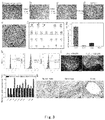
- FIG. 7 shows the results of qualitative and quantitative characterizations of fluorescent peptides on PS-PDA-CMC-VN/BFP-1 peptide mixture-grafted material surfaces (a-b). Results of alizarin red qualitative staining and quantitative measurement of H9hESCs and UMC-C1 hiPSCs after 28 days of osteogenic induction on different PDA-CMC-VN/BFP-1 surfaces.
- FIG. 8 shows that the VB peptide may substitute for VN peptide to be used in the culture of human pluripotent stem cells.
- FIG. 9 shows that the PDA-CMC-VB surface supports long-term self-renewal of a plurality of human pluripotent stem cell lines.
- (a) Cell morphology and karyotype analysis of 5 human pluripotent stem cell lines (H1 hESCs, H9 hESCs, hNF-C1 hiPSCs, UMC-C1hiPSCs, and ipsN004 hiPSCs) upon continuous passage to P20 on the PDA-CMC-VB surface.
- the expression of pluripotency markers Oct-4, SSEA-3, and Tra-1 60 in 5 human pluripotent stem cell lines was detected by using flow cytometry (left panel) and immunofluorescence (right panel). Scale: 100 ⁇ m.
- FIG. 10 confirms that H1 hESCs and hNF-C1 hiPSCs maintain multiple differentiation potential after continuous passage to P20 on the PDA-CMC-VB surface.
- FIG. 11 shows that all of 3 human pluripotent stem cell lines, H9 hESCs, iPSN004 hiPSCs and VBUE hiPSCs, are capable of directionally differentiating into neural precursor cells, neuron cells, and cardiac muscle cells on the PDA-CMC-VB surface.
- Sox-1 red
- Nestin green
- MAP-2 green
- TUJ-1 green
- ⁇ -actinin green
- ⁇ -myosin green
- An aspect of the invention provides a device (also referred to as the device of the invention), comprising:
- the peptide layer comprises a peptide A or a variant peptide thereof as follows;
- peptide A a peptide composed of the amino acid sequence of SEQ ID NO:1,
- variant peptide refers to:
- a peptide which is composed of an amino acid sequence obtained by deletion, substitution, insertion, and/or addition of one or more amino acids in the amino acid sequence of the peptide A, and has a function equivalent to that of the peptide A,
- a peptide which is composed of an amino acid sequence having an amino acid homology of 70% or more with the amino acid sequence of the peptide A, and has a function equivalent to that of the peptide A, and
- a peptide which is encoded by a nucleic acid which hybridizes with the nucleic acid encoding the amino acid sequence of the peptide A under stringent conditions, and has a function equivalent to that of the peptide A.
- the device of the invention may be used in cell experiments in vitro or cell culture.
- a cell experiment in vitro refers to: (1) inducing human somatic cells to be reprogrammed into human pluripotent stem cells, (2) allowing the adhesion of human pluripotent stem cells, and/or (3) allowing for long-term self-renewal of human pluripotent stem cells.
- the material used as the substrate of the device of the invention is not particularly limited, and those typically used as cell culture surfaces may be used, including for example, polystyrene, polydimethylsiloxane, glass, etc.
- a substrate for example, a cell culture plate may be used, or an implant (made of titanium or polyether ether ketone) may be used.
- Dopamine is a biological neurotransmitter in vivo. When a substrate material is immersed in a buffer containing dopamine, dopamine will be deposited on the surface of the substrate material by oxidation polymerization so as to form a polydopamine layer on the surface of the substrate material.
- the molecular weight of polydopamine (PDA) constituting the polydopamine layer is not particularly limited.
- the molecular weight of polydopamine may be controlled by a conventional method. For example, 0.01-100 g/L of dopamine solution reacts at 5-90° C. for 1 minute to 72 hours.
- Carboxymethyl chitosan is one of the most important derivatives of chitosan, and similar to glycosaminoglycan (GAG), which is an extracellular matrix component, it has a good biocompatibility.
- VN peptide may be grafted on a PDA layer, but the resulting device cannot promote the adhesion of cells.
- the weight average molecular weight of carboxymethyl chitosan used in the invention may be 1,000-200,000 g/mol, preferably 10,000-100,000 g/mol, more preferably 60,000-80,000 g/mol, as measured by a gel permeation chromatography and with reference to “Pharmacopoeia of the People's Republic of China”.
- the weight average molecular weight of the carboxymethyl chitosan in the CMC layer of the PDA-CMC-VN device prepared in Examples is 83,550 g/mol.
- the carboxymethyl chitosan comprises an amino group and a carboxyl group, wherein a chemical bond may be formed between the amino group and polydopamine.
- the chemical bond between carboxymethyl chitosan and polydopamine may be achieved by a chemical reaction known in the art, for example, by Michael addition and Schiff base reaction.
- a certain amount of a 0.1 wt %-30 wt % CMC solution may be added to a substrate of a polydopamine layer and reacts at 5 to 60° C. for 1 h-72 h, and a polydopamine-CMC substrate layer may be obtained.
- the peptide layer shall comprise peptide A or a variant thereof as follows, and may also comprise other components as required (peptides, proteins, nucleic acids, small molecule compounds, etc.), as long as the device of the invention can exert the function of regulating behaviors of human pluripotent stem cells.
- the peptide A is a peptide composed of the amino acid sequence of SEQ ID NO:1, and is referred to as VN peptide, which has an amino acid sequence of SEQ ID NO:1:KGGPQVTRGDVFTMP.
- the VN peptide is derived from vitronectin (VN).
- pluripotency maintenance of about 10 passages of hESCs may be achieved by forming a polyacrylic acid layer on the surface of a culture plate and grafting Ac-KGGNGEPRGDTYRAY (SEQ ID NO:3) derived from bone sialoprotein (BSP) and this VN peptide with N-terminal acetylated (Melkoumian Z et al., Synthetic peptide-acrylate surfaces for long-term self-renewal and cardiomyocyte differentiation of human embryonic stem cells. Nature biotechnology. 2010; 28:606-10).
- the resulting device cannot promote the adhesion of cells when this VN peptide is directly grafted on the PDA layer.
- the resulting device is conferred with a function of regulating behaviors of human pluripotent stem cells in the case that a CMC layer is provided as a bridge between a PDA layer and a VN peptide layer.
- the variant of the peptide A includes:
- a peptide which is composed of an amino acid sequence obtained by deletion, substitution, insertion, and/or addition of one or more amino acids in the amino acid sequence of the peptide A, and has a function equivalent to that of the peptide A.
- peptide A refers to the peptide A described above.
- a function equivalent to that of the peptide A refers to any one, any two, or all of the following functions when the peptide A in the device of the invention is replaced with a variant peptide of the peptide A:
- the amino acid substitution may be a conservative substitution. That is, a specific amino acid residue is replaced with a residue having a similar physicochemical property.
- conserved substitutions include substitutions between amino acid residues containing aliphatic groups (for example, mutual substitutions between Ile, Val, Leu, and Ala), substitutions between polar residues (for example, mutual substitutions between Lys and Arg, between Glu and Asp, and between Gln and Asn), etc.
- one or more amino acids refers to amino acids to the extent that deletion, substitution, insertion, and/or addition can be achieved by an artificially synthetic method, for example, 1-20 amino acids, preferably 1-15 amino acids, more preferably 1-10 amino acids, more preferably 1-8 amino acids, more preferably 1-2 amino acids, more preferably 1 amino acid.
- Whether the variant peptide has a function equivalent to that of the peptide A may be determined by replacing the peptide A in the device of the invention with a variant peptide thereof and investigating whether the resulting device has any one, any two, or all of the above functions (i)-(iii).
- the variant of the peptide A comprises:
- a peptide which is composed of an amino acid sequence having an amino acid homology of 70% or more, preferably 80% or more, more preferably 90% or more, more preferably 95% or more, more preferably 97% or more, more preferably 98% or more, more preferably 99% or more with the amino acid sequence of the peptide A, and has a function equivalent to that of the peptide A.
- the homology % between two amino acid sequences may be determined by visual inspection and mathematical calculation.
- the homology percentage between two peptide sequences may be determined by performing sequence information comparison by using a GAP computer program available from University of Wisconsin Genetics Computer Group (UWGCG) based on the algorithm of Needleman, S. B. and Wunsch, C. D. (J. Mol. Bol., 48: 443-453, 1970).
- GAP computer program available from University of Wisconsin Genetics Computer Group (UWGCG) based on the algorithm of Needleman, S. B. and Wunsch, C. D. (J. Mol. Bol., 48: 443-453, 1970).
- Preferred default parameters of the GAP program include: (1) a scoring/matrix, blosum62, as described by Henikoff, S. and Henikoff, J. G. (Proc. Natl. Acad. Sci.
- BLAST program may be used to compare and determine the sequence information, as described by Altschul et al. (Nucl. Acids. Res., 25, p. 3389-3402, 1997). This program is available on the Internet at the web site of the National Center for Biotechnology Information (NCBI) or the DNA Data Bank of Japan (DDBJ).
- NCBI National Center for Biotechnology Information
- DDBJ DNA Data Bank of Japan
- homology % between two amino acid sequences may be determined by using a program such as genetic information processing software GENETYX Ver. 7 (produced by GENETYX) or using an algorithm such as FASTA. In this case, default values may be used for search.
- the variant of the peptide A comprises:
- a peptide which is encoded by a nucleic acid which hybridizes with the nucleic acid encoding the amino acid sequence of the peptide A under stringent conditions, and has a function equivalent to that of the peptide A.
- stringent conditions refer to conditions where so-called specific hybrids are formed and non-specific hybrids are not formed.
- the examples of stringent conditions include those conditions where highly homologous DNAs hybridize with each other, for example, DNAs having a homology of no less than 80%, preferably having a homology of no less than 90%, more preferably having a homology of no less than 95%, still more preferably having a homology of no less than 97%, particularly preferably having a homology of no less than 99% hybridize with each other, and DNAs having a homology less than those described above do not hybridize with each other; or typical washing conditions in Southern hybridization, i.e., washing once, preferably twice or 3 times at a salt concentration and temperature corresponding to 1 ⁇ SSC, 0.1% SDS at 60° C., preferably 0.1 ⁇ SSC, 0.1% SDS at 60° C., and more preferably 0.1 ⁇ SSC, 0.1% SDS at 68° C.
- a VB peptide may be exemplified.
- the VB peptide has an amino acid sequence of KGGPQVTRGDTYRAY (SEQ ID NO:2, and this peptide may be acetylated, which is Ac-KGGPQVTRGDTYRAY (SEQ ID NO:2)).
- the VB peptide is an artificial peptide designed by the inventors, and it is shown by experimental results that it has a function equivalent to that of VN peptide and is not inferior to the VN peptide in terms of properties.
- the distribution density of the peptide A or a variant thereof should be above a certain value, for example, 1 ⁇ g/cm 2 or more, preferably 10 ⁇ g/cm 2 or more, more preferably 20 ⁇ g/cm 2 or more, more preferably 30 ⁇ g/cm 2 or more, and more preferably 40 ⁇ g/cm 2 or more.
- the distribution density of the peptide A or a variant thereof is not particularly limited, and may be, for example, 200 ⁇ g/cm 2 or less, 150 ⁇ g/cm 2 or less, 100 ⁇ g/cm 2 or less, 80 ⁇ g/cm 2 or less, or 60 ⁇ g/cm 2 or less, in view of the operability.
- the carboxymethyl chitosan comprises an amino group and a carboxyl group, wherein the carboxyl group may be grafted to a peptide.
- the grafting of carboxymethyl chitosan to a peptide may be achieved by a chemical reaction known in the art.
- an activating agent solution for example, 20 mg/ml N-hydroxysuccinimide and 20 mg/ml 1-(3-dimethylaminopropyl
- the concentration of reaction solution of the peptide A should be 0.25 mM or more, preferably 1 mM or more.
- the peptide layer may further comprise a compound B having a function of promoting directional differentiation of human pluripotent stem cells, and in combination with a specific medium with defined components, in vitro directional induction differentiation of human pluripotent stem cells may be achieved.
- the compound B may be a peptide, a protein, a nucleic acid, or a small molecule compound.
- the compound B and the specific medium are not particularly limited, as long as they have a function of promoting directional differentiation of human pluripotent stem cells.
- a suitable compound B and a suitable directional induction culture medium may be selected by the person skilled in the art according to the requirements of directional differentiation. For example,
- the peptide layer is grafted with a BFP-1 peptide and an ⁇ MEM medium containing ⁇ -mercaptoethanol, dexamethasone, and vitamin C is comprised to directionally induce osteogenic differentiation of human pluripotent stem cells;
- the peptide layer is grafted with a neural growth factor (NGF) and a N2B27 culture broth supplemented with 100 ng/ml rmNogginis to directionally induce human pluripotent stem cells into neural precursor cells.
- NGF neural growth factor
- the neural precursor cells are cultured in a N2B27 culture broth containing a neural factor, BDNF, GDNF, CNTF, IGF1, and cAMP and may directionally differentiate into neuron cells; or the neural precursor cells are seeded on a poly-L-ornithine substrate and may be directionally induced to differentiate into oligodendrocytes by using a DEME/F12 culture broth containing NI, biotin, PDGF-AA, NT3, cAMP, bFGF, and IGF1.
- the invention further provides a method for promoting directional differentiation of human pluripotent stem cells, comprising step B: culturing human pluripotent stem cells on the surface of the device of the invention by using a directional induction medium.
- a directional induction medium refers to a medium having a function of promoting directional differentiation of human pluripotent stem cells. It is known in the art which kind of directional induction culture medium may promote directional differentiation of human pluripotent stem cells into a specific type of cells.
- an ⁇ MEM medium containing ⁇ -mercaptoethanol, dexamethasone, vitamin C, and fetal bovine serum is used in the differentiation of human pluripotent stem cells into osteoblasts; and a DMEM/F12 medium containing N2, BMP-4, and RA may promote directional differentiation of human pluripotent stem cells into dental epithelial cells, etc.
- the above method for promoting directional differentiation of human pluripotent stem cells may further comprise step A: before the step B, culturing human pluripotent stem cells by using a medium capable of maintaining self-renewal of human pluripotent stem cells.
- step A culturing human pluripotent stem cells by using a medium which can maintain self-renewal of human pluripotent stem cells.
- a suitable compound B and a suitable directional induction culture medium may be selected by the person skilled in the art according to the requirements of directional differentiation. For example,
- the peptide layer is grafted with a BFP-1 peptide and an ⁇ MEM medium containing ⁇ -mercaptoethanol, dexamethasone, and vitamin C is comprised to directionally induce osteogenic differentiation of human pluripotent stem cells;
- the peptide layer is grafted with a neural growth factor (NGF) and a N2B27 culture broth supplemented with 100 ng/ml rmNoggin is comprised to directionally induce human pluripotent stem cells into neural precursor cells.
- NGF neural growth factor
- the neural precursor cells are cultured in a N2B27 culture broth containing a neural factor, BDNF, GDNF, CNTF, IGF1, and cAMP and may directionally differentiate into neuron cells; or the neural precursor cells are seeded on a poly-L-ornithine substrate and may be directionally induced to differentiate into oligodendrocytes by using a DEME/F12 culture broth containing NI, biotin, PDGF-AA, NT3, cAMP, bFGF, and IGF1.
- Human pluripotent stem cells surface-cultured in the invention are cultured with a MEF-CM medium containing bFGF for 2-3 days.
- a RPMI+B27 medium is replaced on the first day of induction, and Activin A is added to allow culturing for 24 h.
- BMP 4 and bFGF are added on the second day to maintain for four days without replacing medium. Subsequently, it is replaced with a RPMI+B27 medium containing 50 ng/ml VEGF165 and the culture continues to enable directional differentiation into cardiac muscle cells.
- Human pluripotent stem cells surface-cultured in the invention may be directionally induced into dental epithelial cells by using a DMEM/F12 medium containing N2, BMP-4, and RA.
- Human pluripotent stem cells surface-cultured in the invention may be directionally induced into hepatocyte-like cells by using media containing different compounds at different stages.
- a basal medium which is RPMI/B27 (minus insulin)
- cytokine Activin A is added, and culture is performed for 3 days
- the basal medium is RPMI/1327 (complete with Insulin), and cytokines BMP2 and FGF4 are added, and culture is performed for 4 days
- the basal medium is RPMI/B27 (complete with Insulin), and cytokines HGF and KGF are added, and culture is performed for 6 days
- the basal medium is a hepatocyte culture medium supplemented with SINGLEQUOTSTM (EGF free), and cytokine Oncostatin-M is added, and culture is performed for 8 days.
- the distribution density of the compound B is not particularly limited, as long as it can exert a function of promoting directional differentiation of human pluripotent stem cells, and is preferably at least 1 ⁇ g/cm 2 or more, preferably 10 ⁇ g/cm 2 or more, more preferably 20 ⁇ g/cm 2 or more, more preferably 30 ⁇ g/cm 2 or more, and more preferably 40 ⁇ g/cm 2 or more.
- a method for grafting the compound B on carboxymethyl chitosan As a method for grafting the compound B on carboxymethyl chitosan, a method which is the same as the method for grafting the peptide A may be used.
- the peptide may be suitably obtained by using a well-known method, such as (1) a chemical synthesis method, or (2) an enzymatic reaction synthesis method, etc., wherein chemical synthesis is simpler.
- a chemical synthesis method for example, a peptide solid-phase synthesis method, etc., may be exemplified.
- the peptide synthesized in this way may be purified by using a conventional measure, for example, ion exchange chromatography, reverse-phase high-performance liquid chromatography, affinity chromatography, etc.
- the peptide solid-phase synthesis method and the subsequent peptide purification are all well known in the art.
- the method as described in the pamphlet of International Publication No. WO2004/011653 may be used, for example. That is, the production may be performed by performing a reaction between an amino acid or a dipeptide obtained by esterification or amidation of the carboxyl terminus of an amino acid or a dipeptide and an amino acid in free state (e.g., an amino acid with the carboxyl group protected) in the presence of a peptide synthase to generate a dipeptide or a tripeptide.
- a culture of microorganisms having the ability of generating peptides, a bacterial cell of microorganism isolated from this culture, a treated product of the bacterial cell of this microorganism, or a peptide synthase derived from this microorganism may be exemplified.
- Another aspect of the invention provides the use of the device of the invention in a cell experiment in vitro.
- a cell experiment in vitro refers to any one, any two, or all of the following items (1)-(3):
- human pluripotent stem cells refer to human induced pluripotent stem cells (hiPSCs) and human embryonic stem cell (hESCs).
- the adhesion of human pluripotent stem cells refers to the case that human embryonic stem cells and human induced pluripotent stem cells can adhere to the surface of a treated cell culture plate, rapidly spread upon adhesion, then begin mitoses, and rapidly enter the logarithmic growth phase.
- the self-renewal of human pluripotent stem cells refers to the case that a human pluripotent stem cell may generate two cells which are the same as the original cells by symmetric division.
- the long-term self-renewal of human pluripotent stem cells refers to the case that human pluripotent stem cells still maintain the pluripotency after more than 20 passages of continuous subculturing in an in vitro microenvironmental system.
- the device of the invention may be further used to promote directional differentiation of human pluripotent stem cells.
- the directional differentiation of human pluripotent stem cells refers to a process that the morphology, structure, and function of human pluripotent stem cells change under the action of related factors.
- the PDA-CMC-VN device manufactured below has a substrate (the surface of a cell culture plate), a polydopamine layer, a carboxymethyl chitosan layer, and a VN peptide layer, in sequence.
- 0.2424 g of tris(hydroxymethyl) aminomethane hydrochloride was placed in a beaker containing 200 ml of pure water and uniformly stirred with a burette, and the pH of the solution was adjusted to 8.5 with 1 mol/L hydrochloric acid.
- 0.4 g of dopamine powder was added to this buffer and uniformly stirred with a burette. After filtration with a 0.22 ⁇ m filtration membrane in a super clean bench, it was added to a Corning 6-well cell culture plate (5 ml volume per well), and placed in a thermostatic shaker (70 rpm) at 37° C. after enclosing with a Parafilm wrap and reaction was performed for 16 h.
- VN peptide was dissolved in 3.1 ml of DPBS buffer to obtain a 1 mM VN peptide solution.
- 19.52 g of morpholinoethane sulfonic acid was dissolved in 1000 ml of pure water and uniformly stirred with a glass rod, and the pH was adjusted to 5.6 with a saturated sodium hydroxide solution.
- 0.8 g of N-hydroxysuccinimide and 0.8 g of 1-(3-dimethylaminopropyl)-3-ethylcarbodiimide hydrochloride were dissolved in 20 ml of a morpholinoethane sulfonic acid buffer respectively and uniformly mixed.
- FITC-KGGPQVTRGDVFTMP fluorescin FITC
- fluorescence values were measured by using a full-wavelength microplate reader with excitation light of 488 nm and absorption light of 538 nm to establish a fluorescence-density standard curve. Thereafter, 1 mM FITC-VN solution was formulated, grafted to an activated PS-PDA-CMC surface by using the method described above, and washed with pure water for 3 times, and fluorescence values were measured under the same conditions.
- PS exhibits a typical water contact angle value)(86.36 ⁇ 1.67°
- the contact angles of surfaces modified by PDA/CMC/VN were reduced to 61.54 ⁇ 1.70°, 41.48 ⁇ 7.16°, and 30.88 ⁇ 3.52°, respectively.
- the fluorescence intensity of the surface of the PS-PDA-CMC-FITCVN modified 12-well plate was 399.695 ⁇ 73.65291.
- the grafting density of the VN peptide on the surface was 51.75 ⁇ 9.53 ug/cm2.
- H1 hESCs and H9 hESCs were purchased from WiCell Research Institute by Guangzhou Institute of Biomedicine and Health, Chinese Academy of Sciences, and were then provided as presents.
- hiPSCs lines hNF-C1 derived from human skin cells and UMC-C1 derived from human mesenechymal stem cells
- These human pluripotent stem cell lines were cultured in culture plates coated with Matrigel (BD Biosciences, Canada), and components used in the media are defined MTESRTM1 (StemCell Technologies, Canada). The cell culture was performed in an incubator at 37° C., 100% humidity, and 5% CO2.
- Matrigel was diluted with DMEM/F12 at 1:80 on ice, added to a 6-well plate with a 1 mlvolume per well, and incubated at 37° C. for 1 h or more. Medium was replaced for cells every day, and the cells were digested with 0.5 mM EDTA at 37° C. for 4-5 min every 3-4 days and passaged at a ratio of 1:3.
- the PDA-CMC-VN modification supports the culture of human pluripotent stem cells on the surface of different substrates.
- hNF-C1 hiPSCs were digested into single cells with 0.25% trypsin/EDTA (Stem Cell Technologies, Canada) and seeded onto PDA/VN and PDA-CMC-VN modified 6-well plates at a density of 23,500 cells/cm-2, and a culture plate pre-plated with Matrigel was used as a control. Fresh media were replaced every day until the fourth day, and a CCK8 cell number counting kit (Dojindo, Japan) was used to measure the cell number in each well. Each group included three wells in parallel, and the absorbance value of each well was measured for three times.
- the particular experimental method was as follows.
- hNF-C1 hiPSCs cultured on Matrigel were digested with EDTA, and transferred to surfaces of different substrates (PS, glass, PDMS, and Ti) modified by PDA-CMC-VN at a ratio of 1:3. After culturing for 4 days, the cell morphology on the surfaces of the materials was observed by using an inverted microscope (Olympus CKX31SF, Japan) and an upright metallographic microscope (Olympus BX51M, Japan). In the meanwhile, cells were collected and the expression of Oct-4 was measured by flow cytometry.
- H1 hESCs and hNF-C1 hiPSCs were selected to study the effect of the concentration of peptides, the method of digestion, and ROCK inhibitor Y-27632 on the adhesion of human pluripotent stem cells.
- a PDA/CMC modified 6-well plate was activated by NHS/EDC, 1 ml of VN peptide at different concentrations (0.25 mM, 0.5 mM, 0.75 mM, 1 mM) were added to wells, and reaction was performed at 4° C. overnight.
- the culture plate grafted with the peptide was washed with a DMEM/F12 medium for three times, H1 hESCs and hNF-C1 human induced pluripotent cells cultured on Matrigel were digested into single cells with 0.25% Trypsin/EDTA, which were seeded at a density of 23,500 cells per square centimeter.
- the cells were transferred onto Matrigel at the same density and taken as a control. Fresh media were replaced every day until the fourth day, a CCK8 kit was then used to measure the cell count in each well.
- H1 hESCs and hNF-C1 hiPSCs were seeded onto the PDA-CMC-VN surface and the Matrigel surface in a form of single cells or clones (23,500 cells per square centimeter), respectively, to study the effect of the passage manner on the adhesion and the proliferation of human pluripotent stem cells.
- ROCK inhibitor Y-27632 was also studied. That is, 5 mM Y-27632 was added in one group and was not added in the other group. Fresh media were replaced every day until the fourth day, and a CCK8 kit was then used to measure the cell count in each well.
- each group contained 3 wells in parallel, and the CCK8 reagent absorbance value of each well was measured for 3 times.
- H1 hESCs and hNF-C1 hiPSCs were selected to study the effect of the concentration of peptides, the digestion manner, and ROCK inhibitor Y-27632 on the adhesion and the proliferation of human pluripotent stem cells on the surface of the PDA-CMC-VN modified culture plate.
- the concentration of the VN peptide increased, the PDA-CMC-VN surface comprised more VN peptide.
- the cell count of hNF-C1 hiPSCs also gradually increased, but the cell count of H1 hESCs was not significantly changed, and both of them were significantly lower than that of the Matrigel control group (p ⁇ 0.01).
- the promoting effect of Y-27632 is relatively significant for H1 hESCs but is not significant for hNF-C1 hiPSCs.
- the cell numbers of H1 hESCs and hNF-C1 hiPSCs seeded in the form of clones were significantly greater than the cell numbers of those seeded in the form of single cells (p ⁇ 0.05), after growing on the PDA-CMC-VN surface for four days, regardless whether Y-27632 was added or not.
- the cell count of hNF-C1 hiPSCs after growing on the PDA-CMC-VN surface for four days was close to that of Matrigel.
- H1, H9 and human induced pluripotent stem cell lines (hNF-C1, GZC2F6 and UMC-C1) cultured on Matrigel were digested with 0.5 mM EDTA under the condition of 37° C. for 4 minutes, clones were blown off with a MTESRTM1 medium containing 5 ⁇ m of Y-27632 and transferred to a PDA-CMC-VN modified 6-well plate at a ratio of 1:3. On the second day, it was replaced with a medium free of Y-27632 and the medium was replaced every day. In the meanwhile, cell morphology was observed by using an inverted microscope (OlympusCKX41, Japan) every day.
- differentiated or abnormal clones were found, they were labeled and removed by suction with a negative-pressure suction apparatus (YX932D, China) in a biosafety cabinet. According to the size and the density of the clones, cells were digested with EDTA and passaged to a new PDA-CMC-VN modified 6-well plate every 3-5 days.
- ACTB was used as a control gene, and primers for respective genes were listed in Table 1.
- Triton-X100 (Sigma, Missouri, USA) was added and permeabilization was performed at room temperature for 30 min, and washed with DPBS for 3 times, with gently shaking on a shaker for 2 min in each time.
- Secondary antibodies (Goat Anti-Mouse Alexa Fluor 488 IgG (Molecular Probes, Invitrogen, USA) and Goat Anti-mouse Alexa Fluor 488 IgM (Molecular Probes, Invitrogen, USA)) were diluted with DPBS at a ratio of 1:500, and 400 ul of the secondary antibodies were added to each well and reaction was performed for 1 h.
- DAPI Washed with DPBS for three times, with gently shaking on a shaker for 5 min in each time.
- DAPI was diluted with DPBS at a ratio of 1:5000, and 500 ul was added to each well, and staining was performed at room temperature for 5 min.
- New DPBS was added after washing with DPBS once, a Nikon Eclipse E800 microscope or a Zeiss Axiovert 200M inverted confocal microscope was used for observation and to take photos. Excitation wavelengths of DAPI and green fluorescent labeled antibody were 405 nm and 488 nm, respectively.
- Oct-3/4 primary antibody (01550, StemCell Technologies, Canada) was diluted with a flow cytometric buffer at a ratio of 1:100. Cells were resuspended with 100 ul of the primary antibody solution, and incubated in an incubator for 30 min. After centrifuging at 200 g for 5 minutes, washing was performed with a flow cytometric buffer once. Steps 3-4 were suitable for the detection of the label Oct-4 in the cell, and were not required for cell surface markers SSEA-3 and Tra-1 60.
- Oct-4 secondary antibody was diluted with a flow cytometric buffer at a ratio of 1:500, resuspended with a volume of 200 ul, and was incubated in an incubator for 30 min.
- directly labeled antibodies SSEA-3 (60061PE, StemCell Technologies, Canada) and Tra-1 60 (60064PE, StemCell Technologies, Canada), were diluted with a flow cytometric buffer at a ratio of 1:100, and cells were resuspended with 100 ul of the solution and incubated in an incubator for 30 min. After the completion of incubation, centrifugation was performed at 200 g for 5 minutes.
- H1 hESCs and hNF-C1 hiPSCs which were subjected to 20 passages of subculturing on PDA-CMC-VN, were digested with Dispase of 1 mg ⁇ mL ⁇ 1 for 7 minutes.
- an EB medium a DMEM/F12 basal medium containing 20% bovine serum albumin, 2 mM L-glutaMax, 1% (wt/vol) NEAA, and 0.1 mM ⁇ -mercaptoethanol
- EB medium a DMEM/F12 basal medium containing 20% bovine serum albumin, 2 mM L-glutaMax, 1% (wt/vol) NEAA, and 0.1 mM ⁇ -mercaptoethanol
- the medium was supplemented or the medium was replaced every 2-3 days according to the conditions of cells. Thereafter, embryoid bodies were transferred to PDA-CMC-VN for adherent culture for 8 days with the medium replaced every other day. Finally, cells were treated with Trizol and total RNA was extracted, and the expression of triploblastic marker genes was detected by fluorescence quantitative PCR (for primers, see Table 2) and was compared to that in undifferentiated human pluripotent stem cells.
- H1 hESCs and hNF-C1 hiPSCs which had been subjected to 20 passages of subculturing on PDA-CMC-VN, were back passaged to Matrigel and cultured so as to be used in the experiment of teratoma generation.
- Cells to be injected were digested with Dispase, resuspended with 1:80 diluted Matrigel, and subcutaneously injected into NOD-SCID mice with a cell count of one million, and markers were made.
- NOD-SCID mice subcutaneously injected into NOD-SCID mice with a cell count of one million, and markers were made.
- teratoma formed and grew to a diameter of 1 cm the mouse was sacrificed. Typically, teratoma would generate after about 6-8 weeks. The teratoma was withdrawn, sectioned, and stained with hematoxylin-eosin. Typical cells of three germ layers were sought for by microscopic observation.
- hESCs lines H1 and H9 and 2 hiPSCs lines (hNF-C1 and UMC-C1) were selected and continuously passaged for more than 20 passages on the PDA-CMC-VN surface to judge whether this surface can support a long-term self-renewal of human pluripotent stem cells.
- hNF-C1 and UMC-C1 2 hiPSCs lines
- H1 hESCs and hNF-C1 hiPSCs still maintained pluripotency after more than 20 passages of culturing on the PDA-CMC-VN surface.
- H1 hESCs and hNF-C1 hiPSCs which were cultured on the PDA-CMC-VN surface, were suspended for 8 days and adherently cultured for 8 days, and the total RNA of the cells was extracted. It was demonstrated by the results of q-PCR that all of embryoid bodies formed by H1 hESCs and hNF-C1 hiPSCs on the PDA-CMC-VN surface expressed triploblastic genes (see FIGS.
- H1 hESCs and hNF-C1 hiPSCs which were cultured on the PDA-CMC-VN surface for a long-term, still maintained a multiple differentiation potential.
- H9 hESCs were continuously passaged to the 15th passage on the PDA-CMC-VN surface, neural directional induction differentiation was performed by using a STEMdiff neural system (Stem Cell Technologies, Canada) according to the instructions of manufacturer. Furthermore, we performed a differentiation experiment of H9 hESCs into cardiac muscle cells according to the method reported in the reference (Yang, L. et al. Human cardiovascular progenitor cells develop from a KDR+ embryonic-stem-cell-derived population. Nature, 2008, 453, 524-528). The experimental method was same as that reported in the reference except that the media used on day 0, day 1, and day 4 were as shown below.
- Stempro-34 SFM10640 medium, Glutamin (100 ⁇ ), transferrin (0.15 mg/ml), 1-thioglycerol (6.5 ul/ml in 500 ml medium, 3 ul/ml of backbone), ascorbic acid (50 ug/ml), BMP4 (0.05 ng/ml), Y27632 (10 ug/ml).
- Stempro-34 SFM 10640 medium, Glutamin (100 ⁇ ), bFGF (6 ng/ml), Activin A (2.5 ng/ml), ascorbic acid (50 ug/ml), BMP4 (10 ng/ml).
- Nerve-specific antibodies used were SOX-1 (R&D AF3369, USA), Nestin (R&D MAB1259, USA), MAP-2 (Millpore AB5622, USA), and Anti- ⁇ -Tubulin III (TUJ-1, Sigma-Aldrich T3952, USA); and cardiac-muscle-cell-specific antibodies were ⁇ -actinin (Abcam AB9465, United Kingdom) and ⁇ -myosin (R&D MAB4470, USA).
- the 15th passage of H9 hESCs cultured on the PDA-CMC-VN surface formed uniform embryoid bodies by using an AggreWellTM 800 plate ( FIG. 6 a ), and differentiated into nerve cells and cardiac muscle cells in the case of defined components, respectively.
- H9 hESCs were induced in a neural induction culture medium for 14 days, a neural rosettes was formed ( FIG. 6 b ) and neural markers Sox-1 and Nestin were positively expressed ( FIG. 6 d ), which demonstrated the successful induction into neural precursor cells.
- we picked up the neural rosette which was cultured in a neural rosette selective medium for 7 days, and a typical neuron morphology appeared ( FIG.
- the manufacture of the PDA-CMC modified cell culture plate was as described above, and 5 mg of a VN peptide was dissolved in 3.1 ml PBS buffer in a super clean bench to obtain a concentration of 1 mM. 5 mg of a BFP-1 peptide was dissolved in 3.0 ml PBS buffer to obtain a 1 mM BFP-1 peptide solution.
- VN and BFP-1 peptide solutions were mixed at volume ratios of 10:0, 7:3, and 5:5 to obtain three groups of peptide mixed solutions, which were VN 10 /BFP-1 0 (briefly referred to as VN), VN 7 /BFP-1 3 (briefly referred to as 7:3), and VN 5 /BFP-1 5 (briefly referred to as 5:5).
- VN 10 /BFP-1 0 VN 10 /BFP-1 0
- VN 7 /BFP-1 3 (briefly referred to as 7:3)
- VN 5 /BFP-1 5 (briefly referred to as 5:5).
- a PDA-CMC-VN/BFP-1 peptide mixture-modified cell culture plate was then obtained by using the NHS/EDC activation method as described above.
- FITC-VN and Rho-BFP-1 peptide solutions having a concentration gradient of 0.2 mM, 0.4 mM, 0.6 mM, 0.8 mM, 1.0 mM, and 1.2 mM were formulated with a sterile PBS solution, respectively, and then 200 ul of peptide gradient solutions were added to a Corning 24-well plate, respectively (3 duplicate wells for each group).
- VN fluorescent peptide mixed solutions
- VN VN 10 /BFP-1 0
- VN 7 /BFP-1 3 VN 7 /BFP-1 3
- VN 5 /BFP-1 5 VN 5 /BFP-1 5
- H9 hESCs (the passage number was 30-50 passages) and UMC-C1 hiPSCs (the passage number was 30-45 passages) cultured on the Matrigel surface were digested into single cells with an enzyme Acutase, and were then seeded onto material surfaces (VN group, 7:3 group, and 5:5 group).
- ROCK inhibitor (Y-27632) was added to MTESRTM1 to promote the adhesion of single cells. Most of cells were completely adherent after 12 h, and the medium was replaced with an MTESRTM1 medium free of ROCK inhibitor (Y-27632) at this time.
- the MTESRTM1 medium was replaced with an osteogenic induction medium (an ⁇ -MEM medium containing 10% FBS, 5 ⁇ g/ml vitamin C (AA), 10 mM ⁇ -sodium glycerophosphate ( ⁇ -GP), and 10 ⁇ 8 M dexamethasone), and the medium was freshened every 2 days until the 28th day.
- an osteogenic induction medium an ⁇ -MEM medium containing 10% FBS, 5 ⁇ g/ml vitamin C (AA), 10 mM ⁇ -sodium glycerophosphate ( ⁇ -GP), and 10 ⁇ 8 M dexamethasone
- FIG. 7 c showed the results of alizarin red qualitative staining and quantitative measurement of respective groups after 28 days of osteogenic induction.
- calcium nodules were formed on both H9 hESCs and UMC-C1 hiPSCs cultured on surfaces in the VN group, the 7:3 group, and the 5:5 group, and the quantity of calcium nodules formed gradually increased as the content of the BFP-1 osteogenic peptide increased.
- the osteogenic peptide indeed exerted a promoting effect on the osteogenesis of stem cells, but different cell lines behaved differently to some extent, in which H9 hESCs had a better osteogenic effect than that of UMC-C1 hiPSCs.
- Examples 1-5 described above were repeated under the same experimental conditions except that the VN peptide in the PDA-CMC-VN device described above was replaced with the VB peptide designed and screened by our research team.
- FIG. 8 showed that under conditions of EDTA digestion and the addition of ROCK inhibitor Y-27632, the PDA-CMC-VB surface and the PDA-CMC-VN surface had similar properties in terms of supporting the adhesion and the proliferation of H1 hECSs and hNF-C1 hiPSCs.
- the cell numbers of H1 hECSs and hNF-C1 hiPSCs which were cultured on the PDA-CMC-VB surface and the PDA-CMC-VN surface for four days, were close to each other at all concentrations of the grafted peptides (0.25 mM, 0.5 mM, and 1 mM) except the concentration of 0.125 mM where the cell count of H1 hECSs cultured to the fourth day on VB was less than that on VN (p ⁇ 0.05) ( FIG. 8 a ). Furthermore, we also found that the cell count on the material surface on the fourth day also increased gradually as the concentration of peptides increased.
- FIGS. 9 and 10 demonstrated that 5 human pluripotent stem cell lines (H1 hESCs, H9 hESCs, hNF-C1 hiPSCs, UMC-C1 hiPSCs, and ipsN004 hiPSCs) still maintained pluripotency after more than 20 passages of continuous passage on the PDA-CMC-VB surface.
- FIG. 11 showed that all of 3 human pluripotent stem cell lines, which were H9 hESCs, iPSN004 hiPSCs, and VBUE hiPSCs, were capable of directionally differentiating into nerve cells and cardiac muscle cells on the PDA-CMC-VB surface.
- any technical feature or a combination of technical features described as a constituent for a certain technical solution in this specification may also be suitable for other technical solutions, in the case of being implementable and not obviously departing from the gist of the invention.
- technical features described as constituents for different technical solutions may also be combined arbitrarily to constitute other technical solutions, in the case of being implementable and not obviously departing from the gist of the invention.
- the invention further comprises technical solutions obtained by combination in the cases described above, and these technical solutions are equivalent to be described in this specification.
- a device useful in cell culture or cell experiments in vitro can be provided.
Applications Claiming Priority (3)
| Application Number | Priority Date | Filing Date | Title |
|---|---|---|---|
| CN2014108507550 | 2014-12-31 | ||
| CN201410850755 | 2014-12-31 | ||
| PCT/CN2015/096857 WO2016107387A1 (fr) | 2014-12-31 | 2015-12-09 | Dispositif et son application au cours d'une expérience in vitro sur des cellules |
Publications (2)
| Publication Number | Publication Date |
|---|---|
| US20180142205A1 US20180142205A1 (en) | 2018-05-24 |
| US10597634B2 true US10597634B2 (en) | 2020-03-24 |
Family
ID=56284201
Family Applications (1)
| Application Number | Title | Priority Date | Filing Date |
|---|---|---|---|
| US15/540,700 Expired - Fee Related US10597634B2 (en) | 2014-12-31 | 2015-12-09 | Device and use thereof in cell experiments in vitro |
Country Status (3)
| Country | Link |
|---|---|
| US (1) | US10597634B2 (fr) |
| CN (1) | CN105820951B (fr) |
| WO (1) | WO2016107387A1 (fr) |
Families Citing this family (2)
| Publication number | Priority date | Publication date | Assignee | Title |
|---|---|---|---|---|
| CN106420822A (zh) * | 2016-09-29 | 2017-02-22 | 广州赛莱拉干细胞科技股份有限公司 | 一种组合物及其应用 |
| US20230147883A1 (en) * | 2021-11-28 | 2023-05-11 | Fatemeh Etezadi | Method for prodcing cardiomyocytes from induced pluripotent stem cells in an integrated process |
Citations (4)
| Publication number | Priority date | Publication date | Assignee | Title |
|---|---|---|---|---|
| US20050147645A1 (en) * | 1998-07-24 | 2005-07-07 | Budny John A. | Composition and method for bone regeneration |
| US20130164787A1 (en) * | 2009-04-22 | 2013-06-27 | Viacyte, Inc. | Cell compositions derived from dedifferentiated reprogrammed cells |
| WO2013116432A1 (fr) | 2012-02-02 | 2013-08-08 | Corning Incorporated | Milieu synthétique de fixation destiné à la culture cellulaire |
| US9133246B2 (en) * | 2011-04-22 | 2015-09-15 | Nano Intelligent Biomedical Engineering Corporation Co, Ltd. | Surface-active collagen membrane by peptide |
Family Cites Families (3)
| Publication number | Priority date | Publication date | Assignee | Title |
|---|---|---|---|---|
| AU2008353306B2 (en) * | 2008-05-16 | 2011-09-01 | Industry Foundation Of Chonnam National University | An synthetic peptide containing bone forming peptide 1(BFP 1) for stimulating osteoblast differentiation, and pharmaceutical composition comprising the synthetic peptide |
| RU2018128383A (ru) * | 2012-03-07 | 2019-03-14 | Янссен Байотек, Инк. | Среда определенного состава для размножения и обновления плюрипотентных стволовых клеток |
| CN103627669A (zh) * | 2012-08-22 | 2014-03-12 | 复旦大学 | 一种诱导人多能干细胞分化为视网膜前体细胞的方法 |
-
2015
- 2015-12-09 US US15/540,700 patent/US10597634B2/en not_active Expired - Fee Related
- 2015-12-09 WO PCT/CN2015/096857 patent/WO2016107387A1/fr active Application Filing
- 2015-12-09 CN CN201510903953.3A patent/CN105820951B/zh active Active
Patent Citations (5)
| Publication number | Priority date | Publication date | Assignee | Title |
|---|---|---|---|---|
| US20050147645A1 (en) * | 1998-07-24 | 2005-07-07 | Budny John A. | Composition and method for bone regeneration |
| US20130164787A1 (en) * | 2009-04-22 | 2013-06-27 | Viacyte, Inc. | Cell compositions derived from dedifferentiated reprogrammed cells |
| US9133246B2 (en) * | 2011-04-22 | 2015-09-15 | Nano Intelligent Biomedical Engineering Corporation Co, Ltd. | Surface-active collagen membrane by peptide |
| WO2013116432A1 (fr) | 2012-02-02 | 2013-08-08 | Corning Incorporated | Milieu synthétique de fixation destiné à la culture cellulaire |
| US20150010999A1 (en) * | 2012-02-02 | 2015-01-08 | Corning Incorporated | Synthetic attachment medium for cell culture |
Non-Patent Citations (8)
Also Published As
| Publication number | Publication date |
|---|---|
| US20180142205A1 (en) | 2018-05-24 |
| WO2016107387A1 (fr) | 2016-07-07 |
| CN105820951B (zh) | 2019-04-19 |
| CN105820951A (zh) | 2016-08-03 |
Similar Documents
| Publication | Publication Date | Title |
|---|---|---|
| Garreta et al. | Myocardial commitment from human pluripotent stem cells: Rapid production of human heart grafts | |
| Chen et al. | Application of human mesenchymal and pluripotent stem cell microcarrier cultures in cellular therapy: achievements and future direction | |
| Zhou et al. | Simple and versatile synthetic polydopamine-based surface supports reprogramming of human somatic cells and long-term self-renewal of human pluripotent stem cells under defined conditions | |
| JP5784266B2 (ja) | 規定細胞培養表面及び使用方法 | |
| Lock et al. | Expansion and differentiation of human embryonic stem cells to endoderm progeny in a microcarrier stirred-suspension culture | |
| Deng et al. | Long-term self-renewal of human pluripotent stem cells on peptide-decorated poly (OEGMA-co-HEMA) brushes under fully defined conditions | |
| KR102146007B1 (ko) | 망막 전구 세포, 망막 색소 상피 세포 및 신경 망막 세포를 얻기 위한 방법 | |
| Harkness et al. | Selective isolation and differentiation of a stromal population of human embryonic stem cells with osteogenic potential | |
| Deng et al. | A novel hydrogel surface grafted with dual functional peptides for sustaining long‐term self‐renewal of human induced pluripotent stem cells and manipulating their osteoblastic maturation | |
| Wang et al. | In vitro culture and directed osteogenic differentiation of human pluripotent stem cells on peptides-decorated two-dimensional microenvironment | |
| AU2007271898B2 (en) | A novel population of multipotent cardiac precursor cells derived from human blastocysts derived stem cells | |
| Wu et al. | Spider silk for xeno-free long-term self-renewal and differentiation of human pluripotent stem cells | |
| JP2012509085A (ja) | マイクロキャリア上での多能性幹細胞の培養 | |
| KR101193541B1 (ko) | 인간 배아줄기세포에서 중간엽 줄기세포로 분화시키는 방법 | |
| Liu et al. | A synthetic substrate to support early mesodermal differentiation of human embryonic stem cells | |
| Yan et al. | Biomaterial strategies for stem cell maintenance during in vitro expansion | |
| WO2023106122A1 (fr) | Procédé de production de cellules de crête neurale spécialisées pour la différenciation en lignée mésenchymateuse | |
| CN110520139A (zh) | 用于治疗视网膜疾病的感光细胞 | |
| H Parsons et al. | Patents on technologies of human tissue and organ regeneration from pluripotent human embryonic stem cells | |
| Jiang et al. | Laminin-521 promotes rat bone marrow mesenchymal stem cell sheet formation on light-induced cell sheet technology | |
| US10597634B2 (en) | Device and use thereof in cell experiments in vitro | |
| Patil et al. | Spatially controlled functional group grafting of silk films to induce osteogenic and chondrogenic differentiation of human mesenchymal stem cells | |
| Wang et al. | Topographical modulation of pluripotency and differentiation of human embryonic stem cells | |
| Wang et al. | Pluripotency maintenance of amniotic fluid-derived stem cells cultured on biomaterials | |
| WO2020081716A2 (fr) | Biomatériaux pour la croissance et la différenciation de cellules 3d |
Legal Events
| Date | Code | Title | Description |
|---|---|---|---|
| FEPP | Fee payment procedure |
Free format text: ENTITY STATUS SET TO SMALL (ORIGINAL EVENT CODE: SMAL); ENTITY STATUS OF PATENT OWNER: SMALL ENTITY |
|
| STPP | Information on status: patent application and granting procedure in general |
Free format text: NON FINAL ACTION MAILED |
|
| STPP | Information on status: patent application and granting procedure in general |
Free format text: RESPONSE TO NON-FINAL OFFICE ACTION ENTERED AND FORWARDED TO EXAMINER |
|
| STPP | Information on status: patent application and granting procedure in general |
Free format text: FINAL REJECTION MAILED |
|
| STPP | Information on status: patent application and granting procedure in general |
Free format text: ADVISORY ACTION MAILED |
|
| STPP | Information on status: patent application and granting procedure in general |
Free format text: RESPONSE AFTER FINAL ACTION FORWARDED TO EXAMINER |
|
| STPP | Information on status: patent application and granting procedure in general |
Free format text: NOTICE OF ALLOWANCE MAILED -- APPLICATION RECEIVED IN OFFICE OF PUBLICATIONS |
|
| AS | Assignment |
Owner name: PEKING UNIVERSITY SCHOOL AND HOSPITAL OF STOMATOLO Free format text: ASSIGNMENT OF ASSIGNORS INTEREST;ASSIGNORS:WEI, SHICHENG;ZHOU, PING;ZHANG, XIAOHONG;AND OTHERS;SIGNING DATES FROM 20170905 TO 20170912;REEL/FRAME:050948/0100 Owner name: PEKING UNIVERSITY SCHOOL AND HOSPITAL OF STOMATOLOGY, CHINA Free format text: ASSIGNMENT OF ASSIGNORS INTEREST;ASSIGNORS:WEI, SHICHENG;ZHOU, PING;ZHANG, XIAOHONG;AND OTHERS;SIGNING DATES FROM 20170905 TO 20170912;REEL/FRAME:050948/0100 |
|
| STPP | Information on status: patent application and granting procedure in general |
Free format text: PUBLICATIONS -- ISSUE FEE PAYMENT VERIFIED |
|
| STCF | Information on status: patent grant |
Free format text: PATENTED CASE |
|
| FEPP | Fee payment procedure |
Free format text: MAINTENANCE FEE REMINDER MAILED (ORIGINAL EVENT CODE: REM.); ENTITY STATUS OF PATENT OWNER: SMALL ENTITY |
|
| LAPS | Lapse for failure to pay maintenance fees |
Free format text: PATENT EXPIRED FOR FAILURE TO PAY MAINTENANCE FEES (ORIGINAL EVENT CODE: EXP.); ENTITY STATUS OF PATENT OWNER: SMALL ENTITY |
|
| STCH | Information on status: patent discontinuation |
Free format text: PATENT EXPIRED DUE TO NONPAYMENT OF MAINTENANCE FEES UNDER 37 CFR 1.362 |