US10028832B2 - Helical coil mitral valve annuloplasty systems and methods - Google Patents
Helical coil mitral valve annuloplasty systems and methods Download PDFInfo
- Publication number
- US10028832B2 US10028832B2 US14/328,050 US201414328050A US10028832B2 US 10028832 B2 US10028832 B2 US 10028832B2 US 201414328050 A US201414328050 A US 201414328050A US 10028832 B2 US10028832 B2 US 10028832B2
- Authority
- US
- United States
- Prior art keywords
- needle
- lumen
- helical anchor
- catheter
- clip
- Prior art date
- Legal status (The legal status is an assumption and is not a legal conclusion. Google has not performed a legal analysis and makes no representation as to the accuracy of the status listed.)
- Active, expires
Links
Images


Classifications
-
- A—HUMAN NECESSITIES
- A61—MEDICAL OR VETERINARY SCIENCE; HYGIENE
- A61F—FILTERS IMPLANTABLE INTO BLOOD VESSELS; PROSTHESES; DEVICES PROVIDING PATENCY TO, OR PREVENTING COLLAPSING OF, TUBULAR STRUCTURES OF THE BODY, e.g. STENTS; ORTHOPAEDIC, NURSING OR CONTRACEPTIVE DEVICES; FOMENTATION; TREATMENT OR PROTECTION OF EYES OR EARS; BANDAGES, DRESSINGS OR ABSORBENT PADS; FIRST-AID KITS
- A61F2/00—Filters implantable into blood vessels; Prostheses, i.e. artificial substitutes or replacements for parts of the body; Appliances for connecting them with the body; Devices providing patency to, or preventing collapsing of, tubular structures of the body, e.g. stents
- A61F2/02—Prostheses implantable into the body
- A61F2/24—Heart valves ; Vascular valves, e.g. venous valves; Heart implants, e.g. passive devices for improving the function of the native valve or the heart muscle; Transmyocardial revascularisation [TMR] devices; Valves implantable in the body
- A61F2/2442—Annuloplasty rings or inserts for correcting the valve shape; Implants for improving the function of a native heart valve
-
- A—HUMAN NECESSITIES
- A61—MEDICAL OR VETERINARY SCIENCE; HYGIENE
- A61B—DIAGNOSIS; SURGERY; IDENTIFICATION
- A61B17/00—Surgical instruments, devices or methods, e.g. tourniquets
- A61B17/068—Surgical staplers, e.g. containing multiple staples or clamps
-
- A—HUMAN NECESSITIES
- A61—MEDICAL OR VETERINARY SCIENCE; HYGIENE
- A61B—DIAGNOSIS; SURGERY; IDENTIFICATION
- A61B17/00—Surgical instruments, devices or methods, e.g. tourniquets
- A61B17/00234—Surgical instruments, devices or methods, e.g. tourniquets for minimally invasive surgery
- A61B2017/00238—Type of minimally invasive operation
- A61B2017/00243—Type of minimally invasive operation cardiac
-
- A—HUMAN NECESSITIES
- A61—MEDICAL OR VETERINARY SCIENCE; HYGIENE
- A61B—DIAGNOSIS; SURGERY; IDENTIFICATION
- A61B17/00—Surgical instruments, devices or methods, e.g. tourniquets
- A61B17/00234—Surgical instruments, devices or methods, e.g. tourniquets for minimally invasive surgery
- A61B2017/00292—Surgical instruments, devices or methods, e.g. tourniquets for minimally invasive surgery mounted on or guided by flexible, e.g. catheter-like, means
- A61B2017/003—Steerable
- A61B2017/00318—Steering mechanisms
- A61B2017/00331—Steering mechanisms with preformed bends
-
- A—HUMAN NECESSITIES
- A61—MEDICAL OR VETERINARY SCIENCE; HYGIENE
- A61B—DIAGNOSIS; SURGERY; IDENTIFICATION
- A61B17/00—Surgical instruments, devices or methods, e.g. tourniquets
- A61B17/00234—Surgical instruments, devices or methods, e.g. tourniquets for minimally invasive surgery
- A61B2017/00349—Needle-like instruments having hook or barb-like gripping means, e.g. for grasping suture or tissue
-
- A—HUMAN NECESSITIES
- A61—MEDICAL OR VETERINARY SCIENCE; HYGIENE
- A61B—DIAGNOSIS; SURGERY; IDENTIFICATION
- A61B17/00—Surgical instruments, devices or methods, e.g. tourniquets
- A61B2017/00743—Type of operation; Specification of treatment sites
- A61B2017/00778—Operations on blood vessels
- A61B2017/00783—Valvuloplasty
-
- A—HUMAN NECESSITIES
- A61—MEDICAL OR VETERINARY SCIENCE; HYGIENE
- A61B—DIAGNOSIS; SURGERY; IDENTIFICATION
- A61B17/00—Surgical instruments, devices or methods, e.g. tourniquets
- A61B17/064—Surgical staples, i.e. penetrating the tissue
- A61B2017/0649—Coils or spirals
-
- A—HUMAN NECESSITIES
- A61—MEDICAL OR VETERINARY SCIENCE; HYGIENE
- A61F—FILTERS IMPLANTABLE INTO BLOOD VESSELS; PROSTHESES; DEVICES PROVIDING PATENCY TO, OR PREVENTING COLLAPSING OF, TUBULAR STRUCTURES OF THE BODY, e.g. STENTS; ORTHOPAEDIC, NURSING OR CONTRACEPTIVE DEVICES; FOMENTATION; TREATMENT OR PROTECTION OF EYES OR EARS; BANDAGES, DRESSINGS OR ABSORBENT PADS; FIRST-AID KITS
- A61F2/00—Filters implantable into blood vessels; Prostheses, i.e. artificial substitutes or replacements for parts of the body; Appliances for connecting them with the body; Devices providing patency to, or preventing collapsing of, tubular structures of the body, e.g. stents
- A61F2/02—Prostheses implantable into the body
- A61F2/24—Heart valves ; Vascular valves, e.g. venous valves; Heart implants, e.g. passive devices for improving the function of the native valve or the heart muscle; Transmyocardial revascularisation [TMR] devices; Valves implantable in the body
- A61F2/2412—Heart valves ; Vascular valves, e.g. venous valves; Heart implants, e.g. passive devices for improving the function of the native valve or the heart muscle; Transmyocardial revascularisation [TMR] devices; Valves implantable in the body with soft flexible valve members, e.g. tissue valves shaped like natural valves
-
- A—HUMAN NECESSITIES
- A61—MEDICAL OR VETERINARY SCIENCE; HYGIENE
- A61F—FILTERS IMPLANTABLE INTO BLOOD VESSELS; PROSTHESES; DEVICES PROVIDING PATENCY TO, OR PREVENTING COLLAPSING OF, TUBULAR STRUCTURES OF THE BODY, e.g. STENTS; ORTHOPAEDIC, NURSING OR CONTRACEPTIVE DEVICES; FOMENTATION; TREATMENT OR PROTECTION OF EYES OR EARS; BANDAGES, DRESSINGS OR ABSORBENT PADS; FIRST-AID KITS
- A61F2/00—Filters implantable into blood vessels; Prostheses, i.e. artificial substitutes or replacements for parts of the body; Appliances for connecting them with the body; Devices providing patency to, or preventing collapsing of, tubular structures of the body, e.g. stents
- A61F2/02—Prostheses implantable into the body
- A61F2/24—Heart valves ; Vascular valves, e.g. venous valves; Heart implants, e.g. passive devices for improving the function of the native valve or the heart muscle; Transmyocardial revascularisation [TMR] devices; Valves implantable in the body
- A61F2/2478—Passive devices for improving the function of the heart muscle, i.e. devices for reshaping the external surface of the heart, e.g. bags, strips or bands
- A61F2002/249—Device completely embedded in the heart wall
-
- A—HUMAN NECESSITIES
- A61—MEDICAL OR VETERINARY SCIENCE; HYGIENE
- A61F—FILTERS IMPLANTABLE INTO BLOOD VESSELS; PROSTHESES; DEVICES PROVIDING PATENCY TO, OR PREVENTING COLLAPSING OF, TUBULAR STRUCTURES OF THE BODY, e.g. STENTS; ORTHOPAEDIC, NURSING OR CONTRACEPTIVE DEVICES; FOMENTATION; TREATMENT OR PROTECTION OF EYES OR EARS; BANDAGES, DRESSINGS OR ABSORBENT PADS; FIRST-AID KITS
- A61F2230/00—Geometry of prostheses classified in groups A61F2/00 - A61F2/26 or A61F2/82 or A61F9/00 or A61F11/00 or subgroups thereof
- A61F2230/0063—Three-dimensional shapes
- A61F2230/0091—Three-dimensional shapes helically-coiled or spirally-coiled, i.e. having a 2-D spiral cross-section
Definitions
- the invention relates generally to the treatment of heart valves. More particularly, it relates to systems, devices, and methods for treating valvular regurgitation, such as mitral regurgitation, via percutaneously delivered helical coils.
- the heart is a four-chambered pump that moves blood efficiently through the vascular system.
- Blood enters the heart through the vena cava and flows into the right atrium. From the right atrium, blood flows through the tricuspid valve and into the right ventricle, which then contracts and forces blood through the pulmonic valve and into the lungs.
- Oxygenated blood returns from the lungs and enters the heart through the left atrium and passes through the mitral valve into the left ventricle. The left ventricle contracts and pumps blood through the aortic valve into the aorta and to the vascular system.
- the mitral valve consists of two leaflets (anterior and posterior) attached to a fibrous ring or annulus.
- the mitral valve leaflets close during contraction of the left ventricle and prevent blood from flowing back into the left atrium.
- the mitral valve annulus may become distended, causing the leaflets to remain partially open during ventricular contraction and thus allow regurgitation of blood into the left atrium. This results in reduced ejection volume from the left ventricle, causing the left ventricle to compensate with a larger stroke volume.
- the increased workload eventually results in dilation and hypertrophy of the left ventricle, further enlarging and distorting the shape of the mitral valve. If left untreated, the condition may result in cardiac insufficiency, ventricular failure, and ultimately death.
- Mitral valve repair includes a variety of procedures to repair or reshape the leaflets to improve closure of the valve during ventricular contraction. If the mitral valve annulus has become distended, a frequent repair procedure involves implanting an annuloplasty ring on the mitral valve annulus.
- the annuloplasty ring generally has a smaller diameter than the annulus, and when sutured to the annulus the annuloplasty ring draws the annulus into a smaller configuration, bringing the mitral valve leaflets closer together and allowing improved closure during ventricular contraction.
- Annuloplasty rings may be rigid, flexible or a combination, having both rigid and flexible segments.
- Rigid annuloplasty rings have the disadvantage of causing the mitral valve annulus to be rigid and unable to flex in response to the contractions of the ventricle, thus inhibiting the normal, three-dimensional movement of the mitral valve that is required for it to function optimally.
- Flexible annuloplasty rings are frequently made of Dacron® fabric and must be sewn to the annulus tissue with a line of sutures. This eventually leads to scar tissue formation and loss of flexibility and function of the mitral valve. Similarly, combination rings must generally be sutured in place and also cause scar tissue formation and loss of mitral valve flexibility and function.
- Annuloplasty is normally an open-heart surgical procedure.
- Valve replacement also typically entails an open-heart surgical procedure in which the patient's mitral valve is removed and replaced with an artificial valve.
- One drawback to open-heart surgical techniques is that heart bypass procedures are required to accomplish replacement and/or repair of the valve.
- Another drawback is that the open-heart procedures require that the patient undergo general anesthesia for a prolonged period of time.
- mitral and other cardiac valve annuloplasty devices and procedures have been developed that employ a helical anchor coil in place of the conventional annuloplasty rings (or bands). Examples of such devices and procedures are described, for example, in US Publication Nos. 2007/0244553, 2007/0244554, 2007/0244555, 2007/0244556, and 2007/0244557, each of which is incorporated by reference herein in its entirety.
- one or more helical anchor coils are percutaneously delivered, in a coiled state, to the mitral valve annulus via a tubular delivery member.
- the helical anchor has a sharpened tip that penetrates into tissue of the valve annulus; as the helical anchor is directed out of the delivery member and rotated along a guide, the helical anchor threads into the annulus tissue.
- a tether is routed through an inner channel of the so-implanted helical anchor(s) and formed into a loop. The loop is tensioned, effectuating a desired modification in the shape of the valve annulus.
- helical anchor and tether systems described above are quite promising, simulating the surgical placement of an annuloplasty ring on the valve annulus.
- the described minimally invasive delivery techniques may be less than optimal.
- any improvements in the helical anchor coils and related methods of use will be well-received.
- Embodiments hereof relate to systems and methods for modifying a heart valve annulus in a minimally invasive surgical procedure.
- a helical anchor is provided, having a memory set to a coiled shape or state.
- the helical anchor is further configured to self-revert from a substantially straight state to the coiled state.
- the helical anchor is loaded within a needle that constrains the helical anchor to the substantially straight state.
- the needle is delivered to the valve annulus and inserted into tissue of the annulus.
- the helical anchor is then deployed from the needle (e.g., the needle is retracted from over the helical anchor). Once deployed, the helical anchor self-transitions toward the coiled shape, cinching engaged tissue of the valve annulus.
- inventions hereof relate to systems and methods for locating and identifying designated anatomical positions along the valve annulus in a minimally invasive procedure, along with devices, systems and methods for modifying the valve annulus with a helical anchor based upon reference to the identified anatomical positions.
- FIG. 1A is a simplified illustration of a heart and illustrating some anatomical features of the mitral valve.
- FIG. 1B is a simplified cross-sectional schematic view of the heart and illustrating some anatomical features of the mitral valve.
- FIG. 1C illustrates additional anatomical features of the mitral valve.
- FIGS. 2A and 2B illustrate a helical anchor useful with some annuloplasty systems and methods in accordance with embodiments hereof.
- FIGS. 3A-3D illustrate a system and method for locating and identifying designated anatomical positions along the mitral valve annulus in a minimally invasive procedure in accordance with an embodiment hereof.
- FIGS. 4A-4C illustrate another system and method for locating and identifying designated anatomical positions along the mitral valve annulus in a minimally invasive procedure in accordance with an embodiment hereof.
- FIGS. 4D and 4E illustrate another system and method for locating and identifying designated anatomical positions along the mitral valve annulus similar to those of FIGS. 4A-4C .
- FIGS. 5A and 5B illustrate another system and method for locating and identifying designated anatomical positions along the mitral valve annulus in a minimally invasive procedure in accordance with an embodiment hereof.
- FIG. 6 illustrates another system and method for locating and identifying designated anatomical positions along the mitral valve annulus in a minimally invasive procedure in accordance with an embodiment hereof.
- FIG. 7A-7C illustrate a guide useful in implanting the helical anchor of FIGS. 2A and 2B relative to the designated anatomical positions along the mitral valve annulus in accordance with an embodiment hereof.
- FIG. 8 is a schematic view of the mitral valve and illustrates helical anchors implanted to the mitral valve annulus and useful for modifying the annulus in accordance with some systems and methods in accordance with embodiments hereof.
- FIG. 9 is a simplified cross-sectional view of a catheter assembly useful for delivering the helical anchors of FIG. 8 .
- FIG. 10A is a simplified cross-sectional view of a needle system for modifying a heart valve annulus in a minimally invasive procedure and including a memory set helical anchor configured to self-revert from a substantially straight state to a coiled state.
- FIG. 10B is a simplified view of the needle system of FIG. 10A employing another embodiment helical anchor.
- FIGS. 11A and 11B illustrate use of the system of FIG. 10A .
- FIGS. 12A and 12B illustrate a delivery system useful with the needle system of FIG. 10A .
- FIGS. 13A-13I illustrate use of the delivery system of FIGS. 12A and 12B .
- FIG. 14 illustrates a handle assembly useful with the delivery system of FIGS. 12A and 12B .
- FIGS. 15A-15F illustrate use of delivery system of FIGS. 12A and 12B , and the handle assembly of FIG. 14 .
- FIGS. 16A and 16B illustrate a needle system in accordance with another embodiment hereof.
- FIG. 17 illustrates a prosthetic cardiac valve implanted within a modified valve annulus in accordance with another embodiment hereof.
- distal and proximal are used in the following description with respect to a position or direction relative to the treating clinician. “Distal” or “distally” are a position distant from or in a direction away from the clinician. “Proximal” and “proximally” are a position near or in a direction toward the clinician.
- Embodiments in accordance herewith relate to systems and methods for treating a defective heart valve, for example the mitral valve.
- the anatomy of a heart 10 includes a left atrium (LA) 12 and a left ventricle (LV) 14 .
- An aorta 16 receives blood from the left ventricle 14 through an aortic valve 18 , which serves to prevent regurgitation of blood back into the left ventricle 14 .
- a mitral valve 20 is positioned between the left atrium 12 and the left ventricle 14 , and allows one-way flow of blood from the left atrium 12 to the left ventricle 14 .
- the mitral valve 20 includes an anterior leaflet 22 and a posterior leaflet 24 that are coupled to chordae tendonae (not shown) that serve as “tension members” that prevent the leaflets 22 , 24 of the mitral valve 20 from going past their closing point and prolapsing back into the left atrium.
- FIG. 1B illustrates the mitral valve 20 (in systole) from a different vantage point.
- a fibrous mitral valve annulus 28 provides attachment for the anterior and posterior leaflets 22 , 24 .
- the anterior and posterior leaflets 22 , 24 are dissimilarly shaped.
- the anterior leaflet 22 is more firmly attached to the annulus 28 , and is somewhat stiffer than the posterior leaflet 24 that is attached to the more mobile posterior lateral mitral annulus.
- the coaptation zone between the leaflets 22 , 24 is not a simple line, but rather a curved, funnel-shaped surface interface. As best shown in FIG. 1C , commissures 30 , 32 are where the anterior leaflet 22 meets the posterior leaflet 24 .
- the coaptation zone is generally along a curved line CL.
- the leaflet 22 , 24 edges are scalloped, more so for the posterior leaflet 24 then the anterior leaflet 22 .
- Three scallop or segmented areas are generally defined along the anterior and the posterior aspects of the mitral valve annulus 28 , and are commonly referred to as the A 1 , A 2 , and A 3 , and P 1 , P 2 , and P 3 segments.
- FIGS. 2A and 2B illustrate one embodiment of a helical anchor 40 useful with annuloplasty devices in accordance with embodiments hereof.
- the helical anchor 40 comprises an elongate coiled member having a tissue penetrating tip 42 at a distal end that is opposite a proximal end 44 .
- the coils of the helical anchor 40 define a structure having a generally cylindrical shape, and the tip 42 extends on a tangent away from the circular perimeter of the helical anchor 40 . Angling the sharpened tip 42 away from the exterior perimeter of the helical anchor 40 makes it easier for the tip 42 to penetrate a valve annulus when the helical anchor 40 is being rotated out of a delivery member and along an anchor guide.
- the helical anchor 40 can comprise a biocompatible metallic or polymeric material having suitable resiliency.
- the coils of the helical anchor 40 define an inner channel 46 for receiving a tether (not shown). As described in the '553 Publication, the tether can be a flexible elongated filament of biocompatible material such as nylon or polyester.
- some systems and methods in accordance with embodiments hereof for delivering and implanting one or more of the helical anchors 40 include locating and marking the mitral valve annulus 28 at three locations or points L 1 -L 3 via a percutaneous approach.
- the first and third locations L 1 , L 3 correspond with the commissures 30 , 32
- the second location L 2 approximates a mid-point of the posterior leaflet 24 along the annulus 28 (i.e., mid-point of the middle posterior segment P 2 ).
- a purpose of marking the three locations L 1 -L 3 is to provide reference points for desired location of the subsequently implanted helical anchor(s) 40 ( FIG. 2A ).
- FIG. 2A In one embodiment with reference to FIG.
- a primary catheter 50 is directed to and placed at the second location L 2 from the ventricular side of the mitral valve 20 .
- a guide wire (not shown) can be utilized to direct the primary catheter 50 to the second location L 2 .
- FIG. 3B illustrates a primary guide wire 52 extending from the primary catheter 50 and piercing through the mitral valve annulus 28 at the second location L 2 .
- the mitral valve annulus 28 can be accessed from the ventricular side; directly underneath the annulus 28 is a sub-annular groove that the primary guide wire 52 conforms to throughout the region of the annulus 28 associated with the posterior leaflet 24 .
- secondary catheters 60 , 62 are pre-shaped to correlate with the general curvature of the mitral valve annulus 28 , and are thus precurved to terminate at the first and third locations L 1 , L 3 when deployed to extend from the primary catheter 50 (with the primary catheter 50 located at L 2 ).
- secondary catheters 60 , 62 may be portions of an integrated structure with primary catheter 50 , or may be independent structures therefrom. As shown in FIG.
- secondary guide wires 64 , 66 are then deployed through the secondary catheters 60 , 62 , respectively, and thus located at L 1 , L 3 . Locations L 1 -L 3 are marked by the guide wires 64 , 52 , 66 .
- FIGS. 4A-4C Another embodiment of a system and method for locating and marking the locations L 1 -L 3 is generally reflected in FIGS. 4A-4C .
- a catheter 80 is placed through the mitral valve 20 via a trans-septal approach as shown in FIG. 4A .
- opposing, self-expanding loops 82 , 84 are deployed from the catheter 80 as reflected by FIG. 4B , and act as a two-armed centering device. More particularly, upon being released from the catheter 80 , the loops 82 , 84 naturally expand to a set shape and self-locate the first and third locations (commissure points) at L 1 and L 3 .
- the opposing loops 82 , 84 are built at an angle slightly less than 180 degrees so that when opened further (relative to the catheter 80 ), the arms or loops 82 , 84 are constrained against the mitral valve annulus 28 and thus “push” the catheter 80 towards the second location L 2 as shown by dashed lines in FIG. 4C .
- the two arms 82 , 84 are located at L 1 and L 3 , and the catheter 80 is at L 2 and are thus available for guided placement of one or more markers, anchors, etc. (not shown).
- the self-expanding loops 82 ′, 84 ′ have a natural or predefined shape formed in accordance with an anatomy of the valve (e.g., mitral valve) such that when deployed from the catheter 80 , the loops 82 ′, 84 ′ present minimal, if any, interference with functioning of the valve leaflets.
- an anatomy of the valve e.g., mitral valve
- FIGS. 5A and 5B Another embodiment of a system and method for locating and marking the locations L 1 -L 3 is generally reflected in FIGS. 5A and 5B , and is similar to those of FIGS. 4A-4E .
- Wires 90 , 92 are employed in place of the loops 82 , 84 ( FIG. 4B ) and can be flat wires.
- the wires 90 , 92 are slightly stiffer then the loops 82 , 84 , and self-revert to the shape memory format shown in FIGS. 5A and 5B when deployed from the catheter 80 .
- a distal tip 94 of each of the wires 90 , 92 can be a ball or loop or other shape that renders the distal tip 94 less traumatic to contacted tissue.
- the system of FIGS. 5A and 5B can be employed to locate the locations L 1 -L 3 in a manner similar to that described above with respect to FIGS. 4A-4C .
- FIG. 6 another embodiment of a system and method for locating L 1 -L 3 entails placing a catheter 100 above the mitral valve 20 via a trans-septal approach.
- the catheter 100 forms a lumen 102 along with a pre-formed bend 104 proximate a distal tip 106 .
- An opening 108 to the lumen 102 is formed at the bend 104 .
- First and second guide wires 110 , 112 are slidably disposed within the lumen 102 .
- the first guide wire 110 is deployed through the distal tip 106 of the catheter 100 to locate L 3 (or L 1 ).
- the second guide wire 112 is deployed through the opening 108 , and locates L 1 (or L 3 ). With the device of FIG. 6 , L 2 would not be marked. However, in a manner similar to that described above with respect to FIGS. 3A-3D , a piercing guide wire (not shown) could be placed retrograde through L 2 , and then guided out through the right side of the heart, and used to initially locate the tip of the device (not shown).
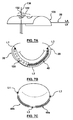
- FIG. 7A illustrates an embodiment of a guide 120 .
- the guide 120 is a thin, elongated body in the form of a half circle. Within the half-circle are a series of circular guide holes 122 that extend perpendicular to an axis of the guide 120 .
- guide wire(s) 130 one of which is shown in FIG.
- FIG. 7A are shown having been previously placed in accordance with one of the methods as described above so as to puncture the annulus 28 from the ventricular side.
- the guide 120 is then placed on the atrial side of the annulus 28 .
- the guide wire 130 is pushed through one of the guide holes 122 .
- Clips 132 (or some other type of anchoring mechanism) are advanced through the guide holes 122 and pulled taut, maintaining the guide 120 in close proximity to the annulus 28 .
- the half circle shape of guide 120 is then used to guide a helical anchor coil 40 from L 1 to L 3 (or from L 3 to L 1 ) as shown in FIG. 7B (as a point of reference, a portion of the guide 120 is cut away in FIG.
- short coil anchors 150 a - 150 c can be turned through the tissue of the annulus 28 as shown in FIG. 8 .
- the anchors 150 a - 150 c are positioned so as to locate an eyelet 152 on the atrial side of the annulus 28 .
- the helix of the anchors 150 a - 150 c grasps the tissue, while the eyelets 152 can serve as an anchor to cinch against.
- the method for cinching can be a suture or some other material threaded through the eyelets 152 and pulled taut.
- FIG. 9 schematically illustrates a catheter assembly 152 for delivering the coil anchor 150 , and includes a catheter 154 , a guide wire 156 and a foam 158 .
- the coil anchor 150 is pre-shaped to keep from going straight.
- the pitch of the screw of the coil anchor 150 needs to be large enough to allow cinching, but small enough that it follows the curve and the guide wire 156 .
- Depth of bite, the balance of size, engagement with the tissue, and clearance for cinching can be accounted for by the coil configuration.
- the biodegradable foam 158 is provided for the coil 150 to pass through and prevent binding.
- the biodegradable foam 158 keeps the coil 150 from tipping up at its ends.
- FIG. 10A is a simplified view of an alternative system 200 in accordance with an embodiment hereof, and includes a helical anchor 202 .
- the helical anchor 202 is delivered to an implantation site in a minimally invasive procedure via a needle 204 .
- FIG. 10A is a simplified view of an alternative system 200 in accordance with an embodiment hereof, and includes a helical anchor 202 .
- the helical anchor 202 is delivered to an implantation site in a minimally invasive procedure via a needle 204 .
- FIG. 10A is a simplified view of an alternative system 200 in accordance with an embodiment hereof, and includes a helical anchor 202 .
- the helical anchor 202 is delivered to an implantation site in a minimally invasive procedure via a needle 204 .
- the helical anchor 202 naturally forms a coil, but can be rendered substantially straight when disposed within a lumen 206 of the needle 204 .
- the helical anchor 202 can be formed of a shape-memory material (e.g., nitinol) that is heat set to form a coil.
- a shape-memory material e.g., nitinol
- FIG. 10B reflects that in other embodiments, the helical anchor 202 ′ naturally forms a wavy line (as opposed to a true coil).
- a push rod 214 is disposed within the lumen 206 of the needle 204 with a distal end of push rod 214 situated proximal to the helical anchor 202 to aid in deployment of the helical anchor from needle 204 .
- Push rod 214 can be manipulated to distally advance the helical anchor 202 relative to the needle 204 and/or to keep the helical anchor 202 stationary as the needle 204 is retracted.
- the needle 204 is delivered to the valve annulus implantation site with the helical anchor 202 entirely disposed within the needle 204 (and thus constrained to a substantially straight shape).
- the distal tip 212 of needle 204 is initially inserted into the tissue of the annulus 28 as shown in FIG. 11A (it being understood that while the needle 204 is illustrated as being visible in FIG. 11A for ease of understanding, in actual practice the needle 204 will be disposed within a thickness of the annulus 28 tissue).
- FIG. 11A identifies one possible implantation site along the mitral valve annulus 28 in a relatively tangential direction.
- the needle 204 is then retracted proximally while the helical anchor 202 ( FIG.
- FIG. 10A held stationary (e.g., via the push rod 214 ( FIG. 10A )).
- the straightened helical anchor 202 As the straightened helical anchor 202 is uncovered, it self-transitions from the constrained, substantially straight shape into the set coiled shape within the annulus tissue.
- the annulus tissue is cinched as generally reflected in FIG. 11B (it being understood that while the helical anchor 202 is illustrated as being visible in FIG. 11B for ease of understanding, in actual practice the helical anchor 202 will be entirely disposed within a thickness of the annulus 28 tissue).
- the tissue is drawn together by the implanted helical anchor 202 .
- the helical anchor 202 when constrained by the needle 204 ( FIG. 10A ), the helical anchor 202 in its austenite form pushes against the walls of the needle 204 , creating friction.
- the helical anchor 202 is selected to be relatively short (e.g., on the order of 1 inch in the straightened state), so that the frictional force can be overcome without binding.
- the helical anchor 202 can be subjected to cooling while it is constrained within the needle 204 to keep the helical anchor 202 in its malleable martensite form, reducing the friction required.
- coatings on the inside of the needle 204 such as a Teflon® coating may be useful in reducing the frictional force.
- two or more of the relatively short helical anchors 202 can be implanted.
- septal-lateral cinching is an effective means of reducing mitral regurgitation.
- FMR functional mitral regurgitation
- a reduction in mitral regurgitation severity from grade 2 to grade 0 was observed with a decrease in the septal-lateral diameter of the mitral annulus of 6.0 ⁇ 2.6 mm.
- Initial testing of prototype devices measured the change in tissue length when deployed on a cadaveric swine heart to be 6.35 mm.
- a related embodiment delivery system 250 for delivering and implanting the helical anchor 202 is shown in FIG. 12A , and includes a delivery catheter 252 and opposing clips 254 , 256 .
- the pair of clips 254 , 256 has opposing curved ends when deployed from a distal end 262 of the catheter 252 .
- the catheter 252 forms a first lumen 258 sized to slidably receive the needle 204 (that otherwise slidaby retains the helical anchor 202 as described above).
- the opposing clips 254 , 256 are retained or disposed within a second lumen 260 of the catheter 252 , with the second lumen 260 being sized to permit clips 254 , 256 to be slidably deployed therefrom.
- the lumens 258 , 260 are open to a distal tip 262 of the catheter 252 .
- Distal tip 262 of catheter 252 may also be referenced to herein as the distal end of catheter 252 .
- the clips 254 , 256 serve as a mechanism for initially attaching the delivery system 250 to the valve annulus. This will allow the user to accurately and reliably insert the needle 204 by serving as an anchor. Additionally, the clips 254 , 256 allow the user to push on the needle 204 and gently pull on the clips 254 , 256 during needle insertion, increasing the ability to puncture the tissue with the needle 204 .
- 12B illustrates the assembly 250 in a deployed state in which a distal portion of the needle 204 is distally extended beyond the catheter 252 but retracted relative to a distal end of push rod 214 , such that the helical anchor 202 is deployed from the lumen of needle 204 and returned to its coiled state.
- FIGS. 13A-13I A method of using the system 250 in implanting the helical anchor 202 is shown in FIGS. 13A-13I .
- the needle 204 is sheathed within the catheter 252 , and the helical anchor 202 is loaded within the needle 204 .
- the catheter 252 is directed to the mitral annulus, for example through a steerable overtube or sheath (not shown) via a trans-septal puncture or a left atrial approach.
- FIG. 13A the needle 204 is sheathed within the catheter 252 , and the helical anchor 202 is loaded within the needle 204 .
- the catheter 252 is directed to the mitral annulus, for example through a steerable overtube or sheath (not shown) via a trans-septal puncture or a left atrial approach.
- the distal tip 262 of catheter 252 can have a tapered shape, or stated another way can form an angled surface with respect to a longitudinal axis of the catheter, such that a first opening or exit of the first lumen 258 within distal tip 262 extends distally beyond a second opening or exit of the second lumen 260 within distal tip 262 .
- the catheter 252 is advanced from the sheath, bringing the tip 262 into contact with the mitral annulus 28 at the desired implant location.
- the clips 254 , 256 are then deployed from the catheter 252 as shown in FIG. 13C .
- the deployed clips 254 , 256 maintain the position of the catheter 252 relative to the annulus tissue 28 .
- any rotational changes can still be made to ensure proper alignment with the annulus 28 and/or the septal-lateral diameter of the mitral valve 20 ( FIG. 1A ).
- the needle 204 (preloaded with the helical anchor 202 as described above) is advanced through the catheter 252 and into the annulus 28 tissue.
- the needle 204 is advanced at an angle more parallel with the tissue surface than is otherwise reflected in the drawing.
- the needle 204 can have a preformed bend (not shown) that will extend a set distance into and then run parallel with the tissue surface.
- FIG. 13F the needle 204 is partially withdrawn from over the helical anchor 202 while push rod 214 is held stationary relative to catheter 252 .
- FIG. 13G reflects that after the needle 204 is withdrawn into the catheter 252 and with push rod 214 distally extend therefrom, the helical anchor 202 is released and self-reverts to its preformed coil shape, cinching the engaged tissue.
- the clips 254 , 256 and push rod 214 are withdrawn back into the catheter 252 , allowing the catheter 252 to be withdrawn from the implantation site as shown in FIG. 13I .
- the catheter 252 can be removed from the sheath, or the sheath can be steered to a new location and the process repeated for any additional helical anchor placements desired.
- System 250 can include various actuators to effectuate desired manipulation of the clips 254 , 256 .
- FIG. 14 illustrates a handle 270 in accordance with an embodiment hereof that may be used with the system 250 .
- the handle 270 includes a needle deployment actuator 272 and a clip actuator 274 .
- FIGS. 15A-15F depicts use of handle 270 in effectuating various deployment states at the distal tip 262 of catheter 252 .
- the needle deployment actuator 272 and the clip actuator 274 are in a “home” position, with the clips (not shown) and the needle (not shown) refracted inside of the catheter 252 .
- the clip actuator 274 is actuated to advance or deploy the clips 254 , 256 .
- the needle deployment actuator 272 is actuated to partially advance the needle 204 .
- FIG. 15D reflects further advancement of the needle 204 prior to deployment of the helical anchor (not shown).
- FIG. 15A-15F depicts use of handle 270 in effectuating various deployment states at the distal tip 262 of catheter 252 .
- FIG. 15A the needle deployment actuator 272 and the clip actuator 274 are in a “home” position, with the clips (not shown) and the needle (not shown) refracted inside of the catheter 252 .
- the clip actuator 274 is actuated
- FIG. 15E the mechanism for maintaining the helical anchor stationary relative to the needle 204 (e.g., the push rod 214 ( FIG. 10A )) is locked. With retraction of the needle 204 , then, the helical anchor 202 is deployed. Finally, FIG. 15F reflects the needle deployment actuator 272 returned to its original position, causing the helical anchor 202 to be fully deployed while the needle 204 ( FIG. 15E ) is captured inside of the catheter 252 .
- the mechanism for maintaining the helical anchor stationary relative to the needle 204 e.g., the push rod 214 ( FIG. 10A )
- FIG. 15F reflects the needle deployment actuator 272 returned to its original position, causing the helical anchor 202 to be fully deployed while the needle 204 ( FIG. 15E ) is captured inside of the catheter 252 .
- the implanted helical anchor 202 effectuates cinching of the mitral valve annulus tissue.
- RF energy can be applied to so-implanted helical anchor 202 .
- the heat generated by the energized helical anchor 202 will cause collagen in the tissue to reorganize (i.e., shape change), and the mitral valve 28 will conform to the shape created by the coil. This change in the collagen structure will keep the desired repaired shape.
- the helical anchor 202 may subsequently be removed from the annulus tissue, or alternatively can be left in place.
- the collagen remodeling may relieve possible stress on the helical anchor 202 from the annulus tissue that might otherwise influence the ability of the helical coil 202 to retract back to its preformed state.
- FIG. 16A illustrates a needle 300 similar to the needle 204 described above, but further including a tracking arm 302 .
- the tracking arm 302 projects from a distal portion of the needle 300 , defining a channel 304 between an outer surface of the needle 300 and the tracking arm 302 .
- the tracking arm 302 is kept on the outside of the annulus, with a tissue layer 306 being captured within the channel 304 as shown in FIG. 16B .
- the tracking arm 302 assists in tracking the needle 300 through the tissue, with the proximal juncture or end where the tracking arm 302 meets or is attached to the needle 300 acting as a stop to prevent the needle 300 from going too deep into the tissue or popping out of the annulus.
- a method for delivering a helical anchor coil to a desired location along a valve annulus as described above is followed by implantation of a prosthetic cardiac valve within the modified valve annulus.
- the structure of a native mitral valve annulus may be modified using one or more helical anchor coils 202 as described herein prior to the delivery and implantation of a prosthetic mitral valve 1775 .
- Examples of percutaneously delivered prosthetic cardiac valves that may be implanted within a modified valve annulus as described herein are described, for example, in US Publication Nos.
- One or more helical anchor coils may be used to modify the shape of a valve annulus into a desirable shape or configuration for implantation of a prosthetic cardiac valve within the modified valve annulus.
- a mitral valve annulus may be modified or shaped into a more rounded configuration that than provides a better fit for a transcatheter three leaflet prosthetic valve having a more rounded circumference or configuration.
Priority Applications (4)
| Application Number | Priority Date | Filing Date | Title |
|---|---|---|---|
| US14/328,050 US10028832B2 (en) | 2013-07-10 | 2014-07-10 | Helical coil mitral valve annuloplasty systems and methods |
| US16/034,061 US10849749B2 (en) | 2013-07-10 | 2018-07-12 | Helical coil mitral valve annuloplasty systems and methods |
| US17/087,008 US11963873B2 (en) | 2013-07-10 | 2020-11-02 | Mitral valve annuloplasty systems and methods |
| US18/445,582 US20240074858A1 (en) | 2013-07-10 | 2023-10-31 | Martial arts gloves with electronic secoring system |
Applications Claiming Priority (2)
| Application Number | Priority Date | Filing Date | Title |
|---|---|---|---|
| US201361844507P | 2013-07-10 | 2013-07-10 | |
| US14/328,050 US10028832B2 (en) | 2013-07-10 | 2014-07-10 | Helical coil mitral valve annuloplasty systems and methods |
Related Child Applications (1)
| Application Number | Title | Priority Date | Filing Date |
|---|---|---|---|
| US16/034,061 Division US10849749B2 (en) | 2013-07-10 | 2018-07-12 | Helical coil mitral valve annuloplasty systems and methods |
Publications (2)
| Publication Number | Publication Date |
|---|---|
| US20150018940A1 US20150018940A1 (en) | 2015-01-15 |
| US10028832B2 true US10028832B2 (en) | 2018-07-24 |
Family
ID=51225114
Family Applications (3)
| Application Number | Title | Priority Date | Filing Date |
|---|---|---|---|
| US14/328,050 Active 2035-08-10 US10028832B2 (en) | 2013-07-10 | 2014-07-10 | Helical coil mitral valve annuloplasty systems and methods |
| US16/034,061 Active 2035-02-13 US10849749B2 (en) | 2013-07-10 | 2018-07-12 | Helical coil mitral valve annuloplasty systems and methods |
| US17/087,008 Active 2036-03-26 US11963873B2 (en) | 2013-07-10 | 2020-11-02 | Mitral valve annuloplasty systems and methods |
Family Applications After (2)
| Application Number | Title | Priority Date | Filing Date |
|---|---|---|---|
| US16/034,061 Active 2035-02-13 US10849749B2 (en) | 2013-07-10 | 2018-07-12 | Helical coil mitral valve annuloplasty systems and methods |
| US17/087,008 Active 2036-03-26 US11963873B2 (en) | 2013-07-10 | 2020-11-02 | Mitral valve annuloplasty systems and methods |
Country Status (3)
| Country | Link |
|---|---|
| US (3) | US10028832B2 (fr) |
| EP (1) | EP3019092B1 (fr) |
| WO (1) | WO2015006575A1 (fr) |
Cited By (18)
| Publication number | Priority date | Publication date | Assignee | Title |
|---|---|---|---|---|
| USD894396S1 (en) | 2019-03-08 | 2020-08-25 | Pacesetter, Inc. | Leadless biostimulator attachment feature |
| US10912644B2 (en) | 2018-10-05 | 2021-02-09 | Shifamed Holdings, Llc | Prosthetic cardiac valve devices, systems, and methods |
| US10973662B2 (en) | 2016-05-16 | 2021-04-13 | Elixir Medical Corporation | Methods and devices for heart valve repair |
| US11000372B2 (en) | 2013-10-25 | 2021-05-11 | Polares Medical Inc. | Systems and methods for transcatheter treatment of valve regurgitation |
| US11160656B2 (en) | 2015-11-06 | 2021-11-02 | Polares Medical Inc. | Device, system, and method for transcatheter treatment of valvular regurgitation |
| US11185704B2 (en) | 2017-11-06 | 2021-11-30 | Pacesetter, Inc. | Biostimulator having fixation element |
| US11298229B2 (en) | 2017-03-13 | 2022-04-12 | Polares Medical Inc. | Device, system, and method for transcatheter treatment of valvular regurgitation |
| US11413145B2 (en) | 2011-01-28 | 2022-08-16 | Polares Medical Inc. | Coaptation enhancement implant, system, and method |
| US11419722B2 (en) | 2011-01-28 | 2022-08-23 | Polares Medical Inc. | Device, system, and method for transcatheter treatment of valve regurgitation |
| US11464634B2 (en) | 2020-12-16 | 2022-10-11 | Polares Medical Inc. | Device, system, and method for transcatheter treatment of valvular regurgitation with secondary anchors |
| US11471282B2 (en) | 2019-03-19 | 2022-10-18 | Shifamed Holdings, Llc | Prosthetic cardiac valve devices, systems, and methods |
| US11534302B2 (en) | 2017-03-13 | 2022-12-27 | Polares Medical Inc. | Device, system, and method for transcatheter treatment of valvular regurgitation |
| US11541243B2 (en) | 2019-03-15 | 2023-01-03 | Pacesetter, Inc. | Biostimulator having coaxial fixation elements |
| US11577086B2 (en) | 2018-08-20 | 2023-02-14 | Pacesetter, Inc. | Fixation mechanisms for a leadless cardiac biostimulator |
| US11622759B2 (en) | 2014-06-24 | 2023-04-11 | Polares Medical Inc. | Systems and methods for anchoring an implant |
| US11759321B2 (en) | 2021-06-25 | 2023-09-19 | Polares Medical Inc. | Device, system, and method for transcatheter treatment of valvular regurgitation |
| US11833034B2 (en) | 2016-01-13 | 2023-12-05 | Shifamed Holdings, Llc | Prosthetic cardiac valve devices, systems, and methods |
| US11974921B2 (en) | 2014-06-18 | 2024-05-07 | Polares Medical Inc. | Mitral valve implants for the treatment of valvular regurgitation |
Families Citing this family (131)
| Publication number | Priority date | Publication date | Assignee | Title |
|---|---|---|---|---|
| US7508853B2 (en) | 2004-12-07 | 2009-03-24 | Imra, America, Inc. | Yb: and Nd: mode-locked oscillators and fiber systems incorporated in solid-state short pulse laser systems |
| US8608797B2 (en) | 2005-03-17 | 2013-12-17 | Valtech Cardio Ltd. | Mitral valve treatment techniques |
| US8951285B2 (en) | 2005-07-05 | 2015-02-10 | Mitralign, Inc. | Tissue anchor, anchoring system and methods of using the same |
| US9883943B2 (en) | 2006-12-05 | 2018-02-06 | Valtech Cardio, Ltd. | Implantation of repair devices in the heart |
| US11259924B2 (en) | 2006-12-05 | 2022-03-01 | Valtech Cardio Ltd. | Implantation of repair devices in the heart |
| US11660190B2 (en) | 2007-03-13 | 2023-05-30 | Edwards Lifesciences Corporation | Tissue anchors, systems and methods, and devices |
| US8382829B1 (en) | 2008-03-10 | 2013-02-26 | Mitralign, Inc. | Method to reduce mitral regurgitation by cinching the commissure of the mitral valve |
| CA2728078A1 (fr) | 2008-06-16 | 2010-01-14 | Valtech Cardio, Ltd. | Dispositifs d'annuloplastie et procedes de mise en place de ceux-ci |
| US8323335B2 (en) | 2008-06-20 | 2012-12-04 | Edwards Lifesciences Corporation | Retaining mechanisms for prosthetic valves and methods for using |
| US9011530B2 (en) | 2008-12-22 | 2015-04-21 | Valtech Cardio, Ltd. | Partially-adjustable annuloplasty structure |
| US8715342B2 (en) | 2009-05-07 | 2014-05-06 | Valtech Cardio, Ltd. | Annuloplasty ring with intra-ring anchoring |
| ES2873182T3 (es) | 2008-12-22 | 2021-11-03 | Valtech Cardio Ltd | Dispositivos de anuloplastia ajustables |
| US8241351B2 (en) | 2008-12-22 | 2012-08-14 | Valtech Cardio, Ltd. | Adjustable partial annuloplasty ring and mechanism therefor |
| US10517719B2 (en) | 2008-12-22 | 2019-12-31 | Valtech Cardio, Ltd. | Implantation of repair devices in the heart |
| US8911494B2 (en) | 2009-05-04 | 2014-12-16 | Valtech Cardio, Ltd. | Deployment techniques for annuloplasty ring |
| US8353956B2 (en) | 2009-02-17 | 2013-01-15 | Valtech Cardio, Ltd. | Actively-engageable movement-restriction mechanism for use with an annuloplasty structure |
| US9968452B2 (en) | 2009-05-04 | 2018-05-15 | Valtech Cardio, Ltd. | Annuloplasty ring delivery cathethers |
| US9180007B2 (en) | 2009-10-29 | 2015-11-10 | Valtech Cardio, Ltd. | Apparatus and method for guide-wire based advancement of an adjustable implant |
| US10098737B2 (en) | 2009-10-29 | 2018-10-16 | Valtech Cardio, Ltd. | Tissue anchor for annuloplasty device |
| US9011520B2 (en) | 2009-10-29 | 2015-04-21 | Valtech Cardio, Ltd. | Tissue anchor for annuloplasty device |
| WO2011067770A1 (fr) | 2009-12-02 | 2011-06-09 | Valtech Cardio, Ltd. | Outil distributeur pour l'implantation d'un ensemble à bobine accouplé à un ancrage hélicoïdal |
| US8870950B2 (en) | 2009-12-08 | 2014-10-28 | Mitral Tech Ltd. | Rotation-based anchoring of an implant |
| US8475525B2 (en) | 2010-01-22 | 2013-07-02 | 4Tech Inc. | Tricuspid valve repair using tension |
| US9307980B2 (en) * | 2010-01-22 | 2016-04-12 | 4Tech Inc. | Tricuspid valve repair using tension |
| US10058323B2 (en) | 2010-01-22 | 2018-08-28 | 4 Tech Inc. | Tricuspid valve repair using tension |
| US8398708B2 (en) | 2010-03-05 | 2013-03-19 | Edwards Lifesciences Corporation | Retaining mechanisms for prosthetic valves |
| US8657872B2 (en) | 2010-07-19 | 2014-02-25 | Jacques Seguin | Cardiac valve repair system and methods of use |
| US11653910B2 (en) | 2010-07-21 | 2023-05-23 | Cardiovalve Ltd. | Helical anchor implantation |
| EP2595569A4 (fr) | 2010-07-23 | 2016-02-24 | Edwards Lifesciences Corp | Mécanismes de rétention pour prothèses valvulaires |
| CN103491900B (zh) | 2010-12-23 | 2017-03-01 | 托尔福公司 | 用于二尖瓣修复和替换的系统 |
| US9554897B2 (en) | 2011-04-28 | 2017-01-31 | Neovasc Tiara Inc. | Methods and apparatus for engaging a valve prosthesis with tissue |
| CN103997990A (zh) | 2011-06-21 | 2014-08-20 | 托尔福公司 | 人工心脏瓣膜装置及相关系统和方法 |
| US9918840B2 (en) | 2011-06-23 | 2018-03-20 | Valtech Cardio, Ltd. | Closed band for percutaneous annuloplasty |
| US10792152B2 (en) | 2011-06-23 | 2020-10-06 | Valtech Cardio, Ltd. | Closed band for percutaneous annuloplasty |
| US9039757B2 (en) | 2011-10-19 | 2015-05-26 | Twelve, Inc. | Prosthetic heart valve devices, prosthetic mitral valves and associated systems and methods |
| EP3943047B1 (fr) | 2011-10-19 | 2023-08-30 | Twelve, Inc. | Dispositif de remplacement de valvule cardiaque |
| EP3984500A1 (fr) | 2011-10-19 | 2022-04-20 | Twelve, Inc. | Dispositifs de valvules cardiaques prothétiques |
| US11202704B2 (en) | 2011-10-19 | 2021-12-21 | Twelve, Inc. | Prosthetic heart valve devices, prosthetic mitral valves and associated systems and methods |
| US8858623B2 (en) | 2011-11-04 | 2014-10-14 | Valtech Cardio, Ltd. | Implant having multiple rotational assemblies |
| EP3656434B1 (fr) | 2011-11-08 | 2021-10-20 | Valtech Cardio, Ltd. | Fonction d'orientation commandée d'un outil de pose d'implant |
| JP6153938B2 (ja) | 2011-12-12 | 2017-06-28 | デイヴィッド・アロン | 心臓弁修復デバイス |
| CN108283534B (zh) | 2012-01-31 | 2019-09-24 | 米特拉尔维尔福科技有限责任公司 | 二尖瓣停放装置和系统 |
| US9579198B2 (en) | 2012-03-01 | 2017-02-28 | Twelve, Inc. | Hydraulic delivery systems for prosthetic heart valve devices and associated methods |
| US9345573B2 (en) | 2012-05-30 | 2016-05-24 | Neovasc Tiara Inc. | Methods and apparatus for loading a prosthesis onto a delivery system |
| US8961594B2 (en) | 2012-05-31 | 2015-02-24 | 4Tech Inc. | Heart valve repair system |
| WO2014052818A1 (fr) | 2012-09-29 | 2014-04-03 | Mitralign, Inc. | Système de distribution de verrous de plicature et procédé d'utilisation de celui-ci |
| WO2014064694A2 (fr) | 2012-10-23 | 2014-05-01 | Valtech Cardio, Ltd. | Fonctionnalité d'orientation commandée pour outil de pose d'implant |
| WO2014064695A2 (fr) | 2012-10-23 | 2014-05-01 | Valtech Cardio, Ltd. | Techniques d'ancrage de tissu percutané |
| WO2014087402A1 (fr) | 2012-12-06 | 2014-06-12 | Valtech Cardio, Ltd. | Techniques pour l'avancée par fil-guide d'un outil |
| WO2014108903A1 (fr) | 2013-01-09 | 2014-07-17 | 4Tech Inc. | Organes d'ancrage de tissu mou |
| EP4166111A1 (fr) | 2013-01-24 | 2023-04-19 | Cardiovalve Ltd. | Valvules prothétiques à ancrage ventriculaire |
| US9724084B2 (en) | 2013-02-26 | 2017-08-08 | Mitralign, Inc. | Devices and methods for percutaneous tricuspid valve repair |
| US10449333B2 (en) | 2013-03-14 | 2019-10-22 | Valtech Cardio, Ltd. | Guidewire feeder |
| US9907681B2 (en) | 2013-03-14 | 2018-03-06 | 4Tech Inc. | Stent with tether interface |
| CN105283214B (zh) | 2013-03-15 | 2018-10-16 | 北京泰德制药股份有限公司 | 平移导管、系统及其使用方法 |
| EP3545906B1 (fr) | 2013-08-14 | 2020-12-23 | Mitral Valve Technologies Sàrl | Appareil et procédés de valvule cardiaque de remplacement |
| US10070857B2 (en) | 2013-08-31 | 2018-09-11 | Mitralign, Inc. | Devices and methods for locating and implanting tissue anchors at mitral valve commissure |
| US10299793B2 (en) | 2013-10-23 | 2019-05-28 | Valtech Cardio, Ltd. | Anchor magazine |
| WO2015063580A2 (fr) | 2013-10-30 | 2015-05-07 | 4Tech Inc. | Système de tension à multiples points d'ancrage |
| US10052095B2 (en) | 2013-10-30 | 2018-08-21 | 4Tech Inc. | Multiple anchoring-point tension system |
| US9610162B2 (en) | 2013-12-26 | 2017-04-04 | Valtech Cardio, Ltd. | Implantation of flexible implant |
| EP3107499A4 (fr) | 2014-02-20 | 2018-03-14 | Mitral Valve Technologies Sàrl | Ancrage hélicoïdal pour maintenir une valvule cardiaque prothétique, valvule cardiaque prothétique, et dispositif de déploiement |
| SG11201606230YA (en) | 2014-02-21 | 2016-08-30 | Mitral Valve Technologies Sarl | Devices, systems and methods for delivering a prosthetic mitral valve and anchoring device |
| JP6559161B2 (ja) | 2014-06-19 | 2019-08-14 | 4テック インコーポレイテッド | 心臓組織の緊締 |
| US10016272B2 (en) | 2014-09-12 | 2018-07-10 | Mitral Valve Technologies Sarl | Mitral repair and replacement devices and methods |
| EP3922213A1 (fr) | 2014-10-14 | 2021-12-15 | Valtech Cardio, Ltd. | Techniques de retenue de feuillets |
| WO2016087934A1 (fr) | 2014-12-02 | 2016-06-09 | 4Tech Inc. | Ancrages de tissu excentrés |
| CA2973940C (fr) | 2015-02-05 | 2022-08-23 | Mitraltech Ltd. | Valve prothetique avec cadres coulissant axialement |
| US20160256269A1 (en) | 2015-03-05 | 2016-09-08 | Mitralign, Inc. | Devices for treating paravalvular leakage and methods use thereof |
| CN111265335B (zh) | 2015-04-30 | 2022-03-15 | 瓦尔泰克卡迪欧有限公司 | 瓣膜成形术技术 |
| CN108289659B (zh) | 2015-11-25 | 2022-06-28 | 爪医疗有限公司 | 组织接合装置、系统和方法 |
| EP3397207A4 (fr) | 2015-12-30 | 2019-09-11 | Mitralign, Inc. | Système et procédé de réduction de régurgitation tricuspide |
| US10751182B2 (en) | 2015-12-30 | 2020-08-25 | Edwards Lifesciences Corporation | System and method for reshaping right heart |
| US11471153B2 (en) * | 2016-02-01 | 2022-10-18 | Biomet Manufacturing, Llc | Suture capture device and method of capturing a suture |
| US10363130B2 (en) | 2016-02-05 | 2019-07-30 | Edwards Lifesciences Corporation | Devices and systems for docking a heart valve |
| US10531866B2 (en) | 2016-02-16 | 2020-01-14 | Cardiovalve Ltd. | Techniques for providing a replacement valve and transseptal communication |
| US10702274B2 (en) | 2016-05-26 | 2020-07-07 | Edwards Lifesciences Corporation | Method and system for closing left atrial appendage |
| GB201611910D0 (en) | 2016-07-08 | 2016-08-24 | Valtech Cardio Ltd | Adjustable annuloplasty device with alternating peaks and troughs |
| EP3496664B1 (fr) | 2016-08-10 | 2021-09-29 | Cardiovalve Ltd | Valve prothétique avec cadres concentriques. |
| CR20190069A (es) | 2016-08-26 | 2019-05-14 | Edwards Lifesciences Corp | Valvulas y sistemas de acoplamiento de valvulas corazon |
| US10722359B2 (en) | 2016-08-26 | 2020-07-28 | Edwards Lifesciences Corporation | Heart valve docking devices and systems |
| US11185406B2 (en) | 2017-01-23 | 2021-11-30 | Edwards Lifesciences Corporation | Covered prosthetic heart valve |
| US11654023B2 (en) | 2017-01-23 | 2023-05-23 | Edwards Lifesciences Corporation | Covered prosthetic heart valve |
| US11013600B2 (en) | 2017-01-23 | 2021-05-25 | Edwards Lifesciences Corporation | Covered prosthetic heart valve |
| USD867595S1 (en) | 2017-02-01 | 2019-11-19 | Edwards Lifesciences Corporation | Stent |
| US11045627B2 (en) | 2017-04-18 | 2021-06-29 | Edwards Lifesciences Corporation | Catheter system with linear actuation control mechanism |
| US10575950B2 (en) | 2017-04-18 | 2020-03-03 | Twelve, Inc. | Hydraulic systems for delivering prosthetic heart valve devices and associated methods |
| WO2018195432A1 (fr) | 2017-04-20 | 2018-10-25 | Medtronic, Inc. | Stabilisation de dispositif d'administration transseptale |
| US10842619B2 (en) | 2017-05-12 | 2020-11-24 | Edwards Lifesciences Corporation | Prosthetic heart valve docking assembly |
| US10646338B2 (en) | 2017-06-02 | 2020-05-12 | Twelve, Inc. | Delivery systems with telescoping capsules for deploying prosthetic heart valve devices and associated methods |
| CR20190570A (es) | 2017-06-30 | 2020-05-17 | Edwards Lifesciences Corp | Mecanismos de bloqueo para dispositivos implantables transcatéter |
| EP4292572A3 (fr) | 2017-06-30 | 2024-04-17 | Edwards Lifesciences Corporation | Valves transcathéter de stations d'accueil |
| USD890333S1 (en) | 2017-08-21 | 2020-07-14 | Edwards Lifesciences Corporation | Heart valve docking coil |
| CA3073834A1 (fr) | 2017-08-25 | 2019-02-28 | Neovasc Tiara Inc. | Prothese de valvule mitrale transcatheter a deploiement sequentiel |
| US10835221B2 (en) | 2017-11-02 | 2020-11-17 | Valtech Cardio, Ltd. | Implant-cinching devices and systems |
| US11135062B2 (en) | 2017-11-20 | 2021-10-05 | Valtech Cardio Ltd. | Cinching of dilated heart muscle |
| CA3086884A1 (fr) | 2018-01-24 | 2019-08-01 | Valtech Cardio, Ltd. | Contraction d'une structure d'annuloplastie |
| EP3743014B1 (fr) | 2018-01-26 | 2023-07-19 | Edwards Lifesciences Innovation (Israel) Ltd. | Techniques pour faciliter la fixation de valve cardiaque et le remplacement de cordon |
| US11026791B2 (en) | 2018-03-20 | 2021-06-08 | Medtronic Vascular, Inc. | Flexible canopy valve repair systems and methods of use |
| US11285003B2 (en) | 2018-03-20 | 2022-03-29 | Medtronic Vascular, Inc. | Prolapse prevention device and methods of use thereof |
| WO2019195860A2 (fr) | 2018-04-04 | 2019-10-10 | Vdyne, Llc | Dispositifs et procédés d'ancrage d'une valvule cardiaque transcathéter |
| CN112384175A (zh) | 2018-07-12 | 2021-02-19 | 瓦尔泰克卡迪欧有限公司 | 瓣环成形系统及其锁定工具 |
| US20200030002A1 (en) * | 2018-07-24 | 2020-01-30 | Covidien Lp | Instruments and methods for myoma retraction |
| US10595994B1 (en) | 2018-09-20 | 2020-03-24 | Vdyne, Llc | Side-delivered transcatheter heart valve replacement |
| US11344413B2 (en) | 2018-09-20 | 2022-05-31 | Vdyne, Inc. | Transcatheter deliverable prosthetic heart valves and methods of delivery |
| US11278437B2 (en) | 2018-12-08 | 2022-03-22 | Vdyne, Inc. | Compression capable annular frames for side delivery of transcatheter heart valve replacement |
| US11071627B2 (en) | 2018-10-18 | 2021-07-27 | Vdyne, Inc. | Orthogonally delivered transcatheter heart valve frame for valve in valve prosthesis |
| US10321995B1 (en) | 2018-09-20 | 2019-06-18 | Vdyne, Llc | Orthogonally delivered transcatheter heart valve replacement |
| US11109969B2 (en) | 2018-10-22 | 2021-09-07 | Vdyne, Inc. | Guidewire delivery of transcatheter heart valve |
| AU2019374743B2 (en) | 2018-11-08 | 2022-03-03 | Neovasc Tiara Inc. | Ventricular deployment of a transcatheter mitral valve prosthesis |
| US11253359B2 (en) | 2018-12-20 | 2022-02-22 | Vdyne, Inc. | Proximal tab for side-delivered transcatheter heart valves and methods of delivery |
| CN113226193A (zh) * | 2018-12-24 | 2021-08-06 | 4科技有限公司 | 自锁组织锚 |
| US11273032B2 (en) | 2019-01-26 | 2022-03-15 | Vdyne, Inc. | Collapsible inner flow control component for side-deliverable transcatheter heart valve prosthesis |
| US11185409B2 (en) | 2019-01-26 | 2021-11-30 | Vdyne, Inc. | Collapsible inner flow control component for side-delivered transcatheter heart valve prosthesis |
| CN109602464A (zh) * | 2019-01-31 | 2019-04-12 | 上海诺强医疗科技有限公司 | 瓣环收缩器 |
| CA3132162A1 (fr) | 2019-03-05 | 2020-09-10 | Vdyne, Inc. | Dispositifs de regulation de regurgitation tricuspide pour prothese de valvule cardiaque transcatheter orthogonale |
| US11076956B2 (en) | 2019-03-14 | 2021-08-03 | Vdyne, Inc. | Proximal, distal, and anterior anchoring tabs for side-delivered transcatheter mitral valve prosthesis |
| US11173027B2 (en) | 2019-03-14 | 2021-11-16 | Vdyne, Inc. | Side-deliverable transcatheter prosthetic valves and methods for delivering and anchoring the same |
| US11602429B2 (en) | 2019-04-01 | 2023-03-14 | Neovasc Tiara Inc. | Controllably deployable prosthetic valve |
| EP3965701A4 (fr) | 2019-05-04 | 2023-02-15 | Vdyne, Inc. | Dispositif cinch et procédé de déploiement d'une valvule cardiaque prothétique à pose latérale dans un anneau natif |
| CA3140925A1 (fr) | 2019-05-20 | 2020-11-26 | Neovasc Tiara Inc. | Dispositif d'introduction avec mecanisme d'hemostase |
| US20200390551A1 (en) * | 2019-06-12 | 2020-12-17 | Northwestern University | Transcatheter Annuloplasty System And Method |
| AU2020295566B2 (en) | 2019-06-20 | 2023-07-20 | Neovasc Tiara Inc. | Low profile prosthetic mitral valve |
| WO2021015905A2 (fr) * | 2019-06-28 | 2021-01-28 | Boston Scientific Scimed, Inc. | Navigation de fil-guide interne manuel pour annuloplastie |
| CN114173715A (zh) * | 2019-08-05 | 2022-03-11 | 爱德华兹生命科学公司 | 心脏壁植入物和方法 |
| AU2020334080A1 (en) | 2019-08-20 | 2022-03-24 | Vdyne, Inc. | Delivery and retrieval devices and methods for side-deliverable transcatheter prosthetic valves |
| US11672661B2 (en) | 2019-08-22 | 2023-06-13 | Silara Medtech Inc. | Annuloplasty systems and methods |
| CN114630665A (zh) | 2019-08-26 | 2022-06-14 | 维迪内股份有限公司 | 可侧面输送的经导管假体瓣膜及其输送和锚定方法 |
| BR112021026675A2 (pt) | 2019-10-29 | 2022-07-19 | Edwards Lifesciences Innovation Israel Ltd | Tecnologias de âncora de tecido e anuloplastia |
| US11234813B2 (en) | 2020-01-17 | 2022-02-01 | Vdyne, Inc. | Ventricular stability elements for side-deliverable prosthetic heart valves and methods of delivery |
| CN112971876B (zh) * | 2021-05-14 | 2021-08-31 | 上海心瑞医疗科技有限公司 | 一种心房分流装置 |
Citations (16)
| Publication number | Priority date | Publication date | Assignee | Title |
|---|---|---|---|---|
| WO1996040356A1 (fr) | 1995-06-07 | 1996-12-19 | Ep Technologies, Inc. | Procedures et dispositifs pour diminuer les risques de stase dans l'auricule cardiaque |
| WO2001028432A1 (fr) | 1999-10-21 | 2001-04-26 | Edwards Lifesciences Corporation | Technique et appareil permettant de reparer une valvule mitrale de maniere tres peu effractive |
| US20040138525A1 (en) | 2003-01-15 | 2004-07-15 | Usgi Medical Corp. | Endoluminal tool deployment system |
| US20040225183A1 (en) | 1999-06-25 | 2004-11-11 | Usgi Medical | Apparatus and methods for forming and securing gastrointestinal tissue folds |
| US20040260317A1 (en) * | 2003-06-20 | 2004-12-23 | Elliot Bloom | Tensioning device, system, and method for treating mitral valve regurgitation |
| US20060265056A1 (en) | 2005-05-13 | 2006-11-23 | Corevalve, Inc. | Heart valve prosthesis and methods of manufacture and use |
| US20070244556A1 (en) | 2006-04-12 | 2007-10-18 | Medtronic Vascular, Inc. | Annuloplasty Device Having a Helical Anchor and Methods for its Use |
| US20070244555A1 (en) | 2006-04-12 | 2007-10-18 | Medtronic Vascular, Inc. | Annuloplasty Device Having a Helical Anchor and Methods for its Use |
| US20070244557A1 (en) | 2006-04-12 | 2007-10-18 | Medtronic Vascular, Inc. | Minimally Invasive Procedure for Implanting an Annuloplasty Device |
| US20080071361A1 (en) | 2006-09-19 | 2008-03-20 | Yosi Tuval | Leaflet-sensitive valve fixation member |
| US20080140189A1 (en) | 2006-12-06 | 2008-06-12 | Corevalve, Inc. | System and method for transapical delivery of an annulus anchored self-expanding valve |
| US20080154186A1 (en) * | 2006-11-07 | 2008-06-26 | Angiodynamics, Inc. | Multiple lumen catheter with proximal port |
| US20110208297A1 (en) | 2010-02-24 | 2011-08-25 | Medtronic Ventor Technologies Ltd. | Mitral Prosthesis and Methods for Implantation |
| US20120035722A1 (en) | 2010-02-24 | 2012-02-09 | Medtronic Ventor Technologies, Ltd | Mitral Prosthesis and Methods for Implantation |
| WO2012087724A1 (fr) | 2010-12-22 | 2012-06-28 | Ethicon Endo-Surgery, Inc. | Création de pli intraluminal |
| US20120277853A1 (en) * | 2008-02-28 | 2012-11-01 | Medtronic Vascular, Inc. | System and Method for Percutaneous Mitral Valve Repair |
Family Cites Families (12)
| Publication number | Priority date | Publication date | Assignee | Title |
|---|---|---|---|---|
| ATE492219T1 (de) * | 1999-04-09 | 2011-01-15 | Evalve Inc | Vorrichtung zur herzklappenoperation |
| US6419696B1 (en) * | 2000-07-06 | 2002-07-16 | Paul A. Spence | Annuloplasty devices and related heart valve repair methods |
| US20030199974A1 (en) * | 2002-04-18 | 2003-10-23 | Coalescent Surgical, Inc. | Annuloplasty apparatus and methods |
| US7753922B2 (en) * | 2003-09-04 | 2010-07-13 | Guided Delivery Systems, Inc. | Devices and methods for cardiac annulus stabilization and treatment |
| US8287555B2 (en) * | 2003-02-06 | 2012-10-16 | Guided Delivery Systems, Inc. | Devices and methods for heart valve repair |
| US9107750B2 (en) * | 2007-01-03 | 2015-08-18 | St. Jude Medical, Cardiology Division, Inc. | Implantable devices for controlling the size and shape of an anatomical structure or lumen |
| US9131928B2 (en) * | 2007-12-20 | 2015-09-15 | Mor Research Applications Ltd. | Elongated body for deployment in a heart |
| US20100010538A1 (en) * | 2008-07-11 | 2010-01-14 | Maquet Cardiovascular Llc | Reshaping the mitral valve of a heart |
| US9011520B2 (en) * | 2009-10-29 | 2015-04-21 | Valtech Cardio, Ltd. | Tissue anchor for annuloplasty device |
| US8277502B2 (en) * | 2009-10-29 | 2012-10-02 | Valtech Cardio, Ltd. | Tissue anchor for annuloplasty device |
| CN108283534B (zh) * | 2012-01-31 | 2019-09-24 | 米特拉尔维尔福科技有限责任公司 | 二尖瓣停放装置和系统 |
| WO2014064695A2 (fr) * | 2012-10-23 | 2014-05-01 | Valtech Cardio, Ltd. | Techniques d'ancrage de tissu percutané |
-
2014
- 2014-07-10 US US14/328,050 patent/US10028832B2/en active Active
- 2014-07-10 EP EP14742725.6A patent/EP3019092B1/fr active Active
- 2014-07-10 WO PCT/US2014/046176 patent/WO2015006575A1/fr active Application Filing
-
2018
- 2018-07-12 US US16/034,061 patent/US10849749B2/en active Active
-
2020
- 2020-11-02 US US17/087,008 patent/US11963873B2/en active Active
Patent Citations (18)
| Publication number | Priority date | Publication date | Assignee | Title |
|---|---|---|---|---|
| WO1996040356A1 (fr) | 1995-06-07 | 1996-12-19 | Ep Technologies, Inc. | Procedures et dispositifs pour diminuer les risques de stase dans l'auricule cardiaque |
| US20040225183A1 (en) | 1999-06-25 | 2004-11-11 | Usgi Medical | Apparatus and methods for forming and securing gastrointestinal tissue folds |
| WO2001028432A1 (fr) | 1999-10-21 | 2001-04-26 | Edwards Lifesciences Corporation | Technique et appareil permettant de reparer une valvule mitrale de maniere tres peu effractive |
| US20040138525A1 (en) | 2003-01-15 | 2004-07-15 | Usgi Medical Corp. | Endoluminal tool deployment system |
| US20040260317A1 (en) * | 2003-06-20 | 2004-12-23 | Elliot Bloom | Tensioning device, system, and method for treating mitral valve regurgitation |
| US20060265056A1 (en) | 2005-05-13 | 2006-11-23 | Corevalve, Inc. | Heart valve prosthesis and methods of manufacture and use |
| US20070244557A1 (en) | 2006-04-12 | 2007-10-18 | Medtronic Vascular, Inc. | Minimally Invasive Procedure for Implanting an Annuloplasty Device |
| US20070244555A1 (en) | 2006-04-12 | 2007-10-18 | Medtronic Vascular, Inc. | Annuloplasty Device Having a Helical Anchor and Methods for its Use |
| US20070244556A1 (en) | 2006-04-12 | 2007-10-18 | Medtronic Vascular, Inc. | Annuloplasty Device Having a Helical Anchor and Methods for its Use |
| US20070244554A1 (en) | 2006-04-12 | 2007-10-18 | Medtronic Vascular, Inc. | Annuloplasty Device Having a Helical Anchor and Methods for its Use |
| US20070244553A1 (en) | 2006-04-12 | 2007-10-18 | Medtronic Vascular, Inc. | Annuloplasty Device Having a Helical Anchor and Methods for its Use |
| US20080071361A1 (en) | 2006-09-19 | 2008-03-20 | Yosi Tuval | Leaflet-sensitive valve fixation member |
| US20080154186A1 (en) * | 2006-11-07 | 2008-06-26 | Angiodynamics, Inc. | Multiple lumen catheter with proximal port |
| US20080140189A1 (en) | 2006-12-06 | 2008-06-12 | Corevalve, Inc. | System and method for transapical delivery of an annulus anchored self-expanding valve |
| US20120277853A1 (en) * | 2008-02-28 | 2012-11-01 | Medtronic Vascular, Inc. | System and Method for Percutaneous Mitral Valve Repair |
| US20110208297A1 (en) | 2010-02-24 | 2011-08-25 | Medtronic Ventor Technologies Ltd. | Mitral Prosthesis and Methods for Implantation |
| US20120035722A1 (en) | 2010-02-24 | 2012-02-09 | Medtronic Ventor Technologies, Ltd | Mitral Prosthesis and Methods for Implantation |
| WO2012087724A1 (fr) | 2010-12-22 | 2012-06-28 | Ethicon Endo-Surgery, Inc. | Création de pli intraluminal |
Non-Patent Citations (2)
| Title |
|---|
| International Preliminary Report on Patentability dated Jan. 12, 2016 in corresponding International Patent Application No. PCT/US2014/046176. |
| International Search Report dated Nov. 26, 2014 in corresponding International Patent Application No. PCT/US2014/046176. |
Cited By (27)
| Publication number | Priority date | Publication date | Assignee | Title |
|---|---|---|---|---|
| US11678986B2 (en) | 2011-01-28 | 2023-06-20 | Polares Medical Inc. | Device, system, and method for transcatheter treatment of valve regurgitation |
| US11648119B2 (en) | 2011-01-28 | 2023-05-16 | Polares Medical Inc. | Coaptation enhancement implant, system, and method |
| US11648120B2 (en) | 2011-01-28 | 2023-05-16 | Polares Medical Inc. | Coaptation enhancement implant, system, and method |
| US11413145B2 (en) | 2011-01-28 | 2022-08-16 | Polares Medical Inc. | Coaptation enhancement implant, system, and method |
| US11419722B2 (en) | 2011-01-28 | 2022-08-23 | Polares Medical Inc. | Device, system, and method for transcatheter treatment of valve regurgitation |
| US11426279B2 (en) | 2011-01-28 | 2022-08-30 | Polares Medical Inc. | Coaptation enhancement implant, system, and method |
| US11497606B2 (en) | 2013-10-25 | 2022-11-15 | Polares Medical Inc. | Systems and methods for transcatheter treatment of valve regurgitation |
| US11000372B2 (en) | 2013-10-25 | 2021-05-11 | Polares Medical Inc. | Systems and methods for transcatheter treatment of valve regurgitation |
| US11974921B2 (en) | 2014-06-18 | 2024-05-07 | Polares Medical Inc. | Mitral valve implants for the treatment of valvular regurgitation |
| US11622759B2 (en) | 2014-06-24 | 2023-04-11 | Polares Medical Inc. | Systems and methods for anchoring an implant |
| US11160656B2 (en) | 2015-11-06 | 2021-11-02 | Polares Medical Inc. | Device, system, and method for transcatheter treatment of valvular regurgitation |
| US11833034B2 (en) | 2016-01-13 | 2023-12-05 | Shifamed Holdings, Llc | Prosthetic cardiac valve devices, systems, and methods |
| US11191656B2 (en) | 2016-05-16 | 2021-12-07 | Elixir Medical Corporation | Methods and devices for heart valve repair |
| US10973662B2 (en) | 2016-05-16 | 2021-04-13 | Elixir Medical Corporation | Methods and devices for heart valve repair |
| US11672659B2 (en) | 2017-03-13 | 2023-06-13 | Polares Medical Inc. | Device, system, and method for transcatheter treatment of valvular regurgitation |
| US11534302B2 (en) | 2017-03-13 | 2022-12-27 | Polares Medical Inc. | Device, system, and method for transcatheter treatment of valvular regurgitation |
| US11298229B2 (en) | 2017-03-13 | 2022-04-12 | Polares Medical Inc. | Device, system, and method for transcatheter treatment of valvular regurgitation |
| US11850435B2 (en) | 2017-11-06 | 2023-12-26 | Pacesetter, Inc. | Biostimulator having fixation element |
| US11185704B2 (en) | 2017-11-06 | 2021-11-30 | Pacesetter, Inc. | Biostimulator having fixation element |
| US11577086B2 (en) | 2018-08-20 | 2023-02-14 | Pacesetter, Inc. | Fixation mechanisms for a leadless cardiac biostimulator |
| US11672657B2 (en) | 2018-10-05 | 2023-06-13 | Shifamed Holdings, Llc | Prosthetic cardiac valve devices, systems, and methods |
| US10912644B2 (en) | 2018-10-05 | 2021-02-09 | Shifamed Holdings, Llc | Prosthetic cardiac valve devices, systems, and methods |
| USD894396S1 (en) | 2019-03-08 | 2020-08-25 | Pacesetter, Inc. | Leadless biostimulator attachment feature |
| US11541243B2 (en) | 2019-03-15 | 2023-01-03 | Pacesetter, Inc. | Biostimulator having coaxial fixation elements |
| US11471282B2 (en) | 2019-03-19 | 2022-10-18 | Shifamed Holdings, Llc | Prosthetic cardiac valve devices, systems, and methods |
| US11464634B2 (en) | 2020-12-16 | 2022-10-11 | Polares Medical Inc. | Device, system, and method for transcatheter treatment of valvular regurgitation with secondary anchors |
| US11759321B2 (en) | 2021-06-25 | 2023-09-19 | Polares Medical Inc. | Device, system, and method for transcatheter treatment of valvular regurgitation |
Also Published As
| Publication number | Publication date |
|---|---|
| EP3019092A1 (fr) | 2016-05-18 |
| WO2015006575A1 (fr) | 2015-01-15 |
| US10849749B2 (en) | 2020-12-01 |
| EP3019092B1 (fr) | 2022-08-31 |
| US20180318080A1 (en) | 2018-11-08 |
| US20210045875A1 (en) | 2021-02-18 |
| US11963873B2 (en) | 2024-04-23 |
| US20150018940A1 (en) | 2015-01-15 |
Similar Documents
| Publication | Publication Date | Title |
|---|---|---|
| US11963873B2 (en) | Mitral valve annuloplasty systems and methods | |
| US11571307B2 (en) | Methods, devices, and systems for percutaneously anchoring annuloplasty rings | |
| US11819411B2 (en) | Annuloplasty and tissue anchor technologies | |
| US11865001B2 (en) | Cardiac valve downsizing device and method | |
| US20210275305A1 (en) | Heart valve leaflet replacement systems and methods for using them | |
| US11534302B2 (en) | Device, system, and method for transcatheter treatment of valvular regurgitation | |
| US20230404757A1 (en) | Device, system, and method for transcatheter treatment of valvular regurgitation | |
| US7695510B2 (en) | Annuloplasty device having shape-adjusting tension filaments | |
| US7316706B2 (en) | Tensioning device, system, and method for treating mitral valve regurgitation | |
| US7655040B2 (en) | Cardiac valve annulus reduction system | |
| US20070118151A1 (en) | Percutaneous cardiac valve repair with adjustable artificial chordae | |
| US7442207B2 (en) | Device, system, and method for treating cardiac valve regurgitation | |
| US20060293698A1 (en) | Retainer device for mitral valve leaflets | |
| US20040210240A1 (en) | Method and repair device for treating mitral valve insufficiency | |
| CN112888375B (zh) | 用于组织锚定件部署的预成型的组织锚定件和针具 | |
| US11583401B2 (en) | Heart valve repair | |
| US20210290388A1 (en) | Percutaneous transvalvular intraannular band for mitral valve repair | |
| US11452601B2 (en) | Wire annuloplasty ring | |
| CN114222544A (zh) | 心脏瓣膜修复 | |
| US20200188111A1 (en) | Heart valve repair |
Legal Events
| Date | Code | Title | Description |
|---|---|---|---|
| AS | Assignment |
Owner name: MEDTRONIC, INC., MINNESOTA Free format text: ASSIGNMENT OF ASSIGNORS INTEREST;ASSIGNORS:QUILL, JASON;CLAGUE, CYNTHIA;GREEN, MICHAEL;AND OTHERS;SIGNING DATES FROM 20130710 TO 20130715;REEL/FRAME:045361/0050 |
|
| STCF | Information on status: patent grant |
Free format text: PATENTED CASE |
|
| MAFP | Maintenance fee payment |
Free format text: PAYMENT OF MAINTENANCE FEE, 4TH YEAR, LARGE ENTITY (ORIGINAL EVENT CODE: M1551); ENTITY STATUS OF PATENT OWNER: LARGE ENTITY Year of fee payment: 4 |