RU2741199C1 - Method of immunochromatographic serodiagnogy with sequential addition of reagents - Google Patents
Method of immunochromatographic serodiagnogy with sequential addition of reagents Download PDFInfo
- Publication number
- RU2741199C1 RU2741199C1 RU2020131639A RU2020131639A RU2741199C1 RU 2741199 C1 RU2741199 C1 RU 2741199C1 RU 2020131639 A RU2020131639 A RU 2020131639A RU 2020131639 A RU2020131639 A RU 2020131639A RU 2741199 C1 RU2741199 C1 RU 2741199C1
- Authority
- RU
- Russia
- Prior art keywords
- sample
- zone
- binding protein
- membrane
- immunochromatographic
- Prior art date
Links
Images
Classifications
-
- G—PHYSICS
- G01—MEASURING; TESTING
- G01N—INVESTIGATING OR ANALYSING MATERIALS BY DETERMINING THEIR CHEMICAL OR PHYSICAL PROPERTIES
- G01N33/00—Investigating or analysing materials by specific methods not covered by groups G01N1/00 - G01N31/00
-
- G—PHYSICS
- G01—MEASURING; TESTING
- G01N—INVESTIGATING OR ANALYSING MATERIALS BY DETERMINING THEIR CHEMICAL OR PHYSICAL PROPERTIES
- G01N33/00—Investigating or analysing materials by specific methods not covered by groups G01N1/00 - G01N31/00
- G01N33/48—Biological material, e.g. blood, urine; Haemocytometers
- G01N33/50—Chemical analysis of biological material, e.g. blood, urine; Testing involving biospecific ligand binding methods; Immunological testing
- G01N33/53—Immunoassay; Biospecific binding assay; Materials therefor
Abstract
Description
Изобретение относится к области иммунологии, медицинской и ветеринарной диагностике и представляет собой способ проведения иммунохроматографической серодиагностики. Одним из основных методов скрининговой диагностики бруцеллеза является серодиагностика - определение специфических антител против антигенов возбудителя бруцеллеза в крови зараженных животных (P. Bailey & Scott's Diagnostic Microbiology-E-Book, 13th ed.; Elsevier Ltd.: St. Louis, MO, USA, 2013; p. 1056. ISBN 9780323083300.). Особенно перспективным представляется реализация серодиагностики в формате иммунохроматографии (ИХ), позволяющей проводить диагностику непосредственно на месте отбора проб неквалифицированным персоналом. Принцип работы ИХ основан на движении жидкой пробы вдоль мембран (формирующих тест-полоску) под действием капиллярных сил, которое приводит к последовательному взаимодействию реагентов на разных участках мембран и окрашиванию функциональных участков тест-полоски. Благодаря такой реализации анализа количество манипуляционных стадий сводится к минимуму, что делает ИХ идеальным решением для внелабораторной диагностики.The invention relates to the field of immunology, medical and veterinary diagnostics and is a method for carrying out immunochromatographic serodiagnostics. One of the main methods of screening diagnostics of brucellosis is serodiagnostics - the determination of specific antibodies against antigens of the causative agent of brucellosis in the blood of infected animals ( P. Bailey &Scott's Diagnostic Microbiology-E-Book, 13th ed .; Elsevier Ltd .: St. Louis, MO, USA, 2013; p. 1056. ISBN 9780323083300. ). Particularly promising is the implementation of serodiagnostics in the format of immunochromatography (IC), which makes it possible to carry out diagnostics directly at the sampling site by unqualified personnel. The principle of operation of IC is based on the movement of a liquid sample along the membranes (forming the test strip) under the action of capillary forces, which leads to a sequential interaction of reagents in different areas of the membranes and staining of the functional areas of the test strip. Thanks to this implementation of the analysis, the number of manipulation stages is minimized, which makes them an ideal solution for out-of-laboratory diagnostics.
Традиционная схема иммунохроматографического анализа для серодиагностики основана на взаимодействии иммуноглобулинов в тестируемой сыворотке крови с меченым иммуноглобулин-связывающим реагентом (обычно в качестве такового реагента выбирают антивидовые антитела, белок А из Staphylococcus aureus или белок G из Streptococcus spp.). Традиционно меченый иммуноглобулин-связывающий реагент предварительно наносится и высушивается на специальной подложке, расположенной между мембраной для нанесения пробы и рабочей мембраной с аналитической зоной. После добавления жидкой пробы она протекает через подложку с меченым иммуноглобулин-связывающим реагентом. Вследствие этого проба смешивается с меченым иммуноглобулин-связывающим реагентом, который связывает все иммуноглобулины в пробе. Последующая детекция специфических антител, образовавших комплекс с меткой, осуществляется путем их взаимодействия в аналитической зоне тест-полоски с иммобилизованным антигеном, что приводит к концентрированию метки в аналитической зоне и формированию окрашенной полосы (Sajid, M., Kawde, A.N., & Daud, M. (2015). Designs, formats and applications of lateral flow assay: A literature review. Journal of Saudi Chemical Society, 19(6), 689-705.). На данном принципе основано большинство используемых на сегодняшний день систем для ИХ-серодиагностики, в частности тест-система для серодиагностики лейкоза крупного рогатого скота, описанная Mukantayev и соавт. (Mukantayev, K., Tursunov, K., Raimbek, G., Shustov, A., Begaliyeva, A., Ingirbay, B., Mukanov, K. & Ramanculov, E. (2018). IMMUNOCHROMATOGRAPHIC ASSAY FOR DIAGNOSIS OF BOVINE LEUKAEMIA VIRUS INFECTION IN COWS USING THE RECOMBINANT PROTEIN GP51. Veterinarija ir Zootechnika, 76(98). https://vetzoo.lsmuni.lt/data/vols/2018/76/pdf/mukantayev.pdf), использованная в настоящей заявке в качестве наиболее близкого аналога.The traditional scheme of immunochromatographic analysis for serodiagnostics is based on the interaction of immunoglobulins in the tested blood serum with a labeled immunoglobulin-binding reagent (usually anti-species antibodies, protein A from Staphylococcus aureus or protein G from Streptococcus spp. Are chosen as such reagent). Traditionally, a labeled immunoglobulin-binding reagent is pre-applied and dried on a special support located between the membrane for sample application and the working membrane with an assay zone. After addition of the liquid sample, it flows through the support with the labeled immunoglobulin binding reagent. As a result, the sample is mixed with a labeled immunoglobulin binding reagent that binds all immunoglobulins in the sample. Subsequent detection of specific antibodies complexed with the label is carried out by their interaction in the analytical zone of the test strip with the immobilized antigen, which leads to the concentration of the label in the analytical zone and the formation of a colored band ( Sajid, M., Kawde, AN, & Daud, M (2015). Designs, formats and applications of lateral flow assay: A literature review. Journal of Saudi Chemical Society, 19 (6), 689-705. ). This principle is the basis for most of the systems used today for IH-serodiagnosis, in particular, the test system for serodiagnosis of bovine leukemia, described by Mukantayev et al. (Mukantayev, K., Tursunov, K., Raimbek, G., Shustov, A., Begaliyeva, A., Ingirbay, B., Mukanov, K. & Ramanculov, E. (2018). IMMUNOCHROMATOGRAPHIC ASSAY FOR DIAGNOSIS OF BOVINE LEUKAEMIA VIRUS INFECTION IN COWS USING THE RECOMBINANT PROTEIN GP51. Veterinarija ir Zootechnika, 76 (98). Https://vetzoo.lsmuni.lt/data/vols/2018/76/pdf/mukantayev.pdf) used in this application in as the closest analogue.
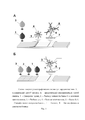
Схематичное изображение тест-полоски, основанной на данном принципе, представлено на рис. 1А. Тест-полоска для ИХ-серодиагностики с традиционной схемой включает следующий набор компонентов:A schematic representation of a test strip based on this principle is shown in Fig. 1A. The test strip for IC serodiagnostics with a traditional scheme includes the following set of components:
• начальная мембрана, абсорбирующая образец, на которую наносится анализируемая проба;• initial membrane, which absorbs the sample, on which the analyzed sample is applied;
• подложка под конъюгат, на которую наносится конъюгат маркера с рецепторными молекулами (антивидовыми антителами, белками A/G и пр.);• a substrate for the conjugate, on which the conjugate of the marker with receptor molecules (anti-species antibodies, proteins A / G, etc.) is applied;
• рабочей мембраны с иммобилизованным антигеном патогена;• working membrane with an immobilized pathogen antigen;
• конечной абсорбирующей мембраны, впитывающей жидкость, протекающую через рабочую мембрану.• a final absorbent membrane that absorbs liquid flowing through the working membrane.
Процедура анализа в данной системе сводится к добавлению на один и тот же участок начальной абсорбирующей мембраны пробы и разбавляющего буфера и, через 10-15 мин, регистрацию результатов анализа.The analysis procedure in this system is reduced to the addition of a sample and a dilution buffer to the same section of the initial absorbent membrane and, after 10-15 minutes, the registration of the analysis results.
Данная схема ИХ-серодиагностики предполагает взаимодействие всех иммуноглобулинов, содержащихся в пробе, с реагентом для связывания иммуноглобулинов. С антигеном же взаимодействует только небольшая доля специфичных к нему иммуноглобулинов, которая обычно не превышает 1% от общей концентрации иммуноглобулинов в крови. Таким образом, сигнал ИХ-анализа (интенсивность окраски аналитической зоны) в традиционной схеме зависит не только от концентрации специфических антител, но и от общего содержания иммуноглобулинов в пробе, что не является диагностически значимым параметром. Конкуренция между специфическими и общими иммуноглобулинами за связывание с иммуноглобулин-связывающим белком негативно сказывается на достоверности диагностики. Поэтому иммунохроматография зачастую уступает по чувствительности альтернативным методам серодиагностики (Sotnikov, D.V., Zherdev, A.V., & Dzantiev, B.B. (2017). Theoretical and experimental comparison of different formats of immunochromatographic serodiagnostics. Sensors, 18(1), 36.).This scheme of IC-serodiagnostics assumes the interaction of all immunoglobulins contained in the sample with a reagent for binding immunoglobulins. Only a small proportion of immunoglobulins specific to it interacts with the antigen, which usually does not exceed 1% of the total concentration of immunoglobulins in the blood. Thus, the signal of IC-analysis (the intensity of the color of the analytical zone) in the traditional scheme depends not only on the concentration of specific antibodies, but also on the total content of immunoglobulins in the sample, which is not a diagnostically significant parameter. The competition between specific and general immunoglobulins for binding to the immunoglobulin-binding protein negatively affects the reliability of the diagnosis. Therefore, immunochromatography is often inferior in sensitivity to alternative methods of serodiagnostics ( Sotnikov, DV, Zherdev, AV, & Dzantiev, BB (2017). Theoretical and experimental comparison of different formats of immunochromatographic serodiagnostics. Sensors, 18 (1), 36. ).
Предлагаемый альтернативный способ ИХ-серодиагностики заключается в разделении стадий связывания специфических иммуноглобулинов с иммобилизованным антигеном и меченым иммуноглобулин-связывающим белком. Для последовательного связывания иммуноглобулинов и меченого реагента в аналитической зоне проба, раствор, содержащий меченый иммуноглобулин-связывающий белок и смывающий буфер последовательно наносятся на начальную мембрану тест-полоски как показано на рис. 1Б. На первой стадии специфические иммуноглобулины в пробе сначала взаимодействуют с антигеном в аналитической зоне, при этом не связавшиеся иммуноглобулины увлекаются потоком жидкости и вымываются из реакционной зоны. На второй стадии иммуноглобулины, связавшиеся в аналитической зоне, проявляются посредством взаимодействия с меченым иммуноглобулин-связывающим белком. Смывающий буфер необходим для поддержания постоянного потока жидкости через мембраны теста. Состав тест-полоски отличается от традиционного только отсутствием подложки под конъюгат. Продолжительность анализа составляет 10-15 мин.The proposed alternative method of IH-serodiagnostics consists in separating the stages of binding of specific immunoglobulins to the immobilized antigen and labeled immunoglobulin-binding protein. For sequential binding of immunoglobulins and a labeled reagent in the assay zone, a sample, a solution containing a labeled immunoglobulin-binding protein and a wash buffer are sequentially applied to the initial membrane of the test strip as shown in Fig. 1B. At the first stage, specific immunoglobulins in the sample first interact with the antigen in the analytical zone, while the unbound immunoglobulins are carried away by the fluid flow and washed out from the reaction zone. In the second stage, the immunoglobulins bound in the assay area are manifested through interaction with a labeled immunoglobulin-binding protein. A washout buffer is required to maintain a constant flow of liquid across the dough membranes. The composition of the test strip differs from the traditional one only in the absence of a substrate for the conjugate. The analysis duration is 10-15 minutes.
Преимущество предлагаемого подхода продемонстрировано на примере ИХ-системы для определения специфических антител против рекомбинантного антигена P24 вируса лейкоза крупного рогатого скота.The advantage of the proposed approach is demonstrated by the example of an IC-system for the determination of specific antibodies against the recombinant antigen P24 of the bovine leukemia virus.
ПримерExample
Для формирования тест-систем использовали набор мембран компании Advanced Microdevices (Индия), включающий рабочую мембрану CNPC15, подложку под конъюгат PT-R5 (только в тест-системе с традиционным форматом), мембрану для нанесения образца GFB-R4 и конечную адсорбирующую мембрану CFSP 223000 Millipore (США).For the formation of test systems, a set of membranes from Advanced Microdevices (India) was used, including a working membrane CNPC15, a substrate for the PT-R5 conjugate (only in a test system with a traditional format), a membrane for applying the sample GFB-R4, and a final CFSP 223000 adsorption membrane. Millipore (USA).
Для формирования аналитической зоны использовали антиген P24 производства РГП «Национальный центр биотехнологии» Комитета науки Министерства образования и науки Республики Казахстан. На 1 см полосы наносили 2 мкл раствора антигена (1 мг/мл в 100 мМ Na - цитратном буфере, рН 6). Конъюгат коллоидного золота (средний диаметр частиц 25 нм) со стрептококковым белком G синтезировали при концентрации белка 10 мкг/мл и оптической плотности золота D520=1. Конъюгат очищали от не связавшегося белка посредством центрифугирования при 10000 g и концентрировали до D520=20. Для тест-системы в традиционном формате (рис. 1А) конъюгат наносили на подложку в объеме 15 мкл на 1 см полосы. В тест-системе в предложенном альтернативном формате (рис. 1Б) конъюгат использовали в жидком виде, нанося 5 мкл конъюгата D520=20 на начальную адсорбирующую мембрану непосредственно после добавления анализируемой пробы. Для нанесения реагентов использовали диспенсер «IsoFlow» фирмы «Imagene Technology» (США). Листы мембран с нанесенными иммунореагентами нарезали на индивидуальные тест-полоски шириной 3,5 мм.To form the analytical zone, we used the P24 antigen produced by the Republican State Enterprise “National Center for Biotechnology” of the Science Committee of the Ministry of Education and Science of the Republic of Kazakhstan. 2 μl of antigen solution (1 mg / ml in 100 mM Na - citrate buffer, pH 6) was applied to 1 cm of the band. The conjugate of colloidal gold (average particle diameter 25 nm) with streptococcal protein G was synthesized at a protein concentration of 10 μg / ml and an optical density of gold D 520 = 1. The conjugate was purified from unbound protein by centrifugation at 10000 g and concentrated to D 520 = 20. For the test system in the traditional format (Fig.1A), the conjugate was applied to the substrate in a volume of 15 μL per 1 cm of the strip. In the test system in the proposed alternative format (Fig. 1B), the conjugate was used in liquid form by applying 5 μL of the D 520 = 20 conjugate onto the initial adsorbing membrane immediately after adding the analyzed sample. The reagents were applied using an IsoFlow dispenser from Imagene Technology (USA). Membrane sheets coated with immunoreagents were cut into individual test strips 3.5 mm wide.
Иммунохроматографический анализ проводили при комнатной температуре. В тест-системе с традиционным форматом анализа (рис. 1А) на начальную адсорбирующую мембрану наносили 10 мкл сыворотки крови коровы зараженной вирусом лейкоза КРС. Затем в то же место наносили 50 мкл разбавляющего раствора (50 мМ К-фосфатный буфер, рН 7,4, с 0,1 М NaCl и 1% Tween-20). Фиксацию результата тестирования осуществляли через 10 мин.Immunochromatographic analysis was performed at room temperature. In a test system with a traditional assay format (Fig. 1A), 10 μL of blood serum from a cow infected with bovine leukemia virus was applied to the initial adsorbing membrane. Then, 50 μl of a dilution solution (50 mM K-phosphate buffer, pH 7.4, with 0.1 M NaCl and 1% Tween-20) was applied to the same place. The test results were recorded after 10 minutes.
В тест-системе с предложенным альтернативным форматом анализа (рис. 1Б) на начальную адсорбирующую мембрану наносили 5 мкл сыворотки крови коровы зараженной вирусом лейкоза КРС. Далее примерно на 1 см ниже места нанесения сыворотки наносили 5 мкл конъюгата белка G с коллоидным золотом D520=20. Затем примерно на 1 см ниже места нанесения конъюгата наносили 50 мкл смывающего раствора (50 мМ К-фосфатный буфер, рН 7,4, с 0,1 М NaCl и 1% Tween-20). Фиксацию результата тестирования осуществляли через 10 мин.In the test system with the proposed alternative analysis format (Fig. 1B), 5 μL of blood serum from a cow infected with bovine leukemia virus was applied to the initial adsorbing membrane. Further, about 1 cm below the place of serum application, 5 μl of protein G conjugate with colloidal gold D520 = 20 was applied. Then, about 1 cm below the site of application of the conjugate, 50 μl of a wash solution (50 mM K-phosphate buffer, pH 7.4, with 0.1 M NaCl and 1% Tween-20) was applied. The test results were recorded after 10 minutes.
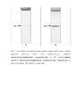
Пример результата тестирования сыворотки крови коровы с диагнозом «лейкоз» в двух форматах анализа представлен на рис. 2. Представленные результаты демонстрируют повышение интенсивности сигнала в аналитической зоне в предложенном способе серодиагностики и, таким образом, повышение достоверности анализа.An example of the test result of blood serum of a cow diagnosed with leukemia in two analysis formats is shown in Fig. 2. The presented results demonstrate an increase in the signal intensity in the analytical zone in the proposed method of serodiagnostics and, thus, an increase in the reliability of the analysis.
Claims (1)
Priority Applications (1)
| Application Number | Priority Date | Filing Date | Title |
|---|---|---|---|
| RU2020131639A RU2741199C1 (en) | 2020-09-25 | 2020-09-25 | Method of immunochromatographic serodiagnogy with sequential addition of reagents |
Applications Claiming Priority (1)
| Application Number | Priority Date | Filing Date | Title |
|---|---|---|---|
| RU2020131639A RU2741199C1 (en) | 2020-09-25 | 2020-09-25 | Method of immunochromatographic serodiagnogy with sequential addition of reagents |
Publications (1)
| Publication Number | Publication Date |
|---|---|
| RU2741199C1 true RU2741199C1 (en) | 2021-01-22 |
Family
ID=74213044
Family Applications (1)
| Application Number | Title | Priority Date | Filing Date |
|---|---|---|---|
| RU2020131639A RU2741199C1 (en) | 2020-09-25 | 2020-09-25 | Method of immunochromatographic serodiagnogy with sequential addition of reagents |
Country Status (1)
| Country | Link |
|---|---|
| RU (1) | RU2741199C1 (en) |
Citations (2)
| Publication number | Priority date | Publication date | Assignee | Title |
|---|---|---|---|---|
| RU2532352C2 (en) * | 2012-06-28 | 2014-11-10 | Федеральное государственное бюджетное учреждение науки Институт биохимии имени А.Н. Баха РАН Российской академии наук (ИНБИ РАН) | Method of carrying out immunochromatographic analysis for serodiagnostics |
| RU2545909C2 (en) * | 2013-03-19 | 2015-04-10 | Федеральное государственное бюджетное учреждение науки Институт биохимии имени А.Н. Баха РАН Российской академии наук (ИНБИ РАН) | Method of immunochromatographic determination of specific antibodies |
-
2020
- 2020-09-25 RU RU2020131639A patent/RU2741199C1/en active
Patent Citations (2)
| Publication number | Priority date | Publication date | Assignee | Title |
|---|---|---|---|---|
| RU2532352C2 (en) * | 2012-06-28 | 2014-11-10 | Федеральное государственное бюджетное учреждение науки Институт биохимии имени А.Н. Баха РАН Российской академии наук (ИНБИ РАН) | Method of carrying out immunochromatographic analysis for serodiagnostics |
| RU2545909C2 (en) * | 2013-03-19 | 2015-04-10 | Федеральное государственное бюджетное учреждение науки Институт биохимии имени А.Н. Баха РАН Российской академии наук (ИНБИ РАН) | Method of immunochromatographic determination of specific antibodies |
Non-Patent Citations (4)
| Title |
|---|
| BARSHEVSKAYA L.V. et al. Triple Immunochromatographic System for Simultaneous Serodiagnosis of Bovine Brucellosis, Tuberculosis, and Leukemia / Biosensors 2019, 9, 115; 10 pages. * |
| SOTNIKOV D.V. et al. Theoretical and Experimental Comparison of Different Formats of Immunochromatographic Serodiagnostics / Sensors 2018, 18, 36, 15 pages. * |
| СОТНИКОВ Д.В. ОПРЕДЕЛЕНИЕ СПЕЦИФИЧЕСКИХ АНТИТЕЛ МЕТОДОМ ИММУНОХРОМАТОГРАФИИ: КОЛИЧЕСТВЕННЫЕ ЗАКОНОМЕРНОСТИ И ПРАКТИЧЕСКИЕ ПРИЛОЖЕНИЯ / Диссерт. на соис. уч. степ. к.х.н., Москва, 2016. * |
| СОТНИКОВ Д.В. ОПРЕДЕЛЕНИЕ СПЕЦИФИЧЕСКИХ АНТИТЕЛ МЕТОДОМ ИММУНОХРОМАТОГРАФИИ: КОЛИЧЕСТВЕННЫЕ ЗАКОНОМЕРНОСТИ И ПРАКТИЧЕСКИЕ ПРИЛОЖЕНИЯ / Диссерт. на соис. уч. степ. к.х.н., Москва, 2016. SOTNIKOV D.V. et al. Theoretical and Experimental Comparison of Different Formats of Immunochromatographic Serodiagnostics / Sensors 2018, 18, 36, 15 pages. BARSHEVSKAYA L.V. et al. Triple Immunochromatographic System for Simultaneous Serodiagnosis of Bovine Brucellosis, Tuberculosis, and Leukemia / Biosensors 2019, 9, 115; 10 pages. * |
Similar Documents
| Publication | Publication Date | Title |
|---|---|---|
| JP6742978B2 (en) | Signal amplification in lateral flow and related immunoassays | |
| US20050272106A1 (en) | Methods and kits for detection of multiple pathogens | |
| US9910036B2 (en) | Method and device for combined detection of viral and bacterial infections | |
| US11714084B2 (en) | Method and associated device for rapid detection of target biomolecules with enhanced sensitivity | |
| US8916389B2 (en) | Sensor element for SPR measurement | |
| JP2019507890A5 (en) | ||
| US20210072235A1 (en) | Method for diagnosing tuberculosis | |
| KR20160120675A (en) | Rapid Quantitative Diagnostic Kit | |
| JP4653574B2 (en) | Method for measuring hemoglobin A1c | |
| RU2741199C1 (en) | Method of immunochromatographic serodiagnogy with sequential addition of reagents | |
| JP6858784B2 (en) | Subtractive Immunoassay Assay Method and Lateral Flow Immunochromatography Assay Strip for Performing the Method | |
| RU2532352C2 (en) | Method of carrying out immunochromatographic analysis for serodiagnostics | |
| RU2530560C2 (en) | Method of obtaining intermolecular conjugate for immunochromatographic determination of specific antibodies | |
| RU2545909C2 (en) | Method of immunochromatographic determination of specific antibodies | |
| JP7417231B2 (en) | Immunochromatographic method and kit for detecting anti-interferon gamma antibodies | |
| RU2753237C1 (en) | Method for immunochromatographic analysis for serodiagnostics with a combined antibody binding pattern | |
| KR102314073B1 (en) | Composition for detecting target material comprising ficolin-1 and method for detecting using the same | |
| Byzova et al. | Manufacturing lateral flow tests for tuberculosis diagnosis: choosing a reactants completion and sensing regime | |
| JP2022153079A (en) | Method and kit for detecting bacteria in blood and urine from urine specimen | |
| EP4320438A1 (en) | Multiplex immunoassay for the detection of mycoplasma bovis infection | |
| KR100524821B1 (en) | The development of the detection method using Complement 1 q for protein chips | |
| TR2022011905A2 (en) | LATERAL FLOW ASSAY RAPID DIAGNOSTIC KIT WITH A NEW CONTROL LINE APPROACH | |
| WO2004016804A2 (en) | Assay for detection of antigen in bodily fluid |
Legal Events
| Date | Code | Title | Description |
|---|---|---|---|
| QB4A | Licence on use of patent |
Free format text: LICENCE FORMERLY AGREED ON 20210520 Effective date: 20210520 |