RU2532352C2 - Method of carrying out immunochromatographic analysis for serodiagnostics - Google Patents
Method of carrying out immunochromatographic analysis for serodiagnostics Download PDFInfo
- Publication number
- RU2532352C2 RU2532352C2 RU2012127077/15A RU2012127077A RU2532352C2 RU 2532352 C2 RU2532352 C2 RU 2532352C2 RU 2012127077/15 A RU2012127077/15 A RU 2012127077/15A RU 2012127077 A RU2012127077 A RU 2012127077A RU 2532352 C2 RU2532352 C2 RU 2532352C2
- Authority
- RU
- Russia
- Prior art keywords
- antibodies
- specific antibodies
- sample
- test
- antigen
- Prior art date
Links
Images


Abstract
Description
Изобретение относится к иммунологии и медицинской диагностике и представляет собой способ проведения иммунохроматографического анализа для серодиагностики. Наличие в сыворотке крови антител, специфичных к возбудителю определенного заболевания, или иному антигену, например, такому как аллерген, является эффективным критерием, позволяющим с высокой достоверностью диагностировать соответствующее инфекционное заболевание или аллергию (Резникова Л.С., Эпштейн-Литвак Р.В., Леви М.И. / Серологические методы исследования при диагностике инфекционных болезней - М., Медгиз, 1962; Медицинская микробиология, вирусология и иммунология: Учебник для студентов медицинских ВУЗов / Под ред. А.А Воробьева. - М.: ООО «Медицинское информационное агенство», 2006). В случае диагностики инфекционных заболеваний преимуществами данного подхода по сравнению с непосредственным выявлением и идентификацией возбудителя является определенность при выборе тестируемой пробы (сыворотка крови, тогда как возбудитель может на данной стадии инфекции преимущественно локализоваться в самых разных органах и тканях), а также возможность быстрой детекции достаточно высокого уровня антител, индуцированного контактом с антигеном, в то время как для выявления антигена могут потребоваться довольно продолжительные стадии доращивания до достижения им регистрируемой концентрации. Хотя в настоящее время активно используются как микробиологические, так и иммунологические способы диагностики инфекционных заболеваний, для проведения массового первичного скрининга оптимально иммунологическое определение наличия в сыворотке крови специфических антител (серодиагностика), которое может быть реализовано с высокой экспрессностью и производительностью. Особенный интерес вызывают серодиагностические подходы в тех случаях, когда в силу особенностей роста микроорганизмов получение результатов микробиологического тестирования может потребовать значительного времени. В случае диагностики аллергии антитела против определенного аллергена являются главным маркером данного функционального расстройства (IgE антитела в случае аллергии немедленного типа и IgG антитела в случае аллергии замедленного типа), поэтому методы лабораторной диагностики аллергии зачастую сводятся к определению антител, или выявлению реакции организма, связанной с наличием данных антител (как в случае провокационной пробы) (Паттерсон Р., Грэммер Л.К., Гринбергер П.А. Аллерические болезни: диагностика и лечение / Перевод с англ., под ред. акад. РАМН А.Г. Чучалина (гл. ред.), чл.-корр. РАМН И.С. Гущина (отв. ред.). - М.: ГЭОТАР-Медиа, 2000). Эти причины определяют значительный интерес к серодиагностическим методам.The invention relates to immunology and medical diagnostics and is a method for conducting immunochromatographic analysis for serodiagnostics. The presence in the blood serum of antibodies specific for the causative agent of a certain disease, or other antigen, for example, such as an allergen, is an effective criterion that makes it possible to diagnose the corresponding infectious disease or allergy with high reliability (Reznikova L.S., Epstein-Litvak R.V. , Levi M.I. / Serological research methods for the diagnosis of infectious diseases - M., Medgiz, 1962; Medical Microbiology, Virology and Immunology: A Textbook for Students of Medical Universities / Edited by A.A. Vorobyov. - M .: OOO “Medical News Agency”, 2006). In the case of the diagnosis of infectious diseases, the advantages of this approach compared to the direct identification and identification of the pathogen are certainty when choosing a test sample (blood serum, while the pathogen can be mainly localized in various organs and tissues at this stage of infection), as well as the possibility of quick detection is sufficient high levels of antibodies induced by contact with the antigen, while quite long tadii of growing until they reach a registered concentration. Although both microbiological and immunological methods for diagnosing infectious diseases are currently actively used, for the primary primary screening, the immunological determination of the presence of specific antibodies in the blood serum (serodiagnosis) is optimal, which can be implemented with high expressivity and productivity. Of particular interest are serodiagnostic approaches in those cases where, due to the characteristics of the growth of microorganisms, obtaining the results of microbiological testing may require considerable time. In the case of allergy diagnosis, antibodies against a particular allergen are the main marker of this functional disorder (IgE antibodies in the case of immediate allergies and IgG antibodies in the case of delayed allergies), therefore, laboratory allergy diagnosis methods often come down to detecting antibodies, or identifying the body's reaction associated with the presence of these antibodies (as in the case of a provocative test) (Patterson R., Grammer L.K., Grinberger P.A. Allergic diseases: diagnosis and treatment / Translation from English, ed. Academician of the RAMS A.G. Chuchalina (Ch. ed.), Corresponding Member of the RAMS I.S. Gushchina (ed. Ed.) - M: GEOTAR-Media, 2000). These reasons determine a significant interest in serodiagnostic methods.
Благодаря экспрессности, достаточно высокой чувствительности и специфичности серологические тесты незаменимы при массовых обследованиях. Кроме того, гуморальный иммунный ответ отражает активный инфекционный процесс, и поэтому результаты иммунохимического тестирования достоверно отражают именно случаи заболевания, дискриминируя их от бактерионосительства.Due to the rapidity, sufficiently high sensitivity and specificity, serological tests are indispensable for mass examinations. In addition, the humoral immune response reflects an active infectious process, and therefore the results of immunochemical testing reliably reflect precisely the cases of the disease, discriminating them against bacterial carriage.
Иммунохимический анализ может быть реализован в различных форматах. Однако, поскольку для массовых обследований первоочередное значение имеют скорость и производительность тестирования, в данной ситуации несомненными преимуществами обладает иммунохроматографический анализ, для которого все необходимые реагенты предварительно нанесены на мембранные компоненты тест-полоски и ее контакт с тестируемой пробой непосредственно инициирует движение фронта жидкости по мембранам, протекание специфических реакций и формирование иммунных комплексов, которые благодаря включению в их состав окрашенного маркера могут детектироваться визуально или с помощью оптического детектора (Рис.1).Immunochemical analysis can be implemented in various formats. However, since the speed and productivity of testing are of primary importance for mass examinations, in this situation immunochromatographic analysis has undoubted advantages, for which all the necessary reagents are preliminarily applied to the membrane components of the test strip and its contact with the test sample directly initiates the movement of the liquid front across the membranes, the course of specific reactions and the formation of immune complexes, which, thanks to the inclusion of a colored mark Parameters can be detected visually or with an optical detector (Fig. 1).
Применительно к серодиагностике (определению антител, специфичных к определенному антигену или группе антигенов) общая схема иммунохроматографии заключается в следующем.In relation to serodiagnosis (determination of antibodies specific to a particular antigen or group of antigens), the general scheme of immunochromatography is as follows.
Проба, потенциально содержащая специфические антитела, при контакте с тест-полоской под действием капиллярных сил перемещается вдоль тест-полоски. При этом она вначале взаимодействует с окрашенными частицами, на поверхности которых адсорбирован (конъюгирован) компонент, предназначенный для связывания с антителами. Часто в качестве такового компонента выбирают антивидовые антитела (иммуноглобулины, выделенные из сыворотки животного, иммунизированного препаратом иммуноглобулинов человека или другого организма, для которого проводится серодиагностика), белок A из Staphylococcus aureus, белок G из Streptococcus spp. или другие реагенты для связывания антител. В качестве метки наиболее часто используется коллоидное золото, а в качестве метода конъюгации - простая физическая адсорбция белка на поверхности с последующим отделением неадсорбированных молекул с помощью осаждения частиц коллоидного золота центрифугированием. Затем фронт жидкости преодолевает аналитическую (тестовую) зону, которая представляет собой участок мембраны с иммобилизованным антигеном (нативным или специально модифицированным для эффективной сорбции), в результате взаимодействия с которым формируются комплексы из молекул антигена/антигенов, специфических к ним антител и конъюгата с коллоидным золотом. Степень связывания маркера с иммобилизованным антигеном и, соответственно, интенсивность окрашивания мембраны определяются концентрацией специфических антител в пробе. Для проверки качества реагентов и сохранения функциональности тест-системы используется расположенная далее контрольная зона, в которой компонент, сорбированный на окрашенной частице, связывается с соответствующим иммобилизованным на мембране реагентом.A sample potentially containing specific antibodies, when in contact with the test strip under the action of capillary forces, moves along the test strip. Moreover, it first interacts with colored particles, on the surface of which a component intended for binding to antibodies is adsorbed (conjugated). Often, anti-species antibodies (immunoglobulins isolated from the serum of an animal immunized with a human immunoglobulin preparation or another organism for which serodiagnosis is performed) are chosen as such a component, protein A from Staphylococcus aureus, protein G from Streptococcus spp. or other reagents for binding antibodies. Colloidal gold is most often used as a label, and as a conjugation method, it is simple physical adsorption of the protein on the surface, followed by separation of the non-adsorbed molecules by precipitation of colloidal gold particles by centrifugation. Then, the liquid front overcomes the analytical (test) zone, which is a portion of the membrane with an immobilized antigen (native or specially modified for effective sorption), as a result of interaction with which complexes of antigen / antigen molecules, specific antibodies and conjugate with colloidal gold are formed . The degree of marker binding to the immobilized antigen and, accordingly, the intensity of membrane staining are determined by the concentration of specific antibodies in the sample. To check the quality of the reagents and maintain the functionality of the test system, the control zone located below is used, in which the component adsorbed on the colored particle is bound to the corresponding reagent immobilized on the membrane.
Ниже представлена информация о методике, реализуемой в тест-системе «TB-Check-1» фирмы «Vedalab» (Франция) - см. http://http://www.sanitamedikal.com/Assets/MD_220002_m3_TB_l408_c.pdf. Данная методика определения антител к возбудителю туберкулеза рассматривается в настоящей заявке в качестве прототипной.Below is information about the methodology implemented in the TB-Check-1 test system of Vedalab (France) - see http: // http: //www.sanitamedikal.com/Assets/MD_220002_m3_TB_l408_c.pdf. This method of determining antibodies to the causative agent of tuberculosis is considered in this application as a prototype.
Метод основан на комбинации антител к иммуноглобулинам человека, конъюгированных с хромогеном, и высокоочищенного БЦЖ-белка. При прохождении исследуемого образца через адсорбционную зону тестового устройства конъюгат, содержащий меченые антитела, связывается с IgG, образуя комплекс «антиген-антитело». Этот комплекс взаимодействует с высокоочищенным БЦЖ-белком в тестовой зоне устройства и, если концентрация специфического IgG к возбудителю туберкулеза превышает 350 Е/мл, образует окрашенную полосу. При низкой концентрации антител окрашенная полоса в тестовой зоне не образуется. Несвязавшийся конъюгат взаимодействует с реагентом в контрольной зоне тестового устройства, образуя окрашенную полосу, что указывает на правильное проведение теста. Процедура исследования: заполнить одноразовую пипетку сывороткой или плазмой и внести 1 каплю в окно для пробы тестового устройства. Добавить в окно для пробы 5-6 полных капель разбавляющего раствора (дилюента). Через 10-15 мин произвести учет результатов.The method is based on a combination of antibodies to human immunoglobulins conjugated to a chromogen and a highly purified BCG protein. When the test sample passes through the adsorption zone of the test device, the conjugate containing labeled antibodies binds to IgG to form an antigen-antibody complex. This complex interacts with a highly purified BCG protein in the test zone of the device and, if the concentration of specific IgG to the tuberculosis pathogen exceeds 350 U / ml, it forms a colored band. At a low concentration of antibodies, a colored band does not form in the test area. The unbound conjugate interacts with the reagent in the control zone of the test device, forming a colored strip, which indicates the correct conduct of the test. Test procedure: fill in a disposable pipette with serum or plasma and add 1 drop to the window for testing the test device. Add 5-6 full drops of dilution solution (diluent) to the sample window. After 10-15 minutes, record the results.
Одной из наиболее важных задач в иммунохроматографии является повышение чувствительности анализа. Применительно к серодиагностике это означает минимизацию ложноотрицательных результатов теста в случае, если антитела в пробе содержатся в диагностически значимых концентрациях, но ниже предела детекции метода. Заявителями предлагается подход для решения этой задачи, основанный на добавлении в пробу дополнительного количества специфических антител в концентрациях ниже, чем предел детекции метода при использовании разбавляющего раствора, не содержащего специфических антител. В этом случае суммарная концентрация антител в пробе и добавленных антител может превысить предел детекции и проба будет диагностирована как положительная. Таким образом, повышается чувствительность метода за счет смещения диапазона определяемых концентраций в область более низких значений.One of the most important tasks in immunochromatography is to increase the sensitivity of the analysis. With regard to serodiagnostics, this means minimizing false negative test results if antibodies in the sample are contained in diagnostically significant concentrations, but below the detection limit of the method. Applicants have proposed an approach to solving this problem, based on adding an additional amount of specific antibodies to the sample at concentrations lower than the detection limit of the method using a dilution solution that does not contain specific antibodies. In this case, the total concentration of antibodies in the sample and the added antibodies may exceed the detection limit and the sample will be diagnosed as positive. Thus, the sensitivity of the method increases due to a shift in the range of determined concentrations to the region of lower values.
Предложенный подход был реализован заявителями для серодиагностики туберкулеза с использованием антигена 38 кДа (Ag78, antigen 5, PhoS, Rv0934) M. tuberculosis. Ниже представлено описание способа получения тест-полосок и проведения иммунохроматографического анализа, а также полученные результаты.The proposed approach was implemented by applicants for serodiagnosis of tuberculosis using the 38 kDa antigen (Ag78, antigen 5, PhoS, Rv0934) M. tuberculosis. The following is a description of the method of obtaining test strips and immunochromatographic analysis, as well as the results.
Пример:Example:
Для формирования тест-системы использовали набор мембран «mdi Easypack» фирмы «Advanced Microdevices» (Индия), включающий рабочую мембрану CNPC-SN12 L2-P25 (размер пор 15 мкм), подложку под конъюгат PT-R5, мембрану для нанесения образца GFB-R4, адсорбирующую мембрану AP 045 и ламинирующую защитную пленку МТ-1.To form the test system, we used a set of mdi Easypack membranes from Advanced Microdevices (India), including a CNPC-SN12 L2-P25 working membrane (
На мембраны были нанесены следующие реагенты:The following reagents were applied to the membranes:
1. Рекомбинантный антиген 38 кДа М. tuberculosis (Rv0934), фирма «Arista Biologicals Inc.» (США), кат. № AGMTB-0220.1. Recombinant antigen 38 kDa M. tuberculosis (Rv0934), the company "Arista Biologicals Inc." (USA), cat. No. AGMTB-0220.
2. Конъюгат коллоидного золота со средним диаметром частиц 30 нм и рекомбинантного антигена 38 кДа М. tuberculosis.2. The conjugate of colloidal gold with an average particle diameter of 30 nm and a recombinant antigen of 38 kDa M. tuberculosis.
3. Моноклональные антитела НТМ81 против рекомбинантного антигена 38 кДа М. tuberculosis, Центр молекулярной диагностики и терапии, Москва (Россия).3. Monoclonal antibodies NTM81 against a 38 kD recombinant antigen M. tuberculosis, Center for Molecular Diagnostics and Therapy, Moscow (Russia).
Для формирования аналитической зоны использовали антиген 38 кДа, контрольной зоны - антитела против антигена 38 кДа. На 1 см полосы наносили 2 мкл раствора антигена (1,0 мг/мл в 50 мМ фосфатном буфере, pH 7,4) и 2 мкл раствора антител (0,5 мг/мл в том же буфере). Конъюгат коллоидного золота с антигеном 38 кДа наносили в разведении, соответствующем D520=2,0, в объеме 8 мкл на 1 см полосы. Для нанесения реагентов использовали диспенсер «IsoFlow» фирмы «Imagene Technology)) (США). Листы мембран с нанесенными иммунореагентами нарезали на индивидуальные тест-полоски шириной 4 мм.To form the analytical zone, 38 kDa antigen was used, and the control zone was used against 38 kDa antigen. 2 μl of antigen solution (1.0 mg / ml in 50 mM phosphate buffer, pH 7.4) and 2 μl of antibody solution (0.5 mg / ml in the same buffer) were applied to 1 cm of strip. A conjugate of colloidal gold with a 38 kDa antigen was applied in a dilution corresponding to D 520 = 2.0, in a volume of 8 μl per 1 cm of the strip. For the application of reagents, an IsoFlow dispenser (Imagene Technology)) (USA) was used. Membrane sheets coated with immunoreagents were cut into individual test strips 4 mm wide.
Иммунохроматографический анализ проводили при комнатной температуре. Тест-полоску погружали в пробу на 1 мин в вертикальном положении, а затем извлекали и помещали на горизонтальную поверхность. Детекцию связывания коллоидного золота осуществляли через 10 мин визуально или получая цифровое изображение тест-полоски с помощью сканера и количественно оценивая интенсивность окраски аналитической зоны с помощью программы «Nonlinear Dynamics TotalLab TL120 v2009».Immunochromatographic analysis was performed at room temperature. The test strip was immersed in the sample for 1 min in a vertical position, and then removed and placed on a horizontal surface. Colloidal gold binding was detected after 10 min visually or by obtaining a digital image of the test strip using a scanner and quantitatively assessing the color intensity of the analytical zone using the Nonlinear Dynamics TotalLab TL120 v2009 program.
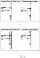
Путем тестирования растворов, содержащих фиксированные концентрации специфических антител (моноклональных антител НТМ81) построена калибровочная кривая и определен предел визуальной детекции метода (Рис.2). Затем были протестированы сыворотки крови от пациента, зараженного туберкулезом и от здорового донора (Рис.3, A), в этом случае положительных результатов тестирования не наблюдалось. После добавления в пробы дополнительного количества специфических антител в концентрации, соответствующей пределу визуальной детекции (5 мкг/мл), в сыворотке больного пациента наблюдается отчетливое окрашивание аналитической зоны (Рис.3, Б). Таким образом, предложенный подход позволил избежать ложноотрицательного результата теста.By testing solutions containing fixed concentrations of specific antibodies (NTM81 monoclonal antibodies), a calibration curve was constructed and the limit of visual detection of the method was determined (Fig. 2). Then, blood serum was tested from a patient infected with tuberculosis and from a healthy donor (Fig. 3, A), in this case, no positive test results were observed. After adding an additional amount of specific antibodies to the samples at a concentration corresponding to the limit of visual detection (5 μg / ml), a clear staining of the analytical zone is observed in the serum of the patient (Fig. 3, B). Thus, the proposed approach avoided a false negative test result.
Рис.1. Принцип иммунохроматографического анализа.Fig. 1. The principle of immunochromatographic analysis.
Рис.2. Калибровочная кривая определения специфических антител против антигена 38 кДа М. tuberculosis методом иммунохроматографии.Fig. 2. Calibration curve for the determination of specific antibodies against 38 kDa antigen of M. tuberculosis by immunochromatography.
Рис.3. Тестирование сывороток крови больного туберкулезом и здорового донора без добавления дополнительного количества специфических антител (А) и после добавления специфических антител в концентрации 5 мкг/мл (Б).Fig. 3. Testing the blood serum of a patient with tuberculosis and a healthy donor without adding an additional amount of specific antibodies (A) and after adding specific antibodies at a concentration of 5 μg / ml (B).
Claims (1)
Priority Applications (1)
| Application Number | Priority Date | Filing Date | Title |
|---|---|---|---|
| RU2012127077/15A RU2532352C2 (en) | 2012-06-28 | 2012-06-28 | Method of carrying out immunochromatographic analysis for serodiagnostics |
Applications Claiming Priority (1)
| Application Number | Priority Date | Filing Date | Title |
|---|---|---|---|
| RU2012127077/15A RU2532352C2 (en) | 2012-06-28 | 2012-06-28 | Method of carrying out immunochromatographic analysis for serodiagnostics |
Publications (2)
| Publication Number | Publication Date |
|---|---|
| RU2012127077A RU2012127077A (en) | 2014-02-10 |
| RU2532352C2 true RU2532352C2 (en) | 2014-11-10 |
Family
ID=50031668
Family Applications (1)
| Application Number | Title | Priority Date | Filing Date |
|---|---|---|---|
| RU2012127077/15A RU2532352C2 (en) | 2012-06-28 | 2012-06-28 | Method of carrying out immunochromatographic analysis for serodiagnostics |
Country Status (1)
| Country | Link |
|---|---|
| RU (1) | RU2532352C2 (en) |
Cited By (3)
| Publication number | Priority date | Publication date | Assignee | Title |
|---|---|---|---|---|
| RU2712249C1 (en) * | 2018-11-28 | 2020-01-27 | Общество С Ограниченной Ответственностью "Синтэко-Групп" | Quantitative immunochromatographic analysis method |
| RU2741199C1 (en) * | 2020-09-25 | 2021-01-22 | Федеральное государственное учреждение"Федеральный исследовательский центр "Фундаментальные основы биотехнологии" Российской академии наук" (ФИЦ Биотехнологии РАН) | Method of immunochromatographic serodiagnogy with sequential addition of reagents |
| RU2753237C1 (en) * | 2020-10-14 | 2021-08-12 | Федеральное государственное учреждение «Федеральный исследовательский центр «Фундаментальные основы биотехнологии» Российской академии наук» (ФИЦ Биотехнологии РАН) | Method for immunochromatographic analysis for serodiagnostics with a combined antibody binding pattern |
Citations (4)
| Publication number | Priority date | Publication date | Assignee | Title |
|---|---|---|---|---|
| US20060105400A1 (en) * | 2002-08-29 | 2006-05-18 | Matthias Dettloff | Elisa kits for detecting collagenase 3 as a proenzyme and in an activated form in body fluids and cell culture supernatants |
| RU2395092C2 (en) * | 2008-09-17 | 2010-07-20 | Институт биохимии имени А.Н. Баха Российской академии наук | Method of determining antibodies to tuberculosis germ |
| WO2011068844A1 (en) * | 2009-12-01 | 2011-06-09 | Temple Douglas | Borrelia burgdorferi bacterial antigen diagnostic test using polymeric bait containing capture particles |
| RU2420740C1 (en) * | 2010-03-24 | 2011-06-10 | Федеральное Государственное Унитарное Предприятие "Государственный научно-исследовательский институт биологического приборостроения" (ФГУП Гос НИИ БП) | Method of immunoassay for analyte detection in sample |
-
2012
- 2012-06-28 RU RU2012127077/15A patent/RU2532352C2/en active IP Right Revival
Patent Citations (4)
| Publication number | Priority date | Publication date | Assignee | Title |
|---|---|---|---|---|
| US20060105400A1 (en) * | 2002-08-29 | 2006-05-18 | Matthias Dettloff | Elisa kits for detecting collagenase 3 as a proenzyme and in an activated form in body fluids and cell culture supernatants |
| RU2395092C2 (en) * | 2008-09-17 | 2010-07-20 | Институт биохимии имени А.Н. Баха Российской академии наук | Method of determining antibodies to tuberculosis germ |
| WO2011068844A1 (en) * | 2009-12-01 | 2011-06-09 | Temple Douglas | Borrelia burgdorferi bacterial antigen diagnostic test using polymeric bait containing capture particles |
| RU2420740C1 (en) * | 2010-03-24 | 2011-06-10 | Федеральное Государственное Унитарное Предприятие "Государственный научно-исследовательский институт биологического приборостроения" (ФГУП Гос НИИ БП) | Method of immunoassay for analyte detection in sample |
Cited By (3)
| Publication number | Priority date | Publication date | Assignee | Title |
|---|---|---|---|---|
| RU2712249C1 (en) * | 2018-11-28 | 2020-01-27 | Общество С Ограниченной Ответственностью "Синтэко-Групп" | Quantitative immunochromatographic analysis method |
| RU2741199C1 (en) * | 2020-09-25 | 2021-01-22 | Федеральное государственное учреждение"Федеральный исследовательский центр "Фундаментальные основы биотехнологии" Российской академии наук" (ФИЦ Биотехнологии РАН) | Method of immunochromatographic serodiagnogy with sequential addition of reagents |
| RU2753237C1 (en) * | 2020-10-14 | 2021-08-12 | Федеральное государственное учреждение «Федеральный исследовательский центр «Фундаментальные основы биотехнологии» Российской академии наук» (ФИЦ Биотехнологии РАН) | Method for immunochromatographic analysis for serodiagnostics with a combined antibody binding pattern |
Also Published As
| Publication number | Publication date |
|---|---|
| RU2012127077A (en) | 2014-02-10 |
Similar Documents
| Publication | Publication Date | Title |
|---|---|---|
| JP6742978B2 (en) | Signal amplification in lateral flow and related immunoassays | |
| AU2020233741B2 (en) | Method and device for combined detection of viral and bacterial infections | |
| US9933423B2 (en) | Method and device for combined detection of viral and bacterial infections | |
| US10379121B2 (en) | Method and device for combined detection of viral and bacterial infections | |
| EP2335072B1 (en) | Method and device for combined detection of viral and bacterial infections | |
| US8962260B2 (en) | Method and device for combined detection of viral and bacterial infections | |
| US20210389318A1 (en) | Lateral flow assays for differential isotype detection | |
| RU2532352C2 (en) | Method of carrying out immunochromatographic analysis for serodiagnostics | |
| US20210072235A1 (en) | Method for diagnosing tuberculosis | |
| RU2395092C2 (en) | Method of determining antibodies to tuberculosis germ | |
| RU2545909C2 (en) | Method of immunochromatographic determination of specific antibodies | |
| RU2741199C1 (en) | Method of immunochromatographic serodiagnogy with sequential addition of reagents | |
| IL274768B1 (en) | Method and device for discriminating between viral and bacterial infections |
Legal Events
| Date | Code | Title | Description |
|---|---|---|---|
| PD4A | Correction of name of patent owner | ||
| MM4A | The patent is invalid due to non-payment of fees |
Effective date: 20160629 |
|
| NF4A | Reinstatement of patent |
Effective date: 20170725 |