RU2515400C2 - Implant surgical drill - Google Patents
Implant surgical drill Download PDFInfo
- Publication number
- RU2515400C2 RU2515400C2 RU2012134550/14A RU2012134550A RU2515400C2 RU 2515400 C2 RU2515400 C2 RU 2515400C2 RU 2012134550/14 A RU2012134550/14 A RU 2012134550/14A RU 2012134550 A RU2012134550 A RU 2012134550A RU 2515400 C2 RU2515400 C2 RU 2515400C2
- Authority
- RU
- Russia
- Prior art keywords
- drill
- cutting
- mucous membrane
- section
- cutting blade
- Prior art date
Links
Images
Classifications
-
- A—HUMAN NECESSITIES
- A61—MEDICAL OR VETERINARY SCIENCE; HYGIENE
- A61C—DENTISTRY; APPARATUS OR METHODS FOR ORAL OR DENTAL HYGIENE
- A61C8/00—Means to be fixed to the jaw-bone for consolidating natural teeth or for fixing dental prostheses thereon; Dental implants; Implanting tools
- A61C8/0089—Implanting tools or instruments
-
- A—HUMAN NECESSITIES
- A61—MEDICAL OR VETERINARY SCIENCE; HYGIENE
- A61B—DIAGNOSIS; SURGERY; IDENTIFICATION
- A61B17/00—Surgical instruments, devices or methods, e.g. tourniquets
- A61B17/16—Bone cutting, breaking or removal means other than saws, e.g. Osteoclasts; Drills or chisels for bones; Trepans
- A61B17/1613—Component parts
- A61B17/1615—Drill bits, i.e. rotating tools extending from a handpiece to contact the worked material
-
- A—HUMAN NECESSITIES
- A61—MEDICAL OR VETERINARY SCIENCE; HYGIENE
- A61B—DIAGNOSIS; SURGERY; IDENTIFICATION
- A61B17/00—Surgical instruments, devices or methods, e.g. tourniquets
- A61B17/16—Bone cutting, breaking or removal means other than saws, e.g. Osteoclasts; Drills or chisels for bones; Trepans
- A61B17/1662—Bone cutting, breaking or removal means other than saws, e.g. Osteoclasts; Drills or chisels for bones; Trepans for particular parts of the body
- A61B17/1688—Bone cutting, breaking or removal means other than saws, e.g. Osteoclasts; Drills or chisels for bones; Trepans for particular parts of the body for the sinus or nose
-
- A—HUMAN NECESSITIES
- A61—MEDICAL OR VETERINARY SCIENCE; HYGIENE
- A61C—DENTISTRY; APPARATUS OR METHODS FOR ORAL OR DENTAL HYGIENE
- A61C8/00—Means to be fixed to the jaw-bone for consolidating natural teeth or for fixing dental prostheses thereon; Dental implants; Implanting tools
- A61C8/0089—Implanting tools or instruments
- A61C8/0092—Implanting tools or instruments for sinus lifting
Landscapes
- Health & Medical Sciences (AREA)
- Life Sciences & Earth Sciences (AREA)
- Veterinary Medicine (AREA)
- Oral & Maxillofacial Surgery (AREA)
- Orthopedic Medicine & Surgery (AREA)
- Dentistry (AREA)
- Animal Behavior & Ethology (AREA)
- General Health & Medical Sciences (AREA)
- Public Health (AREA)
- Surgery (AREA)
- Epidemiology (AREA)
- Otolaryngology (AREA)
- Nuclear Medicine, Radiotherapy & Molecular Imaging (AREA)
- Engineering & Computer Science (AREA)
- Biomedical Technology (AREA)
- Heart & Thoracic Surgery (AREA)
- Medical Informatics (AREA)
- Molecular Biology (AREA)
- Dental Prosthetics (AREA)
- Dental Tools And Instruments Or Auxiliary Dental Instruments (AREA)
- Surgical Instruments (AREA)
Abstract
Description
Область техники Technical field
Настоящее изобретение относится к сверлу, используемому в имплантационной хирургии для компенсации утраты зуба пациента, и более конкретно, к сверлу для имплантационной хирургии, позволяющему быстро и безопасно поднять слизистую оболочку верхнечелюстной пазухи без повреждения при проведении хирургического вмешательства на верхнечелюстной пазухе.The present invention relates to a drill used in implant surgery to compensate for the loss of a patient’s tooth, and more particularly, to a drill for implant surgery to quickly and safely lift the mucous membrane of the maxillary sinus without damage during surgery in the maxillary sinus.
Предшествующий уровень техники State of the art
При традиционном способе имплантационной хирургии на верхнечелюстной пазухе существует два вида доступа, а именно боковой доступ и доступ через свод.In the traditional method of implant surgery in the maxillary sinus, there are two types of access, namely lateral access and access through the arch.
При традиционном доступе через свод широко применяется хирургия остеотомом, использующая набор для хирургии остеотомом. При хирургии остеотомом выполняются процессы сверления от первой до последней стадии согласно плану имплантационного хирургического вмешательства, и затем проводится процесс выдалбливания посредством использования набора для хирургии остеотомом. На этом этапе кортикальный слой кости поднимается и в то же время окружающее его губчатое вещество кости сдавливается, чтобы можно было имплантировать зубной протез. Однако во время такого хирургического вмешательства процесс выдалбливания сопровождается ударами и шумом, таким образом пациент может чувствовать боль, например головную боль, и, следовательно, оператору ничего не остается, как провести хирургическое вмешательство при физиологически неустойчивом состоянии из-за любой возможной боли у пациента.With traditional access through the arch, osteotomy surgery is widely used, using the osteotome surgery kit. In osteotomy surgery, the drilling processes from the first to the last stage are performed according to the plan of the implantation surgery, and then the gouging process is carried out by using the osteotome surgery kit. At this stage, the cortical layer of the bone rises and at the same time, the spongy bone surrounding it is compressed so that a denture can be implanted. However, during such a surgical intervention, the gouging process is accompanied by bumps and noise, so the patient can feel pain, such as a headache, and, therefore, the operator has no choice but to perform the surgery in a physiologically unstable state due to any possible pain in the patient.
Черепная кость тела человека имеет пустоты, а именно верхнечелюстные пазухи, лобные пазухи и клиновидные пазухи, служащие для уменьшения веса черепной кости и обеспечивающие резонирование звука, а между такими пустотами и черепной костью имеется слизистая оболочка. Наличие такой слизистой оболочки мешает имплантационному хирургическому вмешательству, когда оператор пытается приблизиться к верхнечелюстной пазухе и имплантировать зубной протез.The cranial bone of the human body has voids, namely the maxillary sinuses, frontal sinuses and sphenoid sinuses, which serve to reduce the weight of the cranial bone and provide sound resonance, and there is a mucous membrane between such voids and the cranial bone. The presence of such a mucous membrane interferes with implant surgery, when the operator tries to approach the maxillary sinus and implant a denture.
Проблемы при хирургическом вмешательстве могут возникать, когда верхнечелюстная пазуха просверливается для имплантации зубного протеза в пустоту верхнечелюстной пазухи. Другими словами, слизистая оболочка верхнечелюстной пазухи может легко рваться, когда лезвие сверла для использования в имплантационной хирургии приходит в контакт со слизистой оболочкой при вращении сверла для перфорации верхнечелюстной пазухи, или оператор, внезапно и по неосторожности, применяет чрезмерное усилие. Разрыв слизистой оболочки может приводить к проблемам, например к инфекции, поэтому оператору следует всегда быть осторожным во избежание ранения слизистой оболочки.Surgical problems can occur when the maxillary sinus is drilled to implant a denture into the cavity of the maxillary sinus. In other words, the mucous membrane of the maxillary sinus can easily tear when the drill blade for use in implant surgery comes into contact with the mucous membrane when the drill rotates to perforate the maxillary sinus, or the operator, suddenly and recklessly, applies excessive force. Rupture of the mucous membrane can lead to problems, such as infection, so the operator should always be careful to avoid injury to the mucous membrane.
Когда сверление выполняется посредством вращения сверла, оператор главным образом определяет перфорацию верхнечелюстной пазухи в зависимости от ощущений своих кончиков пальцев. Оператор обычно определяет глубину перфорации путем измерения толщины верхнечелюстной пазухи посредством рентгеновского излучения или компьютерной томографии (CT) до проведения хирургического вмешательства. Однако, поскольку кость в верхнечелюстной пазухе имеет различные формы, например плоскую форму, вогнутую форму и перегородчатую форму, так что слизистая оболочка может быть перфорирована из-за различной внутренней формы верхнечелюстных пазух, оператор всегда чувствует психологическое напряжение во время хирургического вмешательства.When drilling is performed by rotating the drill, the operator mainly determines the perforation of the maxillary sinus depending on the sensations of his fingertips. The operator usually determines the depth of the perforation by measuring the thickness of the maxillary sinus by means of x-ray or computed tomography (CT) prior to surgery. However, since the bone in the maxillary sinus has various shapes, for example a flat shape, a concave shape and cloisonne shape, so that the mucous membrane can be perforated due to the different internal shape of the maxillary sinuses, the operator always feels psychological stress during surgery.
Для снижения описанного выше напряжения оператора предлагаются различные способы. Например, кончик сверла может быть выполнен тупым для того, чтобы слизистая оболочка не могла быть порвана даже несмотря на то, что вращающийся кончик сверла приходит в контакт со слизистой оболочкой. Альтернативно, сверло может быть выполнено для вращения на низкой скорости или с управляемой вручную скоростью, при которой режущая способность сверла снижается. В другом случае для проточки кости на инструмент может быть прикреплена алмазная крошка из мелких частиц.Various methods are proposed to reduce the operator voltage described above. For example, the tip of the drill may be blunt so that the mucous membrane cannot be torn even though the rotating tip of the drill comes into contact with the mucous membrane. Alternatively, the drill may be configured to rotate at low speed or at a manually controlled speed at which the cutting ability of the drill is reduced. In another case, a diamond crumb of small particles can be attached to the instrument for grooving the bone.
Однако, поскольку в традиционных сверлах для того, чтобы избежать ранения слизистой оболочки, сверление совершается на низкой скорости, существует недостаток, заключающийся в том, что сверление может занимать много времени.However, since in conventional drills, in order to avoid injury to the mucous membrane, drilling is performed at a low speed, there is a disadvantage in that drilling can be time consuming.
Кроме того, существуют другие проблемы, заключающиеся в том, что срезанная костная стружка не может легко срезаться и отводиться, некоторые срезанные участки кости при сверлении могут быть резко и локально нагреваться из-за нагрева при трении между сверлом и срезанными участками, и слизистая оболочка может быть перфорирована, когда оператор по неосторожности прилагает к ней чрезмерную силу.In addition, there are other problems in that the cut bone chips cannot be easily cut and removed, some cut bone sections during drilling can be sharply and locally heated due to heating during friction between the drill and the cut sections, and the mucous membrane can be perforated when the operator inadvertently exerts excessive force on it.
Раскрытие изобретения Disclosure of invention
Техническая проблема Technical problem
Настоящее изобретение предназначено для решения вышеупомянутых проблем предшествующего уровня техники. Цель настоящего изобретения заключается в обеспечении сверла, обладающего способностью не допускать повреждения слизистой оболочки верхнечелюстной пазухи посредством быстрого и безопасного подъема слизистой оболочки верхнечелюстной пазухи благодаря безопасной конструкции на кончике сверла даже несмотря на то, что сверло приходит в непосредственный контакт со слизистой оболочкой верхнечелюстной пазухи, при этом сверло сохраняет неотъемлемые функции традиционных сверл, а именно перфорирование или рассверливание кортикального слоя кости или губчатого вещества кости при доступе через свод, который является имплантационным хирургическим вмешательством на верхнечелюстной пазухе, таким образом обеспечивая безопасность и удобство при имплантационном хирургическом вмешательстве, а также увеличивая долю успешных операций после хирургического вмешательства.The present invention is intended to solve the above problems of the prior art. The purpose of the present invention is to provide a drill with the ability to prevent damage to the mucous membrane of the maxillary sinus by quickly and safely lifting the mucous membrane of the maxillary sinus due to the safe design at the tip of the drill even though the drill comes into direct contact with the mucous membrane of the maxillary sinus, In this, the drill retains the inherent functions of traditional drills, namely, perforation or reaming of the cortical layer I bone or cancellous bone when accessed through the arch, which is an implant surgery in the maxillary sinus, thus ensuring the safety and convenience of implant surgery, as well as increasing the success rate after surgery.
Техническое решение Technical solution
Согласно аспекту настоящего изобретения для достижения целей обеспечивается сверло для использования в имплантационной хирургии, которое включает в себя соединительный участок, образованный на верхнем конце корпуса сверла для соединения с приводным устройством; и режущий участок, образованный на нижнем конце корпуса и имеющий режущее лезвие для сверления, причем вперед выступает наружный кольцеобразный край дистального конца режущего участка, а не центр дистального конца режущего участка.According to an aspect of the present invention, to achieve the objectives, there is provided a drill for use in implant surgery, which includes a connecting portion formed at an upper end of the drill body for connection to a driving device; and a cutting portion formed at the lower end of the body and having a cutting blade for drilling, with the outer annular edge of the distal end of the cutting portion protruding forward, rather than the center of the distal end of the cutting portion.
На дистальном конце режущего участка образован вогнутый участок с вдавленным центром, так что при сверлении на дистальном конце режущего участка образуется костный диск, таким образом не допускается повреждение слизистой оболочки.At the distal end of the cutting section, a concave section with a depressed center is formed, so that when drilling at the distal end of the cutting section, a bone disc is formed, thus damaging the mucous membrane is not allowed.
Наружный кольцеобразный край режущего участка является закругленным. Соответственно, слизистая оболочка может быть защищена от повреждения даже несмотря на то, что при сверлении режущий участок находится в непосредственном контакте со слизистой оболочкой.The outer annular edge of the cutting portion is rounded. Accordingly, the mucous membrane can be protected from damage even though the cutting portion is in direct contact with the mucous membrane during drilling.
Режущий участок имеет одно или более режущих лезвий, и каждое режущее лезвие имеет одну или более скошенных поверхностей на внутренней стороне его кончика.The cutting portion has one or more cutting blades, and each cutting blade has one or more chamfered surfaces on the inside of its tip.
Каждое режущее лезвие имеет направляющую заданной толщины на своей боковой поверхности. Соответственно, сверло может быть защищено от вибрации при сверлении.Each cutting blade has a guide of a given thickness on its side surface. Accordingly, the drill can be protected from vibration during drilling.
Режущее лезвие включает в себя участок ведущей кромки, изогнутый в ведущем направлении по мере его продвижения к дистальному концу режущего лезвия.The cutting blade includes a leading edge portion curved in the leading direction as it moves toward the distal end of the cutting blade.
Между режущими лезвиями образован карман для стружки так, что срезанная костная стружка легко удаляется через него и хранится в нем.A chip pocket is formed between the cutting blades so that the cut bone chips are easily removed through it and stored in it.
На наружном периметре корпуса между соединительным участком и режущим участком образованы ступенчатый участок, выступающий наружу, и стопорный участок, выступающий наружу дальше от проксимального конца ступенчатого участка, в результате чего стопорный элемент для ограничения глубины сверления может быть жестко установлен вокруг ступенчатого участка.On the outer perimeter of the housing between the connecting portion and the cutting portion, a stepped portion protruding outwardly and a locking portion protruding outward further from the proximal end of the stepped portion are formed, as a result of which the locking member for limiting the drilling depth can be rigidly mounted around the stepped portion.
Полезные эффекты Beneficial effects
Согласно такой конструкции по настоящему изобретению возможно обеспечить сверло, выполненное с возможностью недопущения повреждения слизистой оболочки верхнечелюстной пазухи посредством быстрого и безопасного подъема слизистой оболочки верхнечелюстной пазухи благодаря безопасной конструкции на кончике сверла даже несмотря на то, что сверло приходит в непосредственный контакт со слизистой оболочкой верхнечелюстной пазухи, при этом сверло сохраняет функции, присущие традиционным сверлам, а именно перфорацию или рассверливание кортикального слоя кости или губчатого вещества кости при доступе через свод, который является имплантационным хирургическим вмешательством на верхнечелюстной пазухе.According to such a construction of the present invention, it is possible to provide a drill adapted to prevent damage to the mucous membrane of the maxillary sinus by quickly and safely lifting the mucous membrane of the maxillary sinus due to the safe construction on the tip of the drill even though the drill comes into direct contact with the mucosa of the maxillary sinus while the drill retains the functions inherent in traditional drills, namely perforation or core drilling of the bone or cancellous bone when accessed through the arch, which is an implanted surgery in the maxillary sinus.
При использовании сверла для имплантационной хирургии согласно настоящему изобретению безопасная конструкция на кончике сверла образует костный диск на кончике сверла. По существу, этот костный диск не допускает непосредственного контакта лезвия сверла со слизистой оболочкой верхнечелюстной пазухи, так что слизистая оболочка верхнечелюстной пазухи может быть быстро и безопасно поднята.When using the drill for implant surgery according to the present invention, a secure design at the tip of the drill forms a bone disc at the tip of the drill. Essentially, this bone disc does not allow direct contact of the drill blade with the mucous membrane of the maxillary sinus, so that the mucous membrane of the maxillary sinus can be quickly and safely raised.
Кроме того, при использовании сверла для имплантационной хирургии согласно настоящему изобретению наружный кольцеобразный край кончика сверла закруглен. По существу, слизистая оболочка верхнечелюстной пазухи может быть безопасно поднята, даже если костный диск не образуется и лезвие сверла находится в непосредственном контакте со слизистой оболочкой верхнечелюстной пазухи.In addition, when using the drill for implant surgery according to the present invention, the outer annular edge of the tip of the drill is rounded. Essentially, the mucous membrane of the maxillary sinus can be safely raised, even if the bone disc is not formed and the drill blade is in direct contact with the mucous membrane of the maxillary sinus.
Согласно такой конструкции по настоящему изобретению при хирургическом вмешательстве на верхнечелюстной пазухе оператор может быстрее достичь слизистой оболочки верхнечелюстной пазухи и затем безопасно поднять слизистую оболочку, и, следовательно, обеспечивается множество преимуществ, поскольку может быть меньше операционное поле, может быть меньше отек после хирургического вмешательства и может быть уменьшен используемый костный имплантат.According to such a construction of the present invention, when surgery is performed on the maxillary sinus, the operator can quickly reach the mucous membrane of the maxillary sinus and then safely raise the mucous membrane, and therefore, there are many advantages, since there may be less surgical field, there may be less swelling after surgery and the bone implant used can be reduced.
Кроме того, поскольку процесс выдалбливания, который в иных случаях использовался бы при традиционном хирургическом вмешательстве, например хирургическом вмешательстве с использованием остеотома, согласно настоящему изобретению не выполняется, возможно уменьшение боли у пациента. Кроме того, учитывая, что слизистая оболочка верхнечелюстной пазухи поднимается быстро и безопасно, оператор может выполнять хирургическое вмешательство с удобством и психической устойчивостью к хирургическому вмешательству, и может быть увеличена доля успешных операций после успешно проведенного хирургического вмешательства.In addition, since the hollowing process, which would otherwise be used in conventional surgery, for example, surgery using an osteotome, according to the present invention is not performed, it is possible to reduce pain in the patient. In addition, given that the mucous membrane of the maxillary sinus rises quickly and safely, the operator can perform surgery with the convenience and mental resistance to surgery, and the proportion of successful operations after a successful surgery can be increased.
Краткое описание чертежей Brief Description of the Drawings
Фиг.1 - вид спереди, представляющий сверло для имплантационной хирургии по настоящему изобретению.Figure 1 is a front view representing a drill for implant surgery of the present invention.
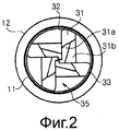
Фиг.2 - вид сверху, представляющий сверло для имплантационной хирургии по настоящему изобретению.2 is a plan view showing a drill for implant surgery of the present invention.
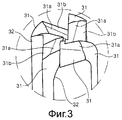
Фиг.3 - вид в перспективе основной части сверла для имплантационной хирургии по настоящему изобретению.Figure 3 is a perspective view of the main part of the drill for implant surgery of the present invention.
Фиг.4 - вид спереди в разрезе, представляющий сверло для имплантационной хирургии по настоящему изобретению.4 is a front view in section, representing a drill for implant surgery of the present invention.
Фиг.5 - вид спереди, представляющий основные части сверла для имплантационной хирургии по настоящему изобретению, с установленным на него стопором.5 is a front view representing the main parts of the drill for implant surgery of the present invention, with a stopper mounted thereon.
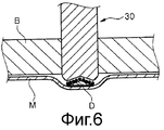
Фиг.6 - концептуальная схема, иллюстрирующая работу сверла для имплантационной хирургии по настоящему изобретению.6 is a conceptual diagram illustrating the operation of a drill for implant surgery of the present invention.
Условное обозначение номеров ссылочных позиций для основных частей на чертежахThe symbol of the reference numbers for the main parts in the drawings
10: корпус 10: case
11: ступенчатый участок11: stepped section
12: стопорный участок 12: locking portion
20: соединительный участок20: connecting section
30: режущий участок 30: cutting section
31: режущее лезвие31: cutting blade
31a: первая скошенная поверхность31a: first beveled surface
31b: вторая скошенная поверхность31b: second beveled surface
32: кончик 32: tip
33: направляющая33: guide
35: карман для стружки35: chip pocket
B: костьB: bone
D: костный дискD: bone disc
M: слизистая оболочкаM: mucous membrane
Наилучший способ выполнения изобретенияBEST MODE FOR CARRYING OUT THE INVENTION
Далее более подробно, со ссылкой на сопроводительные чертежи описывается предпочтительный вариант осуществления сверла для имплантационной хирургии согласно настоящему изобретению.Next, in more detail, with reference to the accompanying drawings, a preferred embodiment of a drill for implant surgery according to the present invention is described.
Сверло согласно настоящему изобретению может применяться на высокой и низкой скоростях. Если сверло соединено с ручным приводным устройством, сверло может использоваться в низкоскоростном режиме. Если сверло соединено с бормашиной для хирургии, сверло может использоваться и в высокоскоростном, и в низкоскоростном режимах.The drill according to the present invention can be used at high and low speeds. If the drill is connected to a manual drive unit, the drill can be used in low speed mode. If the drill is connected to a drill for surgery, the drill can be used in both high-speed and low-speed modes.
Как представлено на фиг.1-4, сверло для имплантационной хирургии согласно предпочтительному варианту осуществления настоящего изобретения состоит из корпуса 10, имеющего в целом цилиндрическую форму. Сверло включает в себя соединительный участок 20, образованный на одном конце, а именно на верхнем конце корпуса 10, для соединения с приводным устройством, например ручным приводным устройством, и описанной выше бормашиной для хирургии, и режущий участок 30, образованный на другом конце, а именно на нижнем конце корпуса 10, и имеющий режущие лезвия 31 для сверления.As shown in figures 1-4, the drill for implant surgery according to a preferred embodiment of the present invention consists of a
Дистальный конец режущего участка 30 имеет вогнутый участок 30a с вдавленным центром для создания формы перевернутого конуса, чтобы при сверлении на кончике мог быть образован костный диск D, как будет объяснено далее со ссылкой на фиг.6. Поскольку костный диск D препятствует непосредственному контакту режущего лезвия 31 сверла со слизистой оболочкой М, например слизистой оболочкой верхнечелюстной пазухи, слизистая оболочка верхнечелюстной пазухи может быть безопасно поднята.The distal end of the
Кроме того, наружный кольцеобразный край режущего участка 30 закруглен. Следовательно, даже если режущее лезвие 31 сверла находится в непосредственном контакте со слизистой оболочкой M верхнечелюстной пазухи без образования костного диска D, слизистая оболочка верхнечелюстной пазухи может быть безопасно поднята. Другими словами, поскольку внутренняя структура верхнечелюстной пазухи имеет неправильную форму, режущее лезвие 31 не может достичь слизистой оболочки верхнечелюстной пазухи в направлении, перпендикулярном слизистой оболочке. Однако, даже если бы это произошло, сверло по настоящему изобретению, в котором наружный кольцеобразный край режущего участка 30 закруглен, может не допустить повреждения слизистой оболочки верхнечелюстной пазухи. In addition, the outer annular edge of the cutting
Количество режущих лезвий 31, образованных на режущем участке 30, предпочтительно составляет 2 или более, чтобы улучшить эффективность срезания. На фиг.2 и 3 в качестве примера представлено, что всего образовано четыре режущих лезвия 31.The number of
Каждое режущее лезвие 31 может иметь множество скошенных поверхностей на внутренней стороне своего кончика для улучшения режущей способности при сверлении. Предпочтительно, чтобы режущая способность была улучшенной, как описано выше, поскольку скорость резания может гибко регулироваться от низкой скорости до высокой скорости. В частности, низкая скорость вращения позволяет собрать костную стружку, а высокая скорость вращения позволяет укоротить время хирургического вмешательства таким образом, что оператор может выполнять хирургическую манипуляцию с удобством для хирургического вмешательства. На фиг.2 и 3 примерно представлено, что каждое режущее лезвие 31 имеет две скошенные поверхности, а именно - первую скошенную поверхность 31a и вторую скошенную поверхность 31b.Each
Количество режущих лезвий 31 и количество скошенных поверхностей упомянуты лишь для примера, и следует понимать, что они не ограничиваются примерами, приведенными на чертежах.The number of
Каждое режущее лезвие 31 имеет боковую поверхность, на которой обеспечивается направляющая 33 заданной толщины, таким образом не допуская вибрации сверла при сверлении. Как представлено на фиг.1, каждая направляющая 33 вытянута в продольном направлении сверла. Кроме того, как представлено на виде сверху фиг.2, направляющие 33 при соединении друг с другом приблизительно образуют круг.Each
Кроме того, как представлено на фиг.1 и 3, участок рабочей кромки 32 режущего лезвия 31 предпочтительно имеет форму, изогнутую в ведущем направлении по мере его продвижения к дистальному концу режущего лезвия 31 для улучшения режущих характеристик сверла и для легкого удаления костной стружки. Соответственно, участок рабочей кромки 32 имеет приблизительно немного крючкообразную форму.In addition, as shown in figures 1 and 3, the portion of the working
Карман 35 для стружки предпочтительно образован между режущими лезвиями 31 таким образом, чтобы срезанная костная стружка могла легко удаляться через него, а также временно в нем храниться.The
На наружном периметре корпуса между соединительным участком 20 и режущим участком 30 могут быть образованы ступенчатый участок 11, выступающий наружу, и стопорный участок 12, дальше выступающий наружу от проксимального конца ступенчатого участка 11. Ступенчатый участок 11 и стопорный участок 12 могут быть вставлены в стопорный элемент 40 и фиксированы в нем, как представлено на фиг.5. Ступенчатый участок 11 может иметь такой размер, что он может вставляться в стопорный элемент 40, и стопорный участок 12 не допускает дальнейшей вставки стопорного элемента 40. Стопорный элемент 40 может позволить ограничить глубину сверления, что дополнительно облегчает хирургическое вмешательство, производимое оператором.On the outer perimeter of the housing between the connecting
Здесь и далее со ссылкой на фиг.6 будет объяснена работа сверла, выполненного как описано выше согласно настоящему изобретению.Hereinafter, with reference to FIG. 6, operation of a drill made as described above according to the present invention will be explained.
Как представлено на фиг.6, в случае сверления, выполняемого с использованием сверла по настоящему изобретению, некоторые раздробленные костные фрагменты (а именно, костная стружка) могут легко удаляться через карман 35 для стружки и храниться в кармане 35 для стружки, при этом кость B продолжает подвергаться резанию. В то же время слизистая оболочка верхнечелюстной пазухи может быть поднята, когда некоторое количество костной стружки, образованной вокруг закругленного участка наружного кольцеобразного края кончика сверла и первой и второй скошенными поверхностями 31a и 31b, отводится в направлении костного диска D, имеющего коническую форму (а именно, через пространство между костным диском D и первой и второй скошенными поверхностями 31a и 31b режущего участка 30). Как представлено на фиг.6, слизистая оболочка верхнечелюстной пазухи поднимается в направлении, вертикальном относительно внутренней поверхности верхнечелюстной пазухи, и в то же время могут быть подняты некоторые горизонтальные области слизистой оболочки, расположенные горизонтально относительно внутренней поверхности (другими словами, некоторые области вокруг отверстия, образованного сверлом).As shown in FIG. 6, in the case of drilling using the drill of the present invention, some fragmented bone fragments (namely, bone chips) can be easily removed through
Кроме того, поскольку кончик сверла режущего участка 30 сверла согласно настоящему изобретению имеет, как объяснено выше, форму перевернутого усеченного конуса, костный диск D из губчатого вещества кости или кортикальный слой кости, имеющий приблизительно коническую форму, остаются перед режущим участком 30, когда перфорируется кость B, например верхнечелюстная пазуха. Благодаря этому костному диску D слизистая оболочка M, например слизистая оболочка верхнечелюстной пазухи, может быть безопасно поднята.In addition, since the tip of the drill of the
Сверло для имплантационной хирургии согласно настоящему изобретению описано со ссылкой на примерные чертежи, однако настоящее изобретение не ограничивается описанными выше вариантами осуществления и сопроводительными чертежами, и специалистам в данной области техники будет очевидно, что возможно создание различных модификаций и изменений в пределах объема изобретения, ограниченного формулой. An implant surgery drill according to the present invention is described with reference to exemplary drawings, however, the present invention is not limited to the embodiments described above and the accompanying drawings, and it will be apparent to those skilled in the art that various modifications and changes are possible within the scope of the invention limited by the claims .
Claims (6)
соединительный участок, образованный на верхнем конце корпуса сверла для соединения с приводным устройством; и
режущий участок, образованный на нижнем конце корпуса и имеющий режущее лезвие для сверления,
причем вперед выступает наружный кольцеобразный край дистального конца режущего участка, а не центр дистального конца режущего участка,
при этом на дистальном конце режущего участка образован вогнутый участок с вдавленным центром так, что при сверлении на дистальном конце режущего участка образуется костный диск, что препятствует повреждению слизистой оболочки, и
наружный кольцеобразный край режущего участка закруглен, что препятствует повреждению слизистой оболочки, даже если режущий участок находится в непосредственном контакте со слизистой оболочкой при сверлении,
и при этом режущий участок имеет одно или более режущих лезвий, причем каждое режущее лезвие включает в себя ведущий участок кромки, изогнутый в ведущем направлении по мере его продвижения к дистальному концу режущего лезвия.1. Drill for use in implant surgery, containing:
a connecting portion formed at the upper end of the drill body for connection to a drive device; and
a cutting portion formed at the lower end of the housing and having a cutting blade for drilling,
moreover, the outer annular edge of the distal end of the cutting section protrudes forward, and not the center of the distal end of the cutting section,
at the same time, at the distal end of the cutting section, a concave section with a depressed center is formed so that when drilling at the distal end of the cutting section, a bone disc is formed, which prevents damage to the mucous membrane, and
the outer annular edge of the cutting section is rounded, which prevents damage to the mucous membrane, even if the cutting section is in direct contact with the mucous membrane during drilling,
and wherein the cutting portion has one or more cutting blades, each cutting blade including a leading edge portion curved in the leading direction as it moves toward the distal end of the cutting blade.
и при этом режущее лезвие включает в себя ведущий участок кромки, изогнутый в ведущем направлении по мере его продвижения к дистальному концу режущего лезвия. 6. A drill for use in implant surgery, in which the cutting blade of the drill has a distal shape with a beveled surface so that a bone disc is formed, and the mucous membrane of the maxillary sinus can be raised when a certain amount of bone chips formed during drilling is removed through the space between beveled surface and bone disc,
and wherein the cutting blade includes a leading edge portion curved in the leading direction as it moves toward the distal end of the cutting blade.
Applications Claiming Priority (3)
| Application Number | Priority Date | Filing Date | Title |
|---|---|---|---|
| KR10-2010-0003589 | 2010-01-14 | ||
| KR20100003589 | 2010-01-14 | ||
| PCT/KR2010/006201 WO2011087200A1 (en) | 2010-01-14 | 2010-09-13 | Drill for implant surgery |
Publications (2)
| Publication Number | Publication Date |
|---|---|
| RU2012134550A RU2012134550A (en) | 2014-02-20 |
| RU2515400C2 true RU2515400C2 (en) | 2014-05-10 |
Family
ID=44304449
Family Applications (1)
| Application Number | Title | Priority Date | Filing Date |
|---|---|---|---|
| RU2012134550/14A RU2515400C2 (en) | 2010-01-14 | 2010-09-13 | Implant surgical drill |
Country Status (9)
| Country | Link |
|---|---|
| US (1) | US9848962B2 (en) |
| EP (1) | EP2523627B1 (en) |
| JP (1) | JP5620520B2 (en) |
| KR (1) | KR101058521B1 (en) |
| CN (1) | CN102781362B (en) |
| CA (1) | CA2787116C (en) |
| DE (2) | DE112010005140T5 (en) |
| RU (1) | RU2515400C2 (en) |
| WO (1) | WO2011087200A1 (en) |
Cited By (2)
| Publication number | Priority date | Publication date | Assignee | Title |
|---|---|---|---|---|
| RU2695004C1 (en) * | 2016-01-29 | 2019-07-18 | Нобель Биокэр Сервисиз Аг | Dental instrument (versions) and set of devices for dental implantation |
| RU2794293C2 (en) * | 2016-01-29 | 2023-04-14 | Нобель Биокэр Сервисиз Аг | Dental tool |
Families Citing this family (26)
| Publication number | Priority date | Publication date | Assignee | Title |
|---|---|---|---|---|
| KR100962166B1 (en) * | 2009-05-04 | 2010-06-10 | 주식회사 이노바이오써지 | Alveolar-ridge cut and expansion tool for dental implant |
| KR101477734B1 (en) * | 2012-04-25 | 2015-01-02 | 주식회사 제노스 | Hybrid driving tool |
| KR101416797B1 (en) * | 2012-06-22 | 2014-07-09 | 주식회사 메가젠임플란트 | Bone harvester |
| JP6104634B2 (en) * | 2013-02-27 | 2017-03-29 | 京セラメディカル株式会社 | Dental drill |
| KR101516949B1 (en) * | 2013-11-06 | 2015-05-04 | 주식회사 디오 | apparatus for bone flattening drill |
| KR101566456B1 (en) * | 2014-08-12 | 2015-11-05 | 주식회사 메가젠임플란트 | Drill for implant and apparatus having the same |
| PL236960B1 (en) | 2014-11-12 | 2021-03-08 | Pospiech Jaroslaw | Adapter for dentist's drill |
| CN105434007B (en) * | 2015-12-22 | 2018-06-29 | 林炳泉 | A kind of maxilla bone hole impactor |
| EP3378433B1 (en) * | 2016-03-17 | 2020-07-22 | Young Keun Hyun | Guide pin for dental implant surgery |
| KR101810972B1 (en) * | 2016-08-12 | 2017-12-20 | 오스템임플란트 주식회사 | Dental drill |
| KR101758803B1 (en) * | 2016-10-10 | 2017-07-17 | 주식회사 디오 | apparatus for implant drill |
| EP3582701A4 (en) * | 2017-02-20 | 2021-04-21 | Paragon 28, Inc. | Implants, devices, instruments, systems and methods of forming and implanting |
| WO2018165676A1 (en) | 2017-03-10 | 2018-09-13 | Paragon 28, Inc. | Bone implant devices, instruments and methods of use |
| US10667886B2 (en) | 2017-03-27 | 2020-06-02 | LittleJohn Kirk VanHorn | Dental end forming burr |
| US11376050B2 (en) | 2017-06-27 | 2022-07-05 | Medos International Sarl | Bone screw |
| KR102027336B1 (en) * | 2017-06-30 | 2019-10-01 | 주식회사 디오 | apparatus for bone flattening drill |
| US10772667B2 (en) * | 2017-12-22 | 2020-09-15 | Medos International Sarl | Bone screw with cutting tip |
| US11679442B2 (en) | 2018-06-22 | 2023-06-20 | Maestro Logistics, Llc | Drill bit and method for making a drill bit |
| CN109044545A (en) * | 2018-09-11 | 2018-12-21 | 上海交通大学医学院附属第九人民医院 | Cheekbone implant operation maxillary sinus mucosa protective case |
| KR102005356B1 (en) * | 2018-12-28 | 2019-07-30 | 정지원 | Dental drill bits |
| KR102157434B1 (en) | 2019-02-28 | 2020-09-17 | 박상필 | Implant drill for fixture placement for dental implants to minimize alveolar bone damage |
| KR102009009B1 (en) * | 2019-04-29 | 2019-08-08 | 김아름 | Implant guide drill |
| KR102346872B1 (en) * | 2019-05-21 | 2022-01-03 | 황적희 | A Dental Drill for a Sinus Elevation with a Crestal Approach |
| CN110811756A (en) * | 2019-11-22 | 2020-02-21 | 武汉大学 | Machine drill bit for maxillary sinus internal lifting operation |
| CN112790821A (en) * | 2021-01-27 | 2021-05-14 | 北京大学第三医院(北京大学第三临床医学院) | Flat bone drill |
| KR102544369B1 (en) * | 2021-05-31 | 2023-06-19 | 주식회사 덴티스 | Sinus drilling instrument |
Citations (3)
| Publication number | Priority date | Publication date | Assignee | Title |
|---|---|---|---|---|
| FR2554709A1 (en) * | 1983-11-14 | 1985-05-17 | Medicalex | Process for positioning an implant, cutting device for its implementation and novel implant, such as an interposition cup |
| SU1335262A1 (en) * | 1986-04-09 | 1987-09-07 | Горьковский научно-исследовательский институт травматологии и ортопедии | Apparatus for treatment of transplant |
| RU2151570C1 (en) * | 1998-11-20 | 2000-06-27 | Олесова Валентина Николаевна | Surgical drill for using in dental surgery |
Family Cites Families (18)
| Publication number | Priority date | Publication date | Assignee | Title |
|---|---|---|---|---|
| US693508A (en) * | 1901-07-08 | 1902-02-18 | Heinrich Wilhelm Adolf Fette | Groove-cutter. |
| US3452625A (en) * | 1966-11-21 | 1969-07-01 | Peter Russo | Drill |
| JPS5976304U (en) * | 1982-11-15 | 1984-05-23 | 大西工業株式会社 | woodworking drill |
| JPH01129011U (en) * | 1988-02-26 | 1989-09-04 | ||
| US5514141A (en) * | 1992-11-18 | 1996-05-07 | Howmedica, Inc. | Small joint reamer |
| JPH08229720A (en) | 1995-02-24 | 1996-09-10 | Aisin Aw Co Ltd | Gun drill |
| SE507542C2 (en) * | 1996-12-04 | 1998-06-22 | Seco Tools Ab | Milling tools and cutting part for the tool |
| US5941706A (en) * | 1997-10-20 | 1999-08-24 | Ura; Robert S. | Variable depth medical drill and method of making the same |
| US6846135B2 (en) * | 2002-03-25 | 2005-01-25 | Hitachi Tool Engineering Ltd. | Radius end mill having radius edge enhanced in resistance to chipping and fracture |
| DE60333036D1 (en) * | 2002-12-26 | 2010-07-29 | Mitsubishi Materials Corp | MILLS |
| SE526105C2 (en) * | 2003-02-06 | 2005-07-05 | Seco Tools Ab | Cutter with three convex curved cutting edges grazing |
| KR200338095Y1 (en) | 2003-10-22 | 2004-01-13 | 주식회사 오스템 | Dental Stopper Drill |
| EP1725358B1 (en) * | 2004-03-12 | 2019-07-10 | Sandvik Intellectual Property AB | Cutting tool and method for cutting material |
| JP2007030074A (en) * | 2005-07-25 | 2007-02-08 | Mitsubishi Materials Kobe Tools Corp | Radius end mill and cutting method |
| KR100792649B1 (en) * | 2007-07-06 | 2008-01-09 | 안상훈 | Cutting lift reamer for maxillary sinus operating implant |
| FR2920327B1 (en) * | 2007-08-30 | 2009-11-20 | Snecma | GRAZING MILL FOR MACHINING WITH HIGH ADVANCE AND LOW PASS DEPTH |
| KR100924092B1 (en) | 2007-11-30 | 2009-11-02 | 김영기 | Reamer for operation implant |
| KR100838942B1 (en) | 2008-02-04 | 2008-06-16 | 허영구 | Drill for sinus membrane lift |
-
2010
- 2010-09-13 DE DE112010005140T patent/DE112010005140T5/en not_active Ceased
- 2010-09-13 CN CN201080065123.3A patent/CN102781362B/en active Active
- 2010-09-13 US US13/522,004 patent/US9848962B2/en active Active
- 2010-09-13 WO PCT/KR2010/006201 patent/WO2011087200A1/en active Application Filing
- 2010-09-13 EP EP10843266.7A patent/EP2523627B1/en active Active
- 2010-09-13 CA CA2787116A patent/CA2787116C/en active Active
- 2010-09-13 JP JP2012548870A patent/JP5620520B2/en active Active
- 2010-09-13 DE DE202010017830U patent/DE202010017830U1/en not_active Expired - Lifetime
- 2010-09-13 RU RU2012134550/14A patent/RU2515400C2/en active
- 2010-11-05 KR KR1020100109504A patent/KR101058521B1/en active IP Right Grant
Patent Citations (3)
| Publication number | Priority date | Publication date | Assignee | Title |
|---|---|---|---|---|
| FR2554709A1 (en) * | 1983-11-14 | 1985-05-17 | Medicalex | Process for positioning an implant, cutting device for its implementation and novel implant, such as an interposition cup |
| SU1335262A1 (en) * | 1986-04-09 | 1987-09-07 | Горьковский научно-исследовательский институт травматологии и ортопедии | Apparatus for treatment of transplant |
| RU2151570C1 (en) * | 1998-11-20 | 2000-06-27 | Олесова Валентина Николаевна | Surgical drill for using in dental surgery |
Cited By (4)
| Publication number | Priority date | Publication date | Assignee | Title |
|---|---|---|---|---|
| RU2695004C1 (en) * | 2016-01-29 | 2019-07-18 | Нобель Биокэр Сервисиз Аг | Dental instrument (versions) and set of devices for dental implantation |
| US11045287B2 (en) | 2016-01-29 | 2021-06-29 | Nobel Biocare Services Ag | Dentistry tool |
| RU2794293C2 (en) * | 2016-01-29 | 2023-04-14 | Нобель Биокэр Сервисиз Аг | Dental tool |
| US11857391B2 (en) | 2016-01-29 | 2024-01-02 | Nobel Biocare Services Ag | Dentistry tool |
Also Published As
| Publication number | Publication date |
|---|---|
| KR101058521B1 (en) | 2011-08-23 |
| CA2787116C (en) | 2015-08-25 |
| KR20110083471A (en) | 2011-07-20 |
| CN102781362A (en) | 2012-11-14 |
| US20120323243A1 (en) | 2012-12-20 |
| JP2013517044A (en) | 2013-05-16 |
| DE202010017830U1 (en) | 2013-03-05 |
| JP5620520B2 (en) | 2014-11-05 |
| CN102781362B (en) | 2015-09-30 |
| US9848962B2 (en) | 2017-12-26 |
| WO2011087200A1 (en) | 2011-07-21 |
| EP2523627B1 (en) | 2017-07-19 |
| DE112010005140T5 (en) | 2012-10-25 |
| CA2787116A1 (en) | 2011-07-21 |
| RU2012134550A (en) | 2014-02-20 |
| EP2523627A1 (en) | 2012-11-21 |
| EP2523627A4 (en) | 2015-07-01 |
Similar Documents
| Publication | Publication Date | Title |
|---|---|---|
| RU2515400C2 (en) | Implant surgical drill | |
| KR101128730B1 (en) | Drill for dental surgery | |
| Pavlíková et al. | Piezosurgery in oral and maxillofacial surgery | |
| US20100266984A1 (en) | Bur for maxillary sinus augmentation | |
| KR100884211B1 (en) | Piezotome for operating maxillary sinus | |
| KR100838942B1 (en) | Drill for sinus membrane lift | |
| US7611355B2 (en) | Manual driver for implant drills and method of dental implantation | |
| EP3777750B1 (en) | Drill bit | |
| MXPA06008808A (en) | Method and tools for low-speed milling without irrigation and with extraction and recovery of tissue particles. | |
| TWI437977B (en) | Apparatus for maxillary sinus operation | |
| Kumar et al. | Maxillary sinus augmentation | |
| Messina et al. | A step-by-step technique for the piezosurgical removal of fractured implants | |
| Magrin et al. | Piezosurgery in bone augmentation procedures previous to dental implant surgery: a review of the literature | |
| KR101810972B1 (en) | Dental drill | |
| TW201004608A (en) | Drill for operating implant | |
| EP1488745A1 (en) | Osteotome Structure | |
| Nissy et al. | Role of piezoelectric device in oral & maxillofacial surgery | |
| KR101122600B1 (en) | Drill for dental surgery | |
| KR101211361B1 (en) | Side wall drill for dental surgery | |
| KR101181923B1 (en) | Drill for lateral sinus graft surgical operation | |
| US12076212B1 (en) | Internal sinus lifting drill set | |
| KR20100088640A (en) | Sinus lift drill | |
| KR20090102261A (en) | An implant drill | |
| WO2015087321A1 (en) | Sinus lift apparatus and method | |
| KR20100032800A (en) | Internal stopped drill |


