RU2214197C2 - Method and set for performing autotransplantation - Google Patents
Method and set for performing autotransplantation Download PDFInfo
- Publication number
- RU2214197C2 RU2214197C2 RU99106519/14A RU99106519A RU2214197C2 RU 2214197 C2 RU2214197 C2 RU 2214197C2 RU 99106519/14 A RU99106519/14 A RU 99106519/14A RU 99106519 A RU99106519 A RU 99106519A RU 2214197 C2 RU2214197 C2 RU 2214197C2
- Authority
- RU
- Russia
- Prior art keywords
- barrier
- chondrocytes
- cartilage
- hemostatic barrier
- matrix
- Prior art date
Links
Images
Classifications
-
- A—HUMAN NECESSITIES
- A61—MEDICAL OR VETERINARY SCIENCE; HYGIENE
- A61F—FILTERS IMPLANTABLE INTO BLOOD VESSELS; PROSTHESES; DEVICES PROVIDING PATENCY TO, OR PREVENTING COLLAPSING OF, TUBULAR STRUCTURES OF THE BODY, e.g. STENTS; ORTHOPAEDIC, NURSING OR CONTRACEPTIVE DEVICES; FOMENTATION; TREATMENT OR PROTECTION OF EYES OR EARS; BANDAGES, DRESSINGS OR ABSORBENT PADS; FIRST-AID KITS
- A61F2/00—Filters implantable into blood vessels; Prostheses, i.e. artificial substitutes or replacements for parts of the body; Appliances for connecting them with the body; Devices providing patency to, or preventing collapsing of, tubular structures of the body, e.g. stents
- A61F2/02—Prostheses implantable into the body
- A61F2/30—Joints
-
- A—HUMAN NECESSITIES
- A61—MEDICAL OR VETERINARY SCIENCE; HYGIENE
- A61F—FILTERS IMPLANTABLE INTO BLOOD VESSELS; PROSTHESES; DEVICES PROVIDING PATENCY TO, OR PREVENTING COLLAPSING OF, TUBULAR STRUCTURES OF THE BODY, e.g. STENTS; ORTHOPAEDIC, NURSING OR CONTRACEPTIVE DEVICES; FOMENTATION; TREATMENT OR PROTECTION OF EYES OR EARS; BANDAGES, DRESSINGS OR ABSORBENT PADS; FIRST-AID KITS
- A61F2/00—Filters implantable into blood vessels; Prostheses, i.e. artificial substitutes or replacements for parts of the body; Appliances for connecting them with the body; Devices providing patency to, or preventing collapsing of, tubular structures of the body, e.g. stents
- A61F2/02—Prostheses implantable into the body
- A61F2/28—Bones
- A61F2/2846—Support means for bone substitute or for bone graft implants, e.g. membranes or plates for covering bone defects
-
- A—HUMAN NECESSITIES
- A61—MEDICAL OR VETERINARY SCIENCE; HYGIENE
- A61F—FILTERS IMPLANTABLE INTO BLOOD VESSELS; PROSTHESES; DEVICES PROVIDING PATENCY TO, OR PREVENTING COLLAPSING OF, TUBULAR STRUCTURES OF THE BODY, e.g. STENTS; ORTHOPAEDIC, NURSING OR CONTRACEPTIVE DEVICES; FOMENTATION; TREATMENT OR PROTECTION OF EYES OR EARS; BANDAGES, DRESSINGS OR ABSORBENT PADS; FIRST-AID KITS
- A61F2/00—Filters implantable into blood vessels; Prostheses, i.e. artificial substitutes or replacements for parts of the body; Appliances for connecting them with the body; Devices providing patency to, or preventing collapsing of, tubular structures of the body, e.g. stents
- A61F2/02—Prostheses implantable into the body
- A61F2/30—Joints
- A61F2/30756—Cartilage endoprostheses
-
- A—HUMAN NECESSITIES
- A61—MEDICAL OR VETERINARY SCIENCE; HYGIENE
- A61B—DIAGNOSIS; SURGERY; IDENTIFICATION
- A61B17/00—Surgical instruments, devices or methods, e.g. tourniquets
- A61B17/00491—Surgical glue applicators
-
- A—HUMAN NECESSITIES
- A61—MEDICAL OR VETERINARY SCIENCE; HYGIENE
- A61F—FILTERS IMPLANTABLE INTO BLOOD VESSELS; PROSTHESES; DEVICES PROVIDING PATENCY TO, OR PREVENTING COLLAPSING OF, TUBULAR STRUCTURES OF THE BODY, e.g. STENTS; ORTHOPAEDIC, NURSING OR CONTRACEPTIVE DEVICES; FOMENTATION; TREATMENT OR PROTECTION OF EYES OR EARS; BANDAGES, DRESSINGS OR ABSORBENT PADS; FIRST-AID KITS
- A61F2/00—Filters implantable into blood vessels; Prostheses, i.e. artificial substitutes or replacements for parts of the body; Appliances for connecting them with the body; Devices providing patency to, or preventing collapsing of, tubular structures of the body, e.g. stents
- A61F2/02—Prostheses implantable into the body
- A61F2/30—Joints
- A61F2/38—Joints for elbows or knees
-
- A—HUMAN NECESSITIES
- A61—MEDICAL OR VETERINARY SCIENCE; HYGIENE
- A61F—FILTERS IMPLANTABLE INTO BLOOD VESSELS; PROSTHESES; DEVICES PROVIDING PATENCY TO, OR PREVENTING COLLAPSING OF, TUBULAR STRUCTURES OF THE BODY, e.g. STENTS; ORTHOPAEDIC, NURSING OR CONTRACEPTIVE DEVICES; FOMENTATION; TREATMENT OR PROTECTION OF EYES OR EARS; BANDAGES, DRESSINGS OR ABSORBENT PADS; FIRST-AID KITS
- A61F2/00—Filters implantable into blood vessels; Prostheses, i.e. artificial substitutes or replacements for parts of the body; Appliances for connecting them with the body; Devices providing patency to, or preventing collapsing of, tubular structures of the body, e.g. stents
- A61F2/02—Prostheses implantable into the body
- A61F2/28—Bones
- A61F2002/2835—Bone graft implants for filling a bony defect or an endoprosthesis cavity, e.g. by synthetic material or biological material
-
- A—HUMAN NECESSITIES
- A61—MEDICAL OR VETERINARY SCIENCE; HYGIENE
- A61F—FILTERS IMPLANTABLE INTO BLOOD VESSELS; PROSTHESES; DEVICES PROVIDING PATENCY TO, OR PREVENTING COLLAPSING OF, TUBULAR STRUCTURES OF THE BODY, e.g. STENTS; ORTHOPAEDIC, NURSING OR CONTRACEPTIVE DEVICES; FOMENTATION; TREATMENT OR PROTECTION OF EYES OR EARS; BANDAGES, DRESSINGS OR ABSORBENT PADS; FIRST-AID KITS
- A61F2/00—Filters implantable into blood vessels; Prostheses, i.e. artificial substitutes or replacements for parts of the body; Appliances for connecting them with the body; Devices providing patency to, or preventing collapsing of, tubular structures of the body, e.g. stents
- A61F2/02—Prostheses implantable into the body
- A61F2/30—Joints
- A61F2002/30001—Additional features of subject-matter classified in A61F2/28, A61F2/30 and subgroups thereof
- A61F2002/30003—Material related properties of the prosthesis or of a coating on the prosthesis
- A61F2002/30004—Material related properties of the prosthesis or of a coating on the prosthesis the prosthesis being made from materials having different values of a given property at different locations within the same prosthesis
- A61F2002/30016—Material related properties of the prosthesis or of a coating on the prosthesis the prosthesis being made from materials having different values of a given property at different locations within the same prosthesis differing in hardness, e.g. Vickers, Shore, Brinell
-
- A—HUMAN NECESSITIES
- A61—MEDICAL OR VETERINARY SCIENCE; HYGIENE
- A61F—FILTERS IMPLANTABLE INTO BLOOD VESSELS; PROSTHESES; DEVICES PROVIDING PATENCY TO, OR PREVENTING COLLAPSING OF, TUBULAR STRUCTURES OF THE BODY, e.g. STENTS; ORTHOPAEDIC, NURSING OR CONTRACEPTIVE DEVICES; FOMENTATION; TREATMENT OR PROTECTION OF EYES OR EARS; BANDAGES, DRESSINGS OR ABSORBENT PADS; FIRST-AID KITS
- A61F2/00—Filters implantable into blood vessels; Prostheses, i.e. artificial substitutes or replacements for parts of the body; Appliances for connecting them with the body; Devices providing patency to, or preventing collapsing of, tubular structures of the body, e.g. stents
- A61F2/02—Prostheses implantable into the body
- A61F2/30—Joints
- A61F2002/30001—Additional features of subject-matter classified in A61F2/28, A61F2/30 and subgroups thereof
- A61F2002/30003—Material related properties of the prosthesis or of a coating on the prosthesis
- A61F2002/3006—Properties of materials and coating materials
- A61F2002/30062—(bio)absorbable, biodegradable, bioerodable, (bio)resorbable, resorptive
-
- A—HUMAN NECESSITIES
- A61—MEDICAL OR VETERINARY SCIENCE; HYGIENE
- A61F—FILTERS IMPLANTABLE INTO BLOOD VESSELS; PROSTHESES; DEVICES PROVIDING PATENCY TO, OR PREVENTING COLLAPSING OF, TUBULAR STRUCTURES OF THE BODY, e.g. STENTS; ORTHOPAEDIC, NURSING OR CONTRACEPTIVE DEVICES; FOMENTATION; TREATMENT OR PROTECTION OF EYES OR EARS; BANDAGES, DRESSINGS OR ABSORBENT PADS; FIRST-AID KITS
- A61F2/00—Filters implantable into blood vessels; Prostheses, i.e. artificial substitutes or replacements for parts of the body; Appliances for connecting them with the body; Devices providing patency to, or preventing collapsing of, tubular structures of the body, e.g. stents
- A61F2/02—Prostheses implantable into the body
- A61F2/30—Joints
- A61F2002/30001—Additional features of subject-matter classified in A61F2/28, A61F2/30 and subgroups thereof
- A61F2002/30316—The prosthesis having different structural features at different locations within the same prosthesis; Connections between prosthetic parts; Special structural features of bone or joint prostheses not otherwise provided for
- A61F2002/30535—Special structural features of bone or joint prostheses not otherwise provided for
-
- A—HUMAN NECESSITIES
- A61—MEDICAL OR VETERINARY SCIENCE; HYGIENE
- A61F—FILTERS IMPLANTABLE INTO BLOOD VESSELS; PROSTHESES; DEVICES PROVIDING PATENCY TO, OR PREVENTING COLLAPSING OF, TUBULAR STRUCTURES OF THE BODY, e.g. STENTS; ORTHOPAEDIC, NURSING OR CONTRACEPTIVE DEVICES; FOMENTATION; TREATMENT OR PROTECTION OF EYES OR EARS; BANDAGES, DRESSINGS OR ABSORBENT PADS; FIRST-AID KITS
- A61F2/00—Filters implantable into blood vessels; Prostheses, i.e. artificial substitutes or replacements for parts of the body; Appliances for connecting them with the body; Devices providing patency to, or preventing collapsing of, tubular structures of the body, e.g. stents
- A61F2/02—Prostheses implantable into the body
- A61F2/30—Joints
- A61F2/30756—Cartilage endoprostheses
- A61F2002/30761—Support means for artificial cartilage, e.g. cartilage defect covering membranes
-
- A—HUMAN NECESSITIES
- A61—MEDICAL OR VETERINARY SCIENCE; HYGIENE
- A61F—FILTERS IMPLANTABLE INTO BLOOD VESSELS; PROSTHESES; DEVICES PROVIDING PATENCY TO, OR PREVENTING COLLAPSING OF, TUBULAR STRUCTURES OF THE BODY, e.g. STENTS; ORTHOPAEDIC, NURSING OR CONTRACEPTIVE DEVICES; FOMENTATION; TREATMENT OR PROTECTION OF EYES OR EARS; BANDAGES, DRESSINGS OR ABSORBENT PADS; FIRST-AID KITS
- A61F2/00—Filters implantable into blood vessels; Prostheses, i.e. artificial substitutes or replacements for parts of the body; Appliances for connecting them with the body; Devices providing patency to, or preventing collapsing of, tubular structures of the body, e.g. stents
- A61F2/02—Prostheses implantable into the body
- A61F2/30—Joints
- A61F2/30756—Cartilage endoprostheses
- A61F2002/30762—Means for culturing cartilage
-
- A—HUMAN NECESSITIES
- A61—MEDICAL OR VETERINARY SCIENCE; HYGIENE
- A61F—FILTERS IMPLANTABLE INTO BLOOD VESSELS; PROSTHESES; DEVICES PROVIDING PATENCY TO, OR PREVENTING COLLAPSING OF, TUBULAR STRUCTURES OF THE BODY, e.g. STENTS; ORTHOPAEDIC, NURSING OR CONTRACEPTIVE DEVICES; FOMENTATION; TREATMENT OR PROTECTION OF EYES OR EARS; BANDAGES, DRESSINGS OR ABSORBENT PADS; FIRST-AID KITS
- A61F2210/00—Particular material properties of prostheses classified in groups A61F2/00 - A61F2/26 or A61F2/82 or A61F9/00 or A61F11/00 or subgroups thereof
- A61F2210/0004—Particular material properties of prostheses classified in groups A61F2/00 - A61F2/26 or A61F2/82 or A61F9/00 or A61F11/00 or subgroups thereof bioabsorbable
-
- A—HUMAN NECESSITIES
- A61—MEDICAL OR VETERINARY SCIENCE; HYGIENE
- A61F—FILTERS IMPLANTABLE INTO BLOOD VESSELS; PROSTHESES; DEVICES PROVIDING PATENCY TO, OR PREVENTING COLLAPSING OF, TUBULAR STRUCTURES OF THE BODY, e.g. STENTS; ORTHOPAEDIC, NURSING OR CONTRACEPTIVE DEVICES; FOMENTATION; TREATMENT OR PROTECTION OF EYES OR EARS; BANDAGES, DRESSINGS OR ABSORBENT PADS; FIRST-AID KITS
- A61F2250/00—Special features of prostheses classified in groups A61F2/00 - A61F2/26 or A61F2/82 or A61F9/00 or A61F11/00 or subgroups thereof
- A61F2250/0014—Special features of prostheses classified in groups A61F2/00 - A61F2/26 or A61F2/82 or A61F9/00 or A61F11/00 or subgroups thereof having different values of a given property or geometrical feature, e.g. mechanical property or material property, at different locations within the same prosthesis
- A61F2250/0019—Special features of prostheses classified in groups A61F2/00 - A61F2/26 or A61F2/82 or A61F9/00 or A61F11/00 or subgroups thereof having different values of a given property or geometrical feature, e.g. mechanical property or material property, at different locations within the same prosthesis differing in hardness, e.g. Vickers, Shore, Brinell
-
- A—HUMAN NECESSITIES
- A61—MEDICAL OR VETERINARY SCIENCE; HYGIENE
- A61F—FILTERS IMPLANTABLE INTO BLOOD VESSELS; PROSTHESES; DEVICES PROVIDING PATENCY TO, OR PREVENTING COLLAPSING OF, TUBULAR STRUCTURES OF THE BODY, e.g. STENTS; ORTHOPAEDIC, NURSING OR CONTRACEPTIVE DEVICES; FOMENTATION; TREATMENT OR PROTECTION OF EYES OR EARS; BANDAGES, DRESSINGS OR ABSORBENT PADS; FIRST-AID KITS
- A61F2250/00—Special features of prostheses classified in groups A61F2/00 - A61F2/26 or A61F2/82 or A61F9/00 or A61F11/00 or subgroups thereof
- A61F2250/0058—Additional features; Implant or prostheses properties not otherwise provided for
-
- A—HUMAN NECESSITIES
- A61—MEDICAL OR VETERINARY SCIENCE; HYGIENE
- A61F—FILTERS IMPLANTABLE INTO BLOOD VESSELS; PROSTHESES; DEVICES PROVIDING PATENCY TO, OR PREVENTING COLLAPSING OF, TUBULAR STRUCTURES OF THE BODY, e.g. STENTS; ORTHOPAEDIC, NURSING OR CONTRACEPTIVE DEVICES; FOMENTATION; TREATMENT OR PROTECTION OF EYES OR EARS; BANDAGES, DRESSINGS OR ABSORBENT PADS; FIRST-AID KITS
- A61F2310/00—Prostheses classified in A61F2/28 or A61F2/30 - A61F2/44 being constructed from or coated with a particular material
- A61F2310/00005—The prosthesis being constructed from a particular material
- A61F2310/00365—Proteins; Polypeptides; Degradation products thereof
-
- A—HUMAN NECESSITIES
- A61—MEDICAL OR VETERINARY SCIENCE; HYGIENE
- A61L—METHODS OR APPARATUS FOR STERILISING MATERIALS OR OBJECTS IN GENERAL; DISINFECTION, STERILISATION OR DEODORISATION OF AIR; CHEMICAL ASPECTS OF BANDAGES, DRESSINGS, ABSORBENT PADS OR SURGICAL ARTICLES; MATERIALS FOR BANDAGES, DRESSINGS, ABSORBENT PADS OR SURGICAL ARTICLES
- A61L2430/00—Materials or treatment for tissue regeneration
- A61L2430/06—Materials or treatment for tissue regeneration for cartilage reconstruction, e.g. meniscus
Abstract
Description
Настоящее изобретение относится к области трансплантации хондроцитов, пересадки кости и хряща, заживления, восстановления суставов и профилактики артритных патологических процессов. В частности, к способам подготовки участка для пересадки, устройствам для такой подготовки и для аутотрансплантации клеток на подготовленный участок для пересадки. The present invention relates to the field of chondrocyte transplantation, bone and cartilage transplantation, healing, joint repair and prevention of arthritic pathological processes. In particular, to methods for preparing a site for transplantation, devices for such preparation, and for autotransplantation of cells into a prepared site for transplantation.
Предшествующий уровень техники
В США ежегодно выполняется более 500000 артропластических операций и полного протезирования суставов. Приблизительно такое же количество аналогичных вмешательств выполняется в Европе. В эти цифры включено приблизительно 90000 операций полного протезирования коленного сустава и приблизительно 50000 операций по восстановлению дефектов в области коленного сустава, выполняемых ежегодно в Европе. В США количество.вмешательств по существу такое же (In: Praemer А., Furner S., Rice, D.P., Musculosckeletal conditions in the United States, American Academy of Orthopaedic Surgeons, Park Ridge, Hi., 1992, 125). Самым полезным был бы способ регенерации лечения хряща, который мог бы выполняться на более ранней стадии повреждения сустава, уменьшая таким образом количество больных, нуждающихся в операции искусственного протезирования сустава. При использовании таких профилактических способов лечения уменьшилось бы также количество больных с развившимся остеоартритом.State of the art
In the United States, more than 500,000 arthroplastic surgeries and complete joint prosthetics are performed annually. Approximately the same number of similar interventions are performed in Europe. These figures include approximately 90,000 total knee prosthetics and approximately 50,000 knee-joint defect repair operations performed annually in Europe. In the USA, the number of interventions is essentially the same (In: Praemer A., Furner S., Rice, DP, Musculosckeletal conditions in the United States, American Academy of Orthopedic Surgeons, Park Ridge, Hi., 1992, 125). The most useful method would be the regeneration of the treatment of cartilage, which could be performed at an earlier stage of joint damage, thereby reducing the number of patients requiring surgery for artificial joint replacement. Using such prophylactic methods of treatment, the number of patients with developed osteoarthritis would also decrease.
Методики, применявшиеся для восстановления поверхности хрящевой структуры в суставах, пытались главным образом вызвать восстановление хряща с использованием подхрящевого сверления, абразивной обработки и других способов, посредством чего производится иссечение пораженного хряща и подхрящевой кости с оставлением открытым снабженного сосудами губчатое вещества кости (Insall, J. , Clin. Orthop. 1974, 101, 61; Ficat R.P. et al., Clin Orthop. 1979, 144, 74; Johnson L.L., In: Operative Arthroscopy, McGinty J.B., Ed., Raven Press, New York, 1991, 341). The methods used to restore the surface of the cartilage structure in the joints, mainly tried to cause the restoration of cartilage by using cartilage drilling, abrasive treatment and other methods, by means of which the affected cartilage and the cartilage bone were excised, leaving the spongy bone open with vessels (Insall, J. , Clin. Orthop. 1974, 101, 61; Ficat RP et al., Clin Orthop. 1979, 144, 74; Johnson LL, In: Operative Arthroscopy, McGinty JB, Ed., Raven Press, New York, 1991, 341) .
Coon and Cahn (Science 1966, 153, 1116) описали методику культивирования синтезирующих хрящ клеток из сомитов куриного эмбриона. Позже Cahn and Lasher (PNAS USA 1967, 58, 1131) использовали систему для анализа участия синтеза ДНК как необходимого условия дифференцировки хряща. Хондроциты реагируют ростом и на эпидермальный фактор роста, и на фактор роста фибробластов (Gospodarowitcz and Mescher, J. Cell Physiology 1977, 93, 117), но в конечном итоге теряют свою дифференцированную функцию (Benya et al., Cell 1978, 15, 1313). Способы выращивания хондроцитов были описаны и в основном использовались с небольшими модификациями Brittberg, M. et а1. (New Engl. J. Med. 1994, 331, 889). Клетки, выращенные с применением этих способов, использовались в качестве аутологичных трансплантатов в коленные суставы пациентов. Кроме того, Kolettas et al. (J. Cell Science 1995, 108, 1991) исследовали экспрессию специфичных для хряща молекул, таких как коллагены и протеогликаны, в условиях длительного культивирования клеток. Они обнаружили, что несмотря на морфологические изменения во время культивирования в однослойных культурах (Aulthouse, A. et al., In Vitro Cell Dev. Bio1., 1989, 25, 659; Archer, С. et al., J. Cell Sci. 1990, 97, 361; Hanselmann, H. et al. , J. . Cell Sci. 1994, 107, 17; Bonaventure, J. et al., Exp. Cell Res. 1994, 212, 97), при сравнении с суспензионными культурами, выращенными над агарозными гелями, альгинатными гранулами или в виде спин-культур (сохраняющих круглоклеточную морфологию), исследованными различными исследователями, они не меняли экспрессируемые хондроцитами маркеры, такие как коллагены типа II и IX и не изменялись крупные агрегирующие протеогликаны, аггрекан, версикан и связывающий белок (Kolettas, E. et al., J. Cell Science 1995, 108, 1991). Coon and Cahn (Science 1966, 153, 1116) described a technique for culturing cartilage synthesizing cells from chicken embryo somites. Later, Cahn and Lasher (PNAS USA 1967, 58, 1131) used a system to analyze the participation of DNA synthesis as a necessary condition for differentiation of cartilage. Chondrocytes respond by growth to both epidermal growth factor and fibroblast growth factor (Gospodarowitcz and Mescher, J. Cell Physiology 1977, 93, 117), but eventually lose their differentiated function (Benya et al., Cell 1978, 15, 1313 ) Methods for growing chondrocytes have been described and mainly used with minor modifications by Brittberg, M. et a1. (New Engl. J. Med. 1994, 331, 889). Cells grown using these methods were used as autologous grafts in the knee joints of patients. In addition, Kolettas et al. (J. Cell Science 1995, 108, 1991) examined the expression of cartilage-specific molecules, such as collagens and proteoglycans, under conditions of prolonged cell culture. They found that despite morphological changes during cultivation in single-layer cultures (Aulthouse, A. et al., In Vitro Cell Dev. Bio1., 1989, 25, 659; Archer, C. et al., J. Cell Sci. 1990, 97, 361; Hanselmann, H. et al., J. Cell Sci. 1994, 107, 17; Bonaventure, J. et al., Exp. Cell Res. 1994, 212, 97), when compared with suspension cultures grown on agarose gels, alginate granules or in the form of spin cultures (preserving round cell morphology) studied by various researchers, they did not change the markers expressed by chondrocytes, such as type II and IX collagens, and did not change full aggregating proteoglycans, aggrecan, versican and binding protein (Kolettas, E. et al., J. Cell Science 1995, 108, 1991).
Суставные хондроциты представляют собой специализированные клетки мезенхимального происхождения обнаруживаемые исключительно в хряще. Хрящ представляет собой бессосудистую ткань, физические свойства которой зависят от внеклеточного матрикса, образуемого хондроцитами. Во время внутрихрящевой оссификации хондроциты подвергаются созреванию, приводящему к клеточной гипретрофии, характеризуемой началом экспрессии коллагена типа Х (Uрholt, W. В. and Olsen, R.R., In.: Cartilage Molecular Aspects (Hall, В & Mewman, S, Eds. ) CRC Boca Raton 1991,43; Reichenberg, Е. et al., Dev. Biol. 1991, 148, 562; Kirsch, T. et al., Differentiation, 1992, 52, 89; Stephens, M. et al., J. Cell Sci 1933, 103, 1111). Joint chondrocytes are specialized cells of mesenchymal origin found exclusively in cartilage. Cartilage is an avascular tissue whose physical properties depend on the extracellular matrix formed by chondrocytes. During intra-cartilaginous ossification, chondrocytes undergo maturation leading to cell hypertrophy characterized by the onset of expression of type X collagen (Uholt, W. B. and Olsen, RR, In .: Cartilage Molecular Aspects (Hall, B & Mewman, S, Eds.) CRC Boca Raton 1991.43; Reichenberg, E. et al., Dev. Biol. 1991, 148, 562; Kirsch, T. et al., Differentiation, 1992, 52, 89; Stephens, M. et al., J. Cell Sci 1933, 103, 1111).
Избыточный распад коллагена II типа в наружных слоях сочленяющихся поверхностей сустава также вызван остеоартритом. Коллагеновая сеть соответственно ослабляется, и в последующем развивается волокнистость, в результате чего вещества матрикса, такие как протеогликаны, утрачиваются и в конечном итоге полностью замещаются. Такое образование волокон ослабленного остеоартритического хряща может распространяться вплоть до обызвествленного хряща в подхрящевую кость (Kenipson, G.E. et al., Вiochim. Biоphys. Acta 1976, 428, 741; Roth, V. and Mow. V.C., J. Bone Joint Surgery, 1980, 62A, 1102; Woo, S.L.-Y. et al., in Handbook of Вioengineering (R. Skalak and S. Chien eds.), McGraw-Hill, New York, 1987, pp. 4.1-4.44). Excessive collapse of type II collagen in the outer layers of articulating surfaces of the joint is also caused by osteoarthritis. The collagen network is accordingly weakened, and subsequently fibrousness develops, as a result of which matrix substances, such as proteoglycans, are lost and ultimately completely replaced. Such fiber formation of weakened osteoarthritic cartilage can extend up to calcified cartilage into the cartilage bone (Kenipson, GE et al., Biochim. Biophys. Acta 1976, 428, 741; Roth, V. and Mow. VC, J. Bone Joint Surgery, 1980 62A, 1102; Woo, SL-Y. Et al., In Handbook of Bioengineering (R. Skalak and S. Chien eds.), McGraw-Hill, New York, 1987, pp. 4.1-4.44).
Описание основного развития гистологической и микроскопической структуры кости, хряща и других таких соединительных тканей можно найти, например, в Wheater, Burkitt and Daniels, Functional Histology, and Edition, (Churchill Livingstone, London, 1987, Chp. 4). Описание основной гистологической структуры дефектов в кости, хряще и другой соединительной ткани можно найти, например, в Wheater, Burkitt, Stevens and Lowe, Basic Histopathology (Churchill Livingstone, London, 1985, Chp. 21). A description of the main development of the histological and microscopic structure of bone, cartilage, and other such connective tissues can be found, for example, in Wheater, Burkitt and Daniels, Functional Histology, and Edition, (Churchill Livingstone, London, 1987, Chp. 4). A description of the basic histological structure of defects in bone, cartilage, and other connective tissue can be found, for example, in Wheater, Burkitt, Stevens and Lowe, Basic Histopathology (Churchill Livingstone, London, 1985, Chp. 21).
Несмотря на достижения в культивировании хондроцитов и манипулировании костью и хрящем, не было достигнуто большого успеха в попытках трансплантации хряща или хондроцитов для восстановления поврежденных сочленяющихся поверхностей. Положения настоящего изобретения предоставляют эффективные и действенные средства, способствующие трансплантации хряща и/или хондроцитов в дефект суставного сочленения или на другие костные поверхности, покрытые хрящем, посредством чего хрящ восстанавливается для устранения дефекта. Настоящее изобретение также предоставляет хирургические инструменты, которые предназначены для подготовки участка для трансплантации с тем, чтобы облегчить эффективное интегрирование пересаженного материала в участок пересадки. Despite advances in the cultivation of chondrocytes and the manipulation of bone and cartilage, little success has been achieved in attempts to transplant cartilage or chondrocytes to repair damaged articulated surfaces. The provisions of the present invention provide effective and efficient means of facilitating the transplantation of cartilage and / or chondrocytes into a joint defect or onto other bone surfaces covered by cartilage, whereby the cartilage is restored to repair the defect. The present invention also provides surgical instruments that are intended to prepare a transplant site in order to facilitate the effective integration of the transplanted material into the transplant site.
Сущность изобретения
Настоящее изобретение предоставляет способ эффективного лечения хряща сочленяющейся поверхности сустава с помощью трансплантации хондроцитов в подходящем матриксе на подлежащую лечению поверхность с гемостатическим барьером и бесклеточной покрывающей накладкой, включающий, во-первых, размещение гемостатического барьера проксимальнее подлежащей лечению поверхности, помещение хондроцитов в подходящем матриксе на подлежащую лечению поверхность дистальнее гемостатического барьера, покрытие подлежащей лечению поверхности бесклеточной покрывающей накладкой. Как будет описано ниже, гемостатический барьер представляет собой барьер, который ингибирует или предотвращает проникновение васкуляризирующих клеток и ткани в пересаженный материал. В частности, настоящий способ обеспечивает гемостатический барьер, который представляет собой способный к рассасыванию, полупроницаемый материал, который ингибирует или предотвращает сосудистую инфильтрацию через барьер. В одном варианте реализации гемостатический барьер содержит коллаген. Термин "бесклеточный" используется здесь, как и в предшествующем уровне техники, и обозначает материал, который по существу свободен от интактных клеток, способных к дальнейшему клеточному делению, распространению или биологической активности. В предпочтительном варианте реализации бесклеточный материал является свободным от всех интактных, имеющих ядра клеток. В одном варианте реализации настоящий способ охватывает использование бесклеточной покрывающей накладки, которая содержит полупроницаемый коллагеновый матрикс. В одном предпочтительном варианте реализации способа пористая поверхность бесклеточной покрывающей накладки направлена в сторону имплантируемого материала.SUMMARY OF THE INVENTION
The present invention provides a method for effectively treating cartilage of an articulating joint surface by transplanting chondrocytes in a suitable matrix onto a surface to be treated with a hemostatic barrier and a cell-free covering pad, including, firstly, placing a hemostatic barrier proximal to the surface to be treated, placing chondrocytes in a suitable matrix on the underlying matrix the surface distal to the hemostatic barrier, the surface of the surface to be treated is besklet ary covering-patch. As will be described below, the hemostatic barrier is a barrier that inhibits or prevents the penetration of vascularizing cells and tissue into the transplanted material. In particular, the present method provides a hemostatic barrier, which is a resorbable, semipermeable material that inhibits or prevents vascular infiltration through the barrier. In one embodiment, the hemostatic barrier comprises collagen. The term "cell-free" is used here, as in the prior art, and means a material that is essentially free of intact cells capable of further cell division, spread or biological activity. In a preferred embodiment, the cell-free material is free of all intact cells having cell nuclei. In one embodiment, the present method encompasses the use of a cell-free coating pad that contains a semipermeable collagen matrix. In one preferred embodiment of the method, the porous surface of the cell-free covering lining is directed towards the implantable material.
Настоящее изобретение, кроме того, обеспечивает аутотрансплантацию коллагена или хондроцитов в участок пересадки, в котором участок пересадки сначала был подготовлен с помощью хирургической манипуляции для лучшего восприятии пересаженного материала. В одном варианте реализации участок пересадки профилируется так, что стенкам участка пересадки придается волнообразный характер, так что при помещении внутрь трансплантата и расширении пересаженного материала до контакта со стенкой участка пересадки, будет создано сопротивление удалению или выталкиванию всего трансплантата из участка пересадки. Настоящее изобретение, кроме того, обеспечивает хирургические инструменты, предназначенные для профилирования участка пересадки, как предусмотрено способом изобретения. The present invention further provides for autologous transplantation of collagen or chondrocytes into a transplant site in which the transplant site was first prepared by surgical manipulation to better perceive the transplanted material. In one embodiment, the transplant site is shaped so that the walls of the transplant site are given a wave-like character, so that when placed inside the graft and the transplanted material expands to come into contact with the wall of the transplant site, resistance will be created to remove or push the entire graft from the transplant site. The present invention also provides surgical instruments for profiling a transplant site as provided by the method of the invention.
Изобретение, кроме того, обеспечивает набор для трансплантации хряща и/или хондроцитов на поверхность сочленяющего сустава, в котором указанный набор включает гемостатический барьер, бесклеточную полупроницаемую покрывающую накладку и органический клей. В еще одном варианте реализации набор, кроме того, может необязательно предоставлять один или более хирургических инструментов, которые могут использоваться для профилирования участка пересадки в соответствии со способами настоящего изобретения. The invention also provides a kit for transplantation of cartilage and / or chondrocytes onto the surface of the articulation joint, wherein said kit includes a hemostatic barrier, a cell-free semi-permeable covering pad and organic glue. In yet another embodiment, the kit may also optionally provide one or more surgical instruments that can be used to profile the transplant site in accordance with the methods of the present invention.
Краткое описание чертежей
Настоящее изобретение будет понятнее при изучении следующих чертежей, которые иллюстрируют определенные свойства настоящего изобретения, на которых:
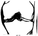
Фиг. 1А представляет собой чертеж, показывающий типичный суставной конец кости. Обычно костный материал покрыт на сочленяющейся поверхности хрящевой тканью.Brief Description of the Drawings
The present invention will be clearer when studying the following drawings, which illustrate certain properties of the present invention, in which:
FIG. 1A is a drawing showing a typical articular end of a bone. Typically, bone material is coated on the articulating surface with cartilage.
На фиг.1В показан пример, где развивается дефект или повреждение хрящевого покрытия (зазор в хряще), и такой дефект перед лечением можно с помощью хирургических процедур подвергнуть непосредственной обработке, небольшому увеличению или профилированию для восприятия пересаженного материала. FIG. 1B shows an example where a defect or damage to the cartilage coating develops (a gap in the cartilage), and such a defect can be subjected to direct processing, a slight increase or profiling to perceive the transplanted material before surgical treatment.
На фиг. 1С показано, как гемостатический барьер (обозначенный цифрой 1) помещается внутрь дефекта хрящевого покрытия для ингибирования или предотвращения прорастания сосудов в регенерирующий хрящ из подлежащей кости. Затем поверх гемостатического барьера наслаиваются хондроциты, имплантация которых предполагается в полость дефекта. In FIG. 1C shows how the hemostatic barrier (indicated by the number 1) is placed inside a cartilage defect to inhibit or prevent vascular growth in the regenerating cartilage from the underlying bone. Then, chondrocytes are layered over the hemostatic barrier, the implantation of which is supposed to be in the cavity of the defect.
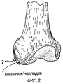
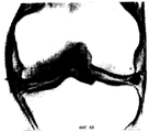
Фиг.2 представляет собой чертеж, показывающий обработанный дефект (зазор в хряще) в хрящевом покрытии, покрытый бесклеточным полупроницаемым материалом (обозначенным цифрой 2), который используется для образования колпачка/накладки или бандажа над участком дефекта. Этот колпачок фиксируется на месте, или пришиванием к здоровому хрящу по краю полости, или прикрепляется другим способом. Этот колпачок покрывает область дефекта сустава, в которую был помещен трансплантат культивированных хондроцитов/хряща, или будет помещен под частично прикрепленный колпачок. Figure 2 is a drawing showing the treated defect (gap in the cartilage) in the cartilage coating, covered with cell-free semi-permeable material (indicated by number 2), which is used to form a cap / lining or bandage over the defect area. This cap is fixed in place, either by suturing to a healthy cartilage along the edge of the cavity, or attached in another way. This cap covers the area of the joint defect in which the grafted chondrocyte / cartilage graft was placed, or will be placed under the partially attached cap.
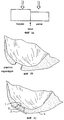
Фиг. 3А представляет собой график, иллюстрирующий различную реакцию на силы сдавливания и сдвига более твердого и более мягкого хряща с последующей зоной демаркации. FIG. 3A is a graph illustrating a different response to the compressive and shear forces of harder and softer cartilage, followed by a demarcation zone.
Фиг.3В иллюстрирует участок пересадки после того, как дефект был спрофилирован с образованием волнистой поверхности стенок. FIG. 3B illustrates a transplant site after a defect has been profiled to form a corrugated wall surface.
Фиг.3С иллюстрирует спрофилированный участок пересадки с гемостатическим барьером (1), пересиженным материалом (3) и бесклеточной покрывающей накладкой (2), размещенными внутрь хряща (4) суставной поверхности. Fig. 3C illustrates a profiled transplant site with a hemostatic barrier (1), re-sourced material (3) and a cell-free covering pad (2) placed inside the cartilage (4) of the articular surface.
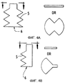
Фиг. 4А иллюстрирует один вариант реализации хирургического устройства настоящего изобретения, где доказаны режущие зубья (5) и выступающий штырь для размещения (6). Иллюстрации в разрезе справа показывают две возможные конфигурации режущих лезвий. FIG. 4A illustrates one embodiment of a surgical device of the present invention where cutting teeth (5) and a protruding pin for placement (6) are proven. The cutaway illustrations to the right show two possible configurations of cutting blades.
Фиг. 4В иллюстрирует второй вариант реализации хирургического устройства настоящего изобретения. FIG. 4B illustrates a second embodiment of a surgical device of the present invention.
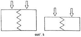
Фиг. 5 представляет собой график, иллюстрирующий модифицированную различную реакцию на силы сдавливания и сдвига более твердого и более мягкого хряща после профилирования участка пересадки. FIG. 5 is a graph illustrating a modified different response to the compressive and shear forces of harder and softer cartilage after profiling the transplant site.
Фиг.6А представляет собой полученное с помощью ЯМР изображение коленного сустава свиньи, показывающее дефект хряща в левом (медиальном) мыщелке. 6A is an NMR image of a pig knee joint showing a cartilage defect in the left (medial) condyle.
Фиг. 6В представляет собой полученное с помощью ЯМР изображение того же коленного сустава свиньи через три месяца после лечения. FIG. 6B is an NMR image of the same pig knee joint three months after treatment.
Подробное описание изобретения
Это изобретение касается использования определенных продуктов, которые ингибируют образование сосудистой ткани, например, такой как капиллярные петли, врастающие в сформировавшийся хрящ, во время процесса аутотрансплантации хондроцитов в дефекты в хряще. Образующаяся сосудистая ткань из подлежащей кости будет иметь тенденцию врастать в новый хрящ, образование которого предполагается, приводя к появлению клеток, отличных от желательных мезенхимальных специализированных хондроцитов.DETAILED DESCRIPTION OF THE INVENTION
This invention relates to the use of certain products that inhibit the formation of vascular tissue, for example, such as capillary loops growing into the formed cartilage, during the process of autologous transplantation of chondrocytes into defects in the cartilage. The resulting vascular tissue from the underlying bone will tend to grow into a new cartilage, the formation of which is assumed, leading to the appearance of cells other than the desired specialized mesenchymal chondrocytes.
Примешивающиеся клетки, внедряющееся в результате прорастания сосудов, могут вызвать вторжение и избыточное врастание в хрящ, образование которого предполагается имплантированными хондроцитами. Одним из типов промышленных продуктов, которые могут использоваться в этом изобретении, является Surgicel® (Ethicon Ltd., UK), который может всасываться через 7-14 дней. Использование этого материала в способе настоящего изобретения противоположно обычному использованию гемостатического устройства, такого как Surgicel®, как это описано во вкладыше компании Ethicon Ltd., в упаковке. Mixed cells, introduced as a result of vascular germination, can cause invasion and excessive growth of cartilage, the formation of which is assumed to be implanted chondrocytes. One type of industrial product that can be used in this invention is Surgicel® (Ethicon Ltd., UK), which can be absorbed after 7-14 days. The use of this material in the method of the present invention is the opposite of the usual use of a hemostatic device, such as Surgicel®, as described in the package insert of Ethicon Ltd.
К удивлению, мы обнаружили, что в ситуации, когда вы хотите ингибировать повторное прорастание сосудов в хрящ, гемостатический материал будет действовать в качестве гелеподобного искусственного хряща. Если бы эритроциты присутствовали внутри дефекта всей толщины суставного хряща, который покрыт таким гемостатическим барьером, эти клетки крови были бы химически преобразованы в гематин и поэтому стали бы неспособны вызывать рост сосудов. Следовательно, гемостатический продукт, используемый в качестве барьера, ингибирующего повторное прорастание сосудов с применением фибриновых клеев или без них, такой, например, как Surgicel®, эффективен для рассматриваемого способа в соответствии с положениями настоящего изобретения. Другой частью этого изобретения является использование бесклеточного компонента, который используется в виде накладки, покрывающей область дефекта сустава, в который трансплантируются культивированные хондроциты/хрящи с использованием для трансплантации аутологичных хондроцитов. Способ изобретения также предусматривает использование подходящих аллогенных хондроцитов или ксеногенных хондроцитов для восстановления дефекта хряща. Surprisingly, we found that in a situation where you want to inhibit the re-growth of blood vessels into the cartilage, the hemostatic material will act as a gel-like artificial cartilage. If red blood cells were present inside the defect of the entire thickness of the articular cartilage, which is covered by such a hemostatic barrier, these blood cells would be chemically converted to hematin and therefore would be unable to cause vascular growth. Therefore, a hemostatic product used as a barrier inhibiting the re-growth of vessels with or without fibrin adhesives, such as, for example, Surgicel®, is effective for the method in question in accordance with the provisions of the present invention. Another part of this invention is the use of a cell-free component, which is used in the form of a lining covering the joint defect region into which cultured chondrocytes / cartilage are transplanted using autologous chondrocytes for transplantation. The method of the invention also involves the use of suitable allogeneic chondrocytes or xenogenic chondrocytes to repair a cartilage defect.
Таким образом, настоящее изобретение описывает способ эффективного восстановления или обработки дефектов хряща в костных поверхностях суставного сочленения, который включает введение средства или устройства для блокировки врастания сосудов в подлежащий восстановлению участок хряща, а также обеспечение бесклеточного барьера, который изолирует участок восстановления и удержит на месте трансплантированные клетки. Таким образом, настоящее изобретение также обеспечивает набор, включающий компонент гемостатического барьера для вставки в подлежащий восстановлению участок так, что имеется эффективное ингибирование прорастания сосудов в подлежащий восстановлению участок; и как только подлежащие трансплантации хондроциты помещаются в подлежащий восстановлению участок, поверх восстанавливаемого участка накладывается бесклеточный полупроницаемый барьер так, что трансплантированные хондроциты удерживаются на месте, но еще способны получать доступ к питательным веществам. Thus, the present invention describes a method for efficiently repairing or treating cartilage defects in the bone surfaces of the articular joint, which includes introducing an agent or device to block the growth of vessels in the cartilage site to be restored, as well as providing a cell-free barrier that isolates the restoration site and keeps the transplanted in place cells. Thus, the present invention also provides a kit comprising a component of a hemostatic barrier for insertion into a site to be restored so that there is effective inhibition of vascular germination in the site to be restored; and as soon as the chondrocytes to be transplanted are placed in the area to be restored, a cell-free semi-permeable barrier is applied over the restored area so that the transplanted chondrocytes are held in place, but are still able to gain access to nutrients.
Примеры определенных аспектов изобретения были приведены с использованием системы in vitro для изучения поведения хондроцитов при контакте с определенным продуктом или комбинацией определенных продуктов, которые ингибируют образование сосудистой ткани. Это испытание in vitro протезирует способность определенных исследуемых материалов ингибировать прорастание сосудов, которое произойдет in vivo, когда капиллярные петли врастут в хрящ, образуемый во время процесса аутотрансплантации хондроцитов в дефекты в хряще. Examples of certain aspects of the invention have been given using an in vitro system to study the behavior of chondrocytes in contact with a particular product or combination of certain products that inhibit the formation of vascular tissue. This in vitro test prostheses the ability of certain test materials to inhibit vascular sprouting that will occur in vivo when capillary loops grow into the cartilage formed during the process of autologous transplantation of chondrocytes into cartilage defects.
Подходяще гемостатические продукты будут характеризоваться наличием способности ингибировать рост или внедрение сосудистой ткани, остеоцитов, фибробластов и т.д. в развивающийся хрящ. Подходящий гемостатический материал достигнет цели способа настоящего изобретения в том, что для оптимизации образования хряща и достижения восстановления полной толщины любых дефектов в суставном хряще, следует предотвратить внедрение сосудов и клеток в развивающийся хрящ. В идеале гемостатический барьер будет устойчивым в течение продолжительного периода времени для обеспечения возможности полного восстановления хряща, а затем с течением времени будет способен рассасываться или разрушаться другим образом. Один из выявленных в качестве подходящего материала называется Surgicel® W1912 (рассасываемый гемостатический материал, содержащий окисленную регенерированную стерильную целлюлозу; Lot GG3DH, Ethicon Ltd. UK). Другим примером подходящего материала является BioGide® (имеющаяся в продаже накладка из коллагенового матрикса I типа; Geistlich Sobne, Switzerland). Suitable hemostatic products will have the ability to inhibit the growth or uptake of vascular tissue, osteocytes, fibroblasts, etc. into developing cartilage. A suitable hemostatic material will achieve the objective of the method of the present invention in that in order to optimize the formation of cartilage and to restore full thickness of any defects in the articular cartilage, the incorporation of blood vessels and cells into developing cartilage should be prevented. Ideally, the hemostatic barrier will be stable over an extended period of time to allow complete restoration of the cartilage, and then over time will be able to dissolve or break down in another way. One identified as a suitable material is called Surgicel® W1912 (absorbable hemostatic material containing oxidized regenerated sterile cellulose; Lot GG3DH, Ethicon Ltd. UK). Another example of a suitable material is BioGide® (a commercially available type I collagen matrix overlay; Geistlich Sobne, Switzerland).
Подходящий органический клеящий материал, такой как, например, Tisseel® или Tissucol® (клей на основе фибрина; Immuno AG, Austria), клеющий белок (Cat. #A-2707, Sigma Chaemical, USA) и Dow Corning Medical Adhesive В (Cat. #895-3, Dow Corning, USA), можно найти в продаже. Хирургические инструменты, предусматриваемые настоящим изобретением, могут быть изготовлены из металла и/или пластика, пригодного для изготовления хирургических инструментов для одноразового или многократного использования. Режущий инструмент может содержать режущие зубья, которые являются полностью круговыми или плоскими или имеют какую-либо промежуточную форму. Поскольку хрящ представляет собой относительно мягкий материал, преимущество может иметь изготовление режущих краев повышенной прочности, которые будут способны спрофилировать хрящ, не будучи способными повредить кость. Такие режущие инструменты могут быть изготовлены с включением отверстий для введения жидкости, удаления с помощью отсоса остатков отсекаемой ткани, и жидкости, и нитей оптических волокон для освещения и визуализации участка дефекта. Suitable organic adhesive materials such as, for example, Tisseel® or Tissucol® (fibrin-based adhesive; Immuno AG, Austria), adhesive protein (Cat. # A-2707, Sigma Chaemical, USA) and Dow Corning Medical Adhesive B (Cat . # 895-3, Dow Corning, USA) can be found on sale. Surgical instruments contemplated by the present invention may be made of metal and / or plastic suitable for the manufacture of surgical instruments for single or multiple use. The cutting tool may contain cutting teeth that are completely circular or flat or have any intermediate shape. Since the cartilage is a relatively soft material, it may be advantageous to make cutting edges of increased strength that will be able to profile the cartilage without being able to damage the bone. Such cutting tools can be made with the inclusion of holes for introducing liquid, removing by suction residues of the cut-off tissue, and liquid, and optical fiber threads for lighting and visualizing a defect site.
Определенные стороны настоящего изобретения можно лучше понять при иллюстрации с помощью следующих примеров, которые предназначены для иллюстрации, а не ограничения. Certain aspects of the present invention can be better understood by illustration using the following examples, which are intended to illustrate and not limit.
Пример 1
Для использования Surgicel® в соответствии с изобретением для предотвращения развития кровеносных сосудов в имплантированный аутотрансплантат хряща или хондроцитов Surgicel® сначала обрабатывают фиксаторам, таким как глутаровый альдегид. Вкратце, Surgicel® обрабатывают 0,6%-ным глутаровым альдегидом в течение 1 мин с последующими несколькими промываниями для устранения остатков альдегида, которые иначе могут оказать токсическое воздействие на ткань. Альтернативно Surgicel® обрабатывают фибриновым клеем, именуемым Tisseel®, перед обработкой глутаровым альдегидом, как описано в примере 2. Было обнаружено, что Surgicel®, фиксированный, например, таким фиксатором, как глутаровый альдегид, промытый стерильным физиологический солевым раствором (0,9%) и хранившийся в холодильнике, не растворяется в течение 1-2 мес. В целом, Surgicel® рассасывается в течение периода от 7 до 14 дней. Этот период времени был бы слишком коротким, потому что для предотвращения развития кровеносных сосудов или прорастания сосудов как такового из костной структуры в имплантированный хрящ перед тем, как имплантированные хондроциты прорастут в твердый хрящевой слой, получающий свои питательные потребности из соседнего хряща, требуется более длительный период времени. Другими словами, достаточное ингибирование прорастания сосудов необходимо в течение более длительного времени, такого как, например, 1 мес. Поэтому продукт не должен существенно всасываться до этого времени. С другой стороны, в конечном счете необходимо его рассасывание. Следовательно, органический материал, используемый в качестве ингибирующего барьера, должен обладать этими способностями, и было обнаружено, что Surgicel®, обработанный таким образом, обеспечивает эту функцию.Example 1
To use the Surgicel® of the invention to prevent the development of blood vessels in an implanted cartilage or chondrocyte autograft, Surgicel® is first treated with fixatives, such as glutaraldehyde. Briefly, Surgicel® is treated with 0.6% glutaraldehyde for 1 min followed by several rinses to remove aldehyde residues that could otherwise have toxic effects on the tissue. Alternatively, Surgicel® is treated with a fibrin glue called Tisseel® before being treated with glutaraldehyde as described in Example 2. It was found that Surgicel® fixed with, for example, a fixative such as glutaraldehyde washed with sterile physiological saline (0.9% ) and stored in the refrigerator, does not dissolve within 1-2 months. In general, Surgicel® resolves over a period of 7 to 14 days. This period of time would be too short, because a longer period is required to prevent the development of blood vessels or the growth of blood vessels from the bone structure into the implanted cartilage before the implanted chondrocytes grow into the hard cartilage layer, which receives its nutritional needs from neighboring cartilage time. In other words, sufficient inhibition of vascular germination is necessary for a longer time, such as, for example, 1 month. Therefore, the product should not be substantially absorbed before this time. On the other hand, its resorption is ultimately necessary. Therefore, the organic material used as an inhibitory barrier must possess these abilities, and it was found that Surgicel® treated in this way provides this function.
Пример 2
Surgicel® также покрывают органическим клеем; в этом примере используемым клеем был Tisseel®, но могут использоваться другие клеи. Этот продукт совместно с Surgicel® создает подходящий барьер для конкретной цели изобретения. Может использоваться любой другой гемостатический или ингибирующий прорастание сосудов барьер. Tisseel® смешивают, как описано выше. Surgicel® затем покрывают Tisseel® с помощью распыления материала Surgicel® на обе стороны до его пропитывания. Затем Tisseel® (фибриновому клею) дают возможность затвердеть при комнатной температуре. Непосредственно перед завершением затвердевания покрытый Surgicel® затем помещают на 1 мин в 0,6%-ный глутаровый альдегид и затем промывают стерильным физиологическим (0,9%) солевым раствором. Затем с помощью фосфатного буферного раствора и/или NaOH доводят рН до тех пор, пока оно не станет устойчивым на уровне от 7,2 до 7,4. После этого обработанный таким образом Surgicel® промывают в среде для тканевой культуры, такой как минимальная основная среда/F 12 с буфером 15 мМ Hepes.Example 2
Surgicel® is also coated with organic glue; in this example, the adhesive used was Tisseel®, but other adhesives may be used. This product, together with Surgicel®, creates a suitable barrier for the specific purpose of the invention. Any other hemostatic or vascular germination inhibiting barrier may be used. Tisseel® is blended as described above. Surgicel® is then coated with Tisseel® by spraying Surgicel® material on both sides until it is soaked. Then Tisseel® (fibrin glue) is allowed to harden at room temperature. Immediately prior to completion of curing, the coated Surgicel® is then placed for 1 min in 0.6% glutaraldehyde and then washed with sterile physiological (0.9%) saline. Then, with a phosphate buffer solution and / or NaOH, the pH is adjusted until it becomes stable at a level of from 7.2 to 7.4. Thereafter, the Surgicel® thus treated is washed in a tissue culture medium such as Minimum Basic Medium / F 12 with 15 mM Hepes buffer.
Как указано в этом примере, мы в качестве фибринового клея для покрытия Surgicel® использовали Tisseel®. Кроме того, фибриновый адгезионный материал или клей можно также непосредственно наносить на дно повреждения по направлению к кости, на которую приклеивается Surgicel®. Используемая вместо исследования in vivo система in vitro состояла из 6-ячеечного одноразового планшета для клеточных исследований NUNCLONTM (NUNC, InterMed, Roskilde, Denmark). Каждая ячейка имеет диаметр приблизительно 4 см.As indicated in this example, we used Tisseel® as the fibrin glue for Surgicel® coating. In addition, the fibrin adhesive material or adhesive can also be directly applied to the bottom of the lesion towards the bone on which Surgicel® is glued. The in vitro system used in place of the in vivo study consisted of a 6-cell disposable NUNCLON ™ cell test plate (NUNC, InterMed, Roskilde, Denmark). Each cell has a diameter of approximately 4 cm.
В изобретении фибриновый адгезионный материал может представлять собой любой адгезионный материал, который совместно с фибриновым компонентам образует клей, который может быть толерантен для людей (lhara, N, et al., Burns Incl. Therm. Inj., 1984, 10, 396). Это изобретение также предусматривает любой другой клеевой компонент, который может использоваться вместо фибринового адгезионного материала. В этом изобретении мы использовали Tisseel® или Tissucol® (Immuno AG, Vienna, Austria). Набор Tisseel® состоит из следующих компонентов:
Tisseel®, лиофилизированное водонепроницаемое покрытие с инактивированными вирусами, содержащее свертывающийся белок, состоящее из фибриногена, фибронектина плазмы (телячьего иммуноглобулина), и фактора XIII, и плазминогена.In the invention, the fibrin adhesive material can be any adhesive material that, together with the fibrin components, forms an adhesive that can be tolerated by humans (lhara, N, et al., Burns Incl. Therm. Inj., 1984, 10, 396). This invention also provides for any other adhesive component that can be used in place of fibrin adhesive material. In this invention, we used Tisseel® or Tissucol® (Immuno AG, Vienna, Austria). The Tisseel® Kit consists of the following components:
Tisseel®, a lyophilized waterproof coating with inactivated viruses, containing a coagulating protein consisting of fibrinogen, plasma fibronectin (calf immunoglobulin), and factor XIII, and plasminogen.
Раствор апротинина (бычьего)
Тромбин 4 (бычий)
Тромбин 500 (бычий)
Раствор хлорида кальция
Набор Tisseel® содержит систему наложения DUPLOJECT®. Фибриновый клей или двухкомпонентное водонепроницаемое покрытие с использованием набора Tisseel® комбинируется следующим образом в соответствии с вкладышем продукта компании Immuno AG.Aprotinin (Bovine) Solution
Thrombin 4 (bovine)
Thrombin 500 (bovine)
Calcium Chloride Solution
The Tisseel® Kit contains the DUPLOJECT® overlay system. A fibrin adhesive or a two-component waterproof coating using the Tisseel® kit is combined as follows in accordance with the Immuno AG product insert.
Пример 3
Хондроциты выращивают в минимальной основной культуральной среде, содержащей НАМ F12 и 15 мМ буфера Hepes и от 5 до 7,5% аутологичной сыворотки в СО2, инкубаторе при 37oС и обрабатывают в лаборатории класса 100 в Verigen Europe A/S, Symbion Science Park, Copenhagen, Denmark. Для культивирования хондроцитов можно использовать другие композиции культуральной среды. Клетки трипсинизируют с использованием ЭДТА в течение 5-10 мин и считают с использованием окрашивания трипаном синим в камере Burker-Turk для определения жизнеспособности. Количество клеток доводят до 7,5•105 клеток на 1 мл. Одну планшету NUNCLONTM раскрывают в лаборатории класса 100.Example 3
Chondrocytes are grown in a minimal basic culture medium containing HAM F12 and 15 mM Hepes buffer and 5 to 7.5% autologous serum in CO 2 , incubator at 37 o C and processed in a class 100 laboratory in Verigen Europe A / S, Symbion Science Park, Copenhagen, Denmark. For culturing chondrocytes, other culture medium compositions may be used. Cells are trypsinized using EDTA for 5-10 minutes and counted using trypan blue staining in a Burker-Turk chamber to determine viability. The number of cells is adjusted to 7.5 • 10 5 cells per 1 ml. One plate Nunclon TM opens in a Class 100 laboratory.
Гемостатический барьер Surgicel® отрезают до подходящего размера, соответствующего дну ячейки в планшете для культуры ткани NUNCLONTM. B этом случае круг имеет размер приблизительно 4 см (но он может быть любого возможного размера), и его помещают в асептических условиях на дно в ячейке 6-ячеечиой одноразовой планшеты NUNCLONTM Delta для работы по исследованию клеток (NUNC, InterMed, Roskilde, Denmark). Гемостатический барьер, который предполагается поместить на дно ячейки, предварительно обрабатывают, как описано в примере 1. Эта обработка обеспечивает значительную отсрочку всасывания Surgicel. Задаем этот гемостатический барьер отмывают несколько раз в дистиллированной воде и в последующем несколько раз до тех пор, пока не будет смыт не вступивший в реакцию глутаральдегид. Небольшое количество среды для клеточной культуры, содержащей сыворотку, наносят для всасывания на гемостатический барьер, в то же самое время поддерживая влажность гемостатического барьера на дне ячейки.The Surgicel® hemostatic barrier is cut to a suitable size corresponding to the bottom of the cell in the NUNCLON ™ tissue culture plate. In this case, the circle has a size of approximately 4 cm (but it can be of any size possible), and it is placed under aseptic conditions on the bottom in the cell of a 6-well disposable NUNCLON ™ Delta plate for cell research (NUNC, InterMed, Roskilde, Denmark ) The hemostatic barrier, which is supposed to be placed at the bottom of the cell, is pre-treated as described in Example 1. This treatment provides a significant delay in the absorption of Surgicel. We set this hemostatic barrier to be washed several times in distilled water and then several times until the glutaraldehyde that has not reacted is washed off. A small amount of serum-containing cell culture medium is applied to the hemostatic barrier for absorption, while at the same time maintaining the humidity of the hemostatic barrier at the bottom of the cell.
Приблизительно 106 клеток в 1 мл культуральной среды помещают непосредственно на верхнюю часть гемостатического барьера, диспергированного по поверхности гемостатического барьера, предварительно обработанного 0,4%-ным глутаральдегидом, как описано выше. Затем планшету инкубируют в СО2 инкубаторе при 37oС в течение 60 мин. Среду для культуры тканей, содержащую от 5 до 7,5% сыворотки, в количестве от 2 до 5 мл осторожно добавляют в ячейку, содержащую клетки, избегая разбрызгивания клеток с помощью удерживания наконечника пипетки тангенциально к стороне ячейки при выталкивании среды. Оказывается, что рН среды очень низкое {рН≈6,8). Затем рН доводят до 7,4-7,5. На следующий день некоторые хондроциты начинают расти на гемостатическом барьере, располагаясь группами. Некоторые клетки погибают вследствие воздействия низкого рН перед доведением рН. Планшету инкубируют в лечение 3-7 дней со сменой среды на 3 день.Approximately 10 6 cells in 1 ml of culture medium are placed directly on top of the hemostatic barrier dispersed over the surface of the hemostatic barrier pretreated with 0.4% glutaraldehyde as described above. The plate is then incubated in a CO 2 incubator at 37 ° C. for 60 minutes. A tissue culture medium containing from 5 to 7.5% serum, in an amount of 2 to 5 ml, is carefully added to the cell containing cell, avoiding cell spatter by holding the pipette tip tangentially to the side of the cell when the medium is ejected. It turns out that the pH of the medium is very low (pH ≈ 6.8). Then the pH is adjusted to 7.4-7.5. The next day, some chondrocytes begin to grow on the hemostatic barrier, located in groups. Some cells die due to exposure to low pH before adjusting the pH. The tablet is incubated in the treatment of 3-7 days with a change of medium for 3 days.
В конце периода инкубации среду декантируют и хранившийся в холодильнике холодный 2,5%-ный глутаральдегид, содержащий 0,1 М натриевую соль диметиларсиновой кислоты (называемую также какодилат с доведением рН с помощью НСl до 7,4), добавляют в качестве фиксатора дня получения клетки и подложки (гемостатиыеского барьера) для последующей подготовки к электронной микроскопии. At the end of the incubation period, the medium is decanted and cold 2.5% glutaraldehyde stored in the refrigerator containing 0.1 M dimethylarsinic acid sodium salt (also called cacodylate adjusted to pH 7.4 with HCl) is added as a fixative for the day of production cells and substrates (hemostatic barrier) for subsequent preparation for electron microscopy.
Пример 4
Хондроциты выращивают в минимальной основной культуральной среде, содержащей НАМ F12 и 15 мМ буфера Hepes и от 5 до 7,5% аутологичной сыворотки, в СО2 инкубаторе при 37oС и обрабатывают в лаборатории класса 100 в Verigen Europe А/S, Symbion Science Park, Copenhagen, Denmark. Для культивирования хондроцитов можно использовать другие композиции культуральной среды. Клетки трипсинизируют с использованием ЭДТА в течение 5-10 мин и считают с использованием окрашивания трипаном синим в камере Burker-Turk для определения жизнеспособности. Количество клеток доводили до 7,5•105 клеток на 1 мл. Одну планшету NUNCLONTM раскрывают в лаборатории класса 100.Example 4
Chondrocytes are grown in a minimal basic culture medium containing HAM F12 and 15 mM Hepes buffer and 5 to 7.5% autologous serum in a CO 2 incubator at 37 ° C and processed in a class 100 laboratory in Verigen Europe A / S, Symbion Science Park, Copenhagen, Denmark. For culturing chondrocytes, other culture medium compositions may be used. Cells are trypsinized using EDTA for 5-10 minutes and counted using trypan blue staining in a Burker-Turk chamber to determine viability. The number of cells was adjusted to 7.5 • 10 5 cells per 1 ml. One plate Nunclon TM opens in a Class 100 laboratory.
Surgicel® (для использования в качестве гемостатического барьера) обрабатывают 0,6%-ным глутаровым альдегидом в течение 1 мин, как описано в примере 1, и промывают 0,9%-ным стерильным раствором хлорида натрия или предпочтительно буфером, таким как фосфатный буферный раствор, или культуральной средой, такой как MEM/F12, потому что рН после обработки глутаровым альдегидом составляет 6,8, а должно быть предпочтительно от 7,0 до 7,5. На обе стороны Surgicel® наносят Tisseel® с использованием системы DUPLOJECT®, покрывая таким образом обе стороны Surgicel®, накладки, которую предполагается использовать, фибриновым клеем. Клей оставляют для высыхания в асептических условиях в течение, по меньшей мере, от 3 до 5 мин. "Покрытый" гемостатический барьер помещают на дно ячейки в 6-ячеечной одноразовой планшете NUNCLONTM Delta для работы по исследованию клеток. Небольшое количество среды для тканевой культуры, содержащей сыворотку, наносят для всасывания на гемостатический барьер. Приблизительно 106 клеток в 1 мл культуральной среды, содержащей сыворотку, помещают непосредственно на верхнюю часть гемостата, диспергированного по поверхности гемостатического барьера. Затем планшету инкубируют в CO2 инкубаторе при 37oС в течение 60 мин. Среду для культуры тканей, содержащую от 5 до 7,5% сыворотки, в количестве от 2 до 5 мл осторожно добавляют в ячейку, содержащую клетки, избегая разбрызгивания клеток с помощью удерживания наконечника пипетки тангенциально к стороне ячейки при выталкивании среды. Через 3-6 дней микроскопическое исследование показывает, что клетки удовлетворительным образом прилипли и вросли в Surgicel®, свидетельствуя о том, что Surgicel® не проявляет токсичности по отношению к хондроцитам и что хондроциты удовлетворительным образом врастают в Surgicel®.
Планшету инкубируют в течение 3-7 дней со сменой среды на 3 день. В конце периода инкубации среду декантируют и хранившийся в холодильнике холодный 2,5%-ный глутаральдегид, содержащий 0,1 М натриевую соль диметиларсиновой кислоты, называемой также какодилат натрия, с доведением рН с помощью НС1 до 7,4, добавляют в качестве фиксатора для получения клетки и подложки (гемостатического барьера) для последующей подготовки к электронной микроскопии.Surgicel® (for use as a hemostatic barrier) is treated with 0.6% glutaraldehyde for 1 min, as described in example 1, and washed with 0.9% sterile sodium chloride solution or preferably with a buffer such as phosphate buffer solution, or culture medium, such as MEM / F12, because the pH after treatment with glutaraldehyde is 6.8, and should preferably be from 7.0 to 7.5. Tisseel® is applied to both sides of Surgicel® using the DUPLOJECT® system, thus covering both sides of Surgicel®, the patch to be used, with fibrin glue. The glue is left to dry under aseptic conditions for at least 3 to 5 minutes. A “coated” hemostatic barrier is placed at the bottom of the cell in a 6-well disposable NUNCLON ™ Delta plate for cell research. A small amount of tissue culture medium containing serum is applied to the hemostatic barrier for absorption. About 10 6 cells in 1 ml of serum-containing culture medium are placed directly on top of a hemostat dispersed over the surface of the hemostatic barrier. The plate is then incubated in a CO 2 incubator at 37 ° C. for 60 minutes. A tissue culture medium containing from 5 to 7.5% serum, in an amount of 2 to 5 ml, is carefully added to the cell containing cell, avoiding cell spatter by holding the pipette tip tangentially to the side of the cell when the medium is ejected. After 3-6 days, microscopic examination shows that the cells have satisfactorily adhered and grown into Surgicel®, indicating that Surgicel® is not toxic to chondrocytes and that chondrocytes satisfactorily grow into Surgicel®.
The plate is incubated for 3-7 days with a change of medium for 3 days. At the end of the incubation period, the medium is decanted and cold 2.5% glutaraldehyde stored in the refrigerator containing 0.1 M sodium salt of dimethylarsinic acid, also called sodium cacodylate, adjusted to pH 7.4 with HCl, is added as a fixative for obtaining cells and substrates (hemostatic barrier) for subsequent preparation for electron microscopy.
Пример 5
Хондроциты выращивают в минимальной основной культуральной среде, содержащей НАМ F12 и 15 мМ буфера Hepes и от 5 до 7,5% аутологичной сыворотки, в СО2 инкубаторе при 37oС и обрабатывают в лаборатории класса 100 в Verigen Europe A/S, Symbion Science Park, Copenhagen, Denmark. Клетки трипсинизируют с использованием ЭДТА в течение 5-1-0 мин и считают с использованием окрашивания трипаном синим в камере Burker-Turk. Количество клеток доводят до 7,5•105-2•106 клеток на 1 мл. Одну планшету NUNCLONTM раскрывают в лаборатории класса 100.Example 5
Chondrocytes are grown in minimal basic culture medium containing HAM F12 and 15 mM Hepes buffer and 5 to 7.5% autologous serum in a CO 2 incubator at 37 ° C and processed in a class 100 laboratory in Verigen Europe A / S, Symbion Science Park, Copenhagen, Denmark. Cells are trypsinized using EDTA for 5-1-0 minutes and counted using trypan blue staining in a Burker-Turk chamber. The number of cells is adjusted to 7.5 • 10 5 -2 • 10 6 cells per 1 ml. One plate Nunclon TM opens in a Class 100 laboratory.
Было обнаружено, что Bio-Gide® может использоваться в качестве рассасываемой двухслойной мембраны, которая будет применяться в качестве накладки или бандажа, покрывающего область дефекта сустава, в которую трансплантируются культивированные хондроциты, а также гемостатического барьера. Bio-Gide® представляет собой чистую коллагеновую мембрану, подученную с помощью стандартизованных, контролируемых процессов изготовления (по E.D. Geistlich Sohne AG, CH-6110 Wolhusen). Коллаген экстрагируют у свиней, прошедших ветеринарную сертификацию, и тщательно очищают во избежание антигенных реакций и стерилизуют в двойных блистерах с помощью γ-облучения. Двухслойная мембрана имеет пористую поверхность и плотную поверхность. Мембрана изготовлена из коллагена I типа и III типа без дальнейшей поперечной сшивки или химической обработки, Коллаген рассасывается в пределах 24 над. Мембрана сохраняет свою структурную целостность даже в мокром состоянии и может фиксироваться с помощью швов или штифтами. Мембрану можно также "приклеить" с использованием фибринового клея, такого как Tisseel®, к соседнему хрящу или ткани или вместо швов, или совместно со швами. It was found that Bio-Gide® can be used as a resorbable bilayer membrane, which will be used as a lining or bandage covering the joint defect area into which cultured chondrocytes are transplanted, as well as a hemostatic barrier. Bio-Gide® is a pure collagen membrane prepared using standardized, controlled manufacturing processes (according to E.D. Geistlich Sohne AG, CH-6110 Wolhusen). Collagen is extracted from veterinary certified pigs and thoroughly cleaned to avoid antigenic reactions and sterilized in double blisters using γ-radiation. The two-layer membrane has a porous surface and a dense surface. The membrane is made of type I and type III collagen without further crosslinking or chemical treatment. Collagen is absorbed within 24 above. The membrane retains its structural integrity even when wet and can be fixed with sutures or pins. The membrane can also be “glued” using fibrin glue, such as Tisseel®, to adjacent cartilage or tissue, either in place of sutures, or together with sutures.
Bio-Gide® раскрывают в лаборатории класса 100 и помещают в аспетических условиях на дно ячейки в 6-ячеечной одноразовой планшете NUNCLONTM Delta для работы по исследованию клеток или с пористой поверхностью двухслойной мембраны, обращенной вверх, или с плотной поверхностью, обращенной вверх. Приблизительно 106 клеток в 1 мл среды для культуры ткани, содержащей сыворотку, помещают непосредственно на верхнюю часть Bio-Gide®, диспергированного или по пористой, или по плотной поверхности Bio-Gide®, затем планшету инкубируют в СО2 инкубаторе при 37oС в течение 60 мин. Среду для культуры тканей, содержащую от 5 до 7,5% сыворотки, в количестве от 2 до 5 мл осторожно добавляют в ячейку, содержащую клетки, избегая разбрызгивания клеток с помощью удерживания наконечника пипетки тангенциально к стороне ячейки при выталкивании среды.Bio-Gide® is opened in a Class 100 laboratory and placed under specific conditions at the bottom of the cell in a 6-cell NUNCLON ™ Delta disposable plate for cell research with either the porous surface of the double-layer membrane facing up or the solid surface facing up. About 10 6 cells in 1 ml of serum-containing tissue culture medium are placed directly on top of the Bio-Gide® dispersed either on the porous or dense surface of the Bio-Gide®, then the plate is incubated in a CO 2 incubator at 37 ° C within 60 minutes A tissue culture medium containing from 5 to 7.5% serum, in an amount of 2 to 5 ml, is carefully added to the cell containing cell, avoiding cell spatter by holding the pipette tip tangentially to the side of the cell when the medium is ejected.
На 2 день после помещения хондроцитов в ячейку, содержащую Bio-Gide®, клетки исследуют в инвертированном микроскопе Nikon. Было отмечено, что некоторые хондроциты прилипли к краю Bio-Gide. Конечно, при использовании этого микроскопа было невозможно смотреть через сам Bio-Gide®.
Планшету инкубируют в течение 3-7 дней со сменой среды на 3 день. В конце периода инкубации среду декантируют и хранившийся в холодильнике холодный 2,5%-ный глутаральдегид, содержащий 0,1 М натриевую соль диметиларсиновой кислоты, называемой также какодилат натрия, с доведением рН с помощью HCl до 7,4, добавляют в качестве фиксатора для получения клетки и опоры Bio-Gide®, с клетками или культивированными на пористой поверхности, или на плотной поверхности. Накладки из Bio-Gide® затем посылают для электронной микроскопии в отделение патанатомии Herlev Hospital, Denmark.On
The plate is incubated for 3-7 days with a change of medium for 3 days. At the end of the incubation period, the medium is decanted and cold 2.5% glutaraldehyde stored in the refrigerator containing 0.1 M sodium salt of dimethylarsinic acid, also called sodium cacodylate, adjusted to pH 7.4 with HCl, is added as a fixative for obtaining cells and supports Bio-Gide®, with cells either cultured on a porous surface, or on a dense surface. Bio-Gide® pads are then sent for electron microscopy to the Pathology Department of Herlev Hospital, Denmark.
Электронная микроскопия показывает, что хондроциты, культивированные на плотной поверхности Bio-Gide®, не врастают в коллагеновую структуру Bio-Gide®, тогда как клетки, культивированные на пористой поверхности, действительно не врастают в коллагеновую структуру и, кроме того, проявляют присутствие протеогликанов и отсутствие признаков фибробластных структура. Этот результат показывает, что когда коллагеновая накладка, такая, например, как накладка из Bio-Gide®, пришивается в качестве накладки, покрывающей дефект хряща, причем пористая поверхность обращена вниз по направлению к дефекту, в который предполагается инъецировать культивированные хондроциты. Они затем будут способны проникать через коллаген и образовывать гладкую хрящевую поверхность вровень с интактной поверхностью, и в этой области будет постепенно создаваться гладкий слой протеогликанов. Тогда как если плотная поверхность коллагена обращена вниз в дефект, хондроциты, подлежащие имплантации, не объединятся с коллагеном, и клетки не образуют такую же гладкую поверхность, как описано выше. Electron microscopy shows that chondrocytes cultured on a dense Bio-Gide® surface do not grow into the Bio-Gide® collagen structure, while cells cultured on a porous surface do not grow into a collagen structure and, in addition, exhibit the presence of proteoglycans and lack of signs of fibroblast structure. This result shows that when a collagen patch, such as, for example, a Bio-Gide® patch, is sutured as a patch to cover a cartilage defect, with the porous surface facing downward to the defect into which the cultured chondrocytes are to be injected. They will then be able to penetrate through collagen and form a smooth cartilaginous surface flush with the intact surface, and a smooth layer of proteoglycans will gradually be created in this area. Whereas if the dense surface of collagen faces down into the defect, the chondrocytes to be implanted will not combine with collagen, and the cells will not form the same smooth surface as described above.
Пример 6
Хондроциты выращивают в минимальной основной культуральной среде, содержащей НАМ F12 и 15 мМ буфера Hepes и от 5 до 7,5% аутологичной сыворотки, в СО2 инкубаторе при 37oС и обрабатывают в лаборатории класса 100 в Verigen Europe A/S, Symbion Science Park, Copenhagen, Denmark. Клетки трипсинизируют с использованием трипсина ЭДТА в течение 5-10 мин и считают в камере Burker-Turk с использованием окрашивания трипаном синим для определения жизнеспособности. Количество клеток доводят до 7,5•105-2•106 клеток на 1 мл. Одну планшету NUNCLONTM раскрывают в лаборатории класса 100.Example 6
Chondrocytes are grown in minimal basic culture medium containing HAM F12 and 15 mM Hepes buffer and 5 to 7.5% autologous serum in a CO 2 incubator at 37 ° C and processed in a class 100 laboratory in Verigen Europe A / S, Symbion Science Park, Copenhagen, Denmark. Cells are trypsinized using trypsin EDTA for 5-10 minutes and counted in a Burker-Turk chamber using trypan blue staining to determine viability. The number of cells is adjusted to 7.5 • 10 5 -2 • 10 6 cells per 1 ml. One plate Nunclon TM opens in a Class 100 laboratory.
Bio-Gide®, используемый в качестве рассасываемой двухслойной мембраны, может также использоваться совместно с органическим клеем, таким как Tisseel® с дополнительным, значительно более высоким содержанием апротонина, чем обычно обнаруживается в Tisseel®, как описано во вкладыше продукта. С помощью увеличения содержания апротонина приблизительно до 25,000 КМЕ/мл, рассасывание материала будет отсрочено на недели вместо обычного периода, исчисляемого днями. Bio-Gide®, used as a resorbable bilayer membrane, can also be used in conjunction with an organic adhesive such as Tisseel® with an additional, significantly higher aprotonin content than is usually found in Tisseel®, as described in the product insert. By increasing the aprotonin content to approximately 25,000 KMU / ml, the resorption of the material will be delayed for weeks instead of the usual period of days.
Для исследования этого признака in vitro Tisseel® наносят на дно ячейки планшеты NUNCLONTM и обеспечивают возможность неполного затвердеваания. Затем на Tisseel® накладывают коллагеновую накладку, такую как Bio-Gide®, и приклеивают ко дну ячейки. Эта комбинация Bio-Gide® и Tisseel® предназначена быть гемостатическим барьером, который будет ингибировать или предотвращать развитие или инфильтрацию кровеносных сосудов в область трансплантации хондроцитов. Теперь эту гибридную коллагеновую накладку можно использовать и в качестве гемостатического барьера на дне поражения (максимально проксимальнее поверхности, подлежащей восстановлению), но также и в качестве подложки для образования хряща, потому что дистальная поверхность может быть пористой стороной коллагеновой накладки и, следовательно, способствовать инфильтрации хондроцитов и хрящевого матрикса. Таким образом, эта гибридная коллагеновая накладка может также использоваться для покрытия верхней части имплантата с коллагеновой пористой поверхностью, направленной вниз по направлению к имплантированным хондроцитам, и барьером, образующим верхнюю часть. Гибридная коллагеновая накладка с увеличенным содержанием апротонинового компонента может также использоваться без любого органического клея, такого как Tisseel®, и помещаться непосредственно внутрь дефекта, прилипая под воздействием естественных сил. Следовательно, коллагеновая накладка может использоваться и в качестве гемостатического барьера, и бесклеточного покрытия участка восстановления/трансплантата с пористыми поверхностями накладок, ориентированными в направлении к трансплантированным хондроцитам/хрящу. В другом варианте будет использоваться коллагеновая накладка, которая состоит из коллагена II типа (т.е. из Gelstlich Sohne AG, CH-6110 Wolhusen).For in vitro testing of this trait, Tisseel® is applied to the bottom of the cell with NUNCLON ™ plates and provides the possibility of incomplete solidification. Then, a collagen pad such as Bio-Gide® is applied to the Tisseel® and glued to the bottom of the cell. This combination of Bio-Gide® and Tisseel® is intended to be a hemostatic barrier that will inhibit or prevent the development or infiltration of blood vessels in the area of chondrocyte transplantation. Now this hybrid collagen lining can be used as a hemostatic barrier at the bottom of the lesion (maximally proximal to the surface to be restored), but also as a substrate for the formation of cartilage, because the distal surface can be the porous side of the collagen lining and, therefore, contribute to infiltration chondrocytes and cartilage matrix. Thus, this hybrid collagen patch can also be used to cover the upper part of the implant with a collagen porous surface directed down towards the implanted chondrocytes and a barrier forming the upper part. A hybrid collagen patch with an increased content of the aprotonin component can also be used without any organic adhesive, such as Tisseel®, and placed directly inside the defect, adhering under the influence of natural forces. Therefore, the collagen pad can be used both as a hemostatic barrier and as a cell-free coating of the restoration / graft site with porous surfaces of the plates oriented towards the transplanted chondrocytes / cartilage. In another embodiment, a collagen patch will be used, which consists of type II collagen (i.e. from Gelstlich Sohne AG, CH-6110 Wolhusen).
Таким образом, настоящее изобретение обеспечивает накладку из гибридного коллагена, в которой указанная накладка представляет собой коллагеновый матрикс с возросшими уровнями апротонинового компонента, предпочтительно приблизительно 25,000 КME/мл, в сочетании с клеем органического матрикса, в котором коллагеновый компонент аналогичен рассасываемому двухслойному материалу Bio-Gide® или коллагену II типа, а органически клей аналогичен материалу Tisseel®. В другом варианте реализации для приклеивания гибридной коллагеновой накладки к участку восстановления не используется никакой органический клей. Thus, the present invention provides a hybrid collagen patch in which said patch is a collagen matrix with increased levels of the aprotonin component, preferably about 25,000 KME / ml, in combination with an organic matrix adhesive in which the collagen component is similar to the Bio-Gide absorbable bilayer ® or type II collagen, and organically the glue is similar to Tisseel®. In another embodiment, no organic adhesive is used to adhere the hybrid collagen patch to the restoration site.
Пример 7
Ввиду ослабленной структуры хряща при остеоартрите, прилипание культивированных аутологичных хондроцитов, трансплантированных в участок пересадки в дефектном хряще, может быть ингибировано, создавая таким образом краевую зону (зону демаркации) между вновь имплантированным хрящом/хондроцитами и окружающим сформированным хрящом. Эта краевая зона будет более всего выражена, если участок пересадки подготовлен для трансплантата путем создания прямых, гладких стенок, срезанных линейным образом. Силы сдвига и сдавливания, действующие через такую краевую золу (как показано на фиг.3А), будут оказывать сильное воздействие для смещения трансплантата, если участок пересадки создается дальнейшим отсечением. Эта краевая зона и различное движение материалов вдоль этой зоны будет ингибировать сливное срастание между пересаженным материалом и окружающим материалом. Сдвиг этой краевой зоны усиливается, когда жесткость упирающегося материала различна. Во многих случаях материал трансплантата мягче, чем окружающий материал, однако в некоторых случаях заболевания остеоартритом окружающий хрящ может быть действительно мягче, чем имплантированные хондроциты/хрящ.Example 7
Due to the weakened cartilage structure in osteoarthritis, the adhesion of cultured autologous chondrocytes transplanted to the transplant site in a defective cartilage can be inhibited, thus creating an edge zone (demarcation zone) between the newly implanted cartilage / chondrocytes and the surrounding formed cartilage. This marginal zone will be most pronounced if the transplant site is prepared for the transplant by creating straight, smooth walls that are cut linearly. The shear and squeezing forces acting through such an edge ash (as shown in FIG. 3A) will have a strong effect for displacing the graft if the transplant site is created by further clipping. This marginal zone and the different movement of materials along this zone will inhibit confluence between the transplanted material and the surrounding material. The shift of this edge zone is enhanced when the stiffness of the abutting material is different. In many cases, the graft material is softer than the surrounding material, but in some cases of osteoarthritis, the surrounding cartilage can be really softer than implanted chondrocytes / cartilage.
Поэтому для разрешения этой проблемы способ изобретения предусматривает использование хирургических инструментов для профилирования стенок участка пересадки так, что стенки не линейные и, следовательно, обеспечивают волнистость поверхностей, что уменьшит сдвиг краевой зоны и обеспечит прикрепление трансплантированного материала. Можно также так спрофилировать участок пересадки, что диаметр участка проксимальнее поверхности кости имеет больший размер, чем отверстие дистальнее кости и на поверхности подлежащего восстановлению хряща тем, что имеется эффект "обратного туннеля". Суженное отверстие на поверхности поможет снижению сдвига краевой зоны и выталкивания материала трансплантата из участка пересадки. Therefore, to solve this problem, the method of the invention involves the use of surgical instruments for profiling the walls of the transplant site so that the walls are not linear and, therefore, provide undulation of the surfaces, which will reduce the shift of the edge zone and ensure the attachment of the transplanted material. It is also possible to profile the transplant site so that the diameter of the site proximal to the bone surface is larger than the hole distal to the bone and on the surface of the cartilage to be restored by the fact that there is a “reverse tunnel” effect. A narrowed hole on the surface will help reduce the shift of the marginal zone and the expulsion of the graft material from the transplant site.
Предпочтительный вариант реализации описывает профилирование стенок участка пересадки образом, подобным резьбовому отверстию для приема болта или винта (как показано на фиг. 3В), обеспечивая таким образом механическое сопротивление сдавливанию и/или выталкиванию пересаженного материала из участка пересадки, что может быть описано как "мужская" и "женская" резьба. A preferred embodiment describes profiling the walls of the transplant site in a manner similar to a threaded hole for receiving a bolt or screw (as shown in FIG. 3B), thus providing mechanical resistance to squeezing and / or pushing the transplanted material from the transplant site, which can be described as “male” "and" female "thread.
Хирургические инструменты, предусматриваемые настоящим изобретением, могут быть изготовлены из металла и/или пластика, пригодного для изготовления хирургических инструментов для одноразового или многократного использования. Поскольку хрящ представляет собой относительно мягкий материал, преимущество может иметь изготовление режущих краев повышенной прочности, которые будут способны спрофилировать хрящ, не будучи способными повредить кость. Такие режущие инструменты могут быть изготовлены с включением отверстий для введения жидкости, удаления с помощью отсоса остатков отсекаемой ткани, и жидкости, и нитей оптических волокон для освещения и визуализации участка дефекта. В определенных вариантах реализации инструмента основание инструмента может иметь выступающее острие или подобное шипу образование, которое поможет в направлении и размещении инструмента в участке пересадки. Конечно, такой шип будет сконструирован для сведения к минимуму повреждения подлежащей кости. Surgical instruments contemplated by the present invention may be made of metal and / or plastic suitable for the manufacture of surgical instruments for single or multiple use. Since the cartilage is a relatively soft material, it may be advantageous to make cutting edges of increased strength that will be able to profile the cartilage without being able to damage the bone. Such cutting tools can be made with the inclusion of holes for introducing liquid, removing by suction residues of the cut-off tissue, and liquid, and optical fiber threads for lighting and visualizing a defect site. In certain implementations of the tool, the base of the tool may have a protruding point or spike-like formation that will help in guiding and positioning the tool in the transplant site. Of course, such a spike will be designed to minimize damage to the underlying bone.
Хотя режущая поверхность инструмента может иметь одиночный зуб, или множество зубьев, или иметь вид шурупа, такой как у металлического метчика, используемого для создания резьбовых отверстий в металлических частях, характеристика, требуемая для режущего инструмента, такова, что полученные в результате спрофилированные стороны участка пересадки имеют волнистый, а не линейный профиль. Например, в определенных вариантах реализации режущий край инструмента может иметь форму, аналогичную форме, показанной на фиг.4А или как на фиг.4В. Режущий край может быть плоским или круглым, так что он наматывается вокруг диаметра режущего инструмента. Для осуществления способа изобретения может конструироваться множество других форм для создания поверхности раздела, которая обеспечивает механическое сопротивление различной реакции на силы сдавливания или сдвига, действующие на трансплантированный материал и окружающий материал. Although the cutting surface of the tool may have a single tooth, or multiple teeth, or be in the form of a screw, such as a metal tap used to create threaded holes in the metal parts, the characteristic required for the cutting tool is such that the resulting profiled sides of the transplant site have a wavy rather than linear profile. For example, in certain embodiments, the cutting edge of the tool may have a shape similar to that shown in FIG. 4A or as in FIG. 4B. The cutting edge may be flat or round, so that it is wound around the diameter of the cutting tool. To implement the method of the invention, many other forms can be designed to create an interface that provides mechanical resistance to a different reaction to the compressive or shear forces acting on the transplanted material and the surrounding material.
Пример 8
Четырехмесячную свинью смешанной йоркширской породы подвергают общей анестезии и кладут на спину. Свинью моют и обкладывают хирургическим бельем в операционной Harrington Arthritis Research Center, Phoenix, Arizonа. Все хирургическое вмешательство выполняют в асептических условиях. Левую заднюю лапу и прилагающую область живота и паха смазывают йодом. Определяют положение коленного сустава и надколенника. Осуществляют срединный и разрез приблизительно в 3 см от задней части надколенника и проводят приблизительный разрез нескольких подкожных, мышечных слоев и связок для получения доступа к медиальному мыщелку бедренной кости. С использованием кругового резца осуществляют повреждение в белом хряще на средней части медиального мыщелка, оставляя край от 0,5 до 1 см до края хряща, покрывающего заднемедиальную часть мыщелка (левый мыщелок, фиг.6А). Дефект размером 0,5-1 см размещают в каудальной части медиального мыщелка, несущей весовую нагрузку. Все хирургическое вмешательство выполняют без жгута на левом бедре. Различные слои и кожу ушивают соответствующим образом.Example 8
A four-month-old mixed Yorkshire pig is subjected to general anesthesia and placed on its back. The pig is washed and wrapped with surgical underwear in the operating room of the Harrington Arthritis Research Center, Phoenix, Arizon. All surgery is performed under aseptic conditions. The left hind paw and the surrounding abdomen and groin are lubricated with iodine. Determine the position of the knee joint and patella. A median and incision is made approximately 3 cm from the posterior part of the patella and an approximate incision is made of several subcutaneous, muscle layers and ligaments to gain access to the medial condyle of the femur. Using a circular incisor, damage is made in the white cartilage on the middle part of the medial condyle, leaving an edge of 0.5 to 1 cm to the edge of the cartilage covering the posterior medial condyle (left condyle, Fig. 6A). A defect of 0.5-1 cm in size is placed in the caudal part of the medial condyle bearing a weight load. All surgery is performed without a tourniquet on the left thigh. The various layers and skin are sutured accordingly.
На 3 день животное снова доставляют в операционную и укладывают, как описано выше, на операционном столе и проводят общую анестезию. Левую заднюю лапу, живот и область паха смазывают йодом, как описано выше. Швы разрезают и область открывают. Отмечают наличие умеренной гематомы в коленном суставе. Сгусток крови удаляют и осматривают дефект. В дефекте имеется сгусток крови, который удаляют. В дефект осторожно вкручивают стерильный хирургический инструмент, имеющий режущий край в виде мужской резьбы с размером, соответствующим или несколько большим, чем окружность повреждения. Накладку Bio-Gide® отрезают до размера, равного дну дефекта. Первый используемый клей, называемый Adhesive Protein (A-2707, Sigma Chemical, USA), накладывают на плотную сторону гемостатической барьерной накладки с отсеченными краями, и накладку посещают плотной стороной вниз на дно поражения с использованием ее в качестве барьера, как описано выше. Обнаруживают, что, как оказывается, этот клей не высыхает очень быстро. Небольшое кровотечение из дна дефекта сразу прекращается. Отрезают второй Bio-Gide®, несколько больший по окружности, чем поражение, и помещают плотной стороной вверх (следовательно, пористой стороной вниз по направлению к трансплантату), как описано выше. On
Эту неклеточную покрывающую накладку затем пришивают над полостью, оставляя один край открытым, куда в участок пересадки можно инъецировать подлежащие пересадке хондроциты. Окружающая часть края накладки была покрыта вторым клеем, Dow Corning Medical Adhesive В (Cat. #895-3; Dow Соrning USA). Этот второй клей высыхает гораздо быстрее и более эффективно, чем первый клей. Обнаруживают, что во время этой конкретной процедуры первый клей не высыхает достаточно для удержания гемостатического барьера на месте при попытке пришивания накладки. Основной барьер, образованный на прокисмальной поверхности участка пересадки, создается самим клеем. This non-cellular covering pad is then sewn over the cavity, leaving one edge open where the chondrocytes to be transplanted can be injected into the transplant site. The surrounding edge was coated with a second adhesive, Dow Corning Medical Adhesive B (Cat. # 895-3; Dow Corning USA). This second glue dries much faster and more efficiently than the first glue. It is found that during this particular procedure, the first adhesive does not dry enough to hold the hemostatic barrier in place when trying to sew the patch. The main barrier formed on the proximal surface of the transplant site is created by the glue itself.
С использованием шприца емкостью 1 мл и иглы 16 калибра суспензию хондроцитарных клеток (приблизительно 0,6 мл) засасывают в цилиндр шприца. Короткую иглу 23 калибра меняют на иглу 16 калибра и клеточную суспензию инъецируют под пришитую покрывающую накладку в участок пересадки (приблизительно 10•106 клеток). Затем открытый край накладки приклеивают перед удалением иглы и иглу осторожно извлекают. Утечка клеток не наблюдается. Рану ушивают и, как и выше, не используют жгут; кровотечение не наблюдают. Зашивают последние слои кожи. После ушивания не происходит выпирания кожи, что указывает на отсутствие гематомы. Послеоперационное восстановление протекает гладко.Using a 1 ml syringe and a 16 gauge needle, a suspension of chondrocyte cells (approximately 0.6 ml) is aspirated into the syringe barrel. A 23 gauge short needle is replaced with a 16 gauge needle and the cell suspension is injected under the sewn overlay pad into the transplant site (approximately 10 x 10 6 cells). Then, the open edge of the patch is glued before removing the needle and the needle is carefully removed. Cell leakage is not observed. The wound is sutured and, as above, do not use a tourniquet; bleeding is not observed. Sew last layers of skin. After suturing, the skin does not bulge out, which indicates the absence of a hematoma. Postoperative recovery is smooth.
Как и ожидалось, пересаженные хондроциты образуют хрящевой матрикс, достаточный для восстановления дефекта, созданного в хрящевой поверхности суставного сочленения коленного сустава исследуемой свиньи. Фиг.6А представляет собой изображение с помощью ЯМР коленного сустава свиньи, показывающее дефект хряща, созданного в коленном суставе (левый мыщелок, медиальный мыщелок), а фиг.6В представляет собой изображение с помощью ЯМР того же коленного сустава через 3 мес после лечения, показывающее восстановление дефекта. As expected, the transplanted chondrocytes form a cartilage matrix sufficient to repair the defect created in the cartilage surface of the articular joint of the knee joint of the test pig. Fig. 6A is an NMR image of a pig knee joint showing a defect in the cartilage created in the knee joint (left condyle, medial condyle), and Fig. 6B is an NMR image of the same knee joint 3 months after treatment, showing defect recovery.
Пример 9
Набор, включающий компоненты, которые можно использовать для реализации способа изобретения, обеспечит возможность удобного осуществления способа изобретения в хирургической клинике. В предпочтительном варианте реализации набор изобретения обеспечит стерильные компоненты, пригодные для легкого использования в хирургической клинике, и обеспечит подходящий гемостатический барьер, подходящую покрывающую накладку и, если необходимо, органический клей. Набор изобретения может также обеспечить стерильный, бесклеточный материал матрикса, подходящий для поддержки аутологичных хондроцитов, которые предстоит имплантировать в дефект поверхности суставного сочленения. В одном варианте реализации набор изобретения содержит гемостатический барьер Surgicel® и покрывающую накладку Bio-Gide® с подходящим покрытием из органического клея Tisseel®, где Surgicel® и покрывающая накладка Bio-Gide® были обработаны в соответствии с положениями изобретения для увеличения периода времени до рассасывания. В случаях, когда Tisseel® предварительно покрыт, в одном варианте реализации Tisseel® дополняется дополнительным апротинином для увеличения периода времени до рассасывания.Example 9
A kit comprising components that can be used to implement the method of the invention will enable convenient implementation of the method of the invention in a surgical clinic. In a preferred embodiment, the kit of the invention will provide sterile components suitable for easy use in a surgical clinic and provide a suitable hemostatic barrier, a suitable covering pad and, if necessary, an organic adhesive. The kit of the invention may also provide a sterile, cell-free matrix material suitable for supporting autologous chondrocytes to be implanted into a defect in a joint surface. In one embodiment, the kit of the invention comprises a Surgicel® hemostatic barrier and a Bio-Gide® overlay with a suitable Tisseel® organic adhesive coating, where the Surgicel® and Bio-Gide® overlay have been treated in accordance with the invention to increase the time period for resorption . In cases where Tisseel® is precoated, in one embodiment, Tisseel® is supplemented with additional aprotinin to increase the period of time before resorption.
В другом предпочтительном варианте реализации и гемостатический барьер, и покрывающая накладка представляют собой полупроницаемый коллагеновый матрикс, который обрабатывается для увеличения периода времени до рассасывания материала. Можно также предоставить клей Tisseel® в усиленной форме в качестве отдельного компонента для применения в случае необходимости ввиду присущей изменчивости необычных обстоятельств, которые могут встретиться при каждой процедуре восстановления/трансплантации. In another preferred embodiment, both the hemostatic barrier and the overlay are a semi-permeable collagen matrix that is processed to increase the period of time before the material is resorbable. Reinforced Tisseel® Adhesive can also be provided as a separate component for use if necessary due to the inherent variability of unusual circumstances that may occur with each restoration / transplant procedure.
Еще один вариант реализации набора изобретения будет включать хирургический инструмент, как описано выше в примере 7, или его подходящие разновидности. Another embodiment of the kit of the invention will include a surgical instrument, as described above in Example 7, or suitable variants thereof.
Специалистам в этой области будет понятно, что в изобретении, показанном в определенных вариантах реализации, могут осуществляться модификации без отхода от сущности и объема притязаний описанного изобретения. Specialists in this field will be clear that in the invention shown in certain embodiments, modifications can be made without departing from the essence and scope of the claims of the described invention.
Claims (15)
30.08.1996 - пп.1-8, 14-15;
15.05.1997 - пп.9-13.Priority on points:
08/30/1996 - claims 1-8, 14-15;
05/15/1997 - pp. 9-13.
Applications Claiming Priority (4)
| Application Number | Priority Date | Filing Date | Title |
|---|---|---|---|
| US08/704,891 | 1996-08-30 | ||
| US08/704,891 US5759190A (en) | 1996-08-30 | 1996-08-30 | Method and kit for autologous transplantation |
| US08/857,090 US5989269A (en) | 1996-08-30 | 1997-05-15 | Method, instruments and kit for autologous transplantation |
| US08/857,090 | 1997-05-15 |
Publications (2)
| Publication Number | Publication Date |
|---|---|
| RU99106519A RU99106519A (en) | 2001-01-27 |
| RU2214197C2 true RU2214197C2 (en) | 2003-10-20 |
Family
ID=27107401
Family Applications (2)
| Application Number | Title | Priority Date | Filing Date |
|---|---|---|---|
| RU99106519/14A RU2214197C2 (en) | 1996-08-30 | 1997-08-29 | Method and set for performing autotransplantation |
| RU2002103570/14A RU2002103570A (en) | 1996-08-30 | 2002-02-08 | METHOD, DEVICE AND KIT FOR AUTOTRANSPLANTATION |
Family Applications After (1)
| Application Number | Title | Priority Date | Filing Date |
|---|---|---|---|
| RU2002103570/14A RU2002103570A (en) | 1996-08-30 | 2002-02-08 | METHOD, DEVICE AND KIT FOR AUTOTRANSPLANTATION |
Country Status (27)
| Country | Link |
|---|---|
| US (9) | US5989269A (en) |
| EP (4) | EP1384452B1 (en) |
| JP (3) | JP2002502272A (en) |
| KR (2) | KR20030097603A (en) |
| CN (2) | CN1200656C (en) |
| AT (3) | ATE263524T1 (en) |
| AU (1) | AU731162B2 (en) |
| BR (1) | BR9711967A (en) |
| CA (2) | CA2419644C (en) |
| CZ (1) | CZ297248B6 (en) |
| DE (4) | DE69739363D1 (en) |
| DK (5) | DK1006950T3 (en) |
| ES (3) | ES2218697T3 (en) |
| HK (2) | HK1025035A1 (en) |
| HU (2) | HU225952B1 (en) |
| IL (1) | IL128580A (en) |
| MX (1) | MXPA99001794A (en) |
| NO (1) | NO990933D0 (en) |
| NZ (3) | NZ334400A (en) |
| PL (1) | PL331834A1 (en) |
| PT (3) | PT1384452E (en) |
| RU (2) | RU2214197C2 (en) |
| SG (2) | SG120854A1 (en) |
| SI (2) | SI1006950T1 (en) |
| SK (1) | SK285075B6 (en) |
| TR (3) | TR200102871T2 (en) |
| WO (1) | WO1998008469A2 (en) |
Families Citing this family (205)
| Publication number | Priority date | Publication date | Assignee | Title |
|---|---|---|---|---|
| US20050186673A1 (en) * | 1995-02-22 | 2005-08-25 | Ed. Geistlich Soehne Ag Fuer Chemistrie Industrie | Collagen carrier of therapeutic genetic material, and method |
| US5940807A (en) * | 1996-05-24 | 1999-08-17 | Purcell; Daniel S. | Automated and independently accessible inventory information exchange system |
| US20060025786A1 (en) * | 1996-08-30 | 2006-02-02 | Verigen Transplantation Service International (Vtsi) Ag | Method for autologous transplantation |
| US5989269A (en) * | 1996-08-30 | 1999-11-23 | Vts Holdings L.L.C. | Method, instruments and kit for autologous transplantation |
| US20020173806A1 (en) * | 1996-08-30 | 2002-11-21 | Verigen Transplantation Service International (Vtsi) Ag | Method for autologous transplantation |
| US6569172B2 (en) * | 1996-08-30 | 2003-05-27 | Verigen Transplantation Service International (Vtsi) | Method, instruments, and kit for autologous transplantation |
| US8735773B2 (en) | 2007-02-14 | 2014-05-27 | Conformis, Inc. | Implant device and method for manufacture |
| US7618451B2 (en) | 2001-05-25 | 2009-11-17 | Conformis, Inc. | Patient selectable joint arthroplasty devices and surgical tools facilitating increased accuracy, speed and simplicity in performing total and partial joint arthroplasty |
| US8480754B2 (en) | 2001-05-25 | 2013-07-09 | Conformis, Inc. | Patient-adapted and improved articular implants, designs and related guide tools |
| US8771365B2 (en) | 2009-02-25 | 2014-07-08 | Conformis, Inc. | Patient-adapted and improved orthopedic implants, designs, and related tools |
| US8556983B2 (en) | 2001-05-25 | 2013-10-15 | Conformis, Inc. | Patient-adapted and improved orthopedic implants, designs and related tools |
| US8545569B2 (en) | 2001-05-25 | 2013-10-01 | Conformis, Inc. | Patient selectable knee arthroplasty devices |
| US7468075B2 (en) | 2001-05-25 | 2008-12-23 | Conformis, Inc. | Methods and compositions for articular repair |
| US8882847B2 (en) | 2001-05-25 | 2014-11-11 | Conformis, Inc. | Patient selectable knee joint arthroplasty devices |
| US9603711B2 (en) | 2001-05-25 | 2017-03-28 | Conformis, Inc. | Patient-adapted and improved articular implants, designs and related guide tools |
| US7799077B2 (en) * | 2002-10-07 | 2010-09-21 | Conformis, Inc. | Minimally invasive joint implant with 3-dimensional geometry matching the articular surfaces |
| US8234097B2 (en) | 2001-05-25 | 2012-07-31 | Conformis, Inc. | Automated systems for manufacturing patient-specific orthopedic implants and instrumentation |
| US7534263B2 (en) | 2001-05-25 | 2009-05-19 | Conformis, Inc. | Surgical tools facilitating increased accuracy, speed and simplicity in performing joint arthroplasty |
| US8083745B2 (en) | 2001-05-25 | 2011-12-27 | Conformis, Inc. | Surgical tools for arthroplasty |
| US8858981B2 (en) * | 1997-10-10 | 2014-10-14 | Ed. Geistlich Soehne Fuer Chemistrie Industrie | Bone healing material comprising matrix carrying bone-forming cells |
| US20050186283A1 (en) * | 1997-10-10 | 2005-08-25 | Ed. Geistlich Soehne Ag Fuer Chemistrie Industrie | Collagen carrier of therapeutic genetic material, and method |
| US9034315B2 (en) * | 1997-10-10 | 2015-05-19 | Ed. Geistlich Soehne Ag Fuer Chemische Industrie | Cell-charged multi-layer collagen membrane |
| US20030180263A1 (en) * | 2002-02-21 | 2003-09-25 | Peter Geistlich | Resorbable extracellular matrix for reconstruction of bone |
| US8563232B2 (en) | 2000-09-12 | 2013-10-22 | Lifenet Health | Process for devitalizing soft-tissue engineered medical implants, and devitalized soft-tissue medical implants produced |
| US20100030340A1 (en) * | 1998-06-30 | 2010-02-04 | Wolfinbarger Jr Lloyd | Plasticized Grafts and Methods of Making and Using Same |
| US6293970B1 (en) | 1998-06-30 | 2001-09-25 | Lifenet | Plasticized bone and soft tissue grafts and methods of making and using same |
| US20080077251A1 (en) * | 1999-06-07 | 2008-03-27 | Chen Silvia S | Cleaning and devitalization of cartilage |
| EP1104313A2 (en) * | 1998-08-14 | 2001-06-06 | Verigen Transplantation Service International (VTSI) AG | Methods, instruments and materials for chondrocyte cell transplantation |
| EP1656960A1 (en) * | 1998-08-14 | 2006-05-17 | Verigen AG | Methods, instruments and materials for chondrocyte cell transplantation |
| AU772012B2 (en) | 1998-09-14 | 2004-04-08 | Board Of Trustees Of The Leland Stanford Junior University | Assessing the condition of a joint and preventing damage |
| US7239908B1 (en) | 1998-09-14 | 2007-07-03 | The Board Of Trustees Of The Leland Stanford Junior University | Assessing the condition of a joint and devising treatment |
| US7184814B2 (en) | 1998-09-14 | 2007-02-27 | The Board Of Trustees Of The Leland Stanford Junior University | Assessing the condition of a joint and assessing cartilage loss |
| US20020081732A1 (en) | 2000-10-18 | 2002-06-27 | Bowlin Gary L. | Electroprocessing in drug delivery and cell encapsulation |
| US7615373B2 (en) | 1999-02-25 | 2009-11-10 | Virginia Commonwealth University Intellectual Property Foundation | Electroprocessed collagen and tissue engineering |
| US7476250B1 (en) * | 1999-04-06 | 2009-01-13 | Mansmann Kevin A | Semi-permeable membranes to assist in cartilage repair |
| US6179840B1 (en) | 1999-07-23 | 2001-01-30 | Ethicon, Inc. | Graft fixation device and method |
| US20020095157A1 (en) | 1999-07-23 | 2002-07-18 | Bowman Steven M. | Graft fixation device combination |
| EP1441028A3 (en) * | 1999-07-28 | 2005-01-05 | Interface Biotech A/S | Cell cultivation method for the preparation of chondrocytes/chondroblasts |
| MXPA02001133A (en) * | 1999-08-02 | 2003-04-10 | Verigen Transplantation Serv | Kit for chondrocyte cell transplantation. |
| US20020116063A1 (en) * | 1999-08-02 | 2002-08-22 | Bruno Giannetti | Kit for chondrocyte cell transplantation |
| DE60041670D1 (en) * | 1999-12-03 | 2009-04-09 | Univ Leeds | Restoration of damaged tissue |
| US6626945B2 (en) * | 2000-03-14 | 2003-09-30 | Chondrosite, Llc | Cartilage repair plug |
| US7678151B2 (en) | 2000-05-01 | 2010-03-16 | Ek Steven W | System and method for joint resurface repair |
| US7163541B2 (en) | 2002-12-03 | 2007-01-16 | Arthrosurface Incorporated | Tibial resurfacing system |
| JP2003531657A (en) * | 2000-05-01 | 2003-10-28 | アースロサーフェス, インク. | Joint resurfacing repair system and method |
| EP2314257B9 (en) * | 2000-05-01 | 2013-02-27 | ArthroSurface, Inc. | System for joint resurface repair |
| US6610067B2 (en) | 2000-05-01 | 2003-08-26 | Arthrosurface, Incorporated | System and method for joint resurface repair |
| US8177841B2 (en) | 2000-05-01 | 2012-05-15 | Arthrosurface Inc. | System and method for joint resurface repair |
| US7713305B2 (en) * | 2000-05-01 | 2010-05-11 | Arthrosurface, Inc. | Articular surface implant |
| US6638312B2 (en) | 2000-08-04 | 2003-10-28 | Depuy Orthopaedics, Inc. | Reinforced small intestinal submucosa (SIS) |
| US8366787B2 (en) | 2000-08-04 | 2013-02-05 | Depuy Products, Inc. | Hybrid biologic-synthetic bioabsorbable scaffolds |
| EP1315756A2 (en) | 2000-09-01 | 2003-06-04 | Virginia Commonwealth University Intellectual Property Foundation | Electroprocessed fibrin-based matrices and tissues |
| WO2002022014A1 (en) | 2000-09-14 | 2002-03-21 | The Board Of Trustees Of The Leland Stanford Junior University | Assessing the condition of a joint and devising treatment |
| US6599323B2 (en) * | 2000-12-21 | 2003-07-29 | Ethicon, Inc. | Reinforced tissue implants and methods of manufacture and use |
| US20020127265A1 (en) * | 2000-12-21 | 2002-09-12 | Bowman Steven M. | Use of reinforced foam implants with enhanced integrity for soft tissue repair and regeneration |
| US6852330B2 (en) * | 2000-12-21 | 2005-02-08 | Depuy Mitek, Inc. | Reinforced foam implants with enhanced integrity for soft tissue repair and regeneration |
| CA2365376C (en) * | 2000-12-21 | 2006-03-28 | Ethicon, Inc. | Use of reinforced foam implants with enhanced integrity for soft tissue repair and regeneration |
| US20040151705A1 (en) | 2002-03-22 | 2004-08-05 | Shuichi Mizuno | Neo-cartilage constructs and a method for preparation thereof |
| AU784394B2 (en) | 2001-04-27 | 2006-03-23 | Geistlich Pharma Ag | Method and membrane for mucosa regeneration |
| US8951260B2 (en) | 2001-05-25 | 2015-02-10 | Conformis, Inc. | Surgical cutting guide |
| US8439926B2 (en) | 2001-05-25 | 2013-05-14 | Conformis, Inc. | Patient selectable joint arthroplasty devices and surgical tools |
| CA2447694A1 (en) | 2001-05-25 | 2002-12-05 | Imaging Therapeutics, Inc. | Methods and compositions for articular resurfacing |
| US6482209B1 (en) | 2001-06-14 | 2002-11-19 | Gerard A. Engh | Apparatus and method for sculpting the surface of a joint |
| US6723102B2 (en) * | 2001-06-14 | 2004-04-20 | Alexandria Research Technologies, Llc | Apparatus and method for minimally invasive total joint replacement |
| US7819918B2 (en) | 2001-07-16 | 2010-10-26 | Depuy Products, Inc. | Implantable tissue repair device |
| JP2005515802A (en) | 2001-07-16 | 2005-06-02 | デピュイ・プロダクツ・インコーポレイテッド | Hybrid / Synthetic Porous Extracellular Matrix Support Skeleton |
| US8025896B2 (en) | 2001-07-16 | 2011-09-27 | Depuy Products, Inc. | Porous extracellular matrix scaffold and method |
| EP1416888A4 (en) | 2001-07-16 | 2007-04-25 | Depuy Products Inc | Meniscus regeneration device and method |
| US7361195B2 (en) | 2001-07-16 | 2008-04-22 | Depuy Products, Inc. | Cartilage repair apparatus and method |
| US20050027307A1 (en) * | 2001-07-16 | 2005-02-03 | Schwartz Herbert Eugene | Unitary surgical device and method |
| US20030033021A1 (en) | 2001-07-16 | 2003-02-13 | Plouhar Pamela Lynn | Cartilage repair and regeneration scaffold and method |
| US7201917B2 (en) | 2001-07-16 | 2007-04-10 | Depuy Products, Inc. | Porous delivery scaffold and method |
| JP4197158B2 (en) | 2001-07-16 | 2008-12-17 | デピュイ・プロダクツ・インコーポレイテッド | Devices with naturally occurring biologically derived materials |
| WO2003026689A1 (en) * | 2001-09-24 | 2003-04-03 | Verigen Ag | Autologous growth factor cocktail composition, method of production and use |
| AU2002330168B2 (en) * | 2001-10-01 | 2006-11-09 | Covidien Lp | Apparatus and method for the repair of articular cartilage defects |
| CA2412012C (en) * | 2001-11-20 | 2011-08-02 | Ed. Geistlich Soehne Ag Fuer Chemische Industrie | Resorbable extracellular matrix containing collagen i and collagen ii for reconstruction of cartilage |
| BR0307033A (en) * | 2002-01-22 | 2004-12-07 | Pfizer | 3- (imidazolyl) -2-aminopropanoic acids for use as tafi-a inhibitors for the treatment of thrombotic diseases |
| US7537780B2 (en) * | 2002-03-22 | 2009-05-26 | Histogenics Corporation | Method for preparing and implanting a cartilage construct to treat cartilage lesions |
| WO2003092542A2 (en) * | 2002-05-01 | 2003-11-13 | Verigen Ag | Injectable chondrocyte implant |
| SE0201479D0 (en) * | 2002-05-16 | 2002-05-16 | Pharmacia Groningen Bv | Kit and method in eye surgery |
| US20040166169A1 (en) * | 2002-07-15 | 2004-08-26 | Prasanna Malaviya | Porous extracellular matrix scaffold and method |
| US20040136968A1 (en) * | 2002-09-27 | 2004-07-15 | Verigen Ag | Autologous cells on a support matrix for tissue repair |
| US20040078090A1 (en) | 2002-10-18 | 2004-04-22 | Francois Binette | Biocompatible scaffolds with tissue fragments |
| US7824701B2 (en) | 2002-10-18 | 2010-11-02 | Ethicon, Inc. | Biocompatible scaffold for ligament or tendon repair |
| AU2003287190A1 (en) | 2002-10-23 | 2004-05-13 | Alastair J. T. Clemow | Modular femoral component for a total knee joint replacement for minimally invasive implantation |
| AU2003290757A1 (en) | 2002-11-07 | 2004-06-03 | Conformis, Inc. | Methods for determing meniscal size and shape and for devising treatment |
| US20060147332A1 (en) | 2004-12-30 | 2006-07-06 | Howmedica Osteonics Corp. | Laser-produced porous structure |
| US7537664B2 (en) | 2002-11-08 | 2009-05-26 | Howmedica Osteonics Corp. | Laser-produced porous surface |
| US7901408B2 (en) | 2002-12-03 | 2011-03-08 | Arthrosurface, Inc. | System and method for retrograde procedure |
| AU2003900620A0 (en) * | 2003-02-12 | 2003-02-27 | Australian Surgical Design And Manufacture Pty Limited | Arthroscopic chondrocyte implantation method and device |
| US8388624B2 (en) | 2003-02-24 | 2013-03-05 | Arthrosurface Incorporated | Trochlear resurfacing system and method |
| US8197837B2 (en) | 2003-03-07 | 2012-06-12 | Depuy Mitek, Inc. | Method of preparation of bioabsorbable porous reinforced tissue implants and implants thereof |
| US7067123B2 (en) * | 2003-04-29 | 2006-06-27 | Musculoskeletal Transplant Foundation | Glue for cartilage repair |
| US20050064042A1 (en) * | 2003-04-29 | 2005-03-24 | Musculoskeletal Transplant Foundation | Cartilage implant plug with fibrin glue and method for implantation |
| US7901457B2 (en) | 2003-05-16 | 2011-03-08 | Musculoskeletal Transplant Foundation | Cartilage allograft plug |
| US7488348B2 (en) | 2003-05-16 | 2009-02-10 | Musculoskeletal Transplant Foundation | Cartilage allograft plug |
| AU2004246749A1 (en) * | 2003-06-12 | 2004-12-23 | Interface Biotech A/S | A method for cell implantation |
| US8226715B2 (en) | 2003-06-30 | 2012-07-24 | Depuy Mitek, Inc. | Scaffold for connective tissue repair |
| US10583220B2 (en) | 2003-08-11 | 2020-03-10 | DePuy Synthes Products, Inc. | Method and apparatus for resurfacing an articular surface |
| US20050046915A1 (en) * | 2003-08-22 | 2005-03-03 | Fuji Photo Film Co., Ltd. | Hologram recording material composition, hologram recording material and hologram recording method |
| DE10349722A1 (en) | 2003-10-23 | 2005-06-16 | Beschorner, Katharina, Dr. | Composition for arthritis / arthritis treatment, in particular of joints |
| GB0325141D0 (en) * | 2003-10-28 | 2003-12-03 | Xiros Plc | Repair of damaged tissue on a bone site |
| AU2006203909A1 (en) | 2003-11-20 | 2006-07-13 | Arthrosurface, Inc. | System and method for retrograde procedure |
| CA2546582A1 (en) | 2003-11-20 | 2005-06-09 | Arthrosurface, Inc. | Retrograde delivery of resurfacing devices |
| US7951163B2 (en) | 2003-11-20 | 2011-05-31 | Arthrosurface, Inc. | Retrograde excision system and apparatus |
| US7316822B2 (en) | 2003-11-26 | 2008-01-08 | Ethicon, Inc. | Conformable tissue repair implant capable of injection delivery |
| US7901461B2 (en) | 2003-12-05 | 2011-03-08 | Ethicon, Inc. | Viable tissue repair implants and methods of use |
| ES2403357T3 (en) | 2003-12-11 | 2013-05-17 | Isto Technologies Inc. | Particle Cartilage System |
| US11395865B2 (en) | 2004-02-09 | 2022-07-26 | DePuy Synthes Products, Inc. | Scaffolds with viable tissue |
| EP1576957A1 (en) * | 2004-03-18 | 2005-09-21 | Universiteit Twente | Tissue repair using pluripotent cells |
| US8221780B2 (en) | 2004-04-20 | 2012-07-17 | Depuy Mitek, Inc. | Nonwoven tissue scaffold |
| US8657881B2 (en) | 2004-04-20 | 2014-02-25 | Depuy Mitek, Llc | Meniscal repair scaffold |
| US8137686B2 (en) | 2004-04-20 | 2012-03-20 | Depuy Mitek, Inc. | Nonwoven tissue scaffold |
| US7569233B2 (en) * | 2004-05-04 | 2009-08-04 | Depuy Products, Inc. | Hybrid biologic-synthetic bioabsorbable scaffolds |
| CA2470463C (en) * | 2004-06-09 | 2012-09-18 | Lornamead Brands, Inc. | Tooth whitening products and methods of making the same |
| WO2006004885A2 (en) | 2004-06-28 | 2006-01-12 | Arthrosurface, Inc. | System for articular surface replacement |
| WO2006022671A1 (en) * | 2004-07-30 | 2006-03-02 | Histogenics Corporation | Method for in situ repair of injured, damaged, diseased or aged articular cartilage using neo-cartilage constructs and a method for preparation thereof |
| US8697139B2 (en) | 2004-09-21 | 2014-04-15 | Frank M. Phillips | Method of intervertebral disc treatment using articular chondrocyte cells |
| US7837740B2 (en) | 2007-01-24 | 2010-11-23 | Musculoskeletal Transplant Foundation | Two piece cancellous construct for cartilage repair |
| US20060111778A1 (en) * | 2004-10-29 | 2006-05-25 | Michalow Alexander E | Methods of promoting healing of cartilage defects and method of causing stem cells to differentiate by the articular chondrocyte pathway |
| US7513866B2 (en) | 2004-10-29 | 2009-04-07 | Depuy Products, Inc. | Intestine processing device and associated method |
| US7828853B2 (en) | 2004-11-22 | 2010-11-09 | Arthrosurface, Inc. | Articular surface implant and delivery system |
| US7354627B2 (en) * | 2004-12-22 | 2008-04-08 | Depuy Products, Inc. | Method for organizing the assembly of collagen fibers and compositions formed therefrom |
| CN100384390C (en) * | 2005-01-13 | 2008-04-30 | 中国人民解放军第三军医大学第一附属医院 | Complex tissue of tissue-engineered bone and cartilage and external constructing method thereof |
| US7815926B2 (en) | 2005-07-11 | 2010-10-19 | Musculoskeletal Transplant Foundation | Implant for articular cartilage repair |
| EP1916964A4 (en) | 2005-08-26 | 2015-11-04 | Zimmer Inc | Implants and methods for repair, replacement and treatment of joint disease |
| KR20080065606A (en) * | 2005-09-02 | 2008-07-14 | 인터페이스 바이오텍 에이/에스 | A method for cell implantation |
| AU2006292224B2 (en) | 2005-09-19 | 2013-08-01 | Histogenics Corporation | Cell-support matrix and a method for preparation thereof |
| CA2623834A1 (en) | 2005-09-30 | 2007-04-12 | Conformis, Inc. | Joint arthroplasty devices |
| EP1951158A4 (en) | 2005-11-21 | 2010-03-31 | Vertegen Inc | Devices and methods for treating facet joints, uncovertebral joints, costovertebral joints and other joints |
| US8728387B2 (en) | 2005-12-06 | 2014-05-20 | Howmedica Osteonics Corp. | Laser-produced porous surface |
| EP1803513B1 (en) * | 2005-12-30 | 2017-03-29 | Howmedica Osteonics Corp. | Method of manufacturing implants using laser |
| TWI584796B (en) | 2006-02-06 | 2017-06-01 | 康福美斯公司 | Patient selectable joint arthroplasty devices and surgical tools |
| US8623026B2 (en) | 2006-02-06 | 2014-01-07 | Conformis, Inc. | Patient selectable joint arthroplasty devices and surgical tools incorporating anatomical relief |
| US20070288021A1 (en) * | 2006-06-07 | 2007-12-13 | Howmedica Osteonics Corp. | Flexible joint implant |
| US8147861B2 (en) * | 2006-08-15 | 2012-04-03 | Howmedica Osteonics Corp. | Antimicrobial implant |
| EP2061429A2 (en) * | 2006-09-07 | 2009-05-27 | Ed. Geistlich Söhne Ag Für Chemische Industrie | Method of treating bone cancer |
| KR20080037883A (en) * | 2006-10-27 | 2008-05-02 | 세원셀론텍(주) | Using method and medical kit |
| US7871440B2 (en) | 2006-12-11 | 2011-01-18 | Depuy Products, Inc. | Unitary surgical device and method |
| US9358029B2 (en) | 2006-12-11 | 2016-06-07 | Arthrosurface Incorporated | Retrograde resection apparatus and method |
| US8163549B2 (en) | 2006-12-20 | 2012-04-24 | Zimmer Orthobiologics, Inc. | Method of obtaining viable small tissue particles and use for tissue repair |
| EP2591756A1 (en) | 2007-02-14 | 2013-05-15 | Conformis, Inc. | Implant device and method for manufacture |
| US7758643B2 (en) * | 2007-02-26 | 2010-07-20 | Biomet Sports Medicine, Llc | Stable cartilage defect repair plug |
| US8435551B2 (en) | 2007-03-06 | 2013-05-07 | Musculoskeletal Transplant Foundation | Cancellous construct with support ring for repair of osteochondral defects |
| US20090012629A1 (en) | 2007-04-12 | 2009-01-08 | Isto Technologies, Inc. | Compositions and methods for tissue repair |
| US8685107B2 (en) | 2007-07-03 | 2014-04-01 | Histogenics Corporation | Double-structured tissue implant and a method for preparation and use thereof |
| US20090054984A1 (en) | 2007-08-20 | 2009-02-26 | Histogenics Corporation | Method For Use Of A Double-Structured Tissue Implant For Treatment Of Tissue Defects |
| US9125743B2 (en) * | 2007-07-16 | 2015-09-08 | Lifenet Health | Devitalization and recellularization of cartilage |
| US9744043B2 (en) * | 2007-07-16 | 2017-08-29 | Lifenet Health | Crafting of cartilage |
| WO2009014718A1 (en) | 2007-07-24 | 2009-01-29 | Porex Corporation | Porous laser sintered articles |
| WO2009026392A1 (en) | 2007-08-20 | 2009-02-26 | Histogenics Corporation | A method for improvement of differentiation of mesenchymal stem cells using a double-structured tissue implant |
| WO2009051701A2 (en) * | 2007-10-15 | 2009-04-23 | Wake Forest University Health Sciences | Methods and compositions for printing biologically compatible nanotube composites of autologous tissue |
| US20110014267A1 (en) * | 2008-02-29 | 2011-01-20 | Hanne Everland | Biosynthetic cartilaginous matrix and methods for their production |
| CN102123745A (en) * | 2008-02-29 | 2011-07-13 | 科洛普拉斯特公司 | Compositions and methods for augmentation and regeneration of living tissue in a subject |
| WO2009111481A1 (en) | 2008-03-03 | 2009-09-11 | Arthrosurface Incorporated | Bone resurfacing system and method |
| WO2009111626A2 (en) | 2008-03-05 | 2009-09-11 | Conformis, Inc. | Implants for altering wear patterns of articular surfaces |
| CA2717725A1 (en) | 2008-03-05 | 2009-09-11 | Musculoskeletal Transplant Foundation | Cancellous constructs, cartilage particles and combinations of cancellous constructs and cartilage particles |
| AU2009246474B2 (en) | 2008-05-12 | 2015-04-16 | Conformis, Inc. | Devices and methods for treatment of facet and other joints |
| GB0812631D0 (en) * | 2008-07-10 | 2008-08-20 | Imp Innovations Ltd | Modular knee implants |
| US8808303B2 (en) | 2009-02-24 | 2014-08-19 | Microport Orthopedics Holdings Inc. | Orthopedic surgical guide |
| US9017334B2 (en) | 2009-02-24 | 2015-04-28 | Microport Orthopedics Holdings Inc. | Patient specific surgical guide locator and mount |
| US8808297B2 (en) | 2009-02-24 | 2014-08-19 | Microport Orthopedics Holdings Inc. | Orthopedic surgical guide |
| BRPI1014917A2 (en) | 2009-04-16 | 2016-04-19 | Conformis Inc | "Patient specific joint arthroplasty devices for ligament repair" |
| WO2010121246A1 (en) | 2009-04-17 | 2010-10-21 | Arthrosurface Incorporated | Glenoid resurfacing system and method |
| US10945743B2 (en) | 2009-04-17 | 2021-03-16 | Arthrosurface Incorporated | Glenoid repair system and methods of use thereof |
| WO2010121250A1 (en) | 2009-04-17 | 2010-10-21 | Arthrosurface Incorporated | Glenoid resurfacing system and method |
| CA2782137A1 (en) | 2009-12-11 | 2011-06-16 | Conformis, Inc. | Patient-specific and patient-engineered orthopedic implants |
| CA2792048A1 (en) | 2010-03-05 | 2011-09-09 | Arthrosurface Incorporated | Tibial resurfacing system and method |
| US9725689B2 (en) | 2010-10-08 | 2017-08-08 | Terumo Bct, Inc. | Configurable methods and systems of growing and harvesting cells in a hollow fiber bioreactor system |
| WO2012112694A2 (en) | 2011-02-15 | 2012-08-23 | Conformis, Inc. | Medeling, analyzing and using anatomical data for patient-adapted implants. designs, tools and surgical procedures |
| US9066716B2 (en) | 2011-03-30 | 2015-06-30 | Arthrosurface Incorporated | Suture coil and suture sheath for tissue repair |
| WO2013081071A1 (en) * | 2011-12-02 | 2013-06-06 | テルモ株式会社 | Device for cell transplantation |
| US20130165982A1 (en) | 2011-12-22 | 2013-06-27 | Arthrosurface Incorporated | System and Method for Bone Fixation |
| US9364896B2 (en) | 2012-02-07 | 2016-06-14 | Medical Modeling Inc. | Fabrication of hybrid solid-porous medical implantable devices with electron beam melting technology |
| US9135374B2 (en) | 2012-04-06 | 2015-09-15 | Howmedica Osteonics Corp. | Surface modified unit cell lattice structures for optimized secure freeform fabrication |
| US9180010B2 (en) | 2012-04-06 | 2015-11-10 | Howmedica Osteonics Corp. | Surface modified unit cell lattice structures for optimized secure freeform fabrication |
| US9486226B2 (en) | 2012-04-18 | 2016-11-08 | Conformis, Inc. | Tibial guides, tools, and techniques for resecting the tibial plateau |
| US9186053B2 (en) * | 2012-05-03 | 2015-11-17 | Covidien Lp | Methods of using light to repair hernia defects |
| US9675471B2 (en) | 2012-06-11 | 2017-06-13 | Conformis, Inc. | Devices, techniques and methods for assessing joint spacing, balancing soft tissues and obtaining desired kinematics for joint implant components |
| WO2014008126A1 (en) | 2012-07-03 | 2014-01-09 | Arthrosurface Incorporated | System and method for joint resurfacing and repair |
| US10016527B2 (en) | 2012-10-23 | 2018-07-10 | Orthovita, Inc. | Materials and methods for repair of cartilage defects |
| US20140178343A1 (en) | 2012-12-21 | 2014-06-26 | Jian Q. Yao | Supports and methods for promoting integration of cartilage tissue explants |
| US9492200B2 (en) | 2013-04-16 | 2016-11-15 | Arthrosurface Incorporated | Suture system and method |
| ES2612659T3 (en) * | 2013-09-27 | 2017-05-18 | Skulle Implants Oy | Coating method and coated surface |
| JP6633522B2 (en) | 2013-11-16 | 2020-01-22 | テルモ ビーシーティー、インコーポレーテッド | Cell growth in bioreactors |
| US20150250472A1 (en) | 2014-03-07 | 2015-09-10 | Arthrosurface Incorporated | Delivery System for Articular Surface Implant |
| US10624748B2 (en) | 2014-03-07 | 2020-04-21 | Arthrosurface Incorporated | System and method for repairing articular surfaces |
| US11607319B2 (en) | 2014-03-07 | 2023-03-21 | Arthrosurface Incorporated | System and method for repairing articular surfaces |
| JP6783143B2 (en) | 2014-03-25 | 2020-11-11 | テルモ ビーシーティー、インコーポレーテッド | Passive replenishment of medium |
| CN106715676A (en) | 2014-09-26 | 2017-05-24 | 泰尔茂比司特公司 | Scheduled feed |
| US10077420B2 (en) | 2014-12-02 | 2018-09-18 | Histogenics Corporation | Cell and tissue culture container |
| CA2985398C (en) * | 2015-05-08 | 2021-08-03 | Reoss Gmbh | Device for covering and/or reconstructing a bone defect site; method for producing a cap of a covering device for a bone defect site |
| US20160331539A1 (en) * | 2015-05-12 | 2016-11-17 | Elwha Llc | Modifiable implants |
| WO2017004592A1 (en) | 2015-07-02 | 2017-01-05 | Terumo Bct, Inc. | Cell growth with mechanical stimuli |
| US11104874B2 (en) | 2016-06-07 | 2021-08-31 | Terumo Bct, Inc. | Coating a bioreactor |
| US11685883B2 (en) | 2016-06-07 | 2023-06-27 | Terumo Bct, Inc. | Methods and systems for coating a cell growth surface |
| US11624046B2 (en) | 2017-03-31 | 2023-04-11 | Terumo Bct, Inc. | Cell expansion |
| EP3656841A1 (en) | 2017-03-31 | 2020-05-27 | Terumo BCT, Inc. | Cell expansion |
| US11298747B2 (en) | 2017-05-18 | 2022-04-12 | Howmedica Osteonics Corp. | High fatigue strength porous structure |
| CA3108761A1 (en) | 2017-08-04 | 2019-02-07 | Arthrosurface Incorporated | Multicomponent articular surface implant |
| GB2567173A (en) * | 2017-10-04 | 2019-04-10 | Univ Oxford Innovation Ltd | Cartilage plug |
| DE102018113580A1 (en) * | 2018-06-07 | 2019-12-12 | Christoph Karl | METHOD AND DEVICE FOR PRODUCING AN IMPLANT |
| GB2616360B (en) | 2019-03-12 | 2023-11-29 | Arthrosurface Inc | Humeral and glenoid articular surface implant systems and methods |
| US11523834B1 (en) | 2022-06-20 | 2022-12-13 | University Of Utah Research Foundation | Cartilage and bone harvest and delivery system and methods |
| US11660194B1 (en) | 2022-06-20 | 2023-05-30 | University Of Utah Research Foundation | Cartilage and bone harvest and delivery system and methods |
Family Cites Families (94)
| Publication number | Priority date | Publication date | Assignee | Title |
|---|---|---|---|---|
| CH632922A5 (en) | 1978-10-06 | 1982-11-15 | Sulzer Ag | Anchorage pin for bone implants |
| EP0034912B1 (en) | 1980-02-21 | 1983-08-17 | J. & P. Coats, Limited | Device for use in the treatment of damaged articular surfaces of human joints |
| US4649918A (en) * | 1980-09-03 | 1987-03-17 | Custom Medical Devices, Inc. | Bone core removing tool |
| US4553272A (en) | 1981-02-26 | 1985-11-19 | University Of Pittsburgh | Regeneration of living tissues by growth of isolated cells in porous implant and product thereof |
| US4393874A (en) | 1982-04-26 | 1983-07-19 | Telectronics Pty. Ltd. | Bradycardia event counting and reporting pacer |
| IL68218A (en) * | 1983-03-23 | 1985-12-31 | Univ Ramot | Compositions for cartilage repair comprising embryonal chondrocytes |
| US4559936A (en) * | 1983-09-29 | 1985-12-24 | Hill Edward B | Bone plugging apparatus |
| EP0160025B1 (en) * | 1983-10-20 | 1989-12-13 | KETHARANATHAN, Vettivetpillai | Biomaterial |
| US4620327A (en) | 1984-07-05 | 1986-11-04 | Caplan Arnold I | Process of adapting soluble bone protein for use in stimulating osteoinduction |
| US4789663A (en) * | 1984-07-06 | 1988-12-06 | Collagen Corporation | Methods of bone repair using collagen |
| US4877020A (en) * | 1984-11-30 | 1989-10-31 | Vich Jose M O | Apparatus for bone graft |
| US4642117A (en) | 1985-03-22 | 1987-02-10 | Collagen Corporation | Mechanically sheared collagen implant material and method |
| US5902741A (en) | 1986-04-18 | 1999-05-11 | Advanced Tissue Sciences, Inc. | Three-dimensional cartilage cultures |
| US5041138A (en) * | 1986-11-20 | 1991-08-20 | Massachusetts Institute Of Technology | Neomorphogenesis of cartilage in vivo from cell culture |
| US5736372A (en) | 1986-11-20 | 1998-04-07 | Massachusetts Institute Of Technology | Biodegradable synthetic polymeric fibrous matrix containing chondrocyte for in vivo production of a cartilaginous structure |
| NL8700113A (en) * | 1987-01-19 | 1988-08-16 | Groningen Science Park | INK, SUITABLE FOR TREATMENT BY RECONSTRUCTIVE SURGERY, WITH TISSUE-SPECIFIC POROSITY, AND METHOD FOR MANUFACTURING THE ENTAGMENT. |
| GB8705985D0 (en) | 1987-03-13 | 1987-04-15 | Geistlich Soehne Ag | Dressings |
| US4846835A (en) * | 1987-06-15 | 1989-07-11 | Grande Daniel A | Technique for healing lesions in cartilage |
| US5258043A (en) | 1987-07-20 | 1993-11-02 | Regen Corporation | Method for making a prosthetic intervertebral disc |
| US5306311A (en) | 1987-07-20 | 1994-04-26 | Regen Corporation | Prosthetic articular cartilage |
| US5158574A (en) | 1987-07-20 | 1992-10-27 | Regen Corporation | Prosthetic meniscus |
| US5116374A (en) | 1989-03-02 | 1992-05-26 | Regen Corporation | Prosthetic meniscus |
| CA1329089C (en) | 1987-09-02 | 1994-05-03 | Russell Warren | Surgical fastener |
| NZ226170A (en) | 1987-09-18 | 1990-07-26 | Ethicon Inc | Stable freeze-dried pharmaceutical composition containing epidermal growth factor |
| CA1339083C (en) | 1987-11-13 | 1997-07-29 | Steven R. Jefferies | Bone repair material and delayed drug delivery system |
| JP2645098B2 (en) * | 1988-09-06 | 1997-08-25 | テルモ株式会社 | Antibacterial agent-containing artificial skin and method for producing the same |
| JP2820415B2 (en) * | 1988-03-14 | 1998-11-05 | ティーエイチエム・バイオメディカル・インコーポレイテッド | Biodegradable and osteogenic graft bone graft substitute composition |
| US5201745A (en) * | 1988-03-15 | 1993-04-13 | Imedex | Visceral surgery patch |
| US4975526A (en) | 1989-02-23 | 1990-12-04 | Creative Biomolecules, Inc. | Bone collagen matrix for zenogenic implants |
| US5053050A (en) * | 1988-04-29 | 1991-10-01 | Samuel Itay | Compositions for repair of cartilage and bone |
| US4904259A (en) * | 1988-04-29 | 1990-02-27 | Samuel Itay | Compositions and methods for repair of cartilage and bone |
| US5108436A (en) | 1988-09-29 | 1992-04-28 | Collagen Corporation | Implant fixation |
| IE61346B1 (en) | 1988-11-02 | 1994-11-02 | Genentech Inc | A permeable material to fit around the teeth or gums of a mammal |
| US5162430A (en) | 1988-11-21 | 1992-11-10 | Collagen Corporation | Collagen-polymer conjugates |
| US5510418A (en) | 1988-11-21 | 1996-04-23 | Collagen Corporation | Glycosaminoglycan-synthetic polymer conjugates |
| US5092883A (en) * | 1988-12-28 | 1992-03-03 | Eppley Barry L | Method for promoting soft connective tissue growth and repair in mammals |
| WO1990009783A1 (en) | 1989-02-22 | 1990-09-07 | Massachusetts Institute Of Technology | Delivery system for controlled release of bioactive factors |
| WO1990013302A1 (en) | 1989-04-28 | 1990-11-15 | Brigham And Women's Hospital | Novel materials and methods for guided tissue regeneration |
| US5062845A (en) * | 1989-05-10 | 1991-11-05 | Spine-Tech, Inc. | Method of making an intervertebral reamer |
| US5129906A (en) | 1989-09-08 | 1992-07-14 | Linvatec Corporation | Bioabsorbable tack for joining bodily tissue and in vivo method and apparatus for deploying same |
| US5067964A (en) * | 1989-12-13 | 1991-11-26 | Stryker Corporation | Articular surface repair |
| US5019108A (en) * | 1990-02-02 | 1991-05-28 | Bertin Kim C | Modular implant |
| US6413511B1 (en) * | 1990-12-20 | 2002-07-02 | University Of Pittsburgh Of The Commonwealth System Of Higher Education | Cartilage alterations by administering to joints chondrocytes comprising a heterologous polynucleotide |
| US5206023A (en) * | 1991-01-31 | 1993-04-27 | Robert F. Shaw | Method and compositions for the treatment and repair of defects or lesions in cartilage |
| US5853746A (en) * | 1991-01-31 | 1998-12-29 | Robert Francis Shaw | Methods and compositions for the treatment and repair of defects or lesions in cartilage or bone using functional barrier |
| US5206028A (en) | 1991-02-11 | 1993-04-27 | Li Shu Tung | Dense collagen membrane matrices for medical uses |
| SE9101853D0 (en) * | 1991-06-17 | 1991-06-17 | Jonas Wadstroem | IMPROVED TISSUE ASHESIVE |
| FR2679778B1 (en) | 1991-08-02 | 1995-07-07 | Coletica | USE OF CROLAGEN CROSSLINKED BY A CROSSLINKING AGENT FOR THE MANUFACTURE OF A SLOW RESORPTIVE, BIOCOMPATIBLE, SUTURABLE MEMBRANE, AS WELL AS SUCH A MEMBRANE. |
| US5259835A (en) * | 1991-08-29 | 1993-11-09 | Tri-Point Medical L.P. | Wound closure means and method using flowable adhesive |
| US5270300A (en) * | 1991-09-06 | 1993-12-14 | Robert Francis Shaw | Methods and compositions for the treatment and repair of defects or lesions in cartilage or bone |
| IL100721A (en) * | 1992-01-21 | 1996-12-05 | Milo Simcha | Punch for opening passages between two compartments |
| CA2117379C (en) * | 1992-02-14 | 1999-11-16 | Kypriacos A. Athanasiou | Multi-phase bioerodible implant/carrier and method of manufacturing and using same |
| US5326357A (en) | 1992-03-18 | 1994-07-05 | Mount Sinai Hospital Corporation | Reconstituted cartridge tissue |
| WO1994000484A1 (en) * | 1992-06-22 | 1994-01-06 | Young Henry E | Scar inhibitory factor and use thereof |
| WO1994009118A1 (en) | 1992-10-13 | 1994-04-28 | The General Hospital Corporation | Immortalized human chondrocytes |
| US5455597A (en) * | 1992-12-29 | 1995-10-03 | Canon Kabushiki Kaisha | Image-forming apparatus, and designation of electron beam diameter at image-forming member in image-forming apparatus |
| DE4300039C1 (en) | 1993-01-04 | 1994-06-09 | Imz Fertigung Vertrieb | Fixture nail for attachment of cover membrane to bone surrounding defect point filled with bone structural material - has nail head of large dia. and shaft of smaller dia. which has cylindrical retention part and conical point part |
| US5514378A (en) * | 1993-02-01 | 1996-05-07 | Massachusetts Institute Of Technology | Biocompatible polymer membranes and methods of preparation of three dimensional membrane structures |
| DE4306661C2 (en) | 1993-03-03 | 1995-04-20 | Michael Dipl Biol Sittinger | Process for producing an implant from cell cultures |
| DE4431598C2 (en) | 1993-03-03 | 1997-03-20 | Michael Sittinger | Process for producing an implant from cell cultures |
| US5709854A (en) * | 1993-04-30 | 1998-01-20 | Massachusetts Institute Of Technology | Tissue formation by injecting a cell-polymeric solution that gels in vivo |
| DE4317448A1 (en) * | 1993-05-19 | 1994-11-24 | Gross Ulrich | Joint replacement |
| CA2130295A1 (en) | 1993-08-26 | 1995-02-27 | Richard A. Berg | Ionically crosslinked glycosaminoglycan gels for soft tissue augmentation and drug delivery |
| US5423858A (en) | 1993-09-30 | 1995-06-13 | United States Surgical Corporation | Septoplasty fasteners and device for applying same |
| GB9400163D0 (en) | 1994-01-06 | 1994-03-02 | Geistlich Soehne Ag | Membrane |
| US5354283A (en) | 1994-01-07 | 1994-10-11 | Little Rapids Corporation | Trocar retention apparatus |
| CA2142209A1 (en) | 1994-03-29 | 1995-09-30 | George H. Chu | Collagen implants having improved tensile properties |
| AU2435495A (en) * | 1994-05-05 | 1995-11-29 | Genzyme Corporation | Methods and compositions for the repair of articular cartilage defects in mammals |
| US5858781A (en) * | 1994-05-13 | 1999-01-12 | Matyas; John R. | Method of tissue transfer and retrieval |
| EP0692227A1 (en) | 1994-07-11 | 1996-01-17 | SULZER Medizinaltechnik AG | Sheet implant |
| EP0804127A1 (en) | 1994-07-28 | 1997-11-05 | Ivan Vesely | Bioprosthetic implants and method of making and using same |
| US5769899A (en) | 1994-08-12 | 1998-06-23 | Matrix Biotechnologies, Inc. | Cartilage repair unit |
| DE4425456A1 (en) * | 1994-09-07 | 1996-03-21 | Matthias Dr Med Honl | Bone saw for concentric cylindrical or part spherical cuts |
| US5569252A (en) | 1994-09-27 | 1996-10-29 | Justin; Daniel F. | Device for repairing a meniscal tear in a knee and method |
| US6080194A (en) * | 1995-02-10 | 2000-06-27 | The Hospital For Joint Disease Orthopaedic Institute | Multi-stage collagen-based template or implant for use in the repair of cartilage lesions |
| US5713374A (en) * | 1995-02-10 | 1998-02-03 | The Hospital For Joint Diseases Orthopaedic Institute | Fixation method for the attachment of wound repair materials to cartilage defects |
| GB9503492D0 (en) | 1995-02-22 | 1995-04-12 | Ed Geistlich S Hne A G F R Che | Chemical product |
| GB9721585D0 (en) | 1997-10-10 | 1997-12-10 | Geistlich Soehne Ag | Chemical product |
| US6132463A (en) * | 1995-05-19 | 2000-10-17 | Etex Corporation | Cell seeding of ceramic compositions |
| WO1997004665A1 (en) | 1995-07-31 | 1997-02-13 | Alta Spinner Australia Pty. Limited | Surface moisture removal from food products |
| US5659252A (en) * | 1995-08-25 | 1997-08-19 | Sony Corporation | Apparatus and method for detecting arcing in a CRT |
| EP0801676A2 (en) | 1995-10-20 | 1997-10-22 | Michael Sittinger | New artificial tissue, method for the production and the use thereof |
| DE19540487A1 (en) | 1995-10-20 | 1997-04-24 | Olaf Schultz | Cell interaction system for induction of artificial 3-dimensional tissue |
| DE19601477C2 (en) | 1996-01-17 | 1999-12-16 | Axel Kirsch | Fastening nail |
| US5842477A (en) | 1996-02-21 | 1998-12-01 | Advanced Tissue Sciences, Inc. | Method for repairing cartilage |
| DE19654884C2 (en) | 1996-03-04 | 1999-07-29 | Kirsch Axel | Molded body |
| US6242247B1 (en) | 1996-06-04 | 2001-06-05 | Sulzer Orthopedics Ltd. | Method for making cartilage and implants |
| US5989269A (en) * | 1996-08-30 | 1999-11-23 | Vts Holdings L.L.C. | Method, instruments and kit for autologous transplantation |
| EP0842670A1 (en) | 1996-11-13 | 1998-05-20 | Katsunari Nishihara | Biomedical materials |
| DE19648876C2 (en) | 1996-11-16 | 1999-10-07 | Will Minuth | Method of making a natural implant |
| US6187053B1 (en) | 1996-11-16 | 2001-02-13 | Will Minuth | Process for producing a natural implant |
| AU6654598A (en) | 1997-02-13 | 1998-09-08 | Benedict, James A. | Implantable collagen-containing putty material |
| DE19957388A1 (en) | 1999-11-24 | 2001-06-13 | Michael Sittinger | Chondroinductive and implantable substrates for cartilage healing and protection |
| DE10006822A1 (en) | 2000-02-08 | 2001-08-23 | Michael Sittinger | Artificial bone chips, processes for their production and their use |
-
1997
- 1997-05-15 US US08/857,090 patent/US5989269A/en not_active Expired - Lifetime
- 1997-08-29 NZ NZ334400A patent/NZ334400A/en not_active IP Right Cessation
- 1997-08-29 AT AT97939677T patent/ATE263524T1/en active
- 1997-08-29 CA CA002419644A patent/CA2419644C/en not_active Expired - Lifetime
- 1997-08-29 KR KR1020027011281A patent/KR20030097603A/en not_active Application Discontinuation
- 1997-08-29 DE DE69739363T patent/DE69739363D1/en not_active Expired - Lifetime
- 1997-08-29 BR BR9711967-9A patent/BR9711967A/en not_active Application Discontinuation
- 1997-08-29 CN CNB97199207XA patent/CN1200656C/en not_active Expired - Fee Related
- 1997-08-29 NZ NZ508145A patent/NZ508145A/en not_active IP Right Cessation
- 1997-08-29 JP JP51196798A patent/JP2002502272A/en active Pending
- 1997-08-29 DK DK97939677T patent/DK1006950T3/en active
- 1997-08-29 SG SG200101256A patent/SG120854A1/en unknown
- 1997-08-29 CA CA002264138A patent/CA2264138C/en not_active Expired - Lifetime
- 1997-08-29 SI SI9730655T patent/SI1006950T1/en unknown
- 1997-08-29 HU HU0400358A patent/HU225952B1/en unknown
- 1997-08-29 MX MXPA99001794A patent/MXPA99001794A/en not_active IP Right Cessation
- 1997-08-29 DK DK01126359T patent/DK1181908T3/en active
- 1997-08-29 HU HU0002980A patent/HUP0002980A3/en unknown
- 1997-08-29 EP EP03023272A patent/EP1384452B1/en not_active Revoked
- 1997-08-29 AU AU41710/97A patent/AU731162B2/en not_active Expired
- 1997-08-29 IL IL128580A patent/IL128580A/en not_active IP Right Cessation
- 1997-08-29 EP EP01126359A patent/EP1181908B1/en not_active Revoked
- 1997-08-29 TR TR2001/02871T patent/TR200102871T2/en unknown
- 1997-08-29 EP EP04008116A patent/EP1459709A1/en not_active Withdrawn
- 1997-08-29 TR TR1999/00437T patent/TR199900437T2/en unknown
- 1997-08-29 CZ CZ0058799A patent/CZ297248B6/en not_active IP Right Cessation
- 1997-08-29 DE DE29724585U patent/DE29724585U1/en not_active Expired - Lifetime
- 1997-08-29 PL PL97331834A patent/PL331834A1/en unknown
- 1997-08-29 NZ NZ518474A patent/NZ518474A/en not_active IP Right Cessation
- 1997-08-29 SK SK240-99A patent/SK285075B6/en unknown
- 1997-08-29 PT PT03023272T patent/PT1384452E/en unknown
- 1997-08-29 AT AT01126359T patent/ATE256443T1/en active
- 1997-08-29 WO PCT/US1997/015258 patent/WO1998008469A2/en active IP Right Grant
- 1997-08-29 RU RU99106519/14A patent/RU2214197C2/en not_active IP Right Cessation
- 1997-08-29 SG SG200101255A patent/SG119137A1/en unknown
- 1997-08-29 PT PT01126359T patent/PT1181908E/en unknown
- 1997-08-29 DK DK03023272T patent/DK1384452T3/en active
- 1997-08-29 PT PT97939677T patent/PT1006950E/en unknown
- 1997-08-29 ES ES97939677T patent/ES2218697T3/en not_active Expired - Lifetime
- 1997-08-29 CN CNA031553281A patent/CN1500447A/en active Pending
- 1997-08-29 ES ES03023272T patent/ES2325206T3/en not_active Expired - Lifetime
- 1997-08-29 DE DE69726896T patent/DE69726896T2/en not_active Expired - Lifetime
- 1997-08-29 EP EP97939677A patent/EP1006950B1/en not_active Expired - Lifetime
- 1997-08-29 AT AT03023272T patent/ATE428376T1/en active
- 1997-08-29 TR TR2001/02883T patent/TR200102883T2/en unknown
- 1997-08-29 ES ES01126359T patent/ES2211722T3/en not_active Expired - Lifetime
- 1997-08-29 KR KR1019997001597A patent/KR100686412B1/en not_active IP Right Cessation
- 1997-08-29 DE DE69728569T patent/DE69728569T2/en not_active Expired - Lifetime
- 1997-08-29 SI SI9730789T patent/SI1384452T1/en unknown
-
1999
- 1999-02-26 NO NO990933A patent/NO990933D0/en not_active Application Discontinuation
- 1999-05-26 US US09/320,246 patent/US6283980B1/en not_active Expired - Lifetime
-
2000
- 2000-07-19 HK HK00104418A patent/HK1025035A1/en not_active IP Right Cessation
- 2000-10-17 US US09/690,252 patent/US6379367B1/en not_active Expired - Lifetime
-
2002
- 2002-01-23 US US10/055,105 patent/US6592598B2/en not_active Expired - Lifetime
- 2002-02-08 RU RU2002103570/14A patent/RU2002103570A/en not_active Application Discontinuation
- 2002-03-04 US US10/091,006 patent/US6599300B2/en not_active Expired - Lifetime
- 2002-03-05 US US10/090,922 patent/US6592599B2/en not_active Expired - Lifetime
- 2002-03-06 US US10/093,129 patent/US6599301B2/en not_active Expired - Lifetime
- 2002-06-04 DK DK200200177U patent/DK200200177U1/en unknown
- 2002-06-04 DK DK200200178U patent/DK200200178U1/en unknown
- 2002-10-30 JP JP2002316043A patent/JP2003159266A/en active Pending
-
2003
- 2003-05-23 US US10/444,391 patent/US7048750B2/en not_active Expired - Fee Related
-
2004
- 2004-07-23 HK HK04105480.2A patent/HK1063003A1/en not_active IP Right Cessation
-
2006
- 2006-03-14 US US11/375,180 patent/US20060195122A1/en not_active Abandoned
-
2007
- 2007-06-13 JP JP2007155993A patent/JP2007296371A/en active Pending
Non-Patent Citations (2)
| Title |
|---|
| American Academy of Orthopedic Surgeons, 1992, 125. J. Bone joint Surgery, 1980, 62A, 1192. * |
| New.Engl.J. МЕД, 1994, 331, 889. * |
Also Published As
Similar Documents
| Publication | Publication Date | Title |
|---|---|---|
| RU2214197C2 (en) | Method and set for performing autotransplantation | |
| US6569172B2 (en) | Method, instruments, and kit for autologous transplantation | |
| AU2002334852A1 (en) | Method, instruments, and kit for autologous transplantation | |
| AU775219B2 (en) | Method, instruments and kit for autologous transplantation | |
| AU778308B2 (en) | Method, instruments and kit for autologous transplantation | |
| AU2007234527A1 (en) | Method, instruments and kit for autologous transplantation |
Legal Events
| Date | Code | Title | Description |
|---|---|---|---|
| MM4A | The patent is invalid due to non-payment of fees |
Effective date: 20050830 |