RU2207373C1 - Method for preparing human granulocytic colony-stimulating factor - Google Patents
Method for preparing human granulocytic colony-stimulating factor Download PDFInfo
- Publication number
- RU2207373C1 RU2207373C1 RU2002111017/13A RU2002111017A RU2207373C1 RU 2207373 C1 RU2207373 C1 RU 2207373C1 RU 2002111017/13 A RU2002111017/13 A RU 2002111017/13A RU 2002111017 A RU2002111017 A RU 2002111017A RU 2207373 C1 RU2207373 C1 RU 2207373C1
- Authority
- RU
- Russia
- Prior art keywords
- csf
- milk
- transgenic
- protein
- animal
- Prior art date
Links
Images

Landscapes
- Medicines That Contain Protein Lipid Enzymes And Other Medicines (AREA)
- Preparation Of Compounds By Using Micro-Organisms (AREA)
Abstract
Description
Изобретение относится к биотехнологии. The invention relates to biotechnology.
Гранулоцитарный колониестимулирующий фактор человека (чГ-КСФ) является гликопротеином, состоящим из 174 аминокислот. чГ-КСФ необходим для прогрессии от плюрипотентной стволовой клетки до зрелых дифференцированных клеток крови и обладает также способностью влиять на функцию последних. The human granulocyte colony stimulating factor (hG-CSF) is a glycoprotein consisting of 174 amino acids. hG-CSF is necessary for progression from a pluripotent stem cell to mature differentiated blood cells and also has the ability to influence the function of the latter.
чГ-КСФ широко используется в онкологии для лечения нейтропении, а также с целью предупреждения миелосупрессии при интенсификации химиотерапии. Применение этого препарата позволяет резко уменьшить число опасных для жизни инфекционных осложнений, которые могут развиться у больных с нейтропенией, получающих цитостатики при злокачественных немиелоидных опухолевых заболеваниях, а также при трансплантации костного мозга (Morstyn G, et al. Clinical studies with granutients receiving cytoxic chemoterapy. Behring Inst. mitt., 1988, Aug. (83), 234-239.). hG-CSF is widely used in oncology for the treatment of neutropenia, as well as for the prevention of myelosuppression with the intensification of chemotherapy. The use of this drug can dramatically reduce the number of life-threatening infectious complications that can develop in patients with neutropenia receiving cytostatics for malignant non-myeloid tumor diseases, as well as for bone marrow transplantation (Morstyn G, et al. Clinical studies with granutients receiving cytoxic chemoterapy. Behring Inst. Mitt., 1988, Aug. (83), 234-239.).
Известен метод получения чГ-КСФ из таких источников, как кровь, где он присутствует в норме. Этот метод трудоемок и дорог в связи с низкой концентрацией биологически активных веществ и ограниченным количеством исходного сырья (Werner RG, Berthold W. Purification of proteins produced by biotechnological process. Arzneimittelforschung, 1988, Mart, 38(3):422-428). Другим недостатком этого метода является возможность присутствия в конечной продукции инфекционных для человека агентов, например, таких как вирусы. A known method of obtaining hG-CSF from sources such as blood, where it is present normally. This method is laborious and expensive due to the low concentration of biologically active substances and the limited amount of feedstock (Werner RG, Berthold W. Purification of proteins produced by biotechnological process. Arzneimittelforschung, 1988, Mart, 38 (3): 422-428). Another disadvantage of this method is the possibility of the presence in the final product of infectious agents for humans, such as viruses.
Известен также способ получения чГ-КСФ с использованием рекомбинантных микроорганизмов. Обычно в качестве продуцента используется адаптированный штамм Escherichia coli (Perez-perez J., et al. Supplementation improves the periplasmic production of human granulocyte-colony stimulating factor in Escherichia coli, Biochem. Biophys. Reg. Commun, 1995, 16, 210 (2), 524-529). Однако получение рекомбинантных белков с использованием прокариот не обеспечивает таких посттрансляционных модификаций как гликолизилирование, силиконирование, правильный фолдинг белка и отщепление лишнего метионина с N-конца белка, что приводит к снижению устойчивости и биологической активности получаемого препарата. В настоящее время имеется импортный препарат нейпоген (Neupogen, фирма Hofman la Roche), который представляет собой негликолизированный рекомбинантный человеческий гранулоцитарный колониестимулирующий фактор, имеющий на N-конце аминоскислотной цепи дополнительный метионин. Это высокоочищенный белок биологически активен, хотя и в меньшей степени, в сравнении с его гликозилированной формой чГ-КСФ. There is also a method of producing hG-CSF using recombinant microorganisms. An adapted strain of Escherichia coli (Perez-perez J., et al. Supplementation improves the periplasmic production of human granulocyte-colony stimulating factor in Escherichia coli, Biochem. Biophys. Reg. Commun, 1995, 16, 210 (2 ), 524-529). However, the preparation of recombinant proteins using prokaryotes does not provide such post-translational modifications as glycolysis, siliconization, correct folding of the protein, and cleavage of excess methionine from the N-terminus of the protein, which leads to a decrease in the stability and biological activity of the resulting preparation. Currently, there is an imported drug Neupogen (Neupogen, Hofman la Roche), which is a non-glycolized recombinant human granulocyte colony stimulating factor having additional methionine at the N-terminus of the amino acid chain. This highly purified protein is biologically active, albeit to a lesser extent, in comparison with its glycosylated form of hG-CSF.
Известен метод получения гликолизированной формы гранулоцитарного колониестимулирующего фактора путем использования в качестве продуцента культуры клеток млекопитающих. Обычно для этого используется культура клеток яичника китайского хомячка (СНО) (Rotondaro L. et al., High-level expression of a cDNA for human granulocyte colony - stimulating factor in Chinese Hamster ovary cells. Effect ot 31-noncoding seguences mol. Biotechnol., 1997, Jun; 7(3), 231-240; Nolloway W. DNA technology in the production of glycosylated recombinant human granulocyte colony stimulating factor, Eur. J. Cancer, 1994; 30A Suppl. 3, 2-6; Nissen C. Glycosylation of recombinant human granulocyte colony stimulating factor: implications for stability and potency. Eur. J. Cancer, 1994, 30A, Suppl. 3, 12-14). Гликозилированная форма гранулоцитарного колониестимулирующего фактора в 2 раза превосходит по способности стимулировать рост колоний гранулоцитов и в 4 раза быстрее обеспечивает проявление положительного эффекта по сравнению с негликолизированным рекомбинантным белком, получаемым с использованием микроорганизмов. A known method of obtaining a glycolized form of granulocyte colony stimulating factor by using mammalian cell culture as a producer. Typically, a Chinese hamster ovary (CHO) cell culture (Rotondaro L. et al., High-level expression of a cDNA for human granulocyte colony - stimulating factor in Chinese Hamster ovary cells. Effect ot 31-noncoding seguences mol. Biotechnol. , 1997, Jun; 7 (3), 231-240; Nolloway W. DNA technology in the production of glycosylated recombinant human granulocyte colony stimulating factor, Eur. J. Cancer, 1994; 30A Suppl. 3, 2-6; Nissen C Glycosylation of recombinant human granulocyte colony stimulating factor: implications for stability and potency. Eur. J. Cancer, 1994, 30A, Suppl. 3, 12-14). The glycosylated form of the granulocyte colony-stimulating factor is 2 times greater in its ability to stimulate the growth of granulocyte colonies and 4 times faster provides a positive effect in comparison with the non-glycosylated recombinant protein obtained using microorganisms.
Несмотря на то, что в рекомбинантном белке в культуре клеток млекопитающих происходят все необходимые посттрансляционные модификации, концентрация получаемого рекомбинантного белка очень низкая, а сам процесс культивирования дорогостоящий и требует высокого технологического обеспечения. Despite the fact that all the necessary post-translational modifications occur in the recombinant protein in the culture of mammalian cells, the concentration of the resulting recombinant protein is very low, and the cultivation process is expensive and requires high technological support.
Известен метод получения рекомбинантных белков с использованием трансгенных животных, которые секретируют эти вещества с молоком (патент RU # 2157846 С1 "Способ получения трансгенного животного, экспрессирующего в молочной железе гранулоцитарный колониестимулирующий фактор человека и гибридный ген h-GM-1 для осуществления способа"). Метод базируется на микроинъекции генетической конструкции в пронуклеус оплодотворенной яйцеклетки в половые пути ложно беременной самки. Генетическая конструкция обеспечивает секрецию рекомбинантного белка только в молочной железе лактирующей трансгенной самки. Для этого структурный ген в генетической конструкции помещен под контроль регуляторных элементов генов белков молока. Этот метод, в отличие от использования культуры бактериальных клеток, обеспечивает получение высокой идентичности рекомбинантного белка нативному, выделенному из крови, и, по сравнению с использованием метода культуры клеток млекопитающих, значительно снижает стоимость получаемого продукта. Это обусловлено тем, что в молочной железе плотность продуцирующих клеток в 100 раз выше, чем в жидкой культуре: в тканях органа содержится 109 клеток/грамм, тогда как в культуре тканей 107 клеток/мл (Н. Meade а. С. Ziomer, Urine as a substitute for milk. Nature Biotechnology, 1998, v. l6, January, 21-22). К тому же стоимость содержания и доения трансгенных животных составляет лишь 1/10 часть стоимости обслуживания биореактора.A known method of producing recombinant proteins using transgenic animals that secrete these substances with milk (patent RU # 2157846 C1 "Method for producing a transgenic animal expressing granulocyte colony stimulating factor in the mammary gland and the h-GM-1 hybrid gene for implementing the method"). The method is based on microinjection of a genetic construct into the pronucleus of a fertilized egg into the genital tract of a mock pregnant woman. The genetic construct secures the secretion of the recombinant protein only in the mammary gland of the lactating transgenic female. For this, the structural gene in the genetic construct is placed under the control of the regulatory elements of the milk protein genes. This method, in contrast to the use of bacterial cell culture, provides a high identity of the recombinant protein to the native one isolated from blood, and, compared with the use of the mammalian cell culture method, significantly reduces the cost of the resulting product. This is due to the fact that the density of producing cells in the mammary gland is 100 times higher than in liquid culture: 10 9 cells / gram is contained in the tissues of the organ, while 10 7 cells / ml in the tissue culture (N. Meade A. C. Ziomer , Urine as a substitute for milk. Nature Biotechnology, 1998, v. L6, January 21-22). In addition, the cost of maintaining and milking transgenic animals is only 1/10 of the cost of servicing a bioreactor.
Предложен способ получения гранулоцитарного колониестимулирующего фактора человека (чГ-КСФ) с молоком трансгенных животных. Последовательность ДНК гена, кодирующего белок чГ-КСФ представлена на фиг.1. A method for producing granulocyte colony stimulating human factor (hG-CSF) with milk of transgenic animals is proposed. The DNA sequence of the gene encoding the hG-CSF protein is shown in FIG.
Получение трансгенных животных является одним из наиболее перспективных направлений для получения различных рекомбинантных белков. Существует большое количество научно-исследовательских работ, позволяющих использовать данную технологию для получения трансгенных животных-продуцентов биологически активных белков человека. Obtaining transgenic animals is one of the most promising directions for the production of various recombinant proteins. There are a large number of research projects that allow using this technology to obtain transgenic animal producers of biologically active human proteins.
Спектр видов животных, способных выступить в роли продуцентов, достаточно широк: кролик (Brem G. et al. Expression of synthetic cDNA sequences encoding human insulin-like grows factor-1 in the mammary gland of transgenic rabbits. Gene, 1994, v. 149, p. 351-355), корова (Rudolph N.S. Biopharmaceutical production in transgenic livestock. Trends in Biotechnology, 1999, sept., v. 17, p. 367-374; van Berkel P.H. et al. Large scale production of recombinant human lactoferrin in the milk of transgenic cows. Nature biotechnology, 2002, v. 20, p. 484-487), коза (Ebert K.M. et al. Transgenic production of a variant of human tissue-type plasminogen activator in goat milk: generation of transgenic goats and analysis of expression. Biotechnology, 1991, v. 9, sept., p. 835-838), овца (Wright G. et al. High level expression of active human alpha-1-antiyrypsin in the milk of transgenic sheep. Biotechnology, 1991, v. 9, sept., p. 830-834), свинья (Velander W. et al. High-level expression of a heterologous protein in the milk of transgenic swine using the cDNA encoding human protein C. Proc. Natl. Acad. Sci. USA, 1992, v. 89, p. 12003-12007). Использование мышей для этих целей ограничено тем, что получаемый за время лактации объем молока не превышает 2-5 мл, что абсолютно нетехнологично. С точки зрения технологии получения трансгенных животных принципиального различия между перечисленными видами нет. Во всех случаях используется микроинъецирование генетической конструкции в пронуклеус оплодотворенной яйцеклетки с последующей подсадки ее в половые пути синхронизированной самки-реципиента. The spectrum of animal species that can act as producers is quite wide: a rabbit (Brem G. et al. Expression of synthetic cDNA sequences encoding human insulin-like grows factor-1 in the mammary gland of transgenic rabbits. Gene, 1994, v. 149 , p. 351-355), cow (Rudolph NS Biopharmaceutical production in transgenic livestock. Trends in Biotechnology, 1999, sept., v. 17, p. 367-374; van Berkel PH et al. Large scale production of recombinant human lactoferrin in the milk of transgenic cows. Nature biotechnology, 2002, v. 20, p. 484-487), goat (Ebert KM et al. Transgenic production of a variant of human tissue-type plasminogen activator in goat milk: generation of transgenic goats and analysis of expression. Biotechnology, 1991, v. 9, sept., p. 835-838), a sheep (Wright G. et al. High level expression of active human alpha-1-antiyrypsin in the milk of transgenic sheep. Biotechnology , 1991, v . 9, sept., P. 830-834), pig (Velander W. et al. High-level expression of a heterologous protein in the milk of transgenic swine using the cDNA encoding human protein C. Proc. Natl. Acad Sci. USA, 1992, v. 89, p. 12003-12007). The use of mice for these purposes is limited by the fact that the volume of milk obtained during lactation does not exceed 2-5 ml, which is absolutely non-technological. From the point of view of the technology for producing transgenic animals, there is no fundamental difference between the listed species. In all cases, microinjection of the genetic construct into the pronucleus of the fertilized egg is used, followed by replanting it into the genital tract of the synchronized female recipient.
Введение генетического материала в животное с целью дальнейшей его экспрессии может осуществляться несколькими способами. The introduction of genetic material into an animal for the purpose of its further expression can be carried out in several ways.
- Микроинъецирование раствора генетического материала в пронуклеус оплодотворенной яйцеклетки является наиболее ранним разработанным методом. Этим методом получено наибольшее количество трансгенных животных в мировой практике (Juhani Janne et. al. Transgenic Animals as Bioproducers of Therapeutic Proteins Trends in Molecular Medicine, Annals of Medicine, 1992, v. 24, p. 273-280). - Microinjection of a solution of genetic material into the pronucleus of a fertilized egg is the earliest developed method. This method yields the largest number of transgenic animals in world practice (Juhani Janne et. Al. Transgenic Animals as Bioproducers of Therapeutic Proteins Trends in Molecular Medicine, Annals of Medicine, 1992, v. 24, p. 273-280).
- Инфекция ранних яйцеклеток, бластоцист, ранних предимплантационных эмбрионов и тканей взрослого животного (например, молочной железы) различными вирусными векторами. Наиболее часто используют ретровирусные и лентивирусные вектора (Rudolf Jaenisch. Transgenic Animals Science, 1988, v. 240, р. 1468-1473). - Infection of early egg cells, blastocysts, early pre-implantation embryos and tissues of an adult animal (eg, mammary gland) with various viral vectors. The most commonly used retroviral and lentiviral vectors (Rudolf Jaenisch. Transgenic Animals Science, 1988, v. 240, p. 1468-1473).
- Перенос трансфецированных ядер эмбриональных или соматических клеток в энуклеированную яйцеклетку. Метод позволяет с большой эффективностью получать заведомо трансгенных животных с использованием заранее трансфецированной и проверенной культуры клеток соответствующего вида (Angelika E. Schnieke et. al. Human Factor IX Transgenic Sheep Produced by Transfer of Nuclei from Transfected Fetal Fibroblasts Science, 1997, v. 278, p. 2130-2133). - Transfer of transfected nuclei of embryonic or somatic cells to an enucleated egg. The method allows highly efficient production of obviously transgenic animals using a pre-transfected and tested cell culture of the corresponding species (Angelika E. Schnieke et. Al. Human Factor IX Transgenic Sheep Produced by Transfer of Nuclei from Transfected Fetal Fibroblasts Science, 1997, v. 278, p. 2130-2133).
В качестве трансгенных животных были использованы кролики с гибридным геном h-GM-1 человека, который содержит геномную копию гена гранулоцитарного колониестимулирующего фактора человека и регуляторные элементы генов молока, направляющие выделение белка чГ-КСФ в молоко. As transgenic animals, we used rabbits with the human h-GM-1 hybrid gene, which contains a genomic copy of the human granulocyte colony stimulating factor gene and regulatory elements of milk genes that direct the release of hG-CSF protein into milk.
Исходных трансгенных самцов спаривали с нетрансгенными самками и получили трансгенное потомство. Определение трансгенности потомства проводили методом полимеразной цепной реакции с праймерами специфичными к последовательности вектора h-GM-1. Трансгенных самок спаривали с нетрансгенными или трансгенными самцами и, после родов, собирали молоко для выделения чГ-КСФ. Сбор молока проводили ручным или машинным доением после предварительной обработки животного окситоцином. The original transgenic males were mated with non-transgenic females and received transgenic offspring. The transgenicity of the offspring was determined by polymerase chain reaction with primers specific for the sequence of the h-GM-1 vector. Transgenic females were mated with non-transgenic or transgenic males and, after delivery, milk was collected to isolate hG-CSF. Milk was collected by manual or machine milking after preliminary treatment of the animal with oxytocin.
Концентрацию и биологическую активность чГ-КСФ человека в молоке трансгенных кроликов определяли несколькими методами. Биологическую активность тестировали по степени стимулирования пролиферации и колониеобразования стволовых гранулоцитарных клеток костного мозга человека. Концентрацию белка чГ-КСФ в молоке тестировали иммуноферментным методом с использованием моноклональных антител против чГ-КСФ (ELISA). The concentration and biological activity of human hG-CSF in the milk of transgenic rabbits was determined by several methods. Biological activity was tested by the degree of stimulation of proliferation and colony formation of human bone marrow stem granulocytic cells. The concentration of hG-CSF protein in milk was tested by enzyme immunoassay using monoclonal antibodies against hG-CSF (ELISA).
Выделение чГ-КСФ из молока трансгенных кроликов проводили следующим образом: молоко, собранное от крольчихи, центрифугировали на лабораторной центрифуге при 10 тыс. g в течение 30 минут. Удаляли жировую фракцию, отбирали супернатант и использовали его для дальнейшего выделения чГ-КСФ. The isolation of hG-CSF from the milk of transgenic rabbits was carried out as follows: milk collected from rabbits was centrifuged in a laboratory centrifuge at 10 thousand g for 30 minutes. The fat fraction was removed, the supernatant was taken and used to further isolate hG-CSF.
Полученный раствор разводили в два раза буфером А (20мМ фосфат калия рН 6.5, 3.5М мочевины, 200мМ ЕДТА, 0.2мМ ДТТ) и интенсивно перемешивали на мешалке для. Эти достигалось разрушение мицелл молока и превращение его в истинный раствор белков. В полученный раствор добавляли 1/5 объема насыщенного раствора сульфата аммония (4М) до конечной концентрации 1М. The resulting solution was diluted twice with buffer A (20 mM potassium phosphate pH 6.5, 3.5 M urea, 200 mM EDTA, 0.2 mM DTT) and intensively mixed on a stirrer. These were achieved by breaking the micelles of milk and turning it into a true protein solution. 1/5 volume of a saturated solution of ammonium sulfate (4M) was added to the resulting solution to a final concentration of 1M.
Для контроля потерь белка чГ-КСФ в процессе выделения определяли его количество в растворе методом иммуноферментного анализа ELISA. To control the loss of hG-CSF protein during the isolation process, its amount in the solution was determined by ELISA.
Затем полученный раствор наносили на колонку К-16 (размер 20 см) заполненную октил-сефарозой (Octyl Sepharose 4 Fast Flow, кат 17-0946-10, фирмы "Амершам-Фармация"), уравновешенной в буфере В (10 мМ фосфата калия рН 6.5, 1.8М мочевины, 10мМ ЕДТА, 1М сульфата аммония, 0.2мМ ДТТ). Скорость прохождения раствора через колонку составляла 20 мл/час. Then, the resulting solution was applied to a K-16 column (
Элюцию проводили с использованием линейного градиента с понижающейся концентрацией сульфата аммония до нуля и при повышении рН до 7.5, а детергента Tween-20 до 0.2% от буфера В к буферу С (10 мМ калий фосфата рН 6.5, 1.8М мочевины, 10мМ ЕДТА, 1М сульфата аммония, 0.2мМ ДТТ, 0.2% Tween-20). Скорость прохождения раствора через колонку составляла 20 мл/час. Всего собирали 22 фракции. Основная часть балластных белков выходит во фракциях 10-19. чГ-КСФ элюируется в конце градиента во фракциях 20-21. Потери активности чГ-КСФ в этом процессе составили 30%. Elution was performed using a linear gradient with a decreasing concentration of ammonium sulfate to zero and with increasing pH to 7.5, and Tween-20 detergent to 0.2% from buffer B to buffer C (10 mM potassium phosphate pH 6.5, 1.8 M urea, 10 mM EDTA, 1M ammonium sulfate, 0.2 mM DTT, 0.2% Tween-20). The rate of passage of the solution through the column was 20 ml / hour. A total of 22 fractions were collected. The bulk of the ballast proteins comes in fractions 10-19. hG-CSF elutes at the end of the gradient in fractions 20-21. The loss of hG-CSF activity in this process was 30%.
Элюаты, содержащие чГ-КСФ, объединяли, разводили в 4 раза буфером (2мМ фосфат калия рН 7.2, 0.15М хлорид калия), и наносили на колонку К-9 (размер 6 см) наполненную иммуносорбентом с иммобилизованными на нем моноклональными антителами против чГ-КСФ (носитель активированная бромцианом sepharose 4B фирмы "Амершам-Фармация"). Промывали колонку буфером (2мМ фосфат калия рН 7.2, 0.5М хлорид калия) около 10 объемов колонки. Элюцию с колонки проводили 0.1М лимонной кислотой. The eluates containing hG-CSF were combined, diluted 4 times with buffer (2 mM potassium phosphate pH 7.2, 0.15 M potassium chloride), and applied to a K-9 column (
Полученный элюат диализовали против буфера (2мМ фосфат калия рН 7.5), концентрировали различными методами и использовали для дальнейшей работы. The resulting eluate was dialyzed against a buffer (2 mM potassium phosphate pH 7.5), concentrated by various methods, and used for further work.
Пример 1
Образцы молока для тестирования на присутствие чГ-КСФ и его биологическую активность брали от трансгенных кроликов. Молоко трансгенных кроликов перед тестированием фракционировали центрифугированием для отделения жировой фракции и испытывали в различных разведениях.Example 1
Samples of milk for testing for the presence of hG-CSF and its biological activity were taken from transgenic rabbits. Before testing, the milk of transgenic rabbits was fractionated by centrifugation to separate the fat fraction and tested in various dilutions.
Моделью для тестирования служили стволовые гемопоэтические клетки доноров костного мозга, культивируемые в полужидких питательных средах. Ядерные клетки костного мозга культивировали in vitro в среде RPMI 1640 с 0,3% агаром, 30% эмбриональной бычьей сывороткой (FBS) и содержащей необходимые компоненты для поддержания роста гемопоэтических клеток в краткосрочной культуре (14 дней) при 37oС в атмосфере 5% СО2.The model for testing was stem hematopoietic cells of bone marrow donors cultivated in semi-liquid nutrient media. Bone marrow nuclear cells were cultured in vitro in RPMI 1640 medium with 0.3% agar, 30% fetal bovine serum (FBS) and containing the necessary components to support the growth of hematopoietic cells in short-term culture (14 days) at 37 ° C in an atmosphere of 5% CO 2 .
Результат роста клеток в соответствующих условиях оценивали по их количеству на 14 день культивирования визуальным подсчетом в камере Горяева. В качестве положительного контроля клетки культивировали с гранулоцитарным колониестимулирующим фактором человека, полученным из культуры клеток млекопитающих - стандартным коммерческим препаратом, используемым в клинической практике Granocyte (международное наименование филграстим, производство фирмы Rhone Poulenc Rorer). The result of cell growth under appropriate conditions was evaluated by their number on the 14th day of cultivation by visual counting in the Goryaev chamber. As a positive control, cells were cultured with a human granulocyte colony stimulating factor obtained from a mammalian cell culture, a standard commercial preparation used in clinical practice by Granocyte (international name filgrastim, manufactured by Rhone Poulenc Rorer).
Отрицательным контролем считали рост клеток в присутствии экстрактов из молока нетрансгенного кролика в соответствующих разведениях, а также без исследуемых добавок. A negative control was considered cell growth in the presence of extracts from milk of non-transgenic rabbit in appropriate dilutions, as well as without the studied additives.
Одновременно прослеживали способность исследуемых клеток костного мозга в присутствии экстрактов из молока кроликов трансгенных по чГ-КСФ образовывать гранулоцитарно-макрофагальные колонии культивированием их в полужидкой среде с метилцеллюлозой (Metho Cult Medium), используемой для анализа гемопоэтических клеток на колониеобразование по методу, предложенному The Terry Fox Laboratory Media Preparation Service (1992). At the same time, the ability of the studied bone marrow cells in the presence of extracts of rabbit milk transgenic for hG-CSF to form granulocyte-macrophage colonies by culturing them in a semi-liquid medium with methyl cellulose (Metho Cult Medium) used to analyze hematopoietic cells for colony formation according to the method proposed by The Terry Fox was monitored Laboratory Media Preparation Service (1992).
В качестве отрицательного контроля использовали интактные клетки (ИТК), а в качестве положительного контроля - коммерческий препарат чГ-КСФ Granocyte в разведениях от 1:10 до 1:10000. Intact cells (TKI) were used as a negative control, and Granocyte commercial preparation hG-CSF in dilutions from 1:10 to 1: 10000 was used as a positive control.
В культурах клеток, инкубированных с образцами экстрактов молока трансгенных кроликов, число жизнеспособных клеток в 2-3 раза превышало аналогичный показатель в контрольной интактной культуре стволовых клеток (с базовым содержанием ростового фактора) и в культуре с добавлением молока нетрансгенных кроликов. In cell cultures incubated with samples of milk extracts of transgenic rabbits, the number of viable cells was 2–3 times higher than that in the control intact stem cell culture (with a basic growth factor content) and in a culture supplemented with milk of non-transgenic rabbits.
По морфологическим признакам клетки, выращенные в культуре с добавлением экстрактов из молока трансгенных кроликов, обладали более высокой степенью дифференцировки, чем клетки в культурах с отрицательным контролем. According to morphological characteristics, cells grown in culture with the addition of extracts from milk of transgenic rabbits showed a higher degree of differentiation than cells in cultures with negative control.
Ориентировочная концентрация белка чГ-КСФ, полученная на основе сравнения удельной биологической активности с препаратом Granocyte, в протестированных образцах молока составляет 50-100 мг/л в эквиваленте на активность препарата филграстим. The approximate concentration of hG-CSF protein, obtained by comparing the specific biological activity with Granocyte, in the tested milk samples is 50-100 mg / l equivalent to the activity of the filgrastim preparation.
Пример 2
Определение концентрации чГ-КСФ иммуноферментным методом проводили в иммунологических плашках на наборе ProCon G-CSF (производство фирмы 000 "Протеиновый контур", г. Санкт-Петербург). Буфера А, В и С поставляются в составе набора.Example 2
Determination of hG-CSF concentration by enzyme immunoassay was carried out in immunological plates on a ProCon G-CSF kit (manufactured by the Protein circuit 000 company, St. Petersburg). Buffers A, B, and C are supplied as part of a kit.
Первые антитела (АТ-1) разводили в буфере Ар (100 мкл антител на 10 мл буферы). Вносили в лунку планшета 1 по 100 мкл раствора первых антител. The first antibodies (AT-1) were diluted in Ar buffer (100 μl of antibodies per 10 ml of buffers). Contributed to the well of tablet 1, 100 μl of a solution of the first antibodies.
Инкубировали планшет при непрерывном встряхивании в течение одного часа при комнатной температуре. Конъюгат вторых антител (АТ-2) разводили в 20 раз в буфере С. В лунку А2 добавляли 25 мкл стандарта чГ-КСФ (0,5 мкг/мл), тщательно перемешивали и титровали кратно 2, перенося по 25 мкл в следующую ячейку и тщательно перемешивали, до лунки Е1 (до концентрации стандарта 31,25 нг/мл). Лунка А1 оставалась для отрицательного контроля. В остальные лунки планшета 2 вносили по 25 мкл образцов обезжиренного молока. Во все ячейки планшета 2, содержащие образцы стандарта и испытуемые образцы, добавляли по 100 мкл раствора вторых антител. Планшет 2 инкубировали в течение 40-60 мин при комнатной температуре при непрерывном встряхивании. Удаляли жидкость из ячеек планшета 1 и трижды промывали буфером В 300 мкл на одну ячейку (состав буфера В согласно производителя набора). Проводили полную аспирацию оставшейся жидкости. Дважды промывали ячейки планшета струей дистиллированной воды. Осушали планшет путем постукивания по поверхности лабораторного стола. Готовили раствор субстрата с красителем. Для этого смешивали в равных отношениях растворы с маркировкой "Субстрат" и "Реагент", учитывая, что на 1 стрип требуется 3,2 мл готового раствора. Вносили во все лунки по 100 мкл раствора субстрата с красителем. Инкубировали стрипы 5-15 мин при комнатной температуре на шейкере. Наблюдали развитие голубой окраски. Останавливали реакцию добавленем 50 мкл раствора 1N серной кислоты в каждую лунку. Производили учет результатов с использованием автоматического фотометра для микропланшетов при длине волны 450 нм. Строили калибровочную кривую "оптическая плотность-концентрация" пользуясь данными по концентрациям, указанным для растворов стандартов. Определяли концентрацию чГ-КСФ в образцах. The plate was incubated with continuous shaking for one hour at room temperature. The second antibody conjugate (AT-2) was diluted 20 times in buffer C. 25 μl of hG-CSF standard (0.5 μg / ml) was added to well A2, thoroughly mixed and titrated in multiples of 2, transferring 25 μl to the next well thoroughly mixed to a well E1 (to a standard concentration of 31.25 ng / ml). Well A1 remained for negative control. 25 μl of skim milk samples were added to the remaining wells of plate 2. 100 μl of a solution of the second antibodies were added to all wells of tablet 2 containing standard and test samples. Tablet 2 was incubated for 40-60 minutes at room temperature with continuous shaking. The liquid was removed from the cells of tablet 1 and washed three times with buffer B 300 μl per cell (composition of buffer B according to the kit manufacturer). Conducted complete aspiration of the remaining fluid. The cells of the tablet were washed twice with a stream of distilled water. Dried the tablet by tapping on the surface of the laboratory bench. A substrate solution with a dye was prepared. For this, solutions labeled “Substrate” and “Reagent” were mixed in equal proportions, considering that 1 ml of the required solution was required for 1 strip. 100 μl of a dye substrate solution was added to all wells. Strips were incubated for 5-15 minutes at room temperature on a shaker. Observed the development of blue color. The reaction was stopped by adding 50 μl of a solution of 1N sulfuric acid to each well. The results were recorded using an automatic photometer for microplates at a wavelength of 450 nm. The calibration curve “optical density-concentration” was built using the concentration data indicated for standard solutions. The concentration of hG-CSF in the samples was determined.
В качестве отрицательного контроля использовали молоко нетрансгенного кролика, а в качестве положительного контроля - коммерческий препарат чГ-КСФ, Granulocyte производства фирмы Rone Poulenc Rorer. Milk of non-transgenic rabbit was used as a negative control, and commercial drug hG-CSF, Granulocyte manufactured by Rone Poulenc Rorer, was used as a positive control.
Содержание чГ-КСФ в различных пробах молока трансгенных кроликов варьировало в пределах 20-100 мг/л. The content of hG-CSF in various milk samples of transgenic rabbits ranged from 20-100 mg / L.
При сравнении данных по определяемому количеству чГ-КСФ в молоке с интактными казеиновыми мицеллами и в образцах с разрушенными мицеллами было получено увеличение показаний в 1,6 раза. Это говорит о том, что значительная часть белка чГ-КСФ содержится внутри мицеллярной структуры. When comparing the data on the determined amount of hG-CSF in milk with intact casein micelles and in samples with broken micelles, an increase of 1.6 times was obtained. This suggests that a significant portion of the hG-CSF protein is contained within the micellar structure.
Пример 3
Идентификацию чГ-КСФ в молоке трансгенных кроликов проводили методом электрофореза белков в полиакриламидном геле с последующим переносом на мембрану и окраской моноклональными антителами против чГ-КСФ конъюгированными с щелочной фосфатазой.Example 3
The identification of hG-CSF in the milk of transgenic rabbits was carried out by protein electrophoresis in a polyacrylamide gel, followed by transfer to the membrane and staining with monoclonal antibodies against hG-CSF conjugated with alkaline phosphatase.
В качестве отрицательного контроля использовали молоко нетрансгенного кролика, а в качестве положительного - коммерческий препарат негликозилированного чГ-КСФ Neupogen (фирма Amgen). Non-transgenic rabbit milk was used as a negative control, and Neupogen (Amgen), a commercial drug of non-glycosylated hG-CSF, was used as a positive control.
Негликозилированная форма чГ-КСФ имеет молекулярную массу 19,6 кДа. Гликозилированная форма имеет молекулярную массу 23-24 кДа в зависимости от вариантов гликозилирования (Nicola N.A., J. Cell. physiol., 1981, 109, 253). The non-glycosylated form of hG-CSF has a molecular weight of 19.6 kDa. The glycosylated form has a molecular weight of 23-24 kDa depending on the glycosylation options (Nicola N.A., J. Cell. Physiol., 1981, 109, 253).
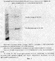
На электрофорез наносили обезжиренные фракции молока от трансгенных и нетрансгенных кроликов, а также положительный контроль Neupogen в чистом виде и в смеси с молоком нетрансгенного кролика (фиг.2). Fat-free fractions of milk from transgenic and non-transgenic rabbits, as well as a positive control of Neupogen in pure form and mixed with milk of non-transgenic rabbit were applied to electrophoresis (Fig. 2).
После окраски мембраны специфическими антителами было обнаружено, что в пробах положительного контроля содержится белок размером около 20 кДа, а в пробах молока трансгенных кроликов содержится белок размером около 24 кДа, специфически связывающийся с моноклональными антителами против чГ-КСФ. After staining the membrane with specific antibodies, it was found that samples of the positive control contained a protein of about 20 kDa, and milk samples of transgenic rabbits contained a protein of about 24 kDa, which specifically binds to monoclonal antibodies against hG-CSF.
Данные результаты показывают, что в молоке трансгенных кроликов содержится гликозилированная форма чГ-КСФ. These results show that the milk of transgenic rabbits contains a glycosylated form of hG-CSF.
Пример 4
Выделение чГ-КСФ из молока трансгенных кроликов проводили следующим образом: молоко, собранное от крольчихи, цент рифугировали на лабораторной центрифуге при 10 тыс. g в течение 30 минут. Удаляли жировую фракцию, отбирали супернатант и использовали его для дальнейшего выделения чГ-КСФ.Example 4
Isolation of hG-CSF from transgenic rabbit milk was performed as follows: milk collected from rabbits was centrifuged in a laboratory centrifuge at 10 thousand g for 30 minutes. The fat fraction was removed, the supernatant was taken and used to further isolate hG-CSF.
Полученный раствор разводили в два раза буфером А (20мМ фосфат калия рН 6.5, 3.5М мочевины, 200мМ ЕДТА, 0.2мМ ДТТ) и интенсивно перемешивали на мешалке для. Эти достигалось разрушение мицелл молока и превращение его в истинный раствор белков. В полученный раствор добавляли 1/5 объема насыщенного раствора сульфата аммония (4М) до конечной концентрации 1М. The resulting solution was diluted twice with buffer A (20 mM potassium phosphate pH 6.5, 3.5 M urea, 200 mM EDTA, 0.2 mM DTT) and intensively mixed on a stirrer. These were achieved by breaking the micelles of milk and turning it into a true protein solution. 1/5 volume of a saturated solution of ammonium sulfate (4M) was added to the resulting solution to a final concentration of 1M.
Для контроля потерь белка чГ-КСФ в процессе выделения определяли его количество в растворе методом иммуноферментного анализа ELISA. To control the loss of hG-CSF protein during the isolation process, its amount in the solution was determined by ELISA.
Затем полученный раствор наносили на колонку К-16 (размер 20 см), заполненную октил-сефарозой (Octyl Sepharose 4 Fast Flow, кат 17-0946-10, фирмы "Амершам-Фармация"), уравновешенной в буфере В (10мМ фосфата калия рН 6.5, 1.8М мочевины, 10мМ ЕДТА, 1М сульфата аммония, 0.2мМ ДТТ). Скорость прохождения раствора через колонку составляла 20 мл/час. Then, the resulting solution was applied to a K-16 column (
Элюцию проводили с использованием линейного градиента с понижающейся концентрацией сульфата аммония до нуля и при повышении рН до 7.5, а детергента Tween-20 до 0.2% от буфера В к буферу С (10 мМ калий фосфата рН 6.5, 1.8М мочевины, 10мМ ЕДТА, 1М сульфата аммония, 0.2мМ ДТТ, 0.2% Tween-20). Скорость прохождения раствора через колонку составляла 20 мл/час. Всего собирали 22 фракции. Основная часть балластных белков выходит во фракциях 10-19. чГ-КСФ элюируется в конце градиента во фракциях 20-21. Потери активности чГ-КСФ в этом процессе составили 30%. Elution was performed using a linear gradient with a decreasing concentration of ammonium sulfate to zero and with increasing pH to 7.5, and Tween-20 detergent to 0.2% from buffer B to buffer C (10 mM potassium phosphate pH 6.5, 1.8 M urea, 10 mM EDTA, 1M ammonium sulfate, 0.2 mM DTT, 0.2% Tween-20). The rate of passage of the solution through the column was 20 ml / hour. A total of 22 fractions were collected. The bulk of the ballast proteins comes in fractions 10-19. hG-CSF elutes at the end of the gradient in fractions 20-21. The loss of hG-CSF activity in this process was 30%.
Элюаты, содержащие чГ-КСФ, объединяли, разводили в 4 раза буфером (2мМ фосфат калия рН 7.2, 0.15М хлорид калия), и наносили на колонку К-9 (размер 6 см), наполненную иммуносорбентом с иммобилизованными на нем моноклональными антителами против чГ-КСФ (носитель активированная бромцианом sepharose 4B фирмы "Амершам-Фармация"). Промывали колонку буфером (2мМ фосфат калия рН 7.2, 0.5М хлорид калия) около 10 объемов колонки. Элюцию с колонки проводили 0.1М лимонной кислотой. The eluates containing hG-CSF were combined, diluted 4 times with buffer (2 mM potassium phosphate pH 7.2, 0.15 M potassium chloride), and applied to a K-9 column (
Фракции нейтрализовали 1М гидроксида калия и образцы наносили на PAG-SDS электрофорез. Fractions were neutralized with 1M potassium hydroxide and samples were applied to PAG-SDS electrophoresis.
Часть проб осаждали трихлоруксусной кислотой, центрифугировали, осадки промывали 70% метанолом, растворяли в электродном буфере, доводили буфером для нанесения до требуемого объема, прогревали и наносили на гель. Окрашивали с помощью красителя Кумаси G-250 (фиг.3). A part of the samples was precipitated with trichloroacetic acid, centrifuged, the precipitates were washed with 70% methanol, dissolved in the electrode buffer, adjusted to the required volume with the application buffer, warmed up and applied to the gel. Stained with Kumashi G-250 dye (FIG. 3).
Как видно на электрофореограмме в элюате содержится две полосы белка с молекулярной массой примерно 25 000 и 50 000 дальтон. Более легкая полоса соответствует размеру гликозилированного чГ-КСФ. Минорная тяжелая полоса является димерной формой чГ-КСФ. As can be seen on the electrophoreogram, the eluate contains two bands of protein with a molecular weight of approximately 25,000 and 50,000 daltons. The lighter band corresponds to the size of the glycosylated hG-CSF. The minor heavy band is a dimeric form of hG-CSF.
Данная технология может применяться для получения субстанции чГ-КСФ, используемой для приготовления лекарственной формы препарата. This technology can be used to obtain the substance hG-CSF used to prepare the dosage form of the drug.
Список литературы
1. Morstyn G, et al. Clinical studies with granutients receiving cytoxic chemoterapy. Behring Inst. mitt., 1988, Aug. (83), 234-239.List of references
1. Morstyn G, et al. Clinical studies with granutients receiving cytoxic chemoterapy. Behring Inst. mitt., 1988, Aug. (83), 234-239.
2. Werner RG, Berthold W. Purification of proteins produced by biotechnological process. Arzneimittelforschung 1988 Mar; 38(3):422-428. 2. Werner RG, Berthold W. Purification of proteins produced by biotechnological process. Arzneimittelforschung 1988 Mar; 38 (3): 422-428.
3. Perez-perez J., et al. Supplementation improves the periplasmic production of human granulocyte-colony stimulating factor in Escherichia coli, Biochem. Biophys. Reg. Commun, 1995, 16, 210 (2), 524-529. 3. Perez-perez J., et al. Supplementation improves the periplasmic production of human granulocyte-colony stimulating factor in Escherichia coli, Biochem. Biophys. Reg. Commun, 1995, 16, 210 (2), 524-529.
4. Rotondaro L. et al. High-level expression of a cDNA for human granulocyte colony - stimulating factor in Chinese Hamster ovary cells. Effect ot 31-noncoding seguences mol., Biotechnol., 1997, Jun; 7(3) 231-240. 4. Rotondaro L. et al. High-level expression of a cDNA for human granulocyte colony - stimulating factor in Chinese Hamster ovary cells. Effect ot 31-noncoding seguences mol., Biotechnol., 1997, Jun; 7 (3) 231-240.
5. Nollowway DNA technology in the production of glycosylated recombinant human granulocyte colony stimulating factor, Eur. J. Cancer, 1994; 30A Suppl. 3., 2. 5. Nollowway DNA technology in the production of glycosylated recombinant human granulocyte colony stimulating factor, Eur. J. Cancer, 1994; 30A Suppl. 3., 2.
6. Nissen С. Glycosylation of recombinant human granulocyte colony stimulating factor: implications for stability and potency. Eur. J. Cancer, 1994, 30A, Suppl. 3, 12-14. 6. Nissen C. Glycosylation of recombinant human granulocyte colony stimulating factor: implications for stability and potency. Eur. J. Cancer, 1994, 30A, Suppl. 3, 12-14.
7. H. Meade а. С. Ziomer. Urine as a substitute for milk. Nature Biotechnology, 1998, v. l6, Jannary, 21-22. 7. H. Meade a. C. Ziomer. Urine as a substitute for milk. Nature Biotechnology, 1998, v. l6, Jannary, 21-22.
8. Патент RU #2157846 Cl "Способ получения трансгенного животного, экспрессирующего в молочной железе гранулоцитарный колониестимулирующий фактор человека и гибридный ген h-GM-1 для осуществления способа". 8. Patent RU # 2157846 Cl "A method for producing a transgenic animal expressing granulocyte colony stimulating factor in a breast and the h-GM-1 hybrid gene for implementing the method."
9. Nicola N.A. J. Cell. physiol, 1981, 109, 253. 9. Nicola N.A. J. Cell. physiol, 1981, 109, 253.
10. Brem G. et al. Expression of synthetic cDNA sequences encoding human insulin-like grows factor-1 in the mammary gland of transgenic rabbits. Gene, 1994, v. 149, p. 351-355. 10. Brem G. et al. Expression of synthetic cDNA sequences encoding human insulin-like grows factor-1 in the mammary gland of transgenic rabbits. Gene, 1994, v. 149, p. 351-355.
11. Rudolph N.S. Biopharmaceutical production in transgenic livestock. Trends in Biotechnology, 1999, sept., v. 17, p. 367-374. 11. Rudolph N.S. Biopharmaceutical production in transgenic livestock. Trends in Biotechnology, 1999, sept., V. 17, p. 367-374.
12. van Berkel P.H. et al. Large scale production of recombinant human lactoferrin in the milk of transgenic cows. Nature biotechnology, 2002, v. 20, p. 484-487. 12. van Berkel P.H. et al. Large scale production of recombinant human lactoferrin in the milk of transgenic cows. Nature biotechnology, 2002, v. 20, p. 484-487.
13. Ebert K.M. et al. Transgenic production of a variant of human tissue-type plasminogen activator in goat milk: generation of transgenic goats and analysis of expression. Biotechnology, 1991, v. 9, sept., p. 835-838. 13. Ebert K.M. et al. Transgenic production of a variant of human tissue-type plasminogen activator in goat milk: generation of transgenic goats and analysis of expression. Biotechnology, 1991, v. 9, sept., P. 835-838.
14. Wright G. et al. High level expression of active human alpha-1-antiyrypsin in the milk of transgenic sheep. Biotechnology, 1991, v. 9, sept., p. 830-834. 14. Wright G. et al. High level expression of active human alpha-1-antiyrypsin in the milk of transgenic sheep. Biotechnology, 1991, v. 9, sept., P. 830-834.
15. Velander W. et al. High-level expression of a heterologous protein in the milk of transgenic swine using the cDNA encoding human protein C. Proc. Natl. Acad. Sci. USA, 1992, v. 89, p. 12003-12007. 15. Velander W. et al. High-level expression of a heterologous protein in the milk of transgenic swine using the cDNA encoding human protein C. Proc. Natl. Acad Sci. USA, 1992, v. 89, p. 12003-12007.
16. Juhani Janne et. al. Transgenic Animals as Bioproducers of Therapeutic Proteins Trends in Molecular Medicine, Annals of Medicine, 1992, v. 24, p. 273-280. 16. Juhani Janne et. al. Transgenic Animals as Bioproducers of Therapeutic Proteins Trends in Molecular Medicine, Annals of Medicine, 1992, v. 24, p. 273-280.
17. Rudolf Jaenisch. Transgenic Animals Science, 1988, v. 240, p. 1468-1473. 17. Rudolf Jaenisch. Transgenic Animals Science, 1988, v. 240, p. 1468-1473.
18. Angelika E. Schnieke et. al. , Human Factor IX Transgenic Sheep Produced by Transfer of Nuclei from Transfected Fetal Fibroblasts Science, 1997, v. 278, p. 2130-2133. 18. Angelika E. Schnieke et. al. , Human Factor IX Transgenic Sheep Produced by Transfer of Nuclei from Transfected Fetal Fibroblasts Science, 1997, v. 278, p. 2130-2133.
Claims (5)
Priority Applications (1)
| Application Number | Priority Date | Filing Date | Title |
|---|---|---|---|
| RU2002111017/13A RU2207373C1 (en) | 2002-04-25 | 2002-04-25 | Method for preparing human granulocytic colony-stimulating factor |
Applications Claiming Priority (1)
| Application Number | Priority Date | Filing Date | Title |
|---|---|---|---|
| RU2002111017/13A RU2207373C1 (en) | 2002-04-25 | 2002-04-25 | Method for preparing human granulocytic colony-stimulating factor |
Publications (1)
| Publication Number | Publication Date |
|---|---|
| RU2207373C1 true RU2207373C1 (en) | 2003-06-27 |
Family
ID=29211849
Family Applications (1)
| Application Number | Title | Priority Date | Filing Date |
|---|---|---|---|
| RU2002111017/13A RU2207373C1 (en) | 2002-04-25 | 2002-04-25 | Method for preparing human granulocytic colony-stimulating factor |
Country Status (1)
| Country | Link |
|---|---|
| RU (1) | RU2207373C1 (en) |
Cited By (2)
| Publication number | Priority date | Publication date | Assignee | Title |
|---|---|---|---|---|
| RU2302664C2 (en) * | 2005-09-28 | 2007-07-10 | ГУ Научно-исследовательский институт фармакологии Томского научного центра Сибирского отделения Российской академии медицинских наук (ГУ НИИ фармакологии ТНЦ СО РАМН) | Method for controlling erythropoiesis disorders in experimental encephalopathy cases |
| RU2422529C1 (en) * | 2010-02-12 | 2011-06-27 | Учреждение Российской академии наук Институт цитологии и генетики Сибирского отделения РАН (ИЦиГ СО РАН) | GENETICALLY ENGINEERED CONSTRUCT pGoatcasGCSF ENABLING HUMAN GRANULOCYTE COLONY-STIMULATING FACTOR PRODUCTION IN MILK OF TRANSGENIC ANIMALS |
Citations (1)
| Publication number | Priority date | Publication date | Assignee | Title |
|---|---|---|---|---|
| RU2139084C1 (en) * | 1994-02-04 | 1999-10-10 | Ф.Хоффманн-Ля Рош Аг | Product containing granulocytic colony-stimulating factor (g-csf) and tnf-binding protein |
-
2002
- 2002-04-25 RU RU2002111017/13A patent/RU2207373C1/en not_active IP Right Cessation
Patent Citations (1)
| Publication number | Priority date | Publication date | Assignee | Title |
|---|---|---|---|---|
| RU2139084C1 (en) * | 1994-02-04 | 1999-10-10 | Ф.Хоффманн-Ля Рош Аг | Product containing granulocytic colony-stimulating factor (g-csf) and tnf-binding protein |
Cited By (2)
| Publication number | Priority date | Publication date | Assignee | Title |
|---|---|---|---|---|
| RU2302664C2 (en) * | 2005-09-28 | 2007-07-10 | ГУ Научно-исследовательский институт фармакологии Томского научного центра Сибирского отделения Российской академии медицинских наук (ГУ НИИ фармакологии ТНЦ СО РАМН) | Method for controlling erythropoiesis disorders in experimental encephalopathy cases |
| RU2422529C1 (en) * | 2010-02-12 | 2011-06-27 | Учреждение Российской академии наук Институт цитологии и генетики Сибирского отделения РАН (ИЦиГ СО РАН) | GENETICALLY ENGINEERED CONSTRUCT pGoatcasGCSF ENABLING HUMAN GRANULOCYTE COLONY-STIMULATING FACTOR PRODUCTION IN MILK OF TRANSGENIC ANIMALS |
Similar Documents
| Publication | Publication Date | Title |
|---|---|---|
| Shakweer et al. | A review of transgenic animal techniques and their applications | |
| EP0807170B1 (en) | Transgenic pigs expressing human coagulation factor viii | |
| Ebert et al. | Transgenic farm animals: progress report | |
| Ghiara et al. | In vitro generated mast cells express natural cytotoxicity against tumour cells | |
| KR100397244B1 (en) | Megakaryocyte differentiation factor | |
| AU721132B2 (en) | Method for development of transgenic goats | |
| JP2007507232A (en) | Process for producing a transgenic mammal producing exogenous protein in milk and transgenic mammal produced thereby | |
| Kaiser et al. | Detection of recombinant human lactoferrin and lysozyme produced in a bitransgenic cow | |
| Carvalho et al. | Production of transgenic cattle by somatic cell nuclear transfer (SCNT) with the human granulocyte colony-stimulation factor (hG-CSF) | |
| Freitas et al. | The establishment of two transgenic goat lines for mammary gland hG-CSF expression | |
| JP2010528678A (en) | Method for producing exogenous protein in milk of transgenic mammals | |
| RU2207373C1 (en) | Method for preparing human granulocytic colony-stimulating factor | |
| Schrader et al. | In vitro studies on lymphocyte differentiation: II. Generation of terminal deoxynucleotidyl transferase-positive cells in long-term cultures of mouse bone marrow | |
| Gough et al. | Molecular biology of the leukaemia inhibitory factor gene | |
| Matsuo et al. | New human oral squamous carcinoma cell line and its tumorigenic subline producing granulocyte colony‐stimulating factor | |
| CN1970771B (en) | Process for high-efficiency production of human lysozyme by using mammary gland of domestic animal | |
| KR102738812B1 (en) | Transgenic cloned piglets defecting porcine GGTA1, CMAH, beta4GalNT2 and CD40 gene for xenotransplantation, and producing method thereof | |
| AU2004281642A1 (en) | Expression of dominant negative transmembrane receptors in the milk of transgenic animals | |
| RU2422529C1 (en) | GENETICALLY ENGINEERED CONSTRUCT pGoatcasGCSF ENABLING HUMAN GRANULOCYTE COLONY-STIMULATING FACTOR PRODUCTION IN MILK OF TRANSGENIC ANIMALS | |
| US6331403B1 (en) | Use of mCRP to slow cell growth and to promote maturation of cells | |
| Stinnakre et al. | Quantitative collection of milk and active recombinant proteins from the mammary glands of transgenic mice | |
| Kim et al. | Ectopic expression of tethered human follicle-stimulating hormone (hFSH) gene in transgenic mice | |
| RU2402211C2 (en) | Method for production of transgenic rabbits producing proteins into mammary gland | |
| US8030537B1 (en) | Somatic cloning gene transfer for the production of recombinant proteins, cells and organs | |
| RU2390562C2 (en) | Method of creating transgenic mammals which produce exogenous proteins in milk, and transgenic mammals created using said method |
Legal Events
| Date | Code | Title | Description |
|---|---|---|---|
| MM4A | The patent is invalid due to non-payment of fees |
Effective date: 20040426 |