KR20250055152A - Device and method for determining liver test site - Google Patents
Device and method for determining liver test site Download PDFInfo
- Publication number
- KR20250055152A KR20250055152A KR1020230138610A KR20230138610A KR20250055152A KR 20250055152 A KR20250055152 A KR 20250055152A KR 1020230138610 A KR1020230138610 A KR 1020230138610A KR 20230138610 A KR20230138610 A KR 20230138610A KR 20250055152 A KR20250055152 A KR 20250055152A
- Authority
- KR
- South Korea
- Prior art keywords
- liver
- scan
- ultrasound image
- determining
- examination
- Prior art date
- Legal status (The legal status is an assumption and is not a legal conclusion. Google has not performed a legal analysis and makes no representation as to the accuracy of the status listed.)
- Pending
Links
Images

Classifications
-
- A—HUMAN NECESSITIES
- A61—MEDICAL OR VETERINARY SCIENCE; HYGIENE
- A61B—DIAGNOSIS; SURGERY; IDENTIFICATION
- A61B8/00—Diagnosis using ultrasonic, sonic or infrasonic waves
- A61B8/08—Clinical applications
-
- A—HUMAN NECESSITIES
- A61—MEDICAL OR VETERINARY SCIENCE; HYGIENE
- A61B—DIAGNOSIS; SURGERY; IDENTIFICATION
- A61B8/00—Diagnosis using ultrasonic, sonic or infrasonic waves
- A61B8/08—Clinical applications
- A61B8/0833—Clinical applications involving detecting or locating foreign bodies or organic structures
- A61B8/085—Clinical applications involving detecting or locating foreign bodies or organic structures for locating body or organic structures, e.g. tumours, calculi, blood vessels, nodules
-
- A—HUMAN NECESSITIES
- A61—MEDICAL OR VETERINARY SCIENCE; HYGIENE
- A61B—DIAGNOSIS; SURGERY; IDENTIFICATION
- A61B8/00—Diagnosis using ultrasonic, sonic or infrasonic waves
- A61B8/08—Clinical applications
- A61B8/0833—Clinical applications involving detecting or locating foreign bodies or organic structures
-
- A—HUMAN NECESSITIES
- A61—MEDICAL OR VETERINARY SCIENCE; HYGIENE
- A61B—DIAGNOSIS; SURGERY; IDENTIFICATION
- A61B8/00—Diagnosis using ultrasonic, sonic or infrasonic waves
- A61B8/52—Devices using data or image processing specially adapted for diagnosis using ultrasonic, sonic or infrasonic waves
- A61B8/5215—Devices using data or image processing specially adapted for diagnosis using ultrasonic, sonic or infrasonic waves involving processing of medical diagnostic data
- A61B8/5223—Devices using data or image processing specially adapted for diagnosis using ultrasonic, sonic or infrasonic waves involving processing of medical diagnostic data for extracting a diagnostic or physiological parameter from medical diagnostic data
-
- A—HUMAN NECESSITIES
- A61—MEDICAL OR VETERINARY SCIENCE; HYGIENE
- A61B—DIAGNOSIS; SURGERY; IDENTIFICATION
- A61B8/00—Diagnosis using ultrasonic, sonic or infrasonic waves
- A61B8/52—Devices using data or image processing specially adapted for diagnosis using ultrasonic, sonic or infrasonic waves
- A61B8/5215—Devices using data or image processing specially adapted for diagnosis using ultrasonic, sonic or infrasonic waves involving processing of medical diagnostic data
- A61B8/523—Devices using data or image processing specially adapted for diagnosis using ultrasonic, sonic or infrasonic waves involving processing of medical diagnostic data for generating planar views from image data in a user selectable plane not corresponding to the acquisition plane
-
- A—HUMAN NECESSITIES
- A61—MEDICAL OR VETERINARY SCIENCE; HYGIENE
- A61B—DIAGNOSIS; SURGERY; IDENTIFICATION
- A61B8/00—Diagnosis using ultrasonic, sonic or infrasonic waves
- A61B8/52—Devices using data or image processing specially adapted for diagnosis using ultrasonic, sonic or infrasonic waves
- A61B8/5292—Devices using data or image processing specially adapted for diagnosis using ultrasonic, sonic or infrasonic waves using additional data, e.g. patient information, image labeling, acquisition parameters
-
- G—PHYSICS
- G06—COMPUTING OR CALCULATING; COUNTING
- G06T—IMAGE DATA PROCESSING OR GENERATION, IN GENERAL
- G06T7/00—Image analysis
- G06T7/0002—Inspection of images, e.g. flaw detection
- G06T7/0012—Biomedical image inspection
-
- G—PHYSICS
- G06—COMPUTING OR CALCULATING; COUNTING
- G06T—IMAGE DATA PROCESSING OR GENERATION, IN GENERAL
- G06T2207/00—Indexing scheme for image analysis or image enhancement
- G06T2207/10—Image acquisition modality
- G06T2207/10132—Ultrasound image
-
- G—PHYSICS
- G06—COMPUTING OR CALCULATING; COUNTING
- G06T—IMAGE DATA PROCESSING OR GENERATION, IN GENERAL
- G06T2207/00—Indexing scheme for image analysis or image enhancement
- G06T2207/30—Subject of image; Context of image processing
- G06T2207/30004—Biomedical image processing
- G06T2207/30056—Liver; Hepatic
Landscapes
- Health & Medical Sciences (AREA)
- Life Sciences & Earth Sciences (AREA)
- Engineering & Computer Science (AREA)
- Physics & Mathematics (AREA)
- General Health & Medical Sciences (AREA)
- Medical Informatics (AREA)
- Nuclear Medicine, Radiotherapy & Molecular Imaging (AREA)
- Radiology & Medical Imaging (AREA)
- Animal Behavior & Ethology (AREA)
- Heart & Thoracic Surgery (AREA)
- Veterinary Medicine (AREA)
- Public Health (AREA)
- Biophysics (AREA)
- Pathology (AREA)
- Biomedical Technology (AREA)
- Surgery (AREA)
- Molecular Biology (AREA)
- Computer Vision & Pattern Recognition (AREA)
- General Physics & Mathematics (AREA)
- Quality & Reliability (AREA)
- Theoretical Computer Science (AREA)
- Physiology (AREA)
- Vascular Medicine (AREA)
- Ultra Sonic Daignosis Equipment (AREA)
Abstract
본 발명은 간 초음파 검사시 시술자가 검사하고 있는 간 검사 부위를 인식하고 판단하는 기술에 관한 것이다. 본 발명의 일 측면에 따르면, 간 검사 부위 판단 방법은, 간 영역을 촬영한 초음파 영상을 입력 받는 단계; 상기 초음파 영상으로부터 특징을 추출하는 단계; 추출된 특징을 기반으로 상기 초음파 영상이 촬영된 방향을 결정하는 단계; 및 상기 추출된 특징 및 결정된 방향을 기반으로 상기 초음파 영상에 포함된 간의 세부 부위를 결정하는 단계;를 포함할 수 있다.The present invention relates to a technology for recognizing and determining a liver examination site being examined by a practitioner during a liver ultrasound examination. According to one aspect of the present invention, a method for determining a liver examination site may include the steps of: receiving an ultrasound image photographing a liver region; extracting features from the ultrasound image; determining a direction in which the ultrasound image was photographed based on the extracted features; and determining a detailed liver site included in the ultrasound image based on the extracted features and the determined direction.
Description
본 발명은 간 초음파 검사시 시술자가 검사하고 있는 간 검사 부위를 인식하고 판단하는 기술에 관한 것이다.The present invention relates to a technology for recognizing and determining a liver examination site being examined by a practitioner during a liver ultrasound examination.
기존 의료용 초음파 검사 장비는 검사 중인 부위를 인식하기 위해서 EMT센서를 부착하거나 사전에 촬영된 CT 영상이 있어야만 한다. 또한, EMT센서는 외부와 프로브에 설치해서 사용하는 불편함으로 시술자에게 부담이 될 수 있을 뿐만 아니라 비싼 센서 비용에 의한 부담 증가의 문제가 있고, 사전에 촬영된 CT 영상은 초음파만 사용하는 대부분의 검사 특성상 일반적 적용의 어려움이 있다.Existing medical ultrasound examination equipment requires an EMT sensor to be attached or a CT image taken in advance to recognize the area being examined. In addition, EMT sensors can be a burden to the operator due to the inconvenience of being installed externally and on the probe, and there is also the problem of increased burden due to expensive sensor costs, and pre-taken CT images are difficult to apply generally due to the characteristics of most examinations that only use ultrasound.
(공개 특허공보) 제 10-2012-0108849호, “초음파 영상과 자기공명 영상 간의 영상정합 방법”(Public Patent Publication) No. 10-2012-0108849, “Image registration method between ultrasound image and magnetic resonance image”
본 발명의 목적은 시술자가 초음파 장치를 이용하여 검사하는 간의 검사 부위를 인식 및 판단하는 기술을 제공하는 것이다. The purpose of the present invention is to provide a technology for recognizing and determining an examination site of a liver being examined using an ultrasound device.
또한, 본 발명의 목적은 간의 검사 부위에 대한 영상을 시술자에게 제공하는 것이다. It is also an object of the present invention to provide an operator with an image of the liver examination site.
또한, 본 발명의 목적은 초음파 장치를 이용하여 검사하는 간의 세부 부위에 대한 쿠이노(Couinaud)의 간아분절 분류에 따른 영상을 제공하는 것이다.In addition, it is an object of the present invention to provide images according to Couinaud's liver subsegment classification for detailed parts of the liver examined using an ultrasound device.
본 발명의 일 측면에 따르면, 간 검사 부위 판단 방법은, 간 영역을 촬영한 초음파 영상을 입력 받는 단계; 상기 초음파 영상으로부터 특징을 추출하는 단계; 추출된 특징을 기반으로 상기 초음파 영상이 촬영된 방향을 결정하는 단계; 및 상기 추출된 특징 및 결정된 방향을 기반으로 상기 초음파 영상에 포함된 간의 세부 부위를 결정하는 단계;를 포함할 수 있다.According to one aspect of the present invention, a method for determining a liver examination site may include the steps of: receiving an ultrasound image photographing a liver region; extracting features from the ultrasound image; determining a direction in which the ultrasound image was photographed based on the extracted features; and determining a detailed liver site included in the ultrasound image based on the extracted features and the determined direction.
일 실시예에서, 상기 특징을 추출하는 단계는, 상기 초음파 영상에 포함된 간, 간 주변 혈관 및 간 주변 장기에 대한 특징을 추출할 수 있다.In one embodiment, the step of extracting the features may extract features for the liver, blood vessels around the liver, and organs around the liver included in the ultrasound image.
일 실시예에서, 상기 초음파 영상이 촬영된 방향을 결정하는 단계는, 상기 추출된 특징을 기반으로 상기 초음파 영상에 포함된 간의 적어도 하나의 부위를 결정하고, 결정된 적어도 하나의 부위를 기반으로 상기 방향을 결정할 수 있다.In one embodiment, the step of determining the direction in which the ultrasound image was captured may include determining at least one region of the liver included in the ultrasound image based on the extracted features, and determining the direction based on the determined at least one region.
일 실시예에서, 상기 방향은, 심와부 종단 스캔, 심와부 횡단 스캔, 우측 늑골하 스캔-간문맥, 우측 늑골하-간우엽 횡단 스캔, 간의 첨부 스캔, 우측 늑골하 스캔-간정맥, 당낭 종단 스캔, 간외 담관 종단 스캔, 우측 간문맥을 포함한 간우엽의 늑간 스캔, 우측 후 늑간 스캔 및 우측 간하부와 우측 신장 피질 스캔 중 적어도 하나일 수 있다.In one embodiment, the direction can be at least one of a parasternal longitudinal scan, a parasternal transverse scan, a right subcostal scan-portal vein, a right subcostal scan-right hepatic lobe transverse scan, an apex scan of the liver, a right subcostal scan-hepatic vein, a longitudinal scan of the liver sac, a longitudinal scan of the extrahepatic bile duct, an intercostal scan of the right hepatic lobe including the right portal vein, a right posterior intercostal scan, and a right subhepatic and right renal cortex scan.
일 실시예에서, 상기 초음파 영상에 포함된 간의 세부 부위를 결정하는 단계는, 쿠이노(Couinaud)의 간아분절 분류에 따른 복수의 세부 부위 중 어느 하나를 결정할 수 있다.In one embodiment, the step of determining a liver sub-region included in the ultrasound image may determine any one of a plurality of liver sub-regions according to Couinaud's liver sub-segment classification.
본 발명의 다른 측면에 따르면, 간 검사 부위 판단 장치는, 간 검사 부위 판단 방법을 실행하기 위한 명령어를 포함하는 메모리; 및 상기 명령어를 실행함으로써, 간 영역을 촬영한 초음파 영상을 입력 받고, 상기 초음파 영상으로부터 특징을 추출하며, 추출된 특징을 기반으로 상기 초음파 영상이 촬영된 방향을 결정하고, 상기 추출된 특징 및 결정된 방향을 기반으로 상기 초음파 영상에 포함된 간의 세부 부위를 결정하는, 프로세서;를 포함할 수 있다.According to another aspect of the present invention, a liver examination site determination device may include a memory including a command for executing a liver examination site determination method; and a processor which, by executing the command, receives an ultrasound image photographing a liver region, extracts features from the ultrasound image, determines a direction in which the ultrasound image was photographed based on the extracted features, and determines a detailed part of the liver included in the ultrasound image based on the extracted features and the determined direction.
본 발명의 일 측면에 따르면, 시술자가 초음파 장치를 이용하여 검사하는 간의 검사 부위를 인식 및 판단하는 것이 가능하게 된다. According to one aspect of the present invention, it becomes possible for a practitioner to recognize and determine an examination site of a liver being examined using an ultrasonic device.
또한, 본 발명의 목적은 간의 검사 부위에 대한 영상을 시술자에게 제공하는 것이 가능하게 된다.In addition, the purpose of the present invention is to enable the operator to obtain an image of the liver examination site.
또한, 본 발명의 목적은 초음파 장치를 이용하여 검사하는 간의 세부 부위에 대한 쿠이노(Couinaud)의 간아분절 분류에 따른 영상을 제공하는 것이 가능하게 된다.In addition, the purpose of the present invention is to enable providing images according to Couinaud's liver segment classification for detailed parts of the liver examined using an ultrasound device.
도 1은 본 발명의 일 실시예에 따른 간 검사 부위 판단 장치의 블록도이다.
도 2는 본 발명의 일 실시예에 따른 간 검사 부위 판단 방법의 흐름도이다.
도 3 및 도 4는 본 발명의 일 실시예에 따른 초음파 영상이 촬영된 방향의 예를 나타낸 도면이다.
도 5는 본 발명의 일 실시예에 따른 간의 세부 부위를 나타낸 도면이다.
도 6은 본 발명의 일 실시예에 따른 건 검사 부위를 출력하는 예를 설명하기 위한 도면이다.
도 7은 본 발명의 다른 실시예에 따른 간 검사 부위 판단 장치의 블록도이다.FIG. 1 is a block diagram of a liver examination site determination device according to one embodiment of the present invention.
Figure 2 is a flow chart of a method for determining a liver examination site according to one embodiment of the present invention.
FIGS. 3 and 4 are drawings showing examples of directions in which ultrasound images are captured according to one embodiment of the present invention.
FIG. 5 is a drawing showing detailed parts of a liver according to one embodiment of the present invention.
FIG. 6 is a drawing for explaining an example of outputting a dry inspection site according to one embodiment of the present invention.
FIG. 7 is a block diagram of a liver examination site determination device according to another embodiment of the present invention.
본 발명의 이점 및 특징, 그리고 그것들을 달성하는 방법은 첨부되는 도면과 함께 상세하게 후술되어 있는 실시예들을 참조하면 명확해질 것이다. 그러나 본 발명은 이하에서 개시되는 실시예들에 한정되는 것이 아니라 다양한 형태로 구현될 수 있으며, 단지 본 실시예들은 본 발명의 개시가 완전하도록 하고, 본 발명이 속하는 기술분야에서 통상의 지식을 가진 자에게 발명의 범주를 완전하게 알려주기 위해 제공되는 것이며, 본 발명의 범주는 청구항에 의해 정의될 뿐이다.The advantages and features of the present invention, and the methods for achieving them, will become clear with reference to the embodiments described in detail below together with the accompanying drawings. However, the present invention is not limited to the embodiments disclosed below, but may be implemented in various forms, and these embodiments are provided only to make the disclosure of the present invention complete and to fully inform those skilled in the art of the scope of the invention, and the scope of the present invention is defined only by the claims.
본 발명의 실시예들을 설명함에 있어서 공지 기능 또는 구성에 대한 구체적인 설명은 본 발명의 실시예들을 설명함에 있어 실제로 필요한 경우 외에는 생략될 것이다. 그리고 후술되는 용어들은 본 발명의 실시예에서의 기능을 고려하여 정의된 용어들로서 이는 사용자, 운용자의 의도 또는 관례 등에 따라 달라질 수 있다. 그러므로 그 정의는 본 명세서 전반에 걸친 내용을 토대로 내려져야 할 것이다.In describing embodiments of the present invention, specific descriptions of known functions or configurations will be omitted unless they are actually necessary for describing embodiments of the present invention. In addition, the terms described below are terms defined in consideration of functions in embodiments of the present invention, and may vary depending on the intention or custom of the user or operator. Therefore, the definitions should be made based on the contents throughout this specification.
이하 사용되는 '…부', '…기' 등의 용어는 적어도 하나의 기능이나 동작을 처리하는 단위를 의미하며, 이는 하드웨어나 소프트웨어, 또는, 하드웨어 및 소프트웨어의 결합으로 구현될 수 있다.The terms ‘… part’, ‘… unit’, etc. used hereinafter mean a unit that processes at least one function or operation, and this can be implemented by hardware, software, or a combination of hardware and software.
도 1은 본 발명의 일 실시예에 따른 간 검사 부위 판단 장치의 블록도이다. FIG. 1 is a block diagram of a liver examination site determination device according to one embodiment of the present invention.
도 1을 참조하면, 간 검사 부위 판단 장치(1000)는 프로세서(1100) 및 메모리(1200)를 포함할 수 있다. Referring to FIG. 1, the liver examination site determination device (1000) may include a processor (1100) and a memory (1200).
프로세서(1100)는 메모리(1200)에 저장된 명령어들을 실행함으로써, 간 검사 부위 판단 장치(1000)의 전반적인 동작을 제어하고, 간 검사 부위를 판단할 수 있다. The processor (1100) can control the overall operation of the liver examination site determination device (1000) and determine the liver examination site by executing commands stored in the memory (1200).
메모리(1200)는 프로세서(1100)에 의해 실행되며, 간 검사 부위 판단 장치(1000)의 전반적인 동작을 제어하고, 간 검사 부위를 판단하는데 이용되는 명령어들을 저장할 수 있다. The memory (1200) is executed by the processor (1100), controls the overall operation of the liver examination site determination device (1000), and can store commands used to determine the liver examination site.
간 검사 부위 판단 장치(1000)의 동작에 대한 보다 상세한 설명은 도 2 내지 6를 참조하여 후술한다. A more detailed description of the operation of the liver examination site determination device (1000) will be described later with reference to FIGS. 2 to 6.
도 2는 본 발명의 일 실시예에 따른 간 검사 부위 판단 방법의 흐름도이다. Figure 2 is a flow chart of a method for determining a liver examination site according to one embodiment of the present invention.
이하, 상기 방법은 도 1에 도시된 간 검사 부위 판단 장치(1000)에 의해 수행되는 것을 예시로 설명한다. Hereinafter, the above method is described as an example performed by the liver examination site determination device (1000) illustrated in FIG. 1.
단계 S2100에서, 간 검사 부위 판단 장치(1000)는 의사 등의 시술자가 초음파 장치를 이용하여 환자의 간 영역을 촬영한 초음파 영상을 외부 장치(예, 초음파 장치 등)으로부터 획득할 수 있다. In step S2100, the liver examination site determination device (1000) can obtain an ultrasound image of a patient's liver area taken by a doctor or other practitioner using an ultrasound device from an external device (e.g., an ultrasound device, etc.).
일 실시예에서, 초음파 영상은 간, 주변 장기, 혈관 등의 신체 일부에 대한 영상을 포함할 수 있다. In one embodiment, the ultrasound images may include images of body parts, such as the liver, surrounding organs, or blood vessels.
단계 S2200에서, 간 검사 부위 판단 장치(1000)는 간을 촬영한 초음파 영상으로부터 특징(feature)을 추출할 수 있다.In step S2200, the liver examination site determination device (1000) can extract features from an ultrasound image of the liver.
일 실시예에서, 간 검사 부위 판단 장치(1000)는 딥러닝 모델 등의 세그멘테이션 네크워크를 이용하여 초음파 영상으로부터 특징을 추출할 수 있다. 구체적으로, 간 검사 부위 판단 장치(1000)는 초음파 영상에 포함된 간, 주변 장기, 혈관 등에 대한 특징을 추출할 수 있다. In one embodiment, the liver examination site determination device (1000) can extract features from an ultrasound image using a segmentation network such as a deep learning model. Specifically, the liver examination site determination device (1000) can extract features for the liver, surrounding organs, blood vessels, etc. included in the ultrasound image.
단계 S2300에서, 간 검사 부위 판단 장치(1000)는 초음파 영상이 촬영된 방향을 결정할 수 있다. In step S2300, the liver examination site determination device (1000) can determine the direction in which the ultrasound image was captured.
일 실시예에서, 간 검사 부위 판단 장치(1000)는 초음파 영상으로부터 추출된 간, 주변 장기, 혈관 등에 대한 특징을 기반으로, 초음파 영상이 촬영된 방향을 결정할 수 있다. In one embodiment, the liver examination site determination device (1000) can determine the direction in which the ultrasound image was captured based on features of the liver, surrounding organs, blood vessels, etc. extracted from the ultrasound image.
일 실시예에서, 간 검사 부위 판단 장치(1000)는 미리 설정된 복수의 방향 중 어느 하나의 방향으로 결정할 수 있다. 여기서, 미리 설정된 복수의 방향은 한국 초음파 학회 등에 따른 표준 영상 중 적어도 하나에 대응하는 방향일 수 있다. In one embodiment, the liver examination site determination device (1000) can determine a direction in any one of a plurality of preset directions. Here, the plurality of preset directions may be a direction corresponding to at least one of the standard images according to the Korean Society of Ultrasound, etc.
일 실시예에서, 표준 영상은 도 3 및 도 4에 도시된 바와 같이, 심와부 종단 스캔, 심와부 횡단 스캔, 우측 늑골하 스캔-간문맥, 우측 늑골하-간우엽 횡단 스캔, 간의 첨부 스캔, 우측 늑골하 스캔-간정맥, 당낭 종단 스캔, 간외 담관 종단 스캔, 우측 간문맥을 포함한 간우엽의 늑간 스캔, 우측 후 늑간 스캔 및 우측 간하부와 우측 신장 피질 스캔 등일 수 있다.In one embodiment, the standard images may be a longitudinal scan of the thoracic spine, a transverse scan of the thoracic spine, a right subcostal scan-portal vein, a right subcostal scan-right liver lobe transverse scan, an apical scan of the liver, a right subcostal scan-hepatic vein, a longitudinal scan of the liver sac, a longitudinal scan of the extrahepatic bile duct, an intercostal scan of the right liver lobe including the right portal vein, a right posterior intercostal scan, and a scan of the right subhepatic and right renal cortex, as illustrated in FIGS. 3 and 4 .
단계 S2400에서, 간 검사 부위 판단 장치(1000)는 초음파 영상이 촬영된 방향을 기반으로 초음파 영상에 포함된 간의 세부 부위를 결정할 수 있다. In step S2400, the liver examination site determination device (1000) can determine a detailed site of the liver included in the ultrasound image based on the direction in which the ultrasound image was captured.
일 실시예에서, 간 검사 부위 판단 장치(1000)는 초음파 영상으로부터 추출된 특징 및 초음파 영상이 촬영된 방향을 기반으로, 초음파 영상에 포함된 간의 세부 부위를 결정할 수 있다. In one embodiment, the liver examination site determination device (1000) can determine a detailed site of the liver included in an ultrasound image based on features extracted from the ultrasound image and the direction in which the ultrasound image was captured.
일 실시예에서, 간의 세부 부위는 도 5에 도시된 바와 같이, 쿠이노(Couinaud)의 간아분절 분류에 따른 간의 부위 중 어느 하나를 의미할 수 있다. In one embodiment, the liver subdivision may mean any one of the liver subdivisions according to Couinaud's liver subdivision classification, as illustrated in FIG. 5.
단계 S2500에서, 간 검사 부위 판단 장치(1000)는 초음파 영상에 포함된 간의 세부 부위를 출력할 수 있다. In step S2500, the liver examination site determination device (1000) can output detailed liver sites included in the ultrasound image.
일 실시예에서, 간 검사 부위 판단 장치(1000)는, 시술자 등이 시각적으로 인식할 수 있도록 간의 부위를 3D 모델로 출력할 수 있다. In one embodiment, the liver examination site determination device (1000) can output a liver site as a 3D model so that a practitioner or the like can visually recognize it.
일 실시예에서, 간 검사 부위 판단 장치(1000)는, 초음파 영상에 포함된 간의 세부 부위에 대응하는 해부학적 이미지와 함께 출력할 수 있다. In one embodiment, the liver examination site determination device (1000) can output an anatomical image corresponding to a detailed liver site included in an ultrasound image.
또한, 간 검사 부위 판단 장치(1000)는 초음파 영상으로부터 간을 검사하는 방향을 예측하도록 미리 학습된 이미지 처리 모델을 기반으로, 초음파 영상이 촬영된 방향을 결정할 수 있다. In addition, the liver examination site determination device (1000) can determine the direction in which the ultrasound image was captured based on an image processing model learned in advance to predict the direction in which the liver is examined from the ultrasound image.
또한, 간 검사 부위 판단 장치(1000)는 초음파 영상이 촬영된 방향을 기반으로 초음파 영상에 포함된 간의 세부 부위를 예측하도록 미리 학습된 모델을 이용하여 간의 세부 부위를 결정할 수 있다.In addition, the liver examination site determination device (1000) can determine a detailed liver site using a pre-learned model to predict a detailed liver site included in an ultrasound image based on the direction in which the ultrasound image was captured.
도 6은 본 발명의 일 실시예에 따른 건 검사 부위를 출력하는 예를 설명하기 위한 도면이다. FIG. 6 is a drawing for explaining an example of outputting a dry inspection site according to one embodiment of the present invention.
도 6을 참조하면, 6100은 초음파 영상에 포함된 간의 세부 부위를 쿠이노(Couinaud)의 간아분절 분류에 따른 3D 모델로 표현한 예를 나타낸다. 이에 따라 시술자는 초음파 장비를 이용하여 검사하고 있는 간의 부위를 해부학적으로 인식할 수 있게 된다.Referring to Figure 6, 6100 shows an example of expressing a detailed portion of the liver included in an ultrasound image as a 3D model according to Couinaud's liver subsegment classification. Accordingly, the operator can anatomically recognize the portion of the liver being examined using ultrasound equipment.
6200은 초음파 영상에 포함된 간, 주변 장기, 혈관 등에 관한 정보를 나타낸다. 이에 따라 시술자는 검사 대상인 간과 다른 주변 장기들을 명확하게 구분하는 것이 가능하게 된다. 6200 indicates information about the liver, surrounding organs, blood vessels, etc. included in the ultrasound image. Accordingly, the operator can clearly distinguish between the liver, which is the subject of the examination, and other surrounding organs.
6300은 초음파 영상을 나타낸다. 이는 초음파 장치를 이용하는 시술자에게 당연히 제공되어야 할 정보이기 때문이다. 6300 represents an ultrasound image. This is because it is information that should be provided to practitioners using ultrasound devices.
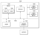
도 7은 본 발명의 다른 실시예에 따른 간 검사 부위 판단 장치의 블록도이다.FIG. 7 is a block diagram of a liver examination site determination device according to another embodiment of the present invention.
도 7에 도시된 바와 같이, 간 검사 부위 판단 장치(1000)는 프로세서(7100), 메모리(7200), 저장부(7300), 사용자 인터페이스 입력부(7400) 및 사용자 인터페이스 출력부(7500) 중 적어도 하나 이상의 요소를 포함할 수 있으며, 이들은 버스(7600)를 통해 서로 통신할 수 있다. 또한, 간 검사 부위 판단 장치(1000)는 네트워크에 접속하기 위한 네트워크 인터페이스(7700)를 또한 포함할 수 있다. 프로세서(7100)는 메모리(7200) 및/또는 저장소(7300)에 저장된 처리 명령어를 실행시키는 CPU 또는 반도체 소자일 수 있다. 메모리(7200) 및 저장부(7300)는 다양한 유형의 휘발성/비휘발성 기억 매체를 포함할 수 있다. 예를 들어, 메모리는 ROM(7240) 및 RAM(7250)을 포함할 수 있다.As illustrated in FIG. 7, the liver examination site determination device (1000) may include at least one element among a processor (7100), a memory (7200), a storage (7300), a user interface input unit (7400), and a user interface output unit (7500), which may communicate with each other via a bus (7600). In addition, the liver examination site determination device (1000) may also include a network interface (7700) for connecting to a network. The processor (7100) may be a CPU or a semiconductor device that executes processing instructions stored in the memory (7200) and/or the storage (7300). The memory (7200) and the storage (7300) may include various types of volatile/nonvolatile memory media. For example, the memory may include a ROM (7240) and a RAM (7250).
이상에서 설명된 장치는 하드웨어 구성요소, 소프트웨어 구성요소, 및/또는 하드웨어 구성요소 및 소프트웨어 구성요소의 조합으로 구현될 수 있다. 예를 들어, 실시예들에서 설명된 장치 및 구성요소는, 예를 들어, 프로세서, 콘트롤러, ALU(arithmetic logic unit), 디지털 신호 프로세서(digital signal processor), 마이크로컴퓨터, FPA(field programmable array), PLU(programmable logic unit), 마이크로프로세서, 또는 명령(instruction)을 실행하고 응답할 수 있는 다른 어떠한 장치와 같이, 하나 이상의 범용 컴퓨터 또는 특수 목적 컴퓨터를 이용하여 구현될 수 있다. 처리 장치는 운영 체제(OS) 및 운영 체제 상에서 수행되는 하나 이상의 소프트웨어 애플리케이션을 수행할 수 있다.The devices described above may be implemented as hardware components, software components, and/or a combination of hardware components and software components. For example, the devices and components described in the embodiments may be implemented using one or more general-purpose computers or special-purpose computers, such as, for example, a processor, a controller, an arithmetic logic unit (ALU), a digital signal processor, a microcomputer, a field programmable array (FPA), a programmable logic unit (PLU), a microprocessor, or any other device capable of executing instructions and responding. The processing device may execute an operating system (OS) and one or more software applications running on the operating system.
또한, 처리 장치는 소프트웨어의 실행에 응답하여, 데이터를 접근, 저장, 조작, 처리 및 생성할 수도 있다. 이해의 편의를 위하여, 처리 장치는 하나가 사용되는 것으로 설명된 경우도 있지만, 해당 기술분야에서 통상의 지식을 가진 자는, 처리 장치가 복수 개의 처리 요소(processing element) 및/또는 복수 유형의 처리 요소를 포함할 수 있음을 알 수 있다. 예를 들어, 처리 장치는 복수 개의 프로세서 또는 하나의 프로세서 및 하나의 콘트롤러를 포함할 수 있다. 또한, 병렬 프로세서(parallel processor)와 같은, 다른 처리 구성(processing configuration)도 가능하다.Additionally, the processing device may access, store, manipulate, process, and generate data in response to the execution of the software. For ease of understanding, the processing device is sometimes described as being used alone, but those skilled in the art will recognize that the processing device may include multiple processing elements and/or multiple types of processing elements. For example, the processing device may include multiple processors, or a processor and a controller. Additionally, other processing configurations, such as parallel processors, are also possible.
소프트웨어는 컴퓨터 프로그램(computer program), 코드(code), 명령(instruction), 또는 이들 중 하나 이상의 조합을 포함할 수 있으며, 원하는 대로 동작하도록 처리 장치를 구성하거나 독립적으로 또는 결합적으로(collectively) 처리 장치를 명령할 수 있다. 소프트웨어 및/또는 데이터는, 처리 장치에 의하여 해석되거나 처리 장치에 명령 또는 데이터를 제공하기 위하여, 어떤 유형의 기계, 구성요소(component), 물리적 장치, 가상 장치(virtual equipment), 컴퓨터 저장 매체 또는 장치, 또는 전송되는 신호 파(signal wave)에 영구적으로, 또는 일시적으로 구체화(embody)될 수 있다. 소프트웨어는 네트워크로 연결된 컴퓨터 시스템 상에 분산되어서, 분산된 방법으로 저장되거나 실행될 수도 있다. 소프트웨어 및 데이터는 하나 이상의 컴퓨터 판독 가능 기록 매체에 저장될 수 있다.The software may include a computer program, code, instructions, or a combination of one or more of these, which may configure a processing device to perform a desired operation or may independently or collectively command the processing device. The software and/or data may be permanently or temporarily embodied in any type of machine, component, physical device, virtual equipment, computer storage medium or device, or transmitted signal waves, for interpretation by the processing device or for providing instructions or data to the processing device. The software may also be distributed over network-connected computer systems, and stored or executed in a distributed manner. The software and data may be stored on one or more computer-readable recording media.
이상의 설명은 본 발명의 기술 사상을 예시적으로 설명한 것에 불과한 것으로서, 본 발명이 속하는 기술 분야에서 통상의 지식을 가진 자라면 본 발명의 본질적인 품질에서 벗어나지 않는 범위에서 다양한 수정 및 변형이 가능할 것이다. 따라서, 본 명세서에 개시된 실시예들은 본 발명의 기술 사상을 한정하기 위한 것이 아니라 설명하기 위한 것이고, 이러한 실시예에 의하여 본 발명의 기술 사상의 범위가 한정되는 것은 아니다. 본 발명의 보호 범위는 아래의 청구범위에 의하여 해석되어야 하며, 그와 균등한 범위 내에 있는 모든 기술사상은 본 발명의 권리범위에 포함되는 것으로 해석되어야 할 것이다.The above description is merely an illustrative description of the technical idea of the present invention, and those skilled in the art will appreciate that various modifications and variations may be made without departing from the essential quality of the present invention. Accordingly, the embodiments disclosed in this specification are not intended to limit the technical idea of the present invention, but rather to explain it, and the scope of the technical idea of the present invention is not limited by these embodiments. The protection scope of the present invention should be interpreted by the following claims, and all technical ideas within the equivalent scope should be interpreted as being included in the scope of the rights of the present invention.
1000: 간 검사 부위 판단 장치
1100: 프로세서
1200: 메모리1000: Liver examination site determination device 1100: Processor
1200: Memory
Claims (10)
간 영역을 촬영한 초음파 영상을 입력 받는 단계;
상기 초음파 영상으로부터 특징을 추출하는 단계;
추출된 특징을 기반으로 상기 초음파 영상이 촬영된 방향을 결정하는 단계; 및
상기 추출된 특징 및 결정된 방향을 기반으로 상기 초음파 영상에 포함된 간의 세부 부위를 결정하는 단계;
를 포함하는, 간 검사 부위 판단 방법.
A method performed by a liver examination site determination device,
A step of receiving an ultrasound image of the liver area;
A step of extracting features from the above ultrasound image;
A step of determining the direction in which the ultrasound image was captured based on the extracted features; and
A step of determining a detailed part of the liver included in the ultrasound image based on the extracted features and determined direction;
A method for determining the site of a liver examination, including:
상기 특징을 추출하는 단계는,
상기 초음파 영상에 포함된 간, 간 주변 혈관 및 간 주변 장기에 대한 특징을 추출하는, 간 검사 부위 판단 방법.
In the first paragraph,
The steps for extracting the above features are:
A method for determining a liver examination site, which extracts features of the liver, blood vessels around the liver, and organs around the liver included in the above ultrasound image.
상기 초음파 영상이 촬영된 방향을 결정하는 단계는,
상기 추출된 특징을 기반으로 상기 초음파 영상에 포함된 간의 적어도 하나의 부위를 결정하고, 결정된 적어도 하나의 부위를 기반으로 상기 방향을 결정하는,
간 검사 부위 판단 방법.
In the first paragraph,
The step of determining the direction in which the above ultrasound image was captured is:
At least one part of the liver included in the ultrasound image is determined based on the extracted features, and the direction is determined based on the determined at least one part.
How to determine the site of liver examination.
상기 방향은,
심와부 종단 스캔, 심와부 횡단 스캔, 우측 늑골하 스캔-간문맥, 우측 늑골하-간우엽 횡단 스캔, 간의 첨부 스캔, 우측 늑골하 스캔-간정맥, 당낭 종단 스캔, 간외 담관 종단 스캔, 우측 간문맥을 포함한 간우엽의 늑간 스캔, 우측 후 늑간 스캔 및 우측 간하부와 우측 신장 피질 스캔 중 적어도 하나인,
간 검사 부위 판단 방법.
In the first paragraph,
The above direction is,
At least one of the following: longitudinal scan of the peritoneum, transverse scan of the peritoneum, right subcostal scan-portal vein, right subcostal scan-transverse scan of the right liver lobe, apical scan of the liver, right subcostal scan-hepatic vein, longitudinal scan of the liver sac, longitudinal scan of the extrahepatic bile duct, intercostal scan of the right liver lobe including the right portal vein, right posterior intercostal scan, and right subhepatic and right renal cortex scan.
How to determine the site of liver examination.
상기 초음파 영상에 포함된 간의 세부 부위를 결정하는 단계는,
쿠이노(Couinaud)의 간아분절 분류에 따른 복수의 세부 부위 중 어느 하나를 결정하는,
간 검사 부위 판단 방법.
In the first paragraph,
The step of determining the detailed parts of the liver included in the above ultrasound image is:
Determining which of the multiple subdivisions according to Couinaud's classification of the liver subdivisions is one.
How to determine the site of liver examination.
상기 명령어를 실행함으로써, 간 영역을 촬영한 초음파 영상을 입력 받고, 상기 초음파 영상으로부터 특징을 추출하며, 추출된 특징을 기반으로 상기 초음파 영상이 촬영된 방향을 결정하고, 상기 추출된 특징 및 결정된 방향을 기반으로 상기 초음파 영상에 포함된 간의 세부 부위를 결정하는, 프로세서;
를 포함하는, 간 검사 부위 판단 장치.
A memory comprising instructions for executing a method for determining a liver examination site; and
A processor which, by executing the above command, receives an ultrasound image of a liver region, extracts features from the ultrasound image, determines the direction in which the ultrasound image was captured based on the extracted features, and determines a detailed part of the liver included in the ultrasound image based on the extracted features and the determined direction;
A device for determining the site of liver examination, including:
상기 프로세서는,
상기 초음파 영상에 포함된 간, 간 주변 혈관 및 간 주변 장기에 대한 특징을 추출하는, 간 검사 부위 판단 장치.
In Article 6,
The above processor,
A liver examination site determination device that extracts features of the liver, blood vessels around the liver, and organs around the liver included in the above ultrasound image.
상기 프로세서는,
상기 추출된 특징을 기반으로 상기 초음파 영상에 포함된 간의 적어도 하나의 부위를 결정하고, 결정된 적어도 하나의 부위를 기반으로 상기 방향을 결정하는,
간 검사 부위 판단 장치.
In Article 6,
The above processor,
At least one part of the liver included in the ultrasound image is determined based on the extracted features, and the direction is determined based on the determined at least one part.
Liver examination site determination device.
상기 방향은,
심와부 종단 스캔, 심와부 횡단 스캔, 우측 늑골하 스캔-간문맥, 우측 늑골하-간우엽 횡단 스캔, 간의 첨부 스캔, 우측 늑골하 스캔-간정맥, 당낭 종단 스캔, 간외 담관 종단 스캔, 우측 간문맥을 포함한 간우엽의 늑간 스캔, 우측 후 늑간 스캔 및 우측 간하부와 우측 신장 피질 스캔 중 적어도 하나인,
간 검사 부위 판단 장치.
In Article 6,
The above direction is,
At least one of the following: longitudinal scan of the peritoneum, transverse scan of the peritoneum, right subcostal scan-portal vein, right subcostal scan-transverse scan of the right liver lobe, apical scan of the liver, right subcostal scan-hepatic vein, longitudinal scan of the liver sac, longitudinal scan of the extrahepatic bile duct, intercostal scan of the right liver lobe including the right portal vein, right posterior intercostal scan, and right subhepatic and right renal cortex scan.
Liver examination site determination device.
상기 프로세서는,
쿠이노(Couinaud)의 간아분절 분류에 따른 복수의 세부 부위 중 어느 하나를 결정하는,
간 검사 부위 판단 장치.In Article 6,
The above processor,
Determining which of the multiple subdivisions according to Couinaud's classification of the liver subdivisions is one.
Liver examination site determination device.
Priority Applications (2)
| Application Number | Priority Date | Filing Date | Title |
|---|---|---|---|
| KR1020230138610A KR20250055152A (en) | 2023-10-17 | 2023-10-17 | Device and method for determining liver test site |
| US18/918,211 US20250120670A1 (en) | 2023-10-17 | 2024-10-17 | Device and method for determining extent of liver examination |
Applications Claiming Priority (1)
| Application Number | Priority Date | Filing Date | Title |
|---|---|---|---|
| KR1020230138610A KR20250055152A (en) | 2023-10-17 | 2023-10-17 | Device and method for determining liver test site |
Publications (1)
| Publication Number | Publication Date |
|---|---|
| KR20250055152A true KR20250055152A (en) | 2025-04-24 |
Family
ID=95341499
Family Applications (1)
| Application Number | Title | Priority Date | Filing Date |
|---|---|---|---|
| KR1020230138610A Pending KR20250055152A (en) | 2023-10-17 | 2023-10-17 | Device and method for determining liver test site |
Country Status (2)
| Country | Link |
|---|---|
| US (1) | US20250120670A1 (en) |
| KR (1) | KR20250055152A (en) |
Citations (1)
| Publication number | Priority date | Publication date | Assignee | Title |
|---|---|---|---|---|
| KR20120108849A (en) | 2011-03-25 | 2012-10-05 | 가천의과학대학교 산학협력단 | Image registration method of ultrasound imaging and magnetic resonance imaging |
Family Cites Families (2)
| Publication number | Priority date | Publication date | Assignee | Title |
|---|---|---|---|---|
| GB2558924B (en) * | 2017-01-20 | 2021-08-11 | Perspectum Diagnostics Ltd | Method and apparatus for providing a quantitative volumetric assessment of organ health |
| CN112469340A (en) * | 2018-07-26 | 2021-03-09 | 皇家飞利浦有限公司 | Ultrasound system with artificial neural network for guided liver imaging |
-
2023
- 2023-10-17 KR KR1020230138610A patent/KR20250055152A/en active Pending
-
2024
- 2024-10-17 US US18/918,211 patent/US20250120670A1/en active Pending
Patent Citations (1)
| Publication number | Priority date | Publication date | Assignee | Title |
|---|---|---|---|---|
| KR20120108849A (en) | 2011-03-25 | 2012-10-05 | 가천의과학대학교 산학협력단 | Image registration method of ultrasound imaging and magnetic resonance imaging |
Also Published As
| Publication number | Publication date |
|---|---|
| US20250120670A1 (en) | 2025-04-17 |
Similar Documents
| Publication | Publication Date | Title |
|---|---|---|
| US20250045920A1 (en) | System and method of mitral valve quantification | |
| EP3625768B1 (en) | Determining a clinical target volume | |
| CN111724904A (en) | Multitasking Progressive Networks for Patient Modeling for Medical Scans | |
| US20140241606A1 (en) | Apparatus and method for lesion segmentation in medical image | |
| CN101189638B (en) | Method and system for characterization of knee joint morphology | |
| US20230090906A1 (en) | Method, device and system for automated processing of medical images to output alerts for detected dissimilarities | |
| JP6442311B2 (en) | Technology for extracting tumor contours in nuclear medicine images | |
| KR102271614B1 (en) | 3D anatomical landmark detection method and apparatus | |
| KR102479703B1 (en) | 3D morphological or anatomical landmark detection method and apparatus by multi-stage deep reinforcement learning | |
| US10740900B2 (en) | Method for operating an imaging X-ray device, in particular a computed tomography system, an X-ray device and a computer program product | |
| KR20200080906A (en) | Ultrasound diagnosis apparatus and operating method for the same | |
| KR20200056871A (en) | lesion area detection device, lesion area detection method, and computer program | |
| US10872401B2 (en) | Method for merging an analysis data record with an image data record, positioning device, computer program and electronically readable data storage medium | |
| US9947117B2 (en) | Reconstruction of a resultant image taking account of contour significance data | |
| US12493959B2 (en) | Image analysis method, image analysis device, image analysis system, control program, and recording medium | |
| US10699460B2 (en) | Depiction of markers in medical imaging | |
| CN107076857B (en) | Nuclear medicine image analysis technology | |
| JP6442309B2 (en) | Nuclear medicine image analysis technology | |
| KR20250055152A (en) | Device and method for determining liver test site | |
| CN109350059A (en) | Combined steering engine and landmark engine for automatic elbow alignment | |
| US11069065B2 (en) | Validity of a reference system | |
| US11406336B2 (en) | Tomosynthesis method with combined slice image datasets | |
| JP6442310B2 (en) | Technology for extracting tumor regions from nuclear medicine images | |
| US11645767B2 (en) | Capturing a misalignment | |
| US11259783B2 (en) | Determining a region of interest to be rendered |
Legal Events
| Date | Code | Title | Description |
|---|---|---|---|
| PG1501 | Laying open of application |
St.27 status event code: A-1-1-Q10-Q12-nap-PG1501 |
|
| D21 | Rejection of application intended |
Free format text: ST27 STATUS EVENT CODE: A-1-2-D10-D21-EXM-PE0902 (AS PROVIDED BY THE NATIONAL OFFICE) |
|
| PE0902 | Notice of grounds for rejection |
St.27 status event code: A-1-2-D10-D21-exm-PE0902 |
|
| D21 | Rejection of application intended |
Free format text: ST27 STATUS EVENT CODE: A-1-2-D10-D21-EXM-PE0902 (AS PROVIDED BY THE NATIONAL OFFICE) |
|
| PE0902 | Notice of grounds for rejection |
St.27 status event code: A-1-2-D10-D21-exm-PE0902 |
|
| T11 | Administrative time limit extension requested |
Free format text: ST27 STATUS EVENT CODE: U-3-3-T10-T11-OTH-X000 (AS PROVIDED BY THE NATIONAL OFFICE) |
|
| T11-X000 | Administrative time limit extension requested |
St.27 status event code: U-3-3-T10-T11-oth-X000 |
|
| T11 | Administrative time limit extension requested |
Free format text: ST27 STATUS EVENT CODE: U-3-3-T10-T11-OTH-X000 (AS PROVIDED BY THE NATIONAL OFFICE) |
|
| T11-X000 | Administrative time limit extension requested |
St.27 status event code: U-3-3-T10-T11-oth-X000 |