KR20230088522A - Methods and compositions for modifying genomic dna - Google Patents
Methods and compositions for modifying genomic dna Download PDFInfo
- Publication number
- KR20230088522A KR20230088522A KR1020237019595A KR20237019595A KR20230088522A KR 20230088522 A KR20230088522 A KR 20230088522A KR 1020237019595 A KR1020237019595 A KR 1020237019595A KR 20237019595 A KR20237019595 A KR 20237019595A KR 20230088522 A KR20230088522 A KR 20230088522A
- Authority
- KR
- South Korea
- Prior art keywords
- cells
- dna
- cell
- sequence
- electroporation
- Prior art date
Links
- 238000000034 method Methods 0.000 title claims abstract description 244
- 239000000203 mixture Substances 0.000 title claims abstract description 41
- 108020004414 DNA Proteins 0.000 claims abstract description 273
- 238000004520 electroporation Methods 0.000 claims abstract description 198
- 150000007523 nucleic acids Chemical group 0.000 claims abstract description 151
- 230000004048 modification Effects 0.000 claims abstract description 96
- 238000012986 modification Methods 0.000 claims abstract description 96
- 108091028043 Nucleic acid sequence Proteins 0.000 claims abstract description 80
- 239000003795 chemical substances by application Substances 0.000 claims abstract description 28
- 230000008685 targeting Effects 0.000 claims abstract description 28
- 230000000593 degrading effect Effects 0.000 claims abstract description 19
- 210000004027 cell Anatomy 0.000 claims description 457
- 102000039446 nucleic acids Human genes 0.000 claims description 130
- 108020004707 nucleic acids Proteins 0.000 claims description 130
- 238000001890 transfection Methods 0.000 claims description 92
- 210000003958 hematopoietic stem cell Anatomy 0.000 claims description 89
- 108090000623 proteins and genes Proteins 0.000 claims description 83
- 101710163270 Nuclease Proteins 0.000 claims description 73
- 108020005004 Guide RNA Proteins 0.000 claims description 62
- 108091034117 Oligonucleotide Proteins 0.000 claims description 53
- 208000016532 chronic granulomatous disease Diseases 0.000 claims description 43
- 102100025621 Cytochrome b-245 heavy chain Human genes 0.000 claims description 42
- 230000000295 complement effect Effects 0.000 claims description 30
- 108091033409 CRISPR Proteins 0.000 claims description 26
- 201000010099 disease Diseases 0.000 claims description 25
- 208000037265 diseases, disorders, signs and symptoms Diseases 0.000 claims description 25
- 238000000338 in vitro Methods 0.000 claims description 24
- 238000004519 manufacturing process Methods 0.000 claims description 23
- 210000005105 peripheral blood lymphocyte Anatomy 0.000 claims description 21
- 210000001744 T-lymphocyte Anatomy 0.000 claims description 19
- 210000000130 stem cell Anatomy 0.000 claims description 19
- 108020004705 Codon Proteins 0.000 claims description 18
- 230000003612 virological effect Effects 0.000 claims description 17
- 230000003833 cell viability Effects 0.000 claims description 14
- 230000000670 limiting effect Effects 0.000 claims description 10
- 210000005260 human cell Anatomy 0.000 claims description 9
- 230000008859 change Effects 0.000 claims description 8
- 238000010790 dilution Methods 0.000 claims description 8
- 239000012895 dilution Substances 0.000 claims description 8
- 108091032973 (ribonucleotides)n+m Proteins 0.000 claims description 7
- 102100031573 Hematopoietic progenitor cell antigen CD34 Human genes 0.000 claims description 7
- 101000777663 Homo sapiens Hematopoietic progenitor cell antigen CD34 Proteins 0.000 claims description 7
- 238000011160 research Methods 0.000 claims description 7
- 238000012216 screening Methods 0.000 claims description 7
- 210000001185 bone marrow Anatomy 0.000 claims description 6
- 210000004962 mammalian cell Anatomy 0.000 claims description 6
- WHUUTDBJXJRKMK-UHFFFAOYSA-N Glutamic acid Natural products OC(=O)C(N)CCC(O)=O WHUUTDBJXJRKMK-UHFFFAOYSA-N 0.000 claims description 5
- 101150013707 HBB gene Proteins 0.000 claims description 5
- 210000002950 fibroblast Anatomy 0.000 claims description 5
- 235000013922 glutamic acid Nutrition 0.000 claims description 4
- 239000004220 glutamic acid Substances 0.000 claims description 4
- 210000002901 mesenchymal stem cell Anatomy 0.000 claims description 3
- 239000002458 cell surface marker Substances 0.000 claims description 2
- 238000007710 freezing Methods 0.000 claims description 2
- 230000008014 freezing Effects 0.000 claims description 2
- 239000012679 serum free medium Substances 0.000 claims 1
- 102000053602 DNA Human genes 0.000 description 237
- 239000002773 nucleotide Substances 0.000 description 81
- 125000003729 nucleotide group Chemical group 0.000 description 80
- 229920002477 rna polymer Polymers 0.000 description 53
- 230000014509 gene expression Effects 0.000 description 46
- 108020004999 messenger RNA Proteins 0.000 description 45
- 238000012937 correction Methods 0.000 description 43
- 230000035772 mutation Effects 0.000 description 43
- 239000012530 fluid Substances 0.000 description 40
- 239000000523 sample Substances 0.000 description 40
- 230000010354 integration Effects 0.000 description 37
- 238000003776 cleavage reaction Methods 0.000 description 31
- 230000007017 scission Effects 0.000 description 31
- 108010042407 Endonucleases Proteins 0.000 description 28
- 102000004533 Endonucleases Human genes 0.000 description 28
- HCHKCACWOHOZIP-UHFFFAOYSA-N Zinc Chemical compound [Zn] HCHKCACWOHOZIP-UHFFFAOYSA-N 0.000 description 28
- 229910052725 zinc Inorganic materials 0.000 description 28
- 239000011701 zinc Substances 0.000 description 28
- 102000004190 Enzymes Human genes 0.000 description 27
- 108090000790 Enzymes Proteins 0.000 description 27
- 230000027455 binding Effects 0.000 description 27
- 229940088598 enzyme Drugs 0.000 description 27
- 239000003550 marker Substances 0.000 description 26
- 230000035899 viability Effects 0.000 description 25
- 239000006285 cell suspension Substances 0.000 description 24
- 230000005684 electric field Effects 0.000 description 23
- 235000000346 sugar Nutrition 0.000 description 23
- 239000013598 vector Substances 0.000 description 23
- 231100000135 cytotoxicity Toxicity 0.000 description 21
- 230000003013 cytotoxicity Effects 0.000 description 21
- 230000008439 repair process Effects 0.000 description 21
- 101001000998 Homo sapiens Protein phosphatase 1 regulatory subunit 12C Proteins 0.000 description 20
- 102100035620 Protein phosphatase 1 regulatory subunit 12C Human genes 0.000 description 20
- 238000004891 communication Methods 0.000 description 20
- 102000004169 proteins and genes Human genes 0.000 description 20
- 238000004113 cell culture Methods 0.000 description 19
- 239000000047 product Substances 0.000 description 19
- 235000018102 proteins Nutrition 0.000 description 19
- 230000008569 process Effects 0.000 description 18
- 238000003556 assay Methods 0.000 description 17
- 238000010362 genome editing Methods 0.000 description 17
- 239000000725 suspension Substances 0.000 description 17
- 108020004682 Single-Stranded DNA Proteins 0.000 description 16
- 238000001415 gene therapy Methods 0.000 description 16
- 102000040430 polynucleotide Human genes 0.000 description 15
- 108091033319 polynucleotide Proteins 0.000 description 15
- 239000002157 polynucleotide Substances 0.000 description 15
- 230000000694 effects Effects 0.000 description 14
- 206010028980 Neoplasm Diseases 0.000 description 13
- JLCPHMBAVCMARE-UHFFFAOYSA-N [3-[[3-[[3-[[3-[[3-[[3-[[3-[[3-[[3-[[3-[[3-[[5-(2-amino-6-oxo-1H-purin-9-yl)-3-[[3-[[3-[[3-[[3-[[3-[[5-(2-amino-6-oxo-1H-purin-9-yl)-3-[[5-(2-amino-6-oxo-1H-purin-9-yl)-3-hydroxyoxolan-2-yl]methoxy-hydroxyphosphoryl]oxyoxolan-2-yl]methoxy-hydroxyphosphoryl]oxy-5-(5-methyl-2,4-dioxopyrimidin-1-yl)oxolan-2-yl]methoxy-hydroxyphosphoryl]oxy-5-(6-aminopurin-9-yl)oxolan-2-yl]methoxy-hydroxyphosphoryl]oxy-5-(6-aminopurin-9-yl)oxolan-2-yl]methoxy-hydroxyphosphoryl]oxy-5-(6-aminopurin-9-yl)oxolan-2-yl]methoxy-hydroxyphosphoryl]oxy-5-(6-aminopurin-9-yl)oxolan-2-yl]methoxy-hydroxyphosphoryl]oxyoxolan-2-yl]methoxy-hydroxyphosphoryl]oxy-5-(5-methyl-2,4-dioxopyrimidin-1-yl)oxolan-2-yl]methoxy-hydroxyphosphoryl]oxy-5-(4-amino-2-oxopyrimidin-1-yl)oxolan-2-yl]methoxy-hydroxyphosphoryl]oxy-5-(5-methyl-2,4-dioxopyrimidin-1-yl)oxolan-2-yl]methoxy-hydroxyphosphoryl]oxy-5-(5-methyl-2,4-dioxopyrimidin-1-yl)oxolan-2-yl]methoxy-hydroxyphosphoryl]oxy-5-(6-aminopurin-9-yl)oxolan-2-yl]methoxy-hydroxyphosphoryl]oxy-5-(6-aminopurin-9-yl)oxolan-2-yl]methoxy-hydroxyphosphoryl]oxy-5-(4-amino-2-oxopyrimidin-1-yl)oxolan-2-yl]methoxy-hydroxyphosphoryl]oxy-5-(4-amino-2-oxopyrimidin-1-yl)oxolan-2-yl]methoxy-hydroxyphosphoryl]oxy-5-(4-amino-2-oxopyrimidin-1-yl)oxolan-2-yl]methoxy-hydroxyphosphoryl]oxy-5-(6-aminopurin-9-yl)oxolan-2-yl]methoxy-hydroxyphosphoryl]oxy-5-(4-amino-2-oxopyrimidin-1-yl)oxolan-2-yl]methyl [5-(6-aminopurin-9-yl)-2-(hydroxymethyl)oxolan-3-yl] hydrogen phosphate Polymers Cc1cn(C2CC(OP(O)(=O)OCC3OC(CC3OP(O)(=O)OCC3OC(CC3O)n3cnc4c3nc(N)[nH]c4=O)n3cnc4c3nc(N)[nH]c4=O)C(COP(O)(=O)OC3CC(OC3COP(O)(=O)OC3CC(OC3COP(O)(=O)OC3CC(OC3COP(O)(=O)OC3CC(OC3COP(O)(=O)OC3CC(OC3COP(O)(=O)OC3CC(OC3COP(O)(=O)OC3CC(OC3COP(O)(=O)OC3CC(OC3COP(O)(=O)OC3CC(OC3COP(O)(=O)OC3CC(OC3COP(O)(=O)OC3CC(OC3COP(O)(=O)OC3CC(OC3COP(O)(=O)OC3CC(OC3COP(O)(=O)OC3CC(OC3COP(O)(=O)OC3CC(OC3COP(O)(=O)OC3CC(OC3COP(O)(=O)OC3CC(OC3CO)n3cnc4c(N)ncnc34)n3ccc(N)nc3=O)n3cnc4c(N)ncnc34)n3ccc(N)nc3=O)n3ccc(N)nc3=O)n3ccc(N)nc3=O)n3cnc4c(N)ncnc34)n3cnc4c(N)ncnc34)n3cc(C)c(=O)[nH]c3=O)n3cc(C)c(=O)[nH]c3=O)n3ccc(N)nc3=O)n3cc(C)c(=O)[nH]c3=O)n3cnc4c3nc(N)[nH]c4=O)n3cnc4c(N)ncnc34)n3cnc4c(N)ncnc34)n3cnc4c(N)ncnc34)n3cnc4c(N)ncnc34)O2)c(=O)[nH]c1=O JLCPHMBAVCMARE-UHFFFAOYSA-N 0.000 description 13
- 239000013612 plasmid Substances 0.000 description 13
- 108090000765 processed proteins & peptides Proteins 0.000 description 13
- 238000010354 CRISPR gene editing Methods 0.000 description 12
- 108010017070 Zinc Finger Nucleases Proteins 0.000 description 12
- 150000001875 compounds Chemical class 0.000 description 12
- PHEDXBVPIONUQT-RGYGYFBISA-N phorbol 13-acetate 12-myristate Chemical compound C([C@]1(O)C(=O)C(C)=C[C@H]1[C@@]1(O)[C@H](C)[C@H]2OC(=O)CCCCCCCCCCCCC)C(CO)=C[C@H]1[C@H]1[C@]2(OC(C)=O)C1(C)C PHEDXBVPIONUQT-RGYGYFBISA-N 0.000 description 12
- 102000004196 processed proteins & peptides Human genes 0.000 description 12
- 108091008146 restriction endonucleases Proteins 0.000 description 12
- 208000026350 Inborn Genetic disease Diseases 0.000 description 11
- 150000001413 amino acids Chemical class 0.000 description 11
- 208000016361 genetic disease Diseases 0.000 description 11
- 230000008901 benefit Effects 0.000 description 10
- 238000001816 cooling Methods 0.000 description 10
- 230000001965 increasing effect Effects 0.000 description 10
- 230000035755 proliferation Effects 0.000 description 10
- 230000000638 stimulation Effects 0.000 description 10
- 108060002716 Exonuclease Proteins 0.000 description 9
- 238000007792 addition Methods 0.000 description 9
- 235000001014 amino acid Nutrition 0.000 description 9
- 210000003719 b-lymphocyte Anatomy 0.000 description 9
- 102000013165 exonuclease Human genes 0.000 description 9
- 229920001184 polypeptide Polymers 0.000 description 9
- 208000007056 sickle cell anemia Diseases 0.000 description 9
- 230000003068 static effect Effects 0.000 description 9
- 238000013518 transcription Methods 0.000 description 9
- 230000035897 transcription Effects 0.000 description 9
- 108091093088 Amplicon Proteins 0.000 description 8
- 108010020764 Transposases Proteins 0.000 description 8
- 230000004075 alteration Effects 0.000 description 8
- 230000006907 apoptotic process Effects 0.000 description 8
- 238000013459 approach Methods 0.000 description 8
- 201000011510 cancer Diseases 0.000 description 8
- 239000003153 chemical reaction reagent Substances 0.000 description 8
- 230000006870 function Effects 0.000 description 8
- 230000002068 genetic effect Effects 0.000 description 8
- -1 pleomycin Natural products 0.000 description 8
- 235000021251 pulses Nutrition 0.000 description 8
- 230000001225 therapeutic effect Effects 0.000 description 8
- 238000002560 therapeutic procedure Methods 0.000 description 8
- 101100290380 Caenorhabditis elegans cel-1 gene Proteins 0.000 description 7
- 102000008579 Transposases Human genes 0.000 description 7
- 230000015556 catabolic process Effects 0.000 description 7
- 230000004663 cell proliferation Effects 0.000 description 7
- 238000006731 degradation reaction Methods 0.000 description 7
- 230000005782 double-strand break Effects 0.000 description 7
- 238000005516 engineering process Methods 0.000 description 7
- 239000000499 gel Substances 0.000 description 7
- 239000000178 monomer Substances 0.000 description 7
- 239000002245 particle Substances 0.000 description 7
- 108091026890 Coding region Proteins 0.000 description 6
- 238000010442 DNA editing Methods 0.000 description 6
- 108010061833 Integrases Proteins 0.000 description 6
- 238000012228 RNA interference-mediated gene silencing Methods 0.000 description 6
- OUUQCZGPVNCOIJ-UHFFFAOYSA-M Superoxide Chemical compound [O-][O] OUUQCZGPVNCOIJ-UHFFFAOYSA-M 0.000 description 6
- ISAKRJDGNUQOIC-UHFFFAOYSA-N Uracil Chemical group O=C1C=CNC(=O)N1 ISAKRJDGNUQOIC-UHFFFAOYSA-N 0.000 description 6
- 230000000692 anti-sense effect Effects 0.000 description 6
- 238000010367 cloning Methods 0.000 description 6
- OPTASPLRGRRNAP-UHFFFAOYSA-N cytosine Chemical compound NC=1C=CNC(=O)N=1 OPTASPLRGRRNAP-UHFFFAOYSA-N 0.000 description 6
- 239000000539 dimer Substances 0.000 description 6
- 238000002474 experimental method Methods 0.000 description 6
- 239000007789 gas Substances 0.000 description 6
- 230000009368 gene silencing by RNA Effects 0.000 description 6
- 125000000623 heterocyclic group Chemical group 0.000 description 6
- 229910052739 hydrogen Inorganic materials 0.000 description 6
- 239000001257 hydrogen Substances 0.000 description 6
- 230000001976 improved effect Effects 0.000 description 6
- 238000010348 incorporation Methods 0.000 description 6
- 239000000463 material Substances 0.000 description 6
- 230000008488 polyadenylation Effects 0.000 description 6
- 238000002360 preparation method Methods 0.000 description 6
- 230000002829 reductive effect Effects 0.000 description 6
- 125000001424 substituent group Chemical group 0.000 description 6
- 150000008163 sugars Chemical class 0.000 description 6
- 238000010257 thawing Methods 0.000 description 6
- RWQNBRDOKXIBIV-UHFFFAOYSA-N thymine Chemical compound CC1=CNC(=O)NC1=O RWQNBRDOKXIBIV-UHFFFAOYSA-N 0.000 description 6
- 230000001988 toxicity Effects 0.000 description 6
- 231100000419 toxicity Toxicity 0.000 description 6
- 108010008532 Deoxyribonuclease I Proteins 0.000 description 5
- 102000007260 Deoxyribonuclease I Human genes 0.000 description 5
- 102100034343 Integrase Human genes 0.000 description 5
- 240000007019 Oxalis corniculata Species 0.000 description 5
- 241000700605 Viruses Species 0.000 description 5
- 239000003242 anti bacterial agent Substances 0.000 description 5
- 210000002798 bone marrow cell Anatomy 0.000 description 5
- 239000002299 complementary DNA Substances 0.000 description 5
- 238000013461 design Methods 0.000 description 5
- 238000001514 detection method Methods 0.000 description 5
- 239000013604 expression vector Substances 0.000 description 5
- 238000009472 formulation Methods 0.000 description 5
- 230000007246 mechanism Effects 0.000 description 5
- 230000001404 mediated effect Effects 0.000 description 5
- 229910052751 metal Inorganic materials 0.000 description 5
- 239000002184 metal Substances 0.000 description 5
- 238000012163 sequencing technique Methods 0.000 description 5
- 210000001519 tissue Anatomy 0.000 description 5
- YIMATHOGWXZHFX-WCTZXXKLSA-N (2r,3r,4r,5r)-5-(hydroxymethyl)-3-(2-methoxyethoxy)oxolane-2,4-diol Chemical compound COCCO[C@H]1[C@H](O)O[C@H](CO)[C@H]1O YIMATHOGWXZHFX-WCTZXXKLSA-N 0.000 description 4
- PEHVGBZKEYRQSX-UHFFFAOYSA-N 7-deaza-adenine Chemical compound NC1=NC=NC2=C1C=CN2 PEHVGBZKEYRQSX-UHFFFAOYSA-N 0.000 description 4
- KDCGOANMDULRCW-UHFFFAOYSA-N 7H-purine Chemical compound N1=CNC2=NC=NC2=C1 KDCGOANMDULRCW-UHFFFAOYSA-N 0.000 description 4
- 241000180579 Arca Species 0.000 description 4
- 108020004635 Complementary DNA Proteins 0.000 description 4
- 230000004568 DNA-binding Effects 0.000 description 4
- 206010064571 Gene mutation Diseases 0.000 description 4
- 108010086093 Mung Bean Nuclease Proteins 0.000 description 4
- 108091093037 Peptide nucleic acid Proteins 0.000 description 4
- 238000011579 SCID mouse model Methods 0.000 description 4
- IQFYYKKMVGJFEH-XLPZGREQSA-N Thymidine Chemical compound O=C1NC(=O)C(C)=CN1[C@@H]1O[C@H](CO)[C@@H](O)C1 IQFYYKKMVGJFEH-XLPZGREQSA-N 0.000 description 4
- OIRDTQYFTABQOQ-KQYNXXCUSA-N adenosine Chemical compound C1=NC=2C(N)=NC=NC=2N1[C@@H]1O[C@H](CO)[C@@H](O)[C@H]1O OIRDTQYFTABQOQ-KQYNXXCUSA-N 0.000 description 4
- 210000004102 animal cell Anatomy 0.000 description 4
- 229940088710 antibiotic agent Drugs 0.000 description 4
- 230000001580 bacterial effect Effects 0.000 description 4
- 229910052799 carbon Inorganic materials 0.000 description 4
- 150000004697 chelate complex Chemical class 0.000 description 4
- 150000004696 coordination complex Chemical class 0.000 description 4
- 239000001064 degrader Substances 0.000 description 4
- 238000012217 deletion Methods 0.000 description 4
- 230000004069 differentiation Effects 0.000 description 4
- 210000003527 eukaryotic cell Anatomy 0.000 description 4
- 108010052305 exodeoxyribonuclease III Proteins 0.000 description 4
- 239000007850 fluorescent dye Substances 0.000 description 4
- 239000012634 fragment Substances 0.000 description 4
- BRZYSWJRSDMWLG-CAXSIQPQSA-N geneticin Natural products O1C[C@@](O)(C)[C@H](NC)[C@@H](O)[C@H]1O[C@@H]1[C@@H](O)[C@H](O[C@@H]2[C@@H]([C@@H](O)[C@H](O)[C@@H](C(C)O)O2)N)[C@@H](N)C[C@H]1N BRZYSWJRSDMWLG-CAXSIQPQSA-N 0.000 description 4
- UYTPUPDQBNUYGX-UHFFFAOYSA-N guanine Chemical group O=C1NC(N)=NC2=C1N=CN2 UYTPUPDQBNUYGX-UHFFFAOYSA-N 0.000 description 4
- 238000001727 in vivo Methods 0.000 description 4
- 239000000411 inducer Substances 0.000 description 4
- 238000005259 measurement Methods 0.000 description 4
- JPXMTWWFLBLUCD-UHFFFAOYSA-N nitro blue tetrazolium(2+) Chemical compound COC1=CC(C=2C=C(OC)C(=CC=2)[N+]=2N(N=C(N=2)C=2C=CC=CC=2)C=2C=CC(=CC=2)[N+]([O-])=O)=CC=C1[N+]1=NC(C=2C=CC=CC=2)=NN1C1=CC=C([N+]([O-])=O)C=C1 JPXMTWWFLBLUCD-UHFFFAOYSA-N 0.000 description 4
- 125000004437 phosphorous atom Chemical group 0.000 description 4
- 210000001236 prokaryotic cell Anatomy 0.000 description 4
- RXWNCPJZOCPEPQ-NVWDDTSBSA-N puromycin Chemical compound C1=CC(OC)=CC=C1C[C@H](N)C(=O)N[C@H]1[C@@H](O)[C@H](N2C3=NC=NC(=C3N=C2)N(C)C)O[C@@H]1CO RXWNCPJZOCPEPQ-NVWDDTSBSA-N 0.000 description 4
- 230000001105 regulatory effect Effects 0.000 description 4
- 230000000284 resting effect Effects 0.000 description 4
- 230000002441 reversible effect Effects 0.000 description 4
- 238000013341 scale-up Methods 0.000 description 4
- 125000006850 spacer group Chemical group 0.000 description 4
- 238000011282 treatment Methods 0.000 description 4
- 210000004881 tumor cell Anatomy 0.000 description 4
- 238000012413 Fluorescence activated cell sorting analysis Methods 0.000 description 3
- 102000010789 Interleukin-2 Receptors Human genes 0.000 description 3
- 108010038453 Interleukin-2 Receptors Proteins 0.000 description 3
- 241001465754 Metazoa Species 0.000 description 3
- 238000010459 TALEN Methods 0.000 description 3
- 108010043645 Transcription Activator-Like Effector Nucleases Proteins 0.000 description 3
- 108700019146 Transgenes Proteins 0.000 description 3
- 239000002253 acid Substances 0.000 description 3
- GFFGJBXGBJISGV-UHFFFAOYSA-N adenyl group Chemical group N1=CN=C2N=CNC2=C1N GFFGJBXGBJISGV-UHFFFAOYSA-N 0.000 description 3
- 125000000217 alkyl group Chemical group 0.000 description 3
- 238000004458 analytical method Methods 0.000 description 3
- 230000009286 beneficial effect Effects 0.000 description 3
- 230000003115 biocidal effect Effects 0.000 description 3
- 101150038500 cas9 gene Proteins 0.000 description 3
- 210000000349 chromosome Anatomy 0.000 description 3
- 229940104302 cytosine Drugs 0.000 description 3
- 230000037430 deletion Effects 0.000 description 3
- 230000029087 digestion Effects 0.000 description 3
- 235000021186 dishes Nutrition 0.000 description 3
- 238000009510 drug design Methods 0.000 description 3
- 238000001378 electrochemiluminescence detection Methods 0.000 description 3
- 108010055863 gene b exonuclease Proteins 0.000 description 3
- 238000009396 hybridization Methods 0.000 description 3
- 230000007062 hydrolysis Effects 0.000 description 3
- 238000006460 hydrolysis reaction Methods 0.000 description 3
- 238000011534 incubation Methods 0.000 description 3
- 208000015181 infectious disease Diseases 0.000 description 3
- 238000003780 insertion Methods 0.000 description 3
- 230000037431 insertion Effects 0.000 description 3
- 238000002743 insertional mutagenesis Methods 0.000 description 3
- 238000011031 large-scale manufacturing process Methods 0.000 description 3
- 108010017391 lysylvaline Proteins 0.000 description 3
- 230000003278 mimic effect Effects 0.000 description 3
- 231100000252 nontoxic Toxicity 0.000 description 3
- 230000003000 nontoxic effect Effects 0.000 description 3
- 239000002777 nucleoside Substances 0.000 description 3
- 210000003819 peripheral blood mononuclear cell Anatomy 0.000 description 3
- 229910052698 phosphorus Inorganic materials 0.000 description 3
- 230000006798 recombination Effects 0.000 description 3
- 238000005215 recombination Methods 0.000 description 3
- 230000010076 replication Effects 0.000 description 3
- 102220003893 rs137854592 Human genes 0.000 description 3
- 231100000735 select agent Toxicity 0.000 description 3
- 239000000243 solution Substances 0.000 description 3
- 238000006467 substitution reaction Methods 0.000 description 3
- 229940113082 thymine Drugs 0.000 description 3
- 238000003151 transfection method Methods 0.000 description 3
- 238000013519 translation Methods 0.000 description 3
- 238000002054 transplantation Methods 0.000 description 3
- ICSNLGPSRYBMBD-UHFFFAOYSA-N 2-aminopyridine Chemical compound NC1=CC=CC=N1 ICSNLGPSRYBMBD-UHFFFAOYSA-N 0.000 description 2
- ZOOGRGPOEVQQDX-UUOKFMHZSA-N 3',5'-cyclic GMP Chemical compound C([C@H]1O2)OP(O)(=O)O[C@H]1[C@@H](O)[C@@H]2N1C(N=C(NC2=O)N)=C2N=C1 ZOOGRGPOEVQQDX-UUOKFMHZSA-N 0.000 description 2
- 102100031765 3-beta-hydroxysteroid-Delta(8),Delta(7)-isomerase Human genes 0.000 description 2
- RYVNIFSIEDRLSJ-UHFFFAOYSA-N 5-(hydroxymethyl)cytosine Chemical compound NC=1NC(=O)N=CC=1CO RYVNIFSIEDRLSJ-UHFFFAOYSA-N 0.000 description 2
- 229920000936 Agarose Polymers 0.000 description 2
- 108700028369 Alleles Proteins 0.000 description 2
- YVXRYLVELQYAEQ-SRVKXCTJSA-N Asn-Leu-Lys Chemical compound CC(C)C[C@@H](C(=O)N[C@@H](CCCCN)C(=O)O)NC(=O)[C@H](CC(=O)N)N YVXRYLVELQYAEQ-SRVKXCTJSA-N 0.000 description 2
- 108090000994 Catalytic RNA Proteins 0.000 description 2
- 102000053642 Catalytic RNA Human genes 0.000 description 2
- 108010035563 Chloramphenicol O-acetyltransferase Proteins 0.000 description 2
- 101100007328 Cocos nucifera COS-1 gene Proteins 0.000 description 2
- 102000004163 DNA-directed RNA polymerases Human genes 0.000 description 2
- 108090000626 DNA-directed RNA polymerases Proteins 0.000 description 2
- 108010053770 Deoxyribonucleases Proteins 0.000 description 2
- 102000016911 Deoxyribonucleases Human genes 0.000 description 2
- KCXVZYZYPLLWCC-UHFFFAOYSA-N EDTA Chemical compound OC(=O)CN(CC(O)=O)CCN(CC(O)=O)CC(O)=O KCXVZYZYPLLWCC-UHFFFAOYSA-N 0.000 description 2
- YQYJSBFKSSDGFO-UHFFFAOYSA-N Epihygromycin Natural products OC1C(O)C(C(=O)C)OC1OC(C(=C1)O)=CC=C1C=C(C)C(=O)NC1C(O)C(O)C2OCOC2C1O YQYJSBFKSSDGFO-UHFFFAOYSA-N 0.000 description 2
- 108010007577 Exodeoxyribonuclease I Proteins 0.000 description 2
- 102100029075 Exonuclease 1 Human genes 0.000 description 2
- LGYZYFFDELZWRS-DCAQKATOSA-N Glu-Glu-Lys Chemical compound NCCCC[C@@H](C(O)=O)NC(=O)[C@H](CCC(O)=O)NC(=O)[C@@H](N)CCC(O)=O LGYZYFFDELZWRS-DCAQKATOSA-N 0.000 description 2
- NYHBQMYGNKIUIF-UUOKFMHZSA-N Guanosine Chemical compound C1=NC=2C(=O)NC(N)=NC=2N1[C@@H]1O[C@H](CO)[C@@H](O)[C@H]1O NYHBQMYGNKIUIF-UUOKFMHZSA-N 0.000 description 2
- 241000238631 Hexapoda Species 0.000 description 2
- 101000866618 Homo sapiens 3-beta-hydroxysteroid-Delta(8),Delta(7)-isomerase Proteins 0.000 description 2
- 108091092195 Intron Proteins 0.000 description 2
- XEEYBQQBJWHFJM-UHFFFAOYSA-N Iron Chemical compound [Fe] XEEYBQQBJWHFJM-UHFFFAOYSA-N 0.000 description 2
- PMGDADKJMCOXHX-UHFFFAOYSA-N L-Arginyl-L-glutamin-acetat Natural products NC(=N)NCCCC(N)C(=O)NC(CCC(N)=O)C(O)=O PMGDADKJMCOXHX-UHFFFAOYSA-N 0.000 description 2
- 241000880493 Leptailurus serval Species 0.000 description 2
- YOKVEHGYYQEQOP-QWRGUYRKSA-N Leu-Leu-Gly Chemical compound CC(C)C[C@H](N)C(=O)N[C@@H](CC(C)C)C(=O)NCC(O)=O YOKVEHGYYQEQOP-QWRGUYRKSA-N 0.000 description 2
- JGKHAFUAPZCCDU-BZSNNMDCSA-N Leu-Tyr-Leu Chemical compound CC(C)C[C@H]([NH3+])C(=O)N[C@H](C(=O)N[C@@H](CC(C)C)C([O-])=O)CC1=CC=C(O)C=C1 JGKHAFUAPZCCDU-BZSNNMDCSA-N 0.000 description 2
- 241000699666 Mus <mouse, genus> Species 0.000 description 2
- 241000699670 Mus sp. Species 0.000 description 2
- 108010077850 Nuclear Localization Signals Proteins 0.000 description 2
- 108020002230 Pancreatic Ribonuclease Proteins 0.000 description 2
- 102000005891 Pancreatic ribonuclease Human genes 0.000 description 2
- BQMFWUKNOCJDNV-HJWJTTGWSA-N Phe-Val-Ile Chemical compound [H]N[C@@H](CC1=CC=CC=C1)C(=O)N[C@@H](C(C)C)C(=O)N[C@@H]([C@@H](C)CC)C(O)=O BQMFWUKNOCJDNV-HJWJTTGWSA-N 0.000 description 2
- OAICVXFJPJFONN-UHFFFAOYSA-N Phosphorus Chemical compound [P] OAICVXFJPJFONN-UHFFFAOYSA-N 0.000 description 2
- CZPWVGJYEJSRLH-UHFFFAOYSA-N Pyrimidine Chemical compound C1=CN=CN=C1 CZPWVGJYEJSRLH-UHFFFAOYSA-N 0.000 description 2
- 239000012980 RPMI-1640 medium Substances 0.000 description 2
- 208000035415 Reinfection Diseases 0.000 description 2
- 240000004808 Saccharomyces cerevisiae Species 0.000 description 2
- 108091081021 Sense strand Proteins 0.000 description 2
- 108091081024 Start codon Proteins 0.000 description 2
- XOWKUMFHEZLKLT-CIQUZCHMSA-N Thr-Ile-Ala Chemical compound [H]N[C@@H]([C@@H](C)O)C(=O)N[C@@H]([C@@H](C)CC)C(=O)N[C@@H](C)C(O)=O XOWKUMFHEZLKLT-CIQUZCHMSA-N 0.000 description 2
- 108091023040 Transcription factor Proteins 0.000 description 2
- 102000040945 Transcription factor Human genes 0.000 description 2
- DRTQHJPVMGBUCF-XVFCMESISA-N Uridine Chemical compound O[C@@H]1[C@H](O)[C@@H](CO)O[C@H]1N1C(=O)NC(=O)C=C1 DRTQHJPVMGBUCF-XVFCMESISA-N 0.000 description 2
- APQIVBCUIUDSMB-OSUNSFLBSA-N Val-Ile-Thr Chemical compound CC[C@H](C)[C@@H](C(=O)N[C@@H]([C@@H](C)O)C(=O)O)NC(=O)[C@H](C(C)C)N APQIVBCUIUDSMB-OSUNSFLBSA-N 0.000 description 2
- VBTFUDNTMCHPII-UHFFFAOYSA-N Val-Trp-Tyr Natural products C=1NC2=CC=CC=C2C=1CC(NC(=O)C(N)C(C)C)C(=O)NC(C(O)=O)CC1=CC=C(O)C=C1 VBTFUDNTMCHPII-UHFFFAOYSA-N 0.000 description 2
- 241000251539 Vertebrata <Metazoa> Species 0.000 description 2
- 108010084455 Zeocin Proteins 0.000 description 2
- 101710185494 Zinc finger protein Proteins 0.000 description 2
- 102100023597 Zinc finger protein 816 Human genes 0.000 description 2
- 230000009471 action Effects 0.000 description 2
- 230000004913 activation Effects 0.000 description 2
- 238000004115 adherent culture Methods 0.000 description 2
- 239000002671 adjuvant Substances 0.000 description 2
- 108010070944 alanylhistidine Proteins 0.000 description 2
- 125000003342 alkenyl group Chemical group 0.000 description 2
- 125000000304 alkynyl group Chemical group 0.000 description 2
- 230000000843 anti-fungal effect Effects 0.000 description 2
- 229940121375 antifungal agent Drugs 0.000 description 2
- 238000003782 apoptosis assay Methods 0.000 description 2
- 125000004429 atom Chemical group 0.000 description 2
- 125000002619 bicyclic group Chemical group 0.000 description 2
- 230000004071 biological effect Effects 0.000 description 2
- 230000015572 biosynthetic process Effects 0.000 description 2
- 229930189065 blasticidin Natural products 0.000 description 2
- 125000004432 carbon atom Chemical group C* 0.000 description 2
- 125000003178 carboxy group Chemical group [H]OC(*)=O 0.000 description 2
- 230000004700 cellular uptake Effects 0.000 description 2
- 210000004978 chinese hamster ovary cell Anatomy 0.000 description 2
- 238000012790 confirmation Methods 0.000 description 2
- 239000012809 cooling fluid Substances 0.000 description 2
- 238000012258 culturing Methods 0.000 description 2
- 125000000753 cycloalkyl group Chemical group 0.000 description 2
- 125000001995 cyclobutyl group Chemical group [H]C1([H])C([H])([H])C([H])(*)C1([H])[H] 0.000 description 2
- 231100000433 cytotoxic Toxicity 0.000 description 2
- 230000001472 cytotoxic effect Effects 0.000 description 2
- 230000002950 deficient Effects 0.000 description 2
- 210000004443 dendritic cell Anatomy 0.000 description 2
- 239000005547 deoxyribonucleotide Substances 0.000 description 2
- 125000002637 deoxyribonucleotide group Chemical group 0.000 description 2
- 230000001419 dependent effect Effects 0.000 description 2
- 238000011161 development Methods 0.000 description 2
- 238000003113 dilution method Methods 0.000 description 2
- 238000006471 dimerization reaction Methods 0.000 description 2
- 238000006073 displacement reaction Methods 0.000 description 2
- VYFYYTLLBUKUHU-UHFFFAOYSA-N dopamine Chemical compound NCCC1=CC=C(O)C(O)=C1 VYFYYTLLBUKUHU-UHFFFAOYSA-N 0.000 description 2
- 239000003814 drug Substances 0.000 description 2
- 229940079593 drug Drugs 0.000 description 2
- 238000007876 drug discovery Methods 0.000 description 2
- 238000001962 electrophoresis Methods 0.000 description 2
- 230000002255 enzymatic effect Effects 0.000 description 2
- DEFVIWRASFVYLL-UHFFFAOYSA-N ethylene glycol bis(2-aminoethyl)tetraacetic acid Chemical compound OC(=O)CN(CC(O)=O)CCOCCOCCN(CC(O)=O)CC(O)=O DEFVIWRASFVYLL-UHFFFAOYSA-N 0.000 description 2
- 238000001943 fluorescence-activated cell sorting Methods 0.000 description 2
- 150000002243 furanoses Chemical group 0.000 description 2
- 238000003197 gene knockdown Methods 0.000 description 2
- 239000011521 glass Substances 0.000 description 2
- VPZXBVLAVMBEQI-UHFFFAOYSA-N glycyl-DL-alpha-alanine Natural products OC(=O)C(C)NC(=O)CN VPZXBVLAVMBEQI-UHFFFAOYSA-N 0.000 description 2
- 108010050848 glycylleucine Proteins 0.000 description 2
- 125000005842 heteroatom Chemical group 0.000 description 2
- 108010085325 histidylproline Proteins 0.000 description 2
- 210000004754 hybrid cell Anatomy 0.000 description 2
- 210000004408 hybridoma Anatomy 0.000 description 2
- 230000001939 inductive effect Effects 0.000 description 2
- 230000000977 initiatory effect Effects 0.000 description 2
- 210000003125 jurkat cell Anatomy 0.000 description 2
- 108010030617 leucyl-phenylalanyl-valine Proteins 0.000 description 2
- 108010087810 leucyl-seryl-glutamyl-leucine Proteins 0.000 description 2
- 238000011068 loading method Methods 0.000 description 2
- 231100000053 low toxicity Toxicity 0.000 description 2
- 238000012423 maintenance Methods 0.000 description 2
- 210000001161 mammalian embryo Anatomy 0.000 description 2
- 238000001000 micrograph Methods 0.000 description 2
- 210000001616 monocyte Anatomy 0.000 description 2
- 210000003643 myeloid progenitor cell Anatomy 0.000 description 2
- VMGAPWLDMVPYIA-HIDZBRGKSA-N n'-amino-n-iminomethanimidamide Chemical compound N\N=C\N=N VMGAPWLDMVPYIA-HIDZBRGKSA-N 0.000 description 2
- 210000000822 natural killer cell Anatomy 0.000 description 2
- 210000001178 neural stem cell Anatomy 0.000 description 2
- 125000003835 nucleoside group Chemical group 0.000 description 2
- 230000037361 pathway Effects 0.000 description 2
- 210000004976 peripheral blood cell Anatomy 0.000 description 2
- 108010051242 phenylalanylserine Proteins 0.000 description 2
- CWCMIVBLVUHDHK-ZSNHEYEWSA-N phleomycin D1 Chemical compound N([C@H](C(=O)N[C@H](C)[C@@H](O)[C@H](C)C(=O)N[C@@H]([C@H](O)C)C(=O)NCCC=1SC[C@@H](N=1)C=1SC=C(N=1)C(=O)NCCCCNC(N)=N)[C@@H](O[C@H]1[C@H]([C@@H](O)[C@H](O)[C@H](CO)O1)O[C@@H]1[C@H]([C@@H](OC(N)=O)[C@H](O)[C@@H](CO)O1)O)C=1N=CNC=1)C(=O)C1=NC([C@H](CC(N)=O)NC[C@H](N)C(N)=O)=NC(N)=C1C CWCMIVBLVUHDHK-ZSNHEYEWSA-N 0.000 description 2
- 239000011574 phosphorus Substances 0.000 description 2
- 210000004180 plasmocyte Anatomy 0.000 description 2
- 239000004033 plastic Substances 0.000 description 2
- 229920000642 polymer Polymers 0.000 description 2
- 238000012545 processing Methods 0.000 description 2
- 229950010131 puromycin Drugs 0.000 description 2
- BOLDJAUMGUJJKM-LSDHHAIUSA-N renifolin D Natural products CC(=C)[C@@H]1Cc2c(O)c(O)ccc2[C@H]1CC(=O)c3ccc(O)cc3O BOLDJAUMGUJJKM-LSDHHAIUSA-N 0.000 description 2
- 230000003362 replicative effect Effects 0.000 description 2
- 108091092562 ribozyme Proteins 0.000 description 2
- 150000003839 salts Chemical class 0.000 description 2
- 150000003384 small molecules Chemical class 0.000 description 2
- 239000007787 solid Substances 0.000 description 2
- 239000000758 substrate Substances 0.000 description 2
- 238000012546 transfer Methods 0.000 description 2
- 230000005945 translocation Effects 0.000 description 2
- 238000011144 upstream manufacturing Methods 0.000 description 2
- 229940035893 uracil Drugs 0.000 description 2
- 210000003501 vero cell Anatomy 0.000 description 2
- 239000013603 viral vector Substances 0.000 description 2
- GJLXVWOMRRWCIB-MERZOTPQSA-N (2S)-2-[[(2S)-2-[[(2S)-2-[[(2S)-2-[[(2S)-2-[[(2S)-2-[[(2S)-2-[[(2S)-2-[[(2S)-2-[[(2S)-2-[[(2S)-2-[[(2S)-2-acetamido-5-(diaminomethylideneamino)pentanoyl]amino]-3-(4-hydroxyphenyl)propanoyl]amino]-3-(4-hydroxyphenyl)propanoyl]amino]-5-(diaminomethylideneamino)pentanoyl]amino]-3-(1H-indol-3-yl)propanoyl]amino]-6-aminohexanoyl]amino]-6-aminohexanoyl]amino]-6-aminohexanoyl]amino]-6-aminohexanoyl]amino]-6-aminohexanoyl]amino]-6-aminohexanoyl]amino]-6-aminohexanamide Chemical compound C([C@H](NC(=O)[C@H](CCCN=C(N)N)NC(=O)C)C(=O)N[C@@H](CC=1C=CC(O)=CC=1)C(=O)N[C@@H](CCCN=C(N)N)C(=O)N[C@@H](CC=1C2=CC=CC=C2NC=1)C(=O)N[C@@H](CCCCN)C(=O)N[C@@H](CCCCN)C(=O)N[C@@H](CCCCN)C(=O)N[C@@H](CCCCN)C(=O)N[C@@H](CCCCN)C(=O)N[C@@H](CCCCN)C(=O)N[C@@H](CCCCN)C(N)=O)C1=CC=C(O)C=C1 GJLXVWOMRRWCIB-MERZOTPQSA-N 0.000 description 1
- UHEPSJJJMTWUCP-DHDYTCSHSA-N (2r,3r,4r,5r)-2-[(1s,2s,3r,4s,6r)-4,6-diamino-3-[(2s,3r,4r,5s,6r)-3-amino-4,5-dihydroxy-6-[(1r)-1-hydroxyethyl]oxan-2-yl]oxy-2-hydroxycyclohexyl]oxy-5-methyl-4-(methylamino)oxane-3,5-diol;sulfuric acid Chemical compound OS(O)(=O)=O.OS(O)(=O)=O.O1C[C@@](O)(C)[C@H](NC)[C@@H](O)[C@H]1O[C@@H]1[C@@H](O)[C@H](O[C@@H]2[C@@H]([C@@H](O)[C@H](O)[C@@H]([C@@H](C)O)O2)N)[C@@H](N)C[C@H]1N UHEPSJJJMTWUCP-DHDYTCSHSA-N 0.000 description 1
- XVZCXCTYGHPNEM-IHRRRGAJSA-N (2s)-1-[(2s)-2-[[(2s)-2-amino-4-methylpentanoyl]amino]-4-methylpentanoyl]pyrrolidine-2-carboxylic acid Chemical compound CC(C)C[C@H](N)C(=O)N[C@@H](CC(C)C)C(=O)N1CCC[C@H]1C(O)=O XVZCXCTYGHPNEM-IHRRRGAJSA-N 0.000 description 1
- ZRLAPVCGIOJNSE-XJTSNBOBSA-N (2s)-2-[[(2r)-2-[[(2s,3s)-2-[[(2s)-2-amino-3-(4-hydroxyphenyl)propanoyl]amino]-3-methylpentanoyl]amino]-3-phenylpropanoyl]amino]-3-methylbutanoic acid Chemical compound C([C@H](N)C(=O)N[C@@H]([C@@H](C)CC)C(=O)N[C@H](CC=1C=CC=CC=1)C(=O)N[C@@H](C(C)C)C(O)=O)C1=CC=C(O)C=C1 ZRLAPVCGIOJNSE-XJTSNBOBSA-N 0.000 description 1
- NBQSSMDPPLHNDC-GSOGOTFGSA-N (2s)-2-[[(2s)-2-[[(2s)-5-amino-2-[[(2s,3r)-2-[[(2s)-2-[[(2s)-1-[(2s)-2-[[(2s)-2-[[(2s)-2-[[(2s)-2-amino-4-methylpentanoyl]amino]-3-methylbutanoyl]amino]-3-methylbutanoyl]amino]-3-(4-hydroxyphenyl)propanoyl]pyrrolidine-2-carbonyl]amino]-3-(1h-indol-3-yl)pr Chemical compound C([C@H](NC(=O)[C@H](C(C)C)NC(=O)[C@H](C(C)C)NC(=O)[C@@H](N)CC(C)C)C(=O)N1[C@@H](CCC1)C(=O)N[C@@H](CC=1C2=CC=CC=C2NC=1)C(=O)N[C@@H]([C@@H](C)O)C(=O)N[C@@H](CCC(N)=O)C(=O)N[C@@H](CCCN=C(N)N)C(=O)N[C@@H](CC=1C=CC=CC=1)C(O)=O)C1=CC=C(O)C=C1 NBQSSMDPPLHNDC-GSOGOTFGSA-N 0.000 description 1
- 125000000008 (C1-C10) alkyl group Chemical group 0.000 description 1
- 125000004400 (C1-C12) alkyl group Chemical group 0.000 description 1
- 102000040650 (ribonucleotides)n+m Human genes 0.000 description 1
- FYADHXFMURLYQI-UHFFFAOYSA-N 1,2,4-triazine Chemical class C1=CN=NC=N1 FYADHXFMURLYQI-UHFFFAOYSA-N 0.000 description 1
- UHDGCWIWMRVCDJ-UHFFFAOYSA-N 1-beta-D-Xylofuranosyl-NH-Cytosine Natural products O=C1N=C(N)C=CN1C1C(O)C(O)C(CO)O1 UHDGCWIWMRVCDJ-UHFFFAOYSA-N 0.000 description 1
- YKBGVTZYEHREMT-KVQBGUIXSA-N 2'-deoxyguanosine Chemical compound C1=NC=2C(=O)NC(N)=NC=2N1[C@H]1C[C@H](O)[C@@H](CO)O1 YKBGVTZYEHREMT-KVQBGUIXSA-N 0.000 description 1
- YMHOBZXQZVXHBM-UHFFFAOYSA-N 2,5-dimethoxy-4-bromophenethylamine Chemical compound COC1=CC(CCN)=C(OC)C=C1Br YMHOBZXQZVXHBM-UHFFFAOYSA-N 0.000 description 1
- FDZGOVDEFRJXFT-UHFFFAOYSA-N 2-(3-aminopropyl)-7h-purin-6-amine Chemical compound NCCCC1=NC(N)=C2NC=NC2=N1 FDZGOVDEFRJXFT-UHFFFAOYSA-N 0.000 description 1
- JRYMOPZHXMVHTA-DAGMQNCNSA-N 2-amino-7-[(2r,3r,4s,5r)-3,4-dihydroxy-5-(hydroxymethyl)oxolan-2-yl]-1h-pyrrolo[2,3-d]pyrimidin-4-one Chemical compound C1=CC=2C(=O)NC(N)=NC=2N1[C@@H]1O[C@H](CO)[C@@H](O)[C@H]1O JRYMOPZHXMVHTA-DAGMQNCNSA-N 0.000 description 1
- 238000012604 3D cell culture Methods 0.000 description 1
- CKTSBUTUHBMZGZ-ULQXZJNLSA-N 4-amino-1-[(2r,4s,5r)-4-hydroxy-5-(hydroxymethyl)oxolan-2-yl]-5-tritiopyrimidin-2-one Chemical compound O=C1N=C(N)C([3H])=CN1[C@@H]1O[C@H](CO)[C@@H](O)C1 CKTSBUTUHBMZGZ-ULQXZJNLSA-N 0.000 description 1
- LRSASMSXMSNRBT-UHFFFAOYSA-N 5-methylcytosine Chemical compound CC1=CNC(=O)N=C1N LRSASMSXMSNRBT-UHFFFAOYSA-N 0.000 description 1
- QNNARSZPGNJZIX-UHFFFAOYSA-N 6-amino-5-prop-1-ynyl-1h-pyrimidin-2-one Chemical compound CC#CC1=CNC(=O)N=C1N QNNARSZPGNJZIX-UHFFFAOYSA-N 0.000 description 1
- LOSIULRWFAEMFL-UHFFFAOYSA-N 7-deazaguanine Chemical compound O=C1NC(N)=NC2=C1CC=N2 LOSIULRWFAEMFL-UHFFFAOYSA-N 0.000 description 1
- 230000035502 ADME Effects 0.000 description 1
- 229930024421 Adenine Natural products 0.000 description 1
- YAXNATKKPOWVCP-ZLUOBGJFSA-N Ala-Asn-Ala Chemical compound [H]N[C@@H](C)C(=O)N[C@@H](CC(N)=O)C(=O)N[C@@H](C)C(O)=O YAXNATKKPOWVCP-ZLUOBGJFSA-N 0.000 description 1
- ZIWWTZWAKYBUOB-CIUDSAMLSA-N Ala-Asp-Leu Chemical compound [H]N[C@@H](C)C(=O)N[C@@H](CC(O)=O)C(=O)N[C@@H](CC(C)C)C(O)=O ZIWWTZWAKYBUOB-CIUDSAMLSA-N 0.000 description 1
- RCQRKPUXJAGEEC-ZLUOBGJFSA-N Ala-Cys-Cys Chemical compound C[C@H](N)C(=O)N[C@@H](CS)C(=O)N[C@@H](CS)C(O)=O RCQRKPUXJAGEEC-ZLUOBGJFSA-N 0.000 description 1
- CXZFXHGJJPVUJE-CIUDSAMLSA-N Ala-Cys-Leu Chemical compound C[C@@H](C(=O)N[C@@H](CS)C(=O)N[C@@H](CC(C)C)C(=O)O)N CXZFXHGJJPVUJE-CIUDSAMLSA-N 0.000 description 1
- NHLAEBFGWPXFGI-WHFBIAKZSA-N Ala-Gly-Asn Chemical compound C[C@@H](C(=O)NCC(=O)N[C@@H](CC(=O)N)C(=O)O)N NHLAEBFGWPXFGI-WHFBIAKZSA-N 0.000 description 1
- QHASENCZLDHBGX-ONGXEEELSA-N Ala-Gly-Phe Chemical compound C[C@H](N)C(=O)NCC(=O)N[C@H](C(O)=O)CC1=CC=CC=C1 QHASENCZLDHBGX-ONGXEEELSA-N 0.000 description 1
- SMCGQGDVTPFXKB-XPUUQOCRSA-N Ala-Gly-Val Chemical compound CC(C)[C@@H](C(O)=O)NC(=O)CNC(=O)[C@H](C)N SMCGQGDVTPFXKB-XPUUQOCRSA-N 0.000 description 1
- 108010076441 Ala-His-His Proteins 0.000 description 1
- ATAKEVCGTRZKLI-UWJYBYFXSA-N Ala-His-His Chemical compound C([C@H](NC(=O)[C@@H](N)C)C(=O)N[C@@H](CC=1NC=NC=1)C(O)=O)C1=CN=CN1 ATAKEVCGTRZKLI-UWJYBYFXSA-N 0.000 description 1
- KMGOBAQSCKTBGD-DLOVCJGASA-N Ala-His-Leu Chemical compound CC(C)C[C@@H](C(O)=O)NC(=O)[C@@H](NC(=O)[C@H](C)N)CC1=CN=CN1 KMGOBAQSCKTBGD-DLOVCJGASA-N 0.000 description 1
- NMXKFWOEASXOGB-QSFUFRPTSA-N Ala-Ile-His Chemical compound C[C@H](N)C(=O)N[C@@H]([C@@H](C)CC)C(=O)N[C@H](C(O)=O)CC1=CN=CN1 NMXKFWOEASXOGB-QSFUFRPTSA-N 0.000 description 1
- DPNZTBKGAUAZQU-DLOVCJGASA-N Ala-Leu-His Chemical compound C[C@@H](C(=O)N[C@@H](CC(C)C)C(=O)N[C@@H](CC1=CN=CN1)C(=O)O)N DPNZTBKGAUAZQU-DLOVCJGASA-N 0.000 description 1
- SOBIAADAMRHGKH-CIUDSAMLSA-N Ala-Leu-Ser Chemical compound [H]N[C@@H](C)C(=O)N[C@@H](CC(C)C)C(=O)N[C@@H](CO)C(O)=O SOBIAADAMRHGKH-CIUDSAMLSA-N 0.000 description 1
- YCRAFFCYWOUEOF-DLOVCJGASA-N Ala-Phe-Ser Chemical compound OC[C@@H](C(O)=O)NC(=O)[C@@H](NC(=O)[C@@H](N)C)CC1=CC=CC=C1 YCRAFFCYWOUEOF-DLOVCJGASA-N 0.000 description 1
- IPZQNYYAYVRKKK-FXQIFTODSA-N Ala-Pro-Ala Chemical compound C[C@H](N)C(=O)N1CCC[C@H]1C(=O)N[C@@H](C)C(O)=O IPZQNYYAYVRKKK-FXQIFTODSA-N 0.000 description 1
- MSWSRLGNLKHDEI-ACZMJKKPSA-N Ala-Ser-Glu Chemical compound [H]N[C@@H](C)C(=O)N[C@@H](CO)C(=O)N[C@@H](CCC(O)=O)C(O)=O MSWSRLGNLKHDEI-ACZMJKKPSA-N 0.000 description 1
- DYXOFPBJBAHWFY-JBDRJPRFSA-N Ala-Ser-Ile Chemical compound CC[C@H](C)[C@@H](C(O)=O)NC(=O)[C@H](CO)NC(=O)[C@H](C)N DYXOFPBJBAHWFY-JBDRJPRFSA-N 0.000 description 1
- IOFVWPYSRSCWHI-JXUBOQSCSA-N Ala-Thr-Leu Chemical compound CC(C)C[C@@H](C(O)=O)NC(=O)[C@H]([C@@H](C)O)NC(=O)[C@H](C)N IOFVWPYSRSCWHI-JXUBOQSCSA-N 0.000 description 1
- BHFOJPDOQPWJRN-XDTLVQLUSA-N Ala-Tyr-Gln Chemical compound C[C@H](N)C(=O)N[C@@H](Cc1ccc(O)cc1)C(=O)N[C@@H](CCC(N)=O)C(O)=O BHFOJPDOQPWJRN-XDTLVQLUSA-N 0.000 description 1
- CLOMBHBBUKAUBP-LSJOCFKGSA-N Ala-Val-His Chemical compound C[C@@H](C(=O)N[C@@H](C(C)C)C(=O)N[C@@H](CC1=CN=CN1)C(=O)O)N CLOMBHBBUKAUBP-LSJOCFKGSA-N 0.000 description 1
- OMSKGWFGWCQFBD-KZVJFYERSA-N Ala-Val-Thr Chemical compound [H]N[C@@H](C)C(=O)N[C@@H](C(C)C)C(=O)N[C@@H]([C@@H](C)O)C(O)=O OMSKGWFGWCQFBD-KZVJFYERSA-N 0.000 description 1
- 108090000672 Annexin A5 Proteins 0.000 description 1
- 102000004121 Annexin A5 Human genes 0.000 description 1
- 108020000948 Antisense Oligonucleotides Proteins 0.000 description 1
- USNSOPDIZILSJP-FXQIFTODSA-N Arg-Asn-Asn Chemical compound NC(N)=NCCC[C@H](N)C(=O)N[C@@H](CC(N)=O)C(=O)N[C@@H](CC(N)=O)C(O)=O USNSOPDIZILSJP-FXQIFTODSA-N 0.000 description 1
- LLUGJARLJCGLAR-CYDGBPFRSA-N Arg-Ile-Val Chemical compound CC[C@H](C)[C@@H](C(=O)N[C@@H](C(C)C)C(=O)O)NC(=O)[C@H](CCCN=C(N)N)N LLUGJARLJCGLAR-CYDGBPFRSA-N 0.000 description 1
- FSNVAJOPUDVQAR-AVGNSLFASA-N Arg-Lys-Arg Chemical compound NC(=N)NCCC[C@H](N)C(=O)N[C@@H](CCCCN)C(=O)N[C@@H](CCCNC(N)=N)C(O)=O FSNVAJOPUDVQAR-AVGNSLFASA-N 0.000 description 1
- HNJNAMGZQZPSRE-GUBZILKMSA-N Arg-Pro-Asn Chemical compound [H]N[C@@H](CCCNC(N)=N)C(=O)N1CCC[C@H]1C(=O)N[C@@H](CC(N)=O)C(O)=O HNJNAMGZQZPSRE-GUBZILKMSA-N 0.000 description 1
- BECXEHHOZNFFFX-IHRRRGAJSA-N Arg-Ser-Tyr Chemical compound [H]N[C@@H](CCCNC(N)=N)C(=O)N[C@@H](CO)C(=O)N[C@@H](CC1=CC=C(O)C=C1)C(O)=O BECXEHHOZNFFFX-IHRRRGAJSA-N 0.000 description 1
- PSUXEQYPYZLNER-QXEWZRGKSA-N Arg-Val-Asn Chemical compound [H]N[C@@H](CCCNC(N)=N)C(=O)N[C@@H](C(C)C)C(=O)N[C@@H](CC(N)=O)C(O)=O PSUXEQYPYZLNER-QXEWZRGKSA-N 0.000 description 1
- ZZXMOQIUIJJOKZ-ZLUOBGJFSA-N Asn-Asn-Ala Chemical compound OC(=O)[C@H](C)NC(=O)[C@H](CC(N)=O)NC(=O)[C@@H](N)CC(N)=O ZZXMOQIUIJJOKZ-ZLUOBGJFSA-N 0.000 description 1
- GNKVBRYFXYWXAB-WDSKDSINSA-N Asn-Glu-Gly Chemical compound [H]N[C@@H](CC(N)=O)C(=O)N[C@@H](CCC(O)=O)C(=O)NCC(O)=O GNKVBRYFXYWXAB-WDSKDSINSA-N 0.000 description 1
- JQBCANGGAVVERB-CFMVVWHZSA-N Asn-Ile-Tyr Chemical compound CC[C@H](C)[C@@H](C(=O)N[C@@H](CC1=CC=C(C=C1)O)C(=O)O)NC(=O)[C@H](CC(=O)N)N JQBCANGGAVVERB-CFMVVWHZSA-N 0.000 description 1
- LWXJVHTUEDHDLG-XUXIUFHCSA-N Asn-Leu-Leu-Ser Chemical compound NC(=O)C[C@H](N)C(=O)N[C@@H](CC(C)C)C(=O)N[C@@H](CC(C)C)C(=O)N[C@@H](CO)C(O)=O LWXJVHTUEDHDLG-XUXIUFHCSA-N 0.000 description 1
- FHETWELNCBMRMG-HJGDQZAQSA-N Asn-Leu-Thr Chemical compound [H]N[C@@H](CC(N)=O)C(=O)N[C@@H](CC(C)C)C(=O)N[C@@H]([C@@H](C)O)C(O)=O FHETWELNCBMRMG-HJGDQZAQSA-N 0.000 description 1
- CDGHMJJJHYKMPA-DLOVCJGASA-N Asn-Phe-Ala Chemical compound C[C@@H](C(=O)O)NC(=O)[C@H](CC1=CC=CC=C1)NC(=O)[C@H](CC(=O)N)N CDGHMJJJHYKMPA-DLOVCJGASA-N 0.000 description 1
- OROMFUQQTSWUTI-IHRRRGAJSA-N Asn-Phe-Arg Chemical compound C1=CC=C(C=C1)C[C@@H](C(=O)N[C@@H](CCCN=C(N)N)C(=O)O)NC(=O)[C@H](CC(=O)N)N OROMFUQQTSWUTI-IHRRRGAJSA-N 0.000 description 1
- RTFWCVDISAMGEQ-SRVKXCTJSA-N Asn-Phe-Asn Chemical compound C1=CC=C(C=C1)C[C@@H](C(=O)N[C@@H](CC(=O)N)C(=O)O)NC(=O)[C@H](CC(=O)N)N RTFWCVDISAMGEQ-SRVKXCTJSA-N 0.000 description 1
- AWXDRZJQCVHCIT-DCAQKATOSA-N Asn-Pro-Lys Chemical compound NCCCC[C@@H](C(O)=O)NC(=O)[C@@H]1CCCN1C(=O)[C@@H](N)CC(N)=O AWXDRZJQCVHCIT-DCAQKATOSA-N 0.000 description 1
- VWADICJNCPFKJS-ZLUOBGJFSA-N Asn-Ser-Asp Chemical compound [H]N[C@@H](CC(N)=O)C(=O)N[C@@H](CO)C(=O)N[C@@H](CC(O)=O)C(O)=O VWADICJNCPFKJS-ZLUOBGJFSA-N 0.000 description 1
- JXMREEPBRANWBY-VEVYYDQMSA-N Asn-Thr-Arg Chemical compound NC(=O)C[C@H](N)C(=O)N[C@@H]([C@H](O)C)C(=O)N[C@@H](CCCNC(N)=N)C(O)=O JXMREEPBRANWBY-VEVYYDQMSA-N 0.000 description 1
- JZLFYAAGGYMRIK-BYULHYEWSA-N Asn-Val-Asp Chemical compound [H]N[C@@H](CC(N)=O)C(=O)N[C@@H](C(C)C)C(=O)N[C@@H](CC(O)=O)C(O)=O JZLFYAAGGYMRIK-BYULHYEWSA-N 0.000 description 1
- MYRLSKYSMXNLLA-LAEOZQHASA-N Asn-Val-Glu Chemical compound [H]N[C@@H](CC(N)=O)C(=O)N[C@@H](C(C)C)C(=O)N[C@@H](CCC(O)=O)C(O)=O MYRLSKYSMXNLLA-LAEOZQHASA-N 0.000 description 1
- CBHVAFXKOYAHOY-NHCYSSNCSA-N Asn-Val-Leu Chemical compound [H]N[C@@H](CC(N)=O)C(=O)N[C@@H](C(C)C)C(=O)N[C@@H](CC(C)C)C(O)=O CBHVAFXKOYAHOY-NHCYSSNCSA-N 0.000 description 1
- ZVTDYGWRRPMFCL-WFBYXXMGSA-N Asp-Ala-Trp Chemical compound C[C@@H](C(=O)N[C@@H](CC1=CNC2=CC=CC=C21)C(=O)O)NC(=O)[C@H](CC(=O)O)N ZVTDYGWRRPMFCL-WFBYXXMGSA-N 0.000 description 1
- DGKCOYGQLNWNCJ-ACZMJKKPSA-N Asp-Glu-Ser Chemical compound [H]N[C@@H](CC(O)=O)C(=O)N[C@@H](CCC(O)=O)C(=O)N[C@@H](CO)C(O)=O DGKCOYGQLNWNCJ-ACZMJKKPSA-N 0.000 description 1
- QCVXMEHGFUMKCO-YUMQZZPRSA-N Asp-Gly-Leu Chemical compound CC(C)C[C@@H](C(O)=O)NC(=O)CNC(=O)[C@@H](N)CC(O)=O QCVXMEHGFUMKCO-YUMQZZPRSA-N 0.000 description 1
- POTCZYQVVNXUIG-BQBZGAKWSA-N Asp-Gly-Pro Chemical compound OC(=O)C[C@H](N)C(=O)NCC(=O)N1CCC[C@H]1C(O)=O POTCZYQVVNXUIG-BQBZGAKWSA-N 0.000 description 1
- QNIACYURSSCLRP-GUBZILKMSA-N Asp-Lys-Gln Chemical compound [H]N[C@@H](CC(O)=O)C(=O)N[C@@H](CCCCN)C(=O)N[C@@H](CCC(N)=O)C(O)=O QNIACYURSSCLRP-GUBZILKMSA-N 0.000 description 1
- UCHSVZYJKJLPHF-BZSNNMDCSA-N Asp-Phe-Phe Chemical compound [H]N[C@@H](CC(O)=O)C(=O)N[C@@H](CC1=CC=CC=C1)C(=O)N[C@@H](CC1=CC=CC=C1)C(O)=O UCHSVZYJKJLPHF-BZSNNMDCSA-N 0.000 description 1
- AHWRSSLYSGLBGD-CIUDSAMLSA-N Asp-Pro-Glu Chemical compound OC(=O)C[C@H](N)C(=O)N1CCC[C@H]1C(=O)N[C@@H](CCC(O)=O)C(O)=O AHWRSSLYSGLBGD-CIUDSAMLSA-N 0.000 description 1
- UXRVDHVARNBOIO-QSFUFRPTSA-N Asp-Val-Ile Chemical compound CC[C@H](C)[C@@H](C(=O)O)NC(=O)[C@H](C(C)C)NC(=O)[C@H](CC(=O)O)N UXRVDHVARNBOIO-QSFUFRPTSA-N 0.000 description 1
- SFJUYBCDQBAYAJ-YDHLFZDLSA-N Asp-Val-Phe Chemical compound OC(=O)C[C@H](N)C(=O)N[C@@H](C(C)C)C(=O)N[C@H](C(O)=O)CC1=CC=CC=C1 SFJUYBCDQBAYAJ-YDHLFZDLSA-N 0.000 description 1
- FTEDXVNDVHYDQW-UHFFFAOYSA-N BAPTA Chemical compound OC(=O)CN(CC(O)=O)C1=CC=CC=C1OCCOC1=CC=CC=C1N(CC(O)=O)CC(O)=O FTEDXVNDVHYDQW-UHFFFAOYSA-N 0.000 description 1
- DWRXFEITVBNRMK-UHFFFAOYSA-N Beta-D-1-Arabinofuranosylthymine Natural products O=C1NC(=O)C(C)=CN1C1C(O)C(O)C(CO)O1 DWRXFEITVBNRMK-UHFFFAOYSA-N 0.000 description 1
- 239000002126 C01EB10 - Adenosine Substances 0.000 description 1
- 210000001266 CD8-positive T-lymphocyte Anatomy 0.000 description 1
- 102000047934 Caspase-3/7 Human genes 0.000 description 1
- 108700037887 Caspase-3/7 Proteins 0.000 description 1
- 229910052684 Cerium Inorganic materials 0.000 description 1
- 241000579895 Chlorostilbon Species 0.000 description 1
- 108010077544 Chromatin Proteins 0.000 description 1
- VYZAMTAEIAYCRO-UHFFFAOYSA-N Chromium Chemical compound [Cr] VYZAMTAEIAYCRO-UHFFFAOYSA-N 0.000 description 1
- 206010010099 Combined immunodeficiency Diseases 0.000 description 1
- RYGMFSIKBFXOCR-UHFFFAOYSA-N Copper Chemical compound [Cu] RYGMFSIKBFXOCR-UHFFFAOYSA-N 0.000 description 1
- 101100329224 Coprinopsis cinerea (strain Okayama-7 / 130 / ATCC MYA-4618 / FGSC 9003) cpf1 gene Proteins 0.000 description 1
- MIKUYHXYGGJMLM-GIMIYPNGSA-N Crotonoside Natural products C1=NC2=C(N)NC(=O)N=C2N1[C@H]1O[C@@H](CO)[C@H](O)[C@@H]1O MIKUYHXYGGJMLM-GIMIYPNGSA-N 0.000 description 1
- 108091005943 CyPet Proteins 0.000 description 1
- GMXSSZUVDNPRMA-FXQIFTODSA-N Cys-Arg-Asp Chemical compound [H]N[C@@H](CS)C(=O)N[C@@H](CCCNC(N)=N)C(=O)N[C@@H](CC(O)=O)C(O)=O GMXSSZUVDNPRMA-FXQIFTODSA-N 0.000 description 1
- DZIGZIIJIGGANI-FXQIFTODSA-N Cys-Glu-Gln Chemical compound SC[C@H](N)C(=O)N[C@@H](CCC(O)=O)C(=O)N[C@@H](CCC(N)=O)C(O)=O DZIGZIIJIGGANI-FXQIFTODSA-N 0.000 description 1
- URDUGPGPLNXXES-WHFBIAKZSA-N Cys-Gly-Cys Chemical compound SC[C@H](N)C(=O)NCC(=O)N[C@@H](CS)C(O)=O URDUGPGPLNXXES-WHFBIAKZSA-N 0.000 description 1
- XCDDSPYIMNXECQ-NAKRPEOUSA-N Cys-Pro-Ile Chemical compound CC[C@H](C)[C@@H](C(O)=O)NC(=O)[C@@H]1CCCN1C(=O)[C@@H](N)CS XCDDSPYIMNXECQ-NAKRPEOUSA-N 0.000 description 1
- KZZYVYWSXMFYEC-DCAQKATOSA-N Cys-Val-Leu Chemical compound [H]N[C@@H](CS)C(=O)N[C@@H](C(C)C)C(=O)N[C@@H](CC(C)C)C(O)=O KZZYVYWSXMFYEC-DCAQKATOSA-N 0.000 description 1
- 206010011732 Cyst Diseases 0.000 description 1
- UHDGCWIWMRVCDJ-PSQAKQOGSA-N Cytidine Natural products O=C1N=C(N)C=CN1[C@@H]1[C@@H](O)[C@@H](O)[C@H](CO)O1 UHDGCWIWMRVCDJ-PSQAKQOGSA-N 0.000 description 1
- 102000004127 Cytokines Human genes 0.000 description 1
- 108090000695 Cytokines Proteins 0.000 description 1
- NYHBQMYGNKIUIF-UHFFFAOYSA-N D-guanosine Natural products C1=2NC(N)=NC(=O)C=2N=CN1C1OC(CO)C(O)C1O NYHBQMYGNKIUIF-UHFFFAOYSA-N 0.000 description 1
- 238000007400 DNA extraction Methods 0.000 description 1
- 230000007064 DNA hydrolysis Effects 0.000 description 1
- 230000008836 DNA modification Effects 0.000 description 1
- 238000001712 DNA sequencing Methods 0.000 description 1
- 102100024746 Dihydrofolate reductase Human genes 0.000 description 1
- 206010059866 Drug resistance Diseases 0.000 description 1
- 239000006144 Dulbecco’s modified Eagle's medium Substances 0.000 description 1
- VSLCIGXQLCYQTD-NPJQDHAYSA-N Dynorphin B (10-13) Chemical compound NCCCC[C@@H](N)C(=O)N[C@@H](C(C)C)C(=O)N[C@H](C(C)C)C(=O)N[C@@H]([C@H](C)O)C(O)=O VSLCIGXQLCYQTD-NPJQDHAYSA-N 0.000 description 1
- 241000196324 Embryophyta Species 0.000 description 1
- 108010037179 Endodeoxyribonucleases Proteins 0.000 description 1
- 102000011750 Endodeoxyribonucleases Human genes 0.000 description 1
- 241000206602 Eukaryota Species 0.000 description 1
- 108700024394 Exon Proteins 0.000 description 1
- 108700007698 Genetic Terminator Regions Proteins 0.000 description 1
- WUAYFMZULZDSLB-ACZMJKKPSA-N Gln-Ala-Asn Chemical compound NC(=O)C[C@@H](C(O)=O)NC(=O)[C@H](C)NC(=O)[C@@H](N)CCC(N)=O WUAYFMZULZDSLB-ACZMJKKPSA-N 0.000 description 1
- PHZYLYASFWHLHJ-FXQIFTODSA-N Gln-Asn-Glu Chemical compound [H]N[C@@H](CCC(N)=O)C(=O)N[C@@H](CC(N)=O)C(=O)N[C@@H](CCC(O)=O)C(O)=O PHZYLYASFWHLHJ-FXQIFTODSA-N 0.000 description 1
- MADFVRSKEIEZHZ-DCAQKATOSA-N Gln-Gln-Lys Chemical compound C(CCN)C[C@@H](C(=O)O)NC(=O)[C@H](CCC(=O)N)NC(=O)[C@H](CCC(=O)N)N MADFVRSKEIEZHZ-DCAQKATOSA-N 0.000 description 1
- QBLMTCRYYTVUQY-GUBZILKMSA-N Gln-Leu-Asp Chemical compound [H]N[C@@H](CCC(N)=O)C(=O)N[C@@H](CC(C)C)C(=O)N[C@@H](CC(O)=O)C(O)=O QBLMTCRYYTVUQY-GUBZILKMSA-N 0.000 description 1
- ZXGLLNZQSBLQLT-SRVKXCTJSA-N Gln-Met-Lys Chemical compound CSCC[C@@H](C(=O)N[C@@H](CCCCN)C(=O)O)NC(=O)[C@H](CCC(=O)N)N ZXGLLNZQSBLQLT-SRVKXCTJSA-N 0.000 description 1
- PAOHIZNRJNIXQY-XQXXSGGOSA-N Gln-Thr-Ala Chemical compound [H]N[C@@H](CCC(N)=O)C(=O)N[C@@H]([C@@H](C)O)C(=O)N[C@@H](C)C(O)=O PAOHIZNRJNIXQY-XQXXSGGOSA-N 0.000 description 1
- VTTSANCGJWLPNC-ZPFDUUQYSA-N Glu-Arg-Ile Chemical compound [H]N[C@@H](CCC(O)=O)C(=O)N[C@@H](CCCNC(N)=N)C(=O)N[C@@H]([C@@H](C)CC)C(O)=O VTTSANCGJWLPNC-ZPFDUUQYSA-N 0.000 description 1
- OAGVHWYIBZMWLA-YFKPBYRVSA-N Glu-Gly-Gly Chemical compound OC(=O)CC[C@H](N)C(=O)NCC(=O)NCC(O)=O OAGVHWYIBZMWLA-YFKPBYRVSA-N 0.000 description 1
- FQFWFZWOHOEVMZ-IHRRRGAJSA-N Glu-Phe-Gln Chemical compound [H]N[C@@H](CCC(O)=O)C(=O)N[C@@H](CC1=CC=CC=C1)C(=O)N[C@@H](CCC(N)=O)C(O)=O FQFWFZWOHOEVMZ-IHRRRGAJSA-N 0.000 description 1
- ZIYGTCDTJJCDDP-JYJNAYRXSA-N Glu-Phe-Lys Chemical compound C1=CC=C(C=C1)C[C@@H](C(=O)N[C@@H](CCCCN)C(=O)O)NC(=O)[C@H](CCC(=O)O)N ZIYGTCDTJJCDDP-JYJNAYRXSA-N 0.000 description 1
- ITVBKCZZLJUUHI-HTUGSXCWSA-N Glu-Phe-Thr Chemical compound [H]N[C@@H](CCC(O)=O)C(=O)N[C@@H](CC1=CC=CC=C1)C(=O)N[C@@H]([C@@H](C)O)C(O)=O ITVBKCZZLJUUHI-HTUGSXCWSA-N 0.000 description 1
- IDEODOAVGCMUQV-GUBZILKMSA-N Glu-Ser-Leu Chemical compound [H]N[C@@H](CCC(O)=O)C(=O)N[C@@H](CO)C(=O)N[C@@H](CC(C)C)C(O)=O IDEODOAVGCMUQV-GUBZILKMSA-N 0.000 description 1
- ZTNHPMZHAILHRB-JSGCOSHPSA-N Glu-Trp-Gly Chemical compound C1=CC=C2C(C[C@H](NC(=O)[C@H](CCC(O)=O)N)C(=O)NCC(O)=O)=CNC2=C1 ZTNHPMZHAILHRB-JSGCOSHPSA-N 0.000 description 1
- JLCYOCDGIUZMKQ-JBACZVJFSA-N Glu-Trp-Phe Chemical compound C1=CC=C(C=C1)C[C@@H](C(=O)O)NC(=O)[C@H](CC2=CNC3=CC=CC=C32)NC(=O)[C@H](CCC(=O)O)N JLCYOCDGIUZMKQ-JBACZVJFSA-N 0.000 description 1
- HQTDNEZTGZUWSY-XVKPBYJWSA-N Glu-Val-Gly Chemical compound CC(C)[C@H](NC(=O)[C@@H](N)CCC(O)=O)C(=O)NCC(O)=O HQTDNEZTGZUWSY-XVKPBYJWSA-N 0.000 description 1
- IWAXHBCACVWNHT-BQBZGAKWSA-N Gly-Asp-Arg Chemical compound NCC(=O)N[C@@H](CC(O)=O)C(=O)N[C@H](C(O)=O)CCCN=C(N)N IWAXHBCACVWNHT-BQBZGAKWSA-N 0.000 description 1
- JPWIMMUNWUKOAD-STQMWFEESA-N Gly-Asp-Trp Chemical compound C1=CC=C2C(=C1)C(=CN2)C[C@@H](C(=O)O)NC(=O)[C@H](CC(=O)O)NC(=O)CN JPWIMMUNWUKOAD-STQMWFEESA-N 0.000 description 1
- MOJKRXIRAZPZLW-WDSKDSINSA-N Gly-Glu-Ala Chemical compound [H]NCC(=O)N[C@@H](CCC(O)=O)C(=O)N[C@@H](C)C(O)=O MOJKRXIRAZPZLW-WDSKDSINSA-N 0.000 description 1
- HMHRTKOWRUPPNU-RCOVLWMOSA-N Gly-Ile-Gly Chemical compound NCC(=O)N[C@@H]([C@@H](C)CC)C(=O)NCC(O)=O HMHRTKOWRUPPNU-RCOVLWMOSA-N 0.000 description 1
- PAWIVEIWWYGBAM-YUMQZZPRSA-N Gly-Leu-Ala Chemical compound NCC(=O)N[C@@H](CC(C)C)C(=O)N[C@@H](C)C(O)=O PAWIVEIWWYGBAM-YUMQZZPRSA-N 0.000 description 1
- IUZGUFAJDBHQQV-YUMQZZPRSA-N Gly-Leu-Asn Chemical compound NCC(=O)N[C@@H](CC(C)C)C(=O)N[C@@H](CC(N)=O)C(O)=O IUZGUFAJDBHQQV-YUMQZZPRSA-N 0.000 description 1
- VEPBEGNDJYANCF-QWRGUYRKSA-N Gly-Lys-Lys Chemical compound NCCCC[C@@H](C(O)=O)NC(=O)[C@@H](NC(=O)CN)CCCCN VEPBEGNDJYANCF-QWRGUYRKSA-N 0.000 description 1
- OQQKUTVULYLCDG-ONGXEEELSA-N Gly-Lys-Val Chemical compound CC(C)[C@H](NC(=O)[C@H](CCCCN)NC(=O)CN)C(O)=O OQQKUTVULYLCDG-ONGXEEELSA-N 0.000 description 1
- JYPCXBJRLBHWME-IUCAKERBSA-N Gly-Pro-Arg Chemical compound NCC(=O)N1CCC[C@H]1C(=O)N[C@@H](CCCNC(N)=N)C(O)=O JYPCXBJRLBHWME-IUCAKERBSA-N 0.000 description 1
- JJGBXTYGTKWGAT-YUMQZZPRSA-N Gly-Pro-Glu Chemical compound NCC(=O)N1CCC[C@H]1C(=O)N[C@@H](CCC(O)=O)C(O)=O JJGBXTYGTKWGAT-YUMQZZPRSA-N 0.000 description 1
- WCORRBXVISTKQL-WHFBIAKZSA-N Gly-Ser-Ser Chemical compound NCC(=O)N[C@@H](CO)C(=O)N[C@@H](CO)C(O)=O WCORRBXVISTKQL-WHFBIAKZSA-N 0.000 description 1
- LLWQVJNHMYBLLK-CDMKHQONSA-N Gly-Thr-Phe Chemical compound [H]NCC(=O)N[C@@H]([C@@H](C)O)C(=O)N[C@@H](CC1=CC=CC=C1)C(O)=O LLWQVJNHMYBLLK-CDMKHQONSA-N 0.000 description 1
- 208000009329 Graft vs Host Disease Diseases 0.000 description 1
- JBCLFWXMTIKCCB-UHFFFAOYSA-N H-Gly-Phe-OH Natural products NCC(=O)NC(C(O)=O)CC1=CC=CC=C1 JBCLFWXMTIKCCB-UHFFFAOYSA-N 0.000 description 1
- RVKIPWVMZANZLI-UHFFFAOYSA-N H-Lys-Trp-OH Natural products C1=CC=C2C(CC(NC(=O)C(N)CCCCN)C(O)=O)=CNC2=C1 RVKIPWVMZANZLI-UHFFFAOYSA-N 0.000 description 1
- 108091005904 Hemoglobin subunit beta Proteins 0.000 description 1
- 102100021519 Hemoglobin subunit beta Human genes 0.000 description 1
- 108010054147 Hemoglobins Proteins 0.000 description 1
- 102000001554 Hemoglobins Human genes 0.000 description 1
- 208000028782 Hereditary disease Diseases 0.000 description 1
- FLUVGKKRRMLNPU-CQDKDKBSSA-N His-Ala-Phe Chemical compound [H]N[C@@H](CC1=CNC=N1)C(=O)N[C@@H](C)C(=O)N[C@@H](CC1=CC=CC=C1)C(O)=O FLUVGKKRRMLNPU-CQDKDKBSSA-N 0.000 description 1
- LDTJBEOANMQRJE-CIUDSAMLSA-N His-Cys-Asp Chemical compound C1=C(NC=N1)C[C@@H](C(=O)N[C@@H](CS)C(=O)N[C@@H](CC(=O)O)C(=O)O)N LDTJBEOANMQRJE-CIUDSAMLSA-N 0.000 description 1
- PMWSGVRIMIFXQH-KKUMJFAQSA-N His-His-Leu Chemical compound C([C@@H](C(=O)N[C@@H](CC(C)C)C(O)=O)NC(=O)[C@@H](N)CC=1NC=NC=1)C1=CN=CN1 PMWSGVRIMIFXQH-KKUMJFAQSA-N 0.000 description 1
- ZSKJIISDJXJQPV-BZSNNMDCSA-N His-Leu-Phe Chemical compound C([C@H](N)C(=O)N[C@@H](CC(C)C)C(=O)N[C@@H](CC=1C=CC=CC=1)C(O)=O)C1=CN=CN1 ZSKJIISDJXJQPV-BZSNNMDCSA-N 0.000 description 1
- SAPLASXFNUYUFE-CQDKDKBSSA-N His-Phe-Ala Chemical compound C[C@@H](C(=O)O)NC(=O)[C@H](CC1=CC=CC=C1)NC(=O)[C@H](CC2=CN=CN2)N SAPLASXFNUYUFE-CQDKDKBSSA-N 0.000 description 1
- 241000282412 Homo Species 0.000 description 1
- 101000914514 Homo sapiens T-cell-specific surface glycoprotein CD28 Proteins 0.000 description 1
- 206010021143 Hypoxia Diseases 0.000 description 1
- MKWSZEHGHSLNPF-NAKRPEOUSA-N Ile-Ala-Val Chemical compound CC[C@H](C)[C@@H](C(=O)N[C@@H](C)C(=O)N[C@@H](C(C)C)C(=O)O)N MKWSZEHGHSLNPF-NAKRPEOUSA-N 0.000 description 1
- LPXHYGGZJOCAFR-MNXVOIDGSA-N Ile-Glu-Leu Chemical compound CC[C@H](C)[C@@H](C(=O)N[C@@H](CCC(=O)O)C(=O)N[C@@H](CC(C)C)C(=O)O)N LPXHYGGZJOCAFR-MNXVOIDGSA-N 0.000 description 1
- VOBYAKCXGQQFLR-LSJOCFKGSA-N Ile-Gly-Val Chemical compound CC[C@H](C)[C@H](N)C(=O)NCC(=O)N[C@@H](C(C)C)C(O)=O VOBYAKCXGQQFLR-LSJOCFKGSA-N 0.000 description 1
- UASTVUQJMLZWGG-PEXQALLHSA-N Ile-His-Gly Chemical compound CC[C@H](C)[C@@H](C(=O)N[C@@H](CC1=CN=CN1)C(=O)NCC(=O)O)N UASTVUQJMLZWGG-PEXQALLHSA-N 0.000 description 1
- KOPIAUWNLKKELG-SIGLWIIPSA-N Ile-His-Ile Chemical compound CC[C@H](C)[C@@H](C(=O)N[C@@H](CC1=CN=CN1)C(=O)N[C@@H]([C@@H](C)CC)C(=O)O)N KOPIAUWNLKKELG-SIGLWIIPSA-N 0.000 description 1
- GAZGFPOZOLEYAJ-YTFOTSKYSA-N Ile-Leu-Ile Chemical compound CC[C@H](C)[C@@H](C(=O)N[C@@H](CC(C)C)C(=O)N[C@@H]([C@@H](C)CC)C(=O)O)N GAZGFPOZOLEYAJ-YTFOTSKYSA-N 0.000 description 1
- OTSVBELRDMSPKY-PCBIJLKTSA-N Ile-Phe-Asn Chemical compound CC[C@H](C)[C@@H](C(=O)N[C@@H](CC1=CC=CC=C1)C(=O)N[C@@H](CC(=O)N)C(=O)O)N OTSVBELRDMSPKY-PCBIJLKTSA-N 0.000 description 1
- JODPUDMBQBIWCK-GHCJXIJMSA-N Ile-Ser-Asn Chemical compound [H]N[C@@H]([C@@H](C)CC)C(=O)N[C@@H](CO)C(=O)N[C@@H](CC(N)=O)C(O)=O JODPUDMBQBIWCK-GHCJXIJMSA-N 0.000 description 1
- YBKKLDBBPFIXBQ-MBLNEYKQSA-N Ile-Thr-Gly Chemical compound CC[C@H](C)[C@@H](C(=O)N[C@@H]([C@@H](C)O)C(=O)NCC(=O)O)N YBKKLDBBPFIXBQ-MBLNEYKQSA-N 0.000 description 1
- WCNWGAUZWWSYDG-SVSWQMSJSA-N Ile-Thr-Ser Chemical compound CC[C@H](C)[C@@H](C(=O)N[C@@H]([C@@H](C)O)C(=O)N[C@@H](CO)C(=O)O)N WCNWGAUZWWSYDG-SVSWQMSJSA-N 0.000 description 1
- QHUREMVLLMNUAX-OSUNSFLBSA-N Ile-Thr-Val Chemical compound CC[C@H](C)[C@@H](C(=O)N[C@@H]([C@@H](C)O)C(=O)N[C@@H](C(C)C)C(=O)O)N QHUREMVLLMNUAX-OSUNSFLBSA-N 0.000 description 1
- DZMWFIRHFFVBHS-ZEWNOJEFSA-N Ile-Tyr-Phe Chemical compound CC[C@H](C)[C@@H](C(=O)N[C@@H](CC1=CC=C(C=C1)O)C(=O)N[C@@H](CC2=CC=CC=C2)C(=O)O)N DZMWFIRHFFVBHS-ZEWNOJEFSA-N 0.000 description 1
- KXUKTDGKLAOCQK-LSJOCFKGSA-N Ile-Val-Gly Chemical compound CC[C@H](C)[C@H](N)C(=O)N[C@@H](C(C)C)C(=O)NCC(O)=O KXUKTDGKLAOCQK-LSJOCFKGSA-N 0.000 description 1
- 206010061218 Inflammation Diseases 0.000 description 1
- 102000012330 Integrases Human genes 0.000 description 1
- ZQISRDCJNBUVMM-UHFFFAOYSA-N L-Histidinol Natural products OCC(N)CC1=CN=CN1 ZQISRDCJNBUVMM-UHFFFAOYSA-N 0.000 description 1
- ZDXPYRJPNDTMRX-VKHMYHEASA-N L-glutamine Chemical compound OC(=O)[C@@H](N)CCC(N)=O ZDXPYRJPNDTMRX-VKHMYHEASA-N 0.000 description 1
- 229930182816 L-glutamine Natural products 0.000 description 1
- ZQISRDCJNBUVMM-YFKPBYRVSA-N L-histidinol Chemical compound OC[C@@H](N)CC1=CNC=N1 ZQISRDCJNBUVMM-YFKPBYRVSA-N 0.000 description 1
- RCFDOSNHHZGBOY-UHFFFAOYSA-N L-isoleucyl-L-alanine Natural products CCC(C)C(N)C(=O)NC(C)C(O)=O RCFDOSNHHZGBOY-UHFFFAOYSA-N 0.000 description 1
- 101800005093 LVV-hemorphin-7 Proteins 0.000 description 1
- LJHGALIOHLRRQN-DCAQKATOSA-N Leu-Ala-Arg Chemical compound CC(C)C[C@H](N)C(=O)N[C@@H](C)C(=O)N[C@H](C(O)=O)CCCN=C(N)N LJHGALIOHLRRQN-DCAQKATOSA-N 0.000 description 1
- CQQGCWPXDHTTNF-GUBZILKMSA-N Leu-Ala-Glu Chemical compound CC(C)C[C@H](N)C(=O)N[C@@H](C)C(=O)N[C@H](C(O)=O)CCC(O)=O CQQGCWPXDHTTNF-GUBZILKMSA-N 0.000 description 1
- WNGVUZWBXZKQES-YUMQZZPRSA-N Leu-Ala-Gly Chemical compound CC(C)C[C@H](N)C(=O)N[C@@H](C)C(=O)NCC(O)=O WNGVUZWBXZKQES-YUMQZZPRSA-N 0.000 description 1
- PBCHMHROGNUXMK-DLOVCJGASA-N Leu-Ala-His Chemical compound CC(C)C[C@H](N)C(=O)N[C@@H](C)C(=O)N[C@H](C(O)=O)CC1=CN=CN1 PBCHMHROGNUXMK-DLOVCJGASA-N 0.000 description 1
- PPBKJAQJAUHZKX-SRVKXCTJSA-N Leu-Cys-Leu Chemical compound CC(C)C[C@H](N)C(=O)N[C@@H](CS)C(=O)N[C@H](C(O)=O)CC(C)C PPBKJAQJAUHZKX-SRVKXCTJSA-N 0.000 description 1
- LOLUPZNNADDTAA-AVGNSLFASA-N Leu-Gln-Leu Chemical compound CC(C)C[C@H](N)C(=O)N[C@@H](CCC(N)=O)C(=O)N[C@@H](CC(C)C)C(O)=O LOLUPZNNADDTAA-AVGNSLFASA-N 0.000 description 1
- WQWSMEOYXJTFRU-GUBZILKMSA-N Leu-Glu-Ser Chemical compound [H]N[C@@H](CC(C)C)C(=O)N[C@@H](CCC(O)=O)C(=O)N[C@@H](CO)C(O)=O WQWSMEOYXJTFRU-GUBZILKMSA-N 0.000 description 1
- BABSVXFGKFLIGW-UWVGGRQHSA-N Leu-Gly-Arg Chemical compound CC(C)C[C@H](N)C(=O)NCC(=O)N[C@H](C(O)=O)CCCNC(N)=N BABSVXFGKFLIGW-UWVGGRQHSA-N 0.000 description 1
- SGIIOQQGLUUMDQ-IHRRRGAJSA-N Leu-His-Val Chemical compound CC(C)C[C@@H](C(=O)N[C@@H](CC1=CN=CN1)C(=O)N[C@@H](C(C)C)C(=O)O)N SGIIOQQGLUUMDQ-IHRRRGAJSA-N 0.000 description 1
- XVZCXCTYGHPNEM-UHFFFAOYSA-N Leu-Leu-Pro Natural products CC(C)CC(N)C(=O)NC(CC(C)C)C(=O)N1CCCC1C(O)=O XVZCXCTYGHPNEM-UHFFFAOYSA-N 0.000 description 1
- KPYAOIVPJKPIOU-KKUMJFAQSA-N Leu-Lys-Lys Chemical compound CC(C)C[C@H](N)C(=O)N[C@@H](CCCCN)C(=O)N[C@@H](CCCCN)C(O)=O KPYAOIVPJKPIOU-KKUMJFAQSA-N 0.000 description 1
- VCHVSKNMTXWIIP-SRVKXCTJSA-N Leu-Lys-Ser Chemical compound [H]N[C@@H](CC(C)C)C(=O)N[C@@H](CCCCN)C(=O)N[C@@H](CO)C(O)=O VCHVSKNMTXWIIP-SRVKXCTJSA-N 0.000 description 1
- BIZNDKMFQHDOIE-KKUMJFAQSA-N Leu-Phe-Asn Chemical compound CC(C)C[C@H](N)C(=O)N[C@H](C(=O)N[C@@H](CC(N)=O)C(O)=O)CC1=CC=CC=C1 BIZNDKMFQHDOIE-KKUMJFAQSA-N 0.000 description 1
- MVHXGBZUJLWZOH-BJDJZHNGSA-N Leu-Ser-Ile Chemical compound [H]N[C@@H](CC(C)C)C(=O)N[C@@H](CO)C(=O)N[C@@H]([C@@H](C)CC)C(O)=O MVHXGBZUJLWZOH-BJDJZHNGSA-N 0.000 description 1
- PPGBXYKMUMHFBF-KATARQTJSA-N Leu-Ser-Thr Chemical compound [H]N[C@@H](CC(C)C)C(=O)N[C@@H](CO)C(=O)N[C@@H]([C@@H](C)O)C(O)=O PPGBXYKMUMHFBF-KATARQTJSA-N 0.000 description 1
- SQUFDMCWMFOEBA-KKUMJFAQSA-N Leu-Ser-Tyr Chemical compound CC(C)C[C@H](N)C(=O)N[C@@H](CO)C(=O)N[C@H](C(O)=O)CC1=CC=C(O)C=C1 SQUFDMCWMFOEBA-KKUMJFAQSA-N 0.000 description 1
- KLSUAWUZBMAZCL-RHYQMDGZSA-N Leu-Thr-Pro Chemical compound CC(C)C[C@H](N)C(=O)N[C@@H]([C@@H](C)O)C(=O)N1CCC[C@H]1C(O)=O KLSUAWUZBMAZCL-RHYQMDGZSA-N 0.000 description 1
- FBNPMTNBFFAMMH-AVGNSLFASA-N Leu-Val-Arg Chemical compound CC(C)C[C@H](N)C(=O)N[C@@H](C(C)C)C(=O)N[C@H](C(O)=O)CCCN=C(N)N FBNPMTNBFFAMMH-AVGNSLFASA-N 0.000 description 1
- FBNPMTNBFFAMMH-UHFFFAOYSA-N Leu-Val-Arg Natural products CC(C)CC(N)C(=O)NC(C(C)C)C(=O)NC(C(O)=O)CCCN=C(N)N FBNPMTNBFFAMMH-UHFFFAOYSA-N 0.000 description 1
- 108010047357 Luminescent Proteins Proteins 0.000 description 1
- 102000006830 Luminescent Proteins Human genes 0.000 description 1
- NCTDKZKNBDZDOL-GARJFASQSA-N Lys-Asn-Pro Chemical compound C1C[C@@H](N(C1)C(=O)[C@H](CC(=O)N)NC(=O)[C@H](CCCCN)N)C(=O)O NCTDKZKNBDZDOL-GARJFASQSA-N 0.000 description 1
- KSFQPRLZAUXXPT-GARJFASQSA-N Lys-Cys-Pro Chemical compound C1C[C@@H](N(C1)C(=O)[C@H](CS)NC(=O)[C@H](CCCCN)N)C(=O)O KSFQPRLZAUXXPT-GARJFASQSA-N 0.000 description 1
- QQUJSUFWEDZQQY-AVGNSLFASA-N Lys-Gln-Lys Chemical compound NCCCC[C@H](N)C(=O)N[C@@H](CCC(N)=O)C(=O)N[C@H](C(O)=O)CCCCN QQUJSUFWEDZQQY-AVGNSLFASA-N 0.000 description 1
- NNCDAORZCMPZPX-GUBZILKMSA-N Lys-Gln-Ser Chemical compound C(CCN)C[C@@H](C(=O)N[C@@H](CCC(=O)N)C(=O)N[C@@H](CO)C(=O)O)N NNCDAORZCMPZPX-GUBZILKMSA-N 0.000 description 1
- ZXEUFAVXODIPHC-GUBZILKMSA-N Lys-Glu-Asn Chemical compound NCCCC[C@H](N)C(=O)N[C@@H](CCC(O)=O)C(=O)N[C@@H](CC(N)=O)C(O)=O ZXEUFAVXODIPHC-GUBZILKMSA-N 0.000 description 1
- QOJDBRUCOXQSSK-AJNGGQMLSA-N Lys-Ile-Lys Chemical compound NCCCC[C@H](N)C(=O)N[C@@H]([C@@H](C)CC)C(=O)N[C@@H](CCCCN)C(O)=O QOJDBRUCOXQSSK-AJNGGQMLSA-N 0.000 description 1
- PRSBSVAVOQOAMI-BJDJZHNGSA-N Lys-Ile-Ser Chemical compound OC[C@@H](C(O)=O)NC(=O)[C@H]([C@@H](C)CC)NC(=O)[C@@H](N)CCCCN PRSBSVAVOQOAMI-BJDJZHNGSA-N 0.000 description 1
- SKRGVGLIRUGANF-AVGNSLFASA-N Lys-Leu-Glu Chemical compound [H]N[C@@H](CCCCN)C(=O)N[C@@H](CC(C)C)C(=O)N[C@@H](CCC(O)=O)C(O)=O SKRGVGLIRUGANF-AVGNSLFASA-N 0.000 description 1
- YPLVCBKEPJPBDQ-MELADBBJSA-N Lys-Leu-Pro Chemical compound CC(C)C[C@@H](C(=O)N1CCC[C@@H]1C(=O)O)NC(=O)[C@H](CCCCN)N YPLVCBKEPJPBDQ-MELADBBJSA-N 0.000 description 1
- YUAXTFMFMOIMAM-QWRGUYRKSA-N Lys-Lys-Gly Chemical compound [H]N[C@@H](CCCCN)C(=O)N[C@@H](CCCCN)C(=O)NCC(O)=O YUAXTFMFMOIMAM-QWRGUYRKSA-N 0.000 description 1
- HONVOXINDBETTI-KKUMJFAQSA-N Lys-Tyr-Cys Chemical compound NCCCC[C@H](N)C(=O)N[C@H](C(=O)N[C@@H](CS)C(O)=O)CC1=CC=C(O)C=C1 HONVOXINDBETTI-KKUMJFAQSA-N 0.000 description 1
- PSVAVKGDUAKZKU-BZSNNMDCSA-N Lys-Tyr-His Chemical compound C1=CC(=CC=C1C[C@@H](C(=O)N[C@@H](CC2=CN=CN2)C(=O)O)NC(=O)[C@H](CCCCN)N)O PSVAVKGDUAKZKU-BZSNNMDCSA-N 0.000 description 1
- GILLQRYAWOMHED-DCAQKATOSA-N Lys-Val-Ser Chemical compound OC[C@@H](C(O)=O)NC(=O)[C@H](C(C)C)NC(=O)[C@@H](N)CCCCN GILLQRYAWOMHED-DCAQKATOSA-N 0.000 description 1
- IKXQOBUBZSOWDY-AVGNSLFASA-N Lys-Val-Val Chemical compound CC(C)[C@@H](C(=O)N[C@@H](C(C)C)C(=O)O)NC(=O)[C@H](CCCCN)N IKXQOBUBZSOWDY-AVGNSLFASA-N 0.000 description 1
- FYYHWMGAXLPEAU-UHFFFAOYSA-N Magnesium Chemical compound [Mg] FYYHWMGAXLPEAU-UHFFFAOYSA-N 0.000 description 1
- FWTBMGAKKPSTBT-GUBZILKMSA-N Met-Gln-Glu Chemical compound [H]N[C@@H](CCSC)C(=O)N[C@@H](CCC(N)=O)C(=O)N[C@@H](CCC(O)=O)C(O)=O FWTBMGAKKPSTBT-GUBZILKMSA-N 0.000 description 1
- WWWGMQHQSAUXBU-BQBZGAKWSA-N Met-Gly-Asn Chemical compound CSCC[C@H](N)C(=O)NCC(=O)N[C@H](C(O)=O)CC(N)=O WWWGMQHQSAUXBU-BQBZGAKWSA-N 0.000 description 1
- OSZTUONKUMCWEP-XUXIUFHCSA-N Met-Leu-Ile Chemical compound CC[C@H](C)[C@@H](C(O)=O)NC(=O)[C@H](CC(C)C)NC(=O)[C@@H](N)CCSC OSZTUONKUMCWEP-XUXIUFHCSA-N 0.000 description 1
- YGNUDKAPJARTEM-GUBZILKMSA-N Met-Val-Ala Chemical compound CSCC[C@H](N)C(=O)N[C@@H](C(C)C)C(=O)N[C@@H](C)C(O)=O YGNUDKAPJARTEM-GUBZILKMSA-N 0.000 description 1
- VEKRTVRZDMUOQN-AVGNSLFASA-N Met-Val-His Chemical compound CSCC[C@H](N)C(=O)N[C@@H](C(C)C)C(=O)N[C@H](C(O)=O)CC1=CNC=N1 VEKRTVRZDMUOQN-AVGNSLFASA-N 0.000 description 1
- 108010059724 Micrococcal Nuclease Proteins 0.000 description 1
- WUGMRIBZSVSJNP-UHFFFAOYSA-N N-L-alanyl-L-tryptophan Natural products C1=CC=C2C(CC(NC(=O)C(N)C)C(O)=O)=CNC2=C1 WUGMRIBZSVSJNP-UHFFFAOYSA-N 0.000 description 1
- KZNQNBZMBZJQJO-UHFFFAOYSA-N N-glycyl-L-proline Natural products NCC(=O)N1CCCC1C(O)=O KZNQNBZMBZJQJO-UHFFFAOYSA-N 0.000 description 1
- 108010087066 N2-tryptophyllysine Proteins 0.000 description 1
- 229920002274 Nalgene Polymers 0.000 description 1
- BQVUABVGYYSDCJ-UHFFFAOYSA-N Nalpha-L-Leucyl-L-tryptophan Natural products C1=CC=C2C(CC(NC(=O)C(N)CC(C)C)C(O)=O)=CNC2=C1 BQVUABVGYYSDCJ-UHFFFAOYSA-N 0.000 description 1
- 229930193140 Neomycin Natural products 0.000 description 1
- 208000009869 Neu-Laxova syndrome Diseases 0.000 description 1
- 101100342977 Neurospora crassa (strain ATCC 24698 / 74-OR23-1A / CBS 708.71 / DSM 1257 / FGSC 987) leu-1 gene Proteins 0.000 description 1
- 108020004711 Nucleic Acid Probes Proteins 0.000 description 1
- JWQWPTLEOFNCGX-AVGNSLFASA-N Phe-Glu-Ser Chemical compound OC[C@@H](C(O)=O)NC(=O)[C@H](CCC(O)=O)NC(=O)[C@@H](N)CC1=CC=CC=C1 JWQWPTLEOFNCGX-AVGNSLFASA-N 0.000 description 1
- LWPMGKSZPKFKJD-DZKIICNBSA-N Phe-Glu-Val Chemical compound [H]N[C@@H](CC1=CC=CC=C1)C(=O)N[C@@H](CCC(O)=O)C(=O)N[C@@H](C(C)C)C(O)=O LWPMGKSZPKFKJD-DZKIICNBSA-N 0.000 description 1
- JEBWZLWTRPZQRX-QWRGUYRKSA-N Phe-Gly-Asp Chemical compound [H]N[C@@H](CC1=CC=CC=C1)C(=O)NCC(=O)N[C@@H](CC(O)=O)C(O)=O JEBWZLWTRPZQRX-QWRGUYRKSA-N 0.000 description 1
- HBGFEEQFVBWYJQ-KBPBESRZSA-N Phe-Gly-Lys Chemical compound NCCCC[C@@H](C(O)=O)NC(=O)CNC(=O)[C@@H](N)CC1=CC=CC=C1 HBGFEEQFVBWYJQ-KBPBESRZSA-N 0.000 description 1
- QPVFUAUFEBPIPT-CDMKHQONSA-N Phe-Gly-Thr Chemical compound [H]N[C@@H](CC1=CC=CC=C1)C(=O)NCC(=O)N[C@@H]([C@@H](C)O)C(O)=O QPVFUAUFEBPIPT-CDMKHQONSA-N 0.000 description 1
- YZJKNDCEPDDIDA-BZSNNMDCSA-N Phe-His-Lys Chemical compound C([C@@H](C(=O)N[C@@H](CCCCN)C(O)=O)NC(=O)[C@@H](N)CC=1C=CC=CC=1)C1=CN=CN1 YZJKNDCEPDDIDA-BZSNNMDCSA-N 0.000 description 1
- TXKWKTWYTIAZSV-KKUMJFAQSA-N Phe-Leu-Cys Chemical compound CC(C)C[C@@H](C(=O)N[C@@H](CS)C(=O)O)NC(=O)[C@H](CC1=CC=CC=C1)N TXKWKTWYTIAZSV-KKUMJFAQSA-N 0.000 description 1
- PHJUFDQVVKVOPU-ULQDDVLXSA-N Phe-Lys-Met Chemical compound CSCC[C@@H](C(=O)O)NC(=O)[C@H](CCCCN)NC(=O)[C@H](CC1=CC=CC=C1)N PHJUFDQVVKVOPU-ULQDDVLXSA-N 0.000 description 1
- XZQYIJALMGEUJD-OEAJRASXSA-N Phe-Lys-Thr Chemical compound [H]N[C@@H](CC1=CC=CC=C1)C(=O)N[C@@H](CCCCN)C(=O)N[C@@H]([C@@H](C)O)C(O)=O XZQYIJALMGEUJD-OEAJRASXSA-N 0.000 description 1
- FENSZYFJQOFSQR-FIRPJDEBSA-N Phe-Phe-Ile Chemical compound C([C@@H](C(=O)N[C@@H]([C@@H](C)CC)C(O)=O)NC(=O)[C@@H](N)CC=1C=CC=CC=1)C1=CC=CC=C1 FENSZYFJQOFSQR-FIRPJDEBSA-N 0.000 description 1
- DSXPMZMSJHOKKK-HJOGWXRNSA-N Phe-Phe-Tyr Chemical compound [H]N[C@@H](CC1=CC=CC=C1)C(=O)N[C@@H](CC1=CC=CC=C1)C(=O)N[C@@H](CC1=CC=C(O)C=C1)C(O)=O DSXPMZMSJHOKKK-HJOGWXRNSA-N 0.000 description 1
- KLYYKKGCPOGDPE-OEAJRASXSA-N Phe-Thr-Leu Chemical compound [H]N[C@@H](CC1=CC=CC=C1)C(=O)N[C@@H]([C@@H](C)O)C(=O)N[C@@H](CC(C)C)C(O)=O KLYYKKGCPOGDPE-OEAJRASXSA-N 0.000 description 1
- APECKGGXAXNFLL-RNXOBYDBSA-N Phe-Trp-Tyr Chemical compound C([C@H](N)C(=O)N[C@@H](CC=1C2=CC=CC=C2NC=1)C(=O)N[C@@H](CC=1C=CC(O)=CC=1)C(O)=O)C1=CC=CC=C1 APECKGGXAXNFLL-RNXOBYDBSA-N 0.000 description 1
- XBCOOBCTVMMQSC-BVSLBCMMSA-N Phe-Val-Trp Chemical compound C([C@H](N)C(=O)N[C@@H](C(C)C)C(=O)N[C@@H](CC=1C2=CC=CC=C2NC=1)C(O)=O)C1=CC=CC=C1 XBCOOBCTVMMQSC-BVSLBCMMSA-N 0.000 description 1
- UTAUEDINXUMHLG-FXQIFTODSA-N Pro-Asp-Ala Chemical compound C[C@@H](C(=O)O)NC(=O)[C@H](CC(=O)O)NC(=O)[C@@H]1CCCN1 UTAUEDINXUMHLG-FXQIFTODSA-N 0.000 description 1
- LQZZPNDMYNZPFT-KKUMJFAQSA-N Pro-Gln-Phe Chemical compound [H]N1CCC[C@H]1C(=O)N[C@@H](CCC(N)=O)C(=O)N[C@@H](CC1=CC=CC=C1)C(O)=O LQZZPNDMYNZPFT-KKUMJFAQSA-N 0.000 description 1
- FRKBNXCFJBPJOL-GUBZILKMSA-N Pro-Glu-Glu Chemical compound [H]N1CCC[C@H]1C(=O)N[C@@H](CCC(O)=O)C(=O)N[C@@H](CCC(O)=O)C(O)=O FRKBNXCFJBPJOL-GUBZILKMSA-N 0.000 description 1
- APIAILHCTSBGLU-JYJNAYRXSA-N Pro-Met-Phe Chemical compound CSCC[C@@H](C(=O)N[C@@H](CC1=CC=CC=C1)C(=O)O)NC(=O)[C@@H]2CCCN2 APIAILHCTSBGLU-JYJNAYRXSA-N 0.000 description 1
- PCWLNNZTBJTZRN-AVGNSLFASA-N Pro-Pro-Lys Chemical compound NCCCC[C@@H](C(O)=O)NC(=O)[C@@H]1CCCN1C(=O)[C@H]1NCCC1 PCWLNNZTBJTZRN-AVGNSLFASA-N 0.000 description 1
- QAAYIXYLEMRULP-SRVKXCTJSA-N Pro-Pro-Met Chemical compound CSCC[C@@H](C(O)=O)NC(=O)[C@@H]1CCCN1C(=O)[C@H]1NCCC1 QAAYIXYLEMRULP-SRVKXCTJSA-N 0.000 description 1
- KBUAPZAZPWNYSW-SRVKXCTJSA-N Pro-Pro-Val Chemical compound CC(C)[C@@H](C(O)=O)NC(=O)[C@@H]1CCCN1C(=O)[C@H]1NCCC1 KBUAPZAZPWNYSW-SRVKXCTJSA-N 0.000 description 1
- VEUACYMXJKXALX-IHRRRGAJSA-N Pro-Tyr-Ser Chemical compound [H]N1CCC[C@H]1C(=O)N[C@@H](CC1=CC=C(O)C=C1)C(=O)N[C@@H](CO)C(O)=O VEUACYMXJKXALX-IHRRRGAJSA-N 0.000 description 1
- 102000018120 Recombinases Human genes 0.000 description 1
- 108010091086 Recombinases Proteins 0.000 description 1
- 108010083644 Ribonucleases Proteins 0.000 description 1
- 102000006382 Ribonucleases Human genes 0.000 description 1
- 108091028664 Ribonucleotide Proteins 0.000 description 1
- 101001025539 Saccharomyces cerevisiae (strain ATCC 204508 / S288c) Homothallic switching endonuclease Proteins 0.000 description 1
- HBZBPFLJNDXRAY-FXQIFTODSA-N Ser-Ala-Val Chemical compound [H]N[C@@H](CO)C(=O)N[C@@H](C)C(=O)N[C@@H](C(C)C)C(O)=O HBZBPFLJNDXRAY-FXQIFTODSA-N 0.000 description 1
- DGHFNYXVIXNNMC-GUBZILKMSA-N Ser-Gln-His Chemical compound C1=C(NC=N1)C[C@@H](C(=O)O)NC(=O)[C@H](CCC(=O)N)NC(=O)[C@H](CO)N DGHFNYXVIXNNMC-GUBZILKMSA-N 0.000 description 1
- VQBCMLMPEWPUTB-ACZMJKKPSA-N Ser-Glu-Ser Chemical compound [H]N[C@@H](CO)C(=O)N[C@@H](CCC(O)=O)C(=O)N[C@@H](CO)C(O)=O VQBCMLMPEWPUTB-ACZMJKKPSA-N 0.000 description 1
- RXUOAOOZIWABBW-XGEHTFHBSA-N Ser-Thr-Arg Chemical compound OC[C@H](N)C(=O)N[C@@H]([C@H](O)C)C(=O)N[C@H](C(O)=O)CCCN=C(N)N RXUOAOOZIWABBW-XGEHTFHBSA-N 0.000 description 1
- PCJLFYBAQZQOFE-KATARQTJSA-N Ser-Thr-Lys Chemical compound C[C@H]([C@@H](C(=O)N[C@@H](CCCCN)C(=O)O)NC(=O)[C@H](CO)N)O PCJLFYBAQZQOFE-KATARQTJSA-N 0.000 description 1
- PLQWGQUNUPMNOD-KKUMJFAQSA-N Ser-Tyr-Leu Chemical compound [H]N[C@@H](CO)C(=O)N[C@@H](CC1=CC=C(O)C=C1)C(=O)N[C@@H](CC(C)C)C(O)=O PLQWGQUNUPMNOD-KKUMJFAQSA-N 0.000 description 1
- MTCFGRXMJLQNBG-UHFFFAOYSA-N Serine Natural products OCC(N)C(O)=O MTCFGRXMJLQNBG-UHFFFAOYSA-N 0.000 description 1
- 108010034546 Serratia marcescens nuclease Proteins 0.000 description 1
- 241000700584 Simplexvirus Species 0.000 description 1
- 108091027967 Small hairpin RNA Proteins 0.000 description 1
- 108020004459 Small interfering RNA Proteins 0.000 description 1
- FAPWRFPIFSIZLT-UHFFFAOYSA-M Sodium chloride Chemical compound [Na+].[Cl-] FAPWRFPIFSIZLT-UHFFFAOYSA-M 0.000 description 1
- 241000701955 Streptomyces virus phiC31 Species 0.000 description 1
- 102100027213 T-cell-specific surface glycoprotein CD28 Human genes 0.000 description 1
- 101150006914 TRP1 gene Proteins 0.000 description 1
- BSNZTJXVDOINSR-JXUBOQSCSA-N Thr-Ala-Leu Chemical compound [H]N[C@@H]([C@@H](C)O)C(=O)N[C@@H](C)C(=O)N[C@@H](CC(C)C)C(O)=O BSNZTJXVDOINSR-JXUBOQSCSA-N 0.000 description 1
- NAXBBCLCEOTAIG-RHYQMDGZSA-N Thr-Arg-Lys Chemical compound NC(N)=NCCC[C@H](NC(=O)[C@@H](N)[C@H](O)C)C(=O)N[C@@H](CCCCN)C(O)=O NAXBBCLCEOTAIG-RHYQMDGZSA-N 0.000 description 1
- SHOMROOOQBDGRL-JHEQGTHGSA-N Thr-Glu-Gly Chemical compound [H]N[C@@H]([C@@H](C)O)C(=O)N[C@@H](CCC(O)=O)C(=O)NCC(O)=O SHOMROOOQBDGRL-JHEQGTHGSA-N 0.000 description 1
- QQWNRERCGGZOKG-WEDXCCLWSA-N Thr-Gly-Leu Chemical compound [H]N[C@@H]([C@@H](C)O)C(=O)NCC(=O)N[C@@H](CC(C)C)C(O)=O QQWNRERCGGZOKG-WEDXCCLWSA-N 0.000 description 1
- YSXYEJWDHBCTDJ-DVJZZOLTSA-N Thr-Gly-Trp Chemical compound C[C@H]([C@@H](C(=O)NCC(=O)N[C@@H](CC1=CNC2=CC=CC=C21)C(=O)O)N)O YSXYEJWDHBCTDJ-DVJZZOLTSA-N 0.000 description 1
- WPAKPLPGQNUXGN-OSUNSFLBSA-N Thr-Ile-Arg Chemical compound [H]N[C@@H]([C@@H](C)O)C(=O)N[C@@H]([C@@H](C)CC)C(=O)N[C@@H](CCCNC(N)=N)C(O)=O WPAKPLPGQNUXGN-OSUNSFLBSA-N 0.000 description 1
- YOOAQCZYZHGUAZ-KATARQTJSA-N Thr-Leu-Ser Chemical compound [H]N[C@@H]([C@@H](C)O)C(=O)N[C@@H](CC(C)C)C(=O)N[C@@H](CO)C(O)=O YOOAQCZYZHGUAZ-KATARQTJSA-N 0.000 description 1
- IJVNLNRVDUTWDD-MEYUZBJRSA-N Thr-Leu-Tyr Chemical compound [H]N[C@@H]([C@@H](C)O)C(=O)N[C@@H](CC(C)C)C(=O)N[C@@H](CC1=CC=C(O)C=C1)C(O)=O IJVNLNRVDUTWDD-MEYUZBJRSA-N 0.000 description 1
- FWTFAZKJORVTIR-VZFHVOOUSA-N Thr-Ser-Ala Chemical compound [H]N[C@@H]([C@@H](C)O)C(=O)N[C@@H](CO)C(=O)N[C@@H](C)C(O)=O FWTFAZKJORVTIR-VZFHVOOUSA-N 0.000 description 1
- MYNYCUXMIIWUNW-IEGACIPQSA-N Thr-Trp-Lys Chemical compound C[C@H]([C@@H](C(=O)N[C@@H](CC1=CNC2=CC=CC=C21)C(=O)N[C@@H](CCCCN)C(=O)O)N)O MYNYCUXMIIWUNW-IEGACIPQSA-N 0.000 description 1
- 102000006601 Thymidine Kinase Human genes 0.000 description 1
- 108020004440 Thymidine kinase Proteins 0.000 description 1
- 108020004566 Transfer RNA Proteins 0.000 description 1
- 206010052779 Transplant rejections Diseases 0.000 description 1
- BIJDDZBDSJLWJY-PJODQICGSA-N Trp-Ala-Val Chemical compound [H]N[C@@H](CC1=CNC2=C1C=CC=C2)C(=O)N[C@@H](C)C(=O)N[C@@H](C(C)C)C(O)=O BIJDDZBDSJLWJY-PJODQICGSA-N 0.000 description 1
- TWJDQTTXXZDJKV-BPUTZDHNSA-N Trp-Arg-Ser Chemical compound [H]N[C@@H](CC1=CNC2=C1C=CC=C2)C(=O)N[C@@H](CCCNC(N)=N)C(=O)N[C@@H](CO)C(O)=O TWJDQTTXXZDJKV-BPUTZDHNSA-N 0.000 description 1
- IQGJAHMZWBTRIF-UBHSHLNASA-N Trp-Asp-Asn Chemical compound C1=CC=C2C(=C1)C(=CN2)C[C@@H](C(=O)N[C@@H](CC(=O)O)C(=O)N[C@@H](CC(=O)N)C(=O)O)N IQGJAHMZWBTRIF-UBHSHLNASA-N 0.000 description 1
- IQLVYVFBJUWZNT-BPUTZDHNSA-N Trp-Cys-Val Chemical compound CC(C)[C@@H](C(=O)O)NC(=O)[C@H](CS)NC(=O)[C@H](CC1=CNC2=CC=CC=C21)N IQLVYVFBJUWZNT-BPUTZDHNSA-N 0.000 description 1
- IMYTYAWRKBYTSX-YTQUADARSA-N Trp-His-Pro Chemical compound C1C[C@@H](N(C1)C(=O)[C@H](CC2=CN=CN2)NC(=O)[C@H](CC3=CNC4=CC=CC=C43)N)C(=O)O IMYTYAWRKBYTSX-YTQUADARSA-N 0.000 description 1
- RIKLKPANMFNREP-FDARSICLSA-N Trp-Met-Ile Chemical compound C1=CC=C2C(C[C@H](N)C(=O)N[C@@H](CCSC)C(=O)N[C@@H]([C@@H](C)CC)C(O)=O)=CNC2=C1 RIKLKPANMFNREP-FDARSICLSA-N 0.000 description 1
- LVTKHGUGBGNBPL-UHFFFAOYSA-N Trp-P-1 Chemical compound N1C2=CC=CC=C2C2=C1C(C)=C(N)N=C2C LVTKHGUGBGNBPL-UHFFFAOYSA-N 0.000 description 1
- NLMXVDDEQFKQQU-CFMVVWHZSA-N Tyr-Asp-Ile Chemical compound CC[C@H](C)[C@@H](C(O)=O)NC(=O)[C@H](CC(O)=O)NC(=O)[C@@H](N)CC1=CC=C(O)C=C1 NLMXVDDEQFKQQU-CFMVVWHZSA-N 0.000 description 1
- UNUZEBFXGWVAOP-DZKIICNBSA-N Tyr-Glu-Val Chemical compound [H]N[C@@H](CC1=CC=C(O)C=C1)C(=O)N[C@@H](CCC(O)=O)C(=O)N[C@@H](C(C)C)C(O)=O UNUZEBFXGWVAOP-DZKIICNBSA-N 0.000 description 1
- BXJQKVDPRMLGKN-PMVMPFDFSA-N Tyr-Trp-Leu Chemical compound C([C@H](N)C(=O)N[C@@H](CC=1C2=CC=CC=C2NC=1)C(=O)N[C@@H](CC(C)C)C(O)=O)C1=CC=C(O)C=C1 BXJQKVDPRMLGKN-PMVMPFDFSA-N 0.000 description 1
- GPLTZEMVOCZVAV-UFYCRDLUSA-N Tyr-Tyr-Arg Chemical compound C([C@H](N)C(=O)N[C@@H](CC=1C=CC(O)=CC=1)C(=O)N[C@@H](CCCN=C(N)N)C(O)=O)C1=CC=C(O)C=C1 GPLTZEMVOCZVAV-UFYCRDLUSA-N 0.000 description 1
- WGHVMKFREWGCGR-SRVKXCTJSA-N Val-Arg-Arg Chemical compound CC(C)[C@@H](C(=O)N[C@@H](CCCN=C(N)N)C(=O)N[C@@H](CCCN=C(N)N)C(=O)O)N WGHVMKFREWGCGR-SRVKXCTJSA-N 0.000 description 1
- COYSIHFOCOMGCF-WPRPVWTQSA-N Val-Arg-Gly Chemical compound CC(C)[C@H](N)C(=O)N[C@H](C(=O)NCC(O)=O)CCCN=C(N)N COYSIHFOCOMGCF-WPRPVWTQSA-N 0.000 description 1
- COYSIHFOCOMGCF-UHFFFAOYSA-N Val-Arg-Gly Natural products CC(C)C(N)C(=O)NC(C(=O)NCC(O)=O)CCCN=C(N)N COYSIHFOCOMGCF-UHFFFAOYSA-N 0.000 description 1
- FRUYSSRPJXNRRB-GUBZILKMSA-N Val-Cys-Arg Chemical compound CC(C)[C@@H](C(=O)N[C@@H](CS)C(=O)N[C@@H](CCCN=C(N)N)C(=O)O)N FRUYSSRPJXNRRB-GUBZILKMSA-N 0.000 description 1
- CELJCNRXKZPTCX-XPUUQOCRSA-N Val-Gly-Ala Chemical compound CC(C)[C@H](N)C(=O)NCC(=O)N[C@@H](C)C(O)=O CELJCNRXKZPTCX-XPUUQOCRSA-N 0.000 description 1
- BEGDZYNDCNEGJZ-XVKPBYJWSA-N Val-Gly-Gln Chemical compound CC(C)[C@H](N)C(=O)NCC(=O)N[C@H](C(O)=O)CCC(N)=O BEGDZYNDCNEGJZ-XVKPBYJWSA-N 0.000 description 1
- WJVLTYSHNXRCLT-NHCYSSNCSA-N Val-His-Asp Chemical compound CC(C)[C@@H](C(=O)N[C@@H](CC1=CN=CN1)C(=O)N[C@@H](CC(=O)O)C(=O)O)N WJVLTYSHNXRCLT-NHCYSSNCSA-N 0.000 description 1
- ZTKGDWOUYRRAOQ-ULQDDVLXSA-N Val-His-Phe Chemical compound CC(C)[C@@H](C(=O)N[C@@H](CC1=CN=CN1)C(=O)N[C@@H](CC2=CC=CC=C2)C(=O)O)N ZTKGDWOUYRRAOQ-ULQDDVLXSA-N 0.000 description 1
- UMPVMAYCLYMYGA-ONGXEEELSA-N Val-Leu-Gly Chemical compound CC(C)[C@H](N)C(=O)N[C@@H](CC(C)C)C(=O)NCC(O)=O UMPVMAYCLYMYGA-ONGXEEELSA-N 0.000 description 1
- RWOGENDAOGMHLX-DCAQKATOSA-N Val-Lys-Ala Chemical compound C[C@@H](C(=O)O)NC(=O)[C@H](CCCCN)NC(=O)[C@H](C(C)C)N RWOGENDAOGMHLX-DCAQKATOSA-N 0.000 description 1
- JVGHIFMSFBZDHH-WPRPVWTQSA-N Val-Met-Gly Chemical compound CC(C)[C@@H](C(=O)N[C@@H](CCSC)C(=O)NCC(=O)O)N JVGHIFMSFBZDHH-WPRPVWTQSA-N 0.000 description 1
- RQOMPQGUGBILAG-AVGNSLFASA-N Val-Met-Leu Chemical compound CC(C)[C@H](N)C(=O)N[C@@H](CCSC)C(=O)N[C@@H](CC(C)C)C(O)=O RQOMPQGUGBILAG-AVGNSLFASA-N 0.000 description 1
- HJSLDXZAZGFPDK-ULQDDVLXSA-N Val-Phe-Leu Chemical compound CC(C)C[C@@H](C(=O)O)NC(=O)[C@H](CC1=CC=CC=C1)NC(=O)[C@H](C(C)C)N HJSLDXZAZGFPDK-ULQDDVLXSA-N 0.000 description 1
- DVLWZWNAQUBZBC-ZNSHCXBVSA-N Val-Thr-Pro Chemical compound C[C@H]([C@@H](C(=O)N1CCC[C@@H]1C(=O)O)NC(=O)[C@H](C(C)C)N)O DVLWZWNAQUBZBC-ZNSHCXBVSA-N 0.000 description 1
- ZLMFVXMJFIWIRE-FHWLQOOXSA-N Val-Trp-Leu Chemical compound CC(C)C[C@@H](C(=O)O)NC(=O)[C@H](CC1=CNC2=CC=CC=C21)NC(=O)[C@H](C(C)C)N ZLMFVXMJFIWIRE-FHWLQOOXSA-N 0.000 description 1
- VBTFUDNTMCHPII-FKBYEOEOSA-N Val-Trp-Tyr Chemical compound CC(C)[C@H](N)C(=O)N[C@@H](Cc1c[nH]c2ccccc12)C(=O)N[C@@H](Cc1ccc(O)cc1)C(O)=O VBTFUDNTMCHPII-FKBYEOEOSA-N 0.000 description 1
- KZSNJWFQEVHDMF-UHFFFAOYSA-N Valine Natural products CC(C)C(N)C(O)=O KZSNJWFQEVHDMF-UHFFFAOYSA-N 0.000 description 1
- 241000545067 Venus Species 0.000 description 1
- 108700005077 Viral Genes Proteins 0.000 description 1
- 208000023940 X-Linked Combined Immunodeficiency disease Diseases 0.000 description 1
- 208000033451 X-linked combined immunodeficiency Diseases 0.000 description 1
- 241000589634 Xanthomonas Species 0.000 description 1
- 239000012190 activator Substances 0.000 description 1
- 239000013543 active substance Substances 0.000 description 1
- 229960000643 adenine Drugs 0.000 description 1
- 229960005305 adenosine Drugs 0.000 description 1
- 108010005233 alanylglutamic acid Proteins 0.000 description 1
- 108010087924 alanylproline Proteins 0.000 description 1
- 108010050025 alpha-glutamyltryptophan Proteins 0.000 description 1
- 150000001408 amides Chemical group 0.000 description 1
- 238000010171 animal model Methods 0.000 description 1
- 230000000844 anti-bacterial effect Effects 0.000 description 1
- 239000000074 antisense oligonucleotide Substances 0.000 description 1
- 238000012230 antisense oligonucleotides Methods 0.000 description 1
- 108010008355 arginyl-glutamine Proteins 0.000 description 1
- 210000004507 artificial chromosome Anatomy 0.000 description 1
- 108010077245 asparaginyl-proline Proteins 0.000 description 1
- 108010093581 aspartyl-proline Proteins 0.000 description 1
- 108010038633 aspartylglutamate Proteins 0.000 description 1
- 108010092854 aspartyllysine Proteins 0.000 description 1
- IQFYYKKMVGJFEH-UHFFFAOYSA-N beta-L-thymidine Natural products O=C1NC(=O)C(C)=CN1C1OC(CO)C(O)C1 IQFYYKKMVGJFEH-UHFFFAOYSA-N 0.000 description 1
- DRTQHJPVMGBUCF-PSQAKQOGSA-N beta-L-uridine Natural products O[C@H]1[C@@H](O)[C@H](CO)O[C@@H]1N1C(=O)NC(=O)C=C1 DRTQHJPVMGBUCF-PSQAKQOGSA-N 0.000 description 1
- 230000000975 bioactive effect Effects 0.000 description 1
- 230000002051 biphasic effect Effects 0.000 description 1
- 210000004369 blood Anatomy 0.000 description 1
- 239000008280 blood Substances 0.000 description 1
- 210000000601 blood cell Anatomy 0.000 description 1
- 229910052793 cadmium Inorganic materials 0.000 description 1
- BDOSMKKIYDKNTQ-UHFFFAOYSA-N cadmium atom Chemical compound [Cd] BDOSMKKIYDKNTQ-UHFFFAOYSA-N 0.000 description 1
- 125000002837 carbocyclic group Chemical group 0.000 description 1
- 101150059443 cas12a gene Proteins 0.000 description 1
- 230000003197 catalytic effect Effects 0.000 description 1
- 239000006143 cell culture medium Substances 0.000 description 1
- 230000030833 cell death Effects 0.000 description 1
- 230000010261 cell growth Effects 0.000 description 1
- 238000011965 cell line development Methods 0.000 description 1
- 238000002659 cell therapy Methods 0.000 description 1
- 230000007248 cellular mechanism Effects 0.000 description 1
- ZMIGMASIKSOYAM-UHFFFAOYSA-N cerium Chemical compound [Ce][Ce][Ce][Ce][Ce][Ce][Ce][Ce][Ce][Ce][Ce][Ce][Ce][Ce][Ce][Ce][Ce][Ce][Ce][Ce][Ce][Ce][Ce][Ce][Ce][Ce][Ce][Ce][Ce][Ce][Ce][Ce][Ce][Ce][Ce][Ce][Ce][Ce] ZMIGMASIKSOYAM-UHFFFAOYSA-N 0.000 description 1
- 238000012512 characterization method Methods 0.000 description 1
- 239000013522 chelant Substances 0.000 description 1
- 238000006243 chemical reaction Methods 0.000 description 1
- 239000007795 chemical reaction product Substances 0.000 description 1
- 238000002512 chemotherapy Methods 0.000 description 1
- 210000003483 chromatin Anatomy 0.000 description 1
- 229910052804 chromium Inorganic materials 0.000 description 1
- 239000011651 chromium Substances 0.000 description 1
- 239000013611 chromosomal DNA Substances 0.000 description 1
- 230000002759 chromosomal effect Effects 0.000 description 1
- 230000001684 chronic effect Effects 0.000 description 1
- 238000013373 clone screening Methods 0.000 description 1
- 229910017052 cobalt Inorganic materials 0.000 description 1
- 239000010941 cobalt Substances 0.000 description 1
- GUTLYIVDDKVIGB-UHFFFAOYSA-N cobalt atom Chemical compound [Co] GUTLYIVDDKVIGB-UHFFFAOYSA-N 0.000 description 1
- 238000007398 colorimetric assay Methods 0.000 description 1
- 238000007906 compression Methods 0.000 description 1
- 230000006835 compression Effects 0.000 description 1
- 230000021615 conjugation Effects 0.000 description 1
- 238000010276 construction Methods 0.000 description 1
- 229910052802 copper Inorganic materials 0.000 description 1
- 239000010949 copper Substances 0.000 description 1
- 238000005138 cryopreservation Methods 0.000 description 1
- 238000012136 culture method Methods 0.000 description 1
- 210000004748 cultured cell Anatomy 0.000 description 1
- 125000004122 cyclic group Chemical group 0.000 description 1
- 208000031513 cyst Diseases 0.000 description 1
- 108010016616 cysteinylglycine Proteins 0.000 description 1
- UHDGCWIWMRVCDJ-ZAKLUEHWSA-N cytidine Chemical compound O=C1N=C(N)C=CN1[C@H]1[C@H](O)[C@@H](O)[C@H](CO)O1 UHDGCWIWMRVCDJ-ZAKLUEHWSA-N 0.000 description 1
- 230000007547 defect Effects 0.000 description 1
- 230000003111 delayed effect Effects 0.000 description 1
- 230000002939 deleterious effect Effects 0.000 description 1
- 229940119679 deoxyribonucleases Drugs 0.000 description 1
- 238000003745 diagnosis Methods 0.000 description 1
- 238000010586 diagram Methods 0.000 description 1
- 108020001096 dihydrofolate reductase Proteins 0.000 description 1
- 238000007865 diluting Methods 0.000 description 1
- FSXRLASFHBWESK-UHFFFAOYSA-N dipeptide phenylalanyl-tyrosine Natural products C=1C=C(O)C=CC=1CC(C(O)=O)NC(=O)C(N)CC1=CC=CC=C1 FSXRLASFHBWESK-UHFFFAOYSA-N 0.000 description 1
- 239000007884 disintegrant Substances 0.000 description 1
- 229960003638 dopamine Drugs 0.000 description 1
- 238000009509 drug development Methods 0.000 description 1
- 238000007877 drug screening Methods 0.000 description 1
- 235000013399 edible fruits Nutrition 0.000 description 1
- 239000012636 effector Substances 0.000 description 1
- 230000005611 electricity Effects 0.000 description 1
- 210000001671 embryonic stem cell Anatomy 0.000 description 1
- 229910052876 emerald Inorganic materials 0.000 description 1
- 239000010976 emerald Substances 0.000 description 1
- 239000003623 enhancer Substances 0.000 description 1
- 238000006911 enzymatic reaction Methods 0.000 description 1
- 208000001936 exophthalmos Diseases 0.000 description 1
- MHMNJMPURVTYEJ-UHFFFAOYSA-N fluorescein-5-isothiocyanate Chemical compound O1C(=O)C2=CC(N=C=S)=CC=C2C21C1=CC=C(O)C=C1OC1=CC(O)=CC=C21 MHMNJMPURVTYEJ-UHFFFAOYSA-N 0.000 description 1
- 108010021843 fluorescent protein 583 Proteins 0.000 description 1
- 108091006047 fluorescent proteins Proteins 0.000 description 1
- 102000034287 fluorescent proteins Human genes 0.000 description 1
- 125000003843 furanosyl group Chemical group 0.000 description 1
- 108020001507 fusion proteins Proteins 0.000 description 1
- 102000037865 fusion proteins Human genes 0.000 description 1
- 108010006664 gamma-glutamyl-glycyl-glycine Proteins 0.000 description 1
- 108010063718 gamma-glutamylaspartic acid Proteins 0.000 description 1
- 238000001502 gel electrophoresis Methods 0.000 description 1
- 238000001476 gene delivery Methods 0.000 description 1
- 238000003209 gene knockout Methods 0.000 description 1
- 238000012239 gene modification Methods 0.000 description 1
- 230000030279 gene silencing Effects 0.000 description 1
- 102000034356 gene-regulatory proteins Human genes 0.000 description 1
- 108091006104 gene-regulatory proteins Proteins 0.000 description 1
- 230000004077 genetic alteration Effects 0.000 description 1
- 231100000118 genetic alteration Toxicity 0.000 description 1
- 230000005017 genetic modification Effects 0.000 description 1
- 230000007614 genetic variation Effects 0.000 description 1
- 235000013617 genetically modified food Nutrition 0.000 description 1
- 230000008826 genomic mutation Effects 0.000 description 1
- 210000004602 germ cell Anatomy 0.000 description 1
- JYPCXBJRLBHWME-UHFFFAOYSA-N glycyl-L-prolyl-L-arginine Natural products NCC(=O)N1CCCC1C(=O)NC(CCCN=C(N)N)C(O)=O JYPCXBJRLBHWME-UHFFFAOYSA-N 0.000 description 1
- 108010066198 glycyl-leucyl-phenylalanine Proteins 0.000 description 1
- 108010025801 glycyl-prolyl-arginine Proteins 0.000 description 1
- 108010010147 glycylglutamine Proteins 0.000 description 1
- 108010015792 glycyllysine Proteins 0.000 description 1
- 108010077515 glycylproline Proteins 0.000 description 1
- 108010037850 glycylvaline Proteins 0.000 description 1
- 208000024908 graft versus host disease Diseases 0.000 description 1
- 230000005484 gravity Effects 0.000 description 1
- 230000012010 growth Effects 0.000 description 1
- 125000002795 guanidino group Chemical group C(N)(=N)N* 0.000 description 1
- 229940029575 guanosine Drugs 0.000 description 1
- 230000035876 healing Effects 0.000 description 1
- 108010025306 histidylleucine Proteins 0.000 description 1
- 108010018006 histidylserine Proteins 0.000 description 1
- 238000002744 homologous recombination Methods 0.000 description 1
- 230000006801 homologous recombination Effects 0.000 description 1
- 125000004435 hydrogen atom Chemical group [H]* 0.000 description 1
- 230000001146 hypoxic effect Effects 0.000 description 1
- 230000001900 immune effect Effects 0.000 description 1
- 210000004263 induced pluripotent stem cell Anatomy 0.000 description 1
- 230000004054 inflammatory process Effects 0.000 description 1
- 239000003112 inhibitor Substances 0.000 description 1
- 230000005764 inhibitory process Effects 0.000 description 1
- 238000002347 injection Methods 0.000 description 1
- 239000007924 injection Substances 0.000 description 1
- 230000016507 interphase Effects 0.000 description 1
- 238000001990 intravenous administration Methods 0.000 description 1
- 229910052742 iron Inorganic materials 0.000 description 1
- 238000002955 isolation Methods 0.000 description 1
- 108010031424 isoleucyl-prolyl-proline Proteins 0.000 description 1
- 238000002372 labelling Methods 0.000 description 1
- 231100001231 less toxic Toxicity 0.000 description 1
- 231100000636 lethal dose Toxicity 0.000 description 1
- 108010034529 leucyl-lysine Proteins 0.000 description 1
- 239000007788 liquid Substances 0.000 description 1
- 238000010859 live-cell imaging Methods 0.000 description 1
- 230000007774 longterm Effects 0.000 description 1
- 210000004698 lymphocyte Anatomy 0.000 description 1
- 210000003738 lymphoid progenitor cell Anatomy 0.000 description 1
- 108010003700 lysyl aspartic acid Proteins 0.000 description 1
- 108010009298 lysylglutamic acid Proteins 0.000 description 1
- 108010054155 lysyllysine Proteins 0.000 description 1
- 108010038320 lysylphenylalanine Proteins 0.000 description 1
- 229910052749 magnesium Inorganic materials 0.000 description 1
- 239000011777 magnesium Substances 0.000 description 1
- 238000007726 management method Methods 0.000 description 1
- WPBNNNQJVZRUHP-UHFFFAOYSA-L manganese(2+);methyl n-[[2-(methoxycarbonylcarbamothioylamino)phenyl]carbamothioyl]carbamate;n-[2-(sulfidocarbothioylamino)ethyl]carbamodithioate Chemical compound [Mn+2].[S-]C(=S)NCCNC([S-])=S.COC(=O)NC(=S)NC1=CC=CC=C1NC(=S)NC(=O)OC WPBNNNQJVZRUHP-UHFFFAOYSA-L 0.000 description 1
- 238000001840 matrix-assisted laser desorption--ionisation time-of-flight mass spectrometry Methods 0.000 description 1
- 239000002609 medium Substances 0.000 description 1
- 108010056582 methionylglutamic acid Proteins 0.000 description 1
- 125000002496 methyl group Chemical group [H]C([H])([H])* 0.000 description 1
- 108091070501 miRNA Proteins 0.000 description 1
- 239000002679 microRNA Substances 0.000 description 1
- 210000001665 muscle stem cell Anatomy 0.000 description 1
- 238000002703 mutagenesis Methods 0.000 description 1
- 231100000350 mutagenesis Toxicity 0.000 description 1
- 229960004927 neomycin Drugs 0.000 description 1
- 210000002569 neuron Anatomy 0.000 description 1
- 210000000440 neutrophil Anatomy 0.000 description 1
- 239000012811 non-conductive material Substances 0.000 description 1
- 239000002853 nucleic acid probe Substances 0.000 description 1
- 150000003833 nucleoside derivatives Chemical class 0.000 description 1
- 230000005257 nucleotidylation Effects 0.000 description 1
- 210000004940 nucleus Anatomy 0.000 description 1
- 230000009437 off-target effect Effects 0.000 description 1
- 238000005457 optimization Methods 0.000 description 1
- 229910052760 oxygen Inorganic materials 0.000 description 1
- 150000002927 oxygen compounds Chemical class 0.000 description 1
- 244000052769 pathogen Species 0.000 description 1
- 239000008188 pellet Substances 0.000 description 1
- 230000002688 persistence Effects 0.000 description 1
- 238000002823 phage display Methods 0.000 description 1
- 210000001539 phagocyte Anatomy 0.000 description 1
- 230000000144 pharmacologic effect Effects 0.000 description 1
- 108010012581 phenylalanylglutamate Proteins 0.000 description 1
- 125000002743 phosphorus functional group Chemical group 0.000 description 1
- 244000000003 plant pathogen Species 0.000 description 1
- 238000006116 polymerization reaction Methods 0.000 description 1
- 229920001296 polysiloxane Polymers 0.000 description 1
- 230000029279 positive regulation of transcription, DNA-dependent Effects 0.000 description 1
- 230000002265 prevention Effects 0.000 description 1
- 125000002924 primary amino group Chemical group [H]N([H])* 0.000 description 1
- 230000037452 priming Effects 0.000 description 1
- 230000005522 programmed cell death Effects 0.000 description 1
- 230000002035 prolonged effect Effects 0.000 description 1
- 108010070643 prolylglutamic acid Proteins 0.000 description 1
- 238000011321 prophylaxis Methods 0.000 description 1
- 150000003212 purines Chemical class 0.000 description 1
- UBQKCCHYAOITMY-UHFFFAOYSA-N pyridin-2-ol Chemical compound OC1=CC=CC=N1 UBQKCCHYAOITMY-UHFFFAOYSA-N 0.000 description 1
- 230000006010 pyroptosis Effects 0.000 description 1
- 238000010188 recombinant method Methods 0.000 description 1
- 238000011084 recovery Methods 0.000 description 1
- 230000014493 regulation of gene expression Effects 0.000 description 1
- 230000022532 regulation of transcription, DNA-dependent Effects 0.000 description 1
- 238000007894 restriction fragment length polymorphism technique Methods 0.000 description 1
- 239000002336 ribonucleotide Substances 0.000 description 1
- 125000002652 ribonucleotide group Chemical group 0.000 description 1
- 108020004418 ribosomal RNA Proteins 0.000 description 1
- 102220118399 rs886041333 Human genes 0.000 description 1
- 235000002020 sage Nutrition 0.000 description 1
- 229910052594 sapphire Inorganic materials 0.000 description 1
- 239000010980 sapphire Substances 0.000 description 1
- 238000010963 scalable process Methods 0.000 description 1
- 230000028327 secretion Effects 0.000 description 1
- 108010048397 seryl-lysyl-leucine Proteins 0.000 description 1
- 230000005783 single-strand break Effects 0.000 description 1
- 210000003491 skin Anatomy 0.000 description 1
- 239000004055 small Interfering RNA Substances 0.000 description 1
- MFBOGIVSZKQAPD-UHFFFAOYSA-M sodium butyrate Chemical compound [Na+].CCCC([O-])=O MFBOGIVSZKQAPD-UHFFFAOYSA-M 0.000 description 1
- 239000011780 sodium chloride Substances 0.000 description 1
- 241000894007 species Species 0.000 description 1
- 230000009870 specific binding Effects 0.000 description 1
- 239000003381 stabilizer Substances 0.000 description 1
- 238000011476 stem cell transplantation Methods 0.000 description 1
- 238000003860 storage Methods 0.000 description 1
- 239000000126 substance Substances 0.000 description 1
- 229910052717 sulfur Inorganic materials 0.000 description 1
- 230000004083 survival effect Effects 0.000 description 1
- 238000004114 suspension culture Methods 0.000 description 1
- 208000024891 symptom Diseases 0.000 description 1
- 230000002194 synthesizing effect Effects 0.000 description 1
- 238000000123 temperature gradient gel electrophoresis Methods 0.000 description 1
- 238000012360 testing method Methods 0.000 description 1
- RYYWUUFWQRZTIU-UHFFFAOYSA-K thiophosphate Chemical compound [O-]P([O-])([O-])=S RYYWUUFWQRZTIU-UHFFFAOYSA-K 0.000 description 1
- 229940104230 thymidine Drugs 0.000 description 1
- 231100000331 toxic Toxicity 0.000 description 1
- 230000002588 toxic effect Effects 0.000 description 1
- 230000002103 transcriptional effect Effects 0.000 description 1
- 230000010474 transient expression Effects 0.000 description 1
- 230000007704 transition Effects 0.000 description 1
- 230000024540 transposon integration Effects 0.000 description 1
- 230000001960 triggered effect Effects 0.000 description 1
- 229910052721 tungsten Inorganic materials 0.000 description 1
- 238000010396 two-hybrid screening Methods 0.000 description 1
- 108010002837 tyrosyl-isoleucyl-phenylalanyl-valine Proteins 0.000 description 1
- 108010051110 tyrosyl-lysine Proteins 0.000 description 1
- 241001515965 unidentified phage Species 0.000 description 1
- DRTQHJPVMGBUCF-UHFFFAOYSA-N uracil arabinoside Natural products OC1C(O)C(CO)OC1N1C(=O)NC(=O)C=C1 DRTQHJPVMGBUCF-UHFFFAOYSA-N 0.000 description 1
- 229940045145 uridine Drugs 0.000 description 1
- 239000004474 valine Substances 0.000 description 1
- 125000002987 valine group Chemical group [H]N([H])C([H])(C(*)=O)C([H])(C([H])([H])[H])C([H])([H])[H] 0.000 description 1
- 210000003462 vein Anatomy 0.000 description 1
- 210000000605 viral structure Anatomy 0.000 description 1
- 238000005406 washing Methods 0.000 description 1
- 239000002699 waste material Substances 0.000 description 1
- 229910052727 yttrium Inorganic materials 0.000 description 1
- 101150022482 zen gene Proteins 0.000 description 1
Images


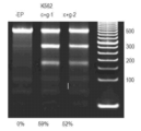
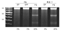
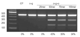

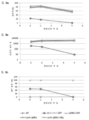
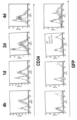


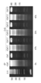


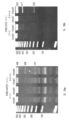
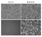
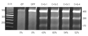


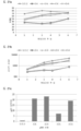
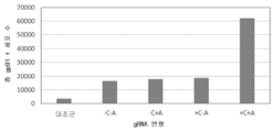


Classifications
-
- A—HUMAN NECESSITIES
- A61—MEDICAL OR VETERINARY SCIENCE; HYGIENE
- A61K—PREPARATIONS FOR MEDICAL, DENTAL OR TOILETRY PURPOSES
- A61K48/00—Medicinal preparations containing genetic material which is inserted into cells of the living body to treat genetic diseases; Gene therapy
-
- C—CHEMISTRY; METALLURGY
- C12—BIOCHEMISTRY; BEER; SPIRITS; WINE; VINEGAR; MICROBIOLOGY; ENZYMOLOGY; MUTATION OR GENETIC ENGINEERING
- C12N—MICROORGANISMS OR ENZYMES; COMPOSITIONS THEREOF; PROPAGATING, PRESERVING, OR MAINTAINING MICROORGANISMS; MUTATION OR GENETIC ENGINEERING; CULTURE MEDIA
- C12N15/00—Mutation or genetic engineering; DNA or RNA concerning genetic engineering, vectors, e.g. plasmids, or their isolation, preparation or purification; Use of hosts therefor
- C12N15/09—Recombinant DNA-technology
- C12N15/87—Introduction of foreign genetic material using processes not otherwise provided for, e.g. co-transformation
- C12N15/90—Stable introduction of foreign DNA into chromosome
- C12N15/902—Stable introduction of foreign DNA into chromosome using homologous recombination
- C12N15/907—Stable introduction of foreign DNA into chromosome using homologous recombination in mammalian cells
-
- A—HUMAN NECESSITIES
- A01—AGRICULTURE; FORESTRY; ANIMAL HUSBANDRY; HUNTING; TRAPPING; FISHING
- A01N—PRESERVATION OF BODIES OF HUMANS OR ANIMALS OR PLANTS OR PARTS THEREOF; BIOCIDES, e.g. AS DISINFECTANTS, AS PESTICIDES OR AS HERBICIDES; PEST REPELLANTS OR ATTRACTANTS; PLANT GROWTH REGULATORS
- A01N1/00—Preservation of bodies of humans or animals, or parts thereof
- A01N1/02—Preservation of living parts
- A01N1/0205—Chemical aspects
- A01N1/021—Preservation or perfusion media, liquids, solids or gases used in the preservation of cells, tissue, organs or bodily fluids
- A01N1/0221—Freeze-process protecting agents, i.e. substances protecting cells from effects of the physical process, e.g. cryoprotectants, osmolarity regulators like oncotic agents
-
- A—HUMAN NECESSITIES
- A61—MEDICAL OR VETERINARY SCIENCE; HYGIENE
- A61K—PREPARATIONS FOR MEDICAL, DENTAL OR TOILETRY PURPOSES
- A61K35/00—Medicinal preparations containing materials or reaction products thereof with undetermined constitution
- A61K35/12—Materials from mammals; Compositions comprising non-specified tissues or cells; Compositions comprising non-embryonic stem cells; Genetically modified cells
- A61K35/14—Blood; Artificial blood
- A61K35/17—Lymphocytes; B-cells; T-cells; Natural killer cells; Interferon-activated or cytokine-activated lymphocytes
-
- A—HUMAN NECESSITIES
- A61—MEDICAL OR VETERINARY SCIENCE; HYGIENE
- A61K—PREPARATIONS FOR MEDICAL, DENTAL OR TOILETRY PURPOSES
- A61K35/00—Medicinal preparations containing materials or reaction products thereof with undetermined constitution
- A61K35/12—Materials from mammals; Compositions comprising non-specified tissues or cells; Compositions comprising non-embryonic stem cells; Genetically modified cells
- A61K35/33—Fibroblasts
-
- A—HUMAN NECESSITIES
- A61—MEDICAL OR VETERINARY SCIENCE; HYGIENE
- A61K—PREPARATIONS FOR MEDICAL, DENTAL OR TOILETRY PURPOSES
- A61K48/00—Medicinal preparations containing genetic material which is inserted into cells of the living body to treat genetic diseases; Gene therapy
- A61K48/0008—Medicinal preparations containing genetic material which is inserted into cells of the living body to treat genetic diseases; Gene therapy characterised by an aspect of the 'non-active' part of the composition delivered, e.g. wherein such 'non-active' part is not delivered simultaneously with the 'active' part of the composition
- A61K48/0016—Medicinal preparations containing genetic material which is inserted into cells of the living body to treat genetic diseases; Gene therapy characterised by an aspect of the 'non-active' part of the composition delivered, e.g. wherein such 'non-active' part is not delivered simultaneously with the 'active' part of the composition wherein the nucleic acid is delivered as a 'naked' nucleic acid, i.e. not combined with an entity such as a cationic lipid
-
- A—HUMAN NECESSITIES
- A61—MEDICAL OR VETERINARY SCIENCE; HYGIENE
- A61K—PREPARATIONS FOR MEDICAL, DENTAL OR TOILETRY PURPOSES
- A61K48/00—Medicinal preparations containing genetic material which is inserted into cells of the living body to treat genetic diseases; Gene therapy
- A61K48/005—Medicinal preparations containing genetic material which is inserted into cells of the living body to treat genetic diseases; Gene therapy characterised by an aspect of the 'active' part of the composition delivered, i.e. the nucleic acid delivered
-
- A—HUMAN NECESSITIES
- A61—MEDICAL OR VETERINARY SCIENCE; HYGIENE
- A61P—SPECIFIC THERAPEUTIC ACTIVITY OF CHEMICAL COMPOUNDS OR MEDICINAL PREPARATIONS
- A61P35/00—Antineoplastic agents
-
- A—HUMAN NECESSITIES
- A61—MEDICAL OR VETERINARY SCIENCE; HYGIENE
- A61P—SPECIFIC THERAPEUTIC ACTIVITY OF CHEMICAL COMPOUNDS OR MEDICINAL PREPARATIONS
- A61P43/00—Drugs for specific purposes, not provided for in groups A61P1/00-A61P41/00
-
- C—CHEMISTRY; METALLURGY
- C12—BIOCHEMISTRY; BEER; SPIRITS; WINE; VINEGAR; MICROBIOLOGY; ENZYMOLOGY; MUTATION OR GENETIC ENGINEERING
- C12N—MICROORGANISMS OR ENZYMES; COMPOSITIONS THEREOF; PROPAGATING, PRESERVING, OR MAINTAINING MICROORGANISMS; MUTATION OR GENETIC ENGINEERING; CULTURE MEDIA
- C12N15/00—Mutation or genetic engineering; DNA or RNA concerning genetic engineering, vectors, e.g. plasmids, or their isolation, preparation or purification; Use of hosts therefor
- C12N15/09—Recombinant DNA-technology
- C12N15/63—Introduction of foreign genetic material using vectors; Vectors; Use of hosts therefor; Regulation of expression
- C12N15/79—Vectors or expression systems specially adapted for eukaryotic hosts
- C12N15/82—Vectors or expression systems specially adapted for eukaryotic hosts for plant cells, e.g. plant artificial chromosomes (PACs)
- C12N15/8201—Methods for introducing genetic material into plant cells, e.g. DNA, RNA, stable or transient incorporation, tissue culture methods adapted for transformation
- C12N15/8206—Methods for introducing genetic material into plant cells, e.g. DNA, RNA, stable or transient incorporation, tissue culture methods adapted for transformation by physical or chemical, i.e. non-biological, means, e.g. electroporation, PEG mediated
-
- C—CHEMISTRY; METALLURGY
- C12—BIOCHEMISTRY; BEER; SPIRITS; WINE; VINEGAR; MICROBIOLOGY; ENZYMOLOGY; MUTATION OR GENETIC ENGINEERING
- C12N—MICROORGANISMS OR ENZYMES; COMPOSITIONS THEREOF; PROPAGATING, PRESERVING, OR MAINTAINING MICROORGANISMS; MUTATION OR GENETIC ENGINEERING; CULTURE MEDIA
- C12N15/00—Mutation or genetic engineering; DNA or RNA concerning genetic engineering, vectors, e.g. plasmids, or their isolation, preparation or purification; Use of hosts therefor
- C12N15/09—Recombinant DNA-technology
- C12N15/87—Introduction of foreign genetic material using processes not otherwise provided for, e.g. co-transformation
-
- C—CHEMISTRY; METALLURGY
- C12—BIOCHEMISTRY; BEER; SPIRITS; WINE; VINEGAR; MICROBIOLOGY; ENZYMOLOGY; MUTATION OR GENETIC ENGINEERING
- C12N—MICROORGANISMS OR ENZYMES; COMPOSITIONS THEREOF; PROPAGATING, PRESERVING, OR MAINTAINING MICROORGANISMS; MUTATION OR GENETIC ENGINEERING; CULTURE MEDIA
- C12N5/00—Undifferentiated human, animal or plant cells, e.g. cell lines; Tissues; Cultivation or maintenance thereof; Culture media therefor
- C12N5/06—Animal cells or tissues; Human cells or tissues
- C12N5/0602—Vertebrate cells
- C12N5/0634—Cells from the blood or the immune system
- C12N5/0636—T lymphocytes
-
- C—CHEMISTRY; METALLURGY
- C12—BIOCHEMISTRY; BEER; SPIRITS; WINE; VINEGAR; MICROBIOLOGY; ENZYMOLOGY; MUTATION OR GENETIC ENGINEERING
- C12N—MICROORGANISMS OR ENZYMES; COMPOSITIONS THEREOF; PROPAGATING, PRESERVING, OR MAINTAINING MICROORGANISMS; MUTATION OR GENETIC ENGINEERING; CULTURE MEDIA
- C12N5/00—Undifferentiated human, animal or plant cells, e.g. cell lines; Tissues; Cultivation or maintenance thereof; Culture media therefor
- C12N5/06—Animal cells or tissues; Human cells or tissues
- C12N5/0602—Vertebrate cells
- C12N5/0634—Cells from the blood or the immune system
- C12N5/0647—Haematopoietic stem cells; Uncommitted or multipotent progenitors
-
- C—CHEMISTRY; METALLURGY
- C12—BIOCHEMISTRY; BEER; SPIRITS; WINE; VINEGAR; MICROBIOLOGY; ENZYMOLOGY; MUTATION OR GENETIC ENGINEERING
- C12N—MICROORGANISMS OR ENZYMES; COMPOSITIONS THEREOF; PROPAGATING, PRESERVING, OR MAINTAINING MICROORGANISMS; MUTATION OR GENETIC ENGINEERING; CULTURE MEDIA
- C12N5/00—Undifferentiated human, animal or plant cells, e.g. cell lines; Tissues; Cultivation or maintenance thereof; Culture media therefor
- C12N5/06—Animal cells or tissues; Human cells or tissues
- C12N5/0602—Vertebrate cells
- C12N5/0652—Cells of skeletal and connective tissues; Mesenchyme
- C12N5/0656—Adult fibroblasts
-
- C—CHEMISTRY; METALLURGY
- C12—BIOCHEMISTRY; BEER; SPIRITS; WINE; VINEGAR; MICROBIOLOGY; ENZYMOLOGY; MUTATION OR GENETIC ENGINEERING
- C12N—MICROORGANISMS OR ENZYMES; COMPOSITIONS THEREOF; PROPAGATING, PRESERVING, OR MAINTAINING MICROORGANISMS; MUTATION OR GENETIC ENGINEERING; CULTURE MEDIA
- C12N5/00—Undifferentiated human, animal or plant cells, e.g. cell lines; Tissues; Cultivation or maintenance thereof; Culture media therefor
- C12N5/06—Animal cells or tissues; Human cells or tissues
- C12N5/0602—Vertebrate cells
- C12N5/0652—Cells of skeletal and connective tissues; Mesenchyme
- C12N5/0662—Stem cells
- C12N5/0663—Bone marrow mesenchymal stem cells (BM-MSC)
-
- C—CHEMISTRY; METALLURGY
- C12—BIOCHEMISTRY; BEER; SPIRITS; WINE; VINEGAR; MICROBIOLOGY; ENZYMOLOGY; MUTATION OR GENETIC ENGINEERING
- C12N—MICROORGANISMS OR ENZYMES; COMPOSITIONS THEREOF; PROPAGATING, PRESERVING, OR MAINTAINING MICROORGANISMS; MUTATION OR GENETIC ENGINEERING; CULTURE MEDIA
- C12N2510/00—Genetically modified cells
Abstract
본 조성물 및 방법은 내인성 게놈 DNA 영역의 서열 변형에 관한 것이다. 특정 양태는 (a) DNA 올리고; (b) DNA 분해제; 및 (c) 캡핑되고/거나 폴리아데닐화되는 표적화 RNA를 포함하는 조성물로 전기천공에 의해 세포를 형질감염시키는 것을 포함하는, 세포에서 표적 게놈 DNA 영역의 부위-특이적 서열 변형을 위한 방법으로서, 도너 DNA는 (i) 표적 게놈 DNA 영역에 상동성인 핵산 서열을 포함하는 상동성 영역 및 (ii) 서열 변형 영역을 포함하며; 게놈 DNA 서열은 표적 게놈 DNA 영역에서 특이적으로 변형된다.The compositions and methods relate to sequence modification of endogenous genomic DNA regions. Certain embodiments include (a) a DNA oligo; (b) DNA degrading agents; and (c) a capped and/or polyadenylated targeting RNA. A method for site-specific sequence modification of a target genomic DNA region in a cell comprising transfecting the cell by electroporation, The donor DNA comprises (i) a homology region comprising a nucleic acid sequence homologous to a target genomic DNA region and (ii) a sequence modification region; A genomic DNA sequence is specifically modified in a target genomic DNA region.

Description
관련 출원의 상호 참조CROSS REFERENCES OF RELATED APPLICATIONS
본 출원은 2016년 6월 21일 출원된 미국 가출원 62/365,126의 이익을 주장한다. 참조된 출원의 전체 내용은 본원에 참조로 통합된다. This application claims the benefit of US provisional application 62/365,126, filed on June 21, 2016. The entire contents of the referenced applications are incorporated herein by reference.
발명의 배경background of invention
1. 발명의 분야1. Field of Invention
본 발명은 일반적으로 생명공학 분야에 관한 것이다. 더욱 특히, 이는 게놈 DNA를 변경하기 위한 신규한 방법 및 조성물에 관한 것이다.The present invention relates generally to the field of biotechnology. More particularly, it relates to novel methods and compositions for altering genomic DNA.
2. 관련 기술의 설명2. Description of related technology
표적화된 게놈 엔지니어링은 세포 내부의 DNA를 따라 특이적 부위에서 지시된 방식으로 내인성 DNA를 편집 또는 변경하는 것을 포함한다. 유전자 복구 및 상동성-유도된 유전자 변경의 엄청난 가능성에도 불구하고, 현재 게놈 엔지니어링 방법은 매우 낮은 복구 또는 편집 효율을 제공하며, 해로운 또는 원하지 않는 DNA 서열 및 결과가 도입될 가능성을 갖는다. Targeted genome engineering involves editing or altering endogenous DNA in a directed manner at specific sites along the DNA inside cells. Despite the enormous promise of gene repair and homology-directed genetic alteration, current genome engineering methods offer very low repair or editing efficiencies and have the potential to introduce deleterious or unwanted DNA sequences and consequences.
내인성 게놈 서열의 변형은 발전된 치료학적 적용은 물론 발전된 연구 방법을 제공할 수 있다. 현재, 시험관 내에서 유전자 기능 파괴의 가장 일반적인 방법은 RNA 간섭 (RNAi)이다. 그러나, 이러한 방법은 한계가 있다. 예를 들어, RNAi는 상당한 표적 이탈 효과 및 독성을 나타낼 수 있다. 또한, RNAi는 많은 내인성 과정의 세포 메카니즘에 관련되며, 관심 경로에 아주 매우 관련될 수 있는 메카니즘 예컨대, RNAi를 인공적으로 수행하는 것은 오해의 소지가 있거나 잘못된 결과로 이어질 수 있다. 세포의 게놈 서열을 변형시키는 효율적이고 비독성인 메카니즘은 유전자 녹-다운을 위한 더욱 정확한 방법일 것이다. Modification of endogenous genomic sequences can provide advanced therapeutic applications as well as advanced research methods. Currently, the most common method of disrupting gene function in vitro is RNA interference (RNAi). However, these methods have limitations. For example, RNAi can exhibit significant off-target effects and toxicity. In addition, RNAi is involved in the cellular mechanisms of many endogenous processes, and artificially enforcing mechanisms such as RNAi, which may very well be involved in a pathway of interest, can be misleading or lead to erroneous results. An efficient and non-toxic mechanism for altering a cell's genome sequence would be a more precise method for gene knock-down.
내인성 게놈 서열을 변형시키는 효율적이고 비독성인 방법은 또한, 생체외 요법에서 발전을 제공할 수 있는데, 환자로부터 세포를 분리하고, 게놈을 변형시켜 돌연변이를 보정하고, 환자 자신의 세포를 다시 이식하여 치료 효과를 달성할 수 있기 때문이다. 현재의 방법은 이러한 결과를 달성하기에는 너무 비효율적이거나 너무 독성이다. 효율적이고, 비독성이며 안정한 부위-특이적 게놈 DNA 변형을 허용하는 기술에 대한 요구가 본 분야에 존재한다.Efficient and non-toxic methods of modifying endogenous genomic sequences could also provide advances in ex vivo therapy, wherein cells are isolated from the patient, the genome is modified to correct the mutation, and the patient's own cells are transplanted back into treatment. Because the effect can be achieved. Current methods are either too inefficient or too toxic to achieve these results. There is a need in the art for technologies that allow efficient, non-toxic and stable site-specific genomic DNA modifications.
내인성 표적 게놈 DNA 서열의 서열 변형 또는 보정에 관한 조성물 및 방법을 제공하고자 한다. It is intended to provide compositions and methods for sequence modification or correction of endogenous target genomic DNA sequences.
조성물 및 방법은 내인성 표적 게놈 DNA 서열의 서열 변형 또는 보정에 관한 것이다. 특정 양태는 세포에서 표적 게놈 DNA 영역의 부위-특이적 서열 변형을 위한 방법으로서, (a) DNA 올리고, (b) DNA 분해제; 및 (c) 캡핑되고/거나 폴리아데닐화되는 표적화 RNA를 포함하는 조성물로 전기천공에 의해 세포를 형질감염시키는 것을 포함하며; 여기에서 DNA 올리고는 (i) 표적 게놈 DNA 영역에 상동성인 DNA 서열을 포함하는 상동성 영역; 및 (ii) 서열 변형 영역을 포함하며; 게놈 DNA 서열이 표적 게놈 DNA 영역에서 특이적으로 변형되는, 방법에 관한 것이다.The compositions and methods relate to sequence modification or correction of endogenous target genomic DNA sequences. Certain embodiments are methods for site-specific sequence modification of a target genomic DNA region in a cell comprising (a) a DNA oligo, (b) a DNA degrading agent; and (c) transfecting the cells by electroporation with a composition comprising the capped and/or polyadenylated targeting RNA; wherein the DNA oligo comprises (i) a homology region comprising a DNA sequence homologous to the target genomic DNA region; and (ii) a sequence modification region; A method wherein a genomic DNA sequence is specifically modified in a target genomic DNA region.
추가의 양태는 세포에서 표적 게놈 DNA 영역의 부위-특이적 서열 변형을 위한 방법으로서, (a) DNA 올리고; (b) DNA 분해제; 및 (c) 캡핑되고/거나 폴리아데닐화되는 표적화 RNA를 포함하는 조성물로 전기천공에 의해 줄기 세포를 형질감염시키는 것을 포함하며; 여기에서 DNA 올리고는 (i) 표적 게놈 DNA 영역에 상동성인 DNA 서열을 포함하는 상동성 영역; 및 (ii) 서열 변형 영역을 포함하며; 게놈 DNA 서열은 표적 게놈 DNA 영역에서 특이적으로 변형되며, 세포는 줄기 세포 또는 이들의 후손인 방법에 관한 것이다. 일부 구체예에서, 세포는 일차 세포이다. 본원에 사용된 바와 같은 용어 "일차"는 불멸화되지 않으며 생 조직으로부터 직접 취해지는 세포를 지칭한다. 이들 세포는 집단 더블링(population doubling)을 거의 겪지 않으며, 따라서 조직의 주요 작용 성분을 더욱 대표하며, 이들 세포는 연속 (종양 또는 인공 불멸화된) 세포주 대비 상기 조직으로부터 유래되며, 따라서 생체내 상태에 대해 더욱 대표적인 모델에 해당한다.A further aspect is a method for site-specific sequence modification of a target genomic DNA region in a cell comprising: (a) a DNA oligo; (b) DNA degrading agents; and (c) transfecting the stem cells by electroporation with a composition comprising the capped and/or polyadenylated targeting RNA; wherein the DNA oligo comprises (i) a homology region comprising a DNA sequence homologous to the target genomic DNA region; and (ii) a sequence modification region; The genomic DNA sequence is specifically modified in a target genomic DNA region, and the cell is a stem cell or a descendant thereof. In some embodiments, the cell is a primary cell. As used herein, the term "primary" refers to cells that are not immortalized and are taken directly from living tissue. These cells undergo little population doubling and are therefore more representative of the major functional components of the tissue, and these cells are derived from the tissue versus continuous (tumor or artificially immortalized) cell lines, and thus are highly dependent on the in vivo state. It corresponds to a more representative model.
일부 구체예에서, DNA 올리고 및 표적화 RNA는 표적 게놈 DNA 영역의 동일한 가닥에 상보적이다. 일부 구체예에서, DNA 분해제는 뉴클레아제이다. 일부 구체예에서, 뉴클레아제는 Cas9를 포함한다. 일부 구체예에서, 뉴클레아제는 Cpf1, CasX 또는 CasY를 포함한다. 일부 구체예에서, 표적화 RNA는 가이드 RNA를 포함한다. 일부 구체예에서, 표적화 RNA는 캡핑되고도 폴리아데닐화된다. 일부 구체예에서, 표적화 RNA는 시험관내에서 캡핑되고/거나 폴리아데닐화된다.In some embodiments, the DNA oligo and targeting RNA are complementary to the same strand of a target genomic DNA region. In some embodiments, the DNA degrading agent is a nuclease. In some embodiments, the nuclease includes Cas9. In some embodiments, the nuclease includes Cpf1, CasX or CasY. In some embodiments, the targeting RNA includes a guide RNA. In some embodiments, the targeting RNA is capped and also polyadenylated. In some embodiments, the targeting RNA is capped and/or polyadenylated in vitro.
용어 "서열 변형" 또는 "DNA 보정"은 DNA 서열에 대한 변화이며, 내인성 게놈 DNA 서열에 대한 또는 이의 첨가, 변화 또는 결실을 포함할 수 있다. 예를 들어, 표적 게놈 서열에 있어서, 도너 DNA는 표적 게놈 서열에 대해 상보적인, 동일한 또는 상동성인 서열 및 서열 변형 또는 보정 영역을 포함한다. 서열 변형 영역은 전형적으로 상동성 말단 사이에 위치한다. 서열 변형은 표적 게놈 서열에 대해 상보적이지 않거나 낮은 정도의 상동성을 가지며, 표적 게놈 서열의 변경을 함유한다.The term "sequence modification" or "DNA correction" is a change to a DNA sequence and may include additions, changes or deletions to or to endogenous genomic DNA sequences. For example, for a target genomic sequence, the donor DNA includes sequences that are complementary, identical or homologous to the target genomic sequence and sequence modification or correction regions. Sequence modification regions are typically located between homologous ends. Sequence modifications are non-complementary or have a low degree of homology to the target genomic sequence and contain alterations in the target genomic sequence.
"상동성" 또는 "동일성" 또는 "유사성"은 2개 펩티드 또는 2개 핵산 분자 사이의 서열 유사성을 지칭한다. 용어 "상동성 영역"은 표적 게놈 DNA 서열과 특정 정도의 상동성을 갖는 도너 DNA의 영역을 지칭한다. 상동성은 비교 목적을 위해 정렬될 수 있는 각 서열에서 위치를 비교함으로써 측정될 수 있다. 비교된 서열의 위치를 동일한 염기 또는 아미노산이 차지하는 경우, 그러면 분자는 그 위치에서 상동성이다. 서열들간의 상동성 정도는 서열들에 의해 공유된 매칭 또는 상동성 위치의 수의 함수이다. "관련되지 않은" 또는 "비-상동성" 서열은 본 발명의 서열 중 하나와 40% 미만의 동일성을 공유하나, 바람직하게는, 25% 미만의 동일성을 공유한다.“Homology” or “identity” or “similarity” refers to sequence similarity between two peptides or two nucleic acid molecules. The term "region of homology" refers to a region of donor DNA that has a certain degree of homology with a target genomic DNA sequence. Homology can be determined by comparing positions in each sequence that can be aligned for comparison purposes. If positions in the compared sequences are occupied by identical bases or amino acids, then the molecules are homologous at that position. The degree of homology between sequences is a function of the number of matching or homologous positions shared by the sequences. An “unrelated” or “non-homologous” sequence shares less than 40% identity with one of the sequences of the present invention, but preferably shares less than 25% identity.
폴리뉴클레오티드 또는 폴리뉴클레오티드 영역 (또는 폴리펩티드 또는 폴리펩티드 영역)은 또 다른 서열에 대해 특정 백분율(예를 들어, 적어도 또는 최대 60%, 65%, 70%, 75%, 80%, 85%, 90%, 95%, 98% 또는 99%, 또는 여기에 속하는 추론가능한 임의의 범위)의 "서열 동일성" 또는 "상동성"을 갖는다는 것은 정렬할 때, 염기 (또는 아미노산)의 백분율이 두 개의 서열 비교에서 동일한 것을 의미한다. 이러한 정렬 및 상동성 또는 서열 동일성 퍼센트는 당해기술에 공지된 소프트웨어 프로그램 예를 들어, 문헌 [Ausubel et al. eds. (2007) Current Protocols in Molecular Biology]에 기술된 것들을 사용하여 측정될 수 있다.A polynucleotide or polynucleotide region (or polypeptide or polypeptide region) may be a specific percentage (e.g., at least or at most 60%, 65%, 70%, 75%, 80%, 85%, 90%, "Sequence identity" or "homology" of 95%, 98% or 99%, or any inferred range therein) means that, when aligned, the percentage of bases (or amino acids) in a comparison of two sequences Means the same thing. Such alignments and percent homology or sequence identity can be performed using software programs known in the art, such as those described in Ausubel et al. eds. (2007) Current Protocols in Molecular Biology.
일부 구체예에서, 올리고는 단일-가닥이다. 단일-가닥 올리고는 DNA에 대한 세포의 내약성을 증가시키고 세포의 DNA-유도된 독성을 감소시킬 것으로 여겨진다.In some embodiments, an oligo is single-stranded. It is believed that single-stranded oligos will increase the tolerance of cells to DNA and reduce DNA-induced toxicity of cells.
특정 구체예에서, 도너 DNA의 상동 영역은 100% 상동성이거나 표적 게놈 서열과 동일하다. 추가의 구체예에서, 도너 DNA의 상동성 영역은 85, 90, 95, 또는 99% 상동성이다.In certain embodiments, the homologous region of the donor DNA is 100% homologous or identical to the target genomic sequence. In a further embodiment, the region of homology of the donor DNA is 85, 90, 95, or 99% homologous.
특정 구체예에서, 도너 DNA는 표적 게놈 DNA 서열에 상동인 적어도 또는 최대 10, 12, 14, 16, 18, 20, 22, 24, 26, 28, 30, 32, 34, 36, 38, 40, 42, 44, 46, 48, 50, 75, 100, 150 및 200개 잔기의 핵산 서열 (또는 이에 속하는 추론가능한 임의의 범위)을 포함한다. 특정 구체예에서, 도너 DNA는 게놈 DNA 서열과 동일한 적어도 약 10개 또는 적어도 약 15개 또는 적어도 약 20개 핵산 서열을 포함한다. 이러한 맥락에서, 용어 "동일한 서열"은 게놈 DNA의 서열과 정확하게 매칭되는 서열을 지칭한다. 동일한 서열은 DNA 서열 변형의 5' 말단인 영역 및 DNA 서열 변형의 3' 말단인 영역에 존재할 수 있다. 예시적인 실례로서, 도너 DNA가 적어도 10개 핵산의 상동성 서열을 포함하는 경우, 도너 DNA는 서열 변형의 양 측면 상에 예를 들어, 5개 핵산의 상동성 서열을 포함할 수 있다. 유사하게는, 10개 핵산의 상동성 서열을 포함하는 도너 DNA는 예를 들어, 서열 변형의 양 측면 상에 예를 들어, 5개 핵산의 상보적 서열을 포함할 수 있다.In certain embodiments, the donor DNA is at least or at most 10, 12, 14, 16, 18, 20, 22, 24, 26, 28, 30, 32, 34, 36, 38, 40, and nucleic acid sequences of 42, 44, 46, 48, 50, 75, 100, 150 and 200 residues (or any deduced range therein). In certain embodiments, the donor DNA comprises at least about 10 or at least about 15 or at least about 20 nucleic acid sequences identical to genomic DNA sequences. In this context, the term "identical sequence" refers to a sequence that exactly matches the sequence of genomic DNA. The same sequence may be present in a region that is the 5' end of a DNA sequence modification and in a region that is the 3' end of a DNA sequence modification. As an illustrative example, where the donor DNA includes homologous sequences of at least 10 nucleic acids, the donor DNA may include homologous sequences of, for example, 5 nucleic acids on either side of the sequence modification. Similarly, donor DNA comprising homologous sequences of
본원에 사용된 바와 같은 용어 "상보적"은 뉴클레오티드들 간의 왓슨-크릭 염기 쌍을 나타내며, 특히 3개의 수소 결합에 의해 연결된 시토신 및 구아닌 잔기 및 2개의 수소 결합에 의해 아데닌 잔기에 연결된 티민 또는 우라실 잔기를 갖는 서로에게 수소 결합된 뉴클레오티드를 지칭한다. 일반적으로, 핵산은 특이적 제2 뉴클레오티드 서열에 "상보성 퍼센트"를 갖는 것으로 기술된 뉴클레오티드 서열을 포함한다. 예를 들어, 뉴클레오티드 서열은 지정된 제2 뉴클레오티드 서열에 80%, 90% 또는 100% 상보성을 가질 수 있으며, 이는 서열의 10개 뉴클레오티드 중 8개, 10개 뉴클레오티드 중 9개 또는 10개 뉴클레오티드 중 10개가 지정된 제2 뉴클레오티드 서열에 상보적임을 나타낸다. 예를 들어, 뉴클레오티드 서열 3'-TCGA-5'는 뉴클레오티드 서열 5'-AGCT-3'에 100% 상보적이다. 추가로, 뉴클레오티드 서열 3'-TCGA-는 뉴클레오티드 서열 5'-TTAGCTGG-3'의 영역에 100% 상보적이다. 2개의 상보적인 뉴클레오티드 서열이 센스 가닥 및 안티센스 가닥을 포함함은 당업자에게 인지될 것이다.As used herein, the term "complementary" refers to Watson-Crick base pairs between nucleotides, in particular cytosine and guanine residues linked by three hydrogen bonds and thymine or uracil residues linked to adenine residues by two hydrogen bonds. Refers to nucleotides hydrogen bonded to each other with Generally, a nucleic acid comprises a nucleotide sequence described as having "percent complementarity" to a specific second nucleotide sequence. For example, a nucleotide sequence can have 80%, 90% or 100% complementarity to a second designated nucleotide sequence, which is 8 out of 10 nucleotides, 9 out of 10 nucleotides, or 10 out of 10 nucleotides of the sequence. Indicates that it is complementary to the designated second nucleotide sequence. For example, the nucleotide sequence 3'-TCGA-5' is 100% complementary to the nucleotide sequence 5'-AGCT-3'. Additionally, the nucleotide sequence 3'-TCGA- is 100% complementary to the region of the nucleotide sequence 5'-TTAGCTGG-3'. It will be appreciated by those skilled in the art that the two complementary nucleotide sequences include a sense strand and an antisense strand.
용어 "형질감염"은 생-활성 물질 예컨대, 핵산, 단백질, 효소 또는 소분자를 세포 내로 도입시키는 방법을 나타낸다. 핵산은 플라스미드 또는 올리고머로서 전달된 DNA, 및/또는 RNA 또는 이의 조합물일 수 있다.The term "transfection" refers to a method of introducing a bio-active substance such as a nucleic acid, protein, enzyme or small molecule into a cell. Nucleic acids can be DNA, and/or RNA, or combinations thereof, delivered as plasmids or oligomers.
용어 "전기천공"은 외부에서 가해진 전기장이 세포에 가해지는 형질감염의 방법을 지칭한다. 특정 구체예에서, 이용된 전기천공 방법은 정적 전기천공이다.The term "electroporation" refers to a method of transfection in which an externally applied electric field is applied to cells. In certain embodiments, the electroporation method used is static electroporation.
특정 구체예에서, 세포는 유동 전기천공을 사용하여 전기천공된다. 유동 전기천공은 세포 현탁액을 이동시키고 유체 챔버 또는 유체 흐름 경로를 포함하는 장치 내로 분자를 로딩하고 (상기 유체 챔버 또는 유체 흐름 경로는 유체 챔버 또는 유체 흐름 경로의 측면을 따라 배치된 전극으로 이루어지고, 유체 챔버 또는 유체 흐름 경로 내부의 생물학적 입자를 전기천공에 적합한 전기장으로 처리하도록 구성됨); 장치 밖으로 전기천공된 세포 현탁액을 이동시키는 것을 포함한다. 용어 "유동 전기천공"은 유체 챔버 흐름 경로 내부의 세포의 전기천공을 지칭한다. 이러한 방법은 특히 대규모 부피의 세포에 효과적이다. 대조적으로 정적 전기천공은 마주하는 전극 사이의 거리 및 액체를 가로지르는 이동 전기와 관련된 제한으로 인해 한 세트의 제한된 부피의 세포의 전기천공을 포함한다.In certain embodiments, cells are electroporated using flow electroporation. Flow electroporation involves moving a cell suspension and loading molecules into a device comprising a fluid chamber or fluid flow path, wherein the fluid chamber or fluid flow path consists of electrodes disposed along the sides of the fluid chamber or fluid flow path; configured to treat biological particles within the fluid chamber or fluid flow path with an electric field suitable for electroporation; and transferring the electroporated cell suspension out of the device. The term “flow electroporation” refers to the electroporation of cells inside a fluid chamber flow path. This method is particularly effective for large volumes of cells. In contrast, static electroporation involves the electroporation of a set of limited volume cells due to limitations associated with the distance between opposing electrodes and moving electricity across a liquid.
특정 양태에서, 세포 내로 발현 작제물을 형질감염시키는 것은 흐름 챔버에서 전기장을 통해 세포 현탁액을 유동시키는 것을 포함하며, 전기장은 흐름 챔버를 적어도 부분적으로 규정하는 반대로 하전된 전극을 마주함으로써 생성되며, 여기에서 흐름 챔버의 내열성은 대략 와트당 10℃ 미만이다. 다른 특정 양태에서, 세포를 형질감염시키는 것은 전기천공될 세포 현탁액을 함유하기 위한 챔버를 포함하는 유동 전기천공 장치를 사용하는 것을 포함하며; 챔버는 반대로 하전가능한 전극을 마주시킴으로써 적어도 부분적으로 규정되며; 챔버의 내열성은 대략 와트당 10℃ 미만이다.In certain embodiments, transfecting the expression construct into a cell comprises flowing the cell suspension through an electric field in a flow chamber, wherein the electric field is generated by facing oppositely charged electrodes that at least partially define the flow chamber, wherein The heat resistance of the flow chamber at is approximately less than 10° C. per watt. In another specific embodiment, transfecting the cells comprises using a flow electroporation device comprising a chamber for containing a cell suspension to be electroporated; The chamber is defined at least in part by facing oppositely chargeable electrodes; The heat resistance of the chamber is approximately less than 10° C. per watt.
특정 양태에서, 세포 내로 발현 작제물을 형질감염시키는 것은 챔버에서 전기장으로 세포 현탁액을 전기천공시키거나 노출시키는 것을 포함하며, 전기장은 챔버를 적어도 부분적으로 규정하는 반대로 하전된 전극을 마주시킴으로써 생성되며, 여기에서 챔버의 내열성은 대략 와트당 10℃ 미만이다. 다른 특정 양태에서, 세포를 형질감염시키는 것은 전기천공될 세포 현탁액을 함유하기 위한 챔버를 포함하는 전기천공 장치를 사용하는 것을 포함하며; 챔버는 반대로 하전가능한 전극을 마주시킴으로써 적어도 부분적으로 규정되며; 챔버의 내열성은 대략 와트당 10℃ 미만이다.In certain embodiments, transfecting the expression construct into the cell comprises electroporation or exposing the cell suspension to an electric field in a chamber, the electric field being generated by facing oppositely charged electrodes that at least partially define the chamber; The heat resistance of the chamber here is approximately less than 10° C. per watt. In another specific embodiment, transfecting the cells comprises using an electroporation device comprising a chamber for containing a cell suspension to be electroporated; The chamber is defined at least in part by facing oppositely chargeable electrodes; The heat resistance of the chamber is approximately less than 10° C. per watt.
특정 양태에서, 챔버의 내열성은 대략 와트당 0.1℃ 내지 와트당 10℃이다. 예를 들어, 챔버의 내열성은 대략 와트당 0.1, 0.2, 0.3, 0.4, 0.5, 0.6, 0.7, 0.8, 0.9, 1.0, 1.5, 2.0, 2.5, 3.0, 3.5, 4.0, 4.5, 5.0, 5.5, 6.0, 6.5, 7.0, 7.5, 8.0, 8.5, 9.0, 9.5, 또는 10℃, 또는 여기에 속하는 추론가능한 임의의 범위일 수 있다.In certain embodiments, the heat resistance of the chamber is between approximately 0.1° C. per watt and 10° C. per watt. For example, the heat resistance of the chamber is approximately 0.1, 0.2, 0.3, 0.4, 0.5, 0.6, 0.7, 0.8, 0.9, 1.0, 1.5, 2.0, 2.5, 3.0, 3.5, 4.0, 4.5, 5.0, 5.5, 6.0 per watt. , 6.5, 7.0, 7.5, 8.0, 8.5, 9.0, 9.5, or 10° C., or any range deduced therein.
마주보는 반대로 하전가능한 전극은 서로 적어도 1mm, 적어도 2mm, 적어도 3 mm, 또는 여기에 속하는 추론가능한 임의의 간격 또는 범위로 이격될 수 있다. 기재된 구체예 중 임의의 구체예에서, 챔버는 대략 1 대 100cm의 완충제와 접촉되는 합쳐진 전극 표면 대 전극 사이의 간격의 비율을 가질 수 있다. 예를 들어, 비율은 대략 1 대 1, 2, 3, 4, 5, 6, 7, 8, 9, 10, 11, 12, 13, 14, 15, 16, 17, 18, 19, 20, 21, 22, 23, 24, 25, 26, 27, 28, 29, 30, 31, 32, 33, 34, 35, 36, 37, 38, 39, 40, 41, 42, 43, 44, 45, 46, 47, 48, 49, 50, 51, 52, 53, 54, 55, 56, 57, 58, 59, 60, 61, 62, 63, 64, 65, 66, 67, 68, 69, 70, 71, 72, 73, 74, 75, 76, 77, 78, 79, 80, 81, 82, 83, 84, 85, 86, 87, 88, 89, 90, 91, 92, 93, 94, 95, 96, 97, 98, 99, 또는 100cm, 또는 여기에 속하는 추론가능한 임의의 값 또는 범위일 수 있다. 특정 양태에서, 챔버는 대략 1 대 100cm의 완충제와 접촉되는 합쳐진 전극 표면 대 전극 사이의 간격의 비율을 가지며, 마주보는 반대로 하전가능한 전극은 서로 적어도 1mm 이격되어 있다. 다른 양태에서, 챔버는 대략 1 대 100cm의 완충제와 접촉되는 합쳐진 전극 표면 대 전극 사이의 간격의 비율을 가지며, 마주보는 반대로 하전가능한 전극은 서로 적어도 3mm 이격되어 있다. 더욱 추가의 양태에서, 챔버는 대략 1 대 100cm의 완충제와 접촉되는 합쳐진 전극 표면 대 전극 사이의 간격의 비율을 가지며, 마주보는 반대로 하전가능한 전극은 서로 대략 3mm 내지 대략 2cm 이격되어 있다. 예를 들어, 마주보는 반대로 하전가능한 전극은 서로 대략 3, 4, 5, 6, 7, 8, 9, 또는 10mm 또는 여기에 속하는 추론가능한 임의의 간격을 두고 이격될 수 있거나, 마주보는 반대로 하전가능한 전극은 서로로부터 대략 1.0, 1.1, 1.2, 1.3, 1.4, 1.5, 1.6, 1.7, 1.8, 1.9, 또는 2.0cm, 또는 여기에 속하는 추론가능한 임의의 간격으로 이격될 수 있다. 이들 구체예의 일부 양태에서, 전기천공된 세포는 이에 의해 실질적으로 열 분해되지 않는다.Opposite oppositely chargeable electrodes may be spaced apart from each other by at least 1 mm, at least 2 mm, at least 3 mm, or any inferred spacing or range therein. In any of the described embodiments, the chamber may have a ratio of the gap between the electrodes to the combined electrode surfaces in contact with the buffer of approximately 1 to 100 cm. For example, the ratio is approximately 1 to 1, 2, 3, 4, 5, 6, 7, 8, 9, 10, 11, 12, 13, 14, 15, 16, 17, 18, 19, 20, 21 , 22, 23, 24, 25, 26, 27, 28, 29, 30, 31, 32, 33, 34, 35, 36, 37, 38, 39, 40, 41, 42, 43, 44, 45, 46 , 47, 48, 49, 50, 51, 52, 53, 54, 55, 56, 57, 58, 59, 60, 61, 62, 63, 64, 65, 66, 67, 68, 69, 70, 71 , 72, 73, 74, 75, 76, 77, 78, 79, 80, 81, 82, 83, 84, 85, 86, 87, 88, 89, 90, 91, 92, 93, 94, 95, 96 , 97, 98, 99, or 100 cm, or any value or range deduced therein. In certain embodiments, the chamber has a ratio of the gap between the electrodes to the surfaces of the merged electrode surfaces in contact with the buffer of approximately 1 to 100 cm, and the opposing oppositely chargeable electrodes are spaced at least 1 mm apart from each other. In another embodiment, the chamber has a ratio of the gap between the electrodes to the surfaces of the combined electrode surfaces in contact with the buffer of approximately 1 to 100 cm, and the opposing oppositely chargeable electrodes are spaced at least 3 mm apart from each other. In a still further aspect, the chamber has a ratio of the gap between the electrodes to the surfaces of the combined electrode surfaces in contact with the buffer of approximately 1 to 100 cm, and the opposing oppositely chargeable electrodes are spaced between approximately 3 mm and approximately 2 cm from each other. For example, opposing oppositely chargeable electrodes can be spaced apart from each other by approximately 3, 4, 5, 6, 7, 8, 9, or 10 mm, or any inferred spacing therein, or opposite oppositely chargeable electrodes can The electrodes may be spaced from one another by approximately 1.0, 1.1, 1.2, 1.3, 1.4, 1.5, 1.6, 1.7, 1.8, 1.9, or 2.0 cm, or any inferred spacing therein. In some aspects of these embodiments, the electroporated cells are thereby substantially free from thermal degradation.
기재된 구체예 중 임의의 구체예에서, 챔버는 대략 1 대 100cm의 완충제와 접촉되는 합쳐진 전극 표면 대 전극 사이의 간격의 비율을 가질 수 있다. 예를 들어, 비율은 대략 1 대 1, 2, 3, 4, 5, 6, 7, 8, 9, 10, 11, 12, 13, 14, 15, 16, 17, 18, 19, 20, 21, 22, 23, 24, 25, 26, 27, 28, 29, 30, 31, 32, 33, 34, 35, 36, 37, 38, 39, 40, 41, 42, 43, 44, 45, 46, 47, 48, 49, 50, 51, 52, 53, 54, 55, 56, 57, 58, 59, 60, 61, 62, 63, 64, 65, 66, 67, 68, 69, 70, 71, 72, 73, 74, 75, 76, 77, 78, 79, 80, 81, 82, 83, 84, 85, 86, 87, 88, 89, 90, 91, 92, 93, 94, 95, 96, 97, 98, 99, 또는 100cm, 또는 여기에 속하는 추론가능한 임의의 값 또는 범위일 수 있다. 특정 양태에서, 챔버는 대략 1 대 100cm의 완충제와 접촉되는 합쳐진 전극 표면 대 전극 사이의 간격의 비율을 가지며, 마주보는 반대로 하전가능한 전극은 서로 적어도 1mm 이격되어 있다. 다른 양태에서, 챔버는 대략 1 대 100cm의 완충제와 접촉되는 합쳐진 전극 표면 대 전극 사이의 간격의 비율을 가지며, 마주보는 반대로 하전가능한 전극은 서로 적어도 3mm 이격되어 있다. 더욱 추가의 양태에서, 챔버는 대략 1 대 100cm의 완충제와 접촉되는 합쳐진 전극 표면 대 전극 사이의 간격의 비율을 가지며, 마주보는 반대로 하전가능한 전극은 서로 대략 3mm 내지 대략 2cm 이격되어 있다. 예를 들어, 마주보는 반대로 하전가능한 전극은 서로 대략 3, 4, 5, 6, 7, 8, 9, 또는 10mm 또는 여기에 속하는 추론가능한 임의의 간격을 두고 이격될 수 있거나, 마주보는 반대로 하전가능한 전극은 서로로부터 대략 1.0, 1.1, 1.2, 1.3, 1.4, 1.5, 1.6, 1.7, 1.8, 1.9, 또는 2.0cm, 또는 여기에 속하는 추론가능한 임의의 간격으로 이격될 수 있다. 이들 구체예의 일부 양태에서, 전기천공된 세포는 이에 의해 실질적으로 열 분해되지 않는다.In any of the described embodiments, the chamber may have a ratio of the gap between the electrodes to the combined electrode surfaces in contact with the buffer of approximately 1 to 100 cm. For example, the ratio is approximately 1 to 1, 2, 3, 4, 5, 6, 7, 8, 9, 10, 11, 12, 13, 14, 15, 16, 17, 18, 19, 20, 21 , 22, 23, 24, 25, 26, 27, 28, 29, 30, 31, 32, 33, 34, 35, 36, 37, 38, 39, 40, 41, 42, 43, 44, 45, 46 , 47, 48, 49, 50, 51, 52, 53, 54, 55, 56, 57, 58, 59, 60, 61, 62, 63, 64, 65, 66, 67, 68, 69, 70, 71 , 72, 73, 74, 75, 76, 77, 78, 79, 80, 81, 82, 83, 84, 85, 86, 87, 88, 89, 90, 91, 92, 93, 94, 95, 96 , 97, 98, 99, or 100 cm, or any value or range deduced therein. In certain embodiments, the chamber has a ratio of the gap between the electrodes to the surfaces of the merged electrode surfaces in contact with the buffer of approximately 1 to 100 cm, and the opposing oppositely chargeable electrodes are spaced at least 1 mm apart from each other. In another embodiment, the chamber has a ratio of the gap between the electrodes to the surfaces of the combined electrode surfaces in contact with the buffer of approximately 1 to 100 cm, and the opposing oppositely chargeable electrodes are spaced at least 3 mm apart from each other. In a still further aspect, the chamber has a ratio of the gap between the electrodes to the surfaces of the combined electrode surfaces in contact with the buffer of approximately 1 to 100 cm, and the opposing oppositely chargeable electrodes are spaced between approximately 3 mm and approximately 2 cm from each other. For example, opposing oppositely chargeable electrodes can be spaced apart from each other by approximately 3, 4, 5, 6, 7, 8, 9, or 10 mm, or any inferred spacing therein, or opposite oppositely chargeable electrodes can The electrodes may be spaced from one another by approximately 1.0, 1.1, 1.2, 1.3, 1.4, 1.5, 1.6, 1.7, 1.8, 1.9, or 2.0 cm, or any inferred spacing therein. In some aspects of these embodiments, the electroporated cells are thereby substantially free from thermal degradation.
임의의 기재된 구체예에서, 장치는 냉각 요소를 추가로 포함하여 열을 소멸시킬 수 있다. 예를 들어, 냉각 요소는 열전 냉각 요소를 포함할 수 있다. 또 다른 예로서, 냉각 요소는 전극과 접촉해서 흐르는 냉각 유체를 포함할 수 있다. 또 다른 예로서, 냉각 요소는 전극과 작동적으로 관련된 열 싱크를 포함할 수 있다. 챔버의 내열성은 대략 와트당 3℃ 미만일 수 있다. 일부 구체예에서, 챔버의 내열성은 대략 와트당 0.5℃ 내지 와트당 4℃이거나, 챔버의 내열성은 대략 와트당 1℃ 내지 와트당 3℃이다. 예를 들어, 챔버의 내열성은 대략 와트당 0.5, 0.6, 0.7, 0.8, 0.9, 1.0, 1.1, 1.2, 1.3, 1.4, 1.5, 1.6, 1.7, 1.8, 1.9, 2.0, 2.1, 2.2, 2.3, 2.4, 2.5, 2.6, 2.7, 2.8, 2.9, 3.0, 3.1, 3.2, 3.3, 3.4, 3.5, 3.6, 3.7, 3.8, 3.9, 또는 4.0 ℃, 또는 여기에 속하는 추론가능한 임의의 값일 수 있다.In any of the described embodiments, the device may further include a cooling element to dissipate heat. For example, the cooling element may include a thermoelectric cooling element. As another example, the cooling element may include a cooling fluid flowing in contact with the electrode. As another example, the cooling element may include a heat sink operatively associated with the electrode. The heat resistance of the chamber may be less than about 3° C. per watt. In some embodiments, the heat resistance of the chamber is between approximately 0.5° C. per watt and 4° C. per watt, or the heat resistance of the chamber is between approximately 1° C. per watt and 3° C. per watt. For example, the heat resistance of the chamber is approximately 0.5, 0.6, 0.7, 0.8, 0.9, 1.0, 1.1, 1.2, 1.3, 1.4, 1.5, 1.6, 1.7, 1.8, 1.9, 2.0, 2.1, 2.2, 2.3, 2.4 per watt. .
전기천공에 의해 세포를 형질감염시키는 것을 포함하는 특정 방법에서, 상기 방법은 0.5 kV/cm 초과의 강도를 갖는 전기장에 세포 현탁액을 노출시키는 것을 포함한다. 예를 들면, 전기장은 대략 3.5 kV/cm 초과의 강도를 가질 수 있다. 특정 양태에서, 전기장은 대략 0.5, 1.0, 1.5, 2.0, 2.5, 3.0, 또는 3.5 kV/cm 초과, 또는 여기에 속하는 추론가능한 임의의 값의 강도를 갖는다.In certain methods comprising transfecting cells by electroporation, the method comprises exposing the cell suspension to an electric field having a strength greater than 0.5 kV/cm. For example, the electric field may have a strength greater than approximately 3.5 kV/cm. In certain embodiments, the electric field has a strength greater than approximately 0.5, 1.0, 1.5, 2.0, 2.5, 3.0, or 3.5 kV/cm, or any value deduced therein.
일부 구체예에서, 세포를 형질감염시키는 것은, 전기천공될 세포 현탁액의 연속 흐름을 수용하고 일시적으로 함유하도록 구성된 전기천공 구역을 갖는 흐름 채널을 규정하는 벽; 흐름 채널과 유체 소통하는 유입구 흐름 포털로서, 이에 의해 현탁액이 유입구 흐름 포털을 통해 흐름 채널 내로 도입될 수 있는 유입구 흐름 포털; 흐름 채널과 유체 소통하는 배출구 흐름 포털로서, 이에 의해 현탁액이 배출구 포털을 통해 흐름 채널로부터 배출될 수 있는 배출구 흐름 포털을 포함하는 유동 전기천공 장치를 사용하는 것을 포함하며; 벽은 흐름 채널의 제1 벽의 실질적 부분을 형성하는 제1 전극 및 제1 벽과 마주하는 흐름 채널의 제2 벽의 실질적인 부분을 형성하는 제2 전극을 포함하는 전기천공 구역 내의 흐름 채널을 규정하며, 제1 및 제2 전극은 전기 에너지 공급원과 전기 소통하도록 위치하는 경우 이들 사이에 전기장이 형성하며 이들 사이를 통해 현탁액이 흐를 수 있게 하며, 흐름 채널의 내열성은 대략 와트당 10℃ 미만이다.In some embodiments, transfecting cells comprises a wall defining a flow channel having an electroporation zone configured to receive and transiently contain a continuous flow of a cell suspension to be electroporated; an inlet flow portal in fluid communication with the flow channel, whereby a suspension may be introduced into the flow channel through the inlet flow portal; using a flow electroporation device comprising an outlet flow portal in fluid communication with the flow channel, whereby the suspension can exit the flow channel through the outlet flow portal; The wall defines a flow channel within the electroporation zone including a first electrode forming a substantial portion of the first wall of the flow channel and a second electrode forming a substantial portion of a second wall of the flow channel opposite the first wall. wherein the first and second electrodes, when placed in electrical communication with a source of electrical energy, form an electric field therebetween and allow a suspension to flow therebetween, wherein the flow channel has a heat resistance of less than approximately 10°C per watt.
특정 이러한 구체예에서, 제1 및 제2 전극 또는 마주하는 반대로 하전가능한 전극은 서로 적어도 1mm 이격된다. 게다가, 챔버는 대략 1 대 100cm의 완충제와 접촉되는 합쳐진 전극 표면 대 전극 사이의 간격의 비율을 가질 수 있다. 특정 구체예에서, 챔버는 대략 1 대 1, 2, 3, 4, 5, 6, 7, 8, 9, 10, 11, 12, 13, 14, 15, 16, 17, 18, 19, 20, 21, 22, 23, 24, 25, 26, 27, 28, 29, 30, 31, 32, 33, 34, 35, 36, 37, 38, 39, 40, 41, 42, 43, 44, 45, 46, 47, 48, 49, 50, 51, 52, 53, 54, 55, 56, 57, 58, 59, 60, 61, 62, 63, 64, 65, 66, 67, 68, 69, 70, 71, 72, 73, 74, 75, 76, 77, 78, 79, 80, 81, 82, 83, 84, 85, 86, 87, 88, 89, 90, 91, 92, 93, 94, 95, 96, 97, 98, 99, 또는 100cm, 또는 여기에 속하는 추론가능한 임의의 값 또는 범위의 완충제와 접촉되는 합쳐진 전극 표면 대 전극 사이의 간격의 비율을 가질 수 있다. 특정 구체예에서, 본원에 기재된 전기천공 방법에 의해 전기천공된 세포는 이에 의해 실질적으로 열분해되지 않는다. 본원에 기재된 특정 구체예에서, 챔버는 흐름 챔버이다.In certain such embodiments, the first and second electrodes or opposing oppositely chargeable electrodes are spaced at least 1 mm apart from each other. Additionally, the chamber may have a ratio of gap between the electrodes to the combined electrode surfaces in contact with the buffer of approximately 1 to 100 cm. In certain embodiments, the chamber is approximately 1 to 1, 2, 3, 4, 5, 6, 7, 8, 9, 10, 11, 12, 13, 14, 15, 16, 17, 18, 19, 20, 21, 22, 23, 24, 25, 26, 27, 28, 29, 30, 31, 32, 33, 34, 35, 36, 37, 38, 39, 40, 41, 42, 43, 44, 45, 46, 47, 48, 49, 50, 51, 52, 53, 54, 55, 56, 57, 58, 59, 60, 61, 62, 63, 64, 65, 66, 67, 68, 69, 70, 71, 72, 73, 74, 75, 76, 77, 78, 79, 80, 81, 82, 83, 84, 85, 86, 87, 88, 89, 90, 91, 92, 93, 94, 95, 96, 97, 98, 99, or 100 cm, or any deduced value or range within the range of the gap between the electrodes and the surfaces of the merged electrodes in contact with the buffer. In certain embodiments, cells electroporated by an electroporation method described herein are thereby substantially not pyrolyzed. In certain embodiments described herein, the chamber is a flow chamber.
일부 양태에서, 전기천공 장치는 전기천공될 세포 현탁액을 수용하는 챔버로서, 마주하는 반대로 하전가능한 전극에 의해 적어로 부분적으로 규정되는 챔버를 포함하며, 여기에서 챔버는 대략 1 대 100cm의 완충제와 접촉되는 합쳐진 전극 표면 대 전극 사이의 간격의 비율을 갖는다. 특정 양태에서, 상기 비율은 대략 1 대 70cm이다. 기타 특정 양태에서, 상기 비율은 대략 1 대 50cm이다. 예를 들어, 비율은 대략 1 대 1, 2, 3, 4, 5, 6, 7, 8, 9, 10, 11, 12, 13, 14, 15, 16, 17, 18, 19, 20, 21, 22, 23, 24, 25, 26, 27, 28, 29, 30, 31, 32, 33, 34, 35, 36, 37, 38, 39, 40, 41, 42, 43, 44, 45, 46, 47, 48, 49, 50, 51, 52, 53, 54, 55, 56, 57, 58, 59, 60, 61, 62, 63, 64, 65, 66, 67, 68, 69, 70, 71, 72, 73, 74, 75, 76, 77, 78, 79, 80, 81, 82, 83, 84, 85, 86, 87, 88, 89, 90, 91, 92, 93, 94, 95, 96, 97, 98, 99, 또는 100cm, 또는 여기에 속하는 추론가능한 임의의 값일 수 있다. 본원에 기재된 특정 구체예에서, 챔버는 흐름 챔버이다.In some embodiments, an electroporation device comprises a chamber containing a cell suspension to be electroporated, at least partially defined by an opposing oppositely chargeable electrode, wherein the chamber is in contact with approximately 1 to 100 cm of a buffer. has the ratio of the combined electrode surfaces to the gap between the electrodes. In certain embodiments, the ratio is approximately 1 to 70 cm. In certain other embodiments, the ratio is approximately 1 to 50 cm. For example, the ratio is approximately 1 to 1, 2, 3, 4, 5, 6, 7, 8, 9, 10, 11, 12, 13, 14, 15, 16, 17, 18, 19, 20, 21 , 22, 23, 24, 25, 26, 27, 28, 29, 30, 31, 32, 33, 34, 35, 36, 37, 38, 39, 40, 41, 42, 43, 44, 45, 46 , 47, 48, 49, 50, 51, 52, 53, 54, 55, 56, 57, 58, 59, 60, 61, 62, 63, 64, 65, 66, 67, 68, 69, 70, 71 , 72, 73, 74, 75, 76, 77, 78, 79, 80, 81, 82, 83, 84, 85, 86, 87, 88, 89, 90, 91, 92, 93, 94, 95, 96 , 97, 98, 99, or 100 cm, or any value that can be inferred therein. In certain embodiments described herein, the chamber is a flow chamber.
일부 구체예에서, 유동 전기천공 장치는 전기천공될 세포 현탁액의 연속 흐름을 수용하고 일시적으로 함유하도록 구성된 흐름 채널을 규정하는 벽; 흐름 채널과 유체 소통하는 유입구 흐름 포털로서, 이에 의해 현탁액이 유입구 흐름 포털을 통해 흐름 채널 내로 도입될 수 있는 유입구 흐름 포털; 흐름 채널과 유체 소통하는 배출구 흐름 포털로서, 이에 의해 현탁액이 배출구 포털을 통해 흐름 채널로부터 배출될 수 있는 배출구 흐름 포털을 포함하며; 벽은 흐름 채널의 제1 벽의 적어도 일부를 형성하는 제1 전극 및 제1 벽과 마주하는 흐름 채널의 제2 벽의 적어도 일부를 형성하는 제2 전극을 포함하는 흐름 채널을 규정하며, 제1 및 제2 전극은 전기 에너지 공급원과 전기 소통하도록 위치하는 경우 이들 사이에 전기장이 형성되며 이들 사이를 통해 현탁액이 흐를 수 있게 하며, 흐름 채널의 내열성은 대략 와트당 10℃ 미만이다. 특정 양태에서, 흐름 챔버의 내열성은 대략 와트당 0.1℃ 내지 와트당 10℃이다. 예를 들어, 흐름 채널의 내열성은 대략 와트당 0.1, 0.2, 0.3, 0.4, 0.5, 0.6, 0.7, 0.8, 0.9, 1.0, 1.5, 2.0, 2.5, 3.0, 3.5, 4.0, 4.5, 5.0, 5.5, 6.0, 6.5, 7.0, 7.5, 8.0, 8.5, 9.0, 9.5, 또는 10℃, 또는 여기에 속하는 추론가능한 임의의 내열성일 수 있다. 제1 및 제2 전극은 서로 적어도 1mm, 적어도 2mm, 적어도 3 mm, 또는 여기에 속하는 추론가능한 임의의 간격 또는 범위로 이격될 수 있다. 기재된 구체예 중 임의의 구체예에서, 흐름 챔버는 대략 1 대 100cm의 완충제와 접촉되는 합쳐진 전극 표면 대 전극 사이의 간격의 비율을 가질 수 있다. 예를 들어, 비율은 대략 1 대 1, 2, 3, 4, 5, 6, 7, 8, 9, 10, 11, 12, 13, 14, 15, 16, 17, 18, 19, 20, 21, 22, 23, 24, 25, 26, 27, 28, 29, 30, 31, 32, 33, 34, 35, 36, 37, 38, 39, 40, 41, 42, 43, 44, 45, 46, 47, 48, 49, 50, 51, 52, 53, 54, 55, 56, 57, 58, 59, 60, 61, 62, 63, 64, 65, 66, 67, 68, 69, 70, 71, 72, 73, 74, 75, 76, 77, 78, 79, 80, 81, 82, 83, 84, 85, 86, 87, 88, 89, 90, 91, 92, 93, 94, 95, 96, 97, 98, 99, 또는 100cm, 또는 여기에 속하는 추론가능한 임의의 값 또는 범위일 수 있다. 특정 양태에서, 흐름 챔버는 대략 1 대 100cm의 완충제와 접촉되는 합쳐진 전극 표면 대 전극 사이의 간격의 비율을 가지며, 제1 및 제2 전극은 서로 적어도 1mm 이격되어 있다. 다른 양태에서, 흐름 챔버는 대략 1 대 100cm의 완충제와 접촉되는 합쳐진 전극 표면 대 전극 사이의 간격의 비율을 가지며, 제1 및 제2 전극은 서로 적어도 3mm 이격되어 있다. 더욱 추가의 양태에서, 흐름 챔버는 대략 1 대 100cm의 완충제와 접촉되는 합쳐진 전극 표면 대 전극 사이의 간격의 비율을 가지며, 제1 및 제2 전극은 서로 대략 3mm 내지 대략 2cm 이격되어 있다. 예를 들어, 제1 및 제2 전극은 서로 대략 3, 4, 5, 6, 7, 8, 9, 또는 10mm 또는 여기에 속하는 추론가능한 임의의 간격을 두고 이격될 수 있거나, 제1 및 제2 전극은 서로로부터 대략 1.0, 1.1, 1.2, 1.3, 1.4, 1.5, 1.6, 1.7, 1.8, 1.9, 또는 2.0cm, 또는 여기에 속하는 추론가능한 임의의 간격으로 이격될 수 있다. 이들 구체예의 일부 양태에서, 흐름 채널에서 전기천공된 세포는 이에 의해 실질적으로 열 분해되지 않는다.In some embodiments, a flow electroporation device includes walls defining flow channels configured to receive and temporarily contain a continuous flow of a cell suspension to be electroporated; an inlet flow portal in fluid communication with the flow channel, whereby a suspension may be introduced into the flow channel through the inlet flow portal; an outlet flow portal in fluid communication with the flow channel, whereby a suspension may be discharged from the flow channel through the outlet portal; The wall defines a flow channel comprising a first electrode forming at least a portion of a first wall of the flow channel and a second electrode forming at least a portion of a second wall of the flow channel opposite the first wall; and the second electrode, when placed in electrical communication with the source of electrical energy, forms an electric field therebetween and allows the suspension to flow therebetween, wherein the flow channel has a thermal resistance of less than about 10° C. per watt. In certain embodiments, the heat resistance of the flow chamber is between approximately 0.1° C. per watt and 10° C. per watt. For example, the heat resistance of the flow channel is approximately 0.1, 0.2, 0.3, 0.4, 0.5, 0.6, 0.7, 0.8, 0.9, 1.0, 1.5, 2.0, 2.5, 3.0, 3.5, 4.0, 4.5, 5.0, 5.5, 6.0, 6.5, 7.0, 7.5, 8.0, 8.5, 9.0, 9.5, or 10° C., or any heat resistance deduced therein. The first and second electrodes may be spaced apart from each other by at least 1 mm, at least 2 mm, at least 3 mm, or any inferred spacing or range therein. In any of the described embodiments, the flow chamber may have a ratio of gap between the electrodes to the surfaces of the merged electrodes in contact with the buffer of approximately 1 to 100 cm. For example, the ratio is approximately 1 to 1, 2, 3, 4, 5, 6, 7, 8, 9, 10, 11, 12, 13, 14, 15, 16, 17, 18, 19, 20, 21 , 22, 23, 24, 25, 26, 27, 28, 29, 30, 31, 32, 33, 34, 35, 36, 37, 38, 39, 40, 41, 42, 43, 44, 45, 46 , 47, 48, 49, 50, 51, 52, 53, 54, 55, 56, 57, 58, 59, 60, 61, 62, 63, 64, 65, 66, 67, 68, 69, 70, 71 , 72, 73, 74, 75, 76, 77, 78, 79, 80, 81, 82, 83, 84, 85, 86, 87, 88, 89, 90, 91, 92, 93, 94, 95, 96 , 97, 98, 99, or 100 cm, or any value or range deduced therein. In certain embodiments, the flow chamber has a ratio of the gap between the electrodes to the surfaces of the joined electrodes in contact with the buffer of approximately 1 to 100 cm, and the first and second electrodes are spaced at least 1 mm apart from each other. In another aspect, the flow chamber has a ratio of the gap between the electrodes to the surfaces of the merged electrodes in contact with the buffer of approximately 1 to 100 cm, and the first and second electrodes are spaced at least 3 mm apart from each other. In a still further aspect, the flow chamber has a ratio of the gap between the electrodes to the combined electrode surfaces in contact with the buffer of approximately 1 to 100 cm, and the first and second electrodes are spaced from about 3 mm to about 2 cm from each other. For example, the first and second electrodes can be spaced apart from each other by approximately 3, 4, 5, 6, 7, 8, 9, or 10 mm, or any inferred spacing therein, or the first and second electrodes can be The electrodes may be spaced from one another by approximately 1.0, 1.1, 1.2, 1.3, 1.4, 1.5, 1.6, 1.7, 1.8, 1.9, or 2.0 cm, or any inferred spacing therein. In some aspects of these embodiments, the cells electroporated in the flow channel are thereby not substantially thermally degraded.
특정 기술된 방법 및 장치에서, 챔버의 내열성은 대략 와트당 0.1℃ 내지 대략 와트당 4℃이다. 일부 양태에서, 챔버의 내열성은 대략 와트당 1.5℃ 내지 대략 와트당 2.5℃이다. 예를 들어, 챔버의 내열성은 대략 와트당 0.1, 0.2, 0.3, 0.4, 0.5, 0.6, 0.7, 0.8, 0.9, 1.0, 1.1, 1.2, 1.3, 1.4, 1.5, 1.6, 1.7, 1.8, 1.9, 2.0, 2.1, 2.2, 2.3, 2.4, 2.5, 2.6, 2.7, 2.8, 2.9, 3.0, 3.1, 3.2, 3.3, 3.4, 3.5, 3.6, 3.7, 3.8, 3.9, 또는 4.0 ℃, 또는 여기에 속하는 추론가능한 임의의 내열성일 수 있다.In certain described methods and devices, the heat resistance of the chamber is from about 0.1° C. per watt to about 4° C. per watt. In some embodiments, the heat resistance of the chamber is between about 1.5° C. per watt and about 2.5° C. per watt. For example, the heat resistance of the chamber is approximately 0.1, 0.2, 0.3, 0.4, 0.5, 0.6, 0.7, 0.8, 0.9, 1.0, 1.1, 1.2, 1.3, 1.4, 1.5, 1.6, 1.7, 1.8, 1.9, 2.0 per watt. , 2.1, 2.2, 2.3, 2.4, 2.5, 2.6, 2.7, 2.8, 2.9, 3.0, 3.1, 3.2, 3.3, 3.4, 3.5, 3.6, 3.7, 3.8, 3.9, or 4.0 °C, or any derivable belonging thereto of heat resistance.
특정 기재된 방법 및 장치에서, 유동 전기천공 장치는 입자를 포함하는 현탁액의 연속 흐름을 수용하고 일시적으로 함유하도록 구성된 흐름 채널을 규정하는 벽; 흐름 채널과 유체 소통하는 유입구 흐름 포털로서, 이에 의해 현탁액이 유입구 흐름 포털을 통해 흐름 채널 내로 도입될 수 있는 유입구 흐름 포털; 흐름 채널과 유체 소통하는 배출구 흐름 포털로서, 이에 의해 현탁액이 배출구 흐름 포털을 통해 흐름 채널로부터 배출될 수 있는 배출구 흐름 포털을 포함하며; 벽은 흐름 채널의 제1 벽을 형성하는 제1 전극 플레이트 및 제1 벽과 마주하는 흐름 채널의 제2 벽을 형성하는 제2 전극 플레이트를 포함하는 흐름 채널을 규정하며, 현탁액과 접촉하는 전극의 영역 및 전극 사이의 간격은 흐름 채널의 내열성이 대략 와트당 4℃ 미만이 되도록 선택되며; 한 쌍의 전극이 전기 에너지 공급원과 전기 소통하게 위치하며, 이에 의해 전기장이 전극 사이에 형성되며, 이에 의해 흐름 채널을 통해 흐르는 입자의 현탁액이 전극 사이에 형성된 전기장으로 처리될 수 있다. 특정 양태에서, 흐름 채널을 규정하는 전극 플레이트는 전기적으로 비전도성 물질로부터 형성되며 제1 및 제2 전극 플레이트 사이에 배치되어 공간 분리 관계로 전극 플레이트를 유지시키는 가스켓을 추가로 포함하며, 가스켓은 여기에서 채널을 규정하여 흐름 채널의 마주보는 측벽을 형성한다. 가스켓은 예를 들어, 제1 및 제2 전극 플레이트의 각각에 대한 밀봉을 형성할 수 있다. 일부 구체예에서, 장치는 복수의 흐름 채널을 포함하며, 가스켓은 복수의 채널 각각의 마주보는 측벽을 형성하는 복수의 채널을 포함한다. 일부 양태에서, 유입구 흐름 포털 및 배출구 흐름 포털 중 하나는 전극 플레이트 중 하나에 형성된 보어 (bore)를 포함하며, 흐름 채널과 유체 소통한다. 유입구 흐름 포털 및 배출구 흐름 포털 중 다른 하나는 전극 플레이트 중 하나에 형성된 보어를 포함할 수 있으며, 흐름 채널과 유체 소통한다. 특정 양태에서, 유입구 흐름 포털 및 배출구 흐름 포털은 전극 플레이트 중 다른 하나에 형성된 보어를 포함하며, 흐름 채널과 유체 소통한다. 임의의 기재된 구체예에서, 장치는 흐름 채널과 작동적으로 연관된 냉각 요소를 추가로 포함하여 열을 소멸시킬 수 있다. 예를 들어, 냉각 요소는 열전 냉각 요소를 포함할 수 있다. 또 다른 예로서, 냉각 요소는 전극과 접촉해서 흐르는 냉각 유체를 포함할 수 있다. 또 다른 예로서, 냉각 요소는 전극과 작동적으로 관련된 열 싱크를 포함할 수 있다. 흐름 채널의 내열성은 대략 와트당 3℃ 미만일 수 있다. 일부 구체예에서, 흐름 채널의 내열성은 대략 와트당 0.5℃ 내지 와트당 4℃이거나, 흐름 채널의 내열성은 대략 와트당 1℃ 내지 와트당 3℃이다. 예를 들어, 흐름 채널의 내열성은 대략 와트당 0.5, 0.6, 0.7, 0.8, 0.9, 1.0, 1.1, 1.2, 1.3, 1.4, 1.5, 1.6, 1.7, 1.8, 1.9, 2.0, 2.1, 2.2, 2.3, 2.4, 2.5, 2.6, 2.7, 2.8, 2.9, 3.0, 3.1, 3.2, 3.3, 3.4, 3.5, 3.6, 3.7, 3.8, 3.9, 또는 4.0 ℃, 또는 여기에 속하는 추론가능한 임의의 값일 수 있다.In certain described methods and devices, the flow electroporation device includes walls defining flow channels configured to receive and temporarily contain a continuous flow of a suspension comprising particles; an inlet flow portal in fluid communication with the flow channel, whereby a suspension may be introduced into the flow channel through the inlet flow portal; an outlet flow portal in fluid communication with the flow channel, whereby a suspension can be discharged from the flow channel through the outlet flow portal; The wall defines a flow channel comprising a first electrode plate forming a first wall of the flow channel and a second electrode plate forming a second wall of the flow channel opposite the first wall, and comprising an electrode in contact with the suspension. The spacing between the zones and the electrodes is selected such that the heat resistance of the flow channel is approximately less than 4° C. per watt; A pair of electrodes is placed in electrical communication with a source of electrical energy, whereby an electric field is formed between the electrodes, whereby a suspension of particles flowing through the flow channel may be subjected to the electric field formed between the electrodes. In certain embodiments, the electrode plate defining the flow channel further comprises a gasket formed from an electrically non-conductive material and disposed between the first and second electrode plates to maintain the electrode plates in a spatially separated relationship, the gasket comprising: Defines a channel at , forming the opposing side walls of the flow channel. A gasket may form a seal to each of the first and second electrode plates, for example. In some embodiments, the device includes a plurality of flow channels and the gasket includes a plurality of channels forming opposing sidewalls of each of the plurality of channels. In some aspects, one of the inlet flow portal and outlet flow portal includes a bore formed in one of the electrode plates and is in fluid communication with the flow channel. The other of the inlet flow portal and the outlet flow portal may include a bore formed in one of the electrode plates and is in fluid communication with the flow channel. In certain aspects, the inlet flow portal and the outlet flow portal include a bore formed in the other one of the electrode plates and are in fluid communication with the flow channel. In any of the described embodiments, the device may further include cooling elements operatively associated with the flow channels to dissipate heat. For example, the cooling element may include a thermoelectric cooling element. As another example, the cooling element may include a cooling fluid flowing in contact with the electrode. As another example, the cooling element may include a heat sink operatively associated with the electrode. The heat resistance of the flow channel may be less than about 3° C. per watt. In some embodiments, the heat resistance of the flow channel is between approximately 0.5° C. per watt and 4° C. per watt, or the heat resistance of the flow channel is between approximately 1° C. per watt and 3° C. per watt. For example, the heat resistance of the flow channel is approximately 0.5, 0.6, 0.7, 0.8, 0.9, 1.0, 1.1, 1.2, 1.3, 1.4, 1.5, 1.6, 1.7, 1.8, 1.9, 2.0, 2.1, 2.2, 2.3, 2.4, 2.5, 2.6, 2.7, 2.8, 2.9, 3.0, 3.1, 3.2, 3.3, 3.4, 3.5, 3.6, 3.7, 3.8, 3.9, or 4.0 °C, or any value deduced therein.
특정 기재된 방법 및 장치에서, 제1 전극은 가늘고 긴 전기 전도성 구조를 포함할 수 있으며, 제2 전극은 관형의 전기 전도성 구조를 포함하며; 여기에서 전극은 제2의 관형 전극이 이격된 관계로 제1 전극을 둘러싸도록 동심으로 정렬되며; 여기에서 흐름 채널은 제1 및 제2 전극 사이에 규정된 환형 공간 내에 배치된다. 전극은 흐름 채널을 규정하는 벽의 적어도 일부를 형성할 수 있다. 일부 구체예에서, 제1 및 제2 전극을 유지하기 위한 동심 환형 공간은 이격된 동심 관계에 있다. 특정 양태에서, 장치는 제2의 예컨대, 장치와 일렬로 또는 평행하게 정렬된다.In certain described methods and devices, the first electrode may include an elongated electrically conductive structure and the second electrode may include a tubular electrically conductive structure; wherein the electrodes are arranged concentrically such that a second tubular electrode surrounds the first electrode in spaced apart relationship; Here the flow channel is arranged in an annular space defined between the first and second electrodes. The electrodes may form at least part of a wall defining the flow channel. In some embodiments, the concentric annular spaces for holding the first and second electrodes are in spaced concentric relationship. In certain embodiments, the device is aligned in line or parallel with a second, eg, device.
유동 전기천공에 의해 세포를 형질감염시키는 것을 포함하는 특정 방법에서, 흐름 채널은 대략 와트당 10℃ 미만의 내열성을 갖는다. 유동 전기천공에 의해 세포를 형질감염시키는 것을 포함하는 일부 방법에서, 방법은 흐름 채널을 통해 전기천공될 세포 현탁액을 흐르게 하고, 흐름 채널을 통해 흐르면서 현탁액을 전기장에 노출시키는 것을 포함하며, 전기장은 0.5 kV/cm보다 큰 강도를 갖는다. 예를 들면, 전기장은 대략 3.5 kV/cm보다 큰 강도를 가질 수 있다. 특정 양태에서, 전기장은 대략 0.5, 1.0, 1.5, 2.0, 2.5, 3.0, 또는 3.5 kV/cm 초과, 또는 여기에 속하는 추론가능한 임의의 값의 강도를 갖는다.In certain methods involving transfecting cells by flow electroporation, the flow channels have a heat resistance of less than approximately 10° C. per watt. In some methods comprising transfecting cells by flow electroporation, the method comprises flowing a cell suspension to be electroporated through a flow channel and exposing the suspension to an electric field while flowing through the flow channel, wherein the electric field is 0.5 It has an intensity greater than kV/cm. For example, the electric field may have a strength greater than approximately 3.5 kV/cm. In certain embodiments, the electric field has a strength greater than approximately 0.5, 1.0, 1.5, 2.0, 2.5, 3.0, or 3.5 kV/cm, or any value deduced therein.
유동 전기천공 장치에 관한 기재된 구체예에서, 유동 전기천공에 대해 기술된 파라미터 및 파라미터 범위는 본원에 기술된 방법에 사용된 정적 전기천공 장치에 적용가능한 것으로 특히 고려된다. 특정 구체예에서, 유동 전기천공이 사용되며, 정적 전기천공 또는 비-유동 전기천공은 제외된다. 추가의 특정 구체예에서, 정적 전기천공이 사용되며 유동 전기천공은 제외된다.In the described embodiments relating to flow electroporation devices, it is specifically contemplated that the parameters and parameter ranges described for flow electroporation are applicable to static electroporation devices used in the methods described herein. In certain embodiments, flow electroporation is used, excluding static electroporation or non-flow electroporation. In a further specific embodiment, static electroporation is used and flow electroporation is excluded.
임의의 기재된 방법은 형질감염된 세포의 제한 희석을 이용하여 단일 세포 콜로니를 수득하는 단계를 포함할 수 있다. 본원에 사용된 바와 같이, 용어 "제한 희석"은 각 배양에서 단일 세포를 달성하기 위한 목표로 세포 배양물을 현저하게 희석하는 공정을 지칭한다. 이러한 분리된 단일 세포가 재생성되는 경우, 생성된 배양물은 단지 원래 세포의 클론만을 함유할 것이다. 예를 들어, 다중-웰 플레이트를 사용하여 단일 세포 배양물 또는 콜로니를 수득할 수 있다. 예를 들어, 제한 희석은 환자 세포 유래되 iPS 연구(예를 들어, 낫적혈구 환자의 수복)에 사용될 수 있다. 제한 희석 방법을 이용하는 iPS 세포는 보정된 헤모글로빈-발현 세포에 대해 변형되고, 분리되고 환자로의 투여를 위해 확장될 수 있다.Any of the described methods may include obtaining single cell colonies using limiting dilution of transfected cells. As used herein, the term “limiting dilution” refers to the process of significantly diluting a cell culture with the goal of achieving single cells in each culture. When these isolated single cells are regenerated, the resulting culture will contain only clones of the original cells. For example, single cell cultures or colonies can be obtained using multi-well plates. For example, limited dilution can be used in patient cell-derived iPS studies (eg, restoration of sickle cell patients). Using the limited dilution method, iPS cells can be modified for calibrated hemoglobin-expressing cells, isolated and expanded for administration to a patient.
임의의 기재된 방법에서, 클론 분리되고 선택된 세포를 확장시켜 특정 게놈 DNA 서열 변형을 갖는 클론 세포를 생성시키는 것을 포함하는 단계가 이용될 수 있다.In any of the described methods, a step comprising clonally segregating and expanding the selected cells to generate clonal cells with specific genomic DNA sequence modifications may be used.
클론 분리된 세포의 확장을 포함하는 기재된 방법에서, 확장은 대규모 제작을 위한 것일 수 있다. 예를 들어, 세포는 1L 초과의 부피로 확장될 수 있거나, 세포는 3L 초과의 부피로 확장될 수 있다. 특정 양태에서, 세포는 1.0, 1.5, 2.0, 2.5, 또는 3.0 L, 또는 여기에 속하는 임의의 추론가능한 값보다 큰 부피로 확장된다.In the disclosed methods involving expansion of clonally separated cells, the expansion may be for large-scale fabrication. For example, cells may be expanded to a volume greater than 1 L, or cells may be expanded to a volume greater than 3 L. In certain embodiments, the cell expands to a volume greater than 1.0, 1.5, 2.0, 2.5, or 3.0 L, or any inferred value pertaining thereto.
임의의 기재된 방법에서, 형질감염되고 선택되거나 스크리닝된 세포를 냉동시키는 것을 포함하는 추가의 단계가 이용될 수 있다. 심지어 미리 냉동된 형질감염되고 선택되고/스크리닝된 세포를 확장시키는 추가의 단계가 또한 이용될 수 있다.In any of the described methods, an additional step comprising freezing the cells that have been transfected and selected or screened can be used. An additional step of expanding even pre-frozen transfected selected/screened cells can also be used.
기재된 방법에서, 세포 배양은 당업자에게 공지된 임의의 추가적인 성분을 포함할 수 있으며, 이는 배양되는 세포 유형을 기반으로 하여 당업자에 의해 용이하게 선택될 것이다. 예를 들어, 세포는 소듐 부티레이트 또는 유사한 염에서 배양될 수 있다.In the methods described, the cell culture may include any additional components known to the skilled artisan, which will be readily selected by the skilled artisan based on the cell type being cultured. For example, cells can be cultured in sodium butyrate or similar salts.
기재된 방법에서, 클론 분리되고 선택되거나 스크리닝된 세포를 확창시켜 게놈 DNA 서열 변형을 갖는 클론 세포를 생성하는 것을 포함하는 추가의 단계가 이용될 수 있다.In the methods described, an additional step may be employed comprising clonal isolation and expansion of the selected or screened cells to generate clonal cells with genomic DNA sequence modifications.
추가의 양태는, 표적 게놈 DNA 서열의 게놈 DNA 서열 변형 또는 보정을 포함하는 안정한 세포주를 생성하는 방법으로서, 방법이 (a) DNA 올리고 및 (b) 분해제를 포함하는 조성물로 전기천공에 의해 세포를 형질감염시키는 단계로서, 도너 DNA는 (i) 표적 게놈 DNA 영역에 대해 상동성인 핵산 서열을 포함하는 상동성 영역; 및 (ii) 서열 변형 영역을 포함하는 단계; 표적 게놈 DNA 영역에서 게놈 DNA 서열 변형에 대해 형질감염된 세포를 스크리닝하는 단계; 제한 희석에 의해 스크리닝된 형질감염 세포를 분리하여 클론 세포를 수득하는 단계; 분리된 형질감염 세포를 확장시켜 게놈 DNA 서열 변형을 포함하는 안정한 세포주를 생성시키는 단계를 포함하는 방법에 관한 것이다.A further aspect is a method of generating a stable cell line comprising a genomic DNA sequence modification or correction of a target genomic DNA sequence, the method comprising electroporation of cells by electroporation with a composition comprising (a) a DNA oligo and (b) a degrading agent. wherein the donor DNA comprises (i) a homology region comprising a nucleic acid sequence homologous to a target genomic DNA region; and (ii) a sequence modification region; screening the transfected cells for genomic DNA sequence alterations in the target genomic DNA region; isolating the screened transfected cells by limiting dilution to obtain clonal cells; Expanding the isolated transfected cell to generate a stable cell line comprising the genomic DNA sequence modification.
본 기재내용은 또한 본원에 기술된 방법에 의해 생성된 세포주 또는 전기천공된 세포를 제공한다.The disclosure also provides cell lines or electroporated cells generated by the methods described herein.
추가의 양태는 본원에 기재된 방법에 의해 생성된 유효량의 세포주 또는 전기천공된 세포를 투여함으로써 질환 또는 질병을 갖거나 갖는 것으로 의심되는 대상체를 치료하는 방법에 관한 것이다.A further aspect relates to a method of treating a subject having or suspected of having a disease or condition by administering an effective amount of a cell line or electroporated cell produced by a method described herein.
본원에 기술된 구체예가 배제될 수 있음이 특히 고려된다. 범위가 기술된 경우, 특정 범위가 배제될 수 있음이 추가로 고려된다.It is particularly contemplated that embodiments described herein may be excluded. Where ranges are stated, it is further contemplated that certain ranges may be excluded.
본원의 명세서에 사용된 바와 같은, 단수 형태는 하나 이상을 의미할 수 있다. 청구범위에서 사용되는 바와 같이, 용어 "포함하는"과 함께 사용되는 경우 단수형태의 용어는 하나 또는 하나 초과를 의미할 수 있다.As used herein in the specification, the singular forms "a" and "an" can mean one or more. As used in the claims, when used in conjunction with the term "comprising" the singular terms can mean one or more than one.
청구범위에서 용어 "또는"의 사용은, 비록 본 명세서는 양자 중 하나 및 "및/또는" 만을 의미하는 정의를 지지하지만, 양자 중 하나만 또는 양자가 서로 배타적임을 나타내는 것으로 명백히 지정되지 않는 경우 "및/또는"의 의미로 사용된다. 본원에 사용된 바와 같은 "또 다른"은 적어도 제2의 또는 그 초과의 것을 의미할 수 있다.The use of the term "or" in the claims, unless expressly specified to indicate that either or both are mutually exclusive, although this specification supports a definition meaning either and only "and/or", unless expressly specified as "and" It is used in the sense of "/or". As used herein, “another” can mean at least a second or more.
본 출원에 전반에 걸쳐, 용어 "약"은 값이 장치에 대한 오차의 고유의 변화를 포함함을 나타내는데 사용되며, 방법은 연구 대상 중에서 존재하는 값 또는 변화를 측정하는데 사용된다.Throughout this application, the term "about" is used to indicate that a value includes the inherent change in error for a device, and a method is used to measure a value or change that exists among subjects studied.
본 발명의 기타 목적, 특징 및 이점은 하기 상세한 설명으로부터 자명해질 것이다. 그러나, 상세한 설명 및 특정 실시예는, 본 발명의 바람직한 구체예를 나타내면서, 단지 예시로서 제공됨이 이해되어야 하는데, 왜냐하면 본 발명의 사상 및 범위 내에 다양한 변경 및 변형이 본 상세한 설명으로부터 당업자에게 자명하게 될 것이기 때문이다.Other objects, features and advantages of the present invention will become apparent from the detailed description that follows. However, it should be understood that the detailed description and specific examples, while indicating preferred embodiments of the present invention, are provided by way of example only, since various changes and modifications within the spirit and scope of the present invention will become apparent to those skilled in the art from this detailed description. because it is
하기 도면은 본 명세서의 일부를 형성하며, 본 발명의 특정 양태를 추가로 입증하기 위해 포함된다. 본 발명은 본원에 제시된 특정 구체예의 상세한 설명과 함께 이들 도면 중 하나 이상을 참조하여 더욱 잘 이해될 수 있다.
도 1: Maxcyte STX 정적 및 유동 전기천공 형질감염 기법을 이용한 안정한 세포주 개발 공정. 도면은 안정한 세포 생성의 작업 흐름을 묘사한다. 전기천공 후, 세포는 전기청공 절차로부터 회수를 위해 허용되는 선택 없이 일정 기간 동안 배양될 수 있다(도면에서 묘사되지 않음). 전기 청공 후, 세포는 선택 제제(선택 단계)의 존재하에 세포를 배양함으로써 선택된다. 선택 단계 후, 세포는 제한 희석 클로닝을 가능하게 하는 선택 제제의 존재하에 더 낮은 밀도에서 배양된다(유지/클론 선택 단계). 클론 집단의 생성 후, 클론은 외인성 폴리펩티드 발현을 위해 스크리닝되며 확장된다(클론 스크리닝 및 확장 단계). 스크리닝 후, 요망되는 활성을 갖는 클론은 냉동보존과 같은 장기간 저장에 제공되거나 생산 목적 (대규모 확대 단계)을 위해 더 큰 규모로 성장된다.
도 2a-c: DNA 형질감염은 세포에 대한 구별된 세포독성을 갖는다. 도 2에 도시된 것은 DNA 및 mRNA 형질감염된 말초혈 림프구(PBL) 및 K562 세포의 생존력(도 2a), DNA 및 mRNA 형질감염된 PBL 및 K562 세포의 GFP 발현(도 2b), 및 DNA 및 mRNA 형질감염된 PBL 및 K562 세포의 세포 수(도 2c)이다. 데이터는 DNA 형질감염이 K562에 대한 세포독성을 초래하지 않으나, 휴지 PBL에서도 강한 세포독성을 유도하지 않음을 입증한다.
도 3: mRNA - CRISPR 형질감염은 K562 및 PBL의 AAVS1 부위에서 게놈 DNA 편집을 유발하였다. 도 3은 휴지 PBL 세포 대 K562 세포의 Cel-1 검정에 의한 유전자 편집의 비교를 묘사한다. 세포는 전기청공되지 않거나(-EP), mRNA-CRISPR(각각 cas9 및 gRNA)으로 전기천공되었다 (+EP). 이러한 전기영동 겔에서의 샘플을 하기와 같이 로딩하였다: 레인 1: 마커; 레인 2: PBL의 -EP; 레인 3: PBL의 +EP; 레인 4: K562의 -EP; 레인 5: K562의 +EP. 보정된 AAVS-1 부위의 절단 생성물은 298 및 170 염기쌍이며, 어미 밴드는 468 염기쌍이다. 편집율은 (분해된 밴드의 밀도)/[(분해된 밴드의 밀도 + 어미 밴드의 밀도)로서 계산되었다. 휴지기 전기천공된 휴지 PBL 및 K562 세포는 각각 46 및 49%의 편집율을 보여주었다.
도 4: mRNA - CRISPR 형질감염은 K562의 AAVS1 부위에서 게놈 DNA 편집을 유발하였다. 도 4는 이중 실험 결과의 전기영동 겔이 mRNA-CRISPR (Cas9 및 가이드 RNA)로의 세포의 전기천공에 의한 유전자 편집의 일관성을 보여줌을 묘사하며, 이는 Cel-1 검정에 의해 각각 59 및 52%의 DNA 편집을 유발한다. 보정된 AAVS-1 부위의 절단 생성물은 298 및 170개 염기쌍이며, 어미 밴드는 468개 염기쌍이다. 편집율은 (분해된 밴드의 밀도)/[(분해된 밴드의 밀도 + 어미 밴드의 밀도)로서 계산되었다.
도 5: mRNA - CRISPR 형질감염은 PBL 및 확장된 T 세포의 AAVS1 부위에서 게놈 DNA 편집을 유발하였다. 도 5에는 휴지 PBL 세포 대 확장된 T 세포의 비교가 묘사되어 있다. 세포를 형질감염시키지 않거나(-EP), GFP-mRNA로 형질감염시키거나, mRNA-CRISPR로 형질감염시켰다(Cas9+gRNA, c+g). 샘플을 PBL의 마커, -EP, GFP 및 c+g, 및 확장된 T 세포의 -EP 및 c+g의 서열에 로딩시켰다. 보정된 AAVS-1 부위의 절단 생성물은 298 및 170개 염기쌍이며, 어미 밴드는 468개 염기쌍이다. 편집율은 (분해된 밴드의 밀도)/[(분해된 밴드의 밀도 + 어미 밴드의 밀도)로서 계산되었다. Cas9 및 가이드 RNA로 전기천공된 PBL 및 확장된 T 세포는 각각 32 및 45% 편집을 나타내었다.
도 6: mRNA - CRISPR 형질감염의 단일-가닥-DNA-올리고 크기 의존성은 K562의 AAVS1 부위에서 Hind III 서열 통합을 유발하였다. 세포를 형질감염시키지 않거나(-EP), mRNA-CRISPR 단독으로 형질감염시키거나(c+g), mRNA-CRISPR 플러스 지시된 바와 같이 다양한 크기를 갖는 단일-가닥-DNA-올리고로 형질감염시켰다. 샘플을 마커, c+g, c+g+26mer, c+g+50mer, c+g+70mer 및 c+g+100mer의 서열로 로딩하였다. 6개 뉴클레오티드를 인식하는 HindIII는 HindIII 분해 부위를 발생시키는 AAVS1 부위 내에 위치하였다. AAVS-1 부위와 통합된 올리고 도너 서열의 절단 생성물은 298개 및 170개 염기쌍이며, 어미 밴드는 468개 염기쌍이다. 통합율은 (분해된 밴드의 밀도)/[(분해된 밴드의 밀도 + 어미 밴드의 밀도)로서 계산되었다. 50, 70 및 100개 핵산 도너 올리고는 각각 43, 35 및 34% 통합을 나타내었으며, 20개 핵산은 0% 통합을 나타내었다.
도 7: mRNA - CRISPR 올리고 형질감염은 확장된 T 세포의 AAVS1 부위에서 Hind III 서열 통합을 유발하였다. 세포를 mRNA-CRISPR 단독으로 또는 mRNA-CRISPR 플러스 50mer 단일 가닥 올리고(c + g + o)로 형질 감염시켰다. PCR 앰플리콘은 HindIII로 분해되거나(+H3) 분해되지 않았다(-H3). 샘플을 하기와 같이 로딩하였다: 1) 마커; 2) c+g-H3; 3) c+g+H3; 4) c+g+o-H3; 5) c+g+o+H3. 도너 올리고는 6개 뉴클레오티드를 AAVS1 부위로 통합시키고 이는 HindIII 분해 부위를 발생시켰다. AAVS-1 부위와 통합된 올리고 도너 서열의 절단 생성물은 298개 및 170개 염기쌍이며, 어미 밴드는 468개 염기쌍이다. 통합율은 (분해된 밴드의 밀도)/[(분해된 밴드의 밀도 + 어미 밴드의 밀도)로서 계산되었다. 도너 올리고로 형질감염된 확장된 T 세포는 15-30% 통합을 나타내었다.
도 8A-C: MaxCyte 시스템에 의한 mRNA 형질감염은 인간 확장된 T 세포에 대한 낮은 세포독성을 갖는다. 도 7에서와 같이 동일한 확장된 t 세포의 생존력 및 세포 증식(도 8a), 형질감염 후 확장된 T 세포의 증식(도 8b), 및 형질감염 후 확장된 T 세포의 GFP 발현(도 8c). 데이터는 mRNA로서의 뉴클레아제와 단일-가닥-올리고 DNA가 6개 뉴클레오티드 통합을 매개할 뿐만 아니라(도 7), 확장된 T 세포에 대한 낮은 세포독성을 보여줌을 입증한다.
도 9: 조혈 줄기 세포(HSC)의 표현형 및 GFP 발현. 전기천공은 해동 후 2일째에 수행하였다. 데이터는 mRNA로의 형질감염이 CD34+ HSC에 대한 DNA로의 형질감염보다 더욱 효과적임을 나타낸다.
도 10A-D: HSC의 DNA- GFP 형질감염은 HSC에 대한 mRNA - GFP 형질감염보다 훨씬 더 높은 세포독성을 갖는다. HSC 세포는 해동 후 2일째에 전기청공시켰다. 도 10에는 mRNA/DNA 형질감염된 CD34+ 인간 HSC의 생존력(도 10a), 증식(도 10b), GFP 발현(도 10c), 및 GFP 평균 형광 강도(MFI)(도 10d)가 도시되어 있다.
도 11A-C: mRNA - Cas9 / gRNA 플러스 상이한 크기의 단일 가닥 도너 DNA 올리고로의 HSC의 형질감염은 낮은 세포 독성을 갖는다. HSC 세포는 해동 후 2일에 전기청공시켰다. 도 11에는 지시된 핵산 길이의 다양한-크기의 DNA 단일-가닥 올리고 및 mRNA-Cas9/gRNA에 의해 형질감염된 HSC의 생존력(도 11a), 표준화된 생존력(도 11b) 및 증식(도 11c)이 도시되어 있다.
도 12: mRNA - CRISPR 형질감염은 CD34+ 조혈 줄기 세포의 AAVS1 부위에서 게놈 DNA 편집을 유발하였다. 세포는 형질감염되지 않거나(-EP), mRNA-GFP로 형질감염되거나 (GFP), mRNA-CRISPR로 4회 반복 형질감염되었다(C+G 1, 2, 3, 4). 전기영동 겔의 샘플을 하기와 같이 로딩하였다: 1) 마커; 2) -EP; 3) GFP; 4) C+G-1; 5) C+G-2; 6) C+G-3; 7) C+G-4. 편집된 AAVS-1 부위의 절단 생성물은 298개 및 170개 염기쌍이며, 어미 밴드는 468개 염기쌍이다. 편집율은 (분해된 밴드의 밀도)/[(분해된 밴드의 밀도 + 어미 밴드의 밀도)로서 계산되었다. Cas9를 인코딩하는 mRNA 및 가이드 RNA로 형질감염된 HSC는 4개의 상이한 실험에서 43, 60, 54, 및 52% 편집을 나타내었다.
도 13A-B: mRNA - CRISPR 올리고 형질감염은 형질감염 후 2일째에 CD34+ 조혈 줄기 세포의 AAVS1 부위에서 Hind III 서열 통합을 유발하였다. 세포는 형질감염되지 않거나(-EP), GFP-mRNA로 형질감염되거나(GFP), mRNA-CRISPR 단독으로 형질감염되거나(C+G), mRNA-CRISPR 플러스 다양한 크기-올리고(26mer, 50mer, 70mer 및 100mer와 지시된 올리고 농도의 100mer)로 형질감염되었다. 전기영동 겔의 샘플을 하기와 같이 로딩하였다: 1) 마커; 2) -EP -H3; 3) -EP +H3; 4) GFP -H3; 5) GFP +H3; 6) C+G -H3; 7) C+G +H3; 8) 26mer -H3,; 9) 26mer +H3; 10) 50mer -H3; 및 11) 50mer +H3. 도 13b의 샘플은 하기와 같이 전기영동 겔 로딩하였다: 1) 마커; 2) 70mer -H3; 3) 70mer +H3; 4) 100mer-30 ㎍/mL -H3; 5) 100mer-30 ㎍/mL +H3; 6) 100mer-100 ㎍/mL -H3; 7) 100mer-100 ㎍/mL +H3; 8) 100mer-200 ㎍/mL -H3; 9) 100mer-200 ㎍/mL +H3. 통합된 AAVS-1 부위의 절단 생성물은 298개 및 170개 염기쌍이며, 어미 밴드는 468개 염기쌍이다. 통합율은 (분해된 밴드의 밀도)/[(분해된 밴드의 밀도 + 어미 밴드의 밀도)로서 계산되었다. 25mer 핵산 DNA 올리고로 형질감염된 HSC는 0% 통합을 나타내는 반면, 50mer 및 70mer 핵산 올리고로 형질감염된 HSC는 각각 9 및 23% 통합을 나타냈다. 30 ㎍/mL의 100개 뉴클레오티드 올리고로 형질감염된 HSC는 이 시점에서 0% 통합을 나타낸 반면 (형질감염 후 4일째에 13%, 데이터 미도시됨), 100 ㎍/mL 및 200 ㎍/mL의 동일한 올리고로 형질감염된 HSC는 각각 28 및 43% 통합을 나타냈다.
도 14: 가이드 RNA는 통합 특이성을 제공한다. AAVS1 부위를 표적으로 하는 gRNA를 갖는 올리고는 AAVS1에 통합되나, 낫적혈구 질환 (SCD) 로커스에는 통합되지 않는다. 세포는 전기천공되지 않거나(-EP) mRNA-CRISPR 플러스 도너 올리고 (c+g+o)로 전기천공되었다. -/+H는 HindIII 엔도뉴클레아제의 부재(-) 또는 존재(+)를 나타낸다. 전기영동 겔의 샘플을 하기와 같이 로딩하였다: 1) 마커; 2) -EP +H; 3) c+g+o-H; 4) c+g+o+H; 5) -EP +H; 6) c+g+o-H; 7) c+g+o+H. 레인 2-4는 AAVS1 로커스로부터 게놈 DNA를 나타내며, 레인 5-7은 SCD 로커스로부터의 게놈 DNA를 나타낸다. 통합된 AAVS1 부위의 절단 생성물은 298개 및 170개 염기쌍이며, 어미 밴드는 468개 염기쌍이다. 통합율은 (분해된 밴드의 밀도)/[(분해된 밴드의 밀도 + 어미 밴드의 밀도)로서 계산되었다. DNA 올리고 및 AAVS1 로커스-특이적 가이드 RNA로 형질감염된 K562 세포는 AAVS1 부위로 특이적으로 통합되며 SCD 로커스에는 통합되지 않는다. AAVS1 로커스에서의 통합율은 20%이었다.
도 15a-b: AAVS1 (도 15A) 및 SCD 로커스(도 15B)를 표적으로 하는 2개의 가이드 RNA를 사용한 부위-특이적 통합. 도 15b에 도시된 바와 같이, 도너 DNA의 부위-특이적 통합은 SCD 로커스에서 달성되었다. 이들 결과는 실시예 2에 추가로 기술된다.
도 16A-B: 서열 변형 영역(대문자 및 음영 없음) 및 상동성 영역(소문자 및 음영)을 갖는 예시적인 도너 DNA 올리고. 도 16a는 정지 코돈이 표적 게놈 DNA 내로 첨가로서 삽입되는 예를 보여준다. 도 16b는 표적 게놈 DNA에서 단일 염기가 변경된 예를 보여준다(SEQ ID NOs:1-4).
도 17: 형질감염 후 1일째에 eGFP를 인코딩하는 mRNA로의 HSC의 효율적인 형질감염. 대조군 세포(형질감염 없음, 왼쪽 두개 현미경 사진) 및 형질감염된 세포를 제시하였다(오른쪽 두개 현미경 사진). 세포는 두 대조군 및 형질감염된 세포 모두에서 생존가능하다. eGFP(하단, 오른쪽)의 100%에 가까운 발현은 mRNA 형질감염으로의 효율적인 형질감염 효율을 입증한다.
도 18: 전기천공은 HSC의 AAVS1 부위에서 효율적인 유전자 편집을 매개한다. HSC는 mRNA 포뮬레이션 중의 cas9(c) 및 gRNA(g)로 형질감염되었다. Cel-1 검정은 유전자 편집 분석을 위해 수행하였다. 레인 1은 마커이다. 레인 2는 대조군 HSC(-EP)이다. 레인 3은 GFP-mRNA 형질감염된 HSC이다. 레인 4 내지 7은 Cas9/gRNA로의 HSC의 쿼드레이트(quadrate) 형질감염이다.
도 19: gp91phox에서 가장 우세한 돌연변이(' 핫스팟 ')는 엑손 7의 위치 676C에서 T로의 돌연변이이다. CRISPR 및 도너 DNA 단일-가닥 올리고의 사용으로 T 돌연변이를 다시 C로 보정하여, 보정 후 CGD의 정지 코돈으로부터 다시 226번의 아미노 부위를 Arg로 보정하여, gp91 발현을 복원할 것이다. CGD 환자로부터 유래된 EBV-형질전환된 B 세포를 사용함으로써, gp91에 대한 FITC-컨주게이팅된 항체로의 형질감염 후 5일째에 검정할 경우 공동형질감염은 실질적으로 gp91 발현을 1% 기저 노이즈 수준(하단 왼쪽)으로부터 10% 상향조절된 수준(하단 오른쪽)으로 복원하였다. 형질감염은 세포 해동 후 2일째에 수행되었다.
도 20a-c는 X-연결된 CGD 환자로부터 유래된 B-LCL에서 단일유전자 돌연변이 복구에서 gRNA의 폴리 A의 효과를 보여준다. (a)에는 다양한 기간에 효소적으로 첨가된 다양한 길이의 폴리 A를 함유하는 gRNA로의 CRISPR/ssOND 형질감염 후 환자 B-LCL의 생존력이 도시되어 있다. (b)에는 다양한 기간에 효소적으로 첨가된 다양한 길이의 폴리 A를 함유하는 gRNA로의 CRISPR/ssOND 형질감염 후 환자 B-LCL의 증식이 도시되어 있다. (c)에는 다양한 기간에 효소적으로 첨가된 다양한 길이의 폴리 A를 함유하는 gRNA로의 CRISPR/ssOND 형질감염 후 환자 B-LCL의 Gp91 복구가 도시되어 있다. 모든 gRNA는 ARCA (T7 Ultra)로의 캡핑으로 제조되었다. 캡핑 후, 5-40 min 폴리 A 첨가는 돌연변이 복구 효율을 향상시켰다.
도 21a-c는 유전자 복구에서 캡핑 및 폴리 A 첨가의 효과를 보여준다. (a)에는 다양하게 변형된 gRNA로의 CRISPR/ssOND 형질감염 후 환자 B-LCL의 생존력이 도시되어 있다. (b)에는 다양하게 변형된 gRNA로의 CRISPR/ssOND 형질감염 후 환자 B-LCL의 증식이 도시되어 있다. (c)에는 다양하게 변형된 gRNA로의 CRISPR/ssOND 형질감염 후 환자 B-LCL의 Gp91 복구가 도시되어 있다. 이들 실험에서 TranscriptionAid 키트가 사용된다. 캡핑(ARCA에 의한)은 생존력 및 세포 증식을 돕는다(세포 증식에 있어서 3-4배 증가). 캡핑 및 폴리 A 테일링의 조합은 테일링(폴리 아데닐화) 또는 비(no) 테일링 중 어느 하나와 조합된 비(no) 캡핑과 동일한 유전자 복구를 가질 수 있다. 종합하면, 캡핑 및 테일링 조합은 복구된 세포의 수를 증가시킨다.
도 22는 다양하게 변형된 gRNA로의 CRISPR/ssOND 형질감염 후 전체 복구된 세포를 보여준다. 캡핑(ARCA에 의한)은 생존력 및 세포 증식을 돕는다(세포 증식에 있어서 5배 증가). 캡핑 및 폴리 A 테일링의 조합은 테일링 또는 비 테일링 중 어느 하나와 조합된 비(no) 캡핑과 동일한 유전자 복구를 가질 수 있다. 종합하면, gRNA의 캡핑 및 테일링 조합은 복구된 세포의 수를 증가시킨다.
도 23은 도너 DNA 올리고 설계의 계략도이다. 상단 올리고 1에서, gRNA 및 올리고는 게놈 DNA 듀플렉스의 같은 가닥에 상보적이다. 하단 올리고 2에서, gRNA 및 올리고는 게놈 DNA 듀플렉스의 상이한 가닥에 상보적이다.
도 24는 돌연변이 복구 효율에 대한 ssDNA 올리고머의 선택 효과를 보여준다. 적용된 단일 가닥 DNA(ssDNA) 올리고머 및 gRNA가 게놈 DNA 듀플렉스의 같은 가닥에 상보적인 경우(올리고 1), 더 높은 복구 효율이 관찰되었다. 적용된 ssDNA 올리고머 및 gRNA가 게놈 DNA 듀플렉스의 상이한 가닥에 상보적인 경우(올리고 2), 더 낮은 복구 효율이 관찰되었다. gRNA는 CGD 케이스에서는 센스를 표적으로 하며, SCID 케이스에 있어서 안티센스를 표적으로 한다.
도 25a-c는 상이한 키트로부터 제조된 gRNA로의 유전자 복구에서 일관성을 보여준다. (a)에는 다양한 로트 번호를 갖는 3개 키트로부터 제조된 gRNA로의 CRISPR/ssOND 형질감염 후 환자 B-LCL의 생존력이 도시되어 있다. (b)에는 다양한 로트 번호를 갖는 3개 키트로부터 제조된 gRNA로의 CRISPR/ssOND 형질감염 후 환자 B-LCL의 증식 도시되어 있다. (c)에는 다양한 로트 번호를 갖는 3개 키트로부터 제조된 gRNA로의 CRISPR/ssOND 형질감염 후 환자 B-LCL의 gp91 복구가 도시되어 있다. 다양한 키트 및 다양한 로트 번호로 제조된 gRNA를 이용한 일관되고 효율적인 유전자 복구가 입증되었다.
도 26은 효소 반응 없이 첨가된 폴리 A 테일링 gRNA로의 CRISPR/ssOND 형질감염 후 환자 B-LCL의 gp91 복구를 보여준다. 사용된 전방향 프라이머는 다음과 같다: 5'-TTAATACGACTCACTATAGGCACCCAGATGAATTGTACGT-3' (SEQ ID NO:5). 20T에 대한 역방향 프라이머는 다음과 같다: 5'-TTTTTTTTTTTTTTTTTTTTAAAAGCACCGACTCGGTGCC-3' (SEQ ID NO:6). 30T에 대한 역방향 프라이머는 다음과 같다: 5'-TTTTTTTTTTTTTTTTTTTTTTTTTTTTTTAAAAGCACCGACTCGGTGCC-3' (SEQ ID NO:7). 이러한 데이터는 RNA 생성을 위한 주형으로서 앰플리콘을 증식하는데 사용된 역방향 프라이머에서 폴리 T 첨가를 통해 폴리 A를 첨가하는 것이 가능함을 입증한다.The following drawings form part of this specification and are included to further demonstrate certain aspects of the present invention. The invention may be better understood by reference to one or more of these drawings in combination with the detailed description of specific embodiments presented herein.
Figure 1: Stable cell line development process using Maxcyte STX static and flow electroporation transfection techniques . The figure depicts the workflow of generating stable cells. After electroporation, cells can be cultured for a period of time without selection acceptable for recovery from the electroporation procedure (not depicted in the figure). After electroporation, cells are selected by culturing the cells in the presence of a selection agent (selection step). After the selection step, the cells are cultured at a lower density in the presence of a selection agent that allows for limited dilution cloning (maintenance/clonal selection step). After creation of the clone population, the clones are screened for exogenous polypeptide expression and expanded (clone screening and expansion step). After screening, clones with the desired activity are provided for long-term storage, such as cryopreservation, or grown to larger scales for production purposes (large-scale expansion phase).
Figure 2a-c: DNA transfection has distinct cytotoxicity to cells have Shown in Figure 2 is the viability of DNA and mRNA transfected peripheral blood lymphocytes (PBL) and K562 cells (Figure 2A), GFP expression of DNA and mRNA transfected PBL and K562 cells (Figure 2B), and DNA and mRNA transfected cells. Cell numbers of PBL and K562 cells (FIG. 2C). The data demonstrate that DNA transfection does not result in cytotoxicity to K562, but does not induce strong cytotoxicity in resting PBLs either.
Figure 3: mRNA - CRISPR transfection of K562 and PBL Genome DNA editing was induced at the AAVS1 site. Figure 3 depicts a comparison of gene editing by the Cel-1 assay of resting PBL cells versus K562 cells. Cells were not electroporated (−EP) or electroporated (+EP) with mRNA-CRISPR (cas9 and gRNA, respectively). Samples on this electrophoretic gel were loaded as follows: Lane 1: Marker; Lane 2: -EP of PBL; Lane 3: +EP of PBL; Lane 4: -EP of K562; Lane 5: +EP of K562. The cleavage products of the calibrated AAVS-1 site are 298 and 170 base pairs, and the parent band is 468 base pairs. The editing rate was calculated as (density of resolved band)/[(density of resolved band + density of parent band). Resting phase electroporated resting PBL and K562 cells showed editing rates of 46 and 49%, respectively.
Figure 4: mRNA - CRISPR transfection induced genomic DNA editing at the AAVS1 site of K562. Figure 4 depicts the electrophoretic gels of duplicate experiments showing the consistency of gene editing by electroporation of cells with mRNA-CRISPR (Cas9 and guide RNA), with 59 and 52% respectively by the Cel-1 assay. cause DNA editing. The cleavage products of the corrected AAVS-1 site are 298 and 170 base pairs, and the parent band is 468 base pairs. The editing rate was calculated as (density of resolved band)/[(density of resolved band + density of parent band).
Figure 5: mRNA - CRISPR transfection induced genomic DNA editing at the AAVS1 site in PBLs and expanded T cells . 5 depicts a comparison of resting PBL cells versus expanded T cells. Cells were untransfected (-EP), transfected with GFP-mRNA, or transfected with mRNA-CRISPR (Cas9+gRNA, c+g). Samples were loaded with sequences of markers, -EP, GFP and c+g of PBLs, and -EP and c+g of expanded T cells. The cleavage products of the corrected AAVS-1 site are 298 and 170 base pairs, and the parent band is 468 base pairs. The editing rate was calculated as (density of resolved band)/[(density of resolved band + density of parent band). PBLs and expanded T cells electroporated with Cas9 and guide RNA showed 32 and 45% editing, respectively.
Figure 6: Single-stranded-DNA-oligo size dependence of mRNA - CRISPR transfection induced Hind III sequence integration at the AAVS1 site of K562. Cells were either untransfected (-EP), transfected with mRNA-CRISPR alone (c+g), or transfected with mRNA-CRISPR plus single-stranded-DNA-oligos of various sizes as indicated. Samples were loaded with sequences of markers, c+g, c+g+26mer, c+g+50mer, c+g+70mer and c+g+100mer. A HindIII recognizing 6 nucleotides was located within the AAVS1 site giving rise to a HindIII cleavage site. The cleavage products of the oligo donor sequence integrated with the AAVS-1 site are 298 and 170 base pairs, and the parent band is 468 base pairs. The integration rate was calculated as (density of resolved band)/[(density of resolved band + density of parent band). Donor oligos of 50, 70 and 100 nucleic acids showed 43, 35 and 34% integration, respectively, and 20 nucleic acids showed 0% integration.
Figure 7: mRNA - CRISPR oligo transfection induced Hind III sequence integration at the AAVS1 site of expanded T cells. Cells were transfected with mRNA-CRISPR alone or mRNA-CRISPR plus 50mer single-stranded oligo (c + g + o). PCR amplicons were digested with HindIII (+H3) or not digested (-H3). Samples were loaded as follows: 1) markers; 2) c+g-H3; 3) c+g+H3; 4) c+g+o-H3; 5) c+g+o+H3. The donor oligo incorporated 6 nucleotides into the AAVS1 site which generated a HindIII cleavage site. The cleavage products of the oligo donor sequence integrated with the AAVS-1 site are 298 and 170 base pairs, and the parent band is 468 base pairs. The integration rate was calculated as (density of resolved band)/[(density of resolved band + density of parent band). Expanded T cells transfected with the donor oligo showed 15-30% integration.
8A-C: mRNA transfection with the MaxCyte system has low cytotoxicity to human expanded T cells. Viability and cell proliferation of the same expanded T cells as in FIG. 7 ( FIG. 8A ), proliferation of expanded T cells after transfection ( FIG. 8B ), and GFP expression of expanded T cells after transfection ( FIG. 8C ). The data demonstrate that nuclease as mRNA and single-stranded-oligo DNA not only mediate 6 nucleotide integration (FIG. 7), but also show low cytotoxicity on expanded T cells.
Figure 9: Phenotype and GFP of hematopoietic stem cells (HSCs) manifestation. Electroporation was performed on
10A-D: DNA- GFP transfection of HSCs has much higher cytotoxicity than mRNA - GFP transfections to HSCs . HSC cells were electroporated 2 days after thawing. 10 shows viability (FIG. 10A), proliferation (FIG. 10B), GFP expression (FIG. 10C), and GFP mean fluorescence intensity (MFI) (FIG. 10D) of mRNA/DNA transfected CD34+ human HSCs.
11A-C: mRNA - Cas9 / gRNA plus single stranded donor DNA oligos of different sizes Transfection of HSCs has low cytotoxicity . HSC cells were electroporated 2 days after thawing. 11 shows the viability (FIG. 11A), normalized viability (FIG. 11B) and proliferation (FIG. 11C) of HSCs transfected with variable-size DNA single-stranded oligos of the indicated nucleic acid lengths and mRNA-Cas9/gRNA. has been
Figure 12: mRNA - CRISPR transfection of CD34+ hematopoietic stem cells induced genomic DNA editing at the AAVS1 site. Cells were either untransfected (-EP), transfected with mRNA-GFP (GFP) or transfected with mRNA-
13A-B: mRNA - CRISPR oligo transfection induced Hind III sequence integration at the AAVS1 site of CD34+
Figure 14: Guide RNA provides integration specificity. Oligos with gRNAs targeting the AAVS1 site integrate into AAVS1, but not the sickle cell disease (SCD) locus. Cells were either unelectroporated (-EP) or electroporated with mRNA-CRISPR plus donor oligo (c+g+o). -/+H indicates the absence (-) or presence (+) of HindIII endonuclease. Samples of the electrophoretic gel were loaded as follows: 1) marker; 2) -EP+H; 3) c+g+oH; 4) c+g+o+H; 5) -EP+H; 6) c+g+oH; 7) c+g+o+H. Lanes 2-4 represent genomic DNA from the AAVS1 locus and lanes 5-7 represent genomic DNA from the SCD locus. The cleavage products of the integrated AAVS1 site are 298 and 170 base pairs, and the parent band is 468 base pairs. The integration rate was calculated as (density of resolved band)/[(density of resolved band + density of parent band). K562 cells transfected with DNA oligos and AAVS1 locus-specific guide RNAs specifically integrate into the AAVS1 site and not the SCD locus. The integration rate at the AAVS1 locus was 20%.
15A-B: Site-specific integration using two guide RNAs targeting AAVS1 (FIG. 15A) and SCD locus (FIG. 15B). As shown in Figure 15B, site-specific integration of donor DNA was achieved at the SCD locus. These results are further described in Example 2.
16A-B: Exemplary donor DNA oligos with sequence modification regions (capital letters and no shading) and regions of homology (lowercase letters and shading). 16A shows an example where a stop codon is inserted as an addition into target genomic DNA. 16B shows an example of a single base alteration in the target genomic DNA (SEQ ID NOs: 1-4).
Figure 17: mRNA encoding eGFP at
18: Electroporation of HSCs Mediates efficient gene editing at the AAVS1 site. HSCs were transfected with cas9(c) and gRNA(g) in mRNA formulation. Cel-1 assay was performed for gene editing analysis.
Figure 19: The most prevalent mutation (' hotspot ') in gp91phox is the mutation at position 676C to T in
Figures 20a-c show the effect of gRNA poly A in monogenic mutation repair in B-LCL derived from X-linked CGD patients. (A) Shown is the viability of patient B-LCL after CRISPR/ssOND transfection with gRNAs containing poly A of various lengths added enzymatically for various time periods. (b) Shows proliferation of patient B-LCL following CRISPR/ssOND transfection with gRNAs containing poly A of various lengths added enzymatically at various time periods. (c) Shown is Gp91 restoration of patient B-LCL after CRISPR/ssOND transfection with gRNAs containing poly A of various lengths added enzymatically at various time periods. All gRNAs were prepared by capping with ARCA (T7 Ultra). After capping, 5–40 min poly A addition improved mutation repair efficiency.
21A-C show the effect of capping and poly A addition on gene repair. (A) Shown is the viability of patient B-LCL after CRISPR/ssOND transfection with variously modified gRNAs. (b) Shows proliferation of patient B-LCL after CRISPR/ssOND transfection with variously modified gRNAs. (c) Shown is Gp91 restoration of patient B-LCL after CRISPR/ssOND transfection with variously modified gRNAs. In these experiments the TranscriptionAid kit is used. Capping (by ARCA) aids viability and cell proliferation (3-4 fold increase in cell proliferation). The combination of capping and poly A tailing can have the same gene repair as no capping combined with either tailing (poly adenylation) or no tailing. Taken together, the combination of capping and tailing increases the number of repaired cells.
Figure 22 shows total recovered cells after CRISPR/ssOND transfection with variously modified gRNAs. Capping (by ARCA) aids viability and cell proliferation (5-fold increase in cell proliferation). The combination of capping and poly A tailing can have the same gene repair as no capping combined with either tailing or no tailing. Taken together, the combination of capping and tailing of gRNAs increases the number of repaired cells.
23 is a schematic diagram of donor DNA oligo design. In
24 shows the effect of selection of ssDNA oligomers on mutation repair efficiency. Higher repair efficiencies were observed when the applied single-stranded DNA (ssDNA) oligomer and gRNA were complementary to the same strand of the genomic DNA duplex (oligo 1). A lower repair efficiency was observed when the applied ssDNA oligomer and gRNA were complementary to different strands of the genomic DNA duplex (oligo 2). The gRNA targets sense in the CGD case and antisense in the SCID case.
Figures 25a-c show consistency in gene repair with gRNAs prepared from different kits. (A) Shows the viability of patient B-LCL after CRISPR/ssOND transfection with gRNAs prepared from three kits with different lot numbers. In (B), proliferation of patient B-LCL after CRISPR/ssOND transfection with gRNAs prepared from three kits with different lot numbers is shown. (c) shows gp91 repair of patient B-LCL after CRISPR/ssOND transfection with gRNAs prepared from three kits with different lot numbers. Consistent and efficient gene repair using gRNAs prepared in different kits and different lot numbers was demonstrated.
26 shows gp91 repair of patient B-LCL after CRISPR/ssOND transfection with poly A tailing gRNA added without enzymatic reaction. The forward primer used was as follows: 5'-TTAATACGACTCACTATAGGCACCCAGATGAATTGTACGT-3' (SEQ ID NO:5). The reverse primer for 20T is: 5'-TTTTTTTTTTTTTTTTTTTTAAAAGCACCGACTCGGTGCC-3' (SEQ ID NO:6). The reverse primer for 30T is: 5'-TTTTTTTTTTTTTTTTTTTTTTTTTTTTTTAAAAGCACCGACTCGGTGCC-3' (SEQ ID NO:7). These data demonstrate that it is possible to add poly A via poly T addition in the reverse primer used to propagate the amplicon as a template for RNA production.
본원에 기술된 방법은 DNA 서열을 변형/보정하기 위해 DNA 올리고 및 DNA 분해제를 사용한다. 본원에 기술된 방법은 낮은 독성 및 높은 혼입 효율의 DNA 서열 변형을 제공하는 것으로 여겨진다.The methods described herein use DNA oligos and DNA degraders to modify/correct DNA sequences. The methods described herein are believed to provide DNA sequence modification with low toxicity and high incorporation efficiency.
핵산nucleic acid
B. 올리고B. oligo
구체예는 DNA 올리고 및 DNA 분해제를 포함하는 조성물로 세포를 전기청공시킴에 의한 표적 게놈 DNA 서열의 서열 변형에 관한 것이다. 일부 구체예에서, DNA 올리고는 단일-가닥이다.An embodiment relates to sequence modification of a target genomic DNA sequence by electroporation of a cell with a composition comprising a DNA oligo and a DNA degrading agent. In some embodiments, DNA oligos are single-stranded.
용어 "내인성 게놈 DNA"는 세포의 크로모좀 DNA를 지칭한다. 용어 "표적 게놈 DNA 서열"은 DNA 서열 변형이 유도되는 내인성 게놈 DNA 부위를 지칭한다. DNA 서열 변형은 하나의 특이적 부위 또는 다중 특이적 부위에서 표적 게놈 DNA 서열의 하나 이상의 염기를 변경시키는 것일 수 있다. 변경은 표적 게놈 DNA 서열의 적어도, 최대, 또는 정확하게 1, 2, 3, 4, 5, 10, 15, 20, 25, 30, 35, 40개 염기쌍 또는 이에 속하는 추론가능한 임의의 범위의 염기쌍을 상이한 적어도, 최대 또는 정확하게는 1, 2, 3, 4, 5, 10, 15, 20, 25, 30, 35, 40개 염기쌍 또는 이에 속하는 추론가능한 임의의 범위의 염기쌍으로 변경하는 것을 포함할 수 있다. 결실은 적어도, 최대 또는 정확히 1, 2, 3, 4, 5, 10, 15, 20, 25, 30, 40, 50, 75, 100, 150, 200, 300, 400 또는 500개 염기쌍 또는 이에 속하는 임의의 추론가능한 범위의 결실일 수 있다. 첨가는 적어도, 최대 또는 정확히 1, 2, 3, 4, 5, 10, 15, 20, 25, 30, 35, 40개 또는 그 초과의 염기쌍 또는 이에 속하는 임의의 추론가능한 범위의 첨가일 수 있다. 서열 변형 또는 보정은 서열 변경이 다중 방식으로 표적 게놈 DNA를 변경시키는 경우, 변화 및 결실, 변화 및 첨가, 등으로서 분류될 수 있다. 한 구체예에서, 서열 변경은 정지 코돈이다. 추가의 구체예에서, DNA 서열 변형은 하나 이상의 정지 코돈이다. 추가의 구체예에서, DNA 서열의 변형은 1, 2, 3, 4, 5개 또는 10개 정지 코돈이다. 서열 변형이 정지 코돈인 경우, 유전자 편집의 효율성 및/또는 신뢰성이 증가될 수 있다.The term "endogenous genomic DNA" refers to the chromosomal DNA of a cell. The term "target genomic DNA sequence" refers to an endogenous genomic DNA site at which a DNA sequence modification is induced. DNA sequence modification can be to alter one or more bases of a target genomic DNA sequence at one specific site or at multiple specific sites. The alteration is at least, at most, or exactly 1, 2, 3, 4, 5, 10, 15, 20, 25, 30, 35, 40 base pairs of the target genomic DNA sequence, or any range of base pairs inferred therefrom that differs at least, at most, or at most, 1, 2, 3, 4, 5, 10, 15, 20, 25, 30, 35, 40 base pairs, or any inferred range of base pairs therein. The deletion is at least, at most or exactly 1, 2, 3, 4, 5, 10, 15, 20, 25, 30, 40, 50, 75, 100, 150, 200, 300, 400 or 500 base pairs or any It may be the fruit of the inferred range of. The addition may be at least, at most, or at least 1, 2, 3, 4, 5, 10, 15, 20, 25, 30, 35, 40 or more base pairs or any deduced range of additions thereto. Sequence modifications or corrections can be classified as changes and deletions, changes and additions, etc., where the sequence alteration alters the target genomic DNA in multiple ways. In one embodiment, the sequence alteration is a stop codon. In a further embodiment, the DNA sequence modification is one or more stop codons. In a further embodiment, the modification of the DNA sequence is 1, 2, 3, 4, 5 or 10 stop codons. When the sequence modification is a stop codon, the efficiency and/or reliability of gene editing can be increased.
용어 "올리고" 또는 "올리고뉴클레오티드"는 폴리뉴클레오티드 예컨대, 데옥시리보핵산(DNA), 및 적절한 경우, 리보핵산(RNA)을 지칭한다. 본 용어는 또한, 동등물로서 뉴클레오티드 유사체로부터 및 기술된 구체예에 적용가능한 경우, 단일(센스 또는 안티센스) 및 이중-가닥 폴리뉴클레오티드로부터 제조된 RNA 또는 DNA의 유도체, 변이체 및 유사체를 포함하는 것으로 이해되어야 한다. 데독시리보뉴클레오티드는 데옥시아데노신, 데옥시시티딘, 데옥시구아노신 및 데옥시티미딘을 포함한다. 명료성을 위해서, 본원에서 DNA 또는 RNA일 수 있는 핵산의 뉴클레오티드를 언급할 경우, 용어 "아데노신", "시티딘", "구아노신" 및 "티미딘"이 사용된다. 핵산이 RNA인 경우, 우라실 염기를 갖는 뉴클레오티드는 우리딘인 것으로 이해된다.The term "oligo" or "oligonucleotide" refers to polynucleotides such as deoxyribonucleic acid (DNA) and, where appropriate, ribonucleic acid (RNA). The term is also understood to include derivatives, variants and analogues of RNA or DNA prepared from nucleotide analogues as equivalents and, where applicable to the described embodiments, from single (sense or antisense) and double-stranded polynucleotides. It should be. Deoxyribonucleotides include deoxyadenosine, deoxycytidine, deoxyguanosine and deoxythymidine. For clarity, the terms “adenosine,” “cytidine,” “guanosine,” and “thymidine” are used herein when referring to nucleotides of a nucleic acid, which may be DNA or RNA. When the nucleic acid is RNA, it is understood that the nucleotide having a uracil base is a uridine.
용어 "폴리뉴클레오티드" 및 "올리고뉴클레오티드"는 상호교환적으로 사용되며 폴리머 형태의 임의의 길이의 뉴클레오티드, 즉 데옥시리보뉴클레오티드 또는 리보뉴클레오티드 또는 이의 유사체를 지칭한다. 폴리뉴클레오티드는 임의의 3-차원 구조를 가질 수 있으며, 공지된 또는 비공지된 임의의 기능을 수행할 수 있다. 하기는 폴리뉴클레오티드의 비제한적 예이다: 유전자 또는 유전자 단편(예를 들어, 프로브, 프라이머, EST 또는 SAGE 태그), 엑손, 인트론, 메신저 RNA(mRNA), 트랜스퍼 RNA, 리보솜 RNA, 리보자임, cDNA, dsRNA, siRNA, miRNA, 재조합 폴리뉴클레오티드, 분지된 폴리뉴클레오티드, 플라스미드, 벡터, 임의의 서열의 분리된 DNA, 임의의 서열의 분리된 RNA, 핵산 프로브 및 프라이머. 폴리뉴클레오티드는 변형된 뉴클레오티드 예컨대, 메틸화된 뉴클레오티드 및 뉴클레오티드 유사체를 포함할 수 있다. 존재하는 경우, 뉴클레오티드 구조에 대한 변형은 폴리뉴클레오티드의 어셈블리 전 또는 후에 부여될 수 있다. 뉴클레오티드의 서열은 비-뉴클레오티드 성분에 의해 중단될 수 있다. 폴리뉴클레오티드는 중합 후 예컨대, 라벨링 성분과의 컨주게이션에 의해 추가로 변형될 수 있다. 본 용어는 또한 이중- 및 단일-가닥 분자 둘 모두를 지칭한다. 달리 명시되거나 요구되는 않는 한, 폴리뉴클레오티드인 본 발명의 임의의 구체예는 이중-가닥 형태 및 이중-가닥 형태를 구성하는 것으로 공지되거나 예측되는 각각의 2개의 상보적인 단일-가닥 형태 둘 모두를 포함한다.The terms “polynucleotide” and “oligonucleotide” are used interchangeably and refer to nucleotides of any length in polymeric form, either deoxyribonucleotides or ribonucleotides or analogs thereof. Polynucleotides can have any three-dimensional structure and can perform any function, known or unknown. The following are non-limiting examples of polynucleotides: genes or gene fragments (eg probes, primers, EST or SAGE tags), exons, introns, messenger RNA (mRNA), transfer RNA, ribosomal RNA, ribozymes, cDNA, dsRNA, siRNA, miRNA, recombinant polynucleotides, branched polynucleotides, plasmids, vectors, isolated DNA of any sequence, isolated RNA of any sequence, nucleic acid probes and primers. A polynucleotide may include modified nucleotides such as methylated nucleotides and nucleotide analogues. If present, modifications to the nucleotide structure may be imparted before or after assembly of the polynucleotide. A sequence of nucleotides may be interrupted by non-nucleotide components. A polynucleotide may be further modified after polymerization, such as by conjugation with a labeling component. The term also refers to both double- and single-stranded molecules. Unless otherwise specified or required, any embodiment of the present invention that is a polynucleotide includes both the double-stranded form and each of the two complementary single-stranded forms known or predicted to constitute the double-stranded form. do.
본원에 기술된 DNA 올리고는 표적 게놈 DNA 서열에 상보적인 서열 및 표적 게놈 DNA 서열의 서열 변형을 포함한다.The DNA oligos described herein include sequences complementary to the target genomic DNA sequence and sequence modifications of the target genomic DNA sequence.
본원에 사용된 바와 같은 용어 "상보적"은 뉴클레오티드들 간의 왓슨-크릭 염기 쌍을 나타내며, 특히 3개의 수소 결합에 의해 연결된 시토신 및 구아닌 잔기 및 2개의 수소 결합에 의해 아데닌 잔기에 연결된 티민 또는 우라실 잔기를 갖는 서로에게 수소 결합된 뉴클레오티드를 지칭한다. 일반적으로, 핵산은 지정된 제2 뉴클레오티드 서열에 대한 "상보성 퍼센트"를 갖는 것으로서 기술된 뉴클레오티드 서열을 포함한다. 예를 들어, 뉴클레오티드 서열이 지정된 제2 뉴클레오티드 서열에 80%, 90% 또는 100% 상보성을 가질 수 있으며, 이는 서열의 10개 뉴클레오티드 중 8개, 10개 뉴클레오티드 중 9개 또는 10개 뉴클레오티드 중 10개가 지정된 제2 뉴클레오티드 서열에 상보적임을 나타낸다. 예를 들어, 뉴클레오티드 서열 3'-TCGA-5'는 뉴클레오티드 서열 5'-ACGT-3'에 100% 상보적이다. 추가로, 뉴클레오티드 서열 3'-TCGA-는 뉴클레오티드 서열 5'-TTAGCTGG-3'의 영역에 100% 상보적이다. 2개의 상보적인 뉴클레오티드 서열이 센스 가닥 및 안티센스 가닥을 포함함은 당업자에게 인지될 것이다.As used herein, the term "complementary" refers to Watson-Crick base pairs between nucleotides, in particular cytosine and guanine residues linked by three hydrogen bonds and thymine or uracil residues linked to adenine residues by two hydrogen bonds. Refers to nucleotides hydrogen bonded to each other with Generally, a nucleic acid comprises a nucleotide sequence described as having a "percent complementarity" to a designated second nucleotide sequence. For example, a nucleotide sequence may have 80%, 90% or 100% complementarity to a second designated nucleotide sequence, which is 8 out of 10 nucleotides, 9 out of 10 nucleotides, or 10 out of 10 nucleotides of the sequence. Indicates that it is complementary to the designated second nucleotide sequence. For example, the nucleotide sequence 3'-TCGA-5' is 100% complementary to the nucleotide sequence 5'-ACGT-3'. Additionally, the nucleotide sequence 3'-TCGA- is 100% complementary to the region of the nucleotide sequence 5'-TTAGCTGG-3'. It will be appreciated by those skilled in the art that the two complementary nucleotide sequences include a sense strand and an antisense strand.
특정 구체예에서, 올리고는 표적 게놈 DNA 서열에 상보적인 서열의 적어도 약 10, 12, 14, 16, 18, 20, 22, 24, 26, 28, 30, 32, 34, 36, 38, 40, 42, 44, 46, 48, 또는 50개 핵산을 포함한다. 특정 구체예에서, 올리고는 게놈 DNA 서열에 상보적인 적어도 약 20개의 핵산 서열을 포함한다. 이러한 맥락에서, 용어 "상보적 서열"은 게놈 DNA의 서열과 정확하게 매칭되는 서열을 지칭한다. 상보적 서열은 DNA 서열 변형의 5' 말단인 영역 및 DNA 서열 변형의 3' 말단인 영역에 존재할 수 있다. 예시적인 실례로서, 올리고가 적어도 20개 핵산의 상보적 서열을 포함하는 경우, 올리고는 예를 들어, 각 측면의 서열 변형에 10개 핵산의 상보적 서열을 포함할 수 있다. 유사하게는, 10개 핵산의 상보적 서열을 포함하는 올리고는 예를 들어, 각 측면의 서열 변형에 5개 핵산의 상보적 서열을 포함할 수 있다.In certain embodiments, an oligo comprises at least about 10, 12, 14, 16, 18, 20, 22, 24, 26, 28, 30, 32, 34, 36, 38, 40, 42, 44, 46, 48, or 50 nucleic acids. In certain embodiments, an oligo comprises at least about 20 nucleic acid sequences that are complementary to genomic DNA sequences. In this context, the term "complementary sequence" refers to a sequence that exactly matches the sequence of genomic DNA. Complementary sequences may be present in a region that is the 5' end of a DNA sequence modification and in a region that is the 3' end of a DNA sequence modification. As an illustrative example, where an oligo comprises a complementary sequence of at least 20 nucleic acids, the oligo can comprise, for example, complementary sequences of 10 nucleic acids on each flanking sequence modification. Similarly, an oligo comprising a complementary sequence of 10 nucleic acids may include, for example, complementary sequences of 5 nucleic acids for each flanking sequence modification.
DNA 올리고는 약 10, 20, 25, 30, 35, 40, 50, 60, 70, 80, 90, 100, 150, 200, 250, 300, 350, 400, 450, 500, 550, 또는 600개 핵산 내지 약 100, 125, 150, 175, 200, 225, 250, 275, 300, 325, 350, 375, 400, 425, 450, 475, 500개 핵산 길이, 또는 이의 추론가능한 임의의 범위일 수 있다. 특정 구체예에서, 올리고는 20개 초과의 핵산, 또는 21, 22, 23, 24, 25, 30, 또는 40개 초과의 핵산이다. 특정 구체예에서, 올리고는 약 30 내지 300개 핵산, 약 25 내지 약 200개 핵산, 약 25 내지 약 150개 핵산, 약 25 내지 약 100개 핵산, 또는 약 40 내지 약 100개 핵산이다.DNA oligos can contain about 10, 20, 25, 30, 35, 40, 50, 60, 70, 80, 90, 100, 150, 200, 250, 300, 350, 400, 450, 500, 550, or 600 nucleic acids. to about 100, 125, 150, 175, 200, 225, 250, 275, 300, 325, 350, 375, 400, 425, 450, 475, 500 nucleic acids in length, or any deduced range thereof. In certain embodiments, an oligo is more than 20 nucleic acids, or more than 21, 22, 23, 24, 25, 30, or 40 nucleic acids. In certain embodiments, an oligo is between about 30 and about 300 nucleic acids, between about 25 and about 200 nucleic acids, between about 25 and about 150 nucleic acids, between about 25 and about 100 nucleic acids, or between about 40 and about 100 nucleic acids.
전기천공 절차 동안 올리고 농도는 전기천공 챔버 및/또는 샘플 용기에서 올리고의 최종 농도일 수 있다. 올리고 농도는 약 10, 20, 30, 50, 75, 100, 150, 200, 250, 300 내지 약 350, 400, 500, 1000, 1500, 2000, 3000, 4000, 또는 5000 μg/mL 또는 이에 속하는 임의의 추론가능한 범위일 수 있다. 특정 구체예에서, 올리고의 농도는 적어도 30 μg/mL이다. 추가의 구체예에서, 올리고의 농도는 적어도, 최대 또는 정확히 10, 15, 20, 25, 30, 35, 40, 45, 50, 55, 60, 65, 70, 75, 80, 85, 90 95, 100, 150, 또는 200 μg/mL 또는 이에 속하는 임의의 추론가능한 범위이다.The oligo concentration during the electroporation procedure may be the final concentration of the oligo in the electroporation chamber and/or sample container. The oligo concentration is from about 10, 20, 30, 50, 75, 100, 150, 200, 250, 300 to about 350, 400, 500, 1000, 1500, 2000, 3000, 4000, or 5000 μg/mL or any part thereof. It can be an inferred range of. In certain embodiments, the concentration of the oligo is at least 30 μg/mL. In a further embodiment, the oligo concentration is at least, at most or exactly 10, 15, 20, 25, 30, 35, 40, 45, 50, 55, 60, 65, 70, 75, 80, 85, 90 95, 100, 150, or 200 μg/mL or any inferred range therein.
C. DNAC. DNA 분해제disintegrant
본 발명은 DNA 올리고 및 DNA 분해제로 전기천공에 의해 세포를 형질감염시킴으로써 표적 게놈 DNA 서열을 변형시키는 방법을 제공한다. 용어 "DNA 분해제"는 핵산의 뉴클레오티드 서브유닛 간의 결합(즉, 포스포디에스테르 결합)을 절단할 수 있는 제제를 지칭한다. 특정 구체예에서, DNA 분해제는 RNA로 인코딩된다. RNA에 대한 DNA 분해제 제공은 형질감염 후 세포의 생존력 증가 및 서열 변형 효율성 증가 중 하나 이상을 수행할 수 있는 것으로 여겨진다. 다른 구체예에서, DNA 분해제는 효소 활성을 갖는 단백질, 효소 또는 소분자 모방체이다.The present invention provides a method for modifying a target genomic DNA sequence by transfecting cells by electroporation with DNA oligos and DNA degraders. The term “DNA degrading agent” refers to an agent capable of cleaving bonds between nucleotide subunits of nucleic acids (ie, phosphodiester bonds). In certain embodiments, the DNA degrading agent is encoded in RNA. It is believed that providing DNA degraders to RNA can accomplish one or more of increasing the viability of cells after transfection and increasing the efficiency of sequence modification. In another embodiment, the DNA degrading agent is a protein, enzyme or small molecule mimic having enzymatic activity.
한 구체예에서, DNA 분해제는 트랜스포사제이다. 예를 들어, 규정된 DNA 서열을 척추동물의 크로모좀으로 정확하게 도입시키도록 설계된 합성 DNA 트랜스포손(예를 들어, "슬립핑 뷰티" 트랜스포손 시스템)이 사용될 수 있다. 슬립핑 뷰티 트랜스포손 시스템은 척추동물 게놈으로 DNA의 특이적 서열을 삽입하도록 설계된 트랜스포손 및 슬립핑 뷰티(SB) 트랜스포사제으로 구성된다. DNA 트랜스포손은 단순한 잘라 붙이기 방식으로 한 DNA 부위로부터 다른 부위로 전위시킨다. 전위는 규정된 DNA 세그먼트가 하나의 DNA 분자로부터 절제되어 동일하거나 상이한 DNA 분자 또는 게놈의 또 다른 부위로 이동되는 정밀한 과정이다.In one embodiment, the DNA degrading agent is a transposase. For example, synthetic DNA transposons designed to accurately incorporate defined DNA sequences into vertebrate chromosomes (eg, the “sleeping beauty” transposon system) can be used. The Sleeping Beauty transposon system consists of a transposon and a Sleeping Beauty (SB) transposase designed to insert specific sequences of DNA into the vertebrate genome. DNA transposons translocate from one DNA site to another in a simple cut-and-paste fashion. Translocation is a precise process by which defined DNA segments are excised from one DNA molecule and moved to another site in the same or different DNA molecule or genome.
모든 다른 Tc1/마리너-유형 트랜스포사제가 하는 바와 같이, SB 트랜스포사제는 트랜스포손을 수령체 DNA 서열의 TA 디뉴클레오티드 염기 쌍으로 삽입시킨다. 삽입 부위는 동일한 DNA 분자 또는 또 다른 DNA 분자 (또는 크로모좀)의 다른 부위일 수 있다. 인간을 포함하는 포유동물 게놈에서, 대략 2억개 TA 부위가 존재한다. TA 삽입 부위는 트랜스포손 통합 과정에서 중복된다. TA 서열의 이러한 중복은 전위의 특징이며 일부 실험에서 메카니즘을 확인하는데 사용된다. 트랜스포사제는 트랜스포손 내부에서 인코딩될 수 있거나 트랜스포사제는 또 다른 공급원에 의해 공급될 수 있으며, 이 경우 트랜스포손은 비-자율적 요소가 된다. 비-자율적 트랜스포손이 유전자 도구로서 가장 유용한데 왜냐하면 삽입 후 이들은 독립적으로 계속해서 절제 및 재삽입될 수 없기 때문이다. 인간 게놈 및 기타 포유동물 게놈에서 확인된 DNA 트랜스포손 모두는 비-자율적인데, 왜냐하면 이들이 트랜스포사제 유전자를 함유함에도 불구하고, 유전자는 비-작용성이며 트랜스포손을 이동시킬 수 있는 트랜스포사제를 생성시킬 수 없기 때문이다.As all other Tc1/Mariner-type transposases do, the SB transposase inserts a transposon into the TA dinucleotide base pair of the recipient DNA sequence. The insertion site can be another site on the same DNA molecule or another DNA molecule (or chromosome). In the mammalian genome, including humans, there are approximately 200 million TA sites. TA insertion sites overlap during transposon integration. This duplication of TA sequences is characteristic of translocation and is used in some experiments to identify mechanisms. The transposase can be encoded inside the transposon or the transposase can be supplied by another source, in which case the transposon is a non-autonomous element. Non-autonomous transposons are most useful as genetic tools because, after insertion, they cannot continue to be excised and reinserted independently. All of the DNA transposons identified in the human genome and other mammalian genomes are non-autonomous because, although they contain transposase genes, the genes are non-functional and do not contain transposase capable of translocating transposons. because it cannot be created.
추가의 구체예에서, DNA 분해제는 인테그라제이다. 예를 들어, phiC31 인테그라제는 박테리오파지 phiC31의 게놈 내부에 인코딩된 서열-특이적 재조합효소이다. phiC31 인테그라제는 부착 부위(att)로 불리는 2개의 34개 염기쌍 서열 사이의 재조합을 매개한다 (하나는 파아지에서 발견되고, 다른 하나는 박테리아 숙주에서 발견됨). 이러한 세린 인테그라제는 포유동물 세포를 포함하는 많은 다양한 세포 유형에서 효과적으로 작용하는 것으로 밝혀졌다. phiC31 인테그라제의 존재하에, attB-함유 도너 플라스미드는 천연 attP 부위(슈도-attP 부위로 불림)에 대한 서열 유사성을 갖는 부위에서의 재조합을 통해 표적 게놈으로 단방향적으로 통합될 수 있다. phiC31 인테그라제는 임의의 크기의 플라스미드를 단일 복사체로서 통합시킬 수 있으며, 보조인자를 필요로 하지 않는다. 통합된 전이유전자는 안정하게 발현되고 유전된다.In a further embodiment, the DNA degrading agent is integrase. For example, phiC31 integrase is a sequence-specific recombinase encoded inside the genome of the bacteriophage phiC31. The phiC31 integrase mediates recombination between two 34 base pair sequences called attachment sites (atts), one found in phage and the other in bacterial hosts. These serine integrases have been shown to work effectively in many different cell types, including mammalian cells. In the presence of phiC31 integrase, the attB-containing donor plasmid can be unidirectionally integrated into the target genome through recombination at a site with sequence similarity to the native attP site (called a pseudo-attP site). phiC31 integrase can integrate plasmids of any size as single copies and does not require cofactors. The integrated transgene is stably expressed and inherited.
특정 구체예에서, DNA 분해제는 뉴클레아제이다. 뉴클레아제는 핵산을 가수분해하는 효소이다. 뉴클레아제는 엔도뉴클레아제 또는 엑소뉴클레아제로서 분류될 수 있다. 엔도뉴클레아제는 DNA 또는 RNA 분자 내부의 핵산 사이의 결합의 가수분해를 촉매하는 임의의 효소 군이다. 엑소뉴클레아제는 DNA 또는 RNA 사슬의 말단으로부터 단일 뉴클레오티드의 가수분해를 촉매하는 임의의 효소 군이다. 뉴클레아제는 또한 DNA 또는 RNA를 특이적으로 분해하는 지의 여부에 따라 분류될 수 있다. DNA의 가수분해를 특이적으로 촉매하는 뉴클레아제는 데옥시리보뉴클레아제 또는 DN아제로서 언급될 수 있는 반면, RNA의 가수분해를 특이적으로 촉매하는 뉴클레아제는 리보뉴클레아제 또는 RN아제로서 언급될 수 있다. 일부 뉴클레아제는 단일-가닥 또는 이중-가닥 핵산 서열에 특이적이다. 일부 효소는 엑소뉴클레아제 및 엔도뉴클레아제 특성 둘 모두를 갖는다. 또한, 일부 효소는 DNA 및 RNA 서열 둘 모두를 분해할 수 있다. 용어 "뉴클레아제"는 핵산 서열을 가수분해하는 임의의 효소를 일반적으로 지칭하는데 본원에서 사용된다. In certain embodiments, the DNA degrading agent is a nuclease. Nucleases are enzymes that hydrolyze nucleic acids. Nucleases can be classified as either endonucleases or exonucleases. Endonucleases are any group of enzymes that catalyze the hydrolysis of bonds between nucleic acids within DNA or RNA molecules. Exonucleases are any group of enzymes that catalyze the hydrolysis of single nucleotides from the ends of DNA or RNA chains. Nucleases can also be classified according to whether they specifically degrade DNA or RNA. Nucleases that specifically catalyze the hydrolysis of DNA may be referred to as deoxyribonucleases or DNases, whereas nucleases that specifically catalyze the hydrolysis of RNA are ribonucleases or RNases. may be referred to as an aze. Some nucleases are specific for single-stranded or double-stranded nucleic acid sequences. Some enzymes have both exonuclease and endonuclease properties. Also, some enzymes can degrade both DNA and RNA sequences. The term “nuclease” is used herein to refer generically to any enzyme that hydrolyzes a nucleic acid sequence.
최적의 반응 조건은 다양한 뉴클레아제에 따라 다르다. 고려되어야 하는 요소는 온도, pH, 효소 보조인자, 염 조성, 이온 강도 및 안정화제를 포함한다. 시중에서 입수가능한 뉴클레아제의 공급업체(예를 들어, Promega Corp.; New England Biolabs, Inc.)는 각 효소에 대한 최적 조건에 대한 정보를 제공한다. 대부분의 뉴클레아제는 인큐베이션 온도에서 측정될 때 pH 7.2 내지 pH 8.5에서 사용된다. 또한, 대부분의 뉴클레아제는 37℃에서 최대 활성을 보여준다; 그러나, 소수개의 효소는 최적의 활성을 위해 더 높거나 낮은 온도를 필요로 한다(예를 들어,Taq I, 65℃; Sma I, 25℃). DNA 농도 또한 요인이 될 수 있는데, 높은 DNA 농도는 효소 활성을 감소시킬 수 있으며, 너무 희석된 DNA 농도는 효소의 Km 아래로 떨어뜨려 효소 활성에 또한 영향을 끼칠 수 있기 때문이다.Optimal reaction conditions are different for different nucleases. Factors to be considered include temperature, pH, enzyme cofactors, salt composition, ionic strength and stabilizers. Suppliers of commercially available nucleases (eg, Promega Corp.; New England Biolabs, Inc.) provide information on optimal conditions for each enzyme. Most nucleases are used at pH 7.2 to pH 8.5 as measured at incubation temperature. Also, most nucleases show maximal activity at 37°C; However, a few enzymes require higher or lower temperatures for optimal activity (e.g. Taq I, 65°C; Sma I, 25°C). DNA concentration can also be a factor, as high DNA concentrations can reduce enzyme activity, and too dilute DNA concentrations can also affect enzyme activity by dropping it below the K m of the enzyme.
뉴클레아제의 비제한적인 예로는 DN아제 I, 벤조나제, 엑소뉴클레아제 I, 엑소뉴클레아제 III, 녹두 뉴클레아제, 뉴클레아제 BAL 31, RN아제 I, S1 뉴클레아제, 람다 엑소뉴클레아제, RecJ 및 T7 엑소뉴클레아제를 포함한다. DN아제 I은 비특이적으로 DNA를 절단하여 5'-포스포릴화 말단 및 3'-하이드록실화 말단을 갖는 디-, 트리- 및 올리고뉴클레오티드 생성물을 방출시키는 엔도뉴클레아제이다. DN아제 I은 단일- 및 이중-가닥 DNA, 크로마틴, 및 RNA:DNA 하이브리드에 작용한다. 엑소뉴클레아제 I은 3'에서 5' 방향으로 단일-가닥 DNA로부터 뉴클레오티드 제거를 촉매한다. 엑소뉴클레아제 III는 이중 DNA의 3'-하이드록실 말단으로부터 모노뉴클레오티드의 단계적 제거를 촉매한다. 엑소뉴클레아제 III는 또한, 단일-가닥 갭을 생성하기 위해 이중 DNA에 닉(nick)으로 작용한다. 단일 가닥 DNA는 엑소뉴클레아제 III에 내성이다. 녹두 뉴클레아제는 DNA의 말단으로부터 단일-가닥 연장을 저하시킨다. 녹두 뉴클레아제는 또한 RNA 엔도뉴클레아제이다. 뉴클레아제 BAL 31은 이중 DNA의 3' 및 5' 말단 모두를 분해한다. 뉴클레아제 BAL 31은 또한 이중 DNA 및 RNA의 닉, 갭 및 단일-가닥 영역에서 절단시키는 고도로 특이적인 단일-가닥 엔도뉴클레아제이다. RN아제 I은 모든 RNA 디뉴클레오티드에서 절단될 단일 가닥 특이적 RNA 엔도뉴클레아제이다. S1 뉴클레아제는 단일-가닥 DNA와 RNA를 엔도뉴클레아제로 분해하여 5'-포스포릴-말단 생성물을 생성시킨다. 이중-가닥 핵산(DNA:DNA, DNA:RNA 또는 RNA:RNA)은 극도로 높은 농도의 효소를 사용하는 것을 제외하고 S1 뉴클레아제 분해에 대해 내성이다. 람다 엑소뉴클레아제는 이중 DNA로부터 5' 모노뉴클레오티드의 제거를 촉매한다. 이의 바람직한 기질은 5'-포스포릴화된 이중 가닥 DNA이지만, 람다 엑소뉴클레아제는 또한 매우 감소된 수준으로 단일-가닥 및 비-포스포릴화된 기질을 분해할 것이다. 람다 엑소뉴클레아제는 닉 또는 갭에서 DNA 분해를 개시할 수 없으며, RecJ는 5'으로부터 3' 방향으로 DNA로부터 데옥시-뉴클레오티드 모노포스페이트의 제거를 촉매하는 단일-가닥 DNA 특이적 엑소뉴클레아제이다. T7 엑소뉴클레아제는 이중 DNA의 5' 모노뉴클레오티드의 제거를 촉매한다. T7 엑소뉴클레아제는 이중 가닥 DNA의 5' 말단으로부터 또는 갭 및 닉에서 뉴클레오티드 제거를 촉매한다. Non-limiting examples of nucleases include DNase I, Benzonase, Exonuclease I, Exonuclease III, Mung Bean Nuclease,
제한 엔도뉴클레아제는 본 발명의 방법과 관련하여 사용될 수 있는 뉴클레아제의 또 다른 예이다. 제한 엔도뉴클레아제 및 이들의 인식 서열의 비제한적 예는 표 1에 제공되어 있다.Restriction endonucleases are another example of nucleases that can be used in connection with the methods of the present invention. Non-limiting examples of restriction endonucleases and their recognition sequences are provided in Table 1.
표


여기에서, R = A 또는 G, K = G 또는 T, S = G 또는 C, Y = C 또는 T, M = A 또는 C, W = A 또는 T, B = A가 아님 (C, G 또는 T), H = G가 아님 (A, C 또는 T), D = C가 아님 (A, G 또는 T), V = T가 아님 (A, C 또는 G), 및 N = 임의의 뉴클레오티드.where R = A or G, K = G or T, S = G or C, Y = C or T, M = A or C, W = A or T, and B = not A (C, G or T ), H = not G (A, C or T), D = not C (A, G or T), V = not T (A, C or G), and N = any nucleotide.
당업자는 표적 게놈 서열 및 DNA 올리고의 특징에 따라 적절한 뉴클레아제를 선택할 수 있을 것이다. 한 구체예에서, 뉴클레아제는 부위-특이적 뉴클레아제이다. 관련된 구체예에서, 뉴클레아제는 적어도 8, 적어도 10, 적어도 12, 적어도 14, 적어도 16, 적어도 18, 적어도 20, 또는 적어도 25개의 염기 쌍의 인식 서열을 갖는다. 8, 10, 12, 14, 16, 18, 20, 또는 25개 염기 쌍 초과의 인식 서열을 갖는 뉴클레아제를 인코딩하는 RNA를 형질감염시키는 것이 세포에 덜 독성일 것으로 여겨진다. 게다가, 뉴클레아제를 RNA로서 제공하는 것 또한 세포에 대한 독성을 감소시킬 수 있다.One skilled in the art will be able to select an appropriate nuclease depending on the target genomic sequence and the characteristics of the DNA oligo. In one embodiment, the nuclease is a site-specific nuclease. In related embodiments, the nuclease has a recognition sequence of at least 8, at least 10, at least 12, at least 14, at least 16, at least 18, at least 20, or at least 25 base pairs. Transfection of RNA encoding a nuclease with a recognition sequence of greater than 8, 10, 12, 14, 16, 18, 20, or 25 base pairs is believed to be less toxic to cells. Moreover, providing the nuclease as RNA may also reduce toxicity to cells.
한 구체예에서, 부위-특이적 뉴클레아제를 인코딩하는 RNA는 Cas 뉴클레아제를 인코딩한다. 관련된 구체예에서, Cas 뉴클레아제는 Cas9이다. 추가의 구체예에서, 뉴클레아제는 cas9이며, 표적화 RNA는 가이드 RNA이다. 본원에 기술된 방법 및 조성물과 이용될 수 있는 서열-특이적 뉴클레아제 시스템의 또 다른 예는 Cas9/CRISPR 시스템을 포함한다 (Wiedenheft, B. et al. Nature 482, 331-338 (2012); Jinek, M. et al. Science 337, 816-821 (2012); Mali, P. et al. Science 339, 823-826 (2013); Cong, L. et al. Science 339, 819-823 (2013)). Cas9/CRISPR(Clustered Regularly interspaced Short Palindromic Repeat: 클러스터링되고 주기적으로 간격을 두고 분포된 짧은 회문구조 반복부) 시스템은 표적 DNA의 RNA-안내된 DNA-결합 및 서열-특이적 절단을 이용한다. 가이드 RNA/Cas9 조합은 뉴클레아제에 부위 특이성을 부여한다. 가이드 RNA(gRNA)는 게놈 PAM(프로토스페이서 인접 모티프) 부위(NNG)의 표적 게놈 DNA 서열 업스트림에 상보적인 약 20개 뉴클레오티드 및 RNA 스캐폴드 불변 영역을 함유한다. Cas(CRISPR-관련된) 9 단백질은 gRNA 및 gRNA가 결합하는 표적 DNA에 결합하며, PAM 부위의 업스트림의 규정된 위치에서 이중-가닥 파괴를 도입한다. Cas9에는 HNH 및 RuvC 엔도뉴클레아제에 상동성인 2개의 독립적인 뉴클레아제 도메인이 내재되어 있으며, 두 도메인중 어느 하나를 돌연변이시킴으로써, Cas9 단백질이 단일-가닥 파괴를 도입하는 니카제로 전환될 수 있다 (Cong, L. et al. Science 339, 819-823 (2013)). Cas9의 단일- 또는 이중-가닥-유도 버젼은 물론 기타 RNA-가이드된 DNA 뉴클레아제 예컨대, 기타 박테리아 Cas9-유사 시스템을 사용한 본 발명의 방법 및 조성물이 이용될 수 있음이 특히 고려된다. 본원에 기재된 방법 및 조성물의 서열-특이적 뉴클레아제는 조작될 수 있거나, 키메라일 수 있거나 유기체로부터 분리될 수 있다. 서열-특이적 뉴클레아제는 서열-특이적 뉴클레아제를 인코딩하는 RNA 예컨대, mRNA 형태로 세포 내로 도입될 수 있다.In one embodiment, the RNA encoding the site-specific nuclease encodes a Cas nuclease. In a related embodiment, the Cas nuclease is Cas9. In a further embodiment, the nuclease is cas9 and the targeting RNA is a guide RNA. Another example of a sequence-specific nuclease system that can be used with the methods and compositions described herein includes the Cas9/CRISPR system (Wiedenheft, B. et al. Nature 482, 331-338 (2012); Jinek, M. et al. Science 337, 816-821 (2012) Mali, P. et al. Science 339, 823-826 (2013) Cong, L. et al. Science 339, 819-823 (2013) ). The Cas9/CRISPR (Clustered Regularly interspaced Short Palindromic Repeat: clustered, periodically spaced short palindromic repeats) system utilizes RNA-guided DNA-binding and sequence-specific cleavage of target DNA. The guide RNA/Cas9 combination confers site specificity to the nuclease. Guide RNA (gRNA) contains about 20 nucleotides complementary to a target genomic DNA sequence upstream of a genomic PAM (protospacer adjacent motif) site (NNG) and an RNA scaffold constant region. The Cas (CRISPR-related) 9 protein binds the gRNA and the target DNA to which the gRNA binds, and introduces a double-strand break at a defined location upstream of the PAM site. Cas9 contains two independent nuclease domains homologous to the HNH and RuvC endonucleases, and by mutating either domain, the Cas9 protein can be converted to a nickase that introduces single-strand breaks. (Cong, L. et al. Science 339, 819-823 (2013)). It is particularly contemplated that methods and compositions of the present invention may be used that employ single- or double-stranded-derived versions of Cas9 as well as other RNA-guided DNA nucleases such as other bacterial Cas9-like systems. The sequence-specific nucleases of the methods and compositions described herein may be engineered, chimeric, or isolated from an organism. Sequence-specific nucleases can be introduced into cells in the form of RNA, such as mRNA, that encodes the sequence-specific nuclease.
일부 구체예에서, 부위=특이적 뉴클레아제는 cpf1, casX 또는 casY를 포함한다. 예시적인 CRISPR 효소 및 시스템은 본원에 참조로 통합된 US 20160208243에 기술되어 있다. 이러한 시스템은 본원에 기술된 바와 같은 게놈 DNA를 변형시키기 위해 현재 공개된 방법에 이용될 수 있음이 고려된다.In some embodiments, the site=specific nuclease comprises cpf1, casX or casY. Exemplary CRISPR enzymes and systems are described in US 20160208243, incorporated herein by reference. It is contemplated that such systems may be used in currently published methods for modifying genomic DNA as described herein.
한 구체예에서, 부위-특이적 뉴클레아제를 인코딩하는 RNA는 아연 핑거 뉴클레아제를 인코딩한다. 아연 핑거 뉴클레아제는 일반적으로, DNA 결합 도메인(즉, 아연 핑거) 및 절단 도메인(즉, 뉴클레아제)를 포함한다. 아연 핑거 결합 도메인은 선택된 임의의 핵산 서열을 인식하고 결합하도록 조작될 수 있다. 예를 들어, 하기 문헌 [Beerli et al. (2002) Nat. Biotechnol. 20:135-141; Pabo et al. (2001) Ann. Rev. Biochem. 70:313-340; Isalan et al. (2001) Nat. Biotechnol. 19:656-660; Segal et al. (2001) Curr. Opin. Biotechnol. 12:632-637; Choo et al. (2000) Curr. Opin. Struct. Biol. 10:411-416; Zhang et al. (2000) J. Biol. Chem. 275(43):33850-33860; Doyon et al. (2008) Nat. Biotechnol. 26:702-708; and Santiago et al. (2008) Proc. Natl. Acad. Sci. USA 105:5809-5814] 참조. 조작된 아연 핑거 결합 도메인은 자연-발생 아연 핑거 단백질과 비교하여 신규한 결합 특이성을 가질 수 있다. 조작 방법은 비제한적으로, 합리적인 설계 및 다양한 선택 유형을 포함한다. 합리적인 설계는 예를 들어, 이중, 삼중 및/또는 사중 뉴클레오티드 서열 및 개별적인 아연 핑거 아미노산 서열을 포함하는 데이터베이스를 사용하는 것을 포함하며, 여기에서 각 이중, 삼중 또는 사중 뉴클레오티드 서열은 특정 삼중 또는 사중 서열에 결합하는 아연 핑거의 하나 이상의 아미노산 서열과 관련된다. 예를 들어, 그 전체 기재내용이 본원에 참조로서 통합된 미국 특허 번호 6,453,242 및 6,534,261 참조. 예로서, 미국 특허 6,453,242에 기술된 알고리즘이 사전 선택된 서열을 표적으로 하기 위해 아연 핑거 결합 도메인을 설계하는데 이용될 수 있다.In one embodiment, the RNA encoding the site-specific nuclease encodes a zinc finger nuclease. Zinc finger nucleases generally include a DNA binding domain (ie zinc finger) and a cleavage domain (ie nuclease). A zinc finger binding domain can be engineered to recognize and bind to any selected nucleic acid sequence. See, for example, Beerli et al. (2002) Nat. Biotechnol. 20:135-141; Pabo et al. (2001) Ann. Rev. Biochem. 70:313-340; Isalan et al. (2001) Nat. Biotechnol. 19:656-660; Segal et al. (2001) Curr. Opin. Biotechnol. 12:632-637; Choo et al. (2000) Curr. Opin. Struct. Biol. 10:411-416; Zhang et al. (2000) J. Biol. Chem. 275(43):33850-33860; Doyon et al. (2008) Nat. Biotechnol. 26:702-708; and Santiago et al. (2008) Proc. Natl. Acad. Sci. USA 105:5809-5814]. Engineered zinc finger binding domains may have novel binding specificities compared to naturally-occurring zinc finger proteins. Manipulation methods include, but are not limited to, rational design and various selection types. Rational design includes, for example, using a database containing double, triple, and/or quadruple nucleotide sequences and individual zinc finger amino acid sequences, wherein each double, triple, or quadruple nucleotide sequence corresponds to a particular triple or quadruple sequence. It relates to the sequence of one or more amino acids of the zinc finger to which it binds. See, eg, US Patent Nos. 6,453,242 and 6,534,261, the entire disclosures of which are incorporated herein by reference. For example, the algorithm described in US Pat. No. 6,453,242 can be used to design zinc finger binding domains to target preselected sequences.
대안적 방법 예컨대, 정상적인 인식 코드표를 사용한 합리적인 설계가 또한 특이적 서열을 표적으로 하기 위해 아연 핑거 결합 도메인을 설계하는데 이용될 수 있다(Sera et al. (2002) Biochemistry 41 :7074-7081). DNA 서열에서 가능한 표적 부위를 확인하고 아연 핑거 결합 도메인을 설계하기 위한 공개적으로 입수가능한 웹-기반 도구는 각각 http://www.zincfingertools.org 및 http://bindr.gdcb.iastate.edu/ZiFiT/에서 찾아볼 수 있다(Mandell et al. (2006) Nuc. Acid Res. 34:W516-W523; Sander et al. (2007) Nuc. Acid Res. 35:W599-W605). Alternative methods, such as rational design using normal recognition code tables, can also be used to design zinc finger binding domains to target specific sequences (Sera et al. (2002) Biochemistry 41:7074-7081). Publicly available web-based tools for identifying possible target sites in DNA sequences and designing zinc finger binding domains are http://www.zincfingertools.org and http://bindr.gdcb.iastate.edu/ZiFiT, respectively. / (Mandell et al. (2006) Nuc. Acid Res. 34:W516-W523; Sander et al. (2007) Nuc. Acid Res. 35:W599-W605).
아연 핑거 결합 도메인은 약 3개 뉴클레오티드 내지 약 21개 뉴클레오티드 길이, 또는 바람직하게는, 약 9개 내지 약 18개 뉴클레오티드 길이 범위의 DNA 서열을 인식하고 결합하도록 설계될 수 있다. 일반적으로, 아연 핑거 결합 도메인은 적어도 3개의 아연 핑거 인식 영역(즉, 아연 핑거)을 포함한다. 한 구체예에서, 아연 핑거 결합 도메인은 4개의 아연 핑거 인식 영역을 포함할 수 있다. 또 다른 구체예에서, 아연 핑거 결합 도메인은 5개의 아연 핑거 인식 영역을 포함할 수 있다. 여전히 또 다른 구체예에서, 아연 핑거 결합 도메인은 6개의 아연 핑거 인식 영역을 포함할 수 있다. 아연 핑거 결합 도메인은 임의의 적당한 표적 DNA 서열에 결합하도록 설계될 수 있다. 예를 들어, 그 기재내용 전체가 본원에 참조로서 통합된 미국 특허 번호 6,607,882; 6,534,261 및 6,453,242 참조.Zinc finger binding domains can be designed to recognize and bind DNA sequences ranging from about 3 nucleotides to about 21 nucleotides in length, or preferably from about 9 to about 18 nucleotides in length. Generally, a zinc finger binding domain includes at least three zinc finger recognition regions (ie, zinc fingers). In one embodiment, a zinc finger binding domain may include four zinc finger recognition regions. In another embodiment, a zinc finger binding domain can include 5 zinc finger recognition regions. In yet another embodiment, a zinc finger binding domain can include 6 zinc finger recognition regions. Zinc finger binding domains can be designed to bind to any suitable target DNA sequence. See, eg, U.S. Patent Nos. 6,607,882; See 6,534,261 and 6,453,242.
아연 핑거 인식 영역을 선택하는 예시적인 방법은 파지 디스플레이 및 2-하이브리드 시스템을 포함할 수 있으며, 각각 전체가 본원에 참조로서 통합된 미국 특허 번호 5,789,538; 5,925,523; 6,007,988; 6,013,453; 6,410,248; 6,140,466; 6,200,759; 및 6,242,568; 뿐만 아니라 WO 98/37186; WO 98/53057; WO 00/27878; WO 01/88197 및 GB 2,338,237에 기재되어 있다. 또한, 아연 핑거 도메인 결합에 대한 결합 특이성의 향상은 예를 들어, WO 02/077227에 기술되어 있다.Exemplary methods of selecting zinc finger recognition regions may include phage display and two-hybrid systems, each of which is incorporated herein by reference in its entirety, such as those described in U.S. Patent Nos. 5,789,538; 5,925,523; 6,007,988; 6,013,453; 6,410,248; 6,140,466; 6,200,759; and 6,242,568; as well as WO 98/37186; WO 98/53057; WO 00/27878; WO 01/88197 and GB 2,338,237. Further, enhancement of binding specificity for zinc finger domain binding has been described, for example, in WO 02/077227.
아연 핑거 결합 도메인 및 융합 단백질 (및 이를 인코딩하는 폴리뉴클레오티드)의 설계 및 작제를 위한 방법은 당업자에게 공지되어 있으며, 그 전체가 본원에 참조로 각각 통합된 미국 특허 출원 공개 번호 20050064474 및 20060188987에 기술된다. 아연 핑거 인식 영역 및/또는 다중-핑거화된 아연 핑거 단백질이 예를 들어, 5개 이상의 아미노산 길이의 링커를 포함하는 적합한 링커 서열을 사용하여 함께 연결될 수 있다. 6개 이상의 아미노산 길이의 링커 서열의 비제한적 예에 있어서는 본원에 그 기재내용 전체가 참조로서 통합된 미국 특허 번호 6,479,626; 6,903,185; 및 7,153,949를 참조하시오. 본원에 기술된 아연 핑거 결합 도메인은 단백질의 개별 아연 핑거 사이의 적합한 링커들의 조합을 포함할 수 있다. Methods for the design and construction of zinc finger binding domains and fusion proteins (and polynucleotides encoding them) are known to those skilled in the art and are described in U.S. Patent Application Publication Nos. 20050064474 and 20060188987, each incorporated herein by reference in its entirety. . Zinc finger recognition regions and/or multi-fingerized zinc finger proteins can be linked together using suitable linker sequences, including, for example, linkers of 5 or more amino acids in length. For non-limiting examples of linker sequences of 6 or more amino acids in length, see U.S. Patent Nos. 6,479,626; 6,903,185; and 7,153,949. A zinc finger binding domain described herein may include a combination of suitable linkers between individual zinc fingers of a protein.
일부 구체예에서, 아연 핑거 뉴클레아제는 핵 위치 신호 또는 서열 (NLS)을 추가로 포함할 수 있다. NLS는 핵으로의 아연 핑거 뉴클레아제 단백질을 표적화시키는 것을 촉진하여 크로모좀의 표적 서열에 이중 가닥 파괴를 도입하는 아미노산 서열이다. 핵 위치 신호는 당해 기술에 공지되어 있다. 예를 들어, 문험 [Makkerh et al. (1996) Current Biology 6:1025-1027] 참조. In some embodiments, the zinc finger nuclease may further include a nuclear localization signal or sequence (NLS). An NLS is an amino acid sequence that introduces a double-strand break into the target sequence of the chromosome by facilitating targeting of the zinc finger nuclease protein to the nucleus. Nuclear localization signals are known in the art. For example, literature [Makkerh et al. (1996) Current Biology 6:1025-1027.
아연 핑거 뉴클레아제는 또한 절단 도메인을 포함한다. 아연 핑거 뉴클레아제의 절단 도메인 부분은 임의의 엔도뉴클레아제 또는 엑소뉴클레아제로부터 수득될 수 있다. 절단 도메인이 유래될 수 있는 엔도뉴클레아제의 비제한적 예는 제한 엔도뉴클레아제 및 호밍 엔도뉴클레아제를 포함하나, 이에 제한되지 않는다. 예를 들어, 문헌 [2002-2003 Catalog, New England Biolabs, Beverly, Mass.; and Belfort et al. (1997) Nucleic Acids Res. 25:3379-3388 or www.neb.com] 참조. DNA를 절단하는 추가적인 효소가 공지되어 있다(예를 들어, S1 뉴클레아제; 녹두 뉴클레아제; 췌장 DN아제 I; 마이크로코컬 뉴클레아제; 효모 HO 엔도뉴클레아제). 또한, 문헌 [Linn et al. (eds.) Nucleases, Cold Spring Harbor Laboratory Press, 1993] 참조. 이들 효소 (또는 이의 작용성 단편) 중 하나 이상이 절단 도메인의 공급원으로서 사용될 수 있다.Zinc finger nucleases also contain a cleavage domain. The cleavage domain portion of a zinc finger nuclease can be obtained from any endonuclease or exonuclease. Non-limiting examples of endonucleases from which a cleavage domain can be derived include, but are not limited to, restriction endonucleases and homing endonucleases. See, eg, 2002-2003 Catalog, New England Biolabs, Beverly, Mass.; and Belfort et al. (1997) Nucleic Acids Res. 25:3379-3388 or www.neb.com]. Additional enzymes that cleave DNA are known (eg, S1 nuclease; mung bean nuclease; pancreatic DNAse I; micrococcal nuclease; yeast HO endonuclease). Also, see Linn et al. (eds.) Nucleases, Cold Spring Harbor Laboratory Press, 1993. One or more of these enzymes (or functional fragments thereof) can be used as a source of cleavage domains.
절단 도메인은 또한, 절단 활성을 위한 다이머화를 필요로 하는 상기 기술된 바와 같은 효소 또는 이의 일부로부터 유도될 수 있다. 2개의 아연 핑거 뉴클레아제가 절단에 필요할 수 있는데, 각 뉴클레아제는 활성 효소 다이머의 모노머를 포함하기 때문이다. 대안적으로, 단일 아연 핑거 뉴클레아제가 두 모노머 모두를 포함하여 활성 효소 다이머를 생성할 수 있다. 본원에 사용된 바와 같이 "활성 효소 다이머"는 핵산 분자를 절단할 수 있는 효소 다이머이다. 2개의 절단 모노머는 동일한 엔도뉴클레아제 (또는 이의 작용성 단편)으로부터 유래될 수 있거나, 각 모노머는 상이한 엔도뉴클레아제 (또는 이의 작용성 단편)으로부터 유래될 수 있다. The cleavage domain may also be derived from an enzyme as described above or a portion thereof that requires dimerization for cleavage activity. Two zinc finger nucleases may be required for cleavage, as each nuclease contains a monomer of the active enzyme dimer. Alternatively, a single zinc finger nuclease can contain both monomers to create an active enzyme dimer. As used herein, an “active enzyme dimer” is an enzyme dimer capable of cleaving a nucleic acid molecule. The two cleavage monomers may be derived from the same endonuclease (or functional fragment thereof), or each monomer may be derived from a different endonuclease (or functional fragment thereof).
2개의 절단 모노머가 활성 효소 다이머를 형성하는데 사용되는 경우, 2개의 아연 핑거 뉴클레아제에 대한 인식 부위는, 바람직하게는, 2개의 아연 핑거 뉴클레아제의 이들의 각 인식 부위로의 결합이 절단 모노머를 서로에 대한 공간 배향으로 위치시키도록 배치되어 절단 모노머가 예를 들어, 다이머화에 의해 활성 효소 다이머를 형성하게 한다. 그 결과, 인식 부위의 근처 가장자리는 약 5 내지 약 18개 뉴클레오티드로 분리될 수 있다. 예를 들어, 근처의 가장자리는 약 5, 6, 7, 8, 9, 10, 11, 12, 13, 14, 15, 16, 17 또는 18개 뉴클레오티드에 의해 분리될 수 있다. 그러나, 임의의 정수개의 뉴클레오티드 또는 뉴클레오티드 쌍이 두 인식 부위(예를 들어, 약 2 내지 약 50개 또는 그 초과의 뉴클레오티드 쌍) 사이에 개입될 수 있음이 이해될 것이다. 예를 들어, 본원에 상세힌 기술된 것과 같은 아연 핑거 뉴클레아제의 인식 부위의 근처 가장자리는 6개 뉴클레오티드에 의해 분리될 수 있다. 일반적으로, 절단 부위는 인식 부위 사이에 위치한다.When two cleavage monomers are used to form the active enzyme dimer, the recognition sites for the two zinc finger nucleases are preferably cleaved by binding of the two zinc finger nucleases to their respective recognition sites. Arranged to place the monomers in a spatial orientation relative to each other so that the cleaved monomers form active enzyme dimers, for example by dimerization. As a result, the near edges of the recognition sites can be separated by about 5 to about 18 nucleotides. For example, adjacent edges may be separated by about 5, 6, 7, 8, 9, 10, 11, 12, 13, 14, 15, 16, 17 or 18 nucleotides. However, it will be appreciated that any integer number of nucleotides or nucleotide pairs may intervene between the two recognition sites (eg, from about 2 to about 50 or more nucleotide pairs). For example, the near edges of the recognition sites of zinc finger nucleases, such as those described in detail herein, may be separated by 6 nucleotides. Generally, the cleavage site is located between the recognition sites.
제한 엔도뉴클레아제(제한 효소)는 많은 종에 존재하며, (인식 부위에서) DNA에 서열-특이적 결합하여 결합 부위에서 또는 결합 부위 근처에서 DNA를 절단할 수 있다. 특정 제한 효소(예를 들어, 타입 IIS)는 인식 부위로부터 제거된 부위에서 DNA를 절단하며 분리가능한 결합 및 절단 도메인을 갖는다. 예를 들어, 타입 IIS 효소 Fokl는 한 가닥의 이의 인식 부위로부터 9개 뉴클레오티드 및 또 다른 가닥의 이의 인식 부위로부터 13개 뉴클레오티드에서 DNA의 이중-가닥 절단을 촉매한다. 예를 들어, 미국 특허 번호 5356802; 5,436,150 및 5,487,994; 뿐만 아니라 문헌 [Li et al. (1992) Proc. Natl. Acad. Sci. USA 89:4275-4279; Li et al. (1993) Proc. Natl. Acad. Sci. USA 90:2764-2768; Kim et al. (1994a) Proc. Natl. Acad. Sci. USA 91:883-887; Kim et al. (1994b) J. Biol. Chem. 269:31, 978-31, 982] 참조. 따라서, 아연 핑거 뉴클레아제는 적어도 하나의 타입 IIS 제한 효소로부터의 절단 도메인 및 하나 이상의 아연 핑거 결합 도메인을 포함할 수 있으며, 이는 조작될 수 있거나 없다. 예시적인 타입 IIS 제한 효소는 예를 들어, 그 기재내용 전체가 본원에 참조로서 통합된 국제 공개 WO 07/014,275에 기술되어 있다. 추가적인 제한 효소는 또한 분리가능한 결합 및 절단 도메인을 함유하며, 또한 본 기재내용에 의해 고려된다. 예를 들어, 문헌 [Roberts et al. (2003) Nucleic Acids Res. 31 :418-420] 참조. Restriction endonucleases (restriction enzymes) are present in many species and are capable of sequence-specific binding to DNA (at the recognition site) and cleaving the DNA at or near the binding site. Certain restriction enzymes (eg, type IIS) cleave DNA at sites removed from the recognition site and have separable binding and cleavage domains. For example, the type IIS enzyme Fokl catalyzes the double-strand break of DNA at 9 nucleotides from its recognition site on one strand and 13 nucleotides from its recognition site on another strand. See, for example, US Patent No. 5356802; 5,436,150 and 5,487,994; as well as Li et al. (1992) Proc. Natl. Acad. Sci. USA 89:4275-4279; Li et al. (1993) Proc. Natl. Acad. Sci. USA 90:2764-2768; Kim et al. (1994a) Proc. Natl. Acad. Sci. USA 91:883-887; Kim et al. (1994b) J. Biol. Chem. 269:31, 978-31, 982]. Thus, a zinc finger nuclease may comprise a cleavage domain from at least one type IIS restriction enzyme and one or more zinc finger binding domains, which may or may not be engineered. Exemplary type IIS restriction enzymes are described, for example, in International Publication No. WO 07/014,275, the disclosure of which is incorporated herein by reference in its entirety. Additional restriction enzymes also contain separable binding and cleavage domains and are also contemplated by the present disclosure. See, eg, Roberts et al. (2003) Nucleic Acids Res. 31:418-420].
또 다른 구체예에서, 표적화 엔도뉴클레아제는 메가뉴클레아제일 수 있다. 메가뉴클레아제는 큰 인식 부위를 특징으로 하는 엔도데옥시리보뉴클레아제이며 즉, 인식 부위는 일반적으로 약 12개 염기쌍 내지 약 40개 염기쌍의 범위이다. 이러한 요구의 결과로서, 인식 부위는 일반적으로 임의의 주어진 게놈에서 단지 1회 발생한다. 자연-발생 메가뉴클레아제는 15-40개 염기-쌍 절단 부위를 인식하며, 보통 4개의 패밀리로 그룹화된다: LAGLIDADG 패밀리, GIY-YIG 패밀리, His-Cyst 박스 패밀리 및 HNH 패밀리. 메가뉴클레아제는 당업자에게 널리 공지된 기법을 사용하여 이들의 인식 서열을 변형시킴으로써 특이적 크로모좀 서열에 표적화될 수 있다.In another embodiment, the targeting endonuclease can be a meganuclease. Meganucleases are endodeoxyribonucleases characterized by large recognition sites, i.e., recognition sites generally range from about 12 base pairs to about 40 base pairs. As a result of this requirement, a recognition site generally occurs only once in any given genome. Naturally-occurring meganucleases recognize 15-40 base-pair cleavage sites and are usually grouped into four families: the LAGLIDADG family, the GIY-YIG family, the His-Cyst box family and the HNH family. Meganucleases can be targeted to specific chromosomal sequences by modifying their recognition sequences using techniques well known to those skilled in the art.
추가의 구체예에서, 표적화 엔도뉴클레아제는 전사 활성인자-유사 이펙터(TALE) 뉴클레아제일 수 있다. TALE는 신규한 DNA 표적에 결합하도록 용이하게 조작될 수 있는 식물 병원체 크산토모나스로부터의 전사 인자이다. TALE 또는 이의 절두된 버젼은 Fokl과 같은 엔도뉴클레아제의 촉매 도메인에 연결되어 TALE 뉴클레아제 또는 TALEN으로 불리는 표적화 엔도뉴클레아제를 발생시킬 수 있다.In a further embodiment, the targeting endonuclease can be a transcriptional activator-like effector (TALE) nuclease. TALEs are transcription factors from the plant pathogen Xanthomonas that can be easily engineered to bind novel DNA targets. TALEs or truncated versions thereof can be linked to the catalytic domain of endonucleases such as Fokl to generate targeting endonucleases called TALE nucleases or TALENs.
여전히 또 다른 구체예에서, 표적화 엔도뉴클레아제는 부위-특이적 뉴클레아제일 수 있다. 특히, 부위-특이적 뉴클레아제는 "희귀-커터" 엔도뉴클레아제일 수 있으며, 이의 인식 서열은 게놈에서 거의 발생하지 않는다. 바람직하게는, 부위-특이적 뉴클레아제의 인식 서열은 게놈에서 단지 한번 발생한다. In yet another embodiment, the targeting endonuclease may be a site-specific nuclease. In particular, the site-specific nuclease may be a "rare-cutter" endonuclease, the recognition sequence of which rarely occurs in the genome. Preferably, the recognition sequence of the site-specific nuclease occurs only once in the genome.
여전히 또 다른 구체예에서, 표적화 엔도뉴클레아제는 인공 표적 DNA 이중 가닥 파괴 유도제(또한 소위 인공 제한 DNA 커터로 불림)일 수 있다. 예를 들어, 인공 표적화된 DNA 이중 가닥 파괴 유도제는 표적화된 절단 부위에 상보적인 적어도 하나의 올리고뉴클레오티드 및 DNA를 절단하는 금속/킬레이트 착물을 포함할 수 있다. 따라서, 인공 표적 DNA 이중 가닥 파괴 유도제는 어떠한 단백질도 함유하지 않는다. 금속/킬레이트 착물의 금속은 세륨, 카드뮴, 코발트, 크롬, 구리, 철, 마그네슘, 망간, 아연 및 기타 등등일 수 있다. 금속/킬레이트 착물의 킬레이트는 EDTA, EGTA, BAPTA, 및 기타 등등일 수 있다. 바람직한 구체예예에서, 금속/킬레이트 착물은 Ce(IV)/EDTA일 수 있다. 다른 바람직한 구체예에서, 인공 표적 DNA 이중 가닥 파괴 유도제는 Ce(IV)/EGTA의 착물 및 슈도-상보적 펩티드 핵산(PNA)의 2 가닥을 포함할 수 있다(Katada et al., Current Gene Therapy, 2011, 11 (1):38-45).In yet another embodiment, the targeting endonuclease may be an artificial target DNA double-strand break inducer (also called an artificial restriction DNA cutter). For example, an artificial targeted DNA double-strand break inducer can include at least one oligonucleotide complementary to a targeted cleavage site and a metal/chelate complex that cleave DNA. Thus, the artificial target DNA double-strand break inducer does not contain any protein. The metal of the metal/chelate complex can be cerium, cadmium, cobalt, chromium, copper, iron, magnesium, manganese, zinc and the like. The chelate of the metal/chelate complex can be EDTA, EGTA, BAPTA, and the like. In a preferred embodiment, the metal/chelate complex can be Ce(IV)/EDTA. In another preferred embodiment, the artificial target DNA double-strand break inducer may comprise a complex of Ce(IV)/EGTA and two strands of a pseudo-complementary peptide nucleic acid (PNA) (Katada et al., Current Gene Therapy, 2011, 11 (1):38-45).
추가의 구체예에서, 뉴클레아제는 호밍 뉴클레아제일 수 있다. 호밍 엔도뉴클레아제는 1-5'cel, l-Ceul, l-Pspl, Vl-Sce, l-SceTV, I-Csml, l-Panl, l-Scell, l-Ppol, l-Scelll, l-Crel, l-Tevl, 1-Tev 및 I-7evIII를 포함한다. 이들의 인식 서열은 공지되어 있다. 또한, 미국 특허 번호 5,420,032; 미국 특허 번호 6,833,252; 문헌 [Belfort e al. (1997) Nucleic Acids Res. 25:3379-3388; Ou on et al. (1989) Gene 82: 115-118; Perler et al. (1994) Nucleic Acids Res. 22, 1125- 1127; Jasin (1996) Trends Genet. 12:224-228; Gimble et al. (1996) J. Mol. Biol. 263: 163-180; Argast et al. (1998) J Mol. Biol. 280:345-353 and the New England Biolabs catalogue] 참조. In a further embodiment, the nuclease can be a homing nuclease. Homing endonucleases include 1-5'cel, l-Ceul, l-Pspl, Vl-Sce, l-SceTV, I-Csml, l-Panl, l-Scell, l-Ppol, l-Scelll, l- Crel, l-Tevl, 1-Tev and I-7evIII. Their recognition sequences are known. See also US Patent Nos. 5,420,032; U.S. Patent No. 6,833,252; See Belfort et al. (1997) Nucleic Acids Res. 25:3379-3388; Ou on et al. (1989) Gene 82: 115-118; Perler et al. (1994) Nucleic Acids Res. 22, 1125-1127; Jasin (1996) Trends Genet. 12:224-228; Gimble et al. (1996) J. Mol. Biol. 263: 163-180; Argast et al. (1998) J Mol. Biol. 280:345-353 and the New England Biolabs catalog].
특정 구체예에서, 뉴클레아제는 조작된 (비-천연 발생) 호밍 엔도뉴클레아제(메가뉴클레아제)를 포함한다. 호밍 엔도뉴클레아제 및 메가뉴클레아제 예컨대, l-Scel, l-Ceul, VI- Pspl, Vl-Sce, l-ScelN, l-Csml, l-Panl, l-Scell, l-Ppol, l-Scelll, l-Crel, l-Tevl, l-Tevll 및 I-7evIII의 인식 서열은 공지되어 있다. 또한, 미국 특허 번호 5,420,032; 미국 특허 번호 6,833,252; 문헌 [Belfort et al. (1997) Nucleic Acids Res. 25:3379-3388; Dujon ef al. (1989) Gene 82: 115-118; Perler et al. (1994) Nucleic Acids Res. 22, 1125-1127; Jasin (1996) Trends Genet. 12:224-228; Gimble et al. (1996) J. Mol. Biol. 263: 163-180; Argast et al. (1998) J. Mol. Biol. 280:345-353 and the New England Biolabs catalogue] 참조. 또한, 호밍 엔도뉴클레아제 및 메가뉴클레아제의 DNA-결합 특이성은 비-천연 표적 부위에 결합하도록 조작될 수 있다. 예를 들어, 문헌 [Chevalier et al. (2002) Molec. Cell 10:895-905; Epinat et al. (2003) Nucleic Acids Res. 31:2952-2962; Ashworth et al. (2006) Nature 441:656-659; Paques et al. (2007) Current Gene Therapy 7:49-66]; 미국 특허 공개 번호 20070117128 참조. 호밍 엔도뉴클레아제 및 메가뉴클레아제의 DNA-결합 도메인은 뉴클레아제에 있어서 (즉, 뉴클레아제가 동족 절단 도메인을 포함하도록) 전체 변경될 수 있거나 이종성 절단 도메인에 융합될 수 있다.In certain embodiments, the nuclease includes an engineered (non-naturally occurring) homing endonuclease (meganuclease). Homing endonucleases and meganucleases such as l-Scel, l-Ceul, VI-Pspl, Vl-Sce, l-ScelN, l-Csml, l-Panl, l-Scell, l-Ppol, l- The recognition sequences of Scelll, l-Crel, l-Tevl, l-Tevll and I-7evIII are known. See also US Patent Nos. 5,420,032; U.S. Patent No. 6,833,252; See Belfort et al. (1997) Nucleic Acids Res. 25:3379-3388; Dujon ef al. (1989) Gene 82: 115-118; Perler et al. (1994) Nucleic Acids Res. 22, 1125-1127; Jasin (1996) Trends Genet. 12:224-228; Gimble et al. (1996) J. Mol. Biol. 263: 163-180; Argast et al. (1998) J. Mol. Biol. 280:345-353 and the New England Biolabs catalog]. In addition, the DNA-binding specificity of homing endonucleases and meganucleases can be engineered to bind to non-natural target sites. See, eg, Chevalier et al. (2002) Molec. Cell 10:895-905; Epinat et al. (2003) Nucleic Acids Res. 31:2952-2962; Ashworth et al. (2006) Nature 441:656-659; Paques et al. (2007) Current Gene Therapy 7:49-66; See US Patent Publication No. 20070117128. The DNA-binding domains of homing endonucleases and meganucleases can be entirely altered for nucleases (i.e., such that the nuclease contains a cognate cleavage domain) or can be fused to a heterologous cleavage domain.
한 구체예에서, DNA 분해제는 오메가, 아연 핑거, TALE 및 CRISPR/Cas9로 구성된 군으로부터 선택되거나 이들 군의 부위-특이적 뉴클레아제이다.In one embodiment, the DNA degrading agent is selected from or is a site-specific nuclease from the group consisting of omega, zinc finger, TALE and CRISPR/Cas9.
D. 마커D. marker
본 발명의 특정 구체예에서, 본 발명의 조성물로 형질감염된 세포 또는 게놈 DNA 서열 변형을 함유하는 세포는 조성물에 마커를 포함시킴으로써 시험관내 또는 생체내에서 확인될 수 있다. 이러한 마커는 세포에 식별가능한 변경을 부여하여 조성물로 형질감염된 세포의 용이한 식별을 허용할 것이다. 일반적으로, 선택가능한 마커는 선택을 허용하는 특성을 부여하는 것이다. 양성 선택가능 마커는 마커의 존재가 이의 선택을 허용하는 것인 반면, 음성 선택가능 마커는 이의 존재가 이의 선택을 방지하는 것이다. 양성 선택가능 마커의 예는 약물 내성 마커 또는 항생제 내성 유전자/마커이다.In certain embodiments of the invention, cells transfected with a composition of the invention or cells containing a genomic DNA sequence modification can be identified in vitro or in vivo by including a marker in the composition. Such markers will impart identifiable alterations to the cells, allowing easy identification of cells transfected with the composition. Generally, a selectable marker is one that imparts a property that permits selection. A positive selectable marker is one in which the presence of the marker permits its selection, whereas a negative selectable marker is one in which the presence of the marker prevents its selection. Examples of positive selectable markers are drug resistance markers or antibiotic resistance genes/markers.
일반적으로, 형질전환주 예를 들어, 네오마이신, 퓨로마이신, 하이그로마이신, DHFR, GPT, 제오신, G418, 플레오마이신, 블라스티시딘 및 히스티디놀에 대한 내성을 부여하는 유전자의 클로닝 및 식별에서 약물 선택 마커 보조제의 포함은 유용한 선택가능 마커이다. 조건의 구현을 기반으로 하는 형질전환주의 구별을 허용하는 표현형을 부여하는 마커 이외에, 기본이 비색 분석인 GFP와 같은 스크린가능한 마커를 포함하는 다른 유형의 마커가 또한 고려된다. 대안적으로, 스크린가능한 효소 예컨대, 헤르페스 심플렉스 바이러스 티미딘 키나제(tk) 또는 클로르암페니콜 아세틸트랜스퍼라제(CAT)가 사용될 수 있다. 당업자는 또한 가능하게는 FACS 분석과 함께 면역학적 마커를 어떻게 사용하는지 알고 있을 것이다. 선택가능하고 스크리닝 가능한 마커의 추가의 예는 당업자에게 널리 공지되어 있다. 특정 구체예에서, 마커는 형광 마커, 효소 마커, 발광 마커, 광활성 마커, 광변환 마커, 또는 비색 마커이다. 형광 마커는 예를 들어, GFP 및 변이체 예컨대, YFP, RFP 등등, 및 기타 형광 단백질 예컨대, DsRed, mPlum, mCherry, YPet, Emerald, CyPet, T-Sapphire, 및 Venus를 포함한다. 광활성 마커는 예를 들어, KFP, PA-mRFP, 및 Dronpa를 포함한다. 광변환 마커는 예를 들어, mEosFP, KikGR, 및 PS-CFP2를 포함한다. 발광 단백질은 예를 들어, Neptune, FP595, 및 피알리딘을 포함한다.In general, cloning of genes conferring resistance to transformants such as neomycin, puromycin, hygromycin, DHFR, GPT, zeocin, G418, pleomycin, blasticidin and histidinol and the inclusion of a drug selectable marker adjuvant in identification is a useful selectable marker. In addition to markers conferring a phenotype that allows differentiation of transformants based on implementation of conditions, other types of markers are also contemplated, including screenable markers such as GFP, the basis of which is a colorimetric assay. Alternatively, a screenable enzyme such as herpes simplex virus thymidine kinase ( tk ) or chloramphenicol acetyltransferase (CAT) may be used. One skilled in the art will also know how to use immunological markers, possibly in conjunction with FACS analysis. Additional examples of selectable and screenable markers are well known to those skilled in the art. In certain embodiments, the marker is a fluorescent marker, an enzymatic marker, a luminescent marker, a photoactive marker, a photoconversion marker, or a colorimetric marker. Fluorescent markers include, for example, GFP and variants such as YFP, RFP, etc., and other fluorescent proteins such as DsRed, mPlum, mCherry, YPet, Emerald, CyPet, T-Sapphire, and Venus. Photoactive markers include, for example, KFP, PA-mRFP, and Dronpa. Photoconversion markers include, for example, mEosFP, KikGR, and PS-CFP2. Luminescent proteins include, for example, Neptune, FP595, and pialidin.
본 발명에 사용된 마커는 RNA 또는 DNA로 인코딩될 수 있다. 특정 구체예에서, 마커는 RNA로 인코딩된다.Markers used in the present invention may be encoded in RNA or DNA. In certain embodiments, markers are encoded in RNA.
특정 양태에서, 전기천공 후, 전기천공된 조성물이 내부화된 세포는 음성 선택에 의해 선택된다. 또 다른 양태에서, 전기천공 후, 전기천공된 작제물이 내부화된 세포는 양성 선택에 의해 선택된다. 일부 양태에서, 선택은 전기천공 동안 선택 내성 유전자를 흡수하거나 선택 내성 유전자를 발현하지 않는 세포의 생존력을 절충시키는 농도의 선택 제제에 세포를 노출시키는 것을 포함한다. 일부 양태에서, 선택은 조건부로 치사시키는 농도의 선택 제제에 세포를 노출시키는 것을 포함한다. 특정 양태에서, 선택 제제 또는 선택 화합물은 항생제이다. 기타 양태에서, 선택 제제는 단독의 또는 조합된 G418 (또한, 제네티신 및 G418 설페이트로서 공지됨), 퓨로마이신, 제오신, 하이그로마이신, 플레오마이신 또는 블라스티시딘이다. 특정 양태에서, 선택 제제의 농도는 0.1㎍/L 내지 0.5㎍/L, 0.5㎍/L 내지 1㎍/L, 1㎍/L 내지 2㎍/L, 2㎍/L 내지 5㎍/L, 5㎍/L 내지 10㎍/L, 10㎍/L 내지 100㎍/L, 100㎍/L 내지 500㎍/L, 0.1mg/L 내지 0.5mg/L, 0.5mg/L 내지 1mg/L, 1mg/L 내지 2mg/L, 2mg/L 내지 5mg/L, 5mg/L 내지 10mg/L, 10mg/L 내지 100mg/L, 100mg/L 내지 500mg/L, 0.1g/L 내지 0.5g/L, 0.5g/L 내지 1g/L, 1g/L 내지 2g/L, 2g/L 내지 5g/L, 5g/L 내지 10g/L, 10g/L 내지 100g/L, 또는 100g/L 내지 500g/L의 범위 또는 여기에 속하는 임의의 추론가능한 범위이다. 특정 양태에서, 선택 제제의 농도는 (y)g/L이며, 여기에서 'y'는 비제한적으로, 0.01, 0.02, 0.03, 0.04, 0.05, 0.06, 0.07, 0.08, 0.09, 0.1, 0.2, 0.3, 0.4, 0.5, 0.6, 0.7, 0.8, 0.9, 1, 2, 3, 4, 5, 6, 7, 8, 9, 10, 20, 30, 40, 50, 60, 70, 80, 90, 100 또는 여기에 속하는 임의 추론가능한 범위을 포함하는 임의의 값일 수 있다. 일부 구체예에서, 선택 제제는 약 0.1, 0.2, 0.3, 0.4, 0.5, 0.6, 0.7, 0.8, 0.9, 1, 1.1, 1.2, 1.3, 1.4, 1.5, 1.6, 1.7, 1.8, 1.9, 2, 2.1, 2.2, 2.3, 2.4, 2.5, 2.6, 2.7, 2.8, 2.9, 3, 3.1, 3.2, 3.3, 3.4, 3.5, 3.6, 3.7, 3.8, 3.9, 4, 4.1, 4.2, 4.3, 4.4, 4.5, 4.6, 4.7, 4.8, 4.9, 5, 5.1, 5.2, 5.3, 5.4, 5.5, 5.6, 5.7, 5.8, 5.9, 6, 6.1, 6.2, 6.3, 6.4, 6.5, 6.6, 6.7, 6.8, 6.9, 7, 7.1, 7.2, 7.3, 7.4, 7.5, 7.6, 7.7, 7.8, 7.9, 8, 8.1, 8.2, 8.3, 8.4, 8.5, 8.6, 8.7, 8.8, 8.9, 9, 9.1, 9.2, 9.3, 9.4, 9.5, 9.6, 9.7, 9.8, 9.9, 또는 10 g/L 또는 여기에 속하는 임의의 추론가능한 범위의 조건부 치사 농도로 배양 배지에 존재한다.In certain embodiments, after electroporation, cells into which the electroporated composition has been internalized are selected by negative selection. In another embodiment, after electroporation, cells into which the electroporated construct has been internalized are selected by positive selection. In some embodiments, the selection comprises exposing the cells to a selection agent at a concentration that compromises the viability of cells that take up the selection resistance gene or do not express the selection resistance gene during electroporation. In some embodiments, the selection comprises exposing the cells to a conditionally lethal concentration of the selection agent. In certain embodiments, the selection agent or compound of choice is an antibiotic. In other embodiments, the agent of choice is G418 (also known as geneticin and G418 sulfate), puromycin, zeocin, hygromycin, pleomycin, or blasticidin, alone or in combination. In certain embodiments, the concentration of the select agent is between 0.1 μg/L and 0.5 μg/L, 0.5 μg/L and 1 μg/L, 1 μg/L and 2 μg/L, 2 μg/L and 5 μg/L, 5 μg/L to 10 μg/L, 10 μg/L to 100 μg/L, 100 μg/L to 500 μg/L, 0.1 mg/L to 0.5 mg/L, 0.5 mg/L to 1 mg/L, 1 mg/L L to 2mg/L, 2mg/L to 5mg/L, 5mg/L to 10mg/L, 10mg/L to 100mg/L, 100mg/L to 500mg/L, 0.1g/L to 0.5g/L, 0.5g /L to 1 g/L, 1 g/L to 2 g/L, 2 g/L to 5 g/L, 5 g/L to 10 g/L, 10 g/L to 100 g/L, or 100 g/L to 500 g/L, or Any conceivable range that falls here. In certain embodiments, the concentration of the select agent is (y) g/L, where 'y' is, but is not limited to, 0.01, 0.02, 0.03, 0.04, 0.05, 0.06, 0.07, 0.08, 0.09, 0.1, 0.2, 0.3 , 0.4, 0.5, 0.6, 0.7, 0.8, 0.9, 1, 2, 3, 4, 5, 6, 7, 8, 9, 10, 20, 30, 40, 50, 60, 70, 80, 90, 100 or any value including any inferred range falling therein. In some embodiments, the select agent is about 0.1, 0.2, 0.3, 0.4, 0.5, 0.6, 0.7, 0.8, 0.9, 1, 1.1, 1.2, 1.3, 1.4, 1.5, 1.6, 1.7, 1.8, 1.9, 2, 2.1 , 2.2, 2.3, 2.4, 2.5, 2.6, 2.7, 2.8, 2.9, 3, 3.1, 3.2, 3.3, 3.4, 3.5, 3.6, 3.7, 3.8, 3.9, 4.1, 4.2, 4.3, 4.5, 4.6, 4.6, 4.6 , 4.7, 4.8, 4.9, 5, 5.1, 5.2, 5.3, 5.4, 5.5, 5.6, 5.7, 5.8, 5.9, 6, 6.1, 6.2, 6.3, 6.4, 6.5, 6.6, 6.7, 6.8, 6.9, 7, 7.1 7.2, 7.3, 7.4, 7.5, 7.6, 7.7, 7.8, 7.9, 8, 8.1, 8.2, 8.3, 8.4, 8.5, 8.6, 8.7, 8.8, 8.9, 9, 9.1, 9.2, 9.3, 9.4, 9.5, 9.6 , 9.7, 9.8, 9.9, or 10 g/L or any deduced range therein.
특정 구체예에서, 코딩 서열 자체의 길이에 상관없이 핵산 세그먼트는 다른 핵산 서열 예컨대, 프로모터, 폴리아데닐화 신호, 추가적인 제한 효소 부위, 다중 클로닝 부위, 기타 코딩 세그먼트, 및 기타 등등과 조합될 수 있어, 이들의 전체 길이는 상당히 변화될 수 있다.In certain embodiments, nucleic acid segments, regardless of the length of the coding sequence itself, may be combined with other nucleic acid sequences such as promoters, polyadenylation signals, additional restriction enzyme sites, multiple cloning sites, other coding segments, and the like, Their overall length can vary considerably.
E. 벡터E. Vector
폴리펩티드는 조성물 중의 핵산 분자에 의해 인코딩될 수 있다. 특정 구체예에서, 핵산 분자는 핵산 벡터 형태일 수 있다. 용어 "벡터"는 담체 핵산 분자를 지칭하는데 사용되는데, 이종성 핵산 서열이 복제되고 발현될 수 있는 세포로의 도입을 위해 이종성 핵산 서열이 이러한 담체 핵산 분자 내로 삽입될 수 있다. 핵산 서열은 "이종성"일 수 있으며, 이는 벡터가 도입될 세포에 또는 혼입될 핵산에 이종인 상황임을 의미하며, 이는 세포 또는 핵산의 서열에 상동성인 서열을 포함하나 정상적으로는 발견되지 않는 핵산 또는 숙주 세포 내부의 위치에서 포함한다. 벡터는 DNA, RNA, 플라스미드, 코스미드, 바이러스(박테리오파지, 동물 바이러스 및 식물 바이러스), 및 인공 크로모좀(예를 들어, YAC)을 포함한다. 당업자는 표준 재조합 기법을 통해 벡터를 작제할 실력이 갖쳐져 있을 것이다(예를 들어, 둘 모두 참조로서 본원에 통합된 문헌 [Sambrook et al., 2001; Ausubel et al., 1996]). 벡터는 항체를 생성하기 위해 숙주 세포에 이용될 수 있다.A polypeptide can be encoded by a nucleic acid molecule in a composition. In certain embodiments, a nucleic acid molecule may be in the form of a nucleic acid vector. The term "vector" is used to refer to a carrier nucleic acid molecule into which a heterologous nucleic acid sequence may be inserted for introduction into a cell in which the heterologous nucleic acid sequence may be replicated and expressed. A nucleic acid sequence may be "heterologous", meaning that the vector is heterologous to the cell into which it is being introduced or to the nucleic acid to be incorporated, which is a nucleic acid or host cell that contains sequences homologous to but not normally found in the cell or sequence of the nucleic acid. Including from an internal location. Vectors include DNA, RNA, plasmids, cosmids, viruses (bacteriophages, animal viruses, and plant viruses), and artificial chromosomes (eg, YACs). One skilled in the art will have the skills to construct vectors via standard recombinant techniques (eg, Sambrook et al., 2001; Ausubel et al., 1996, both incorporated herein by reference). Vectors can be used in host cells to produce antibodies.
용어 "발현 벡터"는 숙주 세포 게놈으로 전사되거나 안정하게 통합되고 후속하여 전사될 수 있는 유전자 생성물의 적어도 일부를 코딩하는 핵산 서열을 함유하는 벡터를 지칭한다. 일부 경우에, RNA 분자는 이어서 단백질, 폴리펩티드 또는 펩티드로 번역된다. 발현 벡터는 다양한 "조정 서열"을 함유할 수 있으며, 이는 특정 숙주 유기체에서 작동적으로 연결된 코딩 서열의 전사 및 가능하게는 번역에 필요한 핵산 서열을 지칭한다. 전사 및 번역을 지배하는 조정 서열 이외에, 벡터 및 발현 벡터는 또한 다른 기능을 제공하는 핵산 서열을 함유할 수 있으며, 본원에 기술되어 있다. 마커를 발현하는 발현 벡터가 본 발명에 유용할 수 있음이 고려된다. 기타 구체예에서, 마커는 mRNA로 인코딩되며 발현 벡터에는 존재하지 않는다.The term "expression vector" refers to a vector that contains a nucleic acid sequence encoding at least a portion of a gene product that can be transcribed or stably integrated into the host cell genome and subsequently transcribed. In some cases, RNA molecules are then translated into proteins, polypeptides or peptides. Expression vectors may contain a variety of “control sequences,” which refer to nucleic acid sequences necessary for the transcription and possibly translation of an operably linked coding sequence in a particular host organism. In addition to regulatory sequences that govern transcription and translation, vectors and expression vectors may also contain nucleic acid sequences that serve other functions and are described herein. It is contemplated that expression vectors expressing the markers may be useful in the present invention. In other embodiments, the marker is encoded in mRNA and is not present in the expression vector.
"프로모터"는 조정 서열이다. 프로모터는 전형적으로, 전사의 개시 및 비율이 조정되는 핵산 서열의 영역이다. 이는 RNA 폴리머라제 및 기타 전사 인자와 같은 조절 단백질 및 분자가 결합할 수 있는 유전자 요소를 함유할 수 있다. 문구 "작동적으로 위치한", "작동적으로 연결된", "조정 하의" 및 "전사 조정 하의"는 프로모터가 핵산 서열과 관련하여 올바른 작용 위치 및/또는 방향으로 존재하여 그러한 서열의 전사 개시 및 발현을 조정함을 의미한다. 프로모터는 핵산 서열의 전사 활성화와 관련된 시스-작용 조절 서열을 지칭하는 "인핸서"와 함께 이용될 수 있거나 없다.A “promoter” is a regulatory sequence. A promoter is typically a region of nucleic acid sequence where the initiation and rate of transcription is controlled. It may contain genetic elements to which regulatory proteins and molecules such as RNA polymerase and other transcription factors may bind. The phrases "operably positioned", "operably linked", "under control" and "under transcriptional control" refer to a promoter being in the correct functional position and/or orientation with respect to a nucleic acid sequence to initiate transcription and expression of such sequence. means to adjust Promoters may or may not be used in conjunction with “enhancers,” which refer to cis-acting regulatory sequences involved in the transcriptional activation of a nucleic acid sequence.
펩티드 또는 단백질 인코딩 폴리펩티드의 발현을 조정하는데 사용되는 특정 프로모터는 표적화된 세포 바람직하게는, 박테리아 세포에서 폴리뉴클레오티드를 발현할 수 있는 한 중요한 것으로서 간주되지 않는다. 인간 세포를 표적으로 하는 경우, 인간 세포에서 발현될 수 있는 프로모터의 조정 하에 그리고 이러한 프로모터에 인접하게 폴리뉴클레오티드 코딩 영역을 위치시키는 것이 바람직하다. 일반적으로 말해서, 이러한 프로모터는 박테리아, 인간 또는 바이러스 프로모터를 포함할 수 있다.The particular promoter used to modulate the expression of the peptide or protein encoding polypeptide is not considered critical as long as it is capable of expressing the polynucleotide in the targeted cell, preferably a bacterial cell. When targeting human cells, it is preferred to place the polynucleotide coding region under the control of and adjacent to a promoter capable of being expressed in human cells. Generally speaking, such promoters may include bacterial, human or viral promoters.
특이적 개시 신호는 또한, 코딩 서열의 효과적인 번역에 필요할 수 있다. 이들 신호는 ATG 개시 코돈 또는 인접한 서열을 포함한다. ATG 개시 코돈을 포함하는 외인성 번역 조정 신호가 제공되어야 할 수 있다. 당업자는 용이하게 필요한 신호를 결정하고 제공할 수 있을 것이다.A specific initiation signal may also be required for efficient translation of a coding sequence. These signals include the ATG initiation codon or adjacent sequences. An exogenous translational control signal comprising an ATG initiation codon may have to be provided. One skilled in the art will readily be able to determine and provide the necessary signals.
벡터는 다중의 제한 효소 부위를 함유하는 핵산 영역인 다중 클로닝 부위 (MCS)를 포함할 수 있으며, 다중 제한 효소 부위 중 일부는 벡터를 분해하기 위해 표준 재조합 기법과 함께 이용될 수 있다 (참조로서 본원에 통합된 문헌 [Carbonelli et al., 1999, Levenson et al., 1998, and Cocea, 1997] 참조).A vector may comprise a multiple cloning site (MCS), which is a region of nucleic acid containing multiple restriction enzyme sites, some of which can be used in conjunction with standard recombination techniques to digest the vector (see herein for reference). (Carbonelli et al. , 1999, Levenson et al. , 1998, and Cocea, 1997) incorporated herein by reference).
대부분의 전사된 진핵생물 RNA 분자는 RNA 스플라이싱되어 일차 전사체로부터 인트론을 제거할 것이다. 게놈 진핵생물 서열을 함유하는 벡터는 단백질 발현의 전사의 적절한 프로세싱을 보장하기 위해 도너 및/또는 어셉터 스플라이싱 부위를 필요로 할 수 있다(본원에 참조로서 통합된 문헌 [Chandler et al., 1997] 참조).Most transcribed eukaryotic RNA molecules will undergo RNA splicing to remove introns from the primary transcript. Vectors containing genomic eukaryotic sequences may require donor and/or acceptor splicing sites to ensure proper processing of transcription of protein expression (see Chandler et al. , incorporated herein by reference). 1997]).
벡터 또는 작제물은 적어도 하나의 말단 신호를 일반적으로 포함할 것이다. "종료 신호" 또는 "종결화제"는 RNA 중합효소에 의한 RNA 전사체의 특정 종결화에 관련된 DNA 서열로 구성된다. 따라서, 특정 구체예에서, RNA 전사체의 생성을 종료하는 종료 신호가 고려된다. 종결화제는 바람직한 메시지 수준을 달성하기 위해 생체내에서 필요할 수 있다. 진핵생물 시스템에서, 종결화제 영역은 또한, 폴리아데닐화 부위를 노출하도록 새로운 전사체의 부위-특이적 절단을 허용하는 특이적 DNA 서열을 포함할 수 있다. 이는 특정화된 내인성 중합효소에 신호를 보내 전사체의 3' 말단에 약 200개 A 잔기(폴리A)의 스트레치를 첨가한다. 이러한 폴리A 테일로 변형된 RNA 분자는 더욱 안정적인 것으로 보이며 더욱 효율적으로 번역된다. 따라서, 진핵 생물을 포함하는 기타 구체예에서, 종결화제가 RNA의 절단을 위한 신호를 포함하는 것이 바람직하며, 종결화제 신호가 폴리아데닐화 메시지를 촉진하는 것이 더욱 바람직하다.A vector or construct will generally include at least one end signal. A "termination signal" or "terminator" consists of a DNA sequence involved in the specific termination of an RNA transcript by RNA polymerase. Thus, in certain embodiments, termination signals that terminate production of RNA transcripts are contemplated. Terminators may be required in vivo to achieve desired message levels. In eukaryotic systems, the terminator region may also contain specific DNA sequences that allow site-specific cleavage of the new transcript to expose polyadenylation sites. This signals a specialized endogenous polymerase to add a stretch of about 200 A residues (polyA) to the 3' end of the transcript. RNA molecules modified with these polyA tails appear to be more stable and are translated more efficiently. Thus, in other embodiments involving eukaryotes, it is preferred that the terminator comprises a signal for cleavage of the RNA, and more preferably the terminator signal facilitates the polyadenylation message.
발현 특히, 진핵생물 발현에서, 전형적으로 폴리아데닐화 신호를 포함시켜 전사체의 적절한 폴리아데닐화를 수행할 것이다.Expression, particularly in eukaryotic expression, will typically include a polyadenylation signal to effect proper polyadenylation of the transcript.
숙주 세포에서 벡터를 증식시키기 위해, 이는 복제 부위의 하나 이상의 오리진(종종 "ori"로 불림)을 함유할 수 있으며, 이는 복제가 개시되는 특정 핵산 서열이다. 대안적으로, 숙주 세포가 효모인 경우, 자가 복제 서열(ARS)이 사용될 수 있다.To propagate a vector in a host cell, it may contain one or more origins of replication (sometimes called "ori"), which are specific nucleic acid sequences at which replication is initiated. Alternatively, when the host cell is yeast, an autonomously replicating sequence (ARS) can be used.
일부 벡터는 벡터가 진핵생물 및 원핵생물 세포 둘 모두에서 복제되고/거나 발현되게 하는 조정 서열을 사용할 수 있다. 당업자는 상기 기술된 숙주 세포 모두를 인큐베이션하여 이들을 유지하고 벡터의 복제를 허용하는 조건을 추가로 이해할 것이다. 또한, 벡터의 대규모 생성을 허용하며, 뿐만 아니라 벡터 및 이들의 동족 폴리펩티드, 단백질 또는 펩티드에 의해 인코딩된 핵산의 생성을 허용하는 조건 및 기법이 또한 이해되고 공지되어 있다.Some vectors may use control sequences that allow the vector to be replicated and/or expressed in both eukaryotic and prokaryotic cells. Those skilled in the art will further understand the conditions under which all of the above described host cells are incubated to maintain them and allow replication of the vectors. In addition, conditions and techniques that permit the large-scale production of vectors, as well as the production of nucleic acids encoded by vectors and their cognate polypeptides, proteins or peptides, are also understood and known.
특정 구체예에서, 전기천공에 의해 세포 내로 형질감염된 조성물은 비-바이러스성이다(즉, 어떠한 바이러스 성분도 함유하지 않음). 비-바이러스 방법은 독성을 감소시키고/거나 방법의 안전성을 향상시킬 것으로 여겨진다. RNA로서 제공된 DNA 분해제 및 작은 DNA 올리고의 사용의 조합이 감소된 세포독성 및 게놈 DNA 서열 변형의 증가된 효율의 이점을 제공하는 것으로 여겨진다.In certain embodiments, compositions transfected into cells by electroporation are non-viral (ie, do not contain any viral components). A non-viral method is believed to reduce toxicity and/or improve the safety of the method. It is believed that the combination of the use of small DNA oligos and DNA degraders provided as RNA provides the benefits of reduced cytotoxicity and increased efficiency of genomic DNA sequence modification.
F. 핵산F. Nucleic Acids 변형 transform
본 기재내용의 문맥에서, 용어 "비변형된 올리고뉴클레오티드"는 일반적으로 리보핵산(RNA) 또는 데옥시리보핵산(DNA)의 올리고머 또는 폴리머를 지칭한다. 일부 구체예에서, 핵산 분자는 비변형된 올리고뉴클레오티드이다. 이러한 용어는 자연 발생 뉴클레오베이스, 당 및 뉴클레오시드간 공유 결합으로 구성된 올리고뉴클레오티드를 포함한다. 용어 "올리고뉴클레오티드 유사체"는 올리고뉴클레오티드와 유사한 방식으로 작용하는 하나 이상의 비-자연 발생 부분을 갖는 올리고뉴클레오티드를 지칭한다. 이러한 비-자연 발생 올리고뉴클레오티드는 예를 들어, 향상된 세포 흡수, 다른 올리고뉴클레오티드 또는 핵산 표적에 대한 향상된 친화성 및 뉴클레아제의 존재하에서의 증가된 안정성과 같은 바람직한 특성으로 인해 자연 발생 형태 대비 종종 선택된다. 용어 "올리고뉴클레오티드"는 비변형된 올리고뉴클레오티드 또는 올리고뉴클레오티드 유사체를 지칭하는데 사용될 수 있다.In the context of this disclosure, the term "unmodified oligonucleotide" generally refers to oligomers or polymers of ribonucleic acid (RNA) or deoxyribonucleic acid (DNA). In some embodiments, the nucleic acid molecule is an unmodified oligonucleotide. This term includes oligonucleotides composed of naturally occurring nucleobases, sugars and covalent linkages between nucleosides. The term "oligonucleotide analog" refers to an oligonucleotide having one or more non-naturally occurring moieties that function in a manner similar to an oligonucleotide. Such non-naturally occurring oligonucleotides are often selected over naturally occurring forms due to desirable properties such as, for example, improved cellular uptake, improved affinity for other oligonucleotides or nucleic acid targets, and increased stability in the presence of nucleases. . The term “oligonucleotide” may be used to refer to unmodified oligonucleotides or oligonucleotide analogs.
핵산 분자의 특정 예는 변형된 즉, 비-자연 발생 뉴클레오시드간 결합을 함유하는 핵산 분자를 포함한다. 이러한 비-자연적 뉴클레오시드간 결합은 예를 들어, 향상된 세포 흡수, 다른 올리고뉴클레오티드 또는 핵산 표적에 대한 향상된 친화성 및 뉴클레아제의 존재하에서의 증가된 안정성과 같은 바람직한 특성으로 인해 자연 발생 형태에 비해 종종 선택된다. 특정 구체예에서, 변형은 메틸 기를 포함한다.Specific examples of nucleic acid molecules include nucleic acid molecules that contain modified, ie, non-naturally occurring internucleoside linkages. These non-naturally occurring internucleoside linkages have desirable properties, such as, for example, enhanced cellular uptake, improved affinity for other oligonucleotides or nucleic acid targets, and increased stability in the presence of nucleases compared to naturally occurring forms. often chosen In certain embodiments, the modification includes a methyl group.
핵산 분자는 하나 이상의 변형된 뉴클레오시드간 결합을 가질 수 있다. 본 명세서에 규정된 바와 같이, 변형된 뉴클레오시드간 결합을 갖는 올리고뉴클레오티드는 인 원자를 갖지 않는 뉴클레오시드간 결합 및 인 원자를 보유하는 뉴클레오시드간 결합을 포함한다. 본 명세서의 목적에 있어서, 그리고 당해 기술에 때때로 언급된 바와 같이, 이들의 뉴클레오시드간 백본에 인 원자를 갖지 않는 변형된 올리고뉴클레오티드는 또한 올리고뉴클레오시드인 것으로 간주될 수 있다.A nucleic acid molecule may have one or more modified internucleoside linkages. As defined herein, oligonucleotides having modified internucleoside linkages include internucleoside linkages without phosphorus atoms and internucleoside linkages with phosphorus atoms. For purposes herein, and as sometimes referred to in the art, modified oligonucleotides that do not have a phosphorus atom in their internucleoside backbone may also be considered oligonucleosides.
핵산 분자에 대한 변형은 하나 또는 둘 모두의 말단 뉴클레오티드가 변형된 변형을 포함할 수 있다.Modifications to a nucleic acid molecule may include modifications in which one or both terminal nucleotides are modified.
하나의 적합한 인-함유 변형된 뉴클레오시드간 결합은 포스포로티오에이트 뉴클레오시드간 결합이다. 많은 다른 변형된 올리고뉴클레오티드 백본(뉴클레오시드간 결합)은 당해분야에 공지되어 있으며 본 구체예의 내용상 유용할 수 있다.One suitable phosphorus-containing modified internucleoside linkage is a phosphorothioate internucleoside linkage. Many other modified oligonucleotide backbones (internucleoside linkages) are known in the art and may be useful in the context of this embodiment.
인-함유 뉴클레오시드간 결합의 제조를 교시하는 대표적인 미국 특허는 비제한적으로, 미국 특허 번호 3,687,808; 4,469,863; 4,476,301; 5,023,243, 5,177,196; 5,188,897; 5,264,423; 5,276,019; 5,278,302; 5,286,717; 5,321,131; 5,399,676; 5,405,939; 5,453,496; 5,455,233; 5,466,677; 5,476,925; 5,519,126; 5,536,821; 5,541,306; 5,550,111; 5,563,253; 5,571,799; 5,587,361; 5,194,599; 5,565,555; 5,527,899; 5,721,218; 5,672,697 5,625,050, 5,489,677, 및 5,602,240을 포함하며, 이들 각각은 본원에 참조로서 통합된다.Representative U.S. patents that teach the preparation of phosphorus-containing internucleoside linkages include, but are not limited to, U.S. Patent Nos. 3,687,808; 4,469,863; 4,476,301; 5,023,243, 5,177,196; 5,188,897; 5,264,423; 5,276,019; 5,278,302; 5,286,717; 5,321,131; 5,399,676; 5,405,939; 5,453,496; 5,455,233; 5,466,677; 5,476,925; 5,519,126; 5,536,821; 5,541,306; 5,550,111; 5,563,253; 5,571,799; 5,587,361; 5,194,599; 5,565,555; 5,527,899; 5,721,218; 5,672,697 5,625,050, 5,489,677, and 5,602,240, each of which is incorporated herein by reference.
백본에 인 원자를 포함하지 않는 변형된 올리고뉴클레오시드 백본(뉴클레오시드간 결합)은 단쇄 알킬 또는 사이클로알킬 뉴클레오시드간 결합, 혼합된 헤테로원자 및 알킬 또는 사이클로알킬 뉴클레오시드간 결합, 또는 하나 이상의 단쇄 헤테로원자 또는 헤테로사이클릭 뉴클레오시드간 결합에 의해 형성되는 뉴클레오시드간 결합을 갖는다. 이들에는 아미드 백본을 갖는 것들; 및 혼합된 N, O, S 및 CH2 요소 부분을 갖는 것을 포함하는 기타의 것을 포함한다.Modified oligonucleoside backbones that do not contain phosphorus atoms in the backbone (internucleoside linkages) can be short-chain alkyl or cycloalkyl internucleoside linkages, mixed heteroatoms and alkyl or cycloalkyl internucleoside linkages, or It has internucleoside linkages formed by one or more short-chain heteroatoms or heterocyclic internucleoside linkages. These include those with an amide backbone; and others, including those having mixed N, O, S and CH2 elemental parts.
상기 인-비함유 올리고뉴클레오시드의 제조를 교시하는 대표적인 미국 특허는 비제한적으로, 미국 특허 번호 5,034,506; 5,166,315; 5,185,444; 5,214,134; 5,216,141; 5,235,033; 5,264,562; 5,264,564; 5,405,938; 5,434,257; 5,466,677; 5,470,967; 5,489,677; 5,541,307; 5,561,225; 5,596,086; 5,602,240; 5,610,289; 5,602,240; 5,608,046; 5,610,289; 5,618,704; 5,623,070; 5,663,312; 5,633,360; 5,677,437; 5,792,608; 5,646,269 및 5,677,439를 포함하며, 이들 각각은 본원에 참조로서 통합된다.Representative U.S. patents that teach the preparation of such phosphorus-free oligonucleosides include, but are not limited to, U.S. Patent Nos. 5,034,506; 5,166,315; 5,185,444; 5,214,134; 5,216,141; 5,235,033; 5,264,562; 5,264,564; 5,405,938; 5,434,257; 5,466,677; 5,470,967; 5,489,677; 5,541,307; 5,561,225; 5,596,086; 5,602,240; 5,610,289; 5,602,240; 5,608,046; 5,610,289; 5,618,704; 5,623,070; 5,663,312; 5,633,360; 5,677,437; 5,792,608; 5,646,269 and 5,677,439, each of which is incorporated herein by reference.
올리고머 화합물은 또한 올리고뉴클레오티드 모방체를 포함할 수 있다. 올리고뉴클레오티드에 적용될 때 용어 모방체는 단지 푸라노스 고리만 또는 푸라노스 고리와 뉴클레오티드간 결합 둘 모두가 신규한 그룹으로 대체되는 올리고머 화합물을 포함하는 것으로 의도되며, 단지 푸라노스 고리의 예를 들어, 모르폴리노 고리로의 대체는 또한 당 대용물인 것으로 또한 당해 기술에 언급된다. 헤테로사이클릭 베이스 모이어티 또는 변형된 헤테로사이클릭 베이스 모이어티는 적절한 표적 핵산과의 하이브리드화를 위해 유지된다.Oligomeric compounds may also include oligonucleotide mimetics. The term mimic, when applied to oligonucleotides, is intended to include oligomeric compounds in which only the furanose ring or both the furanose ring and the internucleotidic linkage are replaced by a novel group, e.g. Replacements with polyno rings are also referred to in the art as being sugar surrogates. The heterocyclic base moiety or modified heterocyclic base moiety is maintained for hybridization with an appropriate target nucleic acid.
올리고뉴클레오티드 모방체는 올리고머 화합물 예컨대, 펩티드 핵산(PNA) 및 사이클로헥세닐 핵산(CeNA로서 공지됨, 문헌 [Wang et al., J. Am. Chem. Soc., 2000, 122, 8595-8602] 참조)을 포함할 수 있다. 올리고뉴클레오티드 모방체의 제조를 교시하는 대표적인 미국 특허는 비제한적으로, 미국 특허 번호 5,539,082; 5,714,331; 및 5,719,262를 포함하며, 이의 각각은 참조로서 본원에 통합된다. 또 다른 부류의 올리고뉴클레오티드 모방체는 포스포노모노에스테르 핵산으로서 언급되며, 백본에 인 기를 통합시킨다. 이러한 부류의 올리고뉴클레오티드 모방체는 분자 생물학에 사용하기 위한 보조제로서 및 핵산 검출을 위한 프로브로서 유전자 발현 억제 영역(안티센스 올리고뉴클레오티드, 리보자임, 센스 올리고뉴클레오티드 및 트리플렉스-형성 올리고뉴클레오티드)에서 유용한 물리적 및 생물학적 및 약물학적 특성을 갖는 것으로 보고된다. 푸라노실 고리가 사이클로부틸 모이어티에 의해 대체된 또 다른 올리고뉴클레오티드 모방체가 보고되었다.Oligonucleotide mimetics include oligomeric compounds such as peptide nucleic acids (PNA) and cyclohexenyl nucleic acids (known as CeNA, see Wang et al. , J. Am. Chem. Soc., 2000, 122, 8595-8602). ) may be included. Representative U.S. patents that teach the preparation of oligonucleotide mimetics include, but are not limited to, U.S. Patent Nos. 5,539,082; 5,714,331; and 5,719,262, each of which is incorporated herein by reference. Another class of oligonucleotide mimetics is referred to as phosphonomonoester nucleic acids and incorporates a phosphorus group into the backbone. This class of oligonucleotide mimics have physical and functional properties useful in the region of gene expression inhibition (antisense oligonucleotides, ribozymes, sense oligonucleotides and triplex-forming oligonucleotides) as adjuvants for use in molecular biology and as probes for the detection of nucleic acids. It is reported to have biological and pharmacological properties. Another oligonucleotide mimic has been reported in which the furanosyl ring is replaced by a cyclobutyl moiety.
핵산 분자는 또한 하나 이상의 변형되거나 치환된 당 모이어티를 함유할 수 있다. 염기 모이어티는 적절한 핵산 표적 화합물과의 하이브리드화를 위해 유지된다. 당 변형은 올리고머 화합물에 뉴클레아제 안정성, 결합 친화성 또는 일부 기타 유리한 생물학적 특성을 부여할 수 있다.A nucleic acid molecule may also contain one or more modified or substituted sugar moieties. The base moiety is maintained for hybridization with an appropriate nucleic acid target compound. Sugar modifications can impart nuclease stability, binding affinity, or some other beneficial biological property to an oligomeric compound.
대표적인 변형된 당은 카르보시클릭 또는 비고리형(acyclic) 당, 2', 3' 또는 4' 위치 중 하나 이상에서 치환기를 갖는 당, 당의 하나 이상의 수소 원자 대신에 치환기를 갖는 당, 및 당에서 임의의 2개의 다른 원자 사이에 결합을 갖는 당을 포함한다. 많은 당 변형은 당해분야에 공지되어 있으며, 2' 위치에서 변형된 당 및 당의 임의의 2개 원자 사이의 브릿지를 갖는 당(당이 바이사이클릭이 됨)이 특히 본 구체예에 유용하다. 본 구체예에 유용한 당 변형의 예는 비제한적으로, 하기로부터 선택된 당 치환기를 포함하는 화합물을 포함한다: OH; F; O-, S-, 또는 N-알킬; 또는 O-알킬-O-알킬 (여기에서, 알킬, 알케닐 및 알키닐은 치환되거나 비치환된 C1 내지 C10 알킬 또는 C2 내지 C10 알케닐 및 알키닐일 수 있다). 특히 적합한 것은 다음과 같다: 2-메톡시에톡시 (또한, 2'-O-메톡시에틸, 2'-MOE, 또는 2'-OCH2CH2OCH3로서 공지됨), 2'-O-메틸 (2'-O--CH3), 2'-플루오로 (2'-F), 또는 4' 탄소 원자를 2' 탄소 원자로 연결시키는 브릿지 기를 갖는 바이사이클릭 당 변형된 뉴클레오시드 (여기에서, 예시적인 브릿지 기는 -CH2--O--, --(CH2)2--O-- 또는 --CH2-N(R3)--O를 포함하며, 여기서 R3은 H 또는 C1-C12 알킬임).Representative modified sugars are carbocyclic or acyclic sugars, sugars having a substituent at one or more of the 2', 3' or 4' positions, sugars having a substituent in place of one or more hydrogen atoms of the sugar, and any of the sugars contains a sugar having a bond between two other atoms of Many sugar modifications are known in the art, and sugars modified at the 2' position and those having a bridge between any two atoms of the sugar (making the sugar bicyclic) are particularly useful in this embodiment. Examples of sugar modifications useful in this embodiment include, but are not limited to, compounds containing sugar substituents selected from: OH; F; O-, S-, or N-alkyl; or O-alkyl-O-alkyl, wherein alkyl, alkenyl and alkynyl can be substituted or unsubstituted C1 to C10 alkyl or C2 to C10 alkenyl and alkynyl. Particularly suitable are: 2-methoxyethoxy (also known as 2'-O-methoxyethyl, 2'-MOE, or 2'-OCH2CH2OCH3), 2'-O-methyl (2'- O--CH3), 2'-fluoro (2'-F), or a modified nucleoside per bicyclic having a bridging group linking the 4' carbon atom to the 2' carbon atom, wherein the exemplary bridging group is -CH2--O--, --(CH2)2--O-- or --CH2-N(R3)--O, where R3 is H or C1-C12 alkyl.
뉴클레오티드에 대한 매우 높은 결합 친화성 및 증가된 뉴클레아제 내성을 부여하는 한 변형은 2'-MOE 측쇄이다(Baker et al., J. Biol. Chem., 1997, 272, 11944-12000). 2'-MOE 치환의 즉각적인 이점 중 하나는 결합 친화성의 향상이며, 이는 O-메틸, O-프로필, 및 O-아미노프로필과 같은 많은 유사한 2' 변형보다 더 크다. 2'-MOE 치환기를 갖는 올리고뉴클레오티드는 또한 생체내 사용을 위한 유망한 특징을 갖는 유전자 발현의 안티센스 억제제인 것으로 입증되었다 (Martin, P., Helv. Chim. Acta, 1995, 78, 486-504; Altmann et al., Chimia, 1996, 50, 168-176; Altmann et al., Biochem. Soc. Trans., 1996, 24, 630-637; and Altmann et al., Nucleosides Nucleotides, 1997, 16, 917-926).One modification that confers very high binding affinity for nucleotides and increased nuclease resistance is the 2'-MOE side chain (Baker et al. , J. Biol. Chem., 1997, 272, 11944-12000). One immediate benefit of the 2'-MOE substitution is the enhancement of binding affinity, which is greater than many similar 2' modifications such as O-methyl, O-propyl, and O-aminopropyl. Oligonucleotides with 2'-MOE substituents have also been demonstrated to be antisense inhibitors of gene expression with promising properties for in vivo use (Martin, P., Helv. Chim. Acta, 1995, 78, 486-504; Altmann et al. , Chimia, 1996, 50, 168-176 Altmann et al. , Biochem. ).
2'-당 치환기는 아라비노 (상) 위치 또는 리보 (하) 위치에 있을 수 있다. 한 2'-아라비노 변형은 2'-F이다. 유사한 변형이 또한 올리고머 화합물 상의 다른 위치 특히, 3' 말단 뉴클레오시드 상의 당의 3' 위치 또는 2'-5' 연결된 올리고뉴클레오티드 및 5' 말단 뉴클레오티드의 5' 위치에서 이루어질 수 있다. 올리고머 화합물은 또한 펜토푸라노실 당 대신에 사이클로부틸 모이어티와 같은 당 모방체를 가질 수 있다. 이러한 변형된 당 구조의 제조를 교시하는 대표적인 미국 특허는 비제한적으로, 미국 특허 번호 4,981,957; 5,118,800; 5,319,080; 5,359,044; 5,393,878; 5,446,137; 5,466,786; 5,514,785; 5,519,134; 5,567,811; 5,576,427; 5,591,722; 5,597,909; 5,610,300; 5,627,053; 5,639,873; 5,646,265; 5,658,873; 5,670,633; 5,792,747; 및 5,700,920를 포함하며, 이들 각각은 그 전체가 본원에 참조로서 통합된다.The 2'-sugar substituent may be in the arabino (top) position or the ribo (bottom) position. One 2'-arabino modification is 2'-F. Similar modifications can also be made at other positions on the oligomeric compound, particularly at the 3' position of a sugar on the 3' terminal nucleoside or at the 5' position of 2'-5' linked oligonucleotides and 5' terminal nucleotides. The oligomeric compound may also have a sugar mimetic such as a cyclobutyl moiety in place of the pentofuranosyl sugar. Representative U.S. patents that teach the preparation of such modified sugar structures include, but are not limited to, U.S. Patent Nos. 4,981,957; 5,118,800; 5,319,080; 5,359,044; 5,393,878; 5,446,137; 5,466,786; 5,514,785; 5,519,134; 5,567,811; 5,576,427; 5,591,722; 5,597,909; 5,610,300; 5,627,053; 5,639,873; 5,646,265; 5,658,873; 5,670,633; 5,792,747; and 5,700,920, each of which is incorporated herein by reference in its entirety.
대표적인 당 치환기는 그 전체가 본원에 참조로서 통합된 미국 특허 번호 6,172,209 (제목 "Capped 2'-Oxyethoxy Oligonucleotides")에 기재되어 있다.Representative sugar substituents are described in US Patent No. 6,172,209 (titled "Capped 2'-Oxyethoxy Oligonucleotides"), which is incorporated herein by reference in its entirety.
대표적인 사이클릭 당 치환기는 그 전체가 본원에 참조로서 통합된 미국 특허 번호 6,271,358 (제목 "RNA Targeted 2'-Oligomeric compounds that are Conformationally Preorganized")에 기재되어 있다.Representative cyclic sugar substituents are described in U.S. Patent No. 6,271,358 entitled "RNA Targeted 2'-Oligomeric compounds that are Conformationally Preorganized", which is incorporated herein by reference in its entirety.
대표적인 구아니디노 치환기는 그 전체가 본원에 참조로서 통합된 미국 특허 번호 6,593,466 (제목 "Functionalized Oligomers")에 기재되어 있다.Representative guanidino substituents are described in US Patent No. 6,593,466 (titled "Functionalized Oligomers"), which is incorporated herein by reference in its entirety.
대표적인 아세트아미도 치환기는 그 전체가 본원에 참조로서 통합된 미국 특허 번호 6,147,200에 기재되어 있다.Representative acetamido substituents are described in U.S. Patent No. 6,147,200, incorporated herein by reference in its entirety.
핵산 분자는 또한 자연 발생 또는 합성의 비변형된 뉴클레오베이스와 구조적으로 구별가능한 그러나, 작용적으로 상호교환가능한 하나 이상의 뉴클레오베이스 (종종 당해기술에서 단순히 "염기"로 언급됨) 변형 또는 치환을 함유할 수 있다. 이러한 뉴클레오베이스 변형은 뉴클레아제 안정성, 결합 친화성 또는 일부 기타 유익한 생물학적 특성을 올리고머 화합물에 부여할 수 있다. 본원에 사용된 바와 같은 "비변형된" 또는 "천연" 뉴클레오베이스는 퓨린 염기 아데닌 (A) 및 구아닌 (G), 및 피리미딘 염기 티민 (T), 시토신 (C) 및 우라실 (U)을 포함한다. 헤테로사이클릭 염기 모이어티로서 본원에서 또한 언급된 변형된 뉴클레오베이스는 기타 합성 및 천연 뉴클레오베이스를 포함하며, 이의 많은 예 예컨대, 특히, 5-메틸시토신 (5-me-C), 5-하이드록시메틸 시토신, 7-데아자구아닌 및 7-데아자아데닌을 포함한다.A nucleic acid molecule may also contain one or more nucleobase (often referred to in the art simply as a "base") modifications or substitutions that are structurally distinguishable from, but functionally interchangeable with, naturally occurring or synthetic unmodified nucleobase. may contain Such nucleobase modifications may impart nuclease stability, binding affinity, or some other beneficial biological property to the oligomeric compound. As used herein, an "unmodified" or "natural" nucleobase comprises the purine bases adenine (A) and guanine (G), and the pyrimidine bases thymine (T), cytosine (C) and uracil (U). include Modified nucleobases, also referred to herein as heterocyclic base moieties, include other synthetic and natural nucleobases, many examples of which include, in particular, 5-methylcytosine (5-me-C), 5- hydroxymethyl cytosine, 7-deazaguanine and 7-deazaadenine.
헤테로사이클릭 염기 모이어티는 또한, 퓨린 또는 피리미딘 염기가 기타 헤테로사이클로 대체된 것들 예를 들어, 7-데아자-아데닌, 7-데아자구아노신, 2-아미노피리딘 및 2-피리돈을 포함할 수 있다. 일부 뉴클레오베이스는 미국 특허 번호 3,687,808에 기재된 것들, 문헌 [The Concise Encyclopedia Of Polymer Science And Engineering, pages 858-859, Kroschwitz, J. I., ed. John Wiley & Sons, 1990]에 기재된 것들, 문헌 [Englisch et al., Angewandte Chemie, International Edition, 1991, 30, 613]에 기재된 것들, 문헌 [Sanghvi, Y. S., Chapter 15, Antisense Research and Applications, pages 289-302, Crooke, S. T. and Lebleu, B., ed., CRC Press, 1993]에 기재된 것들을 포함한다. 이들 뉴클레오베이스 중 특정 뉴클레오베이스는 특히, 올리고머 화합물의 결합 친화성을 증가시키기에 유용하다. 이들은 5-치환된 피리미딘, 6-아자피리미딘 및 N-2, N-6 및 O-6 치환된 퓨린을 포함하며, 2 아미노프로필아데닌, 5-피로피닐우라실 및 5-프로피닐시토신을 포함한다.Heterocyclic base moieties also include those in which purine or pyrimidine bases are replaced with other heterocycles, such as 7-deaza-adenine, 7-deazaguanosine, 2-aminopyridine and 2-pyridone. can do. Some nucleobases are those described in U.S. Patent No. 3,687,808, The Concise Encyclopedia Of Polymer Science And Engineering, pages 858-859, Kroschwitz, J. I., ed. John Wiley & Sons, 1990, Englisch et al., Angewandte Chemie, International Edition, 1991, 30, 613, Sanghvi, Y. S.,
핵산 분자에 대한 추가적인 변형은 참조로서 본원에 통합된 미국 특허 공개 2009/0221685에 기재되어 있다. 핵산 분자에 대한 추가적인 적합한 컨주게이트가 또한 본원에 기술된다.Additional modifications to nucleic acid molecules are described in US Patent Publication 2009/0221685, incorporated herein by reference. Additional suitable conjugates to nucleic acid molecules are also described herein.
II. 세포II. cell 배양 culture
A. 숙주A. host 세포 cell
본원에 사용된 바와 같은, 용어 "세포", "세포주" 및 "세포 배양물"은 상호교환적으로 사용될 수 있다. 이들 용어 전부는 또한, 새로 분리된 세포 및 생체외의 배양되거나 활성화되거나 확장된 세포 둘 모두를 포함한다. 이들 용어 모두는 또한 임의의 및 모든 후속 세대인 이들의 후손을 포함한다. 모든 후손은 의도적인 또는 비의도적인 돌연변이로 인해 동일하지 않을 수 있음이 이해된다. 이종성 핵산 서열을 발현하는 상황에서, "숙주 세포"는 원핵생물 세포 또는 진핵생물 세포를 지칭하며, 이는 벡터를 복제할 수 있거나 벡터에 의해 인코딩된 이종성 유전자를 발현할 수 있는 임의의 형질전환가능한 유기체를 포함한다. 숙주 세포는 벡터 또는 바이러스에 대한 수령체로서 사용될 수 있으며 사용되었다. 숙주 세포는 "형질감염" 또는 "형질전환"될 수 있으며, 이는 외인성 핵산 예컨대, 재조합 단백질-인코딩 서열이 숙주 세포 내로 전달되거나 도입되는 과정을 지칭한다. 형질전환된 세포는 일차 대상 세포 및 이의 후손을 포함한다.As used herein, the terms "cell", "cell line" and "cell culture" may be used interchangeably. All of these terms also include both freshly isolated cells and ex vivo cultured, activated or expanded cells. All of these terms also include their descendants, any and all subsequent generations. It is understood that all progeny may not be identical due to intentional or unintentional mutations. In the context of expressing a heterologous nucleic acid sequence, "host cell" refers to a prokaryotic or eukaryotic cell, which is any transformable organism capable of replicating a vector or expressing a heterologous gene encoded by the vector. includes Host cells can and have been used as recipients for vectors or viruses. A host cell may be "transfected" or "transformed," which refers to the process by which an exogenous nucleic acid, such as a recombinant protein-encoding sequence, is transferred or introduced into a host cell. Transformed cells include primary target cells and their progeny.
특정 구체예에서, 전기천공은 임의의 원핵생물 또는 진핵생물 세포에서 수행될 수 있다. 일부 양태에서, 전기천공은 인간 세포의 전기천공을 포함한다. 기타 양태에서, 전기천공은 동물 세포의 전기천공을 포함한다. 특정 양태에서, 전기천공은 세포주 또는 하이브리드 세포 유형의 전기천공을 포함한다. 일부 양태에서, 전기천공되는 세포 또는 세포들은 암 세포, 종양 세포 또는 불멸화된 세포이다. 일부 경우에, 종양, 암, 불멸화된 세포 또는 세포주가 유도되며, 다른 경우에는 종양, 암, 불멸화된 세포 또는 세포주는 이들의 각 상태 또는 조건으로 자연적으로 유입된다. 특정 양태에서, 전기천공될 세포 또는 세포주는 A549, B-세포, B16, BHK-21, C2C12, C6, CaCo-2, CAP/, CAP-T, CHO, CHO2, CHO-DG44, CHO-K1, COS-1, Cos-7, CV-1, 수지상 세포, DLD-1, 배아 줄기(ES) 세포 또는 유도체, H1299, HEK, 293, 293T, 293FT, Hep G2, 조혈 줄기 세포, HOS, Huh-7, 유도된 다능성 줄기(iPS) 세포 또는 유도체, Jurkat, K562, L5278Y, LNCaP, MCF7, MDA-MB-231, MDCK, 중간엽 세포, Min-6, 단핵 세포, Neuro2a, NIH 3T3, NIH3T3L1, K562, NK-세포, NS0, Panc-1, PC12, PC-3, 말초혈 세포, 혈장 세포, 원발성 섬유모세포, RBL, Renca, RLE, SF21, SF9, SH-SY5Y, SK-MES-1, SK-N-SH, SL3, SW403, 자극 야기성 다능성 획득 (Stimulus-triggered Acquisition of Pluripotency (STAP)) 세포 또는 유도체성 SW403, T-세포, THP-1, 종양 세포, U2OS, U937, 말초혈 림프구, 확장된 T 세포, 조혈 줄기 세포, 또는 Vero 세포일 수 있다. 일부 구체예에서, 세포는 말초혈 림프구, 확장된 T 세포, 줄기 세포, 조혈 줄기 세포, 또는 일차 세포이다. 일부 구체예에서, 세포는 조혈 줄기 세포이다. 일부 구체예에서, 세포는 말초혈 림프구이다.In certain embodiments, electroporation can be performed in any prokaryotic or eukaryotic cell. In some embodiments, electroporation includes electroporation of human cells. In other embodiments, electroporation includes electroporation of animal cells. In certain embodiments, electroporation includes electroporation of a cell line or hybrid cell type. In some embodiments, the cell or cells that are electroporated are cancer cells, tumor cells or immortalized cells. In some cases, the tumor, cancer, immortalized cell or cell line is induced, and in other cases the tumor, cancer, immortalized cell or cell line naturally enters their respective state or condition. In certain embodiments, the cell or cell line to be electroporated is A549, B-cell, B16, BHK-21, C2C12, C6, CaCo-2, CAP/, CAP-T, CHO, CHO2, CHO-DG44, CHO-K1, COS-1, Cos-7, CV-1, dendritic cells, DLD-1, embryonic stem (ES) cells or derivatives, H1299, HEK, 293, 293T, 293FT, Hep G2, hematopoietic stem cells, HOS, Huh-7 , induced pluripotent stem (iPS) cells or derivatives, Jurkat, K562, L5278Y, LNCaP, MCF7, MDA-MB-231, MDCK, mesenchymal cells, Min-6, monocytes, Neuro2a, NIH 3T3, NIH3T3L1, K562 , NK-cells, NS0, Panc-1, PC12, PC-3, peripheral blood cells, plasma cells, primary fibroblasts, RBL, Renca, RLE, SF21, SF9, SH-SY5Y, SK-MES-1, SK- N-SH, SL3, SW403, Stimulus-triggered Acquisition of Pluripotency (STAP) cells or derived SW403, T-cells, THP-1, tumor cells, U2OS, U937, peripheral blood lymphocytes, expanded T cells, hematopoietic stem cells, or Vero cells. In some embodiments, the cell is a peripheral blood lymphocyte, an expanded T cell, a stem cell, a hematopoietic stem cell, or a primary cell. In some embodiments, the cell is a hematopoietic stem cell. In some embodiments, the cell is a peripheral blood lymphocyte.
일부 구체예에서, 세포는 환자로부터 분리된 세포이다. 일부 구체예에서, 세포는 새롭게 분리된다. 일부 구체예에서, 세포는 정확히 20, 19, 18, 17, 16, 15, 14, 13, 12, 11, 10, 9, 8, 7, 6, 5, 4, 3, 2, 1일 또는 그 미만, 또는 정확히 24, 22, 20, 18, 16, 14, 12, 10, 8, 6, 4, 2, 1시간 미만 또는 여기에 속하는 임의의 추론가능한 범위 기간에서 형질감염된다. 일부 구체예에서, 분리된 세포는 전혀 냉동되지 않았다. 일부 구체예는, 분리된 세포는 시험관 내에서 전혀 계대되지 않았다. 일부 구체예에서, 분리된 세포는 정확히 10, 9, 8, 7, 6, 5, 4, 3, 2, 1회 미만 또는 여기에 속하는 임의의 추론가능한 범위의 횟수로 계대되었다. 용어 "계대된"은 이미 존재하는 세포로부터 많은 세포를 생성하기 위해 세포를 분열시키는 과정을 지칭하고자 한다. 계대는 세포를 분열시키고 소수를 각 새로운 용기로 옮기는 것을 포함한다. 부착 배양에 있어서, 세포는 먼저 분리되어야 하며, 일반적으로 트립신-EDTA의 혼합물로 수행된다. 이어서 소수의 분리된 세포는 새로운 배양물을 시딩하는데 사용될 수 있는 반면, 나머지는 버려진다. 또한, 배양된 세포의 양은 모든 세포를 새로운 플라스크에 분배함으로써 용이하게 확대될 수 있다.In some embodiments, the cells are cells isolated from a patient. In some embodiments, cells are isolated de novo. In some embodiments, the cell takes exactly 20, 19, 18, 17, 16, 15, 14, 13, 12, 11, 10, 9, 8, 7, 6, 5, 4, 3, 2, 1 day or more Less than, or exactly 24, 22, 20, 18, 16, 14, 12, 10, 8, 6, 4, 2, 1 hour or less than any inferable range period within it. In some embodiments, the isolated cells are not frozen at all. In some embodiments, the isolated cells have not been passaged at all in vitro. In some embodiments, the isolated cells have been passaged exactly 10, 9, 8, 7, 6, 5, 4, 3, 2, less than 1 time, or any inferred range of times therein. The term “passaged” is intended to refer to the process of dividing cells to generate many cells from already existing cells. Passaging involves splitting the cells and transferring a small number to each new vessel. For adherent culture, cells must first be dissociated, which is usually done with a mixture of trypsin-EDTA. A few isolated cells can then be used to seed new cultures, while the rest are discarded. In addition, the amount of cultured cells can be easily expanded by dispensing all the cells into new flasks.
특정 구체예에서, 세포는 형질감염시키기 어려운 것으로 당업자에게 공지된 것이다. 이러한 세포는 당해기술에 공지되어 있으며, 예를 들어, 일차 세포, 곤충 세포, SF9 세포, Jurkat 세포, CHO 세포, 줄기 세포, 느리게 분열하는 세포, 및 비-분열 세포를 포함한다. 일부 구체예에서, 세포는 배세포 예컨대, 난세포 또는 정자세포이다. 일부 구체예에서, 세포는 수정 배아이다. 일부 구체예에서, 세포는 인간 수정 배아이다.In certain embodiments, the cells are those known to those skilled in the art to be difficult to transfect. Such cells are known in the art and include, for example, primary cells, insect cells, SF9 cells, Jurkat cells, CHO cells, stem cells, slow dividing cells, and non-dividing cells. In some embodiments, the cell is a germ cell such as an egg cell or sperm cell. In some embodiments, the cell is a fertilized embryo. In some embodiments, the cell is a human fertilized embryo.
일부 구체예에서, 세포는 세포의 클론 집단의 확장을 가능하게 하는 제한 희석 방법으로 처리될 수 있다. 제한 희석 클로닝 방법은 당업자에게 널리 공지되어 있다. 이러한 방법은 예를 들어, 하이브리도마를 위해 기술되었으나 임의의 세포에 적용될 수 있다. 이러한 방법은 문헌 [Cloning hybridoma cells by limiting dilution, Journal of tissue culture methods, 1985, Volume 9, Issue 3, pp 175-177, by Joan C. Rener, Bruce L. Brown, and Roland M. Nardone]에 기술되어 있으며, 이는 본원에 참조로 통합된다.In some embodiments, cells may be subjected to a limiting dilution method that allows expansion of clonal populations of cells. Limiting dilution cloning methods are well known to those skilled in the art. This method has been described, for example, for hybridomas but can be applied to any cell. Such methods are described in Cloning hybridoma cells by limiting dilution, Journal of tissue culture methods, 1985, Volume 9,
일부 구체예에서, 세포는 전기천공 전에 또는 전기천공 후에 배양된다. 기타 구체예에서, 세포는 전기천공 후 선택 단계 동안 배양된다. 여전히 기타 구체예에서, 세포는 유지 및 클론 선택 및 개시 확장 단계 동안 배양된다. 여전히 기타 구체예에서, 세포는 스크리닝 단계 동안 배양된다. 기타 구체예에서, 세포는 대규모 생산 단계 동안 배양된다. 현탁 및 접착 세포 배양 방법은 당업자에게 널리 공지되어 있다. 일부 구체예에서, 세포는 시중에서 입수가능한 세포-배양 용기 및 세포 배양 배지를 이용하여 현탁액에서 배양된다. 일부 구체예에서 사용될 수 있는 시중에서 입수가능한 배양 용기의 예는 ADME/TOX 플레이트(ADME/TOX Plate), 셀 챔버 슬라이드(Cell Chamber Slide) 및 커버슬립(Coverslip), 셀 카운팅 이퀵먼트(Cell Counting Equipment), 셀 컬쳐 설페이스(Cell Culture Surfaces), 코닝 HYPERFlask 셀 컬쳐 베슬(Corning HYPERFlask Cell Culture Vessel), 코팅된 컬쳐웨어(Coated Cultureware), 날젠 크리오웨어(Nalgene Cryoware), 컬쳐 챔버(Culture Chamber), 컬쳐 디쉬(Culture Dishe), 글래스 컬쳐 플라스크(Glass Culture Flask), 플라스틱 컬쳐 플라스크(Plastic Culture Flask), 3D 컬쳐 포맷(3D Culture Format), 컬쳐 멀티웰 플레이트(Culture Multiwell Plate), 컬쳐 플레이트 인설트(Culture Plate Insert), 글래스 컬쳐 튜브(Glass Culture Tube), 플라스틱 컬쳐 튜브(Plastic Culture Tube), 스태커블 셀 컬쳐 베슬(Stackable Cell Culture Vessel), 하이폭식 컬쳐 챔버(Hypoxic Culture Chamber), 페트리 디쉬(Petri dish) 및 플라스크 캐리어, 퀵핏 컬쳐 베슬(Quickfit culture vessel), 롤러 보틀(Roller Bottle)을 사용한 스케일-업 셀 컬쳐(Scale-Up Cell Culture), 스피너 플라스크(Spinner Flask), 3D 셀 컬쳐(3D Cell Culture), 또는 세포 배양 백을 포함한다.In some embodiments, the cells are cultured before or after electroporation. In other embodiments, the cells are cultured for a selection step after electroporation. In still other embodiments, the cells are cultured during maintenance and clonal selection and initial expansion steps. In still other embodiments, the cells are cultured during the screening step. In other embodiments, the cells are cultured during large-scale production steps. Suspension and adherent cell culture methods are well known to those skilled in the art. In some embodiments, cells are cultured in suspension using commercially available cell-culture vessels and cell culture media. Examples of commercially available culture vessels that can be used in some embodiments are ADME/TOX Plates, Cell Chamber Slides and Coverslips, Cell Counting Equipment ), Cell Culture Surfaces, Corning HYPERFlask Cell Culture Vessel, Coated Cultureware, Nalgene Cryoware, Culture Chamber, Culture Culture Dishe, Glass Culture Flask, Plastic Culture Flask, 3D Culture Format, Culture Multiwell Plate, Culture Plate Insert Insert), Glass Culture Tube, Plastic Culture Tube, Stackable Cell Culture Vessel, Hypoxic Culture Chamber, Petri dish and Scale-Up Cell Culture using a flask carrier, Quickfit culture vessel, Roller Bottle, Spinner Flask, 3D Cell Culture, or Cell culture bags are included.
기타 구체예에서, 배지는 당업자에게 널리 공지된 구성요소를 사용하여 포뮬레이션될 수 있다. 세포를 배양하기 위한 포뮬레이션 및 방법은 하기 참고문헌에 상세히 기술되어 있다: Short Protocols in Cell Biology J. Bonifacino, et al., ed., John Wiley & Sons, 2003, 826 pp; Live Cell Imaging: A Laboratory Manual D. Spector & R. Goldman, ed., Cold Spring Harbor Laboratory Press, 2004, 450 pp.; Stem Cells Handbook S. Sell, ed., Humana Press, 2003, 528 pp.; Animal Cell Culture: Essential Methods, John M. Davis, John Wiley & Sons, Mar 16, 2011; Basic Cell Culture Protocols, Cheryl D. Helgason, Cindy Miller, Humana Press, 2005; Human Cell Culture Protocols, Series: Methods in Molecular Biology, Vol. 806, Mitry, Ragai R.; Hughes, Robin D. (Eds.), 3rd ed. 2012, XIV, 435 p. 89, Humana Press; Cancer Cell Culture: Method and Protocols, Cheryl D. Helgason, Cindy Miller, Humana Press, 2005; Human Cell Culture Protocols, Series: Methods in Molecular Biology, Vol. 806, Mitry, Ragai R.; Hughes, Robin D. (Eds.), 3rd ed. 2012, XIV, 435 p. 89, Humana Press; Cancer Cell Culture: Method and Protocols, Simon P. Langdon, Springer, 2004; Molecular Cell Biology. 4th edition., Lodish H, Berk A, Zipursky SL, et al., New York: W. H. Freeman; 2000., Section 6.2Growth of Animal Cells in Culture (이들 모두는 참조로서 본원에 통합됨).In other embodiments, media may be formulated using components well known to those skilled in the art. Formulations and methods for culturing cells are described in detail in the following references: Short Protocols in Cell Biology J. Bonifacino, et al., ed., John Wiley & Sons, 2003, 826 pp; Live Cell Imaging: A Laboratory Manual D. Spector & R. Goldman, ed., Cold Spring Harbor Laboratory Press, 2004, 450 pp.; Stem Cells Handbook S. Sell, ed., Humana Press, 2003, 528 pp.; Animal Cell Culture: Essential Methods, John M. Davis, John Wiley & Sons,
일부 구체예에서, 스크리닝 및 확장 단계 동안 및/또는 대규모 생산 단계 (또한, 유가 배양 & 비교로서 언급됨) 동안, 선택 또는 스크리닝으로부터 발생하는 확장된 전기천공 세포는 변형된 게놈 DNA 서열을 포함할 수 있다.In some embodiments, during the screening and expansion phase and/or during the large-scale production phase (also referred to as fed-batch & comparison), the expanded electroporated cells resulting from selection or screening may contain modified genomic DNA sequences. there is.
III. 치료학적 및 약물 발견 적용III. Therapeutic and drug discovery applications
특정 구체예에서, 본원에 기술된 방법에 의해 생성된 세포 및 세포주는 게놈 DNA의 변형시, 치료학적 효과를 제공하는 것이다. 일차 세포는 분리되고, 본원에 기술된 방법에 의해 변형될 수 있으며 처리될 대상체로의 재도입을 위해 생체 외에서 사용될 수 있다. 적합한 일차 세포는 말초혈 단핵 세포(PBMC), 말초혈 림프구 (PBL) 및 기타 혈액 세포 서브셋 예컨대, 비제한적으로, CD4+ T 세포 또는 CD8+ T 세포를 포함한다. 기타 적합한 일차 세포는 조상 세포 예컨대, 골수 또는 림프 조상 세포를 포함한다. 적합한 세포는 또한 줄기 세포 예컨대, 예로서 배아 줄기 세포, 유도된 다능성 줄기 세포, 조혈 줄기 세포, 신경 줄기 세포, 중간엽 줄기 세포, 근육 줄기 세포 및 피부 줄기 세포를 포함한다. 예를 들어, iPSC는 관련된 공지된 유전자 돌연변이로 고통받는 환자로부터 생체외에서 유도될 수 있으며, 이러한 돌연변이는 본원에 기술된 방법을 이용하여 야생형 대립유전자로 변형될 수 있다. 변형된 iPSC는 이어서 도파민 뉴런으로 분화될 수 있으며 환자로 재이식될 수 있다. 또 다른 생체외 치료학적 적용에서, 조혈 줄기 세포는 공지된 유전자 돌연변이에 의해 고통받는 환자로부터 분리될 수 있으며, 이는 이어서 유전자 돌연변이를 보정하도록 변형될 수 있다. 이어서, HSC는 치료학적 효과를 위해 환자로 다시 투여될 수 있거나 환자로의 투여 전에 더욱 성숙한 조혈 세포로 배양물에서 분화될 수 있다.In certain embodiments, cells and cell lines produced by the methods described herein are those that, upon modification of genomic DNA, provide a therapeutic effect. Primary cells can be isolated, modified by the methods described herein and used ex vivo for reintroduction into the subject to be treated. Suitable primary cells include peripheral blood mononuclear cells (PBMCs), peripheral blood lymphocytes (PBLs) and other blood cell subsets such as, but not limited to, CD4+ T cells or CD8+ T cells. Other suitable primary cells include progenitor cells such as bone marrow or lymphoid progenitor cells. Suitable cells also include stem cells such as, for example, embryonic stem cells, induced pluripotent stem cells, hematopoietic stem cells, neural stem cells, mesenchymal stem cells, muscle stem cells and skin stem cells. For example, iPSCs can be derived ex vivo from a patient suffering from a known gene mutation of interest, and this mutation can be transformed to a wild-type allele using methods described herein. Transformed iPSCs can then differentiate into dopamine neurons and can be reimplanted into patients. In another ex vivo therapeutic application, hematopoietic stem cells can be isolated from a patient suffering from a known genetic mutation, which can then be modified to correct the genetic mutation. The HSCs can then be administered back to the patient for a therapeutic effect or differentiated in culture into more mature hematopoietic cells prior to administration to the patient.
일부 구체예에서, 변형된 게놈 DNA 서열은 질환-관련 유전자를 포함한다. 질환-관련 유전자는 당해기술에 공지되어 있다. 질환 관련 유전자는 genecards.org/cgi-bin/listdiseasecards.pl?type=full&no_limit=1의 월드 와이드 웹에 기재된 것이 고려된다. 유전자의 완전한 목록은 물론 이들의 관련 질환은 그 전체가 본원에 참조로서 통합된다.In some embodiments, the modified genomic DNA sequence comprises a disease-associated gene. Disease-associated genes are known in the art. Disease-related genes are considered to be listed on the World Wide Web at genecards.org/cgi-bin/listdiseasecards.pl?type=full&no_limit=1. A complete list of genes as well as their related diseases are incorporated herein by reference in their entirety.
일부 구체예에서, 본 방법은 조혈 줄기 세포(a.k.a. 혈구모세포) 또는 골수 조상 세포에서 게놈 DNA를 변형시키는 것을 포함한다.In some embodiments, the method comprises modifying genomic DNA in hematopoietic stem cells (a.k.a. hematopoietic cells) or myeloid progenitor cells.
일부 구체예에서, 본 방법은 조혈 줄기 세포(a.k.a. 혈구모세포) 또는 골수 조상 세포에서 HBB 유전자 게놈 DNA 서열을 변형시키는 것을 포함한다. 특정 구체예에서, 서열은 게놈 서열에서 질환-관련 돌연변이를 보정하도록 변형된다. 예를 들어, 낫적혈구 빈혈을 갖는 대상체의 게놈 서열은 E6V 돌연변이를 보정하도록 변형될 수 있다. 따라서, 본원에 기술된 방법은 글루탐산 대신에 6번째 위치에서 발린을 갖는 β-글로빈 단백질을 생성하는 게놈 돌연변이를 내포하고 있는 대상체로부터 세포의 게놈 서열을 보정하는데 이용될 수 있다. 따라서, 일 구체예에서, 서열 변형은 HBB 유전자의 6번째 코돈을 글루탐산 코돈으로 변형시키는 게놈 DNA의 보정이다.In some embodiments, the method comprises altering the HBB gene genomic DNA sequence in a hematopoietic stem cell (aka hematopoietic cell) or myeloid progenitor cell. In certain embodiments, the sequence is modified to correct for disease-associated mutations in the genomic sequence. For example, the genomic sequence of a subject with sickle cell anemia can be modified to correct for an E6V mutation. Thus, the methods described herein can be used to correct the genomic sequence of cells from a subject harboring a genomic mutation that results in a β-globin protein with a valine at
HBB 유전자에 대한 단백질 코딩 서열은 하기 서열로 예시된다: MVHLTPEEKSAVTALWGKVNVDEVGGEALGRLLVVYPWTQRFFESFGDLSTPDAVMGNPKVKAHGKKVLGAFSDGLAHLDNLKGTFATLSELHCDKLHVDPENFRLLGNVLVCVLAHHFGKEFTPPVQAAYQKVVAGVANALAHKYH (SEQ ID NO:26). 밑줄친 글루탐산은 단백질의 6번째 위치에서의 아미노산을 나타낸다. DNA 수준에서, 게놈 DNA는 GAG의 GTG로의 돌연변이를 포함하며, 이는 E6V 변이 단백질을 유도한다.The protein coding sequence for the HBB gene is exemplified by the following sequence: MVHLTP E EKSAVTALWGKVNVDEVGGEALGRLLVVYPWTQRFFESFGDLSTPDAVMGNPKVKAHGKKVLGAFSDGLAHLDNLKGTFATLSELHCDKLHVDPENFRLLGNVLVCVLAHHFGKEFTPPVQAAYQKVVAGVANALAHKYH (SEQ ID NO:26). The underlined glutamic acid represents the amino acid at
일부 구체예에서, 본 방법은 세포에서 gp91phox 유전자를 변형시키는 것을 포함한다. 일부 구체예에서, 세포는 환자로부터의 자가 세포이다. 일부 구체예에서, 세포는 조혈 줄기 세포이다. 일부 구체예에서, 본 방법은 만성 육아종병 (CGD)을 치료하기 위한 것이다.In some embodiments, the method comprises modifying the gp91phox gene in a cell. In some embodiments, the cells are autologous cells from the patient. In some embodiments, the cell is a hematopoietic stem cell. In some embodiments, the method is for treating chronic granulomatous disease (CGD).
gp91phox 유전자에 대한 단백질 코딩 서열은 하기 서열에 의해 예시된다: mgnwavnegl sifvilvwlg lnvflfvwyy rvydippkff ytrkllgsal alarapaacl nfncmlillp vcrnllsflr gssaccstrv rrqldrnltf hkmvawmial hsaihtiahl fnvewcvnar vnnsdpysva lselgdrqne sylnfarkri knpegglyla vtllagitgv vitlclilii tsstktirrs yfevfwythh lfviffigla ihgaerivrg qtaeslavhn itvceqkise wgkikecpip qfagnppmtw kwivgpmfly lcerlvrfwr sqqkvvitkv vthpfktiel qmkkkgfkme vgqyifvkcp kvsklewhpf tltsapeedf fsihirivgd wteglfnacg cdkqefqdaw klpkiavdgp fgtasedvfs yevvmlvgag igvtpfasil ksvwykycnn atnlklkkiy fywlcrdtha fewfadllql lesqmqernn agflsyniyl tgwdesqanh favhhdeekd vitglkqktl ygrpnwdnef ktiasqhpnt rigvflcgpe alaetlskqs isnsesgprg vhfifnkenf (SEQ ID NO:27).The protein coding sequence for the gp91phox gene is exemplified by the following sequence: mgnwavnegl sifvilvwlg lnvflfvwyy rvydippkff ytrkllgsal alarapaacl nfncmlillp vcrnllsflr gssaccstrv rrqldrnltf hkmvawmial hsaihtiahl fnvewcvnar vnnsdpysva lselgdrqne syln farkri knpegglyla vtllagitgv vitlclilii tsstktirrs yfevfwythh lfviffigla ihgaerivrg qtaeslavhn itvceqkise wgkikecpip qfagnppmtw kwivgpmfly lcerlvrfwr sqqkvvitkv vthpfktiel qmkkkgfkme vgqyifvkcp kvsklewhp f tltsapeedf fsihirivgd wteglfnacg cdkqefqdaw klpkiavdgp fgtasedvfs yevvmlvgag igvtpfasil ksvwykycnn atnlklkkiy fywlcrdtha fewfadllql lesqmqernn agflsyniyl tgwdesqanh favhhdeekd vitglkqktl ygrpnwdnef ktias qhpnt rigvflcgpe alaetlskqs isnsesgprg vhfifnkenf (SEQ ID NO:27).
일부 구체예에서, 본 방법은 엑손 7의 위치 676C에서 T로 gp91phox 유전자의 뉴클레오티드 서열을 보정한다. gp91phox의 아미노산 부위 226번에서 뉴클레오티드 "T"에서 "C"로의 보정은 정지 코돈으로부터 올바른 Arg로 부위를 복원한다. 따라서, 일 구체예에서, 서열 변형은 gp91phox 유전자의 226번째 코돈을 Arg 코돈으로 변형하는 게놈 DNA의 보정이다.In some embodiments, the method corrects the nucleotide sequence of the gp91phox gene from position 676C to T in
특정 양태에서, 본원에 기술된 방법은 생체외 요법을 위한 개선된 방법에 관한 것이다. 세포의 집단은 대상체로부터 분리될 수 있으며, 세포의 게놈 DNA는 결함을 보정하는 방식으로 변형될 수 있다. 이어서 세포 집단은 치료학적 용도를 위해 대상체로 이식될 수 있다. 특정의 경우, 분리된 세포 집단은 예를 들어, 특정한 시험관내 조작 예컨대, 전통적인 형질감염 및/또는 전기천공 방법에 민감한 세포의 서브셋을 포함할 수 있거나, 세포의 서브셋은 전통적인 형질감염 및/또는 전기천공 방법 또는 게놈 DNA 조작에 내성일 수 있다. 본원에 기술된 방법으로 게놈 DNA를 변형시키는 것은 이러한 집단에서 서열 변형의 더 큰 효율을 유도할 것으로 여겨진다.In certain embodiments, the methods described herein relate to improved methods for ex vivo therapy. A population of cells can be isolated from a subject, and the genomic DNA of the cells can be modified in a way that corrects the defect. The cell population can then be transplanted into a subject for therapeutic use. In certain cases, an isolated population of cells may include, for example, a subset of cells susceptible to certain in vitro manipulations such as traditional transfection and/or electroporation methods, or a subset of cells may include traditional transfection and/or electroporation. It may be resistant to puncturing methods or manipulation of genomic DNA. Modification of genomic DNA by the methods described herein is believed to lead to greater efficiency of sequence modification in these populations.
서열 변형의 효율은 또한 편집율로서 본원에서 언급될 수 있다. 이는 세포의 전체 수로 나눈 편집된 세포의 수에 의해 계산될 수 있다. 본원에 제공된 예에서, 편집율은 (분해 밴드의 밀도)/[(소화된 밴드의 밀도 + 어미 밴드의 밀도)로서 계산되었다.Efficiency of sequence modification may also be referred to herein as editing rate. This can be calculated by the number of edited cells divided by the total number of cells. In the examples provided herein, the editing rate was calculated as (density of digested band)/[(density of digested band + density of parent band).
본 기재내용의 한 양태는 대상체로부터 세포를 분리하고; (a) DNA 올리고; (b) DNA 분해제; 및 (c) 캡핑되고/거나 폴리아데닐화되는 표적화 RNA를 포함하는 조성물로 전기천공에 의해 세포를 형질감염시키는 것을 포함하는, 대상체로부터 분리된 세포에서 표적 게놈 DNA 영역의 부위-특이적 서열 변형 또는 보정을 위한 방법으로서, 도너 DNA는 (i) 표적 게놈 DNA 영역에 상동성인 핵산 서열을 포함하는 상동성 영역; 및 (ii) 서열 변형 영역을 포함하며; 게놈 DNA 서열은 표적 게놈 DNA 영역에서 특이적으로 변형되는 방법에 관한 것이다. 일 구체예에서, 분리된 세포는 2개 이상의 상이한 세포 유형을 포함한다.One aspect of the present disclosure isolates cells from a subject; (a) DNA oligo; (b) DNA degrading agents; and (c) site-specific sequence modification of a target genomic DNA region in a cell isolated from a subject, comprising transfecting the cell by electroporation with a composition comprising a targeting RNA that is capped and/or polyadenylated, or A method for correction wherein the donor DNA comprises (i) a homology region comprising a nucleic acid sequence homologous to a target genomic DNA region; and (ii) a sequence modification region; A genomic DNA sequence relates to a method that is specifically modified in a target genomic DNA region. In one embodiment, the isolated cells include two or more different cell types.
본 문맥에서 사용되는 경우, 용어 "상이한 세포 유형"은 상이한 세포 게통(lineage)으로부터 기원하는 세포 또는 동일한 계통으로부터 기원하나 다능성 또는 분화의 상이한 단계에 있는 세포를 의미할 수 있다. 한 구체예에서, 2개 이상의 상이한 세포 유형은 다능성 또는 분화의 상이한 단계에서의 2개 이상의 세포 유형을 포함한다. 추가의 구체예에서, 세포는 동일한 계통으로부터 비롯되나, 다능성 또는 분화의 상이한 단계에 있다.When used in this context, the term “different cell types” can refer to cells originating from different cell lineages or cells originating from the same lineage but at different stages of pluripotency or differentiation. In one embodiment, the two or more different cell types include two or more cell types at different stages of pluripotency or differentiation. In a further embodiment, the cells are from the same lineage, but at different stages of pluripotency or differentiation.
본원에 기술된 방법은 특정 세포 집단에 대해 부정적으로 선택적이지 않을 것으로 여겨진다. 따라서, 특정 구체예에서, 2개 이상의 상이한 세포 유형 간에 서열 변형의 효율은 1% 미만으로 차이난다. 추가의 구체예에서, 2개 이상의 세포 유형 간에 서열 변형의 효율은 2, 1.5, 1, 0.5, 0.1, 0.05, 또는 0.01% 미만으로 차이난다. 기타 구체예에서, 세포 생존력은 2개 이상의 세포 유형 간에 5% 미만으로 차이난다. 추가의 구체예에서, 세포 생존력은 2개 세포 집단 간에 10, 7, 3, 2, 1, 0.5, 또는 0.1% 미만으로 차이난다.It is believed that the methods described herein are not negatively selective for specific cell populations. Thus, in certain embodiments, the efficiency of sequence modification between two or more different cell types differs by less than 1%. In a further embodiment, the efficiency of sequence modification between the two or more cell types differs by less than 2, 1.5, 1, 0.5, 0.1, 0.05, or 0.01%. In other embodiments, cell viability differs by less than 5% between two or more cell types. In a further embodiment, cell viability differs by less than 10, 7, 3, 2, 1, 0.5, or 0.1% between the two cell populations.
특정 구체예에서, 분리된 세포는 대상체의 골수로부터 분리된 세포이다. 추가의 구체예에서, 분리된 세포는 줄기 세포를 포함한다. 줄기 세포는 몸체로부터 분리가능한 임의의 줄기 세포일 수 있다. 비제한적 예는 조혈 줄기 세포, 중간엽 줄기 세포 및 신경 줄기 세포를 포함한다. 특정 구체예에서, 세포는 조혈 줄기 세포이다. 추가의 구체예에서, 분리된 세포는 세포 표면 마커 CD34+를 포함한다.In certain embodiments, an isolated cell is a cell isolated from the bone marrow of a subject. In a further embodiment, the isolated cells include stem cells. Stem cells may be any stem cells that are separable from the body. Non-limiting examples include hematopoietic stem cells, mesenchymal stem cells and neural stem cells. In certain embodiments, the cell is a hematopoietic stem cell. In a further embodiment, the isolated cell comprises the cell surface marker CD34+.
추가적으로, 본원에 사용된 방법에 의해 생성된 세포 및 세포주는 약물 개발 및/또는 역유전자 연구에 유용할 수 있다. 이러한 세포 및 동물은 특정 돌연변이 또는 이의 서열 변형과 관련된 표현형을 드러낼 수 있으며, 해당 돌연변이(들) 또는 변이 단백질과 특정하게 상호작용하거나 병에 걸린 동물에서 질환 치료에 유용한 약물을 스크리닝하는데 이용될 수 있다. 이들 세포주는 또한, 특이적 돌연변이의 효과를 조사하기 위한 도구를 제공할 수 있는데, 왜냐하면 세포주 및 이의 상응하는 "변형된" 세포주는 "유전적으로 동일한" 조정를 나타내며, 따라서 질환-특이적 돌연변이의 복구, 약물 스크리닝 및 발견, 및 질환 메카니즘 연구를 위한 강력을 도구를 제공하기 때문이다. 추가로, 이러한 기술은 예를 들어, RNAi 및 shRNA와 같은 현 유전자-녹다운 기법에 대한 과학적으로 탁월한 대안을 제공할 수 있는 것으로 여겨진다. 한 예에서, DNA 서열 변형은 치료학적 적용을 위한 또는 발달 또는 질환 메카니즘 연구를 위해 관심 유전자로 도입되는 정지 코돈이다.Additionally, cells and cell lines generated by the methods used herein may be useful for drug development and/or reverse genetic studies. Such cells and animals can reveal phenotypes associated with specific mutations or sequence modifications thereof, and can be used to screen for drugs that specifically interact with the mutant(s) or variant proteins or are useful for treating disease in diseased animals. there is. These cell lines can also provide a tool for examining the effects of specific mutations, since the cell lines and their corresponding "modified" cell lines exhibit "genetically identical" adjustments, thus repairing disease-specific mutations; This is because it provides a powerful tool for drug screening and discovery, and for studying disease mechanisms. Additionally, it is believed that this technology may provide a scientifically superior alternative to current gene-knockdown techniques such as RNAi and shRNA, for example. In one example, the DNA sequence modification is a stop codon introduced into a gene of interest for therapeutic applications or to study developmental or disease mechanisms.
IV. 전기천공IV. electroporation
특정 구체예는 숙주 세포로의 하나 이상의 핵산 분자의 유입을 촉진하기 위해 전기천공의 사용을 포함한다.Certain embodiments include the use of electroporation to facilitate entry of one or more nucleic acid molecules into a host cell.
본원에 사용된 바와 같이, "전기천공" 또는 "전기로딩"은 세포 내로의 핵산 분자의 유입을 촉진하기 위해 세포로의 전류 또는 전기장의 적용을 지칭한다. 당업자는 전기천공의 임의의 방법 및 기법이 본 발명에 의해 고려됨을 이해할 것이다.As used herein, “electroporation” or “electroloading” refers to the application of an electric current or electric field to a cell to facilitate the entry of nucleic acid molecules into the cell. Those skilled in the art will understand that any method and technique of electroporation is contemplated by the present invention.
본 발명의 특정 구체예에서, 전기로딩은 미국 특허 번호 5,612,207(특히 참조로서 본원에 통합됨), 미국 특허 번호 5,720,921(특히 참조로서 본원에 통합됨), 미국 특허 번호 6,074,605(특히 참조로서 본원에 통합됨); 미국 특허 번호 6,090,617(특히 참조로서 본원에 통합됨); 미국 특허 번호 6,485,961(특히 참조로서 본원에 통합됨); 미국 특허 번호 7,029,916(특히 참조로서 본원에 통합됨), 미국 특허 번호 7,141,425(특히 참조로서 본원에 통합됨), 미국 특허 번호 7,186,559(특히 참조로서 본원에 통합됨), 미국 특허 번호 7,771,984(특히 참조로서 본원에 통합됨), 및 미국 공개 번호 2011/0065171(특히 참조로서 본원에 통합됨)에 기술된 바와 같이 수행될 수 있다.In certain embodiments of the present invention, electroloading is described in U.S. Patent Nos. 5,612,207, specifically incorporated herein by reference, U.S. Patent No. 5,720,921, specifically incorporated herein by reference, U.S. Patent No. 6,074,605, specifically incorporated herein by reference; U.S. Patent No. 6,090,617, specifically incorporated herein by reference; U.S. Patent No. 6,485,961, specifically incorporated herein by reference; U.S. Patent No. 7,029,916, specifically incorporated herein by reference; U.S. Patent No. 7,141,425, specifically incorporated herein by reference; U.S. Patent No. 7,186,559, specifically incorporated herein by reference; ), and US Publication No. 2011/0065171, specifically incorporated herein by reference.
본 발명의 상황에서 이용될 수 있는 전기로딩을 위한 기타 방법 및 장치는 또한 예를 들어, 공개 PCT 출원 번호 WO 03/018751 및 WO 2004/031353; 미국 특허 출원 번호 10/781,440, 10/080,272, 및 10/675,592; 및 미국 특허 번호 6,773,669, 6,090,617, 6,617,154에 기술되어 있으며, 이들 모두는 참조로서 통합된다.Other methods and devices for electrical loading that may be used in the context of the present invention are also described in, for example, published PCT Application Nos. WO 03/018751 and WO 2004/031353; US Patent Application Serial Nos. 10/781,440, 10/080,272, and 10/675,592; and U.S. Patent Nos. 6,773,669, 6,090,617, 6,617,154, all incorporated by reference.
본 발명의 특정 구체예에서, 전기천공은 2002년 8월 21일 출원된 미국 특허 출원 일련 번호 10/225,446에 기술된 바와 같이 수행될 수 있으며, 이의 전체 기재내용은 특히 참조로서 본원에 통합된다.In certain embodiments of the present invention, electroporation may be performed as described in US Patent Application Serial No. 10/225,446, filed on August 21, 2002, the entire disclosure of which is specifically incorporated herein by reference.
본 발명의 추가의 구체예에서, 유동 전기천공은 MaxCyte STX®, MaxCyte VLX® 또는 MaxCyte GT® 유동 전기천공 장치를 사용하여 수행된다. 특정 구체예에서, 정적 또는 유동 전기천공이 기재내용 전반에 걸쳐 기술된 파라미터로 사용된다.In a further embodiment of the invention, flow electroporation is performed using a MaxCyte STX®, MaxCyte VLX® or MaxCyte GT® flow electroporation device. In certain embodiments, static or flow electroporation is used with parameters described throughout the disclosure.
전기천공 바람직하게는, 유동 전기천공에 의해 세포를 형질감염시키는 청구된 방법은 40% 초과, 50% 초과 및 60%, 70%, 80% 또는 90% (또는 여기에 속하는 임의의 추론가능한 범위) 초과의 형질감염 효율을 달성할 수 있다. 형질감염 효율은 유전자에 의해 발현된 생성물의 분비 수준 또는 유전자의 생성물을 발현하는 세포의 백분율에 의해 측정될 수 있다. 세포는 전기천공 과정 동안 및 전기천공 과정 후 높은 생존력을 유지한다. 생존력은 일정하게 50% 초과 보다 높다. 전기천공된 세포의 생존력은 출발의 비전기천공된 집단 또는 대조군 작제물로 형질감염된 전기천공된 집단의 생존력의 최대 또는 적어도 약 5%, 10%, 15%, 20%, 25%, 30%, 35%, 40%, 45%, 50%, 55%, 60%, 65%, 70%, 75%, 80%, 85%, 90% 또는 95% (또는 여기에 속하는 임의의 추론가능한 범위)일 수 있다.Electroporation Preferably, the claimed method of transfecting cells by flow electroporation is more than 40%, more than 50% and 60%, 70%, 80% or 90% (or any inferred range therein) Higher transfection efficiencies can be achieved. Transfection efficiency can be measured by the level of secretion of the product expressed by the gene or the percentage of cells expressing the product of the gene. Cells maintain high viability during and after the electroporation process. Viability is consistently higher than 50%. The viability of the electroporated cells is at most or at least about 5%, 10%, 15%, 20%, 25%, 30%, 35%, 40%, 45%, 50%, 55%, 60%, 65%, 70%, 75%, 80%, 85%, 90% or 95% (or any inferred range therein) can
일부 구체예에서, 현재 방법은 입자 현탁액의 전기적 자극을 위한 유동 전기천공 장치를 사용하며, 이러한 장치는 하나 이상의 유입구 흐름 포털, 하나 이상의 배출구 흐름 포털 및 하나 이상의 흐름 채널을 갖는 유동 전기천공 셀 어셈블리로서, 흐름 채널은 두 개 이상의 벽을 포함하고, 이러한 흐름 채널은 추가로 유입구 흐름 포털로부터 현탁액 중의 입자의 연속적인 입자 흐름을 수용하고 일시적으로 함유하도록 형성되는, 유동 전기천공 셀 어셈블리; 및 각각의 전극이 흐름 채널의 적어도 하나의 벽을 형성하도록 흐름 채널과 관련하여 배치된 쌍을 이룬 전극으로서, 전극이 전기 에너지원과 전기적으로 소통하도록 전극을 배치하는 것을 추가로 포함하며, 이에 의해 채널을 통과하여 흐르는 입자 현탁액이 전극 사이에 형성된 전기장으로 처리될 수 있는, 쌍을 이룬 전극을 포함한다.In some embodiments, current methods use a flow electroporation device for electrical stimulation of a particle suspension, such device as a flow electroporation cell assembly having one or more inlet flow portals, one or more outlet flow portals, and one or more flow channels. , a flow electroporation cell assembly, wherein the flow channel comprises two or more walls, the flow channel being further configured to receive and temporarily contain a continuous particle flow of particles in suspension from the inlet flow portal; and paired electrodes disposed relative to the flow channel such that each electrode forms at least one wall of the flow channel, positioning the electrodes such that the electrodes are in electrical communication with the source of electrical energy, whereby It includes paired electrodes, wherein the particle suspension flowing through the channel can be treated with an electric field formed between the electrodes.
일부 구체예에서, 현 방법은 유동 전기천공을 사용하여 샘플 크기의 제한을 극복한다. 이러한 방법을 이용하여, 세포 현탁액은 바람직하게는 배치가능한 유동 셀에 함유된 평행한 전극 바를 가로질러 지나가게 된다.In some embodiments, current methods use flow electroporation to overcome sample size limitations. Using this method, the cell suspension is passed across parallel electrode bars, which are preferably contained in a deployable flow cell.
추가의 구체예에서, 본원에 기술된 유동 또는 정적 전기천공 방법은 샘플의 열 분해를 극복하기 위해 이용된다. 다양한 구성의 세포가 현 방법에 사용될 수 있음이 이해되어야 한다. 전기천공 동안, 세포는 소정의 특징을 갖는 전기 펄스로 처리된다. 예를 들어, 샘플 세포 제조를 위한 특정 설정은 다음과 같다: 전압, 750V; 펄스 폭, 650 μsec; 펄스간 시간, 100 μsec; 파열시 2 이상성 펄스; 파열간 시간, 12 sec; 유속, 0.05 mL/sec. 관심 분자 또는 분자들이 이어서 농도 및/또는 전기 구배 후 세포 내로 분산될 수 있다. 본 발명은 전기장 강도 범위로 세포를 선택적으로 처리할 수 있다.In a further embodiment, the flow or static electroporation methods described herein are used to overcome thermal degradation of the sample. It should be understood that cells of various configurations may be used in the present methods. During electroporation, cells are treated with electrical pulses having predetermined characteristics. For example, specific settings for sample cell preparation are as follows: voltage, 750V; pulse width, 650 μsec; time between pulses, 100 μsec; biphasic pulses in rupture; time between bursts, 12 sec; Flow rate, 0.05 mL/sec. The molecule or molecules of interest can then be dispersed into the cell after a concentration and/or electrical gradient. The present invention can selectively treat cells with a range of electric field strengths.
현 유동 전기천공 방법의 또 다른 이점은 많은 집단의 세포가 형질감염될 수 있는 속도이다. 예를 들어, 림프구 집단은 5시간 미만, 바람직하게는, 4시간 미만 및 가장 바람직하게는, 3시간 미만 및 가장 바람직하게는, 2시간 미만으로 샘플을 전기천공시킴으로써 전기천공에 의해 형질감염될 수 있다. 전기천공 시간은 샘플이 유동 전기천공 공정에 의해 처리되는 시간이다. 특정 구체예에서, 1E10 세포는 유동 전기천공을 이용하여 30분 또는 그 미만으로 형질감염된다. 추가의 구체예에서, 2E11 세포는 유동 전기천공을 이용하여 30분 또는 60분 또는 그 미만으로 형질감염된다.Another advantage of current flow electroporation methods is the speed with which large populations of cells can be transfected. For example, a population of lymphocytes can be transfected by electroporation by electroporation of a sample in less than 5 hours, preferably less than 4 hours and most preferably less than 3 hours and most preferably less than 2 hours. there is. The electroporation time is the time the sample is subjected to the flow electroporation process. In certain embodiments, 1E10 cells are transfected in 30 minutes or less using flow electroporation. In a further embodiment, 2E11 cells are transfected in 30 or 60 minutes or less using flow electroporation.
유동 전기천공에 있어서, 공정은 필요한 유체 및 샘플을 갖는 용기에서 용액 및 세포 현탁액을 유동 세포와 접촉시킴으로써 개시된다. 프라이밍 용액(염수) 및 세포 현탁액은 전기천공 시스템에 요구되는 명령을 제공함으로써 도입되며, 이는 펌프 및 핀치 밸브의 작동을 제어한다. 세포가 전극 사이의 유동 경로를 통과하기 때문에, 선택된 전압의 전기 펄스, 기간 및 진동수가 적용된다. 생성물 및 폐 유체가 설계된 용기에서 수집된다.In flow electroporation, the process is initiated by contacting a solution and cell suspension with a flow cell in a vessel with the required fluid and sample. The priming solution (saline) and cell suspension are introduced by giving the required commands to the electroporation system, which controls the operation of the pump and pinch valve. As the cells pass through the flow path between the electrodes, electrical pulses of selected voltage, duration and frequency are applied. Product and waste fluids are collected in designed vessels.
사용자는 요망되는 전압 및 기타 파라미터를 본 발명의 유동 전기천공 시스템으로 입력한다. 상기 주지된 바와 같이, 세팅 범위가 선택적으로 이용가능하다. 컴퓨터는 타워에서 전자 장치와 소통하여 요망되는 전압으로 컴팩터 뱅크를 하전시킨다. 이어서, 유동 경로로 전달되어 전기장을 발생시키기 전에 적절한 스위치는 전압을 조작한다 (스위치는 교대의 펄스 또는 파열을 제공하여 전기장으로의 연장된 노출에 의해 초래된 전극 마모를 최소화시킨다). 전압은 작업자에 의해 본 발명의 유동 전기천공 시스템으로 설정된 기간 및 진동수 파라미터에 따라 전달된다. 본 발명의 유동 전기천공 시스템이 이제 더욱 상세히 기술된다.The user inputs the desired voltage and other parameters into the flow electroporation system of the present invention. As noted above, a setting range is optionally available. The computer communicates with the electronics in the tower to charge the compactor bank to the desired voltage. An appropriate switch then manipulates the voltage before it is passed into the flow path to generate an electric field (the switch provides alternating pulses or bursts to minimize electrode wear caused by prolonged exposure to the electric field). The voltage is delivered according to the duration and frequency parameters set by the operator into the flow electroporation system of the present invention. The flow electroporation system of the present invention is now described in more detail.
유동 전기천공 공정은 예를 들어, 용기 내의 세포 현탁액 및 용액과 (예를 들어, 튜브를 통해) 유체 소통하게 전기천공 챔버를 위치시킴으로써 개시될 수 있으며, 이는 멸균 또는 무균 환경에서 수행될 수 있다. 세포 현탁액 및/또는 기타 시약은 하나 이상의 펌프, 진공, 밸브, 전기천공 챔버 내부의 공기압 또는 부피를 변경시키는 기타 기계적 장치, 및 이들의 조합을 이용하여 전기천공 챔버로 도입될 수 있으며, 이는 세포 현탁액 및/또는 기타 시약이 요망되는 시간 및 요망되는 속도로 전기천공 챔버 내로 흐르게 할 수 있다. 세포 현탁액 및/또는 기타 시약의 일부가 전기천공 챔버에 위치하는 경우, 요망되는 전압의 전기 펄스, 기간 및/또는 간격이 세포 현탁액 및/또는 기타 시약에 적용된다. 전기천공 후, 처리된 세포 현탁액 및/또는 기타 시약은 전기천공 챔버 내부의 배수량, 압력 또는 부피를 변경시키는 하나 이상의 펌프, 진공, 밸브, 기타 전기적, 물리적, 공압 또는 미세유체 장치, 또는 이들의 조합을 이용하여 전기천공 챔버로부터 제거될 수 있다. 특정 구체예에서, 중력 또는 수동 전달이 전기천공 챔버 안으로 또는 밖으로 샘플을 이동시키거나 샘플을 처리하는데 이용될 수 있다. 요망에 따라, 새로운 세포 현탁액 및/또는 기타 시약이 전기천공 챔버 내로 도입될 수 있다. 전기천공된 샘플은 아직 전기천공되지 않은 샘플과는 별도로 수집될 수 있다. 선행되는 일련의 사건은 예를 들어, 전자 회로(예를 들어, 전기 펄스를 제공함), 펌프, 진공, 밸브, 이들의 조합, 및 전기천공 챔버의 내부로 및 외부로의 샘플의 흐름에 영향을 끼치며 이를 제어하는 기타 요소에 결합된 컴퓨터에 의해 일시적으로 편성될 수 있다. 예로서, 전기천공 공정은 컴퓨터에 의해 실행될 수 있으며, 모니터 및/또는 키보드에 대해 그래픽 사용자 인터페이스를 통해 작업자에 의한 것을 포함한다. 적합한 밸브의 예는 핀치 밸브, 버터플라이 밸브, 및/또는 볼 밸브를 포함한다. 적합한 펌프의 예는 원심분리 또는 양성 대체 펌프를 포함한다.A flow electroporation process can be initiated, for example, by placing an electroporation chamber in fluid communication (eg, via a tube) with a cell suspension and solution in a container, which can be performed in a sterile or aseptic environment. The cell suspension and/or other reagents may be introduced into the electroporation chamber using one or more pumps, vacuums, valves, other mechanical devices that change the air pressure or volume inside the electroporation chamber, and combinations thereof, which may be used to introduce the cell suspension into the electroporation chamber. and/or other reagents can flow into the electroporation chamber for a desired time and at a desired rate. When a portion of the cell suspension and/or other reagent is placed in the electroporation chamber, electrical pulses of the desired voltage, duration and/or interval are applied to the cell suspension and/or other reagent. After electroporation, the treated cell suspension and/or other reagents are transported in one or more pumps, vacuums, valves, other electrical, physical, pneumatic or microfluidic devices that change the displacement, pressure or volume inside the electroporation chamber, or combinations thereof. can be removed from the electroporation chamber using In certain embodiments, gravity or manual transfer may be used to move the sample into or out of the electroporation chamber or to process the sample. If desired, fresh cell suspension and/or other reagents may be introduced into the electroporation chamber. Electroporated samples may be collected separately from samples that have not yet been electroporated. The preceding sequence of events affects, for example, electronic circuitry (eg, providing an electrical pulse), pumps, vacuums, valves, combinations thereof, and flow of the sample into and out of the electroporation chamber. and may be temporarily orchestrated by a computer coupled to other elements that control it. By way of example, the electroporation process may be executed by a computer, including by an operator via a graphical user interface to a monitor and/or keyboard. Examples of suitable valves include pinch valves, butterfly valves, and/or ball valves. Examples of suitable pumps include centrifugal or positive displacement pumps.
예로서, 유동 전기천공 장치는 스페이서에 의해 분리된 적어도 2개의 전극을 포함할 수 있으며, 여기에서 스페이서 및 적어도 2개의 전극은 챔버를 규정한다. 일부 구체예에서, 전기천공 챔버는 스페이서를 가로지르는 적어도 3개의 포트를 추가로 포함할 수 있으며, 여기에서 제1 포트는 챔버로의 샘플 흐름을 위한 것이며, 제2 포트는 챔버 밖으로의 처리된 샘플 흐름을 위한 것이며, 제3 포트는 챔버 내로 또는 챔버 밖으로의 비-샘플 유체 흐름을 위한 것이다. 일부 구체예에서, 비-샘플 유체는 샘플이 챔버로 흐를 때 챔버 밖으로 흘러 나오며, 처리된 샘플이 챔버 밖으로 흘러나올 때 비-샘플 유체는 챔버 내로 흐른다. 또 다른 예로서, 유동 전기천공 장치는 적어도 2개의 평행한 전극을 포함하는 상단 및 바닥 부분을 갖는 전기천공 챔버를 포함할 수 있으며, 챔버는 2개의 전극 사이에 형성되며, 전기천공 챔버의 바닥 부분에서 2개의 챔버 포트 및 전기천공 챔버의 상단 부분에서 2개의 챔버 포트를 갖는다. 이러한 장치는 챔버의 바닥 부분에서 제1 챔버 포트를 통하여 전기천공 챔버와 유체 소통하는 적어도 하나의 샘플 용기를 추가로 포함할 수 있으며, 전기천공 챔버는 챔버의 상단 부분에서 제2 챔버 포트를 통해 샘플 용기와 유체 소통될 수 있으며, 이는 제1 유체 경로를 형성한다. 추가로, 적어도 하나의 생성물 용기는 챔버의 바닥 부분에서 제3 챔버 포트를 통해 전기천공 챔버와 유체 소통될 수 있으며, 전기천공 챔버는 챔버의 상단 부분에서 제4 챔버 포트를 통해 생성물 용기와 유체 소통될 수 있으며, 이는 제2 유체 경로를 형성한다. 일부 구체예에서, 단일 포트 전기천공 챔버가 사용될 수 있다. 다른 구체예에서, 전극, 스페이서, 포트 및 용기의 다양한 다른 적합한 조합물이 사용될 수 있다. 전기천공 챔버는 약 1-10 mL의 내부 부피를 포함할 수 있다; 그러나, 기타 구체예에서, 전기천공 챔버는 더 작은 내부 부피(예를 들어, 0.75 mL, 0.5 mL, 0.25 mL, 또는 그 미만) 또는 더 큰 내부 부피(예를 들어, 15 mL, 20 mL, 25 mL, 또는 그 초과)를 포함할 수 있다. 일부 구체예에서, 전기천공 챔버 및 관련된 요소 예컨대, PVC 백, PVC 튜브, 커넥터, 실리콘 펌프 튜브 및 기타 등등은 일회용일 수 있다 (예를 들어, 의료용 등급 클래스 VI 물질).As an example, a flow electroporation device can include at least two electrodes separated by a spacer, wherein the spacer and the at least two electrodes define a chamber. In some embodiments, the electroporation chamber may further include at least three ports across the spacer, wherein the first port is for sample flow into the chamber and the second port is for the processed sample out of the chamber. flow, and the third port is for non-sample fluid flow into or out of the chamber. In some embodiments, the non-sample fluid flows out of the chamber as the sample flows into the chamber, and the non-sample fluid flows into the chamber as the processed sample flows out of the chamber. As another example, a flow electroporation device can include an electroporation chamber having a top portion comprising at least two parallel electrodes and a bottom portion, the chamber being formed between the two electrodes, and a bottom portion of the electroporation chamber. and two chamber ports at the upper part of the electroporation chamber. Such an apparatus may further include at least one sample container in fluid communication with the electroporation chamber through a first chamber port at a bottom portion of the chamber, the electroporation chamber through a second chamber port at a top portion of the chamber. It may be in fluid communication with the container, which forms a first fluid pathway. Additionally, at least one product container may be in fluid communication with the electroporation chamber through a third chamber port at a bottom portion of the chamber, and the electroporation chamber may be in fluid communication with the product container through a fourth chamber port at a top portion of the chamber. It can be, which forms the second fluid path. In some embodiments, a single port electroporation chamber may be used. In other embodiments, various other suitable combinations of electrodes, spacers, ports, and containers may be used. The electroporation chamber may contain an internal volume of about 1-10 mL; However, in other embodiments, the electroporation chamber has a smaller internal volume (eg, 0.75 mL, 0.5 mL, 0.25 mL, or less) or a larger internal volume (eg, 15 mL, 20 mL, 25 mL, 25 mL, or less). mL, or more). In some embodiments, the electroporation chamber and associated elements such as PVC bags, PVC tubing, connectors, silicone pump tubing, and the like may be disposable (eg, medical grade Class VI materials).
임의의 수의 용기(예를 들어, 1, 2, 3, 4, 5, 6개 또는 그 초과)가 전기천공 챔버와 유체 소통할 수 있다. 용기는 접을 수 있거나, 확장가능하거나 고정된 부피의 용기일 수 있다. 예를 들어, 제1 용기(예를 들어, 샘플 공급원 또는 샘플 용기)는 세포 현탁액을 포함할 수 있으며, 전기천공 동안 세포 현탁액 중의 세포 내로 통과할 물질을 포함할 수 있거나 없다. 그러한 물질이 포함되지 않는 경우, 이러한 물질을 포함하는 제2 용기는 물질이 전기천공 챔버 내로 유입되기 전에 또는 전기천공 챔버에서 인라인으로 혼합될 수 있도록 포함된다. 추가적인 구성에서, 배출될 유체를 유지할 수 있는 또 다른 용기가 부착될 수 있다. 하나 이상의 추가적인 용기는 처리된 샘플 또는 생성물 용기로서 사용될 수 있다. 처리된 샘플 또는 생성물 용기는 전기천공 공정으로부터 생성된 세포 또는 기타 생성물을 유지할 것이다. 게다가, 하나 이상의 추가적인 용기는, 별개의 부피 또는 유닛 부피로 샘플을 분리하는데 사용될 수 있는 다양한 비-샘플 유체 또는 가스를 포함할 수 있다. 비-샘플 유체 또는 가스 용기는 제3 및/또는 제4 포트를 통해 전기천공 챔버와 유체 소통될 수 있다. 비-샘플 유체 또는 가스 용기는 처리된 샘플 용기 또는 샘플 용기로 통합될 수 있으며(예를 들어, 비-샘플 유체 용기는 처리된 샘플 용기 또는 샘플 용기의 일부를 포함할 수 있다); 따라서, 비-샘플 유체 또는 가스는 샘플의 처리 동안 처리된 샘플 용기로부터 또 다른 용기 (이는 샘플 용기를 포함할 수 있음)로 이송될 수 있다. 비-샘플 유체 또는 가스 용기는 비-샘플 유체 또는 가스의 압축이 전기천공에 영향을 끼치지 않는 한 챔버내로 통합될 수 있다. 본 발명의 추가의 양태는 샘플 용기에 커플링되는 기타 용기를 포함할 수 있으며 시약 또는 다른 샘플을 챔버에 공급할 수 있다.Any number of vessels (eg, 1, 2, 3, 4, 5, 6 or more) may be in fluid communication with the electroporation chamber. The container can be a collapsible, expandable or fixed volume container. For example, a first vessel (eg, a sample source or sample vessel) may contain a cell suspension and may or may not contain a material to pass into cells in the cell suspension during electroporation. If such material is not included, a second container containing such material is included so that the material can be mixed in-line before entering into or in the electroporation chamber. In a further configuration, another container capable of holding the fluid to be drained may be attached. One or more additional containers may be used as treated sample or product containers. The treated sample or product container will retain the cells or other products resulting from the electroporation process. Additionally, one or more additional vessels may contain various non-sample fluids or gases that may be used to separate samples into discrete or unit volumes. A non-sample fluid or gas container may be in fluid communication with the electroporation chamber through the third and/or fourth ports. A non-sample fluid or gas container may be incorporated into a treated sample container or sample container (eg, a non-sample fluid container may include a treated sample container or part of a sample container); Thus, non-sample fluids or gases may be transferred from the processed sample vessel to another vessel (which may include the sample vessel) during processing of the sample. A non-sample fluid or gas container can be incorporated into the chamber as long as compression of the non-sample fluid or gas does not affect electroporation. Additional aspects of the invention may include other vessels coupled to the sample vessel and capable of supplying reagents or other samples to the chamber.
추가의 구체예에서, 전기천공 장치는 정적 전기천공이며, 세포 흐름을 포함하지 않으며, 대신 단일 챔버에서 세포 현탁액을 포함한다. 이러한 장치가 사용되는 경우, 유동 전기천공에 대해 기술된 파라미터가 열 분해를 제한하고, 세포 생존력을 향상시키고, 서열 변형 혼입을 향상시키고, 형질감염 효율을 증가시키고 기타 등등을 위해 사용될 수 있다. 이러한 파라미터는 예를 들어, 본 출원 전반에 걸쳐 기술된 유동 전기천공 파라미터 및 챔버의 내열성, 전극 간격, 완충제와 접촉되는 합친 전극 표면 대 전극 간의 간격의 비율, 및 전기장을 포함한다.In a further embodiment, the electroporation device is a static electroporation and does not include a cell flow, but instead includes a cell suspension in a single chamber. When such a device is used, the parameters described for flow electroporation can be used to limit thermal degradation, improve cell viability, enhance sequence modification incorporation, increase transfection efficiency, and the like. Such parameters include, for example, the heat resistance of the chamber and the flow electroporation parameters described throughout this application, the electrode gap, the ratio of the gap between the electrodes to the combined electrode surface in contact with the buffer, and the electric field.
본원에 기술된 구체예가 배제될 수 있음이 특히 고려된다. 범위가 기술된 경우, 특정 범위가 배제될 수 있음이 추가로 고려된다.It is particularly contemplated that embodiments described herein may be excluded. Where ranges are stated, it is further contemplated that certain ranges may be excluded.
특정 양태에서, 전기천공 동안 세포 밀도는 제어된 변수이다. 전기천공 동안 세포의 세포 밀도는 비제한적으로, 세포 유형, 요망되는 전기천공 효율 또는 생성된 전기천공 세포의 요망되는 생존력에 따라 다양할 수 있거나 변화될 수 있다. 특정 양태에서, 세포 밀도는 전기천공에 걸쳐 일정하다. 기타 양태에서, 세포 밀도는 전기천공 공정 동안 변화된다. 특정 양태에서, 전기천공 전 세포 밀도는 1x10⁴ 세포/mL 내지 (y)x104의 범위일 수 있으며, 여기에서 y는 2, 3, 4, 5, 6, 7, 8, 9, 또는 10일 수 있다. 또 다른 양태에서, 전기천공 전의 세포 밀도는 1x10⁵ 세포/mL 내지 (y)x105의 범위일 수 있으며, 여기에서 y는 2, 3, 4, 5, 6, 7, 8, 9, 또는 10 (또는 여기에 속하는 임의의 추론가능한 범위)이다. 여전히 기타 양태에서, 전기천공 전 세포 밀도는 1x10e6 세포/mL 내지 (y)x106의 범위일 수 있으며, 여기에서 y는 2, 3, 4, 5, 6, 7, 8, 9 또는 10일 수 있다. 특정 양태에서, 전기천공 전 세포 밀도는 1x10⁷ 세포/mL 내지 (y)x107의 범위일 수 있으며, 여기에서 y는 2, 3, 4, 5, 6, 7, 8, 9, 또는 10 또는 여기에 속하는 임의의 추론가능한 범위일 수 있다. 여전히 기타 양태에서, 전기천공 전 세포 밀도는 1x10⁷ 세포/mL 내지 1x10⁸ 세포/mL, 1x10⁸ 세포/mL 내지 1x10⁹ 세포/mL, 1x10⁹ 세포/mL 내지 1x10¹⁰ 세포/mL, 1x10¹⁰ 세포/mL 내지 1x10¹¹ 세포/mL, 또는 1x10¹¹ 세포/mL 내지 1x10¹² 세포/mL의 범위일 수 있다. 특정 양태에서, 전기천공 전 세포 밀도는 (y)x106일 수 있으며, 여기에서 y는 임의의 0.01, 0.02, 0.03, 0.04, 0.05, 0.06, 0.07, 0.08, 0.09, 0.1, 0.1, 0.2, 0.3, 0.4, 0.5, 0.6, 0.7, 0.8, 0.9, 1, 1, 2, 3, 4, 5, 6, 7, 8, 9, 10, 10, 20, 30, 40, 50, 60, 70, 80, 90 또는 100 또는 여기에 속하는 임의의 추론가능한 범위일 수 있다. 특정 양태에서, 전기천공 전 세포 밀도는 (y)x1010일 수 있으며, 여기에서 y는 임의의 0.01, 0.02, 0.03, 0.04, 0.05, 0.06, 0.07, 0.08, 0.09, 0.1, 0.1, 0.2, 0.3, 0.4, 0.5, 0.6, 0.7, 0.8, 0.9, 1, 1, 2, 3, 4, 5, 6, 7, 8, 9, 10, 10, 20, 30, 40, 50, 60, 70, 80, 90, 100, 200, 300, 400, 500, 600, 700, 800, 900, 또는 1000 (또는 여기에 속하는 임의의 추론가능한 범위)일 수 있다.In certain embodiments, cell density during electroporation is a controlled variable. The cell density of the cells during electroporation can vary or be varied depending on, but not limited to, the cell type, the desired electroporation efficiency or the desired viability of the resulting electroporated cells. In certain embodiments, cell density is constant across electroporation. In other embodiments, cell density is changed during the electroporation process. In certain embodiments, the pre-electroporation cell density can range from 1x10 4 cells/mL to (y)x10 4 , where y is 2, 3, 4, 5, 6, 7, 8, 9, or 10 days. can In another embodiment, the cell density prior to electroporation can range from 1x10 5 cells/mL to (y)x10 5 , where y is 2, 3, 4, 5, 6, 7, 8, 9, or 10 (or any inferred range falling therein). In still other embodiments, the cell density prior to electroporation may range from 1x10e6 cells/mL to (y)x10 6 , where y may be 2, 3, 4, 5, 6, 7, 8, 9 or 10. there is. In certain embodiments, the pre-electroporation cell density can range from 1x10 7 cells/mL to (y)x10 7 , where y is 2, 3, 4, 5, 6, 7, 8, 9, or 10; It can be any inferred range that falls here. In still other embodiments, the cell density before electroporation is between 1x10 7 cells/mL and 1x10 8 cells/mL, between 1x10 8 cells/mL and 1x10 9 cells/mL, between 1x10 9 cells /mL and 1x10 10 cells/mL, 1x10 10 cells. /mL to 1x10 11 cells/mL, or 1x10 11 cells/mL to 1x10 12 cells/mL. In certain embodiments, the cell density before electroporation can be (y)×10 6 , where y is any of 0.01, 0.02, 0.03, 0.04, 0.05, 0.06, 0.07, 0.08, 0.09, 0.1, 0.1, 0.2, 0.3 , 0.4, 0.5, 0.6, 0.7, 0.8, 0.9, 1, 1, 2, 3, 4, 5, 6, 7, 8, 9, 10, 10, 20, 30, 40, 50, 60, 70, 80 , 90 or 100 or any inferred range falling therein. In certain embodiments, the cell density before electroporation can be (y)×10 10 , where y is any of 0.01, 0.02, 0.03, 0.04, 0.05, 0.06, 0.07, 0.08, 0.09, 0.1, 0.1, 0.2, 0.3 , 0.4, 0.5, 0.6, 0.7, 0.8, 0.9, 1, 1, 2, 3, 4, 5, 6, 7, 8, 9, 10, 10, 20, 30, 40, 50, 60, 70, 80 , 90, 100, 200, 300, 400, 500, 600, 700, 800, 900, or 1000 (or any inferred range pertaining thereto).
특정 양태에서, 전기천공 동안 세포 밀도는 제어된 가변값이다. 전기천공 동안 세포의 세포 밀도는 비제한적으로, 세포 유형, 요망되는 전기천공 효율 또는 생성된 전기천공 세포의 요망되는 생존력에 따라 다양할 수 있거나 변화될 수 있다. 특정 양태에서, 세포 밀도는 전기천공에 걸쳐 일정하다. 기타 양태에서, 세포 밀도는 전기천공 공정 동안 변화된다. 특정 양태에서, 전기천공 동안 세포 밀도는 1x10⁴ 세포/mL 내지 (y)x104의 범위일 수 있으며, 여기에서 y는 2, 3, 4, 5, 6, 7, 8, 9, 또는 10 (또는 여기에 속하는 임의의 추론가능한 범위)일 수 있다. 기타 양태에서, 전기천공 동안 세포 밀도는 1x10⁵ 세포/mL 내지 (y)x105의 범위일 수 있으며, 여기에서 y는 2, 3, 4, 5, 6, 7, 8, 9, 또는 10 (또는 여기에 속하는 임의의 추론가능한 범위)일 수 있다. 여전히 기타 양태에서, 전기천공 동안 세포 밀도는 1x10⁶ 세포/mL 내지 (y)x106의 범위일 수 있으며, 여기에서 y는 2, 3, 4, 5, 6, 7, 8, 9, 또는 10 (또는 여기에 속하는 임의의 추론가능한 범위)일 수 있다. 특정 양태에서, 전기천공 동안 세포 밀도는 1x10⁷ 세포/mL 내지 (y)x107의 범위일 수 있으며, 여기에서 y는 2, 3, 4, 5, 6, 7, 8, 9, 또는 10 (또는 여기에 속하는 임의의 추론가능한 범위)일 수 있다. 여전히 기타 양태에서, 전기천공 동안 세포 밀도는 1x10⁷ 세포/mL 내지 1x10⁸ 세포/mL, 1x10⁸ 세포/mL 내지 1x10⁹ 세포/mL, 1x10⁹ 세포/mL 내지 1x10¹⁰ 세포/mL, 1x10¹⁰ 세포/mL 내지 1x10¹¹ 세포/mL, 또는 1x10¹¹ 세포/mL 내지 1x10¹² 세포/mL의 범위일 수 있다. 특정 양태에서, 전기천공 동안 세포 밀도는 (y)x106일 수 있으며, 여기에서 y는 임의의 0.01, 0.02, 0.03, 0.04, 0.05, 0.06, 0.07, 0.08, 0.09, 0.1, 0.1, 0.2, 0.3, 0.4, 0.5, 0.6, 0.7, 0.8, 0.9, 1, 1, 2, 3, 4, 5, 6, 7, 8, 9, 10, 10, 20, 30, 40, 50, 60, 70, 80, 90 또는 100 (또는 여기에 속하는 임의의 추론가능한 범위)일 수 있다. 특정 양태에서, 전기천공 동안 세포 밀도는 (y)x1010일 수 있으며, 여기에서 y는 임의의 0.01, 0.02, 0.03, 0.04, 0.05, 0.06, 0.07, 0.08, 0.09, 0.1, 0.1, 0.2, 0.3, 0.4, 0.5, 0.6, 0.7, 0.8, 0.9, 1, 1, 2, 3, 4, 5, 6, 7, 8, 9, 10, 10, 20, 30, 40, 50, 60, 70, 80, 90, 100, 200, 300, 400, 500, 600, 700, 800, 900, 또는 1000 (또는 여기에 속하는 임의의 추론가능한 범위)일 수 있다.In certain embodiments, cell density during electroporation is a controlled variable. The cell density of the cells during electroporation can vary or be varied depending on, but not limited to, the cell type, the desired electroporation efficiency or the desired viability of the resulting electroporated cells. In certain embodiments, cell density is constant across electroporation. In other embodiments, cell density is changed during the electroporation process. In certain embodiments, the cell density during electroporation can range from 1x10 4 cells/mL to (y)x10 4 , where y is 2, 3, 4, 5, 6, 7, 8, 9, or 10 ( or any inferred range belonging thereto). In other embodiments, the cell density during electroporation can range from 1x10 5 cells/mL to (y)x10 5 , where y is 2, 3, 4, 5, 6, 7, 8, 9, or 10 ( or any inferred range belonging thereto). In still other embodiments, the cell density during electroporation can range from 1x10 6 cells/mL to (y)x10 6 , where y is 2, 3, 4, 5, 6, 7, 8, 9, or 10 (or any inferred range that falls therein). In certain embodiments, the cell density during electroporation can range from 1x10 7 cells/mL to (y)x10 7 , where y is 2, 3, 4, 5, 6, 7, 8, 9, or 10 ( or any inferred range belonging thereto). In still other embodiments, the cell density during electroporation is between 1x10 7 cells/mL and 1x10 8 cells/mL, 1x10 8 cells/mL and 1x10 9 cells/mL, 1x10 9 cells/mL and 1x10 10 cells/mL, 1x10 10 cells. /mL to 1x10 11 cells/mL, or 1x10 11 cells/mL to 1x10 12 cells/mL. In certain embodiments, the cell density during electroporation can be (y)×10 6 , where y is any of 0.01, 0.02, 0.03, 0.04, 0.05, 0.06, 0.07, 0.08, 0.09, 0.1, 0.1, 0.2, 0.3 , 0.4, 0.5, 0.6, 0.7, 0.8, 0.9, 1, 1, 2, 3, 4, 5, 6, 7, 8, 9, 10, 10, 20, 30, 40, 50, 60, 70, 80 , 90 or 100 (or any inferred range within it). In certain embodiments, the cell density during electroporation can be (y)×10 10 , where y is any of 0.01, 0.02, 0.03, 0.04, 0.05, 0.06, 0.07, 0.08, 0.09, 0.1, 0.1, 0.2, 0.3 , 0.4, 0.5, 0.6, 0.7, 0.8, 0.9, 1, 1, 2, 3, 4, 5, 6, 7, 8, 9, 10, 10, 20, 30, 40, 50, 60, 70, 80 , 90, 100, 200, 300, 400, 500, 600, 700, 800, 900, or 1000 (or any inferred range pertaining thereto).
특정 양태에서, 전기천공 후 세포 밀도는 1x10⁴ 세포/mL 내지 (y)x104의 범위일 수 있으며, 여기에서 y는 2, 3, 4, 5, 6, 7, 8, 9, 또는 10 (또는 여기에 속하는 임의의 추론가능한 범위)일 수 있다. 기타 양태에서, 전기천공 후 세포 밀도는 1x10⁵ 세포/mL 내지 (y)x105의 범위일 수 있으며, 여기에서 y는 2, 3, 4, 5, 6, 7, 8, 9, 또는 10 (또는 여기에 속하는 임의의 추론가능한 범위)일 수 있다. 여전히 기타 양태에서, 전기천공 후 세포 밀도는 1x10⁶ 세포/mL 내지 (y)x106의 범위일 수 있으며, 여기에서 y는 2, 3, 4, 5, 6, 7, 8, 9, 또는 10 (또는 여기에 속하는 임의의 추론가능한 범위)일 수 있다. 특정 양태에서, 전기천공 후 세포 밀도는 1x10⁷ 세포/mL 내지 (y)x107의 범위일 수 있으며, 여기에서 y는 2, 3, 4, 5, 6, 7, 8, 9, 또는 10 (또는 여기에 속하는 임의의 추론가능한 범위)일 수 있다. 여전히 기타 양태에서, 전기천공 후 세포 밀도는 1x10⁷ 세포/mL 내지 1x10⁸ 세포/mL, 1x10⁸ 세포/mL 내지 1x10⁹ 세포/mL, 1x10⁹ 세포/mL 내지 1x10¹⁰ 세포/mL, 1x10¹⁰ 세포/mL 내지 1x10¹¹ 세포/mL, 또는 1x10¹¹ 세포/mL 내지 1x10¹² 세포/mL (또는 여기에 속하는 임의의 추론가능한 범위)의 범위일 수 있다. 특정 양태에서, 전기천공 후 세포 밀도는 (y)x10e6일 수 있으며, 여기에서 y는 임의의 0.01, 0.02, 0.03, 0.04, 0.05, 0.06, 0.07, 0.08, 0.09, 0.1, 0.1, 0.2, 0.3, 0.4, 0.5, 0.6, 0.7, 0.8, 0.9, 1, 1, 2, 3, 4, 5, 6, 7, 8, 9, 10, 10, 20, 30, 40, 50, 60, 70, 80, 90 또는 100 (또는 여기에 속하는 임의의 추론가능한 범위)일 수 있다. 특정 양태에서, 전기천공 후 세포 밀도는 (y)x1010일 수 있으며, 여기에서 y는 임의의 0.01, 0.02, 0.03, 0.04, 0.05, 0.06, 0.07, 0.08, 0.09, 0.1, 0.1, 0.2, 0.3, 0.4, 0.5, 0.6, 0.7, 0.8, 0.9, 1, 1, 2, 3, 4, 5, 6, 7, 8, 9, 10, 10, 20, 30, 40, 50, 60, 70, 80, 90, 100, 200, 300, 400, 500, 600, 700, 800, 900, 또는 1000 (또는 여기에 속하는 임의의 추론가능한 범위)일 수 있다.In certain embodiments, the cell density after electroporation can range from 1x10 4 cells/mL to (y)x10 4 , where y is 2, 3, 4, 5, 6, 7, 8, 9, or 10 ( or any inferred range belonging thereto). In other embodiments, the cell density after electroporation can range from 1x10 5 cells/mL to (y)x10 5 , where y is 2, 3, 4, 5, 6, 7, 8, 9, or 10 ( or any inferred range belonging thereto). In still other embodiments, the cell density after electroporation can range from 1x10 6 cells/mL to (y)x10 6 , where y is 2, 3, 4, 5, 6, 7, 8, 9, or 10 (or any inferred range that falls therein). In certain embodiments, the cell density after electroporation can range from 1x10 7 cells/mL to (y)x10 7 , where y is 2, 3, 4, 5, 6, 7, 8, 9, or 10 ( or any inferred range belonging thereto). In still other embodiments, the cell density after electroporation is between 1x10 7 cells/mL and 1x10 8 cells/mL, 1x10 8 cells/mL and 1x10 9 cells/mL, 1x10 9 cells/mL and 1x10 10 cells/mL, 1x10 10 cells. /mL to 1x10 11 cells/mL, or 1x10 11 cells/mL to 1x10 12 cells/mL (or any inferred range therein). In certain embodiments, the cell density after electroporation can be (y)x10e6, where y is any of 0.01, 0.02, 0.03, 0.04, 0.05, 0.06, 0.07, 0.08, 0.09, 0.1, 0.1, 0.2, 0.3, 0.4, 0.5, 0.6, 0.7, 0.8, 0.9, 1, 1, 2, 3, 4, 5, 6, 7, 8, 9, 10, 10, 20, 30, 40, 50, 60, 70, 80, 90 or 100 (or any inferred range within it). In certain embodiments, the cell density after electroporation can be (y)×10 10 , where y is any of 0.01, 0.02, 0.03, 0.04, 0.05, 0.06, 0.07, 0.08, 0.09, 0.1, 0.1, 0.2, 0.3 , 0.4, 0.5, 0.6, 0.7, 0.8, 0.9, 1, 1, 2, 3, 4, 5, 6, 7, 8, 9, 10, 10, 20, 30, 40, 50, 60, 70, 80 , 90, 100, 200, 300, 400, 500, 600, 700, 800, 900, or 1000 (or any inferred range pertaining thereto).
특정 구체예에서, 전기천공은 임의의 원핵생물 또는 진핵생물 세포에 대해 수행될 수 있다. 일부 양태에서, 전기천공은 인간 세포의 전기천공을 포함한다. 기타 양태에서, 전기천공은 동물 세포의 전기천공을 포함한다. 특정 양태에서, 전기천공은 세포주 또는 하이브리드 세포 유형의 전기천공을 포함한다. 일부 양태에서, 전기천공될 세포 또는 세포들은 암 세포, 종양 세포 또는 불멸 세포이다. 일부 예에서, 종양, 암, 불멸화된 세포 또는 세포주가 유도되며, 다른 예에서, 종양, 암, 불멸화된 세포 또는 세포주는 이들의 각 상태 또는 조건으로 자연적으로 유입된다. 특정 양태에서, 전기천공된 세포 또는 세포주는 A549, B-세포, B16, BHK-21, C2C12, C6, CaCo-2, CAP/, CAP-T, CHO, CHO2, CHO-DG44, CHO-K1, CHO-DUXB11 COS-1, Cos-7, CV-1, 수지상 세포, DLD-1, 배아 줄기(ES) 세포 또는 유도체, H1299, HEK, 293, 293T, 293FT, Hep G2, 조혈 줄기 세포, HOS, Huh-7, 유도된 다능성 줄기(iPS) 세포 또는 유도체, Jurkat, K562, L5278Y, LNCaP, MCF7, MDA-MB-231, MDCK, 중간엽 세포, Min-6, 단핵 세포, Neuro2a, NIH 3T3, NIH3T3L1, NK-세포, NS0, Panc-1, PC12, PC-3, 말초혈 세포, 혈장 세포, 원발성 섬유모세포, RBL, Renca, RLE, SF21, SF9, SH-SY5Y, SK-MES-1, SK-N-SH, SL3, SW403, 자극 야기성 다능성 획득(STAP) 세포 또는 유도체성 SW403, T-세포, THP-1, 종양 세포, U2OS, U937, 또는 Vero 세포일 수 있다.In certain embodiments, electroporation can be performed on any prokaryotic or eukaryotic cell. In some embodiments, electroporation includes electroporation of human cells. In other embodiments, electroporation includes electroporation of animal cells. In certain embodiments, electroporation includes electroporation of a cell line or hybrid cell type. In some embodiments, the cell or cells to be electroporated are cancer cells, tumor cells or immortal cells. In some instances, the tumor, cancer, immortalized cell or cell line is induced, and in other instances, the tumor, cancer, immortalized cell or cell line naturally enters their respective state or condition. In certain embodiments, the electroporated cell or cell line is A549, B-cell, B16, BHK-21, C2C12, C6, CaCo-2, CAP/, CAP-T, CHO, CHO2, CHO-DG44, CHO-K1, CHO-DUXB11 COS-1, Cos-7, CV-1, dendritic cells, DLD-1, embryonic stem (ES) cells or derivatives, H1299, HEK, 293, 293T, 293FT, Hep G2, hematopoietic stem cells, HOS; Huh-7, induced pluripotent stem (iPS) cells or derivatives, Jurkat, K562, L5278Y, LNCaP, MCF7, MDA-MB-231, MDCK, mesenchymal cells, Min-6, monocytes, Neuro2a, NIH 3T3, NIH3T3L1, NK-cells, NS0, Panc-1, PC12, PC-3, peripheral blood cells, plasma cells, primary fibroblasts, RBL, Renca, RLE, SF21, SF9, SH-SY5Y, SK-MES-1, SK -N-SH, SL3, SW403, stimulated acquired pluripotency (STAP) cells or inducible SW403, T-cells, THP-1, tumor cells, U2OS, U937, or Vero cells.
특정 구체예에서, 세포는 형질감염이 어려운 것으로 당업자에게 공지된 것이다. 이러한 세포는 당해 기술에 공지되어 있으며, 예를 들어, 일차 세포, 곤충 세포, SF9 세포, Jurkat 세포, CHO 세포, 줄기 세포, 느리게 분열되는 세포, 및 비-분열 세포를 포함한다.In certain embodiments, the cells are those known to those skilled in the art to be difficult to transfect. Such cells are known in the art and include, for example, primary cells, insect cells, SF9 cells, Jurkat cells, CHO cells, stem cells, slowly dividing cells, and non-dividing cells.
일부 경우에, 특정 수의 세포는 특정 양의 시간에 전기천공될 수 있다. 기술된 플랫폼의 가요성, 일관성 및 재현성을 고려하여, 약 (y)x104, (y)x105, (y)x106, (y)x107, (y)x108, (y)x109, (y)x1010, (y)x1011, (y)x1012, (y)x1013, (y)x1014, 또는 (y)x1015 (또는 여기에 속하는 임의의 추론가능한 범위) 이하 또는 초과의 세포가 0.01, 0.02, 0.03, 0.04, 0.05, 0.06, 0.07, 0.08, 0.09, 0.1, 0.1, 0.2, 0.3, 0.4, 0.5, 0.6, 0.7, 0.8, 0.9, 1, 1, 2, 3, 4, 5, 6, 7, 8, 9, 10, 10, 20, 30, 40, 50, 60, 70, 80, 90 또는 100초 (또는 여기에 속하는 임의의 추론가능한 범위) 미만으로 전기천공될 수 있으며, 여기에서 y는 임의의 1, 2, 3, 4, 5, 6, 7, 8, 또는 9 (또는 여기에 속하는 임의의 추론가능한 범위)일 수 있다. 기타의 경우에서, 약 (y)x104, (y)x105, (y)x106, (y)x107, (y)x108, (y)x109, (y)x1010, (y)x1011, (y)x1012, (y)x1013, (y)x1014, 또는 (y)x1015 (또는 여기에 속하는 임의의 추론가능한 범위) 이하 또는 초과의 세포가 0.01, 0.02, 0.03, 0.04, 0.05, 0.06, 0.07, 0.08, 0.09, 0.1, 0.1, 0.2, 0.3, 0.4, 0.5, 0.6, 0.7, 0.8, 0.9, 1, 1, 2, 3, 4, 5, 6, 7, 8, 9, 10, 10, 20, 30, 40, 50, 60, 70, 80, 90, 100, 110, 또는 120분 (또는 여기에 속하는 임의의 추론가능한 범위) 미만으로 전기천공될 수 있으며, 여기에서 y는 임의의 1, 2, 3, 4, 5, 6, 7, 8, 또는 9 (또는 여기에 속하는 임의의 추론가능한 범위)일 수 있다. 여전히 기타 양태에서, 약 (y)x104, (y)x105, (y)x106, (y)x107, (y)x108, (y)x109, (y)x1010, (y)x1011, (y)x1012, (y)x1013 , (y)x1014, 또는 (y)x1015 (또는 여기에 속하는 임의의 추론가능한 범위) 이하 또는 초과의 세포는 1, 2, 3, 4, 5, 6, 7, 8, 9, 10, 11, 12, 13, 14, 15, 16, 17, 18, 19, 20, 21, 22, 23, 또는 24 시간 (또는 여기에 속하는 임의의 추론가능한 범위) 미만으로 전기천공될 수 있으며, 여기에서 y는 임의의 1, 2, 3, 4, 5, 6, 7, 8, 또는 9 (또는 여기에 속하는 임의의 추론가능한 범위)일 수 있다.In some cases, a certain number of cells can be electroporated in a certain amount of time. Given the flexibility, consistency and reproducibility of the described platform, about (y)x10 4 , (y)x10 5 , (y)x10 6 , (y)x10 7 , (y)x10 8 , (y)x10 9 , (y)x10 10 , (y)x10 11 , (y)x10 12 , (y)x10 13 , (y)x10 14 , or (y)x10 15 (or any inferred range falling therein) or less; or >0.01, 0.02, 0.03, 0.04, 0.05, 0.06, 0.07, 0.08, 0.09, 0.1, 0.1, 0.2, 0.3, 0.4, 0.5, 0.6, 0.7, 0.8, 0.9, 1, 1, 2, 3, may be electroporated in less than 4, 5, 6, 7, 8, 9, 10, 10, 20, 30, 40, 50, 60, 70, 80, 90, or 100 seconds (or any inferred range therein). , where y can be any 1, 2, 3, 4, 5, 6, 7, 8, or 9 (or any inferred range falling therein). In other cases, about (y)x10 4 , (y)x10 5 , (y)x10 6 , (y)x10 7 , (y)x10 8 , (y)x10 9 , (y)x10 10 , (y) )x10 11 , (y)x10 12 , (y)x10 13 , (y)x10 14 , or (y)x10 15 (or any inferred range within) or less than or equal to 0.01, 0.02, 0.03 cells , 0.04, 0.05, 0.06, 0.07, 0.08, 0.09, 0.1, 0.1, 0.2, 0.3, 0.4, 0.5, 0.6, 0.7, 0.8, 0.9, 1, 1, 2, 3, 4, 5, 6, 7, 8 , less than 9, 10, 10, 20, 30, 40, 50, 60, 70, 80, 90, 100, 110, or 120 minutes (or any inferred range pertaining thereto), wherein where y can be any 1, 2, 3, 4, 5, 6, 7, 8, or 9 (or any inferred range within it). In still other embodiments, about (y)x10 4 , (y)x10 5 , (y)x10 6 , (y)x10 7 , (y)
표현 '(y)x10e'는 임의의 수치 값을 취할 수 있는 가변적인 'y'에 지수 값 e까지 증가되는 10을 곱한 것을 의미하는 것으로 이해된다. 예를 들어, (y)x104는 y가 2인 경우, 2x10⁴을 의미하는 것으로 이해되며, 이는 2x10,000에 상응하며, 20,000과 동일하다. (y)x10e4는 또한 (y)*10e4 또는 (y) x 104 또는 (y)*104로서 쓰일 수 있다.The expression '(y)x10 e ' is understood to mean a variable 'y', which can take on any numerical value, multiplied by 10, which is increased to an exponential value e. For example, (y)x10 4 is understood to mean 2x10 4 where y equals 2, which corresponds to 2x10,000 and is equal to 20,000. (y)x10e4 can also be written as (y) * 10e4 or (y) x 10 4 or (y)*10 4 .
세포 또는 배지의 부피는 전기천공될 세포의 양, 스크리닝될 세포의 수, 스크리닝될 세포의 유형, 생성될 단백질의 유형, 요망되는 단백질의 양, 세포 생존력, 및 바람직한 세포 농도와 관련된 특정 세포 특징에 따라 변화될 수 있다. 방법 및 조성물에 이용될 수 있는 부피의 예는 비제한적으로, 0.01, 0.1, 0.2, 0.3, 0.4, 0.5, 0.6, 0.7, 0.8, 0.9, 1, 2, 3, 4, 5, 6, 7, 8, 9, 10, 11, 12, 13, 14, 15, 16, 17, 18, 19, 20, 21, 22, 23, 24, 25, 26, 27, 28, 29, 30, 31, 32, 33, 34, 35, 36, 37, 38, 39, 40, 41, 42, 43, 44, 45, 46, 47, 48, 49, 50, 51, 52, 53, 54, 55, 56, 57, 58, 59, 60, 61, 62, 63, 64, 65, 66, 67, 68, 69, 70, 71, 72, 73, 74, 75, 76, 77, 78, 79, 80, 81, 82, 83, 84, 85, 86, 87, 88, 89, 90, 91, 92, 93, 94, 95, 96, 97, 98, 99, 100, 110, 120, 130, 140, 150, 160, 170, 180, 190, 200, 210, 220, 230, 240, 250, 260, 270, 280, 290, 300, 310, 320, 330, 340, 350, 360, 370, 380, 390, 400, 410, 420, 430, 440, 441, 450, 460, 470, 480, 490, 500, 510, 520, 530, 540, 550, 560, 570, 580, 590, 600, 610, 620, 630, 640, 650, 660, 670, 680, 690, 700, 710, 720, 730, 740, 750, 760, 770, 780, 790, 800, 810, 820, 830, 840, 850, 860, 870, 880, 890, 900, 910, 920, 930, 940, 950, 960, 970, 980, 990, 1000 ml 또는 L (또는 여기에 속하는 임의의 추론가능한 범위), 및 여기에 속하는 임의의 추론가능한 범위를 포함한다. 이러한 부피를 유지할 수 있는 용기는 본원에 기술된 구체예에 사용 고려된다. 이러한 용기는 비제한적으로, 세포 배양 디쉬, 페트리 디쉬, 플라스크, 바이오백, 바이오용기, 바이오반응기 또는 통을 포함한다. 10L 이상으로 유지할 수 있는 것과 같은 대규모 부피를 위한 용기가 특히 고려된다. 특정 구체예에서, 100L 이상의 부피가 이용된다.The volume of cells or medium depends on specific cell characteristics related to the amount of cells to be electroporated, the number of cells to be screened, the type of cells to be screened, the type of protein to be produced, the amount of protein desired, cell viability, and the desired cell concentration. can change according to Examples of volumes that can be used in the methods and compositions include, but are not limited to, 0.01, 0.1, 0.2, 0.3, 0.4, 0.5, 0.6, 0.7, 0.8, 0.9, 1, 2, 3, 4, 5, 6, 7, 8, 9, 10, 11, 12, 13, 14, 15, 16, 17, 18, 19, 20, 21, 22, 23, 24, 25, 26, 27, 28, 29, 30, 31, 32, 33, 34, 35, 36, 37, 38, 39, 40, 41, 42, 43, 44, 45, 46, 47, 48, 49, 50, 51, 52, 53, 54, 55, 56, 57, 58, 59, 60, 61, 62, 63, 64, 65, 66, 67, 68, 69, 70, 71, 72, 73, 74, 75, 76, 77, 78, 79, 80, 81, 82, 83, 84, 85, 86, 87, 88, 89, 90, 91, 92, 93, 94, 95, 96, 97, 98, 99, 100, 110, 120, 130, 140, 150, 160, 170, 180,190,200,210,220,230,240,250,260,270,280,290,300,310,320,330,340,350,360,370,380,390,400,410,4 20, 430, 440, 441, 450, 460, 470, 480, 490, 500, 510, 520, 530, 540, 550, 560, 570, 580, 590, 600, 610, 620, 630, 640, 650, 6 60, 670,680,690,700,710,720,730,740,750,760,770,780,790,800,810,820,830,840,850,860,870,880,890,900,9 10, 920, 930, 940, 950, 960, 970, 980, 990, 1000 ml or L (or any inferred range within this range), and any inferred range within this range. Any vessel capable of holding this volume is contemplated for use in the embodiments described herein. Such vessels include, but are not limited to, cell culture dishes, petri dishes, flasks, biobags, biovessels, bioreactors or kegs. Containers for large volumes, such as those capable of holding more than 10 L, are particularly contemplated. In certain embodiments, volumes of 100 L or greater are used.
본원에 기술된 방법에 의한 세포의 전기천공은 증가된 효율 및/또는 감소된 독성의 이점을 제공하는 것으로 특히 고려된다. 이러한 측정은 게놈 DNA 서열 변형을 혼입시킨 세포의 양을 측정하고/거나 마커를 발현하는 세포의 양을 측정하고/거나 전기천공 후 세포의 생존력을 측정함으로써 이루어질 수 있다. Electroporation of cells by the methods described herein is particularly contemplated as providing the benefits of increased efficiency and/or reduced toxicity. Such measurements can be made by measuring the amount of cells that have incorporated the genomic DNA sequence modification, measuring the amount of cells expressing the marker, and/or measuring the viability of the cells after electroporation.
일부 구체예에서, 서열 변형의 효율은 약 2, 3, 4, 5, 6, 7, 8, 9, 10, 15, 20, 25, 30, 35, 40, 45, 50, 또는 80% 초과이다. 서열 변형의 효율은 서열 변형을 갖는 세포의 수를 측정하고 전체 수의 세포로 나눔으로써 측정될 수 있다. 게놈 DNA 서열 변형의 혼입은 당해기술에 공지된 방법 예컨대, 직접 게놈 DNA 시퀀싱, 다양한 제한 분해(서열 변형이 제한 효소 부위를 첨가하거나, 제거하거나 변경하는 경우), 겔 전기영동, 모세관 어레이 전기영동, MALDI-TOF MS, 동적 대립형질-특이적 하이브리디제이션, 분자 비콘(molecular beacon), 제한 단편 길이 다형체, 프라이머 연장, 온도 구배 겔 전기영동, 및 기타 등등에 의해 측정될 수 있다.In some embodiments, the efficiency of sequence modification is greater than about 2, 3, 4, 5, 6, 7, 8, 9, 10, 15, 20, 25, 30, 35, 40, 45, 50, or 80%. . Efficiency of a sequence modification can be measured by measuring the number of cells with the sequence modification and dividing by the total number of cells. Incorporation of genomic DNA sequence modifications can be performed by methods known in the art, such as direct genomic DNA sequencing, various restriction digests (where the sequence modifications add, remove or alter restriction enzyme sites), gel electrophoresis, capillary array electrophoresis, MALDI-TOF MS, dynamic allele-specific hybridization, molecular beacons, restriction fragment length polymorphisms, primer extension, temperature gradient gel electrophoresis, and the like.
기타 구체예에서, 전기영동 후 세포 생존력은 적어도 20, 25, 30, 35, 40, 45, 50, 55, 60, 65, 70, 75, 80, 85, 또는 95%이다. 세포 생존력은 당해기술에 공지된 방법에 의해 측정될 수 있다. 예를 들어, 세포는 세포 계수 장치에 의해 전기천공 전 및 후에 계수될 수 있다. 기타 구체예에서, 아폽토시스가 측정된다. 다량의 핵산 도입은 아폽토시스를 유도할 수 있는 것으로 간주된다. 본원에 기술된 방법은 당해기술의 다른 방법보다 아폽토시스를 덜 유도할 것으로 여겨진다. 특정 구체예에서, 전기천공 후 아폽토시스를 나타내는 세포의 양은 50, 45, 40, 35, 30, 25, 20, 15, 10, 또는 5% 미만이다. 아폽토시스는 프로그래밍된 세포 치사의 특정 과정을 지칭하며, 당해 기술에 공지된 방법에 의해 측정될 수 있다. 예를 들어, 아폽토시스는 Annexin V 검정, 활성화된 카스파제 3/7 검출 검정, 및 Vybrant® Apoptosis Assay (Life Technologies)에 의해 측정될 수 있다.In other embodiments, the cell viability after electrophoresis is at least 20, 25, 30, 35, 40, 45, 50, 55, 60, 65, 70, 75, 80, 85, or 95%. Cell viability can be measured by methods known in the art. For example, cells can be counted before and after electroporation by a cell counting device. In other embodiments, apoptosis is measured. Introduction of large amounts of nucleic acid is considered capable of inducing apoptosis. The methods described herein are believed to be less likely to induce apoptosis than other methods of the art. In certain embodiments, the amount of cells that exhibit apoptosis after electroporation is less than 50, 45, 40, 35, 30, 25, 20, 15, 10, or 5%. Apoptosis refers to a specific process of programmed cell death and can be measured by methods known in the art. For example, apoptosis can be measured by the Annexin V assay, activated
추가의 구체예에서, 마커를 인코딩하는 핵산을 발현하는 세포의 백분율은 약 10, 15, 20, 25, 30, 35, 40, 45, 50, 55, 60, 65, 70, 75, 80, 85, 또는 90% 초과, 또는 여기에 속하는 임의의 추론가능한 범위이다. 추가의 구체예에서, 외인성 핵산의 도입 이전의 세포의 수 대비 마커를 인코딩하는 핵산을 발현하는 생존 세포의 백분율은 약 10, 15, 20, 25, 30, 35, 40, 45, 50, 55, 60, 65, 70, 75, 80, 85, 또는 90% 초과, 또는 여기에 속하는 임의의 추론가능한 범위이다.In a further embodiment, the percentage of cells expressing a nucleic acid encoding the marker is about 10, 15, 20, 25, 30, 35, 40, 45, 50, 55, 60, 65, 70, 75, 80, 85 , or greater than 90%, or any inferred range falling therein. In a further embodiment, the exogenous nucleic acid introduction The percentage of viable cells expressing a nucleic acid encoding a marker relative to the number of cells transferred is about 10, 15, 20, 25, 30, 35, 40, 45, 50, 55, 60, 65, 70, 75, 80, 85, or greater than 90%, or any inferred range within it.
본 기재내용의 특정 구체예가 본원에 기술된 바와 같은 범위 또는 특정 값을 포함하는 경우, 특히 범위 및 특정 값 (즉, 핵산의 농도, 길이 및 백분율)은 본 발명의 구체예에서 제외될 수 있는 것으로 여겨진다. 본 기재내용이 요소(예를 들어, 세포 유형)의 목록을 포함하는 경우, 또한, 본 발명의 구체예는 목록의 하나 이상의 요소를 구체적으로 제외시킬 수 있는 것으로 여겨진다.Where specific embodiments of the present disclosure include ranges or specific values as described herein, in particular ranges and specific values (i.e., concentrations, lengths and percentages of nucleic acids) are considered to be excluded from embodiments of the present invention. It is considered Where the disclosure includes a list of elements (eg, cell types), it is also contemplated that embodiments of the invention may specifically exclude one or more elements of the list.
VI. 실시예VI. Example
본 발명의 바람직한 구체예를 입증하기 위해 하기 실시예가 포함된다. 하기의 실시예에 기재된 기술은 본 발명의 실시에서 잘 작용하고, 이에 따라 본 발명의 실시를 위한 바람직한 방식을 구성하는 것으로 간주될 수 있는, 본 발명자에 의해 발견된 기술임을 당업자는 이해해야 한다. 그러나, 본 명세서에 비추어 당업자는 기술된 특정 구체예에서 다수의 변형이 이루어질 수 있고, 본 발명의 사상 및 범위에서 벗어남이 없이 동일하거나 유사한 결과가 수득됨을 이해해야한다.The following examples are included to demonstrate preferred embodiments of the present invention. It should be understood by those skilled in the art that the techniques described in the examples below are techniques discovered by the inventors that function well in the practice of the invention, and thus may be considered to constitute preferred modes for the practice of the invention. However, in light of this disclosure, those skilled in the art should understand that many changes can be made in the specific embodiments described and still obtain a similar or similar result without departing from the spirit and scope of the invention.
실시예Example 1 One
세포 배양: 저온보존된 PBMC를 해동하고 RPMI-1640+10% FBS+100u/ml rhIL-2+항생제에서 밤새 배양하였다. 조직 배양 플라스크에 부착된 세포를 제거하였다. K562를 RPMI-1640 + 10% FBS + 2mM L-글루타민 + 항생제에서 배양하였다. 섬유모세포는 DMEM + 10% FBS + 항생제에서 배양하였다. 확장된 T 세포를 활성화 키트 프로토콜에 따라 Dynalbeads 인간 T-활성화인자 CD3/CD28 (Invitrogen, Carlsbad CA)에 의해 활성화시켰다. 세포를 활성화 3-6d 후 형질감염시켰다.Cell culture: Cryopreserved PBMCs were thawed and cultured overnight in RPMI-1640+10% FBS+100u/ml rhIL-2+antibiotics. Cells attached to the tissue culture flask were removed. K562 was cultured in RPMI-1640 + 10% FBS + 2 mM L-glutamine + antibiotics. Fibroblasts were cultured in DMEM + 10% FBS + antibiotics. Expanded T cells were activated by Dynalbeads human T-activator CD3/CD28 (Invitrogen, Carlsbad CA) according to the activation kit protocol. Cells were transfected 3-6 d after activation.
전기천공: 세포를 PBL, 확장된 T 세포 또는 K562에 있어서는 직접적으로 또는 섬유모세포에 있어서는 트립신화를 이용하여 수집하였다. MXCT EP 완충액으로 세척 후, 세포를 mRNA(200ug/ml Cas9 및 100ug/ml gRNA, 또는 100ug/ml GFP) 및/또는 단일 가닥 DNA 올리고(달리 명시되지 않는 한 100ug/ml)와 혼합하고 전기천공하였다. EP 인큐베이션 20분 후, 세포를 2-5d 동안 배양한 후 유전자 변형 검정을 위해 세포 펠렛을 수집하였다.Electroporation: Cells were harvested either directly for PBLs, expanded T cells or K562 or using trypsinization for fibroblasts. After washing with MXCT EP buffer, cells were mixed with mRNA (200ug/ml Cas9 and 100ug/ml gRNA, or 100ug/ml GFP) and/or single-stranded DNA oligos (100ug/ml unless otherwise specified) and electroporated . After 20 minutes of EP incubation, cells were cultured for 2-5 d before collecting cell pellets for genetic modification assays.
게놈 DNA 추출: 게놈 DNA를 Purelink 게놈 DNA Mini 키트(Invitrogen, Carlsbad CA)를 사용하여 추출하였다. 추출된 게놈 DNA를 사용 전에 -4C 냉장고에 보관하였다.Genomic DNA extraction: Genomic DNA was extracted using the Purelink Genomic DNA Mini kit (Invitrogen, Carlsbad CA). The extracted genomic DNA was stored in a -4C refrigerator before use.
유전자 변형 분석: Cel-1 분석을 수행하여 SURVEYOR Mutation 검출 키트(Trangenomic, Omaha NE)를 사용함으로써 유전자 게놈 DNA 편집을 검정하였다. 회사에 의해 제공된 키트 프로토콜을 따랐다. 6개 뉴클레오티드를 인식하는 HindIII의 통합은 HindIII 분해에 의해 검정하였다. 샘플을 10% TBE 겔(Invitrogen, Carlsbad CA)을 사용하여 분석하였다.Genetic Variation Analysis: A Cel-1 assay was performed to assay genetic genomic DNA editing by using the SURVEYOR Mutation Detection Kit (Trangenomic, Omaha NE). The kit protocol provided by the company was followed. Integration of HindIII recognizing 6 nucleotides was assayed by HindIII digestion. Samples were analyzed using 10% TBE gels (Invitrogen, Carlsbad CA).
CRISPR(Cas9 및 gRNA): SSAV1 안전 하버 부위에서 특이적 5' GGGGCCACTAGGGACAGGAT TGG 3' (SEQ ID NO:28) 부위를 표적으로 하는 gRNA 및 Cas9에 대한 전체 키트는 워싱턴 대학의 세인트 루이스 게놈 엔지니어링 센터로부터 구입하였다. gRNA 표적 부위를 함유하는 게놈 DNA 세그먼트를 증폭하기 위한 프라이머(F - 5' TTCGGGTCACCTCTCACTCC 3' (SEQ ID NO:29); R - 5' GGCTCCATCGTAAGCAAACC 3' (SEQ ID NO:30))를 키트에 포함시켰다. 약 468bp 앰플리콘을 추가의 Cel-1 검정 및 HindIII 분해 검정을 위해 사용하였으며, 이의 분해는 게놈 DNA 변형이 발생하는 경우, 각각 약 170 및 298 bp의 2개 밴드를 제공할 것이다. CRISPR (Cas9 and gRNA): A complete kit for Cas9 and gRNA targeting the specific 5' GGGGCCACTAGGGACAGGAT TGG 3' (SEQ ID NO:28) site in the SSAV1 safe harbor site was purchased from the St. Louis Center for Genome Engineering, University of Washington. did Primers to amplify the genomic DNA segment containing the gRNA target site ( F - 5' TTCGGGTCACCTCTCACTCC 3' (SEQ ID NO:29); R - 5' GGCTCCATCGTAAGCAAACC 3' (SEQ ID NO:30)) were included in the kit. . An approximately 468 bp amplicon was used for further Cel-1 assay and HindIII degradation assay, the degradation of which will give two bands of approximately 170 and 298 bp, respectively, if genomic DNA alteration occurs.
CRISPR mRNA: mRNA는 세인트 루이스의 워싱턴 대학으로부터 구입한 주형 플라스미드 DNA로부터 mMESSAGE mMACHINE® T7 Ultra Kit (Invitrogen, Carlsbad CA)를 사용하여 제조하였다.CRISPR mRNA: mRNA was prepared using the mMESSAGE mMACHINE® T7 Ultra Kit (Invitrogen, Carlsbad CA) from template plasmid DNA purchased from Washington University, St. Louis.
단일-가닥 올리고머: 올리고의 서열은 하기와 같다:Single-stranded oligomer: The sequence of the oligo is as follows:
실시예Example 2 2
본원에 기술된 방법은 유전자 질환을 치유하기 위해 환자 조혈 줄기 세포 (HSC)에서 질환-초래 돌연변이(들)를 보정하는데 이용될 수 있다. 이러한 실시예는 만성 육아종병(CGD)을 치유하기 위해 CGD에서 돌연변이를 보정하는 방법을 기술한다. 이러한 질환은 본원에 기술된 유전자 치료 요법의 개념 증명을 입증할 뿐만 아니라, 이러한 질환에 대한 치료학적 방법을 발전시킬 것이며, 이는 지금까지 여전히 충족되지 않은 도전이다. CGD에서 유전자 돌연변이는 널리 공지되어 있기 때문에, 질환을 갖는 세포의 시험관내 작용성 검정 기법은 충분히 발달되었으며, CGD의 동물 모델이 확립되었으며, 낮은 백분율의 보정은 또한 현저한 임상 효율로 이어질 수 있다. 비-바이러스 접근법은 'T'(질환 표현형을 발생시킴)의 'C'(정상 세포에서 우세함)로의 점 돌연변이를 전환시키기 위해 676번 위치에서 엑손 7에 위치한 gp91phox 유전자에서 가장 우세한 돌연변이('핫스팟')를 표적으로 하는 DNA 올리고머 및 CRISPR (Cas9 및 gRNA 쌍)을 인코딩하는 메신저 RNA (mRNA)를 전달하는데 이용될 것이다. CRISPR의 전달은 규제 준수 폐쇄 시스템 (regulatory compliant close system) 유동 전기천공 플랫폼 및 MaxCyte의 cGMP의 사용에 의해 촉진될 것이다.The methods described herein can be used to correct disease-causing mutation(s) in patient hematopoietic stem cells (HSCs) to cure genetic diseases. This example describes a method for correcting mutations in CGD to cure chronic granulomatous disease (CGD). These diseases will not only prove proof of concept for the gene therapy therapies described herein, but will also advance therapeutic methods for these diseases, which to date are still an unmet challenge. Since gene mutations in CGD are well known, techniques for assaying in vitro functionality of diseased cells are sufficiently developed, animal models of CGD have been established, and correction of low percentages can also lead to significant clinical efficacy. A non-viral approach is to convert the most dominant mutation in the gp91phox gene located in
모델 질환으로서 CGD에서의 돌연변이가 임상-관련 효능에서 보정된 것으로 입증될 수 있는 경우, 본원에 기술된 방법의 이러한 개념 증명은 실제로, 많은 다른 질환을 치유하기 위한 동일한 방법의 확장에 있어서 잠재적인 치유 요법으로서의 기술적 플랫폼을 입증할 것이며, 여기에서 공지의 돌연변이는 질환과 관련된다. 게다가, MaxCyte 유동 형질감염 시스템(MaxCyte Flow Transfection System)은 GMP-준수의 FDA-Master-File 지지된 대규모 형질감염 기술 플랫폼이며, 현 임상 시험 및 상업화에 의해 입증되었기 때문에, 이러한 제안 연구의 성공은 CGD 뿐만 아니라 많은 다른 유전자 질환에 대한 임상 연구 및 상업화로 용이하게 바뀔 수 있다.Given that mutations in CGD as a model disease can be demonstrated to be corrected in clinically-relevant efficacy, this proof-of-concept of the methods described herein is, in fact, a potential cure in the extension of the same methods to cure many other diseases. It will demonstrate a technology platform as a therapy, where a known mutation is associated with a disease. Moreover, the success of this proposed study is due to the fact that the MaxCyte Flow Transfection System is a GMP-compliant, FDA-Master-File supported large-scale transfection technology platform, and has been demonstrated by current clinical trials and commercialization. In addition, it can be easily translated into clinical research and commercialization for many other genetic diseases.
HSC는 CGD를 치유하는데 있어서 가장 우수한 선택이며 본 제안에 이용될 것이다. 자가 HSC에서 변이된 유전자를 보정하는 유전자 요법은 유전자 질환을 치유할 가능성이 가장 높다. CGD에 있어서, HSC는 이러한 질환에 대해 대항하는 올바른 후보인자인 것으로 임상적으로 밝혀졌다. 지금까지, 항시적 프로모터에 의해 추진된 질환 유전자/cDNA를 인코딩하는 바이러스 벡터에 의해 형질도입된 자가 HSC를 사용한 CGD에 대한 유전자 요법을 임상적으로 평가하였다. 이러한 방법은, 보정된 유전자를 발현하는 세포를 심지어 <1-5%으로 갖는 환자에서 현저한 임상적 이점을 유도하는, 게놈 내에서의 무작위로 통합된 부위로부터의 발현 및 유전자 통합의 가능성을 입증하였다. 그러나, 통상적인 유전자 요법에 대한 주요 염려는, 게놈 내로의 유전자 통합 위치를 제어할 수 없는 무능력으로 인한 삽입 돌연변이, 및 모든 하위 계통(sub lineage)에 대하여, 심지어 정상적인 건강한 상황인 경우 유전자를 발현할 수 없는 이들 세포에 대하여 cDNA 발현을 유도할 줄기 세포에서 cDNA의 항시적 발현의 위험이다. HSC cgd It is the best choice for healing and will be used in this proposal. Gene therapy that corrects mutated genes in autologous HSCs has the most potential to cure genetic diseases. For CGD, HSCs have been clinically shown to be the right candidates to fight this disease. To date, gene therapy for CGD using autologous HSCs transduced with a viral vector encoding a disease gene/cDNA driven by a constitutive promoter has been clinically evaluated. This method demonstrated the potential of gene integration and expression from randomly integrated sites in the genome to lead to significant clinical benefit in patients with even <1-5% of cells expressing the corrected gene. . However, major concerns with conventional gene therapy are insertional mutations due to the inability to control the site of gene integration into the genome, and expression of the gene for all sub lineages, even under normal healthy circumstances. It is the risk of constitutive expression of cDNA in stem cells that will induce cDNA expression for these cells that cannot.
비-바이러스 방법은 이의 이점을 갖는다. 그러나, 비-바이러스 형질감염 방법에 의한 대부분의 조혈 세포에서 DNA 플라스미드 형질감염의 높은 세포독성 및 낮은 형질감염 효율로 인해 비-바이러스 형질감염 방법은 HSC 형질감염에 이용되기 어렵다. 10년이 넘는 연구를 통해, 형질감염을 위해 전기천공을 이용한 높은 세포독성 및 낮은 형질감염 효율이 DNA-흡수 매개된 아폽토시스/프롭토시스로 인한 것이며, 전기천공-매개된 세포 치사로 인한 것은 아님이 밝혀졌다. 유전자 요법을 위한 비-바이러스 형질감염 방법을 적용하기 위해, 전이유전자를 효율적으로 형질감염시키고 세포 생존을 향상시키기 위한 방법을 발견해야 한다.Non-viral methods have their advantages. However, non-viral transfection methods are difficult to use for HSC transfection due to high cytotoxicity and low transfection efficiency of DNA plasmid transfection in most hematopoietic cells by non-viral transfection methods. Through more than 10 years of research, high cytotoxicity and low transfection efficiency using electroporation for transfection are due to DNA-uptake mediated apoptosis/proptosis and not electroporation-mediated cell death It turns out. To apply non-viral transfection methods for gene therapy, it is necessary to find a way to efficiently transfect transgenes and improve cell survival.
mRNA 형질감염은 낮은 세포독성을 갖는 전이유전자를 발현시키는 효율적인 방법인 것으로 밝혀졌다. mRNA 형질감염의 일시적 발현 특징은 CRISPR, TALEN 또는 ZEN의 뉴클레아제의 강요된 발현과 같은 많은 적용에 유리하다. mRNA 포뮬레이션에서 뉴클레아제의 전기천공을 통한 게놈 DNA에서의 효율적인 특이적 유전자 편집은 이러한 방법의 추가 적용을 재활성화시킨다. mRNA 포뮬레이션에서 뉴클레아제 전기천공을 사용하는 IND 신청의 몇몇개의 성공적인 승인은 우수한 예이다. 임상 시험을 위한 mRNA 형질감염의 이들 현재 적용에 있어서, DNA 물질이 의도적으로 회피되어 세포독성을 저하시킨다. 따라서, 적용은 유전자 삽입-결실을 통한 유전자 녹 아웃의 적용 영역에서 영리하게 설계된다. 현재 비-바이러스 형질감염은 게놈 DNA로의 유전자 또는 뉴클레오티드 첨가에 여전히 적용될 수 없다.mRNA transfection has been shown to be an efficient way to express transgenes with low cytotoxicity. The transient expression characteristics of mRNA transfection are advantageous for many applications, such as forced expression of nucleases of CRISPR, TALENs or ZENs. Efficient specific gene editing in genomic DNA through electroporation of nucleases in mRNA formulations revitalizes further applications of this method. The several successful approvals of IND applications using nuclease electroporation in mRNA formulations are excellent examples. In these current applications of mRNA transfection for clinical trials, DNA material is intentionally avoided to reduce cytotoxicity. Thus, applications are cleverly designed in the area of application of gene knock-out via gene insertion-deletion. Current non-viral transfections still cannot be applied for gene or nucleotide additions to genomic DNA.
본원에 기술된 현 발견은 플라스미드 DNA 흡수가 높은 세포독성을 매개하지만, 단일 가닥 DNA 올리고머의 형질감염은 세포독성을 유발시키지 않음을 보여준다. 이러한 발견은 유전자 요법에서 cDNA의 항시적 발현 대신에 대안적 방법으로서 모노- 또는 소수개의 뉴클레오티드 돌연변이의 유전자 보정에 비-바이러스 방법이 사용될 수 있게 하며, 이는 현 유전자 치료 요법에서 걱정되는 특정 하위-계통에서 돌연변이유발 및 원치않는 발현에 대한 염려를 중요하게 다룰 것이다. 대부분의 유전자 질환이 모노- 또는 소수개의 뉴클레오티드 돌연변이를 포함하기 때문에, 이러한 유전자 보정법은 유전자 질환에 대항하는데 있어서 매우 중요하며 실질적일 수 있다. CGD HSC에서의 유전자 보정에 있어서 도너 DNA로서 DNA 단일 가닥 올리고머 및 mRNA 포뮬레이션에서 뉴클레아제 형질감염(본 제안에서 예로서 CRSPR)에 대한 스위칭은 CGD에 대해서 뿐만 아니라 기타 유전자 질환의 유전자 요법에 있어서 최소 위험 및 높은 유망한 결과를 제공한다.The current findings described herein show that plasmid DNA uptake mediates high cytotoxicity, but transfection of single-stranded DNA oligomers does not induce cytotoxicity. These findings allow non-viral methods to be used for genetic correction of mono- or few nucleotide mutations as an alternative to constitutive expression of cDNA in gene therapy, which can be used to address specific sub-lineages of concern in current gene therapy regimens. concerns about mutagenesis and unwanted expression will be addressed. Since most genetic diseases involve mono- or few nucleotide mutations, such genetic correction methods are very important and can be practical in combating genetic diseases. Switching from DNA single-stranded oligomers and mRNA formulations to nuclease transfection (CRSPR as an example in this proposal) as donor DNA in gene correction in CGD HSC is not only for CGD but also for gene therapy of other genetic diseases. It offers minimal risk and high promising results.
설계 연구design study
유전자 질환은 우리 사회에서 충족되지 않은 도전이다. 대부분의 유전자 질환 예컨대, CGD 또는 낫적혈구병은 단지 증상을 제어하는 능력으로 예컨대, 항박테리아/항진균 예방, CGD에 있어서 IFN-r 및 낫적혈구병에 있어서 혈액 투입으로 치료되었다. CGD 환자에 대한 항생제/항진균 요법에서 이루어진 현저한 발전에도 불구하고, 질환, CGD를 갖는 환자를 치료하는 효과적인 방법의 결여는 불행히도 심각하고, 심지어 치명적인 재발 감염을 발병시킨다. 줄기 세포 이식은 질환을 치유할 수 있으나, 이는 찾기 어려운 엄격하게 매칭되는 도너를 필요로 하여 적용 가능성이 제한된다.Genetic disorders are an unmet challenge in our society. Most genetic diseases such as CGD or sickle cell disease have been treated with only the ability to control symptoms, such as antibacterial/antifungal prophylaxis, IFN-r for CGD and blood transfusion for sickle cell disease. Despite significant advances made in antibiotic/antifungal therapy for CGD patients, the lack of effective methods of treating patients with the disease, CGD, unfortunately leads to serious, even fatal, recurrent infections. Stem cell transplantation can cure disease, but it requires hard-to-find, strictly matched donors, limiting its applicability.
생체외에서 돌연변이-보정된 자가 HSC를 사용한 유전자 요법은 환자에 이로운 효능을 입증하였으며, 이는 유전자 질환 치유에 대한 희망을 증가시킨다. 지금까지, 유전자 전달 방법으로서 바이러스를 사용한 이러한 방법은, 임상 시험에 대한 치료학적 단백질을 항시적으로 발현시키기 위해 프로모터와 돌연변이된 서브유닛을 인코딩하는 전체 cDNA를 대부분 사용하였다. 일부가 단백질을 자연적으로 발현시키지 않는 하위-계통 모두에 대한 일정한 발현 및 전체 게놈에서 무작위된 통합을 이용한 이러한 접근법의 안전성은 크게 염려되고 있으며, 이는 가능한 삽입 돌연변이유발로 이어지며 일부 비공지의 추후 결과일 수 있다.Gene therapy using mutation-corrected autologous HSCs ex vivo has demonstrated beneficial efficacy in patients, raising hopes for curing genetic diseases. To date, these methods using viruses as gene transfer methods have mostly used whole cDNAs encoding promoters and mutated subunits to constitutively express therapeutic proteins for clinical trials. The safety of this approach with random integration in the whole genome and constant expression for all sub-lineages, some of which do not naturally express the protein, is of great concern, leading to possible insertional mutagenesis and some unknown follow-up consequences. can be
자가 HSC의 게놈의 특이적 돌연변이된 부위에서 돌연변이된 뉴클레오티드를 보정하기 위한 비-바이러스 접근법은 삽입 돌연변이유발, 유전자 발현 침묵, 바이러스-감염 세포의 고갈, 및 유전자 발현 조절에서 염려가 덜할 것이다. 그러나, 유전자 요법에서 비-바이러스 접근법은 지금까지 인기가 없었는데, 비-바이러스 접근법의 효율이 생존력 및 형질감염 효율 둘 모두에 있어서 너무 낮았기 때문이다. 그러나, 뉴클레아제(TALEN 또는 CRISPR)를 사용함으로써, 최근에 출원인은 현저한 세포독성 없이, 전기천공이 표적화된 게놈 부위로의 유효한 뉴클레오티드 통합, 특이적 돌연변이된 뉴클레오티드의 효율적인 보정, 및 HSC를 포함한 많은 다양한 세포 유형에서 돌연변이된 세포로부터 작용성 단백질 발현 세포로의 효율적인 표현형 반전을 매개할 수 있음을 발견하였으며, 이는 최근 보고된 것보다 효율이 더 높으며 따라서 비-바이러스 유전자 치료 요법에 대한 사용 희망을 다시 불러일으킨다. 추가적으로, MaxCyte에 의한 개발된 전기천공-기반 cGMP-준수 확장형 유전자 전달 기법은 이러한 발견을 임상 시험 및 가능하게는 상업화로 용이하게 바꿀 수 있다.A non-viral approach to correct for mutated nucleotides at specific mutated sites in the genome of autologous HSCs would be less of a concern in insertional mutagenesis, silencing of gene expression, depletion of virus-infected cells, and regulation of gene expression. However, non-viral approaches in gene therapy have so far not been popular because the efficiency of non-viral approaches has been too low for both viability and transfection efficiency. However, by using nucleases (TALENs or CRISPRs), Applicants recently demonstrated that electroporation can lead to efficient nucleotide integration into targeted genomic regions, efficient correction of specific mutated nucleotides, and many, including HSC, without significant cytotoxicity. We found that various cell types can mediate an efficient phenotypic reversal from mutated cells to cells expressing functional proteins, which is more efficient than recently reported and thus redefines the hope of using it for non-viral gene therapy therapies. arouse Additionally, the electroporation-based cGMP-compliant scalable gene delivery technique developed by MaxCyte could easily translate this discovery into clinical trials and possibly commercialization.
만성 육아종증(CGD)은 유전 질환의 군이며, 여기에서 포식세포는 특정 병원체를 치사시키는데 사용된 반응성 산소 화합물(가장 중요하게는, 초과산화물 라디칼)을 생성시키지 않는다. CGD는 미국에서 200,000명 중 약 1명에 영향을 미치며, 매년 약 20건의 새로운 경우가 진단되었다. CGD의 관리는 초기 진단, 환자 교육 및 감염 예방 및 치료를 위한 항생제를 포함한다. 재발 감염 및 염증의 사망률이 주요 논쟁점이며, 연간 약 0.3의 감염률을 갖는다. 매칭된 도너로부터의 조혈 줄기 세포(HSC) 이식은 치유적이나, 현저한 관련 위험성 (이식 거부, 이식편대 숙주병, 화학요법-관련 독성 및 매칭된 도너의 입수가능성)을 갖는다. 항시적 프로모터에 의해 추진된 질환 유전자/cDNA를 인코딩하는 바이러스 벡터에 의해 형질도입된 자가 줄기 세포를 사용한 유전자 요법이 임상적으로 평가되었다. 이러한 접근법은 환자에 현저한 이점을 발생시키는, 게놈 내에서 무작위로 통합된 부위로부터의 유전자 통합 및 발현의 실행가능성을 입증하였다. 그러나, 통상적인 유전자 요법에 대한 주요 염려는 게놈으로의 유전자 통합 위치를 제어할 수 없는 무능력으로 인해 삽입성 돌연변이유발 위험이다.Chronic granulomatosis (CGD) is a group of inherited diseases in which phagocytes do not produce the reactive oxygen compounds (most importantly, superoxide radicals) used to kill certain pathogens. CGD affects about 1 in 200,000 people in the United States, with about 20 new cases diagnosed each year. Management of CGD includes initial diagnosis, patient education, and antibiotics for infection prevention and treatment. Mortality from recurrent infection and inflammation is a major issue, with an infection rate of about 0.3 per year. Hematopoietic stem cell (HSC) transplants from matched donors are curative, but have significant associated risks (transplant rejection, graft-versus-host disease, chemotherapy-related toxicities, and availability of matched donors). Gene therapy using autologous stem cells transduced with viral vectors encoding disease genes/cDNAs driven by constitutive promoters has been clinically evaluated. This approach has demonstrated the feasibility of gene integration and expression from randomly integrated sites within the genome, resulting in significant benefit to the patient. However, a major concern with conventional gene therapy is the risk of insertional mutagenesis due to the inability to control the site of gene integration into the genome.
본원에 기술된 조성물 및 방법은 CGD 환자에서 돌연변이의 표적화된 보정에 이용될 수 있다. CGD 환자 HSC에서 각 돌연변이를 특이적으로 표적으로 하기 위해 보정 서열을 갖는 메신저 RNA 기반의 비-바이러스 부위-특이적 유전자 편집 도구(CRISPR/Cas9 효소)가 이용될 수 있다. 본원에 기술된 방법을 이용하여, 비바이러스의 부위-특이적 생체외 유전자-변형된 세포 요법이 CGD에 대한 치료법으로서 개발될 수 있다.The compositions and methods described herein can be used for targeted correction of mutations in CGD patients. A non-viral site-specific gene editing tool (CRISPR/Cas9 enzyme) based on messenger RNA with correction sequences can be used to specifically target each mutation in CGD patient HSC. Using the methods described herein, non-viral, site-specific ex vivo gene-modified cell therapies can be developed as a therapy for CGD.
CGD 돌연변이의 부위-특이적 보정을 위한 프로토콜 개발: 제1 표적 보정은 먼저 환자로부터 유래된 EBV-형질전환된 B 세포를 사용함으로써 위치 676C에서 T로의 엑손 7의 gp91phox에서의 가장 우세한 돌연변이('핫스팟')일 것이다. 이러한 특정 CGD에서, 아미노산 부위 226에서 뉴클레오티드 "T"의 "C"로의 보정은 부위가 정지 코돈으로부터 올바른 Arg이 되도록 복원시키며, 보정된 세포로부터의 발현은 gp91 발현에 의해 정량화될 수 있으며 시퀀싱에 의해 확인될 수 있다.Protocol Development for Site-Specific Correction of CGD Mutations : The first targeted correction was to first detect the most dominant mutation in gp91phox of
CGD 환자 HSC에서 작용성 유전자 보정 확인: 특이적 C676T 돌연변이를 갖는 CGD 환자로부터의 자가 HSC가 수득될 수 있다. 형질감염 절차는 최적화될 수 있으며, 변이된 유전자는 본원에 기술된 방법을 이용하여 보정될 수 있다. 보정 효율은 gp91 발현 및 시험관 내에서 과산화물 생성의 작용 복원의 검출에 의해 이어서, 이종이식 모델에서 마우스 이식 연구에 의해 확인되어 이러한 보정된 환자 HSC의 이식 및 마우스로부터 회수된 인간 세포의 기능 복원을 평가할 수 있다. Confirmation of Functional Gene Correction in CGD Patient HSCs: Autologous HSCs from CGD patients with the specific C676T mutation can be obtained. Transfection procedures can be optimized, and mutated genes can be corrected using methods described herein. Correction efficiency was confirmed by detection of gp91 expression and functional restoration of superoxide production in vitro, followed by mouse transplantation studies in xenograft models to assess transplantation of these corrected patient HSCs and functional restoration of human cells recovered from mice. can
임상적 해석을 위한 대규모화 제작 공정: 적합한 CGD 환자로부터 자가 HSC가 수득될 수 있다. 이어서 돌연변이된 유전자는 대규모 cGMP-준수 제작 공정에서 보정될 수 있다. 이어서 시험관 내에서 보정 효율 및 작용 복원이 검사될 수 있다. Large scale fabrication process for clinical interpretation: Autologous HSCs can be obtained from suitable CGD patients. The mutated gene can then be corrected in a large-scale cGMP-compliant manufacturing process. Correction efficiency and restoration of action can then be examined in vitro.
수십년의 연구로, 출원인들 및 기타 사람들은, DNA 형질감염이 대부분의 조혈 세포에 세포독성이며, 이는 비-바이러스 방법이 유전자 요법에 효과적으로 사용되게 하는 것을 방지하는 가장 큰 중요한 장애임을 발견하였다. 출원인들은 일반적으로 직관적으로 간주되는 바와 같이, 전기천공이 아니라 DNA가 세포독성에 대한 원인임을 확인하였다. 낮은 세포독성 및 높은 형질감염 효율성을 지니는 효율적인 형질감염에 대한 적절한 대안의 발견이 오랜 기간 동안 본 발명자들의 목적이었다. 출원인은 mRNA 형질감염을 개척하고, mRNA 형질감염이 이러한 요건을 충족시킴을 발견한 첫 번째 그룹중 하나이다. 도 17에 도시된 바와 같이, 형태학적으로, HSC의 유동 전기천공 형질감염은 mRNA에 의한 높은 형질감염 효율 및 낮은 세포독성을 매개할 수 있다.With decades of research, Applicants and others have discovered that DNA transfection is cytotoxic to most hematopoietic cells, which is the most significant obstacle preventing non-viral methods from being effectively used in gene therapy. Applicants have confirmed that DNA, and not electroporation, is responsible for the cytotoxicity, as is generally considered intuitive. The discovery of suitable alternatives for efficient transfection with low cytotoxicity and high transfection efficiency has been a goal of the present inventors for a long time. Applicants were among the first groups to pioneer mRNA transfection and find that mRNA transfection meets these requirements. As shown in Figure 17, morphologically, flow electroporation transfection of HSCs can mediate high transfection efficiency and low cytotoxicity by mRNA.
도 10A-D에 도시된 바와 같이, 플라스미드 DNA에 의한 것보다 매우 더 높은 형질감염 효율을 유도하는 다양한 mRNA 농도는 모두 DNA 플라스미드 형질감염으로 인한 것보다 더 높은 세포 생존력을 유도하였다. GFP-mRNA 형질감염은 높은 생존력(도 10A), 높은 형질감염 효율성(도 10C-D), 대조군 세포 대비 동일한 세포 증식율(도 10B)을 제공하나, DNA 플라스미드는 높은 세포독성(도 10A), 지연된 세포 증식(도 10B) 및 더 낮은 형질감염 효율(도 10C-D)을 초래하였다.As shown in Figures 10A-D, various mRNA concentrations that resulted in significantly higher transfection efficiencies than with plasmid DNA all led to higher cell viability than with DNA plasmid transfection. GFP-mRNA transfection gave high viability (FIG. 10A), high transfection efficiency (FIG. 10C-D), and equal cell growth rates compared to control cells (FIG. 10B), but DNA plasmids were highly cytotoxic (FIG. 10A), delayed It resulted in cell proliferation (Figure 10B) and lower transfection efficiency (Figure 10C-D).
출원인은 mRNA가 형질감염에 우수할 뿐만 아니라, 단일-가닥 DNA 올리고머도 세포독성을 초래하지 않음을 추가로 입증하였으며, 이는 비-바이러스 접근법에 의한 유전자 보정에 대한 가능성을 연다. 도 11에 도시된 바와 같은, mRNA 및 단일-가닥 올리고머에 의해 높은 형질감염 효율 및 낮은 세포 독성을 갖는 HSC의 유동 전기천공 형질감염. HSC를 MAXCYT 유동 전기천공 기법으로 형질감염시켰다. 대조군 및 GFP-mRNA 형질감염은 유사한 생존력(도 11B), 증식(도 11C)을 발생시켰다. CRISPR(cas 9 (c) 및 gRNA (g)) 및 단일-가닥 올리고머(25mer, 50mer, 70mer 및 100mer)의 형질감염 모두는 유사한 생존력 및 증식을 제공하나, 대조군 세포보다 약간 낮았다. 그러나, 세포는 높은 생존력 및 증식을 유지하였으며(대조군 세포 대비 ≥ 80%), 이는 HSC에서 c+g+올리고 형질감염의 높은 생존력을 입증하였다.Applicants have further demonstrated that not only mRNA is good for transfection, but also that single-stranded DNA oligomers do not result in cytotoxicity, which opens the possibility for gene correction by non-viral approaches. Flow electroporation transfection of HSCs with high transfection efficiency and low cytotoxicity by mRNA and single-stranded oligomers, as shown in FIG. 11 . HSCs were transfected with the MAXCYT flow electroporation technique. Control and GFP-mRNA transfections resulted in similar viability (FIG. 11B), proliferation (FIG. 11C). Transfection of CRISPR (cas 9 (c) and gRNA (g)) and single-stranded oligomers (25mer, 50mer, 70mer and 100mer) both gave similar viability and proliferation, but slightly lower than control cells. However, cells maintained high viability and proliferation (≧80% versus control cells), demonstrating the high viability of c+g+ oligo transfection in HSCs.
mRNA 형질감염은 세포독성이 낮을 뿐만 아니라, 이는 또한 형질감염 후의 효율적인 기능을 매개한다. 도 18에 도시된 바와 같이, 유동 전기천공은 HSC의 AAVS1 부위에서 효율적인 유전자 편집을 매개하였다. 대략 50% 유전자 편집이 달성되었다. 게다가, 형질감염에서 CRISPR과 단일-가닥 DNA 올리고의 조합된 사용은 또한 유전자 보정에 필요한 상동성 재조합의 과정인 효율적인 뉴클레오티드 통합을 매개할 수 있다. 도 13A-B에 도시된 바와 같이, 유동 전기천공은 HSC의 AAVS1 부위로의 효율적인 뉴클레오티드 통합을 매개하였다. HSC의 특이적 게놈 부위에서 효율적인 뉴클레오티드 통합이 달성되었다. 6-뉴클레오티드 Hind III 인식 서열의 통합은 올리고머 크기 및 농도 의존적이다. HSC에서 대략 40% 통합 만큼 높게 도달할 수 있다.Not only does mRNA transfection have low cytotoxicity, it also mediates efficient function after transfection. As shown in Figure 18, flow electroporation mediated efficient gene editing at the AAVS1 site of HSCs. Approximately 50% gene editing was achieved. Moreover, the combined use of CRISPR and single-stranded DNA oligos in transfection can also mediate efficient nucleotide integration, a process of homologous recombination required for gene correction. As shown in Figures 13A-B, flow electroporation mediated efficient nucleotide incorporation of HSCs into the AAVS1 site. Efficient nucleotide integration was achieved at specific genomic sites of HSCs. Incorporation of the 6-nucleotide Hind III recognition sequence is oligomer size and concentration dependent. In HSC it can reach as high as approximately 40% integration.
CRISPR과 단일-가닥 DNA 올리고머의 형질감염에 의한 뉴클레오티드 통합은 뉴클레오티드 길이를 증가시키지 않는, 뉴클레오티드 보정과 상이한 것으로 직관적으로 간주될 수 있다. 유전자 보정은 추가로 올바른 기능적 단백질의 유전자 발현의 보정과 상이할 것이다. 개념 증명에 의해 추가로 입증되는 바와 같이, 출원인은 CRISPR의 mRNA 형질감염 및 단일-가닥 올리고머가 유전자 통합, 유전자 보정을 매개할 수 있을 뿐만 아니라, 작용성 유전자 발현을 갖는 표현형 반전을 매개할 수 있음을 입증하였다. 도 19에 도시된 바와 같이, 유동 전기천공은 CGD 환자 EBV-형질전환된 B 세포에서 gp91 발현의 효율적인 복원을 매개하였다. 동일한 EBV-형질전환된 B 세포에서 HindIII-인식 6-뉴클레오티드 통합의 실질적인 비율은 gp91 발현 세포의 비율보다 훨씬 높았다(데이터 미도시됨). 이는 또한 환자 HSC에서 높은 효율로 성공적으로 수행되었다(>5%; 데이터 미도시됨).Nucleotide integration by transfection of single-stranded DNA oligomers with CRISPR can intuitively be considered different from nucleotide correction, which does not increase nucleotide length. Gene correction will further differ from correction of gene expression of the correct functional protein. As further demonstrated by proof-of-concept, Applicants show that CRISPR's mRNA transfection and single-stranded oligomers can mediate gene integration, gene correction, as well as phenotypic reversal with functional gene expression. proved. As shown in Figure 19, flow electroporation mediated efficient restoration of gp91 expression in CGD patient EBV-transformed B cells. The substantial rate of HindIII-recognized 6-nucleotide integration in the same EBV-transformed B cells was much higher than that in gp91 expressing cells (data not shown). This was also successfully performed in patient HSCs with high efficiency (>5%; data not shown).
하기 방법은 환자에서 CGD를 보정하는데 사용될 수 있다: 위치 676C에서 T로의 엑손 7의 gp91phox에서의 가장 우세한 돌열변이("핫스팟")가 먼저 환자로부터 유래된 EBV-형질전환된 B 세포를 사용하여 표적화될 수 있다. 이러한 특정한 CGD에서, 아미노산 부위 226번에서 뉴클레오티드 "T"를 "C"로 보정하는 것은 이 부위를 정지 코돈으로부터 올바른 Arg으로 복원하며, 보정된 세포로부터의 발현은 gp91 발현에 의해 정량화되고 시퀀싱에 의해 확인될 수 있다.The following method can be used to correct for CGD in a patient: The most dominant mutation in gp91phox of
4개의 gRNA (g1, g2, g3, 및 g4)가 먼저 평가될 수 있으며, 효율에 있어서 입증될 수 있다. 이들 gRNA는 하기 표에서 기록된다:Four gRNAs (g1, g2, g3, and g4) can be evaluated first and demonstrated in efficiency. These gRNAs are recorded in the table below:

유전자 보정 효율에 대한 50mer 내지 200mer(하기 표에 도시된 바와 같이, O1-O4)의 올리고머 크기의 효과가 평가될 수 있다. 올리고머 크기가 세포 내부의 DNA 센서를 환기시켜 아폽토시스 또는 피롭토시스를 개시시키지 않는 경우, 더 긴 올리고머 크기는 더욱 우수한 보정 결과를 발생시킬 수 있는 것으로 예측된다.The effect of oligomer size from 50mer to 200mer (O1-O4, as shown in the table below) on gene correction efficiency can be evaluated. If the oligomer size does not vent the DNA sensor inside the cell to initiate apoptosis or pyroptosis, it is expected that longer oligomer sizes may produce better calibration results.

연구의 제1 단계에 있어서, 올리고머는 HindIII-인식 부위(AAGCTT)를 통합시키는데 사용될 수 있으며, 4개 gRNA의 표적화 효율은, PCR 증폭된 앰플리콘의 HindIII 분해 검정에 의한 HindIII-인식 부위의 통합 효율 및 Cel-1 검정에 의한 Indel 효율을 이용하여 평가될 수 있다. 이어서 제거된 HindIII-인식 서열을 갖는 올리고머가 사용될 수 있으며, 돌연변이 부위에서 T를 C로 변경하여 연장된 긴 기간 동안 gp91 발현의 복원을 평가하여 유전자 보정의 지속성을 이해할 수 있다.In the first stage of the study, oligomers can be used to incorporate the HindIII-recognition site (AAGCTT), and the targeting efficiency of the four gRNAs is the integration efficiency of the HindIII-recognition site by HindIII digestion assay of PCR amplified amplicons. and Indel efficiency by Cel-1 assay. Oligomers with the HindIII-recognition sequence removed can then be used, changing the T to a C at the site of the mutation to assess restoration of gp91 expression over an extended period of time to understand the persistence of the gene correction.
진정한 보정을 확인하기 위해, 하기 기록된 3개 검정법이 이용될 수 있다: 1) gp91에 대한 항체를 사용하여 gp91 발현 복원의 평가 검정; 2) gp91 양성 세포를 분류하고 PCR 앰플리콘을 시퀀싱하여 보정을 입증함; 및 3) 포르볼 미리스테이트 아세테이트(PMA)의 자극을 이용하여 향상된 화학발광의 측정에 의한 O2- 생성의 시험관내 작용성 연구.To confirm true correction, three assays, documented below, can be used: 1) an assay to assess restoration of gp91 expression using an antibody against gp91; 2) sorting gp91 positive cells and sequencing PCR amplicons to verify correction; and 3) an in vitro functional study of O2 − production by measurement of enhanced chemiluminescence using stimulation of phorbol myristate acetate (PMA).
출원인은 이미 Cas9를 생성시키고 평가하였기 때문에, 출원인은 돌연변이 부위에 가까운 부위에서 효율적인 유전자 편집을 달성하는 것과 관련된 어떠한 문제도 예측하지 않는다. cel-1 검정은 유전자 편집의 효율을 평가하는데 사용될 수 있다. Hind III 인식 부위는 또한 돌연변이 부위로 통합되어 올리고머 통합을 평가할 수 있다. 이전에 기술된 방법 및 데이터를 이용하여, 100mer 크기 올리고머를 갖는 2개의 평가된 gRNA-2로 매우 유망한 결과가 밝혀졌다. gp91 유전자 발현은 3% 초과 수준으로 복원되었다. 상이한 구조화된 올리고머가 필요에 따라 설계될 수 있다. 이들은 세포 내부의 올리고머 안정성에 대한 결합 변형을 갖는 올리고머, 또는 심지어 이중 가닥 올리고머를 포함한다. 추가의 최적화를 이용하여, 5-10%의 보정 효율이 달성될 수 있는 것으로 간주되며, 이의 수준은 CGD 환자에 있어서 유의한 임상적 이득을 관찰하는데 충분히 높을 수 있다.Since Applicants have already generated and evaluated Cas9, Applicants do not foresee any problems with achieving efficient gene editing at sites close to the site of mutation. The cel-1 assay can be used to evaluate the efficiency of gene editing. Hind III recognition sites can also be incorporated into mutation sites to assess oligomeric integration. Using previously described methods and data, very promising results were revealed with the two evaluated gRNA-2s with 100mer size oligomers. gp91 gene expression was restored to levels above 3%. Different structured oligomers can be designed as needed. These include oligomers, or even double-stranded oligomers, with binding modifications to the stability of the oligomer inside the cell. With further optimization, correction efficiencies of 5-10% are considered achievable, levels of which may be high enough to observe significant clinical benefit in CGD patients.
CGD 환자 HSC에서 작용성 유전자 보정 확인: 특이적 C676T 돌연변이를 갖는 CGD 환자로부터의 자가 HSC가 수득될 수 있다. 형질감염이 최적화될 수 있으며, 변이된 유전자가 상기 기술된 4개 gRNA 및 올리고머를 사용하여 보정될 수 있다. 이어서 시험관내에서 과산화물 생성의 작용 복원 및 gp91 발현의 검출에 의한 보정 효율이 평가될 수 있다.Confirmation of Functional Gene Correction in CGD Patient HSCs : Autologous HSCs from CGD patients with the specific C676T mutation can be obtained. Transfection can be optimized and the mutated gene can be corrected using the 4 gRNAs and oligomers described above. The efficiency of correction by detection of gp91 expression and restoring the action of superoxide production in vitro can then be assessed.
약 5-10%의 gp91 발현 복원을 달성했다면, SCID 마우스 생착 연구가 약 1-4개월 생착 기간 동안 이러한 보정된 환자 HSC의 생착을 평가하기 위해 이종이식 모델에서 수행될 수 있다. 게다가, 마우스로부터 회수된 인간 세포에서 작용 복원 또한 평가될 수 있다. 1-3개월의 기간으로 1e6-5e6/마우스의 정맥내 꼬리 정맥 주입으로 보정된 복원 HSC의 생착 효율이 또한 평가될 수 있다.Having achieved a restoration of gp91 expression of about 5-10%, SCID mouse engraftment studies can be performed in the xenograft model to evaluate the engraftment of these corrected patient HSCs over an engraftment period of about 1-4 months. In addition, restoration of function in human cells recovered from mice can also be assessed. Engraftment efficiency of reconstituted HSCs calibrated with intravenous tail vein injection of 1e6-5e6/mouse over a period of 1-3 months can also be evaluated.
유전자 보정을 확인하기 위해, 하기 연구가 이용될 수 있다: 1) gp91 발현의 복원을 검정하기 위해 gp91에 대한 항체 이용; 2) gp91 양성 세포를 분류하고 PCR 앰플리콘을 시퀀싱하여 보정을 입증함; 3) 14-17d 분화된 골수 세포로의 포르볼 미리스테이트 아세테이트(PMA)의 자극을 이용하여 향상된 화학발광의 측정에 의한 O2- 생성의 시험관내 작용성 연구; 4) 14-17d 분화된 골수 세포로의 PMA 자극 후 디하이드로호드아민 123(DHR) 형광 프로브를 사용한 O2- 생성의 작용성 연구의 시험관내 FACS 분석; 5) 반고체 아가로스에서 분화된 골수 콜로니의 PMA 자극 후 니트로블루 테트라졸륨(NBT)으로부터의 환원된 포르마잔 형성에서 과산화물 O2- 생성의 시험관내 작용성 연구; 및 6) PMA 자극 후 (DHR) 형광 프로브를 사용하여, SCID 마우스에서 생착된 HSC로부터 회수된 분화된 골수의 과산화물 O2- 생성의 시험관내 작용성 FACS 연구.To confirm genetic correction, the following studies can be used: 1) using antibodies against gp91 to assay restoration of gp91 expression; 2) sorting gp91 positive cells and sequencing PCR amplicons to verify correction; 3) In vitro functional study of O2 - production by measurement of enhanced chemiluminescence using stimulation of phorbol myristate acetate (PMA) with 14-17d differentiated bone marrow cells; 4) in vitro FACS analysis of functional studies of O2 − production using a dihydrohodamine 123 (DHR) fluorescent probe after PMA stimulation with 14-17d differentiated bone marrow cells; 5) In vitro functional study of superoxide O2 - production in reduced formazan formation from nitroblue tetrazolium (NBT) following PMA stimulation of differentiated bone marrow colonies on semi-solid agarose; and 6) in vitro functional FACS study of superoxide O2 -production of differentiated bone marrow recovered from HSCs engrafted in SCID mice, using (DHR) fluorescent probes after stimulation with PMA.
출원인은 HSC 배양물 중 CD34 HSC에서 형질감염 및 유전자 편집에 매우 능숙하였다. 형질감염 조건, 및 해동 및 사이토카인과의 배양 후 형질감염 시점은 추가로 최적화될 수 있다. 유전자 편집이 검사될 수 있으며, 올리고머는 돌연변이 부위에서 뉴클레오티드 통합 효율을 검사하기 위해 Hind III 인식 뉴클레오티드를 통합하도록 변형될 수 있다. 올리고머 크기 및 농도 및 CRISPR 비율 및 농도는 효율적인 유전자 편집 및 Hind III 통합을 수득하기 위해 최적화될 수 있다. Hind III-인식 부위 통합은 돌연변이된 유전자 보정으로 보정될 수 있다. 해동 후 1 내지 5d 동안 HSC를 배양하여 효율적인 보정 효율을 갖게 하기 위해 완전한 증식 단계에 속하는 것이 필요하거나 유리할 수 있다. 출원인은 보정된 CGD-환자 HSC로부터 분화된 호중구에서 gp91 발현의 > 6% 복원을 이미 관찰하였기 때문에, 문제가 예측되지 않는다.Applicants were very proficient in transfection and gene editing in CD34 HSCs in HSC culture. Transfection conditions and the time point of transfection after thawing and incubation with cytokines can be further optimized. Gene editing can be tested, and oligomers can be modified to incorporate Hind III recognition nucleotides to test the efficiency of nucleotide incorporation at the site of mutation. Oligomer size and concentration and CRISPR rates and concentrations can be optimized to obtain efficient gene editing and Hind III integration. Hind III-recognition site integration can be corrected with mutated gene correction. It may be necessary or advantageous to culture HSCs for 1 to 5 d after thawing to be in full proliferation phase to have efficient calibration efficiency. Since Applicants have already observed >6% restoration of gp91 expression in neutrophils differentiated from calibrated CGD-patient HSCs, no problems are foreseen.
임상 해석을 위한 규모 확대 제조 과정: 적합한 CGD 환자로부터 자가 HSC가 수득될 수 있다. 4e8-4e9 HSC가 각 개체에 대한 임상 시험에 필요할 것으로 예측된다. 규모 확대 절차 및 향후 임상 시험의 준비를 위한 세포 조작이 이어서 연구될 수 있다. 규모 확대는 2e6 HSC 내지 1e8 HSC로, 이어서 1e8 내지 1e9 또는 4e9로 시작될 수 있다. 형질감염된 세포는 시험관내에서 검정될 것이다. 돌연변이된 유전자는 이어서 규모 확대 cGMP-준수 제작 공정에서 보정될 수 있다. 보정 효율 및 시험관내에서의 작용성 복원이 이어서 평가될 수 있다. 이러한 보정된 환자 HSC의 생착을 평가하기 위해 이종이식 모델에서 SCID 마우스 이식 연구가 또한, 수행될 수 있다.Scale-up Manufacturing Process for Clinical Interpretation: Autologous HSCs can be obtained from suitable CGD patients. It is predicted that 4e8-4e9 HSCs will be needed for clinical trials on each individual. Cell engineering in preparation for scale-up procedures and future clinical trials can then be studied. Scaling up can begin with 2e6 HSC to 1e8 HSC, followed by 1e8 to 1e9 or 4e9. Transfected cells will be assayed in vitro. The mutated gene can then be corrected in a scale-up cGMP-compliant manufacturing process. Calibration efficiency and restoration of functionality in vitro can then be assessed. SCID mouse transplantation studies in xenograft models can also be performed to evaluate the engraftment of these calibrated patient HSCs.
하기 실험이 유전자 보정을 확인하기 위해 이용될 수 있다: 1) gp91 발현의 복원을 검정하기 위해 gp91에 대한 항체 이용; 2) gp91 양성 세포를 분류하고 PCR 앰플리콘을 시퀀싱하여 보정을 입증함; 3) 14-17d 분화된 골수 세포로의 포르볼 미리스테이트 아세테이트(PMA)의 자극을 이용하여 향상된 화학발광의 측정에 의한 O2- 생성의 시험관내 작용성 연구; 4) 14-17d 분화된 골수 세포로의 PMA 자극 후 디하이드로호드아민 123(DHR) 형광 프로브를 사용한 O2- 생성의 작용성 연구의 시험관내 FACS 분석; 5) 반고체 아가로스에서 분화된 골수 콜로니의 PMA 자극 후 니트로블루 테트라졸륨(NBT)으로부터의 환원된 포르마잔 형성에서 과산화물 O2- 생성의 시험관내 작용성 연구; 및 6) PMA 자극 후 (DHR) 형광 프로브를 사용하여, SCID 마우스에서 생착된 HSC로부터 회수된 분화된 골수의 과산화물 O2- 생성의 시험관내 작용성 FACS 연구.The following experiments can be used to confirm genetic correction: 1) use of antibodies against gp91 to assay restoration of gp91 expression; 2) sorting gp91 positive cells and sequencing PCR amplicons to verify correction; 3) In vitro functional study of O2 - production by measurement of enhanced chemiluminescence using stimulation of phorbol myristate acetate (PMA) with 14-17d differentiated bone marrow cells; 4) in vitro FACS analysis of functional studies of O2 − production using a dihydrohodamine 123 (DHR) fluorescent probe after PMA stimulation with 14-17d differentiated bone marrow cells; 5) In vitro functional study of superoxide O2 - production in reduced formazan formation from nitroblue tetrazolium (NBT) following PMA stimulation of differentiated bone marrow colonies on semi-solid agarose; and 6) in vitro functional FACS study of superoxide O2 -production of differentiated bone marrow recovered from HSCs engrafted in SCID mice, using (DHR) fluorescent probes after stimulation with PMA.
본원에 기술된 방법은 유전자 질환에 대한 예시적인 플랫폼으로서 CGD를 치료하기 위해 돌연변이가 효율적으로 보정된 다수의 자가 HSC를 제조하기 위한 번역적 플랫폼 기법을 개발하는데 사용될 수 있다. 제안된 연구로부터의 결과는 제조 공정 개발, 분석적 방법론 및 생성물 특성규명을 포함하는 기술적 전이 패키지를 개발하고, 인간 임상 시험의 수행을 위한 IND 신청을 지지하기 위해 NIH cGMP 설비에서 IND-가능 연구로의 변경에 대한 요건을 평가하는 것을 알아내는데 이용될 것이다. 본 실시예에 기술된 방법은 또한, CGD 치료를 위한 임상적으로 관련된 수의 유전자-보정된 자가 HSC의 제조를 위한 확대가능한 공정을 개발하는데 이용될 수 있다. 이러한 프로젝트는 높은 생존력, 낮은 독성 및 유전자-보정에 대한 임상적 관련 수준을 갖는 돌연변이-보정된 자가 HSC를 생성할 것이다. 게다가, 이들 결과는 기타 유전자 질환 치료를 위해 개발된 접근법의 적용을 추가로 입증할 수 있으며, 별도의 추가 연구에 대한 근거가 된다.The methods described herein can be used to develop a translational platform technology for generating multiple autologous HSCs efficiently corrected for mutations to treat CGD as an exemplary platform for genetic disorders. The results from the proposed study will develop a technical transition package that includes manufacturing process development, analytical methodology and product characterization, and support for IND-capable research at an NIH cGMP facility to support an IND application for conducting human clinical trials. It will be used to find out evaluating the requirements for change. The methods described in this Example can also be used to develop a scalable process for the production of clinically relevant veterinary gene-corrected autologous HSCs for the treatment of CGD. These projects will generate mutation-corrected autologous HSCs with high viability, low toxicity and clinically relevant levels of gene-correction. Moreover, these results may further validate the applicability of the developed approach for the treatment of other genetic diseases and are the basis for separate further studies.
실시예Example 3 3
유전자 복구는 특이적 뉴클레아제 및 도너 DNA를 필요로 한다. 유전자 복구의 효율은 뉴클레아제의 표적화 부분(CRISPR에 있어서 gRNA) 및 도너 DNA의 서열 부분을 최적화시킴으로써 달성될 수 있다. gRNA를 제조하는 현 실시 방법은 전체(약 100mer) 또는 2개 세그먼트(듀플렉스)에서 gRNA를 합성하거나 gRNA를 시험관내 전사(캡핑 없음 및 테일링 없음)하는 것이다. gRNA를 제조하기 위한 시험관내 전사에 있어서, 현 실시 방법에는 캡핑이나 테일링이 존재하지 않는다(기술 지원으로부터 이전의 3개 슬라이드 참조).Gene repair requires specific nucleases and donor DNA. Efficiency of gene repair can be achieved by optimizing the targeting portion of the nuclease (gRNA in CRISPR) and the sequence portion of the donor DNA. Current practice methods for preparing gRNAs are either synthesizing gRNAs in full (about 100mer) or in two segments (duplex) or in vitro transcribing (no capping and no tailing) gRNAs. For in vitro transcription to prepare gRNA, there is no capping or tailing in the current practice method (see previous 3 slides from technical support).
이러한 현 실시예는, gRNA가 캡핑되고 테일링되는 경우 시험관내 전사된 gRNA가 단일유전자 돌연변이 복구에서 더 높은 효율을 지닐 수 있으며(세포 생존력 및 증식 향상으로 인해), 도너 단일-가닥 DNA 올리고머는 올리고 및 gRNA가 게놈 DNA 서열의 동일한 측면에 상보적인 경우 더 높은 복구 효율을 발생시킬 수 있음을 입증한다.This present example demonstrates that in vitro transcribed gRNAs can have a higher efficiency in single-gene mutation repair (due to improved cell viability and proliferation) when gRNAs are capped and tailed, and donor single-stranded DNA oligomers are oligo and We demonstrate that higher repair efficiencies can occur when gRNAs are complementary to the same side of the genomic DNA sequence.
복구를 위해 첫 번째로 선택된 단일유전자 돌연변이는 gp91phox 결핍된 Exon 7 c.676C>T으로서, gp91phox 결핍된 CGD의 특정한 X-연결된 형태에서 발생하여, 돌연변이로 인한 정지 코돈을 유도한다. 복구를 위해 두 번째로 선택된 단일유전자 돌연변이는 IL2 수용체 γ 사슬에서 인트론 7 슬플라이스 부위로서, G에서 A로의 돌연변이(c.924+1G>A)에서 발생한다.The first monogenic mutation selected for repair is gp91phox-
상이한 돌연변이를 갖는 두 명의 환자로부터의 EBV-형질전환된 환자 B 세포(B-LCL)를 사용하였다. 각 질병에 있어서, B 세포는 x-연결된 gp91phox 유전자에서 단일유전자 돌연변이를 가지며, 이는 gp91 발현을 유도하지 않는다. IL2 수용체 감마쇄 돌연변이와 함께 x-연결된 심한 복합 면역결핍은 IL2 수용체 감마쇄 발현을 일으키지 않는다. 성공적인 유전자 복구는 gp91 및 IL2Rgc 발현의 복원으로 이어질 것이다.EBV-transformed patient B cells (B-LCL) from two patients with different mutations were used. In each disease, B cells have a single gene mutation in the x-linked gp91 phox gene, which does not induce gp91 expression. Severe x-linked combined immunodeficiency with IL2 receptor gamma chain mutations do not result in IL2 receptor gamma chain expression. Successful gene repair will lead to restoration of gp91 and IL2Rgc expression.
달리 명시되지 않는 한 mMESSAGE mMACHINE® T7 ULTRA 전사 키트(T7 Ultra)를 사용하였다. 달리 명시되지 않는 한, 키트에 의해 제공된 프로토콜에 따른다. 테일 캡핑의 효과 연구에 있어서, TranscriptAid T7 고수율 전사 키트(TranscriptAid)를 사용하였다. 캡핑이 없으며, 폴리 A가 첨가되는 경우, 키트로부터의 프로토콜에 따른다. 캡핑에 있어서, 4:1 ARCA;GTP를 사용하였다. 폴리 A에 있어서, mMESSAGE mMACHINE® T7 ULTRA 전사 키트로부터의 폴리 A 첨가를 위한 시약을 사용하였다.The mMESSAGE mMACHINE® T7 ULTRA transcription kit (T7 Ultra) was used unless otherwise specified. Unless otherwise specified, follow the protocol provided by the kit. In the study of the effect of tail capping, the TranscriptAid T7 high-yield transcription kit (TranscriptAid) was used. If there is no capping and poly A is added, follow the protocol from the kit. For capping, 4:1 ARCA;GTP was used. For poly A, reagents for poly A addition from mMESSAGE mMACHINE® T7 ULTRA transcription kit were used.
Cas9 mRNA를 선형화된 cas 9 플라스미드를 사용하여 제조하였다. gRNA RNA는 gRNA 플라스미드로부터의 PCR 앰플리콘을 사용하여 제조하였다. 표적 부위에 상보적인 100mer 크기의 단일 가닥 DNA 올리고는 IDT로부터 구입하였다.Cas9 mRNA was prepared using a linearized cas 9 plasmid. gRNA RNA was prepared using PCR amplicons from gRNA plasmids. A 100mer single-stranded DNA oligo complementary to the target site was purchased from IDT.
Gp91-gRNA 표적화 서열: CACCCAGATGAATTGTACGTNGG (SEQ ID NO:39) (적색은 돌연변이된 뉴클레오티드이다).Gp91-gRNA targeting sequence: CACCCAGATGAATTGTACGTNGG (SEQ ID NO:39) (red is mutated nucleotide).
하기 프라이머, 올리고 및 서열을 본 실시예의 실험에 사용하였다:The following primers, oligos and sequences were used for the experiments in this example:

본원에 기술되고 청구된 모든 조성물 및 방법은 본 명세서에 비추어 과도한 실험 없이 이루어지고 실시될 수 있다. 본 발명의 조성물 및 방법은 바람직한 구체예로 기술되었으나, 본 발명의 개념, 사상 및 범위에서 벗어남이 없이 변형이 본 방법 및 본원에 기술된 방법의 단계 또는 단계의 순서에 적용될 수 있음이 당업자에게 명백할 것이다. 더욱 특히, 화학적 및 생리학적 둘 모두에 관련된 특정 작용제가 본원에 기술된 작용제를 대체하면서 동일하거나 유사한 결과가 달성될 수 있음이 명백할 것이다. 당업자에게 명백한 모든 이러한 유사한 치환 및 변형은 첨부된 청구의 범위에 규정된 바와 같이 본 발명의 사상, 범위 및 개념에 해당되는 것으로 간주된다.All compositions and methods described and claimed herein can be made and practiced without undue experimentation in light of the present specification. While the compositions and methods of this invention have been described in preferred embodiments, it will be apparent to those skilled in the art that variations may be applied to the methods and to the steps or sequence of steps of the methods described herein without departing from the concept, spirit and scope of the invention. something to do. More particularly, it will be apparent that the same or similar results can be achieved while certain agents, both chemical and physiological, are substituted for the agents described herein. All such similar substitutions and modifications obvious to those skilled in the art are considered to be within the spirit, scope and concept of the present invention as defined in the appended claims.
SEQUENCE LISTING <110> MAXCYTE, INC. <120> METHODS AND COMPOSITIONS FOR MODIFYING GENOMIC DNA <130> MAXC.P0041WO <140> PCT/IB2017/054446 <141> 2017-07-21 <150> 62/365,126 <151> 2016-07-21 <160> 48 <170> PatentIn version 3.5 <210> 1 <211> 42 <212> DNA <213> Artificial Sequence <220> <223> Synthetic Nucleic Acid <400> 1 atggtgcatc tgactcctgt agtggagaag tctgccgtta ct 42 <210> 2 <211> 78 <212> DNA <213> Artificial Sequence <220> <223> Synthetic Nucleic Acid <400> 2 atggtgcatc tgactcctgt ggagaagtct gccgttactt accacgtaga ctgaggacac 60 ctcttcagac ggcaatga 78 <210> 3 <211> 39 <212> DNA <213> Artificial Sequence <220> <223> Synthetic Nucleic Acid <400> 3 atggtgcatc tgactcctga ggagaagtct gccgttact 39 <210> 4 <211> 78 <212> DNA <213> Artificial Sequence <220> <223> Synthetic Nucleic Acid <400> 4 atggtgcatc tgactcctgt ggagaagtct gccgttactt accacgtaga ctgaggacac 60 ctcttcagac ggcaatga 78 <210> 5 <211> 40 <212> DNA <213> Artificial Sequence <220> <223> Synthetic Nucleic Acid <400> 5 ttaatacgac tcactatagg cacccagatg aattgtacgt 40 <210> 6 <211> 40 <212> DNA <213> Artificial Sequence <220> <223> Synthetic Nucleic Acid <400> 6 tttttttttt tttttttttt aaaagcaccg actcggtgcc 40 <210> 7 <211> 50 <212> DNA <213> Artificial Sequence <220> <223> Synthetic Nucleic Acid <400> 7 tttttttttt tttttttttt tttttttttt aaaagcaccg actcggtgcc 50 <210> 8 <211> 11 <212> DNA <213> Artificial Sequence <220> <223> Synthetic Nucleic Acid <220> <221> misc_feature <222> (4)..(8) <223> n is a, c, g, or t <400> 8 gacnnnnngt c 11 <210> 9 <211> 13 <212> DNA <213> Artificial Sequence <220> <223> Synthetic Nucleid Acid <220> <221> misc_feature <222> (1)..(1) <223> n is a, c, g, or t <220> <221> misc_feature <222> (4)..(7) <223> n is a, c, g, or t <220> <221> misc_feature <222> (13)..(13) <223> n is a, c, g, or t <400> 9 nacnnnngta ycn 13 <210> 10 <211> 12 <212> DNA <213> Artificial Sequence <220> <223> Synthetic Nucleic Acid <220> <221> misc_feature <222> (4)..(9) <223> n is a, c, g, or t <400> 10 cgannnnnnt gc 12 <210> 11 <211> 11 <212> DNA <213> Artificial Sequence <220> <223> Synthetic Nucleic Acid <220> <221> misc_feature <222> (4)..(8) <223> n is a, c, g, or t <400> 11 gccnnnnngg c 11 <210> 12 <211> 10 <212> DNA <213> Artificial Sequence <220> <223> Synthetic Nucleic Acid <220> <221> misc_feature <222> (4)..(7) <223> n is a, c, g, or t <400> 12 gatnnnnatc 10 <210> 13 <211> 11 <212> DNA <213> Artificial Sequence <220> <223> Synthetic Nucleic Acid <220> <221> misc_feature <222> (3)..(9) <223> n is a, c, g, or t <400> 13 ccnnnnnnng g 11 <210> 14 <211> 11 <212> DNA <213> Artificial Sequence <220> <223> Synthetic Nucleic Acid <220> <221> misc_feature <222> (4)..(8) <223> n is a, c, g, or t <400> 14 gcannnnntg c 11 <210> 15 <211> 12 <212> DNA <213> Artificial Sequence <220> <223> Synthetic Nucleic Acid <220> <221> misc_feature <222> (4)..(9) <223> n is a, c, g, or t <400> 15 ccannnnnnt gg 12 <210> 16 <211> 12 <212> DNA <213> Artificial Sequence <220> <223> Synthetic Nucleic Acid <220> <221> misc_feature <222> (4)..(9) <223> n is a, c, g, or t <400> 16 gacnnnnnng tc 12 <210> 17 <211> 11 <212> DNA <213> Artificial Sequence <220> <223> Synthetic Nucleic Acid <220> <221> misc_feature <222> (4)..(8) <223> n is a, c, g, or t <400> 17 cctnnnnnag g 11 <210> 18 <211> 10 <212> DNA <213> Artificial Sequence <220> <223> Synthetic Nucleic Acid <220> <221> misc_feature <222> (6)..(10) <223> n is a, c, g, or t <400> 18 gagtcnnnnn 10 <210> 19 <211> 10 <212> DNA <213> Artificial Sequence <220> <223> Synthetic Nucleic Acid <220> <221> misc_feature <222> (4)..(7) <223> n is a, c, g, or t <400> 19 caynnnnrtg 10 <210> 20 <211> 11 <212> DNA <213> Artificial Sequence <220> <223> Synthetic Nucleic Acid <220> <221> misc_feature <222> (3)..(9) <223> n is a, c, g, or t <400> 20 gcnnnnnnng c 11 <210> 21 <211> 11 <212> DNA <213> Artificial Sequence <220> <223> Synthetic Nucleic Acid <220> <221> misc_feature <222> (4)..(8) <223> n is a, c, g, or t <400> 21 ccannnnntg g 11 <210> 22 <211> 10 <212> DNA <213> Artificial Sequence <220> <223> Synthetic Nucleic Acid <220> <221> misc_feature <222> (4)..(7) <223> n is a, c, g, or t <400> 22 gacnnnngtc 10 <210> 23 <211> 13 <212> DNA <213> Artificial Sequence <220> <223> Synthetic Nucleic Acid <220> <221> misc_feature <222> (5)..(9) <223> n is a, c, g, or t <400> 23 ggccnnnnng gcc 13 <210> 24 <211> 15 <212> DNA <213> Artificial Sequence <220> <223> Synthetic Nucleic Acid <220> <221> misc_feature <222> (4)..(12) <223> n is a, c, g, or t <400> 24 ccannnnnnn nntgg 15 <210> 25 <211> 10 <212> DNA <213> Artificial Sequence <220> <223> Synthetic Nucleic Acid <220> <221> misc_feature <222> (4)..(7) <223> n is a, c, g, or t <400> 25 gaannnnttc 10 <210> 26 <211> 147 <212> PRT <213> Artificial Sequence <220> <223> Synthetic Polypeptide <400> 26 Met Val His Leu Thr Pro Glu Glu Lys Ser Ala Val Thr Ala Leu Trp 1 5 10 15 Gly Lys Val Asn Val Asp Glu Val Gly Gly Glu Ala Leu Gly Arg Leu 20 25 30 Leu Val Val Tyr Pro Trp Thr Gln Arg Phe Phe Glu Ser Phe Gly Asp 35 40 45 Leu Ser Thr Pro Asp Ala Val Met Gly Asn Pro Lys Val Lys Ala His 50 55 60 Gly Lys Lys Val Leu Gly Ala Phe Ser Asp Gly Leu Ala His Leu Asp 65 70 75 80 Asn Leu Lys Gly Thr Phe Ala Thr Leu Ser Glu Leu His Cys Asp Lys 85 90 95 Leu His Val Asp Pro Glu Asn Phe Arg Leu Leu Gly Asn Val Leu Val 100 105 110 Cys Val Leu Ala His His Phe Gly Lys Glu Phe Thr Pro Pro Val Gln 115 120 125 Ala Ala Tyr Gln Lys Val Val Ala Gly Val Ala Asn Ala Leu Ala His 130 135 140 Lys Tyr His 145 <210> 27 <211> 570 <212> PRT <213> Artificial Sequence <220> <223> Synthetic Polypeptide <400> 27 Met Gly Asn Trp Ala Val Asn Glu Gly Leu Ser Ile Phe Val Ile Leu 1 5 10 15 Val Trp Leu Gly Leu Asn Val Phe Leu Phe Val Trp Tyr Tyr Arg Val 20 25 30 Tyr Asp Ile Pro Pro Lys Phe Phe Tyr Thr Arg Lys Leu Leu Gly Ser 35 40 45 Ala Leu Ala Leu Ala Arg Ala Pro Ala Ala Cys Leu Asn Phe Asn Cys 50 55 60 Met Leu Ile Leu Leu Pro Val Cys Arg Asn Leu Leu Ser Phe Leu Arg 65 70 75 80 Gly Ser Ser Ala Cys Cys Ser Thr Arg Val Arg Arg Gln Leu Asp Arg 85 90 95 Asn Leu Thr Phe His Lys Met Val Ala Trp Met Ile Ala Leu His Ser 100 105 110 Ala Ile His Thr Ile Ala His Leu Phe Asn Val Glu Trp Cys Val Asn 115 120 125 Ala Arg Val Asn Asn Ser Asp Pro Tyr Ser Val Ala Leu Ser Glu Leu 130 135 140 Gly Asp Arg Gln Asn Glu Ser Tyr Leu Asn Phe Ala Arg Lys Arg Ile 145 150 155 160 Lys Asn Pro Glu Gly Gly Leu Tyr Leu Ala Val Thr Leu Leu Ala Gly 165 170 175 Ile Thr Gly Val Val Ile Thr Leu Cys Leu Ile Leu Ile Ile Thr Ser 180 185 190 Ser Thr Lys Thr Ile Arg Arg Ser Tyr Phe Glu Val Phe Trp Tyr Thr 195 200 205 His His Leu Phe Val Ile Phe Phe Ile Gly Leu Ala Ile His Gly Ala 210 215 220 Glu Arg Ile Val Arg Gly Gln Thr Ala Glu Ser Leu Ala Val His Asn 225 230 235 240 Ile Thr Val Cys Glu Gln Lys Ile Ser Glu Trp Gly Lys Ile Lys Glu 245 250 255 Cys Pro Ile Pro Gln Phe Ala Gly Asn Pro Pro Met Thr Trp Lys Trp 260 265 270 Ile Val Gly Pro Met Phe Leu Tyr Leu Cys Glu Arg Leu Val Arg Phe 275 280 285 Trp Arg Ser Gln Gln Lys Val Val Ile Thr Lys Val Val Thr His Pro 290 295 300 Phe Lys Thr Ile Glu Leu Gln Met Lys Lys Lys Gly Phe Lys Met Glu 305 310 315 320 Val Gly Gln Tyr Ile Phe Val Lys Cys Pro Lys Val Ser Lys Leu Glu 325 330 335 Trp His Pro Phe Thr Leu Thr Ser Ala Pro Glu Glu Asp Phe Phe Ser 340 345 350 Ile His Ile Arg Ile Val Gly Asp Trp Thr Glu Gly Leu Phe Asn Ala 355 360 365 Cys Gly Cys Asp Lys Gln Glu Phe Gln Asp Ala Trp Lys Leu Pro Lys 370 375 380 Ile Ala Val Asp Gly Pro Phe Gly Thr Ala Ser Glu Asp Val Phe Ser 385 390 395 400 Tyr Glu Val Val Met Leu Val Gly Ala Gly Ile Gly Val Thr Pro Phe 405 410 415 Ala Ser Ile Leu Lys Ser Val Trp Tyr Lys Tyr Cys Asn Asn Ala Thr 420 425 430 Asn Leu Lys Leu Lys Lys Ile Tyr Phe Tyr Trp Leu Cys Arg Asp Thr 435 440 445 His Ala Phe Glu Trp Phe Ala Asp Leu Leu Gln Leu Leu Glu Ser Gln 450 455 460 Met Gln Glu Arg Asn Asn Ala Gly Phe Leu Ser Tyr Asn Ile Tyr Leu 465 470 475 480 Thr Gly Trp Asp Glu Ser Gln Ala Asn His Phe Ala Val His His Asp 485 490 495 Glu Glu Lys Asp Val Ile Thr Gly Leu Lys Gln Lys Thr Leu Tyr Gly 500 505 510 Arg Pro Asn Trp Asp Asn Glu Phe Lys Thr Ile Ala Ser Gln His Pro 515 520 525 Asn Thr Arg Ile Gly Val Phe Leu Cys Gly Pro Glu Ala Leu Ala Glu 530 535 540 Thr Leu Ser Lys Gln Ser Ile Ser Asn Ser Glu Ser Gly Pro Arg Gly 545 550 555 560 Val His Phe Ile Phe Asn Lys Glu Asn Phe 565 570 <210> 28 <211> 23 <212> DNA <213> Artificial Sequence <220> <223> Synthetic Nucleic Acid <400> 28 ggggccacta gggacaggat tgg 23 <210> 29 <211> 20 <212> DNA <213> Artificial Sequence <220> <223> Synthetic Nucleic Acid <400> 29 ttcgggtcac ctctcactcc 20 <210> 30 <211> 20 <212> DNA <213> Artificial Sequence <220> <223> Synthetic Nucleic Acid <400> 30 ggctccatcg taagcaaacc 20 <210> 31 <211> 23 <212> DNA <213> Artificial Sequence <220> <223> Synthetic Nucleic Acid <220> <221> misc_feature <222> (21)..(21) <223> n is a, c, g, or t <400> 31 tttcctatta ctaaatgatc ngg 23 <210> 32 <211> 23 <212> DNA <213> Artificial Sequence <220> <223> Synthetic Nucleic Acid <220> <221> misc_feature <222> (21)..(21) <223> n is a, c, g, or t <400> 32 cacccagatg aattgtacgt ngg 23 <210> 33 <211> 23 <212> DNA <213> Artificial Sequence <220> <223> Synthetic Nucleic Acid <220> <221> misc_feature <222> (21)..(21) <223> n is a, c, g, or t <400> 33 tgcccacgta caattcatct ngg 23 <210> 34 <211> 23 <212> DNA <213> Artificial Sequence <220> <223> Synthetic Nucleic Acid <220> <221> misc_feature <222> (21)..(21) <223> n is a, c, g, or t <400> 34 agtccagatc atttagtaat ngg 23 <210> 35 <211> 51 <212> DNA <213> Artificial Sequence <220> <223> Synthetic Nucleic Acid <400> 35 acatttttca cccagatgaa ttgtacgtgg gcagaccgca gagagtttgg c 51 <210> 36 <211> 99 <212> DNA <213> Artificial Sequence <220> <223> Synthetic Nucleic Acid <400> 36 ctattactaa atgatctgga cttacatttt tcacccagac gaattgtacg tgggcagacc 60 gcagagagtt tggctgtgca taatataaca gtttgtgaa 99 <210> 37 <211> 146 <212> DNA <213> Artificial Sequence <220> <223> Synthetic Nucleic Acid <400> 37 tcttttaata aaacaattta atttcctatt actaaatgat ctggacttac atttttcacc 60 cagatgaatt gtacgtgggc agaccgcaga gagtttggct gtgcataata taacagtttg 120 tgaacaaaaa atctcagaat ggggaa 146 <210> 38 <211> 196 <212> DNA <213> Artificial Sequence <220> <223> Synthetic Nucleic Acid <400> 38 cagagcactt aaaatatatg cagaatcttt taataaaaca atttaatttc ctattactaa 60 atgatctgga cttacatttt tcacccagat gaattgtacg tgggcagacc gcagagagtt 120 tggctgtgca taatataaca gtttgtgaac aaaaaatctc agaatgggga aaaataaagg 180 aatgcccaat ccctca 196 <210> 39 <211> 23 <212> DNA <213> Artificial Sequence <220> <223> Synthetic Nucleic Acid <220> <221> misc_feature <222> (21)..(21) <223> n is a, c, g, or t <400> 39 cacccagatg aattgtacgt ngg 23 <210> 40 <211> 40 <212> DNA <213> Artificial Sequence <220> <223> Synthetic Nucleic Acid <400> 40 ttaatacgac tcactatagg cacccagatg aattgtacgt 40 <210> 41 <211> 20 <212> DNA <213> Artificial Sequence <220> <223> Synthetic Nucleic Acid <400> 41 aaaagcaccg actcggtgcc 20 <210> 42 <211> 100 <212> DNA <213> Artificial Sequence <220> <223> Synthetic Nucleic Acid <400> 42 ctattactaa atgatctgga cttacatttt tcacccagac gaattgtacg tgggcagacc 60 gcagagagtt tggctgtgca taatataaca gtttgtgaac 100 <210> 43 <211> 100 <212> DNA <213> Artificial Sequence <220> <223> Synthetic Nucleic Acid <400> 43 tgttatatta tgcacagcca aactctctgc ggtctgccca cgtacaattc gtctgggtga 60 aaaatgtaag tccagatcat ttagtaatag gaaattaaat 100 <210> 44 <211> 20 <212> DNA <213> Artificial Sequence <220> <223> Synthetic Nucleic Acid <400> 44 ttctcatcga aaagttcccg 20 <210> 45 <211> 40 <212> DNA <213> Artificial Sequence <220> <223> Synthetic Nucleic Acid <400> 45 ttaatacgac tcactatagg ttctcatcga aaagttcccg 40 <210> 46 <211> 20 <212> DNA <213> Artificial Sequence <220> <223> Synthetic Nucleic Acid <400> 46 aaaagcaccg actcggtgcc 20 <210> 47 <211> 100 <212> DNA <213> Artificial Sequence <220> <223> Synthetic Nucleic Acid <400> 47 gttggcagtt gatagactgc agcatgctta tgacagcgtt ctcaccgaaa agttcccgtg 60 gtattcagta acaagatcct ctaggttctt cagggtggga 100 <210> 48 <211> 100 <212> DNA <213> Artificial Sequence <220> <223> Synthetic Nucleic Acid <400> 48 tcccaccctg aagaacctag aggatcttgt tactgaatac cacgggaact tttcggtgag 60 aacgctgtca taagcatgct gcagtctatc aactgccaac 100 SEQUENCE LISTING <110> MAXCYTE, INC. <120> METHODS AND COMPOSITIONS FOR MODIFYING GENOMIC DNA <130> MAXC.P0041WO <140> PCT/IB2017/054446 <141> 2017-07-21 <150> 62/365,126 <151> 2016-07-21 <160> 48 <170> PatentIn version 3.5 <210> 1 <211> 42 <212> DNA <213> Artificial Sequence <220> <223> Synthetic Nucleic Acid <400> 1 atggtgcatc tgactcctgt agtggagaag tctgccgtta ct 42 <210> 2 <211> 78 <212> DNA <213> Artificial Sequence <220> <223> Synthetic Nucleic Acid <400> 2 atggtgcatc tgactcctgt ggagaagtct gccgttactt accacgtaga ctgaggacac 60 ctcttcagac ggcaatga 78 <210> 3 <211> 39 <212> DNA <213> Artificial Sequence < 220> <223> Synthetic Nucleic Acid <400> 3 atggtgcatc tgactcctga ggagaagtct gccgttact 39 <210> 4 <211> 78 <212> DNA <213> Artificial Sequence <220> <223> Synthetic Nucleic Acid <400> 4 atggtgcatc tgactcctgt ggagaagtct gccgttactt accacgtaga ctgaggacac 60 ctcttcagac ggcaatga 78 <210> 5 <211> 40 <212> DNA <213> Artificial Sequence <220> <223> Synthetic Nucleic Acid <400> 5 ttaatacgac tcactatagg cacccagatg aattgtacgt 40 <210> 6 <21 1> 40 <212> DNA <213> Artificial Sequence <220> <223> Synthetic Nucleic Acid <400> 6 tttttttttt tttttttttt aaaagcaccg actcggtgcc 40 <210> 7 <211> 50 <212> DNA <213> Artificial Sequence <220> <223> Synthetic Nucleic Acid <400> 7 tttttttttt tttttttttt tttttttttt aaaagcaccg actcggtgcc 50 <210> 8 <211> 11 <212> DNA <213> Artificial Sequence <220> <223> Synthetic Nucleic Acid <220> <221> misc_feature < 222> ( 4)..(8) <223> n is a, c, g, or t <400> 8 gacnnnnngt c 11 <210> 9 <211> 13 <212> DNA <213> Artificial Sequence <220> <223> Synthetic Nucleid Acid <220> <221> misc_feature <222> (1)..(1) <223> n is a, c, g, or t <220> <221> misc_feature <222> (4)..( 7) <223> n is a, c, g, or t <220> <221> misc_feature <222> (13)..(13) <223> n is a, c, g, or t <400> 9 nacnnnngta ycn 13 <210> 10 <211> 12 <212> DNA <213> Artificial Sequence <220> <223> Synthetic Nucleic Acid <220> <221> misc_feature <222> (4)..(9) <223> n is a, c, g, or t <400> 10 cgannnnnnt gc 12 <210> 11 <211> 11 <212> DNA <213> Artificial Sequence <220> <223> Synthetic Nucleic Acid <220> <221> misc_feature <222> (4)..(8) <223> n is a, c, g, or t <400> 11 gccnnnnngg c 11 <210> 12 <211> 10 <212> DNA <213> Artificial Sequence <220 > <223> Synthetic Nucleic Acid <220> <221> misc_feature <222> (4)..(7) <223> n is a, c, g, or t <400> 12 gatnnnnatc 10 <210> 13 <211 > 11 <212> DNA <213> Artificial Sequence <220> <223> Synthetic Nucleic Acid <220> <221> misc_feature <222> (3)..(9) <223> n is a, c, g, or t <400> 13 ccnnnnnnng g 11 <210> 14 <211> 11 <212> DNA <213> Artificial Sequence <220> <223> Synthetic Nucleic Acid <220> <221> misc_feature <222> (4)..( 8) <223> n is a, c, g, or t <400> 14 gcannnnntg c 11 <210> 15 <211> 12 <212> DNA <213> Artificial Sequence <220> <223> Synthetic Nucleic Acid <220 > <221> misc_feature <222> (4)..(9) <223> n is a, c, g, or t <400> 15 ccannnnnnt gg 12 <210> 16 <211> 12 <212> DNA <213 > Artificial Sequence <220> <223> Synthetic Nucleic Acid <220> <221> misc_feature <222> (4)..(9) <223> n is a, c, g, or t <400> 16 gacnnnnnng tc 12 <210> 17 <211> 11 <212> DNA <213> Artificial Sequence <220> <223> Synthetic Nucleic Acid <220> <221> misc_feature <222> (4)..(8) <223> n is a , c, g, or t <400> 17 cctnnnnnag g 11 <210> 18 <211> 10 <212> DNA <213> Artificial Sequence <220> <223> Synthetic Nucleic Acid <220> <221> misc_feature <222> (6)..(10) <223> n is a, c, g, or t <400> 18 gagtcnnnnn 10 <210> 19 <211> 10 <212> DNA <213> Artificial Sequence <220> <223> Synthetic Nucleic Acid <220> <221> misc_feature <222> (4)..(7) <223> n is a, c, g, or t <400> 19 caynnnnrtg 10 <210> 20 <211> 11 <212 > DNA <213> Artificial Sequence <220> <223> Synthetic Nucleic Acid <220> <221> misc_feature <222> (3)..(9) <223> n is a, c, g, or t <400> 20 gcnnnnnnng c 11 <210> 21 <211> 11 <212> DNA <213> Artificial Sequence <220> <223> Synthetic Nucleic Acid <220> <221> misc_feature <222> (4)..(8) <223 > n is a, c, g, or t <400> 21 ccannnnntg g 11 <210> 22 <211> 10 <212> DNA <213> Artificial Sequence <220> <223> Synthetic Nucleic Acid <220> <221> misc_feature <222> (4)..(7) <223> n is a, c, g, or t <400> 22 gacnnnngtc 10 <210> 23 <211> 13 <212> DNA <213> Artificial Sequence <220 > <223> Synthetic Nucleic Acid <220> <221> misc_feature <222> (5)..(9) <223> n is a, c, g, or t <400> 23 ggccnnnnng gcc 13 <210> 24 <211> 15 <212> DNA <213> Artificial Sequence <220> <223> Synthetic Nucleic Acid <220> <221> misc_feature <222> (4)..(12) <223> n is a, c, g, or t <400> 24 ccannnnnnn nntgg 15 <210> 25 <211> 10 <212> DNA <213> Artificial Sequence <220> <223> Synthetic Nucleic Acid <220> <221> misc_feature <222> (4)..(7) <223> n is a, c, g, or t <400> 25 gaannnnttc 10 <210> 26 <211> 147 <212> PRT <213> Artificial Sequence <220> <223> Synthetic Polypeptide <400> 26 Met Val His Leu Thr Pro Glu Glu Lys Ser Ala Val Thr Ala Leu Trp 1 5 10 15 Gly Lys Val Asn Val Asp Glu Val Gly Gly Glu Ala Leu Gly Arg Leu 20 25 30 Leu Val Val Tyr Pro Trp Thr Gln Arg Phe Phe Glu Ser Phe Gly Asp 35 40 45 Leu Ser Thr Pro Asp Ala Val Met Gly Asn Pro Lys Val Lys Ala His 50 55 60 Gly Lys Lys Val Leu Gly Ala Phe Ser Asp Gly Leu Ala His Leu Asp 65 70 75 80 Asn Leu Lys Gly Thr Phe Ala Thr Leu Ser Glu Leu His Cys Asp Lys 85 90 95 Leu His Val Asp Pro Glu Asn Phe Arg Leu Leu Gly Asn Val Leu Val 100 105 110 Cys Val Leu Ala His His Phe Gly Lys Glu Phe Thr Pro Pro Val Gln 115 120 125 Ala Ala Tyr Gln Lys Val Val Ala Gly Val Ala Asn Ala Leu Ala His 130 135 140 Lys Tyr His 145 <210> 27 <211> 570 <212> PRT <213 > Artificial Sequence <220> <223> Synthetic Polypeptide <400> 27 Met Gly Asn Trp Ala Val Asn Glu Gly Leu Ser Ile Phe Val Ile Leu 1 5 10 15 Val Trp Leu Gly Leu Asn Val Phe Leu Phe Val Trp Tyr Tyr Arg Val 20 25 30 Tyr Asp Ile Pro Pro Lys Phe Phe Tyr Thr Arg Lys Leu Leu Gly Ser 35 40 45 Ala Leu Ala Leu Ala Arg Ala Pro Ala Ala Cys Leu Asn Phe Asn Cys 50 55 60 Met Leu Ile Leu Leu Pro Val Cys Arg Asn Leu Leu Ser Phe Leu Arg 65 70 75 80 Gly Ser Ser Ala Cys Cys Ser Thr Arg Val Arg Arg Gln Leu Asp Arg 85 90 95 Asn Leu Thr Phe His Lys Met Val Ala Trp Met Ile Ala Leu His Ser 100 105 110 Ala Ile His Thr Ile Ala His Leu Phe Asn Val Glu Trp Cys Val Asn 115 120 125 Ala Arg Val Asn Asn Ser Asp Pro Tyr Ser Val Ala Leu Ser Glu Leu 130 135 140 Gly Asp Arg Gln Asn Glu Ser Tyr Leu Asn Phe Ala Arg Lys Arg Ile 145 150 155 160 Lys Asn Pro Glu Gly Gly Leu Tyr Leu Ala Val Thr Leu Leu Ala Gly 165 170 175 Ile Thr Gly Val Val Ile Thr Leu Cys Leu Ile Leu Ile Ile Thr Ser 180 185 190 Ser Thr Lys Thr Ile Arg Arg Ser Tyr Phe Glu Val Phe Trp Tyr Thr 195 200 205 His His Leu Phe Val Ile Phe Phe Ile Gly Leu Ala Ile His Gly Ala 210 215 220 Glu Arg Ile Val Arg Gly Gln Thr Ala Glu Ser Leu Ala Val His Asn 225 230 235 240 Ile Thr Val Cys Glu Gln Lys Ile Ser Glu Trp Gly Lys Ile Lys Glu 245 250 255 Cys Pro Ile Pro Gln Phe Ala Gly Asn Pro Pro Met Thr Trp Lys Trp 260 265 270 Ile Val Gly Pro Met Phe Leu Tyr Leu Cys Glu Arg Leu Val Arg Phe 275 280 285 Trp Arg Ser Gln Gln Lys Val Val Ile Thr Lys Val Val Thr His Pro 290 295 300 Phe Lys Thr Ile Glu Leu Gln Met Lys Lys Lys Gly Phe Lys Met Glu 305 310 315 320 Val Gly Gln Tyr Ile Phe Val Lys Cys Pro Lys Val Ser Lys Leu Glu 325 330 335 Trp His Pro Phe Thr Leu Thr Ser Ala Pro Glu Glu Asp Phe Phe Ser 340 345 350 Ile His Ile Arg Ile Val Gly Asp Trp Thr Glu Gly Leu Phe Asn Ala 355 360 365 Cys Gly Cys Asp Lys Gln Glu Phe Gln Asp Ala Trp Lys Leu Pro Lys 370 375 380 Ile Ala Val Asp Gly Pro Phe Gly Thr Ala Ser Glu Asp Val Phe Ser 385 390 395 400 Tyr Glu Val Val Met Leu Val Gly Ala Gly Ile Gly Val Thr Pro Phe 405 410 415 Ala Ser Ile Leu Lys Ser Val Trp Tyr Lys Tyr Cys Asn Asn Ala Thr 420 425 430 Asn Leu Lys Leu Lys Lys Ile Tyr Phe Tyr Trp Leu Cys Arg Asp Thr 435 440 445 His Ala Phe Glu Trp Phe Ala Asp Leu Leu Gln Leu Leu Glu Ser Gln 450 455 460 Met Gln Glu Arg Asn Asn Ala Gly Phe Leu Ser Tyr Asn Ile Tyr Leu 465 470 475 480 Thr Gly Trp Asp Glu Ser Gln Ala Asn His Phe Ala Val His Asp 485 490 495 Glu Glu Lys Asp Val Ile Thr Gly Leu Lys Gln Lys Thr Leu Tyr Gly 500 505 510 Arg Pro Asn Trp Asp Asn Glu Phe Lys Thr Ile Ala Ser Gln His Pro 515 520 525 Asn Thr Arg Ile Gly Val Phe Leu Cys Gly Pro Glu Ala Leu Ala Glu 530 535 540 Thr Leu Ser Lys Gln Ser Ile Ser Asn Ser Glu Ser Gly Pro Arg Gly 545 550 555 560 Val His Phe Ile Phe Asn Lys Glu Asn Phe 565 570 <210> 28 <211> 23 <212> DNA <213> Artificial Sequence <220> <223> Synthetic Nucleic Acid <400> 28 ggggccacta gggacaggat tgg 23 <210> 29 <211> 20 <212> DNA <213> Artificial Sequence <220> <223> Synthetic Nucleic Acid <400> 29 ttcgggtcac ctctcactcc 20 <210> 30 <211> 20 <212> DNA <213> Artificial Sequence <220> <223> Synthetic Nucleic Acid <400> 30 ggctccatcg taagcaaacc 20 <210> 31 <211> 23 <212> DNA <213> Artificial Sequence <220> <223> Synthetic Nucleic Acid <220> <221> misc_feature <222> (21)..(21) <223> n is a , c, g, or t <400> 31 tttcctatta ctaaatgatc ngg 23 <210> 32 <211> 23 <212> DNA <213> Artificial Sequence <220> <223> Synthetic Nucleic Acid <220> <221> misc_feature <222 > (21)..(21) <223> n is a, c, g, or t <400> 32 cacccagatg aattgtacgt ngg 23 <210> 33 <211> 23 <212> DNA <213> Artificial Sequence <220> <223> Synthetic Nucleic Acid <220> <221> misc_feature <222> (21)..(21) <223> n is a, c, g, or t <400> 33 tgcccacgta caattcatct ngg 23 <210> 34 < 211> 23 <212> DNA <213> Artificial Sequence <220> <223> Synthetic Nucleic Acid <220> <221> misc_feature <222> (21)..(21) <223> n is a, c, g, or t <400> 34 agtccagatc atttagtaat ngg 23 <210> 35 <211> 51 <212> DNA <213> Artificial Sequence <220> <223> Synthetic Nucleic Acid <400> 35 acatttttca cccagatgaa ttgtacgtgg gcagaccgca gagagtttgg c 51 <210> 36 <211> 99 <212> DNA <213> Artificial Sequence <220> <223> Synthetic Nucleic Acid <400> 36 ctattactaa atgatctgga cttacatttt tcacccagac gaattgtacg tgggcagacc 60 gcagagagtt tggctgtgca taatataaca gtttgtgaa 99 <210> 37 <211> 146 <212> DNA <213> Artificial Sequence <220> <223> Synthetic Nucleic Acid <400> 37 tcttttaata aaacaattta atttcctatt actaaatgat ctggacttac atttttcacc 60 cagatgaatt gtacgtgggc agaccgcaga gagtttggct gtgcataata taacagtttg 120 tgaacaaaaa atct cagaat ggggaa 146 <210> 38 <211> 196 <212> DNA < 213> Artificial Sequence <220> <223> Synthetic Nucleic Acid <400> 38 cagagcactt aaaatatatg cagaatcttt taataaaaca atttaatttc ctattactaa 60 atgatctgga cttacatttt tcacccagat gaattgtacg tgggcagacc gcagagagtt 120 tggctgtgca taatata aca gtttgtgaac aaaaaatctc agaatgggga aaaataaagg 180 aatgcccaat ccctca 196 <210> 39 <211> 23 < 212> DNA <213> Artificial Sequence <220> <223> Synthetic Nucleic Acid <220> <221> misc_feature <222> (21)..(21) <223> n is a, c, g, or t <400 > 39 cacccagatg aattgtacgt ngg 23 <210> 40 <211> 40 <212> DNA <213> Artificial Sequence <220> <223> Synthetic Nucleic Acid <400> 40 ttaatacgac tcactatagg cacccagatg aattgtacgt 40 <210> 41 <211> 20 <212> DNA <213> Artificial Sequence <220> <223> Synthetic Nucleic Acid <400> 41 aaaagcaccg actcggtgcc 20 <210> 42 <211> 100 <212> DNA <213> Artificial Sequence <220> <223> Synthetic Nucleic Acid <400 > 42 ctattactaa atgatctgga cttacatttt tcacccagac gaattgtacg tgggcagacc 60 gcagagagtt tggctgtgca taatataaca gtttgtgaac 100 <210> 43 <211> 100 <212> DNA <213> Artificial Sequence <220> <223> Synthetic Nucleic Acid <4 00> 43 tgttatatta tgcacagcca aactctctgc ggtctgccca cgtacaattc gtctgggtga 60 aaaatgtaag tccagatcat ttagtaatag gaaattaaat 100 <210> 44 <211> 20 <212> DNA <213> Artificial Sequence <220> <223> Synthetic Nucleic Acid <400> 44 ttctcatcga aaagttcccg 20 <210> 45 <211> 40 < 212> DNA <213> Artificial Sequence <220> <223> Synthetic Nucleic Acid <400> 45 ttaatacgac tcactatagg ttctcatcga aaagttcccg 40 <210> 46 <211> 20 <212> DNA <213> Artificial Sequence <220> <223> Synthetic Nucleic Acid <400> 46 aaaagcaccg actcggtgcc 20 <210> 47 <211> 100 <212> DNA <213> Artificial Sequence <220> <223> Synthetic Nucleic Acid <400> 47 gttggcagtt gatagactgc agcatgctta tgacagcgtt ctcaccgaaa agttcccgtg 6 0 gtattcagta acaagatcct ctaggttctt cagggtggga 100 < 210> 48 <211> 100 <212> DNA <213> Artificial Sequence <220> <223> Synthetic Nucleic Acid <400> 48 tcccaccctg aagaacctag aggatcttgt tactgaatac cacgggaact tttcggtgag 60aacgctgtca taagcatgct gcagtctatc aactgccaac 10 0
Claims (81)
(a) DNA 올리고; (b) DNA 분해제; 및 (c) 캡핑되고/거나 폴리아데닐화되는 표적화 RNA를 포함하는 조성물로 전기천공에 의해 상기 세포를 형질감염시키는 것을 포함하며;
상기 DNA 올리고는
(i) 상기 표적 게놈 DNA 영역에 상동성인 DNA 서열을 포함하는 상동성 영역; 및
(ii) 서열 변형 영역을 포함하며;
상기 게놈 DNA 서열이 상기 표적 게놈 DNA 영역에서 특이적으로 변형되는 방법.A method for site-specific sequence modification of a target genomic DNA region in a cell, comprising:
(a) DNA oligo; (b) DNA degrading agents; and (c) transfecting the cell by electroporation with a composition comprising the capped and/or polyadenylated targeting RNA;
The DNA oligo is
(i) a homology region comprising a DNA sequence homologous to the target genomic DNA region; and
(ii) comprises a sequence modification region;
A method in which the genomic DNA sequence is specifically modified in the target genomic DNA region.
(a) DNA 올리고; (b) DNA 분해제; 및 (c) 캡핑되고/거나 폴리아데닐화되는 표적화 RNA를 포함하는 조성물로 전기천공에 의해 세포를 형질감염시키는 단계로서;
도너 DNA가
(i) 표적 게놈 DNA 영역에 상동성인 핵산 서열을 포함하는 상동성 영역; 및
(ii) 서열 변형 영역을 포함하는, 단계;
상기 표적 게놈 DNA 영역에서 상기 게놈 DNA 서열 변형을 위한 형질감염된 세포를 스크리닝하는 단계;
스크리닝된 형질감염 세포를 제한 희석에 의해 분리하여 클론 세포를 수득하는 단계;
분리된 형질감염 세포를 확장시켜 상기 게놈 DNA 서열 변형을 포함하는 안정한 세포주를 생성시키는 단계를 포함하는, 방법.A method for generating a stable cell line comprising a genomic DNA sequence modification of a target genomic DNA sequence, the method comprising:
(a) DNA oligo; (b) DNA degrading agents; and (c) transfecting cells by electroporation with a composition comprising targeting RNA that is capped and/or polyadenylated;
donor DNA
(i) a homology region comprising a nucleic acid sequence homologous to a target genomic DNA region; and
(ii) comprising a sequence modification region;
screening the transfected cells for modification of the genomic DNA sequence in the target genomic DNA region;
isolating the screened transfected cells by limiting dilution to obtain clonal cells;
Expanding the isolated transfected cells to generate a stable cell line comprising the genomic DNA sequence modification.
Applications Claiming Priority (4)
| Application Number | Priority Date | Filing Date | Title |
|---|---|---|---|
| US201662365126P | 2016-07-21 | 2016-07-21 | |
| US62/365,126 | 2016-07-21 | ||
| KR1020197005072A KR20190031306A (en) | 2016-07-21 | 2017-07-21 | Methods and compositions for altering genomic DNA |
| PCT/IB2017/054446 WO2018015936A2 (en) | 2016-07-21 | 2017-07-21 | Methods and compositions for modifying genomic dna |
Related Parent Applications (1)
| Application Number | Title | Priority Date | Filing Date |
|---|---|---|---|
| KR1020197005072A Division KR20190031306A (en) | 2016-07-21 | 2017-07-21 | Methods and compositions for altering genomic DNA |
Publications (1)
| Publication Number | Publication Date |
|---|---|
| KR20230088522A true KR20230088522A (en) | 2023-06-19 |
Family
ID=60992147
Family Applications (2)
| Application Number | Title | Priority Date | Filing Date |
|---|---|---|---|
| KR1020237019595A KR20230088522A (en) | 2016-07-21 | 2017-07-21 | Methods and compositions for modifying genomic dna |
| KR1020197005072A KR20190031306A (en) | 2016-07-21 | 2017-07-21 | Methods and compositions for altering genomic DNA |
Family Applications After (1)
| Application Number | Title | Priority Date | Filing Date |
|---|---|---|---|
| KR1020197005072A KR20190031306A (en) | 2016-07-21 | 2017-07-21 | Methods and compositions for altering genomic DNA |
Country Status (7)
| Country | Link |
|---|---|
| US (1) | US20190247436A1 (en) |
| EP (1) | EP3487992A4 (en) |
| JP (2) | JP2019520844A (en) |
| KR (2) | KR20230088522A (en) |
| CN (1) | CN109844103A (en) |
| SG (1) | SG11201900447SA (en) |
| WO (1) | WO2018015936A2 (en) |
Families Citing this family (33)
| Publication number | Priority date | Publication date | Assignee | Title |
|---|---|---|---|---|
| EP2734621B1 (en) | 2011-07-22 | 2019-09-04 | President and Fellows of Harvard College | Evaluation and improvement of nuclease cleavage specificity |
| US20150044192A1 (en) | 2013-08-09 | 2015-02-12 | President And Fellows Of Harvard College | Methods for identifying a target site of a cas9 nuclease |
| US9359599B2 (en) | 2013-08-22 | 2016-06-07 | President And Fellows Of Harvard College | Engineered transcription activator-like effector (TALE) domains and uses thereof |
| US9526784B2 (en) | 2013-09-06 | 2016-12-27 | President And Fellows Of Harvard College | Delivery system for functional nucleases |
| US9340799B2 (en) | 2013-09-06 | 2016-05-17 | President And Fellows Of Harvard College | MRNA-sensing switchable gRNAs |
| US9322037B2 (en) | 2013-09-06 | 2016-04-26 | President And Fellows Of Harvard College | Cas9-FokI fusion proteins and uses thereof |
| US20150165054A1 (en) | 2013-12-12 | 2015-06-18 | President And Fellows Of Harvard College | Methods for correcting caspase-9 point mutations |
| CA2956224A1 (en) | 2014-07-30 | 2016-02-11 | President And Fellows Of Harvard College | Cas9 proteins including ligand-dependent inteins |
| US11680268B2 (en) | 2014-11-07 | 2023-06-20 | Editas Medicine, Inc. | Methods for improving CRISPR/Cas-mediated genome-editing |
| AU2016326711B2 (en) | 2015-09-24 | 2022-11-03 | Editas Medicine, Inc. | Use of exonucleases to improve CRISPR/Cas-mediated genome editing |
| WO2017070632A2 (en) | 2015-10-23 | 2017-04-27 | President And Fellows Of Harvard College | Nucleobase editors and uses thereof |
| US11597924B2 (en) | 2016-03-25 | 2023-03-07 | Editas Medicine, Inc. | Genome editing systems comprising repair-modulating enzyme molecules and methods of their use |
| EP3443086B1 (en) | 2016-04-13 | 2021-11-24 | Editas Medicine, Inc. | Cas9 fusion molecules, gene editing systems, and methods of use thereof |
| SG11201900907YA (en) | 2016-08-03 | 2019-02-27 | Harvard College | Adenosine nucleobase editors and uses thereof |
| US11661590B2 (en) | 2016-08-09 | 2023-05-30 | President And Fellows Of Harvard College | Programmable CAS9-recombinase fusion proteins and uses thereof |
| WO2018039438A1 (en) | 2016-08-24 | 2018-03-01 | President And Fellows Of Harvard College | Incorporation of unnatural amino acids into proteins using base editing |
| GB2573062A (en) | 2016-10-14 | 2019-10-23 | Harvard College | AAV delivery of nucleobase editors |
| US11278570B2 (en) | 2016-12-16 | 2022-03-22 | B-Mogen Biotechnologies, Inc. | Enhanced hAT family transposon-mediated gene transfer and associated compositions, systems, and methods |
| AU2017378482A1 (en) | 2016-12-16 | 2019-07-11 | B-Mogen Biotechnologies, Inc. | Enhanced hAT family transposon-mediated gene transfer and associated compositions, systems, and methods |
| US10745677B2 (en) | 2016-12-23 | 2020-08-18 | President And Fellows Of Harvard College | Editing of CCR5 receptor gene to protect against HIV infection |
| US11898179B2 (en) | 2017-03-09 | 2024-02-13 | President And Fellows Of Harvard College | Suppression of pain by gene editing |
| WO2018165629A1 (en) | 2017-03-10 | 2018-09-13 | President And Fellows Of Harvard College | Cytosine to guanine base editor |
| AU2018240571A1 (en) | 2017-03-23 | 2019-10-17 | President And Fellows Of Harvard College | Nucleobase editors comprising nucleic acid programmable DNA binding proteins |
| US11560566B2 (en) | 2017-05-12 | 2023-01-24 | President And Fellows Of Harvard College | Aptazyme-embedded guide RNAs for use with CRISPR-Cas9 in genome editing and transcriptional activation |
| WO2019014564A1 (en) | 2017-07-14 | 2019-01-17 | Editas Medicine, Inc. | Systems and methods for targeted integration and genome editing and detection thereof using integrated priming sites |
| JP2020534795A (en) | 2017-07-28 | 2020-12-03 | プレジデント アンド フェローズ オブ ハーバード カレッジ | Methods and Compositions for Evolving Base Editing Factors Using Phage-Supported Continuous Evolution (PACE) |
| WO2019139645A2 (en) | 2017-08-30 | 2019-07-18 | President And Fellows Of Harvard College | High efficiency base editors comprising gam |
| US11795443B2 (en) | 2017-10-16 | 2023-10-24 | The Broad Institute, Inc. | Uses of adenosine base editors |
| AU2019288669A1 (en) | 2018-06-21 | 2021-01-21 | B-Mogen Biotechnologies, Inc. | Enhanced hat family transposon-mediated gene transfer and associated compositions, systems, and methods |
| EP3666882A1 (en) * | 2018-12-12 | 2020-06-17 | AIT Austrian Institute of Technology GmbH | Specific electroporation and lysis of eukaryotic cells |
| WO2020191233A1 (en) | 2019-03-19 | 2020-09-24 | The Broad Institute, Inc. | Methods and compositions for editing nucleotide sequences |
| BR112022022603A2 (en) | 2020-05-08 | 2023-01-17 | Broad Inst Inc | METHODS AND COMPOSITIONS FOR SIMULTANEOUS EDITING OF BOTH DUAL-STRANDED NUCLEOTIDE TARGET SEQUENCE STRAINS |
| CN114088783B (en) * | 2021-11-25 | 2022-09-16 | 云南大学 | Biosensor, preparation method and application thereof, and electrochemical system for detecting ctDNA |
Family Cites Families (113)
| Publication number | Priority date | Publication date | Assignee | Title |
|---|---|---|---|---|
| US3687808A (en) | 1969-08-14 | 1972-08-29 | Univ Leland Stanford Junior | Synthetic polynucleotides |
| US4469863A (en) | 1980-11-12 | 1984-09-04 | Ts O Paul O P | Nonionic nucleic acid alkyl and aryl phosphonates and processes for manufacture and use thereof |
| US5023243A (en) | 1981-10-23 | 1991-06-11 | Molecular Biosystems, Inc. | Oligonucleotide therapeutic agent and method of making same |
| US4476301A (en) | 1982-04-29 | 1984-10-09 | Centre National De La Recherche Scientifique | Oligonucleotides, a process for preparing the same and their application as mediators of the action of interferon |
| US5118800A (en) | 1983-12-20 | 1992-06-02 | California Institute Of Technology | Oligonucleotides possessing a primary amino group in the terminal nucleotide |
| US5550111A (en) | 1984-07-11 | 1996-08-27 | Temple University-Of The Commonwealth System Of Higher Education | Dual action 2',5'-oligoadenylate antiviral derivatives and uses thereof |
| FR2567892B1 (en) | 1984-07-19 | 1989-02-17 | Centre Nat Rech Scient | NOVEL OLIGONUCLEOTIDES, THEIR PREPARATION PROCESS AND THEIR APPLICATIONS AS MEDIATORS IN DEVELOPING THE EFFECTS OF INTERFERONS |
| US5235033A (en) | 1985-03-15 | 1993-08-10 | Anti-Gene Development Group | Alpha-morpholino ribonucleoside derivatives and polymers thereof |
| US5185444A (en) | 1985-03-15 | 1993-02-09 | Anti-Gene Deveopment Group | Uncharged morpolino-based polymers having phosphorous containing chiral intersubunit linkages |
| US5166315A (en) | 1989-12-20 | 1992-11-24 | Anti-Gene Development Group | Sequence-specific binding polymers for duplex nucleic acids |
| US5405938A (en) | 1989-12-20 | 1995-04-11 | Anti-Gene Development Group | Sequence-specific binding polymers for duplex nucleic acids |
| US5034506A (en) | 1985-03-15 | 1991-07-23 | Anti-Gene Development Group | Uncharged morpholino-based polymers having achiral intersubunit linkages |
| US5276019A (en) | 1987-03-25 | 1994-01-04 | The United States Of America As Represented By The Department Of Health And Human Services | Inhibitors for replication of retroviruses and for the expression of oncogene products |
| US5264423A (en) | 1987-03-25 | 1993-11-23 | The United States Of America As Represented By The Department Of Health And Human Services | Inhibitors for replication of retroviruses and for the expression of oncogene products |
| US5188897A (en) | 1987-10-22 | 1993-02-23 | Temple University Of The Commonwealth System Of Higher Education | Encapsulated 2',5'-phosphorothioate oligoadenylates |
| US4924624A (en) | 1987-10-22 | 1990-05-15 | Temple University-Of The Commonwealth System Of Higher Education | 2,',5'-phosphorothioate oligoadenylates and plant antiviral uses thereof |
| WO1989009221A1 (en) | 1988-03-25 | 1989-10-05 | University Of Virginia Alumni Patents Foundation | Oligonucleotide n-alkylphosphoramidates |
| US5278302A (en) | 1988-05-26 | 1994-01-11 | University Patents, Inc. | Polynucleotide phosphorodithioates |
| US5216141A (en) | 1988-06-06 | 1993-06-01 | Benner Steven A | Oligonucleotide analogs containing sulfur linkages |
| US5194599A (en) | 1988-09-23 | 1993-03-16 | Gilead Sciences, Inc. | Hydrogen phosphonodithioate compositions |
| US5591722A (en) | 1989-09-15 | 1997-01-07 | Southern Research Institute | 2'-deoxy-4'-thioribonucleosides and their antiviral activity |
| US5721218A (en) | 1989-10-23 | 1998-02-24 | Gilead Sciences, Inc. | Oligonucleotides with inverted polarity |
| US5399676A (en) | 1989-10-23 | 1995-03-21 | Gilead Sciences | Oligonucleotides with inverted polarity |
| US5264564A (en) | 1989-10-24 | 1993-11-23 | Gilead Sciences | Oligonucleotide analogs with novel linkages |
| ATE269870T1 (en) | 1989-10-24 | 2004-07-15 | Isis Pharmaceuticals Inc | 2'-MODIFIED OLIGONUCLEOTIDES |
| US5264562A (en) | 1989-10-24 | 1993-11-23 | Gilead Sciences, Inc. | Oligonucleotide analogs with novel linkages |
| US5177198A (en) | 1989-11-30 | 1993-01-05 | University Of N.C. At Chapel Hill | Process for preparing oligoribonucleoside and oligodeoxyribonucleoside boranophosphates |
| US5670633A (en) | 1990-01-11 | 1997-09-23 | Isis Pharmaceuticals, Inc. | Sugar modified oligonucleotides that detect and modulate gene expression |
| US5646265A (en) | 1990-01-11 | 1997-07-08 | Isis Pharmceuticals, Inc. | Process for the preparation of 2'-O-alkyl purine phosphoramidites |
| US5587361A (en) | 1991-10-15 | 1996-12-24 | Isis Pharmaceuticals, Inc. | Oligonucleotides having phosphorothioate linkages of high chiral purity |
| US5321131A (en) | 1990-03-08 | 1994-06-14 | Hybridon, Inc. | Site-specific functionalization of oligodeoxynucleotides for non-radioactive labelling |
| US5470967A (en) | 1990-04-10 | 1995-11-28 | The Dupont Merck Pharmaceutical Company | Oligonucleotide analogs with sulfamate linkages |
| GB9009980D0 (en) | 1990-05-03 | 1990-06-27 | Amersham Int Plc | Phosphoramidite derivatives,their preparation and the use thereof in the incorporation of reporter groups on synthetic oligonucleotides |
| EP0455905B1 (en) | 1990-05-11 | 1998-06-17 | Microprobe Corporation | Dipsticks for nucleic acid hybridization assays and methods for covalently immobilizing oligonucleotides |
| US5489677A (en) | 1990-07-27 | 1996-02-06 | Isis Pharmaceuticals, Inc. | Oligonucleoside linkages containing adjacent oxygen and nitrogen atoms |
| US5608046A (en) | 1990-07-27 | 1997-03-04 | Isis Pharmaceuticals, Inc. | Conjugated 4'-desmethyl nucleoside analog compounds |
| US5610289A (en) | 1990-07-27 | 1997-03-11 | Isis Pharmaceuticals, Inc. | Backbone modified oligonucleotide analogues |
| US5602240A (en) | 1990-07-27 | 1997-02-11 | Ciba Geigy Ag. | Backbone modified oligonucleotide analogs |
| US5623070A (en) | 1990-07-27 | 1997-04-22 | Isis Pharmaceuticals, Inc. | Heteroatomic oligonucleoside linkages |
| US5618704A (en) | 1990-07-27 | 1997-04-08 | Isis Pharmacueticals, Inc. | Backbone-modified oligonucleotide analogs and preparation thereof through radical coupling |
| US5677437A (en) | 1990-07-27 | 1997-10-14 | Isis Pharmaceuticals, Inc. | Heteroatomic oligonucleoside linkages |
| US5541307A (en) | 1990-07-27 | 1996-07-30 | Isis Pharmaceuticals, Inc. | Backbone modified oligonucleotide analogs and solid phase synthesis thereof |
| DK0541722T3 (en) | 1990-08-03 | 1996-04-22 | Sterling Winthrop Inc | Compounds and Methods for Inhibiting Gene Expression |
| US5177196A (en) | 1990-08-16 | 1993-01-05 | Microprobe Corporation | Oligo (α-arabinofuranosyl nucleotides) and α-arabinofuranosyl precursors thereof |
| US5214134A (en) | 1990-09-12 | 1993-05-25 | Sterling Winthrop Inc. | Process of linking nucleosides with a siloxane bridge |
| US5561225A (en) | 1990-09-19 | 1996-10-01 | Southern Research Institute | Polynucleotide analogs containing sulfonate and sulfonamide internucleoside linkages |
| JPH06505704A (en) | 1990-09-20 | 1994-06-30 | ギリアド サイエンシズ,インコーポレイテッド | Modified internucleoside linkages |
| US5672697A (en) | 1991-02-08 | 1997-09-30 | Gilead Sciences, Inc. | Nucleoside 5'-methylene phosphonates |
| US5539082A (en) | 1993-04-26 | 1996-07-23 | Nielsen; Peter E. | Peptide nucleic acids |
| US5719262A (en) | 1993-11-22 | 1998-02-17 | Buchardt, Deceased; Ole | Peptide nucleic acids having amino acid side chains |
| US5714331A (en) | 1991-05-24 | 1998-02-03 | Buchardt, Deceased; Ole | Peptide nucleic acids having enhanced binding affinity, sequence specificity and solubility |
| US5571799A (en) | 1991-08-12 | 1996-11-05 | Basco, Ltd. | (2'-5') oligoadenylate analogues useful as inhibitors of host-v5.-graft response |
| ES2103918T3 (en) | 1991-10-17 | 1997-10-01 | Ciba Geigy Ag | BICYCLE NUCLEOSIDES, OLIGONUCLEOTIDES, PROCEDURE FOR THEIR OBTAINING AND INTERMEDIATE PRODUCTS. |
| US5792608A (en) | 1991-12-12 | 1998-08-11 | Gilead Sciences, Inc. | Nuclease stable and binding competent oligomers and methods for their use |
| US5359044A (en) | 1991-12-13 | 1994-10-25 | Isis Pharmaceuticals | Cyclobutyl oligonucleotide surrogates |
| US5420032A (en) | 1991-12-23 | 1995-05-30 | Universitge Laval | Homing endonuclease which originates from chlamydomonas eugametos and recognizes and cleaves a 15, 17 or 19 degenerate double stranded nucleotide sequence |
| FR2687679B1 (en) | 1992-02-05 | 1994-10-28 | Centre Nat Rech Scient | OLIGOTHIONUCLEOTIDES. |
| US5436150A (en) | 1992-04-03 | 1995-07-25 | The Johns Hopkins University | Functional domains in flavobacterium okeanokoities (foki) restriction endonuclease |
| US5356802A (en) | 1992-04-03 | 1994-10-18 | The Johns Hopkins University | Functional domains in flavobacterium okeanokoites (FokI) restriction endonuclease |
| US5487994A (en) | 1992-04-03 | 1996-01-30 | The Johns Hopkins University | Insertion and deletion mutants of FokI restriction endonuclease |
| US5633360A (en) | 1992-04-14 | 1997-05-27 | Gilead Sciences, Inc. | Oligonucleotide analogs capable of passive cell membrane permeation |
| US5792632A (en) | 1992-05-05 | 1998-08-11 | Institut Pasteur | Nucleotide sequence encoding the enzyme I-SceI and the uses thereof |
| US5434257A (en) | 1992-06-01 | 1995-07-18 | Gilead Sciences, Inc. | Binding compentent oligomers containing unsaturated 3',5' and 2',5' linkages |
| EP0577558A2 (en) | 1992-07-01 | 1994-01-05 | Ciba-Geigy Ag | Carbocyclic nucleosides having bicyclic rings, oligonucleotides therefrom, process for their preparation, their use and intermediates |
| US5476925A (en) | 1993-02-01 | 1995-12-19 | Northwestern University | Oligodeoxyribonucleotides including 3'-aminonucleoside-phosphoramidate linkages and terminal 3'-amino groups |
| GB9304618D0 (en) | 1993-03-06 | 1993-04-21 | Ciba Geigy Ag | Chemical compounds |
| DE69433933T2 (en) | 1993-03-23 | 2005-07-21 | CBR Laboratories, Inc., Boston | METHOD AND DEVICE FOR INCLUSION OF BIOLOGICALLY ACTIVE SUBSTANCES IN CELLS |
| JPH08508492A (en) | 1993-03-30 | 1996-09-10 | スターリング ウィンスロップ インコーポレイティド | Acyclic nucleoside analogues and oligonucleotide sequences containing them |
| DE69407032T2 (en) | 1993-03-31 | 1998-07-02 | Sanofi Sa | OLIGONUCLEOTIDES WITH AMIDE CHAINS USE THE PHOSPHOESTER CHAINS |
| DE4311944A1 (en) | 1993-04-10 | 1994-10-13 | Degussa | Coated sodium percarbonate particles, process for their preparation and detergent, cleaning and bleaching compositions containing them |
| US5446137B1 (en) | 1993-12-09 | 1998-10-06 | Behringwerke Ag | Oligonucleotides containing 4'-substituted nucleotides |
| US5519134A (en) | 1994-01-11 | 1996-05-21 | Isis Pharmaceuticals, Inc. | Pyrrolidine-containing monomers and oligomers |
| US6140466A (en) | 1994-01-18 | 2000-10-31 | The Scripps Research Institute | Zinc finger protein derivatives and methods therefor |
| US6242568B1 (en) | 1994-01-18 | 2001-06-05 | The Scripps Research Institute | Zinc finger protein derivatives and methods therefor |
| US5627053A (en) | 1994-03-29 | 1997-05-06 | Ribozyme Pharmaceuticals, Inc. | 2'deoxy-2'-alkylnucleotide containing nucleic acid |
| US5625050A (en) | 1994-03-31 | 1997-04-29 | Amgen Inc. | Modified oligonucleotides and intermediates useful in nucleic acid therapeutics |
| US5646269A (en) | 1994-04-28 | 1997-07-08 | Gilead Sciences, Inc. | Method for oligonucleotide analog synthesis |
| JP4118327B2 (en) | 1994-08-20 | 2008-07-16 | ゲンダック・リミテッド | Improvements in or related to binding proteins for DNA recognition |
| GB9824544D0 (en) | 1998-11-09 | 1999-01-06 | Medical Res Council | Screening system |
| US5597909A (en) | 1994-08-25 | 1997-01-28 | Chiron Corporation | Polynucleotide reagents containing modified deoxyribose moieties, and associated methods of synthesis and use |
| US5792747A (en) | 1995-01-24 | 1998-08-11 | The Administrators Of The Tulane Educational Fund | Highly potent agonists of growth hormone releasing hormone |
| US5789538A (en) | 1995-02-03 | 1998-08-04 | Massachusetts Institute Of Technology | Zinc finger proteins with high affinity new DNA binding specificities |
| US5720921A (en) | 1995-03-10 | 1998-02-24 | Entremed, Inc. | Flow electroporation chamber and method |
| US6773669B1 (en) | 1995-03-10 | 2004-08-10 | Maxcyte, Inc. | Flow electroporation chamber and method |
| US6074605A (en) | 1995-03-10 | 2000-06-13 | Entremed, Inc. | Flow electroporation chamber and method |
| US5925523A (en) | 1996-08-23 | 1999-07-20 | President & Fellows Of Harvard College | Intraction trap assay, reagents and uses thereof |
| US6090617A (en) | 1996-12-05 | 2000-07-18 | Entremed, Inc. | Flow electroporation chamber with electrodes having a crystalline metal nitride coating |
| US6172209B1 (en) | 1997-02-14 | 2001-01-09 | Isis Pharmaceuticals Inc. | Aminooxy-modified oligonucleotides and methods for making same |
| GB2338237B (en) | 1997-02-18 | 2001-02-28 | Actinova Ltd | In vitro peptide or protein expression library |
| GB9703369D0 (en) | 1997-02-18 | 1997-04-09 | Lindqvist Bjorn H | Process |
| GB9710809D0 (en) | 1997-05-23 | 1997-07-23 | Medical Res Council | Nucleic acid binding proteins |
| US6410248B1 (en) | 1998-01-30 | 2002-06-25 | Massachusetts Institute Of Technology | General strategy for selecting high-affinity zinc finger proteins for diverse DNA target sites |
| DE69942334D1 (en) | 1998-03-02 | 2010-06-17 | Massachusetts Inst Technology | POLY ZINC FINGER PROTEINS WITH IMPROVED LINKERS |
| US6271358B1 (en) | 1998-07-27 | 2001-08-07 | Isis Pharmaceuticals, Inc. | RNA targeted 2′-modified oligonucleotides that are conformationally preorganized |
| US6534261B1 (en) | 1999-01-12 | 2003-03-18 | Sangamo Biosciences, Inc. | Regulation of endogenous gene expression in cells using zinc finger proteins |
| US6453242B1 (en) | 1999-01-12 | 2002-09-17 | Sangamo Biosciences, Inc. | Selection of sites for targeting by zinc finger proteins and methods of designing zinc finger proteins to bind to preselected sites |
| US6794136B1 (en) | 2000-11-20 | 2004-09-21 | Sangamo Biosciences, Inc. | Iterative optimization in the design of binding proteins |
| US6593466B1 (en) | 1999-07-07 | 2003-07-15 | Isis Pharmaceuticals, Inc. | Guanidinium functionalized nucleotides and precursors thereof |
| US6147200A (en) | 1999-08-19 | 2000-11-14 | Isis Pharmaceuticals, Inc. | 2'-O-acetamido modified monomers and oligomers |
| AU2001263155A1 (en) | 2000-05-16 | 2001-11-26 | Massachusetts Institute Of Technology | Methods and compositions for interaction trap assays |
| US7029916B2 (en) | 2001-02-21 | 2006-04-18 | Maxcyte, Inc. | Apparatus and method for flow electroporation of biological samples |
| WO2003018751A2 (en) | 2001-08-22 | 2003-03-06 | Maxcyte, Inc. | Apparatus and method for electroporation of biological samples |
| WO2004031353A2 (en) | 2002-09-30 | 2004-04-15 | Maxcyte, Inc. | Apparatus and method for streaming electroporation |
| US7888121B2 (en) | 2003-08-08 | 2011-02-15 | Sangamo Biosciences, Inc. | Methods and compositions for targeted cleavage and recombination |
| US8409861B2 (en) | 2003-08-08 | 2013-04-02 | Sangamo Biosciences, Inc. | Targeted deletion of cellular DNA sequences |
| EP1766057B1 (en) | 2004-05-12 | 2014-12-17 | Maxcyte, Inc. | Methods and devices related to a regulated flow electroporation chamber |
| SG10201508995QA (en) | 2005-07-26 | 2015-11-27 | Sangamo Biosciences Inc | Targeted integration and expression of exogenous nucleic acid sequences |
| ES2384440T3 (en) | 2005-10-18 | 2012-07-04 | Precision Biosciences | Rationally designed meganucleases with sequence specificity and altered DNA binding affinity |
| EP2388327A1 (en) | 2006-01-27 | 2011-11-23 | Isis Pharmaceuticals, Inc. | Oligomeric compounds and compositions for the use in modulation of micrornas |
| KR101472879B1 (en) | 2008-07-07 | 2014-12-16 | 삼성전자주식회사 | Flash memory device and programming method thereof |
| SG11201608503UA (en) * | 2014-04-14 | 2016-11-29 | Maxcyte Inc | Methods and compositions for modifying genomic dna |
| WO2016036754A1 (en) * | 2014-09-02 | 2016-03-10 | The Regents Of The University Of California | Methods and compositions for rna-directed target dna modification |
| US9790490B2 (en) | 2015-06-18 | 2017-10-17 | The Broad Institute Inc. | CRISPR enzymes and systems |
-
2017
- 2017-07-21 KR KR1020237019595A patent/KR20230088522A/en not_active Application Discontinuation
- 2017-07-21 WO PCT/IB2017/054446 patent/WO2018015936A2/en unknown
- 2017-07-21 SG SG11201900447SA patent/SG11201900447SA/en unknown
- 2017-07-21 JP JP2019502559A patent/JP2019520844A/en active Pending
- 2017-07-21 CN CN201780056976.2A patent/CN109844103A/en active Pending
- 2017-07-21 US US16/316,721 patent/US20190247436A1/en active Pending
- 2017-07-21 EP EP17830593.4A patent/EP3487992A4/en active Pending
- 2017-07-21 KR KR1020197005072A patent/KR20190031306A/en not_active IP Right Cessation
-
2022
- 2022-07-14 JP JP2022113052A patent/JP2022153470A/en active Pending
Also Published As
| Publication number | Publication date |
|---|---|
| EP3487992A2 (en) | 2019-05-29 |
| JP2019520844A (en) | 2019-07-25 |
| CN109844103A (en) | 2019-06-04 |
| US20190247436A1 (en) | 2019-08-15 |
| EP3487992A4 (en) | 2020-03-18 |
| WO2018015936A2 (en) | 2018-01-25 |
| WO2018015936A3 (en) | 2018-03-01 |
| JP2022153470A (en) | 2022-10-12 |
| SG11201900447SA (en) | 2019-02-27 |
| KR20190031306A (en) | 2019-03-25 |
Similar Documents
| Publication | Publication Date | Title |
|---|---|---|
| JP7209671B2 (en) | Methods and compositions for modifying genomic DNA | |
| KR20230088522A (en) | Methods and compositions for modifying genomic dna | |
| JP6885876B2 (en) | Methods and Compositions for Modifying Genomic DNA | |
| JP2021506251A (en) | New RNA programmable endonuclease system, as well as its use in genome editing and other applications | |
| WO2018071663A1 (en) | Rna compositions for genome editing | |
| JP2018500913A (en) | Gene editing with microfluidic delivery | |
| CN109312315A (en) | Gene editing albumen and composition are delivered to the carrier-free of cell and tissue | |
| EP4025691B1 (en) | Novel, non-naturally occurring crispr-cas nucleases for genome editing | |
| US20210163937A1 (en) | Enrichment of genome-edited cells | |
| JP2022520104A (en) | CRISPR / CAS fusion proteins and systems | |
| EP4342983A1 (en) | Method for modifying cell | |
| WO2023091987A2 (en) | Omni 263, 264, 266, 268, 269, 271, 274, 275, 276, 278, 279, 280, 281, 283, 284, 286,287, 288, 290, 291, 293, 294, 295, 296, 297, 298, 299, 300, 301, 302, 303, 304, 305, 307,308, 309, 310, 311, 312, 313, 314, 315, 316, 317, 318, 319, 320, 321, 322, 323, 324, 325,326, 327, 329, 330, 331, 332, 333, 334, 336, 337, 338, 339, 340, 341, 342, 343, 344, 345,346, 347, 348, 349, 350, 351, 352, 353, 354, 356, 357, 358, 359, 360, 361, 362, 363, 364,365, 366, 367, 368, 369, 370, 371, 372, 373, 375, 376, 377, 378, 380, 381, 382, 383, 384, 385, and 386 crispr nucleases | |
| AU2022281328A1 (en) | Ciita targeting zinc finger nucleases | |
| CN113166753A (en) | Down-regulation of cytoplasmic DNA sensor pathway |
Legal Events
| Date | Code | Title | Description |
|---|---|---|---|
| A107 | Divisional application of patent | ||
| E902 | Notification of reason for refusal |