KR20220080098A - Epitope of anti-serine protease inhibitor kazal (SPIK) antibody - Google Patents
Epitope of anti-serine protease inhibitor kazal (SPIK) antibody Download PDFInfo
- Publication number
- KR20220080098A KR20220080098A KR1020227011715A KR20227011715A KR20220080098A KR 20220080098 A KR20220080098 A KR 20220080098A KR 1020227011715 A KR1020227011715 A KR 1020227011715A KR 20227011715 A KR20227011715 A KR 20227011715A KR 20220080098 A KR20220080098 A KR 20220080098A
- Authority
- KR
- South Korea
- Prior art keywords
- spik
- antibody
- ser
- sequence
- gly
- Prior art date
- Legal status (The legal status is an assumption and is not a legal conclusion. Google has not performed a legal analysis and makes no representation as to the accuracy of the status listed.)
- Pending
Links
Images


Classifications
-
- C—CHEMISTRY; METALLURGY
- C07—ORGANIC CHEMISTRY
- C07K—PEPTIDES
- C07K16/00—Immunoglobulins [IG], e.g. monoclonal or polyclonal antibodies
- C07K16/38—Immunoglobulins [IG], e.g. monoclonal or polyclonal antibodies against protease inhibitors of peptide structure
-
- C—CHEMISTRY; METALLURGY
- C07—ORGANIC CHEMISTRY
- C07K—PEPTIDES
- C07K16/00—Immunoglobulins [IG], e.g. monoclonal or polyclonal antibodies
- C07K16/40—Immunoglobulins [IG], e.g. monoclonal or polyclonal antibodies against enzymes
-
- A—HUMAN NECESSITIES
- A61—MEDICAL OR VETERINARY SCIENCE; HYGIENE
- A61K—PREPARATIONS FOR MEDICAL, DENTAL OR TOILETRY PURPOSES
- A61K39/00—Medicinal preparations containing antigens or antibodies
- A61K39/395—Antibodies; Immunoglobulins; Immune serum, e.g. antilymphocytic serum
- A61K39/39533—Antibodies; Immunoglobulins; Immune serum, e.g. antilymphocytic serum against materials from animals
- A61K39/3955—Antibodies; Immunoglobulins; Immune serum, e.g. antilymphocytic serum against materials from animals against proteinaceous materials, e.g. enzymes, hormones, lymphokines
-
- A—HUMAN NECESSITIES
- A61—MEDICAL OR VETERINARY SCIENCE; HYGIENE
- A61K—PREPARATIONS FOR MEDICAL, DENTAL OR TOILETRY PURPOSES
- A61K47/00—Medicinal preparations characterised by the non-active ingredients used, e.g. carriers or inert additives; Targeting or modifying agents chemically bound to the active ingredient
- A61K47/50—Medicinal preparations characterised by the non-active ingredients used, e.g. carriers or inert additives; Targeting or modifying agents chemically bound to the active ingredient the non-active ingredient being chemically bound to the active ingredient, e.g. polymer-drug conjugates
- A61K47/51—Medicinal preparations characterised by the non-active ingredients used, e.g. carriers or inert additives; Targeting or modifying agents chemically bound to the active ingredient the non-active ingredient being chemically bound to the active ingredient, e.g. polymer-drug conjugates the non-active ingredient being a modifying agent
- A61K47/68—Medicinal preparations characterised by the non-active ingredients used, e.g. carriers or inert additives; Targeting or modifying agents chemically bound to the active ingredient the non-active ingredient being chemically bound to the active ingredient, e.g. polymer-drug conjugates the non-active ingredient being a modifying agent the modifying agent being an antibody, an immunoglobulin or a fragment thereof, e.g. an Fc-fragment
- A61K47/6801—Drug-antibody or immunoglobulin conjugates defined by the pharmacologically or therapeutically active agent
- A61K47/6803—Drugs conjugated to an antibody or immunoglobulin, e.g. cisplatin-antibody conjugates
-
- A—HUMAN NECESSITIES
- A61—MEDICAL OR VETERINARY SCIENCE; HYGIENE
- A61P—SPECIFIC THERAPEUTIC ACTIVITY OF CHEMICAL COMPOUNDS OR MEDICINAL PREPARATIONS
- A61P1/00—Drugs for disorders of the alimentary tract or the digestive system
- A61P1/16—Drugs for disorders of the alimentary tract or the digestive system for liver or gallbladder disorders, e.g. hepatoprotective agents, cholagogues, litholytics
-
- A—HUMAN NECESSITIES
- A61—MEDICAL OR VETERINARY SCIENCE; HYGIENE
- A61P—SPECIFIC THERAPEUTIC ACTIVITY OF CHEMICAL COMPOUNDS OR MEDICINAL PREPARATIONS
- A61P31/00—Antiinfectives, i.e. antibiotics, antiseptics, chemotherapeutics
- A61P31/12—Antivirals
- A61P31/14—Antivirals for RNA viruses
-
- A—HUMAN NECESSITIES
- A61—MEDICAL OR VETERINARY SCIENCE; HYGIENE
- A61P—SPECIFIC THERAPEUTIC ACTIVITY OF CHEMICAL COMPOUNDS OR MEDICINAL PREPARATIONS
- A61P31/00—Antiinfectives, i.e. antibiotics, antiseptics, chemotherapeutics
- A61P31/12—Antivirals
- A61P31/20—Antivirals for DNA viruses
-
- A—HUMAN NECESSITIES
- A61—MEDICAL OR VETERINARY SCIENCE; HYGIENE
- A61P—SPECIFIC THERAPEUTIC ACTIVITY OF CHEMICAL COMPOUNDS OR MEDICINAL PREPARATIONS
- A61P35/00—Antineoplastic agents
-
- G—PHYSICS
- G01—MEASURING; TESTING
- G01N—INVESTIGATING OR ANALYSING MATERIALS BY DETERMINING THEIR CHEMICAL OR PHYSICAL PROPERTIES
- G01N33/00—Investigating or analysing materials by specific methods not covered by groups G01N1/00 - G01N31/00
- G01N33/48—Biological material, e.g. blood, urine; Haemocytometers
- G01N33/50—Chemical analysis of biological material, e.g. blood, urine; Testing involving biospecific ligand binding methods; Immunological testing
- G01N33/53—Immunoassay; Biospecific binding assay; Materials therefor
- G01N33/575—Immunoassay; Biospecific binding assay; Materials therefor for cancer
- G01N33/57525—Immunoassay; Biospecific binding assay; Materials therefor for cancer of the liver or pancreas
-
- G—PHYSICS
- G01—MEASURING; TESTING
- G01N—INVESTIGATING OR ANALYSING MATERIALS BY DETERMINING THEIR CHEMICAL OR PHYSICAL PROPERTIES
- G01N33/00—Investigating or analysing materials by specific methods not covered by groups G01N1/00 - G01N31/00
- G01N33/48—Biological material, e.g. blood, urine; Haemocytometers
- G01N33/50—Chemical analysis of biological material, e.g. blood, urine; Testing involving biospecific ligand binding methods; Immunological testing
- G01N33/53—Immunoassay; Biospecific binding assay; Materials therefor
- G01N33/575—Immunoassay; Biospecific binding assay; Materials therefor for cancer
- G01N33/57557—Immunoassay; Biospecific binding assay; Materials therefor for cancer of other specific parts of the body, e.g. brain
-
- G—PHYSICS
- G01—MEASURING; TESTING
- G01N—INVESTIGATING OR ANALYSING MATERIALS BY DETERMINING THEIR CHEMICAL OR PHYSICAL PROPERTIES
- G01N33/00—Investigating or analysing materials by specific methods not covered by groups G01N1/00 - G01N31/00
- G01N33/48—Biological material, e.g. blood, urine; Haemocytometers
- G01N33/50—Chemical analysis of biological material, e.g. blood, urine; Testing involving biospecific ligand binding methods; Immunological testing
- G01N33/53—Immunoassay; Biospecific binding assay; Materials therefor
- G01N33/575—Immunoassay; Biospecific binding assay; Materials therefor for cancer
- G01N33/5758—Immunoassay; Biospecific binding assay; Materials therefor for cancer involving compounds serving as markers for tumours, cancers or neoplasias, e.g. cellular determinants, receptors, heat shock/stress proteins, A-protein, oligosaccharides or metabolites
-
- G—PHYSICS
- G01—MEASURING; TESTING
- G01N—INVESTIGATING OR ANALYSING MATERIALS BY DETERMINING THEIR CHEMICAL OR PHYSICAL PROPERTIES
- G01N33/00—Investigating or analysing materials by specific methods not covered by groups G01N1/00 - G01N31/00
- G01N33/48—Biological material, e.g. blood, urine; Haemocytometers
- G01N33/50—Chemical analysis of biological material, e.g. blood, urine; Testing involving biospecific ligand binding methods; Immunological testing
- G01N33/53—Immunoassay; Biospecific binding assay; Materials therefor
- G01N33/576—Immunoassay; Biospecific binding assay; Materials therefor for hepatitis
- G01N33/5761—Hepatitis B
-
- G—PHYSICS
- G01—MEASURING; TESTING
- G01N—INVESTIGATING OR ANALYSING MATERIALS BY DETERMINING THEIR CHEMICAL OR PHYSICAL PROPERTIES
- G01N33/00—Investigating or analysing materials by specific methods not covered by groups G01N1/00 - G01N31/00
- G01N33/48—Biological material, e.g. blood, urine; Haemocytometers
- G01N33/50—Chemical analysis of biological material, e.g. blood, urine; Testing involving biospecific ligand binding methods; Immunological testing
- G01N33/53—Immunoassay; Biospecific binding assay; Materials therefor
- G01N33/576—Immunoassay; Biospecific binding assay; Materials therefor for hepatitis
- G01N33/5767—Immunoassay; Biospecific binding assay; Materials therefor for hepatitis non-A, non-B hepatitis
-
- G—PHYSICS
- G01—MEASURING; TESTING
- G01N—INVESTIGATING OR ANALYSING MATERIALS BY DETERMINING THEIR CHEMICAL OR PHYSICAL PROPERTIES
- G01N33/00—Investigating or analysing materials by specific methods not covered by groups G01N1/00 - G01N31/00
- G01N33/48—Biological material, e.g. blood, urine; Haemocytometers
- G01N33/50—Chemical analysis of biological material, e.g. blood, urine; Testing involving biospecific ligand binding methods; Immunological testing
- G01N33/68—Chemical analysis of biological material, e.g. blood, urine; Testing involving biospecific ligand binding methods; Immunological testing involving proteins, peptides or amino acids
- G01N33/6893—Chemical analysis of biological material, e.g. blood, urine; Testing involving biospecific ligand binding methods; Immunological testing involving proteins, peptides or amino acids related to diseases not provided for elsewhere
-
- C—CHEMISTRY; METALLURGY
- C07—ORGANIC CHEMISTRY
- C07K—PEPTIDES
- C07K2317/00—Immunoglobulins specific features
- C07K2317/10—Immunoglobulins specific features characterized by their source of isolation or production
- C07K2317/14—Specific host cells or culture conditions, e.g. components, pH or temperature
-
- C—CHEMISTRY; METALLURGY
- C07—ORGANIC CHEMISTRY
- C07K—PEPTIDES
- C07K2317/00—Immunoglobulins specific features
- C07K2317/30—Immunoglobulins specific features characterized by aspects of specificity or valency
- C07K2317/31—Immunoglobulins specific features characterized by aspects of specificity or valency multispecific
-
- C—CHEMISTRY; METALLURGY
- C07—ORGANIC CHEMISTRY
- C07K—PEPTIDES
- C07K2317/00—Immunoglobulins specific features
- C07K2317/30—Immunoglobulins specific features characterized by aspects of specificity or valency
- C07K2317/34—Identification of a linear epitope shorter than 20 amino acid residues or of a conformational epitope defined by amino acid residues
-
- C—CHEMISTRY; METALLURGY
- C07—ORGANIC CHEMISTRY
- C07K—PEPTIDES
- C07K2317/00—Immunoglobulins specific features
- C07K2317/50—Immunoglobulins specific features characterized by immunoglobulin fragments
- C07K2317/56—Immunoglobulins specific features characterized by immunoglobulin fragments variable (Fv) region, i.e. VH and/or VL
- C07K2317/565—Complementarity determining region [CDR]
-
- C—CHEMISTRY; METALLURGY
- C07—ORGANIC CHEMISTRY
- C07K—PEPTIDES
- C07K2317/00—Immunoglobulins specific features
- C07K2317/70—Immunoglobulins specific features characterized by effect upon binding to a cell or to an antigen
- C07K2317/76—Antagonist effect on antigen, e.g. neutralization or inhibition of binding
-
- C—CHEMISTRY; METALLURGY
- C07—ORGANIC CHEMISTRY
- C07K—PEPTIDES
- C07K2317/00—Immunoglobulins specific features
- C07K2317/90—Immunoglobulins specific features characterized by (pharmaco)kinetic aspects or by stability of the immunoglobulin
- C07K2317/92—Affinity (KD), association rate (Ka), dissociation rate (Kd) or EC50 value
-
- G—PHYSICS
- G01—MEASURING; TESTING
- G01N—INVESTIGATING OR ANALYSING MATERIALS BY DETERMINING THEIR CHEMICAL OR PHYSICAL PROPERTIES
- G01N2333/00—Assays involving biological materials from specific organisms or of a specific nature
- G01N2333/81—Protease inhibitors
- G01N2333/8107—Endopeptidase (E.C. 3.4.21-99) inhibitors
- G01N2333/811—Serine protease (E.C. 3.4.21) inhibitors
-
- G—PHYSICS
- G01—MEASURING; TESTING
- G01N—INVESTIGATING OR ANALYSING MATERIALS BY DETERMINING THEIR CHEMICAL OR PHYSICAL PROPERTIES
- G01N2800/00—Detection or diagnosis of diseases
- G01N2800/08—Hepato-biliairy disorders other than hepatitis
- G01N2800/085—Liver diseases, e.g. portal hypertension, fibrosis, cirrhosis, bilirubin
-
- G—PHYSICS
- G01—MEASURING; TESTING
- G01N—INVESTIGATING OR ANALYSING MATERIALS BY DETERMINING THEIR CHEMICAL OR PHYSICAL PROPERTIES
- G01N2800/00—Detection or diagnosis of diseases
- G01N2800/50—Determining the risk of developing a disease
-
- G—PHYSICS
- G01—MEASURING; TESTING
- G01N—INVESTIGATING OR ANALYSING MATERIALS BY DETERMINING THEIR CHEMICAL OR PHYSICAL PROPERTIES
- G01N2800/00—Detection or diagnosis of diseases
- G01N2800/70—Mechanisms involved in disease identification
- G01N2800/7095—Inflammation
-
- G—PHYSICS
- G01—MEASURING; TESTING
- G01N—INVESTIGATING OR ANALYSING MATERIALS BY DETERMINING THEIR CHEMICAL OR PHYSICAL PROPERTIES
- G01N33/00—Investigating or analysing materials by specific methods not covered by groups G01N1/00 - G01N31/00
- G01N33/48—Biological material, e.g. blood, urine; Haemocytometers
- G01N33/50—Chemical analysis of biological material, e.g. blood, urine; Testing involving biospecific ligand binding methods; Immunological testing
- G01N33/53—Immunoassay; Biospecific binding assay; Materials therefor
Landscapes
- Health & Medical Sciences (AREA)
- Life Sciences & Earth Sciences (AREA)
- Chemical & Material Sciences (AREA)
- Engineering & Computer Science (AREA)
- Immunology (AREA)
- Medicinal Chemistry (AREA)
- General Health & Medical Sciences (AREA)
- Molecular Biology (AREA)
- Organic Chemistry (AREA)
- Animal Behavior & Ethology (AREA)
- Pharmacology & Pharmacy (AREA)
- Public Health (AREA)
- Veterinary Medicine (AREA)
- Biomedical Technology (AREA)
- Urology & Nephrology (AREA)
- Hematology (AREA)
- Biochemistry (AREA)
- Chemical Kinetics & Catalysis (AREA)
- Nuclear Medicine, Radiotherapy & Molecular Imaging (AREA)
- General Chemical & Material Sciences (AREA)
- Bioinformatics & Cheminformatics (AREA)
- Biotechnology (AREA)
- Microbiology (AREA)
- Physics & Mathematics (AREA)
- Cell Biology (AREA)
- Analytical Chemistry (AREA)
- Food Science & Technology (AREA)
- General Physics & Mathematics (AREA)
- Pathology (AREA)
- Proteomics, Peptides & Aminoacids (AREA)
- Genetics & Genomics (AREA)
- Biophysics (AREA)
- Communicable Diseases (AREA)
- Virology (AREA)
- Gastroenterology & Hepatology (AREA)
- Epidemiology (AREA)
- Oncology (AREA)
- Endocrinology (AREA)
- Mycology (AREA)
- Peptides Or Proteins (AREA)
Abstract
2개 상이한 형태적 에피토프 중 하나에 특이적으로 결합하는 항-AS-SPIK 항체의 2개 부류가, 상기 항체, 상기 항체를 포함하는, 약학적 조성물을 포함하는, 상기 조성물의 제조 방법, 및 AS-SPIK의 발현을 특징으로 하는 장애 (예를 들면, 간암)를 진단 및/또는 치료하기 위한 그들의 용도와 함께 개시된다. 항-AS-SPIK 항체를 포함하는 진단적 방법 및 키트가 또한 개시된다.Two classes of anti-AS-SPIK antibodies that specifically bind to one of two different conformational epitopes are said antibody, a method of making said composition comprising a pharmaceutical composition comprising said antibody, and AS -disclosed are their use for diagnosing and/or treating a disorder characterized by the expression of SPIK (eg liver cancer). Diagnostic methods and kits comprising anti-AS-SPIK antibodies are also disclosed.
Description
관련된 출원에 대한 교차-참조Cross-reference to related applications
본원은 2019년 9월 11일 출원된 미국 가특허 출원 시리얼 번호 62/899,024의 출원일의 우선권 이익을 주장하고, 이 출원의 개시내용은 이 전체가 참고로 본원에 편입된다.This application claims the benefit of priority from the filing date of U.S. Provisional Patent Application Serial No. 62/899,024, filed on September 11, 2019, the disclosure of which is incorporated herein by reference in its entirety.
정부 권리government rights
본 발명은 소기업 혁신 연구 (SBIR) 프로그램 하에 국립 보건원 (NIH)에 의해 수여된 교부 번호 2R44CA165314-02A1 및 FAIN 번호 R44CA165314 하에서 정부 지원으로 실시되었다. 정부는 본 발명에서 특정 권리를 갖는다.This invention was made with government support under Grant No. 2R44CA165314-02A1 and FAIN No. R44CA165314 awarded by the National Institutes of Health (NIH) under the Small Business Innovation Research (SBIR) program. The government has certain rights in this invention.
발명의 분야field of invention
본 발명은 2개 상이한 형태적 에피토프들 중 하나에 특이적으로 결합하는 항-AS-SPIK 항체의 2개 부류와, 상기 항체, 상기 항체를 포함하는, 약학적 조성물을 포함하는, 상기 조성물의 제조 방법, 및 AS-SPIK의 발현을 특징으로 하는 장애 (예를 들면, 간암)를 진단 및/또는 치료하기 위한 그들의 용도에 관한 것이다. 항-AS-SPIK 항체를 포함하는 진단적 방법 및 키트가 또한 개시된다.The present invention relates to the preparation of two classes of anti-AS-SPIK antibodies that specifically bind to one of two different conformational epitopes, and a pharmaceutical composition comprising said antibody, said composition comprising said composition Methods, and their use for diagnosing and/or treating a disorder characterized by expression of AS-SPIK (eg, liver cancer). Diagnostic methods and kits comprising anti-AS-SPIK antibodies are also disclosed.
배경background
간은 신체에서 가장 큰 기관 중 하나이다. 간은 음식의 소화에 필요한 효소 및 담즙의 생산, 글리코겐 저장의 조절, 혈장 단백질 합성, 호르몬 생산, 및 다양한 대사산물의 해독화를 포함하는 많은 기능을 갖는다. 간 장애는 간암 예컨대 간세포 암종 (HCC) 및 간내 담관암종 (ICC), 바이러스성 감염, 간경변, 및 전 세계 수백만 명의 사람들에게 영향을 미치는 간의 기타 염증성 장애를 포함한다. 예를 들어, 미국에서 500만 명 초과의 개인 그리고 전 세계적으로 4억 5천만 명 초과의 개인이 B형 간염 바이러스 (HBV) 및 C형 간염 바이러스 (HCV) 감염으로 고통받고, 이들 감염된 개인의 30% 이상이 간암 발병의 높은 위험에 처해 있다. (1-4) 진단 및 치료에서 진전에도 불구하고, 간암은 양쪽 이환율 및 사망률의 중요한 원인으로 남아있다. 원발성 간암, 또는 간에서 기원하는 암은 10% 미만의 5-년 생존율을 갖는다. 하지만, 간암이 초기에 검출되고 이의 가장 치료가능한 병기 중이면, 생존율이 거의 40%까지 증가한다. 초기 간암을 가진 환자는 증상이 거의 없거나 무 증상을 가질 수 있다. 현행 검출 방법, 예컨대 혈청학적 방법, 초음파, 컴퓨터 단층촬영 (CT) 스캔, 자기 공명 영상 (MRI), 및 혈관조영술은 낮은 감수성 및 작업자 오류의 잠재성으로 인해 신뢰할 수 없다. 비용이 많이 드는 영상 기법은 더 작은, 초기 단계 종양의 검출에 덜 정확할 수 있다 (1, 2). 아직까지 양성 종양과 악성 종양을 구별하는 가장 신뢰할만한 방법으로 여겨지는 간 생검은 침습적이고 수술이 필요하다 (3). 특히 간 간경변, 바이러스성 감염, 및 간의 염증성 장애에 의해 영향받는 이들에 대하여, 간암을 진단하고 치료하는 새로운 방법이 계속해서 필요하다The liver is one of the largest organs in the body. The liver has many functions, including the production of enzymes and bile necessary for digestion of food, regulation of glycogen storage, plasma protein synthesis, hormone production, and detoxification of various metabolites. Liver disorders include liver cancers such as hepatocellular carcinoma (HCC) and intrahepatic cholangiocarcinoma (ICC), viral infections, cirrhosis, and other inflammatory disorders of the liver that affect millions of people worldwide. For example, more than 5 million individuals in the United States and more than 450 million individuals worldwide suffer from hepatitis B virus (HBV) and hepatitis C virus (HCV) infections, and 30 of these infected individuals % or more are at high risk of developing liver cancer. (1-4) Despite advances in diagnosis and treatment, liver cancer remains an important cause of both morbidity and mortality. Primary liver cancer, or cancer that originates in the liver, has a 5-year survival rate of less than 10%. However, if liver cancer is detected early and is in its most treatable stage, the survival rate increases by nearly 40%. Patients with early liver cancer may have few or no symptoms. Current detection methods such as serological methods, ultrasound, computed tomography (CT) scans, magnetic resonance imaging (MRI), and angiography are unreliable due to their low sensitivity and potential for operator error. Expensive imaging techniques may be less accurate for detection of smaller, early stage tumors (1, 2). Liver biopsy, still considered the most reliable way to differentiate between benign and malignant tumors, is invasive and requires surgery (3). There continues to be a need for new methods of diagnosing and treating liver cancer, especially for those affected by cirrhosis of the liver, viral infections, and inflammatory disorders of the liver.
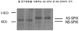
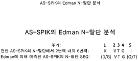
세린 프로테아제 억제제 카잘 (SPIK/SPINK1)은 79개 아미노산을 가진 작은 분비된 단백질이다 (4). 트립시노겐의 자가활성화의 억제제로서 췌장에서 처음 발견되었다 (5, 6). 최근 연구는 SPIK의 발현이 간암 예컨대 간세포 암종 (HCC) 및 간내 담관암종 (ICC)에서 상승되지만, 췌장의 외부를 제외한, 간을 포함하는 정상 조직에서 제한된 또는 무 활성을 갖는다는 것을 시사하였다 (7-9). 암 바이오마커로서 SPIK의 용도는 혈청 SPIK의 수준이 기타 질환, 특별히 췌장염의 존재 하에서 또한 상승된다는 사실에 의해 훼방되었다 (10-12). 우리는 간암 세포에 의해 분비된 SPIK에 대하여, N-말단에서 추가의, 적어도 9-잔기 긴 단편이 유지되는 반면, 정상 세포 예컨대 췌장 세포로부터 분비된 SPIK에서 제거되는 것을 알아내었다 (13). 우리는 이러한 종류의 SPIK를 NS-SPIK (정상 분비된 SPIK)로서 부른다. 하지만, 우리는 간암 세포에 의해 분비된 SPIK가 NS-SPIK보다 더 크고, 이러한 23개 아미노산의 추가의, 적어도 9-잔기 긴 단편이 분비 중 유지되는 것을 알아내었다 (11). 우리는 간암 세포에 의해 분비된 SPIK를 AS-SPIK 또는 LC-SPIK (비정상 분비된 SPIK 또는 간암 분비된 SPIK)로서 칭하였다. 본원에 AS-SPIK 및 LC-SPIK는 동일한 의미를 갖는다. 우리의 추가 연구는 사실 SPIK (서열번호: 6)의 N- 말단에 9개 아미노산 뿐 아니라, 추가의 23개 아미노산이 AS-SPIK에서 유지되는 반면, NS-SPIK로부터 제거되는 것을 알아내었다. 암 세포가 미-절단 SPIK를 분비하는 이유는 알려지지 않는다. 우리의 작업 가설은 SPIK가 세린 프로테아제 억제제이기 때문에, 암 세포에서 SPIK의 과-발현이, 세린 프로테아제의 1개 종류인, 신호 펩타이드 펩티다제의 활성을 억압하여, 암 세포로부터 분비되는 약독화되지 않은, 전장 단백질을 초래한다는 것이다. 우리는 AS-SPIK의 발현 중 발생하는 특정 이벤트를 우리가 이해한다고 믿지만, 본 발명의 조성물 및 방법은 임의의 특정한 세포성 기전에 영향을 미침으로써 작업하는 것들로 제한되지 않는다. 그러므로, 이러한 도메인을 인식할 수 있고 이에 특이적으로 결합할 수 있는 항체를 사용함으로써, 암성 간 세포로부터 생산된 SPIK와 기타 비-암성 질환에 의해 생성된 SPIK 사이를 차별하는 것이 가능해야 한다. 이러한 목적을 위하여, 우리는 고 감수성 및 특이성을 가진, 정상 SPIK (NS-SPIK, 정상 분비된 SPIK)가 아닌, 암성 SPIK (NS-SPIK, 비정상 분비된 SPIK)를 단독으로 인식하는 일련의 단클론성 항체를 개발하였다. 이러한 항체를 사용하여, 우리는 추가로 ELISA 테스트 키트 (SERAVUE®)를 개발하였고, HCC를 기타 간 질환 예컨대 HBV 및 HCV 감염, 간 간경변, 및 비-간 질환 예컨대 췌장염 및 뿐만 아니라 건강한 대상체와 구별하기에서 AS-SPIK의 성능을 사정하였다. 임상적 연구 결과는 대상체 항체 및 관련된 방법이, 기타 간 또는 비-간 질환으로부터 방해 없이, 이의 극초기에서 조차, HCC를 선택적으로 그리고 신뢰할만하게 검출하는데 사용될 수 있음을 시사하고, 이는 HCC의 진단 및 치료를 굉장히 개선할 것이다.The serine protease inhibitor kazal (SPIK/SPINK1) is a small secreted protein with 79 amino acids (4). It was first discovered in the pancreas as an inhibitor of autoactivation of trypsinogen (5, 6). Recent studies have suggested that the expression of SPIK is elevated in liver cancers such as hepatocellular carcinoma (HCC) and intrahepatic cholangiocarcinoma (ICC), but has limited or no activity in normal tissues, including the liver, except outside the pancreas (7 -9). The use of SPIK as a cancer biomarker has been hampered by the fact that the level of serum SPIK is also elevated in the presence of other diseases, particularly pancreatitis (10-12). We found that for SPIK secreted by hepatocarcinoma cells, an additional, at least 9-residue long fragment at the N-terminus is retained, whereas it is cleared in SPIK secreted from normal cells such as pancreatic cells (13). We call this kind of SPIK as NS-SPIK (normally secreted SPIK). However, we found that SPIK secreted by liver cancer cells is larger than NS-SPIK, and this additional, at least 9-residue long fragment of these 23 amino acids is retained during secretion (11). We termed SPIK secreted by hepatocarcinoma cells as AS-SPIK or LC-SPIK (abnormally secreted SPIK or hepatocarcinoma secreted SPIK). AS-SPIK and LC-SPIK herein have the same meaning. Our further study found that, in fact, 9 amino acids at the N-terminus of SPIK (SEQ ID NO: 6), as well as an additional 23 amino acids were retained in AS-SPIK, whereas removed from NS-SPIK. It is not known why cancer cells secrete uncleaved SPIK. Our working hypothesis is that since SPIK is a serine protease inhibitor, over-expression of SPIK in cancer cells suppresses the activity of signal peptide peptidase, one class of serine protease, so that it is not attenuated, secreted from cancer cells. , resulting in a full-length protein. While we believe that we understand the specific events that occur during expression of AS-SPIKs, the compositions and methods of the present invention are not limited to those that work by influencing any particular cellular mechanism. Therefore, by using antibodies capable of recognizing and specifically binding to these domains, it should be possible to discriminate between SPIKs produced from cancerous liver cells and SPIKs produced by other non-cancerous diseases. For this purpose, we developed a series of monoclonal cells that solely recognize a cancerous SPIK (NS-SPIK, aberrantly secreted SPIK), but not a normal SPIK (NS-SPIK, normally secreted SPIK), with high sensitivity and specificity. Antibodies were developed. Using these antibodies, we further developed an ELISA test kit (SERAVUE®), to distinguish HCC from other liver diseases such as HBV and HCV infection, liver cirrhosis, and non-liver diseases such as pancreatitis and as well as healthy subjects. evaluated the performance of AS-SPIK. The results of clinical studies suggest that subject antibodies and related methods can be used to selectively and reliably detect HCC, even in its very early stages, without interference from other liver or non-hepatic diseases, which can help in the diagnosis and It will greatly improve your treatment.
발명의 개요Summary of invention
본 발명의 양태는 AS-SPIK 단백질의 형태적 에피토프에 특이적으로 결합하고, NS-SPIK 단백질에 특이적으로 결합하지 않는 단리된 항체를 포함하고, 여기서 AS-SPIK 단백질의 형태적 에피토프는 서열번호: 2의 L14, L15, S16, L17, D24 및 S25로 이루어지는 군으로부터 선택된 하나 이상의 아미노산; 및 서열번호: 2의 C58, V59, L60, C61, 및 F62로 이루어지는 군으로부터 선택된 하나 이상의 아미노산을 포함한다.Aspects of the invention include an isolated antibody that specifically binds to a conformational epitope of an AS-SPIK protein and does not specifically bind to a NS-SPIK protein, wherein the conformational epitope of the AS-SPIK protein is SEQ ID NO: : at least one amino acid selected from the group consisting of L14, L15, S16, L17, D24 and S25 of 2; and one or more amino acids selected from the group consisting of C58, V59, L60, C61, and F62 of SEQ ID NO:2.
일부 구현예에서, 형태적 에피토프는 서열번호: 2의 아미노산 L14, L15, S16, 및 L17을 포함한다. 일부 구현예에서, 형태적 에피토프는 서열번호: 2의 아미노산 L60 및 C61을 포함한다. 일부 구현예에서, 형태적 에피토프는 서열번호: 2의 아미노산 L14, L15, S16, L17, L60, 및 C61을 포함한다. 일부 구현예에서, 형태적 에피토프는 추가로 서열번호: 2의 아미노산 D24 및 S25를 포함한다. 일부 구현예에서, 형태적 에피토프는 추가로 서열번호: 2의 아미노산 C58, V59 및 F62를 포함한다. 일부 구현예에서, 형태적 에피토프는 서열번호: 2의 아미노산 L14, L15, S16, L17, D24, S25, C58, V59, L60, C61 및 F62를 포함한다.In some embodiments, the conformational epitope comprises amino acids L14, L15, S16, and L17 of SEQ ID NO:2. In some embodiments, the conformational epitope comprises amino acids L60 and C61 of SEQ ID NO:2. In some embodiments, the conformational epitope comprises amino acids L14, L15, S16, L17, L60, and C61 of SEQ ID NO:2. In some embodiments, the conformational epitope further comprises amino acids D24 and S25 of SEQ ID NO:2. In some embodiments, the conformational epitope further comprises amino acids C58, V59 and F62 of SEQ ID NO:2. In some embodiments, the conformational epitope comprises amino acids L14, L15, S16, L17, D24, S25, C58, V59, L60, C61 and F62 of SEQ ID NO:2.
일부 구현예에서, 단리된 항체는 S6을 포함하는 CDRH1 서열; 및/또는 I2, G5, G6, Y10 및 K16을 포함하는 CDRH2 서열; 및/또는 G4 및 Y7을 포함하는 CDRH3 서열; 및/또는 Q4 및 S9를 포함하는 CDRL1 서열; 및/또는 A2, S3, T4 및 S7을 포함하는 CDRL2 서열; 및/또는 Q1, Q2, Y4 및 S5를 포함하는 CDRL3 서열을 포함한다.In some embodiments, the isolated antibody comprises a CDRH1 sequence comprising S6; and/or a CDRH2 sequence comprising I2, G5, G6, Y10 and K16; and/or a CDRH3 sequence comprising G4 and Y7; and/or a CDRL1 sequence comprising Q4 and S9; and/or a CDRL2 sequence comprising A2, S3, T4 and S7; and/or a CDRL3 sequence comprising Q1, Q2, Y4 and S5.
일부 구현예에서, 단리된 항체는 S6을 포함하는 CDRH1 서열; I2, G5, G6, Y10 및 K16을 포함하는 CDRH2 서열; G4 및 Y7을 포함하는 CDRH3 서열; Q4 및 S9를 포함하는 CDRL1 서열; A2, S3, T4 및 S7을 포함하는 CDRL2 서열; 및 Q1, Q2, Y4 및 S5를 포함하는 CDRL3 서열을 포함한다.In some embodiments, the isolated antibody comprises a CDRH1 sequence comprising S6; a CDRH2 sequence comprising I2, G5, G6, Y10 and K16; a CDRH3 sequence comprising G4 and Y7; CDRL1 sequence comprising Q4 and S9; a CDRL2 sequence comprising A2, S3, T4 and S7; and a CDRL3 sequence comprising Q1, Q2, Y4 and S5.
본 발명의 양태는 AS-SPIK 단백질의 형태적 에피토프에 특이적으로 결합하고, NS-SPIK 단백질에 특이적으로 결합하지 않는 단리된 항체를 포함하고, 여기서 AS-SPIK 단백질의 형태적 에피토프는 서열번호: 2의 L36, N37, I42 및 Y43으로 이루어지는 군으로부터 선택된 하나 이상의 아미노산; 및 서열번호: 2의 R67, Q68, I71 및 L72로 이루어지는 군으로부터 선택된 하나 이상의 아미노산을 포함한다.Aspects of the invention include an isolated antibody that specifically binds to a conformational epitope of an AS-SPIK protein and does not specifically bind to a NS-SPIK protein, wherein the conformational epitope of the AS-SPIK protein is SEQ ID NO: : at least one amino acid selected from the group consisting of L36, N37, I42 and Y43 of 2; and one or more amino acids selected from the group consisting of R67, Q68, I71 and L72 of SEQ ID NO:2.
일부 구현예에서, 형태적 에피토프는 서열번호: 2의 아미노산 L36 및 N37을 포함한다. 일부 구현예에서, 형태적 에피토프는 서열번호: 2의 아미노산 I42 및 Y43을 포함한다. 일부 구현예에서, 형태적 에피토프는 서열번호: 2의 아미노산 L36, N37, I42 및 Y43을 포함한다. 일부 구현예에서, 형태적 에피토프는 서열번호: 2의 아미노산 R67, Q68, I71 및 L72를 포함한다. 일부 구현예에서, 형태적 에피토프는 서열번호: 2의 아미노산 L36, N37, I42, Y43, R67, Q68, I71 및 L72를 포함한다.In some embodiments, the conformational epitope comprises amino acids L36 and N37 of SEQ ID NO:2. In some embodiments, the conformational epitope comprises amino acids I42 and Y43 of SEQ ID NO:2. In some embodiments, the conformational epitope comprises amino acids L36, N37, I42 and Y43 of SEQ ID NO:2. In some embodiments, the conformational epitope comprises amino acids R67, Q68, I71 and L72 of SEQ ID NO:2. In some embodiments, the conformational epitope comprises amino acids L36, N37, I42, Y43, R67, Q68, I71 and L72 of SEQ ID NO:2.
일부 구현예에서, 및 항체는 Y3, S7 및 W9를 포함하는 CDRH1 서열; 및/또는 A1, I2, G4, G6 및 Y10을 포함하는 CDRH2 서열; 및/또는 R1 및 D7을 포함하는 CDRH3 서열; 및/또는 A2, S3, Q4, I6, Y9, L10 및 S11을 포함하는 CDRL1 서열; 및/또는 A2, S3, L5 및 S7을 포함하는 CDRL2 서열; 및/또는 Q1, Q2, 및 T5를 포함하는 CDRL3 서열을 포함한다. In some embodiments, and the antibody comprises a CDRH1 sequence comprising Y3, S7 and W9; and/or a CDRH2 sequence comprising A1, I2, G4, G6 and Y10; and/or a CDRH3 sequence comprising R1 and D7; and/or a CDRL1 sequence comprising A2, S3, Q4, I6, Y9, L10 and S11; and/or a CDRL2 sequence comprising A2, S3, L5 and S7; and/or a CDRL3 sequence comprising Q1, Q2, and T5.
일부 구현예에서, 항체는 Y3, S7 및 W9를 포함하는 CDRH1 서열; A1, I2, G4, G6 및 Y10을 포함하는 CDRH2 서열; R1 및 D7을 포함하는 CDRH3 서열; A2, S3, Q4, I6, Y9, L10 및 S11을 포함하는 CDRL1 서열; A2, S3, L5 및 S7을 포함하는 CDRL2 서열; 및 Q1, Q2, 및 T5를 포함하는 CDRL3 서열을 포함한다. In some embodiments, the antibody comprises a CDRH1 sequence comprising Y3, S7 and W9; a CDRH2 sequence comprising A1, I2, G4, G6 and Y10; a CDRH3 sequence comprising R1 and D7; a CDRL1 sequence comprising A2, S3, Q4, I6, Y9, L10 and S11; a CDRL2 sequence comprising A2, S3, L5 and S7; and a CDRL3 sequence comprising Q1, Q2, and T5.
일부 구현예에서, 항체는 다중-특이적이다. 일부 구현예에서, 항체는 이중특이적이다. 일부 구현예에서, 항체는 효과기 세포에 결합 친화성을 갖는다. 일부 구현예에서, 항체는 T-세포 항원에 결합 친화성을 갖는다. 일부 구현예에서, T-세포 항원은 CD3 단백질을 포함한다. 일부 구현예에서, 항체는 단클론성 항체이다. 일부 구현예에서, 항체는 CAR-T 형식이다.In some embodiments, the antibody is multi-specific. In some embodiments, the antibody is bispecific. In some embodiments, the antibody has binding affinity to an effector cell. In some embodiments, the antibody has binding affinity to a T-cell antigen. In some embodiments, the T-cell antigen comprises a CD3 protein. In some embodiments, the antibody is a monoclonal antibody. In some embodiments, the antibody is in CAR-T format.
본 발명의 양태는, 세포독성 제제에 공유적으로 부착된, 본원에 기재된 경우에 항체를 포함하는 면역접합체를 포함한다. 일부 구현예에서, 세포독성 제제는 독소, 화학치료적 제제, 약물 모이어티, 항생제, 방사성 동위원소 및 핵산분해 효소로 이루어지는 군으로부터 선택된다.Aspects of the invention include immunoconjugates comprising an antibody, as described herein, covalently attached to a cytotoxic agent. In some embodiments, the cytotoxic agent is selected from the group consisting of toxins, chemotherapeutic agents, drug moieties, antibiotics, radioactive isotopes and nucleases.
일부 구현예에서, 면역접합체는 식 Ab-(L-D)p를 갖고, 여기서 Ab는 본원에 기재된 경우에 항체이고; L은 링커이고; D는 약물 모이어티이고; p는 1 내지 8 범위인 정수이다. 일부 구현예에서, D는 메이탄시노이드, 아우리스타틴 및 돌로스타틴으로 이루어지는 군으로부터 선택된다. 일부 구현예에서, L은 6-말레이미도카프로일 (MC), 말레이미도프로파노일 (MP), 발린-시트룰린 (val-cit), 알라닌-페닐알라닌 (ala-phe), p-아미노벤질옥시카르보닐 (PAB), N-숙신이미딜 4-(2-피리딜티오)펜타노에이트 (SPP), N-숙신이미딜 4-(N-말레이미도메틸)시클로헥산-1 카르복실레이트 (SMCC), 4-(2-피리딜디티오)부티르산-N-히드록시숙신이미드 에스테르 (SPDB), 및 N-숙신이미딜 (4-요오도-아세틸)아미노벤조에이트 (SIAB)로 이루어지는 군으로부터 선택된 하나 이상의 링커를 포함한다.In some embodiments, the immunoconjugate has the formula Ab-(L-D)p, wherein Ab is an antibody as described herein; L is a linker; D is a drug moiety; p is an integer ranging from 1 to 8. In some embodiments, D is selected from the group consisting of maytansinoids, auristatins and dolostatins. In some embodiments, L is 6-maleimidocaproyl (MC), maleimidopropanoyl (MP), valine-citrulline (val-cit), alanine-phenylalanine (ala-phe), p-aminobenzyloxycar Bornyl (PAB), N-succinimidyl 4-(2-pyridylthio)pentanoate (SPP), N-succinimidyl 4-(N-maleimidomethyl)cyclohexane-1 carboxylate (SMCC) , 4-(2-pyridyldithio)butyric acid-N-hydroxysuccinimide ester (SPDB), and N-succinimidyl (4-iodo-acetyl)aminobenzoate (SIAB) one or more linkers.
본 발명의 양태는 본원에 기재된 경우에 및 항체 또는 면역접합체를 포함하는 약학적 조성물을 포함한다.Aspects of the present invention include pharmaceutical compositions comprising an antibody or immunoconjugate and as described herein.
본 발명의 양태는, 상기 장애를 가진 대상체에 본원에 기재된 경우에 항체 또는 면역접합체, 또는 본원에 기재된 경우에 약학적 조성물을 투여하는 단계를 포함하는, AS-SPIK의 발현을 특징으로 하는 장애의 치료 방법을 포함한다.Aspects of the present invention provide a method of treating a disorder characterized by expression of an AS-SPIK comprising administering to a subject having the disorder an antibody or immunoconjugate as described herein, or a pharmaceutical composition as described herein. including treatment methods.
본 발명의 양태는 AS-SPIK의 발현을 특징으로 하는 장애의 치료를 위한 약제의 제조에서, 본원에 기재된 경우에 항체 또는 면역접합체의 용도를 포함한다.Aspects of the invention include the use of an antibody or immunoconjugate as described herein in the manufacture of a medicament for the treatment of a disorder characterized by the expression of AS-SPIK.
본 발명의 양태는 AS-SPIK의 발현을 특징으로 하는 장애의 치료에서 사용을 위한 본원에 기재된 경우에 항체 또는 면역접합체를 포함한다.Aspects of the invention include an antibody or immunoconjugate as described herein for use in the treatment of a disorder characterized by the expression of AS-SPIK.
일부 구현예에서, 장애는 간 장애이다. 일부 구현예에서, 간 장애는 간세포 암종이다. 일부 구현예에서, 간 장애는 간내 담관암종이다. 일부 구현예에서, 간 장애는 바이러스성 감염이다. 일부 구현예에서, 간 장애는 염증성 간 장애이다. 일부 구현예에서, 염증성 간 장애는 간의 간경변이다.In some embodiments, the disorder is a liver disorder. In some embodiments, the liver disorder is hepatocellular carcinoma. In some embodiments, the liver disorder is intrahepatic cholangiocarcinoma. In some embodiments, the liver disorder is a viral infection. In some embodiments, the liver disorder is an inflammatory liver disorder. In some embodiments, the inflammatory liver disorder is cirrhosis of the liver.
본 발명의 양태는 본원에 기재된 경우에 항체를 인코딩하는 폴리뉴클레오타이드를 포함한다. 본 발명의 양태는 본원에 기재된 경우에 폴리뉴클레오타이드를 포함하는 벡터를 포함한다. 본 발명의 양태는 본원에 기재된 경우에 벡터를 포함하는 숙주 세포를 포함한다.Aspects of the invention include polynucleotides encoding antibodies when described herein. Aspects of the invention include vectors comprising polynucleotides as described herein. Aspects of the invention include a host cell comprising a vector when described herein.
본 발명의 양태는, 항체의 발현을 허용하는 조건 하에서 본원에 기재된 경우에 숙주 세포를 성장시키는 단계, 및 항체를 세포로부터 단리시키는 단계를 포함하는, 본원에 기재된 경우에 항체 또는 면역접합체의 생산 방법을 포함한다.Aspects of the invention provide a method for producing an antibody or immunoconjugate as described herein comprising growing a host cell in the case described herein under conditions permissive for expression of the antibody, and isolating the antibody from the cell. includes
본 발명의 양태는 대상체가 AS-SPIK의 발현을 특징으로 하는 장애를 갖거나 상기 발병의 위험에 처한 지를 결정하는 진단적 방법을 포함하고, 상기 방법은 대상체로부터의 생물학적 테스트 샘플을 본원에 기재된 경우에 AS-SPIK 항체와 접촉시켜 AS-SPIK-항체 복합체를 생성하는 단계; 생물학적 테스트 샘플에서 AS-SPIK-항체 복합체의 농도를 검출하는 단계; 및 AS-SPIK-항체 복합체의 농도를 기준값에 비교하여 대상체가 장애를 갖거나 상기 발병의 위험에 처한 지를 결정하는 단계를 포함한다.Aspects of the invention include a diagnostic method for determining whether a subject has or is at risk of developing a disorder characterized by expression of AS-SPIK, said method comprising administering a biological test sample from a subject to the case described herein generating an AS-SPIK-antibody complex by contacting it with an AS-SPIK antibody; detecting the concentration of the AS-SPIK-antibody complex in the biological test sample; and comparing the concentration of the AS-SPIK-antibody complex to a reference value to determine whether the subject has or is at risk of developing said disorder.
본 발명의 양태는 대상체가 AS-SPIK의 발현을 특징으로 하는 장애를 갖거나 상기 발병의 위험에 처한 지를 결정하는 진단적 방법을 포함하고, 상기 방법은 대상체로부터의 생물학적 테스트 샘플을 SPIK에 특이적으로 결합하는 제1 항체 또는 항원-결합 단편과 접촉시켜 SPIK-항체 복합체를 형성하는 단계; SPIK-항체 복합체를 본원에 기재된 경우에 AS-SPIK 항체 또는 항원-결합 단편과 접촉시켜 AS-SPIK-항체 복합체를 생성하는 단계; AS-SPIK-항체 복합체의 농도를 생물학적 테스트 샘플에서 검출하는 단계; 및 AS-SPIK-항체 복합체의 농도를 기준값에 비교하여 대상체가 장애를 갖거나 상기 발병의 위험에 처한 지를 결정하는 단계를 포함한다.Aspects of the invention include a diagnostic method for determining whether a subject has or is at risk of developing a disorder characterized by expression of AS-SPIK, said method comprising administering a biological test sample from the subject to SPIK-specific forming an SPIK-antibody complex by contacting it with a first antibody or antigen-binding fragment that binds to contacting the SPIK-antibody complex with an AS-SPIK antibody or antigen-binding fragment as described herein to generate an AS-SPIK-antibody complex; detecting the concentration of the AS-SPIK-antibody complex in the biological test sample; and comparing the concentration of the AS-SPIK-antibody complex to a reference value to determine whether the subject has or is at risk of developing said disorder.
일부 구현예에서, 항체 또는 항원-결합 단편은 검출가능한 표지를 포함한다. 일부 구현예에서, 장애는 간 장애이다. 일부 구현예에서, 간 장애는 간세포 암종, 간내 담관암종, 간의 바이러스성 감염, 간의 염증성 장애, 및 간의 간경변으로 이루어지는 군으로부터 선택된다.In some embodiments, the antibody or antigen-binding fragment comprises a detectable label. In some embodiments, the disorder is a liver disorder. In some embodiments, the liver disorder is selected from the group consisting of hepatocellular carcinoma, intrahepatic cholangiocarcinoma, viral infection of the liver, inflammatory disorder of the liver, and cirrhosis of the liver.
본 발명의 양태는 본원에 기재된 경우에 항체 또는 면역접합체를 포함하는 키트를 포함한다. 일부 구현예에서, 키트는 추가로 SPIK에 특이적으로 결합하는 항체를 포함한다.Aspects of the invention include kits comprising an antibody or immunoconjugate as described herein. In some embodiments, the kit further comprises an antibody that specifically binds to SPIK.
도면의 간단한 설명
도 1은 겔 전기영동을 이용하여 AS-SPIK 및 NS-SPIK의 크기를 도시한다.
도 2는 Edman N-말단 분석에 의한 AS-SPIK의 N-말단 서열을 도시한다.
도 3은 AS-SPIK 및 NS-SPIK의 아미노산 서열의 비교를 도시한다.
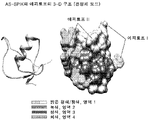
도 4는 AS-SPIK의 3D 구조의 NS-SPIK의 것과의 비교를 도시한다.
도 5는 AS-SPIK 및 NS-SPIK에 결합하는 항체의 결합 활성의 테스트 결과를 도시한다.
도 6은 합성된 펩타이드에 항-AS-SPIK 항체의 결합 테스트의 결과를 도시한다.
도 7은 AS-SPIK 및 이의 에피토프의 3D 구조 (결정체 모델)를 도시한다.
도 8은 부류 I 항-AS-SPIK 항체의 결합 부위를 찾기 위한 억제 테스트의 결과를 도시한다.
도 9는 부류 I 항-AS-SPIK 항체에서 CDR의 공통 아미노산을 도시한다.
도 10은 부류 II 항-AS-SPIK 항체에서 CDR의 공통 아미노산을 도시한다.
도 11은 AS-SPIK 검출 키트를 위한 기전의 예시를 제공한다.
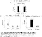
도 12, 패널 A는 IM-CA22가 NS-SPIK가 아닌 AS-SPIK에 특이적으로 결합하는 반면, 항체 IM-BA2가 양쪽 AS-SPIK 및 NS-SPIK에 결합하는 것을 보여주는 그래프이다. 패널 B는 AS-SPIK가 췌장염 또는 건강한 환자가 아닌 HCC에서 상승되는 것을 보여주는 그래프이다. 패널 C는 NS-SPIK가 췌장염 환자에서 상승되지만 AS-SPIK-기반된 검출 키트의 성능을 방해하지 않는다.
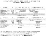
도 13은 부류 I 및 부류 II 항-AS-SPIK 항체의 결합 특징을 보여주는 표 (표 1)이다.
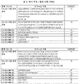
도 14는 CLIPS 분석 연구에 의해 예측된 항-AS-SPIK 결합을 위한 영역, 및 그안에 에피토프를 구성하는 아미노산 잔기를 보여주는 표 (표 2)이다.
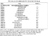
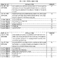
도 15는, 본원에 추가로 기재된 경우에, 에피토프 I에 결합하는 4개 예시 부류 I 항체의 아미노산 서열을 보여주는 표 (표 3)이다.
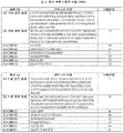
도 16은, 본원에 추가로 기재된 경우에, 에피토프 II에 결합하는 4개 예시 부류 II 항체의 아미노산 서열을 보여주는 표 (표 4)이다.
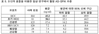
도 17은 512개 샘플을 이용한 임상적 연구에서 혈청 AS-SPIK 수준을 보여주는 표 (표 5)이다.
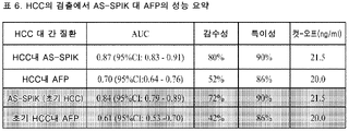
도 18은 HCC의 검출에서 AS-SPIK 대 AFP의 성능 요약을 제공하는 표 (표 6)이다.
도 19는 초기 대 말기 HCC에서 AS-SPIK 수준을 보여주는 표 (표 7)이다.
도 20은 BCLC 병기에 의해, HCC에서 AS-SPIK 수준을 보여주는 표 (표 8)이다.Brief description of the drawing
1 depicts the size of AS-SPIK and NS-SPIK using gel electrophoresis.
Figure 2 depicts the N-terminal sequence of AS-SPIK by Edman N-terminal analysis.
3 depicts a comparison of the amino acid sequences of AS-SPIK and NS-SPIK.
4 shows a comparison of the 3D structure of AS-SPIK with that of NS-SPIK.
Fig. 5 shows the test results of the binding activity of antibodies binding to AS-SPIK and NS-SPIK.
Figure 6 shows the results of the binding test of the anti-AS-SPIK antibody to the synthesized peptide.
Figure 7 depicts the 3D structure (crystal model) of AS-SPIK and its epitope.
8 depicts the results of an inhibition test to find the binding site of a class I anti-AS-SPIK antibody.
9 depicts the consensus amino acids of the CDRs in a class I anti-AS-SPIK antibody.
10 depicts the consensus amino acids of the CDRs in a class II anti-AS-SPIK antibody.
11 provides an illustration of the mechanism for the AS-SPIK detection kit.
12, panel A is a graph showing that IM-CA22 specifically binds AS-SPIK but not NS-SPIK, whereas antibody IM-BA2 binds both AS-SPIK and NS-SPIK. Panel B is a graph showing that AS-SPIK is elevated in HCC but not in pancreatitis or healthy patients. Panel C shows that NS-SPIK is elevated in patients with pancreatitis but does not interfere with the performance of the AS-SPIK-based detection kit.
13 is a table (Table 1) showing the binding characteristics of class I and class II anti-AS-SPIK antibodies.
14 is a table (Table 2) showing regions for anti-AS-SPIK binding predicted by CLIPS analysis studies, and amino acid residues constituting epitopes therein.
15 is a table (Table 3) showing the amino acid sequences of four exemplary class I antibodies that bind to epitope I, when further described herein.
16 is a table (Table 4) showing the amino acid sequences of four exemplary class II antibodies that bind epitope II, when further described herein.
17 is a table (Table 5) showing serum AS-SPIK levels in a clinical study with 512 samples.
18 is a table (Table 6) providing a performance summary of AS-SPIK versus AFP in the detection of HCC.
19 is a table (Table 7) showing AS-SPIK levels in early versus late HCC.
20 is a table (Table 8) showing AS-SPIK levels in HCC, by BCLC stage.
상세한 설명details
본 발명의 실시는, 달리 명시되지 않는 한, 분자 생물학 (재조합 기법 포함), 미생물학, 세포 생물학, 생화학, 및 면역학의 기존 기술을 이용할 것이고, 이들은 당업계의 기술 내에 있다. 상기 기법은 문헌, 예컨대, "Molecular Cloning: A Laboratory Manual", second edition (Sambrook 등, 1989); "Oligonucleotide Synthesis" (M. J. Gait, ed., 1984); "Animal Cell Culture" (R. I. Freshney, ed., 1987); "Methods in Enzymology" (Academic Press, Inc.); "Current Protocols in Molecular Biology" (F. M. Ausubel 등, eds., 1987, and periodic updates); "PCR: The Polymerase Chain Reaction", (Mullis 등, ed., 1994); "A Practical Guide to Molecular Cloning" (Perbal Bernard V., 1988); "Phage Display: A Laboratory Manual" (Barbas 등, 2001); Harlow, Lane 및 Harlow, Using Antibodies: A Laboratory Manual: Portable Protocol No. I, Cold Spring Harbor Laboratory (1998); 및 Harlow 및 Lane, Antibodies: A Laboratory Manual, Cold Spring Harbor Laboratory; (1988)에서 충분히 설명된다.The practice of the present invention will employ, unless otherwise specified, existing techniques of molecular biology (including recombinant techniques), microbiology, cell biology, biochemistry, and immunology, which are within the skill of the art. Such techniques are described in literature, such as "Molecular Cloning: A Laboratory Manual", second edition (Sambrook et al., 1989); "Oligonucleotide Synthesis" (M. J. Gait, ed., 1984); "Animal Cell Culture" (R. I. Freshney, ed., 1987); "Methods in Enzymology" (Academic Press, Inc.); "Current Protocols in Molecular Biology" (F. M. Ausubel et al., eds., 1987, and periodic updates); "PCR: The Polymerase Chain Reaction", (Mullis et al., ed., 1994); "A Practical Guide to Molecular Cloning" (Perbal Bernard V., 1988); "Phage Display: A Laboratory Manual" (Barbas et al., 2001); Harlow, Lane, and Harlow, Using Antibodies: A Laboratory Manual: Portable Protocol No. I, Cold Spring Harbor Laboratory (1998); and Harlow and Lane, Antibodies: A Laboratory Manual, Cold Spring Harbor Laboratory; (1988) fully explained.
값의 범위가 제공되는 경우, 그 범위의 상한과 하한 사이, 문맥이 명확하게 달리 명시하지 않는 한 하한의 단위의 1/10까지, 각 개재 값 그리고 그 진술된 범위에서 임의의 기타 진술된 또는 개재 값이 본 발명 내에서 포괄되는 것이 이해된다. 이들 더 작은 범위의 상한 및 하한은 더 작은 범위에서 독립적으로 포함될 수 있고 진술된 범위에서 임의의 구체적으로 배제된 한계에 따라, 본 발명 내에서 이다 또한 포괄된다. 진술된 범위가 한계의 한쪽 또는 양쪽을 포함하는 경우, 그들 포함된 한계의 어느 한쪽 또는 양쪽을 배제하는 범위는 본 발명에서 또한 포함된다.Where a range of values is provided, between the upper and lower limits of that range, to the tenth of the unit of the lower limit, unless the context clearly dictates otherwise, each intervening value and any other stated or intervening in that stated range. It is understood that the values are encompassed within the present invention. The upper and lower limits of these smaller ranges may independently be included in the smaller ranges and are also encompassed within the invention, subject to any specifically excluded limit in the stated range. Where the stated range includes one or both of the limits, ranges excluding either or both of those included limits are also included in the invention.
달리 명시되지 않는 한, 본원에 항체 잔기는 Kabat 넘버링 시스템 (예를 들면, Kabat 등, Sequences of Immunological Interest. 5th Ed. Public Health Service, National Institutes of Health, Bethesda, Md. (1991))에 따라 넘버링된다.Unless otherwise specified, antibody residues herein are numbered according to the Kabat numbering system (eg, Kabat et al., Sequences of Immunological Interest. 5th Ed. Public Health Service, National Institutes of Health, Bethesda, Md. (1991)). do.
하기 설명에서, 수많은 구체적 상세는 본 발명의 더욱 철저한 이해를 제공하기 위해 제시된다. 하지만, 본 발명이 이들 구체적 상세의 하나 이상 없이 실시될 수 있음이 당업자에 명백해질 것이다. 기타 사례에서, 잘-알려진 속성 및 당업자에 잘 알려진 절차는 본 발명 모호함을 피하기 위해 설명되지 않았다.In the following description, numerous specific details are set forth in order to provide a more thorough understanding of the present invention. It will be apparent, however, to one skilled in the art, that the present invention may be practiced without one or more of these specific details. In other instances, well-known attributes and procedures well known to those skilled in the art have not been described in order to avoid obscuring the present invention.
특허 출원 및 공개를 포함하는, 본 개시내용 도처에 인용된 모든 참고문헌은 그들 전체가 본원에 참고로 편입된다.All references cited throughout this disclosure, including patent applications and publications, are hereby incorporated by reference in their entirety.
정의Justice
본 명세서를 해석하기 위해, 하기 정의가 적용할 것이고 적절할 때마다, 단수형으로 사용된 용어들은 복수형을 또한 포함할 것이고 그 반대도 마찬가지이다. 제시된 임의의 정의가 참고로 본원에 편입된 임의의 문헌과 상충하는 경우에, 하기 제시된 정의가 지배할 수 있다.For the purpose of interpreting this specification, the following definitions will apply and whenever appropriate, terms used in the singular will also include the plural and vice versa. To the extent any definition set forth conflicts with any document incorporated herein by reference, the definition set forth below shall control.
"에피토프"는 단일 항체 분자가 결합하는 항원 분자의 표면에서의 부위이다. 일반적으로, 항원은 몇몇 또는 많은 상이한 에피토프를 갖고 많은 상이한 항체와 반응한다. 상기 용어는 구체적으로 선형 에피토프 및 형태적 에피토프를 포함한다. 상기 용어는 항체에 특이적 결합할 수 있는 임의의 분자성 결정인자를 포함한다. 특정 구현예에서, 에피토프 결정인자는 분자 예컨대 아미노산, 당 측쇄, 포스포릴, 또는 술포닐의 화학적으로 활성 표면 그룹화를 포함하고, 특정 구현예에서, 특이적 3차원 구조적 특징, 및/또는 특이적 전하 특징을 가질 수 있다. 에피토프는 항체에 의해 결합되는 항원의 영역이다. "결합 영역"은 결합 분자에 의해 결합된 결합 표적에서의 영역이다.An “epitope” is a site on the surface of an antigenic molecule to which a single antibody molecule binds. In general, antigens have several or many different epitopes and react with many different antibodies. The term specifically includes linear epitopes and conformational epitopes. The term includes any molecular determinant capable of specific binding to an antibody. In certain embodiments, epitope determinants include chemically active surface groupings of molecules such as amino acids, sugar side chains, phosphoryls, or sulfonyls, and in certain embodiments, specific three-dimensional structural features, and/or specific charges. may have characteristics. An epitope is a region of an antigen bound by an antibody. A “binding region” is a region in a binding target bound by a binding molecule.
"에피토프 맵핑"은 그들의 표적 항원에서 항체의 결합 부위, 또는 에피토프 식별하기의 공정이다. 항체 에피토프는 선형 에피토프 또는 형태적 에피토프일 수 있다. 선형 에피토프는 단백질에서 아미노산의 연속 서열에 의해 형성된다. 형태적 에피토프는 단백질 서열에서 불연속이지만, 단백질을 이의 3-차원 구조로 폴딩시 결합되는 아미노산으로 형성된다."Epitope mapping" is the process of identifying the binding site, or epitope, of an antibody on their target antigen. Antibody epitopes may be linear epitopes or conformational epitopes. Linear epitopes are formed by a continuous sequence of amino acids in a protein. Conformational epitopes are discontinuous in the protein sequence, but are formed of amino acids that are joined upon folding the protein into its three-dimensional structure.
본원에 정의된 경우에 "에피토프 비닝"은 이들이 인식하는 에피토프에 기반된 항체 그룹화의 공정이다. 더욱 특히, 에피토프 비닝은 그들의 에피토프 인식 특성에 기반된 항체 클러스터링하기 그리고 뚜렷한 결합 특이성을 갖는 항체 식별하기를 위하여 컴퓨터 공정과 조합된, 상이한 항체의 에피토프 인식 특성을 분간하는 방법 및 시스템을 포함한다."Epitope binning" as defined herein is the process of grouping antibodies based on the epitopes they recognize. More particularly, epitope binning includes methods and systems for discriminating the epitope recognition properties of different antibodies, combined with computational processes to cluster antibodies based on their epitope recognition properties and to identify antibodies with distinct binding specificities.
항체는 2개 항체가 동일한 또는 입체적으로 중첩 에피토프를 인식하는 경우 참조 항체로서 "본질적으로 동일한 에피토프"를 결합시킨다. 2개 에피토프가 동일한 또는 입체적으로 중첩 에피토프에 결합하는 지를 결정하는 가장 널리 사용되고 신속한 방법은 어느 한쪽 표지화된 항원 또는 표지화된 항체를 사용하여, 임의의 수의 상이한 형식으로 구성될 수 있는, 경쟁 검정이다. 보통, 항원은 96-웰 플레이트에서 부동화되고, 표지화된 항체의 결합을 차단하는 미표지화된 항체의 능력은 방사성 또는 효소 표지를 사용하여 측정된다.An antibody binds “essentially the same epitope” as a reference antibody when the two antibodies recognize the same or sterically overlapping epitope. The most widely used and rapid method of determining whether two epitopes bind to the same or sterically overlapping epitopes is a competition assay, which can be constructed in any number of different formats, using either labeled antigen or labeled antibody. . Usually, antigens are immobilized in 96-well plates, and the ability of unlabeled antibody to block binding of labeled antibody is measured using radioactive or enzymatic labeling.
본원에 사용된 경우에, 아미노산 잔기/위치의 "변형"은 출발 아미노산 서열과 비교된 경우에 일차 아미노산 서열의 변화를 지칭하고, 여기서 변화는 상기 아미노산 잔기/위치를 포함하는 서열 변경에서 비롯한다. 예를 들어, 전형적 변형은 잔기의 (또는 상기 위치에서의) 또 다른 아미노산으로의 치환 (예를 들면, 보존적 또는 비-보존적 치환), 상기 잔기/위치에 인접한 하나 이상의 (일반적으로 5 또는 3개 미만) 아미노산의 삽입, 및 상기 잔기/위치의 결실을 포함한다. "아미노산 치환" 또는 이의 변이는 예정된 (출발) 아미노산 서열에서 기존 아미노산 잔기의 상이한 아미노산 잔기로의 대체를 지칭한다. 일반적으로 및 바람직하게는, 변형은 출발 (또는 "야생형") 아미노산 서열을 포함하는 폴리펩타이드와 비교하여 변이체 폴리펩타이드의 적어도 하나의 물리적 또는 생화학적 활성에서의 변경을 초래한다. 예를 들어, 항체의 경우에서, 변경되는 물리적 또는 생화학적 활성은 표적 분자에 대한 결합 친화성, 결합 능력 및/또는 결합 효과일 수 있다.As used herein, "modification" of an amino acid residue/position refers to a change in the primary amino acid sequence when compared to the starting amino acid sequence, wherein the change results from a sequence change comprising that amino acid residue/position. For example, typical modifications include substitution of a residue (or at that position) with another amino acid (eg, conservative or non-conservative substitution), one or more (generally 5 or less than 3) amino acid insertions, and deletions of said residues/positions. “Amino acid substitution” or variation thereof refers to the replacement of an existing amino acid residue with a different amino acid residue in a predetermined (starting) amino acid sequence. Generally and preferably, the modification results in an alteration in at least one physical or biochemical activity of the variant polypeptide compared to the polypeptide comprising the starting (or "wild-type") amino acid sequence. For example, in the case of an antibody, the physical or biochemical activity to be altered may be binding affinity, binding capacity and/or binding effect for a target molecule.
용어 "항체"는 단클론성 항체 (면역글로불린 Fc 영역을 갖는 전장 항체 포함), 단일-쇄 분자, 뿐만 아니라 항체 단편 (예를 들면, Fab, F(ab')2, 및 Fv)을 포함한다. 용어 "면역글로불린" (Ig)은 본원에 "항체"와 교환가능하게 사용된다. 염기성 4-쇄 항체 단위는 2개 동일한 경 (L) 쇄 및 2개 동일한 중 (H) 쇄로 구성된 헤테로사량체성 당단백질이다. 달리 언급되지 않는 한, 용어 "항체"는 가장 넓은 의미로 본원에 사용되고 구체적으로 IgG, IgM, IgA, IgD, 및 IgE 항체 및 그들의 단편, 바람직하게는 항원-결합 단편을 포함하는 항체의 모든 아이소타입, 하위-부류 및 형태를 포함한다.The term “antibody” includes monoclonal antibodies (including full-length antibodies having an immunoglobulin Fc region), single-chain molecules, as well as antibody fragments (eg, Fab, F(ab′)2, and Fv). The term “immunoglobulin” (Ig) is used herein interchangeably with “antibody”. Basic 4-chain antibody units are heterotetrameric glycoproteins composed of two identical light (L) chains and two identical heavy (H) chains. Unless otherwise stated, the term "antibody" is used herein in its broadest sense and specifically all isotypes of antibodies, including IgG, IgM, IgA, IgD, and IgE antibodies and fragments thereof, preferably antigen-binding fragments. , sub-classes and forms.
달리 진술되지 않는 한, 용어 "항체"는 구체적으로 자연 발생 변이체를 포함하는 천연 인간 및 비- 인간 IgG1, IgG2 (IgG2a, IgG2b), IgG3, IgG4, IgE, IgA, IgD 및 IgM 항체를 포함한다.Unless otherwise stated, the term "antibody" specifically includes native human and non-human IgG1, IgG2 (IgG2a, IgG2b), IgG3, IgG4, IgE, IgA, IgD and IgM antibodies, including naturally occurring variants.
용어 "단클론성 항체"는 본원에 사용된 경우에 실질적으로 동종 항체의 집단으로부터 수득된 항체를 지칭한다, 즉, 집단을 포함하는 개별 항체는 소량으로 존재할 수 있는 가능한 자연 발생 돌연변이를 제외하고 동일하다. 단클론성 항체는 단일 항원 부위에 대해 지시되는, 매우 특이적이다. 게다가, 상이한 결정인자 (에피토프)에 대해 지시된 상이한 항체를 전형적으로 포함하는 종래 (다클론성) 항체 제조물과 대조적으로, 각 단클론성 항체는 항원에서 단일 결정인자에 대해 지시된다. 수식어 "단클론성"은 항체의 실질적으로 동종 집단으로부터 수득되는 것으로 항체의 성격을 나타내고, 임의의 특정한 방법에 의해 항체의 생산을 요구하는 것으로 해석되지 않아야 한다. 예를 들어, 본 발명에 따라 사용될 단클론성 항체는 Kohler 등. (1975) Nature 256:495에 의해 처음 설명된 하이브리도마 방법에 의해 만들어질 수 있거나, 재조합 DNA 방법 (예를 들면, 미국 특허 번호 4,816,567, 참고)에 의해 만들어질 수 있다. "단클론성 항체"는 Clackson 등. (1991) Nature 352:624-628 및 Marks 등. (1991) J. Mol. Biol. 222:581-597에, 예를 들어 설명된 기법을 사용하여 파지 항체 라이브러리로부터 또한 단리될 수 있다.The term "monoclonal antibody," as used herein, refers to an antibody obtained from a population of substantially homogeneous antibodies, i.e., the individual antibodies comprising the population are identical except for possible naturally occurring mutations that may be present in minor amounts. . Monoclonal antibodies are highly specific, directed against a single antigenic site. Moreover, in contrast to conventional (polyclonal) antibody preparations, which typically include different antibodies directed against different determinants (epitopes), each monoclonal antibody is directed against a single determinant on the antigen. The modifier "monoclonal" indicates the nature of the antibody as being obtained from a substantially homogeneous population of antibodies, and is not to be construed as requiring production of the antibody by any particular method. For example, monoclonal antibodies to be used in accordance with the present invention are disclosed in Kohler et al. (1975) Nature 256:495, or can be made by recombinant DNA methods (see, eg, US Pat. No. 4,816,567, ). "Monoclonal antibodies" are described in Clackson et al. (1991) Nature 352:624-628 and Marks et al. (1991) J. Mol. Biol. 222:581-597 can also be isolated from phage antibody libraries using, for example, the techniques described.
본원에 단클론성 항체는 구체적으로 중쇄 및/또는 경쇄의 한 부문이 특정한 종에서 유래된 항체에서 상응하는 서열과 동일하거나 상동인 반면, 쇄(들)의 나머지가 또 다른 종에서 유래된 항체에서 상응하는 서열과 동일하거나 상동인 "키메라" 항체 (면역글로불린), 뿐만 아니라 상기 항체의 단편을, 이들이 원하는 생물학적 활성을 나타내는 한, 포함한다 (미국 특허 번호 4,816,567; 및 Morrison 등. (1984) Proc. Natl. Acad. Sci. USA 81:6851-6855).Monoclonal antibodies herein specifically refer to one segment of the heavy and/or light chains being identical or homologous to the corresponding sequence in an antibody derived from a particular species, while the remainder of the chain(s) are corresponding in an antibody derived from another species. "chimeric" antibodies (immunoglobulins), as well as fragments of such antibodies, provided that they exhibit the desired biological activity (U.S. Pat. No. 4,816,567; and Morrison et al. (1984) Proc. Natl (Acad. Sci. USA 81:6851-6855).
비-인간 (예를 들면, 쥣과) 항체의 "인간화된" 형태는 비-인간 면역글로불린에서 유래된 최소 서열을 함유하는 항체이다. 대부분의 경우, 인간화된 항체는 수령체의 초가변 영역으로부터의 잔기가 원하는 특이성, 친화성, 및 능력을 갖는 비-인간 종 (공여체 항체) 예컨대 마우스, 랫트, 토끼 또는 비인간 영장류의 초가변 영역으로부터의 잔기에 의해 대체되는 인간 면역글로불린 (수령체 항체)이다. 일부 사례에서, 인간 면역글로불린의 Fv 프레임워크 영역 (FR) 잔기는 상응하는 비-인간 잔기에 의해 또한 대체된다. 게다가, 인간화된 항체는 수령체 항체에서 또는 공여체 항체에서 찾아지지 않는 잔기를 포함할 수 있다. 이들 변형은 항체 성능을 추가로 개선하기 위해 실시된다. 일반적으로, 인간화된 항체는 적어도 1개, 및 전형적으로 2개, 가변 도메인의 실질적으로 모두를 포함할 것이고, 여기에서 초가변 루프의 모두 또는 실질적으로 모두는 비-인간 면역글로불린의 것들에 상응하고 FR 영역의 모두 또는 실질적으로 모두는 인간 면역글로불린 서열의 것들이다. 인간화된 항체는 임의로 또한 면역글로불린 불변 영역 (Fc)의 적어도 한 부문, 전형적으로 인간 면역글로불린의 것을 포함할 것이다. 추가 상세에 대하여, Jones 등. (1986) Nature 321:522-525; Riechmann 등. (1988) Nature 332:323-329; 및 Presta (1992) Curr. Op. Struct. Biol. 2:593-596 참고."Humanized" forms of non-human (eg, murine) antibodies are antibodies that contain minimal sequence derived from non-human immunoglobulin. In most cases, humanized antibodies are derived from hypervariable regions of a non-human species (donor antibody) such as mouse, rat, rabbit or non-human primate, in which residues from the hypervariable region of the recipient have the desired specificity, affinity, and ability. is a human immunoglobulin (recipient antibody) replaced by a residue of In some instances, Fv framework region (FR) residues of a human immunoglobulin are also replaced by corresponding non-human residues. Furthermore, humanized antibodies may contain residues that are not found in the recipient antibody or in the donor antibody. These modifications are made to further improve antibody performance. In general, a humanized antibody will comprise at least one, and typically two, substantially all of the variable domains, wherein all or substantially all of the hypervariable loops correspond to those of a non-human immunoglobulin and All or substantially all of the FR regions are of human immunoglobulin sequences. The humanized antibody will optionally also comprise at least one segment of an immunoglobulin constant region (Fc), typically that of a human immunoglobulin. For further details, Jones et al. (1986) Nature 321:522-525; Riechmann et al. (1988) Nature 332:323-329; and Presta (1992) Curr. Op. Struct. Biol. See 2:593-596.
참조 폴리펩타이드 서열에 관하여 "퍼센트 (%) 아미노산 서열 동일성"은 최대 퍼센트 서열 동일성을 달성하기 위해, 필요하면, 서열을 정렬하고 갭을 도입한 후, 참조 폴리펩타이드 서열에서 아미노산 잔기와 동일하고, 임의의 보존적 치환을 서열 동일성의 일부로서 고려하지 않는 후보 서열에서 아미노산 잔기의 백분율로서 정의된다. 퍼센트 아미노산 서열 동일성 결정하기의 목적을 위한 정렬은 당업계의 기술 내에 있는 각종 방식으로, 가령, 공공으로 이용가능한 컴퓨터 소프트웨어 예컨대 BLAST, BLAST-2, ALIGN 또는 Megalign (DNASTAR) 소프트웨어를 사용하여 달성될 수 있다. 당업자는 비교되는 서열의 전장보다 최대 정렬을 달성하는데 필요한 임의의 알고리즘을 포함하는, 서열을 정렬하기 위하여 적절한 파라미터를 결정할 수 있다. 본원에 목적을 위하여, 하지만, % 아미노산 서열 동일성 값은 서열 비교 컴퓨터 프로그램 ALIGN-2를 사용하여 생성된다."Percent (%) amino acid sequence identity" with respect to a reference polypeptide sequence means that to achieve maximum percent sequence identity, after aligning the sequences and introducing gaps if necessary, they are identical to amino acid residues in the reference polypeptide sequence, and optionally It is defined as the percentage of amino acid residues in a candidate sequence that do not consider conservative substitutions as part of sequence identity. Alignment for purposes of determining percent amino acid sequence identity can be achieved in various ways that are within the skill in the art, for example, using publicly available computer software such as BLAST, BLAST-2, ALIGN or Megalign (DNASTAR) software. have. One of ordinary skill in the art can determine appropriate parameters for aligning sequences, including any algorithms necessary to achieve maximal alignment over the full length of the sequences being compared. For purposes herein, however, % amino acid sequence identity values are generated using the sequence comparison computer program ALIGN-2.
본원에 사용된 경우에, 용어 "퍼센트 서열 상동성"은 임의의 주어진 쿼리 서열과 대상체 서열 사이 상동성의 정도를 지칭한다. 예를 들어, 자연 발생 AS-SPIK 폴리펩타이드 또는 NS-SPIK 폴리펩타이드는 쿼리 서열일 수 있고 AS-SPIK 폴리펩타이드 또는 NS-SPIK 폴리펩타이드의 단편은 대상체 서열일 수 있다. 유사하게, AS-SPIK 폴리펩타이드 또는 NS-SPIK 폴리펩타이드의 단편은 쿼리 서열일 수 있고 이의 생물학적으로 활성 변이체는 대상체 서열일 수 있다.As used herein, the term “percent sequence homology” refers to the degree of homology between any given query sequence and a subject sequence. For example, a naturally occurring AS-SPIK polypeptide or NS-SPIK polypeptide can be a query sequence and a fragment of an AS-SPIK polypeptide or NS-SPIK polypeptide can be a subject sequence. Similarly, an AS-SPIK polypeptide or a fragment of a NS-SPIK polypeptide may be a query sequence and a biologically active variant thereof may be a subject sequence.
용어 "공통 서열"은 본원에 사용된 경우에 최대 서열 매치를 달성하기 위해, 필요하면, 서열을 정렬하고 갭을 도입한 후, 서열 정렬에서 각 위치에 찾아진 가장 빈번한 잔기를 나타내고, 임의의 보존적 치환을 서열 동일성의 일부로서 고려하지 않는 아미노산 또는 뉴클레오타이드 잔기의 서열을 의미한다.The term "consensus sequence" as used herein refers to the most frequent residue found at each position in a sequence alignment, after aligning the sequences and introducing gaps, if necessary, to achieve maximal sequence match, and any conservation refers to a sequence of amino acid or nucleotide residues that do not consider enemy substitutions as part of sequence identity.
본원에 "단리된" 항체는 재조합 숙주 세포에서 이의 자연 환경의 구성요소로부터 식별되고 분리되고/거나 회수된 것이다. 이의 자연 환경의 오염물 구성요소는 항체에 대하여 진단적 또는 치료적 용도를 방해하는 물질이고, 효소, 호르몬, 및 기타 단백질성 또는 비-단백질성 용질, 뿐만 아니라 생산의 원하지 않는 부산물을 포함할 수 있다. 바람직한 구현예에서, 본원에 단리된 항체는 (1) SDS-PAGE 또는 SEC- HPLC 방법에 의해 결정된 경우에, 95중량% 초과, 또는 98중량% 초과, 또는 99중량% 초과로, (2) 아미노산 시퀀서를 사용하여 N-말단 또는 내부 아미노산 서열의 적어도 15개 잔기를 수득하기에 충분한 정도로, 또는 (3) 쿠마씨 블루 또는, 바람직하게는, 은 염색을 사용하여 환원 또는 비-환원 조건 하에서 SDS-PAGE에 의한 동종성으로 정제될 것이다. 대개, 단리된 항체는 적어도 하나의 정제 단계에 의해 제조될 것이다.An “isolated” antibody herein is one that has been identified, separated and/or recovered from a component of its natural environment in a recombinant host cell. Contaminant components of its natural environment are substances that would interfere with diagnostic or therapeutic uses for antibodies, and may include enzymes, hormones, and other proteinaceous or non-proteinaceous solutes, as well as unwanted by-products of production. . In a preferred embodiment, the antibody isolated herein comprises (1) greater than 95% by weight, or greater than 98%, or greater than 99% by weight, as determined by SDS-PAGE or SEC-HPLC methods, (2) amino acids to a degree sufficient to obtain at least 15 residues of the N-terminal or internal amino acid sequence using the sequencer, or (3) SDS- under reducing or non-reducing conditions using Coomassie blue or, preferably, silver staining. will be purified to homogeneity by PAGE. Usually, an isolated antibody will be prepared by at least one purification step.
IgG의 경우에서, 4-쇄 단위는 일반적으로 약 150,000 달톤이다. 각 L 쇄는 1개 공유 디술피드 결합에 의해 H 쇄에 결합되는 반면, 2개 H 쇄는 H 쇄 아이소타입에 의존하여 하나 이상의 디술피드 결합에 의해 서로에 결합된다. 각 H 및 L 쇄는 또한 규칙적으로 이격된 쇄내 디술피드 브릿지를 갖는다. 각 H 쇄는 N-말단에, α 및 γ 쇄의 각각에 대하여 가변 도메인 (VH) 이어서 3개 불변 도메인 (CH) 그리고 μ 및 ε 아이소타입에 대하여 4개 CH 도메인을 갖는다. 각 L 쇄는 N-말단에, 가변 도메인 (VL) 이어서 이의 다른 단말에 불변 도메인을 갖는다. VL은 VH와 정렬되고 CL은 중쇄 (CH1)의 제1 불변 도메인과 정렬된다. 특정한 아미노산 잔기는 경쇄 및 중쇄 가변 도메인들 사이 계면을 형성한다고 믿어진다. VH 및 VL의 페어링은 단일 항원-결합 부위를 함께 형성한다.In the case of IgG, a four-chain unit is generally about 150,000 daltons. Each L chain is bound to the H chain by one covalent disulfide bond, while the two H chains are bound to each other by one or more disulfide bonds, depending on the H chain isotype. Each H and L chain also has regularly spaced intrachain disulfide bridges. Each H chain has at the N-terminus a variable domain (VH) for each of the α and γ chains followed by 3 constant domains (CH) and 4 CH domains for the μ and ε isotypes. Each L chain has at its N-terminus a variable domain (VL) followed by a constant domain at its other end. VL is aligned with VH and CL is aligned with the first constant domain of the heavy chain (CH1). Certain amino acid residues are believed to form the interface between the light and heavy chain variable domains. The pairing of VH and VL together forms a single antigen-binding site.
용어 "폴리펩타이드"는 가장 넓은 의미로 본원에 사용되고 펩타이드 서열을 포함한다. 용어 "펩타이드"는 일반적으로 펩타이드 결합에 의해 공유적으로 결합된 최대 약 60, 바람직하게는 최대 약 30개 아미노산을 함유하는 아미노산의 선형 분자성 쇄를 설명한다.The term “polypeptide” is used herein in its broadest sense and includes peptide sequences. The term "peptide" generally describes a linear molecular chain of amino acids containing up to about 60, preferably up to about 30 amino acids covalently linked by peptide bonds.
용어 "특이적 결합" 또는 "에 특이적으로 결합하다" 또는 "에 특이적"은 표적 항원에 항체의 결합, 예를 들면, 특정한 폴리펩타이드, 펩타이드, 또는 기타 표적 (예를 들면, 당단백질 표적)에서 에피토프를 지칭하고, 비-특이적 상호작용과 상이하게 측정가능한 결합을 의미한다 (예를 들면, 비-특이적 상호작용은 소 혈청 알부민 또는 카세인에 결합일 수 있다). 특이적 결합은, 예를 들어, 대조군 분자에 결합하기와 비교하여 표적 분자에 항체의 결합하기를 결정함으로써 측정될 수 있다. 예를 들어, 특이적 결합은 표적과 유사한 대조군 분자, 예를 들어, 과량의 비-표지화된 표적과 경쟁에 의해 결정될 수 있다. 이러한 경우에, 특이적 결합은 프로브에 표지화된 표적의 결합이 여분의 미표지화된 표적에 의해 경쟁적으로 억제된다면 표시된다. 본원에 사용된 경우에 특정한 폴리펩타이드 또는 특정한 폴리펩타이드 표적에서의 에피토프 용어 "특이적 결합" 또는 "에 특이적으로 결합하다" 또는 "에 특이적"은, 예를 들어, 표적에 대하여 적어도 약 200 nM, 대안적으로 적어도 약 150 nM, 대안적으로 적어도 약 100nM, 대안적으로 적어도 약 60 nM, 대안적으로 적어도 약 50 nM, 대안적으로 적어도 약 40 nM, 대안적으로 적어도 약 30 nM, 대안적으로 적어도 약 20 nM, 대안적으로 적어도 약 10 nM, 대안적으로 적어도 약 8 nM, 대안적으로 적어도 약 6 nM, 대안적으로 적어도 약 4 nM, 대안적으로 적어도 약 2 nM, 대안적으로 적어도 약 1 nM, 또는 그 이상의 Kd를 갖는 분자에 의해 나타내질 수 있다. 특정 사례에서, 용어 "특이적 결합"은 분자가 임의의 기타 폴리펩타이드 또는 폴리펩타이드 에피토프에 실질적으로 결합 없이 특정한 폴리펩타이드 또는 특정한 폴리펩타이드에서 에피토프에 결합하는 결합을 지칭한다.The term “specific binding” or “binds specifically to” or “specific to” refers to the binding of an antibody to a target antigen, eg, a specific polypeptide, peptide, or other target (eg, a glycoprotein target). ) and refers to measurable binding differently from non-specific interactions (eg, non-specific interactions may be binding to bovine serum albumin or casein). Specific binding can be measured, for example, by determining the binding of an antibody to a target molecule as compared to binding to a control molecule. For example, specific binding can be determined by competition with a target-like control molecule, eg, an excess of non-labeled target. In this case, specific binding is indicated if binding of the labeled target to the probe is competitively inhibited by an extra unlabeled target. As used herein, the term “specific binding” or “specifically binds to” or “specific to” a particular polypeptide or epitope on a particular polypeptide target means, for example, at least about 200 to the target. nM, alternatively at least about 150 nM, alternatively at least about 100 nM, alternatively at least about 60 nM, alternatively at least about 50 nM, alternatively at least about 40 nM, alternatively at least about 30 nM, alternatively optionally at least about 20 nM, alternatively at least about 10 nM, alternatively at least about 8 nM, alternatively at least about 6 nM, alternatively at least about 4 nM, alternatively at least about 2 nM, alternatively It may be represented by a molecule having a Kd of at least about 1 nM, or greater. In certain instances, the term “specific binding” refers to binding in which the molecule binds to a particular polypeptide or epitope on a particular polypeptide without substantially binding to any other polypeptide or polypeptide epitope.
"결합 친화성"은 분자 (예를 들면, 항체)의 단일 결합 부위와 이의 결합 파트너 (예를 들면, 항원) 사이 비공유 상호작용의 총 합계의 강도를 지칭한다. 달리 명시되지 않는 한, 본원에 사용된 경우에, "결합 친화성"은 결합 쌍 (예를 들면, 항체 및 항원)의 구성원들 사이 1:1 상호작용을 반영하는 고유 결합 친화성을 지칭한다. 분자 X의 이의 파트너 Y에 대한 친화성은 일반적으로 해리 상수 (Kd)에 의해 대표될 수 있다. 예를 들어, Kd는 약 200 nM, 150 nM, 100 nM, 60 nM, 50 nM, 40 nM, 30 nM, 20 nM, 10 nM, 8 nM, 6 nM, 4 nM, 2 nM, 1 nM, 또는 그 이상일 수 있다. 친화성은 본원에 기재된 것들을 포함하는, 당업계에 알려진 흔한 방법에 의해 측정될 수 있다. 저-친화성 항체는 일반적으로 항원을 느리게 결합시키고 용이하게 해리하는 경향인 반면, 고-친화성 항체는 일반적으로 항원을 빠르게 결합시키고 더 오래 결합된 상태를 유지하는 경향이다. 결합 친화성의 다양한 측정 방법은 당업계에 알려진다."Binding affinity" refers to the strength of the sum total of non-covalent interactions between a single binding site of a molecule (eg, an antibody) and its binding partner (eg, an antigen). Unless otherwise specified, as used herein, “binding affinity” refers to intrinsic binding affinity that reflects a 1:1 interaction between members of a binding pair (eg, antibody and antigen). The affinity of a molecule X for its partner Y can generally be represented by the dissociation constant (Kd). For example, Kd is about 200 nM, 150 nM, 100 nM, 60 nM, 50 nM, 40 nM, 30 nM, 20 nM, 10 nM, 8 nM, 6 nM, 4 nM, 2 nM, 1 nM, or It could be more than that. Affinity can be measured by common methods known in the art, including those described herein. Low-affinity antibodies generally bind antigen slowly and tend to dissociate easily, whereas high-affinity antibodies generally bind antigen quickly and tend to remain bound longer. Various methods of measuring binding affinity are known in the art.
본원에 사용된 경우에, "Kd" 는 "Kd 값"은, 예를 들어 표면 플라스몬 공명 검정을 사용하여, 예를 들어, 약 10 반응 단위 (RU)에 부동화된 항원 CM5 칩이 있는 25℃에서 BIAcore™ 2000 또는 BIAcore™ 3000 (BIAcore, Inc., Piscataway, N.J.)을 사용하여 항체 및 표적 쌍에 적절한 기법에 의해 측정된 해리 상수를 지칭한다.As used herein, "Kd" means "Kd value" using, for example, a surface plasmon resonance assay, eg, at 25° C. with an antigen CM5 chip immobilized at about 10 response units (RU). refers to the dissociation constant measured by a technique appropriate for the antibody and target pairing using a BIAcore™ 2000 or BIAcore™ 3000 (BIAcore, Inc., Piscataway, N.J.).
용어 "가"는 본원에 사용된 경우에 항체에서 특정된 수의 결합 부위의 존재를 나타낸다. 이와 같이, 용어 "2가"는 2개 결합 부위의 존재를 나타낸다.The term “A” as used herein refers to the presence of the specified number of binding sites in an antibody. As such, the term “bivalent” refers to the presence of two binding sites.
"폴리에피토프 특이성"은 동일하거나 상이한 표적(들)에서의 2개 이상의 상이한 에피토프에 특이적으로 결합하는 능력을 지칭한다. "단일특이성"은 하나의 에피토프만을 결합시키는 능력을 지칭한다. 일부 구현예에서, 항체는 적어도 10-7 M, 또는 10-8 M 또는 그 이상의 친화성으로 각 에피토프에 결합한다."Polyepitope specificity" refers to the ability to specifically bind two or more different epitopes on the same or different target(s). "Monospecificity" refers to the ability to bind only one epitope. In some embodiments, the antibody binds each epitope with an affinity of at least 10-7 M, or 10-8 M or greater.
용어 "표적" 또는 "결합 표적"은 가장 넓은 의미로 사용되고 구체적으로, 제한 없이, 이들이 자연에 실재하는 경우에 생물학적 기능이 있거나 없이 폴리펩타이드, 핵산, 탄수화물, 지질, 세포, 및 기타 분자를 포함한다.The term "target" or "binding target" is used in its broadest sense and includes, specifically, without limitation, polypeptides, nucleic acids, carbohydrates, lipids, cells, and other molecules with or without biological function when they occur in nature. .
용어 "항원"은, 항체에 결합할 수 있거나 세포성 면역 반응을 촉발시킬 수 있는 실체 또는 이의 단편을 지칭한다. 면역원은 유기체, 특히 동물, 더욱 특히 인간을 포함하는 포유동물에서 면역 반응을 이끌어낼 수 있는 항원을 지칭한다. 용어 항원은, 상기 정의된 경우에 항원 결정인자로서 알려진 영역 또는 에피토프를 포함한다.The term “antigen” refers to an entity or fragment thereof capable of binding an antibody or triggering a cellular immune response. An immunogen refers to an antigen capable of eliciting an immune response in an organism, particularly an animal, more particularly a mammal, including a human. The term antigen, as defined above, includes regions or epitopes known as antigenic determinants.
본원에 사용된 경우에, 용어 "면역원성"은 항체의 생산을 이끌어내고/거나, 면역원의 항원에 대해 지시된 T-세포 및/또는 기타 반응성 면역 세포를 활성화시키는 서브스턴스를 지칭한다.As used herein, the term “immunogenicity” refers to a substance that elicits the production of antibodies and/or activates T-cells and/or other reactive immune cells directed against an antigen of an immunogen.
본 발명의 항체의 "항원-결합 부위" 또는 "항원-결합 영역"은 전형적으로 항원에 대하여 결합 부위의 친화성에 다양한 정도로 기여하는 6개 초가변 영역 (HVR)을 함유한다. 용어 "상보성 결정 영역" 또는 "CDR"은 용어 "초가변 영역" 또는 "HVR"과 본원에 교환가능하게 사용된다. 3개 중쇄 가변 도메인 HVR (HVR-H1, HVR-H2 및 HVR-H3) 및 3개 경쇄 가변 도메인 HVR (HVR-L1, HVR-L2 및 HVR-L3)이 있다. HVR 및 프레임워크 영역 (FR)의 정도는 항체/항원 복합체로부터 구조적 정보 및/또는 서열 중에서 가변성에 따라 그들 영역이 정의된 아미노산 서열의 컴파일링된 데이터베이스와 비교에 의해 결정된다. 더 적은 HVR (즉, 결합 특이성이 3, 4 또는 5개 HVR에 의해 결정되는 경우)로 포함된 기능적 항원 결합 부위가 본 발명의 범위 내에서 또한 포함된다. 6개 HVR의 완전 세트보다 더 적은 것이 일부 결합 표적에 결합하기에 충분할 수 있다. 그래서, 일부 사례에서, VH 또는 VL 도메인 단독의 HVR은 충분할 것이다. 게다가, 특정 항체는 항원에 대하여 비-HVR-회합된 결합 부위를 가질 수 있다. 그러한 결합 부위는 본 정의 내에서 구체적으로 포함된다.An “antigen-binding site” or “antigen-binding region” of an antibody of the invention typically contains six hypervariable regions (HVRs) that contribute to varying degrees to the affinity of the binding site for antigen. The term “complementarity determining region” or “CDR” is used interchangeably herein with the term “hypervariable region” or “HVR”. There are three heavy chain variable domain HVRs (HVR-H1, HVR-H2 and HVR-H3) and three light chain variable domain HVRs (HVR-L1, HVR-L2 and HVR-L3). The extent of HVRs and framework regions (FRs) is determined by comparison with a compiled database of amino acid sequences in which those regions are defined according to structural information and/or variability among sequences from the antibody/antigen complex. Functional antigen binding sites comprised of fewer HVRs (ie, where binding specificity is determined by 3, 4 or 5 HVRs) are also included within the scope of the present invention. Less than a complete set of 6 HVRs may be sufficient to bind some binding targets. So, in some instances, HVRs of either the VH or VL domains alone will be sufficient. In addition, certain antibodies may have non-HVR-associated binding sites for antigen. Such binding sites are specifically included within this definition.
본원에 목적을 위하여 "노출된 항체"는 세포독성 모이어티 또는 방사표지에 접합되지 않는 항체이다.An "exposed antibody" for the purposes herein is an antibody that is not conjugated to a cytotoxic moiety or radiolabel.
"항체-약물 접합체" (ADC) 또는 면역접합체는 세포독성 제제, 예컨대 화학치료적 제제, 약물, 성장 억제성 제제, 독소 (예를 들면, 박테리아, 진균, 식물, 또는 동물 기원, 또는 이의 단편의 효소적으로 활성 독소), 또는 방사성 동위원소 (즉, 방사접합체)에 접합된 항체, 또는 이의 항원-결합 단편을 의미한다."Antibody-drug conjugates" (ADCs) or immunoconjugates are cytotoxic agents, such as chemotherapeutic agents, drugs, growth inhibitory agents, toxins (e.g., bacterial, fungal, plant, or animal origin, or fragments thereof). enzymatically active toxin), or an antibody, or antigen-binding fragment thereof, conjugated to a radioactive isotope (ie, a radioconjugate).
용어 "숙주 세포"는 본원에 사용된 경우에 현행 발명에 따라 항체를 생성하도록 조작될 수 있는 임의의 종류의 세포성 시스템을 나타낸다. 일 구현예에서, 중국 햄스터 난소 (CHO) 세포가 숙주 세포로서 사용된다.The term “host cell,” as used herein, refers to any kind of cellular system that can be engineered to produce antibodies in accordance with the present invention. In one embodiment, Chinese Hamster Ovary (CHO) cells are used as host cells.
본원에 사용된 경우에, 표현들 "세포", "세포주", 및 "세포 배양물"은 교환가능하게 사용되고 모든 그러한 명칭은 자손을 포함한다. 그래서, 단어 "형질전환체" 및 "형질전환된 세포"는 전달 횟수와 상관없이 일차 대상체 세포 및 이로부터 유래된 배양물을 포함한다. 모든 자손이 고의적이거나 우발적인 돌연변이로 인해 DNA 함량에서 정밀하게 동일하지 않을 수 있음이 또한 이해된다. 원래 형질전환된 세포에서 스크리닝된 바와 동일한 기능 또는 생물학적 활성을 갖는 변이체 자손이 포함된다.As used herein, the expressions “cell,” “cell line,” and “cell culture” are used interchangeably and all such designations include progeny. Thus, the words "transformants" and "transformed cells" include primary subject cells and cultures derived therefrom, regardless of the number of transfers. It is also understood that all progeny may not be precisely identical in DNA content due to deliberate or accidental mutation. Variant progeny having the same function or biological activity as screened for in the originally transformed cell are included.
핵산은 또 다른 핵산 서열과 기능적 관계에 있는 경우 "작동가능하게 결합된"다. 예를 들어, 이전-서열 또는 분비성 리더에 대한 DNA는 폴리펩타이드의 분비에 참여하는 이전-단백질로서 발현된다면 폴리펩타이드에 대한 DNA에 작동가능하게 결합되거나; 프로모터 또는 인핸서는 서열의 전사에 영향을 미친다면 코딩 서열에 작동가능하게 결합되거나; 리보솜 결합 부위는 번역을 용이하게 하도록 위치된다면 코딩 서열에 작동가능하게 결합된다. 일반적으로, "작동가능하게 결합된"은 결합되는 DNA 서열이 인접하고, 분비성 리더의 경우에, 인접하고 판독 프레임에 있음을 의미한다. 하지만, 인핸서는 인접하지 않아야 한다. 결합은 편리한 제한 부위에 결찰에 의해 성취된다. 그러한 부위가 실재하지 않는다면, 합성 올리고뉴클레오타이드 어댑터 또는 링커는 종래 실시에 따라 사용된다.A nucleic acid is "operably linked" when it is in a functional relationship with another nucleic acid sequence. For example, DNA for a trans-sequence or secretory leader is operably linked to DNA for a polypeptide if expressed as a trans-protein that participates in secretion of the polypeptide; A promoter or enhancer is operably linked to a coding sequence if it affects the transcription of the sequence; A ribosome binding site is operably linked to a coding sequence if positioned to facilitate translation. In general, "operably linked" means that the DNA sequences to be bound are contiguous and, in the case of a secretory leader, contiguous and in reading frame. However, enhancers must not be contiguous. Binding is accomplished by ligation to convenient restriction sites. If no such site exists, synthetic oligonucleotide adapters or linkers are used according to conventional practice.
용어들 "항-AS-SPIK 항체", "AS-SPIK 항체", 또는 "AS-SPIK에 결합하는 항체" 모두는 항체가 AS-SPIK 표적화에서 진단적 및/또는 치료적 제제로서 유용하도록 충분한 친화성으로 AS-SPIK를 결합시킬 수 있는 항체를 지칭한다.All of the terms “anti-AS-SPIK antibody,” “AS-SPIK antibody,” or “antibody that binds to AS-SPIK” refer to sufficient affinity for the antibody to be useful as a diagnostic and/or therapeutic agent in targeting AS-SPIK. Refers to an antibody capable of binding AS-SPIK chemotaxis.
용어 "가변"은 가변 도메인의 특정 세그먼트가 항체 중에 서열에서 광범위하게 상이하다는 사실을 지칭한다. "가변" 또는 "V" 도메인은 항원 결합을 매개하고 이의 특정한 항원에 대하여 특정한 항체의 특이성을 정의한다. 하지만, 가변성은 가변 도메인의 110-아미노산 범위에 걸쳐서 고르게 분포되지 않는다. 대신에, V 영역은 각 9-12 아미노산 길이인 "초가변 영역"이라고 하는 극단적 가변성의 더 짧은 영역에 의해 분리된 15-30 아미노산의 프레임워크 영역 (FR)이라고 하는 상대적으로 불변 스트레치로 이루어진다. 천연 중쇄 및 경쇄의 가변 도메인은, β-시트 구조를 연결하는, 그리고 일부 경우에 이의 일부를 형성하는 루프를 형성하는, 3개 초가변 영역에 의해 연결된, β-시트 형태를 주로 채택하는, 4개 FR을 각각 포함한다. 각 쇄에서 초가변 영역은 FR에 의해 매우 밀접하게 함께 유지되고, 기타 쇄로부터 초가변 영역과, 항체의 항원-결합 부위의 형성에 기여한다 (Kabat 등, Sequences of Proteins of Immunological Interest, 5th Ed. Public Health Service, National Institutes of Health, Bethesda, MD. (1991) 참고).The term “variable” refers to the fact that certain segments of variable domains differ widely in sequence in an antibody. A “variable” or “V” domain mediates antigen binding and defines the specificity of a particular antibody for its particular antigen. However, the variability is not evenly distributed over the 110-amino acid range of the variable domain. Instead, the V regions consist of relatively constant stretches of 15-30 amino acids, called framework regions (FRs), separated by shorter regions of extreme variability, called “hypervariable regions,” each 9-12 amino acids in length. The variable domains of native heavy and light chains mainly adopt a β-sheet conformation, joined by three hypervariable regions, forming loops connecting and in some cases forming part of the β-sheet structure, 4 each of the dog FRs. The hypervariable regions in each chain are held together very closely by the FRs and, together with the hypervariable regions from the other chain, contribute to the formation of the antigen-binding site of antibodies (Kabat et al., Sequences of Proteins of Immunological Interest, 5th Ed. See Public Health Service, National Institutes of Health, Bethesda, MD. (1991).
"온전한" 항체는 항원-결합 부위 뿐만 아니라 특정한 항체 부류의 경쇄 불변 도메인 (CL) 및 적어도 중쇄 불변 도메인을 포함하는 것이다. 예를 들어, 온전한 IgG 항체는 항원-결합 부위, 경쇄 불변 도메인 CL, 및 적어도 중쇄 불변 도메인 CH1 (Cγ1), CH2 (Cγ2) 및 CH3 (Cγ3)을 포함한다. 온전한 IgM 항체는 항원-결합 부위, 경쇄 불변 도메인 CL, 및 적어도 중쇄 불변 도메인 CM1 (Cμ1), CM2 (Cμ2), CM3 (Cμ3) 및 CM4 (Cμ4)를 포함한다. 온전한 IgA 항체는 항원-결합 부위, 경쇄 불변 도메인 CL, 및 적어도 중쇄 불변 도메인 CA1 (Cα1), CA2 (Cα2) 및 CA3 (Cα3)을 포함한다. 온전한 IgD 항체는 항원-결합 부위, 경쇄 불변 도메인 CL, 및 적어도 중쇄 불변 도메인 CD1 (Cδ1), CD2 (Cδ2) 및 CD3 (Cδ3)을 포함한다. 온전한 IgE 항체는 항원-결합 부위, 경쇄 불변 도메인 CL, 및 적어도 중쇄 불변 도메인 CE1 (Cε1), CE2 (Cε2), CE3 (Cε3) 및 CE4 (Cε4)를 포함한다. 불변 도메인은 천연 서열 불변 도메인 (예를 들면, 인간 천연 서열 불변 도메인) 또는 이의 아미노산 서열 변이체일 수 있다. 바람직하게는, 온전한 항체는 하나 이상의 효과기 기능을 갖는다.An “intact” antibody is one that comprises an antigen-binding site as well as a light chain constant domain (CL) and at least a heavy chain constant domain of a particular antibody class. For example, an intact IgG antibody comprises an antigen-binding site, a light chain constant domain CL, and at least the heavy chain constant domains CH1 (Cγ1), CH2 (Cγ2) and CH3 (Cγ3). An intact IgM antibody comprises an antigen-binding site, a light chain constant domain CL, and at least the heavy chain constant domains CM1 (Cμ1), CM2 (Cμ2), CM3 (Cμ3) and CM4 (Cμ4). An intact IgA antibody comprises an antigen-binding site, a light chain constant domain CL, and at least the heavy chain constant domains CA1 (Cα1), CA2 (Cα2) and CA3 (Cα3). An intact IgD antibody comprises an antigen-binding site, a light chain constant domain CL, and at least the heavy chain constant domains CD1 (Cδ1), CD2 (Cδ2) and CD3 (Cδ3). An intact IgE antibody comprises an antigen-binding site, a light chain constant domain CL, and at least the heavy chain constant domains CE1 (Cε1), CE2 (Cε2), CE3 (Cε3) and CE4 (Cε4). The constant domain may be a native sequence constant domain (eg, a human native sequence constant domain) or an amino acid sequence variant thereof. Preferably, the intact antibody has one or more effector functions.
"항체 단편" 또는 항체의 "항원-결합 단편"은 온전한 항체의 한 부문, 바람직하게는 온전한 항체의 항원 결합 또는 가변 영역을 포함한다. 항체 단편의 비- 제한 예는 Fab, Fab', F(ab')2, 및 Fv 단편; 디아바디; 선형 항체 (미국 특허 번호 5,641,870, 실시예 2; Zapata 등, Protein Eng. 8(10): 1057-1062 (1995) 참고); 단일-쇄 항체 분자; 및 항체 단편으로부터 형성된 다중-특이적 항체를 포함한다. 일 구현예에서, 항체 단편은 온전한 항체의 항원 결합 부위를 포함하고 그래서 항원을 결합시키는 능력을 보유한다. 당업자는 항체 단편이, 항원-결합 단편을 창출하기 위해 이의 경쇄 및 중쇄의 나머지로부터 항체의 적어도 한 항원-결합 부문을 분리시킴으로써, 임의의 온전한 항체로부터, 예를 들면, IgG, IgM, IgA, IgD, 또는 IgE 항체로부터 생성될 수 있음을 이해할 것이다. 특정 구현예에서, 항체 단편은 항체의 항원-결합 영역, 뿐만 아니라 항체의 경쇄 및/또는 중쇄의 하나 이상의 추가의 도메인을 포함할 수 있다. 예를 들어, 일부 구현예에서, 항체 단편은 VH 및 VL 도메인, 경쇄 불변 도메인 CL, 및 하나 이상의 중쇄 불변 도메인, 예를 들면, CH1 (Cγ1) 도메인, CM1 (Cμ1) 도메인, CA1 (Cα1) 도메인, CD1 (Cδ1) 도메인, 또는 CE1 (Cε1) 도메인을 포함하는 항원-결합 영역을 포함할 수 있다.An “antibody fragment” or “antigen-binding fragment” of an antibody comprises a segment of an intact antibody, preferably the antigen-binding or variable region of an intact antibody. Non-limiting examples of antibody fragments include Fab, Fab', F(ab')2, and Fv fragments; diabody; linear antibody (see US Pat. No. 5,641,870, Example 2; Zapata et al., Protein Eng. 8(10): 1057-1062 (1995)); single-chain antibody molecules; and multi-specific antibodies formed from antibody fragments. In one embodiment, the antibody fragment comprises the antigen binding site of an intact antibody and thus retains the ability to bind antigen. One of ordinary skill in the art would recognize that an antibody fragment can be obtained from any intact antibody, e.g., IgG, IgM, IgA, IgD, by isolating at least one antigen-binding portion of the antibody from the rest of its light and heavy chains to create an antigen-binding fragment. , or from an IgE antibody. In certain embodiments, an antibody fragment may comprise an antigen-binding region of an antibody, as well as one or more additional domains of the light and/or heavy chain of the antibody. For example, in some embodiments, an antibody fragment comprises VH and VL domains, a light chain constant domain CL, and one or more heavy chain constant domains, e.g., a CH1 (Cγ1) domain, a CM1 (Cμ1) domain, a CA1 (Ca1) domain. , a CD1 (Cδ1) domain, or an antigen-binding region comprising a CE1 (Cε1) domain.
IgG 항체 단편의 경우에서, 파파인 소화는 "Fab" 단편이라고 하는 2개 동일한 항원- 결합 단편, 및 용이하게 결정화하는 능력을 반영하는 명칭인, 잔류 "Fc" 단편을 생산한다. Fab 단편은 H 쇄의 가변 영역 도메인 (VH), 및 하나의 중쇄의 제1 불변 도메인 (CH1)과 함께 전체 L 쇄로 이루어진다. 각 Fab 단편은 항원 결합에 관하여 1가이다, 즉, 단일 항원-결합 부위를 갖는다. IgG 항체의 펩신 처리는 2가 항원-결합 활성을 갖는 2개 디술피드 결합된 Fab 단편에 거의 상응하고 항원을 여전히 가교시킬 수 있는 단일 대형 F(ab')2 단편을 산출한다. Fab' 단편은 항체 힌지 영역으로부터 하나 이상의 시스테인을 포함하는 CH1 도메인의 카르복시 말단에 추가의 소수 잔기를 가짐으로써 Fab 단편과 상이하다. Fab'-SH는 불변 도메인의 시스테인 잔기(들)가 유리 티올 기를 보유하는 Fab'에 대하여 본원에서 명칭이다. F(ab')2 항체 단편은 원래 이들 사이 힌지 시스테인을 갖는 Fab' 단편의 쌍으로서 생산되었다. 항체 단편의 기타 화학적 커플링은 또한 알려진다.In the case of IgG antibody fragments, papain digestion produces two identical antigen-binding fragments, called “Fab” fragments, and a residual “Fc” fragment, a name that reflects its ability to crystallize readily. The Fab fragment consists of the entire L chain with the variable region domain (VH) of the H chain and the first constant domain of one heavy chain (CH1). Each Fab fragment is monovalent with respect to antigen binding, ie, has a single antigen-binding site. Pepsin treatment of an IgG antibody yields a single large F(ab')2 fragment that almost corresponds to two disulfide-linked Fab fragments with bivalent antigen-binding activity and is still capable of cross-linking antigen. Fab' fragments differ from Fab fragments by having an additional minor residue at the carboxy terminus of the CH1 domain comprising one or more cysteines from the antibody hinge region. Fab'-SH is the designation herein for Fab' in which the cysteine residue(s) of the constant domain bear a free thiol group. F(ab')2 antibody fragments were originally produced as pairs of Fab' fragments with hinge cysteines between them. Other chemical couplings of antibody fragments are also known.
IgG 항체의 Fc 단편은 디술피드에 의해 함께 유지된 양쪽 H 쇄의 카르복시-말단 부문을 포함한다. 항체의 효과기 기능은 Fc 영역에서 서열에 의해 결정되고, 이 영역은 또한 특정 유형의 세포에서 찾아진 Fc 수용체 (FcR)에 의해 인식된 부분이다.Fc fragments of IgG antibodies contain the carboxy-terminal segments of both H chains held together by disulfides. The effector function of an antibody is determined by sequences in the Fc region, which is also a portion recognized by Fc receptors (FcRs) found in certain types of cells.
"천연-서열 Fc 영역"은 자연에서 찾아진 Fc 영역의 아미노산 서열과 동일한 아미노산 서열을 포함한다. 천연-서열 인간 Fc 영역은, 예를 들어, 천연-서열 인간 IgG1 Fc 영역 (비-A 및 A 알로타입); 천연-서열 인간 IgG2 Fc 영역; 천연-서열 인간 IgG3 Fc 영역; 및 천연-서열 인간 IgG4 Fc 영역, 뿐만 아니라 이들의 자연 발생 변이체를 포함한다.A “native-sequence Fc region” comprises an amino acid sequence identical to the amino acid sequence of an Fc region found in nature. Native-sequence human Fc regions include, for example, native-sequence human IgG1 Fc regions (non-A and A allotypes); native-sequence human IgG2 Fc region; native-sequence human IgG3 Fc region; and native-sequence human IgG4 Fc regions, as well as naturally occurring variants thereof.
"변이체 Fc 영역"은 적어도 하나의 아미노산 변형, 바람직하게는 하나 이상의 아미노산 치환(들)의 덕분에 천연-서열 Fc 영역의 것과 상이한 아미노산 서열을 포함한다. 바람직하게는, 변이체 Fc 영역은 천연-서열 Fc 영역에 또는 모체 폴리펩타이드의 Fc 영역에 비교하여 적어도 하나의 아미노산 치환, 예를 들면, 천연-서열 Fc 영역에서 또는 모체 폴리펩타이드의 Fc 영역에서 약 1 내지 약 10개 아미노산 치환, 및 바람직하게는 약 1 내지 약 5개 아미노산 치환을 갖는다. 본원에 변이체 Fc 영역은 천연-서열 Fc 영역과 및/또는 모체 폴리펩타이드의 Fc 영역과 적어도 약 80% 상동성, 및 가장 바람직하게는 이와 적어도 약 90% 상동성, 더욱 바람직하게는 이와 적어도 약 95% 상동성을 바람직하게는 소유할 것이다.A “variant Fc region” comprises an amino acid sequence that differs from that of a native-sequence Fc region by virtue of at least one amino acid modification, preferably one or more amino acid substitution(s). Preferably, the variant Fc region has at least one amino acid substitution in the native-sequence Fc region or compared to the Fc region of the parent polypeptide, eg about 1 in the native-sequence Fc region or in the Fc region of the parent polypeptide. to about 10 amino acid substitutions, and preferably from about 1 to about 5 amino acid substitutions. A variant Fc region herein is at least about 80% homologous to a native-sequence Fc region and/or to an Fc region of a parent polypeptide, and most preferably at least about 90% homology thereto, more preferably at least about 95% thereto % homology will preferably possess.
인간 IgG1 아미노산 서열은 이 전체가 본원에 참고로 편입되는 UniProtKB No. P01857에 의해 제공된다. 인간 IgG2 아미노산 서열은 이 전체가 본원에 참고로 편입되는 UniProtKB No. P01859에 의해 제공된다. 인간 IgG3 아미노산 서열은 이 전체가 본원에 참고로 편입되는 UniProtKB No. P01860에 의해 제공된다. 인간 IgG4 아미노산 서열은 이 전체가 본원에 참고로 편입되는 UniProtKB No. P01861에 의해 제공된다.The human IgG1 amino acid sequence is described in UniProtKB No. 6,568, which is incorporated herein by reference in its entirety. It is provided by P01857. The human IgG2 amino acid sequence is described in UniProtKB No. 1, 2, or 3, which is incorporated herein by reference in its entirety. provided by P01859. The human IgG3 amino acid sequence is described in UniProtKB No. 1, 2, or 3, which is incorporated herein by reference in its entirety. It is provided by P01860. The human IgG4 amino acid sequence is described in UniProtKB No. 6,564, which is incorporated herein by reference in its entirety. Provided by P01861.
"Fv"는 완전 항원-인식 및 결합 부위를 함유하는 최소 항체 단편이다. 이러한 단편은 빡빡한, 비-공유 회합으로 하나의 중-쇄 및 하나의 경-쇄 가변 영역 도메인의 이량체로 이루어진다. 단일-쇄 Fv (scFv) 종에서, 하나의 중-쇄 및 하나의 경-쇄 가변 도메인은 경쇄 및 중쇄가 2-쇄 Fv 종에서의 것과 유사한 "이량체성" 구조에서 회합할 수 있도록 가요성 펩타이드 링커에 의해 공유적으로 결합될 수 있다. 항원 결합을 위하여 아미노산 잔기에 기여하고 항원 결합 특이성을 항체에 부여하는 6개 초가변 루프 (H 및 L 쇄 각각으로부터 3개 루프)가 이들 2개 도메인의 폴딩으로부터 나온다. 하지만, 심지어 단일 가변 도메인 (또는 항원에 특이적인 3개 HVR만을 포함하는 Fv의 절반)은, 전체 결합 부위보다 더 낮은 친화성에서라도, 항원을 인식하고 결합시키는 능력을 갖는다.An “Fv” is the smallest antibody fragment containing a complete antigen-recognition and binding site. These fragments consist of dimers of one heavy-chain and one light-chain variable region domain in tight, non-covalent association. In single-chain Fv (scFv) species, one heavy-chain and one light-chain variable domain are flexible peptides such that the light and heavy chains can associate in a "dimeric" structure similar to that in two-chain Fv species. It may be covalently bound by a linker. Six hypervariable loops (3 loops from each of the H and L chains) that contribute amino acid residues for antigen binding and confer antigen binding specificity to the antibody arise from the folding of these two domains. However, even a single variable domain (or half of an Fv comprising only three HVRs specific for an antigen) has the ability to recognize and bind antigen, even at a lower affinity than the entire binding site.
"sFv" 또는 "scFv"로서 또한 약칭된 "단일-쇄 Fv"는 단일 폴리펩타이드 쇄에 연결된 VH 및 VL 항체 도메인을 포함하는 항체 단편이다. 바람직하게는, sFv 폴리펩타이드는 sFv가 항원 결합을 위하여 원하는 구조를 형성할 수 있는 VH 및 VL 도메인 사이 폴리펩타이드 링커를 추가로 포함한다. sFv의 검토를 위하여, Pluckthun in The Pharmacology of Monoclonal Antibodies, vol. 113, Rosenburg and Moore eds., Springer-Verlag, New York, pp. 269-315 (1994); Borrebaeck 1995, 하기 참고.A “single-chain Fv”, also abbreviated as “sFv” or “scFv”, is an antibody fragment comprising VH and VL antibody domains linked to a single polypeptide chain. Preferably, the sFv polypeptide further comprises a polypeptide linker between the VH and VL domains through which the sFv can form the desired structure for antigen binding. For a review of sFv, see Pluckthun in The Pharmacology of Monoclonal Antibodies, vol. 113, Rosenburg and Moore eds., Springer-Verlag, New York, pp. 269-315 (1994); Borrebaeck 1995, see below.
용어 "키메라 항원 수용체" 또는 "CAR"은, 막-스패닝 및 세포내-신호전달 도메인에 원하는 결합 특이성 (예를 들면, 단클론성 항체 또는 기타 리간드의 항원-결합 영역)을 그라프팅하는, 조작된 수용체를 지칭하기 위해 가장 넓은 의미로 본원에 사용된다. 전형적으로, 수용체는 키메라 항원 수용체 (CAR)를 창출하기 위해 T 세포에 단클론성 항체의 특이성을 그라프팅하는데 사용된다. (Dai 등, J Natl Cancer Inst, 2016; 108(7):djv439; 및 Jackson 등, Nature Reviews Clinical Oncology, 2016; 13:370-383.).The term “chimeric antigen receptor” or “CAR” refers to an engineered, engineered, grafting specificity (eg, antigen-binding region of a monoclonal antibody or other ligand) to a membrane-spanning and intracellular-signaling domain. It is used herein in its broadest sense to refer to a receptor. Typically, the receptor is used to graft the specificity of a monoclonal antibody to a T cell to create a chimeric antigen receptor (CAR). (Dai et al., J Natl Cancer Inst, 2016; 108(7):djv439; and Jackson et al., Nature Reviews Clinical Oncology, 2016; 13:370-383.).
본원에 사용된 경우에, 용어 "효과기 세포"는 면역 반응의 인지 및 활성화 상과 반대로, 면역 반응의 효과기 상에서 연루되는 면역 세포를 지칭한다. 일부 효과기 세포는 특이적 Fc 수용체를 발현시키고 특이적 면역 기능을 실시한다. 일부 구현예에서, 효과기 세포 예컨대 자연 살해 세포는 항체-의존적 세포성 세포독성 (ADCC)을 유도할 수 있다. 예를 들어, FcR을 발현시키는 단핵구 및 대식세포는 표적 세포의 특이적 사멸 그리고 면역계의 기타 구성요소에 항원 제시하기, 또는 항원을 제시하는 세포에 결합하기에 연루된다. 일부 구현예에서, 효과기 세포는 표적 항원 또는 표적 세포를 포식할 수 있다.As used herein, the term “effector cell” refers to an immune cell that is involved in the effector phase of an immune response, as opposed to the recognition and activation phase of the immune response. Some effector cells express specific Fc receptors and carry out specific immune functions. In some embodiments, effector cells such as natural killer cells are capable of inducing antibody-dependent cellular cytotoxicity (ADCC). For example, monocytes and macrophages expressing FcR are implicated in specific killing of target cells and presenting antigens to other components of the immune system, or binding to cells presenting antigens. In some embodiments, an effector cell is capable of phagocytosing a target antigen or target cell.
"인간 효과기 세포"는 수용체 예컨대 T 세포 수용체 또는 FcR을 발현시키고 효과기 기능을 수행하는 백혈구이다. 바람직하게는, 세포는 적어도 FcγRIII을 발현시키고 ADCC 효과기 기능을 수행한다. ADCC를 매개하는 인간 백혈구의 예는 자연 살해 (NK) 세포, 단핵구, 세포독성 T 세포 및 호중구를 포함하고; NK 세포가 바람직하다. 효과기 세포는 이의 천연 공급원으로부터, 예를 들면, 본원에 기재된 경우에 혈액 또는 PBMC로부터 단리될 수 있다.“Human effector cells” are leukocytes that express receptors such as T cell receptors or FcRs and perform effector functions. Preferably, the cell expresses at least FcγRIII and performs ADCC effector function. Examples of human leukocytes that mediate ADCC include natural killer (NK) cells, monocytes, cytotoxic T cells and neutrophils; NK cells are preferred. Effector cells can be isolated from their natural source, for example, from blood or PBMCs as described herein.
용어 "면역 세포"는, 제한 없이, 골수양 또는 림프양 기원의 세포, 가령 림프구 (예컨대 세포용해성 T 세포 (CTL)를 포함하는 B 세포 및 T 세포), 살해 세포, 자연 살해 (NK) 세포, 대식세포, 단핵구, 호산구, 다형핵 세포, 예컨대 호중구, 과립구, 비만 세포, 및 호염기구를 포함하여, 가장 넓은 의미로 본원에 사용된다.The term "immune cell" includes, without limitation, cells of myeloid or lymphoid origin, such as lymphocytes (such as B cells and T cells, including cytolytic T cells (CTLs)), killer cells, natural killer (NK) cells, It is used herein in its broadest sense, including macrophages, monocytes, eosinophils, polymorphonuclear cells such as neutrophils, granulocytes, mast cells, and basophils.
항체 "효과기 기능"은 항체의 Fc 영역 (천연 서열 Fc 영역 또는 아미노산 서열 변이체 Fc 영역)에 기인하는 그들 생물학적 활성을 지칭한다. 항체 효과기 기능의 예는 C1q 결합; 보체 의존적 세포독성; Fc 수용체 결합; 항체-의존적 세포-매개된 세포독성 (ADCC); 포식작용; 세포 표면 수용체 (예를 들면, B 세포 수용체; BCR)의 하향 조절, 등을 포함한다.Antibody “effector functions” refer to their biological activities attributable to the Fc region (a native sequence Fc region or amino acid sequence variant Fc region) of an antibody. Examples of antibody effector functions include Clq binding; complement dependent cytotoxicity; Fc receptor binding; antibody-dependent cell-mediated cytotoxicity (ADCC); phagocytosis; down regulation of cell surface receptors (eg, B cell receptors; BCRs), and the like.
"항체-의존적 세포-매개된 세포독성" 및 "ADCC"는 Fc 수용체 (FcR)를 발현시키는 비특이적 세포독성 세포 (예를 들면, 자연 살해 (NK) 세포, 호중구, 및 대식세포)가 표적 세포에서 결합된 항체를 인식하고 후속적으로 표적 세포의 용해를 야기시키는 세포-매개된 반응을 지칭한다. ADCC를 매개하기 위한 일차 세포, NK 세포는 FcγRIII만을 발현시키고, 반면에 단핵구는 FcγRI, FcγRII 및 FcγRIII을 발현시킨다. 조혈 세포에서 FcR 발현은 Ravetch and Kinet, Annu. Rev. Immunol 9:457-92 (1991)의 464쪽에서 표 3에 요약된다. 관심 분자의 ADCC 활성을 사정하기 위해, 시험관내 ADCC 검정, 예컨대 미국 특허 번호 5,500,362 또는 5,821,337에 기재된 것은 수행될 수 있다. 그러한 검정에 유용한 효과기 세포는 말초 혈액 단핵 세포 (PBMC) 및 자연 살해 (NK) 세포를 포함한다. 대안적으로, 또는 추가적으로, 관심 분자의 ADCC 활성은 생체내에서, 예를 들면, 동물 모델 예컨대 Clynes 등. PNAS (USA) 95:652-656 (1998)에 개시된 것에서 사정될 수 있다.“Antibody-dependent cell-mediated cytotoxicity” and “ADCC” refer to non-specific cytotoxic cells expressing Fc receptors (FcRs) (e.g., natural killer (NK) cells, neutrophils, and macrophages) in target cells. Refers to a cell-mediated reaction that recognizes bound antibody and subsequently causes lysis of the target cell. The primary cells for mediating ADCC, NK cells, express FcγRIII only, whereas monocytes express FcγRI, FcγRII and FcγRIII. FcR expression in hematopoietic cells is described in Ravetch and Kinet, Annu. Rev. It is summarized in Table 3 on page 464 of Immunol 9:457-92 (1991). To assess ADCC activity of a molecule of interest, an in vitro ADCC assay such as those described in US Pat. Nos. 5,500,362 or 5,821,337 can be performed. Useful effector cells for such assays include peripheral blood mononuclear cells (PBMC) and natural killer (NK) cells. Alternatively, or additionally, ADCC activity of a molecule of interest can be determined in vivo, eg, in an animal model such as Clynes et al. PNAS (USA) 95:652-656 (1998).
"보체 의존적 세포독성" 또는 "CDC"는 보체의 존재 하에서 표적을 용해시키는 분자의 능력을 지칭한다. 보체 활성화 경로는 동족 항원과 복합된 분자 (예를 들면, 항체)에 보체 시스템의 제1 구성요소 (C1q)의 결합에 의해 개시된다. 보체 활성화를 사정하기 위해, CDC 검정은, 예를 들면, Gazzano-Santoro 등, J. Immunol. Methods 202:163 (1996)에 기재된 경우에, 수행될 수 있다.“Complement dependent cytotoxicity” or “CDC” refers to the ability of a molecule to lyse a target in the presence of complement. The complement activation pathway is initiated by the binding of the first component of the complement system (C1q) to a molecule (eg, an antibody) complexed with a cognate antigen. To assess complement activation, CDC assays are described, for example, in Gazzano-Santoro et al., J. Immunol. Methods 202:163 (1996).
"차단" 항체 또는 "길항제" 또는 "길항적" 항체는 그것이 결합하는 항원의 생물학적 활성을 억제시키거나 감소시키는 것이다. 바람직한 차단 항체 또는 길항제 항체는 항원의 생물학적 활성을 실질적으로 또는 완전히 억제시킬 수 있다.A “blocking” antibody or “antagonist” or “antagonistic” antibody is one that inhibits or reduces the biological activity of the antigen to which it binds. Preferred blocking antibodies or antagonist antibodies are capable of substantially or completely inhibiting the biological activity of an antigen.
관심 항원, 예를 들면, AS-SPIK 또는 NS-SPIK 폴리펩타이드를 "결합시키는" 항체는 항체가 항원을 발현시키는 세포 또는 조직 표적하기에서 치료적 제제로서 유용하고, 기타 단백질과 상당히 교차-반응하지 않도록 충분한 친화성으로 항원을 결합시키는 것이다. 표적 분자에 항체의 결합에 관하여, 용어 "특이적 결합" 또는 특정한 폴리펩타이드 또는 특정한 폴리펩타이드 표적에서 에피토프"에 특이적으로 결합하다" 또는 "에 특이적이다"는 비- 특이적 상호작용과 상이하게 측정가능한 결합을 의미한다. 특이적 결합은, 예를 들어, 일반적으로 결합 활성을 갖지 않는 유사한 구조의 분자인, 대조군 분자의 결합과 비교하여 분자의 결합을 결정함으로써 측정될 수 있다. 예를 들어, 특이적 결합은 표적, 예를 들어, 과량의 비- 표지화된 표적에 유사한 대조군 분자와 경쟁에 의해 결정될 수 있다. 이러한 경우에, 특이적 결합은 프로브에 표지화된 표적의 결합이 과도한 미표지화된 표적에 의해 경쟁적으로 억제된다면 표시된다. 일 구현예에서, 용어 "특이적 결합"은 임의의 기타 폴리펩타이드 또는 폴리펩타이드 에피토프에 실질적으로 결합 없이 특정한 폴리펩타이드 또는 특정한 폴리펩타이드에서의 에피토프에 분자가 결합하는 결합을 지칭한다.Antibodies that "bind" an antigen of interest, e.g., an AS-SPIK or NS-SPIK polypeptide, are useful as therapeutic agents in targeting cells or tissues in which the antibody expresses the antigen, and do not significantly cross-react with other proteins. It binds the antigen with sufficient affinity to prevent it from occurring. With respect to the binding of an antibody to a target molecule, the term “specific binding” or “specifically binds to” or “specific to” a specific polypeptide or epitope on a specific polypeptide target is different from a non-specific interaction It means a measurable binding. Specific binding can be measured, for example, by determining binding of a molecule compared to binding of a control molecule, which is a molecule of similar structure that generally does not have binding activity. For example, specific binding can be determined by competition with a control molecule similar to a target, eg, an excess of non-labeled target. In this case, specific binding is indicated if binding of the labeled target to the probe is competitively inhibited by excessive unlabeled target. In one embodiment, the term "specific binding" refers to binding of a molecule to a particular polypeptide or epitope in a particular polypeptide without substantially binding to any other polypeptide or polypeptide epitope.
용어들 "암" 및 "암성"은 미조절된 세포 성장을 전형적으로 특징으로 하는 포유동물에서 생리학적 병태를 지칭하거나 설명한다. "종양"은 하나 이상의 암성 세포를 포함한다. 암의 예는, 비제한적으로, 암종, 림프종, 모세포종, 육종, 및 백혈병 또는 림프양 악성종양을 포함한다. 상기 암의 더욱 특정한 예는 편평 세포 암 (예를 들면, 상피 편평 세포 암), 피부암, 흑색종, 소-세포 폐암, 비-소 세포 폐암 ("NSCLC")을 포함하는 폐암, 폐의 선암종 및 폐의 편평 암종, 복막의 암, 위장암을 포함하는 위의 또는 위 암, 췌장암 (예를 들면, 췌장 도관 선암종), 교모세포종, 자궁경부암, 난소암 (예를 들면, 고급 장액성 난소 암종), 간암 (예를 들면, 간세포 암종 (HCC), 간내 담관암종 (ICC)), 방광암 (예를 들면, 요로상피 방광암), 고환 (생식 세포 종양) 암, 간암, 유방암, 뇌암 (예를 들면, 성상세포종), 결장암, 직장암, 결장직장암, 자궁내막 또는 자궁 암종, 침샘 암종, 신장 또는 신암 (예를 들면, 신세포 암종, 신모세포종 또는 윌름스 종양), 전립선암, 외음부암, 갑상선암, 간 암종, 항문 암종, 음경 암종, 뿐만 아니라 두경부암을 포함한다. 암의 추가의 예는, 제한 없이, 망막모세포종, 포막종, 남성모세포종, 간종양, 비-호지킨 림프종 (NHL), 다발성 흑색종 및 급성 혈액학적 악성종양을 포함하는 혈액학적 악성종양, 자궁내막 또는 자궁 암종, 자궁내막증, 섬유육종, 융모막암종, 침샘 암종, 외음부암, 갑상선암, 식도 암종, 간 암종, 항문 암종, 음경 암종, 비인두 암종, 후두 암종, 카포시 육종, 흑색종, 피부 암종, 신경초종, 희소돌기아교종, 신경모세포종, 횡문근육종, 골형성 육종, 평활근육종, 및 요로 암종을 포함한다.The terms “cancer” and “cancerous” refer to or describe a physiological condition in a mammal that is typically characterized by unregulated cell growth. A “tumor” includes one or more cancerous cells. Examples of cancer include, but are not limited to, carcinoma, lymphoma, blastoma, sarcoma, and leukemia or lymphoid malignancy. More specific examples of such cancers include squamous cell cancer (eg, epithelial squamous cell cancer), skin cancer, melanoma, lung cancer, including small-cell lung cancer, non-small cell lung cancer (“NSCLC”), adenocarcinoma of the lung and Squamous carcinoma of the lung, cancer of the peritoneum, gastric or gastric cancer including gastrointestinal cancer, pancreatic cancer (eg, pancreatic ductal adenocarcinoma), glioblastoma, cervical cancer, ovarian cancer (eg, high-grade serous ovarian carcinoma) , liver cancer (eg, hepatocellular carcinoma (HCC), intrahepatic cholangiocarcinoma (ICC)), bladder cancer (eg urothelial bladder cancer), testicular (germ cell tumor) cancer, liver cancer, breast cancer, brain cancer (eg, astrocytoma), colon cancer, rectal cancer, colorectal cancer, endometrial or uterine carcinoma, salivary gland carcinoma, kidney or renal cancer (eg, renal cell carcinoma, nephroblastoma or Wilms' tumor), prostate cancer, vulvar cancer, thyroid cancer, liver carcinoma , anal carcinoma, penile carcinoma, as well as head and neck cancer. Further examples of cancer include, but are not limited to, hematologic malignancies including, but not limited to, retinoblastoma, phozomas, androblastoma, hepatoma, non-Hodgkin's lymphoma (NHL), multiple melanoma and acute hematologic malignancies, endometrial cancer. or uterine carcinoma, endometriosis, fibrosarcoma, choriocarcinoma, salivary gland carcinoma, vulvar cancer, thyroid cancer, esophageal carcinoma, liver carcinoma, anal carcinoma, penile carcinoma, nasopharyngeal carcinoma, laryngeal carcinoma, Kaposi's sarcoma, melanoma, skin carcinoma, schwannoma , oligodendroglioma, neuroblastoma, rhabdomyosarcoma, osteogenic sarcoma, leiomyosarcoma, and urinary tract carcinoma.
용어 "전이성 암"은 조직 기원의 암 세포가 혈관 또는 림프관에 의해 원래 부위에서 신체의 다른 곳에 하나 이상의 부위로 전이되어 조직 기원 이외의 하나 이상의 장기에서 하나 이상의 이차 종양을 형성하는 암의 상태를 의미한다.The term “metastatic cancer” refers to a condition of cancer in which cancer cells of tissue origin have metastasized by blood vessels or lymphatic vessels from an original site to one or more sites elsewhere in the body to form one or more secondary tumors in one or more organs other than tissue origin. do.
본원에 사용된 경우에, "AS-SPIK의 발현을 특징으로 하는 장애"의 "AS-SPIK-연관된 장애"는, 적당한 대조군 세포에 비해, AS-SPIK의 정상 또는 상승된 수준을 발현시키는 세포를 특징으로 하는 임의의 장애일 수 있는, AS-SPIK 유전자 또는 유전자 산물 (AS-SPIK 폴리펩타이드)의 발현 또는 과-발현과 연관되는 장애이다. 적당한 대조군 세포는 AS- SPIK-발현 또는 과-발현 암으로 영향받지 않는 개체로부터 세포일 수 있거나, 이들은 어느 한쪽 필요로 하는 대상체로부터 비-암성 세포일 수 있거나, 이들은 AS-SPIK-발현 또는 과-발현 암으로 영향받는 또 다른 개체로부터 비-암성 세포일 수 있다. AS-SPIK-연관된 장애의 하나의 두드러진 예는 간암이다.As used herein, an "AS-SPIK-associated disorder" of a "disorder characterized by expression of AS-SPIK" refers to cells expressing normal or elevated levels of AS-SPIK as compared to appropriate control cells. It is a disorder associated with expression or over-expression of an AS-SPIK gene or gene product (AS-SPIK polypeptide), which may be any disorder characterized by. Suitable control cells may be cells from an individual not affected with an AS-SPIK-expressing or over-expressing cancer, or they may be non-cancerous cells from a subject in need thereof, or they may be an AS-SPIK-expressing or over-expressing cancer. It may be a non-cancerous cell from another individual affected with the expressing cancer. One prominent example of an AS-SPIK-associated disorder is liver cancer.
용어들 "세포 증식적 장애" 및 "증식적 장애"는 어느 정도의 비정상 세포 증식과 연관되는 장애를 지칭한다. 일 구현예에서, 세포 증식적 장애는 암이다.The terms “cell proliferative disorder” and “proliferative disorder” refer to a disorder that is associated with some degree of abnormal cell proliferation. In one embodiment, the cell proliferative disorder is cancer.
"종양"은, 본원에 사용된 경우에, 악성 또는 양성이든, 모든 신생물성 세포 성장 및 증식, 그리고 모든 전-암성 및 암성 세포 및 조직을 지칭한다.“Tumor,” as used herein, refers to all neoplastic cell growth and proliferation, whether malignant or benign, and all pre-cancerous and cancerous cells and tissues.
용어들 "예측적" 및 "예후적"은 본원에 사용된 경우에, 예측 또는 예후를 위한 방법이 본 방법을 실시하는 사람이 항-AS- SPIK 항체를 포함하는, 항-암 제제를 이용한 치료에 반응할 가능성이 더 높은 것으로 간주되는 (보통 치료 전이지만 반드시 그런 것은 아님) 환자를 선택하도록 한다는 의미에서, 또한 교환가능하다.The terms “prognostic” and “prognostic” as used herein refer to treatment with an anti-cancer agent, wherein the method for prediction or prognosis comprises an anti-AS-SPIK antibody in a person practicing the method. They are also interchangeable, in the sense of allowing the selection of patients who are considered more likely to respond to (usually, but not necessarily prior to treatment) patients.
용어들 "치료하다", "치료" 또는 "치료하기"는 본원에 사용된 경우에 양쪽 치료적 치료 및 예방적 조치의 예방법을 지칭하고, 여기서 목적은 표적화된 병리학적 병태 또는 장애를 예방하거나 둔화 (약화)시키는 것이다. 치료를 필요로 하는 대상체는 특정한 병태 또는 장애를 이미 갖는 이들, 뿐만 아니라 장애를 갖기 쉬운 이들 또는 장애가 예방되어야 하는 이들을 포함한다.The terms “treat,” “treatment,” or “treating,” as used herein, refer to both therapeutic treatment and prophylaxis of prophylactic measures, wherein the object is to prevent or blunt a targeted pathological condition or disorder. (weaken) it. Subjects in need of treatment include those already with the particular condition or disorder, as well as those prone to have the disorder or those in which the disorder is to be prevented.
발명의 상세한 설명DETAILED DESCRIPTION OF THE INVENTION
본원에 논증된 경우에, AS-SPIK는 유전적 SPIK의 완전 아미노산 서열을 갖지만, NS-SPIK는 분비 동안 이의 N-말단에서 23개 아미노산의 제거로 인해 AS-SPIK보다 짧다. 크기에서 이러한 차이는 겔 전기영동 및 Edman Degradation 단백질 서열분석에 의해 확인되었다.In the case demonstrated herein, AS-SPIK has the complete amino acid sequence of a genetic SPIK, but NS-SPIK is shorter than AS-SPIK due to the removal of 23 amino acids from its N-terminus during secretion. This difference in size was confirmed by gel electrophoresis and Edman Degradation protein sequencing.
추가적으로, 본원에 데이터는 AS-SPIK 및 NS-SPIK의 3D 형태가 상이하다는 것을 논증한다. 이의 N-말단에서 추가의 23개 아미노산 보유하기는 NS-SPIK에 비해 AS-SPIK의 형태를 상당히 변경시킨다. 3개 구조적/형태적 변화는 AS-SPIK 및 NS-SPIK의 3D 구조 (결정체)의 비교를 통해서 시각화된다. 첫번째 변화는 단백질의 N-말단에서이다. AS-SPIK에서 여분의 23-잔기 단편은 바깥쪽으로 내밀고 단백질의 본체를 지나 확장하고; 대조적으로, NS-SPIK의 N-말단은 단백질의 본체로부터 돌출하는 추가의 단편을 갖지 않는다. 두번째, AS-SPIK의 더 긴 N-말단으로 인해, AS-SPIK에서 제1 루프는 NS-SPIK에서 상응하는 루프에 비교하여 더 평평하고 각도가 상이하다. 이러한 차이는 AS-SPIK에서 제1 루프와 알파 나선 사이 더 많은 공간을 초래하고, 이는 NS-SPIK에서 내부에 있고 접근불가능한 아미노산을 노출시킨다. 마지막으로, AS-SPIK의 더 긴 N-말단은 단백질의 N-말단과 알파-나선 사이 상대적 위치 및 거리를 또한 변화시켜, 이러한 국소 영역내 공간을 증가시킨다 (도 4).Additionally, the data herein demonstrate that the 3D conformation of AS-SPIK and NS-SPIK is different. Retaining an additional 23 amino acids at its N-terminus significantly alters the conformation of AS-SPIK compared to NS-SPIK. The three conformational/conformational changes are visualized through comparison of the 3D structures (crystals) of AS-SPIK and NS-SPIK. The first change is at the N-terminus of the protein. In AS-SPIK, the extra 23-residue fragment extrudes and extends beyond the body of the protein; In contrast, the N-terminus of NS-SPIK has no additional fragments projecting from the body of the protein. Second, due to the longer N-terminus of AS-SPIK, the first loop in AS-SPIK is flatter and angled compared to the corresponding loop in NS-SPIK. This difference results in more space between the first loop and the alpha helix in AS-SPIK, which exposes internal and inaccessible amino acids in NS-SPIK. Finally, the longer N-terminus of AS-SPIK also changes the relative position and distance between the N-terminus and the alpha-helix of the protein, increasing the spacing within this local region ( FIG. 4 ).
크기 및 형태에서의 이들 차이로 인해, AS-SPIK에 특이적으로 결합하지만, NS-SPIK에 결합하지 않는 많은 상이한 항체를 생성하는 것이 가능하다. 이것은, AS-SPIK에 특이적으로 결합하지만, NS-SPIK에 결합하지 않는, 마우스 및 토끼에서 대략 20개 상이한 단클론성 항체를 개발함으로써 확인되었다. 도 5는, 마우스 또는 토끼이든, 개발된 단클론성 항체들 중 8개 (IM-A1, IM-B10, IM-C6, IM-E2, IM-CA22, IM-CA18, IM-CA46, 및 IMCA77)가 (음성 대조군과 유사한) NS-SPIK와의 결합 활성이 무시할만 내지 없으면서 AS-SPIK에 강하게 결합한다는 것을 도시한다. 양에서 개발된 다클론성 항체인, Poly S는, 각종 에피토프에 결합하는 여러 항체를 함유함에 따라, 양쪽 AS-SPIK 및 NS-SPIK에 강하게 결합한다.Because of these differences in size and shape, it is possible to generate many different antibodies that specifically bind AS-SPIK but do not bind NS-SPIK. This was confirmed by developing approximately 20 different monoclonal antibodies in mice and rabbits that specifically bind AS-SPIK but not NS-SPIK. Figure 5 shows eight of the monoclonal antibodies developed, either mouse or rabbit (IM-A1, IM-B10, IM-C6, IM-E2, IM-CA22, IM-CA18, IM-CA46, and IMCA77). shows strong binding to AS-SPIK with negligible to no binding activity to NS-SPIK (similar to the negative control). Poly S, a polyclonal antibody developed in sheep, binds strongly to both AS-SPIK and NS-SPIK, as it contains several antibodies that bind to various epitopes.
상기언급된 8개 항체의 추가의 분석을 통해서, 우리는 그들 모두가 형태적 에피토프에 결합한다고 결정하였다. 이것은 AS-SPIK 및 NS-SPIK의 선형 에피토프 영역의 각종 조합에 결합하는 그들의 잠재력을 평가하기 위해 각종 합성된 펩타이드로 각 항체를 테스트함으로써 뒷받침된다. AS-SPIK의 전체 서열을 함유하지만 시스테인 측기의 불활성화로 인해 임의의 디술피드 결합이 없는, 펩타이드 A를 포함하여, AS-SPIK의 상이한 영역으로부터 서열을 각각 함유하는 4개 펩타이드는 BioMatik (Wilmington DE)에 의해 합성되었다. 펩타이드 B는 AS-SPIK 단편 M1- G50을 함유하고, 펩타이드 C는 AS-SPIK 단편 D23-G50을 함유하고, 펩타이드 D는 AS-SPIK 단편 N51-C79를 함유한다. 테스트 결과는 이들 8개 항체들 중 어느 것도 합성 펩타이드의 임의의 것에 결합하지 않음을 보여주고, 우리가 개발하였던 항-AS-SPIK 항체가 형태적 에피토프에 결합한다는 가설을 뒷받침한다 (도 6).Through further analysis of the above-mentioned eight antibodies, we determined that all of them bind conformational epitopes. This is supported by testing each antibody with various synthesized peptides to assess their potential to bind various combinations of linear epitope regions of AS-SPIK and NS-SPIK. Four peptides each containing sequences from different regions of AS-SPIK, including peptide A, which contain the entire sequence of AS-SPIK but lack any disulfide bonds due to inactivation of the cysteine side groups, are BioMatik (Wilmington DE ) was synthesized by Peptide B contains AS-SPIK fragment M 1 -G 50 , peptide C contains AS-SPIK fragment D 23 -G 50 , and peptide D contains AS-SPIK fragment N 51 -C 79 . The test results show that none of these 8 antibodies bind to any of the synthetic peptides, supporting the hypothesis that the anti-AS-SPIK antibody we developed binds to a conformational epitope ( FIG. 6 ).
본원에 제공된 데이터는 본원에 기재된 항-AS-SPIK 항체가 2개 부류: 부류 I 및 부류 II로 분할될 수 있음을 논증한다. 부류 I 항체는 IM-CA22와 유사하게 기능하고, IM-A1, IM-B10, IM-CA18, IM-D2, IM-D3, IM-D5 및 IM-G2를 포함한다. 부류 II 항체는 IM-E2와 유사하게 기능하고, IM-C6, IM-CA46, IM-CA77, IM-A6, IM-B3, IM-F5 및 IM-G6을 포함한다. 가장 중요하게, 우리는 임의의 부류 I 항체가 샌드위치 ELISA 테스트에서 쌍으로서 임의의 부류 II 항체와 작업할 있음을 논증하고, 그 반대도 마찬가지이다. 이것은 부류 II 항체가 결합하는 형태적 에피토프와 상이한 형태적 에피토프에 부류 I 항체가 결합한다는 사실 때문이다. 추가적으로, 샌드위치 ELISA에서 쌍으로서 사용된 경우, 부류 I에서 항체는 임의의 기타 부류 I 항체와 결합을 전적으로 억제시키고, 부류 II에서 항체는 임의의 기타 부류 II 항체와 결합을 전적으로 억제시킨다 (도 13; 표 1).The data provided herein demonstrate that the anti-AS-SPIK antibodies described herein can be divided into two classes: class I and class II. Class I antibodies function similarly to IM-CA22 and include IM-A1, IM-B10, IM-CA18, IM-D2, IM-D3, IM-D5 and IM-G2. Class II antibodies function similarly to IM-E2 and include IM-C6, IM-CA46, IM-CA77, IM-A6, IM-B3, IM-F5 and IM-G6. Most importantly, we demonstrate that any class I antibody can work with any class II antibody as a pair in a sandwich ELISA test, and vice versa. This is due to the fact that class I antibodies bind to a conformational epitope different from that to which class II antibodies bind. Additionally, when used as a pair in a sandwich ELISA, antibodies in class I completely inhibit binding to any other class I antibody, and antibodies in class II completely inhibit binding to any other class II antibody ( FIG. 13 ; Table 1).
본원에 기재된 경쟁 테스트 결과 이외에, 우리는 CLIPS (화학적으로 결합된 스캐폴드상의 펩타이드) 펩타이드 검정과 정밀 에피토프 맵핑을 수행하여 부류 I 항체의 결합 에피토프가 부류 II 항체의 것과 다르다는 것을 보다 결정적으로 보여주었다. CLIPS 에피토프 맵핑은 부류 I 항체가 AS-SPIK 내에서 적어도 2개 별도 영역으로 구성된 불연속, 형태적 에피토프인 에피토프 I에 결합한다는 것을 시사한다. 유사하게, 부류 II 항체는 AS-SPIK 내에서 또 다른 2개 별도 영역으로 구성된 또한 불연속, 형태적 에피토프인 에피토프 II에 결합한다 (도 14; 표 2).In addition to the competition test results described herein, we performed a CLIPS (peptide on chemically bound scaffold) peptide assay and precise epitope mapping to show more conclusively that the binding epitope of class I antibodies is different from that of class II antibodies. CLIPS epitope mapping suggests that class I antibodies bind to epitope I, a discontinuous, conformational epitope composed of at least two distinct regions within AS-SPIK. Similarly, class II antibodies bind to epitope II, which is also a discontinuous, conformational epitope composed of another two distinct regions within AS-SPIK ( FIG. 14 ; Table 2).
CLIPS 연구는 모든 부류 I 항체에 대하여 필수 결합 부위로서 7FLLSALALLSLSGNTGADSLGREA29 (서열번호: 7) 및 58CVLCFENRKRQ68, (서열번호: 8)을 식별하였다. 에피토프 I의 이들 결합 부위 내에서, 결정적 잔기는 14LLSL17 (서열번호: 12), 24DS25 (서열번호: 13), 및 58CVLCF62 (서열번호: 14)이다 (도 14; 표 2).The CLIPS study identified 7 FLLSALALLSLSGNTGADSLGREA 29 (SEQ ID NO: 7) and 58 CVLCFENRKRQ 68, (SEQ ID NO: 8) as essential binding sites for all class I antibodies. Within these binding sites of epitope I, the critical residues are 14 LLSL 17 (SEQ ID NO: 12), 24 DS 25 (SEQ ID NO: 13), and 58 CVLCF 62 (SEQ ID NO: 14) ( FIG. 14 ; Table 2). .
CLIPS 연구는 모든 부류 II 항체에 대하여 필수 결합 부위로서 36LNGCTKIYD44 (서열번호: 9) 및 64NRKRQTSILIQ74 (서열번호: 10)를 또한 식별하였다. 에피토프 II의 이들 결합 부위 내에서, 결정적 잔기는 36LN37 (서열번호: 15), 42IY43 (서열번호: 16), 67RQ68 (서열번호: 17), 및 71IL72 (서열번호: 18)이다 (도 14; 표 2).The CLIPS study also identified 36 LNGCTKIYD 44 (SEQ ID NO: 9) and 64 NRKRQTSILIQ 74 (SEQ ID NO: 10) as essential binding sites for all class II antibodies. Within these binding sites of epitope II, the critical residues are 36 LN 37 (SEQ ID NO: 15), 42 IY 43 (SEQ ID NO: 16), 67 RQ 68 (SEQ ID NO: 17), and 71 IL 72 (SEQ ID NO: 18) (Fig. 14; Table 2).
항체의 양쪽 부류에 대하여 에피토프 정의하기 이외에, 그들의 각각의 항체 속의 정의하는 특징인, 항체의 각 부류 중에 구조적 유사성이 본원에 또한 제공된다. 부류 I 항체는 그들의 CDR에서 현저한 유사성을 갖고 하기 잔기는 고도로 보존된다: a) CDRH1에서 S6, b) CDRH2에서 I2, G5, G6, Y10 및 K16, c) CDRH3에서 G4 및 Y7, d) CDRL1에서 Q4 및 S9, e) CDRL2에서 A2, S3, T4 및 S7, 및 f) CDRL3에서 Q1, Q2, Y4 및 S5 (도 9). 부류 II 항체는 그들의 CDR에서 현저한 유사성을 또한 갖고 하기 잔기는 고도로 보존된다: a) CDRH1에서 Y3, S7 및 W9, b) CDRH2에서 A1, I2, G4, G6 및 Y10, c) CDRH3에서 R1 및 D7, d) CDRL1에서 A2, S3, Q4, I6, Y9, L10 및 S11, e) CDRL2에서 A2, S3, L5 및 S7, 및 f) CDRL3에서 Q1, Q2, 및 T5 (도 10). 일부 구현예에서, 항-AS-SPIK 항체는 상기 열거된 보존된 아미노산 잔기를 포함하는 CDR 서열의 임의의 적당한 조합을 포함할 수 있다. 예를 들어, 일부 구현예에서, 부류 I 항-AS-SPIK 항체는 하기 CDR 서열의 의 1, 2, 3, 4, 5, 또는 모두 6개, 또는 이의 임의의 조합을 포함할 수 있다: a) CDRH1에서 S6, b) CDRH2에서 I2, G5, G6, Y10 및 K16, c) CDRH3에서 G4 및 Y7, d) CDRL1에서 Q4 및 S9, e) CDRL2에서 A2, S3, T4 및 S7, 및 f) CDRL3에서 Q1, Q2, Y4 및 S5 (도 9). 일부 구현예에서, 부류 II 항-AS-SPIK 항체는 하기 CDR 서열의 의 1, 2, 3, 4, 5, 또는 모두 6개, 또는 이의 임의의 조합을 포함할 수 있다: a) CDRH1에서 Y3, S7 및 W9, b) CDRH2에서 A1, I2, G4, G6 및 Y10, c) CDRH3에서 R1 및 D7, d) CDRL1에서 A2, S3, Q4, I6, Y9, L10 및 S11, e) CDRL2에서 A2, S3, L5 및 S7, 및 f) CDRL3에서 Q1, Q2, 및 T5 (도 10).In addition to defining epitopes for both classes of antibodies, structural similarities among each class of antibodies, a defining characteristic of their respective antibody genus, are also provided herein. Class I antibodies have marked similarities in their CDRs and the following residues are highly conserved: a) CDRH1 to S6, b) CDRH2 to I2, G5, G6, Y10 and K16, c) CDRH3 to G4 and Y7, d) CDRL1 Q4 and S9, e) A2, S3, T4 and S7 in CDRL2, and f) Q1, Q2, Y4 and S5 in CDRL3 ( FIG. 9 ). Class II antibodies also have significant similarities in their CDRs and the following residues are highly conserved: a) Y3, S7 and W9 in CDRH1, b) A1, I2, G4, G6 and Y10 in CDRH2, c) R1 and D7 in CDRH3 , d) A2, S3, Q4, I6, Y9, L10 and S11 in CDRL1, e) A2, S3, L5 and S7 in CDRL2, and f) Q1, Q2, and T5 in CDRL3 ( FIG. 10 ). In some embodiments, an anti-AS-SPIK antibody may comprise any suitable combination of CDR sequences comprising the conserved amino acid residues enumerated above. For example, in some embodiments, a class I anti-AS-SPIK antibody may comprise 1, 2, 3, 4, 5, or all 6 of the following CDR sequences, or any combination thereof: a ) S6 in CDRH1, b) I2, G5, G6, Y10 and K16 in CDRH2, c) G4 and Y7 in CDRH3, d) Q4 and S9 in CDRL1, e) A2, S3, T4 and S7 in CDRL2, and f) Q1, Q2, Y4 and S5 in CDRL3 (Figure 9). In some embodiments, a class II anti-AS-SPIK antibody may comprise 1, 2, 3, 4, 5, or all 6 of the following CDR sequences, or any combination thereof: a) CDRH1 to Y3 , S7 and W9, b) A1, I2, G4, G6 and Y10 in CDRH2, c) R1 and D7 in CDRH3, d) A2, S3, Q4, I6, Y9, L10 and S11 in CDRL2, e) A2 in CDRL2 , S3, L5 and S7, and f) Q1, Q2, and T5 in CDRL3 ( FIG. 10 ).
에피토프 I에 결합하는 부류 I 항체의 다양한 예의 아미노산 서열은 표 3 (도 15)에서 제공된다. 에피토프 II에 결합하는 부류 II 항체의 다양한 예의 아미노산 서열은 표 4 (도 16)에서 제공된다.The amino acid sequences of various examples of class I antibodies that bind epitope I are provided in Table 3 ( FIG. 15 ). The amino acid sequences of various examples of class II antibodies that bind epitope II are provided in Table 4 ( FIG. 16 ).
본 발명의 양태는, 적어도 부분적으로, 특정 장애가 고유 형태 즉 세린 프로테아제 억제제 카잘 (SPIK)의 발현을 특징으로 한다는 발견에 기초한다. 일단 두드러진 예는, 제한 없이, 간세포 암종 (HCC) 및 간내 담관암종 (ICC)을 포함하는 간암이다. 더욱 구체적으로, 본 발명자는 특정 암, 예컨대 간암이 분비된 SPIK 폴리펩타이드의 N-말단에 추가의 23개 아미노산을 포함하는 SPIK의 한 형태를 발현시킨다는 것을 알아내었다. 이러한 23개 아미노산 세그먼트 (서열번호: 6)는 정상 세포, 예컨대 췌장 세포로부터 분비된 SPIK 폴리펩타이드에서 찾아지지 않았다. 이것은 이러한 23개 아미노산 세그먼트 중 첫 9개 아미노산이 간암 세포주에 의해 분비된 미가공된 SPIK에서 실재할 수 있다는 우리의 이전 보고와 일치한다. Lu 등, Immunology 2011; 134(4):398-408. 우리는 SPIK의 더 긴 형태를 AS-SPIK 또는 비정상 분비된 SPIK로서 지칭할 수 있다. 우리는 또한 간암 세포에 의해 생산된 AS-SPIK를 LC-SPIK 또는 간암 분비된 SPIK로서 지칭할 수 있다. 용어들 AS-SPIK 및 LC-SPIK는 본원에 교환가능하게 사용된다. 예시적 AS-SPIK 폴리펩타이드는 서열번호: 2의 아미노산 서열을 가질 수 있다. 우리는 정상 세포, 예컨대 췌장 세포에 의해 분비된 SPIK의 형태를 NS-SPIK 또는 정상 분비된 SPIK로서 지칭할 수 있다. 예시적 NS-SPIK 폴리펩타이드는 서열번호: 4의 아미노산 서열을 가질 수 있다.Aspects of the present invention are based, at least in part, on the discovery that certain disorders are characterized by expression of a native form, namely the serine protease inhibitor kazal (SPIK). One prominent example is liver cancer, including, without limitation, hepatocellular carcinoma (HCC) and intrahepatic cholangiocarcinoma (ICC). More specifically, the inventors have found that certain cancers, such as liver cancer, express a form of SPIK comprising an additional 23 amino acids at the N-terminus of a secreted SPIK polypeptide. This 23 amino acid segment (SEQ ID NO: 6) was not found in the SPIK polypeptide secreted from normal cells, such as pancreatic cells. This is consistent with our previous report that the first 9 of these 23 amino acid segments may be present in the raw SPIK secreted by hepatocarcinoma cell lines. Lu et al., Immunology 2011; 134(4):398-408. We may refer to the longer form of SPIK as AS-SPIK or abnormally secreted SPIK. We may also refer to AS-SPIK produced by liver cancer cells as LC-SPIK or liver cancer secreted SPIK. The terms AS-SPIK and LC-SPIK are used interchangeably herein. An exemplary AS-SPIK polypeptide may have the amino acid sequence of SEQ ID NO:2. We may refer to the form of SPIK secreted by normal cells, such as pancreatic cells, as NS-SPIK or normally secreted SPIK. An exemplary NS-SPIK polypeptide may have the amino acid sequence of SEQ ID NO:4.
본 발명자는 AS-SPIK가 양쪽 이의 크기 및 형태 (3-차원 구조)의 면에서 NS-SPIK와 상이하다는 것을 본원에 논증하였다. NS-SPIK가 아닌, AS-SPIK에 선택적으로 결합하는 항체는 이들이 결합하는 에피토프에 따라 2개 부류 (부류 I 및 부류 II)로 분할될 수 있다. 부류 I 항-AS-SPIK 항체는 에피토프 I에 결합하고 부류 II 항-AS-SPIK 항체는 에피토프 II에 결합하고, 이들 에피토프의 양쪽은 본원에 기재된다. AS-SPIK의 3D 구조의 에피토프 맵핑 및 분석은 각 에피토프가, 적어도 2개 별도 영역으로 이루어지는, 형태적이고 불연속이고, 특이적 에피토프 서열이 또한 식별되는 것을 보여준다. 이외에도, 각 부류의 항체의 추가 분석은 그들의 기능성에 결정적인 부류 I 및 부류 II 항체에 대하여 공통 서열을 식별하였다. AS-SPIK의 발현을 특징으로 하는 장애 (예를 들면, 간암)를 진단 및/또는 치료하기 위한 상기 항체의 사용 방법이 본원에 또한 기재된다.We have demonstrated herein that AS-SPIK differs from NS-SPIK in both its size and shape (three-dimensional structure). Antibodies that selectively bind AS-SPIK, but not NS-SPIK, can be divided into two classes (Class I and Class II) depending on the epitope to which they bind. Class I anti-AS-SPIK antibodies bind epitope I and class II anti-AS-SPIK antibodies bind epitope II, both of which are described herein. Epitope mapping and analysis of the 3D structure of AS-SPIK shows that each epitope is conformational and discontinuous, consisting of at least two separate regions, and that specific epitope sequences are also identified. In addition, further analysis of each class of antibodies identified consensus sequences for class I and class II antibodies that were critical for their functionality. Also described herein are methods of using such antibodies for diagnosing and/or treating a disorder characterized by expression of AS-SPIK (eg, liver cancer).
따라서, 본 발명의 양태는 AS-SPIK에 특이적으로 또는 우선적으로 결합하고, NS-SPIK에 결합하지 않는 조성물, 예컨대 항체를 포함한다. AS-SPIK 복합체가 또한 제공된다. 본 발명의 구현예에 따른 AS-SPIK 복합체는 AS-SPIK, 및 AS-SPIK 폴리펩타이드, 또는 이들의 단편에 특이적으로 또는 우선적으로 결합하는 항체를 포함한다.Accordingly, aspects of the invention include compositions, such as antibodies, that specifically or preferentially bind AS-SPIK and do not bind NS-SPIK. AS-SPIK complexes are also provided. The AS-SPIK complex according to an embodiment of the present invention comprises an antibody that specifically or preferentially binds to AS-SPIK, and an AS-SPIK polypeptide, or a fragment thereof.
본 발명의 양태는 본원에 기재된 경우에 항체 (Ab), 링커 (L), 및 약물 모이어티 (D)를 포함하는 항체-약물 접합체 (ADC)를 포함한다. 일부 구현예에서, ADC는 식 Ab-(L-D)p를 갖고, 식중 p는 1 내지 8 범위인 정수이다.Aspects of the invention include antibody-drug conjugates (ADCs) comprising an antibody (Ab), a linker (L), and a drug moiety (D) when described herein. In some embodiments, the ADC has the formula Ab-(L-D)p, wherein p is an integer ranging from 1 to 8.
본 발명의 양태는 또한 AS-SPIK의 발현을 특징으로 하는 장애, 예를 들면, 간 장애, 예컨대 간암, 예를 들어, HCC 또는 ICC의 검출을 위하여 대상체 항체의 사용 방법을 포함한다.Aspects of the invention also include methods of using the subject antibody for the detection of a disorder characterized by the expression of AS-SPIK, eg, a liver disorder, eg, liver cancer, eg, HCC or ICC.
우리가 AS-SPIK의 발현 동안 발생하는 특정 이벤트를 이해한다고 믿지만, 본 발명의 조성물 및 방법은 임의의 특정한 세포성 기전에 영향을 미침으로써 작업하는 것들에 제한되지 않는다. 이론에 구속됨 없이, 본 발명자는 SPIK가 프로테아제 억제제이기 때문에, 암 세포에서 SPIK의 과-발현이 프로테아제의 일 종류인 신호 펩티다제의 활성을 억압하여, 암 세포로부터 분비되는 미약독화된, 전장 단백질을 초래한다는 것을 가정한다.Although we believe that we understand the specific events that occur during expression of AS-SPIK, the compositions and methods of the present invention are not limited to those that work by influencing any particular cellular mechanism. Without wishing to be bound by theory, the inventors believe that because SPIK is a protease inhibitor, over-expression of SPIK in cancer cells suppresses the activity of signal peptidase, a type of protease, resulting in an attenuated, full-length, secreted from cancer cells. It is assumed to result in protein.
조성물composition
본원에 제공된 조성물은 AS-SPIK에 특이적으로 또는 우선적으로 결합하고 NS-SPIK에 결합하지 않는 항체를 포함한다.Compositions provided herein include antibodies that specifically or preferentially bind AS-SPIK and do not bind NS-SPIK.
SPINK1, PSTI, 및 TATI로서 또한 알려진 세린 프로테아제 억제제 카잘 (SPIK)은 많은 세포성 프로테아제, 예컨대 트립신-유사 프로테아제 및 키모트립신-유사 프로테아제의 활성을 폭넓게 조절하는 것으로 나타난 작은 단백질이다. Greene, LJ, J Surg Oncol. 1975;7(2):151- 154; Horii 등, Biochemical and biophysical research communications 1987;149(2):635-641; Stenman, UH, Clin Chem. 2002;48(8):1206-1209. SPIK는 또한 세포자살의 억제에서 역할을 할 수 있다. Lu 등, Immunology 2011;134(4):398-408. 예시적 인간 SPIK 아미노산 서열은 GenBank 수탁 번호: M11949, GI Number: 190687; GenBank 수탁 번호: NM003122, GI: 657940887; 및 GeneBank 수탁 번호: BC025790, GI: 19343607을 포함한다.The serine protease inhibitor kajal (SPIK), also known as SPINK1, PSTI, and TATI, is a small protein that has been shown to broadly regulate the activity of many cellular proteases, such as trypsin-like proteases and chymotrypsin-like proteases. Greene, LJ, J Surg Oncol. 1975;7(2):151-154; Horii et al., Biochemical and biophysical research communications 1987;149(2):635-641; Stenman, UH, Clin Chem. 2002;48(8):1206-1209. SPIK may also play a role in the inhibition of apoptosis. Lu et al., Immunology 2011;134(4):398-408. Exemplary human SPIK amino acid sequences include GenBank Accession Numbers: M11949, GI Numbers: 190687; GenBank Accession Number: NM003122, GI: 657940887; and GeneBank Accession Number: BC025790, GI: 19343607.
항체antibody
본원에 제공된 항체는 AS-SPIK 단백질에서 형태적 에피토프에 특이적으로 또는 우선적으로 결합하는 항체를 포함할 수 있다. 일부 구현예에서, 항체는 상기 기재된 경우에 불연속, 형태적 에피토프에 특이적으로 또는 우선적으로 결합한다. 본 발명의 구현예에 따른 항체는 다클론성 또는 단클론성, 특히 단클론성일 수 있고, 인간, 마우스, 토끼, 양 또는 염소 세포에 의해, 또는 이들 세포에서 유래된 하이브리도마에 의해 생산될 수 있다. 일부 구현예에서, 항체는 인간화된, 또는 키메라일 수 있다.Antibodies provided herein may include antibodies that specifically or preferentially bind conformational epitopes on AS-SPIK proteins. In some embodiments, the antibody specifically or preferentially binds to a discontinuous, conformational epitope as described above. Antibodies according to an embodiment of the invention may be polyclonal or monoclonal, in particular monoclonal, and may be produced by human, mouse, rabbit, sheep or goat cells or by hybridomas derived from these cells. . In some embodiments, the antibody may be humanized or chimeric.
본 발명의 구현예에 따른 항체는 각종 형상을 추정할 수 있고 면역글로불린 유전자에 의해 실질적으로 인코딩된 하나 이상의 폴리펩타이드로 이루어지는 단백질을 포괄할 수 있다. 온전한 항체, 항체 다량체, 또는 항체 단편 또는 항체의 기능적, 항원-결합 영역을 포함하는 이의 기타 변이체를 포함하는, 다양한 항체 구조들 중 어느 하나가 사용될 수 있다. 용어 "면역글로불린"은 "항체"와 동의적으로 사용될 수 있다. 항체는 단클론성 또는 다클론성 기원일 수 있다. 항체의 공급원에 관계 없이, 적당한 항체는 온전한 항체, 예를 들어, 2개 중 (H) 쇄 및 2개 경 (L) 쇄를 갖는 IgG 사량체, 단일 쇄 항체, 키메라 항체, 인간화된 항체, 상보적 결정 영역 (CDR)-그라프팅된 항체 뿐만 아니라 항체 단편, 예를 들면, Fab, Fab', F(ab')2, scFv, Fv, 및 상기 단편에서 유래된 재조합 항체, 예를 들면, 카멜바디, 마이크로항체, 디아바디 및 이중특이적 항체를 포함한다.Antibodies according to embodiments of the present invention may assume various shapes and may encompass proteins consisting of one or more polypeptides substantially encoded by immunoglobulin genes. Any of a variety of antibody constructs can be used, including intact antibodies, antibody multimers, or antibody fragments or other variants thereof comprising a functional, antigen-binding region of an antibody. The term “immunoglobulin” may be used synonymously with “antibody”. Antibodies may be of monoclonal or polyclonal origin. Regardless of the source of the antibody, suitable antibodies include intact antibodies, e.g., IgG tetramers with two heavy (H) chains and two light (L) chains, single chain antibodies, chimeric antibodies, humanized antibodies, complement Red crystal region (CDR)-grafted antibodies as well as antibody fragments such as Fab, Fab', F(ab')2, scFv, Fv, and recombinant antibodies derived from such fragments, such as camel body, microantibody, diabody and bispecific antibody.
온전한 항체는 항원-결합 가변 영역 (VH 및 VL) 뿐만 아니라 경쇄 불변 도메인 (CL) 및 중쇄 불변 도메인, CH1, CH2 및 CH3을 포함하는 것이다. 불변 도메인은 천연 서열 불변 도메인 (예를 들면 인간 천연 서열 불변 도메인) 또는 이의 아미노산 서열 변이체일 수 있다. 당업계에서 잘 알려진 바와 같이, VH 및 VL 영역은 더욱 보존된 프레임워크 영역 (FR)으로 산재된, "상보성 결정 영역" (CDR)으로 칭하는, 초가변성의 영역으로 추가로 세분된다. 항체의 CDR은 전형적으로 천연 면역글로불린 결합 부위의 자연 Fv 영역의 결합 친화성 및 특이성을 함께 정의하는 아미노산 서열을 포함한다.An intact antibody is one comprising an antigen-binding variable region (VH and VL) as well as a light chain constant domain (CL) and heavy chain constant domains, CH1, CH2 and CH3. The constant domain may be a native sequence constant domain (eg, a human native sequence constant domain) or an amino acid sequence variant thereof. As is well known in the art, the VH and VL regions are further subdivided into regions of hypervariability, termed “complementarity determining regions” (CDRs), interspersed with more conserved framework regions (FRs). The CDRs of an antibody typically comprise amino acid sequences that together define the binding affinity and specificity of the native Fv region of the native immunoglobulin binding site.
항-AS-SPIK 항체는 면역글로불린의 임의의 부류, 예를 들어, IgA, IgG, IgE, IgD, IgM (뿐만 아니라 이의 아형 (예를 들면, IgG1, IgG2, IgG3, 및 IgG4)) 출신일 수 있고, 면역글로불린의 경쇄는 카파 또는 람다 유형일 수 있다. 인식된 인간 면역글로불린 유전자는 카파, 람다, 알파 (IgA1 및 IgA2), 감마 (IgG1, IgG2, IgG3, IgG4), 델타, 엡실론, 및 뮤 불변 영역 유전자, 뿐만 아니라 무수한 면역글로불린 가변 영역 유전자를 포함한다.The anti-AS-SPIK antibody can be from any class of immunoglobulin, e.g., IgA, IgG, IgE, IgD, IgM (as well as subtypes thereof (e.g., IgGl, IgG2, IgG3, and IgG4)). and the light chain of the immunoglobulin may be of the kappa or lambda type. Recognized human immunoglobulin genes include kappa, lambda, alpha (IgA1 and IgA2), gamma (IgG1, IgG2, IgG3, IgG4), delta, epsilon, and mu constant region genes, as well as numerous immunoglobulin variable region genes. .
면역글로불린 또는 항체의 용어 "항원-결합 부문"은 표적, 이 경우에서, AS-SPIK 단백질의 형태적 에피토프에 특이적으로 또는 우선적으로 결합하는 면역글로불린의 한 부문을 일반적으로 지칭한다. 면역글로불린의 항원-결합 부문은 그러므로 하나 이상의 면역글로불린 쇄가 전장이 아니지만, 표적에 특이적으로 또는 우선적으로 결합하는 분자이다. 항원-결합 부문 또는 단편의 예는 (i) VLC, VHC, CL 및 CH1 도메인으로 이루어지는 1가 단편인, Fab 단편; (ii) 힌지 영역에서 디술피드 브릿지에 의해 결합된 2개 Fab 단편을 포함하는 2가 단편인, F(ab')2 단편; (iii) 항체의 단일 아암의 VLC 및 VHC 도메인으로 이루어지는 Fv 단편, 및 (v) 예를 들면, 가변 영역의 항원 결합 부문을 특이적으로 또는 우선적으로 결합시키기에 충분한 프레임워크를 갖는 단리된 CDR을 포함한다. 경쇄 가변 영역의 항원-결합 부문 및 중쇄 가변 영역의 항원 결합 부문, 예를 들면, Fv 단편의 2개 도메인, VLC 및 VHC는 VLC 및 VHC 영역이 페어링하여 (단일 쇄 Fv (scFv)로서 알려진 1가 분자를 형성하는 단일 단백질 쇄로서 이들이 만들어지게 하는 합성 링커에 의해, 재조합 방법을 사용하여, 합쳐질 수 있다. 그러한 scFv는 항체의 용어 "항원-결합 부문"에 의해 포괄된다.The term “antigen-binding segment” of an immunoglobulin or antibody generally refers to a segment of an immunoglobulin that specifically or preferentially binds to a conformational epitope of a target, in this case an AS-SPIK protein. The antigen-binding portion of an immunoglobulin is therefore a molecule in which one or more immunoglobulin chains are not full-length, but bind specifically or preferentially to a target. Examples of antigen-binding segments or fragments include (i) Fab fragments, which are monovalent fragments consisting of the VLC, VHC, CL and CH1 domains; (ii) a F(ab')2 fragment, which is a bivalent fragment comprising two Fab fragments joined by a disulfide bridge in the hinge region; (iii) an Fv fragment consisting of the VLC and VHC domains of a single arm of an antibody, and (v) an isolated CDR having a framework sufficient to specifically or preferentially bind, e.g., the antigen-binding portion of the variable region; include The antigen-binding portion of the light chain variable region and the antigen-binding portion of the heavy chain variable region, e.g., two domains of an Fv fragment, VLC and VHC, are monovalent, known as single chain Fv (scFv), in which the VLC and VHC regions are paired. Can be joined, using recombinant methods, by synthetic linkers that allow them to be made into a single protein chain to form a molecule Such scFvs are encompassed by the term "antigen-binding portion" of an antibody.
"Fv" 단편은 완전 항원- 인식 및 결합 부위를 함유하는 최소 항체 단편이다. 이러한 영역은 빡빡한, 콘-공유 회합에서 1개 중쇄 및 1개 경쇄 가변 도메인의 이량체로 이루어진다. 각 가변 도메인의 3개 초가변 영역이 상호작용하여 VH-VL 이량체의 표면에서 항원-결합 부위를 정의하는 것이 이러한 형상에 있다. 6개 초가변 영역이 항원-결합 특이성을 부여하는 반면, 심지어 단일 가변 도메인 (또는 항원에 대하여 특이적인 3개 초가변 영역만을 포함하는 Fv의 절반)은, 전체 결합 부위보다 낮은 친화성에서라도, 항원을 인식하고 결합시키는 능력을 갖는다. 안정성을 개선하기 위해, VH-VL 도메인은 가요성 펩타이드 링커 예컨대 (Gly4Ser)3에 의해 연결되어 단일 쇄 Fv 또는 scFV 항체 단편을 형성할 수 있거나 프레임워크 영역에서 2개 시스테인 잔기를 도입함으로써 디술피드 결합을 형성하도록 조작되어 디술피드 안정화된 Fv (dsFv)를 산출할 수 있다.An “Fv” fragment is the smallest antibody fragment that contains a complete antigen-recognition and binding site. This region consists of a dimer of one heavy and one light chain variable domain in tight, cone-covalent association. It is in this configuration that the three hypervariable regions of each variable domain interact to define an antigen-binding site on the surface of the VH-VL dimer. Whereas six hypervariable regions confer antigen-binding specificity, even a single variable domain (or half of an Fv comprising only three hypervariable regions specific for an antigen), at a lower affinity than the entire binding site, have the ability to recognize and combine To improve stability, the VH-VL domains can be linked by a flexible peptide linker such as (Gly4Ser)3 to form single chain Fv or scFV antibody fragments or by introducing two cysteine residues in the framework region by disulfide bonds can be engineered to form a disulfide stabilized Fv (dsFv).
항체의 단편은 이들이 전장 항체의 원하는 에피토프 특이성 및/또는 NS-SPIK가 아니고 AS-SPIK를 결합시키는 충분한 특이성을 보유하는 한 제공된 방법에서 사용에 적당하다.Fragments of antibodies are suitable for use in the methods provided as long as they retain the desired epitope specificity of the full-length antibody and/or sufficient specificity to bind AS-SPIK but not NS-SPIK.
본원에 기재된 항체의 항원-결합 도메인은 T-세포 인게이저 분자 (예를 들면, 이중특이적 T-세포 인게이저, 별칭 BiTE 분자) 뿐만 아니라 CAR-T 구조의 생산에서 활용될 수 있다. T-세포 인게이저 분자는, 예를 들어, Huehls 등, Bispecific T cell engagers for cancer immunotherapy, Immunol Cell Biol. 2015 Mar; 93(3):290-296에 기재된다. 결합 (표적화) 도메인으로서 단일-도메인 항체를 포함하는 CAR-T 구조는, 예를 들어, Iri-Sofla 등, 2011, Experimental Cell Research 317:2630-2641 및 Jamnani 등, 2014, Biochim Biophys Acta, 1840:378-386에 기재된다.The antigen-binding domains of the antibodies described herein can be utilized in the production of T-cell engager molecules (eg, bispecific T-cell engagers, alias BiTE molecules) as well as CAR-T constructs. T-cell engager molecules are described, for example, in Huehls et al., Bispecific T cell engagers for cancer immunotherapy, Immunol Cell Biol. 2015 Mar; 93(3):290-296. CAR-T structures comprising single-domain antibodies as binding (targeting) domains are described, for example, in Iri-Sofla et al., 2011, Experimental Cell Research 317:2630-2641 and Jamnani et al., 2014, Biochim Biophys Acta, 1840: 378-386.
항체 단편을 제조하는 방법은 양쪽 생화학적 방법 (예를 들면 화학적 가교가 뒤따를 수 있는 온전한 항체의 단백질분해 소화) 및 면역글로불린 서열이 유전적으로 조작되어 원하는 단편의 합성을 지시하는 재조합 DNA-기반된 방법을 포괄한다. 항체 단편은 비-특이적 티올프로테아제, 파파인에 의해 전체 면역글로불린의 단백질분해에 의해 수득될 수 있다. 파파인 소화는, 단일 항원-결합 부위, 및 잔류 "Fc 단편"을 각각 가진, "Fab 단편으로 칭하는, 2개 동일한 항원-결합 단편을 산출한다. 각종 분획은 단백질 A-세파로스 또는 이온 교환 크로마토그래피에 의해 분리될 수 있다. 토끼 및 인간 기원의 IgG로부터 F(ab')2 단편의 제조를 위한 일반 절차는 효소 펩신에 의해 제한된 단백질분해이다. 온전한 항체의 펩신 처리는 2개 항원-조합 부위를 갖고 항원을 여전히 가교시킬 수 있는 F(ab')2 단편을 산출한다. Fab 단편은 경쇄의 불변 도메인 및 중쇄의 제1 불변 도메인 (CH1)을 함유한다. Fab' 단편은 항체 힌지 영역으로부터 하나 이상의 시스테인(들)을 포함하는 중쇄 CH1 도메인의 카르복실 말단에 몇몇 잔기의 부가에 의해 Fab 단편과 상이하다. F(ab')2 항체 단편은 이들 사이 힌지 시스테인을 갖는 Fab' 단편의 쌍으로서 원래 생산되었다.Methods for making antibody fragments include both biochemical methods (eg, proteolytic digestion of intact antibodies, which may be followed by chemical crosslinking) and recombinant DNA-based, in which immunoglobulin sequences are genetically engineered to direct synthesis of the desired fragment. encompasses the method Antibody fragments can be obtained by proteolysis of whole immunoglobulins with a non-specific thiolprotease, papain. Papain digestion yields two identical antigen-binding fragments, termed "Fab fragments," each with a single antigen-binding site, and a residual "Fc fragment." The various fractions were subjected to Protein A-Sepharose or ion exchange chromatography The general procedure for the preparation of F(ab')2 fragments from rabbit and human IgG is limited proteolysis by the enzyme pepsin.Pepsin treatment of intact antibody separates two antigen-combining sites. and yields F(ab')2 fragment that can still cross-link antigen.Fab fragment contains the constant domain of light chain and the first constant domain (CH1) of heavy chain.Fab' fragment is from the antibody hinge region at least one It differs from Fab fragments by the addition of several residues at the carboxyl terminus of the heavy chain CH1 domain comprising cysteine(s) F(ab')2 antibody fragments were originally produced as a pair of Fab' fragments with hinge cysteines between them. became
항-AS-SPIK 항체의 제조 방법이 또한 본 발명의 범위 내이다. 예를 들어, 가변 영역은 면역글로불린 쇄를 인코딩하는 DNA 서열을 변경하기 위해 PCR 돌연변이유발 방법을 사용하여 (예를 들면, 인간화된 면역글로불린을 생성하기 위해 이용된 방법을 사용하여 작제될 수 있다.Methods of making anti-AS-SPIK antibodies are also within the scope of the present invention. For example, variable regions can be constructed using PCR mutagenesis methods to alter the DNA sequence encoding the immunoglobulin chain (eg, using the methods employed to generate humanized immunoglobulins).
단클론성 항체는 항체-생산 세포의 단일 클론에 의해 생산된 동일한 항원 특이성의 동종 항체이고, 다클론성 항체는 일반적으로 동일한 항원에서 상이한 에피토프를 인식하고 항체 생산 세포의 1개 초과 클론에 의해 생산된다. 각 단클론성 항체는 항원에서 단일 결정인자에 대해 지시된다. 수식어, 단클론성은 항체의 실질적으로 동종 집단으로부터 수득되는 것으로 항체의 특징을 나타내고, 임의의 특정한 방법에 의해 항체의 생산을 요구하는 것으로 해석되지 않아야 한다. Monoclonal antibodies are homologous antibodies of the same antigen specificity produced by a single clone of antibody-producing cells, polyclonal antibodies generally recognize different epitopes on the same antigen and are produced by more than one clone of antibody-producing cells . Each monoclonal antibody is directed against a single determinant on the antigen. The modifier, monoclonal, characterizes the antibody as being obtained from a substantially homogeneous population of antibodies, and is not to be construed as requiring production of the antibody by any particular method.
본원에 단클론성 항체는 키메라 항체, 즉, 특정한 종에서 유래된 또는 특정한 항체 부류 또는 하위부류에 속하는 항체에서 상응하는 서열과 동일하거나 상동인 중쇄 및/또는 경쇄의 한 부문을 전형적으로 갖는 반면, 쇄(들)의 나머지가 또 다른 종에서 유래된 또는 또 다른 항체 부류 또는 하위부류에 속하는 항체에서 상응하는 서열과 동일하거나 상동인 항체, 뿐만 아니라 상기 항체의 단편을, 이들이 원하는 생물학적 활성을 나타내는 한, 포함할 수 있다. 관심 키메라 항체는 비-인간 영장류 (예를 들면 유인원, 구세계 원숭이, 신세계 원숭이, 원원류)에서 유래된 가변 도메인 항원-결합 서열 및 인간 불변 영역 서열을 포함하는 영장류화된 항체를 포함한다.A monoclonal antibody herein is a chimeric antibody, i.e., typically having one segment of a heavy and/or light chain identical or homologous to the corresponding sequence in an antibody derived from a particular species or belonging to a particular antibody class or subclass, whereas the chain Antibodies identical or homologous to the corresponding sequences in antibodies from another species or belonging to another antibody class or subclass, as well as fragments of said antibodies, as long as they exhibit the desired biological activity, may include Chimeric antibodies of interest include primatized antibodies comprising variable domain antigen-binding sequences derived from a non-human primate (eg, apes, old world monkeys, new world monkeys, protozoans) and human constant region sequences.
쥣과 및 토끼 단클론성 항체는, 양쪽 NS-SPIK 및 AS-SPIK에서 찾아진 아미노산 서열인, 공통 영역 (서열번호: 4) 이외에, NS-SPIK에서 찾아지지 않은 AS-SPIK (서열번호: 6)에서 찾아진 여분의 23개 아미노산 서열을 갖는, 특이적으로 설계된 재조합 단백질로 마우스 또는 토끼의 면역화를 통해서 생성되었다. 일부 구현예에서, 재조합 단백질은 NS-SPIK가 아닌 AS-SPIK에만 결합에서 효과적인 항체를 생성하기 위해 전체 23개 아미노산 서열 (서열번호: 6)을 가질 필요는 없을 수 있다.The murine and rabbit monoclonal antibodies contain an AS-SPIK (SEQ ID NO: 6) not found in NS-SPIK, in addition to the consensus region (SEQ ID NO: 4), an amino acid sequence found in both NS-SPIK and AS-SPIK. It was generated through immunization of mice or rabbits with a specifically designed recombinant protein having an extra 23 amino acid sequence found in . In some embodiments, a recombinant protein may not necessarily have the entire 23 amino acid sequence (SEQ ID NO: 6) to generate an antibody that is effective in binding only to AS-SPIK and not to NS-SPIK.
단클론성 항체를 생산하는 방법은 정제 단계를 포함할 수 있다. 예를 들어, 항체는 일반적으로, 예를 들어, 여과, 원심분리 및 각종 크로마토그래피 방법, 예컨대 HPLC 또는 친화성 크로마토그래피를 사용하여 추가로 정제될 수 있고, 이들 모두는 당업자에게 잘 알려진 기법이다. 이들 정제 기법은 각각 혼합물의 기타 구성요소로부터 원하는 항체를 분리하기 위한 분획화를 포함한다. 항체의 제조에 특히 적합해진 분석적 방법은, 예를 들어, 단백질 A-세파로스 및/또는 단백질 G-세파로스 크로마토그래피를 포함한다.A method for producing a monoclonal antibody may include a purification step. For example, antibodies can generally be further purified using, for example, filtration, centrifugation and various chromatographic methods such as HPLC or affinity chromatography, all of which are techniques well known to those skilled in the art. Each of these purification techniques involves fractionation to separate the desired antibody from other components of the mixture. Analytical methods particularly suited for the production of antibodies include, for example, protein A-sepharose and/or protein G-sepharose chromatography.
본 발명의 항-AS-SPIK 항체는 인간 또는 비- 인간 공급원으로부터 CDR을 포함할 수 있다. "인간화된" 항체는, 인간 불변 및/또는 가변 영역 도메인 또는 특이적 변화를 보유하는, 마우스, 랫트, 햄스터, 토끼 또는 기타 종으로부터 일반적으로 키메라 또는 돌연변이체 단클론성 항체이다. 면역글로불린의 프레임워크는 인간, 인간화된, 또는 비-인간 (예를 들면, 인간에서 항원성을 감소시키도록 변형된 쥣과 프레임워크), 또는 합성 프레임워크 (예를 들면, 공통 서열)일 수 있다. 인간화된 면역글로불린은 프레임워크 잔기가 인간 생식계열 서열에 상응하고 CDR이 V(D)J 재조합 및 체세포 돌연변이에서 비롯하는 것들이다. 하지만, 인간화된 면역글로불린은 인간 생식계열 면역글로불린 핵산 서열에서 인코딩되지 않은 아미노산 잔기 (예를 들면, 생체외 랜덤 또는 부위-특이적 돌연변이유발에 의해 도입된 돌연변이)를 또한 포함할 수 있다. 생식계열 서열에 기반되지만, 예를 들어, 생체내 체세포 돌연변이 공정에 의해 도입된 프레임워크 돌연변이를 소유하는 항체 가변 도메인 유전자는 "인간"으로 칭해진다.Anti-AS-SPIK antibodies of the invention may comprise CDRs from human or non-human sources. A “humanized” antibody is a chimeric or mutant monoclonal antibody, usually from a mouse, rat, hamster, rabbit or other species, which retains human constant and/or variable region domains or specific changes. The framework of an immunoglobulin may be a human, humanized, or non-human (eg, a murine framework modified to reduce antigenicity in humans), or a synthetic framework (eg, a consensus sequence). have. Humanized immunoglobulins are those in which the framework residues correspond to human germline sequences and the CDRs result from V(D)J recombination and somatic mutations. However, a humanized immunoglobulin may also include amino acid residues that are not encoded in the human germline immunoglobulin nucleic acid sequence (eg, mutations introduced by random or site-specific mutagenesis in vitro). Antibody variable domain genes that are based on germline sequences, but possess framework mutations introduced, for example, by in vivo somatic mutation processes, are termed “human”.
인간화된 항체는 예를 들어 (1) 비-인간 상보성 결정 영역 (CDR)을 인간 프레임워크 및 불변 영역에 그라프팅하기 (당업계에서 인간화로서 지칭된 공정), 또는, 대안적으로, (2) 전체 비-인간 가변 도메인 이식하기, 그러나 표면 잔기의 대체에 의해 인간-유사 표면을 이들에 제공하기 (당업계에서 베니어링으로서 지칭된 공정)를 포함하는 당업계에서 알려진 다양한 방법에 의해 조작될 수 있다. 인간화된 항체는 양쪽 인간화된 및 베니어링된 항체를 포함할 수 있다. 유사하게, 인간 항체는 인간 면역글로불린 유전자좌를 유전자이식 동물, 예를 들면, 내인성 면역글로불린 유전자가 부분적으로 또는 완전히 불활성화된 마우스에 도입함으로써 만들어질 수 있다. 공격시, 인간 항체 생산은 관찰되고, 이는 유전자 재배열, 조립, 및 항체 레퍼토리를 포함하는 모든 면에 있어서 인간에서 보여진 것을 매우 닮는다.Humanized antibodies can be prepared by, for example, (1) grafting non-human complementarity determining regions (CDRs) to human frameworks and constant regions (a process referred to in the art as humanization), or, alternatively, (2) It can be engineered by a variety of methods known in the art, including grafting entire non-human variable domains, but providing them with a human-like surface by replacement of surface residues (a process referred to in the art as veneering). have. Humanized antibodies may include both humanized and veneered antibodies. Similarly, human antibodies can be made by introducing a human immunoglobulin locus into a transgenic animal, eg, a mouse in which the endogenous immunoglobulin genes have been partially or completely inactivated. Upon challenge, human antibody production is observed, which closely resembles that seen in humans in all respects including gene rearrangement, assembly, and antibody repertoire.
키메라 및 인간화된 항체 이외에, 완전히 인간 항체는 인간 면역글로불린 유전자를 갖는 유전자이식 마우스에서 유래될 수 있다. 일부 구현예에서, 항체는 scFv-파지 디스플레이 라이브러리에 의해 생산 및 식별될 수 있다.In addition to chimeric and humanized antibodies, fully human antibodies can be derived from transgenic mice carrying human immunoglobulin genes. In some embodiments, antibodies can be produced and identified by scFv-phage display libraries.
항-AS-SPIK 항체는 그들의 항원 결합 친화성, 그들의 효과기 기능, 또는 그들의 약동학을 조정하도록 변형될 수 있다. 특히, 랜덤 돌연변이는 더 높은 친화성 및/또는 더 높은 특이성을 가진 항체를 식별하도록 스크리닝된 산물 및 CDR에서 실시될 수 있다. 전형적으로, CDR은 1 또는 2개 아미노산에서 상이할 수 있다.Anti-AS-SPIK antibodies can be modified to modulate their antigen binding affinity, their effector function, or their pharmacokinetics. In particular, random mutations can be performed in the screened products and CDRs to identify antibodies with higher affinity and/or higher specificity. Typically, the CDRs may differ by 1 or 2 amino acids.
CDR 셔플링 및 이식화 기술은 본원에 제공된 항체와, 예를 들어 사용될 수 있다. CDR 서플링은 CDR 서열을 특이적 프레임워크 영역에 삽입시킨다. CDR 이식화 기법은 CDR 서열의 단일 마스터 프레임워크로의 랜덤 조합을 허용한다. 상기 기법을 사용하여, 항-AS-SPIK 항체의 CDR 서열은, 예를 들어, 복수의 상이한 서열을 창출하도록 돌연변이유발될 수 있고, 이는 원하는 특징, 예를 들면, 더 높은 친화성에 대하여 스크리닝된 스캐폴드 서열 및 생성된 항체 변이체에 편입될 수 있다.CDR shuffling and grafting techniques can be used, for example, with the antibodies provided herein. CDR suffling inserts CDR sequences into specific framework regions. CDR grafting techniques allow for random combination of CDR sequences into a single master framework. Using this technique, the CDR sequences of an anti-AS-SPIK antibody can, for example, be mutagenized to create a plurality of different sequences, which are screened for a desired characteristic, eg, higher affinity. It can be incorporated into the fold sequence and the resulting antibody variants.
우리의 SPIK의 기능 연구는 SPIK가 그랜자임 A (GzmA)를 결합시킬 수 있고 이것을 세포자살 유도로부터 억제시킬 수 있다는 것을 보여준다. Lu 등, Immunology 2011;134(4):398-408. GzmA는 면역 감시 중 표적 세포를 사멸시키기 위해 활성화된 CTL 및 NK 세포에 의해 분비된 세포독성 세린 프로테아제이다. 악성 세포, 예컨대 종양 전구체/종양 생식 세포의 제거에서 GzmA-유도된 세포자살의 역할은 확인되었다. Pardo 등, Eur J Immunol 2002;32(10):2881-2887. 그러므로, 간암 세포에서 AS-SPIK의 과-발현이 세포를 면역-제거 중 GzmA에 의해 유도된 세포자살에 내성이도록 야기시킬 것이 가능하다. 이것은 면역 경로에 의해 사멸로부터 이들 암 세포의 탈출을 초래한다. Lu 등, Immunology 2011;134(4):398-408. 이러한 가정에 기초하여, 그리고 이론에 구속됨 없이, 우리는 AS-SPIK의 과-발현의 억압, 또는 과-발현된 AS-SPIK의 활성의 억제가 인체 면역 제거 중 GzmA에 의해 유도된 암 세포의 면역-사멸을 복원할 수 있는 것으로 결론낸다.Our functional study of SPIK shows that SPIK can bind granzyme A (GzmA) and inhibit it from inducing apoptosis. Lu et al., Immunology 2011;134(4):398-408. GzmA is a cytotoxic serine protease secreted by activated CTL and NK cells to kill target cells during immune surveillance. A role for GzmA-induced apoptosis in the clearance of malignant cells, such as tumor progenitors/tumor germ cells, has been identified. Pardo et al., Eur J Immunol 2002;32(10):2881-2887. Therefore, it is possible that over-expression of AS-SPIK in liver cancer cells makes the cells resistant to apoptosis induced by GzmA during immune-ablation. This results in the escape of these cancer cells from death by the immune pathway. Lu et al., Immunology 2011;134(4):398-408. Based on these assumptions, and without being bound by theory, we found that suppression of over-expression of AS-SPIK, or inhibition of activity of over-expressed AS-SPIK, of cancer cells induced by GzmA during human immune clearance It is concluded that immunity-killing can be restored.
본 발명의 구현예에 따른 항-AS-SPIK 항체는, 이의 개시내용이 그 전체가 참고로 본원에 편입되는 PCT 출원 번호 PCT/US19/20999의 개시내용에 의해 논증된 경우에, AS-SPIK의 활성을 억제시킬 수 있다. 그러므로, AS-SPIK의 결합을 GzmA로 차단시키고, GzmA를 자유화하고, 이들 암 세포의 세포자살적 사멸을 면역-제거를 통해 복원하기 위해 항-AS-SPIK 항체를 사용하는 것이 가능하다. 이러한 목적을 위하여, 항-SPIK 항체는, 비제한적으로, 암, 바이러스성 감염, 및 염증을 포함하는, AS-SPIK의 발현을 특징으로 하는 장애의 치료에서 사용될 수 있다.An anti-AS-SPIK antibody according to an embodiment of the present invention is, when demonstrated by the disclosure of PCT Application No. PCT/US19/20999, the disclosure of which is incorporated herein by reference in its entirety, of AS-SPIK activity can be inhibited. Therefore, it is possible to use anti-AS-SPIK antibodies to block the binding of AS-SPIK with GzmA, liberate GzmA and restore the apoptotic death of these cancer cells through immuno-elimination. For this purpose, anti-SPIK antibodies can be used in the treatment of disorders characterized by the expression of AS-SPIK, including, but not limited to, cancer, viral infections, and inflammation.
항체의 하나의 치료적 용도는 인간화를 통해서이다. 인간화된 단클론성 항체를 이용한 요법은 신속하게 개발되고 있는 영역이고 그들의 특이성 및 효율은 잘 연구된다. Rothernberg, ME, Cell 2016;165(3):509. 비제한적으로 IM-CA22, IM-A1, IM-B10, IM-CA18, IM-D2, IM-D3, IM-D5, IM-G2, IM-E2, IM-C6, IM-CA46, IM-CA77, IM-A6, IM-B3, IM-F5 및 IM-G6을 포함하는 대상체 항-AS-SPIK 단클론성 항체, 및 본 발명의 기타 항체, 예컨대 본원에 기재된 경우에, 에피토프 I 또는 에피토프 II에 결합하는, 그리고 SPIK의 활성을 억제시킬 수 있는 항체는 또한 인간화될 수 있고 질환의 치료에 사용될 수 있다.One therapeutic use of antibodies is through humanization. Therapy with humanized monoclonal antibodies is an area that is rapidly developing and their specificity and efficacy are well studied. Rothernberg, ME, Cell 2016;165(3):509. without limitation IM-CA22, IM-A1, IM-B10, IM-CA18, IM-D2, IM-D3, IM-D5, IM-G2, IM-E2, IM-C6, IM-CA46, IM-CA77 , subject anti-AS-SPIK monoclonal antibodies, including IM-A6, IM-B3, IM-F5 and IM-G6, and other antibodies of the invention, such as, when described herein, binding to epitope I or epitope II Antibodies capable of inhibiting the activity of SPIKs, and capable of inhibiting the activity of SPIKs, can also be humanized and used in the treatment of diseases.
예를 들어 파지미드 기술을 사용하는 재조합 기술은 다양한 항체를 인코딩하는 재조합 유전자로부터 원하는 특이성을 갖는 항체의 제조를 허용한다. 특정 재조합 기법은 면역화된 동물의 비장으로부터 단리된 RNA로부터 제조된 조합 면역글로불린 파지 발현 라이브러리의 면역학적 스크리닝에 의한 항체 유전자의 단리를 포함한다. 상기 방법의 경우에, 조합 면역글로불린 파지미드 라이브러리는 면역화된 동물의 비장으로부터 단리된 RNA로부터 제조될 수 있고, 적절한 항체를 발현시키는 파지미드는 항원을 발현시키는 세포 및 대조군 세포를 패닝함으로써 선택될 수 있다.Recombinant technology, for example using phagemid technology, allows for the production of antibodies with the desired specificity from recombinant genes encoding a variety of antibodies. Particular recombinant techniques include isolation of antibody genes by immunological screening of combinatorial immunoglobulin phage expression libraries prepared from RNA isolated from the spleen of an immunized animal. In the case of this method, a combinatorial immunoglobulin phagemid library can be prepared from RNA isolated from the spleen of an immunized animal, and the phagemid expressing the appropriate antibody can be selected by panning cells expressing the antigen and control cells. have.
상기 개시된 조합 면역글로불린 파지 발현 라이브러리 이외에, 하나의 분자 클로닝 접근법은 인간 항체 라이브러리를 함유하는 유전자이식 마우스로부터 항체를 제조하는 것이다. 상기 유전자이식 동물은 단일 아이소타입, 더욱 구체적으로 B 세포 성숙화에 필수인 아이소타입, 예컨대 IgM 및 가능하게 IgD의 인간 항체를 생산하는데 이용될 수 있다.In addition to the combinatorial immunoglobulin phage expression libraries disclosed above, one molecular cloning approach is to prepare antibodies from transgenic mice containing human antibody libraries. Such transgenic animals can be used to produce human antibodies of a single isotype, more specifically of an isotype essential for B cell maturation, such as IgM and possibly IgD.
항-AS-SPIK 면역글로불린은 글리코실화를 감소 또는 폐지하도록 변형될 수 있다. 글리코실화가 부족한 면역글로불린은 전혀 글리코실화되지 않은; 완전히 글리코실화되지 않은; 또는 비전형적으로 글리코실화되는 면역글로불린일 수 있다 (즉, 돌연변이체에 대한 글리코실화 패턴은 상응하는 야생형 면역글로불린의 글리코실화 패턴과 상이하다). IgG 폴리펩타이드는 글리코실화를 약독화하는 하나 이상의 (예를 들면, 1, 2, 또는 3 또는 그 이상) 돌연변이, 즉, 글리코실화가 부족하거나, 완전히 글리코실화되지 않거나 비전형적으로 글리코실화되는 IgG CH2 도메인을 초래하는 돌연변이를 포함한다. 올리고당 구조는, 예를 들어, N-결합된 글리칸으로부터 푸코스 모이어티를 제거함으로써 또한 변형될 수 있다.Anti-AS-SPIK immunoglobulins can be modified to reduce or abolish glycosylation. Immunoglobulins lacking glycosylation are not glycosylated at all; not fully glycosylated; or an immunoglobulin that is atypically glycosylated (ie, the glycosylation pattern for the mutant is different from that of the corresponding wild-type immunoglobulin). An IgG polypeptide may have one or more (eg, 1, 2, or 3 or more) mutations that attenuate glycosylation, i.e., an IgG CH2 that lacks glycosylation, is not fully glycosylated, or is atypically glycosylated. mutations that result in the domain. The oligosaccharide structure can also be modified, for example, by removing the fucose moiety from the N-linked glycan.
항체는 비-단백질 중합체, 예를 들면, 폴리에틸렌 글리콜에 접합에 의해 생체내 그들의 안정성 및 또는 용해도를 증가시키도록 또한 변형될 수 있다. 임의의 PEG화 방법은 항-AS-SPIK 항체가 NS-SPIK가 아니고 AS-SPIK를 선택적으로 결합시키는 능력을 보유하는 한 사용될 수 있다.Antibodies may also be modified to increase their stability and/or solubility in vivo by conjugation to non-protein polymers, such as polyethylene glycol. Any PEGylation method can be used as long as the anti-AS-SPIK antibody is not NS-SPIK and retains the ability to selectively bind AS-SPIK.
매우 다양한 항체/면역글로불린 프레임워크 또는 스캐폴드는 생성된 폴리펩타이드가 표적, 즉, AS-SPIK에 특이적인 적어도 하나의 결합 영역을 포함하는 한 이용될 수 있다. 상기 프레임워크 또는 스캐폴드는 인간 면역글로불린, 또는 이의 단편 (예컨대 본원에 다른 곳에 개시된 것들)의 5개 주요 이디오타입을 포함하고, 바람직하게는 인간화된 양태를 갖는, 기타 동물 종의 면역글로불린을 포함한다. 단일 중- 쇄 항체 예컨대 낙타과에서 식별된 것들은 이와 관련하여 특히 관심이다.A wide variety of antibody/immunoglobulin frameworks or scaffolds may be used as long as the resulting polypeptide comprises at least one binding region specific for the target, ie, AS-SPIK. The framework or scaffold comprises immunoglobulins of other animal species comprising the five major idiotypes of human immunoglobulins, or fragments thereof (such as those disclosed elsewhere herein), preferably with humanized aspects. do. Single heavy-chain antibodies such as those identified in the Camelidae family are of particular interest in this regard.
본 발명의 항-AS-SPIK 항체는 AS-SPIK에서 에피토프에 특이적으로 또는 우선적으로 결합하고 NS-SPIK에서 에피토프에 결합하지 않는다. 에피토프는 파라토프, 즉, 항체의 결합 부위에 의해 특이적으로 결합되는 표적에서 항원 결정인자를 지칭한다. 에피토프 결정인자는 보통 분자 예컨대 아미노산 또는 당 측쇄의 화학적으로 활성 표면 그룹화로 이루어지고, 전형적으로 특이적 3-차원 구조적 특징, 뿐만 아니라 특이적 전하 특징을 갖는다. 에피토프는 일반적으로 약 4 내지 약 10개, 바람직하게는 4 내지 8개, 연속 아미노산 (선형 또는 연속 에피토프)을 갖거나, 대안적으로 특정한 구조 (예를 들면, 형태적 에피토프)를 정의하는 비연속 아미노산의 세트일 수 있다. 그래서, 에피토프는 적어도 4, 적어도 6, 적어도 8, 적어도 10, 및 적어도 12개 상기 아미노산으로 이루어질 수 있다. 아미노산의 공간적 형태의 결정 방법은 당업계에서 알려지고, 예를 들어, x-선 결정학, 2-차원 핵 자기 공명, 및 CLIPS (화학적으로 결합된 스캐폴드상의 펩타이드) 펩타이드 검정과 정밀 에피토프 맵핑을 포함한다 (Timmerman, Puijk 등, J Mol Recognit, 20(5), 283-299, (2007).The anti-AS-SPIK antibody of the present invention specifically or preferentially binds to an epitope in AS-SPIK and does not bind to an epitope in NS-SPIK. An epitope refers to a paratope, ie, an antigenic determinant on a target that is specifically bound by the binding site of an antibody. Epitope determinants usually consist of chemically active surface groupings of molecules such as amino acids or sugar side chains, and typically have specific three-dimensional structural characteristics, as well as specific charge characteristics. Epitopes generally have from about 4 to about 10, preferably from 4 to 8, contiguous amino acids (linear or contiguous epitopes), or alternatively discontinuous defining a particular structure (eg conformational epitope). It may be a set of amino acids. Thus, an epitope may consist of at least 4, at least 6, at least 8, at least 10, and at least 12 such amino acids. Methods for determining the spatial conformation of amino acids are known in the art and include, for example, x-ray crystallography, two-dimensional nuclear magnetic resonance, and CLIPS (peptides on chemically bound scaffolds) peptide assays and precise epitope mapping. (Timmerman, Puijk et al., J Mol Recognit, 20(5), 283-299, (2007).
항체가 결합할 수 있는 기타 잠재적 에피토프의 예측 방법은 비제한적으로, Kyte-Doolittle Analysis (Kyte 및 Dolittle, J. Mol. Biol. 157:105-132 (1982)), Hopp and Woods Analysis (Hopp and Woods, Proc. Natl. Acad. Sci. USA 78:3824-3828 (1981); Hopp 및 Woods, Mol. Immunol. 20:483-489 (1983); Hopp, J. Immunol. Methods 88:1-18 (1986)), Jameson-Wolf Analysis (Jameson and Wolf, Comput. Appl. Biosci. 4:181-186 (1988)), 및 Emini Analysis (Emini 등, Virology 140:13-20 (1985)), Chou and Fasman Analysis (Ponomarenko & Regenmortel, Structural Bioinformatics, 2009), Karplus and Schulz Analysis (Kolaskar and Tongaonkar Analysis Kolaskar & Tongaonkar, FEBS Letters, 172-174 (1990)), 및 Parker 분석을 포함할 수 있다. 일부 구현예에서 잠재적 에피토프는 기타 연구로부터 알려진 항원 부위와 상관관계를 통해서 결정되고 이들 예측적 기법은 구조적 데이터, 예컨대 X-선 결정학적 데이터와 조합될 수 있다. 에피토프 예측은 양쪽 연속 및 불연속 에피토프를 예측하는 기법을 또한 포함할 수 있다. 불연속 에피토프의 예측 방법은 비제한적으로 하기: DiscoTope, BEpro, ElliPro, SEPPA, EPITOPIA, EPCES, Bpredictor, 및 EPMeta (Yao 등, PLOS ONE, (2013))를 포함한다. 일부 구현예에서, 잠재적 에피토프는 이론적 세포외 도메인을 결정함으로써 식별된다. 분석 알고리즘 예컨대 TMpred (Hofmann 및 Stoffel, Biol. Chem. 374:166 (1993) 참고) 또는 TMHMM (Krogh 등, J. Mol. Biol., 305(3):567-580 (2001))은 상기 예측하는데 사용될 수 있다. 기타 알고리즘, 예컨대 SignalP 3.0 (Bednsten 등, J. Mol. Biol. 340(4):783-795 (2004))은 신호 펩타이드의 존재를 예측하는데 그리고 그들 펩타이드가 전장 단백질로부터 개열될 것을 예측하는데 사용될 수 있다. 세포의 외부에서 단백질의 부문은 항체 상호작용을 위한 표적의 역할을 할 수 있다.Methods for predicting other potential epitopes to which antibodies may bind include, but are not limited to, Kyte-Doolittle Analysis (Kyte and Dolittle, J. Mol. Biol. 157:105-132 (1982)), Hopp and Woods Analysis (Hopp and Woods). , Proc. Natl. Acad. Sci. USA 78:3824-3828 (1981); Hopp and Woods, Mol. Immunol. 20:483-489 (1983); Hopp, J. Immunol. Methods 88:1-18 (1986) )), Jameson-Wolf Analysis (Jameson and Wolf, Comput. Appl. Biosci. 4:181-186 (1988)), and Emini Analysis (Emini et al., Virology 140:13-20 (1985)), Chou and Fasman Analysis (Ponomarenko & Regenmortel, Structural Bioinformatics, 2009), Karplus and Schulz Analysis (Kolaskar and Tongaonkar Analysis Kolaskar & Tongaonkar, FEBS Letters, 172-174 (1990)), and Parker analysis. In some embodiments potential epitopes are determined through correlation with antigenic sites known from other studies and these predictive techniques can be combined with structural data, such as X-ray crystallographic data. Epitope prediction may also include techniques for predicting both contiguous and discontinuous epitopes. Methods of predicting discontinuous epitopes include, but are not limited to, DiscoTope, BEpro, ElliPro, SEPPA, EPITOPIA, EPCES, Bpredictor, and EPMeta (Yao et al., PLOS ONE, (2013)). In some embodiments, potential epitopes are identified by determining a theoretical extracellular domain. Analytical algorithms such as TMpred (see Hofmann and Stoffel, Biol. Chem. 374:166 (1993)) or TMHMM (Krogh et al., J. Mol. Biol., 305(3):567-580 (2001)) make this prediction. can be used Other algorithms, such as SignalP 3.0 (Bednsten et al., J. Mol. Biol. 340(4):783-795 (2004)), can be used to predict the presence of signal peptides and to predict that those peptides will be cleaved from the full-length protein. have. Sections of proteins outside the cell can serve as targets for antibody interactions.
본 발명의 조성물은 (1) 결합 활성의 역치 수준을 나타내는; (2) 알려진 관련된 폴리펩타이드 분자와 상당히 교차-반응하지 않는; (3) AS-SPIK에 결합하는 그리고 (4) NS-SPIK에 결합하지 않는 본원에 기재된 항체를 포함한다. 항체의 결합 친화성은 당업자에 의해, 예를 들어, Scatchard 분석 (Scatchard, Ann. NY Acad, Sci. 51:660-672 (1949))에 의해 용이하게 결정될 수 있다.The compositions of the present invention (1) exhibit a threshold level of binding activity; (2) does not significantly cross-react with known related polypeptide molecules; (3) binding to AS-SPIK and (4) not binding to NS-SPIK. The binding affinity of an antibody can be readily determined by one of ordinary skill in the art, for example, by Scatchard analysis (Scatchard, Ann. NY Acad, Sci. 51:660-672 (1949)).
일부 구현예에서, 항-AS-SPIK 항체는 AS-SPIK에 일부 상동성을 갖도록 예측된 기타 단백질, 예를 들어, NS-SPIK보다 표적 AS-SPIK에 대하여 적어도 1.5-배, 2-배, 5-배, 10-배, 100-배, 103-배, 104-배, 105-배, 106-배 또는 그 이상 그들의 표적 에피토프 또는 모방 유인물에 결합할 수 있다.In some embodiments, the anti-AS-SPIK antibody is at least 1.5-fold, 2-fold, 5 times to the target AS-SPIK over other proteins predicted to have some homology to the AS-SPIK, e.g., NS-SPIK -fold, 10-fold, 100-fold, 103-fold, 104-fold, 105-fold, 106-fold or more may bind their target epitope or mimic lure.
일부 구현예에서, 항-AS-SPIK 항체는 10-4 M 이하, 10-7 M 이하, 10-9 M 이하의 높은 친화성으로 또는 서브나노몰 친화성 (0.9, 0.8, 0.7, 0.6, 0.5, 0.4, 0.3, 0.2, 0.1 nM 또는 심지어 그 이하)으로 결합한다. 일부 구현예에서, 그들의 각각의 표적에 대하여 항-AS-SPIK 항체의 결합 친화성은 적어도 1 x 10⁶ Ka이다. 일부 구현예에서 AS-SPIK에 대하여 항-AS-SPIK 항체의 결합 친화성은 적어도 5x10⁶ Ka, 적어도 1x10⁷ Ka, 적어도 2x10⁷ Ka, 적어도 1x10⁸ Ka, 또는 그 이상이다. 항체는 AS-SPIK에 그들의 결합 친화성의 면에서 또한 기재 또는 특정될 수 있다. 일부 구현예에서 결합 친화성은 5x10⁻² M, 10-2 M, 5x10⁻³ M, 10-3 M, 5x10⁻³ M, 10-4 M, 5x10⁻⁵ M, 10-5 M, 5x10⁻⁶ M, 10-6 M, 5x10⁻⁷ M, 10-7 M, 5x10⁻⁸ M, 10-8 M, 5x10⁻⁹ M, 10-9 M, 5x10⁻¹⁰ M, 10-10 M, 5x10-11 M, 10-11 M, 5x10-12 M, 10-12 M, 5x10-13 M, 10-13 M, 5x10-14 M, 10-14 M, 5x10-15 M, 또는 10-15 M 미만, 또는 그 이하의 Kd를 가진 것들을 포함한다.In some embodiments, the anti-AS-SPIK antibody has a high affinity of 10 -4 M or less, 10 -7 M or less, 10 -9 M or less or sub-nanomolar affinity (0.9, 0.8, 0.7, 0.6, 0.5). , 0.4, 0.3, 0.2, 0.1 nM or even less). In some embodiments, the binding affinity of the anti-AS-SPIK antibody for their respective target is at least 1×10 6 Ka. In some embodiments the binding affinity of an anti-AS-SPIK antibody to AS-SPIK is at least 5x10 6 Ka, at least 1x10 7 Ka, at least 2x10 7 Ka, at least 1x10 8 Ka, or more. Antibodies may also be described or specified in terms of their binding affinity to AS-SPIKs. In some embodiments the binding affinity is 5x10 -2 M, 10 -2 M, 5x10 -3 M, 10 -3 M, 5x10 -3 M, 10 -4 M, 5x10 -5 M, 10 -5 M, 5x10 -6 M, 10 -6 M, 5x10 -7 M, 10 -7 M, 5x10 -8 M, 10 -8 M, 5x10 -9 M, 10 -9 M, 5x10 -10 M, 10 -10 M, 5x10 -11 less than M, 10 -11 M, 5x10 -12 M, 10 -12 M, 5x10 -13 M, 10 -13 M, 5x10 -14 M, 10 -14 M, 5x10 -15 M, or 10 -15 M, or Including those with a Kd lower than that.
본 발명의 항체는 10-4 M 이하, 10-7 M 이하, 10-9 M 이하의 친화성으로 또는 서브-나노몰 친화성 (0.9, 0.8, 0.7, 0.6, 0.5, 0.4, 0.3, 0.2, 0.1 nM 또는 심지어 그 이하)으로 결합할 수 있다. 일부 구현예에서, 그들의 각각의 표적에 대하여 항-AS-SPIK 항체의 결합 친화성은 적어도 1 x 10⁶ Ka이다. 일부 구현예에서, AS-SPIK에 대하여 항-AS-SPIK 항체의 결합 친화성은 적어도 5x10⁶ Ka, 적어도 1x10⁷ Ka, 적어도 2x10⁷ Ka, 적어도 1x10⁸ Ka, 또는 그 이상이다. 일부 구현예에서, 결합 친화성은 5x10⁻² M, 10-2 M, 5x10⁻³ M, 10-3 M, 5x10⁻³ M, 10-4 M, 5x10⁻⁵ M, 10-5 M, 5x10⁻⁶ M, 10-6 M, 5x10⁻⁷ M, 10-7 M, 5x10⁻⁸ M, 10-8 M, 5x10⁻⁹ M, 5x10⁻¹⁰ M, 10-10 M, 5x10-11 M, 10-11 M, 5x10-12 M, 10-12 M, 5x10-13 M, 10-13 M, 5x10-14 M, 10-14 M, 5x10-15 M, 또는 10-15 M 미만, 또는 그 이하의 Kd를 가진 것들을 포함한다. 이에 대조적으로, 예를 들면 NS-SPIK에 용어 "비-특이적으로 결합하기"는, 본원에 사용된 경우에 예를 들면 AS-SPIK에 "특이적 결합하기"에 대하여 결정된 것보다 적어도 1.5, 2, 5, 10, 100, 103, 104, 105, 106 또는 그 이상의 인자만큼 더 적은 결합 친화성을 지칭한다. 친화성, 예컨대 Kd는 관심 항체 및 이의 항원의 Fab-버전으로 수행된 방사성-표지화된 항원-결합 검정 (방사면역 검정, RIA)에 의해 측정될 수 있다. 또 다른 구현예에 따라, Kd는 부동화된 항원과 표면 플라스몬 공명 검정을 사용하여 측정될 수 있다. 바람직한 구현예에서, 본 발명의 항체는 AS-SPIK에 특이적으로 또는 우선적으로 결합하고 NS-SPIK에 특이적으로 결합하지 않고, 여기서 AS-SPIK에 항체의 친화성은 NS-SPIK보다 적어도 1.5-배, 2-배, 5-배, 10-배, 100-배, 103-배, 104-배, 105-배 또는 106-배 더 크다.Antibodies of the present invention have an affinity of 10 -4 M or less, 10 -7 M or less, 10 -9 M or less or sub-nanomolar affinity (0.9, 0.8, 0.7, 0.6, 0.5, 0.4, 0.3, 0.2, 0.1 nM or even less). In some embodiments, the binding affinity of the anti-AS-SPIK antibody for their respective target is at least 1×10 6 Ka. In some embodiments, the binding affinity of an anti-AS-SPIK antibody to AS-SPIK is at least 5x10 6 Ka, at least 1x10 7 Ka, at least 2x10 7 Ka, at least 1x10 8 Ka, or more. In some embodiments, the binding affinity is 5x10 -2 M, 10 -2 M, 5x10 -3 M, 10 -3 M, 5x10 -3 M, 10 -4 M, 5x10 -5 M, 10 -5 M, 5x10 − 6 M, 10 -6 M, 5x10 -7 M, 10 -7 M, 5x10 -8 M, 10 -8 M, 5x10 -9 M, 5x10 -10 M, 10 -10 M, 5x10 -11 M, 10 - Kd less than 11 M, 5x10 -12 M, 10 -12 M, 5x10 -13 M, 10 -13 M, 5x10 -14 M, 10 -14 M, 5x10 -15 M, or 10 -15 M, or less include those with In contrast, the term "non-specifically binding," for example to NS-SPIK, as used herein, means at least 1.5, refers to binding affinity by a factor of 2, 5, 10, 100, 10 3 , 10 4 , 10 5 , 10 6 or more. Affinities, such as Kd, can be measured by radio-labeled antigen-binding assays (radioimmunoassays, RIAs) performed with Fab-versions of the antibody of interest and antigens thereof. According to another embodiment, Kd can be measured using immobilized antigen and a surface plasmon resonance assay. In a preferred embodiment, the antibody of the invention specifically or preferentially binds to AS-SPIK and does not specifically bind to NS-SPIK, wherein the affinity of the antibody to AS-SPIK is at least 1.5-fold that of NS-SPIK. , 2-fold, 5-fold, 10-fold, 100-fold, 10 3 -fold, 10 4 -fold, 10 5 -fold or 10 6 -fold greater.
일부 구현예에서, 항체는 알려진 관련된 폴리펩타이드 분자에 결합하지 않고; 예를 들어, 이들은 알려진 관련된 폴리펩타이드, 예를 들어, NS-SPIK가 아닌 AS-SPIK를 결합시킨다. 항체는 알려진 관련된 폴리펩타이드에 대해 스크리닝되어 AS-SPIK를 특이적으로 또는 우선적으로 결합시키는 항체 집단을 단리시킬 수 있다. 예를 들어, AS-SPIK에 특이적인 항체는 적절한 완충액 조건 하에서 불용성 매트릭스에 부착된 NS-SPIK를 포함하는 컬럼을 통해서 유동할 것이다. 그러한 스크리닝은 밀접하게 관련된 폴리펩타이드에 비- 교차-반응성인 다클론성 및 단클론성 항체를 단리시킨다. 특이적 항체의 기타 스크리닝 및 단리 방법은, 제한 없이, 예를 들어, 동시 면역전기영동, 방사면역검정 (RIA), 방사면역침전, 효소-결합 면역흡착 검정 (ELISA), 도트 블롯 또는 웨스턴 블롯 검정, 억제 또는 경쟁 검정, 및 샌드위치 검정을 포함한다.In some embodiments, the antibody does not bind to a known related polypeptide molecule; For example, they bind known related polypeptides, such as AS-SPIK but not NS-SPIK. Antibodies can be screened against known related polypeptides to isolate a population of antibodies that specifically or preferentially bind AS-SPIK. For example, an antibody specific for AS-SPIK will flow through a column comprising NS-SPIK attached to an insoluble matrix under appropriate buffer conditions. Such screening isolates polyclonal and monoclonal antibodies that are non-cross-reactive to closely related polypeptides. Other methods of screening and isolation of specific antibodies include, without limitation, simultaneous immunoelectrophoresis, radioimmunoassay (RIA), radioimmunoprecipitation, enzyme-linked immunosorbent assay (ELISA), dot blot or western blot assay. , inhibition or competition assays, and sandwich assays.
본 발명의 구현예에 따른 항체는, 리포터 (예를 들면, 검출가능한 리포터)로서 또한 지칭될 수 있는, 검출가능한 표지를 포함할 수 있다. 일부 구현예에서, 검출가능한 표지는 태깅된 펩타이드의 발현 또는 활성의 정성적 및/또는 정량적 사정을 허용하는 항체 (예를 들면, 항-AS-SPIK 항체) 또는 이의 생물학적으로-활성 단편에 공유적으로 결합되는 임의의 분자일 수 있다. 활성은 생물학적 활성, 물리-화학적 활성, 또는 이들의 조합을 포함할 수 있다. 검출가능한 표지의 양쪽 형태 및 위치는, 표지화된 항체가 생물학적 활성을 보유하는 한, 다양할 수 있다. 많은 상이한 표지는 사용될 수 있고, 특정한 표지의 선택은 원하는 응용에 의존할 것이다. 표지화된 항-AS-SPIK 항체는, 예를 들어, 생물학적 샘플, 예를 들면, 소변, 타액, 뇌척수액, 혈액 또는 생검 샘플에서 AS-SPIK의 수준 사정하기에 사용될 수 있다.Antibodies according to embodiments of the invention may comprise a detectable label, which may also be referred to as a reporter (eg, a detectable reporter). In some embodiments, the detectable label is covalent to an antibody (eg, anti-AS-SPIK antibody) or biologically-active fragment thereof that allows for qualitative and/or quantitative assessment of expression or activity of the tagged peptide. It may be any molecule bound to Activity may include biological activity, physico-chemical activity, or a combination thereof. Both forms and positions of the detectable label can vary, so long as the labeled antibody retains biological activity. Many different labels can be used, and the choice of a particular label will depend on the desired application. The labeled anti-AS-SPIK antibody can be used, for example, to assess the level of AS-SPIK in a biological sample such as a urine, saliva, cerebrospinal fluid, blood or biopsy sample.
검출가능한 표지는 효소, 광-친화성 리간드, 방사성동위원소, 및 형광성 또는 화학발광성 화합물을 포함할 수 있다. 예시적 효소 표지는 양고추냉이 퍼옥시다제, 알칼리 포스파타제, β-갈락토시다제, 및 우레아제를 포함할 수 있다. 효소에 항-AS-SPIK 항체의 공유 결합은 상이한 방법, 예를 들어, 유리 아미노기를 통해 글루타르알데히드와의 커플링에 의해 수행될 수 있다. 대안적으로, 항-AS-SPIK 항체는 당 잔기를 통해 효소에 커플링될 수 있다. 탄수화물을 함유하는 기타 효소는 이러한 방식으로 항체에 또한 커플링될 수 있다. 효소 커플링은, 헤테로이작용성 링커, 예컨대 숙신이미딜 6-(N-말레이미도) 헥사노에이트를 사용하여, 효소, 예컨대 β-갈락토시다제의 유리 티올 기와 항체의 아미노 기를 인터링크함으로써 또한 수행될 수 있다. 양고추냉이-퍼옥시다제 검출 시스템은, 예를 들어, 450 nm에 검출가능한 과산화수소의 존재 하에서 가용성 생성물을 산출하는, 발색 기질 테트라메틸벤지딘 (TMB)과 사용될 수 있다. 알칼리 포스파타제 검출 시스템은, 예를 들어, 405 nm에 용이하게 검출가능한 가용성 생성물을 산출하는, 발색 기질 p-니트로페닐포스페이트와 사용될 수 있다. 유사하게, β-갈락토시다제 검출 시스템은, 410 nm에 검출가능한 가용성 생성물을 산출하는, 발색 기질 o- 니트로페닐-P-D-갈락토피라녹시드 (ONPG)와 사용될 수 있다. 우레아제 검출 시스템은 기질, 예컨대 우레아-브로모크레졸 퍼플과 사용될 수 있다.Detectable labels can include enzymes, photo-affinity ligands, radioisotopes, and fluorescent or chemiluminescent compounds. Exemplary enzyme markers can include horseradish peroxidase, alkaline phosphatase, β-galactosidase, and urease. Covalent attachment of an anti-AS-SPIK antibody to an enzyme can be accomplished by different methods, for example, by coupling with glutaraldehyde via a free amino group. Alternatively, the anti-AS-SPIK antibody may be coupled to the enzyme via a sugar moiety. Other enzymes containing carbohydrates can also be coupled to the antibody in this manner. Enzymatic coupling is also performed by interlinking the amino group of the antibody with the free thiol group of an enzyme, such as β-galactosidase, using a heterobifunctional linker such as succinimidyl 6-(N-maleimido) hexanoate can be The horseradish-peroxidase detection system can be used, for example, with the chromogenic substrate tetramethylbenzidine (TMB), which yields a soluble product in the presence of hydrogen peroxide detectable at 450 nm. Alkaline phosphatase detection systems can be used, for example, with the chromogenic substrate p-nitrophenylphosphate, which yields a soluble product readily detectable at 405 nm. Similarly, the β-galactosidase detection system can be used with the chromogenic substrate o-nitrophenyl-P-D-galactopyranoxide (ONPG), yielding a soluble product detectable at 410 nm. The urease detection system can be used with a substrate such as urea-bromocresol purple.
검출가능한 표지는, 비제한적으로, 플루오레세인 이소티오시아네이트, 로다민, 피코에리테린, 피코시아닌, 알로피코시아닌, o-프탈데히드 및 플루오레스카민; 루미놀, 이소루미놀, 방향족 아크리디늄 에스테르, 이미다졸, 아크리디늄 염 및 옥살레이트 에스테르로 이루어지는 군으로부터 선택된 화학발광성 화합물; 리포솜 또는 덱스트란; 또는 생물발광성 화합물 예컨대 루시페린, 루시페라제 및 에쿼린을 포함하는 형광성 표지일 수 있다. 대안적으로 또는 이외에도, 검출가능한 표지는, 비제한적으로, 방사선 불투과성 또는 조영 제제 예컨대 바륨, 디아트리조에이트, 에티오드화 오일, 구연산갈륨, 이오카름산, 이오세탐산, 이오다미드, 이오디파미드, 이오독삼산, 이오굴아미드, 이오헥솔, 이오파미돌, 이오파노산, 이오프로셈산, 이오세팜산, 이오세르산, 이오술라미드 메글루민, 이소세메트산, 이오타술, 이오테트르산, 이오탈람산, 이오트록산, 이옥사글산, 이옥소트리조산, 이포데이트, 메글루민, 메트리자미드, 메트리조에이트, 프로필리오돈, 및 탈로스 클로라이드를 포함한다. Detectable labels include, but are not limited to, fluorescein isothiocyanate, rhodamine, phycoerytherin, phycocyanin, allophycocyanin, o-phthalaldehyde and fluorescamine; a chemiluminescent compound selected from the group consisting of luminol, isoluminol, aromatic acridinium esters, imidazoles, acridinium salts and oxalate esters; liposomes or dextran; or a fluorescent label comprising bioluminescent compounds such as luciferin, luciferase and aequorin. Alternatively or in addition, a detectable label may be, but is not limited to, a radiopaque or contrasting agent such as barium, diatrizoate, ethiodinated oil, gallium citrate, iocarmic acid, iocetamic acid, iodamide, iodine. Dipamide, Iodoxamic acid, Iogulamide, Iohexol, Iopamidol, Iopanoic acid, Ioprosamic acid, Iosepamic acid, Ioseric acid, Iosulamide meglumine, Isomemethic acid, Iotasul, Io Tetric acid, iotalamic acid, iotroxane, ioxaglic acid, ioxotrizoic acid, ipodate, meglumine, metrizamide, metrizoate, propyliodone, and talose chloride.
표지는 합성 중 또는 합성후에 첨가될 수 있다. 재조합 항-AS-SPIK 항체 또는 이의 생물학적으로 활성 변이체는 형질전환된 세포가 성장되는 배양 배지에 표지화된 전구체 (예를 들면, 방사성표지화된 아미노산)의 첨가에 의해 또한 표지화될 수 있다. 일부 구현예에서, 펩타이드의 유사체 또는 변이체는 검출가능한 마커의 편입을 용이하게 하기 위해 사용될 수 있다. 예를 들어, 임의의 N-말단 페닐알라닌 잔기는, 125I로 쉽게 표지화될 수 있는 밀접하게 관련된 방향족 아미노산, 예컨대 티로신으로 대체될 수 있다. 일부 구현예에서, 효과적 표지화를 지원하는 추가의 작용기는 항-AS-SPIK 항체 또는 이의 생물학적으로 활성 변이체의 단편에 첨가될 수 있다. 예를 들어, 3-트리부틸틴벤조일 기는 천연 구조의 N-말단에 첨가될 수 있고; 트리부틸틴 기의 125I로의 후속적 대체는 방사성표지화된 요오도벤조일 기를 생성할 것이다.The label may be added during or after synthesis. The recombinant anti-AS-SPIK antibody or biologically active variant thereof can also be labeled by addition of a labeled precursor ( eg , a radiolabeled amino acid) to the culture medium in which the transformed cells are grown. In some embodiments, analogs or variants of a peptide can be used to facilitate incorporation of a detectable marker. For example, any N-terminal phenylalanine residue may be replaced with a closely related aromatic amino acid, such as tyrosine, which may be readily labeled with 125 I. In some embodiments, additional functional groups that support effective labeling may be added to the fragment of the anti-AS-SPIK antibody or biologically active variant thereof. For example, a 3-tributyltinbenzoyl group may be added to the N-terminus of the native structure; Subsequent replacement of the tributyltin group with 125 I will result in a radiolabeled iodobenzoyl group.
항체 약물 접합체 (ADC)Antibody Drug Conjugates (ADCs)
본 발명의 양태는 세포독성 제제 예컨대 화학치료적 제제, 약물, 성장 억제성 제제, 독소 (예를 들면, 박테리아, 진균, 식물, 또는 동물 기원, 또는 이의 단편의 효소적으로 활성 독소), 또는 방사능 동위원소 (즉, 방사성접합체)에 접합된 항체를 포함하는 면역접합체, 또는 항체-약물 접합체 (ADC)를 포함한다. 또 다른 양태에서, 본 발명은 추가로 면역접합체의 사용 방법을 제공한다. 일 양태에서, 면역접합체는 세포독성 제제 또는 검출가능한 제제에 공유적으로 부착된 상기 항-AS-SPIK 항체들 중 임의의 것을 포함한다. ADC는, 예를 들어, 미국 특허 번호 8,362,213에 기재되고, 이의 개시내용은 이 전체가 본원에 참고로 편입된다.Aspects of the present invention include cytotoxic agents such as chemotherapeutic agents, drugs, growth inhibitory agents, toxins (eg, enzymatically active toxins of bacterial, fungal, plant, or animal origin, or fragments thereof), or radioactivity immunoconjugates comprising an antibody conjugated to an isotope (ie, a radioconjugate), or an antibody-drug conjugate (ADC). In another aspect, the invention further provides a method of using an immunoconjugate. In one aspect, the immunoconjugate comprises any of the above anti-AS-SPIK antibodies covalently attached to a cytotoxic or detectable agent. ADCs are described, for example, in US Pat. No. 8,362,213, the disclosure of which is incorporated herein by reference in its entirety.
세포독성 또는 세포억제 제제, 즉, 암의 치료에서 종양 세포를 사멸 또는 억제시키는 약물의 국소 전달을 위한 ADC의 용도 (Lambert, J. (2005) Curr. Opinion in Pharmacology 5:543-549; Wu 등 (2005) Nature Biotechnology 23(9):1137-1146; Payne, G. (2003) Cancer Cell 3:207-212; Syrigos and Epenetos (1999) Anticancer Research 19:605-614; Niculescu-Duvaz and Springer (1997) Adv. Drug Del. Rev. 26:151-172; 미국 특허 번호 4,975,278)는 약물 모이어티의 종양에의 표적화된 전달, 및 그안에 세포내 축적을 허용하고, 여기에서 이들 미접합된 약물 제제의 전신 투여는 정상 세포 뿐만 아니라 제거하고자 하는 종양 세포에 허용불가능한 수준의 독성을 초래할 수 있다 (Baldwin 등 (1986) Lancet pp. (Mar. 15, 1986):603-05; Thorpe, (1985) "Antibody Carriers Of Cytotoxic Agents In Cancer Therapy: A Review," in Monoclonal Antibodies '84: Biological And Clinical Applications, A. Pinchera 등 (ed.s), pp. 475-506). ADC의 치료적 지수, 즉, 최대 효능 및 최소 독성을 개선하기 위한 노력은 다클론성 (Rowland 등 (1986) Cancer Immunol. Immunother., 21:183-87) 및 단클론성 항체 (mAb)의 선택성 뿐만 아니라 약물-결합 및 약물-방출 특성 (Lambert, J. (2005) Curr. Opinion in Pharmacology 5:543-549)에 집중하였다. ADC에서 사용된 약물 모이어티는 박테리아성 단백질 독소 예컨대 디프테리아 독소, 식물 단백질 독소 예컨대 리신, 작은 분자 예컨대 아우리스타틴, 겔다나마이신 (Mandler 등 (2000) J. of the Nat. Cancer Inst. 92(19):1573-1581; Mandler 등 (2000) Bioorganic & Med. Chem. Letters 10:1025-1028; Mandler 등 (2002) Bioconjugate Chem. 13:786-791), 메이탄시노이드 (EP 1391213; Liu 등 (1996) Proc. Natl. Acad. Sci. USA 93:8618-8623), 칼리키아미신 (Lode 등 (1998) Cancer Res. 58:2928; Hinman 등 (1993) Cancer Res. 53:3336-3342), 다우노마이신, 독소루비신, 메토트렉세이트, 및 빈데신 (Rowland 등 (1986) 상기)을 포함한다. 약물 모이어티는 튜불린 결합, DNA 결합, 또는 토포이소머라제 억제를 포함하는 세포독성 및 세포억제 기전에 영향을 미칠 수 있다. 일부 세포독성 약물은 큰 항체 또는 단백질 수용체 리간드에 접합된 경우 불활성적이거나 덜 활성적인 경향이다.Use of ADCs for local delivery of cytotoxic or cytostatic agents, i.e. drugs that kill or inhibit tumor cells in the treatment of cancer (Lambert, J. (2005) Curr. Opinion in Pharmacology 5:543-549; Wu et al. (2005) Nature Biotechnology 23(9):1137-1146; Payne, G. (2003) Cancer Cell 3:207-212; Syrigos and Epenetos (1999) Anticancer Research 19:605-614; Niculescu-Duvaz and Springer (1997) ) Adv. Drug Del. Rev. 26:151-172; US Pat. No. 4,975,278) allow for targeted delivery of drug moieties to tumors, and intracellular accumulation therein, wherein the Systemic administration can result in unacceptable levels of toxicity to normal cells as well as tumor cells to be removed (Baldwin et al. (1986) Lancet pp. (Mar. 15, 1986):603-05; Thorpe, (1985) "Antibody Carriers Of Cytotoxic Agents In Cancer Therapy: A Review," in Monoclonal Antibodies '84: Biological And Clinical Applications, A. Pinchera et al. (ed.s), pp. 475-506). Efforts to improve the therapeutic indices of ADCs, i.e., maximal efficacy and minimal toxicity, are not only dependent on the selectivity of polyclonal (Rowland et al. (1986) Cancer Immunol. Immunother., 21:183-87) and monoclonal antibodies (mAbs). rather than drug-binding and drug-release properties (Lambert, J. (2005) Curr. Opinion in Pharmacology 5:543-549). Drug moieties used in ADCs include bacterial protein toxins such as diphtheria toxin, plant protein toxins such as ricin, small molecules such as auristatin, geldanamycin (Mandler et al. (2000) J. of the Nat. Cancer Inst. 92(19) ): 1573-1581; Mandler et al. (2000) Bioorganic & Med. Chem. Letters 10: 1025-1028; Mandler et al. (2002) Bioconjugate Chem. 13: 786-791), maytansinoids (EP 1391213; Liu et al. ( 1996) Proc. Natl. Acad. Sci. USA 93:8618-8623), calicheamicin (Lode et al. (1998) Cancer Res. 58:2928; Hinman et al. (1993) Cancer Res. 53:3336-3342), Dow nomycin, doxorubicin, methotrexate, and vindesine (Rowland et al. (1986) supra). The drug moiety can affect cytotoxic and cytostatic mechanisms, including tubulin binding, DNA binding, or topoisomerase inhibition. Some cytotoxic drugs tend to be inactive or less active when conjugated to large antibodies or protein receptor ligands.
돌라스타틴의 합성 유사체 (WO 02/088172)인, 아우리스타틴 펩타이드, 아우리스타틴 E (AE) 및 모노메틸아우리스타틴 (MMAE)은 (i) (암종에서 루이스 Y에 특이적인) 키메라 단클론성 항체 cBR96; (ii) 혈액학적 악성종양에서 CD30에 특이적인 cAC10 (Klussman, 등 (2004), Bioconjugate Chemistry 15(4):765-773; Doronina 등 (2003) Nature Biotechnology 21(7):778-784; Francisco 등 (2003) Blood 102(4):1458-1465; US 2004/0018194; (iii) CD20-발현 암 및 면역 장애의 치료를 위한 항-CD20 항체 예컨대 리툭산 (WO 04/032828); (iv) 결장직장암의 치료를 위한 항-EphB2R 항체 2H9 (Mao 등 (2004) Cancer Research 64(3):781-788); (v) E-셀렉틴 항체 (Bhaskar 등 (2003) Cancer Res. 63:6387-6394); (vi) 트라스투주맙 (HERCEPTIN®, US 2005/0238649), 및 (vi) 항-CD30 항체 (WO 03/043583)에 약물 모이어티로서 접합되었다. 아우리스타틴 E의 변이체는 미국 특허 번호 5,767,237 및 미국 특허 번호 6,124,431에 개시된다. 단클론성 항체에 접합된 모노메틸 아우리스타틴 E는 2004년 3월 28일 제시된, Senter 등, Proceedings of the American Association for Cancer Research, Volume 45, Abstract Number 623에 개시된다. 아우리스타틴 유사체 MMAE 및 MMAF는 각종 항체에 접합되었다 (US 2005/0238649).Synthetic analogues of dolastatin (WO 02/088172), an auristatin peptide, auristatin E (AE) and monomethylauristatin (MMAE) were (i) chimeric monoclonal (specific for Lewis Y in carcinoma) antibody cBR96; (ii) cAC10 specific for CD30 in hematological malignancies (Klussman, et al. (2004), Bioconjugate Chemistry 15(4):765-773; Doronina et al. (2003) Nature Biotechnology 21(7):778-784; Francisco et al. (2003) Blood 102(4):1458-1465; US 2004/0018194; (iii) anti-CD20 antibodies such as Rituxan (WO 04/032828) for the treatment of CD20-expressing cancers and immune disorders; (iv) colorectal cancer anti-EphB2R antibody 2H9 (Mao et al. (2004) Cancer Research 64(3):781-788) for the treatment of (v) E-selectin antibody (Bhaskar et al. (2003) Cancer Res. 63:6387-6394); (vi) trastuzumab (HERCEPTIN®, US 2005/0238649), and (vi) anti-CD30 antibody (WO 03/043583) conjugated as drug moiety.Auristatin E variants are described in US Pat. Nos. 5,767,237 and U.S. Pat. The auristatin analogs MMAE and MMAF have been conjugated to various antibodies (US 2005/0238649).
항체에 약물 모이어티의 종래 부착 수단, 즉, 공유 결합을 통한 결합하기는 일반적으로 약물 모이어티가 항체에서 다수의 부위에 부착되는 분자들의 이종 혼합물을 초래한다. 예를 들어, 세포독성 약물은 항체의 종종-수많은 라이신 잔기를 통해서 항체에 전형적으로 접합되어, 이종 항체-약물 접합체 혼합물을 생성한다. 반응 조건에 의존하여, 이종 혼합물은 전형적으로 0 내지 약 8, 또는 그 이상, 부착된 약물 모이어티를 가진 항체의 분포를 함유한다. 이외에도, 약물 모이어티 대 항체의 특정한 정수 비를 가진 접합체의 각 서브그룹 내에는, 약물 모이어티가 항체에서 각종 부위에 부착되는 잠재적으로 이종 혼합물이 있다. 분석적 및 예방적 방법은 접합 반응에서 비롯하는 이종 혼합물 내에서 항체-약물 접합체 종 분자를 분리 및 특성규명하는데 불충분할 수 있다. 항체는 많은 반응성 작용기를 종종 가진, 크고, 복잡하고 구조적으로 다양한 생물분자이다. 링커 시약 및 약물-링커 중간체와 그들의 반응성은 인자 예컨대 pH, 농도, 염 농도, 및 공-용매에 의존적이다. 게다가, 다단계 접합 공정은 반응 조건 제어하기 그리고 반응물 및 중간체 특성규명하기에서 어려움으로 인해 비-재현가능성일 수 있다.Conventional means of attachment of a drug moiety to an antibody, ie, via a covalent bond, generally results in a heterogeneous mixture of molecules in which the drug moiety is attached to multiple sites in the antibody. For example, a cytotoxic drug is typically conjugated to an antibody through the often-numerous lysine residues of the antibody, resulting in a heterogeneous antibody-drug conjugate mixture. Depending on the reaction conditions, the heterogeneous mixture typically contains a distribution of 0 to about 8, or more, antibodies with attached drug moieties. In addition, within each subgroup of conjugates with specific integer ratios of drug moieties to antibodies, there is a potentially heterogeneous mixture in which drug moieties are attached to various sites on the antibody. Analytical and prophylactic methods may be insufficient to isolate and characterize antibody-drug conjugate species molecules within heterogeneous mixtures resulting from conjugation reactions. Antibodies are large, complex and structurally diverse biomolecules, often with many reactive functional groups. Linker reagents and drug-linker intermediates and their reactivity depend on factors such as pH, concentration, salt concentration, and co-solvent. Moreover, multi-step conjugation processes can be non-reproducible due to difficulties in controlling reaction conditions and characterizing reactants and intermediates.
시스테인 티올은, 양성자화되고 pH 7 근처에서 덜 친핵성인 대부분의 아민과 달리, 중성 pH에서 반응성이다. 유리 티올 (RSH, 술프하이드릴) 기가 상대적으로 반응성이므로, 시스테인 잔기를 가진 단백질은 종종 디술피드-결합된 올리고머로서 그들의 산화된 형태로 실재하거나 내부적으로 브릿징된 디술피드 기를 갖는다. 세포외 단백질은 일반적으로 유리 티올을 갖지 않는다 (Garman, 1997, Non-Radioactive Labelling: A Practical Approach, Academic Press, London, at page 55). 항체 시스테인 티올 기는 항체 아민 또는 하이드록실 기보다 친전자성 접합 시약에 대해 일반적으로 더욱 반응성, 즉 더욱 친핵성이다. 시스테인 잔기는 리간드에 공유 부착을 형성하기 위해 또는 새로운 분자내 디술피드 결합을 형성하기 위해 유전 공학 기법에 의해 단백질에 도입되었다 (Better 등 (1994) J. Biol. Chem. 13:9644-9650; Bernhard 등 (1994) Bioconjugate Chem. 5:126-132; Greenwood 등 (1994) Therapeutic Immunology 1:247-255; Tu 등 (1999) Proc. Natl. Acad. Sci. USA 96:4862-4867; Kanno 등 (2000) J. of Biotechnology, 76:207-214; Chmura 등 (2001) Proc. Nat. Acad. Sci. USA 98(15):8480-8484; 미국 특허 번호 6,248,564). 하지만, 단백질의 각종 아미노산 잔기의 시스테인 아미노산으로의 돌연변이에 의한 시스테인 티올 기에서의 조작은 특히 미페어링된 (유리 Cys) 잔기나 반응 또는 산화에 상대적으로 접근가능한 것들의 경우에서 잠재적으로 문제적이다. E. 콜리의 주변세포질, 배양 상청액, 또는 부분적으로나 완전히 정제된 단백질이든, 단백질의 농축된 용액에서, 단백질의 표면에서 미페어링된 Cys 잔기는 페어링 및 산화하여 분자간 디술피드, 따라서 단백질 이량체 또는 다량체를 형성할 수 있다. 디술피드 이량체 형성은 새로운 Cys가 약물, 리간드, 또는 기타 표지에의 접합에 대하여 비반응성으로 만든다. 게다가, 단백질이 새롭게 조작된 Cys와 기존의 Cys 잔기 사이 분자내 디술피드 결합을 산화적으로 형성한다면, 양쪽 Cys 티올 기는 활성 부위 참가 및 상호작용에 대하여 이용불가능하다. 게다가, 단백질은 3차 구조의 미스폴딩 또는 상실에 의해 불활성 또는 비-특이적이게 될 수 있다 (Zhang 등 (2002) Anal. Biochem. 311:1-9).Cysteine thiols are reactive at neutral pH, unlike most amines that are protonated and are less nucleophilic near
시스테인-조작된 항체는 FAB 항체 단편 (thioFab)으로서 설계되었고 전장, IgG 단클론성 (thioMab) 항체로서 발현되었다 (Junutula, J. R. 등. (2008) J Immunol Methods 332:41-52; US 2007/0092940, 이의 내용은 참고로 편입된다). ThioFab 및 ThioMab 항체는 항체 약물 접합체 (Thio ADC)를 제조하기 위해 티올-반응성 링커 시약 및 약물-링커 시약으로 새롭게 도입된 시스테인 티올에 링커를 통해서 접합되었다.Cysteine-engineered antibodies were designed as FAB antibody fragments (thioFab) and expressed as full-length, IgG monoclonal (thioMab) antibodies (Junutula, J. R. et al. (2008) J Immunol Methods 332:41-52; US 2007/0092940, The contents thereof are incorporated by reference). ThioFab and ThioMab antibodies were conjugated via linkers to thiol-reactive linker reagents and newly introduced cysteine thiols as drug-linker reagents to prepare antibody drug conjugates (Thio ADCs).
특허 출원 및 공개를 포함하는, 본원에 인용된 모든 참고문헌은 그들 전체가참고로 편입된다.All references cited herein, including patent applications and publications, are incorporated by reference in their entirety.
폴리펩타이드polypeptide
본 발명의 양태는 SPIK 폴리펩타이드, 예를 들어, 서열번호: 1의 핵산 서열에 의해 인코딩된 AS-SPIK 폴리펩타이드를 포함하는 조성물을 포함한다. 용어들 "펩타이드", "폴리펩타이드", 및 "단백질"은, 전형적으로 이들이 다양한 크기의 펩타이드 서열을 지칭하여도, 본원에 교환가능하게 사용된다. 우리는 이들이 아미노산 잔기의 선형 중합체임을 전하기 위해, 그리고 이들을 전장 단백질과 구별하기 위해 본 발명의 아미노산-기반된 조성물을 "폴리펩타이드"로서 지칭할 수 있다. 본 발명의 구현예에 따른 폴리펩타이드는 AS-SPIK 폴리펩타이드 또는 NS-SPIK 폴리펩타이드의 단편을 "구성"할 수 있거나 "포함"할 수 있고, 본 발명은 AS-SPIK 폴리펩타이드 또는 NS-SPIK 폴리펩타이드의 생물학적으로 활성 변이체를 구성하거나 포함하는 폴리펩타이드를 포괄한다. 폴리펩타이드가 그러므로 AS-SPIK 폴리펩타이드 또는 NS-SPIK 폴리펩타이드 (또는 이의 생물학적으로 활성 변이체)의 단편만을 포함할 수 있지만 추가의 잔기를 또한 포함할 수 있음이 이해될 것이다. 생물학적으로 활성 변이체는 프로테아제를 억제하기에 충분한 활성을 보유할 것이다.Aspects of the present invention include compositions comprising a SPIK polypeptide, eg, an AS-SPIK polypeptide encoded by the nucleic acid sequence of SEQ ID NO:1. The terms “peptide,” “polypeptide,” and “protein” are used interchangeably herein, although they typically refer to peptide sequences of varying sizes. We may refer to the amino acid-based compositions of the present invention as “polypeptides” to demonstrate that they are linear polymers of amino acid residues, and to distinguish them from full-length proteins. A polypeptide according to an embodiment of the present invention may “consist” or “comprise” an AS-SPIK polypeptide or a fragment of an NS-SPIK polypeptide, and the present invention provides an AS-SPIK polypeptide or NS-SPIK poly It encompasses polypeptides comprising or constituting a biologically active variant of a peptide. It will be understood that a polypeptide may therefore comprise only a fragment of an AS-SPIK polypeptide or a NS-SPIK polypeptide (or a biologically active variant thereof), but may also comprise additional residues. Biologically active variants will retain sufficient activity to inhibit a protease.
아미노산 잔기들 사이 결합은 종래 펩타이드 결합 또는 또 다른 공유 결합 (예컨대 에스테르 또는 에테르 결합)일 수 있고, 폴리펩타이드는 아미드화, 인산화 또는 글리코실화에 의해 변형될 수 있다. 변형은 폴리펩타이드 백본 및/또는 하나 이상의 측쇄에 영향을 미칠 수 있다. 화학적 변형은 폴리펩타이드를 인코딩하는 mRNA의 번역 이후 생체내 만들어진 자연 발생 변형 (예를 들면, 박테리아성 숙주에서 글리코실화) 또는 시험관내 만들어진 합성 변형일 수 있다. AS-SPIK 폴리펩타이드 또는 NS-SPIK 폴리펩타이드의 생물학적으로 활성 변이체는 자연 발생의 (즉, 생체내 자연적으로 만들어진) 및 합성 변형 (즉, 시험관내 만들어진 자연 발생 또는 비-자연 발생 변형)과의 임의의 조합에서 비롯하는 하나 이상의 구조적 변형을 포함할 수 있다. 변형의 예는, 비제한적으로, 아미드화 (예를 들면, C-말단에서 유리 카르복실 기의 아미노 기에 의한 대체); 비오틴화 (예를 들면, 라이신 또는 기타 반응성 아미노산 잔기의 비오틴 분자와의 아실화); 글리코실화 (예를 들면, 당단백질 또는 당펩타이드를 생성하기 위해 어느 한쪽 아스파라긴, 하이드록실리신, 세린 또는 트레오닌 잔기에 글리코실 기의 첨가); 아세틸화 (예를 들면, 전형적으로 폴리펩타이드의 N-말단에서, 아세틸 기의 첨가); 알킬화 (예를 들면, 알킬 기의 첨가); 이소프레닐화 (예를 들면, 이소프레노이드 기의 첨가); 리포일화 (예를 들면 리포에이트 모이어티의 부착); 및 인산화 (예를 들면, 세린, 티로신, 트레오닌 또는 히스티딘에 포스페이트 기의 첨가)를 포함한다.The bond between amino acid residues may be a conventional peptide bond or another covalent bond (such as an ester or ether bond), and the polypeptide may be modified by amidation, phosphorylation or glycosylation. Modifications may affect the polypeptide backbone and/or one or more side chains. Chemical modifications may be naturally occurring modifications made in vivo (eg, glycosylation in a bacterial host) following translation of the mRNA encoding the polypeptide or synthetic modifications made in vitro. Biologically active variants of AS-SPIK polypeptides or NS-SPIK polypeptides may be of any nature with naturally occurring (i.e., naturally occurring in vivo) and synthetic modifications (i.e., naturally occurring or non-naturally occurring modifications made in vitro). may include one or more structural modifications resulting from a combination of Examples of modifications include, but are not limited to, amidation (eg, replacement of a free carboxyl group with an amino group at the C-terminus); biotinylation (eg, acylation of a lysine or other reactive amino acid residue with a biotin molecule); glycosylation (eg, addition of a glycosyl group to either an asparagine, hydroxylysine, serine or threonine residue to produce a glycoprotein or glycopeptide); acetylation (eg, addition of an acetyl group, typically at the N-terminus of the polypeptide); alkylation (eg, addition of an alkyl group); isoprenylation (eg, addition of an isoprenoid group); lipoylation (eg attachment of a lipoate moiety); and phosphorylation (eg, addition of a phosphate group to serine, tyrosine, threonine or histidine).
생물학적으로 활성 변이체에서 아미노산 잔기들 중 하나 이상은 비- 자연 발생 아미노산 잔기일 수 있다. 자연 발생 아미노산 잔기는 유전 코드 뿐만 아니라 비-표준 아미노산 (예를 들면, L-형상 대신에 D-형상을 갖는 아미노산)에 의해 자연적으로 인코딩된 것들을 포함한다. 본 펩타이드는 표준 잔기의 변형된 버전인 아미노산 잔기를 또한 포함할 수 있다 (예를 들면 피롤리신은 라이신 대신에 사용될 수 있고 셀레노시스테인은 시스테인 대신에 사용될 수 있다). 비-자연 발생 아미노산 잔기는 자연에서 찾아지지 않지만, 아미노산의 기본 식에 따르고 펩타이드에 통합될 수 있는 것들이다. 이들은 D-알로이소류신(2R,3S)-2- 아미노-3-메틸펜탄산 및 L-사이클로펜틸 글리신 (S)-2-아미노-2-사이클로펜틸 아세트산을 포함한다. 기타 예의 경우에, 교과서 또는 전세계 웹사이트를 참고할 수 있다 (사이트는 현재 캘리포니아 공과대학에서 관리되고 기능적 단백질에 성공적으로 통합된 비-천연 아미노산의 구조를 표시한다).One or more of the amino acid residues in the biologically active variant may be a non-naturally occurring amino acid residue. Naturally occurring amino acid residues include those naturally encoded by the genetic code as well as non-canonical amino acids (eg, amino acids having a D-shape instead of an L-shape). The peptide may also contain amino acid residues that are modified versions of standard residues (eg pyrrolysine may be used in place of lysine and selenocysteine may be used in place of cysteine). Non-naturally occurring amino acid residues are those that are not found in nature, but conform to the basic formula of amino acids and can be incorporated into peptides. These include D-aloisoleucine (2R,3S)-2-amino-3-methylpentanoic acid and L-cyclopentyl glycine (S)-2-amino-2-cyclopentyl acetic acid. For other examples, reference may be made to textbooks or web sites worldwide (sites presenting the structures of non-natural amino acids currently administered at the California Institute of Technology and successfully incorporated into functional proteins).
대안적으로, 또는 이외에도, 생물학적으로 활성 변이체에서 아미노산 잔기들 중 하나 이상은 야생형 서열에서 상응하는 위치에 찾아진 자연 발생 잔기와 상이한 자연 발생 잔기일 수 있다. 다시 말해서, 생물학적으로 활성 변이체는 하나 이상의, 특히 1개 또는 2개, 아미노산 치환을 포함할 수 있다. 우리는 야생형 서열의 돌연변이로서 아미노산 잔기의 치환, 부가, 또는 결실을 지칭할 수 있다. 언급된 경우에, 치환은 자연 발생 아미노산 잔기를 비-자연 발생 잔기 또는 단지 상이한 자연 발생 잔기로 대체할 수 있다. 추가로 치환은 보존적 또는 비-보존적 치환을 구성할 수 있다. 보존적 아미노산 치환은 전형적으로 하기 그룹: 글리신 및 알라닌; 발린, 이소류신, 및 류신; 아스파르트산 및 글루탐산; 아스파라긴, 글루타민, 세린 및 트레오닌; 라이신, 히스티딘 및 아르기닌; 그리고 페닐알라닌 및 티로신 내에서 치환을 포함한다.Alternatively, or in addition to, one or more of the amino acid residues in the biologically active variant may be a naturally occurring residue that differs from the naturally occurring residue found at the corresponding position in the wild-type sequence. In other words, a biologically active variant may comprise one or more, in particular one or two, amino acid substitutions. We may refer to substitutions, additions, or deletions of amino acid residues as mutations in the wild-type sequence. In the cases mentioned, substitutions may replace a naturally occurring amino acid residue with a non-naturally occurring residue or only a different naturally occurring residue. Further substitutions may constitute conservative or non-conservative substitutions. Conservative amino acid substitutions typically include the following groups: glycine and alanine; valine, isoleucine, and leucine; aspartic acid and glutamic acid; asparagine, glutamine, serine and threonine; lysine, histidine and arginine; and substitutions within phenylalanine and tyrosine.
AS-SPIK의 생물학적으로 활성 변이체인 폴리펩타이드는 그들의 서열이 상응하는 야생형 폴리펩타이드와 유사하거나 상동인 정도의 면에서 특성규명될 수 있다. 예를 들어, 생물학적으로 활성 변이체의 서열은 야생형 폴리펩타이드에서 상응하는 잔기에 적어도 또는 약 80% 상동 (또는 동일)할 수 있다. 예를 들어, AS-SPIK 폴리펩타이드 또는 NS-SPIK 폴리펩타이드의 생물학적으로 활성 변이체는 AS-SPIK 또는 NS-SPIK 폴리펩타이드 (서열번호: 2, 4)에 또는 이들의 상동체 또는 동원체에 적어도 또는 약 80% 서열 상동성 (예를 들면, 적어도 또는 약 85%, 90%, 95%, 96%, 97%, 98%, 또는 99% 서열 상동성) (또는 인용된 백분율 동일성)을 가진 아미노산 서열을 가질 수 있다.Polypeptides that are biologically active variants of AS-SPIK can be characterized in terms of the degree to which their sequence is similar or homologous to the corresponding wild-type polypeptide. For example, the sequence of a biologically active variant may be at least or about 80% homologous (or identical) to a corresponding residue in the wild-type polypeptide. For example, a biologically active variant of an AS-SPIK polypeptide or NS-SPIK polypeptide is at least or approximately amino acid sequences having 80% sequence homology (e.g., at least or about 85%, 90%, 95%, 96%, 97%, 98%, or 99% sequence homology) (or recited percent identity) can have
AS-SPIK 폴리펩타이드 또는 NS-SPIK 폴리펩타이드의 생물학적으로 활성 변이체는 본 방법에서 유용하기에 충분한 생물학적 활성을 보유할 것이다. 생물학적으로 활성 변이체는 프로테아제 활성의 억제제로서 기능하기에 충분한 활성을 보유할 것이다. 생물학적 활성은 당업자에 알려진 방식으로 사정될 수 있고, 제한 없이, 시험관내 개열 검정 또는 기능적 검정을 포함한다.A biologically active variant of an AS-SPIK polypeptide or NS-SPIK polypeptide will retain sufficient biological activity to be useful in the present methods. Biologically active variants will retain sufficient activity to function as an inhibitor of protease activity. Biological activity can be assessed in a manner known to those of skill in the art and includes, without limitation, in vitro cleavage assays or functional assays.
폴리펩타이드는 예를 들어, 재조합 기법 또는 화학적 합성을 포함하는 다양한 방법에 의해 생성될 수 있다. 일단 생성되면, 폴리펩타이드는 임의의 원하는 정도로 단리 및 정제될 수 있다. 예를 들어, 동결건조를, 예를 들어, 역상 (바람직하게는) 또는 순상 HPLC, 또는 다당류 겔 배지 예컨대 Sephadex G-25에서 크기 배제 또는 분배 크로마토그래피 이후 사용할 수 있다. 최종 폴리펩타이드의 조성은 표준 수단에 의한 펩타이드의 분해 후 아미노산 분석에 의해, 아미노산 서열분석에 의해, 또는 FAB-MS 기법에 의해 확인될 수 있다. 폴리펩타이드의 아미노 기의 산 염을 포함하는 염, 에스테르, 아미드, 및 N-아실 유도체는 당업계에서 알려진 방법을 사용하여 제조될 수 있고, 상기 펩타이드는 본 발명의 맥락에서 유용하다.Polypeptides can be produced by a variety of methods, including, for example, recombinant techniques or chemical synthesis. Once produced, the polypeptide can be isolated and purified to any desired degree. For example, lyophilization can be used, for example, following size exclusion or partition chromatography in reverse phase (preferably) or normal phase HPLC, or polysaccharide gel media such as Sephadex G-25. The composition of the final polypeptide can be confirmed by amino acid analysis after digestion of the peptide by standard means, by amino acid sequencing, or by FAB-MS techniques. Salts, esters, amides, and N-acyl derivatives, including acid salts of amino groups of polypeptides, can be prepared using methods known in the art, and such peptides are useful in the context of the present invention.
AS-SPIK 복합체가 또한 제공된다. 본 발명의 구현예에 따른 AS-SPIK 복합체는, 본원에 기재된 경우에, AS-SPIK, 및 AS-SPIK 폴리펩타이드 또는 이의 단편에 특이적으로 또는 우선적으로 결합하는 본 발명의 항체를 포함한다. 항체는 본원에 기재된 항-AS-SPIK 항체의 임의의 것일 수 있다. AS-SPIK 폴리펩타이드 또는 이의 단편은 본원에 기재된 AS-SPIK 폴리펩타이드 또는 이의 단편일 수 있다. 일부 구현예에서, 항체는 본원에 기재된 경우에 에피토프 I 또는 에피토프 II에 결합하는 항-AS-SPIK 단클론성 항체이다. 일부 구현예에서, AS-SPIK 폴리펩타이드는 서열번호: 2의 아미노산 서열에 적어도 98% 상동성 (또는 동일성)을 갖는 아미노산 서열을 가진 폴리펩타이드이다. 일부 구현예에서, AS-SPIK 폴리펩타이드는 서열번호: 2의 아미노산 서열을 갖는 폴리펩타이드이다.AS-SPIK complexes are also provided. An AS-SPIK complex according to an embodiment of the present invention, when described herein, comprises an antibody of the present invention that specifically or preferentially binds to AS-SPIK and an AS-SPIK polypeptide or fragment thereof. The antibody may be any of the anti-AS-SPIK antibodies described herein. The AS-SPIK polypeptide or fragment thereof may be an AS-SPIK polypeptide or fragment thereof described herein. In some embodiments, the antibody is an anti-AS-SPIK monoclonal antibody that binds to epitope I or epitope II when described herein. In some embodiments, the AS-SPIK polypeptide is a polypeptide having an amino acid sequence with at least 98% homology (or identity) to the amino acid sequence of SEQ ID NO:2. In some embodiments, the AS-SPIK polypeptide is a polypeptide having the amino acid sequence of SEQ ID NO:2.
항-AS-SPIK 항체 예컨대 에피토프 I 또는 에피토프 II에 결합하는 항체의 특이적 결합은, 본원에 기재된 경우에, 특정 조건 하에서 AS-SPIK 또는 AS-SPIK 펩타이드와 면역-복합체를 형성할 수 있다. 복합체는, 예를 들어, 샌드위치 ELISA 테스트로 추가 분석을 위하여 용액으로부터 침전될 수 있다. 담체로서 제2 항-SPIK 항체로 부동화된 96-웰 플레이트를 사용하여, 면역 복합체는 플레이트에 의해 포착될 수 있다. 만일 복합체에서 항체가 리포터 예컨대 양고추냉이 퍼옥시다제 (HPR)로 표지화된다면, 형성된 AS-SPIK 면역-복합체의 양은 그 다음 결정될 수 있다. AS-SPIK 면역- 복합체는 또한 웨스턴 블롯 분석을 위하여 단백질 A 또는 G와 아가로스 비드 결합에 의해 포착될 수 있다.Specific binding of an anti-AS-SPIK antibody such as an antibody that binds to epitope I or epitope II, when described herein, can form an immune-complex with an AS-SPIK or AS-SPIK peptide under certain conditions. The complex can be precipitated from solution for further analysis, for example, in a sandwich ELISA test. Using a 96-well plate immobilized with a second anti-SPIK antibody as a carrier, immune complexes can be captured by the plate. If the antibody in the complex is labeled with a reporter such as horseradish peroxidase (HPR), the amount of AS-SPIK immune-complex formed can then be determined. AS-SPIK immune-complexes can also be captured by agarose bead binding with protein A or G for western blot analysis.
핵산nucleic acid
용어들 "핵산" 및 "폴리뉴클레오타이드"는, cDNA, 게놈성 DNA, 합성 DNA, 및 핵산 유사체를 함유하는 DNA (또는 RNA)를 포함하는, 양쪽 RNA 및 DNA를 지칭하기 위해 본원에 교환가능하게 사용되고, 이들 중 임의의 것은 본 발명의 폴리펩타이드를 인코딩할 수 있고 이들 모두는 본 발명에 의해 포괄된다. 폴리뉴클레오타이드는 본질적으로 임의의 3- 차원 구조를 가질 수 있다. 핵산은 이중-가닥 또는 단일-가닥 (즉, 센스 가닥 또는 안티센스 가닥)일 수 있다. 폴리뉴클레오타이드의 비-제한 예는 유전자, 유전자 단편, 엑손, 인트론, 메신저 RNA (mRNA) 및 이의 부문, 전이 RNA, 리보솜 RNA, siRNA, 마이크로-RNA, 리보자임, cDNA, 재조합 폴리뉴클레오타이드, 분지형 폴리뉴클레오타이드, 플라스미드, 벡터, 임의의 서열의 단리된 DNA, 임의의 서열의 단리된 RNA, 핵산 프로브, 및 프라이머, 뿐만 아니라 핵산 유사체를 포함한다. 본 발명의 맥락에서, 핵산은 자연 발생 AS-SPIK 폴리펩타이드 또는 NS-SPIK 폴리펩타이드 또는 이들의 생물학적으로 활성 변이체의 단편을 인코딩할 수 있다. 본 발명의 구현예에 따른 핵산 서열의 비-제한 예는 서열번호: 1 및 3, 또는 이들의 생물학적으로 활성 단편 또는 변이체를 포함한다.The terms “nucleic acid” and “polynucleotide” are used interchangeably herein to refer to both RNA and DNA, including cDNA, genomic DNA, synthetic DNA, and DNA (or RNA) containing nucleic acid analogs. , any of which may encode a polypeptide of the invention, all of which are encompassed by the invention. A polynucleotide can have essentially any three-dimensional structure. Nucleic acids may be double-stranded or single-stranded ( ie, sense strand or antisense strand). Non-limiting examples of polynucleotides include genes, gene fragments, exons, introns, messenger RNA (mRNA) and segments thereof, transfer RNA, ribosomal RNA, siRNA, micro-RNA, ribozyme, cDNA, recombinant polynucleotide, branched poly nucleotides, plasmids, vectors, isolated DNA of any sequence, isolated RNA of any sequence, nucleic acid probes, and primers, as well as nucleic acid analogs. In the context of the present invention, a nucleic acid may encode a fragment of a naturally occurring AS-SPIK polypeptide or a NS-SPIK polypeptide or a biologically active variant thereof. Non-limiting examples of nucleic acid sequences according to embodiments of the present invention include SEQ ID NOs: 1 and 3, or biologically active fragments or variants thereof.
"단리된" 핵산은, 예를 들어, 자연-발생 DNA 분자 또는 이의 단편일 수 있고, 다만 자연-발생 게놈에서 그 DNA 분자를 바로 측접하여 정상적으로 찾아진 핵산 서열들 중 적어도 하나는 제거되거나 없다. 그래서, 단리된 핵산은, 비제한적으로, 기타 서열에 관계 없이, 별도 분자로서 실재하는 DNA 분자 (예를 들면, 화학적으로 합성된 핵산, 또는 폴리머라제 연쇄 반응 (PCR) 또는 제한 엔도뉴클레아제 처리에 의해 생산된 cDNA 또는 게놈성 DNA 단편)를 포함한다. 단리된 핵산은 또한 벡터, 자율적으로 복제하는 플라스미드, 바이러스에, 또는 원핵생물 또는 진핵생물의 게놈성 DNA에 통합되는 DNA 분자를 지칭한다. 이외에도, 단리된 핵산은 조작된 핵산 예컨대 하이브리드 또는 융합 핵산의 부분인 DNA 분자를 포함할 수 있다. 예를 들어, cDNA 라이브러리 또는 게놈성 라이브러리, 또는 게놈성 DNA 제한 다이제스트를 함유하는 겔 슬라이스 내에서 기타 핵산의 다수 (예를 들면, 수십, 또는 수백 내지 수백만) 중에 실재하는 핵산은 단리된 핵산이 아니다.An “isolated” nucleic acid can be, for example, a naturally-occurring DNA molecule or fragment thereof, provided that at least one of the nucleic acid sequences normally found immediately flanking the DNA molecule in the naturally-occurring genome is removed or absent. Thus, an isolated nucleic acid can be a DNA molecule that exists as a separate molecule (eg, chemically synthesized nucleic acid, or polymerase chain reaction (PCR) or restriction endonuclease treatment, but not limited to, irrespective of other sequences) cDNA or genomic DNA fragments produced by Isolated nucleic acid also refers to a DNA molecule that is integrated into a vector, an autonomously replicating plasmid, a virus, or into the genomic DNA of a prokaryote or eukaryote. In addition, isolated nucleic acids can include engineered nucleic acids such as DNA molecules that are part of hybrid or fusion nucleic acids. For example, a nucleic acid present in a cDNA library or a genomic library, or in a multitude (eg, tens, or hundreds to millions) of other nucleic acids within a gel slice containing a genomic DNA restriction digest, is not an isolated nucleic acid. .
단리된 핵산 분자는, 예를 들어, 본원에 기재된 폴리펩타이드를 인코딩하는 뉴클레오타이드 서열을 포함하여, 본원에 기재된 뉴클레오타이드 서열을 함유하는 단리된 핵산을 수득하는데 사용될 수 있는, 폴리머라제 연쇄 반응 (PCR) 기법에 의해 생산될 수 있다. PCR은 총 게놈성 DNA 또는 총 세포성 RNA로부터 서열을 포함하는, DNA 뿐만 아니라 RNA로부터 특이적 서열을 증폭시키는데 사용될 수 있다. 일반적으로, 관심 영역의 끝 또는 그 이상으로부터 서열 정보는 증폭될 템플레이트의 반대 가닥과 서열이 동일하거나 유사한 올리고뉴클레오타이드 프라이머를 설계하는데 이용된다. 부위-특이적 뉴클레오타이드 서열 변형이 템플레이트 핵산에 도입될 수 있는 각종 PCR 전략이 또한 이용가능하다. An isolated nucleic acid molecule can be used to obtain an isolated nucleic acid containing a nucleotide sequence described herein, including, for example, a nucleotide sequence encoding a polypeptide described herein, a polymerase chain reaction (PCR) technique. can be produced by PCR can be used to amplify specific sequences from RNA as well as DNA, including sequences from total genomic DNA or total cellular RNA. In general, sequence information from the end or more of the region of interest is used to design oligonucleotide primers that are identical or similar in sequence to the opposite strand of the template to be amplified. A variety of PCR strategies are also available in which site-specific nucleotide sequence modifications can be introduced into the template nucleic acid.
단리된 핵산은 또한 어느 한쪽 단일 핵산 분자로서 (예를 들면, 포스포라미다이트 기술을 사용하여 3'에서 5' 방향으로 자동화된 DNA 합성 이용) 또는 일련의 올리고뉴클레오타이드로서 화학적으로 합성될 수 있다. 예를 들어, 원하는 서열을 함유하는 긴 올리고뉴클레오타이드 (예를 들면, >50-100개 뉴클레오타이드)의 하나 이상의 쌍이 합성될 수 있고, 각 쌍은 올리고뉴클레오타이드 쌍이 어닐링되는 경우 듀플렉스가 형성되도록 상보성의 짧은 세그먼트 (예를 들면, 약 15개 뉴클레오타이드)를 함유한다. DNA 폴리머라제는 올리고뉴클레오타이드를 확장하는데 사용되어, 올리고뉴클레오타이드 쌍 당 단일, 이중-가닥 핵산 분자를 초래하고, 이는 그 다음 벡터에 결찰될 수 있다. 본 발명의 단리된 핵산은 또한, 예를 들면, (예를 들어, 상기 식에 따른) AS-SPIK- 또는 NS-SPIK-인코딩 DNA의 자연 발생 부문의 돌연변이유발에 의해 수득될 수 있다.Isolated nucleic acids can also be synthesized chemically as either single nucleic acid molecules ( eg, using automated DNA synthesis in the 3' to 5' direction using phosphoramidite technology) or as a series of oligonucleotides. For example, one or more pairs of long oligonucleotides ( e.g. , >50-100 nucleotides) containing the desired sequence can be synthesized and each pair is a short segment of complementarity such that a duplex is formed when the oligonucleotide pair is annealed. ( eg , about 15 nucleotides). DNA polymerase is used to expand the oligonucleotides, resulting in single, double-stranded nucleic acid molecules per pair of oligonucleotides, which can then be ligated into a vector. An isolated nucleic acid of the invention can also be obtained, for example , by mutagenesis of the naturally occurring segment of the AS-SPIK- or NS-SPIK-encoding DNA (eg, according to the formula above).
2개 핵산 또는 이들이 인코딩하는 폴리펩타이드는 서로에 어느 정도의 상동성 또는 동일성을 갖는 것으로서 기재될 수 있다. 예를 들어, AS-SPIK 폴리펩타이드 또는 NS-SPIK 폴리펩타이드 및 이들의 생물학적으로 활성 변이체는 어느 정도의 상동성 또는 동일성을 나타내는 것으로서 기재될 수 있다. 정렬은 단백질 정보 연구 (PIR) 사이트 (http://pir.georgetown.edu)에서 짧은 AS-SPIK 폴리펩타이드 또는 NS-SPIK 폴리펩타이드 서열을 찾고 이어서 NCBI 웹사이트 (http://www.ncbi.nlm.nih.gov/blast)에서 "짧은 거의 동일한 서열" Basic Local Alignment Search Tool (BLAST) 알고리즘으로 분석함으로써 조립될 수 있다.Two nucleic acids or the polypeptides they encode may be described as having some degree of homology or identity to each other. For example, AS-SPIK polypeptides or NS-SPIK polypeptides and biologically active variants thereof may be described as exhibiting some degree of homology or identity. Alignment was performed by finding a short AS-SPIK polypeptide or NS-SPIK polypeptide sequence on the Protein Information Research (PIR) site (http://pir.georgetown.edu) followed by the NCBI website (http://www.ncbi.nlm). .nih.gov/blast) by analysis with the "short nearly identical sequence" Basic Local Alignment Search Tool (BLAST) algorithm.
서열 상동성 또는 동일성을 결정하기 위해, 쿼리 핵산 또는 아미노산 서열은, 상기 기재된 경우에, 컴퓨터 프로그램, 예컨대, 예를 들어, 핵산 또는 단백질 서열의 정렬이 그들의 전체 길이에 걸쳐서 실시되도록 하는 (전면적 정렬), BioEdit (버전 4.8.5, 노스캐롤라이나 주립대학교), 또는 ALIGN-2를 사용하여, 하나 이상의 대상체 핵산 또는 아미노산 서열, 각각으로 정렬될 수 있다.To determine sequence homology or identity, the query nucleic acid or amino acid sequence, if described above, is subjected to a computer program, such as, for example, such that an alignment of the nucleic acid or protein sequence is performed over their entire length (exclusive alignment). , BioEdit (version 4.8.5, North Carolina State University), or ALIGN-2, with one or more subject nucleic acid or amino acid sequences, respectively.
BioEdit는 쿼리와 하나 이상의 대상체 서열 사이 최상의 매치를 계산하고 동일성, 유사성 및 차이가 결정될 수 있도록 이들을 정렬시킨다. 하나 이상의 잔기의 갭은 쿼리 서열, 대상체 서열, 또는 양쪽에 삽입되어, 서열 정렬을 최대화할 수 있다. 핵산 서열의 빠른 쌍별 정렬을 위하여, 하기 디폴트 파라미터가 사용된다: 단어 크기: 2; 창 크기: 4; 채점 방법: 백분율; 상단 대각선의 수: 4; 및 갭 페널티: 5. 핵산 서열의 다중 정렬을 위하여, 하기 파라미터가 사용된다: 갭 개방 페널티: 10.0; 갭 확장 패널티: 5.0; 및 가중치 전환: 예. 단백질 서열의 빠른 쌍별 정렬을 위하여, 하기 파라미터가 사용된다: 단어 크기: 1; 창 크기: 5; 채점 방법: 백분율; 상단 대각선의 수: 5; 갭 패널티: 3. 단백질 서열의 다중 정렬을 위하여, 하기 파라미터가 사용된다: 가중치 매트릭스: 블로섬; 갭 개방 페널티: 10.0; 갭 확장 패널티: 0.05; 친수성 갭: 켜짐; 친수성 잔기: Gly, Pro, Ser, Asn, Asp, Gln, Glu, Arg 및 Lys; 잔기-특이적 갭 페널티: 켜짐. 출력은 서열 사이 관계를 반영하는 서열 정렬이다.BioEdit calculates the best match between the query and one or more subject sequences and aligns them so that identities, similarities and differences can be determined. Gaps of one or more residues may be inserted in the query sequence, the subject sequence, or both to maximize sequence alignment. For fast pairwise alignment of nucleic acid sequences, the following default parameters are used: word size: 2; window size: 4; Scoring Method: Percentage; Number of top diagonals: 4; and gap penalty: 5. For multiple alignments of nucleic acid sequences, the following parameters are used: gap opening penalty: 10.0; Gap Expansion Penalty: 5.0; and weight switching: yes. For fast pairwise alignment of protein sequences, the following parameters are used: word size: 1; window size: 5; Scoring Method: Percentage; Number of top diagonals: 5; Gap penalty: 3. For multiple alignments of protein sequences, the following parameters are used: Weight matrix: Blossom; Gap Open Penalty: 10.0; Gap Expansion Penalty: 0.05; hydrophilic gap: on; hydrophilic residues: Gly, Pro, Ser, Asn, Asp, Gin, Glu, Arg and Lys; Residue-specific gap penalty: On. The output is a sequence alignment that reflects the relationship between the sequences.
쿼리 서열과 대상체 서열 사이 퍼센트 상동성을 결정하기 위해, BioEdit는 최상 정렬에서 동일성의 수를 비교된 잔기의 수로 나눗셈하고 (갭 위치는 배제됨), 그 결과에 100을 곱셈한다. 출력은 쿼리 서열에 관하여 대상체 서열의 퍼센트 상동성이다. 퍼센트 상동성 값이 가장 가까운 10분의 1로 반올림될 수 있음이 주목된다. 예를 들어, 78.11, 78.12, 78.13, 및 78.14는 78.1로 반내림되고, 반면 78.15, 78.16, 78.17, 78.18, 및 78.19는 최대 78.2로 반올림된다.To determine percent homology between the query sequence and the subject sequence, BioEdit divides the number of identities in the best alignment by the number of residues compared (gap positions are excluded), and multiplies the result by 100. The output is the percent homology of the subject sequence with respect to the query sequence. It is noted that percent homology values may be rounded to the nearest tenth. For example, 78.11, 78.12, 78.13, and 78.14 round up to 78.1, while 78.15, 78.16, 78.17, 78.18, and 78.19 round up to 78.2.
본원에 기재된 핵산 및 폴리펩타이드는 "외인성"으로서 지칭될 수 있다. 용어 "외인성"은 핵산 또는 폴리펩타이드가 재조합 핵산 작제물의 부분이거나, 상기에 의해 인코딩되거나, 이의 자연 환경에 있지 않음을 나타낸다. 예를 들어, 외인성 핵산은 또 다른 종, 즉, 이종 핵산에 도입된 하나의 종으로부터의 서열일 수 있다. 전형적으로, 그와 같은 외인성 핵산은 재조합 핵산 작제물을 통해 기타 종에 도입된다. 외인성 핵산은 또한 유기체에 고유하고 그 유기체의 세포에 재도입된 서열일 수 있다. 천연 서열을 포함하는 외인성 핵산은 외인성 핵산에 결합된 비-자연 서열, 예를 들면, 재조합 핵산 작제물에서 천연 서열을 측접하는 비-천연 조절성 서열의 존재에 의해 자연 발생 서열과 종종 구별될 수 있다. 이외에도, 안정하게 형질전환된 외인성 핵산은 전형적으로 천연 서열이 찾아지는 위치 이외의 위치에서 통합된다.Nucleic acids and polypeptides described herein may be referred to as “exogenous”. The term "exogenous" indicates that the nucleic acid or polypeptide is not part of, encoded by, or in its natural environment of a recombinant nucleic acid construct. For example, the exogenous nucleic acid may be a sequence from another species, ie, one species introduced into a heterologous nucleic acid. Typically, such exogenous nucleic acids are introduced into other species via recombinant nucleic acid constructs. An exogenous nucleic acid may also be a sequence that is native to an organism and has been re-introduced into the cells of that organism. Exogenous nucleic acids, including native sequences, can often be distinguished from naturally occurring sequences by the presence of a non-native sequence bound to the exogenous nucleic acid, for example, a non-native regulatory sequence flanking the native sequence in a recombinant nucleic acid construct. have. In addition, stably transformed exogenous nucleic acids are typically integrated at locations other than those at which the native sequence is found.
재조합 작제물이 본원에 또한 제공되고 AS-SPIK를 발현하기 위해 세포를 형질전환시키는데 사용될 수 있다. 재조합 핵산 작제물은 특정한 세포에서 AS-SPIK 또는 NS-SPIK 발현시키기에 적당한 조절성 영역에 작동가능하게 결합된 AS-SPIK 또는 NS-SPIK 서열을 인코딩하는 핵산을 포함한다. 핵산의 수가 특정한 아미노산 서열을 갖는 폴리펩타이드를 인코딩할 수 있음이 인식될 것이다. 유전 코드의 퇴화는 당업계에 잘 알려진다. 많은 아미노산의 경우에, 아미노산에 대하여 코돈의 역할을 하는 하나 초과의 뉴클레오타이드 삼중항이 있다. 예를 들어, AS-SPIK 또는 NS-SPIK에 대하여 코딩 서열에서의 코돈은 특정한 유기체에서의 최적 발현이, 그 유기체에 대하여 적절한 코돈 편향 표를 사용하여, 수득되도록 변형될 수 있다.Recombinant constructs are also provided herein and can be used to transform cells to express AS-SPIK. A recombinant nucleic acid construct comprises a nucleic acid encoding an AS-SPIK or NS-SPIK sequence operably linked to a regulatory region suitable for expression of the AS-SPIK or NS-SPIK in a particular cell. It will be appreciated that any number of nucleic acids may encode a polypeptide having a particular amino acid sequence. Degeneration of the genetic code is well known in the art. For many amino acids, there is more than one nucleotide triplet that serves as a codon for the amino acid. For example, for AS-SPIK or NS-SPIK, codons in a coding sequence can be modified such that optimal expression in a particular organism is obtained, using codon bias tables appropriate for that organism.
핵산 예컨대 본원에 기재된 것들을 함유하는 벡터가 또한 제공된다. "벡터"는, 또 다른 DNA 세그먼트가 삽입된 세그먼트의 복제를 가져오기 위해 삽입될 수 있는 레플리콘, 예컨대 플라스미드, 파지, 또는 코스미드이다. 일반적으로, 벡터는 적합한 대조군 요소와 회합된 경우 복제할 수 있다. 적당한 벡터 백본은, 예를 들어, 당업계에서 일상적으로 사용된 것들 예컨대 플라스미드, 바이러스, 인공 염색체, BAC, YAC, 또는 PAC를 포함한다. 용어 "벡터"는 클로닝 및 발현 벡터, 뿐만 아니라 바이러스성 벡터 및 통합 벡터를 포함한다. "발현 벡터"는 조절성 영역을 포함하는 벡터이다. 매우 다양한 숙주/발현 벡터 조합은 본원에 기재된 핵산 서열을 발현시키는데 사용될 수 있다. 적당한 발현 벡터는, 비제한적으로, 예를 들어, 박테리오파지, 배큘로바이러스, 및 레트로바이러스에서 유래된 플라스미드 및 바이러스성 벡터를 포함한다.Vectors containing nucleic acids such as those described herein are also provided. A "vector" is a replicon, such as a plasmid, phage, or cosmid, into which another DNA segment can be inserted to effect replication of the inserted segment. In general, vectors are capable of replication when associated with suitable control elements. Suitable vector backbones include, for example, those routinely used in the art such as plasmids, viruses, artificial chromosomes, BACs, YACs, or PACs. The term “vector” includes cloning and expression vectors, as well as viral vectors and integration vectors. An “expression vector” is a vector comprising a regulatory region. A wide variety of host/expression vector combinations can be used to express the nucleic acid sequences described herein. Suitable expression vectors include, but are not limited to, plasmids and viral vectors derived from, for example, bacteriophages, baculoviruses, and retroviruses.
본원에 제공된 벡터는 또한, 예를 들어, 복제의 기원, 스캐폴드 부착 영역 (SAR), 및/또는 마커를 포함할 수 있다. 마커 유전자는 숙주 세포에 선택가능한 표현형을 부여할 수 있다. 예를 들어, 마커는 살생물제 내성, 예컨대 항생제 (예를 들면, 카나마이신, G418, 블레오마이신, 또는 하이그로마이신)에 대한 내성을 부여할 수 있다. 상기 언급된 경우에, 발현 벡터는 발현된 폴리펩타이드의 조작 또는 검출 (예를 들면, 정제 또는 국소화)을 용이하게 하도록 설계된 태그 서열을 포함할 수 있다. 태그 서열, 예컨대 녹색 형광성 단백질 (GFP), 글루타티온 S-트랜스퍼라제 (GST), 폴리히스티딘, c-myc, 헤마글루티닌, 또는 Flag™ 태그 (Kodak, New Haven, CT) 서열은 전형적으로 인코딩된 폴리펩타이드와의 융합으로서 발현된다. 상기 태그는 어느 한쪽 카르복실 또는 아미노 말단에 포함하여, 폴리펩타이드 내에서 어느 곳에 삽입될 수 있다.The vectors provided herein can also include, for example, an origin of replication, a scaffold attachment region (SAR), and/or a marker. A marker gene can confer a selectable phenotype on a host cell. For example, the marker can confer biocide resistance, such as resistance to an antibiotic (eg, kanamycin, G418, bleomycin, or hygromycin). In the aforementioned cases, the expression vector may include a tag sequence designed to facilitate manipulation or detection (eg, purification or localization) of the expressed polypeptide. Tag sequences such as green fluorescent protein (GFP), glutathione S-transferase (GST), polyhistidine, c-myc, hemagglutinin, or Flag™ tag (Kodak, New Haven, CT) sequences typically encode It is expressed as a fusion with a polypeptide. The tag may be inserted anywhere in the polypeptide, including at either carboxyl or amino terminus.
추가의 발현 벡터는 또한, 예를 들어, 염색체성, 비-염색체성 및 합성 DNA 서열의 세그먼트를 포함할 수 있다. 적당한 벡터는 SV40의 유도체 및 알려진 박테리아성 플라스미드, 예를 들면, E. 콜리 플라스미드 col E1, pCR1, pBR322, pMal-C2, pET, pGEX, pMB9 및 그들의 유도체, 플라스미드 예컨대 RP4; 파지 DNA, 예를 들면, 파지 1의 수많은 유도체, 예를 들면, NM989, 및 기타 파지 DNA, 예를 들면, M13 및 사상 단일 가닥 파지 DNA; 효모 플라스미드 예컨대 2μ 플라스미드 또는 이의 유도체, 진핵성 세포에서 유용한 벡터, 예컨대 곤충 또는 포유류 세포에서 유용한 벡터; 플라스미드 및 파지 DNA, 예컨대 파지 DNA 또는 기타 발현 대조군 서열을 이용하도록 변형된 플라스미드의 조합에서 유래된 벡터를 포함한다.Additional expression vectors may also include segments of, for example, chromosomal, non-chromosomal and synthetic DNA sequences. Suitable vectors include derivatives of SV40 and known bacterial plasmids such as the E. coli plasmids col E1, pCR1, pBR322, pMal-C2, pET, pGEX, pMB9 and their derivatives, plasmids such as RP4; phage DNA, eg, numerous derivatives of
벡터는 조절성 영역을 또한 포함할 수 있다. 용어 "조절성 영역"은 전사 또는 번역 개시 및 속도, 그리고 전사 또는 번역 산물의 안정성 및/또는 이동성에 영향을 주는 뉴클레오타이드 서열을 지칭한다. 조절성 영역은, 비제한적으로, 프로모터 서열, 인핸서 서열, 반응 요소, 단백질 인식 부위, 유도성 요소, 단백질 결합 서열, 5' 및 3' 미번역된 영역 (UTR), 전사적 시작 부위, 종료 서열, 폴리아데닐화 서열, 핵 국소화 신호, 및 인트론을 포함한다.The vector may also include a regulatory region. The term “regulatory region” refers to a nucleotide sequence that affects the initiation and rate of transcription or translation, and the stability and/or mobility of a transcription or translation product. Regulatory regions include, but are not limited to, promoter sequences, enhancer sequences, response elements, protein recognition sites, inducible elements, protein binding sequences, 5' and 3' untranslated regions (UTRs), transcriptional start sites, termination sequences, polyas denylation sequences, nuclear localization signals, and introns.
본원에 사용된 경우에, 용어 "작동가능하게 결합된"은 그와 같은 서열의 전사 또는 번역에 영향을 주기 위해 핵산에서 전사되도록 조절성 영역 및 서열의 위치화를 지칭한다. 예를 들어, 코딩 서열을 프로모터의 통제 하에 두기 위해, 폴리펩타이드의 전사적 판독 프레임의 번역 개시 부위는 전형적으로 프로모터의 하류에 1 내지 약 50개 뉴클레오타이드 사이 위치된다. 프로모터는, 하지만, 번역 개시 부위의 상류에 약 5,000개 뉴클레오타이드 또는 전사 시작 부위의 상류에 약 2,000개 뉴클레오타이드 만큼 위치될 수 있다. 프로모터는 전형적으로 적어도 코어 (기본) 프로모터를 포함한다. 프로모터는 또한 적어도 하나의 대조군 요소, 예컨대 인핸서 서열, 업스트림 요소 또는 업스트림 활성화 영역 (UAR)을 포함할 수 있다. 포함될 프로모터의 선택은, 비제한적으로, 효율성, 선택성, 유도성, 원하는 발현 수준, 및 세포- 또는 조직-우선적 발현을 포함하는 몇몇 인자에 의존한다. 코딩 서열에 비해 프로모터 및 기타 조절성 영역을 적절하게 선택하고 위치시킴으로써 코딩 서열의 발현을 조절하는 것이 당업자에게 일상적인 문제이다.As used herein, the term “operably linked” refers to the positioning of regulatory regions and sequences such that they are transcribed in a nucleic acid to affect the transcription or translation of such sequences. For example, to put a coding sequence under the control of a promoter, the translation initiation site of the transcriptional reading frame of a polypeptide is typically located between 1 and about 50 nucleotides downstream of the promoter. A promoter, however, may be located by about 5,000 nucleotides upstream of the translation initiation site or by about 2,000 nucleotides upstream of the transcription initiation site. A promoter typically comprises at least a core (basic) promoter. A promoter may also include at least one control element, such as an enhancer sequence, an upstream element or an upstream activation region (UAR). The choice of promoter to include depends on several factors including, but not limited to, efficiency, selectivity, inducibility, desired expression level, and cell- or tissue-preferential expression. It is a routine problem for those skilled in the art to control the expression of a coding sequence by appropriately selecting and positioning promoters and other regulatory regions relative to the coding sequence.
AS-SPIK 또는 NS-SPIK 핵산 서열을 포함하는 벡터는 세포, 즉, 원핵 또는 진핵 세포, 예를 들어, 포유류 세포에 의한 흡수를 촉진시키는 방식으로 제형화될 수 있다. 유용한 벡터 시스템 및 제형은 상기 기재된다. 일부 구현예에서 벡터는 조성물을 특이적 세포 유형에 전달할 수 있다. 하지만 본 발명은 그렇게 제한되지 않고, 예를 들어 인산칼슘, DEAE 덱스트란, 리포솜, 리포플렉스, 계면활성제, 및 퍼플루오로 화학 액체를 사용하는, 기타 DNA 전달 방법 예컨대 화학적 형질감염은, 물리적 전달 방법, 예컨대 전기천공법, 미세 주사, 탄도 입자, 및 "유전자 총" 시스템과 같이, 또한 고려된다. 일부 구현예에서, 본 발명의 폴리뉴클레오타이드는 미세전달 비히클 예컨대 양이온성 리포솜, 기타 지질-함유 복합체, 및 폴리뉴클레오타이드의 숙주 세포로의 전달을 매개할 수 있는 기타 거대분자성 복합체와 또한 사용될 수 있다. 또 다른 전달 방법은 발현된 산물을 세포내로 생산할 수 있는 단일 가닥 DNA 생산 벡터를 사용하는 것이다.Vectors comprising an AS-SPIK or NS-SPIK nucleic acid sequence may be formulated in a manner that facilitates uptake by cells, ie, prokaryotic or eukaryotic cells, eg, mammalian cells. Useful vector systems and formulations are described above. In some embodiments a vector is capable of delivering a composition to a specific cell type. However, the present invention is not so limited, and other methods of DNA delivery such as chemical transfection, using, for example, calcium phosphate, DEAE dextran, liposomes, lipoplexes, surfactants, and perfluoro chemical liquids, are physical delivery methods. , such as electroporation, microinjection, ballistic particles, and “gene gun” systems are also contemplated. In some embodiments, polynucleotides of the present invention may also be used with microdelivery vehicles such as cationic liposomes, other lipid-containing complexes, and other macromolecular complexes capable of mediating delivery of polynucleotides to host cells. Another method of delivery is the use of single-stranded DNA production vectors capable of producing the expressed product intracellularly.
본원에 기재된 항체의 생산 방법을 제공하는 것이 본 발명의 또 다른 양태이다. 항체가 화학적 합성에 의해 제조될 수 있어도, 이들은 재조합 DNA 기술의 방법, 예컨대 단일 재조합 숙주 세포에서 단백질을 구성하는 모든 쇄의 공-발현, 또는 중쇄 폴리펩타이드 및 항체, 예를 들면, 인간 항체의 공-발현에 의해 전형적으로 생산된다. 이외에도, 항체 중쇄 및 경쇄는 단일 폴리시스트론성 발현 벡터를 사용하여 또한 발현될 수 있다. 개별 폴리펩타이드의 정제는 표준 단백질 정제 기술 예컨대 친화성 (단백질 A) 크로마토그래피, 크기 배제 크로마토그래피 및/또는 소수성 상호작용 크로마토그래피를 사용하여 달성된다.It is another aspect of the invention to provide methods for producing the antibodies described herein. Although antibodies may be prepared by chemical synthesis, they may be prepared by methods of recombinant DNA technology, such as co-expression of all chains constituting a protein in a single recombinant host cell, or co-expression of heavy chain polypeptides and antibodies, such as human antibodies. -typically produced by expression. In addition, antibody heavy and light chains can also be expressed using a single polycistronic expression vector. Purification of individual polypeptides is accomplished using standard protein purification techniques such as affinity (protein A) chromatography, size exclusion chromatography and/or hydrophobic interaction chromatography.
적당한 약학적으로 허용가능한 담체와의 혼합물에서 본 발명의 하나 이상의 단백질을 포함하는 약학적 조성물을 제공하는 것이 본 발명의 또 다른 양태이다. 본원에 사용된 경우에 약학적으로 허용가능한 담체, 비제한적으로, 아쥬반트, 고체 담체, 물, 완충액, 또는 치료적 구성요소를 보유하기 위해 당업계에서 사용된 기타 담체, 또는 이들의 조합이 예시된다.It is another aspect of the invention to provide a pharmaceutical composition comprising one or more proteins of the invention in admixture with a suitable pharmaceutically acceptable carrier. As used herein, pharmaceutically acceptable carriers, including, but not limited to, adjuvants, solid carriers, water, buffers, or other carriers used in the art to hold therapeutic components, or combinations thereof are exemplified. do.
본 발명에 따라 사용된 단백질 (예를 들면, 항체)의 치료적 제형은 원하는 정도의 순도를 갖는 단백질을 임의적 약학적으로 허용가능한 담체, 부형제 또는 안정화제와 (예를 들면 Remington's Pharmaceutical Sciences 16th edition, Osol, A. Ed. (1980), 참고), 예컨대 동결건조된 제형 또는 수용액의 형태로 혼합시킴으로써 보관을 위하여 제조된다. 허용가능한 담체, 부형제, 또는 안정화제는 이용된 투약량 및 농도에서 수령체에 비독성이고, 완충액 예컨대 포스페이트, 시트레이트, 및 기타 유기 산; 아스코르브산 및 메티오닌을 포함하는 항산화제; 보존제 (예컨대 옥타데실디메틸벤질 암모늄 클로라이드; 헥사메토늄 클로라이드; 벤잘코늄 클로라이드, 벤제토늄 클로라이드; 페놀, 부틸 또는 벤질 알코올; 알킬 파라벤 예컨대 메틸 또는 프로필 파라벤; 카테콜; 레조르시놀; 사이클로헥산올; 3-펜탄올; 및 m-크레졸); 저분자량 (약 10개 미만 잔기) 폴리펩타이드; 단백질, 예컨대 혈청 알부민, 젤라틴, 또는 면역글로불린; 친수성 중합체 예컨대 폴리비닐피롤리돈; 아미노산 예컨대 글리신, 글루타민, 아스파라긴, 히스티딘, 아르기닌, 또는 라이신; 단당류, 이당류, 및 글루코스, 만노오스, 또는 덱스트린을 포함하는 기타 탄수화물; 킬레이트 제제 예컨대 EDTA; 당류 예컨대 수크로스, 만니톨, 트레할로스 또는 소르비톨; 염-형성 반대-이온 예컨대 나트륨; 금속 복합체 (예를 들면 Zn- 단백질 복합체); 및/또는 비-이온성 계면활성제 예컨대 TWEEN™, PLURONICS™ 또는 폴리에틸렌 글리콜 (PEG)을 포함한다.Therapeutic formulations of proteins (eg antibodies) used in accordance with the present invention include proteins having the desired degree of purity with optional pharmaceutically acceptable carriers, excipients or stabilizers (eg Remington's Pharmaceutical Sciences 16th edition, Osol, A. Ed. (1980), eg), prepared for storage by mixing in the form of lyophilized formulations or aqueous solutions. Acceptable carriers, excipients, or stabilizers are nontoxic to recipients at the dosages and concentrations employed, and include buffers such as phosphate, citrate, and other organic acids; antioxidants including ascorbic acid and methionine; preservatives (such as octadecyldimethylbenzyl ammonium chloride; hexamethonium chloride; benzalkonium chloride, benzethonium chloride; phenol, butyl or benzyl alcohol; alkyl parabens such as methyl or propyl paraben; catechol; resorcinol; cyclohexanol; 3 -pentanol; and m-cresol); low molecular weight (less than about 10 residues) polypeptides; proteins such as serum albumin, gelatin, or immunoglobulins; hydrophilic polymers such as polyvinylpyrrolidone; amino acids such as glycine, glutamine, asparagine, histidine, arginine, or lysine; monosaccharides, disaccharides, and other carbohydrates including glucose, mannose, or dextrins; chelating agents such as EDTA; sugars such as sucrose, mannitol, trehalose or sorbitol; salt-forming counter-ions such as sodium; metal complexes (eg Zn-protein complexes); and/or non-ionic surfactants such as TWEEN™, PLURONICS™ or polyethylene glycol (PEG).
사용 방법How to use
본원에 개시된 조성물은 AS-SPIK의 발현을 특징으로 하는 장애의 진단 및/또는 치료에 일반적으로 및 다양하게 유용하다. 상기 장애는, 비제한적으로, 암, 바이러스성 감염, 및 염증성 장애를 포함한다. 하나의 두드러진 예는 간암이다. 기타 비-제한 예는 용어 "암"의 정의와 관련하여 본원에 기재된 그들 암을 포함한다. 따라서, 본 발명의 양태는 상기 암을 갖는, 또는 상기 암 발병의 위험에 처한 대상체에서 암 (예를 들면, 간암)을 진단하고/거나 치료하는 방법을 포함한다. 용어들 "대상체", "환자", 및 "개체"는 본원에 교환가능하게 사용된다.The compositions disclosed herein are generally and variably useful in the diagnosis and/or treatment of disorders characterized by the expression of AS-SPIK. Such disorders include, but are not limited to, cancer, viral infections, and inflammatory disorders. One prominent example is liver cancer. Other non-limiting examples include those cancers described herein with respect to the definition of the term “cancer”. Accordingly, aspects of the invention include methods of diagnosing and/or treating cancer (eg, liver cancer) in a subject having, or at risk of developing, said cancer. The terms “subject,” “patient,” and “individual” are used interchangeably herein.
일부 구현예에서, 본 방법은 대상체로부터 생물학적 테스트 샘플을 AS-SPIK 항체 또는 항원-결합 단편과 접촉시켜 AS-SPIK-항체 복합체를 생성하는 단계; 생물학적 테스트 샘플에서 AS-SPIK-항체 복합체의 농도를 검출하는 단계; 및 AS-SPIK-항체 복합체의 농도를 기준값에 비교하여 대상체가 장애를 갖거나 상기 발병의 위험에 처한 지를 결정하는 단계를 포함한다. 특정 구현예에서, 본 방법은 생물학적 테스트 샘플을 SPIK에 결합하는 제1 항체 또는 항원-결합 단편과 접촉시켜 SPIK-항체 복합체를 생성하는 단계; SPIK-항체 복합체를 AS-SPIK 항체 또는 항원-결합 단편과 접촉시켜 생물학적 테스트 샘플에서 AS-SPIK-항체 복합체를 생성하는 단계; 및 AS-SPIK-항체 복합체의 농도를 기준값에 비교하여 대상체가 장애를 갖거나 상기 발병의 위험에 처한 지를 결정하는 단계를 포함한다. 상기 방법에서 활용될 수 있는 항체의 몇몇 비-제한 예는 본원에 기재된다.In some embodiments, the method comprises contacting a biological test sample from a subject with an AS-SPIK antibody or antigen-binding fragment to generate an AS-SPIK-antibody complex; detecting the concentration of the AS-SPIK-antibody complex in the biological test sample; and comparing the concentration of the AS-SPIK-antibody complex to a reference value to determine whether the subject has or is at risk of developing said disorder. In certain embodiments, the method comprises contacting a biological test sample with a first antibody or antigen-binding fragment that binds SPIK to generate an SPIK-antibody complex; contacting the SPIK-antibody complex with an AS-SPIK antibody or antigen-binding fragment to generate an AS-SPIK-antibody complex in the biological test sample; and comparing the concentration of the AS-SPIK-antibody complex to a reference value to determine whether the subject has or is at risk of developing said disorder. Some non-limiting examples of antibodies that can be utilized in the methods are described herein.
일부 구현예에서, 본 방법은, 본원에 기재된 경우에, 항체, 또는 이의 항원-결합 단편, 또는 항체-약물 접합체의 치료적으로 유효량을 AS-SPIK의 발현을 특징으로 하는 질환 또는 장애로 고통받는 환자에 투여하는 단계를 포함한다.In some embodiments, the method comprises administering, as described herein, a therapeutically effective amount of an antibody, or antigen-binding fragment thereof, or antibody-drug conjugate to suffering from a disease or disorder characterized by expression of AS-SPIK. administering to the patient.
간암liver cancer
AS-SPIK의 발현을 특징으로 하는 장애의 하나의 두드러진 예는 간암이다. 간암은 간에 손상 또는 약화된 간 기능을 초래하는 광범위한 병태를 포괄한다. 간암은, 예를 들어, 감염원, 질환, 외상, 또는 유전적 병태나 감염원, 질환, 외상, 및 유전적 병태의 조합에서 비롯할 수 있다.One prominent example of a disorder characterized by the expression of AS-SPIK is liver cancer. Liver cancer encompasses a wide range of conditions that result in liver damage or weakened liver function. Liver cancer can result, for example, from an infectious agent, disease, trauma, or genetic condition or a combination of the infectious agent, disease, trauma, and genetic condition.
간암은 비정상 세포 성장을 포함하는 질환, 예컨대 원발성 간암, 예를 들어, 간세포 암종, 담관암종, 혈관육종, 및 간모세포종을 포함할 수 있다. 상기 암은 질환 진행의 임의의 병기, 예컨대 초초기로부터 HCC (바르셀로나 클리닉 간암 시스템 (BCLC) 0 기 및 종양 크기 < 2cm), 초기 (BCLC A 기, 2cm 내지 5cm의 종양 크기), 중기 (BCLC B 기, 중간 종양 크기 > 5cm), 말기 (BCLC C 및 D 기, 진전된 병기), 또는 전이성 병기, (Pons 등, HPB 2005;7(1):35-41 및 ICC 초기 (I, II 및 IIIa 기, 종양 크기 < 2cm), 중기 (IIIb 및 IIIc 기, 종양 크기 > 2cm), 및 말기 (IV 기)로부터 ICC (Farges 등, Cancer 2011; 117(10):2170-2177)에서 암을 포함할 수 있다. Liver cancer can include diseases involving abnormal cell growth, such as primary liver cancer, eg, hepatocellular carcinoma, cholangiocarcinoma, angiosarcoma, and hepatoblastoma. The cancer is at any stage of disease progression, such as early to HCC (Barcelona Clinic Liver Cancer System (BCLC)
간암은 바이러스, 예컨대 B형 간염, C형 간염, 및 D형 간염에 의해 야기된 감염성 질환에 의해 또한 유도될 수 있다. 특정 간염 바이러스에 관계 없이, 상기 감염은 어느 한쪽 급성 또는 만성일 수 있다.Liver cancer can also be induced by infectious diseases caused by viruses such as hepatitis B, hepatitis C, and hepatitis D. Regardless of the particular hepatitis virus, the infection can be either acute or chronic.
간암은 간 손상, 예를 들어, 간 간경변으로부터 또한 발생할 수 있다. 말기의 반흔 또는 간 섬유증인, 간경변은 많은 형태들의 간 질환 및 병태에 의해 야기될 수 있다. 간경변은 유전적 병태, 예를 들어 혈색소 침착증, 낭포성 섬유증, 윌슨병, 및 자가면역 장애의 결과로서 발생할 수 있다. 간경변은 간염 바이러스성 감염 및 알코올 소비로부터 또한 발생할 수 있다.Liver cancer can also result from liver damage, such as cirrhosis of the liver. Cirrhosis, a late-stage scar or liver fibrosis, can be caused by many forms of liver disease and condition. Cirrhosis can occur as a result of genetic conditions such as hemochromatosis, cystic fibrosis, Wilson's disease, and autoimmune disorders. Cirrhosis can also result from hepatitis viral infection and alcohol consumption.
간암은 또한 비제한적으로 알코올성 간 질환, 간에서 비정상 지방 함량에 관련된 장애 예컨대 지방 간, 비-알코올성 지방 간 질환, 비-알코올성 지방증, 및 간 섬유증을 포함하는 기타 질환에 의해 야기될 수 있다.Liver cancer can also be caused by other diseases, including but not limited to alcoholic liver disease, disorders related to abnormal fat content in the liver such as fatty liver, non-alcoholic fatty liver disease, non-alcoholic steatosis, and liver fibrosis.
생물학적 샘플biological sample
"생물학적 샘플", "테스트 샘플" 또는 "샘플"은 환자에서 수득된 또는 유래된 샘플을 지칭한다. 샘플은, 예를 들어, 체액 샘플일 수 있다. 예시적 체액 샘플은 혈액, 혈청, 혈장, 소변, 타액, 정액, 대변, 가래, 뇌척수액, 눈물, 점액, 양수, 또는 이들의 임의의 조합을 포함한다. 일부 구현예에서, 생물학적 샘플은 조직 샘플일 수 있다. 예시적 조직 샘플은 생검 표본, 예컨대 간 생검 표본, 또는 환자의 세포로부터 제조된 일차 세포 배양 표본, 또는 일차 배양으로부터 상청액을 포함한다.A “biological sample”, “test sample” or “sample” refers to a sample obtained or derived from a patient. The sample may be, for example, a bodily fluid sample. Exemplary bodily fluid samples include blood, serum, plasma, urine, saliva, semen, feces, sputum, cerebrospinal fluid, tears, mucus, amniotic fluid, or any combination thereof. In some embodiments, the biological sample can be a tissue sample. Exemplary tissue samples include biopsy specimens, such as liver biopsy specimens, or primary cell culture specimens prepared from cells of a patient, or supernatants from primary cultures.
면역검정immunoassay
본 발명의 양태는 테스트 샘플에서 AS-SPIK의 존재 또는 부재를 검출하는데 사용될 수 있는 진단적 검정 방법, 예를 들면, 진단적 면역검정을 포함한다. AS-SPIK의 검출에 사용된 면역검정 형식은 다양한 방식으로 구성될 수 있다. 면역검정은 양쪽 동종 및 이종 검정, 경쟁적 및 비-경쟁적 검정, 직접 및 간접 검정, 그리고 "샌드위치" 검정을 포함할 수 있다. 유용한 형식은, 비제한적으로, 효소 면역검정, 예를 들어, 효소 결합 면역흡착 검정 (ELISA), 화학발광 면역-검정 (CLIA), 전기화학발광 검정, 방사면역검정, 면역형광, 형광 이방성, 면역침전, 평형 투석, 면역확산, 면역블롯팅, 응집, 발광 근접 검정, 및 비구측정법을 포함한다.Aspects of the invention include diagnostic assay methods, eg, diagnostic immunoassays, that can be used to detect the presence or absence of AS-SPIK in a test sample. The immunoassay format used for the detection of AS-SPIK can be constructed in a variety of ways. Immunoassays may include both homogeneous and heterogeneous assays, competitive and non-competitive assays, direct and indirect assays, and “sandwich” assays. Useful formats include, but are not limited to, enzyme immunoassays such as enzyme linked immunosorbent assay (ELISA), chemiluminescent immuno-assay (CLIA), electrochemiluminescent assay, radioimmunoassay, immunofluorescence, fluorescence anisotropy, immunoassay precipitation, equilibrium dialysis, immunodiffusion, immunoblotting, aggregation, luminescence proximity assays, and cytometry.
형식에 관계 없이, 생물학적 샘플은 본 발명의 항-AS-SPIK 항체와 접촉된다. 일부 구현예에서, 생물학적 샘플은 고체 지지체에서 부동화될 수 있다. 일부 구현예에서, 생물학적 샘플은 고체 지지체에서 부동화된 본 발명의 항-SPIK 항체와 접촉된다. 고체 지지체는, 예를 들어, 플라스틱 표면, 유리 표면, 종이 또는 섬유성 표면, 또는 입자의 표면일 수 있다. 더욱 구체적으로, 지지체는 마이크로플레이트, 비드, 폴리비닐리덴 디플루오라이드 (PVDF) 막, 니트로셀룰로오스 막, 나일론 막, 다공성 막, 비다공성 막을 포함할 수 있다. 기질의 조성은 가변될 수 있다. 예를 들어, 기질 또는 지지체는 유리, 셀룰로스-기반된 물질, 열가소성 중합체, 예컨대 폴리에틸렌, 폴리프로필렌, 또는 폴리에스테르, 미립자 물질 (예를 들면, 유리 또는 각종 열가소성 중합체)로 구성된 소결 구조, 또는 니트로셀룰로스, 나일론, 또는 폴리술폰으로 구성된 캐스트 막 필름을 포함할 수 있다. 일반 구현예에서, 기질은, 고체 지지체 (예를 들면, 유리 예컨대 유리 슬라이드 또는 코팅된 플레이트, 실리카, 플라스틱 또는 유도체화된 플라스틱, 상자성 또는 비-자성 금속), 반-고체 지지체 (예를 들면, 중합체성 물질, 젤, 아가로스, 또는 기타 매트릭스), 및/또는 다공성 지지체 (예를 들면, 필터, 나일론 또는 니트로셀룰로스 막 또는 기타 막) 중 하나 이상을 포함하는, 항체 또는 폴리펩타이드가 부동화될 수 있는 임의의 표면 또는 지지체일 수 있다. 일부 구현예에서, 합성 중합체는, 예를 들면, 폴리스티렌, 폴리프로필렌, 폴리글리시딜메타크릴레이트, 아민화 또는 카르복실화 폴리스티렌, 폴리아크릴아미드, 폴리아미드, 및 폴리비닐클로라이드를 포함하여, 기질로서 사용될 수 있다.Regardless of the format, the biological sample is contacted with an anti-AS-SPIK antibody of the invention. In some embodiments, a biological sample can be immobilized on a solid support. In some embodiments, the biological sample is contacted with an anti-SPIK antibody of the invention immobilized on a solid support. The solid support can be, for example, a plastic surface, a glass surface, a paper or fibrous surface, or a surface of a particle. More specifically, the support may include microplates, beads, polyvinylidene difluoride (PVDF) membranes, nitrocellulose membranes, nylon membranes, porous membranes, non-porous membranes. The composition of the substrate can be varied. For example, the substrate or support may be glass, a cellulose-based material, a thermoplastic polymer such as polyethylene, polypropylene, or polyester, a sintered structure composed of a particulate material (eg, glass or various thermoplastic polymers), or nitrocellulose , nylon, or a cast membrane film composed of polysulfone. In a general embodiment, the substrate is a solid support (e.g., glass such as a glass slide or coated plate, silica, plastic or derivatized plastic, paramagnetic or non-magnetic metal), a semi-solid support (e.g., An antibody or polypeptide comprising one or more of a polymeric material, gel, agarose, or other matrix), and/or a porous support (eg, a filter, nylon or nitrocellulose membrane or other membrane) may be immobilized. It may be any surface or support. In some embodiments, synthetic polymers are substrates, including, for example, polystyrene, polypropylene, polyglycidylmethacrylate, aminated or carboxylated polystyrene, polyacrylamide, polyamide, and polyvinylchloride. can be used as
일부 구현예에서, 면역검정 형식은 2개 항체 "샌드위치" 검정일 수 있다. 생물학적 샘플은 고체 지지체, 예를 들어, 미량정량판에서 부동화된 본 발명의 항-SPIK 항체와 접촉된다. 샘플 및 제1 항체는 SPIK-항체 복합체의 특이적 결합 및 형성을 선호하는 조건 하에서 인큐베이션된다. 접촉하기 단계 이후, 생물학적 샘플의 미결합된 구성성분은 제거된다. 그 다음, 복합체는 제2 항-SPIK 항체와 접촉된다. 제2 항체는 제1 항체에 의해 결합된 에피토프보다 상이한 SPIK 에피토프에 결합한다. 그래서, 제1 및 제2 항체는 SPIK에 결합에 대하여 서로 경쟁적으로 억제시키지 않는다. 일부 구현예에서, 제1 항체는 양쪽 AS-SPIK 및 NS-SPIK에 존재하는 에피토프, 즉, 항원 결정인자를 인식할 수 있다. 우리는 이와 같은 항체를 "범-SPIK" 항체로서 지칭할 수 있다. 대안적으로, 제1 항체는 AS-SPIK에서만 존재하는 에피토프를 인식할 수 있다. 일부 구현예에서, 제2 항체는 양쪽 AS-SPIK 및 NS-SPIK에서 존재하는 에피토프, 즉, 항원 결정인자를 인식할 수 있다. 대안적으로, 제2 항체는 AS-SPIK 또는 NS-SPIK에서만 존재하는 에피토프를 인식할 수 있다. 그래서, 샌드위치 검정은 제1 항체가 범-SPIK 항체이고 제2 항체가 AS-SPIK에 특이적으로 또는 우선적으로 결합하고 NS-SPIK에 특이적으로 결합하지 않도록 구성될 수 있다. 대안적으로, 샌드위치 검정은 양쪽 제1 및 제2 항체가 AS-SPIK에 특이적으로 또는 우선적으로 결합하고 NS-SPIK에 특이적으로 결합하지 않도록 구성될 수 있다.In some embodiments, the immunoassay format may be a two antibody “sandwich” assay. The biological sample is contacted with an anti-SPIK antibody of the invention immobilized on a solid support, eg, a microtiter plate. The sample and the first antibody are incubated under conditions favoring specific binding and formation of the SPIK-antibody complex. After the contacting step, unbound components of the biological sample are removed. The complex is then contacted with a second anti-SPIK antibody. The second antibody binds a different SPIK epitope than the epitope bound by the first antibody. Thus, the first and second antibodies do not competitively inhibit each other for binding to SPIK. In some embodiments, the first antibody is capable of recognizing an epitope, ie, an antigenic determinant, present on both AS-SPIK and NS-SPIK. We may refer to such antibodies as “pan-SPIK” antibodies. Alternatively, the first antibody may recognize an epitope present only in AS-SPIK. In some embodiments, the second antibody is capable of recognizing an epitope present in both AS-SPIK and NS-SPIK, ie, an antigenic determinant. Alternatively, the second antibody may recognize an epitope present only in AS-SPIK or NS-SPIK. Thus, the sandwich assay can be configured such that the first antibody is a pan-SPIK antibody and the second antibody specifically or preferentially binds AS-SPIK and does not specifically bind NS-SPIK. Alternatively, the sandwich assay can be configured such that both first and second antibodies specifically or preferentially bind AS-SPIK and not specifically bind NS-SPIK.
항체 결합은 다양한 방식으로 측정될 수 있다. 예를 들어, 검출가능한 표지에 의해 생성된 신호는 분석될 수 있고, 적용가능하면, 복합체 형성과 연관된 신호, 예를 들어 형광성 신호 발광성 신호, 또는 인광성 신호, 또는 방사능 신호의 측정을 허용하는 광학 스캐너 또는 기타 이미지 획득 장치 및 소프트웨어를 사용하여 정량화될 수 있다. 검출가능한 신호를 측정하기 위한 예시적 기기장치는 비제한적으로 마이크로플레이트 판독기, 형광계, 분광광도계 및 감마 계수기를 포함할 수 있다.Antibody binding can be measured in a variety of ways. For example, a signal generated by a detectable label can be analyzed and, if applicable, optics allowing measurement of a signal associated with complex formation, such as a fluorescent signal, a luminescent signal, or a phosphorescent signal, or a radioactive signal. It can be quantified using a scanner or other image acquisition device and software. Exemplary instruments for measuring a detectable signal may include, but are not limited to, microplate readers, fluorometers, spectrophotometers, and gamma counters.
참조 샘플reference sample
생물학적 샘플에서 AS-SPIK의 수준은 참조 샘플의 것과 비교될 수 있다. 표준 참조 수준은 전형적으로 개체들의 집단에서 유래된 평균 AS-SPIK 수준을 대표한다. 참조 집단은 해당 개체와 비슷한 연령, 신체 크기, 민족 배경 또는 일반적인 건강의 개체를 포함할 수 있다. 그래서, 환자의 샘플에서 AS-SPIK 수준은 1) 간암을 갖는 것으로 알려지고 AS-SPIK를 발현시키고 체액이 AS-SPIK를 함유하는 개체; 2) 간암을 갖지 않고 체액이 낮은 수준의 AS-SPIK를 함유하는 개체에서 유래된 값과 비교될 수 있다.The level of AS-SPIK in the biological sample can be compared to that of a reference sample. A standard reference level is typically representative of an average AS-SPIK level derived from a population of individuals. The reference population may include individuals of similar age, body size, ethnic background, or general health to the subject. Thus, AS-SPIK levels in a sample of patients can be determined by: 1) individuals known to have liver cancer and expressing AS-SPIK and whose body fluids contain AS-SPIK; 2) can be compared with values derived from individuals who do not have liver cancer and whose body fluids contain low levels of AS-SPIK.
일반적으로, AS-SPIK의 상승된 수준은 어느 한쪽 대조군 샘플에서 찾아진 AS-SPIK의 수준보다 더 큰, 바람직하게는 적어도 1, 2, 3, 4 또는 5% 더 큰, 더욱 바람직하게는 적어도 5% 더 큰 또는 간암을 갖지 않는 정상 건강한 개체의 집단으로부터 샘플에서 찾아진 AS-SPIK의 평균 수준 (기준값)보다 더 큰 AS-SPIK의 임의의 수준일 수 있다. AS-SPIK의 감소된 수준은 어느 한쪽 대조군 샘플에서 찾아진 AS-SPIK의 수준보다 적은 또는 간암을 갖는 개체의 집단으로부터 샘플에서 찾아진 AS-SPIK의 평균 수준보다 적은 AS-SPIK의 임의의 수준일 수 있다. 임의의 집단 크기는 정상 건강한 개체의 집단으로부터 샘플에서 찾아진 AS-SPIK의 평균 수준을 결정하는데 사용될 수 있다. 예를 들어, 2 내지 250, 예를 들면, 2, 3, 4, 5, 10, 15, 20, 25, 30, 40, 50, 100, 150, 200, 250 또는 그 이상 개체들의 집단은, 더 큰 샘플 집단에서 나오는 측정에서 더 큰 정확도로, 정상 건강한 개체의 집단으로부터 샘플에서 AS-SPIK의 평균 수준을 결정하는데 사용될 수 있다.In general, the elevated level of AS-SPIK is greater than the level of AS-SPIK found in either control sample, preferably at least 1, 2, 3, 4 or 5% greater, more preferably at least 5 % greater or any level of AS-SPIK greater than the mean level (reference value) of AS-SPIK found in a sample from a population of normal healthy individuals without liver cancer. A reduced level of AS-SPIK can be any level of AS-SPIK that is less than the level of AS-SPIK found in either control sample or less than the average level of AS-SPIK found in a sample from a population of individuals with liver cancer. can Any population size can be used to determine the mean level of AS-SPIK found in a sample from a population of normal healthy individuals. For example, from 2 to 250, eg, 2, 3, 4, 5, 10, 15, 20, 25, 30, 40, 50, 100, 150, 200, 250 or more populations of individuals, further It can be used to determine mean levels of AS-SPIK in samples from a population of normal healthy individuals, with greater accuracy in measurements coming from large sample populations.
일부 구현예에서, 참조 차트는 샘플에서 AS-SPIK의 특정한 수준이 대조군 샘플 또는 더 큰 집단에 비해 상승되는지 아닌지 여부를 결정하는데 사용될 수 있다. 예를 들어, 참조 차트는 해당 개체와 동일 연령, 민족 배경 또는 일반적인 건강의 건강한 개체에서 찾아진 AS-SPIK의 정상 범위를 함유할 수 있다. 이러한 참조 차트를 사용하여, 샘플에서 측정된 AS-SPIK의 임의의 수준은 대조군 샘플에 비해 또는 더 큰 집단에서 유래된 평균 값에 비해 낮은, 정상, 또는 상승된 것으로서 분류될 수 있다. 용어 "상승된 수준"은 참조 수준보다 더 높은, 바람직하게는 적어도 2% 더 높은, 더욱 바람직하게는 적어도 5% 더 높은 수준으로서 정의된다.In some embodiments, a reference chart can be used to determine whether or not a particular level of AS-SPIK in a sample is elevated relative to a control sample or a larger population. For example, a reference chart may contain normal ranges of AS-SPIK found in healthy individuals of the same age, ethnic background, or general health as the subject. Using this reference chart, any level of AS-SPIK measured in a sample can be classified as low, normal, or elevated compared to a control sample or relative to a mean value derived from a larger population. The term "elevated level" is defined as a level above the reference level, preferably at least 2% higher, more preferably at least 5% higher.
대안적으로, 또는 이외에도, 생물학적 샘플에서 AS-SPIK의 수준은 하나 이상의 추가의 생물학적 마커, 예를 들어 발현이 AS-SPIK의 발현에 독립적인 또 다른 마커의 수준에 대해 "정규화"될 수 있다. 즉, 추가의 마커의 수준은 어느 한쪽 동일한 시간에 또는 별도 경우에 AS-SPIK의 것들과 병행하여 평가될 수 있다. 추가의 마커는 샘플 제조, 취급 및 보관 뿐만 아니라 나날의 검정 변동성에 대하여 내부 제어의 역할을 할 수 있다. 수준 AS-SPIK 및 추가의 마커에 대한 값은 비율로서 표현될 수 있고 비율은 참조 샘플 또는 집단에 대하여 수득된 유사한 비율과 비교될 수 있다. 유용한 제2 마커는 알파-태아단백질일 수 있다.Alternatively, or in addition to, the level of AS-SPIK in a biological sample may be "normalized" to the level of one or more additional biological markers, eg, another marker whose expression is independent of expression of AS-SPIK. That is, the level of additional markers can be assessed either at the same time or on separate occasions in parallel with those of AS-SPIK. Additional markers can serve as internal controls for sample preparation, handling and storage as well as day-to-day assay variability. Values for the level AS-SPIK and additional markers can be expressed as a ratio and the ratio can be compared to similar ratios obtained for a reference sample or population. A useful second marker may be alpha-fetoprotein.
일부 구현예에서, 본 방법은 표준 참조 세트의 사용을 포함할 수 있다. 참조 세트는 정제된 SPIK 폴리펩타이드 또는 이의 단편의 하나 이상의 샘플을 포함할 수 있다. 여러 샘플이 사용되는 경우, 이들은 상이한 농도일 수 있다. 일 구현예에서, 참조 세트는 AS-SPIK의 50ng/ml, 30ng/ml, 8ng/ml, 3ng/ml, 1ng/ml 및 0 ng/ml의 농도에서 재조합 AS-SPIK의 6개 샘플을 포함할 수 있다. 재조합 AS-SPIK는 어느 한쪽 항-AS-SPIK 항체 예컨대 IM-CA22 또는 항-태그 항체를 사용하여 친화성 크로마토그래피 (HPLC)로 정제될 수 있다. 혈액 또는 기타 체액에서 기준값은 가변할 수 있다. 하지만, 숙련된 사람은 각각의 집단의 상이한 체액에서 AS-SPIK의 평균 수준을 결정하는 그리고 각각의 기준값을 결정하는 위치에 있고, 이는 결정될 간암을 갖는 환자에서 AS-SPIK의 수준이 기준값보다 훨씬 높은 반면, 검출될 간암으로 고통받지 않는 환자의 또는 건강한 개체의 수준이 각각의 기준값보다 훨씬 낮다는 것을 보장한다. 바람직한 구현예에서, 기준값은 정상 건강한 개체의 집단으로부터 샘플에서 찾아진 AS-SPIK의 평균 수준보다 약 5%, 더욱 바람직하게는 약 7%, 더더욱 바람직하게는 약 10% 더 높다. 비교가능성이 주어지도록, 생물학적 샘플에서 그리고 대조군 샘플에서 AS-SPIK의 수준이 동일한 방법을 통해 결정되는 것이 유의된다. 예를 들면 AS-SPIK 수준의 절대 값은 상기 기재된 경우에 재조합 AS-SPIK를 사용하여 보정 곡선을 통해 결정될 수 있다.In some embodiments, the method may include the use of a standard reference set. The reference set may include one or more samples of purified SPIK polypeptides or fragments thereof. If several samples are used, they may be of different concentrations. In one embodiment, the reference set will comprise 6 samples of recombinant AS-SPIK at concentrations of 50 ng/ml, 30 ng/ml, 8 ng/ml, 3 ng/ml, 1 ng/ml and 0 ng/ml of AS-SPIK. can Recombinant AS-SPIK can be purified by affinity chromatography (HPLC) using either anti-AS-SPIK antibody such as IM-CA22 or anti-tag antibody. Reference values in blood or other bodily fluids may vary. However, the skilled person is in a position to determine the mean level of AS-SPIK in the different body fluids of each population and to determine the respective reference value, in which the level of AS-SPIK in patients with liver cancer to be determined is much higher than the reference value. On the other hand, it is ensured that the level of a patient not suffering from liver cancer or of a healthy individual to be detected is much lower than the respective reference value. In a preferred embodiment, the reference value is about 5%, more preferably about 7%, even more preferably about 10% higher than the mean level of AS-SPIK found in a sample from a population of normal healthy individuals. To give comparability, it is noted that the level of AS-SPIK in the biological sample and in the control sample is determined via the same method. For example, absolute values of AS-SPIK levels can be determined via a calibration curve using recombinant AS-SPIK in the cases described above.
대조군 샘플control sample
일부 구현예에서, 양성 대조군은 진핵 세포 또는 세포주에 의해 생산된 AS-SPIK의 샘플을 포함할 수 있다. 예를 들어, 유용한 대조군은 안정한 세포주 S2-3으로부터 AS-SPIK의 100 ng/ml를 함유하는 배지일 수 있다. 이것은 AS-SPIK를 과-발현시켰던 인공 촉진물의 제어 하에서 HCC 세포의 염색체에 AS-SPIK의 DNA 서열을 삽입함으로써 본 발명자에 의해 창출되었다.In some embodiments, a positive control may comprise a sample of AS-SPIK produced by a eukaryotic cell or cell line. For example, a useful control may be medium containing 100 ng/ml of AS-SPIK from the stable cell line S2-3. This was created by the present inventors by inserting the DNA sequence of AS-SPIK into the chromosome of HCC cells under the control of an artificial promoter that over-expressed AS-SPIK.
본원에 개시된 방법은 간암을 갖는 것으로 의심되거나 상기 위험에 처한 환자에 있어서 간암의 검출에서 유용하다. 본 방법은, 환자가 간세포 암종의 관해를 경험하기 위하여 위험에 처한 지를 결정하기 위해, 간암, 예를 들어, 간세포 암종으로 치료된 환자로부터 샘플의 분석에서 또한 사용될 수 있다. 본 방법은 치료의 효능을 결정하기 위해 그리고 필요하면 관리하는 임상의에게 치료를 변경시키게 하기 위해 치료, 예를 들어 치료적 제제 예컨대 작은 분자 약물 또는 치료적 항체, 화학요법, 방사선 요법 또는 수술을 이용한 치료의 과정을 모니터링하는데 또한 사용될 수 있다. 본 방법은 환자로부터 수득된 생물학적 샘플, 예를 들어, 혈액 또는 혈청 샘플에 있어서 AS-SPIK의 수준에서 조정, 예를 들어 증가와 연관되는 임의의 장애로 고통받거나 상기 위험에 처한 환자의 검출, 모니터링, 또는 분석에서 또한 사용될 수 있다. The methods disclosed herein are useful in the detection of liver cancer in a patient suspected of having or at risk of having liver cancer. The method can also be used in the analysis of a sample from a patient treated for liver cancer, eg, hepatocellular carcinoma, to determine whether the patient is at risk for experiencing remission of hepatocellular carcinoma. The method comprises the use of a treatment, e.g., a therapeutic agent such as a small molecule drug or therapeutic antibody, chemotherapy, radiation therapy or surgery, to determine the efficacy of the treatment and, if necessary, to modify the treatment to the administering clinician. It can also be used to monitor the course of treatment. The method comprises detecting, monitoring, a patient suffering from or at risk of any disorder associated with modulation, eg, an increase, in the level of AS-SPIK in a biological sample obtained from the patient, eg, a blood or serum sample. , or in an assay.
본원에 개시된 방법은 기타 표준 진단적 방법, 예를 들어 간 효소 또는 알파-태아단백질의 혈청학적 분석, 초음파 (초음파촬영), 컴퓨터 단층촬영 (CT 스캔), 자기 공명 영상 (MRI), 혈관조영술, 복강경, 또는 생검과 함께 사용될 수 있다.The methods disclosed herein can be used in other standard diagnostic methods, for example, serological analysis of liver enzymes or alpha-fetoprotein, ultrasound (ultrasonography), computed tomography (CT scan), magnetic resonance imaging (MRI), angiography, It can be used in conjunction with laparoscopy, or biopsy.
제조 물품manufactured goods
본원에 기재된 조성물은, 예를 들어, 생물학적 샘플에서 AS-SPIK의 검출, 식별, 및 정량화에서 사용을 위하여 표지화된 적당한 용기에 패키징될 수 있다. "키트"로서 또한 지칭된 제조 물품은 항체, 항체의 항원-결합 단편, 및/또는 본 발명의 항체-약물 접합체, 배지, 양성 대조군으로서 사용을 위한 항원의 정제된 샘플, 또는 이들의 임의의 조합을 포함할 수 있다. 키트에서 포함된 용기는 NS-SPIK가 아닌 AS-SPIK에 특이적으로 또는 우선적으로 결합하는 본 발명의 항체를 포함하는 조성물을 포함할 수 있다. 키트는 양쪽 AS-SPIK 및 NS-SPIK에 결합하는 항체를 또한 포함할 수 있다. 테스트 샘플 및 항체를 희석하기 또는 재구성하기에 적당한 완충액이 또한 제공될 수 있다. 구성요소들의 일부는 건조 형태로 제공될 수 있고, 재구성을 필요로 할 수 있다. 항-SPIK 항체는 검정 장치, 예를 들어, 마이크로플레이트에 사전-결합될 수 있다. 그래서, 일 구현예에서, AS-SPIK의 검출, 식별 및 정량화를 위한 키트는 항-AS-SPIK 항체 및 범-SPIK 항체를 포함한다. 키트는 검출가능한 표지를 임의로 포함할 수 있다.The compositions described herein can be packaged in suitable labeled containers for use, for example, in the detection, identification, and quantification of AS-SPIK in a biological sample. Articles of manufacture, also referred to as "kits" include antibodies, antigen-binding fragments of antibodies, and/or antibody-drug conjugates of the invention, media, purified samples of antigen for use as positive controls, or any combination thereof. may include The container included in the kit may contain a composition comprising an antibody of the invention that specifically or preferentially binds to AS-SPIK but not to NS-SPIK. The kit may also include an antibody that binds to both AS-SPIK and NS-SPIK. Buffers suitable for diluting or reconstituting the test sample and antibody may also be provided. Some of the components may be provided in dry form and may require reconstitution. The anti-SPIK antibody may be pre-bound to an assay device, eg, a microplate. Thus, in one embodiment, the kit for detection, identification and quantification of AS-SPIK comprises an anti-AS-SPIK antibody and a pan-SPIK antibody. The kit may optionally include a detectable label.
따라서, 본 발명의 적어도 하나의 조성물, 예를 들면, 항-AS-SPIK 항체를 포함하는, 패키징된 제품 (예를 들면, 본원에 기재된 조성물들 중 하나 이상을 함유하고 농축된 또는 즉시 사용가능한 농도로 저장, 선적, 또는 판매를 위하여 패키징된 멸균 용기) 및 키트가 또한 본 발명의 범위 내에 있다. 제품은 본 발명의 하나 이상의 조성물을 함유하는 용기 (예를 들면, 바이알, 단지, 병, 백, 또는 기타 등등)를 포함할 수 있다. 이외에도, 제조 물품은 추가로, 예를 들어, 진단 또는 치료가 필요한 병태를 치료하기 또는 모니터링하기 위하여 패키징 물질, 사용 설명서, 주사기, 전달 장치, 완충액 또는 기타 대조군 시약을 포함할 수 있다.Thus, a packaged product (e.g., a concentrated or ready-to-use concentration containing one or more of the compositions described herein and containing at least one composition of the invention, e.g., an anti-AS-SPIK antibody, sterile containers packaged for storage, shipment, or sale into a furnace) and kits are also within the scope of the present invention. The article of manufacture may include a container (eg, a vial, jar, bottle, bag, or the like) containing one or more compositions of the present invention. In addition, the article of manufacture may further include packaging materials, instructions for use, syringes, delivery devices, buffers, or other control reagents, for example, to treat or monitor a condition in need of diagnosis or treatment.
특정한 유형의 검정을 위한 시약은 본 발명의 키트에서 또한 제공될 수 있다. 그래서, 본 키트는 (예를 들면, 응집 검정 또는 측면 유동 검정에 적당한) 비드, 또는 플레이트 (예를 들면, ELISA 검정에 적당한 플레이트)의 집단을 포함할 수 있다. 기타 구현예에서, 본 키트는 장치, 예컨대 측면 유동 면역검정 장치, 분석 회전자, 또는 전기화학, 광학, 또는 광전자 센서를 포함한다. 비드, 플레이트, 및 장치의 집단은 면역검정을 수행하는데 유용하다. 예를 들어, 이들은 제1 제제-분석물-제2 제제 복합체의 형성을 검출하는데 유용할 수 있다.Reagents for certain types of assays may also be provided in the kits of the invention. Thus, the kit may include a population of beads (eg, suitable for aggregation assays or lateral flow assays), or plates (eg, suitable for ELISA assays). In other embodiments, the kit comprises a device, such as a side flow immunoassay device, an assay rotor, or an electrochemical, optical, or optoelectronic sensor. Populations of beads, plates, and devices are useful for performing immunoassays. For example, they may be useful for detecting the formation of a first agent-analyte-second agent complex.
이외에도, 본 키트는 각종 희석제 및 완충액, 특이적으로 결합된 항원 또는 항체의 검출을 위한 표지화된 접합체 또는 기타 제제, 및 기타 신호-생성 시약, 예컨대 효소 기질, 보조인자 및 발색체를 포함할 수 있다. 본 키트는 가변하는 농도의 하나 이상의 참조 샘플, 예를 들어, 정제된 재조합 AS-SPIK를 포함할 수 있다. 본 키트는 양성 대조군, 예를 들어 AS-SPIK를 과발현시키는 세포주로부터 세포 상청액을 또한 포함할 수 있다. 키트의 기타 구성요소는 코팅 시약, 테스트될 항원 또는 분석물에 특이적인 다클론성 또는 단클론성 포착 항체, 또는 항체들의 2개 이상의 칵테일, 표준으로서 이들 항원들의 정제된 또는 절반-정제된 추출물, 단클론성 항체 검출기 항체, 거기에 접합된 지시약 분자가 있는 항-마우스, 항-개, 항-닭, 또는 항-인간 항체, 비색 비교를 위한 지표 차트, 일회용 장갑, 오염 제거 지침, 어플리케이터 스틱 또는 용기, 샘플 준비 컵 등을 포함할 수 있다. 일 구현예에서, 키트는 펩타이드-항체 복합체의 형성을 허용하는 반응 배지를 구성하는데 적절한 완충액 또는 기타 시약을 포함한다.In addition, the kit may contain various diluents and buffers, labeled conjugates or other agents for detection of specifically bound antigen or antibody, and other signal-generating reagents such as enzyme substrates, cofactors and chromophores. . The kit may include varying concentrations of one or more reference samples, eg, purified recombinant AS-SPIK. The kit may also include a cell supernatant from a positive control, eg a cell line overexpressing AS-SPIK. Other components of the kit include coating reagents, polyclonal or monoclonal capture antibodies specific for the antigen or analyte to be tested, or cocktails of two or more antibodies, purified or half-purified extracts of these antigens as standards, monoclonal a sex antibody detector antibody, an anti-mouse, anti-dog, anti-chicken, or anti-human antibody having an indicator molecule conjugated thereto, an indicator chart for colorimetric comparison, disposable gloves, decontamination instructions, an applicator stick or container; sample preparation cups, and the like. In one embodiment, the kit comprises a buffer or other reagent suitable for constructing a reaction medium that permits the formation of a peptide-antibody complex.
상기 키트는 대상체가 간암을 갖거나 상기 위험에 처한 지를 결정하기 위해 임상의에 편리한, 효율적인 방식을 제공한다. 그래서, 특정 구현예에서, 본 키트는 추가로 사용 설명서를 포함한다. 제품은 범례 (예를 들면, 제품 사용을 설명하는 인쇄된 표지 또는 삽입물 또는 기타 매체 (예를 들면, 오디오- 또는 비디오테이프))를 또한 포함할 수 있다. 범례는 용기와 연관 (예를 들면, 용기에 부착)될 수 있고 검정이 수행되어야 하는 방식, 그에 대한 표시, 및 기타 용도를 설명할 수 있다.The kit provides a convenient, efficient way for the clinician to determine whether a subject has or is at risk for liver cancer. Thus, in certain embodiments, the kit further comprises instructions for use. The product may also include a legend (eg, a printed cover or insert or other medium (eg, audio- or videotape) describing product use). A legend may be associated with (eg, attached to) a vessel and describe how the assay should be performed, an indication thereof, and other uses.
하기 실시예는 실례적 목적으로만 제공되고, 임의의 방식으로 본 발명의 범위를 제한하기 위한 것은 아니다. 몇몇 구현예가 본 개시내용에서 제공되었어도, 개시된 조성물 및 방법이 본 개시내용의 사상 또는 범위에서 이탈 없이 많은 기타 특이적 형태로 구체화될 수 있음이 이해되어야 한다. 본 실시예는 제한적이지 않고 실례적으로 간주되어야 하고, 본 발명은 본원에 주어진 상세에 제한되지 않아야 한다. 변화, 치환, 및 변경의 각종 예는 당업자에 의해 확인가능하고 본원에 개시된 사상 및 범위에서 이탈 없이 실시될 수 있다.The following examples are provided for illustrative purposes only and are not intended to limit the scope of the present invention in any way. Although several embodiments have been provided in this disclosure, it should be understood that the disclosed compositions and methods may be embodied in many other specific forms without departing from the spirit or scope of the disclosure. This example is to be regarded as illustrative and not restrictive, and the invention is not to be limited to the details given herein. Various examples of changes, substitutions, and alterations are ascertained by those skilled in the art and can be practiced without departing from the spirit and scope disclosed herein.
실시예Example
실시예 1. AS-SPIK는 NS-SPIK보다 더 크다 Example 1. AS-SPIK is greater than NS-SPIK
세린 프로테아제 억제제 카잘 (SPIK/SPINK1)은 79개 아미노산을 가진 작은 분비된 단백질이다 (4). 정상적으로, 분비된 SPIK 단백질은, 56개 잔기만을 함유하는 분비된 단백질을 초래하는, 신호 펩타이드로서 작용한다고 믿어지는, 이의 N-말단에서 23개 아미노산의 제거로 인해 유전적 SPIK보다 더 짧다 (14, 15). 하지만, 간암 세포에서, AS-SPIK는 이러한 추가의 단편을 보유하고 그러므로 NS-SPIK보다 더 크다. AS-SPIK와 NS-SPIK 사이 이러한 조성적 차이는 크기 분석 (겔 전기영동) 및 Edman Degradation-기반된 단백질 서열분석에 의해 확인된다. 간단히, AS-SPIK는 AS-SPIK의 높은 수준을 발현시키는 우리가 작제하였던 세포주인, S2-3 세포의 배지로부터 정제되었고 (16), NS-SPIK는 HPLC를 사용하여 췌장 세포의 배지로부터 정제되었다. 1μg의 각 단백질은 5-15% 구배 SDS-PAGE 겔 (Invitrogen, Carlsbad, CA)에서 운용되었다. PVDF 막으로 옮긴 후, 단백질은 Coomassie Blue 염색에 의해 시각화되었다. 이러한 테스팅으로부터 결과를 포착하는 도 1은 췌장 세포에 의해 생산된 NS-SPIK의 크기가 약 6.5KD이었음을 도시하고, 이는 공개된 서열 데이터 (서열번호: 4)와 일치하여, NS-SPIK에서 첫 23개 아미노산이 분비 동안 제거되는 것을 확인한다 (14, 17). 대조적으로, AS-SPIK의 크기는 대략 10 KD이었고 NS-SPIK보다 더 컸으며, 이는 전장 SPIK 단백질 (서열번호: 2)의 크기와 재차 일치한다.The serine protease inhibitor kazal (SPIK/SPINK1) is a small secreted protein with 79 amino acids (4). Normally, the secreted SPIK protein is shorter than the genetic SPIK due to the removal of 23 amino acids at its N-terminus, which is believed to act as a signal peptide, resulting in a secreted protein containing only 56 residues (14, 15). However, in liver cancer cells, AS-SPIK retains this additional fragment and is therefore larger than NS-SPIK. This compositional difference between AS-SPIK and NS-SPIK is confirmed by size analysis (gel electrophoresis) and Edman Degradation-based protein sequencing. Briefly, AS-SPIK was purified from the medium of S2-3 cells, a cell line we constructed that expresses high levels of AS-SPIK (16), and NS-SPIK was purified from the medium of pancreatic cells using HPLC. . 1 μg of each protein was run on a 5-15% gradient SDS-PAGE gel (Invitrogen, Carlsbad, CA). After transfer to PVDF membrane, proteins were visualized by Coomassie Blue staining. Figure 1, capturing the results from this testing, shows that the size of NS-SPIK produced by pancreatic cells was about 6.5 KD, which is consistent with published sequence data (SEQ ID NO: 4), the first in NS-SPIK Confirm that 23 amino acids are removed during secretion (14, 17). In contrast, the size of AS-SPIK was approximately 10 KD and was larger than that of NS-SPIK, again consistent with the size of the full-length SPIK protein (SEQ ID NO: 2).
AS-SPIK의 정확한 서열을 결정하기 위해, 상기 겔 전기영동으로부터 AS-SPIK 밴드는 막으로부터 절단되었다. Alphalyse Inc. (Palo Alto, CA)는, 도 2에서 도시된 결과로, Edman 분해 서열 분석을 수행하였다. 여기에서, S2-3 세포-분비된-SPIK의 N-말단에서 Edman 분해에 의해 예측된 서열은 적색으로 나타난다. Edman 분해 데이터는 AS-SPIK의 N-말단이 SPIK의 잔기 2-6의 서열을 매칭하였음 나타내어 (시작 코돈의 제1 메티오닌을 배제함) (도 2 참고), NS-SPIK에서 제거되는, ((M)K로 시작하는) SPIK의 N-말단에서 전체 23개 아미노산 서열이 분비 후 AS-SPIK에서 보유되었음을 시사한다. 양쪽 NS-SPIK 및 AS-SPIK의 전체 아미노산 서열은 도 3에서 도시되어, 이러한 차이를 강조한다.To determine the exact sequence of AS-SPIK, the AS-SPIK band from the gel electrophoresis was excised from the membrane. Alphalyse Inc. (Palo Alto, CA) performed Edman digest sequence analysis with the results shown in FIG. 2 . Here, the sequence predicted by Edman digestion at the N-terminus of S2-3 cell-secreted-SPIK is shown in red. Edman degradation data indicated that the N-terminus of AS-SPIK matched the sequence of residues 2-6 of SPIK (excluding the first methionine of the start codon) (see Figure 2), which was removed from NS-SPIK (( M) suggesting that the entire 23 amino acid sequence at the N-terminus of SPIK (beginning with K) was retained in AS-SPIK after secretion. The full amino acid sequences of both NS-SPIK and AS-SPIK are shown in Figure 3, highlighting these differences.
실시예 2. AS-SPIK의 형태는 NS-SPIK의 것과 상이하다 Example 2. The form of AS-SPIK is different from that of NS-SPIK
AS-SPIK의 N-말단에서 이들 추가의 23개 잔기의 포함은 차례로 단백질의 형태를 변화시킨다. 이러한 변화는 AS-SPIK 및 NS-SPIK의 3D 구조를 비교함으로써 시각화된다. AS-SPIK의 결정체 구조는 CLISP 단백질 에피토프 맵핑 연구 (Pepscan, Lelystad, Netherlands)에 의해 결정되었고 NS-SPIK의 결정체 구조는 Hecht 등에 의해 동료-검토된 간행물로부터 취득되었다 (18). 양쪽 구조는 도 4에서 나란히 도시된다. 가시적 비교를 통해서, AS-SPIK와 NS-SPIK 사이 3개 형태적 차이는 식별될 수 있고, 이는 적색으로 윤곽이 그려진다. 박스 I은 양쪽 NS-SPIK 및 AS-SPIK의 N-말단을 보여준다. AS-SPIK에서 여분의 23-잔기 단편은 바깥쪽으로 내밀고 단백질의 본체를 지나 확장하고; 대조적으로, NS-SPIK의 N-말단은 단백질의 본체로부터 돌출하는 추가의 단편을 이다 갖지 않는다. 이러한 노출된 단편은 NS-SPIK가 아니고 AS-SPIK와 선택적으로 상호작용하는 기타 단백질, 예컨대 항체의 가능성을 크게 증가시킨다. 박스 II는 AS-SPIK의 더 긴 N-말단으로 인해, AS-SPIK에서 제1 루프가 NS-SPIK에서 상응하는 루프에 비교하여 더 평평하고 각도가 상이하다는 것을 보여준다. 이러한 차이는 AS-SPIK에서 제1 루프와 알파 나선 사이 더 많은 공간을 초래하고 (도 4, 박스 II), 이는 NS-SPIK에서 내부에 있고 접근불가능한 아미노산을 노출시킨다. 마지막으로, AS-SPIK의 더 긴 N-말단은 단백질의 N-말단과 알파-나선 사이 상대적 위치 및 거리를 또한 변화시킨다. AS-SPIK에서 알파 나선에 비해 N-말단의 낮은 위치는 박스 III에 결정체 구조에서 강조된다. 3차 구조에서 이들 형태적 변화 및 차이는 상이한 형태적 항체가 SPIK의 어느 한쪽 형태를 특이적으로 표적화하도록 생성될 수 있음을 시사한다.Inclusion of these additional 23 residues at the N-terminus of AS-SPIK in turn changes the conformation of the protein. These changes are visualized by comparing the 3D structures of AS-SPIK and NS-SPIK. The crystal structure of AS-SPIK was determined by CLISP protein epitope mapping study (Pepscan, Lelystad, Netherlands) and the crystal structure of NS-SPIK was obtained from a peer-reviewed publication by Hecht et al. (18). Both structures are shown side by side in FIG. 4 . Through visual comparison, three morphological differences between AS-SPIK and NS-SPIK can be discerned, which are outlined in red. Box I shows the N-terminus of both NS-SPIK and AS-SPIK. In AS-SPIK, the extra 23-residue fragment extrudes and extends beyond the body of the protein; In contrast, the N-terminus of NS-SPIK has no additional fragments projecting from the body of the protein. Such exposed fragments greatly increase the likelihood of other proteins, such as antibodies, that are not NS-SPIKs and that selectively interact with AS-SPIKs. Box II shows that due to the longer N-terminus of AS-SPIK, the first loop in AS-SPIK is flatter and angularly different compared to the corresponding loop in NS-SPIK. This difference results in more space between the first loop and the alpha helix in AS-SPIK (Fig. 4, box II), which exposes internal and inaccessible amino acids in NS-SPIK. Finally, the longer N-terminus of AS-SPIK also changes the relative position and distance between the N-terminus and the alpha-helix of the protein. The lower position of the N-terminus relative to the alpha helix in AS-SPIK is highlighted in the crystal structure in box III. These conformational changes and differences in tertiary structure suggest that different conformational antibodies can be generated to specifically target either form of SPIK.
실시예 3. NS-SPIK가 아닌 AS-SPIK에 선택적으로 결합하는 항체의 개발 Example 3. Development of an antibody that selectively binds to AS-SPIK rather than NS-SPIK
상기에서 기재된 AS-SPIK와 NS-SPIK 사이 차이로 인해, NS-SPIK가 아닌, AS-SPIK를 단독으로 인식하는 항체를 개발하는 것이 가능하다. 이것의 증거로서, 우리는 양쪽 마우스 및 토끼에서 NS-SPIK가 아닌 AS-SPIK만을 결합시키는 대략 20개 단클론성 항체를 개발하였다. 동시에, 우리는 양쪽 AS-SPIK 및 NS-SPIK에 결합하는, 양으로부터 다클론성 항체를 개발하였다. 간단히, 마우스 및 토끼는 1) 태그 2) 링커 서열, 3) 서열번호: 6의 서브세트인 다양한 길이의 서열, 및 4) AS-SPIK 및 NS-SPIK (서열번호: 4)의 공통 영역으로 이루어진 일련의 재조합 단백질로 면역화되었다. 단클론성 항체를 개발하기 위해, 동물은 상기 기재된 서열을 가진 재조합 단백질로 주사되었다. 혈액은 4회 주사 후 테스트되었고, 단일 클론은 양성으로 테스트된 샘플에 대하여 확립되었다. NS-SPIK가 아닌 AS-SPIK에만 결합하는 항체를 식별하기 위해, 클론은 부분적으로 정제된 AS-SPIK 및 NS-SPIK로 코팅된 플레이트를 사용하는 ELISA 테스트에 의해 스크리닝되었다. AS-SPIK에 양성이지만 NS-SPIK에 음성으로 테스트된 것들이 그 다음 선택되었다. AS-SPIK에 최고 결합 친화성을 가진 세포주는 확장되었고 하이브리도마는 확립되었다. 생성된 항체는 그 다음 단백질 G 친화성 컬럼을 사용하여 세포 배양 배지로부터 정제되었다. 양에서 다클론성 항체를 생성하기 위해, 양은 상기 기재된 재조합 단백질들 중 하나로 면역화되었다. 4회 주사 후, 혈청은 수집되었고 다클론성 항체는 혈청으로부터 정제되었다.Due to the differences between AS-SPIK and NS-SPIK described above, it is possible to develop an antibody that recognizes AS-SPIK alone, but not NS-SPIK. As evidence of this, we developed approximately 20 monoclonal antibodies that bind only AS-SPIK and not NS-SPIK in both mice and rabbits. Simultaneously, we developed polyclonal antibodies from sheep that bind to both AS-SPIK and NS-SPIK. Briefly, mice and rabbits consist of 1) a tag 2) a linker sequence, 3) a sequence of varying length that is a subset of SEQ ID NO: 6, and 4) the consensus region of AS-SPIK and NS-SPIK (SEQ ID NO: 4). They were immunized with a series of recombinant proteins. To develop monoclonal antibodies, animals were injected with recombinant proteins having the sequences described above. Blood was tested after 4 injections and a single clone was established for the sample that tested positive. To identify antibodies that only bind AS-SPIK and not NS-SPIK, clones were screened by ELISA test using partially purified AS-SPIK and plates coated with NS-SPIK. Those that tested positive for AS-SPIK but negative for NS-SPIK were then selected. Cell lines with the highest binding affinity for AS-SPIK were expanded and hybridomas established. The resulting antibody was then purified from the cell culture medium using a Protein G affinity column. To generate polyclonal antibodies in sheep, sheep were immunized with one of the recombinant proteins described above. After 4 injections, serum was collected and polyclonal antibodies purified from serum.
도 5는 20개 단클론성 항체들의 이러한 그룹으로부터 8개 예시 항체: IM-A1, IM-B10, IM-C6, IM-E2, IM-CA22, IM-CA18, IM-CA46, IM-CA77 및 IM-Poly S (양으로부터 다클론성 항체)의 결합 테스트 결과를 도시하고 (이들 8개 항체의 서열은 표 3 및 4 (도 15 및 16) 제공되고, 미국 가특허 출원 시리얼 번호 62/639,850, PCT 특허 출원 번호 PCT/US19/20999 및 미국 가특허 출원 시리얼 번호 62/871565에서 또한 제공되고, 이들 출원의 개시내용은 그들 전체가 본원에 참고로 편입된다. 도 5는 이들 예시 단클론성 항체의 모두 8개가, 이들이 마우스 또는 토끼에서 개발되었든, AS-SPIK에 강하게 결합한다는 것을 도시한다. 대조적으로, NS-SPIK에 결합 활성은 음성 대조군 (도 5, Neg 대조군)과 유사한, 배경 수준에 있다. 예상된 대로, 다클론성 항체가 양쪽 NS-SPIK 및 AS-SPIK (도 5, Poly S)에 걸쳐서 공통일 수 있는 것들을 포함하여, 다양한 에피토프에 결합하는 여러 상이한 항체를 함유함에 따라, 다클론성 양 항체는 양쪽 AS-SPIK 및 NS-SPIK에 강하게 결합한다. 이러한 데이터는 NS-SPIK가 아닌 AS-SPIK에 선택적으로 결합하는 항체가 여러 동물 모델에서 개발될 수 있다는 것을 확인한다.Figure 5 shows 8 exemplary antibodies from this group of 20 monoclonal antibodies: IM-A1, IM-B10, IM-C6, IM-E2, IM-CA22, IM-CA18, IM-CA46, IM-CA77 and IM. -Poly S (polyclonal antibody from sheep) shows the binding test results (the sequences of these 8 antibodies are provided in Tables 3 and 4 ( FIGS. 15 and 16 ), U.S. Provisional Patent Application Serial No. 62/639,850, PCT Also provided in Patent Application No. PCT/US19/20999 and U.S. Provisional Patent Application Serial No. 62/871565, the disclosures of these applications are incorporated herein by reference in their entirety. It shows that dogs, whether they are developed in mice or rabbits, strongly bind AS-SPIK.In contrast, the binding activity of NS-SPIK is at the background level, similar to the negative control ( FIG. 5 , Neg control). As polyclonal antibodies contain several different antibodies that bind to various epitopes, including those that may be common across both NS-SPIK and AS-SPIK ( FIG. 5 , Poly S), polyclonal sheep antibodies binds strongly to both AS-SPIK and NS-SPIK These data confirm that antibodies that selectively bind AS-SPIK but not NS-SPIK can be developed in several animal models.
대조군으로서, 우리는, 양쪽 AS-SPIK 및 NS-SPIK (서열번호: 4)에서 공통 서열이고 도 4에서 녹색으로 채색되는, 서열 36ELNGCTKIYDPV47을 함유하는 펩타이드를 사용하여, 마우스에서 일련의 단클론성 항체를 동시에 개발하였다. 이것은, 양쪽 AS-SPIK 및 NS-SPIK (데이터 도시되지 않음)에 결합하는, 항체 IM-BA2의 생산으로 이어졌다. 하지만, 우리는 이러한 방법을 사용하여 NS-SPIK가 아닌 AS-SPIK에 특이적으로 결합하는 항체를 생성할 수 없어서, AS-SPIK에 특이적인 에피토프가 형태적이고 선형이 아님을 시사한다.As a control, we used a peptide containing SEQ ID NO: 36 ELNGCTKIYDPV 47 , which is a consensus sequence in both AS-SPIK and NS-SPIK (SEQ ID NO: 4) and colored green in FIG. 4 , a series of monoclonal cells in mice. Antibodies were developed simultaneously. This led to the production of antibody IM-BA2, which binds to both AS-SPIK and NS-SPIK (data not shown). However, we were unable to generate antibodies that specifically bind AS-SPIK but not NS-SPIK using these methods, suggesting that AS-SPIK-specific epitopes are conformational and non-linear.
실시예 4. 항-AS-SPIK 항체는 형태-의존적이다 Example 4. Anti-AS-SPIK Antibodies are Conform-dependent
NS-SPIK가 아닌 AS-SPIK에 특이적인 에피토프가 형태-의존적이라는 관찰을 추가로 뒷받침하기 위해, 각종 합성된 펩타이드에 항체 결합을 평가하는 추가의 테스트는 완료되었다. AS-SPIK의 상이한 영역으로부터 서열을 각각 함유하는 4개 펩타이드는 BioMatik (Wilmington DE)에 의해 합성되었다. 펩타이드 A는 AS-SPIK의 전체 서열을 함유하지만, 시스테인 측기의 불활성화를 통해 임의의 디술피드 결합의 형성은 없었다. 펩타이드 B는 M1- G50으로부터 AS-SPIK의 단편의 서열을 함유하고, 펩타이드 C는 D23-G50으로부터 AS-SPIK의 단편의 서열을 함유하고 펩타이드 D는 N51-C79로부터 AS-SPIK의 단편의 서열을 함유한다. AS-SPIK 및 기타 펩타이드에 우리의 단클론성 항체의 결합 활성은 ELISA에 의해 테스트되었고 IM-CA22에 대한 결과는 도 6에서 도시된다. 유사한 결과는, IM-CA18, IM-CA46, IM-CA77, IM-A1, IM-A6, IM-B10, IM-C6, IM-D3, IM-D5, IM-E2, IM-F5, IM-G6 및 IM-G7을 포함하는, 우리가 개발하였던 모든 기타 단클론성 항-AS-SPIK 항체로부터 수득되었다. 간단히, 96-웰 플레이트는 천연 AS-SPIK 및 상이한 펩타이드로 코팅되었고, 플레이트는 그 다음 HRP로 표지화된 항-AS-SPIK 항체 (본원에 IM-CA22)와 반응되었다. OD450nm는 기질, TMB로 플레이트 인큐베이션 후 결정되었다. 도 6은 항체 IM-CA22가 천연 단백질만을 인식하지만, 전체 천연 AS-SPIK 서열을 함유하는, 펩타이드 A를 포함하는, 합성된 펩타이드들 중 임의의 것을 인식하지 않는다는 것을 도시한다. 이러한 결과는 추가로 항-AS-SPIK 항체가 형태-의존적 항체라는 관찰을 뒷받침한다. 대조적으로, 우리의 다클론성 항체 Poly S는 모든 합성된 펩타이드 뿐만 아니라 천연 AS-SPIK를 인식하여 (도 6, Poly S), Poly S에서 양쪽 선형 에피토프 항체 뿐만 아니라 형태-의존적 에피토프 항체가 있다는 것을 시사한다. 하지만, 이들 테스트 결과와 함께, 우리가 상기 식별하였던 형태에서 차이는 본원에 기재된 단클론성 항체가 형태-의존적 에피토프를 갖는다는 것을 강하게 논증한다.To further support the observation that epitopes specific for AS-SPIK but not NS-SPIK are conformation-dependent, additional tests evaluating antibody binding to various synthesized peptides were completed. Four peptides each containing sequences from different regions of AS-SPIK were synthesized by BioMatik (Wilmington DE). Peptide A contains the entire sequence of AS-SPIK, but without the formation of any disulfide bonds through inactivation of the cysteine side group. Peptide B contains the sequence of a fragment of AS-SPIK from M 1 -G 50 , peptide C contains the sequence of a fragment of AS-SPIK from D 23 -G 50 and peptide D contains the sequence of a fragment of AS-SPIK from N 51 -C 79 contains the sequence of a fragment of SPIK. The binding activity of our monoclonal antibody to AS-SPIK and other peptides was tested by ELISA and the results for IM-CA22 are shown in FIG. 6 . Similar results were obtained for IM-CA18, IM-CA46, IM-CA77, IM-A1, IM-A6, IM-B10, IM-C6, IM-D3, IM-D5, IM-E2, IM-F5, IM- It was obtained from all other monoclonal anti-AS-SPIK antibodies we had developed, including G6 and IM-G7. Briefly, 96-well plates were coated with native AS-SPIK and different peptides, and the plates were then reacted with HRP-labeled anti-AS-SPIK antibody (IM-CA22 herein). OD450nm was determined after plate incubation with the substrate, TMB. Figure 6 shows that the antibody IM-CA22 recognizes only the native protein, but does not recognize any of the synthesized peptides, including peptide A, containing the entire native AS-SPIK sequence. These results further support the observation that anti-AS-SPIK antibodies are conformation-dependent antibodies. In contrast, our polyclonal antibody Poly S recognized all synthesized peptides as well as native AS-SPIK (Fig. 6, Poly S), indicating that there are both linear epitope antibodies as well as conformation-dependent epitope antibodies in Poly S. suggest However, together with these test results, the differences in the conformations we identified above strongly argue that the monoclonal antibodies described herein have conformation-dependent epitopes.
실시예 5. 부류 I 및 부류 II 항-AS-SPIK 항체 Example 5. Class I and Class II Anti-AS-SPIK Antibodies
항-AS-SPIK 항체의 가능한 결합 에피토프를 식별하기 위해, 우리는 우리가 개발하였던 항-AS-SPIK 항체들의 모두가 동일한 에피토프에 결합하는지를 시험하기 위해 ELISA 테스트 시스템을 확립하였다. 간단히, 우리는 한 플레이트를 1개 항체로 코팅하였고, 그 다음 상기 플레이트를 S2-3 세포로부터 천연 AS-SPIK로 인큐베이션시켰다. AS-SPIK가 플레이트에서 포착된 후, HRP로 표지화된 제2 항-AS-SPIK, 이어서 기질이 첨가되었다. OD 값은 색채가 발색된 후 결정되었다. 음성 결과가 수득되었다면, 이것은 포착 및 신호 항체가 동일한 에피토프에 또는 하나에 대한 그 결합이 다른 하나에 대한 결합을 억제할 만큼 충분히 가까운 에피토프에 결합한다는 것을 시사한다. 대조적으로, 양성 결과가 수득되었다면, 이는 이들 2개 항체가 상이한 에피토프에 결합한다는 것을 시사하고, 이는 양쪽이 동시에 상이한 항체에 의해 접근될 수 있도록 충분히 떨어져 있다. Poly S는 대조군으로서 사용되었다. 우리의 데이터는 우리가 개발하였던 항-AS-SPIK 항체들의 모두가 2개 부류: IM-CA22, IM-A1, IM-B10, IM-CA18, IM-D2, IM-D3, IM-D5 및 IM-G2를 포함하는 부류 I 항체, 및 IM-E2, IM-C6, IM-CA46, IM-CA77, IM-A6, IM-B3, IM-F5 및 IM-G6을 포함하는 부류 II 항체로 분할될 수 있다는 것을 논증한다. 이들 항체 부류의 정의하는 속성은 부류 I에서 임의의 항체가 부류 II에서 임의의 항체와 작업할 수 있다는 것이고, 그 반대도 마찬가지이다. 이것은 이들이 상기 기재된 ELISA 테스트에서 양성 결과를 제공하고, 샌드위치 ELISA에서 항체 쌍으로서 양호하게 기능할 수 있다는 것을 의미한다. 우리의 데이터는 또한 이들 항체들의 모두가 1개 또는 2개 상이한 형태적 에피토프에 결합한다는 것을 논증한다. 표 1 (도 13)은 4개 부류 I 항체: IM-A1, IM-B10, IM-CA22 및 IM-CA18, 및 4개 부류 II 항체: IM-C6, IM-E2, IM-CA46 및 IM-CA77을 사용하여 이들 경쟁 ELISA 테스트로부터 결과를 보여준다. 간단히, 플레이트는 100ng/ml로 부류 II로부터 각 항체로 코팅되었고 그 다음 AS-SPIK와 반응되었다. 세정 후, 부류 I로부터 (HRP로 표지화된) 신호 항체는 첨가되었고, 결합 활성은 결정되었다. 이것의 결과는 표 1A (도 13)에서 보여진다. 이들 테스트는 그 다음, 표 1B (도 13)에서 보여진 부류 I-부류 I 쌍의 결과 및 표 1C (도 13)에서 보여진 부류 II-부류 II 쌍의 결과와, 양쪽 포착 항체 및 신호 항체에 대하여 동일한 부류 내에서 항체를 사용하여 반복되었다. 모든 세포의 경우에, 열거된 농도는 양성 결과를 여전히 제공하는 신호 항체의 최저 농도를 대표하고, 더 낮은 농도는 더 강한 결합 친화성을 시사한다. 이들 결과에 기초하여, 부류 I에서 모든 항체가 부류 II에서 임의의 항체와 작업할 수 있는 한편, 신호 항체가 매우 높은 농도 (500ng/ml)에서 존재하는 경우조차, 동일한 부류에서 항체가 표적 결합을 거의 완전히 억제시킨다는 것이 명확하다. 마지막으로, 그리고 놀랍지 않게도, 여기에서 열거된 모든 항체는 우리의 다클론성 항체 Poly S와 작업할 수 있어서, 테스트 시스템이 매우 효율적이라는 것을 시사한다.To identify possible binding epitopes of anti-AS-SPIK antibodies, we established an ELISA test system to test whether all of the anti-AS-SPIK antibodies we developed bind to the same epitope. Briefly, we coated one plate with one antibody and then incubated the plate with native AS-SPIK from S2-3 cells. After AS-SPIK was captured on the plate, a second anti-AS-SPIK labeled with HRP followed by substrate was added. The OD value was determined after the color developed. If a negative result is obtained, this suggests that the capture and signal antibodies bind to the same epitope or to an epitope close enough that its binding to one inhibits binding to the other. In contrast, if a positive result is obtained, this suggests that these two antibodies bind to different epitopes, which are far enough apart so that both can be accessed by different antibodies at the same time. Poly S was used as a control. Our data show that all of the anti-AS-SPIK antibodies we developed are in two classes: IM-CA22, IM-A1, IM-B10, IM-CA18, IM-D2, IM-D3, IM-D5 and IM. class I antibodies comprising -G2, and class II antibodies comprising IM-E2, IM-C6, IM-CA46, IM-CA77, IM-A6, IM-B3, IM-F5 and IM-G6. argue that it can be The defining property of these antibody classes is that any antibody from class I can work with any antibody from class II and vice versa. This means that they give positive results in the ELISA tests described above and can function well as antibody pairs in a sandwich ELISA. Our data also demonstrate that all of these antibodies bind to one or two different conformational epitopes. Table 1 (FIG. 13) shows four class I antibodies: IM-A1, IM-B10, IM-CA22 and IM-CA18, and four class II antibodies: IM-C6, IM-E2, IM-CA46 and IM- Results from these competitive ELISA tests are shown using CA77. Briefly, plates were coated with each antibody from class II at 100 ng/ml and then reacted with AS-SPIK. After washing, a signal antibody from class I (labeled with HRP) was added and binding activity was determined. The results of this are shown in Table 1A (FIG. 13). These tests were then followed with the results of the class I-class I pair shown in Table 1B (FIG. 13) and the results of the class II-Class II pair shown in Table 1C (FIG. 13), identical for both capture and signal antibodies. It was repeated using antibodies within the class. For all cells, the listed concentrations represent the lowest concentrations of signal antibody still giving positive results, with lower concentrations suggesting stronger binding affinity. Based on these results, all antibodies in class I can work with any antibody in class II, while antibodies in the same class inhibit target binding, even when the signal antibody is present at very high concentrations (500 ng/ml). It is clear that it is almost completely suppressed. Finally, and not surprisingly, all of the antibodies listed here are capable of working with our polyclonal antibody Poly S, suggesting that the test system is highly efficient.
실시예 6. AS-SPIK의 에피토프 I 및 에피토프 II Example 6. Epitope I and Epitope II of AS-SPIK
동일한 부류 내에서 항체가 매우 유사한 에피토프에 결합할 가능성이 있다는 이해로, 다음 단계는 부류 I 및 부류 II 항-AS-SPIK 항체에 대하여 정확한 결합 부위를 결정하는 것이다. 이것을 달성하기 위해, 우리는 4개 부류 I 항체 (IM-CA22, IM-CA18, IM-A1 및 IM-B10) 및 4개 부류 II 항체 (IM-CA46, IM-CA77, IM-C6 및 IM-E2)를 포함하는 8개 단클론성 항-AS-SPIK 항체를 사용하여 에피토프 맵핑을 수행하였다. IM-CA22. IM-CA18, IMCA46 및 IMCA77은 마우스에서 생성되었고 IM-A1, IM-B10, IM-C6 및 IM-E2는 토끼에서 생성되었다. 에피토프 맵핑은 양으로부터 다클론성 항체인 Poly S에서 또한 수행되었다. CLIPS (화학적으로 결합된 스캐폴드상의 펩타이드) 펩타이드 어레이 (19)를 이용한 정밀 에피토프 맵핑은 Pepscan (Lelystad, Netherlands)에 의해 실시되었다. With the understanding that antibodies within the same class are likely to bind very similar epitopes, the next step is to determine the correct binding sites for class I and class II anti-AS-SPIK antibodies. To achieve this, we tested four class I antibodies (IM-CA22, IM-CA18, IM-A1 and IM-B10) and four class II antibodies (IM-CA46, IM-CA77, IM-C6 and IM- E2), epitope mapping was performed using 8 monoclonal anti-AS-SPIK antibodies. IM-CA22. IM-CA18, IMCA46 and IMCA77 were generated in mice and IM-A1, IM-B10, IM-C6 and IM-E2 were generated in rabbits. Epitope mapping was also performed on Poly S, a polyclonal antibody from sheep. Precise epitope mapping using CLIPS (peptides on chemically bound scaffolds) peptide arrays (19) was performed by Pepscan (Lelystad, Netherlands).
CLIPS 기술은 펩타이드를 한정된 3D 구조로 구조적으로 고정시킨다. CLIPS 반응은 CLIPS 스캐폴드의 브로모 기와 펩타이드 작제물에 도입된 시스테인의 티올 측쇄 사이 발생한다. 반응은 초고속이고, 매우 특이적이며 온화한 조건에서 수행된다. 이러한 세련된 화학을 사용하여, 천연 단백질 서열은 다양한 구조를 가진 CLIPS 작제물로 형질전환된다. CLIPS 기술은 이제 일상적으로 사용되어 펩타이드 라이브러리를 단일, 이중 또는 삼중 루프화된 구조 뿐만 아니라 시트- 및 나선-유사 폴드를 형상화하고, 이는 형태적 및 불연속 결합 부위 모방을 허용한다. 서열 의존적, 형태적 의존적 및 불연속 형태적 에피토프는 그래서 결정될 수 있다.CLIPS technology structurally immobilizes the peptide into a defined 3D structure. The CLIPS reaction occurs between the bromo group of the CLIPS scaffold and the thiol side chain of the cysteine introduced into the peptide construct. The reaction is ultrafast, very specific and carried out under mild conditions. Using this sophisticated chemistry, native protein sequences are transformed into CLIPS constructs with various structures. CLIPS technology is now routinely used to shape peptide libraries into single, double or triple looped structures as well as sheet- and helix-like folds, which allow conformational and discontinuous binding site mimicry. Sequence-dependent, conformation-dependent and discontinuous conformational epitopes can thus be determined.
2,400개 초과 독립적 펩타이드의 어레이는 합성되었고 단클론성 항체 IM-CA18, IM-CA22, IM-CA46, IM-CA77, IM-A1, IM-B10, IM-C6, IM-E2 및 다클론성 항체 Poly S의 결합은 테스트되었고 분석되었다. 이러한 에피토프 맵핑의 결과는 막대 도표, 히트맵, 표를 사용하여, 그리고 PDB 엔트리 1HPT.pdb를 사용하여 Swiss 모델에서 모델링된 구조에서 시각화된다. 선형, 형태-의존적, 및 불연속 형태-의존적 에피토프를 포함하는, 각 항체에 대하여 AS-SPIK 내에서 가능한 결합 영역은 예측된다. 이들 AS-SPIK 결합 영역의 3D 구조 (결정체 모델)은 또한 시각화된다.Arrays of more than 2,400 independent peptides were synthesized and monoclonal antibodies IM-CA18, IM-CA22, IM-CA46, IM-CA77, IM-A1, IM-B10, IM-C6, IM-E2 and polyclonal antibodies Poly The binding of S was tested and analyzed. The results of this epitope mapping are visualized in structures modeled in the Swiss model using bar charts, heatmaps, tables, and using the PDB entry 1HPT.pdb. Possible binding regions within the AS-SPIK for each antibody, including linear, conformation-dependent, and discrete conformation-dependent epitopes, are predicted. The 3D structure (crystal model) of these AS-SPIK binding regions is also visualized.
AS-SPIK가 79개 아미노산을 가진 작은 단백질이기 때문에, CLIPS에 의해 결정된 경우에 형태적 의존적 에피토프 작제하기는 4개 항원 영역만을 필요로 한다. 이들 영역은 도 7에서 강조된다. 제1 영역은 G5-A29이고, 이는 2개 파트를 포함한다: AS-SPIK에서만 실재하는, 밝은 청색으로 표시되는, 파트 I (G5-A23). 황색으로 표시되는 파트 II (D24-A29)는 양쪽 AS-SPIK 및 NS-SPIK (서열번호 4)에서 실재하고; 제2 영역은 녹색으로 표시된 K31-C47이고; 제3 영역은 청색으로 표시된 I42- N56이고; 제4 영역은 회색으로 표시된 D50-C79이다. 하지만, CLIPS로부터 결과는 영역 1, 2, 및 4만이 AS-SPIK 및 항-AS-SPIK 항체의 결합에서 직접적으로 관여된다는 것을 시사한다. 표 2 (도 14)는 CLIPS에 의해 예측된 경우에 항-AS-SPIK 항체에 대하여 가능한 결합 영역을 포함하는 아미노산을 보여준다. CLIPS 연구는 또한 각 항체가 적어도 2개 별도 영역에 결합한다는 것을 보여주어, 본원에 기재된 모든 항-AS-SPIK 항체가 불연속, 형태적 에피토프에 결합한다는 것을 암시한다. 이것은 항-AS-SPIK 항체가 배타적으로 형태적 의존적 항체라는 우리의 이전 관찰과 일치한다.Because AS-SPIK is a small protein with 79 amino acids, conformational dependent epitope construction as determined by CLIPS requires only 4 antigenic regions. These areas are highlighted in FIG. 7 . The first region is G 5 -A 29 , which contains two parts: Part I (G 5 -A 23 ), represented in light blue, only present in AS-SPIK. Part II (D 24 -A 29 ), shown in yellow, is present in both AS-SPIK and NS-SPIK (SEQ ID NO: 4); the second region is K 31 -C 47 indicated in green; the third region is I 42 - N 56 indicated in blue; The fourth region is D 50 -C 79 shown in gray. However, the results from CLIPS suggest that
실시예 7. 에피토프 I 및 에피토프 II의 결정적 잔기 Example 7. Critical Residues of Epitope I and Epitope II
항체의 양쪽 부류에 대하여 AS-SPIK의 잠재적 결합 영역 식별하기 이외에, CLIPS 연구는 또한 이의 결합 친화성에 대한 각 영역 내에서 아미노산의 상대적 중요성을 평가한다. 영역 1 (7FLLSALALLSLSGNTGADSLGREA29, 서열번호: 7) 및 영역 4 (58CVLCFENRKRQ68, 서열번호: 8)는 모든 부류 I 항체에 대하여 필수 결합 부위를 포함한다. 영역 1에서 결합 기능성에 대하여 가장 결정적 아미노산은 14LLSL17 (서열번호: 12)이고, 더 낮은 정도로는 24DS25 (서열번호: 13)이다. 영역 4에서, 결정적 잔기는 58CVLCF26 (서열번호: 14)이다. 함께, 이들 아미노산은 에피토프 I (표 2 (도 14) 및 도 7)이라고 하는, 모든 부류 I 항체에 대하여 불연속 형태적 에피토프를 포함한다. 이러한 에피토프의 불연속 성질은 (도 8에서 도시된) 이들 발견을 확인하기 위해 우리가 수행했던 억제 테스트에 의해 추가로 뒷받침된다. 여기에서, 우리는 14LLSL17을 함유하는 9개 아미노산을 가진 짧은 펩타이드를 합성하였고, 이는 이러한 영역에서 천연 AS-SPIK와 부류 I 항체 사이 결합을 억제시킬 수 있다. 하지만, 결합의 억제는 불완전하여, 결합을 책임지는 AS-SPIK의 뚜렷한 제2 영역이 있다는 것을 시사한다. 이것은 항-AS-SPIK가 AS-SPIK에서 적어도 2개 별도 영역을 포함해야 하는 불연속 형태적 의존적 에피토프를 결합시킨다는 우리의 가정과 일치한다. CLIPS 연구는 또한 영역 2 (36LNGCTKIYD44, 서열번호: 9) 및 영역 4 (64NRKRQTSILIQ75, 서열번호: 10)에서 아미노산이 모든 부류 II 항체에 대하여 필수 결합 부위를 포함한다는 것을 보여준다. 영역 2에서 결합 기능성에 대하여 가장 결정적 아미노산은 36LN37 (서열번호: 15) 및 42IY43 (서열번호: 16)이다. 영역 4에서, 결정적 잔기는 67RQ68 (서열번호: 17) 및 71IL72 (서열번호: 18)이다. 함께, 이들 아미노산은 에피토프 II (표 2 (도 14) 및 도 7)라고 하는, 모든 부류 II 항체에 대하여 불연속 형태적 에피토프를 포함한다. Poly S에 대하여 결합 에피토프에 관한 CLIPS 연구로부터 결과는 poly S에서 우성 항체가 부류 II 항-AS-SPIK 항체로서 카테고리화될 수 있는 것은, 이것이, 기타 부류 II 항체와 유사한, 에피토프 II에 결합하기 때문인 것을 시사한다.In addition to identifying the potential binding regions of AS-SPIKs for both classes of antibodies, CLIPS studies also evaluate the relative importance of amino acids within each region for their binding affinity. Region 1 ( 7 FLLSALALLSLSGNTGADSLGREA 29, SEQ ID NO: 7) and region 4 ( 58 CVLCFENRKRQ 68, SEQ ID NO: 8) contain the essential binding sites for all class I antibodies. The most critical amino acids for binding functionality in
실시예 8. 부류 I 및 부류 II 항체 CDR 내에서 공통 서열 Example 8. Consensus Sequences in Class I and Class II Antibody CDRs
우리가 에피토프 I 및 II를 정의하였고 이들에 결합하는 항체의 예시를 식별했으므로, 다음 단계는 각 부류 내에서 항체들 사이 구조적 유사성을 식별하는 것이다. 4개 부류 I 항체 (IM-A (토끼), IM-B10 (토끼), IM-CA22 (마우스) 및 IM-CA18 (마우스))에서 CDR은 비교되었고 분석되었다. 그들의 CDR의 공통 서열은 소프트웨어 BioEdit (노스캐롤라이나 주립대학교)를 사용하여 결정되었고, 도 9에서 제시된다. 흥미롭게, 여기에서 2개 항체가 마우스 출신이고 또 다른 2개 항체가 토끼 출신이어도, 우리는 그들의 CDR에서 적어도 1개 아미노산이 항체들의 모두에서 보존되는 것을 알아내었다. 이들 4개 부류 I 항체에서 가장 보존된 CDR은 CDRL2이다. 이러한 CDR에서 4/7 (57%) 아미노산은 동일하다. 이들 CDR의 각각 내에서 보존된 잔기는 이러한 항체 (부류 I) 속의 한정 특징이다. 부류 II 항체 IM-C6 (토끼), IM-E2 (토끼), IM-CA46 (마우스) 및 IMCA77 (마우스)의 유사한 연구는 똑같이 중요한 발견으로 이어진다. 도 10은 이들 4개 부류 II 항체에 대하여 CDR 공통 서열을 열거한다. 부류 II 항체의 CDR에서 보존된 아미노산의 수는 부류 I에서보다 높다. 우리는 그들의 CDR의 각각에서 적어도 2개 아미노산이 보존되는 것을 알아내었다. 이들 4개 부류 II 항체에서 가장 보존된 CDR은 동일한 이 CDR에서 7/11 (64%) 아미노산을 가진 CDRL1이다. 이들 CDR의 각각 내에서 보존된 잔기는 이러한 항체 (부류 II) 속의 한정 특징이다.Now that we have defined epitopes I and II and have identified examples of antibodies that bind to them, the next step is to identify structural similarities between antibodies within each class. CDRs from four class I antibodies (IM-A (rabbit), IM-B10 (rabbit), IM-CA22 (mouse) and IM-CA18 (mouse)) were compared and analyzed. The consensus sequence of their CDRs was determined using the software BioEdit (North Carolina State University) and is shown in FIG. 9 . Interestingly, although here two antibodies are from mouse and another two antibodies are from rabbit, we have found that at least one amino acid in their CDR is conserved in all of the antibodies. The most conserved CDR in these four class I antibodies is CDRL2. 4/7 (57%) amino acids in these CDRs are identical. Conserved residues within each of these CDRs are a defining feature of this antibody (class I) genus. Similar studies of class II antibodies IM-C6 (rabbit), IM-E2 (rabbit), IM-CA46 (mouse) and IMCA77 (mouse) lead to equally important findings. Figure 10 lists the CDR consensus sequences for these four class II antibodies. The number of conserved amino acids in the CDRs of class II antibodies is higher than in class I. We found that at least two amino acids in each of their CDRs were conserved. The most conserved CDR in these four class II antibodies is CDRL1 with 7/11 (64%) amino acids in this same CDR. Conserved residues within each of these CDRs are a defining feature of this antibody (class II) genus.
실시예 9. 임상적 평가 Example 9. Clinical evaluation
포착 항체로서 AS-SPIK에, 그리고 신호 항체로서 HRP-접합된 다클론성 항-AS-SPIK 항체 (Poly S)에 단독으로 결합하는, 샌드위치 ELISA (효소-결합된 면역흡착 검정)에 기반되고 단클론성 항체 (IM-CA22)를 활용하는 AS-SPIK 테스트 키트는 임상적 평가에 사용되었다. 이러한 키트의 기전은 도 11에서 예시된다.Based on a sandwich ELISA (enzyme-linked immunosorbent assay) and monoclonal binding alone to AS-SPIK as capture antibody and to HRP-conjugated polyclonal anti-AS-SPIK antibody (Poly S) as signal antibody. The AS-SPIK test kit utilizing the sex antibody (IM-CA22) was used for clinical evaluation. The mechanism of this kit is illustrated in FIG. 11 .
연구 설계 및 샘플 집단:Study design and sample population:
혈청 샘플은 전향적, 맹검된 연구에서 총 512명 고유한 연구 대상체로부터 수집되었고, 각 사이트의 기관 감사 위원회 (IRB)에 의해 승인된 연구 프로토콜 하에 각종 연구 기관을 통해서 얻어졌다. 정보제공된 환자 동의는 모든 연구 참가자에 대하여 수득되었다. 512개 샘플 중, 164개는 생검, CT, 및/또는 MRI를 사용하여 양성으로 진단되었던 HCC 환자 출신이었다. 이것은 또한 구체적으로 초기 HCC (BCLC 0 및 A 기)를 가진 81명 환자를 포함하였다. 남아있는 348명 대상체는 간경변, 비-간경변성 만성 HBV/HCV, 췌장염, 및 건강한 대상체를 포함하는 각종 대조군의 부분이었다.Serum samples were collected from a total of 512 unique study subjects in a prospective, blinded study and obtained through various research institutions under study protocols approved by the Institutional Audit Committee (IRB) at each site. Informed patient consent was obtained for all study participants. Of the 512 samples, 164 were from HCC patients diagnosed as benign using biopsy, CT, and/or MRI. It also specifically included 81 patients with early HCC (BCLC stages 0 and A). The remaining 348 subjects were part of various controls, including cirrhosis, non-cirrhotic chronic HBV/HCV, pancreatitis, and healthy subjects.
AS-SPIK의 혈청 수준은, 아미노산 서열 정보가 본원에 제공되는 (서열번호: 75 및 서열번호: 76), 단클론성 항체 IM-CA22를 활용하는, ELISA-기반된 테스트 키트를 사용하여 정량화되었다. HCC에 대하여 가장 흔히 사용된 바이오마커인 AFP는, 그들의 인증된 임상적 실험실에서 FDA-승인된 AFP 테스트를 사용하여, 각 연구 기관에 의해 모든 환자에 대하여 정량적으로 결정되었다. 수신자 작동 곡선 (ROC)은 양쪽 AS-SPIK 및 AFP에 대하여 작제되었고, 그들의 감수성, 특이성, 및 AUC (곡선하 면적)은 비교되었다. 이 분석을 위하여, 의도된 사용 집단, 간 질환을 가진 이들만이 고려되었다 (HCC, 간경변, 및 HBV/HCV). 췌장염 환자는 정상 췌장 SPIK (NS-SPIK)가 AS-SPIK 테스트를 방해하지 않는 반면, 건강한 환자가 기준선 음성 대조군으로서 사용되었다는 것을 확인하기 위해 별도로 평가되었다.Serum levels of AS-SPIK were quantified using an ELISA-based test kit, utilizing the monoclonal antibody IM-CA22, for which amino acid sequence information is provided herein (SEQ ID NO: 75 and SEQ ID NO: 76). AFP, the most commonly used biomarker for HCC, was quantitatively determined for all patients by each research institution using the FDA-approved AFP test in their accredited clinical laboratory. Recipient operating curves (ROC) were constructed for both AS-SPIK and AFP, and their sensitivity, specificity, and AUC (area under the curve) were compared. For this analysis, only the intended use population, those with liver disease (HCC, cirrhosis, and HBV/HCV) were considered. Pancreatitis patients were assessed separately to confirm that normal pancreatic SPIK (NS-SPIK) did not interfere with the AS-SPIK test, whereas healthy patients were used as baseline negative controls.
결과:result:
A). 혈청 AS-SPIK는 초기 HCC를 포함하는 HCC 환자에서 상당히 상승된다A). Serum AS-SPIK is significantly elevated in HCC patients, including early HCC
결과는 모든 HCC에서 AS-SPIK의 평균 혈청 수준 (45.2 ng/mL, 95% CI: 40.5 내지 49.9)이 모든 대조군 그룹에서보다 상당히 더 높다는 것을 논증하였다 (p < 0.001) (도 17; 표 5). 초기 HCC (BCLC 0 및 A 기)를 가진 81명 환자의 경우에, 혈청 AS-SPIK의 평균 농도는 38.1 ng/mL (95% CI: 32.1 내지 44.2)이었고, 모든 대조군 그룹에서 혈청 AS-SPIK 수준과 상당히 상이하였다 (P < 0.001) (도 17; 표 5). 이들 결과는 WO2019/173503의 실시예 12 및 13, 뿐만 아니라 도 15 및 16에 기재된 이전 연구와 일치하고, 이의 개시내용은 그 전체가 본원에 참고로 편입된다.Results demonstrated that mean serum levels of AS-SPIK (45.2 ng/mL, 95% CI: 40.5 to 49.9) in all HCCs were significantly higher than in all control groups (p < 0.001) ( FIG. 17 ; Table 5). . For 81 patients with early HCC (BCLC stages 0 and A), the mean concentration of serum AS-SPIK was 38.1 ng/mL (95% CI: 32.1 to 44.2), and serum AS-SPIK levels in all control groups. was significantly different (P < 0.001) (FIG. 17; Table 5). These results are consistent with previous studies described in Examples 12 and 13 of WO2019/173503, as well as in FIGS. 15 and 16, the disclosure of which is incorporated herein by reference in its entirety.
B). 기타 간 질환과 HCC 구별하기에서 AS-SPIK 및 AFP의 성능.B). Performance of AS-SPIK and AFP in differentiating HCC from other liver diseases.
전반적으로, AS-SPIK는 AFP보다 상당히 더 양호한 감수성 및 특이성을 논증하였고 표 6 (도 18)에 나타난다. 간 질환 환자를 대조군으로서 사용하는 HCC 검출에서 AS-SPIK에 대하여 AUC는, AFP에 대하여 0.70 (95% CI: 0.64 내지 0.76)와 비교하여, 0.87 (95% CI: 0.83 내지 0.91)이었다. 혈청 AS-SPIK에 대하여 컷오프 값으로서 21.5 ng/mL를 사용하여, AS-SPIK의 감수성 및 특이성은 80% 및 90%, 각각이었다. 상대적으로, 혈청 AFP의 컷오프 값으로서 표준 20.0 ng/mL를 사용하여, 감수성 및 특이성은 겨우 52% 및 86%, 각각이었고, 이는 AS-SPIK의 것보다 상당히 더 낮다 (P<0.05). 초기 HCC의 경우에, AS-SPIK에 대하여 AUC는, AFP에 대하여 겨우 0.61 (95% CI: 0.53 내지 0.70)와 비교하여, 0.84 (95% CI: 0.79 내지 0.89)이었다. 컷오프 값으로서 21.5 ng/mL를 사용하여, 이의 초기에 있어서 HCC 검출에서 AS-SPIK의 감수성은 72%이었고 특이성이 90%이었고, 이는 AFP에 대하여 42% 감수성 및 86% 특이성보다 상당히 더 높다 (도 18; 표 6).Overall, AS-SPIK demonstrated significantly better sensitivity and specificity than AFP and is shown in Table 6 (Figure 18). The AUC for AS-SPIK in HCC detection using liver disease patients as controls was 0.87 (95% CI: 0.83 to 0.91), compared to 0.70 (95% CI: 0.64 to 0.76) for AFP. Using 21.5 ng/mL as the cutoff value for serum AS-SPIK, the sensitivity and specificity of AS-SPIK were 80% and 90%, respectively. Relatively, using the standard 20.0 ng/mL as the cutoff value for serum AFP, the sensitivity and specificity were only 52% and 86%, respectively, significantly lower than that of AS-SPIK (P<0.05). For early HCC, the AUC for AS-SPIK was 0.84 (95% CI: 0.79 to 0.89), compared to only 0.61 (95% CI: 0.53 to 0.70) for AFP. Using 21.5 ng/mL as the cutoff value, the sensitivity and specificity of AS-SPIK in HCC detection at its initial stage was 72% and 90%, which is significantly higher than the 42% sensitivity and 86% specificity for AFP (Fig. 18; Table 6).
C). 테스트는 HCC 병기의 예측 및 모니터링/예후를 위하여 사용될 수 있다.C). The test can be used for the prediction and monitoring/prognosis of HCC staging.
본 연구에서, AS-SPIK 수준과 암 진행의 병기 사이 상관관계는 관찰되었다 (도 19; 표 7). 초기 그룹에서 환자에 대하여 AS-SPIK의 평균 수준은 38.1 ng/mL (95% CI: 32.1 - 44.1)이었던 반면, 말기 그룹에서 AS-SPIK의 평균 수준은 52.2 ng/mL (95% CI: 45.3 - 59.1)이었다. 이들 값을 비교하여, 우리는 2개 그룹 사이 통계적으로 상당한 차이를 이해하여 (P < 0.05), 병기에 의한 HCC의 진행과 AS-SPIK의 증가된 수준 사이 상관관계가 있다는 것을 보여준다 (도 19; 표 7).In this study, a correlation between AS-SPIK levels and the stage of cancer progression was observed ( FIG. 19 ; Table 7). The mean level of AS-SPIK for patients in the early group was 38.1 ng/mL (95% CI: 32.1 - 44.1), while the mean level of AS-SPIK in the late group was 52.2 ng/mL (95% CI: 45.3 - 59.1). By comparing these values, we understand a statistically significant difference between the two groups (P < 0.05), showing that there is a correlation between the progression of HCC by stage and increased levels of AS-SPIK (Fig. 19; Table 7).
이것은 AS-SPIK의 평균 값이 BCLC 병기에 의해 분할된 경우 말기 및 더욱 진전된 병기에 대하여 지속적으로 더 높았던 관찰에 의해 추가로 뒷받침된다. 초초기 그룹 (BCLC 0 기)에서 환자는 겨우 33.7 ng/mL의 최저 평균 AS-SPIK 값을 가졌던 반면, 말기 D 환자는 65.6 ng/mL의 최고 평균 AS-SPIK 값을 가졌다. BCLC 분류에 엄격하게 기반된 분석은, 하지만, 특히 모집하기 어려운 그룹 예컨대 BCLC 0 기 (초초기) 및 D 기 (말기) 그룹에 대하여 과-계층화 및 불균일 샘플 크기를 초래하였다. 그래서, 결과는 통계적으로 유의미하지 않았다 (P > 0.05) (도 20; 표 8). 이들 결과는 AS-SPIK가 HCC 진행을 모니터링하는 도구로서 잠재력을 가질 수 있다는 것을 시사한다.This is further supported by the observation that mean values of AS-SPIK were consistently higher for late and more advanced stages when divided by BCLC stage. Patients in the early-early group (BCLC stage 0) had the lowest mean AS-SPIK value of only 33.7 ng/mL, whereas the terminal D patients had the highest mean AS-SPIK value of 65.6 ng/mL. Analyzes based strictly on BCLC classification, however, have resulted in over-stratification and non-uniform sample sizes, especially for groups that are difficult to recruit such as BCLC stage 0 (early) and D (late) groups. Thus, the results were not statistically significant (P > 0.05) (Fig. 20; Table 8). These results suggest that AS-SPIK may have potential as a tool to monitor HCC progression.
D). AS-SPIK 검출 키트는 환자의 혈청에서 NS-SPIK가 아닌, AS-SPIK를 단독으로 검출하였다D). The AS-SPIK detection kit alone detected AS-SPIK, not NS-SPIK, in the patient's serum.
AS-SPIK가, 이의 N-말단에서 추가의 단편을 함유하는, SPIK의 간-암 특이적 동형이기 때문에, 우리는 NS-SPIK (췌장 SPIK)가 본원에 기재된 AS-SPIK 테스트를 방해하는 지를 평가하였다. 도 12, 패널 A는 2개 단클론성 항체, (AS-SPIK만을 인식하는) IM-CA22 및 (양쪽 AS-SPIK 및 NS-SPIK를 인식하는) IM-BA2가 플레이트를 코팅하는데 사용되었고, 그 다음 어느 한쪽 AS-SPIK 또는 NS-SPIK와 반응되었다는 것을 도시한다. IM-CA22 및 AS-SPIK 테스트 키트는 AS-SPIK만을 인식하는 반면, IM-BA2는 양쪽 AS-및 NS-SPIK의 존재를 검출하고 확인한다. 도 12, 패널 B는, 췌장 SPIK의 상승된 수준을 갖는 것으로 예상되는, 임상적 연구로부터 24명 췌장염 환자에서 AS-SPIK의 혈청 수준을 도시한다. 데이터는 이들 환자에서 AS-SPIK 수준 (7.4 ng/mL)이 건강한 환자의 것들 (7.4 ng/mL, P > 0.99)과 유사하였고, 임상적 연구의 HCC 환자 (45.2 ng/mL, P < 0.001)에서 상당히 더 낮았다는 것을 논증한다. 도 12, 패널 C는 췌장염 환자에서 NS-SPIK의 상승된 혈청 수준 그리고 본원에 기재된 AS-SPIK 검출 키트를 방해하지 않는다는 관찰을 확인한다.Since AS-SPIK is a liver-cancer specific isoform of SPIK, containing an additional fragment at its N-terminus, we assessed whether NS-SPIK (pancreatic SPIK) interferes with the AS-SPIK test described herein. did 12 , panel A shows that two monoclonal antibodies, IM-CA22 (recognizing only AS-SPIK) and IM-BA2 (recognizing both AS-SPIK and NS-SPIK) were used to coat the plate, followed by Shown is reaction with either AS-SPIK or NS-SPIK. The IM-CA22 and AS-SPIK test kits recognize only AS-SPIKs, whereas IM-BA2 detects and confirms the presence of both AS- and NS-SPIKs. 12, Panel B, depicts the serum levels of AS-SPIK in 24 pancreatitis patients from a clinical study, expected to have elevated levels of pancreatic SPIK. The data showed that AS-SPIK levels (7.4 ng/mL) in these patients were similar to those of healthy patients (7.4 ng/mL, P > 0.99), and HCC patients in the clinical study (45.2 ng/mL, P < 0.001). demonstrate that it was significantly lower in 12 , Panel C confirms the observation that elevated serum levels of NS-SPIK in patients with pancreatitis and do not interfere with the AS-SPIK detection kit described herein.
서열 정보:Sequence information:
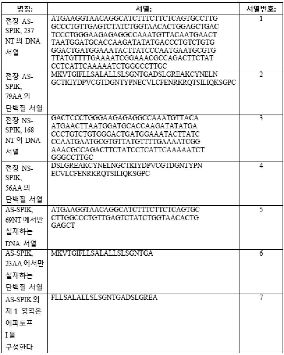
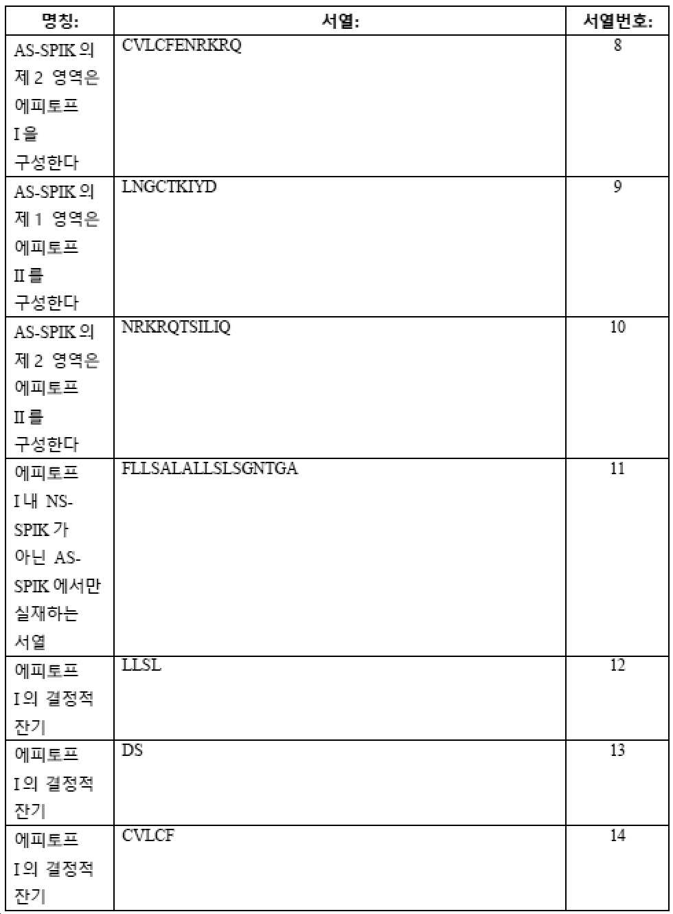
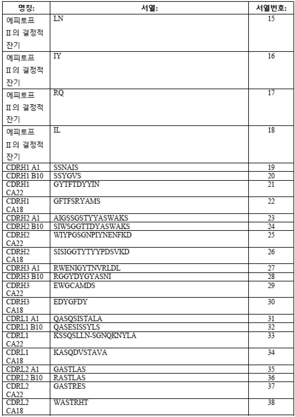

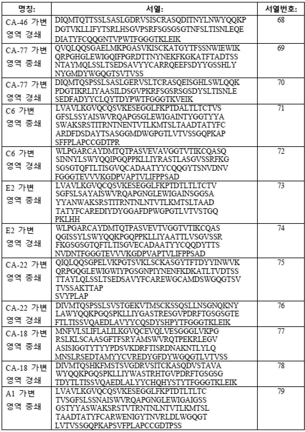
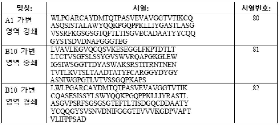
참고문헌:references:
1. Yu NC, Chaudhari V, Raman SS, Lassman C, Tong MJ, Busuttil RW, Lu DS. CT and MRI improve detection of hepatocellular carcinoma, compared with ultrasound alone, in patients with cirrhosis. Clin Gastroenterol Hepatol 2011;9:161-167.One. Yu NC, Chaudhari V, Raman SS, Lassman C, Tong MJ, Busuttil RW, Lu DS. CT and MRI improve detection of hepatocellular carcinoma, compared with ultrasound alone, in patients with cirrhosis. Clin Gastroenterol Hepatol 2011;9:161-167.
2. Bruix J, Sherman M. Management of hepatocellular carcinoma. Hepatology 2005;42:1208-1236.2. Bruix J, Sherman M. Management of hepatocellular carcinoma. Hepatology 2005;42:1208-1236.
3. Lok AS, Heathcote EJ, Hoofnagle JH. Management of hepatitis B: 2000--summary of a workshop. Gastroenterology 2001;122:2092-2093.3. Lok AS, Heathcote EJ, Hoofnagle JH. Management of hepatitis B: 2000--summary of a workshop. Gastroenterology 2001;122:2092-2093.
4. Greene LJ, Pubols MH, Bartelt DC. Human pancreatic secretory trypsin inhibitor. Methods Enzymol 1976;45:813-825.4. Greene LJ, Pubols MH, Bartelt DC. Human pancreatic secretory trypsin inhibitor. Methods Enzymol 1976;45:813-825.
5. Stenman UH. Tumor-associated trypsin inhibitor. Clin Chem 2002;48:1206-1209.5. Stenman UH. Tumor-associated trypsin inhibitors. Clin Chem 2002;48:1206-1209.
6. Greene LJ. Pancreatic exocrine secretory proteins. J Surg Oncol 1975;7:151-154.6. Greene LJ. Pancreatic exocrine secretory proteins. J Surg Oncol 1975;7:151-154.
7. Lee YC, Pan HW, Peng SY, Lai PL, Kuo WS, Ou YH, Hsu HC. Overexpression of tumour-associated trypsin inhibitor (TATI) enhances tumour growth and is associated with portal vein invasion, early recurrence and a stage-independent prognostic factor of hepatocellular carcinoma. Eur J Cancer 2007;43:736-744.7. Lee YC, Pan HW, Peng SY, Lai PL, Kuo WS, Ou YH, Hsu HC. Overexpression of tumor-associated trypsin inhibitor (TATI) enhances tumor growth and is associated with portal vein invasion, early recurrence and a stage-independent prognostic factor of hepatocellular carcinoma. Eur J Cancer 2007;43:736-744.
8. Lu X, Lee M, Tran T, Block T. High level expression of apoptosis inhibitor in hepatoma cell line expressing Hepatitis B virus. Int J Med Sci 2005;2:30-35.8. Lu X, Lee M, Tran T, Block T. High level expression of apoptosis inhibitor in hepatoma cell line expressing Hepatitis B virus. Int J Med Sci 2005;2:30-35.
9. Ohmachi Y, Murata A, Matsuura N, Yasuda T, Yasuda T, Monden M, Mori T, et al. Specific expression of the pancreatic-secretory-trypsin-inhibitor (PSTI) gene in hepatocellular carcinoma. Int J Cancer 1993;55:728-734.9. Ohmachi Y, Murata A, Matsuura N, Yasuda T, Yasuda T, Monden M, Mori T, et al. Specific expression of the pancreatic-secretory-trypsin-inhibitor (PSTI) gene in hepatocellular carcinoma. Int J Cancer 1993;55:728-734.
10. Hirota M, Ohmuraya M, Baba H. The role of trypsin, trypsin inhibitor, and trypsin receptor in the onset and aggravation of pancreatitis. J Gastroenterol 2006;41:832-836.10. Hirota M, Ohmuraya M, Baba H. The role of trypsin, trypsin inhibitor, and trypsin receptor in the onset and aggravation of pancreatitis. J Gastroenterol 2006;41:832-836.
11. Playford RJ, Hanby AM, Quinn C, Calam J. Influence of inflammation and atrophy on pancreatic secretory trypsin inhibitor levels within the gastric mucosa. Gastroenterology 1994;106:735-741.11. Playford RJ, Hanby AM, Quinn C, Calam J. Influence of inflammation and atrophy on pancreatic secretory trypsin inhibitor levels within the gastric mucosa. Gastroenterology 1994;106:735-741.
12. Kobayashi K, Horiuchi M, Saheki T. Pancreatic secretory trypsin inhibitor as a diagnostic marker for adult-onset type II citrullinemia. Hepatology 1997;25:1160-1165.12. Kobayashi K, Horiuchi M, Saheki T. Pancreatic secretory trypsin inhibitor as a diagnostic marker for adult-onset type II citrullinemia. Hepatology 1997;25:1160-1165.
13. Lu F, Lamontagne J, Sun A, Pinkerton M, Block T, Lu X. Role of the inflammatory protein serine protease inhibitor Kazal in preventing cytolytic granule granzyme A-mediated apoptosis. Immunology 2011;134:398-408.13. Lu F, Lamontagne J, Sun A, Pinkerton M, Block T, Lu X. Role of the inflammatory protein serine protease inhibitor Kazal in preventing cytolytic granule granzyme A-mediated apoptosis. Immunology 2011;134:398-408.
14. Bartelt DC, Shapanka R, Greene LJ. The primary structure of the human pancreatic secretory trypsin inhibitor. Amino acid sequence of the reduced S-aminoethylated protein. Arch Biochem Biophys 1977;179:189-199.14. Bartelt DC, Shapanka R, Greene LJ. The primary structure of the human pancreatic secretory trypsin inhibitor. Amino acid sequence of the reduced S-aminoethylated protein. Arch Biochem Biophys 1977;179:189-199.
15. Kikuchi N, Nagata K, Yoshida N, Tanaka T, Yamamoto M, Saitoh Y. Purification and complete amino acid sequence of canine pancreatic secretory trypsin inhibitor. FEBS Letters 1985;191:269-272.15. Kikuchi N, Nagata K, Yoshida N, Tanaka T, Yamamoto M, Saitoh Y. Purification and complete amino acid sequence of canine pancreatic secretory trypsin inhibitor. FEBS Letters 1985;191:269-272.
16. Lu X, Lamontagne J, Lu F, Block TM. Tumor-associated protein SPIK/TATI suppresses serine protease dependent cell apoptosis. Apoptosis 2008;13:483-494.16. Lu X, Lamontagne J, Lu F, Block TM. Tumor-associated protein SPIK/TATI suppresses serine protease dependent cell apoptosis. Apoptosis 2008;13:483-494.
17. Horii A, Kobayashi T, Tomita N, Yamamoto T, Fukushige S, Murotsu T, Ogawa M, et al. Primary structure of human pancreatic secretory trypsin inhibitor (PSTI) gene. Biochem Biophys Res Commun 1987;149:635-641.17. Horii A, Kobayashi T, Tomita N, Yamamoto T, Fukushige S, Murotsu T, Ogawa M, et al. Primary structure of human pancreatic secretory trypsin inhibitor (PSTI) gene. Biochem Biophys Res Commun 1987;149:635-641.
18. Hecht HJ, Szardenings M, Collins J, Schomburg D. Three-dimensional structure of a recombinant variant of human pancreatic secretory trypsin inhibitor (Kazal type). J Mol Biol 1992;225:1095-1103.18. Hecht HJ, Szardenings M, Collins J, Schomburg D. Three-dimensional structure of a recombinant variant of human pancreatic secretory trypsin inhibitor (Kazal type). J Mol Biol 1992;225:1095-1103.
19. Timmerman P, Puijk WC, Meloen RH. Functional reconstruction and synthetic mimicry of a conformational epitope using CLIPS technology. J Mol Recognit 2007;20:283-299.19. Timmerman P, Puijk WC, Meloen RH. Functional reconstruction and synthetic mimicry of a conformational epitope using CLIPS technology. J Mol Recognit 2007;20:283-299.
SEQUENCE LISTING <110> IMCARE BIOTECH, LLC. <120> EPITOPES OF ANTI-SERINE PROTEASE INHIBITOR KAZAL (SPIK) ANTIBODIES <130> IMC-0003-WO <140> PCT/US2020/046782 <141> 2020-08-18 <150> 62/899,024 <151> 2019-09-11 <160> 125 <170> PatentIn version 3.5 <210> 1 <211> 237 <212> DNA <213> Homo sapiens <400> 1 atgaaggtaa caggcatctt tcttctcagt gccttggccc tgttgagtct atctggtaac 60 actggagctg actccctggg aagagaggcc aaatgttaca atgaacttaa tggatgcacc 120 aagatatatg accctgtctg tgggactgat ggaaatactt atcccaatga atgcgtgtta 180 tgttttgaaa atcggaaacg ccagacttct atcctcattc aaaaatctgg gccttgc 237 <210> 2 <211> 79 <212> PRT <213> Homo sapiens <400> 2 Met Lys Val Thr Gly Ile Phe Leu Leu Ser Ala Leu Ala Leu Leu Ser 1 5 10 15 Leu Ser Gly Asn Thr Gly Ala Asp Ser Leu Gly Arg Glu Ala Lys Cys 20 25 30 Tyr Asn Glu Leu Asn Gly Cys Thr Lys Ile Tyr Asp Pro Val Cys Gly 35 40 45 Thr Asp Gly Asn Thr Tyr Pro Asn Glu Cys Val Leu Cys Phe Glu Asn 50 55 60 Arg Lys Arg Gln Thr Ser Ile Leu Ile Gln Lys Ser Gly Pro Cys 65 70 75 <210> 3 <211> 168 <212> DNA <213> Homo sapiens <400> 3 gactccctgg gaagagaggc caaatgttac aatgaactta atggatgcac caagatatat 60 gaccctgtct gtgggactga tggaaatact tatcccaatg aatgcgtgtt atgttttgaa 120 aatcggaaac gccagacttc tatcctcatt caaaaatctg ggccttgc 168 <210> 4 <211> 56 <212> PRT <213> Homo sapiens <400> 4 Asp Ser Leu Gly Arg Glu Ala Lys Cys Tyr Asn Glu Leu Asn Gly Cys 1 5 10 15 Thr Lys Ile Tyr Asp Pro Val Cys Gly Thr Asp Gly Asn Thr Tyr Pro 20 25 30 Asn Glu Cys Val Leu Cys Phe Glu Asn Arg Lys Arg Gln Thr Ser Ile 35 40 45 Leu Ile Gln Lys Ser Gly Pro Cys 50 55 <210> 5 <211> 69 <212> DNA <213> Homo sapiens <400> 5 atgaaggtaa caggcatctt tcttctcagt gccttggccc tgttgagtct atctggtaac 60 actggagct 69 <210> 6 <211> 23 <212> PRT <213> Homo sapiens <400> 6 Met Lys Val Thr Gly Ile Phe Leu Leu Ser Ala Leu Ala Leu Leu Ser 1 5 10 15 Leu Ser Gly Asn Thr Gly Ala 20 <210> 7 <211> 24 <212> PRT <213> Homo sapiens <400> 7 Phe Leu Leu Ser Ala Leu Ala Leu Leu Ser Leu Ser Gly Asn Thr Gly 1 5 10 15 Ala Asp Ser Leu Gly Arg Glu Ala 20 <210> 8 <211> 11 <212> PRT <213> Homo sapiens <400> 8 Cys Val Leu Cys Phe Glu Asn Arg Lys Arg Gln 1 5 10 <210> 9 <211> 9 <212> PRT <213> Homo sapiens <400> 9 Leu Asn Gly Cys Thr Lys Ile Tyr Asp 1 5 <210> 10 <211> 11 <212> PRT <213> Homo sapiens <400> 10 Asn Arg Lys Arg Gln Thr Ser Ile Leu Ile Gln 1 5 10 <210> 11 <211> 17 <212> PRT <213> Homo sapiens <400> 11 Phe Leu Leu Ser Ala Leu Ala Leu Leu Ser Leu Ser Gly Asn Thr Gly 1 5 10 15 Ala <210> 12 <211> 4 <212> PRT <213> Homo sapiens <400> 12 Leu Leu Ser Leu 1 <210> 13 <211> 2 <212> PRT <213> Homo sapiens <400> 13 Asp Ser 1 <210> 14 <211> 5 <212> PRT <213> Homo sapiens <400> 14 Cys Val Leu Cys Phe 1 5 <210> 15 <211> 2 <212> PRT <213> Homo sapiens <400> 15 Leu Asn 1 <210> 16 <211> 2 <212> PRT <213> Homo sapiens <400> 16 Ile Tyr 1 <210> 17 <211> 2 <212> PRT <213> Homo sapiens <400> 17 Arg Gln 1 <210> 18 <211> 2 <212> PRT <213> Homo sapiens <400> 18 Ile Leu 1 <210> 19 <211> 6 <212> PRT <213> Artificial Sequence <220> <221> source <223> /note="Description of Artificial Sequence: Synthetic peptide" <400> 19 Ser Ser Asn Ala Ile Ser 1 5 <210> 20 <211> 6 <212> PRT <213> Artificial Sequence <220> <221> source <223> /note="Description of Artificial Sequence: Synthetic peptide" <400> 20 Ser Ser Tyr Gly Val Ser 1 5 <210> 21 <211> 10 <212> PRT <213> Artificial Sequence <220> <221> source <223> /note="Description of Artificial Sequence: Synthetic peptide" <400> 21 Gly Tyr Thr Phe Thr Asp Tyr Tyr Ile Asn 1 5 10 <210> 22 <211> 10 <212> PRT <213> Artificial Sequence <220> <221> source <223> /note="Description of Artificial Sequence: Synthetic peptide" <400> 22 Gly Phe Thr Phe Ser Arg Tyr Ala Met Ser 1 5 10 <210> 23 <211> 16 <212> PRT <213> Artificial Sequence <220> <221> source <223> /note="Description of Artificial Sequence: Synthetic peptide" <400> 23 Ala Ile Gly Ser Ser Gly Ser Thr Tyr Tyr Ala Ser Trp Ala Lys Ser 1 5 10 15 <210> 24 <211> 16 <212> PRT <213> Artificial Sequence <220> <221> source <223> /note="Description of Artificial Sequence: Synthetic peptide" <400> 24 Ser Ile Trp Ser Gly Gly Thr Thr Asp Tyr Ala Ser Trp Ala Lys Ser 1 5 10 15 <210> 25 <211> 17 <212> PRT <213> Artificial Sequence <220> <221> source <223> /note="Description of Artificial Sequence: Synthetic peptide" <400> 25 Trp Ile Tyr Pro Gly Ser Gly Asn Pro Ile Tyr Asn Glu Asn Phe Lys 1 5 10 15 Asp <210> 26 <211> 17 <212> PRT <213> Artificial Sequence <220> <221> source <223> /note="Description of Artificial Sequence: Synthetic peptide" <400> 26 Ser Ile Ser Ile Gly Gly Thr Tyr Thr Tyr Tyr Pro Asp Ser Val Lys 1 5 10 15 Asp <210> 27 <211> 14 <212> PRT <213> Artificial Sequence <220> <221> source <223> /note="Description of Artificial Sequence: Synthetic peptide" <400> 27 Arg Trp Glu Asn Ile Gly Tyr Thr Asn Val Arg Leu Asp Leu 1 5 10 <210> 28 <211> 12 <212> PRT <213> Artificial Sequence <220> <221> source <223> /note="Description of Artificial Sequence: Synthetic peptide" <400> 28 Arg Gly Gly Tyr Asp Tyr Gly Tyr Ala Ser Asn Ile 1 5 10 <210> 29 <211> 8 <212> PRT <213> Artificial Sequence <220> <221> source <223> /note="Description of Artificial Sequence: Synthetic peptide" <400> 29 Glu Trp Gly Cys Ala Met Asp Ser 1 5 <210> 30 <211> 7 <212> PRT <213> Artificial Sequence <220> <221> source <223> /note="Description of Artificial Sequence: Synthetic peptide" <400> 30 Glu Asp Tyr Gly Phe Asp Tyr 1 5 <210> 31 <211> 11 <212> PRT <213> Artificial Sequence <220> <221> source <223> /note="Description of Artificial Sequence: Synthetic peptide" <400> 31 Gln Ala Ser Gln Ser Ile Ser Thr Ala Leu Ala 1 5 10 <210> 32 <211> 11 <212> PRT <213> Artificial Sequence <220> <221> source <223> /note="Description of Artificial Sequence: Synthetic peptide" <400> 32 Gln Ala Ser Glu Ser Ile Ser Ser Tyr Leu Ser 1 5 10 <210> 33 <211> 17 <212> PRT <213> Artificial Sequence <220> <221> source <223> /note="Description of Artificial Sequence: Synthetic peptide" <400> 33 Lys Ser Ser Gln Ser Leu Leu Asn Ser Gly Asn Gln Lys Asn Tyr Leu 1 5 10 15 Ala <210> 34 <211> 11 <212> PRT <213> Artificial Sequence <220> <221> source <223> /note="Description of Artificial Sequence: Synthetic peptide" <400> 34 Lys Ala Ser Gln Asp Val Ser Thr Ala Val Ala 1 5 10 <210> 35 <211> 7 <212> PRT <213> Artificial Sequence <220> <221> source <223> /note="Description of Artificial Sequence: Synthetic peptide" <400> 35 Gly Ala Ser Thr Leu Ala Ser 1 5 <210> 36 <211> 7 <212> PRT <213> Artificial Sequence <220> <221> source <223> /note="Description of Artificial Sequence: Synthetic peptide" <400> 36 Arg Ala Ser Thr Leu Ala Ser 1 5 <210> 37 <211> 7 <212> PRT <213> Artificial Sequence <220> <221> source <223> /note="Description of Artificial Sequence: Synthetic peptide" <400> 37 Gly Ala Ser Thr Arg Glu Ser 1 5 <210> 38 <211> 7 <212> PRT <213> Artificial Sequence <220> <221> source <223> /note="Description of Artificial Sequence: Synthetic peptide" <400> 38 Trp Ala Ser Thr Arg His Thr 1 5 <210> 39 <211> 12 <212> PRT <213> Artificial Sequence <220> <221> source <223> /note="Description of Artificial Sequence: Synthetic peptide" <400> 39 Gln Gln Gly Tyr Ser Thr Ser Asp Val Asp Asn Ala 1 5 10 <210> 40 <211> 12 <212> PRT <213> Artificial Sequence <220> <221> source <223> /note="Description of Artificial Sequence: Synthetic peptide" <400> 40 Gln Gln Gly Tyr Ser Val Ser Asn Val Asp Asn Ile 1 5 10 <210> 41 <211> 9 <212> PRT <213> Artificial Sequence <220> <221> source <223> /note="Description of Artificial Sequence: Synthetic peptide" <400> 41 Gln Ser Asp Tyr Ser His Pro Tyr Thr 1 5 <210> 42 <211> 8 <212> PRT <213> Artificial Sequence <220> <221> source <223> /note="Description of Artificial Sequence: Synthetic peptide" <400> 42 His Gln His Tyr Ser Thr Tyr Thr 1 5 <210> 43 <211> 6 <212> PRT <213> Artificial Sequence <220> <221> source <223> /note="Description of Artificial Sequence: Synthetic peptide" <400> 43 Ser Tyr Ala Ile Ser Trp 1 5 <210> 44 <211> 7 <212> PRT <213> Artificial Sequence <220> <221> source <223> /note="Description of Artificial Sequence: Synthetic peptide" <400> 44 Ser Ala Tyr Ala Ile Ser Trp 1 5 <210> 45 <211> 10 <212> PRT <213> Artificial Sequence <220> <221> source <223> /note="Description of Artificial Sequence: Synthetic peptide" <400> 45 Gly Tyr Thr Phe Thr Ser Tyr Trp Met Gln 1 5 10 <210> 46 <211> 10 <212> PRT <213> Artificial Sequence <220> <221> source <223> /note="Description of Artificial Sequence: Synthetic peptide" <400> 46 Gly Tyr Thr Phe Ser Ser Asn Trp Ile Glu 1 5 10 <210> 47 <211> 16 <212> PRT <213> Artificial Sequence <220> <221> source <223> /note="Description of Artificial Sequence: Synthetic peptide" <400> 47 Ala Ile Asn Thr Tyr Gly Gly Thr Tyr Tyr Ala Ser Trp Ala Lys Ser 1 5 10 15 <210> 48 <211> 16 <212> PRT <213> Artificial Sequence <220> <221> source <223> /note="Description of Artificial Sequence: Synthetic peptide" <400> 48 Ala Ile Asn Ser Gly Gly Ser Ala Tyr Tyr Ala Asn Trp Ala Lys Ser 1 5 10 15 <210> 49 <211> 17 <212> PRT <213> Artificial Sequence <220> <221> source <223> /note="Description of Artificial Sequence: Synthetic peptide" <400> 49 Ala Ile Tyr Pro Gly Asp Gly Asp Thr Arg Tyr Thr Gln Lys Phe Glu 1 5 10 15 Asp <210> 50 <211> 17 <212> PRT <213> Artificial Sequence <220> <221> source <223> /note="Description of Artificial Sequence: Synthetic peptide" <400> 50 Gln Ile Phe Pro Gly Arg Asp Thr Thr Asn Tyr Asn Glu Lys Phe Lys 1 5 10 15 Gly <210> 51 <211> 17 <212> PRT <213> Artificial Sequence <220> <221> source <223> /note="Description of Artificial Sequence: Synthetic peptide" <400> 51 Arg Asp Phe Asp Ser Asp Ala Tyr Thr Ser Ala Ser Gly Gly Met Asp 1 5 10 15 Pro <210> 52 <211> 13 <212> PRT <213> Artificial Sequence <220> <221> source <223> /note="Description of Artificial Sequence: Synthetic peptide" <400> 52 Arg Glu Asp Ile Tyr Asp Tyr Gly Gly Ala Phe Asp Pro 1 5 10 <210> 53 <211> 11 <212> PRT <213> Artificial Sequence <220> <221> source <223> /note="Description of Artificial Sequence: Synthetic peptide" <400> 53 Gly Ala Asn Tyr Ala Asn Ile Arg Phe Ala Tyr 1 5 10 <210> 54 <211> 21 <212> PRT <213> Artificial Sequence <220> <221> source <223> /note="Description of Artificial Sequence: Synthetic peptide" <400> 54 Arg Gln Glu Glu Phe Ser Asp Tyr Tyr Gly Ser Ser His Leu Tyr Asn 1 5 10 15 Tyr Gly Met Asp Tyr 20 <210> 55 <211> 11 <212> PRT <213> Artificial Sequence <220> <221> source <223> /note="Description of Artificial Sequence: Synthetic peptide" <400> 55 Gln Ala Ser Gln Ser Ile Asn Asn Tyr Leu Ser 1 5 10 <210> 56 <211> 11 <212> PRT <213> Artificial Sequence <220> <221> source <223> /note="Description of Artificial Sequence: Synthetic peptide" <400> 56 Gln Ala Ser Gln Gly Ile Ser Ser Tyr Leu Ser 1 5 10 <210> 57 <211> 11 <212> PRT <213> Artificial Sequence <220> <221> source <223> /note="Description of Artificial Sequence: Synthetic peptide" <400> 57 Arg Ala Ser Gln Asp Ile Thr Asn Tyr Leu Asn 1 5 10 <210> 58 <211> 11 <212> PRT <213> Artificial Sequence <220> <221> source <223> /note="Description of Artificial Sequence: Synthetic peptide" <400> 58 Arg Ala Ser Gln Glu Ile Ser Gly His Leu Ser 1 5 10 <210> 59 <211> 7 <212> PRT <213> Artificial Sequence <220> <221> source <223> /note="Description of Artificial Sequence: Synthetic peptide" <400> 59 Arg Ala Ser Thr Leu Ala Ser 1 5 <210> 60 <211> 7 <212> PRT <213> Artificial Sequence <220> <221> source <223> /note="Description of Artificial Sequence: Synthetic peptide" <400> 60 Ala Ala Thr Thr Leu Val Ser 1 5 <210> 61 <211> 7 <212> PRT <213> Artificial Sequence <220> <221> source <223> /note="Description of Artificial Sequence: Synthetic peptide" <400> 61 Tyr Thr Ser Arg Leu His Ser 1 5 <210> 62 <211> 7 <212> PRT <213> Artificial Sequence <220> <221> source <223> /note="Description of Artificial Sequence: Synthetic peptide" <400> 62 Ala Ala Ser Ile Leu Asp Ser 1 5 <210> 63 <211> 11 <212> PRT <213> Artificial Sequence <220> <221> source <223> /note="Description of Artificial Sequence: Synthetic peptide" <400> 63 Gln Gln Gly Tyr Thr Ser Asn Val Asp Asn Val 1 5 10 <210> 64 <211> 12 <212> PRT <213> Artificial Sequence <220> <221> source <223> /note="Description of Artificial Sequence: Synthetic peptide" <400> 64 Gln Gln Asp Tyr Thr Thr Ser Asn Val Asp Asn Thr 1 5 10 <210> 65 <211> 9 <212> PRT <213> Artificial Sequence <220> <221> source <223> /note="Description of Artificial Sequence: Synthetic peptide" <400> 65 Gln Gln Gly Asn Thr Val Pro Trp Thr 1 5 <210> 66 <211> 9 <212> PRT <213> Artificial Sequence <220> <221> source <223> /note="Description of Artificial Sequence: Synthetic peptide" <400> 66 Leu Gln Tyr Thr Asp Tyr Pro Trp Thr 1 5 <210> 67 <211> 120 <212> PRT <213> Artificial Sequence <220> <221> source <223> /note="Description of Artificial Sequence: Synthetic polypeptide" <400> 67 Gln Gly His Leu Gln Gln Ser Gly Ala Glu Leu Ala Arg Pro Gly Thr 1 5 10 15 Ser Val Lys Leu Ser Cys Lys Ala Ser Gly Tyr Thr Phe Thr Ser Tyr 20 25 30 Trp Met Gln Trp Val Lys Gln Arg Pro Gly Gln Gly Leu Glu Trp Ile 35 40 45 Gly Ala Ile Tyr Pro Gly Asp Gly Asp Thr Arg Tyr Thr Gln Lys Phe 50 55 60 Glu Asp Lys Ala Thr Leu Thr Ala Asp Lys Ser Ser Ser Thr Ala Tyr 65 70 75 80 Met Gln Leu Ser Asn Leu Ala Ser Glu Asp Ser Ala Tyr Tyr Tyr Cys 85 90 95 Ala Arg Gly Ala Asn Tyr Ala Asn Ile Arg Phe Ala Tyr Trp Gly Gln 100 105 110 Gly Thr Leu Val Thr Val Ser Ala 115 120 <210> 68 <211> 107 <212> PRT <213> Artificial Sequence <220> <221> source <223> /note="Description of Artificial Sequence: Synthetic polypeptide" <400> 68 Asp Ile Gln Met Thr Gln Thr Thr Ser Ser Leu Ser Ala Ser Leu Gly 1 5 10 15 Asp Arg Val Ser Ile Ser Cys Arg Ala Ser Gln Asp Ile Thr Asn Tyr 20 25 30 Leu Asn Trp Tyr Gln Gln Lys Pro Asp Gly Thr Val Lys Leu Leu Ile 35 40 45 Phe Tyr Thr Ser Arg Leu His Ser Gly Val Pro Ser Arg Phe Ser Gly 50 55 60 Ser Gly Ser Gly Thr Asn Phe Ser Leu Thr Ile Ser Asn Leu Glu Gln 65 70 75 80 Glu Asp Ile Ala Thr Tyr Phe Cys Gln Gln Gly Asn Thr Val Pro Trp 85 90 95 Thr Phe Gly Gly Gly Thr Lys Leu Glu Ile Lys 100 105 <210> 69 <211> 130 <212> PRT <213> Artificial Sequence <220> <221> source <223> /note="Description of Artificial Sequence: Synthetic polypeptide" <400> 69 Gln Val Gln Leu Gln Gln Ser Gly Ala Glu Leu Met Lys Pro Gly Ala 1 5 10 15 Ser Val Lys Ile Ser Cys Lys Ala Thr Gly Tyr Thr Phe Ser Ser Asn 20 25 30 Trp Ile Glu Trp Ile Lys Gln Arg Pro Gly His Gly Leu Glu Trp Ile 35 40 45 Gly Gln Ile Phe Pro Gly Arg Asp Thr Thr Asn Tyr Asn Glu Lys Phe 50 55 60 Lys Gly Lys Ala Thr Phe Thr Ala Asp Thr Ser Ser Asn Thr Ala Tyr 65 70 75 80 Met Gln Leu Ser Ser Leu Thr Ser Glu Asp Ser Ala Val Tyr Tyr Cys 85 90 95 Ala Arg Arg Gln Glu Glu Phe Ser Asp Tyr Tyr Gly Ser Ser His Leu 100 105 110 Tyr Asn Tyr Gly Met Asp Tyr Trp Gly Gln Gly Thr Ser Val Thr Val 115 120 125 Ser Ser 130 <210> 70 <211> 107 <212> PRT <213> Artificial Sequence <220> <221> source <223> /note="Description of Artificial Sequence: Synthetic polypeptide" <400> 70 Asp Ile Gln Met Thr Gln Ser Pro Ser Ser Leu Ser Ala Ser Leu Gly 1 5 10 15 Glu Arg Val Ser Leu Thr Cys Arg Ala Ser Gln Glu Ile Ser Gly His 20 25 30 Leu Ser Trp Leu Gln Gln Lys Pro Asp Gly Thr Ile Lys Arg Leu Ile 35 40 45 Tyr Ala Ala Ser Ile Leu Asp Ser Gly Val Pro Lys Arg Phe Ser Gly 50 55 60 Ser Arg Ser Gly Ser Asp Tyr Ser Leu Thr Ile Ser Asn Leu Glu Ser 65 70 75 80 Glu Asp Phe Ala Asp Tyr Tyr Cys Leu Gln Tyr Thr Asp Tyr Pro Trp 85 90 95 Thr Phe Gly Gly Gly Thr Lys Val Glu Ile Lys 100 105 <210> 71 <211> 152 <212> PRT <213> Artificial Sequence <220> <221> source <223> /note="Description of Artificial Sequence: Synthetic polypeptide" <400> 71 Leu Val Ala Val Leu Lys Gly Val Gln Cys Gln Ser Val Lys Glu Ser 1 5 10 15 Glu Gly Gly Leu Phe Lys Pro Thr Asp Ala Leu Thr Leu Thr Cys Thr 20 25 30 Val Ser Gly Phe Ser Leu Ser Ser Tyr Ala Ile Ser Trp Val Arg Gln 35 40 45 Ala Pro Gly Ser Gly Leu Glu Trp Ile Gly Ala Ile Asn Thr Tyr Gly 50 55 60 Gly Thr Tyr Tyr Ala Ser Trp Ala Lys Ser Arg Ser Thr Ile Thr Arg 65 70 75 80 Asn Thr Asn Glu Asn Thr Val Thr Leu Lys Met Thr Ser Leu Thr Ala 85 90 95 Ala Asp Thr Ala Thr Tyr Phe Cys Ala Arg Asp Phe Asp Ser Asp Ala 100 105 110 Tyr Thr Ser Ala Ser Gly Gly Met Asp Trp Gly Pro Gly Thr Leu Val 115 120 125 Thr Val Ser Ser Gly Gln Pro Lys Ala Pro Ser Phe Phe Pro Leu Ala 130 135 140 Pro Cys Cys Gly Asp Thr Pro Arg 145 150 <210> 72 <211> 132 <212> PRT <213> Artificial Sequence <220> <221> source <223> /note="Description of Artificial Sequence: Synthetic polypeptide" <400> 72 Trp Leu Pro Gly Ala Arg Cys Ala Tyr Asp Met Thr Gln Thr Pro Ala 1 5 10 15 Ser Val Glu Val Ala Val Gly Gly Thr Val Thr Ile Lys Cys Gln Ala 20 25 30 Ser Gln Ser Ile Asn Asn Tyr Leu Ser Trp Tyr Gln Gln Ile Pro Gly 35 40 45 Gln Pro Pro Lys Leu Leu Ile Tyr Arg Ala Ser Thr Leu Ala Ser Gly 50 55 60 Val Ser Ser Arg Phe Lys Gly Ser Gly Ser Gly Thr Gln Phe Thr Leu 65 70 75 80 Thr Ile Ser Gly Val Gln Cys Ala Asp Ala Ala Thr Tyr Tyr Cys Gln 85 90 95 Gln Gly Tyr Thr Ser Asn Val Asp Asn Val Phe Gly Gly Gly Thr Glu 100 105 110 Val Val Val Lys Gly Asp Pro Val Ala Pro Thr Val Leu Ile Phe Pro 115 120 125 Pro Ser Ala Asp 130 <210> 73 <211> 136 <212> PRT <213> Artificial Sequence <220> <221> source <223> /note="Description of Artificial Sequence: Synthetic polypeptide" <400> 73 Leu Val Ala Val Leu Lys Gly Val Gln Cys Gln Ser Val Lys Glu Ser 1 5 10 15 Glu Gly Gly Leu Phe Lys Pro Thr Asp Thr Leu Thr Leu Thr Cys Thr 20 25 30 Val Ser Gly Phe Ser Leu Ser Ala Tyr Ala Ile Ser Trp Val Arg Gln 35 40 45 Ala Pro Gly Asn Gly Leu Glu Trp Ile Gly Ala Ile Asn Ser Gly Gly 50 55 60 Ser Ala Tyr Tyr Ala Asn Trp Ala Lys Ser Arg Ser Thr Ile Thr Arg 65 70 75 80 Asn Thr Asn Leu Asn Thr Val Thr Leu Lys Met Thr Ser Leu Thr Ala 85 90 95 Ala Asp Thr Ala Thr Tyr Phe Cys Ala Arg Glu Asp Ile Tyr Asp Tyr 100 105 110 Gly Gly Ala Phe Asp Pro Trp Gly Pro Gly Thr Leu Val Thr Val Ser 115 120 125 Thr Gly Gln Pro Lys Leu His His 130 135 <210> 74 <211> 133 <212> PRT <213> Artificial Sequence <220> <221> source <223> /note="Description of Artificial Sequence: Synthetic polypeptide" <400> 74 Trp Leu Pro Gly Ala Arg Cys Ala Tyr Asp Met Thr Gln Thr Pro Ala 1 5 10 15 Ser Val Glu Val Thr Val Gly Gly Thr Val Thr Ile Lys Cys Gln Ala 20 25 30 Ser Gln Gly Ile Ser Ser Tyr Leu Ser Trp Tyr Gln Gln Lys Pro Gly 35 40 45 Gln Pro Pro Lys Leu Leu Ile Tyr Ala Ala Thr Thr Leu Val Ser Gly 50 55 60 Val Ser Ser Arg Phe Lys Gly Ser Gly Ser Gly Thr Gln Phe Thr Leu 65 70 75 80 Thr Ile Ser Gly Val Glu Cys Ala Asp Ala Ala Thr Tyr Tyr Cys Gln 85 90 95 Gln Asp Tyr Thr Thr Ser Asn Val Asp Asn Thr Phe Gly Gly Gly Thr 100 105 110 Glu Val Val Val Lys Gly Asp Pro Val Ala Pro Thr Val Leu Ile Phe 115 120 125 Pro Pro Ser Ala Asp 130 <210> 75 <211> 130 <212> PRT <213> Artificial Sequence <220> <221> source <223> /note="Description of Artificial Sequence: Synthetic polypeptide" <400> 75 Gln Ile Gln Leu Gln Gln Ser Gly Pro Glu Leu Val Lys Pro Gly Thr 1 5 10 15 Ser Val Lys Leu Ser Cys Lys Ala Ser Gly Tyr Thr Phe Thr Asp Tyr 20 25 30 Tyr Ile Asn Trp Val Lys Gln Arg Pro Gly Gln Gly Leu Glu Trp Ile 35 40 45 Gly Trp Ile Tyr Pro Gly Ser Gly Asn Pro Ile Tyr Asn Glu Asn Phe 50 55 60 Lys Asp Lys Ala Thr Leu Thr Val Asp Thr Ser Ser Thr Thr Ala Tyr 65 70 75 80 Leu Gln Leu Ser Ser Leu Thr Ser Glu Asp Ser Ala Val Tyr Phe Cys 85 90 95 Ala Arg Glu Trp Gly Cys Ala Met Asp Ser Trp Gly Gln Gly Thr Ser 100 105 110 Val Thr Val Ser Ser Ala Lys Thr Thr Ala Pro Ser Val Tyr Pro Leu 115 120 125 Ala Pro 130 <210> 76 <211> 113 <212> PRT <213> Artificial Sequence <220> <221> source <223> /note="Description of Artificial Sequence: Synthetic polypeptide" <400> 76 Asp Ile Val Met Thr Gln Ser Pro Ser Ser Leu Ser Val Ser Thr Gly 1 5 10 15 Glu Lys Val Thr Met Ser Cys Lys Ser Ser Gln Ser Leu Leu Asn Ser 20 25 30 Gly Asn Gln Lys Asn Tyr Leu Ala Trp Tyr Gln Gln Lys Pro Gly Gln 35 40 45 Ser Pro Lys Leu Leu Ile Tyr Gly Ala Ser Thr Arg Glu Ser Gly Val 50 55 60 Pro Asp Arg Phe Thr Gly Ser Gly Ser Gly Thr Glu Phe Thr Leu Thr 65 70 75 80 Ile Ser Ser Val Gln Ala Glu Asp Leu Ala Val Tyr Tyr Cys Gln Ser 85 90 95 Asp Tyr Ser His Pro Tyr Thr Phe Gly Gly Gly Thr Lys Leu Glu Ile 100 105 110 Lys <210> 77 <211> 135 <212> PRT <213> Artificial Sequence <220> <221> source <223> /note="Description of Artificial Sequence: Synthetic polypeptide" <400> 77 Met Asn Phe Val Leu Ser Leu Ile Phe Leu Ala Leu Ile Leu Lys Gly 1 5 10 15 Val Gln Cys Glu Val Gln Leu Val Glu Ser Gly Gly Gly Leu Val Lys 20 25 30 Pro Gly Arg Ser Leu Lys Leu Ser Cys Ala Ala Ser Gly Phe Thr Phe 35 40 45 Ser Arg Tyr Ala Met Ser Trp Val Arg Gln Thr Pro Glu Lys Arg Leu 50 55 60 Glu Gly Val Ala Ser Ile Ser Ile Gly Gly Thr Tyr Thr Tyr Tyr Pro 65 70 75 80 Asp Ser Val Lys Asp Arg Phe Thr Ile Ser Arg Asp Asn Ala Lys Asn 85 90 95 Thr Leu Tyr Leu Gln Met Asn Ser Leu Arg Ser Glu Asp Thr Ala Met 100 105 110 Tyr Tyr Cys Val Arg Glu Asp Tyr Gly Phe Asp Tyr Trp Gly Gln Gly 115 120 125 Thr Leu Val Thr Val Ser Ser 130 135 <210> 78 <211> 106 <212> PRT <213> Artificial Sequence <220> <221> source <223> /note="Description of Artificial Sequence: Synthetic polypeptide" <400> 78 Asp Ile Val Met Thr Gln Ser His Lys Phe Met Ser Thr Ser Val Gly 1 5 10 15 Asp Arg Val Ser Ile Thr Cys Lys Ala Ser Gln Asp Val Ser Thr Ala 20 25 30 Val Ala Trp Tyr Gln Gln Lys Pro Gly Gln Ser Pro Lys Leu Leu Ile 35 40 45 Tyr Trp Ala Ser Thr Arg His Thr Gly Val Pro Asp Arg Phe Thr Gly 50 55 60 Ser Gly Ser Gly Thr Asp Tyr Thr Leu Thr Ile Ser Ser Val Gln Ala 65 70 75 80 Glu Asp Leu Ala Leu Tyr Tyr Cys His Gln His Tyr Ser Thr Tyr Thr 85 90 95 Phe Gly Gly Gly Thr Lys Leu Glu Ile Lys 100 105 <210> 79 <211> 151 <212> PRT <213> Artificial Sequence <220> <221> source <223> /note="Description of Artificial Sequence: Synthetic polypeptide" <400> 79 Leu Val Ala Val Leu Lys Gly Val Gln Cys Gln Ser Val Lys Glu Ser 1 5 10 15 Glu Gly Gly Leu Phe Lys Pro Thr Asp Thr Leu Thr Leu Thr Cys Thr 20 25 30 Val Ser Gly Phe Ser Leu Ser Ser Asn Ala Ile Ser Trp Val Arg Gln 35 40 45 Ala Pro Gly Asn Gly Leu Glu Trp Ile Gly Ala Ile Gly Ser Ser Gly 50 55 60 Ser Thr Tyr Tyr Ala Ser Trp Ala Lys Ser Arg Ser Thr Val Thr Arg 65 70 75 80 Asn Thr Asn Leu Asn Thr Val Thr Leu Lys Met Thr Ser Leu Thr Ala 85 90 95 Ala Asp Thr Ala Thr Tyr Phe Cys Ala Arg Trp Glu Asn Ile Gly Tyr 100 105 110 Thr Asn Val Arg Leu Asp Leu Trp Gly Gln Gly Thr Leu Val Thr Val 115 120 125 Ser Ser Gly Gln Pro Lys Ala Pro Ser Val Phe Pro Leu Ala Pro Cys 130 135 140 Cys Gly Asp Thr Pro Ser Ser 145 150 <210> 80 <211> 114 <212> PRT <213> Artificial Sequence <220> <221> source <223> /note="Description of Artificial Sequence: Synthetic polypeptide" <400> 80 Trp Leu Pro Gly Ala Arg Cys Ala Tyr Asp Met Thr Gln Thr Pro Ala 1 5 10 15 Ser Val Glu Val Ala Val Gly Gly Thr Val Thr Ile Lys Cys Gln Ala 20 25 30 Ser Gln Ser Ile Ser Thr Ala Leu Ala Trp Tyr Gln Gln Lys Pro Gly 35 40 45 Gln Pro Pro Lys Leu Leu Ile Tyr Gly Ala Ser Thr Leu Ala Ser Gly 50 55 60 Val Ser Ser Arg Phe Lys Gly Ser Gly Ser Gly Thr Gln Phe Thr Leu 65 70 75 80 Thr Ile Ser Gly Val Glu Cys Ala Asp Ala Ala Thr Tyr Tyr Cys Gln 85 90 95 Gln Gly Tyr Ser Thr Ser Asp Val Asp Asn Ala Phe Gly Gly Gly Thr 100 105 110 Glu Gly <210> 81 <211> 135 <212> PRT <213> Artificial Sequence <220> <221> source <223> /note="Description of Artificial Sequence: Synthetic polypeptide" <400> 81 Leu Val Ala Val Leu Lys Gly Val Gln Cys Gln Ser Val Lys Glu Ser 1 5 10 15 Glu Gly Gly Leu Phe Lys Pro Thr Asp Thr Leu Thr Leu Thr Cys Thr 20 25 30 Val Ser Gly Phe Ser Leu Ser Ser Tyr Gly Val Ser Trp Val Arg Gln 35 40 45 Ala Pro Gly Lys Gly Leu Glu Trp Ile Gly Ser Ile Trp Ser Gly Gly 50 55 60 Thr Thr Asp Tyr Ala Ser Trp Ala Lys Ser Arg Ser Thr Ile Thr Arg 65 70 75 80 Asn Thr Asn Glu Asn Thr Val Thr Leu Lys Val Thr Ser Leu Thr Ala 85 90 95 Ala Asp Thr Ala Thr Tyr Phe Cys Ala Arg Gly Gly Tyr Asp Tyr Gly 100 105 110 Tyr Ala Ser Asn Ile Trp Gly Pro Gly Thr Leu Val Thr Val Ser Ser 115 120 125 Gly Gln Pro Lys Ala Pro Ser 130 135 <210> 82 <211> 134 <212> PRT <213> Artificial Sequence <220> <221> source <223> /note="Description of Artificial Sequence: Synthetic polypeptide" <400> 82 Leu Trp Leu Pro Gly Ala Arg Cys Ala Tyr Asp Met Thr Gln Thr Pro 1 5 10 15 Ala Ser Val Glu Val Ala Val Gly Gly Thr Val Thr Ile Lys Cys Gln 20 25 30 Ala Ser Glu Ser Ile Ser Ser Tyr Leu Ser Trp Tyr Gln Gln Lys Pro 35 40 45 Gly Gln Pro Pro Lys Leu Leu Ile Tyr Arg Ala Ser Thr Leu Ala Ser 50 55 60 Gly Val Pro Ser Arg Phe Ser Gly Ser Gly Ser Gly Thr Glu Phe Thr 65 70 75 80 Leu Thr Ile Ser Asp Gly Gln Cys Asp Asp Ala Ala Thr Tyr Tyr Cys 85 90 95 Gln Gln Gly Tyr Ser Val Ser Asn Val Asp Asn Ile Phe Gly Gly Gly 100 105 110 Thr Glu Val Val Val Lys Gly Asp Pro Val Ala Pro Thr Val Leu Ile 115 120 125 Phe Pro Pro Ser Ala Asp 130 <210> 83 <211> 15 <212> PRT <213> Artificial Sequence <220> <221> source <223> /note="Description of Artificial Sequence: Synthetic peptide" <400> 83 Gly Gly Gly Gly Ser Gly Gly Gly Gly Ser Gly Gly Gly Gly Ser 1 5 10 15 <210> 84 <211> 12 <212> PRT <213> Homo sapiens <400> 84 Glu Leu Asn Gly Cys Thr Lys Ile Tyr Asp Pro Val 1 5 10 <210> 85 <211> 5 <212> PRT <213> Homo sapiens <400> 85 Lys Val Thr Gly Ile 1 5 <210> 86 <211> 79 <212> PRT <213> Artificial Sequence <220> <221> source <223> /note="Description of Artificial Sequence: Synthetic polypeptide" <400> 86 Met Lys Val Thr Gly Ile Phe Leu Leu Ser Ala Leu Ala Leu Leu Ser 1 5 10 15 Leu Ser Gly Asn Thr Gly Ala Asp Ser Leu Gly Arg Glu Ala Lys Cys 20 25 30 Tyr Asn Glu Leu Asn Gly Cys Thr Lys Ile Tyr Asp Pro Val Cys Gly 35 40 45 Thr Asp Gly Asn Thr Tyr Pro Asn Glu Cys Val Leu Cys Phe Glu Asn 50 55 60 Arg Lys Arg Gln Thr Ser Ile Leu Ile Gln Lys Ser Gly Pro Cys 65 70 75 <210> 87 <211> 51 <212> PRT <213> Artificial Sequence <220> <221> source <223> /note="Description of Artificial Sequence: Synthetic polypeptide" <400> 87 Met Lys Val Thr Gly Ile Phe Leu Leu Ser Ala Leu Ala Leu Leu Ser 1 5 10 15 Leu Ser Gly Asn Thr Gly Ala Asp Ser Leu Gly Arg Glu Ala Lys Cys 20 25 30 Tyr Asn Glu Leu Asn Gly Cys Thr Lys Ile Tyr Asp Pro Val Cys Gly 35 40 45 Thr Asp Gly 50 <210> 88 <211> 28 <212> PRT <213> Artificial Sequence <220> <221> source <223> /note="Description of Artificial Sequence: Synthetic peptide" <400> 88 Asp Ser Leu Gly Arg Glu Ala Lys Cys Tyr Asn Glu Leu Asn Gly Cys 1 5 10 15 Thr Lys Ile Tyr Asp Pro Val Cys Gly Thr Asp Gly 20 25 <210> 89 <211> 28 <212> PRT <213> Artificial Sequence <220> <221> source <223> /note="Description of Artificial Sequence: Synthetic peptide" <400> 89 Asn Thr Tyr Pro Asn Glu Cys Val Leu Cys Phe Glu Asn Arg Lys Arg 1 5 10 15 Gln Thr Ser Ile Leu Ile Gln Lys Ser Gly Pro Cys 20 25 <210> 90 <211> 21 <212> PRT <213> Artificial Sequence <220> <221> source <223> /note="Description of Artificial Sequence: Synthetic peptide" <400> 90 Ser Ala Leu Ala Leu Leu Ser Leu Ser Gly Asn Thr Gly Ala Asp Ser 1 5 10 15 Leu Gly Arg Glu Ala 20 <210> 91 <211> 9 <212> PRT <213> Artificial Sequence <220> <221> source <223> /note="Description of Artificial Sequence: Synthetic peptide" <400> 91 Gly Asn Thr Gly Ala Asp Ser Leu Gly 1 5 <210> 92 <211> 9 <212> PRT <213> Artificial Sequence <220> <221> source <223> /note="Description of Artificial Sequence: Synthetic peptide" <400> 92 Ser Gly Asn Thr Gly Ala Asp Ser Leu 1 5 <210> 93 <211> 9 <212> PRT <213> Artificial Sequence <220> <221> source <223> /note="Description of Artificial Sequence: Synthetic peptide" <400> 93 Leu Ser Gly Asn Thr Gly Ala Asp Ser 1 5 <210> 94 <211> 9 <212> PRT <213> Artificial Sequence <220> <221> source <223> /note="Description of Artificial Sequence: Synthetic peptide" <400> 94 Ser Leu Ser Gly Asn Thr Gly Ala Asp 1 5 <210> 95 <211> 9 <212> PRT <213> Artificial Sequence <220> <221> source <223> /note="Description of Artificial Sequence: Synthetic peptide" <400> 95 Leu Ser Leu Ser Gly Asn Thr Gly Ala 1 5 <210> 96 <211> 13 <212> PRT <213> Artificial Sequence <220> <221> source <223> /note="Description of Artificial Sequence: Synthetic peptide" <400> 96 Leu Leu Ser Leu Ser Gly Asn Thr Gly Ala Asp Ser Leu 1 5 10 <210> 97 <211> 13 <212> PRT <213> Artificial Sequence <220> <221> source <223> /note="Description of Artificial Sequence: Synthetic peptide" <400> 97 Ser Leu Ser Gly Asn Thr Gly Ala Asp Ser Leu Gly Arg 1 5 10 <210> 98 <211> 13 <212> PRT <213> Artificial Sequence <220> <221> source <223> /note="Description of Artificial Sequence: Synthetic peptide" <400> 98 Ser Gly Asn Thr Gly Ala Asp Ser Leu Gly Arg Glu Ala 1 5 10 <210> 99 <211> 10 <212> PRT <213> Artificial Sequence <220> <221> source <223> /note="Description of Artificial Sequence: Synthetic peptide" <400> 99 Ala Leu Leu Ser Leu Ser Gly Asn Thr Gly 1 5 10 <210> 100 <211> 10 <212> PRT <213> Artificial Sequence <220> <221> source <223> /note="Description of Artificial Sequence: Synthetic peptide" <400> 100 Ser Ala Leu Ala Leu Leu Ser Leu Ser Gly 1 5 10 <210> 101 <211> 10 <212> PRT <213> Artificial Sequence <220> <221> source <223> /note="Description of Artificial Sequence: Synthetic peptide" <400> 101 Asp Leu Val Pro Arg Gly Ser Pro Gly Ile 1 5 10 <210> 102 <211> 10 <212> PRT <213> Artificial Sequence <220> <221> source <223> /note="Description of Artificial Sequence: Synthetic peptide" <400> 102 Gly Leu Val Pro Arg Gly Ser Pro Gly Ile 1 5 10 <210> 103 <211> 10 <212> PRT <213> Artificial Sequence <220> <221> source <223> /note="Description of Artificial Sequence: Synthetic peptide" <400> 103 Leu Glu Asp Pro Gly Tyr Arg Gly Arg Thr 1 5 10 <210> 104 <211> 26 <212> PRT <213> Artificial Sequence <220> <221> source <223> /note="Description of Artificial Sequence: Synthetic peptide" <400> 104 Gly Ile Phe Leu Leu Ser Ala Leu Ala Leu Leu Ser Leu Ser Gly Asn 1 5 10 15 Thr Gly Ala Asp Ser Leu Gly Arg Glu Ala 20 25 <210> 105 <211> 14 <212> PRT <213> Artificial Sequence <220> <221> source <223> /note="Description of Artificial Sequence: Synthetic peptide" <400> 105 Ser Leu Ser Gly Asn Thr Gly Ala Asp Ser Leu Gly Arg Glu 1 5 10 <210> 106 <211> 15 <212> PRT <213> Artificial Sequence <220> <221> source <223> /note="Description of Artificial Sequence: Synthetic peptide" <400> 106 Leu Leu Ser Leu Ser Gly Asn Thr Gly Ala Asp Ser Leu Gly Arg 1 5 10 15 <210> 107 <211> 11 <212> PRT <213> Artificial Sequence <220> <221> source <223> /note="Description of Artificial Sequence: Synthetic peptide" <400> 107 Gly Ile Phe Leu Leu Ser Ala Leu Ala Leu Leu 1 5 10 <210> 108 <211> 9 <212> PRT <213> Artificial Sequence <220> <221> source <223> /note="Description of Artificial Sequence: Synthetic peptide" <400> 108 Phe Leu Leu Ser Ala Leu Ala Leu Leu 1 5 <210> 109 <211> 19 <212> PRT <213> Artificial Sequence <220> <221> source <223> /note="Description of Artificial Sequence: Synthetic peptide" <400> 109 Asp Gly Asn Thr Tyr Pro Asn Glu Cys Val Leu Cys Phe Glu Asn Arg 1 5 10 15 Lys Arg Gln <210> 110 <211> 12 <212> PRT <213> Artificial Sequence <220> <221> source <223> /note="Description of Artificial Sequence: Synthetic peptide" <400> 110 Asp Gly Asn Thr Tyr Pro Asn Glu Cys Val Leu Cys 1 5 10 <210> 111 <211> 19 <212> PRT <213> Artificial Sequence <220> <221> source <223> /note="Description of Artificial Sequence: Synthetic peptide" <400> 111 Gly Asn Thr Tyr Pro Asn Glu Cys Val Leu Cys Phe Glu Asn Arg Lys 1 5 10 15 Arg Gln Glu <210> 112 <211> 11 <212> PRT <213> Artificial Sequence <220> <221> source <223> /note="Description of Artificial Sequence: Synthetic peptide" <400> 112 Cys Val Leu Cys Phe Glu Asn Arg Lys Arg Gln 1 5 10 <210> 113 <211> 9 <212> PRT <213> Artificial Sequence <220> <221> source <223> /note="Description of Artificial Sequence: Synthetic peptide" <400> 113 Leu Cys Phe Glu Asn Arg Lys Arg Gln 1 5 <210> 114 <211> 17 <212> PRT <213> Artificial Sequence <220> <221> source <223> /note="Description of Artificial Sequence: Synthetic peptide" <400> 114 Lys Cys Tyr Asn Glu Leu Asn Gly Cys Thr Lys Ile Tyr Asp Pro Val 1 5 10 15 Cys <210> 115 <211> 14 <212> PRT <213> Artificial Sequence <220> <221> source <223> /note="Description of Artificial Sequence: Synthetic peptide" <400> 115 Lys Cys Tyr Asn Glu Leu Asn Gly Cys Thr Lys Ile Tyr Asp 1 5 10 <210> 116 <211> 13 <212> PRT <213> Artificial Sequence <220> <221> source <223> /note="Description of Artificial Sequence: Synthetic peptide" <400> 116 Lys Cys Tyr Asn Glu Leu Asn Gly Cys Thr Lys Ile Tyr 1 5 10 <210> 117 <211> 8 <212> PRT <213> Artificial Sequence <220> <221> source <223> /note="Description of Artificial Sequence: Synthetic peptide" <400> 117 Asn Gly Cys Thr Lys Ile Tyr Asp 1 5 <210> 118 <211> 11 <212> PRT <213> Artificial Sequence <220> <221> source <223> /note="Description of Artificial Sequence: Synthetic peptide" <400> 118 Leu Asn Gly Cys Thr Lys Ile Tyr Asp Pro Val 1 5 10 <210> 119 <211> 11 <212> PRT <213> Artificial Sequence <220> <221> source <223> /note="Description of Artificial Sequence: Synthetic peptide" <400> 119 Cys Thr Lys Ile Tyr Asp Pro Val Cys Gly Thr 1 5 10 <210> 120 <211> 30 <212> PRT <213> Artificial Sequence <220> <221> source <223> /note="Description of Artificial Sequence: Synthetic polypeptide" <400> 120 Asp Gly Asn Thr Tyr Pro Asn Glu Cys Val Leu Cys Phe Glu Asn Arg 1 5 10 15 Lys Arg Gln Thr Ser Ile Leu Ile Gln Lys Ser Gly Pro Cys 20 25 30 <210> 121 <211> 10 <212> PRT <213> Artificial Sequence <220> <221> source <223> /note="Description of Artificial Sequence: Synthetic peptide" <400> 121 Gly Asn Thr Tyr Pro Asn Glu Cys Val Leu 1 5 10 <210> 122 <211> 15 <212> PRT <213> Artificial Sequence <220> <221> source <223> /note="Description of Artificial Sequence: Synthetic peptide" <400> 122 Asn Arg Lys Arg Gln Thr Ser Ile Leu Ile Gln Lys Ser Gly Pro 1 5 10 15 <210> 123 <211> 10 <212> PRT <213> Artificial Sequence <220> <221> source <223> /note="Description of Artificial Sequence: Synthetic peptide" <400> 123 Arg Lys Arg Gln Thr Ser Ile Leu Ile Gln 1 5 10 <210> 124 <211> 11 <212> PRT <213> Artificial Sequence <220> <221> source <223> /note="Description of Artificial Sequence: Synthetic peptide" <400> 124 Asn Arg Lys Arg Gln Thr Ser Ile Leu Ile Gln 1 5 10 <210> 125 <211> 13 <212> PRT <213> Artificial Sequence <220> <221> source <223> /note="Description of Artificial Sequence: Synthetic peptide" <400> 125 Cys Phe Glu Asn Arg Lys Arg Gln Thr Ser Ile Leu Ile 1 5 10 SEQUENCE LISTING <110> IMCARE BIOTECH, LLC. <120> EPITOPES OF ANTI-SERINE PROTEASE INHIBITOR KAZAL (SPIK) ANTIBODIES <130> IMC-0003-WO <140> PCT/US2020/046782 <141> 2020-08-18 <150> 62/899,024 <151> 2019- 09-11 <160> 125 <170> PatentIn version 3.5 <210> 1 <211> 237 <212> DNA <213> Homo sapiens <400> 1 atgaaggtaa caggcatctt tcttctcagt gccttggccc tgttgagtct atctggtaac 60 actggagctg actccctgctagat aagaggcc accta tgggactgat ggaaatactt atcccaatga atgcgtgtta 180 tgttttgaaa atcggaaacg ccagacttct atcctcattc aaaaatctgg gccttgc 237 <210> 2 <211> 79 79 Leu Ser Ile PRT <213> Homo sapiens Leu A Val Leu Ser Ile Pheapiens <400> 2 Me 5 10 15 Leu Ser Gly Asn Thr Gly Ala Asp Ser Leu Gly Arg Glu Ala Lys Cys 20 25 30 Tyr Asn Glu Leu Asn Gly Cys Thr Lys Ile Tyr Asp Pro Val Cys Gly 35 40 45 Thr Asp Gly Asn Thr Tyr Pro Asn Glu Cys Val Leu Cys Phe Glu Asn 50 55 60 Arg Lys Arg Gln Thr Ser Ile Leu Ile Gln Lys Ser Gly Pro Cys 65 70 75 <210> 3 <211> 168 <212> DNA <213> Ho mo sapiens <400> 3 gactccctgg gaagagaggc caaatgttac aatgaactta atggatgcac caagatatat 60 gaccctgtct gtgggactga tggaaatact tatcccaatg aatgcgtgtt atgttttgaa 120 aatcggaaac gccagacttc tatcctcatt caaaaatctg ggccttgc 168 <210> 4 <211> 56 <212> PRT <213> Homo sapiens <400> 4 Asp Ser Leu Gly Arg Glu Ala Lys Cys Tyr Asn Glu Leu Asn Gly Cys 1 5 10 15 Thr Lys Ile Tyr Asp Pro Val Cys Gly Thr Asp Gly Asn Thr Tyr Pro 20 25 30 Asn Glu Cys Val Leu Cys Phe Glu Asn Arg Lys Arg Gln Thr Ser Ile 35 40 45 Leu Ile Gln Lys Ser Gly Pro Cys 50 55 <210> 5 <211> 69 <212> DNA <213> Homo sapiens <400> 5 atgaaggtaa caggcatctt tcttctcagt gccttggccc tgttgagtct atctggtaac 60 actggagct <69 <210> 211> 23 <212> PRT <213> Homo sapiens <400> 6 Met Lys Val Thr Gly Ile Phe Leu Leu Ser Ala Leu Ala Leu Leu Ser 1 5 10 15 Leu Ser Gly Asn Thr Gly Ala 20 <210> 7 <211 > 24 <212> PRT <213> Homo sapiens <400> 7 Phe Leu Leu Ser Ala Leu Ala Leu Leu Ser Leu Ser Gly Asn Thr Gly 1 5 10 15 Ala Asp Ser Leu Gly Arg Glu Ala 20 <210> 8 <211> 11 <212> PRT <213> Homo sapiens <400> 8 Cys Val Leu Cys Phe Glu Asn Arg Lys Arg Gln 1 5 10 <210> 9 <211> 9 <212> PRT <213> Homo sapiens <400> 9 Leu Asn Gly Cys Thr Lys Ile Tyr Asp 1 5 <210> 10 <211> 11 <212> PRT <213> Homo sapiens <400> 10 Asn Arg Lys Arg Gln Thr Ser Ile Leu Ile Gln 1 5 10 <210> 11 <211> 17 <212> PRT <213> Homo sapiens <400> 11 Phe Leu Leu Ser Ala Leu Ala Leu Leu Ser Leu Ser Gly Asn Thr Gly 1 5 10 15 Ala <210> 12 <211> 4 <212> PRT <213> Homo sapiens <400> 12 Leu Leu Ser Leu 1 <210> 13 <211> 2 <212> PRT <213> Homo sapiens <400> 13 Asp Ser 1 <210> 14 <211> 5 <212> PRT <213> Homo sapiens <400> 14 Cys Val Leu Cys Phe 1 5 <210> 15 <211> 2 <212> PRT <213> Homo sapiens <400> 15 Leu Asn 1 <210> 16 <211> 2 <212> PRT <213> Homo sapiens <400> 16 Ile Tyr 1 <210> 17 <211> 2 <212> PRT <213> Homo sapiens <400> 17 Arg Gln 1 <210> 18 <211> 2 <212> PRT <213> Homo sapiens <400> 18 Ile Leu 1 <210> 19 <211> 6 <212> PRT <213> Artificial Sequence <220> <221> source <223> /note="Description of Artificial Sequence: Synthetic peptide" <400> 19 Ser Ser Asn Ala Ile Ser 1 5 <210> 20 <211> 6 <212> PRT <213> Artificial Sequence <220> <221> source <223> /note="Description of Artificial Sequence: Synthetic peptide" <400> 20 Ser Ser Tyr Gly Val Ser 1 5 <210> 21 <211> 10 <212> PRT <213> Artificial Sequence <220> <221> source <223> /note="Description of Artificial Sequence: Synthetic peptide" <400> 21 Gly Tyr Thr Phe Thr Asp Tyr Tyr Ile Asn 1 5 10 <210> 22 <211> 10 <212> PRT <213> Artificial Sequence <220> <221> source <223> /note="Description of Artificial Sequence: Synthetic peptide " <400> 22 Gly Phe Thr Phe Ser Arg Tyr Ala Met Ser 1 5 10 <210> 23 <211> 16 <212> PRT <213> Artificial Sequence <220> <221> source <223> /note="Description of Artificial Sequence: Synthetic peptide" <400> 23 Ala Ile Gly Ser Ser Gly Ser Thr T yr Tyr Ala Ser Trp Ala Lys Ser 1 5 10 15 <210> 24 <211> 16 <212> PRT <213> Artificial Sequence <220> <221> source <223> /note="Description of Artificial Sequence: Synthetic peptide " <400> 24 Ser Ile Trp Ser Gly Gly Thr Thr Asp Tyr Ala Ser Trp Ala Lys Ser 1 5 10 15 <210> 25 <211> 17 <212> PRT <213> Artificial Sequence <220> <221> source < 223> /note="Description of Artificial Sequence: Synthetic peptide" <400> 25 Trp Ile Tyr Pro Gly Ser Gly Asn Pro Ile Tyr Asn Glu Asn Phe Lys 1 5 10 15 Asp <210> 26 <211> 17 <212> PRT <213> Artificial Sequence <220> <221> source <223> /note="Description of Artificial Sequence: Synthetic peptide" <400> 26 Ser Ile Ser Ile Gly Gly Thr Tyr Thr Tyr Tyr Pro Asp Ser Val Lys 1 5 10 15 Asp <210> 27 <211> 14 <212> PRT <213> Artificial Sequence <220> <221> source <223> /note="Description of Artificial Sequence: Synthetic peptide" <400> 27 Arg Trp Glu Asn Ile Gly Tyr Thr Asn Val Arg Leu Asp Leu 1 5 10 <210> 28 <211> 12 <212> PRT <213> Artificial Sequence <220> <221> source <223> /note="Description of Artificial Sequence: Synthetic peptide" <400> 28 Arg Gly Gly Tyr Asp Tyr Gly Tyr Ala Ser Asn Ile 1 5 10 <210> 29 <211> 8 <212> PRT <213> Artificial Sequence <220> <221> source <223> /note="Description of Artificial Sequence: Synthetic peptide" <400 > 29 Glu Trp Gly Cys Ala Met Asp Ser 1 5 <210> 30 <211> 7 <212> PRT <213> Artificial Sequence <220> <221> source <223> /note="Description of Artificial Sequence: Synthetic peptide " <400> 30 Glu Asp Tyr Gly Phe Asp Tyr 1 5 <210> 31 <211> 11 <212> PRT <213> Artificial Sequence <220> <221> source <223> /note="Description of Artificial Sequence: Synthetic peptide" <400> 31 Gln Ala Ser Gln Ser Ile Ser Thr Ala Leu Ala 1 5 10 <210> 32 <211> 11 <212> PRT <213> Artificial Sequence <220> <221> source <223> /note ="Description of Artificial Sequence: Synthetic peptide" <400> 32 Gln Ala Ser Glu Se r Ile Ser Ser Tyr Leu Ser 1 5 10 <210> 33 <211> 17 <212> PRT <213> Artificial Sequence <220> <221> source <223> /note="Description of Artificial Sequence: Synthetic peptide" < 400> 33 Lys Ser Ser Gln Ser Leu Leu Asn Ser Gly Asn Gln Lys Asn Tyr Leu 1 5 10 15 Ala <210> 34 <211> 11 <212> PRT <213> Artificial Sequence <220> <221> source <223 > /note="Description of Artificial Sequence: Synthetic peptide" <400> 34 Lys Ala Ser Gln Asp Val Ser Thr Ala Val Ala 1 5 10 <210> 35 <211> 7 <212> PRT <213> Artificial Sequence <220 > <221> source <223> /note="Description of Artificial Sequence: Synthetic peptide" <400> 35 Gly Ala Ser Thr Leu Ala Ser 1 5 <210> 36 <211> 7 <212> PRT <213> Artificial Sequence <220> <221> source <223> /note="Description of Artificial Sequence: Synthetic peptide" <400> 36 Arg Ala Ser Thr Leu Ala Ser 1 5 <210> 37 <211> 7 <212> PRT <213> Artificial Sequence <220> <221> source <223> /note="Description of Artifi cial Sequence: Synthetic peptide" <400> 37 Gly Ala Ser Thr Arg Glu Ser 1 5 <210> 38 <211> 7 <212> PRT <213> Artificial Sequence <220> <221> source <223> /note=" Description of Artificial Sequence: Synthetic peptide" <400> 38 Trp Ala Ser Thr Arg His Thr 1 5 <210> 39 <211> 12 <212> PRT <213> Artificial Sequence <220> <221> source <223> /note ="Description of Artificial Sequence: Synthetic peptide" <400> 39 Gln Gln Gly Tyr Ser Thr Ser Asp Val Asp Asn Ala 1 5 10 <210> 40 <211> 12 <212> PRT <213> Artificial Sequence <220> < 221> source <223> /note="Description of Artificial Sequence: Synthetic peptide" <400> 40 Gln Gln Gly Tyr Ser Val Ser Asn Val Asp Asn Ile 1 5 10 <210> 41 <211> 9 <212> PRT < 213> Artificial Sequence <220> <221> source <223> /note="Description of Artificial Sequence: Synthetic peptide" <400> 41 Gln Ser Asp Tyr Ser His Pro Tyr Thr 1 5 <210> 42 <211> 8 < 212> PRT <213> Artificial Sequence <220> <221> source <22 3> /note="Description of Artificial Sequence: Synthetic peptide" <400> 42 His Gln His Tyr Ser Thr Tyr Thr 1 5 <210> 43 <211> 6 <212> PRT <213> Artificial Sequence <220> <221 > source <223> /note="Description of Artificial Sequence: Synthetic peptide" <400> 43 Ser Tyr Ala Ile Ser Trp 1 5 <210> 44 <211> 7 <212> PRT <213> Artificial Sequence <220> < 221> source <223> /note="Description of Artificial Sequence: Synthetic peptide" <400> 44 Ser Ala Tyr Ala Ile Ser Trp 1 5 <210> 45 <211> 10 <212> PRT <213> Artificial Sequence <220 > <221> source <223> /note="Description of Artificial Sequence: Synthetic peptide" <400> 45 Gly Tyr Thr Phe Thr Ser Tyr Trp Met Gln 1 5 10 <210> 46 <211> 10 <212> PRT < 213> Artificial Sequence <220> <221> source <223> /note="Description of Artificial Sequence: Synthetic peptide" <400> 46 Gly Tyr Thr Phe Ser Ser Asn Trp Ile Glu 1 5 10 <210> 47 <211> 16 <212> PRT <213> Artificial Sequence <220> <221> so urce <223> /note="Description of Artificial Sequence: Synthetic peptide" <400> 47 Ala Ile Asn Thr Tyr Gly Gly Thr Tyr Tyr Ala Ser Trp Ala Lys Ser 1 5 10 15 <210> 48 <211> 16 <212 > PRT <213> Artificial Sequence <220> <221> source <223> /note="Description of Artificial Sequence: Synthetic peptide" <400> 48 Ala Ile Asn Ser Gly Gly Ser Ala Tyr Tyr Ala Asn Trp Ala Lys Ser 1 5 10 15 <210> 49 <211> 17 <212> PRT <213> Artificial Sequence <220> <221> source <223> /note="Description of Artificial Sequence: Synthetic peptide" <400> 49 Ala Ile Tyr Pro Gly Asp Gly Asp Thr Arg Tyr Thr Gln Lys Phe Glu 1 5 10 15 Asp <210> 50 <211> 17 <212> PRT <213> Artificial Sequence <220> <221> source <223> /note="Description of Artificial Sequence: Synthetic peptide" <400> 50 Gln Ile Phe Pro Gly Arg Asp Thr Thr Asn Tyr Asn Glu Lys Phe Lys 1 5 10 15 Gly <210> 51 <211> 17 <212> PRT <213> Artificial Sequence <220 > <221> source <223> /note="Description of Artific ial Sequence: Synthetic peptide" <400> 51 Arg Asp Phe Asp Ser Asp Ala Tyr Thr Ser Ala Ser Gly Gly Met Asp 1 5 10 15 Pro <210> 52 <211> 13 <212> PRT <213> Artificial Sequence <220> <221> source <223> /note="Description of Artificial Sequence: Synthetic peptide" <400> 52 Arg Glu Asp Ile Tyr Asp Tyr Gly Gly Ala Phe Asp Pro 1 5 10 <210> 53 <211> 11 <212> PRT <213> Artificial Sequence <220> <221> source <223> /note="Description of Artificial Sequence: Synthetic peptide" <400> 53 Gly Ala Asn Tyr Ala Asn Ile Arg Phe Ala Tyr 1 5 10 <210> 54 <211> 21 <212> PRT <213> Artificial Sequence <220> <221> source <223> /note ="Description of Artificial Sequence: Synthetic peptide" <400> 54 Arg Gln Glu Glu Phe Ser Asp Tyr Tyr Gly Ser Ser His Leu Tyr Asn 1 5 10 15 Tyr Gly Met Asp Tyr 20 <210> 55 <211> 11 <212 > PRT <213> Artificial Sequence <220> <221> source <223> /note="Description of Artificial Sequence: Synthetic peptide" <400> 55 Gln Ala Ser Gln Ser Ile Asn Asn Tyr Leu Ser 1 5 10 <210> 56 <211> 11 <212> PRT <213> Artificial Sequence <220 > <221> source <223> /note="Description of Artificial Sequence: Synthetic peptide" <400> 56 Gln Ala Ser Gln Gly Ile Ser Ser Tyr Leu Ser 1 5 10 <210> 57 <211> 11 <212> PRT <213> Artificial Sequence <220> <221> source <223> /note="Description of Artificial Sequence: Synthetic peptide" <400> 57 Arg Ala Ser Gln Asp Ile Thr Asn Tyr Leu Asn 1 5 10 <210> 58 < 211> 11 <212> PRT <213> Artificial Sequence <220> <221> source <223> /note="Description of Artificial Sequence: Synthetic peptide" <400> 58 Arg Ala Ser Gln Glu Ile Ser Gly His Leu Ser 1 5 10 <210> 59 <211> 7 <212> PRT <213> Artificial Sequence <220> <221> source <223> /note="Description of Artificial Sequence: Synthetic peptide" <400> 59 Arg Ala Ser Thr Leu Ala Ser 1 5 <210> 60 <211> 7 <212> PRT <213> Artificial Sequence <220> <221> source <223> /note="Description of Artificial Sequence: Synthetic peptide" <400> 60 Ala Ala Thr Thr Leu Val Ser 1 5 <210> 61 <211> 7 <212> PRT <213> Artificial Sequence <220> <221> source <223> /note="Description of Artificial Sequence: Synthetic peptide" <400> 61 Tyr Thr Ser Arg Leu His Ser 1 5 <210> 62 <211> 7 < 212> PRT <213> Artificial Sequence <220> <221> source <223> /note="Description of Artificial Sequence: Synthetic peptide" <400> 62 Ala Ala Ser Ile Leu Asp Ser 1 5 <210> 63 <211> 11 <212> PRT <213> Artificial Sequence <220> <221> source <223> /note="Description of Artificial Sequence: Synthetic peptide" <400> 63 Gln Gln Gly Tyr Thr Ser Asn Val Asp Asn Val 1 5 10 <210> 64 <211> 12 <212> PRT <213> Artificial Sequence <220> <221> source <223> /note="Description of Artificial Sequence: Synthetic peptide" <400> 64 Gln Gln Asp Tyr Thr Thr Ser Asn Val Asp Asn Thr 1 5 10 <210> 65 <211> 9 <212> PRT <213> Artificial Sequence <220> <221> source <223> /note="Description of Artificial Sequence: Synthetic peptide" <400> 65 Gln Gln Gly Asn Thr Val Pro Trp Thr 1 5 <210> 66 <211> 9 <212> PRT <213> Artificial Sequence <220> <221> source <223> /note="Description of Artificial Sequence: Synthetic peptide" <400> 66 Leu Gln Tyr Thr Asp Tyr Pro Trp Thr 1 5 <210> 67 <211> 120 <212> PRT <213> Artificial Sequence <220> <221> source <223> /note="Description of Artificial Sequence: Synthetic polypeptide" <400> 67 Gln Gly His Leu Gln Gln Ser Gly Ala Glu Leu Ala Arg Pro Gly Thr 1 5 10 15 Ser Val Lys Leu Ser Cys Lys Ala Ser Gly Tyr Thr Phe Thr Ser Tyr 20 25 30 Trp Met Gln Trp Val Lys Gln Arg Pro Gly Gln Gly Leu Glu Trp Ile 35 40 45 Gly Ala Ile Tyr Pro Gly Asp Gly Asp Thr Arg Tyr Thr Gln Lys Phe 50 55 60 Glu Asp Lys Ala Thr Leu Thr Ala Asp Lys Ser Ser Ser Thr Ala Tyr 65 70 75 80 Met Gln Leu Ser Asn Leu Ala Ser Glu Asp Ser Ala Tyr Tyr Tyr Cys 85 90 95 Ala Arg Gly Ala Asn Tyr Ala Asn Ile Arg Phe Ala Tyr Trp Gly Gln 100 105 110 Gly Thr Leu Val Thr Val Ser Ala 115 120 <210> 68 <211> 107 <212> PRT <213> Artificial Sequence <220> <221> source <223> /note="Description of Artificial Sequence: Synthetic polypeptide" <400> 68 Asp Ile Gln Met Thr Gln Thr Thr Ser Ser Leu Ser Ala Ser Leu Gly 1 5 10 15 Asp Arg Val Ser Ile Ser Cys Arg Ala Ser Gln Asp Ile Thr Asn Tyr 20 25 30 Leu Asn Trp Tyr Gln Gln Lys Pro Asp Gly Thr Val Lys Leu Leu Ile 35 40 45 Phe Tyr Thr Ser Arg Leu His Ser Gly Val Pro Ser Arg Phe Ser Gly 50 55 60 Ser Gly Ser Gly Thr Asn Phe Ser Leu Thr Ile Ser Asn Leu Glu Gln 65 70 75 80 Glu Asp Ile Ala Thr Tyr Phe Cys Gln Gln Gly Asn Thr Val Pro Trp 85 90 95 Thr Phe Gly Gly Gly Thr Lys Leu Glu Ile Lys 100 105 <210> 69 <211> 130 <212> PRT <213> Artificial Sequence <220> <221> source <223> /note="Description of Artificial Sequence: Synthetic polypeptide" <400> 69 Gln Val Gln Leu Gln Gln Ser Gly Ala Glu Leu Met Lys Pro Gly Ala 1 5 10 15 Ser Val Lys Ile Ser Cys Lys Ala Thr Gly Tyr Thr Phe Ser Ser Asn 20 25 30 Trp Ile Glu Trp Ile Lys Gln Arg Pro Gly His Gly Leu Glu Trp Ile 35 40 45 Gly Gln Ile Phe Pro Gly Arg Asp Thr Thr Asn Tyr Asn Glu Lys Phe 50 55 60 Lys Gly Lys Ala Thr Phe Thr Ala Asp Thr Ser Ser Asn Thr Ala Tyr 65 70 75 80 Met Gln Leu Ser Ser Leu Thr Ser Glu Asp Ser Ala Val Tyr Tyr Cys 85 90 95 Ala Arg Arg Gln Glu Glu Phe Ser Asp Tyr Tyr Gly Ser Ser His Leu 100 105 110 Tyr Asn Tyr Gly Met Asp Tyr Trp Gly Gln Gly Thr Ser Val Thr Val 115 120 125 Ser Ser 130 <210> 70 <211> 107 <212> PRT <213> Artificial Sequence <220> <221> source <223> /note="Description of Artificial Sequence: Synthetic polypeptide " <400> 70 Asp Ile Gln Met Thr Gln Ser Pro Ser Ser Leu Ser Ala Ser Leu Gly 1 5 10 15 Glu Arg Val Ser Leu Thr Cys Arg Ala Ser Gln Glu Ile Ser Gly His 20 25 30 Leu Ser Trp Leu Gln Gln Lys Pro Asp Gly Thr Ile Lys Arg Leu Ile 35 40 45 Tyr Ala Ala Ser Ile Leu Asp Ser Gly Val Pro Lys Arg Phe Ser Gly 50 55 60 Ser Arg Ser Gly Ser Asp Tyr Ser Leu Th r Ile Ser Asn Leu Glu Ser 65 70 75 80 Glu Asp Phe Ala Asp Tyr Tyr Cys Leu Gln Tyr Thr Asp Tyr Pro Trp 85 90 95 Thr Phe Gly Gly Gly Thr Lys Val Glu Ile Lys 100 105 <210> 71 <211> 152 <212> PRT <213> Artificial Sequence <220> <221> source <223> /note="Description of Artificial Sequence: Synthetic polypeptide" <400> 71 Leu Val Ala Val Leu Lys Gly Val Gln Cys Gln Ser Val Lys Glu Ser 1 5 10 15 Glu Gly Gly Leu Phe Lys Pro Thr Asp Ala Leu Thr Leu Thr Cys Thr 20 25 30 Val Ser Gly Phe Ser Leu Ser Ser Tyr Ala Ile Ser Trp Val Arg Gln 35 40 45 Ala Pro Gly Ser Gly Leu Glu Trp Ile Gly Ala Ile Asn Thr Tyr Gly 50 55 60 Gly Thr Tyr Tyr Ala Ser Trp Ala Lys Ser Arg Ser Thr Ile Thr Arg 65 70 75 80 Asn Thr Asn Glu Asn Thr Val Thr Leu Lys Met Thr Ser Leu Thr Ala 85 90 95 Ala Asp Thr Ala Thr Tyr Phe Cys Ala Arg Asp Phe Asp Ser Asp Ala 100 105 110 Tyr Thr Ser Ala Ser Gly Gly Met Asp Trp Gly Pro Gly Thr Leu Val 115 120 125 Thr Val Ser Ser Gly Gln Pro Lys Ala Pro Ser Phe Phe Pro Leu Ala 130 135 1 40 Pro Cys Cys Gly Asp Thr Pro Arg 145 150 <210> 72 <211> 132 <212> PRT <213> Artificial Sequence <220> <221> source <223> /note="Description of Artificial Sequence: Synthetic polypeptide" <400> 72 Trp Leu Pro Gly Ala Arg Cys Ala Tyr Asp Met Thr Gln Thr Pro Ala 1 5 10 15 Ser Val Glu Val Ala Val Gly Gly Thr Val Thr Ile Lys Cys Gln Ala 20 25 30 Ser Gln Ser Ile Asn Asn Tyr Leu Ser Trp Tyr Gln Gln Ile Pro Gly 35 40 45 Gln Pro Pro Lys Leu Leu Ile Tyr Arg Ala Ser Thr Leu Ala Ser Gly 50 55 60 Val Ser Ser Arg Phe Lys Gly Ser Gly Ser Gly Thr Gln Phe Thr Leu 65 70 75 80 Thr Ile Ser Gly Val Gln Cys Ala Asp Ala Ala Thr Tyr Tyr Cys Gln 85 90 95 Gln Gly Tyr Thr Ser Asn Val Asp Asn Val Phe Gly Gly Gly Thr Glu 100 105 110 Val Val Val Lys Gly Asp Pro Val Ala Pro Thr Val Leu Ile Phe Pro 115 120 125 Pro Ser Ala Asp 130 <210> 73 <211> 136 <212> PRT <213> Artificial Sequence <220> <221> source <223> /note="Descrip tion of Artificial Sequence: Synthetic polypeptide" <400> 73 Leu Val Ala Val Leu Lys Gly Val Gln Cys Gln Ser Val Lys Glu Ser 1 5 10 15 Glu Gly Gly Leu Phe Lys Pro Thr Asp Thr Leu Thr Leu Thr Cys Thr 20 25 30 Val Ser Gly Phe Ser Leu Ser Ala Tyr Ala Ile Ser Trp Val Arg Gln 35 40 45 Ala Pro Gly Asn Gly Leu Glu Trp Ile Gly Ala Ile Asn Ser Gly Gly 50 55 60 Ser Ala Tyr Tyr Ala Asn Trp Ala Lys Ser Arg Ser Thr Ile Thr Arg 65 70 75 80 Asn Thr Asn Leu Asn Thr Val Thr Leu Lys Met Thr Ser Leu Thr Ala 85 90 95 Ala Asp Thr Ala Thr Tyr Phe Cys Ala Arg Glu Asp Ile Tyr Asp Tyr 100 105 110 Gly Gly Ala Phe Asp Pro Trp Gly Pro Gly Thr Leu Val Thr Val Ser 115 120 125 Thr Gly Gln Pro Lys Leu His His 130 135 <210> 74 <211> 133 <212> PRT <213> Artificial Sequence <220> <221> source <223> /note="Description of Artificial Sequence: Synthetic polypeptide" <400> 74 Trp Leu Pro Gly Ala Arg Cys Ala Tyr Asp Met Thr Gln Thr Pro Ala 1 5 10 15 Ser Val Glu Val Thr Val Gly Gly Thr Val Thr Ile Lys Cys Gln Ala 20 25 30 Ser Gln Gly Ile Ser Ser Tyr Leu Ser Trp Tyr Gln Gln Lys Pro Gly 35 40 45 Gln Pro Pro Lys Leu Leu Ile Tyr Ala Ala Thr Thr Leu Val Ser Gly 50 55 60 Val Ser Arg Phe Lys Gly Ser Gly Ser Gly Thr Gln Phe Thr Leu 65 70 75 80 Thr Ile Ser Gly Val Glu Cys Ala Asp Ala Ala Thr Tyr Tyr Cys Gln 85 90 95 Gln Asp Tyr Thr Thr Ser Asn Val Asp Asn Thr Phe Gly Gly Gly Thr 100 105 110 Glu Val Val Val Lys Gly Asp Pro Val Ala Pro Thr Val Leu Ile Phe 115 120 125 Pro Pro Ser Ala Asp 130 <210> 75 <211> 130 <212> PRT <213> Artificial Sequence <220> <221> source <223> /note="Description of Artificial Sequence: Synthetic polypeptide" <400> 75 Gln Ile Gln Leu Gln Gln Ser Gly Pro Glu Leu Val Lys Pro Gly Thr 1 5 10 15 Ser Val Lys Leu Ser Cys Lys Ala Ser Gly Tyr Thr Phe Thr Asp Tyr 20 25 30 Tyr Ile Asn Trp Val Lys Gln Arg Pro Gly Gln Gly Leu Glu Trp Ile 35 40 45 Gly Trp Ile Tyr Pro Gly Ser Gly Asn Pro Ile Tyr Asn Glu Asn Phe 50 55 60 Lys Asp Lys Ala Thr Leu Thr Val Asp Thr Ser Ser Thr Thr Ala Tyr 65 70 75 80 Leu Gln Leu Ser Ser Leu Thr Ser Glu Asp Ser Ala Val Tyr Phe Cys 85 90 95 Ala Arg Glu Trp Gly Cys Ala Met Asp Ser Trp Gly Gln Gly Thr Ser 100 105 110 Val Thr Val Ser Ser Ala Lys Thr Thr Ala Pro Ser Val Tyr Pro Leu 115 120 125 Ala Pro 130 <210> 76 <211> 113 <212> PRT <213> Artificial Sequence <220> <221> source <223> /note="Description of Artificial Sequence: Synthetic polypeptide" <400> 76 Asp Ile Val Met Thr Gln Ser Pro Ser Ser Leu Ser Val Ser Thr Gly 1 5 10 15 Glu Lys Val Thr Met Ser Cys Lys Ser Ser Gln Ser Leu Leu Asn Ser 20 25 30 Gly Asn Gln Lys Asn Tyr Leu Ala Trp Tyr Gln Gln Lys Pro Gly Gln 35 40 45 Ser Pro Lys Leu Leu Ile Tyr Gly Ala Ser Thr Arg Glu Ser Gly Val 50 55 60 Pro Asp Arg Phe Thr Gly Ser Gly Ser Gly Thr Glu Phe Thr Leu Thr 65 70 75 80 Ile Ser Ser Val Gln Ala Glu Asp Leu Ala Val Tyr Tyr Cys Gln Ser 85 90 95 Asp Tyr Ser His Pro Tyr Thr Phe Gly Gly Gly Thr Lys Leu Glu Ile 100 105 110 Lys <210> 77 <211 > 135 <212> PRT <213> Artificial Sequence <220> <221> source <223> /note="Description of Artificial Sequence: Synthetic polypeptide" <400> 77 Met Asn Phe Val Leu Ser Leu Ile Phe Leu Ala Leu Ile Leu Lys Gly 1 5 10 15 Val Gln Cys Glu Val Gln Leu Val Glu Ser Gly Gly Gly Leu Val Lys 20 25 30 Pro Gly Arg Ser Leu Lys Leu Ser Cys Ala Ala Ser Gly Phe Thr Phe 35 40 45 Ser Arg Tyr Ala Met Ser Trp Val Arg Gln Thr Pro Glu Lys Arg Leu 50 55 60 Glu Gly Val Ala Ser Ile Ser Ile Gly Gly Thr Tyr Thr Tyr Tyr Pro 65 70 75 80 Asp Ser Val Lys Asp Arg Phe Thr Ile Ser Arg Asp Asn Ala Lys Asn 85 90 95 Thr Leu Tyr Leu Gln Met Asn Ser Leu Arg Ser Glu Asp Thr Ala Met 100 105 110 Tyr Tyr Cys Val Arg Glu Asp Tyr Gly Phe Asp Tyr Trp Gly Gln Gly 115 120 125 Thr Leu Val Thr Val Ser Ser 130 135 <210> 78 <211> 106 <212> PRT <213> Artificial Sequence <220> <221> source <223> /note="Description of Artificial Sequence: Synthetic polypeptide" <400> 78 Asp Ile Val Met Thr Gln Ser His Lys Phe Met Ser Thr Ser Val Gly 1 5 10 15 Asp Arg Val Ser Ile Thr Cys Lys Ala Ser Gln Asp Val Ser Thr Ala 20 25 30 Val Ala Trp Tyr Gln Gln Lys Pro Gly Gln Ser Pro Lys Leu Leu Ile 35 40 45 Tyr Trp Ala Ser Thr Arg His Thr Gly Val Pro Asp Arg Phe Thr Gly 50 55 60 Ser Gly Ser Gly Thr Asp Tyr Thr Leu Thr Ile Ser Ser Val Gln Ala 65 70 75 80 Glu Asp Leu Ala Leu Tyr Tyr Cys His Gln His Tyr Ser Thr Tyr Thr 85 90 95 Phe Gly Gly Gly Thr Lys Leu Glu Ile Lys 100 105 <210> 79 <211> 151 <212> PRT <213> Artificial Sequence < 220> <221> source <223> /note="Description of Artificial Sequence: Synthetic polypeptide" <400> 79 Leu Val Ala Val Leu Lys Gly Val Gln Cys Gln Ser Val Lys Glu Ser 1 5 10 15 G lu Gly Gly Leu Phe Lys Pro Thr Asp Thr Leu Thr Leu Thr Cys Thr 20 25 30 Val Ser Gly Phe Ser Leu Ser Ser Asn Ala Ile Ser Trp Val Arg Gln 35 40 45 Ala Pro Gly Asn Gly Leu Glu Trp Ile Gly Ala Ile Gly Ser Ser Gly 50 55 60 Ser Thr Tyr Tyr Ala Ser Trp Ala Lys Ser Arg Ser Thr Val Thr Arg 65 70 75 80 Asn Thr Asn Leu Asn Thr Val Thr Leu Lys Met Thr Ser Leu Thr Ala 85 90 95 Ala Asp Thr Ala Thr Tyr Phe Cys Ala Arg Trp Glu Asn Ile Gly Tyr 100 105 110 Thr Asn Val Arg Leu Asp Leu Trp Gly Gln Gly Thr Leu Val Thr Val 115 120 125 Ser Ser Gly Gln Pro Lys Ala Pro Ser Val Phe Pro Leu Ala Pro Cys 130 135 140 Cys Gly Asp Thr Pro Ser Ser 145 150 <210> 80 <211> 114 <212> PRT <213> Artificial Sequence <220> <221> source <223> /note="Description of Artificial Sequence: Synthetic polypeptide " <400> 80 Trp Leu Pro Gly Ala Arg Cys Ala Tyr Asp Met Thr Gln Thr Pro Ala 1 5 10 15 Ser Val Glu Va l Ala Val Gly Gly Thr Val Thr Ile Lys Cys Gln Ala 20 25 30 Ser Gln Ser Ile Ser Thr Ala Leu Ala Trp Tyr Gln Gln Lys Pro Gly 35 40 45 Gln Pro Lys Leu Leu Ile Tyr Gly Ala Ser Thr Leu Ala Ser Gly 50 55 60 Val Ser Ser Arg Phe Lys Gly Ser Gly Ser Gly Thr Gln Phe Thr Leu 65 70 75 80 Thr Ile Ser Gly Val Glu Cys Ala Asp Ala Ala Thr Tyr Tyr Cys Gln 85 90 95 Gln Gly Tyr Ser Thr Ser Asp Val Asp Asn Ala Phe Gly Gly Gly Thr 100 105 110 Glu Gly <210> 81 <211> 135 <212> PRT <213> Artificial Sequence <220> <221> source <223> /note="Description of Artificial Sequence: Synthetic polypeptide" <400> 81 Leu Val Ala Val Leu Lys Gly Val Gln Cys Gln Ser Val Lys Glu Ser 1 5 10 15 Glu Gly Gly Leu Phe Lys Pro Thr Asp Thr Leu Thr Leu Thr Cys Thr 20 25 30 Val Ser Gly Phe Ser Leu Ser Ser Tyr Gly Val Ser Trp Val Arg Gln 35 40 45 Ala Pro Gly Lys Gly Leu Glu Trp Ile Gly Ser Ile Trp Ser Gly Gly 50 55 60 Thr Thr Asp Tyr Ala Ser Trp Ala Lys Ser Arg Ser Thr Ile Thr Arg 65 7 0 75 80 Asn Thr Asn Glu Asn Thr Val Thr Leu Lys Val Thr Ser Leu Thr Ala 85 90 95 Ala Asp Thr Ala Thr Tyr Phe Cys Ala Arg Gly Gly Tyr Asp Tyr Gly 100 105 110 Tyr Ala Ser Asn Ile Trp Gly Pro Gly Thr Leu Val Thr Val Ser Ser 115 120 125 Gly Gln Pro Lys Ala Pro Ser 130 135 <210> 82 <211> 134 <212> PRT <213> Artificial Sequence <220> <221> source <223> /note=" Description of Artificial Sequence: Synthetic polypeptide" <400> 82 Leu Trp Leu Pro Gly Ala Arg Cys Ala Tyr Asp Met Thr Gln Thr Pro 1 5 10 15 Ala Ser Val Glu Val Ala Val Gly Gly Thr Val Thr Ile Lys Cys Gln 20 25 30 Ala Ser Glu Ser Ile Ser Ser Tyr Leu Ser Trp Tyr Gln Gln Lys Pro 35 40 45 Gly Gln Pro Lys Leu Leu Ile Tyr Arg Ala Ser Thr Leu Ala Ser 50 55 60 Gly Val Pro Ser Arg Phe Ser Gly Ser Gly Ser Gly Thr Glu Phe Thr 65 70 75 80 Leu Thr Ile Ser Asp Gly Gln Cys Asp Asp Ala Ala Thr Tyr Tyr Cys 85 90 95 Gln Gln Gly Tyr Ser Val Ser Asn Val Asp Asn Ile Phe Gly Gly Gly 100 105 110 Thr Glu Val Val Val Lys Gly Asp Pro Val Ala Pro Thr Val Leu Ile 115 120 125 Phe Pro Pro Ser Ala Asp 130 <210> 83 <211> 15 <212> PRT <213> Artificial Sequence <220> <221> source <223> /note="Description of Artificial Sequence: Synthetic peptide" <400> 83 Gly Gly Gly Gly Ser Gly Gly Gly Gly Ser Gly Gly Gly Gly Ser 1 5 10 15 <210> 84 <211> 12 <212> PRT <213> Homo sapiens <400> 84 Glu Leu Asn Gly Cys Thr Lys Ile Tyr Asp Pro Val 1 5 10 <210> 85 <211> 5 <212> PRT <213> Homo sapiens <400> 85 Lys Val Thr Gly Ile 1 5 <210> 86 <211> 79 <212> PRT <213> Artificial Sequence <220> <221> source <223> /note="Description of Artificial Sequence: Synthetic polypeptide" <400> 86 Met Lys Val Thr Gly Ile Phe Leu Leu Ser Ala Leu Ala Leu Leu Ser 1 5 10 15 Leu Ser Gly Asn Thr Gly Ala Asp Ser Leu Gly Arg Glu Ala Lys Cys 20 25 30 Tyr Asn Glu Leu Asn Gly Cys T hr Lys Ile Tyr Asp Pro Val Cys Gly 35 40 45 Thr Asp Gly Asn Thr Tyr Pro Asn Glu Cys Val Leu Cys Phe Glu Asn 50 55 60 Arg Lys Arg Gln Thr Ser Ile Leu Ile Gln Lys Ser Gly Pro Cys 65 70 75 < 210> 87 <211> 51 <212> PRT <213> Artificial Sequence <220> <221> source <223> /note="Description of Artificial Sequence: Synthetic polypeptide" <400> 87 Met Lys Val Thr Gly Ile Phe Leu Leu Ser Ala Leu Ala Leu Leu Ser 1 5 10 15 Leu Ser Gly Asn Thr Gly Ala Asp Ser Leu Gly Arg Glu Ala Lys Cys 20 25 30 Tyr Asn Glu Leu Asn Gly Cys Thr Lys Ile Tyr Asp Pro Val Cys Gly 35 40 45 Thr Asp Gly 50 <210> 88 <211> 28 <212> PRT <213> Artificial Sequence <220> <221> source <223> /note="Description of Artificial Sequence: Synthetic peptide" <400> 88 Asp Ser Leu Gly Arg Glu Ala Lys Cys Tyr Asn Glu Leu Asn Gly Cys 1 5 10 15 Thr Lys Ile Tyr Asp Pro Val Cys Gly Thr Asp Gly 20 25 <210> 89 <211> 28 <212> PRT <213> Artificial Sequence <220 > <221> source <223> /note="Description of Artificial Sequence e: Synthetic peptide" <400> 89 Asn Thr Tyr Pro Asn Glu Cys Val Leu Cys Phe Glu Asn Arg Lys Arg 1 5 10 15 Gln Thr Ser Ile Leu Ile Gln Lys Ser Gly Pro Cys 20 25 <210> 90 <211> 21 <212> PRT <213> Artificial Sequence <220> <221> source <223> /note="Description of Artificial Sequence: Synthetic peptide" <400> 90 Ser Ala Leu Ala Leu Leu Ser Leu Ser Gly Asn Thr Gly Ala Asp Ser 1 5 10 15 Leu Gly Arg Glu Ala 20 <210> 91 <211> 9 <212> PRT <213> Artificial Sequence <220> <221> source <223> /note="Description of Artificial Sequence: Synthetic peptide " <400> 91 Gly Asn Thr Gly Ala Asp Ser Leu Gly 1 5 <210> 92 <211> 9 <212> PRT <213> Artificial Sequence <220> <221> source <223> /note="Description of Artificial Sequence: Synthetic peptide" <400> 92 Ser Gly Asn Thr Gly Ala Asp Ser Leu 1 5 <210> 93 <211> 9 <212> PRT <213> Artificial Sequence <220> <221> source <223> /note= "Description of Artificial Sequence: Synthetic peptide" <400> 93 Leu Ser Gly Asn Thr Gly Ala Asp Ser 1 5 <210> 94 <211> 9 <212> PRT <213> Artificial Sequence <220> <221> source <223> /note="Description of Artificial Sequence: Synthetic peptide" <400> 94 Ser Leu Ser Gly Asn Thr Gly Ala Asp 1 5 <210> 95 <211> 9 <212> PRT <213> Artificial Sequence <220> <221> source <223> /note="Description of Artificial Sequence: Synthetic peptide " <400> 95 Leu Ser Leu Ser Gly Asn Thr Gly Ala 1 5 <210> 96 <211> 13 <212> PRT <213> Artificial Sequence <220> <221> source <223> /note="Description of Artificial Sequence: Synthetic peptide" <400> 96 Leu Leu Ser Leu Ser Gly Asn Thr Gly Ala Asp Ser Leu 1 5 10 <210> 97 <211> 13 <212> PRT <213> Artificial Sequence <220> <221> source < 223> /note="Description of Artificial Sequence: Synthetic peptide" <400> 97 Ser Leu Ser Gly Asn Thr Gly Ala Asp Ser Leu Gly Arg 1 5 10 <210> 98 <211> 13 <212> PRT <213> Artificial Sequence <220> <221> source <223> /note="Description of Arti ficial Sequence: Synthetic peptide" <400> 98 Ser Gly Asn Thr Gly Ala Asp Ser Leu Gly Arg Glu Ala 1 5 10 <210> 99 <211> 10 <212> PRT <213> Artificial Sequence <220> <221> source <223> /note="Description of Artificial Sequence: Synthetic peptide" <400> 99 Ala Leu Leu Ser Leu Ser Gly Asn Thr Gly 1 5 10 <210> 100 <211> 10 <212> PRT <213> Artificial Sequence < 220> <221> source <223> /note="Description of Artificial Sequence: Synthetic peptide" <400> 100 Ser Ala Leu Ala Leu Leu Ser Leu Ser Gly 1 5 10 <210> 101 <211> 10 <212> PRT <213> Artificial Sequence <220> <221> source <223> /note="Description of Artificial Sequence: Synthetic peptide" <400> 101 Asp Leu Val Pro Arg Gly Ser Pro Gly Ile 1 5 10 <210> 102 <211 > 10 <212> PRT <213> Artificial Sequence <220> <221> source <223> /note="Description of Artificial Sequence: Synthetic peptide" <400> 102 Gly Leu Val Pro Arg Gly Ser Pro Gly Ile 1 5 10 <210> 103 <211> 10 <212> PRT <21 3> Artificial Sequence <220> <221> source <223> /note="Description of Artificial Sequence: Synthetic peptide" <400> 103 Leu Glu Asp Pro Gly Tyr Arg Gly Arg Thr 1 5 10 <210> 104 <211> 26 <212> PRT <213> Artificial Sequence <220> <221> source <223> /note="Description of Artificial Sequence: Synthetic peptide" <400> 104 Gly Ile Phe Leu Leu Ser Ala Leu Ala Leu Leu Ser Leu Ser Gly Asn 1 5 10 15 Thr Gly Ala Asp Ser Leu Gly Arg Glu Ala 20 25 <210> 105 <211> 14 <212> PRT <213> Artificial Sequence <220> <221> source <223> /note="Description of Artificial Sequence: Synthetic peptide" <400> 105 Ser Leu Ser Gly Asn Thr Gly Ala Asp Ser Leu Gly Arg Glu 1 5 10 <210> 106 <211> 15 <212> PRT <213> Artificial Sequence <220> <221 > source <223> /note="Description of Artificial Sequence: Synthetic peptide" <400> 106 Leu Leu Ser Leu Ser Gly Asn Thr Gly Ala Asp Ser Leu Gly Arg 1 5 10 15 <210> 107 <211> 11 <212 > PRT <213> Artificial Sequence <220> <221> s ource <223> /note="Description of Artificial Sequence: Synthetic peptide" <400> 107 Gly Ile Phe Leu Leu Ser Ala Leu Ala Leu Leu 1 5 10 <210> 108 <211> 9 <212> PRT <213> Artificial Sequence <220> <221> source <223> /note="Description of Artificial Sequence: Synthetic peptide" <400> 108 Phe Leu Leu Ser Ala Leu Ala Leu Leu 1 5 <210> 109 <211> 19 <212> PRT <213> Artificial Sequence <220> <221> source <223> /note="Description of Artificial Sequence : Synthetic peptide" <400> 109 Asp Gly Asn Thr Tyr Pro Asn Glu Cys Val Leu Cys Phe Glu Asn Arg 1 5 10 15 Lys Arg Gln <210> 110 <211> 12 <212> PRT <213> Artificial Sequence <220 > <221> source <223> /note="Description of Artificial Sequence: Synthetic peptide" <400> 110 Asp Gly Asn Thr Tyr Pro Asn Glu Cys Val Leu Cys 1 5 10 <210> 111 <211> 19 <212> PRT <213> Artificial Sequence <220> <221> source <223> /note="Description of Artificial Sequence: Synthetic peptide" <400> 111 Gly Asn Thr Tyr Pro Asn Glu Cys Val Leu Cys Phe Glu Asn Arg Lys 1 5 10 15 Arg Gln Glu <210> 112 <211> 11 <212> PRT <213> Artificial Sequence <220> <221> source <223> /note="Description of Artificial Sequence: Synthetic peptide" <400> 112 Cys Val Leu Cys Phe Glu Asn Arg Lys Arg Gln 1 5 10 <210> 113 <211> 9 <212> PRT <213> Artificial Sequence <220> <221> source <223> /note="Description of Artificial Sequence: Synthetic peptide" <400> 113 Leu Cys Phe Glu Asn Arg Lys Arg Gln 1 5 <210> 114 <211> 17 <212> PRT <213> Artificial Sequence <220> <221> source <223> /note="Description of Artificial Sequence: Synthetic peptide" <400> 114 Lys Cys Tyr Asn Glu Leu Asn Gly Cys Thr Lys Ile Tyr Asp Pro Val 1 5 10 15 Cys <210> 115 <211> 14 <212> PRT <213> Artificial Sequence <220> <221> source <223> /note= "Description of Artificial Sequence: Synthetic peptide" <400> 115 Lys Cys Tyr Asn Glu Leu Asn Gly Cys Thr Lys Ile Tyr Asp 1 5 10 <210> 116 <211> 13 <212> PRT <213> Artificial Sequence <220> <221> source <223> /note="Description of Artificial Sequence: Synthetic peptide" <400> 116 Lys Cys Tyr Asn Glu Leu Asn Gly Cys Thr Lys Ile Tyr 1 5 10 <210> 117 <211> 8 <212> PRT <213> Artificial Sequence <220> <221> source <223> /note="Description of Artificial Sequence: Synthetic peptide " <400> 117 Asn Gly Cys Thr Lys Ile Tyr Asp 1 5 <210> 118 <211> 11 <212> PRT <213> Artificial Sequence <220> <221> source <223> /note="Description of Artificial Sequence : Synthetic peptide" <400> 118 Leu Asn Gly Cys Thr Lys Ile Tyr Asp Pro Val 1 5 10 <210> 119 <211> 11 <212> PRT <213> Artificial Sequence <220> <221> source <223> / note="Description of Artificial Sequence: Synthetic peptide" <400> 119 Cys Thr Lys Ile Tyr Asp Pro Val Cys Gly Thr 1 5 10 <210> 120 <211> 30 <212> PRT <213> Artificial Sequence <220> < 221> source <223> /note="Description of Artificial Sequen ce: Synthetic polypeptide" <400> 120 Asp Gly Asn Thr Tyr Pro Asn Glu Cys Val Leu Cys Phe Glu Asn Arg 1 5 10 15 Lys Arg Gln Thr Ser Ile Leu Ile Gln Lys Ser Gly Pro Cys 20 25 30 <210> 121 <211> 10 <212> PRT <213> Artificial Sequence <220> <221> source <223> /note="Description of Artificial Sequence: Synthetic peptide" <400> 121 Gly Asn Thr Tyr Pro Asn Glu Cys Val Leu 1 5 10 <210> 122 <211> 15 <212> PRT <213> Artificial Sequence <220> <221> source <223> /note="Description of Artificial Sequence: Synthetic peptide" <400> 122 Asn Arg Lys Arg Gln Thr Ser Ile Leu Ile Gln Lys Ser Gly Pro 1 5 10 15 <210> 123 <211> 10 <212> PRT <213> Artificial Sequence <220> <221> source <223> /note="Description of Artificial Sequence: Synthetic peptide" <400> 123 Arg Lys Arg Gln Thr Ser Ile Leu Ile Gln 1 5 10 <210> 124 <211> 11 <212> PRT <213> Artificial Sequence <220> <221> source <223> /note= "Description of Artificial Sequence: Synthetic peptide" <400> 124 Asn Arg Lys Arg Gln Thr Ser Ile Leu Ile Gln 1 5 10 <210> 125 <211> 13 <212> PRT <213> Artificial Sequence <220> <221> source <223> /note="Description of Artificial Sequence : Synthetic peptide"<400> 125 Cys Phe Glu Asn Arg Lys Arg Gln Thr Ser Ile Leu Ile 1 5 10
Claims (50)
서열번호: 2의 L14, L15, S16, L17, D24 및 S25로 이루어지는 군으로부터 선택된 하나 이상의 아미노산; 및
서열번호: 2의 C58, V59, L60, C61, 및 F62로 이루어지는 군으로부터 선택된 하나 이상의 아미노산
을 포함하는, 단리된 항체.An isolated antibody that specifically binds to a conformational epitope of an AS-SPIK protein and does not specifically bind a NS-SPIK protein, wherein the conformational epitope of the AS-SPIK protein comprises:
at least one amino acid selected from the group consisting of L14, L15, S16, L17, D24 and S25 of SEQ ID NO: 2; and
at least one amino acid selected from the group consisting of C58, V59, L60, C61, and F62 of SEQ ID NO:2
An isolated antibody comprising:
S6을 포함하는 CDRH1 서열; 및/또는
I2, G5, G6, Y10 및 K16을 포함하는 CDRH2 서열; 및/또는
G4 및 Y7을 포함하는 CDRH3 서열; 및/또는
Q4 및 S9를 포함하는 CDRL1 서열; 및/또는
A2, S3, T4 및 S7을 포함하는 CDRL2 서열; 및/또는
Q1, Q2, Y4 및 S5를 포함하는 CDRL3 서열
을 포함하는, 단리된 항체.8. The method according to any one of claims 1 to 7,
CDRH1 sequence comprising S6; and/or
a CDRH2 sequence comprising I2, G5, G6, Y10 and K16; and/or
a CDRH3 sequence comprising G4 and Y7; and/or
CDRL1 sequence comprising Q4 and S9; and/or
a CDRL2 sequence comprising A2, S3, T4 and S7; and/or
CDRL3 sequence comprising Q1, Q2, Y4 and S5
An isolated antibody comprising:
S6을 포함하는 CDRH1 서열;
I2, G5, G6, Y10 및 K16을 포함하는 CDRH2 서열;
G4 및 Y7을 포함하는 CDRH3 서열;
Q4 및 S9를 포함하는 CDRL1 서열;
A2, S3, T4 및 S7을 포함하는 CDRL2 서열; 및
Q1, Q2, Y4 및 S5를 포함하는 CDRL3 서열
을 포함하는, 단리된 항체.9. The method of claim 8,
CDRH1 sequence comprising S6;
a CDRH2 sequence comprising I2, G5, G6, Y10 and K16;
a CDRH3 sequence comprising G4 and Y7;
CDRL1 sequence comprising Q4 and S9;
a CDRL2 sequence comprising A2, S3, T4 and S7; and
CDRL3 sequence comprising Q1, Q2, Y4 and S5
An isolated antibody comprising:
서열번호: 2의 L36, N37, I42 및 Y43으로 이루어지는 군으로부터 선택된 하나 이상의 아미노산; 및
서열번호: 2의 R67, Q68, I71 및 L72로 이루어지는 군으로부터 선택된 하나 이상의 아미노산
을 포함하는, 단리된 항체.An isolated antibody that specifically binds to a conformational epitope of an AS-SPIK protein and does not specifically bind a NS-SPIK protein, wherein the conformational epitope of the AS-SPIK protein comprises:
at least one amino acid selected from the group consisting of L36, N37, I42 and Y43 of SEQ ID NO: 2; and
at least one amino acid selected from the group consisting of R67, Q68, I71 and L72 of SEQ ID NO:2
An isolated antibody comprising:
Y3, S7 및 W9를 포함하는 CDRH1 서열; 및/또는
A1, I2, G4, G6 및 Y10을 포함하는 CDRH2 서열; 및/또는
R1 및 D7을 포함하는 CDRH3 서열; 및/또는
A2, S3, Q4, I6, Y9, L10 및 S11을 포함하는 CDRL1 서열; 및/또는
A2, S3, L5 및 S7을 포함하는 CDRL2 서열; 및/또는
Q1, Q2, 및 T5를 포함하는 CDRL3 서열
을 포함하는, 단리된 항체.16. The method according to any one of claims 10 to 15, wherein the antibody is
a CDRH1 sequence comprising Y3, S7 and W9; and/or
a CDRH2 sequence comprising A1, I2, G4, G6 and Y10; and/or
a CDRH3 sequence comprising R1 and D7; and/or
a CDRL1 sequence comprising A2, S3, Q4, I6, Y9, L10 and S11; and/or
a CDRL2 sequence comprising A2, S3, L5 and S7; and/or
CDRL3 sequence comprising Q1, Q2, and T5
An isolated antibody comprising:
Y3, S7 및 W9를 포함하는 CDRH1 서열;
A1, I2, G4, G6 및 Y10을 포함하는 CDRH2 서열;
R1 및 D7을 포함하는 CDRH3 서열;
A2, S3, Q4, I6, Y9, L10 및 S11을 포함하는 CDRL1 서열;
A2, S3, L5 및 S7을 포함하는 CDRL2 서열; 및
Q1, Q2, 및 T5를 포함하는 CDRL3 서열
을 포함하는, 단리된 항체.17. The method of claim 16, wherein the antibody is
a CDRH1 sequence comprising Y3, S7 and W9;
a CDRH2 sequence comprising A1, I2, G4, G6 and Y10;
a CDRH3 sequence comprising R1 and D7;
a CDRL1 sequence comprising A2, S3, Q4, I6, Y9, L10 and S11;
a CDRL2 sequence comprising A2, S3, L5 and S7; and
CDRL3 sequence comprising Q1, Q2, and T5
An isolated antibody comprising:
(a) Ab가 제 1 항 내지 제 23 항 중 어느 한 항에 따른 항체이고;
(b) L이 링커이고;
(c) D가 약물 모이어티이고;
(d) p가 1 내지 8 범위인 정수인,
면역접합체.An immunoconjugate having the formula Ab-(LD)p, comprising:
(a) Ab is an antibody according to any one of claims 1 to 23;
(b) L is a linker;
(c) D is a drug moiety;
(d) p is an integer ranging from 1 to 8;
immunoconjugate.
(a) 상기 대상체로부터의 생물학적 테스트 샘플을 제 1 항 내지 제 23 항 중 어느 한 항에 따른 AS-SPIK 항체와 접촉시켜 AS-SPIK-항체 복합체를 생성하는 단계;
(b) 상기 AS-SPIK-항체 복합체의 농도를 상기 생물학적 테스트 샘플에서 검출하는 단계; 및
(c) 상기 AS-SPIK-항체 복합체의 농도를 기준값에 비교하여 상기 대상체가 상기 장애를 갖거나 상기 발병의 위험에 처한 지를 결정하는 단계
를 포함하는, 방법.A diagnostic method for determining whether a subject has or is at risk of developing a disorder characterized by expression of AS-SPIK, the method comprising:
(a) contacting a biological test sample from said subject with an AS-SPIK antibody according to any one of claims 1 to 23 to produce an AS-SPIK-antibody complex;
(b) detecting the concentration of the AS-SPIK-antibody complex in the biological test sample; and
(c) determining whether the subject has or is at risk of developing the disorder by comparing the concentration of the AS-SPIK-antibody complex to a reference value.
A method comprising
(a) 상기 대상체로부터의 생물학적 테스트 샘플을 SPIK에 특이적으로 결합하는 제1 항체 또는 항원-결합 단편과 접촉시켜 SPIK-항체 복합체를 형성하는 단계;
(b) 상기 SPIK-항체 복합체를 제 1 항 내지 제 23 항 중 어느 한 항에 따른 AS-SPIK 항체 또는 항원-결합 단편과 접촉시켜 AS-SPIK-항체 복합체를 생성하는 단계;
(c) 상기 AS-SPIK-항체 복합체의 농도를 상기 생물학적 테스트 샘플에서 검출하는 단계; 및
(d) 상기 AS-SPIK-항체 복합체의 농도를 기준값에 비교하여 상기 대상체가 상기 장애를 갖거나 상기 발병의 위험에 처한 지를 결정하는 단계
를 포함하는, 방법.A diagnostic method for determining whether a subject has or is at risk of developing a disorder characterized by expression of AS-SPIK, the method comprising:
(a) contacting a biological test sample from the subject with a first antibody or antigen-binding fragment that specifically binds to SPIK to form an SPIK-antibody complex;
(b) contacting the SPIK-antibody complex with the AS-SPIK antibody or antigen-binding fragment according to any one of claims 1 to 23 to produce an AS-SPIK-antibody complex;
(c) detecting the concentration of the AS-SPIK-antibody complex in the biological test sample; and
(d) comparing the concentration of the AS-SPIK-antibody complex to a reference value to determine whether the subject has or is at risk of developing the disorder.
A method comprising
Applications Claiming Priority (3)
| Application Number | Priority Date | Filing Date | Title |
|---|---|---|---|
| US201962899024P | 2019-09-11 | 2019-09-11 | |
| US62/899,024 | 2019-09-11 | ||
| PCT/US2020/046782 WO2021050217A1 (en) | 2019-09-11 | 2020-08-18 | Epitopes of anti-serine protease inhibitor kazal (spik) antibodies |
Publications (1)
| Publication Number | Publication Date |
|---|---|
| KR20220080098A true KR20220080098A (en) | 2022-06-14 |
Family
ID=72243286
Family Applications (1)
| Application Number | Title | Priority Date | Filing Date |
|---|---|---|---|
| KR1020227011715A Pending KR20220080098A (en) | 2019-09-11 | 2020-08-18 | Epitope of anti-serine protease inhibitor kazal (SPIK) antibody |
Country Status (7)
| Country | Link |
|---|---|
| US (1) | US20240043512A1 (en) |
| EP (1) | EP4028132A1 (en) |
| JP (2) | JP2022547574A (en) |
| KR (1) | KR20220080098A (en) |
| CN (1) | CN114901699A (en) |
| CA (1) | CA3150538A1 (en) |
| WO (1) | WO2021050217A1 (en) |
Families Citing this family (1)
| Publication number | Priority date | Publication date | Assignee | Title |
|---|---|---|---|---|
| US12377162B2 (en) * | 2019-07-08 | 2025-08-05 | Imcare Biotech, Llc | Anti-serine protease inhibitor kazal (SPIK) antibodies, immunoconjugates, and methods of use |
Family Cites Families (23)
| Publication number | Priority date | Publication date | Assignee | Title |
|---|---|---|---|---|
| US4816567A (en) | 1983-04-08 | 1989-03-28 | Genentech, Inc. | Recombinant immunoglobin preparations |
| IL85035A0 (en) | 1987-01-08 | 1988-06-30 | Int Genetic Eng | Polynucleotide molecule,a chimeric antibody with specificity for human b cell surface antigen,a process for the preparation and methods utilizing the same |
| US4975278A (en) | 1988-02-26 | 1990-12-04 | Bristol-Myers Company | Antibody-enzyme conjugates in combination with prodrugs for the delivery of cytotoxic agents to tumor cells |
| EP1400536A1 (en) | 1991-06-14 | 2004-03-24 | Genentech Inc. | Method for making humanized antibodies |
| EP0731106B1 (en) | 1993-10-01 | 2004-11-17 | Teikoku Hormone Mfg. Co., Ltd. | Dolastatin derivatives |
| US5641870A (en) | 1995-04-20 | 1997-06-24 | Genentech, Inc. | Low pH hydrophobic interaction chromatography for antibody purification |
| US6248564B1 (en) | 1997-08-29 | 2001-06-19 | Harvard University | Mutant MHC class I molecules |
| US20040018194A1 (en) | 2000-11-28 | 2004-01-29 | Francisco Joseph A. | Recombinant anti-CD30 antibodies and uses thereof |
| US6884869B2 (en) | 2001-04-30 | 2005-04-26 | Seattle Genetics, Inc. | Pentapeptide compounds and uses related thereto |
| CA2467242A1 (en) | 2001-11-20 | 2003-05-30 | Seattle Genetics, Inc. | Treatment of immunological disorders using anti-cd30 antibodies |
| US20050180972A1 (en) | 2002-07-31 | 2005-08-18 | Wahl Alan F. | Anti-CD20 antibody-drug conjugates for the treatment of cancer and immune disorders |
| EP1391213A1 (en) | 2002-08-21 | 2004-02-25 | Boehringer Ingelheim International GmbH | Compositions and methods for treating cancer using maytansinoid CD44 antibody immunoconjugates and chemotherapeutic agents |
| EP2478912B1 (en) | 2003-11-06 | 2016-08-31 | Seattle Genetics, Inc. | Auristatin conjugates with anti-HER2 or anti-CD22 antibodies and their use in therapy |
| DK1791565T3 (en) | 2004-09-23 | 2016-08-01 | Genentech Inc | Cysteingensplejsede antibodies and conjugates |
| CA2703140A1 (en) * | 2007-10-25 | 2009-04-30 | Viventia Biotech Inc. | Antibodies against a cancer-associated epitope of variant hnrnpg and uses thereof |
| RU2583270C2 (en) | 2009-04-01 | 2016-05-10 | Дженентек, Инк. | ANTIBODIES TO FcRH5, THEIR IMMUNOCONJUGATES AND METHODS OF THEIR USE |
| WO2012078752A2 (en) * | 2010-12-09 | 2012-06-14 | Philadelphia Health & Education Corporation D/B/A | Serine protease inhibitor kazal antibodies |
| CA2898472C (en) * | 2013-02-15 | 2023-03-07 | Immunomedics, Inc. | Chimeric and humanized anti-histone antibodies |
| WO2015164757A1 (en) * | 2014-04-25 | 2015-10-29 | Oregon Health & Science University | Methods of viral neutralizing antibody epitope mapping |
| BR112018070097A2 (en) * | 2016-03-29 | 2019-02-12 | Obi Pharma, Inc. | antibody, hybridoma, pharmaceutical composition, method for treating cancer in an individual, method for inhibiting cancer cell proliferation, method for diagnosing cancer in an individual, method for treating a human patient, method for imaging an individual, conjugate of antibody-antibody (adc) method for treating cancer, bispecific antibody and method for preparing a homogeneous antibody population |
| US12180301B2 (en) | 2018-03-06 | 2024-12-31 | Imcare Biotech, Llc | Serine protease inhibitor kazal (SPIK) compositions and methods |
| CN109678950B (en) * | 2019-01-08 | 2022-05-10 | 灏灵赛奥(天津)生物科技有限公司 | spink1 antigen, antibodies capable of specifically binding spink1 and functional fragments thereof, and their applications and products |
| US12377162B2 (en) * | 2019-07-08 | 2025-08-05 | Imcare Biotech, Llc | Anti-serine protease inhibitor kazal (SPIK) antibodies, immunoconjugates, and methods of use |
-
2020
- 2020-08-18 EP EP20762027.9A patent/EP4028132A1/en active Pending
- 2020-08-18 JP JP2022516046A patent/JP2022547574A/en active Pending
- 2020-08-18 KR KR1020227011715A patent/KR20220080098A/en active Pending
- 2020-08-18 WO PCT/US2020/046782 patent/WO2021050217A1/en not_active Ceased
- 2020-08-18 CA CA3150538A patent/CA3150538A1/en active Pending
- 2020-08-18 US US17/641,780 patent/US20240043512A1/en active Pending
- 2020-08-18 CN CN202080071514.XA patent/CN114901699A/en active Pending
-
2025
- 2025-09-26 JP JP2025159797A patent/JP2026012693A/en active Pending
Also Published As
| Publication number | Publication date |
|---|---|
| JP2026012693A (en) | 2026-01-27 |
| US20240043512A1 (en) | 2024-02-08 |
| CN114901699A (en) | 2022-08-12 |
| WO2021050217A1 (en) | 2021-03-18 |
| EP4028132A1 (en) | 2022-07-20 |
| JP2022547574A (en) | 2022-11-14 |
| CA3150538A1 (en) | 2021-03-18 |
Similar Documents
| Publication | Publication Date | Title |
|---|---|---|
| CA3027417C (en) | Anti-human trop-2 antibody having an antitumor activity in vivo | |
| TWI432452B (en) | Antibody against HER-3 and its use | |
| US12377162B2 (en) | Anti-serine protease inhibitor kazal (SPIK) antibodies, immunoconjugates, and methods of use | |
| JP7539536B2 (en) | Serine protease inhibitor kazal (spik) compositions and methods | |
| CN103492418B (en) | Antibodies against kidney-associated antigen 1 and antigen-binding fragments thereof | |
| KR102197478B1 (en) | Compositions and methods for detecting and treating esophageal cancer | |
| AU2022263503B2 (en) | Compositions and methods for detecting and treating gastric cancer | |
| EP3954999B1 (en) | Compositions and methods for detecting and treating ovarian cancer | |
| CN105007941A (en) | Anti-LAMP1 antibodies and antibody drug conjugates, and uses thereof | |
| EP3543259A2 (en) | Antibody binding to carbonic anhydrase and use thereof | |
| CN117098548A (en) | Anti-B7-H3 antibodies and their uses | |
| JP2026012693A (en) | Epitope of anti-serine protease inhibitor KAZAL (SPIK) antibody | |
| US9840560B2 (en) | Monoclonal antibodies to EGFR, and uses therefor | |
| JP2019526227A (en) | Monoclonal antibodies, compositions and methods for detecting mucin-like protein (MLP) as biomarkers for ovarian cancer and pancreatic cancer | |
| US20190194319A1 (en) | Anti-ascl1 antibodies and methods of use | |
| KR20230086712A (en) | Anti-TROP-2 antibodies, antigen-binding fragments thereof or mutants thereof, and medical uses thereof |
Legal Events
| Date | Code | Title | Description |
|---|---|---|---|
| PA0105 | International application |
St.27 status event code: A-0-1-A10-A15-nap-PA0105 |
|
| PG1501 | Laying open of application |
St.27 status event code: A-1-1-Q10-Q12-nap-PG1501 |
|
| E13-X000 | Pre-grant limitation requested |
St.27 status event code: A-2-3-E10-E13-lim-X000 |
|
| P11-X000 | Amendment of application requested |
St.27 status event code: A-2-2-P10-P11-nap-X000 |
|
| P13-X000 | Application amended |
St.27 status event code: A-2-2-P10-P13-nap-X000 |
|
| PA0201 | Request for examination |
St.27 status event code: A-1-2-D10-D11-exm-PA0201 |
|
| D21 | Rejection of application intended |
Free format text: ST27 STATUS EVENT CODE: A-1-2-D10-D21-EXM-PE0902 (AS PROVIDED BY THE NATIONAL OFFICE) |
|
| PE0902 | Notice of grounds for rejection |
St.27 status event code: A-1-2-D10-D21-exm-PE0902 |
|
| T11 | Administrative time limit extension requested |
Free format text: ST27 STATUS EVENT CODE: U-3-3-T10-T11-OTH-X000 (AS PROVIDED BY THE NATIONAL OFFICE) |
|
| T11-X000 | Administrative time limit extension requested |
St.27 status event code: U-3-3-T10-T11-oth-X000 |
|
| E13 | Pre-grant limitation requested |
Free format text: ST27 STATUS EVENT CODE: A-2-3-E10-E13-LIM-X000 (AS PROVIDED BY THE NATIONAL OFFICE) |
|
| E13-X000 | Pre-grant limitation requested |
St.27 status event code: A-2-3-E10-E13-lim-X000 |
|
| P11 | Amendment of application requested |
Free format text: ST27 STATUS EVENT CODE: A-2-2-P10-P11-NAP-X000 (AS PROVIDED BY THE NATIONAL OFFICE) |
|
| P11-X000 | Amendment of application requested |
St.27 status event code: A-2-2-P10-P11-nap-X000 |