WO2017172990A1 - Antibodies, pharmaceutical compositions and methods - Google Patents
Antibodies, pharmaceutical compositions and methods Download PDFInfo
- Publication number
- WO2017172990A1 WO2017172990A1 PCT/US2017/024853 US2017024853W WO2017172990A1 WO 2017172990 A1 WO2017172990 A1 WO 2017172990A1 US 2017024853 W US2017024853 W US 2017024853W WO 2017172990 A1 WO2017172990 A1 WO 2017172990A1
- Authority
- WO
- WIPO (PCT)
- Prior art keywords
- cancer
- antibody
- antigen
- antibodies
- amino acid
- Prior art date
Links
Classifications
-
- C—CHEMISTRY; METALLURGY
- C07—ORGANIC CHEMISTRY
- C07K—PEPTIDES
- C07K16/00—Immunoglobulins [IGs], e.g. monoclonal or polyclonal antibodies
- C07K16/18—Immunoglobulins [IGs], e.g. monoclonal or polyclonal antibodies against material from animals or humans
- C07K16/28—Immunoglobulins [IGs], e.g. monoclonal or polyclonal antibodies against material from animals or humans against receptors, cell surface antigens or cell surface determinants
- C07K16/30—Immunoglobulins [IGs], e.g. monoclonal or polyclonal antibodies against material from animals or humans against receptors, cell surface antigens or cell surface determinants from tumour cells
- C07K16/3076—Immunoglobulins [IGs], e.g. monoclonal or polyclonal antibodies against material from animals or humans against receptors, cell surface antigens or cell surface determinants from tumour cells against structure-related tumour-associated moieties
-
- C—CHEMISTRY; METALLURGY
- C07—ORGANIC CHEMISTRY
- C07K—PEPTIDES
- C07K16/00—Immunoglobulins [IGs], e.g. monoclonal or polyclonal antibodies
- C07K16/18—Immunoglobulins [IGs], e.g. monoclonal or polyclonal antibodies against material from animals or humans
-
- A—HUMAN NECESSITIES
- A61—MEDICAL OR VETERINARY SCIENCE; HYGIENE
- A61K—PREPARATIONS FOR MEDICAL, DENTAL OR TOILETRY PURPOSES
- A61K49/00—Preparations for testing in vivo
- A61K49/001—Preparation for luminescence or biological staining
- A61K49/0013—Luminescence
- A61K49/0017—Fluorescence in vivo
- A61K49/005—Fluorescence in vivo characterised by the carrier molecule carrying the fluorescent agent
- A61K49/0058—Antibodies
-
- A—HUMAN NECESSITIES
- A61—MEDICAL OR VETERINARY SCIENCE; HYGIENE
- A61P—SPECIFIC THERAPEUTIC ACTIVITY OF CHEMICAL COMPOUNDS OR MEDICINAL PREPARATIONS
- A61P35/00—Antineoplastic agents
-
- C—CHEMISTRY; METALLURGY
- C07—ORGANIC CHEMISTRY
- C07K—PEPTIDES
- C07K16/00—Immunoglobulins [IGs], e.g. monoclonal or polyclonal antibodies
- C07K16/18—Immunoglobulins [IGs], e.g. monoclonal or polyclonal antibodies against material from animals or humans
- C07K16/28—Immunoglobulins [IGs], e.g. monoclonal or polyclonal antibodies against material from animals or humans against receptors, cell surface antigens or cell surface determinants
-
- C—CHEMISTRY; METALLURGY
- C07—ORGANIC CHEMISTRY
- C07K—PEPTIDES
- C07K16/00—Immunoglobulins [IGs], e.g. monoclonal or polyclonal antibodies
- C07K16/44—Immunoglobulins [IGs], e.g. monoclonal or polyclonal antibodies against material not provided for elsewhere, e.g. haptens, metals, DNA, RNA, amino acids
-
- C—CHEMISTRY; METALLURGY
- C12—BIOCHEMISTRY; BEER; SPIRITS; WINE; VINEGAR; MICROBIOLOGY; ENZYMOLOGY; MUTATION OR GENETIC ENGINEERING
- C12N—MICROORGANISMS OR ENZYMES; COMPOSITIONS THEREOF; PROPAGATING, PRESERVING, OR MAINTAINING MICROORGANISMS; MUTATION OR GENETIC ENGINEERING; CULTURE MEDIA
- C12N5/00—Undifferentiated human, animal or plant cells, e.g. cell lines; Tissues; Cultivation or maintenance thereof; Culture media therefor
- C12N5/10—Cells modified by introduction of foreign genetic material
- C12N5/12—Fused cells, e.g. hybridomas
- C12N5/16—Animal cells
- C12N5/163—Animal cells one of the fusion partners being a B or a T lymphocyte
-
- G—PHYSICS
- G01—MEASURING; TESTING
- G01N—INVESTIGATING OR ANALYSING MATERIALS BY DETERMINING THEIR CHEMICAL OR PHYSICAL PROPERTIES
- G01N33/00—Investigating or analysing materials by specific methods not covered by groups G01N1/00 - G01N31/00
- G01N33/48—Biological material, e.g. blood, urine; Haemocytometers
- G01N33/50—Chemical analysis of biological material, e.g. blood, urine; Testing involving biospecific ligand binding methods; Immunological testing
- G01N33/53—Immunoassay; Biospecific binding assay; Materials therefor
- G01N33/574—Immunoassay; Biospecific binding assay; Materials therefor for cancer
- G01N33/57407—Specifically defined cancers
- G01N33/57438—Specifically defined cancers of liver, pancreas or kidney
-
- G—PHYSICS
- G01—MEASURING; TESTING
- G01N—INVESTIGATING OR ANALYSING MATERIALS BY DETERMINING THEIR CHEMICAL OR PHYSICAL PROPERTIES
- G01N33/00—Investigating or analysing materials by specific methods not covered by groups G01N1/00 - G01N31/00
- G01N33/48—Biological material, e.g. blood, urine; Haemocytometers
- G01N33/50—Chemical analysis of biological material, e.g. blood, urine; Testing involving biospecific ligand binding methods; Immunological testing
- G01N33/92—Chemical analysis of biological material, e.g. blood, urine; Testing involving biospecific ligand binding methods; Immunological testing involving lipids, e.g. cholesterol, lipoproteins, or their receptors
-
- A—HUMAN NECESSITIES
- A61—MEDICAL OR VETERINARY SCIENCE; HYGIENE
- A61K—PREPARATIONS FOR MEDICAL, DENTAL OR TOILETRY PURPOSES
- A61K39/00—Medicinal preparations containing antigens or antibodies
- A61K2039/505—Medicinal preparations containing antigens or antibodies comprising antibodies
-
- A—HUMAN NECESSITIES
- A61—MEDICAL OR VETERINARY SCIENCE; HYGIENE
- A61K—PREPARATIONS FOR MEDICAL, DENTAL OR TOILETRY PURPOSES
- A61K39/00—Medicinal preparations containing antigens or antibodies
- A61K2039/505—Medicinal preparations containing antigens or antibodies comprising antibodies
- A61K2039/507—Comprising a combination of two or more separate antibodies
-
- C—CHEMISTRY; METALLURGY
- C07—ORGANIC CHEMISTRY
- C07K—PEPTIDES
- C07K2317/00—Immunoglobulins specific features
- C07K2317/30—Immunoglobulins specific features characterized by aspects of specificity or valency
- C07K2317/31—Immunoglobulins specific features characterized by aspects of specificity or valency multispecific
-
- C—CHEMISTRY; METALLURGY
- C07—ORGANIC CHEMISTRY
- C07K—PEPTIDES
- C07K2317/00—Immunoglobulins specific features
- C07K2317/50—Immunoglobulins specific features characterized by immunoglobulin fragments
- C07K2317/56—Immunoglobulins specific features characterized by immunoglobulin fragments variable (Fv) region, i.e. VH and/or VL
- C07K2317/565—Complementarity determining region [CDR]
-
- C—CHEMISTRY; METALLURGY
- C07—ORGANIC CHEMISTRY
- C07K—PEPTIDES
- C07K2319/00—Fusion polypeptide
-
- G—PHYSICS
- G01—MEASURING; TESTING
- G01N—INVESTIGATING OR ANALYSING MATERIALS BY DETERMINING THEIR CHEMICAL OR PHYSICAL PROPERTIES
- G01N2400/00—Assays, e.g. immunoassays or enzyme assays, involving carbohydrates
-
- G—PHYSICS
- G01—MEASURING; TESTING
- G01N—INVESTIGATING OR ANALYSING MATERIALS BY DETERMINING THEIR CHEMICAL OR PHYSICAL PROPERTIES
- G01N2405/00—Assays, e.g. immunoassays or enzyme assays, involving lipids
- G01N2405/08—Sphingolipids
- G01N2405/10—Glycosphingolipids, e.g. cerebrosides, gangliosides
Definitions
- the present disclosure relates to antibodies and binding fragments thereof to carbohydrate antigens, as well as nucleic acids encoding such as antibodies, complementary nucleic acids, polypeptides, vectors, host cells, and methods of making and using thereof, including pharmaceutical compositions comprising said antibody and/or binding fragments. Furthermore, methods are provided for administering antibodies to a subject in an amount effective to inhibit cancer cells. Specifically, antibodies that bind to stage-specific embryonic antigen 4 (SSEA-4) are disclosed herein, as well as related compositions and methods of use. Methods of use include, without limitation, cancer therapies and diagnostics. BACKGROUND OF THE INVENTION
- Globo H has been shown to overexpress on a variety of epithelial cancers and is associated with tumor aggressiveness and poor prognosis in breast cancer and small cell lung carcinoma.
- SSEA-3 Stage-specific embryonic antigen 3
- PNAS, 105(33): 11667-11672, 2008 PNAS, 105(33): 11667-11672, 2008.
- SSEA-4 stage-specific embryonic antigen 4
- Cancers expressing SSEA-4 include, but are not limited to, breast cancer, lung cancer, esophageal cancer, rectal cancer, biliary cancer, liver cancer, buccal cancer, gastric cancer, colon cancer, nasopharyngeal cancer, kidney cancer, prostate cancer, ovarian cancer, cervical cancer, endometrial cancer, pancreatic cancer, testicular cancer, bladder cancer, head and neck cancer, oral cancer,
- neuroendocrine cancer adrenal cancer, thyroid cancer, bone cancer, skin cancer, basal cell carcinoma, squamous cell carcinoma, melanoma, or brain tumor.
- the present disclosure features an antibody or binding fragment thereof specific to SSEA-4.
- the anti-SSEA-4 antibody binds to Neu5Ac ⁇ 2 ⁇ 3Gal ⁇ 1 ⁇ 3GalNAc ⁇ 1 ⁇ 3Gal ⁇ 1 ⁇ 4Gal ⁇ 1 ⁇ 4Glc ⁇ 1.
- the present disclosure provides for hybridoma clones designated as 1J1s (deposited under American Type Culture Collection (ATCC) Accession Number PTA-122679), 1G1s (deposited under ATCC Accession Number PTA-122678), 2F20s (deposited under ATCC Number PTA-122676), and antibodies or antigen-binding fragments produced therefrom.
- 1J1s deposited under American Type Culture Collection (ATCC) Accession Number PTA-122679
- 1G1s deposited under ATCC Accession Number PTA-122678
- 2F20s deposited under ATCC Number PTA-122676
- antibodies or antigen-binding fragments produced therefrom are antibodies or antigen-binding fragments produced therefrom.
- the present disclosure provides an antibody, or an antigen-binding fragment thereof, comprising: a heavy chain variable domain (VH) comprises of an amino acid sequence of at least about 80% sequence homology to the amino acid sequence set forth in SEQ ID NO: 3 and/or a light chain variable domain (VL) comprises of an amino acid sequence of at least about 80% homology to the amino acid sequence as set forth in SEQ ID NO: 4 (Table 1).
- the amino acid sequence of the heavy chain variable domain (VH), which comprises of an amino acid sequence of at least about 80% sequence homology to the amino acid sequence set forth in SEQ ID NO: 3, will include or exclude naturally occurring sequences.
- the amino acid sequence of the light chain variable domain (VL), which comprises of an amino acid sequence of at least about 80% sequence homology to the amino acid sequence set forth in SEQ ID NO: 4 will include or exclude naturally occurring sequences.
- the antibody or antigen-binding fragment further comprising: H-CDR1, H-CDR2, and H-CDR3 selected from (i)-(iii) as set forth in Table 1:
- H-CDR1 selected from SEQ ID NO:13;
- H-CDR2 selected from SEQ ID NO: 15;
- H-CDR3 selected from SEQ ID NO:17, respectively;
- L-CDR1, L-CDR2 and L-CDR3 selected from (iv)-(vi):
- antibody or antigen-binding fragment thereof comprises a heavy chain region, wherein the heavy chain region comprises a complementarity determining region (CDR) amino acid sequence of at least about 80% homology to the amino acid sequence selected from SEQ ID NOs: 13, 15 or 17.
- the antibody or antigen-binding fragment thereof comprise a light chain region, wherein the light chain region comprises a complementarity determining region (CDR) amino acid sequence of at least about 80% homology to the amino acid sequence selected from SEQ ID NOs: 6, 8 or 10.
- the antibody or antigen- binding fragment excludes naturally occurring sequences.
- the antibody or antigen-binding fragment includes naturally occurring sequences.
- the antibody or antigen-binding fragment further comprising: H-FW1, H-FW2, H- FW3, and H-FW4, selected from (i)-(iv) as set forth in Table 1:
- H-FW1 selected from SEQ ID NO: 12;
- H-FW2 selected from SEQ ID NO: 14;
- H-FW4 selected from SEQ ID NO: 18, respectively;
- L-FW1, L-FW2, L-FW3, and L-FW4 selected from (v)-(viii):
- the present disclosure provides an antibody, or an antigen-binding fragment thereof, produced by the hybridoma designated as 1J1s deposited under ATCC Accession Number PTA-122679.
- the present disclosure provides a hybridoma designated as 1J1s deposited under ATCC Accession Number PTA-122679.
- the present disclosure provides an antibody, or an antigen-binding fragment thereof, comprising: a heavy chain variable domain (VH) comprises of an amino acid sequence of at least about 80% sequence homology to the amino acid sequence set forth in SEQ ID NO: 21 and/or a light chain variable domain (VL) comprises of an amino acid sequence of at least about 80% homology to the amino acid sequence as set forth in SEQ ID NO: 22. (Table 2).
- the amino acid sequence of the heavy chain variable domain (VH), which comprises of an amino acid sequence of at least about 80% sequence homology to the amino acid sequence set forth in SEQ ID NO: 21, will include or exclude naturally occurring sequences.
- the amino acid sequence of the light chain variable domain (LH) which comprises of an amino acid sequence of at least about 80% sequence homology to the amino acid sequence set forth in SEQ ID NO: 22, will include or exclude naturally occurring sequences.
- the antibody, or antigen-binding fragment further comprising H-CDR1, H-CDR2, and H-CDR3 selected from (i)-(iii) as set forth in Table 2:
- H-CDR1 selected from SEQ ID NO:31;
- H-CDR2 selected from SEQ ID NO: 33;
- H-CDR3 selected from SEQ ID NO:35, respectively;
- L-CDR1, L-CDR2 and L-CDR3 selected from (iv)-(vi):
- L-CDR1 selected from SEQ ID NO: 24;
- the antibody, or antigen-binding fragment thereof comprises a heavy chain region, wherein the heavy chain region comprises a complementarity determining region (CDR) amino acid sequence of at least about 80% homology to the amino acid sequence selected from SEQ ID NOs: 31, 33, or 35.
- the antibody, or antigen-binding fragment thereof comprises a light chain region, wherein the light chain region comprises a complementarity determining region (CDR) amino acid sequence of at least about 80% homology to the amino acid sequence selected from SEQ ID NOs: 24, 26 or 28.
- the antibody or antigen- binding fragment includes or excludes naturally occurring sequences.
- the antibody or antigen-binding fragment further comprising: H-FW1, H-FW2, H- FW3 and H-FW4, selected from (i)-(iv) as set forth in Table 2:
- H-FW1 selected from SEQ ID NO: 30;
- H-FW2 selected from SEQ ID NO: 32;
- L-FW1, L-FW2, L-FW3 and L-FW4 selected from (v)-(viii):
- the present disclosure provides an antibody, or an antigen-binding fragment thereof, produced by the hybridoma designated as 1G1s deposited under ATCC Accession Number PTA-122678. [0016] In one aspect, the present disclosure provides a hybridoma designated as 1G1s deposited under ATCC Accession Number PTA-122678.
- the present disclosure provides an antibody, or an antigen-binding fragment thereof, comprises a heavy chain variable domain (VH) comprises of an amino acid sequence of at least about 80% sequence homology to the amino acid sequence set forth in SEQ ID NO: 39 and/or a light chain variable domain (VL) comprises an amino acid sequence of at least about 80% homology to the amino acid sequence as set forth in SEQ ID NO: 40.
- VH heavy chain variable domain
- VL light chain variable domain
- the amino acid sequence of the heavy chain variable domain (VH), which comprises an amino acid sequence of at least about 80% sequence homology to the amino acid sequence set forth in SEQ ID NO: 39 will include or exclude naturally occurring sequences.
- the amino acid sequence of the light chain variable domain (VH), which comprises of an amino acid sequence of at least about 80% sequence homology to the amino acid sequence set forth in SEQ ID NO: 40 will include or exclude naturally occurring sequences.
- the antibody, or antigen-binding fragment thereof further comprising H-CDR1, H-CDR2, and H-CDR3 selected from (i)-(iii) as set forth in Table 3:
- H-CDR1 selected from SEQ ID NO:49;
- H-CDR2 selected from SEQ ID NO: 51;
- H-CDR3 selected from SEQ ID NO:53, respectively;
- L-CDR1, L-CDR2 and L-CDR3 selected from (iv)-(vi):
- L-CDR1 selected from SEQ ID NO: 42;
- the antibody, or antigen-binding fragment thereof comprises a heavy chain region, wherein the heavy chain region comprises a complementarity determining region (CDR) amino acid sequence of at least about 80% homology to the amino acid sequence selected from SEQ ID NOs: 49, 51 or 53.
- the antibody, or antigen-binding fragment thereof comprises a light chain region, wherein the light chain region comprises a complementarity determining region (CDR) amino acid sequence of at least about 80% homology to the amino acid sequence selected from SEQ ID NOs: 42, 44 or 46.
- the antibody or antigen- binding fragment includes or excludes naturally occurring sequences.
- the antibody or antigen-binding fragment further comprising: H-FW1, H-FW2, H- FW3 and H-FW4, selected from (i)-(iv) as set forth in Table 3:
- H-FW1 selected from SEQ ID NO: 48;
- H-FW2 selected from SEQ ID NO: 50;
- L-FW1, L-FW2, L-FW3 and L-FW4 selected from (v)-(viii):
- the present disclosure provides an antibody, or an antigen-binding fragment thereof, produced by the hybridoma designated as 2F20s deposited under ATCC Accession Number PTA-122676.
- the present disclosure provides a hybridoma designated as 2F20s deposited under ATCC Accession Number PTA-122676.
- the exemplary antibody or antigen-binding fragment thereof includes variable domain capable of binding to one or more carbohydrate antigens.
- the antibody or antigen-binding fragment thereof targets carbohydrate antigen SSEA-4 (Neu5Ac ⁇ 2 ⁇ 3Gal ⁇ 1 ⁇ 3GalNAc ⁇ 1 ⁇ 3Gal ⁇ 1 ⁇ 4Gal ⁇ 1 ⁇ 4Glc ⁇ 1) (SSEA-4 hexasaccharide).
- SSEA-4 carbohydrate antigen SSEA-4 (Neu5Ac ⁇ 2 ⁇ 3Gal ⁇ 1 ⁇ 3GalNAc ⁇ 1 ⁇ 3Gal ⁇ 1 ⁇ 4Gal ⁇ 1 ⁇ 4Glc ⁇ 1) (SSEA-4 hexasaccharide).
- the present disclosure provides an antibody or binding fragment thereof, wherein the antibody or binding fragment thereof comprises VH selected from SEQ ID NO: 3, SEQ ID NO:21, or SEQ ID NO:39 and VL selected from SEQ ID NO: 4, SEQ ID NO:22 or SEQ ID NO:40.
- the present disclosure provides an antibody, or an antigen-binding fragment thereof, comprising: a heavy chain variable domain (VH) comprises an amino acid sequence of at least about 80% sequence homology to the amino acid sequence set forth in SEQ ID NOs:3, 21 or 39 and/or a light chain variable domain (VL) comprises an amino acid sequence of at least about 80% homology to the amino acid sequence as set forth in SEQ ID NO: 4, 22 or 40.
- VH heavy chain variable domain
- VL light chain variable domain
- the antibody, or an antigen-binding fragment thereof may include or exclude natural sequences.
- the antibody or antigen-binding fragment thereof is selected from: (a) a whole immunoglobulin molecule;
- the antibody is a humanized antibody.
- the antibody is an IgG or IgM.
- the pharmaceutical composition further comprises at least one additional therapeutic agent.
- the present disclosure provides a method for inhibiting the proliferation of cancer cells, comprising the administering of an effective amount of an exemplary pharmaceutical composition to a subject in need thereof, wherein the proliferation of cancer cells is inhibited.
- the present disclosure provides a method of treating cancer in a subject.
- the method comprises administering to a subject in need thereof an effective amount of the exemplary antibody described herein.
- the cancer is selected from the group consisting breast cancer, lung cancer, esophageal cancer, rectal cancer, biliary cancer, liver cancer, buccal cancer, gastric cancer, colon cancer, nasopharyngeal cancer, kidney cancer, prostate cancer, ovarian cancer, cervical cancer, endometrial cancer, pancreatic cancer, testicular cancer, bladder cancer, head and neck cancer, oral cancer, neuroendocrine cancer, adrenal cancer, thyroid cancer, bone cancer, skin cancer, basal cell carcinoma, squamous cell carcinoma, melanoma, or brain tumor.
- the present disclosure provides a method for staging cancer in a subject, comprising:
- Figure 1 shows pictures of BALB/c nude male mice with HPAC implanted tumors on Day 36 after treatment with Vehicle, 10 mL/kg x 10, intraperitoneally, twice weekly.
- Figure 2 shows pictures of BALB/c nude male mice with HPAC implanted tumors on Day 36 after treatment with Globo H-2C2, 0.4 mg/kg x 10, intraperitoneally, twice weekly.
- Figure 3 shows pictures of BALB/c nude male mice with HPAC implanted tumors on Day 36 after treatment with commercial SSEA-4 antibody (MC-813-70), 0.4 mg/kg x 10, intraperitoneally, twice weekly.
- Figure 4 shows pictures of BALB/c nude male mice with HPAC implanted tumors on Day 36 after treatment with 1G1s, 0.4 mg/kg x 10, intraperitoneally, twice weekly.
- Figure 5 shows pictures of BALB/c nude male mice with HPAC implanted tumors on Day 36 after treatment with 1J1s, 0.4 mg/kg x 10, intraperitoneally, twice weekly.
- Figure 6 shows pictures of BALB/c nude male mice with HPAC implanted tumors on Day 36 after treatment with 2F20s, 0.4 mg/kg x 10, intraperitoneally, twice weekly.
- Figure 7 shows a graph of measurements of tumor volume during the course of antibody injections over 36 days.
- the effect of SSEA-4 antibodies 1G1s, 2F20s and 1J1s were measured on HPAC tumors.
- Commercial SSEA-4 antibody (MC-813-70) and vehicle (PBS) were also measured.
- Figure 8 shows flow cytometry histograms.2 ⁇ 10 5 cells/tube were stained with tested antibody (green line) or isotype control (black line) followed by incubation with FITC-conjugated secondary antibody.
- Figure 9 shows FACS binding assay results of exemplary SSEA-4 antibodies 1J1s, 1G1s, 2F20s, and commercial clone MC-813-70 to various cancer and non-cancer cell lines.
- the cancer cell lines tested were MCF-7 (breast cancer), MDA-MB231 (breast cancer), HPAC
- the non-tumorigenic cell lines tested were HK2 (kidney, cortex/proximal tubule), NL-20 (bronchial epithelium), THLE-3 (liver, normal cell) and HuMEC (human mammary epithelial cells).
- Figure 10 shows the structure of SS serial sugars and Gb serial sugars conjugated to lipid1.
- Figure 11 shows the characterization of epitopes by titration ELISA.
- A commercial SSEA-4 antibody (MC-813-70),
- B 1G1s,
- C 1J1s
- D 2F20s.
- Figure 12 shows cross reactivity test with biotinylated sugars by the chemiluminescent sandwich ELISA analysis.
- Figure 13 shows a graph of measurements of tumor volume during the course of antibody combination injections over 37 days.
- the effect of antibodies 2C2 (Anti-Globo H) and 1J1s (Anti-SSEA4) were measured on HPAC tumors.
- Antibody methods and compositions directed to the markers for use in diagnosing and treating a broad spectrum of cancers are provided.
- Anti-SSEA-4 antibodies were developed and disclosed herein. Methods of use include, without limitation, cancer therapies and diagnostics.
- the antibodies described herein can bind to a broad spectrum of SSEA-4-expressing cancer cells, thereby facilitating cancer diagnosis and treatment.
- Cells that can be targeted by the antibodies include carcinomas, such as those in skin, blood, lymph node, brain, lung, breast, mouse, esophagus, stomach, liver, bile duct, pancreas, colon, kidney, cervix, ovary, prostate cancer, etc.
- glycosylcholine refers to a polysaccharide, or oligosaccharide.
- Glycan is also used herein to refer to the carbohydrate portion of a glycoconjugate, such as a glycoprotein, glycolipid, glycopeptide, glycoproteome, peptidoglycan, lipopolysaccharide, or a proteoglycan.
- Glycans usually consist solely of O-glycosidic linkages between monosaccharides.
- cellulose is a glycan (or more specifically a glucan) composed of ß-1,4-linked D-glucose
- chitin is a glycan composed of ß-1,4-linked N-acetyl-D-glucosamine.
- Glycans can be homopolymers or heteropolymers of monosaccharide residues, and can be linear or branched.
- Glycans can be found attached to proteins as in glycoproteins and proteoglycans. They are generally found on the exterior surface of cells. O- and N-linked glycans are very common in eukaryotes but may also be found, although less commonly, in prokaryotes. N-Linked glycans are found attached to the R-group nitrogen (N) of asparagine in the sequon. The sequon is a Asn-X-Ser or Asn-X-Thr sequence, where X is any amino acid except praline.
- the term“antigen” is defined as any substance capable of eliciting an immune response.
- immunogenicity refers to the ability of an immunogen, antigen, or vaccine to elicit an immune response.
- epitopope is defined as the parts of an antigen molecule which contact the antigen binding site of an antibody or a T cell receptor.
- the term“vaccine” refers to a preparation that contains an antigen, consisting of whole disease-causing organisms (killed or weakened) or components of such organisms, such as proteins, peptides, or polysaccharides, that is used to confer immunity against the disease that the organisms cause.
- Vaccine preparations can include or exclude any one of natural, synthetic or recombinantly derived preparations. Recombinantly derived preparations can be obtained, for example, by recombinant DNA technology.
- the term“antigen specific” refers to a property of a cell population such that the supply of a particular antigen, or a fragment of the antigen, results in specific cell proliferation.
- binding pairs refers to the interaction between binding pairs (e.g., an antibody and an antigen).
- specific binding can be embodied by an affinity constant of about 10 -6 moles/liter, about 10 -7 moles/liter, or about 10 -8 moles/liter, or less.
- phrase“substantially similar”,“substantially the same”,“equivalent”, or “substantially equivalent”, as used herein, denotes a sufficiently high degree of similarity between two numeric values (for example, one associated with a molecule and the other associated with a reference/comparator molecule) such that one of skill in the art would consider the differences between the two values to be of little or no biological and/or statistical significance within the context of the biological characteristic measured by said values (e.g., Kd values, anti-viral effects, etc.).
- the differences between said two values is, for example, less than about 50%, less than about 40%, less than about 30%, less than about 20%, and/or less than about 10% as a function of the value for the reference/comparator molecule.
- the phrase“substantially reduced,” or“substantially different”, as used herein, denotes a sufficiently high degree of difference between two numeric values (generally one associated with a molecule and the other associated with a reference/comparator molecule) such that one of skill in the art would consider the difference between the two values to be of statistical significance within the context of the biological characteristic measured by said values (e.g., Kd values).
- the differences between said two values are, for example, greater than about 10%, greater than about 20%, greater than about 30%, greater than about 40%, and/or greater than about 50% as a function of the value for the reference/comparator molecule.
- Binding affinity generally refers to the strength of the sum of total noncovalent interactions between a single binding site of a molecule (e.g., an antibody) and its binding partner (e.g., an antigen). Unless indicated otherwise, as used herein,“binding affinity” refers to the intrinsic binding affinity which reflects a 1:1 interaction between members of a binding pair (e.g., antibody and antigen).
- the affinity of a molecule X for its partner Y can generally be represented by the dissociation constant (Kd). Affinity can be measured by common methods known in the art, including those described herein.
- Low-affinity antibodies generally bind antigen slowly and tend to dissociate readily, whereas high-affinity antibodies generally bind antigen faster and tend to remain bound longer.
- a variety of methods of measuring binding affinity are known in the art, any of which can be used for purposes of the present invention. Specific illustrative embodiments are described in the following.
- the“Kd” or“Kd value” according to this invention is measured by a radiolabeled antigen binding assay (RIA) performed with the Fab version of an antibody of interest and its antigen as described by the following assay.
- RIA radiolabeled antigen binding assay
- Solution binding affinity of Fabs for antigen is measured by equilibrating Fab with a minimal concentration of (125I)-labeled antigen in the presence of a titration series of unlabeled antigen, then capturing bound antigen with an anti-Fab antibody-coated plate (Chen, et al., (1999) J. Mol Biol 293:865-881).
- microtiter plates (Dynex) are coated overnight with 5 ⁇ g/mL of a capturing anti-Fab antibody (Cappel Labs) in 50 mM sodium carbonate (pH 9.6), and subsequently blocked with 2% (w/v) bovine serum albumin in PBS for two to five hours at room temperature
- the Kd or Kd value is measured by using surface plasmon resonance assays using a BIAcoreTM-2000 or a BIAcoreTM-3000 (BIAcore, Inc., Piscataway, N.J.) at 25°C, with immobilized antigen CM5 chips at ⁇ 10 response units (RU).
- CM5 carboxymethylated dextran biosensor chips
- EDC N-ethyl-N′-(3-dimethylaminopropyl)-carbodiimide hydrochloride
- NHS N-hydroxysuccinimide
- Antigen is diluted with 10 mM sodium acetate, pH 4.8, to 5 ⁇ g/mL ( ⁇ 0.2 ⁇ M) before injection at a flow rate of 5 ⁇ L/minute to achieve approximately 10 response units (RU) of coupled protein.
- concentrations of antigen as measured in a spectrometer, such as a stop-flow equipped
- An“on-rate” or“rate of association” or“association rate” or“kon” according to this invention can also be determined with the same surface plasmon resonance technique described above using a BIAcoreTM-2000 or a BIAcoreTM-3000 (BIAcore, Inc., Piscataway, N.J.) at 25°C with immobilized antigen CM5 chips at or“association rate” or“kon” according to this invention can also be determined with the same surface plasmon N-ethyl-N′-(3-dimethylaminopropyl)-carbodiimide hydrochloride (EDC) and N-hydroxysuccinimide (NHS) according to the supplier's instructions.
- Antigen is diluted with 10 mM sodium acetate, pH 4.8, to 5 ⁇ g/mL ( ⁇ 0.2 ⁇ M) before injection at a flow rate of 5 ⁇ L/minute to achieve approximately 10 response units (RU) of coupled protein.
- vector is intended to refer to a nucleic acid molecule capable of transporting another nucleic acid to which it has been linked.
- plasmid refers to a circular double stranded DNA loop into which additional DNA segments may be ligated.
- phage vector refers to a viral vector, wherein additional DNA segments may be ligated into the viral genome.
- viral vector capable of autonomous replication in a host cell into which they are introduced (e.g., bacterial vectors having a bacterial origin of replication and episomal mammalian vectors).
- vectors e.g., non-episomal mammalian vectors
- vectors can be integrated into the genome of a host cell upon introduction into the host cell, and thereby are replicated along with the host genome.
- certain vectors are capable of directing the expression of genes to which they are operatively linked.
- Such vectors are referred to herein as“recombinant expression vectors” (or simply,“recombinant vectors”).
- expression vectors of utility in recombinant DNA techniques are often in the form of plasmids.
- “plasmid” and“vector” may be used interchangeably as the plasmid is the most commonly used form of vector.
- Polynucleotide or“nucleic acid,” as used interchangeably herein, refer to polymers of nucleotides of any length, and include DNA and RNA.
- the nucleotides can be
- a polynucleotide may comprise modified nucleotides, such as methylated nucleotides and their analogs. If present, modification to the nucleotide structure may be imparted before or after assembly of the polymer.
- the sequence of nucleotides may be interrupted by non-nucleotide components.
- a polynucleotide may be further modified after synthesis, such as by conjugation with a label.
- modifications include, for example,“caps,” substitution of one or more of the naturally occurring nucleotides with an analog, internucleotide modifications such as, for example, those with uncharged linkages (e.g., methyl phosphonates, phosphotriesters, phosphoamidates, carbamates, etc.) and with charged linkages (e.g., phosphorothioates, phosphorodithioates, etc.), those containing pendant moieties, such as, for example, proteins (e.g., nucleases, toxins, antibodies, signal peptides, ply-L-lysine, etc.), those with intercalators (e.g., acridine, psoralen, etc.), those containing chelators (e.g., metals, radioactive metals, boron, oxidative metals, etc.), those containing alkylators, those with modified linkages (e.g., alpha anomeric nucleic acids, etc.), as well
- any of the hydroxyl groups ordinarily present in the sugars may be replaced, for example, by phosphonate groups, phosphate groups, protected by standard protecting groups, or activated to prepare additional linkages to additional nucleotides, or may be conjugated to solid or semi-solid supports.
- the 5’ and 3’ terminal OH can be phosphorylated or substituted with amines or organic capping group moieties of from 1 to 20 carbon atoms.
- Other hydroxyls may also be derivatized to standard protecting groups.
- Polynucleotides can also contain analogous forms of ribose or deoxyribose sugars that are generally known in the art, including, for example, 2’-O-methyl-, 2’-O-allyl, 2’-fluoro- or 2’-azido-ribose, carbocyclic sugar analogs, tituted with amines or organic capping group moieties of from 1 to 20 carbon atoms. Other hydroxyls may also be derivatized to standard protecting groups. Polynucleotides can also contain side. One or more phosphodiester linkages may be replaced by alternative linking groups.
- linking groups include, but are not limited to, embodiments wherein phosphate is replaced by P(O)S (“thioate”), P(S)S (“dithioate”), (O)NR2 (“amidate”), P(O)R, P(O)OR’, CO or CH2 (“formacetal”), in which each R or R’ is independently H or substituted or unsubstituted alkyl (1-20C) optionally containing an ether ( from 1 to 20 carbon atoms. Other hydroxyls may also be derivatized to standard protecting nucleotide need be identical.
- P(O)S thioate
- P(S)S dithioate
- aminodate amino acid
- P(O)R P(O)OR’
- CO or CH2 (“formacetal”) in which each R or R’ is independently H or substituted or unsubstituted alkyl (1-20C) optionally containing an ether ( from 1 to 20 carbon atoms.
- R or R’ is independently H or substituted or
- “Oligonucleotide,” as used herein, generally refers to short, single-stranded, synthetic polynucleotides that are typically, but not necessarily, less than about 200 nucleotides in length.
- the terms“oligonucleotide” and“polynucleotide” are not mutually exclusive. The description above for polynucleotides is equally and fully applicable to oligonucleotides.
- “Antibodies” (Abs) and“immunoglobulins” (Igs), as used herein, are glycoproteins having the same structural characteristics. While antibodies exhibit binding specificity to a specific antigen, immunoglobulins include both antibodies and other antibody-like molecules which generally lack antigen specificity. Polypeptides of the latter kind are, for example, produced at low levels by the lymph system and at increased levels by myelomas.
- antibody and“immunoglobulin”, as used herein, are used interchangeably in the broadest sense and include monoclonal antibodies (e.g., full length or intact monoclonal antibodies), polyclonal antibodies, monovalent, multivalent antibodies, multispecific antibodies (e.g., bispecific antibodies so long as they exhibit the desired biological activity), and may also include certain antibody fragments, as described in greater detail herein.
- An antibody can be chimeric, human, humanized, and/or affinity matured.
- The“variable region” or“variable domain” of an antibody refers to the amino-terminal domains of heavy or light chain of the antibody. These domains are generally the most variable parts of an antibody and contain the antigen-binding sites.
- variable refers to the fact that certain portions of the variable domains differ extensively in sequence among antibodies and are used in the binding and specificity of each particular antibody for its particular antigen. However, the variability is not evenly distributed throughout the variable domains of antibodies. It is concentrated in three segments called complementarity-determining regions (CDRs) or hypervariable regions both in the light-chain and the heavy-chain variable domains. The more highly conserved portions of variable domains are called the framework (FR).
- CDRs complementarity-determining regions
- FR framework
- the variable domains of native heavy and light chains each comprise four FR regions, largely adopting a beta-sheet configuration, connected by three CDRs, which form loops connecting, and in some cases forming part of, the beta-sheet structure.
- the CDRs in each chain are held together in close proximity by the FR regions and, with the CDRs from the other chain, contribute to the formation of the antigen-binding site of antibodies (see Kabat et al.,
- Papain digestion of antibodies produces two identical antigen-binding fragments, called “Fab” fragments, each with a single antigen-binding site, and a residual“Fc” fragment, whose name reflects its ability to crystallize readily. Pepsin treatment yields an F(ab’)2 fragment that has two antigen-combining sites and is still capable of cross-linking antigen.
- Fv is the minimum antibody fragment which contains a complete antigen-recognition and -binding site. In a two-chain Fv species, this region consists of a dimer of one heavy- and one light-chain variable domain in tight, non-covalent association.
- one heavy- and one light-chain variable domain can be covalently linked by a flexible peptide linker such that the light and heavy chains can associate in a“dimeric” structure analogous to that in a two-chain Fv species. It is in this configuration that the three CDRs of each variable domain interact to define an antigen-binding site on the surface of the VH-VL dimer. Collectively, the six CDRs confer antigen-binding specificity to the antibody. However, even a single variable domain (or half of an Fv comprising only three CDRs specific for an antigen) has the ability to recognize and bind antigen, although at a lower affinity than the entire binding site.
- the Fab fragment also contains the constant domain of the light chain and the first constant domain (CH1) of the heavy chain.
- Fab′ fragments differ from Fab fragments by the addition of a few residues at the carboxyl terminus of the heavy chain CH1 domain including one or more cysteines from the antibody hinge region.
- Fab′-SH is the designation herein for Fab′ in which the cysteine residue(s) of the constant domains bear a free thiol group.
- F(ab′)2 antibody fragments originally were produced as pairs of Fab′ fragments which have hinge cysteines between them. Other chemical couplings of antibody fragments are also known.
- The“light chains” of antibodies (immunoglobulins) from any vertebrate species can be assigned to one of two clearly distinct types, called kappa ( ⁇ ) and lambda ( ⁇ ), based on the amino acid sequences of their constant domains.
- antibodies can be assigned to different classes.
- immunoglobulins There are five major classes of immunoglobulins: IgA, IgD, IgE, IgG and IgM, and several of these may be further divided into subclasses (isotypes), e.g., IgG 1 , IgG 2 , IgG 3 , IgG 4 , IgA 1 , and IgA 2 .
- the heavy chain constant domains that correspond to the different classes of immunoglobulins are called bulins) can be assigned to different classes.
- An antibody may be part of a larger fusion molecule, formed by covalent or non-covalent association of the antibody with one or more other proteins or peptides.
- antibody fragments comprise only a portion of an intact antibody, wherein the portion retains at least one, and as many as most or all, of the functions normally associated with that portion when present in an intact antibody.
- an antibody fragment comprises an antigen binding site of the intact antibody and thus retains the ability to bind antigen.
- an antibody fragment for example one that comprises the Fc region, retains at least one of the biological functions normally associated with the Fc region when present in an intact antibody, such as FcRn binding, antibody half life modulation, ADCC function and complement binding.
- an antibody fragment is a monovalent antibody that has an in vivo half life substantially similar to an intact antibody.
- such an antibody fragment may comprise an antigen binding arm linked to an Fc sequence capable of conferring in vivo stability to the fragment.
- the term“monoclonal antibody” as used herein refers to an antibody obtained from a population of substantially homogeneous antibodies, i.e., the individual antibodies comprising the population are identical except for possible naturally occurring mutations that may be present in minor amounts.
- the modifier“monoclonal” indicates the character of the antibody as not being a mixture of discrete antibodies.
- Such monoclonal antibody typically includes an antibody comprising a polypeptide sequence that binds a target, wherein the target-binding polypeptide sequence was obtained by a process that includes the selection of a single target binding polypeptide sequence from a plurality of polypeptide sequences.
- the monoclonal antibody may exclude natural sequences.
- the selection process can be the selection of a unique clone from a plurality of clones, such as a pool of hybridoma clones, phage clones or recombinant DNA clones.
- the selected target binding sequence can be further altered, for example, to improve affinity for the target, to humanize the target binding sequence, to improve its production in cell culture, to reduce its immunogenicity in vivo, to create a multispecific antibody, etc., and that an antibody comprising the altered target binding sequence is also a monoclonal antibody of this invention.
- each monoclonal antibody of a monoclonal antibody preparation is directed against a single determinant on an antigen.
- the monoclonal antibody preparations are advantageous in that they are typically uncontaminated by other immunoglobulins.
- the modifier“monoclonal” indicates the character of the antibody as being obtained from a substantially homogeneous population of antibodies, and is not to be construed as requiring production of the antibody by any particular method.
- the monoclonal antibodies to be used in accordance with the present invention may be made by a variety of techniques, including, for example, the hybridoma method (e.g., Kohler et al., Nature, 256: 495 (1975); Harlow et al., Antibodies: A Laboratory Manual, (Cold Spring Harbor Laboratory Press, 2nd ed.1988); Hammerling et al., in: Monoclonal Antibodies and T-Cell hybridomas 563-681 (Elsevier, N.Y., 1981)), recombinant DNA methods (see, e.g., U.S. Pat.
- the hybridoma method e.g., Kohler et al., Nature, 256: 495 (1975); Harlow et al., Antibodies: A Laboratory Manual, (Cold Spring Harbor Laboratory Press, 2nd ed.1988); Hammerling et al., in: Monoclonal Antibodies and T-Cell hybridomas 563-681 (Els
- phage display technologies see, e.g., Clackson et al., Nature, 352: 624-628 (1991); Marks et al., J. Mol. Biol.222: 581-597 (1992); Sidhu et al., J. Mol. Biol.338(2): 299-310 (2004); Lee et al., J. Mol. Biol.340(5): 1073-1093 (2004); Fellouse, Proc. Natl. Acad. Sci. USA 101(34): 12467-12472 (2004); and Lee et al., J. Immunol.
- the monoclonal antibodies herein specifically include“chimeric” antibodies in which a portion of the heavy and/or light chain is identical with or homologous to corresponding sequences in antibodies derived from a particular species or belonging to a particular antibody class or subclass, while the remainder of the chain(s) is identical with or homologous to corresponding sequences in antibodies derived from another species or belonging to another antibody class or subclass, as well as fragments of such antibodies, so long as they exhibit the desired biological activity (U.S. Pat. No. 4,816,567; and Morrison et al., Proc. Natl. Acad. Sci. USA 81:6851-6855 (1984)).
- Antibodies of the present invention also include chimerized or humanized monoclonal antibodies generated from antibodies of the present invention.
- the antibodies can be full-length or can comprise a fragment (or fragments) of the antibody having an antigen-binding portion, including, but not limited to, Fab, F(ab')2, Fab', F(ab)', Fv, single chain Fv (scFv), bivalent scFv (bi-scFv), trivalent scFv (tri-scFv), Fd, dAb fragment (e.g., Ward et al, Nature, 341 :544-546 (1989)), an CDR, diabodies, triabodies, tetrabodies, linear antibodies, single-chain antibody molecules, and multispecific antibodies formed from antibody fragments.
- Single chain antibodies produced by joining antibody fragments using recombinant methods, or a synthetic linker are also encompassed by the present invention.
- the antibodies or antigen-binding portions thereof of the present invention may be monospecific, bi-specific or multispecific.
- All antibody isotypes are encompassed by the present invention, including IgG (e.g., IgGl, IgG2, IgG3, IgG4), IgM, IgA (IgAl, IgA2), IgD or IgE (all classes and subclasses are encompassed by the present invention).
- the antibodies or antigen-binding portions thereof may be mammalian (e.g., mouse, human) antibodies or antigen-binding portions thereof.
- the light chains of the antibody may be of kappa or lambda type.
- anti-cancer antibodies of the present invention include in combination with a heavy chain or light chain variable region, a heavy chain or light chain constant region, a framework region, or any portion thereof, of non-murine origin, preferably of human origin, which can be incorporated into an antibody of the present invention.
- Antibodies with a variable heavy chain region and a variable light chain region that are at least about 70%, at least about 75%, at least about 80%, at least about 81%, at least about 82%, at least about 83%, at least about 84%, at least about 85%, at least about 86%, at least about 87%>, at least about 88%>, at least about 89%>, at least about 90%>, at least about 91 >, at least about 92%>, at least about 93%>, at least about 94%>, at least about 95%), at least about 96%>, at least about 97%>, at least about 98%>, at least about 99%> or about 100% homologous to the variable heavy chain region and variable light chain region of the antibody produced by the reference antibody, and can also bind to a carbohydrate antigen (e.g., SSEA-4).
- a carbohydrate antigen e.g., SSEA-4
- Homology can be present at either the amino acid or nucleotide sequence level.
- sequence of the antibodies having the recited homologies to either the amino acid or nucleotide sequences will exclude naturally occurring antibody sequences.
- sequence of the antibodies having the recited homologies to either the amino acid or nucleotide sequences will include naturally occurring antibody sequences.
- CDRs corresponding to the CDRs in Table 1-3 have sequence variations.
- CDRs, in which 1, 2, 3, 4, 5, 6, 7 or 8 residues, or less than 20%, less than 30%, or less than about 40% of total residues in the CDR, are substituted or deleted can be present in an antibody (or antigen-binding portion thereof) that binds a carbohydrate antigen.
- the antibodies or antigen-binding portions may be peptides.
- Such peptides can include variants, analogs, orthologs, homologs and derivatives of peptides, that exhibit a biological activity, e.g., binding of a carbohydrate antigen.
- the peptides may contain one or more analogs of an amino acid (including, for example, non-naturally occurring amino acids, amino acids which only occur naturally in an unrelated biological system, modified amino acids from mammalian systems etc.), peptides with substituted linkages, as well as other modifications known in the art.
- antibodies or antigen-binding portions thereof in which specific amino acids have been substituted, deleted, or added.
- these alternations do not have a substantial effect on the peptide's biological properties such as binding affinity.
- antibodies may have amino acid substitutions in the framework region, such as to improve binding affinity of the antibody to the antigen.
- a selected, small number of acceptor framework residues can be replaced by the corresponding donor amino acids.
- the donor framework can be a mature or germline human antibody framework sequence or a consensus sequence. Guidance concerning how to make phenotypically silent amino acid substitutions is provided in Bowie et al., Science, 247: 1306-1310 (1990). Cunningham et al, Science, 244: 1081-1085 (1989).
- the antibody, or antigen-binding portion thereof can be derivatized or linked to another functional molecule.
- an antibody can be functionally linked (by chemical coupling, genetic fusion, noncovalent interaction, etc.) to one or more other molecular entities, such as another antibody, a detectable agent, a cytotoxic agent, a pharmaceutical agent, a protein or peptide that can mediate association with another molecule (such as a streptavidin core region or a polyhistidine tag), amino acid linkers, signal sequences, immunogenic carriers, or ligands useful in protein purification, such as glutathione-S-transferase, histidine tag, and staphylococcal protein A.
- One type of derivatized protein is produced by crosslinking two or more proteins (of the same type or of different types).
- Suitable crosslinkers include those that are heterobifunctional, having two distinct reactive groups separated by an appropriate spacer (e.g., m-maleimidobenzoyl-N- hydroxysuccinimide ester) or homobifunctional (e.g., disuccinimidyl suberate).
- spacer e.g., m-maleimidobenzoyl-N- hydroxysuccinimide ester
- homobifunctional e.g., disuccinimidyl suberate
- linkers are available from Pierce Chemical Company, Rockford, 111.
- Useful detectable agents with which a protein can be derivatized (or labeled) include fluorescent compounds, various enzymes, prosthetic groups, luminescent materials, bioluminescent materials, and radioactive materials.
- Non-limiting, exemplary fluorescent detectable agents include fluorescein, fluorescein isothiocyanate, rhodamine, and, phycoerythrin.
- a protein or antibody can also be derivatized with detectable enzymes, such as alkaline phosphatase, horseradish peroxidase, beta-galactosidase, acetylcholinesterase, glucose oxidase and the like.
- detectable enzymes such as alkaline phosphatase, horseradish peroxidase, beta-galactosidase, acetylcholinesterase, glucose oxidase and the like.
- a protein can also be derivatized with a prosthetic group (e.g.,
- Nucleic acids encoding a functionally active variant of the present antibody or antigen- binding portion thereof are also encompassed by the present invention. These nucleic acid molecules may hybridize with a nucleic acid encoding any of the present antibody or antigen-binding portion thereof under medium stringency, high stringency, or very high stringency conditions. Guidance for performing hybridization reactions can be found in Current Protocols in Molecular Biology, John Wiley & Sons, N.Y.6.3.1-6.3.6, 1989, which is incorporated herein by reference.
- Specific hybridization conditions referred to herein are as follows: 1) medium stringency hybridization conditions: 6 X SSC at about 45°C, followed by one or more washes in 0.2 X SSC, 0.1% SDS at 60°C; 2) high stringency hybridization conditions: 6 X SSC at about 45°C, followed by one or more washes in 0.2XSSC, 0.1% SDS at 65°C; and 3) very high stringency hybridization conditions: 0.5 M sodium phosphate, 7% SDS at 65°C, followed by one or more washes at 0.2XSSC, 1% SDS at 65°C.
- a nucleic acid encoding the present antibody or antigen-binding portion thereof may be introduced into an expression vector that can be expressed in a suitable expression system, followed by isolation or purification of the expressed antibody or antigen-binding portion thereof.
- a nucleic acid encoding the present antibody or antigen-binding portion thereof can be translated in a cell-free translation system.
- the present antibodies or antigen-binding portions thereof can be produced by host cells transformed with DNA encoding light and heavy chains (or portions thereof) of a desired antibody. Antibodies can be isolated and purified from these culture supernatants and/or cells using standard techniques. For example, a host cell may be transformed with DNA encoding the light chain, the heavy chain, or both, of an antibody. Recombinant DNA technology may also be used to remove some or all of the DNA encoding either or both of the light and heavy chains that is not necessary for binding (e.g., the constant region).
- the present nucleic acids can be expressed in various suitable cells, including prokaryotic and eukaryotic cells, e.g., bacterial cells, (e.g., E. coli), yeast cells, plant cells, insect cells, and mammalian cells.
- prokaryotic and eukaryotic cells e.g., bacterial cells, (e.g., E. coli), yeast cells, plant cells, insect cells, and mammalian cells.
- bacterial cells e.g., E. coli
- yeast cells e.g., plant cells, insect cells, and mammalian cells.
- a number of mammalian cell lines are known in the art and include immortalized cell lines available from the American Type Culture Collection (ATCC).
- ATCC American Type Culture Collection
- Non-limiting examples of the cells include all cell lines of mammalian origin or mammalian-like characteristics, including but not limited to, parental cells, derivatives and/or engineered variants of monkey kidney cells (COS, e.g., COS-1, COS-7), HEK293, baby hamster kidney (BHK, e.g., BHK21), Chinese hamster ovary (CHO), NSO, PerC6, BSC-1, human hepatocellular carcinoma cells (e.g., Hep G2), SP2/0, HeLa, Madin-Darby bovine kidney (MDBK), myeloma and lymphoma cells.
- the engineered variants include, e.g., glycan profile modified and/or site-specific integration site derivatives.
- the present invention also provides for cells comprising the nucleic acids described herein.
- the cells may be a hybridoma or transfectant.
- the present antibody or antigen-binding portion thereof can be synthesized by solid phase procedures well known in the art.
- Solid Phase Peptide Synthesis A Practical Approach by E. Atherton and R. C. Sheppard, published by IRL at Oxford University Press (1989). Methods in Molecular Biology, Vol.35: Peptide Synthesis Protocols (ed. M. W.Pennington and B. M. Dunn), chapter 7. Solid Phase Peptide Synthesis, 2nd Ed., Pierce Chemical Co., Rockford, IL (1984).
- G. Barany and R. B. Merrifield The Peptides: Analysis, Synthesis, Biology, editors E. Gross and J. Meienhofer, Vol.1 and Vol.2, Academic Press, New York, (1980), pp.3-254. M. Bodansky, Principles of Peptide Synthesis, Springer-Verlag, Berlin (1984).
- “Humanized” forms of non-human (e.g., murine) antibodies are chimeric antibodies that contain minimal sequence derived from non-human immunoglobulin.
- a humanized antibody is a human immunoglobulin (recipient antibody) in which residues from a hypervariable region of the recipient are replaced by residues from a hypervariable region of a non- human species (donor antibody) such as mouse, rat, rabbit or nonhuman primate having the desired specificity, affinity, and/or capacity.
- donor antibody such as mouse, rat, rabbit or nonhuman primate having the desired specificity, affinity, and/or capacity.
- framework region (FR) residues of the human immunoglobulin are replaced by corresponding non-human residues.
- humanized antibodies may comprise residues that are not found in the recipient antibody or in the donor antibody.
- the humanized antibody will comprise substantially all of at least one, and typically two, variable domains, in which all or substantially all of the hypervariable loops correspond to those of a non- human immunoglobulin and all or substantially all of the FRs are those of a human immunoglobulin sequence.
- the humanized antibody optionally will also comprise at least a portion of an
- hypervariable region when used herein refers to the regions of an antibody variable domain which are hypervariable in sequence and/or form structurally defined loops.
- antibodies comprise six hypervariable regions; three in the VH (H1, H2, H3), and three in the VL (L1, L2, L3).
- a number of hypervariable region delineations are in use and are encompassed herein.
- the Kabat Complementarity Determining Regions are based on sequence variability and are the most commonly used (Kabat et al., Sequences of Proteins of Immunological Interest, 5th Ed. Public Health Service, National Institutes of Health, Bethesda, Md. (1991)). Chothia refers instead to the location of the structural loops (Chothia and Lesk J. Mol. Biol. 196:901-917 (1987)).
- “Framework” or“FW” residues are those variable domain residues other than the hypervariable region residues as herein defined.
- variable domain residue numbering as in Kabat or“amino acid position numbering as in Kabat” and variations thereof, refers to the numbering system used for heavy chain variable domains or light chain variable domains of the compilation of antibodies in Kabat et al., Sequences of Proteins of Immunological Interest, 5th Ed. Public Health Service, National Institutes of Health, Bethesda, Md. (1991). Using this numbering system, the actual linear amino acid sequence may contain fewer or additional amino acids corresponding to a shortening of, or insertion into, a FR or HVR of the variable domain.
- a heavy chain variable domain may include a single amino acid insert (e.g., residue 52a according to Kabat) after residue 52 of H2 and inserted residues (e.g., residues 82a, 82b, and 82c, etc. according to Kabat) after heavy chain FR residue 82.
- the Kabat numbering of residues may be determined for a given antibody by alignment at regions of homology of the sequence of the antibody with a“standard” Kabat numbered sequence.
- Single-chain Fv or“scFv” antibody fragments comprise the VH and VL domains of antibody, wherein these domains are present in a single polypeptide chain.
- the scFv polypeptide further comprises a polypeptide linker between the VH and VL domains which enables the scFv to form the desired structure for antigen binding.
- diabodies refers to small antibody fragments with two antigen-binding sites, which fragments comprise a heavy-chain variable domain (VH) connected to a light-chain variable domain (VL) in the same polypeptide chain (VH-VL).
- VH heavy-chain variable domain
- VL light-chain variable domain
- VH-VL polypeptide chain
- A“human antibody”, as used herein, is one which possesses an amino acid sequence which corresponds to that of an antibody produced by a human and/or has been made using any of the techniques for making human antibodies as disclosed herein. This definition of a human antibody specifically excludes a humanized antibody comprising non-human antigen-binding residues.
- an“affinity matured antibody”, as used herein, is one with one or more alterations in one or more HVRs thereof which result in an improvement in the affinity of the antibody for antigen, compared to a parent antibody which does not possess those alteration(s).
- an affinity matured antibody has nanomolar or even picomolar affinities for the target antigen.
- Affinity matured antibodies are produced by procedures known in the art. Marks et al. Bio/Technology 10:779-783 (1992) describes affinity maturation by VH and VL domain shuffling. Random mutagenesis of CDR and/or framework residues is described by: Barbas et al. Proc Nat. Acad. Sci. USA 91:3809-3813 (1994); Schier et al. Gene 169:147-155 (1995); Yelton et al. J. Immunol.
- A“blocking antibody” or an“antagonist antibody”, as used herein, is one which inhibits or reduces biological activity of the antigen it binds. Certain blocking antibodies or antagonist antibodies substantially or completely inhibit the biological activity of the antigen.
- An“agonist antibody”, as used herein, is an antibody which mimics at least one of the functional activities of a polypeptide of interest.
- A“disorder”, as used herein, is any condition that would benefit from treatment with an antibody of the invention. This includes chronic and acute disorders or diseases including those pathological conditions which predispose the mammal to the disorder in question.
- disorders to be treated herein include cancer.
- cell proliferative disorder and“proliferative disorder”, as used herein, refer to disorders that are associated with some degree of abnormal cell proliferation.
- the cell proliferative disorder is cancer.
- Tumor refers to all neoplastic cell growth and proliferation, whether malignant or benign, and all pre-cancerous and cancerous cells and tissues.
- cancer refers to all neoplastic cell growth and proliferation, whether malignant or benign, and all pre-cancerous and cancerous cells and tissues.
- cancer refers to all neoplastic cell growth and proliferation, whether malignant or benign, and all pre-cancerous and cancerous cells and tissues.
- cancer cancer
- cancer cancer
- cancer cancer
- cancer cancer
- cancer cancer
- cancer cancer
- cancer cancer
- cancer refers to or describe the physiological condition in mammals that is typically characterized by unregulated cell growth/proliferation.
- Examples of cancer include, but are not limited to, carcinoma, lymphoma (e.g., Hodgkin's and non-Hodgkin's lymphoma), blastoma, sarcoma, and leukemia.
- cancers include squamous cell cancer, small-cell lung cancer, non-small cell lung cancer, adenocarcinoma of the lung, squamous carcinoma of the lung, cancer of the peritoneum, hepatocellular cancer, gastrointestinal cancer, pancreatic cancer, glioblastoma, cervical cancer, ovarian cancer, liver cancer, bladder cancer, hepatoma, breast cancer, colon cancer, colorectal cancer, endometrial or uterine carcinoma, salivary gland carcinoma, kidney cancer, liver cancer, prostate cancer, vulvar cancer, thyroid cancer, hepatic carcinoma, leukemia and other
- lymphoproliferative disorders and various types of head and neck cancer.
- treatment refers to clinical intervention in an attempt to alter the natural course of the individual or cell being treated, and can be performed either for prophylaxis or during the course of clinical pathology. Desirable effects of treatment include preventing occurrence or recurrence of disease, alleviation of symptoms, diminishment of any direct or indirect pathological consequences of the disease, preventing or decreasing inflammation and/or tissue/organ damage, decreasing the rate of disease progression, amelioration or palliation of the disease state, and remission or improved prognosis.
- antibodies of the invention are used to delay development of a disease or disorder.
- antibody-drug conjugates refers to an antibody conjugated to a cytotoxic agent such as a chemotherapeutic agent, a drug, a growth inhibitory agent, a toxin (e.g., an enzymatically active toxin of bacterial, fungal, plant, or animal origin, or fragments thereof), or a radioactive isotope (i.e., a radioconjugate).
- a cytotoxic agent such as a chemotherapeutic agent, a drug, a growth inhibitory agent, a toxin (e.g., an enzymatically active toxin of bacterial, fungal, plant, or animal origin, or fragments thereof), or a radioactive isotope (i.e., a radioconjugate).
- T cell surface antigen refers to an antigen can include representative T cell surface markers known in the art, including T-cell antigen receptor (TcR), which is the principle defining marker of all T-cells which are used by the T-cell for specific recognition of MHC-associated peptide antigens.
- TcR T-cell antigen receptor
- An exemplar associated with the TcR is a complex of proteins known as CD3, which participate in the transduction of an intracellular signal following TcR binding to its cognate MHC/antigen complex.
- Other examples of T cell sufrace antigen can include (or exclude) CD2, CD4, CD5, CD6, CD8, CD28, CD40L and/or CD44.
- An“individual” or a“subject”, as used herein, is a vertebrate.
- the vertebrate is a mammal. Mammals include, but are not limited to, farm animals (such as cows), sport animals, pets (such as cats, dogs, and horses), primates, mice and rats.
- the vertebrate is a human.
- “Mammal” for purposes of treatment, as used herein, refers to any animal classified as a mammal, including humans, domestic and farm animals, and zoo, sports, or pet animals, such as dogs, horses, cats, cows, etc. In certain embodiments, the mammal is human.
- an“effective amount”, as used herein, refers to an amount effective, at dosages and for periods of time necessary, to achieve the desired therapeutic or prophylactic result.
- A“therapeutically effective amount” of a substance/molecule of the invention may vary according to factors such as the disease state, age, sex, and weight of the individual, and the ability of the substance/molecule, to elicit a desired response in the individual.
- a therapeutically effective amount is also one in which any toxic or detrimental effects of the substance/molecule are outweighed by the therapeutically beneficial effects.
- A“prophylactically effective amount” refers to an amount effective, at dosages and for periods of time necessary, to achieve the desired prophylactic result. Typically but not necessarily, since a prophylactic dose is used in subjects prior to or at an earlier stage of disease, the prophylactically effective amount would be less than the therapeutically effective amount.
- cytotoxic agent refers to a substance that inhibits or prevents the function of cells and/or causes destruction of cells.
- the term is intended to include radioactive isotopes (e.g., At211, I131, I125, Y90, Re186, Re188, Sm153, Bi212, P32, Pb212 and radioactive isotopes of Lu), chemotherapeutic agents (e.g., methotrexate, adriamycin, vinca alkaloids, vincristine, vinblastine, etoposide, doxorubicin, melphalan, mitomycin C, chlorambucil,
- radioactive isotopes e.g., At211, I131, I125, Y90, Re186, Re188, Sm153, Bi212, P32, Pb212 and radioactive isotopes of Lu
- chemotherapeutic agents e.g., methotrexate, adriamycin, vinca alkaloids, vin
- daunorubicin or other intercalating agents
- enzymes and fragments thereof such as
- nucleolyticenzymes such as small molecule toxins or enzymatically active toxins of bacterial, fungal, plant or animal origin, including fragments and/or variants thereof, and the various antitumor or anticancer agents disclosed below.
- Other cytotoxic agents are described below.
- a tumoricidal agent causes destruction of tumor cells.
- A“chemotherapeutic agent”, as used herein, is a chemical compound useful in the treatment of cancer.
- chemotherapeutic agents include alkylating agents such as thiotepa and CYTOXAN ® cyclosphosphamide; alkyl sulfonates such as busulfan, improsulfan and piposulfan; aziridines such as benzodopa, carboquone, meturedopa, and uredopa; ethylenimines and methylamelamines including altretamine, triethylenemelamine, trietylenephosphoramide, triethiylenethiophosphoramide and trimethylolomelamine; acetogenins (especially bullatacin and bullatacinone); delta-9-tetrahydrocannabinol (dronabinol, MARINOL ® ); beta-lapachone; lapachol; colchicines; betulinic acid; a camptothec
- HYCAMTIN ® CPT-11 (irinotecan, CAMPTOSAR ® ), acetylcamptothecin, scopolectin, and 9- aminocamptothecin); bryostatin; callystatin; CC-1065 (including its adozelesin, carzelesin and bizelesin synthetic analogues); podophyllotoxin; podophyllinic acid; teniposide; cryptophycins (particularly cryptophycin 1 and cryptophycin 8); dolastatin; duocarmycin (including the synthetic analogues, KW-2189 and CB1-TM1); eleutherobin; pancratistatin; a sarcodictyin; spongistatin; nitrogen mustards such as chlorambucil, chlomaphazine, cholophosphamide, estramustine, ifosfamide, mechlorethamine, mechlorethamine oxide hydrochloride,
- dynemicin including dynemicin A; an esperamicin; as well as neocarzinostatin chromophore and related chromoprotein enediyne antiobiotic chromophores), aclacinomysins, actinomycin, authramycin, azaserine, bleomycins, cactinomycin, carabicin, caminomycin, carzinophilin, chromomycinis, dactinomycin, daunorubicin, detorubicin, 6-diazo-5-oxo-L-norleucine, ADRIAMYCIN ® doxorubicin (including morpholino- doxorubicin, cyanomorpholino-doxorubicin, 2-pyrrolino-doxorubicin and deoxydoxorubicin), epirubicin, esorubicin
- dromostanolone propionate epitiostanol, mepitiostane, testolactone
- anti-adrenals such as aminoglutethimide, mitotane, trilostane
- folic acid replenisher such as frolinic acid; aceglatone; aldophosphamide glycoside; aminolevulinic acid; eniluracil; amsacrine; bestrabucil; bisantrene; edatraxate; defofamine; demecolcine; diaziquone; elformithine; elliptinium acetate; an epothilone; etoglucid; gallium nitrate; hydroxyurea; lentinan; lonidainine; maytansinoids such as maytansine and ansamitocins; mitoguazone; mitoxantrone; mopidanmol; nitraerine; pentostatin; phenamet
- chloranbucil gemcitabine (GEMZAR ® ); 6-thioguanine; mercaptopurine; methotrexate; platinum analogs such as cisplatin and carboplatin; vinblastine (VELBAN ® ); platinum; etoposide (VP-16); ifosfamide; mitoxantrone; vincristine (ONCOVIN ® ); oxaliplatin; leucovovin; vinorelbine
- NAVELBINE ® novantrone; edatrexate; daunomycin; aminopterin; ibandronate; topoisomerase inhibitor RFS 2000; difluoromethylornithine (DMFO); retinoids such as retinoic acid; capecitabine (XELODA ® ); pharmaceutically acceptable salts, acids or derivatives of any of the above; as well as combinations of two or more of the above such as CHOP, an abbreviation for a combined therapy of cyclophosphamide, doxorubicin, vincristine, and prednisolone, and FOLFOX, an abbreviation for a treatment regimen with oxaliplatin (ELOXATINTM) combined with 5-FU and leucovovin.
- ELOXATINTM oxaliplatin
- One aspect of the present disclosure features the new antibody targeting the SSEA-4 related antigens.
- the mAb 1J1s (ATCC Accession No. PTA-122679) is a monoclonal antibody, produced by the hybridoma cell line (ATCC Accession No. PTA-122679).
- the antibody described herein can contain the same VH and VL chains as antibody 1J1s.
- Antibodies binding to the same epitope as 1J1s are also within the scope of this disclosure.
- the mAb 1G1s (ATCC Accession No. PTA-122678) is a mouse monoclonal antibody, produced by the hybridoma cell line (ATCC Accession No. PTA-122678).
- the antibodies described herein can contain the same VH and VL chains as antibody 1G1s.
- Antibodies binding to the same epitope as 1G1s are also within the scope of this disclosure.
- the mAb 2F20s (ATCC Accession No. PTA-122676) is a monoclonal antibody, produced by the hybridoma cell line (ATCC Accession No. PTA-122676).
- the antibodies described herein can contain the same VH and VL chains as antibody 2F20s. Antibodies binding to the same epitope as 2F20s are also within the scope of this disclosure.
- A“1J1s antibody” or“mAb 1J1s” or“antibody from clone 1J1s” refers to an antibody expressed by clone 1J1s or to an antibody synthesized in other manners, but having the same CDRs and optionally, the same framework regions as the antibody expressed by clone 1J1s.
- One aspect of the present disclosure features the new antibodies specific to SSEA-4.
- the anti-SSEA-4 antibody binds toNeu5Ac ⁇ 2 ⁇ 3Gal ⁇ 1 ⁇ 3GalNAc ⁇ 1 ⁇ 3Gal ⁇ 1 ⁇ 4Gal ⁇ 1 ⁇ 4Glc ⁇ 1 (SSEA-4 hexasaccharide).
- any of the antibodies described herein can be a full length antibody or an antigen- binding fragment thereof.
- the antigen binding fragment is a Fab fragment, a F(ab') 2 fragment, or a single-chain Fv fragment.
- the antigen binding fragment is a Fab fragment, a F(ab')2 fragment, or a single-chain Fv fragment.
- the antibody is a human antibody, a humanized antibody, a chimeric antibody, or a single-chain antibody.
- any of the antibodies described herein has one or more characteristics of: (a) is a recombinant antibody, a monoclonal antibody, a chimeric antibody, a humanized antibody, a human antibody, an antibody fragment, a bispecific antibody, a monospecific antibody, a monovalent antibody, an IgG1 antibody, an IgG2 antibody, or derivative of an antibody; (b) is a human, murine, humanized, or chimeric antibody, antigen-binding fragment, or derivative of an antibody; (c) is a single-chain antibody fragment, a multibody, a Fab fragment, and/or an immunoglobulin of the IgG, IgM, IgA, IgE, IgD isotypes and/or subclasses thereof; (d) has one or more of the following characteristics: (i) mediates ADCC and/or CDC of cancer cells; (ii) induces and/or promotes apoptosis of cancer cells; (iii) inhibits proliferation of
- the binding of the antibodies to their respective antigens is specific.
- the term "specific” is generally used to refer to the situation in which one member of a binding pair will not show any significant binding to molecules other than its specific binding partner (s) and e.g. has less than about 30%, preferably 20%, 10%, or 1 % cross-reactivity with any other molecule other than those specified herein.
- the antibodies are suitable bind to the target epitopes with a high affinity (low KD value), and preferably KD is in the nanomolar range or lower. Affinity can be measured by methods known in the art, such as, for example; surface plasmon resonance.
- Exemplary Antibodies capable of binding to the Globo H epitopes and SSEA-4 epitopes described herein can be made by any method known in the art. See, for example, Harlow and Lane, (1988) Antibodies: A Laboratory Manual, Cold Spring Harbor Laboratory, New York. Immunization of Host Animals and Hybridoma Technology
- the present invention provides for a method for making a hybridoma that expresses an antibody that specifically binds to a carbohydrate antigen (e.g., Globo H).
- the method contains the following steps: immunizing an animal with a composition that includes a carbohydrate antigen (e.g., Globo H); isolating splenocytes from the animal; generating hybridomas from the splenocytes; and selecting a hybridoma that produces an antibody that specifically binds to Globo H.
- a carbohydrate antigen e.g., Globo H
- isolating splenocytes from the animal generating hybridomas from the splenocytes
- selecting a hybridoma that produces an antibody that specifically binds to Globo H Kohler and Milstein, Nature, 256: 495, 1975. Harlow, E. and Lane, D. Antibodies: A Laboratory Manual, Cold Spring Harbor Laboratory Press, Cold Spring Harbor, N.Y.,
- carbohydrate antigen is used to immunize mice subcutaneously.
- One or more boosts may or may not be given.
- the titers of the antibodies in the plasma can be monitored by, e.g., ELISA (enzyme-linked immunosorbent assay) or flow cytometry.
- Mice with sufficient titers of anti-carbohydrate antigen antibodies are used for fusions.
- Mice may or may not be boosted with antigen 3 days before sacrifice and removal of the spleen.
- the mouse splenocytes are isolated and fused with PEG to a mouse myeloma cell line. The resulting hybridomas are then screened for the production of antigen-specific antibodies.
- Cells are plated, and then incubated in selective medium. Supematants from individual wells are then screened by ELISA for human anti- carbohydrate antigen monoclonal antibodies. The antibody secreting hybridomas are repeated, screened again, and if still positive for anti-carbohydrate antigen antibodies, can be subcloned by limiting dilution.
- Adjuvants that may be used to increase the immunogenicity of one or more of the carbohydrate antigens.
- adjuvants include aluminum phosphate, aluminum hydroxide, MF59 (4.3% w/v squalene, 0.5% w/v polysorbate 80 (Tween 80), 0.5%> w/v sorbitan trioleate (Span 85)), CpG-containing nucleic acid, QS21 (saponin adjuvant), a -Galactosyl-ceramides or synthetic analogs thereof (e.g., C34, see US 8,268,969), MPL (Monophosphoryl Lipid A), 3DMPL (3-O-deacylated MPL), extracts from Aquilla, ISCOMS (see, e.g., Sjolander et al.
- Exemplary Polyclonal antibodies against the anti-SSEA-4 antibodies may be prepared by collecting blood from the immunized mammal examined for the increase of desired antibodies in the serum, and by separating serum from the blood by any conventional method.
- Polyclonal antibodies include serum containing the polyclonal antibodies, as well as the fraction containing the polyclonal antibodies may be isolated from the serum.
- Polyclonal antibodies are generally raised in host animals (e.g., rabbit, mouse, horse, or goat) by multiple subcutaneous (sc) or intraperitoneal (ip) injections of the relevant antigen and an adjuvant. It may be useful to conjugate the relevant antigen to a protein that is immunogenic in the species to be immunized, e.g., keyhole limpet hemocyanin, serum albumin, bovine thyroglobulin, or soybean trypsin inhibitor using a bifunctional or derivatizing agent, for example, maleimidobenzoyl sulfosuccinimide ester (conjugation through cysteine residues), N-hydroxysuccinimide (through lysine residues), glutaraldehyde, succinic anhydride, SOCl 2 , etc.
- a protein that is immunogenic in the species to be immunized e.g., keyhole limpet hemocyanin, serum albumin, bovine thyroglobulin
- Any mammalian animal may be immunized with the antigen for producing the desired antibodies.
- animals of Rodentia, Lagomorpha, or Primates can be used.
- Animals of Rodentia include, for example, mouse, rat, and hamster.
- Animals of Lagomorpha include, for example, rabbit.
- Animals of Primates include, for example, a monkey of Catarrhini (old world monkey) such as Macaca fascicularis, rhesus monkey, baboon, and chimpanzees.
- antigens may be diluted and suspended in an appropriate amount of phosphate buffered saline (PBS), physiological saline, etc. If desired, the antigen suspension may be mixed with an appropriate amount of a standard adjuvant, such as Freund's complete adjuvant, made into emulsion, and then administered to mammalian animals. Animals are immunized against the antigen, immunogenic conjugates, or derivatives by combining 1 mg or 1 ⁇ g of the peptide or conjugate (for rabbits or mice, respectively) with 3 volumes of Freund's incomplete adjuvant.
- PBS phosphate buffered saline
- the antigen suspension may be mixed with an appropriate amount of a standard adjuvant, such as Freund's complete adjuvant, made into emulsion, and then administered to mammalian animals. Animals are immunized against the antigen, immunogenic conjugates, or derivatives by combining 1 mg or 1 ⁇ g of the peptide or conjugate (for rabbits or mice, respectively) with
- Animals can be boosted until the titer plateaus by several administrations of antigen mixed with an appropriately amount of Freund's incomplete adjuvant every 4 to 21 days. Animals are boosted with 1/5 to 1/10 the original amount of peptide or conjugate in Freund's complete adjuvant by subcutaneous injection at multiple sites. Seven to 14 days later the animals are bled and the serum is assayed for antibody titer. An appropriate carrier may also be used for immunization. After immunization as above, serum is examined by a standard method for an increase in the amount of desired antibodies. Preferably, the animal is boosted with the conjugate of the same antigen, but conjugated to a different protein and/or through a different cross-linking reagent. Conjugates also can be made in recombinant cell culture as protein fusions. Also, aggregating agents such as alum are suitably used to enhance the immune response.
- antibodies can be made by the conventional hybridoma technology. Kohler et al., Nature, 256:495 (1975). In the hybridoma method, a mouse or other appropriate host animal, such as a hamster or rabbit, is immunized as hereinabove described to elicit lymphocytes that produce or are capable of producing antibodies that will specifically bind to the protein used for immunization. Alternatively, lymphocytes may be immunized in vitro.
- immune cells are collected from the mammal immunized with the antigen and checked for the increased level of desired antibodies in the serum as described above, and are subjected to cell fusion.
- the immune cells used for cell fusion are preferably obtained from spleen.
- Other preferred parental cells to be fused with the above immunocyte include, for example, myeloma cells of mammalians, and more preferably myeloma cells having an acquired property for the selection of fused cells by drugs.
- Preferred myeloma cells are those that fuse efficiently, support stable high-level production of antibody by the selected antibody-producing cells, and sensitive to a medium such as HAT medium.
- preferred myeloma cell lines are murine myeloma lines, such as those derived from MOPC-21 and MPC-11 mouse tumors available from the Salk Institute Cell
- the above immunocyte and myeloma cells can be fused according to known methods, for example, the method of Milstein et al. (Galfre et al., Methods Enzymol.73:3-46, 1981).
- Lymphocytes are fused with myeloma cells using a suitable fusing agent, such as polyethylene glycol, to form a hybridoma cell (Goding, Monoclonal Antibodies: Principles and Practice, pp.59- 103 (Academic Press, 1986)).
- Resulting hybridomas obtained by the cell fusion may be selected by cultivating them in a standard selection medium, such as HAT medium (hypoxanthine, aminopterin, and thymidine containing medium).
- HAT medium hyperxanthine, aminopterin, and thymidine containing medium.
- the cell culture is typically continued in the HAT medium for several days to several weeks, the time being sufficient to allow all the other cells, with the exception of the desired hybridoma (non-fused cells), to die.
- the standard limiting dilution is performed to screen and clone a hybridoma cell producing the desired antibody.
- the hybridoma cells thus prepared are seeded and grown in a suitable culture medium that preferably contains one or more substances that inhibit the growth or survival of the unfused, parental myeloma cells.
- a suitable culture medium that preferably contains one or more substances that inhibit the growth or survival of the unfused, parental myeloma cells.
- the culture medium for the hybridomas typically will include hypoxanthine, aminopterin, and thymidine (HAT medium), which substances prevent the growth of HGPRT-deficient cells.
- Culture medium in which hybridoma cells are growing is assayed for production of monoclonal antibodies directed against the antigen.
- the binding specificity of monoclonal antibodies produced by hybridoma cells is determined by immunoprecipitation or by an in vitro binding assay. Measurement of absorbance in enzyme-linked immunosorbent assay (ELISA), enzyme immunoassay (EIA), radioimmunoassay (RIA), and/or immunofluorescence may be used to measure the antigen binding activity of the antibody of the invention.
- ELISA enzyme-linked immunosorbent assay
- EIA enzyme immunoassay
- RIA radioimmunoassay
- immunofluorescence may be used to measure the antigen binding activity of the antibody of the invention.
- the antibody of the present invention is immobilized on a plate, protein of the invention is applied to the plate, and then a sample containing a desired antibody, such as culture supernatant of antibody producing cells or purified antibodies, is applied. Then, a secondary antibody that recognizes the primary antibody and is labeled with an enzyme, such as alkaline phosphatase, is applied, and the plate is incubated. Next, after washing, an enzyme substrate, such as p-nitrophenyl phosphate, is added to the plate, and the absorbance is measured to evaluate the antigen binding activity of the sample.
- a fragment of the protein, such as a C-terminal or N-terminal fragment may be used in this method.
- the binding affinity of the monoclonal antibody can, for example, be determined by the Scatchard analysis of Munson et al., Anal. Biochem., 107:220 (1980).
- hybridoma cells producing antibodies that bind to epitopes described herein can be identified and selected for further characterization.
- the clones may be subcloned by limiting dilution procedures and grown by standard methods (Goding, Monoclonal Antibodies: Principles and Practice, pp.59-103 (Academic Press, 1986)).
- Suitable culture media for this purpose include, for example, D-MEM or RPMI-1640 medium.
- the monoclonal antibodies secreted by the subclones are suitably separated from the culture medium, ascites fluid, or serum by conventional immunoglobulin purification procedures such as, for example, protein A-Sepharose, hydroxylapatite chromatography, gel electrophoresis, dialysis, or affinity chromatography.
- the hybridoma cells may be grown in vivo as ascites tumors in an animal.
- the obtained hybridomas can be subsequently transplanted into the abdominal cavity of a mouse and the ascites are harvested.
- the obtained monoclonal antibodies can be purified by, for example, ammonium sulfate precipitation, a protein A or protein G column, DEAE ion exchange chromatography, or an affinity column to which the protein of the present invention is coupled.
- the antibody of the present invention can be used not only for purification and detection of the protein of the present invention, but also as a candidate for agonists and antagonists of the protein of the present invention.
- this antibody can be applied to the antibody treatment for diseases related to the protein of the present invention.
- the monoclonal antibodies thus obtained can be also recombinantly prepared using genetic engineering techniques (see, for example, Borrebaeck C. A. K. and Larrick J. W. Therapeutic Monoclonal Antibodies, published in the United Kingdom by MacMillan Publishers LTD, 1990).
- a DNA encoding an antibody may be cloned from an immune cell, such as a hybridoma or an immunized lymphocyte producing the antibody, inserted into an appropriate vector, and introduced into host cells to prepare a recombinant antibody.
- the present invention also provides recombinant antibodies prepared as described above.
- a human antibody or a humanized antibody is preferable for reducing immunogenicity.
- transgenic animals having a repertory of human antibody genes may be immunized with an antigen selected from a protein, protein expressing cells, or their lysates.
- Antibody producing cells are then collected from the animals and fused with myeloma cells to obtain hybridoma, from which human antibodies against the protein can be prepared.
- an immune cell such as an immunized lymphocyte, producing antibodies may be immortalized by an oncogene and used for preparing monoclonal antibodies.
- DNA encoding the monoclonal antibodies can be readily isolated and sequenced using conventional procedures (e.g., by using oligonucleotide probes that are capable of binding specifically to genes encoding the heavy and light chains of murine antibodies).
- the hybridoma cells serve as a preferred source of such DNA.
- the DNA may be placed into expression vectors, which are then transfected into host cells such as E. coli cells, simian COS cells, Chinese hamster ovary (CHO) cells, or myeloma cells that do not otherwise produce immunoglobulin protein, to obtain the synthesis of monoclonal antibodies in the recombinant host cells.
- host cells such as E. coli cells, simian COS cells, Chinese hamster ovary (CHO) cells, or myeloma cells that do not otherwise produce immunoglobulin protein, to obtain the synthesis of monoclonal antibodies in the recombinant host cells.
- DNAs encoding the antibodies produced by the hybridoma cells described above can be genetically modified, via routine technology, to produce genetically engineered antibodies.
- antibodies such as humanized antibodies, chimeric antibodies, single-chain antibodies, and bi-specific antibodies, can be produced via, e.g., conventional recombinant technology.
- the DNA can then be modified, for example, by substituting the coding sequence for human heavy and light chain constant domains in place of the homologous murine sequences, Morrison et al., (1984) Proc. Nat. Acad. Sci.81:6851, or by covalently joining to the
- immunoglobulin coding sequence all or part of the coding sequence for a non-immunoglobulin polypeptide.
- genetically engineered antibodies such as “chimeric” or “hybrid” antibodies; can be prepared that have the binding specificity of a target antigen.
- non-immunoglobulin polypeptides are substituted for the constant domains of an antibody, or they are substituted for the variable domains of one antigen-combining site of an antibody to create a chimeric bivalent antibody comprising one antigen-combining site having specificity for an antigen and another antigen-combining site having specificity for a different antigen.
- Chimeric or hybrid antibodies also may be prepared in vitro using known methods in synthetic protein chemistry, including those involving crosslinking agents. For example,
- immunotoxins may be constructed using a disulfide-exchange reaction or by forming a thioether bond.
- suitable reagents for this purpose include iminothiolate and methyl-4- mercaptobutyrimidate.
- a humanized antibody has one or more amino acid residues introduced into it from a source which is non-human. These non-human amino acid residues are often referred to as "import" residues, which are typically taken from an“import” variable domain. Humanization can be essentially performed following the method of Winter and co-workers (Jones et al., Nature, 321:522-525 (1986);
- such“humanized” antibodies are chimeric antibodies (U.S. Pat. No.4,816,567), wherein substantially less than an intact human variable domain has been substituted by the corresponding sequence from a non-human species.
- humanized antibodies are typically human antibodies in which some CDR residues and possibly some FR residues are substituted by residues from analogous sites in rodent antibodies.
- variable domains both light and heavy
- the choice of human variable domains, both light and heavy, to be used in making the humanized antibodies is very important to reduce antigenicity.
- the sequence of the variable domain of a rodent antibody is screened against the entire library of known human variable-domain sequences.
- the human sequence which is closest to that of the rodent is then accepted as the human framework (FR) for the humanized antibody (Sims et al., J. Immunol., 151:2296 (1993); Chothia et al., J. Mol. Biol., 196:901 (1987)).
- Another method uses a particular framework derived from the consensus sequence of all human antibodies of a particular subgroup of light or heavy chains.
- the same framework may be used for several different humanized antibodies (Carter et al., Proc. Natl. Acad Sci. USA, 89:4285 (1992); Prestaetal., J. Immnol., 151:2623 (1993)).
- humanized antibodies are prepared by a process of analysis of the parental sequences and various conceptual humanized products using three-dimensional models of the parental and humanized sequences.
- Three-dimensional immunoglobulin models are commonly available and are familiar to those skilled in the art.
- Computer programs are available which illustrate and display probable three-dimensional conformational structures of selected candidate immunoglobulin sequences. Inspection of these displays permits analysis of the likely role of the residues in the functioning of the candidate immunoglobulin sequence, i. e., the analysis of residues that influence the ability of the candidate immunoglobulin to bind its antigen.
- FR residues can be selected and combined from the recipient and import sequences so that the desired antibody characteristic, such as increased affinity for the target antigen(s), is achieved.
- the CDR residues are directly and most substantially involved in influencing antigen binding.
- transgenic animals e.g., mice
- transgenic animals e.g., mice
- J H antibody heavy-chain joining region
- Human antibodies can also be derived from phage-display libraries (Hoogenboom et al., J. Mol. Biol., 227:381 (1991); Marks et al., J. Mol. Biol., 222:581-597 (1991)).
- a vector comprises a nucleic acid comprising a nucleotide sequence encoding either the heavy chain variable region or the light chain variable region of an anti-Globo H antibody as described herein.
- a vector comprises a nucleic acid comprising a nucleotide sequence encoding either the heavy chain variable region or the light chain variable region of an anti-SSEA-4 antibody as described herein.
- the vector comprises nucleotide sequences encoding both the heavy chain variable region and the light chain variable region, the expression of which can be controlled by a single promoter or two separate promoters. Also provided here are methods for producing any of the anti-Globo Hand anti-SSEA-4 antibodies as described herein, e.g., via the recombinant technology described herein.
- fully human antibodies can be obtained by using commercially available mice that have been engineered to express specific human immunoglobulin proteins.
- Transgenic animals that are designed to produce a more desirable (e.g., fully human antibodies) or more robust immune response may also be used for generation of humanized or human antibodies.
- Examples of such technology are Xenomouse RTM from Amgen, Inc. (Fremont, Calif.) and HuMAb- Mouse RTM and TC Mouse TM from Medarex, Inc. (Princeton, N.J.).
- antibodies may be made recombinantly by phage display technology. See, for example, U.S. Pat. Nos.5,565,332;
- phage display technology (McCafferty et al., (1990) Nature 348:552-553) can be used to produce human antibodies and antibody fragments in vitro, from immunoglobulin variable (V) domain gene repertoires from unimmunized donors.
- Antigen-binding fragments of an intact antibody can be prepared via routine methods.
- F(ab')2 fragments can be produced by pepsin digestion of an antibody molecule, and Fab fragments that can be generated by reducing the disulfide bridges of F(ab')2 fragments.
- the anti-Globo H and anti-SSEA-4 antibodies described herein can be isolated from antibody phage libraries (e.g., single-chain antibody phage libraries) generated using the techniques described in McCafferty et al., Nature, 348:552-554 (1990). Clackson et al., Nature, 352:624-628 (1991) and Marks et al., J. Mol Biol., 222:581-597 (1991). Subsequent publications describe the production of high affinity (nM range) human antibodies by chain shuffling (Marks et al., Bio/Technology, 10:779-783 (1992)), as well as combinatorial infection and in vivo
- Antibodies obtained as described herein may be purified to homogeneity.
- the separation and purification of the antibody can be performed according to separation and purification methods used for general proteins.
- the antibody may be separated and isolated by the appropriately selected and combined use of column chromatographies, such as affinity chromatography, filter, ultrafiltration, salting-out, dialysis, SDS polyacrylamide gel electrophoresis, isoelectric focusing, and others (Antibodies: A Laboratory Manual. Ed Harlow and David Lane, Cold Spring Harbor Laboratory, 1988), but are not limited thereto.
- the concentration of the antibodies obtained as above may be determined by the measurement of absorbance, Enzyme- linked immunosorbent assay (ELISA), or so on.
- Exemplary chromatography with the exception of affinity includes, for example, ion-exchange chromatography, hydrophobic chromatography, gel filtration, reverse-phase chromatography, adsorption chromatography, and so on (Strategies for Protein Purification and Characterization: A Laboratory Course Manual. Ed Daniel R. Marshak et al., Cold Spring Harbor Laboratory Press, 1996).
- the chromatographic procedures can be carried out by liquid-phase chromatography, such as HPLC or FPLC.
- the antibodies can be characterized using methods well known in the art. For example, one method is to identify the epitope to which the antigen binds, or“epitope mapping.” There are many methods known in the art for mapping and characterizing the location of epitopes on proteins, including solving the crystal structure of an antibody-antigen complex, competition assays, gene fragment expression assays, and synthetic peptide-based assays, as described, for example, in Chapter 11 of Harlow and Lane, Using Antibodies, a Laboratory Manual, Cold Spring Harbor Laboratory Press, Cold Spring Harbor, N.Y., 1999. In additional, epitope mapping can be used to determine the sequence to which an antibody binds.
- the epitope can be a linear epitope, (e.g., contained in a single stretch of amino acids), or a conformational epitope formed by a three- dimensional interaction of amino acids that may not necessarily be contained in a single stretch (primary structure linear sequence).
- Peptides of varying lengths e.g., at least 4–6 amino acids long
- the epitope to which the antibody binds can be determined in a systematic screening by using overlapping peptides derived from the target antigen sequence and determining binding by the antibody.
- the open reading frame encoding the target antigen is fragmented either randomly or by specific genetic constructions and the reactivity of the expressed fragments of the antigen with the antibody to be tested is determined.
- the gene fragments may, for example, be produced by PCR and then transcribed and translated into protein in vitro, in the presence of radioactive amino acids. The binding of the antibody to the radioactively labeled antigen fragments is then determined by immunoprecipitation and gel electrophoresis. Certain epitopes can also be identified by using large libraries of random peptide sequences displayed on the surface of phage particles (phage libraries). Alternatively, a defined library of overlapping peptide fragments can be tested for binding to the test antibody in simple binding assays.
- mutagenesis of an antigen binding domain can be performed to identify residues required, sufficient, and/or necessary for epitope binding.
- domain swapping experiments can be performed using a mutant of a target antigen in which various residues in the binding epitope for the candidate antibody have been replaced (swapped) with sequences from a closely related, but antigenically distinct protein (such as another member of the neurotrophin protein family). By assessing binding of the antibody to the mutant target protein, the importance of the particular antigen fragment to antibody binding can be assessed.
- competition assays can be performed using other antibodies known to bind to the same antigen to determine whether an antibody binds to the same epitope (e.g., the MC45 antibody described herein) as the other antibodies. Competition assays are well known to those of skill in the art.
- compositions disclosed herein can be included in a pharmaceutical composition together with additional active agents, carriers, vehicles, excipients, or auxiliary agents identifiable by a person skilled in the art upon reading of the present disclosure.
- compositions preferably comprise at least one pharmaceutically acceptable carrier.
- the compositions disclosed herein form the “active compound”, also referred to as the“active agent.”
- active compound also referred to as the“active agent.”
- pharmaceutically acceptable carrier includes solvents, dispersion media, coatings, antibacterial and antifungal agents, isotonic and absorption delaying agents, and the like, compatible with
- a pharmaceutical composition is formulated to be compatible with its intended route of administration.
- routes of administration include parenteral, e.g., intravenous, intradermal, subcutaneous, oral (e.g., inhalation), transdermal (e.g., topical), transmucosal, and rectal administration.
- Solutions or suspensions used for parenteral, intradermal, or subcutaneous application can include the following components: a sterile diluent such as water for injection, saline solution, fixed oils, polyethylene glycols, glycerine, propylene glycol, or other synthetic solvents; antibacterial agents such as benzyl alcohol or methyl parabens; antioxidants such as ascorbic acid or sodium bisulfite; chelating agents such as ethylenediaminetetraacetic acid; buffers such as acetates, citrates, or phosphates and agents for the adjustment of tonicity such as sodium chloride or dextrose. pH can be adjusted with acids or bases, such as hydrochloric acid or sodium hydroxide.
- the parenteral preparation can be enclosed in ampoules, disposable syringes, or multiple dose vials made of glass or plastic.
- compositions comprising at least one anti-SSEA-4 antibody or at least one
- compositions comprise one or more antibodies that bind to one or more SSEA-4 and/or one or more
- compositions comprising sequences encoding one or more antibodies that bind to one or more SSEA-4.
- compositions may further comprise suitable carriers, such as pharmaceutically acceptable excipients including buffers, which are well known in the art.
- anti-SSEA-4 antibodies are monoclonal.
- fragments of the anti-SSEA-4 antibodies e.g., Fab, Fab’-SH and F(ab’) 2 fragments
- Fab, Fab’-SH and F(ab’) 2 fragments are provided. These antibody fragments can be created by traditional means, such as enzymatic digestion, or may be generated by recombinant techniques. Such antibody fragments may be chimeric, humanized, or human. These fragments are useful for the diagnostic and therapeutic purposes set forth below.
- a variety of methods are known in the art for generating phage display libraries from which an antibody of interest can be obtained.
- One method of generating antibodies of interest is through the use of a phage antibody library as described in Lee et al., J. Mol. Biol. (2004), 340(5): 1073-93.
- the anti-SSEA-4 antibodies of the invention can be made by using combinatorial libraries to screen for synthetic antibody clones with the desired activity or activities.
- synthetic antibody clones are selected by screening phage libraries containing phage that display various fragments of antibody variable region (Fv) fused to phage coat protein. Such phage libraries are panned by affinity chromatography against the desired antigen. Clones expressing Fv fragments capable of binding to the desired antigen are adsorbed to the antigen and thus separated from the non-binding clones in the library. The binding clones are then eluted from the antigen, and can be further enriched by additional cycles of antigen adsorption/elution.
- Fv antibody variable region
- any of the anti-SSEA-4 antibodies of the invention can be obtained by designing a suitable antigen screening procedure to select for the phage clone of interest followed by construction of a full length anti-SSEA-4 antibody clone using the Fv sequences from the phage clone of interest and suitable constant region (Fc) sequences described in Kabat et al., Sequences of Proteins of Immunological Interest, Fifth Edition, NIH Publication 91-3242, Bethesda Md. (1991), vols.1-3.
- the antigen-binding domain of an antibody is formed from two variable (V) regions of about 110 amino acids, one each from the light (VL) and heavy (VH) chains, that both present three hypervariable loops or complementarity-determining regions (CDRs).
- V variable
- VH variable
- CDRs complementarity-determining regions
- Variable domains can be displayed functionally on phage, either as single-chain Fv (scFv) fragments, in which VH and VL are covalently linked through a short, flexible peptide, or as Fab fragments, in which they are each fused to a constant domain and interact non-covalently, as described in Winter et al., Ann. Rev. Immunol., 12: 433-455 (1994).
- scFv encoding phage clones and Fab encoding phage clones are collectively referred to as“Fv phage clones” or“Fv clones”.
- Repertoires of VH and VL genes can be separately cloned by polymerase chain reaction (PCR) and recombined randomly in phage libraries, which can then be searched for antigen-binding clones as described in Winter et al., Ann. Rev. Immunol., 12: 433-455 (1994).
- Libraries from immunized sources provide high-affinity antibodies to the immunogen without the requirement of constructing hybridomas.
- the naive repertoire can be cloned to provide a single source of human antibodies to a wide range of non-self and also self antigens without any immunization as described by Griffiths et al., EMBO J, 12: 725-734 (1993).
- naive libraries can also be made synthetically by cloning the unrearranged V-gene segments from stem cells, and using PCR primers containing random sequence to encode the highly variable CDR3 regions and to accomplish rearrangement in vitro as described by Hoogenboom and Winter, J. Mol. Biol., 227: 381-388 (1992).
- Filamentous phage is used to display antibody fragments by fusion to the minor coat protein pIII.
- the antibody fragments can be displayed as single chain Fv fragments, in which VH and VL domains are connected on the same polypeptide chain by a flexible polypeptide spacer, e.g. as described by Marks et al., J. Mol. Biol., 222: 581-597 (1991), or as Fab fragments, in which one chain is fused to pIII and the other is secreted into the bacterial host cell periplasm where assembly of a Fab-coat protein structure which becomes displayed on the phage surface by displacing some of the wild type coat proteins, e.g.
- nucleic acids encoding antibody gene fragments are obtained from immune cells harvested from humans or animals. If a library biased in favor of anti-SSEA-4 clones is desired, the subject is immunized with SSEA-4 to generate an antibody response, and spleen cells and/or circulating B cells or other peripheral blood lymphocytes (PBLs) are recovered for library construction.
- PBLs peripheral blood lymphocytes
- a human antibody gene fragment library biased in favor of anti- human SSEA-4 clones is obtained by generating an anti-human SSEA-4 antibody response in transgenic mice carrying a functional human immunoglobulin gene array (and lacking a functional endogenous antibody production system) such that SSEA-4 immunization gives rise to B cells producing human antibodies against SSEA-4.
- the generation of human antibody-producing transgenic mice is described below.
- Additional enrichment for anti- SSEA-4 reactive cell populations can be obtained by using a suitable screening procedure to isolate B cells expressing SSEA-4-specific antibody, e.g., by cell separation with SSEA-4 affinity chromatography or adsorption of cells to fluorochrome-labeled /SSEA-4/ followed by flow-activated cell sorting (FACS).
- FACS flow-activated cell sorting
- unimmunized donor provides a better representation of the possible antibody repertoire, and also permits the construction of an antibody library using any animal (human or non-human) species in which SSEA-4 is not antigenic.
- stem cells are harvested from the subject to provide nucleic acids encoding unrearranged antibody gene segments.
- the immune cells of interest can be obtained from a variety of animal species, such as human, mouse, rat, lagomorpha, luprine, canine, feline, porcine, bovine, equine, and avian species, etc.
- Nucleic acid encoding antibody variable gene segments are recovered from the cells of interest and amplified.
- the desired DNA can be obtained by isolating genomic DNA or mRNA from lymphocytes followed by polymerase chain reaction (PCR) with primers matching the 5’ and 3’ ends of rearranged VH and VL genes as described in Orlandi et al., Proc. Natl. Acad. Sci. (USA), 86: 3833-3837 (1989), thereby making diverse V gene repertoires for expression.
- the V genes can be amplified from cDNA and genomic DNA, with back primers at the 5′ end of the exon encoding the mature V-domain and forward primers based within the J-segment as described in Orlandi et al. (1989) and in Ward et al., Nature, 341: 544-546 (1989).
- back primers can also be based in the leader exon as described in Jones et al., Biotechnol., 9: 88-89 (1991), and forward primers within the constant region as described in Sastry et al., Proc. Natl. Acad. Sci. (USA), 86: 5728-5732 (1989).
- degeneracy can be incorporated in the primers as described in Orlandi et al. (1989) or Sastry et al. (1989).
- the library diversity is maximized by using PCR primers targeted to each V-gene family in order to amplify all available VH and VL arrangements present in the immune cell nucleic acid sample, e.g. as described in the method of Marks et al., J. Mol. Biol., 222: 581-597 (1991) or as described in the method of Orum et al., Nucleic Acids Res., 21: 4491-4498 (1993).
- rare restriction sites can be introduced within the PCR primer as a tag at one end as described in Orlandi et al. (1989), or by further PCR amplification with a tagged primer as described in Clackson et al., Nature, 352: 624-628 (1991).
- Repertoires of synthetically rearranged V genes can be derived in vitro from V gene segments. Most of the human VH-gene segments have been cloned and sequenced (reported in Tomlinson et al., J. Mol. Biol., 227: 776-798 (1992)), and mapped (reported in Matsuda et al., Nature Genet., 3: 88-94 (1993); these cloned segments (including all the major conformations of the H1 and H2 loop) can be used to generate diverse VH gene repertoires with PCR primers encoding H3 loops of diverse sequence and length as described in Hoogenboom and Winter, J. Mol. Biol., 227: 381-388 (1992).
- VH repertoires can also be made with all the sequence diversity focused in a long H3 loop of a single length as described in Barbas et al., Proc. Natl. Acad. Sci. USA, 89: 4457-4461 (1992).
- Human V ⁇ and V ⁇ segments have been cloned and sequenced (reported in Williams and Winter, Eur. J. Immunol., 23: 1456-1461 (1993)) and can be used to make synthetic light chain repertoires.
- Synthetic V gene repertoires based on a range of VH and VL folds, and L3 and H3 lengths, will encode antibodies of considerable structural diversity.
- germline V-gene segments can be rearranged in vitro according to the methods of
- Repertoires of antibody fragments can be constructed by combining VH and VL gene repertoires together in several ways. Each repertoire can be created in different vectors, and the vectors recombined in vitro, e.g., as described in Hogrefe et al., Gene, 128: 119-126 (1993), or in vivo by combinatorial infection, e.g., the loxP system described in Waterhouse et al., Nucl. Acids Res., 21: 2265-2266 (1993). The in vivo recombination approach exploits the two-chain nature of Fab fragments to overcome the limit on library size imposed by E. coli transformation efficiency.
- Naive VH and VL repertoires are cloned separately, one into a phagemid and the other into a phage vector.
- the two libraries are then combined by phage infection of phagemid-containing bacteria so that each cell contains a different combination and the library size is limited only by the number of cells present (about 1012 clones).
- Both vectors contain in vivo recombination signals so that the VH and VL genes are recombined onto a single replicon and are co-packaged into phage virions.
- the repertoires may be cloned sequentially into the same vector, e.g., as described in Barbas et al., Proc. Natl. Acad. Sci. USA, 88: 7978-7982 (1991), or assembled together by PCR and then cloned, e.g. as described in Clackson et al., Nature, 352: 624-628 (1991).
- PCR assembly can also be used to join VH and VL DNAs with DNA encoding a flexible peptide spacer to form single chain Fv (scFv) repertoires.
- SSEA-4 targets can be used to coat the wells of adsorption plates, expressed on host cells affixed to adsorption plates or used in cell sorting, or conjugated to biotin for capture with streptavidin-coated beads, or used in any other art-known method for panning phage display libraries.
- the phage library samples are contacted with immobilized SSEA-4 under conditions suitable for binding of at least a portion of the phage particles with the adsorbent. Normally, the conditions, including pH, ionic strength, temperature and the like are selected to mimic physiological conditions.
- the phages bound to the solid phase are washed and then eluted by acid, e.g. as described in Barbas et al., Proc. Natl. Acad. Sci. USA, 88: 7978-7982 (1991), or by alkali, e.g. as described in Marks et al., J. Mol. Biol., 222: 581-597 (1991), or by SSEA-43/ antigen competition, e.g.
- Phages can be enriched 20-1,000-fold in a single round of selection. Moreover, the enriched phages can be grown in bacterial culture and subjected to further rounds of selection.
- the efficiency of selection depends on many factors, including the kinetics of dissociation during washing, and whether multiple antibody fragments on a single phage can simultaneously engage with antigen.
- Antibodies with fast dissociation kinetics (and weak binding affinities) can be retained by use of short washes, multivalent phage display and high coating density of antigen in solid phase. The high density not only stabilizes the phage through multivalent interactions, but favors rebinding of phage that has dissociated.
- phages can be incubated with excess biotinylated SSEA-4, but with the biotinylated SSEA-4 at a concentration of lower molarity than the target molar affinity constant for SSEA-4.
- the high affinity-binding phages can then be captured by streptavidin-coated paramagnetic beads.
- streptavidin-coated paramagnetic beads Such“equilibrium capture” allows the antibodies to be selected according to their affinities of binding, with sensitivity that permits isolation of mutant clones with as little as two-fold higher affinity from a great excess of phages with lower affinity.
- Conditions used in washing phages bound to a solid phase can also be manipulated to discriminate on the basis of dissociation kinetics.
- Anti-SSEA-4 clones may be selected.
- the invention provides anti- SSEA-4 antibodies that block the binding between a SSEA-4 ligand and SSEA-4, but do not block the binding between a SSEA-4 ligand and a second protein.
- Fv clones corresponding to such anti- SSEA-4 antibodies can be selected by (1) isolating anti-SSEA-4 clones from a phage library as described in Section B(I)(2) above, and optionally amplifying the isolated population of phage clones by growing up the population in a suitable bacterial host; (2) selecting SSEA-4 and a second protein against which blocking and non-blocking activity, respectively, is desired; (3) adsorbing the anti- SSEA-4 phage clones to immobilized SSEA-4; (4) using an excess of the second protein to elute any undesired clones that recognize SSEA-4-binding determinants which overlap or are shared with the binding determinants of the second protein; and (5) eluting the clones which remain adsorbed following step (4).
- clones with the desired blocking/non-blocking properties can be further enriched by repeating the selection procedures described herein one or more times.
- DNA encoding the Fv clones of the invention is readily isolated and sequenced using conventional procedures (e.g. by using oligonucleotide primers designed to specifically amplify the heavy and light chain coding regions of interest from hybridoma or phage DNA template).
- the DNA can be placed into expression vectors, which are then transfected into host cells such as E. coli cells, simian COS cells, Chinese hamster ovary (CHO) cells, or myeloma cells that do not otherwise produce immunoglobulin protein, to obtain the synthesis of the desired monoclonal antibodies in the recombinant host cells.
- DNA encoding the Fv clones of the invention can be combined with known DNA sequences encoding heavy chain and/or light chain constant regions (e.g. the appropriate DNA sequences can be obtained from Kabat et al., supra) to form clones encoding full or partial length heavy and/or light chains.
- constant regions of any isotype can be used for this purpose, including IgG, IgM, IgA, IgD, and IgE constant regions, and that such constant regions can be obtained from any human or animal species.
- a Fv clone derived from the variable domain DNA of one animal (such as human) species and then fused to constant region DNA of another animal species to form coding sequence(s) for“hybrid”, full length heavy chain and/or light chain is included in the definition of“chimeric” and“hybrid” antibody as used herein.
- a Fv clone derived from human variable DNA is fused to human constant region DNA to form coding sequence(s) for all human, full or partial length heavy and/or light chains.
- the antibodies produced by naive libraries can be of moderate affinity, but affinity maturation can also be mimicked in vitro by constructing and reselecting from secondary libraries as described in Winter et al. (1994), supra.
- the antibodies may exclude naturally occurring antibody sequences.
- mutation can be introduced at random in vitro by using error-prone polymerase (reported in Leung et al., Technique, 1: 11-15 (1989)) in the method of Hawkins et al., J. Mol. Biol., 226: 889-896 (1992) or in the method of Gram et al., Proc. Natl. Acad. Sci. USA, 89: 3576-3580 (1992).
- affinity maturation can be performed by randomly mutating one or more CDRs, e.g. using PCR with primers carrying random sequence spanning the CDR of interest, in selected individual Fv clones and screening for higher affinity clones.
- WO 9607754 published 14 Mar.1996) described a method for inducing mutagenesis in a complementarity determining region of an immunoglobulin light chain to create a library of light chain genes.
- the invention provides affinity-matured SSEA-4 antibodies. These antibodies have increased affinity and specificity for SSEA-4. This increase in affinity and sensitivity permits the molecules of the invention to be used for applications and methods that are benefited by (a) the increased sensitivity of the molecules of the invention and/or (b) the tight binding of SSEA-4 by the molecules of the invention.
- SSEA-4 antibodies that are useful for treatment of SSEA-4- mediated disorders in which a partial or total blockade of one or more SSEA-4 activities is desired.
- the anti SSEA-4 antibodies of the invention are used to treat cancer.
- the anti- SSEA-4 antibodies of the invention permit the sensitive and specific detection of the epitopes in straightforward and routine biomolecular assays such as immunoprecipitations, ELISAs, or immunomicroscopy without the need for mass spectrometry or genetic manipulation. In turn, this provides a significant advantage in both observing and elucidating the normal functioning of these pathways and in detecting when the pathways are functioning aberrantly.
- the SSEA-4 antibodies of the invention can also be used to determine the role in the development and pathogenesis of disease. For example, as described above, the SSEA-4 antibodies of the invention can be used to determine whether the TACAs are normally temporally expressed which can be correlated with one or more disease states.
- the SSEA-4 antibodies of the invention can further be used to treat diseases in which one or more SSEA-4s are aberrantly regulated or aberrantly functioning without interfering with the normal activity of SSEA-4s for which the anti-SSEA-4 antibodies of the invention are not specific.
- the anti- SSEA-4 antibodies of the invention find utility as reagents for detection of cancer states in various cell types and tissues.
- anti- SSEA-4 antibodies are useful for the development of SSEA-4 antagonists with blocking activity patterns similar to those of the subject antibodies of the invention.
- anti- SSEA-4 antibodies of the invention can be used to determine and identify other antibodies that have the same SSEA-4 binding characteristics and/or capabilities of blocking SSEA-4 pathways.
- anti- SSEA-4 antibodies of the invention can be used to identify other anti-SSEA-4 antibodies that bind substantially the same antigenic determinant(s) of SSEA-4 as the antibodies exemplified herein, including linear and conformational epitopes.
- the anti-SSEA-4 antibodies of the invention can be used in assays based on the physiological pathways in which SSEA-4 is involved to screen for small molecule antagonists of SSEA-4 which will exhibit similar pharmacological effects in blocking the binding of one or more binding partners to SSEA-4 as the antibody does.
- the anti-SSEA-4 antibodies of the invention can be made by using
- synthetic antibody clones are selected by screening phage libraries containing phage that display various fragments of antibody variable region (Fv) fused to phage coat protein. Such phage libraries are panned by affinity chromatography against the desired antigen. Clones expressing Fv fragments capable of binding to the desired antigen are adsorbed to the antigen and thus separated from the non-binding clones in the library. The binding clones are then eluted from the antigen, and can be further enriched by additional cycles of antigen adsorption/elution.
- Fv antibody variable region
- any of the anti-SSEA-4 antibodies of the invention can be obtained by designing a suitable antigen screening procedure to select for the phage clone of interest followed by construction of a full length anti-SSEA-4 antibody clone using the Fv sequences from the phage clone of interest and suitable constant region (Fc) sequences described in Kabat et al., Sequences of Proteins of Immunological Interest, Fifth Edition, NIH Publication 91-3242, Bethesda Md. (1991), vols.1-3.
- anti-SSEA-4 antibodies of the invention are monoclonal. Also encompassed within the scope of the invention are antibody fragments such as Fab, Fab’, Fab’-SH and F(ab’)2 fragments, and variations thereof, of the anti-SSEA-4 antibodies provided herein. These antibody fragments can be created by traditional means, such as enzymatic digestion, or may be generated by recombinant techniques. Such antibody fragments may be chimeric, human or humanized. These fragments are useful for the experimental, diagnostic, and therapeutic purposes set forth herein.
- Monoclonal antibodies can be obtained from a population of substantially identical antibodies.
- homogeneous antibodies i.e., the individual antibodies comprising the population are identical except for possible naturally occurring mutations that may be present in minor amounts.
- the modifier“monoclonal” indicates the character of the antibody as not being a mixture of discrete antibodies.
- the anti-SSEA-4 monoclonal antibodies of the invention can be made using a variety of methods known in the art, including the hybridoma method first described by Kohler et al., Nature, 256:495 (1975), or alternatively they may be made by recombinant DNA methods (e.g., U.S. Pat. No.4,816,567).
- the nucleic acid encoding it is isolated and inserted into a replicable vector for further cloning (amplification of the DNA) or for expression.
- DNA encoding the antibody is readily isolated and sequenced using conventional procedures (e.g., by using oligonucleotide probes that are capable of binding specifically to genes encoding the heavy and light chains of the antibody).
- Many vectors are available. The choice of vector depends in part on the host cell to be used. Host cells include, but are not limited to, cells of either prokaryotic or eukaryotic (generally mammalian) origin. It will be appreciated that constant regions of any isotype can be used for this purpose, including IgG, IgM, IgA, IgD, and IgE constant regions, and that such constant regions can be obtained from any human or animal species.
- Polynucleotide sequences encoding polypeptide components of the antibody of the invention can be obtained using standard recombinant techniques. Desired polynucleotide sequences may be isolated and sequenced from antibody producing cells such as hybridoma cells. Alternatively, polynucleotides can be synthesized using nucleotide synthesizer or PCR techniques. Once obtained, sequences encoding the polypeptides are inserted into a recombinant vector capable of replicating and expressing heterologous polynucleotides in prokaryotic hosts. Many vectors that are available and known in the art can be used for the purpose of the present invention.
- Selection of an appropriate vector will depend mainly on the size of the nucleic acids to be inserted into the vector and the particular host cell to be transformed with the vector.
- Each vector contains various components, depending on its function (amplification or expression of heterologous polynucleotide, or both) and its compatibility with the particular host cell in which it resides.
- the vector components generally include, but are not limited to: an origin of replication, a selection marker gene, a promoter, a ribosome binding site (RBS), a signal sequence, the heterologous nucleic acid insert and a transcription termination sequence.
- plasmid vectors containing replicon and control sequences which are derived from species compatible with the host cell are used in connection with these hosts.
- the vector ordinarily carries a replication site, as well as marking sequences which are capable of providing phenotypic selection in transformed cells.
- E. coli is typically transformed using pBR322, a plasmid derived from an E. coli species.
- pBR322 contains genes encoding ampicillin (Amp) and tetracycline (Tet) resistance and thus provides easy means for identifying transformed cells.
- pBR322 its derivatives, or other microbial plasmids or bacteriophage may also contain, or be modified to contain, promoters which can be used by the microbial organism for expression of endogenous proteins. Examples of pBR322 derivatives used for expression of particular antibodies are described in detail in Carter et al., U.S. Pat. No.5,648,237.
- phage vectors containing replicon and control sequences that are compatible with the host microorganism can be used as transforming vectors in connection with these hosts.
- bacteriophage such as ⁇ GEMTM-11 may be utilized in making a recombinant vector which can be used to transform susceptible host cells such as E. coli LE392.
- the expression vector of the invention may comprise two or more promoter-cistron pairs, encoding each of the polypeptide components.
- a promoter is an untranslated regulatory sequence located upstream (5’) to a cistron that modulates its expression.
- Prokaryotic promoters typically fall into two classes, inducible and constitutive. Inducible promoter is a promoter that initiates increased levels of transcription of the cistron under its control in response to changes in the culture condition, e.g. the presence or absence of a nutrient or a change in temperature.
- promoters recognized by a variety of potential host cells are well known.
- the selected promoter can be operably linked to cistron DNA encoding the light or heavy chain by removing the promoter from the source DNA via restriction enzyme digestion and inserting the isolated promoter sequence into the vector of the invention.
- Both the native promoter sequence and many heterologous promoters may be used to direct amplification and/or expression of the target genes.
- heterologous promoters are utilized, as they generally permit greater transcription and higher yields of expressed target gene as compared to the native target polypeptide promoter.
- Promoters suitable for use with prokaryotic hosts include the PhoA promoter, the tion and higher yields of expressed target gene as compared to the native target polypeptide promoter.in by removing the promoter from the source DNA via restriction enzyme ditional in bacteria (such as other known bacterial or phage promoters) are suitable as well. Their nucleotide sequences have been published, thereby enabling a skilled worker operably to ligate them to cistrons encoding the target light and heavy chains (Siebenlist et al. (1980) Cell 20: 269) using linkers or adaptors to supply any required restriction sites.
- each cistron within the recombinant vector comprises a secretion signal sequence component that directs translocation of the expressed polypeptides across a membrane.
- the signal sequence may be a component of the vector, or it may be a part of the target polypeptide DNA that is inserted into the vector.
- the signal sequence selected for the purpose of this invention should be one that is recognized and processed (i.e. cleaved by a signal peptidase) by the host cell.
- the signal sequence is substituted by a prokaryotic signal sequence selected, for example, from the group consisting of the alkaline phosphatase, penicillinase, Ipp, or heat-stable enterotoxin II (STII) leaders, LamB, PhoE, PelB, OmpA and MBP.
- STII heat-stable enterotoxin II
- LamB, PhoE, PelB, OmpA and MBP are STII signal sequences or variants thereof.
- the production of the immunoglobulins according to the invention can occur in the cytoplasm of the host cell, and therefore does not require the presence of secretion signal sequences within each cistron.
- immunoglobulin light and heavy chains are expressed, folded and assembled to form functional immunoglobulins within the cytoplasm.
- Certain host strains e.g., the E. coli trxB ⁇ strains
- Antibodies of the invention can also be produced by using an expression system in which the quantitative ratio of expressed polypeptide components can be modulated in order to maximize the yield of secreted and properly assembled antibodies of the invention. Such modulation is accomplished at least in part by simultaneously modulating translational strengths for the polypeptide components.
- TIR translational initiation region
- a series of amino acid or nucleic acid sequence variants can be created with a range of translational strengths, thereby providing a convenient means by which to adjust this factor for the desired expression level of the specific chain.
- TIR variants can be generated by conventional mutagenesis techniques that result in codon changes which can alter the amino acid sequence. In certain embodiments, changes in the nucleotide sequence are silent.
- Alterations in the TIR can include, for example, alterations in the number or spacing of Shine-Dalgarno sequences, along with alterations in the signal sequence.
- One method for generating mutant signal sequences is the generation of a“codon bank” at the beginning of a coding sequence that does not change the amino acid sequence of the signal sequence (i.e., the changes are silent). This can be accomplished by changing the third nucleotide position of each codon; additionally, some amino acids, such as leucine, serine, and arginine, have multiple first and second positions that can add complexity in making the bank. This method of mutagenesis is described in detail in Yansura et al. (1992)
- a set of vectors is generated with a range of TIR strengths for each cistron therein. This limited set provides a comparison of expression levels of each chain as well as the yield of the desired antibody products under various TIR strength combinations.
- TIR strengths can be determined by quantifying the expression level of a reporter gene as described in detail in Simmons et al. U.S. Pat. No.5,840,523. Based on the translational strength comparison, the desired individual TIRs are selected to be combined in the expression vector constructs of the invention.
- Prokaryotic host cells suitable for expressing antibodies of the invention include Archaebacteria and Eubacteria, such as Gram-negative or Gram-positive organisms.
- useful bacteria include Escherichia (e.g., E. coli), Bacilli (e.g., B. subtilis), Enterobacteria,
- Pseudomonas species e.g., P. aeruginosa
- Salmonella typhimurium e.g., Salmonella typhimurium
- Serratia marcescans e.g., Salmonella typhimurium
- Klebsiella, Proteus, Shigella, Rhizobia, Vitreoscilla, or Paracoccus In one embodiment, gram- negative cells are used. In one embodiment, E. coli cells are used as hosts for the invention.
- E. coli strains include strain W3110 (Bachmann, Cellular and Molecular Biology, vol.2 (Washington, D.C.: American Society for Microbiology, 1987), pp.1190-1219; ATCC Deposit No. 27,325) and derivatives thereof, including strain 33D3 having genotype W3110 hmann, Cellular and Molecular Biology, vol.2 (Washington, D.C.: American Society for Microbiology, 1987), pp.1190- 1219; ATCC Deposit NE. coli 294 (ATCC 31,446), E. coli B, E. coli E. coliCC 31,446), enod E. coli RV308 (ATCC 31,608) are also suitable. These examples are illustrative rather than limiting.
- Host cells are transformed with the above-described expression vectors and cultured in conventional nutrient media modified as appropriate for inducing promoters, selecting transformants, or amplifying the genes encoding the desired sequences.
- Transformation means introducing DNA into the prokaryotic host so that the DNA is replicable, either as an extrachromosomal element or by chromosomal integrant. Depending on the host cell used, transformation is done using standard techniques appropriate to such cells.
- the calcium treatment employing calcium chloride is generally used for bacterial cells that contain substantial cell-wall barriers.
- Another method for transformation employs polyethylene
- glycol/DMSO glycol/DMSO.
- electroporation Yet another technique used is electroporation.
- Prokaryotic cells used to produce the polypeptides of the invention are grown in media known in the art and suitable for culture of the selected host cells.
- suitable media include luria broth (LB) plus necessary nutrient supplements.
- the media also contains a selection agent, chosen based on the construction of the expression vector, to selectively permit growth of prokaryotic cells containing the expression vector. For example, ampicillin is added to media for growth of cells expressing ampicillin resistant gene.
- any necessary supplements besides carbon, nitrogen, and inorganic phosphate sources may also be included at appropriate concentrations introduced alone or as a mixture with another supplement or medium such as a complex nitrogen source.
- the culture medium may contain one or more reducing agents selected from the group consisting of glutathione, cysteine, cystamine, thioglycollate, dithioerythritol and dithiothreitol.
- the prokaryotic host cells are cultured at suitable temperatures.
- E. coli growth for example, growth occurs at a temperature range including, but not limited to, about 20°C to about 39°C, about 25°C to about 37°C, and at about 30°C
- the pH of the medium may be any pH ranging from about 5 to about 9, depending mainly on the host organism.
- the pH can be from about 6.8 to about 7.4, or about 7.0.
- an inducible promoter is used in the expression vector of the invention, protein expression is induced under conditions suitable for the activation of the promoter.
- PhoA promoters are used for controlling transcription of the polypeptides.
- the transformed host cells are cultured in a phosphate-limiting medium for induction.
- the phosphate-limiting medium is the C.R.A.P medium (see, e.g., Simmons et al., J. Immunol. Methods (2002), 263:133-147).
- C.R.A.P medium see, e.g., Simmons et al., J. Immunol. Methods (2002), 263:133-147.
- inducers may be used, according to the vector construct employed, as is known in the art.
- the expressed polypeptides of the present invention are secreted into and recovered from the periplasm of the host cells. Protein recovery typically involves disrupting the microorganism, generally by such means as osmotic shock, sonication or lysis. Once cells are disrupted, cell debris or whole cells may be removed by centrifugation or filtration. The proteins may be further purified, for example, by affinity resin chromatography. Alternatively, proteins can be transported into the culture media and isolated therein. Cells may be removed from the culture and the culture supernatant being filtered and concentrated for further purification. The expressed polypeptides can be further isolated and identified using commonly known methods such as polyacrylamide gel electrophoresis (PAGE) and Western blot assay.
- PAGE polyacrylamide gel electrophoresis
- antibody production is conducted in large quantity by a fermentation process.
- Various large-scale fed-batch fermentation procedures are available for production of recombinant proteins.
- Large-scale fermentations have at least 1000 liters of capacity, for example about 1,000 to 100,000 liters of capacity. These fermentors use agitator impellers to distribute oxygen and nutrients, especially glucose (a common carbon/energy source).
- Small scale fermentation refers generally to fermentation in a fermentor that is no more than approximately 100 liters in volumetric capacity, and can range from about 1 liter to about 100 liters.
- induction of protein expression is typically initiated after the cells have been growing under suitable conditions to a desired density at which stage the cells are in the early stationary phase (e.g., an OD550 of about 180-220).
- a desired density at which stage the cells are in the early stationary phase e.g., an OD550 of about 180-220.
- inducers may be used, according to the vector construct employed, as is known in the art and described above. Cells may be grown for shorter periods prior to induction. Cells are usually induced for about 12-50 hours, although longer or shorter induction time may be used.
- various fermentation conditions can be modified.
- additional vectors overexpressing chaperone proteins such as Dsb proteins (DsbA, DsbB, DsbC, DsbD and or DsbG) or FkpA (a peptidylprolyl cis, trans- isomerase with chaperone activity) can be used to co-transform the host prokaryotic cells.
- the chaperone proteins have been demonstrated to facilitate the proper folding and solubility of heterologous proteins produced in bacterial host cells. Chen et al.
- host strains deficient for proteolytic enzymes can be used for the present invention.
- host cell strains may be modified to effect genetic mutation(s) in the genes encoding known bacterial proteases such as Protease III, OmpT, DegP, Tsp, Protease I, Protease Mi, Protease V, Protease VI and combinations thereof.
- E. coli protease-deficient strains are available and described in, for example, Joly et al. (1998), supra; Georgiou et al., U.S. Pat. No.5,264,365; Georgiou et al., U.S. Pat. No.5,508,192; Hara et al., Microbial Drug Resistance, 2:63-72 (1996).
- E. coli strains deficient for proteolytic enzymes and transformed with plasmids overexpressing one or more chaperone proteins are used as host cells in the expression system of the invention.
- the antibody protein produced herein is further purified to obtain preparations that are substantially homogeneous for further assays and uses.
- Standard protein purification methods known in the art can be employed. The following procedures are exemplary of suitable purification procedures: fractionation on immunoaffinity or ion-exchange columns, ethanol precipitation, reverse phase HPLC, chromatography on silica or on a cation-exchange resin such as DEAE, chromatofocusing, SDS-PAGE, ammonium sulfate precipitation, and gel filtration using, for example, Sephadex G-75.
- Protein A immobilized on a solid phase is used for immunoaffinity purification of the antibody products of the invention.
- Protein A is a 41 kD cell wall protein from Staphylococcus aureus which binds with a high affinity to the Fc region of antibodies. Lindmark et al (1983) J. Immunol. Meth.62:1-13.
- the solid phase to which Protein A is immobilized can be a column comprising a glass or silica surface, or a controlled pore glass column or a silicic acid column. In some applications, the column is coated with a reagent, such as glycerol, to possibly prevent nonspecific adherence of contaminants.
- the preparation derived from the cell culture as described above can be applied onto a Protein A immobilized solid phase to allow specific binding of the antibody of interest to Protein A.
- the solid phase would then be washed to remove contaminants non-specifically bound to the solid phase.
- the antibody of interest is recovered from the solid phase by elution.
- the vector components generally include, but are not limited to, one or more of the following: a signal sequence, an origin of replication, one or more marker genes, an enhancer element, a promoter, and a transcription termination sequence.
- a vector for use in a eukaryotic host cell may also contain a signal sequence or other polypeptide having a specific cleavage site at the N-terminus of the mature protein or polypeptide of interest.
- the heterologous signal sequence selected generally is one that is recognized and processed (i.e., cleaved by a signal peptidase) by the host cell.
- mammalian signal sequences as well as viral secretory leaders, for example, the herpes simplex gD signal are available.
- the DNA for such precursor region is ligated in reading frame to DNA encoding the antibody.
- an origin of replication component is not needed for mammalian expression vectors.
- the SV40 origin may typically be used only because it contains the early promoter.
- Expression and cloning vectors may contain a selection gene, also termed a selectable marker.
- Typical selection genes encode proteins that (a) confer resistance to antibiotics or other toxins, e.g., ampicillin, neomycin, methotrexate, or tetracycline, (b) complement auxotrophic deficiencies, where relevant, or (c) supply critical nutrients not available from complex media.
- One example of a selection scheme utilizes a drug to arrest growth of a host cell. Those cells that are successfully transformed with a heterologous gene produce a protein conferring drug resistance and thus survive the selection regimen. Examples of such dominant selection use the drugs neomycin, mycophenolic acid and hygromycin.
- Suitable selectable markers for mammalian cells are those that enable the identification of cells competent to take up the antibody nucleic acid, such as DHFR, thymidine kinase, metallothionein-I and -II (e.g., primate metallothionein genes), adenosine deaminase, ornithine decarboxylase, etc.
- cells transformed with the DHFR selection gene may first be identified by culturing all of the transformants in a culture medium that contains methotrexate (Mtx), a competitive antagonist of DHFR.
- Mtx methotrexate
- Appropriate host cells when wild-type DHFR is employed include, for example, the Chinese hamster ovary (CHO) cell line deficient in DHFR activity (e.g., ATCC CRL-9096).
- host cells particularly wild-type hosts that contain endogenous DHFR transformed or co-transformed with DNA sequences encoding an antibody, wild-type DHFR protein, and another selectable marker such as aminoglycoside 3′-phosphotransferase (APH) can be selected by cell growth in medium containing a selection agent for the selectable marker such as an aminoglycosidic antibiotic, e.g., kanamycin, neomycin, or G418. See U.S. Pat. No.4,965,199.
- APH aminoglycoside 3′-phosphotransferase
- Expression and cloning vectors usually contain a promoter that is recognized by the host organism and is operably linked to nucleic acid encoding a polypeptide of interest (e.g., an antibody).
- Promoter sequences are known for eukaryotes. Virtually all eukaryotic genes have an AT- rich region located approximately 25 to 30 bases upstream from the site where transcription is initiated. Another sequence found 70 to 80 bases upstream from the start of transcription of many genes is a CNCAAT region where N may be any nucleotide. At the 3’ end of most eukaryotic genes is an AATAAA sequence that may be the signal for addition of the poly A tail to the 3’ end of the coding sequence. All of these sequences are suitably inserted into eukaryotic expression vectors.
- Antibody polypeptide transcription from vectors in mammalian host cells can be controlled, for example, by promoters obtained from the genomes of viruses such as polyoma virus, fowlpox virus, adenovirus (such as Adenovirus 2), bovine papilloma virus, avian sarcoma virus, cytomegalovirus, a retrovirus, hepatitis-B virus and Simian Virus 40 (SV40), from heterologous mammalian promoters, e.g., the actin promoter or an immunoglobulin promoter, or from heat-shock promoters, provided such promoters are compatible with the host cell systems.
- viruses such as polyoma virus, fowlpox virus, adenovirus (such as Adenovirus 2), bovine papilloma virus, avian sarcoma virus, cytomegalovirus, a retrovirus, hepatitis-B virus and Simian Virus 40 (SV40),
- the early and late promoters of the SV40 virus are conveniently obtained as an SV40 restriction fragment that also contains the SV40 viral origin of replication.
- the immediate early promoter of the human cytomegalovirus is conveniently obtained as a HindIII E restriction fragment.
- a system for expressing DNA in mammalian hosts using the bovine papilloma virus as a vector is disclosed in U.S. Pat. No.4,419,446. A modification of this system is described in U.S. Pat. No. 4,601,978.
- Enhancer sequences are now known from mammalian genes (globin, elastase, albumin, ⁇ -fetoprotein, and insulin). Typically, however, one will use an enhancer from a eukaryotic cell virus. Examples include the SV40 enhancer on the late side of the replication origin (bp 100-270), the
- cytomegalovirus early promoter enhancer the polyoma enhancer on the late side of the replication origin, and adenovirus enhancers. See also Yaniv, Nature 297:17-18 (1982) on enhancing elements for activation of eukaryotic promoters.
- the enhancer may be spliced into the vector at a position 5’ or 3’ to the antibody polypeptide-encoding sequence, but is generally located at a site 5′ from the promoter.
- Expression vectors used in eukaryotic host cells will typically also contain sequences necessary for the termination of transcription and for stabilizing the mRNA. Such sequences are commonly available from the 5’ and, occasionally 3’, untranslated regions of eukaryotic or viral DNAs or cDNAs. These regions contain nucleotide segments transcribed as polyadenylated fragments in the untranslated portion of the mRNA encoding an antibody.
- One useful transcription termination component is the bovine growth hormone polyadenylation region. See WO94/11026 and the expression vector disclosed therein.
- Suitable host cells for cloning or expressing the DNA in the vectors herein include higher eukaryote cells described herein, including vertebrate host cells. Propagation of vertebrate cells in culture (tissue culture) has become a routine procedure. Examples of useful mammalian host cell lines are monkey kidney CV1 line transformed by SV40 (COS-7, ATCC CRL 1651); human embryonic kidney line (293 or 293 cells subcloned for growth in suspension culture, Graham et al., J. Gen Virol.36:59 (1977)); baby hamster kidney cells (BHK, ATCC CCL 10); Chinese hamster ovary cells/-DHFR (CHO, Urlaub et al., Proc. Natl. Acad. Sci. USA 77:4216 (1980)); mouse sertoli cells (TM4, Mather, Biol. Reprod.23:243-251 (1980)); monkey kidney cells (CV1 ATCC CCL 70);
- African green monkey kidney cells (VERO-76, ATCC CRL-1587); human cervical carcinoma cells (HELA, ATCC CCL 2); canine kidney cells (MDCK, ATCC CCL 34); buffalo rat liver cells (BRL 3A, ATCC CRL 1442); human lung cells (W138, ATCC CCL 75); human liver cells (Hep G2, HB 8065); mouse mammary tumor (MMT 060562, ATCC CCL51); TRI cells (Mather et al., Annals N.Y. Acad. Sci.383:44-68 (1982)); MRC 5 cells; FS4 cells; and a human hepatoma line (Hep G2).
- Host cells are transformed with the above-described expression or cloning vectors for antibody production and cultured in conventional nutrient media modified as appropriate for inducing promoters, selecting transformants, or amplifying the genes encoding the desired sequences.
- the host cells used to produce an antibody of this invention may be cultured in a variety of media.
- Commercially available media such as Ham's F10 (Sigma), Minimal Essential Medium (MEM), (Sigma), RPMI-1640 (Sigma), and Dulbecco's Modified Eagle's Medium
- DMEM fetal mesenchymal cells
- any of the media described in Ham et al., Meth. Enz.58:44 (1979), Barnes et al., Anal. Biochem.102:255 (1980), U.S. Pat. Nos. 4,767,704; 4,657,866; 4,927,762; 4,560,655; or 5,122,469; WO 90/03430; WO 87/00195; or U.S. Pat. Re.30,985 may be used as culture media for the host cells.
- any of these media may be supplemented as necessary with hormones and/or other growth factors (such as insulin, transferrin, or epidermal growth factor), salts (such as sodium chloride, calcium, magnesium, and phosphate), buffers (such as HEPES), nucleotides (such as adenosine and thymidine), antibiotics (such as GENTAMYCINTM drug), trace elements (defined as inorganic compounds usually present at final concentrations in the micromolar range), and glucose or an equivalent energy source. Any other necessary supplements may also be included at appropriate concentrations that would be known to those skilled in the art.
- the culture conditions such as temperature, pH, and the like, are those previously used with the host cell selected for expression, and will be apparent to the ordinarily skilled artisan.
- the antibody can be produced intracellularly, or directly secreted into the medium. If the antibody is produced intracellularly, as a first step, the particulate debris, either host cells or lysed fragments, are generally removed, for example, by centrifugation or ultrafiltration. Where the antibody is secreted into the medium, supernatants from such expression systems are generally first concentrated using a commercially available protein concentration filter, for example, an Amicon or Millipore Pellicon ultrafiltration unit. A protease inhibitor such as PMSF may be included in any of the foregoing steps to inhibit proteolysis and antibiotics may be included to prevent the growth of adventitious contaminants.
- a protease inhibitor such as PMSF may be included in any of the foregoing steps to inhibit proteolysis and antibiotics may be included to prevent the growth of adventitious contaminants.
- the antibody composition prepared from the cells can be purified using, for example, hydroxylapatite chromatography, gel electrophoresis, dialysis, and affinity chromatography, with affinity chromatography being a generally acceptable purification technique.
- affinity reagents such as protein A as an affinity ligand depends on the species and isotype of any immunoglobulin Fc domain that is present in the antibody.
- Protein A can be used to purify antibodies that are based on human ⁇ 1, ⁇ 2, or ⁇ 4 heavy chains (Lindmark et al., J. Immunol. Meth.62:1-13 (1983)). Protein G is recommended for all mouse isotypes and for human ⁇ 3 (Guss et al., EMBO J.
- the matrix to which the affinity ligand is attached is most often agarose, but other matrices are available. Mechanically stable matrices such as controlled pore glass or poly(styrenedivinyl)benzene allow for faster flow rates and shorter processing times than can be achieved with agarose.
- the antibody comprises a CH3 domain
- the Bakerbond ABXTM resin J. T. Baker, Phillipsburg, N.J. is useful for purification.
- the mixture comprising the antibody of interest and contaminants may be subjected to further purification steps, as necessary, for example by low pH hydrophobic interaction chromatography using an elution buffer at a pH between about 2.5- 4.5, generally performed at low salt concentrations (e.g., from about 0-0.25M salt).
- Antibodies of the invention can be characterized for their physical/chemical properties and biological functions by various assays known in the art.
- Antibodies, or antigen-binding fragments, variants or derivatives thereof of the present disclosure can also be described or specified in terms of their binding affinity to an antigen.
- the affinity of an antibody for a carbohydrate antigen can be determined experimentally using any suitable method (see, e.g., Berzofsky et al, "Antibody- Antigen Interactions," In Fundamental Immunology, Paul, W. E., Ed., Raven Press: New York, N.Y. (1984); Kuby, Janis Immunology, W. H. Freeman and Company: New York, N.Y. (1992); and methods described herein).
- the measured affinity of a particular antibody-carbohydrate antigen interaction can vary if measured under different conditions ⁇ e.g., salt concentration, pH).
- affinity and other antigen- binding parameters e.g., KD, Ka, Ka
- KD, Ka, Ka are preferably made with standardized solutions of antibody and antigen, and a standardized buffer.
- the present antibodies or antigen-binding portions thereof have in vitro and in vivo therapeutic, prophylactic, and/or diagnostic utilities.
- these antibodies can be administered to cells in culture, e.g., in vitro or ex vivo, or to a subject, e.g., in vivo, to treat, inhibit, prevent relapse, and/or diagnose cancer.
- Purified antibodies can be further characterized by a series of assays including, but not limited to, N-terminal sequencing, amino acid analysis, non-denaturing size exclusion high pressure liquid chromatography (HPLC), mass spectrometry, ion exchange chromatography and papain digestion.
- assays including, but not limited to, N-terminal sequencing, amino acid analysis, non-denaturing size exclusion high pressure liquid chromatography (HPLC), mass spectrometry, ion exchange chromatography and papain digestion.
- antibodies are analyzed for their biological activity.
- antibodies of the invention are tested for their antigen binding activity.
- the antigen binding assays that are known in the art and can be used herein include without limitation any direct or competitive binding assays using techniques such as western blots, radioimmunoassays, ELISA (enzyme linked immunosorbent assay),“sandwich” immunoassays, immunoprecipitation assays, fluorescent immunoassays, chemiluminescent immunoassays, nanoparticle immunoassays, aptamer immunoassays, and protein A immunoassays.
- the present invention encompasses antibody fragments. In certain circumstances there are advantages of using antibody fragments, rather than whole antibodies. The smaller size of the fragments allows for rapid clearance, and may lead to improved access to solid tumors.
- F(ab’)2 fragments can be isolated directly from recombinant host cell culture.
- Fab and F(ab’)2 fragment with increased in vivo half-life comprising salvage receptor binding epitope residues are described in U.S. Pat. No.5,869,046.
- Other techniques for the production of antibody fragments will be apparent to the skilled practitioner.
- the antibody of choice is a single chain Fv fragment (scFv). See WO 93/16185; U.S. Pat. Nos.5,571,894; and 5,587,458.
- Fv and sFv are the only species with intact combining sites that are devoid of constant regions; thus, they are suitable for reduced nonspecific binding during in vivo use.
- sFv fusion proteins may be constructed to yield fusion of an effector protein at either the amino or the carboxy terminus of an sFv. See Antibody Engineering, ed. Borrebaeck, supra.
- the antibody fragment may also be a“linear antibody”, e.g., as described in U.S. Pat. No.5,641,870 for example. Such linear antibody fragments may be monospecific or bispecific.
- the invention encompasses humanized antibodies.
- Various methods for humanizing non-human antibodies are known in the art.
- a humanized antibody can have one or more amino acid residues introduced into it from a source which is non-human. These non-human amino acid residues are often referred to as“import” residues, which are typically taken from an “import” variable domain.
- Humanization can be essentially performed following the method of Winter and co-workers (Jones et al. (1986) Nature 321:522-525; Riechmann et al. (1988) Nature 332:323-327; Verhoeyen et al.
- humanized antibodies are chimeric antibodies (U.S. Pat. No.4,816,567) wherein substantially less than an intact human variable domain has been substituted by the corresponding sequence from a non-human species.
- humanized antibodies are typically human antibodies in which some
- hypervariable region residues and possibly some FR residues are substituted by residues from analogous sites in rodent antibodies.
- variable domains both light and heavy
- the choice of human variable domains, both light and heavy, to be used in making the humanized antibodies can be important to reduce antigenicity.
- sequence of the variable domain of a rodent antibody is screened against the entire library of known human variable-domain sequences.
- the human sequence which is closest to that of the rodent is then accepted as the human framework for the humanized antibody (Sims et al. (1993) J. Immunol.151:2296; Chothia et al. (1987) J. Mol. Biol.196:901.
- Another method uses a particular framework derived from the consensus sequence of all human antibodies of a particular subgroup of light or heavy chains.
- the same framework may be used for several different humanized antibodies (Carter et al. (1992) Proc. Natl. Acad. Sci. USA, 89:4285; Presta et al. (1993) J. Immunol., 151:2623.
- humanized antibodies are prepared by a process of analysis of the parental sequences and various conceptual humanized products using three-dimensional models of the parental and humanized sequences.
- Three-dimensional immunoglobulin models are commonly available and are familiar to those skilled in the art.
- Computer programs are available which illustrate and display probable three-dimensional conformational structures of selected candidate immunoglobulin sequences. Inspection of these displays permits analysis of the likely role of the residues in the functioning of the candidate immunoglobulin sequence, i.e., the analysis of residues that influence the ability of the candidate immunoglobulin to bind its antigen.
- FR residues can be selected and combined from the recipient and import sequences so that the desired antibody characteristic, such as increased affinity for the target antigen(s), is achieved.
- the hypervariable region residues are directly and most substantially involved in influencing antigen binding.
- Human anti-SSEA-4 antibodies of the invention can be constructed by combining Fv clone variable domain sequence(s) selected from human-derived phage display libraries with known human constant domain sequences(s) as described above.
- human monoclonal anti- SSEA-4 antibodies of the invention can be made by the hybridoma method. Human myeloma and mouse-human heteromyeloma cell lines for the production of human monoclonal antibodies have been described, for example, by Kozbor J.
- transgenic animals e.g., mice
- transgenic animals e.g., mice
- JH antibody heavy-chain joining region
- transfer of the human germ-line immunoglobulin gene array in such germ-line mutant mice will result in the production of human antibodies upon antigen challenge.
- Jakobovits et al. Proc. Natl. Acad. Sci. USA, 90: 2551 (1993); Jakobovits et al., Nature, 362: 255 (1993); Bruggermann et al., Year in Immunol., 7: 33 (1993).
- Gene shuffling can also be used to derive human antibodies from non-human, e.g. rodent, antibodies, where the human antibody has similar affinities and specificities to the starting non-human antibody.
- this method which is also called“epitope imprinting”
- either the heavy or light chain variable region of a non-human antibody fragment obtained by phage display techniques as described above is replaced with a repertoire of human V domain genes, creating a population of non-human chain/human chain scFv or Fab chimeras.
- Bispecific antibodies are monoclonal antibodies that have binding specificities for at least two different antigens.
- bispecific antibodies are human or humanized antibodies.
- one of the binding specificities is for SSEA-4 including a specific lysine linkage and the other is for any other antigen.
- bispecific antibodies may bind to two different SSEA-4s having two different lysine linkages.
- Bispecific antibodies can be prepared as full length antibodies or antibody fragments (e.g., F(ab’)2 bispecific antibodies).
- antibody variable domains with the desired binding specificities are fused to immunoglobulin constant domain sequences.
- the fusion for example, is with an immunoglobulin heavy chain constant domain, comprising at least part of the hinge, CH2, and CH3 regions.
- the first heavy-chain constant region (CH1) containing the site necessary for light chain binding, is present in at least one of the fusions.
- DNAs encoding the immunoglobulin heavy chain fusions and, if desired, the immunoglobulin light chain are inserted into separate expression vectors, and are co- transfected into a suitable host organism.
- the bispecific antibodies are composed of a hybrid
- immunoglobulin heavy chain with a first binding specificity in one arm, and a hybrid
- immunoglobulin heavy chain-light chain pair providing a second binding specificity in the other arm. It was found that this asymmetric structure facilitates the separation of the desired bispecific compound from unwanted immunoglobulin chain combinations, as the presence of an
- the interface between a pair of antibody molecules can be engineered to maximize the percentage of heterodimers which are recovered from recombinant cell culture.
- the interface comprises at least a part of the CH3 domain of an antibody constant domain.
- one or more small amino acid side chains from the interface of the first antibody molecule are replaced with larger side chains (e.g. tyrosine or tryptophan).
- Compensatory “cavities” of identical or similar size to the large side chain(s) are created on the interface of the second antibody molecule by replacing large amino acid side chains with smaller ones (e.g. alanine or threonine). This provides a mechanism for increasing the yield of the heterodimer over other unwanted end-products such as homodimers.
- Bispecific antibodies include cross-linked or“heteroconjugate” antibodies.
- one of the antibodies in the heteroconjugate can be coupled to avidin, the other to biotin.
- Such antibodies have, for example, been proposed to target immune system cells to unwanted cells (U.S. Pat. No.4,676,980), and for treatment of HIV infection (WO 91/00360, WO 92/00373, and EP 03089).
- Heteroconjugate antibodies may be made using any convenient cross-linking methods. Suitable cross-linking agents are well known in the art, and are disclosed in U.S. Pat. No.4,676,980, along with a number of cross-linking techniques.
- bispecific antibodies can be prepared using chemical linkage.
- Brennan et al., Science, 229: 81 (1985) describe a procedure wherein intact antibodies are proteolytically cleaved to generate F(ab’)2 fragments. These fragments are reduced in the presence of the dithiol complexing agent sodium arsenite to stabilize vicinal dithiols and prevent intermolecular disulfide formation.
- the Fab’ fragments generated are then converted to thionitrobenzoate (TNB) derivatives.
- One of the Fab’-TNB derivatives is then reconverted to the Fab’-thiol by reduction with mercaptoethylamine and is mixed with an equimolar amount of the other Fab’-TNB derivative to form the bispecific antibody.
- the bispecific antibodies produced can be used as agents for the selective immobilization of enzymes.
- bispecific antibodies have been produced using leucine zippers.
- the leucine zipper peptides from the Fos and Jun proteins were linked to the Fab’ portions of two different antibodies by gene fusion.
- the antibody homodimers were reduced at the hinge region to form monomers and then re-oxidized to form the antibody heterodimers. This method can also be utilized for the production of antibody homodimers.
- The“diabody” technology described by Hollinger et al., Proc. Natl. Acad. Sci.
- the fragments comprise a heavy-chain variable domain (VH) connected to a light-chain variable domain (VL) by a linker which is too short to allow pairing between the two domains on the same chain. Accordingly, the VH and VL domains of one fragment are forced to pair with the complementary VL and VH domains of another fragment, thereby forming two antigen-binding sites.
- VH and VL domains of one fragment are forced to pair with the complementary VL and VH domains of another fragment, thereby forming two antigen-binding sites.
- sFv single-chain Fv
- Antibodies with more than two valencies are contemplated.
- trispecific antibodies can be prepared. Tutt et al. J. Immunol.147: 60 (1991).
- a multivalent antibody may be internalized (and/or catabolized) faster than a bivalent antibody by a cell expressing an antigen to which the antibodies bind.
- the antibodies of the present invention can be multivalent antibodies (which are other than of the IgM class) with three or more antigen binding sites (e.g. tetravalent antibodies), which can be readily produced by recombinant expression of nucleic acid encoding the polypeptide chains of the antibody.
- the multivalent antibody can comprise a dimerization domain and three or more antigen binding sites.
- the dimerization domain comprises (or consists of), for example, an Fc region or a hinge region.
- the antibody will comprise an Fc region and three or more antigen binding sites amino-terminal to the Fc region.
- a multivalent antibody comprises (or consists of), for example, three to about eight, or four antigen binding sites.
- the multivalent antibody comprises at least one polypeptide chain (for example, two polypeptide chains), wherein the polypeptide chain(s) comprise two or more variable domains.
- the polypeptide chain(s) may comprise VD1-(X1)n- VD2-(X2)n-Fc, wherein VD1 is a first variable domain, VD2 is a second variable domain, Fc is one polypeptide chain of an Fc region, X1 and X2 represent an amino acid or polypeptide, and n is 0 or 1.
- the polypeptide chain(s) may comprise: VH-CH1-flexible linker-VH-CH1-Fc region chain; or VH-CH1-VH-CH1-Fc region chain.
- the multivalent antibody herein may further comprise at least two (for example, four) light chain variable domain polypeptides.
- the multivalent antibody herein may, for instance, comprise from about two to about eight light chain variable domain polypeptides.
- the light chain variable domain polypeptides contemplated here comprise a light chain variable domain and, optionally, further comprise a CL domain.
- amino acid sequence modification(s) of the antibodies described herein are contemplated.
- Amino acid sequence variants of the antibody are prepared by introducing appropriate nucleotide changes into the antibody nucleic acid, or by peptide synthesis. Such modifications include, for example, deletions from, and/or insertions into and/or substitutions of, residues within the amino acid sequences of the antibody. Any combination of deletion, insertion, and substitution can be made to arrive at the final construct, provided that the final construct possesses the desired characteristics.
- the amino acid alterations may be introduced in the subject antibody amino acid sequence at the time that sequence is made.
- a useful method for identification of certain residues or regions of the antibody that are preferred locations for mutagenesis is called“alanine scanning mutagenesis” as described by Cunningham and Wells (1989) Science, 244:1081-1085.
- a residue or group of target residues are identified (e.g., charged residues such as arg, asp, his, lys, and glu) and replaced by a neutral or negatively charged amino acid (e.g., alanine or polyalanine) to affect the interaction of the amino acids with antigen.
- Those amino acid locations demonstrating functional sensitivity to the substitutions then are refined by introducing further or other variants at, or for, the sites of substitution.
- the site for introducing an amino acid sequence variation is predetermined, the nature of the mutation per se need not be predetermined. For example, to analyze the performance of a mutation at a given site, ala scanning or random mutagenesis is conducted at the target codon or region and the expressed immunoglobulins are screened for the desired activity.
- Amino acid sequence insertions include amino- and/or carboxyl-terminal fusions ranging in length from one residue to polypeptides containing a hundred or more residues, as well as intrasequence insertions of single or multiple amino acid residues.
- terminal insertions include an antibody with an N-terminal methionyl residue or the antibody fused to a cytotoxic polypeptide.
- Other insertional variants of the antibody molecule include the fusion to the N- or C- terminus of the antibody to an enzyme (e.g. for ADEPT) or a polypeptide which increases the serum half-life of the antibody.
- variants are an amino acid substitution variant. These variants have at least one amino acid residue in the antibody molecule replaced by a different residue.
- the sites of greatest interest for substitutional mutagenesis include the hypervariable regions, but FrameWork alterations are also contemplated.
- Substantial modifications in the biological properties of the antibody are accomplished by selecting substitutions that differ significantly in their effect on maintaining (a) the structure of the polypeptide backbone in the area of the substitution, for example, as a sheet or helical conformation, (b) the charge or hydrophobicity of the molecule at the target site, or (c) the bulk of the side chain.
- Amino acids may be grouped according to similarities in the properties of their side chains (in A. L. Lehninger, in Biochemistry, second ed., pp.73-75, Worth Publishers, New York (1975)):
- non-polar Ala (A), Val (V), Leu (L), Ile (I), Pro (P), Phe (F), Trp (W), Met (M)
- uncharged polar Gly (G), Ser (S), Thr (T), Cys (C), Tyr (Y), Asn (N), Gln (O)
- Non-conservative substitutions will entail exchanging a member of one of these classes for another class. Such substituted residues also may be introduced into the conservative substitution sites or, into the remaining (non-conserved) sites.
- substitutional variant involves substituting one or more hypervariable region residues of a parent antibody (e.g. a humanized or human antibody).
- a parent antibody e.g. a humanized or human antibody.
- the resulting variant(s) selected for further development will have modified (e.g., improved) biological properties relative to the parent antibody from which they are generated.
- a convenient way for generating such substitutional variants involves affinity maturation using phage display. Briefly, several
- hypervariable region sites e.g.6-7 sites are mutated to generate all possible amino acid
- the antibodies thus generated are displayed from filamentous phage particles as fusions to at least part of a phage coat protein (e.g., the gene III product of M13) packaged within each particle.
- the phage-displayed variants are then screened for their biological activity (e.g. binding affinity) as herein disclosed.
- scanning mutagenesis e.g., alanine scanning
- Such contact residues and neighboring residues are candidates for substitution according to techniques known in the art, including those elaborated herein.
- the panel of variants is subjected to screening using techniques known in the art, including those described herein, and antibodies with superior properties in one or more relevant assays may be selected for further development.
- Nucleic acid molecules encoding amino acid sequence variants of the antibody are prepared by a variety of methods known in the art. These methods include, but are not limited to, isolation from a natural source (in the case of naturally occurring amino acid sequence variants) or preparation by oligonucleotide-mediated (or site-directed) mutagenesis, PCR mutagenesis, and cassette mutagenesis of an earlier prepared variant or a non-variant version of the antibody. In some aspects the nucleic acid molecules will exclude naturally occurring sequences.
- the Fc region variant may comprise a human Fc region sequence (e.g., a human IgG1, IgG2, IgG3 or IgG4 Fc region) comprising an amino acid modification (e.g. a substitution) at one or more amino acid positions including that of a hinge cysteine.
- a human Fc region sequence e.g., a human IgG1, IgG2, IgG3 or IgG4 Fc region
- an amino acid modification e.g. a substitution
- the invention provides immunoconjugates, or antibody-drug conjugates (ADC), comprising an antibody conjugated to a cytotoxic agent such as a
- chemotherapeutic agent a drug, a growth inhibitory agent, a toxin (e.g., an enzymatically active toxin of bacterial, fungal, plant, or animal origin, or fragments thereof), or a radioactive isotope (i.e., a radioconjugate).
- a toxin e.g., an enzymatically active toxin of bacterial, fungal, plant, or animal origin, or fragments thereof
- a radioactive isotope i.e., a radioconjugate
- Toxins used in antibody-toxin conjugates include bacterial toxins such as diphtheria toxin, plant toxins such as ricin, small molecule toxins such as geldanamycin (Mandler et al (2000) Jour. of the Nat. Cancer Inst.92(19):1573-1581; Mandler et al (2000)
- Antibodies of the invention can be further modified to contain additional
- the moieties suitable for derivatization of the antibody are water soluble polymers.
- water soluble polymers include, but are not limited to, polyethylene glycol (PEG), copolymers of ethylene glycol/propylene glycol, carboxymethylcellulose, dextran, polyvinyl alcohol, polyvinyl pyrrolidone, poly-1,3-dioxolane, poly-1,3,6-trioxane, ethylene/maleic anhydride copolymer, polyaminoacids (either homopolymers or random copolymers), and dextran or poly(n- vinyl pyrrolidone)polyethylene glycol, propropylene glycol homopolymers, prolypropylene oxide/ethylene oxide co-polymers, polyoxyethylated polyols (e.g., glycerol), polyvinyl alcohol, and mixtures thereof.
- PEG polyethylene glycol
- copolymers of ethylene glycol/propylene glycol carboxymethyl
- Polyethylene glycol propionaldehyde may have advantages in manufacturing due to its stability in water.
- the polymer may be of any molecular weight, and may be branched or unbranched.
- the number of polymers attached to the antibody may vary, and if more than one polymer is attached, the polymers can be the same or different molecules. In general, the number and/or type of polymers used for derivatization can be determined based on considerations including, but not limited to, the particular properties or functions of the antibody to be improved, whether the antibody derivative will be used in a therapy under defined conditions, etc.
- conjugates of an antibody and nonproteinaceous moiety that may be selectively heated by exposure to radiation are provided.
- the amino acids may be selectively heated by exposure to radiation.
- nonproteinaceous moiety is a carbon nanotube (Kam et al., Proc. Natl. Acad. Sci.102: 11600-11605 (2005)).
- the radiation may be of any wavelength, and includes, but is not limited to, wavelengths that do not harm ordinary cells, but which heat the nonproteinaceous moiety to a temperature at which cells proximal to the antibody-nonproteinaceous moiety are killed.
- the present invention provides pharmaceutical compositions comprising an antibody or antigen-binding portion thereof described herein, and a pharmaceutically acceptable carrier.
- the pharmaceutical composition comprises a nucleic acid encoding the present antibody or antigen-binding portion thereof, and a pharmaceutically acceptable carrier.
- Pharmaceutically acceptable carriers include any and all solvents, dispersion media, isotonic and absorption delaying agents, and the like that are physiologically compatible.
- the composition is effective to inhibit cancer cells in a subject.
- Routes of administration of the present pharmaceutical compositions include, but are not limited to, intravenous, intramuscular, intransal, subcutaneous, oral, topical, subcutaneous, intradermal, transdermal, subdermal, parenteral, rectal, spinal, or epidermal administration.
- compositions of the present invention can be prepared as injectables, either as liquid solutions or suspensions, or as solid forms which are suitable for solution or suspension in liquid vehicles prior to injection.
- the pharmaceutical composition can also be prepared in solid form, emulsified or the active ingredient encapsulated in liposome vehicles or other particulate carriers used for sustained delivery.
- the pharmaceutical composition can be in the form of an oil emulsion, water-in-oil emulsion, water-in-oil-in-water emulsion, site-specific emulsion, long-residence emulsion, stickyemulsion, microemulsion, nanoemulsion, liposome, microparticle, microsphere, nanosphere, nanoparticle and various natural or synthetic polymers, such as nonresorbable impermeable polymers such as ethylenevinyl acetate copolymers and Hytrel ® copolymers, swellable polymers such as hydrogels, or resorbable polymers such as collagen and certain polyacids or polyesters such as those used to make resorbable sutures, that allow for sustained release of the pharmaceutical composition.
- nonresorbable impermeable polymers such as ethylenevinyl acetate copolymers and Hytrel ® copolymers
- swellable polymers such as hydrogels
- resorbable polymers such as collagen and certain poly
- the present antibodies or antigen-binding portions thereof are formulated into pharmaceutical compositions for delivery to a mammalian subject.
- the pharmaceutical composition is administered alone, and/or mixed with a pharmaceutically acceptable vehicle, excipient or carrier.
- Suitable vehicles are, for example, water, saline, dextrose, glycerol, ethanol, or the like, and combinations thereof.
- the vehicle can contain minor amounts of auxiliary substances such as wetting or emulsifying agents, pH buffering agents, or adjuvants.
- Pharmaceutically acceptable carriers can contain a physiologically acceptable compound that acts to, e.g., stabilize, or increase or decrease the absorption or clearance rates of the pharmaceutical compositions of the invention.
- Physiologically acceptable compounds can include, e.g., carbohydrates, such as glucose, sucrose, or dextrans, antioxidants, such as ascorbic acid or glutathione, chelating agents, low molecular weight proteins, detergents, liposomal carriers, or excipients or other stabilizers and/or buffers.
- Other physiologically acceptable compounds include wetting agents, emulsifying agents, dispersing agents or preservatives. See, for example, the 21 st edition of Remington's Pharmaceutical Science, Mack Publishing Company, Easton, Pa. ("Remington's").
- the pharmaceutical compositions of the present invention can also include ancillary substances, such as pharmacological agents, cytokines, or other biological response modifiers.
- compositions can be formulated into pharmaceutical compositions in either neutral or salt forms.
- Pharmaceutically acceptable salts include the acid addition salts (formed with the free amino groups of the active polypeptides) and which are formed with inorganic acids such as, for example, hydrochloric or phosphoric acids, or organic acids such as acetic, oxalic, tartaric, mandelic, and the like. Salts formed from free carboxyl groups can also be derived from inorganic bases such as, for example, sodium, potassium, ammonium, calcium, or ferric hydroxides, and such organic bases as isopropylamine, trimethylamine, 2-ethylamino ethanol, histidine, procaine, and the like.
- compositions can be administered in a single dose treatment or in multiple dose treatments on a schedule and over a time period appropriate to the age, weight, and condition of the subject, the particular composition used, and the route of administration, whether the pharmaceutical composition is used for prophylactic or curative purposes, etc.
- the pharmaceutical composition according to the invention is administered once per month, twice per month, three times per month, every other week (qow), once per week (qw), twice per week (biw), three times per week (tiw), four times per week, five times per week, six times per week, every other day (qod), daily (qd), twice a day (qid), or three times a day (tid).
- the duration of administration of an antibody according to the invention can vary, depending on any of a variety of factors, e.g., subject response, etc.
- the pharmaceutical composition can be administered over a period of time ranging from about one or more seconds to one or more hours, one day to about one week, from about two weeks to about four weeks, from about one month to about two months, from about two months to about four months, from about four months to about six months, from about six months to about eight months, from about eight months to about 1 year, from about 1 year to about 2 years, or from about 2 years to about 4 years, or more.
- Dosage unit form refers to physically discrete units suited as unitary dosages for the subject to be treated; each unit containing a predetermined quantity of active compound calculated to produce the desired therapeutic effect in association with the required pharmaceutical carrier.
- the data obtained from the cell culture assays and animal studies can be used in formulating a range of dosage for use in humans.
- the dosage of such compounds lies within a range of circulating concentrations that include the ED 50 with little or no toxicity.
- the dosage can vary within this range depending upon the dosage form employed and the route of administration utilized.
- the therapeutically effective dose can be estimated initially from cell culture assays.
- a dose can be formulated in animal models to achieve a circulating plasma concentration range that includes the IC50 (i.e., the concentration of the test compound which achieves a half-maximal inhibition of symptoms) as determined in cell culture.
- IC50 i.e., the concentration of the test compound which achieves a half-maximal inhibition of symptoms
- An exemplary, non- limiting range for a therapeutically or prophylactically effective amount of an antibody or antigen-binding portion of the invention is from about 0.001 to about 60 mg/kg body weight, about 0.01 to about 30 mg/kg body weight, about 0.01 to about 25 mg/kg body weight, about 0.5 to about 25 mg/kg body weight, about 0.1 to about 20 mg/kg body weight, about 10 to about 20 mg/kg body weight, about 0.75 to about 10 mg/kg body weight, about 1 to about 10 mg/kg body weight, about 2 to about 9 mg/kg body weight, about 1 to about 2 mg/kg body weight,about 3 to about 8 mg/kg body weight, about 4 to about 7 mg/kg body weight, about 5 to about 6 mg/kg body weight, about 8 to about 13 mg/kg body weight, about 8.3 to about 12.5 mg/kg body weight, about 4 to about 6 mg/kg body weight, about 4.2 to about 6.3 mg/kg body weight, about 1.6 to about 2.5 mg/kg body weight, about 2 to about
- the pharmaceutical composition is formulated to contain an effective amount of the present antibody or antigen-binding portion thereof, wherein the amount depends on the animal to be treated and the condition to be treated.
- the present antibody or antigen-binding portion thereof is administered at a dose ranging from about 0.01 mg to about 10 g, from about 0.1 mg to about 9 g, from about 1 mg to about 8 g, from about 2 mg to about 7 g, from about 3 mg to about 6 g, from about 10 mg to about 5 g, from about 20 mg to about 1 g, from about 50 mg to about 800 mg, from about 100 mg to about 500 mg, from about 0.01 ⁇ g to about lOg, from about 0.05 ⁇ g to about 1.5 mg, from about 10 ⁇ g to about 1 mg protein, from about 30 ⁇ g to about 500 ⁇ g, from about 40 ⁇ g to about 300 ⁇ g, from about 0.1 ⁇ g to about 200 ⁇ g, from about 0.1 ⁇ g to about 5 ⁇ g, from about 5
- the specific dose level for any particular subject depends upon a variety of factors including the activity of the specific peptide, the age, body weight, general health, sex, diet, time of administration, route of administration, and rate of excretion, drug combination and the severity of the particular disease undergoing therapy and can be determined by one of ordinary skill in the art without undue experimentation.
- Therapeutic formulations comprising an antibody of the invention are prepared for storage by mixing the antibody having the desired degree of purity with optional physiologically acceptable carriers, excipients or stabilizers (Remington's Pharmaceutical Sciences 16 th edition, Osol, A. Ed. (1980)), in the form of aqueous solutions, lyophilized or other dried formulations.
- Acceptable carriers, excipients, or stabilizers are nontoxic to recipients at the dosages and concentrations employed, and include buffers such as phosphate, citrate, histidine and other organic acids;
- antioxidants including ascorbic acid and methionine; preservatives (e.g., octadecyldimethylbenzyl ammonium chloride; hexamethonium chloride; benzalkonium chloride, benzethonium chloride; phenol, butyl or benzyl alcohol; alkyl parabens such as methyl or propyl paraben; catechol;
- preservatives e.g., octadecyldimethylbenzyl ammonium chloride; hexamethonium chloride; benzalkonium chloride, benzethonium chloride; phenol, butyl or benzyl alcohol; alkyl parabens such as methyl or propyl paraben; catechol;
- resorcinol cyclohexanol; 3-pentanol; and m-cresol
- low molecular weight polypeptides polypeptides
- proteins such as serum albumin, gelatin, or immunoglobulins
- hydrophilic polymers such as polyvinylpyrrolidone
- amino acids such as glycine, glutamine, asparagine, histidine, arginine, or lysine
- chelating agents such as EDTA
- sugars such as sucrose, mannitol, trehalose or sorbitol
- salt-forming counter-ions such as sodium
- metal complexes e.g., Zn-protein complexes
- non-ionic surfactants such as TWEENTM, PLURONICSTM or polyethylene glycol (PEG).
- the formulation herein may also contain more than one active compound as necessary for the particular indication being treated, including, but not limited to those with complementary activities that do not adversely affect each other. Such molecules are suitably present in combination in amounts that are effective for the purpose intended.
- the active ingredients may also be entrapped in microcapsule prepared, for example, by coacervation techniques or by interfacial polymerization, for example, hydroxymethylcellulose or gelatin-microcapsule and poly-(methylmethacylate) microcapsule, respectively, in colloidal drug delivery systems (for example, liposomes, albumin microspheres, microemulsions, nano-particles and nanocapsules) or in macroemulsions.
- colloidal drug delivery systems for example, liposomes, albumin microspheres, microemulsions, nano-particles and nanocapsules
- formulations to be used for in vivo administration must be sterile. This is readily accomplished by filtration through sterile filtration membranes.
- Sustained-release preparations may be prepared. Suitable examples of sustained-release preparations include semipermeable matrices of solid hydrophobic polymers containing the immunoglobulin of the invention, which matrices are in the form of shaped articles, e.g., films, or microcapsule. Examples of sustained-release matrices include polyesters, hydrogels (for example, poly(2-hydroxyethyl-methacrylate), or poly(vinylalcohol)), polylactides (U.S. Pat.
- copolymers of L-glutamic acid and ⁇ ethyl-L-glutamate non-degradable ethylene-vinyl acetate
- degradable lactic acid-glycolic acid copolymers such as the LUPRON DEPOTTM (injectable microspheres composed of lactic acid-glycolic acid copolymer and leuprolide acetate)
- poly-D- ( ⁇ )-3-hydroxybutyric acid While polymers such as ethylene-vinyl acetate and lactic acid-glycolic acid enable release of molecules for over 100 days, certain hydrogels release proteins for shorter time periods.
- encapsulated immunoglobulins When encapsulated immunoglobulins remain in the body for a long time, they may denature or aggregate as a result of exposure to moisture at 37°C, resulting in a loss of biological activity and possible changes in immunogenicity. Rational strategies can be devised for stabilization depending on the mechanism involved. For example, if the aggregation mechanism is discovered to be intermolecular S—S bond formation through thio-disulfide interchange, stabilization may be achieved by modifying sulfhydryl residues, lyophilizing from acidic solutions, controlling moisture content, using appropriate additives, and developing specific polymer matrix compositions.
- An antibody of the invention may be used in, for example, in vitro, ex vivo, and in vivo therapeutic methods.
- Antibodies of the invention can be used as an antagonist to partially or fully block the specific antigen activity in vitro, ex vivo and/or in vivo.
- at least some of the antibodies of the invention can neutralize antigen activity from other species.
- antibodies of the invention can be used to inhibit a specific antigen activity, e.g., in a cell culture containing the antigen, in human subjects or in other mammalian subjects having the antigen with which an antibody of the invention cross-reacts (e.g.
- an antibody of the invention can be used for inhibiting antigen activities by contacting the antibody with the antigen such that antigen activity is inhibited.
- the antigen is a human protein molecule.
- an antibody of the invention can be used in a method for inhibiting an antigen in a subject suffering from a disorder in which the antigen activity is detrimental, comprising administering to the subject an antibody of the invention such that the antigen activity in the subject is inhibited.
- the antigen is a human protein molecule and the subject is a human subject.
- the subject can be a mammal expressing the antigen with which an antibody of the invention binds.
- the subject can be a mammal into which the antigen has been introduced (e.g., by administration of the antigen or by expression of an antigen transgene).
- An antibody of the invention can be administered to a human subject for therapeutic purposes.
- an antibody of the invention can be administered to a non-human mammal expressing an antigen with which the antibody cross-reacts (e.g., a primate, pig or mouse) for veterinary purposes or as an animal model of human disease. Regarding the latter, such animal models may be useful for evaluating the therapeutic efficacy of antibodies of the invention (e.g., testing of dosages and time courses of administration).
- Antibodies of the invention can be used to treat, inhibit, delay progression of, prevent/delay recurrence of, ameliorate, or prevent diseases, disorders or conditions associated with abnormal expression and/or activity of SSEA-4s and SSEA-4ated proteins, including but not limited to cancer, muscular disorders, ubiquitin-pathway-related genetic disorders,
- a blocking antibody of the invention is specific for SSEA-4.
- an immunoconjugate comprising an antibody of the invention conjugated with a cytotoxic agent is administered to the patient.
- the immunoconjugate and/or antigen to which it is bound is/are internalized by cells expressing one or more proteins on their cell surface which are associated with SSEA-4, resulting in increased therapeutic efficacy of the immunoconjugate in killing the target cell with which it is associated.
- the cytotoxic agent targets or interferes with nucleic acid in the target cell.
- cytotoxic agents include any of the chemotherapeutic agents noted herein (such as a maytansinoid or a calicheamicin), a radioactive isotope, or a ribonuclease or a DNA endonuclease.
- Antibodies of the invention can be used either alone or in combination with other compositions in a therapy.
- an antibody of the invention may be co-administered with another antibody, and/or adjuvant/therapeutic agents (e.g., steroids).
- an antibody of the invention may be combined with an anti-inflammatory and/or antiseptic in a treatment scheme, e.g. in treating any of the diseases described herein, including cancer, muscular disorders, ubiquitin- pathway-related genetic disorders, immune/inflammatory disorders, neurological disorders, and other ubiquitin pathway-related disorders.
- Such combined therapies noted above include combined administration (where the two or more agents are included in the same or separate formulations), and separate administration, in which case, administration of the antibody of the invention can occur prior to, and/or following, administration of the adjunct therapy or therapies.
- An antibody of the invention can be administered by any suitable means, including parenteral, subcutaneous, intraperitoneal, intrapulmonary, and intranasal, and, if desired for local treatment, intralesional administration.
- Parenteral infusions include intramuscular, intravenous, intraarterial, intraperitoneal, or subcutaneous administration.
- the antibody is suitably administered by pulse infusion, particularly with declining doses of the antibody. Dosing can be by any suitable route, for example, by injections (e.g., intravenous or subcutaneous injections), depending in part on whether the administration is brief or chronic.
- an antibody of the invention can be expressed intracellularly as an intrabody.
- intrabody refers to an antibody or antigen-binding portion thereof that is expressed intracellularly and that is capable of selectively binding to a target molecule, as described in Marasco, Gene Therapy 4: 11-15 (1997); Kontermann, Methods 34: 163-170 (2004); U.S. Pat. Nos. 6,004,940 and 6,329,173; U.S. Patent Application Publication No.2003/0104402, and PCT
- Intracellular expression of an intrabody is effected by introducing a nucleic acid encoding the desired antibody or antigen-binding portion thereof (lacking the wild- type leader sequence and secretory signals normally associated with the gene encoding the antibody or antigen-binding fragment) into a target cell. Any standard method of introducing nucleic acids into a cell may be used, including, but not limited to, microinjection, ballistic injection,
- nucleic acids encoding all or a portion of an anti-SSEA-4 antibody of the invention can be delivered to a target cell, such that one or more intrabodies are expressed which are capable of intracellular binding to a SSEA-4 and modulation of one or more SSEA-4-mediated cellular pathways.
- Antibodies can possess certain characteristics that enhance delivery of antibodies into cells, or can be modified to possess such characteristics. Techniques for achieving this are known in the art. For example, cationization of an antibody is known to facilitate its uptake into cells (see, e.g., U.S. Pat. No.6,703,019). Lipofections or liposomes can also be used to deliver the antibody into cells. Where antibody fragments are used, the smallest inhibitory fragment that specifically binds to the binding domain of the target protein is generally advantageous. For example, based upon the variable-region sequences of an antibody, peptide molecules can be designed that retain the ability to bind the target protein sequence.
- Such peptides can be synthesized chemically and/or produced by recombinant DNA technology. See, e.g., Marasco et al., Proc. Natl. Acad. Sci. USA, 90: 7889-7893 (1993).
- modulator polypeptides into target cells can be enhanced by methods known in the art.
- certain sequences such as those derived from HIV Tat or the Antennapedia homeodomain protein are able to direct efficient uptake of heterologous proteins across cell membranes. See, e.g., Chen et al., Proc. Natl. Acad. Sci. USA (1999), 96:4325-4329.
- certain embodiments of the invention provide for the antibody or antigen-binding fragment thereof to traverse the blood-brain barrier.
- Certain neurodegenerative diseases are associated with an increase in permeability of the blood-brain barrier, such that the antibody or antigen-binding fragment can be readily introduced to the brain.
- the blood-brain barrier remains intact, several art-known approaches exist for transporting molecules across it, including, but not limited to, physical methods, lipid-based methods, and receptor and channel-based methods.
- Physical methods of transporting the antibody or antigen-binding fragment across the blood-brain barrier include, but are not limited to, circumventing the blood-brain barrier entirely, or by creating openings in the blood-brain barrier.
- Circumvention methods include, but are not limited to, direct injection into the brain (see, e.g., Papanastassiou et al., Gene Therapy 9: 398-406 (2002)), interstitial infusion/convection-enhanced delivery (see, e.g., Bobo et al., Proc. Natl. Acad. Sci.
- Methods of creating openings in the barrier include, but are not limited to, ultrasound (see, e.g., U.S. Patent Publication No.2002/0038086), osmotic pressure (e.g., by administration of hypertonic mannitol (Neuwelt, E. A., Implication of the Blood-Brain Barrier and its Manipulation, Vols 1 & 2, Plenum Press, N.Y. (1989))), permeabilization by, e.g., bradykinin or permeabilizer A-7 (see, e.g., U.S. Pat. Nos.
- Lipid-based methods of transporting the antibody or antigen-binding fragment across the blood-brain barrier include, but are not limited to, encapsulating the antibody or antigen-binding fragment in liposomes that are coupled to antibody binding fragments that bind to receptors on the vascular endothelium of the blood-brain barrier (see, e.g., U.S. Patent Application Publication No. 20020025313), and coating the antibody or antigen-binding fragment in low-density lipoprotein particles (see, e.g., U.S. Patent Application Publication No.20040204354) or apolipoprotein E (see, e.g., U.S. Patent Application Publication No.20040131692).
- Receptor and channel-based methods of transporting the antibody or antigen-binding fragment across the blood-brain barrier include, but are not limited to, using glucocorticoid blockers to increase permeability of the blood-brain barrier (see, e.g., U.S. Patent Application Publication Nos.2002/0065259, 2003/0162695, and 2005/0124533); activating potassium channels (see, e.g., U.S. Patent Application Publication No.2005/0089473), inhibiting ABC drug transporters (see, e.g., U.S.
- Patent Application Publication No.2003/0073713 coating antibodies with a transferrin and modulating activity of the one or more transferrin receptors (see, e.g., U.S. Patent Application Publication No.2003/0129186), and cationizing the antibodies (see, e.g., U.S. Pat. No.5,004,697).
- the antibody composition of the invention would be formulated, dosed, and administered in a fashion consistent with good medical practice.
- Factors for consideration in this context include the particular disorder being treated, the particular mammal being treated, the clinical condition of the individual patient, the cause of the disorder, the site of delivery of the agent, the method of administration, the scheduling of administration, and other factors known to medical practitioners.
- the antibody need not be, but is optionally formulated with one or more agents currently used to prevent or treat the disorder in question.
- the effective amount of such other agents depends on the amount of antibodies of the invention present in the formulation, the type of disorder or treatment, and other factors discussed above. These are generally used in the same dosages and with administration routes as described herein, or about from 1 to 99% of the dosages described herein, or in any dosage and by any route that is empirically/clinically determined to be appropriate.
- an antibody of the invention when used alone or in combination with other agents such as chemotherapeutic agents, will depend on the type of disease to be treated, the type of antibody, the severity and course of the disease, whether the antibody is administered for preventive or therapeutic purposes, previous therapy, the patient's clinical history and response to the antibody, and the discretion of the attending physician.
- the antibody is suitably administered to the patient at one time or over a series of treatments.
- about 1 ⁇ g/kg to 15 mg/kg (e.g.0.1 mg/kg-10 mg/kg) of antibody can be an initial candidate dosage for administration to the patient, whether, for example, by one or more separate administrations, or by continuous infusion.
- One typical daily dosage might range from about 1 ⁇ g for the prevention or treatment of disease, the appropriate dosage of an antibody of the invention (with several days or longer, depending on the condition, the treatment would generally be sustained until a desired suppression of disease symptoms occurs.
- One exemplary dosage of the antibody would be in the range from about 0.05 mg/kg to about 10 mg/kg.
- one or more doses of about 0.5 mg/kg, 2.0 mg/kg, 4.0 mg/kg or 10 mg/kg (or any combination thereof) may be administered to the patient.
- Such doses may be administered intermittently, e.g. every week or every three weeks (e.g. such that the patient receives from about two to about twenty, or e.g. about six doses of the antibody).
- An initial higher loading dose, followed by one or more lower doses may be administered.
- An exemplary dosing regimen comprises administering an initial loading dose of about 4 mg/kg, followed by a weekly maintenance dose of about 2 mg/kg of the antibody.
- other dosage regimens may be useful. The progress of this therapy is easily monitored by conventional techniques and assays.
- an article of manufacture containing materials useful for the treatment, prevention and/or diagnosis of the disorders described above comprises a container and a label or package insert on or associated with the container.
- Suitable containers include, for example, bottles, vials, syringes, etc.
- the containers may be formed from a variety of materials such as glass or plastic.
- the container holds a composition which is by itself or when combined with another composition effective for treating, preventing and/or diagnosing the condition and may have a sterile access port (for example the container may be an intravenous solution bag or a vial having a stopper by a hypodermic injection needle).
- At least one active agent in the composition is an antibody of the invention.
- the label or package insert indicates that the composition is used for treating the condition of choice.
- the article of manufacture may comprise (a) a first container with a composition contained therein, wherein the composition comprises an antibody of the invention; and (b) a second container with a composition contained therein, wherein the composition comprises a further cytotoxic or otherwise therapeutic agent.
- the article of manufacture in this embodiment of the invention may further comprise a package insert indicating that the compositions can be used to treat a particular condition.
- the article of manufacture may further comprise a second (or third) container comprising a pharmaceutically-acceptable buffer, such as bacteriostatic water for injection (BWFI), phosphate-buffered saline, Ringer's solution and dextrose solution.
- BWFI bacteriostatic water for injection
- phosphate-buffered saline such as bacteriostatic water for injection (BWFI), phosphate-buffered saline, Ringer's solution and dextrose solution.
- BWFI
- the subject being treated is a mammal.
- the subject is a human.
- the subject is a domesticated animal, such as a dog, cat, cow, pig, horse, sheep, or goat.
- the subject is a companion animal such as a dog or cat.
- the subject is a livestock animal such as a cow, pig, horse, sheep, or goat.
- the subject is a zoo animal.
- the subject is a research animal such as a rodent, dog, or non-human primate.
- the subject is a non-human transgenic animal such as a transgenic mouse or transgenic pig.
- “pre-lyophilized formulation” can be produced.
- the antibody for preparing the formulation is preferably essentially pure and desirably essentially homogeneous (i.e. free from contaminating proteins etc.).
- “Essentially pure” protein means a composition comprising at least about 90% by weight of the protein, based on total weight of the composition, preferably at least about 95% by weight.
- “Essentially homogeneous” protein means a composition comprising at least about 99% by weight of protein, based on total weight of the composition.
- the protein is an antibody.
- the amount of antibody in the pre-lyophilized formulation is determined taking into account the desired dose volumes, mode(s) of administration etc.
- the protein of choice is an intact antibody (a full-length antibody)
- from about 2 mg/mL to about 50 mg/mL, preferably from about 5 mg/mL to about 40 mg/mL and most preferably from about 20-30 mg/mL is an exemplary starting protein concentration.
- the protein is generally present in solution.
- the protein may be present in a pH-buffered solution at a pH from about 4-8, and preferably from about 5-7.
- Exemplary buffers include histidine, phosphate, Tris, citrate, succinate and other organic acids.
- the buffer concentration can be from about 1 mM to about 20 mM, or from about 3 mM to about 15 mM, depending, for example, on the buffer and the desired isotonicity of the formulation (e.g. of the reconstituted formulation).
- the preferred buffer is histidine in that, as demonstrated below, this can have lyoprotective properties. Succinate was shown to be another useful buffer.
- the lyoprotectant is added to the pre-lyophilized formulation.
- the lyoprotectant is added to the pre-lyophilized formulation.
- the lyoprotectant is a non-reducing sugar such as sucrose or trehalose.
- the amount of lyoprotectant in the pre-lyophilized formulation is generally such that, upon reconstitution, the resulting formulation will be isotonic. However, hypertonic reconstituted formulations may also be suitable. In addition, the amount of lyoprotectant must not be too low such that an unacceptable amount of degradation/aggregation of the protein occurs upon lyophilization.
- lyoprotectant is a sugar (such as sucrose or trehalose) and the protein is an antibody
- exemplary lyoprotectant concentrations in the pre-lyophilized formulation are from about 10 mM to about 400 mM, and preferably from about 30 mM to about 300 mM, and most preferably from about 50 mM to about 100 mM.
- the ratio of protein to lyoprotectant is selected for each protein and lyoprotectant combination.
- the molar ratio of lyoprotectant to antibody may be from about 100 to about 1500 moles lyoprotectant to 1 mole antibody, and preferably from about 200 to about 1000 moles of lyoprotectant to 1 mole antibody, for example from about 200 to about 600 moles of lyoprotectant to 1 mole antibody.
- a surfactant to the pre-lyophilized formulation.
- the surfactant may be added to the lyophilized formulation and/or the reconstituted formulation.
- exemplary surfactants include nonionic surfactants such as polysorbates (e.g. polysorbates 20 or 80); poloxamers (e.g.
- poloxamer 188 Triton; sodium dodecyl sulfate (SDS); sodium laurel sulfate; sodium octyl glycoside; lauryl-, myristyl-, linoleyl-, or stearyl-sulfobetaine; lauryl-, myristyl-, linoleyl- or stearyl- sarcosine; linoleyl-, myristyl-, or cetyl-betaine; lauroamidopropyl-, cocamidopropyl-,
- linoleamidopropyl-, myristamidopropyl-, palnidopropyl-, or isostearamidopropyl-betaine e.g lauroamidopropyl
- myristamidopropyl-, palmidopropyl-, or isostearamidopropyl-dimethylamine sodium methyl cocoyl-, or disodium methyl oleyl-taurate
- MONAQUATTM series Mona Industries, Inc., Paterson, N.J.
- polyethyl glycol, polypropyl glycol, and copolymers of ethylene and propylene glycol e.g. Pluronics, PF68 etc.
- the amount of surfactant added is such that it reduces aggregation of the reconstituted protein and minimizes the formation of particulates after reconstitution.
- the surfactant may be present in the pre-lyophilized formulation in an amount from about 0.001-0.5%, and preferably from about 0.005-0.05%.
- a mixture of the lyoprotectant such as sucrose or trehalose
- a bulking agent e.g. mannitol or glycine
- the bulking agent may allow for the production of a uniform lyophilized cake without excessive pockets therein etc.
- compositions such as those described in Remington's Pharmaceutical Sciences 16th edition, Osol, A. Ed. (1980) may be included in the pre-lyophilized formulation (and/or the lyophilized formulation and/or the reconstituted formulation) provided that they do not adversely affect the desired characteristics of the formulation.
- Acceptable carriers, excipients or stabilizers are nontoxic to recipients at the dosages and concentrations employed and include; additional buffering agents; preservatives; co-solvents;
- antioxidants including ascorbic acid and methionine; chelating agents such as EDTA; metal complexes (e.g. Zn-protein complexes); biodegradable polymers such as polyesters; and/or salt- forming counterions such as sodium.
- compositions and formulations described herein are preferably stable.
- a “stable" formulation/composition is one in which the antibody therein essentially retains its physical and chemical stability and integrity upon storage.
- Various analytical techniques for measuring protein stability are available in the art and are reviewed in Peptide and Protein Drug Delivery, 247-301, Vincent Lee Ed., Marcel Dekker, Inc., New York, N.Y., Pubs. (1991) and Jones, A. Adv. Drug Delivery Rev.10: 29-90 (1993). Stability can be measured at a selected temperature for a selected time period.
- the formulations to be used for in vivo administration must be sterile. This is readily accomplished by filtration through sterile filtration membranes, prior to, or following, lyophilization and reconstitution. Alternatively, sterility of the entire mixture may be accomplished by autoclaving the ingredients, except for protein, at about 120°C for about 30 minutes, for example.
- the formulation is lyophilized.
- freeze-dryers are available for this purpose such as Hull50® (Hull, USA) or GT20® (Leybold-Heraeus, Germany) freeze-dryers. Freeze-drying is accomplished by freezing the formulation and subsequently subliming ice from the frozen content at a temperature suitable for primary drying. Under this condition, the product temperature is below the eutectic point or the collapse temperature of the formulation.
- the shelf temperature for the primary drying will range from about -30 to 25°C (provided the product remains frozen during primary drying) at a suitable pressure, ranging typically from about 50 to 250 mTorr.
- the formulation, size and type of the container holding the sample (e.g., glass vial) and the volume of liquid will mainly dictate the time required for drying, which can range from a few hours to several days (e.g.40-60hrs).
- a secondary drying stage may be carried out at about 0-40°C, depending primarily on the type and size of container and the type of protein employed. However, it was found herein that a secondary drying step may not be necessary.
- the shelf temperature throughout the entire water removal phase of lyophilization may be from about 15-30°C (e.g., about 20°C).
- the time and pressure required for secondary drying will be that which produces a suitable lyophilized cake, dependent, e.g., on the temperature and other parameters.
- the secondary drying time is dictated by the desired residual moisture level in the product and typically takes at least about 5 hours (e.g.10-15 hours).
- the pressure may be the same as that employed during the primary drying step. Freeze-drying conditions can be varied depending on the formulation and vial size.
- the container in which reconstitution of the protein is to be carried out may, for example, be a 3, 5, 10, 20, 50 or 100 cc vial.
- lyophilization will result in a lyophilized formulation in which the moisture content thereof is less than about 5%, and preferably less than about 3%.
- the lyophilized formulation may be reconstituted with a diluent such that the protein concentration in the reconstituted formulation is at least 50 mg/mL, for example from about 50 mg/mL to about 400 mg/mL, more preferably from about 80 mg/mL to about 300 mg/mL, and most preferably from about 90 mg/mL to about 150 mg/mL.
- a diluent such that the protein concentration in the reconstituted formulation is at least 50 mg/mL, for example from about 50 mg/mL to about 400 mg/mL, more preferably from about 80 mg/mL to about 300 mg/mL, and most preferably from about 90 mg/mL to about 150 mg/mL.
- Such high protein concentrations in the reconstituted formulation are considered to be particularly useful where subcutaneous delivery of the reconstituted formulation is intended.
- the protein concentration in the reconstituted formulation is significantly higher than that in the pre-lyophilized formulation.
- the protein concentration in the reconstituted formulation may be about 2-40 times, preferably 3-10 times and most preferably 3-6 times (e.g. at least three fold or at least four fold) that of the pre-lyophilized formulation.
- Reconstitution generally takes place at a temperature of about 25°C to ensure complete hydration, although other temperatures may be employed as desired.
- the time required for reconstitution will depend, e.g., on the type of diluent, amount of excipient(s) and protein.
- Exemplary diluents include sterile water, bacteriostatic water for injection (BWFI), a pH buffered solution (e.g. phosphate-buffered saline), sterile saline solution, Ringer's solution or dextrose solution.
- BWFI bacteriostatic water for injection
- pH buffered solution e.g. phosphate-buffered saline
- sterile saline solution e.g. phosphate-buffered saline
- Ringer's solution or dextrose solution e.g. phosphate-buffered saline
- the diluent optionally contains a preservative.
- Exemplary preservatives have been described above, with aromatic alcohols such as benzyl or phenol alcohol being the preferred preservatives.
- the amount of preservative employed is determined by assessing different preservative concentrations for compatibility with the protein and preservative efficacy testing.
- the preservative is an aromatic alcohol (such as benzyl alcohol)
- it can be present in an amount from about 0.1-2.0% and preferably from about 0.5-1.5%, but most preferably about 1.0- 1.2%.
- the reconstituted formulation has less than 6000 particles per vial which are >10 ⁇ m size.
- Described herein are therapeutic methods that include administering to a subject in need of such treatment a therapeutically effective amount of a composition that includes one or more antibodies described herein.
- the subject in need of the treatment is diagnosed with, suspected of having, or at risk for cancer.
- the cancer include, but are not limited to, sarcoma, skin cancer, leukemia, lymphoma, brain cancer, lung cancer, breast cancer, oral cancer, esophagus cancer, stomach cancer, liver cancer, bile duct cancer, pancreas cancer, colon cancer, kidney cancer, cervix cancer, ovary cancer and prostate cancer.
- the cancer is sarcoma, skin cancer, leukemia, lymphoma, brain cancer, lung cancer, breast cancer, ovarian cancer, prostate cancer, colon cancer, or pancreas cancer.
- the cancer is brain cancer or glioblastoma multiforme (GBM) cancer.
- GBM glioblastoma multiforme
- the antibody is capable of targeting SSEA-4-expressing cancer cells. In certain embodiments, the antibody is capable of targeting SSEA-4 on cancer cells. In certain embodiments, the antibody is capable of targeting SSEA-4 in cancers.
- the treatment results in reduction of tumor size, elimination of malignant cells, prevention of metastasis, prevention of relapse, reduction or killing of disseminated cancer, prolongation of survival and/or prolongation of time to tumor cancer progression.
- the treatment further comprises administering an additional therapy to said subject prior to, during or subsequent to said administering of the antibodies.
- the additional therapy is treatment with a chemotherapeutic agent.
- the additional therapy is radiation therapy.
- the methods of the invention are particularly advantageous in treating and preventing early stage tumors, thereby preventing progression to the more advanced stages resulting in a reduction in the morbidity and mortality associated with advanced cancer.
- the methods of the invention are also advantageous in preventing the recurrence of a tumor or the regrowth of a tumor, for example, a dormant tumor that persists after removal of the primary tumor, or in reducing or preventing the occurrence of a tumor.
- the methods as disclosed herein are useful for the treatment or prevention of a cancer, for example where a cancer is characterized by increased Globo H, SSEA-3 and/or SSEA-4 expression.
- the cancer comprises a cancer stem cell.
- the cancer is a pre-cancer, and/or a malignant cancer and/or a therapy resistant cancer.
- the cancer is a brain cancer.
- the cancer may be a liquid tumor, e.g., such as leukemia and lymphoma, solid tumor, for example, breast cancer, colorectal cancer, rectal cancer, lung cancer, renal cell cancer, a glioma (e.g., anaplastic astrocytoma, anaplastic oligoastrocytoma, anaplastic oligodendroglioma, glioblastoma multiforme (GBM)), kidney cancer, prostate cancer, liver cancer, pancreatic cancer, soft-tissue sarcoma, carcinoid carcinoma, head and neck cancer, melanoma, and ovarian cancer.
- the cancer is a brain cancer or GBM.
- an effective amount of the pharmaceutical composition/formulation described above, containing at least one antibody described herein can be administered to a subject (e.g., a human) in need of the treatment via a suitable route, such as intravenous administration, e.g., as a bolus or by continuous infusion over a period of time, by intramuscular, intraperitoneal, intracerebrospinal, subcutaneous, intra-articular, intrasynovial, intrathecal, oral, inhalation or topical routes.
- nebulizers for liquid formulations including jet nebulizers and ultrasonic nebulizers are useful for administration.
- Liquid formulations can be directly nebulized and lyophilized powder can be nebulized after reconstitution.
- the antibodies can be aerosolized using a fluorocarbon formulation and a metered dose inhaler, or inhaled as a lyophilized and milled powder.
- the subject to be treated by the methods described herein can be a mammal, more preferably a human.
- Mammals include, but are not limited to, farm animals, sport animals, pets, primates, horses, dogs, cats, mice and rats.
- a human subject who needs the treatment may be a human patient having, at risk for, or suspected of having cancer, which include, but not limited to, breast cancer, lung cancer, esophageal cancer, rectal cancer, biliary cancer, liver cancer, buccal cancer, gastric cancer, colon cancer, nasopharyngeal cancer, kidney cancer, prostate cancer, ovarian cancer, cervical cancer, endometrial cancer, pancreatic cancer, testicular cancer, bladder cancer, head and neck cancer, oral cancer, neuroendocrine cancer, adrenal cancer, thyroid cancer, bone cancer, skin cancer, basal cell carcinoma, squamous cell carcinoma, melanoma, or brain tumor.
- a subject having cancer can be identified by routine medical examination.
- an effective amount refers to the amount of each active agent required to confer therapeutic effect on the subject, either alone or in combination with one or more other active agents. Effective amounts vary, as recognized by those skilled in the art, depending on the particular condition being treated, the severity of the condition, the individual patient parameters including age, physical condition, size, gender and weight, the duration of the treatment, the nature of concurrent therapy, if any, the specific route of administration and like factors within the knowledge and expertise of the health practitioner. These factors are well known to those of ordinary skill in the art and can be addressed with no more than routine experimentation. It is generally preferred that a maximum dose of the individual components or combinations thereof be used, that is, the highest safe dose according to sound medical judgment. It will be understood by those of ordinary skill in the art, however, that a patient may insist upon a lower dose or tolerable dose for medical reasons, psychological reasons or for virtually any other reasons.
- Empirical considerations such as the half-life, generally will contribute to the determination of the dosage.
- antibodies that are compatible with the human immune system such as humanized antibodies or fully human antibodies, may be used to prolong half-life of the antibody and to prevent the antibody being attacked by the host's immune system.
- Frequency of administration may be determined and adjusted over the course of therapy, and is generally, but not necessarily, based on treatment and/or suppression and/or amelioration and/or delay of cancer.
- sustained continuous release formulations of the antibodies described herein may be appropriate.
- formulations and devices for achieving sustained release are known in the art.
- dosages for an antibody as described herein may be determined empirically in individuals who have been given one or more administration(s) of the antibody. Individuals are given incremental dosages of the antibody.
- an indicator of the disease e.g., cancer
- an indicator of the disease can be followed according to routine practice.
- an initial candidate dosage can be about 2 mg/kg.
- a typical daily dosage might range from about any of 0.1 ⁇ g/kg to 3 ⁇ g/kg to 30 ⁇ g/kg to 300 ⁇ g/kg to 3 mg/kg, to 30 mg/kg to 100 mg/kg or more, depending on the factors mentioned above.
- the treatment is sustained until a desired suppression of symptoms occurs or until sufficient therapeutic levels are achieved to alleviate cancer, or a symptom thereof.
- An exemplary dosing regimen comprises administering an initial dose of about 2 mg/kg, followed by a weekly maintenance dose of about 1 mg/kg of the antibody, or followed by a maintenance dose of about 1 mg/kg every other week.
- other dosage regimens may be useful, depending on the pattern of pharmacokinetic decay that the practitioner wishes to achieve. For example, dosing from one-four times a week is contemplated. In certain embodiments, dosing ranging from about 3 ⁇ g/mg to about 2 mg/kg (such as about 3 ⁇ g/mg, about 10 ⁇ g/mg, about 30 ⁇ g/mg, about 100 ⁇ g/mg, about 300 ⁇ g/mg, about 1 mg/kg, and about 2 mg/kg) may be used.
- dosing frequency is once every week, every 2 weeks, every 4 weeks, every 5 weeks, every 6 weeks, every 7 weeks, every 8 weeks, every 9 weeks, or every 10 weeks; or once every month, every 2 months, or every 3 months, or longer.
- the progress of this therapy is easily monitored by conventional techniques and assays.
- the dosing regimen, including the antibody used can vary over time.
- the appropriate dosage of an antibody described herein will depend on the specific antibody (or compositions thereof) employed, the type and severity of the cancer, whether the antibody is administered for preventive or therapeutic purposes, previous therapy, the patient's clinical history and response to the antibody, and the discretion of the attending physician.
- the administration of the antibodies described herein may be essentially continuous over a preselected period of time or may be in a series of spaced dose, e.g., either before, during, or after developing cancer.
- the term“treating” refers to the application or administration of a composition including one or more active agents to a subject, who has cancer, a symptom of cancer, or a predisposition toward cancer, with the purpose to cure, heal, alleviate, relieve, alter, remedy, ameliorate, improve, or affect cancer, the symptom of cancer, or the predisposition toward cancer.
- Alleviating cancer includes delaying the development or progression of cancer, or reducing cancer severity. Alleviating cancer does not necessarily require curative results. As used therein, "delaying" the development of cancer means to defer, hinder, slow, retard, stabilize, and/or postpone progression of cancer. This delay can be of varying lengths of time, depending on the history of cancer and/or individuals being treated.
- a method that "delays" or alleviates the development of cancer, or delays the onset of cancer is a method that reduces probability (the risk) of developing one or more symptoms of cancer in a given time frame and/or reduces extent of the symptoms in a given time frame, when compared to not using the method. Such comparisons are typically based on clinical studies, using a number of subjects sufficient to give a statistically significant result.
- “Development” or “progression” of cancer means initial manifestations and/or ensuing progression of cancer. Development of cancer can be detectable and assessed using standard clinical techniques as well known in the art. However, development also refers to progression that may be undetectable. For purpose of this disclosure, development or progression refers to the biological course of the symptoms.“Development” includes occurrence, recurrence, and onset. As used herein "onset” or“occurrence” of cancer includes initial onset and/or recurrence.
- compositions can be administered via other conventional routes, e.g., administered orally, parenterally, by inhalation spray, topically, rectally, nasally, buccally, vaginally or via an implanted reservoir.
- parenteral as used herein includes subcutaneous, intracutaneous, intravenous, intramuscular, intraarticular, intraarterial, intrasynovial, intrasternal, intrathecal, intralesional, and intracranial injection or infusion techniques.
- injectable depot routes of administration such as using 1-, 3-, or 6-month depot injectable or biodegradable materials and methods.
- Injectable compositions may contain various carriers such as vegetable oils, dimethylactamide, dimethyformamide, ethyl lactate, ethyl carbonate, isopropyl myristate, ethanol, and polyols (glycerol, propylene glycol, liquid polyethylene glycol, and the like).
- various carriers such as vegetable oils, dimethylactamide, dimethyformamide, ethyl lactate, ethyl carbonate, isopropyl myristate, ethanol, and polyols (glycerol, propylene glycol, liquid polyethylene glycol, and the like).
- water soluble antibodies can be administered by the drip method, whereby a
- Physiologically acceptable excipients may include, for example, 5% dextrose, 0.9% saline, Ringer’s solution or other suitable excipients.
- Intramuscular preparations e.g., a sterile formulation of a suitable soluble salt form of the antibody, can be dissolved and administered in a pharmaceutical excipient such as Water-for-Injection, 0.9% saline, or 5% glucose solution.
- Described herein is a method for diagnosing cancer in a subject, comprising (a) applying a composition that includes one or more monoclonal antibodies that detect expression of SSEA-4, to a cell or tissue sample obtained from the subject; (b) assaying the binding of the monoclonal antibody to the cell or the tissue sample; and(c) comparing the binding with a normal control to determine the presence of the cancer in the subject.
- Examples of the cancer for detection and diagnosis include, but are not limited to, sarcoma, skin cancer, leukemia, lymphoma, brain cancer, lung cancer, breast cancer, oral cancer, esophagus cancer, stomach cancer, liver cancer, bile duct cancer, pancreas cancer, colon cancer, kidney cancer, cervix cancer, ovary cancer and prostate cancer.
- the cancer is sarcoma, skin cancer, leukemia, lymphoma, brain cancer, lung cancer, breast cancer, ovarian cancer, prostate cancer, colon cancer, or pancreas cancer.
- the antibody is capable of detecting Globo H, SSEA-3 and SSEA-4-expressing cancer cells. In certain embodiments, the antibody is capable ofdetecting Globo H and SSEA on cancer cells. In certain embodiments, the antibody is capable of detecting SSEA in cancers. In certain embodiments, the cancer is brain cancer or glioblastoma multiforme (GBM) cancer, and the antibody is an anti-SSEA-4 monoclonal antibody.
- GBM glioblastoma multiforme
- SSEA-4-specific monoclonal antibodies can be used alone or in combination for in vitro and in vivo diagnostic assays to detect SSEA-4-expressing cancer cells. In certain
- SSEA-4 specific monoclonal antibodies are contacted with a biological sample from an individual having or suspected of having cancer, and antibody binding to a cell in the sample is determined when higher or lower than normal antibody binding indicates that the individual has cancer.
- the biological sample is a blood sample or blood fraction (e.g., serum, plasma, platelets, red blood cells, white blood cells).
- the biological sample is a tissue sample (biopsy), e.g., from a suspected tumor site, or from a tissue that is known to be affected, e.g., to determine the boundaries of a known tumor.
- the biological sample is obtained from a site of inflammation.
- Biopsies are typically performed to obtain samples from tissues, i.e., non-fluid cell types.
- the biopsy technique applied will depend on the tissue type to be evaluated (e.g., breast, skin, colon, prostate, kidney, lung, bladder, lymph node, liver, bone marrow, airway or lung). In the case of a cancer the technique will also depend on the size and type of the tumor (e.g., solid, suspended, or blood), among other factors. Biopsy techniques are discussed, for example, in Harrison's
- any method of detecting antibody binding to a cell in a sample can be used for the present diagnostic assays.
- Methods of detecting antibody binding are well known in the art, e.g., flow cytometry, fluorescent microscopy, ELISAs, etc.
- the method comprises preparing the biological sample for detection prior to the determining step. For example, a subpopulation of cells (e.g., white blood cells) can be separated from the rest of the sample from the individual (e.g., other blood components) or cells in a tissue can be suspended for easier detection.
- the percentage of SSEA-4 expressing cells in the sample is determined and compared to a control, e.g., a sample from an individual or group of individuals that are known to have cancer (positive control) or from an individual or group of individuals that are known not to have cancer (normal, non-disease, or negative control).
- a control e.g., a sample from an individual or group of individuals that are known to have cancer (positive control) or from an individual or group of individuals that are known not to have cancer (normal, non-disease, or negative control).
- the control is a standard range of SSEA-4 expression established for a given tissue. A higher or lower than normal percentage of /SSEA-4 expressing cells, or higher or lower expression level, indicates that the individual has cancer.
- kits for detecting SSEA-4 in a biological sample such as a blood sample or tissue sample.
- a biological sample such as a blood sample or tissue sample.
- a biopsy can be performed to obtain a tissue sample for histological examination.
- a blood sample can be obtained to detect the presence of SSEA-4.
- Kits for detecting a polypeptide will typically comprise one or more antibodies that specifically bind SSEA-4, such as any of the antibodies disclosed herein.
- the antibodies are labeled (for example, with a fluorescent, radioactive, or an enzymatic label).
- kits includes instructional materials disclosing means of use of one or more antibodies that specifically bind SSEA-4.
- the instructional materials may be written, in an electronic form (such as a computer diskette or compact disk) or may be visual (such as video files).
- the kits may also include additional components to facilitate the particular application for which the kit is designed.
- the kit may additionally contain means of detecting a label (such as enzyme substrates for enzymatic labels, filter sets to detect fluorescent labels, appropriate secondary labels such as a secondary antibody, or the like).
- the kits may additionally include buffers and other reagents routinely used for the practice of a particular method. Such kits and appropriate contents are well known to those of skill in the art.
- the antibodies can be conjugated to other compounds including, but not limited to, enzymes, magnetic beads, colloidal magnetic beads, haptens, fluorochromes, metal compounds, radioactive compounds or drugs.
- the antibodies can also be utilized in immunoassays such as but not limited to radioimmunoassays (RIAs), enzyme linked immunosorbent assays (ELISA), or immunohistochemical assays.
- RIAs radioimmunoassays
- ELISA enzyme linked immunosorbent assays
- the antibodies can also be used for fluorescence activated cell sorting (FACS).
- a FACS employs a plurality of color channels, low angle and obtuse light- scattering detection channels, and impedance channels, among other more sophisticated levels of detection, to separate or sort cells (see U.S. Patent No.5, 061,620).
- Any of the monoclonal antibodies that bind to SSEA-4, as disclosed herein, can be used in these assays.
- the antibodies can be used in a conventional immunoassay, including, without limitation, an ELISA, an RIA, FACS, tissue immunohistochemistry, Western blot or immunoprecipitation.
- Another aspect of the present disclosure features a method for staging and/or determining prognosis of tumorsin a human patient, the method comprising: (a) applying a composition that includes one or more antibodies that detect the expression of markers consisting of SSEA-4 to a cell or tissue sample obtained from the patient; (b) assaying the binding of the monoclonal antibodies to the cell or the tissue sample; (c) comparing the expression level of the markers in the test sample with the level in a reference sample, and (d) determining the stage and/or prognosis of tumors in the patient based upon the outcome identified in step (c).
- mice received their first immunization with SSEA-4-DT-CRM197 (diphtheria toxin cross reacting material 197) conjugated with OBI-821 saponin adjuvant.
- SSEA-4-DT-CRM197 diphtheria toxin cross reacting material 197) conjugated with OBI-821 saponin adjuvant.
- Each mouse was administrated with designated treatment via subcutaneous (s.c.) injection at both left and right abdominal sites (100 ⁇ L/site). The mice were dosed four times (Day 0, Day 7, Day 14, and Day 23).
- Blood sample approximately 0.1 mL whole blood/time point/animal was obtained through facial vein without anticoagulant before Day 0 (pre-dose), Day 10, Day 17, Day 26 and on the sacrifice day (Day 35, volume of whole blood was collected through facial vein and heart punch).
- Coagulated blood samples were centrifuged (1500 ⁇ g, 15 min, 4°C), and serum was separated and transferred into eppendorf tubes. All serum samples were tested for titers of anti- SSEA-4 antibody. Five mice were found to produce high levels of anti-SSEA-4 IgG and IgM and were used for hybridoma production. Mouse myeloma cells were used for fusion with the mouse splenocytes following procedure of a K ⁇ hler and Milstein (K ⁇ hler G. and Milstein C, 1975).
- Hybridoma supernatants were screened by affinity ELISA with 0.2 ⁇ g SSEA-4-lipid 1/per well.
- the commercial SSEA-4 Antibody (MC-813-70) (Biolegend; Cat#330402) served as positive controls.
- the OD of hybridoma clone with no dilution of supernatant. Background x 2 was selected.
- Top three hybridoma clones were 1G1s, 1J1s and 2F20s.
- Example 2 Measurement of the Anti-Tumor Activity of the exemplary antibody in Nude Mice
- HPAC cells were subcutaneously (s.c.) implanted into BALB/c male nude mice and test articles were administered at 0.4 mg/kg by intraperitoneal (i.p.) injection twice weekly for 5 weeks starting when the average tumor size reached 50-120 mm 3 .
- the tumor size was monitored and recorded twice weekly for 36 days. Mortality, body weight, and signs of overt animal toxicity were also recorded twice weekly for 36 days. Tumor growth was calculated as T/C (treatment/control) x 100%.
- Test substances were prepared at 1.45, 1 , or 0.5 mg/mL in liquid form. Test substances were freshly prepared each day before dosing by diluting stocks solutions with phosphate buffer saline (PBS, pH7.4) to obtain the designated dosing concentration (0.04 mg/mL). Test substances were administered intraperitoneally (i.p.) at a dosing volume at 10 mL/kg twice weekly for 5 weeks.
- PBS phosphate buffer saline
- 4°C prepared fresh and stored in the refrigerator or kept on ice.
- HP AC tumor cell line (ATCC CRL-21 19, human pancreatic adenocarcinoma) was prepared and cultured at Eurofins Panlabs Taiwan Ltd. before subcutaneous implantation at 0.1 mL of 1 x 10 6 cells/mouse into the right flank of BALB/c male nude mice.
- DMEM/F12 medium Invitrogen, USA
- Epidermal growth factor R&D Systems, USA
- Fetal bovine serum Invitrogen, USA
- Insulin Sigma, USA
- Hydrocortisone Sigma, USA
- Penicillin/streptomycin solution Invitrogen, USA.
- HPAC human pancreatic adenocarcinoma tumor cells
- Tumor volume (mm 3 ) was determined according to the ellipsoid formula: length x (width) 2 x 0.5.
- Test substances were administered intraperitoneally twice weekly for 5 weeks. Tumor size was measured and recorded twice weekly for 36 days. Tumor growth was calculated as T/C
- T/C value ⁇ 42% was considered significant antitumor activity.
- two-way ANOVA followed by Bonferroni test was applied to ascertain the statistically significant difference compared to Group 1 (Vehicle, PBS) at * P ⁇ 0.05.
- Figures 1-6 show the photos of Nude Mice with administering different test compounds for 36 Days.
- the tumor inhibition ability was calculated as 19.6% of 1G1s, 33.8% of 1J1s and 32.5% of 2F20s. It indicated that both 1J1s and 2F20s have the better tumor inhibition ability (Figure 7).
- Example 3 Determination of Cell Binding Ability of the exemplary antibody with MCF7 by
- MCF-7 cell were cultured in Minimum essential medium (Invitrogen, Cat #10370021) with 2 mM L-glutamine (Invitrogen, Cat #25030081) and 1mM sodium pyruvate (Invitrogen, Cat #11360070) and supplemented with 0.01 mg/mL insulin (Sigma, Cat #SI-I9278-5mL), 10 % fetal bovine serum (Invitrogen, Cat #16000044).
- Minimum essential medium Invitrogen, Cat #10370021
- 2 mM L-glutamine Invitrogen, Cat #25030081
- 1mM sodium pyruvate Invitrogen, Cat #11360070
- 0.01 mg/mL insulin Sigma, Cat #SI-I9278-5mL
- 10 % fetal bovine serum Invitrogen, Cat #16000044.
- Test cells were suspended by discarding media from the test sample flask containing monolayer cells. Cells were rinsed with 5 mL PBS twice.1 mL of 0.05% trypsin was added to flask, swirled to cover all surface area, and put in a 37°C CO2 incubator for 5-10 minutes.5 mL of complete growth medium was added. Cells were aspirated by pipetting gently. Cell suspension was transferred to a 15 mL conical tube. The tube was centrifuged for 5 minutes at 200 g. Following centrifugation, the supernatant was removed, and the cell pellet was agitated.
- the primary antibody was diluted in FACS buffer to final antibody concentration of 10 ⁇ g/mL.50 ⁇ L antibody diluent was added to 50 ⁇ L cell suspension to reach 0.5 ⁇ g antibody per tube. Each tube was vortexed gently to mix cell suspension and first antibody well, then tubes were placed on ice for incubation of approximately 30 minutes. Every tube was filled with 1 mL FACS buffer, and washed one time each by centrifuging at 400 g for 5 minutes. Supernatant was discarded by vacuum suction, being careful of the suction action and avoiding touching the bottom of the tube that may cause cell loss.
- Secondary antibodies used for assay setup 1 mg, Goat anti-Mouse IgG, Human ads- FITC (Southern Biotech, Cat #1030-02) for 1G1s, 1J1s and 2F20s assay.
- the secondary antibody was diluted in FACS buffer to a final antibody concentration of 4 ⁇ g/mL.100 ⁇ L diluted secondary antibody was added to every tube, then each tube was vortexed gently to mix cell suspension and secondary antibody well. Tubes were placed on ice and light avoided for an incubation time of approximately 30 minutes. Then, every tube was filled with 1mL FACS buffer, and washed one time each by centrifuging at 400g for 5 minutes.
- the supernatant was discarded by vacuum suction, being careful of the suction action and avoiding touching the bottom of the tube that may cause cell loss.
- 300 ⁇ L of 4% paraformaldehyde fixed solution was added to every tube and tubes placed on ice, while avoiding light, for incubation of approximately 30 minutes. Every tube was filled with 1mL FACS buffer, and washed each one time by centrifuging at 400g for 5 minutes. Then, the supernatant was discarded by vacuum suction, being careful of the suction action and avoiding touching the bottom of the tube that may cause cell loss.
- the test cell tube was resuspended with 400 ⁇ L of FACS buffer and then stored in 4 ⁇ 2°C refrigerator, avoiding light.
- Flow Cytometry was performed right after staining. To perform data analysis, the percentage of MCF7 cell binding were analyzed by FCS Express 4 Flow Research software. In the histogram plot, isotype control was gated and defined that 5 % of gated cells were background. Based on the setting of the background, the percentage of binding region (M1) of test cell tube was determined. At this setting, test cell of 5% or greater above the isotype control were considered as positive binding of MCF7 cells.
- Figure 8A shows 100% of commercial SSEA-4 antibody (MC-813- 70), on HPAC cells.
- Figure 8B shows 99.59% of 1J1s on HPAC cells.
- Figure 8C shows 55.56% of 2F20s on HPAC cells.
- Figure 8D shows 95.76% of 1G1s on HPAC cells. It indicates 1J1s has the best cell binding affinity on HPAC cell (Pancreatic cell line).
- Figure 9 shows FACS binding assay results of exemplary SSEA-4 antibodies to various cancer and non-cancer cell lines. It also indicates 1J1s has the best cell binding affinity on MCF-7 (breast cancer), MDA-MB231 (breast cancer) and HK2 (kidney, cortex/proximal tubule).
- Example 4 Epitope Mapping of Exemplary Anti-SSEA4 IgG Antibody by ELISA Analysis with SSEA-4, SS serial and Gb serial sugars
- SSEA4-lipid1, SSEA4-ceramide, SS serial sugars-lipid 1, and Gb serial sugars-lipid 1 powder were dissolved in 100% ethanol in glass bottle and stored at 4°C until use.
- the 10x Blocking Buffer (Sigma, Cat #B6429) was diluted in double-distilled water (d.d. H 2 O) to 1x for use.
- Wash Buffer 0.5 mL of Tween 20 was added into 1L of PBS to make a 0.05% Tween 20 in PBS.
- Goat anti-mouse IgG-AP (Southern Biotech, Cat #1030-04) was rehydrated to make 2.0 mL of 0.3 mg/mL solution in d.d. H 2 O.1.0 mL of glycerol (ACS grade or better) was added to a 2 mL eppendorf to reach the 1mL scale tick of the eppendorf.0.5 mL of rehydrated antibody was transferred into the glycerol, mixed well and stored at -20 ⁇ 2°C refrigerator. Substrate Solution
- Substrate Solution Alkaline Phosphatase Yellow (pNPP) Liquid Substrate System for ELISA, Sigma, Cat #P7998
- pNPP Alkaline Phosphatase Yellow
- Blocking Buffer was removed by aspiration and each well washed three times with 200 ⁇ L Wash Buffer.50 ⁇ L diluted positive control (commercial SSEA-4 Antibody MC-813-70) and exemplary primary antibodies were added from Dilution Plates to wells in the Test Plates for individual SSEA4 coating antigen. Test Plates were covered, labeled, and incubated at room temperature for approximately 1 hour. After incubation, wells were aspirated and washed three times with 200 ⁇ L Wash Buffer.
- Figure 10 shows the structure of truncated SSEA-4: SS serial sugars and Gb serial sugars conjugated with lipid1.
- Figure 11 shows the epitope mapping result of primary SSEA-4 antibodies. Truncated glycan terminal antigens showed that a complete SSEA-4 structure is required for 1J1s and 2F20s recognition.
- commercial SSEA-4 antibody MC-813-70 and 1G1s also recognize both SSEA-4 and SS5 due to the last compound structure (N-Acetylneuraminic Acid; NeuAc). It means the first compound structure (Glucose) at the reducing end of sugar is less essential for epitope recognition.
- 96-well commercial available Streptavidin HBC coated black plate (ThermoSci, Cat #15503) was washed with 200 ⁇ L/per well Wash Buffer (Sigma, Cat#P5493) once.
- Biotinylated sugars stock was diluted to 1 ng/mL in 1x PBS. Volumes of diluted biotinylated sugars were added to test plate (50 ⁇ L/well; 50 ng/well). Test plate was incubated at room temperature for 2 hours. Wells were washed once with 200 ⁇ L Wash Buffer.200 ⁇ L of SuperBlock were added to each well and incubated at room temperature for 1 hour.
- FIG. 12 shows the ELISA affinity assay result of SSEA-4 antibodies.
- Exemplary SSEA-4 antibodies including 1G1s, 1J1s, 2F20s, and commercial MC-813-70, showed minimal interaction with other tumor associated sugars and are specific with SSEA-4 antigens (SSEA4 and SSEA4-b-N-Sp). It showed binding specificity of SSEA4-antibodies in this invention.
- SSEA4 and SSEA4-b-N-Sp SSEA-4 antigens
- HPAC cells were subcutaneously (s.c.) implanted into BALB/c male nude mice and test articles were administered at 0.1 mg/kg, 10 mg/kg or combination with 0.1 mg/kg of both antibodies by intraperitoneal (i.p.) injection twice weekly for 5 weeks starting when the average tumor size reached 50-120 mm 3 .
- the tumor size was then monitored and recorded twice weekly for 37 days. Mortality, body weight, and signs of overt animal toxicity were also recorded twice weekly for 37 days. Tumor growth was calculated as T/C (treatment/control) x 100%.
- Test substances were prepared at 1 or 1.03 mg/mL in liquid form. Test substances were freshly prepared each day before dosing by diluting stocks solutions with sodium citrate buffer (25 mM sodium citrate and 100 mM NaCl, pH6.5) to obtain the designated dosing concentration (0.01 mg/mL or 1 mg/mL). Test substances were administered intraperitoneally at a dosing volume at 10 mL/kg twice weekly for 5 weeks.
- 4°C prepared fresh and stored in the refrigerator or kept on ice.
- HPAC tumor cell line (ATCC CRL-2119, human pancreatic adenocarcinoma) was prepared and cultured at Eurofins Panlabs Taiwan Ltd. before subcutaneous implantation at 0.1 mL of 1 x 10 6 cells/mouse into the right flank of BALB/c male nude mice.
- BALB/c male nude mice 6-7 weeks of age, weighing 18-22 g were obtained from BioLasco Taiwan (under Charles River Laboratories Licensee). All animals were maintained in a well-controlled temperature (20-24°C) and humidity (30-70%) environment with 12 hours light/dark cycles. Free access to standard lab diet [MFG (Oriental Yeast Co., Ltd., Japan)] and autoclaved tap water were granted. All aspects of this study including housing, experimentation, and animal disposal were performed in general accordance with the“Guide for the Care and Use of Laboratory Animals: Eighth Edition” (National Academys Press, Washington, D.C., 2011) in our AAALAC- accredited laboratory animal facility. In addition, the animal care and use protocol was reviewed and approved by the IACUC at Pharmacology Discovery Services Taiwan Ltd.
- DMEM/F12 medium Invitrogen, USA
- Epidermal growth factor R&D Systems, USA
- Fetal bovine serum Invitrogen, USA
- Insulin Sigma, USA
- Hydrocortisone Sigma, USA
- Penicillin/streptomycin solution Invitrogen, USA.
- HPAC human pancreatic adenocarcinoma tumor cells
- HPAC ATCC CRL-2119, 1.0 x 10 6 in 0.1 mL
- the administration of test substances and the vehicle was initiated when the average tumor size reached 50-120 mm 3 (set as Day 1).
- Test substances (according to the dosing in Table X) were prepared freshly prior to each dosing.
- Test substances and the vehicle were administered twice weekly by intraperitoneal injection for 5 weeks. Tumor size, body weight, and mortality were recorded twice weekly for 37 days prior to administrations of test substances or the vehicle.
- Tumor volume (mm 3 ) was determined according to the ellipsoid formula: length x (width) 2 x 0.5.
- Test substances were administered intraperitoneally twice weekly for 5 weeks. Tumor size was measured and recorded twice weekly for 37 days. Tumor growth was calculated as T/C
- T/C value ⁇ 42% was considered significant antitumor activity.
- two-way ANOVA followed by Bonferroni test was applied to ascertain the statistically significant difference compared to Group 1 (Vehicle, PBS) at * P ⁇ 0.05.
- FIG. 13 showed the photos of Nude Mice with administering different test compounds for 37 Days.
- the tumor growth inhibition (TGI (%) (Vcontrol-Vtest)/(Vcontrol - V original )*100) at the end of this study were calculated as 10.8 % of 0.1 mg/kg 1J1s, 24.4% of 10 mg/kg 1J1s, 15.4% of 10 mg/kg 2C2 (0.1 mg/kg 2C2 didn’t showed inhibition ability of HPAC).
- 0.1 mg/kg 1J1s combined with 0.1 mg/kg 2C2 showed 33.7% tumor growth inhibition, even higher than 100-fold dosing of treatment with 1J1s or 2C2 alone.
Abstract
Description


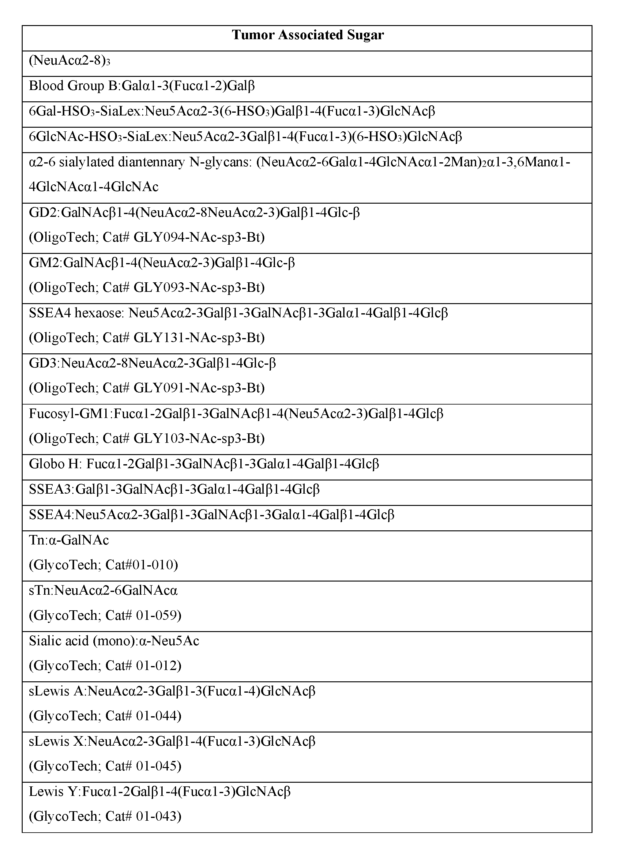


Claims
Priority Applications (9)
| Application Number | Priority Date | Filing Date | Title |
|---|---|---|---|
| AU2017239637A AU2017239637A1 (en) | 2016-03-29 | 2017-03-29 | Antibodies, pharmaceutical compositions and methods |
| EP17776596.3A EP3436482A4 (en) | 2016-03-29 | 2017-03-29 | Antibodies, pharmaceutical compositions and methods |
| JP2018551216A JP2019513144A (en) | 2016-03-29 | 2017-03-29 | Antibody, pharmaceutical composition and method |
| KR1020187030713A KR20180121786A (en) | 2016-03-29 | 2017-03-29 | Antibodies, pharmaceutical compositions and methods |
| BR112018070097-5A BR112018070097A2 (en) | 2016-03-29 | 2017-03-29 | antibody, hybridoma, pharmaceutical composition, method for treating cancer in an individual, method for inhibiting cancer cell proliferation, method for diagnosing cancer in an individual, method for treating a human patient, method for imaging an individual, conjugate of antibody-antibody (adc) method for treating cancer, bispecific antibody and method for preparing a homogeneous antibody population |
| CA3019560A CA3019560A1 (en) | 2016-03-29 | 2017-03-29 | Antibodies, pharmaceutical compositions and methods |
| CN201780033081.7A CN109311995A (en) | 2016-03-29 | 2017-03-29 | Antibody, pharmaceutical composition and method |
| IL261963A IL261963A (en) | 2016-03-29 | 2018-09-26 | Antibodies, pharmaceutical compositions and methods |
| ZA2018/06576A ZA201806576B (en) | 2016-03-29 | 2018-10-03 | Antibodies, pharmaceutical compositions and methods |
Applications Claiming Priority (2)
| Application Number | Priority Date | Filing Date | Title |
|---|---|---|---|
| US201662314841P | 2016-03-29 | 2016-03-29 | |
| US62/314,841 | 2016-03-29 |
Publications (1)
| Publication Number | Publication Date |
|---|---|
| WO2017172990A1 true WO2017172990A1 (en) | 2017-10-05 |
Family
ID=59960245
Family Applications (1)
| Application Number | Title | Priority Date | Filing Date |
|---|---|---|---|
| PCT/US2017/024853 WO2017172990A1 (en) | 2016-03-29 | 2017-03-29 | Antibodies, pharmaceutical compositions and methods |
Country Status (13)
| Country | Link |
|---|---|
| US (1) | US11041017B2 (en) |
| EP (1) | EP3436482A4 (en) |
| JP (1) | JP2019513144A (en) |
| KR (1) | KR20180121786A (en) |
| CN (1) | CN109311995A (en) |
| AR (1) | AR108005A1 (en) |
| AU (1) | AU2017239637A1 (en) |
| BR (1) | BR112018070097A2 (en) |
| CA (1) | CA3019560A1 (en) |
| IL (1) | IL261963A (en) |
| TW (1) | TWI780045B (en) |
| WO (1) | WO2017172990A1 (en) |
| ZA (1) | ZA201806576B (en) |
Cited By (10)
| Publication number | Priority date | Publication date | Assignee | Title |
|---|---|---|---|---|
| EP3541414A4 (en) * | 2016-11-21 | 2020-11-11 | OBI Pharma, Inc. | Conjugated biological molecules, pharmaceutical compositions and methods |
| CN112312924A (en) * | 2018-06-01 | 2021-02-02 | 台湾浩鼎生技股份有限公司 | Combination therapy by using anti-Globo H or anti-SSEA-4 antibodies with anti-negative immune checkpoint antibodies |
| US10935544B2 (en) | 2015-09-04 | 2021-03-02 | Obi Pharma, Inc. | Glycan arrays and method of use |
| US10980894B2 (en) | 2016-03-29 | 2021-04-20 | Obi Pharma, Inc. | Antibodies, pharmaceutical compositions and methods |
| US11041017B2 (en) | 2016-03-29 | 2021-06-22 | Obi Pharma, Inc. | Antibodies, pharmaceutical compositions and methods |
| JP2021517450A (en) * | 2018-03-20 | 2021-07-26 | チョウ ファーマ ユーエスエー インコーポレイテッド | Chimeric antigen receptor that binds to SSEA4 and its use |
| US11203645B2 (en) | 2018-06-27 | 2021-12-21 | Obi Pharma, Inc. | Glycosynthase variants for glycoprotein engineering and methods of use |
| US11583577B2 (en) | 2016-04-22 | 2023-02-21 | Obi Pharma, Inc. | Cancer immunotherapy by immune activation or immune modulation via Globo series antigens |
| US11643456B2 (en) | 2016-07-29 | 2023-05-09 | Obi Pharma, Inc. | Human antibodies, pharmaceutical compositions and methods |
| US11642400B2 (en) | 2016-07-27 | 2023-05-09 | Obi Pharma, Inc. | Immunogenic/therapeutic glycan compositions and uses thereof |
Families Citing this family (15)
| Publication number | Priority date | Publication date | Assignee | Title |
|---|---|---|---|---|
| US10087236B2 (en) | 2009-12-02 | 2018-10-02 | Academia Sinica | Methods for modifying human antibodies by glycan engineering |
| US11377485B2 (en) | 2009-12-02 | 2022-07-05 | Academia Sinica | Methods for modifying human antibodies by glycan engineering |
| US9782476B2 (en) | 2013-09-06 | 2017-10-10 | Academia Sinica | Human iNKT cell activation using glycolipids with altered glycosyl groups |
| WO2015109180A2 (en) * | 2014-01-16 | 2015-07-23 | Academia Sinica | Compositions and methods for treatment and detection of cancers |
| JP6894239B2 (en) | 2014-05-27 | 2021-06-30 | アカデミア シニカAcademia Sinica | Compositions and methods for universal glycoforms for enhanced antibody efficacy |
| CA2950440A1 (en) | 2014-05-27 | 2015-12-03 | Academia Sinica | Anti-her2 glycoantibodies and uses thereof |
| CA2950415A1 (en) | 2014-05-27 | 2015-12-03 | Academia Sinica | Anti-cd20 glycoantibodies and uses thereof |
| TWI732738B (en) | 2014-05-28 | 2021-07-11 | 中央研究院 | Anti-tnf-alpha glycoantibodies and uses thereof |
| AU2017316663B2 (en) | 2016-08-22 | 2024-02-22 | CHO Pharma Inc. | Antibodies, binding fragments, and methods of use |
| CN111565738B (en) * | 2018-02-28 | 2023-12-26 | 圆祥生技股份有限公司 | Bifunctional proteins that bind checkpoint inhibitors as targeted therapies |
| US10688182B2 (en) * | 2018-03-29 | 2020-06-23 | Cho Pharma Usa, Inc. | Monoclonal antibodies that bind to SSEA4 and uses thereof |
| AU2020216780A1 (en) * | 2019-01-28 | 2021-07-22 | Multitude Inc. | Antibodies specific to CD44 |
| CN114901699A (en) * | 2019-09-11 | 2022-08-12 | 英凯尔生物科技有限责任公司 | Epitopes of anti-Serine Protease Inhibitor KAZAL (SPIK) antibodies |
| TWI811604B (en) * | 2019-12-30 | 2023-08-11 | 台灣浩鼎生技股份有限公司 | Antibodies, pharmaceutical compositions and uses thereof |
| CN111528219B (en) * | 2020-05-13 | 2022-03-15 | 上海市计量测试技术研究院 | Freeze-drying protective agent for T lymphocyte subpopulation counting standard substance and application thereof |
Citations (4)
| Publication number | Priority date | Publication date | Assignee | Title |
|---|---|---|---|---|
| WO2006105152A2 (en) * | 2005-03-31 | 2006-10-05 | Stemnion, Inc. | Amnion-derived cell compositions, methods of making and uses thereof |
| KR20120014238A (en) * | 2009-06-16 | 2012-02-16 | 아카데미아 시니카 | Globo h and related anti-cancer vaccines with novel glycolipid adjuvants |
| US20150316556A1 (en) * | 2014-04-01 | 2015-11-05 | Miltenyi Biotech Gmbh | Ssea4 and st3gal2 as chemotherapeutic drug response biomarkers |
| US20150344551A1 (en) * | 2014-01-16 | 2015-12-03 | Academia Sinica | Compositions and methods for treatment and detection of cancers |
Family Cites Families (154)
| Publication number | Priority date | Publication date | Assignee | Title |
|---|---|---|---|---|
| US3773919A (en) | 1969-10-23 | 1973-11-20 | Du Pont | Polylactide-drug mixtures |
| CU22545A1 (en) | 1994-11-18 | 1999-03-31 | Centro Inmunologia Molecular | OBTAINING A CHEMICAL AND HUMANIZED ANTIBODY AGAINST THE RECEPTOR OF THE EPIDERMAL GROWTH FACTOR FOR DIAGNOSTIC AND THERAPEUTIC USE |
| USRE30985E (en) | 1978-01-01 | 1982-06-29 | Serum-free cell culture media | |
| US4419446A (en) | 1980-12-31 | 1983-12-06 | The United States Of America As Represented By The Department Of Health And Human Services | Recombinant DNA process utilizing a papilloma virus DNA as a vector |
| US4522811A (en) | 1982-07-08 | 1985-06-11 | Syntex (U.S.A.) Inc. | Serial injection of muramyldipeptides and liposomes enhances the anti-infective activity of muramyldipeptides |
| US4601978A (en) | 1982-11-24 | 1986-07-22 | The Regents Of The University Of California | Mammalian metallothionein promoter system |
| US4560655A (en) | 1982-12-16 | 1985-12-24 | Immunex Corporation | Serum-free cell culture medium and process for making same |
| US4657866A (en) | 1982-12-21 | 1987-04-14 | Sudhir Kumar | Serum-free, synthetic, completely chemically defined tissue culture media |
| US4816567A (en) | 1983-04-08 | 1989-03-28 | Genentech, Inc. | Recombinant immunoglobin preparations |
| US4767704A (en) | 1983-10-07 | 1988-08-30 | Columbia University In The City Of New York | Protein-free culture medium |
| US4943533A (en) | 1984-03-01 | 1990-07-24 | The Regents Of The University Of California | Hybrid cell lines that produce monoclonal antibodies to epidermal growth factor receptor |
| US4965199A (en) | 1984-04-20 | 1990-10-23 | Genentech, Inc. | Preparation of functional human factor VIII in mammalian cells using methotrexate based selection |
| US4601903A (en) | 1985-05-01 | 1986-07-22 | The United States Of America As Represented By The Department Of Health And Human Services | Vaccine against Neisseria meningitidis Group B serotype 2 invasive disease |
| GB8516415D0 (en) | 1985-06-28 | 1985-07-31 | Celltech Ltd | Culture of animal cells |
| US4676980A (en) | 1985-09-23 | 1987-06-30 | The United States Of America As Represented By The Secretary Of The Department Of Health And Human Services | Target specific cross-linked heteroantibodies |
| US4927762A (en) | 1986-04-01 | 1990-05-22 | Cell Enterprises, Inc. | Cell culture medium with antioxidant |
| US5811128A (en) | 1986-10-24 | 1998-09-22 | Southern Research Institute | Method for oral or rectal delivery of microencapsulated vaccines and compositions therefor |
| IL85035A0 (en) | 1987-01-08 | 1988-06-30 | Int Genetic Eng | Polynucleotide molecule,a chimeric antibody with specificity for human b cell surface antigen,a process for the preparation and methods utilizing the same |
| US4849222A (en) | 1987-03-24 | 1989-07-18 | The Procter & Gamble Company | Mixtures for treating hypercholesterolemia |
| US5057540A (en) | 1987-05-29 | 1991-10-15 | Cambridge Biotech Corporation | Saponin adjuvant |
| US4975278A (en) | 1988-02-26 | 1990-12-04 | Bristol-Myers Company | Antibody-enzyme conjugates in combination with prodrugs for the delivery of cytotoxic agents to tumor cells |
| US5004697A (en) | 1987-08-17 | 1991-04-02 | Univ. Of Ca | Cationized antibodies for delivery through the blood-brain barrier |
| JP2670680B2 (en) | 1988-02-24 | 1997-10-29 | 株式会社ビーエムジー | Polylactic acid microspheres containing physiologically active substance and method for producing the same |
| WO1990003430A1 (en) | 1988-09-23 | 1990-04-05 | Cetus Corporation | Cell culture medium for enhanced cell growth, culture longevity and product expression |
| NZ230747A (en) | 1988-09-30 | 1992-05-26 | Bror Morein | Immunomodulating matrix comprising a complex of at least one lipid and at least one saponin; certain glycosylated triterpenoid saponins derived from quillaja saponaria molina |
| GB8823869D0 (en) | 1988-10-12 | 1988-11-16 | Medical Res Council | Production of antibodies |
| DE3920358A1 (en) | 1989-06-22 | 1991-01-17 | Behringwerke Ag | BISPECIFIC AND OLIGO-SPECIFIC, MONO- AND OLIGOVALENT ANTI-BODY CONSTRUCTS, THEIR PRODUCTION AND USE |
| EP0479909B1 (en) | 1989-06-29 | 1996-10-30 | Medarex, Inc. | Bispecific reagents for aids therapy |
| JP2975679B2 (en) | 1989-09-08 | 1999-11-10 | ザ・ジョーンズ・ホプキンス・ユニバーシティ | Structural change of EGF receptor gene in human glioma |
| DE69133566T2 (en) | 1990-01-12 | 2007-12-06 | Amgen Fremont Inc. | Formation of xenogenic antibodies |
| US5061620A (en) | 1990-03-30 | 1991-10-29 | Systemix, Inc. | Human hematopoietic stem cell |
| US5268164A (en) | 1990-04-23 | 1993-12-07 | Alkermes, Inc. | Increasing blood-brain barrier permeability with permeabilizer peptides |
| US5112596A (en) | 1990-04-23 | 1992-05-12 | Alkermes, Inc. | Method for increasing blood-brain barrier permeability by administering a bradykinin agonist of blood-brain barrier permeability |
| US5427908A (en) | 1990-05-01 | 1995-06-27 | Affymax Technologies N.V. | Recombinant library screening methods |
| KR0149181B1 (en) | 1990-06-29 | 1998-08-17 | 데이비드 알, 맥지 | Melanin production by transformed microorganisms |
| US5625126A (en) | 1990-08-29 | 1997-04-29 | Genpharm International, Inc. | Transgenic non-human animals for producing heterologous antibodies |
| US5661016A (en) | 1990-08-29 | 1997-08-26 | Genpharm International Inc. | Transgenic non-human animals capable of producing heterologous antibodies of various isotypes |
| JP2938569B2 (en) | 1990-08-29 | 1999-08-23 | ジェンファーム インターナショナル,インコーポレイティド | Method for producing xenogeneic immunoglobulin and transgenic mouse |
| US5545806A (en) | 1990-08-29 | 1996-08-13 | Genpharm International, Inc. | Ransgenic non-human animals for producing heterologous antibodies |
| US5633425A (en) | 1990-08-29 | 1997-05-27 | Genpharm International, Inc. | Transgenic non-human animals capable of producing heterologous antibodies |
| US5122469A (en) | 1990-10-03 | 1992-06-16 | Genentech, Inc. | Method for culturing Chinese hamster ovary cells to improve production of recombinant proteins |
| US5508192A (en) | 1990-11-09 | 1996-04-16 | Board Of Regents, The University Of Texas System | Bacterial host strains for producing proteolytically sensitive polypeptides |
| US5264365A (en) | 1990-11-09 | 1993-11-23 | Board Of Regents, The University Of Texas System | Protease-deficient bacterial strains for production of proteolytically sensitive polypeptides |
| WO1992009690A2 (en) | 1990-12-03 | 1992-06-11 | Genentech, Inc. | Enrichment method for variant proteins with altered binding properties |
| US5571894A (en) | 1991-02-05 | 1996-11-05 | Ciba-Geigy Corporation | Recombinant antibodies specific for a growth factor receptor |
| EP0586515B1 (en) | 1991-04-30 | 1997-09-17 | Eukarion, Inc. | Cationized antibodies against intracellular proteins |
| DE69233254T2 (en) | 1991-06-14 | 2004-09-16 | Genentech, Inc., South San Francisco | Humanized Heregulin antibody |
| GB9114948D0 (en) | 1991-07-11 | 1991-08-28 | Pfizer Ltd | Process for preparing sertraline intermediates |
| CA2116774C (en) | 1991-09-19 | 2003-11-11 | Paul J. Carter | Expression in e. coli antibody fragments having at least a cysteine present as a free thiol. use for the production of bifunctional f(ab') 2 antibodies |
| US5565332A (en) | 1991-09-23 | 1996-10-15 | Medical Research Council | Production of chimeric antibodies - a combinatorial approach |
| EP0605522B1 (en) | 1991-09-23 | 1999-06-23 | Medical Research Council | Methods for the production of humanized antibodies |
| US5587458A (en) | 1991-10-07 | 1996-12-24 | Aronex Pharmaceuticals, Inc. | Anti-erbB-2 antibodies, combinations thereof, and therapeutic and diagnostic uses thereof |
| US5288502A (en) | 1991-10-16 | 1994-02-22 | The University Of Texas System | Preparation and uses of multi-phase microspheres |
| WO1993008829A1 (en) | 1991-11-04 | 1993-05-13 | The Regents Of The University Of California | Compositions that mediate killing of hiv-infected cells |
| US5667988A (en) | 1992-01-27 | 1997-09-16 | The Scripps Research Institute | Methods for producing antibody libraries using universal or randomized immunoglobulin light chains |
| ATE419355T1 (en) | 1992-02-06 | 2009-01-15 | Novartis Vaccines & Diagnostic | MARKER FOR CANCER AND BIOSYNTHETIC BINDING PROTEIN FOR IT |
| US5733743A (en) | 1992-03-24 | 1998-03-31 | Cambridge Antibody Technology Limited | Methods for producing members of specific binding pairs |
| ATE348175T1 (en) | 1992-07-17 | 2007-01-15 | Dana Farber Cancer Inst Inc | METHOD OF INTRACELLULAR BINDING OF TARGETED MOLECULES |
| EP0652775B1 (en) | 1992-07-27 | 2000-04-19 | THE GOVERNMENT OF THE UNITED STATES OF AMERICA as represented by the SECRETARY OF THE DEPARTMENT OF HEALTH AND HUMAN SERVICES | Targeting of liposomes to the blood-brain barrier |
| JPH08500017A (en) | 1992-08-17 | 1996-01-09 | ジェネンテク,インコーポレイテッド | Bispecific immune adhesin |
| WO1994011026A2 (en) | 1992-11-13 | 1994-05-26 | Idec Pharmaceuticals Corporation | Therapeutic application of chimeric and radiolabeled antibodies to human b lymphocyte restricted differentiation antigen for treatment of b cell lymphoma |
| EP0724432B1 (en) | 1993-10-22 | 2002-09-18 | Genentech, Inc. | Methods and compositions for microencapsulation of antigens for use as vaccines |
| US6544952B1 (en) | 1994-03-15 | 2003-04-08 | Sloan-Kettering Institute For Cancer Research | Synthesis of glycoconjugates of the globo-H epitope and uses thereof |
| US5849906A (en) | 1994-09-02 | 1998-12-15 | Cavalieri; Ercole | Antigenic conjugates of polycyclic aromatic hydrocarbons to nucleosides |
| AUPM873294A0 (en) | 1994-10-12 | 1994-11-03 | Csl Limited | Saponin preparations and use thereof in iscoms |
| US5804396A (en) | 1994-10-12 | 1998-09-08 | Sugen, Inc. | Assay for agents active in proliferative disorders |
| US5840523A (en) | 1995-03-01 | 1998-11-24 | Genetech, Inc. | Methods and compositions for secretion of heterologous polypeptides |
| DE69522717T2 (en) | 1995-03-30 | 2002-02-14 | Pfizer | quinazoline derivatives |
| US5641870A (en) | 1995-04-20 | 1997-06-24 | Genentech, Inc. | Low pH hydrophobic interaction chromatography for antibody purification |
| US5869046A (en) | 1995-04-14 | 1999-02-09 | Genentech, Inc. | Altered polypeptides with increased half-life |
| GB9508538D0 (en) | 1995-04-27 | 1995-06-14 | Zeneca Ltd | Quinazoline derivatives |
| GB9508565D0 (en) | 1995-04-27 | 1995-06-14 | Zeneca Ltd | Quiazoline derivative |
| EP0822830B1 (en) | 1995-04-27 | 2008-04-02 | Amgen Fremont Inc. | Human anti-IL-8 antibodies, derived from immunized xenomice |
| WO1996034096A1 (en) | 1995-04-28 | 1996-10-31 | Abgenix, Inc. | Human antibodies derived from immunized xenomice |
| WO1996040210A1 (en) | 1995-06-07 | 1996-12-19 | Imclone Systems Incorporated | Antibody and antibody fragments for inhibiting the growth of tumors |
| US6265150B1 (en) | 1995-06-07 | 2001-07-24 | Becton Dickinson & Company | Phage antibodies |
| WO1997038123A1 (en) | 1996-04-05 | 1997-10-16 | Board Of Regents, The University Of Texas System | Methods for producing soluble, biologically-active disulfide bond-containing eukaryotic proteins in bacterial cells |
| GEP20012442B (en) | 1996-04-12 | 2001-05-25 | Warner Lambert Co | Irreversible Inhibitors of Tyrosine Kinases |
| US6231859B1 (en) | 1996-12-02 | 2001-05-15 | Aquila Biopharmaceuticals, Inc. | Saponin adjuvant compositions |
| ES2301183T3 (en) | 1996-12-03 | 2008-06-16 | Amgen Fremont Inc. | COMPLETELY HUMAN ANTIBODY THAT JOINS THE EGFR RECEIVER. |
| AUPO517897A0 (en) | 1997-02-19 | 1997-04-11 | Csl Limited | Chelating immunostimulating complexes |
| UA73073C2 (en) | 1997-04-03 | 2005-06-15 | Уайт Холдінгз Корпорейшн | Substituted 3-cyan chinolines |
| US6083715A (en) | 1997-06-09 | 2000-07-04 | Board Of Regents, The University Of Texas System | Methods for producing heterologous disulfide bond-containing polypeptides in bacterial cells |
| ZA986732B (en) | 1997-07-29 | 1999-02-02 | Warner Lambert Co | Irreversible inhibitiors of tyrosine kinases |
| ZA986729B (en) | 1997-07-29 | 1999-02-02 | Warner Lambert Co | Irreversible inhibitors of tyrosine kinases |
| TW436485B (en) | 1997-08-01 | 2001-05-28 | American Cyanamid Co | Substituted quinazoline derivatives |
| US7018637B2 (en) | 1998-02-23 | 2006-03-28 | Aventis Pasteur, Inc | Multi-oligosaccharide glycoconjugate bacterial meningitis vaccines |
| AUPP807399A0 (en) | 1999-01-08 | 1999-02-04 | Csl Limited | Improved immunogenic lhrh composition and methods relating thereto |
| EP2204186B1 (en) | 1999-02-17 | 2016-04-06 | CSL Limited | Immunogenic complexes and methods relating thereto |
| DE60031957T2 (en) | 1999-02-17 | 2007-09-13 | Glycominds Ltd. | COMBINATIVE, COMPLEX CARBOHYDRATE LIBRARIES AND METHOD FOR THE PREPARATION AND USE THEREOF |
| US7824687B2 (en) | 1999-08-20 | 2010-11-02 | Sloan-Kettering Institute For Cancer Research | Clustered multi-antigenic carbohydrate constructs, methods for their preparation, and uses thereof |
| US7854934B2 (en) | 1999-08-20 | 2010-12-21 | Sloan-Kettering Institute For Cancer Research | Glycoconjugates, glycoamino acids, intermediates thereto, and uses thereof |
| US6514221B2 (en) | 2000-07-27 | 2003-02-04 | Brigham And Women's Hospital, Inc. | Blood-brain barrier opening |
| AU2001286930A1 (en) | 2000-08-30 | 2002-03-13 | The Board Of Trustees Of The Leland Stanford Junior University | Glucocorticoid blocking agents for increasing blood-brain barrier permeability |
| US7034036B2 (en) | 2000-10-30 | 2006-04-25 | Pain Therapeutics, Inc. | Inhibitors of ABC drug transporters at the blood-brain barrier |
| US20030083299A1 (en) | 2000-11-04 | 2003-05-01 | Ferguson Ian A. | Non-invasive delivery of polypeptides through the blood-brain barrier |
| US20040018194A1 (en) | 2000-11-28 | 2004-01-29 | Francisco Joseph A. | Recombinant anti-CD30 antibodies and uses thereof |
| WO2002086096A2 (en) | 2001-01-23 | 2002-10-31 | University Of Rochester Medical Center | Methods of producing or identifying intrabodies in eukaryotic cells |
| DE10121982B4 (en) | 2001-05-05 | 2008-01-24 | Lts Lohmann Therapie-Systeme Ag | Nanoparticles of protein with coupled apolipoprotein E to overcome the blood-brain barrier and process for their preparation |
| EP2147679B1 (en) | 2001-07-25 | 2014-06-25 | Raptor Pharmaceutical, Inc. | Compositions for blood-brain barrier transport |
| JP2005526692A (en) | 2001-08-14 | 2005-09-08 | ビオミラ,インコーポレーテッド | Immunogenic conjugates of carbohydrate haptens and aggregated protein carriers |
| WO2003043583A2 (en) | 2001-11-20 | 2003-05-30 | Seattle Genetics, Inc. | Treatment of immunological disorders using anti-cd30 antibodies |
| US20030162695A1 (en) | 2002-02-27 | 2003-08-28 | Schatzberg Alan F. | Glucocorticoid blocking agents for increasing blood-brain barrier permeability |
| AU2003219277A1 (en) | 2002-03-14 | 2003-09-29 | Medical Research Council | Intracellular antibodies |
| EP1575514A2 (en) | 2002-07-31 | 2005-09-21 | Seattle Genetics, Inc. | Anti-cd20 antibody-drug conjugates for the treatment of cancer and immune disorders |
| EP1391213A1 (en) | 2002-08-21 | 2004-02-25 | Boehringer Ingelheim International GmbH | Compositions and methods for treating cancer using maytansinoid CD44 antibody immunoconjugates and chemotherapeutic agents |
| WO2004042017A2 (en) | 2002-10-31 | 2004-05-21 | Genentech, Inc. | Methods and compositions for increasing antibody production |
| JP4995423B2 (en) | 2002-12-03 | 2012-08-08 | ブランシェット・ロックフェラー・ニューロサイエンスィズ・インスティテュート | Artificial low density lipoprotein carrier for transporting substances across the blood-brain barrier |
| AU2004205684A1 (en) | 2003-01-23 | 2004-08-05 | Genentech, Inc. | Methods for producing humanized antibodies and improving yield of antibodies or antigen binding fragments in cell culture |
| US20060035267A1 (en) | 2003-04-09 | 2006-02-16 | Livingston Philip O | Optimal polyvalent vaccine for cancer |
| GB0316294D0 (en) | 2003-07-11 | 2003-08-13 | Polytherics Ltd | Conjugated biological molecules and their preparation |
| DE602004028381D1 (en) | 2003-08-25 | 2010-09-09 | Univax Llc | Er of regulator of imprinted sites) moleküls |
| EP1663239A4 (en) | 2003-09-10 | 2008-07-23 | Cedars Sinai Medical Center | Potassium channel mediated delivery of agents through the blood-brain barrier |
| EP1723422A2 (en) | 2004-03-05 | 2006-11-22 | The Scripps Research Institute | High throughput glycan microarrays |
| JP2008506683A (en) | 2004-07-18 | 2008-03-06 | コーリー ファーマシューティカル グループ, リミテッド | Methods and compositions for inducing innate immune responses |
| EP2484374A1 (en) | 2004-07-18 | 2012-08-08 | CSL Limited | Immuno stimulating complex and oligonucleotide formulations for inducing enhanced interferon-gamma responses |
| EA025871B9 (en) | 2005-10-07 | 2017-08-31 | Экселиксис, Инк. | Mek inhibitors and methods of using the same |
| EP2104513B1 (en) * | 2006-11-27 | 2015-05-20 | diaDexus, Inc. | Ovr110 antibody compositions and methods of use |
| WO2009035494A2 (en) | 2007-07-30 | 2009-03-19 | The Scripps Research Institute | Methods for producing anti-glycan antibodies, vaccines and methods for treating cancer or infectious disease |
| CA2701612A1 (en) | 2007-10-05 | 2009-04-09 | Takeda Pharmaceutical Company Limited | Neuromedin u derivative |
| EP2268315B1 (en) | 2008-03-31 | 2019-07-31 | Freie Universität Berlin | Drug conjugates with polyglycerols |
| KR101825315B1 (en) | 2008-04-08 | 2018-02-02 | 슬로안-케테링인스티튜트퍼캔서리서치 | Triterpene saponins, methods of synthesis, and uses thereof |
| CN102065868A (en) | 2008-06-16 | 2011-05-18 | 中央研究院 | Compositions for inducing immune responses specific to Globo h and SSEA3 and uses thereof in cancer treatment |
| EP2347769A1 (en) | 2010-01-20 | 2011-07-27 | Glycotope GmbH | Cancer stem cell markers and uses thereof |
| CA2801922A1 (en) | 2010-06-11 | 2011-12-15 | Sloan-Kettering Institute For Cancer Research | Multivalent glycopeptide constructs and uses thereof |
| US9175326B2 (en) | 2011-03-03 | 2015-11-03 | University Of Maryland, Baltimore | Transglycosylation activity of glycosynthase mutants of an endo-beta-N-acetylglucosaminidase (endo-D) from streptococcus pneumoniae |
| EP2500035A1 (en) | 2011-03-15 | 2012-09-19 | Icon Genetics GmbH | Pharmaceutical formulation containing immunglobulin |
| BR112013031819B1 (en) | 2011-06-10 | 2022-05-03 | Mersana Therapeutics, Inc | Polymeric support, pharmaceutical composition, compound and support use |
| JP6282232B2 (en) | 2012-02-10 | 2018-02-21 | ユニバーシティー オブ メリーランド,ボルティモア | Enzymatic chemical glycosylation of antibodies and Fc fragments thereof |
| JP2015117181A (en) | 2012-02-22 | 2015-06-25 | 学校法人 城西大学 | Sepsis therapeutic agent |
| CN104520324A (en) * | 2012-02-24 | 2015-04-15 | 施特姆森特Rx股份有限公司 | DLL3 modulators and methods of use |
| PT2825156T (en) | 2012-03-16 | 2017-11-02 | Merck Patent Gmbh | Targeting aminoacid lipids |
| KR101806370B1 (en) | 2013-01-04 | 2017-12-07 | 오비아이 파머 인코퍼레이티드 | Vaccines with higher carbohydrate antigen density and novel saponin adjuvant |
| US10307471B2 (en) | 2013-05-02 | 2019-06-04 | National Institute Of Advanced Industrial Science And Technology | Immunity inducer for saccharide antigens |
| ES2914176T3 (en) | 2013-09-17 | 2022-06-07 | Obi Pharma Inc | Compositions of a carbohydrate vaccine to induce immune responses and uses thereof in the treatment of cancer |
| US10150818B2 (en) | 2014-01-16 | 2018-12-11 | Academia Sinica | Compositions and methods for treatment and detection of cancers |
| EP3593812A3 (en) | 2014-03-15 | 2020-05-27 | Novartis AG | Treatment of cancer using chimeric antigen receptor |
| EP3119432B1 (en) | 2014-03-19 | 2020-01-08 | MacKay Medical Foundation the Presbyterian Church in Taiwan MacKay Memorial Hospital | Antibodies against immunogenic glycopeptides, composition comprising the same and use thereof |
| CA2941029C (en) | 2014-04-10 | 2021-02-16 | Obi Pharma Inc. | Antibodies, pharmaceutical compositions and uses thereof |
| ES2767399T3 (en) * | 2014-08-19 | 2020-06-17 | Miltenyi Biotec Bv & Co Kg | Chimeric antigen receptor specific for the SSEA4 antigen |
| RU2720295C2 (en) | 2014-09-15 | 2020-04-28 | Оби Фарма, Инк. | Compositions of immunogenic/therapeutic glycoconjugates and use thereof |
| CN107430127B (en) | 2015-01-24 | 2020-08-28 | 中央研究院 | Cancer markers and methods of use thereof |
| CA2973886A1 (en) | 2015-01-30 | 2016-08-04 | Academia Sinica | Compositions and methods relating to universal glycoforms for enhanced antibody efficacy |
| SG10201914047RA (en) | 2015-07-16 | 2020-03-30 | Daiichi Sankyo Co Ltd | Novel endos mutant enzyme |
| AR105910A1 (en) | 2015-09-04 | 2017-11-22 | Obi Pharma Inc | GLICAN MATRICES AND METHOD OF USE |
| WO2017062792A1 (en) | 2015-10-07 | 2017-04-13 | Obi Pharma, Inc. | Novel carbohydrate antibodies, pharmaceutical compositions and uses thereof |
| US10980894B2 (en) | 2016-03-29 | 2021-04-20 | Obi Pharma, Inc. | Antibodies, pharmaceutical compositions and methods |
| WO2017172990A1 (en) | 2016-03-29 | 2017-10-05 | Obi Pharma, Inc. | Antibodies, pharmaceutical compositions and methods |
| SG11201809024UA (en) | 2016-04-22 | 2018-11-29 | Obi Pharma Inc | Cancer immunotherapy by immune activation or immune modulation via globo series antigens |
| GB201611535D0 (en) | 2016-07-01 | 2016-08-17 | King's College London | Methods and compositions for treating cancer with siglec-9 activity modulators |
| WO2018022933A1 (en) | 2016-07-27 | 2018-02-01 | Obi Pharma, Inc. | Immunogenic/therapeutic glycan compositions and uses thereof |
| TWI786054B (en) | 2016-07-29 | 2022-12-11 | 台灣浩鼎生技股份有限公司 | Human antibodies, pharmaceutical compositions and methods |
| AU2017316663B2 (en) | 2016-08-22 | 2024-02-22 | CHO Pharma Inc. | Antibodies, binding fragments, and methods of use |
| CA3044274A1 (en) | 2016-11-21 | 2018-05-24 | Obi Pharma, Inc. | Conjugated biological molecules, pharmaceutical compositions and methods |
-
2017
- 2017-03-29 WO PCT/US2017/024853 patent/WO2017172990A1/en active Application Filing
- 2017-03-29 TW TW106110636A patent/TWI780045B/en active
- 2017-03-29 KR KR1020187030713A patent/KR20180121786A/en unknown
- 2017-03-29 CN CN201780033081.7A patent/CN109311995A/en active Pending
- 2017-03-29 AU AU2017239637A patent/AU2017239637A1/en not_active Abandoned
- 2017-03-29 US US15/473,267 patent/US11041017B2/en active Active
- 2017-03-29 EP EP17776596.3A patent/EP3436482A4/en not_active Withdrawn
- 2017-03-29 BR BR112018070097-5A patent/BR112018070097A2/en not_active IP Right Cessation
- 2017-03-29 JP JP2018551216A patent/JP2019513144A/en active Pending
- 2017-03-29 CA CA3019560A patent/CA3019560A1/en not_active Abandoned
- 2017-03-30 AR ARP170100781A patent/AR108005A1/en unknown
-
2018
- 2018-09-26 IL IL261963A patent/IL261963A/en unknown
- 2018-10-03 ZA ZA2018/06576A patent/ZA201806576B/en unknown
Patent Citations (4)
| Publication number | Priority date | Publication date | Assignee | Title |
|---|---|---|---|---|
| WO2006105152A2 (en) * | 2005-03-31 | 2006-10-05 | Stemnion, Inc. | Amnion-derived cell compositions, methods of making and uses thereof |
| KR20120014238A (en) * | 2009-06-16 | 2012-02-16 | 아카데미아 시니카 | Globo h and related anti-cancer vaccines with novel glycolipid adjuvants |
| US20150344551A1 (en) * | 2014-01-16 | 2015-12-03 | Academia Sinica | Compositions and methods for treatment and detection of cancers |
| US20150316556A1 (en) * | 2014-04-01 | 2015-11-05 | Miltenyi Biotech Gmbh | Ssea4 and st3gal2 as chemotherapeutic drug response biomarkers |
Non-Patent Citations (2)
| Title |
|---|
| LOU ET AL.: "Stage-specific embryonic antigen-4 as a potential therapeutic target in glioblastoma multiforme and other cancers", PROCEEDINGS OF THE NATIONAL ACADEMY OF SCIENCES, vol. 111, 18 February 2014 (2014-02-18), pages 2482 - 2487, XP055218264 * |
| See also references of EP3436482A4 * |
Cited By (14)
| Publication number | Priority date | Publication date | Assignee | Title |
|---|---|---|---|---|
| US10935544B2 (en) | 2015-09-04 | 2021-03-02 | Obi Pharma, Inc. | Glycan arrays and method of use |
| US11041017B2 (en) | 2016-03-29 | 2021-06-22 | Obi Pharma, Inc. | Antibodies, pharmaceutical compositions and methods |
| US11833223B2 (en) | 2016-03-29 | 2023-12-05 | Obi Pharma, Inc. | Antibodies, pharmaceutical compositions and methods |
| US10980894B2 (en) | 2016-03-29 | 2021-04-20 | Obi Pharma, Inc. | Antibodies, pharmaceutical compositions and methods |
| US11583577B2 (en) | 2016-04-22 | 2023-02-21 | Obi Pharma, Inc. | Cancer immunotherapy by immune activation or immune modulation via Globo series antigens |
| US11642400B2 (en) | 2016-07-27 | 2023-05-09 | Obi Pharma, Inc. | Immunogenic/therapeutic glycan compositions and uses thereof |
| US11643456B2 (en) | 2016-07-29 | 2023-05-09 | Obi Pharma, Inc. | Human antibodies, pharmaceutical compositions and methods |
| US11000601B2 (en) | 2016-11-21 | 2021-05-11 | Obi Pharma, Inc. | Conjugated biological molecules, pharmaceutical compositions and methods |
| EP3541414A4 (en) * | 2016-11-21 | 2020-11-11 | OBI Pharma, Inc. | Conjugated biological molecules, pharmaceutical compositions and methods |
| JP2021517450A (en) * | 2018-03-20 | 2021-07-26 | チョウ ファーマ ユーエスエー インコーポレイテッド | Chimeric antigen receptor that binds to SSEA4 and its use |
| JP7378152B2 (en) | 2018-03-20 | 2023-11-13 | チョウ ファーマ ユーエスエー インコーポレイテッド | Chimeric antigen receptor binding to SSEA4 and its use |
| EP3801607A4 (en) * | 2018-06-01 | 2022-03-16 | OBI Pharma, Inc. | Combination therapy by using anti-globo h or anti-ssea-4 antibody with anti-negative immune check points antibody |
| CN112312924A (en) * | 2018-06-01 | 2021-02-02 | 台湾浩鼎生技股份有限公司 | Combination therapy by using anti-Globo H or anti-SSEA-4 antibodies with anti-negative immune checkpoint antibodies |
| US11203645B2 (en) | 2018-06-27 | 2021-12-21 | Obi Pharma, Inc. | Glycosynthase variants for glycoprotein engineering and methods of use |
Also Published As
| Publication number | Publication date |
|---|---|
| ZA201806576B (en) | 2020-01-29 |
| TWI780045B (en) | 2022-10-11 |
| BR112018070097A2 (en) | 2019-02-12 |
| KR20180121786A (en) | 2018-11-08 |
| EP3436482A4 (en) | 2020-03-11 |
| US11041017B2 (en) | 2021-06-22 |
| US20170283488A1 (en) | 2017-10-05 |
| TW201802116A (en) | 2018-01-16 |
| EP3436482A1 (en) | 2019-02-06 |
| AU2017239637A1 (en) | 2018-11-15 |
| IL261963A (en) | 2018-11-29 |
| CA3019560A1 (en) | 2017-10-05 |
| JP2019513144A (en) | 2019-05-23 |
| CN109311995A (en) | 2019-02-05 |
| AR108005A1 (en) | 2018-07-04 |
Similar Documents
| Publication | Publication Date | Title |
|---|---|---|
| US11041017B2 (en) | Antibodies, pharmaceutical compositions and methods | |
| US11833223B2 (en) | Antibodies, pharmaceutical compositions and methods | |
| US20190177435A1 (en) | Compositions and methods for treatment and detection of cancers | |
| US20190085062A1 (en) | Compositions and methods for treatment and detection of cancers | |
| EP3245225B1 (en) | Compositions and methods for treatment and detection of cancers | |
| US9975965B2 (en) | Compositions and methods for treatment and detection of cancers | |
| US10118969B2 (en) | Compositions and methods relating to universal glycoforms for enhanced antibody efficacy | |
| TWI717333B (en) | Compositions and methods relating to universal glyoforms for enhanced antibody efficacy | |
| US20190389963A1 (en) | Combination therapy by using anti-globo h or anti-ssea-4 antibody with anti-negative immune check points antibody | |
| WO2016114819A1 (en) | Compositions and methods for treatment and detection of cancers |
Legal Events
| Date | Code | Title | Description |
|---|---|---|---|
| ENP | Entry into the national phase |
Ref document number: 2018551216 Country of ref document: JP Kind code of ref document: A Ref document number: 3019560 Country of ref document: CA |
|
| NENP | Non-entry into the national phase |
Ref country code: DE |
|
| REG | Reference to national code |
Ref country code: BR Ref legal event code: B01A Ref document number: 112018070097 Country of ref document: BR |
|
| ENP | Entry into the national phase |
Ref document number: 20187030713 Country of ref document: KR Kind code of ref document: A |
|
| WWE | Wipo information: entry into national phase |
Ref document number: 2017776596 Country of ref document: EP |
|
| ENP | Entry into the national phase |
Ref document number: 2017776596 Country of ref document: EP Effective date: 20181029 |
|
| ENP | Entry into the national phase |
Ref document number: 2017239637 Country of ref document: AU Date of ref document: 20170329 Kind code of ref document: A |
|
| 121 | Ep: the epo has been informed by wipo that ep was designated in this application |
Ref document number: 17776596 Country of ref document: EP Kind code of ref document: A1 |
|
| ENP | Entry into the national phase |
Ref document number: 112018070097 Country of ref document: BR Kind code of ref document: A2 Effective date: 20180928 |