KR20220041154A - YAP inhibition for wound healing - Google Patents
YAP inhibition for wound healing Download PDFInfo
- Publication number
- KR20220041154A KR20220041154A KR1020227006337A KR20227006337A KR20220041154A KR 20220041154 A KR20220041154 A KR 20220041154A KR 1020227006337 A KR1020227006337 A KR 1020227006337A KR 20227006337 A KR20227006337 A KR 20227006337A KR 20220041154 A KR20220041154 A KR 20220041154A
- Authority
- KR
- South Korea
- Prior art keywords
- wound
- enf
- days
- skin
- yap
- Prior art date
Links
Images


Classifications
-
- A—HUMAN NECESSITIES
- A61—MEDICAL OR VETERINARY SCIENCE; HYGIENE
- A61K—PREPARATIONS FOR MEDICAL, DENTAL OR TOILETRY PURPOSES
- A61K31/00—Medicinal preparations containing organic active ingredients
- A61K31/33—Heterocyclic compounds
- A61K31/395—Heterocyclic compounds having nitrogen as a ring hetero atom, e.g. guanethidine or rifamycins
- A61K31/40—Heterocyclic compounds having nitrogen as a ring hetero atom, e.g. guanethidine or rifamycins having five-membered rings with one nitrogen as the only ring hetero atom, e.g. sulpiride, succinimide, tolmetin, buflomedil
- A61K31/409—Heterocyclic compounds having nitrogen as a ring hetero atom, e.g. guanethidine or rifamycins having five-membered rings with one nitrogen as the only ring hetero atom, e.g. sulpiride, succinimide, tolmetin, buflomedil having four such rings, e.g. porphine derivatives, bilirubin, biliverdine
-
- A—HUMAN NECESSITIES
- A61—MEDICAL OR VETERINARY SCIENCE; HYGIENE
- A61P—SPECIFIC THERAPEUTIC ACTIVITY OF CHEMICAL COMPOUNDS OR MEDICINAL PREPARATIONS
- A61P17/00—Drugs for dermatological disorders
- A61P17/02—Drugs for dermatological disorders for treating wounds, ulcers, burns, scars, keloids, or the like
-
- A—HUMAN NECESSITIES
- A61—MEDICAL OR VETERINARY SCIENCE; HYGIENE
- A61P—SPECIFIC THERAPEUTIC ACTIVITY OF CHEMICAL COMPOUNDS OR MEDICINAL PREPARATIONS
- A61P17/00—Drugs for dermatological disorders
- A61P17/14—Drugs for dermatological disorders for baldness or alopecia
Landscapes
- Health & Medical Sciences (AREA)
- Veterinary Medicine (AREA)
- Pharmacology & Pharmacy (AREA)
- Life Sciences & Earth Sciences (AREA)
- General Health & Medical Sciences (AREA)
- Animal Behavior & Ethology (AREA)
- Public Health (AREA)
- Medicinal Chemistry (AREA)
- Chemical & Material Sciences (AREA)
- Chemical Kinetics & Catalysis (AREA)
- Organic Chemistry (AREA)
- Dermatology (AREA)
- Bioinformatics & Cheminformatics (AREA)
- Nuclear Medicine, Radiotherapy & Molecular Imaging (AREA)
- General Chemical & Material Sciences (AREA)
- Engineering & Computer Science (AREA)
- Gastroenterology & Hepatology (AREA)
- Epidemiology (AREA)
- Pharmaceuticals Containing Other Organic And Inorganic Compounds (AREA)
- Medicines That Contain Protein Lipid Enzymes And Other Medicines (AREA)
- Medicines Containing Material From Animals Or Micro-Organisms (AREA)
- Cosmetics (AREA)
Abstract
대상체의 진피 부위에서 상처의 치유를 촉진하는 방법이 제공된다. 상기 방법의 양태는 상처의 ENF-매개 치유를 촉진하기 위해 상처에 인그레일드-1 계통-음성 섬유아세포(ENF)의 기계적 활성화를 조절하기 위해 상처에 유효량의 YAP 억제제 조성물을 투여하는 단계를 포함할 수 있다. 또한, 대상체에서 상처의 치유 동안 흉터화를 예방하는 방법 및 대상체에서 모발 성장을 촉진하는 방법이 제공된다. 상기 방법의 양태는 대상체의 진피 부위에 상처를 형성하는 단계 및 상처의 ENF-매개 치유를 촉진하기 위해 상처에서 인그레일드-1 계통-음성 섬유아세포(ENF)의 기계적 활성화를 조절하기 위해 상처에 유효량의 YAP 억제제 조성물을 투여하는 단계를 포함할 수 있다. 또한, 일정량의 YAP 억제제 조성물 및 조직 파괴 장치를 포함하는 키트가 제공된다.A method of promoting healing of a wound in a dermal region of a subject is provided. Aspects of the method include administering to the wound an effective amount of a YAP inhibitor composition to modulate the mechanical activation of Ingrade -1 lineage-negative fibroblasts (ENFs) in the wound to promote ENF-mediated healing of the wound. can do. Also provided are methods of preventing scarring during healing of a wound in a subject and methods of promoting hair growth in a subject. Aspects of the method include forming a wound in a dermal region of a subject and administering a wound to a wound to modulate mechanical activation of Ingrade -1 lineage-negative fibroblasts (ENFs) in the wound to promote ENF-mediated healing of the wound. administering an effective amount of a YAP inhibitor composition. Also provided is a kit comprising an amount of a YAP inhibitor composition and a tissue disruption device.
Description
정부 권리 인정Recognition of Government Rights
본 발명은 국립 보건원(National Institutes of Health)이 수여한 계약 GM1 16892에 따라 정부 지원으로 이루어졌다. 정부는 본 발명에 대한 특정 권리를 갖는다.This invention was made with government support under contract GM1 16892 awarded by the National Institutes of Health. The government has certain rights in this invention.
관련 출원의 교차 참조Cross-reference to related applications
35 U.S.C. §119(e)에 따라, 본 출원은 2019년 7월 26일자로 출원된 미국 임시 특허 출원 제62/879369호의 출원일에 대한 우선권을 주장하며; 이의 개시내용은 본원에 인용되어 포함된다.35 U.S.C. Pursuant to §119(e), this application claims priority to the filing date of U.S. Provisional Patent Application No. 62/879369, filed July 26, 2019; The disclosures of which are incorporated herein by reference.
피부는 여러 층으로 구성된 신체에서 가장 큰 기관으로서 생물학적 항상성에서 중요한 역할을 한다. 피부는 열 조절, 대사 기능(비타민 D 대사), 및 면역 기능을 포함하여 다양한 기능을 가지고 있다. 포유류 피부는 2개의 주요 층인 표피와 진피를 포함한다. 표피는 피부의 가장 바깥쪽 층이며 환경에 대한 보호 장벽 역할을 한다. 진피는 표피 아래의 피부 층이며 예컨대 모낭, 땀샘, 피지선, 아포크린 샘, 림프관 및 혈관을 포함하는 피부 부속기의 위치를 제공한다. 진피는 구조 단백질(콜라겐 및 엘라스틴), 특수 단백질(피브릴린, 피브로넥틴, 및 라미닌), 및 프로테오글리칸으로 구성된 세포외 기질 또는 결합 조직을 통해 피부에 강도 및 탄력을 제공한다. 표피 및 진피는 얇은 섬유질 세포외 기질인 기저막에 의해 분리된다.The skin is the largest organ in the body, composed of many layers, and plays an important role in biological homeostasis. The skin has a variety of functions, including heat regulation, metabolic function (vitamin D metabolism), and immune function. Mammalian skin contains two major layers, the epidermis and the dermis. The epidermis is the outermost layer of the skin and serves as a protective barrier to the environment. The dermis is the layer of skin below the epidermis and provides the location of skin appendages including, for example, hair follicles, sweat glands, sebaceous glands, apocrine glands, lymphatic vessels and blood vessels. The dermis provides strength and elasticity to the skin through an extracellular matrix or connective tissue composed of structural proteins (collagen and elastin), specialized proteins (fibrillin, fibronectin, and laminin), and proteoglycans. The epidermis and dermis are separated by a basement membrane, a thin fibrous extracellular matrix.
모발은 진피에 존재하는 모낭에서 자라는 단백질 필라멘트이다. 모발은 포유류를 다른 유기체 부류와 구별하는 주요 차이점이다. 모발은 추위와 자외선으로부터 보호할 수 있고, 먼지와 땀으로부터 장기를 보호할 수 있고, 감각 기능을 제공할 수 있다. 각 모발은 모간(hair shaft)과 모낭의 2개의 개별 구조로 이루어져 있다. 모간은 피부 외부에 보이는 부분을 포함한다. 모낭은 모발이 자랄 수 있는 기관이며 호르몬, 신경펩티드 및 면역 세포간의 복잡한 상호작용을 통해 모발 성장을 조절한다. 모낭의 조직학적 배열은 외부 모근초(root sheath)와 내부 모근초로 나누어진다. 탈모는 전 세계적으로 수십 억 명의 사람들에게 영향을 미치는 매우 일반적인 문제이다. 예를 들어, 안드로겐성 탈모, 또는 남성형 탈모는 50세까지 남성의 90% 초과 및 65세까지 여성의 50% 초과에 영향을 미치는 것으로 추정된다. 탈모는 피부 흉터(예컨대, 기계적 손상 또는 화상 후) 또는 자가면역 상태(예컨대, 원형 탈모)의 결과로서 발생할 수 있다.Hair is a protein filament that grows from hair follicles in the dermis. Hair is the main difference that distinguishes mammals from other classes of organisms. Hair can protect against cold and UV rays, protect organs from dust and sweat, and provide sensory functions. Each hair consists of two separate structures: a hair shaft and a hair follicle. The hair shaft contains the visible part of the skin. Hair follicles are the organs from which hair can grow and regulate hair growth through complex interactions between hormones, neuropeptides and immune cells. The histological arrangement of hair follicles is divided into an external root sheath and an internal root sheath. Hair loss is a very common problem that affects billions of people worldwide. For example, androgenetic alopecia, or androgenetic alopecia, is estimated to affect more than 90% of men by the age of 50 and more than 50% of women by the age of 65. Hair loss can occur as a result of skin scarring (eg, after mechanical damage or burns) or an autoimmune condition (eg, alopecia areata).
상처 치유 또는 조직 치유는 조직 재생을 수반하는 생물학적 과정이다. 치유 과정 동안, 손상되거나 파괴된 조직은 살아있는 조직으로 대체된다. 피부 장벽이 무너지면, 조절된 일련의 생화학적 현상이 활성화되어 손상을 복구한다. 이러한 과정은 예컨대 성장 인자, 사이토카인, 및 케모카인을 포함하는 수많은 생물학적 구성성분에 의해 조절되며, 예컨대 가용성 매개체, 혈액 세포, 세포외 기질 성분, 및 실질 세포를 포함하여 몇몇 성분을 사용한다. 상처 치유는 일반적으로 여러 단계를 통해 진행된다. 이러한 과정은 지혈, 염증, 증식, 및 리모델링을 포함하는 여러 단계로 나누어진다. 상처 치유의 종점은 흉터 형성을 포함할 수 있다. 피부 상처는 섬유성 흉터 조직이 발달하여 언제나(invariably) 치유되며, 이는 변형, 성장 제한, 및 영구적인 기능 상실을 초래할 수 있다. 예컨대 "정상" 잔주름 및 광범위한 흉터(widespread scar), 위축 흉터(atrophic scar), 흉터 구축(scar contracture), 비후성 흉터(hypertrophic scar), 및 켈로이드 흉터(hypertrophic scar)를 포함하는 비정상적인 흉터를 비롯한 다양한 유형의 흉터가 피부 조직 복구 후에 형성될 수 있다.Wound healing or tissue healing is a biological process that involves tissue regeneration. During the healing process, damaged or destroyed tissue is replaced with living tissue. When the skin barrier breaks down, a regulated chain of biochemical events is activated to repair the damage. This process is regulated by numerous biological components, including, for example, growth factors, cytokines, and chemokines, and uses several components, including, for example, soluble mediators, blood cells, extracellular matrix components, and parenchymal cells. Wound healing usually proceeds through several stages. This process is divided into several stages including hemostasis, inflammation, proliferation, and remodeling. The endpoint of wound healing may include scar formation. Skin wounds heal invariably with the development of fibrous scar tissue, which can lead to deformation, growth restriction, and permanent loss of function. Various types including, for example, "normal" fine lines and abnormal scars, including widespread scars, atrophic scars, scar contractures, hypertrophic scars, and keloid scars of scars can form after skin tissue repair.
흉터로 이어지는 섬유화 과정을 성공적으로 예방하거나 역전시키는 치료 전략은 현재 존재하지 않는다. 흉터를 줄이려는 시도는 종종 섬유성으로 알려진 세포 집단의 절제를 수반하지만, 이러한 접근법은 적절한 치유에 필요한 세포를 비특이적으로 제거함으로써 상처 회복을 손상시키거나 지연시킬 수 있다. 피부 재생 - 다음과 같이 정상 피부의 3가지 특징의 회복으로 정의됨: 1) 2차 요소(예컨대, 피부 부속기), 2) ECM 구조, 및 3) 기계적 강도-은 달성되지 않았다.No treatment strategy currently exists to successfully prevent or reverse the fibrotic process leading to scarring. Attempts to reduce scarring often involve excision of a population of cells known as fibrous, but this approach can impair or delay wound healing by non-specifically removing cells necessary for proper healing. Skin regeneration—defined as the restoration of three characteristics of normal skin as follows: 1) secondary components (eg, skin appendages), 2) ECM structure, and 3) mechanical strength—was not achieved.
또한, 피부의 모발-성장 잠재력을 회복시키는 효과적인 치료법이 존재하지 않는다. 특히, 어떠한 표적화된 분자 제제도 모낭 재생을 유도할 수 있는 것으로 입증되지 않았다. 가장 효과적인 기존 치료법은 일반적으로 모발-성장 피부를 탈모증의 영향을 받는 부위에 이식하는 것을 수반하는데, 이러한 접근법은 이식가능한 조직의 이용가능성, 기증자 부위 이환율, 및 비용에 의해 제한된다. 탈모의 영향을 받는 부위에서 새로운 내인성 모낭의 재생을 성공적으로 촉진하는 치료 전략은 존재하지 않는다.In addition, there are no effective treatments to restore the hair-growth potential of the skin. In particular, no targeted molecular agents have been demonstrated to be capable of inducing hair follicle regeneration. The most effective existing treatments generally involve transplantation of hair-growing skin to the area affected by alopecia, which approach is limited by the availability of transplantable tissue, donor site morbidity, and cost. There is no treatment strategy that successfully promotes the regeneration of new endogenous hair follicles in areas affected by hair loss.
대상체의 진피 부위에서 상처 치유를 촉진하는 방법이 제공된다. 이러한 방법의 양태는 상처의 ENF-매개 치유를 촉진하기 위해 상처에서 인그레일드-1 계통-음성 섬유아세포(ENF: Engrailed-1 lineage-negative fibroblast)의 기계적 활성화를 조절하기 위해 상처에 유효량의 YAP 억제제 조성물을 투여하는 단계를 포함할 수 있다. 또한 대상체에서 상처 치유 동안 흉터를 예방하는 방법 및 대상체에서 모발 성장을 촉진하는 방법이 제공된다. 이러한 방법의 양태는 대상체의 진피 부위에 상처를 형성하고 상처에 ENF-매개 치유를 촉진하기 위해 상처에 인그레일드-1 계통-음성 섬유아세포(ENF)의 기계적 활성화를 조절하기 위해 상처에 유효량의 YAP 억제제 조성물을 투여하는 단계를 포함할 수 있다. 또한 일정량의 YAP 억제제 조성물 및 조직 파괴 장치를 포함하는 키트가 제공된다.A method of promoting wound healing in a dermal region of a subject is provided. Aspects of this method provide an effective amount of YAP in a wound to modulate mechanical activation of Engrailed-1 lineage-negative fibroblasts ( ENFs ) in the wound to promote ENF-mediated healing of the wound. administering an inhibitor composition. Also provided are methods of preventing scarring during wound healing in a subject and methods of promoting hair growth in a subject. Aspects of this method include administering an effective amount to a wound to modulate mechanical activation of Ingrade -1 lineage-negative fibroblasts (ENFs) in a wound to form a wound in a dermal region of a subject and promote ENF-mediated healing in the wound. administering a YAP inhibitor composition. Also provided is a kit comprising an amount of a YAP inhibitor composition and a tissue disruption device.
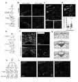
도 1, a 내지 i은 깊은 진피 ENF가 인그레일드-1을 활성화하고 생후 상처 콜라겐 침착에 기여함을 예시한다. (a) 세포 이식, 생착(engraftment), 및 상처 실험을 묘사하는 개략도. (b) 상처가 없는 피부로 이식 후(상단 행) 또는 절제 상처 후 이식 후(하단 행) 인그레일드-1-양성 섬유아세포(EPF, 좌측 컬럼) 및 인그레일드-1-음성 섬유아세포(ENF, 우측 컬럼)의 형광 이미지화. (c) 이식 및 상처를 받은 ENF(적색)의 조직학, 이식 후 상처 내에서 ENF가 EPF로 전환되어 유도된 생후 EPF(pEPF, 녹색) 존재; I형 콜라겐(col-I)에 대한 면역염색은 백색으로 도시됨. 상단, 병합됨; 하단 좌측, ENF 및 EPF; 하단 우측, col-I 염색. N = 각각 ENF 및 EPF를 받은 3마리 마우스, 2개 상처/마우스. (d) 상단: (c)에 도시된 공초점 영상화의 3D 재구성, Imaris 소프트웨어를 사용하여 생성됨(ENF, 적색; pEPF, 녹색; col-I, 백색). 하단: col-I 염색과 Tomato(ENF) 또는 GFP(pEPF) 신호 사이의 신호 공동현지화의 정량화. 점은 상처 당 평균을 나타낸다. N = 5 내지 6개 상처, * P = 0.0335. (e) 상처 치유 동안 En-1 활성화의 일시적으로-정의된 평가를 위한 타목시펜 유도 후 En1 Cre-ERT ;Ai6 마우스의 상처를 나타내는 개략도. (f) 타목시펜-유도된 En1 Cre-ERT ;Ai6 마우스로부터의 상처가 없는 피부(상단 행) 및 치유된 상처(POD 14; 하단 행)의 조직학적 분석, 여기서 GFP+ 세포(EPF, 녹색)는 상처 치유 동안 활성화된 En-1 발현에서 반드시 발생 한다(백색 화살표). Dlk-1(적색) 및 col-I(백색)에 대한 면역염색; DAPI, 청색. N = 4마리 마우스, 2개 상처/마우스. (g) 생후 En-1 활성화에 대해 제안된 메커니즘. 진피(상단)에 상주하는 ENF(적색)는 상처-특이적 신호에 노출되는 경우 pEPF(적색에서 녹색 세포로; 중간)를 생성한다. 이러한 pEPF는 배아-유도된 EPF(eEPF)와 함께 흉터 상처 복구를 매개한다(하단). (h) 3개의 ENF 하위유형의 단리 및 별도의 이식 후 각 하위유형에 대한 상처를 나타내는 개략도. (i) mTomato-발현 수용자 마우스(적색)에 유두상(CD26+, 좌측), 망상(Dlk1+ Sca1-, 중간), 및 피하(Dlk1+/- Sca1+, 우측) ENF(백색)의 이식 및 상처는 망상 ENF만이 pEPF(녹색, 백색 화살표)를 생성함을 보여준다. DAPI, 청색. N = 각각의 ENF 하위유형을 받은 3마리 마우스, 1개 상처/마우스.
도 2, a 내지 i은 시험관 내 및 생체 내 기질 역학에 대한 반응으로 표준 기계적 에너지 변환(mechanotransduction) 신호전달을 통해 망상 진피 ENF가 인그레일드-1을 활성화함을 예시한다. (a) 다양한 역학을 가진 기질에서 ENF의 단리 및 배양: 경직성 플라스틱(ROCK 억제제 Y-27632 포함 또는 제외; 상단) 또는 연질 히드로겔(하단). (b) ENF(적색)에서 pEPF(녹색)로 가변 전환을 나타내는 경직성 TCPS(좌측 컬럼), ROCK 억제제를 포함하는 TCPS(Y-27632; 중간 컬럼), 또는 연질 히드로겔(우측 컬럼) 상에서 배양 1일(상단 행) 또는 14일(하단 행) 후 ENF. (c) 상이한 기질 상에서 배양에서 시간 경과에 따라 EPF로 전환된 ENF의 백분율 정량화. N = 개별 한배 새끼로부터 유도된 P1 ENF를 사용한 3개의 실험 복제물. (d) ROCK 억제제(Y-27632)를 포함하거나 포함하지 않는 경직성 기질(TCPS) 상에서 ENF 하위집단의 분류 및 배양을 나타내는 개략도. (e) 기계적 에너지 변환 억제가 있거나(하단 행) 없는(상단 행), TCPS 상에서 배양 14일 후 유두상(좌측 컬럼), 망상(중간 컬럼), 및 피하(우측 컬럼) ENF, TCPS 상의 망상 진피 ENF에서만 En-1 활성화(GFP, 녹색)가 나타남(상단 행, 중간 패널). N = 개별 한배 새끼로부터 유도된 P1 ENF를 사용한 3개의 실험 복제물. (f) 표준 기계적 에너지 변환 신호전달 경로의 개략도. 기계적 힘은 FAK와 하류 Rho 및 ROCK의 활성화를 통해 신호를 전달받는다; 베르테포르핀은 경로의 최종 전사 이펙터인 YAP를 억제하여 기계적 에너지 변환을 억제한다. (g) 좌측 패널: 등쪽(dorsal) 상처에 긴장을 적용하기 위한 전략을 나타내는 개략도. 우측 패널: 대조군 무처치(sham)(좌측 사진), 증가된 긴장 적용(중간 사진), 또는 증가된 긴장 및 베르테포르핀 처리(우측 사진) 후 치유된 등쪽 절개 상처의 전체 사진. (h) 증가된 장력(tension)과 함께 증가된 pEPF(녹색)를 나타내는 En-1 Cre-ERT ;Ai6 마우스에서 대조군(좌측 컬럼), 장력-처리된(중간 컬럼), 및 장력- 및 베르테포르핀-처리된(우측 컬럼) 상처의 형광 조직학. α-SMA(적색) 및 YAP(백색)에 대한 면역형광 염색; DAPI, 청색. 하단 행, 개별 채널; 상단 행, 병합됨. (i) 20x 고출력 필드(HPF: high-powered field) 당 GFP+ 세포(pEPF; 상단 패널) 및 YAP+ 세포(하단 패널)의 정량화. (g 내지 i) N = 4 내지 5마리 마우스/조건.
도 3, a 내지 l은 Dlk1+ ENF의 기계적 활성화가 섬유성 전사 시그니처와 관련이 있음을 예시한다. (a) 2, 7 또는 14일 동안 시험관 내에서 배양된 벌크 ENF의 개략도. (b) 920개 유전자에 대한 유전자 발현 히트맵 및 계층적 클러스터링은 2일차에 비해 배양 14일차에서 유의하게 상향조절(4배 초과) 또는 하향조절(1/4배 미만)되었다. 배양 2, 7 또는 14일차, 또는 베르테포르핀(Vert) 처리(보라색 박스)를 포함하는 배양 14일차에 대해 도시된 값(플롯 하단의 라벨). (c) (b)에 도시된 920개의 차등적으로 발현된 유전자(14일차 대 2일차)의 화산 플롯. (d) Vert 처리가 있거나 없는, 상이한 시점에서 배양된 ENF로부터의 RNA-seq 데이터의 주 성분 분석(PCA: Principal component analysis). 각 시점 및 조건에 대한 클러스터는 타원으로 표시된다. (e) Vert가 있거나 없는 배양에서 14일차에 ENF에 대해, (b)에 도시된 유의하게 상향조절되거나(상단 플롯) 하향조절된(하단 플롯) 유전자에 대한 GO 용어 풍부. (f) 이전에 섬유증 및 ECM 침착에 연루된 선택된 유전자의 상대적 발현을 나타내는 히트맵. Dlk1은 7일차에 ENF에서 상향조절되었다(적색 박스). 프로-섬유화/매트릭스 유전자는 14일차에 크게 상향조절되었다(녹색 박스); 이러한 변화는 Vert 처리(보라색 박스)로 완화되었다. N = 실험군 당 2개의 생물학적 복제물(2개의 개별 한배 새끼로부터 유래된 풀링된 ENF, 각각 10마리 새끼). (g) RNA-seq에 대한 흉터 pEPF 및 흉터 및 상처가 없는 피부 eEPF 및 ENF의 단리를 나타내는 개략도. (h) 손상되지 않은 피부(uninj)와 비교하여 상처(inj)의 ENF, eEPF, 또는 pEPF에서 1,138개 유전자의 히트맵 및 계층적 클러스터링이 유의하게 상향조절되거나 하향조절되었다. (i) (h)에 도시된 1,138개의 차별적으로 발현된 유전자를 보여주는 화산 플롯. 개별 플롯에는 각 플롯에 도시된 비교와 함께 레이블(오른쪽 상단 모서리)이 지정되어 있다. (j) 손상된 피부 및 손상되지 않은 피부의 pEPF, eEPF, 및 ENF에 대한 RNA-seq 데이터의 PCA. (k) 각 세포 유형에 대한 Dpp4(CD26; 좌측 패널), Jag1(중간 패널), 및 Dll1(우측 패널) 유전자 수의 비교. (l) 이전에 ENF(좌측 패널) 또는 EPF(우측 패널) 정체성과 관련된 것으로 보고된 선택된 유전자의 상대적 발현을 보여주는 히트맵. N = 실험 군 당 2개의 생물학적 복제물(각각 2개의 군으로 풀링된 6마리 마우스로부터의 24개 흉터 및 6개의 상처가 없는 피부 조각). 녹색 박스, EPF 집단(pEPF, inj 및 uninj eEPF); 적색 박스, ENF(inj 및 uninj ENF).
도 4, a 내지 h는 생체 내 기계적 에너지 변환 억제가 재생을 통해 흉터가 없는 상처 치유를 초래함을 예시한다. (a) POD 0(좌측 컬럼), 14(중간 좌측 컬럼), 30(중간 우측 컬럼), 및 90(우측 컬럼)에서, PBS(대조군; 중간 행) 또는 베르테포르핀(하단 행)으로 처리된 상처의 각 시점에 대한 해당하는 전체 사진이 있는, 등쪽 절제 상처(상단 행)의 개략도. 적색 점선 원은 부목 상처에 사용되는 고리의 위치를 나타낸다. (b) POD 14(좌측 컬럼), 30(중간 컬럼), 또는 90(우측 컬럼)에서 수확된 대조군-처리된(상단 행) 및 베르테포르핀-처리된(하단 행) 상처의 H&E 조직학. 백색 화살표는 피부 부속기와 형태학적으로 일치하는 구조를 나타낸다. (c) 모낭 및 기타 피부 부속기의 재성장을 나타내는 POD 90의 베르테포르핀-처리된 상처. 전체 사진(상단 행) 및 조직학: 중간 행, 모낭/땀샘 마커 CK14(적색) 및 CK19(녹색)(DAPI, 청색)에 대한 면역염색; 하단 행, 피지선에 대한 Oil Red O 염색(적색). (d 내지 f) POD 14(D), 30(E), 및 90(F)에서 대조군-처리된(상단 행) 및 베르테포르핀-처리된(하단 행) 상처의 형광 조직학, 섬유아세포(EPF, ENF) 및 ECM 단백질(col-I, Fn) 및 섬유아세포/기계적 에너지 변환 마커(CD26, Dlk-1, YAP, αSMA)에 대한 면역염색을 보여줌; 각 패널의 레이블로 표시된 컬러. 패널 (b 내지 f)의 경우, N = 조건/시점 당 3마리 마우스, 2개 상처/마우스. (f) 맨 우측 패널, 치유 2주, 1개월, 및 3개월 후 PBS-처리된 및 베르테포르핀-처리된 상처에 대한 20x HPF 당 GFP+ 세포(EPF)의 정량화. (g) POD 14(i), 30(ii), 및 90(iii)에서 상처가 없는 피부(녹색) 및 PBS-처리된(적색) 또는 베르테포르핀-처리된(청색) 상처에 대한 26개의 ECM 미세구조 특성을 시각화하는 t-SNE 플롯, 음영 영역으로 강조 표시가 된 각 군의 클러스터가 있음. N = 3마리 마우스/조건, 5 내지 10개 이미지/마우스. 점은 단일 이미지를 나타낸다. (h) 계산된 상처 파괴력으로 상처가 없는 피부(녹색), PBS-처리된(적색), 및 베르테포르핀-처리된(청색) 상처의 인스트론 기계적 강도 시험(좌측 플롯; 상처가 없는 것 vs. PBS, *P = 0.0417; 상처가 없는 것 vs. 베르테포르핀, P = 0.8057) 및 영률(Young's modulus)(우측 플롯; 상처가 없는 것 vs. PBS, *P = 0.0048; 상처가 없는 것 vs. 베르테포르핀, P = 0.9287). 점은 개별 마우스를 나타낸다. N = 7마리 마우스(상처가 없는 것), 5마리 마우스(PBS), 4마리 마우스(베르테포르핀).
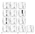
도 5, a 내지 f는 섬유아세포 하위유형을 단리하기 위한 FACS 전략을 예시한다. (a) 타목시펜-유도된 En-1 Cre-ERT ;Ai6 등쪽 피부 및 절제 상처로부터 ENF(Lin- GFP- CD26-), eEPF(Lin- GFP- CD26+), 및 pEPF(Lin- GFP+)를 단리하기 위한 전략. (b) 상처가 없는 피부(좌측) 및 상처(우측)에 대한 대표적인 FACS 플롯이 (a)에 도시되어 있다. *, **은 후속 플롯으로 옮겨진 게이팅된 세포 집단을 나타낸다. (c) 상처가 없는 피부 대 치유된 상처(POD 14)에서 ENF(적색), eEPF(청색), 및 pEPF(녹색)로 표시된 섬유아세포(Lin-)의 상대적 비율의 정량화. 점은 생물학적 복제물을 나타내며, N = 3개의 생물학적 복제물, 각각은 4마리의 마우스(2개 상처/마우스)로부터 풀링된 세포를 포함한다. 상처가 없는 것 대 상처가 있는 것: eEPF, * P = 0.0559; pEPF, * P = 0.0204; ENF, P = 0.6433. (d) 이전에 보고된 표면 마커를 기반으로 하는 En-1 Cre ;Ai6 등쪽 피부에서 유두상, 망상, 및 피하 섬유아세포의 FACS 단리에 대한 개략도. (e) ENF(Lin- GFP-; 적색 박스) 및 EPF(Lin- GFP+; 녹색 박스), ENF 하위유형의 분획(유두상, 청색 박스; 망상, 회색 박스; 피하, 보라색 박스)을 단리하기 위한 게이팅 전략을 나타내는 대표적인 FACS 플롯. *, **, ***, 및 ‡는 후속 플롯으로 옮겨진 게이팅된 세포 집단을 나타낸다. (f) 섬유아세포가 PDGFRa+ 세포(좌측 패널) 대 Lin- 세포(우측 패널)로 정의될 때 각각의 ENF 하위집단(유두상, 청색; 망상, 회색; 피하, 보라색)으로 표시되는 섬유아세포의 비율. N = 개별 한배 새끼로부터 풀링된 세포를 사용하는 3개의 개별적인 실험. 좌측: 유두상 vs. 피하 P = 0.0135, 망상 vs. 피하 * P = 0.0067. 우측: 모든 쌍별 비교 P > 0.05.
도 6, a 내지 c는 시험관 내 ENF 및 pEPF에 대한 유전자 세트 풍부 분석을 예시한다. 2일차(ENF로 유지) 또는 14일차(활성화된 인그레일드-1, GFP+) 동안 TCPS에서 배양된 ENF(mTomato+)에 대한 정규화된 RNA-seq 계수를 (a) 유전자 온톨로지 생물학 과정(Gene Ontology Biological Process), (b) 유전자 온톨로지 분자 기능(Gene Ontology Molecular Function), 및 (c) 홀마크(Hallmark) 데이터베이스에서 풍부에 대해 분석하였다. 인그레일드-1의 활성화는 14일차에 다양한 ECM-관련 용어에 대한 풍부에 의해 추론되는 바와 같이, "근육 발달" 정체성의 상실 및 프로-섬유화 정체성의 획득과 관련이 있었다.
도 7, a 내지 c는 생체 내 ENF 및 pEPF에 대한 유전자 세트 풍부 분석을 예시한다. 흉터 ENF(GFP- CD26-) 및 생후 EPF(GFP+)에 대한 정규화된 RNA-seq 계수를 (a) 유전자 온톨로지 생물학적 과정, (b) 유전자 온톨로지 분자 기능, 및 (c) 홀마크 데이터베이스에서의 풍부에 대해 분석하였다. 흉터 ENF는 ECM-접착 및 Notch 신호전달-관련 용어에 대해 풍부하여, 기계적민감성(mechanosensitive) 표현형을 지지하였다. 대조적으로, 생후 EPF는 다양한 ECM-관련 용어에 대해 풍부하여, 기계적민감성 ENF에 의한 상처 환경에서 인그레일드-1의 활성화가 프로-섬유화 표현형의 획득과 관련이 있음을 확인시켜준다.
도 8, a 내지 c는 다중 용량의 베르테포르핀으로 치료된 상처의 특성을 예시한다. (a) 표시된 간격으로 PBS(적색) 대 1(청색), 2(보라색), 또는 4(밝은 청색) 용량의 베르테포르핀으로 처리된 상처에 대한 봉합(재상피화) 속도를 나타내는 상처 곡선. N = 적어도 6개 상처/조건. POD 4, 2개 용량 베르테포르핀 대 PBS, * P = 0.0140; POD 8, 4개 용량 베르테포르핀 대 PBS, * P = 0.0140; 다른 모든 비교, P > 0.05. (B) POD 0(좌측 컬럼) 및 30(우측 컬럼)에서 PBS(첫 번째 줄), 1(두 번째 줄), 2(세 번째 줄), 또는 4(네 번째 줄)개 용량의 베르테포르핀으로 처리된 상처의 대표적인 전체 사진. (c) 치유의 2주 또는 1개월 후 다양한 처리군에 대한 ECM 미세구조 특성의 t-SNE 시각화(설명 참조). 상처가 없는 피부 및 흉터(PBS)의 클러스터는 음영처리된 영역으로 강조되어 있다.
도 9, a 내지 b는 상처 후 2주의 ECM 섬유 파라미터의 정량화를 예시한다. (a) POD 14에서 상처가 없는 피부 및 베르테포르핀-처리된 또는 PBS-처리된 상처의 정량화된 섬유 파라미터. 개별 값을 피크로시리우스(Picrosirius) 염색으로 평가된, 성숙(적색) 대 미성숙(녹색) 섬유에 대해 계산하였다. 점은 각각 N = 3마리 마우스로부터 2개의 상처의 평균을 나타낸다. (b) 상처가 없는 피부와 PBS-처리된(좌측) 또는 베르테포르핀-처리된 상처(우측)간의 섬유 파라미터(적색, 성숙; 녹색, 미성숙)의 비교에 대한 P-값.
도 10, a 내지 b는 상처 후 1개월의 ECM 섬유 파라미터의 정량화를 예시한다. (a) POD 30에서 상처가 없는 피부 및 베르테포르핀-처리된 또는 PBS-처리된 상처의 정량화된 섬유 파라미터. 개별 값을 피크로시리우스(Picrosirius) 염색으로 평가된, 성숙(적색) 대 미성숙(녹색) 섬유에 대해 계산하였다. 점은 각각 N = 3마리 마우스로부터 2개의 상처의 평균을 나타낸다. (b) 상처가 없는 피부와 PBS-처리된(좌측) 또는 베르테포르핀-처리된 상처(우측)간의 섬유 파라미터(적색, 성숙; 녹색, 미성숙)의 비교에 대한 P-값.
도 11, a 내지 b는 상처 후 3개월의 ECM 섬유 파라미터의 정량화를 예시한다. (a) POD 90에서 상처가 없는 피부 및 베르테포르핀-처리된 또는 PBS-처리된 상처의 정량화된 섬유 파라미터. 개별 값을 피크로시리우스(Picrosirius) 염색으로 평가된, 성숙(적색) 대 미성숙(녹색) 섬유에 대해 계산하였다. 점은 각각 N = 3마리 마우스로부터 2개의 상처의 평균을 나타낸다. (b) 상처가 없는 피부와 PBS-처리된(좌측) 또는 베르테포르핀-처리된 상처(우측)간의 섬유 파라미터(적색, 성숙; 녹색, 미성숙)의 비교에 대한 P-값.
도 12, a 내지 b는 치유 1개월 후 PBS-처리된 및 베르테포르핀-처리된 상처의 인스트론 비교를 예시한다. (a) 치유 1개월 후 상처가 없는 피부 (녹색), PBS-처리된 상처(적색), 및 베르테포르핀-처리된 상처(청색)에 대한 대표적인 힘-변위 곡선. (b) (a)와 동일한 군에 대한 대표적인 응력-변형 곡선. 베르테포르핀 처리는 치유 1개월 후 흉터(PBS 처리)보다 상처가 없는 피부와 더 유사한 상처를 나타냈다.
도 13, a 내지 c는 베르테포르핀-처리된 상처에서 새로운 모낭의 생성을 예시한다. (a) POD 0(좌측 컬럼), 14(중간 좌측 컬럼), 30(중간 우측 컬럼), 및 90(우측 컬럼)에서, PBS(대조군; 중간 행) 또는 베르테포르핀(하단 행)으로 처리한 상처의 각 시점에 대한 상응하는 전체 사진이 있는, 등쪽 절제 상처(상단 행)의 개략도. (b) POD 14(좌측 컬럼), 30(중간 컬럼), 또는 90(우측 컬럼)에서 수확된 대조군-처리된(상단 행) 및 베르테포르핀-처리된(하단 행) 상처의 H&E 조직학. 백색 화살표는 피부 부속기와 형태학적으로 일치하는 구조를 나타낸다. (c) 모낭 및 기타 피부 부속기의 재성장을 나타내는 POD 90의 베르테포르핀-처리된 상처. 전체 사진(상단 행) 및 조직학: 하단 행, 모낭/땀샘 마커 CK14(적색) 및 CK19(녹색)(DAPI, 청색)에 대한 면역염색. Figures 1, a-i illustrate that deep dermal ENF activates Ingrade-1 and contributes to postnatal wound collagen deposition. (a) Schematic depicting cell transplantation, engraftment, and wounding experiments. (b) Post-transplantation into unwounded skin (top row) or post-transplantation after excisional wounding (bottom row) Ingrade-1 -positive fibroblasts (EPF, left column) and Ingrade-1 -negative fibroblasts ( fluorescence imaging of ENF, right column). (c) histology of transplanted and wounded ENF (red), presence of post-transplantation post-transplantation of ENF into EPF induced postnatal EPF (pEPF, green); Immunostaining for type I collagen (col-I) is shown in white. top, merged; bottom left, ENF and EPF; Bottom right, col-I staining. N = 3 mice, 2 wounds/mouse that received ENF and EPF, respectively. (d) Top: 3D reconstruction of the confocal imaging shown in (c), generated using Imaris software (ENF, red; pEPF, green; col-I, white). Bottom: Quantification of signal colocalization between col-I staining and Tomato (ENF) or GFP (pEPF) signals. Dots represent averages per wound. N = 5 to 6 wounds, * P = 0.0335. (e) Schematic representation of wounds in En1 Cre-ERT ;Ai6 mice after tamoxifen induction for a transiently-defined assessment of En-1 activation during wound healing. (f) tamoxifen-derived En1 Cre-ERT ; Histological analysis of unwounded skin (top row) and healed wounds (
Figures 2, a-i illustrate that reticular dermal ENF activates Ingrailed-1 via canonical mechanotransduction signaling in response to matrix dynamics in vitro and in vivo . (a) Isolation and culture of ENFs in substrates with different kinetics: rigid plastics (with or without ROCK inhibitor Y-27632; top) or soft hydrogels (bottom). (b)
3, a-l illustrate that mechanical activation of Dlk1 + ENF is associated with a fibrillar transcriptional signature. (a) Schematic of bulk ENF cultured in vitro for 2, 7 or 14 days. (b) Gene expression heatmaps and hierarchical clustering for 920 genes were significantly upregulated (greater than 4 fold) or downregulated (less than 1/4 fold) at
Figures 4, a-h illustrate that inhibition of mechanical energy conversion in vivo results in scarless wound healing through regeneration. (a) At POD 0 (left column), 14 (middle left column), 30 (middle right column), and 90 (right column), treated with PBS (control; middle row) or verteporfin (bottom row) Schematic of the dorsal resection wound (top row), with a corresponding overall picture for each time point of the wound. The red dotted circle indicates the location of the ring used for the splint wound. (b) H&E histology of control-treated (top row) and verteporfin-treated (bottom row) wounds harvested at POD 14 (left column), 30 (middle column), or 90 (right column). White arrows indicate structures morphologically consistent with skin appendages. (c) Verteporfin-treated wounds of
5, a-f illustrate a FACS strategy for isolating fibroblast subtypes. (A) Tamoxifen-induced En-1 Cre-ERT ; ENF(Lin - GFP - CD26-), eEPF(Lin - GFP - CD26 + ), and pEPF(Lin - GFP + ) from Ai6 dorsal skin and excisional wounds . Strategies to isolate. (b) Representative FACS plots for unwounded skin (left) and wounded (right) are shown in (a) . * , ** indicate gated cell populations transferred to subsequent plots. (c) Quantification of the relative proportions of fibroblasts (Lin − ) labeled as ENF (red), eEPF (blue), and pEPF (green) in unwounded skin versus healed wounds (POD 14). Dots represent biological replicates, N = 3 biological replicates, each containing pooled cells from 4 mice (2 wounds/mouse). Unwound vs. wounded: eEPF, * P = 0.0559; pEPF, * P = 0.0204; ENF, P = 0.6433. (d) Schematic of FACS isolation of papillary, reticular, and subcutaneous fibroblasts from En-1 Cre ;Ai6 dorsal skin based on previously reported surface markers. (e) ENF (Lin - GFP - ; red box) and EPF (Lin - GFP + ; green box), isolating fractions of the ENF subtype (papillary, blue box; reticular, gray box; subcutaneous, purple box) Representative FACS plots showing the gating strategy for * , ** , *** , and ‡ represent gated cell populations transferred to subsequent plots. (f) Fibroblasts represented by each ENF subpopulation (papillary, blue; reticular, gray; subcutaneous, purple) when fibroblasts are defined as PDGFRa + cells (left panel) versus Lin − cells (right panel). ratio. N = 3 separate experiments using pooled cells from individual littermates. Left: Papillary vs. Subcutaneous P = 0.0135, delusional vs. delusional. subcutaneous * P = 0.0067. Right: All pairwise comparisons P > 0.05.
6, a-c illustrate gene set abundance analysis for ENF and pEPF in vitro . Normalized RNA-seq counts for ENF (mTomato + ) cultured in TCPS during day 2 (maintained as ENF) or day 14 (activated engraved - 1 , GFP + ) were calculated using (a) Gene Ontology Biology Process (Gene). Ontology Biological Process), (b) Gene Ontology Molecular Function, and (c) abundance in the Hallmark database were analyzed. Activation of Ingrade-1 was associated with loss of "muscle development" identity and acquisition of pro-fibrotic identity, as inferred by abundance for various ECM-related terms at
7, a-c illustrate gene set abundance analysis for ENF and pEPF in vivo . Normalized RNA-seq coefficients for scar ENF (GFP - CD26 - ) and postnatal EPF (GFP + ) were calculated for (a) gene ontology biological processes, (b) gene ontology molecular functions, and (c) abundance in the Hallmark database. was analyzed. Scar ENFs were enriched for ECM-adhesion and Notch signaling-related terms, supporting a mechanosensitive phenotype. In contrast, postnatal EPF is enriched for a variety of ECM-related terms, confirming that activation of Ingrailed-1 in the wound environment by mechanosensitive ENFs is associated with the acquisition of a pro-fibrotic phenotype.
8, a-c illustrate the properties of wounds treated with multiple doses of verteporfin. (A) Wound curves showing closure (re-epithelialization) rates for wounds treated with verteporfin at doses of PBS (red) versus 1 (blue), 2 (purple), or 4 (light blue) at the indicated intervals. N = at least 6 wounds/condition.
9, a-b illustrate the quantification of
10, a-b illustrate the quantification of ECM fiber parameters one month after wounding. (A) Quantified fiber parameters of unwounded skin and verteporfin-treated or PBS-treated wounds at
11, a-b illustrate the quantification of ECM fiber parameters at 3 months post wounding. (A) Quantified fiber parameters of unwounded skin and verteporfin-treated or PBS-treated wounds at
12, a-b illustrate an instron comparison of PBS-treated and verteporfin-treated wounds one month after healing. (A) Representative force-displacement curves for unwounded skin (green), PBS-treated wound (red), and verteporfin-treated wound (blue) 1 month after healing. (b) Representative stress-strain curves for the same group as (a). Verteporfin treatment showed wounds more similar to unwounded skin than scars (PBS treatment) after 1 month of healing.
13, A-C illustrate the generation of new hair follicles in verteporfin-treated wounds. (a) POD 0 (left column), 14 (middle left column), 30 (middle right column), and 90 (right column), treated with PBS (control; middle row) or verteporfin (bottom row) Schematic of the dorsal resection wound (top row), with a corresponding overall picture for each time point of the wound. (b) H&E histology of control-treated (top row) and verteporfin-treated (bottom row) wounds harvested at POD 14 (left column), 30 (middle column), or 90 (right column). White arrows indicate structures morphologically consistent with skin appendages. (c) Verteporfin-treated wounds of
본원에서 통상적인 의미로 사용되는 용어 "섬유아세포"는 세포외 기질의 합성 및 조직화를 담당하는 세포를 지칭한다. 2개의 섬유아세포 계통은 인그레일드-1 계통-음성 섬유아세포(ENF) 및 인그레일드-1 계통-양성 섬유아세포(EPF)를 포함한다. EPF 계통은 발달 동안 임의의 시점에서 인그레일드-1을 발현하는 모든 세포와 이러한 세포의 모든 자손을 포함한다.The term “fibroblast” as used herein in its conventional sense refers to a cell responsible for the synthesis and organization of the extracellular matrix. The two fibroblast lineages include Ingraild-1 lineage-negative fibroblasts (ENFs) and Ingraild-1 lineage-positive fibroblasts (EPFs). The EPF lineage includes all cells and all progeny of those cells that express Ingraild-1 at any time point during development.
본원에서 사용된 용어 "조절(modulating)"은 생물학적 세포, 세포 집단, 또는 세포 구성성분(예컨대, 단백질, 핵산 등)의 속성(attribute)을 증가, 감소 또는 억제하는 것을 의미한다. 일부 경우, 속성은 예컨대 신호전달 경로의 활성화를 포함한다. 일부 경우, 속성은 하나 이상의 세포의 양 및/또는 활성을 포함한다. 일부 경우, 속성은 예컨대 세포의 구성성분(예컨대 단백질, 핵산, 등)의 예컨대 양, 활성, 또는 발현 수준(DNA 또는 RNA 발현 수준)을 포함한다. 일부 경우, "조절하기(modulating)" 또는 "조절(modulation)"은 적합한 시험관 내 분석법, 세포 분석법 또는 생체 내 분석법을 사용하여 측정될 수 있다. 일부 경우, 증가 또는 감소는 기준에 비해 10% 이상, 예컨대 기준에 비해 10% 이상, 20% 이상, 30% 이상, 40% 이상, 50% 이상, 60% 이상, 70% 이상, 80% 이상, 90% 이상, 95% 이상, 97% 이상, 98% 이상, 최대 100%이다. 예컨대, 증가 또는 감소는 기준에 비해 2배 이상 3배 이상, 4배 이상, 5배 이상, 6배 이상, 7배 이상, 8배 이상, 9배 이상, 10배 이상, 50배 이상, 또는 100배 이상일 수 있다.As used herein, the term “modulating” means increasing, decreasing or inhibiting an attribute of a biological cell, cell population, or cellular component (eg, protein, nucleic acid, etc.). In some cases, the attribute includes, for example, activation of a signaling pathway. In some cases, an attribute includes the amount and/or activity of one or more cells. In some cases, an attribute includes, for example, the amount, activity, or expression level (DNA or RNA expression level) of a component (eg, protein, nucleic acid, etc.) of a cell. In some cases, "modulating" or "modulation" can be determined using a suitable in vitro assay, cellular assay, or in vivo assay. In some cases, the increase or decrease is 10% or more relative to a reference, such as 10% or more, 20% or more, 30% or more, 40% or more, 50% or more, 60% or more, 70% or more, 80% or more, 70% or more, 80% or more, 90% or more, 95% or more, 97% or more, 98% or more, up to 100%. For example, an increase or decrease can be at least 2 fold, at least 3 fold, at least 4 fold, at least 5 fold, at least 6 fold, at least 7 fold, at least 8 fold, at least 9 fold, at least 10 fold, at least 50 fold, or 100 fold over a reference. It can be more than double.
본원에서 통상적인 의미로 사용된 용어 "섬유증(fibrosis)"은 기관 또는 조직 부분의 손상 또는 염증 또는 이의 혈액 공급 방해의 결과로서 기관 또는 조직에서 과도한 섬유성 결합 조직의 형성 또는 발달을 지칭한다. 이는 흉터, 비정상적인 반응 과정 또는 알려지지 않은 또는 이해되지 않은 원인으로 이어지는 정상적인 치유 반응의 결과일 수 있다.The term “fibrosis,” as used herein in its ordinary sense, refers to the formation or development of excessive fibrous connective tissue in an organ or tissue as a result of damage or inflammation of an organ or tissue part or obstruction of its blood supply. It may be the result of scarring, an abnormal reaction process, or a normal healing response leading to unknown or unknown causes.
본원에서 통상적인 의미로 사용되는 용어 "흉터화(scarring)"는 손상 또는 질환에 의해 파괴된 정상 조직을 섬유 조직으로 대체하는 상태를 지칭한다. 용어 "흉터화"는 또한 피부 치유 과정에서 발생하는 색상, 윤곽(볼록함(bulging)/오목함(indentation)), 거칠기(rugosity)(거침(roughness)/부드러움(smoothness)) 및 질감(부드러움(softness)/단단함(hardness)) 중 하나 이상의 이상(abnormality)을 지칭한다. 흉터화의 맥락에서 본원에서 사용된 표현 "방지" 또는 "예방"은 흉터화의 발달 정도에 대한 조정을 지칭하며, 이에 의해 치유된 피부 표면의 색상, 윤곽, 거칠기 및 질감 중 하나 이상이 일반적인 육안 검사에서 대상체의 정상 피부와 유사한 것을 지칭한다. 흉터화의 맥락에서 본원에서 사용된 표현 "감소하는" 또는 "감소"는 흉터화의 발달 정도에 대한 조정을 지칭하며, 이에 의해 치유된 피부 표면의 색상, 윤곽, 거칠기 및 질감 중 하나 이상이 환자의 정상 피부에 측정 가능하게 더 가깝게 접근함을 지칭한다.The term “scarring” as used herein in its ordinary meaning refers to a condition in which normal tissue destroyed by injury or disease is replaced with fibrous tissue. The term “scarring” also refers to the color, contour (bulging/indentation), rugosity (roughness/smoothness) and texture (softness) that occur during the healing process of the skin. It refers to one or more abnormality of softness/hardness). As used herein in the context of scarring, the expression "prevention" or "prevention" refers to an adjustment to the degree of development of scarring, whereby one or more of the color, contour, roughness and texture of the surface of the healed skin is normal to the macroscopic Refers to something similar to a subject's normal skin in an examination. As used herein in the context of scarring, the expression "reducing" or "reducing" refers to an adjustment to the degree of development of scarring, whereby one or more of the color, contour, roughness and texture of the healed skin surface is a patient refers to a measurably closer approach to the normal skin of
본원에서 통상적인 의미로 사용되는 용어 "흉터(scar)"는 손상 또는 질환에 의해 파괴된 정상 조직을 대체하는 섬유성 조직을 지칭한다. 피부 외층의 손상은 조직을 재건함으로써 치유되며, 이러한 경우, 흉터화(scarring)는 경미하다. 그러나, 피부 아래 두꺼운 조직 층이 손상되면 재건이 더욱 복잡해진다. 신체는 콜라겐 섬유(신체에서 자연적으로 생성되는 단백질)를 생성하며, 이로 인해 대개 눈에 띄는 흉터가 생긴다. 상처가 치유된 후, 새로운 콜라겐이 형성되고 혈관이 정상으로 돌아옴에 따라 흉터가 계속 변경되어, 손상 후 2년 동안 대부분의 흉터가 옅어지고 외양이 개선된다. 그러나, 손상의 일부 가시적인 증거가 있으며, 모낭과 땀샘이 다시 자라지 않는다. 본원에서 사용된 바와 같이, "흉터 영역"은 손상 또는 질환에 의해 파괴되고 섬유질 조직으로 대체된 정상 조직의 영역을 지칭한다.The term “scar” as used herein in its ordinary sense refers to a fibrous tissue that replaces normal tissue destroyed by an injury or disease. Damage to the outer layers of the skin heals by rebuilding the tissue, in which case scarring is minor. However, reconstruction becomes more complicated when the thick layer of tissue beneath the skin is damaged. The body produces collagen fibers (proteins that are produced naturally in the body), which usually result in visible scarring. After the wound heals, the scar continues to change as new collagen is formed and blood vessels return to normal, resulting in most scars pale and improved in appearance two years after injury. However, there is some visible evidence of damage, and hair follicles and sweat glands do not grow back. As used herein, “scarring area” refers to an area of normal tissue that has been destroyed by an injury or disease and replaced with fibrous tissue.
흉터는 세 가지 주요 면에서 정상 피부와 다르다:(1) 이는 어떠한 피부 부속기(모낭, 땀샘 등)도 없다; (2) 이의 콜라겐 구조는 근본적으로 다르며, 정상 피부에 유연성과 강도를 부여하는 "바스켓위브(basketweave)" 패턴이 아니고 조밀하고 평행한 섬유질이다; 그리고 (3) 열등한 매트릭스 구조의 결과로 인해, 피부보다 약하다.A scar differs from normal skin in three main ways: (1) it lacks any skin appendages (hair follicles, sweat glands, etc.); (2) its collagen structure is fundamentally different, with dense, parallel fibers rather than the “basketweave” pattern that imparts flexibility and strength to normal skin; and (3) weaker than the skin, as a result of the inferior matrix structure.
본원에서 사용된 용어 "흉터-관련 유전자"는 정상적인 상처 치유 과정의 일부로서 흉터화에 반응하여 활성화되는 단백질을 코딩하는 핵산을 지칭한다. 본원에서 사용된 용어 "흉터-관련 유전자 산물"은 정상적인 상처 치유 과정의 일부로서 흉터화에 반응하여 발현된 단백질을 지칭한다.As used herein, the term “scar-associated gene” refers to a nucleic acid encoding a protein that is activated in response to scarring as part of the normal wound healing process. As used herein, the term “scar-associated gene product” refers to a protein expressed in response to scarring as part of the normal wound healing process.
흉터 조직은 주로 조직화되지 않은 콜라겐성 세포외 기질로 구성된다. 이는 상처 반응하여 진피 섬유아세포와 구별되는, 근섬유아세포에 의해 생성되며, 이는 TGF-βI, TGF-P2 및 TGF-P3(총칭하여 TGF-β로 지칭됨)이라고 지칭되는 적어도 3개의 이소형에 존재하는 분비된 단백질인, 변형 성장 인자(Transforming Growth Factor)-β의 국소 농도를 증가시킨다. TGF-β는 다수의 조직 유형에서 섬유증과 관련된 중요한 사이토카인이다(문헌[Beanes, S. et al, Expert Reviews in Molecular Medicine, vol. 5, no. 8, pp. 1 -22 (2003)]). 흉터의 유형이 PCT 출원 공개 WO 2014/040074호에 추가로 설명되어 있으며, 이는 그 전체 내용이 본원에 인용되어 포함된다.Scar tissue is mainly composed of an unorganized, collagenous extracellular matrix. It is produced by myofibroblasts, distinct from dermal fibroblasts in response to a wound, and is present in at least three isoforms termed TGF-βI, TGF-P2 and TGF-P3 (collectively referred to as TGF-β). increases the local concentration of a secreted protein, Transforming Growth Factor-β. TGF-β is an important cytokine associated with fibrosis in many tissue types (Beanes, S. et al, Expert Reviews in Molecular Medicine, vol. 5, no. 8, pp. 1-22 (2003)). . Types of scarring are further described in PCT Application Publication No. WO 2014/040074, which is incorporated herein by reference in its entirety.
본원에서 이의 통상적인 의미로 사용된 용어 "피부"는 신체의 모든 표면 조직 및 예컨대 점막 및 눈 조직뿐만 아니라 일반 피부를 비롯한 신체의 표면 아래 구조를 포함한다. "피부"라는 표현은 상처 부위 자체를 포함할 수 있다. 상처 표면에 피부가 다시 가까워지는 것은 오랫 동안 상처 치유의 상당 부분이 완료되었다는 주요 신호였다. 이러한 결함의 재봉합은 박티레아, 독소, 및 기계적 힘으로부터의 보호를 포함하는 피부의 보호 기능을 회복할뿐만 아니라, 필수 체액을 유지하기 위한 장벽을 제공한다. 각질층을 시작으로 여러 층으로 구성된 표피는 피부의 가장 바깥쪽 층이다. 가장 안쪽 피부 층은 진피층이다.The term “skin”, as used herein in its ordinary sense, includes all surface tissues of the body and subsurface structures of the body including, for example, mucous membranes and eye tissues, as well as normal skin. The expression "skin" may include the wound site itself. The skin's re-approach to the wound surface has long been a major sign that a significant portion of wound healing has been completed. Resealing these defects restores the skin's protective functions, including protection from bacterium, toxins, and mechanical forces, as well as providing a barrier to retain essential body fluids. The epidermis, which consists of several layers, starting with the stratum corneum, is the outermost layer of the skin. The innermost layer of skin is the dermis.
본원에서 통상적인 의미로 사용되는 용어 "피부 부속기"는 모낭, 피지 및 땀샘, 손톱 및 발톱을 포함한다.As used herein, the term “skin appendages” includes hair follicles, sebum and sweat glands, fingernails and toenails.
본원에서 사용된 용어 "진피 부위"는 임의의 크기 및 영역을 갖는 대상체의 피부 영역을 지칭한다. 진피 부위는 예컨대 두피와 같은 대상체의 피부 일부를 포함할 수 있다. 진피 부위는 예컨대 표피 및 진피를 포함하는 피부의 하나 이상의 층을 포함할 수 있다. 일부 경우, 진피 부위는 상처를 포함한다.As used herein, the term “dermal region” refers to an area of a subject's skin of any size and area. The dermal region may include a portion of the subject's skin, such as, for example, the scalp. The dermal region may comprise one or more layers of skin including, for example, the epidermis and the dermis. In some cases, the dermal region comprises a wound.
본원에서 이의 통상적인 의미로 사용되는 "감광제" 또는 "광반응 작용제" 또는 "감광화 작용제"는 광-활성화된 약물 또는 화합물이다. 감광제는 가장 일반적으로 가시 스펙트럼에서 전자기 복사를 흡수하고, 이를 또 다른 에너지 형태, 가장 일반적으로 반응성 산소 종 및/또는 열 에너지로 방출하는 물질로 정의될 수 있다. 일부 경우, 감광화 작용제는 광역동 치료에 유용하다. 이러한 제제는 전자기 복사를 흡수하고 치료 효과, 예컨대 원치 않는 세포 또는 조직의 손상 또는 파괴를 발휘하기에 충분하거나, 진단 응용분야에서 검출하기에 충분한 에너지를 방출할 수 있다. 예컨대, 감광제는 하나 이상의 유형의 선택된 표적 조직에 수집되고, 특정 파장의 빛에 노출될 때, 빛을 흡수하여 표적 조직의 손상 또는 파괴를 유도하는 임의의 화학적 화합물일 수 있다. 선택된 표적에 위치하고 빛을 흡수하는 실질적으로 모든 화학적 화합물이 사용될 수 있다. 감광제는 이것이 투여되는 대상체에 비독성일 수 있고 비독성 조성물로 제형화될 수 있다. 감광제는 또한 광분해된 형태에서 비독성일 수 있다. 일부 경우, 감광제는 광화학적 효과가 없는 상태에서 세포에 대한 독성이 부족하여 비-표적 조직에서 쉽게 제거되는 것을 특징으로 한다.A “photosensitizer” or “photoreactive agent” or “photosensitizing agent” as used herein in its conventional sense is a light-activated drug or compound. Photosensitizers can be defined as materials that most commonly absorb electromagnetic radiation in the visible spectrum and emit it as another form of energy, most commonly reactive oxygen species and/or thermal energy. In some cases, photosensitizing agents are useful for photodynamic therapy. Such agents may absorb electromagnetic radiation and emit energy sufficient to exert a therapeutic effect, such as unwanted damage or destruction of cells or tissues, or to be detected in diagnostic applications. For example, the photosensitizer can be any chemical compound that collects on one or more types of selected target tissues and, when exposed to light of a particular wavelength, absorbs the light and induces damage or destruction of the target tissue. Virtually any chemical compound that is located on the selected target and absorbs light can be used. The photosensitizer may be non-toxic to the subject to which it is administered and may be formulated into a non-toxic composition. Photosensitizers may also be non-toxic in photolyzed form. In some cases, photosensitizers are characterized by a lack of toxicity to cells in the absence of a photochemical effect and thus are easily removed from non-target tissues.
본원에서 통상적인 의미로 사용되는 용어 "상처"는 인간 또는 비 인간 동물 신체의 내부 또는 외부 신체 표면에서 정상 조직 연속성의 임의의 파괴 및/또는 손실을 포함하며, 예컨대 수술 또는 신체적 상해와 같은 비-생리학적 과정으로 인해 발생한다. 본원에서 사용된 표현 "상처" 또는 "상처 환경"은 잠재적으로 흉터화를 유도할 수 있는 치유 과정을 유발할 수 있는 임의의 피부 병변을 지칭하며, 이는 부상으로 생성된 상처, 화상으로 생성된 상처, 질환으로 생성된 상처 및 수술 과정에 의해 생성된 상처를 포함한다. 상처는 신체의 임의의 외부 또는 내부에 존재할 수 있으며 관통이거나 비-관통일 수 있다. 본원에 기술된 방법은 피부 표면에 문제가 있는 상처를 치료하는데 유익할 수 있다. 본 발명의 방법에 따라 치료될 수 있는 상처의 예는 표재성 및 비-표재성 상처 둘 모두, 예컨대 찰과상, 열상, 온열 손상으로 인한 상처(예컨대 화상 및 저온 치료로 유발된 상처), 및 수술로 인한 임의의 상처를 포함한다.The term "wound", as used herein in its ordinary sense, includes any disruption and/or loss of normal tissue continuity on the internal or external body surface of a human or non-human animal body, including non-surgical or bodily injury, for example. It occurs due to a physiological process. As used herein, the expression "wound" or "wound environment" refers to any skin lesion capable of triggering a healing process that can potentially lead to scarring, including wounds resulting from injuries, wounds resulting from burns, Includes wounds produced by disease and wounds produced by surgical procedures. Wounds can be anywhere on the outside or inside of the body and can be piercing or non-penetrating. The methods described herein may be beneficial in treating wounds with problematic skin surfaces. Examples of wounds that can be treated according to the methods of the present invention include both superficial and non-superficial wounds, such as abrasions, lacerations, wounds caused by heat injury (such as burns and wounds caused by cold treatment), and any resulting from surgery. including wounds of
본원에서 이의 통상적인 의미로 사용된 용어 "상처 치유"는 비제한적으로 염증, 육아(granulation), 신생혈관 형성, 섬유아세포, 내피세포 및 상피 세포의 이동, 세포외 기질 침착, 재상피화, 및 리모델링의 과정을 포함하여, 시간 및 공간적 치유 프로그램의 유도를 통한 재생 과정을 지칭한다.The term "wound healing" as used herein in its ordinary meaning includes, but is not limited to, inflammation, granulation, angiogenesis, fibroblasts, endothelial and epithelial cell migration, extracellular matrix deposition, re-epithelialization, and remodeling. refers to the regenerative process through the induction of a temporal and spatial healing program, including the process of
본원에서 이의 통상적인 의미로 사용된 용어 "모낭 형성" 또는 "모낭 형성 유도"는 진피 유두상 진피 유두상 세포가 표피 세포를 유도하여 모낭 구조를 형성하는 현상을 지칭한다.The term "follicle formation" or "induction of follicle formation" as used herein in its ordinary meaning refers to a phenomenon in which dermal papilla dermal papilla cells induce epidermal cells to form a hair follicle structure.
본원에서 이의 통상적인 의미로 사용된 용어 "모발 성장" 또는 "모발 성장 유도"는 모낭의 모기질(hair matrix) 세포가 분화 및 증식하여 모간을 형성하고, 진피초(dermal sheath) 세포가 모기질 또는 외근초(ORS: outer root sheath)에 작용하여 신체 표면으로부터 모간을 신장시키는 현상을 지칭한다. 일부 경우, 모발 성장은 하나 이상의 새로운 모낭의 생성을 유도한다. 일부 경우, 모발 성장은 하나 이상의 새로운 모발 생성을 포함한다.As used herein, the term "hair growth" or "hair growth induction" as used herein means that the hair matrix cells of the hair follicles differentiate and proliferate to form a hair shaft, and the dermal sheath cells produce the matrix. Or it refers to a phenomenon that acts on the outer root sheath (ORS) to extend the hair shaft from the body surface. In some cases, hair growth leads to the production of one or more new hair follicles. In some cases, hair growth includes the creation of one or more new hairs.
본원에서 통상적인 의미로 사용되는 용어 "탈모(alopecia)"는 모발이 손실되는 질환을 지칭한다. 이는 남성형 탈모, 외상, 방사선요법, 화학요법, 철 결핍 또는 기타 영양 결핍, 자가면역 질환 및 진균 감염과 같은 여러 원인으로 인해 유발될 수 있다. 탈모에서 모발의 손실은 머리털에만 국한되지 않고 신체 어느 곳에서나 발생할 수 있다. 탈모는 종종 모발 색상의 퇴색을 동반한다. 탈모는 종종 모발이 가늘어지거나 모발이 짧아지는 것과 같이 모발의 질 저하를 동반한다. 탈모의 종류와 관련하여, 원형 탈모, 남성형 탈모, 갱년기 탈모, 여성형 탈모, 지루성 탈모, 비강성 탈모, 노인성 탈모, 암 화학요법 약물-유도된 탈모, 방사선 노출로 인한 탈모, 발모광(trichotillomania), 생후 탈모, 등이 있다. 탈모 유형이 미국특허 제9808511호에 추가로 기술되어 있으며, 그 전체가 인용되어 본원에 포함된다.The term “alopecia” as used herein in its ordinary sense refers to a disease in which hair is lost. It can be caused by several causes, such as androgenetic hair loss, trauma, radiation therapy, chemotherapy, iron or other nutritional deficiencies, autoimmune diseases, and fungal infections. Hair loss in hair loss is not limited to hair hair, it can occur anywhere on the body. Hair loss is often accompanied by fading of hair color. Hair loss is often accompanied by deterioration of the quality of the hair, such as thinning or shortening of the hair. Regarding the types of hair loss, alopecia areata, male pattern hair loss, menopausal hair loss, female pattern hair loss, seborrheic hair loss, alopecia pityis, senile hair loss, cancer chemotherapy drug-induced hair loss, hair loss due to radiation exposure, trichotillomania, Postnatal hair loss, etc. Types of hair loss are further described in US Pat. No. 9808511, which is incorporated herein by reference in its entirety.
원형 탈모는 모발이 갑자기 빠지는 것을 유발할 수 있는 자가면역 질환이다. 원형 탈모는 동전 크기의 원형 내지 윤곽이 뚜렷한 반점이 있는 탈모반이 갑자기 발생하는 탈모로서, 자각 증상 또는 전구 증상 등이 없는 경우가 많으며, 이후 자연 회복이 일어나지 않으면 점차적으로 면적이 증가하고 다루기 어려워진다. 이는 두피나 신체 다른 부위에 부분 탈모를 유도할 수 있다. 영향을 받은 모낭의 모발 성장은 감소되거나 완전히 중단된다. 원형 탈모는 자가면역 질환 예컨대 하시모토 질환으로 대표되는 갑상선 질환, 백반증, 전신성 홍반성 루프스, 류마티스 관절염, 또는 중증 근무력증 또는 아토피 질환 예컨대 기관지 천식, 아토피성 피부염, 또는 알러지성 비염과 관련이 있는 것으로 알려져 있다.Alopecia areata is an autoimmune disease that can cause sudden hair loss. Alopecia areata is hair loss that occurs suddenly in a bald area the size of a coin or a bald spot with a clear outline, and there are often no subjective symptoms or prodromal symptoms. . This can lead to partial hair loss on the scalp or other parts of the body. Hair growth of the affected hair follicles is reduced or completely stopped. Alopecia areata is known to be associated with autoimmune diseases such as thyroid disease typified by Hashimoto's disease, vitiligo, systemic lupus erythematosus, rheumatoid arthritis, or myasthenia gravis or atopic diseases such as bronchial asthma, atopic dermatitis, or allergic rhinitis. .
본원에서 통상적인 의미로 사용되는 용어 "마이크로니들링(microneedling)"은 신체의 부위에 미세바늘(microneedle)을 사용하는 것을 지칭한다. 개별 미세바늘은 피부 각질층(표피를 덮고 있는 피부의 맨 바깥층)의 공칭 두께보다 클 수 있는, 사전 결정된 거리까지 피부를 천공하도록 설계되어 있다. 이러한 미세바늘을 사용하면 피부 장벽 특성을 극복할 수 있다. 동시에, 미세바늘은 피부 외부 표면 아래로 약 2.0 내지 2.5 mm 미만인 표피를 관통하지 않도록 제조되는 경우 상대적으로 통증이 없고 출혈이 없다. 미세바늘은 각질층을 완전히 관통하기에 충분한 힘의 피부에 대한 직접적인 미는 동작이 필요할 수 있다. 일반적으로, 미세바늘 자극 시스템은 주름, 여드름 흉터, 튼살, 피부 미백 및 안면 회춘과 같은 다양한 상태의 피부 관리 치료에 사용되는 것으로 잘 알려져 있다. 마이크로니들링의 특정 실시형태에서, 피부에 구멍을 뚫고 약물 또는 화장품을 피부에 적용하는 방법은 피부에 신속하고 충분하게 침투하는 방법을 제공한다. 일부 경우, 미세바늘을 사용하여 자연 치유 과정을 시작하고 콜라겐과 엘라스틴 생성 등을 자극하여 피부를 치유하기에 충분할 정도로 피부를 손상시키기에 충분하다. 이러한 방법에서, 피부 깊숙한 층을 손상시키지 않고 마이크로니들링 장치로 피부에 수백 내지 수천 개의 작은 구멍 또는 미세도관이 생성된다. 이러한 피부 손상은 자연 치유 과정을 시작하여 새롭고 건강한 피부 조직을 생성하기 위해 유두상 진피에서 새로운 천연 콜라겐과 엘라스틴의 형성을 자극하는 천연 자극제와 성장 인자의 방출로 이어진다. 또한, 새로운 모세혈관이 형성된다. 상처 치유 과정과 관련된 이러한 신생혈관 및 신생콜라겐 형성은 보다 젊어보이는 피부의 형성, 피부 병리의 감소 및 흉터 개선으로 이어진다. 일반적으로 경피(percutaneous) 콜라겐 유도 요법이라고 지칭되는, 마이크로니들링은 또한 광 노화의 치료에서 사용되어 왔다. 또한, 구멍이 생긴 부위 및 미세한 구멍을 통해 피부 내로 침투할 것으로 예상되는 부위에 의료용 물질이 도포될 수 있다. 마이크로니들링은 일반적으로 피부를 제거하거나 영구적으로 손상시키지 않고 특정 상태가 보장되는 안면, 목, 두피, 및 신체의 거의 모든 곳에 적용된다. 사전결정된 수의 바늘이 원하는 깊이까지 피부 내로 삽입된다. 경미한 부상에 대한 반응으로서, 피부 조직은 자연적인 상처 치유 과정을 시작한다. 이러한 자연적인 과정은 흉터를 매끄럽게 하고 주름을 제거하고 색소 침착을 개선하는데 도움이 되는 새로운 건강한 진피 조직을 형성하여, 보다 젊고, 보다 건강하며 보다 깨끗한 외관의 피부로 만들어준다.As used herein, the term “microneedling” in its ordinary sense refers to the use of microneedles in a body part. The individual microneedles are designed to puncture the skin to a predetermined distance, which may be greater than the nominal thickness of the stratum corneum (the outermost layer of skin covering the epidermis). The use of these microneedles can overcome the skin barrier properties. At the same time, the microneedles are relatively painless and bleed-free when manufactured so as not to penetrate the epidermis that is less than about 2.0 to 2.5 mm below the outer surface of the skin. The microneedle may require a direct pushing action against the skin with sufficient force to completely penetrate the stratum corneum. In general, microneedle stimulation systems are well known for use in skin care treatment of various conditions such as wrinkles, acne scars, stretch marks, skin lightening and facial rejuvenation. In certain embodiments of microneedling, the method of piercing the skin and applying a drug or cosmetic to the skin provides a method for rapid and sufficient penetration into the skin. In some cases, microneedles are used to start the natural healing process and stimulate the production of collagen and elastin, etc., enough to damage the skin enough to heal the skin. In this method, hundreds to thousands of tiny pores or microtubules are created in the skin with a microneedling device without damaging the deep layers of the skin. This skin damage leads to the release of natural stimulants and growth factors that stimulate the formation of new natural collagen and elastin in the papillary dermis to initiate the natural healing process to create new, healthy skin tissue. Also, new capillaries are formed. This neovascularization and neocollagen formation associated with the wound healing process leads to the formation of younger-looking skin, reduced skin pathology and improved scarring. Microneedling, commonly referred to as percutaneous collagen induction therapy, has also been used in the treatment of photoaging. In addition, the medical material may be applied to the area where the hole is made and the area where it is expected to penetrate into the skin through the minute hole. Microneedling is generally applied on the face, neck, scalp, and almost anywhere on the body where certain conditions are guaranteed without removing or permanently damaging the skin. A predetermined number of needles are inserted into the skin to a desired depth. In response to minor injuries, the skin tissue initiates the natural wound healing process. This natural process creates new, healthy dermal tissue that helps smooth scars, remove wrinkles and improve pigmentation, resulting in younger, healthier and cleaner looking skin.
본원에서 통상적인 의미로 사용되는 용어 "분할 레이저 표면처리 치료(fractional laser resurfacing treatment)" 또는 "분할 레이저 표면처리(fractional laser resurfacing)" 또는 "분할 표면처리(fractional resurfacing)"은 피부에 열 손상을 유도하여 피부 결함을 개선하기 위해 전자기 복사를 사용하는 것을 지칭하며, 이는 피부의 복잡한 상처 치유 반응을 초래한다. 이는 손상된 피부의 생물학적 복구를 유도한다. 이러한 목적을 제공하는 다양한 기술이 도입되었다. 상이한 기술은 치료 방식의 2개의 군으로 분류될 수 있다: 절제 레이저 피부 표면처리("LSR": ablative laser skin resurfacing) 및 비-절제 콜라겐 리모델링("NCR": non-ablative collagen remodeling). 치료 방식의 첫 번째 군, 즉 LSR은 표피 및/또는 진피에 열 손상을 일으키는 것을 포함하는 반면, 두 번째 군, 즉 NCR은 표피의 열 손상을 방지하도록 설계되었다. 펄스 CO2 또는 Er:YAG 레이저를 사용하는 LSR은 당업계에서 레이저 표면처리(laser resurfacing) 또는 절제 표면처리(ablative resurfacing)로 지칭될 수 있으며, 광노화 피부, 만성 노화 피부, 흉터, 표재성 색소 병변, 튼살, 및 표재성 피부 병변의 징후에 대한 효과적인 치료 선택사항으로 간주된다. NCR 기술은 당업계에서 비 절제 표면처리(non ablative resurfacing), 비-절제 하부표면처리(non-ablative subsurfacing), 또는 비-절제 피부 리모델링으로 다양하게 지칭된다. NCR 기술은 일반적으로 비-절제 레이저, 플래시램프, 또는 무선 주파수 전류를 사용하여 표피 조직에 손상을 주지 않으면서 진피 조직을 손상시킨다. NCR 기술의 이면에 있는 개념은 오직 진피 조직의 열 손상만이 상처 치유를 유도하여 생물학적 복구와 새로운 진피 콜라겐 형성을 유도하는 것으로 여겨진다는 것이다. 이러한 유형의 상처 치유는 광노화와 관련된 구조적 손상을 감소시킬 수 있다. NCR 기술에서 표피 손상을 피하면 치료 관련 부작용의 심각성과 지속기간이 감소한다. 특히, NCR 기술을 사용하면 통상적으로 절차 후 진물, 딱지, 색소 변화 및 표피 장벽 기능의 장기간 손실로 인한 감염 발생을 피할 수 있다. 분할 레이저 표면처리를 실행하기 위한 추가 방법 및 장치가 예컨대 PCT 출원 공개 WO 2005/007003호; 미국 특허 출원 제20160324578호; 및 문헌[Beasley et al. (2013) Current Dermatology Reports. 2:135-143]에 기술되어 있으며, 이의 개시내용은 그 전체가 인용되어 본원에 포함된다.The terms "fractional laser resurfacing treatment" or "fractional laser resurfacing" or "fractional resurfacing," as used herein in their ordinary sense, refer to thermal damage to the skin. Refers to the use of electromagnetic radiation to induce and ameliorate skin imperfections, resulting in complex wound healing responses of the skin. This leads to biological repair of damaged skin. Various techniques have been introduced to serve this purpose. The different techniques can be divided into two groups of treatment modalities: ablative laser skin resurfacing (“LSR”) and non-ablative collagen remodeling (“NCR”). The first group of treatment modalities, ie, LSR, involved causing thermal damage to the epidermis and/or dermis, while the second group, ie, NCR, was designed to prevent thermal damage to the epidermis. LSR using a pulsed CO 2 or Er:YAG laser may be referred to in the art as laser resurfacing or ablative resurfacing and is used to treat photoaging skin, chronic aging skin, scarring, superficial pigmented lesions, It is considered an effective treatment option for signs of stretch marks, and superficial skin lesions. The NCR technique is variously referred to in the art as non ablative resurfacing, non-ablative subsurfacing, or non-ablative skin remodeling. NCR techniques typically use non-ablative lasers, flashlamps, or radio frequency currents to damage dermal tissue without damaging the epidermal tissue. The concept behind NCR technology is that only thermal damage of dermal tissue is believed to induce wound healing, leading to biological repair and the formation of new dermal collagen. This type of wound healing can reduce the structural damage associated with photoaging. Avoidance of epidermal damage in NCR techniques reduces the severity and duration of treatment-related adverse events. In particular, the use of NCR technology avoids the development of infections, usually due to post-procedural scabs, scabs, pigment changes, and long-term loss of epidermal barrier function. Further methods and apparatus for carrying out segmentation laser surfacing are described, for example, in PCT application publications WO 2005/007003; US Patent Application No. 20160324578; and Beasley et al. (2013) Current Dermatology Reports. 2:135-143, the disclosure of which is incorporated herein by reference in its entirety.
본원에서 사용된 용어 "투여하는"은 생체 내 투여뿐만 아니라 생체 외 조직으로의 직접 투여를 포함한다. 일반적으로, 투여는 예컨대 경구, 협측, 비경구(예컨대 정맥 내, 동맥 내, 피하), 복강 내(즉, 체강 내로), 국소적으로, 예컨대 흡입 또는 통기에 의해(즉, 입 또는 코를 통해), 또는 직장으로 전신적(즉, 전신에 영향을 미침)이다. 조성물은 원하는 대로 통상적인 비-독성의 약학적으로 허용되는 담체, 보조제, 및 비히클을 함유하는 투여 단위 제형으로 투여될 수 있다. 용어 "국소적으로"는 주입, 삽입, 이식, 국소 적용, 또는 비경구 적용을 포함할 수 있다.As used herein, the term “administering” includes administration in vivo as well as administration directly to tissues ex vivo. Generally, administration is administered, eg, orally, bucally, parenterally (eg, intravenously, intraarterially, subcutaneously), intraperitoneally (ie, into a body cavity), topically, such as by inhalation or insufflation (ie, through the mouth or nose). ), or rectally, systemically (ie, affecting the whole body). The compositions may be administered in dosage unit form containing conventional non-toxic pharmaceutically acceptable carriers, adjuvants, and vehicles as desired. The term “topically” may include injection, insertion, implantation, topical application, or parenteral application.
상세한 설명details
대상체의 진피 부위에서 상처 치유를 촉진하는 방법이 제공된다. 상기 방법의 양태는 상처의 ENF-매개 치유를 촉진하기 위해 상처에서 인그레일드-1 계통-음성 섬유아세포(ENF)의 기계적 활성화를 조절하기 위해 상처에 유효량의 YAP 억제제 조성물을 투여하는 단계를 포함할 수 있다. 또한 대상체에서 상처의 치유 동안 흉터를 예방하는 방법 및 대상체에서 모발 성장을 촉진하는 방법이 제공된다. 상기 방법의 양태는 대상체의 진피 부위에서 상처를 형성하는 단계 및 상처의 ENF-매개 치유를 촉진하기 위해 상처의 인그레일드-1 계통-음성 섬유아세포(ENF)의 기계적 활성화를 조절하기 위해 상처에 유효량의 YAP 억제제 조성물을 투여하는 단계를 포함할 수 있다. 또한 일정량의 YAP 억제제 조성물 및 조직 파괴 장치를 포함하는 키트가 제공된다.A method of promoting wound healing in a dermal region of a subject is provided. Aspects of the method include administering to the wound an effective amount of a YAP inhibitor composition to modulate the mechanical activation of Ingrade -1 lineage-negative fibroblasts (ENFs) in the wound to promote ENF-mediated healing of the wound. can do. Also provided are methods of preventing scarring during healing of wounds in a subject and methods of promoting hair growth in a subject. Aspects of the method include forming a wound in a dermal region of a subject and applying to a wound to modulate mechanical activation of Ingrade -1 lineage-negative fibroblasts (ENFs) of the wound to promote ENF-mediated healing of the wound. administering an effective amount of a YAP inhibitor composition. Also provided is a kit comprising an amount of a YAP inhibitor composition and a tissue disruption device.
본 발명을 보다 상세하게 설명하기 전에, 본 발명은 기술된 특정 실시형태로 제한되지 않으며, 물론 변경될 수 있는 것으로 이해되어야 한다. 또한, 본원에서 사용된 용어는 단지 특정한 실시형태를 설명하기 위한 목적이며, 본 발명의 범위가 청구된 청구범위에 의해서만 제한될 것이기 때문에 제한하려는 의도가 아님을 이해해야 한다.Before describing the present invention in more detail, it is to be understood that the present invention is not limited to the specific embodiments described, which may, of course, vary. It is also to be understood that the terminology used herein is for the purpose of describing particular embodiments only, and is not intended to be limiting, as the scope of the invention will be limited only by the appended claims.
값의 범위가 제공되는 경우, 문맥에서 명확하게 달리 지시하지 않는 한, 각 중간 값은 해당 범위의 상한과 하한 사이 및 명시된 범위에 있는 임의의 기타 명시된 값 또는 중간 값의 단위의 10분의 1이 본 발명 내에 포함되는 것으로 이해된다. 이러한 보다 작은 범위의 상한 및 하한은 보다 작은 범위에 독립적으로 포함될 수 있고 또한 언급된 범위에서 임의로 구체적으로 배제된 제한에 따라 본 발명 내에 포함된다. 언급된 범위가 제한 중 하나 또는 둘 모두를 포함하는 경우, 포함된 제한 중 하나 또는 둘 모두를 제외한 범위도 또한 본 발명에 포함된다.Where a range of values is provided, unless the context clearly dictates otherwise, each intermediate value represents a tenth of the unit between the upper and lower limits of that range and any other stated value or intermediate value in the stated range. It is understood to be encompassed within the present invention. The upper and lower limits of these smaller ranges may independently be included in the smaller ranges and are also included within the invention subject to any specifically excluded limitation in the stated range. Where the stated range includes one or both of the limits, ranges excluding either or both of the included limits are also included in the invention.
특정 범위는 "약"이라는 용어가 선행하는 수치 값과 함께 본원에 제공된다. 용어 "약"은 선행하는 정확한 숫자뿐만 아니라, 그 용어가 선행하는 숫자에 가깝거나 대략적인 숫자에 대한 문자 그대로의 지원을 제공하기 위해 본원에서 사용된다. 숫자가 구체적으로 언급된 숫자에 가깝거나 대략적인지 여부를 결정할 때, 언급되지 않은 숫자에 가깝거나 대략적인 숫자는 이것이 제시된 문맥에서 구체적으로 언급된 숫자와 실질적으로 등가를 제공하는 숫자일 수 있다.Certain ranges are provided herein together with a numerical value preceded by the term “about”. The term “about” is used herein to provide literal support for the exact number that precedes it, as well as numbers that are close to or approximate to the number that the term precedes. In determining whether a number approximates or approximates a specifically recited number, the non-recited proximate or approximate number may be a number that, in the context in which it is presented, provides substantially equivalent to the specifically recited number.
달리 정의되지 않는 한, 본원에서 사용된 모든 기술적 및 과학적 용어는 본 발명이 속하는 기술 분야에서 당업자에 의해 통상적으로 이해되는 바와 동일한 의미를 갖는다. 본원에 기술된 것과 유사하거나 등가인 임의의 방법 및 재료가 또한 본 발명의 실시 또는 시험에 사용될 수 있지만, 대표적인 예시적인 방법 및 재료가 이제 기술된다.Unless defined otherwise, all technical and scientific terms used herein have the same meaning as commonly understood by one of ordinary skill in the art to which this invention belongs. Although any methods and materials similar or equivalent to those described herein can also be used in the practice or testing of the present invention, representative exemplary methods and materials are now described.
본원에서 인용된 모든 간행물 및 특허는 각각의 개별 간행물 또는 특허가 인용되어 포함되는 것으로 구체적이고 개별적으로 표시된 것처럼 본원에 인용되어 포함되며 간행물이 인용되는 것과 관련된 방법 및/또는 재료를 개시하고 설명하기 위해 인용되어 본원에 포함된다. 임의의 간행물의 인용은 출원일 이전의 개시를 위한 것이며 본 발명이 선행 발명으로 인해 그러한 간행물보다 선행할 자격이 없음을 인정하는 것으로 해석되어서는 안된다. 또한, 제공된 공개일은 실제 공개일과 다를 수 있으므로 별도로 확인해야 할 수 있다.All publications and patents cited herein are incorporated herein by reference as if each individual publication or patent were specifically and individually indicated to be incorporated by reference, and are intended to disclose and explain the methods and/or materials in connection with which the publications are cited. It is incorporated herein by reference. Citation of any publication is for disclosure prior to the filing date and should not be construed as an admission that the present invention is not entitled to antedate such publication by virtue of prior invention. In addition, the provided publication date may be different from the actual publication date, so you may need to check it separately.
본원 및 첨부된 청구범위에서 사용된 바와 같이, 단수 형태("a", "an" 및 "the")는 문맥이 명백하게 달리 지시하지 않는 한 복수의 지시대상을 포함한다는 점에 유의한다. 또한 청구범위는 임의의 선택적인 요소를 제외하도록 작성될 수 있음을 유의한다. 이와 같이, 이러한 진술은 청구범위 요소의 인용, 또는 "부정적" 제한의 사용과 관련하여 "단독", "오직" 등의 배타적 용어 사용에 대한 선행 근거로 사용하기 위한 것이다.It is noted that, as used herein and in the appended claims, the singular forms "a", "an" and "the" include plural referents unless the context clearly dictates otherwise. It is also noted that the claims may be written to exclude any optional element. As such, these statements are intended to be used as antecedent to the recitation of elements of a claim, or use of the exclusive terminology "solely", "only", etc. in connection with the use of a "negative" limitation.
본 개시내용을 읽을 때 당업자에게 명백한 바와 같이, 본원에서 기술되고 예시된 개별적인 실시형태 각각은 본 발명의 범주 또는 사상을 벗어나지 않으면서 임의의 다른 여러 실시형태의 특징과 쉽게 분리되거나 결합될 수 있는 별개의 구성요소 및 특징을 갖는다. 임의의 언급된 방법은 언급된 사건 순서로 또는 논리적으로 가능한 임의의 다른 순서로 수행될 수 있다.As will be apparent to those skilled in the art upon reading this disclosure, each of the individual embodiments described and illustrated herein is a separate and distinct embodiment that can be readily separated or combined with features of any other several embodiments without departing from the scope or spirit of the invention. has the components and characteristics of Any recited method may be performed in the recited order of events or in any other order logically possible.
장치 및 방법이 기능적인 설명과 함께 문법적 유동성을 위해 기술되었거나 기술될 것이지만, 35 U.S.C. §112에 따라 명시적으로 공식화되지 않는 한, 청구범위는 "수단" 또는 "단계" 제한의 구성에 의해 어떠한 방식으로든 반드시 제한되는 것으로 해석되어서는 안되지만, 등가물에 대한 사법 원칙에 따라 청구범위에 의해 제공된 정의의 의미 및 등가물의 전체 범위가 부여되어야 하며, 청구범위가 35 U.S.C. §112에 따라 명시적으로 공식화되는 경우 35 U.S.C. §112에 따라 완전한 법적 등가물이 부여된다는 것이 명시적으로 이해되어야 한다.Although devices and methods have been or will be described for grammatical fluidity along with functional descriptions, 35 U.S.C. Unless expressly formulated under §112, a claim should not be construed as necessarily limited in any way by the constitution of a "means" or "step" limitation, but is not necessarily limited by the claims by the judicial principle of equivalents. The full scope of the meaning and equivalents of the definitions provided are to be given, and the claim is to be made at 35 USC 35 U.S.C. when expressly formulated under §112. It should be expressly understood that under §112 a full legal equivalent is conferred.
본 발명의 다양한 양태를 추가로 기술함에 있어서, 방법이 먼저 보다 상세하게 검토되고, 이어서 키트가 검토된다. 방법 및 키트가 사용되는 응용분야도 또한 아래에서 더 자세히 제공된다.In further describing various aspects of the invention, the method is first reviewed in more detail, followed by the kit. The applications in which the methods and kits are used are also provided in greater detail below.
방법method
상기 요약된 바와 같이, 상기 방법의 양태는 대상체의 진피 부위에서 상처의 치유를 촉진하는 방법을 포함한다. 특정 실시형태에서, 치유는 ENF-매개 치유이다. 일부 경우, 상기 방법은 대상체에서 상처 치유 동안 흉터를 예방한다. 일부 경우, 상기 방법은 대상체에서 모발 성장을 촉진한다. 특정 실시형태에서, 상기 방법의 양태는 상처의 치유를 촉진하기 위해 상처에 유효량의 YAP 억제제 조성물을 투여하는 단계를 포함한다. 특정 실시형태에서, 상기 방법의 양상은 상처의 ENF-매개 치유를 촉진하기 위해 상처에서 인그레일드-1 계통-음성 섬유아세포(ENF)의 기계적 활성화를 조절하기 위해 상처에 유효량의 YAP 억제제 조성물을 투여하는 단계를 포함한다. 상기 방법은 본원에 기술된 임의의 세포 또는 세포 집단에 적용될 수 있다. 상기 방법은 결과를 대조군, 예컨대 흉터를 포함하는 진피 부위, 피부 부속기가 없는 진피 부위, 또는 흉터가 없는 진피 부위를 포함하여 상처 또는 YAP 억제제 조성물로 처리되지 않은 치유된 상처와 비교하는 단계를 포함할 수 있다.As outlined above, aspects of the method include methods of promoting healing of a wound at a dermal site of a subject. In certain embodiments, the healing is ENF-mediated healing. In some cases, the method prevents scarring during wound healing in a subject. In some instances, the method promotes hair growth in a subject. In certain embodiments, aspects of the method include administering to the wound an effective amount of a YAP inhibitor composition to promote healing of the wound. In certain embodiments, aspects of the method include administering to a wound an effective amount of a YAP inhibitor composition to modulate the mechanical activation of Ingrailed -1 lineage-negative fibroblasts (ENFs) in the wound to promote ENF-mediated healing of the wound. administering. The method can be applied to any cell or cell population described herein. The method may comprise comparing the results to a control, such as a wound comprising a dermal site comprising a scar, a dermal site devoid of skin appendages, or a wound comprising a dermal site devoid of scarring or a healed wound not treated with the YAP inhibitor composition. can
일부 경우, 상기 방법은 하나 이상의 세포에서, 예컨대, 상처 환경에서 기계적 신호전달 경로 또는 기계적-형질도입 경로를 통해 기계적 신호전달을 조절하는 단계를 포함한다. 하나 이상의 세포는 예컨대 ENF와 같이 본원에 기술된 바와 같은 임의의 세포일 수 있다. 본원에서 사용된 용어 "기계적 활성화"는 상처 환경 내의 기계적 신호에 반응하여 하나 이상의 세포에서 예컨대 인그레일드-1(En-1)(인그레일드 호메오박스 1)(Uniprot 접근 번호 Q05925)의 발현 및/또는 활성을 유도하는 하나 이상의 세포, 예컨대, 하나 이상의 ENF에서 기계적 신호전달 경로의 활성화를 지칭한다. 기계적 신호는 예컨대, 기계적 장력, 세포외 기질(ECM) 강성, 변형, 전단 스트레스, 또는 접착 영역을 포함할 수 있다. 일부 경우, 하나 이상의 세포에서 기계적 신호전달 경로의 활성화는 상처 후 섬유화 및 흉터화에 기여한다. 일부 경우, 기계적 신호전달 경로는 예컨대, 상처 환경에서 기계적 신호를 전사 변화, 예컨대, 하나 이상의 세포에서 프로-섬유화 유전자의 발현으로 전환시킨다. 일부 경우, 기계적 신호전달 경로는 하나 이상의 세포가 이들의 환경과 상호작용할 때 활성화되며, 예컨대, 세포 접착 구조, 예컨대 초점 접착 키나제(FAK: focal adhesion kinase)에 결합하는 인테그린 및 막횡단 수용체를 통해 환경의 강성을 조사하여 기계적 신호를 Rho 및 Rho-관련 단백질 키나제(ROCK) 신호전달을 통해 전사 변화로 변환한다. 기계적 신호전달 경로는 예컨대 프로-섬유화 유전자를 활성화하는 최종 전사 이펙터로서 Yes-관련 단백질(YAP; Yes-Associated Protein 1; YAP1)(Uniprot 접근 번호 P46937)을 포함할 수 있다. 일부 경우, 기계적 신호전달 경로는 상처 환경에서 하나 이상의 세포에서 En-1의 발현 및/또는 활성 증가를 포함하는 전사 변화를 유도한다. 일부 경우, 기계적 신호전달 경로는 예컨대 문헌[Keely et al. (2011) Journal Of Cell Science 124:1 195-1205]에 기술된 신호전달 경로 중 임의의 하나를 포함한다.In some cases, the method comprises modulating mechanical signaling in one or more cells, such as through a mechanical signaling pathway or a mechano-transduction pathway in the wound environment. The one or more cells may be any cell as described herein, such as ENF. As used herein, the term "mechanical activation" refers to the expression of, such as Ingraild- 1 (En-1) (Ingrailed Homeobox 1) (Uniprot Accession No. Q05925), in one or more cells in response to a mechanical signal within the wound environment. and/or activation of a mechanical signaling pathway in one or more cells, such as one or more ENFs, that induces the activity. Mechanical signals may include, for example, mechanical tension, extracellular matrix (ECM) stiffness, strain, shear stress, or adhesion regions. In some cases, activation of mechanical signaling pathways in one or more cells contributes to post-wound fibrosis and scarring. In some cases, mechanical signaling pathways convert mechanical signals, eg, in the wound environment, into transcriptional changes, eg, expression of pro-fibrotic genes in one or more cells. In some cases, mechanical signaling pathways are activated when one or more cells interact with their environment, such as through integrins and transmembrane receptors that bind cell adhesion structures, such as focal adhesion kinase (FAK). By examining the stiffness of Mechanical signaling pathways may include Yes-Associated Protein 1 (YAP1) (Uniprot Accession No. P46937), for example, as a final transcriptional effector that activates pro-fibrotic genes. In some cases, the mechanistic signaling pathway induces transcriptional changes, including increased expression and/or activity of En-1 in one or more cells in the wound environment. In some cases, mechanical signaling pathways are described, for example, in Keely et al. (2011) Journal Of Cell Science 124:1 195-1205].
특정 실시형태에서, 상기 방법은 예컨대, 상처에서 하나 이상의 세포의 기계적 활성화를 조절하는 단계를 포함한다. 하나 이상의 세포는 ENF를 포함할 수 있다. ENF의 기계적 활성화는 예컨대 상처 환경에서 상처 후 ENF, 예컨대, ENF의 하위집단의 인그레일드-1 계통-양성 섬유아세포(EPF)로의 전이를 촉진할 수 있다. EPF는 생후적으로 유도된 EPF(pEPF)일 수 있다. 일부 경우, 상기 방법은 ENF에서 En-1의 발현 또는 활성을 감소 또는 억제하여 ENF가 EPF로 전이되지 않도록 할 수 있다. 일부 경우, 상기 방법은 예컨대, YAP 억제제 조성물로 처리되지 않은 상처에 비해, 상처에서 ENF의 EPF로의 전이를 감소시키는 단계를 포함한다. 일부 경우, 상기 방법은 상처에서 ENF의 EPF로의 전이를 억제하는 단계를 포함한다. 일부 경우, 상기 방법은 상처에 존재하는 EPF의 양에 대한 ENF의 양, 예컨대 EPF에 대한 ENF의 비율을 보존하는 단계를 포함한다. 이러한 실시형태에서, 상처 형성 후 상처 환경에 본래 존재하는 하나 이상의 ENF는 ENF로 남아있고, 예컨대, 기계적 활성화를 통해 EPF로 전이되지 않는다. 일부 경우, 상기 방법은 YAP 억제제 조성물로 처리되지 않은 상처에 존재하는 EPF의 양에 대한 ENF의 양과 비교하여 상처에 존재하는 EPF의 양에 대한 ENF의 양을 증가시키는 단계를 포함한다(즉, YAP 억제제 조성물로 처리되지 않은 상처에 존재하는 EPF에 대한 ENF의 비율과 비교하여 YAP 억제제 조성물로 처리된 상처에 존재하는 EPF에 대한 ENF의 비율 증가. 일부 경우, 상처에서 ENF 대 EPF의 비율은 예컨대, 2:1 내지 40:1, 2:1 내지 30:1, 2:1 내지 20:1, 2:1 내지 15:1, 2:1 내지 10:1, 2:1 내지 5:1을 비롯하여 2:1 내지 50:1의 범위이다. 일부 경우, 상기 방법은 ENF를 독점적으로 함유하는 상처 또는 치유된 상처를 생성하며, 여기서 상처 또는 치유된 상처는 EPF를 함유하지 않거나 실질적으로 EPF를 함유하지 않는다. 상기 방법은 상처에서 ENF 및/또는 EPF의 양을 정량화하는 단계를 포함할 수 있다. 정량화는 예컨대 현미경(예컨대, 형광 현미경), 유세포 분석, 조직학적 분석, 면역형광 등을 포함하는 임의의 편리한 분석법에 의해 수행될 수 있다.In certain embodiments, the method comprises modulating mechanical activation of one or more cells, eg, in a wound. The one or more cells may comprise ENF. Mechanical activation of ENFs can promote the metastasis of ENFs, eg, a subpopulation of ENFs, to ingrained-1 lineage-positive fibroblasts (EPFs), eg, in the wound environment, after wounding. The EPF may be postnatally induced EPF (pEPF). In some cases, the method can reduce or inhibit the expression or activity of En-1 in ENF so that ENF does not metastasize to EPF. In some cases, the method comprises reducing the metastasis of ENF to EPF in the wound, eg, as compared to a wound not treated with the YAP inhibitor composition. In some cases, the method comprises inhibiting the metastasis of ENF to EPF in the wound. In some cases, the method includes preserving the amount of ENF to the amount of EPF present in the wound, such as a ratio of ENF to EPF. In such embodiments, the one or more ENFs originally present in the wound environment after wound formation remain ENFs and do not metastasize to EPFs, eg, through mechanical activation. In some cases, the method comprises increasing the amount of ENF relative to the amount of EPF present in the wound compared to the amount of ENF relative to the amount of EPF present in the wound not treated with the YAP inhibitor composition (ie, YAP) Increase the ratio of ENF to EPF in the wound treated with YAP inhibitor composition compared to the ratio of ENF to EPF in the wound that is not treated with the inhibitor composition.In some cases, the ratio of ENF to EPF in the wound is, for example, 2:1 to 40:1, 2:1 to 30:1, 2:1 to 20:1, 2:1 to 15:1, 2:1 to 10:1, 2:1 to 5:1, including 2 It ranges from :1 to 50: 1. In some cases, the method produces a wound or healed wound that contains exclusively ENF, wherein the wound or healed wound is free or substantially free of EPF. The method can comprise the step of quantifying the amount of ENF and/or EPF in the wound.Quantification can be performed by any convenient method, including, for example, microscopy (such as fluorescence microscopy), flow cytometry, histological analysis, immunofluorescence, and the like. It can be performed by an analytical method.
본 발명의 실시형태에서 관심 세포는 피부에 존재하는 임의의 세포를 포함할 수 있다. 일부 경우, 하나 이상의 관심 세포는 하나 이상의 피부 층에 존재하는 세포 예컨대 진피에 존재하는 세포, 즉, 진피 세포를 포함한다. 일부 경우, 하나 이상의 세포는 상처 치유 및/또는 흉터화에 참여하는 세포를 포함한다. 일부 경우, 하나 이상의 세포는 섬유아세포, 예컨대, 예를 들어 진피 섬유아세포의 하나 이상의 하위 집단을 비롯한 진피 섬유아세포를 포함한다. 일부 경우, 하나 이상의 세포는 섬유아세포로부터 유도된 계통의 세포를 포함한다. 일부 경우, 하나 이상의 세포는 ENF, 예컨대, 진피 ENF를 포함한다. 본 발명의 실시형태에서 관심의 ENF는 ENF의 하위집단, 예컨대 ENF의 하나 이상의 하위집단 유래의 세포를 임의의 수로 포함할 수 있다. 일부 경우, ENF는 유두상 진피의 ENF를 포함한다. 일부 경우, ENF는 망상 진피의 ENF를 포함한다. 일부 경우, ENF는 망상 진피(Dlk1+) ENF를 포함한다. 일부 경우, ENF는 피하의 ENF를 포함한다.A cell of interest in an embodiment of the invention may include any cell present in the skin. In some cases, the one or more cells of interest include cells present in one or more skin layers such as cells present in the dermis, ie, dermal cells. In some cases, the one or more cells include cells that participate in wound healing and/or scarring. In some cases, the one or more cells comprise fibroblasts, such as dermal fibroblasts, including, for example, one or more subpopulations of dermal fibroblasts. In some cases, the one or more cells comprise cells of a lineage derived from fibroblasts. In some cases, the one or more cells comprise ENFs, such as dermal ENFs. An ENF of interest in an embodiment of the invention may comprise any number of cells from a subpopulation of ENF, such as one or more subpopulations of ENF. In some cases, the ENF includes the ENF of the papillary dermis. In some cases, the ENF includes the ENF of the reticular dermis. In some cases, ENFs include reticular dermal (Dlk1+) ENFs. In some cases, ENF includes subcutaneous ENF.
상기 요약된 바와 같이, 상기 방법의 양태는 상처에 유효량의 YAP 억제제 조성물을 투여하는 단계를 포함할 수 있다. 투여는 상처의 치유를 촉진할 수 있다. 일부 경우, 투여는 상처에서 하나 이상의 세포, 예컨대, ENF의 기계적 활성화를 조절한다. 특정 실시형태에서, YAP 억제제 조성물은 하나 이상의 YAP 억제제를 포함한다. 일부 경우, YAP 억제제 조성물은 YAP 억제제로 본질적으로 이루어져 있다. 본원에서 사용된 바와 같이, "YAP 억제제"는 YAP 기능 및 신호전달을 억제할 수 있는 분자를 지칭한다. 일부 경우, YAP 억제제는 세포의 기계적 신호전달을 억제한다. 일부 경우, YAP 억제제는 YAP 발현(DNA 또는 RNA 발현) 또는 활성(예컨대, 핵 전위)를 감소시키거나 억제한다. 일부 경우, YAP 억제제는 섬유화 및 흉터화에 관여하는 하나 이상의 세포(예컨대, ENF)에서 예컨대 기계적 신호전달에서 YAP와 다른 신호전달 분자의 상호작용을 감소시키거나 억제한다. 일부 경우, YAP 억제제는 YAP의 하류에 있는 표적의 전사 활성화를 감소시키거나 억제한다. 특정 실시형태에서, YAP 억제제 조성물의 투여는 상처에서 하나 이상의 세포, 예컨대 ENF의 기계적 활성화를 감소시키며, 여기서, 예컨대, 상처에서 하나 이상의 세포, 예컨대, ENF의 기계적 활성화 수준은 YAP 억제제 조성물로 처리되지 않은 하나 이상의 세포, 예컨대 ENF의 기계적 활성화 수준과 비교하여 감소된다. 일부 실시형태에서, YAP 억제제 조성물의 투여는 상처에서 하나 이상의 세포, 예컨대, ENF의 기계적 활성화를 억제한다. 일부 경우, YAP 억제제 조성물의 투여는 하나 이상의 세포, 예컨대, ENF에서 En-1의 발현 또는 활성을 감소시키거나 억제한다. 일부 경우, YAP 억제제 조성물의 투여는 상처에서 ENF의 EPF로의 전이를 감소시키거나 억제한다. 일부 경우, YAP 억제제 조성물의 투여는 상처에 존재하는 EPF 양에 대한 ENF의 양을 보존한다. 일부 경우, YAP 억제제 조성물의 투여는 YAP 억제제 조성물로 처리되지 않은 상처에 존재하는 EPF의 양에 대한 ENF의 양과 비교하여 상처에 존재하는 EPF의 양에 대한 ENF의 양을 증가시킨다.As summarized above, aspects of the method may comprise administering to the wound an effective amount of a YAP inhibitor composition. Administration may promote healing of the wound. In some cases, administration modulates mechanical activation of one or more cells in the wound, such as ENF. In certain embodiments, the YAP inhibitor composition comprises one or more YAP inhibitors. In some cases, the YAP inhibitor composition consists essentially of a YAP inhibitor. As used herein, “YAP inhibitor” refers to a molecule capable of inhibiting YAP function and signaling. In some cases, YAP inhibitors inhibit mechanical signaling in cells. In some cases, YAP inhibitors reduce or inhibit YAP expression (DNA or RNA expression) or activity (eg, nuclear translocation). In some cases, YAP inhibitors reduce or inhibit the interaction of YAP with other signaling molecules, such as in mechanical signaling, in one or more cells (eg, ENFs) involved in fibrosis and scarring. In some cases, YAP inhibitors reduce or inhibit transcriptional activation of a target downstream of YAP. In certain embodiments, administration of the YAP inhibitor composition reduces mechanical activation of one or more cells, such as ENF, in the wound, wherein the level of mechanical activation of, e.g., one or more cells, such as ENF, in the wound is not treated with the YAP inhibitor composition. decreased compared to the level of mechanical activation of one or more cells that have not been used, such as ENF. In some embodiments, administration of the YAP inhibitor composition inhibits mechanical activation of one or more cells, such as ENF, in the wound. In some cases, administration of the YAP inhibitor composition reduces or inhibits the expression or activity of En-1 in one or more cells, such as ENF. In some instances, administration of the YAP inhibitor composition reduces or inhibits the metastasis of ENF to EPF in the wound. In some cases, administration of the YAP inhibitor composition preserves the amount of ENF relative to the amount of EPF present in the wound. In some cases, administration of the YAP inhibitor composition increases the amount of ENF relative to the amount of EPF present in the wound compared to the amount of ENF relative to the amount of EPF present in the wound not treated with the YAP inhibitor composition.
본원에서 사용된 바와 같이, "유효량의 YAP 억제제 조성물"은 본원에 기술된 바와 같은 방법의 임의의 실시형태에 따라 상처에서 상처의 치유를 촉진하고/하거나 하나 이상의 세포, 예컨대, ENF의 기계적 활성화를 조절하기에 적합한 YAP 억제제 조성물의 양을 지칭한다. 일부 경우, 유효량의 YAP 억제제 조성물은 예컨대 2회 이상 용량, 3회 이상 용량, 4회 이상 용량, 5회 이상 용량, 6회 이상 용량, 7회 이상 용량, 8회 이상 용량, 9회 이상 용량, 또는 10회 이상 용량과 같이 YAP 억제제 조성물의 1회 이상의 단위 용량을 포함한다. 일부 경우, YAP 억제제 조성물의 유효량은 YAP 억제제 조성물의 단일 용량, 예컨대 단일 주입을 포함한다. YAP 억제제 조성물은 예컨대, 본원에 기술된 방법의 임의의 실시형태에 따라 상처에서 하나 이상의 세포, 예컨대, ENF의 기계적 활성화를 조절하기에 적합한 유효량의 YAP 억제제와 같은 임의의 적합한 양의 YAP 억제제를 포함할 수 있다. 일부 경우, 유효량의 YAP 억제제 조성물은 상처 봉합(wound closure) 또는 상처 봉합 속도를 지연시키지 않는다. 일부 경우, YAP 억제제 조성물은 예컨대, 0.1 mg/ml 내지 2 mg/ml, 0.5 mg/ml 내지 2 mg/ml, 1 mg/ml 내지 2 mg/ml, 0.1 mg/ml 내지 1 mg/ml, 0.5 mg/ml 내지 1 mg/ml, 또는 1 mg/ml 내지 5 mg/ml의 범위의 유효량의 YAP 억제제를 포함한다. 유효량의 YAP 억제제 조성물은 예컨대 상처 형성 후, 예컨대 1일 이상, 2일 이상, 3일 이상, 4일 이상, 5일 이상, 7일 이상, 8일 이상, 9일 이상, 10일 이상, 11일 이상, 12일 이상, 13일 이상, 14일 이상, 21일 이상, 30일 이상, 60일 이상, 또는 90일 이상을 포함하여 임의의 적합한 기간에 걸쳐 투여될 수 있다.As used herein, an “effective amount of a YAP inhibitor composition” promotes healing of a wound and/or inhibits mechanical activation of one or more cells, such as ENF, in a wound according to any embodiment of the method as described herein. It refers to the amount of a YAP inhibitor composition suitable to modulate. In some cases, an effective amount of a YAP inhibitor composition may include, for example, 2 or more doses, 3 or more doses, 4 or more doses, 5 or more doses, 6 or more doses, 7 or more doses, 8 or more doses, 9 or more doses, or one or more unit doses of the YAP inhibitor composition, such as ten or more doses. In some cases, an effective amount of the YAP inhibitor composition comprises a single dose, such as a single infusion, of the YAP inhibitor composition. The YAP inhibitor composition comprises any suitable amount of a YAP inhibitor, such as, for example, an effective amount of a YAP inhibitor suitable to modulate mechanical activation of one or more cells, such as ENF, in a wound according to any embodiment of the methods described herein. can do. In some cases, an effective amount of a YAP inhibitor composition does not delay wound closure or the rate of wound closure. In some cases, the YAP inhibitor composition is, for example, 0.1 mg/ml to 2 mg/ml, 0.5 mg/ml to 2 mg/ml, 1 mg/ml to 2 mg/ml, 0.1 mg/ml to 1 mg/ml, 0.5 an effective amount of a YAP inhibitor in the range of mg/ml to 1 mg/ml, or 1 mg/ml to 5 mg/ml. An effective amount of a YAP inhibitor composition may be administered, such as at least 1 day, at least 2 days, at least 3 days, at least 4 days, at least 5 days, at least 7 days, at least 8 days, at least 9 days, at least 10 days, at least 11 days, eg, after wound formation. It may be administered over any suitable period of time, including at least 12 days, at least 13 days, at least 14 days, at least 21 days, at least 30 days, at least 60 days, or at least 90 days.
일부 예시에서, YAP 억제제는 원하는 활성, 예컨대, YAP 발현 및/또는 활성 억제를 나타내는 소분자 제제이다. 관심있는 천연 발생 또는 합성 소분자 화합물은 다양한 화학적 부류, 예컨대 유기 분자, 예컨대 50 초과 및 약 2,500 달톤 미만의 분자량을 갖는 작은 유기 화합물을 포함한다. 후보 제제는 단백질과 구조적 상호작용을 위한 작용기, 특히, 수소 결합을 포함하며, 전형적으로 적어도 하나의 아민, 카보닐, 히드록실 또는 카복실기, 바람직하게는 작용성 화학기 중 적어도 2개를 포함한다. 후보 제제는 환형 탄소 또는 헤테로환형 구조 및/또는 방향족 또는 상기 작용기 중 하나 이상으로 치환된 다방향족 구조를 포함할 수 있다. 후보 제제는 또한 펩티드, 사카라이드, 지방산, 스테로이드, 퓨린, 피리미딘, 유도체, 구조적 유사체 또는 이들의 조합을 포함하는 생체 분자에서도 발견된다. 이러한 분자는 특히 스크리닝 프로토콜을 사용하여 식별될 수 있다.In some examples, a YAP inhibitor is a small molecule agent that exhibits a desired activity, such as inhibition of YAP expression and/or activity. Naturally occurring or synthetic small molecule compounds of interest include various chemical classes, such as organic molecules, such as small organic compounds having molecular weights greater than 50 and less than about 2,500 Daltons. Candidate agents contain functional groups for structural interaction with the protein, in particular hydrogen bonds, and typically contain at least two of at least one amine, carbonyl, hydroxyl or carboxyl group, preferably a functional chemical group. . Candidate agents may include cyclic carbon or heterocyclic structures and/or polyaromatic structures substituted with aromatic or one or more of the foregoing functional groups. Candidate agents are also found in biomolecules including peptides, saccharides, fatty acids, steroids, purines, pyrimidines, derivatives, structural analogs, or combinations thereof. Such molecules can be specifically identified using screening protocols.
일부 경우, YAP 억제제는 감광화 작용제이다. 일부 경우, Yap 억제제는 벤조포르피린 유도체(BPD)이다. 벤조포르피린 유도체는 예컨대 미국특허 제5,880,145호; 미국특허 제6,878,253호; 미국특허 제10,272,261호; 및 미국 특허출원 제2009/0304803호에 기술된 것과 같은 임의의 편리한 벤조포르피린 유도체일 수 있으며, 상기 개시내용은 그 전체가 인용되어 본원에 포함된다. 일부 경우, 벤조포르피린 유도체는 감광화 작용제이다. 일부 경우, YAP 억제제는 베르테포르핀(벤조포르피린 유도체 모노산 고리 A, BPD-MA; 상품명: Visudyne®)이다.In some cases, the YAP inhibitor is a photosensitizing agent. In some cases, the Yap inhibitor is a benzoporphyrin derivative (BPD). Benzoporphyrin derivatives are described, for example, in U.S. Patent Nos. 5,880,145; U.S. Patent No. 6,878,253; US Patent No. 10,272,261; and any convenient benzoporphyrin derivatives such as those described in US Patent Application No. 2009/0304803, the disclosure of which is incorporated herein by reference in its entirety. In some cases, the benzoporphyrin derivative is a photosensitizing agent. In some cases, the YAP inhibitor is verteporfin (benzoporphyrin derivative monoacid ring A, BPD-MA; trade name: Visudyne®).
일부 경우, YAP 억제제는 단백질 또는 이의 단편 또는 단백질 복합체이다. 일부 경우, YAP 억제제는 항체 결합 제제 또는 이의 유도체이다. 본원에서 사용된 용어 "항체 결합 제제"는 관심 분석물, 예컨대 YAP에 결합하기에 충분한 폴리클로날 또는 모노클로날 항체 또는 단편을 포함한다. 항체 단편은, 예컨대 단량체 Fab 단편, 단량체 Fab' 단편, 또는 이량체 F(ab)'2 단편일 수 있다. 또한 상기 용어의 범주 내에서 "항체 결합 제제"는 단일-사슬 항체 분자(scFv) 또는 중쇄 및 경쇄 불변 영역을 대체하여 키메라 항체를 생산하거나 가변 영역의 불변 영역과 프레임워크 부분 모두를 대체하여 인간화된 항체를 생성함으로써 모노클로날 항체로부터 생산된 인간화된 항체 또는 키메라 항체와 같은, 항체 조작에 의해 생산된 분자이다. 일부 경우, YAP 억제제는 효소 또는 효소 복합체이다. 일부 경우, YAP 억제제는 인산화 효소, 예컨대 키나제를 포함한다. 일부 경우, YAP 억제제는 가이드 RNA 및 CRISPR 이펙터 단백질, 예컨대 핵산의 표적화된 절단을 위해 사용되는 Cas9를 포함하는 복합체이다.In some cases, the YAP inhibitor is a protein or fragment or protein complex thereof. In some cases, the YAP inhibitor is an antibody binding agent or derivative thereof. As used herein, the term “antibody binding agent” includes polyclonal or monoclonal antibodies or fragments sufficient to bind an analyte of interest, such as YAP. The antibody fragment can be, for example, a monomeric Fab fragment, a monomeric Fab' fragment, or a dimeric F(ab)'2 fragment. Also within the scope of the term "antibody binding agent" is a single-chain antibody molecule (scFv) or a humanized by replacing both the constant and framework portions of a variable region to produce a chimeric antibody or by replacing the constant regions of the heavy and light chains to produce a chimeric antibody. A molecule produced by antibody engineering, such as a humanized antibody or a chimeric antibody produced from a monoclonal antibody by generating the antibody. In some cases, the YAP inhibitor is an enzyme or enzyme complex. In some cases, the YAP inhibitor comprises a kinase, such as a kinase. In some cases, the YAP inhibitor is a complex comprising a guide RNA and a CRISPR effector protein, such as Cas9, used for targeted cleavage of a nucleic acid.
일부 경우, YAP 억제제는 핵산이다. 핵산은 DNA 또는 RNA 분자를 포함할 수 있다. 특정 실시형태에서, 핵산은 예컨대 유전자의 발현을 감소 또는 하향조절함으로써 유전자 또는 단백질의 활성을 조절, 예컨대 억제 또는 감소시킨다. 핵산은 단일 가닥 또는 이중 가닥일 수 있으며 변형되거나 변형되지 않은 뉴클레오티드 또는 비-뉴클레오티드 또는 이의 다양한 혼합물 및 조합을 포함할 수 있다. 일부 경우, YAP 억제제는 RNA 스플라이싱을 통한 세포내 유전자 침묵 분자 및 유전자 기능 억제에 유용한 안티센스 올리고뉴클레오티드 효과 또는 RNA 간섭(RNAi) 효과를 제공하는 분자를 포함한다. 일부 경우, 예컨대 안티센스 RNA, 짧은 일시적 RNA(stRNA), 이중 가닥 RNA(dsRNA), 작은 간섭 RNA(siRNA), 짧은 헤어핀 RNA(shRNA), 마이크로RNA(miRNA), 소형 비-코딩 RNA(tncRNA), snRNA, snoRNA, 및 기타 RNAi-유사 작은 RNA 구조물과 같은 유전자 침묵 분자를 사용하여 단백질-코딩 유전자뿐만 아니라 비-단백질-코딩 유전자를 표적화할 수 있다. 일부 경우, 핵산은 압타머(예컨대, 스피에겔머(spiegelmer))를 포함한다. 일부 경우, 핵산은 안티센스 화합물을 포함한다. 일부 경우, 핵산은 작은 간섭 RNA(siRNA), 잠금 핵산(LNA) 억제제, 펩티드 핵산(PNA) 등을 포함하는 이중 가닥 RNA와 같은 RNA 간섭(RNAi)에서 이용될 수 있는 분자를 포함한다.In some cases, the YAP inhibitor is a nucleic acid. Nucleic acids may include DNA or RNA molecules. In certain embodiments, the nucleic acid modulates, such as inhibits or reduces the activity of a gene or protein, such as by reducing or downregulating the expression of the gene. Nucleic acids may be single-stranded or double-stranded and may include modified or unmodified nucleotides or non-nucleotides or various mixtures and combinations thereof. In some cases, YAP inhibitors include intracellular gene silencing molecules via RNA splicing and molecules that provide an antisense oligonucleotide effect or RNA interference (RNAi) effect useful for inhibiting gene function. In some cases, such as antisense RNA, short transient RNA (stRNA), double stranded RNA (dsRNA), small interfering RNA (siRNA), short hairpin RNA (shRNA), microRNA (miRNA), small non-coding RNA (tncRNA), Gene silencing molecules such as snRNAs, snoRNAs, and other RNAi-like small RNA constructs can be used to target protein-coding genes as well as non-protein-coding genes. In some cases, the nucleic acid comprises an aptamer (eg, a spiegelmer). In some cases, the nucleic acid comprises an antisense compound. In some cases, nucleic acids include molecules that can be used in RNA interference (RNAi), such as double-stranded RNA, including small interfering RNA (siRNA), locked nucleic acid (LNA) inhibitors, peptide nucleic acids (PNA), and the like.
일부 실시형태에서, YAP 억제제 조성물은 하나 이상의 YAP 억제제가 하나 이상의 담체, 증점제, 희석제, 완충액, 보존제, 계면 활성제, 부형제 등과 혼합될 수 있는 약학적으로 허용되는 조성물로서 투여된다. 약학 조성물은 또한 하나 이상의 YAP 억제제 외에 하나 이상의 부가 활성 성분 예컨대 항균제, 항염증제, 마취제 등을 포함할 수 있다. 일부 경우, YAP 억제제 조성물은 예컨대 YAP 억제제의 유도체를 포함한다. "유도체"는 약학적으로 허용되는 염 및 화학적으로 변형된 제제를 포함한다.In some embodiments, the YAP inhibitor composition is administered as a pharmaceutically acceptable composition in which one or more YAP inhibitors may be admixed with one or more carriers, thickeners, diluents, buffers, preservatives, surfactants, excipients, and the like. The pharmaceutical composition may also contain one or more additional active ingredients in addition to one or more YAP inhibitors such as antibacterial agents, anti-inflammatory agents, anesthetics, and the like. In some cases, the YAP inhibitor composition comprises, for example, a derivative of a YAP inhibitor. "Derivatives" include pharmaceutically acceptable salts and chemically modified agents.
본 발명의 약학 조성물은 약학 조성물을 투여하는데 통상적으로 사용되는 임의의 경로로 투여될 수 있다. 예컨대, 투여는 국소적으로(안구 내, 질 내, 직장 내, 비강 내 포함), 경구로, 흡입으로, 또는 비경구적으로, 예컨대 정맥 내 점적 또는 피하, 복강내 또는 근육 내 주입으로 수행될 수 있다.The pharmaceutical composition of the present invention may be administered by any route commonly used for administering the pharmaceutical composition. For example, administration can be performed topically (including intraocular, vaginal, rectal, intranasal), orally, by inhalation, or parenterally, such as by intravenous instillation or subcutaneous, intraperitoneal or intramuscular infusion. there is.
국소 투여를 위해 제형화된 약학 조성물은 연고, 로션, 크림, 겔, 점적제, 분무제, 액체, 고약, 스틱, 비누, 에어로졸, 및 분말을 포함할 수 있다. 임의의 통상적인 약학적인 부형제, 예컨대 담체, 수성, 분말 또는 유성 베이스, 증점제 등이 사용될 수 있다. 연고 및 크림은 예컨대 적합한 증점제 및/또는 겔화제를 첨가하여 수성 또는 유성 베이스와 함께 제형화될 수 있다. 로션은 수성 또는 유성 베이스와 함께 제형화될 수 있으며, 일반적으로 하나 이상의 유화제, 분산제, 현탁제, 증점제 또는 착색제를 또한 함유할 것이다. 분말은 임의의 적합한 분말 베이스의 도움으로 형성될 수 있다. 점적제는 하나 이상의 분산제, 가용화제 또는 현탁제를 또한 포함하는 수성 또는 비-수성 베이스와 함께 제형화될 수 있다. 에어로졸 스프레이는 적합한 추진제를 사용하여 가압된 팩에서 편리하게 전달된다.Pharmaceutical compositions formulated for topical administration may include ointments, lotions, creams, gels, drops, sprays, liquids, salves, sticks, soaps, aerosols, and powders. Any conventional pharmaceutical excipients may be used, such as carriers, aqueous, powder or oily bases, thickeners and the like. Ointments and creams may be formulated, for example, with aqueous or oily bases with the addition of suitable thickening and/or gelling agents. Lotions may be formulated with an aqueous or oily base and will generally also contain one or more emulsifying, dispersing, suspending, thickening or coloring agents. The powder may be formed with the aid of any suitable powder base. Drops may be formulated with an aqueous or non-aqueous base that also contains one or more dispersing, solubilizing or suspending agents. Aerosol sprays are conveniently delivered in pressurized packs using a suitable propellant.
YAP 억제제 조성물은 임의의 적합한 온도에서 저장될 수 있다. 일부 경우, YAP 억제제 조성물은 1℃ 내지 30℃, 2℃ 내지 27℃, 또는 5℃ 내지 25℃의 범위의 온도에서 저장된다. YAP 억제제 조성물은 하기 상세하게 기술되는 바와 같은 임의의 적합한 용기에 저장될 수 있다.The YAP inhibitor composition may be stored at any suitable temperature. In some cases, the YAP inhibitor composition is stored at a temperature in the range of 1°C to 30°C, 2°C to 27°C, or 5°C to 25°C. The YAP inhibitor composition may be stored in any suitable container as detailed below.
YAP 억제제 조성물은 대상체의 진피 부위의 상처에 투여될 수 있다. 일부 경우, YAP 억제제 조성물은 대상체의 상처를 둘러싼 진피 부위에 투여된다. 투여는 예컨대 국소, 정맥 내, 피하 및 근육 내를 포함하는 임의의 적합한 경로에 의해 이루어질 수 있다. 일부 경우, 투여는 대상체의 국소 진피 부위 아래에 조성물을 주입하는 단계를 포함한다. 주입은 예컨대 바늘과 같은 임의의 적합한 장치로 수행될 수 있다. 다른 전달 수단은 코팅된 미세바늘, 즉, 미세바늘에 YAP 억제제 조성물이 침착되어 있는 미세바늘뿐만 아니라, 그 안에 YAP 억제제 조성물을 수용하여 그로부터 YAP 억제제 조성물을 분산시키도록 구성된 내부 저장조를 포함하는 미세바늘을 포함한다. 일부 경우, 투여는 조성물을 국소 진피 부위에 전달하는 단계를 포함한다. 전달은 예컨대 경피 패치, 겔, 크림, 연고, 분무, 로션, 고약, 스틱, 비누, 분말, 패서리, 에어로졸, 점적, 용액 및 임의의 다른 편리한 약학 형태와 같은 임의의 적합한 장치 또는 조성물로 수행될 수 있다.The YAP inhibitor composition may be administered to a wound in the dermal region of the subject. In some instances, the YAP inhibitor composition is administered to the dermal region surrounding the subject's wound. Administration may be by any suitable route, including, for example, topical, intravenous, subcutaneous and intramuscular. In some cases, administering comprises injecting the composition under a topical dermal site of the subject. Injection may be performed with any suitable device, such as, for example, a needle. Another means of delivery includes coated microneedles, ie, microneedles having a YAP inhibitor composition deposited thereon, as well as an internal reservoir configured to receive and disperse the YAP inhibitor composition therein. includes In some cases, administering comprises delivering the composition to a topical dermal site. Delivery may be effected with any suitable device or composition, such as, for example, transdermal patches, gels, creams, ointments, sprays, lotions, salves, sticks, soaps, powders, patseries, aerosols, drops, solutions and any other convenient pharmaceutical form. can
YAP 억제제 조성물은 임의의 적합한 시간에 투여될 수 있다. 일부 경우, YAP 억제제 조성물은 대상체의 상처 형성 직후에 상처에 투여된다. 일부 경우, YAP 억제제 조성물은 예컨대, 상처 형성 후 1분, 2분, 5분, 10분, 30분, 또는 1시간과 같이 상처 형성 후 임의의 적합한 시간 후에 상처에 투여된다.The YAP inhibitor composition may be administered at any suitable time. In some instances, the YAP inhibitor composition is administered to a wound immediately after formation of the wound in the subject. In some cases, the YAP inhibitor composition is administered to the wound after any suitable time after wound formation, such as, for example, 1 minute, 2 minutes, 5 minutes, 10 minutes, 30 minutes, or 1 hour after wound formation.
특정 실시형태에서, 본원에 제공된 방법은 상처 치유를 촉진한다. 특정 실시형태에서, 본원에 제공된 방법은 상처의 ENF-매개 치유를 촉진한다. 본원에서 사용된 용어 "ENF-매개 치유"는 상처에서 ENF의 존재 및/또는 활성과 관련된 상처의 치유를 지칭한다. 일부 경우, 치유, 예컨대, ENF-매개 치유는 하나 이상의 세포로부터의 재생 반응을 포함한다. 일부 경우, 상기 방법은 상처 치유, 예컨대 상처 봉합 및 복구를 손상시키지 않는다. 예컨대, 일부 경우, 상기 방법은 상처 봉합 또는 상처 봉합 속도를 지연시키지 않는다. 일부 경우, 상처의 치유, 예컨대, ENF-매개 치유는 YAP 억제제 조성물로 처리되지 않은 상처의 치유를 위한 시간의 양과 실질적으로 동일한 시간의 양으로 완료된다. 일부 경우, 상처 치유, 예컨대, ENF-매개 치유는 YAP 억제제 조성물로 처리되지 않은 상처의 치유를 위한 시간의 양보다 적은 시간의 양으로 완료된다, 즉, 상처의 치유, 예컨대, ENF-매개 치유는 YAP 억제제 조성물로 처리되지 않은 상처의 치유와 비교하여 가속화된다. 특정 실시형태에서, 상기 방법은 하기 상세하게 기술되는 바와 같이, 대상체에서 상처의 치유 동안 흉터화를 감소시키거나 예방한다. 일부 경우, 상처 치유, 예컨대, ENF-매개 치유는 피부 부속기의 재생을 포함한다. 일부 경우, 피부 부속기는 모낭, 땀샘, 및 피지선을 포함한다. 특정 실시형태에서, 본원에 제공된 방법은 하기 상세히 기술되는 바와 같이 대상체에서 모발 성장을 촉진한다. 특정 실시형태에서, 본원에 제공된 방법은 하기 상세히 기술되는 바와 같이 예컨대 탈모 영역에서 모발 성장을 촉진함으로써 탈모에 대해 대상체를 치료한다. 일부 경우, 상처의 치유, 예컨대, ENF-매개 치유는 YAP 억제제 조성물로 처리되지 않은 치유된 상처에서 콜라겐 과증식의 수준과 비교하여 감소된 수준의 콜라겐 과증식을 갖는 치유된 상처를 생성한다. 일부 경우, 상처의 치유, 예컨대, ENF-매개 치유는 YAP 억제제 조성물로 처리되지 않은 치유된 상처의 결합 조직 구조와 비교하여 개선된 결합 조직 구조를 포함하는 치유된 상처를 생성한다. 특정 실시형태에서, 치유, 예컨대, ENF-매개 치유는 진피 부속기, 미세구조(즉, 매트릭스 구조), 및 기계적 강도(예컨대 상처 파괴 강도), 즉, 예컨대 정상 피부 또는 상처가 없는 피부와 유사한 하나 이상의 회복 또는 재성장을 포함한다.In certain embodiments, the methods provided herein promote wound healing. In certain embodiments, the methods provided herein promote ENF-mediated healing of a wound. As used herein, the term “ENF-mediated healing” refers to healing of a wound that is associated with the presence and/or activity of ENF in the wound. In some cases, healing, eg, ENF-mediated healing, involves a regenerative response from one or more cells. In some cases, the method does not impair wound healing, such as wound closure and repair. For example, in some cases, the method does not delay wound closure or the rate of wound closure. In some cases, healing of a wound, eg, ENF-mediated healing, is completed in an amount of time substantially equal to the amount of time for healing of a wound that has not been treated with the YAP inhibitor composition. In some cases, wound healing, eg, ENF-mediated healing, is completed in an amount of time less than the amount of time for healing of a wound that has not been treated with the YAP inhibitor composition, ie, healing of a wound, eg, ENF-mediated healing, is It is accelerated compared to the healing of wounds not treated with the YAP inhibitor composition. In certain embodiments, the method reduces or prevents scarring during healing of a wound in a subject, as described in detail below. In some cases, wound healing, such as ENF-mediated healing, involves regeneration of skin appendages. In some cases, the skin appendages include hair follicles, sweat glands, and sebaceous glands. In certain embodiments, the methods provided herein promote hair growth in a subject as described in detail below. In certain embodiments, the methods provided herein treat a subject for hair loss, such as by promoting hair growth in areas of hair loss, as described in detail below. In some cases, healing of a wound, eg, ENF-mediated healing, results in a healed wound having a reduced level of collagen hyperplasia compared to the level of collagen hyperplasia in a healed wound that has not been treated with the YAP inhibitor composition. In some cases, healing of a wound, eg, ENF-mediated healing, results in a healed wound comprising an improved connective tissue structure as compared to the connective tissue structure of a healed wound that has not been treated with the YAP inhibitor composition. In certain embodiments, healing, e.g., ENF-mediated healing, is a dermal appendage, microstructure (i.e., matrix structure), and mechanical strength (e.g., wound breaking strength), i.e., one or more similar to normal skin or unwounded skin. Includes recovery or regrowth.
특정 실시형태에서, 상기 방법은 대상체의 진피 부위에서 상처를 형성하는 단계를 추가로 포함한다. 일부 경우, 상처는 과정, 예컨대 수술 과정을 수행하기 위해 형성된다. 일부 경우, 상처는 개선된 조직 질을 향상시키기 위해 형성된다. 예컨대, 상기 방법은 조직 재생을 유도하기 위해 미세한 손상을 형성하는 단계를 포함할 수 있다. 일부 경우, 상처는 외부 진피 층, 예컨대 각질층을 파괴하여 대상체의 피부를 통해 하나 이상의 물질 또는 조성물, 예컨대 치료 조성물의 침투 및 흡수를 증가시키기 위해 형성된다. 일부 경우, 상기 방법은 다수의 진피 부위에서 하나 이상의 상처를 형성하는 단계를 포함한다. 일부 경우, 상기 방법은 진피 부위를 가로질러 하나 이상의 상처를 형성하는 단계를 포함한다. 상처의 성질 및 크기는 다양할 수 있다. 특정 실시형태에서, 상처는 현미경적 상처이다. 현미경적 상처는 예컨대 레이저, 미세바늘 등과 같이 하기 상세히 기술되는 바와 같이 임의의 적합한 수단에 의해 형성될 수 있다. 특정 실시형태에서, 상처는 부분적으로 치유된 상처이다.In certain embodiments, the method further comprises forming a wound at a dermal site of the subject. In some cases, a wound is formed to perform a procedure, such as a surgical procedure. In some cases, wounds are formed to enhance improved tissue quality. For example, the method may include forming microscopic damage to induce tissue regeneration. In some cases, the wound is formed to disrupt the outer dermal layer, such as the stratum corneum, to increase penetration and absorption of one or more substances or compositions, such as therapeutic compositions, through the skin of a subject. In some cases, the method includes forming one or more wounds at the plurality of dermal sites. In some cases, the method includes forming one or more wounds across the dermal site. The nature and size of the wound may vary. In certain embodiments, the wound is a microscopic wound. Microscopic wounds may be formed by any suitable means as described in detail below, such as, for example, lasers, microneedles, and the like. In certain embodiments, the wound is a partially healed wound.
상처는 임의의 적합한 수단에 의해, 예컨대, 피부의 기계적, 물리적 또는 화학적 손상에 의해 형성될 수 있다. 일부 경우, 상처는 비-생리학적 과정, 예컨대 수술 상처 또는 외과적 상처로 인한 상처, 찰과상, 열상, 열 손상(예컨대, 화상 또는 저온 치료로 인한 상처)으로 인해 발생한다. 일부 경우, 상처는 예컨대 초음파, 무선 주파수(RF: radio frequency), 레이저(예컨대, 프락셀), 자외선 에너지, 적외선 에너지, 또는 기계적 파괴 중 하나 이상의 적용에 의해 형성된다. 일부 경우, 상처는, 예컨대 미세박피술(예컨대, 적합한 피부 준비 패드, 사포 사용), 마이크로니들링, 테이프-스트립핑, 팬-스크러버, 각질제거 스크럽, 압축 문지름, 저에너지 전달에서 비-절제 레이저에 의해 형성된다. 추가의 기계적 처리는 예컨대 소파술 또는 박피술(예컨대 적합한 사포 또는 마이크로-니들링(또는 미세 천공) 사용)을 포함한다. 특정 양태에서, 상처는 화학적 처리(예컨대, 부식제 등), 또는 비제한적으로 박피술(DA), 입자-매개 박피술(PMDA), 미세박피술, 미세바늘, 레이저(예컨대, 절제, 비절제, 분획, 비-분획, 표재성, 또는 심부 치료를 전달하는 레이저, 및/또는 CO2-기반, 또는 에르븀-YAG-기반, 에르븀-유리 기반(예컨대 Sciton Laser), 네오디뮴:이트륨 알루미늄 가넷(Nd:YAG) 레이저 등), 저수준(저강도) 레이저 요법 치료(예컨대, HairMax® 레이저 콤), 레이저 연마, 방사선 조사, 무선 주파수(RF) 절제, 피부 절제술(예컨대 더마플래닝), 코어링 바늘, 천공 장치, 펀치 도구 또는 기타 수술 도구, 흡입 도구 또는 기구, 전자기학, 전자기 파괴, 전기천공, 초음파 천공법, 저전압 전류, 강렬한 펄스광, 또는 수술적 치료(예컨대 피부 이식, 모발 이식, 스트립 수확, 두피 축소, 모발 이식, 모낭 단위 추출(FUE), 로봇 FUE 등), 또는 초음파적으로 가속된 식염수를 포함하여, 기계적 또는 전자기적 또는 물리적 처리를 사용하여 달성될 수 있다. 일부 경우, 상처는 하기 상세히 기술되는 바와 같이 조직 파괴 장치에 의해 형성된다.A wound may be formed by any suitable means, such as by mechanical, physical or chemical damage to the skin. In some cases, wounds result from non-physiological processes, such as surgical wounds or wounds resulting from surgical wounds, abrasions, lacerations, thermal injuries (eg, wounds from burns or cold treatment). In some cases, the wound is formed by, for example, application of one or more of ultrasound, radio frequency (RF), laser (eg, Fraxel), ultraviolet energy, infrared energy, or mechanical destruction. In some cases, the wound is treated by, for example, microdermabrasion (eg, using a suitable skin preparation pad, sandpaper), microneedling, tape-stripping, pan-scrubber, exfoliating scrub, compression rubbing, by a non-ablative laser in low energy delivery. is formed Further mechanical treatments include, for example, curettage or dermabrasion (eg using suitable sandpaper or micro-needling (or micro-perforation)). In certain embodiments, the wound is treated with chemical treatment (eg, caustic, etc.), or, without limitation, dermabrasion (DA), particle-mediated dermabrasion (PMDA), microdermabrasion, microneedle, laser (eg, ablation, non-ablation, fractionation, non-ablation) -lasers that deliver fractional, superficial, or deep treatment, and/or CO 2 -based, or erbium-YAG-based, erbium-glass-based (such as Sciton Laser), neodymium:yttrium aluminum garnet (Nd:YAG) lasers, etc. ), low-level (low-intensity) laser therapy treatment (eg, HairMax® laser comb), laser polishing, irradiation, radio frequency (RF) ablation, skin resection (eg dermaplaning), coring needle, puncture device, punch tool or Other surgical instruments, suction instruments or instruments, electromagnetism, electromagnetic destruction, electroporation, ultrasonic puncture, low voltage current, intense pulsed light, or surgical treatment (such as skin transplantation, hair transplantation, strip harvesting, scalp reduction, hair transplantation, hair follicles) unit extraction (FUE), robotic FUE, etc.), or using mechanical or electromagnetic or physical treatment, including ultrasonically accelerated saline. In some cases, the wound is formed by a tissue disruption device as described in detail below.
본 발명의 방법의 실시형태는 임의의 적합한 대상체에서 실시될 수 있다. 본 발명의 대상체는 "포유류" 또는 "포유동물"일 수 있으며, 여기서 이러한 용어는 육식 동물(예컨대, 개 및 고양이), 설치류(예컨대, 마우스, 기니 피그 및 쥐), 및 영장류(예컨대, 인간, 침팬지, 및 원숭이)를 포함하여 포유류 부류에 속하는 유기체를 설명하기 위해 광범위하게 사용된다. 일부 예시에서, 대상체는 인간이다. 상기 방법은 둘 모두의 성별 및 임의의 발달 단계(즉, 신생아, 유아, 유소년, 청소년, 성인)의 인간 대상체에 적용될 수 있으며, 특정 실시형태에서, 인간 대상체는 유소년, 청소년 또는 성인이다. 본 발명은 인간 대상체 유래의 샘플에 적용될 수 있지만, 상기 방법은 또한 다른 동물 대상체(즉, "비-인간 대상체") 예컨대 비제한적으로 조류, 마우스, 랫트, 개, 고양이, 가축 및 말 유래의 샘플에서도 수행될 수 있는 것으로 이해되어야 한다.Embodiments of the methods of the present invention may be practiced in any suitable subject. A subject of the present invention may be a "mammal" or "mammal", where such terms include predators (e.g., dogs and cats), rodents (e.g., mice, guinea pigs and rats), and primates (e.g., humans, It is used extensively to describe organisms belonging to the class of mammals, including chimpanzees, and monkeys). In some examples, the subject is a human. The methods can be applied to human subjects of both sexes and at any stage of development (ie, newborns, infants, juveniles, adolescents, adults), and in certain embodiments, the human subject is a juvenile, adolescent, or adult. While the present invention may be applied to samples derived from human subjects, the methods may also be applied to samples derived from other animal subjects (i.e., “non-human subjects”) such as, but not limited to, birds, mice, rats, dogs, cats, livestock and horses. It should be understood that it can also be performed in
흉터 감소scar reduction
특정 실시형태에서, 본원에서 제공된 방법은 대상체에서 상처 치유 동안 흉터를 감소시키거나 예방한다. 특정 실시형태에서, 상기 방법은 예컨대, 본원에 기술된 임의의 실시형태에 따라, 대상체의 진피 부위에서 상처를 형성하는 단계, 및 예컨대, 본원에 기술된 임의의 실시형태에 따라, 상처의 치유를 촉진하기 위해 상처에 유효량의 YAP 억제제 조성물을 투여하는 단계를 포함한다. 특정 실시형태에서, 상기 방법은 예컨대 본원에 기술된 임의의 실시형태에 따라, 대상체의 진피 부위에 상처를 형성하는 단계, 및 예컨대, 본원에 기술된 임의의 실시형태에 따라, 상처의 ENF-매개 치유를 촉진하기 위해 상처에 인그레일드-1 계통-음성 섬유아세포(ENF)의 기계적 활성화를 조절하기 위해 상처에 유효량의 YAP 억제제 조성물을 투여하는 단계를 포함한다. 일부 경우, 본원에 기술된 임의의 실시형태에 따른 YAP 억제제 조성물의 투여는 ENF, 예컨대, Dlk+ 망상 ENF에서 YAP의 발현 및/또는 활성을 표적화함으로써 흉터화를 감소 또는 예방한다.In certain embodiments, the methods provided herein reduce or prevent scarring during wound healing in a subject. In certain embodiments, the method comprises forming a wound at a dermal site of a subject, e.g., according to any of the embodiments described herein, and healing the wound, e.g., according to any of the embodiments described herein. administering to the wound an effective amount of a YAP inhibitor composition to promote palpation. In certain embodiments, the method comprises forming a wound in a dermal region of a subject, eg, according to any embodiment described herein, and ENF-mediated of the wound, eg, according to any embodiment described herein. administering to the wound an effective amount of a YAP inhibitor composition to modulate the mechanical activation of Ingrailed-1 lineage-negative fibroblasts (ENFs) in the wound to promote healing. In some cases, administration of a YAP inhibitor composition according to any of the embodiments described herein reduces or prevents scarring by targeting the expression and/or activity of YAP in an ENF, such as a Dlk+ reticulated ENF.
흉터화의 수준 또는 양은 임의의 편리한 척도에 따라 평가 및 측정될 수 있다. 예컨대 치유 동안 YAP 억제제 조성물로 처리된 상처 또는 YAP 억제제 조성물로 처리된 치유된 상처에서 흉터화의 수준은 대조군, 예컨대 YAP 억제제 조성물로 처리되지 않은 상처 또는 치유된 상처에 대해 평가될 수 있다. 일부 경우, 흉터화의 수준은 예컨대, 인장 강도, 흉터 면적, 등과 같은 치유된 상처의 물리적 특성을 측정하여 평가된다. 일부 경우, 흉터화의 수준은 진피 부위에서 예컨대 모낭, 땀샘, 및 피지선을 포함하는 하나 이상의 피부 부속기의 존재를 검출하거나 이들을 정량화하여 평가된다. 일부 경우, 흉터화의 수준은 진피 부위에서 결합 조직 또는 ECM 매트릭스의 형성을 검출 및/또는 특성분석하여 평가된다. 특정 실시형태에서, 흉터화의 수준은 진피 부위에서 세포, 예컨대 세포의 유형 또는 하위집단의 양을 검출 및/또는 정량화하여 평가된다. 일부 경우, 흉터화의 수준은 ENF 및 EPF 중 하나 이상의 양을 검출 및/또는 정량화하여 평가된다. 특정 실시형태에서, 흉터화의 수준은 진피 부위에서 EPF의 양에 대해 ENF의 양을 정량화하여 평가된다. 일부 경우, 흉터화의 수준은 하나 이상의 흉터-관련 유전자 및/또는 흉터-관련 유전자 산물의 발현 및/또는 활성을 측정 및/또는 정량화하여 평가된다. 일부 경우, 흉터화의 수준은 다음 중 하나 이상에 의해 평가된다: 육안 검사, 조직학, 면역조직화학 분석, 면역형광, 및 기계 학습. 일부 경우, 흉터화의 수준은 조직학적 염색을 기반으로 결합 조직 및 섬유증을 정량적으로 평가하는 기계 학습 알고리즘으로 평가된다. 일부 실시형태에서, 평가된 척도는 예컨대 ECM 섬유 길이 및 폭, ECM 섬유 군의 패킹 및 정렬, 및 ECM 섬유 분기를 포함한다. 다양한 흉터 평가 척도가 예컨대 PCT 특허출원 공개 WO 2014/040074호에 제공되어 있으며, 이는 그 전체가 인용되어 본원에 포함된다. 일부 실시형태에 따르면, 상기 방법은 시각적 아날로그 척도(VAS: visual analog scale) 점수, 색상 매칭(CM: color matching), 무광/광택(M/S: matte/shiny) 평가, 윤곽(C: contour) 평가, 왜곡(D: distortion) 평가, 질감(T: texture) 평가, 또는 이들의 조합에 의해 측정되는 바와 같이 대조군과 비교하여 흉터화를 감소시킨다. 흉터화 감소의 크기는 다양할 수 있지만, 일부 예시에서 크기는 10% 내지 98%, 예컨대, 10% 내지 95%, 20% 내지 95%, 30% 내지 95%, 40% 내지 95%, 50% 내지 95%, 60% 내지 95%, 70% 내지 95%, 80% 내지 95%, 또는 90% 내지 95%의 범위이다.The level or amount of scarring can be assessed and measured according to any convenient measure. For example, the level of scarring in a wound treated with a YAP inhibitor composition during healing or in a healed wound treated with a YAP inhibitor composition can be assessed relative to a control, such as a wound not treated with a YAP inhibitor composition or healed wound. In some cases, the level of scarring is assessed by measuring physical properties of the healed wound, such as, for example, tensile strength, scar area, and the like. In some cases, the level of scarring is assessed by detecting or quantifying the presence of one or more skin appendages in the dermal region, including, for example, hair follicles, sweat glands, and sebaceous glands. In some cases, the level of scarring is assessed by detecting and/or characterizing the formation of connective tissue or ECM matrix at the dermal site. In certain embodiments, the level of scarring is assessed by detecting and/or quantifying the amount of cells, such as a type or subpopulation of cells, in the dermal region. In some cases, the level of scarring is assessed by detecting and/or quantifying the amount of one or more of ENF and EPF. In certain embodiments, the level of scarring is assessed by quantifying the amount of ENF relative to the amount of EPF at the dermal site. In some cases, the level of scarring is assessed by measuring and/or quantifying the expression and/or activity of one or more scar-associated genes and/or scar-associated gene products. In some cases, the level of scarring is assessed by one or more of the following: visual inspection, histology, immunohistochemical analysis, immunofluorescence, and machine learning. In some cases, the level of scarring is assessed with a machine learning algorithm that quantitatively evaluates connective tissue and fibrosis based on histological staining. In some embodiments, the metrics evaluated include, for example, ECM fiber length and width, packing and alignment of groups of ECM fibers, and ECM fiber branching. Various scar rating scales are provided, for example, in PCT Patent Application Publication No. WO 2014/040074, which is incorporated herein by reference in its entirety. According to some embodiments, the method includes a visual analog scale (VAS) score, color matching (CM), matte/shiny (M/S) evaluation, contour (C: contour) Reduces scarring compared to a control as measured by an assessment, a D: distortion assessment, a T texture assessment, or a combination thereof. The magnitude of scarring reduction can vary, but in some instances the magnitude is between 10% and 98%, such as between 10% and 95%, between 20% and 95%, between 30% and 95%, between 40% and 95%, 50%. to 95%, 60% to 95%, 70% to 95%, 80% to 95%, or 90% to 95%.
치유 과정 동안 흉터화의 감소 수준은 다양할 수 있다. 특정 실시형태에서, 상기 방법은 흉터의 발생, 중증도, 또는 둘 모두를 감소시키기에 효과적이다. 일부 경우, 상기 방법은 YAP 억제제 조성물로 처리되지 않은 치유된 상처에서 흉터와의 수준에 비해 흉터화의 감소된 수준을 갖는 치유된 상처를 생성한다. 특정 실시형태에서, 상기 방법은 흉터가 없는 치유된 상처를 생성한다. 일부 경우, 상기 방법은 YAP 억제제 조성물로 처리되지 않은 치유된 상처에서 결합 조직 구조에 비해 개선된 결합 조직 구조를 포함하는 치유된 상처를 생성한다. 일부 경우, 상기 방법은 YAP 억제제 조성물로 처리되지 않은 치유된 상처에서 콜라겐 과증식 수준에 비해 감소된 수준의 콜라겐 과증식을 갖는 치유된 상처를 생성한다. 일부 실시형태에서, 상기 방법은 상처에서 콜라겐 섬유의 정렬을 개선한다. 일부 실시형태에서, 상기 방법은 상처에서 콜라겐 형성을 감소시킨다. 일부 경우, 상기 방법은 피부 부속기의 증가된 성장을 갖는 치유된 상처를 생성한다. 특정 실시형태에서, 상기 방법은 상처 크기를 감소시킨다. 일부 경우, 본원에 제공된 방법에 따라 YAP 억제제 조성물로 처리된 치유된 상처를 갖는 진피 부위는 외관(예컨대, 색소 침착, 질감)이 정상 피부 또는 상처가 없는 피부와 구별할 수 없다. 일부 경우, 본원에 제공된 방법에 따라 YAP 억제제 조성물로 처리된 치유된 상처를 갖는 진피 부위는 정상 피부 또는 상처가 없는 피부와 구별할 수 없는 물리적 특성(예컨대, 인장 강도)을 갖는다. 일부 경우, 본원에 제공된 방법에 따라 YAP 억제제 조성물로 처리된 치유된 상처를 갖는 진피 부위는 정상 피부 또는 상처가 없는 피부와 구별할 수 없는 피부 부속기의 성장 및 생성을 갖는다. 일부 경우, 본원에 제공된 방법에 따라 YAP 억제제 조성물로 처리된 치유된 상처를 갖는 진피 부위는 정상 피부 또는 상처가 없는 피부와 구별할 수 없는 결합 조직 구조, 예컨대, ECM 매트릭스를 갖는다. 특정 실시형태에서, 상기 방법은 대조군에 비해 정상적인 상처 치유를 손상시키거나 상처 봉합 속도를 지연시키지 않는다. 특정 실시형태에서, 상기 방법은 대조군에 비해 상처 치유, 예컨대 상처 봉합 속도를 증가시킨다. 일부 경우, 본원에 기술된 바와 같은 방법의 생성된 효과 중 하나 이상은 흉터화의 감소 또는 흉터화의 예방을 나타낸다.The level of reduction in scarring during the healing process may vary. In certain embodiments, the method is effective to reduce the occurrence, severity, or both of scarring. In some cases, the method results in a healed wound having a reduced level of scarring compared to the level with the scar in the healed wound not treated with the YAP inhibitor composition. In certain embodiments, the method produces a healed wound without scarring. In some cases, the method produces a healed wound comprising an improved connective tissue structure as compared to a connective tissue structure in a healed wound not treated with the YAP inhibitor composition. In some instances, the method results in a healed wound having a reduced level of collagen overgrowth compared to the level of collagen overgrowth in the healed wound not treated with the YAP inhibitor composition. In some embodiments, the method improves alignment of collagen fibers in a wound. In some embodiments, the method reduces collagen formation in the wound. In some cases, the method produces a healed wound with increased growth of skin appendages. In certain embodiments, the method reduces wound size. In some instances, a dermal site having a healed wound treated with a YAP inhibitor composition according to the methods provided herein is indistinguishable in appearance (eg, pigmentation, texture) from normal skin or unwounded skin. In some cases, a dermal site with a healed wound treated with a YAP inhibitor composition according to the methods provided herein has physical properties (eg, tensile strength) that are indistinguishable from normal skin or unwounded skin. In some instances, a dermal site with a healed wound treated with a YAP inhibitor composition according to the methods provided herein has growth and production of skin appendages that are indistinguishable from normal skin or unwounded skin. In some cases, a dermal site having a healed wound treated with a YAP inhibitor composition according to the methods provided herein has a connective tissue structure, such as an ECM matrix, that is indistinguishable from normal skin or unwounded skin. In certain embodiments, the method does not impair normal wound healing or delay the rate of wound closure compared to a control. In certain embodiments, the method increases the rate of wound healing, such as wound closure, as compared to a control. In some cases, one or more of the resulting effects of a method as described herein is to reduce scarring or prevent scarring.
일부 실시형태에 따르면, 상기 방법은 대조군에 비해 흉터 면적을 감소시킨다. 일부 실시형태에 따르면, 상기 방법은 대조군에 비해 흉터 면적을 1% 이상, 2% 이상, 3% 이상, 4% 이상, 5% 이상, 6% 이상, 7% 이상, 8% 이상, 9% 이상, 10% 이상, 11% 이상, 12% 이상, 13% 이상, 14% 이상, 15% 이상, 20% 이상, 25% 이상, 30% 이상, 35% 이상, 40% 이상, 45% 이상, 50% 이상, 55% 이상, 60% 이상, 65% 이상, 70% 이상, 75% 이상, 80% 이상, 85% 이상, 90% 이상, 또는 95% 이상만큼 감소시킨다. 일부 실시형태에 따르면, 상기 방법은 대조군에 비해 흉터 면적을 예컨대 YAP 억제제 조성물의 투여의 1일 이상, 2일 이상, 3일 이상, 4일 이상, 5일 이상, 7일 이상, 8일 이상, 9일 이상, 10일 이상, 11일 이상, 12일 이상, 13일 이상, 14일 이상, 21일 이상, 30일 이상, 60일 이상, 또는 90일 이상 내에 감소시킨다.According to some embodiments, the method reduces scar area compared to a control. According to some embodiments, the method reduces scar area by 1% or more, 2% or more, 3% or more, 4% or more, 5% or more, 6% or more, 7% or more, 8% or more, 9% or more, compared to a control. , 10% or more, 11% or more, 12% or more, 13% or more, 14% or more, 15% or more, 20% or more, 25% or more, 30% or more, 35% or more, 40% or more, 45% or more, 50 % or more, 55% or more, 60% or more, 65% or more, 70% or more, 75% or more, 80% or more, 85% or more, 90% or more, or 95% or more. According to some embodiments, the method reduces the scar area relative to a control, such as at least 1 day, at least 2 days, at least 3 days, at least 4 days, at least 5 days, at least 7 days, at least 8 days, at least 9 days, at least 10 days, at least 11 days, at least 12 days, at least 13 days, at least 14 days, at least 21 days, at least 30 days, at least 60 days, or at least 90 days.
일부 실시형태에 따르면, 상기 방법은 대조군에 비해 진피 부위에서 섬유증을 감소시킨다. 일부 경우, 상기 방법은 대조군에 비해 진피 부위에서 섬유증을 1% 이상, 2% 이상, 3% 이상, 4% 이상, 5% 이상, 6% 이상, 7% 이상, 8% 이상, 9% 이상, 10% 이상, 11% 이상, 12% 이상, 13% 이상, 14% 이상, 15% 이상, 20% 이상, 25% 이상, 30% 이상, 35% 이상, 40% 이상, 45% 이상, 50% 이상, 55% 이상, 60% 이상, 65% 이상, 70% 이상, 75% 이상, 80% 이상, 85% 이상, 90% 이상, 또는 95% 이상 만큼 감소시킨다. 일부 실시형태에 따르면, 상기 방법은 대조군에 비해 진피 부위에서 섬유증을 투여의 1일 이상, 2일 이상, 3일 이상, 4일 이상, 5일 이상, 7일 이상, 8일 이상, 9일 이상, 10일 이상, 11일 이상, 12일 이상, 13일 이상, 14일 이상, 21일 이상, 30일 이상, 60일 이상, 또는 90일 이상 내에 감소시킨다.According to some embodiments, the method reduces fibrosis in the dermal region as compared to a control. In some cases, the method reduces fibrosis in the dermal region by at least 1%, at least 2%, at least 3%, at least 4%, at least 5%, at least 6%, at least 7%, at least 8%, at least 9%, 10% or more, 11% or more, 12% or more, 13% or more, 14% or more, 15% or more, 20% or more, 25% or more, 30% or more, 35% or more, 40% or more, 45% or more, 50% or more or more, 55% or more, 60% or more, 65% or more, 70% or more, 75% or more, 80% or more, 85% or more, 90% or more, or 95% or more. According to some embodiments, the method comprises at least 1 day, at least 2 days, at least 3 days, at least 4 days, at least 5 days, at least 7 days, at least 8 days, at least 9 days of administering fibrosis in the dermal region compared to a control group. , 10 days or more, 11 days or more, 12 days or more, 13 days or more, 14 days or more, 21 days or more, 30 days or more, 60 days or more, or 90 days or more.
일부 실시형태에 따르면, 상기 방법은 대조군에 비해, 예컨대 상처 파괴력(breaking force) 및 영률에 의해 측정되는 바와 같이, 증가된 인장 강도를 갖는 상처 또는 치유된 상처를 생성한다. 일부 실시형태에 따르면, 상기 방법은 대조군에 비해 인장 강도를 투여의 1일 이상, 2일 이상, 3일 이상, 4일 이상, 5일 이상, 7일 이상, 8일 이상, 9일 이상, 10일 이상, 11일 이상, 12일 이상, 13일 이상, 14일 이상, 21일 이상, 30일 이상, 60일 이상, 또는 90일 이상 내에 증가시킨다. 일부 실시형태에 따르면, 상기 방법은 대조군에 비해 인장 강도를 1% 이상, 2% 이상, 3% 이상, 4% 이상, 5% 이상, 6% 이상, 7% 이상, 8% 이상, 9% 이상, 10% 이상, 11% 이상, 12% 이상, 13% 이상, 14% 이상, 15% 이상, 20% 이상, 25% 이상, 30% 이상, 35% 이상, 40% 이상, 45% 이상, 50% 이상, 55% 이상, 60% 이상, 65% 이상, 70% 이상, 75% 이상, 80% 이상, 85% 이상, 90% 이상, 또는 95% 이상 만큼 증가시킨다.According to some embodiments, the method produces a wound or healed wound with increased tensile strength, such as as measured by wound breaking force and Young's modulus, relative to a control. According to some embodiments, the method increases the tensile strength compared to a control group for at least 1 day, at least 2 days, at least 3 days, at least 4 days, at least 5 days, at least 7 days, at least 8 days, at least 9 days, at least 10 days of administration. Days or more, 11 days or more, 12 days or more, 13 days or more, 14 days or more, 21 days or more, 30 days or more, 60 days or more, or 90 days or more. According to some embodiments, the method increases tensile strength by 1% or more, 2% or more, 3% or more, 4% or more, 5% or more, 6% or more, 7% or more, 8% or more, 9% or more, as compared to a control. , 10% or more, 11% or more, 12% or more, 13% or more, 14% or more, 15% or more, 20% or more, 25% or more, 30% or more, 35% or more, 40% or more, 45% or more, 50 % or more, 55% or more, 60% or more, 65% or more, 70% or more, 75% or more, 80% or more, 85% or more, 90% or more, or 95% or more.
일부 실시형태에 따르면, 상기 방법은 대조군에 비해 진피 부위에서, 피부 부속기, 예컨대 모낭, 땀샘, 및/또는 피지선, 또는 이들의 임의의 조합의 검출 가능한 수준을 생성한다. 일부 실시형태에 따르면, 상기 방법은 대조군에 비해 진피 부위에서, 피부 부속기 예컨대 모낭, 땀샘, 및/또는 피지선, 또는 이들의 임의의 조합의 수를 증가시킨다. 일부 경우, 상기 방법은 대조군에 비해 진피 부위에서 피부 부속기 예컨대 모낭, 땀샘, 및/또는 피지선, 또는 이들의 임의의 조합의 수를 1% 이상, 2% 이상, 3% 이상, 4% 이상, 5% 이상, 6% 이상, 7% 이상, 8% 이상, 9% 이상, 10% 이상, 11% 이상, 12% 이상, 13% 이상, 14% 이상, 15% 이상, 20% 이상, 25% 이상, 30% 이상, 35% 이상, 40% 이상, 45% 이상, 50% 이상, 55% 이상, 60% 이상, 65% 이상, 70% 이상, 75% 이상, 80% 이상, 85% 이상, 90% 이상, 또는 95% 이상만큼 증가시킨다. 일부 실시형태에 따르면, 상기 방법은 대조군에 비해 진피 부위에서 투여의 1일 이상, 2일 이상, 3일 이상, 4일 이상, 5일 이상, 7일 이상, 8일 이상, 9일 이상, 10일 이상, 11일 이상, 12일 이상, 13일 이상, 14일 이상, 21일 이상, 30일 이상, 60일 이상, 또는 90일 이상 내에 피부 부속기 예컨대 모낭, 땀샘, 및/또는 피지선, 또는 이들의 임의의 조합의 검출 가능한 수준을 생성하거나 또는 수를 증가시킨다.According to some embodiments, the method produces detectable levels of skin appendages, such as hair follicles, sweat glands, and/or sebaceous glands, or any combination thereof, in the dermal region relative to a control. According to some embodiments, the method increases the number of skin appendages such as hair follicles, sweat glands, and/or sebaceous glands, or any combination thereof, in the dermal region relative to a control. In some cases, the method reduces the number of skin appendages such as hair follicles, sweat glands, and/or sebaceous glands, or any combination thereof in the dermal region by 1% or more, 2% or more, 3% or more, 4% or more, 5 or more, compared to a control. % or more, 6% or more, 7% or more, 8% or more, 9% or more, 10% or more, 11% or more, 12% or more, 13% or more, 14% or more, 15% or more, 20% or more, 25% or more , 30% or more, 35% or more, 40% or more, 45% or more, 50% or more, 55% or more, 60% or more, 65% or more, 70% or more, 75% or more, 80% or more, 85% or more, 90 % or more, or by 95% or more. According to some embodiments, the method comprises at least 1 day, at least 2 days, at least 3 days, at least 4 days, at least 5 days, at least 7 days, at least 8 days, at least 9 days, 10 days of administration at the dermal site compared to a control. skin appendages such as hair follicles, sweat glands, and/or sebaceous glands, or these in at least 11 days, at least 12 days, at least 13 days, at least 14 days, at least 21 days, at least 30 days, at least 60 days, or at least 90 days Produces a detectable level or increases the number of any combination of
일부 실시형태에 따르면, 상기 방법은 대조군에 비해 진피 부위에서 모발의 수를 증가시킨다. 일부 경우, 상기 방법은 대조군에 비해 진피 부위에서 모발의 수를 1% 이상, 2% 이상, 3% 이상, 4% 이상, 5% 이상, 6% 이상, 7% 이상, 8% 이상, 9% 이상, 10% 이상, 11% 이상, 12% 이상, 13% 이상, 14% 이상, 15% 이상, 20% 이상, 25% 이상, 30% 이상, 35% 이상, 40% 이상, 45% 이상, 50% 이상, 55% 이상, 60% 이상, 65% 이상, 70% 이상, 75% 이상, 80% 이상, 85% 이상, 90% 이상, 또는 95% 이상 만큼 증가시킨다. 일부 실시형태에 따르면, 상기 방법은 대조군에 비해 진피 부위에서 모발의 수를 투여의 1일 이상, 2일 이상, 3일 이상, 4일 이상, 5일 이상, 7일 이상, 8일 이상, 9일 이상, 10일 이상, 11일 이상, 12일 이상, 13일 이상, 14일 이상, 21일 이상, 30일 이상, 60일 이상, 또는 90일 이상 내에 증가시킨다.According to some embodiments, the method increases the number of hairs in the dermal region as compared to a control. In some cases, the method reduces the number of hairs in the dermal region by 1% or more, 2% or more, 3% or more, 4% or more, 5% or more, 6% or more, 7% or more, 8% or more, 9% or more, compared to the control. or more, 10% or more, 11% or more, 12% or more, 13% or more, 14% or more, 15% or more, 20% or more, 25% or more, 30% or more, 35% or more, 40% or more, 45% or more, 50% or more, 55% or more, 60% or more, 65% or more, 70% or more, 75% or more, 80% or more, 85% or more, 90% or more, or 95% or more. According to some embodiments, the method comprises administering the number of hairs in the dermal region compared to the control for 1 day or more, 2 days or more, 3 days or more, 4 days or more, 5 days or more, 7 days or more, 8 days or more, 9 days or more. Days or more, 10 days or more, 11 days or more, 12 days or more, 13 days or more, 14 days or more, 21 days or more, 30 days or more, 60 days or more, or 90 days or more.
일부 경우, 상기 방법은 상처에 존재하는 세포의 양 및/또는 유형을 조절한다. 일부 경우, 상기 방법은 상처에 존재하는 세포의 하나 이상의 하위집단의 양 및/또는 유형을 조절한다. 일부 경우, 상기 방법은 ENF의 양 또는 대조군에 비해 상처 또는 치유된 상처에서 EPF의 양에 대한 ENF의 양을 조절한다. 일부 경우, 상기 방법은 대조군에 비해 상처 또는 치유된 상처에 존재하는 DLK+ 세포의 양을 조절한다. 일부 경우, 상기 방법은 대조군에 비해 상처 또는 치유된 상처에 비해 YAP+ 세포의 양을 조절한다. 일부 경우, 대조군에 존재하는 EPF의 양에 대한 ENF의 양에 비해 상처에 존재하는 EPF의 양에 대한 ENF의 증가된 양은 흉터화의 감소 또는 흉터화의 예방을 나타낸다. 일부 경우, 대조군에 대해 상처에서 ENF의 EPF로의 전이의 감소는 흉터화의 감소 또는 흉터화의 예방을 나타낸다. 일부 경우, 상처에서 ENF의 EPF로의 전이 억제는 흉터화의 감소 또는 흉터화의 예방을 나타낸다. 일부 경우, 상처에 존재하는 EPF의 양에 대한 ENF의 양, 예컨대 EPF에 대한 ENF의 비율의 보존은 흉터화의 감소 또는 흉터화의 예방을 나타낸다. 일부 경우, ENF를 함유하는 상처 또는 치유된 상처는 독점적으로 흉터화의 감소 또는 흉터화의 예방을 나타낸다. 일부 경우, 대조군에 대해 감소된 양의 EPF를 함유하는 상처 또는 치유된 상처는 흉터화의 감소 또는 흉터화의 예방을 나타낸다.In some cases, the method modulates the amount and/or type of cells present in the wound. In some cases, the method modulates the amount and/or type of one or more subpopulations of cells present in the wound. In some cases, the method modulates the amount of ENF relative to the amount of ENF or the amount of EPF in the wound or healed wound as compared to a control. In some cases, the method modulates the amount of DLK+ cells present in the wound or healed wound as compared to a control. In some cases, the method modulates the amount of YAP+ cells compared to a wound or a healed wound compared to a control. In some cases, an increased amount of ENF relative to the amount of EPF present in the wound compared to the amount of ENF relative to the amount of EPF present in the control indicates reduction of scarring or prevention of scarring. In some cases, a decrease in the metastasis of ENF to EPF in the wound relative to a control indicates a reduction in scarring or prevention of scarring. In some cases, inhibition of metastasis of ENF to EPF in the wound indicates reduction of scarring or prevention of scarring. In some cases, preservation of the amount of ENF to the amount of EPF present in the wound, such as the ratio of ENF to EPF, is indicative of reduction of scarring or prevention of scarring. In some cases, wounds containing ENF or healed wounds exhibit exclusively reduction or prevention of scarring. In some cases, a wound or healed wound containing a reduced amount of EPF relative to a control indicates reduction in scarring or prevention of scarring.
일부 실시형태에 따르면, 상기 방법은 EPF의 양 또는 대조군에 비해 EPF의 양에 대한 ENF의 양을 증가시킨다. 특정 실시형태에서, 상기 방법은 ENF의 양 또는 EPF의 양에 대한 ENF의 양을 1% 이상, 2% 이상, 3% 이상, 4% 이상, 5% 이상, 6% 이상, 7% 이상, 8% 이상, 9% 이상, 10% 이상, 11% 이상, 12% 이상, 13% 이상, 14% 이상, 15% 이상, 20% 이상, 25% 이상, 30% 이상, 35% 이상, 40% 이상, 45% 이상, 50% 이상, 55% 이상, 60% 이상, 65% 이상, 70% 이상, 75% 이상, 80% 이상, 85% 이상, 90% 이상, 또는 95% 이상 만큼 증가시킨다. 일부 실시형태에 따르면, 상기 방법은 ENF의 양 또는 대조군에 비해 EPF의 양에 대한 ENF의 양을 예컨대 YAP 억제제 조성물의 투여의 1일 이상, 2일 이상, 3일 이상, 4일 이상, 5일 이상, 7일 이상, 8일 이상, 9일 이상, 10일 이상, 11일 이상, 12일 이상, 13일 이상, 14일 이상, 21일 이상, 30일 이상, 60일 이상, 또는 90일 이상 내에 증가시킨다.According to some embodiments, the method increases the amount of EPF or the amount of ENF relative to the amount of EPF as compared to a control. In certain embodiments, the method comprises reducing the amount of ENF or the amount of ENF to the amount of EPF by 1% or more, 2% or more, 3% or more, 4% or more, 5% or more, 6% or more, 7% or more, 8 % or more, 9% or more, 10% or more, 11% or more, 12% or more, 13% or more, 14% or more, 15% or more, 20% or more, 25% or more, 30% or more, 35% or more, 40% or more , 45% or more, 50% or more, 55% or more, 60% or more, 65% or more, 70% or more, 75% or more, 80% or more, 85% or more, 90% or more, or 95% or more. According to some embodiments, the method determines the amount of ENF or the amount of ENF relative to the amount of EPF as compared to a control, such as 1 or more, 2 or more, 3 or more, 4 or more, 5 days of administration of the YAP inhibitor composition. or more, 7 days or more, 8 days or more, 9 days or more, 10 days or more, 11 days or more, 12 days or more, 13 days or more, 14 days or more, 21 days or more, 30 days or more, 60 days or more, or 90 days or more. increase within
일부 실시형태에 따르면, 상기 방법은 대조군에 비해 상처 또는 치유된 상처에 존재하는 DLK+ 세포의 양을 증가시킨다. 특정 실시형태에서, 상기 방법은 상처 또는 치유된 상처에 존재하는 DLK+ 세포의 양을 1% 이상, 2% 이상, 3% 이상, 4% 이상, 5% 이상, 6% 이상, 7% 이상, 8% 이상, 9% 이상, 10% 이상, 11% 이상, 12% 이상, 13% 이상, 14% 이상, 15% 이상, 20% 이상, 25% 이상, 30% 이상, 35% 이상, 40% 이상, 45% 이상, 50% 이상, 55% 이상, 60% 이상, 65% 이상, 70% 이상, 75% 이상, 80% 이상, 85% 이상, 90% 이상, 또는 95% 이상 만큼 증가시킨다. 일부 실시형태에 따르면, 상기 방법은 대조군에 비해 상처 또는 치유된 상처에 존재하는 DLK+ 세포의 양을 예컨대 YAP 억제제 조성물의 투여의 1일 이상, 2일 이상, 3일 이상, 4일 이상, 5일 이상, 7일 이상, 8일 이상, 9일 이상, 10일 이상, 11일 이상, 12일 이상, 13일 이상, 14일 이상, 21일 이상, 30일 이상, 60일 이상, 또는 90일 이상 이내에 증가시킨다.According to some embodiments, the method increases the amount of DLK+ cells present in the wound or healed wound as compared to a control. In certain embodiments, the method reduces the amount of DLK+ cells present in the wound or healed wound by at least 1%, at least 2%, at least 3%, at least 4%, at least 5%, at least 6%, at least 7%, 8 % or more, 9% or more, 10% or more, 11% or more, 12% or more, 13% or more, 14% or more, 15% or more, 20% or more, 25% or more, 30% or more, 35% or more, 40% or more , 45% or more, 50% or more, 55% or more, 60% or more, 65% or more, 70% or more, 75% or more, 80% or more, 85% or more, 90% or more, or 95% or more. According to some embodiments, the method reduces the amount of DLK+ cells present in the wound or healed wound as compared to a control, such as 1 day or more, 2 or more days, 3 or more days, 4 or more days, 5 days or more of administration of the YAP inhibitor composition. or more, 7 days or more, 8 days or more, 9 days or more, 10 days or more, 11 days or more, 12 days or more, 13 days or more, 14 days or more, 21 days or more, 30 days or more, 60 days or more, or 90 days or more. increase within
일부 실시형태에 따르면, 상기 방법은 대조군에 비해 예컨대, 상처 또는 치유된 상처에서 YAP+ 세포의 양을 감소시킨다. 특정 실시형태에서, 상기 방법은 YAP+ 세포의 양을 1% 이상, 2% 이상, 3% 이상, 4% 이상, 5% 이상, 6% 이상, 7% 이상, 8% 이상, 9% 이상, 10% 이상, 11% 이상, 12% 이상, 13% 이상, 14% 이상, 15% 이상, 20% 이상, 25% 이상, 30% 이상, 35% 이상, 40% 이상, 45% 이상, 50% 이상, 55% 이상, 60% 이상, 65% 이상, 70% 이상, 75% 이상, 80% 이상, 85% 이상, 90% 이상, 또는 95% 이상 만큼 감소시킨다. 일부 실시형태에 따르면, 상기 방법은 대조군에 비해 YAP+ 세포의 양을 예컨대 YAP 억제제 조성물의 투여의 1일 이상, 2일 이상, 3일 이상, 4일 이상, 5일 이상, 7일 이상, 8일 이상, 9일 이상, 10일 이상, 11일 이상, 12일 이상, 13일 이상, 14일 이상, 21일 이상, 30일 이상, 60일 이상, 또는 90일 이상 이내에 감소시킨다.According to some embodiments, the method reduces the amount of YAP+ cells, eg, in a wound or a healed wound, compared to a control. In certain embodiments, the method increases the amount of YAP+ cells by at least 1%, at least 2%, at least 3%, at least 4%, at least 5%, at least 6%, at least 7%, at least 8%, at least 9%, at least 10 % or more, 11% or more, 12% or more, 13% or more, 14% or more, 15% or more, 20% or more, 25% or more, 30% or more, 35% or more, 40% or more, 45% or more, 50% or more , 55% or more, 60% or more, 65% or more, 70% or more, 75% or more, 80% or more, 85% or more, 90% or more, or 95% or more. According to some embodiments, the method reduces the amount of YAP+ cells compared to a control, such as 1 day or more, 2 or more days, 3 or more, 4 or more, 5 or more, 7 or more, 8 or more days of administration of the YAP inhibitor composition. or more, 9 days or more, 10 days or more, 11 days or more, 12 days or more, 13 days or more, 14 days or more, 21 days or more, 30 days or more, 60 days or more, or 90 days or more.
일부 실시형태에서, 상기 방법은 흉터-관련 유전자의 발현 및/또는 활성 또는 흉터-관련 유전자 산물의 생성을 조절할 수 있다. 일부 경우, 흉터화의 수준은 흉터-관련 유전자의 발현 및/또는 활성을 측정함으로써 평가될 수 있다. 일부 경우, 흉터화의 수준은 흉터-관련 유전자 산물의 양 및/또는 활성을 측정함으로써 평가될 수 있다. 또 다른 실시형태에 따르면, 유효량의 YAP 억제제 조성물은 흉터-관련 유전자로부터 발현된 메신저 RNA(mRNA) 수준을 조절하는데 효과적이다. 또 다른 실시형태에 따르면, 유효량의 YAP 억제제 조성물은 흉터 관련 유전자로부터 발현된 흉터-관련 유전자 산물의 수준을 조절하는데 효과적이다. 일부 실시형태에 따르면, 흉터-관련 유전자 및/또는 산물은 변형 성장 인자-β1(TGF-β1), 종양 괴사 인자-α(TNF-α), 콜라겐, 인터류킨-6(IL-6), 케모카인(CC 모티프) 리간드 2(CCL2)(또는 단핵구 주화성 단백질-1(MCP-1)), 케모카인(CC 모티프) 수용체 2(CCR2), EGF-유사 모듈-함유 뮤신-유사 호르몬 수용체-유사 1(EMR1), CD26, YAP, 피브로넥틴, 또는 sma/mad-관련 단백질(SMAD) 중 하나 이상이다. 일부 실시형태에 따르면, 상기 방법은 대조군에 비해, 상처, 예컨대 상처에 존재하는 세포에서 콜라겐 유형 1, CD26, 및 YAP 중 하나 이상의 발현 및/또는 활성을 조절, 예컨대 감소시킨다. 일부 실시형태에 따르면, 상기 방법은 대조군에 비해 상처, 예컨대 상처에 존재하는 세포에서 피브로넥틴의 발현 및/ 활성을 조절, 예컨대 증가시킨다. 일부 실시형태에 따르면, 상기 방법은 대조군에 비해 진피 부위에서 예컨대 각각 사이토케라틴 14 및/또는 사이토케라틴 19와 같은 모낭 및 땀샘 정체성의 마커의 검출 가능한 수준을 생성한다. 일부 경우, 상기 방법은 대조군에 비해 진피 부위에서 예컨대 사이토케라틴 14 및/또는 사이토케라틴 19와 같은, 모낭 및 땀샘 정체성의 마커의 수준을 증가시킨다.In some embodiments, the method is capable of modulating the expression and/or activity of a scar-associated gene or production of a scar-associated gene product. In some cases, the level of scarring can be assessed by measuring the expression and/or activity of scar-associated genes. In some cases, the level of scarring can be assessed by measuring the amount and/or activity of a scar-associated gene product. According to another embodiment, an effective amount of a YAP inhibitor composition is effective to modulate messenger RNA (mRNA) levels expressed from scar-associated genes. According to another embodiment, an effective amount of a YAP inhibitor composition is effective to modulate the level of a scar-associated gene product expressed from a scar-related gene. According to some embodiments, the scar-associated gene and/or product is transforming growth factor-β1 (TGF-β1), tumor necrosis factor-α (TNF-α), collagen, interleukin-6 (IL-6), chemokine ( CC motif) ligand 2 (CCL2) (or monocyte chemotactic protein-1 (MCP-1)), chemokine (CC motif) receptor 2 (CCR2), EGF-like module-containing mucin-like hormone receptor-like 1 (EMR1) ), CD26, YAP, fibronectin, or sma/mad-associated protein (SMAD). According to some embodiments, the method modulates, such as reduces, the expression and/or activity of one or more of
특정 실시형태에서, 상기 방법은 흉터-관련 유전자 또는 흉터-관련 유전자 산물 중 하나 이상의 발현 및/ 활성을 1% 이상, 2% 이상, 3% 이상, 4% 이상, 5% 이상, 6% 이상, 7% 이상, 8% 이상, 9% 이상, 10% 이상, 11% 이상, 12% 이상, 13% 이상, 14% 이상, 15% 이상, 20% 이상, 25% 이상, 30% 이상, 35% 이상, 40% 이상, 45% 이상, 50% 이상, 55% 이상, 60% 이상, 65% 이상, 70% 이상, 75% 이상, 80% 이상, 85% 이상, 90% 이상, 또는 95% 이상 만큼 감소 또는 증가시킨다. 일부 실시형태에 따르면, 상기 방법은 흉터-관련 유전자 또는 흉터-관련 유전자 산물 중 하나 이상의 발현 및/또는 활성을 예컨대 YAP 억제제 조성물의 투여의 1일 이상, 2일 이상, 3일 이상, 4일 이상, 5일 이상, 7일 이상, 8일 이상, 9일 이상, 10일 이상, 11일 이상, 12일 이상, 13일 이상, 14일 이상, 21일 이상, 30일 이상, 60일 이상, 또는 90일 이상 내에 감소 또는 증가시킨다.In certain embodiments, the method increases the expression and/or activity of one or more of a scar-associated gene or a scar-associated gene product by at least 1%, at least 2%, at least 3%, at least 4%, at least 5%, at least 6%, 7% or more, 8% or more, 9% or more, 10% or more, 11% or more, 12% or more, 13% or more, 14% or more, 15% or more, 20% or more, 25% or more, 30% or more, 35% or more or more, 40% or more, 45% or more, 50% or more, 55% or more, 60% or more, 65% or more, 70% or more, 75% or more, 80% or more, 85% or more, 90% or more, or 95% or more decrease or increase by According to some embodiments, the method comprises reducing the expression and/or activity of one or more of a scar-associated gene or a scar-associated gene product, such as 1 or more, 2 or more, 3 or more, 4 or more days of administration of the YAP inhibitor composition. , at least 5 days, at least 7 days, at least 8 days, at least 9 days, at least 10 days, at least 11 days, at least 12 days, at least 13 days, at least 14 days, at least 21 days, at least 30 days, at least 60 days, or Decrease or increase within 90 days or more.
모발 성장hair growth
특정 실시형태에서, 본원에 제공된 방법은 진피 부위에서 대상체의 모발 성장을 촉진한다. 일부 실시형태에서, 대상체는 탈모가 있을 수 있고/있거나 탈모로 진단되었을 수 있다. 특정 실시형태에서, 상기 방법은 예컨대 탈모의 진피 부위에서 모발 성장을 촉진함으로써, 탈모에 대해 대상체를 치료하기 위한 방법이다. 특정 실시형태에서, 상기 방법은 예컨대 본원에 제공된 임의의 실시형태에 따라, 모발 성장을 원하는 곳에서 대상체의 진피 부위에서 상처를 형성하는 단계, 및 예컨대 본원에 제공된 임의의 실시형태에 따라, 상처의 치유를 촉진하기 위해 상처에 유효량의 YAP 억제제 조성물을 투여하는 단계를 포함한다. 특정 실시형태에서, 상기 방법은 예컨대 본원에 제공된 임의의 실시형태에 따라, 모발 성장을 원하는 곳에서 진피 부위에서 상처를 형성하는 단계, 및 예컨대 본원에 제공된 임의의 실시형태에 따라, 상처의 ENF-매개 치유를 촉진하기 위해 상처에서 인그레일드-1 계통-음성 섬유아세포(ENF)의 기계적 활성화를 조절하기 위해 상처에 유효량의 YAP 억제제 조성물을 투여하는 단계를 포함할 수 있다. 일부 경우, 본원에 기술된 임의의 실시형태에 따른 YAP 억제제 조성물의 투여는 ENF, 예컨대, Dlk+ 망상 ENF에서 YAP의 발현 및/또는 활성을 표적화함으로써 모발 성장을 촉진한다.In certain embodiments, the methods provided herein promote hair growth in a subject in a dermal region. In some embodiments, the subject may have and/or have been diagnosed with hair loss. In certain embodiments, the method is a method for treating a subject for hair loss, such as by promoting hair growth in the dermal region of the hair loss. In certain embodiments, the method comprises forming a wound at a dermal site of a subject where hair growth is desired, such as according to any embodiment provided herein, and, such as according to any embodiment provided herein, of the wound. administering to the wound an effective amount of a YAP inhibitor composition to promote healing. In certain embodiments, the method comprises forming a wound in the dermal region where hair growth is desired, eg, according to any embodiment provided herein, and ENF- of the wound, eg, according to any embodiment provided herein. administering to the wound an effective amount of a YAP inhibitor composition to modulate the mechanical activation of Ingrailed-1 lineage-negative fibroblasts (ENFs) in the wound to promote mediated healing. In some cases, administration of a YAP inhibitor composition according to any of the embodiments described herein promotes hair growth by targeting the expression and/or activity of YAP in an ENF, such as a Dlk+ reticulated ENF.
특정 실시형태에서, 본원에 제공된 방법은 대상체에서 모발 성장을 촉진한다. 상기 방법은 대상체의 임의의 적합한 진피 부위에서 모발 성장을 유도하거나 촉진할 수 있다. 특정 실시형태에서, 상기 방법은 피부 부속기, 예컨대, 모낭, 땀샘, 등이 없는 진피 부위에서 모발 성장을 촉진 또는 유도한다. 일부 경우, 진피 부위에는 모발이 없다. 일부 경우, 진피 부위는 흉터를 포함한다. 특정 실시형태에서, 상기 방법은 피부 부속기가 있는 진피 부위에서 모발 성장을 촉진하거나 유도한다. 일부 경우, 진피 부위는 모발을 포함한다. 진피 부위는 예컨대 두피, 안면, 다리, 팔 등과 같이 모발이 자연적으로 성장할 수 있는 신체의 임의의 부분에 위치할 수 있다. 특정 실시형태에서, 진피 부위는 대상체의 두피의 모발이 없는 영역에 존재한다. 특정 실시형태에서, 진피 부위는 대상체의 두피의 전체 표면을 포함한다.In certain embodiments, the methods provided herein promote hair growth in a subject. The method may induce or promote hair growth in any suitable dermal region of a subject. In certain embodiments, the method promotes or induces hair growth in areas of the dermis devoid of skin appendages, such as hair follicles, sweat glands, etc. In some cases, there is no hair in the dermis area. In some cases, the dermal region comprises a scar. In certain embodiments, the method promotes or induces hair growth in the dermal region where the skin appendages are present. In some cases, the dermal region comprises hair. The dermal region may be located on any part of the body where hair can naturally grow, such as, for example, the scalp, face, legs, arms, and the like. In certain embodiments, the dermal region is in a hairless region of the subject's scalp. In certain embodiments, the dermal region comprises the entire surface of the subject's scalp.
모발 성장의 수준은 임의의 편리한 척도에 따라 평가 및 측정될 수 있다. 모발 성장의 수준은 대조군, 예컨대 탈모를 특징으로 하는 진피 부위, 피부 부속기가 없는 진피 부위, YAP 억제제 조성물로 처리되지 않은 상처, 또는 YAP 억제제 조성물로 처리되지 않은 치유된 상처에 대해 평가될 수 있다. 특정 실시형태에서, 모발 성장은 진피 부위에 나타나는 새로운 모발의 존재를 검출함으로써 결정된다. 이러한 방법에서, 모발 성장은 새로운 모발의 끝부분이 처리 면적에 나타날 때 확인될 수 있다. 모발 성장은 또한 모낭 형성을 검출 및/또는 모낭 길이의 증가를 측정함으로써 결정될 수 있다. 일부 경우, 모발 성장은 하나 이상의 새로운 모낭의 생성을 포함한다. 모발 성장은 또한 헤어라인의 변화를 측정함으로써 결정될 수 있다. 일부 경우, 헤어라인의 변화는 헤어라인의 임의의 지점과 대상체의 머리의 눈썹 사이의 거리 변화를 측정함으로써 결정된다. 일부 경우, 상기 방법은 대조군에 비해 빠지는 모발의 양을 감소시킨다. 일부 경우, 상기 방법은 탈모의 진행을 예방한다. 특정 실시형태에서, 영구적으로 또는 상기 방법을 수행한 후 예컨대 1개월 이상, 2개월 이상, 3개월 이상, 반년 이상, 1년 이상, 2년 이상, 3년 이상, 5년 이상, 또는 10년 이상을 포함하여 일정 기간 동안 탈모의 재발이 없다.The level of hair growth can be assessed and measured according to any convenient measure. The level of hair growth can be assessed against a control, such as a dermal region characterized by alopecia, a dermal region without skin appendages, a wound not treated with the YAP inhibitor composition, or a healed wound not treated with the YAP inhibitor composition. In certain embodiments, hair growth is determined by detecting the presence of new hair appearing in the dermal region. In this way, hair growth can be identified when new hair tips appear on the treated area. Hair growth can also be determined by detecting hair follicle formation and/or measuring an increase in hair follicle length. In some cases, hair growth involves the creation of one or more new hair follicles. Hair growth can also be determined by measuring changes in the hairline. In some cases, the change in the hairline is determined by measuring a change in the distance between an arbitrary point of the hairline and an eyebrow of the subject's head. In some cases, the method reduces the amount of hair falling out compared to a control. In some cases, the method prevents the progression of hair loss. In certain embodiments, such as at least 1 month, at least 2 months, at least 3 months, at least half a year, at least 1 year, at least 2 years, at least 3 years, at least 5 years, or at least 10 years, permanently or after performing the method. There is no recurrence of hair loss for a certain period of time, including
일부 실시형태에 따르면, 상기 방법은 대조군에 비해 탈모의 양을 감소시킨다. 일부 경우, 상기 방법은 대조군에 비해 탈모의 양을 1% 이상, 2% 이상, 3% 이상, 4% 이상, 5% 이상, 6% 이상, 7% 이상, 8% 이상, 9% 이상, 10% 이상, 11% 이상, 12% 이상, 13% 이상, 14% 이상, 15% 이상, 20% 이상, 25% 이상, 30% 이상, 35% 이상, 40% 이상, 45% 이상, 50% 이상, 55% 이상, 60% 이상, 65% 이상, 70% 이상, 75% 이상, 80% 이상, 85% 이상, 90% 이상, 또는 95% 이상 만큼 감소시킨다. 일부 실시형태에 따르면, 상기 방법은 대조군에 비해 예컨대 YAP 억제제 조성물의 투여의 1일 이상, 2일 이상, 3일 이상, 4일 이상, 5일 이상, 7일 이상, 8일 이상, 9일 이상, 10일 이상, 11일 이상, 12일 이상, 13일 이상, 14일 이상, 21일 이상, 30일 이상, 60일 이상, 또는 90일 이상 내에 탈모의 양을 감소시킨다.According to some embodiments, the method reduces the amount of hair loss compared to a control. In some cases, the method reduces the amount of hair loss by 1% or more, 2% or more, 3% or more, 4% or more, 5% or more, 6% or more, 7% or more, 8% or more, 9% or more, 10% or more compared to the control. % or more, 11% or more, 12% or more, 13% or more, 14% or more, 15% or more, 20% or more, 25% or more, 30% or more, 35% or more, 40% or more, 45% or more, 50% or more , 55% or more, 60% or more, 65% or more, 70% or more, 75% or more, 80% or more, 85% or more, 90% or more, or 95% or more. According to some embodiments, the method comprises at least 1 day, at least 2 days, at least 3 days, at least 4 days, at least 5 days, at least 7 days, at least 8 days, at least 9 days, e.g., administration of the YAP inhibitor composition as compared to a control. , 10 days or more, 11 days or more, 12 days or more, 13 days or more, 14 days or more, 21 days or more, 30 days or more, 60 days or more, or 90 days or more.
일부 실시형태에 따르면, 상기 방법은 대조군에 비해 예컨대 YAP 억제제 조성물로 처리된 진피 부위에서 모낭의 수를 증가시킨다. 일부 경우, 상기 방법은 대조군에 비해 진피 부위에서 모낭의 수를 1% 이상, 2% 이상, 3% 이상, 4% 이상, 5% 이상, 6% 이상, 7% 이상, 8% 이상, 9% 이상, 10% 이상, 11% 이상, 12% 이상, 13% 이상, 14% 이상, 15% 이상, 20% 이상, 25% 이상, 30% 이상, 35% 이상, 40% 이상, 45% 이상, 50% 이상, 55% 이상, 60% 이상, 65% 이상, 70% 이상, 75% 이상, 80% 이상, 85% 이상, 90% 이상, 또는 95% 이상 만큼 증가시킨다. 일부 실시형태에 따르면, 상기 방법은 대조군에 비해 예컨대 YAP 억제제 조성물의 투여의 1일 이상, 2일 이상, 3일 이상, 4일 이상, 5일 이상, 7일 이상, 8일 이상, 9일 이상, 10일 이상, 11일 이상, 12일 이상, 13일 이상, 14일 이상, 21일 이상, 30일 이상, 60일 이상, 또는 90일 이상 이내에 진피 부위에서 모낭의 수를 증가시킨다.According to some embodiments, the method increases the number of hair follicles in a dermal region, such as treated with a YAP inhibitor composition, as compared to a control. In some cases, the method reduces the number of hair follicles in the dermal region by 1% or more, 2% or more, 3% or more, 4% or more, 5% or more, 6% or more, 7% or more, 8% or more, 9% or more compared to a control. or more, 10% or more, 11% or more, 12% or more, 13% or more, 14% or more, 15% or more, 20% or more, 25% or more, 30% or more, 35% or more, 40% or more, 45% or more, 50% or more, 55% or more, 60% or more, 65% or more, 70% or more, 75% or more, 80% or more, 85% or more, 90% or more, or 95% or more. According to some embodiments, the method comprises at least 1 day, at least 2 days, at least 3 days, at least 4 days, at least 5 days, at least 7 days, at least 8 days, at least 9 days, e.g., administration of the YAP inhibitor composition as compared to a control. , to increase the number of hair follicles in the dermal region within 10 days or more, 11 days or more, 12 days or more, 13 days or more, 14 days or more, 21 days or more, 30 days or more, 60 days or more, or 90 days or more.
일부 실시형태에 따르면, 상기 방법은 대조군에 비해 진피 부위에서 모발의 수를 증가시킨다. 일부 경우, 상기 방법은 대조군에 비해 진피 부위에서 모발의 수를 1% 이상, 2% 이상, 3% 이상, 4% 이상, 5% 이상, 6% 이상, 7% 이상, 8% 이상, 9% 이상, 10% 이상, 11% 이상, 12% 이상, 13% 이상, 14% 이상, 15% 이상, 20% 이상, 25% 이상, 30% 이상, 35% 이상, 40% 이상, 45% 이상, 50% 이상, 55% 이상, 60% 이상, 65% 이상, 70% 이상, 75% 이상, 80% 이상, 85% 이상, 90% 이상, 또는 95% 이상만큼 증가시킨다. 일부 실시형태에 따르면, 상기 방법은 대조군에 비해 예컨대 YAP 억제제 조성물의 투여의 1일 이상, 2일 이상, 3일 이상, 4일 이상, 5일 이상, 7일 이상, 8일 이상, 9일 이상, 10일 이상, 11일 이상, 12일 이상, 13일 이상, 14일 이상, 21일 이상, 30일 이상, 60일 이상, 또는 90일 이상 이내에 진피 부위의 모발의 수를 증가시킨다.According to some embodiments, the method increases the number of hairs in the dermal region as compared to a control. In some cases, the method reduces the number of hairs in the dermal region by 1% or more, 2% or more, 3% or more, 4% or more, 5% or more, 6% or more, 7% or more, 8% or more, 9% or more, compared to the control. or more, 10% or more, 11% or more, 12% or more, 13% or more, 14% or more, 15% or more, 20% or more, 25% or more, 30% or more, 35% or more, 40% or more, 45% or more, 50% or more, 55% or more, 60% or more, 65% or more, 70% or more, 75% or more, 80% or more, 85% or more, 90% or more, or 95% or more. According to some embodiments, the method comprises at least 1 day, at least 2 days, at least 3 days, at least 4 days, at least 5 days, at least 7 days, at least 8 days, at least 9 days, e.g., administration of the YAP inhibitor composition as compared to a control. , 10 days or more, 11 days or more, 12 days or more, 13 days or more, 14 days or more, 21 days or more, 30 days or more, 60 days or more, or 90 days or more.
키트kit
본 개시내용의 양태는 또한 키트를 포함한다. 키트는 본원에 기술된 방법의 실시형태를 실시하는데 적합하다. 키트는 예컨대 일정량의 YAP 억제제 조성물과 조직 파괴 장치를 포함할 수 있다. 일부 경우, 키트는 모발 성장을 촉진하기 위한 방법의 실시형태를 실시하기에 적합하다. 일부 경우, 키트는 탈모에 대해 대상체를 치료하기 위한 방법의 실시형태를 실시하기에 적합하다.Aspects of the present disclosure also include kits. The kits are suitable for practicing embodiments of the methods described herein. The kit may include, for example, an amount of a YAP inhibitor composition and a tissue disruption device. In some cases, the kit is suitable for practicing embodiments of a method for promoting hair growth. In some cases, the kit is suitable for practicing embodiments of a method for treating a subject for hair loss.
YAP 억제제 조성물은 임의의 적합한 양으로 존재할 수 있다. 일부 경우, 키트는 예컨대 상기된 실시형태에 따라 유효량의 YAP 억제제 조성물을 포함한다. YAP 억제제 조성물은 YAP 억제제 조성물과 호환될 수 있는 임의의 적합한 용기에 존재할 수 있다. "호환될 수 있는"은 용기가 용기의 표면과 접촉하는 YAP 억제제 조성물의 액체 및/또는 시약이 실질적으로 불활성인 것(예컨대, 유의하게 반응하지 않음)을 의미한다. 관심 용기는 다양할 수 있으며 비제한적으로 시험관, 원심분리 관, 배양 관, 팔콘 튜브, 마이크로튜브, Eppendorf 튜브, 검체 수집 용기, 검체 운반 용기, 및 주사기를 포함할 수 있다.The YAP inhibitor composition may be present in any suitable amount. In some cases, the kit comprises an effective amount of a YAP inhibitor composition, such as in accordance with the embodiments described above. The YAP inhibitor composition may be in any suitable container compatible with the YAP inhibitor composition. "Compatible" means that the liquid and/or reagent of the YAP inhibitor composition with which the container is in contact with the surface of the container is substantially inert (eg, does not significantly react). Containers of interest may vary and may include, but are not limited to, test tubes, centrifuge tubes, culture tubes, Falcon tubes, microtubes, Eppendorf tubes, sample collection containers, sample transport containers, and syringes.
YAP 억제제 조성물을 보유하기 위한 용기는 YAP 억제제 조성물의 임의의 적합한 부피를 보유하도록 구성될 수 있다. 일부 경우, 용기의 크기는 용기에 담길 YAP 억제제 조성물의 부피에 따라 달라질 수 있다. 특정 실시형태에서, 용기는 0.1 mg 내지 1000 mg, 예컨대 0.1 mg 내지 900 mg, 예컨대 0.1 mg 내지 800 mg, 예컨대 0.1 mg 내지 700 mg, 예컨대 0.1 mg 내지 600 mg, 예컨대 0.1 mg 내지 500 mg, 예컨대 0.1 mg 내지 400 mg, 또는 0.1 mg 내지 300 mg, 또는 0.1 mg 내지 200 mg, 또는 0.1 mg 내지 100 mg, 0.1 mg 내지 90 mg, 또는 0.1 mg 내지 80 mg, 또는 0.1 mg 내지 70 mg, 또는 0.1 mg 내지 60 mg, 또는 0.1 mg 내지 50 mg, 또는 0.1 mg 내지 40 mg, 또는 0.1 mg 내지 30 mg, 또는 0.1 mg 내지 25 mg, 또는 0.1 mg 내지 20 mg, 또는 0.1 mg 내지 15 mg, 또는 0.1 mg 내지 10 mg, 또는 0.1 mg 내지 5 mg, 또는 0.1 mg 내지 1 mg, 또는 0.1 mg 내지 0.5 mg의 범위의 YAP 억제제 조성물의 양을 보유하도록 구성될 수 있다. 특정 실시형태에서, 용기는 0.1 g 내지 10 g, 또는 0.1 g 내지 5 g, 또는 0.1 g 내지 1 g, 또는 0.1 g 내지 0.5 g의 범위의 YAP 억제제 조성물의 양을 보유하도록 구성된다. 특정 예시에서, 용기는 0.1 ml 내지 200 ml의 범위의 부피(예컨대, 액체 YAP 억제제 조성물 부피)를 보유하도록 구성된다. 예컨대, 용기는 0.1 ml 내지 1000 ml, 예컨대 0.1 ml 내지 900 ml, 또는 0.1 ml 내지 800 ml, 또는 0.1 ml 내지 700 ml, 또는 0.1 ml 내지 600 ml, 또는 0.1 ml 내지 500 ml, 또는 0.1 ml 내지 400 ml, 또는 0.1 ml 내지 300 ml, 또는 0.1 ml 내지 200 ml, 또는 0.1 ml 내지 100 ml, 또는 0.1 ml 내지 50 ml, 또는 0.1 ml 내지 25 ml, 또는 0.1 ml 내지 10 ml, 또는 0.1 ml 내지 5 ml, 또는 0.1 ml 내지 1 ml, 또는 0.1 ml 내지 0.5 ml의 범위의 부피(예컨대, 액체 부피)를 보유하도록 구성될 수 있다. 특정 예시에서, 용기는 0.1 ml 내지 200 ml의 범위의 부피(예컨대, 액체 YAP 억제제 조성물의 부피)를 보유하도록 구성된다.The container for holding the YAP inhibitor composition may be configured to hold any suitable volume of the YAP inhibitor composition. In some cases, the size of the container may vary depending on the volume of YAP inhibitor composition to be contained in the container. In certain embodiments, the container is 0.1 mg to 1000 mg, such as 0.1 mg to 900 mg, such as 0.1 mg to 800 mg, such as 0.1 mg to 700 mg, such as 0.1 mg to 600 mg, such as 0.1 mg to 500 mg, such as 0.1 mg to 400 mg, or 0.1 mg to 300 mg, or 0.1 mg to 200 mg, or 0.1 mg to 100 mg, 0.1 mg to 90 mg, or 0.1 mg to 80 mg, or 0.1 mg to 70 mg, or 0.1 mg to 60 mg, or 0.1 mg to 50 mg, or 0.1 mg to 40 mg, or 0.1 mg to 30 mg, or 0.1 mg to 25 mg, or 0.1 mg to 20 mg, or 0.1 mg to 15 mg, or 0.1 mg to 10 mg, or 0.1 mg to 5 mg, or 0.1 mg to 1 mg, or 0.1 mg to 0.5 mg of the YAP inhibitor composition. In certain embodiments, the container is configured to hold an amount of the YAP inhibitor composition in the range of 0.1 g to 10 g, or 0.1 g to 5 g, or 0.1 g to 1 g, or 0.1 g to 0.5 g. In certain instances, the container is configured to hold a volume in the range of 0.1 ml to 200 ml (eg, a volume of liquid YAP inhibitor composition). For example, the container may be between 0.1 ml and 1000 ml, such as between 0.1 ml and 900 ml, or between 0.1 ml and 800 ml, or between 0.1 ml and 700 ml, or between 0.1 ml and 600 ml, or between 0.1 ml and 500 ml, or between 0.1 ml and 400 ml. ml, or 0.1 ml to 300 ml, or 0.1 ml to 200 ml, or 0.1 ml to 100 ml, or 0.1 ml to 50 ml, or 0.1 ml to 25 ml, or 0.1 ml to 10 ml, or 0.1 ml to 5 ml , or in the range of 0.1 ml to 1 ml, or 0.1 ml to 0.5 ml (eg, liquid volume). In certain instances, the container is configured to hold a volume in the range of 0.1 ml to 200 ml (eg, a volume of liquid YAP inhibitor composition).
용기의 모양도 또한 다를 수 있다. 특정 경우에, 용기는 분석 및/또는 분석을 수행하는데 사용되는 방법 또는 기타 장치와 호환되는 모양으로 구성될 수 있다. 예컨대, 용기는 분석을 수행하는데 사용되는 전형적인 실험실 장비의 모양 또는 분석을 수행하는데 사용되는 기타 장치와 호환되는 모양으로 구성될 수 있다. 일부 실시형태에서, 액체 용기는 바이알 또는 시험관일 수 있다. 특정 경우에, 액체 용기는 바이알이다. 특정 경우에, 액체 용기는 시험관이다.The shape of the container may also be different. In certain instances, the vessel may be configured into a shape compatible with the assay and/or the method or other device used to perform the assay. For example, the vessel may be configured in a shape that is compatible with typical laboratory equipment used to perform assays or other devices used to perform assays. In some embodiments, the liquid container may be a vial or test tube. In certain instances, the liquid container is a vial. In certain instances, the liquid container is a test tube.
전술한 바와 같이, 용기의 실시형태는 시약 장치와 접촉하는 YAP 억제제 조성물과 호환될 수 있다. 용기에 적합한 재료의 예는 비제한적으로 유리 및 플라스틱을 포함한다. 예컨대, 용기는 유리, 예컨대 비제한적으로 실리케이트 유리, 보로실리케이트 유리, 소듐 보로실리케이트 유리(예컨대 PYREXTM), 용융 석영 유리, 용융 실리카 유리, 등으로 구성될 수 있다. 용기에 적합한 재료의 다른 예는 플라스틱, 예컨대 비제한적으로 폴리프로필렌, 폴리메틸펜텐, 폴리테트라플루오로에틸렌(PTFE), 퍼플루오로에터(PFE), 플루오르화된 에틸렌 프로필렌(FEP), 퍼플루오로알콕시 알칸(PFA), 폴리에틸렌 테레프탈레이트(PET), 폴리에틸렌(PE), 폴리에터에터케톤(PEEK), 등을 포함한다.As noted above, embodiments of the container are compatible with the YAP inhibitor composition in contact with the reagent device. Examples of suitable materials for the container include, but are not limited to, glass and plastic. For example, the container may be constructed of glass such as, but not limited to, silicate glass, borosilicate glass, sodium borosilicate glass (eg PYREX ™ ), fused quartz glass, fused silica glass, and the like. Other examples of suitable materials for containers include plastics such as, but not limited to, polypropylene, polymethylpentene, polytetrafluoroethylene (PTFE), perfluoroether (PFE), fluorinated ethylene propylene (FEP), perfluoro roalkoxy alkanes (PFA), polyethylene terephthalate (PET), polyethylene (PE), polyetheretherketone (PEEK), and the like.
일부 실시형태에서, 용기는 밀봉될 수 있다. 즉, 용기는 용기의 내용물이 용기로부터 나가는 것을 실질적으로 방지하는 밀봉을 포함할 수 있다. 용기의 밀봉은 또한 다른 물질이 용기에 들어가는 것을 실질적으로 방지할 수 있다. 예컨대, 밀봉은 액체가 용기에 들어가거나 나가는 것을 실질적으로 방지하는 수밀 밀봉(water-tight seal)일 수 있거나, 기체가 용기에 들어가거나 나가는 것을 실질적으로 방지하는 기밀 밀봉(air-tight seal)일 수 있다. 일부 예시에서, 밀봉은 제거 가능하거나 깨질 수 있는 밀봉이므로, 원할때, 예컨대 용기의 내용물의 일부를 제거하고자 하는 경우 용기의 내용물이 주변 환경에 노출될 수 있다. 일부 예시에서, 밀봉은 용기에 샘플을 유지하기 위한 장벽(예컨대, 수밀 및/또는 기밀 밀봉)을 제공하기 위해 탄력 있는 재료로 제조된다. 특정 유형의 밀봉은 비제한적으로 용기의 유형에 따라 필름, 예컨대 중합체 필름, 캡 등을 포함한다. 밀봉에 적합한 재료는 예컨대 고무 또는 중합체 밀봉, 예컨대 비제한적으로 실리콘 고무, 천연 고무, 스티렌 부타디엔 고무, 에틸렌-프로필렌 공중합체, 폴리클로로프렌, 폴리아크릴레이트, 폴리부타디엔, 폴리우레탄, 스티렌 부타디엔, 등, 및 이들의 조합을 포함한다. 예컨대, 특정 실시형태에서, 밀봉은 바늘, 주사기, 또는 캐눌라로 뚫을 수 있는 격막이다. 밀봉은 또한 용기의 샘플에 대한 편리한 접근을 제공할 수 있을 뿐만 아니라, 용기의 개구부 위에 있는 보호 장벽을 제공할 수 있다. 일부 예시에서, 밀봉은 제거 가능한 밀봉, 예컨대 나사형 또는 스냅-온 캡 또는 용기의 개구부에 적용될 수 있는 다른 적합한 밀봉 요소이다. 예컨대, 나사형 캡은 샘플을 용기에 첨가하기 전 또는 후에 개구부 위에 나사로 고정될 수 있다.In some embodiments, the container may be sealed. That is, the container may include a seal that substantially prevents the contents of the container from exiting the container. The sealing of the container may also substantially prevent other substances from entering the container. For example, the seal may be a water-tight seal that substantially prevents liquid from entering or exiting the container, or it may be an air-tight seal that substantially prevents gas from entering or exiting the container. there is. In some instances, the seal is a removable or breakable seal, so that the contents of the container can be exposed to the environment when desired, such as when a portion of the contents of the container is to be removed. In some examples, the seal is made of a resilient material to provide a barrier (eg, a watertight and/or hermetic seal) for retaining the sample in the container. Certain types of seals include, but are not limited to, films, such as polymer films, caps, and the like, depending on the type of container. Suitable materials for sealing include, for example, rubber or polymeric seals such as, but not limited to, silicone rubber, natural rubber, styrene butadiene rubber, ethylene-propylene copolymer, polychloroprene, polyacrylate, polybutadiene, polyurethane, styrene butadiene, and the like, and combinations thereof. For example, in certain embodiments, the seal is a septum pierceable with a needle, syringe, or cannula. The seal may also provide convenient access to the sample in the container, as well as provide a protective barrier over the opening of the container. In some examples, the seal is a removable seal, such as a threaded or snap-on cap or other suitable sealing element that can be applied to the opening of the container. For example, a threaded cap may be screwed over the opening before or after adding the sample to the container.
본원에서 사용된 바와 같이, "조직 파괴 장치"는 세포 손상(damage) 또는 상해(injury)를 유발할 수 있는 장치이다. 조직 파괴 장치는 예컨대, 본원에 기술된 임의의 방법에 따라, 대상체의 진피 부위에 상처를 형성하도록 구성될 수 있다. 일부 경우, 장치는 예컨대 초음파, 무선 주파수(RF), 레이저, 자외선 에너지, 적외선 에너지, 또는 기계적 파괴 중 하나 이상을 진피 위치에 적용할 수 있다. 적합한 조직 파괴 장치는 비제한적으로 수술 기구(예컨대, 메스, 란셋 등), 바늘, 미세바늘(예컨대, Dermaroller®), 레이저, 등을 포함한다. 특정 실시형태에서, 장치는 피부에 동시에 침투할 수 있는 1, 2, 3, 4, 5, 6, 7, 8, 9, 또는 10개의 피부-침투 구성요소(예컨대 바늘, 드릴, 마이크로오거, 절단 이빨(cutting teeth)을 포함하는 튜브, 스푼 비트(spoon bit), 와이어, 섬유, 블레이드, 고압 유체 제트, 극저온프로브, 극저온 바늘, 초음파 바늘, 하나 이상의 화학적 제제를 포함하는 다중-구멍 바늘, 미세전극, 및/또는 진공, 또는 본원에 기술된 임의의 다른 구성요소)를 포함한다. 일부 경우, 조직 파괴 장치는 상처, 예컨대 조직 파괴 장치에 의해 형성된 상처에 유효량의 YAP 억제제 조성물을 투여하거나 전달하도록 구성된다. 특정 실시형태에서, 조직 파괴 장치는 대상체의 국소 진피 부위 또는 국소 진피 부위 아래에 YAP 억제제 조성물을 투여하도록, 예컨대 주입하도록 구성된다. 투여는 예컨대 바늘, 미세바늘, 겔, 등과 같이 상기 임의의 실시형태에 따른 임의의 적합한 메커니즘 또는 매질로 수행될 수 있다. 일부 경우, 조직 파괴 장치의 하나 이상의 부분은 유효량의 YAP 억제제 조성물을 함유한다. 일부 경우, 조직 파괴 장치는 하나 이상의 미세바늘을 포함한다. 일부 경우, 조직 파괴 장치는 미세바늘 어레이를 포함한다. 특정 실시형태에서, 조직 파괴 장치는 예컨대 Dermaroller® 또는 Dermapen®을 포함하는 마이크로니들링 장치이다. 일부 경우, 조직 파괴 장치는 예컨대 분할 레이저 표면처리를 실시할 수 있는 레이저이다.As used herein, a “tissue disrupting device” is a device capable of causing cell damage or injury. The tissue disrupting device may be configured to form a wound in a dermal region of a subject, eg, according to any of the methods described herein. In some cases, the device may apply one or more of, for example, ultrasound, radio frequency (RF), laser, ultraviolet energy, infrared energy, or mechanical disruption to the dermal location. Suitable tissue destruction devices include, but are not limited to, surgical instruments (eg, scalpels, lancets, etc.), needles, microneedles (eg, Dermaroller®), lasers, and the like. In certain embodiments, the device comprises 1, 2, 3, 4, 5, 6, 7, 8, 9, or 10 skin-penetrating components capable of simultaneously penetrating the skin (such as needles, drills, microaugers, cuttings). Tubes containing cutting teeth, spoon bits, wires, fibers, blades, high pressure fluid jets, cryoprobes, cryogenic needles, ultrasonic needles, multi-hole needles containing one or more chemical agents, microelectrodes , and/or vacuum, or any other component described herein). In some cases, the tissue disruption device is configured to administer or deliver an effective amount of a YAP inhibitor composition to a wound, such as a wound formed by the tissue disruption device. In certain embodiments, the tissue disrupting device is configured to administer, eg, inject, a YAP inhibitor composition to a topical dermal site or subdermally to a topical dermal site of a subject. Administration may be effected by any suitable mechanism or medium according to any of the above embodiments, such as, for example, needles, microneedles, gels, and the like. In some cases, one or more portions of the tissue disruption device contain an effective amount of a YAP inhibitor composition. In some cases, the tissue disruption device includes one or more microneedles. In some cases, the tissue disruption device comprises an array of microneedles. In certain embodiments, the tissue disruption device is a microneedling device comprising, for example, Dermaroller® or Dermapen®. In some cases, the tissue destruction device is, for example, a laser capable of performing segmentation laser surfacing.
유용성Usefulness
본 방법은 예컨대 임상 및 연구 적용을 포함하는 상처 치유를 수반하는 적용에서 사용을 모색한다. 특정 실시형태에서, 상기 방법은 생후 상처 치유 또는 성인의 상처 치유에서 사용을 모색한다. 상기 방법은 상처가 의도적으로, 예컨대 수술을 통해, 또는 의도하지 않게 생성된 임의의 적용에서 사용을 모색할 수 있다.The method finds use in applications involving wound healing, including, for example, clinical and research applications. In certain embodiments, the method seeks use in postnatal wound healing or adult wound healing. The method may find use in any application in which a wound is intentionally created, such as through surgery, or unintentionally.
특정 실시형태에서, 대상 방법은 흉터화를 감소시키거나 예방하는 것이 바람직한 적용에서의 용도를 모색한다. 대상 방법은 흉터화가 발생할 가능성이 있는 상처 부위 또는 안구를 포함한 모든 유형의 피부의 치료에 적용될 수 있다. 특정 실시형태에서, 상기 방법은 화상(burns), 데임(scalds), 긁힘(grazes), 찰과상(abrasions), 베임(cuts) 및 기타 절개 상처, 예컨대, 뒤프렌트 병(Dupuytren's disease)과 같은 수술 및 병리학적 피부 흉터화 상태, 및 섬유성 진피 흉터화, 비후성 흉터화, 켈로이드 흉터 및 각막 및 기타 안구 조직 흉터화의 상태로부터 발생하는 인간 피부의 흉터화를 치료 또는 예방하기 위해 사용될 수 있다.In certain embodiments, the subject methods find use in applications where it is desirable to reduce or prevent scarring. The subject method can be applied to the treatment of any type of skin including the eye or the wounded area where scarring is likely to occur. In certain embodiments, the method comprises burns, scalds, grazes, abrasions, cuts, and other incisional wounds, such as surgery, such as Dupuytren's disease, and It can be used to treat or prevent pathological skin scarring conditions and scarring of human skin resulting from conditions of fibrous dermal scarring, hypertrophic scarring, keloid scarring, and corneal and other ocular tissue scarring.
대상 방법은 모발 성장을 촉진하기 위한 적용에서의 추가 용도를 모색한다. 대상 방법은 특정한 진피 위치, 예컨대 상당한 탈모 영역에서 증가된 모발 성장이 요구되는 적용에서의 용도를 모색할 수 있다. 특정 실시형태에서, 상기 방법은 탈모 또는 부작용으로 탈모를 수반하는 상태를 치료하는데 이의 용도를 모색한다. 상기 방법은 다양한 상태, 예컨대 비제한적으로 임신 및 출산 동안의 호르몬 변화, 질환(갑상선 기능 항진증 및 갑상선 기능 저하증, 루푸스, 발모광), 약물, 화학요법, 식이 결핍, 스트레스, 탈모, 외상, 방사선요법,, 철 결핍 또는 기타 영양 결핍, 자가면역 질환 및 진균 감염으로부터의 탈모를 치료하는데 사용될 수 있다. 특정 실시형태에서, 본 방법은 탈모에 대해 대상체를 치료하는데 이의 용도를 모색한다.The subject methods find further use in applications to promote hair growth. The subject methods may find use in applications where increased hair growth is desired in certain dermal locations, such as areas of significant hair loss. In certain embodiments, the method finds use in treating hair loss or conditions accompanying hair loss as a side effect. The method can be used for a variety of conditions, including, but not limited to, hormonal changes during pregnancy and childbirth, diseases (hyperthyroidism and hypothyroidism, lupus, baldness), drugs, chemotherapy, dietary deficiencies, stress, hair loss, trauma, radiation therapy. , , iron deficiency or other nutritional deficiencies, autoimmune diseases and hair loss from fungal infections. In certain embodiments, the methods find use thereof in treating a subject for hair loss.
다음의 실시예는 제한이 아니라 예시를 위해 제공된다.The following examples are provided for purposes of illustration and not limitation.
실시예Example
하기 실시예는 본 발명을 제조하고 사용하는 방법에 대한 완전한 개시 및 설명을 당업자에게 제공하기 위해 제시되는 것이며, 본 발명자들이 그들의 발명으로 간주하는 것의 범위를 제한하기 위한 것이나 아래의 실험들이 수행되는 모든 또는 유일한 실험임을 나타내기 위한 것이 아니다. 사용된 숫자(예컨대, 양, 온도 등)와 관련하여 정확성을 보장하기 위해 노력했지만 일부 실험 오류 및 편차를 고려해야 한다. 달리 표시되지 않는 한, 부는 중량부이고, 분자량은 중량 평균 분자량이고, 온도는 섭씨이고, 압력은 대기압 또는 그 부근이다.The following examples are presented to provide those skilled in the art with a complete disclosure and description of how to make and use the present invention, and are not intended to limit the scope of what the inventors regard as their invention, but to Or, it is not intended to represent that this is a unique experiment. Efforts have been made to ensure accuracy with respect to numbers used (eg amounts, temperature, etc.), but some experimental errors and deviations should be accounted for. Unless otherwise indicated, parts are parts by weight, molecular weight is weight average molecular weight, temperature is in degrees Celsius, and pressure is at or near atmospheric.
분자 및 세포 생화학의 일반적인 방법은 문헌[Molecular Cloning: A Laboratory Manual, 3rd Ed. (Sambrook et al., HaRBor Laboratory Press 2001)]; 문헌[Short Protocols in Molecular Biology, 4th Ed. (Ausubel et al. eds., John Wiley & Sons 1999)]; 문헌[Protein Methods (Bollag et al., John Wiley & Sons 1996)]; 문헌[Nonviral Vectors for Gene Therapy (Wagner et al. eds., Academic Press 1999)]; 문헌[Viral Vectors (Kaplift & Loewy eds., Academic Press 1995)]; 문헌[Immunology Methods Manual (I. Lefkovits ed., Academic Press 1997)]; 및 문헌[Cell and Tissue Culture: Laboratory Procedures in Biotechnology (Doyle & Griffiths, John Wiley & Sons 1998)]과 같은 표준 교과서에서 확인할 수 있으며, 이의 개시내용은 인용되어 본원에 포함된다. 본 개시내용에 언급되거나 이와 관련된 방법을 위한 시약, 클로닝 벡터, 세포, 및 키트는 상업적 공급업체 예컨대 BioRad, Agilent Technologies, Thermo Fisher Scientific, Sigma-Aldrich, New England Biolabs(NEB), Takara Bio USA, Inc. 등 뿐만 아니라 기탁장소 예컨대 Addgene, Inc., American Type Culture Collection(ATCC), 등으로부터 이용가능하다.General methods of molecular and cellular biochemistry are described in Molecular Cloning: A Laboratory Manual, 3rd Ed. (Sambrook et al., HaRBor Laboratory Press 2001)]; Short Protocols in Molecular Biology, 4th Ed. (Ausubel et al. eds., John Wiley & Sons 1999)]; Protein Methods (Bollag et al., John Wiley & Sons 1996); Nonviral Vectors for Gene Therapy (Wagner et al. eds., Academic Press 1999); Viral Vectors (Kaplift & Loewy eds., Academic Press 1995); Immunology Methods Manual (I. Lefkovits ed., Academic Press 1997); and in standard textbooks such as Cell and Tissue Culture: Laboratory Procedures in Biotechnology (Doyle & Griffiths, John Wiley & Sons 1998), the disclosures of which are incorporated herein by reference. Reagents, cloning vectors, cells, and kits for methods mentioned in or related to this disclosure are available from commercial suppliers such as BioRad, Agilent Technologies, Thermo Fisher Scientific, Sigma-Aldrich, New England Biolabs (NEB), Takara Bio USA, Inc. . et al., as well as depositories such as Addgene, Inc., American Type Culture Collection (ATCC), and the like.
실시예 1: 기계적민감성 섬유아세포에서 인그레일드-1 활성화의 억제는 흉터화 없이 상처 재생을 생성한다Example 1: Inhibition of Ingrade-1 Activation in Mechanosensitive Fibroblasts Produces Wound Regeneration without Scarring
A. 재료 & 방법:A. Materials & Methods:
마우스mouse
형질전환 마우스 계통: En-1Cre(En1tm2(cre)Wrst/J), En-1Cre-ERT(En1tm7(cre/ESR1)Ali/J), R26mTmG(Gt(ROSA)26Sortm4(ACTB-tdTomato,-EGFP)Luo/J), Ai6(B6.Cg-Gt(ROSA)26Sor tm6(CAG-ZsGreen 1)Hze /J), 및 NOD-SCID(NOD.CB17-Prkdc scid /J).Transgenic mouse strains: En-1 Cre (En1 tm2(cre)Wrst /J), En-1 Cre-ERT (En1 tm7(cre/ESR1)Ali /J), R26 mTmG (Gt(ROSA)26Sor tm4(ACTB ) -tdTomato,-EGFP)Luo /J), Ai6 (B6.Cg- Gt(ROSA)26Sor tm6(CAG-ZsGreen 1)Hze /J), and NOD-SCID (NOD.CB17- Prkdc scid /J ).
마우스를 스탠포드 APLAC 가이드라인(APLAC-1 1048)에 따라 스탠포드 대학 비교 의학 전시관(Stanford University Comparative Medicine Pavilion)에서 사육(breed) 및 유지하였다. 마우스를 수의학 서비스 센터(VSC: Veterinary Service Center)의 비교 의학부의 관리 하에 수용 및 사육하였다. En1 Cre , En1 Cre~ERT , Gt(ROSA)26Sor tm4(ACTB-dTomato,-EGFP)Luo (R26mTmG) 및 B6.Cg-Gt(ROSA)26Sor tm6(CAG-ZsGreen1)Hze (Ai6) 마우스 계통을 Jackson Laboratories로부터 입수하였다. En1 Cre 및 En1 Cre~ERT 마우스를 Ai6 및 mT/mG 리포터 마우스와 교배하여 이들의 GFP 양성에 의해 생체 내에서 정의된 바와 같은 모든 EPF 및 생후 EPF를 추적하였다.Mice were bred and maintained at the Stanford University Comparative Medicine Pavilion according to Stanford APLAC guidelines (APLAC-1 1048). Mice were housed and housed under the supervision of the Department of Comparative Medicine of the Veterinary Service Center (VSC). En1 Cre , En1 Cre~ERT , Gt(ROSA)26Sor tm4(ACTB-dTomato,-EGFP)Luo ( R26mTmG ) and B6.Cg-Gt(ROSA)26Sortm6 (CAG-ZsGreen1)Hze (Ai6) mouse strains. It was obtained from Jackson Laboratories. En1 Cre and En1 Cre~ERT mice were crossed with Ai6 and mT/mG reporter mice to track all EPF and postnatal EPF as defined in vivo by their GFP positivity.
형질전환 마우스 계통을 각각의 개별 동물의 조직 수집 및 유전자형 분석(genotyping)에 의해 검증하였다. 다음의 프라이머를 사용하였다: En-1Cre 및 En-1Cre-ERT 마우스의 경우(밴드 크기 Cre: 102 bp, 내부 양성 대조군: 74 bp) Cre 정방향 5'-GCG GTC TGG CAG TAA AAA CTA TC-3', Cre 역방향 5'-GTG AAA CAG CAT TGC TGT CAC TT-3', IPC 정방향 5'-CAC GTG GGC TCC AGC ATT-3', IPC 역방향 5'-TCA CCA GTC ATT TCT GCC TTT G-3'; R26mTmG의 경우(밴드 크기 돌연변이: 140 bp, wt: 96 bp) 돌연변이 역방향 5'-GTT ATG TAA CGC GGA ACT CCA-3', wt 역방향 5'-CAG GAC AAC GCC CAC ACA-3', 공통 정방향 5'-CTT CCC TCG TGA TCT GCA AC-3'. PCR 조건은 다음과 같았다: 94℃에서 10분 동안, 94℃에서 30초 동안, 56℃에서 1:30분 동안, 72℃에서 1.5분 동안, 35회 사이클 반복, 72℃에서 8분 동안. Ai6 및 R26VT2/GK3 마우스를 자외선 조명 하에 녹색 형광에 대한 시각화에 의해 유전자형을 분석하였다.Transgenic mouse lines were validated by tissue collection and genotyping of each individual animal. The following primers were used: for En-1 Cre and En-1 Cre-ERT mice (band size Cre: 102 bp, internal positive control: 74 bp) Cre forward 5'-GCG GTC TGG CAG TAA AAA CTA TC- 3', Cre reverse 5'-GTG AAA CAG CAT TGC TGT CAC TT-3', IPC forward 5'-CAC GTG GGC TCC AGC ATT-3', IPC reverse 5'-TCA CCA GTC ATT TCT GCC TTT G-3 '; R26 For mTmG (band size mutation: 140 bp, wt: 96 bp) mutation reverse 5'-GTT ATG TAA CGC GGA ACT CCA-3', wt reverse 5'-CAG GAC AAC GCC CAC ACA-3', common forward 5'-CTT CCC TCG TGA TCT GCA AC-3'. PCR conditions were as follows: 94°C for 10 minutes, 94°C for 30 seconds, 56°C for 1:30 minutes, 72°C for 1.5 minutes, 35 cycles repeated, 72°C for 8 minutes. Ai6 and R26 VT2/GK3 mice were genotyped by visualization for green fluorescence under ultraviolet light.
진피 섬유아세포 수확Harvesting Dermal Fibroblasts
마우스를 CO2 마취 및 경추 탈구로 안락사시키고, 등쪽 털을 자르고, 제모 크림을 30초 동안 등쪽에 국소적으로 도포하였다. 다음으로, 절개 가위를 이용하여 근막층(fascial planes)을 따라 분리하여 등쪽 피부를 수확하고, 메스로 피하 지방을 다듬고, 피부를 베타딘으로 헹군 다음, 차가운 PBS로 5회 헹구었다. 세포 현탁액을 얻기 위해, 수확된 피부를 날카로운 가위를 사용하여 잘게 썰고, 효소적으로 분해시키고(Liberase DL, 0.5 mg/mL, 1시간), 40 um 나일론 메쉬를 통해 여과하였다. ENF 및 EPF를 이전에 보고된 FACS 전략을 통해 En-1Cre;R26mTmG 마우스(En-1 계통-음성 세포, mTomato+; En-1 계통-양성 세포, GFP+)로부터 단리하였다. 간단하게, 조혈(CD45, Ter-119), 내피(CD31, Tie2), 및 외피(CD326, CD324) 세포 마커에 대한 계통 게이트(Lin)를 음성 게이트로 사용하여 섬유아세포(Lin-)를 단리하였으며, 이를 ENF(Tomato+ GFP- Lin-) 및 EPF(Tomato- GFP+ Lin-)로 분류하였다. ENF 하위집단을 단리하기 위해, 상기된 바와 같이 기계적 분해 및 효소적 분해를 통해 P1 En-1 Cre ;Ai6 마우스(En-1 계통 음성 세포, 형광 없음; En-1 계통-양성 세포, GFP+)로부터 등쪽 피부 세포를 수확하였다. 이어서 유두상 진피(Lin- CD26+ Dlk1- Sca1-), 망상 진피(Lin- CD26- Dlk1+ Sca1-), 및 피하(Lin- CD26- Dlk1+/- Sca1+)의 ENF를 유도하기 위해 CD26, Dlk1, 및 Sca1 외에 앞서 언급한 계통 마커에 대해 세포를 염색하였다. 세포를 FACS 분석에 앞서 FACS 완충액 및 DAPI에 재현탁하였다.Mice were euthanized by CO 2 anesthesia and cervical dislocation, dorsal hair clipping and depilatory cream applied topically on dorsal side for 30 seconds. Next, the dorsal skin was harvested by separating it along the fascial planes using dissection scissors, trimming the subcutaneous fat with a scalpel, rinsing the skin with betadine, and then rinsing 5 times with cold PBS. To obtain a cell suspension, the harvested skin was minced using sharp scissors, enzymatically digested (Liberase DL, 0.5 mg/mL, 1 h), and filtered through a 40 um nylon mesh. ENF and EPF were isolated from En-1 Cre ;R26 mTmG mice (En-1 lineage-negative cells, mTomato + ; En-1 lineage-positive cells, GFP + ) via a previously reported FACS strategy. Briefly, fibroblasts (Lin − ) were isolated using lineage gates (Lin) for hematopoietic (CD45, Ter-119), endothelial (CD31, Tie2), and cortical (CD326, CD324) cell markers as negative gates. , which were classified into ENF (Tomato + GFP - Lin - ) and EPF (Tomato - GFP + Lin - ). To isolate the ENF subpopulation, P1 En-1 Cre ;Ai6 mice ( En-1 lineage negative cells, no fluorescence; En-1 lineage-positive cells, GFP + ) via mechanical digestion and enzymatic digestion as described above. Dorsal skin cells were harvested from followed by CD26 to induce ENF in the papillary dermis (Lin - CD26 + Dlk1 - Sca1 - ), reticular dermis (Lin - CD26 - Dlk1 + Sca1 - ), and subcutaneous (Lin - CD26 - Dlk1 +/- Sca1 + ). Cells were stained for the previously mentioned lineage markers in addition to Dlk1 and Sca1. Cells were resuspended in FACS buffer and DAPI prior to FACS analysis.
세포 생착cell engraftment
1일령(P1) En-1 Cre ;R26 mTmG 및 En-1 Cre ;Ai6 마우스를 사용하여 다음의 세 가지 이유로 생착 및 시험관 내 연구 둘 모두를 위해 ENF 및 EPF를 단리하였다. 첫째, P1 마우스는 더 나이가 든(older) P60 마우스와 유사한 흉터화 결과로 치유되는 것으로 알려져 있다. 둘째, 신생 마우스 피부는 유년 또는 성체 마우스 피부보다 세포가 더 많기 때문에 성공적인 생착에 필요한 더 높은 세포 수를 유도하기 위해 더 적은 수의 마우스가 희생될 수 있다. 마지막으로, P1 세포는 P60 세포보다 생착 후 더 높은 생존력을 유지하는 것으로 관찰된다. 수용 마우스(P60 C57BL/6 또는 R26mTmG)를 마취하고(2% 이소플루란), 이의 등쪽 모발을 제모 크림을 사용하여 제거하고, 이의 피부를 알코올 와이프로 준비하였다. 주입 부위(견갑부 수준에서 마우스 당 2개의 6 mm 환형 영역, 정중선에서 대략 8 mm 측면)에 피부 마커로 표시하고, 섬유아세포를 각 영역의 경계 주위에 피내로 주입하였다(마우스 당 100,000개 세포; n = 각각 ENF, ENF 하위집단, 또는 EPF를 받는 3마리 마우스). 세포를 48시간 동안 생착시킨 후, 각각의 표시된 주입 부위에 별도의 6 mm 전체-두께 절제 상처(아래 참조)를 만들어서 생착된 세포가 이제 상처 가장자리에 위치하도록 하였다.1 day old (P1) En-1 Cre ;R26 mTmG and En-1 Cre ;Ai6 mice were used to isolated ENF and EPF for both engraftment and in vitro studies for the following three reasons. First, P1 mice are known to heal with scarring consequences similar to older P60 mice. Second, since neonatal mouse skin has more cells than juvenile or adult mouse skin, fewer mice can be sacrificed to induce a higher cell number required for successful engraftment. Finally, P1 cells are observed to maintain higher viability after engraftment than P60 cells. Recipient mice (P60 C57BL/6 or R26 mTmG ) were anesthetized (2% isoflurane), their dorsal hair was removed using depilatory cream, and their skin was prepared with an alcohol wipe. Injection sites (two 6 mm annular areas per mouse at the scapular level, approximately 8 mm lateral to the midline) were marked with skin markers and fibroblasts were injected intradermally around the borders of each area (100,000 cells per mouse; n = 3 mice receiving ENF, ENF subpopulation, or EPF, respectively). After the cells were engrafted for 48 h, separate 6 mm full-thickness excision wounds (see below) were made at each marked injection site so that the engrafted cells were now located at the wound margin.
등쪽 절제 상처dorsal resection wound
P60 En-1 Cre ;R26 mTmG , En-1 Cre ;Ai6, 및 En-1 Cre-ERT ;Ai6 마우스를 잘-확립된 프로토콜에 따라 피부 상처 치유 실험에 사용하였다. 간단하게, 마우스를 마취시키고(2% 이소플루란), 이의 등쪽 모발을 제모 크림을 사용하여 제거하고, 이의 등쪽 피부를 알코올로 닦아 준비하였다. 다음으로, 2개의 6 mm 전체-두께 환형 상처를 귀 아래 대략 6 mm 및 정중선의 4 mm 측면인, 동일한 수준에서 각 동물의 등쪽 피부 밑 근육층(panniculus carnosus)을 통해 배치하였다. 그런 다음 상처 주위에 접착제 및 8개의 단순 단속(interrupted) Ethilon 6-0 봉합사(Ethicon)로 고정된 12 mm 직경 실리콘 링으로 상처를 스텐트로 개방하였다. mechanotransdution inhibitor를 투여받은 마우스의 경우, 30 μl의 베르테포르핀(1 mg/mL)을 상처 기저부에 국소적으로 주입하였다; 비히클 대조군의 경우 PBS를 상처에 주입하였다. 수술 후 통증관리(Post-operative analgesia)는 부프레노핀 0.05 mg/kg을 4시간 마다 3회 투여한 후, 지시된 대로 수행하였다. 드레싱을 마취 하에 격일로 교체하였다. 모든 상처는 수술 후(POD) 14일까지 완전히 재상피화되었으며, 이때 상처 및 주변 피부(상처가 없는 대조군으로서 사용됨)를 수확하고 조직학을 위해 처리하였다. En-1 Cre-ERT ;Ai6 마우스의 유도는 상처를 입히기 전에 5일 연속의 복강 내 타목시펜 주입(90% 옥수수유/에탄올 v/v; 200 mg/kg 체중)에 의해 달성하였다. 모든 실험에서, 각각 2개의 상처를 가진 최소 3마리의 마우스를 각 처리군에 대해 사용하였다.P60 En-1 Cre ;R26 mTmG , En-1 Cre ;Ai6 , and En-1 Cre-ERT ;Ai6 mice were used for skin wound healing experiments according to well-established protocols. Briefly, mice were anesthetized (2% isoflurane), their dorsal hair was removed using depilatory cream, and their dorsal skin was prepared by wiping with alcohol. Next, two 6 mm full-thickness annular wounds were placed through the dorsal subcutaneous muscle layer (panniculus carnosus) of each animal at the same level, approximately 6 mm below the ear and 4 mm lateral to the midline. The wound was then stented open with a 12 mm diameter silicone ring secured around the wound with adhesive and 8 simple interrupted Ethilon 6-0 sutures (Ethicon). In the case of mice receiving mechanotransdution inhibitor, 30 μl of verteporfin (1 mg/mL) was locally injected at the base of the wound; For vehicle control, PBS was injected into the wound. Post-operative analgesia was performed as indicated after administration of buprenorphine 0.05 mg/
상처의 기계적 로딩mechanical loading of the wound
20 mm-길이의 선형 상처를 P60 En-1 Cre-ERT ;Ai6 마우스의 등쪽에 생성하고 봉합사로 봉합하였다. POD 4에서, 로딩 장치(22 mm 확장 나사 및 Luhr 플레이트 지지대로 구성됨)를 접착제와 단순 단속 봉합사로 각 상처에 걸쳐 고정시켰다. 기계적 에너지 변환 신호전달 억제제를 투여받은 마우스의 경우, 30 μl의 베르테포르핀(1 mg/mL)을 POD 0 및 POD 4 둘 모두에 봉합선을 따라 주입하였다; 비히클 대조군의 경우 PBS를 상처에 주입하였다. 총 10일 동안 2일 마다 2 mm씩 확장시켜 장치 장력을 증가시켰다. 확장되지 않은 장치가 있는 마우스는 위(sham) 수술 대조군으로 사용하였다. POD 14에, 상처를 수확하고 조직학적 분석을 위해 처리하여 흉터화 pEPF에 대한 ENF의 활성화에 대한 상처 장력 증가의 영향을 특성분석하였다. 도 2, g 내지 i에서, 각 실험 군에 대해 최소 4마리의 마우스를 사용하였다.A 20 mm-long linear wound was created on the dorsal side of P60 En-1 Cre-ERT ;Ai6 mice and closed with sutures. At
조직학 및 면역형광 염색Histology and immunofluorescence staining
조직을 2% 파라포름알데히드 중에서 4℃에서 16시간 동안 고정시켰다. 샘플을 PBS 중의 30% 수크로스에서 4℃에서 1주일 동안 담가 포매를 위해 준비하였다. 이어서 수크로스 용액에서 샘플을 제거하고, 조직 블록을 급속 동결을 달성하기 위해 드라이 아이스 하에 Tissue Tek O.C.T.(Sakura Finetek)에 포매하여 준비하였다. 동결 블록을 Thermo Scientific CryoStar NX70 크라이오스타트(cryostat)에 장착하고, 10 μm-두께 섹션을 Superfrost/Plus 접착 슬라이드(Fisher)로 옮겼다. 헤마톡실린 및 에오신 염색을 위해, 표준 프로토콜을 수정 없이 사용하였다. 면역형광 염색을 위해, 슬라이드를 다음의 1차 항체를 첨가하기 전에 Power Block(Biogenex)으로 1시간 동안 차단하였다: Abeam ab34710(항-콜라겐 유형 I), Abcam ab28340(항-CD26), Invitrogen MA5-15915(항-Dlk1), Abcam ab51317(항-Sca1), Abcam ab5694(항-α-SMA), Santa Cruz Biotechnology sc-101 199(항-YAP), Abcam ab7800(항-CK14), 및 Abcam ab52625(항-CK19). 이어서 슬라이드를 Alexa Fluor 568 또는 Alexa Fluor 647-접합된 항-토끼, 항-랫트, 또는 항-마우스 항체(Invitrogen)와 함께 1시간 동안 인큐베이션하였다. 마지막으로, DAPI(Thermo Fisher)가 있는 Fluoromount-G 장착 용액에 슬라이드를 장착하였다. Leica DMI4000B 현미경으로 명시야(Brightfield) 이미지를 획득한 반면, Leica DM6000 SP5 수직 공초점 현미경으로 형광 이미지를 획득하였다.Tissues were fixed in 2% paraformaldehyde at 4° C. for 16 hours. Samples were prepared for embedding by soaking in 30% sucrose in PBS at 4°C for 1 week. Samples were then removed from the sucrose solution, and tissue blocks were prepared by embedding in Tissue Tek O.C.T. (Sakura Finetek) under dry ice to achieve quick freezing. Freeze blocks were mounted on a Thermo Scientific CryoStar NX70 cryostat and 10 μm-thick sections were transferred to Superfrost/Plus adhesive slides (Fisher). For hematoxylin and eosin staining, standard protocols were used without modification. For immunofluorescence staining, slides were blocked for 1 h with Power Block (Biogenex) prior to addition of the following primary antibodies: Abeam ab34710 (anti-collagen type I), Abcam ab28340 (anti-CD26), Invitrogen MA5- 15915 (anti-Dlk1), Abcam ab51317 (anti-Sca1), Abcam ab5694 (anti-α-SMA), Santa Cruz Biotechnology sc-101 199 (anti-YAP), Abcam ab7800 (anti-CK14), and Abcam ab52625 ( anti-CK19). Slides were then incubated with Alexa Fluor 568 or Alexa Fluor 647-conjugated anti-rabbit, anti-rat, or anti-mouse antibodies (Invitrogen) for 1 hour. Finally, slides were mounted in Fluoromount-G mounting solution with Thermo Fisher (DAPI). Brightfield images were acquired with a Leica DMI4000B microscope, while fluorescence images were acquired with a Leica DM6000 SP5 vertical confocal microscope.
Imaris Pixel 공동-현지화(Co-localization) 분석Imaris Pixel Co-localization Analysis
Imaris 8.1.2 소프트웨어(Bitplane)를 사용하여 공초점 z-스택을 분석하였다. 콜라겐-I 면역형광 및 이식된 ENF 또는 pEPF의 표면을 먼저 3-차원으로 재구성하였다. 다음으로, 콜라겐 I과 이식된 섬유아세포 사이의 표면 접촉 비율을 공동현지화 모듈로 결정하였다. 도 1의 d의 각 점은 한 상처의 면역형광 조직학으로부터 계산된 평균 접촉을 나타낸다.Confocal z-stacks were analyzed using Imaris 8.1.2 software (Bitplane). Collagen-I immunofluorescence and the surface of the implanted ENF or pEPF were first reconstructed in three dimensions. Next, the surface contact ratio between collagen I and implanted fibroblasts was determined by the colocalization module. Each dot in Fig. 1d represents the average contact calculated from the immunofluorescence histology of one wound.
벌크 RNA 서열분석Bulk RNA sequencing
총 RNA를 Trizol 시약(Invitrogen)에서 세포를 용해하여 수확하였다. RNA 추출 및 라이브러리 준비를 표준 Qiagen 키트 및 프로토콜을 사용하여 Stanford Functional Genomics 시설에서 수행하였다. 방향성 RNA-Seq 라이브러리를 Agilent Bioanalyzer로 분석하여 성공적인 라이브러리 생성을 보장한 다음, Illumina HiSeq 4000 System(2x75 bp, 150 사이클)으로 서열분석하였다. 페어드-엔드 리드(Paired-end read)를 STAR 정렬기를 사용하여 마우스 게놈 참조 서열 mm10에 맵핑하였다. 음의 이항(binomial) 모델을 사용하여 Matlab 2019a에서 차등 유전자 전사 분석을 달성하였다. 총 판독 수와 각 전사물의 길이로 판독 수를 정규화하여, 킬로베이스 맵핑 당 판독(RPKM: reads per kilobase mapped) 값을 산출하는 것이 일반적이다. 그러나, 이러한 분석은 전체 레인 수를 지배하는 소수의 고도로 발현된 유전자 쪽으로 치우칠 수 있다. 따라서, 그 대신에 계수는 관찰된 계수의 비율 대 의사(pseudo)-참조 샘플의 비율의 중앙값을 취하여 계산된 크기 인자에 의해 정규화하였다(이의 계수는 모든 샘플에 걸쳐 각 유전자의 기하학적 평균임). 차등 유전자 전사의 가설을 시험하기 위해, 판독 계수를 음의 이항 분포에 따라 모델링하였으며, 분산은 샷 노이즈 항과 평균의 국소적으로 회귀된 비-모수 평활 함수의 합으로 간주된다. 이어서 P 값을 다중 시험 문제를 설명하기 위한 Benjamini-Hochberg 통계 방법에 의해 조정하였으며, 계수는 시험관 내 연구의 경우 0.00005 및 생체 내 연구의 경우 0.01의 임계치에서 상당히 다른 것으로 간주되었다. 원시 RNA-seq 데이터를 다음의 Github 리포지터리(repository)에서 접근할 수 있다: https://github.com/shamikmascharak/Mascharak-et-al-ENF.Total RNA was harvested by lysing cells in Trizol reagent (Invitrogen). RNA extraction and library preparation were performed at the Stanford Functional Genomics facility using standard Qiagen kits and protocols. The directional RNA-Seq library was analyzed with an Agilent Bioanalyzer to ensure successful library generation, and then sequenced with an Illumina HiSeq 4000 System (2x75 bp, 150 cycles). Paired-end reads were mapped to the mouse genome reference sequence mm10 using a STAR aligner. Differential gene transcription analysis was achieved in Matlab 2019a using a negative binomial model. It is common to normalize the number of reads by the total number of reads and the length of each transcript, yielding a reads per kilobase mapped (RPKM) value. However, this analysis may be biased towards a small number of highly expressed genes that dominate the total number of lanes. Therefore, instead, the coefficients were normalized by a size factor calculated by taking the median of the ratio of the observed counts to the ratio of the pseudo-reference sample (the coefficient of which is the geometric mean of each gene across all samples). To test the hypothesis of differential gene transcription, the read coefficients were modeled according to a negative binomial distribution, and the variance was taken as the sum of the shot noise term and the locally regressed non-parametric smoothing function of the mean. The P values were then adjusted by the Benjamini-Hochberg statistical method to account for the multiple test problem, and the coefficients were considered to be significantly different at the thresholds of 0.00005 for in vitro studies and 0.01 for in vivo studies. Raw RNA-seq data can be accessed from the following Github repository: https://github.com/shamikmascharak/Mascharak-et-al-ENF.
유전자 세트 풍부 분석Gene set abundance analysis
도 3, e에서 유의하게 상향- 또는 하향-조절된 유전자의 유전자 온톨로지(GO) 분석을 p 값 컷오프가 0.05인 g.Profiler(https://biit.cs.ut.ee/gprofiler/aost)를 사용하여 수행하였다. 도 6 및 7의 순위가 매겨진 전체 게놈 풍부 분석을 Broad Institute에서 개발한 GSEA 소프트웨어를 사용하여 각각 0.01과 0.25의 공칭 p 값과 거짓 발견 비율(FDR: false discovery rate) 컷오프를 사용하여 수행하였다. 모든 g.Profiler 및 GSEA 결과는 다음의 Github 리포지터리에서 이용가능하다: https://github.com/shamikmascharak/Mascharak-et-al-ENF.3, e, gene ontology (GO) analysis of significantly up- or down-regulated genes was performed using g.Profiler (https://biit.cs.ut.ee/gprofiler/aost) with a p-value cutoff of 0.05. was carried out using The ranked whole genome abundance analyzes in Figures 6 and 7 were performed using GSEA software developed by the Broad Institute with nominal p values of 0.01 and 0.25, respectively, and false discovery rate (FDR) cutoffs. All g.Profiler and GSEA results are available in the following Github repository: https://github.com/shamikmascharak/Mascharak-et-al-ENF.
콜라겐 미세구조의 정량적 분석Quantitative analysis of collagen microstructure
피크로시리우스 레드로 염색된 조직학적 섹션의 분석을 위해, 3개의 생물학적 복제물으로부터 흉터와 주변 정상 피부를 실험 조건 당 최소 20개의 이미지에 대해 각각 5 내지 10개의 개별 위치에서 무작위로 이미지화 하였다. 다음으로, 피크로시리우스 레드 이미지의 색상 디콘볼루션(deconvolution)을 문헌[Ruifrok et al.,(A. C. Ruifrok, D. A. Johnston, Quantification of histochemical staining by color deconvolution. Anal Quant Cytol Histol 23, 291-299(2001))]에 의해 이전에 기술된 알고리즘을 사용하여 ImageJ에서 수행하였으며 여기서 각각의 순수한 얼룩은 3개의 RGB 채널(색상 1 = [1 0 0], 색상 2 = [0 1 0], 색상 3 = [1 1 1]) 내의 흡광도를 특징으로 한다. 조직학 이미지의 직교-정규 변환은 이미지에 대한 각 색상의 개별 기여도에 해당하는 개별 이미지를 생성하였다. 복굴절 피크로시리우스 레드 이미지(섬유 다발의 패킹에 따라 편광 하에서 녹색에서 적색으로)에 적용되는 이러한 기술은 성숙 및 미성숙 결합 조직 섬유에 해당하는 디콘볼루션된 적색 및 녹색 이미지를 생성하였으며, 이어서 이를 독립적으로 분석하였다. 따라서, 분석은 세포 요소가 포함되지 않은 순수 세포외 기질 섬유를 사용하여 수행하였다. Matlab 2019a(wiener2 함수)의 수정된 Wiener 필터를 사용하여 디콘볼루션된 섬유의 노이즈 감소를 달성하였으며, 이러한 필터는 미리 지정된 이웃(현재 응용프로그램에서 3x3 픽셀) 내의 국소 이미지 변화에 맞게 조정된다. 필터는 분산이 낮은 영역을 우선적으로 매끄럽게 하여, 섬유의 날카로운 모서리를 보존한다. 그런 다음 im2bw 명령을 사용하여 부드러운 이미지를 이진화하고 선형 및 다이아몬드 모양의 구조 요소를 모두 사용하여 침식 및 팽창(dilation) 필터를 통해 처리하여 섬유 모양의 물체를 선택하였다. 마지막으로, 섬유 네트워크를 bwmorph 명령을 사용하여 "골격화(skeletonized)"하고 디지털화된 맵의 다양한 파라미터(섬유 길이, 폭, 지속성, 정렬 등)를 regionprops 명령을 사용하여 측정하였다. Matlab의 기본 tsne(유클리드 거리로 지정된 거리 메트릭) 명령을 사용하여 t-분산 확률적 이웃 임베딩(t-SNE)에 의한 정량화된 섬유 네트워크 특성의 차원 감소를 달성하였다. 섬유 정량화 파이프라인을 포함하는 Matlab 스크립트는 다음의 Github 리포지터리에서 이용가능하다: https://github.com/shamikmascharak/Mascharak-et-al-ENF For analysis of histological sections stained with Picrosiris red, scars and surrounding normal skin from 3 biological replicates were randomly imaged at 5 to 10 individual locations each for a minimum of 20 images per experimental condition. Next, color deconvolution of the Picrosirius red image was performed according to Ruifrok et al., (AC Ruifrok, DA Johnston, Quantification of histochemical staining by color deconvolution. Anal Quant Cytol Histol 23 , 291-299 (2001). ))], where each pure blotch was performed in ImageJ using 3 RGB channels (
인장 강도 시험tensile strength test
P60 C57BL/6 마우스의 상처가 없는 피부(N = 7) 및 PBS(N = 5) 또는 베르테포르핀-처리된(N = 4) 상처에 대한 인장 강도 시험을 100 N 로드 셀이 장착된 Instron 5565를 사용하여 POD 30에서 수행하였다. 등쪽 피부를 수확하고 4 mm × 15 mm 스트립으로 절단하였다. 이어서 조직 스트립을 각 그립 가장자리에 등거리에 위치한 흉터와 함께 맞춤형 그립을 사용하여 고정시키고 디지털 캘리퍼스를 사용하여 조직의 길이를 측정하기 전에 느슨함(slack)을 제거하기 위해 0.02 N의 힘으로 사전하중을 가하였다; 정확한 치수를 확인하기 위해 스트립의 너비와 두께도 또한 다시 측정하였다. 마지막으로, 피부를 1%/s의 속도로, 변형률이 증가함에 따라 응력이 급격히 감소하는 것으로 정의되는 실패에 대한 확장 시험에 적용하였다. 상처 파괴력, 또는 항복력(yield force)을 조직이 소성 변형 및 최종 실패에 들어가기 전에 최대 힘에서 결정하였다. 실제 변형은 길이의 변화를 원래 게이지 길이로 나눈 값으로 계산하였으며, 실제 응력은 힘을 원래 단면 면적으로 나눈 값으로 계산하였다. 영률은 응력-변형 곡선의 선형 탄성 부분 동안 기울기의 최소 제곱 회귀를 사용하여 계산하였다(R2 > 0.99).Tensile strength tests were performed on unwounded skin (N = 7) and PBS (N = 5) or verteporfin-treated (N = 4) wounds of P60 C57BL/6 mice in an Instron 5565 equipped with a 100 N load cell. was performed at
B. 결과:B. Results:
1. 섬유아세포 하위집단은 상처 환경에서 1. Fibroblast subpopulations in the wound environment 인그레일드-1Ingrade-1 을 활성화한다activate
생체 내 상처 환경에 대한 정의된 섬유아세포 계통의 반응을 설명하기 위해, ENF(Tomato+) 및 EPF(GFP+)를 En-1 Cre ;R26 mTmG 마우스의 피부로부터 단리하였다. 각각의 섬유아세포 하위유형(ENF 또는 EPF)을 개별적인 8주령의 야생형(비-형광) 마우스의 등쪽에 피내로 이식한 다음 생착된 영역 내의 피부에 상처를 입혔다(즉, 주입된 영역이 상처를 입은 영역보다 더 커서, 주입된 세포의 고리가 상처 가장자리 주위에 남게됨). 이어서 상처가 치유되도록 하고, 완전히 치유되면(14일차에), 치유된 상처(흉터)와 상처가 없는 피부 주변을 수확하였다(도 1, a의 실험 개략도). 상처가 없는 피부의 세포와 상처로 이동한 피부의 세포 둘 모두에 생착된 세포의 표현형을 조사하기 위해 조직학적 분석을 수행하였다.To elucidate the response of a defined fibroblast lineage to the wound environment in vivo , ENF (Tomato + ) and EPF (GFP + ) were isolated from the skin of En-1 Cre ;R26 mTmG mice. Each fibroblast subtype (ENF or EPF) was implanted intradermally in the dorsal side of individual 8-week-old wild-type (non-fluorescent) mice and then the skin within the engrafted area was wounded (i.e., the injected area was wounded). larger than the area, leaving a ring of injected cells around the wound edge). The wounds were then allowed to heal, and when completely healed (on day 14), the healed wounds (scars) and the surrounding skin without wounds were harvested (experimental schematic in Fig. 1, a ). Histological analysis was performed to examine the phenotype of the engrafted cells in both the cells of the unwounded skin and the cells of the skin that migrated to the wound.
상처가 없는 피부 내에서, 모든 생착된 섬유아세포(EPF 및 ENF)는 선형적으로 신장된 세포체를 갖는 정지 형태를 보여주었다(도 1, b 상단 행). 예측한 바와 같이, EPF-생착된 상처가 없는 피부는 GFP+ 세포(즉, EPF; 도 1, b, 상단 좌측)만을 함유하였고, ENF-생착된 상처가 없는 피부는 GFP+ 세포가 없는 Tomato+ 세포(즉, ENF)만을 함유하였으며, 이는 En-1 활성화를 나타낸다(mT/mG 형광 리포터의 Cre-유도된 재조합으로 이어짐; 도 1, b 상단 우측). 상처가 없는 피부의 EPF와 대조적으로, 흉터 내 생착된 EPF는 상처 EPF 표현형의 이전 보고(문헌[Rinkevich et al., Skin fibrosis. Identification and isolation of a dermal lineage with intrinsic fibrogenic potential. Science 348, aaa2151 (2015)])와 일치하는, 다중 확장된 세포 과정과 함께 활성화된 이동 형태를 나타냈다(도 1, b 하단 좌측). 놀랍게도, ENF가 생착된 상처는 상처-생착된 EPF와 유사한 활성화된 형태를 가진 수 많은 GFP+ 세포를 함유하는 것으로 밝혀졌으며(도 1, b 하단 우측), 이는 이식된 ENF가 상처 환경에 대한 반응으로 생후-유도된 EPF(pEPF)가 되도록 En-1 발현을 활성화하였음을 나타낸다. 이들 pEPF의 섬유형 표현형을 확인하기 위해, I형 콜라겐(col-l; 도 1, c)에 대해 면역형광 염색을 수행하였다. 픽셀 공동현지화 분석은 pEPF가 상처-이식된 ENF보다 col-I과 상당히 더 많이 중첩되었음을 확인하였으며(도 1, d), 이는 En-1을 활성화한 세포에서 특히 증가된 콜라겐 생성을 나타낸다.Within unwounded skin, all engrafted fibroblasts (EPF and ENF) showed a quiescent morphology with linearly elongated cell bodies ( FIG. 1 b top row ). As expected, EPF-engrafted wound-free skin contained only GFP + cells (ie, EPF; Figure 1, b, top left ), and ENF-engrafted wound-free skin contained Tomato + cells without GFP + cells. It contained only cells (ie ENF), indicating En-1 activation (leading to Cre-induced recombination of the mT/mG fluorescent reporter; FIG. 1 , b top right ). In contrast to EPF of unwounded skin, EPF engrafted in scars has been reported previously in a wound EPF phenotype (Rinkevich et al ., Skin fibrosis. Identification and isolation of a dermal lineage with intrinsic fibrogenic potential. Science 348 , aaa2151 ( 2015)]), showing an activated migratory morphology with multiple expanded cellular processes ( Fig. 1, b bottom left ). Surprisingly, it was found that ENF engrafted wounds contained numerous GFP + cells with an activated morphology similar to wound-engrafted EPF ( Fig. 1 , b bottom right ), indicating that the engrafted ENF responds to the wound environment. indicates that En-1 expression was activated to become postnatally-induced EPF (pEPF). To confirm the fibrous phenotype of these pEPFs, immunofluorescent staining was performed on type I collagen ( col-1; FIG. 1, c ). Pixel colocalization analysis confirmed that pEPF overlapped significantly more with col-I than wound-grafted ENF ( FIG. 1 , d ), indicating increased collagen production especially in En-1 activated cells.
이러한 생착 결과는 ENF가 상처 환경 내에서 En-1을 활성화함을 강력하게 시사하였다. 그러나, 분류된 ENF에 오염된 EPF가 소수 포함되어 있을 가능성과, 이들이 상처에 불균형적으로 증식하여 ENF-이식된 상처에서 관찰되는 GFP+ 세포를 생성할 가능성을 배제하는 것이 중요하였다. 생후 ENF가 성인 상처 치유 동안 En-1 발현을 활성화한다는 것을 확실히 확인하기 위해, En-1 Cre-ERT ;Ai6 형질전환 마우스 모델을 생성하였다. 이러한 모델에서, 형광 Ai6 리포터(GFP 발현을 유도함)의 En-1 Cre -유도된 재조합은 타목시펜을 사용한 유도 후에만 발생할 수 있다. 따라서, En-1 발현의 추적은 시간적으로 제어될 수 있다. 생후 ENF에서 EPF로의 전이를 강력하게 입증하기 위해, En-1 Cre-ERT ;Ai6 마우스의 전신 타목시펜 유도를 상처를 입히기 전에 수행하였으며, 이에 따라 흉터의 임의의 GFP+ 섬유아세포는 반드시 상처 치유 동안 En-1 활성화를 통해 발생한 EPF를 나타낼 것이다. 완전한 상처 치유 시 흉터와 상처가 없는 조직 주변을 수확하였다(14일차; 도 1, e의 실험적 개략도, 도 5, a 및 도 5, b의 FACS 단리 전략). 타목시펜-유도된 En-1 Cre-ERT ;Ai6 마우스에서, 상처가 없는 피부에서는 드문 GFP+ 세포만 관찰되었다(도 1, f, 상단 좌측). 이러한 발견은 Cre 재조합이 상처 외부에서 유의미한 범위에서 발생하지 않는다는 것을 시사하며, En-1 발현이 상처 환경에 반응하여 특이적으로 활성화된다는 개념을 뒷받침한다. 상처가 없는 피부와 현저하게 대조적으로, 치유된 상처에서, 섬유아세포의 대략 40%는 GFP+였다(도 1, f 하단 좌측, 도 5, c). 이러한 데이터는 상처-생착된 ENF에서 En-1 활성화의 발견을 확증하고 생후 ENF에서 EPF로의 전이가 흉터-생성 EPF의 상당한 부분을 생성함을 시사하였다(도 1, g의 개략도).These engraftment results strongly suggested that ENF activates En-1 within the wound environment. However, it was important to rule out the possibility that the sorted ENF contained a small number of contaminated EPF and the possibility that they would proliferate disproportionately in the wound to produce the GFP + cells observed in ENF-transplanted wounds. To conclusively confirm that postnatal ENF activates En-1 expression during adult wound healing, an En-1 Cre-ERT ;Ai6 transgenic mouse model was generated. In this model, En-1 Cre -induced recombination of the fluorescent Ai6 reporter (which induces GFP expression) can occur only after induction with tamoxifen. Thus, the tracking of En-1 expression can be temporally controlled. To robustly demonstrate the postnatal ENF to EPF transition, systemic tamoxifen induction of En-1 Cre-ERT ;Ai6 mice was performed prior to wounding, so that any GFP + fibroblasts in the scar must necessarily contain En -1 Cre-ERT ;Ai6 mice during wound healing. -1 will represent the EPF generated through activation. Scars and scar-free tissue perimeters were harvested upon complete wound healing (
이러한 데이터는 생착된 ENF가 상처 환경에 대한 반응으로 En-1을 생후적으로 활성화할 수 있음(즉, pEPF가 됨)을 입증하였지만, 이러한 거동을 할 수 있는 ENF의 특정 하위세트는 연루되지 않았다. 상처가 없는 피부 섬유아세포는 독특한 표면 마커가 있는 여러 해부학적으로 구별되는 하위집단을 포함하는 것을 나타내는 부상하는 문헌이 있다(문헌[R. R. Driskell et al., Distinct fibroblast lineages determine dermal architecture in skin development and repair. Nature 504, 277-281 (2013)], 문헌[R. R. Driskell, F. M. Watt, Understanding fibroblast heterogeneity in the skin. Trends Cell Biol 25, 92-99 (2015)]). 목표는 이러한 상이한 하위 집단에 해당하는 ENF가 상처 맥락에서 뚜렷한 표현형을 나타낼 수 있는지 여부, 특히 En-1을 활성화하는 능력이 임의의 ENF 하위집단에 특이적일 수 있는지 여부를 확인하는 것이었다. 유세포 분석을 사용하여, 등쪽 진피 섬유아세포를 En-1 Cre ;Ai6 마우스로부터 수득하였다. 섬유아세포(Lin-; 세부 사항은 방법 참조)를 먼저 En-1 계통 양성 세포(GFP+) 및 En-1 계통-음성 세포(내재 형광 없음)로 분류하였다. 이전에 보고된 표면 마커를 기반으로, (문헌[R. R. Driskell et al, Distinct fibroblast lineages determine dermal architecture in skin development and repair. Nature 504, 277-281 (2013)], 문헌[R. R. Driskell, F. M. Watt, Understanding fibroblast heterogeneity in the skin. Trends Cell Biol 25, 92-99 (2015)]), 이어서 ENF를 유두상 진피(CD26+ Sca1-), 망상 진피(Dlk1+ Sca1-), 및 피하(Dlk1+/- Sca1+) 소분획으로 추가로 분류하였다(도 1, h의 실험 개략도, 도 5 d 및 도 5, e의 FACS 단리 전략).Although these data demonstrated that engrafted ENFs could postnatally activate En-1 in response to the wound environment (i.e., become pEPFs), a specific subset of ENFs capable of this behavior were not implicated. . There is an emerging literature indicating that intact dermal fibroblasts comprise several anatomically distinct subpopulations with distinct surface markers (RR Driskell et al ., Distinct fibroblast lineages determine dermal architecture in skin development and repair). Nature 504 , 277-281 (2013), RR Driskell, FM Watt, Understanding fibroblast heterogeneity in the skin. Trends Cell Biol 25 , 92-99 (2015). The goal was to determine whether ENFs corresponding to these different subpopulations could exhibit distinct phenotypes in the context of a wound, in particular whether the ability to activate En-1 could be specific to any ENF subpopulation. Using flow cytometry, dorsal dermal fibroblasts were obtained from En-1 Cre ;Ai6 mice. Fibroblasts (Lin − ; see Methods for details) were first sorted into En-1 lineage positive cells (GFP + ) and En-1 lineage-negative cells (no intrinsic fluorescence). Based on previously reported surface markers (RR Driskell et al , Distinct fibroblast lineages determine dermal architecture in skin development and repair. Nature 504 , 277-281 (2013)), RR Driskell, FM Watt, Understanding Trends Cell Biol 25 , 92-99 (2015)]), followed by ENF in the papillary dermis (CD26 + Sca1 − ), reticular dermis (Dlk1 + Sca1 − ), and subcutaneous (Dlk1 +/- Sca1 ). + ) further sorted into subfractions (experimental schematics in Fig . 1, h , FACS isolation strategy in Fig. 5 d and Fig. 5, e ).
이전 보고와 유사하게, (문헌[R. R. Driskell et al, Distinct fibroblast lineages determine dermal architecture in skin development and repair. Nature 504, 277-281 (2013)]) 유두상, 망상, 및 피하 섬유아세포는 19%, 12%, 및 52%의 PDGFRa+ ENF를 포함하였으며(도 5, f 좌측 패널); 대신 계통 음성도를 기준으로 비교하였을 때, 3개의 집단이 보다 고르게 분포되었다(도 5, f 우측 패널). 그러나, 상당한 부분의 ENF가 PDGFRa를 발현하지 않는 것으로 관찰되었다(도 5, e‡). 따라서, 이러한 마커는 분류 전략에 포함되지 않았다. 이어서 벌크 ENF에 대해 전술한 바와 같이, 유두상, 망상, 및 피하 ENF 하위집단을 상처를 입히기 전에 R26 mTmG (Tomato+) 마우스에 개별적으로 생착시켰다(도 1, h). 생착된 유두상 진피 또는 피하 ENF가 있는 흉터에서, GFP+ 세포가 관찰되지 않았으며, 이는 이러한 ENF 하위집단에서 En-1 활성화의 결여를 나타낸다(도 1, i, 좌측 및 우측 패널). 그러나, 이식된 망상 진피 ENF를 함유하는 흉터에서 수 많은 GFP+ 세포가 관찰되었다. (도 1, i 중간 패널, 백색 화살촉). 이러한 발견은 망상 진피(Dlk1+ Sca1-) ENF가 상처에 대한 반응으로 생후 En-1 활성화가 가능한 1차 ENF 하위집단임을 시사하였다.Similar to previous reports (RR Driskell et al , Distinct fibroblast lineages determine dermal architecture in skin development and repair. Nature 504 , 277-281 (2013)), papillary, reticular, and subcutaneous fibroblasts were 19%, 12%, and 52% of PDGFRa + ENF ( FIG. 5 , f left panel ); Instead, when compared based on phylogenetic negativity, the three groups were more evenly distributed ( FIG. 5 , f right panel ). However, it was observed that a significant fraction of ENFs did not express PDGFRa ( FIG. 5 , e‡ ). Therefore, these markers were not included in the classification strategy. Papillary, reticular, and subcutaneous ENF subpopulations were then individually engrafted into R26 mTmG (Tomato + ) mice prior to wounding ( FIG. 1 , h ), as described above for bulk ENF. In scars with engrafted papillary dermal or subcutaneous ENFs, no GFP + cells were observed, indicating a lack of En-1 activation in this ENF subpopulation ( FIG. 1 , i , left and right panels ). However, large numbers of GFP + cells were observed in scars containing transplanted reticular dermal ENFs. ( Fig. 1, i middle panel , white arrowhead). These findings suggested that reticular dermal (Dlk1 + Sca1 − ) ENFs are a primary ENF subpopulation capable of postnatal En-1 activation in response to wounding.
타목시펜-유도된 En-1 Cre-ERT ;Ai6 마우스의 피부와 상처에서 Dlk1 발현을 조사했을 때, 상처가 없는 피부의 Dlk1 발현은 깊은 진피에 국한되어 있었으며, 이는 Dlk1이 망상(깊은) 진피 섬유아세포 마커로 보고된 이전 보고와 일치한다(도 1, f, 상단 우측).(문헌[R. R. Driskell et al, Distinct fibroblast lineages determine dermal architecture in skin development and repair. Nature 504, 277-281 (2013)], 문헌[R. R. Driskell, F. M. Watt, Understanding fibroblast heterogeneity in the skin. Trends Cell Biol 25, 92-99 (2015)]). 흉터에서, Dlk1 발현은 모든 진피층에 걸쳐 관찰되었다(도 1, f 하단). 특히, Dlk1+ ENF는 pEPF(GFP+)의 사슬과 밀접하게 연관되어 발견되었다(도 1, f, 하단 우측, 백색 화살촉). 이러한 데이터는 Dlk1+Sca1- 망상 ENF가 상처 환경에 반응하여 En-1을 활성화하여 흉터화에 기여한다는 개념을 추가로 뒷받침하였다.When we examined Dlk1 expression in the skin and wounds of tamoxifen-induced En-1 Cre-ERT ;Ai6 mice, Dlk1 expression in unwounded skin was localized to the deep dermis, indicating that Dlk1 was expressed in reticular (deep) dermal fibroblasts. Consistent with previous reports reported as markers ( FIG. 1 , f , top right ). (RR Driskell et al , Distinct fibroblast lineages determine dermal architecture in skin development and repair. Nature 504 , 277-281 (2013)), RR Driskell, FM Watt, Understanding fibroblast heterogeneity in the skin. Trends Cell Biol 25 , 92-99 (2015). In the scar, Dlk1 expression was observed across all dermal layers ( FIG. 1 , f bottom ). In particular, Dlk1 + ENF was found to be closely associated with the chain of pEPF(GFP + ) ( FIG. 1 , f , bottom right , white arrowheads). These data further supported the notion that Dlk1 + Sca1 − reticulated ENFs contribute to scarring by activating En-1 in response to the wound environment.
2. 생후 2. After birth 인그레일드-1Ingrade-1 활성화는 기계적반응성(mechanoresponsive)이다 Activation is mechanoresponsive
섬유아세포는 인테그린이라 지칭되는 세포 표면 수용체를 통해 환경과 상호작용한다. 이러한 막횡단 수용체는 초점 접착 키나제(FAK)에 결합하여 기계적 신호를 Rho 및 Rho-관련 단백질 키나제(ROCK) 신호전달을 통해 전사 변화로 변환한다. (문헌[P. P. Provenzano, P. J. Keely, Mechanical signaling through the cytoskeleton regulates cell proliferation by coordinated focal adhesion and Rho GTPase signaling. Journal of cell science 124, 1195-1205 (2011)]) 간행물에 따르면 이러한 기계적 신호전달, 또는 기계적 에너지 변환, 경로가 흉터화에서 상처-상주 세포를 조절한다는 사실이 입증되었다(문헌[L. A. Barnes et al., Mechanical Forces in Cutaneous Wound Healing: Emerging Therapies to Minimize Scar Formation. Adv Wound Care (New Rochelle) 7, 47-56 (2018)]; 문헌[S. Aarabi et al., Mechanical load initiates hypertrophic scar formation through decreased cellular apoptosis. FASEB journal : official publication of the Federation of American Societies for Experimental Biology 21, 3250-3261 (2007)]; 문헌[V. W. Wong et al., Focal adhesion kinase links mechanical force to skin fibrosis via inflammatory signaling. Nat Med 18, 148-152 (2011)]) 특히 섬유아세포는 기계적 자극에 매우 민감한 것으로 알려져 있다. 상처를 가로질러 물리적으로 증가하는 장력은 상주 섬유아세포가 콜라겐 및 TGF-β와 같은 프로-섬유화 유전자의 발현을 증가시키도록 한다;(문헌[S. Aarabi et al., Mechanical load initiates hypertrophic scar formation through decreased cellular apoptosis. FASEB journal : official publication of the Federation of American Societies for Experimental Biology 21, 3250-3261 (2007)]) 반대로, 상처 장력을 완화하면 흉터화가 확실히 줄어든다. (문헌[M. T. Longaker et al., A randomized controlled trial of the embrace advanced scar therapy device to reduce incisional scar formation. Plast Reconstr Surg 134, 536-546 (2014)]) 흉터화 및 섬유아세포 표현형에 대한 기질 역학의 확립된 기여를 감안할 때, 상처-관련 기계적 신호가 섬유증 pEPF에 대한 ENF의 활성화를 촉진할 수 있다고 추론되었다.Fibroblasts interact with their environment through cell surface receptors called integrins. These transmembrane receptors bind to focal adhesion kinase (FAK) and convert mechanical signals into transcriptional changes via Rho and Rho-associated protein kinase (ROCK) signaling. (PP Provenzano, PJ Keely, Mechanical signaling through the cytoskeleton regulates cell proliferation by coordinated focal adhesion and Rho GTPase signaling. Journal of cell science 124 , 1195-1205 (2011)) According to the publication, such mechanical signaling, or mechanical It has been demonstrated that energy conversion, a pathway, modulates wound-resident cells in scarring (LA Barnes et al. , Mechanical Forces in Cutaneous Wound Healing: Emerging Therapies to Minimize Scar Formation. Adv Wound Care (New Rochelle) 7 , 47-56 (2018)];S. Aarabi et al ., Mechanical load initiates hypertrophic scar formation through decreased cellular apoptosis.FASEB journal : official publication of the Federation of American Societies for Experimental Biology 21 , 3250-3261 (2007) ) ] ; Physically increasing tension across the wound causes resident fibroblasts to increase the expression of pro-fibrotic genes such as collagen and TGF-β; (S. Aarabi et al ., Mechanical load initiates hypertrophic scar formation through FASEB journal : official publication of the Federation of American Societies for Experimental Biology 21 , 3250-3261 (2007)]) Conversely, when wound tension is relieved, scarring is definitely reduced. (MT Longaker et al ., A randomized controlled trial of the embrace advanced scar therapy device to reduce incisional scar formation. Plast Reconstr Surg 134 , 536-546 (2014)) of stromal dynamics for scarring and fibroblast phenotype Given the established contribution, it was inferred that wound-associated mechanical signals could promote the activation of ENF to fibrotic pEPF.
이러한 가설을 시험하기 위해, En-1 Cre ;R26 mTmG 마우스로부터 단리된 ENF를 세가지 기계적 환경 중 하나에서 시험관 내 배양하였다: (1) 콜라겐-코팅된 조직 배양 플라스틱(TCPS; 높은 강성); (2) ROCK 억제제 Y-27632가 있는 TCPS(차단된 강성 감지를 갖는 높은 강성) 및 (3) 3D 콜라겐 히드로겔(낮은 강성)(도 2, a의 실험 개략도). 배양 14일 후, 강성 기질(TCPS)에서 성장한 ENF는 GFP+ EPF로의 전이에 의해 입증된 바와 같이 En-1 발현을 크게 활성화시켰다(도 2, b 좌측 컬럼 및 2, c, 녹색 원). 이와 대조적으로, 낮은-강성 환경(연질 히드로겔)에서 성장한 ENF는 대부분 GFP-를 유지하였으며, 이는 최소 En-1 활성화를 나타낸다(도 2, b 우측 컬럼 및 2, c, 청색 삼각형). 더 낮은 강성 기질의 효과를 모방하여, ROCK 억제제(도 2, b 중간 컬럼 및 2, c, 적색 사각형)를 사용하여 세포 기계적 에너지 변환 신호전달을 차단하였을 때 En-1 활성화의 유사한 결여가 관찰되었다.To test this hypothesis, ENFs isolated from En-1 Cre ;R26 mTmG mice were cultured in vitro in one of three mechanical environments: (1) collagen-coated tissue culture plastic (TCPS; high stiffness); (2) TCPS with ROCK inhibitor Y-27632 (high stiffness with blocked stiffness sensing) and (3) 3D collagen hydrogel (low stiffness) (experimental schematic in Figure 2, a ). After 14 days of culture, ENFs grown on rigid matrix (TCPS) significantly activated En-1 expression as evidenced by the transition to GFP + EPF ( FIG. 2, b left column and 2, c , green circles). In contrast, ENFs grown in a low-stiffness environment (soft hydrogel) mostly retained GFP − , indicating minimal En-1 activation ( FIG. 2 , b right column and 2, c , blue triangles). A similar lack of En-1 activation was observed when cellular mechanical energy transduction signaling was blocked using a ROCK inhibitor ( Fig. .
기계적 장력에 대한 반응으로 En-1 활성화가 상이한 ENF 하위집단 간에 다양한지 여부를 확인하기 위해, ENF를 생착 연구에서와 같이 En-1 Cre ;Ai6 마우스로부터 해부학적으로 분획화하였다. 이어서 각각의 집단을 ROCK 억제제 Y-27632가 있거나 없는 TCPS 상에서 배양하였다(도 2, d의 실험 개략도). 유두상 진피 및 피하 ENF는 강성 기질에서 En-1 활성화를 거의 또는 전혀 나타내지 않았다(도 2, e 좌측 및 우측 컬럼). 대조적으로, 망상 진피(Dlk1+) ENF는 14일 후에 GFP+ pEPF로 거의 완전한 전환을 나타냈으며(도 2, e 상단 중간 컬럼), 이는 ENF 생착 및 상처 후 pEPF 생성이 Dlk1+ ENF에 대해 고유하다는 초기 발견과 일치한다(도 1, i). Dlk1+ ENF에서 En-1 활성화는 ROCK 억제제의 첨가로 차단되었다(도 2, e 하단 중간). 이러한 데이터는 Dlk1+ 망상 ENF가 기계적 신호에 대한 반응으로 En-1 발현을 활성화한다는 것을 시사하며, 이는 ROCK과 관련된 표준 기계적 에너지 변환 경로를 통해 신호를 받는다(도 2, f).To determine whether En-1 activation in response to mechanical tension varied between different ENF subpopulations, ENFs were anatomically fractionated from En-1 Cre ;Ai6 mice as in engraftment studies. Each population was then incubated on TCPS with or without ROCK inhibitor Y-27632 (experimental schematic in Fig. 2, d ). Papillary dermal and subcutaneous ENFs showed little or no En-1 activation in the rigid matrix ( FIG. 2 , e left and right columns ). In contrast, reticular dermal (Dlk1 + ) ENFs showed almost complete conversion to GFP + pEPF after 14 days ( Fig. 2, e top middle column ), suggesting that ENF engraftment and post-wound pEPF production are unique to Dlk1 + ENFs. Consistent with the initial findings ( FIG. 1 , i ). En-1 activation in Dlk1 + ENF was blocked by the addition of a ROCK inhibitor ( FIG. 2 , bottom middle ). These data suggest that Dlk1 + reticular ENF activates En-1 expression in response to mechanical signals, which are signaled through the canonical mechanical energy transduction pathway associated with ROCK ( Fig. 2, f ).
이어서, 기계적 장력이 생체 내에서 유사하게 ENF에서 EPF로의 전이를 촉진하는지 여부를 평가하였다. 이러한 가설을 시험하기 위해, 타목시펜-유도된 En-1 Cre-ERT ;Ai6 마우스의 등쪽에 절개 상처를 생성하였으며 이러한 상처는 이전에 확립된 프로토콜에 따라 기계적 로딩에 적용된다.(문헌[S. Aarabi et al., Mechanical load initiates hypertrophic scar formation through decreased cellular apoptosis. FASEB journal : official publication of the Federation of American Societies for Experimental Biology 21 , 3250-3261 (2007)]) 분산(확장) 장치를 각 상처에 부착하고 10일에 걸쳐 확장하여 상처 전체의 장력이 치유 과정 전반에 걸쳐 통제된 방식으로 증가할 수 있도록 하였다(도 2, g 좌측 패널 개략도). 극도로, 기계적으로 로딩된 흉터는 대조군 상처(분산 장치가 적용되었지만 확장되지 않은 상처)에 비해 두꺼워지고 융기된 것으로 나타났다(도 2, g 중간 및 좌측 사진). 이와 같이 극도로 비대해진 외양과 일치하게, 기계적으로 로딩된 흉터의 조직학은 증가된 기계적 에너지 변환 신호전달과 일치하는, 더 큰 YAP 및 α-SMA의 발현을 나타냈다(도 2, h 중간 및 좌측 컬럼). 중요하게도, 상처 장력이 증가하면 또한 상처에서 pEPF(GFP+) 및 YAP+ 세포의 수가 유의하게 증가하는 것으로 나타났다(도 2, h 중간 컬럼 및 2, i).We then evaluated whether mechanical tension similarly promotes the ENF to EPF transition in vivo . To test this hypothesis, incision wounds were created on the dorsal side of tamoxifen-induced En-1 Cre-ERT ;Ai6 mice and these wounds were subjected to mechanical loading according to a previously established protocol (S. Aarabi). et al ., Mechanical load initiates hypertrophic scar formation through decreased cellular apoptosis. It was extended over 10 days to allow the tension throughout the wound to increase in a controlled manner throughout the healing process ( FIG. 2 , g left panel schematic ). Extremely mechanically loaded scars appeared thickened and raised compared to control wounds (wounds to which a dispersing device had been applied but not expanded) ( FIG. 2 , g middle and left photos ). Consistent with this extremely hypertrophic appearance, histology of mechanically loaded scars revealed greater expression of YAP and α-SMA, consistent with increased mechanical energy transduction signaling ( FIG. 2H middle and left columns ). ). Importantly, increasing wound tension also showed a significant increase in the number of pEPF(GFP + ) and YAP+ cells in the wound ( FIG. 2 h middle column and 2, i ).
ENF가 기계적 장력에 반응하여 En-1을 활성화하고 섬유성 표현형을 채택하고, ROCK 억제가 생후 En-1 활성화를 차단한다는 관찰은 생후 ENF에서 EPF로의 전이가 표준 기계적 에너지 변환 신호전달(예컨대, FAK, ROCK)에 의존한다는 것을 강력하게 시사하였다. 기계적 자극에 대한 반응으로, YAP(기계적 에너지 변환의 최종 전사 이펙터)는 핵으로 이동하여 증식- 및 이동-관련 유전자를 활성화하는 것으로 알려져 있다.(문헌[T. Panciera, L. Azzolin, M. Cordenonsi, S. Piccolo, Mechanobiology of YAP and TAZ in physiology and disease. Nature reviews. Molecular cell biology 18, 758-770 (2017)], 문헌[F. Liu et al., Mechanosignaling through YAP and TAZ drives fibroblast activation and fibrosis. Am J Physiol Lung Cell Mol Physiol 308, L344-357 (2015)]) 최근에, YAP는 폐 섬유아세포를 폐 섬유증을 지속시키는 피드백 루프로 활성화시키는 것으로 나타났다.(문헌[F. Liu et al., Mechanosignaling through YAP and TAZ drives fibroblast activation and fibrosis. Am J Physiol Lung Cell Mol Physiol 308, L344-357 (2015)]) YAP는 ENF가 섬유성 pEPF 표현형으로 전환되도록 유도하여 피부 흉터화에서 섬유화를 유사하게 촉진할 수 있다는 가설을 세웠다.The observation that ENF activates En-1 in response to mechanical tension and adopts a fibrous phenotype, and that ROCK inhibition blocks postnatal En-1 activation, suggests that the transition from postnatal ENF to EPF is standard mechanical energy transduction signaling (e.g., FAK , strongly suggest that it depends on ROCK). In response to mechanical stimuli, YAP (final transcriptional effector of mechanical energy conversion) is known to migrate to the nucleus and activate proliferation- and migration-related genes (T. Panciera, L. Azzolin, M. Cordenonsi). , S. Piccolo, Mechanobiology of YAP and TAZ in physiology and disease. Nature reviews Molecular cell biology 18 , 758-770 (2017), F. Liu et al ., Mechanosignaling through YAP and TAZ drives fibroblast activation and fibrosis Am J Physiol Lung Cell Mol Physiol 308 , L344-357 (2015)]) Recently, YAP has been shown to activate lung fibroblasts into a feedback loop that sustains lung fibrosis (F. Liu et al ., Mechanosignaling through YAP and TAZ drives fibroblast activation and fibrosis. hypothesized that it could be done.
이러한 가설을 평가하기 위해, 분산된 등쪽 상처를 YAP 기계적 에너지 변환 신호전달의 화학적 억제제인 베르테포르핀으로 처리하였다(도 2, f). 베르테포르핀을 사용한 처리는 증가된 상처 장력의 영향을 완화하였다: 베르테포르핀으로 처리된 기계적으로 로딩된 상처는 대조군(비-기계적으로 로딩된) 상처와 거의 유사하고(도 2, g 우측 사진) 기계적으로 로딩된, 비-베르테포르핀 처리된 상처(도 2, h 우측 컬럼, 도 2, i 상단 패널)와 비교하여 유의하게 더 적은 pEPF를 함유하였다. 면역형광 염색은 비처리된 상처에 비해 베르테포르핀-처리에서 감소된 YAP 및 α-SMA 발현을 확인하였으며(도 2, h 우측 컬럼), 베르테포르핀-처리된 상처에서 유의하게 더 적은 수의 YAP+ 세포가 있었다(도 2, i 하단 패널). 종합적으로, 이러한 결과는 기계적 장력이 상처 치유 동안 생체 내에서 ENF에서 EPF로의 전이를 유도한다는 것을 입증하였다.To evaluate this hypothesis, dispersed dorsal wounds were treated with verteporfin, a chemical inhibitor of YAP mechanical energy transduction signaling ( FIG. 2 , f ). Treatment with verteporfin alleviated the effects of increased wound tension: the mechanically loaded wounds treated with verteporfin were nearly identical to the control (non-mechanically loaded) wounds ( FIG. 2 , g right photo). ) contained significantly less pEPF compared to mechanically loaded, non-verteporfin treated wounds ( FIG. 2 , h right column, FIG. 2 , i top panel ). Immunofluorescence staining confirmed reduced YAP and α-SMA expression in verteporfin-treated compared to untreated wounds ( FIG. 2 , h right column ), and significantly lower numbers in verteporfin-treated wounds. There were YAP+ cells ( FIG. 2 , lower panel i ). Collectively, these results demonstrated that mechanical tension induces the ENF to EPF transition in vivo during wound healing.
3. 생후-유도된 EPF는 배아-유도된 EPF 특징을 요약한다3. Postnatal-Induced EPF Recapitulates Embryo-Derived EPF Characteristics
시험관 내 En-1 활성화가 섬유성 전사체 프로파일로의 이동을 포함하는지 확인하기 위해, En-1 Cre ;R26 mTmG 마우스에서 대량 ENF를 단리하고 TCPS에서 2일(ENF가 단일 세포로 유지된 시점), 7일(ENF가 콜로니를 형성할 때), 또는 14일(ENF가 En-1을 활성화할 때) 동안 이러한 세포를 성장시켰다(도 3, a의 실험 개략도). 그런 다음 배양된 세포를 대량 RNA-seq 분석에 적용하였다.To confirm that En-1 activation in vitro involves a shift to a fibrous transcript profile, bulk ENF was isolated from En-1 Cre ;R26 mTmG mice and 2 days in TCPS (time when ENF was maintained as single cells) These cells were grown for 7 days (when ENFs formed colonies), or 14 days (when ENFs activated En-1 ) ( Experimental schematic in Fig. 3, a ). The cultured cells were then subjected to mass RNA-seq analysis.
배양 14일 후에 유의하게 상향 또는 하향 조절된 920개 유전자의 계층적 클러스터링(초기 2일 시점과 비교하여 각각 4배 초과 증가 또는 4배 감소, 도 3, b 및 3, c)이 시간이 지남에 따라 전사 이동이 나타났다(도 3, b 및 3, d). 14일(g. Profiler)에 상향조절된 유전자의 유전자 온톨로지(GO) 주석은 ECM 침착과 관련된 여러 용어가 포함되어 있었으며(도 3, e 상단 패널), 이는 강성-활성화된 ENF의 프로-섬유화 촉진 변화를 시사한다. 대조적으로, 근육 발달과 관련된 유전자는 초기 시점에서 ENF에서 더 높게 발현되었지만 배양에서 시간이 지남에 따라 하향 조절되었다(도 3, e 하단 패널). 유사하게, 순위가 매겨진 전체 게놈의 유전자 세트 풍부 분석(GSEA: Gene Set Enrichment Analysis, Broad Institute)은 ECM 생성 및 침착, 외피-중간엽 전이, 및 14일 후 "근육 정체성(muscle identity)" 용어의 손실과 관련된 용어의 표현이 증가한 것으로 나타났다(도 6). 이러한 발견은 천연 ENF가 근육-관련 유전자를 발현한다는 보고와 일치한다; (문헌[Y. Rinkevich et al., Skin fibrosis. Identification and isolation of a dermal lineage with intrinsic fibrogenic potential. Science 348, aaa2151 (2015)]) 이러한 표현형은 기계적으로 활성화된 ENF가 더 섬유성 표현형으로 이동함에 따라 손실될 수 있다. 흥미롭게도, 가장 높은 Dlk1 발현은 7일차에 관찰되었다("콜로니 단계"; 도 3, f, 적색 박스)). 이러한 발견은 Dlk1+ ENF 하위집단이 7일차까지 배양물에서 불균형적으로 확장되었음을 시사한다(그 결과 대량 샘플에서 Dlk1 발현의 증가된 표현이 나타남). g. Profiler 및 GSEA 결과와 일치하게, 여러 ECM 유전자(예컨대, 콜라겐, 피브로넥틴)가 En-1 발현 활성화 후 14일에 상향조절되었다(도 3, f, 녹색 박스).Hierarchical clustering of 920 genes that were significantly up- or down-regulated after 14 days of culture (greater than 4-fold increase or 4-fold decrease compared to the initial 2-day time point, respectively; Figs . Transcriptional migration was shown along with ( FIGS. 3 , b and 3 , d ). On day 14 (g. Profiler), gene ontology (GO) annotation of upregulated genes included several terms related to ECM deposition ( Fig. 3 , e upper panel ), which was a pro-fibrosis-promoting change in rigid-activated ENFs. suggests In contrast, genes involved in muscle development were more highly expressed in ENF at early time points but downregulated over time in culture ( Fig. 3, e bottom panel ). Similarly, Gene Set Enrichment Analysis (GSEA) of ranked whole genomes (Gene Set Enrichment Analysis, Broad Institute) showed that ECM generation and deposition, cortical-mesenchymal transitions, and "muscle identity" terms after 14 days were analyzed. It was found that the expression of terms related to loss increased ( FIG. 6 ). These findings are consistent with reports that native ENFs express muscle-associated genes; (Y. Rinkevich et al., Skin fibrosis. Identification and isolation of a dermal lineage with intrinsic fibrogenic potential. Science 348, aaa2151 (2015)) may be lost accordingly. Interestingly, the highest Dlk1 expression was observed on day 7 (“colony stage”; FIG. 3 f , red box). These findings suggest that the Dlk1 + ENF subpopulations expanded disproportionately in culture by day 7 (resulting in increased expression of Dlk1 expression in bulk samples). g. Consistent with Profiler and GSEA results, several ECM genes (eg, collagen, fibronectin) were upregulated 14 days after activation of En-1 expression ( FIG. 3 , f , green box).
다음으로, 생후 En-1 활성화가 기계적 에너지 변환 신호전달에 의존하는지 평가하기 위해, TCPS에서 배양된 ENF를 베르테포르핀의 존재 하에 성장시켰다. 배양 14일 후, 처리된 세포를 RNA-seq에 적용하였다. 기계적 에너지 변환은 처리되지 않은 세포에서 관찰된 전사체 이동을 약화시켰다(도 3, b, 보라색 박스). g. Profiler에서의 GO 용어 분석은 ECM-관련 용어의 감소된 풍부와 근육 발달-관련 용어의 상대적으로 높은 증가를 보여, 이러한 세포가 천연 ENF 정체성을 더 밀접하게 유지했음을 나타낸다(도 3, e). 이러한 패턴과 일치하게, 주요 성분 분석(PCA: principal component analysis)에 의한 모든 RNA-seq 데이터의 시각화는 14일 동안 베르테포르핀으로 처리된 ENF가 2일 동안만 배양된 처리되지 않은 세포와 더 밀접하게 유사함을 나타냈다(도 3, d, 보라색 클러스터). 베르테포르핀-처리된 ENF는 또한 다양한 ECM 유전자의 감소된 발현을 나타냈으며(도 3, f, 보라색 박스), 이는 YAP 억제가 섬유성 pEPF의 생성을 차단함을 시사한다.Next, to evaluate whether postnatal En-1 activation is dependent on mechanical energy transduction signaling, ENFs cultured in TCPS were grown in the presence of verteporfin. After 14 days of culture, the treated cells were subjected to RNA-seq. Mechanical energy conversion attenuated the transcript migration observed in untreated cells ( Fig. 3, b , purple box). g. GO term analysis in the Profiler showed a decreased abundance of ECM-related terms and a relatively high increase in muscle development-related terms, indicating that these cells more closely maintained their native ENF identity ( Fig. 3, e ). Consistent with this pattern, visualization of all RNA-seq data by principal component analysis (PCA) showed that ENF treated with verteporfin for 14 days was more closely related to untreated cells cultured for only 2 days. were similar to each other ( FIG. 3 , d , purple clusters). Verteporfin-treated ENFs also showed reduced expression of various ECM genes ( FIG. 3 , f , purple box), suggesting that YAP inhibition blocks the production of fibrous pEPF.
생체 내 ENF에서 EPF로의 전이 동안 발생하는 전사 변화를 연구하기 위해, 타목시펜-유도된 En-1 Cre-ERT ;Ai6 마우스로부터 5개의 섬유아세포 집단을 단리하고 대량 RNA-seq로 분석하였다: 상처가 있는 피부 유래의 pEPF(GFP+); 상처가 없는 피부 및 상처가 있는 피부 유래의 eEPF(GFP-CD26+); 및 상처가 없는 피부 및 상처가 있는 피부 유래의 ENF(GFP- CD26-)(도 3, g의 실험 개략도). 상처 후(도 3, i) 1,138개의 차등적으로 발현된 유전자의 계층적 클러스터링(도 3, h)은 pEPF가 ENF보다 eEPF와 더 밀접하게 클러스터링되었음을 보여주었다. PCA에 의한 전사 프로파일을 비교할 때 유사한 패턴이 관찰되었다(도 3, j). 생후-유도된 및 배아-유도된 EPF(각각 pEPF 및 eEPF) 둘 모두는 pEPF가 GFP 발현만을 기준으로 분류되었고 CD26 발현에 대해 특이적으로 게이팅되지 않았다는 사실에도 불구하고, Dpp4(CD26)를 포함하여, 상처에 대한 반응으로 섬유증-관련 유전자의 증가된 발현을 나타냈다(도 3, k 좌측 패널, 도 5, b). 한편, ENF는 특히 상처 후에 몇몇 YAP 신호전달-관련 유전자(노치(Notch) 리간드 Jag1, Dll1)(문헌[A. Totaro, M. Castellan, D. Di Biagio, S. Piccolo, Crosstalk between YAP/TAZ and Notch Signaling. Trends Cell Biol 28, 560-573(2018)])의 증가된 발현을 나타냈으며, 이는 상처 환경에서 기계적으로 반응한다는 것을 시사한다(도 3, k 중간 및 우측 패널). 이러한 발견을 뒷받침하는, 순위가 매겨진 전체 게놈의 GSEA는 흉터 ENF가 ECM 접착 및 노치 신호전달과 관련된 용어에 대해 풍부한 반면, 생후 EPF(기계적으로 활성화된 ENF로부터 발생하는 것으로 추정됨)는 ECM 생산 및 침착과 관련된 용어가 풍부하다는 것을 보여주었다(도 7). 마지막으로, ENF(도 3, l 좌측)와 eEPF(도 3, l 우측)를 구별하는 것으로 이전에 보고된 다양한 유전자의 전사 활성을 비교하였다.(문헌[Y. Rinkevich et al., Skin fibrosis. Identification and isolation of a dermal lineage with intrinsic fibrogenic potential. Science 348, aaa2151 (2015)]). 다시 한 번, pEPF는 ENF로부터 분기되어, eEPF와 더 밀접하게 유사한 유전자 발현 프로파일을 나타내는 것으로 밝혀졌다(도 3, l, 녹색 박스). 따라서, 시험관 내 및 생체 내 둘 모두에서 기계적민감성 ENF에서 생후 En-1 활성화는 배아-유도된 EPF와 유사한 프로-섬유화 전사 프로파일의 획득을 동반하였다.To study the transcriptional changes that occur during the ENF to EPF transition in vivo , five fibroblast populations were isolated from tamoxifen-induced En-1 Cre-ERT ;Ai6 mice and analyzed by bulk RNA-seq: wounded skin-derived pEPF (GFP + ); eEPFs from intact and wounded skin (GFP - CD26 + ); and ENF (GFP - CD26- ) from unwounded and wounded skin (experimental schematic in Figure 3, g ). Hierarchical clustering of 1,138 differentially expressed genes after wounding ( FIG. 3 , i ) ( FIG. 3 , h ) showed that pEPF clustered more closely with eEPF than with ENF. A similar pattern was observed when comparing the transcriptional profiles by PCA ( FIG. 3 , j ). Both postnatal-derived and embryo-derived EPF (pEPF and eEPF, respectively) contained Dpp4 (CD26), despite the fact that pEPF was sorted based on GFP expression only and not specifically gated for CD26 expression. , showed increased expression of fibrosis-associated genes in response to wounding ( FIG. 3 , k left panel , FIG. 5 , b ). On the other hand, ENF is involved in several YAP signaling-related genes (Notch ligand Jag1, Dll1), especially after wounding (A. Totaro, M. Castellan, D. Di Biagio, S. Piccolo, Crosstalk between YAP/TAZ and Notch Signaling. Trends Cell Biol 28 , 560-573 (2018)]), suggesting a mechanistic response in the wound environment ( FIG. 3 , k middle and right panels ). Supporting these findings, ranked whole-genome GSEAs show that scar ENFs are enriched for terms related to ECM adhesion and Notch signaling, whereas postnatal EPFs (presumed to arise from mechanically activated ENFs) are associated with ECM production and was shown to be abundant in terms associated with deposition ( FIG. 7 ). Finally, the transcriptional activity of various genes previously reported to discriminate ENF ( Fig. 3, l left ) and eEPF ( Fig. 3, l right ) was compared (Y. Rinkevich et al ., Skin fibrosis. Identification and isolation of a dermal lineage with intrinsic fibrogenic potential. Science 348 , aaa2151 (2015)]). Once again, pEPF was found to diverge from ENF, exhibiting a more closely similar gene expression profile to eEPF ( FIG. 3 , 1 , green box ). Thus, postnatal En-1 activation in mechanosensitive ENF, both in vitro and in vivo, was accompanied by the acquisition of a pro-fibrotic transcriptional profile similar to embryonic-derived EPF.
4. YAP 신호전달의 조절은 ENF-매개 상처 치유를 촉진한다4. Modulation of YAP Signaling Promotes ENF-Mediated Wound Healing
En-1 활성화가 프로-섬유화 표현형의 채택과 관련되어 있고, YAP 억제가 시험관 내 En-1 활성화를 방지한다는 점을 감안할 때, YAP 억제가 또한 생체 내에서 En-1 활성화를 차단하여 마우스 상처 모델에서 흉터화를 감소시킬 수 있는지 여부를 평가하였다. 성체 En-1 Cre ;R26 mTmG 마우스에 상처를 입히고 상처 기저부에 PBS(대조군) 또는 베르테포르핀(1 mg/mL)을 주입하여 POD 0 상처를 처리하였다. 중요하게는, 이러한 투여 요법에서 YAP 억제는 상처 봉합 속도에 유의한 영향을 미치지 않았다(도 8, a, 적색 원 대 청색 사각형). POD 14, 30, 또는 90에서 육안 및 조직학적 검사를 위해 상처를 수확하였다. 예측한 바와 같이, 대조군 상처는 전형적인 흉터화 방식으로 치유되었다(도 4, a 중간행). 90일 후에도, 상처 부위는 노출되지 않았고, 모발 재성장이 발생하지 않는 뚜렷한 창백한 흉터 조직 영역을 형성하였다(도 4, a 중간행, 우측 이미지). 극적으로 대조적으로, 베르테포르핀으로 처리된 상처는 30일까지 치유된 상처 내에서 실질적인 모발 성장을 가졌으며, 90일까지 치유된 상처는 상처가 없는 피부와 크게 구별할 수 없었다(도 4, a 하단 행). 성체 포유류 (흉터화) 상처 치유의 특징은 대조군 상처 치유 후 남아있는 드러난 영역에 의해 예시되는 바와 같이, 2차 부속기(예컨대, 모낭, 땀샘)의 재생이 완전히 결여되었기 때문에, 이는 놀라운 결과였다. 그러나, 총체적 발견은 베르테포르핀-처리된 상처가 재생 치유를 나타냈다는 것을 시사하였다. 따라서, 목표는 치유된 베르테포르핀-처리된 상처가 흉터 조직이 아니라, 상처가 없는 건강한 피부와 얼마나 유사한지를 추가로 조사하는 것이었다. 각각의 총체적 외관과 일치하게, 헤마톡실린 및 에오신(H&E) 염색은 대조군 상처에 2차 요소가 없는 조밀하고 평행한 콜라겐 다발이 포함되어 있는 것으로 나타난 반면(도 4, b 상단 행), 베르테포르핀-처리된 상처는 2주 까지 섬유증 감소와 세포질 증가를 보여주었고 1개월과 3개월까지 형태학적으로 유사한 모낭 또는 땀샘의 수많은 구조를 함유하는 것으로 나타났다(도 4, b 하단 행, 백색 화살표). 2차 요소의 진정한 재생을 확인하는, 베르테포르핀-처리된 상처는 사이토케라틴 14 및 19(CK14 및 CK19, 각각 모낭 및 땀샘 정체성 마커; 도 4, c 상단)에 대한 이러한 부속기의 양성 IF 염색과 Oil Red O(도 4, c 하단)를 사용한 양성 지질 염색을 나타내었으며, 이는 기능적으로 재생된 피지선의 존재를 나타낸다.Given that En- 1 activation is associated with the adoption of a pro-fibrotic phenotype and that YAP inhibition prevents En-1 activation in vitro , YAP inhibition also blocks En-1 activation in vivo, resulting in a mouse wound model. was evaluated whether it could reduce scarring. Adult En-1 Cre ;R26 mTmG mice were wounded and
mechanotransdution signaling 의 억제가 시험관 내에서 ENF에서 EPF로의 전이를 감소시킨다는 발견과 일치하게(도 2, b 및 2, c), 대조군 상처는 14일 후에 진피 전체에 걸쳐 풍부한 EPF(GFP+; 도 4, d 위쪽 좌측)를 함유한 반면, 베르테포르핀-처리된 상처는 거의 독점적으로 ENF(Tomato+; 도 4, d 아래쪽 좌측)을 함유하는 것으로 관찰되었다. 14일차의 대조군 상처는 col-I에 대한 강한 염색 및 피브로넥틴(Fn; 도 4, d, 상단 우측)에 대한 약한 염색을 나타냈으며, 이는 전형적인 흉터 ECM과 일치한다. 그러나, 이러한 시점의 베르테포르핀-처리된 상처는 col-I 염색을 실질적으로 감소시켰고 피브로넥틴에 대한 염색을 비교적 더 강하게 하였으며(이전에 ENF에 의해 침착된 지배적인 임시 매트릭스 단백질로 보고됨; (문헌[D. Jiang et al., Two succeeding fibroblastic lineages drive dermal development and the transition from regeneration to scarring. Nat Cell Biol 20, 422-431 (2018)]) 도 4, d 하단 우측), 이는 YAP 억제가 상처에 대한 반응으로 ENF가 프로-섬유화 pEPF로 전환되는 것을 차단함을 시사한다. 30일차에, 베르테포르핀-처리된 상처는 다시 대조군 상처에 비해, 유의하게 더 적은 EPF 및 CD26에 대한 감소된 염색을 함유하였다(도 4, e 좌측). 대조군 상처의 IF 염색은 깊은 진피(도 4, e 상단 좌측, 적색)로 제한된 Dlk1 발현 및 흉터 쪽으로 이동하는 YAP+ 세포 사슬(도 4, e 상단 우측)을 보여주었다. 대조적으로, 베르테포르핀-처리된 상처에서, Dlk1+ 세포는 진피 전반에 걸쳐 존재하였으며(도 4, e 하단 좌측) YAP+ 세포의 사슬은 현저하게 더 짧았다(도 4, d 하단 우측). 종합적으로, 이러한 결과는 Dlk1+ ENF에서 pEPF로의 전이가 기계적 에너지 변환 억제에 의해 방해를 받았음을 시사하며, 시험관 내 결과(도 2, e 중간 컬럼)를 뒷받침한다. 치유 3개월 후 치유된 상처를 조사하였을 때, 대조군 상처는 수 많은 YAP+ 세포와 함께 광범위한 GFP 발현(EPF 및 EPF-유도된 매트릭스를 나타냄)을 가졌다. 이러한 섬유아세포는 α-SMA+에 대해 양성으로 염색되었으며, 이는 프로-섬유화 근섬유아세포 표현형과 일치한다(도 4, f 상단). 대조적으로, 베르테포르핀-처리된 상처는 극적으로 감소된 수의 EPF를 지속적으로 나타냈으며 YAP+ 세포가 드물었고 사실상 α-SMA+ 세포가 없었다(도 4, f 하단 행 및 맨 우측 패널). 전반적으로, 이러한 데이터는 상처에서 ENF 기계적 활성화를 차단하며, 재생성, ENF-유도된 수리로 이어진다는 것을 강력하게 나타낸다.Consistent with the finding that inhibition of mechanotransdution signaling reduces ENF to EPF transition in vitro (Figures 2, b and 2, c), control wounds were enriched throughout the dermis after 14 days of EPF (GFP + ; Figure 4, d upper left ), whereas verteporfin-treated wounds were observed to contain almost exclusively ENF (Tomato + ; FIG. 4, d lower left ). Control wounds at
총체적 및 조직학적 평가는 YAP 억제가 흉터화를 감소시킨다는 것을 강력하게 시사하지만, 이러한 표본의 시각적 분석은 주관적이고 정성적이며, 따라서 편향되기 쉽다.(문헌[K. W. Eva, G. R. Norman, Heuristics and biases--a biased perspective on clinical reasoning. Med Educ 39, 870-872 (2005)], 문헌[A. Tversky, D. Kahneman, Judgment under Uncertainty: Heuristics and Biases. Science 185, 1124-1 131 (1974)]) 또한, 베르테포르핀-처리된 상처는 상처가 없는 피부와 매우 유사하게 보이지만(도 4, a 하단 행), 베르테포르핀이 단순히 흉터를 가리는 모발 성장을 유발하기 보다는, 섬유증이 없는 피부 재생이 실제로 발생했는지를 확인하는 것이 중요하였다. 이러한 문제를 극복하기 위해, 최근 표준 조직학 염색을 기반으로 결합 조직 및 섬유증을 정량적으로 평가하는 것으로 보고된 새로운 기계 학습 알고리즘을 사용하였다(문헌[S. Mascharak et al., Automated machine learning analysis of connective tissue networks in acute and chronic skin fibroses (manuscript submitted). (2019)]) 간단하게, 피크로시리우스 레드-염색된 조직의 이미지를 색상-디콘볼루션하여 세포체와 핵으로부터 ECM 섬유를 단리하였다. 섬유 구성성분을 이미지-처리하여 노이즈를 감소시킨 다음, 수 천 개의 섬유와 분기점의 디지털 맵을 생성하기 위해 이진화하였다. 이어서 개별(예컨대, 길이, 폭) 및 군(예컨대, 패킹, 정렬) 섬유 속성의 패널을 측정하여 ECM 특징을 정량적으로 프로파일링 하였다.Although gross and histological evaluations strongly suggest that YAP inhibition reduces scarring, visual analysis of these specimens is subjective and qualitative, and thus prone to bias (KW Eva, GR Norman, Heuristics and biases- -a biased perspective on clinical reasoning. Med Educ 39 , 870-872 (2005)], A. Tversky, D. Kahneman, Judgment under Uncertainty: Heuristics and Biases. Science 185 , 1124-1 131 (1974))) Furthermore, while verteporfin-treated wounds look very similar to unwounded skin ( FIG. 4 , a lower row ), rather than simply causing hair growth to mask the scar, verteporfin-treated wounds actually regenerate the skin without fibrosis. It was important to ascertain that this had occurred. To overcome this problem, we used a novel machine learning algorithm that was recently reported to quantitatively evaluate connective tissue and fibrosis based on standard histological staining (S. Mascharak et al. , Automated machine learning analysis of connective tissue). networks in acute and chronic skin fibroses (manuscript submitted). (2019)]) Briefly, images of Picrosirius red-stained tissues were color-deconvolved to isolate ECM fibers from cell bodies and nuclei. The fiber components were image-processed to reduce noise and then binarized to generate a digital map of thousands of fibers and branches. ECM characteristics were then quantitatively profiled by measuring panels of individual (eg, length, width) and group (eg, packing, alignment) fiber properties.
베르테포르핀-처리된 및 대조군-처리된 표본을 피크로시리우스 레드로 염색하고 이러한 분석에 적용하였다. 여러 척도(섬유 길이, 폭, 분기 등)에 걸쳐, POD 14 베르테포르핀-처리된 상처는 대조군(PBS) 상처와 정량적으로 구별되었으며 대신 상처가 없는 피부와 유사하였다(도 9, a 및 9, b). 결합 조직 파라미터의 PCA는 상처에서 YAP 억제가 14일 후에 상처가 없는 피부와 유사한 ECM을 생성하였음을 확인시켜주었으며, 이는 베르테포르핀-처리된 상처 및 상처가 없는 피부에 대해 크게 중첩되는 클러스터에 의해 입증되었다(도 4, g, 패널 i). 치유 30일 및 90일 후 유사한 분석은 30일에 이러한 두 군 간의 중첩이 증가하고 90일에 완전한 중첩을 보여주었으며(도 4, g, 패널 ii 및 iii; 도 10 및 11), 이는 상처가 났을 때 베르테포르핀으로 처리된 조직이 시간이 지남에 따라 재생 방식으로 계속 재형성되었음을 나타낸다. 따라서, 정량적 분석은 YAP 억제가 상처 치유 마우스 모델에서 흉터화를 유의하게 감소시키고 피부 재생을 촉진함을 확인시켜주었다.Verteporfin-treated and control-treated specimens were stained with Picrosirius red and subjected to this analysis. Across multiple measures (fiber length, width, branching, etc.),
베르테포르핀이 상처 치유 과정에 걸쳐 지속적인 효과가 있는 것으로 나타났음을 감안하여, 상처 회복 과정 전반에 걸쳐 베르테포르핀을 여러 회 투여했을 때의 효과를 평가하였다. 2회 용량의 베르테포르핀(POD 0 및 4)으로 처리한 상처는 단일 베르테포르핀 용량(POD 0)으로 처리된 상처와 유사한 상처 봉합 속도, 전체 외관, 및 ECM 특성을 나타냈다(도 8, a 내지 c). 그러나, 베르테포르핀 용량을 4회 처리(POD 0, 4, 8, 및 12)로 추가로 증가시켰을 때, 상처 봉합이 지연되었고(도 8, a), 모발 재성장이 크게 감소하였고(도 8, b), ECM 특징이 상처가 없는 피부와 달라졌다(도 8, c). 따라서, 베르테포르핀은 용량-의존적 방식으로 흉터화에 영향을 미치며, 과량 투여 시 유해한 영향이 관찰되었다.Given that verteporfin has been shown to have a lasting effect throughout the wound healing process, the effect of multiple administrations of verteporfin throughout the wound healing process was evaluated. Wounds treated with two doses of verteporfin (
특히, 전형적인 흉터는 과도한 콜라겐을 특징으로 한다는 사실에도 불구하고, 상처가 없는 피부보다 훨씬 더 약하고 불량한 콜라겐 조직화로 인해 건강한 피부의 강도의 최대 80%를 회복하였다(문헌[C. D. Marshall et al., Cutaneous Scarring: Basic Science, Current Treatments, and Future Directions. Adv Wound Care (New Rochelle) 7, 29-45 (2018)]). 본 시점까지의 발견은 베르테포르핀 처리가 전체적으로 그리고 조직학적으로 상처가 없는 피부와 유사한 치유된 상처를 생성하였으며, 중요하게는 상처가 없는 피부와 크게 다르지 않은 ECM 미세구조적 특성을 가짐을 보여주었다. 이러한 피부 구조의 재생이 정상 피부의 기계적 견고성을 기능적으로 회복하는지 여부를 결정하는 것도 또한 중요하였다. 베르테포르핀-처리된 상처의 물리적 특성을 특성분석하기 위해, 치유 30일 후 상처가 없는 피부와 PBS-처리된 또는 베르테포르핀-처리된 상처에서 인장 시험을 수행하였다. 흉터의 감소된 구조적 무결성과 일치하게, 상처 파괴력 및 영률로 측정되는 바와 같이, 치유된 대조군 상처는 상처가 없는 피부에 비해 유의하게 감소된 인장 강도를 가졌다(도 4, h, 녹색 대 적색). 대조적으로, 베르테포르핀-처리된 상처의 인장 강도는 상처가 없는 피부와 크게 다르지 않았으며(도 4, h, 녹색 대 청색), 이는 이러한 상처의 재생 ECM 특징과 일치하는 정상적인 피부 강도의 복원을 강력하게 뒷받침한다(도 12의 대표적인 힘-변위 및 응력-변형 곡선).In particular, despite the fact that typical scars are characterized by excess collagen, they are much weaker than unwounded skin and restore up to 80% of the strength of healthy skin due to poor collagen organization (CD Marshall et al ., Cutaneous). Scarring: Basic Science, Current Treatments, and Future Directions. Adv Wound Care (New Rochelle) 7 , 29-45 (2018)]). Findings to this point showed that verteporfin treatment produced healed wounds that were overall and histologically similar to unwounded skin and, importantly, with ECM microstructural properties not significantly different from unwounded skin. It was also important to determine whether regeneration of this skin structure functionally restores the mechanical firmness of normal skin. To characterize the physical properties of verteporfin-treated wounds, tensile tests were performed on unwounded skin and PBS-treated or verteporfin-treated
C. 논의:C. Discussion:
섬유아세포는 독특한 역할과 거동을 가진 여러 하위집단으로 구성된 이질적인 세포 집단이다. (문헌[Y. Rinkevich et al., Skin fibrosis. Identification and isolation of a dermal lineage with intrinsic fibrogenic potential. Science 348, aaa2151 (2015)]; 문헌[R. R. Driskell et al., Distinct fibroblast lineages determine dermal architecture in skin development and repair. Nature 504, 277-281 (2013)]; 문헌[R. R. Driskell, F. M. Watt, Understanding fibroblast heterogeneity in the skin. Trends Cell Biol 25, 92-99 (2015)]; 문헌[D. Jiang et al., Two succeeding fibroblastic lineages drive dermal development and the transition from regeneration to scarring. Nat Cell Biol 20, 422-431 (2018)]; 문헌[E. Marsh, D. G. Gonzalez, E. A. Lathrop, J. Boucher, V. Greco, Positional Stability and Membrane Occupancy Define Skin Fibroblast Homeostasis In Vivo. Cell 175, 1620-1633 e1613 (2018)]; 문헌[M. C. Salzer et al., Identity Noise and Adipogenic Traits Characterize Dermal Fibroblast Aging. Cell 175, 1575-1590 e1522 (2018)]; 문헌[B. A. Shook et al., Myofibroblast proliferation and heterogeneity are supported by macrophages during skin repair. Science 362, (2018)]; 문헌[T. Tabib, C. Morse, T. Wang, W. Chen, R. Lafyatis, SFRP2/DPP4 and FM01/LSP1 Define Major Fibroblast Populations in Human Skin. J Invest Dermatol 138, 802-810 (2018)]; 문헌[M. D. Lynch, F. M. Watt, Fibroblast heterogeneity: implications for human disease. The Journal of clinical investigation 128, 26-35 (2018)]; 문헌[C. Philippeos et al., Spatial and Single-Cell Transcriptional Profiling Identifies Functionally Distinct Human Dermal Fibroblast Subpopulations. J Invest Dermatol 138, 811-825 (2018)]; 문헌[T. Leavitt et al., Prrxl lineage fibroblasts have fibrogenic potential in the ventral dermis. (manuscript submitted), (2019)]). 상처는 진피 섬유아세포의 하위 세트를 활성화하여 수축 특성과 풍부한 ECM 생성을 나타내며(문헌[I. A. Darby, T. D. Hewitson, Fibroblast differentiation in wound healing and fibrosis. Int Rev Cytol 257, 143-179 (2007)]; 문헌[I. A. Darby, B. Laverdet, F. Bonte, A. Desmouliere, Fibroblasts and myofibroblasts in wound healing. Clin Cosmet Investig Dermatol 7, 301-311 (2014)]; 문헌[B. Hinz, Formation and function of the myofibroblast during tissue repair. J Invest Dermatol 127, 526-537 (2007)]; 문헌[B. Hinz et al., The myofibroblast: one function, multiple origins. Am J Pathol 170, 1807-1816 (2007)]) 섬유성 흉터의 형성을 야기한다. 등쪽 피부에서 섬유성 흉터 조직의 침착을 담당하는 En-1의 배아 발현(eEPF)에 의해 정의되는 진피 섬유아세포 하위집단은 이전에 확인되었으며,(문헌[Y. Rinkevich et al., Skin fibrosis. Identification and isolation of a dermal lineage with intrinsic fibrogenic potential. Science 348, aaa2151 (2015)]) 이는 상처 복구에서 섬유아세포 이질성의 분야를 연 발견이었다. 그러나, 생후 상처 치유에서 En-1 계통-음성 섬유아세포(ENF)의 역할은 최소한으로 연구되었다. 본원에서, ENF가 상처 환경 내의 기계적 신호에 대한 반응으로 En-1을 활성화하고 생후적으로 유도된 EPF(pEPF)로서 흉터 형성에 기여한다는 것이 처음으로 밝혀졌다.Fibroblasts are a heterogeneous cell population composed of several subpopulations with distinct roles and behaviors. (Y. Rinkevich et al. , Skin fibrosis. Identification and isolation of a dermal lineage with intrinsic fibrogenic potential. Science 348 , aaa2151 (2015); RR Driskell et al. , Distinct fibroblast lineages determine dermal architecture in skin Development and repair. Nature 504 , 277-281 (2013); RR Driskell, FM Watt, Understanding fibroblast heterogeneity in the skin. Trends Cell Biol 25 , 92-99 (2015); D. Jiang et al , Two succeeding fibroblastic lineages drive dermal development and the transition from regeneration to scarring.
최근 연구에서는 상처가 없는 성체 마우스 피부 섬유아세포를 표면 마커 발현을 기반으로 유두상, 망상, 및 피하 하위집단으로 분류하였다. (문헌[R. R. Driskell et al., Distinct fibroblast lineages determine dermal architecture in skin development and repair. Nature 504, 277-281 (2013)], 문헌[R. R. Driskell, F. M. Watt, Understanding fibroblast heterogeneity in the skin. Trends Cell Biol 25, 92-99 (2015)]). 이러한 세분화는 해부학적 위치를 기반으로 하지만, 이는 또한 별개의 표현형, 및 특히, 상이한 섬유발생 잠재성을 부여할 수 있다. 해부학적으로 분류된 ENF의 시험관 내 및 생체 내 거동을 연구함으로써, Dlk1 + Sca1- 망상 ENF는 생후 En-1 활성화가 가능한 우세한 기계적민감성 세포 유형으로 식별된다. 다른 군은 En-1 및 Dlk-1-계통 둘 모두로부터 발생한 α-SMA+ CD26+ 상처 근섬유아세포의 하위세트를 보고하였다.(문헌[B. A. Shook et al., Myofibroblast proliferation and heterogeneity are supported by macrophages during skin repair. Science 362, (2018)], 문헌[C. F. Guerrero-Juarez et al., Single-cell analysis reveals fibroblast heterogeneity and myeloid-derived adipocyte progenitors in murine skin wounds. Nature communications 10, 650 (2019)]) 이러한 발견은 상처 치유에서 이러한 En-1 및 Dlk-1 섬유아세포 계통의 중요성을 뒷받침하며, 더 나아가, 기계적 힘이 이러한 두 계통을 연결하는 역할을 할 수 있음을 시사하며(즉, Dlk-1+ ENF에서 pEPF로 활성화됨), 따라서 생후 흉터 형성에 대한 공유된 기여를 설명한다.In a recent study, unwounded adult mouse dermal fibroblasts were classified into papillary, reticular, and subcutaneous subpopulations based on surface marker expression. (RR Driskell et al. , Distinct fibroblast lineages determine dermal architecture in skin development and repair. Nature 504 , 277-281 (2013)), RR Driskell, FM Watt, Understanding fibroblast heterogeneity in the skin. Trends Cell Biol 25 , 92-99 (2015)]). Although this segmentation is based on anatomical location, it can also confer distinct phenotypes, and in particular, different fibrogenic potentials. By studying the in vitro and in vivo behavior of anatomically sorted ENFs, Dlk1 + Sca1 -reticular ENFs are identified as the predominant mechanosensitive cell type capable of postnatal En-1 activation. Another group has reported a subset of α-SMA + CD26 + wound myofibroblasts originating from both En-1 and Dlk-1 -lineages (BA Shook et al ., Myofibroblast proliferation and heterogeneity are supported by macrophages during Skin repair. Science 362 , (2018)], CF Guerrero-Juarez et al ., Single-cell analysis reveals fibroblast heterogeneity and myeloid-derived adipocyte progenitors in murine skin wounds.
흉터에 대한 물리적 장력의 기여는 상처 장력을 감소시키기 위해 이완된 피부 장력을 따라 고전적으로 절개하여 흉터화 감소로 치유를 촉진하는 외과 의사에 의해 오랫 동안 인식되어 왔다. 물리적으로 장력을 완화하거나 화학적으로 세포 기계적 에너지 변환을 차단하면(FAK 억제를 통해) 흉터 부담이 크게 감소하는 것으로 나타났다(문헌[V. W. Wong et al., Focal adhesion kinase links mechanical force to skin fibrosis via inflammatory signaling. Nat Med 18, 148-152 (2011)], 문헌[M. T. Longaker et al., A randomized controlled trial of the embrace advanced scar therapy device to reduce incisional scar formation. Plast Reconstr Surg 134, 536-546 (2014)], 문헌[A. F. Lim et al., The embrace device significantly decreases scarring following scar revision surgery in a randomized controlled trial. Plast Reconstr Surg 133, 398-405 (2014)]). 그러나, 장력에 대한 프로-섬유화 반응과 관련된 특정 세포 집단과, 기계적 에너지 변환의 분자 메커니즘은 이전에 알려지지 않았다. 물리적 자극이 Dlk1+ En-1-음성 섬유아세포(ENF)를 활성화하여 섬유증에 기여하는 방법을 정확하게 기술함으로써, YAP은 흉터화를 에방하는 유망한 분자 표적으로 식별된다. YAP 신호전달의 억제는 상처 치유 동안 ENF에서 EPF로의 전이를 방지하며, 따라서 섬유증 감소 및 2차 피부 요소(모낭, 땀샘, 피지선)의 재생을 통해 ENF-매개 상처 복구를 촉진하는 것으로 나타났다. 이러한 발견에 기초하여, 흉터화 섬유화세포의 특정 하위세트에서 프로-섬유화 경로의 표적화된 조절을 통해 YAP 억제가 치유를 손상시키지 않으면서 재생 상처 치유를 허용한다는 가설을 세웠다. 섬유성 상처 반응을 방지하면 몇 달 또는 그 이상의 과정에 걸쳐 2차 요소의 회복과 함께 재생 복구가 가능하다.The contribution of physical tension to scarring has long been recognized by surgeons to promote healing by reducing scarring by classically incising along relaxed skin tension to reduce wound tension. Physically relaxing tension or chemically blocking cellular mechanical energy transduction (via FAK inhibition) has been shown to significantly reduce scar burden (VW Wong et al ., Focal adhesion kinase links mechanical force to skin fibrosis via inflammatory signaling). Nat Med 18 , 148-152 (2011)], [MT Longaker et al ., A randomized controlled trial of the embrace advanced scar therapy device to reduce incisional scar formation. Plast Reconstr Surg 134 , 536-546 (2014)] , AF Lim et al ., The embrace device significantly decreases scarring following scar revision surgery in a randomized controlled trial. Plast Reconstr Surg 133 , 398-405 (2014)). However, the specific cell populations involved in the pro-fibrotic response to tension and the molecular mechanisms of mechanical energy conversion were previously unknown. By accurately describing how physical stimulation contributes to fibrosis by activating Dlk1 + En-1 -negative fibroblasts (ENFs), YAP is identified as a promising molecular target to prevent scarring. Inhibition of YAP signaling has been shown to prevent ENF to EPF transition during wound healing and thus promote ENF-mediated wound repair through reduction of fibrosis and regeneration of secondary skin elements (hair follicles, sweat glands, sebaceous glands). Based on these findings, we hypothesized that YAP inhibition through targeted modulation of the pro-fibrotic pathway in specific subsets of scarring fibroblasts allows for regenerative wound healing without compromising healing. Prevention of the fibrotic wound reaction allows for regenerative repair with recovery of secondary components over the course of several months or longer.
이러한 발견은 흉터 예방에 영향을 미칠 수 있다. 흉터화를 감소시키려는 시도는 종종 섬유성으로 알려진 세포 집단의 절제를 수반하지만, 이러한 접근법은 적절한 치유에 필요한 세포를 비특이적으로 제거함으로써 상처 회복을 손상시키거나 지연시킬 수 있다. 이와 같이, 정상 피부의 세 가지 특징인 1) 2차 요소, 2) ECM 구조, 3) 기계적 강도의 회복으로 정의되는 피부 재생의 "성배(holy grail)"는 달성되지 않았다. 피부 상처에서, 망상 진피 ENF가 활성화되어 흉터화에 기여하는 프로-섬유화 pEPF가 되는 것으로 보고되었다. 또한, 이러한 ENF에서 EPF로의 전이는 YAP 신호전달에 의존하는 기계적으로 유도된 과정이다. 상처 치유에서 ENF에서 EPF로의 전이를 차단함으로써, 치유의 속도 또는 효능을 손상시키지 않으면서 ENF에 대한 생후 치유가 달성된다. 가장 놀랍게도, 성체 마우스 상처의 피부 재생은 세 가지 주요 발견인 1) 2차 피부 요소의 재성장; 2) 정상 매트릭스 구조의 복원; 및 3) 기계적 견고성의 회복에 의해 뒷받침되는 것으로 입증되었다.These findings may influence scar prevention. Attempts to reduce scarring often involve resection of a population of cells known as fibrous, but this approach can impair or delay wound healing by non-specifically removing cells necessary for proper healing. As such, the “holy grail” of skin regeneration, which is defined by the three characteristics of normal skin: 1) secondary components, 2) ECM structure, and 3) restoration of mechanical strength, has not been achieved. It has been reported that in skin wounds, reticular dermal ENFs are activated to become pro-fibrotic pEPFs that contribute to scarring. Furthermore, this ENF to EPF transition is a mechanistically induced process that is dependent on YAP signaling. By blocking the ENF to EPF transition in wound healing, postnatal healing for ENF is achieved without compromising the rate or efficacy of healing. Most surprisingly, dermal regeneration of adult mouse wounds was accompanied by three major findings: 1) regrowth of secondary skin elements; 2) restoration of normal matrix structure; and 3) recovery of mechanical robustness.
생후 생활에서 ENF-매개 상처 치유가 재생 상처 복구에 대한 위의 세 가지 기준을 충족한다는 관찰은 재생이 상처 복구를 위한 "기본" 경로를 나타낼 수 있으며, 이는 나중에 흉터화 EPF의 출현으로 대체될 수 있음을 의미한다.The observation that ENF-mediated wound healing in postnatal life meets the above three criteria for regenerative wound repair may indicate that regeneration may represent a "default" pathway for wound repair, which may later be replaced by the emergence of scarring EPF. means there is
실시예 2: 탈모 치료를 위한 베르테포르핀의 용도Example 2: Use of Verteporfin for the Treatment of Hair Loss
A. 재료 및 방법A. Materials and Methods
성체 마우스를 잘 확립된 프로토콜에 따라 피부 상처 치유 실험에 사용하였다. 간단하게, 마우스를 마취시키고(2% 이소플루란), 이의 등쪽 모발을 제모 크림을 사용하여 제거하고, 등쪽 피부를 알코올로 닦아 준비하였다. 다음으로, 2개의 6 mm 전체-두께 환형 상처를 귀 아래 대략 6 mm 및 정중선의 4 mm 측면인, 동일한 수준에서 각 동물의 등쪽 피부 밑 근육층(panniculus carnosus)을 통해 배치하였다. 그런 다음 상처 주위에 접착제 및 8개의 단순 단속(interrupted) Ethilon 6-0 봉합사(Ethicon)로 고정된 12 mm 직경 실리콘 링으로 상처를 스텐트로 개방하였다. mechanotransduction inhibitor 를 투여받은 마우스의 경우, 30 μl의 베르테포르핀(1 mg/mL)을 상처 기저부에 국소적으로 주입하였다; 비히클 대조군의 경우 PBS를 상처에 주입하였다. 수술 후 통증관리(Post-operative analgesia)는 부프레노핀 0.05 mg/kg을 4시간 마다 3회 투여한 후, 지시된 대로 수행하였다. 드레싱을 마취 하에 격일로 교체하였다. 모든 상처는 수술 후(POD) 14일까지 완전히 재상피화되었으며, 이때 상처 및 주변 피부(상처가 없는 대조군으로서 사용됨)를 수확하고 조직학을 위해 처리하였다.Adult mice were used for skin wound healing experiments according to well-established protocols. Briefly, mice were anesthetized (2% isoflurane), their dorsal hair was removed using depilatory cream, and dorsal skin was prepared by wiping with alcohol. Next, two 6 mm full-thickness annular wounds were placed through the dorsal subcutaneous muscle layer (panniculus carnosus) of each animal at the same level, approximately 6 mm below the ear and 4 mm lateral to the midline. The wound was then stented open with a 12 mm diameter silicone ring secured around the wound with adhesive and 8 simple interrupted Ethilon 6-0 sutures (Ethicon). For mice receiving mechanotransduction inhibitor, 30 μl of verteporfin (1 mg/mL) was locally injected at the base of the wound; For vehicle control, PBS was injected into the wound. Post-operative analgesia was performed as indicated after administration of buprenorphine 0.05 mg/kg three times every 4 hours. Dressings were changed every other day under anesthesia. All wounds were fully re-epithelialized by
B. 결과 & 논의B. Results & Discussion
전형적으로 털이 없는 흉터 영역의 형성을 초래하는 상기 상처 치유 마우스 모델에서, 상처 직후 단일 베르테포르핀 처리(국소 주입)가 모발 재성장을 극적으로 유도하는 것으로 밝혀졌다. 베르테포르핀-처리된 상처에서 새로운 모낭의 재생을 육안으로(도 13, a) 및 조직학(도 13, b) 및 면역조직화학 분석(도 13, c)에서 모두 관찰되었다. 대조적으로, 처리되지 않은 상처는 드러난 영역을 유지하였으며; 치유 3개월 후에도 모낭의 재성장이 없었다. 베르테포르핀 처리는 상처 봉합을 지연시키지 않았다.In this wound healing mouse model, which typically results in the formation of hairless scar areas, a single verteporfin treatment (local injection) immediately after wounding has been found to dramatically induce hair regrowth. Regeneration of new hair follicles in verteporfin-treated wounds was observed both macroscopically (Fig. 13, a) and histologically (Fig. 13, b) and immunohistochemical analysis (Fig. 13, c). In contrast, untreated wounds remained exposed; There was no regrowth of hair follicles even after 3 months of healing. Verteporfin treatment did not delay wound closure.
탈모 영역의 미세-손상 후 베르테포르핀의 주입을 포함하는 방법(프락셀, 마이크로니들링, 또는 다른 유사한 접근법을 통해)을 사용하여 이러한 영역에서 증가된 모발 재성장을 촉진할 수 있다. 이러한 방법은 피부의 다른 영역에서 활성 모낭의 생착을 필요로 하지 않지만 그 대신 털이 없는 영역에서 진정한 새로운 모낭 생성을 촉진할 수 있다. 기존의 여러 치료 방법 및 장치는 조직 품질을 개선하기 위해 낮은 수준의 확산 조직 손상을 유발한다. 예컨대, 분할 레이저 표면처리 치료(프락셀)은 피부의 표적화된 영역 전체에 걸쳐 미세한 손상을 일으키며, 이는 조직 재생을 촉진하기 위해 유리한 상처-유사 환경을 유도하는 것으로 알려져 있다. 이러한 접근법은 피부의 외부 보호 층(각질층)을 파괴하여 국소적으로 전달되는 치료제(예컨대, 미녹시딜 또는 피나스테리드, 국소 탈모 요법)의 침투 및 흡수를 개선하는 추가 이점이 있다Methods that include injection of verteporfin after micro-injury of bald areas (via Fraxel, microneedling, or other similar approaches) can be used to promote increased hair regrowth in these areas. This method does not require the engraftment of active follicles in other areas of the skin, but can instead promote the generation of truly new follicles in the hairless areas. Several existing treatment methods and devices induce low-level diffuse tissue damage to improve tissue quality. For example, split laser surfacing treatment (Fraxel) causes microscopic damage throughout a targeted area of the skin, which is known to induce a favorable wound-like environment to promote tissue regeneration. This approach has the added advantage of breaking the outer protective layer (stratum corneum) of the skin to improve penetration and absorption of topically delivered therapeutic agents (eg minoxidil or finasteride, topical alopecia therapy).
첨부된 청구범위에도 불구하고, 본 개시내용은 또한 다음의 조항에 의해 정의된다:Notwithstanding the appended claims, the present disclosure is also defined by the following provisions:
1. 대상체의 진피 부위에서 상처의 ENF-매개 치유를 촉진하는 방법으로서, 다음의 단계를 포함하는 방법:1. A method for promoting ENF-mediated healing of a wound in a dermal region of a subject, comprising the steps of:
상처의 ENF-매개 치유를 촉진하기 위해 상처에서 인그레일드-1 계통-음성 섬유아세포(ENF)의 기계적 활성화를 조절하기 위해 상처에 유효량의 YAP 억제제 조성물을 투여하는 단계.administering to the wound an effective amount of a YAP inhibitor composition to modulate the mechanical activation of Ingrade -1 lineage-negative fibroblasts (ENFs) in the wound to promote ENF-mediated healing of the wound.
2. 조항 1에 있어서, 상기 방법은 상처에서 ENF에서 인그레일드-1 계통-양성 섬유아세포(EPF)로의 전이를 감소시키는 단계를 포함하는 방법,2. The method according to
3. 조항 1 내지 2 중 어느 하나에 있어서, 상기 방법은 상처에 존재하는 EPF의 양에 대한 ENF의 양을 보존하는 단계를 포함하는 방법.3. The method according to any one of
4. 조항 3에 있어서, 상기 방법은 YAP 억제제 조성물로 처리하지 않은 상처에 존재하는 EPF 양에 대한 ENF의 양과 비교하여 상처에 존재하는 EPF 양에 대한 ENF의 양을 증가시키는 단계를 포함하는 방법.4. The method according to
5. 조항 1 내지 4 중 어느 하나에 있어서, 상처의 ENF-매개 치유는 YAP 억제제 조성물로 처리되지 않은 상처의 치유를 위한 시간의 양과 실질적으로 동일한 시간의 양으로 완료되는 방법.5. The method according to any one of
6. 조항 1 내지 5 중 어느 하나에 있어서, 투여가 대상체의 국소 진피 부위 밑에 조성물을 주입하는 단계를 포함하는 방법.6. The method according to any one of
7. 조항 1 내지 6 중 어느 하나에 있어서, YAP 억제제 조성물이 YAP 억제제를 포함하는 방법.7. The method according to any one of
8. 조항 1 내지 7 중 어느 하나에 있어서, YAP 억제제 조성물이 YAP 억제제로 본질적으로 이루어지는 방법.8. The method according to any one of
9. 조항 7 내지 8 중 어느 하나에 있어서, YAP 억제제가 감광화 작용제인 방법.9. The method according to any one of
10. 조항 7 내지 9 중 어느 하나에 있어서, YAP 억제제가 벤조포르피린 유도체인 방법.10. The method according to any one of
11. 조항 7 내지 10 중 어느 하나에 있어서, YAP 억제제가 베르테포르핀인 방법.11. The method according to any one of
12. 조항 1 내지 11 중 어느 하나에 있어서, ENF가 Dlk1+ 망상 ENF를 포함하는 방법.12. The method according to any one of
13. 조항 1 내지 12 중 어느 하나에 있어서, 대상체가 성인인 방법.13. The method according to any one of
14. 조항 1 내지 13 중 어느 하나에 있어서, 상처의 ENF-매개 치유가 피부 부속기의 재생을 포함하는 방법.14. The method according to any one of
15. 조항 14에 있어서, 피부 부속기가 모낭, 땀샘, 및 피지선을 포함하는 방법.15. The method of
16. 조항 1 내지 15 중 어느 하나에 있어서, 상처의 ENF-매개 치유가 YAP 억제제 조성물로 처리되지 않은 치유된 상처에서의 결합 조직 구조에 비해 개선된 결합 조직 구조를 포함하는 치유된 상처를 생성하는 방법.16. The method according to any one of
17. 조항 1 내지 16 중 어느 하나에 있어서, 상처의 ENF-매개 치유가 YAP 억제제 조성물로 처리되지 않은 치유된 상처에서의 콜라겐 과증식 수준에 비해 감소된 수준의 콜라겐 과증식을 갖는 치유된 상처를 생성하는 방법.17. The method according to any one of
18. 조항 1 내지 17 중 어느 하나에 있어서, 상기 방법이 상처를 형성하는 단계를 추가로 포함하는 방법.18. The method according to any one of
19. 조항 1 내지 18 중 어느 하나에 있어서, 상처가 수술 상처인 방법.19. The method according to any one of
20. 조항 1 내지 19 중 어느 하나에 있어서, 상기 방법이 YAP 억제제 조성물로 처리되지 않은 치유된 상처에서의 흉터화 수준에 비해 감소된 수준의 흉터화를 가진 치유된 상처를 생성하는 방법.20. A method according to any one of
21. 조항 1 내지 20 중 어느 하나에 있어서, 상기 방법이 흉터가 없는 치유된 상처를 생성하는 방법.21. The method according to any one of
22. 조항 1 내지 21 중 어느 하나에 있어서, 상기 방법이 탈모에 대해 대상체를 치료하는 것인 방법.22. The method according to any one of clauses 1-21, wherein the method treats the subject for hair loss.
23. 조항 1 내지 22 중 어느 하나에 있어서, 상기 방법이 모발 성장을 촉진하는 방법.23. The method according to any one of clauses 1-22, wherein said method promotes hair growth.
24. 조항 23에 있어서, 모발 성장이 새로운 모낭의 생성을 포함하는 방법.24. The method of clause 23, wherein hair growth comprises the generation of new hair follicles.
25. 조항 1 내지 24 중 어느 하나에 있어서, 진피 부위가 모발이 없는 방법.25. The method according to any one of
26. 조항 1 내지 25 중 어느 하나에 있어서, 진피 부위가 흉터를 포함하는 방법.26. The method according to any one of
27. 조항 1 내지 26 중 어느 하나에 있어서, 진피 부위가 대상체의 두피에 존재하는 방법.27. The method according to any one of clauses 1-26, wherein the dermal region is on the scalp of the subject.
28. 조항 1 내지 27 중 어느 하나에 있어서, 대상체가 탈모가 있는 방법.28. The method according to any one of
29. 조항 1 내지 28 중 어느 하나에 있어서, 상처가 현미경 상처인 방법.29. The method according to any one of clauses 1-28, wherein the wound is a microscopic wound.
30. 조항 1 내지 29 중 어느 하나에 있어서, 상처가 미세바늘 또는 레이저에 의해 형성되는 방법.30. The method according to any one of
31. 대상체에서 상처의 치유 동안 흉터화를 예방하는 방법으로서, 다음의 단계를 포함하는 방법:31. A method of preventing scarring during healing of a wound in a subject, comprising the steps of:
대상체의 진피 부위에 상처를 형성하는 단계, 및forming a wound in the dermal region of the subject; and
상처의 ENF-매개 치유를 촉진하기 위해 상처에서 인그레일드-1 계통-음성 섬유아세포(ENF)의 기계적 활성화를 조절하기 위해 상처에 유효량의 YAP 억제제 조성물을 투여하는 단계.administering to the wound an effective amount of a YAP inhibitor composition to modulate the mechanical activation of Ingrade -1 lineage-negative fibroblasts (ENFs) in the wound to promote ENF-mediated healing of the wound.
32. 조항 31에 있어서, 상처가 수술 상처인 방법.32. The method of clause 31, wherein the wound is a surgical wound.
33. 조항 31 내지 32 중 어느 하나에 있어서, 상기 방법이 YAP 억제제 조성물로 처리되지 않은 치유된 상처에서의 흉터화 수준에 비해 감소된 수준의 흉터화를 갖는 치유된 상처를 생성하는 방법.33. The method according to any one of clauses 31 to 32, wherein the method produces a healed wound having a reduced level of scarring compared to the level of scarring in a healed wound not treated with the YAP inhibitor composition.
34. 조항 31 내지 33 중 어느 하나에 있어서, 상기 방법이 흉터가 없는 치유된 상처를 생성하는 방법.34. The method according to any one of clauses 31 to 33, wherein the method produces a healed wound without scarring.
35. 조항 31 내지 34 중 어느 하나에 있어서, 상처의 ENF-매개 치유가 YAP 억제제로 처리되지 않은 치유된 상처에서의 결합 조직 구조에 비해 개선된 결합 조직 구조를 포함하는 치유된 상처를 생성하는 방법.35. The method according to any one of clauses 31 to 34, wherein the ENF-mediated healing of the wound comprises improved connective tissue structure compared to the connective tissue structure in the healed wound not treated with the YAP inhibitor. .
36. 조항 31 내지 35 중 어느 하나에 있어서, 상처의 ENF-매개 치유가 YAP 억제제 조성물로 처리되지 않은 치유된 상처에서의 콜라겐 과증식 수준에 비해 감소된 수준의 콜라겐 과증식을 갖는 치유된 상처를 생성하는 방법.36. The method according to any one of clauses 31 to 35, wherein the ENF-mediated healing of the wound produces a healed wound having a reduced level of collagen hyperproliferation compared to the level of collagen hyperproliferation in the healed wound not treated with the YAP inhibitor composition. method.
37. 조항 31 내지 36 중 어느 하나에 있어서, 상처의 ENF-매개 치유가 YAP 억제제로 처리되지 않은 상처의 치유에 대한 시간의 양과 실질적으로 동등한 시간의 양으로 완료되는 방법.37. The method according to any one of clauses 31 to 36, wherein ENF-mediated healing of the wound is completed in an amount of time substantially equivalent to the amount of time to healing of a wound that has not been treated with the YAP inhibitor.
38. 조항 31 내지 37 중 어느 하나에 있어서, 투여가 국소 진피 부위 밑에 조성물을 주입하는 단계를 포함하는 방법.38. The method according to any one of clauses 31 to 37, wherein administering comprises injecting the composition under the topical dermal site.
39. 조항 31 내지 38 중 어느 하나에 있어서, YAP 억제제 조성물이 YAP 억제제를 포함하는 방법.39. The method according to any one of clauses 31 to 38, wherein the YAP inhibitor composition comprises a YAP inhibitor.
40. 조항 31 내지 39 중 어느 하나에 있어서, YAP 억제제 조성물이 YAP 억제제로 본질적으로 이루어지는 방법.40. The method according to any one of clauses 31 to 39, wherein the YAP inhibitor composition consists essentially of a YAP inhibitor.
41. 조항 39 내지 40 중 어느 하나에 있어서, YAP 억제제가 감광화 작용제인 방법.41. The method according to any one of clauses 39 to 40, wherein the YAP inhibitor is a photosensitizing agent.
42. 조항 39 내지 41 중 어느 하나에 있어서, YAP 억제제가 벤조포르피린 유도체인 방법.42. The method according to any one of clauses 39 to 41, wherein the YAP inhibitor is a benzoporphyrin derivative.
43. 조항 39 내지 42 중 어느 하나에 있어서, YAP 억제제가 베르테포르핀인 방법.43. The method according to any one of clauses 39 to 42, wherein the YAP inhibitor is verteporfin.
44. 조항 31 내지 43 중 어느 하나에 있어서, ENF가 Dlk1+ 망상 ENF를 포함하는 방법.44. The method according to any one of clauses 31 to 43, wherein the ENF comprises Dlk1 + reticulated ENF.
45. 조항 31 내지 44 중 어느 하나에 있어서, 대상체가 성인인 방법.45. The method according to any one of clauses 31 to 44, wherein the subject is an adult.
46. 조항 31 내지 45 중 어느 하나에 있어서, 상처의 ENF-매개 치유가 피부 부속기의 재생을 포함하는 방법.46. The method according to any one of clauses 31 to 45, wherein ENF-mediated healing of the wound comprises regeneration of skin appendages.
47. 조항 46에 있어서, 피부 부속기가 모낭, 땀샘, 및 피지선을 포함하는 방법.47. The method of clause 46, wherein the skin appendages comprise hair follicles, sweat glands, and sebaceous glands.
48. 대상체에서 모발 성장을 촉진하는 방법으로서, 상기 방법은 다음의 단계를 포함하는 방법:48. A method of promoting hair growth in a subject, said method comprising the steps of:
대상체의 진피 부위에 상처를 형성하는 단계, 및forming a wound in the dermal region of the subject; and
상처의 ENF-매개 치유를 촉진하기 위해 상처에서 인그레일드-1 계통-음성 섬유아세포(ENF)의 기계적 활성화를 조절하기 위해 상처에 유효량의 YAP 억제제 조성물을 투여하는 단계.administering to the wound an effective amount of a YAP inhibitor composition to modulate the mechanical activation of Ingrade -1 lineage-negative fibroblasts (ENFs) in the wound to promote ENF-mediated healing of the wound.
49. 조항 48에 있어서, 모발 성장이 새로운 모낭의 생성을 포함하는 방법.49. The method of clause 48, wherein hair growth comprises the generation of new hair follicles.
50. 조항 48 내지 49 중 어느 하나에 있어서, 진피 부위가 모발이 없는 방법.50. The method according to any one of clauses 48 to 49, wherein the dermal region is hairless.
51. 조항 48 내지 50 중 어느 하나에 있어서, 진피 부위가 흉터를 포함하는 방법.51. The method according to any one of clauses 48 to 50, wherein the dermal region comprises a scar.
52. 조항 48 내지 51 중 어느 하나에 있어서, 진피 부위가 대상체의 두피에 존재하는 방법.52. The method according to any one of clauses 48 to 51, wherein the dermal region is on the scalp of the subject.
53. 조항 48 내지 52 중 어느 하나에 있어서, 대상체가 탈모가 있는 방법.53. The method according to any one of clauses 48 to 52, wherein the subject has alopecia.
54. 조항 48 내지 53 중 어느 하나에 있어서, 대상체가 성인인 방법.54. The method according to any one of clauses 48 to 53, wherein the subject is an adult.
55. 조항 48 내지 54 중 어느 하나에 있어서, 상처가 현미경 상처인 방법.55. The method according to any one of clauses 48 to 54, wherein the wound is a microscopic wound.
56. 조항 48 내지 55 중 어느 하나에 있어서, 상처가 미세바늘 또는 레이저에 의해 형성되는 방법.56. The method according to any one of clauses 48 to 55, wherein the wound is formed by means of a microneedle or a laser.
57. 조항 48 내지 56 중 어느 하나에 있어서, 투여가 국소 진피 부위 밑에 조성물을 주입하는 단계를 포함하는 방법.57. The method according to any one of clauses 48 to 56, wherein administering comprises injecting the composition under the topical dermal site.
58. 조항 48 내지 57 중 어느 하나에 있어서, YAP 억제제 조성물이 YAP 억제제를 포함하는 방법.58. The method according to any one of clauses 48 to 57, wherein the YAP inhibitor composition comprises a YAP inhibitor.
59. 조항 48 내지 58 중 어느 하나에 있어서, YAP 억제제 조성물이 YAP 억제제로 본질적으로 이루어지는 방법.59. The method according to any one of clauses 48 to 58, wherein the YAP inhibitor composition consists essentially of a YAP inhibitor.
60. 조항 58 내지 59 중 어느 하나에 있어서, YAP 억제제가 감광화 작용제인 방법.60. The method according to any one of clauses 58 to 59, wherein the YAP inhibitor is a photosensitizing agent.
61. 조항 58 내지 60 중 어느 하나에 있어서, YAP 억제제가 벤조포르피린 유도체인 방법.61. The method according to any one of clauses 58 to 60, wherein the YAP inhibitor is a benzoporphyrin derivative.
62. 조항 58 내지 61 중 어느 하나에 있어서, YAP 억제제가 베르테포르핀인 방법.62. The method according to any one of clauses 58 to 61, wherein the YAP inhibitor is verteporfin.
63. 조항 48 내지 62 중 어느 하나에 있어서, ENF가 Dlk1+ 망상 ENF를 포함하는 방법.63. A method according to any one of clauses 48 to 62, wherein the ENF comprises Dlk1 + reticulated ENF.
64. 조항 48 내지 63 중 어느 하나에 있어서, 대상체가 성인인 방법.64. The method according to any one of clauses 48 to 63, wherein the subject is an adult.
65. 조항 48 내지 64 중 어느 하나에 있어서, 상처의 ENF-매개 치유가 피부 부속기의 재생을 포함하는 방법.65. The method according to any one of clauses 48 to 64, wherein ENF-mediated healing of the wound comprises regeneration of skin appendages.
66. 조항 65에 있어서, 피부 부속기가 모낭, 땀샘, 및 피지선을 포함하는 방법.66. The method of clause 65, wherein the skin appendages comprise hair follicles, sweat glands, and sebaceous glands.
67. 일정량의 YAP 억제제 조성물; 및 조직 파괴 장치를 포함하는 키트.67. an amount of a YAP inhibitor composition; and a kit comprising a tissue disruption device.
68. 조항 67에 있어서, 일정량의 YAP 억제제 조성물의 양이 상처의 ENF-매개 치유를 촉진하기 위해 상처에서 인그레일드-1 계통-음성 섬유아세포(ENF)의 기계적 활성화를 조절하기 위한 유효량의 YAP 억제제 조성물을 포함하는 키트.68. The YAP of clause 67, wherein the amount of the YAP inhibitor composition is effective to modulate the mechanical activation of Ingrald -1 lineage-negative fibroblasts (ENFs) in the wound to promote ENF-mediated healing of the wound. A kit comprising an inhibitor composition.
69. 조항 67 내지 68 중 어느 하나에 있어서, 조직 파괴 장치가 현미경 상처를 형성하는 키트.69. The kit according to any one of clauses 67 to 68, wherein the tissue disrupting device forms a microscopic wound.
70. 조항 67 내지 69 중 어느 하나에 있어서, 조직 파괴 장치가 미세바늘 또는 레이저인 키트.70. The kit according to any one of clauses 67 to 69, wherein the tissue disruption device is a microneedle or a laser.
71. 조항 67 내지 70 중 어느 하나에 있어서, 키트가 국소 진피 부위 밑에 YAP 억제제 조성물을 주입하기 위한 장치를 추가로 포함하는 키트.71. The kit of any of clauses 67-70, wherein the kit further comprises a device for injecting the YAP inhibitor composition under the topical dermal site.
72. 조항 67 내지 71 중 어느 하나에 있어서, YAP 억제제 조성물이 YAP 억제제를 포함하는 키트.72. The kit according to any one of clauses 67 to 71, wherein the YAP inhibitor composition comprises a YAP inhibitor.
73. 조항 67 내지 72 중 어느 하나에 있어서, YAP 억제제 조성물이 YAP 억제제로 본질적으로 이루어진 키트.73. The kit according to any one of clauses 67 to 72, wherein the YAP inhibitor composition consists essentially of a YAP inhibitor.
74. 조항 72 내지 73 중 어느 하나에 있어서, YAP 억제제가 감광화 작용제인 키트.74. The kit according to any one of clauses 72 to 73, wherein the YAP inhibitor is a photosensitizing agent.
75. 조항 72 내지 74 중 어느 하나에 있어서, YAP 억제제가 벤조포르피린 유도체인 키트.75. The kit according to any one of clauses 72 to 74, wherein the YAP inhibitor is a benzoporphyrin derivative.
76. 조항 72 내지 75 중 어느 하나에 있어서, YAP 억제제가 베르테포르핀인 키트.76. The kit according to any one of clauses 72 to 75, wherein the YAP inhibitor is verteporfin.
이전에 기술된 실시형태의 적어도 일부에서, 실시형태에 사용된 하나 이상의 요소는 이러한 대체가 기술적으로 가능하지 않은 경우가 아닌 한 또 다른 실시형태에서 상호교환적으로 사용될 수 있다. 청구된 주제의 범위를 벗어나지 않고 상기 설명된 방법 및 구조에 대해 다양한 다른 생략, 부가 및 수정이 이루어질 수 있다는 것이 당업자에 의해 이해될 것이다. 이러한 모든 수정 및 변경은 첨부된 청구범위에 의해 정의된 바와 같이 주제의 범위 내에 속하는 것으로 의도된다.In at least some of the previously described embodiments, one or more elements used in an embodiment may be used interchangeably in another embodiment unless such replacement is technically possible. It will be understood by those skilled in the art that various other omissions, additions, and modifications may be made to the methods and structures described above without departing from the scope of the claimed subject matter. All such modifications and variations are intended to fall within the scope of the subject matter as defined by the appended claims.
일반적으로, 본원, 특히 첨부된 청구범위(예컨대, 첨부된 청구범위의 본문)에서 사용된 용어는 일반적으로 "개방된" 용어로 의도되는 것으로 당업자에 의해 이해될 것이다(예컨대, 용어 "포함하는(including)"은 "비제한적으로 포함하는(including but not limited to)"으로 해석되어야 하고, 용어 "갖는(having)"은 "적어도 갖는(having at least)"으로 해석되어야 하고, 용어 "포함하다(include)"는 "비제한적으로 포함하다(includes but is not limited to)"로 해석되어야 하는 등). 특정 수의 도입된 청구범위 인용이 의도된 경우, 이러한 의도는 청구범위에서 명시적으로 인용될 것이고, 그러한 인용이 없을 경우 그러한 의도가 존재하지 않는다는 것이 당업자에 의해 추가로 이해될 것이다. 예컨대, 이해를 돕기 위해, 다음의 첨부된 청구범위에는 청구범위 인용을 도입하기 위해 "적어도 하나" 및 "하나 이상"이라는 도입 문구의 사용을 포함할 수 있다. 그러나, 이러한 문구의 사용은 부정 관사("a" 또는 "an")에 의한 청구범위 인용의 도입이 이러한 도입된 청구범위 인용을 포함하는 임의의 특정 청구범위를, 동일한 청구범위에 "하나 이상" 또는 "적어도 하나"라는 도입 문구와 부정관사 예컨대 "a" 또는 "an"을 포함하는 경우에도, 그러한 인용 하나만 포함하는 실시형태로 제한한다는 것을 의미하는 것으로 해석되어서는 안된다(예컨대, "a" 및/또는 "an"은 "적어도 하나" 또는 "하나 이상"을 의미하는 것으로 해석되어야 함); 청구범위 인용을 도입하는데 사용되는 정관사를 사용하는 경우에도 마찬가지이다. 또한, 도입된 청구범위 인용의 특정 횟수가 명시적으로 인용되더라도, 당업자는 그러한 인용이 적어도 인용된 횟수를 의미하는 것으로 해석되어야 함을 인식할 것이다(예컨대, 다른 수식어가 없는 "2개의 인용"의 맨(bare) 인용은 적어도 2개의 인용, 또는 2개 이상의 인용을 의미함). 또한, "A, B, 및 C, 등 중 적어도 하나"와 유사한 관례가 있는 경우, 일반적으로 이러한 구성은 당업자가 관례를 이해할 수 있다는 의미에서 의도된다(예컨대, "A, B 및 C 중 적어도 하나를 갖는 시스템"은 A 단독, B 단독, C 단독, A와 B 함께, A와 C 함께, B와 C 함께, 및/또는 A, B와 C 함께 등을 갖는 시스템을 비제한적으로 포함함). "A, B 또는 C 등 중 적어도 하나"와 유사한 관례가 있는 경우, 일반적으로 이러한 구성은 당업자가 관례를 이해할 수 있다는 의미에서 의도된다(예컨대, "A, B, 또는 C 중 적어도 하나를 갖는 시스템"은 A 단독, B 단독, C 단독, A와 B 함께, A와 C 함께, B와 C 함께, 및/또는 A, B, 및 C 함께 등을 비제한적으로 포함함). 명세서, 청구범위, 또는 도면에 있든지 간에 둘 이상의 대안적인 용어를 제시하는 실질적으로 임의의 분리 단어 및/또는 어구는 용어 중 하나, 둘 중 하나의 용어 또는 두 용어 모두를 포함하는 가능성을 고려하는 것으로 이해되어야 한다는 것이 당업자에 의해 추가로 이해될 것이다. 예컨대, 어구 "A 또는 B"는 "A" 또는 "B" 또는 "A와 B"의 가능성을 포함하는 것으로 이해될 것이다.Generally, it will be understood by those of ordinary skill in the art that terms used herein, and particularly in the appended claims (eg, the text of the appended claims), are intended to be generally "open" terms (eg, the term "comprising (e.g., "including" should be construed as "including but not limited to", the term "having" should be construed as "having at least", and the term "including" include)" should be construed as "includes but is not limited to," etc.). It will be further understood by those skilled in the art that where a specific number of introduced claim recitations are intended, such intent will be expressly recited in the claims, and in the absence of such recitation no such intent exists. For example, to aid understanding, the following appended claims may include the use of the introductory phrases "at least one" and "one or more" to introduce claim recitations. However, the use of such phrases means that the incorporation of a claim recitation by the indefinite article ("a" or "an") recites any particular claim including such incorporated claim recitation into "one or more" in the same claim. or the inclusion of the introductory phrase “at least one” and indefinite articles such as “a” or “an” should not be construed to mean limiting to embodiments containing only one such recitation (e.g., “a” and “an” and “an”). /or "an" should be construed to mean "at least one" or "one or more"; The same is true of the use of the definite article used to introduce claim citations. Also, even if a particular number of citations in an incorporated claim are explicitly recited, one of ordinary skill in the art will recognize that such citation should be construed to mean at least the number of times recited (eg, of "two citations" without other modifiers). A bare citation means at least two citations, or two or more citations). Also, where there is a convention similar to "at least one of A, B, and C, etc.," such construction is generally intended in the sense that one of ordinary skill in the art would understand the convention (e.g., "at least one of A, B, and C"). "A system having" includes, but is not limited to, systems having A alone, B alone, C alone, A and B together, A and C together, B and C together, and/or A, B and C together, and the like). Where there is a convention similar to "at least one of A, B, or C, etc.", this configuration is generally intended in the sense that one of ordinary skill in the art would understand the convention (eg, "a system having at least one of A, B, or C)." " includes, but is not limited to, A alone, B alone, C alone, A and B together, A and C together, B and C together, and/or A, B, and C together, and the like). Substantially any separating words and/or phrases that present two or more alternative terms, whether in the specification, claims, or drawings, contemplate the possibility of including one, either, or both terms. It will be further understood by those skilled in the art that For example, the phrase “A or B” will be understood to include the possibilities of “A” or “B” or “A and B”.
또한, 본 개시내용의 특징 또는 양태가 마쿠시 군의 관점에서 기술되는 경우, 당업자는 본 개시내용이 또한 마쿠시 군의 임의의 개별적인 구성원 또는 구성원의 하위 군의 관점에서도 설명된다는 것을 인식할 것이다.In addition, where a feature or aspect of the present disclosure is described in terms of a Markush group, those skilled in the art will recognize that the present disclosure is also described in terms of any individual member or subgroup of members of the Markush group.
당업자에 의해 이해되는 바와 같이, 서면 설명을 제공하는 것과 같은 임의의 모든 목적을 위해, 본원에 개시된 모든 범위는 또한 임의의 모든 가능한 하위-범위 및 이의 하위-범위의 조합을 포함한다. 임의의 열거된 범위는 동일한 범위를 적어도 동일한 절반, 1/3, 1/4, 1/5, 1/10 등으로 나눌 수 있도록 충분히 설명하고 가능하게 하는 것으로 쉽게 인식될 수 있다. 비제한적인 예로서, 본원에서 논의된 각각의 범위는 더 낮은 1/3, 중간 1/3 또는 상위 1/3 등으로 쉽게 나눌 수 있다. 또한 당업자에 의해 이해되는 바와 같이, "까지(up to)", "적어도(at leat)" "초과(greater than)", "미만(less than)" 등과 같은 모든 언어는 인용된 수를 포함하고 위에서 설명한 바와 같이 후속적으로 하위-범위로 나누어질 수 있는 범위를 지칭한다. 마지막으로, 당업자에 의해 이해되는 바와 같이, 범위는 각 개별 구성원을 포함한다. 따라서, 예컨대 1 내지 3개의 물건을 갖는 군은 1, 2, 또는 3개의 물건을 갖는 군을 지칭한다. 유사하게, 1 내지 5개의 물건을 갖는 군은 1, 2, 3, 4, 또는 5개를 갖는 군 등을 지칭한다.As will be understood by one of ordinary skill in the art, for any and all purposes, such as providing written description, all ranges disclosed herein also include any and all possible sub-ranges and combinations of sub-ranges thereof. Any recited range can be readily recognized as sufficiently descriptive and enabling such a range to be divisible by at least equal halves, 1/3, 1/4, 1/5, 1/10, etc. As a non-limiting example, each of the ranges discussed herein may be readily divided into a lower third, a middle third, or an upper third, and so forth. Also, as will be understood by one of ordinary skill in the art, all languages such as "up to," "at least," "greater than," "less than," etc. are inclusive of the recited number and Refers to a range that can be subsequently subdivided into sub-ranges as described above. Finally, as will be understood by one of ordinary skill in the art, ranges include each individual member. Thus, for example, a group having 1 to 3 things refers to a group having 1, 2, or 3 things. Similarly, a group having 1 to 5 things refers to a group having 1, 2, 3, 4, or 5 things, and the like.
상기 발명은 이해의 명료함을 위해 예시 및 예에 의해 일부 상세히 기술되었지만, 본 발명의 교시에 비추어 첨부된 청구범위의 사상 또는 범주로부터 벗어나지 않으면서 그에 대해 특정 변화 및 변형이 이루어질 수 있다는 것이 관련 기술분야의 통상의 기술자에게 용이하게 명백하다.Although the foregoing invention has been described in some detail by way of illustration and example for purposes of clarity of understanding, it is in the light of the teachings of the present invention that certain changes and modifications may be made thereto without departing from the spirit or scope of the appended claims. It is readily apparent to a person skilled in the art.
따라서, 상기의 것은 단지 본 발명의 원리를 예시한다. 관련 기술분야의 통상의 기술자는 본원에 명백하게 기재되거나 제시되지 않았지만 본 발명의 원리를 구현하고 그의 사상 및 범주 내에 포함되는 다양한 배열을 고안할 수 있을 것임이 인지될 것이다. 또한, 본원에 언급된 모든 예 및 조건부 언어는 주로 독자가 본 발명의 원리 및 본 발명자들에 의해 부여된 개념을 이해하여 관련 기술분야를 발전시키는 것을 돕도록 의도되며, 이러한 구체적으로 열거된 예 및 조건으로 제한되지 않는 것으로 해석되어야 한다. 또한, 본 발명의 원리, 양태, 및 실시형태 뿐만 아니라 그의 구체적 예를 열거하는 본원의 모든 진술은 그의 구조적 및 기능적 등가물 둘 다를 포괄하는 것으로 의도된다. 추가적으로, 이러한 등가물은 현재 공지되어 있는 등가물 및 향후 개발될 등가물, 즉 구조에 상관없이 동일한 기능을 수행하는 개발된 임의의 요소 둘 다를 포함하는 것으로 의도된다. 또한, 본원에 개시된 어떤 것도 그러한 개시가 청구범위에 명시적으로 인용되었는지 여부에 관계없이 대중에게 헌정되도록 의도되지 않는다.Accordingly, the foregoing merely illustrates the principles of the invention. It will be appreciated that those skilled in the art will be able to devise various arrangements that, although not explicitly described or shown herein, embody the principles of the invention and are included within its spirit and scope. Moreover, all examples and conditional language recited herein are primarily intended to assist the reader in advancing the relevant art in understanding the principles of the invention and the concepts conferred by the inventors, and include those specifically enumerated examples and It should be construed as not being limited by conditions. Moreover, all statements herein reciting principles, aspects, and embodiments of the invention, as well as specific examples thereof, are intended to cover both structural and functional equivalents thereof. Additionally, it is intended that such equivalents include both currently known equivalents and equivalents developed in the future, ie, any elements developed that perform the same function, regardless of structure. Furthermore, nothing disclosed herein is intended to be dedicated to the public regardless of whether such disclosure is explicitly recited in the claims.
따라서, 본 발명의 범주는 본원에 제시되고 기술된 예시적인 실시형태로 제한되는 것으로 의도되지 않는다. 오히려, 본 발명의 범주 및 사상은 첨부된 청구범위에 의해 구현된다. 청구범위에서, 35 U.S.C. §112(f) 또는 35 U.S.C. §112(6)은 정확한 어구 "~를 위한 수단" 또는 정확한 어구 "~를 위한 단계"가 청구범위에서 이러한 제한의 초기에 열거되는 경우에만 청구범위에서 제한에 대해 행사되는 것으로 명백하게 정의되고; 이러한 정확한 어구가 청구범위에서 제한에 사용되지 않은 경우, 35 U.S.C. §112 (f) 또는 35 U.S.C. §112(6)는 행사되지 않는다.Accordingly, it is not intended that the scope of the invention be limited to the exemplary embodiments shown and described herein. Rather, the scope and spirit of the invention is embodied by the appended claims. In the claims, 35 U.S.C. §112(f) or 35 U.S.C. § 112(6) is expressly defined as being exercised against a limitation in a claim only if the exact phrase "means for" or the exact phrase "step for" is recited at the beginning of such limitation in the claim; Where such exact phrases are not used for limitations in the claims, 35 U.S.C. §112(f) or 35 U.S.C. §112(6) is not enforced.
Claims (15)
상기 방법은 상처의 ENF-매개 치유를 촉진하기 위해 상처에서 인그레일드-1 계통-음성 섬유아세포(ENF)의 기계적 활성화를 조절하기 위해 유효량의 YAP 억제제 조성물을 투여하는 단계를 포함하는, 방법.A method of promoting ENF-mediated healing of a wound in a dermal region of a subject, comprising:
The method comprises administering an effective amount of a YAP inhibitor composition to modulate the mechanical activation of Ingraild -1 lineage-negative fibroblasts (ENFs) in the wound to promote ENF-mediated healing of the wound.
상기 방법은 상처에서 ENF의 인그레일드-1 계통-양성 섬유아세포(EPF)로의 전이를 감소시키는 단계를 포함하는, 방법.The method of claim 1,
The method comprises reducing the metastasis of ENF to Ingrade-1 lineage-positive fibroblasts (EPF) in the wound.
상기 방법은 상처에 존재하는 EPF의 양에 대한 ENF의 양을 보존하는 단계를 포함하는, 방법.3. The method of claim 1 or 2,
The method comprising preserving an amount of ENF relative to the amount of EPF present in the wound.
상기 방법은 YAP 억제제 조성물로 처리되지 않은 상처에 존재하는 EPF의 양에 대한 ENF의 양에 비해 상처에 존재하는 EPF의 양에 대한 ENF의 양을 증가시키는 단계를 포함하는, 방법.4. The method of claim 3,
wherein the method comprises increasing the amount of ENF relative to the amount of EPF present in the wound compared to the amount of ENF relative to the amount of EPF present in the wound not treated with the YAP inhibitor composition.
상처의 ENF-매개 치유는 YAP 억제제 조성물로 처리되지 않은 상처의 치유에 대한 시간의 양과 실질적으로 동등한 시간의 양으로 완료되는, 방법.5. The method according to any one of claims 1 to 4,
wherein ENF-mediated healing of the wound is completed in an amount of time substantially equivalent to the amount of time to healing of a wound that has not been treated with the YAP inhibitor composition.
투여는 대상체의 국소 진피 부위 밑에 조성물을 주입하는 단계를 포함하는, 방법.6. The method according to any one of claims 1 to 5,
wherein administering comprises injecting the composition under a topical dermal site of the subject.
YAP 억제제 조성물은 YAP 억제제를 포함하는, 방법.7. The method according to any one of claims 1 to 6,
The method of claim 1, wherein the YAP inhibitor composition comprises a YAP inhibitor.
YAP 억제제 조성물은 필수적으로 YAP 억제제로 이루어지는, 방법.8. The method according to any one of claims 1 to 7,
The method of claim 1, wherein the YAP inhibitor composition consists essentially of a YAP inhibitor.
YAP 억제제는 감광화 작용제인, 방법.9. The method according to any one of claims 7 to 8,
The YAP inhibitor is a photosensitizing agent.
YAP 억제제는 벤조포르피린 유도체인, 방법.10. The method according to any one of claims 7 to 9,
The YAP inhibitor is a benzoporphyrin derivative.
YAP 억제제는 베르테포르핀인, 방법.11. The method according to any one of claims 7 to 10,
The YAP inhibitor is verteporfin.
상처의 ENF-매개 치유는 피부 부속기(dermal appendages)의 재생을 포함하는, 방법.12. The method according to any one of claims 1 to 11,
A method, wherein ENF-mediated healing of a wound comprises regeneration of dermal appendages.
피부 부속기는 모낭, 땀샘, 및 피지선을 포함하는, 방법.13. The method of claim 12,
A method, wherein the skin appendages include hair follicles, sweat glands, and sebaceous glands.
상기 방법은
대상체의 진피 부위에 상처를 형성하는 단계, 및
상처의 ENF-매개 치유를 촉진하기 위해 상처에서 인그레일드-1 계통-음성 섬유아세포(ENF)의 기계적 활성화를 조절하기 위해 유효량의 YAP 억제제 조성물을 투여하는 단계를 포함하는, 방법.A method of promoting hair growth in a subject, comprising:
the method
forming a wound in the dermal region of the subject; and
A method comprising administering an effective amount of a YAP inhibitor composition to modulate the mechanical activation of Ingrailed -1 lineage-negative fibroblasts (ENFs) in the wound to promote ENF-mediated healing of the wound.
조직 파괴 장치를 포함하는 키트.an amount of a YAP inhibitor composition; and
A kit comprising a tissue disruption device.
Applications Claiming Priority (3)
| Application Number | Priority Date | Filing Date | Title |
|---|---|---|---|
| US201962879369P | 2019-07-26 | 2019-07-26 | |
| US62/879,369 | 2019-07-26 | ||
| PCT/US2020/043420 WO2021021607A2 (en) | 2019-07-26 | 2020-07-24 | Yap inhibition for wound healing |
Publications (1)
| Publication Number | Publication Date |
|---|---|
| KR20220041154A true KR20220041154A (en) | 2022-03-31 |
Family
ID=74229013
Family Applications (1)
| Application Number | Title | Priority Date | Filing Date |
|---|---|---|---|
| KR1020227006337A KR20220041154A (en) | 2019-07-26 | 2020-07-24 | YAP inhibition for wound healing |
Country Status (10)
| Country | Link |
|---|---|
| US (1) | US20220313658A1 (en) |
| EP (1) | EP4003334A4 (en) |
| JP (1) | JP2022541572A (en) |
| KR (1) | KR20220041154A (en) |
| CN (1) | CN114401718A (en) |
| AU (1) | AU2020320171A1 (en) |
| BR (1) | BR112022001196A2 (en) |
| CA (1) | CA3144185A1 (en) |
| IL (1) | IL289923A (en) |
| WO (1) | WO2021021607A2 (en) |
Families Citing this family (6)
| Publication number | Priority date | Publication date | Assignee | Title |
|---|---|---|---|---|
| KR102550631B1 (en) * | 2021-03-16 | 2023-07-03 | (주)파인헬스케어 | Apparatus for providing evaluation of bedsore stages and treatment recommendations using artificial intelligence and operation method thereof |
| EP4376907A1 (en) * | 2021-07-30 | 2024-06-05 | The Board of Trustees of the Leland Stanford Junior University | Mechanotransduction disruption mediation in skin grafting methods and compositions for use in practicing the same |
| CN114288474A (en) * | 2021-12-27 | 2022-04-08 | 湖北中部医疗科技有限公司 | Dermal layer and artificial skin for promoting hair follicle regeneration and preparation method thereof |
| WO2023211728A1 (en) * | 2022-04-28 | 2023-11-02 | The Board Of Trustees Of The Leland Stanford Junior University | Piezo inhibition for wound healing |
| CN116694089B (en) * | 2022-09-19 | 2024-03-26 | 广州贝奥吉因生物科技股份有限公司 | Hydrogel for inhibiting scar as well as preparation method and application thereof |
| CN116602265B (en) * | 2023-05-25 | 2024-05-14 | 北京工业大学 | Intelligent control system and method for building keloid animal model |
Family Cites Families (4)
| Publication number | Priority date | Publication date | Assignee | Title |
|---|---|---|---|---|
| DE102010009572A1 (en) * | 2010-02-26 | 2011-09-01 | Euroderm Gmbh | A method of promoting healing of skin wounds using the hair root sheath cells, preparation and method of manufacture |
| EP2858635A1 (en) * | 2012-06-11 | 2015-04-15 | The Regents of The University of California | Inhibitors of hippo-yap signaling pathway |
| EP2897689A4 (en) * | 2012-09-24 | 2016-03-30 | Cormatrix Cardiovascular Inc | Systems, compositions and methods for the treatment of alopecia |
| IL248468A0 (en) * | 2016-10-13 | 2017-01-31 | Technion Res & Dev Foundation | Use of caspase-3 inhibitors and caspase-3 activators in the manufacture of medicament for treating cancer and wound healing |
-
2020
- 2020-07-24 EP EP20848339.6A patent/EP4003334A4/en active Pending
- 2020-07-24 CA CA3144185A patent/CA3144185A1/en active Pending
- 2020-07-24 JP JP2022503590A patent/JP2022541572A/en active Pending
- 2020-07-24 BR BR112022001196A patent/BR112022001196A2/en unknown
- 2020-07-24 WO PCT/US2020/043420 patent/WO2021021607A2/en unknown
- 2020-07-24 AU AU2020320171A patent/AU2020320171A1/en active Pending
- 2020-07-24 CN CN202080063651.9A patent/CN114401718A/en active Pending
- 2020-07-24 US US17/626,699 patent/US20220313658A1/en active Pending
- 2020-07-24 KR KR1020227006337A patent/KR20220041154A/en unknown
-
2022
- 2022-01-17 IL IL289923A patent/IL289923A/en unknown
Also Published As
| Publication number | Publication date |
|---|---|
| WO2021021607A2 (en) | 2021-02-04 |
| US20220313658A1 (en) | 2022-10-06 |
| EP4003334A4 (en) | 2023-08-16 |
| IL289923A (en) | 2022-03-01 |
| JP2022541572A (en) | 2022-09-26 |
| CA3144185A1 (en) | 2021-02-04 |
| CN114401718A (en) | 2022-04-26 |
| BR112022001196A2 (en) | 2022-03-15 |
| WO2021021607A3 (en) | 2021-04-08 |
| EP4003334A2 (en) | 2022-06-01 |
| AU2020320171A1 (en) | 2022-02-17 |
Similar Documents
| Publication | Publication Date | Title |
|---|---|---|
| US20220313658A1 (en) | YAP Inhibition for Wound Healing | |
| Syed et al. | Ex vivo evaluation of antifibrotic compounds in skin scarring: EGCG and silencing of PAI-1 independently inhibit growth and induce keloid shrinkage | |
| CN104840371B (en) | mesenchymal stem cell extract and use thereof | |
| EP3878970B1 (en) | Laminin 511 production promoter, basal epidermal layer stabilizer and/or screening method for agent to minimize reduction in or promote increase in epidermal stem cells | |
| CN113599492A (en) | Anti-aging medicine or cosmetic and preparation method thereof | |
| CN111759748B (en) | Anti-aging peptide composition and application thereof | |
| Zcharia et al. | Heparanase regulates murine hair growth | |
| JP6223960B2 (en) | Molecular signature of cutaneous pigmentation associated with extracellular matrix organization | |
| Aoi et al. | Clinically applicable transplantation procedure of dermal papilla cells for hair follicle regeneration | |
| Gong et al. | Protective mechanism of adipose-derived stem cells in remodelling of the skin stem cell niche during photoaging | |
| Shi et al. | Experimental study and clinical observations of autologous hair follicle cell transplants to treat stable vitiligo | |
| Bellei et al. | Regenerative Medicine-Based Treatment for Vitiligo: An Overview | |
| TWI614031B (en) | Pharmaceutical compositions and methods for preventing skin aging-associated conditions | |
| US20230355498A1 (en) | Hair growth agent | |
| KR20200103718A (en) | Method for isolating human skin fibroblasts | |
| WO2023211728A1 (en) | Piezo inhibition for wound healing | |
| WO2023215294A1 (en) | Complement pathway inhibition for wound healing | |
| Mohammed et al. | Biological skin regeneration using epigenetic targets | |
| US20230390356A1 (en) | Hair growth agent | |
| CN113929750B (en) | Polypeptide with skin barrier repairing function and application thereof | |
| Min | Effect of carbon dioxide laser (10600nm) on hydroxyproline in mouse skin. | |
| TWI713590B (en) | Method and composition for treatment of hair loss | |
| An | Uncovering the Cellular and Molecular Mechanisms of UVB-Induced Melanocyte Stem Cell Activation and Epidermal Migration | |
| CN106334188A (en) | Medicine action target for treating skin melanin related diseases and whitening skins | |
| Ehsanipour | FUNCTIONAL HETEROGENEITY OF FIBROBLASTS IN DERMAL WOUND HEALING |