KR20210133321A - Methods and devices for the treatment of ocular disease in human subjects - Google Patents
Methods and devices for the treatment of ocular disease in human subjects Download PDFInfo
- Publication number
- KR20210133321A KR20210133321A KR1020217034816A KR20217034816A KR20210133321A KR 20210133321 A KR20210133321 A KR 20210133321A KR 1020217034816 A KR1020217034816 A KR 1020217034816A KR 20217034816 A KR20217034816 A KR 20217034816A KR 20210133321 A KR20210133321 A KR 20210133321A
- Authority
- KR
- South Korea
- Prior art keywords
- drug
- scs
- eye
- patient
- antibody
- Prior art date
Links
- 238000000034 method Methods 0.000 title claims abstract description 484
- 238000011282 treatment Methods 0.000 title claims abstract description 82
- 208000022873 Ocular disease Diseases 0.000 title claims description 60
- 239000003814 drug Substances 0.000 claims abstract description 314
- 229940079593 drug Drugs 0.000 claims abstract description 310
- 239000013583 drug formulation Substances 0.000 claims abstract description 232
- 208000024304 Choroidal Effusions Diseases 0.000 claims abstract description 118
- 208000027129 choroid disease Diseases 0.000 claims abstract description 95
- 238000003780 insertion Methods 0.000 claims abstract description 78
- 230000037431 insertion Effects 0.000 claims abstract description 78
- 238000002347 injection Methods 0.000 claims abstract description 54
- 239000007924 injection Substances 0.000 claims abstract description 54
- 208000037265 diseases, disorders, signs and symptoms Diseases 0.000 claims abstract description 29
- 239000011859 microparticle Substances 0.000 claims abstract description 18
- 239000002105 nanoparticle Substances 0.000 claims abstract description 15
- 208000035475 disorder Diseases 0.000 claims abstract description 13
- 210000001508 eye Anatomy 0.000 claims description 224
- 239000005557 antagonist Substances 0.000 claims description 141
- -1 infliximab Chemical compound 0.000 claims description 138
- 108010073929 Vascular Endothelial Growth Factor A Proteins 0.000 claims description 137
- 108010019530 Vascular Endothelial Growth Factors Proteins 0.000 claims description 136
- 108010038512 Platelet-Derived Growth Factor Proteins 0.000 claims description 109
- 102000010780 Platelet-Derived Growth Factor Human genes 0.000 claims description 109
- 229940124599 anti-inflammatory drug Drugs 0.000 claims description 81
- 239000002260 anti-inflammatory agent Substances 0.000 claims description 76
- 239000003112 inhibitor Substances 0.000 claims description 76
- YNDXUCZADRHECN-JNQJZLCISA-N triamcinolone acetonide Chemical group C1CC2=CC(=O)C=C[C@]2(C)[C@]2(F)[C@@H]1[C@@H]1C[C@H]3OC(C)(C)O[C@@]3(C(=O)CO)[C@@]1(C)C[C@@H]2O YNDXUCZADRHECN-JNQJZLCISA-N 0.000 claims description 76
- 229960002117 triamcinolone acetonide Drugs 0.000 claims description 76
- 239000004037 angiogenesis inhibitor Substances 0.000 claims description 57
- 229940121369 angiogenesis inhibitor Drugs 0.000 claims description 57
- 239000003018 immunosuppressive agent Substances 0.000 claims description 52
- 229940021182 non-steroidal anti-inflammatory drug Drugs 0.000 claims description 51
- 230000002792 vascular Effects 0.000 claims description 49
- VEEGZPWAAPPXRB-BJMVGYQFSA-N (3e)-3-(1h-imidazol-5-ylmethylidene)-1h-indol-2-one Chemical compound O=C1NC2=CC=CC=C2\C1=C/C1=CN=CN1 VEEGZPWAAPPXRB-BJMVGYQFSA-N 0.000 claims description 47
- 210000003786 sclera Anatomy 0.000 claims description 46
- GFNANZIMVAIWHM-OBYCQNJPSA-N triamcinolone Chemical compound O=C1C=C[C@]2(C)[C@@]3(F)[C@@H](O)C[C@](C)([C@@]([C@H](O)C4)(O)C(=O)CO)[C@@H]4[C@@H]3CCC2=C1 GFNANZIMVAIWHM-OBYCQNJPSA-N 0.000 claims description 46
- 230000000694 effects Effects 0.000 claims description 45
- 229960003444 immunosuppressant agent Drugs 0.000 claims description 45
- 239000000041 non-steroidal anti-inflammatory agent Substances 0.000 claims description 45
- 230000002829 reductive effect Effects 0.000 claims description 45
- 229960005294 triamcinolone Drugs 0.000 claims description 44
- 230000004797 therapeutic response Effects 0.000 claims description 43
- 230000000699 topical effect Effects 0.000 claims description 42
- 206010061218 Inflammation Diseases 0.000 claims description 40
- 230000004054 inflammatory process Effects 0.000 claims description 39
- 208000016615 Central areolar choroidal dystrophy Diseases 0.000 claims description 36
- 230000001861 immunosuppressant effect Effects 0.000 claims description 36
- 230000004410 intraocular pressure Effects 0.000 claims description 34
- 230000003902 lesion Effects 0.000 claims description 34
- 208000033810 Choroidal dystrophy Diseases 0.000 claims description 31
- 210000003161 choroid Anatomy 0.000 claims description 27
- 239000000203 mixture Substances 0.000 claims description 25
- 239000002245 particle Substances 0.000 claims description 25
- 230000001225 therapeutic effect Effects 0.000 claims description 23
- 206010046851 Uveitis Diseases 0.000 claims description 21
- RITAVMQDGBJQJZ-FMIVXFBMSA-N axitinib Chemical compound CNC(=O)C1=CC=CC=C1SC1=CC=C(C(\C=C\C=2N=CC=CC=2)=NN2)C2=C1 RITAVMQDGBJQJZ-FMIVXFBMSA-N 0.000 claims description 20
- 208000005590 Choroidal Neovascularization Diseases 0.000 claims description 18
- 206010060823 Choroidal neovascularisation Diseases 0.000 claims description 18
- UREBDLICKHMUKA-CXSFZGCWSA-N dexamethasone Chemical compound C1CC2=CC(=O)C=C[C@]2(C)[C@]2(F)[C@@H]1[C@@H]1C[C@@H](C)[C@@](C(=O)CO)(O)[C@@]1(C)C[C@@H]2O UREBDLICKHMUKA-CXSFZGCWSA-N 0.000 claims description 18
- 239000012634 fragment Substances 0.000 claims description 18
- 108091032973 (ribonucleotides)n+m Proteins 0.000 claims description 17
- 108090000623 proteins and genes Proteins 0.000 claims description 17
- HEFNNWSXXWATRW-UHFFFAOYSA-N Ibuprofen Chemical compound CC(C)CC1=CC=C(C(C)C(O)=O)C=C1 HEFNNWSXXWATRW-UHFFFAOYSA-N 0.000 claims description 16
- 208000003569 Central serous chorioretinopathy Diseases 0.000 claims description 15
- 208000032843 Hemorrhage Diseases 0.000 claims description 15
- JYGXADMDTFJGBT-VWUMJDOOSA-N hydrocortisone Chemical compound O=C1CC[C@]2(C)[C@H]3[C@@H](O)C[C@](C)([C@@](CC4)(O)C(=O)CO)[C@@H]4[C@@H]3CCC2=C1 JYGXADMDTFJGBT-VWUMJDOOSA-N 0.000 claims description 15
- 150000003431 steroids Chemical class 0.000 claims description 15
- 206010064930 age-related macular degeneration Diseases 0.000 claims description 14
- 229960003005 axitinib Drugs 0.000 claims description 14
- DCOPUUMXTXDBNB-UHFFFAOYSA-N diclofenac Chemical compound OC(=O)CC1=CC=CC=C1NC1=C(Cl)C=CC=C1Cl DCOPUUMXTXDBNB-UHFFFAOYSA-N 0.000 claims description 14
- 208000002780 macular degeneration Diseases 0.000 claims description 14
- 230000004304 visual acuity Effects 0.000 claims description 14
- 108091023037 Aptamer Proteins 0.000 claims description 13
- 208000001344 Macular Edema Diseases 0.000 claims description 13
- 206010025415 Macular oedema Diseases 0.000 claims description 13
- 201000010230 macular retinal edema Diseases 0.000 claims description 13
- HPNSFSBZBAHARI-UHFFFAOYSA-N micophenolic acid Natural products OC1=C(CC=C(C)CCC(O)=O)C(OC)=C(C)C2=C1C(=O)OC2 HPNSFSBZBAHARI-UHFFFAOYSA-N 0.000 claims description 13
- PMATZTZNYRCHOR-CGLBZJNRSA-N Cyclosporin A Chemical compound CC[C@@H]1NC(=O)[C@H]([C@H](O)[C@H](C)C\C=C\C)N(C)C(=O)[C@H](C(C)C)N(C)C(=O)[C@H](CC(C)C)N(C)C(=O)[C@H](CC(C)C)N(C)C(=O)[C@@H](C)NC(=O)[C@H](C)NC(=O)[C@H](CC(C)C)N(C)C(=O)[C@H](C(C)C)NC(=O)[C@H](CC(C)C)N(C)C(=O)CN(C)C1=O PMATZTZNYRCHOR-CGLBZJNRSA-N 0.000 claims description 12
- 206010029113 Neovascularisation Diseases 0.000 claims description 12
- SMWDFEZZVXVKRB-UHFFFAOYSA-N Quinoline Chemical compound N1=CC=CC2=CC=CC=C21 SMWDFEZZVXVKRB-UHFFFAOYSA-N 0.000 claims description 12
- 108091008605 VEGF receptors Proteins 0.000 claims description 12
- 229960001259 diclofenac Drugs 0.000 claims description 12
- 229940014456 mycophenolate Drugs 0.000 claims description 12
- HPNSFSBZBAHARI-RUDMXATFSA-N mycophenolic acid Chemical compound OC1=C(C\C=C(/C)CCC(O)=O)C(OC)=C(C)C2=C1C(=O)OC2 HPNSFSBZBAHARI-RUDMXATFSA-N 0.000 claims description 12
- 108091034117 Oligonucleotide Proteins 0.000 claims description 11
- 230000014759 maintenance of location Effects 0.000 claims description 11
- 210000001328 optic nerve Anatomy 0.000 claims description 11
- 102000005962 receptors Human genes 0.000 claims description 11
- 108020003175 receptors Proteins 0.000 claims description 11
- 206010025421 Macule Diseases 0.000 claims description 10
- 206010030113 Oedema Diseases 0.000 claims description 10
- 102000009484 Vascular Endothelial Growth Factor Receptors Human genes 0.000 claims description 10
- FEBLZLNTKCEFIT-VSXGLTOVSA-N fluocinolone acetonide Chemical compound C1([C@@H](F)C2)=CC(=O)C=C[C@]1(C)[C@]1(F)[C@@H]2[C@@H]2C[C@H]3OC(C)(C)O[C@@]3(C(=O)CO)[C@@]2(C)C[C@@H]1O FEBLZLNTKCEFIT-VSXGLTOVSA-N 0.000 claims description 10
- CGIGDMFJXJATDK-UHFFFAOYSA-N indomethacin Chemical compound CC1=C(CC(O)=O)C2=CC(OC)=CC=C2N1C(=O)C1=CC=C(Cl)C=C1 CGIGDMFJXJATDK-UHFFFAOYSA-N 0.000 claims description 10
- LMEKQMALGUDUQG-UHFFFAOYSA-N azathioprine Chemical compound CN1C=NC([N+]([O-])=O)=C1SC1=NC=NC2=C1NC=N2 LMEKQMALGUDUQG-UHFFFAOYSA-N 0.000 claims description 9
- 229960003957 dexamethasone Drugs 0.000 claims description 9
- 229950008499 plitidepsin Drugs 0.000 claims description 9
- UUSZLLQJYRSZIS-LXNNNBEUSA-N plitidepsin Chemical compound CN([C@H](CC(C)C)C(=O)N[C@@H]1C(=O)N[C@@H]([C@H](CC(=O)O[C@H](C(=O)[C@H](C)C(=O)N[C@@H](CC(C)C)C(=O)N2CCC[C@H]2C(=O)N(C)[C@@H](CC=2C=CC(OC)=CC=2)C(=O)O[C@@H]1C)C(C)C)O)[C@@H](C)CC)C(=O)[C@@H]1CCCN1C(=O)C(C)=O UUSZLLQJYRSZIS-LXNNNBEUSA-N 0.000 claims description 9
- 108010049948 plitidepsin Proteins 0.000 claims description 9
- PHXJVRSECIGDHY-UHFFFAOYSA-N ponatinib Chemical compound C1CN(C)CCN1CC(C(=C1)C(F)(F)F)=CC=C1NC(=O)C1=CC=C(C)C(C#CC=2N3N=CC=CC3=NC=2)=C1 PHXJVRSECIGDHY-UHFFFAOYSA-N 0.000 claims description 9
- 102000004169 proteins and genes Human genes 0.000 claims description 9
- 150000003384 small molecules Chemical class 0.000 claims description 9
- 239000000725 suspension Substances 0.000 claims description 9
- MLDQJTXFUGDVEO-UHFFFAOYSA-N BAY-43-9006 Chemical compound C1=NC(C(=O)NC)=CC(OC=2C=CC(NC(=O)NC=3C=C(C(Cl)=CC=3)C(F)(F)F)=CC=2)=C1 MLDQJTXFUGDVEO-UHFFFAOYSA-N 0.000 claims description 8
- 239000005511 L01XE05 - Sorafenib Substances 0.000 claims description 8
- 108091008606 PDGF receptors Proteins 0.000 claims description 8
- 102000011653 Platelet-Derived Growth Factor Receptors Human genes 0.000 claims description 8
- 239000002253 acid Substances 0.000 claims description 8
- 230000000740 bleeding effect Effects 0.000 claims description 8
- PIQCTGMSNWUMAF-UHFFFAOYSA-N chembl522892 Chemical compound C1CN(C)CCN1C1=CC=C(NC(=N2)C=3C(NC4=CC=CC(F)=C4C=3N)=O)C2=C1 PIQCTGMSNWUMAF-UHFFFAOYSA-N 0.000 claims description 8
- MPVGZUGXCQEXTM-UHFFFAOYSA-N linifanib Chemical compound CC1=CC=C(F)C(NC(=O)NC=2C=CC(=CC=2)C=2C=3C(N)=NNC=3C=CC=2)=C1 MPVGZUGXCQEXTM-UHFFFAOYSA-N 0.000 claims description 8
- CMWTZPSULFXXJA-VIFPVBQESA-N naproxen Chemical compound C1=C([C@H](C)C(O)=O)C=CC2=CC(OC)=CC=C21 CMWTZPSULFXXJA-VIFPVBQESA-N 0.000 claims description 8
- QFJCIRLUMZQUOT-HPLJOQBZSA-N sirolimus Chemical compound C1C[C@@H](O)[C@H](OC)C[C@@H]1C[C@@H](C)[C@H]1OC(=O)[C@@H]2CCCCN2C(=O)C(=O)[C@](O)(O2)[C@H](C)CC[C@H]2C[C@H](OC)/C(C)=C/C=C/C=C/[C@@H](C)C[C@@H](C)C(=O)[C@H](OC)[C@H](O)/C(C)=C/[C@@H](C)C(=O)C1 QFJCIRLUMZQUOT-HPLJOQBZSA-N 0.000 claims description 8
- 229960003787 sorafenib Drugs 0.000 claims description 8
- SPMVMDHWKHCIDT-UHFFFAOYSA-N 1-[2-chloro-4-[(6,7-dimethoxy-4-quinolinyl)oxy]phenyl]-3-(5-methyl-3-isoxazolyl)urea Chemical compound C=12C=C(OC)C(OC)=CC2=NC=CC=1OC(C=C1Cl)=CC=C1NC(=O)NC=1C=C(C)ON=1 SPMVMDHWKHCIDT-UHFFFAOYSA-N 0.000 claims description 7
- 108010036949 Cyclosporine Proteins 0.000 claims description 7
- 239000005517 L01XE01 - Imatinib Substances 0.000 claims description 7
- 239000002147 L01XE04 - Sunitinib Substances 0.000 claims description 7
- 239000002137 L01XE24 - Ponatinib Substances 0.000 claims description 7
- 108060008682 Tumor Necrosis Factor Proteins 0.000 claims description 7
- 102000000852 Tumor Necrosis Factor-alpha Human genes 0.000 claims description 7
- 108010081667 aflibercept Proteins 0.000 claims description 7
- 230000000340 anti-metabolite Effects 0.000 claims description 7
- 229940100197 antimetabolite Drugs 0.000 claims description 7
- 239000002256 antimetabolite Substances 0.000 claims description 7
- 229960002170 azathioprine Drugs 0.000 claims description 7
- 229960000397 bevacizumab Drugs 0.000 claims description 7
- 239000003795 chemical substances by application Substances 0.000 claims description 7
- 229960001265 ciclosporin Drugs 0.000 claims description 7
- 239000011521 glass Substances 0.000 claims description 7
- 229960002411 imatinib Drugs 0.000 claims description 7
- KTUFNOKKBVMGRW-UHFFFAOYSA-N imatinib Chemical compound C1CN(C)CCN1CC1=CC=C(C(=O)NC=2C=C(NC=3N=C(C=CN=3)C=3C=NC=CC=3)C(C)=CC=2)C=C1 KTUFNOKKBVMGRW-UHFFFAOYSA-N 0.000 claims description 7
- 229960001002 nepafenac Drugs 0.000 claims description 7
- QEFAQIPZVLVERP-UHFFFAOYSA-N nepafenac Chemical compound NC(=O)CC1=CC=CC(C(=O)C=2C=CC=CC=2)=C1N QEFAQIPZVLVERP-UHFFFAOYSA-N 0.000 claims description 7
- ISWRGOKTTBVCFA-UHFFFAOYSA-N pirfenidone Chemical compound C1=C(C)C=CC(=O)N1C1=CC=CC=C1 ISWRGOKTTBVCFA-UHFFFAOYSA-N 0.000 claims description 7
- QYSPLQLAKJAUJT-UHFFFAOYSA-N piroxicam Chemical compound OC=1C2=CC=CC=C2S(=O)(=O)N(C)C=1C(=O)NC1=CC=CC=N1 QYSPLQLAKJAUJT-UHFFFAOYSA-N 0.000 claims description 7
- 229960001131 ponatinib Drugs 0.000 claims description 7
- OIGNJSKKLXVSLS-VWUMJDOOSA-N prednisolone Chemical compound O=C1C=C[C@]2(C)[C@H]3[C@@H](O)C[C@](C)([C@@](CC4)(O)C(=O)CO)[C@@H]4[C@@H]3CCC2=C1 OIGNJSKKLXVSLS-VWUMJDOOSA-N 0.000 claims description 7
- ZAHRKKWIAAJSAO-UHFFFAOYSA-N rapamycin Natural products COCC(O)C(=C/C(C)C(=O)CC(OC(=O)C1CCCCN1C(=O)C(=O)C2(O)OC(CC(OC)C(=CC=CC=CC(C)CC(C)C(=O)C)C)CCC2C)C(C)CC3CCC(O)C(C3)OC)C ZAHRKKWIAAJSAO-UHFFFAOYSA-N 0.000 claims description 7
- 229960002930 sirolimus Drugs 0.000 claims description 7
- 239000004055 small Interfering RNA Substances 0.000 claims description 7
- 208000023275 Autoimmune disease Diseases 0.000 claims description 6
- VOVIALXJUBGFJZ-KWVAZRHASA-N Budesonide Chemical compound C1CC2=CC(=O)C=C[C@]2(C)[C@@H]2[C@@H]1[C@@H]1C[C@H]3OC(CCC)O[C@@]3(C(=O)CO)[C@@]1(C)C[C@@H]2O VOVIALXJUBGFJZ-KWVAZRHASA-N 0.000 claims description 6
- CMSMOCZEIVJLDB-UHFFFAOYSA-N Cyclophosphamide Chemical compound ClCCN(CCCl)P1(=O)NCCCO1 CMSMOCZEIVJLDB-UHFFFAOYSA-N 0.000 claims description 6
- 102000014150 Interferons Human genes 0.000 claims description 6
- 108010050904 Interferons Proteins 0.000 claims description 6
- 239000003798 L01XE11 - Pazopanib Substances 0.000 claims description 6
- 208000037111 Retinal Hemorrhage Diseases 0.000 claims description 6
- 230000033115 angiogenesis Effects 0.000 claims description 6
- 229940044094 angiotensin-converting-enzyme inhibitor Drugs 0.000 claims description 6
- UREBDLICKHMUKA-DVTGEIKXSA-N betamethasone Chemical compound C1CC2=CC(=O)C=C[C@]2(C)[C@]2(F)[C@@H]1[C@@H]1C[C@H](C)[C@@](C(=O)CO)(O)[C@@]1(C)C[C@@H]2O UREBDLICKHMUKA-DVTGEIKXSA-N 0.000 claims description 6
- 229930182912 cyclosporin Natural products 0.000 claims description 6
- SYTBZMRGLBWNTM-UHFFFAOYSA-N flurbiprofen Chemical compound FC1=CC(C(C(O)=O)C)=CC=C1C1=CC=CC=C1 SYTBZMRGLBWNTM-UHFFFAOYSA-N 0.000 claims description 6
- XXSMGPRMXLTPCZ-UHFFFAOYSA-N hydroxychloroquine Chemical compound ClC1=CC=C2C(NC(C)CCCN(CCO)CC)=CC=NC2=C1 XXSMGPRMXLTPCZ-UHFFFAOYSA-N 0.000 claims description 6
- 229960000598 infliximab Drugs 0.000 claims description 6
- 229940079322 interferon Drugs 0.000 claims description 6
- ONDPWWDPQDCQNJ-UHFFFAOYSA-N n-(3,3-dimethyl-1,2-dihydroindol-6-yl)-2-(pyridin-4-ylmethylamino)pyridine-3-carboxamide;phosphoric acid Chemical compound OP(O)(O)=O.OP(O)(O)=O.C=1C=C2C(C)(C)CNC2=CC=1NC(=O)C1=CC=CN=C1NCC1=CC=NC=C1 ONDPWWDPQDCQNJ-UHFFFAOYSA-N 0.000 claims description 6
- 229960004378 nintedanib Drugs 0.000 claims description 6
- XZXHXSATPCNXJR-ZIADKAODSA-N nintedanib Chemical compound O=C1NC2=CC(C(=O)OC)=CC=C2\C1=C(C=1C=CC=CC=1)\NC(C=C1)=CC=C1N(C)C(=O)CN1CCN(C)CC1 XZXHXSATPCNXJR-ZIADKAODSA-N 0.000 claims description 6
- 229960000639 pazopanib Drugs 0.000 claims description 6
- CUIHSIWYWATEQL-UHFFFAOYSA-N pazopanib Chemical compound C1=CC2=C(C)N(C)N=C2C=C1N(C)C(N=1)=CC=NC=1NC1=CC=C(C)C(S(N)(=O)=O)=C1 CUIHSIWYWATEQL-UHFFFAOYSA-N 0.000 claims description 6
- 230000000149 penetrating effect Effects 0.000 claims description 6
- 229960002702 piroxicam Drugs 0.000 claims description 6
- 229940044551 receptor antagonist Drugs 0.000 claims description 6
- 239000002464 receptor antagonist Substances 0.000 claims description 6
- 229960001796 sunitinib Drugs 0.000 claims description 6
- WINHZLLDWRZWRT-ATVHPVEESA-N sunitinib Chemical compound CCN(CC)CCNC(=O)C1=C(C)NC(\C=C/2C3=CC(F)=CC=C3NC\2=O)=C1C WINHZLLDWRZWRT-ATVHPVEESA-N 0.000 claims description 6
- 229950003046 tesevatinib Drugs 0.000 claims description 6
- 208000011931 total central choroidal atrophy Diseases 0.000 claims description 6
- MZOFCQQQCNRIBI-VMXHOPILSA-N (3s)-4-[[(2s)-1-[[(2s)-1-[[(1s)-1-carboxy-2-hydroxyethyl]amino]-4-methyl-1-oxopentan-2-yl]amino]-5-(diaminomethylideneamino)-1-oxopentan-2-yl]amino]-3-[[2-[[(2s)-2,6-diaminohexanoyl]amino]acetyl]amino]-4-oxobutanoic acid Chemical compound OC[C@@H](C(O)=O)NC(=O)[C@H](CC(C)C)NC(=O)[C@H](CCCN=C(N)N)NC(=O)[C@H](CC(O)=O)NC(=O)CNC(=O)[C@@H](N)CCCCN MZOFCQQQCNRIBI-VMXHOPILSA-N 0.000 claims description 5
- 206010059245 Angiopathy Diseases 0.000 claims description 5
- 108010037462 Cyclooxygenase 2 Proteins 0.000 claims description 5
- 108010074328 Interferon-gamma Proteins 0.000 claims description 5
- NWIBSHFKIJFRCO-WUDYKRTCSA-N Mytomycin Chemical compound C1N2C(C(C(C)=C(N)C3=O)=O)=C3[C@@H](COC(N)=O)[C@@]2(OC)[C@@H]2[C@H]1N2 NWIBSHFKIJFRCO-WUDYKRTCSA-N 0.000 claims description 5
- 102100038280 Prostaglandin G/H synthase 2 Human genes 0.000 claims description 5
- 206010038910 Retinitis Diseases 0.000 claims description 5
- 108020004459 Small interfering RNA Proteins 0.000 claims description 5
- QJJXYPPXXYFBGM-LFZNUXCKSA-N Tacrolimus Chemical compound C1C[C@@H](O)[C@H](OC)C[C@@H]1\C=C(/C)[C@@H]1[C@H](C)[C@@H](O)CC(=O)[C@H](CC=C)/C=C(C)/C[C@H](C)C[C@H](OC)[C@H]([C@H](C[C@H]2C)OC)O[C@@]2(O)C(=O)C(=O)N2CCCC[C@H]2C(=O)O1 QJJXYPPXXYFBGM-LFZNUXCKSA-N 0.000 claims description 5
- 229960004669 basiliximab Drugs 0.000 claims description 5
- 229960002537 betamethasone Drugs 0.000 claims description 5
- RZEKVGVHFLEQIL-UHFFFAOYSA-N celecoxib Chemical compound C1=CC(C)=CC=C1C1=CC(C(F)(F)F)=NN1C1=CC=C(S(N)(=O)=O)C=C1 RZEKVGVHFLEQIL-UHFFFAOYSA-N 0.000 claims description 5
- 229960002806 daclizumab Drugs 0.000 claims description 5
- 229960005293 etodolac Drugs 0.000 claims description 5
- 229960001680 ibuprofen Drugs 0.000 claims description 5
- 229960000905 indomethacin Drugs 0.000 claims description 5
- 229960000485 methotrexate Drugs 0.000 claims description 5
- OFPXSFXSNFPTHF-UHFFFAOYSA-N oxaprozin Chemical compound O1C(CCC(=O)O)=NC(C=2C=CC=CC=2)=C1C1=CC=CC=C1 OFPXSFXSNFPTHF-UHFFFAOYSA-N 0.000 claims description 5
- 229960003876 ranibizumab Drugs 0.000 claims description 5
- FNHKPVJBJVTLMP-UHFFFAOYSA-N regorafenib Chemical compound C1=NC(C(=O)NC)=CC(OC=2C=C(F)C(NC(=O)NC=3C=C(C(Cl)=CC=3)C(F)(F)F)=CC=2)=C1 FNHKPVJBJVTLMP-UHFFFAOYSA-N 0.000 claims description 5
- 229960003895 verteporfin Drugs 0.000 claims description 5
- ZOBPZXTWZATXDG-UHFFFAOYSA-N 1,3-thiazolidine-2,4-dione Chemical compound O=C1CSC(=O)N1 ZOBPZXTWZATXDG-UHFFFAOYSA-N 0.000 claims description 4
- XXLPVQZYQCGXOV-UHFFFAOYSA-N 4-amino-5-fluoro-3-[6-(4-methylpiperazin-1-yl)-1H-benzimidazol-2-yl]-1H-quinolin-2-one 2-hydroxypropanoic acid Chemical compound CC(O)C(O)=O.CC(O)C(O)=O.CN1CCN(CC1)c1ccc2nc([nH]c2c1)-c1c(N)c2c(F)cccc2[nH]c1=O XXLPVQZYQCGXOV-UHFFFAOYSA-N 0.000 claims description 4
- 108010049777 Ankyrins Proteins 0.000 claims description 4
- 102000008102 Ankyrins Human genes 0.000 claims description 4
- 102100035432 Complement factor H Human genes 0.000 claims description 4
- 102000004127 Cytokines Human genes 0.000 claims description 4
- 108090000695 Cytokines Proteins 0.000 claims description 4
- 206010012688 Diabetic retinal oedema Diseases 0.000 claims description 4
- 206010012689 Diabetic retinopathy Diseases 0.000 claims description 4
- 108010008165 Etanercept Proteins 0.000 claims description 4
- 208000036893 GUCY2D-related dominant retinopathy Diseases 0.000 claims description 4
- 208000010412 Glaucoma Diseases 0.000 claims description 4
- 101000737574 Homo sapiens Complement factor H Proteins 0.000 claims description 4
- 102000008070 Interferon-gamma Human genes 0.000 claims description 4
- FBOZXECLQNJBKD-ZDUSSCGKSA-N L-methotrexate Chemical compound C=1N=C2N=C(N)N=C(N)C2=NC=1CN(C)C1=CC=C(C(=O)N[C@@H](CCC(O)=O)C(O)=O)C=C1 FBOZXECLQNJBKD-ZDUSSCGKSA-N 0.000 claims description 4
- 239000002138 L01XE21 - Regorafenib Substances 0.000 claims description 4
- ZRVUJXDFFKFLMG-UHFFFAOYSA-N Meloxicam Chemical compound OC=1C2=CC=CC=C2S(=O)(=O)N(C)C=1C(=O)NC1=NC=C(C)S1 ZRVUJXDFFKFLMG-UHFFFAOYSA-N 0.000 claims description 4
- 206010065700 Ocular sarcoidosis Diseases 0.000 claims description 4
- 208000003435 Optic Neuritis Diseases 0.000 claims description 4
- 206010039705 Scleritis Diseases 0.000 claims description 4
- 229940123464 Thiazolidinedione Drugs 0.000 claims description 4
- 206010064996 Ulcerative keratitis Diseases 0.000 claims description 4
- RJURFGZVJUQBHK-UHFFFAOYSA-N actinomycin D Natural products CC1OC(=O)C(C(C)C)N(C)C(=O)CN(C)C(=O)C2CCCN2C(=O)C(C(C)C)NC(=O)C1NC(=O)C1=C(N)C(=O)C(C)=C2OC(C(C)=CC=C3C(=O)NC4C(=O)NC(C(N5CCCC5C(=O)N(C)CC(=O)N(C)C(C(C)C)C(=O)OC4C)=O)C(C)C)=C3N=C21 RJURFGZVJUQBHK-UHFFFAOYSA-N 0.000 claims description 4
- 229960002833 aflibercept Drugs 0.000 claims description 4
- 229940100198 alkylating agent Drugs 0.000 claims description 4
- 239000002168 alkylating agent Substances 0.000 claims description 4
- ILKJAFIWWBXGDU-MOGDOJJUSA-N amcinonide Chemical compound O([C@@]1([C@H](O2)C[C@@H]3[C@@]1(C[C@H](O)[C@]1(F)[C@@]4(C)C=CC(=O)C=C4CC[C@H]13)C)C(=O)COC(=O)C)C12CCCC1 ILKJAFIWWBXGDU-MOGDOJJUSA-N 0.000 claims description 4
- 229960003099 amcinonide Drugs 0.000 claims description 4
- 229960001102 betamethasone dipropionate Drugs 0.000 claims description 4
- CIWBQSYVNNPZIQ-XYWKZLDCSA-N betamethasone dipropionate Chemical compound C1CC2=CC(=O)C=C[C@]2(C)[C@]2(F)[C@@H]1[C@@H]1C[C@H](C)[C@@](C(=O)COC(=O)CC)(OC(=O)CC)[C@@]1(C)C[C@@H]2O CIWBQSYVNNPZIQ-XYWKZLDCSA-N 0.000 claims description 4
- 229960000590 celecoxib Drugs 0.000 claims description 4
- 229960004397 cyclophosphamide Drugs 0.000 claims description 4
- 201000011190 diabetic macular edema Diseases 0.000 claims description 4
- 239000000032 diagnostic agent Substances 0.000 claims description 4
- 229940039227 diagnostic agent Drugs 0.000 claims description 4
- HUPFGZXOMWLGNK-UHFFFAOYSA-N diflunisal Chemical compound C1=C(O)C(C(=O)O)=CC(C=2C(=CC(F)=CC=2)F)=C1 HUPFGZXOMWLGNK-UHFFFAOYSA-N 0.000 claims description 4
- 229960004945 etoricoxib Drugs 0.000 claims description 4
- MNJVRJDLRVPLFE-UHFFFAOYSA-N etoricoxib Chemical compound C1=NC(C)=CC=C1C1=NC=C(Cl)C=C1C1=CC=C(S(C)(=O)=O)C=C1 MNJVRJDLRVPLFE-UHFFFAOYSA-N 0.000 claims description 4
- 229960001347 fluocinolone acetonide Drugs 0.000 claims description 4
- 150000002224 folic acids Chemical class 0.000 claims description 4
- 230000012010 growth Effects 0.000 claims description 4
- 229960000890 hydrocortisone Drugs 0.000 claims description 4
- 229960004171 hydroxychloroquine Drugs 0.000 claims description 4
- 230000001939 inductive effect Effects 0.000 claims description 4
- 229960003130 interferon gamma Drugs 0.000 claims description 4
- 229960004752 ketorolac Drugs 0.000 claims description 4
- OZWKMVRBQXNZKK-UHFFFAOYSA-N ketorolac Chemical compound OC(=O)C1CCN2C1=CC=C2C(=O)C1=CC=CC=C1 OZWKMVRBQXNZKK-UHFFFAOYSA-N 0.000 claims description 4
- WJEOLQLKVOPQFV-UHFFFAOYSA-N masitinib Chemical compound C1CN(C)CCN1CC1=CC=C(C(=O)NC=2C=C(NC=3SC=C(N=3)C=3C=NC=CC=3)C(C)=CC=2)C=C1 WJEOLQLKVOPQFV-UHFFFAOYSA-N 0.000 claims description 4
- 229960002009 naproxen Drugs 0.000 claims description 4
- MQHIQUBXFFAOMK-UHFFFAOYSA-N pazopanib hydrochloride Chemical compound Cl.C1=CC2=C(C)N(C)N=C2C=C1N(C)C(N=1)=CC=NC=1NC1=CC=C(C)C(S(N)(=O)=O)=C1 MQHIQUBXFFAOMK-UHFFFAOYSA-N 0.000 claims description 4
- 229960002794 prednicarbate Drugs 0.000 claims description 4
- FNPXMHRZILFCKX-KAJVQRHHSA-N prednicarbate Chemical compound C1CC2=CC(=O)C=C[C@]2(C)[C@@H]2[C@@H]1[C@@H]1CC[C@@](C(=O)COC(=O)CC)(OC(=O)OCC)[C@@]1(C)C[C@@H]2O FNPXMHRZILFCKX-KAJVQRHHSA-N 0.000 claims description 4
- 229960005205 prednisolone Drugs 0.000 claims description 4
- XOFYZVNMUHMLCC-ZPOLXVRWSA-N prednisone Chemical compound O=C1C=C[C@]2(C)[C@H]3C(=O)C[C@](C)([C@@](CC4)(O)C(=O)CO)[C@@H]4[C@@H]3CCC2=C1 XOFYZVNMUHMLCC-ZPOLXVRWSA-N 0.000 claims description 4
- 229960004836 regorafenib Drugs 0.000 claims description 4
- WVYADZUPLLSGPU-UHFFFAOYSA-N salsalate Chemical compound OC(=O)C1=CC=CC=C1OC(=O)C1=CC=CC=C1O WVYADZUPLLSGPU-UHFFFAOYSA-N 0.000 claims description 4
- 229960000894 sulindac Drugs 0.000 claims description 4
- 208000011580 syndromic disease Diseases 0.000 claims description 4
- MYZAXBZLEILEBR-RVFOSREFSA-N (2S)-1-[(2S,3R)-2-[[(2R)-2-[[2-[[(2S)-2-[(2-aminoacetyl)amino]-5-(diaminomethylideneamino)pentanoyl]amino]acetyl]amino]-3-sulfopropanoyl]amino]-3-hydroxybutanoyl]pyrrolidine-2-carboxylic acid Chemical compound C[C@@H](O)[C@H](NC(=O)[C@H](CS(O)(=O)=O)NC(=O)CNC(=O)[C@H](CCCN=C(N)N)NC(=O)CN)C(=O)N1CCC[C@H]1C(O)=O MYZAXBZLEILEBR-RVFOSREFSA-N 0.000 claims description 3
- KCOYQXZDFIIGCY-CZIZESTLSA-N (3e)-4-amino-5-fluoro-3-[5-(4-methylpiperazin-1-yl)-1,3-dihydrobenzimidazol-2-ylidene]quinolin-2-one Chemical compound C1CN(C)CCN1C1=CC=C(N\C(N2)=C/3C(=C4C(F)=CC=CC4=NC\3=O)N)C2=C1 KCOYQXZDFIIGCY-CZIZESTLSA-N 0.000 claims description 3
- SRSHBZRURUNOSM-DEOSSOPVSA-N (4-chlorophenyl) (1s)-6-chloro-1-(4-methoxyphenyl)-1,3,4,9-tetrahydropyrido[3,4-b]indole-2-carboxylate Chemical compound C1=CC(OC)=CC=C1[C@H]1C(NC=2C3=CC(Cl)=CC=2)=C3CCN1C(=O)OC1=CC=C(Cl)C=C1 SRSHBZRURUNOSM-DEOSSOPVSA-N 0.000 claims description 3
- LFKQSJNCVRGFCC-UHFFFAOYSA-N 1-(2,4-difluorophenyl)-3-[4-[(6,7-dimethoxy-4-quinolinyl)oxy]-2-fluorophenyl]urea Chemical compound C=12C=C(OC)C(OC)=CC2=NC=CC=1OC(C=C1F)=CC=C1NC(=O)NC1=CC=C(F)C=C1F LFKQSJNCVRGFCC-UHFFFAOYSA-N 0.000 claims description 3
- YAHLWSGIQJATGG-UHFFFAOYSA-N 1-(4-chlorophenyl)cyclopropane-1-carboxylic acid Chemical compound C=1C=C(Cl)C=CC=1C1(C(=O)O)CC1 YAHLWSGIQJATGG-UHFFFAOYSA-N 0.000 claims description 3
- DEEOXSOLTLIWMG-UHFFFAOYSA-N 1-[2-[5-(2-methoxyethoxy)-1-benzimidazolyl]-8-quinolinyl]-4-piperidinamine Chemical compound C1=NC2=CC(OCCOC)=CC=C2N1C(N=C12)=CC=C1C=CC=C2N1CCC(N)CC1 DEEOXSOLTLIWMG-UHFFFAOYSA-N 0.000 claims description 3
- VPBYZLCHOKSGRX-UHFFFAOYSA-N 1-[2-chloro-4-(6,7-dimethoxyquinazolin-4-yl)oxyphenyl]-3-propylurea Chemical compound C1=C(Cl)C(NC(=O)NCCC)=CC=C1OC1=NC=NC2=CC(OC)=C(OC)C=C12 VPBYZLCHOKSGRX-UHFFFAOYSA-N 0.000 claims description 3
- HXHAJRMTJXHJJZ-UHFFFAOYSA-N 3-[(4-bromo-2,6-difluorophenyl)methoxy]-5-(4-pyrrolidin-1-ylbutylcarbamoylamino)-1,2-thiazole-4-carboxamide Chemical compound S1N=C(OCC=2C(=CC(Br)=CC=2F)F)C(C(=O)N)=C1NC(=O)NCCCCN1CCCC1 HXHAJRMTJXHJJZ-UHFFFAOYSA-N 0.000 claims description 3
- NHFDRBXTEDBWCZ-ZROIWOOFSA-N 3-[2,4-dimethyl-5-[(z)-(2-oxo-1h-indol-3-ylidene)methyl]-1h-pyrrol-3-yl]propanoic acid Chemical compound OC(=O)CCC1=C(C)NC(\C=C/2C3=CC=CC=C3NC\2=O)=C1C NHFDRBXTEDBWCZ-ZROIWOOFSA-N 0.000 claims description 3
- XXJWYDDUDKYVKI-UHFFFAOYSA-N 4-[(4-fluoro-2-methyl-1H-indol-5-yl)oxy]-6-methoxy-7-[3-(1-pyrrolidinyl)propoxy]quinazoline Chemical compound COC1=CC2=C(OC=3C(=C4C=C(C)NC4=CC=3)F)N=CN=C2C=C1OCCCN1CCCC1 XXJWYDDUDKYVKI-UHFFFAOYSA-N 0.000 claims description 3
- QFCXANHHBCGMAS-UHFFFAOYSA-N 4-[[4-(4-chloroanilino)furo[2,3-d]pyridazin-7-yl]oxymethyl]-n-methylpyridine-2-carboxamide Chemical compound C1=NC(C(=O)NC)=CC(COC=2C=3OC=CC=3C(NC=3C=CC(Cl)=CC=3)=NN=2)=C1 QFCXANHHBCGMAS-UHFFFAOYSA-N 0.000 claims description 3
- MNWOBDDXRRBONM-UHFFFAOYSA-N 5-amino-1-[[3,5-dichloro-4-(4-chlorobenzoyl)phenyl]methyl]triazole-4-carboxamide;2,4-dioxo-1h-pyrimidine-6-carboxylic acid Chemical compound OC(=O)C1=CC(=O)NC(=O)N1.NC1=C(C(=O)N)N=NN1CC(C=C1Cl)=CC(Cl)=C1C(=O)C1=CC=C(Cl)C=C1 MNWOBDDXRRBONM-UHFFFAOYSA-N 0.000 claims description 3
- BSYNRYMUTXBXSQ-FOQJRBATSA-N 59096-14-9 Chemical compound CC(=O)OC1=CC=CC=C1[14C](O)=O BSYNRYMUTXBXSQ-FOQJRBATSA-N 0.000 claims description 3
- BLQYVHBZHAISJM-CMDGGOBGSA-N 6-(4-methylpiperazin-1-yl)-n-(5-methyl-1h-pyrazol-3-yl)-2-[(e)-2-phenylethenyl]pyrimidin-4-amine Chemical compound C1CN(C)CCN1C1=CC(NC=2NN=C(C)C=2)=NC(\C=C\C=2C=CC=CC=2)=N1 BLQYVHBZHAISJM-CMDGGOBGSA-N 0.000 claims description 3
- WLCZTRVUXYALDD-IBGZPJMESA-N 7-[[(2s)-2,6-bis(2-methoxyethoxycarbonylamino)hexanoyl]amino]heptoxy-methylphosphinic acid Chemical compound COCCOC(=O)NCCCC[C@H](NC(=O)OCCOC)C(=O)NCCCCCCCOP(C)(O)=O WLCZTRVUXYALDD-IBGZPJMESA-N 0.000 claims description 3
- JGEBLDKNWBUGRZ-HXUWFJFHSA-N 9-[[[(2r)-1,4-dioxan-2-yl]methyl-methylsulfamoyl]amino]-2-(1-methylpyrazol-4-yl)-11-oxobenzo[1,2]cyclohepta[2,4-b]pyridine Chemical compound C=1C=C2C=CC3=NC=C(C4=CN(C)N=C4)C=C3C(=O)C2=CC=1NS(=O)(=O)N(C)C[C@@H]1COCCO1 JGEBLDKNWBUGRZ-HXUWFJFHSA-N 0.000 claims description 3
- 201000001320 Atherosclerosis Diseases 0.000 claims description 3
- 208000008515 Choroidal sclerosis Diseases 0.000 claims description 3
- FBRAWBYQGRLCEK-AVVSTMBFSA-N Clobetasone butyrate Chemical compound C1CC2=CC(=O)C=C[C@]2(C)[C@]2(F)[C@@H]1[C@@H]1C[C@H](C)[C@@](C(=O)CCl)(OC(=O)CCC)[C@@]1(C)CC2=O FBRAWBYQGRLCEK-AVVSTMBFSA-N 0.000 claims description 3
- 108010039419 Connective Tissue Growth Factor Proteins 0.000 claims description 3
- CUDVHEFYRIWYQD-UHFFFAOYSA-N E-3810 free base Chemical compound C=1C=C2C(C(=O)NC)=CC=CC2=CC=1OC(C1=CC=2OC)=CC=NC1=CC=2OCC1(N)CC1 CUDVHEFYRIWYQD-UHFFFAOYSA-N 0.000 claims description 3
- 101150048336 Flt1 gene Proteins 0.000 claims description 3
- UQRCJCNVNUFYDX-UHFFFAOYSA-N Golvatinib Chemical compound C1CN(C)CCN1C1CCN(C(=O)NC=2N=CC=C(OC=3C=C(F)C(NC(=O)C4(CC4)C(=O)NC=4C=CC(F)=CC=4)=CC=3)C=2)CC1 UQRCJCNVNUFYDX-UHFFFAOYSA-N 0.000 claims description 3
- 239000002139 L01XE22 - Masitinib Substances 0.000 claims description 3
- 108010026255 MP0112 Proteins 0.000 claims description 3
- BLXXJMDCKKHMKV-UHFFFAOYSA-N Nabumetone Chemical compound C1=C(CCC(C)=O)C=CC2=CC(OC)=CC=C21 BLXXJMDCKKHMKV-UHFFFAOYSA-N 0.000 claims description 3
- NVRXTLZYXZNATH-UHFFFAOYSA-N PP121 Chemical compound N1=C(C=2C=C3C=CNC3=NC=2)C=2C(N)=NC=NC=2N1C1CCCC1 NVRXTLZYXZNATH-UHFFFAOYSA-N 0.000 claims description 3
- 108010051742 Platelet-Derived Growth Factor beta Receptor Proteins 0.000 claims description 3
- 102100030485 Platelet-derived growth factor receptor alpha Human genes 0.000 claims description 3
- 101710148465 Platelet-derived growth factor receptor alpha Proteins 0.000 claims description 3
- 102100026547 Platelet-derived growth factor receptor beta Human genes 0.000 claims description 3
- 108091030071 RNAI Proteins 0.000 claims description 3
- 108091027967 Small hairpin RNA Proteins 0.000 claims description 3
- 102000002938 Thrombospondin Human genes 0.000 claims description 3
- 108060008245 Thrombospondin Proteins 0.000 claims description 3
- 229960003697 abatacept Drugs 0.000 claims description 3
- 229960002964 adalimumab Drugs 0.000 claims description 3
- 229960002459 alefacept Drugs 0.000 claims description 3
- 108010089411 angiocal protein Proteins 0.000 claims description 3
- 229960003270 belimumab Drugs 0.000 claims description 3
- 210000004204 blood vessel Anatomy 0.000 claims description 3
- 229960003655 bromfenac Drugs 0.000 claims description 3
- ZBPLOVFIXSTCRZ-UHFFFAOYSA-N bromfenac Chemical compound NC1=C(CC(O)=O)C=CC=C1C(=O)C1=CC=C(Br)C=C1 ZBPLOVFIXSTCRZ-UHFFFAOYSA-N 0.000 claims description 3
- 229960004436 budesonide Drugs 0.000 claims description 3
- 229960002412 cediranib Drugs 0.000 claims description 3
- AEULIVPVIDOLIN-UHFFFAOYSA-N cep-11981 Chemical compound C1=C2C3=C4CNC(=O)C4=C4C5=CN(C)N=C5CCC4=C3N(CC(C)C)C2=CC=C1NC1=NC=CC=N1 AEULIVPVIDOLIN-UHFFFAOYSA-N 0.000 claims description 3
- 229960003115 certolizumab pegol Drugs 0.000 claims description 3
- CBGUOGMQLZIXBE-XGQKBEPLSA-N clobetasol propionate Chemical compound C1CC2=CC(=O)C=C[C@]2(C)[C@]2(F)[C@@H]1[C@@H]1C[C@H](C)[C@@](C(=O)CCl)(OC(=O)CC)[C@@]1(C)C[C@@H]2O CBGUOGMQLZIXBE-XGQKBEPLSA-N 0.000 claims description 3
- 229960004703 clobetasol propionate Drugs 0.000 claims description 3
- 201000007717 corneal ulcer Diseases 0.000 claims description 3
- BMCQMVFGOVHVNG-TUFAYURCSA-N cortisol 17-butyrate Chemical compound C1CC2=CC(=O)CC[C@]2(C)[C@@H]2[C@@H]1[C@@H]1CC[C@@](C(=O)CO)(OC(=O)CCC)[C@@]1(C)C[C@@H]2O BMCQMVFGOVHVNG-TUFAYURCSA-N 0.000 claims description 3
- 229960004544 cortisone Drugs 0.000 claims description 3
- 229950009240 crenolanib Drugs 0.000 claims description 3
- DYNHJHQFHQTFTP-UHFFFAOYSA-N crenolanib Chemical compound C=1C=C2N(C=3N=C4C(N5CCC(N)CC5)=CC=CC4=CC=3)C=NC2=CC=1OCC1(C)COC1 DYNHJHQFHQTFTP-UHFFFAOYSA-N 0.000 claims description 3
- POZRVZJJTULAOH-LHZXLZLDSA-N danazol Chemical compound C1[C@]2(C)[C@H]3CC[C@](C)([C@](CC4)(O)C#C)[C@@H]4[C@@H]3CCC2=CC2=C1C=NO2 POZRVZJJTULAOH-LHZXLZLDSA-N 0.000 claims description 3
- 208000001309 degenerative myopia Diseases 0.000 claims description 3
- 230000004340 degenerative myopia Effects 0.000 claims description 3
- 230000008021 deposition Effects 0.000 claims description 3
- 229960003662 desonide Drugs 0.000 claims description 3
- WBGKWQHBNHJJPZ-LECWWXJVSA-N desonide Chemical compound C1CC2=CC(=O)C=C[C@]2(C)[C@@H]2[C@@H]1[C@@H]1C[C@H]3OC(C)(C)O[C@@]3(C(=O)CO)[C@@]1(C)C[C@@H]2O WBGKWQHBNHJJPZ-LECWWXJVSA-N 0.000 claims description 3
- 229960003428 dexibuprofen Drugs 0.000 claims description 3
- HEFNNWSXXWATRW-JTQLQIEISA-N dexibuprofen Chemical compound CC(C)CC1=CC=C([C@H](C)C(O)=O)C=C1 HEFNNWSXXWATRW-JTQLQIEISA-N 0.000 claims description 3
- 229960002783 dexketoprofen Drugs 0.000 claims description 3
- DKYWVDODHFEZIM-NSHDSACASA-N dexketoprofen Chemical compound OC(=O)[C@@H](C)C1=CC=CC(C(=O)C=2C=CC=CC=2)=C1 DKYWVDODHFEZIM-NSHDSACASA-N 0.000 claims description 3
- 229960000616 diflunisal Drugs 0.000 claims description 3
- AUZONCFQVSMFAP-UHFFFAOYSA-N disulfiram Chemical compound CCN(CC)C(=S)SSC(=S)N(CC)CC AUZONCFQVSMFAP-UHFFFAOYSA-N 0.000 claims description 3
- 229950005778 dovitinib Drugs 0.000 claims description 3
- 208000011325 dry age related macular degeneration Diseases 0.000 claims description 3
- 229960000403 etanercept Drugs 0.000 claims description 3
- GAKMQHDJQHZUTJ-ULHLPKEOSA-N fluocortolone Chemical compound C1([C@@H](F)C2)=CC(=O)C=C[C@]1(C)[C@@H]1[C@@H]2[C@@H]2C[C@@H](C)[C@H](C(=O)CO)[C@@]2(C)C[C@@H]1O GAKMQHDJQHZUTJ-ULHLPKEOSA-N 0.000 claims description 3
- FAOZLTXFLGPHNG-KNAQIMQKSA-N fluorometholone Chemical compound C([C@@]12C)=CC(=O)C=C1[C@@H](C)C[C@@H]1[C@]2(F)[C@@H](O)C[C@]2(C)[C@@](O)(C(C)=O)CC[C@H]21 FAOZLTXFLGPHNG-KNAQIMQKSA-N 0.000 claims description 3
- 230000014509 gene expression Effects 0.000 claims description 3
- 230000009368 gene silencing by RNA Effects 0.000 claims description 3
- 229950007540 glesatinib Drugs 0.000 claims description 3
- 229960001524 hydrocortisone butyrate Drugs 0.000 claims description 3
- 229960003685 imatinib mesylate Drugs 0.000 claims description 3
- YLMAHDNUQAMNNX-UHFFFAOYSA-N imatinib methanesulfonate Chemical compound CS(O)(=O)=O.C1CN(C)CCN1CC1=CC=C(C(=O)NC=2C=C(NC=3N=C(C=CN=3)C=3C=NC=CC=3)C(C)=CC=2)C=C1 YLMAHDNUQAMNNX-UHFFFAOYSA-N 0.000 claims description 3
- 229940005319 inlyta Drugs 0.000 claims description 3
- 108010045851 interleukin 2 inhibitor Proteins 0.000 claims description 3
- YYUAYBYLJSNDCX-UHFFFAOYSA-N isoxicam Chemical compound OC=1C2=CC=CC=C2S(=O)(=O)N(C)C=1C(=O)NC=1C=C(C)ON=1 YYUAYBYLJSNDCX-UHFFFAOYSA-N 0.000 claims description 3
- 229950002252 isoxicam Drugs 0.000 claims description 3
- UHEBDUAFKQHUBV-UHFFFAOYSA-N jspy-st000261 Chemical compound C1=CC=C2C3=C(C(=O)NC4)C4=C(C=4C(=CC=C(C=4)COC(C)C)N4CCCOC(=O)CN(C)C)C4=C3CC2=C1 UHEBDUAFKQHUBV-UHFFFAOYSA-N 0.000 claims description 3
- WOSKHXYHFSIKNG-UHFFFAOYSA-N lenvatinib Chemical compound C=12C=C(C(N)=O)C(OC)=CC2=NC=CC=1OC(C=C1Cl)=CC=C1NC(=O)NC1CC1 WOSKHXYHFSIKNG-UHFFFAOYSA-N 0.000 claims description 3
- 229960001429 lenvatinib mesylate Drugs 0.000 claims description 3
- HWLFIUUAYLEFCT-UHFFFAOYSA-N lenvatinib mesylate Chemical compound CS(O)(=O)=O.C=12C=C(C(N)=O)C(OC)=CC2=NC=CC=1OC(C=C1Cl)=CC=C1NC(=O)NC1CC1 HWLFIUUAYLEFCT-UHFFFAOYSA-N 0.000 claims description 3
- 229950002216 linifanib Drugs 0.000 claims description 3
- 150000002632 lipids Chemical class 0.000 claims description 3
- 229960002202 lornoxicam Drugs 0.000 claims description 3
- OXROWJKCGCOJDO-JLHYYAGUSA-N lornoxicam Chemical compound O=C1C=2SC(Cl)=CC=2S(=O)(=O)N(C)\C1=C(\O)NC1=CC=CC=N1 OXROWJKCGCOJDO-JLHYYAGUSA-N 0.000 claims description 3
- 229950004231 lucitanib Drugs 0.000 claims description 3
- 229960004655 masitinib Drugs 0.000 claims description 3
- 229960001929 meloxicam Drugs 0.000 claims description 3
- 229960004857 mitomycin Drugs 0.000 claims description 3
- DZFZXPPHBWCXPQ-UHFFFAOYSA-N n-[5-[2-(cyclopropanecarbonylamino)imidazo[1,2-b]pyridazin-6-yl]oxy-2-methylphenyl]-2,5-dimethylpyrazole-3-carboxamide Chemical compound CN1N=C(C)C=C1C(=O)NC1=CC(OC2=NN3C=C(NC(=O)C4CC4)N=C3C=C2)=CC=C1C DZFZXPPHBWCXPQ-UHFFFAOYSA-N 0.000 claims description 3
- YRCHYHRCBXNYNU-UHFFFAOYSA-N n-[[3-fluoro-4-[2-[5-[(2-methoxyethylamino)methyl]pyridin-2-yl]thieno[3,2-b]pyridin-7-yl]oxyphenyl]carbamothioyl]-2-(4-fluorophenyl)acetamide Chemical compound N1=CC(CNCCOC)=CC=C1C1=CC2=NC=CC(OC=3C(=CC(NC(=S)NC(=O)CC=4C=CC(F)=CC=4)=CC=3)F)=C2S1 YRCHYHRCBXNYNU-UHFFFAOYSA-N 0.000 claims description 3
- 229940005014 pegaptanib sodium Drugs 0.000 claims description 3
- 229960003073 pirfenidone Drugs 0.000 claims description 3
- 108700002400 risuteganib Proteins 0.000 claims description 3
- MLKXDPUZXIRXEP-MFOYZWKCSA-N sulindac Chemical compound CC1=C(CC(O)=O)C2=CC(F)=CC=C2\C1=C/C1=CC=C(S(C)=O)C=C1 MLKXDPUZXIRXEP-MFOYZWKCSA-N 0.000 claims description 3
- 230000008685 targeting Effects 0.000 claims description 3
- 229950004186 telatinib Drugs 0.000 claims description 3
- 229960002871 tenoxicam Drugs 0.000 claims description 3
- 229960000940 tivozanib Drugs 0.000 claims description 3
- 108010075758 trebananib Proteins 0.000 claims description 3
- LNPDTQAFDNKSHK-UHFFFAOYSA-N valdecoxib Chemical compound CC=1ON=C(C=2C=CC=CC=2)C=1C1=CC=C(S(N)(=O)=O)C=C1 LNPDTQAFDNKSHK-UHFFFAOYSA-N 0.000 claims description 3
- 229950000578 vatalanib Drugs 0.000 claims description 3
- YCOYDOIWSSHVCK-UHFFFAOYSA-N vatalanib Chemical compound C1=CC(Cl)=CC=C1NC(C1=CC=CC=C11)=NN=C1CC1=CC=NC=C1 YCOYDOIWSSHVCK-UHFFFAOYSA-N 0.000 claims description 3
- 229950003081 volasertib Drugs 0.000 claims description 3
- SXNJFOWDRLKDSF-STROYTFGSA-N volasertib Chemical compound C1CN([C@H]2CC[C@@H](CC2)NC(=O)C2=CC=C(C(=C2)OC)NC=2N=C3N(C(C)C)[C@@H](C(N(C)C3=CN=2)=O)CC)CCN1CC1CC1 SXNJFOWDRLKDSF-STROYTFGSA-N 0.000 claims description 3
- AKNNEGZIBPJZJG-MSOLQXFVSA-N (-)-noscapine Chemical compound CN1CCC2=CC=3OCOC=3C(OC)=C2[C@@H]1[C@@H]1C2=CC=C(OC)C(OC)=C2C(=O)O1 AKNNEGZIBPJZJG-MSOLQXFVSA-N 0.000 claims description 2
- NXUWTKIOMJSLSV-DEEZXRHXSA-N (2S)-2-[[(2S)-2-[[(2S)-2-[[2-[[(2S)-2-[[(2S)-5-amino-2-[[(2S)-2-[[(2S)-2-[[(2S)-2-[[(2S)-2-[[(2S)-6-amino-2-[[(2S)-2-[[(2S)-2-[[(2S)-2-[[(2S)-5-amino-2-[[(2S)-2-[[(2S)-2-[[(2S)-2-[[(2S)-2-[[(2S)-2-[[(2S)-2-[[(2S)-2-amino-3-(4-hydroxyphenyl)propanoyl]amino]propanoyl]amino]-5-carbamimidamidopentanoyl]amino]propanoyl]amino]propanoyl]amino]propanoyl]amino]-5-carbamimidamidopentanoyl]amino]-5-oxopentanoyl]amino]propanoyl]amino]-5-carbamimidamidopentanoyl]amino]propanoyl]amino]hexanoyl]amino]propanoyl]amino]-4-methylpentanoyl]amino]propanoyl]amino]-5-carbamimidamidopentanoyl]amino]-5-oxopentanoyl]amino]-4-methylpentanoyl]amino]acetyl]amino]-3-methylbutanoyl]amino]propanoyl]amino]propanoic acid Chemical compound CC(C)C[C@H](NC(=O)[C@H](CCC(N)=O)NC(=O)[C@H](CCCNC(N)=N)NC(=O)[C@H](C)NC(=O)[C@H](CC(C)C)NC(=O)[C@H](C)NC(=O)[C@H](CCCCN)NC(=O)[C@H](C)NC(=O)[C@H](CCCNC(N)=N)NC(=O)[C@H](C)NC(=O)[C@H](CCC(N)=O)NC(=O)[C@H](CCCNC(N)=N)NC(=O)[C@H](C)NC(=O)[C@H](C)NC(=O)[C@H](C)NC(=O)[C@H](CCCNC(N)=N)NC(=O)[C@H](C)NC(=O)[C@@H](N)Cc1ccc(O)cc1)C(=O)NCC(=O)N[C@@H](C(C)C)C(=O)N[C@@H](C)C(=O)N[C@@H](C)C(O)=O NXUWTKIOMJSLSV-DEEZXRHXSA-N 0.000 claims description 2
- RDJGLLICXDHJDY-NSHDSACASA-N (2s)-2-(3-phenoxyphenyl)propanoic acid Chemical compound OC(=O)[C@@H](C)C1=CC=CC(OC=2C=CC=CC=2)=C1 RDJGLLICXDHJDY-NSHDSACASA-N 0.000 claims description 2
- VUAFHZCUKUDDBC-SCSAIBSYSA-N (2s)-2-[(2-methyl-2-sulfanylpropanoyl)amino]-3-sulfanylpropanoic acid Chemical compound CC(C)(S)C(=O)N[C@H](CS)C(O)=O VUAFHZCUKUDDBC-SCSAIBSYSA-N 0.000 claims description 2
- HMLGSIZOMSVISS-ONJSNURVSA-N (7r)-7-[[(2z)-2-(2-amino-1,3-thiazol-4-yl)-2-(2,2-dimethylpropanoyloxymethoxyimino)acetyl]amino]-3-ethenyl-8-oxo-5-thia-1-azabicyclo[4.2.0]oct-2-ene-2-carboxylic acid Chemical compound N([C@@H]1C(N2C(=C(C=C)CSC21)C(O)=O)=O)C(=O)\C(=N/OCOC(=O)C(C)(C)C)C1=CSC(N)=N1 HMLGSIZOMSVISS-ONJSNURVSA-N 0.000 claims description 2
- FUFLCEKSBBHCMO-UHFFFAOYSA-N 11-dehydrocorticosterone Natural products O=C1CCC2(C)C3C(=O)CC(C)(C(CC4)C(=O)CO)C4C3CCC2=C1 FUFLCEKSBBHCMO-UHFFFAOYSA-N 0.000 claims description 2
- PLIKAWJENQZMHA-UHFFFAOYSA-N 4-aminophenol Chemical compound NC1=CC=C(O)C=C1 PLIKAWJENQZMHA-UHFFFAOYSA-N 0.000 claims description 2
- YEAHTLOYHVWAKW-UHFFFAOYSA-N 8-(1-hydroxyethyl)-2-methoxy-3-[(4-methoxyphenyl)methoxy]benzo[c]chromen-6-one Chemical compound C1=CC(OC)=CC=C1COC(C(=C1)OC)=CC2=C1C1=CC=C(C(C)O)C=C1C(=O)O2 YEAHTLOYHVWAKW-UHFFFAOYSA-N 0.000 claims description 2
- 239000005541 ACE inhibitor Substances 0.000 claims description 2
- 108010082162 AZX 100 Proteins 0.000 claims description 2
- 102100038568 Age-related maculopathy susceptibility protein 2 Human genes 0.000 claims description 2
- 102000009088 Angiopoietin-1 Human genes 0.000 claims description 2
- 108010048154 Angiopoietin-1 Proteins 0.000 claims description 2
- 102400000068 Angiostatin Human genes 0.000 claims description 2
- 108010079709 Angiostatins Proteins 0.000 claims description 2
- 108010006654 Bleomycin Proteins 0.000 claims description 2
- 108090000549 Calreticulin Proteins 0.000 claims description 2
- 101800000626 Canstatin Proteins 0.000 claims description 2
- 102400000730 Canstatin Human genes 0.000 claims description 2
- 206010070957 Choroidal haemangioma Diseases 0.000 claims description 2
- 102100031186 Chromogranin-A Human genes 0.000 claims description 2
- MFYSYFVPBJMHGN-UHFFFAOYSA-N Cortisone Natural products O=C1CCC2(C)C3C(=O)CC(C)(C(CC4)(O)C(=O)CO)C4C3CCC2=C1 MFYSYFVPBJMHGN-UHFFFAOYSA-N 0.000 claims description 2
- MFYSYFVPBJMHGN-ZPOLXVRWSA-N Cortisone Chemical compound O=C1CC[C@]2(C)[C@H]3C(=O)C[C@](C)([C@@](CC4)(O)C(=O)CO)[C@@H]4[C@@H]3CCC2=C1 MFYSYFVPBJMHGN-ZPOLXVRWSA-N 0.000 claims description 2
- 206010048843 Cytomegalovirus chorioretinitis Diseases 0.000 claims description 2
- 108010092160 Dactinomycin Proteins 0.000 claims description 2
- 102000016942 Elastin Human genes 0.000 claims description 2
- 108010014258 Elastin Proteins 0.000 claims description 2
- 108010079505 Endostatins Proteins 0.000 claims description 2
- WJOHZNCJWYWUJD-IUGZLZTKSA-N Fluocinonide Chemical compound C1([C@@H](F)C2)=CC(=O)C=C[C@]1(C)[C@]1(F)[C@@H]2[C@@H]2C[C@H]3OC(C)(C)O[C@@]3(C(=O)COC(=O)C)[C@@]2(C)C[C@@H]1O WJOHZNCJWYWUJD-IUGZLZTKSA-N 0.000 claims description 2
- WHZRCUIISKRTJL-YTZKRAOUSA-N Fluocortolone caproate Chemical compound C1([C@@H](F)C2)=CC(=O)C=C[C@]1(C)[C@@H]1[C@@H]2[C@@H]2C[C@@H](C)[C@H](C(=O)COC(=O)CCCCC)[C@@]2(C)C[C@@H]1O WHZRCUIISKRTJL-YTZKRAOUSA-N 0.000 claims description 2
- GHASVSINZRGABV-UHFFFAOYSA-N Fluorouracil Chemical compound FC1=CNC(=O)NC1=O GHASVSINZRGABV-UHFFFAOYSA-N 0.000 claims description 2
- 102000016970 Follistatin Human genes 0.000 claims description 2
- 108010014612 Follistatin Proteins 0.000 claims description 2
- 229940126043 Galectin-3 inhibitor Drugs 0.000 claims description 2
- MUQNGPZZQDCDFT-JNQJZLCISA-N Halcinonide Chemical compound C1CC2=CC(=O)CC[C@]2(C)[C@]2(F)[C@@H]1[C@@H]1C[C@H]3OC(C)(C)O[C@@]3(C(=O)CCl)[C@@]1(C)C[C@@H]2O MUQNGPZZQDCDFT-JNQJZLCISA-N 0.000 claims description 2
- 101000808726 Homo sapiens Age-related maculopathy susceptibility protein 2 Proteins 0.000 claims description 2
- 101000695054 Homo sapiens Bombesin receptor subtype-3 Proteins 0.000 claims description 2
- 101000776612 Homo sapiens Cilia- and flagella-associated protein 73 Proteins 0.000 claims description 2
- 101001012669 Homo sapiens Melanoma inhibitory activity protein 2 Proteins 0.000 claims description 2
- 101000669513 Homo sapiens Metalloproteinase inhibitor 1 Proteins 0.000 claims description 2
- 101001041393 Homo sapiens Serine protease HTRA1 Proteins 0.000 claims description 2
- VEXZGXHMUGYJMC-UHFFFAOYSA-N Hydrochloric acid Chemical compound Cl VEXZGXHMUGYJMC-UHFFFAOYSA-N 0.000 claims description 2
- 102000006992 Interferon-alpha Human genes 0.000 claims description 2
- 108010047761 Interferon-alpha Proteins 0.000 claims description 2
- 108090000467 Interferon-beta Proteins 0.000 claims description 2
- 102000051628 Interleukin-1 receptor antagonist Human genes 0.000 claims description 2
- 108700021006 Interleukin-1 receptor antagonist Proteins 0.000 claims description 2
- 102000004137 Lysophosphatidic Acid Receptors Human genes 0.000 claims description 2
- 108090000642 Lysophosphatidic Acid Receptors Proteins 0.000 claims description 2
- 108010083015 MMI-0100 Proteins 0.000 claims description 2
- SBDNJUWAMKYJOX-UHFFFAOYSA-N Meclofenamic Acid Chemical compound CC1=CC=C(Cl)C(NC=2C(=CC=CC=2)C(O)=O)=C1Cl SBDNJUWAMKYJOX-UHFFFAOYSA-N 0.000 claims description 2
- 102100029778 Melanoma inhibitory activity protein 2 Human genes 0.000 claims description 2
- 102100039364 Metalloproteinase inhibitor 1 Human genes 0.000 claims description 2
- CMWTZPSULFXXJA-UHFFFAOYSA-N Naproxen Natural products C1=C(C(C)C(O)=O)C=CC2=CC(OC)=CC=C21 CMWTZPSULFXXJA-UHFFFAOYSA-N 0.000 claims description 2
- 108010081689 Osteopontin Proteins 0.000 claims description 2
- 108010089484 PRM-151 Proteins 0.000 claims description 2
- 102000004211 Platelet factor 4 Human genes 0.000 claims description 2
- 108090000778 Platelet factor 4 Proteins 0.000 claims description 2
- LRJOMUJRLNCICJ-JZYPGELDSA-N Prednisolone acetate Chemical compound C1CC2=CC(=O)C=C[C@]2(C)[C@@H]2[C@@H]1[C@@H]1CC[C@@](C(=O)COC(=O)C)(O)[C@@]1(C)C[C@@H]2O LRJOMUJRLNCICJ-JZYPGELDSA-N 0.000 claims description 2
- 108010057464 Prolactin Proteins 0.000 claims description 2
- 102000003946 Prolactin Human genes 0.000 claims description 2
- 229940123573 Protein synthesis inhibitor Drugs 0.000 claims description 2
- 206010038935 Retinopathy sickle cell Diseases 0.000 claims description 2
- 108010005173 SERPIN-B5 Proteins 0.000 claims description 2
- 108010086019 Secretin Proteins 0.000 claims description 2
- 102100037505 Secretin Human genes 0.000 claims description 2
- 102100021119 Serine protease HTRA1 Human genes 0.000 claims description 2
- 102100030333 Serpin B5 Human genes 0.000 claims description 2
- 108091008874 T cell receptors Proteins 0.000 claims description 2
- 102000016266 T-Cell Antigen Receptors Human genes 0.000 claims description 2
- 102000004887 Transforming Growth Factor beta Human genes 0.000 claims description 2
- 108090001012 Transforming Growth Factor beta Proteins 0.000 claims description 2
- 108010009583 Transforming Growth Factors Proteins 0.000 claims description 2
- 102000009618 Transforming Growth Factors Human genes 0.000 claims description 2
- 150000001242 acetic acid derivatives Chemical class 0.000 claims description 2
- RJURFGZVJUQBHK-IIXSONLDSA-N actinomycin D Chemical compound C[C@H]1OC(=O)[C@H](C(C)C)N(C)C(=O)CN(C)C(=O)[C@@H]2CCCN2C(=O)[C@@H](C(C)C)NC(=O)[C@H]1NC(=O)C1=C(N)C(=O)C(C)=C2OC(C(C)=CC=C3C(=O)N[C@@H]4C(=O)N[C@@H](C(N5CCC[C@H]5C(=O)N(C)CC(=O)N(C)[C@@H](C(C)C)C(=O)O[C@@H]4C)=O)C(C)C)=C3N=C21 RJURFGZVJUQBHK-IIXSONLDSA-N 0.000 claims description 2
- 229960001686 afatinib Drugs 0.000 claims description 2
- ULXXDDBFHOBEHA-CWDCEQMOSA-N afatinib Chemical compound N1=CN=C2C=C(O[C@@H]3COCC3)C(NC(=O)/C=C/CN(C)C)=CC2=C1NC1=CC=C(F)C(Cl)=C1 ULXXDDBFHOBEHA-CWDCEQMOSA-N 0.000 claims description 2
- AKNNEGZIBPJZJG-UHFFFAOYSA-N alpha-noscapine Natural products CN1CCC2=CC=3OCOC=3C(OC)=C2C1C1C2=CC=C(OC)C(OC)=C2C(=O)O1 AKNNEGZIBPJZJG-UHFFFAOYSA-N 0.000 claims description 2
- 229960004050 aminobenzoic acid Drugs 0.000 claims description 2
- 229960004238 anakinra Drugs 0.000 claims description 2
- 229940045799 anthracyclines and related substance Drugs 0.000 claims description 2
- 229960003982 apatinib Drugs 0.000 claims description 2
- FZCSTZYAHCUGEM-UHFFFAOYSA-N aspergillomarasmine B Natural products OC(=O)CNC(C(O)=O)CNC(C(O)=O)CC(O)=O FZCSTZYAHCUGEM-UHFFFAOYSA-N 0.000 claims description 2
- 235000019206 astragalus extract Nutrition 0.000 claims description 2
- 108010079452 beta Adrenergic Receptors Proteins 0.000 claims description 2
- 102000012740 beta Adrenergic Receptors Human genes 0.000 claims description 2
- 230000003115 biocidal effect Effects 0.000 claims description 2
- 229960001561 bleomycin Drugs 0.000 claims description 2
- OYVAGSVQBOHSSS-UAPAGMARSA-O bleomycin A2 Chemical compound N([C@H](C(=O)N[C@H](C)[C@@H](O)[C@H](C)C(=O)N[C@@H]([C@H](O)C)C(=O)NCCC=1SC=C(N=1)C=1SC=C(N=1)C(=O)NCCC[S+](C)C)[C@@H](O[C@H]1[C@H]([C@@H](O)[C@H](O)[C@H](CO)O1)O[C@@H]1[C@H]([C@@H](OC(N)=O)[C@H](O)[C@@H](CO)O1)O)C=1N=CNC=1)C(=O)C1=NC([C@H](CC(N)=O)NC[C@H](N)C(N)=O)=NC(N)=C1C OYVAGSVQBOHSSS-UAPAGMARSA-O 0.000 claims description 2
- 229960004272 bucillamine Drugs 0.000 claims description 2
- FAKRSMQSSFJEIM-RQJHMYQMSA-N captopril Chemical group SC[C@@H](C)C(=O)N1CCC[C@H]1C(O)=O FAKRSMQSSFJEIM-RQJHMYQMSA-N 0.000 claims description 2
- 229960000830 captopril Drugs 0.000 claims description 2
- 201000006270 central gyrate choroidal dystrophy Diseases 0.000 claims description 2
- 229960005465 clobetasone butyrate Drugs 0.000 claims description 2
- FZCHYNWYXKICIO-FZNHGJLXSA-N cortisol 17-valerate Chemical compound C1CC2=CC(=O)CC[C@]2(C)[C@@H]2[C@@H]1[C@@H]1CC[C@@](C(=O)CO)(OC(=O)CCCC)[C@@]1(C)C[C@@H]2O FZCHYNWYXKICIO-FZNHGJLXSA-N 0.000 claims description 2
- 208000001763 cytomegalovirus retinitis Diseases 0.000 claims description 2
- 231100000433 cytotoxic Toxicity 0.000 claims description 2
- 230000001472 cytotoxic effect Effects 0.000 claims description 2
- 229960000640 dactinomycin Drugs 0.000 claims description 2
- CFCUWKMKBJTWLW-UHFFFAOYSA-N deoliosyl-3C-alpha-L-digitoxosyl-MTM Natural products CC=1C(O)=C2C(O)=C3C(=O)C(OC4OC(C)C(O)C(OC5OC(C)C(O)C(OC6OC(C)C(O)C(C)(O)C6)C5)C4)C(C(OC)C(=O)C(O)C(C)O)CC3=CC2=CC=1OC(OC(C)C1O)CC1OC1CC(O)C(O)C(C)O1 CFCUWKMKBJTWLW-UHFFFAOYSA-N 0.000 claims description 2
- 229960001850 droxicam Drugs 0.000 claims description 2
- OEHFRZLKGRKFAS-UHFFFAOYSA-N droxicam Chemical compound C12=CC=CC=C2S(=O)(=O)N(C)C(C2=O)=C1OC(=O)N2C1=CC=CC=N1 OEHFRZLKGRKFAS-UHFFFAOYSA-N 0.000 claims description 2
- 229920002549 elastin Polymers 0.000 claims description 2
- 229940017733 esbriet Drugs 0.000 claims description 2
- 229960001419 fenoprofen Drugs 0.000 claims description 2
- 229960004369 flufenamic acid Drugs 0.000 claims description 2
- LPEPZBJOKDYZAD-UHFFFAOYSA-N flufenamic acid Chemical compound OC(=O)C1=CC=CC=C1NC1=CC=CC(C(F)(F)F)=C1 LPEPZBJOKDYZAD-UHFFFAOYSA-N 0.000 claims description 2
- 229960000785 fluocinonide Drugs 0.000 claims description 2
- 229960003973 fluocortolone Drugs 0.000 claims description 2
- 229960004437 fluocortolone caproate Drugs 0.000 claims description 2
- 229960005283 fluocortolone pivalate Drugs 0.000 claims description 2
- XZBJVIQXJHGUBE-HZMVJJPJSA-N fluocortolone pivalate Chemical compound C1([C@@H](F)C2)=CC(=O)C=C[C@]1(C)[C@@H]1[C@@H]2[C@@H]2C[C@@H](C)[C@H](C(=O)COC(=O)C(C)(C)C)[C@@]2(C)C[C@@H]1O XZBJVIQXJHGUBE-HZMVJJPJSA-N 0.000 claims description 2
- 229960001048 fluorometholone Drugs 0.000 claims description 2
- 229960002949 fluorouracil Drugs 0.000 claims description 2
- 229960002390 flurbiprofen Drugs 0.000 claims description 2
- 108020001507 fusion proteins Proteins 0.000 claims description 2
- 102000037865 fusion proteins Human genes 0.000 claims description 2
- 239000003862 glucocorticoid Substances 0.000 claims description 2
- 229960002383 halcinonide Drugs 0.000 claims description 2
- 229960002475 halometasone Drugs 0.000 claims description 2
- GGXMRPUKBWXVHE-MIHLVHIWSA-N halometasone Chemical compound C1([C@@H](F)C2)=CC(=O)C(Cl)=C[C@]1(C)[C@]1(F)[C@@H]2[C@@H]2C[C@@H](C)[C@@](C(=O)CO)(O)[C@@]2(C)C[C@@H]1O GGXMRPUKBWXVHE-MIHLVHIWSA-N 0.000 claims description 2
- 229960000994 lumiracoxib Drugs 0.000 claims description 2
- KHPKQFYUPIUARC-UHFFFAOYSA-N lumiracoxib Chemical compound OC(=O)CC1=CC(C)=CC=C1NC1=C(F)C=CC=C1Cl KHPKQFYUPIUARC-UHFFFAOYSA-N 0.000 claims description 2
- 229960003803 meclofenamic acid Drugs 0.000 claims description 2
- GLVAUDGFNGKCSF-UHFFFAOYSA-N mercaptopurine Chemical compound S=C1NC=NC2=C1NC=N2 GLVAUDGFNGKCSF-UHFFFAOYSA-N 0.000 claims description 2
- 229960001428 mercaptopurine Drugs 0.000 claims description 2
- 229960004584 methylprednisolone Drugs 0.000 claims description 2
- CFCUWKMKBJTWLW-BKHRDMLASA-N mithramycin Chemical compound O([C@@H]1C[C@@H](O[C@H](C)[C@H]1O)OC=1C=C2C=C3C[C@H]([C@@H](C(=O)C3=C(O)C2=C(O)C=1C)O[C@@H]1O[C@H](C)[C@@H](O)[C@H](O[C@@H]2O[C@H](C)[C@H](O)[C@H](O[C@@H]3O[C@H](C)[C@@H](O)[C@@](C)(O)C3)C2)C1)[C@H](OC)C(=O)[C@@H](O)[C@@H](C)O)[C@H]1C[C@@H](O)[C@H](O)[C@@H](C)O1 CFCUWKMKBJTWLW-BKHRDMLASA-N 0.000 claims description 2
- 229960001664 mometasone Drugs 0.000 claims description 2
- QLIIKPVHVRXHRI-CXSFZGCWSA-N mometasone Chemical compound C1CC2=CC(=O)C=C[C@]2(C)[C@]2(Cl)[C@@H]1[C@@H]1C[C@@H](C)[C@@](C(=O)CCl)(O)[C@@]1(C)C[C@@H]2O QLIIKPVHVRXHRI-CXSFZGCWSA-N 0.000 claims description 2
- WPEWQEMJFLWMLV-UHFFFAOYSA-N n-[4-(1-cyanocyclopentyl)phenyl]-2-(pyridin-4-ylmethylamino)pyridine-3-carboxamide Chemical compound C=1C=CN=C(NCC=2C=CN=CC=2)C=1C(=O)NC(C=C1)=CC=C1C1(C#N)CCCC1 WPEWQEMJFLWMLV-UHFFFAOYSA-N 0.000 claims description 2
- 229960004270 nabumetone Drugs 0.000 claims description 2
- PLPRGLOFPNJOTN-UHFFFAOYSA-N narcotine Natural products COc1ccc2C(OC(=O)c2c1OC)C3Cc4c(CN3C)cc5OCOc5c4OC PLPRGLOFPNJOTN-UHFFFAOYSA-N 0.000 claims description 2
- OSTGTTZJOCZWJG-UHFFFAOYSA-N nitrosourea Chemical compound NC(=O)N=NO OSTGTTZJOCZWJG-UHFFFAOYSA-N 0.000 claims description 2
- 229960004708 noscapine Drugs 0.000 claims description 2
- 208000008798 osteoma Diseases 0.000 claims description 2
- 229960002739 oxaprozin Drugs 0.000 claims description 2
- 229960004662 parecoxib Drugs 0.000 claims description 2
- TZRHLKRLEZJVIJ-UHFFFAOYSA-N parecoxib Chemical compound C1=CC(S(=O)(=O)NC(=O)CC)=CC=C1C1=C(C)ON=C1C1=CC=CC=C1 TZRHLKRLEZJVIJ-UHFFFAOYSA-N 0.000 claims description 2
- 229950010765 pivalate Drugs 0.000 claims description 2
- IUGYQRQAERSCNH-UHFFFAOYSA-N pivalic acid Chemical compound CC(C)(C)C(O)=O IUGYQRQAERSCNH-UHFFFAOYSA-N 0.000 claims description 2
- 229960003171 plicamycin Drugs 0.000 claims description 2
- 229960004618 prednisone Drugs 0.000 claims description 2
- 229940097325 prolactin Drugs 0.000 claims description 2
- 150000005599 propionic acid derivatives Chemical class 0.000 claims description 2
- 239000000007 protein synthesis inhibitor Substances 0.000 claims description 2
- 150000003212 purines Chemical class 0.000 claims description 2
- 150000003230 pyrimidines Chemical class 0.000 claims description 2
- 230000001105 regulatory effect Effects 0.000 claims description 2
- 102200031812 rs1065489 Human genes 0.000 claims description 2
- 102210039666 rs1329428 Human genes 0.000 claims description 2
- 102220005752 rs1410996 Human genes 0.000 claims description 2
- 102200031793 rs800292 Human genes 0.000 claims description 2
- 229960000953 salsalate Drugs 0.000 claims description 2
- 229960002101 secretin Drugs 0.000 claims description 2
- OWMZNFCDEHGFEP-NFBCVYDUSA-N secretin human Chemical compound C([C@@H](C(=O)N[C@H](C(=O)N[C@@H](CO)C(=O)N[C@@H](CCC(O)=O)C(=O)N[C@@H](CC(C)C)C(=O)N[C@@H](CO)C(=O)N[C@@H](CCCNC(N)=N)C(=O)N[C@@H](CC(C)C)C(=O)N[C@@H](CCCNC(N)=N)C(=O)N[C@@H](CCC(O)=O)C(=O)NCC(=O)N[C@@H](C)C(=O)N[C@@H](CCCNC(N)=N)C(=O)N[C@@H](CC(C)C)C(=O)N[C@@H](CCC(N)=O)C(=O)N[C@@H](CCCNC(N)=N)C(=O)N[C@@H](CC(C)C)C(=O)N[C@@H](CC(C)C)C(=O)N[C@@H](CCC(N)=O)C(=O)NCC(=O)N[C@@H](CC(C)C)C(=O)N[C@@H](C(C)C)C(N)=O)[C@@H](C)O)NC(=O)[C@@H](NC(=O)CNC(=O)[C@H](CC(O)=O)NC(=O)[C@H](CO)NC(=O)[C@@H](N)CC=1NC=NC=1)[C@@H](C)O)C1=CC=CC=C1 OWMZNFCDEHGFEP-NFBCVYDUSA-N 0.000 claims description 2
- RGYQPQARIQKJKH-UHFFFAOYSA-N setanaxib Chemical compound CN(C)C1=CC=CC(C2=C3C(=O)N(C=4C(=CC=CC=4)Cl)NC3=CC(=O)N2C)=C1 RGYQPQARIQKJKH-UHFFFAOYSA-N 0.000 claims description 2
- PEGQOIGYZLJMIB-UHFFFAOYSA-N setogepram Chemical compound CCCCCC1=CC=CC(CC(O)=O)=C1 PEGQOIGYZLJMIB-UHFFFAOYSA-N 0.000 claims description 2
- 229960001967 tacrolimus Drugs 0.000 claims description 2
- QJJXYPPXXYFBGM-SHYZHZOCSA-N tacrolimus Natural products CO[C@H]1C[C@H](CC[C@@H]1O)C=C(C)[C@H]2OC(=O)[C@H]3CCCCN3C(=O)C(=O)[C@@]4(O)O[C@@H]([C@H](C[C@H]4C)OC)[C@@H](C[C@H](C)CC(=C[C@@H](CC=C)C(=O)C[C@H](O)[C@H]2C)C)OC QJJXYPPXXYFBGM-SHYZHZOCSA-N 0.000 claims description 2
- 229950004034 terameprocol Drugs 0.000 claims description 2
- ORQFDHFZSMXRLM-IYBDPMFKSA-N terameprocol Chemical compound C1=C(OC)C(OC)=CC=C1C[C@H](C)[C@H](C)CC1=CC=C(OC)C(OC)=C1 ORQFDHFZSMXRLM-IYBDPMFKSA-N 0.000 claims description 2
- ZRKFYGHZFMAOKI-QMGMOQQFSA-N tgfbeta Chemical compound C([C@H](NC(=O)[C@H](C(C)C)NC(=O)CNC(=O)[C@H](CCC(O)=O)NC(=O)[C@H](CCCNC(N)=N)NC(=O)[C@H](CC(N)=O)NC(=O)[C@H](CC(C)C)NC(=O)[C@H]([C@@H](C)O)NC(=O)[C@H](CCC(O)=O)NC(=O)[C@H]([C@@H](C)O)NC(=O)[C@H](CC(C)C)NC(=O)CNC(=O)[C@H](C)NC(=O)[C@H](CO)NC(=O)[C@H](CCC(N)=O)NC(=O)[C@@H](NC(=O)[C@H](C)NC(=O)[C@H](C)NC(=O)[C@@H](NC(=O)[C@H](CC(C)C)NC(=O)[C@@H](N)CCSC)C(C)C)[C@@H](C)CC)C(=O)N[C@@H]([C@@H](C)O)C(=O)N[C@@H](C(C)C)C(=O)N[C@@H](CC=1C=CC=CC=1)C(=O)N[C@@H](C)C(=O)N1[C@@H](CCC1)C(=O)N[C@@H]([C@@H](C)O)C(=O)N[C@@H](CC(N)=O)C(=O)N[C@@H](CCC(O)=O)C(=O)N[C@@H](C)C(=O)N[C@@H](CC=1C=CC=CC=1)C(=O)N[C@@H](CCCNC(N)=N)C(=O)N[C@@H](C)C(=O)N[C@@H](CC(C)C)C(=O)N1[C@@H](CCC1)C(=O)N1[C@@H](CCC1)C(=O)N[C@@H](CCCNC(N)=N)C(=O)N[C@@H](CCC(O)=O)C(=O)N[C@@H](CCCNC(N)=N)C(=O)N[C@@H](CO)C(=O)N[C@@H](CCCNC(N)=N)C(=O)N[C@@H](CC(C)C)C(=O)N[C@@H](CC(C)C)C(O)=O)C1=CC=C(O)C=C1 ZRKFYGHZFMAOKI-QMGMOQQFSA-N 0.000 claims description 2
- 229960002905 tolfenamic acid Drugs 0.000 claims description 2
- YEZNLOUZAIOMLT-UHFFFAOYSA-N tolfenamic acid Chemical compound CC1=C(Cl)C=CC=C1NC1=CC=CC=C1C(O)=O YEZNLOUZAIOMLT-UHFFFAOYSA-N 0.000 claims description 2
- 229960002004 valdecoxib Drugs 0.000 claims description 2
- 108010060757 vasostatin Proteins 0.000 claims description 2
- 102000005789 Vascular Endothelial Growth Factors Human genes 0.000 claims 38
- 239000008194 pharmaceutical composition Substances 0.000 claims 12
- 230000002452 interceptive effect Effects 0.000 claims 5
- 108700022150 Designed Ankyrin Repeat Proteins Proteins 0.000 claims 4
- 238000011443 conventional therapy Methods 0.000 claims 4
- 230000009885 systemic effect Effects 0.000 claims 4
- ZQFGRJWRSLZCSQ-ZSFNYQMMSA-N verteporfin Chemical compound C=1C([C@@]2([C@H](C(=O)OC)C(=CC=C22)C(=O)OC)C)=NC2=CC(C(=C2C=C)C)=NC2=CC(C(=C2CCC(O)=O)C)=NC2=CC2=NC=1C(C)=C2CCC(=O)OC ZQFGRJWRSLZCSQ-ZSFNYQMMSA-N 0.000 claims 4
- 230000005856 abnormality Effects 0.000 claims 3
- 239000002525 vasculotropin inhibitor Substances 0.000 claims 3
- CSGQVNMSRKWUSH-IAGOWNOFSA-N (3r,4r)-4-amino-1-[[4-(3-methoxyanilino)pyrrolo[2,1-f][1,2,4]triazin-5-yl]methyl]piperidin-3-ol Chemical compound COC1=CC=CC(NC=2C3=C(CN4C[C@@H](O)[C@H](N)CC4)C=CN3N=CN=2)=C1 CSGQVNMSRKWUSH-IAGOWNOFSA-N 0.000 claims 2
- WYQPLTPSGFELIB-JTQPXKBDSA-N Difluprednate Chemical compound C1([C@@H](F)C2)=CC(=O)C=C[C@]1(C)[C@]1(F)[C@@H]2[C@@H]2CC[C@@](C(=O)COC(C)=O)(OC(=O)CCC)[C@@]2(C)C[C@@H]1O WYQPLTPSGFELIB-JTQPXKBDSA-N 0.000 claims 2
- 239000002136 L01XE07 - Lapatinib Substances 0.000 claims 2
- 101000686934 Mus musculus Prolactin-7D1 Proteins 0.000 claims 2
- VMHLLURERBWHNL-UHFFFAOYSA-M Sodium acetate Chemical compound [Na+].CC([O-])=O VMHLLURERBWHNL-UHFFFAOYSA-M 0.000 claims 2
- FAPWRFPIFSIZLT-UHFFFAOYSA-M Sodium chloride Chemical compound [Na+].[Cl-] FAPWRFPIFSIZLT-UHFFFAOYSA-M 0.000 claims 2
- DPXJVFZANSGRMM-UHFFFAOYSA-N acetic acid;2,3,4,5,6-pentahydroxyhexanal;sodium Chemical compound [Na].CC(O)=O.OCC(O)C(O)C(O)C(O)C=O DPXJVFZANSGRMM-UHFFFAOYSA-N 0.000 claims 2
- 229920003123 carboxymethyl cellulose sodium Polymers 0.000 claims 2
- 229940063834 carboxymethylcellulose sodium Drugs 0.000 claims 2
- 229960004875 difluprednate Drugs 0.000 claims 2
- 229940043075 fluocinolone Drugs 0.000 claims 2
- 229960004891 lapatinib Drugs 0.000 claims 2
- BCFGMOOMADDAQU-UHFFFAOYSA-N lapatinib Chemical compound O1C(CNCCS(=O)(=O)C)=CC=C1C1=CC=C(N=CN=C2NC=3C=C(Cl)C(OCC=4C=C(F)C=CC=4)=CC=3)C2=C1 BCFGMOOMADDAQU-UHFFFAOYSA-N 0.000 claims 2
- 229960003784 lenvatinib Drugs 0.000 claims 2
- 239000002679 microRNA Substances 0.000 claims 2
- 229960003816 muromonab-cd3 Drugs 0.000 claims 2
- 229960005027 natalizumab Drugs 0.000 claims 2
- 239000003504 photosensitizing agent Substances 0.000 claims 2
- 239000000244 polyoxyethylene sorbitan monooleate Substances 0.000 claims 2
- 235000010482 polyoxyethylene sorbitan monooleate Nutrition 0.000 claims 2
- 229920000053 polysorbate 80 Polymers 0.000 claims 2
- 229940068968 polysorbate 80 Drugs 0.000 claims 2
- 230000000306 recurrent effect Effects 0.000 claims 2
- 229960001487 rimexolone Drugs 0.000 claims 2
- QTTRZHGPGKRAFB-OOKHYKNYSA-N rimexolone Chemical compound C1CC2=CC(=O)C=C[C@]2(C)[C@@H]2[C@@H]1[C@@H]1C[C@@H](C)[C@@](C(=O)CC)(C)[C@@]1(C)C[C@@H]2O QTTRZHGPGKRAFB-OOKHYKNYSA-N 0.000 claims 2
- 229960004641 rituximab Drugs 0.000 claims 2
- 239000001632 sodium acetate Substances 0.000 claims 2
- 235000017281 sodium acetate Nutrition 0.000 claims 2
- 239000001509 sodium citrate Substances 0.000 claims 2
- NLJMYIDDQXHKNR-UHFFFAOYSA-K sodium citrate Chemical compound O.O.[Na+].[Na+].[Na+].[O-]C(=O)CC(O)(CC([O-])=O)C([O-])=O NLJMYIDDQXHKNR-UHFFFAOYSA-K 0.000 claims 2
- ALYNCZNDIQEVRV-UHFFFAOYSA-N 4-aminobenzoic acid Chemical compound NC1=CC=C(C(O)=O)C=C1 ALYNCZNDIQEVRV-UHFFFAOYSA-N 0.000 claims 1
- VHRSUDSXCMQTMA-PJHHCJLFSA-N 6alpha-methylprednisolone Chemical compound C([C@@]12C)=CC(=O)C=C1[C@@H](C)C[C@@H]1[C@@H]2[C@@H](O)C[C@]2(C)[C@@](O)(C(=O)CO)CC[C@H]21 VHRSUDSXCMQTMA-PJHHCJLFSA-N 0.000 claims 1
- ZKRFOXLVOKTUTA-KQYNXXCUSA-N 9-(5-phosphoribofuranosyl)-6-mercaptopurine Chemical compound O[C@@H]1[C@H](O)[C@@H](COP(O)(O)=O)O[C@H]1N1C(NC=NC2=S)=C2N=C1 ZKRFOXLVOKTUTA-KQYNXXCUSA-N 0.000 claims 1
- 241000702423 Adeno-associated virus - 2 Species 0.000 claims 1
- 102100034608 Angiopoietin-2 Human genes 0.000 claims 1
- 108010048036 Angiopoietin-2 Proteins 0.000 claims 1
- 208000037260 Atherosclerotic Plaque Diseases 0.000 claims 1
- 102000004082 Calreticulin Human genes 0.000 claims 1
- 102100024484 Codanin-1 Human genes 0.000 claims 1
- 102000015225 Connective Tissue Growth Factor Human genes 0.000 claims 1
- 102400001047 Endostatin Human genes 0.000 claims 1
- 101000980888 Homo sapiens Codanin-1 Proteins 0.000 claims 1
- 206010020843 Hyperthermia Diseases 0.000 claims 1
- 102000003996 Interferon-beta Human genes 0.000 claims 1
- 102000010789 Interleukin-2 Receptors Human genes 0.000 claims 1
- 108010038453 Interleukin-2 Receptors Proteins 0.000 claims 1
- 239000002118 L01XE12 - Vandetanib Substances 0.000 claims 1
- 239000002176 L01XE26 - Cabozantinib Substances 0.000 claims 1
- 108700011259 MicroRNAs Proteins 0.000 claims 1
- FOFDIMHVKGYHRU-UHFFFAOYSA-N N-(1,3-benzodioxol-5-ylmethyl)-4-(4-benzofuro[3,2-d]pyrimidinyl)-1-piperazinecarbothioamide Chemical compound C12=CC=CC=C2OC2=C1N=CN=C2N(CC1)CCN1C(=S)NCC1=CC=C(OCO2)C2=C1 FOFDIMHVKGYHRU-UHFFFAOYSA-N 0.000 claims 1
- 101100202924 Neurospora crassa (strain ATCC 24698 / 74-OR23-1A / CBS 708.71 / DSM 1257 / FGSC 987) tsp-2 gene Proteins 0.000 claims 1
- 102000004264 Osteopontin Human genes 0.000 claims 1
- 229940124090 Platelet-derived growth factor (PDGF) receptor antagonist Drugs 0.000 claims 1
- WCUXLLCKKVVCTQ-UHFFFAOYSA-M Potassium chloride Chemical compound [Cl-].[K+] WCUXLLCKKVVCTQ-UHFFFAOYSA-M 0.000 claims 1
- 102400001051 Restin Human genes 0.000 claims 1
- 108010046722 Thrombospondin 1 Proteins 0.000 claims 1
- 238000011374 additional therapy Methods 0.000 claims 1
- 229950009545 amuvatinib Drugs 0.000 claims 1
- 229940121363 anti-inflammatory agent Drugs 0.000 claims 1
- 229950007227 buteprate Drugs 0.000 claims 1
- 229960001292 cabozantinib Drugs 0.000 claims 1
- ONIQOQHATWINJY-UHFFFAOYSA-N cabozantinib Chemical compound C=12C=C(OC)C(OC)=CC2=NC=CC=1OC(C=C1)=CC=C1NC(=O)C1(C(=O)NC=2C=CC(F)=CC=2)CC1 ONIQOQHATWINJY-UHFFFAOYSA-N 0.000 claims 1
- 108010038764 cytoplasmic linker protein 170 Proteins 0.000 claims 1
- 229960000766 danazol Drugs 0.000 claims 1
- XFBVBWWRPKNWHW-UHFFFAOYSA-N etodolac Chemical compound C1COC(CC)(CC(O)=O)C2=N[C]3C(CC)=CC=CC3=C21 XFBVBWWRPKNWHW-UHFFFAOYSA-N 0.000 claims 1
- 229960002524 firocoxib Drugs 0.000 claims 1
- FULAPETWGIGNMT-UHFFFAOYSA-N firocoxib Chemical compound C=1C=C(S(C)(=O)=O)C=CC=1C=1C(C)(C)OC(=O)C=1OCC1CC1 FULAPETWGIGNMT-UHFFFAOYSA-N 0.000 claims 1
- 229960002650 fluprednidene acetate Drugs 0.000 claims 1
- DEFOZIFYUBUHHU-IYQKUMFPSA-N fluprednidene acetate Chemical compound C1CC2=CC(=O)C=C[C@]2(C)[C@]2(F)[C@@H]1[C@@H]1CC(=C)[C@@](C(=O)COC(=O)C)(O)[C@@]1(C)C[C@@H]2O DEFOZIFYUBUHHU-IYQKUMFPSA-N 0.000 claims 1
- 230000002008 hemorrhagic effect Effects 0.000 claims 1
- UUADYKVKJIMIPA-UHFFFAOYSA-N hydron;3-(1-methylindol-3-yl)-4-[1-[1-(pyridin-2-ylmethyl)piperidin-4-yl]indol-3-yl]pyrrole-2,5-dione;chloride Chemical compound Cl.C12=CC=CC=C2N(C)C=C1C(C(NC1=O)=O)=C1C(C1=CC=CC=C11)=CN1C(CC1)CCN1CC1=CC=CC=N1 UUADYKVKJIMIPA-UHFFFAOYSA-N 0.000 claims 1
- 230000036031 hyperthermia Effects 0.000 claims 1
- 229960001388 interferon-beta Drugs 0.000 claims 1
- HYYBABOKPJLUIN-UHFFFAOYSA-N mefenamic acid Chemical compound CC1=CC=CC(NC=2C(=CC=CC=2)C(O)=O)=C1C HYYBABOKPJLUIN-UHFFFAOYSA-N 0.000 claims 1
- 108091070501 miRNA Proteins 0.000 claims 1
- 230000011278 mitosis Effects 0.000 claims 1
- PXQPEWDEAKTCGB-UHFFFAOYSA-N orotic acid Chemical compound OC(=O)C1=CC(=O)NC(=O)N1 PXQPEWDEAKTCGB-UHFFFAOYSA-N 0.000 claims 1
- 230000000649 photocoagulation Effects 0.000 claims 1
- 238000002428 photodynamic therapy Methods 0.000 claims 1
- 239000001103 potassium chloride Substances 0.000 claims 1
- 229960002800 prednisolone acetate Drugs 0.000 claims 1
- JDOZJEUDSLGTLU-VWUMJDOOSA-N prednisolone phosphate Chemical compound O=C1C=C[C@]2(C)[C@H]3[C@@H](O)C[C@](C)([C@@](CC4)(O)C(=O)COP(O)(O)=O)[C@@H]4[C@@H]3CCC2=C1 JDOZJEUDSLGTLU-VWUMJDOOSA-N 0.000 claims 1
- 229960002943 prednisolone sodium phosphate Drugs 0.000 claims 1
- RZJQGNCSTQAWON-UHFFFAOYSA-N rofecoxib Chemical compound C1=CC(S(=O)(=O)C)=CC=C1C1=C(C=2C=CC=CC=2)C(=O)OC1 RZJQGNCSTQAWON-UHFFFAOYSA-N 0.000 claims 1
- YGSDEFSMJLZEOE-UHFFFAOYSA-M salicylate Chemical compound OC1=CC=CC=C1C([O-])=O YGSDEFSMJLZEOE-UHFFFAOYSA-M 0.000 claims 1
- 229960001860 salicylate Drugs 0.000 claims 1
- 239000002924 silencing RNA Substances 0.000 claims 1
- 239000011780 sodium chloride Substances 0.000 claims 1
- WZWYJBNHTWCXIM-UHFFFAOYSA-N tenoxicam Chemical compound O=C1C=2SC=CC=2S(=O)(=O)N(C)C1=C(O)NC1=CC=CC=N1 WZWYJBNHTWCXIM-UHFFFAOYSA-N 0.000 claims 1
- 229960001017 tolmetin Drugs 0.000 claims 1
- UPSPUYADGBWSHF-UHFFFAOYSA-N tolmetin Chemical compound C1=CC(C)=CC=C1C(=O)C1=CC=C(CC(O)=O)N1C UPSPUYADGBWSHF-UHFFFAOYSA-N 0.000 claims 1
- 229940121358 tyrosine kinase inhibitor Drugs 0.000 claims 1
- 229960000241 vandetanib Drugs 0.000 claims 1
- UHTHHESEBZOYNR-UHFFFAOYSA-N vandetanib Chemical compound COC1=CC(C(/N=CN2)=N/C=3C(=CC(Br)=CC=3)F)=C2C=C1OCC1CCN(C)CC1 UHTHHESEBZOYNR-UHFFFAOYSA-N 0.000 claims 1
- 230000008728 vascular permeability Effects 0.000 claims 1
- 238000012800 visualization Methods 0.000 claims 1
- 229960002760 ziv-aflibercept Drugs 0.000 claims 1
- 239000012530 fluid Substances 0.000 abstract description 34
- 210000001519 tissue Anatomy 0.000 description 101
- 102100039037 Vascular endothelial growth factor A Human genes 0.000 description 99
- 238000012377 drug delivery Methods 0.000 description 45
- 239000002158 endotoxin Substances 0.000 description 30
- 229920006008 lipopolysaccharide Polymers 0.000 description 30
- 208000024891 symptom Diseases 0.000 description 24
- 239000003981 vehicle Substances 0.000 description 22
- 206010063045 Effusion Diseases 0.000 description 16
- 208000010164 Multifocal Choroiditis Diseases 0.000 description 16
- 201000010099 disease Diseases 0.000 description 16
- 210000001525 retina Anatomy 0.000 description 16
- 239000003053 toxin Substances 0.000 description 14
- 231100000765 toxin Toxicity 0.000 description 14
- 230000001186 cumulative effect Effects 0.000 description 13
- 230000001965 increasing effect Effects 0.000 description 13
- 230000009467 reduction Effects 0.000 description 13
- 208000033379 Chorioretinopathy Diseases 0.000 description 12
- 239000003855 balanced salt solution Substances 0.000 description 12
- 229940126585 therapeutic drug Drugs 0.000 description 12
- 210000004027 cell Anatomy 0.000 description 11
- 230000002207 retinal effect Effects 0.000 description 11
- MNIPYSSQXLZQLJ-UHFFFAOYSA-N Biofenac Chemical compound OC(=O)COC(=O)CC1=CC=CC=C1NC1=C(Cl)C=CC=C1Cl MNIPYSSQXLZQLJ-UHFFFAOYSA-N 0.000 description 10
- 230000002411 adverse Effects 0.000 description 10
- 238000001802 infusion Methods 0.000 description 10
- 230000008569 process Effects 0.000 description 10
- 210000003583 retinal pigment epithelium Anatomy 0.000 description 10
- 102000019034 Chemokines Human genes 0.000 description 9
- 108010012236 Chemokines Proteins 0.000 description 9
- 241000283973 Oryctolagus cuniculus Species 0.000 description 9
- 241000282898 Sus scrofa Species 0.000 description 9
- 208000004644 retinal vein occlusion Diseases 0.000 description 9
- 238000009472 formulation Methods 0.000 description 8
- 230000000717 retained effect Effects 0.000 description 8
- 208000002691 Choroiditis Diseases 0.000 description 7
- 206010028980 Neoplasm Diseases 0.000 description 7
- 208000003971 Posterior uveitis Diseases 0.000 description 7
- 206010038848 Retinal detachment Diseases 0.000 description 7
- 230000007423 decrease Effects 0.000 description 7
- FBHSPRKOSMHSIF-GRMWVWQJSA-N deflazacort Chemical compound C1CC2=CC(=O)C=C[C@]2(C)[C@@H]2[C@@H]1[C@@H]1C[C@H]3OC(C)=N[C@@]3(C(=O)COC(=O)C)[C@@]1(C)C[C@@H]2O FBHSPRKOSMHSIF-GRMWVWQJSA-N 0.000 description 7
- NOPFSRXAKWQILS-UHFFFAOYSA-N docosan-1-ol Chemical compound CCCCCCCCCCCCCCCCCCCCCCO NOPFSRXAKWQILS-UHFFFAOYSA-N 0.000 description 7
- 230000008030 elimination Effects 0.000 description 7
- 238000003379 elimination reaction Methods 0.000 description 7
- 229940125721 immunosuppressive agent Drugs 0.000 description 7
- 239000007787 solid Substances 0.000 description 7
- VVNCNSJFMMFHPL-VKHMYHEASA-N D-penicillamine Chemical compound CC(C)(S)[C@@H](N)C(O)=O VVNCNSJFMMFHPL-VKHMYHEASA-N 0.000 description 6
- 210000001742 aqueous humor Anatomy 0.000 description 6
- 208000034158 bleeding Diseases 0.000 description 6
- 230000008859 change Effects 0.000 description 6
- 230000000295 complement effect Effects 0.000 description 6
- 230000006872 improvement Effects 0.000 description 6
- 208000015181 infectious disease Diseases 0.000 description 6
- 239000012528 membrane Substances 0.000 description 6
- 238000012014 optical coherence tomography Methods 0.000 description 6
- VYMDGNCVAMGZFE-UHFFFAOYSA-N phenylbutazonum Chemical compound O=C1C(CCCC)C(=O)N(C=2C=CC=CC=2)N1C1=CC=CC=C1 VYMDGNCVAMGZFE-UHFFFAOYSA-N 0.000 description 6
- 108090000765 processed proteins & peptides Proteins 0.000 description 6
- 208000003556 Dry Eye Syndromes Diseases 0.000 description 5
- 206010013774 Dry eye Diseases 0.000 description 5
- HKVAMNSJSFKALM-GKUWKFKPSA-N Everolimus Chemical compound C1C[C@@H](OCCO)[C@H](OC)C[C@@H]1C[C@@H](C)[C@H]1OC(=O)[C@@H]2CCCCN2C(=O)C(=O)[C@](O)(O2)[C@H](C)CC[C@H]2C[C@H](OC)/C(C)=C/C=C/C=C/[C@@H](C)C[C@@H](C)C(=O)[C@H](OC)[C@H](O)/C(C)=C/[C@@H](C)C(=O)C1 HKVAMNSJSFKALM-GKUWKFKPSA-N 0.000 description 5
- DGAQECJNVWCQMB-PUAWFVPOSA-M Ilexoside XXIX Chemical compound C[C@@H]1CC[C@@]2(CC[C@@]3(C(=CC[C@H]4[C@]3(CC[C@@H]5[C@@]4(CC[C@@H](C5(C)C)OS(=O)(=O)[O-])C)C)[C@@H]2[C@]1(C)O)C)C(=O)O[C@H]6[C@@H]([C@H]([C@@H]([C@H](O6)CO)O)O)O.[Na+] DGAQECJNVWCQMB-PUAWFVPOSA-M 0.000 description 5
- 230000003110 anti-inflammatory effect Effects 0.000 description 5
- SNHRLVCMMWUAJD-SUYDQAKGSA-N betamethasone valerate Chemical compound C1CC2=CC(=O)C=C[C@]2(C)[C@]2(F)[C@@H]1[C@@H]1C[C@H](C)[C@@](C(=O)CO)(OC(=O)CCCC)[C@@]1(C)C[C@@H]2O SNHRLVCMMWUAJD-SUYDQAKGSA-N 0.000 description 5
- PLCQGRYPOISRTQ-FCJDYXGNSA-L dexamethasone sodium phosphate Chemical compound [Na+].[Na+].C1CC2=CC(=O)C=C[C@]2(C)[C@]2(F)[C@@H]1[C@@H]1C[C@@H](C)[C@@](C(=O)COP([O-])([O-])=O)(O)[C@@]1(C)C[C@@H]2O PLCQGRYPOISRTQ-FCJDYXGNSA-L 0.000 description 5
- 238000001647 drug administration Methods 0.000 description 5
- 239000003446 ligand Substances 0.000 description 5
- 239000000463 material Substances 0.000 description 5
- 229910052751 metal Inorganic materials 0.000 description 5
- 239000002184 metal Substances 0.000 description 5
- 238000012148 non-surgical treatment Methods 0.000 description 5
- 229940126701 oral medication Drugs 0.000 description 5
- 230000035515 penetration Effects 0.000 description 5
- 229940083542 sodium Drugs 0.000 description 5
- 239000011734 sodium Substances 0.000 description 5
- 229910052708 sodium Inorganic materials 0.000 description 5
- 210000002301 subretinal fluid Anatomy 0.000 description 5
- 238000002560 therapeutic procedure Methods 0.000 description 5
- 230000006496 vascular abnormality Effects 0.000 description 5
- COVZYZSDYWQREU-UHFFFAOYSA-N Busulfan Chemical compound CS(=O)(=O)OCCCCOS(C)(=O)=O COVZYZSDYWQREU-UHFFFAOYSA-N 0.000 description 4
- 208000017442 Retinal disease Diseases 0.000 description 4
- 208000000208 Wet Macular Degeneration Diseases 0.000 description 4
- 238000009825 accumulation Methods 0.000 description 4
- 230000000735 allogeneic effect Effects 0.000 description 4
- 239000013060 biological fluid Substances 0.000 description 4
- 230000001684 chronic effect Effects 0.000 description 4
- 238000000576 coating method Methods 0.000 description 4
- 238000004891 communication Methods 0.000 description 4
- 210000004087 cornea Anatomy 0.000 description 4
- ALEXXDVDDISNDU-JZYPGELDSA-N cortisol 21-acetate Chemical compound C1CC2=CC(=O)CC[C@]2(C)[C@@H]2[C@@H]1[C@@H]1CC[C@@](C(=O)COC(=O)C)(O)[C@@]1(C)C[C@@H]2O ALEXXDVDDISNDU-JZYPGELDSA-N 0.000 description 4
- 238000002716 delivery method Methods 0.000 description 4
- 238000010586 diagram Methods 0.000 description 4
- KPHWPUGNDIVLNH-UHFFFAOYSA-M diclofenac sodium Chemical compound [Na+].[O-]C(=O)CC1=CC=CC=C1NC1=C(Cl)C=CC=C1Cl KPHWPUGNDIVLNH-UHFFFAOYSA-M 0.000 description 4
- 229960000735 docosanol Drugs 0.000 description 4
- 238000005553 drilling Methods 0.000 description 4
- NNYBQONXHNTVIJ-UHFFFAOYSA-N etodolac Chemical compound C1COC(CC)(CC(O)=O)C2=C1C(C=CC=C1CC)=C1N2 NNYBQONXHNTVIJ-UHFFFAOYSA-N 0.000 description 4
- 230000002757 inflammatory effect Effects 0.000 description 4
- DKYWVDODHFEZIM-UHFFFAOYSA-N ketoprofen Chemical compound OC(=O)C(C)C1=CC=CC(C(=O)C=2C=CC=CC=2)=C1 DKYWVDODHFEZIM-UHFFFAOYSA-N 0.000 description 4
- 238000005259 measurement Methods 0.000 description 4
- 210000002901 mesenchymal stem cell Anatomy 0.000 description 4
- 230000036961 partial effect Effects 0.000 description 4
- 210000004127 vitreous body Anatomy 0.000 description 4
- 102000040650 (ribonucleotides)n+m Human genes 0.000 description 3
- RZVAJINKPMORJF-UHFFFAOYSA-N Acetaminophen Chemical compound CC(=O)NC1=CC=C(O)C=C1 RZVAJINKPMORJF-UHFFFAOYSA-N 0.000 description 3
- 108010004032 Bromelains Proteins 0.000 description 3
- 108010067225 Cell Adhesion Molecules Proteins 0.000 description 3
- 102000016289 Cell Adhesion Molecules Human genes 0.000 description 3
- ITRJWOMZKQRYTA-RFZYENFJSA-N Cortisone acetate Chemical compound C1CC2=CC(=O)CC[C@]2(C)[C@@H]2[C@@H]1[C@@H]1CC[C@@](C(=O)COC(=O)C)(O)[C@@]1(C)CC2=O ITRJWOMZKQRYTA-RFZYENFJSA-N 0.000 description 3
- 108020004414 DNA Proteins 0.000 description 3
- VPGRYOFKCNULNK-ACXQXYJUSA-N Deoxycorticosterone acetate Chemical compound C1CC2=CC(=O)CC[C@]2(C)[C@@H]2[C@@H]1[C@@H]1CC[C@H](C(=O)COC(=O)C)[C@@]1(C)CC2 VPGRYOFKCNULNK-ACXQXYJUSA-N 0.000 description 3
- 241000607473 Edwardsiella <enterobacteria> Species 0.000 description 3
- LFQSCWFLJHTTHZ-UHFFFAOYSA-N Ethanol Chemical compound CCO LFQSCWFLJHTTHZ-UHFFFAOYSA-N 0.000 description 3
- 206010052128 Glare Diseases 0.000 description 3
- 102100039619 Granulocyte colony-stimulating factor Human genes 0.000 description 3
- 101000611183 Homo sapiens Tumor necrosis factor Proteins 0.000 description 3
- ANMATWQYLIFGOK-UHFFFAOYSA-N Iguratimod Chemical compound CS(=O)(=O)NC1=CC=2OC=C(NC=O)C(=O)C=2C=C1OC1=CC=CC=C1 ANMATWQYLIFGOK-UHFFFAOYSA-N 0.000 description 3
- 102000015696 Interleukins Human genes 0.000 description 3
- 108010063738 Interleukins Proteins 0.000 description 3
- 241001465754 Metazoa Species 0.000 description 3
- 208000002193 Pain Diseases 0.000 description 3
- QGMRQYFBGABWDR-UHFFFAOYSA-M Pentobarbital sodium Chemical compound [Na+].CCCC(C)C1(CC)C(=O)NC(=O)[N-]C1=O QGMRQYFBGABWDR-UHFFFAOYSA-M 0.000 description 3
- 102100040247 Tumor necrosis factor Human genes 0.000 description 3
- 108010053096 Vascular Endothelial Growth Factor Receptor-1 Proteins 0.000 description 3
- 102100033178 Vascular endothelial growth factor receptor 1 Human genes 0.000 description 3
- 102100033177 Vascular endothelial growth factor receptor 2 Human genes 0.000 description 3
- 230000001154 acute effect Effects 0.000 description 3
- 239000000556 agonist Substances 0.000 description 3
- 210000002159 anterior chamber Anatomy 0.000 description 3
- METKIMKYRPQLGS-UHFFFAOYSA-N atenolol Chemical compound CC(C)NCC(O)COC1=CC=C(CC(N)=O)C=C1 METKIMKYRPQLGS-UHFFFAOYSA-N 0.000 description 3
- PLCQGRYPOISRTQ-LWCNAHDDSA-L betamethasone sodium phosphate Chemical compound [Na+].[Na+].C1CC2=CC(=O)C=C[C@]2(C)[C@]2(F)[C@@H]1[C@@H]1C[C@H](C)[C@@](C(=O)COP([O-])([O-])=O)(O)[C@@]1(C)C[C@@H]2O PLCQGRYPOISRTQ-LWCNAHDDSA-L 0.000 description 3
- QRZAKQDHEVVFRX-UHFFFAOYSA-N biphenyl-4-ylacetic acid Chemical compound C1=CC(CC(=O)O)=CC=C1C1=CC=CC=C1 QRZAKQDHEVVFRX-UHFFFAOYSA-N 0.000 description 3
- 208000035269 cancer or benign tumor Diseases 0.000 description 3
- 230000002490 cerebral effect Effects 0.000 description 3
- 239000011248 coating agent Substances 0.000 description 3
- 150000001875 compounds Chemical class 0.000 description 3
- 230000001276 controlling effect Effects 0.000 description 3
- IMZMKUWMOSJXDT-UHFFFAOYSA-N cromoglycic acid Chemical compound O1C(C(O)=O)=CC(=O)C2=C1C=CC=C2OCC(O)COC1=CC=CC2=C1C(=O)C=C(C(O)=O)O2 IMZMKUWMOSJXDT-UHFFFAOYSA-N 0.000 description 3
- 230000006378 damage Effects 0.000 description 3
- 229960001145 deflazacort Drugs 0.000 description 3
- 230000003111 delayed effect Effects 0.000 description 3
- 229960005167 everolimus Drugs 0.000 description 3
- 201000005884 exanthem Diseases 0.000 description 3
- 230000004438 eyesight Effects 0.000 description 3
- SWZTYAVBMYWFGS-UHFFFAOYSA-N fingolimod hydrochloride Chemical compound Cl.CCCCCCCCC1=CC=C(CCC(N)(CO)CO)C=C1 SWZTYAVBMYWFGS-UHFFFAOYSA-N 0.000 description 3
- 230000002496 gastric effect Effects 0.000 description 3
- 230000004313 glare Effects 0.000 description 3
- 238000000227 grinding Methods 0.000 description 3
- 229950003909 iguratimod Drugs 0.000 description 3
- RAXXELZNTBOGNW-UHFFFAOYSA-N imidazole Natural products C1=CNC=N1 RAXXELZNTBOGNW-UHFFFAOYSA-N 0.000 description 3
- 230000001976 improved effect Effects 0.000 description 3
- 230000006698 induction Effects 0.000 description 3
- 230000007794 irritation Effects 0.000 description 3
- 239000007788 liquid Substances 0.000 description 3
- 238000004519 manufacturing process Methods 0.000 description 3
- 238000012544 monitoring process Methods 0.000 description 3
- 230000009826 neoplastic cell growth Effects 0.000 description 3
- 208000013441 ocular lesion Diseases 0.000 description 3
- 210000003733 optic disk Anatomy 0.000 description 3
- 230000036407 pain Effects 0.000 description 3
- 229960001639 penicillamine Drugs 0.000 description 3
- 229920000642 polymer Polymers 0.000 description 3
- 206010037844 rash Diseases 0.000 description 3
- 238000013268 sustained release Methods 0.000 description 3
- 239000012730 sustained-release form Substances 0.000 description 3
- 229940124597 therapeutic agent Drugs 0.000 description 3
- LRLWXBHFPGSUOX-GJQYOBCGSA-N (1r,3z,5s)-3-[(2e)-2-[(3as,7as)-1-[(e,2s)-6-ethyl-6-hydroxyoct-4-en-2-yl]-7a-methyl-3a,5,6,7-tetrahydro-3h-inden-4-ylidene]ethylidene]-5-fluoro-4-methylidenecyclohexan-1-ol Chemical compound C1(/[C@@H]2CC=C([C@]2(CCC1)C)[C@@H](C)C/C=C/C(O)(CC)CC)=C\C=C1\C[C@@H](O)C[C@H](F)C1=C LRLWXBHFPGSUOX-GJQYOBCGSA-N 0.000 description 2
- VHZXNQKVFDBFIK-NBBHSKLNSA-N (8r,9s,10r,13s,14s,16r)-16-fluoro-10,13-dimethyl-1,2,3,4,7,8,9,11,12,14,15,16-dodecahydrocyclopenta[a]phenanthren-17-one Chemical compound C1CCC[C@]2(C)[C@H]3CC[C@](C)(C([C@H](F)C4)=O)[C@@H]4[C@@H]3CC=C21 VHZXNQKVFDBFIK-NBBHSKLNSA-N 0.000 description 2
- IAKHMKGGTNLKSZ-INIZCTEOSA-N (S)-colchicine Chemical compound C1([C@@H](NC(C)=O)CC2)=CC(=O)C(OC)=CC=C1C1=C2C=C(OC)C(OC)=C1OC IAKHMKGGTNLKSZ-INIZCTEOSA-N 0.000 description 2
- DTOUUUZOYKYHEP-UHFFFAOYSA-N 1,3-bis(2-ethylhexyl)-5-methyl-1,3-diazinan-5-amine Chemical compound CCCCC(CC)CN1CN(CC(CC)CCCC)CC(C)(N)C1 DTOUUUZOYKYHEP-UHFFFAOYSA-N 0.000 description 2
- ZIMLRKWQDLVPEK-UHFFFAOYSA-N 1-[4-(4-chloro-3-methoxyphenyl)piperazin-1-yl]-2-[3-(1H-imidazol-2-yl)pyrazolo[3,4-b]pyridin-1-yl]ethanone Chemical compound C1=C(Cl)C(OC)=CC(N2CCN(CC2)C(=O)CN2C3=NC=CC=C3C(C=3NC=CN=3)=N2)=C1 ZIMLRKWQDLVPEK-UHFFFAOYSA-N 0.000 description 2
- RVLCUCVJZVRNDC-IMJSIDKUSA-N 2-[(2s,5s)-5-methyl-3,6-dioxopiperazin-2-yl]acetic acid Chemical compound C[C@@H]1NC(=O)[C@H](CC(O)=O)NC1=O RVLCUCVJZVRNDC-IMJSIDKUSA-N 0.000 description 2
- AINBLHPPOHIAEC-UHFFFAOYSA-N 2-[2-(2,6-dichloroanilino)phenyl]acetic acid;2-(dimethylamino)ethanol Chemical compound CN(C)CCO.OC(=O)CC1=CC=CC=C1NC1=C(Cl)C=CC=C1Cl AINBLHPPOHIAEC-UHFFFAOYSA-N 0.000 description 2
- QKICWELGRMTQCR-UHFFFAOYSA-N 4-[(7-chloroquinolin-4-yl)azaniumyl]pentyl-diethylazanium;dihydrogen phosphate Chemical compound OP(O)(O)=O.OP(O)(O)=O.ClC1=CC=C2C(NC(C)CCCN(CC)CC)=CC=NC2=C1 QKICWELGRMTQCR-UHFFFAOYSA-N 0.000 description 2
- 102100022749 Aminopeptidase N Human genes 0.000 description 2
- BOJKULTULYSRAS-OTESTREVSA-N Andrographolide Chemical compound C([C@H]1[C@]2(C)CC[C@@H](O)[C@]([C@H]2CCC1=C)(CO)C)\C=C1/[C@H](O)COC1=O BOJKULTULYSRAS-OTESTREVSA-N 0.000 description 2
- 102000004411 Antithrombin III Human genes 0.000 description 2
- 108090000935 Antithrombin III Proteins 0.000 description 2
- 102100025074 C-C chemokine receptor-like 2 Human genes 0.000 description 2
- 101710155857 C-C motif chemokine 2 Proteins 0.000 description 2
- 102100031168 CCN family member 2 Human genes 0.000 description 2
- 201000009030 Carcinoma Diseases 0.000 description 2
- 241000947912 Cardiobacteriales Species 0.000 description 2
- 102000000018 Chemokine CCL2 Human genes 0.000 description 2
- 206010010904 Convulsion Diseases 0.000 description 2
- 206010011224 Cough Diseases 0.000 description 2
- AKUJBENLRBOFTD-RPRRAYFGSA-N Dexamethasone acetate Chemical compound C1CC2=CC(=O)C=C[C@]2(C)[C@]2(F)[C@@H]1[C@@H]1C[C@@H](C)[C@@](C(=O)COC(C)=O)(O)[C@@]1(C)C[C@@H]2O AKUJBENLRBOFTD-RPRRAYFGSA-N 0.000 description 2
- 241000531375 Dexamine Species 0.000 description 2
- 208000002699 Digestive System Neoplasms Diseases 0.000 description 2
- 241000434709 Dusona group Species 0.000 description 2
- 102000001301 EGF receptor Human genes 0.000 description 2
- 108060006698 EGF receptor Proteins 0.000 description 2
- 241000305071 Enterobacterales Species 0.000 description 2
- 102000004190 Enzymes Human genes 0.000 description 2
- 108090000790 Enzymes Proteins 0.000 description 2
- 208000010201 Exanthema Diseases 0.000 description 2
- 206010017993 Gastrointestinal neoplasms Diseases 0.000 description 2
- 208000008069 Geographic Atrophy Diseases 0.000 description 2
- 108010072051 Glatiramer Acetate Proteins 0.000 description 2
- PEDCQBHIVMGVHV-UHFFFAOYSA-N Glycerine Chemical compound OCC(O)CO PEDCQBHIVMGVHV-UHFFFAOYSA-N 0.000 description 2
- 108010017080 Granulocyte Colony-Stimulating Factor Proteins 0.000 description 2
- 102100039620 Granulocyte-macrophage colony-stimulating factor Human genes 0.000 description 2
- 239000008961 HMPL-004 Substances 0.000 description 2
- WZUVPPKBWHMQCE-UHFFFAOYSA-N Haematoxylin Chemical compound C12=CC(O)=C(O)C=C2CC2(O)C1C1=CC=C(O)C(O)=C1OC2 WZUVPPKBWHMQCE-UHFFFAOYSA-N 0.000 description 2
- YCISZOVUHXIOFY-HKXOFBAYSA-N Halopredone acetate Chemical compound C1([C@H](F)C2)=CC(=O)C(Br)=C[C@]1(C)[C@]1(F)[C@@H]2[C@@H]2CC[C@](OC(C)=O)(C(=O)COC(=O)C)[C@@]2(C)C[C@@H]1O YCISZOVUHXIOFY-HKXOFBAYSA-N 0.000 description 2
- 208000007514 Herpes zoster Diseases 0.000 description 2
- 208000017604 Hodgkin disease Diseases 0.000 description 2
- 208000010747 Hodgkins lymphoma Diseases 0.000 description 2
- 108060003951 Immunoglobulin Proteins 0.000 description 2
- 206010062016 Immunosuppression Diseases 0.000 description 2
- 102100037872 Intercellular adhesion molecule 2 Human genes 0.000 description 2
- 102000003810 Interleukin-18 Human genes 0.000 description 2
- 108090000171 Interleukin-18 Proteins 0.000 description 2
- 108010065637 Interleukin-23 Proteins 0.000 description 2
- 102000013264 Interleukin-23 Human genes 0.000 description 2
- XEEYBQQBJWHFJM-UHFFFAOYSA-N Iron Chemical compound [Fe] XEEYBQQBJWHFJM-UHFFFAOYSA-N 0.000 description 2
- 108010024121 Janus Kinases Proteins 0.000 description 2
- 102000015617 Janus Kinases Human genes 0.000 description 2
- 208000008839 Kidney Neoplasms Diseases 0.000 description 2
- 201000006165 Kuhnt-Junius degeneration Diseases 0.000 description 2
- 108010064548 Lymphocyte Function-Associated Antigen-1 Proteins 0.000 description 2
- 206010025323 Lymphomas Diseases 0.000 description 2
- 206010054949 Metaplasia Diseases 0.000 description 2
- FQISKWAFAHGMGT-SGJOWKDISA-M Methylprednisolone sodium succinate Chemical compound [Na+].C([C@@]12C)=CC(=O)C=C1[C@@H](C)C[C@@H]1[C@@H]2[C@@H](O)C[C@]2(C)[C@@](O)(C(=O)COC(=O)CCC([O-])=O)CC[C@H]21 FQISKWAFAHGMGT-SGJOWKDISA-M 0.000 description 2
- 208000033776 Myeloid Acute Leukemia Diseases 0.000 description 2
- 201000007224 Myeloproliferative neoplasm Diseases 0.000 description 2
- 206010056677 Nerve degeneration Diseases 0.000 description 2
- 206010029260 Neuroblastoma Diseases 0.000 description 2
- PXHVJJICTQNCMI-UHFFFAOYSA-N Nickel Chemical compound [Ni] PXHVJJICTQNCMI-UHFFFAOYSA-N 0.000 description 2
- MITFXPHMIHQXPI-UHFFFAOYSA-N Oraflex Chemical compound N=1C2=CC(C(C(O)=O)C)=CC=C2OC=1C1=CC=C(Cl)C=C1 MITFXPHMIHQXPI-UHFFFAOYSA-N 0.000 description 2
- 101150046816 PRPH2 gene Proteins 0.000 description 2
- 206010034962 Photopsia Diseases 0.000 description 2
- 208000006664 Precursor Cell Lymphoblastic Leukemia-Lymphoma Diseases 0.000 description 2
- SBQAKZYUNWNIRL-WIPKXTQKSA-N Prednisolone farnesylate Chemical compound C1CC2=CC(=O)C=C[C@]2(C)[C@@H]2[C@@H]1[C@@H]1CC[C@@](C(=O)COC(=O)/C=C(C)/CC/C=C(C)/CCC=C(C)C)(O)[C@@]1(C)C[C@@H]2O SBQAKZYUNWNIRL-WIPKXTQKSA-N 0.000 description 2
- 206010063664 Presumed ocular histoplasmosis syndrome Diseases 0.000 description 2
- 208000003670 Pure Red-Cell Aplasia Diseases 0.000 description 2
- KAESVJOAVNADME-UHFFFAOYSA-N Pyrrole Chemical compound C=1C=CNC=1 KAESVJOAVNADME-UHFFFAOYSA-N 0.000 description 2
- 206010038389 Renal cancer Diseases 0.000 description 2
- 208000032398 Retinal pigment epitheliopathy Diseases 0.000 description 2
- 206010039729 Scotoma Diseases 0.000 description 2
- 108010005246 Tissue Inhibitor of Metalloproteinases Proteins 0.000 description 2
- 102000005876 Tissue Inhibitor of Metalloproteinases Human genes 0.000 description 2
- 102100024598 Tumor necrosis factor ligand superfamily member 10 Human genes 0.000 description 2
- 101710097160 Tumor necrosis factor ligand superfamily member 10 Proteins 0.000 description 2
- 208000001445 Uveomeningoencephalitic Syndrome Diseases 0.000 description 2
- 208000034705 Vogt-Koyanagi-Harada syndrome Diseases 0.000 description 2
- 208000029977 White Dot Syndromes Diseases 0.000 description 2
- 229960004420 aceclofenac Drugs 0.000 description 2
- 208000019672 acute posterior multifocal placoid pigment epitheliopathy Diseases 0.000 description 2
- 208000009956 adenocarcinoma Diseases 0.000 description 2
- OIRDTQYFTABQOQ-KQYNXXCUSA-N adenosine Chemical compound C1=NC=2C(N)=NC=NC=2N1[C@@H]1O[C@H](CO)[C@@H](O)[C@H]1O OIRDTQYFTABQOQ-KQYNXXCUSA-N 0.000 description 2
- UCTWMZQNUQWSLP-UHFFFAOYSA-N adrenaline Chemical compound CNCC(O)C1=CC=C(O)C(O)=C1 UCTWMZQNUQWSLP-UHFFFAOYSA-N 0.000 description 2
- 229940042992 afinitor Drugs 0.000 description 2
- FPIPGXGPPPQFEQ-OVSJKPMPSA-N all-trans-retinol Chemical compound OC\C=C(/C)\C=C\C=C(/C)\C=C\C1=C(C)CCCC1(C)C FPIPGXGPPPQFEQ-OVSJKPMPSA-N 0.000 description 2
- POJWUDADGALRAB-UHFFFAOYSA-N allantoin Chemical compound NC(=O)NC1NC(=O)NC1=O POJWUDADGALRAB-UHFFFAOYSA-N 0.000 description 2
- 108010050122 alpha 1-Antitrypsin Proteins 0.000 description 2
- 102000015395 alpha 1-Antitrypsin Human genes 0.000 description 2
- 229940024142 alpha 1-antitrypsin Drugs 0.000 description 2
- DTOSIQBPPRVQHS-PDBXOOCHSA-N alpha-linolenic acid Chemical compound CC\C=C/C\C=C/C\C=C/CCCCCCCC(O)=O DTOSIQBPPRVQHS-PDBXOOCHSA-N 0.000 description 2
- LSNWBKACGXCGAJ-UHFFFAOYSA-N ampiroxicam Chemical compound CN1S(=O)(=O)C2=CC=CC=C2C(OC(C)OC(=O)OCC)=C1C(=O)NC1=CC=CC=N1 LSNWBKACGXCGAJ-UHFFFAOYSA-N 0.000 description 2
- 229950011249 ampiroxicam Drugs 0.000 description 2
- 208000007502 anemia Diseases 0.000 description 2
- 239000000427 antigen Substances 0.000 description 2
- 108091007433 antigens Proteins 0.000 description 2
- 102000036639 antigens Human genes 0.000 description 2
- 229960005348 antithrombin iii Drugs 0.000 description 2
- IMOZEMNVLZVGJZ-QGZVFWFLSA-N apremilast Chemical compound C1=C(OC)C(OCC)=CC([C@@H](CS(C)(=O)=O)N2C(C3=C(NC(C)=O)C=CC=C3C2=O)=O)=C1 IMOZEMNVLZVGJZ-QGZVFWFLSA-N 0.000 description 2
- 238000003491 array Methods 0.000 description 2
- CUFNKYGDVFVPHO-UHFFFAOYSA-N azulene Chemical compound C1=CC=CC2=CC=CC2=C1 CUFNKYGDVFVPHO-UHFFFAOYSA-N 0.000 description 2
- 239000003659 bee venom Substances 0.000 description 2
- 230000008901 benefit Effects 0.000 description 2
- 229940022836 benlysta Drugs 0.000 description 2
- 229960005354 betamethasone sodium phosphate Drugs 0.000 description 2
- 229920002988 biodegradable polymer Polymers 0.000 description 2
- 239000004621 biodegradable polymer Substances 0.000 description 2
- 230000015572 biosynthetic process Effects 0.000 description 2
- 210000004369 blood Anatomy 0.000 description 2
- 239000008280 blood Substances 0.000 description 2
- 229960002092 busulfan Drugs 0.000 description 2
- 229940112133 busulfex Drugs 0.000 description 2
- 201000005667 central retinal vein occlusion Diseases 0.000 description 2
- 238000006243 chemical reaction Methods 0.000 description 2
- 210000004240 ciliary body Anatomy 0.000 description 2
- MYSWGUAQZAJSOK-UHFFFAOYSA-N ciprofloxacin Chemical compound C12=CC(N3CCNCC3)=C(F)C=C2C(=O)C(C(=O)O)=CN1C1CC1 MYSWGUAQZAJSOK-UHFFFAOYSA-N 0.000 description 2
- 229940032122 claris Drugs 0.000 description 2
- 208000027744 congestion Diseases 0.000 description 2
- 210000000795 conjunctiva Anatomy 0.000 description 2
- 238000013270 controlled release Methods 0.000 description 2
- 229920001577 copolymer Polymers 0.000 description 2
- 208000029078 coronary artery disease Diseases 0.000 description 2
- RYJIRNNXCHOUTQ-OJJGEMKLSA-L cortisol sodium phosphate Chemical compound [Na+].[Na+].O=C1CC[C@]2(C)[C@H]3[C@@H](O)C[C@](C)([C@@](CC4)(O)C(=O)COP([O-])([O-])=O)[C@@H]4[C@@H]3CCC2=C1 RYJIRNNXCHOUTQ-OJJGEMKLSA-L 0.000 description 2
- 229960003290 cortisone acetate Drugs 0.000 description 2
- VFLDPWHFBUODDF-FCXRPNKRSA-N curcumin Chemical compound C1=C(O)C(OC)=CC(\C=C\C(=O)CC(=O)\C=C\C=2C=C(OC)C(O)=CC=2)=C1 VFLDPWHFBUODDF-FCXRPNKRSA-N 0.000 description 2
- 239000003255 cyclooxygenase 2 inhibitor Substances 0.000 description 2
- 229940026692 decadron Drugs 0.000 description 2
- 230000003247 decreasing effect Effects 0.000 description 2
- 229960001251 denosumab Drugs 0.000 description 2
- WZJZMXBKUWKXTQ-UHFFFAOYSA-N desmedipham Chemical compound CCOC(=O)NC1=CC=CC(OC(=O)NC=2C=CC=CC=2)=C1 WZJZMXBKUWKXTQ-UHFFFAOYSA-N 0.000 description 2
- 229960003657 dexamethasone acetate Drugs 0.000 description 2
- VQODGRNSFPNSQE-CXSFZGCWSA-N dexamethasone phosphate Chemical compound C1CC2=CC(=O)C=C[C@]2(C)[C@]2(F)[C@@H]1[C@@H]1C[C@@H](C)[C@@](C(=O)COP(O)(O)=O)(O)[C@@]1(C)C[C@@H]2O VQODGRNSFPNSQE-CXSFZGCWSA-N 0.000 description 2
- 229960002344 dexamethasone sodium phosphate Drugs 0.000 description 2
- 238000009792 diffusion process Methods 0.000 description 2
- NJDNXYGOVLYJHP-UHFFFAOYSA-L disodium;2-(3-oxido-6-oxoxanthen-9-yl)benzoate Chemical compound [Na+].[Na+].[O-]C(=O)C1=CC=CC=C1C1=C2C=CC(=O)C=C2OC2=CC([O-])=CC=C21 NJDNXYGOVLYJHP-UHFFFAOYSA-L 0.000 description 2
- MVCOAUNKQVWQHZ-UHFFFAOYSA-N doramapimod Chemical compound C1=CC(C)=CC=C1N1C(NC(=O)NC=2C3=CC=CC=C3C(OCCN3CCOCC3)=CC=2)=CC(C(C)(C)C)=N1 MVCOAUNKQVWQHZ-UHFFFAOYSA-N 0.000 description 2
- 239000000975 dye Substances 0.000 description 2
- 230000005684 electric field Effects 0.000 description 2
- 230000003511 endothelial effect Effects 0.000 description 2
- 229940002671 entocort Drugs 0.000 description 2
- 229940088598 enzyme Drugs 0.000 description 2
- 210000000981 epithelium Anatomy 0.000 description 2
- 229950009569 etaracizumab Drugs 0.000 description 2
- 239000000284 extract Substances 0.000 description 2
- 239000003889 eye drop Substances 0.000 description 2
- 229940012356 eye drops Drugs 0.000 description 2
- 208000024519 eye neoplasm Diseases 0.000 description 2
- 229960000192 felbinac Drugs 0.000 description 2
- 229940065410 feldene Drugs 0.000 description 2
- 238000013534 fluorescein angiography Methods 0.000 description 2
- 229940009600 gammagard Drugs 0.000 description 2
- PCHJSUWPFVWCPO-UHFFFAOYSA-N gold Chemical compound [Au] PCHJSUWPFVWCPO-UHFFFAOYSA-N 0.000 description 2
- 229910052737 gold Inorganic materials 0.000 description 2
- 239000010931 gold Substances 0.000 description 2
- 229960001743 golimumab Drugs 0.000 description 2
- 239000003102 growth factor Substances 0.000 description 2
- 229950004611 halopredone acetate Drugs 0.000 description 2
- 210000003958 hematopoietic stem cell Anatomy 0.000 description 2
- 230000002209 hydrophobic effect Effects 0.000 description 2
- 108010023571 hylase Proteins 0.000 description 2
- 238000003384 imaging method Methods 0.000 description 2
- 102000018358 immunoglobulin Human genes 0.000 description 2
- 230000001506 immunosuppresive effect Effects 0.000 description 2
- 230000028709 inflammatory response Effects 0.000 description 2
- 230000005764 inhibitory process Effects 0.000 description 2
- 208000014674 injury Diseases 0.000 description 2
- 108010044426 integrins Proteins 0.000 description 2
- 102000006495 integrins Human genes 0.000 description 2
- 201000010982 kidney cancer Diseases 0.000 description 2
- 208000017169 kidney disease Diseases 0.000 description 2
- 208000032839 leukemia Diseases 0.000 description 2
- 230000000670 limiting effect Effects 0.000 description 2
- 239000006194 liquid suspension Substances 0.000 description 2
- 230000007774 longterm Effects 0.000 description 2
- 208000018555 lymphatic system disease Diseases 0.000 description 2
- 210000004698 lymphocyte Anatomy 0.000 description 2
- 201000001441 melanoma Diseases 0.000 description 2
- KBOPZPXVLCULAV-UHFFFAOYSA-N mesalamine Chemical compound NC1=CC=C(O)C(C(O)=O)=C1 KBOPZPXVLCULAV-UHFFFAOYSA-N 0.000 description 2
- 230000015689 metaplastic ossification Effects 0.000 description 2
- BDAGIHXWWSANSR-UHFFFAOYSA-N methanoic acid Natural products OC=O BDAGIHXWWSANSR-UHFFFAOYSA-N 0.000 description 2
- LBWFXVZLPYTWQI-IPOVEDGCSA-N n-[2-(diethylamino)ethyl]-5-[(z)-(5-fluoro-2-oxo-1h-indol-3-ylidene)methyl]-2,4-dimethyl-1h-pyrrole-3-carboxamide;(2s)-2-hydroxybutanedioic acid Chemical compound OC(=O)[C@@H](O)CC(O)=O.CCN(CC)CCNC(=O)C1=C(C)NC(\C=C/2C3=CC(F)=CC=C3NC\2=O)=C1C LBWFXVZLPYTWQI-IPOVEDGCSA-N 0.000 description 2
- 230000004112 neuroprotection Effects 0.000 description 2
- 102000039446 nucleic acids Human genes 0.000 description 2
- 108020004707 nucleic acids Proteins 0.000 description 2
- 150000007523 nucleic acids Chemical class 0.000 description 2
- 201000008106 ocular cancer Diseases 0.000 description 2
- 238000011458 pharmacological treatment Methods 0.000 description 2
- 229920002635 polyurethane Polymers 0.000 description 2
- 239000004814 polyurethane Substances 0.000 description 2
- 102000004196 processed proteins & peptides Human genes 0.000 description 2
- 230000002035 prolonged effect Effects 0.000 description 2
- 230000001172 regenerating effect Effects 0.000 description 2
- 230000004044 response Effects 0.000 description 2
- 230000004264 retinal detachment Effects 0.000 description 2
- 108091008601 sVEGFR Proteins 0.000 description 2
- 230000037390 scarring Effects 0.000 description 2
- 230000011664 signaling Effects 0.000 description 2
- 239000000243 solution Substances 0.000 description 2
- 229910001220 stainless steel Inorganic materials 0.000 description 2
- 239000010935 stainless steel Substances 0.000 description 2
- 239000000126 substance Substances 0.000 description 2
- 239000000758 substrate Substances 0.000 description 2
- IHCDKJZZFOUARO-UHFFFAOYSA-M sulfacetamide sodium Chemical compound O.[Na+].CC(=O)[N-]S(=O)(=O)C1=CC=C(N)C=C1 IHCDKJZZFOUARO-UHFFFAOYSA-M 0.000 description 2
- HHVIBTZHLRERCL-UHFFFAOYSA-N sulfonyldimethane Chemical compound CS(C)(=O)=O HHVIBTZHLRERCL-UHFFFAOYSA-N 0.000 description 2
- 210000001760 tenon capsule Anatomy 0.000 description 2
- 238000003325 tomography Methods 0.000 description 2
- 238000002604 ultrasonography Methods 0.000 description 2
- VBEQCZHXXJYVRD-GACYYNSASA-N uroanthelone Chemical compound C([C@@H](C(=O)N[C@H](C(=O)N[C@@H](CS)C(=O)N[C@@H](CC(N)=O)C(=O)N[C@@H](CS)C(=O)N[C@H](C(=O)N[C@@H]([C@@H](C)CC)C(=O)NCC(=O)N[C@@H](CC=1C=CC(O)=CC=1)C(=O)N[C@@H](CO)C(=O)NCC(=O)N[C@@H](CC(O)=O)C(=O)N[C@@H](CCCNC(N)=N)C(=O)N[C@@H](CS)C(=O)N[C@@H](CCC(N)=O)C(=O)N[C@@H]([C@@H](C)O)C(=O)N[C@@H](CCCNC(N)=N)C(=O)N[C@@H](CC(O)=O)C(=O)N[C@@H](CC(C)C)C(=O)N[C@@H](CCCNC(N)=N)C(=O)N[C@@H](CC=1C2=CC=CC=C2NC=1)C(=O)N[C@@H](CC=1C2=CC=CC=C2NC=1)C(=O)N[C@@H](CCC(O)=O)C(=O)N[C@@H](CC(C)C)C(=O)N[C@@H](CCCNC(N)=N)C(O)=O)C(C)C)[C@@H](C)O)NC(=O)[C@H](CO)NC(=O)[C@H](CC(O)=O)NC(=O)[C@H](CC(C)C)NC(=O)[C@H](CO)NC(=O)[C@H](CCC(O)=O)NC(=O)[C@@H](NC(=O)[C@H](CC=1NC=NC=1)NC(=O)[C@H](CCSC)NC(=O)[C@H](CS)NC(=O)[C@@H](NC(=O)CNC(=O)CNC(=O)[C@H](CC(N)=O)NC(=O)[C@H](CC(C)C)NC(=O)[C@H](CS)NC(=O)[C@H](CC=1C=CC(O)=CC=1)NC(=O)CNC(=O)[C@H](CC(O)=O)NC(=O)[C@H](CC=1C=CC(O)=CC=1)NC(=O)[C@H](CO)NC(=O)[C@H](CO)NC(=O)[C@H]1N(CCC1)C(=O)[C@H](CS)NC(=O)CNC(=O)[C@H]1N(CCC1)C(=O)[C@H](CC=1C=CC(O)=CC=1)NC(=O)[C@H](CO)NC(=O)[C@@H](N)CC(N)=O)C(C)C)[C@@H](C)CC)C1=CC=C(O)C=C1 VBEQCZHXXJYVRD-GACYYNSASA-N 0.000 description 2
- YTZALCGQUPRCGW-ZSFNYQMMSA-N verteporfin Chemical compound N1C(C=C2C(=C(CCC(O)=O)C(C=C3C(CCC(=O)OC)=C(C)C(N3)=C3)=N2)C)=C(C=C)C(C)=C1C=C1C2=CC=C(C(=O)OC)[C@@H](C(=O)OC)[C@@]2(C)C3=N1 YTZALCGQUPRCGW-ZSFNYQMMSA-N 0.000 description 2
- 210000002395 vitreous cell Anatomy 0.000 description 2
- QCHFTSOMWOSFHM-WPRPVWTQSA-N (+)-Pilocarpine Chemical compound C1OC(=O)[C@@H](CC)[C@H]1CC1=CN=CN1C QCHFTSOMWOSFHM-WPRPVWTQSA-N 0.000 description 1
- SNICXCGAKADSCV-JTQLQIEISA-N (-)-Nicotine Chemical compound CN1CCC[C@H]1C1=CC=CN=C1 SNICXCGAKADSCV-JTQLQIEISA-N 0.000 description 1
- RJNRORZRFGUAKL-ADMBVFOFSA-N (1r)-1-[(3ar,5r,6s,6ar)-6-[3-(dimethylamino)propoxy]-2,2-dimethyl-3a,5,6,6a-tetrahydrofuro[2,3-d][1,3]dioxol-5-yl]ethane-1,2-diol;hydrochloride Chemical compound Cl.O1C(C)(C)O[C@@H]2[C@@H](OCCCN(C)C)[C@@H]([C@H](O)CO)O[C@@H]21 RJNRORZRFGUAKL-ADMBVFOFSA-N 0.000 description 1
- MDEJTPWQNNMAQF-BVMLLJBZSA-N (1s,3s,4r)-4-[(3as,4r,5s,7as)-4-(aminomethyl)-7a-methyl-1-methylidene-3,3a,4,5,6,7-hexahydro-2h-inden-5-yl]-3-(hydroxymethyl)-4-methylcyclohexan-1-ol Chemical compound C[C@]1([C@H]2CC[C@]3([C@H]([C@@H]2CN)CCC3=C)C)CC[C@H](O)C[C@@H]1CO MDEJTPWQNNMAQF-BVMLLJBZSA-N 0.000 description 1
- KIUKXJAPPMFGSW-DNGZLQJQSA-N (2S,3S,4S,5R,6R)-6-[(2S,3R,4R,5S,6R)-3-Acetamido-2-[(2S,3S,4R,5R,6R)-6-[(2R,3R,4R,5S,6R)-3-acetamido-2,5-dihydroxy-6-(hydroxymethyl)oxan-4-yl]oxy-2-carboxy-4,5-dihydroxyoxan-3-yl]oxy-5-hydroxy-6-(hydroxymethyl)oxan-4-yl]oxy-3,4,5-trihydroxyoxane-2-carboxylic acid Chemical compound CC(=O)N[C@H]1[C@H](O)O[C@H](CO)[C@@H](O)[C@@H]1O[C@H]1[C@H](O)[C@@H](O)[C@H](O[C@H]2[C@@H]([C@@H](O[C@H]3[C@@H]([C@@H](O)[C@H](O)[C@H](O3)C(O)=O)O)[C@H](O)[C@@H](CO)O2)NC(C)=O)[C@@H](C(O)=O)O1 KIUKXJAPPMFGSW-DNGZLQJQSA-N 0.000 description 1
- QVNZBDLTUKCPGJ-SHQCIBLASA-N (2r)-2-[(3r)-3-amino-3-[4-[(2-methylquinolin-4-yl)methoxy]phenyl]-2-oxopyrrolidin-1-yl]-n-hydroxy-4-methylpentanamide Chemical compound O=C1N([C@H](CC(C)C)C(=O)NO)CC[C@@]1(N)C(C=C1)=CC=C1OCC1=CC(C)=NC2=CC=CC=C12 QVNZBDLTUKCPGJ-SHQCIBLASA-N 0.000 description 1
- CBOJBBMQJBVCMW-BTVCFUMJSA-N (2r,3r,4s,5r)-2-amino-3,4,5,6-tetrahydroxyhexanal;hydrochloride Chemical compound Cl.O=C[C@H](N)[C@@H](O)[C@H](O)[C@H](O)CO CBOJBBMQJBVCMW-BTVCFUMJSA-N 0.000 description 1
- SMZPWUUYPYYHIV-HNJRGHQBSA-N (2r,3s)-3-[formyl(hydroxy)amino]-4-methyl-n-[(2s,3s)-3-methyl-1-oxo-1-(pyridin-2-ylamino)pentan-2-yl]-2-(2-methylpropyl)pentanamide Chemical compound O=CN(O)[C@@H](C(C)C)[C@@H](CC(C)C)C(=O)N[C@@H]([C@@H](C)CC)C(=O)NC1=CC=CC=N1 SMZPWUUYPYYHIV-HNJRGHQBSA-N 0.000 description 1
- MOLOJNHYNHBPCW-ZETCQYMHSA-N (2s)-2-amino-4-[2-(1-aminoethylideneamino)ethylsulfanyl]butanoic acid Chemical compound CC(=N)NCCSCC[C@H](N)C(O)=O MOLOJNHYNHBPCW-ZETCQYMHSA-N 0.000 description 1
- LOGFVTREOLYCPF-KXNHARMFSA-N (2s,3r)-2-[[(2r)-1-[(2s)-2,6-diaminohexanoyl]pyrrolidine-2-carbonyl]amino]-3-hydroxybutanoic acid Chemical compound C[C@@H](O)[C@@H](C(O)=O)NC(=O)[C@H]1CCCN1C(=O)[C@@H](N)CCCCN LOGFVTREOLYCPF-KXNHARMFSA-N 0.000 description 1
- FYGDTMLNYKFZSV-URKRLVJHSA-N (2s,3r,4s,5s,6r)-2-[(2r,4r,5r,6s)-4,5-dihydroxy-2-(hydroxymethyl)-6-[(2r,4r,5r,6s)-4,5,6-trihydroxy-2-(hydroxymethyl)oxan-3-yl]oxyoxan-3-yl]oxy-6-(hydroxymethyl)oxane-3,4,5-triol Chemical compound O[C@@H]1[C@@H](O)[C@H](O)[C@@H](CO)O[C@H]1OC1[C@@H](CO)O[C@@H](OC2[C@H](O[C@H](O)[C@H](O)[C@H]2O)CO)[C@H](O)[C@H]1O FYGDTMLNYKFZSV-URKRLVJHSA-N 0.000 description 1
- UUTKICFRNVKFRG-WDSKDSINSA-N (4R)-3-[oxo-[(2S)-5-oxo-2-pyrrolidinyl]methyl]-4-thiazolidinecarboxylic acid Chemical compound OC(=O)[C@@H]1CSCN1C(=O)[C@H]1NC(=O)CC1 UUTKICFRNVKFRG-WDSKDSINSA-N 0.000 description 1
- WZTIQQBMSJTRBR-WYKNNRPVSA-N (4S)-5-[[(2S)-5-amino-1-[[(2S)-1-[[(2S)-1-[[(2S)-1-[[(2S)-1-[[(2S)-1-[[(2S)-4-amino-1-[[(2S)-1-[[(1S)-1-carboxy-2-hydroxyethyl]amino]-3-hydroxy-1-oxopropan-2-yl]amino]-1,4-dioxobutan-2-yl]amino]-4-methyl-1-oxopentan-2-yl]amino]-1-oxopropan-2-yl]amino]-5-(diaminomethylideneamino)-1-oxopentan-2-yl]amino]-4-carboxy-1-oxobutan-2-yl]amino]-4-methyl-1-oxopentan-2-yl]amino]-1,5-dioxopentan-2-yl]amino]-5-oxo-4-[[(2S)-5-oxopyrrolidine-2-carbonyl]amino]pentanoic acid Chemical compound OC[C@@H](C(O)=O)NC(=O)[C@H](CO)NC(=O)[C@H](CC(N)=O)NC(=O)[C@H](CC(C)C)NC(=O)[C@H](C)NC(=O)[C@H](CCCN=C(N)N)NC(=O)[C@H](CCC(O)=O)NC(=O)[C@H](CC(C)C)NC(=O)[C@H](CCC(N)=O)NC(=O)[C@H](CCC(O)=O)NC(=O)[C@@H]1CCC(=O)N1 WZTIQQBMSJTRBR-WYKNNRPVSA-N 0.000 description 1
- QIQSYARFOIKJJR-LUTWCBITSA-N (4z,7z,10z,13z,16z,19z)-docosa-4,7,10,13,16,19-hexaenoic acid;(4z,7z,10z,13z,16z)-docosa-4,7,10,13,16-pentaenoic acid;(7z,10z,13z,16z,19z)-docosa-7,10,13,16,19-pentaenoic acid;(6z,9z,12z,15z,18z)-henicosa-6,9,12,15,18-pentaenoic acid;(5z,8z,11z,14z,17z)-i Chemical compound CCCCC\C=C/C\C=C/CCCCCCCC(O)=O.CCCCC\C=C/C\C=C/C\C=C/CCCCC(O)=O.CC\C=C/C\C=C/C\C=C/CCCCCCCC(O)=O.CC\C=C/C\C=C/C\C=C/C\C=C/CCCCCCC(O)=O.CC\C=C/C\C=C/C\C=C/C\C=C/C\C=C/CCCC(O)=O.CC\C=C/C\C=C/C\C=C/C\C=C/C\C=C/CCCCC(O)=O.CCCCC\C=C/C\C=C/C\C=C/C\C=C/C\C=C/CCC(O)=O.CC\C=C/C\C=C/C\C=C/C\C=C/C\C=C/CCCCCC(O)=O.CC\C=C/C\C=C/C\C=C/C\C=C/C\C=C/C\C=C/CCC(O)=O QIQSYARFOIKJJR-LUTWCBITSA-N 0.000 description 1
- MTDHILKWIRSIHB-UHFFFAOYSA-N (5-azaniumyl-3,4,6-trihydroxyoxan-2-yl)methyl sulfate Chemical compound NC1C(O)OC(COS(O)(=O)=O)C(O)C1O MTDHILKWIRSIHB-UHFFFAOYSA-N 0.000 description 1
- IXPBPUPDRDCRSY-WNRKZQPVSA-N (5e)-8-[4-(2-butoxyethoxy)phenyl]-1-(2-methylpropyl)-n-[4-[(3-propylimidazol-4-yl)methylsulfinyl]phenyl]-3,4-dihydro-2h-1-benzazocine-5-carboxamide;methanesulfonic acid Chemical compound CS(O)(=O)=O.C1=CC(OCCOCCCC)=CC=C1C1=CC=C(N(CC(C)C)CCC\C(=C/2)C(=O)NC=3C=CC(=CC=3)[S+]([O-])CC=3N(C=NC=3)CCC)C\2=C1 IXPBPUPDRDCRSY-WNRKZQPVSA-N 0.000 description 1
- CVYPRDPBCXSVBN-WDZFZDKYSA-N (5z)-5-[[5-[(4-chlorophenyl)methylsulfanyl]-1-methyl-3-(trifluoromethyl)pyrazol-4-yl]methylidene]-2-sulfanylidene-1,3-thiazolidin-4-one Chemical compound C=1C=C(Cl)C=CC=1CSC=1N(C)N=C(C(F)(F)F)C=1\C=C1/SC(=S)NC1=O CVYPRDPBCXSVBN-WDZFZDKYSA-N 0.000 description 1
- NSMXQKNUPPXBRG-SECBINFHSA-N (R)-lisofylline Chemical compound O=C1N(CCCC[C@H](O)C)C(=O)N(C)C2=C1N(C)C=N2 NSMXQKNUPPXBRG-SECBINFHSA-N 0.000 description 1
- KWTSXDURSIMDCE-QMMMGPOBSA-N (S)-amphetamine Chemical compound C[C@H](N)CC1=CC=CC=C1 KWTSXDURSIMDCE-QMMMGPOBSA-N 0.000 description 1
- JCTRJRHLGOKMCF-ZSOIEALJSA-N (Z)-4-methoxy-2,2-bipyridine-6-carbaldehyde oxime Chemical compound COC1=CC(\C=N/O)=NC(C=2N=CC=CC=2)=C1 JCTRJRHLGOKMCF-ZSOIEALJSA-N 0.000 description 1
- BJHCYTJNPVGSBZ-YXSASFKJSA-N 1-[4-[6-amino-5-[(Z)-methoxyiminomethyl]pyrimidin-4-yl]oxy-2-chlorophenyl]-3-ethylurea Chemical compound CCNC(=O)Nc1ccc(Oc2ncnc(N)c2\C=N/OC)cc1Cl BJHCYTJNPVGSBZ-YXSASFKJSA-N 0.000 description 1
- LNMRSSIMGCDUTP-UHFFFAOYSA-N 1-[5-tert-butyl-2-(4-methylphenyl)pyrazol-3-yl]-3-[[5-fluoro-2-[1-(2-hydroxyethyl)indazol-5-yl]oxyphenyl]methyl]urea Chemical compound C1=CC(C)=CC=C1N1C(NC(=O)NCC=2C(=CC=C(F)C=2)OC=2C=C3C=NN(CCO)C3=CC=2)=CC(C(C)(C)C)=N1 LNMRSSIMGCDUTP-UHFFFAOYSA-N 0.000 description 1
- KIHYPELVXPAIDH-HNSNBQBZSA-N 1-[[4-[(e)-n-[[4-cyclohexyl-3-(trifluoromethyl)phenyl]methoxy]-c-methylcarbonimidoyl]-2-ethylphenyl]methyl]azetidine-3-carboxylic acid Chemical compound CCC1=CC(C(\C)=N\OCC=2C=C(C(C3CCCCC3)=CC=2)C(F)(F)F)=CC=C1CN1CC(C(O)=O)C1 KIHYPELVXPAIDH-HNSNBQBZSA-N 0.000 description 1
- 102100024341 10 kDa heat shock protein, mitochondrial Human genes 0.000 description 1
- PIDANAQULIKBQS-UHFFFAOYSA-N 17-hydroxy-17-(2-hydroxyacetyl)-10,13,16-trimethyl-6,7,8,9,12,14,15,16-octahydrocyclopenta[a]phenanthrene-3,11-dione Chemical compound C1CC2=CC(=O)C=CC2(C)C2C1C1CC(C)C(C(=O)CO)(O)C1(C)CC2=O PIDANAQULIKBQS-UHFFFAOYSA-N 0.000 description 1
- VOXZDWNPVJITMN-ZBRFXRBCSA-N 17β-estradiol Chemical compound OC1=CC=C2[C@H]3CC[C@](C)([C@H](CC4)O)[C@@H]4[C@@H]3CCC2=C1 VOXZDWNPVJITMN-ZBRFXRBCSA-N 0.000 description 1
- MPDGHEJMBKOTSU-YKLVYJNSSA-N 18beta-glycyrrhetic acid Chemical compound C([C@H]1C2=CC(=O)[C@H]34)[C@@](C)(C(O)=O)CC[C@]1(C)CC[C@@]2(C)[C@]4(C)CC[C@@H]1[C@]3(C)CC[C@H](O)C1(C)C MPDGHEJMBKOTSU-YKLVYJNSSA-N 0.000 description 1
- LATZVDXOTDYECD-UFTFXDLESA-N 2,3-dihydroxybutanedioic acid (3S,4R)-3-ethyl-4-(1,5,7,10-tetrazatricyclo[7.3.0.02,6]dodeca-2(6),3,7,9,11-pentaen-12-yl)-N-(2,2,2-trifluoroethyl)pyrrolidine-1-carboxamide tetrahydrate Chemical compound O.O.O.O.OC(C(O)C(O)=O)C(O)=O.CC[C@@H]1CN(C[C@@H]1c1cnc2cnc3[nH]ccc3n12)C(=O)NCC(F)(F)F LATZVDXOTDYECD-UFTFXDLESA-N 0.000 description 1
- VPMMJSPGZSFEAH-UHFFFAOYSA-N 2,4-diaminophenol;hydrochloride Chemical compound [Cl-].NC1=CC=C(O)C([NH3+])=C1 VPMMJSPGZSFEAH-UHFFFAOYSA-N 0.000 description 1
- ZAVJTSLIGAGALR-UHFFFAOYSA-N 2-(2,2,2-trifluoroacetyl)cyclooctan-1-one Chemical compound FC(F)(F)C(=O)C1CCCCCCC1=O ZAVJTSLIGAGALR-UHFFFAOYSA-N 0.000 description 1
- BFUUJUGQJUTPAF-UHFFFAOYSA-N 2-(3-amino-4-propoxybenzoyl)oxyethyl-diethylazanium;chloride Chemical compound [Cl-].CCCOC1=CC=C(C(=O)OCC[NH+](CC)CC)C=C1N BFUUJUGQJUTPAF-UHFFFAOYSA-N 0.000 description 1
- NXMZBNYLCVTRGB-UHFFFAOYSA-N 2-(4-ethoxyphenyl)-3-(4-methylsulfonylphenyl)pyrazolo[1,5-b]pyridazine Chemical compound C1=CC(OCC)=CC=C1C1=NN(N=CC=C2)C2=C1C1=CC=C(S(C)(=O)=O)C=C1 NXMZBNYLCVTRGB-UHFFFAOYSA-N 0.000 description 1
- DIDYGLSKVUKRRP-UHFFFAOYSA-N 2-(diethylamino)ethyl 2,2-diphenylpropanoate Chemical compound C=1C=CC=CC=1C(C)(C(=O)OCCN(CC)CC)C1=CC=CC=C1 DIDYGLSKVUKRRP-UHFFFAOYSA-N 0.000 description 1
- MVGWUTBTXDYMND-QGZVFWFLSA-N 2-[(3r)-7-[[4-cyclopentyl-3-(trifluoromethyl)phenyl]methoxy]-1,2,3,4-tetrahydrocyclopenta[b]indol-3-yl]acetic acid Chemical compound C([C@@H]1CC(=O)O)CC(C2=C3)=C1NC2=CC=C3OCC(C=C1C(F)(F)F)=CC=C1C1CCCC1 MVGWUTBTXDYMND-QGZVFWFLSA-N 0.000 description 1
- OMPATGZMNFWVOH-UHFFFAOYSA-N 2-[2,6-difluoro-4-(3-methoxyphenyl)anilino]pyridine-3-carboxylic acid Chemical compound COC1=CC=CC(C=2C=C(F)C(NC=3C(=CC=CN=3)C(O)=O)=C(F)C=2)=C1 OMPATGZMNFWVOH-UHFFFAOYSA-N 0.000 description 1
- HVTUHSABWJPWNK-UHFFFAOYSA-N 2-[2-chloro-5-[3-(5-chlorospiro[3h-1-benzofuran-2,4'-piperidine]-1'-yl)-2-hydroxypropoxy]-4-(methylcarbamoyl)phenoxy]-2-methylpropanoic acid Chemical compound CNC(=O)C1=CC(Cl)=C(OC(C)(C)C(O)=O)C=C1OCC(O)CN1CCC2(OC3=CC=C(Cl)C=C3C2)CC1 HVTUHSABWJPWNK-UHFFFAOYSA-N 0.000 description 1
- ANMLJLFWUCQGKZ-UHFFFAOYSA-N 2-[3-(trifluoromethyl)anilino]-3-pyridinecarboxylic acid (3-oxo-1H-isobenzofuran-1-yl) ester Chemical compound FC(F)(F)C1=CC=CC(NC=2C(=CC=CN=2)C(=O)OC2C3=CC=CC=C3C(=O)O2)=C1 ANMLJLFWUCQGKZ-UHFFFAOYSA-N 0.000 description 1
- XILVEPYQJIOVNB-UHFFFAOYSA-N 2-[3-(trifluoromethyl)anilino]benzoic acid 2-(2-hydroxyethoxy)ethyl ester Chemical compound OCCOCCOC(=O)C1=CC=CC=C1NC1=CC=CC(C(F)(F)F)=C1 XILVEPYQJIOVNB-UHFFFAOYSA-N 0.000 description 1
- JIEKMACRVQTPRC-UHFFFAOYSA-N 2-[4-(4-chlorophenyl)-2-phenyl-5-thiazolyl]acetic acid Chemical compound OC(=O)CC=1SC(C=2C=CC=CC=2)=NC=1C1=CC=C(Cl)C=C1 JIEKMACRVQTPRC-UHFFFAOYSA-N 0.000 description 1
- PZEUTLIKVUEDLB-UHFFFAOYSA-N 2-[[[2-[[6-amino-2-[[2-[[6-amino-2-[[2-[[2-[[2-[[2-[[2-[2-[[1-[2-[[2-[[2-[[2-[[2-[[2-[[6-amino-2-[[2-[[2-[2-[[2-[[2-[(2-aminoacetyl)amino]-3-methylpentanoyl]amino]acetyl]amino]propanoylamino]-3-methylbutanoyl]amino]-4-methylpentanoyl]amino]hexanoyl]amino]-3-methylbutanoyl]amino]-4-methylpentanoyl]amino]-3-hydroxybutanoyl]amino]-3-methylbutanoyl]amino]acetyl]amino]-4-methylpentanoyl]pyrrolidine-2-carbonyl]amino]propanoylamino]-4-methylpentanoyl]amino]-3-methylpentanoyl]amino]-3-hydroxypropanoyl]amino]-3-(1H-indol-3-yl)propanoyl]amino]-3-methylpentanoyl]amino]hexanoyl]amino]-5-carbamimidamidopentanoyl]amino]hexanoyl]amino]-3-(carbamoylamino)propanoyl]-(3-amino-3-oxopropyl)carbamoyl]amino]pentanediamide Chemical compound CCC(C)C(NC(=O)CN)C(=O)NCC(=O)NC(C)C(=O)NC(C(C)C)C(=O)NC(CC(C)C)C(=O)NC(CCCCN)C(=O)NC(C(C)C)C(=O)NC(CC(C)C)C(=O)NC(C(C)O)C(=O)NC(C(C)C)C(=O)NCC(=O)NC(CC(C)C)C(=O)N1CCCC1C(=O)NC(C)C(=O)NC(CC(C)C)C(=O)NC(C(C)CC)C(=O)NC(CO)C(=O)NC(Cc1c[nH]c2ccccc12)C(=O)NC(C(C)CC)C(=O)NC(CCCCN)C(=O)NC(CCCNC(N)=N)C(=O)NC(CCCCN)C(=O)NC(CNC(N)=O)C(=O)N(CCC(N)=O)C(=O)NC(CCC(N)=O)C(N)=O PZEUTLIKVUEDLB-UHFFFAOYSA-N 0.000 description 1
- JJBCTCGUOQYZHK-UHFFFAOYSA-N 2-acetyloxybenzoate;(5-amino-1-carboxypentyl)azanium Chemical compound OC(=O)C(N)CCCC[NH3+].CC(=O)OC1=CC=CC=C1C([O-])=O JJBCTCGUOQYZHK-UHFFFAOYSA-N 0.000 description 1
- QUZMDHVOUNDEKW-MERQFXBCSA-N 2-amino-2-(hydroxymethyl)propane-1,3-diol;(2s)-2-(3-benzoylphenyl)propanoic acid Chemical compound OCC(N)(CO)CO.OC(=O)[C@@H](C)C1=CC=CC(C(=O)C=2C=CC=CC=2)=C1 QUZMDHVOUNDEKW-MERQFXBCSA-N 0.000 description 1
- MSWZFWKMSRAUBD-IVMDWMLBSA-N 2-amino-2-deoxy-D-glucopyranose Chemical compound N[C@H]1C(O)O[C@H](CO)[C@@H](O)[C@@H]1O MSWZFWKMSRAUBD-IVMDWMLBSA-N 0.000 description 1
- YTRMTPPVNRALON-UHFFFAOYSA-N 2-phenyl-4-quinolinecarboxylic acid Chemical compound N=1C2=CC=CC=C2C(C(=O)O)=CC=1C1=CC=CC=C1 YTRMTPPVNRALON-UHFFFAOYSA-N 0.000 description 1
- 102100039377 28 kDa heat- and acid-stable phosphoprotein Human genes 0.000 description 1
- WONYMNWUJVKVII-UHFFFAOYSA-N 3,5-diiodothyropropionic acid Chemical compound IC1=CC(CCC(=O)O)=CC(I)=C1OC1=CC=C(O)C=C1 WONYMNWUJVKVII-UHFFFAOYSA-N 0.000 description 1
- AULFVKUUIUIWQU-UHFFFAOYSA-N 3-[[2-(diaminomethylideneamino)-1,3-thiazol-4-yl]methylsulfanyl]-n'-sulfamoylpropanimidamide;2-[4-(2-methylpropyl)phenyl]propanoic acid Chemical compound CC(C)CC1=CC=C(C(C)C(O)=O)C=C1.NC(N)=NC1=NC(CSCC\C(N)=N\S(N)(=O)=O)=CS1 AULFVKUUIUIWQU-UHFFFAOYSA-N 0.000 description 1
- ASYYOZSDALANRF-UHFFFAOYSA-K 3-bis[(2-methyl-4-oxopyran-3-yl)oxy]gallanyloxy-2-methylpyran-4-one Chemical compound [Ga+3].CC=1OC=CC(=O)C=1[O-].CC=1OC=CC(=O)C=1[O-].CC=1OC=CC(=O)C=1[O-] ASYYOZSDALANRF-UHFFFAOYSA-K 0.000 description 1
- TYNLGDBUJLVSMA-UHFFFAOYSA-N 4,5-diacetyloxy-9,10-dioxo-2-anthracenecarboxylic acid Chemical compound O=C1C2=CC(C(O)=O)=CC(OC(C)=O)=C2C(=O)C2=C1C=CC=C2OC(=O)C TYNLGDBUJLVSMA-UHFFFAOYSA-N 0.000 description 1
- OSWFIVFLDKOXQC-UHFFFAOYSA-N 4-(3-methoxyphenyl)aniline Chemical compound COC1=CC=CC(C=2C=CC(N)=CC=2)=C1 OSWFIVFLDKOXQC-UHFFFAOYSA-N 0.000 description 1
- ZGFJFBOLVLFLLN-ZNLRHDTNSA-N 4-(4-chlorophenyl)-1-[(3e)-3-[9-(2-hydroxypropan-2-yl)-5h-[1]benzoxepino[3,4-b]pyridin-11-ylidene]propyl]-3,3-dimethylpiperidin-4-ol Chemical compound C12=CC(C(C)(O)C)=CC=C2OCC2=NC=CC=C2\C1=C/CCN(CC1(C)C)CCC1(O)C1=CC=C(Cl)C=C1 ZGFJFBOLVLFLLN-ZNLRHDTNSA-N 0.000 description 1
- ORQFDHFZSMXRLM-UHFFFAOYSA-N 4-[4-(3,4-dimethoxyphenyl)-2,3-dimethylbutyl]-1,2-dimethoxybenzene Chemical compound C1=C(OC)C(OC)=CC=C1CC(C)C(C)CC1=CC=C(OC)C(OC)=C1 ORQFDHFZSMXRLM-UHFFFAOYSA-N 0.000 description 1
- GDTQLZHHDRRBEB-UHFFFAOYSA-N 4-[5-(cyclopropylcarbamoyl)-2-methylanilino]-5-methyl-n-propylpyrrolo[2,1-f][1,2,4]triazine-6-carboxamide Chemical compound C12=C(C)C(C(=O)NCCC)=CN2N=CN=C1NC(C(=CC=1)C)=CC=1C(=O)NC1CC1 GDTQLZHHDRRBEB-UHFFFAOYSA-N 0.000 description 1
- MPMKMQHJHDHPBE-RUZDIDTESA-N 4-[[(2r)-1-(1-benzothiophene-3-carbonyl)-2-methylazetidine-2-carbonyl]-[(3-chlorophenyl)methyl]amino]butanoic acid Chemical compound O=C([C@@]1(N(CC1)C(=O)C=1C2=CC=CC=C2SC=1)C)N(CCCC(O)=O)CC1=CC=CC(Cl)=C1 MPMKMQHJHDHPBE-RUZDIDTESA-N 0.000 description 1
- HWGAOXKGTHUMFZ-UHFFFAOYSA-N 4-butyl-1,2-diphenylpyrazolidine-3,5-dione;piperazine Chemical compound C1CNCCN1.O=C1C(CCCC)C(=O)N(C=2C=CC=CC=2)N1C1=CC=CC=C1 HWGAOXKGTHUMFZ-UHFFFAOYSA-N 0.000 description 1
- QTQGHKVYLQBJLO-UHFFFAOYSA-N 4-methylbenzenesulfonate;(4-methyl-1-oxo-1-phenylmethoxypentan-2-yl)azanium Chemical compound CC1=CC=C(S(O)(=O)=O)C=C1.CC(C)CC(N)C(=O)OCC1=CC=CC=C1 QTQGHKVYLQBJLO-UHFFFAOYSA-N 0.000 description 1
- AEUAEICGCMSYCQ-UHFFFAOYSA-N 4-n-(7-chloroquinolin-1-ium-4-yl)-1-n,1-n-diethylpentane-1,4-diamine;dihydrogen phosphate Chemical compound OP(O)(O)=O.ClC1=CC=C2C(NC(C)CCCN(CC)CC)=CC=NC2=C1 AEUAEICGCMSYCQ-UHFFFAOYSA-N 0.000 description 1
- JIFCFQDXHMUPGP-UHFFFAOYSA-N 4-tert-butyl-n-[2-methyl-3-[4-methyl-6-[4-(morpholine-4-carbonyl)anilino]-5-oxopyrazin-2-yl]phenyl]benzamide Chemical compound C1=CC=C(C=2N=C(NC=3C=CC(=CC=3)C(=O)N3CCOCC3)C(=O)N(C)C=2)C(C)=C1NC(=O)C1=CC=C(C(C)(C)C)C=C1 JIFCFQDXHMUPGP-UHFFFAOYSA-N 0.000 description 1
- JRWROCIMSDXGOZ-UHFFFAOYSA-N 4-tert-butyl-n-[4-chloro-2-(1-oxidopyridin-1-ium-4-carbonyl)phenyl]benzenesulfonamide Chemical compound C1=CC(C(C)(C)C)=CC=C1S(=O)(=O)NC1=CC=C(Cl)C=C1C(=O)C1=CC=[N+]([O-])C=C1 JRWROCIMSDXGOZ-UHFFFAOYSA-N 0.000 description 1
- IFGWYHGYNVGVRB-UHFFFAOYSA-N 5-(2,4-difluorophenoxy)-n-[2-(dimethylamino)ethyl]-1-(2-methylpropyl)indazole-6-carboxamide Chemical compound CN(C)CCNC(=O)C=1C=C2N(CC(C)C)N=CC2=CC=1OC1=CC=C(F)C=C1F IFGWYHGYNVGVRB-UHFFFAOYSA-N 0.000 description 1
- SRHSMXLXWORYJK-SSDOTTSWSA-N 5-[(2,3-difluorophenyl)methylsulfanyl]-7-[[(2r)-1-hydroxypropan-2-yl]amino]-3h-[1,3]thiazolo[4,5-d]pyrimidin-2-one Chemical compound N=1C=2NC(=O)SC=2C(N[C@@H](CO)C)=NC=1SCC1=CC=CC(F)=C1F SRHSMXLXWORYJK-SSDOTTSWSA-N 0.000 description 1
- NXNKJLOEGWSJGI-UHFFFAOYSA-N 5-[[9-(4-cyanophenyl)-3-(3,5-dichlorophenyl)-1-methyl-2,4-dioxo-1,3,7-triazaspiro[4.4]nonan-7-yl]methyl]thiophene-3-carboxylic acid Chemical compound CN1C(=O)N(C=2C=C(Cl)C=C(Cl)C=2)C(=O)C1(C(C1)C=2C=CC(=CC=2)C#N)CN1CC1=CC(C(O)=O)=CS1 NXNKJLOEGWSJGI-UHFFFAOYSA-N 0.000 description 1
- SQDAZGGFXASXDW-UHFFFAOYSA-N 5-bromo-2-(trifluoromethoxy)pyridine Chemical compound FC(F)(F)OC1=CC=C(Br)C=N1 SQDAZGGFXASXDW-UHFFFAOYSA-N 0.000 description 1
- DVEQCIBLXRSYPH-UHFFFAOYSA-N 5-butyl-1-cyclohexylbarbituric acid Chemical compound O=C1C(CCCC)C(=O)NC(=O)N1C1CCCCC1 DVEQCIBLXRSYPH-UHFFFAOYSA-N 0.000 description 1
- FYBPKUIKUPJSGB-WRGLNDQXSA-N 5-chloro-2-(2,4-dichlorophenoxy)phenol;2-(diethylamino)-n-(2,6-dimethylphenyl)acetamide;(8s,9s,10r,11s,13s,14s,17r)-11,17-dihydroxy-17-(2-hydroxyacetyl)-10,13-dimethyl-7,8,9,11,12,14,15,16-octahydro-6h-cyclopenta[a]phenanthren-3-one;(2r)-2,4-dihydroxy-n-( Chemical compound Cl.OCC(C)(C)[C@@H](O)C(=O)NCCCO.CCN(CC)CC(=O)NC1=C(C)C=CC=C1C.OC1=CC(Cl)=CC=C1OC1=CC=C(Cl)C=C1Cl.O=C1C=C[C@]2(C)[C@H]3[C@@H](O)C[C@](C)([C@@](CC4)(O)C(=O)CO)[C@@H]4[C@@H]3CCC2=C1 FYBPKUIKUPJSGB-WRGLNDQXSA-N 0.000 description 1
- KKYABQBFGDZVNQ-UHFFFAOYSA-N 6-[5-[(cyclopropylamino)-oxomethyl]-3-fluoro-2-methylphenyl]-N-(2,2-dimethylpropyl)-3-pyridinecarboxamide Chemical compound CC1=C(F)C=C(C(=O)NC2CC2)C=C1C1=CC=C(C(=O)NCC(C)(C)C)C=N1 KKYABQBFGDZVNQ-UHFFFAOYSA-N 0.000 description 1
- AFNHHLILYQEHKK-BDAKNGLRSA-N 7-[[(3r,4r)-3-hydroxy-4-(hydroxymethyl)pyrrolidin-1-yl]methyl]-1,5-dihydropyrrolo[3,2-d]pyrimidin-4-one Chemical compound C1[C@H](O)[C@@H](CO)CN1CC1=CNC2=C1NC=NC2=O AFNHHLILYQEHKK-BDAKNGLRSA-N 0.000 description 1
- ORVNHOYNEHYKJG-UHFFFAOYSA-N 8-(2,6-difluorophenyl)-2-(1,3-dihydroxypropan-2-ylamino)-4-(4-fluoro-2-methylphenyl)pyrido[2,3-d]pyrimidin-7-one Chemical compound CC1=CC(F)=CC=C1C1=NC(NC(CO)CO)=NC2=C1C=CC(=O)N2C1=C(F)C=CC=C1F ORVNHOYNEHYKJG-UHFFFAOYSA-N 0.000 description 1
- GFNANZIMVAIWHM-UHFFFAOYSA-N 9-fluoro-11,16,17-trihydroxy-17-(2-hydroxyacetyl)-10,13-dimethyl-6,7,8,11,12,14,15,16-octahydrocyclopenta[a]phenanthren-3-one Chemical compound O=C1C=CC2(C)C3(F)C(O)CC(C)(C(C(O)C4)(O)C(=O)CO)C4C3CCC2=C1 GFNANZIMVAIWHM-UHFFFAOYSA-N 0.000 description 1
- GSDSWSVVBLHKDQ-UHFFFAOYSA-N 9-fluoro-3-methyl-10-(4-methylpiperazin-1-yl)-7-oxo-2,3-dihydro-7H-[1,4]oxazino[2,3,4-ij]quinoline-6-carboxylic acid Chemical compound FC1=CC(C(C(C(O)=O)=C2)=O)=C3N2C(C)COC3=C1N1CCN(C)CC1 GSDSWSVVBLHKDQ-UHFFFAOYSA-N 0.000 description 1
- 108010029945 ABT-122 Proteins 0.000 description 1
- 208000030507 AIDS Diseases 0.000 description 1
- 108010093667 ALX-0061 Proteins 0.000 description 1
- 108010005042 AMG-220 Proteins 0.000 description 1
- 108010067717 AT-1001 Proteins 0.000 description 1
- 208000004998 Abdominal Pain Diseases 0.000 description 1
- 206010000234 Abortion spontaneous Diseases 0.000 description 1
- QTBSBXVTEAMEQO-UHFFFAOYSA-M Acetate Chemical compound CC([O-])=O QTBSBXVTEAMEQO-UHFFFAOYSA-M 0.000 description 1
- 229920004435 Aclon® Polymers 0.000 description 1
- 208000002874 Acne Vulgaris Diseases 0.000 description 1
- 108010075348 Activated-Leukocyte Cell Adhesion Molecule Proteins 0.000 description 1
- 208000004476 Acute Coronary Syndrome Diseases 0.000 description 1
- 208000017612 Acute Hemorrhagic Pancreatitis Diseases 0.000 description 1
- 208000024893 Acute lymphoblastic leukemia Diseases 0.000 description 1
- 208000014697 Acute lymphocytic leukaemia Diseases 0.000 description 1
- 208000031261 Acute myeloid leukaemia Diseases 0.000 description 1
- 208000029411 Adnexal disease Diseases 0.000 description 1
- 201000010000 Agranulocytosis Diseases 0.000 description 1
- 108010088751 Albumins Proteins 0.000 description 1
- 102000009027 Albumins Human genes 0.000 description 1
- 102000002260 Alkaline Phosphatase Human genes 0.000 description 1
- 108020004774 Alkaline Phosphatase Proteins 0.000 description 1
- POJWUDADGALRAB-PVQJCKRUSA-N Allantoin Natural products NC(=O)N[C@@H]1NC(=O)NC1=O POJWUDADGALRAB-PVQJCKRUSA-N 0.000 description 1
- 201000004384 Alopecia Diseases 0.000 description 1
- 241000947840 Alteromonadales Species 0.000 description 1
- 206010002091 Anaesthesia Diseases 0.000 description 1
- 206010073478 Anaplastic large-cell lymphoma Diseases 0.000 description 1
- 241001530105 Anax Species 0.000 description 1
- 241000746375 Andrographis Species 0.000 description 1
- 206010002383 Angina Pectoris Diseases 0.000 description 1
- 108090000663 Annexin A1 Proteins 0.000 description 1
- 102100040006 Annexin A1 Human genes 0.000 description 1
- 108090000672 Annexin A5 Proteins 0.000 description 1
- 102000004121 Annexin A5 Human genes 0.000 description 1
- 102000013918 Apolipoproteins E Human genes 0.000 description 1
- 108010025628 Apolipoproteins E Proteins 0.000 description 1
- 101100064324 Arabidopsis thaliana DTX48 gene Proteins 0.000 description 1
- 101100236399 Arabidopsis thaliana MAPKKK20 gene Proteins 0.000 description 1
- 239000004475 Arginine Substances 0.000 description 1
- 108010082340 Arginine deiminase Proteins 0.000 description 1
- 102000003916 Arrestin Human genes 0.000 description 1
- 108090000328 Arrestin Proteins 0.000 description 1
- 208000031104 Arterial Occlusive disease Diseases 0.000 description 1
- 206010003210 Arteriosclerosis Diseases 0.000 description 1
- 241001076939 Artines Species 0.000 description 1
- CIWBSHSKHKDKBQ-JLAZNSOCSA-N Ascorbic acid Chemical compound OC[C@H](O)[C@H]1OC(=O)C(O)=C1O CIWBSHSKHKDKBQ-JLAZNSOCSA-N 0.000 description 1
- JEBFVOLFMLUKLF-IFPLVEIFSA-N Astaxanthin Natural products CC(=C/C=C/C(=C/C=C/C1=C(C)C(=O)C(O)CC1(C)C)/C)C=CC=C(/C)C=CC=C(/C)C=CC2=C(C)C(=O)C(O)CC2(C)C JEBFVOLFMLUKLF-IFPLVEIFSA-N 0.000 description 1
- 206010003571 Astrocytoma Diseases 0.000 description 1
- 102100039339 Atrial natriuretic peptide receptor 1 Human genes 0.000 description 1
- 208000010839 B-cell chronic lymphocytic leukemia Diseases 0.000 description 1
- 208000003950 B-cell lymphoma Diseases 0.000 description 1
- 102100024222 B-lymphocyte antigen CD19 Human genes 0.000 description 1
- 102100022005 B-lymphocyte antigen CD20 Human genes 0.000 description 1
- 208000032791 BCR-ABL1 positive chronic myelogenous leukemia Diseases 0.000 description 1
- 108010072524 BKT140 Proteins 0.000 description 1
- 229940125814 BTK kinase inhibitor Drugs 0.000 description 1
- KPYSYYIEGFHWSV-UHFFFAOYSA-N Baclofen Chemical compound OC(=O)CC(CN)C1=CC=C(Cl)C=C1 KPYSYYIEGFHWSV-UHFFFAOYSA-N 0.000 description 1
- WPTTVJLTNAWYAO-KPOXMGGZSA-N Bardoxolone methyl Chemical group C([C@@]12C)=C(C#N)C(=O)C(C)(C)[C@@H]1CC[C@]1(C)C2=CC(=O)[C@@H]2[C@@H]3CC(C)(C)CC[C@]3(C(=O)OC)CC[C@]21C WPTTVJLTNAWYAO-KPOXMGGZSA-N 0.000 description 1
- KUVIULQEHSCUHY-XYWKZLDCSA-N Beclometasone Chemical compound C1CC2=CC(=O)C=C[C@]2(C)[C@]2(Cl)[C@@H]1[C@@H]1C[C@H](C)[C@@](C(=O)COC(=O)CC)(OC(=O)CC)[C@@]1(C)C[C@@H]2O KUVIULQEHSCUHY-XYWKZLDCSA-N 0.000 description 1
- 208000009137 Behcet syndrome Diseases 0.000 description 1
- HNNIWKQLJSNAEQ-UHFFFAOYSA-N Benzydamine hydrochloride Chemical compound Cl.C12=CC=CC=C2C(OCCCN(C)C)=NN1CC1=CC=CC=C1 HNNIWKQLJSNAEQ-UHFFFAOYSA-N 0.000 description 1
- 241001112285 Berta Species 0.000 description 1
- 229920002498 Beta-glucan Polymers 0.000 description 1
- 201000004569 Blindness Diseases 0.000 description 1
- 208000019838 Blood disease Diseases 0.000 description 1
- 108010051118 Bone Marrow Stromal Antigen 2 Proteins 0.000 description 1
- 206010005949 Bone cancer Diseases 0.000 description 1
- 102100037086 Bone marrow stromal antigen 2 Human genes 0.000 description 1
- 208000018084 Bone neoplasm Diseases 0.000 description 1
- 241000283690 Bos taurus Species 0.000 description 1
- 101100208237 Bos taurus THBS2 gene Proteins 0.000 description 1
- 229940104754 Bradykinin B1 receptor antagonist Drugs 0.000 description 1
- 208000003174 Brain Neoplasms Diseases 0.000 description 1
- 208000014644 Brain disease Diseases 0.000 description 1
- 208000026310 Breast neoplasm Diseases 0.000 description 1
- 102100036301 C-C chemokine receptor type 7 Human genes 0.000 description 1
- 108010074051 C-Reactive Protein Proteins 0.000 description 1
- 102100025248 C-X-C motif chemokine 10 Human genes 0.000 description 1
- 102100025277 C-X-C motif chemokine 13 Human genes 0.000 description 1
- 102100032752 C-reactive protein Human genes 0.000 description 1
- 239000002126 C01EB10 - Adenosine Substances 0.000 description 1
- 102100024210 CD166 antigen Human genes 0.000 description 1
- 108010046080 CD27 Ligand Proteins 0.000 description 1
- 102000017420 CD3 protein, epsilon/gamma/delta subunit Human genes 0.000 description 1
- 108050005493 CD3 protein, epsilon/gamma/delta subunit Proteins 0.000 description 1
- 108010029697 CD40 Ligand Proteins 0.000 description 1
- 102100032937 CD40 ligand Human genes 0.000 description 1
- 102100032912 CD44 antigen Human genes 0.000 description 1
- 102100025221 CD70 antigen Human genes 0.000 description 1
- 229940126192 CSF1R kinase inhibitor Drugs 0.000 description 1
- 229940127562 CSL324 Drugs 0.000 description 1
- 229940126692 CXCR3 antagonist Drugs 0.000 description 1
- 108010061299 CXCR4 Receptors Proteins 0.000 description 1
- 102000012000 CXCR4 Receptors Human genes 0.000 description 1
- 102100024155 Cadherin-11 Human genes 0.000 description 1
- 102100029968 Calreticulin Human genes 0.000 description 1
- 241000222122 Candida albicans Species 0.000 description 1
- 206010007134 Candida infections Diseases 0.000 description 1
- 241001250090 Capra ibex Species 0.000 description 1
- OKTJSMMVPCPJKN-UHFFFAOYSA-N Carbon Chemical compound [C] OKTJSMMVPCPJKN-UHFFFAOYSA-N 0.000 description 1
- 206010007270 Carcinoid syndrome Diseases 0.000 description 1
- 206010007275 Carcinoid tumour Diseases 0.000 description 1
- 102000053642 Catalytic RNA Human genes 0.000 description 1
- 108090000994 Catalytic RNA Proteins 0.000 description 1
- 229940122805 Cathepsin S inhibitor Drugs 0.000 description 1
- 229940123097 Cell adhesion molecule antagonist Drugs 0.000 description 1
- CXRFDZFCGOPDTD-UHFFFAOYSA-M Cetrimide Chemical compound [Br-].CCCCCCCCCCCCCC[N+](C)(C)C CXRFDZFCGOPDTD-UHFFFAOYSA-M 0.000 description 1
- 108091006146 Channels Proteins 0.000 description 1
- 108010059013 Chaperonin 10 Proteins 0.000 description 1
- 229940105129 Chemotaxis inhibitor Drugs 0.000 description 1
- 102100038196 Chitinase-3-like protein 1 Human genes 0.000 description 1
- 101100530489 Chlamydia abortus (strain DSM 27085 / S26/3) rpsQ gene Proteins 0.000 description 1
- VEXZGXHMUGYJMC-UHFFFAOYSA-M Chloride anion Chemical compound [Cl-] VEXZGXHMUGYJMC-UHFFFAOYSA-M 0.000 description 1
- 239000004099 Chlortetracycline Substances 0.000 description 1
- 229920001287 Chondroitin sulfate Polymers 0.000 description 1
- VYZAMTAEIAYCRO-UHFFFAOYSA-N Chromium Chemical compound [Cr] VYZAMTAEIAYCRO-UHFFFAOYSA-N 0.000 description 1
- 208000010833 Chronic myeloid leukaemia Diseases 0.000 description 1
- 239000009413 Chuanxinlian Substances 0.000 description 1
- 108090000317 Chymotrypsin Proteins 0.000 description 1
- 208000032544 Cicatrix Diseases 0.000 description 1
- 206010052129 Ciliary hyperaemia Diseases 0.000 description 1
- 206010009185 Ciliary muscle spasm Diseases 0.000 description 1
- PTOAARAWEBMLNO-KVQBGUIXSA-N Cladribine Chemical compound C1=NC=2C(N)=NC(Cl)=NC=2N1[C@H]1C[C@H](O)[C@@H](CO)O1 PTOAARAWEBMLNO-KVQBGUIXSA-N 0.000 description 1
- 208000030808 Clear cell renal carcinoma Diseases 0.000 description 1
- 241000723375 Colchicum Species 0.000 description 1
- 102000012432 Collagen Type V Human genes 0.000 description 1
- 108010022514 Collagen Type V Proteins 0.000 description 1
- 108010001463 Collagen Type XVIII Proteins 0.000 description 1
- 102000047200 Collagen Type XVIII Human genes 0.000 description 1
- 102100031162 Collagen alpha-1(XVIII) chain Human genes 0.000 description 1
- 206010061045 Colon neoplasm Diseases 0.000 description 1
- 208000019399 Colonic disease Diseases 0.000 description 1
- 208000001333 Colorectal Neoplasms Diseases 0.000 description 1
- 108010028780 Complement C3 Proteins 0.000 description 1
- 102000016918 Complement C3 Human genes 0.000 description 1
- 102100035436 Complement factor D Human genes 0.000 description 1
- 108090000059 Complement factor D Proteins 0.000 description 1
- 208000034656 Contusions Diseases 0.000 description 1
- RYGMFSIKBFXOCR-UHFFFAOYSA-N Copper Chemical compound [Cu] RYGMFSIKBFXOCR-UHFFFAOYSA-N 0.000 description 1
- 206010011033 Corneal oedema Diseases 0.000 description 1
- 208000001778 Coronary Occlusion Diseases 0.000 description 1
- 201000000054 Coronary Restenosis Diseases 0.000 description 1
- 201000000057 Coronary Stenosis Diseases 0.000 description 1
- 206010011086 Coronary artery occlusion Diseases 0.000 description 1
- 206010056489 Coronary artery restenosis Diseases 0.000 description 1
- 206010011091 Coronary artery thrombosis Diseases 0.000 description 1
- 229940081624 Cot kinase inhibitor Drugs 0.000 description 1
- 241000959617 Cyathus Species 0.000 description 1
- 229940093444 Cyclooxygenase 2 inhibitor Drugs 0.000 description 1
- 229930105110 Cyclosporin A Natural products 0.000 description 1
- OJLOPKGSLYJEMD-LNQMSSPSSA-N Cyotec Chemical compound CCCCC(C)(O)CC=C[C@H]1[C@H](O)CC(=O)[C@@H]1CCCCCCC(=O)OC OJLOPKGSLYJEMD-LNQMSSPSSA-N 0.000 description 1
- MQJKPEGWNLWLTK-UHFFFAOYSA-N Dapsone Chemical compound C1=CC(N)=CC=C1S(=O)(=O)C1=CC=C(N)C=C1 MQJKPEGWNLWLTK-UHFFFAOYSA-N 0.000 description 1
- 206010051055 Deep vein thrombosis Diseases 0.000 description 1
- OLAMGHNQGZIWHZ-YIKYYZBWSA-N Deltonin Chemical compound O([C@@H]1[C@@H](CO)O[C@H]([C@@H]([C@H]1O)O[C@H]1[C@@H]([C@H](O)[C@@H](O)[C@H](C)O1)O)O[C@@H]1CC2=CC[C@H]3[C@@H]4C[C@H]5[C@@H]([C@]4(CC[C@@H]3[C@@]2(C)CC1)C)[C@@H]([C@]1(OC[C@H](C)CC1)O5)C)[C@@H]1O[C@H](CO)[C@@H](O)[C@H](O)[C@H]1O OLAMGHNQGZIWHZ-YIKYYZBWSA-N 0.000 description 1
- 201000004624 Dermatitis Diseases 0.000 description 1
- 229920004937 Dexon® Polymers 0.000 description 1
- 206010012692 Diabetic uveitis Diseases 0.000 description 1
- 206010012735 Diarrhoea Diseases 0.000 description 1
- 101800001224 Disintegrin Proteins 0.000 description 1
- 241000332309 Dolomedes Species 0.000 description 1
- 208000030453 Drug-Related Side Effects and Adverse reaction Diseases 0.000 description 1
- 208000000059 Dyspnea Diseases 0.000 description 1
- 206010013975 Dyspnoeas Diseases 0.000 description 1
- 229940123546 E-selectin antagonist Drugs 0.000 description 1
- 208000019878 Eales disease Diseases 0.000 description 1
- YHFUPQNXMNMQAK-UHFFFAOYSA-N Elsibucol Chemical compound CC(C)(C)C1=C(O)C(C(C)(C)C)=CC(SC(C)(C)SC=2C=C(C(OCCCC(O)=O)=C(C=2)C(C)(C)C)C(C)(C)C)=C1 YHFUPQNXMNMQAK-UHFFFAOYSA-N 0.000 description 1
- 208000001976 Endocrine Gland Neoplasms Diseases 0.000 description 1
- 208000017701 Endocrine disease Diseases 0.000 description 1
- 206010014714 Endocrine neoplasms Diseases 0.000 description 1
- 208000027666 Endometrial Stromal Tumors Diseases 0.000 description 1
- 206010014733 Endometrial cancer Diseases 0.000 description 1
- 206010014759 Endometrial neoplasm Diseases 0.000 description 1
- 108010029984 Enkorten Proteins 0.000 description 1
- 102400001368 Epidermal growth factor Human genes 0.000 description 1
- 101800003838 Epidermal growth factor Proteins 0.000 description 1
- 101000867232 Escherichia coli Heat-stable enterotoxin II Proteins 0.000 description 1
- YRSSFEUQNAXQMX-UHFFFAOYSA-N Esonarimod Chemical compound CC(=O)SCC(C(O)=O)CC(=O)C1=CC=C(C)C=C1 YRSSFEUQNAXQMX-UHFFFAOYSA-N 0.000 description 1
- 229940123177 Estrogen receptor beta agonist Drugs 0.000 description 1
- 102100026761 Eukaryotic translation initiation factor 5A-1 Human genes 0.000 description 1
- 208000006168 Ewing Sarcoma Diseases 0.000 description 1
- 206010015943 Eye inflammation Diseases 0.000 description 1
- 206010052140 Eye pruritus Diseases 0.000 description 1
- 101150089023 FASLG gene Proteins 0.000 description 1
- 206010016100 Faeces discoloured Diseases 0.000 description 1
- 108010088842 Fibrinolysin Proteins 0.000 description 1
- 102000003974 Fibroblast growth factor 2 Human genes 0.000 description 1
- 108090000379 Fibroblast growth factor 2 Proteins 0.000 description 1
- 229940123256 Fibroblast growth factor antagonist Drugs 0.000 description 1
- 206010016654 Fibrosis Diseases 0.000 description 1
- 108010029961 Filgrastim Proteins 0.000 description 1
- 229940124894 Fluzone Drugs 0.000 description 1
- 241000182328 Foraminitermes group Species 0.000 description 1
- 241000233866 Fungi Species 0.000 description 1
- IECPWNUMDGFDKC-UHFFFAOYSA-N Fusicsaeure Natural products C12C(O)CC3C(=C(CCC=C(C)C)C(O)=O)C(OC(C)=O)CC3(C)C1(C)CCC1C2(C)CCC(O)C1C IECPWNUMDGFDKC-UHFFFAOYSA-N 0.000 description 1
- 108091006027 G proteins Proteins 0.000 description 1
- 102000003688 G-Protein-Coupled Receptors Human genes 0.000 description 1
- 108090000045 G-Protein-Coupled Receptors Proteins 0.000 description 1
- 102100023416 G-protein coupled receptor 15 Human genes 0.000 description 1
- 229940090399 GPR183 antagonist Drugs 0.000 description 1
- 102000030782 GTP binding Human genes 0.000 description 1
- 108091000058 GTP-Binding Proteins 0.000 description 1
- 102000007563 Galectins Human genes 0.000 description 1
- 108010046569 Galectins Proteins 0.000 description 1
- 208000005577 Gastroenteritis Diseases 0.000 description 1
- 208000018522 Gastrointestinal disease Diseases 0.000 description 1
- 208000012671 Gastrointestinal haemorrhages Diseases 0.000 description 1
- CEAZRRDELHUEMR-URQXQFDESA-N Gentamicin Chemical compound O1[C@H](C(C)NC)CC[C@@H](N)[C@H]1O[C@H]1[C@H](O)[C@@H](O[C@@H]2[C@@H]([C@@H](NC)[C@@](C)(O)CO2)O)[C@H](N)C[C@@H]1N CEAZRRDELHUEMR-URQXQFDESA-N 0.000 description 1
- 229930182566 Gentamicin Natural products 0.000 description 1
- 102000006395 Globulins Human genes 0.000 description 1
- 108010044091 Globulins Proteins 0.000 description 1
- AEMRFAOFKBGASW-UHFFFAOYSA-N Glycolic acid Polymers OCC(O)=O AEMRFAOFKBGASW-UHFFFAOYSA-N 0.000 description 1
- 206010018498 Goitre Diseases 0.000 description 1
- 241000434710 Gonotypus group Species 0.000 description 1
- 208000009329 Graft vs Host Disease Diseases 0.000 description 1
- 108010017213 Granulocyte-Macrophage Colony-Stimulating Factor Proteins 0.000 description 1
- 206010018687 Granulocytopenia Diseases 0.000 description 1
- AXSPHUWXYSZPBG-UHFFFAOYSA-N Gusperimus hydrochloride Chemical compound Cl.Cl.Cl.NCCCNCCCCNC(=O)C(O)NC(=O)CCCCCCN=C(N)N AXSPHUWXYSZPBG-UHFFFAOYSA-N 0.000 description 1
- RZWGLLGWRRVAKM-UHFFFAOYSA-N Haloxin Natural products CCCC(=O)C1C2NCCCC2C3CCCCN3C1=O RZWGLLGWRRVAKM-UHFFFAOYSA-N 0.000 description 1
- 208000002250 Hematologic Neoplasms Diseases 0.000 description 1
- 208000000616 Hemoptysis Diseases 0.000 description 1
- 229920001499 Heparinoid Polymers 0.000 description 1
- 206010019851 Hepatotoxicity Diseases 0.000 description 1
- 208000009889 Herpes Simplex Diseases 0.000 description 1
- 102100026122 High affinity immunoglobulin gamma Fc receptor I Human genes 0.000 description 1
- 102100037907 High mobility group protein B1 Human genes 0.000 description 1
- 102000007625 Hirudins Human genes 0.000 description 1
- 108010007267 Hirudins Proteins 0.000 description 1
- 229940119240 Histamine H4 receptor antagonist Drugs 0.000 description 1
- 201000002563 Histoplasmosis Diseases 0.000 description 1
- 208000021519 Hodgkin lymphoma Diseases 0.000 description 1
- 241001272567 Hominoidea Species 0.000 description 1
- 101000961044 Homo sapiens Atrial natriuretic peptide receptor 1 Proteins 0.000 description 1
- 101000980825 Homo sapiens B-lymphocyte antigen CD19 Proteins 0.000 description 1
- 101000897405 Homo sapiens B-lymphocyte antigen CD20 Proteins 0.000 description 1
- 101000716068 Homo sapiens C-C chemokine receptor type 6 Proteins 0.000 description 1
- 101000716065 Homo sapiens C-C chemokine receptor type 7 Proteins 0.000 description 1
- 101000934394 Homo sapiens C-C chemokine receptor-like 2 Proteins 0.000 description 1
- 101000858088 Homo sapiens C-X-C motif chemokine 10 Proteins 0.000 description 1
- 101000858064 Homo sapiens C-X-C motif chemokine 13 Proteins 0.000 description 1
- 101000868273 Homo sapiens CD44 antigen Proteins 0.000 description 1
- 101000883515 Homo sapiens Chitinase-3-like protein 1 Proteins 0.000 description 1
- 101000829794 Homo sapiens G-protein coupled receptor 15 Proteins 0.000 description 1
- 101000746373 Homo sapiens Granulocyte-macrophage colony-stimulating factor Proteins 0.000 description 1
- 101000913074 Homo sapiens High affinity immunoglobulin gamma Fc receptor I Proteins 0.000 description 1
- 101001025337 Homo sapiens High mobility group protein B1 Proteins 0.000 description 1
- 101000935040 Homo sapiens Integrin beta-2 Proteins 0.000 description 1
- 101001076418 Homo sapiens Interleukin-1 receptor type 1 Proteins 0.000 description 1
- 101000998146 Homo sapiens Interleukin-17A Proteins 0.000 description 1
- 101000917826 Homo sapiens Low affinity immunoglobulin gamma Fc region receptor II-a Proteins 0.000 description 1
- 101000917824 Homo sapiens Low affinity immunoglobulin gamma Fc region receptor II-b Proteins 0.000 description 1
- 101000917858 Homo sapiens Low affinity immunoglobulin gamma Fc region receptor III-A Proteins 0.000 description 1
- 101000917839 Homo sapiens Low affinity immunoglobulin gamma Fc region receptor III-B Proteins 0.000 description 1
- 101001014566 Homo sapiens Membrane-spanning 4-domains subfamily A member 3 Proteins 0.000 description 1
- 101000645296 Homo sapiens Metalloproteinase inhibitor 2 Proteins 0.000 description 1
- 101000831266 Homo sapiens Metalloproteinase inhibitor 4 Proteins 0.000 description 1
- 101000610652 Homo sapiens Peripherin-2 Proteins 0.000 description 1
- 101001081555 Homo sapiens Plasma protease C1 inhibitor Proteins 0.000 description 1
- 101001009517 Homo sapiens Probable G-protein coupled receptor 32 Proteins 0.000 description 1
- 101001069595 Homo sapiens Probable G-protein coupled receptor 83 Proteins 0.000 description 1
- 101001012157 Homo sapiens Receptor tyrosine-protein kinase erbB-2 Proteins 0.000 description 1
- 101000884271 Homo sapiens Signal transducer CD24 Proteins 0.000 description 1
- 101000716102 Homo sapiens T-cell surface glycoprotein CD4 Proteins 0.000 description 1
- 101000830596 Homo sapiens Tumor necrosis factor ligand superfamily member 15 Proteins 0.000 description 1
- 101000851376 Homo sapiens Tumor necrosis factor receptor superfamily member 8 Proteins 0.000 description 1
- 102000002265 Human Growth Hormone Human genes 0.000 description 1
- 108010000521 Human Growth Hormone Proteins 0.000 description 1
- 239000000854 Human Growth Hormone Substances 0.000 description 1
- 241000829111 Human polyomavirus 1 Species 0.000 description 1
- 108010003272 Hyaluronate lyase Proteins 0.000 description 1
- 102000001974 Hyaluronidases Human genes 0.000 description 1
- HHZQLQREDATOBM-CODXZCKSSA-M Hydrocortisone Sodium Succinate Chemical compound [Na+].O=C1CC[C@]2(C)[C@H]3[C@@H](O)C[C@](C)([C@@](CC4)(O)C(=O)COC(=O)CCC([O-])=O)[C@@H]4[C@@H]3CCC2=C1 HHZQLQREDATOBM-CODXZCKSSA-M 0.000 description 1
- 206010020565 Hyperaemia Diseases 0.000 description 1
- 208000002682 Hyperkalemia Diseases 0.000 description 1
- 206010020772 Hypertension Diseases 0.000 description 1
- 241000191917 Hyphomicrobiaceae Species 0.000 description 1
- 208000019025 Hypokalemia Diseases 0.000 description 1
- 108060006678 I-kappa-B kinase Proteins 0.000 description 1
- 208000002971 Immunoblastic Lymphadenopathy Diseases 0.000 description 1
- 108010054477 Immunoglobulin Fab Fragments Proteins 0.000 description 1
- 102000001706 Immunoglobulin Fab Fragments Human genes 0.000 description 1
- 108010091135 Immunoglobulin Fc Fragments Proteins 0.000 description 1
- 102000018071 Immunoglobulin Fc Fragments Human genes 0.000 description 1
- 108010021625 Immunoglobulin Fragments Proteins 0.000 description 1
- 102000008394 Immunoglobulin Fragments Human genes 0.000 description 1
- 108010067060 Immunoglobulin Variable Region Proteins 0.000 description 1
- 102000017727 Immunoglobulin Variable Region Human genes 0.000 description 1
- 208000026350 Inborn Genetic disease Diseases 0.000 description 1
- 206010061216 Infarction Diseases 0.000 description 1
- 102100021854 Inhibitor of nuclear factor kappa-B kinase subunit beta Human genes 0.000 description 1
- 108010017642 Integrin alpha2beta1 Proteins 0.000 description 1
- 229940123038 Integrin antagonist Drugs 0.000 description 1
- 102100025390 Integrin beta-2 Human genes 0.000 description 1
- 108010064600 Intercellular Adhesion Molecule-3 Proteins 0.000 description 1
- 101710148794 Intercellular adhesion molecule 2 Proteins 0.000 description 1
- 102100037871 Intercellular adhesion molecule 3 Human genes 0.000 description 1
- 102100026720 Interferon beta Human genes 0.000 description 1
- 102100037850 Interferon gamma Human genes 0.000 description 1
- 102000003777 Interleukin-1 beta Human genes 0.000 description 1
- 108090000193 Interleukin-1 beta Proteins 0.000 description 1
- 102100026016 Interleukin-1 receptor type 1 Human genes 0.000 description 1
- 108010065805 Interleukin-12 Proteins 0.000 description 1
- 102000013462 Interleukin-12 Human genes 0.000 description 1
- 102000013691 Interleukin-17 Human genes 0.000 description 1
- 108050003558 Interleukin-17 Proteins 0.000 description 1
- 102100035012 Interleukin-17 receptor C Human genes 0.000 description 1
- 101710186068 Interleukin-17 receptor C Proteins 0.000 description 1
- 102100033461 Interleukin-17A Human genes 0.000 description 1
- 102100036672 Interleukin-23 receptor Human genes 0.000 description 1
- 101710195550 Interleukin-23 receptor Proteins 0.000 description 1
- 108090000978 Interleukin-4 Proteins 0.000 description 1
- 102000004388 Interleukin-4 Human genes 0.000 description 1
- 206010022557 Intermediate uveitis Diseases 0.000 description 1
- 102100024319 Intestinal-type alkaline phosphatase Human genes 0.000 description 1
- 208000032984 Intraoperative Complications Diseases 0.000 description 1
- 206010022941 Iridocyclitis Diseases 0.000 description 1
- PWWVAXIEGOYWEE-UHFFFAOYSA-N Isophenergan Chemical compound C1=CC=C2N(CC(C)N(C)C)C3=CC=CC=C3SC2=C1 PWWVAXIEGOYWEE-UHFFFAOYSA-N 0.000 description 1
- 206010023126 Jaundice Diseases 0.000 description 1
- 108010038142 KAI 9803 Proteins 0.000 description 1
- 239000002177 L01XE27 - Ibrutinib Substances 0.000 description 1
- 102000006835 Lamins Human genes 0.000 description 1
- 108010047294 Lamins Proteins 0.000 description 1
- 206010023804 Large intestine perforation Diseases 0.000 description 1
- 208000032004 Large-Cell Anaplastic Lymphoma Diseases 0.000 description 1
- NNJVILVZKWQKPM-UHFFFAOYSA-N Lidocaine Chemical compound CCN(CC)CC(=O)NC1=C(C)C=CC=C1C NNJVILVZKWQKPM-UHFFFAOYSA-N 0.000 description 1
- 208000010415 Low Vision Diseases 0.000 description 1
- 102100029204 Low affinity immunoglobulin gamma Fc region receptor II-a Human genes 0.000 description 1
- 102100029205 Low affinity immunoglobulin gamma Fc region receptor II-b Human genes 0.000 description 1
- 102100029185 Low affinity immunoglobulin gamma Fc region receptor III-B Human genes 0.000 description 1
- 208000019693 Lung disease Diseases 0.000 description 1
- 208000008771 Lymphadenopathy Diseases 0.000 description 1
- 208000031422 Lymphocytic Chronic B-Cell Leukemia Diseases 0.000 description 1
- 208000030289 Lymphoproliferative disease Diseases 0.000 description 1
- 102100026238 Lymphotoxin-alpha Human genes 0.000 description 1
- 108090000542 Lymphotoxin-alpha Proteins 0.000 description 1
- 102100026894 Lymphotoxin-beta Human genes 0.000 description 1
- 108090000362 Lymphotoxin-beta Proteins 0.000 description 1
- 102100032517 Membrane-spanning 4-domains subfamily A member 3 Human genes 0.000 description 1
- 108010006035 Metalloproteases Proteins 0.000 description 1
- 102000005741 Metalloproteases Human genes 0.000 description 1
- 102100026262 Metalloproteinase inhibitor 2 Human genes 0.000 description 1
- 102100026261 Metalloproteinase inhibitor 3 Human genes 0.000 description 1
- 102100024289 Metalloproteinase inhibitor 4 Human genes 0.000 description 1
- 229930192392 Mitomycin Natural products 0.000 description 1
- HZQDCMWJEBCWBR-UUOKFMHZSA-N Mizoribine Chemical compound OC1=C(C(=O)N)N=CN1[C@H]1[C@H](O)[C@H](O)[C@@H](CO)O1 HZQDCMWJEBCWBR-UUOKFMHZSA-N 0.000 description 1
- DJEIHHYCDCTAAH-UHFFFAOYSA-N Mofezolac (TN) Chemical compound C1=CC(OC)=CC=C1C1=NOC(CC(O)=O)=C1C1=CC=C(OC)C=C1 DJEIHHYCDCTAAH-UHFFFAOYSA-N 0.000 description 1
- 208000034578 Multiple myelomas Diseases 0.000 description 1
- 102000016943 Muramidase Human genes 0.000 description 1
- 108010014251 Muramidase Proteins 0.000 description 1
- 101100335081 Mus musculus Flt3 gene Proteins 0.000 description 1
- 208000021642 Muscular disease Diseases 0.000 description 1
- 208000000112 Myalgia Diseases 0.000 description 1
- RTGDFNSFWBGLEC-UHFFFAOYSA-N Mycophenolate mofetil Chemical compound COC1=C(C)C=2COC(=O)C=2C(O)=C1CC=C(C)CCC(=O)OCCN1CCOCC1 RTGDFNSFWBGLEC-UHFFFAOYSA-N 0.000 description 1
- 201000003793 Myelodysplastic syndrome Diseases 0.000 description 1
- 208000033761 Myelogenous Chronic BCR-ABL Positive Leukemia Diseases 0.000 description 1
- 208000033833 Myelomonocytic Chronic Leukemia Diseases 0.000 description 1
- 208000014767 Myeloproliferative disease Diseases 0.000 description 1
- 201000009623 Myopathy Diseases 0.000 description 1
- PSPFQEBFYXJZEV-UHFFFAOYSA-N N'-(1,8-dimethyl-4-imidazo[1,2-a]quinoxalinyl)ethane-1,2-diamine Chemical compound C1=C(C)C=C2N3C(C)=CN=C3C(NCCN)=NC2=C1 PSPFQEBFYXJZEV-UHFFFAOYSA-N 0.000 description 1
- 108010062010 N-Acetylmuramoyl-L-alanine Amidase Proteins 0.000 description 1
- 206010028735 Nasal congestion Diseases 0.000 description 1
- 206010028813 Nausea Diseases 0.000 description 1
- 229930193140 Neomycin Natural products 0.000 description 1
- 208000009277 Neuroectodermal Tumors Diseases 0.000 description 1
- 206010052399 Neuroendocrine tumour Diseases 0.000 description 1
- 102100028762 Neuropilin-1 Human genes 0.000 description 1
- 108090000772 Neuropilin-1 Proteins 0.000 description 1
- 101100202932 Neurospora crassa (strain ATCC 24698 / 74-OR23-1A / CBS 708.71 / DSM 1257 / FGSC 987) tsp-4 gene Proteins 0.000 description 1
- 101100202938 Neurospora crassa (strain ATCC 24698 / 74-OR23-1A / CBS 708.71 / DSM 1257 / FGSC 987) tsp-5 gene Proteins 0.000 description 1
- 208000033755 Neutrophilic Chronic Leukemia Diseases 0.000 description 1
- 208000015914 Non-Hodgkin lymphomas Diseases 0.000 description 1
- 101710202583 Nuclear pore complex protein NUP85 Proteins 0.000 description 1
- 102100027582 Nuclear pore complex protein Nup85 Human genes 0.000 description 1
- 239000004677 Nylon Substances 0.000 description 1
- 102000004473 OX40 Ligand Human genes 0.000 description 1
- 108010042215 OX40 Ligand Proteins 0.000 description 1
- 208000001388 Opportunistic Infections Diseases 0.000 description 1
- 102100040557 Osteopontin Human genes 0.000 description 1
- 206010033650 Pancreatitis haemorrhagic Diseases 0.000 description 1
- 206010033661 Pancytopenia Diseases 0.000 description 1
- 108090000526 Papain Proteins 0.000 description 1
- 208000004788 Pars Planitis Diseases 0.000 description 1
- CWRVKFFCRWGWCS-UHFFFAOYSA-N Pentrazole Chemical compound C1CCCCC2=NN=NN21 CWRVKFFCRWGWCS-UHFFFAOYSA-N 0.000 description 1
- 208000018262 Peripheral vascular disease Diseases 0.000 description 1
- 102100040375 Peripherin-2 Human genes 0.000 description 1
- PIJVFDBKTWXHHD-UHFFFAOYSA-N Physostigmine Natural products C12=CC(OC(=O)NC)=CC=C2N(C)C2C1(C)CCN2C PIJVFDBKTWXHHD-UHFFFAOYSA-N 0.000 description 1
- 206010035226 Plasma cell myeloma Diseases 0.000 description 1
- 229920003171 Poly (ethylene oxide) Polymers 0.000 description 1
- 229920002732 Polyanhydride Polymers 0.000 description 1
- 229920000954 Polyglycolide Polymers 0.000 description 1
- 108010040201 Polymyxins Proteins 0.000 description 1
- 229920001710 Polyorthoester Polymers 0.000 description 1
- 239000004793 Polystyrene Substances 0.000 description 1
- 102100030321 Probable G-protein coupled receptor 32 Human genes 0.000 description 1
- 102100033865 Probable G-protein coupled receptor 83 Human genes 0.000 description 1
- 241000241413 Propolis Species 0.000 description 1
- 239000004365 Protease Substances 0.000 description 1
- 102000004022 Protein-Tyrosine Kinases Human genes 0.000 description 1
- 108090000412 Protein-Tyrosine Kinases Proteins 0.000 description 1
- 102100027378 Prothrombin Human genes 0.000 description 1
- 108010094028 Prothrombin Proteins 0.000 description 1
- 102000016971 Proto-Oncogene Proteins c-kit Human genes 0.000 description 1
- 108010014608 Proto-Oncogene Proteins c-kit Proteins 0.000 description 1
- 241000588733 Pseudescherichia vulneris Species 0.000 description 1
- 101100208249 Rattus norvegicus Thbs4 gene Proteins 0.000 description 1
- 102000004278 Receptor Protein-Tyrosine Kinases Human genes 0.000 description 1
- 108090000873 Receptor Protein-Tyrosine Kinases Proteins 0.000 description 1
- 102100030086 Receptor tyrosine-protein kinase erbB-2 Human genes 0.000 description 1
- 206010038063 Rectal haemorrhage Diseases 0.000 description 1
- 208000006265 Renal cell carcinoma Diseases 0.000 description 1
- 206010038486 Renal neoplasms Diseases 0.000 description 1
- 206010038540 Renal tubular necrosis Diseases 0.000 description 1
- 208000037656 Respiratory Sounds Diseases 0.000 description 1
- 206010057190 Respiratory tract infections Diseases 0.000 description 1
- 201000007737 Retinal degeneration Diseases 0.000 description 1
- 208000016624 Retinal neoplasm Diseases 0.000 description 1
- 208000007014 Retinitis pigmentosa Diseases 0.000 description 1
- 201000000582 Retinoblastoma Diseases 0.000 description 1
- 206010038923 Retinopathy Diseases 0.000 description 1
- QCHFTSOMWOSFHM-UHFFFAOYSA-N SJ000285536 Natural products C1OC(=O)C(CC)C1CC1=CN=CN1C QCHFTSOMWOSFHM-UHFFFAOYSA-N 0.000 description 1
- 101000935814 Salmonella typhimurium (strain LT2 / SGSC1412 / ATCC 700720) Periplasmic beta-glucosidase Proteins 0.000 description 1
- 235000017276 Salvia Nutrition 0.000 description 1
- 240000007164 Salvia officinalis Species 0.000 description 1
- 229940123578 Selectin antagonist Drugs 0.000 description 1
- 206010040047 Sepsis Diseases 0.000 description 1
- 102100038081 Signal transducer CD24 Human genes 0.000 description 1
- 241000700584 Simplexvirus Species 0.000 description 1
- 206010052535 Small intestinal haemorrhage Diseases 0.000 description 1
- 239000000150 Sympathomimetic Substances 0.000 description 1
- 229940123155 T cell inhibitor Drugs 0.000 description 1
- 208000031673 T-Cell Cutaneous Lymphoma Diseases 0.000 description 1
- 102100036011 T-cell surface glycoprotein CD4 Human genes 0.000 description 1
- 210000001744 T-lymphocyte Anatomy 0.000 description 1
- 239000004098 Tetracycline Substances 0.000 description 1
- 208000009832 Thoracic Neoplasms Diseases 0.000 description 1
- ATJFFYVFTNAWJD-UHFFFAOYSA-N Tin Chemical compound [Sn] ATJFFYVFTNAWJD-UHFFFAOYSA-N 0.000 description 1
- 108010031429 Tissue Inhibitor of Metalloproteinase-3 Proteins 0.000 description 1
- RTAQQCXQSZGOHL-UHFFFAOYSA-N Titanium Chemical compound [Ti] RTAQQCXQSZGOHL-UHFFFAOYSA-N 0.000 description 1
- ZWUKMNZJRDGCTQ-UHFFFAOYSA-N Tizanidine hydrochloride Chemical compound Cl.ClC=1C=CC2=NSN=C2C=1NC1=NCCN1 ZWUKMNZJRDGCTQ-UHFFFAOYSA-N 0.000 description 1
- 206010044269 Toxocariasis Diseases 0.000 description 1
- 201000005485 Toxoplasmosis Diseases 0.000 description 1
- QHMBSVQNZZTUGM-UHFFFAOYSA-N Trans-Cannabidiol Natural products OC1=CC(CCCCC)=CC(O)=C1C1C(C(C)=C)CCC(C)=C1 QHMBSVQNZZTUGM-UHFFFAOYSA-N 0.000 description 1
- TZIZWYVVGLXXFV-FLRHRWPCSA-N Triamcinolone hexacetonide Chemical compound C1CC2=CC(=O)C=C[C@]2(C)[C@]2(F)[C@@H]1[C@@H]1C[C@H]3OC(C)(C)O[C@@]3(C(=O)COC(=O)CC(C)(C)C)[C@@]1(C)C[C@@H]2O TZIZWYVVGLXXFV-FLRHRWPCSA-N 0.000 description 1
- 102000004903 Troponin Human genes 0.000 description 1
- 108090001027 Troponin Proteins 0.000 description 1
- 108010065729 Troponin I Proteins 0.000 description 1
- 102000013394 Troponin I Human genes 0.000 description 1
- 102100031988 Tumor necrosis factor ligand superfamily member 6 Human genes 0.000 description 1
- 102100040113 Tumor necrosis factor receptor superfamily member 10A Human genes 0.000 description 1
- 102100036857 Tumor necrosis factor receptor superfamily member 8 Human genes 0.000 description 1
- 241001106462 Ulmus Species 0.000 description 1
- 108010073923 Vascular Endothelial Growth Factor C Proteins 0.000 description 1
- 108010073919 Vascular Endothelial Growth Factor D Proteins 0.000 description 1
- 108010053099 Vascular Endothelial Growth Factor Receptor-2 Proteins 0.000 description 1
- 108010053100 Vascular Endothelial Growth Factor Receptor-3 Proteins 0.000 description 1
- 102100038232 Vascular endothelial growth factor C Human genes 0.000 description 1
- 102100038234 Vascular endothelial growth factor D Human genes 0.000 description 1
- 102100033179 Vascular endothelial growth factor receptor 3 Human genes 0.000 description 1
- 206010053649 Vascular rupture Diseases 0.000 description 1
- 206010047115 Vasculitis Diseases 0.000 description 1
- 206010047249 Venous thrombosis Diseases 0.000 description 1
- 241000700605 Viruses Species 0.000 description 1
- 206010047513 Vision blurred Diseases 0.000 description 1
- 208000034699 Vitreous floaters Diseases 0.000 description 1
- 206010047663 Vitritis Diseases 0.000 description 1
- 206010047700 Vomiting Diseases 0.000 description 1
- 208000016025 Waldenstroem macroglobulinemia Diseases 0.000 description 1
- 208000033559 Waldenström macroglobulinemia Diseases 0.000 description 1
- 206010047924 Wheezing Diseases 0.000 description 1
- 241000021375 Xenogenes Species 0.000 description 1
- VHIORVCHBUEWEP-ZSCHJXSPSA-N [(5s)-5-amino-5-carboxypentyl]azanium;2-(3-benzoylphenyl)propanoate Chemical compound NCCCC[C@H](N)C(O)=O.OC(=O)C(C)C1=CC=CC(C(=O)C=2C=CC=CC=2)=C1 VHIORVCHBUEWEP-ZSCHJXSPSA-N 0.000 description 1
- DERCOWNWEPPIHD-UHFFFAOYSA-N [17-(2-chloroacetyl)-9-fluoro-11-hydroxy-10,13,16-trimethyl-3-oxo-6,7,8,11,12,14,15,16-octahydrocyclopenta[a]phenanthren-17-yl] butanoate Chemical compound C1CC2=CC(=O)C=CC2(C)C2(F)C1C1CC(C)C(C(=O)CCl)(OC(=O)CCC)C1(C)CC2O DERCOWNWEPPIHD-UHFFFAOYSA-N 0.000 description 1
- JLCPHMBAVCMARE-UHFFFAOYSA-N [3-[[3-[[3-[[3-[[3-[[3-[[3-[[3-[[3-[[3-[[3-[[5-(2-amino-6-oxo-1H-purin-9-yl)-3-[[3-[[3-[[3-[[3-[[3-[[5-(2-amino-6-oxo-1H-purin-9-yl)-3-[[5-(2-amino-6-oxo-1H-purin-9-yl)-3-hydroxyoxolan-2-yl]methoxy-hydroxyphosphoryl]oxyoxolan-2-yl]methoxy-hydroxyphosphoryl]oxy-5-(5-methyl-2,4-dioxopyrimidin-1-yl)oxolan-2-yl]methoxy-hydroxyphosphoryl]oxy-5-(6-aminopurin-9-yl)oxolan-2-yl]methoxy-hydroxyphosphoryl]oxy-5-(6-aminopurin-9-yl)oxolan-2-yl]methoxy-hydroxyphosphoryl]oxy-5-(6-aminopurin-9-yl)oxolan-2-yl]methoxy-hydroxyphosphoryl]oxy-5-(6-aminopurin-9-yl)oxolan-2-yl]methoxy-hydroxyphosphoryl]oxyoxolan-2-yl]methoxy-hydroxyphosphoryl]oxy-5-(5-methyl-2,4-dioxopyrimidin-1-yl)oxolan-2-yl]methoxy-hydroxyphosphoryl]oxy-5-(4-amino-2-oxopyrimidin-1-yl)oxolan-2-yl]methoxy-hydroxyphosphoryl]oxy-5-(5-methyl-2,4-dioxopyrimidin-1-yl)oxolan-2-yl]methoxy-hydroxyphosphoryl]oxy-5-(5-methyl-2,4-dioxopyrimidin-1-yl)oxolan-2-yl]methoxy-hydroxyphosphoryl]oxy-5-(6-aminopurin-9-yl)oxolan-2-yl]methoxy-hydroxyphosphoryl]oxy-5-(6-aminopurin-9-yl)oxolan-2-yl]methoxy-hydroxyphosphoryl]oxy-5-(4-amino-2-oxopyrimidin-1-yl)oxolan-2-yl]methoxy-hydroxyphosphoryl]oxy-5-(4-amino-2-oxopyrimidin-1-yl)oxolan-2-yl]methoxy-hydroxyphosphoryl]oxy-5-(4-amino-2-oxopyrimidin-1-yl)oxolan-2-yl]methoxy-hydroxyphosphoryl]oxy-5-(6-aminopurin-9-yl)oxolan-2-yl]methoxy-hydroxyphosphoryl]oxy-5-(4-amino-2-oxopyrimidin-1-yl)oxolan-2-yl]methyl [5-(6-aminopurin-9-yl)-2-(hydroxymethyl)oxolan-3-yl] hydrogen phosphate Polymers Cc1cn(C2CC(OP(O)(=O)OCC3OC(CC3OP(O)(=O)OCC3OC(CC3O)n3cnc4c3nc(N)[nH]c4=O)n3cnc4c3nc(N)[nH]c4=O)C(COP(O)(=O)OC3CC(OC3COP(O)(=O)OC3CC(OC3COP(O)(=O)OC3CC(OC3COP(O)(=O)OC3CC(OC3COP(O)(=O)OC3CC(OC3COP(O)(=O)OC3CC(OC3COP(O)(=O)OC3CC(OC3COP(O)(=O)OC3CC(OC3COP(O)(=O)OC3CC(OC3COP(O)(=O)OC3CC(OC3COP(O)(=O)OC3CC(OC3COP(O)(=O)OC3CC(OC3COP(O)(=O)OC3CC(OC3COP(O)(=O)OC3CC(OC3COP(O)(=O)OC3CC(OC3COP(O)(=O)OC3CC(OC3COP(O)(=O)OC3CC(OC3CO)n3cnc4c(N)ncnc34)n3ccc(N)nc3=O)n3cnc4c(N)ncnc34)n3ccc(N)nc3=O)n3ccc(N)nc3=O)n3ccc(N)nc3=O)n3cnc4c(N)ncnc34)n3cnc4c(N)ncnc34)n3cc(C)c(=O)[nH]c3=O)n3cc(C)c(=O)[nH]c3=O)n3ccc(N)nc3=O)n3cc(C)c(=O)[nH]c3=O)n3cnc4c3nc(N)[nH]c4=O)n3cnc4c(N)ncnc34)n3cnc4c(N)ncnc34)n3cnc4c(N)ncnc34)n3cnc4c(N)ncnc34)O2)c(=O)[nH]c1=O JLCPHMBAVCMARE-UHFFFAOYSA-N 0.000 description 1
- BRDUXOHVEZVAHI-UHFFFAOYSA-N [4-(5-sulfanylidenedithiol-3-yl)phenyl] 2-[2-(2,6-dichloroanilino)phenyl]acetate Chemical compound ClC1=CC=CC(Cl)=C1NC1=CC=CC=C1CC(=O)OC1=CC=C(C=2SSC(=S)C=2)C=C1 BRDUXOHVEZVAHI-UHFFFAOYSA-N 0.000 description 1
- 229940088541 a-hydrocort Drugs 0.000 description 1
- 230000003187 abdominal effect Effects 0.000 description 1
- 238000002679 ablation Methods 0.000 description 1
- 230000002159 abnormal effect Effects 0.000 description 1
- 229950008347 abrilumab Drugs 0.000 description 1
- 229960004892 acemetacin Drugs 0.000 description 1
- FSQKKOOTNAMONP-UHFFFAOYSA-N acemetacin Chemical compound CC1=C(CC(=O)OCC(O)=O)C2=CC(OC)=CC=C2N1C(=O)C1=CC=C(Cl)C=C1 FSQKKOOTNAMONP-UHFFFAOYSA-N 0.000 description 1
- BZKPWHYZMXOIDC-UHFFFAOYSA-N acetazolamide Chemical compound CC(=O)NC1=NN=C(S(N)(=O)=O)S1 BZKPWHYZMXOIDC-UHFFFAOYSA-N 0.000 description 1
- 229960000571 acetazolamide Drugs 0.000 description 1
- YQNQNVDNTFHQSW-UHFFFAOYSA-N acetic acid [2-[[(5-nitro-2-thiazolyl)amino]-oxomethyl]phenyl] ester Chemical compound CC(=O)OC1=CC=CC=C1C(=O)NC1=NC=C([N+]([O-])=O)S1 YQNQNVDNTFHQSW-UHFFFAOYSA-N 0.000 description 1
- FHEAIOHRHQGZPC-KIWGSFCNSA-N acetic acid;(2s)-2-amino-3-(4-hydroxyphenyl)propanoic acid;(2s)-2-aminopentanedioic acid;(2s)-2-aminopropanoic acid;(2s)-2,6-diaminohexanoic acid Chemical compound CC(O)=O.C[C@H](N)C(O)=O.NCCCC[C@H](N)C(O)=O.OC(=O)[C@@H](N)CCC(O)=O.OC(=O)[C@@H](N)CC1=CC=C(O)C=C1 FHEAIOHRHQGZPC-KIWGSFCNSA-N 0.000 description 1
- 206010000496 acne Diseases 0.000 description 1
- NIXOWILDQLNWCW-UHFFFAOYSA-N acrylic acid group Chemical group C(C=C)(=O)O NIXOWILDQLNWCW-UHFFFAOYSA-N 0.000 description 1
- 229940119059 actemra Drugs 0.000 description 1
- 229940099550 actimmune Drugs 0.000 description 1
- 230000009471 action Effects 0.000 description 1
- 239000008186 active pharmaceutical agent Substances 0.000 description 1
- 239000013543 active substance Substances 0.000 description 1
- 229940112258 acular Drugs 0.000 description 1
- 125000002252 acyl group Chemical group 0.000 description 1
- 229960005305 adenosine Drugs 0.000 description 1
- 229960003001 adenosine triphosphate disodium Drugs 0.000 description 1
- 108700025316 aldesleukin Proteins 0.000 description 1
- 229960005310 aldesleukin Drugs 0.000 description 1
- 239000003288 aldose reductase inhibitor Substances 0.000 description 1
- 229940090865 aldose reductase inhibitors used in diabetes Drugs 0.000 description 1
- 229960000548 alemtuzumab Drugs 0.000 description 1
- 235000010443 alginic acid Nutrition 0.000 description 1
- 229920000615 alginic acid Polymers 0.000 description 1
- 229940054685 alinia Drugs 0.000 description 1
- 239000011717 all-trans-retinol Substances 0.000 description 1
- 235000019169 all-trans-retinol Nutrition 0.000 description 1
- 229960000458 allantoin Drugs 0.000 description 1
- 239000000956 alloy Substances 0.000 description 1
- 229910045601 alloy Inorganic materials 0.000 description 1
- 229960004663 alminoprofen Drugs 0.000 description 1
- FPHLBGOJWPEVME-UHFFFAOYSA-N alminoprofen Chemical compound OC(=O)C(C)C1=CC=C(NCC(C)=C)C=C1 FPHLBGOJWPEVME-UHFFFAOYSA-N 0.000 description 1
- 102000004139 alpha-Amylases Human genes 0.000 description 1
- 108090000637 alpha-Amylases Proteins 0.000 description 1
- HUMHYXGDUOGHTG-HEZXSMHISA-N alpha-D-GalpNAc-(1->3)-[alpha-L-Fucp-(1->2)]-D-Galp Chemical compound O[C@H]1[C@H](O)[C@H](O)[C@H](C)O[C@H]1O[C@@H]1[C@@H](O[C@@H]2[C@@H]([C@@H](O)[C@@H](O)[C@@H](CO)O2)NC(C)=O)[C@@H](O)[C@@H](CO)OC1O HUMHYXGDUOGHTG-HEZXSMHISA-N 0.000 description 1
- 108010026331 alpha-Fetoproteins Proteins 0.000 description 1
- 102000013529 alpha-Fetoproteins Human genes 0.000 description 1
- 229940024171 alpha-amylase Drugs 0.000 description 1
- 108010027597 alpha-chymotrypsin Proteins 0.000 description 1
- 235000020661 alpha-linolenic acid Nutrition 0.000 description 1
- OSQHGAXCVAYZPZ-UHFFFAOYSA-K aluminum;2-[3-(trifluoromethyl)anilino]benzoate Chemical compound [Al+3].[O-]C(=O)C1=CC=CC=C1NC1=CC=CC(C(F)(F)F)=C1.[O-]C(=O)C1=CC=CC=C1NC1=CC=CC(C(F)(F)F)=C1.[O-]C(=O)C1=CC=CC=C1NC1=CC=CC(C(F)(F)F)=C1 OSQHGAXCVAYZPZ-UHFFFAOYSA-K 0.000 description 1
- 230000001668 ameliorated effect Effects 0.000 description 1
- 229960004012 amifampridine Drugs 0.000 description 1
- CWJNMKKMGIAGDK-UHFFFAOYSA-N amtolmetin guacil Chemical compound COC1=CC=CC=C1OC(=O)CNC(=O)CC(N1C)=CC=C1C(=O)C1=CC=C(C)C=C1 CWJNMKKMGIAGDK-UHFFFAOYSA-N 0.000 description 1
- 230000037005 anaesthesia Effects 0.000 description 1
- 229940072359 anaprox Drugs 0.000 description 1
- 230000002491 angiogenic effect Effects 0.000 description 1
- 206010002449 angioimmunoblastic T-cell lymphoma Diseases 0.000 description 1
- 229940089918 ansaid Drugs 0.000 description 1
- 239000000730 antalgic agent Substances 0.000 description 1
- 229960002469 antazoline Drugs 0.000 description 1
- REYFJDPCWQRWAA-UHFFFAOYSA-N antazoline Chemical compound N=1CCNC=1CN(C=1C=CC=CC=1)CC1=CC=CC=C1 REYFJDPCWQRWAA-UHFFFAOYSA-N 0.000 description 1
- 210000001874 anterior cell Anatomy 0.000 description 1
- 201000004612 anterior uveitis Diseases 0.000 description 1
- 230000003266 anti-allergic effect Effects 0.000 description 1
- 230000000844 anti-bacterial effect Effects 0.000 description 1
- 230000000843 anti-fungal effect Effects 0.000 description 1
- 230000000845 anti-microbial effect Effects 0.000 description 1
- 229940006133 antiglaucoma drug and miotics carbonic anhydrase inhibitors Drugs 0.000 description 1
- 229940111131 antiinflammatory and antirheumatic product propionic acid derivative Drugs 0.000 description 1
- 239000004599 antimicrobial Chemical class 0.000 description 1
- 239000003963 antioxidant agent Substances 0.000 description 1
- 230000003078 antioxidant effect Effects 0.000 description 1
- 235000006708 antioxidants Nutrition 0.000 description 1
- 239000000074 antisense oligonucleotide Substances 0.000 description 1
- 238000012230 antisense oligonucleotides Methods 0.000 description 1
- 229940098152 anusol hc Drugs 0.000 description 1
- 229960001164 apremilast Drugs 0.000 description 1
- JTMITOKKUMVWRT-UHFFFAOYSA-N apricoxib Chemical compound C1=CC(OCC)=CC=C1C1=CC(C)=CN1C1=CC=C(S(N)(=O)=O)C=C1 JTMITOKKUMVWRT-UHFFFAOYSA-N 0.000 description 1
- 229950008049 apricoxib Drugs 0.000 description 1
- 229940059756 arava Drugs 0.000 description 1
- 229940094361 arcalyst Drugs 0.000 description 1
- ODKSFYDXXFIFQN-UHFFFAOYSA-N arginine Natural products OC(=O)C(N)CCCNC(N)=N ODKSFYDXXFIFQN-UHFFFAOYSA-N 0.000 description 1
- 229940087508 aristospan Drugs 0.000 description 1
- 208000011775 arteriosclerosis disease Diseases 0.000 description 1
- 229940097776 arthrotec Drugs 0.000 description 1
- 239000000607 artificial tear Substances 0.000 description 1
- 229940072224 asacol Drugs 0.000 description 1
- 108010038239 aspartyl-alanyl-diketopiperazine Proteins 0.000 description 1
- 235000013793 astaxanthin Nutrition 0.000 description 1
- 239000001168 astaxanthin Substances 0.000 description 1
- MQZIGYBFDRPAKN-ZWAPEEGVSA-N astaxanthin Chemical compound C([C@H](O)C(=O)C=1C)C(C)(C)C=1/C=C/C(/C)=C/C=C/C(/C)=C/C=C/C=C(C)C=CC=C(C)C=CC1=C(C)C(=O)[C@@H](O)CC1(C)C MQZIGYBFDRPAKN-ZWAPEEGVSA-N 0.000 description 1
- 229940022405 astaxanthin Drugs 0.000 description 1
- 210000001130 astrocyte Anatomy 0.000 description 1
- 229940092117 atgam Drugs 0.000 description 1
- 230000003143 atherosclerotic effect Effects 0.000 description 1
- 229950003462 atiprimod Drugs 0.000 description 1
- SERHTTSLBVGRBY-UHFFFAOYSA-N atiprimod Chemical compound C1CC(CCC)(CCC)CCC11CN(CCCN(CC)CC)CC1 SERHTTSLBVGRBY-UHFFFAOYSA-N 0.000 description 1
- 208000010668 atopic eczema Diseases 0.000 description 1
- AUJRCFUBUPVWSZ-XTZHGVARSA-M auranofin Chemical compound CCP(CC)(CC)=[Au]S[C@@H]1O[C@H](COC(C)=O)[C@@H](OC(C)=O)[C@H](OC(C)=O)[C@H]1OC(C)=O AUJRCFUBUPVWSZ-XTZHGVARSA-M 0.000 description 1
- 229960005207 auranofin Drugs 0.000 description 1
- KBWWFTIQBJUOQR-UHFFFAOYSA-L aurotioprol Chemical compound [Na+].[Au+].[S-]CC(O)CS([O-])(=O)=O KBWWFTIQBJUOQR-UHFFFAOYSA-L 0.000 description 1
- 229940062310 avandia Drugs 0.000 description 1
- 229940120638 avastin Drugs 0.000 description 1
- 229940062316 avelox Drugs 0.000 description 1
- 229960001671 azapropazone Drugs 0.000 description 1
- WOIIIUDZSOLAIW-NSHDSACASA-N azapropazone Chemical compound C1=C(C)C=C2N3C(=O)[C@H](CC=C)C(=O)N3C(N(C)C)=NC2=C1 WOIIIUDZSOLAIW-NSHDSACASA-N 0.000 description 1
- 229940022777 azasan Drugs 0.000 description 1
- 229960004099 azithromycin Drugs 0.000 description 1
- MQTOSJVFKKJCRP-BICOPXKESA-N azithromycin Chemical compound O([C@@H]1[C@@H](C)C(=O)O[C@@H]([C@@]([C@H](O)[C@@H](C)N(C)C[C@H](C)C[C@@](C)(O)[C@H](O[C@H]2[C@@H]([C@H](C[C@@H](C)O2)N(C)C)O)[C@H]1C)(C)O)CC)[C@H]1C[C@@](C)(OC)[C@@H](O)[C@H](C)O1 MQTOSJVFKKJCRP-BICOPXKESA-N 0.000 description 1
- 229940064856 azulfidine Drugs 0.000 description 1
- 210000003719 b-lymphocyte Anatomy 0.000 description 1
- FXOMQOAAJCQQCO-UHFFFAOYSA-N balanitin-3 Natural products CC1CCC2(OC1)OC3CC4C5CC=C6CC(CCC6(C)C5CCC4(C)C3C2C)OC7OC(CO)C(OC8OC(CO)C(O)C(O)C8OC9OC(C)C(O)C(O)C9O)C(O)C7O FXOMQOAAJCQQCO-UHFFFAOYSA-N 0.000 description 1
- 108010027346 baminercept Proteins 0.000 description 1
- 229950008926 baminercept Drugs 0.000 description 1
- 229950000971 baricitinib Drugs 0.000 description 1
- XUZMWHLSFXCVMG-UHFFFAOYSA-N baricitinib Chemical compound C1N(S(=O)(=O)CC)CC1(CC#N)N1N=CC(C=2C=3C=CNC=3N=CN=2)=C1 XUZMWHLSFXCVMG-UHFFFAOYSA-N 0.000 description 1
- 230000004888 barrier function Effects 0.000 description 1
- 229950000210 beclometasone dipropionate Drugs 0.000 description 1
- 229960005347 belatacept Drugs 0.000 description 1
- 229960004277 benorilate Drugs 0.000 description 1
- FEJKLNWAOXSSNR-UHFFFAOYSA-N benorilate Chemical compound C1=CC(NC(=O)C)=CC=C1OC(=O)C1=CC=CC=C1OC(C)=O FEJKLNWAOXSSNR-UHFFFAOYSA-N 0.000 description 1
- 229960005430 benoxaprofen Drugs 0.000 description 1
- 229940075791 berinert Drugs 0.000 description 1
- 239000002876 beta blocker Substances 0.000 description 1
- 229940059173 beta med Drugs 0.000 description 1
- MSWZFWKMSRAUBD-UHFFFAOYSA-N beta-D-galactosamine Natural products NC1C(O)OC(CO)C(O)C1O MSWZFWKMSRAUBD-UHFFFAOYSA-N 0.000 description 1
- WHGYBXFWUBPSRW-FOUAGVGXSA-N beta-cyclodextrin Chemical compound OC[C@H]([C@H]([C@@H]([C@H]1O)O)O[C@H]2O[C@@H]([C@@H](O[C@H]3O[C@H](CO)[C@H]([C@@H]([C@H]3O)O)O[C@H]3O[C@H](CO)[C@H]([C@@H]([C@H]3O)O)O[C@H]3O[C@H](CO)[C@H]([C@@H]([C@H]3O)O)O[C@H]3O[C@H](CO)[C@H]([C@@H]([C@H]3O)O)O3)[C@H](O)[C@H]2O)CO)O[C@@H]1O[C@H]1[C@H](O)[C@@H](O)[C@@H]3O[C@@H]1CO WHGYBXFWUBPSRW-FOUAGVGXSA-N 0.000 description 1
- 229960004853 betadex Drugs 0.000 description 1
- 229950006991 betamethasone phosphate Drugs 0.000 description 1
- 229960004311 betamethasone valerate Drugs 0.000 description 1
- 229940046858 betnovate Drugs 0.000 description 1
- 229940110331 bextra Drugs 0.000 description 1
- 230000002146 bilateral effect Effects 0.000 description 1
- 239000000560 biocompatible material Substances 0.000 description 1
- 230000005540 biological transmission Effects 0.000 description 1
- 229960000074 biopharmaceutical Drugs 0.000 description 1
- 229940047585 bivigam Drugs 0.000 description 1
- 208000015294 blood coagulation disease Diseases 0.000 description 1
- 208000016363 blood protein disease Diseases 0.000 description 1
- 208000027503 bloody stool Diseases 0.000 description 1
- ULTCRVJUAZCGPP-UHFFFAOYSA-N bms-066 Chemical compound C=1C=2C=3N(C)C=NC=3C(NC)=NC=2NC=1C1=CC=CC(CNC(=O)COC)=N1 ULTCRVJUAZCGPP-UHFFFAOYSA-N 0.000 description 1
- GULVVTHHUIIMRL-WUFINQPMSA-N bms-470,539 Chemical compound C([C@H](C(=O)N1CCC(CC1)(C(=O)CCC)C=1C=CC=CC=1)NC(=O)[C@@H](N)CC=1N(C=NC=1)C)C1=CC=C(OC)C=C1 GULVVTHHUIIMRL-WUFINQPMSA-N 0.000 description 1
- XPXZTVWPRKJTAA-PJKOMPQUSA-N bms-875944 Chemical compound C([C@@H](C(=O)N[C@@H](CCC(O)=O)C(=O)N[C@@H](CC(C)C)C(=O)NCC(=O)N[C@@H](CO)C(=O)N[C@@H](CC(C)C)C(O)=O)NC(=O)[C@H](CO)NC(=O)[C@H](CC(N)=O)NC(=O)[C@H](CC=1C=CC=CC=1)NC(=O)[C@H](CO)NC(=O)[C@@H](N)CSSC[C@H](N)C(=O)N[C@@H](CC=1C=CC(O)=CC=1)C(=O)NCC(=O)N[C@@H](CCCNC(N)=N)C(=O)N[C@@H](CCCCN)C(=O)N[C@@H](CCCCN)C(=O)N[C@@H](CCCNC(N)=N)C(=O)N[C@@H](CCCNC(N)=N)C(=O)N[C@@H](CCC(N)=O)C(=O)N[C@@H](CCCNC(N)=N)C(=O)N[C@@H](CCCNC(N)=N)C(=O)N[C@@H](CCCNC(N)=N)C(O)=O)C1=CC=C(O)C=C1 XPXZTVWPRKJTAA-PJKOMPQUSA-N 0.000 description 1
- 208000015322 bone marrow disease Diseases 0.000 description 1
- 239000003144 bradykinin B1 receptor antagonist Substances 0.000 description 1
- 229960002874 briakinumab Drugs 0.000 description 1
- 229950005901 briobacept Drugs 0.000 description 1
- 229960003735 brodalumab Drugs 0.000 description 1
- 235000019835 bromelain Nutrition 0.000 description 1
- 229960001780 bromelains Drugs 0.000 description 1
- 201000005242 bronchial neoplasm Diseases 0.000 description 1
- 229950003872 bucolome Drugs 0.000 description 1
- DQXBYHZEEUGOBF-UHFFFAOYSA-N but-3-enoic acid;ethene Chemical compound C=C.OC(=O)CC=C DQXBYHZEEUGOBF-UHFFFAOYSA-N 0.000 description 1
- 210000004900 c-terminal fragment Anatomy 0.000 description 1
- 229940009550 c1 esterase inhibitor Drugs 0.000 description 1
- JCTRJRHLGOKMCF-UHFFFAOYSA-N caerulomycin A Natural products COC1=CC(C=NO)=NC(C=2N=CC=CC=2)=C1 JCTRJRHLGOKMCF-UHFFFAOYSA-N 0.000 description 1
- 229940112129 campath Drugs 0.000 description 1
- 229960001838 canakinumab Drugs 0.000 description 1
- 201000011510 cancer Diseases 0.000 description 1
- 229940095731 candida albicans Drugs 0.000 description 1
- 201000003984 candidiasis Diseases 0.000 description 1
- 229950011318 cannabidiol Drugs 0.000 description 1
- QHMBSVQNZZTUGM-ZWKOTPCHSA-N cannabidiol Chemical compound OC1=CC(CCCCC)=CC(O)=C1[C@H]1[C@H](C(C)=C)CCC(C)=C1 QHMBSVQNZZTUGM-ZWKOTPCHSA-N 0.000 description 1
- ZTGXAWYVTLUPDT-UHFFFAOYSA-N cannabidiol Natural products OC1=CC(CCCCC)=CC(O)=C1C1C(C(C)=C)CC=C(C)C1 ZTGXAWYVTLUPDT-UHFFFAOYSA-N 0.000 description 1
- 239000003556 cannabinoid 2 receptor agonist Substances 0.000 description 1
- AIXAANGOTKPUOY-UHFFFAOYSA-N carbachol Chemical compound [Cl-].C[N+](C)(C)CCOC(N)=O AIXAANGOTKPUOY-UHFFFAOYSA-N 0.000 description 1
- 229960004484 carbachol Drugs 0.000 description 1
- 239000003489 carbonate dehydratase inhibitor Substances 0.000 description 1
- 208000002458 carcinoid tumor Diseases 0.000 description 1
- 229940055240 carimune Drugs 0.000 description 1
- 210000000845 cartilage Anatomy 0.000 description 1
- 229940047475 cataflam Drugs 0.000 description 1
- 229940047495 celebrex Drugs 0.000 description 1
- 229940087110 celestone Drugs 0.000 description 1
- 230000030833 cell death Effects 0.000 description 1
- 229940107810 cellcept Drugs 0.000 description 1
- 229920002301 cellulose acetate Polymers 0.000 description 1
- 208000007832 central areolar 3 choroidal dystrophy Diseases 0.000 description 1
- 208000015114 central nervous system disease Diseases 0.000 description 1
- 208000025997 central nervous system neoplasm Diseases 0.000 description 1
- 239000000919 ceramic Substances 0.000 description 1
- 229960001927 cetylpyridinium chloride Drugs 0.000 description 1
- YMKDRGPMQRFJGP-UHFFFAOYSA-M cetylpyridinium chloride Chemical compound [Cl-].CCCCCCCCCCCCCCCC[N+]1=CC=CC=C1 YMKDRGPMQRFJGP-UHFFFAOYSA-M 0.000 description 1
- 229940125400 channel inhibitor Drugs 0.000 description 1
- DREIJXJRTLTGJC-ZLBJMMTISA-N chembl3137308 Chemical compound C([C@H]1C[C@@](O)(C2)C3)C2C[C@H]3[C@H]1NC1=C2C=CNC2=NC=C1C(=O)N DREIJXJRTLTGJC-ZLBJMMTISA-N 0.000 description 1
- 239000002576 chemokine receptor CXCR4 antagonist Substances 0.000 description 1
- 239000002819 chemotaxis inhibitor Substances 0.000 description 1
- 229960005091 chloramphenicol Drugs 0.000 description 1
- WIIZWVCIJKGZOK-RKDXNWHRSA-N chloramphenicol Chemical compound ClC(Cl)C(=O)N[C@H](CO)[C@H](O)C1=CC=C([N+]([O-])=O)C=C1 WIIZWVCIJKGZOK-RKDXNWHRSA-N 0.000 description 1
- QZXCCPZJCKEPSA-UHFFFAOYSA-N chlorfenac Chemical compound OC(=O)CC1=C(Cl)C=CC(Cl)=C1Cl QZXCCPZJCKEPSA-UHFFFAOYSA-N 0.000 description 1
- 229960003333 chlorhexidine gluconate Drugs 0.000 description 1
- YZIYKJHYYHPJIB-UUPCJSQJSA-N chlorhexidine gluconate Chemical compound OC[C@@H](O)[C@@H](O)[C@H](O)[C@@H](O)C(O)=O.OC[C@@H](O)[C@@H](O)[C@H](O)[C@@H](O)C(O)=O.C1=CC(Cl)=CC=C1NC(=N)NC(=N)NCCCCCCNC(=N)NC(=N)NC1=CC=C(Cl)C=C1 YZIYKJHYYHPJIB-UUPCJSQJSA-N 0.000 description 1
- 229960002328 chloroquine phosphate Drugs 0.000 description 1
- XTHPWXDJESJLNJ-UHFFFAOYSA-M chlorosulfate Chemical compound [O-]S(Cl)(=O)=O XTHPWXDJESJLNJ-UHFFFAOYSA-M 0.000 description 1
- CYDMQBQPVICBEU-UHFFFAOYSA-N chlorotetracycline Natural products C1=CC(Cl)=C2C(O)(C)C3CC4C(N(C)C)C(O)=C(C(N)=O)C(=O)C4(O)C(O)=C3C(=O)C2=C1O CYDMQBQPVICBEU-UHFFFAOYSA-N 0.000 description 1
- 229960004475 chlortetracycline Drugs 0.000 description 1
- CYDMQBQPVICBEU-XRNKAMNCSA-N chlortetracycline Chemical compound C1=CC(Cl)=C2[C@](O)(C)[C@H]3C[C@H]4[C@H](N(C)C)C(O)=C(C(N)=O)C(=O)[C@@]4(O)C(O)=C3C(=O)C2=C1O CYDMQBQPVICBEU-XRNKAMNCSA-N 0.000 description 1
- 235000019365 chlortetracycline Nutrition 0.000 description 1
- TZFWDZFKRBELIQ-UHFFFAOYSA-N chlorzoxazone Chemical compound ClC1=CC=C2OC(O)=NC2=C1 TZFWDZFKRBELIQ-UHFFFAOYSA-N 0.000 description 1
- 229960003633 chlorzoxazone Drugs 0.000 description 1
- KXKPYJOVDUMHGS-OSRGNVMNSA-N chondroitin sulfate Chemical compound CC(=O)N[C@H]1[C@H](O)O[C@H](OS(O)(=O)=O)[C@H](O)[C@@H]1O[C@H]1[C@H](O)[C@@H](O)[C@H](O)[C@@H](C(O)=O)O1 KXKPYJOVDUMHGS-OSRGNVMNSA-N 0.000 description 1
- 229940059329 chondroitin sulfate Drugs 0.000 description 1
- 229910052804 chromium Inorganic materials 0.000 description 1
- 239000011651 chromium Substances 0.000 description 1
- 208000032852 chronic lymphocytic leukemia Diseases 0.000 description 1
- 201000010902 chronic myelomonocytic leukemia Diseases 0.000 description 1
- 201000010903 chronic neutrophilic leukemia Diseases 0.000 description 1
- AHMIRVCNZZUANP-LPBAWZRYSA-N chrysalin Chemical compound CC(O)=O.CC(O)=O.C([C@@H](C(=O)N[C@@H](C(C)C)C(N)=O)NC(=O)[C@H]1N(CCC1)C(=O)CNC(=O)CNC(=O)[C@H](CO)NC(=O)[C@H](CC(O)=O)NC(=O)CNC(=O)[C@H](CCC(O)=O)NC(=O)[C@H](CS)NC(=O)[C@H](C)NC(=O)[C@H](CC(O)=O)NC(=O)CNC(=O)[C@H](CCCNC(N)=N)NC(=O)[C@H](CCCCN)NC(=O)CNC(=O)[C@H](CCC(O)=O)NC(=O)[C@H](CC(O)=O)NC(=O)[C@H]1N(CCC1)C(=O)[C@H](CCCCN)NC(=O)[C@H](CC=1C=CC(O)=CC=1)NC(=O)CNC(=O)[C@H](C)N)C1=CC=CC=C1.C([C@@H](C(=O)N[C@@H](C(C)C)C(N)=O)NC(=O)[C@H]1N(CCC1)C(=O)CNC(=O)CNC(=O)[C@H](CO)NC(=O)[C@H](CC(O)=O)NC(=O)CNC(=O)[C@H](CCC(O)=O)NC(=O)[C@H](CS)NC(=O)[C@H](C)NC(=O)[C@H](CC(O)=O)NC(=O)CNC(=O)[C@H](CCCNC(N)=N)NC(=O)[C@H](CCCCN)NC(=O)CNC(=O)[C@H](CCC(O)=O)NC(=O)[C@H](CC(O)=O)NC(=O)[C@H]1N(CCC1)C(=O)[C@H](CCCCN)NC(=O)[C@H](CC=1C=CC(O)=CC=1)NC(=O)CNC(=O)[C@H](C)N)C1=CC=CC=C1.C([C@@H](C(=O)N[C@@H](C(C)C)C(N)=O)NC(=O)[C@H]1N(CCC1)C(=O)CNC(=O)CNC(=O)[C@H](CO)NC(=O)[C@H](CC(O)=O)NC(=O)CNC(=O)[C@H](CCC(O)=O)NC(=O)[C@H](CS)NC(=O)[C@H](C)NC(=O)[C@H](CC(O)=O)NC(=O)CNC(=O)[C@H](CCCNC(N)=N)NC(=O)[C@H](CCCCN)NC(=O)CNC(=O)[C@H](CCC(O)=O)NC(=O)[C@H](CC(O)=O)NC(=O)[C@H]1N(CCC1)C(=O)[C@H](CCCCN)NC(=O)[C@H](CC=1C=CC(O)=CC=1)NC(=O)CNC(=O)[C@H](C)N)C1=CC=CC=C1 AHMIRVCNZZUANP-LPBAWZRYSA-N 0.000 description 1
- 229960002376 chymotrypsin Drugs 0.000 description 1
- 108010088797 cibinetide Proteins 0.000 description 1
- 230000001886 ciliary effect Effects 0.000 description 1
- 229940090100 cimzia Drugs 0.000 description 1
- NKPPORKKCMYYTO-DHZHZOJOSA-N cinmetacin Chemical compound CC1=C(CC(O)=O)C2=CC(OC)=CC=C2N1C(=O)\C=C\C1=CC=CC=C1 NKPPORKKCMYYTO-DHZHZOJOSA-N 0.000 description 1
- 229950011171 cinmetacin Drugs 0.000 description 1
- GPUVGQIASQNZET-CCEZHUSRSA-N cinnoxicam Chemical compound C=1C=CC=CC=1/C=C/C(=O)OC=1C2=CC=CC=C2S(=O)(=O)N(C)C=1C(=O)NC1=CC=CC=N1 GPUVGQIASQNZET-CCEZHUSRSA-N 0.000 description 1
- 229950001983 cinnoxicam Drugs 0.000 description 1
- 229940088949 cinryze Drugs 0.000 description 1
- 229960003405 ciprofloxacin Drugs 0.000 description 1
- 230000002060 circadian Effects 0.000 description 1
- 229960002436 cladribine Drugs 0.000 description 1
- 229950001565 clazakizumab Drugs 0.000 description 1
- 206010073251 clear cell renal cell carcinoma Diseases 0.000 description 1
- 229960005507 clevegen Drugs 0.000 description 1
- 229960001146 clobetasone Drugs 0.000 description 1
- WDDPHFBMKLOVOX-AYQXTPAHSA-N clofarabine Chemical compound C1=NC=2C(N)=NC(Cl)=NC=2N1[C@@H]1O[C@H](CO)[C@@H](O)[C@@H]1F WDDPHFBMKLOVOX-AYQXTPAHSA-N 0.000 description 1
- 229940103380 clolar Drugs 0.000 description 1
- CVNFYQCHAWFYQI-ZSCHJXSPSA-N clonixin lysine salt Chemical compound NCCCC[C@H](N)C(O)=O.CC1=C(Cl)C=CC=C1NC1=NC=CC=C1C(O)=O CVNFYQCHAWFYQI-ZSCHJXSPSA-N 0.000 description 1
- 230000015271 coagulation Effects 0.000 description 1
- 238000005345 coagulation Methods 0.000 description 1
- 229960001338 colchicine Drugs 0.000 description 1
- 239000002442 collagenase inhibitor Substances 0.000 description 1
- 230000001010 compromised effect Effects 0.000 description 1
- 238000004624 confocal microscopy Methods 0.000 description 1
- 210000002808 connective tissue Anatomy 0.000 description 1
- 239000002872 contrast media Substances 0.000 description 1
- 230000036461 convulsion Effects 0.000 description 1
- 229940038717 copaxone Drugs 0.000 description 1
- 229910052802 copper Inorganic materials 0.000 description 1
- 239000010949 copper Substances 0.000 description 1
- 201000001891 corneal deposit Diseases 0.000 description 1
- 201000004778 corneal edema Diseases 0.000 description 1
- 208000002528 coronary thrombosis Diseases 0.000 description 1
- 229940064332 cortef Drugs 0.000 description 1
- IDLFZVILOHSSID-OVLDLUHVSA-N corticotropin Chemical compound C([C@@H](C(=O)N[C@@H](CO)C(=O)N[C@@H](CCSC)C(=O)N[C@@H](CCC(O)=O)C(=O)N[C@@H](CC=1NC=NC=1)C(=O)N[C@@H](CC=1C=CC=CC=1)C(=O)N[C@@H](CCCNC(N)=N)C(=O)N[C@@H](CC=1C2=CC=CC=C2NC=1)C(=O)NCC(=O)N[C@@H](CCCCN)C(=O)N1[C@@H](CCC1)C(=O)N[C@@H](C(C)C)C(=O)NCC(=O)N[C@@H](CCCCN)C(=O)N[C@@H](CCCCN)C(=O)N[C@@H](CCCNC(N)=N)C(=O)N[C@@H](CCCNC(N)=N)C(=O)N1[C@@H](CCC1)C(=O)N[C@@H](C(C)C)C(=O)N[C@@H](CCCCN)C(=O)N[C@@H](C(C)C)C(=O)N[C@@H](CC=1C=CC(O)=CC=1)C(=O)N1[C@@H](CCC1)C(=O)N[C@@H](CC(N)=O)C(=O)NCC(=O)N[C@@H](C)C(=O)N[C@@H](CCC(O)=O)C(=O)N[C@@H](CC(O)=O)C(=O)N[C@@H](CCC(O)=O)C(=O)N[C@@H](CO)C(=O)N[C@@H](C)C(=O)N[C@@H](CCC(O)=O)C(=O)N[C@@H](C)C(=O)N[C@@H](CC=1C=CC=CC=1)C(=O)N1[C@@H](CCC1)C(=O)N[C@@H](CC(C)C)C(=O)N[C@@H](CCC(O)=O)C(=O)N[C@@H](CC=1C=CC=CC=1)C(O)=O)NC(=O)[C@@H](N)CO)C1=CC=C(O)C=C1 IDLFZVILOHSSID-OVLDLUHVSA-N 0.000 description 1
- 229960000265 cromoglicic acid Drugs 0.000 description 1
- 229940109248 cromoglycate Drugs 0.000 description 1
- 229940064774 cuprimine Drugs 0.000 description 1
- 235000012754 curcumin Nutrition 0.000 description 1
- 239000004148 curcumin Substances 0.000 description 1
- 229940109262 curcumin Drugs 0.000 description 1
- 201000007241 cutaneous T cell lymphoma Diseases 0.000 description 1
- 208000030381 cutaneous melanoma Diseases 0.000 description 1
- 229940121384 cxc chemokine receptor type 4 (cxcr4) antagonist Drugs 0.000 description 1
- AFDPFLDWOXXHQM-NRFANRHFSA-N cyclopentyl (2s)-2-cyclohexyl-2-[[6-[3-(hydroxyamino)-3-oxopropyl]pyridin-3-yl]methylamino]acetate Chemical compound C1=NC(CCC(=O)NO)=CC=C1CN[C@H](C(=O)OC1CCCC1)C1CCCCC1 AFDPFLDWOXXHQM-NRFANRHFSA-N 0.000 description 1
- 230000000254 damaging effect Effects 0.000 description 1
- 229960000860 dapsone Drugs 0.000 description 1
- 229940070230 daypro Drugs 0.000 description 1
- DIOQZVSQGTUSAI-UHFFFAOYSA-N decane Chemical compound CCCCCCCCCC DIOQZVSQGTUSAI-UHFFFAOYSA-N 0.000 description 1
- 230000004452 decreased vision Effects 0.000 description 1
- 230000007812 deficiency Effects 0.000 description 1
- 229950006252 delcasertib Drugs 0.000 description 1
- 229940027008 deltasone Drugs 0.000 description 1
- OLAMGHNQGZIWHZ-CRMXYRPKSA-N deltonin Natural products O([C@@H]1[C@@H](O)[C@H](O[C@H]2[C@H](O)[C@@H](O)[C@H](O)[C@@H](CO)O2)[C@@H](CO)O[C@@H]1O[C@@H]1CC=2[C@@](C)([C@@H]3[C@H]([C@H]4[C@@](C)([C@H]5[C@H](C)[C@@]6(O[C@H]5C4)OC[C@H](C)CC6)CC3)CC=2)CC1)[C@H]1[C@H](O)[C@H](O)[C@@H](O)[C@H](C)O1 OLAMGHNQGZIWHZ-CRMXYRPKSA-N 0.000 description 1
- 210000004443 dendritic cell Anatomy 0.000 description 1
- 230000035614 depigmentation Effects 0.000 description 1
- 229940003382 depo-medrol Drugs 0.000 description 1
- 238000013461 design Methods 0.000 description 1
- 229960004486 desoxycorticosterone acetate Drugs 0.000 description 1
- 238000001514 detection method Methods 0.000 description 1
- 229950000812 dexamethasone palmitate Drugs 0.000 description 1
- 229960004833 dexamethasone phosphate Drugs 0.000 description 1
- RPBJOYICBFNIMN-RDWMNNCQSA-M dexamethasone sodium m-sulfobenzoate Chemical compound [Na+].O=C([C@]1(O)[C@@]2(C)C[C@H](O)[C@]3(F)[C@@]4(C)C=CC(=O)C=C4CC[C@H]3[C@@H]2C[C@H]1C)COC(=O)C1=CC=CC(S([O-])(=O)=O)=C1 RPBJOYICBFNIMN-RDWMNNCQSA-M 0.000 description 1
- 229960000632 dexamfetamine Drugs 0.000 description 1
- 229960005448 dexketoprofen trometamol Drugs 0.000 description 1
- 229960000633 dextran sulfate Drugs 0.000 description 1
- 206010012601 diabetes mellitus Diseases 0.000 description 1
- 229960004590 diacerein Drugs 0.000 description 1
- 238000003745 diagnosis Methods 0.000 description 1
- JSYGRUBHOCKMGQ-UHFFFAOYSA-N dichloramine Chemical compound ClNCl JSYGRUBHOCKMGQ-UHFFFAOYSA-N 0.000 description 1
- BIXZHMJUSMUDOQ-UHFFFAOYSA-N dichloran Chemical compound NC1=C(Cl)C=C([N+]([O-])=O)C=C1Cl BIXZHMJUSMUDOQ-UHFFFAOYSA-N 0.000 description 1
- 229940120124 dichloroacetate Drugs 0.000 description 1
- JXTHNDFMNIQAHM-UHFFFAOYSA-N dichloroacetic acid Chemical compound OC(=O)C(Cl)Cl JXTHNDFMNIQAHM-UHFFFAOYSA-N 0.000 description 1
- DCERVXIINVUMKU-UHFFFAOYSA-N diclofenac epolamine Chemical compound OCC[NH+]1CCCC1.[O-]C(=O)CC1=CC=CC=C1NC1=C(Cl)C=CC=C1Cl DCERVXIINVUMKU-UHFFFAOYSA-N 0.000 description 1
- 229960000942 diclofenac epolamine Drugs 0.000 description 1
- KXZOIWWTXOCYKR-UHFFFAOYSA-M diclofenac potassium Chemical compound [K+].[O-]C(=O)CC1=CC=CC=C1NC1=C(Cl)C=CC=C1Cl KXZOIWWTXOCYKR-UHFFFAOYSA-M 0.000 description 1
- 229960004515 diclofenac potassium Drugs 0.000 description 1
- 229960001193 diclofenac sodium Drugs 0.000 description 1
- 229940004812 dicloran Drugs 0.000 description 1
- HPNMFZURTQLUMO-UHFFFAOYSA-N diethylamine Chemical compound CCNCC HPNMFZURTQLUMO-UHFFFAOYSA-N 0.000 description 1
- VFLDPWHFBUODDF-UHFFFAOYSA-N diferuloylmethane Natural products C1=C(O)C(OC)=CC(C=CC(=O)CC(=O)C=CC=2C=C(OC)C(O)=CC=2)=C1 VFLDPWHFBUODDF-UHFFFAOYSA-N 0.000 description 1
- 208000010643 digestive system disease Diseases 0.000 description 1
- PCXRACLQFPRCBB-ZWKOTPCHSA-N dihydrocannabidiol Natural products OC1=CC(CCCCC)=CC(O)=C1[C@H]1[C@H](C(C)C)CCC(C)=C1 PCXRACLQFPRCBB-ZWKOTPCHSA-N 0.000 description 1
- IBYZHOSMLJYFCH-UHFFFAOYSA-N dihydroxy-(2-methoxyethoxy)-sulfanylidene-$l^{5}-phosphane Chemical compound COCCOP(O)(O)=S IBYZHOSMLJYFCH-UHFFFAOYSA-N 0.000 description 1
- 229950009763 dilmapimod Drugs 0.000 description 1
- BNIILDVGGAEEIG-UHFFFAOYSA-L disodium hydrogen phosphate Chemical compound [Na+].[Na+].OP([O-])([O-])=O BNIILDVGGAEEIG-UHFFFAOYSA-L 0.000 description 1
- HSYBQXDGYCYSGA-UHFFFAOYSA-L disodium;[6-[[5-fluoro-2-(3,4,5-trimethoxyanilino)pyrimidin-4-yl]amino]-2,2-dimethyl-3-oxopyrido[3,2-b][1,4]oxazin-4-yl]methyl phosphate Chemical compound [Na+].[Na+].COC1=C(OC)C(OC)=CC(NC=2N=C(NC=3N=C4N(COP([O-])([O-])=O)C(=O)C(C)(C)OC4=CC=3)C(F)=CN=2)=C1 HSYBQXDGYCYSGA-UHFFFAOYSA-L 0.000 description 1
- 208000032625 disorder of ear Diseases 0.000 description 1
- 238000009826 distribution Methods 0.000 description 1
- 229940072701 dolobid Drugs 0.000 description 1
- 229950005521 doramapimod Drugs 0.000 description 1
- 229960003933 dorzolamide Drugs 0.000 description 1
- IAVUPMFITXYVAF-XPUUQOCRSA-N dorzolamide Chemical compound CCN[C@H]1C[C@H](C)S(=O)(=O)C2=C1C=C(S(N)(=O)=O)S2 IAVUPMFITXYVAF-XPUUQOCRSA-N 0.000 description 1
- 239000002552 dosage form Substances 0.000 description 1
- 238000002651 drug therapy Methods 0.000 description 1
- 208000017574 dry cough Diseases 0.000 description 1
- 229940051106 duexis Drugs 0.000 description 1
- 206010013990 dysuria Diseases 0.000 description 1
- 229960002224 eculizumab Drugs 0.000 description 1
- 229960000284 efalizumab Drugs 0.000 description 1
- 230000005489 elastic deformation Effects 0.000 description 1
- 230000005611 electricity Effects 0.000 description 1
- 230000005672 electromagnetic field Effects 0.000 description 1
- 238000005370 electroosmosis Methods 0.000 description 1
- 238000001962 electrophoresis Methods 0.000 description 1
- 229940020485 elidel Drugs 0.000 description 1
- 229950001861 elocalcitol Drugs 0.000 description 1
- 229950007970 elsibucol Drugs 0.000 description 1
- 229940073621 enbrel Drugs 0.000 description 1
- 238000005538 encapsulation Methods 0.000 description 1
- 201000011523 endocrine gland cancer Diseases 0.000 description 1
- 208000030172 endocrine system disease Diseases 0.000 description 1
- 208000023965 endometrium neoplasm Diseases 0.000 description 1
- 210000002889 endothelial cell Anatomy 0.000 description 1
- 210000003038 endothelium Anatomy 0.000 description 1
- 229960003720 enoxolone Drugs 0.000 description 1
- 229950004136 entospletinib Drugs 0.000 description 1
- YQGOJNYOYNNSMM-UHFFFAOYSA-N eosin Chemical compound [Na+].OC(=O)C1=CC=CC=C1C1=C2C=C(Br)C(=O)C(Br)=C2OC2=C(Br)C(O)=C(Br)C=C21 YQGOJNYOYNNSMM-UHFFFAOYSA-N 0.000 description 1
- 229940035000 epanova Drugs 0.000 description 1
- 229940116977 epidermal growth factor Drugs 0.000 description 1
- 102000052116 epidermal growth factor receptor activity proteins Human genes 0.000 description 1
- 108700015053 epidermal growth factor receptor activity proteins Proteins 0.000 description 1
- 206010015037 epilepsy Diseases 0.000 description 1
- 229950009760 epratuzumab Drugs 0.000 description 1
- 229940011399 escin Drugs 0.000 description 1
- 229930186222 escin Natural products 0.000 description 1
- 229950004843 esonarimod Drugs 0.000 description 1
- AJIPIJNNOJSSQC-NYLIRDPKSA-N estetrol Chemical compound OC1=CC=C2[C@H]3CC[C@](C)([C@H]([C@H](O)[C@@H]4O)O)[C@@H]4[C@@H]3CCC2=C1 AJIPIJNNOJSSQC-NYLIRDPKSA-N 0.000 description 1
- 229950009589 estetrol Drugs 0.000 description 1
- QCYAXXZCQKMTMO-QFIPXVFZSA-N ethyl (2s)-2-[(2-bromo-3-oxospiro[3.5]non-1-en-1-yl)amino]-3-[4-(2,7-naphthyridin-1-ylamino)phenyl]propanoate Chemical compound N([C@@H](CC=1C=CC(NC=2C3=CN=CC=C3C=CN=2)=CC=1)C(=O)OCC)C1=C(Br)C(=O)C11CCCCC1 QCYAXXZCQKMTMO-QFIPXVFZSA-N 0.000 description 1
- 239000005038 ethylene vinyl acetate Substances 0.000 description 1
- QAIOVDNCIZSSSF-RFAJLIJZSA-N etiprednol dicloacetate Chemical compound C1CC2=CC(=O)C=C[C@]2(C)[C@@H]2[C@@H]1[C@@H]1CC[C@](OC(=O)C(Cl)Cl)(C(=O)OCC)[C@@]1(C)C[C@@H]2O QAIOVDNCIZSSSF-RFAJLIJZSA-N 0.000 description 1
- 229950006990 etiprednol dicloacetate Drugs 0.000 description 1
- 229960001493 etofenamate Drugs 0.000 description 1
- 108010085279 eukaryotic translation initiation factor 5A Proteins 0.000 description 1
- 238000011156 evaluation Methods 0.000 description 1
- 238000013265 extended release Methods 0.000 description 1
- 229940051306 eylea Drugs 0.000 description 1
- 239000010248 fenbid Substances 0.000 description 1
- 229960001395 fenbufen Drugs 0.000 description 1
- ZPAKPRAICRBAOD-UHFFFAOYSA-N fenbufen Chemical compound C1=CC(C(=O)CCC(=O)O)=CC=C1C1=CC=CC=C1 ZPAKPRAICRBAOD-UHFFFAOYSA-N 0.000 description 1
- IDKAXRLETRCXKS-UHFFFAOYSA-N fenclofenac Chemical compound OC(=O)CC1=CC=CC=C1OC1=CC=C(Cl)C=C1Cl IDKAXRLETRCXKS-UHFFFAOYSA-N 0.000 description 1
- 229950006236 fenclofenac Drugs 0.000 description 1
- ZNOLGFHPUIJIMJ-UHFFFAOYSA-N fenitrothion Chemical compound COP(=S)(OC)OC1=CC=C([N+]([O-])=O)C(C)=C1 ZNOLGFHPUIJIMJ-UHFFFAOYSA-N 0.000 description 1
- 229960005341 fenoprofen calcium Drugs 0.000 description 1
- LZPBLUATTGKZBH-UHFFFAOYSA-L fenoprofen calcium Chemical compound O.O.[Ca+2].[O-]C(=O)C(C)C1=CC=CC(OC=2C=CC=CC=2)=C1.[O-]C(=O)C(C)C1=CC=CC(OC=2C=CC=CC=2)=C1 LZPBLUATTGKZBH-UHFFFAOYSA-L 0.000 description 1
- VHUXSAWXWSTUOD-UHFFFAOYSA-L fenoprofen calcium (anhydrous) Chemical compound [Ca+2].[O-]C(=O)C(C)C1=CC=CC(OC=2C=CC=CC=2)=C1.[O-]C(=O)C(C)C1=CC=CC(OC=2C=CC=CC=2)=C1 VHUXSAWXWSTUOD-UHFFFAOYSA-L 0.000 description 1
- 229960002679 fentiazac Drugs 0.000 description 1
- 229950010512 fezakinumab Drugs 0.000 description 1
- 206010016629 fibroma Diseases 0.000 description 1
- 206010049444 fibromatosis Diseases 0.000 description 1
- 230000004761 fibrosis Effects 0.000 description 1
- 229960004177 filgrastim Drugs 0.000 description 1
- 229960000556 fingolimod Drugs 0.000 description 1
- 229960004967 fingolimod hydrochloride Drugs 0.000 description 1
- 229930003935 flavonoid Natural products 0.000 description 1
- 150000002215 flavonoids Chemical class 0.000 description 1
- 235000017173 flavonoids Nutrition 0.000 description 1
- 229940028757 flebogamma Drugs 0.000 description 1
- 229940034975 flo-pred Drugs 0.000 description 1
- BYZCJOHDXLROEC-RBWIMXSLSA-N fluazacort Chemical compound C1CC2=CC(=O)C=C[C@]2(C)[C@]2(F)[C@@H]1[C@@H]1C[C@H]3OC(C)=N[C@@]3(C(=O)COC(=O)C)[C@@]1(C)C[C@@H]2O BYZCJOHDXLROEC-RBWIMXSLSA-N 0.000 description 1
- 229950002335 fluazacort Drugs 0.000 description 1
- SYWHXTATXSMDSB-GSLJADNHSA-N fludrocortisone acetate Chemical compound C1CC2=CC(=O)CC[C@]2(C)[C@]2(F)[C@@H]1[C@@H]1CC[C@@](C(=O)COC(=O)C)(O)[C@@]1(C)C[C@@H]2O SYWHXTATXSMDSB-GSLJADNHSA-N 0.000 description 1
- 229960003469 flumetasone Drugs 0.000 description 1
- WXURHACBFYSXBI-GQKYHHCASA-N flumethasone Chemical compound C1([C@@H](F)C2)=CC(=O)C=C[C@]1(C)[C@]1(F)[C@@H]2[C@@H]2C[C@@H](C)[C@@](C(=O)CO)(O)[C@@]2(C)C[C@@H]1O WXURHACBFYSXBI-GQKYHHCASA-N 0.000 description 1
- NOOCSNJCXJYGPE-UHFFFAOYSA-N flunixin Chemical compound C1=CC=C(C(F)(F)F)C(C)=C1NC1=NC=CC=C1C(O)=O NOOCSNJCXJYGPE-UHFFFAOYSA-N 0.000 description 1
- 229960000588 flunixin Drugs 0.000 description 1
- 229960003336 fluorocortisol acetate Drugs 0.000 description 1
- YVHXHNGGPURVOS-SBTDHBFYSA-N fluprednidene Chemical compound O=C1C=C[C@]2(C)[C@@]3(F)[C@@H](O)C[C@](C)([C@@](C(=C)C4)(O)C(=O)CO)[C@@H]4[C@@H]3CCC2=C1 YVHXHNGGPURVOS-SBTDHBFYSA-N 0.000 description 1
- 229960002714 fluticasone Drugs 0.000 description 1
- MGNNYOODZCAHBA-GQKYHHCASA-N fluticasone Chemical compound C1([C@@H](F)C2)=CC(=O)C=C[C@]1(C)[C@]1(F)[C@@H]2[C@@H]2C[C@@H](C)[C@@](C(=O)SCF)(O)[C@@]2(C)C[C@@H]1O MGNNYOODZCAHBA-GQKYHHCASA-N 0.000 description 1
- 229960000289 fluticasone propionate Drugs 0.000 description 1
- WMWTYOKRWGGJOA-CENSZEJFSA-N fluticasone propionate Chemical compound C1([C@@H](F)C2)=CC(=O)C=C[C@]1(C)[C@]1(F)[C@@H]2[C@@H]2C[C@@H](C)[C@@](C(=O)SCF)(OC(=O)CC)[C@@]2(C)C[C@@H]1O WMWTYOKRWGGJOA-CENSZEJFSA-N 0.000 description 1
- 201000003444 follicular lymphoma Diseases 0.000 description 1
- 229950004923 fontolizumab Drugs 0.000 description 1
- 235000019253 formic acid Nutrition 0.000 description 1
- JQONJQKKVAHONF-UHFFFAOYSA-N fosmanogepix Chemical compound C1=CC=[N+](COP(O)([O-])=O)C(N)=C1C1=CC(CC=2C=CC(COC=3N=CC=CC=3)=CC=2)=NO1 JQONJQKKVAHONF-UHFFFAOYSA-N 0.000 description 1
- 229960003704 framycetin Drugs 0.000 description 1
- PGBHMTALBVVCIT-VCIWKGPPSA-N framycetin Chemical compound N[C@@H]1[C@@H](O)[C@H](O)[C@H](CN)O[C@@H]1O[C@H]1[C@@H](O)[C@H](O[C@H]2[C@@H]([C@@H](N)C[C@@H](N)[C@@H]2O)O[C@@H]2[C@@H]([C@@H](O)[C@H](O)[C@@H](CN)O2)N)O[C@@H]1CO PGBHMTALBVVCIT-VCIWKGPPSA-N 0.000 description 1
- 229960004675 fusidic acid Drugs 0.000 description 1
- IECPWNUMDGFDKC-MZJAQBGESA-N fusidic acid Chemical compound O[C@@H]([C@@H]12)C[C@H]3\C(=C(/CCC=C(C)C)C(O)=O)[C@@H](OC(C)=O)C[C@]3(C)[C@@]2(C)CC[C@@H]2[C@]1(C)CC[C@@H](O)[C@H]2C IECPWNUMDGFDKC-MZJAQBGESA-N 0.000 description 1
- 229940069042 gamunex Drugs 0.000 description 1
- 208000030304 gastrointestinal bleeding Diseases 0.000 description 1
- 208000018685 gastrointestinal system disease Diseases 0.000 description 1
- 238000001415 gene therapy Methods 0.000 description 1
- 238000002695 general anesthesia Methods 0.000 description 1
- 208000016361 genetic disease Diseases 0.000 description 1
- 229940062737 gengraf Drugs 0.000 description 1
- 210000004392 genitalia Anatomy 0.000 description 1
- 229940063135 genotropin Drugs 0.000 description 1
- 229960002518 gentamicin Drugs 0.000 description 1
- 229950003717 gevokizumab Drugs 0.000 description 1
- 229960003776 glatiramer acetate Drugs 0.000 description 1
- 208000005017 glioblastoma Diseases 0.000 description 1
- 229960002442 glucosamine Drugs 0.000 description 1
- 229960001911 glucosamine hydrochloride Drugs 0.000 description 1
- 229960002849 glucosamine sulfate Drugs 0.000 description 1
- 239000003292 glue Substances 0.000 description 1
- 235000011187 glycerol Nutrition 0.000 description 1
- AZMMUMQYPBKXHS-UHFFFAOYSA-N gold sodium Chemical compound [Na].[Au] AZMMUMQYPBKXHS-UHFFFAOYSA-N 0.000 description 1
- 229960002706 gusperimus Drugs 0.000 description 1
- 208000024963 hair loss Diseases 0.000 description 1
- 230000003676 hair loss Effects 0.000 description 1
- 230000009931 harmful effect Effects 0.000 description 1
- 229940102290 hecoria Drugs 0.000 description 1
- 208000035861 hematochezia Diseases 0.000 description 1
- 208000013210 hematogenous Diseases 0.000 description 1
- 208000014951 hematologic disease Diseases 0.000 description 1
- 208000019691 hematopoietic and lymphoid cell neoplasm Diseases 0.000 description 1
- 208000018706 hematopoietic system disease Diseases 0.000 description 1
- 208000006750 hematuria Diseases 0.000 description 1
- 208000031169 hemorrhagic disease Diseases 0.000 description 1
- 229940047537 hemril Drugs 0.000 description 1
- 239000002554 heparinoid Substances 0.000 description 1
- 229940025770 heparinoids Drugs 0.000 description 1
- 230000002440 hepatic effect Effects 0.000 description 1
- 230000007686 hepatotoxicity Effects 0.000 description 1
- 231100000304 hepatotoxicity Toxicity 0.000 description 1
- 229960004867 hexetidine Drugs 0.000 description 1
- 229940006607 hirudin Drugs 0.000 description 1
- WQPDUTSPKFMPDP-OUMQNGNKSA-N hirudin Chemical compound C([C@@H](C(=O)N[C@@H](CCC(O)=O)C(=O)N[C@@H](CCC(O)=O)C(=O)N[C@@H]([C@@H](C)CC)C(=O)N1[C@@H](CCC1)C(=O)N[C@@H](CCC(O)=O)C(=O)N[C@@H](CCC(O)=O)C(=O)N[C@@H](CC=1C=CC(OS(O)(=O)=O)=CC=1)C(=O)N[C@@H](CC(C)C)C(=O)N[C@@H](CCC(N)=O)C(O)=O)NC(=O)[C@H](CC(O)=O)NC(=O)CNC(=O)[C@H](CC(O)=O)NC(=O)[C@H](CC(N)=O)NC(=O)[C@H](CC=1NC=NC=1)NC(=O)[C@H](CO)NC(=O)[C@H](CCC(N)=O)NC(=O)[C@H]1N(CCC1)C(=O)[C@H](CCCCN)NC(=O)[C@H]1N(CCC1)C(=O)[C@@H](NC(=O)CNC(=O)[C@H](CCC(O)=O)NC(=O)CNC(=O)[C@@H](NC(=O)[C@@H](NC(=O)[C@H]1NC(=O)[C@H](CCC(N)=O)NC(=O)[C@H](CC(N)=O)NC(=O)[C@H](CCCCN)NC(=O)[C@H](CCC(O)=O)NC(=O)CNC(=O)[C@H](CC(O)=O)NC(=O)[C@H](CO)NC(=O)CNC(=O)[C@H](CC(C)C)NC(=O)[C@H]([C@@H](C)CC)NC(=O)[C@@H]2CSSC[C@@H](C(=O)N[C@@H](CCC(O)=O)C(=O)NCC(=O)N[C@@H](CO)C(=O)N[C@@H](CC(N)=O)C(=O)N[C@H](C(=O)N[C@H](C(NCC(=O)N[C@@H](CCC(N)=O)C(=O)NCC(=O)N[C@@H](CC(N)=O)C(=O)N[C@@H](CCCCN)C(=O)N2)=O)CSSC1)C(C)C)NC(=O)[C@H](CC(C)C)NC(=O)[C@H]1NC(=O)[C@H](CC(C)C)NC(=O)[C@H](CC(N)=O)NC(=O)[C@H](CCC(N)=O)NC(=O)CNC(=O)[C@H](CO)NC(=O)[C@H](CCC(O)=O)NC(=O)[C@H]([C@@H](C)O)NC(=O)[C@@H](NC(=O)[C@H](CC(O)=O)NC(=O)[C@@H](NC(=O)[C@H](CC=2C=CC(O)=CC=2)NC(=O)[C@@H](NC(=O)[C@@H](N)C(C)C)C(C)C)[C@@H](C)O)CSSC1)C(C)C)[C@@H](C)O)[C@@H](C)O)C1=CC=CC=C1 WQPDUTSPKFMPDP-OUMQNGNKSA-N 0.000 description 1
- 239000003396 histamine H4 receptor antagonist Substances 0.000 description 1
- 229940121372 histone deacetylase inhibitor Drugs 0.000 description 1
- 239000003276 histone deacetylase inhibitor Substances 0.000 description 1
- 229940010510 hizentra Drugs 0.000 description 1
- 102000044507 human SERPING1 Human genes 0.000 description 1
- 229940048921 humira Drugs 0.000 description 1
- 208000013653 hyalitis Diseases 0.000 description 1
- 229920002674 hyaluronan Polymers 0.000 description 1
- 229960003160 hyaluronic acid Drugs 0.000 description 1
- 229960002773 hyaluronidase Drugs 0.000 description 1
- 229960001067 hydrocortisone acetate Drugs 0.000 description 1
- 229960004204 hydrocortisone sodium phosphate Drugs 0.000 description 1
- 229960001401 hydrocortisone sodium succinate Drugs 0.000 description 1
- VWQWXZAWFPZJDA-CGVGKPPMSA-N hydrocortisone succinate Chemical compound O=C1CC[C@]2(C)[C@H]3[C@@H](O)C[C@](C)([C@@](CC4)(O)C(=O)COC(=O)CCC(O)=O)[C@@H]4[C@@H]3CCC2=C1 VWQWXZAWFPZJDA-CGVGKPPMSA-N 0.000 description 1
- 229950006240 hydrocortisone succinate Drugs 0.000 description 1
- 229960002927 hydroxychloroquine sulfate Drugs 0.000 description 1
- 201000001421 hyperglycemia Diseases 0.000 description 1
- 201000005991 hyperphosphatemia Diseases 0.000 description 1
- 229960001507 ibrutinib Drugs 0.000 description 1
- XYFPWWZEPKGCCK-GOSISDBHSA-N ibrutinib Chemical compound C1=2C(N)=NC=NC=2N([C@H]2CN(CCC2)C(=O)C=C)N=C1C(C=C1)=CC=C1OC1=CC=CC=C1 XYFPWWZEPKGCCK-GOSISDBHSA-N 0.000 description 1
- FVRWRGBTEHAPDI-UHFFFAOYSA-N ibuprofen guaiacol ester Chemical compound COC1=CC=CC=C1OC(=O)C(C)C1=CC=C(CC(C)C)C=C1 FVRWRGBTEHAPDI-UHFFFAOYSA-N 0.000 description 1
- 229940029200 iluvien Drugs 0.000 description 1
- 230000001900 immune effect Effects 0.000 description 1
- 230000036039 immunity Effects 0.000 description 1
- 238000001727 in vivo Methods 0.000 description 1
- 208000033065 inborn errors of immunity Diseases 0.000 description 1
- 229910052738 indium Inorganic materials 0.000 description 1
- VBCVPMMZEGZULK-NRFANRHFSA-N indoxacarb Chemical compound C([C@@]1(OC2)C(=O)OC)C3=CC(Cl)=CC=C3C1=NN2C(=O)N(C(=O)OC)C1=CC=C(OC(F)(F)F)C=C1 VBCVPMMZEGZULK-NRFANRHFSA-N 0.000 description 1
- 230000007574 infarction Effects 0.000 description 1
- 230000002458 infectious effect Effects 0.000 description 1
- 208000027866 inflammatory disease Diseases 0.000 description 1
- 230000000977 initiatory effect Effects 0.000 description 1
- 229940125798 integrin inhibitor Drugs 0.000 description 1
- 108010042414 interferon gamma-1b Proteins 0.000 description 1
- 208000028774 intestinal disease Diseases 0.000 description 1
- 230000000968 intestinal effect Effects 0.000 description 1
- 229940126181 ion channel inhibitor Drugs 0.000 description 1
- 230000037427 ion transport Effects 0.000 description 1
- 150000002500 ions Chemical class 0.000 description 1
- 229910052742 iron Inorganic materials 0.000 description 1
- 230000001788 irregular Effects 0.000 description 1
- 208000028867 ischemia Diseases 0.000 description 1
- LGYTZKPVOAIUKX-UHFFFAOYSA-N kebuzone Chemical compound O=C1C(CCC(=O)C)C(=O)N(C=2C=CC=CC=2)N1C1=CC=CC=C1 LGYTZKPVOAIUKX-UHFFFAOYSA-N 0.000 description 1
- 229960000194 kebuzone Drugs 0.000 description 1
- 229960000991 ketoprofen Drugs 0.000 description 1
- BWHLPLXXIDYSNW-UHFFFAOYSA-N ketorolac tromethamine Chemical compound OCC(N)(CO)CO.OC(=O)C1CCN2C1=CC=C2C(=O)C1=CC=CC=C1 BWHLPLXXIDYSNW-UHFFFAOYSA-N 0.000 description 1
- 229940043355 kinase inhibitor Drugs 0.000 description 1
- 210000005053 lamin Anatomy 0.000 description 1
- GKWPCEFFIHSJOE-UHFFFAOYSA-N laquinimod Chemical compound OC=1C2=C(Cl)C=CC=C2N(C)C(=O)C=1C(=O)N(CC)C1=CC=CC=C1 GKWPCEFFIHSJOE-UHFFFAOYSA-N 0.000 description 1
- 150000002605 large molecules Chemical class 0.000 description 1
- 230000002045 lasting effect Effects 0.000 description 1
- VHOGYURTWQBHIL-UHFFFAOYSA-N leflunomide Chemical compound O1N=CC(C(=O)NC=2C=CC(=CC=2)C(F)(F)F)=C1C VHOGYURTWQBHIL-UHFFFAOYSA-N 0.000 description 1
- 201000002364 leukopenia Diseases 0.000 description 1
- 231100001022 leukopenia Toxicity 0.000 description 1
- 229960004194 lidocaine Drugs 0.000 description 1
- 229960004488 linolenic acid Drugs 0.000 description 1
- 239000006193 liquid solution Substances 0.000 description 1
- 201000007270 liver cancer Diseases 0.000 description 1
- 208000019423 liver disease Diseases 0.000 description 1
- 208000014018 liver neoplasm Diseases 0.000 description 1
- 239000003589 local anesthetic agent Substances 0.000 description 1
- 229960005015 local anesthetics Drugs 0.000 description 1
- 230000033001 locomotion Effects 0.000 description 1
- 229960004305 lodoxamide Drugs 0.000 description 1
- RVGLGHVJXCETIO-UHFFFAOYSA-N lodoxamide Chemical compound OC(=O)C(=O)NC1=CC(C#N)=CC(NC(=O)C(O)=O)=C1Cl RVGLGHVJXCETIO-UHFFFAOYSA-N 0.000 description 1
- 229940076783 lucentis Drugs 0.000 description 1
- 201000011649 lymphoblastic lymphoma Diseases 0.000 description 1
- 235000010335 lysozyme Nutrition 0.000 description 1
- 229960000274 lysozyme Drugs 0.000 description 1
- 239000004325 lysozyme Substances 0.000 description 1
- 229920002521 macromolecule Polymers 0.000 description 1
- 235000019988 mead Nutrition 0.000 description 1
- 230000007246 mechanism Effects 0.000 description 1
- 230000001404 mediated effect Effects 0.000 description 1
- 230000003821 menstrual periods Effects 0.000 description 1
- 230000006996 mental state Effects 0.000 description 1
- PIDANAQULIKBQS-RNUIGHNZSA-N meprednisone Chemical compound C1CC2=CC(=O)C=C[C@]2(C)[C@@H]2[C@@H]1[C@@H]1C[C@H](C)[C@@](C(=O)CO)(O)[C@@]1(C)CC2=O PIDANAQULIKBQS-RNUIGHNZSA-N 0.000 description 1
- 229960004963 mesalazine Drugs 0.000 description 1
- 230000002503 metabolic effect Effects 0.000 description 1
- 150000002739 metals Chemical class 0.000 description 1
- GAJWNIKZLYZYSY-OKUPSQOASA-N methanesulfonic acid;n-[(e)-(3-methylphenyl)methylideneamino]-6-morpholin-4-yl-2-(2-pyridin-2-ylethoxy)pyrimidin-4-amine Chemical compound CS(O)(=O)=O.CS(O)(=O)=O.CC1=CC=CC(\C=N\NC=2N=C(OCCC=3N=CC=CC=3)N=C(C=2)N2CCOCC2)=C1 GAJWNIKZLYZYSY-OKUPSQOASA-N 0.000 description 1
- MMHHPKCJJIFLBQ-QFIPXVFZSA-N methyl (2s)-2-[(2,6-dichlorobenzoyl)amino]-3-[4-[6-(dimethylamino)-1-methyl-2,4-dioxoquinazolin-3-yl]phenyl]propanoate Chemical compound N([C@@H](CC=1C=CC(=CC=1)N1C(C2=CC(=CC=C2N(C)C1=O)N(C)C)=O)C(=O)OC)C(=O)C1=C(Cl)C=CC=C1Cl MMHHPKCJJIFLBQ-QFIPXVFZSA-N 0.000 description 1
- PLBHSZGDDKCEHR-LFYFAGGJSA-N methylprednisolone acetate Chemical compound C([C@@]12C)=CC(=O)C=C1[C@@H](C)C[C@@H]1[C@@H]2[C@@H](O)C[C@]2(C)[C@@](O)(C(=O)COC(C)=O)CC[C@H]21 PLBHSZGDDKCEHR-LFYFAGGJSA-N 0.000 description 1
- 238000010603 microCT Methods 0.000 description 1
- 206010062198 microangiopathy Diseases 0.000 description 1
- 239000011325 microbead Substances 0.000 description 1
- 238000000386 microscopy Methods 0.000 description 1
- 238000013508 migration Methods 0.000 description 1
- 230000005012 migration Effects 0.000 description 1
- 230000003278 mimic effect Effects 0.000 description 1
- 239000003604 miotic agent Substances 0.000 description 1
- 208000015994 miscarriage Diseases 0.000 description 1
- 239000003226 mitogen Substances 0.000 description 1
- 229950000844 mizoribine Drugs 0.000 description 1
- 230000004342 moderate myopia Effects 0.000 description 1
- 229950008814 momelotinib Drugs 0.000 description 1
- ZVHNDZWQTBEVRY-UHFFFAOYSA-N momelotinib Chemical compound C1=CC(C(NCC#N)=O)=CC=C1C1=CC=NC(NC=2C=CC(=CC=2)N2CCOCC2)=N1 ZVHNDZWQTBEVRY-UHFFFAOYSA-N 0.000 description 1
- RAHBGWKEPAQNFF-UHFFFAOYSA-N motesanib Chemical compound C=1C=C2C(C)(C)CNC2=CC=1NC(=O)C1=CC=CN=C1NCC1=CC=NC=C1 RAHBGWKEPAQNFF-UHFFFAOYSA-N 0.000 description 1
- JJVZSYKFCOBILL-MKMRYRNGSA-N motixafortide Chemical compound NCCCC[C@@H]1NC(=O)[C@H](CCCNC(N)=O)NC(=O)[C@H](Cc2ccc(O)cc2)NC(=O)[C@H](CSSC[C@H](NC(=O)[C@H](CCCNC(N)=O)NC(=O)[C@H](CCCNC(N)=N)NC(=O)[C@H](Cc2ccc(O)cc2)NC(=O)[C@@H]2CCCN2C(=O)[C@H](CCCCN)NC1=O)C(=O)N[C@@H](CCCNC(N)=N)C(N)=O)NC(=O)[C@H](Cc1ccc2ccccc2c1)NC(=O)[C@H](CCCNC(N)=N)NC(=O)[C@H](CCCNC(N)=N)NC(=O)c1ccc(F)cc1 JJVZSYKFCOBILL-MKMRYRNGSA-N 0.000 description 1
- FABPRXSRWADJSP-MEDUHNTESA-N moxifloxacin Chemical compound COC1=C(N2C[C@H]3NCCC[C@H]3C2)C(F)=CC(C(C(C(O)=O)=C2)=O)=C1N2C1CC1 FABPRXSRWADJSP-MEDUHNTESA-N 0.000 description 1
- 201000006417 multiple sclerosis Diseases 0.000 description 1
- 230000035772 mutation Effects 0.000 description 1
- 201000005962 mycosis fungoides Diseases 0.000 description 1
- 208000001491 myopia Diseases 0.000 description 1
- 230000004379 myopia Effects 0.000 description 1
- MAHASPGBAIQZLY-RTQZJKMDSA-N n,n'-bis[3,5-bis[(e)-n-(diaminomethylideneamino)-c-methylcarbonimidoyl]phenyl]decanediamide;hydron;tetrachloride Chemical compound Cl.Cl.Cl.Cl.NC(N)=N/N=C(\C)C1=CC(C(=N/N=C(N)N)/C)=CC(NC(=O)CCCCCCCCC(=O)NC=2C=C(C=C(C=2)C(\C)=N\N=C(N)N)C(\C)=N\N=C(N)N)=C1 MAHASPGBAIQZLY-RTQZJKMDSA-N 0.000 description 1
- HSQAARMBHJCUOK-UHFFFAOYSA-N n-(1-adamantylmethyl)-2-chloro-5-[3-(3-hydroxypropylamino)propyl]benzamide Chemical compound OCCCNCCCC1=CC=C(Cl)C(C(=O)NCC23CC4CC(CC(C4)C2)C3)=C1 HSQAARMBHJCUOK-UHFFFAOYSA-N 0.000 description 1
- CFRXVFRHMZLQBS-UHFFFAOYSA-N n-(3,5-dichloro-1-hydroxypyridin-4-ylidene)-6-(difluoromethoxy)-[1]benzofuro[3,2-c]pyridine-9-carboxamide Chemical compound ClC1=CN(O)C=C(Cl)C1=NC(=O)C1=CC=C(OC(F)F)C2=C1C1=CN=CC=C1O2 CFRXVFRHMZLQBS-UHFFFAOYSA-N 0.000 description 1
- INTZVPKROJBQQJ-UHFFFAOYSA-N n-(8-ethoxyquinolin-5-yl)benzamide Chemical compound C12=CC=CN=C2C(OCC)=CC=C1NC(=O)C1=CC=CC=C1 INTZVPKROJBQQJ-UHFFFAOYSA-N 0.000 description 1
- IWPRJTUCJJRGSK-GOTSBHOMSA-N n-[(2s)-3-methyl-1-oxo-1-[[(2s)-1-oxo-3-phenylpropan-2-yl]amino]butan-2-yl]naphthalene-2-carboxamide Chemical compound C([C@H](NC(=O)[C@@H](NC(=O)C=1C=C2C=CC=CC2=CC=1)C(C)C)C=O)C1=CC=CC=C1 IWPRJTUCJJRGSK-GOTSBHOMSA-N 0.000 description 1
- RFZQYGBLRIKROZ-PCLIKHOPSA-N n-[(e)-(3-methylphenyl)methylideneamino]-7-morpholin-4-yl-2-pyridin-4-ylpyrazolo[1,5-a]pyrimidin-5-amine Chemical compound CC1=CC=CC(\C=N\NC2=NC3=CC(=NN3C(N3CCOCC3)=C2)C=2C=CN=CC=2)=C1 RFZQYGBLRIKROZ-PCLIKHOPSA-N 0.000 description 1
- QOSMEMHKXNNIGG-SSEXGKCCSA-N n-[1-[(3r)-3-(3,5-difluorophenyl)-3-(1-methylsulfonylpiperidin-4-yl)propyl]piperidin-4-yl]-n-ethyl-2-(4-methylsulfonylphenyl)acetamide Chemical compound C1([C@@H](CCN2CCC(CC2)N(CC)C(=O)CC=2C=CC(=CC=2)S(C)(=O)=O)C=2C=C(F)C=C(F)C=2)CCN(S(C)(=O)=O)CC1 QOSMEMHKXNNIGG-SSEXGKCCSA-N 0.000 description 1
- JQLBBYLGWHUHRW-KUBAVDMBSA-N n-[2-[[(4z,7z,10z,13z,16z,19z)-docosa-4,7,10,13,16,19-hexaenoyl]amino]ethyl]-2-hydroxybenzamide Chemical compound CC\C=C/C\C=C/C\C=C/C\C=C/C\C=C/C\C=C/CCC(=O)NCCNC(=O)C1=CC=CC=C1O JQLBBYLGWHUHRW-KUBAVDMBSA-N 0.000 description 1
- RDSACQWTXKSHJT-UHFFFAOYSA-N n-[3,4-difluoro-2-(2-fluoro-4-iodoanilino)-6-methoxyphenyl]-1-(2,3-dihydroxypropyl)cyclopropane-1-sulfonamide Chemical compound C1CC1(CC(O)CO)S(=O)(=O)NC=1C(OC)=CC(F)=C(F)C=1NC1=CC=C(I)C=C1F RDSACQWTXKSHJT-UHFFFAOYSA-N 0.000 description 1
- IJMHHZDBRUGXNO-UHFFFAOYSA-N n-[3-(8-anilinoimidazo[1,2-a]pyrazin-6-yl)phenyl]-4-tert-butylbenzamide Chemical compound C1=CC(C(C)(C)C)=CC=C1C(=O)NC1=CC=CC(C=2N=C(NC=3C=CC=CC=3)C3=NC=CN3C=2)=C1 IJMHHZDBRUGXNO-UHFFFAOYSA-N 0.000 description 1
- YOHYSYJDKVYCJI-UHFFFAOYSA-N n-[3-[[6-[3-(trifluoromethyl)anilino]pyrimidin-4-yl]amino]phenyl]cyclopropanecarboxamide Chemical compound FC(F)(F)C1=CC=CC(NC=2N=CN=C(NC=3C=C(NC(=O)C4CC4)C=CC=3)C=2)=C1 YOHYSYJDKVYCJI-UHFFFAOYSA-N 0.000 description 1
- MPYACSQFXVMWNO-UHFFFAOYSA-N n-[5-[4-(3,3-dimethylazetidine-1-carbonyl)phenyl]-[1,2,4]triazolo[1,5-a]pyridin-2-yl]cyclopropanecarboxamide Chemical compound C1C(C)(C)CN1C(=O)C1=CC=C(C=2N3N=C(NC(=O)C4CC4)N=C3C=CC=2)C=C1 MPYACSQFXVMWNO-UHFFFAOYSA-N 0.000 description 1
- RIJLVEAXPNLDTC-UHFFFAOYSA-N n-[5-[4-[(1,1-dioxo-1,4-thiazinan-4-yl)methyl]phenyl]-[1,2,4]triazolo[1,5-a]pyridin-2-yl]cyclopropanecarboxamide Chemical compound C1CC1C(=O)NC(=NN12)N=C1C=CC=C2C(C=C1)=CC=C1CN1CCS(=O)(=O)CC1 RIJLVEAXPNLDTC-UHFFFAOYSA-N 0.000 description 1
- ZMAZXHICVRYLQN-UHFFFAOYSA-N n-cyclopropyl-4-methyl-3-[6-(4-methylpiperazin-1-yl)-4-oxoquinazolin-3-yl]benzamide Chemical compound C1CN(C)CCN1C1=CC=C(N=CN(C=2C(=CC=C(C=2)C(=O)NC2CC2)C)C2=O)C2=C1 ZMAZXHICVRYLQN-UHFFFAOYSA-N 0.000 description 1
- OHDXDNUPVVYWOV-UHFFFAOYSA-N n-methyl-1-(2-naphthalen-1-ylsulfanylphenyl)methanamine Chemical compound CNCC1=CC=CC=C1SC1=CC=CC2=CC=CC=C12 OHDXDNUPVVYWOV-UHFFFAOYSA-N 0.000 description 1
- CMWTZPSULFXXJA-VIFPVBQESA-M naproxen(1-) Chemical compound C1=C([C@H](C)C([O-])=O)C=CC2=CC(OC)=CC=C21 CMWTZPSULFXXJA-VIFPVBQESA-M 0.000 description 1
- 230000008693 nausea Effects 0.000 description 1
- 229960004927 neomycin Drugs 0.000 description 1
- 208000025426 neoplasm of thorax Diseases 0.000 description 1
- 210000005036 nerve Anatomy 0.000 description 1
- 230000004770 neurodegeneration Effects 0.000 description 1
- 208000015122 neurodegenerative disease Diseases 0.000 description 1
- 208000016065 neuroendocrine neoplasm Diseases 0.000 description 1
- 201000011519 neuroendocrine tumor Diseases 0.000 description 1
- 208000027831 neuroepithelial neoplasm Diseases 0.000 description 1
- 239000004090 neuroprotective agent Substances 0.000 description 1
- 208000004235 neutropenia Diseases 0.000 description 1
- 229910052759 nickel Inorganic materials 0.000 description 1
- 229960002715 nicotine Drugs 0.000 description 1
- SNICXCGAKADSCV-UHFFFAOYSA-N nicotine Natural products CN1CCCC1C1=CC=CN=C1 SNICXCGAKADSCV-UHFFFAOYSA-N 0.000 description 1
- 201000011330 nonpapillary renal cell carcinoma Diseases 0.000 description 1
- 229960001180 norfloxacin Drugs 0.000 description 1
- OGJPXUAPXNRGGI-UHFFFAOYSA-N norfloxacin Chemical compound C1=C2N(CC)C=C(C(O)=O)C(=O)C2=CC(F)=C1N1CCNCC1 OGJPXUAPXNRGGI-UHFFFAOYSA-N 0.000 description 1
- 229920001778 nylon Polymers 0.000 description 1
- 230000000414 obstructive effect Effects 0.000 description 1
- 229960002450 ofatumumab Drugs 0.000 description 1
- 229960001699 ofloxacin Drugs 0.000 description 1
- 239000002674 ointment Substances 0.000 description 1
- 230000000771 oncological effect Effects 0.000 description 1
- 208000020911 optic nerve disease Diseases 0.000 description 1
- 230000003287 optical effect Effects 0.000 description 1
- 230000008520 organization Effects 0.000 description 1
- MMMNTDFSPSQXJP-UHFFFAOYSA-N orphenadrine citrate Chemical compound OC(=O)CC(O)(C(O)=O)CC(O)=O.C=1C=CC=C(C)C=1C(OCCN(C)C)C1=CC=CC=C1 MMMNTDFSPSQXJP-UHFFFAOYSA-N 0.000 description 1
- 108010000953 osteoblast cadherin Proteins 0.000 description 1
- 229940094443 oxytocics prostaglandins Drugs 0.000 description 1
- 229940055729 papain Drugs 0.000 description 1
- 235000019834 papain Nutrition 0.000 description 1
- 229960005489 paracetamol Drugs 0.000 description 1
- 230000001769 paralizing effect Effects 0.000 description 1
- 244000045947 parasite Species 0.000 description 1
- 238000007911 parenteral administration Methods 0.000 description 1
- 244000052769 pathogen Species 0.000 description 1
- 230000037361 pathway Effects 0.000 description 1
- 229960005492 pazopanib hydrochloride Drugs 0.000 description 1
- 108010056229 pepsinated immunoglobulin G Proteins 0.000 description 1
- 229940023041 peptide vaccine Drugs 0.000 description 1
- 230000002093 peripheral effect Effects 0.000 description 1
- WCNLCIJMFAJCPX-UHFFFAOYSA-N pethidine hydrochloride Chemical compound Cl.C=1C=CC=CC=1C1(C(=O)OCC)CCN(C)CC1 WCNLCIJMFAJCPX-UHFFFAOYSA-N 0.000 description 1
- KAICRBBQCRKMPO-UHFFFAOYSA-N phosphoric acid;pyridine-3,4-diamine Chemical compound OP(O)(O)=O.NC1=CC=NC=C1N KAICRBBQCRKMPO-UHFFFAOYSA-N 0.000 description 1
- 239000003757 phosphotransferase inhibitor Substances 0.000 description 1
- 229960001697 physostigmine Drugs 0.000 description 1
- PIJVFDBKTWXHHD-HIFRSBDPSA-N physostigmine Chemical compound C12=CC(OC(=O)NC)=CC=C2N(C)[C@@H]2[C@@]1(C)CCN2C PIJVFDBKTWXHHD-HIFRSBDPSA-N 0.000 description 1
- 229960001163 pidotimod Drugs 0.000 description 1
- ASFKKFRSMGBFRO-UHFFFAOYSA-N piketoprofen Chemical compound C=1C=CC(C(=O)C=2C=CC=CC=2)=CC=1C(C)C(=O)NC1=CC(C)=CC=N1 ASFKKFRSMGBFRO-UHFFFAOYSA-N 0.000 description 1
- 229960001503 piketoprofen Drugs 0.000 description 1
- 229960001416 pilocarpine Drugs 0.000 description 1
- KASDHRXLYQOAKZ-XDSKOBMDSA-N pimecrolimus Chemical compound C/C([C@H]1OC(=O)[C@@H]2CCCCN2C(=O)C(=O)[C@]2(O)O[C@@H]([C@H](C[C@H]2C)OC)[C@@H](OC)C[C@@H](C)C/C(C)=C/[C@H](C(C[C@H](O)[C@H]1C)=O)CC)=C\[C@@H]1CC[C@H](Cl)[C@H](OC)C1 KASDHRXLYQOAKZ-XDSKOBMDSA-N 0.000 description 1
- 210000005059 placental tissue Anatomy 0.000 description 1
- 230000036470 plasma concentration Effects 0.000 description 1
- 229940012957 plasmin Drugs 0.000 description 1
- 229920003023 plastic Polymers 0.000 description 1
- 239000004033 plastic Substances 0.000 description 1
- 229920000111 poly(butyric acid) Polymers 0.000 description 1
- 229920001200 poly(ethylene-vinyl acetate) Polymers 0.000 description 1
- 229920000747 poly(lactic acid) Polymers 0.000 description 1
- 239000002745 poly(ortho ester) Substances 0.000 description 1
- 229920000058 polyacrylate Polymers 0.000 description 1
- 229920001610 polycaprolactone Polymers 0.000 description 1
- 239000004632 polycaprolactone Substances 0.000 description 1
- 239000004417 polycarbonate Substances 0.000 description 1
- 229920000515 polycarbonate Polymers 0.000 description 1
- 229920000728 polyester Polymers 0.000 description 1
- 229920006149 polyester-amide block copolymer Polymers 0.000 description 1
- 102000040430 polynucleotide Human genes 0.000 description 1
- 108091033319 polynucleotide Proteins 0.000 description 1
- 239000002157 polynucleotide Substances 0.000 description 1
- 229920000098 polyolefin Polymers 0.000 description 1
- 229920002223 polystyrene Polymers 0.000 description 1
- 229920000915 polyvinyl chloride Polymers 0.000 description 1
- 239000004800 polyvinyl chloride Substances 0.000 description 1
- 229920002620 polyvinyl fluoride Polymers 0.000 description 1
- 239000011148 porous material Substances 0.000 description 1
- 208000024896 potassium deficiency disease Diseases 0.000 description 1
- QTAQWNSMRSLSCG-UHFFFAOYSA-M potassium;3-(4,5-diphenyl-1,3-oxazol-2-yl)propanoate Chemical compound [K+].O1C(CCC(=O)[O-])=NC(C=2C=CC=CC=2)=C1C1=CC=CC=C1 QTAQWNSMRSLSCG-UHFFFAOYSA-M 0.000 description 1
- MZKKJVZIFIQOPP-UHFFFAOYSA-M potassium;4-aminobenzoate Chemical compound [K+].NC1=CC=C(C([O-])=O)C=C1 MZKKJVZIFIQOPP-UHFFFAOYSA-M 0.000 description 1
- 230000035935 pregnancy Effects 0.000 description 1
- 238000002203 pretreatment Methods 0.000 description 1
- 208000025638 primary cutaneous T-cell non-Hodgkin lymphoma Diseases 0.000 description 1
- 208000028529 primary immunodeficiency disease Diseases 0.000 description 1
- 208000029340 primitive neuroectodermal tumor Diseases 0.000 description 1
- 229940002612 prodrug Drugs 0.000 description 1
- 239000000651 prodrug Substances 0.000 description 1
- 229960000825 proglumetacin Drugs 0.000 description 1
- MKFWBVKQDGNXDW-SPIKMXEPSA-N proglumetacin dimaleate Chemical compound OC(=O)\C=C/C(O)=O.OC(=O)\C=C/C(O)=O.C=1C=CC=CC=1C(=O)NC(C(=O)N(CCC)CCC)CCC(=O)OCCCN(CC1)CCN1CCOC(=O)CC(C1=CC(OC)=CC=C11)=C(C)N1C(=O)C1=CC=C(Cl)C=C1 MKFWBVKQDGNXDW-SPIKMXEPSA-N 0.000 description 1
- 230000002062 proliferating effect Effects 0.000 description 1
- 230000035755 proliferation Effects 0.000 description 1
- WTFXJFJYEJZMFO-UHFFFAOYSA-N propamidine Chemical compound C1=CC(C(=N)N)=CC=C1OCCCOC1=CC=C(C(N)=N)C=C1 WTFXJFJYEJZMFO-UHFFFAOYSA-N 0.000 description 1
- 229960003761 propamidine Drugs 0.000 description 1
- 229960003981 proparacaine Drugs 0.000 description 1
- 230000000069 prophylactic effect Effects 0.000 description 1
- 238000011321 prophylaxis Methods 0.000 description 1
- 229940069949 propolis Drugs 0.000 description 1
- 150000003180 prostaglandins Chemical class 0.000 description 1
- 229940039716 prothrombin Drugs 0.000 description 1
- 238000005086 pumping Methods 0.000 description 1
- 229960004986 pyritinol Drugs 0.000 description 1
- SIXLXDIJGIWWFU-UHFFFAOYSA-N pyritinol Chemical compound OCC1=C(O)C(C)=NC=C1CSSCC1=CN=C(C)C(O)=C1CO SIXLXDIJGIWWFU-UHFFFAOYSA-N 0.000 description 1
- 229940099538 rapamune Drugs 0.000 description 1
- 229940044601 receptor agonist Drugs 0.000 description 1
- 239000000018 receptor agonist Substances 0.000 description 1
- 210000000664 rectum Anatomy 0.000 description 1
- 229940116176 remicade Drugs 0.000 description 1
- 230000000241 respiratory effect Effects 0.000 description 1
- 208000023504 respiratory system disease Diseases 0.000 description 1
- 208000024725 retina neoplasm Diseases 0.000 description 1
- 230000004258 retinal degeneration Effects 0.000 description 1
- 210000001957 retinal vein Anatomy 0.000 description 1
- 230000002441 reversible effect Effects 0.000 description 1
- 201000009410 rhabdomyosarcoma Diseases 0.000 description 1
- 108091092562 ribozyme Proteins 0.000 description 1
- 229920002477 rna polymer Polymers 0.000 description 1
- SUFUKZSWUHZXAV-BTJKTKAUSA-N rosiglitazone maleate Chemical compound [H+].[H+].[O-]C(=O)\C=C/C([O-])=O.C=1C=CC=NC=1N(C)CCOC(C=C1)=CC=C1CC1SC(=O)NC1=O SUFUKZSWUHZXAV-BTJKTKAUSA-N 0.000 description 1
- 108010018091 rusalatide acetate Proteins 0.000 description 1
- 150000003873 salicylate salts Chemical class 0.000 description 1
- 239000000523 sample Substances 0.000 description 1
- 201000000306 sarcoidosis Diseases 0.000 description 1
- 231100000241 scar Toxicity 0.000 description 1
- 230000037387 scars Effects 0.000 description 1
- 230000028327 secretion Effects 0.000 description 1
- 229960004540 secukinumab Drugs 0.000 description 1
- 239000002412 selectin antagonist Substances 0.000 description 1
- 239000004065 semiconductor Substances 0.000 description 1
- 230000035945 sensitivity Effects 0.000 description 1
- 238000000926 separation method Methods 0.000 description 1
- 239000003215 serotonin 5-HT2 receptor antagonist Substances 0.000 description 1
- 210000004911 serous fluid Anatomy 0.000 description 1
- 108010038132 serratiopeptidase Proteins 0.000 description 1
- 208000018316 severe headache Diseases 0.000 description 1
- 229940007115 shark cartilage extract Drugs 0.000 description 1
- QZRSVBDWRWTHMT-UHFFFAOYSA-M silver;3-carboxy-3,5-dihydroxy-5-oxopentanoate Chemical compound [Ag+].OC(=O)CC(O)(C([O-])=O)CC(O)=O QZRSVBDWRWTHMT-UHFFFAOYSA-M 0.000 description 1
- 229940126586 small molecule drug Drugs 0.000 description 1
- YWIVKILSMZOHHF-QJZPQSOGSA-N sodium;(2s,3s,4s,5r,6r)-6-[(2s,3r,4r,5s,6r)-3-acetamido-2-[(2s,3s,4r,5r,6r)-6-[(2r,3r,4r,5s,6r)-3-acetamido-2,5-dihydroxy-6-(hydroxymethyl)oxan-4-yl]oxy-2-carboxy-4,5-dihydroxyoxan-3-yl]oxy-5-hydroxy-6-(hydroxymethyl)oxan-4-yl]oxy-3,4,5-trihydroxyoxane-2- Chemical compound [Na+].CC(=O)N[C@H]1[C@H](O)O[C@H](CO)[C@@H](O)[C@@H]1O[C@H]1[C@H](O)[C@@H](O)[C@H](O[C@H]2[C@@H]([C@@H](O[C@H]3[C@@H]([C@@H](O)[C@H](O)[C@H](O3)C(O)=O)O)[C@H](O)[C@@H](CO)O2)NC(C)=O)[C@@H](C(O)=O)O1 YWIVKILSMZOHHF-QJZPQSOGSA-N 0.000 description 1
- JGMJQSFLQWGYMQ-UHFFFAOYSA-M sodium;2,6-dichloro-n-phenylaniline;acetate Chemical compound [Na+].CC([O-])=O.ClC1=CC=CC(Cl)=C1NC1=CC=CC=C1 JGMJQSFLQWGYMQ-UHFFFAOYSA-M 0.000 description 1
- LJZITGOQTCTZFY-NJOYYUEASA-M sodium;6-[2-[(8s,9s,10r,11s,13s,14s,17r)-11,17-dihydroxy-10,13-dimethyl-3-oxo-7,8,9,11,12,14,15,16-octahydro-6h-cyclopenta[a]phenanthren-17-yl]-2-oxoethoxy]carbonylcyclohex-3-ene-1-carboxylate Chemical compound [Na+].O=C([C@@]1(O)CC[C@H]2[C@H]3[C@@H]([C@]4(C=CC(=O)C=C4CC3)C)[C@@H](O)C[C@@]21C)COC(=O)C1CC=CCC1C([O-])=O LJZITGOQTCTZFY-NJOYYUEASA-M 0.000 description 1
- 239000002904 solvent Substances 0.000 description 1
- OAVGBZOFDPFGPJ-UHFFFAOYSA-N sotrastaurin Chemical compound C1CN(C)CCN1C1=NC(C=2C(NC(=O)C=2C=2C3=CC=CC=C3NC=2)=O)=C(C=CC=C2)C2=N1 OAVGBZOFDPFGPJ-UHFFFAOYSA-N 0.000 description 1
- 241000894007 species Species 0.000 description 1
- 230000003595 spectral effect Effects 0.000 description 1
- 238000001228 spectrum Methods 0.000 description 1
- 208000000995 spontaneous abortion Diseases 0.000 description 1
- 206010041823 squamous cell carcinoma Diseases 0.000 description 1
- 230000003637 steroidlike Effects 0.000 description 1
- 229940090374 stivarga Drugs 0.000 description 1
- 208000003265 stomatitis Diseases 0.000 description 1
- NCEXYHBECQHGNR-QZQOTICOSA-N sulfasalazine Chemical compound C1=C(O)C(C(=O)O)=CC(\N=N\C=2C=CC(=CC=2)S(=O)(=O)NC=2N=CC=CC=2)=C1 NCEXYHBECQHGNR-QZQOTICOSA-N 0.000 description 1
- 229960002812 sunitinib malate Drugs 0.000 description 1
- 238000011477 surgical intervention Methods 0.000 description 1
- 230000002459 sustained effect Effects 0.000 description 1
- 229940034785 sutent Drugs 0.000 description 1
- 230000008961 swelling Effects 0.000 description 1
- 230000002889 sympathetic effect Effects 0.000 description 1
- 229940127230 sympathomimetic drug Drugs 0.000 description 1
- 208000006379 syphilis Diseases 0.000 description 1
- 238000007910 systemic administration Methods 0.000 description 1
- 229960005262 talniflumate Drugs 0.000 description 1
- CILIXQOJUNDIDU-ASQIGDHWSA-N teduglutide Chemical compound C([C@@H](C(=O)N[C@H](C(=O)N[C@@H](CC(N)=O)C(=O)N[C@@H](CC=1C2=CC=CC=C2NC=1)C(=O)N[C@@H](CC(C)C)C(=O)N[C@@H]([C@@H](C)CC)C(=O)N[C@@H](CCC(N)=O)C(=O)N[C@@H]([C@@H](C)O)C(=O)N[C@@H](CCCCN)C(=O)N[C@@H]([C@@H](C)CC)C(=O)N[C@@H]([C@@H](C)O)C(=O)N[C@@H](CC(O)=O)C(O)=O)[C@@H](C)CC)NC(=O)[C@H](CC(O)=O)NC(=O)[C@H](CCCNC(N)=N)NC(=O)[C@H](C)NC(=O)[C@H](C)NC(=O)[C@H](CC(C)C)NC(=O)[C@H](CC(N)=O)NC(=O)[C@H](CC(O)=O)NC(=O)[C@H](CC(C)C)NC(=O)[C@@H](NC(=O)[C@@H](NC(=O)[C@H](CC(N)=O)NC(=O)[C@H](CCSC)NC(=O)[C@H](CCC(O)=O)NC(=O)[C@H](CC(O)=O)NC(=O)[C@H](CO)NC(=O)[C@H](CC=1C=CC=CC=1)NC(=O)[C@H](CO)NC(=O)CNC(=O)[C@H](CC(O)=O)NC(=O)CNC(=O)[C@@H](N)CC=1NC=NC=1)[C@@H](C)O)[C@@H](C)CC)C1=CC=CC=C1 CILIXQOJUNDIDU-ASQIGDHWSA-N 0.000 description 1
- 229960002444 teduglutide Drugs 0.000 description 1
- LZNWYQJJBLGYLT-UHFFFAOYSA-N tenoxicam Chemical compound OC=1C=2SC=CC=2S(=O)(=O)N(C)C=1C(=O)NC1=CC=CC=N1 LZNWYQJJBLGYLT-UHFFFAOYSA-N 0.000 description 1
- GKCBAIGFKIBETG-UHFFFAOYSA-N tetracaine Chemical compound CCCCNC1=CC=C(C(=O)OCCN(C)C)C=C1 GKCBAIGFKIBETG-UHFFFAOYSA-N 0.000 description 1
- 229960002372 tetracaine Drugs 0.000 description 1
- 229960002180 tetracycline Drugs 0.000 description 1
- 229930101283 tetracycline Natural products 0.000 description 1
- 235000019364 tetracycline Nutrition 0.000 description 1
- 150000003522 tetracyclines Chemical class 0.000 description 1
- 229920001169 thermoplastic Polymers 0.000 description 1
- 239000004416 thermosoftening plastic Substances 0.000 description 1
- 206010043554 thrombocytopenia Diseases 0.000 description 1
- 210000001685 thyroid gland Anatomy 0.000 description 1
- 239000011135 tin Substances 0.000 description 1
- 229910052718 tin Inorganic materials 0.000 description 1
- 239000010936 titanium Substances 0.000 description 1
- 229910052719 titanium Inorganic materials 0.000 description 1
- 229960002388 tizanidine hydrochloride Drugs 0.000 description 1
- 229960000707 tobramycin Drugs 0.000 description 1
- NLVFBUXFDBBNBW-PBSUHMDJSA-N tobramycin Chemical compound N[C@@H]1C[C@H](O)[C@@H](CN)O[C@@H]1O[C@H]1[C@H](O)[C@@H](O[C@@H]2[C@@H]([C@@H](N)[C@H](O)[C@@H](CO)O2)O)[C@H](N)C[C@@H]1N NLVFBUXFDBBNBW-PBSUHMDJSA-N 0.000 description 1
- UJLAWZDWDVHWOW-YPMHNXCESA-N tofacitinib Chemical compound C[C@@H]1CCN(C(=O)CC#N)C[C@@H]1N(C)C1=NC=NC2=C1C=CN2 UJLAWZDWDVHWOW-YPMHNXCESA-N 0.000 description 1
- 238000012549 training Methods 0.000 description 1
- 238000012546 transfer Methods 0.000 description 1
- 230000032258 transport Effects 0.000 description 1
- 230000008733 trauma Effects 0.000 description 1
- 230000008736 traumatic injury Effects 0.000 description 1
- 238000011269 treatment regimen Methods 0.000 description 1
- FMMTUUIZTMQIFC-UHFFFAOYSA-M trimethyl(prop-2-enyl)azanium;bromide Chemical compound [Br-].C[N+](C)(C)CC=C FMMTUUIZTMQIFC-UHFFFAOYSA-M 0.000 description 1
- PYIHTIJNCRKDBV-UHFFFAOYSA-L trimethyl-[6-(trimethylazaniumyl)hexyl]azanium;dichloride Chemical compound [Cl-].[Cl-].C[N+](C)(C)CCCCCC[N+](C)(C)C PYIHTIJNCRKDBV-UHFFFAOYSA-L 0.000 description 1
- FQCQGOZEWWPOKI-UHFFFAOYSA-K trisalicylate-choline Chemical compound [Mg+2].C[N+](C)(C)CCO.OC1=CC=CC=C1C([O-])=O.OC1=CC=CC=C1C([O-])=O.OC1=CC=CC=C1C([O-])=O FQCQGOZEWWPOKI-UHFFFAOYSA-K 0.000 description 1
- 239000012588 trypsin Substances 0.000 description 1
- 201000008827 tuberculosis Diseases 0.000 description 1
- 208000019206 urinary tract infection Diseases 0.000 description 1
- 210000001745 uvea Anatomy 0.000 description 1
- 229960005486 vaccine Drugs 0.000 description 1
- 208000019553 vascular disease Diseases 0.000 description 1
- 229920002554 vinyl polymer Polymers 0.000 description 1
- 230000004393 visual impairment Effects 0.000 description 1
- 229940061392 visudyne Drugs 0.000 description 1
- 230000008673 vomiting Effects 0.000 description 1
- XLYOFNOQVPJJNP-UHFFFAOYSA-N water Substances O XLYOFNOQVPJJNP-UHFFFAOYSA-N 0.000 description 1
- 230000004584 weight gain Effects 0.000 description 1
- 235000019786 weight gain Nutrition 0.000 description 1
- 229940036061 zaltrap Drugs 0.000 description 1
- YKPUWZUDDOIDPM-VURMDHGXSA-N zucapsaicin Chemical compound COC1=CC(CNC(=O)CCCC\C=C/C(C)C)=CC=C1O YKPUWZUDDOIDPM-VURMDHGXSA-N 0.000 description 1
- 229960002860 zucapsaicin Drugs 0.000 description 1
Images
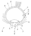


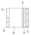
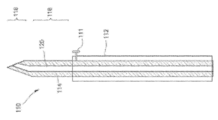

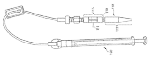

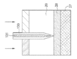


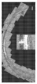
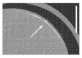


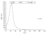


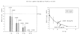


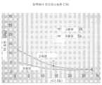


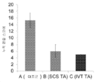


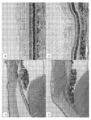


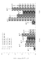
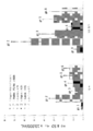
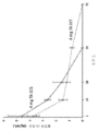


Classifications
-
- A—HUMAN NECESSITIES
- A61—MEDICAL OR VETERINARY SCIENCE; HYGIENE
- A61K—PREPARATIONS FOR MEDICAL, DENTAL OR TOILETRY PURPOSES
- A61K31/00—Medicinal preparations containing organic active ingredients
- A61K31/33—Heterocyclic compounds
- A61K31/395—Heterocyclic compounds having nitrogen as a ring hetero atom, e.g. guanethidine or rifamycins
- A61K31/435—Heterocyclic compounds having nitrogen as a ring hetero atom, e.g. guanethidine or rifamycins having six-membered rings with one nitrogen as the only ring hetero atom
- A61K31/44—Non condensed pyridines; Hydrogenated derivatives thereof
- A61K31/4427—Non condensed pyridines; Hydrogenated derivatives thereof containing further heterocyclic ring systems
- A61K31/4439—Non condensed pyridines; Hydrogenated derivatives thereof containing further heterocyclic ring systems containing a five-membered ring with nitrogen as a ring hetero atom, e.g. omeprazole
-
- A—HUMAN NECESSITIES
- A61—MEDICAL OR VETERINARY SCIENCE; HYGIENE
- A61K—PREPARATIONS FOR MEDICAL, DENTAL OR TOILETRY PURPOSES
- A61K31/00—Medicinal preparations containing organic active ingredients
- A61K31/56—Compounds containing cyclopenta[a]hydrophenanthrene ring systems; Derivatives thereof, e.g. steroids
- A61K31/57—Compounds containing cyclopenta[a]hydrophenanthrene ring systems; Derivatives thereof, e.g. steroids substituted in position 17 beta by a chain of two carbon atoms, e.g. pregnane or progesterone
- A61K31/573—Compounds containing cyclopenta[a]hydrophenanthrene ring systems; Derivatives thereof, e.g. steroids substituted in position 17 beta by a chain of two carbon atoms, e.g. pregnane or progesterone substituted in position 21, e.g. cortisone, dexamethasone, prednisone or aldosterone
-
- A—HUMAN NECESSITIES
- A61—MEDICAL OR VETERINARY SCIENCE; HYGIENE
- A61K—PREPARATIONS FOR MEDICAL, DENTAL OR TOILETRY PURPOSES
- A61K39/00—Medicinal preparations containing antigens or antibodies
- A61K39/395—Antibodies; Immunoglobulins; Immune serum, e.g. antilymphocytic serum
-
- A—HUMAN NECESSITIES
- A61—MEDICAL OR VETERINARY SCIENCE; HYGIENE
- A61K—PREPARATIONS FOR MEDICAL, DENTAL OR TOILETRY PURPOSES
- A61K45/00—Medicinal preparations containing active ingredients not provided for in groups A61K31/00 - A61K41/00
- A61K45/06—Mixtures of active ingredients without chemical characterisation, e.g. antiphlogistics and cardiaca
-
- A—HUMAN NECESSITIES
- A61—MEDICAL OR VETERINARY SCIENCE; HYGIENE
- A61K—PREPARATIONS FOR MEDICAL, DENTAL OR TOILETRY PURPOSES
- A61K47/00—Medicinal preparations characterised by the non-active ingredients used, e.g. carriers or inert additives; Targeting or modifying agents chemically bound to the active ingredient
- A61K47/06—Organic compounds, e.g. natural or synthetic hydrocarbons, polyolefins, mineral oil, petrolatum or ozokerite
- A61K47/08—Organic compounds, e.g. natural or synthetic hydrocarbons, polyolefins, mineral oil, petrolatum or ozokerite containing oxygen, e.g. ethers, acetals, ketones, quinones, aldehydes, peroxides
- A61K47/12—Carboxylic acids; Salts or anhydrides thereof
-
- A—HUMAN NECESSITIES
- A61—MEDICAL OR VETERINARY SCIENCE; HYGIENE
- A61K—PREPARATIONS FOR MEDICAL, DENTAL OR TOILETRY PURPOSES
- A61K47/00—Medicinal preparations characterised by the non-active ingredients used, e.g. carriers or inert additives; Targeting or modifying agents chemically bound to the active ingredient
- A61K47/06—Organic compounds, e.g. natural or synthetic hydrocarbons, polyolefins, mineral oil, petrolatum or ozokerite
- A61K47/26—Carbohydrates, e.g. sugar alcohols, amino sugars, nucleic acids, mono-, di- or oligo-saccharides; Derivatives thereof, e.g. polysorbates, sorbitan fatty acid esters or glycyrrhizin
-
- A—HUMAN NECESSITIES
- A61—MEDICAL OR VETERINARY SCIENCE; HYGIENE
- A61K—PREPARATIONS FOR MEDICAL, DENTAL OR TOILETRY PURPOSES
- A61K47/00—Medicinal preparations characterised by the non-active ingredients used, e.g. carriers or inert additives; Targeting or modifying agents chemically bound to the active ingredient
- A61K47/30—Macromolecular organic or inorganic compounds, e.g. inorganic polyphosphates
- A61K47/36—Polysaccharides; Derivatives thereof, e.g. gums, starch, alginate, dextrin, hyaluronic acid, chitosan, inulin, agar or pectin
- A61K47/38—Cellulose; Derivatives thereof
-
- A—HUMAN NECESSITIES
- A61—MEDICAL OR VETERINARY SCIENCE; HYGIENE
- A61K—PREPARATIONS FOR MEDICAL, DENTAL OR TOILETRY PURPOSES
- A61K9/00—Medicinal preparations characterised by special physical form
- A61K9/0012—Galenical forms characterised by the site of application
- A61K9/0019—Injectable compositions; Intramuscular, intravenous, arterial, subcutaneous administration; Compositions to be administered through the skin in an invasive manner
-
- A—HUMAN NECESSITIES
- A61—MEDICAL OR VETERINARY SCIENCE; HYGIENE
- A61K—PREPARATIONS FOR MEDICAL, DENTAL OR TOILETRY PURPOSES
- A61K9/00—Medicinal preparations characterised by special physical form
- A61K9/0012—Galenical forms characterised by the site of application
- A61K9/0048—Eye, e.g. artificial tears
-
- A—HUMAN NECESSITIES
- A61—MEDICAL OR VETERINARY SCIENCE; HYGIENE
- A61K—PREPARATIONS FOR MEDICAL, DENTAL OR TOILETRY PURPOSES
- A61K9/00—Medicinal preparations characterised by special physical form
- A61K9/10—Dispersions; Emulsions
-
- A—HUMAN NECESSITIES
- A61—MEDICAL OR VETERINARY SCIENCE; HYGIENE
- A61K—PREPARATIONS FOR MEDICAL, DENTAL OR TOILETRY PURPOSES
- A61K9/00—Medicinal preparations characterised by special physical form
- A61K9/14—Particulate form, e.g. powders, Processes for size reducing of pure drugs or the resulting products, Pure drug nanoparticles
- A61K9/16—Agglomerates; Granulates; Microbeadlets ; Microspheres; Pellets; Solid products obtained by spray drying, spray freeze drying, spray congealing,(multiple) emulsion solvent evaporation or extraction
-
- A—HUMAN NECESSITIES
- A61—MEDICAL OR VETERINARY SCIENCE; HYGIENE
- A61K—PREPARATIONS FOR MEDICAL, DENTAL OR TOILETRY PURPOSES
- A61K9/00—Medicinal preparations characterised by special physical form
- A61K9/48—Preparations in capsules, e.g. of gelatin, of chocolate
-
- A—HUMAN NECESSITIES
- A61—MEDICAL OR VETERINARY SCIENCE; HYGIENE
- A61M—DEVICES FOR INTRODUCING MEDIA INTO, OR ONTO, THE BODY; DEVICES FOR TRANSDUCING BODY MEDIA OR FOR TAKING MEDIA FROM THE BODY; DEVICES FOR PRODUCING OR ENDING SLEEP OR STUPOR
- A61M37/00—Other apparatus for introducing media into the body; Percutany, i.e. introducing medicines into the body by diffusion through the skin
- A61M37/0015—Other apparatus for introducing media into the body; Percutany, i.e. introducing medicines into the body by diffusion through the skin by using microneedles
-
- A—HUMAN NECESSITIES
- A61—MEDICAL OR VETERINARY SCIENCE; HYGIENE
- A61P—SPECIFIC THERAPEUTIC ACTIVITY OF CHEMICAL COMPOUNDS OR MEDICINAL PREPARATIONS
- A61P25/00—Drugs for disorders of the nervous system
-
- A—HUMAN NECESSITIES
- A61—MEDICAL OR VETERINARY SCIENCE; HYGIENE
- A61P—SPECIFIC THERAPEUTIC ACTIVITY OF CHEMICAL COMPOUNDS OR MEDICINAL PREPARATIONS
- A61P27/00—Drugs for disorders of the senses
- A61P27/02—Ophthalmic agents
-
- A—HUMAN NECESSITIES
- A61—MEDICAL OR VETERINARY SCIENCE; HYGIENE
- A61P—SPECIFIC THERAPEUTIC ACTIVITY OF CHEMICAL COMPOUNDS OR MEDICINAL PREPARATIONS
- A61P27/00—Drugs for disorders of the senses
- A61P27/02—Ophthalmic agents
- A61P27/06—Antiglaucoma agents or miotics
-
- A—HUMAN NECESSITIES
- A61—MEDICAL OR VETERINARY SCIENCE; HYGIENE
- A61P—SPECIFIC THERAPEUTIC ACTIVITY OF CHEMICAL COMPOUNDS OR MEDICINAL PREPARATIONS
- A61P27/00—Drugs for disorders of the senses
- A61P27/02—Ophthalmic agents
- A61P27/10—Ophthalmic agents for accommodation disorders, e.g. myopia
-
- A—HUMAN NECESSITIES
- A61—MEDICAL OR VETERINARY SCIENCE; HYGIENE
- A61P—SPECIFIC THERAPEUTIC ACTIVITY OF CHEMICAL COMPOUNDS OR MEDICINAL PREPARATIONS
- A61P29/00—Non-central analgesic, antipyretic or antiinflammatory agents, e.g. antirheumatic agents; Non-steroidal antiinflammatory drugs [NSAID]
-
- A—HUMAN NECESSITIES
- A61—MEDICAL OR VETERINARY SCIENCE; HYGIENE
- A61P—SPECIFIC THERAPEUTIC ACTIVITY OF CHEMICAL COMPOUNDS OR MEDICINAL PREPARATIONS
- A61P3/00—Drugs for disorders of the metabolism
- A61P3/08—Drugs for disorders of the metabolism for glucose homeostasis
- A61P3/10—Drugs for disorders of the metabolism for glucose homeostasis for hyperglycaemia, e.g. antidiabetics
-
- A—HUMAN NECESSITIES
- A61—MEDICAL OR VETERINARY SCIENCE; HYGIENE
- A61P—SPECIFIC THERAPEUTIC ACTIVITY OF CHEMICAL COMPOUNDS OR MEDICINAL PREPARATIONS
- A61P31/00—Antiinfectives, i.e. antibiotics, antiseptics, chemotherapeutics
- A61P31/12—Antivirals
- A61P31/20—Antivirals for DNA viruses
-
- A—HUMAN NECESSITIES
- A61—MEDICAL OR VETERINARY SCIENCE; HYGIENE
- A61P—SPECIFIC THERAPEUTIC ACTIVITY OF CHEMICAL COMPOUNDS OR MEDICINAL PREPARATIONS
- A61P37/00—Drugs for immunological or allergic disorders
- A61P37/02—Immunomodulators
-
- A—HUMAN NECESSITIES
- A61—MEDICAL OR VETERINARY SCIENCE; HYGIENE
- A61P—SPECIFIC THERAPEUTIC ACTIVITY OF CHEMICAL COMPOUNDS OR MEDICINAL PREPARATIONS
- A61P43/00—Drugs for specific purposes, not provided for in groups A61P1/00-A61P41/00
-
- A—HUMAN NECESSITIES
- A61—MEDICAL OR VETERINARY SCIENCE; HYGIENE
- A61P—SPECIFIC THERAPEUTIC ACTIVITY OF CHEMICAL COMPOUNDS OR MEDICINAL PREPARATIONS
- A61P7/00—Drugs for disorders of the blood or the extracellular fluid
- A61P7/02—Antithrombotic agents; Anticoagulants; Platelet aggregation inhibitors
-
- A—HUMAN NECESSITIES
- A61—MEDICAL OR VETERINARY SCIENCE; HYGIENE
- A61P—SPECIFIC THERAPEUTIC ACTIVITY OF CHEMICAL COMPOUNDS OR MEDICINAL PREPARATIONS
- A61P9/00—Drugs for disorders of the cardiovascular system
- A61P9/10—Drugs for disorders of the cardiovascular system for treating ischaemic or atherosclerotic diseases, e.g. antianginal drugs, coronary vasodilators, drugs for myocardial infarction, retinopathy, cerebrovascula insufficiency, renal arteriosclerosis
-
- C—CHEMISTRY; METALLURGY
- C07—ORGANIC CHEMISTRY
- C07K—PEPTIDES
- C07K16/00—Immunoglobulins [IGs], e.g. monoclonal or polyclonal antibodies
- C07K16/18—Immunoglobulins [IGs], e.g. monoclonal or polyclonal antibodies against material from animals or humans
- C07K16/22—Immunoglobulins [IGs], e.g. monoclonal or polyclonal antibodies against material from animals or humans against growth factors ; against growth regulators
-
- A—HUMAN NECESSITIES
- A61—MEDICAL OR VETERINARY SCIENCE; HYGIENE
- A61K—PREPARATIONS FOR MEDICAL, DENTAL OR TOILETRY PURPOSES
- A61K39/00—Medicinal preparations containing antigens or antibodies
- A61K2039/505—Medicinal preparations containing antigens or antibodies comprising antibodies
-
- A—HUMAN NECESSITIES
- A61—MEDICAL OR VETERINARY SCIENCE; HYGIENE
- A61K—PREPARATIONS FOR MEDICAL, DENTAL OR TOILETRY PURPOSES
- A61K39/00—Medicinal preparations containing antigens or antibodies
- A61K2039/54—Medicinal preparations containing antigens or antibodies characterised by the route of administration
-
- A—HUMAN NECESSITIES
- A61—MEDICAL OR VETERINARY SCIENCE; HYGIENE
- A61K—PREPARATIONS FOR MEDICAL, DENTAL OR TOILETRY PURPOSES
- A61K2300/00—Mixtures or combinations of active ingredients, wherein at least one active ingredient is fully defined in groups A61K31/00 - A61K41/00
-
- A—HUMAN NECESSITIES
- A61—MEDICAL OR VETERINARY SCIENCE; HYGIENE
- A61M—DEVICES FOR INTRODUCING MEDIA INTO, OR ONTO, THE BODY; DEVICES FOR TRANSDUCING BODY MEDIA OR FOR TAKING MEDIA FROM THE BODY; DEVICES FOR PRODUCING OR ENDING SLEEP OR STUPOR
- A61M37/00—Other apparatus for introducing media into the body; Percutany, i.e. introducing medicines into the body by diffusion through the skin
- A61M37/0015—Other apparatus for introducing media into the body; Percutany, i.e. introducing medicines into the body by diffusion through the skin by using microneedles
- A61M2037/0023—Drug applicators using microneedles
-
- A—HUMAN NECESSITIES
- A61—MEDICAL OR VETERINARY SCIENCE; HYGIENE
- A61M—DEVICES FOR INTRODUCING MEDIA INTO, OR ONTO, THE BODY; DEVICES FOR TRANSDUCING BODY MEDIA OR FOR TAKING MEDIA FROM THE BODY; DEVICES FOR PRODUCING OR ENDING SLEEP OR STUPOR
- A61M37/00—Other apparatus for introducing media into the body; Percutany, i.e. introducing medicines into the body by diffusion through the skin
- A61M37/0015—Other apparatus for introducing media into the body; Percutany, i.e. introducing medicines into the body by diffusion through the skin by using microneedles
- A61M2037/0061—Methods for using microneedles
Abstract
후안구 장애 또는 맥락막 질환의 치료를 위한 인간 대상체의 눈의 맥락막위 공간 (SCS)으로의 약물 제형의 표적화된 비-외과적 투여 방법 및 장치가 제공된다. 일 구체예에서, 본 방법은 중공 미세바늘을 삽입 부위의 눈으로 삽입하고, 삽입된 미세바늘을 통해 약물 제형을 눈의 맥락막위 공간 내로 주입하는 것을 포함하며, 주입된 약물 제형은 주입 동안 삽입 부위로부터 맥락막위 공간 내에서 멀리 유동한다. 일 구체예에서, 유체 약물 제형은 약물 나노입자 또는 마이크로입자를 포함한다.Methods and devices are provided for the targeted non-surgical administration of a drug formulation to the suprachoroidal space (SCS) of an eye of a human subject for the treatment of a retroocular disorder or choroidal disease. In one embodiment, the method comprises inserting a hollow microneedle into the eye at the insertion site, and injecting the drug formulation through the inserted microneedle into the suprachoroidal space of the eye, wherein the injected drug formulation is administered at the insertion site during the injection. flows away in the suprachoroidal space from In one embodiment, the fluid drug formulation comprises drug nanoparticles or microparticles.
Description
관련 출원의 교차 참조Cross-reference to related applications
본 출원은 2012년 11월 8일 출원된 미국 가출원 일련 번호 61/724,144; 2012년 12월 7일에 출원된 61/734,872; 2012년 12월 21일 출원된 61/745,237; 2013년 3월 5일 출원된 61/773,124; 2013년 3월 14일 출원된 61/785,229; 2013년 5월 3일 출원된 61/819,388; 2013년 9월 4일 출원된 61/873,660; 및 2013년 11월 1일 출원된 61/898,926으로부터 우선권을 주장하며, 이들 모두는 모든 목적에 있어서 그들 전체가 참조로서 본원에 통합된다.This application is filed on November 8, 2012 in United States Provisional Application Serial Nos. 61/724,144; 61/734,872, filed December 7, 2012; 61/745,237, filed December 21, 2012; 61/773,124, filed March 5, 2013; 61/785,229, filed March 14, 2013; 61/819,388, filed May 3, 2013; 61/873,660, filed September 4, 2013; and 61/898,926, filed November 1, 2013, all of which are incorporated herein by reference in their entirety for all purposes.
본 발명은 일반적으로 안구 요법 분야에 속하며, 더욱 특히, 국소 약물 전달을 위해 표적으로 하는 안구 조직으로의 유체 약물 제형의 주입을 위한 미세바늘의 용도에 관한 것이다.The present invention generally belongs to the field of ocular therapy, and more particularly relates to the use of microneedles for injection of fluid drug formulations into targeted ocular tissues for topical drug delivery.
눈으로의 약물 전달 특히, 대분자의 전달 및 후안부로의 전달은 극도로 어렵다. 후안 영역에서의 많은 염증성 및 증식성 질병에는 장기간의 약물학적 치료가 요구된다. 이러한 질병의 예로는 황반변성, 당뇨망막병증, 및 포도막염을 포함한다. 또한, 염증성 반응, 증식 및 혈관신생과 관련된 많은 맥락막 질환에는 장기간의 약물학적 치료가 요구된다. 효능이 빈약한 국소 적용 및 종종 심각한 부작용을 초래하며 종종 감염 부위에 도달하지 못하는 전신 투여와 같은 통상적인 전달 방법을 이용하여 후안부에 유효 용량의 약물을 전달하는 것은 어렵다. (Geroski & Edelhauser, Invest. Ophthalmol. Vis. Sci. 41:961-64 (2000)). 예를 들어, 안약은 눈 또는 눈 전방의 조직(들)의 외표면에 발생한 질환을 치료하는데 유용하지만, 안약은 눈에 현저하게 침투될 수 없는데, 이러한 침투는 다양한 망막 질병 및 맥락막 질환 치료에 요구될 수 있다.Delivery of drugs to the eye, in particular, of large molecules and delivery to the posterior segment is extremely difficult. Many inflammatory and proliferative diseases in the posterior ocular region require long-term pharmacological treatment. Examples of such diseases include macular degeneration, diabetic retinopathy, and uveitis. In addition, many choroidal diseases associated with inflammatory responses, proliferation and angiogenesis require long-term pharmacological treatment. It is difficult to deliver an effective dose of a drug to the posterior segment using conventional delivery methods such as poor efficacy topical application and systemic administration, which often results in severe side effects and often does not reach the site of infection. (Geroski & Edelhauser, Invest. Ophthalmol. Vis. Sci. 41:961-64 (2000)). For example, while eye drops are useful for treating diseases occurring on the outer surface of the eye or the tissue(s) in front of the eye, eye drops cannot penetrate appreciably into the eye, which penetration is required for the treatment of various retinal and choroidal diseases. can be
통상적인 바늘 및 주사기를 이용한 안구로의 직접 주입은 효과적인 것으로 보고되었으나, 전문적인 훈련이 요구되며, 안전에 대한 주의가 증가된다 (Maurice, J. Ocul. Pharmacol. Ther. 17:393-401 (2001)). 약물을 필요로하는 안구 조직 부위로 치료학적 유효량의 약물을 전달하는데 필요한 눈 주입 치료의 횟수 및/또는 빈도를 최소화할 수 있는 것이 또한 바람직할 것이다.Direct injection into the eye using conventional needles and syringes has been reported to be effective, but requires specialized training and increased safety precautions (Maurice, J. Ocul. Pharmacol. Ther. 17:393-401 (2001) )). It would also be desirable to be able to minimize the number and/or frequency of ocular infusion treatments required to deliver a therapeutically effective amount of a drug to an ocular tissue site in need thereof.
눈의 맥락막위 공간 (SCS)은 연구되었으며, 이의 관삽입은 약물 전달을 위한 가능한 경로로서 기술되었다. 예를 들어, 문헌 [Olsen, et al., American J. Ophthalmology 142(5): 777-87 (Nov. 2006).; PCT 특허 출원 공개 번호 WO 2007/100745] 참조.The suprachoroidal space (SCS) of the eye has been studied and its cannulation has been described as a possible route for drug delivery. See, eg, Olsen, et al., American J. Ophthalmology 142(5): 777-87 (Nov. 2006); PCT Patent Application Publication No. WO 2007/100745].
따라서, 예를 들어, 후안구 장애를 치료하기 위해 후안부 조직으로 치료제를 직접 전달하기 위한 더욱 우수하고, 안전하고 더욱 효과적인 기법을 제공하는 것이 바람직할 것이다. 또한, 맥락막 질환 예를 들어, 혈관 이상과 관련된 맥락막 질환의 치료를 위해 SCS에 치료제를 직접 전달하기 위한 더욱 우수하고, 안전하고 더욱 효과적인 기법을 제공하는 것이 바람직할 것이다. 본 발명은 이러한 요구 및 그 밖의 요구를 다룬다.Accordingly, it would be desirable to provide a better, safer and more effective technique for delivering therapeutic agents directly to posterior segment tissues, for example to treat posterior ocular disorders. It would also be desirable to provide a better, safer and more effective technique for delivering therapeutics directly to SCS for the treatment of choroidal diseases, such as choroidal diseases associated with vascular abnormalities. The present invention addresses these and other needs.
발명의 개요Summary of invention
일 양태에서, 본 발명은 안과 치료가 요구되는 인간 환자에서 비-외과적 안과 치료, 및 더욱 특히, 후안구 장애, 맥락막 질환 및 혈관 이상과 관련된 그 밖의 질병의 치료를 위한 표적화된 국소 약물 전달을 위한 눈의 맥락막위 공간으로의 약물 제형의 주입에 관한 것이다.In one aspect, the present invention provides targeted topical drug delivery for non-surgical ophthalmic treatment, and more particularly, for the treatment of posterior ocular disorders, choroidal disease and other diseases associated with vascular abnormalities in a human patient in need thereof. to the injection of a drug formulation into the suprachoroidal space of the eye for
본 발명의 일 양태에서, 치료가 필요한 인간 대상체에서 후안구 장애를 치료하는 방법이 제공된다. 일 구체예에서, 이러한 방법은 후안구 장애 또는 맥락막 질환의 치료가 필요한 대상체의 안구의 맥락막위 공간 (SCS)에 유효량의 약물 제형을 비-외과적으로 투여하는 것을 포함한다. 추가의 구체예에서, 투여시, 약물 제형은 삽입 부위로부터 멀리 유동하며, 실질적으로 후안부로 국소화된다. 일 구체예에서, 후안구 장애는 안구 염증 질환 예컨대, 포도막염, 공막염, 녹내장, 눈 사르코이드증, 시신경염, 황반 부종, 당뇨망막병증, 황반변성, 각막 궤양, 자가면역 질환, AIDS의 안과적 징후, 시신경 변성, 지도형 위축, 맥락막 질병 또는 망막염이다. 일 구체예에서 질환은 급성이다. 또 다른 구체예에서, 질환은 만성이다.In one aspect of the invention, there is provided a method of treating a retroocular disorder in a human subject in need thereof. In one embodiment, such a method comprises non-surgically administering an effective amount of a drug formulation to the suprachoroidal space (SCS) of the eye of a subject in need of treatment of a posterior ocular disorder or choroidal disease. In a further embodiment, upon administration, the drug formulation flows away from the site of insertion and is localized substantially to the posterior segment. In one embodiment, the posterior ocular disorder is an ocular inflammatory disease such as uveitis, scleritis, glaucoma, ocular sarcoidosis, optic neuritis, macular edema, diabetic retinopathy, macular degeneration, corneal ulceration, autoimmune disease, ophthalmic signs of AIDS, optic nerve degeneration, geographic atrophy, choroidal disease or retinitis. In one embodiment the disease is acute. In another embodiment, the disease is chronic.
또 다른 구체예에서, 맥락막 질환, 예를 들어, 안구 혈관신생, 결절 맥락막 혈관병증, 맥락막 경화증, 중심성 장액 맥락막병증, 다초점 맥락막병증 또는 맥락막 이영양증 (예를 들어, 중심 뇌회형 맥락막 이영양증 (central gyrate choroidal dystrophy), 포행성 맥락막 이영양증, 전체 중심 맥락막 위축)을 치료하기 위한 방법이 제공된다. 일 구체예에서, 이러한 방법은 치료가 필요한 환자의 SCS에 유효량의 항-염증 약물, 혈관 내피 성장 인자 (VEGF) 조절인자, 혈소판 유래 성장 인자 (PDGF) 조절인자, 혈관형성 억제제, 면역억제제, 혈관 투과 억제제, 또는 이들의 조합물을 포함하는 약물 제형을 비-외과적으로 투여하는 것을 포함한다. 추가의 구체예에서, SCS로 투여된 유효량의 약물은 유리체내, 앞방내, 국소, 비경구 또는 경구로 투여된 동일한 약물 용량과 비교하여, 약물의 더 높은 효능 또는 더 큰 치료 효과를 제공한다. 더욱 추가의 구체예에서, SCS 약물 요법으로 치료중인 환자는 이전에 동일한 질환에 대해 상이한 유형의 요법에 대해서 반응적이지 않았다.In another embodiment, a choroidal disease, e.g., ocular neovascularization, nodular choroidal angiopathy, choroidal sclerosis, central serous choroidopathy, multifocal choroidopathy or choroidal dystrophy (e.g., central gyrate choroidal dystrophy (central gyrate) A method is provided for treating choroidal dystrophy), entrapment choroidal dystrophy, total central choroidal atrophy). In one embodiment, the method comprises an effective amount of an anti-inflammatory drug, a vascular endothelial growth factor (VEGF) modulator, a platelet-derived growth factor (PDGF) modulator, an angiogenesis inhibitor, an immunosuppressant, a vascular non-surgical administration of a drug formulation comprising a permeation inhibitor, or a combination thereof. In a further embodiment, an effective amount of a drug administered with SCS provides a higher potency or greater therapeutic effect of the drug as compared to the same drug dose administered intravitreally, intracamerally, topically, parenterally or orally. In a still further embodiment, the patient being treated with SCS drug therapy has not previously been responsive to a different type of therapy for the same disease.
추가의 또 다른 구체예에서, 대상체에서 망막하 삼출 및 출혈 감소를 위한 방법이 제공된다. 추가의 구체예에서, 이러한 방법은 치료가 필요한 환자의 SCS에 유효량의 항-염증 약물, 혈관 내피 성장 인자 (VEGF) 조절인자, 혈소판 유래 성장 인자 (PDGF) 조절인자, 혈관형성 억제제, 면역억제제, 혈관 투과 억제제, 또는 이들의 조합물을 포함하는 약물 제형을 비-외과적으로 투여하는 것을 포함하며, 이러한 약물 제형의 투여는 환자의 유리체내로 투여된 동일한 투여량의 약물과 비교할 경우, 환자가 겪게되는 망막하 삼출 및 출혈을 감소시킨다.In yet another embodiment, a method for reducing subretinal effusion and bleeding in a subject is provided. In a further embodiment, the method comprises an effective amount of an anti-inflammatory drug, a vascular endothelial growth factor (VEGF) modulator, a platelet derived growth factor (PDGF) modulator, an angiogenesis inhibitor, an immunosuppressant, comprising non-surgically administering a drug formulation comprising a vascular permeation inhibitor, or a combination thereof, wherein administration of the drug formulation may result in a patient Reduces the subretinal effusion and hemorrhage experienced.
일 구체예에서, 인간 환자에서 후안구 장애 또는 맥락막 질환을 치료하는 방법이 제공된다. 추가의 구체예에서, 이러한 방법은 후안구 장애 또는 맥락막 질환의 치료가 필요한 대상체의 안구의 맥락막위 공간 (SCS)에 유효량의 약물 제형을 비-외과적으로 투여하는 것을 포함한다. 추가의 구체예에서, SCS에 투여된 약물의 안구내 제거 반감기 (t1/ 2)는 유리체내, 앞방내, 국소, 비경구 또는 경구 투여되는 경우의 약물의 안구내 t1/2보다 크다. 또 다른 구체예에서, 본원에 기술된 방법을 통해 SCS에 투여되는 경우 약물의 평균 안구내 최대 농도 (Cmax)는 유리체내, 앞방내, 국소, 비경구 또는 경구 투여되는 경우의 약물의 안구내 Cmax보다 크다. 또 다른 구체예에서, 본원에 기술된 방법을 통해 SCS에 투여되는 경우 약물의 평균 안구내 곡선하 영역 (AUCO -t)은 유리체내, 앞방내, 국소, 비경구 또는 경구 투여되는 경우의 약물의 안구내 AUC0 -t보다 크다. 추가의 또 다른 구체예에서, 본원에 기술된 방법을 통해 SCS에 투여되는 경우 약물의 안구내 피크 농도까지의 시간 (tmax)는 동일한 약물 용량이 유리체내, 앞방내, 국소, 비경구 또는 경구 투여되는 경우의 약물의 안구내 tmax보다 크다. 추가의 구체예에서, 약물 제형은 유효량의 항-염증 약물 (예를 들어, 스테로이드 또는 NSAID), VEGF 조절인자 (예를 들어, VEGF 길항제), 혈소판 유래 성장 인자 (PDGF) 조절인자, 혈관형성 억제제, 면역억제제, 혈관 투과 억제제, 또는 이들의 조합물을 포함한다.In one embodiment, a method of treating a retroocular disorder or choroidal disease in a human patient is provided. In a further embodiment, such a method comprises non-surgically administering an effective amount of a drug formulation to the suprachoroidal space (SCS) of the eye of a subject in need of treatment of a posterior ocular disorder or choroidal disease. In a further embodiment, the intraocular elimination half-life of the drug administered to the SCS (t 1/2) is the eye of the drug in a case where the glass body, in front bangnae, topical, parenteral or oral administration is greater than that in the t 1/2. In another embodiment, the mean intraocular maximal concentration (C max ) of the drug when administered to SCS via the methods described herein is the intraocular maximum concentration (C max ) of the drug when administered intravitreally, intracamerally, topically, parenterally or orally. greater than C max. In another embodiment, the mean intraocular area under the curve (AUC O -t ) of the drug when administered to SCS via the methods described herein is the drug when administered intravitreally, intracamerally, topically, parenterally, or orally. is greater than the intraocular AUC 0 -t of In yet another embodiment, the time to intraocular peak concentration (t max ) of the drug when administered to SCS via the methods described herein is equal to the same drug dose administered intravitreally, intracamerally, topically, parenterally, or orally. greater than the intraocular t max of the drug when administered. In a further embodiment, the drug formulation comprises an effective amount of an anti-inflammatory drug (eg, a steroid or NSAID), a VEGF modulator (eg, a VEGF antagonist), a platelet derived growth factor (PDGF) modulator, an angiogenesis inhibitor , an immunosuppressant, an inhibitor of vascular permeation, or a combination thereof.
일 구체예에서, 인간 대상체에서 후안구 장애 또는 맥락막 질환을 치료하는 방법은 치료가 필요한 인간 대상체의 눈의 SCS로 중공 미세바늘을 통해 약물 제형을 전달하는 것을 포함한다. 추가의 구체예에서, 약물 제형의 전달은 개구를 갖는 팁 말단을 지닌 중공 미세바늘을 인간 대상체의 눈의 삽입 부위로 삽입하고; 삽입된 미세바늘을 통하여 삽입 부위로부터 떨어진 SCS 공간내로 약물 제형을 일정 기간에 걸쳐 주입하는 것을 포함한다. 일 구체예에서, SCS로 투여된 약물 제형은 삽입 부위로부터 멀리 유동하고, 실질적으로 후안부로 국소화되어, 또 다른 방식 (예를 들어, 유리체내, 앞방내, 국소, 비경구, 및/또는 경구)으로 투여된 동일한 약물 용량의 치료 효능과 비교하여 약물 용량의 치료 효능을 증가시킨다. 또 다른 구체예에서, SCS로 투여되는 경우 치료 반응을 유발시키기에 충분한 약물의 용량은 유리체내, 앞방내, 국소, 비경구, 또는 경구로 투여되는 경우의 치료 반응을 유발시키기에 충분한 약물의 투여량보다 적다. 또 다른 구체예에서, 약물 제형은 눈의 적도의 공막 내로 또는 눈의 적도와 가장자리 사이로 삽입된 중공 미세바늘에 의해 SCS로 전달된다. 추가의 구체예에서, 중공 미세바늘은 90도 각도 (수직)로 삽입 부위로 삽입된다.In one embodiment, a method of treating a posterior ocular disorder or choroidal disease in a human subject comprises delivering a drug formulation through a hollow microneedle to the SCS of an eye of a human subject in need thereof. In a further embodiment, delivery of the drug formulation comprises inserting a hollow microneedle having a tip end having an opening into an insertion site of an eye of a human subject; It involves injecting the drug formulation into the SCS space away from the insertion site through the inserted microneedle over a period of time. In one embodiment, the drug formulation administered with SCS flows away from the site of insertion and is localized substantially to the posterior segment, in another manner (e.g., intravitreal, intraanterior chamber, topical, parenteral, and/or oral ) increases the therapeutic efficacy of a drug dose compared to the therapeutic efficacy of the same drug dose administered as In another embodiment, the dose of drug sufficient to elicit a therapeutic response when administered with SCS is an administration of the drug sufficient to elicit a therapeutic response when administered intravitreally, intracamerally, topically, parenterally, or orally. less than the amount In another embodiment, the drug formulation is delivered to the SCS by a hollow microneedle inserted into the sclera of the equator of the eye or between the equator and edge of the eye. In a further embodiment, the hollow microneedle is inserted into the insertion site at a 90 degree angle (vertical).
일 구체예에서, 본원에 기술된 방법에 의해 전달된 약물 제형은 유효량의 항-염증 약물 예를 들어, 스테로이드 또는 비-스테로이드 항-염증 약물 (NSAID)을 포함한다. 또 다른 구체예에서, 본원에 기술된 방법을 통해 SCS로 전달된 약물은 스테로이드, 면역억제제, 항대사물, T-세포 억제제, 알킬화제, 생물학적 제제, TNFα 길항제, 인터루킨 길항제, 신경보호제, 혈관 내피 성장 인자 (VEGF) 길항제, 혈소판 유래 성장 인자 (PDGF) 길항제, 또는 이들의 조합물이다. 또 다른 구체예에서, 약물은 염증, 신경보호, 보체 억제, 드루젠 형성, 흉터 형성, 맥락막모세혈관층의 감소 또는 맥락막 혈관신생에 영향을 끼친다. 또 다른 구체예에서, 약물 제형은 약물의 마이크로입자 및/또는 나노입자를 포함한다. 일 구체예에서, 약물 제형은 D50이 1 μm 또는 그 미만이고/거나 D99가 10 μm 또는 그 미만인 마이크로입자를 포함한다.In one embodiment, the drug formulation delivered by the methods described herein comprises an effective amount of an anti-inflammatory drug, such as a steroid or non-steroidal anti-inflammatory drug (NSAID). In another embodiment, the drug delivered to the SCS via the methods described herein is a steroid, immunosuppressant, antimetabolite, T-cell inhibitor, alkylating agent, biologic, TNFα antagonist, interleukin antagonist, neuroprotective agent, vascular endothelial growth a factor (VEGF) antagonist, a platelet-derived growth factor (PDGF) antagonist, or a combination thereof. In another embodiment, the drug affects inflammation, neuroprotection, complement inhibition, drusen formation, scarring, reduction of the choriocapillaris or choroidal neovascularization. In another embodiment, the drug formulation comprises microparticles and/or nanoparticles of the drug. In one embodiment, the drug formulation comprises microparticles having a D 50 of 1 μm or less and/or a D 99 of 10 μm or less.
상기 제시된 바와 같이, 본 발명의 일 양태는 인간 대상체의 눈의 SCS에 약물 제형을 비-외과적으로 투여하는 것을 필요하여 후안구 장애의 치료가 필요한 인간 대상체에서 후안구 장애를 치료하기 위한 방법으로서, 투여시 약물 제형은 삽입 부위로부터 멀리 유동하며, 실질적으로 후안부에 국소화되는 방법을 포함한다. 본 방법의 일 구체예에서, 안구의 안압은 SCS로의 약물 제형의 투여 동안 실질적으로 일정하게 유지된다. 또 다른 구체예에서, 눈의 SCS로의 약물 제형의 투여는 유리체내, 앞방내, 국소, 비경구, 또는 경구로의 동일한 약물 용량의 투여와 비교하여, 감소된 수의 부작용, 또는 하나 이상의 부작용의 감소된 중증도를 유도한다.As set forth above, one aspect of the present invention is a method for treating a post-ocular disorder in a human subject in need thereof by non-surgical administration of a drug formulation to the SCS of the eye of the human subject. , wherein upon administration the drug formulation flows away from the site of insertion and is substantially localized in the posterior segment. In one embodiment of the method, the intraocular pressure of the eye is maintained substantially constant during administration of the drug formulation to the SCS. In another embodiment, administration of the drug formulation to the SCS of the eye results in a reduced number of side effects, or one or more side effects, compared to administration of the same drug dose intravitreally, intracamerally, topically, parenterally, or orally. lead to reduced severity.
본 발명의 일 양태에서, 본 발명은 치료가 필요한 인간 환자에서 맥락막 질환을 치료하는 방법에 관한 것이다. 일 구체예에서, 이러한 방법은 환자의 눈의 맥락막위 공간 (SCS)으로 유효량의 항-염증 약물, 혈관 내피 성장 인자 (VEGF) 조절인자, 혈소판 유래 성장 인자 (PDGF) 조절인자, 혈관형성 억제제, 면역억제제 또는 혈관 투과 억제제를 포함하는 약물 제형을 비-외과적으로 투여하는 것을 포함한다. 추가의 구체예에서, 약물 제형의 투여 전에 인간 환자는 맥락막 질환에 대해 이미 처치되었으나, 치료에 대해 적절하게 반응적이지 않았다.In one aspect of the invention, the invention relates to a method of treating a choroidal disease in a human patient in need thereof. In one embodiment, the method comprises an effective amount of an anti-inflammatory drug, a vascular endothelial growth factor (VEGF) modulator, a platelet derived growth factor (PDGF) modulator, an angiogenesis inhibitor, into the suprachoroidal space (SCS) of the eye of the patient. non-surgical administration of a drug formulation comprising an immunosuppressant or vascular permeation inhibitor. In a further embodiment, prior to administration of the drug formulation, the human patient has already been treated for a choroidal disease, but has not been adequately responsive to treatment.
본 발명의 또 다른 양태에서, 본 발명은 치료가 필요한 인간 환자에서 안구 혈관신생을 치료하는 방법에 관한 것이다. 일 구체예에서, 본 방법은 환자의 눈의 맥락막위 공간 (SCS)으로 유효량의 항-염증 약물, 혈관 내피 성장 인자 (VEGF) 조절인자 (예를 들어, VEGF 길항제), 혈소판 유래 성장 인자 (PDGF) 조절인자 (예를 들어, PDGF 길항제), 혈관형성 억제제, 면역억제제 또는 혈관 투과 억제제를 포함하는 약물 제형을 비-외과적으로 투여하는 것을 포함한다. 추가의 구체예에서, 안구 혈관신생은 맥락막 혈관신생이다. 일 구체예에서, 약물 제형의 투여 전에 안구 혈관신생에 대해 처치되는 인간 환자는 이미 안구 혈관신생에 대해 처치되었으나, 치료에 대한 적적하게 반응적이지 않았다.In another aspect of the invention, the invention relates to a method of treating ocular neovascularization in a human patient in need thereof. In one embodiment, the method comprises an effective amount of an anti-inflammatory drug, a vascular endothelial growth factor (VEGF) modulator (eg, a VEGF antagonist), platelet derived growth factor (PDGF) into the suprachoroidal space (SCS) of a patient's eye. ) a modulator (eg, a PDGF antagonist), an angiogenesis inhibitor, an immunosuppressant, or a vascular permeation inhibitor, non-surgical administration. In a further embodiment, the ocular neovascularization is choroidal neovascularization. In one embodiment, the human patient being treated for ocular neovascularization prior to administration of the drug formulation has already been treated for ocular neovascularization but has not been adequately responsive to treatment.
일 구체예에서, 본원에 기술된 방법에 의해 전달된 약물 제형은 유효량의 항-염증 약물 예를 들어, 스테로이드 화합물 또는 비-스테로이드 항-염증 약물 (NSAID)을 포함한다. 또 다른 구체예에서, 본원에 기술된 방법을 통해 SCS로 전달된 약물은 혈관 투과 억제제, 혈관형성 억제제 또는 VEGF 조절인자, 예를 들어, VEGF 길항제이다. 일 구체예에서, VEGF 길항제는 VEGF 수용체 길항제 또는 가용성 VEGF 수용체이다. 일 구체예에서, 약물 제형은 D50이 1μm 또는 그 미만이고/거나 D99가 10μm 또는 그 미만인 약물 마이크로입자를 포함한다. 추가의 구체예에서, 약물 제형은 트리암시놀론을 포함한다.In one embodiment, the drug formulation delivered by the methods described herein comprises an effective amount of an anti-inflammatory drug, such as a steroid compound or a non-steroidal anti-inflammatory drug (NSAID). In another embodiment, the drug delivered to the SCS via the methods described herein is a vascular permeation inhibitor, an angiogenesis inhibitor or a VEGF modulator, eg, a VEGF antagonist. In one embodiment, the VEGF antagonist is a VEGF receptor antagonist or a soluble VEGF receptor. In one embodiment, the drug formulation comprises drug microparticles having a D 50 of 1 μm or less and/or a D 99 of 10 μm or less. In a further embodiment, the drug formulation comprises triamcinolone.
본 발명의 일 구체예에서, 맥락막 질환 또는 후안구 장애의 치료가 필요한 인간 대상체의 눈의 SCS로 약물 제형을 비-외과적으로 투여하는 것을 포함하여 인간 대상체에서 맥락막 질환 또는 후안구 장애를 치료하는 방법으로서, 눈의 안압이 SCS로의 약물 제형의 투여 동안 실질적으로 일정하게 유지되는 방법이 제공된다. 또 다른 구체예에서, 맥락막 질환 또는 후안구 장애의 치료가 필요한 환자의 눈의 SCS로의 약물 제형의 투여는 유리체내, 앞방내, 국소, 비경구, 또는 경구로의 동일한 약물 용량의 투여와 비교하여, 감소된 수의 부작용 또는 하나 이상의 부작용의 감소된 중증도를 유도한다. 일 구체예에서, 본원에 기술된 방법에 의해 감소된 부작용은 망막하 삼출 및/또는 출혈이다.In one embodiment of the present invention, the treatment of choroidal disease or posterior ocular disorder in a human subject comprising non-surgical administration of a drug formulation to the SCS of the eye of a human subject in need of treatment of a choroidal disease or posterior ocular disorder. As a method, provided is a method wherein the intraocular pressure of the eye is maintained substantially constant during administration of the drug formulation to the SCS. In another embodiment, administration of the drug formulation to the SCS of the eye of a patient in need of treatment for a choroidal disease or posterior ocular disorder is compared to administration of the same drug dose intravitreally, intracamerally, topically, parenterally, or orally. , resulting in a reduced number of side effects or a decreased severity of one or more side effects. In one embodiment, the side effect reduced by the methods described herein is subretinal effusion and/or hemorrhage.
도 1a, 1b, 1c 및 1d는 인간 눈의 조직 구조의 단면도이다. (a) 전체 눈, (1b) 각막의 확대도, (1c) 맥락막위 공간에 유체가 없는 눈의 공막 및 관련 조직의 확대도, 또는 (1d) 맥락막위 공간에 유체가 있는 눈의 공막 및 관련 조직의 확대도.
도 2는 일 구체예에 따른 기다란 본체에 배치된 중공 미세바늘을 포함하는 미세바늘 장치의 단면도이다.
도 3은 도 2에 도시된 미세바늘 장치의 기다란 본체의 단면도이다.
도 4는 일 구체예에 따른 미세바늘 장치의 도면이다.
도 5는 일 구체예에 따른 미세바늘 장치의 도면이다.
도 6a 및 6b는 눈의 맥락막위 공간 내로 약물을 전달하기 위해 중공 미세바늘을 사용하기 위한 과정의 구체예를 나타내며, 여기에서 이러한 과정은 중공 미세바늘의 공막으로의 삽입 및 유체 약물 제형의 맥락막위 공간으로의 주입을 포함한다.
도 7a는 통상적인 30 게이지 피하 바늘의 팁과 비교한 일 구체예에 따른 중공 미세바늘의 비교를 보여준다. 도 7b는 전체 눈에 맞춰진 형상의 맞춤 아크릴 몰드의 도식도를 보여준다.
도 8a 및 8b는 각각 설포르하드아민 (sulforhadamine)의 주입 전 및 후의 돼지 눈의 시상 단면의 현미경 명시 야상이다.
도 9a, 9b, 9c 및 9d는 맥락막위 공간으로 주입하지 않은 돼지 눈의 동결절편 (9a), 축 평면에서 500 nm 형광 입자의 주입 후 파노라믹 뷰 (panoramic view)를 형성하기 위해 콜라쥬화시킨 토끼 눈의 동결절편 (9b), 시상 방향으로 500 nm 형광 입자의 주입 후 미세바늘 삽입 부위의 전방 및 후방 둘 모두의 공간을 보여주기 위해 콜라쥬화시킨 돼지 눈의 동결절편 (9c), 및 시상 방향으로 500 nm 형광 입자의 주입 후의 미세바늘 삽입 부위의 전방 및 후방 둘 모두를 보여주기 위해 콜라쥬화된 인간 눈의 동결절편 (9d)의 형광투시 이미지이다. 도 9b, 9c 및 9d의 삽도는 미세바늘 삽입 부위의 확대된 장면을 보여준다.
도 10a 및 10b는 돼지 눈의 맥락막위 공간으로 주입된 1μm 콘트라스트 입자의 주변 확산의 단면 이미지 (10a) 및 단면 이미지의 삼차원 재구성 (10b)을 보여주는 마이크로컴퓨터 단층촬영 이미지이다.
도 11a, 11b, 11c 및 11d는 (11a) 20 nm 입자, (11b) 100 nm 입자, (11c) 500 nm 입자 및 (11d) 1000 nm 입자의 돼지 눈으로의 맥락막위 전달의 성공률에 대한 주입압 및 미세바늘 길이의 효과를 보여주는 그래프이다.
도 12a 및 12b는 시상 방향으로 20 nm 입자 (12a) 및 1000 nm 입자 (12b)의 주입 후 미세바늘 삽입 부위의 전방 및 후방 둘 모두의 공간을 보여주기 위해 콜라쥬화된 돼지 눈의 동결절편의 형광투시 이미지이다. 도 12a 및 12b의 삽도는 미세바늘 삽입 부위의 확대된 장면을 보여준다.
도 13a 및 13b는 18 mmHg (13a) 및 36 mmHg (13b)의 모의 안압에 있어서 1000 nm 입자의 맥락막위 전달의 성공률에 대한 안압 및 미세바늘 길이의 효과를 보여주는 그래프이다.
도 14는 맥락막위 공간으로 소듐 플루오레세인의 주입 후 취해진 토끼 눈의 사이트 스캔 (sight scan)의 1-차원 선이며, x-축은 뒤 (0)에서부터 앞 (160)으로의 눈의 위치를 나타내며, y-축은 그러한 위치에서의 형광 강도를 나타낸다.
도 15는 시간에 걸친 맥락막위 공간으로부터의 소듐 플루오레세인의 제거율을 보여주는 그래프이다.
도 16은 시간에 걸친 맥락막위 공간으로부터 20 nm 입자의 제거율을 보여주는 그래프이다.
도 17은 시간에 걸친 맥막막위 공간으로부터 500 nm 입자의 제거율을 보여주는 그래프이다.
도 18은 일 구체예에 따른 눈으로의 약물 투여 방법의 블록 선도이다.
도 19a는 투여 후 시간에 따른 후안부에 유지된 트리암시놀론 (TA)의 양을 나타내는 그래프이다 (원형 - SCS 주입, 마름모 - 유리체내 주입).
도 19b는 유리체내로 투여된 트리암시놀론 (TA) (위)와 비교하여 SCS에 투여되는 경우 (아래)의 맥락막 및 망막에서의 트리암시놀론의 증가된 보유를 보여주는 그래프이다.
도 19c의 위는 투여 후 시간에 따른 눈 뒤(맥락막) 에서의 트리암시놀론 (TA)의 양에 대한 눈의 수정체의 TA의 양의 비를 보여주는 그래프이다. 도 19c 아래는 투여 후 시간 (일)에 따른 눈 뒤 (망막)에서 트리암시놀론 (TA)의 양에 대한 눈의 수정체의 TA의 양의 비를 보여주는 그래프이다.
도 20a는 TA 투여 후 시간에 따른 토끼 눈에서의 안압 (IOP, mmHg)의 그래프이다. 토끼에는 연구 0일에 비히클, 3.2 mg TA 또는 5.2 mg TA를 주입하였다.
도 20b는 3.2 mg의 트리암시놀론 (왼쪽) 또는 비히클 (오른쪽)의 맥락막위 주입 후 토끼 눈의 단면도이다.
도 20c는 토끼 눈의 SCS로의 TA의 투여 후 TA 투여 후 시간에 따른 TA 혈장내 농도 (ng/mL)를 보여주는 그래프이다.
도 20d는 SCS로의 투여 후 다양한 조직에서 트리암시놀론 (TA)의 보유 (μg TA/g 조직)를 보여주는 그래프이다. 가장 많은 양의 약물이 눈 뒤 (맥락막, 망막)의 조직에 보유되며, 더 적은 양이 전안부 (수청체, 유리체액)에 유지된다.
도 20e는 TA 투여 후 시간에 따른 공막 및 맥락막에서 트리암시놀론 (TA) (μg)의 양을 보여주는 그래프이다.
도 20f는 TA 투여 후 시간에 따른 망막에서 트리암시놀론 (TA) (μg)의 양을 보여주는 그래프이다.
도 21a는 처치 후 시간 및 LPS 독소 투여 후 시간에 따른 비히클 (왼쪽), SCS로 투여된 4 mg 트리암시놀론 (TA) (중간) 또는 유리체내로 투여된 4 mg 트리암시놀론 (TA) (오른쪽)으로 처리된 눈의 누적되는 McDonald-Shadduck 스코어를 보여주는 그래프이다. McDonald-Shadduck 스코어는 후방 포도막염 모델을 제공한다.
도 21b는 NZW 토끼에서 후방 포도막염 모델에서 SCS 또는 유리체내로 투여된 트리암시놀론 (TA)의 효과를 보여주는 대표적인 안저 사진이다.
도 21c는 최종 시점에서 조직학으로부터 측정된 NZW 토끼에서 전반적인 염증 중증도를 보여주는 그래프이다. 하기 조직이 분석되었다: 섬모체 돌기, 공막-맥락막, 유리체, 망막 및 시신경 (0-4 스케일, 최대 스코어=20).
도 21d는 IVT 또는 SCS TA 투여에 대한 반응에서 NZW 토끼에서의 안압 (mmGh)을 보여주는 그래프이다.
도 22a는 (i) 리포폴리사카라이드 (LPS) 독소 후 비히클 (왼쪽), (ii) LPS 독소 후 SCS로의 2 mg 트리암시놀론 (중간) 또는 (iii) 평형 염 용액 후 비히클로 시험처리된 돼지 눈의 평균 Hackett/McDonald 안구 스코어를 보여주는 그래프이다. 2 mg 용량으로의 SCS TA 처리는 이러한 돼지 포도막염 모델에서 안구 염증 반응을 현저하게 감소시킨다.
도 22b는 (i) 리포폴리사카라이드 (LPS) 독소 후 비히클, (ii) LPS 독소 후 SCS로의 2 mg 트리암시놀론 (TA) 또는 (iii) LPS 독소 후 유리체내로의 2 mg 트리암시놀론 또는 (iv) LPS 독소 후 SCS로의 0.2 mg 트리암시놀론으로 시험처리된 돼지 눈의 평균 Hackett/McDonald 안구 스코어를 보여주는 그래프이다. 염증 감소가, 유리체내로 투여되는 경우에 요구되는 TA의 용량의 10%인 용량으로 SCS로 투여된 TA로 3일 이내에 관찰되었다.
도 23은 독소로 처리되거나 독소 부재하에 이어서, SCS 또는 유리체내로 투여된 저 또는 고 용량의 TA로 처리되는 동물의 평균 (± 표준 편차) 누적 염증 안구 스코어를 보여주는 그래프이다. SCS TA로 처리된 눈의 평균 염증 스코어는 처치 후 1일, 2일 및 3일에 IVT TA로 처리된 눈의 스코어보다 낮았다.
포도막염 유도 (즉, 독소 투여)시 (-1일), 약물 투여 시 (0일)의 평균 (+/- SD) 누적 염증 안구 스코어. 0.2 mg (저 용량) 또는 2.0 mg (고 용량)의 트리암시놀론 아세토니드 (TA)를 맥락막위 공간 (SCS) 또는 유리체내 (IVT) 주입으로 눈에 투여하고, 안구 스코어를 처치 후 1, 2 및 3일에 계산하였다. 군 1 평균 누적 염증 스코어는 0일에 군 2 내지 6보다 현저하게 낮았다 (Wilcoxon; P<0.028); b. 군 2 평균 누적 염증 스코어는 1일에 군 1 및 3, 4, 5 및 6보다 현저하게 높았다 (Wilcoxon; P<0.028); c. 군 5 평균 누적 염증 스코어는 1일에 군 1, 3, 4 및 6보다 현저하게 높았다 (Wilcoxon; P<0.029); d. 군 6 평균 누적 염증 스코어는 1일에 군 1보다 현저하게 높았다 (Wilcoxon; P=0.02); e. 군 2 평균 누적 염증 스코어는 2일에 군 1, 3, 4 및 6보다 현저하게 높았다 (Wilcoxon; P<0.028); f. 군 5 평균 누적 염증 스코어는 2일에 군 1 및 3보다 현저하게 높았다 (Wilcoxon; P<0.042); g. 군 6 평균 누적 염증 스코어는 2일에 군 1보다 현저하게 높았다 (Wilcoxon; P=0.028); h. 군 2 평균 누적 염증 스코어는 3일에 군 1, 3, 4, 5 및 6보다 현저하게 높았다 (Wilcoxon; P<0.02); i. 군 5 평균 누적 염증 스코어는 3일에 군 1 및 6보다 현저하게 높았다 (Wilcoxon; P<0.047); j. 군 6 평균 누적 염증 스코어는 3일에 군 1보다 현저하게 높았다 (Wilcoxon; P=0.018). G1=군 1; G2=군 2; G3=군 3; G4=군 4; G5=군 5; G6=군 6.
도 24는 -1일에 독소로 투여되거나 독소 부재하에 이어서 0일에 SCS 또는 유리체내로 투여된 저 또는 고 용량의 TA로 처리된 동물의 눈의 평균 안압을 보여주는 그래프이다. 포도막염 유발 전 (-1일), 0.2 mg (저 용량) 또는 2.0 mg (고 용량) 트리암시놀론 아세토니드 (TA)의 맥락막위 공간 (SCS) 또는 유리체내 (IVT) 주입으로의 약물 투여일 (0일)에서의 돼지 눈의 평균 (+/- SD) 안압 (IOP). IOP는 처치 후 1hr., 3 hr., 6 hr. 1일, 2일 및 3일에 측정하였다. a. 군 1 눈의 IOP는 치료제 주입 후 1 및 3 시간에 군 2 눈보다 현저하게 높았다 (P=0.01; 0.04).
도 25a-b는 독소 투여되거나 독소 부재하에 이어서 SCS 또는 유리체내로 투여된 저 또는 고 용량의 TA로 처리된 동물의 눈의 와이드-필드 안저 이미지이다. 와이드-필드 안저 이미지는 -1일에 리포폴리사카라이드 (LPS) 주입 직전에, 처리 후 0일 및 3일에 비히클, 0.2 mg (저 용량) 또는 2.0 mg (고 용량)의 트리암시놀론 아세토니드 주입 직전에 이미지화한 것이다. 평형 염 용액 및 비히클 주입된 군 1 눈은 정상적인 외형을 유지하였다. 군 1 눈을 제외한 모든 눈에서 LPS 주입 후 24시간째에 후안부에 실질적인 혼탁함이 발생하였다. SCS로의 저 용량 및 고 용량 mg TA 및 고 용량 TA IVT로의 처리는 처리전 모양과 가까운 안저 이미지를 유도한 반면, 저용량 TA IVT로의 처리는 비히클 처리된 눈에 비해 단지 약간 개선된 이미지를 유도하였다. 2.0 mg TA IVT 주입된 눈은 중심 유리체에서 가시적인 TA의 큰 고형 데폿 (depot)을 가졌다.
도 26은 평형 염 용액 (BSS) 또는 100 ng의 리포폴리사카라이드 (LPS)의 유리체내 주입 후 3일에 또는 비히클, 0.2 mg TA, 또는 2.0 mg TA의 SCS 또는 IVT 투여 후 72 시간에 눈의 안구 조직병리를 보여준다. 검사한 눈 어느 것도 조직병리에서 실질적인 조직, 구조 또는 독소 변화의 증거를 갖지 않았다. 평형 염 용액 (BSS) 또는 100 ng의 리포폴리사카라이드 (LPS)의 유리체내 (IVT) 주입 후 3일 및 비히클, 0.2 mg 트리암시놀론 아세토니드 (저 용량 TA), 또는 2.0 mg의 트리암시놀론 아세토니드 (고 용량 TA)의 맥락막위 (SCS) 또는 IVT 주입 후 72 시간에서의 눈의 안구 조직병리. 헤마톡실린 및 에오신 염료.
A. BSS IVT 및 SCS로 비히클 주입된 전안부 (군 1). 기준 막대: 1 mm.
B. BSS IVT 및 SCS로 비히클 주입된 후안부 (군 1). 기준 막대: 200 μm.
C. LPS IVT 및 SCS로 비히클 주입된 전안부 (군 2). 기준 막대: 1 mm.
D. LPS IVT 및 SCS로 비히클 주입된 후안부 (군 2). 기준 막대: 200 μm.
E. LPS IVT 및 SCS로 저 용량 TA 주입된 전안부 (군 3). 기준 막대: 1 mm.
F. LPS IVT 및 SCS로 저 용량 TA 주입된 후안부 (군 3). 기준 막대: 200 μm.
G. LPS IVT 및 SCS로 고 용량 TA 주입된 전안부 (군 4). 기준 막대: 1 mm.
H. LPS IVT 및 SCS로 고 용량 TA 주입된 후안부 (군 4). 화살표는 SCS중 TA의 존재를 나타낸다. 기준 막대: 200 μm.
I. LPS IVT 및 저 용량 TA IVT로 주입된 전안부 (군 5). 기준 막대: 1 mm.
J. LPS IVT 및 저 용량 TA IVT로 주입된 후안부 (군 5). 기준 막대: 200 μm.
K. LPS IVT 및 고 용량 TA IVT로 주입된 전안부 (군 6). 기준 막대: 1 mm.
L. LPS IVT 및 고 용량 TA IVT로 주입된 후안부 (군 6). 기준 막대: 200 μm.
도 27은 평형 염 용액 (BSS) 또는 100 ng의 리포폴리사카라이드 (LPS)의 유리체내 (IVT) 주입 후 4일 및 비히클, 0.2 mg 트리암시놀론 아세토니드 (저 용량 TA), 또는 2.0 mg의 트리암시놀론 아세토니드 (고 용량 TA)의 맥락막위 (SCS) 또는 IVT 주입 후 3일에 전안부 및 후안부의 평균 안구 조직병리 염증 스코어를 보여준다. a. 군 1 평균 조직학적 염증 스코어는 군 2 내지 6보다 현저하게 낮았다 (P<0.04). b. 군 5 평균 조직학적 염증 스코어는 군 4 및 6보다 현저하게 높았다 (P<0.04). c. 군 4 평균 조직학적 염증 스코어는 군 2, 5, 및 6보다 현저하게 낮았다 (P<0.04). d. 군 6 평균 조직학적 염증 스코어는 군 2보다 현저하게 낮았다 (P=0.018).
도 28은 평형 염 용액 (BSS) 또는 100 ng의 리포폴리사카라이드 (LPS)의 유리체내 (IVT) 주입 후 3일 및 비히클, 0.2 mg 트리암시놀론 아세토니드 (저 용량 TA), 또는 2.0 mg의 트리암시놀론 아세토니드 (고 용량 TA)의 맥락막위 (SCS) 또는 IVT 주입 후 72시간에 평균 방수 (AH) 및 유리체액 (VH) 세포 계수를 보여준다. a. 군 2 평균 세포 계수는 1, 3, 4, 5, 및 6보다 현저하게 높았다 (P<0.002). b. 군 5 평균 세포 계수는 군 1보다 현저하게 높았다 (P<0.002). c. 군 6 평균 세포 계수는 군 1보다 현저하게 높았다 (P<0.002). d. 군 3 평균 세포 계수는 군 1 및 4보다 현저하게 높았다 (P<0.048). e. 군 5 평균 세포 계수는 군 1 및 4보다 현저하게 높았다 (P<0.034).
도 29는 SCS 또는 IVT 투여 후 혈장내 트리암시놀론 (TA) 농도를 보여주는 그래프이다.
도 30은 맥락막위 공간으로의 베바시주맙의 주입 전 3일 (왼쪽 이미지) 및 주입 후 56일 (오른쪽 이미지)에서 환자 번호 3의 광 간섭성 단층촬영 (OCT) 이미지이다. 망막내 유체에서의 감소가 관찰될 수 있다.
도 31은 4 mg (40mg/mL) TA 또는 비히클의 SCS 투여 후 안압 (IOP)을 보여주는 그래프이다.
도 32는 4 mg (40mg/mL) TA 또는 비히클의 SCS 투여 후 1일 및 90일에서 중앙 각막 두께를 보여주는 그래프이다.
도 33은 4 mg (40mg/mL) TA의 SCS 투여 후 시간에 대한 혈장내 TA 농도를 보여주는 그래프이다. 1A, 1B, 1C and 1D are cross-sectional views of the tissue structure of the human eye. ( a ) whole eye, ( 1b ) magnification of cornea, ( 1c ) magnification of sclera and related tissues of eye without fluid in suprachoroidal space, or ( 1d ) sclera and related sclera of eye with fluid in suprachoroidal space an enlarged view of the organization.
2 is a cross-sectional view of a microneedle device including a hollow microneedle disposed on an elongated body according to one embodiment.
3 is a cross-sectional view of the elongated body of the microneedle device shown in Fig.
4 is a diagram of a microneedle device according to an embodiment.
5 is a diagram of a microneedle device according to an embodiment.
6A and 6B show an embodiment of a process for using a hollow microneedle to deliver a drug into the suprachoroidal space of the eye, wherein the process involves insertion of the hollow microneedle into the sclera and suprachoroidal of a fluid drug formulation. Including injection into space.
7A shows a comparison of a hollow microneedle according to one embodiment compared to the tip of a conventional 30 gauge hypodermic needle. Figure 7b shows a schematic illustration of the alignment of the acrylic molded shape fitted to the entire eye.
8A and 8B are microscopic bright field images of sagittal cross-sections of pig eyes before and after injection of sulforhadamine, respectively.
Figures 9a, 9b, 9c and 9d are a frozen section of a pig eye without injection into the suprachoroidal space ( 9a ), collaged to form a panoramic view after injection of 500 nm fluorescent particles in the axial plane. A frozen section of a rabbit eye ( 9b ), a frozen section of a pig eye collaged to show the space both anterior and posterior to the microneedle insertion site after injection of 500 nm fluorescent particles in the sagittal direction (9c ), and in the sagittal direction It is a fluoroscopic image of a frozen section (9d ) of a human eye collaged to show both the anterior and posterior of the microneedle insertion site after injection of 500 nm fluorescent particles into the collage. The insets of Figures 9b , 9c and 9d show enlarged scenes of the microneedle insertion site.
10A and 10B are microcomputed tomography images showing a cross-sectional image ( 10a ) of the peripheral diffusion of 1 μm contrast particles injected into the suprachoroidal space of a pig eye and a three-dimensional reconstruction of the cross-sectional image ( 10b ).
11A, 11B, 11C and 11D show the infusion pressure and success rate of suprachoroidal delivery of ( 11a ) 20 nm particles, ( 11b ) 100 nm particles, ( 11c ) 500 nm particles and ( 11d ) 1000 nm particles to pig eyes. It is a graph showing the effect of the microneedle length.
12A and 12B are fluorescence of cryosections of pig eyes collaged to show both anterior and posterior spaces of the microneedle insertion site after injection of 20 nm particles ( 12a ) and 1000 nm particles ( 12b ) in the sagittal direction. It is a perspective image. 12A and 12B insets show enlarged scenes of the microneedle insertion site.
13A and 13B are graphs showing the effect of intraocular pressure and microneedle length on the success rate of suprachoroidal delivery of 1000 nm particles at simulated intraocular pressures of 18 mmHg ( 13a ) and 36 mmHg ( 13b ).
14 is a one-dimensional line of a sight scan of a rabbit eye taken after injection of sodium fluorescein into the suprachoroidal space, where the x-axis represents the position of the eye from posterior (0) to anterior (160); , the y-axis represents the fluorescence intensity at that position.
15 is a graph showing the rate of removal of sodium fluorescein from the suprachoroidal space over time.
16 is a graph showing the removal rate of 20 nm particles from the suprachoroidal space over time.
17 is a graph showing the removal rate of 500 nm particles from the suprachoroidal space over time.
18 is a block diagram of a method of administering a drug to the eye according to one embodiment.
19A is a graph showing the amount of triamcinolone (TA) retained in the posterior segment over time after administration (circle - SCS injection, rhombus - intravitreal injection).
19B is a graph showing increased retention of triamcinolone in the choroid and retina when administered to SCS (bottom) compared to triamcinolone (TA) administered intravitreally (top).
19C is a graph showing the ratio of the amount of TA in the lens of the eye to the amount of triamcinolone (TA) in the back of the eye (choroid) according to time after administration. Figure 19c below is a graph showing the amount of the ratio of the lens of the eye TA for the amount of triamcinolone acetonide (TA) from the eye back (retina) with time (in days) after administration.
20A is a graph of intraocular pressure (IOP, mmHg) in rabbit eyes over time after TA administration. Rabbit was injected with vehicle, 3.2 mg TA or 5.2 mg TA on
20B is a cross-sectional view of a rabbit eye after suprachoroidal injection of 3.2 mg of triamcinolone (left) or vehicle (right).
20C is a graph showing the TA plasma concentration (ng/mL) over time after TA administration after administration of TA to SCS of rabbit eyes.
20D is a graph showing retention of triamcinolone (TA) (μg TA/g tissue) in various tissues following administration to SCS. The greatest amount of drug is retained in the tissues behind the eye (choroid, retina), and smaller amounts are retained in the anterior segment (aqueous and vitreous humor).
20E is a graph showing the amount of triamcinolone (TA) (μg) in the sclera and choroid over time after TA administration.
20f is a graph showing the amount of triamcinolone (TA) (μg) in the retina over time after TA administration.
21A shows time after treatment and time after administration of LPS toxin treated with vehicle (left), 4 mg triamcinolone (TA) administered as SCS (middle) or 4 mg triamcinolone (TA) administered intravitreally (right). A graph showing the cumulative McDonald-Shadduck score of the eye. The McDonald-Shadduck score provides a model of posterior uveitis.
21B is a representative fundus photograph showing the effect of triamcinolone (TA) administered intravitreally or SCS in a posterior uveitis model in NZW rabbits.
21C is a graph showing overall inflammation severity in NZW rabbits as determined from histology at the final time point. The following tissues were analyzed: ciliary processes, sclera-choroid, vitreous, retina and optic nerve (0-4 scale, max score=20).
21D is a graph showing intraocular pressure (mmGh) in NZW rabbits in response to IVT or SCS TA administration.
22A is a diagram of pig eyes challenged with (i) lipopolysaccharide (LPS) toxin followed by vehicle (left), (ii) LPS toxin followed by 2 mg triamcinolone to SCS (middle) or (iii) balanced salt solution followed by vehicle. A graph showing the average Hackett/McDonald eye score. SCS TA treatment at a dose of 2 mg significantly reduced the ocular inflammatory response in this porcine uveitis model.
22B shows (i) lipopolysaccharide (LPS) toxin followed by vehicle, (ii) LPS toxin followed by 2 mg triamcinolone (TA) to SCS or (iii) LPS toxin followed by 2 mg triamcinolone into the vitreous or (iv) LPS toxin. Graph showing the mean Hackett/McDonald ocular score of pig eyes challenged with 0.2 mg triamcinolone post-SCS. A reduction in inflammation was observed within 3 days of TA administered with SCS at a dose that was 10% of the dose of TA required when administered intravitreally.
23 is a graph showing the mean (± standard deviation) cumulative inflammatory ocular scores of animals treated with or without toxin, followed by SCS or low or high doses of TA administered intravitreally. The mean inflammation scores of eyes treated with SCS TA were lower than those of eyes treated with IVT TA on
Mean (+/- SD) cumulative inflammatory ocular score upon uveitis induction (ie, toxin administration) (Day −1) and upon drug administration (Day 0). 0.2 mg (low dose) or 2.0 mg (high dose) of triamcinolone acetonide (TA) is administered to the eye by suprachoroidal space (SCS) or intravitreal (IVT) injection, and ocular scores are administered 1, 2 and 3 after treatment. counted in days.
24 is a graph showing the mean intraocular pressure of the eyes of animals treated with low or high doses of TA administered either with toxin on day −1 or in the absence of toxin followed by SCS or intravitreal on
25A-B are wide-field fundus images of eyes of animals treated with low or high doses of TA administered toxin or in the absence of toxin followed by SCS or intravitreally. Wide-field fundus images were taken immediately before lipopolysaccharide (LPS) injection on day -1, and vehicle, 0.2 mg (low dose) or 2.0 mg (high dose) triamcinolone acetonide injection on
Figure 26 is an eye view at 3 days after intravitreal injection of balanced salt solution (BSS) or 100 ng of lipopolysaccharide (LPS) or 72 hours after SCS or IVT administration of vehicle, 0.2 mg TA, or 2.0 mg TA. Shows ocular histopathology. None of the eyes examined had evidence of substantial tissue, structure, or toxin changes in histopathology. 3 days after intravitreal (IVT) injection of balanced salt solution (BSS) or 100 ng of lipopolysaccharide (LPS) and vehicle, 0.2 mg triamcinolone acetonide (low dose TA), or 2.0 mg triamcinolone acetonide (high Ocular histopathology of the eye at 72 h after suprachoroidal (SCS) or IVT injection at dose TA). hematoxylin and eosin dyes.
A. Vehicle-injected anterior segment with BSS IVT and SCS (Group 1). Reference bar: 1 mm.
B. Posterior segment injected with vehicle with BSS IVT and SCS (Group 1). Reference bar: 200 μm.
C. Anterior segment (group 2) injected with vehicle with LPS IVT and SCS. Reference bar: 1 mm.
D. Vehicle-injected posterior segment with LPS IVT and SCS (Group 2). Reference bar: 200 μm.
E. Low-dose TA-injected anterior segment with LPS IVT and SCS (Group 3). Reference bar: 1 mm.
F. posterior segment injected with low dose TA with LPS IVT and SCS (group 3). Reference bar: 200 μm.
G. High-dose TA-injected anterior segment with LPS IVT and SCS (group 4). Reference bar: 1 mm.
H. High-dose TA-injected posterior segment with LPS IVT and SCS (group 4). Arrows indicate the presence of TA in SCS. Reference bar: 200 μm.
I. Anterior segment injected with LPS IVT and low-dose TA IVT (Group 5). Reference bar: 1 mm.
J. Posterior segment injected with LPS IVT and low-dose TA IVT (Group 5). Reference bar: 200 μm.
K. Anterior segment injected with LPS IVT and high-dose TA IVT (group 6). Reference bar: 1 mm.
L. posterior segment injected with LPS IVT and high-dose TA IVT (group 6). Reference bar: 200 μm.
27
28
29 is a graph showing plasma triamcinolone (TA) concentration after administration of SCS or IVT.
30 is an optical coherence tomography (OCT) image of Patient No. 3 at 3 days before (left image) and 56 days after (right image) injection of bevacizumab into the suprachoroidal space. A decrease in intraretinal fluid can be observed.
31 is a graph showing intraocular pressure (IOP) following SCS administration of 4 mg (40 mg/mL) TA or vehicle.
32 is a graph showing median corneal thickness at 1 and 90 days after administration of 4 mg (40 mg/mL) TA or SCS of vehicle.
33 is a graph showing plasma TA concentration versus time after SCS administration of 4 mg (40 mg/mL) TA.
본 발명의 상세한 설명DETAILED DESCRIPTION OF THE INVENTION
후안구 장애 및 맥락막 질환의 치료가 필요한 인간 대상체에서 후안구 장애 및 맥락막 질환을 치료하기 위한 방법, 장치 및 약물 제형이 본원에 제공된다. 본원에 제공된 방법, 장치 및 제형은 후안구 장애 및 맥락막 질환을 치료하는데 효과적인 후안부 약물 전달을 허용하며, 일반적으로 하기 특징을 포함한다: (1) 본 방법은 비-외과적이며, 따라서 최소로 침입적 (invasive)이며 안전하다; (2) 약물 제형은 이들이 눈의 후안부 및/또는 눈의 맥락막위 공간 (SCS)로 잘 표적화되는 방식으로 투여되면서 동시에, 전안부 또는 눈의 다른 영역으로의 약물 노출을 제한한다; (3) 본 방법 및 제형은 지속되고/거나 제어된 방식으로 약물을 전달할 수 있다; (4) 본 방법 및 장치는 사용자-친화적이다. 본원에 제시된 비-외과적 SCS 전달 방법, 이러한 방법을 시행하기 위한 장치 및 SCS 전달용 약물 제형은 이러한 요망되는 특징을 달성한다.Provided herein are methods, devices, and drug formulations for treating post-ocular disorders and choroidal disorders in a human subject in need thereof. The methods, devices and formulations provided herein allow for effective posterior segment drug delivery to treat posterior ocular disorders and choroidal diseases, and generally include the following features: (1) the method is non-surgical and thus minimally It is invasive and safe; (2) drug formulations limit drug exposure to the anterior segment or other areas of the eye while they are administered in a well-targeted manner to the posterior segment of the eye and/or to the suprachoroidal space (SCS) of the eye; (3) the methods and formulations are capable of delivering drugs in a sustained and/or controlled manner; (4) The method and apparatus are user-friendly. The non-surgical SCS delivery methods presented herein, devices for practicing such methods, and drug formulations for SCS delivery achieve these desired characteristics.
본원에 사용된 바와 같은 "비-외과적" 안구 약물 전달 방법은 전신 마취 및/또는 눈뒤 마취 (또한, 눈뒤 차단으로 불림)가 요구되지 않는 약물 전달 방법을 나타낸다. 대안적으로 또는 추가적으로, "비-외과적" 안구 약물 전달 방법은 28 게이지 또는 그 보다 작은 직경의 기구로 수행된다. 대안적으로 또는 추가적으로, "비-외과적" 안구 약물 전달 방법은 션트 (shunt) 또는 캐눌라 (cannula)를 통한 안구 약물 전달에 전형적으로 요구되는 유도 메카니즘이 필요하지 않다.A “non-surgical” ocular drug delivery method as used herein refers to a drug delivery method that does not require general anesthesia and/or retroocular anesthesia (also called retroocular block). Alternatively or additionally, "non-surgical" ocular drug delivery methods are performed with 28 gauge or smaller diameter instruments. Alternatively or additionally, "non-surgical" ocular drug delivery methods do not require the induction mechanism typically required for ocular drug delivery via a shunt or cannula.
본원에 기술된 비-외과적 후안구 장애 및 맥락막 질환 치료 방법은 후안 영역 예를 들어, 후안부의 망막맥락막 조직, 황반, 망막 색소 상피 (RPE) 및 시신경으로의 국소 전달에 특히 유용하다. 또 다른 구체예에서, 본원에 제공된 비-외과적 방법 및 미세바늘은 눈 안의 특정 후안 조직 또는 영역 또는 이웃하는 조직으로의 약물 전달을 표적으로 하는데 이용될 수 있다. 일 구체예에서, 본원에 기술된 방법은 치료를 필요로 하는 인간 대상체의 눈의 공막, 맥락막, Brach 막, 망막 색소 상피, 망막하 공간, 망막, 황반, 시신경 유두, 시신경, 섬모체, 잔기둥 그물, 눈방수, 유리체액, 및/또는 그 밖의 안구 조직 또는 이웃하는 조직에 약물을 특정하게 전달한다. 일 구체예에서, 본원에 제공된 방법 및 미세바늘은 눈 안의 특정 후안 조직 또는 영역 또는 이웃하는 조직으로 약물 전달을 표적으로 하는데 사용될 수 있다.The non-surgical methods of treating posterior ocular disorders and choroidal diseases described herein are particularly useful for local delivery to the posterior ocular region, such as the retinal choroid tissue of the posterior segment, the macula, retinal pigment epithelium (RPE) and the optic nerve. In another embodiment, the non-surgical methods and microneedles provided herein can be used to target drug delivery to a specific posterior ocular tissue or region or neighboring tissue within the eye. In one embodiment, the methods described herein comprise the sclera, choroid, Brach's membrane, retinal pigment epithelium, subretinal space, retina, macula, optic disc, optic nerve, ciliary body, columnar of the eye of a human subject in need thereof. The drug is specifically delivered to the reticle, aqueous humor, vitreous humor, and/or other ocular tissues or neighboring tissues. In one embodiment, the methods and microneedles provided herein can be used to target drug delivery to a specific posterior ocular tissue or region or neighboring tissue within the eye.
본원에 기술된 방법의 일 구체예에서, 후안구 장애 또는 맥락막 질환의 치료를 위해 맥락막위 공간으로의 약물 예를 들어, 항-염증 약물 (예를 들어, 트리암시놀론), 혈관 내피 성장 인자 (VEGF) 조절인자 (예를 들어, VEGF 길항제), 혈소판 유래 성장 인자 (PDGF) 길항제의 비-외과적 전달은 환자의 눈 예를 들어, 공막내로의 미세바늘의 삽입 및 삽입된 미세바늘을 통한 눈의 맥락막위 공간으로의 약물 제형의 주사 또는 주입에 의해 달성된다. 일 구체예에서, SCS에 투여된 유효량의 약물은 동일한 투여량이 유리체내, 국소, 앞방내, 비경구 또는 경구 투여되는 경우의 약물의 치료 효능과 비교하여 약물의 더 높은 치료 효능을 제공한다. 일 구체예에서, 본원에 기술된 미세바늘 약물 전달 방법은 치료가 필요한 후안 조직 근처로의 후속 국소 전달을 위해 SCS로 약물을 정확히 전달한다. 약물은 비-외과적 약물 투여가 완료된 후 연장된 기간 예를 들어, 수 시간 또는 일 또는 주 또는 개월 동안 융합된 용적으로부터 (또는 예를 들어, 약물 제형중의 마이크로입자 또는 나노입자로부터) 안구 조직으로 방출될 수 있다. 이는 유리하게는, 안구 조직 표면으로의 약물 제형의 국소 도포에 의한 전달에 비해 약물의 증가된 생체이용율, 또는 동일한 약물 투여량의 경구, 비경구 유리체내 투여 대비 증가된 생체이용율을 제공할 수 있다.In one embodiment of the methods described herein, a drug into the choroidal space, e.g., an anti-inflammatory drug (e.g., triamcinolone), vascular endothelial growth factor (VEGF) for the treatment of a posterior ocular disorder or choroidal disease. Non-surgical delivery of modulators (eg, VEGF antagonists), platelet-derived growth factor (PDGF) antagonists, can be achieved by insertion of microneedles into a patient's eye, eg, into the sclera and through the inserted microneedle into the choroid of the eye. This is accomplished by injection or infusion of the drug formulation into the gastric space. In one embodiment, an effective amount of a drug administered to the SCS provides a higher therapeutic efficacy of the drug compared to the therapeutic efficacy of the drug when the same dose is administered intravitreally, topically, intracamerally, parenterally or orally. In one embodiment, the microneedle drug delivery method described herein delivers the drug precisely to the SCS for subsequent local delivery to the vicinity of the posterior ocular tissue in need of treatment. The drug is administered from the fused volume (or, for example, from microparticles or nanoparticles in the drug formulation) to the ocular tissue for an extended period of time, such as hours or days or weeks or months, after non-surgical drug administration is complete. can be released as This can advantageously provide increased bioavailability of the drug compared to delivery by topical application of the drug formulation to the ocular tissue surface, or increased bioavailability compared to oral, parenteral intravitreal administration of the same drug dose. .
본원에 기술된 방법 및 미세바늘 장치에 있어서, SCS 약물 전달 방법은 유리하게는, 안구 조직내로의 삽입 깊이의 정확한 제어를 포함하여, 약물 제형이 맥락막위 공간내 및 일부 구체예에서, SCS를 둘러싸는 후안 조직으로 유동하도록 미세바늘 팁이 눈 내에 위치하게 할 수 있다. 일 구체예에서, 미세바늘은 눈의 공막에 삽입된다. 일 구체예에서, SCS로의 약물 흐름은 마세바늘과 아래의 조직 예컨대, 맥락막 및 망막 조직과의 접촉 없이 달성된다.In the methods and microneedle devices described herein, the SCS drug delivery method advantageously comprises precise control of the depth of insertion into the ocular tissue, such that the drug formulation surrounds the SCS in the suprachoroidal space and in some embodiments. may place the microneedle tip within the eye to flow into the posterior ocular tissue. In one embodiment, the microneedle is inserted into the sclera of the eye. In one embodiment, drug flow to the SCS is achieved without contact of the needle with the underlying tissue such as the choroid and retinal tissue.
일 구체예에서, 본원에 제공된 방법은 맥락막위 공간으로의 약물 전달을 달성하여, 국소, 비경구, 앞방내 또는 유리체내 약물 전달을 통해서는 얻을 수 없는 후안 조직으로의 약물 접근을 허용한다. 본원에 기술된 방법은 후안구 장애 또는 맥락막 질환의 치료를 위해 후안 조직으로 약물을 전달하기 때문에, 본원에 제공된 방법으로 처리된 인간 대상체에서 치료 반응을 달성하기에 충분한 맥락막위 약물 용량은 동일하거나 실질적으로 동일한 치료 반응을 유발시키기에 충분한 유리체내, 국소, 비경구 또는 경구 약물 용량보다 적다. 일 구체예에서, 본원에 기술된 SCS 전달 방법은 동일하거나 실질적으로 동일한 치료 반응을 유발시키기에 충분한 유리체내, 국소, 앞방내, 비경구 또는 경구 약물 용량과 비교하여, 후안구 장애 치료 약물 또는 맥락막 질환 치료 약물의 감소된 약물 용량을 허용한다. 추가의 구체예에서, 치료 반응을 유발시키기에 충분한 맥락막위 약물 용량은 치료 반응을 유발시키기에 충분한 유리체내, 국소, 비경구 또는 경구 약물 용량의 75% 또는 그 미만, 또는 50% 또는 그 미만, 또는 25% 또는 그 미만이다. 일 구체예에서, 치료 반응은 환자가 치료받고 있는 후안구 장애 또는 맥락막 질환의 증상/임상 증후의 중증도의 감소, 또는 환자가 치료받고 있는 후안구 장애 또는 맥락막 질환의 증상(들)/임상 증후(들)의 수 감소이다.In one embodiment, the methods provided herein achieve drug delivery into the suprachoroidal space, allowing drug access to posterior ocular tissue not obtainable via topical, parenteral, intracameral or intravitreal drug delivery. Because the methods described herein deliver a drug to the posterior ocular tissue for the treatment of a posterior ocular disorder or choroidal disease, a suprachoroidal drug dose sufficient to achieve a therapeutic response in a human subject treated with the methods provided herein is the same or substantially less than the intravitreal, topical, parenteral or oral drug dose sufficient to elicit the same therapeutic response as In one embodiment, the SCS delivery method described herein comprises a posterior ocular disorder treatment drug or choroid, as compared to an intravitreal, topical, intracameral, parenteral or oral drug dose sufficient to elicit the same or substantially the same therapeutic response. Allows for reduced drug doses of disease-treating drugs. In a further embodiment, the suprachoroidal drug dose sufficient to elicit a therapeutic response is 75% or less, or 50% or less of the intravitreal, topical, parenteral or oral drug dose sufficient to elicit a therapeutic response; or 25% or less. In one embodiment, the therapeutic response is a reduction in the severity of the symptoms/clinical symptoms of the retro-ocular disorder or choroidal disease for which the patient is being treated, or the symptom(s)/clinical symptoms of the retro-ocular disorder or choroidal disease for which the patient is being treated ( ) is a decrease in the number of
용어 "맥락막위 공간"은 맥락막위 (suprachoroidal), SCS, 맥락막위 및 맥락막위 (suprachoroidia)와 상호교환적으로 사용되며, 공막과 맥락막 사이에 배치된 눈의 영역내의 잠재적 공간을 기술한다. 이러한 영역은 일차적으로 2개의 인접한 조직 각각으로부터 유래된 긴 색소침착된 돌기의 밀접하게 패킹된 층으로 이루어진다; 그러나, 맥락막위 공간과 인접 조직에서의 유체 또는 기타 물질 축적 결과로서 이러한 영역에 공간이 발생할 수 있다. 당업자에게는 맥락막위 공간이 눈의 일부 질환 상태로 인한 유체 축적에 의해 또는 일부 트라우마 또는 외과적 개입의 결과로서 빈번하게 팽창된다는 것이 자명할 것이다. 그러나, 본 설명에서, 유체 축적은 맥락막위로의 약물 제형의 주입에 의해 의도적으로 생성되어 맥락막위 공간 (약물 제형으로 충전됨)이 생성된다. 이론으로 제한하고자 하는 것은 아니나, SCS 영역이 포도막공막 유출 (즉, 눈의 한 영역으로부터 다른 영역으로 유체를 통과하여 이동시키는 눈의 자연적 처리과정)을 위한 경로로서 작용하며, 공막으로부터 맥락막이 분리되는 경우 실제 공간이 되는 것으로 여겨진다.The term “suprachoroidal space” is used interchangeably with suprachoroidal, SCS, suprachoroidal and suprachoroidia, and describes the potential space within the region of the eye disposed between the sclera and the choroid. These regions consist primarily of closely packed layers of long pigmented projections derived from each of two adjacent tissues; However, voids may develop in these areas as a result of fluid or other material accumulation in the suprachoroidal space and adjacent tissues. It will be apparent to those skilled in the art that the suprachoroidal space is frequently inflated either by fluid accumulation due to some disease state of the eye or as a result of some trauma or surgical intervention. However, in the present description, fluid accumulation is intentionally created by injection of the drug formulation into the choroid, thereby creating a suprachoroidal space (filled with the drug formulation). Without wishing to be bound by theory, the SCS region acts as a pathway for uveal scleral outflow (i.e., the eye's natural process of passing fluid through and moving fluid from one area of the eye to another), where the choroid separates from the sclera. In this case, it is considered to be a real space.
도 1a에 도시된 바와 같이, 본원에 사용된 바와 같은 "안구 조직" 및 "눈" (10)은 전안부 (12) (즉, 눈의 수정체 전방 부분) 및 후안부 (14) (즉, 눈의 수정체 후방 부분) 둘 모두를 포함한다. 전안부 (12)는 각막 (16) 및 수정체 (18)에 의해 경계를 이루는 반면, 후안부 (14)는 공막 (20) 및 수정체 (18)에 의해 경계를 이룬다. 전안부 (12)는 홍채 (24)와 각막 (16) 사이의 전방 챔버 (22) 및 수정체 (18)와 홍채 (24) 사이의 후방 챔버 (26)로 추가로 재분할된다. 전안부 (12)상의 공막 (20)의 노출된 부분은 결막 (미도시됨)으로 불리는 투명한 막에 의해 보호된다. 공막 (20) 아래에는 합쳐서 망막맥락막 조직으로 불리는 맥락막 (28)과 망막 (27)이 존재한다. 맥락막 (28)과 공막 (20) 사이의 소성 결합 조직 또는 잠재적 공간은 맥락막위 공간 (SCS) (미도시됨)으로 불린다. 도 1b는 각막 (16)을 도시하며, 이는 상피 (30), 보우만 (Bowman) 층 (32), 간질 (34), 데세멧 (Descemet) 막 (36), 및 내피 (38)로 구성된다. 도 1c 및 도 1d는 둘러싸는 테논 캡슐 (Tenon Capsule) (40) 또는 결막 (41)을 갖는 공막 (20), 맥락막위 공간 (42), 맥락막 (28), 및 망막 (27)을 도시하며, 각각 맥락막위 공간에 유체가 부재하고 존재한다. As shown in FIG. 1A , “eye tissue” and “eye” 10 as used herein refer to the anterior segment 12 (i.e., the anterior portion of the lens of the eye) and the posterior segment 14 (i.e., the eye of the posterior part of the lens). The
전반에 걸쳐 제시된 바와 같이, 일 구체예에서, 본원에 기술된 방법은 중공 또는 고형 미세바늘 예를 들어, 강직성 미세바늘로 수행된다. 본원에 사용된 바와 같은 용어 "미세바늘"은 베이스, 샤프트, 및 공막 및 기타 안구 조직으로의 삽입에 적합한 팁 말단을 갖는 도관체 (conduit body)를 나타내며, 본원에 기술된 바와 같은 최소의 침입성 삽입 및 약물 제형 주입에 적합한 치수를 갖는다. 즉, 미세바늘은 약 2000 마이크론을 초과하지 않는 길이 또는 유효 길이 및 약 600 마이크론을 초과하지 않는 직경을 갖는다. 미세바늘의 "길이" 및 "유효 길이" 둘 모두는 미세바늘의 샤프트의 길이 및 미세바늘의 베벨 (bevel) 높이를 포함한다.As indicated throughout, in one embodiment, the methods described herein are performed with hollow or solid microneedles, eg, rigid microneedles. The term “microneedle” as used herein refers to a conduit body having a base, a shaft, and a tip end suitable for insertion into the sclera and other ocular tissues, and has minimal invasiveness as described herein. It has dimensions suitable for insertion and injection of drug formulations. That is, the microneedles have a length or effective length not exceeding about 2000 microns and a diameter not exceeding about 600 microns. Both "length" and "effective length" of a microneedle include the length of the shaft of the microneedle and the bevel height of the microneedle.
본원에 사용된 바와 같은 용어 "중공"은 미세바늘의 중심을 관통하는 단일의 직선 구멍은 물론, 다중 구멍, 미세바늘을 관통하는 복합 경로의 뒤에 오는 구멍, 구멍(들)로부터의 다중 유입점 및 배출점, 및 구멍의 교차 또는 네트워크를 포함한다. 즉, 중공 미세바늘은 미세바늘의 베이스로부터 베이스로부터의 원위의 미세바늘의 샤프트 및/또는 팁 부분의 배출점 (개구)으로의 하나 이상의 연속 경로를 포함하는 구조를 갖는다.As used herein, the term “hollow” includes a single straight hole through the center of the microneedle, as well as multiple holes, a hole following a complex path through the microneedle, multiple entry points from the hole(s) and outlet points, and intersections or networks of holes. That is, the hollow microneedle has a structure comprising one or more continuous paths from the base of the microneedle to the exit point (opening) of the shaft and/or tip portion of the microneedle distal from the base.
도 2-5는 미세바늘 장치의 예시적 구체예를 도시한다. 일 구체예에서, 도 2-3에 도시된 바와 같이, 미세바늘 장치 (110)는 중공 구멍 (140)을 지닌 중공 미세바늘 (114)를 포함하며, 상기 중공 구멍을 통과하여 유체 약물 제형 (미도시됨)이 눈으로 전달될 수 있거나 생물학적 유체가 눈으로부터 배출될 수 있다. 미세바늘은 근위 부분 (116) 및 팁 부분 (118)를 포함한다. 미세바늘 (114)은 예를 들어, 미세바늘의 근위 부분 (116) 및 팁 부분 (118)이 연장되는 원위 말단을 지닌 기다란 본체 (112)를 포함하는 베이스로부터 연장될 수 있다. 기다란 본체는 고정 수단 (111) 즉, 베이스 (112)의 원위 말단을 지나서 연장되는 미세바늘의 베이스 부분 예컨대, 스크류 또는 핀을 추가로 포함할 수 있다. 미세바늘을 고정하기 위한 기다란 본체 (112)의 예시적인 구체예는 도 3에 도시되어 있으며, 캡 부분 (113) 및 내부에 중공 구멍 (117)을 지닌 베이스 부분 (115)을 포함한다. 기다란 본체 (112)의 캡 부분 (113) 및 베이스 부분 (115)은 바람직하게는, 기다란 본체의 캡 부분으로부터 돌출되는 바늘의 길이 (즉, 베이스 (112)로부터 연장되는 미세바늘의 근위 부분 및 팁 부분)를 수동으로 조절하기 위한 수단을 포함한다. 이러한 수단은 예를 들어, 캡 부분 (113)이 기다란 본체의 베이스 부분 (115)의 안으로 및 밖으로 스크류되게 하는 쓰레드 (119)를 포함할 수 있다. 도 4에 도시된 예시적 구체예에서, 기다란 본체의 베이스 부분 (115)은 미세바늘을 통한 유체 약물 제형의 맥락막위 공간으로의 제어된 주입을 위한 작동기 (120)에 작동가능하게 연결될 수 있다. 2-5 depict exemplary embodiments of a microneedle device. In one embodiment, as shown in FIGS . 2-3 , the microneedle device 110 comprises a hollow microneedle 114 having a hollow hole 140 , through which a fluid drug formulation (not shown) shown) can be delivered to the eye or the biological fluid can be drained from the eye. The microneedle includes a proximal portion 116 and a tip portion 118 . The microneedles 114 can extend from a base comprising, for example, an elongate body 112 having a distal end from which a proximal portion 116 and a tip portion 118 of the microneedle extend. The elongate body may further comprise a fixing means 111 , ie, a base portion of the microneedle extending beyond the distal end of the base 112 , such as a screw or pin. An exemplary embodiment of an elongate body 112 for holding microneedles is shown in FIG. 3 and includes a cap portion 113 and a base portion 115 having a hollow hole 117 therein. The cap portion 113 and the base portion 115 of the elongate body 112 are preferably the length of the needle protruding from the cap portion of the elongated body (ie, the proximal portion and tip of the microneedle extending from the base 112 ). part) for manual adjustment. Such means may include, for example, a thread 119 that allows the cap portion 113 to be screwed into and out of the base portion 115 of the elongated body. In the exemplary embodiment shown in FIG . 4 , the base portion 115 of the elongate body can be operably connected to an
미세바늘 장치는 약물 제형을 예를 들어, 용액 또는 현탁액으로서 함유하기 위한 유체 저장소를 추가로 포함할 수 있으며, 약물 저장소는 미세바늘의 팁 말단에 대해 원위에서 미세바늘의 구멍과 작동가능하게 소통된다. 유체 저장소는 미세바늘과 일체화될 수 있거나, 기다란 본체와 일체화될 수 있거나, 미세바늘 및 기다란 본체 둘 모두로부터 분리된 것일 수 있다.The microneedle device may further comprise a fluid reservoir for containing the drug formulation, eg, as a solution or suspension, wherein the drug reservoir is in operable communication with the aperture of the microneedle distal to the tip end of the microneedle. . The fluid reservoir may be integral with the microneedle, may be integral with the elongate body, or may be separate from both the microneedle and the elongate body.
미세바늘은 금속, 유리, 반도체 물질, 세라믹 또는 폴리머를 포함하는 다양한 생체적합성 물질로 형성/제작될 수 있다. 적합한 금속의 예는 약학 등급의 스테인레스 강철, 금, 티타늄, 니켈, 철, 금, 주석, 크로뮴, 구리 및 이들의 합금을 포함한다. 폴리머는 생분해성 또는 비-생분해성일 수 있다. 적합한 생체적합성의 생분해성 폴리머의 예는 폴리락티드, 폴리글리콜리드, 폴리락티드-코-글리콜리드 (PLGA), 폴리안하이드리드, 폴리오르토에스테르, 폴리에테르에스테르, 폴리카프롤락톤, 폴리에스테르아미드, 폴리(부티르산), 폴리(발레르산), 폴리우레탄 및 이들의 코폴리머 및 블렌드를 포함한다. 대표적인 비-생분해성 폴리머는 의학 기기 제작에서 공지된 다양한 열가소성 또는 그 밖의 고분자 구조 물질을 포함한다. 예로는 나일론, 폴리에스테르, 폴리카보네이트, 폴리아크릴레이트, 에틸렌-비닐 아세테이트와 그 밖의 아실 치환된 셀룰로스 아세테이트와의 폴리머, 비-분해성 폴리우레탄, 폴리스티렌, 폴리비닐 클로라이드, 폴리비닐 플루오라이드, 폴리(비닐 이미다졸), 클로로술포네이트 폴리올레핀, 폴리에틸렌 옥사이드, 이들의 블렌드 및 코폴리머를 포함한다. 생분해성 미세바늘은 안구 조직 내로 부주위로 잘려진 경우에도 실질적으로 유해하지 않도록, 비-생분해성 미세바늘 대비 증가된 안전 수준을 제공할 수 있다.The microneedles can be formed/fabricated from a variety of biocompatible materials including metal, glass, semiconductor materials, ceramics, or polymers. Examples of suitable metals include pharmaceutical grade stainless steel, gold, titanium, nickel, iron, gold, tin, chromium, copper and alloys thereof. The polymer may be biodegradable or non-biodegradable. Examples of suitable biocompatible biodegradable polymers are polylactide, polyglycolide, polylactide-co-glycolide (PLGA), polyanhydride, polyorthoester, polyetherester, polycaprolactone, polyester amides, poly(butyric acid), poly(valeric acid), polyurethanes and copolymers and blends thereof. Representative non-biodegradable polymers include various thermoplastic or other polymeric structural materials known in medical device fabrication. Examples include nylon, polyester, polycarbonate, polyacrylate, ethylene-vinyl acetate and other polymers of acyl substituted cellulose acetate, non-degradable polyurethane, polystyrene, polyvinyl chloride, polyvinyl fluoride, poly(vinyl) imidazole), chlorosulfonate polyolefins, polyethylene oxides, blends and copolymers thereof. Biodegradable microneedles may provide an increased level of safety compared to non-biodegradable microneedles, such that they are not substantially harmful even when inadvertently cut into ocular tissue.
미세바늘은 하기 실시예에 기술된 바와 같이 또는 당 분야에 공지된 다양한 방법에 의해 제작될 수 있다. 일 구체예에서, 중공 미세바늘은 레이저 또는 유사 광 에너지 공급원을 이용하여 제작될 수 있다. 한 예에서, 마이크로캐눌라는 레이저를 사용하여 절단되어 요망되는 미세바늘 길이를 나타낼 수 있다. 레이저는 또한 단일 또는 다중 팁 개구를 형상화하는데 이용될 수 있다. 단일 또는 다중 절단은 단일 마이크론캐눌라에 대해 수행되어 요망되는 미세바늘 구조를 형상화시킬 수 있다. 한 예에서, 마이크로캐눌라는 금속 예컨대, 스테인레스 강철로 제조될 수 있으며, 광 스펙트럼 적외선 영역내 파장을 갖는 레이저를 사용하여 절단된다 (예를 들어, 약 0.7 내지 약 300 μm). 본 분야에서의 것과 친숙한 금속 전해연마 기법을 이용하여 추가의 개선이 수행될 수 있다. 또 다른 구체예에서, 미세바늘 길이 및 선택적 베벨은 물리적 연삭 공정에 의해 형성되며, 이는 예를 들어, 움직이는 연마 표면에 대하여 금속 캐눌라를 연삭시키는 것을 포함할 수 있다. 제작 공정은 정밀 연삭, 마이크로-비드 제트 분사 및 초음파 제거를 추가로 포함하여 미세바늘의 요망되는 정확한 팁의 형상을 형성할 수 있다.Microneedles can be fabricated as described in the Examples below or by various methods known in the art. In one embodiment, the hollow microneedles can be fabricated using a laser or similar light energy source. In one example, the microcannula can be cut using a laser to reveal a desired microneedle length. Lasers can also be used to shape single or multiple tip openings. Single or multiple cuts can be performed on a single microncannula to shape the desired microneedle structure. In one example, the microcannula may be made of a metal such as stainless steel and cut using a laser having a wavelength in the infrared region of the light spectrum (eg, about 0.7 to about 300 μm). Further improvements can be made using metal electropolishing techniques familiar to those in the art. In another embodiment, the microneedle length and optional bevel are formed by a physical grinding process, which may include, for example, grinding a metal cannula against a moving abrasive surface. The fabrication process may further include precision grinding, micro-bead jet jetting, and ultrasonic ablation to form the desired precise tip shape of the microneedle.
가능한 제작 기법의 더욱 상세한 내용은 예를 들어, U.S. 특허 출원 공개 번호 2006/0086689, U.S. 특허 출원 공개 번호 2006/0084942, U.S. 특허 출원 공개 번호 2005/0209565, U.S. 특허 출원 공개 번호 2002/0082543, U.S. 특허 번호 6,334,856, U.S. 특허 번호 6,611,707, U.S. 특허 번호 6,743,211에 기술되어 있으며, 이들 모두는 모든 목적에 있어서 그 전체가 본원에 참조로 통합된다.Further details of possible fabrication techniques can be found, for example, in U.S. Patent Application Publication No. 2006/0086689, U.S. Patent Application Publication No. 2006/0084942, U.S. Patent Application Publication No. 2005/0209565, U.S. Patent Application Publication No. 2002/0082543, U.S. Patent No. 6,334,856, U.S. Patent No. 6,611,707, U.S. No. 6,743,211, all of which are incorporated herein by reference in their entirety for all purposes.
본원에 제공된 방법 및 장치는 맥락막위 약물 전달이 다른 비-외과적 (예를 들어, 통상적인 바늘) 및 외과적 방법 대비 탁월하게, 최소로 침입성인 비-외과적 방식으로 달성가능하게 한다. 예를 들어, 일 구체예에서, 본원에 제공된 방법은 하나 이상의 미세바늘을 사용하여 수행된다. 일 구체예에서, 미세바늘은 눈 예를 들어, 공막 내로 수직으로 또는 약 80°내지 약 100°의 각도로 삽입되어 짧은 침투 길이로 맥락막위 공간에 도달된다. 이는 공막 및 기타 안구 조직을 통하는 더 긴 침투 경로를 취하고, 방법의 침입성, 바늘 자국 크기가 증가되고, 결론적으로 감염 및/또는 혈관 파열 위험성이 증가되는, 급경사각으로 맥락막위 공간에 도달해야 하는 긴 통상적인 바늘 또는 캐눌라에 상반된다. 이러한 긴 바늘에 있어서, 삽입 깊이를 정확하게 제어하는 능력은 본원에 기술된 미세바늘 방법 대비 저하된다.The methods and devices provided herein enable suprachoroidal drug delivery to be achieved in a minimally invasive non-surgical manner, superior to other non-surgical (eg, conventional needle) and surgical methods. For example, in one embodiment, the methods provided herein are performed using one or more microneedles. In one embodiment, the microneedle is inserted vertically into the eye, eg, into the sclera or at an angle of about 80° to about 100° to reach the suprachoroidal space with a short penetration length. It takes a longer route of penetration through the sclera and other ocular tissues and must reach the suprachoroidal space at a steep angle, increasing the invasiveness of the method, the size of the needle mark, and consequently the risk of infection and/or vascular rupture. As opposed to a long conventional needle or cannula. For such long needles, the ability to precisely control the depth of insertion is compromised compared to the microneedle method described herein.
일 구체예에서, 본 방법이 공막을 가로질러 침투하지 않으면서 공막내로 적어도 제 2 미세바늘을 삽입하는 것을 추가로 포함하도록, 미세바늘은 2개 이상의 미세바늘 에레이의 일부이다. 일 구체예에서, 2개 이상의 미세바늘 어레이가 안구 조직으로 삽입되는 경우, 2개 이상의 미세바늘 각각의 약물 제형은 약물, 제형, 약물 제형의 부피/양, 또는 이러한 변수의 조합에 있어서 서로 동일하거나 상이할 수 있다. 한 경우, 상이한 유형의 약물 제형이 하나 이상의 미세바늘을 통해 주입될 수 있다. 예를 들어, 제 2 약물 제형을 포함하는 제 2 중공 미세바늘의 안구 조직으로의 삽입은 제 2 약물 제형의 안구 조직으로의 전달을 유도할 것이다.In one embodiment, the microneedles are part of an array of two or more microneedles, such that the method further comprises inserting at least a second microneedle into the sclera without penetrating across the sclera. In one embodiment, when two or more microneedle arrays are inserted into the ocular tissue, the drug formulations of each of the two or more microneedles are identical to each other in terms of drug, dosage form, volume/amount of drug formulation, or a combination of these variables, or may be different. In one case, different types of drug formulations may be injected through one or more microneedles. For example, insertion of a second hollow microneedle comprising a second drug formulation into an ocular tissue will result in delivery of the second drug formulation to the ocular tissue.
또 다른 구체예에서, 본원에 기술된 미세바늘 장치는 눈으로부터 물질 예컨대, 유체, 조직 또는 분자 샘플을 제거하도록 조정된다.In another embodiment, the microneedle device described herein is adapted to remove a substance such as a fluid, tissue or molecular sample from the eye.
그러나, 맥락막위 공간 및 후안구 조직으로 약물 제형을 전달하는 다른 유형의 미세바늘 (예를 들어, 고형 미세바늘) 및 기타 방법이 본원에 기술된 전달 방법 대신에 또는 이와 함께 사용될 수 있음이 당업자에게 자명할 것이다. 비제한적 예는 미세바늘로부터의 약물 제형의 코팅을 적어도 부분적으로 용해시키거나; 미세바늘로부터의 약물 제형 (예를 들어, 실질적으로 온전한 슬리브로서 또는 단편으로)의 코팅을 적어도 부분적으로 박리시키거나; 미세바늘과 일체적으로 형성되거나 연결된 베이스로부터 미세바늘을 분리 파괴 또는 용해시키거나; 임의의 이들의 조합을 포함한다.However, it will be appreciated by those skilled in the art that other types of microneedles (eg, solid microneedles) and other methods of delivering drug formulations to the suprachoroidal space and posterior ocular tissue may be used in place of or in conjunction with the delivery methods described herein. it will be self-evident Non-limiting examples include at least partially dissolving the coating of the drug formulation from the microneedles; at least partially peeling off the coating of the drug formulation (eg, as a substantially intact sleeve or in pieces) from the microneedles; separating, breaking or dissolving the microneedles from the base integrally formed with or connected to the microneedles; any combination thereof.
본원에 기술된 미세바늘 장치는 또한, 센서로서 하나 이상의 미세바늘을 사용하도록 조정되어 분석물, 전기 활성 및 광 또는 그 밖의 시그널을 검출할 수 있다. 센서는 압력, 온도, 화학물질 및/또는 전자기장 (예를 들어, 광)의 센서를 포함할 수 있다. 바이오센서는 미세바늘 상에 또는 그 안에, 또는 미세바늘을 통해 체조직과 소통하는 장치 내부에 위치할 수 있다. 미세바늘 바이오센서는 4가지 부류의 주변환기 (principal transducer)중 임의의 것일 수 있다: 전위차측정형 변환기, 전류측정형 변환기, 광 변환기 및 물리화학 변환기. 일 구체예에서, 중공 미세바늘은 물질 예컨대, 겔로 충전되며, 이는 미세바늘과 관련된 감지 작용성을 갖는다. 기질로의 결합 또는 효소에 의해 매개된 반응을 기반으로 하는 감지에 대한 적용에서, 기질 또는 효소는 바늘 내부에 고정될 수 있다. 또 다른 구체예에서, 도파관은 미세바늘 장치로 통합되어 광을 특정 위치로 유도할 수 있거나, 검출에 있어서, 예를 들어, 색상 평가를 위한 pH 염료와 같은 수단을 이용한다. 유사하게는, 열, 전기, 광, 초음파 또는 그 밖의 에너지 형태가 특정 조직을 직접적으로 자극, 손상 또는 치유하거나 진단 목적을 위해 정확하게 전달된다.The microneedle devices described herein can also be adapted to use one or more microneedles as sensors to detect analytes, electrical activity and light or other signals. Sensors may include sensors of pressure, temperature, chemicals, and/or electromagnetic fields (eg, light). The biosensor may be located on or in the microneedle, or within the device in communication with body tissue through the microneedle. The microneedle biosensor can be any of four classes of principal transducers: potentiometric transducers, amperometric transducers, optical transducers, and physicochemical transducers. In one embodiment, the hollow microneedles are filled with a material such as a gel, which has the sensing functionality associated with the microneedles. In applications for sensing based on binding to a substrate or an enzyme mediated reaction, the substrate or enzyme may be immobilized inside a needle. In another embodiment, the waveguide can be integrated into a microneedle device to direct light to a specific location, or for detection using means such as, for example, a pH dye for color evaluation. Similarly, heat, electricity, light, ultrasound or other forms of energy directly stimulate, damage or heal specific tissues or are precisely delivered for diagnostic purposes.
일 구체예에서, 인간 대상체의 눈의 맥락막위 공간으로의 약물의 비-외과적 전달을 위한 미세바늘 장치는 중공 미세바늘을 포함한다. 장치는 미세바늘의 근위 말단을 고정하기 위한 긴 하우징을 포함할 수 있다. 장치는 미세바늘을 통해 약물 제형을 안내하기 위한 수단을 추가로 포함할 수 있다. 예를 들어, 이러한 수단은 미세바늘의 베이스 또는 근위 말단과 유체 연결되는 가요성 또는 강성 도관일 수 있다. 수단은 또한, 장치를 통해 유체 흐름을 유도하기 위해 압력 경도를 발생시키기 위한 펌프 또는 기타 장치를 포함할 수 있다. 도관은 약물 제형의 공급원과 작동가능하게 연결될 수 있다. 공급원은 임의의 적합한 컨테이너일 수 있다. 일 구체예에서, 공급원은 통상적인 주사기 형태로 존재할 수 있다. 공급원은 일회용 단위체 용량 컨테이너일 수 있다.In one embodiment, a microneedle device for non-surgical delivery of a drug to the suprachoroidal space of an eye of a human subject comprises a hollow microneedle. The device may include an elongated housing for securing the proximal end of the microneedle. The device may further comprise means for guiding the drug formulation through the microneedle. For example, such means may be a flexible or rigid conduit in fluid communication with the base or proximal end of the microneedle. The means may also include a pump or other device for generating a pressure gradient to direct fluid flow through the device. The conduit may be operably connected to a source of drug formulation. The source may be any suitable container. In one embodiment, the source may be in the form of a conventional syringe. The source may be a disposable unit dose container.
일 구체예에서, 미세바늘은 약 50 μm 내지 약 2000 μm의 유효 길이를 갖는다. 또 다른 특정 구체예에서, 미세바늘은 약 150 μm 내지 약 1500 μm, 또는 약 300 μm 내지 약 1250 μm, 또는 약 500 μm 내지 약 1250 μm, 또는 약 500 μm 내지 약 1500 μm, 또는 약 600 μm 내지 약 1000 μm, 또는 약 700 μm 내지 약 1000 μm의 유효 길이를 갖는다. 일 구체예에서, 미세바늘의 유효 길이는 약 600 μm, 또는 약 700 μm, 또는 약 800 μm 또는 약 1000 μm이다. 다양한 구체예에서, 미세바늘의 근위 부분은 약 50 μm 내지 600 μm, 또는 약 50 μm 내지 약 400 μm, 또는 약 50 μm 내지 약 500 μm, 또는 약 100 μm 내지 약 400 μm, 또는 약 200 μm 내지 약 600 μm, 또는 약 100 μm 내지 약 250 μm의 최대 폭 또는 단면 치수를 가지며, 약 5 μm 내지 약 400 μm의 구멍 직경을 갖는다. 특정 구체예에서, 미세바늘의 근위 부분은 약 600 μm의 최대 폭 또는 단면 치수를 갖는다. 그러나, 미세바늘의 팁이 베벨링된 구체예에서, 구멍 직경은 미세바늘의 근위 부분의 외부 직경보다 클 수 있음이 당업자에게 자명할 것이다. 미세바늘은 약 1:1.5 내지 약 1:10의 종횡비 (폭:길이)를 갖도록 제작될 수 있다. 일 구체예에서, 미세바늘의 종횡비는 약 1:3 내지 약 1:5이다. 또 다른 구체예에서, 미세바늘의 종횡비는 약 1:4 내지 약 1:10이다.In one embodiment, the microneedles have an effective length of about 50 μm to about 2000 μm. In another specific embodiment, the microneedles are from about 150 μm to about 1500 μm, or from about 300 μm to about 1250 μm, or from about 500 μm to about 1250 μm, or from about 500 μm to about 1500 μm, or from about 600 μm to an effective length of about 1000 μm, or about 700 μm to about 1000 μm. In one embodiment, the effective length of the microneedles is about 600 μm, or about 700 μm, or about 800 μm or about 1000 μm. In various embodiments, the proximal portion of the microneedle is from about 50 μm to 600 μm, or from about 50 μm to about 400 μm, or from about 50 μm to about 500 μm, or from about 100 μm to about 400 μm, or from about 200 μm to It has a maximum width or cross-sectional dimension of about 600 μm, or about 100 μm to about 250 μm, and a pore diameter of about 5 μm to about 400 μm. In certain embodiments, the proximal portion of the microneedle has a maximum width or cross-sectional dimension of about 600 μm. However, it will be apparent to those skilled in the art that in embodiments where the tip of the microneedle is beveled, the hole diameter may be greater than the outer diameter of the proximal portion of the microneedle. The microneedles may be manufactured to have an aspect ratio (width:length) of about 1:1.5 to about 1:10. In one embodiment, the aspect ratio of the microneedles is from about 1:3 to about 1:5. In another embodiment, the aspect ratio of the microneedles is from about 1:4 to about 1:10.
미세바늘은 일직선의 또는 가늘어지는 샤프트를 가질 수 있다. 일 구체예에서, 미세바늘의 직경은 미세바늘의 베이스 단부에서 가장 크고 베이스 원위 말단에서 가늘어져 끝이 뾰족해진다. 미세바늘은 또한, 일직선 (즉, 가늘어지지 않는) 부분 및 가늘어지는 (예를 들어, 베벨링되는) 부분 둘 모두를 포함하는 샤프트를 갖도록 제작될 수 있다. 다양한 구체예에서, 미세바늘은 약 5도 내지 약 30도, 약 5도 내지 약 25도, 약 5도 내지 약 20도, 약 10도 내지 약 20도, 및 약 10도 내지 약 30도의 베벨 각을 갖는다. 수직에서 원형 단면을 갖거나 단면이 비-원형일 수 있는 샤프트를 갖는 미세바늘이 형성될 수 있다. 미세바늘의 팁 부분은 다양한 형상을 가질 수 있다. 미세바늘의 팁은 샤프트의 장축 둘레로 대칭형 또는 비대칭형일 수 있다. 팁은 베벨링되거나, 가늘어져서 뾰족해지거나, 스퀘어-오프 (squared-off)이거나 둥글 수 있다. 다양한 구체예에서, 미세바늘은 약 50 μm 내지 500 μm, 약 100 μm 내지 약 500 μm, 약 100 μm 내지 약 400 μm, 약 200 μm 내지 약 400 μm, 및 약 300 μm 내지 약 500 μm의 베벨 높이를 갖는다. 특정 구체예에서, 미세바늘은 미세바늘의 팁 부분이 안구 조직내로 삽입되는 미세바늘의 실질적으로 유일한 부분이 되도록 설계될 수 있다 (즉, 팁 부분은 미세바늘 전체 길이의 75% 초과, 미세바늘 전체 길이의 85% 초과, 또는 미세바늘 전체 길이의 약 95% 초과이다). 다른 특정 구체예에서, 미세바늘은, 팁 부분이 일반적으로 미세바늘 전체 길이의 약 75% 미만, 미세바늘 전체 길이의 약 50% 미만, 또는 미세바늘 전체 길이의 약 25% 미만인 길이를 가지며 안구 조직내로 삽입되는 미세바늘의 유일한 부분이 되도록 설계될 수 있다. 예를 들어, 일 구체예에서, 미세바늘은 500 μm 내지 1500 μm의 총 유효 길이를 가지며, 여기에서 팁 부분은 약 400 μm 미만, 약 300 μm 미만, 또는 약 200 μm 미만인 길이를 갖는다.The microneedles may have a straight or tapered shaft. In one embodiment, the diameter of the microneedle is greatest at the base end of the microneedle and tapers off at the base distal end. Microneedles can also be manufactured to have a shaft that includes both a straight (ie, non-tapered) portion and a tapered (eg, beveled) portion. In various embodiments, the microneedles have a bevel angle of about 5 degrees to about 30 degrees, about 5 degrees to about 25 degrees, about 5 degrees to about 20 degrees, about 10 degrees to about 20 degrees, and about 10 degrees to about 30 degrees. has Microneedles can be formed that have a circular cross-section in vertical or have a shaft that can be non-circular in cross-section. The tip portion of the microneedle may have various shapes. The tip of the microneedle may be symmetrical or asymmetrical about the long axis of the shaft. The tip may be beveled, tapered, pointed, squared-off, or rounded. In various embodiments, the microneedles have a bevel height between about 50 μm and 500 μm, between about 100 μm and about 500 μm, between about 100 μm and about 400 μm, between about 200 μm and about 400 μm, and between about 300 μm and about 500 μm. has In certain embodiments, the microneedle may be designed such that the tip portion of the microneedle is substantially the only portion of the microneedle that is inserted into the ocular tissue (ie, the tip portion is greater than 75% of the total length of the microneedle, the entire microneedle greater than 85% of the length, or greater than about 95% of the total length of the microneedles). In other specific embodiments, the microneedles have a length in which the tip portion is generally less than about 75% of the total length of the microneedles, less than about 50% of the total length of the microneedles, or less than about 25% of the total length of the microneedles, and the ocular tissue It can be designed to be the only part of the microneedle being inserted into it. For example, in one embodiment, the microneedles have a total effective length of between 500 μm and 1500 μm, wherein the tip portion has a length that is less than about 400 μm, less than about 300 μm, or less than about 200 μm.
일 구체예에서, 베벨의 높이는 약 100 μm 내지 약 500 μm이다. 또 다른 구체예에서, 베벨의 높이는 약 500 μm 또는 그 미만, 약 450 μm 또는 그 미만, 약 400 μm 또는 그 미만, 또는 약 350 μm 또는 그 미만이다. 또 다른 구체예에서, 베벨의 높이는 약 200 μm 내지 약 500 μm, 또는 약 100 μm 내지 약 700 μm, 또는 약 200 μm 내지 약 700 μm이다. 추가의 다른 구체예에서, 베벨의 높이는 약 500 μm 내지 약 900 μm, 또는 약 500 μm 내지 약 800 μm, 또는 약 500 μm 내지 약 700 μm이다. 이러한 방식에서, 베벨은 원위 엣지가 (i) 표적 조직의 탄성적인 변형을 실질적으로 초래하지 않거나 (ii) 눈의 내부 구조물 예를 들어, 수정체 또는 망막을 손상시키지 않으면서 표적 조직을 뚫어 유리체내로 관통되기에 충분히 뾰족하도록 준비될 수 있다.In one embodiment, the height of the bevel is from about 100 μm to about 500 μm. In another embodiment, the height of the bevel is about 500 μm or less, about 450 μm or less, about 400 μm or less, or about 350 μm or less. In another embodiment, the height of the bevel is from about 200 μm to about 500 μm, or from about 100 μm to about 700 μm, or from about 200 μm to about 700 μm. In still other embodiments, the height of the bevel is from about 500 μm to about 900 μm, or from about 500 μm to about 800 μm, or from about 500 μm to about 700 μm. In this way, the bevel is such that the distal edge pierces the target tissue and into the vitreous without (i) causing substantially no elastic deformation of the target tissue or (ii) damaging the internal structures of the eye, such as the lens or retina. It may be prepared to be sharp enough to be pierced.
일 구체예에서, 미세바늘은 베이스로부터 연장된다. 베이스는 미세바늘과 일체화될 수 있거나 분리될 수 있다. 베이스는 강성 또는 가요성일 수 있다. 베이스는 실질적으로 평면이거나, 이는 예를 들어, 주입 부위의 안구 조직 표면의 형상으로 구부러질 수 있거나 예를 들어, 안구 표면으로부터 멀리 구부러져서 (예를 들어, 볼록하여) 베이스와 안구 조직 사이의 접촉이 최소가 될 수 있다. 바람직하게는, 베이스는 삽입 지점의 눈 표면과의 최소 접촉을 제공하도록 형상화된다. 예를 들어, 일 구체예에서, 베이스는 미세바늘 샤프트로부터 단지 최소 거리로 실질적으로 수직으로 연장될 수 있다. 또 다른 구체예에서, 베이스는 안구 조직의 휘어짐에 반하여 작용하도록 미세바늘 쪽으로 안구 조직을 들어올려 안구 조직으로의 미세바늘의 삽입을 조장하도록 형상화될 수 있다 (예를 들어, 베이스는 안구 조직을 "핀치 (pinch)"하도록 미세바늘의 팁 부분쪽으로 미세바늘로부터 연장될 수 있다). 일부 이러한 구체예는 적어도 부분적으로, 본원에 참조로서 통합된 U.S. 특허 번호 6,743,211에 기술된 장치를 기반으로 할 수 있다.In one embodiment, the microneedles extend from the base. The base may be integral with the microneedle or may be separate. The base may be rigid or flexible. The base may be substantially planar, or it may be curved, for example, into the shape of the ocular tissue surface at the injection site or, for example, curved away from the ocular surface (eg, convex) so that contact between the base and the ocular tissue This can be the minimum. Preferably, the base is shaped to provide minimal contact with the eye surface of the insertion point. For example, in one embodiment, the base may extend substantially vertically for only a minimal distance from the microneedle shaft. In another embodiment, the base may be shaped to lift the ocular tissue toward the microneedles to act against the curvature of the ocular tissue to facilitate insertion of the microneedles into the ocular tissue (e.g., the base may " may extend from the microneedle towards the tip portion of the microneedle to pinch". Some such embodiments are described, at least in part, in U.S. Pat. It may be based on the device described in Patent No. 6,743,211.
특정 구체예에서, 미세바늘 장치는 단일 미세바늘을 갖는다. 일 구체예에서, 도 5에 도시된 바와 같이, 미세바늘 장치 (130)는 볼록 베이스 (132) 및 중공 미세바늘 (134)를 포함하며, 이러한 중공 미세바늘은 보어 (140)을 가지며, 보어를 통과하여 유체 약물 제형 (미도시됨)이 눈으로 전달될 수 있거나 생물학적 유체가 눈으로부터 배출될 수 있다. 중공 미세바늘 (134)은 근위 부분 (136) 및 팁 부분 (138)을 포함한다.In certain embodiments, the microneedle device has a single microneedle. In one embodiment, as shown in FIG . 5 , the microneedle device 130 includes a convex base 132 and a hollow microneedle 134 , the hollow microneedle having a bore 140 , A fluid drug formulation (not shown) may be passed through to the eye or a biological fluid may be expelled from the eye. The hollow microneedle 134 includes a proximal portion 136 and a tip portion 138 .
미세바늘은 눈으로 삽입되기에 적합한 임의의 각도로 미세바늘 장치의 베이스로부터 연장될 수 있다. 특정 구체예에서, 미세바늘은 약 90도의 각도로 베이스로부터 연장되어 눈 표면으로의 미세바늘의 대략적인 수직 삽입을 제공한다. 또 다른 특정 구체예에서, 미세바늘은 약 60 내지 약 110도, 또는 약 70도 내지 약 100도, 또는 약 80도 내지 약 90도, 또는 약 85도 내지 약 95도의 각도로 베이스로부터 연장된다.The microneedle may extend from the base of the microneedle device at any angle suitable for insertion into the eye. In certain embodiments, the microneedles extend from the base at an angle of about 90 degrees to provide approximately vertical insertion of the microneedles into the ocular surface. In another specific embodiment, the microneedles extend from the base at an angle from about 60 to about 110 degrees, or from about 70 degrees to about 100 degrees, or from about 80 degrees to about 90 degrees, or from about 85 degrees to about 95 degrees.
미세바늘 장치는 미세바늘을 안구 조직내로 제어가능하게 삽입하고 선택적으로 제거하기 위한 수단을 포함할 수 있다. 또한, 미세바늘 장치는 적어도 하나의 미세바늘이 안구 조직내로 삽입되는 각도를 제어하는 수단을 포함할 수 있다 (예를 들어, 적어도 하나의 미세바늘을 안구 조직의 표면으로 약 90도의 각도로 삽입함으로써).The microneedle device may include means for controllably inserting and selectively removing microneedles into ocular tissue. The microneedle device may also include means for controlling the angle at which the at least one microneedle is inserted into the ocular tissue (eg, by inserting the at least one microneedle into the surface of the ocular tissue at an angle of about 90 degrees). ).
안구 조직내로의 미세바늘의 삽입 깊이는 미세바늘의 길이는 물론 미세바늘의 그 밖의 기하학적 특징에 의해 제어될 수 있다. 예를 들어, 플랜지 (flange) 또는 미세바늘 폭의 급격한 변화는 미세바늘의 삽입 깊이를 제한하는데 이용될 수 있다. 미세바늘 삽입은 또한, 미세바늘을 안구 조직 내로 제어된 거리로 이동시키고 마찬가지로 예를 들어, 반대로 작동하여 미세바늘을 제어된 거리로 제거할 수 있는 기어 또는 그 밖의 기계적 요소를 포함하는 기계적 미세위치지정 시스템 (mechanical micropositioning system)을 사용하여 또한 제어될 수 있다. 삽입 깊이는 또한, 미세바늘이 안구 조직 내로 삽입되는 속도에 의해 제어될 수 있다. 제거 거리는 미세바늘이 삽입되는 안구 조직의 탄성 반동에 의해 또는 삽입력이 해제된 후 특정 거리로 미세바늘을 다시 잡아당기는 미세바늘 장치내의 탄성 요소를 포함시킴으로써 제어될 수 있다.The depth of insertion of the microneedle into the ocular tissue may be controlled by the length of the microneedle as well as other geometric characteristics of the microneedle. For example, a sharp change in flange or microneedle width can be used to limit the insertion depth of the microneedle. Microneedle insertion also includes mechanical micro-positioning, including a gear or other mechanical element, that moves the microneedle a controlled distance into the ocular tissue and likewise, for example, works in reverse to remove the microneedle a controlled distance. It can also be controlled using a mechanical micropositioning system. The insertion depth can also be controlled by the rate at which the microneedles are inserted into the ocular tissue. The removal distance can be controlled either by elastic recoil of the ocular tissue into which the microneedle is inserted or by including an elastic element in the microneedle device that pulls the microneedle back a certain distance after the insertion force is released.
삽입 각도는 미세바늘 베이스에 대한 제 1 각도로 미세바늘을 위치시키고 베이스를 안구 표면에 대한 제 2 각도로 위치시킴으로써 유도될 수 있다. 일 구체예에서, 제 1 각도는 약 90°일 수 있으며, 제 2 각도는 약 0°일 수 있다. 삽입 각도는 또한, 특정 각도로 배향되는 장치 하우징내의 채널을 통해 그러한 하우징으로부터의 미세바늘 돌출부를 가짐으로써 유도될 수 있다.The angle of insertion can be derived by positioning the microneedle at a first angle to the microneedle base and positioning the base at a second angle to the ocular surface. In one embodiment, the first angle may be about 90° and the second angle may be about 0°. The insertion angle can also be derived by having the microneedle protrusion from such a housing through a channel in the device housing that is oriented at a particular angle.
당업자는 본원에 제시된 기재내용 및 하기 실시예와 함께 당 분야에 공지된 기계적 시스템을 개조하여, 미세바늘 삽입을 제어가능하게 구동시키기 위한 적합한 구조물을 고안할 수 있으며, 이러한 구조물은 수동으로 작동가능하거나, 전기기계적으로 작동가능하거나 이들의 조합으로 작동가능할 수 있다.One of ordinary skill in the art, in conjunction with the teachings presented herein and the examples below, can devise suitable structures for controllably actuating microneedle insertion by adapting mechanical systems known in the art, such structures being manually operable or , electromechanically operable, or a combination thereof.
중공 미세바늘을 통한 약물 제형 또는 생물학적 유체의 수송은 예를 들어, 하나 이상의 밸브, 펌프, 센서, 작동기 및 마이크로프로세서를 사용하여 제어되거나 모니터링될 수 있다. 예를 들어, 일 구체예에서, 미세바늘 장치는 마이크로펌프, 마이크로밸브 및 포지셔너 (positioner)를 포함할 수 있으며, 마이크로프로세서는 펌프 또는 밸브를 제어하도록 프로그래밍되어 미세바늘을 통한 약물 제형의 안구 조직으로의 전달 속도를 제어한다. 미세바늘을 통한 흐름은 확산, 모세관 작용, 기계식 펌프, 전기삼투, 전기영동, 대류 또는 기타 구동력에 의해 구동될 수 있다. 장치 및 미세바늘 설계는 이러한 구동기 (driver)를 이용할 수 있도록 공지된 펌프 및 기타 장치를 사용하여 개조될 수 있다. 일 구체예에서, 미세바늘 장치는 약물 제형의 안구 조직으로의 전달을 향상시키기 위한, 베크 (Beck)의 U.S. 특허 6,319,240에 기술된 것과 유사한 이온이동 장치를 추가로 포함할 수 있다. 또 다른 구체예에서, 미세바늘 장치는 미세바늘을 관통하는 흐름을 모니터링하고 펌프 및 밸브의 사용을 조종하기 위한 유량 측정계 또는 그 밖의 수단을 추가로 포함할 수 있다.Transport of the drug formulation or biological fluid through the hollow microneedles can be controlled or monitored using, for example, one or more valves, pumps, sensors, actuators, and microprocessors. For example, in one embodiment, the microneedle device may include a micropump, microvalve, and positioner, wherein the microprocessor is programmed to control the pump or valve into the ocular tissue of the drug formulation through the microneedle. control the transfer rate of Flow through the microneedles may be driven by diffusion, capillary action, mechanical pumps, electroosmosis, electrophoresis, convection, or other driving forces. Devices and microneedle designs can be adapted using known pumps and other devices to take advantage of such drivers. In one embodiment, the microneedle device is disclosed in Beck's U.S. Pat. It may further include an ion transport device similar to that described in Patent 6,319,240. In another embodiment, the microneedle device may further comprise a flow meter or other means for monitoring flow through the microneedle and controlling the use of pumps and valves.
약물 제형 또는 생물학적 유체의 흐름은 당 분야에 공지된 다양한 밸브 또는 게이트를 사용하여 조절될 수 있다. 밸브는 선택적으로 그리고 반복적으로 개방되고 폐쇄될 수 있는 밸브일 수 있거나, 이는 파열성 배리어와 같은 1회용 타입일 수 있다. 미세바늘 장치에 사용되는 기타 밸브 또는 게이트는 열적으로, 전기화학적으로, 기계적으로 또는 자성적으로 활성화되어 미세바늘을 관통하는 물질의 흐름을 선택적으로 개시하거나 조절하거나 중단시킬 수 있다. 일 구체예에서, 흐름은 밸브로서 작동하는 속도-제한 막으로 제어된다.The flow of the drug formulation or biological fluid can be regulated using a variety of valves or gates known in the art. The valve may be a valve that can be opened and closed selectively and repeatedly, or it may be of a disposable type such as a burstable barrier. Other valves or gates used in the microneedle device may be thermally, electrochemically, mechanically or magnetically activated to selectively initiate, regulate, or stop the flow of material through the microneedle. In one embodiment, the flow is controlled with a rate-limiting membrane that acts as a valve.
또 다른 구체예에서, 장치는 2개 이상의 미세바늘 어레이를 포함한다. 예를 들어, 장치는 2 내지 1000개 (예를 들어, 2 내지 100개) 미세바늘 어레이를 포함할 수 있다. 일 구체예에서, 장치는 1 내지 10개 미세바늘을 포함한다. 미세바늘 어레이는 다양한 미세바늘의 혼합체를 포함할 수 있다. 예를 들어, 어레이는 다양한 길이, 기반 부분 직경, 팁 부분 형상, 미세바늘 간의 공간, 약물 코팅 등을 갖는 미세바늘을 포함할 수 있다. 미세바늘 장치가 2개 이상의 미세바늘 어레이를 포함하는 구체예에서, 단일 미세바늘이 기반으로부터 연장되는 각도는 어레이중 또 다른 미세바늘이 베이스로부터 연장되는 각도에 독립적일 수 있다.In another embodiment, the device comprises an array of two or more microneedles. For example, the device may include an array of 2 to 1000 (eg, 2 to 100) microneedles. In one embodiment, the device comprises 1 to 10 microneedles. The microneedle array may include a mixture of various microneedles. For example, the array may include microneedles of varying lengths, base portion diameters, tip portion shapes, spacing between microneedles, drug coatings, and the like. In embodiments where the microneedle device comprises two or more microneedle arrays, the angle at which a single microneedle extends from the base may be independent of the angle at which another microneedle in the array extends from the base.
본원에 제공된 SCS 약물 전달 방법은 이전에 공지된 바늘 장치와 비교할 경우 단일 투여로 조직을 표적화시키기 더욱 어려우며 더 큰 조직 영역에 걸쳐 약물 제형의 전달을 가능하게 한다. 이론으로 한정하고자 하는 것은 아니나, SCS로 유입시, 약물 제형은 삽입 부위로부터 원주방향으로 후안부의 망막맥락막 조직, 황반 및 시신경 쪽은 물론 전방향으로 포도막 및 섬모체 쪽으로 유동하는 것으로 여겨진다. 또한, 주입된 약물 제형의 일부는 SCS에 데폿 (depot)으로서 유지될 수 있거나, 미세바늘 삽입 부위와 근접한 SCS 아래 조직 예를 들어, 공막에 유지될 수 있어 약물 제형의 추가적인 데폿으로서 제공되며, 이는 후속하여 SCS 및 그 밖의 인접한 후방 조직으로 확산될 수 있다.The SCS drug delivery methods provided herein are more difficult to target tissue in a single dose and allow delivery of drug formulations over larger tissue areas when compared to previously known needle devices. Without wishing to be bound by theory, it is believed that upon entry into the SCS, the drug formulation flows circumferentially from the insertion site toward the retinal choroid tissue, macula and optic nerve of the posterior segment, as well as toward the uvea and ciliary body in an anterior direction. In addition, a portion of the injected drug formulation may be maintained as a depot in the SCS, or it may be retained in the tissue below the SCS proximate to the microneedle insertion site, such as the sclera, to serve as an additional depot of the drug formulation, which It may subsequently spread to the SCS and other adjacent posterior tissues.
본원에 기술된 미세바늘 장치 및 비-외과적 방법은 특히, 후안구 장애 또는 맥락막 질환의 치료, 진단 또는 예방을 위해, 약물 제형을 인간 대상체의 눈으로 전달하는데 이용될 수 있다. 일 구체예에서, 약물 제형은 유효량의 항-염증 약물, 면역억제제, VEGF 조절인자 (예를 들어, VEGF 길항제), 혈관형성 억제제 (예를 들어, PDGF 길항제) 또는 혈관 투과 억제제를 포함한다. 추가의 구체예에서, 제형은 스테로이드 화합물 및 비-스테로이드 항-염증 약물 (NSAID)로부터 선택된 항-염증 약물을 포함한다. 더욱 추가의 구체예에서, 약물 제형은 트리암시놀론 제형, 예를 들어, 트리암시놀론 아세토니드 제형이다.The microneedle devices and non-surgical methods described herein can be used to deliver a drug formulation to the eye of a human subject, inter alia, for the treatment, diagnosis or prophylaxis of posterior ocular disorders or choroidal diseases. In one embodiment, the drug formulation comprises an effective amount of an anti-inflammatory drug, an immunosuppressant, a VEGF modulator (eg, a VEGF antagonist), an angiogenesis inhibitor (eg, a PDGF antagonist), or a vascular permeation inhibitor. In a further embodiment, the formulation comprises an anti-inflammatory drug selected from a steroid compound and a non-steroidal anti-inflammatory drug (NSAID). In a still further embodiment, the drug formulation is a triamcinolone formulation, eg, a triamcinolone acetonide formulation.
일 양태에서, 본 발명은 맥락막 질환의 치료가 필요한 인간 환자에서의 맥락막 질환의 치료에 관한 것이다. 일 구체예에서, 본 방법은 치료가 필요한 환자의 한쪽 또는 양쪽 눈의 맥락막위 공간으로 유효량의 맥락막 질환 치료 약물을 포함하는 약물 제형을 비-외과적으로 전달하는 것을 포함한다. 한쪽 눈을 지닌 환자는 단지 한쪽 눈 치료만 수행될 것임이 이해되어야 한다.In one aspect, the present invention relates to the treatment of a choroidal disease in a human patient in need thereof. In one embodiment, the method comprises non-surgically delivering a drug formulation comprising an effective amount of a choroidal disease treatment drug to the choroidal space of one or both eyes of a patient in need thereof. It should be understood that patients with one eye will only undergo treatment for one eye.
일 양태에서, 본원에 기술된 방법 및 미세바늘은 맥락막 질환 또는 후안구 장애의 치료를 위한 약물 제형의 비-외과적 투여에 관한 것이며, 여기에서 대부분의 약물 제형은 비-외과적 처리 방법이 완료된 후 일정 기간 동안 맥락막 질환 또는 후안구 장애 중 어느 하나의 치료가 필요한 환자의 한쪽 또는 양쪽 눈의 SCS에 보유된다. 이론으로 한정하고자 하는 것은 아니나, SCS에서의 약물 제형 보유는 본원에 기술된 약물 제형의 서방형 방출 프로파일에 기여한다.In one aspect, the methods and microneedles described herein are directed to non-surgical administration of a drug formulation for the treatment of choroidal disease or posterior ocular disorder, wherein the majority of the drug formulation has completed the non-surgical treatment method. After being retained in the SCS of one or both eyes of a patient in need of treatment for either choroidal disease or posterior ocular disorder for a period of time. Without wishing to be bound by theory, retention of the drug formulation in the SCS contributes to the sustained release profile of the drug formulations described herein.
본원에 제공된 방법으로 치료된 인간 대상체는 성인 또는 소아일 수 있다. 광범위한 후안구 장애 및 질병 및 맥락막 질환이 본원에 기술된 방법, 장치 및 약물 제형으로 치료가능하다.A human subject treated with the methods provided herein may be an adult or a child. A wide range of posterior ocular disorders and diseases and choroidal disorders are treatable with the methods, devices and drug formulations described herein.
본원에 기술된 방법, 장치 및 약물 제형에 의한 치료에 의해 치료가능한 후안구 장애의 예는 비제한적으로, 포도막염, 녹내장, 황반 부종, 당뇨병성 황반 부종, 망막병증, 노화 관련 황반변성 (예를 들어, 습식 AMD 또는 건식 AMD), 공막염, 시신경 변성, 지도형 위축, 맥락막 질병, 눈 사르코이드증, 시신경염, 맥락막 혈관신생, 안구 암, 유전자 질환(들), 후안부에 영향을 끼치는 자가면역 질병, 망막염 (예를 들어, 사이토메갈로바이러스 망막염) 및 각막 궤양을 포함한다. 본원에 기술된 방법, 장치 및 약물 제형에 의한 치료에 의해 치료가능한 후안구 장애는 급성 또는 만성일 수 있다. 예를 들어, 안구 질병은 급성 또는 만성 포도막염일 수 있다. 포도막염은 바이러스, 진균류 또는 기생충으로의 감염; 눈의 비감염성 이종 물질의 존재; 자가면역 질병; 또는 외과적 또는 트라우마 손상에 의해 초래될 수 있다. 포도막염 또는 그 밖의 유형의 안구 염증으로 이어질 수 있는 병원성 유기체에 의해 초래된 질병은 비제한적으로, 톡소포자충증 (toxoplasmosis), 개회충증 (toxocariasis), 히스토플라스마증 (histoplasmosis), 단순 헤르페스 (herpes simplex) 또는 대상 포진 감염 (herpes zoster infection), 결핵 (tuberculosis), 매독 (syphilis), 사코이드증 (sarcoidosis), 보크트-고야나기-하라다 증후군 (Vogt-Koyanagi-Harada syndrome), 바셋병 (Behcet's disease), 특발성 망막 혈관염 (idiopathic retinal vasculitis), 보크트-고야나기-하라다 증후군 (Vogt-Koyanagi-Harada Syndrome), 급성후부다발성판상색소상피증 (acute posterior multifocal placoid pigment epitheliopathy) (APMPPE), 추정 눈 히스토플라스마증 증후군 (presumed ocular histoplasmosis syndrome) (POHS), 버드숏 맥락막병증 (birdshot chroidopathy), 다발성 경화증 (Multiple Sclerosis), 교감눈염증 (sympathetic opthalmia), 점상내증 맥락막병증 (punctate inner chroidopathy), 주변 포도막염 (pars planitis), 또는 홍채섬모체염 (iridocyclitis)을 포함한다. 급성 포도막염은 갑작스럽게 발생하며 약 6주까지 지속될 수 있다. 만성 포도막염은 싸인 및/또는 증상의 발생이 점차적이며, 증상이 약 6주보다 길게 지속되는 포도막염의 한 형태이다.Examples of posterior ocular disorders treatable by treatment with the methods, devices and drug formulations described herein include, but are not limited to, uveitis, glaucoma, macular edema, diabetic macular edema, retinopathy, age-related macular degeneration (e.g., , wet AMD or dry AMD), scleritis, optic nerve degeneration, geographic atrophy, choroidal disease, ocular sarcoidosis, optic neuritis, choroidal neovascularization, eye cancer, genetic disease(s), autoimmune disease affecting the posterior segment, retinitis (eg, cytomegalovirus retinitis) and corneal ulcers. Posterior ocular disorders treatable by treatment with the methods, devices and drug formulations described herein may be acute or chronic. For example, the ocular disease may be acute or chronic uveitis. Uveitis can be caused by infection with a virus, fungus or parasite; the presence of non-infectious xenogenes in the eye; autoimmune diseases; or by a surgical or traumatic injury. Diseases caused by pathogenic organisms that can lead to uveitis or other types of eye inflammation include, but are not limited to, toxoplasmosis, toxocariasis, histoplasmosis, herpes simplex ) or herpes zoster infection, tuberculosis, syphilis, sarcoidosis, Vogt-Koyanagi-Harada syndrome, Behcet's disease ), idiopathic retinal vasculitis, Vogt-Koyanagi-Harada Syndrome, acute posterior multifocal placoid pigment epitheliopathy (APMPPE), putative eye hist Presumed ocular histoplasmosis syndrome (POHS), birdshot chroidopathy, multiple sclerosis, sympathetic opthalmia, punctate inner chroidopathy, peripheral uveitis (pars planitis), or iridocyclitis. Acute uveitis develops suddenly and can last up to 6 weeks. Chronic uveitis is a form of uveitis in which the onset of signs and/or symptoms is gradual, with symptoms lasting longer than about 6 weeks.
포도막염의 싸인은 섬모체 충혈, 방수 흐림, 안과 검사시 가시적 세포 예컨대, 수성 세포 (aqueous cell), 수정체뒤 세포 (retrolental cell) 및 유리체 세포 (vitreous cell)의 축적, 각막후면 침착물, 및 앞방출혈을 포함한다. 포도막염의 증상은 통증 (예컨대, 모양체 연축), 발적, 눈부심, 증가된 눈물분비 및 저하된 시력을 포함한다. 후방 포도막염은 눈의 후방 또는 맥락막 부분에 영향을 끼친다. 눈의 맥락막 부분의 염증은 또한, 맥락막염으로 종종 불린다. 후방 포도막염은 또한, 망막에서 발생하는 염증 (망막염) 또는 눈의 후안부의 혈관에서 발생하는 염증 (혈관염)과 관련될 수 있다. 일 구체예에서, 본원에 제공된 방법은 비-외과적 투여가 필요한 포도막염 환자에 유효량의 포도막염 치료 약물을 환자 눈의 SCS에 비-외과적으로 투여하는 것을 포함한다. 추가의 구체예에서, 환자는 SCS로의 포도막염 치료 약물의 투여 후 증상 중증도의 감소를 경험한다.Signs of uveitis include ciliary hyperemia, aqueous humor, accumulation of cells visible on ophthalmic examination such as aqueous cells, retrolental cells and vitreous cells, posterior corneal deposits, and anterior chamber hemorrhage. includes Symptoms of uveitis include pain (eg, ciliary spasms), redness, glare, increased tear secretion and decreased vision. Posterior uveitis affects the posterior or choroidal portion of the eye. Inflammation of the choroidal portion of the eye is also often referred to as choroiditis. Posterior uveitis can also be associated with inflammation that occurs in the retina (retinitis) or in the blood vessels in the posterior segment of the eye (vasculitis). In one embodiment, the methods provided herein comprise non-surgically administering to a SCS of the patient's eye an effective amount of a uveitis treatment drug to a patient with uveitis in need thereof. In a further embodiment, the patient experiences a decrease in symptom severity following administration of the uveitis treatment drug to the SCS.
일 구체예에서, SCS로 전달된 약물 제형은 환자가 염증 감소, 신경보호, 보체 억제, 드루젠 형성, 흉터 형성, 및/또는 맥락막모세혈관층 또는 맥락막 혈관신생의 감소를 경험하게 한다.In one embodiment, the drug formulation delivered to the SCS causes the patient to experience reduced inflammation, neuroprotection, complement inhibition, drusen formation, scarring, and/or reduction in choriocapillaris or choroidal neovascularization.
본원에 기술된 비-외과적 방법은 후안 영역 예를 들어, 후안부의 망막맥락막 조직, 황반, 및 시신경으로의 약물의 국소 전달에 특히 유용하다. 일 구체예에서, 본원에 기술된 비-외과적 치료 방법 및 장치는 유전자-기반 요법 적용에 이용될 수 있다. 예를 들어, 일 구체예에서, 이러한 방법은 표적화된 안구 조직으로 선택 DNA, RNA 또는 올리고누클레오티드를 전달하기 위해 약물 제형을 맥락막위 공간으로 투여하는 것을 포함한다.The non-surgical methods described herein are particularly useful for local delivery of drugs to the posterior ocular region, eg, the retinal choroid tissue of the posterior segment, the macula, and the optic nerve. In one embodiment, the non-surgical treatment methods and devices described herein can be used for gene-based therapy applications. For example, in one embodiment, such a method comprises administering a drug formulation into the choroidal space to deliver a selected DNA, RNA or oligonucleotide to a targeted ocular tissue.
전반에 걸쳐 제시된 바와 같이, 본원에 기술된 방법은 또한, 맥락막 질환의 치료가 필요한 환자에서 맥락막 질환을 치료 가능하게 할 수 있다. 일 구체예에서, 맥락막 질환 치료가 필요한 환자는 맥락막 질환을 치료하기 위한 이전의 비-SCS 방법에는 비반응적이었다. 본원에 기술된 방법, 장치 및 약물 제형에 의해 치료가 가능한 맥락막 질환의 예는 비제한적으로, 맥락막 혈관신생, 결절 맥락막 혈관병증, 중심성 장액 맥락막병증, 다초점 맥락막병증 또는 맥락막 이영양증 (예를 들어, 중심 뇌회형 맥락막 이영양증, 포행성 맥락막 이영양증 또는 전체 중심 맥락막 위축)을 포함한다. 맥락막 질환은 하기에 추가로 상세히 기술된다.As presented throughout, the methods described herein may also make it possible to treat choroidal disease in a patient in need thereof. In one embodiment, the patient in need of treatment for choroidal disease has been unresponsive to prior non-SCS methods for treating choroidal disease. Examples of choroidal diseases treatable by the methods, devices and drug formulations described herein include, but are not limited to, choroidal neovascularization, nodular choroidal angiopathy, central serous choroidopathy, multifocal choroidopathy or choroidal dystrophy (e.g., central cerebellar choroidal dystrophy, encroaching choroidal dystrophy or total central choroidal atrophy). Choroidal diseases are described in further detail below.
일 구체예에서, 맥락막 질환 치료 약물은 혈관형성 억제제, 혈관 투과 억제제 또는 항-염증 약물이다. 일 구체예에서, 혈관형성 억제제는 혈관 내피 성장 인자 (VEGF) 조절인자 또는 혈소판 유래 성장 인자 (PDGF) 조절인자이다. 일 구체예에서, 맥락막 질환 치료 방법은 미세바늘을 통해 치료가 필요한 환자의 한쪽 또는 양쪽 눈의 SCS로 약물 제형을 투여하는 것을 포함한다. 추가의 구체예에서, 미세바늘은 팁 및 개구를 지닌 중공 미세바늘이며, 약물 제형은 중공 미세바늘의 팁을 통해 한쪽 또는 양쪽 눈의 SCS로 주입된다.In one embodiment, the drug for treating choroidal disease is an angiogenesis inhibitor, a vascular permeation inhibitor, or an anti-inflammatory drug. In one embodiment, the angiogenesis inhibitor is a vascular endothelial growth factor (VEGF) modulator or a platelet derived growth factor (PDGF) modulator. In one embodiment, the method of treating a choroidal disease comprises administering a drug formulation through a microneedle to the SCS of one or both eyes of a patient in need of treatment. In a further embodiment, the microneedle is a hollow microneedle having a tip and an opening, and the drug formulation is injected through the tip of the hollow microneedle into the SCS of one or both eyes.
일 구체예에서, 후안구 장애 또는 맥락막 질환의 치료가 필요한 인간 대상체에서 후안구 장애 또는 맥락막 질환을 치료하는 방법은 인간 대상체의 눈의 맥락막위 공간에 약물 제형을 비-외과적으로 투여하는 것을 포함하며, 투여되면 약물 제형은 삽입 부위로부터 멀리 유동하여 실질적으로 눈의 후안부로 국소화된다. 일 구체예에서, 본원에 제공된 비-외과적 방법은 동일한 약물 용량의 유리체내, 국소, 비경구, 앞방내 또는 경구 투여와 비교하여 눈에서의 더 긴 약물 보유를 허용한다.In one embodiment, a method of treating a retroocular disorder or choroidal disease in a human subject in need thereof comprises non-surgically administering a drug formulation to the suprachoroidal space of the eye of the human subject. When administered, the drug formulation flows away from the insertion site and is substantially localized to the posterior segment of the eye. In one embodiment, the non-surgical methods provided herein allow for longer drug retention in the eye compared to intravitreal, topical, parenteral, intracameral or oral administration of the same drug dose.
일 구체예에서, 비-외과적 SCS 약물 전달 방법으로 치료되는 인간 대상체에서 치료 반응을 달성하기에 충분한 맥락막위 약물 용량은 동일하거나 실질적으로 동일한 치료 반응을 유발시키기에 충분한 유리체내, 비경구, 앞방내, 국소, 또는 경구 약물 용량보다 적다. 추가의 구체예에서, 맥락막위 약물 용량은 동일하거나 실질적으로 동일한 치료 반응을 달성하기에 충분한 경구, 비경구 또는 유리체내 용량보다 적어도 10% 적다. 추가의 구체예에서, 맥락막위 용량은 동일하거나 실질적으로 동일한 치료 반응을 달성하기에 충분한 경구, 비경구, 앞방내, 국소, 또는 유리체내 용량보다 약 10 퍼센트 내지 약 25 퍼센트 미만, 또는 약 10 퍼센트 내지 약 50 퍼센트 적다. 따라서, 일 구체예에서, 본원에 기술된 후안구 장애 또는 맥락막 질환을 치료하는 방법은 다른 투여 경로보다 큰 치료 효능을 달성한다. 일 구체예에서, 본원에 제공된 비-외과적 방법은 인간 대상체의 눈의 공막 내로 중공 미세바늘을 삽입하고 중공 미세바늘을 관통하여 눈의 맥락막위 공간으로 약물 제형을 주입하는 것을 포함한다. 하기 더욱 상세히 설명된 바와 같이, 일 구체예에서, 약물 제형은 약물의 용액 또는 현탁액이다.In one embodiment, a suprachoroidal drug dose sufficient to achieve a therapeutic response in a human subject being treated with a non-surgical SCS drug delivery method is sufficient to elicit the same or substantially the same therapeutic response intravitreally, parenterally, intravenously. less than the intra-room, topical, or oral drug dose. In a further embodiment, the suprachoroidal drug dose is at least 10% less than the oral, parenteral or intravitreal dose sufficient to achieve the same or substantially the same therapeutic response. In a further embodiment, the suprachoroidal dose is about 10 percent to less than about 25 percent, or about 10 percent greater than the oral, parenteral, intracameral, topical, or intravitreal dose sufficient to achieve the same or substantially the same therapeutic response. to about 50 percent less. Thus, in one embodiment, the methods of treating post-ocular disorders or choroidal diseases described herein achieve greater therapeutic efficacy than other routes of administration. In one embodiment, the non-surgical method provided herein comprises inserting a hollow microneedle into the sclera of an eye of a human subject and injecting a drug formulation through the hollow microneedle into the suprachoroidal space of the eye. As explained in more detail below, in one embodiment, the drug formulation is a solution or suspension of the drug.
일 구체예에서, 인간 대상체에서 후안구 장애 또는 맥락막 질환을 치료하기 위한 비-외과적 방법은 미세바늘 (중공 또는 고형)을 통한 환자의 한쪽 또는 양쪽 눈의 SCS로의 약물 전달을 포함하며, 미세바늘 삽입 부위는 각 눈의 적도와 가장자리 사이이다.In one embodiment, a non-surgical method for treating a posterior ocular disorder or choroidal disease in a human subject comprises delivery of a drug via a microneedle (hollow or solid) to the SCS of one or both eyes of a patient, wherein the microneedle The insertion site is between the equator and edge of each eye.
또 다른 구체예에서, 삽입 부위는 눈의 가장자리보다 약 2 mm 내지 약 10 mm뒤이다. 일 구체예에서, 미세바늘 삽입 부위는 평편 부분 (pars plana)이다. 그러나, 또 다른 구체예에서, 삽입 부분은 평편 부분 밖이다. 일 구체예에서, 미세바늘의 삽입 부위는 눈의 적도 둘레이다.In another embodiment, the insertion site is about 2 mm to about 10 mm behind the edge of the eye. In one embodiment, the microneedle insertion site is a pars plana. However, in another embodiment, the insertion portion is outside the flat portion. In one embodiment, the insertion site of the microneedle is around the equator of the eye.
또 다른 구체예에서, 삽입 부위는 눈의 가장자리보다 2 내지 10 mm 앞, 예를 들어, 가장자리보다 약 5 mm 앞이다.In another embodiment, the insertion site is 2-10 mm ahead of the edge of the eye, eg, about 5 mm ahead of the edge.
또 다른 구체예에서, 약물 제형은 주입 부위에서 (즉, 미세바늘의 팁에서) SCS로 도입되며, 이어서 주입이 발생하는 동안 주입 부위로부터 멀리 SCS를 통해 유동한다. 또 다른 구체예에서, 주입 부위 (즉, 미세바늘의 팁에서)는 눈의 적도보다 앞이며, 약물 제형의 적어도 일부는 주입 동안 (즉, 약물 제형이 미세바늘로부터 계속해서 흘러나오는 동안) 눈의 적도 뒤로 유동한다. 또 다른 구체예에서, 주입 부위 (즉, 미세바늘의 팁에서)는 눈의 적도보다 앞이며, 약물 제형의 적어도 일부는 주입 동안 (즉, 약물 제형이 미세바늘로부터 계속해서 흘러나오는 동안) 황반 근처로 유동한다.In another embodiment, the drug formulation is introduced into the SCS at the injection site (ie, at the tip of the microneedle) and then flows through the SCS away from the injection site while the injection occurs. In another embodiment, the injection site (i.e., at the tip of the microneedle) is anterior to the equator of the eye, and at least a portion of the drug formulation is removed from the eye during injection (i.e., while the drug formulation continues to flow out of the microneedle). drifts behind the equator. In another embodiment, the injection site (ie, at the tip of the microneedle) is anterior to the equator of the eye, and at least a portion of the drug formulation is near the macula during injection (ie, while the drug formulation continues to flow out of the microneedle). to flow with
일 구체예에서, 미세바늘의 안구 조직으로의 삽입 깊이는 정밀하게 제어된다. 다양한 방법이 본원에 기술된 미세바늘의 삽입 깊이를 제어하는데 이용될 수 있다. 특정 구체예에서, 삽입 깊이는 미세바늘의 선택된 길이 또는 유효 길이에 의해 제한된다. "유효 길이"는 조직 삽입을 이용가능한 그러한 부분 즉, 베이스로부터 연장된 길이이며, 조직 변형이 없다면 삽입될 것이다. "유효 길이"는 베이스내로 또는 베이스를 관통하여 연장되어 조직으로 삽입될 수 없는 미세바늘의 임의의 근위 부분을 제외시키며, 미세바늘 샤프트 길이 및 베벨 길이 둘 모두를 포함한다. 즉, 미세바늘은 요망되는 침투 깊이와 대략적으로 동일한 유효 길이를 가질 수 있다. 일 구체예에서, 미세바늘은 미세바늘의 팁이 공막을 가로질러 완전히 관통하지 않으면서 공막의 기저 (즉, 공막과 맥락막의 계면 근처)로 실질적으로 삽입될 수 있을 정도로 충분히 짧다. 또 다른 구체예에서, 미세바늘의 팁은 맥락막을 통과하여 관통하지 않으면서 맥락막위 공간 내로 공막을 통해 삽입된다.In one embodiment, the depth of insertion of the microneedle into the ocular tissue is precisely controlled. A variety of methods can be used to control the insertion depth of the microneedles described herein. In certain embodiments, the insertion depth is limited by the selected length or effective length of the microneedle. "Effective length" is that portion at which tissue insertion is available, ie, the length extending from the base, and will be inserted if there is no tissue deformation. "Effective length" excludes any proximal portion of a microneedle that extends into or through the base and cannot be inserted into tissue, and includes both the microneedle shaft length and the bevel length. That is, the microneedles may have an effective length approximately equal to the desired depth of penetration. In one embodiment, the microneedle is short enough that the tip of the microneedle can be inserted substantially into the base of the sclera (ie, near the interface of the sclera and choroid) without penetrating completely across the sclera. In another embodiment, the tip of the microneedle is inserted through the sclera into the suprachoroidal space without penetrating through the choroid.
또 다른 구체예에서, 미세바늘은 요망되는 관통 깊이보다 긴 길이를 갖도록 설계되나, 미세바늘은 조직 내로 단지 어느 정도 제어가능하게 삽입된다. 부분적 삽입은 미세바늘 삽입 과정 동안 구부러지고 움푹 들어간 조직의 기계적 특성에 의해 제어될 수 있다. 이러한 방식으로, 미세바늘이 조직 내로 삽입될 때, 이의 이동은 조직을 부분적으로 탄성적으로 변형시키고 조직으로 부분적으로 관통된다. 조직이 변형되는 정도를 제어함으로써, 조직 내로의 미세바늘의 삽입 깊이가 제어될 수 있다.In another embodiment, the microneedles are designed to have a length greater than the desired depth of penetration, but the microneedles are only somewhat controllably inserted into the tissue. Partial insertion can be controlled by the mechanical properties of the tissue bent and dented during the microneedle insertion process. In this way, when the microneedle is inserted into the tissue, its movement partially elastically deforms the tissue and partially penetrates the tissue. By controlling the degree to which the tissue is deformed, the insertion depth of the microneedle into the tissue can be controlled.
일 구체예에서, 미세바늘은 회전/드릴링 기법 및/또는 진동 작동을 이용하여 인간 환자의 눈으로 삽입된다. 이러한 방식으로, 미세바늘은 조직 내로의 요망되는 깊이에 상응하는 예를 들어, 요망되는 회전 수로 미세바늘을 드릴링함으로써 요망되는 깊이로 삽입될 수 있다. 미세바늘 드릴링의 설명에 있어서는 예를 들어, 참조로서 본원에 통합된 U.S. 특허 출원 공개 번호 2005/0137525를 참조하시오. 회전/드릴링 기법 및/또는 진동 작동은 삽입 단계, 제거 단계, 또는 이 두 단계 모두 동안 적용될 수 있다.In one embodiment, the microneedle is inserted into the eye of a human patient using rotation/drilling techniques and/or vibratory actuation. In this way, the microneedle can be inserted to a desired depth by drilling the microneedle at a desired number of turns corresponding to the desired depth into the tissue. For a description of microneedle drilling, see, for example, the U.S. Pat. See Patent Application Publication No. 2005/0137525. Rotation/drilling techniques and/or vibratory actuation may be applied during the insertion phase, the removal phase, or both.
일 구체예에서, 약물 제형은 압력 경도 (예를 들어, 펌핑, 주사기)를 사용하여 공급원 저장소로부터 안구 조직 내로 약물 제형을 추진시킴으로써 중공 미세바늘을 통하여 맥락막위 공간으로 주입된다. 또 다른 구체예에서, 약물 제형은 전기장 (예를 들어, 이온이동) 또는 또 다른 외부에서 가해진 에너지 (예를 들어, 초음파/음향 에너지)를 사용하여 공급원 저장소로부터 안구 조직내로 추진된다.In one embodiment, the drug formulation is injected into the suprachoroidal space through a hollow microneedle by propelling the drug formulation from a source reservoir into the ocular tissue using a pressure gradient (eg, pumping, syringe). In another embodiment, the drug formulation is propelled from a source reservoir into the ocular tissue using an electric field (eg, ion migration) or another externally applied energy (eg, ultrasonic/acoustic energy).
일 구체예에서, 본원에 기술된 비-외과적 약물 전달 방법으로부터 맥락막위 공간 내로 주입된 약물 제형의 양은 약 10 μL 내지 약 200 μL, 예를 들어, 약 50 μL 내지 약 150 μL이다. 또 다른 구체예에서, 약 10 μL 내지 약 500 μL, 예를 들어, 약 50 μL 내지 약 250 μL가 맥락막위 공간으로 비-외과적으로 투여된다. 예를 들어, 일 구체예에서, 비-외과적 방법은 중공 미세바늘을 삽입 부위에서 공막 내로 삽입하고, 중공 미세바늘을 통해 맥락막위 공간으로 약물 제형을 주입하는 것을 포함하며, 미세바늘은 개구를 지닌 팁 말단을 갖는다. 상기 제시된 바와 같이, 약 10 μL 내지 약 200 μL, 또는 약 50 μL 내지 약 150 μL 또는 약 10 μL 내지 약 500 μL 또는 약 50 μL 내지 약 250 μL이 본원에 기술된 하나 이상의 중공 미세바늘을 통해 전달될 수 있다.In one embodiment, the amount of drug formulation injected into the choroidal space from the non-surgical drug delivery method described herein is from about 10 μL to about 200 μL, e.g., from about 50 μL to about 150 μL. In another embodiment, about 10 μL to about 500 μL, eg, about 50 μL to about 250 μL, is administered non-surgically into the suprachoroidal space. For example, in one embodiment, the non-surgical method comprises inserting a hollow microneedle into the sclera at the insertion site, and injecting the drug formulation through the hollow microneedle into the suprachoroidal space, wherein the microneedle opens the opening. with a tip end with As indicated above, from about 10 μL to about 200 μL, or from about 50 μL to about 150 μL or from about 10 μL to about 500 μL or from about 50 μL to about 250 μL is delivered via one or more hollow microneedles described herein can be
일 구체예에서, 중공 미세바늘을 통해 약물 제형을 주입하는 구동력 또는 구동압은 주입된 약물 제형이 맥락막위 공간 내에 유동하고 투여 과정 동안 (즉, 주입 동안) 눈 뒤에 도달하게 한다. 이는 1 또는 2분 미만 예컨대, 약 1 초 내지 약 100 초, 예를 들어, 약 10 초 내지 약 30 초에 발생할 수 있다. 일 양태에서, 약물 제형은 SCS로의 약물 주입 동안 및 주입 후 삽입 부위로부터 멀리 유동한다. 추가의 구체에에서, 약물은 주입 과정 동안 삽입 부위로부터 적어도 2.5 mm 멀리 떨어진 부위, 또는 삽입 부위로부터 적어도 5 mm 멀리 떨어진 부위, 또는 삽입 부위로부터 적어도 7.5 mm 멀리 떨어진 부위, 또는 삽입 부위로부터 적어도 10 mm 멀리 떨어진 부위로 맥락막위 공간내에서 원주형으로 유동한다. 일 구체예에서, 약물 제형은 삽입 부위로부터 눈 뒤 (후안부) (즉, 후안부의 망막맥락막 조직, 황반 및 시신경) 쪽으로 맥락막위 공간 내에서 원주형으로 유동한다.In one embodiment, the driving force or driving pressure for injecting the drug formulation through the hollow microneedle causes the injected drug formulation to flow within the choroidal space and reach behind the eye during the course of administration (ie, during injection). This may occur in less than 1 or 2 minutes, such as from about 1 second to about 100 seconds, such as from about 10 seconds to about 30 seconds. In one aspect, the drug formulation flows away from the insertion site during and after drug injection into the SCS. In a further embodiment, the drug is administered during the injection process at a site at least 2.5 mm away from the site of insertion, or at least 5 mm away from the site of insertion, or at least 7.5 mm away from the site of insertion, or at least 10 mm from the site of insertion. Circumferential flow within the suprachoroidal space to a distant site. In one embodiment, the drug formulation flows circumferentially in the suprachoroidal space from the site of insertion towards the back of the eye (posterior segment) (ie, retinal choroid tissue, macula and optic nerve of the posterior segment).
SCS 내에서 전달된 약물의 양은 또한 부분적으로, 사용된 미세바늘의 유형 및 이것이 사용되는 방법에 의해 제어될 수 있다. 일 구체예에서, 중공 미세바늘은 유체 약물을 전달하기 위해 안구 조직 내로 삽입되고 삽입 후 안구 조직으로부터 점점 제거되며, 여기서 특정 투여량 달성 후, 전달은 유체 구동력 예컨대, 압력 (예를 들어, 주사기와 같은 기계적 장치로부터) 또는 전기장을 탈활성화시킴으로써 중단되어 약물의 누출/비제어된 전달을 회피할 수 있다. 바람직하게는, 전달되는 약물의 양은 적합한 주입압에서 유체 약물 제형을 추진시킴으로써 제어된다. 일 구체예에서, 주입압은 적어도 150 kPa, 적어도 250 kPa, 또는 적어도 300 kPa일 수 있다. 또 다른 구체예에서, 주입압은 약 150 kPa 내지 약 300 kPa이다. 적합한 주입압은 특정 환자 또는 종에 따라 변화될 수 있다.The amount of drug delivered within the SCS can also be controlled, in part, by the type of microneedle used and the method by which it is used. In one embodiment, the hollow microneedle is inserted into and gradually removed from the ocular tissue after insertion to deliver a fluid drug, wherein, after achieving a specific dose, delivery is accomplished by a fluid driving force such as pressure (e.g., with a syringe and from the same mechanical device) or by deactivating the electric field to avoid leakage/uncontrolled delivery of the drug. Preferably, the amount of drug delivered is controlled by propelling the fluid drug formulation at a suitable infusion pressure. In one embodiment, the injection pressure may be at least 150 kPa, at least 250 kPa, or at least 300 kPa. In another embodiment, the injection pressure is from about 150 kPa to about 300 kPa. Suitable infusion pressures may vary depending on the particular patient or species.
적합한 양의 약물 제형을 전달하기 위해 요망되는 주입압은 미세바늘의 삽입 깊이 및 약물 제형의 조성에 의해 영향을 받을 수 있음이 주지되어야 한다. 예를 들어, 눈 안으로 전달하기 위한 약물 제형이 활성제 또는 마이크로버블을 캡슐화하는 나노입자 또는 마이크로입자 형태이거나 이들을 포함하는 구체예에서는 더 큰 주입압이 요구될 수 있다. 나노입자 또는 마이크로입자 캡슐화 기법은 당 분야에 널리 공지되어 있다. 일 구체예에서, 약물 제형은 10 μm 또는 그 미만의 D99를 갖는 현탁액 중 약물 입자로 구성된다. 일 구체예에서, 약물 제형은 7 μm 또는 그 미만의 D99를 갖는 현탁액 중 약물 입자로 구성된다. 또 다른 구체예에서, 약물 제형은 3 μm 또는 그 미만의 D99를 갖는 현탁액 중 약물 입자로 구성된다. 또 다른 구체예에서, 약물 제형은 5 μm 또는 그 미만의 D50을 갖는 현탁액 중 약물 입자로 구성된다. 일 구체예에서, 약물 제형은 1 μm 또는 그 미만의 D50을 갖는 현탁액 중 약물 입자로 구성된다.It should be noted that the infusion pressure desired to deliver an appropriate amount of drug formulation may be affected by the microneedle insertion depth and the composition of the drug formulation. For example, in embodiments where the drug formulation for delivery into the eye is in the form of or includes nanoparticles or microparticles encapsulating the active agent or microbubbles, greater infusion pressure may be required. Nanoparticle or microparticle encapsulation techniques are well known in the art. In one embodiment, the drug formulation consists of drug particles in suspension having a D 99 of 10 μm or less. In one embodiment, the drug formulation consists of drug particles in suspension having a D 99 of 7 μm or less. In another embodiment, the drug formulation consists of drug particles in suspension having a D 99 of 3 μm or less. In another embodiment, the drug formulation consists of drug particles in suspension having a D 50 of 5 μm or less. In one embodiment, the drug formulation consists of drug particles in suspension with a D 50 of 1 μm or less.
일 구체예에서, SCS로의 약물의 비-외과적 투여 방법은 미세바늘의 눈으로의 삽입 후, 및 약물 제형의 맥락막위 공간으로의 주입 전 및/또는 동안 중공 미세바늘을 부분적으로 제거하는 것을 추가로 포함한다. 특정 구체예에서, 미세바늘의 부분 제거는 약물 제형의 안구 조직으로의 주입 단계 전에 발생한다. 이러한 삽입/제거 단계는 포켓을 형성할 수 있으며, 유리하게는 약물 제형이 미세바늘의 팁 부분의 개구에서 안구 조직에 의해 방해받지 않거나 덜 방해받게 미세바늘로부터 유동하게 한다. 이러한 포켓은 약물 제형으로 충전될 수 있으나, 또한 도관으로서 작용하며, 이러한 도관을 통해 약물 제형이 미세바늘로부터 포켓을 통과하여 맥락막위 공간으로 유동할 수 있다. 도 6a는 공막 (20)내로 삽입된 중공 미세바늘 (130)을 보여주며, 약물 제형 (131)은 미세바늘의 중공 구멍에 일시적으로 위치한다. (약물 제형 저장소와의 유체 소통은 미도시됨.) 도 6b는 부분적 제거 및 약물 제형 (131)의 맥락막위 공간으로의 주입 후 미세바늘 (130)을 보여준다. 화살표는 맥락막위 공간을 관통하는 약물 제형의 원주형 흐름을 보여준다.In one embodiment, the method of non-surgical administration of a drug to the SCS further comprises partial removal of the hollow microneedle after insertion of the microneedle into the eye, and before and/or during injection of the drug formulation into the suprachoroidal space. include as In certain embodiments, partial removal of the microneedles occurs prior to the step of injecting the drug formulation into the ocular tissue. This insertion/removal step may form a pocket, advantageously allowing the drug formulation to flow out of the microneedle undisturbed or less hampered by ocular tissue at the opening of the tip portion of the microneedle. This pocket may be filled with the drug formulation, but also acts as a conduit through which the drug formulation can flow from the microneedle through the pocket into the suprachoroidal space. Figure 6a and shows the
일 구체예에서, 미세바늘은 하나 이상의 안구 또는 이웃하는 조직으로의 약물의 제어된 (즉, 시간에 걸쳐 지연된, 연장된 또는 조절된) 방출을 위해 맥락막위 공간 내로 공막을 관통하여 약물 제형을 주입한다. 이러한 "제어된 방출" 또는 "지연된 방출" 또는 "연장된 방출" 또는 "조절된 방출"은 안구 조직으로 약물 제형의 국소 적용 또는 유리체내 주입에 의해 수득가능한 것보다 일반적으로 더 연장된다. 일 구체예에서, 적어도 하나의 미세바늘이 안구 조직으로부터 회수된 후 약물 제형 방출이 제어되거나, 연장되거나, 지연되거나 조절된다. 이러한 전달 방법은 가능한 짧은 기간에 걸쳐 삽입 및 회수 과정이 발생하여 환자의 불편함을 최소화시키는 것이 바람직한 안구 조직에 대해 특히 유리할 수 있다 - 이는 패치가 환자의 불편함 없이 연장된 기간에 걸쳐 좀더 착용될 수 있을 것 같은 (미세바늘이 삽입되면서) 경피 미세바늘 패치 적용에 상반됨.In one embodiment, the microneedle pierces the sclera into the choroidal space for controlled (i.e., delayed, extended or controlled over time) release of the drug to one or more eyes or neighboring tissues to inject the drug formulation. do. Such "controlled release" or "delayed release" or "extended release" or "controlled release" is generally more prolonged than that obtainable by intravitreal injection or topical application of the drug formulation into the ocular tissue. In one embodiment, drug formulation release is controlled, prolonged, delayed or controlled after at least one microneedle is withdrawn from the ocular tissue. This method of delivery can be particularly advantageous for ocular tissues where it is desirable for the insertion and withdrawal process to occur over a short period of time to minimize patient discomfort - this would allow the patch to be worn more over an extended period of time without patient discomfort. Contrary to the likely percutaneous microneedle patch application (with the microneedle being inserted).
또 다른 양태에서, 인간 대상체의 눈의 맥락막위 공간으로 약물을 비-외과적으로 투여함으로써 후안구 장애 또는 맥락막 질환을 치료하는 방법은 미세바늘 삽입 및/또는 유체 약물 제형의 주입을 모니터링하여 SCS로의 유체 약물 제형의 정확한 전달을 확실히 하는 것을 포함한다 (예를 들어, 도 18 참조). 이러한 모니터링은 하나 이상의 이러한 단계 동안 이미지-안내된 피드백 방법 (imaged-guided feedback method)을 이용하여 달성될 수 있으며, 이의 비제한적 예는 통상적인 현미경, MRI, x-선, 공초점 현미경, 안구 간섭 단층 촬영 (예를 들어, 전안부 광 간섭 단층 촬영, 하이델베르크 망막 단층 촬영, 스펙트럼 도메인 광 간섭 단층 촬영), 플루오레세인 혈관조영, 인도시아닌 형광안저혈관조영, 고해상도 입체 안저 사진촬영, 자동형광 이미지화, 울트라-와이드 필드 이미지화, 및 다양한 초음파 기법을 포함한다. 이와 같이, 이러한 방법은 유체 약물 제형의 초기 주입이 눈의 맥락막위 공간으로 및 삽입 부위로부터 멀리 유동하는지의 여부를 측정하는 것을 추가로 포함할 수 있다. 초기 주입이 성공적인 것으로 측정된 경우, 요망되는 용적의 유체 약물 제형이 주입되고, 주입은 유체 구동력 예컨대, 압력을 제거하고 눈으로부터 미세바늘을 제거함으로써 중단될 수 있다. 그러나, 유체 약물 제형의 초기 주입이 성공적이지 않은 것 (즉, 약물 제형이 눈의 맥락막위 공간으로 및 삽입 부위로부터 멀리 실질적으로 유동되지 않음)으로 측정된 경우, 그러면 미세바늘은 재위치설정되고, 성공적인 전달이 달성될 때까지 그러한 과정이 반복될 수 있다.In another aspect, a method of treating a posterior ocular disorder or choroidal disease by non-surgically administering a drug to the suprachoroidal space of an eye of a human subject includes monitoring microneedle insertion and/or injection of a fluid drug formulation into the SCS. including ensuring accurate delivery of the fluid drug formulation (see, eg, FIG. 18 ). Such monitoring may be accomplished using an imaged-guided feedback method during one or more of these steps, non-limiting examples of which include conventional microscopy, MRI, x-ray, confocal microscopy, ocular interference. tomography (e.g. anterior segment optical coherence tomography, Heidelberg retinal tomography, spectral domain optical coherence tomography), fluorescein angiography, indocyanine fluorescein angiography, high-resolution stereoscopic fundus photography, autofluorescence imaging , ultra-wide field imaging, and various ultrasound techniques. As such, such methods may further comprise determining whether the initial injection of the fluid drug formulation flows into the suprachoroidal space of the eye and away from the site of insertion. If the initial infusion is determined to be successful, the desired volume of the fluid drug formulation is infused and the infusion can be stopped by removing the fluid driving force such as pressure and removing the microneedle from the eye. However, if the initial injection of the fluid drug formulation is determined to be unsuccessful (i.e., the drug formulation does not substantially flow into the suprachoroidal space of the eye and away from the insertion site), then the microneedle is repositioned, The process may be repeated until successful delivery is achieved.
SCS 및 후안구 조직으로의 약물 제형의 표적화는 고농도의 약물이 맥락막/공막 및 망막으로 전달되게 하며, 약물은 전방의 방수로 거의 내지 전혀 전달되지 않는다. 추가적으로, 본원에 제공된 방법은 다른 약물 전달 방법 대비 눈에서의 더 많은 약물 보유를 허용하며, 예를 들어, 앞방내, 유리체내, 국소, 비경구 또는 경구 약물 전달 방법을 통해 전달된 동일한 용량과 비교하여, 본원에 제공된 방법을 통해 전달되는 경우 더 많은 양의 약물이 눈에 보유된다. 따라서, 일 구체예에서, 본원에 기술된 방법을 통해 전달된 약물의 안구내 제거 반감기 (t1/ 2)는 동일한 약물 용량이 유리체내, 앞방내, 국소, 비경구 또는 경구 투여되는 경우의 약물의 안구내 t1/2보다 크다. 또 다른 구체예에서, 본원에 기술된 방법을 통해 전달되는 경우, 약물의 안구내 Cmax는 동일한 약물 용량이 유리체내, 앞방내, 국소, 비경구 또는 경구 투여되는 경우의 약물의 안구내 Cmax보다 크다. 또 다른 구체예에서, 본원에 기술된 방법을 통해 SCS로 투여되는 경우, 약물의 안구내 곡선하 평균 영역 (AUC0 -t)은 유리체내, 앞방내, 국소, 비경구 또는 경구 투여되는 경우의 약물의 안구내 AUC0 -t보다 크다. 추가의 또 다른 구체예에서, 본원에 기술된 방법을 통해 SCS로 투여되는 경우, 안구내에서 약물의 피크 농도까지의 시간 (tmax)는 동일한 약물 용량이 유리체내, 앞방내, 국소, 비경구 또는 경구로 투여되는 경우의 약물의 안구내 tmax보다 크다. 추가의 구체예에서, 약물은 혈관형성 억제제, 항-염증 약물 (예를 들어, 스테로이드 또는 NSAID), VEGF 조절인자 (예를 들어, VEGF 길항제), PDGF 조절인자 (예를 들어, PDGF 길항제), 면역억제제, 또는 혈관 투과 억제제이다. 더욱 추가의 구체예에서, 약물은 트리암시놀론, 인플릭시맙 (infliximab), 마이코페놀레이트 (mycophenolate), 소라페닙 (sorafenib), 악시티닙 (axitinib) 또는 네파페낙 (nepafenac)이다.Targeting of drug formulations to the SCS and posterior ocular tissues results in delivery of high concentrations of drug to the choroid/sclera and retina, with little to no drug delivery to the anterior aqueous humor. Additionally, the methods provided herein allow for more drug retention in the eye compared to other drug delivery methods, e.g., compared to the same dose delivered via an anterior chamber, intravitreal, topical, parenteral, or oral drug delivery method. Thus, a greater amount of the drug is retained in the eye when delivered via the methods provided herein. Thus, in one embodiment, the drug in the case where intraocular removal of the drug delivery by the methods described herein, the half-life (t 1/2) is a glass body, in front bangnae, topical, parenteral or oral administration of the same dose greater than the intraocular t 1/2 of In the case in another embodiment, is passed through the methods described herein, the drug intraocular C max is the eye of the drug in the case same is dose the glass body, in front bangnae, topical, parenteral or oral administration in C max bigger than In another embodiment, when administered by SCS via the methods described herein, the mean area under the intraocular curve (AUC 0 -t ) of the drug is that of when administered intravitreally, intracamerally, topically, parenterally, or orally. The intraocular AUC of the drug is greater than 0 -t. In yet another embodiment, when administered by SCS via the methods described herein, the time to peak concentration of the drug in the eye (t max ) is equal to the same drug dose in the vitreous, intracameral, topical, parenteral. or greater than the intraocular t max of the drug when administered orally. In a further embodiment, the drug is an angiogenesis inhibitor, an anti-inflammatory drug (eg, a steroid or NSAID), a VEGF modulator (eg, a VEGF antagonist), a PDGF modulator (eg, a PDGF antagonist), immunosuppressants, or inhibitors of vascular permeation. In a still further embodiment, the drug is triamcinolone, infliximab, mycophenolate, sorafenib, axitinib or nepafenac.
일 구체예에서, 본원에 제공된 비-외과적 SCS 약물 전달 방법을 통해 투여되는 경우 약물의 안구내 t1/ 2은 동일한 용량이 국소, 앞방내, 유리체내, 경구 또는 비경구로 투여되는 경우의 약물의 안구내 t1/2보다 길다. 추가의 구체예에서, 본원에 제공된 비-외과적 SCS 약물 전달 방법을 통해 투여되는 경우 약물의 안구내 t1/ 2은 동일한 투여량이 국소, 앞방내, 유리체내, 경구 또는 비경구로 투여되는 경우의 약물의 안구내 t1/2보다 약 1.1 배 내지 약 10 배 길거나, 약 1.25 배 내지 약 10 배 길거나, 약 1.5 배 내지 약 10 배 길거나, 약 2 배 내지 약 5 배 길다. 추가의 구체예에서, 약물은 혈관형성 억제제, 항-염증 약물 (예를 들어, 스테로이드 또는 NSAID), VEGF 조절인자 (예를 들어, VEGF 길항제), PDGF 조절인자 (예를 들어, PDGF 길항제), 면역억제제, 또는 혈관 투과 억제제이다. In one embodiment, the ratio provided herein - when administered via surgical SCS drug delivery method of the drug intraocular t 1/2 is the drug when the same dose topical, front bangnae, intravitreal, orally or parenterally administered longer than the intraocular t 1/2 of In a further embodiment, the non-it provided herein - in the case when administered via surgical SCS drug delivery method of the drug intraocular t 1/2 is the amount of the same administered topically, in front bangnae, intravitreal, orally or parenterally administered about 1.1 times to about 10 times longer, about 1.25 times to about 10 times longer, about 1.5 times to about 10 times longer, or about 2 times to about 5 times longer than the intraocular t 1/2 of the drug. In a further embodiment, the drug is an angiogenesis inhibitor, an anti-inflammatory drug (eg, a steroid or NSAID), a VEGF modulator (eg, a VEGF antagonist), a PDGF modulator (eg, a PDGF antagonist), immunosuppressants, or inhibitors of vascular permeation.
또 다른 구체예에서, 본원에 기술된 방법을 통해 전달되는 경우 약물의 안구내 Cmax는 동일한 약물 용량이 유리체내, 앞방내, 국소, 비경구 또는 경구 투여되는 경우의 약물의 안구내 Cmax보다 크다. 추가의 구체예에서, 본원에 제공된 비-외과적 SCS 약물 전달 방법으로 투여되는 경우의 약물의 안구내 Cmax는 동일한 용량이 국소, 앞방내, 유리체내, 경구 또는 비경구 투여되는 경우의 약물의 안구내 Cmax보다 적어도 1.1 배 크거나, 적어도 1.25 배 크거나, 적어도 1.5 배 크거나, 적어도 2 배 크거나, 적어도 5 배 크다. 일 구체예에서, 본원에 제공된 비-외과적 SCS 약물 전달 방법으로 투여되는 경우의 약물의 안구내 Cmax는 동일한 용량이 국소, 앞방내, 유리체내, 경구 또는 비경구 투여되는 경우의 약물의 안구내 Cmax보다 약 1 내지 약 2 배 크거나, 약 1.25 내지 약 2 배 크거나, 약 1 내지 약 5 배 크거나, 약 1 내지 약 10 배 크거나, 약 2 내지 약 5 배 크거나, 약 2 내지 약 10 배 크다. 추가의 구체예에서, 약물은 혈관형성 억제제, 항-염증 약물 (예를 들어, 스테로이드 또는 NSAID), VEGF 조절인자 (예를 들어, VEGF 길항제), PDGF 조절인자 (예를 들어, PDGF 길항제), 면역억제제 또는 혈관 투과 억제제이다. 일 구체예에서, 약물은 트리암시놀론, 인플릭시맙, 마이코페놀레이트, 메토트렉세이트, 소라페닙, 악시티닙 또는 네파페낙이다. In another embodiment, the intraocular C max of the drug when delivered via the methods described herein is greater than the intraocular C max of the drug when the same drug dose is administered intravitreally, intracamerally, topically, parenterally, or orally. Big. In a further embodiment, the intraocular C max of the drug when administered with the non-surgical SCS drug delivery methods provided herein is the same as that of the drug when the same dose is administered topically, intracamerally, intravitreally, orally or parenterally. At least 1.1 times greater, at least 1.25 times greater, at least 1.5 times greater, at least 2 times greater, or at least 5 times greater than the intraocular C max . In one embodiment, the intraocular C max of the drug when administered by the non-surgical SCS drug delivery method provided herein is the ocular C max of the drug when the same dose is administered topically, intracamerally, intravitreally, orally or parenterally. within the C max of less than about 1 to about 2 times greater, or about 1.25 to about 2 times greater, or about 1 to about 5 times greater, or about 1 to about 10 fold greater, or about 2 to about 5 times greater, or about 2 to about 10 times larger. In a further embodiment, the drug is an angiogenesis inhibitor, an anti-inflammatory drug (eg, a steroid or NSAID), a VEGF modulator (eg, a VEGF antagonist), a PDGF modulator (eg, a PDGF antagonist), It is an immunosuppressant or vascular permeation inhibitor. In one embodiment, the drug is triamcinolone, infliximab, mycophenolate, methotrexate, sorafenib, axitinib or nepafenac.
또 다른 구체예에서, 본원에 기술된 방법을 통해 SCS로 투여되는 경우 약물의 안구내 평균 곡선하 영역 (AUC0 -t)는 유리체내, 앞방내, 국소, 비경구 또는 경구 투여되는 경우의 약물의 안구내 AUC0 -t보다 크다. 추가의 구체예에서, 본원에 제공된 비-외과적 SCS 약물 전달 방법으로 투여되는 경우의 약물의 안구내 AUC0 -t는 동일한 용량이 국소, 앞방내, 유리체내, 경구 또는 비경구 투여되는 경우의 약물의 안구내 AUC0 -t보다 적어도 1.1 배 크거나, 적어도 1.25 배 크거나, 적어도 1.5 배 크거나, 적어도 2 배 크거나, 적어도 5 배 크다. 일 구체예에서, 본원에 제공된 비-외과적 SCS 약물 전달 방법으로 투여되는 경우의 약물의 안구내 AUC0 -t는 동일한 용량이 국소, 앞방내, 유리체내, 경구 또는 비경구 투여되는 경우의 약물의 안구내 AUC0-t보다 약 1 내지 약 2 배 크거나, 약 1.25 내지 약 2 배 크거나, 약 1 내지 약 5 배 크거나, 약 1 내지 약 10 배 크거나, 약 2 내지 약 5 배 크거나, 약 2 내지 약 10 배 크다. 추가의 구체예에서, 약물은 혈관형성 억제제, 항-염증 약물 (예를 들어, 스테로이드 또는 NSAID), VEGF 조절인자 (예를 들어, VEGF 길항제), PDGF 조절인자 (예를 들어, PDGF 길항제), 면역억제제 또는 혈관 투과 억제제이다. 더욱 추가의 구체예에서, 약물은 트리암시놀론, 인플릭시맙, 마이코페놀레이트, 메토트렉세이트, 소라페닙, 악시티닙 또는 네파페낙이다. In another embodiment, the mean intraocular area under the curve (AUC 0 -t ) of the drug when administered by SCS via the methods described herein is the drug when administered intravitreally, intrachamberly, topically, parenterally, or orally. is greater than the intraocular AUC 0 -t of In a further embodiment, the intraocular AUC 0 -t of the drug when administered by the non-surgical SCS drug delivery methods provided herein is equal to that when the same dose is administered topically, intracamerally, intravitreally, orally or parenterally. At least 1.1 times greater, at least 1.25 times greater, at least 1.5 times greater, at least 2 times greater, or at least 5 times greater than the intraocular AUC 0 -t of the drug. In one embodiment, the intraocular AUC 0 -t of the drug when administered by the non-surgical SCS drug delivery method provided herein is the same dose of the drug when administered topically, intracamerally, intravitreally, orally or parenterally. about 1 to about 2 times greater, about 1.25 to about 2 times greater, about 1 to about 5 times greater, about 1 to about 10 times greater, or about 2 to about 5 times greater than the intraocular AUC 0-t of large, or about 2 to about 10 times larger. In a further embodiment, the drug is an angiogenesis inhibitor, an anti-inflammatory drug (eg, a steroid or NSAID), a VEGF modulator (eg, a VEGF antagonist), a PDGF modulator (eg, a PDGF antagonist), It is an immunosuppressant or vascular permeation inhibitor. In a still further embodiment, the drug is triamcinolone, infliximab, mycophenolate, methotrexate, sorafenib, axitinib or nepafenac.
일 구체예에서, 일단 SCS로 전달된 유효량의 약물 (예를 들어, 혈관형성 억제제, 항-염증 약물 (예를 들어, 스테로이드 또는 NSAID), VEGF 조절인자 (예를 들어, VEGF 길항제), PDGF 조절인자 (예를 들어, PDGF 길항제), 면역억제제 또는 혈관 투과 억제제)를 포함하는 약물 제형은 일정 기간에 걸쳐 SCS에 실질적으로 보유된다. 예를 들어, 일 구체예에서, 약물 제형의 약 80%가 약 30 분, 또는 약 1 시간, 또는 약 4 시간 또는 약 24 시간 또는 약 48 시간 또는 약 72 시간 동안 SCS에 보유된다. 이와 관련하여, 약물 데폿이 SCS 및/또는 주위 조직에 형성되어 일정 기간에 걸쳐 약물의 지속된 방출을 허용한다.In one embodiment, an effective amount of a drug (eg, an angiogenesis inhibitor, anti-inflammatory drug (eg, steroid or NSAID), VEGF modulator (eg, VEGF antagonist), PDGF modulation, once delivered to the SCS) A drug formulation comprising a factor (eg, a PDGF antagonist), an immunosuppressant, or a vascular permeation inhibitor) is substantially retained in the SCS over a period of time. For example, in one embodiment, about 80% of the drug formulation is retained in the SCS for about 30 minutes, or about 1 hour, or about 4 hours or about 24 hours or about 48 hours or about 72 hours. In this regard, drug depots are formed in the SCS and/or surrounding tissues to allow sustained release of the drug over a period of time.
일 구체예에서, 일단 약물 (예를 들어, 약물 마이크로입자 또는 나노입자)로 로딩되면 맥락막위 공간은 일정 기간에 걸쳐 망막 또는 그 밖의 후안구 조직으로 약물의 지속된 방출을 제공한다. 본원에 기술된 방법을 통한 후안구 조직으로의 약물의 표적화는 동일한 약물 용량의 다른 투여 방법 예컨대, 동일한 약물 용량의 유리체내, 앞방내, 경구, 비경구 및 국소 전달과 비교하여, 하나 이상의 후안구 장애 또는 맥락막 질환 (예를 들어, PCV)의 치료에서 더 큰 치료 효능을 허용 한다. 추가의 구체예에서, SCS로 전달된 약물의 치료 효과는 인간 대상체에서 동일한 치료 효과를 달성하기에 충분한 유리체내, 앞방내, 국소, 비경구 또는 경구 용량보다 더 적은 용량으로 달성된다. 추가적으로, 이론으로 제한하고자 하는 것은 아니나, 본원에 제공된 방법으로 달성가능한 더 낮은 용량은 비-맥락막위 투여 경로 (예를 들어, 유리체내, 앞방내, 국소, 비경구, 경구)를 통해 인간 환자에 전달된 동일한 약물 용량 또는 더 높은 용량의 약물과 비교하여 감소된 수의 약물 부작용 및/또는 감소된 중증도의 하나 이상의 부작용(들)을 유도한다. 예를 들어, 본원에 제공된 방법은 동일한 용량의 동일한 약물의 경구, 국소, 앞방내, 비경구 또는 유리체내 투여와 비교하여, 감소된 수의 부작용 또는 하나 이상의 부작용 또는 임상 징후의 감소된 중증도를 제공한다. 일 구체예에서, 처리된 환자에서 저하된 부작용 또는 임상 징후는 망막하 삼출 및/또는 망막하 출혈이다.In one embodiment, once loaded with a drug (eg, drug microparticles or nanoparticles), the suprachoroidal space provides sustained release of the drug into the retina or other posterior ocular tissue over a period of time. Targeting of a drug to posterior ocular tissue via the methods described herein is advantageous compared to other methods of administration of the same drug dose, such as intravitreal, intracameral, oral, parenteral, and topical delivery of the same drug dose to one or more posterior ocular tissues. This allows for greater therapeutic efficacy in the treatment of disorders or choroidal diseases (eg, PCV). In a further embodiment, the therapeutic effect of the drug delivered to the SCS is achieved at a dose less than an intravitreal, intracameral, topical, parenteral or oral dose sufficient to achieve the same therapeutic effect in a human subject. Additionally, without wishing to be bound by theory, lower doses achievable with the methods provided herein may be administered to human patients via non-choroidal routes of administration (e.g., intravitreal, intracameral, topical, parenteral, oral). Induces a reduced number of drug side effects and/or one or more side effect(s) of reduced severity compared to the same drug dose delivered or a higher dose of the drug. For example, the methods provided herein provide a reduced number of side effects or a reduced severity of one or more side effects or clinical signs as compared to oral, topical, intracameral, parenteral or intravitreal administration of the same drug at the same dose. do. In one embodiment, the reduced side effect or clinical sign in the treated patient is subretinal effusion and/or subretinal hemorrhage.
일 구체예에서, 본원에 제공된 비-외과적 맥락막위 약물 전달 방법은 동일하거나 유사한 약물 용량의 경구, 비경구 및/또는 유리체내 약물 전달 방법과 비교하여, 증가된 치료 효능 및/또는 개선된 치료 반응을 유도한다. 일 구체예에서, 치료 반응을 제공하기에 충분한 SCS 약물 용량은 동일하거나 실질적으로 동일한 치료 반응을 제공하기에 충분한 유리체내, 앞방내, 국소, 경구 또는 비경구 약물 용량의 약 90%, 또는 약 75%, 또는 약 반 (예를 들어, 약 반 또는 그 미만)이다. 또 다른 구체예에서, 치료 반응을 제공하기에 충분한 SCS 용량은 동일하거나 실질적으로 동일한 치료 반응을 제공하기에 충분한 유리체내, 앞방내, 국소, 경구 또는 비경구 약물 용량의 약 4분의 1이다. 추가의 또 다른 구체예에서, 치료 반응을 제공하기에 충분한 SCS 용량은 동일하거나 실질적으로 동일한 치료 반응을 제공하기에 충분한 유리체내, 앞방내, 국소, 경구 또는 비경구 약물 용량의 약 10분의 1이다. 일 구체예에서, 치료 반응은 당업자에게 공지된 방법에 의해 측정된 바와 같은 염증 감소이다. 또 다른 구체예에서, 치료 반응은 안구 병변 수의 감소 또는 안구 병변 크기 감소이다.In one embodiment, the non-surgical suprachoroidal drug delivery method provided herein provides increased therapeutic efficacy and/or improved treatment compared to oral, parenteral and/or intravitreal drug delivery methods of the same or similar drug dose. induce a reaction. In one embodiment, the SCS drug dose sufficient to provide a therapeutic response is about 90%, or about 75% of the intravitreal, intracameral, topical, oral, or parenteral drug dose sufficient to provide the same or substantially the same therapeutic response. %, or about half (eg, about half or less). In another embodiment, the SCS dose sufficient to provide a therapeutic response is about a quarter of the intravitreal, intracameral, topical, oral or parenteral drug dose sufficient to provide the same or substantially identical therapeutic response. In yet another embodiment, the SCS dose sufficient to provide a therapeutic response is about one tenth of the intravitreal, intracameral, topical, oral or parenteral drug dose sufficient to provide the same or substantially the same therapeutic response. am. In one embodiment, the therapeutic response is reduction of inflammation as measured by methods known to those of skill in the art. In another embodiment, the therapeutic response is a reduction in the number of ocular lesions or a reduction in the size of the ocular lesions.
일 구체예에서, 약물 제형중 유효량의 약물의 총 양은 약 0.05 mg 내지 약 5 mg이다. 일 구체예에서, 약물 제형중 약물의 총 양은 약 0.2 mg 내지 약 4 mg이다. 또 다른 구체예에서, 약물 제형중 약물의 총 양은 약 1 mg 내지 약 4 mg이다. 약물 용량은 당업자에게 공지된 방법에 따라 변화될 수 있으며, 예를 들어, 환자 연령 및 후안구 장애 또는 맥락막 질환의 임상 징후를 기반으로 변화될 것이다.In one embodiment, the total amount of effective amount of drug in the drug formulation is from about 0.05 mg to about 5 mg. In one embodiment, the total amount of drug in the drug formulation is from about 0.2 mg to about 4 mg. In another embodiment, the total amount of drug in the drug formulation is from about 1 mg to about 4 mg. The drug dose can be varied according to methods known to those skilled in the art, and will be varied based on, for example, the patient's age and clinical signs of retroocular disorder or choroidal disease.
본원에 기술된 방법에 의해 전달된 약물 제형의 치료 효능 및 인간 대상체에서의 치료 반응은 당업자에게 공지된 바와 같은 당 분야의 표준 수단에 의해 검정될 수 있다. 일반적으로, 임의의 특정 약물의 치료 효능은 약물 투여 후 인간 대상체의 반응을 측정함으로써 평가될 수 있다; 높은 치료 효능을 갖는 약물은 더 낮은 치료 효능을 갖는 약물보다 증상의 더 큰 개선 및/또는 중단을 보여줄 것이다. 비제한적 예에서, 본원에 제공된 약물 제형 (예를 들어, 혈관형성 억제제, 항-염증 약물 (예를 들어, 스테로이드 또는 NSAID), VEGF 조절인자 (예를 들어, VEGF 길항제), PDGF 조절인자 (예를 들어, PDGF 길항제), 면역억제제 또는 혈관 투과 억제제 제형)은 예를 들어, 통증 강도, 안구 병변 (크기 또는 수)의 변화, 안압, 염증 (예를 들어, Hackett/McDonald 안구 스코어의 변화를 측정함으로써), 고안압 및/또는 시력 변화를 관찰함으로써 측정될 수 있다.The therapeutic efficacy and therapeutic response in human subjects of drug formulations delivered by the methods described herein can be assayed by standard means in the art as known to those skilled in the art. In general, the therapeutic efficacy of any particular drug can be assessed by measuring the response of a human subject after administration of the drug; A drug with high therapeutic efficacy will show greater amelioration and/or cessation of symptoms than a drug with lower therapeutic efficacy. In non-limiting examples, drug formulations provided herein (eg, angiogenesis inhibitors, anti-inflammatory drugs (eg, steroids or NSAIDs), VEGF modulators (eg, VEGF antagonists), PDGF modulators (eg, For example, PDGF antagonist), immunosuppressant or vascular permeation inhibitor formulations) measure, for example, pain intensity, changes in ocular lesions (size or number), changes in intraocular pressure, inflammation (eg, Hackett/McDonald ocular score). ), by observing ocular pressure and/or changes in visual acuity.
또 다른 구체예에서, 약물 예를 들어, 혈관형성 억제제, 항-염증 약물 (예를 들어, 스테로이드 또는 NSAID), VEGF 조절인자 (예를 들어, VEGF 길항제), PDGF 조절인자 (예를 들어, PDGF 길항제), 면역억제제 또는 혈관 투과 억제제의 효능은 예를 들어, Hackett/McDonald 안구 스코어, 염증, 시력, 및/또는 부종에 따른 측정 변화를 관찰함으로써 측정될 수 있다. 또 다른 구체예에서, 약물 예를 들어, 트리암시놀론 또는 마이코페놀레이트의 효능은 예를 들어, Hackett/McDonald 안구 스코어, 염증, 시력, 및/또는 부종에 따른 측정 변화를 관찰함으로써 측정될 수 있다. 또 다른 구체예에서, 약물 예를 들어, 소라페닙 및/또는 악시티닙의 치료 효능은 예를 들어, 병변 성장 및/또는 수의 변화를 관찰함으로써 측정될 수 있다. 또 다른 구체예에서, 약물 예를 들어, 인플릭시맙 (Remicade®)의 효능은 예를 들어, 망막 두께, 염증, 시력, 눈부심, 흐림간의 전형적 시간, 각막 궤양 및/또는 부종에서의 변화를 관찰함으로써 측정될 수 있다. 또 다른 구체예에서, 약물 예를 들어, 네파페낙의 효능은 예를 들어, 망막 두께 및 부피의 광 간섭 단층촬영 (OCT) 측정, 염증, 시력, 통증 및/또는 안압의 변화를 관찰함으로써 측정될 수 있다.In another embodiment, a drug, e.g., an angiogenesis inhibitor, an anti-inflammatory drug (e.g., a steroid or NSAID), a VEGF modulator (e.g., a VEGF antagonist), a PDGF modulator (e.g., PDGF) The efficacy of an antagonist), immunosuppressant, or vascular permeation inhibitor can be measured by observing, for example, changes in measurements with Hackett/McDonald ocular score, inflammation, visual acuity, and/or edema. In another embodiment, the efficacy of a drug, e.g., triamcinolone or mycophenolate, can be measured by observing measurement changes with, for example, Hackett/McDonald ocular score, inflammation, visual acuity, and/or edema. In another embodiment, the therapeutic efficacy of a drug, such as sorafenib and/or axitinib, can be measured, for example, by observing changes in lesion growth and/or number. In another embodiment, the efficacy of the drug, e.g., infliximab (Remicade®), is for example, changes in retinal thickness, inflammation, visual acuity, glare, typical time between clouding, corneal ulceration and/or edema. It can be measured by observation. In another embodiment, the efficacy of a drug, e.g., nepafenac, is measured, e.g., by optical coherence tomography (OCT) measurements of retinal thickness and volume, changes in inflammation, visual acuity, pain and/or intraocular pressure. can be
또 다른 구체예에서, 약물 예를 들어, 아자티오핀의 효능은 예를 들어, 시력, 황반 부종, 안압, 염증, 및/또는 SF-36 물리적 성분 스코어 (Physical Component Score)에 대한 측정에서의 변화를 관찰함으로써 측정될 수 있다. 또 다른 구체예에서, 약물 예를 들어, 항-염증 약물 예컨대, TNF 패밀리 길항제, 예를 들어, TNF-α 길항제, 림포톡신-α 길항제, 림포톡신-β 길항제, CD27L 길항제, CD20L 길항제, FASL 길항제, 4-BBL 길항제, OX40L 길항제, TNF-관련 아폽토시스 유도 리간드 (TRAIL) 길항제, 야누스 키나제 (JAK) 길항제, 또는 인터루킨 길항제의 효능은 예를 들어, 염증, 병변, 세포 치사, 및/또는 시력에서의 변화를 관찰함으로써 측정될 수 있다. 또 다른 구체예에서, 사이클로포스파미드의 치료 효능은 예를 들어, 병변 크기 및/또는 수, 병변 성장, 시력, 황반 부종, 안압 및/또는 염증에서의 변화를 관찰함으로써 측정될 수 있다.In another embodiment, the efficacy of the drug, e.g., azathiopine, is determined by, e.g., a change in vision, macular edema, intraocular pressure, inflammation, and/or a measure on the SF-36 Physical Component Score. can be measured by observing In another embodiment, a drug, e.g., an anti-inflammatory drug, such as a TNF family antagonist, e.g., a TNF-α antagonist, a lymphotoxin-α antagonist, a lymphotoxin-β antagonist, a CD27L antagonist, a CD20L antagonist, a FASL antagonist Efficacy of , 4-BBL antagonist, OX40L antagonist, TNF-related apoptosis inducing ligand (TRAIL) antagonist, Janus kinase (JAK) antagonist, or interleukin antagonist, for example in inflammation, lesion, cell death, and/or vision It can be measured by observing the change. In another embodiment, the therapeutic efficacy of cyclophosphamide can be measured, for example, by observing changes in lesion size and/or number, lesion growth, visual acuity, macular edema, intraocular pressure, and/or inflammation.
일 구체예에서, SCS로의 유효량의 약물 제형의 비-외과적 투여는 유리체내, 앞방내, 경구 또는 비경구 투여된 동일한 약물 용량에 의해 초래된 부작용 또는 임상 징후의 수와 비교하여, 감소된 수의 유해한 부작용 또는 임상 징후를 유도한다. 또 다른 구체예에서, SCS로의 유효량의 약물 제형의 비-외과적 투여는 유리체내, 앞방내, 경구 또는 비경구 투여된 동일한 약물 용량에 의해 초래된 유해한 부작용 또는 임상 징후와 비교하여, 감소된 수의 하나 이상의 유해한 부작용 또는 임상 징후를 유도한다. 감소되거나 개선될 수 있는 부작용 및 임상 징후의 예는 비제한적으로, 염증, 위장관 부작용 (예를 들어, 설사, 구역, 위창자염, 구토, 위장관, 직장, 및 십이지장 출혈, 출혈성 췌장염, 대장 천공 흑변 또는 혈변, 및/또는 각혈); 혈액 부작용 (예를 들어, 백혈구 감소, 빈혈, 범혈구감소증 및 과립구결핍증, 혈소판감소증, 호중성백혈구감소증, 진정적혈구계 무형성증 (PRCA), 심부정맥혈전증, 잦은 멍, 및/또는 코, 입, 질 또는 직장으로부터의 비일상적 출혈); 면역학적 부작용/임상 징후 (예를 들어, 면역억제, 패혈증 유도 면역억제, 기회 감염 (단순 포진 바이러스, 대상 포진, 및 침습성 캔디다 감염), 및/또는 증가된 감염); 종양학적 부작용/임상 징후 (예를 들어, 림프종, 림프세포증식 질병 및/또는 비-흑색종 피부 암종); 신장 부작용/임상 징후 (예를 들어, 배뇨장애, 요의절박, 요로 감염, 혈뇨, 세뇨관 괴사, 및/또는 BK 바이러스-관련 신장병증); 신진대사 부작용/임상 징후 (예를 들어, 부종, 고인산혈증, 저칼륨혈증, 고혈당증, 고칼륨혈증. 부기, 급격한 체중 증가 및/또는 커진 갑상샘); 호흡기 부작용/임상 징후 (예를 들어, 호흡기 감염, 호흡 곤란, 증가된 기침, 원발성 결핵성 마른 기침, 천명, 및/또는 코막힘); 피부과 부작용/임상 징후 (예를 들어, 여드름, 발진, 발한이상 습진, 구진설 건설-형 피부 약진 발진 (papulosquamous psoriatic-like skin eruption rash), 수포, 진물, 구내염, 및/또는 모발 손실); 근골격 부작용/임상 징후 (예를 들어, 근육병증 및/또는 근육통), 간 부작용/임상 징후 (예를 들어, 간독성 및/또는 황달), 복통, 임신 초기 유산 발생 증가, 놓친 월경 기간, 심각한 두통, 혼동, 정신 상태 변화, 시력 손실, 발작 (경련), 빛에 대한 증가된 민감성, 건성안, 충혈눈, 가려운 눈, 및/또는 고혈압을 포함한다. 상기 제시된 바와 같이, 부작용 또는 임상 징후의 감소 또는 개선은 환자 눈의 SCS로의 약물 제형의 투여 전의 부작용 또는 임상 징후의 중증도와 비교한 경우의 감소 또는 개선이거나, 동일한 약물의 유리체내, 앞방내, 비경구 또는 경구 투여시 경험한 감소 또는 개선과 비교한 경우의 환자에서의 부작용 또는 임장 징후의 감소 또는 개선이다.In one embodiment, the non-surgical administration of an effective amount of the drug formulation to the SCS results in a reduced number of side effects or clinical signs as compared to the number of side effects or clinical signs caused by the same drug dose administered intravitreally, intracamerally, orally or parenterally. inducing adverse side effects or clinical signs of In another embodiment, the non-surgical administration of an effective amount of the drug formulation to the SCS results in a reduced number of adverse side effects or clinical signs caused by the same drug dose administered intravitreally, intracamerally, orally or parenterally. induce one or more adverse side effects or clinical signs of Examples of side effects and clinical signs that may be reduced or ameliorated include, but are not limited to, inflammation, gastrointestinal side effects (e.g., diarrhea, nausea, gastroenteritis, vomiting, gastrointestinal, rectal, and duodenal bleeding, hemorrhagic pancreatitis, perforated colon black stool) or bloody stool, and/or hemoptysis); Blood side effects (e.g., leukopenia, anemia, pancytopenia and granulocytopenia, thrombocytopenia, neutropenia, true erythrocyte aplasia (PRCA), deep vein thrombosis, frequent bruising, and/or nose, mouth, vaginal or unusual bleeding from the rectum); immunological side effects/clinical indications (eg, immunosuppression, sepsis-induced immunosuppression, opportunistic infections (herpes simplex virus, herpes zoster, and invasive candida infections), and/or increased infections); oncological side effects/clinical indications (eg, lymphoma, lymphoproliferative disease and/or non-melanoma skin carcinoma); renal adverse events/clinical signs (eg, dysuria, urgency, urinary tract infection, hematuria, tubular necrosis, and/or BK virus-associated nephropathy); metabolic side effects/clinical signs (eg, edema, hyperphosphatemia, hypokalemia, hyperglycemia, hyperkalemia. swelling, rapid weight gain and/or enlarged thyroid gland); Respiratory adverse events/clinical signs (eg, respiratory infections, dyspnea, increased cough, primary tuberculous dry cough, wheezing, and/or nasal congestion); dermatological side effects/clinical indications (e.g., acne, rash, dyspersive eczema, papulosquamous psoriatic-like skin eruption rash, blisters, sores, stomatitis, and/or hair loss); Musculoskeletal side effects/clinical signs (e.g., myopathy and/or myalgia), hepatic side effects/clinical signs (e.g. hepatotoxicity and/or jaundice), abdominal pain, increased incidence of miscarriage in early pregnancy, missed menstrual periods, severe headache, confusion, changes in mental state, vision loss, seizures (convulsions), increased sensitivity to light, dry eyes, red eyes, itchy eyes, and/or high blood pressure. As indicated above, the reduction or improvement of side effects or clinical signs is a reduction or improvement compared to the severity of side effects or clinical signs prior to administration of the drug formulation to the SCS of the patient's eye, or intravitreal, intracameral, parenteral administration of the same drug. A reduction or improvement in adverse events or clinical signs in a patient as compared to the reduction or improvement experienced with oral or oral administration.
일 구체예에서, SCS로의 유효량의 약물 제형의 비-외과적 투여는 SCS로의 약물의 투여 전 환자가 경험한 증상의 수와 비교하거나 유리체내, 앞방내, 경구 또는 비경구 투여된 동일한 약물 용량으로 처리한 후 환자가 경험한 증상의 수와 비교할 경우 감소된 수의 맥락막 질환 증상을 유도한다.In one embodiment, the non-surgical administration of an effective amount of the drug formulation to the SCS is compared to the number of symptoms experienced by the patient prior to administration of the drug to the SCS or at the same drug dose administered intravitreally, intracamerally, orally or parenterally. It induces a reduced number of choroidal disease symptoms when compared to the number of symptoms experienced by the patient after treatment.
일 구체예에서, 환자의 한쪽 또는 양쪽 눈의 SCS로의 유효량의 맥락막 질환 치료 약물을 포함하는 약물 제형의 비-외과적 투여는 유리체내, 앞방내, 경구, 국소 또는 비경구 투여된 동일한 약물 용량에 의해 초래된 유해한 부작용 또는 임상 징후의 수와 비교하여, 유해한 부작용 또는 유해한 임상 징후의 감소된 수를 유도한다. 또 다른 구체예에서, SCS로의 유효량의 약물 제형의 비-외과적 투여는 맥락막 질환으로 고통받는 환자에서 감소된 중증도의 부작용 또는 임상 징후를 유도한다. 추가의 구체예에서, 부작용 또는 임상 징후의 중증도는 유리체내, 앞방내, 경구, 국소 또는 비경구 투여된 동일한 약물 용량에 의해 초래된 유해한 부작용 또는 임상 징후의 중증도와 비교하여 감소된다. 예를 들어, 일 구체예에서, SCS로의 약물 투여 전 망막하 삼출 및/또는 망막하 출혈과 비교하여, 환자 눈의 SCS로의 약물 제형의 투여시 맥락막 질환으로부터 고통받는 환자에서 망막하 삼출은 감소되고/거나 망막하 출혈이 감소된다. 추가의 구체예에서, 동일한 약물이 유리체내, 앞방내, 경구, 국소 또는 비경구 투여되는 경우 경험한 중증도의 감소와 비교하여 환자에서 망막하 삼출은 감소되고/거나 망막하 출혈이 감소된다.In one embodiment, the non-surgical administration of a drug formulation comprising an effective amount of a choroidal disease treatment drug to the SCS of one or both eyes of a patient is at the same drug dose administered intravitreally, intracamerally, orally, topically or parenterally. results in a reduced number of adverse side effects or adverse clinical signs compared to the number of adverse side effects or clinical signs caused by In another embodiment, the non-surgical administration of an effective amount of the drug formulation to the SCS induces side effects or clinical signs of reduced severity in a patient suffering from choroidal disease. In a further embodiment, the severity of side effects or clinical signs is reduced as compared to the severity of adverse side effects or clinical signs caused by the same dose of drug administered intravitreally, intracamerally, orally, topically or parenterally. For example, in one embodiment, administration of the drug formulation to the SCS of a patient's eye reduces subretinal effusion in a patient suffering from a choroidal disease as compared to subretinal effusion and/or subretinal hemorrhage prior to drug administration to the SCS and and/or reduced subretinal hemorrhage. In a further embodiment, subretinal effusion is reduced and/or subretinal hemorrhage is reduced in the patient as compared to the decrease in severity experienced when the same drug is administered intravitreally, intracamerally, orally, topically or parenterally.
약물 제형의 SCS로의 전달은 국소, 유리체내, 앞방내, 경구 또는 비경구 경로를 통해 동일한 약물의 전달과 비교하여, 후안구 조직에서 약물의 더 큰 보유를 허용한다. 일 구체예에서, 단일 투여 후 28일 또는 더 긴 기간 동안의 10 μg/g 후안구 조직 초과의 약물 농도가 달성될 수 있다. 또 다른 구체예에서, 단일 투여 후 28일 또는 더 긴 기간 동안의 100 μg/g 후안구 조직 초과의 약물 농도가 달성될 수 있다. 또 다른 구체예에서, 단일 투여 후 28일보다 긴 기간 동안의 1000 μg/g 조직 초과의 약물 농도가 달성될 수 있다. 더욱 수용성인 약물과 비교하여 더욱 소수성인 약물이 SCS로부터 서서히 제거됨이 밝혀졌다. 일 구체예에서, SCS로 투여된 약물 제형은 더욱 소수성인 약물을 포함한다.Delivery of a drug formulation to the SCS allows for greater retention of drug in the posterior ocular tissue compared to delivery of the same drug via topical, intravitreal, intracameral, oral or parenteral routes. In one embodiment, drug concentrations of greater than 10 μg/g retroocular tissue for 28 days or longer after a single administration can be achieved. In another embodiment, drug concentrations of greater than 100 μg/g retroocular tissue for 28 days or longer after a single administration can be achieved. In another embodiment, drug concentrations greater than 1000 μg/g tissue for a period longer than 28 days after a single administration can be achieved. It has been found that the more hydrophobic drugs are slowly cleared from the SCS compared to the more water soluble drugs. In one embodiment, the drug formulation administered with SCS comprises a more hydrophobic drug.
일 구체예에서, 환자에서 맥락막 질환을 치료하기 위한 방법으로서, 치료를 필요로 하는 환자의 한쪽 또는 양쪽 눈의 SCS로 유효량의 맥락막 질환 치료 약물 (예를 들어, 혈관형성 억제제 예컨대, VEGF 조절인자)을 포함하는 약물 제형을 비-외과적으로 투여하는 것을 포함하며, 투여시 약물 제형이 후안부에 실질적으로 국소화되는 방법이 제공된다. 추가의 구체예에서, 약물 제형은 RPF에 실질적으로 국소화된다. 일 구체예에서, 약물이 황반 또는 망막하 공간에 실질적으로 국소화된다. 일 구체예에서, 본원에 기술된 미세바늘 중 하나 이상이 본 방법을 수행하는데 사용된다.In one embodiment, there is provided a method for treating a choroidal disease in a patient, wherein an effective amount of a choroidal disease treating drug (eg, an angiogenesis inhibitor such as a VEGF modulator) is administered to the SCS of one or both eyes of a patient in need thereof. A method is provided, comprising non-surgically administering a drug formulation comprising In a further embodiment, the drug formulation is substantially localized to the RPF. In one embodiment, the drug is substantially localized to the macula or subretinal space. In one embodiment, one or more of the microneedles described herein are used to perform the method.
맥락막 질환 치료가 필요한 인간 대상체에서 맥락막 질환을 치료하는 방법은 인간 대상체의 한쪽 또는 양쪽 눈의 맥락막위 공간으로 유효량의 맥락막 질환 치료 약물을 포함하는 약물 제형을 비-외과적으로 투여하는 것을 포함한다. 추가의 구체예에서, 유효량의 맥락막 질환 치료 약물은 유효량의 항-염증 약물, 혈관 내피 성장 인자 (VEGF) 조절인자, 혈소판 유래 성장 인자 (PDGF) 조절인자, 혈관형성 억제제, 면역억제제 또는 혈관 투과 억제제를 포함한다. 일 구체예에서, 투여시 맥락막 질환 치료 약물 제형은 삽입 부위로부터 멀리 유동하여 후안부에 실질적으로 국소화된다. 일 구체예에서, 본원에 제공된 비-외과적 방법은 동일한 약물 용량의 유리체내, 국소, 비경구 또는 경구 투여와 비교하여 눈에서의 더 긴 약물 보유를 허용한다.A method of treating a choroidal disease in a human subject in need thereof comprises non-surgically administering a drug formulation comprising an effective amount of a choroidal disease treating drug to the choroidal space of one or both eyes of the human subject. In a further embodiment, an effective amount of a choroidal disease treatment drug is an effective amount of an anti-inflammatory drug, a vascular endothelial growth factor (VEGF) modulator, a platelet derived growth factor (PDGF) modulator, an angiogenesis inhibitor, an immunosuppressant, or a vascular permeation inhibitor. includes In one embodiment, upon administration, the choroidal disease treatment drug formulation flows away from the site of insertion and is substantially localized in the posterior segment. In one embodiment, the non-surgical methods provided herein allow for longer drug retention in the eye compared to intravitreal, topical, parenteral or oral administration of the same drug dose.
션트 또는 캐눌라, 또는 그 밖의 외과적 방법을 통한 안구 치료중인 환자에서, 치료 방법 시작 후 안압의 뚜렷한 증가 또는 감소가 보고되었다. 일 구체예에서, 맥락막위 약물 투여 2 분, 10 분, 15 분 또는 30 분 후 후안구 장애 또는 맥락막 질환에 대해 치료중인 환자 눈의 안압 (IOP)은 후안구 장애 또는 맥락막 질환 치료 약물의 투여 전의 환자 눈의 IOP와 비교하여 실질적으로 동일한 IOP이다. 일 구체예에서, 맥락막위 약물 투여 2 분, 10 분, 15 분 또는 30 분 후 후안구 장애 또는 맥락막 질환에 대해 치료중인 환자 눈의 안압 (IOP)은 후안구 장애 또는 맥락막 질환 치료 약물의 투여 전의 환자 눈의 IOP와 비교하여 10% 이하 만큼 변화된다. 일 구체예에서, 맥락막위 약물 투여 2 분, 10 분, 15 분 또는 30 분 후 후안구 장애 또는 맥락막 질환에 대해 치료중인 환자 눈의 안압 (IOP)은 후안구 장애 또는 맥락막 질환 치료 약물의 투여 전의 환자 눈의 IOP와 비교하여 20% 이하 만큼 변화된다. 일 구체예에서, 맥락막위 약물 투여 2 분, 10 분, 15 분 또는 30 분 후 후안구 장애 또는 맥락막 질환에 대해 치료중인 환자 눈의 안압 (IOP)은 후안구 장애 또는 맥락막 질환 치료 약물의 투여 전의 환자 눈의 IOP와 비교하여 10%-30% 이하 만큼 변화된다. 추가의 구체예에서, 유효량의 후안구 장애 또는 맥락막 질환 치료 약물은 유효량의 항-염증 약물, 혈관 내피 성장 인자 (VEGF) 조절인자, 혈소판 유래 성장 인자 (PDGF) 조절인자, 혈관형성 억제제, 면역억제제 또는 혈관 투과 억제제를 포함한다.In patients undergoing ocular treatment via shunt or cannula, or other surgical methods, significant increases or decreases in intraocular pressure have been reported after initiation of treatment regimens. In one embodiment, the intraocular pressure (IOP) of the eye of a patient being treated for a posterior ocular disorder or
일 구체예에서, 본원에 기술된 방법으로 치료가 가능한 맥락막 질환은 맥락막 혈관신생, 맥락막 경화증, 결절 맥락 혈관병증, 중심성 장액 맥락막병증, 다초점 맥락막병증 또는 맥락막 이영양증이다. 예를 들어, 맥락막 이영양증은 중심 뇌회형 맥락막 이영양증, 포행성 맥락막 이영양증 또는 전체 중심 맥락막 위축이다. 일부 구체예에서, 맥락막 질환의 치료가 필요한 환자는 망막하 삼출 및 출혈을 겪으며, 본원에 제공된 방법은 SCS로의 약물 제형의 투여 전 환자가 겪은 망막하 삼출 및/또는 출혈과 비교하여, 저하된 망막하 삼출 및/또는 출혈을 제공한다. 또 다른 구체예에서, 치료가 필요한 환자는 망막하 삼출 및 출혈을 겪으며, 본원에 제공된 비-외과적 치료 방법중 하나로의 처리 후 환자가 겪은 망막하 삼출 및 출현은 동일한 용량의 동일한 약물로의 유리체내 요법 후 환자가 겪은 망막하 삼출 및 출혈보다 적다.In one embodiment, the choroidal disease treatable by the methods described herein is choroidal neovascularization, choroidal sclerosis, nodular choroidal angiopathy, central serous choroidopathy, multifocal choroidopathy or choroidal dystrophy. For example, choroidal dystrophy is central cerebral choroidal dystrophy, encroaching choroidal dystrophy, or total central choroidal atrophy. In some embodiments, the patient in need of treatment for a choroidal disease suffers from subretinal effusion and hemorrhage, and the methods provided herein comprise a reduced retina compared to the subretinal effusion and/or hemorrhage experienced by the patient prior to administration of the drug formulation to the SCS. lower exudation and/or bleeding. In another embodiment, the patient in need of treatment suffers from subretinal effusion and bleeding, and the subretinal effusion and appearance experienced by the patient after treatment with one of the non-surgical treatment methods provided herein is free of the same dose of the same drug. less than the subretinal effusions and hemorrhages experienced by patients after in vivo therapy.
일 구체예에서, 본원에 제공된 방법은 이전에 맥락막 질환 또는 후안구 장애 치료를 받았으나, 이전에 치료에 대해서는 비반응적이거나 적절하게 반응적이지 않은 환자에 대해 효과적인 치료를 제공한다. 예를 들어, 일 구체예에서, 본 발명의 맥락막 질환 치료 방법 또는 후안구 장애 치료 방법으로 치료중인 환자는 동일한 맥락막 질환 또는 후안구 장애에 대해 이전에 처치되었으나, 비반응적이었거나 적절하게 반응적이지지 않았다. 당업자에게 자명할 바와 같이, 치료에 대해 비반응적이거나 절절하게 반응적이지 않은 환자는 맥락막 질환 또는 후안구 장애의 임상 징후에서의 개선 또는 증상에서의 개선을 나타내지 않는다. 일 구체예에서, 증상 또는 임상 징후는 병변 크기, 염증, 부종, 시력 또는 유리체 흐림이다.In one embodiment, the methods provided herein provide effective treatment for a patient who has previously been treated for a choroidal disease or posterior ocular disorder, but is previously unresponsive or not adequately responsive to treatment. For example, in one embodiment, a patient being treated with a method for treating a choroidal disease or a method of treating a posterior ocular disorder of the present invention has been previously treated for the same choroidal disease or posterior ocular disorder, but has been non-responsive or not adequately responsive didn't As will be apparent to one of ordinary skill in the art, patients who are unresponsive or not responsive to treatment show no improvement in clinical signs or symptoms of choroidal disease or posterior ocular disorder. In one embodiment, the symptom or clinical sign is lesion size, inflammation, edema, visual acuity or vitreous clouding.
일 구체예에서, 결절 맥락막 혈관병증 치료가 필요한 환자가 본원에 제공된 비-외과적 SCS 약물 전달 방법 중 하나로 치료된다. 예를 들어, 일 구체예에서, 치료가 필요한 환자에 유효량의 PCV 치료 약물을 포함하는 약물 제형이 한쪽 또는 양쪽 눈의 SCS로 투여된다. 추가의 구체예에서, 약물 제형 투여는 본원에 기술된 미세바늘 장치로 수행된다. 더욱 추가의 구체예에서, 유효량의 PCV 치료 약물은 유효량의 항-염증 약물, 혈관 내피 성장 인자 (VEGF) 조절인자, 혈소판 유래 성장 인자 (PDGF) 조절인자, 혈관형성 억제제, 면역억제제 또는 혈관 투과 억제제를 포함한다.In one embodiment, a patient in need of treatment for nodular choroidal vasculopathy is treated with one of the non-surgical SCS drug delivery methods provided herein. For example, in one embodiment, a drug formulation comprising an effective amount of a PCV therapeutic drug is administered to a patient in need of treatment with SCS in one or both eyes. In a further embodiment, administration of the drug formulation is performed with the microneedle device described herein. In a still further embodiment, the effective amount of the PCV therapeutic drug is an effective amount of an anti-inflammatory drug, a vascular endothelial growth factor (VEGF) modulator, a platelet derived growth factor (PDGF) modulator, an angiogenesis inhibitor, an immunosuppressant, or a vascular permeation inhibitor. includes
PCV는 맥락막 혈관의 구별되는 혈관 이상증인 것으로 제안되었지만, PCV는 타입 1 혈관신생의 변형으로 간주되는 비정상 맥락막 혈관병증이다 (Imamura et al. (2010). Survey of Ophthalmology, volume 55, pp. 501-515, 본원에 참조로서 통합됨). PCV는 백인 환자에 존재하는 것으로 또한 보고되었으나, 유색 인종에서 더 높은 빈번도로 발생하는 것으로 보고되었다 (Imamura et al. (2010). Survey of Ophthalmology, volume 55, pp. 501-515, 본원에 참조로서 통합됨). 본원에 기술된 방법은 유색 및 비유색 환자 둘 모두의 환자에서 사용가능하다. 예를 들어, 일 구체예에서, PCV에 대한 치료제 수용 환자는 아프리카, 히스파냐, 중동 또는 아시아 혈통이다. 또 다른 구체예에서, 치료제 수용 환자는 백인이다.Although PCV has been proposed to be a distinct vascular dystrophy of choroidal vessels, PCV is an abnormal choroidal angiopathy considered to be a variant of
PCV 환자의 임상 징후는 혈관 이상 및 일정하지 않은 크기의 장액, 감각신경 망막의 장액혈애겅 분리 및 시신경 주위 또는 중심 황반에서 색소 상피를 포함한다. PCV 환자는 또한 망막하 삼출 및/또는 출혈을 겪을 수 있다. 또 다른 구체예에서, PCV 환자는 눈에서 지질 침착을 갖는다. 본 발명은 치료 전 임상 징후의 발생 및/또는 중증도와 비교하여, 본원에 기술된 방법으로 처리된 PCV 환자가 겪은 PCV 임상 징후의 발생 및/또는 중증도 감소를 제공한다. 예를 들어, 본원에 제공된 비-외과적 치료 방법 중 하나로 PCV에 대한 치료제를 수용한 환자는 비-외과적 SCS 약물 전달 방법으로 치료되기 전에 드러난 혈관 이상의 발생 및/또는 중증도와 비교하여, 감소된 혈관 이상의 발생 및/또는 중증도를 겪는다. 또 다른 구체예에서, PCV 환자에서 망막하 삼출 및/또는 출혈의 중증도는 본원에 기술된 비-외과적 SCS 약물 전달 방법 중 하나로 치료하기 전의 망막하 삼출 및/또는 출혈의 중증도와 비교하여, 감소된다. PCV 치료 약물, 예를 들어, 혈관형성 억제제, VEGF 조절인자, PDGF 조절인자, 항-염증 약물, 혈관 투과 억제제는 하기 더욱 상세히 설명되어 있다.Clinical signs in patients with PCV include vascular abnormalities and serous fluid of irregular size, serous hematogenous separation of the sensorine retina, and pigmented epithelium around the optic nerve or in the central macula. PCV patients may also experience subretinal effusion and/or hemorrhage. In another embodiment, the PCV patient has lipid deposition in the eye. The present invention provides for a reduction in the incidence and/or severity of clinical signs of PCV experienced by a PCV patient treated with the methods described herein as compared to the occurrence and/or severity of clinical signs prior to treatment. For example, a patient receiving a treatment for PCV with one of the non-surgical treatment methods provided herein has a reduced incidence and/or severity of vascular abnormalities revealed prior to treatment with the non-surgical SCS drug delivery method. suffer from the occurrence and/or severity of vascular abnormalities. In another embodiment, the severity of subretinal effusion and/or bleeding in the PCV patient is reduced as compared to the severity of subretinal effusion and/or bleeding prior to treatment with one of the non-surgical SCS drug delivery methods described herein. do. PCV therapeutic drugs such as angiogenesis inhibitors, VEGF modulators, PDGF modulators, anti-inflammatory drugs, vascular permeation inhibitors are described in more detail below.
일 구체예에서, 본원에 제공된 비-외과적 방법 중 하나로 PCV에 대해 치료중인 환자는 또한 제 2 안구 질병에 대해 치료된다. 추가의 구체예에서, 추가적인 안구 질병은 드루젠, 낫적혈구 망막병증, 중심성 장액 맥락망막병증, 전형적 신생혈관 (타입 1 또는 2) 연령 관련 황반변성, 시신경의 멜라닌세포종, 국한 맥락막 혈관종, 기울어진 디스크 증후군 (tilted disk syndrome), 병리학적 근시, 맥락막 골종, 망막 미세혈관병증이다. 제 2 안구 질환의 치료는 본원에 기술된 비-외과적 SCS 약물 전달 방법 또는 당 분야에 공지된 기타 방법 예를 들어, 유리체내 또는 국소 약물 투여로 수행될 수 있다.In one embodiment, the patient being treated for PCV with one of the non-surgical methods provided herein is also treated for a second ocular disease. In a further embodiment, the additional ocular disease is drusen, sickle cell retinopathy, central serous chorioretinopathy, classical neovascular (
또 다른 구체예에서, 본원에 기술된 맥락막 질환 치료 방법 즉, 환자의 한쪽 또는 양쪽 눈의 SCS로의 비-외과적 약물 전달은 중심성 장액 맥락망막병증 (CSC) (또한, 중심성 장액 망막병증 (CSR)으로서 공지됨) 환자를 치료하기 위한 방법이다. CSR은 삼출 맥락망막병증이며, 망막 색소 상피 (RPE)의 관련된 박리의 존재 또는 부재하의 삼출 감각신경 망막 박리를 특징으로 한다. 일부 예에서, CSR은 변시증 및 소시증을 야기한다. 일부 예에서, CSR은 망막 아래의 유체 누출을 특징으로 한다. 추가적으로, CSR 환자는 종종 시력 저하를 겪는다. 일 구체예에서, 환자의 한쪽 또는 양쪽 눈의 SCS에 유효량의 CSR 치료 약물을 포함하는 약물 제형을 비-외과적으로 투여하는 것을 포함하여, CSR에 대한 환자를 치료하는 방법이 제공된다. 약물은 일 구체예에서, 본원에 기술된 미세바늘중 하나로 투여된다. 추가의 구체예에서, CSR 치료 약물은 항-염증 약물, 혈관 내피 성장 인자 (VEGF) 조절인자 (예를 들어, VEGF 길항제), 혈소판 유래 성장 인자 (PDGF) 조절인자 (예를 들어, PDGF 길항제), 혈관형성 억제제, 면역억제제 또는 혈관 투과 억제제이다. 추가의 구체예에서, 본원에 기술된 방법중 하나로 CSR에 대해 치료된 환자는 치료되기 전의 환자 시력과 비교하여, 시력 증가를 경험한다. 또 다른 구체예에서, CSR에 대한 치료 후 환자는 비-외과적 SCS 약물 전달을 통한 치료 전에 환자가 겪은 망막 아래의 유체 누출 대비, 감소된 망막 아래의 유체 누출을 경험한다.In another embodiment, the method of treating a choroidal disease described herein, i.e., non-surgical drug delivery to the SCS of one or both eyes of a patient, comprises central serous chorioretinopathy (CSC) (also called central serous retinopathy (CSR)). as a method for treating a patient. CSR is exudative chorioretinopathy and is characterized by exudative sensorineural retinal detachment in the presence or absence of associated detachment of the retinal pigment epithelium (RPE). In some instances, CSR causes metaplasia and microsis. In some instances, CSR is characterized by fluid leakage under the retina. Additionally, CSR patients often suffer from reduced vision. In one embodiment, a method of treating a patient for CSR is provided comprising non-surgically administering to the SCS of one or both eyes of the patient a drug formulation comprising an effective amount of a CSR therapeutic drug. The drug is, in one embodiment, administered with one of the microneedles described herein. In a further embodiment, the CSR therapeutic drug is an anti-inflammatory drug, a vascular endothelial growth factor (VEGF) modulator (eg, a VEGF antagonist), a platelet derived growth factor (PDGF) modulator (eg, a PDGF antagonist) , angiogenesis inhibitors, immunosuppressants or vascular permeation inhibitors. In a further embodiment, a patient treated for CSR with one of the methods described herein experiences an increase in visual acuity as compared to the patient's visual acuity prior to treatment. In another embodiment, the patient after treatment for CSR experiences reduced subretinal fluid leak compared to the subretinal fluid leak experienced by the patient prior to treatment via non-surgical SCS drug delivery.
추가의 또 다른 구체예에서, 다초점 맥락막염 (MFC) 환자의 치료 방법이 제공된다. 일 구체예에서, MFC 치료 방법은 유효량의 MFC 치료 약물을 포함하는 약물 제형을 MFC 치료가 필요한 환자의 한쪽 또는 양쪽 눈의 SCS로 비-외과적으로 투여하는 것을 포함한다. 일 구체예에서, 약물 제형은 본원에 기술된 미세바늘 중 하나로 투여된다. 추가의 구체예에서, 유효량의 MFC 치료 약물은 유효량의 항-염증 약물, 혈관 내피 성장 인자 (VEGF) 조절인자 (예를 들어, VEGF 길항제), 혈소판 유래 성장 인자 (PDGF) 조절인자 (예를 들어, PDGF 길항제), 혈관형성 억제제, 면역억제제 또는 혈관 투과 억제제를 포함한다. 일 구체예에서, MFC 환자는 중간정도의 근시이며, 일 구체예에서, 양쪽에 발생한다. 일부 구체예에서, MFC 환자는 감소된 시력, 부유물, 광시증을 포함하는 후방 포도막염의 증상은 물론 눈부심과 같은 전안부 증상을 나타낸다. 일 구체예에서, MFC 환자는 유리체 세포 및/또는 전방 세포를 나타낸다. MFC 환자의 안저검사 발견은 망막 색소 상피 (RPE) 및 맥락막모세혈관층의 수준에서 황색 내지 회색 병변으로 구성된다. 병변 크기는 약 50 μm 내지 약 1,000 μm이며, 유두주위 영역에서의 분포를 지닌다. 일 구체예에서, 본원에 제공된 방법은 치료제 처리된 환자에서 병변 크기 및/또는 수를 감소시킨다. 일부 예에서, 활성 병변은 망막하 유체 및 플러피 보더 (fluffy border)와 관련된다. 활성 질병은 또한, 시신경 헤드 충혈 및 부종, 낭모양 황반 부종 및 황반 및 유두주위 맥락막 혈관신생과 관련될 수 있다.In yet another embodiment, a method of treating a patient with multifocal choroiditis (MFC) is provided. In one embodiment, the method of treating MFC comprises non-surgically administering to the SCS of one or both eyes of a patient in need of MFC treatment a drug formulation comprising an effective amount of an MFC therapeutic drug. In one embodiment, the drug formulation is administered with one of the microneedles described herein. In a further embodiment, an effective amount of an MFC therapeutic drug is an effective amount of an anti-inflammatory drug, a vascular endothelial growth factor (VEGF) modulator (e.g., a VEGF antagonist), a platelet derived growth factor (PDGF) modulator (e.g. , PDGF antagonist), angiogenesis inhibitors, immunosuppressants or vascular permeation inhibitors. In one embodiment, the MFC patient is moderate myopia, and in one embodiment, it occurs bilaterally. In some embodiments, the MFC patient exhibits symptoms of posterior uveitis including reduced visual acuity, floaters, photopsies, as well as anterior segment symptoms such as glare. In one embodiment, the MFC patient exhibits vitreous cells and/or anterior cells. Fundus examination findings in MFC patients consist of yellow to gray lesions at the level of the retinal pigment epithelium (RPE) and choriocapillaris. The lesion size ranges from about 50 μm to about 1,000 μm, with distribution in the peri-papillary region. In one embodiment, the methods provided herein reduce the size and/or number of lesions in a therapeutically treated patient. In some instances, the active lesion is associated with subretinal fluid and a fluffy border. Active disease may also be associated with optic nerve head hyperemia and edema, cystic macular edema and macular and peri-papillary choroidal neovascularization.
일 구체예에서, MFC 환자를 치료하는 방법은 유효량의 MFC 치료 약물을 포함하는 약물 제형을 치료가 필요한 환자의 한쪽 또는 양쪽 눈의 SCS로 비-외과적으로 투여하는 것을 포함한다. 추가의 구체예에서, 본 방법은 본원에 기술된 미세바늘중 하나를 이용하여 환자의 한쪽 또는 양쪽 눈의 SCS로 약물 제형을 투여하는 것을 포함한다. 예를 들어, 일 구체예에서, 약물 제형은 환자 눈의 SCS로 팁과 개구를 갖는 중공 미세바늘에 의해 개구를 통과하여 SCS로 전달된다. 추가의 구체예에서, 유효량의 MFC 치료 약물은 유효량의 항-염증 약물, 혈관형성 억제제, VEGF 조절인자 또는 혈관 투과 억제제이다. 추가의 구체예에서, MFC에 대해 치료된 환자는 본 발명의 MFC 치료 방법으로 처리되기 전의 환자 눈에서 병변(들)의 수, 병변(들) 크기, 망막하 유체 양, 시력 및 맥락막 혈관신생과 비교하여, 감소된 병변(들) 수 (예를 들어, RPE에서 감소된 수의 병변(들)), 감소된 크기의 병변(들) (예를 들어, RPE에 존재하는 감소된 크기의 병변(들)), 감소된 양의 망막하 유체, 시력 증가 또는 맥락막 혈관신생의 약화를 경험한다. 또 다른 구체예에서, MFC 치료된 환자는 본 발명의 비-외과적 SCS 약물 전달 방법으로 치료되기 전의 환자가 겪은 시신경 헤드 충혈 및 부종과 비교하여, 시신경 헤드 충혈 및 부종의 약화를 경험한다.In one embodiment, a method of treating a patient with MFC comprises non-surgically administering a drug formulation comprising an effective amount of an MFC therapeutic drug to the SCS of one or both eyes of the patient in need thereof. In a further embodiment, the method comprises administering the drug formulation to the SCS of one or both eyes of the patient using one of the microneedles described herein. For example, in one embodiment, the drug formulation is delivered to the SCS of a patient's eye by a hollow microneedle having a tip and an opening through the opening to the SCS. In a further embodiment, the effective amount of the MFC therapeutic drug is an effective amount of an anti-inflammatory drug, an angiogenesis inhibitor, a VEGF modulator, or a vascular permeation inhibitor. In a further embodiment, a patient treated for MFC has a number of lesion(s), lesion(s) size, subretinal fluid amount, visual acuity, and choroidal neovascularization in the patient's eye prior to being treated with the MFC treatment method of the present invention. In comparison, reduced number of lesion(s) (e.g., reduced number of lesion(s) in RPE), reduced size of lesion(s) (e.g., reduced size of lesion(s) present in RPE) )), reduced amounts of subretinal fluid, increased visual acuity, or weakened choroidal neovascularization. In another embodiment, the MFC treated patient experiences a reduction in optic nerve head congestion and edema as compared to the optic nerve head congestion and edema experienced by the patient prior to being treated with the non-surgical SCS drug delivery method of the present invention.
일 구체예에서, 맥락막 혈관신생의 치료가 필요한 환자는 본원에 제공된 비-외과적 SCS 약물 전달 방법 중 하나로 치료된다. 예를 들어, 일 구체예에서, 치료가 필요한 환자에 유효량의 맥락막 혈관신생 치료 약물을 포함하는 약물 제형을 한쪽 또는 양쪽 눈의 SCS에 투여된다. 추가의 구체예에서, 약물 제형 투여는 본원에 기술된 미세바늘 장치로 수행된다. 일 구체예에서, 유효량의 맥락막 혈관신생 치료 약물은 항-염증 약물, 혈관형성 억제제, VEGF 조절인자, (예를 들어, VEGF 길항제), 혈소판 유래 성장 인자 (PDGF) 조절인자 (예를 들어, PDGF 길항제), 혈관형성 억제제, 면역억제제 또는 혈관 투과 억제제이다.In one embodiment, a patient in need of treatment for choroidal neovascularization is treated with one of the non-surgical SCS drug delivery methods provided herein. For example, in one embodiment, a drug formulation comprising an effective amount of a drug for treating choroidal neovascularization is administered to the SCS of one or both eyes to a patient in need thereof. In a further embodiment, administration of the drug formulation is performed with the microneedle device described herein. In one embodiment, an effective amount of a choroidal angiogenesis therapeutic drug is an anti-inflammatory drug, an angiogenesis inhibitor, a VEGF modulator, (eg, a VEGF antagonist), a platelet derived growth factor (PDGF) modulator (eg, PDGF). antagonist), angiogenesis inhibitors, immunosuppressants or vascular permeation inhibitors.
일 구체예에서, 맥락막 이영양증의 치료가 필요한 환자는 본원에 제공된 비-외과적 SCS 약물 전달 방법중 하나로 치료된다. 예를 들어, 일 구체예에서, 치료가 필요한 환자에 유효량의 맥락막 이영양증 치료 약물을 포함하는 약물 제형이 한쪽 또는 양쪽 눈의 SCS로 투여된다. 추가의 구체예에서, 유효량의 맥락막 이영양증 치료 약물은 유효량의 항-염증 약물, 혈관 내피 성장 인자 (VEGF) 조절인자, 혈소판 유래 성장 인자 (PDGF) 조절인자, 혈관형성 억제제, 면역억제제 또는 혈관 투과 억제제를 포함한다. 추가의 구체예에서, 약물 제형 투여는 본원에 기술된 미세바늘 장치로 수행된다. 본원에 제공된 맥락막 이영양증 방법은 일 구체예에서, 국소, 경구, 비경구, 유리체내 또는 앞방내 경로를 통해 환자에 투여된 동일한 약물과 비교하여, 더 큰 정도로 맥락막 이영양증의 증상 또는 임상 징후를 개선시킨다.In one embodiment, a patient in need of treatment for choroidal dystrophy is treated with one of the non-surgical SCS drug delivery methods provided herein. For example, in one embodiment, a drug formulation comprising an effective amount of a drug for treating choroidal dystrophy is administered to a patient in need of treatment with SCS in one or both eyes. In a further embodiment, an effective amount of a choroidal dystrophy treatment drug is an effective amount of an anti-inflammatory drug, a vascular endothelial growth factor (VEGF) modulator, a platelet derived growth factor (PDGF) modulator, an angiogenesis inhibitor, an immunosuppressant, or a vascular permeation inhibitor. includes In a further embodiment, administration of the drug formulation is performed with the microneedle device described herein. The choroidal dystrophy methods provided herein, in one embodiment, ameliorate the symptoms or clinical signs of choroidal dystrophy to a greater extent compared to the same drug administered to the patient via the topical, oral, parenteral, intravitreal or intracameral route. .
추가의 또 다른 구체예에서, 본원에 기술된 맥락막 질환 치료 방법 즉, 본원에 기술된 비-외과적 SCS 약물 전달 방법은 점상 내층 맥락막병증 (PIC) 환자를 치료하기 위한 방법이다. 일 구체예에서, PIC에 대한 환자를 치료하는 방법은 환자의 한쪽 또는 양쪽 눈의 SCS로 유효량의 PIC 치료 약물을 포함하는 약물 제형을 비-외과적으로 투여하는 것을 포함한다. 추가의 구체예에서, 본 방법은 본원에 기술된 미세바늘중 하나를 사용한 약물 전달을 포함한다. 일 구체예에서, 유효량의 PIC 치료 약물은 항-염증 약물, 혈관형성 억제제, 면역억제제, VEGF 조절인자 (예를 들어, VEGF 길항제), PDGF 조절인자 (예를 들어, PDGF 길항제) 또는 혈관 투과 억제제이다.In yet another embodiment, the method of treating a choroidal disease described herein, ie, the non-surgical SCS drug delivery method described herein, is a method for treating a patient with punctate lining choroidopathy (PIC). In one embodiment, a method of treating a patient for PIC comprises non-surgically administering a drug formulation comprising an effective amount of a PIC therapeutic drug to the SCS of one or both eyes of the patient. In a further embodiment, the method comprises drug delivery using one of the microneedles described herein. In one embodiment, an effective amount of a PIC therapeutic drug is an anti-inflammatory drug, an angiogenesis inhibitor, an immunosuppressant, a VEGF modulator (eg, a VEGF antagonist), a PDGF modulator (eg, a PDGF antagonist) or a vascular permeation inhibitor. am.
일 구체예에서, 본원에 제공된 SCS 치료 방법 중 하나로 처리되는 PIC 환자는 SCS 약물 투여 전에 겪은 증상/임상 징후와 비교하여, 개선된 PCT 증상/임상 징후 또는 감소된 수의 PIC 증상/임상 징후를 겪는다. 추가의 구체예에서, 증상/임상 징후는 염증, 흐린 시력, 광시증, 중심 및/또는 주변 암점 또는 변시증이다. 또 다른 구체예에서, 증상/임상 징후는 내부 맥락막 및 망막 색소 상피 수준에서 저하된 시력, 양쪽성 백색-황색 맥락망막 병변(들) (예를 들어, 약 100 μm 내지 약 200 μm의 직경)이다. 병변(들)은 전형적으로 유리체염과 관련이 없으며, 전형적으로 중간주변부로 연장되지 않는다. PIC 병변은 위축성 흉터로 연장되며, 탈색소 (depigmentation) 테두리를 남긴다. 일 구체예에서, 본원에 기술된 비-외과적 SCS 약물 전달 방법 중 하나로 치료된 PIC 환자는 치료 전의 환자가 나타낸 염증, 병변(들) 수, 또는 병변(들) 크기와 비교하여, 감소된 염증, 감소된 수의 병변(들), 또는 감소된 크기의 병변(들)을 경험한다. 또 다른 구체예에서, 본원에 기술된 비-외과적 SCS 약물 전달 방법 중 하나로 치료된 PIC 환자는, SCS로 투여된 것과 동일한 약물 용량으로의 유리체내, 경구, 국소, 비경구 또는 앞방내 약물 치료 후 염증, 병변(들) 수 또는 병변(들) 크기와 비교하여 감소된 염증, 감소된 수의 병변(들), 또는 감소된 크기의 병변(들)을 경험한다.In one embodiment, a PIC patient treated with one of the SCS treatment methods provided herein suffers from improved PCT symptoms/clinical signs or a reduced number of PIC symptoms/clinical signs compared to symptoms/clinical signs experienced prior to administration of the SCS drug. . In a further embodiment, the symptom/clinical indication is inflammation, blurred vision, photopsia, central and/or peripheral scotoma or metaplasia. In another embodiment, the symptom/clinical indication is decreased visual acuity at the level of the inner choroid and retinal pigment epithelium, a bilateral white-yellow chorioretinal lesion(s) (eg, a diameter of about 100 μm to about 200 μm). . The lesion(s) is typically not associated with vitritis and typically does not extend to the midperipheral region. PIC lesions extend into atrophic scars, leaving a border of depigmentation. In one embodiment, a PIC patient treated with one of the non-surgical SCS drug delivery methods described herein has reduced inflammation compared to the inflammation, lesion(s) number, or lesion(s) size exhibited by the patient prior to treatment. , a reduced number of lesion(s), or a reduced size of lesion(s). In another embodiment, a PIC patient treated with one of the non-surgical SCS drug delivery methods described herein is administered intravitreal, oral, topical, parenteral or intracameral drug treatment with the same drug dose as administered with SCS. experience reduced inflammation, reduced number of lesion(s), or reduced size of lesion(s) compared to post-inflammatory, lesion(s) number or lesion(s) size.
일 구체예에서, 본원에 기술된 맥락막 질환 치료 방법은 맥락막 이영양증에 대해 환자를 치료하는 방법이다. 일 구체예에서, 맥락막 이영양증에 대해 환자를 치료하기 위한 방법은 치료가 필요한 환자의 한쪽 또는 양쪽 눈의 SCS에 유효량의 맥락막 이영양증 치료 약물을 포함하는 약물 제형을 비-외과적으로 투여하는 것을 포함한다. 추가의 구체예에서, 본 방법은 팁 및 개구를 갖는 중공 미세바늘을 통해 환자의 한쪽 또는 양쪽 눈의 SCS에 약물 제형을 전달하는 것을 포함한다. 추가의 구체예에서, 유효량의 맥락막 이영양증 치료 약물은 유효량의 항-염증 약물, 혈관 내피 성장 인자 (VEGF) 조절인자 (예를 들어, VEGF 길항제), 혈소판 유래 성장 인자 (PDGF) 조절인자 (예를 들어, PDGF 길항제), 혈관형성 억제제, 면역억제제 또는 혈관 투과 억제제를 포함한다. 일 구체예에서, 미세바늘은 공막으로 삽입되고, 약물 제형은 삽입된 미세바늘의 개구를 통해 SCS로 주입된다. 맥락막 이영양증은 일 구체예에서, 중심성원형맥락막이영양증 (CACD), 중심 뇌회형 맥락막 이영양증, 포행성 맥락막 이영양증, 또는 전체 중심 맥락막 위축이다. 추가의 구체예에서, 환자는 CACD에 대해 치료되며, CACD는 CACD1, CACD2 또는 CACD3이다. CACD에는 일 구체예에서, 드루젠이 존재한다. 또 다른 구체예에서, CACD에는 드루젠이 부재한다.In one embodiment, the method of treating a choroidal disease described herein is a method of treating a patient for choroidal dystrophy. In one embodiment, a method for treating a patient for choroidal dystrophy comprises non-surgically administering to the SCS of one or both eyes of the patient in need thereof a drug formulation comprising an effective amount of a choroidal dystrophy treatment drug. . In a further embodiment, the method comprises delivering the drug formulation to the SCS of one or both eyes of the patient through a hollow microneedle having a tip and an opening. In a further embodiment, an effective amount of a drug for treating choroidal dystrophy is an effective amount of an anti-inflammatory drug, a vascular endothelial growth factor (VEGF) modulator (e.g., a VEGF antagonist), a platelet derived growth factor (PDGF) modulator (e.g., eg, PDGF antagonist), angiogenesis inhibitors, immunosuppressants or vascular permeation inhibitors. In one embodiment, the microneedle is inserted into the sclera and the drug formulation is injected into the SCS through the opening of the inserted microneedle. The choroidal dystrophy is, in one embodiment, central circular choroidal dystrophy (CACD), central cerebral choroidal dystrophy, encroaching choroidal dystrophy, or total central choroidal atrophy. In a further embodiment, the patient is treated for CACD, wherein the CACD is CACD1, CACD2 or CACD3. In CACD, in one embodiment, drusen is present. In another embodiment, the CACD is drusen-free.
본원에 제공된 맥락막 이영양증 치료 방법은 일 구체예에서, 치료 전의 환자가 겪은 맥락막 이영양증 증상(들)의 수를 감소시킨다. 또 다른 구체예에서, 본원에 제공된 맥락막 이영양증 치료 방법은 치료 전의 환자가 겪는 맥락막 이영양증 증상(들)의 중증도를 감소시킨다.The methods of treating choroidal dystrophy provided herein, in one embodiment, reduce the number of choroidal dystrophy symptom(s) experienced by the patient prior to treatment. In another embodiment, the methods of treating choroidal dystrophy provided herein reduce the severity of the choroidal dystrophy symptom(s) experienced by the patient prior to treatment.
본원에 기술된 맥락막 질환 치료 방법의 일 구체예에서, 유효량의 맥락막 질환 치료 약물, 예를 들어, 유효량의 항-염증 약물 (예를 들어, 스테로이드 화합물 또는 NSAID), 혈관 내피 성장 인자 (VEGF) 조절인자 (예를 들어, VEGF 길항제), 면역억제제, 혈관형성 억제제 (예를 들어, 혈소판 유래 성장 인자 (PDGF) 길항제), 또는 혈관 투과 억제제를 포함하는 약물 제형의 치료가 필요한 환자의 한쪽 또는 양쪽 눈의 SCS로의 비-외과적 전달은 환자 눈에 미세바늘을 삽입하고, 미세바늘을 통해 SCS로 약물을 주입하거나 고형 또는 중공 미세바늘상의 코팅을 통해 SCS로 주입함으로써 달성된다. 고형 또는 중공 미세바늘은 일 구체예에서, 공막으로 삽입된다. 일 구체예에서, 팁 및 개구를 갖는 중공 미세바늘은 공막으로 삽입되며, 약물 제형은 삽입된 미세바늘을 통해 눈의 맥락막위 공간으로 주사 또는 주입된다. 또 다른 구체예에서, 미세바늘 상에 코팅된 유효량의 맥락막 질환 치료 약물을 갖는 고형 미세바늘은 공막 내로 삽입되고, 약물은 환자 눈의 SCS로 확산된다.In one embodiment of the method of treating a choroidal disease described herein, an effective amount of a choroidal disease treating drug, e.g., an effective amount of an anti-inflammatory drug (e.g., a steroid compound or an NSAID), modulating vascular endothelial growth factor (VEGF) One or both eyes of a patient in need of treatment with a drug formulation comprising a factor (eg, a VEGF antagonist), an immunosuppressant, an angiogenesis inhibitor (eg, a platelet derived growth factor (PDGF) antagonist), or a vascular permeation inhibitor Non-surgical delivery of SCS is achieved by inserting a microneedle into the patient's eye and injecting the drug into the SCS through the microneedle or into the SCS through a coating on a solid or hollow microneedle. The solid or hollow microneedles are, in one embodiment, inserted into the sclera. In one embodiment, a hollow microneedle having a tip and an opening is inserted into the sclera and the drug formulation is injected or infused into the suprachoroidal space of the eye through the inserted microneedle. In another embodiment, a solid microneedle having an effective amount of a choroidal disease treatment drug coated on the microneedle is inserted into the sclera and the drug diffuses into the SCS of the patient's eye.
후안구 장애 또는 맥락막 질환의 치료를 위해 인간 대상체의 눈의 맥락막위 공간으로 전달된 약물 제형은 액체 약물, 적합한 용매중의 약물을 포함하는 액체 용액, 또는 액체 현탁액 형태로 존재할 수 있다. 액체 현탁액은 주입을 위한 적합한 액체 비히클에 분산된 마이크로입자 또는 나노입자를 포함할 수 있다. 다양한 구체예에서, 약물은 액체 비히클, 마이크로입자 또는 나노입자, 또는 비히클과 입자 둘 모두로 존재한다. 약물 제형은 맥락막위 공간 안으로 및 맥락막위 공간 내에는 물론 둘러싸는 후안구 조직으로 유동하기에 충분히 유체이다. 일 구체예에서, 유체 약물 제형의 점도는 37℃에서 약 1 cP이다.A drug formulation delivered to the suprachoroidal space of an eye of a human subject for treatment of a retroocular disorder or choroidal disease may be in the form of a liquid drug, a liquid solution comprising the drug in a suitable solvent, or a liquid suspension. Liquid suspensions may comprise microparticles or nanoparticles dispersed in a suitable liquid vehicle for injection. In various embodiments, the drug is present as a liquid vehicle, microparticles or nanoparticles, or both vehicle and particles. The drug formulation is fluid enough to flow into the suprachoroidal space and into the suprachoroidal space as well as the surrounding posterior ocular tissue. In one embodiment, the viscosity of the fluid drug formulation is about 1 cP at 37°C.
광범위한 약물이 본 미세바늘 장치와 방법을 이용하여 맥락막위 공간 및 후안구 조직으로 전달되기 위해 제형화될 수 있다. 본원에 사용된 바와 같은 용어 "약물"은 임의의 예방제, 치료제 또는 진단제 즉, 의학적 적용에 유용한 성분을 나타낸다. 약물은 소분자, 단백질, 펩티드 및 이의 단편으로부터 선택될 수 있으며, 이는 천연 발생적이거나, 합성되거나 재조합적으로 생성될 수 있다. 예를 들어, 일 구체예에서, 본원에 기술된 비-외과적 방법으로 맥락막위 공간으로 전달된 약물은 항체 또는 이의 단편 (예를 들어, Fab, Fv 또는 Fc 단편)이다. 특정 구체예에서, 약물은 모든 목적에 있어서 그 전체가 본원에 참조로 통합된 U.S. 특허 번호 6,773,916에 기술된 바와 같은 서브-면역글로불린 항원-결합 분자 예컨대, Fv 면역글로불린 단편, 미니바디, 디아바디 및 기타 등등이다. 일 구체예에서, 약물은 인간화된 항체 또는 이의 단편이다.A wide range of drugs can be formulated for delivery to the suprachoroidal space and posterior ocular tissue using the present microneedle devices and methods. The term “drug” as used herein refers to any prophylactic, therapeutic or diagnostic agent, ie, a component useful in medical applications. The drug may be selected from small molecules, proteins, peptides and fragments thereof, which may be naturally occurring, synthesized or recombinantly produced. For example, in one embodiment, the drug delivered to the choroidal space by the non-surgical method described herein is an antibody or fragment thereof (eg, Fab, Fv or Fc fragment). In certain embodiments, the drug is disclosed in U.S. Pat. sub-immunoglobulin antigen-binding molecules such as Fv immunoglobulin fragments, minibodies, diabodies and the like as described in Patent No. 6,773,916. In one embodiment, the drug is a humanized antibody or fragment thereof.
일 구체예에서, 약물은 적합한 올리고누클레오티드 (예를 들어, 안티센스 올리고누클레오티드 제제), 폴리누클레오티드 (예를 들어, 치료학적 DNA), 리보자임, dsRNA, siRNA, RNAi, 유전자 치료 벡터 및/또는 백신으로부터 선택된다. 추가의 구체예에서, 약물은 압타머 (예를 들어, 특정 표적 분자에 결합하는 올리고누클레오티드 또는 펩티드 분자)이다. 또 다른 구체예에서, 본원에 제공된 방법을 통해 전달된 약물 제형은 소분자 약물, 내생성 단백질 또는 이의 단편, 또는 내생성 펩티드 또는 이의 단편을 포함한다.In one embodiment, the drug is from a suitable oligonucleotide (eg, antisense oligonucleotide agent), polynucleotide (eg, therapeutic DNA), ribozyme, dsRNA, siRNA, RNAi, gene therapy vector and/or vaccine. is chosen In a further embodiment, the drug is an aptamer (eg, an oligonucleotide or peptide molecule that binds to a specific target molecule). In another embodiment, the drug formulation delivered via the methods provided herein comprises a small molecule drug, an endogenous protein or fragment thereof, or an endogenous peptide or fragment thereof.
안구 조직으로 전달하기 위한 약물의 유형의 대표적인 예는 비제한적으로, 스테로이드, 면역억제제, 항대사물, T-세포 억제제, 알킬화제, 생물학적 제제, TNF 길항제 (예를 들어, TNF-α 길항제), VEGF 길항제 및/또는 비-스테로이드 항-염증 약물 (NSAID)을 포함하는 항-염증 약물을 포함한다. 후안구 장애를 치료하기 위해 맥락막위 공간으로 전달될 수 있는 특정 약물 및 약물 부류의 비제한적 예는 축동제 (예를 들어, 필로카프린 (pilocarpine), 카르바콜 (carbachol), 피소스티그민 (physostigmine)), 교감신경작용제 (예를 들어, 아드레날린 (adrenaline), 디피베프린 (dipivefrine)), 탄산탈수소효소 억제제 (예를 들어, 아세타졸아미드 (acetazolamide), 도르졸아미드 (dorzolamide)), VEGF 길항제, PDGF 길항제, NSAID, 스테로이드, 프로스타글란딘 (prostaglandin), 항-박테리아 및 항-진균류를 포함하는 항-미생물 화합물 (예를 들어, 클로르암페니콜 (chloramphenicol), 클로르테트라사이클린 (chlortetracycline), 시프로플록사신 (ciprofloxacin), 프라마이세틴 (framycetin), 푸시드산 (fusidic acid), 젠타미신 (gentamicin), 네오마이신 (neomycin), 노르플록사신 (norfloxacin), 오플록사신 (ofloxacin), 폴리믹신 (polymyxin), 프로프아미딘 (propamidine), 테트라사이클린 (tetracycline), 토브라마이신 (tobramycin), 퀴놀린), 알도스 환원효소 억제제, 항-염증 및/또는 항-알레르기 화합물 (예를 들어, 스테로이드 화합물 예컨대, 트리암시놀론, 베타메타손 (betamethasone), 클로베타손 (clobetasone), 덱사메타손, 플루오로메톨론 (fluorometholone), 하이드로코르티손 (hydrocortisone), 프레드니솔론 (prednisolone) 및 비-스테로이드 화합물 예컨대, 안타졸린 (antazoline), 브롬페낙 (bromfenac), 디클로페낙 (diclofenac), 인도메타신 (indomethacin), 로독스아미드 (lodoxamide), 사프로펜 (saprofen), 소듐 크로모글라이케이트 (sodium cromoglycate)), 인공 눈물/건안 치료제, 국소 마취제 (예를 들어, 아메토카인 (amethocaine), 리그노카인 (lignocaine), 옥스부프로카인 (oxbuprocaine), 프록시메타카인 (proxymetacaine)), 사이클로스포린 (cyclosporine), 디클로페낙 (diclofenac), 우로가스트론 (urogastrone) 및 성장 인자 예컨대, 표피 성장 인자, 동공확대제 및 조절마비제, 미토마이신 (mitomycin) C, 및 콜라게나제 억제제 및 노화 관련 황반변성 치료제 예컨대, 페가그타닙 소듐 (pegagtanib sodium), 라니비주맙 (ranibizumab), 및 베바시주맙을 포함한다.Representative examples of types of drugs for delivery to ocular tissue include, but are not limited to, steroids, immunosuppressants, antimetabolites, T-cell inhibitors, alkylating agents, biologics, TNF antagonists (eg, TNF-α antagonists), VEGF anti-inflammatory drugs, including antagonists and/or non-steroidal anti-inflammatory drugs (NSAIDs). Non-limiting examples of certain drugs and classes of drugs that can be delivered to the suprachoroidal space to treat posterior ocular disorders include miotic agents (eg, pilocarpine, carbachol, physostigmine). )), sympathomimetic agents (e.g., adrenaline, dipiveprine), carbonic anhydrase inhibitors (e.g., acetazolamide, dorzolamide), VEGF antagonists , PDGF antagonists, NSAIDs, steroids, prostaglandins, anti-microbial compounds including anti-bacterial and anti-fungal (e.g., chloramphenicol, chlortetracycline, ciprofloxacin) ), framycetin, fusidic acid, gentamicin, neomycin, norfloxacin, ofloxacin, polymyxin, pro propamidine, tetracycline, tobramycin, quinoline), aldose reductase inhibitors, anti-inflammatory and/or anti-allergic compounds (e.g., steroid compounds such as triamcinolone, betamethasone, clobetasone, dexamethasone, fluorometholone, hydrocortisone, prednisolone and non-steroidal compounds such as antazoline, bromfenac, Diclofenac, indomethacin, lodoxamide, saprofen, sodium chromoglycate (sodi) um cromoglycate), artificial tears/dry eye treatment, local anesthetics (e.g., amethocaine, lignocaine, oxbuprocaine, proxymetacaine), cyclosporine ( cyclosporine), diclofenac, urogastron and growth factors such as epidermal growth factor, dilators and paralytic agents, mitomycin C, and collagenase inhibitors and therapeutic agents for age-related macular degeneration such as , pegagtanib sodium, ranibizumab, and bevacizumab.
전반에 걸쳐 제시된 바와 같이, 일부 구체예에서, 유효량의 혈관형성 억제제, 항-염증 약물 (예를 들어, 스테로이드 또는 NSAID), VEGF 조절인자 (예를 들어, VEGF 길항제), PDGF 조절인자 (예를 들어, PDGF 길항제), 면역억제제 또는 혈관 투과 억제제를 포함하는 약물 제형을 이를 필요로 하는 환자의 눈의 SCS로 전달하는 방법이 제공된다.As indicated throughout, in some embodiments, in some embodiments, an effective amount of an angiogenesis inhibitor, an anti-inflammatory drug (eg, a steroid or NSAID), a VEGF modulator (eg, a VEGF antagonist), a PDGF modulator (eg, Methods are provided for delivering a drug formulation comprising a PDGF antagonist), an immunosuppressive agent, or a vascular permeation inhibitor to the SCS of an eye of a patient in need thereof.
일 구체예에서, 혈관형성 억제제는 인테그린 길항제, 셀렉틴 길항제, 부착 분자 길항제 (예를 들어, 세포간 부착 분자 (ICAM)-1, ICAM-2, ICAM-3, 혈소판 내피 부착 분자 (PCAM), 혈관 세포 부착 분자 (VCAM)의 길항제), 림프구 기능-관련 항원 1 (LFA-1)의 길항제), 염기성 섬유아세포 성장 인자 길항제, 혈관 내피 성장 인자 (VEGF) 조절인자, 또는 혈소판 유래 성장 인자 (PDGF) 조절인자 (예를 들어, PDGF 길항제)이다. 본원에 기술된 방법을 통해 전달된 인테그린 길항제는 일 구체예에서, 소분자 인테그린 길항제, 예를 들어, 파올릴로 (Paolillo) 등의 문헌 [Mini Rev Med Chem, 2009, volume 12, pp. 1439-1446, 그 전체가 본원에 참조로서 통합됨]에 기술된 길항제, 또는 그 전체가 본원에 참조로서 통합된 U.S. 특허 번호 6,524,581에 기술된 바와 같은 림프구 부착-유도 사이토킨 또는 성장 인자 길항제 (예를 들어, 종양 괴사 인자-α (TNF-α), 인터루킨-1β (IL-1β), 단핵구 화학주성 단백질-1 (MCP-1) 및 혈관 내피 성장 인자 (VEGF))이다. 또 다른 구체예에서, 약물은 진단제 예컨대, 당 분야에 공지된 조영제일 수 있다.In one embodiment, the angiogenesis inhibitor is an integrin antagonist, selectin antagonist, adhesion molecule antagonist (eg, intercellular adhesion molecule (ICAM)-1, ICAM-2, ICAM-3, platelet endothelial adhesion molecule (PCAM), vascular antagonist of cell adhesion molecule (VCAM)), antagonist of lymphocyte function-associated antigen 1 (LFA-1)), basic fibroblast growth factor antagonist, vascular endothelial growth factor (VEGF) modulator, or platelet derived growth factor (PDGF) modulators (eg, PDGF antagonists). Integrin antagonists delivered via the methods described herein are, in one embodiment, small molecule integrin antagonists, such as those described in Paolillo et al., Mini Rev Med Chem, 2009,
일 구체예에서, SCS로 투여된 혈관형성 억제제의 안구내 제거 반감기 (t1/ 2)는 동일한 투여량의 혈관형성 억제제가 유리체내, 앞방내, 국소, 비경구 또는 경구 투여되는 경우의 혈관형성 억제제의 안구내 t1/2보다 크다. 또 다른 구체예에서, 본원에 기술된 방법을 통해 동일한 투여량의 혈관형성 억제제가 SCS로 투여되는 경우의 혈관형성 억제제의 평균 안구내 최대 농도 (Cmax)는 동일한 투여량이 유리체내, 앞방내, 국소, 비경구 또는 경구 투여되는 경우의 혈관형성 억제제의 안구내 Cmax보다 크다. 또 다른 구체예에서, 본원에 기술된 방법으로 SCS로 투여되는 경우의 혈관형성 억제제의 안구내 평균 곡선하영역 (AUC0 -t)은 동일한 투여량의 혈관형성 억제제가 유리체내, 앞방내, 국소, 비경구 또는 경구 투여되는 경우 혈관형성 억제제의 안구내 AUC0 -t보다 크다.In one embodiment, the intraocular elimination half-life of the angiogenesis inhibitor administered in the SCS (t 1/2) is angiogenic when the angiogenesis inhibitor of the same dose intravitreal, front bangnae, topical, parenteral or oral administration to be administered greater than the intraocular t 1/2 of the inhibitor. In another embodiment, the mean intraocular maximal concentration (C max ) of the angiogenesis inhibitor when the same dose of the angiogenesis inhibitor is administered as SCS via the methods described herein can be administered at the same dose intravitreally, intracamerally, greater than the intraocular C max of an angiogenesis inhibitor when administered topically, parenterally or orally. In another embodiment, the mean intraocular area under the curve (AUC 0 -t ) of the angiogenesis inhibitor when administered as SCS in the methods described herein is equal to the same dose of the angiogenesis inhibitor administered intravitreally, intracamerally, or locally. , greater than the intraocular AUC 0 -t of an angiogenesis inhibitor when administered parenterally or orally.
본원에 기술된 방법 및 장치를 통해 전달된 혈관형성 억제제는 일 구체예에서, 인터페론 감마 1β, 피르페니돈을 갖는 인터페론 감마 1β (Actimmune®), ACUHTR028, αVβ5, 아미노벤조에이트 포타슘, 아밀로이드 P, ANG1122, ANG1170, ANG3062, ANG3281, ANG3298, ANG4011, 항-CTGF RNAi, 아플리딘, 살비아 및 오미자와 황기 추출물, 죽상경화판 차단제, 아졸 (Azol), AZX100, BB3, 연결 조직 성장 인자 항체, CT140, 다나졸, 에스브리에이트 (Esbriet), EXC001, EXC002, EXC003, EXC004, EXC005, F647, FG3019, 피브로코린, 폴리스타틴, FT011, 갈렉틴-3 억제제, GKT137831, GMCT01, GMCT02, GRMD01, GRMD02, GRN510, 헤베론 알파 R (Heberon Alfa R), 인터페론 α-2β, ITMN520, JKB119, JKB121, JKB122, KRX168, LPA1 수용체 길항제, MGN4220, MIA2, 마이크로RNA 29a 올리고누클레오티드, MMI0100, 노스카핀, PBI4050, PBI4419, PDGFR 억제제, PF-06473871, PGN0052, 피레스파, 피르페넥스, 피르페니돈, 플리티뎁신, PRM151, Px102, PYN17, PYN22와 PYN17, 렐리베르겐, rhPTX2 융합 단백질, RXI109, 세크레틴, STX100, TGF-β 억제제, 전환 성장 인자, β-수용체 2 올리고누클레오티드, VA999260 또는 XV615이다.Angiogenesis inhibitors delivered via the methods and devices described herein are, in one embodiment, interferon gamma 1β, interferon gamma 1β with pirfenidone (Actimmune®), ACUHTR028, αVβ5, aminobenzoate potassium, amyloid P, ANG1122 , ANG1170, ANG3062, ANG3281, ANG3298, ANG4011, anti-CTGF RNAi, aplidine, salvia and omizawa astragalus extract, atherosclerotic blocker, azole (Azol), AZX100, BB3, connective tissue growth factor antibody, CT140, Danazole, Esbriet, EXC001, EXC002, EXC003, EXC004, EXC005, F647, FG3019, fibrocorin, follistatin, FT011, galectin-3 inhibitor, GKT137831, GMCT01, GMCT02, GRMD01, GRMD02, GRN510, GRMD02 Heberon Alfa R, interferon α-2β, ITMN520, JKB119, JKB121, JKB122, KRX168, LPA1 receptor antagonist, MGN4220, MIA2, microRNA 29a oligonucleotide, MMI0100, noscapine, PBI4050, PBI4419, PDGFR inhibitor , PF-06473871, PGN0052, pirespa, pirfenex, pirfenidone, plitidepsin, PRM151, Px102, PYN17, PYN22 and PYN17, relibergen, rhPTX2 fusion protein, RXI109, secretin, STX100, TGF-β inhibitor, transforming growth factor, β-
본원에 기술된 방법을 통해 전달하기 위한 특정 내생성 혈관형성 억제제는 엔도스탄틴, 타입 XVIII 콜라겐으로부터 유래된 20 kDa C-말단 단편, 앤지오스탄틴 (플라스민의 38 kDa 단편), 또는 트롬보스폰딘 (TSP) 단백질 패밀리의 일원을 포함한다. 추가의 구체예에서, 혈관형성 억제제는 TSP-1, TSP-2, TSP-3, TSP-4 및 TSP-5이다. 본원에 기술된 맥락막 질환 치료 방법을 통해 전달 가능하게 되는 그 밖의 내생성 혈관형성 억제제는 가용성 VEGF 수용체, 예를 들어, 가용성 VEGFR-1 및 뉴로필린 1 (NPR1), 앤지오포이에틴-1, 앤지오포이에틴-2, 바소스탄틴, 칼레티쿨린, 혈소판 인자-4, 메탈로프로테이나제의 조직 억제제 (TIMP) (예를 들어, TIMP1, TIMP2, TIMP3, TIMP4), 연골-유래된 혈관형성 억제제 (예를 들어, 펩티드 트로포닌 I 및 크론도모둘린 I), 디스인테그린 및 트롬보스폰딘 모티프 1을 갖는 메탈로프로테이나제, 인터페론 (IFN) (예를 들어, IFN-α, IFN-β, IFN-γ), 케모킨, 예를 들어, C-X-C 모티프를 갖는 케모킨 (예를 들어, CXCL10, 또한, 인터페론 감마-유도된 단백질 10 또는 유도성 소 사이토킨 B10으로 공지됨), 인터루킨 사이토킨 (예를 들어, IL-4, IL-12, IL-18), 프로트롬빈, 안티트롬빈 III 단편, 프롤락틴, TNFSF15 유전자에 의해 엔코딩된 단백질, 오스테오폰틴, 마스핀, 칸스타틴 및 프롤리페린-관련 단백질을 포함한다.Certain endogenous angiogenesis inhibitors for delivery via the methods described herein include endostatin, a 20 kDa C-terminal fragment derived from type XVIII collagen, angiostatin (a 38 kDa fragment of plasmin), or thrombospondin. (TSP) includes members of the protein family. In a further embodiment, the angiogenesis inhibitor is TSP-1, TSP-2, TSP-3, TSP-4 and TSP-5. Other endogenous angiogenesis inhibitors that may be delivered via the methods of treating choroidal disease described herein include soluble VEGF receptors, such as soluble VEGFR-1 and neuropilin 1 (NPR1), angiopoietin-1, angi. Opoetin-2, vasostatin, calreticulin, platelet factor-4, tissue inhibitor of metalloproteinase (TIMP) (eg, TIMP1, TIMP2, TIMP3, TIMP4), cartilage-derived angiogenesis Inhibitors (eg, peptides troponin I and crondomodulin I), disintegrin and metalloproteinases with
일 구체예에서, 맥락막 질환을 치료하기 위해 본원에 기술된 방법을 통해 전달된 혈관형성 억제제는 항체이다. 추가의 구체예에서, 항체는 인간화된 모노클로날 항체이다. 더욱 추가의 구체예에서, 인간화된 모노클로날 항체는 베바시주맙이다.In one embodiment, the angiogenesis inhibitor delivered via the methods described herein for treating a choroidal disease is an antibody. In a further embodiment, the antibody is a humanized monoclonal antibody. In a still further embodiment, the humanized monoclonal antibody is bevacizumab.
일 구체예에서, 본원에 기술된 비-외과적 치료 방법 및 장치는 유전자-기반 치료법 적용에 이용될 수 있다. 예를 들어, 이러한 방법은 일 구체예에서, 유체 약물 제형을 맥락막위 공간으로 투여하여 선택 DNA, RNA, 또는 올리고누클레오티드를 표적으로 하는 안구 조직으로 전달하는 것을 포함한다.In one embodiment, the non-surgical treatment methods and devices described herein can be used in gene-based therapy applications. For example, such methods include, in one embodiment, administering a fluid drug formulation into the suprachoroidal space to deliver select DNA, RNA, or oligonucleotides to targeted ocular tissue.
일 구체예에서, 약물은 맥락막 질환 치료에 유용하다. 추가의 구체예에서, 맥락막 질환 치료 약물은 유전자 발현을 억제하기 위해 투여된 핵산이다. 예를 들어, 핵산은 일 구체예에서, 혈관형성에 관련된 유전자를 표적으로 하는, 마이크로-리보핵산 (마이크로RNA), 작은 간섭 RNA (siRNA), 작은 헤어핀 RNA (shRNA) 또는 이중 가닥 RNA (dsRNA)이다. 일 구체예에서, 맥락막 질환을 치료하기 위해 본원에 제공된 방법은 이를 필요로 하는 환자의 SCS에 RNA 분자를 투여하는 것을 포함한다. 추가의 구체예에서, RNA 분자는 본원에 기술된 미세바늘중 하나를 통해 SCS로 전달된다. 일 구체예에서, 환자는 PCV에 대해 치료되며, RNA 분자는, 표적으로 하는 유전자의 발현이 RNA의 투여시 환자에서 하향조절되도록 HTRA1 , CFH , 엘라스틴 또는 ARMS2을 표적으로 한다. 추가의 구체예에서, 표적화된 유전자는 CFH이며, RNA 분자는 rs3753394, rs800292, rs3753394, rs6680396, rs1410996, rs2284664, rs1329428, 및 rs1065489로부터 선택된 다형태를 표적으로 한다. 또 다른 구체예에서, 환자는 맥락막 이영양증에 대해 치료되며, RNA 분자는 PRPH2 유전자를 표적으로 한다. 추가의 구체예에서, RNA 분자는 PRPH2 유전자에서의 돌연변이를 표적으로 한다.In one embodiment, the drug is useful for treating choroidal disease. In a further embodiment, the choroidal disease treatment drug is a nucleic acid administered to inhibit gene expression. For example, the nucleic acid is, in one embodiment, a micro-ribonucleic acid (microRNA), small interfering RNA (siRNA), small hairpin RNA (shRNA) or double-stranded RNA (dsRNA) that targets a gene involved in angiogenesis. am. In one embodiment, a method provided herein for treating a choroidal disease comprises administering an RNA molecule to the SCS of a patient in need thereof. In a further embodiment, the RNA molecule is delivered to the SCS via one of the microneedles described herein. In one embodiment, the patient is treated for PCV and the RNA molecule targets HTRA1 , CFH , elastin or ARMS2 such that expression of the targeted gene is downregulated in the patient upon administration of the RNA. In a further embodiment, the targeted gene is CFH and the RNA molecule targets a polymorphism selected from rs3753394, rs800292, rs3753394, rs6680396, rs1410996, rs2284664, rs1329428, and rs1065489. In another embodiment, the patient is treated for choroidal dystrophy and the RNA molecule targets the PRPH2 gene. In a further embodiment, the RNA molecule targets a mutation in the PRPH2 gene.
일 구체예에서, 본원의 비-외과적 방법 (예를 들어, 미세바늘 장치 및 방법)을 이용하여 맥락막위 공간으로 전달된 약물은 시롤리무스 (Rapamycin®, Rapamune®)이다. 일 구체예에서, 본원에 기재된 비-외과적 약물 전달 방법은 비제한적으로 하기를 포함하는 광범위한 질병 또는 장애를 치료, 예방 및/또는 개선하기 위해 라파마이신과 함께 이용된다: 복부 신생물, 후천성 면역결핍 증후군, 급성 관동맥 증후군, 급성 림프구성 백혈병, 급성 골수구성 백혈병, 급성 비-림프아구성 백혈병, 샘암종, 샘종, 샘근상피종, 부속기 질병, 역형성 별아교세포종, 역형성 큰 세포 림프종, 역형성 형질세포종, 빈혈, 협심증, 이상단백혈증을 갖는 혈관면역모세포 림프절병증, 혈관근지방종, 동맥 폐쇄성 질병, 동맥경화증, 별아교세포종, 죽상동맥경화증, 자가면역 질환, B-세포 림프종, 혈액 응고 장애, 혈액 단백질 장애, 골암, 골수 질환, 뇌 질환, 뇌 신생물, 유방 신생물, 기관지 신생물, 카르시노이드 증후군, 카르시노이드 종양, 암종, 편평 세포 암종, 중추신경계 질환, 중추신경계 신생물, 맥락막 질병, 맥락막 얼기 신생물, 맥락막 혈관신생, 맥락막염, 만성 림프구성 백혈병, 만성 골수성 백혈병, 만성 골수단핵구 백혈병, 만성 골수증식성 질환, 만성 호중구성 백혈병, 투명 세포 신장 세포 암종, 결장 질환, 결장 신생물, 결장직장 신생물, 관상 동맥 질환, 관상 질환, 관상 폐색증, 관상 재협착증, 관상 협착증, 관상 혈정증, 피부 T-세포 림프종, 당뇨병, 소화계 신생물, 안구 건조증, 귀 질병, 부종, 내분비샘 신생물, 내분비계 질병, 장구내막 신생물, 장구내막 간질 종양, 유잉 육종, 피진, 눈 신생물, 섬유증, 소포 림프종, 위장관 질병, 위장관 신생물, 생식기 신생물, 아교모세포종, 신경아종, 신경아교육종, 이식편대숙주 질환, 혈액 질환, 혈액 신생물, 출혈 장애, 혈액응고 질환, 호지킨병, 호지킨 림프종, 상동성 소모병, 면역모세포 림프절병, 면역결핍 증후군, 면역증식성 장애, 경색증, 염증, 창자 질병, 창자 신생물, 허혈, 신장 암, 신장 질병, 신장 신생물, 백혈병, B-세포, 백혈병, 림프구, 간 암, 간 질환, 폐 질환, 림프계 질환, 림프모구 림프종, 림프종, 황반변성, 황반 부종, 흑색종, 구강 신생물, 다발 골수종, 골수형성이상 증후군, 골섬유증, 골수증식성 장애, 신경외배엽 종양, 신경내분비종양, 신경상피종, 신경섬유종증, 신장암, 기도 질환, 망막 변성, 망막 질병, 망막 신생물, 망막모세포종, 횡문근육종, 흉부 신생물, 포도막염, 혈관 질환, 발덴스트롬마크로글로불린혈증, 및 습식 황반변성. 또한, 본원에 기재된 미세바늘 장치 및 방법을 이용한 라파마이신의 전달은 당 분야에 공지된 다른 제제 또는 본원에 기록된 하나 이상의 제제와 함께 조합될 수 있다.In one embodiment, the drug delivered to the choroidal space using the non-surgical methods (eg, microneedle devices and methods) herein is sirolimus (Rapamycin®, Rapamune®). In one embodiment, the non-surgical drug delivery methods described herein are used in combination with rapamycin to treat, prevent and/or ameliorate a wide range of diseases or disorders including, but not limited to: abdominal neoplasia, acquired immunity. Deficiency Syndrome, Acute Coronary Syndrome, Acute Lymphocytic Leukemia, Acute Myelocytic Leukemia, Acute Non-Lymphoblastic Leukemia, Adenocarcinoma, Adenocarcinoma, Adenomyosarcoma, Adnexal Disease, Anaplastic Astrocytes, Anaplastic Large Cell Lymphoma, Anaplastic Traits Celloma, anemia, angina pectoris, hemangioimmunoblastic lymphadenopathy with dysproteinemia, hemangiomyolipoma, arterial occlusive disease, arteriosclerosis, astrocytoma, atherosclerosis, autoimmune disease, B-cell lymphoma, blood coagulation disorder, blood protein disorder, bone cancer, bone marrow disease, brain disease, brain neoplasm, breast neoplasm, bronchial neoplasm, carcinoid syndrome, carcinoid tumor, carcinoma, squamous cell carcinoma, central nervous system disease, central nervous system neoplasm, choroidal disease, choroidal plexus neoplasm, choroidal neovascularization, choroiditis, chronic lymphocytic leukemia, chronic myelogenous leukemia, chronic myelomonocytic leukemia, chronic myeloproliferative disease, chronic neutrophilic leukemia, clear cell renal cell carcinoma, colon disease, colon neoplasia, colorectal neoplasm, coronary artery disease, coronary disease, coronary occlusion, coronary restenosis, coronary stenosis, coronary thrombosis, cutaneous T-cell lymphoma, diabetes, gastrointestinal neoplasm, dry eye, ear disease, edema, endocrine neoplasm, Endocrine disease, endometrial neoplasm, endometrial stromal tumor, Ewing's sarcoma, pidgin, ophthalmic neoplasm, fibrosis, follicular lymphoma, gastrointestinal disease, gastrointestinal neoplasm, genital neoplasm, glioblastoma, neuroblastoma, neuroblastoma, graft macro-host disease, blood disease, hematological neoplasm, bleeding disorder, coagulation disease, Hodgkin's disease, Hodgkin's lymphoma, homologous wasting disease, immunoblastic lymphadenopathy, immunodeficiency syndrome, immunoproliferative disorder, infarction, inflammation, intestine disease, intestinal neoplasia, ischemia, kidney cancer, kidney disease, renal neoplasia, leukemia, B-cell, leukemia, lymphocytes, liver cancer, liver disease, lung disease, lymphatic system Disease, lymphoblastic lymphoma, lymphoma, macular degeneration, macular edema, melanoma, oral neoplasia, multiple myeloma, myelodysplastic syndrome, osteofibrosis, myeloproliferative disorder, neuroectodermal tumor, neuroendocrine tumor, neuroepithelial tumor, nerve fibromatosis, renal cancer, airway disease, retinal degeneration, retinal disease, retinal neoplasm, retinoblastoma, rhabdomyosarcoma, thoracic neoplasm, uveitis, vascular disease, Waldenstrom macroglobulinemia, and wet macular degeneration. In addition, delivery of rapamycin using the microneedle devices and methods described herein can be combined with other agents known in the art or with one or more agents described herein.
일 구체예에서, 본원에 기술된 비-외과적 방법을 이용하여 맥락막위 공간으로 전달된 혈관형성 억제제는 혈소판 유래 성장 인자 (PDGF) 패밀리의 일원의 길항제 예를 들어, PDGF-수용체 (PDGFR)의 시그널링 및/또는 활성을 억제, 감소 또는 조절하는 약물이다. 예를 들어, 맥락막 질환 치료를 위해 맥락막위 공간으로 전달된 PDGF 길항제는 일 구체예에서, 항-PDGF 압타머, 항-PDGF 항체 또는 이의 단편, 항-PDGFR 항체 또는 이의 단편, 또는 소분자 길항제이다. 일 구체예에서, PDGF 길항제는 PDGFR-α 또는 PDGFR-β의 길항제이다. 일 구체예에서, PDGF 길항제는 항-PDGF-β 압타머 E10030, 수니티닙, 악시티닙, 소레페닙, 이마티닙, 이마티닙 메실레이트, 닌테다닙, 파조파닙 HCl, 포나티닙, MK-2461, 도비티닙, 파조파닙, 크레놀라닙, PP-121, 텔라티닙, 이마티닙, KRN 633, CP 673451, TSU-68, Ki8751, 아무바티닙, 티보자닙, 마시티닙, 모테사닙 디포스페이트, 도비티닙 디락트산, 리니파닙 (ABT-869)이다.In one embodiment, an angiogenesis inhibitor delivered to the choroidal space using the non-surgical method described herein is an antagonist of a member of the platelet-derived growth factor (PDGF) family, e.g., the PDGF-receptor (PDGFR). A drug that inhibits, decreases or modulates signaling and/or activity. For example, a PDGF antagonist delivered to the choroidal space for treatment of a choroidal disease is, in one embodiment, an anti-PDGF aptamer, an anti-PDGF antibody or fragment thereof, an anti-PDGFR antibody or fragment thereof, or a small molecule antagonist. In one embodiment, the PDGF antagonist is an antagonist of PDGFR-α or PDGFR-β. In one embodiment, the PDGF antagonist is anti-PDGF-β aptamer E10030, sunitinib, axitinib, sorefenib, imatinib, imatinib mesylate, nintedanib, pazopanib HCl, ponatinib, MK-2461, dovitinib, pazopanib, crenolanib, PP-121, telatinib, imatinib, KRN 633, CP 673451, TSU-68, Ki8751, mubatinib, tivozanib, masitinib, motesanib diphosphate, dovitinib dilactic acid, linifanib (ABT-869).
일 구체예에서, SCS로 투여된 PDGF 길항제의 안구내 제거 반감기 (t1/2)는 동일한 투여량의 PDGF 길항제가 유리체내, 앞방내, 국소, 비경구 또는 경구 투여되는 경우의 PDGF 길항제의 안구내 t1/2보다 크다. 또 다른 구체예에서, 본원에 기술된 방법을 통해 SCS로 투여되는 경우 PDGF 길항제의 평균 안구내 최대 농도 (Cmax)는 동일한 투여량의 PDGF 길항제가 유리체내, 앞방내, 국소, 비경구 또는 경구 투여되는 경우의 PDGF 길항제의 안구내 Cmax보다 크다. 또 다른 구체예에서, 본원에 기술된 방법을 통해 SCS로 투여되는 경우 PDGF 길항제의 안구내 평균 곡선하영역 (AUC0 -t)은 동일한 투여량의 PDGF 길항제가 유리체내, 앞방내, 국소, 비경구 또는 경구 투여되는 경우의 PDGF 길항제의 안구내 AUC0 -t보다 크다. In one embodiment, the intraocular clearance half-life (t 1/2 ) of the PDGF antagonist administered with SCS is the ocular elimination half-life (
특정 구체예에서, 본원에 기재된 미세바늘 장치 및 방법을 이용하여 맥락막위 공간으로 전달된 약물은 혈관 내피 성장 인자 (VEGF) 조절인자이다. 예를 들어, 일 구체예에서, VEGF 조절인자는 VEGF 길항제이다. 일 구체예에서, VEGF 조절인자는 VEGF-수용체 키나제 길항제, 항-VEGF 항체 또는 이의 단편, 항-VEGF 수용체 항체, 항-VEGF 압타머, 소분자 VEGF 길항제, 티아졸리딘디온, 퀴놀린 또는 설계된 안키린 반복 단백질 (DARPin)이다.In certain embodiments, the drug delivered to the choroidal space using the microneedle devices and methods described herein is a vascular endothelial growth factor (VEGF) modulator. For example, in one embodiment, the VEGF modulator is a VEGF antagonist. In one embodiment, the VEGF modulator is a VEGF-receptor kinase antagonist, an anti-VEGF antibody or fragment thereof, an anti-VEGF receptor antibody, an anti-VEGF aptamer, a small molecule VEGF antagonist, a thiazolidinedione, a quinoline or a designed ankyrin repeat. protein (DARPin).
일 구체예에서, 본원에 기술된 비-외과적 방법을 통해 전달된 VEGF 길항제는 VEGF 수용체 (VEGFR)의 길항제, 즉, VEGFR의 시그널링 및/또는 활성을 억제하거나, 감소하거나 조절하는 약물이다. VEGFR는 막-결합된 또는 가용성 VEGFR이다. 추가의 구체예에서, VEGFR는 VEGFR-1, VEGFR-2 또는 VEGFR-3이다. 일 구체예에서, VEGF 길항제는 VEGF-C 단백질을 표적으로 한다. 또 다른 구체예에서, VEGF 조절인자는 티로신 키나제 또는 티로신 키나제 수용체의 길항제이다. 또 다른 구체예에서, VEGF 조절인자는 VEGF-A 단백질의 조절인자이다. 추가의 또 다른 구체예에서, VEGF 길항제는 모노클로날 항체이다. 추가의 구체예에서, 모노클로날 항체는 인간화된 모노클로날 항체이다.In one embodiment, the VEGF antagonist delivered via the non-surgical method described herein is an antagonist of a VEGF receptor (VEGFR), ie, a drug that inhibits, decreases or modulates the signaling and/or activity of VEGFR. VEGFRs are membrane-bound or soluble VEGFRs. In a further embodiment, the VEGFR is VEGFR-1, VEGFR-2 or VEGFR-3. In one embodiment, the VEGF antagonist targets the VEGF-C protein. In another embodiment, the VEGF modulator is an antagonist of a tyrosine kinase or tyrosine kinase receptor. In another embodiment, the VEGF modulator is a modulator of the VEGF-A protein. In yet another embodiment, the VEGF antagonist is a monoclonal antibody. In a further embodiment, the monoclonal antibody is a humanized monoclonal antibody.
일 구체예에서, SCS로 투여된 VEGF 길항제의 안구내 제거 반감기 (t1/ 2)는 동일한 투여량의 VEGF 길항제가 유리체내, 앞방내, 국소, 비경구 또는 경구 투여되는 경우의 VEGF 길항제의 안구내 t1/2보다 크다. 또 다른 구체예에서, 본원에 기술된 방법을 통해 SCS로 투여되는 경우 VEGF 길항제의 평균 안구내 최대 농도 (Cmax)는 동일한 투여량의 VEGF 길항제가 유리체내, 앞방내, 국소, 비경구 또는 경구 투여되는 경우의 VEGF 길항제의 안구내 Cmax보다 크다. 또 다른 구체예에서, 본원에 기술된 방법을 통해 SCS로 투여되는 경우 VEGF 길항제의 안구내 평균 곡선하영역 (AUC0 -t)은 동일한 투여량의 VEGF 길항제가 유리체내, 앞방내, 국소, 비경구 또는 경구 투여되는 경우의 VEGF 길항제의 안구내 AUC0 -t보다 크다.In one embodiment, the intraocular elimination half-life of the VEGF antagonist administered in the SCS (t 1/2) is the eye of a VEGF antagonist if the VEGF antagonist in the same dose intravitreal, front bangnae, topical, parenteral or oral administration to be administered greater than my t 1/2. In another embodiment, the mean intraocular maximal concentration (C max ) of the VEGF antagonist when administered as SCS via the methods described herein is equal to or greater than the same dose of the VEGF antagonist administered intravitreally, intracamerally, topically, parenterally, or orally. greater than the intraocular C max of the VEGF antagonist when administered. In another embodiment, the mean intraocular area under the curve (AUC 0 -t ) of the VEGF antagonist when administered by SCS via the methods described herein is equal to the intraocular, intracameral, topical, parenteral, intravitreal, intraocular, intraocular, intraocular,, intraocular,, intraocular,, intraocular dose of the VEGF antagonist dosage of the VEGF antagonist. greater than the intraocular AUC 0 -t of the VEGF antagonist when administered orally or orally.
일 구체예에서, 본원에 기술된 비-외과적 방법, 주사 및/또는 장치는 본원에 기술된 하나 이상의 후안구 장애 또는 맥락막 질환을 치료하기 위해 눈의 맥락막위 공간으로 하기 VEGF 조절인자 중 하나를 전달하는데 이용된다:: AL8326, 2C3 항체, AT001 항체, HyBEV, 베바시주맙 (Avastin®), ANG3070, APX003 항체, APX004 항체, 포나티닙 (AP24534), BDM-E, VGX100 항체 (VGX100 CIRCADIAN), VGX200 (c-fos 유도된 성장 인자 모노클로날 항체), VGX300, COSMIX, DLX903/1008 항체, ENMD2076, 수니티닙 말레이트 (Sutent®), INDUS815C, R84 항체, KD019, NM3, 항-VEGF 길항제 (예를 들어, 항-VEGF 항체)와 조합된 동종 중간엽 전구체 세포, MGCD265, MG516, VEGF-수용체 키나제 억제제, MP0260, NT503, 항-DLL4/VEGF 양특이성 항체, PAN90806, 팔로미드 529, BD0801 항체, XV615, 루시타닙 (AL3810, E3810), AMG706 (모테사닙 디포스페이트), AAV2-sFLT01, 가용성 Flt1 수용체, 세디라닙 (Recentin™), AV-951, 티보자닙 (KRN-951), 레고라페닙 (Stivarga®), 볼라세르팁 (BI6727), CEP11981, KH903, 렌바티닙 (E7080), 렌바티닙 메실레이트, 테라메프로콜 (EM1421), 라니비주맙 (Lucentis®), 파조파닙 하이드로클로라이드 (Votrient™), PF00337210, PRS050, SP01 (쿠르쿠민), 카르복시아미도트리아졸 오로테이트, 하이드록시클로로퀸, 리니파닙 (ABT869, RG3635), 플루오시놀론 아세토니드 (Iluvien®), ALG1001, AGN150998, DARPin MP0112, AMG386, 포나티닙 (AP24534), AVA101, 닌테다닙 (Vargatef™), BMS690514, KH902, 골바티닙 (E7050), 에베롤리무스 (Afinitor®), 도비티닙 락테이트 (TKI258, CHIR258), ORA101, ORA102, 악시티닙 (Inlyta®, AG013736), 플리티뎁신 (Aplidin®), PTC299, 아플리베르셉트 (Zaltrap®, Eylea®), 페갑타닙 소듐 (Macugen™, LI900015), 베르테포르핀 (Visudyne®), 부실라민 (리마틸 (Rimatil), 라민 (Lamin), 브리마니 (Brimani), 라미트 (Lamit), 부미크 (Boomiq)), R3 항체, AT001/r84 항체, 트로포닌 (BLS0597), EG3306, 바탈라닙 (PTK787), Bmab100, GSK2136773, 항-VEGFR 알테라제, 아빌라, CEP7055, CLT009, ESBA903, HuMax-VEGF 항체, GW654652, HMPL010, GEM220, HYB676, JNJ17029259, TAK593, XtendVEGF 항체, Nova21012, Nova21013, CP564959, 스마트 항-VEGF 항체, AG028262, AG13958, CVX241, SU14813, PRS055, PG501, PG545, PTI101, TG100948, ICS283, XL647, 앤자스타우린 하이드로클로라이드 (LY317615), BC194, 퀴놀린, COT601M06.1, COT604M06.2, 항-VEGF 또는 VEGF-R 항체에 커플링된 마비온VEGF, SIR-스피어스, 아파티닙 (YN968D1), 및 AL3818. 또한, 본원에 기재된 미세바늘 장치 및 비-외과적 방법을 이용한 VEGF 길항제의 전달은 단일 또는 다중 제형 중에서 어느 하나에 본원에 기록된 하나 이상의 제제 또는 당 분야에 공지된 그 밖의 제제와 조합될 수 있다.In one embodiment, the non-surgical method, injection and/or device described herein injects one of the following VEGF modulators into the suprachoroidal space of the eye to treat one or more posterior ocular disorders or choroidal diseases described herein. Used to deliver: AL8326, 2C3 antibody, AT001 antibody, HyBEV, bevacizumab (Avastin®), ANG3070, APX003 antibody, APX004 antibody, ponatinib (AP24534), BDM-E, VGX100 antibody (VGX100 CIRCADIAN), VGX200 (c-fos induced growth factor monoclonal antibody), VGX300, COSMIX, DLX903/1008 antibody, ENMD2076, sunitinib malate (Sutent®), INDUS815C, R84 antibody, KD019, NM3, anti-VEGF antagonist ( For example, allogeneic mesenchymal precursor cells in combination with anti-VEGF antibody), MGCD265, MG516, VEGF-receptor kinase inhibitor, MP0260, NT503, anti-DLL4/VEGF bispecific antibody, PAN90806, palomid 529, BD0801 antibody, XV615, lucitanib (AL3810, E3810), AMG706 (motesanib diphosphate), AAV2-sFLT01, soluble Flt1 receptor, cediranib (Recentin™), AV-951, tivozanib (KRN-951), regorafenib ( Stivarga®), volacertib (BI6727), CEP11981, KH903, lenvatinib (E7080), lenvatinib mesylate, terameprocol (EM1421), ranibizumab (Lucentis®), pazopanib hydrochloride ( Votrient™), PF00337210, PRS050, SP01 (curcumin), carboxyamidotriazole orotate, hydroxychloroquine, linifanib (ABT869, RG3635), fluocinolone acetonide (Iluvien®), ALG1001, AGN150998, DARPin MP0112, AMG386, Ponatinib (AP24534), AVA101, Nintedanib (Vargatef™), B MS690514, KH902, golvatinib (E7050), everolimus (Afinitor®), dovitinib lactate (TKI258, CHIR258), ORA101, ORA102, axitinib (Inlyta®, AG013736), plitidepsin (Aplidin®) , PTC299, aflibercept (Zaltrap®, Eylea®), pegaptanib sodium (Macugen™, LI900015), verteporfin (Visudyne®), bucilamine (Rimatil, Lamin), Brimani ), Lamit, Boomiq), R3 antibody, AT001/r84 antibody, troponin (BLS0597), EG3306, batalanib (PTK787), Bmab100, GSK2136773, anti-VEGFR alterase, Avila, CEP7055, CLT009, ESBA903, HuMax-VEGF antibody, GW654652, HMPL010, GEM220, HYB676, JNJ17029259, TAK593, XtendVEGF antibody, Nova21012, Nova21013, CP564959, smart anti-VEGF antibody, AG028262, AG13958, CVX5, AG13958, CVX5 PG545, PTI101, TG100948, ICS283, XL647, anjastaurine hydrochloride (LY317615), BC194, quinoline, COT601M06.1, COT604M06.2, MabionVEGF, SIR-Spears coupled to anti-VEGF or VEGF-R antibody , afatinib (YN968D1), and AL3818. In addition, delivery of a VEGF antagonist using the microneedle device and non-surgical methods described herein may be combined with one or more of the agents described herein, either in single or multiple formulations, or with other agents known in the art. .
일 구체예에서, 본원에 기재된 미세바늘 장치 및 방법을 이용하여 인간 대상체의 눈의 맥락막위 공간으로의 VEGF 길항제의 전달은 하나 이상의 후안구 장애 또는 맥락막 질환을 치료, 예방 및/또는 개선하는데 이용된다. 예를 들어, 후안구 장애는 일 구체예에서, 망막의 질병이다. 또 다른 구체예에서, 후안구 장애는 맥락막의 질병이다. 추가의 또 다른 구체예에서, 후안구 장애는 시신경 질환이다. 일 구체예에서, 후안구 장애는 황반변성, 노화 관련 황반변성, 신생혈관 노화 관련 황반변성, 황반하 신생혈관 노화 관련 황반변성, 황반 부종, 망막 정맥 폐쇄 후 황반 부종, 망막 정맥 폐쇄 (RVO)를 수반한 황반 부종, 당뇨병성 황반 부종, 분지 망막 정맥 폐쇄에 부차적인 황반 부종, 중앙 망막 정맥 폐쇄에 부차적인 황반 부종, 분지 망막 정맥 폐쇄, 중앙 망막 정맥 폐쇄, 혈관신생, 맥락막 혈관신생, 황반하 맥락막 혈관신생, 당뇨병성 황반 부종으로 인한 시력 손상, 당뇨망막병증, 안구 건조증 (건조 안구의 염증 및 각막 조직 손상), 망막 박리, 망막 질환, 색소망막염, 망막 정맥 폐쇄, 분지 망막 정맥 폐쇄, 중앙 망막 정맥 폐쇄, 근시, 병리학적 근시, 신경퇴행성 질병, 안구 혈관신생, 눈 암, 포도막염, 녹내장, 공막염, 눈 사르코이드증, 시신경염, 각막 궤양, 안구 자가면역 질환, 또는 망막염으로부터 선택된다.In one embodiment, delivery of a VEGF antagonist to the choroidal space of an eye of a human subject using the microneedle devices and methods described herein is used to treat, prevent and/or ameliorate one or more posterior ocular disorders or choroidal diseases. . For example, the posterior ocular disorder is, in one embodiment, a disease of the retina. In another embodiment, the retroocular disorder is a disease of the choroid. In yet another embodiment, the posterior ocular disorder is an optic nerve disease. In one embodiment, the posterior ocular disorder is macular degeneration, age-related macular degeneration, neovascular age-related macular degeneration, submacular neovascular age-related macular degeneration, macular edema, macular edema after retinal vein occlusion, retinal vein occlusion (RVO). Concomitant macular edema, diabetic macular edema, macular edema secondary to branch retinal vein occlusion, macular edema secondary to central retinal vein occlusion, branch retinal vein occlusion, central retinal vein occlusion, neovascularization, choroidal neovascularization, submacular choroid Angiogenesis, damage to vision due to diabetic macular edema, diabetic retinopathy, dry eye (inflammation of dry eye and damage to corneal tissue), retinal detachment, retinal disease, retinitis pigmentosa, retinal vein occlusion, branch retinal vein occlusion, central retinal vein obstructive myopia, pathological myopia, neurodegenerative disease, ocular neovascularization, eye cancer, uveitis, glaucoma, scleritis, ocular sarcoidosis, optic neuritis, corneal ulcer, ocular autoimmune disease, or retinitis.
일 구체예에서, 맥락막 질환 (예를 들어, 맥락막 혈관신생, 결절 맥락막 혈관병증, 중심성 장액 맥락막병증, 다초점 맥락막병증)을 치료하기 위해 본원에 제공된 방법은 이러한 치료를 필요로 하는 환자에 유효량의 항-염증 약물을 포함하는 약물 제형을 환자 눈의 맥락막위 공간으로 투여하는 것을 포함한다.In one embodiment, a method provided herein for treating a choroidal disease (e.g., choroidal neovascularization, nodular choroidal vasculopathy, central serous choroidopathy, multifocal choroidopathy) is administered to a patient in need thereof in an amount effective and administering a drug formulation comprising an anti-inflammatory drug into the choroidal space of a patient's eye.
일 구체예에서, 본원에 기술된 비-외과적 후안구 장애 또는 맥락막 질환 치료 방법을 통해 인간 환자 눈의 맥락막위 공간으로 전달된 약물은 염증을 감소, 억제, 예방 및/또는 개선시키며, 즉, 항-염증 약물이다. 일 구체예에서, 본원에 기술된 방법을 통해 약물 제형을 필요로 하는 환자의 눈의 SCS로 전달된 약물 제형은 유효량의 면역억제제를 포함한다. 예를 들어, 일 구체예에서, 면역억제제는 글루코코르티코이드, 사이토킨 억제제, 세포분열억제제, 알킬화제, 항-대사물, 폴산 유사체, 세포독성 항생제, 인터페론, 오피오이드, T-세포 수용체 유도된 항체 또는 IL-2 수용체 유도된 항체이다. 일 구체예에서, 면역억제제는 항-대사물이며, 항-대사물은 퓨린 유사체, 피리미딘 유사체, 폴산 유사체 또는 단백질 합성 억제제이다. 또 다른 구체예에서, 면역억제제는 인터루킨-2 억제제 (예를 들어, 바실릭시맙 또는 다클리주맙)이다. 본원에 기술된 제형 및 방법으로 이용가능한 기타 면역억제제는 비제한적으로, 사이클로포스파미드, 니트로소우레아, 메토트렉세이트, 아자티오프린, 메르캅토퓨린, 플루오로우라실, 닥티노마이신, 안트라사이클린, 미토마이신 C, 블레오마이신, 미트라마이신, 무로모납-CD3, 사이클로스포린, 타크롤리무스, 시롤리무스 또는 마이코페놀레이트를 포함한다. 일 구체예에서, 약물 제형은 유효량의 마이코페놀레이트를 포함한다.In one embodiment, the drug delivered to the choroidal space of a human patient's eye via the non-surgical method of treating posterior ocular disorder or choroidal disease described herein reduces, inhibits, prevents and/or ameliorates inflammation, i.e., It is an anti-inflammatory drug. In one embodiment, the drug formulation delivered to the SCS of the eye of a patient in need thereof via the methods described herein comprises an effective amount of an immunosuppressant agent. For example, in one embodiment, the immunosuppressant is a glucocorticoid, a cytokine inhibitor, a mitogenic agent, an alkylating agent, an anti-metabolite, a folic acid analog, a cytotoxic antibiotic, an interferon, an opioid, a T-cell receptor derived antibody or IL- It is a two receptor-derived antibody. In one embodiment, the immunosuppressive agent is an anti-metabolite, and the anti-metabolite is a purine analog, pyrimidine analog, folic acid analog or protein synthesis inhibitor. In another embodiment, the immunosuppressant is an interleukin-2 inhibitor (eg, basiliximab or daclizumab). Other immunosuppressive agents usable in the formulations and methods described herein include, but are not limited to, cyclophosphamide, nitrosourea, methotrexate, azathioprine, mercaptopurine, fluorouracil, dactinomycin, anthracycline, mitomycin. C, bleomycin, mithramycin, muromonap-CD3, cyclosporine, tacrolimus, sirolimus or mycophenolate. In one embodiment, the drug formulation comprises an effective amount of mycophenolate.
일 구체예에서, SCS로 투여된 면역억제제의 안구내 제거 반감기 (t1/ 2)는 동일한 투여량의 면역억제제가 유리체내, 앞방내, 국소, 비경구 또는 경구 투여되는 경우의 면역억제제의 안구내 t1/2보다 크다. 또 다른 구체예에서, 본원에 기술된 방법을 통해 SCS로 투여된 면역억제제의 평균 안구내 최대 농도 (Cmax)는 동일한 투여량의 면역억제제가 유리체내, 앞방내, 국소, 비경구 또는 경구 투여되는 경우의 면역억제제의 안구내 Cmax보다 크다. 또 다른 구체예에서, 본원에 기술된 방법을 통해 SCS로 투여되는 경우의 면역억제제의 안구내 평균 곡선하영역 (AUC0 -t)은 동일한 투여량의 면역억제제가 유리체내, 앞방내, 국소, 비경구 또는 경구 투여되는 경우의 면역억제제의 안구내 AUC0 -t보다 크다.In one embodiment, the intraocular elimination half-life of the immunosuppressive agent administered in the SCS (t 1/2) is the eye of the immunosuppressive agent in the case where the immunosuppressive agent of the same dose intravitreal, front bangnae, topical, parenteral or oral administration to be administered greater than my t 1/2. In another embodiment, the mean intraocular maximal concentration (C max ) of the immunosuppressant administered by SCS via the methods described herein is equal to or greater than the mean intraocular maximum concentration (C max ) of the same dose of the immunosuppressant administered intravitreally, intracamerally, topically, parenterally, or orally. greater than the intraocular C max of immunosuppressive agents when In another embodiment, the mean intraocular area under the curve (AUC 0 -t ) of the immunosuppressant when administered as SCS via the methods described herein is equal to or greater than the intraocular mean area under the curve (AUC 0 -t ) of the same dose of the immunosuppressant administered intravitreally, intracamerally, topically, greater than the intraocular AUC 0 -t of the immunosuppressant when administered parenterally or orally.
일 구체예에서, 본원에 기술된 방법을 통해 약물 제형을 필요로 하는 환자의 눈의 SCS로 전달된 약물 제형은 유효량의 혈관 투과 억제제를 포함한다. 일 구체예에서, 혈관 투과 억제제는 혈관 내피 성장 인자 (VEGF) 길항제 또는 앤지오텐신 전환 효소 (ACE) 억제제이다. 추가의 구체예에서, 혈관 투과 억제제는 앤지오텐신 전환 효소 (ACE) 억제제이며, ACE 억제제는 캅토프릴이다.In one embodiment, the drug formulation delivered to the SCS of the eye of a patient in need thereof via the methods described herein comprises an effective amount of a vascular permeation inhibitor. In one embodiment, the vascular permeation inhibitor is a vascular endothelial growth factor (VEGF) antagonist or an angiotensin converting enzyme (ACE) inhibitor. In a further embodiment, the vascular permeation inhibitor is an angiotensin converting enzyme (ACE) inhibitor and the ACE inhibitor is captopril.
일 구체예에서, SCS로 투여된 혈관 투과 억제제의 안구내 제거 반감기 (t1/ 2)는 동일한 투여량의 혈관 투과 억제제가 유리체내, 앞방내, 국소, 비경구 또는 경구 투여되는 경우의 혈관 투과 억제제의 안구내 t1/2보다 크다. 또 다른 구체예에서, 본원에 기술된 방법을 통해 SCS로 투여된 혈관 투과 억제제의 평균 안구내 최대 농도 (Cmax)는 동일한 투여량의 혈관 투과 억제제가 유리체내, 앞방내, 국소, 비경구 또는 경구 투여되는 경우의 혈관 투과 억제제의 안구내 Cmax보다 크다. 또 다른 구체예에서, 본원에 기술된 방법을 통해 SCS로 투여되는 경우의 혈관 투과 억제제의 안구내 평균 곡선하영역 (AUC0 -t)은 동일한 투여량의 혈관 투과 억제제가 유리체내, 앞방내, 국소, 비경구 또는 경구 투여되는 경우의 혈관 투과 억제제의 안구내 AUC0 -t보다 크다.In one embodiment, the intraocular elimination half-life of blood permeable inhibitor administered in the SCS (t 1/2) is a blood vessel the transmission of the same dose of inhibitor is intravitreal, front bangnae, topical, parenteral or vascular permeation when orally administered greater than the intraocular t 1/2 of the inhibitor. In another embodiment, the mean intraocular maximal concentration (C max ) of the vascular permeation inhibitor administered with SCS via the methods described herein is equal to or greater than that of the same dose of the vascular permeation inhibitor administered intravitreally, intracamerally, topically, parenterally, or greater than the intraocular C max of the vascular permeation inhibitor when administered orally. In another embodiment, the mean intraocular area under the curve (AUC 0 -t ) of the vascular permeation inhibitor when administered by SCS via the methods described herein is equal to the intraocular, intrachamber, greater than the intraocular AUC 0 -t of the vascular permeation inhibitor when administered topically, parenterally or orally.
일 구체예에서, 약물은 스테로이드 또는 비-스테로이드 항-염증 약물 (NSAID)이다. 또 다른 구체예에서, 항-염증 약물은 항체 또는 이의 단편, 항-염증 펩티드(들) 또는 항-염증 압타머(들)이다. 명세서 전반에 걸쳐 제시된 바와 같이, 맥락막위 공간으로의 항-염증 약물의 전달은 경구, 유리체내, 앞방내, 국소 및/또는 비경구 투여 경로를 통해 전달된 동일한 약물의 투여에 비해 이점을 유도한다. 예를 들어, 일 구체예에서, 맥락막위 공간으로 전달된 약물의 치료 효과는 약물이 경구, 유리체내, 국소 또는 비경구 경로를 통해 전달되는 경우 동일한 투여량으로 전달된 동일한 약물의 치료 효과보다 크다. 일 구체예에서, SCS로 투여된 항-염증 약물의 안구내 제거 반감기 (t1/ 2)는, 동일한 투여량의 항-염증 약물이 유리체내, 앞방내, 국소, 비경구 또는 경구 투여되는 경우의 항-염증 약물의 안구내 t1/2보다 크다. 또 다른 구체예에서, 본원에 기술된 방법을 통해 SCS로 투여되는 경우의 항-염증 약물의 평균 안구내 최대 농도 (Cmax)는 유리체내, 앞방내, 국소, 비경구 또는 경구 투여되는 경우의 항-염증 약물의 평균 안구내 Cmax보다 크다. 또 다른 구체예에서, 본원에 기술된 방법을 통해 SCS로 투여되는 경우의 항-염증 약물의 안구내 평균 곡선하영역 (AUC0 -t)은 동일한 투여량의 항-염증 약물이 유리체내, 앞방내, 국소, 비경구 또는 경구 투여되는 경우의 항-염증 약물의 안구내 AUC0 -t보다 크다.In one embodiment, the drug is a steroid or non-steroidal anti-inflammatory drug (NSAID). In another embodiment, the anti-inflammatory drug is an antibody or fragment thereof, anti-inflammatory peptide(s) or anti-inflammatory aptamer(s). As indicated throughout the specification, delivery of anti-inflammatory drugs to the suprachoroidal space leads to advantages over administration of the same drug delivered via oral, intravitreal, intracameral, topical and/or parenteral routes of administration. . For example, in one embodiment, the therapeutic effect of a drug delivered to the choroidal space is greater than the therapeutic effect of the same drug delivered at the same dosage when the drug is delivered via the oral, intravitreal, topical or parenteral route. . In one embodiment, the anti-administered in the SCS-inflammation within the elimination half-life eye of the drug (t 1/2) is, wherein the same dosage - if inflammatory drug is intravitreal, front bangnae, topical, parenteral or oral administration greater than the intraocular t 1/2 of the anti-inflammatory drug. In another embodiment, the mean intraocular maximal concentration (C max ) of the anti-inflammatory drug when administered as SCS via the methods described herein is the same as when administered intravitreally, intracamerally, topically, parenterally or orally. greater than the mean intraocular C max of anti-inflammatory drugs. In another embodiment, the mean intraocular area under the curve (AUC 0 -t ) of the anti-inflammatory drug when administered by SCS via the methods described herein is equal to the amount of the anti-inflammatory drug administered intravitreally, before greater than the intraocular AUC 0 -t of an anti-inflammatory drug when administered intracranially, topically, parenterally or orally.
본원에 제공된 방법을 통해 투여될 수 있는 스테로이드 화합물은 하이드로코르티손, 하이드로코르티손-17-부티레이트, 하이드로코르티손-17-아세포네이트, 하이드로코르티손-17-부테프레이트, 코르티손, 틱소코르톨 피발레이트, 프레드니솔론, 메틸프레드니솔론, 프레드니손, 트리암시놀론, 트리암시놀론 아세토니드, 모메타손, 암시노니드, 부데소니드, 데소니드, 플루오시노니드, 할시노니드, 베타메타손, 베타메타손 디프로피오네이트, 덱사메타손, 플루오코르톨론, 하이드로코르티손-17-발러레이트, 할로메타손, 알클로메타손 디프로피오네이트, 프레드니카르베이트, 클로베타손-17-부티레이트, 클로베타솔-17-프로피오네이트, 플루오코르톨론 카프로에이트, 플루오코르톨론 피발레이트, 플루프레드니덴 아세테이트 또는 프레드니카르베이트를 포함한다.Steroid compounds that may be administered via the methods provided herein include hydrocortisone, hydrocortisone-17-butyrate, hydrocortisone-17-acenate, hydrocortisone-17-butephrate, cortisone, thixocortol pivalate, prednisolone, Methylprednisolone, prednisone, triamcinolone, triamcinolone acetonide, mometasone, amcinonide, budesonide, desonide, fluocinonide, halcinonide, betamethasone, betamethasone dipropionate, dexamethasone, fluocortolone, hydrocortisone- 17-valerate, halomethasone, alclomethasone dipropionate, prednicarbate, clobetasone-17-butyrate, clobetasol-17-propionate, fluocortolone caproate, fluocortolone pivalate, flupredniden acetate or prednicarbate.
본원에 제공된 방법을 통해 투여될 수 있는 특정 부류의 NSAID는 살리실레이트, 프로피온산 유도체, 아세트산 유도체, 에놀산 유도체, 페남산 유도체 및 사이클로옥시게나제-2 (COX-2) 억제제를 포함한다. 일 구체예에서, 본원에 제공된 방법은 NSAID를 필요로 하는 환자 눈의 SCS로 하기 NSAID 중 하나 이상을 전달하는데 이용된다: 아세틸살리실산, 디플루니살, 살살레이트, 이부프로펜, 덱시부프로펜, 나프록센, 페노프로펜, 케오토프로펜, 덱스케토프로펜, 플루르바이프로펜, 옥사프로진, 록사프로펜, 인도메타신, 톨메틴, 술린닥, 에토돌락, 케토롤락, 디클로페낙 또는 나부메톤, 피록시캄, 멜록시캄, 테녹시캄, 드록시캄, 로르녹시캄 또는 이속시캄, 메파남산, 메클로페남산, 플루페남산, 톨페남산, 셀레콕십, 레페콕십, 발데콕십, 파레콕십, 루미라콕십, 에토리콕십 또는 피로콕십.Certain classes of NSAIDs that can be administered via the methods provided herein include salicylates, propionic acid derivatives, acetic acid derivatives, enolic acid derivatives, phenamic acid derivatives, and cyclooxygenase-2 (COX-2) inhibitors. In one embodiment, the methods provided herein are used to deliver one or more of the following NSAIDs to the SCS of the eye of a patient in need thereof: acetylsalicylic acid, diflunisal, salsalate, ibuprofen, dexibuprofen, naproxen, fenoprofen, keotoprofen, dexketoprofen, flurbiprofen, oxaprozin, loxaprofen, indomethacin, tolmethine, sulindac, etodolac, ketorolac, diclofenac or nabumetone; Piroxicam, meloxicam, tenoxicam, droxicam, lornoxicam or isoxicam, mepanamic acid, meclofenamic acid, flufenamic acid, tolfenamic acid, celecoxib, lefecoxib, valdecoxib, pare Coxib, lumiracoxib, etoricoxib or pyrocoxib.
후안구 장애 또는 맥락막 질환, 맥락막 혈관신생, 또는 망막하 삼출을 치료하기 위해 본원에서 제공된 방법에 사용될 수 있는 항-염증 약물의 그 밖의 예는 비제한적으로 하기를 포함한다: 마이코페놀레이트, 레미카세, 네파페낙, 19AV 효능제(들), 19GJ 효능제, 2MD 유사체, 4SC101, 4SC102, 57-57, 5-HT2 수용체 길항제, 64G12, A804598, A967079, AAD2004, AB1010, AB224050, 아바타셉트, 에타라시주맙 (Abegrin™), Abevac®, AbGn134, AbGn168, Abki, ABN912, ABR215062, ABR224050, 사이클로스포린 (Abrammune®), 도코사놀 (베헤닐 알콜, Abreva®), ABS15, ABS4, ABS6, ABT122, ABT325, ABT494, ABT874, ABT963, ABXIL8, ABXRB2, AC430, 아세네트라 (Accenetra), 라이소자임 클로라이드 (Acdeam®), ACE772, 아세클로페낙 (아세블록 (Acebloc), 아세비드 (Acebid), 아세낙 (Acenac)), 아세트아미노펜, 클로르족사존, 세라펩타제, 티자니딘 하이드로클로라이드, 베타덱스, 아세클로게식 플러스 (Aceclogesic Plus), 아세클론 (Aceclon), 아세클로렌 (Acecloren), 아세클로리즘 (Aceclorism), 아세크로나, 아세페인, 아세메타신, 아스프린, 아세탈 (Acetal)-SP (아세클로페낙 (Aceclofenac) - 콤비네이션, 이부프로펜, 아세틸 (Acetyl)-G, 아세틸살리실레이트 dl-라이신, 아세틸살리실산, 아시코트 (Acicot), 아시파인 (Acifine), 아시크 (Acik), 아클로센 (Aclocen), 아클로플람 (Acloflam)-P, 아클로모어 (Aclomore), 아클론 (Aclon), A-CQ, ACS15, 아크타리트, 악템라 (Actemra), 악텔레아 (Acthelea) 리오필리자도, 악티파스트 (Actifast), 악티맙 (Actimab)-B, 악티쿠임 (Actiquim), 악티린 (Actirin), 악티스 플러스 (Actis PLUS), 활성화된 백혈구 세포 부착 분자 항체, 아쿨라 (Acular) X, AD452, 아달리무맙, ADAMTS5 억제제, ADC1001, 아드코-디클로페낙 (Adco-Diclofenac), 아드코-인도메타신 (Adco-Indomethacin), 아드코-멜록시캄 (Adco-Meloxicam), 아드코-나프록센 (Adco-Naproxen), 아드코-피록시캄 (Adco-Piroxicam), 아드코르트 (Adcort), 아드코-술린닥 (Adco-Sulindac), 아데노신 트리포스페이트 디소듐, 아데노신A2a (AdenosineA2a) 수용체 효능제, 아디모드 (Adimod), 아디노스 (Adinos), 아디옥트 (Adioct), 아디오돌 (Adiodol), 아디포플러스 (Adipoplus), 아디포스 유래된 줄기 및/또는 재생 세포, 아디젠 (Adizen), 아드펩 (Adpep), 아드바칸 (Advacan), 아드바그라프 (Advagraf), 아드벨 (Advel), 아드위플람 (Adwiflam), AEB071, 아엔탈 (Aental), 아페낙 (Afenac), 아펜 플러스 (Affen Plus), 아피안센 (Afiancen), 아피니터 (Afinitor), 아플라민 (Aflamin), 아플라자코르트 (Aflazacort), 아플로겐 (Aflogen), 아플록산 (Afloxan), AFM15, AFM16, AFM17, AFM23, 아프프레드-덱사 ( Afpred-Dexa), AFX200, AG011, 아가펜 (Agafen), 아가니르센, AGI1096, 아기덱스 (Agidex), AGS010, 아구돌 (Agudol), A-하이드로코트 (A-Hydrocort), AIK1, AIN457, 에어탈 (Airtal), AIT110, AJM300, 아줄레믹산, AK106, AL-24-2A1, AL4-1A1, 알라 코트 (Ala Cort), 알란츠 (Alanz), 알부민 면역-글로불린, 알클로메타손 디프로피오네이트, ALD518, 알데스루킨, 알도데르마 (Aldoderma), 알레파셉트, 알렘투주맙, Alequel™, 알레르골론 (Alergolon), 알레르고손 (Alergosone), 알레트락손 (Aletraxon), 알페낙 (Alfenac), 알가손 (Algason), 알긴 베크 (Algin vek) 코트, 알기오플렉스 (Algioflex), 알기렉스 (Algirex), 알기빈 플러스 (Algivin Plus), 알리카포르센 소듐, 알린 (Alin), 알리니아 (Alinia), 알리비오돌 (Aliviodol), 알리비오신 (Aliviosin), 알칼리성 포스파타제, ALKS6931, 알란토인, 알루부펜 (Allbupen), 올몰 (Allmol), 올로크리신 (Allochrysine), 동종 내피 세포, 동종 중간엽 전구체 세포, 동종 중간엽 줄기 세포, 알미노프로펜, 알파 1 안티트립신, 알파 7 니코틴 효능제, 알파 아밀라제, 알파 키모트립신, 알파 페토프로테인, 알파 리놀렌산, 알파-1-안티트립신, α2β1 인테그린 억제제, 알파코트 (Alphacort), 알파펜 (Alphafen), 알파-헥시딘, 알파-트립신, 알핀테른 (Alphintern), 알피네임드 (Alpinamed) 모빌러티 오메가 3, 알폭센 (Alpoxen), AL-Rev1, 알테라제 (Alterase), ALX0061, ALX0761, ALXN1007, ALXN1102, AM3840, AM3876, AMAB, AMAP102, 아마손 (Amason), 암벤 (Ambene), 암베짐G (AmbezimG), 암시노니드 (amcinonide), AME133v, 아메신 (Amecin), 아멜로테크스 (Ameloteks), A-메타프레드 (A-Methapred), 아메비브 (Amevive), AMG108, AMG139, AMG162, AMG181, AMG191, AMG220, AMG623, AMG674, AMG714, AMG719, AMG729, AMG827, 아미돌 (Amidol), 아미팜프리딘 포스페이트, 디클로페낙 (Emifenac®), 아미메타신 (Amimethacin), 아미프릴로스 하이드로클로라이드, 아미프로펜 (Amiprofen), 암모포스 (Ammophos), 아모플람 (Amoflam), AMP110, 암피키 (Ampikyy), 암피온 (Ampion), 암피록시캄, 암톨메틴 구아실, AMX256, AN6415, ANA004, ANA506, 아나부 (Anabu), 아나센 (Anacen), 아나플람 (Anaflam), 아나플렉스 (Anaflex) ACI, 아나이다 (Anaida), 아나킨라, 아날겐 아르트리티스 (Analgen Artritis), 아나판 (Anapan), 아나프록스 (Anaprox), 아나반 (Anavan), 아낙스 (Anax), 안코 (Anco), 안드로그라피스, 아네올 (Aneol), 아네르긱스 (Anergix), Anervax.RA™ (치료학적 펩티드 백신), 안플렌 (Anflene), ANG797, 아닐릭신 (Anilixin), 안머루신 (Anmerushin), 안넥신 (Annexin) 1 펩티드, 안넥신 A5, 아노디네 (Anodyne), 안사이드 (Ansaid), 안스피린 (Anspirin), 안타렌 (Antarene), 항 BST2 항체, 항 C5a MAb, 항 ILT7 항체, 항 VLA1 항체, 항-알파11 항체, 항-CD4 802-2, 항-CD86 모노클로날 항체, 항-케모킨, 항-DC-SIGN, 항-HMGB-1 MAb, 항-IL-18 Mab, 항-IL-1R MAb, 항-IL-1R MAb, 항-IL23 BRISTOL, 항-인터루킨-1β 항체, 항-LIGHT 항체, 항-MIF 항체, 항-MIF 항체, 항-miR181a, 항산화제 염증 조절인자, 안티플라민 (Antiphlamine), AntiRAGE MAb, 안티트롬빈 III, 항-TIRC-7 MAb, 아누솔 (Anusol)-HC, 애니펜 (Anyfen), AP105, AP1089, AP1189, AP401, AP501, 아파존, APD334, 아펜탁 (Apentac), APG103, 아피돈 (Apidone), 아필리모드 메실레이트, 아피탁 (Apitac), 아피톡신 (Apitoxin), 아피젤 (Apizel), APN 억제제, 아포-아자티오프린, Apo-덱사메타손, ApoE 모방체, ApoFasL, 아포-인도메탄신 (Indomethacin), 아포-메페남, 아포-메토트렉세이트, 아포-나부메톤, 아포-나프로-NA (Apo-Napro-NA), 아포-나프록센 (Naproxen), 아포니딘, 아포-페닐부타존 (Phenylbutazone), 아포-피록시캄 (Piroxicam), 아포-술린 (Sulin), Apo-테녹시캄 (Tenoxicam), 아포-티아프로페닉 (Tiaprofenic), 아프라낙스 (Apranax), 아프레밀라스트, 아프리콕십, 아프로펜 (Aprofen), 아프로스 (Aprose), 아프록센 (Aproxen), APX001 항체, APX007 항체, APY0201, 아크보덱스 (AqvoDex), AQX108, AQX1125, AQX131135, AQX140, AQX150, AQX200, AQX356, AQXMN100, AQXMN106, ARA290, 아라바 (Arava), 아르칼리스트 (Arcalyst), 아르콕시아 (Arcoxia), 아레킨 (Arechin), 아르플루르 (Arflur), ARG098, ARG301, 아르기닌 아에신, 아르기닌 데이미나제 (페길화됨), ARGX109 항체, ARGX110, 아르헤우마 (Arheuma), 아리스토코르트 (Aristocort), 아리스토스판 (Aristospan), Ark-AP, ARN4026, 아로펜 (Arofen), 아로프 (Aroff) EZ, 아롤레프 (Arolef), 아로탈 (Arotal), 아르피브루 (Arpibru), 아르피문 (Arpimune), 아르푸 수안극신 (Arpu Shuangxin), ARQ101, 아르레스틴 SP, 아르록스 (Arrox), ARRY162, ARRY371797, ARRY614, ARRY872, ART621, 아르타민 (Artamin), 아르트프리 (Arthfree), 아르토 테크 (Artho Tech), 아르트렉신 (Arthrexin), 아르트리스프레이 (Arthrispray), 아르트로텍 (Arthrotec), 애테라 상어 연골 추출물 (Arthrovas™, Neoretna™, Psovascar™), 아르티핏 (Artifit), 아르티고 (Artigo), 아르틴 (Artin), 아르티노르 (Artinor), 아르티시드 (Artisid), 아르토플렉스 (Artoflex), 아르텐 히페르겔 (Artren Hipergel), 아르트리돌 (Artridol), 아르트릴라제 (Artrilase), 아르트로캅틴 (Artrocaptin), 아르트로디엣 (Artrodiet), 아르트로펜 (Artrofen), 아르트로판 (Artropan), 아르트로실 (Artrosil), 아르트로실렌 (Artrosilene), 아르트로틴 (Artrotin), 아르트록스 (Artrox), 아르티플람 (Artyflam), 아르제라 (Arzerra), AS604850, AS605858, 아사콜 (Asacol), ASA-그린덱스 (Grindeks), 아사지팜 (Asazipam), 아세클로 (Aseclo), ASF1096, ASF1096, ASK8007, ASKP1240, ASLAN003, 아스모 (Asmo) ID, 아소넵 (Asonep), ASP015K, ASP2408, ASP2409, 아스파긴 (Aspagin), 아스페올 (Aspeol), 아스피캄 (Aspicam), 아스피리멕스 (Aspirimex), AST120, 아스탁산틴, 아스트로코르트 (AstroCort), 아스제스 (Aszes), AT002 항체, AT007, AT008 항체, AT008 항체, AT010, AT1001, 아타시셉트, 아타스핀 (Ataspin), 아테파덴 (Atepadene), 아트감 (Atgam), ATG-프레세니우스 (Fresenius), 아트로펜 (Athrofen), ATI003, 아티프리모드, ATL1222, ATN103, ATN192, ATR107, 아트리 (Atri), 아트르민 (Atrmin), 아트로삽 (Atrosab) 항체, ATX3105, AU801, 아우라노핀, 아우로빈 (Aurobin), 아우로판 (Auropan), 아우로티오 (Aurothio), 아우로티오프롤, 자가 아디포스 유래된 재생 세포, 아우토네크 (Autonec), 아반디아 (Avandia), AVE9897, AVE9940, 아벨록스 (Avelox), 아벤트 (Avent), AVI3378, 아블로퀸 (Avloquin), AVP13546, AVP13748, AVP28225, AVX002, 악스셀 디클로페낙 (Axcel Diclofenac), 악스셀 파파인 (Axcel Papain), 악센 (Axen), AZ17, AZ175, 아자코르티드 (Azacortid), AZA-DR, 아자프린 (Azafrine), 아자문 (Azamun), 아자닌 (Azanin), 아잡 (Azap), 아자핀 (Azapin), 아자프렌 (Azapren), 아자프린 (Azaprin), 아자람 (Azaram), 아자산 (Azasan), 아자티오프린, AZD0275, AZD0902, AZD2315, AZD5672, AZD6703, AZD7140, AZD8309, AZD8566, AZD9056, 아제트 (Azet), 아진트렐 (Azintrel), 아지트로마이신, Az-od, 아조핏 (Azofit), 아졸리드 (Azolid), 아조란 (Azoran), 아줄렌 (Azulene), 아줄피딘 (Azulfidine), 아줄핀 (Azulfin), B1 길항제, 바클로넷 (Baclonet), BAF312, BAFF 억제제, 바게스 (Bages), 바일리 (Baily) S.P., 발레스톤 (Baleston), 발솔론 (Balsolone), 바미네르셉트 알화, 바르독솔론 메틸, 바리시티닙, 바로타제 (Barotase), 바세캄 (Basecam), 바실릭시맙, 박스문 (Baxmune), 박소 (Baxo), BAY869766, BB2827, BCX34, BCX4208, 베크핀 (Becfine), 베클레이트-C (Beclate-C), 베클레이트-N, 베클롤랍 Q (Beclolab Q), 베클로메타손 디프로피오네이트, 베클로르힌 (Beclorhin), 베크멧-CG (Becmet-CG), 베기타 (Begita), 베그티 (Begti), 벨라타셉트, 벨리무맙, 벨로살릭 (Belosalic), 베메트손 (Bemetson), 벤 (Ben), 베네밧 (Benevat), 베넥삼 (Benexam), 벤플로긴 (Benflogin), 베니산 (Benisan), 벤라이스타 (Benlysta), 벤라이스타, 베노릴레이트, 베노손 (Benoson), 베녹사프로펜, 벤톨 (Bentol), 벤자이다민 하이드로클로라이드, 벤자아민 (Benzymin), 베오페낙 (Beofenac), 베라펜 (Berafen), 베리네르트 (Berinert), 베를로펜 (Berlofen), 베르타넬(Bertanel), 베스타민 (Bestamine), 베스토펜 (Bestofen), 베타 니십 (Beta Nicip), 베타코르트 (Betacort), 베타코르텐 G (Betacorten G), 베타폼 (Betafoam), 베타-글루칸, 베탈라르 (Betalar), 베타-M, 베타메드 (Betamed), 베타메솔 (Betamesol), 베타메타손, 베타메타손 디프로피오네이트, 베타메타손 소듐, 베타메타손 소듐 포스페이트, 베타메타손 발러레이트, 베타네 (Betane), 베타넥스 (Betanex), 베타파텐 (Betapanthen), 베타파르 (Betapar), 베타프레드 (Betapred), 베타손 (Betason), 베타소네이트 (Betasonate), 베타소네 (Betasone), 베타트린타 (Betatrinta), 베타발 (Betaval), 베타존 (Betazon), 베타조네 (Betazone), 베테실 (Betesil), 베트네코르트 (Betnecort), 베트네솔 (Betnesol), 베트노베이트 (Betnovate), 벡스트라 (Bextra), BFPC13, BFPC18, BFPC21, BFPT6864, BG12, BG9924, BI695500, BI695501, BIA12, 빅-조인트-D (Big-Joint-D), BIIB023 항체, 바이-크시캄 (Bi-ksikam), 빙고 (Bingo), 바이오비 (BioBee), 바이오-카르틸리지 (Bio-Cartilage), 바이오-C-신키 (Bio-C-Sinkki), 바이오덱손 (Biodexone), 바이오페낙 (Biofenac), 바이오레우캄 (Bioreucam), 바이오손 (Biosone), 바이오스포린 (Biosporin), BIRB796, 비트노발 (Bitnoval), 비트비오 (Bitvio), 비비감 (Bivigam), BKT140, BKTP46, BL2030, BL3030, BL4020, BL6040, BL7060, BLI1300, 블리시비모드, 블로키움 (Blokium) B12, 블로키움 게식 (Blokium Gesic), 블로키움, BMS066, BMS345541, BMS470539, BMS561392, BMS566419, BMS582949, BMS587101, BMS817399, BMS936557, BMS945429, BMS-A, BN006, BN007, BNP166, 보나코르트 (Bonacort), 보나스 (Bonas), 골수 간질 세포 항원 2 항체, 본플렉스 (Bonflex), 보니펜 (Bonifen), 부미크 (Boomiq), 보르빗 (Borbit), 보송 (Bosong), BR02001, BR3-FC, 브라디키닌 (Bradykinin) B1 수용체 길항제, 브레디닌 (Bredinin), 브렉세캄 (Brexecam), 브렉신 (Brexin), 브렉소딘 (Brexodin), 브리아키누맙, 브리마니 (Brimani), 브리오바셉트, 브리스타플람 (Bristaflam), 브릿텐 (Britten), 브로벤 (Broben), 브로드알루맙, 브로엔 (Broen)-C, 브로멜라인스, 브로멜린 (Bromelin), 브로낙스 (Bronax), 브로파인 (Bropain), 브로시랄 (Brosiral), 브루아스 (Bruace), 브루파돌 (Brufadol), 브루펜 (Brufen), 브루겔 (Brugel), 브루킬 (Brukil), 브루실 (Brusil), BT061, BTI9, BTK 키나제 억제제, BTT1023 항체, BTT1507, 부실라민, 부실레이트 (Bucillate), 부코 레이기스 (Buco Reigis), 부콜롬, 부데노팔크 (Budenofalk), 부데소니드, 부덱스 (Budex), 부펙트 (Bufect), 부펜콘 (Bufencon), 부광 케노프로펜 (Bukwang Ketoprofen), 부니드 (Bunide), 부노펜 (Bunofen), 부실벡스 (Busilvex), 부술판, 부술펙스 (Busulfex), 부술리포 (Busulipo), 부타르트롤 (Butartrol), 부타루트 (Butarut) B12, 부타소나 (Butasona), 부타졸리딘 (Butazolidin), 부테손 (Butesone), 부티디오나 (Butidiona), BVX10, BXL628, BYM338, B-존 (Zone), C1 에스테라제 억제제, C243, c4462, c5997, C5aQb, c7198, c9101, C9709, c9787, CAB101, 카드헤린 11 항체, 카에룰로마이신 A, CAL263, 칼코트 (Calcort), 칼마텔 (Calmatel), CAM3001, 카멜리드 안티바디스 (Camelid Antibodies), 캄록스 (Camlox), 카몰라 (Camola), 캄파스 (Campath), 캄록스 (Camrox), 캄테남 (Camtenam), 카나키누맙, 캔디다 알비칸스 항원, 칸딘 (Candin), 칸나비디올, CAP1.1, CAP1.2, CAP2.1, CAP2.2, CAP3.1, CAP3.2, 카레람 (Careram), 카리문 (Carimune), 카리오덴트 (Cariodent), 카르티픽스 (Cartifix), 카르티조인트 (CartiJoint), 카르틸라고 (Cartilago), 카르티사페 (Cartisafe)-DN, 카르티샤인 (Cartishine), 카르티비트 (Cartivit), 카르트릴 (Cartril)-S, 카루돌 (Carudol), CaspaCIDe, CaspaCIDe, 카사인 (Casyn), CAT1004, CAT1902, CAT2200, 카타플람 (Cataflam), 카텝신 (Cathepsin) S 억제제, 카틀렙 (Catlep), CB0114, CB2 효능제, CC0478765, CC10004, CC10015, CC1088, CC11050, CC13097, CC15965, CC16057, CC220, CC292, CC401, CC5048, CC509, CC7085, CC930, CCR1 길항제, CCR6 억제제, CCR7 길항제, CCRL2 길항제, CCX025, CCX354, CCX634, CD 디클로페낙 (Diclofenac), CD102, CD103 항체, CD103 항체, CD137 항체, CD16 항체, CD18 항체, CD19 항체, CD1d 항체, CD20 항체, CD200Fc, CD209 항체, CD24, CD3 항체, CD30 항체, CD32A 항체, CD32B 항체, CD4 항체, CD40 리간드, CD44 항체, CD64 항체, CDC839, CDC998, CDIM4, CDIM9, CDK9-억제제, CDP146, CDP323, CDP484, CDP6038, CDP870, CDX1135, CDX301, CE224535, 세아넬 (Ceanel), 세베덱스 (Cebedex), 세부티드 (Cebutid), 세클로낙 (Ceclonac), 세엑스 (Ceex), CEL2000, 셀락트 (Celact), 셀벡스 (Celbexx), 셀콕스 (Celcox), 셀레비옥스 (Celebiox), 셀레브렉스 (Celebrex), 셀레브린 (Celebrin), 셀레콕스 (Celecox), 셀레콕십 (celecoxib), 셀레돌 (Celedol), 셀레스톤 (Celestone), 셀레벡스 (Celevex), 셀렉스 (Celex), CELG4, 세포 부착 분자 길항제, 셀셉트 (CellCept), 셀문 (Cellmune), 셀로스티 (Celosti), 셀록십 (Celoxib), 셀프로트 (Celprot), 셀루덱스 (Celudex), 세니크리비록 메실레이트, 센플라셀-l, CEP11004, CEP37247, CEP37248, 세피르 (Cephyr), 세프로펜 (Ceprofen), 세르티칸 (Certican), 세르톨리주맙 페골, 세토페니드 (Cetofenid), 세토프로페노 (Cetoprofeno), 세틸피리디늄 클로라이드, CF101, CF402, CF502, CG57008, CGEN15001, CGEN15021, CGEN15051, CGEN15091, CGEN25017, CGEN25068, CGEN40, CGEN54, CGEN768, CGEN855, CGI1746, CGI560, CGI676, Cgtx-펩티드, CH1504, CH4051, CH4446, 차페로닌 10, 케모킨 C-C 모티프 리간드 2, 케모킨 C-C 모티프 리간드 2 항체, 케모킨 C-C 모티프 리간드 5 항체, 케모킨 C-C 모티프 수용체 2 항체, 케모킨 C-C 모티프 수용체 4 항체, 케모킨 C-X-C 모티프 리간드 10 항체, 케모킨 C-X-C 모티프 리간드 12 압타머, 케모탁시스 (Chemotaxis) 억제제, 킬메타신 (Chillmetacin), 키티나제 3-유사 1, 클로코데민 (Chlocodemin), 클로퀸 (Chloquin), 클로르헥시딘 글루코네이트, 클로로퀴인 포스페이트, 콜린 마그네슘 트리살리실레이트, 콘드로이틴 술페이트, 콘드로스카르트 (Chondroscart), CHR3620, CHR4432, CHR5154, 크리살린 (Chrysalin), 쿠안실리안 (Chuanxinlian), 카이마프라 (Chymapra), 카이모타제 (Chymotase), 카이모트립신, 카이트무트립 (Chytmutrip), CI202, CI302, 시클로데름 (Cicloderm)-C, 시클로프렌 (Ciclopren), 시크포랄 (Cicporal), 실아민 (Cilamin), 심지아 (Cimzia), 신코펜, 신메타신, 신녹시캄, 시노덤 (Cinoderm), 시놀론 (Cinolone)-S, 신라이즈 (Cinryze), 십코를린 (Cipcorlin), 시페마스타트, 시폴 (Cipol)-N, 시프리다놀 (Cipridanol), 십젠 (Cipzen), 시탁스 (Citax) F, 시토간 (Citogan), 시토켄 (Citoken) T, 시바미드 (Civamide), CJ042794, CJ14877, c-Kit 모노클로날 항체, 클라드리빈, 클라펜 (Clafen), 클란자 (Clanza), 클라베르살 (Claversal), 클라자키주맙, 클레아로이드 (Clearoid), 클레아제 (Clease), 클레베겐 (Clevegen), 클레비안 (Clevian), 클리돌 (Clidol), 클린닥 (Clindac), 클리노릴 (Clinoril), 클립톨 (Cliptol), 클로베네이트 (Clobenate), 클로베쿠아드 (Clobequad), 클로베타솔 부티레이트, 클로베타솔 프로피오네이트, 클로돌 (Clodol), 플로파라빈, 클로펜 (Clofen), 클로페날 (Clofenal) LP, 클로라르 (Clolar), 클로낙 (Clonac), 클론감마 (Clongamma), 클로닉신 라이신, 클로타소세 (Clotasoce), 클로바코르트 (Clovacort), 클로바나 (Clovana), 클록신 (Cloxin), CLT001, CLT008, C-MAF 억제제, CMPX1023, 크낙 (Cnac), CNDO201, CNI1493, CNTO136, CNTO148, CNTO1959, 코베펜 (Cobefen), CoBenCoDerm, 코빅스 (Cobix), 코페낙 (Cofenac), 코페낙, COG241, COL179, 콜치신, 콜키움 디스퍼트 (Colchicum Dispert), 콜키막스 (Colchimax), 콜시브라 (Colcibra), 콜레데스 (Coledes) A, 콜레솔 (Colesol), 콜리폼 (Colifoam), 콜리레스트 (Colirest), 콜라겐, 타입 V, 콤코르트 (Comcort), 보체 성분 (3b/4b) 수용체 1, 보체 성분 C1s 억제제, 보체 성분 C3, 보체 인자 5a 수용체 항체, 보체 인자 5a 수용체 항체, 보체 인자 D 항체, 콘드로술프 (Condrosulf), 콘드로텍 (Condrotec), 콘드로틴 (Condrothin), 코네스타트 알파, 연결 조직 성장 인자 항체, 쿨판 (Coolpan), 코팍손 (Copaxone), 코피론 (Copiron), 코르데플라 (Cordefla), 코르하이드론 (Corhydron), 코르트 S (Cort S), 코르탄 (Cortan), 코르타트 (Cortate), 코르트-돔 (Cort-Dome), 코르테세틴 (Cortecetine), 코르테프 (Cortef), 코르테로이드 (Corteroid), 코르티캅 (Corticap), 코르티카스 (Corticas), 코르틱 (Cortic)-DS, 코르티코트로핀, 코르티덤 (Cortiderm), 코르티덱스 (Cortidex), 코르티플람 (Cortiflam), 코르티넷 M (Cortinet M), 코르티닐 (Cortinil), 코르티파이렌 B (Cortipyren B), 코르티란 (Cortiran), 코르티스 (Cortis), 코르티솔루 (Cortisolu), 코르티손 아세테이트, 코르티발 (Cortival), 코르톤 (Cortone) 아세테이트, 코르토핀 (Cortopin), 코르토랄 (Cortoral), 코르트릴 (Cortril), 코르타이피렌 (Cortypiren), 코사민 (Cosamine), 코손 (Cosone), 코신트로핀, COT 키나제 억제제, 코틸람 (Cotilam), 코트리손 (Cotrisone), 코트손 (Cotson), 코복스 (Covox), 콕스 B (Cox B), COX-2/5-LO 억제제, 콕세톤 (Coxeton), 콕스플람 (Coxflam), 콕시캄 (Coxicam), 콕시토르(Coxitor), 콕스트랄 (Coxtral), 콕시파르 (Coxypar), CP195543, CP412245, CP424174, CP461, CP629933, CP690550, CP751871, CPSI2364, C-퀸 (quin), CR039, CR074, CR106, CRA102, CRAC 채널 억제제, CRACM 이온 채널 억제제, 크라티손 (Cratisone), CRB15, CRC4273, CRC4342, C-반응성 단백질 2-메톡시에틸 포스포로티오에이트 올리고누클레오티드, 크레아박스 (CreaVax)-RA, CRH 조절인자, 크리트산, 크로캄 (Crocam), 크론스박스 (Crohnsvax), 크로모글리식산 (Cromoglycic acid), 크로몰린 소듐, 크로노코르테로이드 (Cronocorteroid), 크로노디카손 (Cronodicasone), CRTX803, CRx119, CRx139, CRx150, CS502, CS670, CS706, CSF1R 키나제 억제제, CSL324, CSL718, CSL742, CT112, CT1501R, CT200, CT2008, CT2009, CT3, CT335, CT340, CT5357, CT637, CTP05, CTP10, CT-P13, CTP17, 쿠프레닐 (Cuprenil), 쿠프리민 (Cuprimine), 쿠프린도 (Cuprindo), 쿠프리펜 (Cupripen), 쿠라퀸 (Curaquin), 컷펜 (Cutfen), CWF0808, CWP271, CX1020, CX1030, CX1040, CX5011, Cx611, Cx621, Cx911, CXC 케모킨 수용체 4 항체, CXCL13 항체, CXCR3 길항제, CXCR4 길항제, 시아투스 (Cyathus) 1104 B, 사이클로 (Cyclo)-2, 사이클로코르트 (Cyclocort), 사이클로옥시게나제-2 억제제, 사이클로포스파미드, 사이클로린 (Cyclorine), 사이클로스포린 A 프로드럭 (Cyclosporin A Prodrug), 사이클로스포린 유사체 A, 사이클로스포린, 크라이레비아 (Cyrevia), 크라이린 클라리스 (Cyrin CLARIS), CYT007TNFQb, CYT013IL1bQb, CYT015IL17Qb, CYT020TNFQb, CYT107, CYT387, CYT99007, 사이토킨 억제제, 사이토판 (Cytopan), 사이토레그 (Cytoreg), CZC24832, D1927, D9421C, 다클리주맙, 다나졸, 다닐라제 (Danilase), 단테스 (Dantes), 단젠 (Danzen), 답손, 다제-D (Dase-D), 데이프로 (Daypro), 데이프로 알타 (Daypro Alta), 데이런 (Dayrun), 다젠 (Dazen), DB295, DBTP2, D-Cort, DD1, DD3, DE096, DE098, Debio0406, Debio0512, Debio0615, Debio0618, Debio1036, 데카덤 (Decaderm), 데카드랄레 (Decadrale), 데카드론 (Decadron), 데카드로날 (Decadronal), 데칼론 (Decalon), 데칸 (Decan), 데카손 (Decason), 데크단 (Decdan), 데실론 (Decilone), 데클로펜 (Declophen), 데코펜 (Decopen), 데코렉스(Decorex), 데코르텐 (Decorten), 데데마 (Dedema), 데드론 (Dedron), 데엑사 (Deexa), 데프코르트 (Defcort), 데-플람 (De-flam), 데플라맛 (Deflamat), 데플란 (Deflan), 데플라닐 (Deflanil), 데플라렌 (Deflaren), 데플라즈 (Deflaz), 데플라자코르트, 데프낙 (Defnac), 데프날론 (Defnalone), 데프닐 (Defnil), 데포살릭 (Defosalic), 데프수어 (Defsure), 데프자 (Defza), 데하이드로코르티손 (Dehydrocortison), 데코르트 (Dekort), 델라길 (Delagil), 델카세르팁, 델미티드, 델피코르트 (Delphicort), 델타코르솔론 프레드니솔론 (Deltacorsolone prednisolone) (델타코르트릴 (Deltacortril)), 델타플루오렌 (Deltafluorene), 델타솔론 (Deltasolone), 델타손 (Deltasone), 델타스탑 (Deltastab), 델토닌 (Deltonin), 데마린 (Demarin), 데미손 (Demisone), 데네볼라 (Denebola), 데니루킨 디프티톡스, 데노수맙, 덴조 (Denzo), 데포코르핀 (Depocortin), 데포-메드롤 (Depo-medrol), 데포메토트렉세이트 (Depomethotrexate), 데포프레드 (Depopred), 데포셋 (Deposet), 데피린 (Depyrin), 데리나제 (Derinase), 데르몰 (Dermol), 데르몰랄 (Dermolar), 데르모네이트 (Dermonate), 데르모손 (Dermosone), 데르손 (Dersone), 데스케토 (Desketo), 데소니드, 데스옥시코르티코스테론 아세테이트, 데스원 (Deswon), 덱사 (Dexa), 덱사벤 (Dexabene), 덱사십 (Dexacip), 덱사코르트 (Dexacort), 덱사코르티손, 덱사코티실 (Dexacotisil), 덱사딕, 덱사드린, 덱사드론 (Dexadron), 덱사파르 (Dexafar), 덱사힐 (Dexahil), 덱사랍 (Dexalab), 덱살라프 (Dexalaf), 덱살렛 (Dexalet), 덱살겐 (Dexalgen), 덱살리온, 덱살로칼, 덱살론 (Dexalone), 덱사-M, 덱사메코르틴 (Dexamecortin), 덱사메드 (Dexamed), 덱사메디스 (Dexamedis), 덱사메랄, 덱사메타 (Dexameta), 덱사메타손, 덱사메타손 아세테이트, 덱사메타손 팔미테이트, 덱사메타손 포스페이트, 덱사메타손 소듐 메타술포벤조에이트, 덱사메타손 소듐 포스페이트, 덱사민 (Dexamine), 덱사파텐 (Dexapanthen), 덱사-S, 덱사손 (Dexason), 덱사탑 (Dexatab), 덱사토픽 (Dexatopic), 덱사발 (Dexaval), 덱사벤 (Dexaven), 덱사졸리딘 (Dexazolidin), 덱사조나 (Dexazona), 덱사존 (Dexazone), 덱스코르 (Dexcor), 덱시부 (Dexibu), 덱시부프로펜, 덱시코 (Dexico), 덱시펜 (Dexifen), 덱시문 (Deximune), 덱스케토프로펜, 덱스케토프로펜 트로메타몰, 덱스마크 (Dexmark), 덱소멧 (Dexomet), 덱손 I (Dexon I), 덱소날린 (Dexonalin), 덱소넥스 (Dexonex), 덱소니 (Dexony), 덱솝티펜 (Dexoptifen), 덱스핀 (Dexpin), 덱스탄-플러스 (Dextan-Plus), 덱스트란 술페이트, 데자코르 (Dezacor), Dfz, 디아세린, 디안넥신 (Diannexin), 디아스톤 (Diastone), 디카롤 (Dicarol), 디카손 (Dicasone), 딕놀 (Dicknol), 디클로 (Diclo), 디클로본 (Diclobon), 디클로본스 (Diclobonse), 디클로본족스 (Diclobonzox), 디클로파스트 (Diclofast), 디클로펜 (Diclofen), 디클로페낙, 디클로페낙 베타-디메틸아미노에탄올, 디클로페낙 데아놀, 디클로페낙 디에틸아민, 디클로페낙 에폴아민, 디클로페낙 포타슘, 디클로페낙 레시네이트, 디클로페낙 소듐, 디클로겐 (Diclogen) AGIO, 디클로겐 플러스, 디클로킴 (Diclokim), 디클로메드 (Diclomed), 디클로-NA (Diclo-NA), 디클로낙 (Diclonac), 디클로르아민 (Dicloramin), 디클로란 (Dicloran), 디클로레움 (Dicloreum), 디클로리즘 (Diclorism), 디클로텍 (Diclotec), 디클로비트 (Diclovit), 디클로왈 (Diclowal), 디클로젬 (Diclozem), 디코 P (Dico P), 디코펜 (Dicofen), 디콜리브 (Dicoliv), 디코르손 (Dicorsone), 디크론 (Dicron), 디크서 (Dicser), 디페나 (Difena), 디푸탑 (Diffutab), 디플루니살, 딜마피모드, 딜로라 (Dilora), 디메틸 술폰, 디낙 (Dinac), D-인도메타신, 디옥사플렉스 프로텍트 (Dioxaflex Protect), 디파게식 (Dipagesic), 디페노펜 (Dipenopen), 디펙신 (Dipexin), 디프로 AS (Dipro AS), 디프로베타 (Diprobeta), 디프로베타손 (Diprobetasone), 디프로클레나트 (Diproklenat), 디프로메트 (Dipromet), 디프로노바 (Dipronova), 디프로손 (Diprosone), 디프로베이트 (Diprovate), 디프록센 (Diproxen), 디사르민 (Disarmin), 디세르 (Diser), 디소파인 (Disopain), 디스파인 (Dispain), 디스퍼캄 (Dispercam), 디스타민 (Distamine), 디족스 (Dizox), DLT303, DLT404, DM199, DM99, DMI9523, dnaJP1, DNX02070, DNX04042, DNX2000, DNX4000, 도코사놀, Docz-6, 돌라미드 (Dolamide), 돌라렌 (Dolaren), 돌치스 (Dolchis), 돌렉스 (Dolex), 돌플람 (Dolflam), 돌프레 (Dolfre), 돌기트 (Dolgit), 돌막스 (Dolmax), 돌미나 (Dolmina), 돌로 케타존 (Dolo Ketazon), 돌로베스트 (Dolobest), 돌로비드 (Dolobid), 돌록 (Doloc), 돌로캄 (Dolocam), 돌로카르티겐 (Dolocartigen), 돌로피트 (Dolofit), 돌로킨드 (Dolokind), 돌로메드 (Dolomed), 돌로낙 (Dolonac), 돌로넥스 (Dolonex), 돌로트렌 (Dolotren), 돌로젠 (Dolozen), 돌퀸 (Dolquine), Dom0100, Dom0400, Dom0800, 도메트 (Domet), 도메톤 (Dometon), 도미나돌 (Dominadol), 돈기팝 (Dongipap), 도니카 (Donica), 돈티사닌 (Dontisanin), 도라마피모드, 도릭시나 릴렉스 (Dorixina Relax), 도르멜록스 (Dormelox), 도르진 플러스 (Dorzine Plus), 독사타르 (Doxatar), 독스트란 (Doxtran), DP NEC, DP4577, DP50, DP6221, D-펜아민 (D-Penamine), DPIV/APN 억제제, DR1 억제제, DR4 억제제, DRA161, DRA162, 드레넥스 (Drenex), DRF4848, DRL15725, 드로사딘 (Drossadin), DSP, 듀엑시스 (Duexis), 듀어-데카드론 (Duo-Decadron), 듀오플렉스 (Duoflex), 두오나스 (Duonase), DV1079, DV1179, DWJ425, DWP422, Dymol, DYN15, 다이나파르 (Dynapar), 다이스멘 (Dysmen), E5090, E6070, Easy Dayz, 에베트렉사트 (Ebetrexat), EBI007, EC0286, EC0565, EC0746, 에칵스 (Ecax), 자주천인국 추출물, EC-나프로신 (EC-Naprosyn), 에코낙 (Econac), 에코스프린 300 (Ecosprin 300), 에코스프린 300, 에크리독산 (Ecridoxan), 에쿨리주맙, 에데캄 (Edecam), 에팔리주맙, 에프코르테솔 (Efcortesol), 에피겔 (Effigel), 에플라겐 (Eflagen), 에프리돌 (Efridol), EGFR 항체, EGS21, eIF5A1 siRNA, 에카르진 (Ekarzin), 엘라핀, 엘도플람 (Eldoflam), 엘리델 (Elidel), 엘리플람 (Eliflam), 엘리손 (Elisone), 엘메스 (Elmes), 엘메타신 (Elmetacin), ELND001, ELND004, 엘로칼시톨, 엘로콤 (Elocom), 엘시부콜, 에만젠 (Emanzen), 엠코르트 (Emcort), 에미펜 (Emifen), 에미페낙 (Emifenac), 에모르파존, 엠피나제 (Empynase), 엠리카산, 엠토르 (Emtor), 에나블 (Enable), 엔브렐 (Enbrel), 에세이드 (Enceid), 엔코르스타트 (EncorStat), 엔코르톨론 (Encortolon), 엔코르톤 (Encorton), 엔다제 (Endase), 엔도게식 (Endogesic), 엔독산 (Endoxan), 엔코르텐 (Enkorten), 엔세라 (Ensera), 엔토코르트 (Entocort), 엔자일란 (Enzylan), 에파노바 (Epanova), 에파랑 (Eparang), 에파텍 (Epatec), 에피코틸 (Epicotil), 상피 성장 인자 수용체 2 항체, 상피 성장 인자 수용체 항체, 에피딕손 (Epidixone), 에피드론 (Epidron), 에피클린 (Epiklin), EPPA1, 에프라투주맙, 에퀴O (EquiO), 에락 (Erac), 에라존 (Erazon), ERB041, ERB196, 에르돈 (Erdon), 에리덱스 (EryDex), 에스체리치아 콜라이 장내독소 B 서브유닛, 에스신 (Escin), E-셀렉틴 길항제, 에스페낙 (Esfenac), ESN603, 에소나리모드, 에스프로펜 (Esprofen), 에스테트롤, 에스토페인 (Estopein), 에스트로겐 수용체 베타 효능제, 에타네르셉트, 에타라시주맙, ETC001, 에탄올 프로폴리스 추출물, ETI511, 에티프로드놀 디클로아세테이트, 에도틴 (Etodin), 에토디네 (Etodine), 에토돌 (Etodol), 에토돌락, 에토다이 (Etody), 에토페나메이트, 에톨 포르트 (Etol Fort), 에톨락 (Etolac), 에토핀 (Etopin), 에토리콕십, 에토릭스 (Etorix), 에토세이트 (Etosafe), 에토바 (Etova), 에토족스 (Etozox), 에투라 (Etura), 유콥 (Eucob), 유판스 (Eufans), 진핵생물 번역 개시 인자 5A 올리고누클레오티드, 유낙 (Eunac), 유로콕스 (Eurocox), 유로게식 (Eurogesic), 에베롤리무스, 에비노폰 (Evinopon), EVT401, 엑사플람 (Exaflam), EXEL9953, 엑시코르트 (Exicort), 엑스펜 (Expen), 엑스트라 페버레트 (Extra Feverlet), 엑스트라판 (Extrapan), 엑스트라우마 (Extrauma), 엑수다제 (Exudase), F16, F991, 팔캄 (Falcam), 팔콜 (Falcol), 팔자이 (Falzy), 파르보빌 (Farbovil), 파르코메타신 (Farcomethacin), 파르네레이트 (Farnerate), 파르네존 (Farnezone), 파르네존, 파로트린 (Farotrin), 파스 (fas) 항체, 파스트플람 (Fastflam), FasTRACK, 파스툼 (Fastum), 파울드메트로 (Fauldmetro), Fc감마RIA 항체, FE301, 페브로펜 (Febrofen), 페브로피드 (Febrofid), 펠비낙 (felbinac), 펠덴 (Feldene), 펠덱스 (Feldex), 펠로란 (Feloran), 펠시캄 (Felxicam), 페낙 (Fenac), 페나콥 (Fenacop), 페나돌 (Fenadol), 페나플란 (Fenaflan), 페나믹 (Fenamic), 페나렌 (Fenaren), 페나톤 (Fenaton), 펜비드 (Fenbid), 펜부펜, 펭쉬 구통 (Fengshi Gutong), 페니코르트 (Fenicort), 페노핀 (Fenopine), 페노프로펜 칼슘, 페노프론 (Fenopron), 펜리스 (Fenris), 펜섭 (Fensupp), 펜식캄 (Fenxicam), 페프라디놀, 페로비스크 (Ferovisc), 페버렛 (Feverlet), 페자키누맙, FG3019, FHT401, FHTCT4, FID114657, 피기투무맙, 필렉시 (Filexi), 필그라스팀, 필라제 (Fillase), 피날 (Final), 핀독신 (Findoxin), 핀골리모드 하이드로클로라이드, 피라테그라스트, 피르답세 (Firdapse), 피시오다르 (Fisiodar), 피바사 (Fivasa), FK778, 플라콕토 (Flacoxto), 플라달긴 (Fladalgin), 플라곤 (Flagon), 플라마 (Flamar), 플람시드 (Flamcid), 플람포르트 (Flamfort), 플라미드 (Flamide), 플라미나제 (Flaminase), 플라미렉스 게식 (Flamirex Gesic), 플라니드 (Flanid), 플란젠 (Flanzen), 플라렌 (Flaren), 플라렌 (Flaren), 플래쉬 액트 (Flash Act), 플라보노이드 항-염증 분자 (Flavonoid Anti-inflammatory Molecule), 플레보감마 DIF (Flebogamma DIF), 플레낙 (Flenac), 플렉스 (Flex), 플렉사펜 400 (Flexafen 400), 플렉시 (Flexi), 플렉시돌 (Flexidol), 플렉시움 (Flexium), 플렉손 (Flexon), 플렉소노 (Flexono), 플로겐 (Flogene), 플로기아트린 B12 (Flogiatrin B12), 플로고민 (Flogomin), 플로고랄 (Flogoral), 플로고산 (Flogosan), 플로고터 (Flogoter), 플로-프레드 (Flo-Pred), 플로스테론 (Flosteron), 프로트립 포르테 (Flotrip Forte), Flt3 억제제, 플루아스테론, 플루캄 (Flucam), 플루시나르 (Flucinar), 플루드로코르티손 아세테이트, 플루페나메이트 알루미늄, 플루메타손, 플루미돈 (Flumidon), 플루닉신, 플루오시놀론, 플루오시놀론 아세토니드, 플루오시노니드, 플루오코르톨론, 플루오니드 (Fluonid), 플루오로메톨론, 플루르 (Flur), 플루비프로펜, 플루리벡 (Fluribec), 플루로메톨론 (Flurometholone), 플루탈 (Flutal), 플루티카손, 플루티카손 프로피오네이트, 플루티존 (Flutizone), 플루존 (Fluzone), FM101 항체, fms-관련 티로신 키나제 1 항체, 폴리트락스 (Folitrax), 폰톨리주맙, 포름산, 포르테코르틴 (Fortecortin), 포스페그 (Fospeg), 포스타마티닙 디소듐, FP1069, FP13XX, FPA008, FPA031, FPT025, FR104, FR167653, 프라메빈 (Framebin), 프라임 (Frime), 프로벤 (Froben), 프롤릭스 (Frolix), FROUNT 억제제, 푸비펜 PAP (Fubifen PAP), 푸콜 (Fucole) 이부프로펜, 풀라모톨 (Fulamotol), 풀펜 (Fulpen), 푼기핀 (Fungifin), 푸로탈긴 (Furotalgin), 푸시데이트 소듐, FX002, FX141L, FX201, FX300, FX87L, 갈렉틴 (Galectin) 조절인자, 갈륨 말톨레이트, 가미문 N (Gamimune N), 감마가드 (Gammagard), 감마-I.V., 감마퀸 (GammaQuin), 감마-베닌 (Gamma-Venin), 가무넥스 (Gamunex), 가르젠 (Garzen), 가스피린 (Gaspirin), 가텍스 (Gattex), GBR500, GBR500 항체, GBT009, G-CSF, GED0301, GED0414, 게페넥 (Gefenec), 겔로펜 (Gelofen), 게네프릴 (Genepril), 겐그라프 (Gengraf), 게니문 (Genimune), 게니퀸 (Geniquin), 게노트로핀 (Genotropin), 겐즈29155, 게르빈 (Gerbin), 게르빈 (Gerbin), 게보키주맙 (gevokizumab), GF01564600, 길레니아 (Gilenia), 길레냐 (Gilenya), 기비노스타트, GL0050, GL2045, 글라티라머 아세테이트, 글로불린 (Globulin), 글로르토 포르테 (Glortho Forte), 글로발록스 (Glovalox), 글로베닌-I (Glovenin-I), GLPG0259, GLPG0555, GLPG0634, GLPG0778, GLPG0974, 글루코 (Gluco), 글루코세린 (Glucocerin), 글루코사민, 글루코사민 하이드로클로라이드, 글루코사민 술페이트, 글루코틴 (Glucotin), 글루덱스 (Gludex), 글루틸라즈 (Glutilage), GLY079, GLY145, 글리카닉 (Glycanic), 글리세포르트 업 (Glycefort up), 글리게식 (Glygesic), 글리소펩 (Glysopep), GMCSF 항체, GMI1010, GMI1011, GMI1043, GMR321, GN4001, 고안나 살브 (Goanna Salve), 골플렉스 (Goflex), 골드 소듐 티오말레이트, 골리무맙, GP2013, GPCR 조절인자, GPR15 길항제, GPR183 길항제, GPR32 길항제, GPR83 길항제, 수용체 길항제 결합된 G-단백질, 그라셉토르 (Graceptor), 그라프탁 (Graftac), 과립구 콜로니-자극 인자 항체, 과립구-대식세포 콜로니-자극 인자 항체, 그라빅스 (Gravx), GRC4039, 그레라이스 (Grelyse), GS101, GS9973, GSC100, GSK1605786, GSK1827771, GSK2136525, GSK2941266, GSK315234, GSK681323, GT146, GT442, 구식시아오통 (Gucixiaotong), 구피세라 (Gufisera), 구피손 (Gupisone), 구스페리무스 하이드로클로라이드, GW274150, GW3333, GW406381, GW856553, GWB78, GXP04, 기네스트렐 (Gynestrel), 할로아트 (Haloart), 할로프레돈 아세테이트, 할록신 (Haloxin), HANALL, 하날 솔루다코르틴 (Hanall Soludacortin), 하비스코 (Havisco), 바원 부실라민 (Hawon Bucillamin), HB802, HC31496, HCQ 200, HD104, HD203, HD205, HDAC 억제제, HE2500, HE3177, HE3413, 헤코리아 (Hecoria), 헥토미타신 (Hectomitacin), 헤파솔론 (Hefasolon), 헬렌 (Helen), 헬레닐 (Helenil), 헤마맥스 (HemaMax), 헤마톰 (Hematom), 조혈모세포, 헤마트롤 (Hematrol), 헴너 (Hemner), 헴릴 (Hemril), 헤파리노이드, 헤프탁스 (Heptax), HER2 항체, 헤르포닐 (Herponil), hESC 유래된 수지상 세포, hESC 유래된 조혈모 줄기 세포, 헤스페르코르빈 (Hespercorbin), 헥사코르톤 (Hexacorton), 헥사드롤 (Hexadrol), 헥세티딘, 헥소덤 (Hexoderm), 헥소덤 살릭 (Hexoderm Salic), HF0220, HF1020, HFT-401, hG-CSFR ED Fc, 하이베르나 (Hiberna), 고이동성 군 박스 1 항체, 힐로니드 (Hiloneed), 히노캄 (Hinocam), 히루딘, 히루도이드 (Hirudoid), 히손 (Hison), 히스타민 H4 수용체 길항제, 히테너셉트 (Hitenercept), 히젠트라 (Hizentra), HL036, HL161, HMPL001, HMPL004, HMPL004, HMPL011, HMPL342, HMPL692, 꿀벌 베놈, 홍퀴앙 (Hongqiang), 호테민 (Hotemin), HPH116, HTI101, HuCAL 항체, 인간 지방 중간엽 줄기 세포, 항-MHC 클래스 II 모노클로날 항체, 인간 면역글로불린, 인간 태반 조직 가수분해물, HuMaxCD4, HuMax-TAC, 휴메톤 (Humetone), 휴미카드 (Humicade), 휴미라 (Humira), 휴온스 (Huons) 베타메타손 소듐 포스페이트, 휴온스 덱사메타손 소듐 포스페이트, 휴온스 피록시캄 (Huons Piroxicam), 휴온스 탈니플루메이트 (Huons Talniflumate), 휴로펜 (Hurofen), 휴루마 (Huruma), 휴밥 (Huvap), HuZAF, HX02, 히알로겔 (Hyalogel), 히알루로네이트 소듐, 히알루론산, 히알루로니다제, 하이란 (Hyaron), 하이코신 (Hycocin), 하이코르트(Hycort), Hy-코르티손, 하이드로코르티손, 하이드로코르티손 아세테이트, 하이드로코르티손 부티레이트, 하이드로코르티손 헤미숙시네이트, 하이드로코르티손 소듐 포스페이트, 하이드로코르티손 소듐 숙시네이트, 하이드로코르티스탑 (Hydrocortistab), 하이드로코르톤 (Hydrocortone), 하이드롤린 (Hydrolin), 하이드로퀸 (Hydroquine), Hydro-Rx, 하이드로손 (Hydrosone) HIKMA, 하이드록시클로로퀸, 하이드록시클로로퀸 술페이트, 하일라제 데사우 (Hylase Dessau), HyMEX, 하이펜 (Hypen), HyQ, 하이소네이트 (Hysonate), HZN602, I.M.75, IAP 억제제, 아이발긴 (Ibalgin), 아이발긴 (Ibalgin), 아이벡스 (Ibex), 이브루티닙, IBsolvMIR, 아이부 (Ibu), 아이부콘 (Ibucon), 아이부돌로르 (Ibudolor), 아이부펜 (Ibufen), 아이부플람 (Ibuflam), 아이부플렉스 (Ibuflex), 아이부게식 (Ibugesic), 아이부-헤파 (Ibu-Hepa), 아이부킴 (Ibukim), 아이부말 (Ibumal), 아이부날 (Ibunal), 아이부펜탈 (Ibupental), 아이부프릴 (Ibupril), 아이부프로프 (Ibuprof), 아이부프로펜, 아이부센트 (Ibuscent), 아이부소프트 (Ibusoft), 아이부수키 펜정(Ibusuki Penjeong), 아이부수스펜 (Ibususpen), 아이부타르드 (Ibutard), 아이부탑 (Ibutop), 아이부탑 (Ibutop), 아이부트렉스 (Ibutrex), IC487892, 이크탐몰, ICRAC 블록커 (Blocker), IDEC131, IDECCE9.1, 아이데스 (Ides), 아이디신 (Idicin), 아이디존 (Idizone), IDN6556, 아이도메틴 (Idomethine), IDR1, 아이딜 SR (Idyl SR), 아이펜 (Ifen), 이구라티모드, IK6002, IKK-베타 억제제, IL17 길항제, IL-17 억제제, IL-17RC, IL18, IL1Hy1, IL1R1, IL-23 아드넥틴 (Adnectin), IL23 억제제, IL23 수용체 길항제, IL-31 mAb, IL-6 억제제, IL6Qb, 아이라콕스 (Ilacox), 아이랄리스 (Ilaris), 아이돌데카킨 (ilodecakin), ILV094, ILV095, 아이맥세틸 (Imaxetil), IMD0560, IMD2560, 아이메셀 플러스 (Imesel Plus), 이미노랄 (Iminoral), 임모딘 (Immodin), IMMU103, IMMU106, 임무셉트 (Immucept), 임무핀 (Immufine), 임무넥스 시럽 (Immunex Syrup), 면역글로불린, 면역글로불린 G, 이뮤노프린 (Immunoprin), 이뮤노겔 (ImmunoRel), 이뮤린 (Immurin), IMO8400, IMP731 항체, 임플란타 (Implanta), 이뮤노셀 (Imunocell), 이무란 (Imuran), 이무렉 (Imurek), 이무세이프 (Imusafe), 이뮤스포린 (Imusporin), 이뮤트렉스 (Imutrex), IN0701, 아이날 (Inal), INCB039110, INCB18424, INCB28050, INCB3284, INCB3344, 인덱손 (Indexon), 인딕 (Indic), 인도 (Indo), 인도-A, 인도비드 (Indobid), 인도-브로스 (Indo-Bros), 인도카프 (Indocaf), 인도카르실 (Indocarsil), 인도시드 (Indocid), 인도신 (Indocin), 인도메호트파스 (Indomehotpas), 인도멘 (Indomen), 인도메트 (Indomet), 인도메타신 (Indometacin), 인도메타신, 인도메타손 (Indomethasone), 인도메틴 (Indometin), 인도민 (Indomin), 인도팔 (Indopal), 인도론 (Indoron), 인도트록신 (Indotroxin), INDUS830, INDUS83030, 인플라다제 (Infladase), 인플라막 (Inflamac), 인플람마솜 (Inflammasome) 억제제, 인플라비스 (Inflavis), 인플락센 (Inflaxen), 인플렉트라 (Inflectra), 인플릭시맙, 인갈립트 (Ingalipt), 이니콕스 dp (Inicox dp), 인메신 (Inmecin), 인무노아르트로 (Inmunoartro), 인나미트 (Innamit), InnoD06006, INO7997, 이노신 (Inocin), 이노텐 (Inoten), 이노반 (Inovan), 인프라 (Inpra), 인사이드 팝 (Inside Pap), 인사이더-P (Insider-P), 인스타실 (Instacyl), 인스트라쿨 (Instracool), 인타페낙 (Intafenac), 인타플람 (Intaflam), 인테반 (Inteban), 인테반 스판술 (Inteban Spansule), 인테그린, 알파 1 항체, 인테그린, 알파 2 항체, 인테너스 (Intenurse), 인터페론 알파, 인터페론 베타-1a, 인터페론 감마, 인터페론 감마 항체, 인터킹 (Interking), 인터루킨 1 Hy1, 인터루킨 1 항체, 인터루킨 1 수용체 항체, 인터루킨 1, 베타 항체, 인터루킨 10, 인터루킨 10 항체, 인터루킨 12, 인터루킨 12 항체, 인터루킨 13 항체, 인터루킨 15 항체, 인터루킨 17 항체, 인터루킨 17 수용체 C, 인터루킨 18, 인터루킨 18 결합 단백질, 인터루킨 18 항체, 인터루킨 2 수용체, 알파 항체, 인터루킨 20 항체, 인터루킨 21 mAb, 인터루킨 23 압타머, 인터루킨 31 항체, 인터루킨 34, 인터루킨 6 억제제, 인터루킨 6 항체, 인터루킨 6 수용체 항체, 인터루킨 7, 인터루킨 7 수용체 항체, 인터루킨 8, 인터루킨 8 항체, 인터루킨-18 항체, 인티드롤 (Intidrol), 인트라덱스 (Intradex), 인트라감 P (Intragam P), 인트라게식 (Intragesic), 인트라글로빈 F (Intraglobin F), 인트라텍트 (Intratect), 인젤 (Inzel), 아이오맙 B (Iomab B), IOR-T3, IP751, IPH2201, IPH2301, IPH24, IPH33, IPI145, 아이포코르트 (Ipocort), IPP201007, I-프로펜 (I-Profen), 아이프록스 (Iprox), 아이프손 (Ipson), 아이푸톤 (Iputon), IRAK4 억제제, 아이레모드 (Iremod), 아이르톤피손 (Irtonpyson), IRX3, IRX5183, ISA247, ISIS104838, ISIS2302, ISISCRPRx, 아이스마프론 (Ismafron), IsoQC 억제제, 아이속스 (Isox), ITF2357, 아이베감 EN (Iveegam EN), 아이베프레드 (Ivepred), IVIG-SN, IW001, 아이질록스 (Izilox), J607Y, J775Y, JAK 억제제, JAK3 억제제, JAK3 키나제 억제제, JI3292, JI4135, 지난 리다 (Jinan Lida), JNJ10329670, JNJ18003414, JNJ26528398, JNJ27390467, JNJ28838017, JNJ31001958, JNJ38518168, JNJ39758979, JNJ40346527, JNJ7777120, JNT-Plus, 조플람 (Joflam), 조인트 글루코사민 (Joint Glucosamin), 조인테크 (Jointec), 조인트스템 (Jointstem), 조이넙 (Joinup), JPE1375, JSM10292, JSM7717, JSM8757, JTE051, JTE052, JTE522, JTE607, 주스고 (Jusgo), K412, K832, 카플람 (Kaflam), KAHR101, KAHR102, KAI9803, 칼라이민 (Kalymin), 캄 프레드솔 (Kam Predsol), 카메톤 (Kameton), KANAb071, 카파프록트 (Kappaproct), KAR2581, KAR3000, KAR3166, KAR4000, KAR4139, KAR4141, KB002, KB003, KD7332, KE298, 켈릭시맙 (keliximab), 케마나트 (Kemanat), 켐록스 (Kemrox), 케나코르트 (Kenacort), 케날로그 (Kenalog), 게낙시르 (Kenaxir), 켄케트수 베노글로불린-IH (Kenketsu Venoglobulin-IH), 케플라트 (Keplat), 케탈기판 (Ketalgipan), 케토 파인 (Keto Pine), 케토 (Keto), 케토보스 (Ketobos), 케토판 (Ketofan), 케토펜 (Ketofen), 케톨간 (Ketolgan), 케토날 (Ketonal), 케토플러스 카타 플라즈마 (Ketoplus Kata Plasma), 케토프로펜 (ketoprofen), 케토레스 (Ketores), 케토린 (Ketorin), 케토로락, 케토로락 트로메트아민, 케토셀렉트 (Ketoselect), 케토탑 (Ketotop), 케토바일 (Ketovail), 케트리신 (Ketricin), 케트록 (Ketroc), 케툼 (Ketum), 케이 (Keyi), 케이벤 (Keyven), KF24345, K-페낙 (K-Fenac), K-페나크 (K-Fenak), K-게식 (K-Gesic), 키파덴 (Kifadene), 킬코르트 (Kilcort), 킬드롤 (Kildrol), KIM127, 키모탑 (Kimotab), 키나제 억제제 4SC, 키나제 N, 킨코르트 (Kincort), 킨도라제 (Kindorase), 키네레트 (Kineret), 키네토 (Kineto), 키타돌 (Kitadol), 키텍스 (Kitex), 키톨락 (Kitolac), KLK1 억제제, 클로펜-L (Klofen-L), 클로타렌 (Klotaren), KLS-40or, KLS-40ra, KM277, 크나본 (Knavon), 코돌라 오라바스 (Kodolo orabase), 코하쿠사닌 (Kohakusanin), 코이드 (Koide), 코이덱사 (Koidexa), 콜베트 (Kolbet), 코낙 (Konac), 콘드로 (Kondro), 콘드로민 (Kondromin), 콘쉬엔 (Konshien), 콘탑 (Kontab), 코르덱사 (Kordexa), 코사 (Kosa), 코타제 (Kotase), KPE06001, KRP107, KRP203, KRX211, KRX252, KSB302, K-Sep, Kv 1.3 블록커, Kv1.3 4SC, Kv1.3 억제제, KVK702, 키놀 (Kynol), L156602, 라비본 (Labizone), 라보하이드로 (Labohydro), 라보펜 (Labopen), 라콕사 (Lacoxa), 라민 (Lamin), 라미트 (Lamit), 라페틸 (Lanfetil), 라퀴니모드, 라라조티드 아세테이트, LAS186323, LAS187247, LAS41002, 라티코르트 (Laticort), LBEC0101, LCP3301, LCP-시로 (LCP-Siro), LCP-타크로 (LCP-Tacro), LCsA, LDP392, 립-S (Leap-S), 레데르코트 (Ledercort), 레데르펜 (Lederfen), 레데르론 (Lederlon), 레데르스판 (Lederspan), 레페닌 (Lefenine), 레플루노미드 (leflunomide), 레플럭스 (Leflux), 레프노 (Lefno), 레프라 (Lefra), 레프토스 (Leftose), 레푸미드 (Lefumide), 레푸노딘 (Lefunodin), 레프바 (Lefva), 레날리도미드, 레네르셉트, 렌티RA (LentiRA), LEO15520, 에오다스 (Leodase), 류킨 (Leukine), 백혈구 작용-관련 항원-1 길항제, 백혈구 면역글로불린-유사 수용체, 서브패밀리 A, 멤버 4 항체, 류코테라 (Leukothera), 류프롤리드 아세테이트, 레발부테롤, 레보메톨, LFA-1 길항제, LFA451, LFA703, LFA878, LG106, LG267 억제제, LG688 억제제, LGD5552, 리 라이프 (Li Life), 리다만틀 (LidaMantle), 리덱스 (Lidex), 리도카인, 리도카인 하이드로클로라이드, 리그노카인 하이드로클로라이드, LIM0723, LIM5310, 리메타손 (Limethason), 리무스 (Limus), 리무스틴 (Limustin), 린닥 (Lindac), 린포넥스 (Linfonex), 리놀라 아큐트 (Linola acute), 립시 (Lipcy), 리소필라인, 리스트란 (Listran), 간 X 수용체 조절인자, 리작 (Lizak), LJP1207, LJP920, 로바펜 (Lobafen), 로부 (Lobu), 로카플루오 (Locafluo), 로칼린 (Localyn), 로카셉틸-네오 (Locaseptil-Neo), 로크프렌 (Locpren), 로딘 (Lodine), 로도트라 (Lodotra), 로페딕 (Lofedic), 로플람 (Loflam), 로프낙 (Lofnac), 롤캄 (Lolcam), 로낙 (Lonac), 로나졸락 칼슘, 로프로펜 (Loprofen), 로라코르트 (Loracort), 로르캄 (Lorcam), 로르페나민 (Lorfenamin), 로린덴 로티오 (Lorinden Lotio), 로른크라트 (Lorncrat), 로르녹시캄, 로록스 (Lorox), 로스마피모드, 로테프레드놀 에타보네이트, 로테프레드놀 (Loteprednol), 로티락 (Lotirac), 저분자 가노더르마 루시둠 폴리사카라이드 (Low Molecular Ganoderma Lucidum Polysaccharide), 록사펜 (Loxafen), 록스페닌 (Loxfenine), 록시캄 (Loxicam), 록소펜 (Loxofen), 록소날 (Loxonal), 록소닌 (Loxonin), 록소프로펜 (loxoprofen) 소듐, 록소론 (Loxoron), LP183A1, LP183A2, LP204A1, LPCN1019, LT1942, LT1964, LTNS101, LTNS103, LTNS106, LTNS108, LTS1115, LTZMP001, Lubor, 루미라콕십, 루미텍트 (Lumitect), LX2311, LX2931, LX2932, LY2127399, LY2189102, LY2439821, LY294002, LY3009104, LY309887, LY333013, 림프구 활성화 유전자 3 항체, 림포글로불린 (Lymphoglobuline), 라이서 (Lyser), 라이신 아스피린, 라이소박트 (Lysobact), 라이소플람 (Lysoflam), 라이소자임 하이드로클로라이드, M3000, M834, M923, mAb hG-CSF, MABP1, 대식세포 이동 억제 인자 항체, 마이통그나 (Maitongna), 마자밀 프롤롱아툼 (Majamil prolongatum), 주조직적합성 복합체 클래스 II DR 항체, 주조직적합성 복합체 클래스 II 항체, 말리덴스 (Malidens), 말리발 (Malival), 만난-결합 렉틴, 만난-결합 렉틴-관련 세린 프로테아제-2 항체, 맙캅 (MapKap) 키나제 2 억제제, 마라비록, 마르렉스 (Marlex), 마시티닙, 마소 (Maso), MASP2 항체, MAT304, 매트릭스 메탈로프로테아제 억제제, 마브릴리무맙, 막시플람 (Maxiflam), 막실라제 (Maxilase), 막시무스 (Maximus), 막시소나 (Maxisona), 막시우스 (Maxius), 막스프로 (Maxpro), 막스렐 (Maxrel), 막스술리드 (Maxsulid), Maxy12, Maxy30, MAXY4, Maxy735, Maxy740, 메이페나믹 (Mayfenamic), MB11040, MBPY003b, MCAF5352A, McCam, McRofy, MCS18, MD707, MDAM, MD코르트, MDR06155, MDT012, 메비캄 (Mebicam), 메부톤 (Mebuton), 메클로페나메이트 소듐, 메클로펜 (Meclophen), 메콕스 (Mecox), 메다콤 (Medacomb), 메다펜 (Medafen), 메다몰 (Medamol), 메데손 (Medesone), MEDI2070, MEDI5117, MEDI541, MEDI552, MEDI571, 메디콕스 (Medicox), 메디펜 (Medifen), 메디솔루 (Medisolu), 메딕손 (Medixon), 메드니솔 (Mednisol), 메드롤 (Medrol), 메드롤론 (Medrolon), 메드록시프로게스테론 아세테이트, 메팔긴 (Mefalgin), 메페남산, 메페닉스 (Mefenix), 메펜탄 (Mefentan), 메플렌 (Meflen), 메프네트라 포르테 (Mefnetra forte), 메프타게식-DT (Meftagesic-DT), 메프탈 (Meftal), 거핵세포 성장 및 개발 인자, 메가스파스, 메가스터, 메게스트롤 아세테이트, 메이테 (Meite), 메크선 (Meksun), 멜브렉스 (Melbrex), 멜캄 (Melcam), 멜캄 (Melcam), 멜플람 (Melflam), 멜릭 (Melic), 멜리카 (Melica), 멜릭스 (Melix), 멜로캄 (Melocam), 멜로콕스 (Melocox), 멜-온 (Mel-One), 멜로프롤 (Meloprol), 멜로스테랄 (Melosteral), 멜록스 (Melox), 멜록산 (Meloxan), 멜록스캄 (Meloxcam), 멜록식 (Meloxic), 멜록시캄 (Meloxicam), 멜록시펜 (Meloxifen), 멜록신 (Meloxin), 멜록시브 (Meloxiv), 멜프레드 (Melpred), 멜프로스 (Melpros), 멜루르진 (Melurjin), 메나민 (Menamin), 메니손 (Menisone), 멘톰케토 (Menthomketo), 메토네우린 (Menthoneurin), 멘토신 (Mentocin), 메파 (Mepa), 메파렌 (Mepharen), 메프레디니손, 메프레소 (Mepresso), 멥솔론 (Mepsolone), 메르캅토퓨린, 메르반 (Mervan), 메사도론 (Mesadoron), 메살아민, 메사살 (Mesasal), 메사텍 (Mesatec), 중간엽 전구 세포, 중간엽 줄기 세포, 메시폴 (Mesipol), 메스렌 (Mesren), 메술란 (Mesulan), 메술리드 (Mesulid), 메타신 (Metacin), 메타닥산 (Metadaxan), 메타플렉스 (Metaflex), 메탈캅타제 (Metalcaptase), 메탈로엔자임 억제제, 메타프레드 (Metapred), 메탁스 (Metax), 메타즈 (Metaz), 메테드 (Meted), 메테딕 (Metedic), 메타신 (Methacin), 메타덤 (Methaderm), 메타손 (Methasone), 메토트락스 (Methotrax), 메토트렉세이트, 메토트렉세이트 소듐, 메트프레드 (Methpred), 메틸 프리드니솔론 아세테이트, 메틸 살리실레이트, 메틸 술포닐 메탄, 메틸론 (Methylon), 메틸프레드 (Methylpred), 메틸프레드니솔론, 메틸프레드니솔론 아세테이트, 메틸프레드니솔론 소듐 숙시네이트, 메틸프레드니솔론 숙시네이트, 메틸프레드니솔론 (Methylprednisolone), 메티솔 (Methysol), 메틴돌 (Metindol), 메토아르트 (Metoart), 메토젝트 (Metoject), 메톨레이트 (Metolate), 메토랄 (Metoral), 메토신 (Metosyn), 메토탑 (Metotab), 메트라신 (Metracin), 메트렉스 (Metrex), 메트로니다졸, 메티프레드 (Metypred), 메바목스 (Mevamox), 메베달 (Mevedal), 메빌록스 (Mevilox), 메빈 SR (Mevin SR), 멕실랄 (Mexilal), 멕스팜 (Mexpharm), 멕스트 (Mext), 멕스트란 (Mextran), MF280, M-FasL, MHC 클래스 II 베타 사슬 펩티드, 미카르 (Micar), 미클로펜 (Miclofen), 미클로페낙 (Miclofenac), 미코페놀라토 모페틸 (Micofenolato Mofetil), 미코손 (Micosone), 미크로다제 (Microdase), 마이크로RNA 181a-2 올리고누클레오티드, MIF 억제제, MIFQb, MIKA-Ketoprofen, 미카메탄 (Mikametan), 밀로디스팀, 밀탁스 (Miltax), 미나펜 (Minafen), 미날펜 (Minalfen), 미날페네 (Minalfene), 미네술린 (Minesulin), 미노코르트 (Minocort), 미오플렉스 (Mioflex), 미올록스 (Miolox), 미프로펜 (Miprofen), 미리다신 (Miridacin), 미르록스 (Mirloks), 미소클로 (Misoclo), 미소페낙 (Misofenac), MISTB03, MISTB04, 미틸로르 (Mitilor), 미조리빈, MK0359, MK0812, MK0873, MK2 억제제, MK50, MK8457, MK8808, MKC204, MLN0002, MLN0415, MLN1202, MLN273, MLN3126, MLN3701, MLN3897, MLNM002, MM093, MM7XX, MN8001, 모빅 (Mobic), 모비캄 (Mobicam), 모비콕스 (Mobicox), 모비펜 플러스 (Mobifen Plus), 모빌라트 (Mobilat), 모비틸 (Mobitil), 모콕스 (Mocox), 모디그라프 (Modigraf), 모드라손 (Modrasone), 모둘린 (Modulin), 모페셉트 (Mofecept), 모페틸 (Mofetyl), 모페졸락 소듐, 모필렛 (Mofilet), 몰라스 (Molace), 몰그라모스팀, 몰스라이드 (Molslide), 모메킨 (Momekin), 모멘 겔레 (Momen Gele), 모멘트 100 (Moment 100), 모메손 (Momesone), 모메순 (Momesun), 모메타메드 (Mometamed), 모메타손, 모메타손 푸로에이트, 모니메이트 (Monimate), 모노소듐 알파-루미놀, Mopik, MOR103, MOR104, MOR105, MOR208 항체, MORAb022, 모리캄 (Moricam), 모르니플루메이트, 모수올리트 (Mosuolit), 모토랄 (Motoral), 모박신 (Movaxin), 모버 (Mover), 모벡스 (Movex), 모빅스 (Movix), 모복시캄 (Movoxicam), 목스 포르테 (Mox Forte), 목센 (Moxen), 목시플록사신 하이드로클로라이드, 모조빌 (Mozobil), MP, MP0210, MP0270, MP1000, MP1031, MP196, MP435, MPA, mPGES-1 억제제, MPSS, MRX7EAT, MSL, MT203, MT204, mTOR 억제제, MTRX1011A, 무콜라제 (Mucolase), 멀티코트 (Multicort), 멀티스템 (MultiStem), 무라미다제, 무라미다제, 무라미다제 하이드로클로라이드, 무로모납-CD3, 무스락스 (Muslax), 무스피닐 (Muspinil), 무타즈 (Mutaze), 무베라 (Muvera), MX68, 마이셉트 (Mycept), 마이코셀 (Mycocell), 마이코셉트 (Mycocept), 마이코페놀라트모페닐 악타비스 (Mycofenolatmofetil Actavis), 마이코페트 (Mycofet), 마이코피트 (Mycofit), 마이콜레이트 (Mycolate), 마이콜도사 (Mycoldosa), 마이코문 (Mycomun), 마이코놀 (Myconol), 마이코페놀레이트 모페틸, 마이코페놀레이트 소듐, 마이코페놀산, 마이코틸 (Mycotil), 골수성 전구 세포, 마이페낙스 (Myfenax), 마이페틸 (Myfetil), 마이포르틱 (Myfortic), 마이그라프트 (Mygraft), 마이오크라이신 (Myochrysine), 마이오크리신 (Myocrisin), 마이프로돌 (Myprodol), 마이손 (Mysone), nab-사이클로스포린 (nab-Cyclosporine), 나벤탁 (Nabentac), 나빅시몰스, 나브톤 (Nabton), 나부코 (Nabuco), 나부콕스 (Nabucox), 나부플람 (Nabuflam), 나부메트 (Nabumet), 나부메톤, 나부톤 (Nabuton), 나크 플러스 (Nac Plus), 낙타 (Nacta), 낙톤 (Nacton), 나디움 (Nadium), 나클로펜 SR (Naklofen SR), NAL1207, NAL1216, NAL1219, NAL1268, NAL8202, 날폰 (Nalfon), 날게신 S (Nalgesin S), 나밀루맙, 남세이프 (Namsafe), 난드롤론, 나노코르트 (Nanocort), 나노감 (Nanogam), 나노소말 타크롤리무스 (Nanosomal Tacrolimus), 나파겔른 (Napageln), 나필락 (Napilac), 나프렐란 (Naprelan), 나프로 (Napro), 나프로딜 (Naprodil), 나프로낙스 (Napronax), 나프로팔 (Napropal), 나프로손 (Naproson), 나프로신 (Naprosyn), 나프로발 (Naproval), 나프록스 (Naprox), 나프록센, 나프록센 소듐, 나프록신 (Naproxin), 나프로젠 (Naprozen), 나르본 (Narbon), 나렉신 (Narexsin), 나릴 (Naril), 나시다 (Nasida), 나탈리주맙, 낙소돔 (Naxdom), 낙센 (Naxen), 낙신 (Naxin), 나조벨 (Nazovel), NC2300, ND07, NDC01352, 네부메톤 (Nebumetone), NecLipGCSF, 네스술리드 (Necsulide), 세스수님 (Necsunim), 넬시드-S (Nelsid-S), 네오 클로베네이트 (Neo Clobenate), 네오 스위플록스 FC (Neo Swiflox FC), 네오코플란 (Neocoflan), 네오-드롤 (Neo-Drol), 네오-에블리몬 (Neo-Eblimon), 네오-하이드로 (Neo-Hydro), 네오플란타 (Neoplanta), 네오포린 (Neoporine), 네오프레올 (Neopreol), 네오프록스 (Neoprox), 네오랄 (Neoral), 네오트렉세이트 (Neotrexate), 네오젠 (Neozen), 네프라 (Nepra), 네스타코르트 (Nestacort), 뉴메가 (Neumega), 뉴포겐 (Neupogen), 뉴프렉스 (Neuprex), 뉴로페낙 (Neurofenac), 뉴로게식 (Neurogesic), 뉴롤랍 (Neurolab), 뉴로테라돌 (Neuroteradol), 뉴록시캄 (Neuroxicam), 뉴탈린 (Neutalin), 뉴트라주맙, 뉴자임 (Neuzym), 뉴 파나족스 (New Panazox), 뉴펜스탑 (Newfenstop), 뉴감 (NewGam), 뉴마펜 (Newmafen), 뉴마탈 (Newmatal), 뉴시캄 (Newsicam), NEX1285, sFcRIIB, 넥스토맙 (Nextomab), NF-카파B 억제제, NF-kB 억제제, NGD20001, NHP554B, NHP554P, NI0101 항체, NI0401, NI0501 항체, NI0701, NI071, NI1201 항체, NI1401, 니십 (Nicip), 니코나스 (Niconas), 니쿨 (Nicool), 니코드 (NiCord), 니콕스 (Nicox), 니플루메이트 (Niflumate), 니가즈 (Nigaz), 니캄 (Nikam), 닐리티스 (Nilitis), 니마세 (Nimace), 니마이드 (Nimaid), 니마크-P (Nimark-P), 니마츠 (Nimaz), 님셋 쥬이시 (Nimcet Juicy), 니메 (Nime), 니메드 (Nimed), 니메파스트 (Nimepast), 니메술리드, 니메술릭스 (Nimesulix), 니메술론 (Nimesulon), 니미카 플러스 (Nimica Plus), 님쿨 (Nimkul), 님린 (Nimlin), 님나트 (Nimnat), 니모돌 (Nimodol), 님피다제 (Nimpidase), 님사이드-S (Nimsaid-S), 님서 (Nimser), 님시-SP (Nimsy-SP), 니무펩 (Nimupep), 니무솔 (Nimusol), 니무탈 (Nimutal), 니무윈 (Nimuwin), 님본-S (Nimvon-S), 니코르트 (Nincort), 니오펜 (Niofen), 니판 (Nipan), 니펜트 (Nipent), 니스 (Nise), 니솔론 (Nisolone), 니소프레드 (Nisopred), 니소프렉스 (Nisoprex), 니술리드 (Nisulid), 니타족사니드, 니트콘 (Nitcon), 산화질소, 니즈비살 B (Nizhvisal B), 니존 (Nizon), NL, NMR1947, NN8209, NN8210, NN8226, NN8555, NN8765, NN8828, NNC014100000100, NNC051869, 노악스 (Noak), 노데벡스 (Nodevex), 노디아 (Nodia), 노페낙 (Nofenac), 노플라그마 (Noflagma), 노플람 (Noflam), 노플라멘 (Noflamen), 노플럭스 (Noflux), 비-항벡테리아 테트라사이클린, 논피론 (Nonpiron), 노파인 (Nopain), 노름페론 (Normferon), 노트펠 (Notpel), 노트리티스 (Notritis), 노바코르트 (Novacort), 노바겐트 (Novagent), 노바린 (Novarin), 노비게식 (Novigesic), NOXA12, NOXD19, 녹센 (Noxen), 녹손 (Noxon), NPI1302a-3, NPI1342, NPI1387, NPI1390, NPRCS1, NPRCS2, NPRCS3, NPRCS4, NPRCS5, NPRCS6, NPS3, NPS4, nPT-ery, NU3450, 핵 인자 NF-카파-B p65 서브유닛 올리고누클레오티드, 누코르트 (Nucort), 눌로직스 (Nulojix), 누메드-플러스 (Numed-Plus), 뉴로킨드 오르토 (Nurokind Ortho), 누손-H (Nusone-H), 누트리케미아 (Nutrikemia), 누비온 (Nuvion), NV07알파, NX001, 나이클로베이트 (Nyclobate), 나이옥스 (Nyox), 나이사 (Nysa), 오바르코르트 (Obarcort), OC002417, OC2286, 오카라투주맙, OCTSG815, 오에데메스 (Oedemese), 오에데메스-D, 오파투무맙, 오프길-O, 오프비스타 (Ofvista), OHR118, OKi, 오키펜 (Okifen), 오크사멘 (Oksamen), 올라이 (Olai), 올로키주맙, 오메프로스 E (Omeprose E), 옴나코르틸 (Omnacortil), 옴니드 (Omneed), 옴니클로르 (Omniclor), 옴니겔 (Omnigel), 옴니웰 (Omniwel), 오네르셉트, ONO4057, ONS1210, ONS1220, 온탁 플러스 (Ontac Plus), 온타크 (Ontak), ONX0914, OPC6535, 오페바칸, OPN101, OPN201, OPN302, OPN305, OPN401, 오프렐베킨, OPT66, 옵티퍼 (Optifer), 옵티플루르 (Optiflur), OptiMIRA, 오라베이스 Hca (Orabase Hca), 오라덱손 (Oradexon), 오라플렉스 (Oraflex), 오랄페낙 (OralFenac), 오랄로그 (Oralog), 오랄프레드 (Oralpred), 오라-세드 (Ora-sed), 오라손 (Orasone), 오르베크 (orBec), 오르본 포르테 (Orbone forte), 오르클 (Orcl), ORE10002, ORE10002, 오렌시아 (Orencia), Org214007, Org217993, Org219517, Org223119, Org37663, Org39141, Org48762, Org48775, 오르가드론 (Orgadrone), 오르목센 (Ormoxen), 오로펜 플러스 (Orofen Plus), 오로밀라제 바이오가란 (Oromylase Biogaran), 오르탈 포르테 (Orthal Forte), 오르토 플렉스 (Ortho Flex), 오르토클론 OKT3 (Orthoclone OKT3), 오르토펜 (Orthofen), 오르토플람 (Orthoflam), 오르토게식 (Orthogesic), 오르토글루 (Orthoglu), 오르토-II, 오르토막 (Orthomac), 오르토-플러스, 오르티님스 (Ortinims), 오르토펜 (Ortofen), 오루디스 (Orudis), 오루바일 (Oruvail), OS2, 오스카르트 (Oscart), 오스메톤 (Osmetone), 오스파인 (Ospain), 오스실라이프 (Ossilife), 오스텔록스 (Ostelox), 오스텔루크 (Osteluc), 오스테오세린 (Osteocerin), 오스테오폰틴, 오스테랄 (Osteral), 오텔릭시주맙, 오티팍스 (Otipax), 오우 닝 (Ou Ning), 오바세이브 (OvaSave), OX40 리간드 항체, 옥사 (Oxa), 옥사게식 CB (Oxagesic CB), 옥살긴 DP (Oxalgin DP), 옥사프로진, OXCQ, 옥세노 (Oxeno), 옥십 MD (Oxib MD), 옥시부트 (Oxibut), 옥시캄 (Oxicam), 옥시클로린 (Oxiklorin), 옥시말 (Oximal), 옥시날 (Oxynal), 옥시펜부타존, 옥시펜부타존 (Oxyphenbutazone), 오조랄리주맙, P13 펩티드, P1639, P21, P2X7 길항제, p38 알파 억제제, p38 길항제, p38 MAP 키나제 억제제, p38alpha MAP 키나제 억제제, P7 펩티드, P7170, P979, PA401, PA517, Pabi-덱사메타손, PAC, PAC10649, 파클리탁셀, 파이녹삼 (Painoxam), 팔돈 (Paldon), 팔리마 (Palima), 파마피모드, 파마타제 (Pamatase), 파나프코르트 (Panafcort), 파나프코르텔론 (Panafcortelone), 파네윈 (Panewin), 판그라프 (PanGraf), 파니문 비오랄 (Panimun Bioral), 판메손 (Panmesone), 파노딘 SR (Panodin SR), 판슬레이 (Panslay), 판젬 (Panzem), 판젬 NCD, PAP1, 파파인, 파리르진 (Papirzin), 파펜 K 팝 (Pappen K Pap), 팝티님-D (Paptinim-D), 파퀴니모드, PAR2 길항제, 파라세타몰 (Paracetamol), 파라딕 (Paradic), 파라펜 TAJ (Parafen TAJ), 파라미딘 (Paramidin), 파라나크 (Paranac), 파라파르 (Parapar), 파르시 (Parci), 파레콕십, 파릭삼 (Parixam), 파리-S (Parry-S), 파르타젯 부술판 (Partaject Busulfan), 파테클라주맙, 팍스시드 (Paxceed), PBI0032, PBI1101, PBI1308, PBI1393, PBI1607, PBI1737, PBI2856, PBI4419, PBI4419, P-Cam, PCI31523, PCI32765, PCI34051, PCI45261, PCI45292, PCI45308, PD360324, PD360324, PDA001, PDE4 억제제, PDE-IV 억제제, PDL241 항체, PDL252, 페디아프레드 (Pediapred), 페프리 (Pefree), 페가카리스팀, 페가닉스 (Peganix), Peg-인터루킨 12, 페그수네르셉트, 페그수네르셉트 (Pegsunercept), 페길화된 아르기닌 데이미나제, 펠데신, 펠루비프로펜, 페나클 (Penacle), 페니실라민, 페노스탑 (Penostop), 펜타글린 (Pentalgin), 펜타사 (Pentasa), 펜타우드 (Pentaud), 펜토스타틴, 페온 (Peon), 펩다스 (Pepdase), 펩서 (Pepser), 펩티라제 (Peptirase), 펩젠 (Pepzen), 펩졸 (Pepzol), 페르쿠탈긴 (Percutalgine), 페리오칩 (Periochip), 퍼옥시좀 증식인자 활성화된 수용체 감마 조절인자, 페티젠 (Petizene), PF00344600, PF04171327, PF04236921, PF04308515, PF05230905, PF05280586, PF251802, PF3475952, PF3491390, PF3644022, PF4629991, PF4856880, PF5212367, PF5230896, PF547659, PF755616, PF9184, PG27, PG562, PG760564, PG8395, PGE3935199, PGE527667, PH5, PH797804, PHA408, 파마니아가 메페남산 (Pharmaniaga Mefenamic acid), 파마니아가 멜록시캄 (Pharmaniaga Meloxicam), 펠딘 (Pheldin), 페노셉트 (Phenocept, 페닐부타존, PHY702, PI3K 델타 억제제, PI3K 감마/델타 억제제, PI3K 억제제, 피캄 (Picalm), 피도티모드, 피케토프로펜, 필레라이프 (Pilelife), 필로필 (Pilopil), 필로베이트 (Pilovate), 피메크롤리무스, 피페타넨 (Pipethanen), 피락탐 (Piractam), 피렉실 (Pirexyl), 피로벳 (Pirobet), 피로크 (Piroc), 피로캄 (Pirocam), 피로펠 (Pirofel), 피로겔 (Pirogel), 피로메드 (Piromed), 피로솔 (Pirosol), 피록스 (Pirox), 피록센 (Piroxen), 피록시캄 (Piroxicam), 피록시캄 베타덱스, 피록시파르 (Piroxifar), 피록실 (Piroxil), 피록심 (Piroxim), 픽심 (Pixim), 픽시킨 (Pixykine), PKC 세타 억제제, PL3100, PL5100 디클로페낙 (Diclofenac), 플라센타 폴리펩티드 (Placenta Polypeptide), 플라쿠에닐 (Plaquenil), 플레릭사포르, 플록펜 (Plocfen), PLR14, PLR18, 플루틴 (Plutin), PLX3397, PLX5622, PLX647, PLX-BMT, pms-디클로페낙 (pms-Diclofenac), pms-이부프로펜 (pms-Ibuprofen), pms-레플루노미드 (pms-Leflunomide), pms-멜록시캄 (pms-Meloxicam), pms-피록시캄 (pms-Piroxicam), pms-프레드니솔론 (pms-Prednisolone), pms-술파살라진 (pms-Sulfasalazine), pms-티아프로페닉 (pms-Tiaprofenic), PMX53, PN0615, PN100, PN951, 포도필록스, POL6326, 폴코르톨론 (Polcortolon), 폴리덤 (Polyderm), 폴리감 (Polygam) S/D, 폴리플로긴 (Polyphlogin), 폰시프 (Poncif), 폰스탄 (Ponstan), 폰스틸 포르테 (Ponstil Forte), 포린-A 네오랄 (Porine-A Neoral), 포타바 (Potaba), 포타슘 아미노벤조에이트, 폰텐코르트 (Potencort), 포비돈 (Povidone), 포비돈 아이오딘, 프랄나카산, 프란딘 (Prandin), 프레벨 (Prebel), 프로코딜 (Precodil), 프로코르티실 포르테 (Precortisyl Forte), 프레코르틸 (Precortyl), 프레드폼 (Predfoam), 프레디코르트 (Predicort), 프레디코르텐 (Predicorten), 프레딜랍 (Predilab), 프레딜론 (Predilone), 프레드메틸 (Predmetil), 프레드믹스 (Predmix), 프레드나 (Predna), 프레드네솔 (Prednesol), 프레드니 (Predni), 프레드니카르베이트, 프레드니코르트 (Prednicort), 프레드니딥 (Prednidib), 프레드니파르마 (Prednifarma), 프레드닐라스카 (Prednilasca), 프레드니솔론, 델타코르트릴 (Deltacortril) (프레드니솔론), 프레드니솔론 아세테이트, 프레드니솔론 소듐 포스페이트, 프레드니솔론 소듐 숙시네이트, 프레드니솔론 소듐 숙시네이트, 프레드니손, 프레드니손 아세테이트, 프레드니톱 (Prednitop), 프레드놀-L (Prednol-L), 프레드녹스 (Prednox), 프레돈 (Predone), 프레도네마 (Predonema), 프레드솔 (Predsol), 프레드솔론 (Predsolone), 프레드손 (Predsone), 프레드발 (Predval), 프레플람 (Preflam), 프렐론 (Prelon), 프레낙솔 (Prenaxol), 프레놀론 (Prenolone), 프레세르벡스 (Preservex), 프레세르빈 (Preservin), 프레솔 (Presol), 프레손 (Preson), 프렉시제 (Prexige), 프릴릭시맙 (Priliximab), 프리마코르트 (Primacort), 프리무노 (Primmuno), 프리모페낙 (Primofenac), 프리나베렐 (prinaberel), 프리비겐 (Privigen), 프릭삼 (Prixam), 브루북실 (Probuxil), 프로카르네 (Procarne), 프로카이말 (Prochymal), 프로시더-EF (Procider-EF), 프록토시트 (Proctocir), 프로다제 (Prodase), 프로델 B (Prodel B), 프로덴트 (Prodent), 프로덴트 베르데 (Prodent Verde), 프로에파 (Proepa), 프로페콤 (Profecom), 프로페낙 L (Profenac L), 프로페니드 (Profenid), 프로페놀 (Profenol), 프로플람 (Proflam), 프로플렉스 (Proflex), 프로게식 Z (Progesic Z), 프로글루메타신, 프로글루메타신 말레에이트, 프로그라프 (Prograf), 프롤라제 (Prolase), 프로릭산 (Prolixan), 프로메타진 하이드로클로라이드, 프로모스템 (Promostem), 프로문 (Promune), 프로나B (PronaB), 프로나제, 프로나트 (Pronat), 프롱스 (Prongs), 프로니손 (Pronison), 프론토플람 (Prontoflam), 프로파덤-L (Propaderm-L), 프로포데자스 (Propodezas), 프로폴리솔 (Propolisol), 프로포놀 (Proponol), 프로필 니코티네이트, 프로스탈록 (Prostaloc), 프로스타폴 (Prostapol), 프로타신 (Protacin), 프로타제 (Protase), 프로테아제 억제제, 프로텍탄 (Protectan), 프로테이나제 활성화된 수용체 2 억제제, 프로토펜 (Protofen), 프로트린 (Protrin), 프록살요크 (Proxalyoc), 프록시돌 (Proxidol), 프록시겔 (Proxigel), 프록실 (Proxil), 프록사임 (Proxym), 프로자임 (Prozym), PRT062070, PRT2607, PRTX100, PRTX200, PRX106, PRX167700, 프라이솔론 (Prysolone), PS031291, PS375179, PS386113, PS540446, PS608504, PS826957, PS873266, 프소리드 (Psorid), PT, PT17, PTL101, P-수송 인자 펩티드, PTX3, 풀미니크 (Pulminiq), 풀소니드 (Pulsonid), 푸라젠 (Purazen), 푸르신 (Pursin), PVS40200, PX101, PX106491, PX114, PXS2000, PXS2076, PYM60001, 피랄벡스 (Pyralvex), 피라님 (Pyranim), 피라지노부타존, 피레놀 (Pyrenol), 피리캄 (Pyricam), 피로덱스 (Pyrodex), 피록시-키드 (Pyroxi-Kid), QAX576, 콴보비얀 (Qianbobiyan), QPI1002, QR440, qT3, 퀴아코르트 (Quiacort), 퀴도필 (Quidofil), R107s, R125224, R1295, R132811, R1487, R1503, R1524, R1628, R333, R348, R548, R7277, R788, 라벡시모드, 라딕스 이사티디스 (Radix Isatidis), 라도펜 (Radofen), 라이펙 (Raipeck), 람바졸 (Rambazole), 란다지마 (Randazima), 라파칸 (Rapacan), 라파문 (Rapamune), 랍티바 (Raptiva), 라박스 (Ravax), 레이오스 (Rayos), RDEA119, RDEA436, RDP58, 레악틴 (Reactine), 레비프 (Rebif), REC200, 게카르틱스-DN (Recartix-DN), 강화된 당화반응 말단 생성물 항체에 대한 수용체, Reclast, Reclofen, 재조합체 HSA-TIMP-2, 재조합체 인간 알칼리성 포스파타제, 재조합체 인터페론 감마, 재조합체 인간 알칼리성 포스파타제, 레코닐 (Reconil), 렉타겔 HC (Rectagel HC), 렉티신 (Recticin), 렉토 메나덤 (Recto Menaderm), 렉토스 (Rectos), 레디프레드 (Redipred), 레돌렛 (Redolet), 레파스틴 (Refastin), 레게니카 (Regenica), REGN88, 렐라펜 (Relafen), 렐락십 (Relaxib), 렐레브 (Relev), 렐렉스 (Relex), 렐리펜 (Relifen), 렐리펙스 (Relifex), 렐리츠 (Relitch), 레마토프 (Rematof), 레메스템셀-l, 레메술리둠 (Remesulidum), Remicade® (인플릭시맙), 렘시마 (Remsima), 렘시마, 렘시마, ReN1869, 레나셉트 (Renacept), 펜포르 (Renfor), 레노답트 (Renodapt), 레노답트-S (Renodapt-S), 렌타 (Renta), 레오산 (Reosan), 레파레-AR (Repare-AR), 레파릴렉신 (Reparilexin), 레파릭신 (reparixin), 레퍼탁신 (Repertaxin), 레피스피린 (Repisprin), 레소킨 (Resochin), 레졸 (Resol), 레솔빈 E1 (resolvin E1), 레수르길 (Resurgil), Re-틴-콜로이드, 레토츠 (Retoz), 레우마캅 (Reumacap), 레우마콘 (Reumacon), 레우마돌로르 (Reumadolor), 레우마도르 (Reumador), 레우마니살 (Reumanisal), 레우마진 (Reumazin), 레우멜 (Reumel), 레우모테크 (Reumotec), 레우퀴놀 (Reuquinol), 레바밀라스트, 레바스코르 (Revascor), 레비로크 (Reviroc), 레블리미드 (Revlimid), 레브모크시캄 (Revmoksikam), 리워크 (Rewalk), 렉살간 (Rexalgan), RG2077, RG3421, RG4934 항체, RG7416, RG7624, 레일라 (Rheila), 레오마 (Rheoma), 레프록스 (Rheprox), 레우데놀론 (Rheudenolone), 레우펜 (Rheufen), 레우게식 (Rheugesic), 레우마시드 (Rheumacid), 레우마코르트 (Rheumacort), 레우마트렉스 (Rheumatrex), 레우메서 (Rheumesser), 레우미드 (Rheumid), 레우몬 (Rheumon), 레우목스 (Rheumox), 레우녹십 (Rheuoxib), 류린 (Rhewlin), 루신 (Rhucin), 루덱스 (RhuDex), 루레프 (Rhulef), 리복스 (Ribox), 리부날 (Ribunal), 리다우라 (Ridaura), 리팍시민, 릴로나셉트, 리마칼립, 리마스 (Rimase), 리메이트 (Rimate), 리마틸 (Rimatil), 리메시드 (Rimesid), 리세드로네이트 소듐, 리타민 (Ritamine), 리토 (Rito), 리툭산 (Rituxan), 리툭시맙, RNS60, RO1138452, Ro313948, RO3244794, RO5310074, Rob803, 로카믹스 (Rocamix), 로카스 (Rocas), 로펩 (Rofeb), 로페콕십 (rofecoxib), 로피 (Rofee), 로페왈 (Rofewal), 로피십 플러스 (Roficip Plus), 로제펜 (Rojepen), 로캄 (Rokam), 롤로디큄 (Rolodiquim), 로마콕스 포르트 (Romacox Fort), 로마팀 (Romatim), 로마자리트, 로나벤 (Ronaben), 로나칼러레트, 로녹신 (Ronoxcin), ROR 감마 T 길항제, ROR 감마 t 역 효능제, 로세신 (Rosecin), 로시글리타존, 로스마린산 (Rosmarinic acid), 로탄 (Rotan), 로텍 (Rotec), 로타신 (Rothacin), 록삼 (Roxam), 록십 (Roxib), 록시캄 (Roxicam), 록소프로 (Roxopro), 록시긴 DT (Roxygin DT), RP54745, RPI78, RPI78M, RPI78MN, RPIMN, RQ00000007, RQ00000008, RTA402, R-타이플람 (R-Tyflam), 루비캄 (Rubicalm), 루비펜 (Rubifen), 루마 팝 (Ruma pap), 루말레프 (Rumalef), 루미돌 (Rumidol), 루미펜 (Rumifen), 루노멕스 (Runomex), 루살라티드 아세테이트, 룩솔리티닙 (ruxolitinib), RWJ445380, RX10001, 라이클로저 MR (Rycloser MR), 라이돌 (Rydol), S1P 수용체 효능제, S1P 수용체 조절인자, S1P1 효능제, S1P1 수용체 효능제, S2474, S3013, SA237, SA6541, 사즈 (Saaz), S-아데노실-L-메티오닌-술페이트-p-톨루엔 술포네이트, 살라 (Sala), 살라지딘 (Salazidin), 살라진 (Salazine), 살라조피린 (Salazopyrin), 살콘 (Salcon), 살리캄 (Salicam), 살살레이트, 사메론 (Sameron), SAN300, 사나벤 (Sanaven), 사딤문 (Sandimmun), 산도글로불린 (Sandoglobulin), 사넥손 (Sanexon), 상시아 (SangCya), SAR153191, SAR302503, SAR479746, 사라펩 (Sarapep), 사르그라모스팀, 사티벡스 (Sativex), 사반탁 (Savantac), 세이브 (Save), 삭시존 (Saxizon), 사조 (Sazo), SB1578, SB210396, SB217969, SB242235, SB273005, SB281832, SB683698, SB751689, SBI087, SC080036, SC12267, SC409, 스카플람 (Scaflam), SCD 케토프로펜, SCIO323, SCIO469, SD-15, SD281, SDP051 항체, Sd-rxRNA, 세쿠키무맙, 세다제 (Sedase), 세딜락스 (Sedilax), 세프덴 (Sefdene), 세이자임 (Seizyme), SEL113, 셀라딘 (Seladin), 셀레콕스 (Selecox), 셀렉틴 P 리간드 항체, 글루코코르티코이드 수용체 효능제, 셀렉토펜 (Selectofen), 셀렉틴 (Selektine), SelK1 항체, 셀록스 (Seloxx), 셀스폿 (Selspot), 셀젠 (Selzen), 셀젠타 (Selzenta), 셀젠트리 (Selzentry), 세마피모드, 세마피모드 하이드로클로라이드, 셈파라티드, 셈파라타이드 (Semparatide), 세나펜 (Senafen), 센디펜 (Sendipen), 센터릭 (Senterlic), SEP119249, 셉다스 (Sepdase), 셉트로스 (Septirose), 세락틸 (Seractil), 세라펜-P (Serafen-P), 세라제 (Serase), 세라티드 D (Seratid D), 세라티오펩티다제 (Seratiopeptidase), 세라토-M (Serato-M), 세라토마 포르테 (Seratoma Forte), 세라자임 (Serazyme), 세레존 (Serezon), 세로 (Sero), 세로다제 (Serodase), 세르피캄 (Serpicam), 세라 (Serra), 세르라펩타제, 세라틴 (Serratin), 세라티오펩티다제 (Serratiopeptidase), 세라자임 (Serrazyme), 세르비손 (Servisone), 세븐 E P (Seven E P), SGI1252, SGN30, SGN70, SGX203, 상어 연골 추출물, 쉐릴 (Sheril), 쉴드 (Shield), 쉬파젠 (Shifazen), 쉬파젠-포르트 (Shifazen-Fort), 쉰코르트 (Shincort), 쉰코르트, 쉬오솔 (Shiosol), ShK186, 수앙구앙지아오얀 (Shuanghuangxiaoyan), SI615, SI636, 시그마스포린 (Sigmasporin), 시그마스포린, SIM916, 심폰 (Simpone), 시물레트 (Simulect), 시나코르트 (Sinacort), 시날기아 (Sinalgia), 시나폴 (Sinapol), 시나트롤 (Sinatrol), 신시아 (Sinsia), 시포니모드, 시롤림 (Sirolim), 시롤리무스, 시로판 (Siropan), 시로타 (Sirota), 시로바 (Sirova), 시루쿠맙, 시스탈 포르테 (Sistal Forte), SKF105685, SKF105809, SKF106615, SKF86002, 스키날라르 (Skinalar), 스카이님 (Skynim), 스카이트립 (Skytrip), SLAM 패밀리 멤버 7 항체, 슬로-인도 (Slo-indo), SM101, SM201 항체, SM401, SMAD 패밀리 멤버 7 올리고누클레오티드, SMART 항-IL-12 항체, SMP114, SNO030908, SNO070131, 소듐 아우로티오말레이트, 소듐 콘드로이틴 술페이트, 소듐 데옥시리보누클레오티드, 소듐 구알레네이트, 소듐 나프록센, 소듐 살리실레이트, 소딕센 (Sodixen), 소페오 (Sofeo), 솔레톤 (Soleton), 솔히드롤 (Solhidrol), 솔리캄 (Solicam), 솔리키 (Soliky), 솔리리스 (Soliris), 솔-멜코트 (Sol-Melcort), 솔로메트 (Solomet), 솔론도 (Solondo), 솔론 (Solone), 솔루-코트 (Solu-Cort), 솔루-코르테프 (Solu-Cortef), 솔루-데코르틴 H (Solu-Decortin H), 솔루펜 (Solufen), 솔루-케트 (Solu-Ket), 솔루마크 (Solumark), 솔루-메드롤 (Solu-Medrol), 솔루프레드 (Solupred), 소말겐 (Somalgen), 소마트로핀, 소납 (Sonap), 손 (Sone), 소넵시주맙, 소넥사 (Sonexa), 소님 (Sonim), 소님 P, 수닐 (Soonil), 소랄 (Soral), 소네닐 (Sorenil), 소트라스타우린 아세테이트, SP-10, SP600125, 스파니딘 (Spanidin), SP-Cortil, SPD550, 스페다세 (Spedace), 정자 부착 분자 1, 스픽톨 (Spictol), 비장 티로신 키나제 올리고누클레오티드, 스포린 (Sporin), S-프린 (S-prin), SPWF1501, SQ641, SQ922, SR318B, SR9025, SRT2104, SSR150106, SSR180575, SSS07 항체, ST1959, STA5326, 스타빌린 1 항체, 스타코르트 (Stacort), 스탈로게식 (Stalogesic), 스타노졸롤, 스타렌 (Staren), 스타르멜록스 (Starmelox), 스테덱스 (Stedex) IND-SWIFT, 스텔라라 (Stelara), 스테민 (Stemin), 스테니롤 (Stenirol), 스테라프레드 (Sterapred), 스테리덤 S (Steriderm S), 스테리오 (Sterio), 스테리손 (Sterisone), 스테론 (Steron), 스티코닥틸라 헬리안투스 펩티드, 스틱제놀 A (Stickzenol A), 스티에프코르틸 (Stiefcortil), 스티물란 (Stimulan), STNM01, 스토어 오퍼레이티드 칼슘 채널 (Store Operated Calcium Channel) (SOCC) 조절인자, STP432, STP900, 스트라타신 (Stratasin), 스트리딤문 (Stridimmune), 스트리그라프 (Strigraf), SU 메드롤 (SU Medrol), 수브레움 (Subreum), 수부톤 (Subuton), 숙시코르트 (Succicort), 숙시메드 (Succimed), 술란 (Sulan), 술콜론 (Sulcolon), 술파살라진 헤일 (Sulfasalazin Heyl), 술파살라진 (Sulfasalazin), 술파살라진, 술포빗 (Sulfovit), 술리닥 (Sulidac), 술리드 (Sulide), 술린닥, 술린덱스 (Sulindex), 술린톤 (Sulinton), 술파핀 (Sulphafine), 수밀루 (Sumilu), SUN597, 수프라펜 (Suprafen), 수프레틱 (Supretic), 숩시딘 (Supsidine), 수르감 (Surgam), 수르가민 (Surgamine), 수루가무 (Surugamu), 수스펜 (Suspen), 수톤 (Suton), 수베닐 (Suvenyl), 수웨이 (Suwei), SW 덱사손 (SW Dexasone), Syk 패밀리 키나제 억제제, Syn1002, 시나크란 (Synacran), 시나크텐 (Synacthen), 시날라르 C (Synalar C), 시날라르 (Synalar), 시나비브 (Synavive), 시네르코르트 (Synercort), 시프레스타 (Sypresta), T 세포 사이토킨-유도 표면 분자 항체, T 세포 수용체 항체, T5224, T5226, TA101, TA112, TA383, TA5493, 타발루맙, 타세딘 (Tacedin), 타크그라프 (Tacgraf), TACIFc5, 타크로벨 (Tacrobell), 타크로그라프 (Tacrograf), 타크롤 (Tacrol), 타크롤리무스, 타데키니그 알파 (Tadekinig alpha), 타돌락 (Tadolak), TAFA93, 타피롤 아르트로 (Tafirol Artro), 타이젠 (Taizen), TAK603, TAK715, TAK783, 타크파 (Takfa), 타크스타 (Taksta), 탈라로졸, 탈핀 (Talfin), 탈마인 (Talmain), 탈마피모드, 탈메아 (Talmea), 탈니프 (Talnif), 탈니플루메이트, 탈로스 (Talos), 탈파인 (Talpain), 탈루마트 (Talumat), 타말겐 (Tamalgen), 탐세톤 (Tamceton), 타메존 (Tamezon), 탄드릴락스 (Tandrilax), 탄닌스, 탄노신트 (Tannosynt), 탄툼 (Tantum), 탄지세르팁, 타파인-베타 (Tapain-beta), 타포에인 (Tapoein), 타레나크 (Tarenac), 타렌플루르빌, 타리무스 (Tarimus), 타르프록센 (Tarproxen), 타욱십 (Tauxib), 타조무스트 (Tazomust), TBR652, TC5619, T-세포, 면역 조절제 1, ATP아제, H+ 수송, 라이소좀 V0 서브유닛 A3 항체, TCK1, T-코르트, T-덱사, 테셀락 (Tecelac), 테콘 (Tecon), 데두글루티드, 티코르트 (Teecort), 테겔린 (Tegeline), 테멘틸 (Tementil), 테모포르핀, 텐캄 (Tencam), 텐드론 (Tendrone), 테네푸스 (Tenefuse), 텐플라이 (Tenfly), 테니답 소듐, 테노캄 (Tenocam), 테노플렉스 (Tenoflex), 테녹산 (Tenoksan), 테노틸 (Tenotil), 테녹시캄, 테녹심 (Tenoxim), 테파디나 (Tepadina), 테라코르트 (Teracort), 테라돌 (Teradol), 테토밀라스트, TG0054, TG1060, TG20, TG20, tgAAC94, Th1/Th2 사이토킨 신타제 억제제, Th-17 세포 억제제, 탈리도 (Thalido), 탈리도미드, 탈로미드 (Thalomid), 테미세라 (Themisera), 테닐 (Thenil), 테라펙틴 (Therafectin), 테라피에이스 (Therapyace), 티아라빈, 티아졸피리미딘스 (Thiazolopyrimidines), 티오크트산, 티오테파, THR090717, THR0921, 트레노펜 (Threenofen), 트롬베이트 (Thrombate) III, 티믹 펩티드 (Thymic peptide), 티모데프레신 (Thymodepressin), 티모감 (Thymogam), 티모글로불린 (Thymoglobulin), 티모글로불린 (Thymoglobuline), 티모젯 티믹 펩티드 (Thymoject thymic peptides), 타이모모둘린, 타이모펜틴, 타이모폴리펩티드, 티아프로펜산, 티베조늄 아이오다이드, 티코플렉스 (Ticoflex), 틸마콕십, 틸루르 (Tilur), T-면역, 티모콘 (Timocon), 티오라세 (Tiorase), 티스솝 (Tissop), TKB662, TL011, TLR4 길항제, TLR8 억제제, TM120, TM400, TMX302, TNF 알파 억제제, TNF 알파-TNF 수용체 길항제, TNF 항체, TNF 수용체 슈퍼패밀리 길항제, TNF TWEAK 바이-스페서픽 (Bi-Specific), TNF-키노이드, TNFQb, TNFR1 길항제, TNR001, TNX100, TNX224, TNX336, TNX558, 토실리주맙, 토파시티닙, 토쿠혼 합 (Tokuhon happ), TOL101, TOL102, 톨렉신 (Tolectin), 톨레리맙 (ToleriMab), 톨레로스템 (Tolerostem), 톨인돌 (Tolindol), 톨-유사 수용체 4 항체, 톨-유사 수용체 항체, 톨메틴 소듐, 통키퍼 (Tongkeeper), 톤멕스 (Tonmex), 탑플람 (Topflame), 토피코르트 (Topicort), 탑류콘 (Topleucon), 탑나크 (Topnac), 탑핀 이크탐몰 (Toppin Ichthammol), 토랄리주맙, 토라렌 (Toraren), 토르콕시아 (Torcoxia), 토록스 (Toroxx), 토리 (Tory), 토셀락 (Toselac), 토타릴 (Totaryl), 터치-메드 (Touch-med), 터치론 (Touchron), 토복 (Tovok), 톡식 아피스 (Toxic apis), 토이올라이좀 (Toyolyzom), TP4179, TPCA1, TPI526, TR14035, 트라딜 포르트 (Tradil Fort), 트라피세트-EN (Traficet-EN), 트라메이스 (Tramace), 트라마돌 하이드로클로라이드, 트라닐라스트, 트란시문 (Transimune), 트란스포리나 (Transporina), 트라툴 (Tratul), 트렉살 (Trexall), 트리아코르트 (Triacort), 트리아코르트 (Triakort), 트리알론 (Trialon), 트리암 (Triam), 트리암시놀론, 트리암시놀론 아세테이트, 트리암시놀론 아세토니드, 트리암시놀론 아세토니드 아세테이트, 트리암시놀론 헥사세토니드, 트리암코르트 (Triamcort), 트리암시코르트 (Triamsicort), 트리아넥스 (Trianex), 트리신 (Tricin), 트리코트 (Tricort), 트리코르톤 (Tricortone), 트리코스 T (TricOs T), 트리덤 (Triderm), 트릴락 (Trilac), 트릴리세이트 (Trilisate), 트리노코르트 (Trinocort), 트리놀론 (Trinolone), 트리올렉스 (Triolex), 트리프톨리드, 트리스펜 (Trisfen), 트리바리스 (Trivaris), TRK170, TRK530, 트로케이드 (Trocade), 트롤아민 살리실레이트, 트롤로볼 (Trolovol), 트로세라 (Trosera), 트로세라 D, 트로이코르트 (Troycort), TRX1 항체, TRX4, 트라이모토 (Trymoto), 트라이모토-A, TT301, TT302, TT32, TT32, TT33, TTI314, 종양 괴사 인자, 종양 괴사 인자 2-메톡시에틸 포스포로티오에이트 올리고누클레오티드, 종양 괴사 인자 항체, 종양 괴사 인자 키노이드, 종양 괴사 인자 올리고누클레오티드, 종양 괴사 인자 수용체 슈퍼패밀리, 멤버 1B 항체, 종양 괴사 인자 수용체 슈퍼패밀리1B 올리고누클레오티드, 종양 괴사 인자 슈퍼패밀리, 멤버 12 항체, 종양 괴사 인자 슈퍼패밀리, 멤버 4 항체, 종양 단백질 p53 올리고누클레오티드, 종양 괴사 인자 알파 항체, TuNEX, TXA127, TX-RAD, TYK2 억제제, 타이사브리 (Tysabri), 우비데카레논, 우세라제 (Ucerase), 울로데신, 울티플람 (Ultiflam), 울트라파스틴 (Ultrafastin), 울트라펜 (Ultrafen), 울트랄란 (Ultralan), U-Nice-B, 유니플러스 (Uniplus), 유니트렉세이트 (Unitrexate), 유니젠 (Unizen), 유팍시캄 (Uphaxicam), UR13870, UR5269, UR67767, 유레몰 (Uremol)-HC, 유리곤 (Urigon), U-리티스 (Ritis), 유스테키무맙 (ustekinumab), V85546, 발십 (Valcib), 발콕스 (Valcox), 발데콕십 (valdecoxib), 발데즈 (Valdez), 발딕스 (Valdixx), 발디 (Valdy), 발렌탁 (Valentac), 발록십 (Valoxib), 발툰 (Valtune), 발루스 AT (Valus AT), 발츠 (Valz), 발쳐 (Valzer), 바미드 (Vamid), 반탈 (Vantal), 반텔린 (Vantelin), VAP-1 SSAO 억제제, 바팔릭시맙, 바레스플라딥 메틸, 바리코신 (Varicosin), 바리다제 (Varidase), 혈관 부착 단백질-1 항체, VB110, VB120, VB201, VBY285, 벡트라 (Vectra)-P, 베돌리주맙, 베프렌 (Vefren), VEGFR-1 항체, 벨도나 (Veldona), 벨투주맙, 베넥신 (Vendexine), 베니문 N (Venimmun N), 베노포르테 (Venoforte), 베노글로불린 (Venoglobulin)-IH, 베노젤 (Venozel), 베랄 (Veral), 베락스 (Verax), 베르시르논, 베로-덱사메타손, 베로-클라드리빈 (Vero-Kladribin), 베타존 (Vetazone), VGX1027, VGX750, 비벡스 MTX (Vibex MTX), 비도플루디무스, 비페낙 (Vifenac), 비모보 (Vimovo), 비물티사 (Vimultisa), 빈코르트 (Vincort), 빈그라프 (Vingraf), 비오포름-HC (Vioform-HC), 비옥슬 (Vioxl), 비옥스 (Vioxx), 비로브론 (Virobron), 비실리주맙, 비바글로빈 (Vivaglobin), 비발드 플러스 (Vivalde Plus), 비비안-A (Vivian-A), VLST002, VLST003, VLST004, VLST005, VLST007, 보알라 (Voalla), 보클로스포린, 보캄 (Vokam), 보크모르 (Vokmor), 볼막스 (Volmax), 볼나-K (Volna-K), 볼타돌 (Voltadol), 볼타게식 (Voltagesic), 볼타나제 (Voltanase), 볼타네크 (Voltanec), 볼타렌 (Voltaren), 볼타릴 (Voltarile), 볼틱 (Voltic), 보렌 (Voren), 보르세투주맙 (vorsetuzumab), 보탄-SR (Votan-SR), VR909, VRA002, VRP1008, VRS826, VRS826, VT111, VT214, VT224, VT310, VT346, VT362, VTX763, 부르돈 (Vurdon), VX30 항체, VX467, VX5, VX509, VX702, VX740, VX745, VX745, VX850, W54011, 왈라코르트 (Walacort), 왈릭스 (Walix), WC3027, 윌그라프 (Wilgraf), 윈플람 (Winflam), 윈몰 (Winmol), 윈프레드 (Winpred), 윈솔브 (Winsolve), 윈토게노 (Wintogeno), WIP901, 원콕스 (Woncox), WSB711 항체, WSB712 항체, WSB735, WSB961, X071NAB, X083NAB, 잔토미신 포르테 (Xantomicin Forte), 제데놀 (Xedenol), 제포 (Xefo), 제포캄 (Xefocam), 제나르 (Xenar), 제폴 (Xepol), X-플람 (X-Flam), 지브라 (Xibra), 지캄 (Xicam), 지코틸 (Xicotil), 지팍산 (Xifaxan), XL499, XmAb5483, XmAb5485, XmAb5574, XmAb5871, XOMA052, 엑스프레스 (Xpress), XPro1595, XtendTNF, XToll, 엑스트라 (Xtra), 자일렉스-H (Xylex-H), 자이노펜 SR (Xynofen SR), 양 수-IVIG (Yang Shu-IVIG), YHB14112, YM974, 유펠린 (Youfeline), 유페낙 (Youfenac), 유마 (Yuma), 유메롤 (Yumerol), 유로벤 (Yuroben), YY 피록시캄, Z104657A, 자시 (Zacy), 잘토킨 (Zaltokin), 잘토프로펜 (zaltoprofen), Zap70 억제제, 제파인 (Zeepain), 제록심 포르트 (Zeloxim Fort), 제마-파크 (Zema-Pak), 젬팍 (Zempack), 젬프레드 (Zempred), 제나팍스 (Zenapax), 제나스 (Zenas), 제놀 (Zenol), 제노스 (Zenos), 제녹손 (Zenoxone), 제락스 (Zerax), 제로캄 (Zerocam), 제로스파슴 (Zerospasm), ZFNs, 산화아연, 집소르 (Zipsor), 지랄리무맙, 지티스 (Zitis), 직스-S (Zix-S), 조코르트 (Zocort), 조딕삼 (Zodixam), 조프타덱스 (Zoftadex), 졸레드론산, 졸핀 (Zolfin), 졸테롤 (Zolterol), 조파이린 (Zopyrin), 조랄론 (Zoralone), ZORprin, 조르트레스 (Zortress), ZP1848, 주캅사이신, 주노베이트 (Zunovate), 지위터리오닉(Zwitterionic) 폴리사카라이드, ZY1400, 자이바디스 (Zybodies), 자이셀 (Zycel), 자이로펜 (Zyrofen), 자이로겐 (Zyrogen) 억제제, 자이서 (Zyser), 자이트림 (Zytrim), 및 자이윈-포르테 (Zywin-Forte). 또한, 상기 기록된 항-염증 약물은 상기 또는 본원에 기록된 하나 이상의 제제 또는 당 분야에 공지된 그 밖의 제제와 조합될 수 있다.Other examples of anti-inflammatory drugs that can be used in the methods provided herein to treat posterior ocular disorders or choroidal disease, choroidal neovascularization, or subretinal effusion include, but are not limited to, mycophenolate, remicase. , nepafenac, 19AV agonist(s), 19GJ agonist, 2MD analog, 4SC101, 4SC102, 57-57, 5-HT2 receptor antagonist, 64G12, A804598, A967079, AAD2004, AB1010, AB224050, abatacept, etaracizumab (Abegrin™), Abevac®, AbGn134, AbGn168, Abki, ABN912, ABR215062, ABR224050, cyclosporine (Abrammune®), docosanol (behenyl alcohol, Abreva®), ABS15, ABS4, ABS6, ABT122, ABT325, ABT494, ABT874 , ABT963, ABXIL8, ABXRB2, AC430, Accenetra, Lysozyme Chloride (Acdeam®), ACE772, Aceclofenac (Acebloc, Acebid, Acenac), Acetaminophen, Chlor Zoxazone, Serapeptase, Tizanidine Hydrochloride, Betadex, Aceclogesic Plus, Aceclon, Acecloren, Aceclorism, Acecrona, Acepane , Acemethacin, Asprine, Acetal-SP (Aceclofenac - Combination, Ibuprofen, Acetyl-G, Acetylsalicylate dl-Lysine, Acetylsalicylic Acid, Acicot, Acipine Acifine, Acik, Aclocen, Acloflam-P, Aclomore, Aclon, A-CQ, ACS15, Aktarit, Ac Actemra, Actelea, Liophilizado, Actifast ( Actifast), Actimab-B, Actiquim, Actirin, Actis PLUS, Activated Leukocyte Cell Adhesion Molecule Antibody, Acular X, AD452, Adali Mumab, ADAMTS5 inhibitor, ADC1001, Adco-Diclofenac, Adco-Indomethacin, Adco-Meloxicam, Adco-Naproxen , Adco-Piroxicam, Adcort, Adco-Sulindac, adenosine triphosphate disodium, adenosine A2a (AdenosineA2a) receptor agonist, Adimod ), Adinos (Adinos), Adioct (Adioct), Adiodol (Adiodol), Adipoplus (Adipoplus), Adipose derived stem and / or regenerative cells, Adizen (Adizen), Adpep (Adpep), Advacan, Advagraf, Advel, Adwiflam, AEB071, Aental, Afenac, Affen Plus, Affensen (Afiancen), Afinitor, Aflamin, Aflazacort, Aflogen, Afloxan, AFM15, AFM16, AFM17, AFM23, Affred-Dexa ( Afpred-Dexa), AFX200, AG011, Agafen, Aganirsene, AGI1096, Agidex, AGS010, Agudol, A-Hydrocort, AIK1, AIN457, Airtal, AIT110, AJM300, Azulemic Acid, AK106, AL-24-2A1, AL4-1A1, Ala Cort, Alanz, Albumin Immunoglobulin, Alclomethasone Dipropionate, ALD518, Aldesleukin, Aldoderma, Alefacept, Alemtuzumab, Alequel™, Allergolon , Allergosone, Aletraxon, Alfenac, Algason, Algin vek coat, Algioflex, Algirex, Algirex Algivin Plus, Alicaporsen Sodium, Alin, Alinia, Aliviodol, Aliviosin, Alkaline Phosphatase, ALKS6931, Allantoin, Allbufen, Allmol, Allochrysine, Allogeneic Endothelial Cells, Allogeneic Mesenchymal Progenitor Cells, Allogeneic Mesenchymal Stem Cells, Alminoprofen, Alpha 1 Antitrypsin, Alpha 7 Nicotine Agonist, Alpha Amylase, Alpha Chymotrypsin , alpha fetoprotein, alpha linolenic acid, alpha-1-antitrypsin, α2β1 integrin inhibitor, alphacort, alphafen, alpha-hexidine, alpha-trypsin, Alphintern, Alpinamed ) Mobility Omega 3, Alpoxen, AL-Rev1, Alterase, ALX0061, ALX0761, ALXN1007, ALXN1102, AM3840, AM3876, AMAB, AMAP102, Amason, Ambene, Cancer AmbezimG, amcinonide, AME133v, Amecin, Ameloteks, A-Methapred, Amevive, AMG108, AMG139, AMG162 , AMG181, AMG191, AMG220, AMG623, AMG674, AMG714, AMG719, AMG729, AMG827, Amidol, Amifampridine Phosphate, Diclofenac (Emifenac®), Amimethacin, Amiprilose Hydrochloride, Amiprofen, Ammophos, Amoflam (Amoflam), AMP110, Ampikyy, Ampion, Ampiroxicam, Amtolmethine Guacyl, AMX256, AN6415, ANA004, ANA506, Anabu, Anacen, Anaflam ), Anaflex ACI, Anaida, Anakinra, Analgen Artritis, Anapan, Anaprox, Anavan, Anax ), Anco, Andrographis, Aneol, Anergix, Anervax.RA™ (Therapeutic Peptide Vaccine), Anflene, ANG797, Anilixin, Eye Anmerushin, Annexin 1 peptide, Annexin A5, Anodyne, Ansaid, Anspirin, Antarene, anti-BST2 antibody, anti-C5a MAb, anti-ILT7 antibody, anti-VLA1 antibody, anti-alpha11 antibody, anti-CD4 802-2, anti-CD86 monoclonal antibody, anti-chemokine, anti-DC-SIGN, anti-HMGB-1 MAb, anti-IL -18 Mab, anti-IL-1R MAb, anti-IL-1R MAb, anti-IL23 BRISTOL, anti-interleukin-1β antibody, anti-LIGHT antibody, anti-MIF antibody, anti-MIF antibody, anti-miR181a, antioxidant Antiinflammatory modulators, Antiflamin, AntiRAGE MAb, Antithrombin III, Anti-TIRC-7 MAb, Anusol-HC, Anyfen, AP105, AP1089, AP1189, AP401, AP501, Apazone, APD334, Apentac, APG103, Apidone, Apilimod Mesylate, Apitac, Apitoxin, Apizel, APN Inhibitors, Apo-azathioprine, Apo-dexamethasone, ApoE mimic, ApoFasL, apo-indomethacin, apo-mefenam, apo-methotrexate, apo-nabumetone, apo-napro-NA (Apo-Napro- NA), apo-naproxen, aponidine, apo-phenylbutazone, apo-piroxicam, apo-sulin, Apo-tenoxicam, apo- Tiaprofenic, Apranax, Apremilast, Apricoxib, Aprofen, Aprose, Aproxen, APX001 Antibody, APX007 Antibody, APY0201, Ark AqvoDex, AQX108, AQX1125, AQX131135, AQX140, AQX150, AQX200, AQX356, AQXMN100, AQXMN106, ARA290, Arava, Arcalyst, Arcoxia, Arechin , Arflur, ARG098, ARG301, arginine aesine, arginine deiminase (pegylated), ARGX109 antibody, ARGX110, Arheuma, Aristocort, Aristospan , Ark-AP, ARN4026, Arofen, Aroff EZ, Arolef, Arotal, Arpibru, Arpimune, Arfu Suanmax Arpu Shuangxin), ARQ101, Arrestin SP, Arrox, ARRY162, ARRY371797 , ARRY614, ARRY872, ART621, Artamin, Arthfree, Artho Tech, Arthrexin, Arthrispray, Arthrotec, Atera Shark Cartilage Extract (Arthrovas™, Neoretna™, Psovascar™), Artifit, Artigo, Artin, Artinor, Artisid, Artoflex ), Artren Hipergel, Artridol, Artrilase, Artrocaptin, Artrodiet, Artrofen, Artrophan (Artropan), Artrosil, Artrosilene, Artrotin, Artrox, Artyflam, Arzerra, AS604850, AS605858, Asa Asacol, ASA-Grindeks, Asazipam, Aseclo, ASF1096, ASF1096, ASK8007, ASKP1240, ASLAN003, Asmo ID, Asonep, ASP015K , ASP2408, ASP2409, Aspagin, Aspeol, Aspicam, Aspirimex, AST120, Astaxanthin, AstroCort, Aszes , AT002 Antibody, AT007, AT008 Antibody, AT008 Antibody, AT010, AT1001, Atasicept, Ataspin, Atepadene, Atgam, ATG-Fresenius, Atropene (Athrofen), A TI003, Atiprimod, ATL1222, ATN103, ATN192, ATR107, Atri, Atrmin, Atrosab Antibody, ATX3105, AU801, Auranofin, Aurobin, Auropan (Auropan), Aurothio, aurothioprole, autologous adipose-derived regenerative cells, Autonec, Avandia, AVE9897, AVE9940, Avelox, Avent, AVI3378, Abloquin, AVP13546, AVP13748, AVP28225, AVX002, Axcel Diclofenac, Axcel Papain, Axen, AZ17, AZ175, Azacortid, AZA -DR, Azaprine, Azamun, Azanin, Azap, Azapin, Azapren, Azaprin, Azaram, Azasan, Azathioprine, AZD0275, AZD0902, AZD2315, AZD5672, AZD6703, AZD7140, AZD8309, AZD8566, AZD9056, Azet, Azintrel, Azithromycin, Azofit (Azofit), Azolid, Azoran, Azulene, Azulfidine, Azulfin, B1 Antagonist, Baclonet, BAF312, BAFF Inhibitor, Bar Bages, Baily SP, Baleston, Balsolone, Baminercept Alfa, Bardoxolone Methyl, Baricitinib, Barotase, Basecam, Basiliximab, Baxmune, Baxo, BAY869766, BB2827, BCX34, BCX4208, Becfine, Beclate-C, Beclate-N, Beclolab Q, Beclomethasone Dipropionate, Bechlor Beclorhin, Becmet-CG, Begita, Begti, Belatacept, Belimumab, Belosalic, Bemetson, Ben ), benevat, benexam, benflogin, benisan, benlysta, benlysta, benorylate, benoson, benoxa Profen, Bentol, Benzidamine Hydrochloride, Benzymin, Beofenac, Berafen, Berinert, Berlofen, Berta Bertanel, Bestamine, Bestofen, Beta Nicip, Betacort, Betacorten G, Betafoam, Beta-Glucan, Betalar, Beta-M, Betamed, Betamesol, Betamethasone, Betamethasone Dipropionate, Betamethasone Sodium, Betamethasone Sodium Phosphate, Betamethasone Valerate, Betane, Betanex ( Betanex), Betaparthen, Betapar, Betapred, Betason, Betasonate, Betasone, Betatrinta, Betaval ( Betaval), Betazon, Betazone, Betesil, Betnecort, Betnesol, Betnovate ate), Bextra, BFPC13, BFPC18, BFPC21, BFPT6864, BG12, BG9924, BI695500, BI695501, BIA12, Big-Joint-D, BIIB023 antibody, Bi-ksikam ), Bingo, BioBee, Bio-Cartilage, Bio-C-Sinkki, Biodexone, Biofenac, Bio Bioreucam, Biosone, Biosporin, BIRB796, Bitnoval, Bitvio, Bivigam, BKT140, BKTP46, BL2030, BL3030, BL4020, BL6040, BL7060, BLI1300, Blissibimod, Blokium B12, Blokium Gesic, Blokium, BMS066, BMS345541, BMS470539, BMS561392, BMS566419, BMS582949, BMS587101, BMS6557, BMS94 BN006, BN007, BNP166, Bonacort, Bonas, bone marrow stromal cell antigen 2 antibody, Bonflex, Bonifen, Boomiq, Borbit, Bosong, BR02001, BR3-FC, Bradykinin B1 Receptor Antagonist, Bredinin, Brexecam, Brexin, Brexodin, Briakinumab, Bri Brimani, Briobacept, Bristaflam, Britten, Broben, Broadalumab, Broen-C, Bromelains, Bromelin ), Bronax, Bropain, Brosiral, Bruace, Brufadol, Brufen, Brugel, Brukil , Brusil, BT061, BTI9, BTK kinase inhibitor, BTT1023 antibody, BTT1507, Bucilamine, Bucillate, Buco Reigis, Bucolom, Budenofalk, budesonide, Bucillate Budex, Bufect, Bufencon, Bukwang Ketoprofen, Bunide, Bunofen, Busilvex, Busulfan, Busulfex (Busulfex), Busulipo, Butartrol, Butarut B12, Butasona, Butazolidin, Butesone, Butidiona, BVX10, BXL628, BYM338, B-Zone, C1 esterase inhibitor, C243, c4462, c5997, C5aQb, c7198, c9101, C9709, c9787, CAB101, cadherin 11 antibody, caerulomycin A, CAL263 , Calcort, Calmatel, CAM3001, Camelid Antibodies, Camlox, Camola, Campath, Camrox, Camtenam (Camtenam), Canakinumab, Candida albicans antigen, Candin, Cannabidiol, CAP1.1, CAP1.2, CAP2.1, CAP2.2, CAP3.1, CAP3.2, Careram (Careram) ), Carimune, Cariodent, Cartifix, CartiJoint ), Cartilago, Cartisafe-DN, Cartishine, Cartivit, Cartril-S, Carudol, CaspaCIDe, CaspaCIDe, Casain ( Casyn), CAT1004, CAT1902, CAT2200, Cataflam, Cathepsin S Inhibitor, Catlep, CB0114, CB2 Agonist, CC0478765, CC10004, CC10015, CC1088, CC11050, CC13097, CC15965, CC16057 , CC220, CC292, CC401, CC5048, CC509, CC7085, CC930, CCR1 antagonist, CCR6 inhibitor, CCR7 antagonist, CCRL2 antagonist, CCX025, CCX354, CCX634, CD Diclofenac, CD102, CD103 antibody, CD103 antibody, CD137 antibody, CD16 antibody, CD18 antibody, CD19 antibody, CD1d antibody, CD20 antibody, CD200Fc, CD209 antibody, CD24, CD3 antibody, CD30 antibody, CD32A antibody, CD32B antibody, CD4 antibody, CD40 ligand, CD44 antibody, CD64 antibody, CDC839, CDC998, CDIM4, CDIM9, CDK9-inhibitor, CDP146, CDP323, CDP484, CDP6038, CDP870, CDX1135, CDX301, CE224535, Ceanel, Cebedex, Cebutid, Ceclonac, Ce Ceex, CEL2000, Celact, Celbexx, Celcox, Celebiox, Celebrex, Celebrin, Celecox, Celebrine Coxib (celecoxib), celedol (Celedol), celeston (Celestone), celebex (Celevex), Celex, CELG4, cell adhesion molecule antagonist, CellCept, Cellmune, Celosti, Celoxib, Celprot, Celudex , cenicriviroc mesylate, senplacel-l, CEP11004, CEP37247, CEP37248, cephyr, ceprofen, certican, sertolizumab pegol, cetofenid, Cetoprofeno, Cetylpyridinium Chloride, CF101, CF402, CF502, CG57008, CGEN15001, CGEN15021, CGEN15051, CGEN15091, CGEN25017, CGEN25068, CGEN40, CGEN54, CGEN768, CGEN855, CGI1746, CGI560, CGI67peptide CH1504, CH4051, CH4446, chaperonin 10, chemokine CC motif ligand 2, chemokine CC motif ligand 2 antibody, chemokine CC motif ligand 5 antibody, chemokine CC motif receptor 2 antibody, chemokine CC motif receptor 4 antibody, Chemokine CXC Motif Ligand 10 Antibody, Chemokine CXC Motif Ligand 12 Aptamer, Chemotaxis Inhibitor, Chillmetacin, Chitinase 3-Like 1, Chlocodemin, Chloquin ), chlorhexidine gluconate, chloroquine phosphate, choline magnesium trisalicylate, chondroitin sulfate, Chondroscart, CHR3620, CHR4432, CHR5154, Chrysalin, Chuanxinlian, Kymapra (Chymapra), chymotase (Chymotase), chymotrypsin, chytmutrip, CI202, CI302, cycloderm (Ciclode) rm)-C, cycloprene (Ciclopren), cicporal, cilamine (Cilamin), simzia (Cimzia), cincopene, cinmethacin, cinnoxicam, Cinoderm, Cinolone )-S, Cinryze, Cipcorlin, Cifemastat, Cipol-N, Cipridanol, Cipridanol, Cipzen, Citax F, Cytogan ( Citogan), Citoken T, Civamide, CJ042794, CJ14877, c-Kit monoclonal antibody, cladribine, Clafen, Clanza, Claversal , Clazakizumab, Clearoid, Clease, Clevegen, Clevian, Clidol, Clindac, Clinoril, Cliptol ( Cliptol), Clobenate, Clobequad, Clobetasol Butyrate, Clobetasol Propionate, Clodol, Floparabine, Clofen, Clofenal ) LP, Clolar, Clonac, Clonegamma, Clonixin Lysine, Clotasoce, Clovacort, Clovana, Cloxin , CLT001, CLT008, C-MAF inhibitor, CMPX1023, Cnac, CNDO201, CNI1493, CNTO136, CNTO148, CNTO1959, Cobefen, CoBenCoDerm, Cobix, Cofenac, Cofenac, COG241, COL179, Colchicine, Colchicum Dispert, Colchimax, Colcibra, Colledes (Col) edes) A, Colesol, Colifoam, Colirest, Collagen, Type V, Comcort, complement component (3b/4b) receptor 1, complement component C1s inhibitor, complement component C3, Complement Factor 5a Receptor Antibody, Complement Factor 5a Receptor Antibody, Complement Factor D Antibody, Condrosulf, Condrotec, Condrothin, Konestat Alpha, Connective Tissue Growth Factor Antibody, Coolpan (Coolpan), Copaxone, Copiron, Cortate, Cort-Dome, Cortcetin, Cortef, Corteroid, Corticap, Corticas, Cortic-DS, Cortico Trophine, Cortiderm, Cortidex, Cortiflam, Cortinet M, Cortinil, Cortipyren B, Cortiran, Cortis, Cortisolu, Cortisone Acetate, Cortival, Cortone Acetate, Cortopin, Cortoral, Cortril, Cortypyrene ( Cortypiren), Cosamine, Cosone, Cosintropin, COT Kinase Inhibitor, Cotilam, Cotrisone, Cotson, Covox, Cox B ), COX-2/5-LO inhibitors, Coxeton, Coxflam, Coxicam, Coxitor, Coxtral, Coxypar, CP195543, CP412245, CP424174, CP461, CP629933, CP690550, CP751871, CPSI2364, C-quin, CR039, CR074, CRAC106, CRA102, CR106 Channel Inhibitor, CRACM Ion Channel Inhibitor, Cratisone, CRB15, CRC4273, CRC4342, C-reactive protein 2-methoxyethyl phosphorothioate oligonucleotide, CreaVax-RA, CRH modulator, critic acid , Crocam, Crohnsvax, Cromoglycic acid, Cromolin sodium, Cronocorteroid, Cronodicasone, CRTX803, CRx119, CRx139, CRx150, CS502, CS670, CS706, CSF1R kinase inhibitor, CSL324, CSL718, CSL742, CT112, CT1501R, CT200, CT2008, CT2009, CT3, CT335, CT340, CT5357, CT637, CTP05, CTP10, CT-P13, CTP17, Cuprenil (Cuprenil) , Cuprimine, Cuprindo, Cupripen, Curaquin, Cutfen, CWF0808, CWP271, CX1020, CX1030, CX1040, CX5011, Cx611, Cx621, Cx911 , CXC Chemokine Receptor 4 Antibody, CXCL13 Antibody, CXCR3 Antagonist, CXCR4 Antagonist, Cyathus 1104 B, Cyclo-2, Cyclocort, Cyclooxygenase-2 Inhibitor, Cyclophospha Mead, Cyclorine, Cyclosporin A Prodrug, Cyclo Sporin Analog A, Cyclosporine, Cyrevia (Cyrevia), Cyrin CLARIS (Cyrin CLARIS), CYT007TNFQb, CYT013IL1bQb, CYT015IL17Qb, CYT020TNFQb, CYT107, CYT387, CYto, , D1927, D9421C, Daclizumab, Danazole, Danilase, Dantes, Danzen, Dapsone, Dase-D, Daypro, Daypro Alta ), Dayrun, Dazen, DB295, DBTP2, D-Cort, DD1, DD3, DE096, DE098, Debio0406, Debio0512, Debio0615, Debio0618, Debio1036, Decaderm, Decadrale ), Decadron, Decadronal, Decalon, Decan, Decason, Decdan, Decilone, Declophen ), decopen (Decopen), decorex (Decorex), decorten (Decorten), dedema (Dedema), deedron (Dedron), deexa (Deexa), defcort (Defcort), de-flam (De) -flam), Deflamat, Deflan, Deflanil, Deflanil, Deflanil, Deflaz, Deflazacort, Defnac, Defnalon (Defnalone), Defnil, Defosalic, Defsure, Defza, Dehydrocortison, Dekort, Delagil, Delcasertib , Delmited, Delpicort (Delp hicort), deltacorsolone prednisolone (deltacortril), deltafluorene, deltasolone, deltasone, deltastab, deltonin , Demarin, Demisone, Denebola, Denirukin Diftitox, Denosumab, Denzo, Depocortin, Depo-medrol , Depomethotrexate, Depopred, Deposet, Depyrin, Derinase, Dermol, Dermolar, Dermonate , dermosone, dersone, desketo, desonide, desoxycorticosterone acetate, deswon, dexa, dexabene, dexabene ( Dexacip), Dexacort, Dexacortisone, Dexacotisil, Dexadic, Dexadrine, Dexadron, Dexafar, Dexahil, Dexalab, Dexalaf, Dexalet, Dexalgen, Dexalion, Dexalocal, Dexalone, Dexa-M, Dexamecortin, Dexamed, Dexamedi Dexamedis, Dexameral, Dexameta, Dexamethasone, Dexamethasone Acetate, Dexamethasone Palmitate, Dexamethasone Phosphate, Dexamethasone Sodium Metasulfobenzoate, Dexamethasone Sodium Phosphate, Dexamine (Dexamine), Dexamethasone Dexa-S, Dexason, Dexa tab), Dexatopic, Dexaval, Dexaven, Dexazolidin, Dexazona, Dexazone, Dexcor, Dexibu Dexibu), Dexibuprofen, Dexico, Dexifen, Deximune, Dexketoprofen, Dexketoprofen Trometamol, Dexmark, Dexomet , Dexon I, Dexonalin, Dexonex, Dexony, Dexoptifen, Dexpin, Dextan-Plus, Dextran Sulfate, Dezacor, Dfz, Diaserine, Diannexin, Diastone, Dicarol, Dicasone, Dicknol, Diclo, Diclo Diclobon, Diclobonse, Diclobonzox, Diclofast, Diclofen, Diclofenac, Diclofenac Beta-dimethylaminoethanol, Diclofenac Deanol, Diclofenac Di Ethylamine, Diclofenac Epolamine, Diclofenac Potassium, Diclofenac Resinate, Diclofenac Sodium, Diclogen AGIO, Diclogen Plus, Diclokim, Diclomed, Diclo-NA (Diclo-NA) -NA), Diclonac, Dichloramine, Dicloran, Dichloreum, Dichlorism, Diclotec, Diclobit ( Diclovit), Diclowal, Diclozem, Dico P, Dicofen, Dicoliv, Dicorsone, Dicron, Dicser, Difena, Diffutab, Diflunisal, Dilmapimod, Dilora, Dimethyl Sulfone, Dinac, D-Indometa Sin, Dioxaflex Protect, Dipagesic, Dipenopen, Dipexin, Dipro AS, Diprobeta, Diprobetasone ), diproklenat, dipromet, dipronova, diprosone, diprovate, diproxen, disarmin, Diser, Disopain, Dispain, Dispercam, Distamine, Dizox, DLT303, DLT404, DM199, DM99, DMI9523, dnaJP1, DNX02070 , DNX04042, DNX2000, DNX4000, Docosanol, Docz-6, Dolamide, Dolaren, Dolchis, Dolex, Dolflam, Dolfre, Dolgit, Dolmax, Dolmina, Dolo Ketazon, Dolobest, Dolobid, Doloc, Dolocam, Dolocam Dolocartigen, Dolofit, Dolokind, Dolomed, Dolonac, Dolonex, Dolotren, Dolozen, Dolquin (Dolquine), Dom0100, Dom0400, Dom0800, Domet, Dometon meton), Dominadol, Dongipap, Donica, Dontisanin, Doramapimod, Dorixina Relax, Dormelox, Dorzine Plus ( Dorzine Plus), Doxatar, Doxtran, DP NEC, DP4577, DP50, DP6221, D-Penamine, DPIV/APN inhibitor, DR1 inhibitor, DR4 inhibitor, DRA161, DRA162, Drenex, DRF4848, DRL15725, Drossadin, DSP, Duexis, Duo-Decadron, Duoflex, Duonase, DV1079, DV1179, DWJ425, DWP422, Dymol, DYN15, Dynapar, Dysmen, E5090, E6070, Easy Dayz, Ebetrexat, EBI007, EC0286, EC0565, EC0746, Ecax extract, EC-Naprosyn, Econac, Ecosprin 300, Ecosprin 300, Ecridoxan, Eculizumab, Edecam, Efalizumab, Efcortesol, Effigel, Eflagen, Efridol, EGFR antibody, EGS21, eIF5A1 siRNA, Ekarzin, Elafine, Eldople Eldoflam, Elidel, Eliflam, Elisone, Elmes, Elmetacin, ELND001, ELND004, Elocalcitol, Elocom ), Elsibucol, Emanzen, Emcort, Emiphen (Emifen), Emifenac, Emmorphazone, Empynase, Emlicasan, Emtor, Enable, Enbrel, Enceid, Encorstat (EncorStat), Encortolon, Encorton, Endase, Endogesic, Endoxan, Enkorten, Ensera, Entocort Entocort), Enzylan, Epanova, Eparang, Epatec, Epicotil, Epidermal Growth Factor Receptor 2 Antibody, Epidermal Growth Factor Receptor Antibody, Epidixone ), Epidron, Epiklin, EPPA1, Epratuzumab, EquiO, Erac, Erazon, ERB041, ERB196, Erdon, Eridex ( EryDex), Escherichia coli enterotoxin B subunit, Escin, E-selectin antagonist, Esfenac, ESN603, esonarimod, Esprofen, estetrol, estopein ( Estopein), estrogen receptor beta agonist, etanercept, etaracizumab, ETC001, ethanol propolis extract, ETI511, etiprodnol dichloroacetate, edotin (Etodin), etodine (Etodol) , etodolac, etody (Etody), etofenamate, etol fort (Etol), etolac (Etolac), etopine (Etopin), etoricoxib, etorix (Etorix), etosate (Etosafe), Etova, Etozox, Etura, Eucob, Eufans, eukaryotic translation initiation factor 5A oligonucleotide, Eunac, Eurocox, Eurogesic, Everolimus, Evinopon, EVT401, Exaflam, EXEL9953, Exicort, Expen, Extra Perveret Extra Feverlet, Extrapan, Extrauma, Exudase, F16, F991, Falcam, Falcol, Falzy, Farbovil, Farcometa Farcomethacin, Farnerate, Farnezone, Farnezone, Farotrin, Fas Antibody, Fastflam, FasTRACK, Fastum, Fouldmetro (Fauldmetro), FcgammaRIA antibody, FE301, febrofen (Febrofen), febrofid (Febrofid), felbinac (felbinac), feldene (Feldene), Feldex (Feldex), feloran (Feloran), felcicam (Felxicam), Fenac, Fenacop, Fenadol, Fenaflan, Fenamic, Fenaren, Fenaton, Fenbid , Fenbufen, Fengshi Gutong, Fenicort, Fenopine, Fenoprofen Calcium, Fenopron, Fenris, Fensupp, Fenxicam ), pepradinol, Ferovisc, Feverlet, Fezakinumab, FG3019, FHT401, FHTCT4, FID114657, Pigitumumab, Filexi, Filgrastim, Fillase, Final, findoxin, fingolimod hydrochloride, pirategrast, pirdaxe (F) irdapse), Fisiodar, Fivasa, FK778, Flacoxto, Fladalgin, Flaagon, Flamar, Flamcid, Flamport (Flamfort), Flamid (Flamide), Flaminase (Flaminase), Flamirex Gesic, Flanid, Flanzen, Flaren, Flaren, Flash Act, Flavonoid Anti-inflammatory Molecule, Flebogamma DIF, Flenac, Flex, Flexafen 400, Flexafen 400 Flexi), Flexidol, Flexium, Flexon, Flexono, Flogene, Flogiatrin B12, Flogomin, Flogo Flogoral, Flogosan, Flogoter, Flo-Pred, Flosteron, Flotrip Forte, Flt3 inhibitor, Fluasterone, Flu Cam (Flucam), flucinar (Flucinar), fludrocortisone acetate, flufenamate aluminum, flumethasone, flumidon (Flumidon), flunixin, fluucinolone, fluocinolone acetonide, fluosinonide, fluoro Cortolone, Fluonid, Fluorometolone, Flur, Flubiprofen, Fluribec, Flurometholone, Flutal, Fluticasone, Fluticasone Propionate, Flutizone, Fluzone, FM101 antibody, fms-related tyrosine Kinase 1 Antibody, Folitrax, Fontolizumab, Formic Acid, Fortecortin, Fospeg, Fostamatinib Disodium, FP1069, FP13XX, FPA008, FPA031, FPT025, FR104, FR167653, Framebin, Prime, Froben, Frolix, FROUNT inhibitor, Fubifen PAP, Fucole Ibuprofen, Fulamotol, Fulpen ), Fungifin, Furotalgin, Pushdate Sodium, FX002, FX141L, FX201, FX300, FX87L, Galectin Modulator, Gallium Maltolate, Gamimune N, Gammagard (Gammagard), Gamma-IV, GammaQuin, Gamma-Venin, Gamunex, Garzen, Gaspirin, Gattex, GBR500, GBR500 Antibody, GBT009, G-CSF, GED0301, GED0414, Gefenec, Gelofen, Genepril, Gengraf, Genimune, Geniquin, Geniquin Genotropin, Genz29155, Gerbin, Gerbin, Gevokizumab, GF01564600, Gilenia, Gilenya, Gibinostat, GL0050, GL2045, Glatiramer Acetate, Globulin, Glortho Forte, Glovalox, Glovenin-I, GLPG0259, GLPG0555, GLPG0634, GLPG0778, GLPG0974, Gluco, glue Glucocerin, Glucosamine, Glucosamine Hydrochloride, Glucosamine Sulfate, Glucotin, Gludex, Glutilage, GLY079, GLY145, Glycanic, Glycerin Up (Glycefort up), Glygesic, Glysopep, GMCSF Antibody, GMI1010, GMI1011, GMI1043, GMR321, GN4001, Goanna Salve, Goflex, Gold Sodium Thiomaal rate, golimumab, GP2013, GPCR modulator, GPR15 antagonist, GPR183 antagonist, GPR32 antagonist, GPR83 antagonist, receptor antagonist bound G-protein, Graceptor, Graftac, granulocyte colony-stimulating factor antibody , granulocyte-macrophage colony-stimulating factor antibody, Gravx, GRC4039, Grelyse, GS101, GS9973, GSC100, GSK1605786, GSK1827771, GSK2136525, GSK2941266, GSK315234, GSK681323, GT146, GT442 Gucixiaotong, Gufisera, Gupisone, Gusperimus hydrochloride, GW274150, GW3333, GW406381, GW856553, GWB78, GXP04, Gynestrel, Haloart, Halopredone Acetate, Haloxin, HANALL, Hanall Soludacortin, Havisco, Hawon Bucillamin, HB802, HC31496, HCQ 200, HD104, HD203, HD205, HDAC inhibitor; HE2500, HE3177, HE3413, Hecoria, Hectomita cin), Hefasolon, Helen, Helenil, HemaMax, Hematom, hematopoietic stem cells, Hematrol, Hemner, Hemril ), heparinoids, Heptax, HER2 antibody, Herponil, hESC-derived dendritic cells, hESC-derived hematopoietic stem cells, Hespercorbin, Hexacorton , Hexadrol, Hexetidine, Hexoderm, Hexoderm Salic, HF0220, HF1020, HFT-401, hG-CSFR ED Fc, Hiberna, High Mobility Group Box 1 Antibody, Hiloneed, Hinocam, Hirudin, Hirudoid, Hisone, Histamine H4 receptor antagonist, Hitenercept, Hizentra, HL036, HL161 , HMPL001, HMPL004, HMPL004, HMPL011, HMPL342, HMPL692, Bee Venom, Hongqiang, Hotemin, HPH116, HTI101, HuCAL Antibodies, Human Adipose Mesenchymal Stem Cells, Anti-MHC Class II Monoclonal Antibody, human immunoglobulin, human placental tissue hydrolyzate, HuMaxCD4, HuMax-TAC, Humetone, Humicade, Humira, Huons Betamethasone Sodium Phosphate, Huons Dexamethasone Sodium Phosphate, Huons P Huons Piroxicam, Huons Talniflumate, Hurofen, Huruma, Huvap, HuZAF, HX02, Hyalogen, Hyaluronate Sodium, Hyaluronic Acid ronic acid, hyaluronidase, high Hyaron, Hycocin, Hycort, Hy-cortisone, hydrocortisone, hydrocortisone acetate, hydrocortisone butyrate, hydrocortisone hemisuccinate, hydrocortisone sodium phosphate, hydrocortisone sodium succinate, Hydrocortistab, Hydrocortone, Hydrolin, Hydroquine, Hydro-Rx, Hydrosone HIKMA, Hydroxychloroquine, Hydroxychloroquine Sulfate, Hylase De Hylase Dessau, HyMEX, Hypen, HyQ, Hysonate, HZN602, IM75, IAP inhibitor, Ibalgin, Ibalgin, Ibex, Ibrutinib , IBsolvMIR, Ibu, Ibucon, Ibudolor, Ibufen, Ibuflam, Ibuflex, Ibugesic, I Ibu-Hepa, Ibukim, Ibumal, Ibunal, Ibupental, Ibupril, Ibuprof, Ibuprofen , Ibuscent, Ibusoft, Ibusuki Penjeong, Ibususpen, Ibutard, Ibutop, Ibutop , Ibutrex, IC487892, Ichtammol, ICRAC Blocker, IDEC131, IDECCE9.1, Ides, Idicin, Idizone, IDN6556, Idometine ( Idomethine), IDR1, Idyl SR ), ifen, iguratimod, IK6002, IKK-beta inhibitor, IL17 antagonist, IL-17 inhibitor, IL-17RC, IL18, IL1Hy1, IL1R1, IL-23 Adnectin, IL23 inhibitor, IL23 receptor Antagonist, IL-31 mAb, IL-6 inhibitor, IL6Qb, Ilacox, Ilaris, Idoldecakin, ILV094, ILV095, Imaxetil, IMD0560, IMD2560, Imesel Imesel Plus, Iminoral, Immodin, IMMU103, IMMU106, Immucept, Immufine, Immunex Syrup, Immunoglobulin, Immunoglobulin G, I Immunoprin, ImmunoRel, Immurin, IMO8400, IMP731 antibody, Implanta, Immunocell, Imuran, Immurek, Immusafe ( Imusafe), Imusporin, Imutrex, IN0701, Inal, INCB039110, INCB18424, INCB28050, INCB3284, INCB3344, Indexon, Indic, Indo, Indo-A, Indobid, Indo-Bros, Indocaf, Indocarsil, Indocid, Indocin, Indomehotpas ), Indomen, Indomet, Indomethacin, Indomethacin, Indomethasone, Indometin, Indomin, Indopal, Indoron ), Indotroxin, INDUS830, INDUS83030, Infladase, Inflamac, Inflammasome inhibitor, Inflavis, Inflaxen, Inflectra (Inflectra), infliximab, ingalipt (Ingalipt), inicox dp (Inicox dp), inmecin (Inmecin), inmunoartro (Inmunoartro), innamit (Innamit), InnoD06006, INO7997, inosine ( Inocin, Inoten, Inovan, Inpra, Inside Pap, Insider-P, Instacyl, Instracool, Inta Intafenac, Intaflam, Inteban, Inteban Spansule, Integrin, Alpha 1 Antibody, Integrin, Alpha 2 Antibody, Intenurse, Interferon Alpha, Interferon Beta -1a, interferon gamma, interferon gamma antibody, interking, interleukin 1 Hy1, interleukin 1 antibody, interleukin 1 receptor antibody, interleukin 1, beta antibody, interleukin 10, interleukin 10 antibody, interleukin 12, interleukin 12 antibody, interleukin 13 antibody, interleukin 15 antibody, interleukin 17 antibody, interleukin 17 receptor C, interleukin 18, interleukin 18 binding protein, interleukin 18 antibody, interleukin 2 receptor, alpha antibody, interleukin 20 antibody, interleukin 21 mAb, interleukin 23 aptamer, interleukin 31 antibody, interleukin 34, interleukin 6 inhibitor, interleukin 6 antibody, interleukin 6 receptor antibody, interleukin 7, interleukin 7 receptor antibody, interleukin 8, interleukin 8 antibody, interleukin-18 antibody, intidrol, Intradex, Intragam P (Intra gam P), Intragesic, Intraglobin F, Intratect, Inzel, Iomab B, IOR-T3, IP751, IPH2201, IPH2301, IPH24, IPH33 , IPI145, Ipocort, IPP201007, I-Profen, Iprox, Ipson, Iputon, IRAK4 inhibitor, Iremod, Irtonpyson, IRX3, IRX5183, ISA247, ISIS104838, ISIS2302, ISISCRPRx, Ismafron, IsoQC inhibitor, Isox, ITF2357, Iveegam EN, Ivepred ), IVIG-SN, IW001, Izilox, J607Y, J775Y, JAK Inhibitor, JAK3 Inhibitor, JAK3 Kinase Inhibitor, JI3292, JI4135, Jinan Lida, JNJ10329670, JNJ18003414, JNJ26528398, JNJ28380467, JNJ27390467 JNJ31001958, JNJ38518168, JNJ39758979, JNJ40346527, JNJ7777120, JNT-Plus, Joflam, Joint Glucosamine, Jointec, Jointstem, JSM7717, Joinup, JSM10292 , JSM8757, JTE051, JTE052, JTE522, JTE607, Jusgo, K412, K832, Kaflam, KAHR101, KAHR102, KAI9803, Kalymin, Kam Predsol, Cameton Kameton), KANAb071, Kapproct (Kap) paproct), KAR2581, KAR3000, KAR3166, KAR4000, KAR4139, KAR4141, KB002, KB003, KD7332, KE298, keliximab, Kemanat, Kemrox, Kenacort, Kenalog, Kenaxir, Kenketsu Venoglobulin-IH, Keplat, Ketalgipan, Keto Pine, Keto , Ketobos, Ketophan, Ketofen, Ketolgan, Ketonal, Ketoplus Kata Plasma, Ketoprofen, Ketores (Ketores), Ketorin (Ketorin), Ketorolac, Ketorolac Tromethamine, Ketoselect (Ketoselect), Ketotop (Ketotop), Ketobail (Ketovail), Ketricin (Ketricin), Ketroc ( Ketroc), Ketum, K (Keyi), K-Ben (Keyven), KF24345, K-Fenac (K-Fenac), K-Fenak (K-Fenak), K-Gesic (K-Gesic), Kiffaden (Kifadene), Kilcort, Kildrol, KIM127, Kimotab, Kinase Inhibitor 4SC, Kinase N, Kincort, Kindorase, Kineret, Kineto, Kitadol, Kitex, Kitolac, KLK1 inhibitor, Klofen-L, Klotaren, KLS-40or, KLS-40ra , KM277, Knavon, Kodolo orabase, Kohakusanin, Koi de), Koidexa, Kolbet, Konac, Kondro, Kondromin, Konshien, Kontab, Kordexa, Kosa, Kotase, KPE06001, KRP107, KRP203, KRX211, KRX252, KSB302, K-Sep, Kv 1.3 Blocker, Kv1.3 4SC, Kv1.3 Inhibitor, KVK702, Kynol, L156602 , Labizone, Labohydro, Labopen, Lacoxa, Lamin, Lamit, Lanfetil, Laquinimod, Larazotide Acetate , LAS186323, LAS187247, LAS41002, Laticort, LBEC0101, LCP3301, LCP-Siro, LCP-Tacro, LCsA, LDP392, Leap-S, Leap-S Ledercort, Lederfen, Lederlon, Lederspan, Lefenine, leflunomide, Leflux, Lefno Lefno), Lefra, Leftose, Lefumide, Lefunodin, Lefva, Lenalidomide, Renercept, LentiRA, LEO15520, Leodase, Leukine, leukocyte action-related antigen-1 antagonist, leukocyte immunoglobulin-like receptor, subfamily A, member 4 antibody, Leukothera, leuprolide acetate, levalbuterol, Levometol, LFA-1 antagonist, LFA451, LFA703, LFA878, LG106, LG267 inhibitor, LG688 inhibitor, LGD5552, Lilife ( Li Life), Lidamantle, Lidex, Lidocaine, Lidocaine Hydrochloride, Lignocaine Hydrochloride, LIM0723, LIM5310, Limethason, Limus, Limustin , Lindac, Linponex, Linola acute, Lipcy, Lysophyline, Listran, Liver X Receptor Modulator, Lizak, LJP1207, LJP920 , Lobafen, Lobu, Locafluo, Localyn, Locaseptil-Neo, Locpren, Lodine, Lodotra , Lofedic, Loflam, Lofnac, Lolcam, Lonac, Lonazolac Calcium, Loprofen, Loracort, Lorcam ( Lorcam), Lorfenamine, Lorinden Lotio, Lorncrat, Lornoxicam, Lorox, Losmapimod, Loteprednol Etabonate, Lotepred Loteprednol, Lotirac, Low Molecular Ganoderma Lucidum Polysaccharide, Loxafen, Loxfenine, Loxicam, Loxofen ( Loxofen, Loxonal, Loxonin, loxoprofen Sodium, Loxoron, LP183A1, LP183A2, LP204A1, LPCN1019, LT1942, LT1964, LTNS101, LTNS103, LTNS106, LTNS108, LTS1115, LTZMP001, Lubor, Lumie Lacoxib, Lumitect, LX2311, LX2931, LX2932, LY2127399, LY2189102, LY2439821, LY294002, LY3009104, LY309887, LY333013, Lymphocyte Activation Gene 3 Antibody, Lymphoglobulin (Lymphoglobulin), Lysine Aspirin, Lyser Lysobact, Lysoflam, Lysozyme Hydrochloride, M3000, M834, M923, mAb hG-CSF, MABP1, Macrophage Migration Inhibitor Antibody, Maitongna, Majamil Prolongatum (Majamil prolongatum), major histocompatibility complex class II DR antibody, major histocompatibility complex class II antibody, Maleidens, Malival, mannan-binding lectin, mannan-binding lectin-related serine protease-2 antibody , MapKap kinase 2 inhibitor, maraviroc, Marlex, masitinib, Maso, MASP2 antibody, MAT304, matrix metalloprotease inhibitor, marbrilimumab, maxiflam, maxilla Maxilase, Maximus, Maxisona, Maxius, Maxpro, Maxrel, Maxsulid, Maxy12, Maxy30, MAXY4, Maxy735, Maxy740 , Mayfenamic, MB11040, MBPY003b, MCAF5352A, McCam, McRofy, MCS18, MD707, MDAM, MD Cort, MDR06155, MDT012, Mebicam, Mebuton, Meclofenamate Sodium, Meclophen, Mecox, Medacomb, Medafen, Medamol, Medesone, MEDI2070, MEDI 5117, MEDI541, MEDI552, MEDI571, Medicox, Medifen, Medisolu, Medixon, Mednisol, Medrol, Medrolon, Medroxyprogesterone Acetate, Mefalgin, Mefenamic Acid, Mefenix, Mefentan, Meflen, Mefnetra forte, Meftagesic-DT ), Meftal, megakaryocyte growth and development factor, megaspas, megaster, megestrol acetate, Meite, Meksun, Melbrex, Melcam, Melcam, Melflam, Melic, Melica, Melix, Melocam, Melocox, Mel-One, Melo Meloprol, Melosteral, Melox, Meloxan, Meloxcam, Meloxic, Meloxicam, Meloxifen , Meloxin, Meloxiv, Melfred, Melpros, Melurjin, Menamin, Menisone, Menthomketo, Menthoneurin, Mentocin, Mepa, Mepharen, Mepredinisone, Mepresso, Mepsolone, Mercaptopurine, Mervan, Mesadoron, Mesalamine, Mesasal, Mesatec, Mesenchymal Progenitor Cells, Mesenchymal Stem Cells, Mesipol, Mesren (M) esren), mesulan, mesulid, metacin, metadaxan, metaflex, metalcaptase, metalloenzyme inhibitor, metapred , Metax, Metaz, Meted, Metedic, Methacin, Methaderm, Methasone, Methotrax, Methotrexate, Methotrexate Sodium, Methpred, Methyl Prednisolone Acetate, Methyl Salicylate, Methyl Sulfonyl Methane, Methylon, Methylpred, Methylprednisolone, Methylprednisolone Acetate, Methylsuccinsolone Sodium Nate, Methylprednisolone Succinate, Methylprednisolone, Methysol, Metindol, Metoart, Metoject, Metolate, Metoral, Metosyn, Metotab, Metracin, Metrex, Metronidazole, Metypred, Mevamox, Mevedal, Mevilox, Mevin SR, Mevin SR, Mexilal, Mexpharm, Mext, Mextran, MF280, M-FasL, MHC class II beta chain peptide, Micar, Miclofen, Microfenac, Micofenolato Mofetil, Micosone, Microdase, microRNA 181a-2 oligonucleotide, MIF inhibitor, MIFQb, MIKA -Ketoprofen, mica Methane (Mikametan), Milodistim, Miltax (Miltax), Minafen (Minafen), Minalfen (Minalfen), Minalfene (Minalfene), Minesulin (Minesulin), Minocort (Minocort), Mioflex (Mioflex) ), Miolox, Miprofen, Miridacin, Mirloks, Misoclo, Misofenac, MISTB03, MISTB04, Mitilor, Missouri Vin, MK0359, MK0812, MK0873, MK2 inhibitor, MK50, MK8457, MK8808, MKC204, MLN0002, MLN0415, MLN1202, MLN273, MLN3126, MLN3701, MLN3897, MLNM002, MM093, MM7XX, MN8001, Mobicam ), Mobicox, Mobifen Plus, Mobilat, Mobitil, Mocox, Modigraf, Modrasone, Modulin ), Mofecept, Mofetyl, Mofezolac Sodium, Mofilet, Molace, Molgramostim, Molslide, Momekin, Momen Gele), Moment 100, Momesone, Momesun, Mometamed, Mometasone, Mometasone furoate, Monimate, Monosodium Alpha-Luminol , Mopik, MOR103, MOR104, MOR105, MOR208 antibody, MORAb022, Moricam, Morniflumate, Mosuolit, Motoral, Movaxin, Mover, Movex (Movex), Mo Movix, Movoxicam, Mox Forte, Moxen, Moxifloxacin Hydrochloride, Mozobil, MP, MP0210, MP0270, MP1000, MP1031, MP196, MP435, MPA, mPGES-1 inhibitor, MPSS, MRX7EAT, MSL, MT203, MT204, mTOR inhibitor, MTRX1011A, Mucolase, Multicort, MultiStem, Muramidase, Muramidase, Mura Midase Hydrochloride, Muromonap-CD3, Muslax, Muspinil, Mutaze, Muvera, MX68, Mycept, Mycocell, Mycocept, Mycofenolatmofetil Actavis, Mycofet, Mycofit, Mycolate, Mycoldosa, Mycomun, Myconol (Myconol), mycophenolate mofetil, mycophenolate sodium, mycophenolic acid, mycotil, myeloid progenitor cells, myfenax, myfetil, myfortic, mygraf Mygraft, Myochrysine, Myocrisin, Myprodol, Mysone, nab-Cyclosporine, Nabentac, Nabiximole , Nabton, Nabuco, Nabucox, Nabuflam, Nabumet, Nabumeton, Nabuton, Nac Plus, Nacta , Nacton, Nadium, Naclofen SR (Naklofen SR), NAL1207, NAL1216, NAL1219, NAL1268, NAL8202, Nalfon, Nalgesin S, Namilumab, Namsafe, Nandrolone, Nanocort, NanoGam (Nanogam), Nanosomal Tacrolimus, Napageln, Napilac, Naprelan, Napro, Naprodil, Napronax ), Napropal, Naproson, Naprosyn, Naproval, Naprox, Naproxen, Naproxen Sodium, Naproxin, Naproxin (Naprozen), Narbon, Narexsin, Naril, Nasida, Natalizumab, Naxdom, Naxen, Naxin, Nazovel ), NC2300, ND07, NDC01352, Nebumetone, NecLipGCSF, Necsulide, Necsunim, Nelsid-S, Neo Clobenate, Neo Swiflox FC, Neo Swiflox FC, Neo-Drol, Neo-Eblimon, Neo-Hydro, Neoplanta, Neoporine, Neopreol, Neoprox, Neoral, Neotrexate, Neozen, Nepra, Nestacort, Neumega, Neupogen, Neuprex, Neurofe nac), Neurogesic, Neurolab, Neuroteradol, Neuroxicam, Neutalin, Neutalin, Neutrazumab, Neuzym, New Panazox ), Newfenstop, NewGam, Newmafen, Newmatal, Newsicam, NEX1285, sFcRIIB, Nextomab, NF-kappaB inhibitor, NF- kB inhibitor, NGD20001, NHP554B, NHP554P, NI0101 antibody, NI0401, NI0501 antibody, NI0701, NI071, NI1201 antibody, NI1401, Nicip, Niconas, Nicool, NiCord, Nicox (Nicox), Niflumate (Niflumate), Nigaz (Nigaz), Nikam (Nikam), Nilitis (Nilitis), Nimace (Nimace), Nimaid (Nimaid), Nimark-P (Nimark-P), Nimaz, Nimcet Juicy, Nime, Nimed, Nimepast, Nimesulide, Nimesulix, Nimesulon, Nimica Plus (Nimica Plus), Nimkul, Nimlin, Nimnat, Nimodol, Nimpidase, Nimside-S (Nimsaid-S), Nimser, Nimsy-SP, Nimupep, Nimusol, Nimutal, Nimuwin, Nimvon-S, Nicort, Ni Niofen, Nipan, Nipent, Nise, Nisolone, Nisopred, Nisoprex (Nisoprex), Nisulid, Nitazoxanide, Nitcon, Nitric Oxide, Nizhvisal B, Nizon, NL, NMR1947, NN8209, NN8210, NN8226, NN8555, NN8765, NN8828, NNC014100000100, NNC051869, Noak, Nodevex, Nodia, Nofenac, Noflagma, Noflam, Noflamen, Noflamen Flux, non-antibacterial tetracycline, Nonpiron, Nopain, Normferon, Notpel, Notritis, Novacort, Novagent, Novarin, Novigesic, NOXA12, NOXD19, Noxen, Noxon, NPI1302a-3, NPI1342, NPI1387, NPI1390, NPRCS1, NPRCS2, NPRCS3, NPRCS4, NPRCS NPRCS5, NPRCS6, NPS3, NPS4, nPT-ery, NU3450, nuclear factor NF-kappa-B p65 subunit oligonucleotide, Nucort, Nulojix, Numed-Plus, Neuro Nurokind Ortho, Nusone-H, Nutrikemia, Nuvion, NV07Alpha, NX001, Nyclobate, Nyox, Nysa (Nysa), Obarcort, OC002417, OC2286, Ocaratuzumab, OCTSG815, Oedemese, Oedemese-D, Ofatumumab, Offgil-O, Ofvista , OHR118, OKi, Okifen, Oksamen , Olai, Olokizumab, Omeprose E, Omnacortil, Omneed, Omniclor, Omnigel, Omniwel, Oh Nercept, ONO4057, ONS1210, ONS1220, Ontac Plus, Ontak, ONX0914, OPC6535, Opevacan, OPN101, OPN201, OPN302, OPN305, OPN401, Ofrelbekin, OPT66, Optifer , Optiflur, OptiMIRA, Orabase Hca, Oradexon, Oraflex, OralFenac, Oralog, Oralpred, Ora- Ora-sed, Orasone, orBec, Orbone forte, Orcl, ORE10002, ORE10002, Orencia, Org214007, Org217993, Org219517, Org223119, Org37663, Org39141, Org48762, Org48775, Orgadrone, Ormoxen, Orofen Plus, Oromylase Biogaran, Orthal Forte, Orthoplex ( Ortho Flex), Orthoclone OKT3, Orthofen, Orthoflam, Orthogesic, Orthoglu, Ortho-II, Orthomac, Ortho-Plus , Ortinims, Ortofen, Orudis, Oruvail, OS2, Oscart, Osmeto ne), Ospain, Ossilife, Ostelox, Osteluc, Osteocerin, Osteopontin, Osteral, Otelix. Mab, Otipax, Ou Ning, OvaSave, OX40 Ligand Antibody, Oxa, Oxagesic CB, Oxalgin DP, Oxalgin DP, OXCQ, Oxeno, Oxib MD, Oxibut, Oxicam, Oxiklorin, Oximal, Oxynal, Oxyphenbutazone, Oxyphenbutazone, ozoralizumab, P13 peptide, P1639, P21, P2X7 antagonist, p38 alpha inhibitor, p38 antagonist, p38 MAP kinase inhibitor, p38alpha MAP kinase inhibitor, P7 peptide, P7170, P979, PA401, PA517, Pabi-dexamethasone, PAC, PAC10649, paclitaxel, painoxam, Paldon, Palima, Pharmafimod, Pamatase, Panafcort, Panafcortelone ( Panafcortelone), Panewin, PanGraf, Panimun Bioral, Panmesone, Panodin SR, Panslay, Panzem, Panzem NCD, PAP1, Papain, Parirzin, Pappen K Pap, Paptinim-D, Paquinimod, PAR2 Antagonist, Paracetamol, Paradic, Paradic Parafen TAJ, Paramidin, Paranac, Parap ar), Parci, Parecoxib, Parixam, Parry-S, Partaject Busulfan, Fateclazumab, Paxceed, PBI0032, PBI1101, PBI1308, PBI1393, PBI1607, PBI1737, PBI2856, PBI4419, PBI4419, P-Cam, PCI31523, PCI32765, PCI34051, PCI45261, PCI45292, PCI45308, PD360324, PD360324, PDA001, PDE4 inhibitor, PDE-IV inhibitor, PDL252, Antibody, PDL241 Pediapred, Pefree, Pegacharistim, Peganix, Peg-Interleukin 12, Pegsunercept, Pegsunercept, PEGylated Arginine Deiminase, Pel Decin, Felubiprofen, Penacle, Penicillamine, Penostop, Pentalgin, Pentasa, Pentaud, Pentostatin, Peon, Pepdase, Pepser, Peptirase, Pepzen, Pepzol, Percutalgine, Periochip, Peroxisome Proliferator Activated Receptor Gamma Regulator, Petizene, PF00344600, PF04171327, PF04236921, PF04308515, PF05230905, PF05280586, PF251802, PF3475952, PF3491390, PF3644022, PF4629991, PF4856880, PF4856880, PG5,951664, PF562 , PGE3935199, PGE527667, PH5, PH797804, PHA408, Parmaniaga mefenamic acid (P harmaniaga Mefenamic acid), Pharmaniaga Meloxicam, Feldin (Pheldin), Fenocept (Phenocept, Phenylbutazone, PHY702, PI3K Delta Inhibitor, PI3K Gamma/Delta Inhibitor, PI3K Inhibitor, Picalm, Pidotimod, Piketoprofen, Pilelife, Pilopil, Pilovate, Pimecrolimus, Pipethanen, Pilactam, Pirexyl , Pirobet, Piroc, Pirocam, Pirofel, Pirogel, Piromed, Pirosol, Pirox, Piro Piroxen, Piroxicam, Piroxicam betadex, Piroxifar, Piroxil, Piroxim, Pixim, Pixykine, PKC theta Inhibitors, PL3100, PL5100 Diclofenac, Placenta Polypeptide, Plaquenil, Plerixapor, Plocfen, PLR14, PLR18, Plutin, PLX3397, PLX64722, PLX64722 , PLX-BMT, pms-Diclofenac, pms-Ibuprofen, pms-Leflunomide, pms-Meloxicam, pms-pirox Cam (pms-Piroxicam), pms-Prednisolone (pms-Prednisolone), pms-Sulfasalazine (pms-Sulfasalazine), pms-Tiaprofenic (pms-Tiaprofenic), PMX53, PN0615, PN100, PN951, podophylox, POL6326, Polcortolone olon), Polyderm, Polygam S/D, Polyphlogin, Poncif, Ponstan, Ponstil Forte, Polin-A Neural (Porine-A Neoral), Potaba, Potassium Aminobenzoate, Potencort, Povidone, Povidone Iodine, Pralnacasan, Prandin, Prebel, Precodil, Procortisyl Forte, Precortyl, Predfoam, Predicort, Predicorten, Predilab, Predilone (Predilone), Predmethyl (Predmetil), Predmix, Predna, Prednesol, Predni, Prednicarbate, Prednicort, Prednidib ), Prednifarma, Prednilasca, Prednisolone, Deltacortril (Prednisolone), Prednisolone Acetate, Prednisolone Sodium Phosphate, Prednisolone Sodium Acetate, Prednisolone Acetate, Prednisolone Sodium Succinate, Prednisolone Sodium Succinate Prednitop, Prednol-L, Prednox, Predone, Predonema, Predsol, Predsolone, Predson ( Predsone), Predval, Preflam, Prelon, Prenaxol, Prenolone, Preservex, Preservin, Presol ( Presol), Preson, Prexige, Priliximab, Primacort, Primmuno, Primofenac, Prinaberel, Privogen ( Privigen, Prixam, Brubuxil, Procarne, Prochymal, Procider-EF, Proctocir, Prodase ), Prodel B, Prodent, Prodent Verde, Proepa, Profecom, Profenac L, Profenid ), Profenol, Proflam, Proflex, Progesic Z, Proglumethacin, Proglumethacin Maleate, Prograf, Prolase ( Prolase), Prolixan, Promethazine Hydrochloride, Promostem, Promune, PronaB, Pronase, Pronat, Prongs, Pro Nisone (Pronison), Prontoflam (Prontoflam), Propaderm-L (Propaderm-L), Propodezas, Propolisol (Propolisol), Proponol (Proponol), Propyl nicotinate, Prostaloc (Prostaloc), Prostapol, Protacin, Protase, Protease Inhibitor, Protectan, Proteinase Activated Receptor 2 Inhibitor, Protofen, Protrin (Protrin), Proxalyoc, Proxidol, Proxigel, Proxil, Proxym, Prozym, PRT062070, PRT2607, PRTX100, PRTX200, PRX106, PRX167700, Prysolone, PS031291, PS375179, PS386113, PS540446, PS608504, PS826957, PS873266, Psorid , PT, PT17, PTL101, P-transport factor peptide, PTX3, Pulminiq, Pulsonid, Purazen, Pursin, PVS40200, PX101, PX106491, PX114, PXS2000 , PXS2076, PYM60001, Pyralvex, Pyranim, Pyrazinobutazone, Pyrenol, Pyricam, Pyrodex, Pyroxi-Kid, QAX576, Qianbobiyan, QPI1002, QR440, qT3, Quiacort, Quidofil, R107s, R125224, R1295, R132811, R1487, R1503, R1524, R1628, R333, R348, R548 , R7277, R788, rabeximod, Radix Isatidis, Radofen, Raipeck, Rambazole, Randazima, Rapacan, Rapamun (Rapamune), Raptiva, Ravax, Rayos, RDEA119, RDEA436, RDP58, Reactine, Rebif, REC200, Recartix-DN DN), receptor for enhanced glycosylation end product antibody, Reclast, Reclofen, recombinant HSA-TIMP-2, recombinant human alkaline phosphatase, recombinant interferon gamma, recombinant human Alkaline Phosphatase, Reconil, Rectagel HC, Recticin, Recto Menaderm, Rectos, Redipred, Redolet, Repa Refastin, Regenica, REGN88, Relafen, Relaxib, Relev, Relex, Relifen, Relifex, Rell Relitch, Rematof, Remestemcel-l, Remesulidum, Remicade® (infliximab), Remsima, Remsima, Remima, ReN1869, Renacept ( Renacept, Renfor, Renodapt, Renodapt-S, Renta, Reosan, Repare-AR, Reparelexin ( Reparilexin, Reparixin, Repertaxin, Repisprin, Resochin, Resol, Resolvin E1, Resurgil, Re- Tin-Colloid, Retoz, Reumacap, Reumacon, Reumadolor, Reumador, Reumanisal, Reumazin ), Reumel, Reumotec, Reuquinol, Revamilast, Revascor, Reviroc, Revlimid, Revmoxicam ( Revmoksikam), Rewalk, Rexalgan, RG2077, RG3421, RG4934 antibody, RG7416, RG7624, Rheila, Rhe oma), Rheprox, Rheudenolone, Rheufen, Rheugesic, Rheumacid, Rheumacort, Rheumatrex, Rheumesser, Rheumid, Rheumon, Rheumox, Rheuoxib, Rhewlin, Rhucin, RhuDex, Ruref (Rhulef), Ribox (Ribox), Ribunal (Ribunal), Ridaura (Ridaura), Rifaximin, Rilonacept, Rimacalib, Rimas (Rimase), Rimate (Rimate), Rimatil (Rimatil), Rimesid, risedronate sodium, Ritamine, Rito, Rituxan, Rituximab, RNS60, RO1138452, Ro313948, RO3244794, RO5310074, Rob803, Rocamix, Roca Rocas, Rofeb, rofecoxib, Rofee, Rofewal, Roficip Plus, Rojepen, Rokam, Rolodiquim ), Romacox Fort, Romatim, Romanazarite, Ronaben, Ronakaloret, Ronoxcin, ROR Gamma T antagonist, ROR Gamma T inverse agonist, Rosesin (Rosecin), rosiglitazone, rosmarinic acid, rotan, rotec, rothacin, roxam, roxib, roxicam, loxopro ( Roxopro), Roxygin DT, RP54745, RPI78, RPI78M, RPI78MN, RPIMN, RQ00000 007, RQ00000008, RTA402, R-Tyflam, Rubicalm, Rubifen, Ruma pap, Rumalef, Rumidol, Rumifen (Rubicalm) Rumifen), Runomex, Rusalatide Acetate, ruxolitinib, RWJ445380, RX10001, Rycloser MR, Rydol, S1P Receptor Agonist, S1P Receptor Modulator, S1P1 agonist, S1P1 receptor agonist, S2474, S3013, SA237, SA6541, Saaz, S-adenosyl-L-methionine-sulfate-p-toluene sulfonate, Sala, Salazidin, Salazine, Salazopyrin, Salcon, Salicam, Salsalate, Sameron, SAN300, Sanaven, Sandimmun, Sandoglobulin ( Sandoglobulin), Sanexon, SangCya, SAR153191, SAR302503, SAR479746, Sarapep, Sargramostim, Sativex, Savantac, Save, Saxi Saxizon, Sazo, SB1578, SB210396, SB217969, SB242235, SB273005, SB281832, SB683698, SB751689, SBI087, SC080036, SC12267, SC409, SD Scaflam, SCD Ketoprofen, SCIO323 -15, SD281, SDP051 antibody, Sd-rxRNA, secukimumab, Sedase, Sedilax, Sefdene, Seizyme, SEL113, Seladin, Celecox (Seleco x), selectin P ligand antibody, glucocorticoid receptor agonist, Selectofen, Selektine, SelK1 antibody, Seloxx, Selspot, Selzen, Selzenta , Selzentry, Semafimod, Semafimod Hydrochloride, Semparatide, Semparatide, Senafen, Sendipen, Senterlic, SEP119249, Sepdas (Sepdase), Septirose, Seractil, Serafen-P, Serase, Seratid D, Seratiopeptidase, Serato -M (Serato-M), Seratoma Forte, Serazyme, Serezon, Sero, Serodase, Serpicam, Serra, Serrapeptase, Serratin, Serrathiopeptidase, Serrazyme, Servisone, Seven EP, SGI1252, SGN30, SGN70, SGX203, Shark Cartilage Extract, Sheril, Shield, Shifazen, Shifazen-Fort, Shincort, Shincort, Shiosol, ShK186, Shuanghuangxiaoyan ), SI615, SI636, Sigmasporin, Sigmasporin, SIM916, Simpone, Simulect, Sinacort, Sinalgia, Sinapol, Sinatrol ( Sinatrol), Cynthia (Sinsia), Sifonimo De, Sirolim, Sirolimus, Siropan, Sirota, Sirova, Sirucumab, Sistal Forte, SKF105685, SKF105809, SKF106615, SKF86002, Skinalar (Skinalar), Skynim, Skytrip, SLAM family member 7 antibody, Slo-indo, SM101, SM201 antibody, SM401, SMAD family member 7 oligonucleotide, SMART anti-IL- 12 Antibody, SMP114, SNO030908, SNO070131, Sodium Aurothiomalate, Sodium Chondroitin Sulfate, Sodium Deoxyribonucleotide, Sodium Gualenate, Sodium Naproxen, Sodium Salicylate, Sodixen, Sofeo ), Soleton, Solhidrol, Solicam, Soliky, Soliris, Sol-Melcort, Solomet, Solon Solondo, Solone, Solu-Cort, Solu-Cortef, Solu-Decortin H, Solufen, Solu-Ket (Solu-Ket), Solumark, Solu-Medrol, Solupred, Somalgen, Somatropin, Sonap, Sone, Sonepsy Mab, Sonexa, Sonim, Sonim P, Soonil, Soral, Sorenil, Sotrastaurine Acetate, SP-10, SP600125, Spanidin, SP -Cortil, SPD550, Spedace, sperm adhesion molecule 1, Spictol, splenic tyrosine kinase oligonucleotide, sporin ), S-prin, SPWF1501, SQ641, SQ922, SR318B, SR9025, SRT2104, SSR150106, SSR180575, SSS07 antibody, ST1959, STA5326, Stabilin 1 antibody, Stacort, Stalogsic ), stanozolol, Staren, Starmelox, Stedex IND-SWIFT, Stellara, Stemin, Stenirol, Sterapred (Sterapred), Steriderm S, Sterio, Sterisone, Steron, Sticodactilla Helianthus Peptide, Stickzenol A, Stif Stiefcortil, Stimulan, STNM01, Store Operated Calcium Channel (SOCC) Modulator, STP432, STP900, Stratasin, Stridimmune, Stregraph (Strigraf), SU Medrol, Subreum, Subuton, Succicort, Succimed, Sulan, Sulcolon, Sulpa Sulfasalazin Heyl, Sulfasalazin, Sulfasalazine, Sulfovit, Sulidac, Sulide, Sulindac, Sulindex, Sulinton ), Sulphafine, Sumilu, SUN597, Suprafen, Supretic, Supsidine, Surgam, Surgamine, Surugamu ( Surugamu), Suspen n), Suton, Subenyl, Suwei, SW Dexasone, Syk family kinase inhibitor, Syn1002, Synacran, Synacthen, Sinalar C (Synalar C), Synalar, Synavive, Synercort, Sypresta, T cell cytokine-inducing surface molecule antibody, T cell receptor antibody, T5224, T5226, TA101, TA112, TA383, TA5493, Tavalumab, Tacedin, Tacgraf, TACIFc5, Tacrobell, Tacrograf, Tacrol, Tacrolimus, Tadekinig Alpha (Tadekinig alpha), Tadolak, TAFA93, Tafirol Artro, Taizen, TAK603, TAK715, TAK783, Takfa, Taksta, Talarosol, Talfin, Talmain, Talmapimod, Talmea, Talnif, Talniflumate, Talos, Talpain, Talumat, Tamalgen (Tamalgen), Tamceton, Tamezon, Tandrilax, Tannins, Tannosynt, Tantum, Tanzisertip, Tapain-beta ), Tapoein, Tarenac, Tarenflurville, Tarimus, Tarproxen, Tauxib, Tazomust, TBR652, TC5619, T -Cell, immunomodulator 1, ATPase, H+ transport, lysosomal V0 subunit A3 antibody, TCK1, T-Cort, T-Dexa, Tesselac (Tec elac), Tecon, Deduglutide, Teecort, Tegeline, Tementil, Temoporfin, Tencam, Tendrone, Tenefuse , Tenfly, tenidab sodium, tenocam, tenoflex, tenoksan, tenotil, tenoxicam, tenoxim, tepadina , Teracort, Teradol, Tetomylast, TG0054, TG1060, TG20, TG20, tgAAC94, Th1/Th2 Cytokine Synthase Inhibitor, Th-17 Cell Inhibitor, Thalido, Thalido Mead, Thalomid, Themisera, Thenil, Therafectin, Therapyace, Thiarabine, Thiazolopyrimidines, Thioctic acid, Thiotepa, THR090717, THR0921, Threenofen, Thrombate III, Thymic peptide, Thymodepressin, Thymogam, Thymoglobulin, Thymoglobuline, Thymoject thymic peptides, thymomodulin, thymopentin, thymopolypeptide, thiaprofenic acid, tibezonium iodide, Ticoflex, tilmacoxib, tilur, T-immune , Timocon, Tiorase, Tissop, TKB662, TL011, TLR4 antagonist, TLR8 inhibitor, TM120, TM400, TMX302, TNF alpha inhibitor, TNF alpha-TNF receptor antagonist, TNF antibody, TNF receptor superfamily antagonists, TNF TWEAK bi-specific, TNF-kinoid, TNFQb, TNFR1 antagonist, TNR001, TNX100, TNX224, TNX336, TNX558, tocilizumab, tofacitinib, Tokuhon happ, TOL101, TOL102 , Tolectin, ToleriMab, Tolerostem, Tolindol, Toll-like receptor 4 antibody, Toll-like receptor antibody, Tolmethin sodium, Tongkeeper, Tonmex, Topflame, Topicort, Topleucon, Topnac, Toppin Ichthammol, Toralizumab, Toraren, Thor Torcoxia, Toroxx, Tory, Toselac, Totaryl, Touch-med, Touchron, Tovok, Tovok Toxic apis, Toyolyzom, TP4179, TPCA1, TPI526, TR14035, Tradil Fort, Traficet-EN, Tramace, Tramadol hydrochloride , Tranilast, Transimune, Transporina, Tratul, Trexall, Triacort, Triakort, Trialon, Triam, triamcinolone, triamcinolone acetate, triamcinolone acetonide, triamcinolone acetonide acetate, triamcinolone hexacetonide, triamcort, triamsicort, trianex, tricine ), Tricort, Tricortone , Tricos T, Triderm, Trilac, Trilisate, Trinocort, Triolone, Triolex, Triptol Lead, Trisfen, Trivaris, TRK170, TRK530, Trocade, trolamine salicylate, Trolovol, Trosera, Trocera D, Troikort (Troycort), TRX1 antibody, TRX4, Trymoto, Trimoto-A, TT301, TT302, TT32, TT32, TT33, TTI314, tumor necrosis factor, tumor necrosis factor 2-methoxyethyl phosphorothioate oligonucleotide , tumor necrosis factor antibody, tumor necrosis factor kinase, tumor necrosis factor oligonucleotide, tumor necrosis factor receptor superfamily, member 1B antibody, tumor necrosis factor receptor superfamily 1B oligonucleotide, tumor necrosis factor superfamily, member 12 antibody, tumor Necrosis factor superfamily, member 4 antibody, tumor protein p53 oligonucleotide, tumor necrosis factor alpha antibody, TuNEX, TXA127, TX-RAD, TYK2 inhibitor, Tysabri, ubidecarenone, Ucerase, Ulodecin, Ultiflam, Ultrafastin, Ultrafen, Ultralan, U-Nice-B, Uniplus, Unitrexate, Unizen ), Uphaxicam, UR13870, UR5269, UR67767, Uremol-HC, Urigon, U-Ritis, ustekinumab, V85546, Valcib ), valcox, valdecoxib, valdez, valdix (Valdixx), Valdy, Valentac, Valoxib, Valtune, Valus AT, Valz, Valzer, Vamid, Vantal, Vantelin, VAP-1 SSAO Inhibitor, Bafaliximab, Varespladip Methyl, Varicosin, Varidase, Vascular Adhesion Protein-1 Antibody, VB110, VB120, VB201, VBY285, Vectra-P, vedolizumab, Vefren, VEGFR-1 antibody, Veldona, Veltuzumab, Vendexine, Venimmun N ), Venoforte, Venoglobulin-IH, Venozel, Veral, Verax, Versirnon, Vero-dexamethasone, Vero-Kladribin ), Betazone, VGX1027, VGX750, Vibex MTX, Vidofludimus, Vifenac, Vimovo, Vimultisa, Vincort, Vin Vingraf, Vioform-HC, Vioxl, Vioxx, Virobron, Vicilizumab, Vivaglobin, Vivalde Plus , Vivian-A, VLST002, VLST003, VLST004, VLST005, VLST007, Voalla, Voclosporin, Vokam, Vokmor, Volmax, Volna-K (Volna-K), Voltadol, Voltagesic, Voltanase, Voltanec, Voltaren, Volta rile), Voltic, Voren, vorsetuzumab, Votan-SR, VR909, VRA002, VRP1008, VRS826, VRS826, VT111, VT214, VT224, VT310, VT346, VT362 , VTX763, Vurdon, VX30 antibody, VX467, VX5, VX509, VX702, VX740, VX745, VX745, VX850, W54011, Walacort, Walix, WC3027, Wilgraf , Winflam, Winmol, Winpred, Winsolve, Wintogeno, WIP901, Woncox, WSB711 antibody, WSB712 antibody, WSB735, WSB961, X071NAB, X083NAB, Xantomicin Forte, Xedenol, Xefo, Xefocam, Xenar, Xepol, X-Flam, Zebra Xibra), Xicam, Xicotil, Xifaxan, XL499, XmAb5483, XmAb5485, XmAb5574, XmAb5871, XOMA052, Xpress, XPro1595, XtendTNF, XToll, Xtra, Xtra -H (Xylex-H), Xynofen SR (Xynofen SR), Yang Shu-IVIG (Yang Shu-IVIG), YHB14112, YM974, Youfeline, Youfenac, Yuma, Yume Yumerol, Yuroben, YY Piroxicam, Z104657A, Zacy, Zaltokin, zaltoprofen, Zap70 inhibitor, Zeepain, Xeroxim Fort ( Zelox im Fort), Zema-Pak, Zempack, Zempred, Zenapax, Zenas, Zenol, Zenos, Zenoxone , Zerax, Zerocam, Zerospasm, ZFNs, Zinc Oxide, Zipsor, Ziralimumab, Zitis, Zix-S, Zocort, Zodixam, Zoftadex, zoledronic acid, Zolfin, Zolterol, Zopyrin, Zoralone, ZORprin, Zort Zortress, ZP1848, Zucapsaicin, Zunovate, Zwitterionic Polysaccharide, ZY1400, Zybodies, Zycel, Zyrofen, Zyrogen ) inhibitors, Zyser, Zytrim, and Zywin-Forte. In addition, the anti-inflammatory drugs noted above may be combined with one or more of the agents described above or herein or with other agents known in the art.
일 구체예에서, 항-염증 약물은 본원에 기재된 미세바늘 장치 및 방법을 이용하여 눈의 맥락막위 공간으로 비-외과적으로 전달되며, 후안구 장애의 치료, 예방 및/또는 개선이 필요한 인간 환자에서 후안구 장애를 치료, 예방 및/또는 개선하는데 이용된다. 예를 들어, 후안구 장애 또는 질병은 황반변성 (예를 들어, 노화 관련 황반변성, 건식 노화 관련 황반변성, 삼출성 노화 관련 황반변성, 노화 관련 황반변성과 관련된 지도형 위축, 신생혈관 (습식) 노화 관련 황반변성, 신생혈관 황반병증 및 노화 관련 황반변성, 노화 관련 황반변성에서 비전형적 맥락막 혈관신생 (CNV)을 갖는 잠재성, 스타르가르트 질환 (Stargardt's disease), 황반하 습식 노화 관련 황반변성, 및 신생혈관 노화 관련 황반변성과 관련된 유리체황반 접착 (VMA)), 황반 부종, 당뇨병성 황반 부종, 포도막염, 공막염, 맥락망막 염증, 맥락망막염, 맥락막염, 망막염, 망막맥락막염, 국소성 맥락망막 염증, 국소성 맥락망막염, 국소성 맥락막염, 국소성 망막염, 국소성 망막맥락막염, 산재성 맥락망막 염증, 산재성 맥락망막염, 산재성 맥락막염, 산재성 망막염, 산재성 망막맥락막염, 후부 섬모체염, 하라다 질환 (Harada's disease), 맥락망막 흉터 (예를 들어, 후극의 황반 흉터, 일광 망막병증), 맥락막 변성 (예를 들어, 위축, 경화), 유전 맥락막 이영양증 (예를 들어, 범맥락막위축 (choroidermia), 맥락막 이영양증, 뇌회형 위축), 맥락막 출혈 및 파열, 맥락막 박리, 망막 박리, 망막층간분리, 과민성 망막병증, 망막병증, 미숙의 망막병증, 망막 앞막, 주변 망막 변성, 유전 망막 이상증, 색소성 망막염, 망막 출혈, 망막 층의 분리, 중심성 장액 망막병증, 녹내장, 고안압, 녹내장의증, 원발 개방각 녹내장, 원발 폐쇄각 녹내장, 부유물, 레버의 유전 시신경병증 (Leber’s hereditary optic neropathy), 시신경유두 드루젠, 눈의 염증 질환, 진균류 감염의 염증 병변, 염증 병변, 염증 통증, 염증 피부 질병 또는 질환, 쇼그렌 증후군, 눈에 대한 쇼그렌 증후군으로부터 선택된다.In one embodiment, the anti-inflammatory drug is delivered non-surgically to the choroidal space of the eye using the microneedle devices and methods described herein, and in a human patient in need of treatment, prevention and/or amelioration of a posterior ocular disorder. in the treatment, prevention and/or amelioration of posterior ocular disorders. For example, the posterior ocular disorder or disease is macular degeneration (e.g., age-related macular degeneration, dry age-related macular degeneration, exudative age-related macular degeneration, age-related macular degeneration-related geographic atrophy, neovascularization (wet) age-related Macular degeneration, neovascular maculopathy and age-related macular degeneration, the potential to have atypical choroidal neovascularization (CNV) in age-related macular degeneration, Stargardt's disease, submacular wet age-related macular degeneration, and vitreous macular adhesion (VMA) associated with neovascular age-related macular degeneration), macular edema, diabetic macular edema, uveitis, scleritis, chorioretinal inflammation, chorioretinitis, choroiditis, retinitis, retinal choroiditis, focal chorioretinal inflammation, focal context. Retinitis, focal choroiditis, focal retinitis, focal retinitis choroiditis, disseminated chorioretinitis, disseminated chorioretinitis, disseminated choroiditis, disseminated retinitis, disseminated retinitis choroiditis, posterior ciliary disease, Harada's disease ), choroidal scarring (eg, macular scar of the posterior pole, solar retinopathy), choroidal degeneration (eg, atrophy, sclerosis), hereditary choroidal dystrophy (eg, panchoroidermia, choroidal dystrophy, cerebral atrophy), choroidal hemorrhage and rupture, choroidal detachment, retinal detachment, retinal delamination, hypersensitivity retinopathy, retinopathy, retinopathy of immaturity, epiretinal membrane, peripheral retinal degeneration, hereditary retinopathy, retinitis pigmentosa, retinal hemorrhage, Separation of retinal layers, central serous retinopathy, glaucoma, ocular hypertension, glaucoma, primary open-angle glaucoma, primary angle-closure glaucoma, floaters, Leber's hereditary optic neropathy, optic disc drusen, inflammation of the eye disease, inflammatory lesion of a fungal infection, inflammatory lesion, inflammatory pain, inflammatory skin disease or condition, Sjogren's syndrome, Sjogren's syndrome for the eye.
일 구체예에서, 본원에 기술된 비-외과적 방법을 이용하여 맥락막위 공간에 전달된 약물은 혈소판 유래 성장 인자 (PDGF) 패밀리의 일원의 길항제 예를 들어, PDGF-수용체 (PDGFR)의 시그널링 및/또는 활성을 억제, 감소 또는 조절하는 약물이다. 예를 들어, 하나 이상의 후안구 장애 또는 맥락막 질환의 치료를 위해 맥락막위 공간으로 전달된 PDGF 길항제는 일 구체예에서, 항-PDGF 압타머, 항-PDGF 항체 또는 이의 단편, 항-PDGFR 항체 또는 이의 단편, 또는 소분자 길항제이다. 일 구체예에서, PDGF 길항제는 PDGFRα 또는 PDGFRβ의 길항제이다. 일 구체예에서, PDGF 길항제는 항-PDGF-β 압타머 E10030, 수니티닙, 악시티닙, 소레페닙, 이마티닙, 이마티닙 메실레이트, 닌테다닙, 파조파닙 HCl, 포나티닙, MK-2461, 도비티닙 (Dovitinib), 파조파닙, 크레놀라닙, PP-121, 텔라티닙, 이마티닙, KRN 633, CP 673451, TSU-68, Ki8751, 아무바티닙, 티보자닙, 마시티닙, 모테사닙 디포스페이트, 도비티닙 디락트산, 리니파닙 (ABT-869)이다. 일 구체예에서, SCS로 투여된 PDGF 길항제의 안구내 제거 반감기 (t1/ 2)는 유리체내, 앞방내, 국소, 비경구 또는 경구 투여되는 경우의 PDGF 길항제의 안구내 t1/2보다 크다. 또 다른 구체예에서, 본원에 기술된 방법을 통해 SCS로 투여되는 경우의 PDGF 길항제의 평균 안구내 최대 농도 (Cmax)는 유리체내, 앞방내, 국소, 비경구 또는 경구 투여되는 경우의 PDGF 길항제의 안구내 Cmax보다 크다. 또 다른 구체예에서, 본원에 기술된 방법을 통해 SCS로 투여되는 경우의 PDGF 길항제의 안구내 평균 곡선하영역 (AUC0 -t)은 유리체내, 앞방내, 국소, 비경구 또는 경구 투여되는 경우의 PDGF 길항제의 안구내 AUC0 -t보다 크다.In one embodiment, the drug delivered to the choroidal space using the non-surgical method described herein is an antagonist of a member of the platelet-derived growth factor (PDGF) family, e.g., signaling of the PDGF-receptor (PDGFR) and / or drugs that inhibit, decrease or modulate activity. For example, the PDGF antagonist delivered to the choroidal space for the treatment of one or more posterior ocular disorders or choroidal diseases is, in one embodiment, an anti-PDGF aptamer, an anti-PDGF antibody or fragment thereof, an anti-PDGFR antibody or a fragment thereof fragments, or small molecule antagonists. In one embodiment, the PDGF antagonist is an antagonist of PDGFRα or PDGFRβ. In one embodiment, the PDGF antagonist is anti-PDGF-β aptamer E10030, sunitinib, axitinib, sorefenib, imatinib, imatinib mesylate, nintedanib, pazopanib HCl, ponatinib, MK-2461, Dovitinib, Pazopanib, Crenolanib, PP-121, Telatinib, Imatinib, KRN 633, CP 673451, TSU-68, Ki8751, Mubatinib, Tivozanib, Masitinib, Motesanib diphosphate, dovitinib dilactic acid, linifanib (ABT-869). In one embodiment, the intraocular elimination half-life of a PDGF antagonist administered in the SCS (t 1/2) is greater than that in the eye t 1/2 of PDGF antagonist cases intravitreal, front bangnae, topical, parenteral or oral administration which . In another embodiment, the mean intraocular maximal concentration (C max ) of the PDGF antagonist when administered by SCS via the methods described herein is the PDGF antagonist when administered intravitreally, intracamerally, topically, parenterally, or orally. greater than the intraocular C max of In another embodiment, the intraocular mean area under the curve (AUC 0 -t ) of the PDGF antagonist when administered as SCS via the methods described herein is administered intravitreally, intracamerally, topically, parenterally, or orally. greater than the intraocular AUC 0 -t of the PDGF antagonist of
일 구체예에서, 본원에 기재된 장치 및 방법을 이용하여 맥락막위 공간으로 비-외과적으로 전달된 약물은 후안구 장애 황반변성, 또는 황반변성과 관련된 질병 또는 질환을 치료, 예방 및/또는 개선시킨다. 일 구체예에서, 본원에 기술된 방법은 노화 관련 황반변성, 건식 노화 관련 황반변성, 삼출성 노화 관련 황반변성, 노화 관련 황반변성과 관련된 지도형 위축, 신생혈관 (습식) 노화 관련 황반변성, 신생혈관 황반병증 및 노화 관련 황반변성, 노화 관련 황반변성에서 비전형적 맥락막 혈관신생 (CNV)을 갖는 잠재성, 스타르가르트 질환, 황반하 습식 노화 관련 황반변성, 또는 신생혈관 노화 관련 황반변성과 관련된 유리체황반 접착 (VMA)을 치료 또는 개선하기 위해 이를 필요로 하는 인간 환자에서 사용된다.In one embodiment, the drug non-surgically delivered to the choroidal space using the devices and methods described herein treats, prevents and/or ameliorates posterior ocular disorder macular degeneration, or a disease or condition associated with macular degeneration. In one embodiment, the methods described herein comprise age-related macular degeneration, dry age-related macular degeneration, exudative age-related macular degeneration, age-related macular degeneration-related geographic atrophy, neovascular (wet) age-related macular degeneration, neovascular macular degeneration. Pathology and age-related macular degeneration, potential with atypical choroidal neovascularization (CNV) in age-related macular degeneration, Stargardt's disease, submacular wet age-related macular degeneration, or vitreous macular adhesion associated with neovascular age-related macular degeneration It is used in human patients in need thereof to treat or ameliorate (VMA).
본원에 기술된 장치 및 방법을 통해 맥락막위 공간으로 전달될 수 있는 황반변성을 치료, 예방 및/또는 개선시키는 약물의 예는 비제한적으로 하기를 포함한다: A0003, A36 펩티드, AAV2-sFLT01, ACE041, ACU02, ACU3223, ACU4429, AdPEDF, 아플리베르셉트, AG13958, 아가니르센, AGN150998, AGN745, AL39324, AL78898A, AL8309B, ALN-VEG01, 알프로스타딜, AM1101, 아밀로이드 베타 항체, 아네코르타브 아세테이트, 항-VEGFR-2 알테라제 (Alterase), 압토신 (Aptocine), APX003, ARC1905, ARC1905와 루센티스 (Lucentis), ATG3, ATP-결합 카세트, 서브-패밀리 A, 멤버 4 유전자, ATXS10, 아바스틴 (Avastin)과 비수디네 (Visudyne), AVT101, AVT2, 베르틸리무맙, 베르테포르핀을 갖는 베바시주맙, 베바시라닙 소듐, 베바시나립 소듐과; 라니비주맙, 비리모니딘 타르트레이트, BVA301, 카나키누맙, Cand5, Cand5와 루센티스, CERE140, 섬모 향신경성 인자, CLT009, CNTO2476, 콜라겐 모노클로날 항체, 보체 성분 5 압타머 (페길화됨), 보체 성분 5 압타머 (페길화됨)와 라니주맙, 보체 성분 C3, 보체 인자 B 항체, 보체 인자 D 항체, 구리 옥사이드와 루테인, 비타민 C, 비타민 E, 및 산화아연, 달란테르셉트, DE109, 베바시주맙, 라니비주맙, 트리암시놀론, 트리암시놀론 아세토니드, 트리암시놀론 아세토니드와 베르테포르핀, 덱사메타손, 덱사메타손과 라니비주맙 및 베르테포르핀, 디시테르티드, DNA 손상 유도성 전사체 4 올리고누클레오티드, E10030, E10030와 Lucentis, EC400, 에쿨리주맙, EGP, EHT204, 배아 줄기 세포, 인간 줄기 세포, 엔도글린 모노클로날 항체, EphB4 RTK 억제제, EphB4 가용성 수용체, ESBA1008, ETX6991, 에비존 (Evizon), 아이바 (Eyebar), 아이프라미스 파이브 (EyePromise Five), 아이비 (Eyevi), 에이레아 (Eylea), F200, FCFD4514S, 펜레티니드, 플루오시놀론 아세토니드, 플루오시놀론 아세토니드와 라니비주맙, fms-관련 티로신 키나제 1 올리고누클레오티드, fms-관련 티로신 키나제 1 올리고누클레오티드와 키나제 불활성 도메인 수용체 169, 포스브레타불린 트로메트아민, 가운넥스 (Gamunex), GEM220, GS101, GSK933776, HC31496, 인간 n-CoDeR, HYB676, IBI-20089와 라니비주맙 (Lucentis®), iCo-008, 아이콘1 (Icon1), I-골드 (I-Gold), 아이라리스 (Ilaris), 아이루비엔 (Iluvien), 아이루비엔과 루센티스, 면역글로불린, 인테그린 알파5베타1 면역글로불린 단편, 인테그린 억제제, IRIS 루테인 (Lutein), I-센스 오쿠쉴드 (I-Sense Ocushield), 아이소넵 (Isonep), 이소프로필 우노프로스톤, JPE1375, JSM6427, KH902, 렌티부에 (LentiVue), LFG316, LP590, LPO1010AM, 루센티스 (Lucentis), 루센티스와 비수다이네 (Visudyne), 루테인 엑스트라 (Lutein ekstra), 루테인과 마이르틸루스 추출물, 루테인과 제악산틴, M200, M200과 루센티스, 마쿠겐 (Macugen), MC1101, MCT355, 메카마이라신, 마이클로플라스민 (Microplasmin), 모텍사핀 루테튬, MP0112, NADPH 옥시다제 억제제, 애테르나 상어 연골 추출물 (Arthrovas™, Neoretna™, Psovascar™), 뉴로트로핀 4 유전자, Nova21012, Nova21013, NT501, NT503, 뉴트리-스툴른 (Nutri-Stulln), 오크리플라스민, 오쿠산 (OcuXan), 오프탄 마쿨라 (Oftan macula), 옵트린 (Optrin), ORA102와 베바시주맙 (Avastin®), P144, P17, 팔로미드 529, PAN90806, 판젬 (Panzem), 판젬 (Panzem), PARP 억제제, 파조파닙 하이드로클로라이드, 페갑타닙 소듐, PF4523655, PG11047, 피리베딜, 혈소판-유래된 성장 인자 베타 폴리펩티드 압타머 (페길화됨), 혈소판-유래된 성장 인자 베타 폴리펩티드 압타머 (페길화됨)와 라니비주맙, PLG101, PMX20005, PMX53, POT4, PRS055, PTK787, 라니비주맙, 라니비주맙과 트리암시놀론 아세토니드, 라니비주맙과 베르테포르핀, 라니비주맙과 볼록식시맙, RD27, 레스쿨라 (Rescula), 레탄 (Retaane), 망막 색소 상피 세포, 레티노스타트 (RetinoStat), RG7417, RN6G, RT101, RTU007, SB267268, 세르핀 펩티다제 억제제, 클레이드 F, 멤버 1 유전자, 상어 연골 추출물, Shef1, SIR1046, SIR1076, Sirna027, 시롤리무스, SMTD004, 스넬비트 (Snelvit), SOD 미메틱스 (Mimetics), 솔리리스 (Soliris), 소넵시주맙, 스쿠알아민 락테이트, ST602, 스타르겐 (StarGen), T2TrpRS, TA106, 탈라포르핀 소듐, 타우로우르소데옥시콜산 (Tauroursodeoxycholic acid), TG100801, TKI, TLCx99, TRC093, TRC105, 트리바스탈 레타르드 (Trivastal Retard), TT30, 우르사 (Ursa), 우르소디올, 반지오룩스 (Vangiolux), VAR10200, 혈관 내피 성장 인자 항체, 혈관 내피 성장 인자 B, 혈관 내피 성장 인자 키노이드, 혈관 내피 성장 인자 올리고누클레오티드, VAST 화합물, 바탈라닙, VEGF 길항제 (예를 들어, 본원에 기술된 바와 같음), 베르테포르핀, 비수디네 (Visudyne), 비수디네와 루센티스 및 덱사메타손, 비수디네와 트리암시놀론 아세토니드, 비비스 (Vivis), 볼록식시맙, 보트리엔트 (Votrient), XV615, 제악산틴, ZFP TF, 아연-모노시스테인 및 자이브레스타트 (Zybrestat). 일 구체예에서, 상기 기술된 황반변성 치료 약물 중 하나 이상은 상기 기술된 하나 이상의 제제 또는 당 분야에 공지된 그 밖의 제제와 조합된다.Examples of drugs that treat, prevent and/or ameliorate macular degeneration that can be delivered to the suprachoroidal space via the devices and methods described herein include, but are not limited to: A0003, A36 peptide, AAV2-sFLT01, ACE041 , ACU02, ACU3223, ACU4429, AdPEDF, aflibercept, AG13958, aganirsen, AGN150998, AGN745, AL39324, AL78898A, AL8309B, ALN-VEG01, alprostadil, AM1101, amyloid beta antibody, anecortave acetate -VEGFR-2 Alterase, Aptocine, APX003, ARC1905, ARC1905 and Lucentis, ATG3, ATP-binding cassette, sub-family A, member 4 genes, ATXS10, Avastin Visudyne, AVT101, AVT2, Vertilimumab, Bevacizumab with Verteporfin, Bevaciranib Sodium, Bevacinarib Sodium; ranibizumab, birimonidine tartrate, BVA301, canakinumab, Cand5, Cand5 and lucentis, CERE140, ciliary neurotrophic factor, CLT009, CNTO2476, collagen monoclonal antibody, complement component 5 aptamer (pegylated), complement component 5 aptamer (pegylated) with ranizumab, complement component C3, complement factor B antibody, complement factor D antibody, copper oxide and lutein, vitamin C, vitamin E, and zinc oxide, dalantercept, DE109, bevacizu Mab, ranibizumab, triamcinolone, triamcinolone acetonide, triamcinolone acetonide plus verteporfin, dexamethasone, dexamethasone plus ranibizumab and verteporfin, disitertide, DNA damage inducing transcript 4 oligonucleotides, E10030, E10030 Lucentis, EC400, eculizumab, EGP, EHT204, embryonic stem cells, human stem cells, endoglin monoclonal antibody, EphB4 RTK inhibitor, EphB4 soluble receptor, ESBA1008, ETX6991, Evizon, Eyebar, EyePromise Five, Eyevi, Eylea, F200, FCFD4514S, fenretinide, fluocinolone acetonide, fluocinolone acetonide and ranibizumab, fms-related tyrosine kinase 1 Oligonucleotides, fms-associated tyrosine kinase 1 oligonucleotides with kinase inactive domain receptor 169, fosbretavulin tromethamine, Gamunex, GEM220, GS101, GSK933776, HC31496, human n-CoDeR, HYB676, IBI-20089 With ranibizumab (Lucentis®), iCo-008, Icon1 (Icon1), I-Gold (I-Gold), Ilaris, Iluvien, Irubien and Lucentis, Immunoglobulin , Integrin alpha5beta1 immunoglobulin fragment, Integrin inhibitor, IRIS lutein (Lutein), I-Sense Okushield (I-Sens) e Ocushield), Isonep, Isopropyl Unoprostone, JPE1375, JSM6427, KH902, LentiVue, LFG316, LP590, LPO1010AM, Lucentis, Lucentis and Visudyne , lutein extra (Lutein ekstra), lutein and myrtilus extract, lutein and zeaxanthin, M200, M200 and lucentis, Macugen, MC1101, MCT355, mecamyrasin, microplasmin, Motexafine Lutetium, MP0112, NADPH Oxidase Inhibitor, Atherna Shark Cartilage Extract (Arthrovas™, Neoretna™, Psovascar™), Neurotrophin 4 Gene, Nova21012, Nova21013, NT501, NT503, Nutri-Stulln ), ocriplasmin, okusan (OcuXan), oftan macula, optrin (Optrin), ORA102 and bevacizumab (Avastin®), P144, P17, palomid 529, PAN90806, panzem ( Panzem), Panzem, PARP inhibitor, pazopanib hydrochloride, pegaptanib sodium, PF4523655, PG11047, pyrivedil, platelet-derived growth factor beta polypeptide aptamer (pegylated), platelet-derived growth factor beta Polypeptides aptamers (pegylated) with ranibizumab, PLG101, PMX20005, PMX53, POT4, PRS055, PTK787, ranibizumab, ranibizumab and triamcinolone acetonide, ranibizumab and verteporfin, ranibizumab and convex Simab, RD27, Rescula, Retaane, Retinal Pigment Epithelial Cell, RetinoStat, RG7417, RN6G, RT101, RTU007, SB267268, Serpin Peptidase Inhibitor, Clade F, Member 1 gene, shark cartilage extract, Shef1, SIR1046, SIR1076, Sirna027, Sirolimus, SMTD004, Snelvit, SOD Mimetics, Soliris, Sonepsizumab, Squalamine Lactate, ST602, StarGen, T2TrpRS, TA106, Talaporfin Sodium, Tauroursodeoxycholic acid, TG100801, TKI, TLCx99, TRC093, TRC105, Trivastal Retard, TT30, Ursa, Ursodiol , Vangiolux, VAR10200, vascular endothelial growth factor antibody, vascular endothelial growth factor B, vascular endothelial growth factor kinase, vascular endothelial growth factor oligonucleotide, VAST compound, vatalanib, VEGF antagonist (eg, as described herein), verteporfin, Visudyne, Visudine and Lucentis and dexamethasone, Visudine and Triamcinolone acetonide, Vivis, Voloxiximab, Votrient, XV615, zeaxanthin, ZFP TF, zinc-monocysteine and zybrestat (Zybrestat). In one embodiment, one or more of the drugs for treating macular degeneration described above are combined with one or more agents described above or other agents known in the art.
일 구체예에서, 본원에 제공된 방법 및 장치는 후안구 장애 또는 맥락막 질환의 치료가 필요한 인간 대상체의 눈의 맥락막위 공간으로 트리암시놀론 또는 트리암시놀론 아세토니드를 전달하는데 사용된다. 추가의 구체예에서, 트리암시놀론 또는 트리암시놀론 아세토니드는 교감 눈염증, 일시적 동맥염, 포도막염 및/또는 그 밖의 후안구 염증 질병을 치료하기 위해 전달된다. 일 구체예에서, 트리암시놀론 또는 트리암시놀론 아세토니드는 본원에 기술된 방법 및 장치를 이용하여 교감 눈염증의 치료가 필요한 인간 대상체의 눈의 맥락막위 공간으로 전달된다. 또 다른 구체예에서, 트리암시놀론 또는 트리암시놀론 아세토니드는 본원에 기술된 방법 및 장치를 이용하여 일시적 동맥염의 치료가 필요한 인간 대상체에서 눈의 맥락막위 공간으로 전달된다. 추가의 또 다른 구체예에서, 트리암시놀론 또는 트리암시놀론 아세토니드는 본원에 기술된 방법 및 장치를 이용하여 포도막염의 치료가 필요한 인간 대상체에서 눈의 맥락막위 공간으로 전달된다. 또 다른 구체예에서, 트리암시놀론 또는 트리암시놀론 아세토니드는 본원에 기술된 방법 및 장치를 이용하여 하나 이상의 후안구 염증 질병의 치료가 필요한 인간 대상체에서 눈의 맥락막위 공간으로 전달된다. 또 다른 구체예에서, 트리암시놀론 또는 트리암시놀론 아세토니드는 본원에 기술된 방법중 하나로 전달된다.In one embodiment, the methods and devices provided herein are used to deliver triamcinolone or triamcinolone acetonide to the choroidal space of an eye of a human subject in need of treatment of a posterior ocular disorder or choroidal disease. In a further embodiment, triamcinolone or triamcinolone acetonide is delivered to treat sympathetic eye inflammation, transient arteritis, uveitis and/or other posterior ocular inflammatory conditions. In one embodiment, triamcinolone or triamcinolone acetonide is delivered to the choroidal space of the eye of a human subject in need of treatment of sympathetic ophthalmic inflammation using the methods and devices described herein. In another embodiment, triamcinolone or triamcinolone acetonide is delivered to the suprachoroidal space of an eye in a human subject in need of treatment for transient arteritis using the methods and devices described herein. In yet another embodiment, triamcinolone or triamcinolone acetonide is delivered to the choroidal space of the eye in a human subject in need of treatment for uveitis using the methods and devices described herein. In another embodiment, triamcinolone or triamcinolone acetonide is delivered to the suprachoroidal space of the eye in a human subject in need of treatment for one or more posterior ocular inflammatory diseases using the methods and devices described herein. In another embodiment, triamcinolone or triamcinolone acetonide is delivered in one of the methods described herein.
본원에서 제공된 트리암시놀론 조성물은 일 구체예에서, 트리암시놀론 또는 트리암시놀론 아세토니드의 마이크로입자 또는 나노입자를 포함하는 현탁액이다. 마이크로입자는 일 구체예에서, 약 3 μm 또는 그 미만의 D50을 갖는다. 추가의 구체예에서, D50은 약 2 μm이다. 또 다른 구체예에서, D50은 약 2 μm 또는 그 미만이다. 더욱 추가의 구체예에서, D50은 약 1000 nm 또는 그 미만이다. 마이크로입자는 일 구체예에서, 약 10 μm 또는 그 미만의 D99를 갖는다. 또 다른 구체예에서, D99는 약 10 μm이다. 또 다른 구체예에서, D99는 약 10 μm 또는 그 미만, 또는 약 9 μm 또는 그 미만이다.The triamcinolone compositions provided herein are, in one embodiment, a suspension comprising microparticles or nanoparticles of triamcinolone or triamcinolone acetonide. The microparticles, in one embodiment, have a D 50 of about 3 μm or less. In a further embodiment, D 50 is about 2 μm. In another embodiment, D 50 is about 2 μm or less. In a still further embodiment, D 50 is about 1000 nm or less. The microparticles, in one embodiment, have a D 99 of about 10 μm or less. In another embodiment, D 99 is about 10 μm. In another embodiment, D 99 is about 10 μm or less, or about 9 μm or less.
일 구체예에서, 트리암시놀론은 조성물중에 약 1 mg/mL 내지 약 400 mg/mL로 존재한다. 추가의 구체예에서, 트리암시놀론은 조성물중에 약 2 mg/mL 내지 약 300 mg/mL로 존재한다. 추가의 구체예에서, 트리암시놀론은 조성물중에 약 5 mg/mL 내지 약 200 mg/mL로 존재한다. 추가의 구체예에서, 트리암시놀론은 조성물중에 약 10 mg/mL 내지 약 100 mg/mL로 존재한다. 추가의 구체예에서, 트리암시놀론은 조성물중에 약 20 mg/mL 내지 약 75 mg/mL로 존재한다. 추가의 구체예에서, 트리암시놀론은 조성물중에 약 30 mg/mL 내지 약 50 mg/mL로 존재한다. 일 구체예에서, 트리암시놀론은 조성물중에 약 10, 약 20, 약 25, 약 30, 약 35, 약 40, 약 45, 약 50, 약 55 약 60, 또는 약 75 mg/mL로 존재한다. 일 구체예에서, 트리암시놀론은 조성물중에 약 40 mg/mL로 존재한다.In one embodiment, triamcinolone is present in the composition at about 1 mg/mL to about 400 mg/mL. In a further embodiment, triamcinolone is present in the composition at about 2 mg/mL to about 300 mg/mL. In a further embodiment, triamcinolone is present in the composition at about 5 mg/mL to about 200 mg/mL. In a further embodiment, triamcinolone is present in the composition at about 10 mg/mL to about 100 mg/mL. In a further embodiment, triamcinolone is present in the composition at about 20 mg/mL to about 75 mg/mL. In a further embodiment, the triamcinolone is present in the composition at about 30 mg/mL to about 50 mg/mL. In one embodiment, triamcinolone is present in the composition at about 10, about 20, about 25, about 30, about 35, about 40, about 45, about 50, about 55, about 60, or about 75 mg/mL. In one embodiment, triamcinolone is present in the composition at about 40 mg/mL.
일 구체예에서, 트리암시놀론 조성물은 소듐 클로라이드를 포함한다. 또 다른 구체예에서, 트리암시놀론 조성물은 카르복시메틸셀룰로스 소듐을 포함한다.In one embodiment, the triamcinolone composition comprises sodium chloride. In another embodiment, the triamcinolone composition comprises sodium carboxymethylcellulose.
일 구체예에서, 트리암시놀론 조성물은 트리암시놀론 마이크로입자를 포함한다. 추가의 구체예에서, 조성물은 폴리소르베이트 80을 포함한다. 또 다른 구체예에서, 트리암시놀론 조성물은 CaCl2, MgCl2, 소듐 아세테이트 및 소듐 시트레이트중 하나 이상을 포함한다. 일 구체예에서, 조성물은 0.02% 또는 약 0.02%, 0.015% 또는 약 0.015%의 폴리소르베이트 80 (w/v%)를 포함한다.In one embodiment, the triamcinolone composition comprises triamcinolone microparticles. In a further embodiment, the composition comprises
일 구체예에서, 조성물의 pH는 약 5.0 내지 약 8.5이다. 추가의 구체예에서, 조성물의 pH는 약 5.5 내지 약 8.0이다. 더욱 추가의 구체예에서, 조성물의 pH는 약 6.0 내지 약 7.5이다.In one embodiment, the pH of the composition is from about 5.0 to about 8.5. In a further embodiment, the pH of the composition is from about 5.5 to about 8.0. In a still further embodiment, the pH of the composition is from about 6.0 to about 7.5.
또 다른 양태에서, 본 발명은 맥락막 질환에 대한 환자를 진단하는 방법을 제공한다. 일 구체예에서, 본 방법은 환자의 한쪽 또는 양쪽 눈의 SCS로 맥락막 질환 진단제를 투여하고, 진단제를 가시화시키고, 환자가 맥락막 질환을 갖는지의 여부를 가시화에 기초하여 결정하는 것을 포함한다. 추가의 구체예에서, 진단제는 본원에 기술된 미세바늘중 하나를 통해 환자의 한쪽 또는 양쪽 눈의 SCS로 전달된다.In another aspect, the present invention provides a method of diagnosing a patient for choroidal disease. In one embodiment, the method comprises administering a choroidal disease diagnostic agent to the SCS of one or both eyes of the patient, visualizing the diagnostic agent, and determining based on the visualization whether the patient has a choroidal disease. In a further embodiment, the diagnostic agent is delivered to the SCS of one or both eyes of the patient via one of the microneedles described herein.
일 구체예에서, 맥락막 질환에 대한 환자를 진단하는 방법이 제공된다. 본 방법은 환자의 한쪽 또는 양쪽 눈의 SCS에 인도시아닌 그린 (ICG) 분자를 투여하는 것을 포함한다. ICG 분자는 약 790 nm 내지 약 805 nm 범위의 적외선 흡수에 의해 자극된다. ICG 분자는 맥락막 혈관구조의 가시화를 허용한다. 일 구체예에서, 맥락막 질환의 양성 진단은 박동성 결절 혈관이 황반에서 가시화되는 경우 제공된다. 추가의 구체예에서, 맥락막 질환은 PCV이다.In one embodiment, a method of diagnosing a patient for choroidal disease is provided. The method comprises administering an indocyanine green (ICG) molecule to the SCS of one or both eyes of the patient. ICG molecules are stimulated by infrared absorption in the range of about 790 nm to about 805 nm. ICG molecules allow visualization of choroidal vasculature. In one embodiment, a positive diagnosis of choroidal disease is provided when pulsatile nodular vessels are visualized in the macula. In a further embodiment, the choroidal disease is PCV.
일 구체예에서, 상기 제공된 바와 같이, 본원에 제공된 하나 이상의 약물은 이를 필요로 하는 환자 눈의 맥락막위 공간으로 전달되며, 하나 이상의 후안구 장애 또는 맥락막 질환의 치료에 있어서, 본원에 기재된 미세바늘 장치 및 방법의 이용은 후안부에서 섬유증 (예를 들어, 골수섬유증, 당뇨병성 신장병증에서의 섬유증, 낭포성 섬유증, 흉터 및 피부 섬유증)을 치료, 예방 및/또는 개선시킨다.In one embodiment, as provided above, one or more drugs provided herein are delivered to the choroidal space of a patient's eye in need thereof, and in the treatment of one or more posterior ocular disorders or choroidal diseases, the microneedle device described herein. and use of the method to treat, prevent and/or ameliorate fibrosis (eg, myelofibrosis, fibrosis in diabetic nephropathy, cystic fibrosis, scarring and skin fibrosis) in the posterior segment.
일 구체예에서, 상기 제공된 바와 같이, 본원에 제공된 하나 이상의 약물은 이를 필요로 하는 환자의 눈의 맥락막위 공간으로 전달되며, 맥락막 질환 치료에 있어서, 본원에 기재된 미세바늘 장치 및 방법의 이용은 맥락막 이영양증을 치료, 예방 및/또는 개선한다.In one embodiment, as provided above, one or more drugs provided herein are delivered to the choroidal space of an eye of a patient in need thereof, and in treating a choroidal disease, use of the microneedle devices and methods described herein comprises the choroid Treating, preventing and/or ameliorating dystrophy.
일 구체예에서, 섬유증을 치료, 예방 및/또는 개선하는 약물은 본원에 기술된 장치 및 방법과 함께 이용되며, 눈의 맥락막위 공간으로 전달된다. 추가의 구체예에서, 약물은 인터페론 감마 1b (Actimmune®)와 피르페니돈, ACUHTR028, 알파V베타5, 아미노벤조에이트 포타슘, 아밀로이드 P, ANG1122, ANG1170, ANG3062, ANG3281, ANG3298, ANG4011, 항-CTGF RNAi, 아플리딘 (Aplidin), 살비아 및 오미자와 황기 추출물, 죽상경화판 차단제, 아졸 (Azol), AZX100, BB3, 연결 조직 성장 인자 항체, CT140, 다나졸, 에스브리에트 (Esbriet), EXC001, EXC002, EXC003, EXC004, EXC005, F647, FG3019, 피브로코린 (Fibrocorin), 폴리스타틴 (Follistatin), FT011, 갈렉틴-3 억제제, GKT137831, GMCT01, GMCT02, GRMD01, GRMD02, GRN510, 헤베론 알파 R (Heberon Alfa R), 인터페론 알파-2b, 인터페론 감마-1b와 피르페니돈, ITMN520, JKB119, JKB121, JKB122, KRX168, LPA1 수용체 길항제, MGN4220, MIA2, 마이크로RNA 29a 올리고누클레오티드, MMI0100, 노스카핀, PBI4050, PBI4419, PDGFR 억제제, PF-06473871, PGN0052, 피레스파 (Pirespa), 피르페넥스 (Pirfenex), 피르페니돈, 플리티뎁신, PRM151, Px102, PYN17, PYN22와 PYN17, 렐리베르겐 (Relivergen), rhPTX2 융합 단백질, RXI109, 세크레틴, STX100, TGF-베타 억제제, 전환 성장 인자, 베타 수용체 2 올리고누클레오티드, VA999260 또는 XV615이다. 일 구체예에서, 상기 기술된 섬유증 치료 약물 중 하나 이상은 상기 또는 본원에 기록된 하나 이상의 제제 또는 당 분야에 공지된 그 밖의 제제와 조합된다.In one embodiment, a drug that treats, prevents and/or ameliorates fibrosis is used in conjunction with the devices and methods described herein and is delivered to the suprachoroidal space of the eye. In a further embodiment, the drug is interferon gamma 1b (Actimmune®) and pirfenidone, ACUHTR028, alphaVbeta5, aminobenzoate potassium, amyloid P, ANG1122, ANG1170, ANG3062, ANG3281, ANG3298, ANG4011, anti-CTGF RNAi, Aplidin, Salvia and Omizawa Astragalus extract, Atherosclerosis Blocker, Azol, AZX100, BB3, Connective Tissue Growth Factor Antibody, CT140, Danazole, Esbriet, EXC001, EXC002, EXC003, EXC004, EXC005, F647, FG3019, Fibrocorin, Follistatin, FT011, Galectin-3 Inhibitor, GKT137831, GMCT01, GMCT02, GRMD01, GRMD02, GRN510, Heberon Alpha R ( Heberon Alfa R), interferon alpha-2b, interferon gamma-1b and pirfenidone, ITMN520, JKB119, JKB121, JKB122, KRX168, LPA1 receptor antagonist, MGN4220, MIA2, microRNA 29a oligonucleotide, MMI0100, noscapine, PBI4050, PBI4419, PDGFR inhibitor, PF-06473871, PGN0052, Pirespa, Pirfenex, Pirfenidone, Plitidepsin, PRM151, Px102, PYN17, PYN22 and PYN17, Relivergen, rhPTX2 fusion protein, RXI109, secretin, STX100, TGF-beta inhibitor, transforming growth factor,
일 구체예에서, 당뇨병성 황반 부종을 치료, 예방 및/또는 개선시키는 약물이 본원에 기술된 장치 및 방법과 함께 사용되며, 눈의 맥락막위 공간으로 전달된다. 추가의 구체예에서, 약물은 AKB9778, 베바시라닙 소듐, Cand5, 콜린 페노피브레이트, 코르티젝트 (Cortiject), c-raf 2-메톡시에틸 포스포로티오에이트 올리고누클레오티드, DE109, 덱사메타손, DNA 손상 유도성 전사체 4 올리고누클레오티드, FOV2304, iCo007, KH902, MP0112, NCX434, 옵티나 (Optina), 오주르덱스 (Ozurdex), PF4523655, SAR1118, 시롤리무스, SK0503 또는 트리리픽스 (TriLipix)이다. 일 구체예에서, 상기 기술된 당뇨병성 황반 부종 치료 약물 중 하나 이상은 상기 또는 본원에 기록된 하나 이상의 제제 또는 당 분야에 공지된 그 밖의 제제와 조합된다.In one embodiment, a drug for treating, preventing and/or ameliorating diabetic macular edema is used in conjunction with the devices and methods described herein and delivered to the choroidal space of the eye. In a further embodiment, the drug is AKB9778, bevaciranib sodium, Cand5, choline fenofibrate, Cortiject, c-raf 2-methoxyethyl phosphorothioate oligonucleotide, DE109, dexamethasone, before inducing
일 구체예에서, 당뇨병성 황반 부종을 치료, 예방 및/또는 개선시키는 약물이 본원에 기술된 장치 및 방법과 함께 사용되며, 눈의 맥락막위 공간으로 전달된다. 추가의 구체예에서, 약물은 중공 미세바늘을 통해 후안구 장애 또는 맥락막 질환의 치료가 필요한 인간 대상체의 맥락막위 공간으로 전달된다. 일 구체예에서, 약물은 데누포솔 테트라소듐, 덱사메타손, 에칼란티드, 페갑타닙 소듐, 라니비주맙 또는 트리암시놀론이다. 또한, 상기 기록된 바와 같이, 황반 부종을 치료, 예방 및/또는 개선시키는 본원에 기술된 미세바늘 장치 및 방법을 사용하여 안구 조직으로 전달된 약물은 상기 또는 본원에 기록된 하나 이상의 제제 또는 당 분야에 공지된 그 밖의 제제와 조합될 수 있다.In one embodiment, a drug for treating, preventing and/or ameliorating diabetic macular edema is used in conjunction with the devices and methods described herein and delivered to the choroidal space of the eye. In a further embodiment, the drug is delivered via a hollow microneedle to the suprachoroidal space of a human subject in need of treatment of a posterior ocular disorder or choroidal disease. In one embodiment, the drug is denufosol tetrasodium, dexamethasone, ecalantide, pegaptanib sodium, ranibizumab or triamcinolone. In addition, as noted above, drugs delivered to ocular tissue using the microneedle devices and methods described herein for treating, preventing and/or ameliorating macular edema may be administered to one or more of the agents described above or herein or in the art. It can be combined with other agents known in
일 구체예에서, 고안압을 치료, 예방 및/또는 개선시키는 약물은 본원에 기록된 장치 및 방법과 함께 사용되며, 눈의 맥락막위 공간으로 전달된다. 추가의 구체예에서, 약물은 2-MeS-베타 감마-CCl2-ATP, 아세탈 디아졸 (Aceta Diazol), 아세타졸아미드, 아리스토몰 (Aristomol), 아르테옵틱 (Arteoptic), AZD4017, 베탈믹 (Betalmic), 베탁솔롤 하이드로클로라이드, 베티몰 (Betimol), 베톱틱 S (Betoptic S), 브리모딘 (Brimodin), 브리모날 (Brimonal), 브리모니딘, 브리모니딘 타르트레이트, 브리니딘 (Brinidin), 칼테 (Calte), 카르테올롤 하이드로클로라이드, 코솝트 (Cosopt), CS088, DE092, DE104, DE111, 도르졸아미드, 도르졸아미드 하이드로클로라이드, 도르졸아미드 하이드로클로라이드와 티몰롤 말레에이트, 드롭티몰 (Droptimol), 포르티놀 (Fortinol), 글라우몰 (Glaumol), 하이파딜 (Hypadil), 아이스모틱 (Ismotic), 이소프로필 우노프로스톤, 이소소르비드, 라타룩스 (Latalux), 라타노프로스트, 라타노프로스 (Latanoprost)와 티몰롤 (Timolol) 말레에이트, 레보부놀롤 하이드로클로라이드, 로텐신 (Lotensin), 만니겐 (Mannigen), 만니톨, 메티프라놀롤, 미페프리스톤, 미켈란 (Mikelan), 미님스 메티프라놀롤 (Minims Metipranolol), 미롤 (Mirol), 니프라딜롤, 노르 텐츠 (Nor Tenz), 오쿠프레스 (Ocupress), 올메사르탄, 오프탈롤 (Ophtalol), 필로카르핀 니트레이트, 피오바즈 (Piobaj), 레스쿨라 (Rescula), RU486, 라이스몬 TG (Rysmon TG), SAD448, 사플루탄 (Saflutan), 쉐몰 (Shemol), 타플로탄 (Taflotan), 타플루프로스트, 타플루프로스트와 티몰롤, 티아부트 (Thiaboot), 티모콤드 (Timocomod), 티몰롤, 티몰롤 악타비스 (Timolol Actavis), 티몰롤 헤미하이드레이트, 티몰롤 말레에이트, 트라바스트 (Travast), 트라보프로스트, 우닐라트 (Unilat), 잘라콤 (Xalacom), 잘라탄 (Xalatan) 또는 조밀롤 (Zomilol)이다. 또한, 상기 기록된 바와 같이, 고안압을 치료, 예방 및/또는 개선시키는 본원에 기술된 미세바늘 장치 및 방법을 사용하여 맥락막위 공간으로 전달된 약물은 상기 또는 본원에 기록된 하나 이상의 제제 또는 당 분야에 공지된 그 밖의 제제와 조합될 수 있다.In one embodiment, a drug for treating, preventing and/or ameliorating ocular hypertension is used in conjunction with the devices and methods described herein, and is delivered to the suprachoroidal space of the eye. In a further embodiment, the drug is 2-MeS-beta gamma-CCl2-ATP, Aceta Diazol, Acetazolamide, Aristomol, Arteoptic, AZD4017, Betalmic , Betaxolol Hydrochloride, Betimol, Betoptic S, Brimodin, Brimonal, Brimonidine, Brimonidine Tartrate, Brinidin, Calte (Calte), carteolol hydrochloride, cosopt, CS088, DE092, DE104, DE111, dorzolamide, dorzolamide hydrochloride, dorzolamide hydrochloride and timolol maleate, dropthymol (Droptimol), Fortinol, Glaumol, Hypadil, Ismotic, Isopropyl Unoprostone, Isosorbide, Latalux, Latanoprost, Latanoprost ( Latanoprost) and Timolol Maleate, Levobunolol Hydrochloride, Lotensin, Mannigen, Mannitol, Metipranolol, Mifepristone, Mikelan, Minims Metipranolol ), Mirol, Nipradilol, Nor Tenz, Ocupress, Olmesartan, Ophtalol, Pilocarpine Nitrate, Piobaj, Rescula ), RU486, Rysmon TG, SAD448, Saflutan, Shemol, Taflotan, Tafluprost, Tafluprost and Timolol, Thiaboot, Timocomod, Timolol, Timolol Actavis, Timolol Hemihydrate, Timolol Maleate, Trava Travast, Travoprost, Unilat, Xalacom, Xalatan or Zomilol. Also, as noted above, drugs delivered to the choroidal space using the microneedle devices and methods described herein for treating, preventing and/or ameliorating ocular pressure may be administered to one or more of the agents or sugars described above or herein. It may be combined with other agents known in the art.
본원에 기술된 비-외과적 방법을 통해 맥락막위 공간으로 전달된 약물은 약물 제형으로서 존재한다. 일 구체예에서, "약물 제형"은 수용액 또는 현탁액이며, 유효량의 약물을 포함한다. 따라서, 일부 구체예에서, 약물 제형은 유체 약물 제형이다. "약물 제형"은 당 분야에 공지된 하나 이상의 약제학적으로 허용되는 부형제 물질을 전형적으로 포함하는 약물의 제형이다. 용어 "부형제"는 약물의 조작, 안정성, 분산성, 습윤화성, 방출 동력 및/또는 주입을 용이하게 하기 위해 의도된 제형의 임의의 비-활성 성분을 나타낸다. 일 구체예에서, 부형제는 물 또는 염수를 포함하거나 이들로 구성될 수 있다.Drugs delivered to the suprachoroidal space via the non-surgical methods described herein exist as drug formulations. In one embodiment, a "drug formulation" is an aqueous solution or suspension and contains an effective amount of the drug. Thus, in some embodiments, the drug formulation is a fluid drug formulation. A “drug formulation” is a formulation of a drug that typically includes one or more pharmaceutically acceptable excipient substances known in the art. The term “excipient” refers to any non-active ingredient of a formulation intended to facilitate manipulation, stability, dispersibility, wettability, release kinetics, and/or infusion of a drug. In one embodiment, the excipient may comprise or consist of water or saline.
일 구체예에서, 약물 제형 (예를 들어, 유체 약물 제형)은 마이크로입자 또는 나노입자를 포함하며, 이들 어느 하나는 적어도 하나의 약물을 포함한다. 바람직하게는, 마이크로입자 또는 나노입자는 맥락막위 공간 및 둘러싸는 후안구 조직으로의 약물이 제어된 방출을 제공한다. 본원에 사용된 바와 같은 용어 "마이크로입자"는 약 1 μm 내지 약 100 μm, 예를 들어, 약 1 내지 약 25 μm, 또는 약 1 μm 내지 약 7 μm의 수 평균 직경을 갖는 마이크로구체, 마이크로캡슐, 마이크로입자 및 비드를 내포한다. "나노입자"는 약 1 nm 내지 약 1000 nm의 평균 직경을 갖는 입자이다. 마이크로입자는 일 구체예에서, 약 3 μm 또는 그 미만의 D50을 갖는다. 추가의 구체예에서, D50은 약 2 μm이다. 또 다른 구체예에서, 약물 제형의 입자의 D50은 약 2 μm 또는 그 미만이다. 또 다른 구체예에서, 약물 제형의 입자의 D50은 약 1000 nm 또는 그 미만이다. 일 구체예에서, 약물 제형은 약 10 μm 또는 그 미만의 D99를 갖는 마이크로입자를 포함한다. 마이크로입자는 일 구체예에서, 약 3 μm 또는 그 미만의 D50을 갖는다. 추가의 구체예에서, D50은 약 2 μm이다. 또 다른 구체예에서, 약물 제형의 입자의 D50은 약 2 μm 또는 그 미만이다. 또 다른 구체예에서, 약물 제형의 입자의 D50은 약 1000 nm 또는 그 미만이다. 일 구체예에서, 약물 제형은 약 10 μm 또는 그 미만의 D99를 갖는 마이크로입자를 포함한다. 마이크로입자는 일 구체예에서, 약 3 μm 또는 그 미만의 D50을 갖는다. 추가의 구체예에서, D50은 약 2 μm이다. 또 다른 구체예에서, 약물 제형의 입자의 D50은 약 2 μm 또는 그 미만이다. 또 다른 구체예에서, 약물 제형의 입자의 D50은약 100 nm 내지 약 1000 nm이다. 일 구체예에서, 약물 제형은 약 1000 nm 내지 약 10 μm의 D99를 갖는 마이크로입자를 포함한다. 마이크로입자는 일 구체예에서, 약 1 μm 내지 약 5μm 또는 그 미만의 D50을 갖는다. 또 다른 구체예에서, 약물 제형은 약 10 μm의 D99를 갖는 입자를 포함한다. 또 다른 구체예에서, 제형의 입자의 D99는 약 10 μm 미만, 또는 약 9 μm 미만, 또는 약 7 μm 미만 또는 약 3μm 미만이다. 추가의 구체예에서, 마이크로입자 또는 나노입자는 항-염증 약물을 포함한다. 추가의 구체예에서, 항-염증 약물은 트리암시놀론이다.In one embodiment, a drug formulation (eg, a fluid drug formulation) comprises microparticles or nanoparticles, either of which comprise at least one drug. Preferably, the microparticles or nanoparticles provide controlled release of the drug into the suprachoroidal space and surrounding posterior ocular tissue. As used herein, the term “microparticle” refers to microspheres, microcapsules having a number average diameter of from about 1 μm to about 100 μm, such as from about 1 μm to about 25 μm, or from about 1 μm to about 7 μm. , containing microparticles and beads. A “nanoparticle” is a particle having an average diameter of from about 1 nm to about 1000 nm. The microparticles, in one embodiment, have a D 50 of about 3 μm or less. In a further embodiment, D 50 is about 2 μm. In another embodiment, the D 50 of the particles of the drug formulation is about 2 μm or less. In another embodiment, the D 50 of the particles of the drug formulation is about 1000 nm or less. In one embodiment, the drug formulation comprises microparticles having a D 99 of about 10 μm or less. The microparticles, in one embodiment, have a D 50 of about 3 μm or less. In a further embodiment, D 50 is about 2 μm. In another embodiment, the D 50 of the particles of the drug formulation is about 2 μm or less. In another embodiment, the D 50 of the particles of the drug formulation is about 1000 nm or less. In one embodiment, the drug formulation comprises microparticles having a D 99 of about 10 μm or less. The microparticles, in one embodiment, have a D 50 of about 3 μm or less. In a further embodiment, D 50 is about 2 μm. In another embodiment, the D 50 of the particles of the drug formulation is about 2 μm or less. In another embodiment, the D 50 of the particles of the drug formulation is between about 100 nm and about 1000 nm. In one embodiment, the drug formulation comprises microparticles having a D 99 of about 1000 nm to about 10 μm. The microparticles, in one embodiment, have a D 50 of from about 1 μm to about 5 μm or less. In another embodiment, the drug formulation comprises particles having a D 99 of about 10 μm. In another embodiment, the particles of the formulation have a D 99 of less than about 10 μm, or less than about 9 μm, or less than about 7 μm or less than about 3 μm. In a further embodiment, the microparticles or nanoparticles comprise an anti-inflammatory drug. In a further embodiment, the anti-inflammatory drug is triamcinolone.
마이크로입자 및 나노입자는 구형 형상으로 존재할 수 있거나 없다. "마이크로캡슐" 및 "나노캡슐"은 외부 쉘이 또 다른 물질의 코어를 둘러싸는 마이크로입자 및 나노입자로서 정의된다. 코어는 액체, 겔, 고체, 기체 또는 이들의 조합일 수 있다. 한 경우에서, 마이크로캡슐 또는 나노캡슐은 외부 쉘이 기체 코어를 둘러싸는 "마이크로버블" 또는 "나노버블"일 수 있으며, 여기에서 약물은 외부 쉘 표면, 외부쉘 자체 또는 코어에 배치된다. (마이크로버블 및 나노버블은 진단을 위해 당 분야에 공지된 것으로서 음향 진동에 반응적일 수 있거나 마이크로버블을 터트려서 이의 페이로드 (payload)를 선택 안구 조직 부위에서/로 방출시킨다). "마이크로구체" 및 "나노구체"는 고형 구체일 수 있으며, 다공성일 수 있으며, 매트릭스 물질 또는 쉘에 포어 또는 공동에 의해 형성된 스폰지-형 또는 벌집 구조를 포함할 수 있거나, 매트릭스 물질 또는 쉘에 다수의 분리된 공동을 포함할 수 있다. 마이크로입자 또는 나노입자는 매트릭스 물질을 추가로 포함할 수 있다. 쉘 또는 매트릭스 물질은 폴리머, 아미노산, 사카라이드 또는 마이크로캡슐화 기술 분야에서 공지된 그 밖의 물질일 수 있다.Microparticles and nanoparticles may or may not exist in a spherical shape. “Microcapsules” and “nanocapsules” are defined as microparticles and nanoparticles in which an outer shell surrounds a core of another material. The core may be a liquid, a gel, a solid, a gas, or a combination thereof. In one case, microcapsules or nanocapsules may be “microbubbles” or “nanobubbles” in which an outer shell surrounds a gas core, wherein the drug is disposed on the outer shell surface, the outer shell itself, or the core. (Microbubbles and nanobubbles are known in the art for diagnosis and can be responsive to acoustic vibrations or burst microbubbles to release their payload to/from selected ocular tissue sites). “Microspheres” and “nanospheres” may be solid spheres, may be porous, and may contain a sponge-like or honeycomb structure formed by pores or cavities in a matrix material or shell, or may contain multiple in a matrix material or shell. may include a separate cavity of The microparticles or nanoparticles may further comprise a matrix material. The shell or matrix material may be a polymer, amino acid, saccharide or other material known in the art of microencapsulation.
약물 함유 마이크로입자 또는 나노입자는 수성 또는 비수성 액체 비히클에 현탁될 수 있다. 액체 비히클은 약제학적으로 허용되는 용액일 수 있으며, 선택적으로 계면활성제를 추가로 포함할 수 있다. 약물 자체의 마이크로입자 또는 나노입자는 부형제 물질 예컨대, 폴리머, 폴리사카라이드, 계면활성제 등을 포함할 수 있으며, 이는 입자로부터 약물 방출 역동학을 제어하는 것으로 당 분야에 공지되어 있다.The drug-containing microparticles or nanoparticles can be suspended in an aqueous or non-aqueous liquid vehicle. The liquid vehicle may be a pharmaceutically acceptable solution, and may optionally further comprise a surfactant. The microparticles or nanoparticles of the drug itself may include excipient materials such as polymers, polysaccharides, surfactants, and the like, which are known in the art to control the kinetics of drug release from the particle.
일 구체예에서, 약물 제형은 공막에서 콜라겐 또는 GAG 섬유를 분해하기에 효과적인 제제를 추가로 포함하며, 이는 안구 조직으로의 약물의 침투/방출을 향상시킬 수 있다. 이러한 제제는 예를 들어, 효소 예컨대, 히알루로니다제, 콜라게나제 또는 이들의 조합물일 수 있다. 이러한 방법의 변형에서, 효소는 약물의 주입 전 또는 후에 별도의 단계로 안구 조직에 투여된다. 효소 및 약물은 동일한 부위에서 투여된다. In one embodiment, the drug formulation further comprises an agent effective to degrade collagen or GAG fibers in the sclera, which may enhance penetration/release of the drug into the ocular tissue. Such agents may be, for example, enzymes such as hyaluronidase, collagenase or a combination thereof. In a variant of this method, the enzyme is administered to the eye tissue in a separate step before or after injection of the drug. The enzyme and drug are administered at the same site.
또 다른 구체예에서, 약물 제형은 투여시 상 변화 처리되는 것이다. 예를 들어, 액체 약물 제형은 중공 미세바늘을 통해 맥락막위 공간으로 주입될 수 있으며, 이어서 여기에서 겔화되고, 약물은 제어된 방출을 위해 겔로부터 분산된다.In another embodiment, the drug formulation is subject to a phase change upon administration. For example, a liquid drug formulation can be injected into the suprachoroidal space through a hollow microneedle, where it is gelled, and the drug is dispersed from the gel for controlled release.
상기 기술된 바와 같이, 즉, 하나 이상의 후안구 장애 또는 맥락막 질환을 치료하기 위해 본원에 기술된 방법을 통해 맥락막위 공간으로 전달된 약물은 하나 이상의 추가적인 약물과 투여될 수 있다. 일 구체예에서, 하나 이상의 추가적인 약물은 제 1의 후안구 장애 치료 약물 또는 맥락막 질환 치료 약물과 동일한 제형 중에 존재한다. 또 다른 구체예에서, 하나 이상의 추가적인 약물은 후안구 장애 또는 맥락막 질환의 치료가 필요한 인간 대상체에 유리체내, 경구, 국소 또는 비경구로 전달된다. 일 구체예에서, VEGF 길항제는 PDGF 길항제와 함께 본원에 기재된 방법 중 하나를 통해 후안구 장애 또는 맥락막 질환의 치료가 필요한 인간 대상체의 눈의 맥락막위 공간으로 전달된다. PDGF 길항제는 예를 들어, 유리체내로 또는 맥락막위 공간으로 투여된다. 또 다른 구체예에서, PDGF 길항제는 VEGF 길항제와 함께 본원에 기술된 방법을 통해 인간 대상체의 눈의 맥락막위 공간으로 전달된다. PDGF 길항제 및 VEGF 길항제는 동일한 제형 또는 별도의 제형으로 투여될 수 있다.As described above, ie, a drug delivered to the suprachoroidal space via the methods described herein to treat one or more posterior ocular disorders or choroidal diseases may be administered with one or more additional drugs. In one embodiment, the one or more additional drugs are in the same dosage form as the first posterior ocular disorder treatment drug or choroidal disease treatment drug. In another embodiment, the one or more additional drugs are delivered intravitreally, orally, topically, or parenterally to a human subject in need of treatment of a posterior ocular disorder or choroidal disease. In one embodiment, the VEGF antagonist in combination with the PDGF antagonist is delivered via one of the methods described herein to the suprachoroidal space of the eye of a human subject in need of treatment of a retroocular disorder or choroidal disease. The PDGF antagonist is administered, for example, intravitreally or into the suprachoroidal space. In another embodiment, the PDGF antagonist is delivered to the suprachoroidal space of the eye of a human subject via the methods described herein in combination with the VEGF antagonist. The PDGF antagonist and VEGF antagonist may be administered in the same formulation or in separate formulations.
맥락막위 전달 이외에, 인간 대상체에 전달된 하나 이상의 추가적인 약물이 유리체내 (IVT) 투여 (예를 들어, 유리체내 주입, 유리체내 이식 또는 안약)를 통해 전달될 수 있다. IVT 투여 방법은 당 분야에 널리 공지되어 있다. IVT를 통해 투여될 수 있는 약물의 예로는 비제한적으로 하기를 포함한다: A0003, A0006, 아세돌론 (Acedolone), AdPEDF, 아플리베르셉트 (aflibercept), AG13958, 아가니르센, AGN208397, AKB9778, AL78898A, 아밀로이드 P, 혈관형성 억제제 유전자 테라피 (Angiogenesis Inhibitor Gene Therapy), ARC1905, 아우로코르트 (Aurocort), 베바시라닙 소듐, 브리모니딘, 브리모니딘 (Brimonidine), 브리모니딘 타르트레이트, 브롬페낙 소듐, Cand5, CERE140, 시간클로르 (Ciganclor), CLT001, CLT003, CLT004, CLT005, 보체 성분 5 압타머 (페길화됨), 보체 인자 D 항체, 클로티젝트 (Cortiject), c-raf 2-메톡시에틸 포스포로티오에이트 올리고누클레오티드, 사이클로스포린, 트리암시놀론, DE109, 데누포솔 테트라소듐, 덱사메타손, 덱사메타손 포스페이트, 디시테르티드, DNA 손상 유도성 전사체 4 올리고누클레오티드, E10030, 엘칼란티드, EG3306, Eos013, ESBA1008, ESBA105, 에이레아 (Eylea), FCFD4514S, 플루오시놀론 아세토니드, fms-관련 티로신 키나제 1 올리고누클레오티드, 포미비르센 소듐, 포스브레타불린 트로메트아민, FOV2301, FOV2501, 간시클로비르, 간시클로비르 소듐, GS101, GS156, 하이풀로니다제, IBI20089, iCo007, 일루비엔 (Iluvien), INS37217, 이소넵 (Isonep), JSM6427, 칼비토르 (Kalbitor), KH902, 레르델리무맙, LFG316, 루센티스 (Lucentis), M200, 마쿠겐 (Macugen), 마키우에이도 (Makyueido), 마이크로플라스민 (Microplasmin), MK0140, MP0112, NCX434, 뉴로트로핀 4 유전자, OC10X, 오크리플라스민, ORA102, 오주르덱스 (Ozurdex), P144, P17, 팔로미드 (Palomid) 529, 파조파닙 하이드로클로라이드, 페갑타닙 소듐, 플라스마 칼리크레인 (Plasma Kallikrein) 억제제, 혈소판-유래된 성장 인자 베타 폴리펩티드 압타머 (페길화됨), POT4, PRM167, PRS055, QPI1007, 라니비주맙, 레스베라트롤, 레틸론 (Retilone), 망막 색소 상피-특이적 단백질 65kDa 유전자, 레티세르트 (Retisert), 로드 유래된 콘 생존성 인자, RPE65 유전자 테라피, RPGR 유전자 테라피, RTP801, Sd-rxRNA, 세르핀 펩티다제 억제제 클레이드 F 일원 1 유전자, 시르나027 (Sirna027), 시롤리무스, 소넵시주맙, SRT501, STP601, TG100948, 트라비오 (Trabio), 트리암시놀론, 트리암시놀론 아세토니드, 트리바리스 (Trivaris), 종양 괴사 인자 항체, VEGF/rGel-Op, 베르테포르핀, 비수디네 (Visudyne), 비트라스 (Vitrase), 비트라서트 (Vitrasert), 비트라벤 (Vitravene), 비트레알스 (Vitreals), 볼로식시맙, 보트리엔트 (Votrient), XG102, 지브롬 (Xibrom), XV615, 및 지브레스타트 (Zybrestat). 따라서, 본 발명의 방법은 본원에 기술된 미세바늘 장치를 사용하여 맥락막위 공간으로 투여된 본원에 기술된 하나 이상이 약물과 함께 상기 기록된 약물중 하나 이상을 IVT를 통해 투여하는 것을 포함한다.In addition to suprachoroidal delivery, one or more additional drugs delivered to a human subject can be delivered via intravitreal (IVT) administration (eg, intravitreal injection, intravitreal implantation, or eye drops). Methods of administering IVT are well known in the art. Examples of drugs that may be administered via IVT include, but are not limited to, A0003, A0006, Acedolone, AdPEDF, aflibercept, AG13958, Aganirsen, AGN208397, AKB9778, AL78898A, Amyloid P, Angiogenesis Inhibitor Gene Therapy, ARC1905, Aurocort, Bevaciranib Sodium, Brimonidine, Brimonidine, Brimonidine Tartrate, Bromfenac Sodium, Cand5, CERE140, Ciganclor, CLT001, CLT003, CLT004, CLT005, complement component 5 aptamer (pegylated), complement factor D antibody, Cortiject, c-raf 2-methoxyethyl phosphoro Thioate oligonucleotide, cyclosporine, triamcinolone, DE109, denufosol tetrasodium, dexamethasone, dexamethasone phosphate, disitertide, DNA damage inducing transcript 4 oligonucleotide, E10030, Elcalantide, EG3306, Eos013, ESBA1008, ESBA105 Eylea, FCFD4514S, fluocinolone acetonide, fms-related tyrosine kinase 1 oligonucleotide, fomivirsen sodium, fosbretavulin tromethamine, FOV2301, FOV2501, ganciclovir, ganciclovir sodium, GS101, GS156, Hyfullonidase, IBI20089, iCo007, Iluvien, INS37217, Isonep, JSM6427, Kalbitor, KH902, Lerdelimumab, LFG316, Lucentis, M200 , Macugen, Macueido, Microplasmin, MK0140, MP0112, NCX434, Neurotrophin 4 gene, OC10X, Ocryplasmin, ORA1 02, Ozurdex, P144, P17, Palomid 529, pazopanib hydrochloride, pegaptanib sodium, plasma kallikrein inhibitor, platelet-derived growth factor beta polypeptide aptamer ( pegylated), POT4, PRM167, PRS055, QPI1007, ranibizumab, resveratrol, Retilone, retinal pigment epithelium-specific protein 65 kDa gene, Retisert, rod derived cone viability factor, RPE65 gene therapy, RPGR gene therapy, RTP801, Sd-rxRNA, serpin peptidase inhibitor Clade F member 1 gene, sirna027 (Sirna027), sirolimus, sonepizumab, SRT501, STP601, TG100948, travio ( Trabio), triamcinolone, triamcinolone acetonide, Trivaris, tumor necrosis factor antibody, VEGF/rGel-Op, verteporfin, Visudyne, Vitrase, Vitrasert, beet Raven (Vitravene), Vitreals (Vitreals), voloxiximab, Votrient (Votrient), XG102, Xibrom (Xibrom), XV615, and Zybrestat (Zybrestat). Accordingly, the methods of the present invention comprise administering via IVT one or more of the above documented drugs in conjunction with one or more drugs described herein administered into the choroidal space using the microneedle device described herein.
실시예Example
본 발명은 하기 실시예를 참조로 하여 추가로 설명된다. 그러나, 상기 기술된 구체예와 같이 이들 실시예는 예시이며, 어떤 식으로든 본 발명의 범위를 제한하고자 하는 것은 아님을 주지해야 한다.The invention is further illustrated with reference to the following examples. However, it should be noted that these examples, like the embodiments described above, are illustrative and are not intended to limit the scope of the present invention in any way.
재료 및 방법Materials and Methods
달리 명시되지 않는 한, 전체 토끼 눈 (Pel-Freez Biologicals, Rogers, AR), 돼지 눈 (Sioux-Preme Packing, Sioux Center, IA) 및 인간 눈 (Georgia Eye Bank, Atlanta, GA) (모두 부착된 시신경을 가짐)을 얼음위에서 수송하고 3일 이하 동안 4℃에서 습식 보관하였다. 사용 전, 눈이 실온이 되게 하고, 임의의 지방 및 결막을 제거하여 공막을 노출시켰다.Unless otherwise specified, whole rabbit eyes (Pel-Freez Biologicals, Rogers, AR), pig eyes (Sioux-Preme Packing, Sioux Center, IA) and human eyes (Georgia Eye Bank, Atlanta, GA) (all attached optic nerves) ) were transported on ice and stored wet at 4°C for up to 3 days. Prior to use, the eyes were allowed to come to room temperature and any fat and conjunctiva were removed to expose the sclera.
달리 명시되지 않는 한, 중공 미세바늘은 종래 기술된 바와 같이 (J. Jiang, et al., Pharm. Res. 26:395-403 (2009)) 보로실리케이크 마이크로피펫 튜브 (Sutter Instrument, Novato, CA)으로부터 제작하였다. 도 7a는 30 게이지 피하 바늘의 팁과 비교한 중공 미세바늘의 비교를 보여준다 (기준 막대 = 500 μm). 쓰레드 캡 (threaded cap)이 구비된 맞춤형 펜-형 장치는 미세바늘을 위치설정하고 이의 길이를 정확히 조절하도록 제조되었다. 이러한 장치는 주입압을 가하기 위한 이산화탄소 기체 실린더에 연결된 튜브가 구비된 마이크로피펫 홀더 (MMP-KIT, World Precision Instruments, Sarasota, FL)에 부착된다. 홀더는 미세조작기 (KITE, World Precision Instruments)에 부착되며, 이는 공막 내로의 미세바늘 삽입을 제어하는데 사용되었다.Unless otherwise specified, hollow microneedles were prepared in borosilicake micropipette tubes (Sutter Instrument, Novato, CA) as previously described (J. Jiang, et al., Pharm. Res. 26:395-403 (2009)). ) was prepared from. Figure 7a shows a comparison of hollow micro needle compared to tip of the 30 gauge hypodermic needle (by bar = 500 μm). A custom pen-type device equipped with a threaded cap was fabricated to position the microneedle and precisely control its length. This device is attached to a micropipette holder (MMP-KIT, World Precision Instruments, Sarasota, FL) equipped with a tube connected to a carbon dioxide gas cylinder to apply injection pressure. The holder was attached to a micromanipulator (KITE, World Precision Instruments), which was used to control insertion of the microneedle into the sclera.
카르복실레이트-개질된 FluoSpheres® (Invitrogen, Carlsbad, CA)를 20 nm, 100 nm, 500 nm, and 1000 nm 직경 입자의 2 wt% 고형물 현탁액으로서 주입하였다. 0.5 wt% 최종 농도의 Tween 80 (Sigma-Aldrich, St. Louis, MO)을 현탁액에 첨가하고, 사용전 초음파 처리하였다. 설포르호드아민 B (Sigma-Aldrich)를 행크의 평형 염 용액 (Mediatech, Manassas, VA)에 용해시켜 10-4 M의 설포르호드아민 용액을 제조하였다. 직경이 1 μm로 측정된 바륨 설페이트 입자 (Fisher Scientific, Waltham, MA)를 평형 염 용액 (BSS Plus, Alcon, Fort Worth, TX)에 현탁시켜 1.5 wt % 현탁액을 형성시켰다.Carboxylate-modified FluoSpheres® (Invitrogen, Carlsbad, CA) were injected as 2 wt % solids suspensions of 20 nm, 100 nm, 500 nm, and 1000 nm diameter particles. Tween 80 (Sigma-Aldrich, St. Louis, MO) at a final concentration of 0.5 wt % was added to the suspension and sonicated prior to use. Sulphorhodamine B (Sigma-Aldrich) was dissolved in Hank's balanced salt solution (Mediatech, Manassas, Va.) to prepare a 10 -4 M sulforhodamine solution. Barium sulfate particles (Fisher Scientific, Waltham, MA) measuring 1 μm in diameter were suspended in a balanced salt solution (BSS Plus, Alcon, Fort Worth, TX) to form a 1.5 wt % suspension.
전체 눈에 맞춰지도록 형상화된 맞춤형 아크릴 몰드를 제작하여 눈을 고정적으로 고정하고 모든 실험에 사용하였다 (도 7b). 카테터를 유리체내의 시신경을 통해 삽입하고 내부 안압을 발생시키기 위한 높이까지 증가된 BSS Plus 병에 연결하였다 (18 또는 36 mm Hg). 미세바늘 삽입 및 조작 동안 눈의 외표면을 고정적으로 유지하기 위해 몰드내에서 채널에 흡입이 가해졌다. 각 미세바늘을 요망되는 용적의 주입할 물질로 사전-충전시켰다. 미세바늘을 설정된 미세바늘 길이로 장치 홀더에 위치시키고, 미세조작기에 부착시키고 일정 압력 공급원으로 연결시켰다. 이어서, 미세바늘을 가장자리로부터 5-7 mm 뒤의 공막 조직으로 수직 삽입하여다. 설정 압력을 가하여 주입을 유도하여다. 용액 주입이 시작되었는지 관찰하기 위해 30초가 허용되었다. 주입이 발생하면, 특정 용적의 주입 즉시 압력을 중단시켰다. 주입된 물질의 육안 관찰이 맥락막위 공간에서의 국소화를 보여주는 경우, 주입은 성공한 것으로 간주되었다. 주입이 그러한 기간 내에 시작되지 않는다면, 가해진 압력을 중단하고 바늘을 회수하였다. 이는 비성공적인 전달로 간주되었다.A customized acrylic mold shaped to fit the entire eye was manufactured, the eye was fixedly fixed, and used for all experiments ( FIG. 7b ). A catheter was inserted through the optic nerve in the vitreous and connected to a BSS Plus bottle (18 or 36 mm Hg) raised to a height to generate internal intraocular pressure. Suction was applied to the channel within the mold to keep the outer surface of the eye stationary during microneedle insertion and manipulation. Each microneedle was pre-filled with the desired volume of material to be injected. The microneedle was placed in the device holder at the set microneedle length, attached to the micromanipulator and connected to a constant pressure source. The microneedle is then vertically inserted into the scleral tissue 5-7 mm behind the edge. Injection is induced by applying a set pressure. Thirty seconds were allowed to observe if the solution injection had started. When injection occurred, the pressure was stopped immediately after injection of the specified volume. Injection was considered successful if visual observation of the injected material showed localization in the suprachoroidal space. If infusion did not begin within that period, the applied pressure was stopped and the needle withdrawn. This was considered an unsuccessful delivery.
현미경을 이용하여 이미지화될 눈을 전달이 완료된 후 수분 내에 구성물로부터 분리시켰다. 눈을 드라이 아이스 또는 액체 질소에 보관한 아세톤 또는 이소펜탄 중에 넣어, 넣은 후 수분 내에 눈이 완전하게 냉동되게 하였다. 냉동된 눈을 액체로부터 제거하고, 눈의 일부를 주입된 물질의 이미지화를 위해 라조르 블레이드 (razor blade)를 사용하여 절단하였다. 이미지화를 명시 (brightfield) 및 형광 옵틱스 (fluorescence optics)를 사용한 실체 현미경 (모델 SZX12, Olympus America, Center Valley, PA)을 사용하여 수행하였다. 공막, 맥락막 및 망막을 포함하는 부분을 최적 절단 온도 매질 (Optimal Cutting Temperature media) (Sakura Finetek, Torrance, CA)에 넣고 드라이 아이스 또는 질소 액체하에 냉동시켰다. 이러한 샘플을 10-30 μm 두께로 동결절편화시키고 (Microm Cryo-Star HM 560MV, Walldorf, Germany) 명시 및 형광 현미경 (Nikon E600, Melville, NY)에 의해 이미지화시켜 눈에 주입된 물질의 위치를 측정하였다. 이미지를 Adobe Photoshop 소프트웨어를 사용하여 필요에 따라 콜라쥬화시켰다 (Adobe Systems, San Jose, CA).The eye to be imaged using a microscope was separated from the construct within minutes after delivery was complete. The eyes were placed in acetone or isopentane stored in dry ice or liquid nitrogen, and the eyes were completely frozen within minutes of placement. The frozen eye was removed from the liquid, and a portion of the eye was cut using a razor blade for imaging of the injected material. Imaging was performed using a stereo microscope (model SZX12, Olympus America, Center Valley, PA) using brightfield and fluorescence optics. Sections containing the sclera, choroid and retina were placed in Optimal Cutting Temperature media (Sakura Finetek, Torrance, CA) and frozen under dry ice or nitrogen liquid. These samples were cryosectioned to a thickness of 10-30 μm (Microm Cryo-Star HM 560MV, Walldorf, Germany) and imaged by bright and fluorescence microscopy (Nikon E600, Melville, NY) to determine the location of the injected material into the eye. did. Images were collaged as needed using Adobe Photoshop software (Adobe Systems, San Jose, CA).
마이크로컴퓨터 단층촬영 이미지화에 사용한 돼지 눈을 주입 후 냉동시키지 않았다. 대신에, 돼지 눈에 바륨 설페이트 현탁액을 주입하고, 30 mm 직경 샘플 튜브에서 안정화시키고 Scanco μCT40 데스크탑 콘빔 시스템 (Scanco Medical AG, Bruttisellen, Switzerland)을 사용하여 30 μm 등방성 복셀 크기, E = 55 kVp, I = 145 μA 및 노출 시간 = 200 ms에서 공기중에 스캐닝하였다. 펠드캄프 (Feldkamp) 등 (J. Opt. Soc. Am. A-Opt. Image Sci. Vis. 1:612-619 (1984))의 기법을 기반으로 하는 콘볼루션 백프로젝션 알고리즘 (convolution backprojection algorithm)을 통해, 원시 자료를 자동으로 재구성하여 2D 그레이스케일 단층사진을 생성시켰다. 전반적인 절편화 값 (Gauss 시그마, Gauss 지지체, 및 한계점)을 콘트라스트-강화 영역은 물론 일반적인 눈 조직에 대해 선택하였다. 그레이스케일 단층사진을 쌓고, 3D 이진화된 이미지를 최적 단편화 값을 적용함으로써 생성시켰다 (전체 눈에 대한 하나의 이미지 및 조영제 주입된 영역에 대한 또 다른 이미지). 이들 이미지를 Scanco 이미지 처리 언어를 사용하여 중첩시켜 전체 눈 내의 콘트라스트-강화 영역의 상대 3D 위치를 입증하였다.Pig eyes used for microcomputed tomography imaging were not frozen after injection. Instead, pig eyes were injected with a barium sulfate suspension, stabilized in a 30 mm diameter sample tube and using a Scanco μCT40 desktop conebeam system (Scanco Medical AG, Bruttisellen, Switzerland) with a 30 μm isotropic voxel size, E = 55 kVp, I Scanned in air at = 145 μA and exposure time = 200 ms. A convolution backprojection algorithm based on the technique of Feldkamp et al. (J. Opt. Soc. Am. A-Opt. Image Sci. Vis. 1:612-619 (1984)) was 2D grayscale tomography was generated by automatically reconstructing the raw data. Overall sectioning values (Gauss sigma, Gauss scaffold, and threshold) were chosen for contrast-enhancing areas as well as for general eye tissue. Grayscale tomograms were stacked and 3D binarized images were generated by applying optimal fragmentation values (one image for the entire eye and another image for the contrast-injected area). These images were superimposed using the Scanco image processing language to demonstrate the relative 3D position of the contrast-enhancing region within the entire eye.
실시예Example 1. 중공1. Hollow 미세바늘을 사용한 using a fine needle 맥락막위above the choroid 공간으로의 모델 화합물의 전달 Delivery of model compounds into space
적색-형광 설포르호드아민 B를 모델 화합물로서 사용하고 맥락막위 공간을 표적으로 하기 위해 공막 바로 기저에 삽입된 단일 중공 미세바늘을 사용하여 생체외 돼지 눈으로 주입하였다. 도 8a 및 8b (기준 막대: 500 μm)에 도시된, 비처리된 돼지 눈의 시상 단면의 현미경 명시 야상은 35 μL의 설포르호드아민 B의 주입 전 및 후 둘 모두에서 취하였다. 정상 안구 조직 (도 8a)는 공막, 맥락막, 망막, 및 유리체액을 확인함으로써 구별될 수 있다. 모델 화합물의 주입 후 (도 8b), 설포르호드아민 용액은 공막 바로 아래 및 맥락막위 공간의 맥락막 위에서 관찰될 수 있으며, 이는 용액이 초기 주입 부위로부터 맥락막위 공간내로 주입되고 확산되었음을 확인시켜 준다. 35 μL 이하의 용적이 누출 없이 주입가능하였으나, 더 많은 용적은 와정맥이 생체내에서 부착될 눈의 표면상의 개구로부터 누출되었다. 그러나, 돼지 및 토끼의 생체내 후속 실험은 100 μL 이하가 이러한 개구를 통과하여 누출 없이 맥락막위 전달됨을 입증하였다 (데이타 미도시됨).Red-fluorescent sulforhodamine B was used as a model compound and injected into pig eyes ex vivo using a single hollow microneedle inserted just under the sclera to target the suprachoroidal space. Microscopic bright field images of sagittal sections of untreated pig eyes, shown in FIGS. 8A and 8B (reference bar: 500 μm), were taken both before and after injection of 35 μL of sulforhodamine B. Normal ocular tissue ( FIG. 8A ) can be distinguished by identifying the sclera, choroid, retina, and vitreous humor. After injection of the model compound ( FIG. 8B ), sulfhorhodamine solution can be observed just below the sclera and above the choroid of the suprachoroidal space, confirming that the solution has been injected and diffused into the suprachoroidal space from the initial injection site. Volumes of up to 35 μL were injectable without leakage, but larger volumes leaked from openings on the surface of the eye to which the vertebral veins would attach in vivo. However, subsequent in vivo experiments in pigs and rabbits demonstrated that up to 100 μL was delivered through this opening suprachoroidal without leakage (data not shown).
실시예Example 2. 중공 미세바늘을 사용한 2. Using hollow microneedles 맥락막위above the choroid 공간으로의 입자 전달 particle transport into space
입경 500 nm 또는 1000 nm의 입자를 토끼, 돼지 및 인간 눈의 맥락막위 공간으로 생체외 주입하고, 이미지화시켜 공막 바로 아래의 입자의 분포 및 국소화를 평가하였다. 공막 (1), 맥락막 (2), 및 망막 (3)을 맥락막위 공간 내로 주입되지 않은 돼지 눈의 동결절편 형광투시 이미지에서 확인하였다 (도 9a, 기준 막대: 500 μm). 500 nm 입자 주입 후 토끼 눈의 동결절편의 형광투시 이미지를 축면에서 취하고, 이미지를 콜라쥬화시켜 파노라믹 뷰를 형성시켰다 (도 9b,기준 막대: 500 μm). 형광 입자 (이는 이미지에서 밝은 백색 영역으로서 나타남)의 스프레드가 공막 바로 아래의 얇은 쉬스 (sheath)에서 눈의 적도를 따라 관찰되었다. 15 μL의 용적을 주입하고, 삽입 부위의 평면에서 취해진 이러한 특정 단면에서, 주입은 대략적으로 20 mm으로 스프레딩시켰으며, 이는 눈의 총 둘레의 약 36%에 상응한다.Particles with particle diameters of 500 nm or 1000 nm were injected ex vivo into the choroidal space of rabbit, pig and human eyes and imaged to evaluate the distribution and localization of particles just below the sclera. The sclera (1), choroid (2), and retina (3) were identified in cryosection fluoroscopy images of pig eyes that were not injected into the suprachoroidal space ( FIG. 9a , reference bar: 500 μm). Fluoroscopy images of frozen sections of rabbit eyes after injection of 500 nm particles were taken axially, and the images were collaged to form a panoramic view ( FIG. 9b , reference bar: 500 μm). A spread of fluorescent particles (which appears as a bright white area in the image) was observed along the equator of the eye in a thin sheath just below the sclera. A volume of 15 μL was injected, and at this particular cross-section taken in the plane of the insertion site, the injection spread approximately 20 mm, which corresponds to about 36% of the total circumference of the eye.
돼지 및 인간 눈의 동결절편의 형과투시 이미지를 시상 방향으로 취하였으며 이미지는 오른쪽으로의 눈의 전방 및 왼쪽으로의 눈의 후방을 보여준다 (각각 도 9c 및 9d). 이러한 이미지는 주입 부위로부터 눈의 전방향 및 후방향 둘 모두에서 맥락막위 공간에 스프레딩되는 미세주입된 입자 (밝은 백색으로 나타남)의 능력을 보여준다. 이들 실험에서, 단일 미세바늘은 두 종 모두의 맥락막위 공간으로 2 wt% 입자 현탁액 30 μL를 전달하였다. 설포르호드아민 주입으로 관찰된 것과 유사한 주입 부위로부터 멀리에 있는 와정맥 개구에서 누출이 관찰되었다.Transoscopic images of cryosections of pig and human eyes were taken in the sagittal direction and the images show the anterior of the eye to the right and the posterior of the eye to the left ( FIGS. 9c and 9d , respectively). These images show the ability of microinjected particles (shown in bright white) to spread in the suprachoroidal space in both the anterior and posterior directions of the eye from the injection site. In these experiments, a single microneedle delivered 30 μL of a 2 wt% particle suspension into the choroidal space of both species. Leakage was observed in the vortex venous opening distal to the injection site similar to that observed with sulforhodamine infusion.
이들 이미지에서 삽도는 미세바늘 삽입 부위의 확대된 면을 보여준다. 각각의 경우, 공막 내의 삽입 부위는 입자로 충전되었다. 돼지 (도 9c) 및 인간 (도 9d)의 경우에, 망막은 여전히 부착되어 있으며 가시적이며, 미세바늘이 망막으로 관통되지 않았음이 분명하였다. 토끼의 경우에 (도 9b), 망막은 동결절편 과정 동안 분리되었으며 육안으로 확인되지 않았다. 이들 결과는 미세바늘이 토끼, 돼지 및 인간 눈의 맥락막위 공간을 표적으로 하여 직경 1000 nm 이하의 입자를 전달할 수 있었음을 확인시켜주었다. 결과는 이들 입자가 맥락막위 공간 내에서 모든 방향에서 주입 부위로부터 원주형으로 스프레딩됨을 추가적으로 확인시켜주었다.In these images, the inset shows a magnified view of the microneedle insertion site. In each case, the insertion site within the sclera was filled with particles. In the case of pigs ( FIG. 9C ) and humans ( FIG. 9D ), the retina was still attached and visible, and it was evident that the microneedles had not penetrated into the retina. In the case of rabbits ( FIG. 9b ), the retina was detached during the cryosectioning process and was not visually confirmed. These results confirmed that the microneedles were able to deliver particles with a diameter of less than 1000 nm by targeting the choroidal space of rabbit, pig and human eyes. The results further confirmed that these particles spread circumferentially from the injection site in all directions within the suprachoroidal space.
마이크로컴퓨터 단층촬영 (μCT)은 비침습성 방법으로 삼차원으로 맥락막위 공간에서 주입된 물질의 원주형 스프레딩 및 국소화를 이미지화하는데 이용되었다. 돼지 눈의 맥락막위 공간 내로의 1 μm 직경의 바륨 설페이트 조영제 입자 35 μL를 주입한 후, 단면적 이미지는 입자가 눈의 외부 엣지 바로 아래 즉, 공막 바로 아래에 원형을 이룬 얇은 백색 스트립으로서 분포되었음을 보여주었다 (도 10a). 이러한 프로파일은 맥락막위 전달의 특징이며 형광 이미지화로부터의 결과와 유사하다. 이들 단면적 이미지의 삼-차원 재구성은 눈의 후안부에서 입자의 스프레드를 보여주었다 (도 10b, 기준 막대: 5 mm). 입자 스프레드는 비록 주입 부위 둘레로 비대칭적으로 분포되었지만 대략 5 mm 반경이었으며, 대략 70 mm2의 영역에 걸친다 (이는 눈 뒤의 표면 영역의 7%를 나타냄). 이는 맥락막위 공간을 표적으로 함으로써 후안부의 상당한 부분에 걸쳐 입자를 스프레딩시키는 미세바늘의 능력을 추가로 확인시켜주었다.Microcomputed tomography (μCT) was used to image the columnar spreading and localization of injected material in the suprachoroidal space in three dimensions in a non-invasive manner. After injection of 35 µL of 1 µm diameter barium sulfate contrast agent particles into the suprachoroidal space of a pig's eye, cross-sectional images showed that the particles were distributed as thin white strips in a circle just below the outer edge of the eye, i.e. just below the sclera. gave ( Fig. 10a ). This profile is characteristic of choroidal delivery and is similar to results from fluorescence imaging. Three-dimensional reconstructions of these cross-sectional images showed a spread of particles in the posterior segment of the eye ( FIG. 10b , reference bar: 5 mm). The particle spread, although distributed asymmetrically around the injection site, was approximately 5 mm in radius and spans an area of approximately 70 mm 2 (representing 7% of the surface area behind the eye). This further confirmed the ability of the microneedles to spread particles over a significant portion of the posterior segment by targeting the suprachoroidal space.
실시예Example 3. 3. 맥락막위above the choroid 공간으로의 입자 particles into space 전달에 대한 작동 변수의of the operating variable for the transfer 효과 effect
20, 100, 500, 및 1000 nm 직경의 입자를 광범위한 다양한 미세바늘 길이 및 주입압을 이용하여 생체외에서 돼지 눈으로 주입하여 맥락막위 전달의 성공률을 측정하였다. 시도된 주입은 완전한 성공 (맥락막위 공간 내로 25 μL 입자 현탁액의 완전한 주입) 또는 완전한 비성공 (전혀 주입할 수 없음)인 것으로 간주되었다. 부분적 주입은 관찰되지 않았다. 돼지 눈으로 20 nm (도 11a), 100 nm (도 11b), 500 nm (도 11c), 및 1000 nm (도 11d) 입자에 있어서, 입자의 맥락막위 전달의 성공률에 대한 주입압 및 미세바늘 길이의 효과가 도시되어 있다.The success rates of suprachoroidal delivery were measured by injecting 20, 100, 500, and 1000 nm diameter particles into pig eyes ex vivo using a wide variety of microneedle lengths and injection pressures. Attempted infusions were considered complete success (complete infusion of 25 μL particle suspension into the suprachoroidal space) or complete failure (no infusion at all). No partial injection was observed. For 20 nm ( FIG. 11A ), 100 nm ( FIG. 11B ), 500 nm ( FIG. 11C ), and 1000 nm ( FIG. 11D ) particles into the pig eye, injection pressure and microneedle length versus success rate of suprachoroidal delivery of particles The effect is shown.
성공률은 주입압이 더 크고 미세바늘 길이가 더 길수록 증가하였다 (ANOVA, p<0.05). 20 nm 입자에 있어서 (도 11a), 100% 성공적 주입이 모든 미세바늘 길이에서 250 kPa 압력을 이용하여 달성되었다. 100 nm 입자에 있어서 (도 11b), 압력의 효과는 250 kPa에서 유사하게 정체 상태를 유지하였으며, 100% 성공이 모든 그러나 가장 짧은 미세바늘 길이 (700 μm)에서 달성되었다. 더 큰 입자 (500 및 1000 nm) (각각 도 11c 및 11d)에 있어서, 압력 효과는 300 kPa에서 일반적으로 정체 상태를 유지하였으며, 성공률은 더 짧은 미세바늘에 있어서 현격하게 감소되었다. 어떠한 이론으로 제한하고자 하는 것은 아니나, 짧은 미세바늘 길이는 공막 내로 주입되어 그러한 입자가 공막의 일부를 통과하게 하여 맥락막위 공간으로 도달되게 하는 것으로 여겨졌다. 공막의 콜라겐 섬유 다발의 공간이 대략 300 nm이기 때문에 더 작은 입자 (20 및 100 nm)가 더욱 용이하게 공막의 일부를 통과하여 맥락막위 공간에 도달할 수 있다. 그러나, 더 큰 입자 (500 및 1000 nm)는 이러한 해부학적 배리어를 통과하기에 더욱 어려우므로 주입압은 더욱 중요한 변수가 되며 주입 성공률은 현격하게 감소한다.The success rate increased with higher injection pressure and longer microneedle length (ANOVA, p<0.05). For 20 nm particles ( FIG. 11A ), 100% successful implantation was achieved using 250 kPa pressure at all microneedle lengths. For 100 nm particles ( FIG. 11B ), the effect of pressure remained similarly stagnant at 250 kPa, with 100% success achieved at all but the shortest microneedle length (700 μm). For larger particles (500 and 1000 nm) ( FIGS. 11C and 11D , respectively), the pressure effect remained generally stagnant at 300 kPa, and the success rate was significantly reduced for the shorter microneedles. Without wishing to be bound by any theory, it was believed that short microneedle lengths were injected into the sclera to allow such particles to pass through a portion of the sclera to reach the suprachoroidal space. Because the spacing of the collagen fiber bundles in the sclera is approximately 300 nm, smaller particles (20 and 100 nm) can more easily pass through a portion of the sclera to reach the suprachoroidal space. However, as larger particles (500 and 1000 nm) are more difficult to pass through these anatomical barriers, the injection pressure becomes a more important parameter and the injection success rate decreases significantly.
ANOVA를 이용하여 다양한 미세바늘 길이에서 다양한 크기의 입자의 주입률의 통계학적 비교를 수행하였으며 하기 표에 요약하였다. 유의성은 p<0.05인 것으로 간주되었으며 별표 (*)로 표시하였다.Statistical comparisons of the injection rates of particles of various sizes at various microneedle lengths were performed using ANOVA and are summarized in the table below. Significance was considered to be p<0.05 and marked with an asterisk (*).

통계학적 분석은 700 μm의 미세바늘 길이에서, 대부분의 공막 조직을 횡단하여 맥락막위 공간에 도달해야 하는 경우, 성공률은 입자 크기에 매우 의존적이었음을 보여주었다. 800 및 900 μm 미세바늘을 사용하여, 콜라겐 섬유 공간보다 더 작은 입자들 (20 및 100 nm)은 유사하게 거동하며, 콜라겐 섬유 공간보다 큰 입자들 (500 및 1000 nm) 또한 유사하게 거동하나, 100 nm 및 500 nm 입자간에는 상당한 차이가 있었다. 아마도 공막의 기저에 도달한 가장 긴 미세바늘 (1000 μm)은 입자 크기에 대한 현저한 의존성을 보이지 않았으며, 이는 공막의 콜라겐 배리어를 극복할 필요가 더 이상 없었음을 암시한다.Statistical analysis showed that, at a microneedle length of 700 μm, when it was necessary to traverse most scleral tissue to reach the suprachoroidal space, the success rate was highly dependent on the particle size. Using 800 and 900 μm microneedles, particles smaller than the collagen fiber space (20 and 100 nm) behave similarly, and particles larger than the collagen fiber space (500 and 1000 nm) behave similarly, but 100 There was a significant difference between the nm and 500 nm particles. Perhaps the longest microneedle (1000 μm) reaching the base of the sclera did not show a significant dependence on particle size, suggesting that it was no longer necessary to overcome the collagen barrier of the sclera.
임의의 특정 이론에 의해 제한하고자 하는 것은 아니라, 상기는 20 및 100 nm의 입자가 공막은 물론 맥락막위 공간 내에 스프레딩될 수 있는 반면, 500 및 1000 nm의 입자는 맥락막위 공간에 독점적으로 국소화되어야 함을 추가로 암시하였다. 20 nm 입자의 스프레드 (도 12a)를 동일한 조건하에 1000 nm 입자의 스프레드 (도 12b)와 비교하였다. 예상한 바와 같이, 더 작은 입자가 공막은 물론 맥락막위 공간에서 상당한 스프레드를 나타내었다. 이에 반해, 더 큰 입자는 맥락막위 공간으로 주로 밀렸으며 공막으로부터 대부분 배제되었다. 이러한 큰 입자의 국소화는 도 11에 도시된 결과와 일맥상통하였다.Without wishing to be bound by any particular theory, the above states that particles of 20 and 100 nm can spread within the sclera as well as the suprachoroidal space, whereas particles of 500 and 1000 nm must be exclusively localized in the suprachoroidal space. was further implied. The spread of 20 nm particles ( FIG. 12A ) was compared with the spread of 1000 nm particles ( FIG. 12B ) under the same conditions. As expected, the smaller particles exhibited significant spread in the sclera as well as the suprachoroidal space. In contrast, larger particles were predominantly pushed into the suprachoroidal space and mostly excluded from the sclera. The localization of these large particles was consistent with the results shown in FIG . 11 .
이와 같이, 20 및 100 nm 입자는 800 μm의 최소 미세바늘 길이 및 250 kPa의 최소압을 이용하여 확실하게 주입되었다. 500 및 1000 nm 입자를 전달하기 위해, 1000 μm의 최소 미세바늘 길이 및 250-300 kPa의 최소압이 요구되었다.As such, 20 and 100 nm particles were reliably injected using a minimum microneedle length of 800 μm and a minimum pressure of 250 kPa. To deliver 500 and 1000 nm particles, a minimum microneedle length of 1000 μm and a minimum pressure of 250-300 kPa were required.
실시예Example 4. 4. 맥락막위above the choroid 공간으로의 입자 전달에 대한 안압 효과 Effects of intraocular pressure on particle transport into space
안압 (IOP)은 눈을 부푼 상태로 유지시키는 눈 안의 내압이다. 이는 주입압에 대응될 수 있는 배압을 제공한다. 맥락막위 공간으로의 입자 전달에 대한 안압의 효과를 평가하기 위해, 1000 nm 입자를 2개의 상이한 수준의 IOP 18 및 36 mmHg에서 주입하였다. 18 mmHg 및 36 mmHg의 모의된 IOP 수준에서 1000 nm 입자의 맥락막위 전달의 성공률에 대한 주입압 및 미세바늘 길이의 효과는 각각 도 13a 및 도 13b 도시되어 있다. 전달 성공률은 일반적으로 IOP 증가에 따라 일반적으로 증가하였다. 특히, 정상 IOP에서, 입자는 가장 낮은 주입압 (150 kPa)에서 또는 가장 짧은 미세바늘 (700 μm)을 사용하여서는 전달되지 않았으며, 가장 긴 미세바늘 (1000 μm)만이 가장 높은 주입압 (300 kPa) 에서 100% 성공률을 달성하였다 (도 13a). 이에 반해, 증가된 IOP에서, 입자는 가장 낮은 주입압에서 그리고, 가장 짧은 미세바늘을 사용하여 때때로 전달되었으며, 가장 높은 주입압에서 900 및 1000 μm 미세바늘 둘 모두를 사용하여 100% 성공률이 달성되었다 (도 13b). Intraocular pressure (IOP) is the pressure within the eye that keeps the eye inflated. This provides a back pressure that can counteract the injection pressure. To evaluate the effect of intraocular pressure on particle delivery into the suprachoroidal space, 1000 nm particles were injected at two different levels of
임의의 이론으로 제한하고자 하는 것은 아니나, 증가된 IOP의 주요 효과는 공막 표면을 더욱 단단하게 하여, 미세바늘 삽입 동안 조직 표면이 휘어지는 것을 감소시키며, 이렇게 하여 해당 길이의 미세바늘에 대한 공막으로의 투과 깊이를 증가시키는 것으로 여겨진다. 미세바늘 삽입 깊이가 직접적으로 측정되지 않았으나, 이들 결과는 미세바늘 삽입이 공막 내로 더 깊게 삽입되어 주입 성공률을 증가시키기 때문에 미세바늘 삽입이 증가된 IOP에서 더욱 효과적일 수 있음을 암시한다.Without wishing to be bound by any theory, the main effect of increased IOP is to make the scleral surface harder, thereby reducing the warping of the tissue surface during microneedle insertion, thereby penetrating the sclera for microneedles of that length. It is believed to increase depth. Although microneedle insertion depth was not directly measured, these results suggest that microneedle insertion may be more effective in increased IOP because microneedle insertion is inserted deeper into the sclera, increasing the injection success rate.
실시예Example 5. 살아있는 동물 모델에서 5. In a live animal model 맥락막위above the choroid 공간으로 모델 화합물의 전달 Transfer of model compounds into space
맥락막위 공간으로의 형광 분자 (소듐 플루오레세인)의 전달은 승인된 살아있는 동물 실험 프로토콜에 따라 토끼를 사용하여 평가되었다. 눈의 일차원 스캔 (눈길을 통한)은 눈에서 형광 분자의 분산을 측정하기 위해 주입 후 처음 5분 내에 이루어졌다 (도 14). y-축은 형광 강도를 나타내며 (즉, 농도), x-축은 앞 (160)에서부터 뒤 (0)로의 눈에서의 위치를 나타낸다. 따라서, 결과는 주입후 처음 5분 내에, 플루오레세인이 맥락막위 공간을 통해 눈 뒤로 이미 유동하였으며, 일부는 초기 삽입 부위에 남아있음을 보여준다.Delivery of a fluorescent molecule (sodium fluorescein) to the choroidal space was assessed using rabbits according to an approved live animal testing protocol. One-dimensional scans of the eye (via the glaucoma) were made within the first 5 minutes after injection to measure the dispersion of fluorescent molecules in the eye ( FIG. 14 ). The y-axis represents the fluorescence intensity (ie, concentration), and the x-axis represents the position in the eye from anterior (160) to posterior (0). Thus, the results show that within the first 5 minutes after injection, fluorescein had already flowed through the suprachoroidal space behind the eye, some remaining at the initial insertion site.
시간에 걸쳐 맥락막위 공간으로부터 플루오레세인이 제거율을 평가하기 위해 유사한 스캔을 취하였다 (도 15). 형광 강도는 시간에 걸쳐 눈의 두 영역 (맥락막위 공간 및 중간-유리체 영역)에서 측정하였다. 결과는 주입된 물질 대부분이 중간-유리체 영역으로 통과되지 않고 맥락막위 공간에 유지되며 물질이 실질적으로 24 시간 내에 맥락막위 공간을 비움을 보여준다.Similar scans were taken to assess the rate of fluorescein clearance from the suprachoroidal space over time ( FIG. 15 ). Fluorescence intensity was measured in two regions of the eye (suprachoroidal space and mid-vitreous region) over time. The results show that most of the injected material does not pass into the mid-vitreous region and remains in the suprachoroidal space and the material empties the suprachoroidal space within substantially 24 hours.
실시예Example 6. 살아있는 동물 모델에서 6. In a live animal model 맥락막위above the choroid 공간으로 입자의 전달 transfer of particles into space
살아있는 동물 실험은 또한 맥락막위 공간으로 입자 전달을 평가하기 위해 수행하였다. 20 nm 및 500 nm 직경을 갖는 형광 입자를 토끼 눈에 주입하였으며 형광 강도를 평가하여 입자가 눈의 두 영역 (맥락막위 공간 및 중간-유기체 영역)에서 유지되는 시간의 길이를 측정하였다.Live animal experiments were also performed to assess particle delivery into the suprachoroidal space. Fluorescent particles with diameters of 20 nm and 500 nm were injected into rabbit eyes and the fluorescence intensity was evaluated to determine the length of time the particles were maintained in two regions of the eye (suprachoroidal space and mid-organism region).
더 작은 입자 (도 16)는 성공적으로 맥락막위 공간으로 전달되었으며, 적어도 35일 동안 맥락막위 공간에 유지되었다. 더 큰 입자 (도 17)가 또한 맥락막위 공간으로 성공적으로 전달되었으며 적어도 24일 동안 맥락막위 공간에 유지되었다. 특히, 중간-유리체 영역에서의 낮은 형광 수준에 의해 나타나는 바와 같이 더 작은 입자 및 더 큰 입자 둘 모두는 잘 국소화되었다.The smaller particles ( FIG. 16 ) successfully delivered to the suprachoroidal space and remained in the suprachoroidal space for at least 35 days. Larger particles ( FIG. 17 ) also successfully delivered into the suprachoroidal space and remained in the suprachoroidal space for at least 24 days. In particular, both the smaller and larger particles were well localized as indicated by the low fluorescence level in the mid-vitreous region.
실시예Example 7. 7. 맥락막위above the choroid 공간으로 전달하기 위한 to convey into space 트리암시놀론triamcinolone 제형 formulation
트리암시놀론은 본원에 제공된 방법 및 장치를 사용하여 맥락막위 공간으로 전달된다. 일 구체예에서, 트리암시놀론 제형은 하기 3개의 제형 중 하나로부터 선택된다.Triamcinolone is delivered to the choroidal space using the methods and devices provided herein. In one embodiment, the triamcinolone formulation is selected from one of the following three formulations.

실시예Example 8. 8. 맥락막위above the choroid 또는 or 유리체내in the vitreous 투여를 통해 전달된 delivered through administration 트리암시놀론triamcinolone 아세토니드의of acetonide 전체 양의 비교 Comparison of total quantities
본 연구에서, 표준 30 게이지 바늘을 사용하여 유리체내로 주입되는 경우 또는 중공 미세바늘을 사용하여 맥락막위 공간으로 주입되는 경우 살아있는 염색된 토끼 눈으로 전달된 트리암시놀론 아세토니드 (TA)의 전체 양을 비교하였다.In this study, we compared the total amount of triamcinolone acetonide (TA) delivered to live stained rabbit eyes when injected intravitreally using a standard 30 gauge needle or when injected into the choroidal space using a hollow microneedle. did.
0일에, 염색된 토끼에 2mg/100 μl TA Triesence® (TA; Alcon Labs)의 유리체내 또는 양쪽 맥락막위 주입으로 주입하였다. 유리체내 주입은 30 g 바늘 (Becton-Dickinson)을 사용하여 수행하였으며, 맥락막위 주입은 33g 750 μm 미세바늘을 사용하여 수행하였다. 주입 후 주사기/바늘 어셈블리에 존재하는 잔류량의 TA를 RP-HPLC에 의해 측정하였다. 임상 관찰, McDonald-Shadduck 스코어링을 이용한 세극등생체현미경검사, 안압 평가 (IOP), 망막전위도검사 (ERG) 및 전신 노출을 투여 후 120일까지 평가하였다. 동물을 30 분, 7일, 30일, 60일 및 120일에 현미경 관찰, 안구 독성역학 및 안구 조직병리학을 위해 희생시켰다.On
도 19a에서 입증된 바와 같이, TA를 유리체내 투여 후보다는 SCS로의 투여 후 눈에 더 긴 기간 유지되었다. 대체로 유리체내 투여된 약물의 t1/2는 13.3일인 반면, SCS로 전달된 약물의 t1/2는 39.8일이었다. As demonstrated in FIG. 19A , TA was retained in the eye for a longer period after administration to the SCS than after intravitreal administration. In general, the t 1/2 for drugs administered intravitreally was 13.3 days, whereas the t 1/2 for drugs delivered to the SCS was 39.8 days.
추가로, SCS로 전달된 약물은 눈의 뒤 즉, 후안부를 표적으로 하였다. 도 19b는 약물이 SCS로 투여되었을 때 더 높은 농도의 TA가 맥락막 및 망막에서 발견되었음을 보여준다. SCS 투여는 약물을 후안부로 유도할 뿐만 아니라, 전안부 예컨대, 수정체 및 유리체액에서 발견되는 약물의 양을 제한하였다. 약물이 유리체내 투여되었을 때, 반대 상황이 관찰되었다: 가장 높은 농도의 약물이 유리체에서발견되었으며, 더 낮은 용량이 맥락막 및 망막에서 발견되었다. 유리체내 투여 후 눈의 전방에서 더 높은 TA 농도가 또한 도 19c에서 확인되었다. 이러한 도면은 유리체내 투여된 TA의 비가 수정체:맥락막 및 수정체:망막 둘 모두에 있어서 대략 1:1인 반면, SCS 전달된 TA의 비는 훨씬 더 낮았음을 보여준다.Additionally, drugs delivered to the SCS targeted the back of the eye, ie, the posterior segment. Figure 19b shows that the TA higher concentrations found in the choroid and retina when the drug is administered in the SCS. SCS administration not only led the drug to the posterior segment, but also limited the amount of drug found in the anterior segment such as the lens and vitreous humor. When the drug was administered intravitreally, the opposite situation was observed: the highest concentration of drug was found in the vitreous, and lower doses were found in the choroid and retina. A higher TA concentration in the anterior chamber of the eye after intravitreal administration was also confirmed in FIG. 19C . This figure shows that the ratio of intravitreal administered TA is approximately 1:1 for both lens:choroid and lens:retina, whereas the ratio of SCS delivered TA was much lower.
이들 데이타는 유리체내 대신에 SCS로의 TA 투여가 눈에서 TA의 더 긴 반감기 및 눈 뒤로의 TA의 더욱 우수한 표적화를 유도함을 보여준다. 미세바늘을 사용한 SCS로의 TA 투여는 또한 어느 시점에서도 뚜렷한 염증, 부종, 아폽토시스 및 괴사를 갖지 않는 유리한 안전성 프로파일을 보여준다. 마지막으로, 미세바늘을 사용한 맥락막위 공간으로의 Triesence®의 투여는 이러한 모델에서 표준 유리체내 주입과 대략적으로 동일한 전체 용량의 TA를 전달하였다.These data show that administration of TA to the SCS instead of intravitreally leads to a longer half-life of TA in the eye and better targeting of TA to the back of the eye. Administration of TA to SCS using microneedles also shows a favorable safety profile with no apparent inflammation, edema, apoptosis and necrosis at any time point. Finally, administration of Triesence® into the suprachoroidal space using a microneedle delivered a full dose of TA approximately equal to standard intravitreal injection in this model.
실시예Example 9. 뉴질랜드 화이트 토끼에서 9. From New Zealand White Rabbit 트리암시놀론triamcinolone 아세토니드의of acetonide 맥락막위above the choroid 미세주입 microinjection
본 연구에서, 뉴질랜드 화이트 토끼에 대한 GLP 연구에서 미세바늘을 사용한 트리암시놀론 아세토니드 (TA)의 맥락막위 투여의 안구 관용성 (tolerability) 및 독성역학.In this study, ocular tolerability and toxicokinetics of suprachoroidal administration of triamcinolone acetonide (TA) using microneedles in a GLP study in New Zealand white rabbits.
0일에, 뉴질랜드 화이트 (NZW) 토끼에 33g 750 μm 미세바늘을 사용하여 비히클, 3.2 mg (80 μL) 또는 5.2 mg (130 μL)의 TA (Triesence®, Alcon labs)를 단일의 양쪽 맥락막위 주입으로 투여하였다. 임상 관찰, McDonald-Shadduck 스코어링을 이용한 세극등생체현미경검사, 안압 평가 (IOP), 망막전위도검사 (ERG) 및 전신 노출을 투여 후 26주까지 평가하였다. 동물을 1일, 13주 및 26주에 현미경 관찰, 안구 독성역학 및 안구 조직병리를 위해 희생시켰다. 투여 후 13주에서의 안과 검사 결과는 하기 표 1에 제시하였다. 세극등생체현미경검사 및 기저 검사 동안 흡수된 발견은 일반적으로 사실상 완만하며 일시적이었으며, 맥락막위 투여 후 13주 이하 동안 탁월한 안구 관용성을 입증하였다. 망막전위도검사 실험의 결과는 표 2에 제공되었다. 암소시 ERG에 의해 평가된 바와 같이 a-파 또는 b-파 내재 시간 또는 진폭에 대한 투여 또는 TA-관련 효과가 없다.On


임상 관찰, 체중 또는 안과 검사에 대한 투여의 시험 제품, 용량 또는 방법에 관련된 해로운 영향은 없었다. 어떤 동물에서도 IOP에 대한 효과는 관찰되지 않았다 (도 20a).There were no adverse effects related to the test product, dose or method of administration on clinical observations, body weight or ophthalmic examinations. No effect on IOP was observed in any animal ( FIG. 20A ).
염증 세포 및 시험 제품이 조직병리학에 의해 평가된 바와 같이 TA-처리된 동물의 맥락막위 공간에서 1일에 관찰되었으며, 13주에는 관찰되지 않았다. 또한, TA는 1일에 맥락막위 공간 내에서 용이하게 가시화되었으며, 조직병리학에 의해 평가된 바와 같이 투여 또는 처리 방법에 관련된 해로운 효과가 없었다 (도 20b, 표 3).Inflammatory cells and test article were observed in the suprachoroidal space of TA-treated animals as assessed by histopathology on

혈장 및 안구 매트릭스 (방수 (AH), 수정체, 홍채/섬모체 (ICB), 유리체액 (VH), 공막/맥락막 (SC), 및 망막)을 1, 14, 28 및 60일에 샘플링하였다. 혈장 (LLOQ 0.5 ng/mL) 및 안구 매트릭스 (LLOQ 2 - 15 ng/mL)를 LC-MS/MS를 이용하여 분석하고, 생성 데이타는 비구획 PK 변수에 대해 평가하였다. TA로의 전신 노출은 최소였다 (도 20c).Plasma and ocular matrices (aqueous humor (AH), lens, iris/ciliary body (ICB), vitreous humor (VH), sclera/choroid (SC), and retina) were sampled on
처리와 관련된 해로운 효과는 관찰되지 않았다. 혈장내 TA는 고 및 저 TA 용량 군 둘 모두에서 1일에 대략 12 ng/mL로 피크였다. SCS TA 후, TA는 SC>망막 > ICB >VH >수정체>AH로 관찰되었다 (감소 순서). TA는 공막/맥락막 및 망막에서 고농도로 내지 홍채/섬모체에서 이보다 낮은 정도로 관찰되었으며, 방수, 수정체 및 유리체에서는 저농도로만 존재하였다 (도 20d). 특히, TA의 공막/맥락막 조직 농도는 단일 투여 후 28일 초과 동안 100 μg/g 조직 초과로 달성될 수 있다. 전달된 용량 대부분은 후안구 조직에서 유지되며, 매우 적은 약물이 전안구 조직으로 전달되었다. 공막/맥락막에서 TA 수준은 유리체에서 발견된 수준과 상이한 6 자릿수였다. 이는 유리체내로의 제제의 투여 후 발견되는 것으로 예측한 것과 상반된다. 후안부에서 관찰된 TA 농도는, 유리체내에서 매우 적은 약물 농도가 존재하지만 후방 염증 동물 모델에서 효능을 제공한다. No treatment-related deleterious effects were observed. Plasma TA peaked at approximately 12 ng/mL per day in both the high and low TA dose groups. After SCS TA, TA was observed as SC>retina>ICB>VH>lens>AH (in decreasing order). TA was observed at high concentrations in the sclera/choroid and retina, to lower levels in the iris/ciliary body, and only at low concentrations in the aqueous humor, lens and vitreous ( FIG. 20d ). In particular, scleral/choroidal tissue concentrations of TA can be achieved above 100 μg/g tissue for more than 28 days after a single administration. Most of the delivered dose is retained in the posterior ocular tissue, and very little drug is delivered to the anterior ocular tissue. TA levels in the sclera/choroid were six orders of magnitude different from those found in the vitreous. This is contrary to what would be expected to be found after administration of the agent into the vitreous. The TA concentrations observed in the posterior segment provide efficacy in an animal model of posterior inflammation, although very low drug concentrations are present in the vitreous.
게다가, SCS로 전달된 공막/맥락막 및 망막에 보유된 TA의 양은 용량에 따라 현격하게 상이하지 않았다. 시간에 걸쳐 공막/맥락막에서 발견된 TA (μg) 질량의 비교는, 고 용량의 TA로 처리된 동물의 공막/맥락막에서 보유된 30일까지의 TA의 양과 저 용량의 TA로 처리된 것은 현저하게 상이하지 않았음을 보여줬다 (도 20e). 망막에 보유된 TA의 양은 15일까지 현저하게 상이하지 않았다 (도 20f).Moreover, the amount of TA retained in the sclera/choroid and retina delivered to the SCS did not differ significantly with dose. Comparison of the mass of TA (μg) found in the sclera/choroid over time showed that the amount of TA retained in the sclera/choroid by
이들 데이타는 맥락막위 약물 전달이 매우 관용적이며, 후안부 질환에서 항-염증제에 대한 중요한 표적 구조인 공막/맥락막 및 망막으로의 TA의 분포를 유도하며, 전안부에서 TA 노출을 제한함을 시사한다.These data suggest that suprachoroidal drug delivery is very tolerant and induces distribution of TA to the sclera/choroid and retina, important target structures for anti-inflammatory agents in posterior segment disease, and limits TA exposure in the anterior segment. .
실시예Example 10. 10. 맥락막위above the choroid 미세주입은 치료학적-관련 Microinjection is therapeutically-relevant 후안구Juan-gu 구조로 as a structure TA를TA 전달하며 to deliver 전안부에서의in the anterior segment 노출을 제한한다. Limit exposure.
이러한 연구에서, 중공 미세바늘을 사용하여 맥락막위 공간 (SCS)로의 투여 또는 유리체내 (IVT) 주입 후 뉴질랜드 화이트 토끼에서 트리암시놀론 아세토니드 (TA)의 안구 및 전신 약동학을 평가하였다.In this study, the ocular and systemic pharmacokinetics of triamcinolone acetonide (TA) in New Zealand white rabbits after administration into the suprachoroidal space (SCS) or intravitreal (IVT) injection using a hollow microneedle were evaluated.
0일에, 수컷 토끼 (군당 5마리)에 33g 750 μm 미세바늘을 사용한 SCS 주입을 통해 또는 표준 30g 바늘을 이용한 IVT 주입을 통해 4 mg TA (100 μL Triesence® 트리암시놀론 제형, Alcon Labs)의 양쪽 단일 투여로 투여하였다. 연구 설계는 하기 표 4에 나타나 았다.On

임상 관찰, 체중 및 안압 (IOP)을 투여 후 13주까지 평가하였다. 혈장 및 안구 매트릭스 (방수 (AH), 수정체, 홍채/섬모체 (ICB), 유리체액 (VH), 공막/맥락막 (SC), 및 망막)를 1, 14, 28, 56 및 91일에 샘플링하였다. 혈장 (LLOQ 0.5 ng/mL) 및 안구 매트릭스 (LLOQ 2 - 15 ng/mL)를 LC-MS/MS를 사용하여 분석하고, 생성 데이타를 비구획 PK 변수에 대해 평가하였다. Clinical observations, body weight and intraocular pressure (IOP) were assessed up to 13 weeks post-dose. Plasma and ocular matrices (aqueous (AH), lens, iris/ciliary body (ICB), vitreous humor (VH), sclera/choroid (SC), and retina) were sampled on
처리 또는 투여 방법과 관련된 해로운 효과는 관찰되지 않았다. 혈장내 TA는 두 군 모두에서 4 ng/mL으로 1일에 피크였으며, TA는 91일에 걸쳐 모든 안구 매트릭스에서 정량가능하였다. SCS 주입 후, Cmax 및 AUC0 -t 값은 값 감소 순으로 공막/맥락막 이어서, 망막, 유리체액, 홍채, 수정체, 방수, 및 혈장에서 가장 높았다. IVT 주입 후, Cmax 및 AUC0 -t 값은 값 감소 순으로 유리체액 이어서, 홍채, 망막, 수정체, 공막/맥락막, 방수, 및 혈장에서 가장 높았다.No adverse effects related to treatment or administration methods were observed. Plasma TA peaked on
SCS TA Cmax 및 AUC (농도 곡선하 영역)는 IVT TA와 비교하여 공막/맥락막 (Cmax: 10-배, AUC: 11-배)에서 증가하였다 (표 5). SCS 및 IVT TA 망막 Cmax 및 AUC는 대략 동일하나 (표 5), IVT TA (14일) 비교하여 SCS TA는 매우 신속하게 피크에 도달하였다 (1일). SCS 주입 후 TA로의 노출은 IVT 주입과 비교하여 공막/맥락막에서 더 높았다. 공막/맥락막에 있어서 SCS:IVT Cmax 및 AUC0 -t 비는 각각 12.2 및 11.7이었다 (표 5).SCS TA C max and AUC (area under the concentration curve ) were increased in the sclera/choroid (C max : 10-fold, AUC: 11-fold) compared to IVT TA (Table 5). SCS and IVT TA retinal C max and AUC were approximately the same (Table 5), but SCS TA peaked very quickly (day 1) compared to IVT TA (day 14). Exposure to TA after SCS injection was higher in the sclera/choroid compared to IVT injection. The SCS:IVT C max and AUC 0 -t ratios for the sclera/choroid were 12.2 and 11.7, respectively (Table 5).
IVT TA Cmax 및 AUC는 SCS TA와 비교하여 수정체 (Cmax: 290-배, AUC: 690-배), 방수 (Cmax: 250-배, AUC: 63-배), 홍채/섬모체 (Cmax: 24-배, AUC: 44-배) 및 VH (Cmax: 4-배, AUC: 52-배)에서 증가하였다 (표 5).IVT TA C max and AUC compared with SCS TA were for lens (C max : 290-fold, AUC: 690-fold), aqueous humor (C max : 250-fold, AUC: 63-fold), iris/ciliary body (C max : 24-fold, AUC: 44-fold) and VH (C max : 4-fold, AUC: 52-fold) (Table 5).

데이타는 IVT 및 SCS TA 둘 모두가 알비노 토끼에서 매우 관용적이었으며, 전신 노출은 어느 하나의 루트에 의해 최소였음을 암시하였다 (도 29). 또한, SCS TA는 투명/맥락막 및 망막으로 훨씬 더 큰 비율로 흡수된 반면, IVT TA는 눈 전반에 분포되며, 이는 미세바늘을 이용한 SCS 투여가 후안부 질환의 치료학적-관련 안구 구조로의 TA 전달 및 전안부 노출 제한에 대한 표적화된 방법임을 나타낸다.The data suggested that both IVT and SCS TA were very tolerant in albino rabbits, and systemic exposure was minimal by either route ( FIG. 29 ). In addition, SCS TA is absorbed at a much greater rate into the clear/choroid and retina, whereas IVT TA is distributed throughout the eye, suggesting that microneedle administration of SCS is a therapeutically relevant TA to ocular structures in posterior segment disease. It represents a targeted method for delivery and limiting anterior segment exposure.
실시예Example 11. 뉴질랜드 화이트 토끼의 후방 포도막염 모델에서 11. In a model of posterior uveitis in New Zealand white rabbits 트리암시놀론triamcinolone 아세토니드의 of acetonide 맥락막위above the choroid 미세주입의 평가 Evaluation of microinjection
본 연구에서, 뉴질랜드 화이트 토끼의 후안부 포도막염의 망막하 내독소-유도된 모델에서 맥락막위 또는 유리체내 트리암시놀론 아세토니드 (TA)로의 전처리 효과를 평가하였다.In this study, the effect of pretreatment with suprachoroidal or intravitreal triamcinolone acetonide (TA) was evaluated in a subretinal endotoxin-induced model of posterior uveitis in New Zealand white rabbits.
1일에, 암컷 토끼 (4 눈/군)에 33g 750 μm 미세바늘을 사용한 맥락막위 공간 (SCS)으로의 비히클 또는 4 mg TA (Triesence®, 40 mg/mL 현탁액, Alcon Labs)의 한쪽 단일 주입 또는 표준 30g 바늘을 사용한 4 mg TA IVT 주입으로 투여하였다. 6일에, 각 동물에 리포폴리사카라이드 (LPS, 20-30 μL, 203 μg/g의 1% 소듐 히알루로네이트)를 한쪽 단일 망막하 주입으로 투여하여 처리된 눈에서 안구 염증을 유도하였다. 동물을 용량 투여 후 22일 동안 모니터링하였다. 종점은 체중, 안구 관찰, McDonald-Shadduck 스코어링 및 사진촬영을 이용한 세극등생체현미경검사, 간접 검안경검사, 안저 촬영, 안압 (IOP), 및 조직병리학을 포함한다. 연구 설계의 요약은 하기 표 6에 나타내었다.On

치사율, 체중 또는 안구 관찰에 대한 시험 제품- 또는 투여-관련 효과는 없었다. 추가로, SCS TA 투여 후 IOP에서의 현저한 증가는 관찰되지 않았다 (도 21d). LPS 주입 후 13일에, SCS 비히클 투여된 눈은 SCS TA 또는 IVT TA 눈보다 더 큰 범포도막염을 나타냈다 (도 21a). TA의 SCS 투여는 대조군과 비교하여 전반적인 염증 반응 감소를 초래하였다 (도 21a). 유리체염, 방수 흐림, 및 세포충실성은 SCS 비히클 눈과 비교하여 눈의 SCS 및 IVT TA 군 둘 모두에서 현저하게 덜 심각하였다 (도 21b). SCS 비히클 군과 비교하여 홍채 혈관 확장 및 비틀림이 SCS TA 동물에서 감소되었으며, IVT TA 동물에서 더 낮은 정도로 감소되었다. SCS TA는 연구 전반에 걸쳐 비히클 군과 비교하여 염증 종점에서 현저한 감소를 초래하였다. 비히클 군과 비교하여 SCS 또는 IVT TA 투여된 눈에서 조직병리학적으로 평가한 바와 같이 염증의 뚜렷한 감소가 있었다 (도 21c).There were no test article- or dose-related effects on mortality, body weight, or ocular observation. Additionally, no significant increase in IOP was observed after SCS TA administration ( FIG. 21D ). At day 13 post LPS injection, SCS vehicle administered eyes exhibited greater panuveitis than SCS TA or IVT TA eyes ( FIG. 21A ). SCS administration of TA resulted in a reduction of the overall inflammatory response compared to the control group ( FIG. 21A ). Vitritis, aqueous humor, and cytoplasm were significantly less severe in both the SCS and IVT TA groups of the eye compared to the SCS vehicle eye ( FIG. 21B ). Compared to the SCS vehicle group, iris vasodilation and torsion were reduced in SCS TA animals and to a lesser extent in IVT TA animals. SCS TA resulted in a significant reduction in inflammatory endpoints compared to the vehicle group throughout the study. There was a marked reduction in inflammation as assessed histopathologically in SCS or IVT TA administered eyes compared to vehicle group ( FIG. 21C ).
연구 결과는 중공 미세바늘을 이용한 4 mg TA의 SCS 투여가 알비노 토끼의 이러한 후방 포도막염의 망막하 내독소-유도된 모델에서의 염증 반응 감소에서 4 mg IVT TA 만큼 효과적이었음을 보여주었다. The study results showed that SCS administration of 4 mg TA using a hollow microneedle was as effective as 4 mg IVT TA in reducing the inflammatory response in this subretinal endotoxin-induced model of this posterior uveitis in albino rabbits.
실시예Example 12. 미세바늘을 이용한 12. Using microneedles 맥락막위above the choroid 공간으로의 into space 트리암시놀론triamcinolone 아세토니드의of acetonide 주입 대injection stand 트리암시놀론triamcinolone 아세토니드acetonide 유리체내in the vitreous 주입에 의한 돼지 모델에서 급성 후방 포도막염의 치료 Treatment of Acute Posterior Uveitis in a Pig Model by Injection
본 연구에서, 맥락막위 공간 (SCS)으로의 트리암시놀론 아세토니드 (TA)의 미세바늘 주입 효과를 급성 후방 포도막염 모델에서 유리체내 (IVT) TA 주입 효과와 비교하였다.In this study, the effect of microneedle injection of triamcinolone acetonide (TA) into the suprachoroidal space (SCS) was compared with that of intravitreal (IVT) TA injection in an acute posterior uveitis model.
10마리의 젖을 뗀 돼지에 BSS 또는 리포폴리사카라이드 (LPS)를 IVT 주입하고 이어서 24시간 후 0.2 mg 또는 2.0 mg의 TA를 SCS로 또는 IVT에 의해 주입하였다. SCS는 본원에 제공된 중공 미세바늘을 이용하여 접근하였다. Hackett/McDonald 스코어링 시스템하에서 각 눈의 측정은 처리 후 -1, 0 및 3일에 수행하였다. 이어서, 돼지를 안락사시키고, 방수 및 유리체액을 세포 계수 및 단백질 수준을 위해 수집하고, 눈을 조직병리학에 대해 처리하였다.Ten weaned pigs were injected IVT with BSS or lipopolysaccharide (LPS) followed by injection of 0.2 mg or 2.0 mg of TA by SCS or by
미세바늘을 사용한 SCS로의 TA의 주입은 처리된 돼지에서 염증 반응의 현격한 감소와 관련되었다 (도 22a). 추가로, 염증 반응에서의 이러한 감소는 TA가 유리체내 전달되는 경우보다 TA가 SCS로 전달되는 경우에서 더 낮은 용량으로 달성될 수 있다. 도 22b는 염증 감소가 유리체내 투여되는 경우 요구되는 TA의 용량의 10%인 SCS로 투여된 TA의 용량으로 3일에 관찰되었음을 보여준다.Injection of TA into the SCS using microneedles was associated with a significant reduction of the inflammatory response in treated pigs ( FIG. 22A ). Additionally, this reduction in inflammatory response can be achieved at lower doses when TA is delivered to the SCS than when it is delivered to the vitreous. Figure 22b shows that the reduced inflammation was observed in about 3 hours at a dose of the administration of 10% of the amount of the required TA TA SCS when administered glass body.
이러한 연구로부터의 결과는, SCS로의 TA 전달이 염증의 효과적인 제어를 제공하며, 유리체내 전달된 TA 보다 현저하게 더 낮은 용량으로 그렇게 할 수 있음을 시사한다.Results from these studies suggest that TA delivery to the SCS provides effective control of inflammation and may do so at significantly lower doses than intravitreal delivered TA.
실시예Example 13. 미세바늘을 사용한 13. Using microneedles 맥락막위above the choroid 공간으로의 into space 트리암시놀론triamcinolone 아세토니드의of acetonide 주입에 의한 돼지 모델에서의 급성 후방 포도막염의 치료 Treatment of Acute Posterior Uveitis in a Pig Model by Injection
이러한 연구에서 동물의 처치는 안과적 및 육안적 조사에서 동물 사용을 위한 ARVO 성명 (ARVO Statement for the Use of Animals in Ophthalmic and Visual Research)에 따르며 노쓰 캐롤리나 주립 대학 기관 동물 보호 및 사용 위원회 (North Carolina State University Institutional Animal Care and Use Committee)에 의해 승인되고 모니터링되었다. 동물을 포도막염 유도 1주 전에 연구 환경에 순화시켰다.The treatment of animals in these studies is in accordance with the ARVO Statement for the Use of Animals in Ophthalmic and Visual Research and the North Carolina State University Institutional Animal Care and Use Committee. It was approved and monitored by the University Institutional Animal Care and Use Committee. Animals were acclimatized to the
총 20마리의 젓 뗀 돼지 가축 (수스 스크로파 도메스티커스 (Sus scrofa domesticus)) 수컷 또는 암컷 (12-20 kg)을 본 연구에 사용하였다. 본 연구에 단지 왼쪽 눈만 시험하였다; 오른쪽 눈에는 주입하거나, 검사하거나 처리하지 않았다. 모든 주입은 마취된 (마스크를 통한 산소중 이소플로란 및 근내 Telazol-Ketamine-Xylazine) 돼지에서 수행하였으며, 눈은 무균으로 준비하였다 (멸균의 5% 베타딘 용액에 이어서 멸균 세안제로의 세척). 주입 직 후, 1방울의 목시플록사신 안과 용액 (Vigamox®, Alcon Laboratories, Fort Worth, TX)을 국소적으로 가하였다.A total of 20 brine pigs ( Sus scropa domesticus ( Sus scrofa domesticus )) males or females (12-20 kg) were used in this study. Only the left eye was tested in this study; The right eye was not injected, examined, or treated. All injections were performed in anesthetized (isofloran in oxygen via a mask and intramuscular Telazol-Ketamine-Xylazine) pigs, and the eyes were prepared aseptically (sterile 5% betadine solution followed by washing with a sterile face wash). Immediately following injection, 1 drop of moxifloxacin ophthalmic solution (Vigamox®, Alcon Laboratories, Fort Worth, TX) was topically applied.
TA 또는 비히클의 SCS 또는 IVT 주입 24시간 전에 (-1일), 100 μL BSS (평형 염 용액, Alcon Laboratories, Inc., Forth Worth, TX)중의 100 ng의 리포폴리사카라이드 (LPS; E. coli 055:B55; Sigma, Inc. St. Louis, MO)를 후방의 중앙 유리체에 27 게이지 바늘을 사용하여 주입하였다.100 ng of lipopolysaccharide (LPS; E. coli) in 100 μL BSS (equilibrated salt solution, Alcon Laboratories, Inc., Forth Worth, TX) 24 hours prior to SCS or IVT injection of TA or vehicle (Day -1) 055:B55; Sigma, Inc. St. Louis, MO) was injected into the posterior central vitreous using a 27 gauge needle.
LPS 주입 24시간 후 (0일), 0.2 mg 또는 2.0 mg의 시중에서 입수가능한 TA (Triesence®; Alcon Laboratories, Inc, Forth Worth, TX) 또는 비히클을 무균 준비된 눈에 유리체내 (27 게이지 바늘) 또는 SCS (33 게이지, 850 μm 미세바늘)로 주입하였다 (표 7). 전형적인 치료 용량 (예를 들어, 2.0 mg) 및 10배 더 적은 용량을 나타내도록 TA 용량을 선택하여 치료 효과를 비교하였다.Twenty-four hours after LPS injection (Day 0), 0.2 mg or 2.0 mg of commercially available TA (Triesence®; Alcon Laboratories, Inc, Forth Worth, TX) or vehicle was administered intravitreally (27 gauge needle) or Injected with SCS (33 gauge, 850 μm microneedles) ( Table 7 ). The therapeutic effects were compared by selecting the TA dose to represent a typical therapeutic dose (eg 2.0 mg) and a 10-fold lower dose.
모든 주입은 가장자리보다 대략 5-6 mm 뒤의 상부에서 이루어졌다. SCS 주입에 대한 눈의 안정화를 돕기 위해, 멸균 실험실 스패툴라 (Corning 멸균 평편 말단 스패툴라, Corning Life Sciences, Corning, NY)를 내부 결막원개에 위치시켰다. 미세바늘을 이용한 SCS 주입 기법이 능숙해지게 하기 위해, 본 연구를 수행하기 전에 돼지 사체 눈에서 대략 10-15회 SCS 주입을 시도하였다. 비히클을 사용하여 TA를 희석하여 저 용량 (0.2 mg/100 μL) 또는 고 용량 (2.0 mg/100 μL)을 제공하였다. 비히클 (100 μL)은 또한 대조군에 TA 없이 사용되었다. 처리군은 표 7에 기록되었다.All injections were made at the top, approximately 5-6 mm behind the edge. To aid in stabilization of the eye for SCS injection, a sterile laboratory spatula (Corning sterile flat distal spatula, Corning Life Sciences, Corning, NY) was placed in the internal conjunctival formate. In order to become proficient in the technique of microneedle SCS injection, approximately 10-15 SCS injections were attempted in the porcine cadaveric eyes prior to conducting this study. TA was diluted with vehicle to give either a low dose (0.2 mg/100 μL) or a high dose (2.0 mg/100 μL). Vehicle (100 μL) was also used without TA for control. Treatment groups are reported in Table 7.

안구 염증 스코어Eye Inflammation Score
하기 기술된 바와 같이 (뉴질랜드 화이트 토끼 대신에) 돼지에 사용하도록 개조된 Hackett-McDonald 현미경 안구 염증 스코어링 시스템을 사용하여 전안부 및 후방 유리체를 평가하였다. 결막 (울혈, 부기, 분비, 0-4); 방수 흐림 (0-3); 동공빛반사 (0-2); 홍채 병발 (0-4); 각막 (병발 및 영역, 0-4); 파누스 (혈관화, 0-2); 및 전방 유리체 세포 침윤 (0-4)의 스코어를 합하여 각 검사에 대한 각 동물에 단일 염증 스코어를 제공하였다. 휴대용 세극등생체현미경 (Zeiss HSO-10, Carl Zeiss Meditec, Inc. USA)을 사용하여, 안구 염증 스코어를 -1일 (LPS 주입전), 0일 (비히클 또는 TA 주입 전) 이어서, 주입 후 1, 2 및 3일에 평가하였다.The anterior segment and posterior vitreous were evaluated using the Hackett-McDonald microscopic ocular inflammation scoring system adapted for use in pigs (instead of New Zealand white rabbits) as described below. conjunctiva (congestion, swelling, secretion, 0-4); waterproof cloudy (0-3); pupil light reflex (0-2); Iris involvement (0-4); cornea (involvement and area, 0-4); Panus (vascularization, 0-2); and anterior vitreous cell infiltration (0-4) scores were summed to give each animal a single inflammation score for each test. Using a handheld slit lamp biomicroscope (Zeiss HSO-10, Carl Zeiss Meditec, Inc. USA), the ocular inflammation score was assessed on day -1 (before LPS injection), on day 0 (before vehicle or TA injection) followed by 1 post-injection, Assessments were made on
안압intraocular pressure
TonoVet 안압계 (iCare, Finland)을 사용하여 안압 (IOP)을 -6, -4, -1, 0, 1, 2, 및 3일에 측정하였다. 또한, IOP를 0일에 SCS 또는 IVT 주입 후 1, 3 및 6 시간에 측정하였다. 측정을 제작업체 권고에 따라 국소 마취제를 사용하지 않으면서 수집하였다. 순화 동안 돼지의 컨디셔닝은 규칙적인 안구 검사 및 IOP 측정이 최소 물리적 보정하에 수행되게 하였다. 안압계 프로브의 팁을 중앙 각막과 접촉되도록 유도하고, 6개 측정을 연속하여 수행하였다. 6개 측정 후, 평균 IOP를 IOP를 제시하는 디스플레이상에 나타냈으며 이를 기록하였다. Intraocular pressure (IOP) was measured on days -6, -4, -1, 0, 1, 2, and 3 using a TonoVet tonometer (iCare, Finland). In addition, IOP was measured on
망막전위도검사electroretinography test (ERG) (ERG)
-1, 0 및 3일에 마취된 돼지, 및 1% 트로피카미드 HCL로 확장된 동공 및 0.5% 프로파라카인 HCl로 마취한 각막을 사용하여, 전체 필드 ERG를 주입 전 왼쪽 눈에서 기록하였다. ERG 전 15분 동안 모든 동물을 어둠에 적응시켰다. 단극성 접촉 렌즈 전극 (ERG-제트, La Chaux des Fonds, Switzerland)을 각막 상에 위치시켜 활성 전극으로서 제공하였다. 외안각에서 피부하 전극은 무관 전극으로서 제공되었다. 바르라퀘르 눈꺼풀 스페쿨럼 (Barraquer eyelid speculum)을 눈꺼풀이 개방된 채 유지되도록 위치시키고, 피부하 바늘 전극을 접지 전극으로서 등에 삽입하였다. ERG를 최대 강도의 미니-ganzfeld 광자극제 (Roland Instruments, Wiesbaden, Germany)로 전달된 0.33 Hz에서의 짧은 플래쉬에 의해 유발시켰다. 20개 반응을 증폭시키고, 여과하고 평균화하였다 (Retiport Electrophysiologic Diagnostic Systems, Roland Instruments, Wiesbaden, Germany). B-파 진폭을 지정된 시간에서 각 돼지로부터 기록하였다.Full-field ERGs were recorded in the left eye prior to injection, using pigs anesthetized on days -1, 0 and 3, and pupils dilated with 1% tropicamide HCL and corneas anesthetized with 0.5% proparacaine HCl. All animals were acclimatized to the dark for 15 min prior to ERG. A unipolar contact lens electrode (ERG-Jet, La Chaux des Fonds, Switzerland) was placed on the cornea to serve as the active electrode. The subcutaneous electrode in the lateral canthus served as an unrelated electrode. A Barraquer eyelid speculum was placed such that the eyelids remained open, and a subcutaneous needle electrode was inserted into the back as a ground electrode. ERG was evoked by a short flash at 0.33 Hz delivered with maximal intensity mini-ganzfeld photostimulator (Roland Instruments, Wiesbaden, Germany). Twenty reactions were amplified, filtered and averaged (Retiport Electrophysiologic Diagnostic Systems, Roland Instruments, Wiesbaden, Germany). B-wave amplitudes were recorded from each pig at the indicated times.
와이드wide -필드 -field 안저fundus 디지탈digital 사진촬영 Photo shoot
트로피카미드 1%로 동공 확장되고 마취된 동물을 이용하여 연구 -1, 0 및 3일에, 와이드-필드 디지털 이미지화 시스템에 의해 표준화된 조도 및 포커스를 사용하여 안저를 촬영하였다 (Retcam II, Clarity Medical Systems, Pleasanton, CA).On study days -1, 0 and 3 using animals dilated with
광 간섭 단층촬영optical coherence tomography
-1, 0, 및 3일에 와이드-필드 안저 사진촬영 후, 6개 이미지화 프로토콜을 사용하여 스펙트럼-도메인 광 간섭 단층촬영 (SD-OCT) (Bioptigen SDOCT EnVisu Ophthalmic Imaging System, Durham, NC)으로 중앙 망막을 이미지화시켰다 (6, 8, 및 12 mm의 직사각형 부피 스캔, 및 도플러 직사각형 부피 포함). SD-OCT는 망막 병리의 생체 내 평가를 허용하였으며, 망막 두께를 측정하고, 이어서 내부 캘리퍼를 사용하여 3개의 대표적인 영역 중 시신경유두보다 상부인 하나의 유두 직경을 평균화하여 기간 당 눈 당 망막 두께 평균 값을 제공하였다.After wide-field fundus photography on days -1, 0, and 3, central with spectral-domain optical coherence tomography (SD-OCT) (Bioptigen SDOCT EnVisu Ophthalmic Imaging System, Durham, NC) using 6 imaging protocols. The retina was imaged (with rectangular volume scans of 6, 8, and 12 mm, and Doppler rectangular volumes). SD-OCT allowed for in vivo assessment of retinal pathology, measuring retinal thickness, and then averaging the diameter of one papilla above the optic disc out of three representative areas using an internal caliper to average retinal thickness per eye per period. values are provided.
안구 조직병리학ocular histopathology
임상 스코어링, OCT, ERG 및 와이드-필드 안저 사진촬영 완료 후 연구 3일에 돼지를 안락사시켰다. 과용량의 정맥내 바비튜레이트로 안락사시킨 후, 양쪽 눈을 제거하였다. 방수 (AH)를 흡입하고, 안락사 직후 각 눈으로부터 유리체액 (VH)의 1 mL 샘플을 수집하였다. 이어서, 안구를 24시간 동안 데이비슨 용액에서 고정시키고, 이어서 알콜에서 고정시켰다. 시신경을 포함하는 각 안구의 중앙 시상 섹션을 헤마톡실린 및 에오신으로 염색하고, 광 현미경으로 검사하였다. 연구 군에 대해 가려진 2명의 병리학-훈련된 관찰자가 전안부 및 후안부의 염증 침윤 정도를 분류하였다. 사용된 전안부 및 후안부의 등급 스케일은 다음과 같았다: 0 = 세포 침윤에 대한 증거 없음; 1 = 소수개의 침윤된 세포 (중간) - 국소; 2 = 소수개의 침윤된 세포 (중간) - 확산; 3 = 중간 수의 침윤된 세포; 및 4 = 많은 양의 침윤 세포.Pigs were euthanized on
방수 및 유리체액 염증 세포 계수 및 단백질 농도Aqueous and vitreous humor inflammatory cell count and protein concentration
눈으로부터의 흡입 후, 즉각적으로 AH 및 VH를 얼음 위에 놓고, 실험실로 옮긴 후, 처리 때 까지 -80℃에서 냉동시켰다. 실온에서 해동된 샘플을 볼텍싱하고, 전체 세포 계수를 혈구계를 사용하여 수행하였다. Bradford Assay을 이용하여 전체 단백질 농도를 측정하였다 (Pierce BCA Protein Assay Kit, Thermo Scientific Pierce, Rockford, IL).After inhalation from the eye, AH and VH were immediately placed on ice, transported to the laboratory and frozen at -80°C until treatment. Thawed samples at room temperature were vortexed and whole cell counts were performed using a hemocytometer. Total protein concentration was determined using the Bradford Assay (Pierce BCA Protein Assay Kit, Thermo Scientific Pierce, Rockford, IL).
데이타data 및 통계 분석 and statistical analysis
조직학적 등급화에 있어서, 처리 군에 대해 가려진 2명의 무관한 관찰자들이 각 시점에서 각 눈을 평가하고, 각 동물에 대한 평균 결과 스코어를 분석에 사용하였다. 일반적으로 분포된 변수 데이타 (즉, IOP, ERG, 망막 두께, 세포 계수, 단백질 농도)를 Tukey-Kramer 사후 분석을 이용한 1-방식 ANOVA 모델을 사용하여 각 군에 대한 시점에 의해 비교하였다. 비-변수 데이타 (즉, 임상 스코어, 조직학적 등급)에 있어서, Wilcoxon 평가를 시점에 의해 동물당 수행하였다. P<0.05에서 차이는 유의한 것으로 간주되었다. 결과 및 확률을 컴퓨터 통계 소프트웨어를 사용하여 계산하였다 (JMP 10, SAS Inc. Cary, NC).For histological grading, each eye was assessed at each time point by two unrelated observers blinded to the treatment group, and the mean outcome score for each animal was used for analysis. Generally distributed variable data (ie, IOP, ERG, retinal thickness, cell count, protein concentration) were compared by time point for each group using a one-way ANOVA model using Tukey-Kramer post hoc analysis. For non-variable data (ie, clinical score, histological grade), Wilcoxon assessments were performed per animal by time point. At P <0.05, the difference was considered significant. Results and probabilities were calculated using computer statistical software (
결과result
SCS로의 TA 또는 비히클의 주입은 어려움 또는 해로운 효과 없이 미세바늘을 사용하여 달성되었다. 각 주입 후 세극등생체현미경검사 및 간접 검안경검사를 통해 눈을 검사하였다. SCS 주입 후 미세바늘 공막 천공을 통한 처리 물질의 후방-누출 또는 백색 약물 현탁액의 유리체내로의 누출 증거가 관찰되지 않았다. 유리체내 TA 주입은 간접 검안경검사에서 중앙 유리체 백색 데폿으로서 육안으로 관찰되었다. 추가로, 임의의 주입 (SCS 또는 IVT) 후 주입 부위 또는 유리체 출혈에 대한 증거가 없다.Injection of TA or vehicle into the SCS was achieved using microneedles without difficulty or detrimental effects. After each injection, the eyes were examined by slit lamp biomicroscopy and indirect ophthalmoscopy. No evidence of back-leakage of the treatment material through the microneedle scleral puncture or leakage of the white drug suspension into the vitreous after SCS injection was observed. Intravitreal TA injection was visually observed as a central vitreous white depot on indirect ophthalmoscopy. Additionally, there is no evidence of injection site or vitreous hemorrhage after any injection (SCS or IVT).
안구 염증 스코어Eye Inflammation Score
-1일에 LPS의 유리체내 주입 후, 누적 염증 스코어는 모든 군에서 6 내지 10으로 증가되었다 (도 23). LPS 주입된 눈의 스코어는 BBS 주입된 눈보다 현저하게 높았다 (P<0.02). 0일에 주입 처리 후, 염증 스코어는 일반적으로 감소하였으며, 그러나 처리 후 24 시간째에, 비히클 처리된 눈 (군 2)은 다른 처리 군보다 현저하게 높은 평균 스코어를 가졌으며 (P<0.02), IVT 0.2 mg TA로 처리된 눈 (군 5)은 군 1, 3, 4 및 6보다 현저하게 높은 평균 스코어를 가졌다 (P<0.03). 처리 후 48 및 72 시간째에, 0.2 mg IVT TA로 처리된 눈 (군 4)은 SCS TA (0.2 및 2.0 mg; 군 3 및 4) 및 비히클 (군 1)로 처리된 눈보다 현저하게 높은 평균 스코어를 가졌다. SCS TA (0.2 및 2.0 mg; 군 3 및 4) 및 IVT TA (2.0 mg; 군 6)로 처리된 눈은 처리 후 각 검사 일 (즉, 1, 2 및 3일)에 비히클로 처리된 눈보다 현저하게 상이하지 않은 평균 염증 스코어를 가졌다 (도 23).After intravitreal injection of LPS on day −1, the cumulative inflammation score increased to 6-10 in all groups ( FIG. 23 ). Scores of LPS injected eyes is markedly higher than that of BBS the injected eye (P <0.02). After the injection process to zero, the inflammation score was generally reduced, but at 24 hours post treatment, the vehicle treated eyes (group 2) had a significantly higher mean scores than the other treatment groups (P <0.02), IVT the eye (group of 5) treated with 0.2 mg TA had a significantly higher mean scores than
안압intraocular pressure
안압은 순화 동안 19 내지 24 mmHg 범위였으며, 돼지가 조작에 익숙해짐에 따라 시간에 따라 약간 감소하였다. 포도막염의 유도 시, LPS 수용 군에서 시간 0까지 IOP는 12 내지 16 mmHg로 감소하였다. 주입 처리 후, IOP는 주입 후 6 시간에 걸쳐 모든 군에서 낮게 유지되었으며, 이어서 기준선으로 복귀되었다. LPS 주입되지 않은 군 1 눈은 주입 후 1 및 3시간에 군 2 눈보다 현저하게 높은 IOP를 가졌다 (P=0.01; 0.04). 다르게는, 군간의 현저한 차이는 없었으며 (도 24), 주입 직후 IOP에서 주지되는 급격한 증가는 없었다 (즉, 1, 3, 6 시간).The intraocular pressure ranged from 19 to 24 mmHg during acclimatization and decreased slightly with time as pigs became accustomed to manipulation. Upon induction of uveitis, IOP decreased to 12-16 mmHg by
망막전위도검사electroretinography test
암소시 B 파 진폭은 각 시점 (즉, -1, 0 및 3일)에서 평가된 임의의 군 간에는 현격한 차이가 없었으며, 단 -1일에서의 군 4는 군 1, 3, 5 및 6보다 현저하게 높았다 (P<0.007). 이러한 전처리 결과는 아마도 생물학적 변화의 결과이며, 임상적으로 유의하지 않는다. 그러나, 주입 후 망막 기능이상 (즉, b 파 진폭 감소)에 대한 주지할만한 증거가 없다. Soco B wave amplitude was not significantly different between any groups assessed at each time point (i.e., -1, 0, and 3 days), except that
와이드wide -필드 -field 안저fundus 디지털 사진촬영 digital photography
와이드-필드 안저 이미지는 LPS 주입 후 24시간에 후안부의 실질적인 탁함을 드러냈으며, 단 군 1은 BSS 주입되었으며 정상적인 외형을 유지하였다. LPS 주입된 눈에서 관찰된 탁함은 유리체액으로의 삼출 및 세포 침윤으로 인한 결과였다. 비히클 처리된 군 (군 2)에서, 탁함은 주입 후 1 내지 3일에 증가하는 것으로 나타났다. SCS로의 0.2 및 2.0 mg TA 처리 및 2.0 mg TA IVT 처리는 덜 탁한 유리체를 갖는 안저 이미지를 유도하였으며, 처리전 안저 외형과 유사하였다. 그러나, 0.2 mg TA IVT로의 처리는 비히클 처리된 눈에 비해 단지 약간 개선된 이미지를 유도하였다. 2.0 mg TA IVT 주입된 눈은 중심 유리체에서 육안으로 보이는 TA의 큰 고형 데폿을 가졌다 (도 25a-b).Wide-field fundus images revealed substantial turbidity of the
광 간섭 단층촬영optical coherence tomography
주입 전 또는 주입 후 임의의 군에서 망막 두께에 대한 현저가 차이가 없었다. 현성 망막 병리는 포도막염 유도 또는 치료 후 관찰되지 않았으나, 망막 혈관구조로부터 발산하는 세포가 관찰되었다.There was no significant difference in retinal thickness in any group before or after injection. No overt retinal pathology was observed after uveitis induction or treatment, but cells emanating from the retinal vasculature were observed.
안구 조직병리학ocular histopathology
임의의 군에서 검사한 눈은 조직병리학에 대한 실질적인 조직 구조 또는 독성학적 변화에 대한 증거가 없었다. 그러나, 군 1 (BSS 유리체내 / 비히클 SCS)을 제외한 모든 눈은 전방 포도막, 유리체, 및 망막에서 세포 침윤을 가졌다. 세포 침윤은 우세하게는 호중구성이었다. 군 2 (LPS 유리체내 / 비히클 SCS) 눈은 홍채, 홍채 뿌리 및 홍채각막각에서 중간 내지 심각한 호중구 침윤을 가졌다. 또한, 염증 세포의 유리체 본체, 내부 망막 층 및 망막 혈관주위세포 침윤에서 중간 내지 심각한 호중구 침윤이 존재하였다 (도 26). 군 3 눈 (LPS 유리체내 / 저 용량 TA SCS)에서, 홍채에는 완만한 호중구 침윤이 존재하였으며, 내부 망막 층 및 유리체에는 호중구의 완만한 침윤이 존재하였다. 군 4 (LPS 유리체내 / 고 용량 TA SCS) 눈의 전안부는 정상이었으며, 단지 가끔 염증 세포가 관찰되었다. 유리체는 매우 가벼운 호중구 침윤 및 매우 가벼운 내부 망막 세포 침윤을 가졌다. TA는 군 4의 각 눈의 SCS 공간에서 육안으로 확인가능하였으며 (도 26), 이는 주입 기법이 실제로 TA를 SCS로 전달하였음을 나타낸다. TA 또는 주입 결과로서 SCS에서 독성의 조직학적 증거 또는 염증이 없었다. 군 5 (LPS 유리체내 / 저 용량 TA IVT) 눈에서, 전방 포도막에는 가벼운 호중구 침윤이 존재하였으며, 유리체에는 중간 내지 심각한 세포 침윤이 존재하였으며, 내부 망막에서는 중간의 주위혈관 침윤을 포함한 중간 침윤이 존재하였다. 군 6 (LPS 유리체내 / 고 용량 TA SCS) 눈에서, 전방 포도막에는 가벼운 호중구 침윤이 존재하였으며, 가벼운 주위혈관 침윤을 포함하는 중간의 유리체 침윤이 존재하였다 (도 26).Eyes examined in any group had no evidence of substantial tissue structure or toxicological changes for histopathology. However, all eyes except group 1 (BSS intravitreal/vehicle SCS) had cell infiltration in the anterior uvea, vitreous, and retina. Cell infiltration was predominantly neutrophilic. Group 2 (LPS intravitreal/vehicle SCS) eyes had moderate to severe neutrophil infiltration in the iris, iris root and iris cornea. There was also moderate to severe neutrophil infiltration in the vitreous body, inner retinal layer and retinal pericyte infiltration of inflammatory cells ( FIG. 26 ). In
전안부 및 후안부의 안구 조직병리학적 염증 스코어 (도 27)의 검토는 군 1 눈 (BSS 유리체내 / 비히클 SCS)이 다른 군보다 현저하게 낮은 (P<0.04) 평균 조직학적 염증 스코어를 가짐을 밝혀냈다. 군 5 (LPS 유리체내 / 저 용량 TA IVT)의 눈은 SCS (군 4) 또는 유리체내 (군 6)중 어느 하나로 고 용량 TA를 수용한 눈보다 현저하게 더 높은 전안부의 평균 조직학적 염증 스코어를 가졌다 (P<0.04). 군 4 (LPS 유리체내 / 고 용량 TA SCS)의 눈은 비히클 처리된 눈 (군 2) 및 IVT TA 처리된 눈 (군 5 및 6)보다 현저하게 낮은 후안부의 평균 조직학적 염증 스코어를 가졌다 (P<0.04). 고 용량 IVT TA로 처리된 눈은 비히클 처리된 눈 (군 2)보다 현저하게 낮은 평균 조직학적 염증 스코어를 가졌다 (P=0.018) (도 27).Anterior and after review of the eye tissue pathological inflammation score (27) in regards to the military one eye (BSS intravitreal / vehicle SCS) is significantly lower (P <0.04) having an average histological inflammation scores than the other groups found out Eyes in group 5 (LPS intravitreal / low dose TA IVT) had a significantly higher mean histological inflammation score of the anterior segment than eyes that received high dose TA either in SCS (group 4) or intravitreal (group 6). had ( P < 0.04). Eyes in group 4 (LPS intravitreal / high dose TA SCS) had mean histological inflammation scores of the posterior segment significantly lower than vehicle treated eyes (group 2) and IVT TA treated eyes (
방수 및 유리체액 염증 세포 계수 및 단백질 농도Aqueous and vitreous humor inflammatory cell count and protein concentration
평균 방수 (AH) 세포 계수는 군 1 (BSS 유리체내 / 비히클 SCS) 눈에서의 2,000 세포/ml 내지 군 2 (LPS 유리체내 / 비히클 SCS) 눈에서의 27,800 ± SD 530 세포/ml 범위였으며, 이는 각각의 다른 군보다 현저하게 높았다 (P<0.0023). 군 5 (LPS 유리체내 / 저 용량 TA IVT) 및 6 (LPS 유리체내 / 고 용량 TA IVT)에 대한 평균 AH 세포 계수는 군 1 (BSS 유리체내 / 비히클 SCS) 보다 현저하게 높았다 (P=0.022; P=0.021). 군 3 (LPS 유리체내 / 저 용량 TA SCS) 및 4 (LPS 유리체내 / 고 용량 TA SCS)의 평균 AH 세포 계수는 군 1 (BSS 유리체내 / 비히클 SCS), 군 5 (LPS 유리체내 / 저 용량 TA IVT), 또는 6 (LPS 유리체내 / 고 용량 TA IVT)의 AH 세포 계수보다 현저하게 상이하지 않았다 (도 28).Mean aqueous humor (AH) cell counts ranged from 2,000 cells/ml in Group 1 (BSS intravitreal/vehicle SCS) eyes to 27,800±SD 530 cells/ml in Group 2 (LPS intravitreal/vehicle SCS) eyes, which significantly higher than each other group ( P < 0.0023). Mean AH cell counts for groups 5 (LPS intravitreal/low dose TA IVT) and 6 (LPS intravitreal/high dose TA IVT) were significantly higher than group 1 (BSS intravitreal/vehicle SCS) ( P =0.022; P = 0.021). Mean AH cell counts in groups 3 (LPS intravitreal/low dose TA SCS) and 4 (LPS intravitreal/high dose TA SCS) were higher than those of group 1 (BSS intravitreal/vehicle SCS), group 5 (LPS intravitreal/low dose). TA IVT), or AH cell counts of 6 (LPS intravitreal/high dose TA IVT) ( FIG. 28 ).
평균 유리체액 (VH) 세포 계수는 군 1 (BSS 유리체내 / 비히클 SCS) 눈에서 6,300 세포/ml 내지 군 2 (LPS 유리체내 / 비히클 SCS) 눈에서 55,000 ± SD 1,620 세포/ml의 범위였으며, 이는 각각의 다른 군보다 현저하게 높았다 (P<0.018). 군 3 (LPS 유리체내 / 저 용량 TA SCS)의 평균 VH 세포 계수는 군 1 (BSS 유리체내 / 비히클 SCS) (P=0.031) 및 군 4 (LPS 유리체내 / 고 용량 TA SCS) (P=0.048)보다 현저하게 높았다. 군 5 (LPS 유리체내 / 저 용량 TA IVT)의 평균 VH 세포 계수는 또한 군 1 (BSS 유리체내 / 비히클 SCS) (P=0.023) 및 군 4 (LPS 유리체내 / 고 용량 TA SCS) (P=0.032)의 VH 세포 계수보다 현저하게 높았다. 군 3 (LPS 유리체내 / 저 용량 TA SCS)의 평균 VH 세포 계수는 군 5 (LPS 유리체내 / 저 용량 TA IVT) 및 6 (LPS 유리체내 / 고 용량 TA IVT)의 VH 세포 계수와 현격한 차이가 없었다. 게다가, 군 4 (LPS 유리체내 / 고 용량 TA SCS)의 평균 VH 세포 계수는 비처리된 대조군인 군 1의 VH 세포 계수와 현격한 차이가 없었다 (도 28).Mean vitreous humor (VH) cell counts ranged from 6,300 cells/ml in Group 1 (BSS intravitreal/vehicle SCS) eyes to 55,000±SD 1,620 cells/ml in Group 2 (LPS intravitreal/vehicle SCS) eyes, which significantly higher than each other group ( P < 0.018). Mean VH cell counts in group 3 (LPS intravitreal/low dose TA SCS) were group 1 (BSS intravitreal/vehicle SCS) ( P =0.031) and group 4 (LPS intravitreal/high dose TA SCS) ( P =0.048) ) was significantly higher than Mean VH cell counts in group 5 (LPS intravitreal/low dose TA IVT) also showed that group 1 (BSS intravitreal/vehicle SCS) (P=0.023) and group 4 (LPS intravitreal/high dose TA SCS) ( P = 0.032), which was significantly higher than the VH cell count. The mean VH cell counts in group 3 (LPS intravitreal/low dose TA SCS) were significantly different from those in groups 5 (LPS intravitreal/low dose TA IVT) and 6 (LPS intravitreal/high dose TA IVT). there was no Moreover, the mean VH cell count of group 4 (LPS intravitreal/high dose TA SCS) was not significantly different from that of
평균 방수 (AH) 단백질 농도는 군 1 (BSS 유리체내 / 비히클 SCS) 눈의 0.0 mg/ml 내지 군 6 눈의 3.0 ± SD 3.5 mg/ml 범위였다. 군 간의 평균 방수 단백질 농도의 현격한 차이는 없었다. 평균 유리체액 (VH) 단백질 농도는 군 1 (BSS 유리체내 / 비히클 SCS) 눈의 0.0 mg/ml 내지 군 6 (LPS 유리체내 / 고 용량 TA IVT) 눈의 4.0 ± SD 0.8 mg/ml의 범위였다. 군 1 (BSS 유리체내 / 비히클 SCS) 및 군 4 (LPS 유리체내 / 고 용량 TA SCS)는 군 3 (LPS 유리체내 / 저 용량 TA SCS), 5 (LPS 유리체내 / 저 용량 TA IVT), 및 6 (LPS 유리체내 / 고 용량 TA IVT)보다 현저하게 낮은 평균 유리체액 단백질 농도를 가졌다 (P<0.033).Mean aqueous humor (AH) protein concentrations ranged from 0.0 mg/ml in group 1 (BSS intravitreal / vehicle SCS) eyes to 3.0 ± SD 3.5 mg/ml in
이들 데이타는 미세바늘을 사용하여 SCS로의 TA의 전달이 효과적이고 관용적이었으며, 돼지 눈에서의 주입 후 3일 이하 동안 허용가능한 안전성 프로파일을 가짐을 보여준다. 게다가, 0.2 mg 및 2.0 mg의 TA의 SCS 주입은 2.0 mg TA IVT 주입과 같이 본 모델에서 염증 감소에 효과적이었다. SCS로 0.2 mg 및 2.0 mg의 TA 투여된 눈의 평균 염증 스코어, 유리체 세포 침윤 OCT 스코어, 및 눈의 조직학적 등급은 2.0 TA 주입된 IVT와 현저한 차이가 없었다. SCS에서 주입된 0.2 mg TA는 급성 안구 염증 감소에서 2.0 mg TA IVT 만큼 효과적이었으나 0.2 mg TA IVT는 덜 효과적인 것으로 입증되었다. 맥락막 및 망막으로의 TA의 더욱 표적화된 전달로 인해 약물이 SCS로 전달될 경우 유효 용량의 10-배 감소가 발생할 수 있다.These data show that delivery of TA to the SCS using microneedles was effective and tolerable, with an acceptable safety profile for up to 3 days post-injection in pig eyes. Moreover, SCS injections of 0.2 mg and 2.0 mg TA were as effective in reducing inflammation in this model as 2.0 mg TA IVT injections. Mean inflammation scores, vitreous cell infiltration OCT scores, and histological grades of eyes administered 0.2 mg and 2.0 mg TA with SCS were not significantly different from IVT injected with 2.0 TA. 0.2 mg TA injected in SCS was as effective as 2.0 mg TA IVT in reducing acute ocular inflammation, whereas 0.2 mg TA IVT proved to be less effective. A 10-fold reduction in effective dose may occur when the drug is delivered to the SCS due to more targeted delivery of TA to the choroid and retina.
SCS 주입 후 주입 부위 복잡성, 급성으로 증가된 안압 또는 망막 독성에 대한 증거는 없었다. 급성으로 증가된 IOP는 SCS 주입 후 본 연구에서 관찰되지 않았다.There was no evidence of injection site complexity, acutely increased intraocular pressure, or retinal toxicity after SCS injection. Acutely increased IOP was not observed in this study after SCS infusion.
SCS로의 TA의 전달은 인간 눈에 대한 해부학적 구조, 크기 및 망막 혈관 패턴에 있어서 유사한 모델에서 급성 후방 포도막염을 감소시키는 효과적인 치료법을 제공한다. 돼지 눈에서 SCS로의 TA 주입 후 해로운 효과, 증가된 IOP 또는 절차적 또는 급성 약물 독성 증거는 존재하지 않았다.Delivery of TA to the SCS provides an effective therapy to reduce acute posterior uveitis in a model similar in anatomy, size and retinal vascular pattern to the human eye. There was no evidence of deleterious effects, increased IOP, or procedural or acute drug toxicity after TA injection into SCS in pig eyes.
실시예Example 14. 14. 베바시주맙의of bevacizumab 맥락막위above the choroid 미세주입은 인간 환자에서 매우 관용적이다. Microinjection is very well tolerated in human patients.
미세바늘을 사용한 맥락막위 공간 (SCS)으로의 베바시주맙의 단일 미세바늘 주입의 안전성 및 관용성을 평가하였다. 습식 노화 관련 황반변성 (AMD)에 부차적인 맥락막 혈관신생 (CNV)을 갖는 4명의 성인 환자를 단일-중심 오픈-라벨 연구의 1 기 (phase 1, single-center, open-label study)로 등재시켰다. 각각의 대상체에 사전 동의서를 제공하고, 적격성에 대해 스크리닝하였다. 국소 마취 적용 후, 각 환자에 850 μm 33 게이지 미세바늘을 사용하여 SCS로 100 μL 베바시주맙 (Avastin®)을 단일 한쪽 주입으로 투여하였다. 미세바늘을 상측두 사분역에서 가장자리보다 대략 8-12 mm 뒤의 공막내로 삽입하였다. 처리된 환자는 관찰을 위해 4시간 동안 병원에 남아있고, 그 후, 2달 기간 동안 추적 검사를 위해 여러 번 내원하였다. 주요 안전성 검사는 안압 (IOP), 혈관조영상, 생체현미경검사, 간접 검안경검사, 안저 사진촬영, 광 간섭 단층촬영 (OCT), 시력 (VA) (표 8), 및 통증 평가를 포함하였다.The safety and tolerability of single microneedle injection of bevacizumab into the suprachoroidal space (SCS) using microneedles was evaluated. Four adult patients with choroidal neovascularization (CNV) secondary to wet age-associated macular degeneration (AMD) were enrolled in a
4명의 환자에 SCS로 성공적으로 투여하였으며, 이는 주입 직 후 검안경을 통해 확인하였다. 투여에 있어서 중간 수준의 통증이 기록되었다. 안과 검사에서 베바시주맙 또는 투여 방법과 관련된 예상치 못하거나 심각한 해로운 작용은 없었다. IOP, OCT (도 30) 또는 VA에 대한 부정적인 효과는 어떤 대상체에서도 주지되지 않았다. 처리 후 2달 동안 어떤 환자도 구조 요법 또는 재주입을 요구하지 않았다. 본 연구 결과는 SCS가 단지 국소 마취를 이용하여 미세바늘을 통해 성공적으로 안전하게 투여될 수 있음을 보여주었다. 연구 결과는 또한, 100 μL의 베바시주맙이 예상치 못한 또는 심각한 해로운 작용 없이 SCS로 전달될 수 있음을 입증한다.Four patients were successfully administered with SCS, which was confirmed through an ophthalmoscope immediately after injection. A moderate level of pain was recorded with administration. There were no unexpected or serious adverse effects related to bevacizumab or the method of administration on ophthalmic examination. No negative effects on IOP, OCT ( FIG. 30 ) or VA were noted in any subjects. No patient required rescue therapy or reinfusion for 2 months post treatment. The results of this study showed that SCS can be successfully and safely administered via microneedles using only local anesthesia. The study results also demonstrate that 100 μL of bevacizumab can be delivered to the SCS without unexpected or serious deleterious effects.

실시예Example 15. 15. 맥락막위above the choroid 또는 or 유리체내in the vitreous 투여를 통해 전달된 delivered through administration 트리암시놀론triamcinolone 아세토니드의of acetonide 전체 양 비교 Total volume comparison
본 연구에서, 중공 미세바늘을 사용하여 맥락막위 공간으로 주입되는 경우 또는 표준 30 게이지 바늘을 사용하여 유리체내 주입되는 경우의 돼지 눈으로 전달된 트리암시놀론 아세토니드 (TA)의 전체 양을 비교하였다.In this study, the total amount of triamcinolone acetonide (TA) delivered to pig eyes when injected into the suprachoroidal space using a hollow microneedle or when injected intravitreally using a standard 30 gauge needle was compared.
치사 후 24시간 내에 적출된 전체 돼지 사체 눈 (Sioux-Preme Packing)을 모든 주입에 있어 사용하였다. TA의 유리체내 및 맥락막위 주입은 Triesence® (TA; Alcon Labs)을 이용하여 수행하였다. 유리체내 주입은 30 g 바늘 (Becton-Dickinson)을 사용하여 수행하고, 맥락막위 주입은 중공 미세바늘을 사용하여 수행하였다. 1mL 주사기 (Becton-Dickinson)에 평가된 3개의 용적 각각으로 TA의 요구량을 로딩하였다: 50, 100, 및 150 μL (각각 2, 4, 및 6 mg). 주입 후 주사기/바늘 어셈블리에 존재하는 TA의 잔여량을 RP-HPLC에 의해 측정하였다. 각 용량 용적에 있어서 눈에 전달된 TA의 전체 양은 돼지 눈으로의 주입 전 주사기로 로딩된 전체 양 대 주입 후 주사기/바늘 어셈블리로부터 회수된 TA의 잔여량의 차이로서 측정하였다.Whole pig carcass eyes (Sioux-Preme Packing) extracted within 24 hours after death were used for all injections. Intravitreal and suprachoroidal injections of TA were performed using Triesence® (TA; Alcon Labs). Intravitreal injection was performed using a 30 g needle (Becton-Dickinson), and choroidal injection was performed using a hollow microneedle. A 1 mL syringe (Becton-Dickinson) was loaded with the required amount of TA into each of the three volumes evaluated: 50, 100, and 150 μL (2, 4, and 6 mg, respectively). The residual amount of TA present in the syringe/needle assembly after injection was determined by RP-HPLC. The total amount of TA delivered to the eye for each dose volume was determined as the difference between the total amount loaded into the syringe prior to injection into the porcine eye versus the remaining amount of TA recovered from the syringe/needle assembly after injection.
맥락막위 공간으로 50, 100 및 150 μL TA 주입된 후 평균 총 투여된 용량은 표적 용량 수준의 86-92% 범위인 반면, 유리체내로 50 및 100 μL TA 주입된 후 평균 총 투여된 용량은 88-89% 범위였다. 실제로 각 용적에 있어서 두 투여 경로 및 바늘 간의 차이는 관찰되지 않았다. 연구 결과는 TA의 표적 용량 수준이 미세바늘을 사용한 SCS로 또는 30 g 바늘을 사용한 유리체내로 일관되게 전달될 수 있음을 보여주었다. 전달된 TA의 전체 양은 두 투여 경로 간에 유사하였다.The mean total administered dose after 50, 100 and 150 μL TA injection into the suprachoroidal space ranged from 86-92% of the target dose level, whereas the mean total administered dose after 50 and 100 μL TA injection into the vitreous was 88 -89% range. In fact, no differences were observed between the two routes of administration and needles for each volume. The study results showed that the target dose level of TA can be consistently delivered to the SCS using a microneedle or into the vitreous using a 30 g needle. The total amount of TA delivered was similar between the two routes of administration.
실시예Example 16. 뉴질랜드 화이트 토끼에서 4mg 16. 4mg from New Zealand White Rabbit 트리암시놀론triamcinolone 아세토니드의of acetonide 맥락막위above the choroid 미세주입 microinjection
본 연구에서, 미세바늘을 사용하여 4mg 트리암시놀론 아세토니드 (TA)의 맥락막위 투여의 안구 관용성 및 독성역학을 동물 모델로서 뉴질랜드 화이트 토끼에서 측정하였다. In this study, the ocular tolerability and toxicokinetics of suprachoroidal administration of 4 mg triamcinolone acetonide (TA) using microneedles was measured in New Zealand white rabbits as an animal model.
0일에, 뉴질랜드 화이트 (NZW) 토끼 (4/성별/군/말단 시점, 총 48 마리)에 33g 750 μm 미세바늘을 사용하여 100μL의 4 mg TA (40 mg/mL) 또는 비히클을 양쪽성 단일 맥락막위 주입으로 투여하였다. 임상 관찰, 체중, 식품 소비, McDonald-Shadduck 스코어링을 사용한 세극등생체현미경검사, 간접 검안경검사, 안압 평가 (IOP), 중앙 각막 두께 (CCT), 망막전위도검사 (ERG), 혈청 화학 및 혈액학, 및 전신 노출을 투여 후 13주까지 평가하였다. 동물을 1일 또는 13주에 희생시키고, 검시 및 안구 조직병리학으로 거시적 관찰에 대해 평가하였다. 4마리 동물/성별에 90일에 추가적인 맥락막위 투여로 제공하고, 추가의 13주 동안 이어질 것이다.On
미세바늘을 사용한 맥락막위 공간으로의 TA 주입을 96개 눈에서 성공적으로 수행하였다. 임상 관찰, 체중, 체중 획득, 식품 소비 또는 혈청 화학 및 혈액학에 대한 투여- 또는 TA-관련된 해로운 효과가 없었다.TA injection into the suprachoroidal space using microneedles was successfully performed in 96 eyes. There were no administration- or TA-related adverse effects on clinical observations, body weight, body weight gain, food consumption or serum chemistry and hematology.
세극등생체현미경검사 및 안저 검사 동안 관찰된 결과는 일반적으로, 사실상 가볍고 일시적이며, 맥락막위 투여 후 13주 이하 동안 탁월한 안구 관용성을 입증하였다 (표 9).Results observed during slit lamp biomicroscopy and fundus examination were generally mild and transient in nature and demonstrated excellent ocular tolerance for up to 13 weeks after suprachoroidal administration ( Table 9 ).

주입 절차와 관련된 안압 (IOP) 감소가 맥락막위 투여 후 24 시간에 양쪽 군 모두에서 관찰되었다 (도 31). 이는 또한, 유리체내 주입을 사용한 연구에서도 관찰되었다. 대략 2-3 mmHg의 IOP의 약한 증가가 7-90일에 비히클 군과 비교할 경우 4 mg TA 군에서 관찰되었으나, 작은 규모의 변화로 인해 해로운 것으로 간주되지 않았다 (도 31). IOP 감소가 28일에 비히클 군에서 관찰되었으며, 조사되었으나, 이러한 예상치 못한 데이타세트에 대해 기여하는 인자는 밝혀지지 않았다.A decrease in intraocular pressure (IOP) associated with the infusion procedure was observed in both groups 24 h after suprachoroidal administration ( FIG. 31 ). This was also observed in studies using intravitreal injection. A mild increase in IOP of approximately 2-3 mmHg was observed in the 4 mg TA group when compared to the vehicle group on days 7-90, but was not considered detrimental due to small-scale changes ( FIG. 31 ). A reduction in IOP was observed and investigated in the vehicle group on
1일에, 각막두께에 의해 측정된 바와 같이, 중앙 각막 두께 (CCT)에서의 약간의 감소는 두 군 모두에서 관찰되었으며, 90일까지 분석하였다. 처리 간의 CCT에서의 현저한 차이가 없었다 (도 32).On
암소시 망막전위도검사 (ERG)에 의해 평가된 바와 같이, a-파 또는 b-파 내재 시간 또는 진폭에 대한 투여- 또는 4 mg TA-관련 효과가 없었다 (표 10).There were no dose- or 4 mg TA-related effects on either a-wave or b-wave intrinsic time or amplitude, as assessed by electroretinography (ERG) in cows ( Table 10 ).

도 33은 8마리 토끼에서 측정된 바와 같이, 혈장내 평균 TA 농도, ±SD를 보여준다. 맥락막위 투여 후 TA로의 전신 노출은 1일에 12 ng/mL의 평균 Cmax로 최소였다. 33 shows the mean plasma concentration of TA, ±SD, as measured in 8 rabbits. Systemic exposure to TA following suprachoroidal administration was minimal with a mean C max of 12 ng/mL per day.
조직병리학적으로, TA는 1일에 맥락막위 공간내에서 용이하게 가시화되었으며, 비히클은 식별가능하지 않았다. 조직병리학에 의해 평가된 바와 같이 투여 방법 또는 4 mg TA와 관련된 해로운 효과가 없었다 (표 11).Histopathologically, the TA was readily visualized in the suprachoroidal space on

이들 데이타는, 미세바늘을 사용한 맥락막위 공간으로의 투여가 약물 제형 예를 들어, 트리암시놀론의 안구 약물 전달에 있어서 안전하고 비-외과적 옵션일 수 있다는 추가의 증거를 제공한다. 33g 750 μm 미세바늘을 사용한 4 mg TA (40 mg/mL)의 양쪽성 단일 맥락막위 주입은 알비노 토끼에서 매우 관용적이었으며, TA로의 제한된 전신 노출을 유도하였다.These data provide further evidence that administration to the suprachoroidal space using microneedles may be a safe and non-surgical option for ocular drug delivery of drug formulations such as triamcinolone. Bilateral single choroidal injection of 4 mg TA (40 mg/mL) using a 33 g 750 μm microneedle was very well tolerated in albino rabbits and induced limited systemic exposure to TA.
* * * * * * * ** * * * * * * *
본원에 언급된 공개문헌, 특허 및 특허 출원은 이들 전체가 참조로서 특정하게 통합되었다. 기술된 본 발명은 이의 특정 구체예를 참조로 기술되었으나, 다양한 변화가 이루어질 수 있으며, 당업자는 등가물이 본 발명의 사상 및 범위로부터 벗어나지 않으면서 치환될 수 있음을 이해해야 한다. 또한, 기술된 본 발명의 객관적인 사상 및 범위에 대한 특정 상황, 재료, 물질의 조성, 공정, 처리 단계 또는 단계들을 취하도록 많은 변화가 이루어질 수 있다. 모든 이러한 변형은 본원에 첨부된 청구범위 내에 있는 것으로 의도된다.The publications, patents and patent applications mentioned herein are specifically incorporated by reference in their entirety. While the invention described has been described with reference to specific embodiments thereof, it should be understood that various changes may be made and that equivalents may be substituted for those skilled in the art without departing from the spirit and scope of the invention. In addition, many changes may be made to adapt a particular situation, material, composition of matter, process, processing step or steps to the objective spirit and scope of the invention as described. All such modifications are intended to be within the scope of the claims appended hereto.
Claims (240)
약물 제형이 투여되면 삽입 부위로부터 멀리 유동하여 후안부에 실질적으로 국소화되는 방법.In a human subject in need of treatment for a posterior ocular disorder comprising non-surgically administering an effective amount of a drug formulation to the suprachoroidal space (SCS) of the eye of the human subject in need thereof. A method of treating a retroocular disorder comprising:
A method wherein the drug formulation, when administered, flows away from the site of insertion and is substantially localized in the posterior segment.
개구를 지니는 팁 말단을 갖는 중공 미세바늘을 삽입 부위에서 공막 내로 삽입하고,
삽입된 미세바늘을 통해 SCS로 약물 제형을 주입하는 것을 포함하는 방법.104. The method of claim 103, wherein administration of the subject's eye to the SCS comprises:
inserting a hollow microneedle having a tip end having an opening into the sclera at the insertion site;
A method comprising injecting a drug formulation into the SCS through an inserted microneedle.
약물 제형이 유효량의 항-염증 약물, 혈관 내피 성장 인자 (VEGF) 조절인자, 혈소판 유래 성장 인자 (PDGF) 조절인자, 혈관형성 억제제, 면역억제제, 혈관 투과 억제제 또는 이들의 조합물을 포함하며,
SCS로의 약물 제형의 투여가 환자에 유래체내로 투여된 동일한 투여량의 약물과 비교하여 환자가 겪는 망막하 삼출 및 출혈을 감소시키는 방법.A method of reducing subretinal effusion and bleeding in a subject comprising non-surgically administering an effective amount of a drug formulation to the suprachoroidal space (SCS) of an eye in a subject in need thereof, comprising:
The drug formulation comprises an effective amount of an anti-inflammatory drug, a vascular endothelial growth factor (VEGF) modulator, a platelet-derived growth factor (PDGF) modulator, an angiogenesis inhibitor, an immunosuppressant, a vascular permeation inhibitor, or a combination thereof,
A method in which administration of a drug formulation to an SCS reduces subretinal effusion and bleeding experienced by a patient as compared to the same dose of drug administered intradermally to the patient.
SCS로의 투여가 환자에 유리체내 투여된 동일한 투여량의 약물과 비교하여 환자가 겪는 부종을 감소시키는 방법.An effective amount of a drug formulation comprising an effective amount of an anti-inflammatory drug, a vascular endothelial growth factor (VEGF) modulator, a platelet derived growth factor (PDGF) modulator, an angiogenesis inhibitor, an immunosuppressant, a vascular permeation inhibitor, or a combination thereof A method of reducing edema in a subject in need thereof comprising administering non-surgically, comprising:
A method wherein administration to SCS reduces edema experienced by a patient as compared to the same dose of drug administered intravitreally to the patient.
SCS로의 약물 제형의 투여가 환자에 유리체내 투여된 동일한 투여량의 약물과 비교하여 환자가 겪는 안구 염증을 감소시키는 방법.An effective amount of a drug formulation comprising an effective amount of an anti-inflammatory drug, a vascular endothelial growth factor (VEGF) modulator, a platelet derived growth factor (PDGF) modulator, an angiogenesis inhibitor, an immunosuppressant, a vascular permeation inhibitor, or a combination thereof A method of reducing ocular inflammation in a subject comprising non-surgical administration to the suprachoroidal space (SCS) of an eye in a subject in need thereof, comprising:
A method in which administration of a drug formulation to an SCS reduces ocular inflammation experienced by a patient as compared to the same dose of drug administered intravitreally to the patient.
약물 제형 투여 전 결절 병변 (polypoidal lesion)이 환자 눈에 존재하며,
환자 SCS로의 유효량의 약물 투여가 유리체내 투여된 동일 투여량의 약물과 비교하여 결절 병변의 크기를 감소시키는 방법.122. The method according to any one of claims 109 to 121,
A polypoidal lesion is present in the patient's eye before administration of the drug formulation,
A method wherein administration of an effective amount of a drug to a patient SCS reduces the size of a nodular lesion as compared to the same dose of drug administered intravitreally.
약물 제형의 투여 전, 다양한 크기의 결절 병변이 환자의 눈에 존재하며,
SCS로의 유효량의 약물 투여가 유리체내 투여된 동일한 투여량의 약물과 비교하여 결절 병변 크기의 변동성을 감소시키는 방법.122. The method according to any one of claims 109 to 121,
Prior to administration of the drug formulation, nodular lesions of various sizes are present in the patient's eye,
A method in which administration of an effective amount of a drug to the SCS reduces variability in nodular lesion size compared to the same dose of drug administered intravitreally.
약물 제형의 투여 전, 변화되는 크기의 확장성 및 분기성 내부 맥락막 혈관이 환자 눈에 존재하며,
SCS로의 유효량의 약물 투여가 유리체내 투여된 동일한 투여량의 약물과 비교하여, 내부 맥락막 혈관의 크기 변동성을 감소시키는 방법.129. The method according to any one of claims 109 to 128,
Prior to administration of the drug formulation, dilated and bifurcated internal choroidal vessels of varying sizes are present in the patient's eye,
A method wherein administration of an effective amount of a drug to the SCS reduces size variability of internal choroidal vessels as compared to the same dose of drug administered intravitreally.
약물 제형의 투여 전, 죽상동맥경화증 (atherosclerosis)이 환자 눈의 맥락막 혈관에 존재하며,
SCS로의 유효량의 약물 투여가 유리체내 투여된 동일한 투여량의 약물과 비교하여, 맥락막 혈관의 죽상동맥경화증 수준을 감소시키는 방법.129. The method according to any one of claims 109 to 128,
Prior to administration of the drug formulation, atherosclerosis is present in the choroidal vessels of the patient's eye,
A method wherein administration of an effective amount of a drug to the SCS reduces the level of atherosclerosis in choroidal vessels as compared to the same dose of drug administered intravitreally.
약물 제형의 투여 전, 환자 눈의 맥락막 혈관이 유리질화되며,
SCS로의 유효량의 약물 투여가 유리체내 투여된 동일한 투여량의 약물과 비교하여, 맥락막 혈관의 유리질화 수준을 감소시키는 방법.129. The method according to any one of claims 109 to 128,
Prior to administration of the drug formulation, the choroidal vessels of the patient's eye are vitrified,
A method in which administration of an effective amount of a drug to the SCS reduces the level of vitreous nitrification of choroidal vessels as compared to the same dose of drug administered intravitreally.
약물 제형의 투여 전, 환자는 일정 수준의 망막하 출혈을 경험하며,
SCS로의 유효량의 약물 투여가 유리체내 투여된 동일한 투여량의 약물과 비교하여, 망막하 출혈 수준을 감소시키는 방법.129. The method according to any one of claims 109 to 128,
Prior to administration of the drug formulation, the patient experiences some level of subretinal hemorrhage,
A method wherein administration of an effective amount of a drug to the SCS reduces the level of subretinal hemorrhage as compared to the same dose of drug administered intravitreally.
유효량의 약물을 SCS로 투여하면 환자의 시력 (visual acuity)이 유리체내 투여된 동일한 투여량의 약물과 비교하여 향상되는 방법.129. The method according to any one of claims 109 to 128,
A method in which administration of an effective amount of a drug by SCS improves the visual acuity of a patient as compared to the same dose of drug administered intravitreally.
약물 제형의 투여 전, 환자의 눈은 재발성 혈관신생 또는 장액혈액성 박리를 경험하며,
SCS로의 유효량의 약물 투여가 유리체내 투여된 동일한 투여량의 약물과 비교하여, 눈의 재발성 혈관신생 또는 장액혈액성 박리 (serosanguineous detachment)를 감소시키는 방법.129. The method according to any one of claims 109 to 128,
Prior to administration of the drug formulation, the patient's eye experiences recurrent neovascularization or serous hemorrhagic detachment,
A method in which administration of an effective amount of a drug to the SCS reduces recurrent angiogenesis or serosanguineous detachment of the eye, compared to the same dose of drug administered intravitreally.
약물 제형의 투여 전, 망막하 지질 침착이 환자 눈에 존재하며,
SCS로의 유효량의 약물 투여가 유리체내 투여된 동일한 투여량의 약물과 비교하여, 망막하 지질 침착을 감소시키는 방법.129. The method according to any one of claims 109 to 128,
Prior to administration of the drug formulation, subretinal lipid deposition is present in the patient's eye,
A method wherein administration of an effective amount of a drug to the SCS reduces subretinal lipid deposition as compared to the same dose of drug administered intravitreally.
맥락막 질환이 대상체의 양쪽 눈에 영향을 끼치며,
SCS로의 약물 제형의 투여가 양쪽 눈에서 수행되는 방법.121. The method of any one of claims 109 or 118-120,
The choroidal disease affects both eyes of the subject,
A method wherein administration of the drug formulation to the SCS is performed in both eyes.
투여 단계가 개구를 지닌 팁 말단을 갖는 중공 미세바늘을 삽입 부위에서 공막 내로 삽입하고,
약물 제형을 삽입된 미세바늘을 통해 SCS로 주입하는 것을 포함하는 방법.142. The method according to any one of claims 109 to 141,
the administering step inserts a hollow microneedle having a tip end with an opening into the sclera at the insertion site;
A method comprising injecting a drug formulation into the SCS through an inserted microneedle.
투여 단계가 개구를 지닌 팁 말단을 갖는 중공 미세바늘을 삽입 부위에서 공막 내로 삽입하고,
간섭 RNA 분자를 삽입된 미세바늘을 통해 SCS 내로 주입하는 것을 포함하는 방법.A nodule in a patient comprising administering to the eye of the patient an effective amount of an interfering RNA molecule targeting a gene selected from the group consisting of HTRA1, CFH, elastin and ARMS2, such that expression of the targeted gene is down-regulated upon administration of the interfering RNA. A method of treating or preventing choroidal vasculopathy (PCV), comprising:
the administering step inserts a hollow microneedle having a tip end with an opening into the sclera at the insertion site;
A method comprising injecting an interfering RNA molecule into the SCS through an inserted microneedle.
환자의 한쪽 또는 양쪽 눈의 SCS에 맥락막 질환 진단제를 투여하고,
진단제를 가시화시키고,
가시화를 기반으로 하여 환자가 맥락막 질환을 갖는지의 여부를 결정하는 것을 포함하는 방법.A method for diagnosing a patient with choroidal disease, the method comprising:
administering a choroidal disease diagnostic agent to the SCS of one or both eyes of the patient;
Visualize the diagnostics,
A method comprising determining whether the patient has choroidal disease based on the visualization.
(a) 개구를 지닌 팁 말단을 갖는 중공 미세바늘을 삽입 부위에서 눈 안으로 삽입하는 단계;
(b) 트리암시놀론 마이크로입자를 포함하는 트리암시놀론 조성물에 선택된 압력을 가하여 삽입된 미세바늘을 통한 트리암시놀론 조성물의 주입을 유도하는 단계;
(c) 트리암시놀론 조성물이 삽입 부위로부터 멀리 눈의 맥락막위 공간으로 유동하는지의 여부를 결정하는 단계;
(d) 단계 (c)에서 트리암시놀론 조성물이 삽입 부위로부터 멀리 맥락막위 공간으로 유동하는 것으로 결정되는 경우, 선택된 용적의 트리암시놀론 조성물을 주입하고, 곧바로 압력을 제거하거나,
단계 (c)에서 트리암시놀론 조성물이 삽입 부위로부터 멀리 맥락막위 공간으로 실질적으로 유동하지 않는 것으로 결정되는 경우, 삽입된 미세바늘을 재위치설정하고, 선택된 용적의 트리암시놀론 조성물이 주입될 때까지 단계 (b)-(d)를 반복하는 단계; 및
(e) 눈으로부터 미세바늘을 제거하는 단계를 포함하는 방법.A method of administering a triamcinolone composition to the eye of a patient, comprising:
(a) inserting a hollow microneedle having a tip end with an opening into the eye at the insertion site;
(b) applying a selected pressure to the triamcinolone composition comprising triamcinolone microparticles to induce injection of the triamcinolone composition through the inserted microneedle;
(c) determining whether the triamcinolone composition flows away from the site of insertion into the suprachoroidal space of the eye;
(d) if it is determined in step (c) that the triamcinolone composition flows into the suprachoroidal space away from the site of insertion, inject a selected volume of the triamcinolone composition and immediately relieve pressure;
If it is determined in step (c) that the triamcinolone composition does not substantially flow away from the site of insertion into the suprachoroidal space, then reposition the inserted microneedle and proceed to step (b) until a selected volume of the triamcinolone composition has been injected. - repeating (d); and
(e) removing the microneedles from the eye.
Applications Claiming Priority (18)
| Application Number | Priority Date | Filing Date | Title |
|---|---|---|---|
| US201261724144P | 2012-11-08 | 2012-11-08 | |
| US61/724,144 | 2012-11-08 | ||
| US201261734872P | 2012-12-07 | 2012-12-07 | |
| US61/734,872 | 2012-12-07 | ||
| US201261745237P | 2012-12-21 | 2012-12-21 | |
| US61/745,237 | 2012-12-21 | ||
| US201361773124P | 2013-03-05 | 2013-03-05 | |
| US61/773,124 | 2013-03-05 | ||
| US201361785229P | 2013-03-14 | 2013-03-14 | |
| US61/785,229 | 2013-03-14 | ||
| US201361819388P | 2013-05-03 | 2013-05-03 | |
| US61/819,388 | 2013-05-03 | ||
| US201361873660P | 2013-09-04 | 2013-09-04 | |
| US61/873,660 | 2013-09-04 | ||
| US201361898926P | 2013-11-01 | 2013-11-01 | |
| US61/898,926 | 2013-11-01 | ||
| KR1020217008160A KR102467714B1 (en) | 2012-11-08 | 2013-11-08 | Methods and devices for the treatment of ocular disease in human subjects |
| PCT/US2013/069156 WO2014074823A1 (en) | 2012-11-08 | 2013-11-08 | Methods and devices for the treatment of ocular diseases in human subjects |
Related Parent Applications (1)
| Application Number | Title | Priority Date | Filing Date |
|---|---|---|---|
| KR1020217008160A Division KR102467714B1 (en) | 2012-11-08 | 2013-11-08 | Methods and devices for the treatment of ocular disease in human subjects |
Publications (1)
| Publication Number | Publication Date |
|---|---|
| KR20210133321A true KR20210133321A (en) | 2021-11-05 |
Family
ID=50685178
Family Applications (3)
| Application Number | Title | Priority Date | Filing Date |
|---|---|---|---|
| KR1020217008160A KR102467714B1 (en) | 2012-11-08 | 2013-11-08 | Methods and devices for the treatment of ocular disease in human subjects |
| KR1020157015062A KR20150083117A (en) | 2012-11-08 | 2013-11-08 | Methods and devices for the treatment of ocular disease in human subjects |
| KR1020217034816A KR20210133321A (en) | 2012-11-08 | 2013-11-08 | Methods and devices for the treatment of ocular disease in human subjects |
Family Applications Before (2)
| Application Number | Title | Priority Date | Filing Date |
|---|---|---|---|
| KR1020217008160A KR102467714B1 (en) | 2012-11-08 | 2013-11-08 | Methods and devices for the treatment of ocular disease in human subjects |
| KR1020157015062A KR20150083117A (en) | 2012-11-08 | 2013-11-08 | Methods and devices for the treatment of ocular disease in human subjects |
Country Status (16)
| Country | Link |
|---|---|
| US (10) | US20150258120A1 (en) |
| EP (2) | EP3721872A1 (en) |
| JP (4) | JP6487848B2 (en) |
| KR (3) | KR102467714B1 (en) |
| CN (2) | CN104884049A (en) |
| AU (2) | AU2013342275B2 (en) |
| BR (1) | BR112015010566A2 (en) |
| CA (2) | CA3121763A1 (en) |
| EA (1) | EA034963B1 (en) |
| ES (1) | ES2813869T3 (en) |
| HK (1) | HK1213192A1 (en) |
| IL (1) | IL238432B (en) |
| MX (2) | MX2015005839A (en) |
| PH (1) | PH12015500986A1 (en) |
| SG (1) | SG11201503637SA (en) |
| WO (1) | WO2014074823A1 (en) |
Families Citing this family (158)
| Publication number | Priority date | Publication date | Assignee | Title |
|---|---|---|---|---|
| EP1888548B1 (en) | 2005-05-26 | 2012-08-22 | Neuron Systems, Inc | Quinoline derivative for the treatment of retinal diseases |
| US8128933B2 (en) | 2005-11-23 | 2012-03-06 | Acceleron Pharma, Inc. | Method of promoting bone growth by an anti-activin B antibody |
| EA026874B1 (en) | 2005-11-23 | 2017-05-31 | Акселерон Фарма Инк. | ACTIVIN-ActRIIa ANTAGONISTS AND USE THEREOF FOR PROMOTING BONE GROWTH |
| US20070202186A1 (en) | 2006-02-22 | 2007-08-30 | Iscience Interventional Corporation | Apparatus and formulations for suprachoroidal drug delivery |
| DK2371865T3 (en) | 2006-04-07 | 2017-10-23 | Aerpio Therapeutics Inc | Antibodies that bind human protein tyrosine phosphatase beta (HPTP-β) and uses thereof |
| US7622593B2 (en) | 2006-06-27 | 2009-11-24 | The Procter & Gamble Company | Human protein tyrosine phosphatase inhibitors and methods of use |
| US8895016B2 (en) | 2006-12-18 | 2014-11-25 | Acceleron Pharma, Inc. | Antagonists of activin-actriia and uses for increasing red blood cell levels |
| CN107050424A (en) | 2007-02-01 | 2017-08-18 | 阿塞勒隆制药公司 | Activin A CTRIIA antagonists and the purposes in breast cancer is treated or prevented |
| TW201907946A (en) | 2007-02-02 | 2019-03-01 | 美商艾瑟勒朗法瑪公司 | Variants derived from ActRIIB and their uses |
| MX368173B (en) | 2007-02-09 | 2019-09-23 | Acceleron Pharma Inc | Activin-actriia antagonists and uses for promoting bone growth in cancer patients. |
| EP2207562B1 (en) | 2007-09-18 | 2017-05-31 | Acceleron Pharma, Inc. | Activin-actriia antagonists and uses for decreasing or inhibiting fsh secretion |
| US8216997B2 (en) | 2008-08-14 | 2012-07-10 | Acceleron Pharma, Inc. | Methods for increasing red blood cell levels and treating anemia using a combination of GDF traps and erythropoietin receptor activators |
| ES2808139T3 (en) | 2008-08-14 | 2021-02-25 | Acceleron Pharma Inc | GDF traps |
| CN107267520A (en) | 2009-06-12 | 2017-10-20 | 阿塞勒隆制药公司 | The ACTRIIB FC fusion proteins of truncation |
| ES2726946T3 (en) | 2009-07-06 | 2019-10-10 | Aerpio Therapeutics Inc | Benzosulfonamide derivatives, compositions thereof and their use in the prevention of cancer cell metastasis |
| WO2011063018A1 (en) | 2009-11-17 | 2011-05-26 | Acceleron Pharma Inc. | Actriib proteins and variants and uses therefore relating to utrophin induction for muscular dystrophy therapy |
| EP3578205A1 (en) | 2010-08-06 | 2019-12-11 | ModernaTX, Inc. | A pharmaceutical formulation comprising engineered nucleic acids and medical use thereof |
| EP2857499A1 (en) | 2010-10-01 | 2015-04-08 | Moderna Therapeutics, Inc. | Engineered nucleic acids and methods of use thereof |
| WO2012051575A2 (en) | 2010-10-15 | 2012-04-19 | Iscience Interventional Corporation | Device for ocular access |
| KR20130132824A (en) | 2010-11-08 | 2013-12-05 | 악셀레론 파마 인코포레이티드 | Actriia binding agents and uses thereof |
| EP2691101A2 (en) | 2011-03-31 | 2014-02-05 | Moderna Therapeutics, Inc. | Delivery and formulation of engineered nucleic acids |
| US9464124B2 (en) | 2011-09-12 | 2016-10-11 | Moderna Therapeutics, Inc. | Engineered nucleic acids and methods of use thereof |
| SG10201602654SA (en) | 2011-10-03 | 2016-05-30 | Moderna Therapeutics Inc | Modified nucleosides,nucleotides,and nucleic acids,and uses thereof |
| EP3505182A1 (en) | 2011-10-13 | 2019-07-03 | Aerpio Therapeutics, Inc. | Methods for treating vascular leak syndrome and cancer |
| CA3018046A1 (en) | 2011-12-16 | 2013-06-20 | Moderna Therapeutics, Inc. | Modified nucleoside, nucleotide, and nucleic acid compositions |
| US9572897B2 (en) | 2012-04-02 | 2017-02-21 | Modernatx, Inc. | Modified polynucleotides for the production of cytoplasmic and cytoskeletal proteins |
| US9283287B2 (en) | 2012-04-02 | 2016-03-15 | Moderna Therapeutics, Inc. | Modified polynucleotides for the production of nuclear proteins |
| US9303079B2 (en) | 2012-04-02 | 2016-04-05 | Moderna Therapeutics, Inc. | Modified polynucleotides for the production of cytoplasmic and cytoskeletal proteins |
| AU2013243950A1 (en) | 2012-04-02 | 2014-10-30 | Moderna Therapeutics, Inc. | Modified polynucleotides |
| EP3308796B1 (en) | 2012-11-02 | 2021-07-14 | Celgene Corporation | Activin-actrii antagonists and uses for treating bone and other disorders |
| MX2015005839A (en) | 2012-11-08 | 2015-12-17 | Clearside Biomedical Inc | Methods and devices for the treatment of ocular diseases in human subjects. |
| PT2922554T (en) | 2012-11-26 | 2022-06-28 | Modernatx Inc | Terminally modified rna |
| SG11201505389WA (en) * | 2013-01-18 | 2015-08-28 | Advenchen Pharmaceuticals Llc | Process for preparing the anti-tumor agent 6-(7-((1-aminocyclopropyl) methoxy)-6-methoxyquinolin-4-yloxy)-n-methyl-1-naphthamide and its crystalline |
| US20150050277A1 (en) | 2013-03-15 | 2015-02-19 | Aerpio Therapeutics Inc. | Compositions and methods for treating ocular diseases |
| US8980864B2 (en) | 2013-03-15 | 2015-03-17 | Moderna Therapeutics, Inc. | Compositions and methods of altering cholesterol levels |
| BR112015027762A2 (en) | 2013-05-03 | 2017-08-29 | Clearside Biomedical Inc | APPLIANCE AND METHODS FOR OCULAR INJECTION |
| KR20160013026A (en) * | 2013-05-24 | 2016-02-03 | 바이엘 헬스케어 엘엘씨 | Treatment of polypoidal choroidal vasculopathy |
| WO2014197317A1 (en) | 2013-06-03 | 2014-12-11 | Clearside Biomedical, Inc. | Apparatus and methods for drug delivery using multiple reservoirs |
| JP2016522249A (en) * | 2013-06-20 | 2016-07-28 | ノバルティス アーゲー | Use of VEGF antagonists in the treatment of choroidal neovascularization |
| USD756524S1 (en) * | 2013-08-05 | 2016-05-17 | Xialing Zhang | Protective cover for a subdermal needle electrode cable assembly |
| WO2015048744A2 (en) | 2013-09-30 | 2015-04-02 | Moderna Therapeutics, Inc. | Polynucleotides encoding immune modulating polypeptides |
| US10323076B2 (en) | 2013-10-03 | 2019-06-18 | Modernatx, Inc. | Polynucleotides encoding low density lipoprotein receptor |
| EP3081169B1 (en) * | 2013-12-12 | 2023-06-28 | Samsung Medison Co., Ltd. | Method and apparatus for displaying ultrasonic image |
| US10010447B2 (en) | 2013-12-18 | 2018-07-03 | Novartis Ag | Systems and methods for subretinal delivery of therapeutic agents |
| CA2936239A1 (en) * | 2014-01-16 | 2015-07-23 | Ontogenesis, Llc | Compositions and methods for the treatment of intraocular neovascularization and/or leakage |
| BR122020011529B1 (en) | 2014-02-12 | 2022-11-29 | Gyroscope Therapeutics Limited | APPARATUS FOR DELIVERING A THERAPEUTIC AGENT INTO AN EYE |
| JP6483148B2 (en) | 2014-03-14 | 2019-03-13 | エアーピオ セラピューティクス インコーポレイテッド | HPTP-β inhibitor |
| WO2015168619A1 (en) * | 2014-05-02 | 2015-11-05 | Mayo Foundation For Medical Education And Research | Individualized treatment of eye disease |
| US9949874B2 (en) | 2014-06-06 | 2018-04-24 | Janssen Biotech, Inc. | Therapeutic agent delivery device with convergent lumen |
| US9925088B2 (en) | 2014-06-06 | 2018-03-27 | Janssen Biotech, Inc. | Sub-retinal tangential needle catheter guide and introducer |
| TN2016000553A1 (en) | 2014-06-13 | 2018-04-04 | Acceleron Pharma Inc | Methods and compositions for treating ulcers |
| MX2016016836A (en) * | 2014-06-17 | 2017-07-27 | Clearside Biomedical Inc | Methods and devices for treating posterior ocular disorders. |
| CA2952958A1 (en) | 2014-06-20 | 2015-12-23 | Clearside Biomedical, Inc. | Variable diameter cannula and methods for controlling insertion depth for medicament delivery |
| US9840553B2 (en) | 2014-06-28 | 2017-12-12 | Kodiak Sciences Inc. | Dual PDGF/VEGF antagonists |
| EP3791883B1 (en) | 2014-07-30 | 2023-10-25 | Massachusetts Eye & Ear Infirmary | Methotrexate for proliferative vitreoretinopathy |
| US10219936B2 (en) | 2014-09-11 | 2019-03-05 | Orbit Biomedical Limited | Therapeutic agent delivery device with advanceable cannula and needle |
| US10258502B2 (en) | 2014-09-18 | 2019-04-16 | Orbit Biomedical Limited | Therapeutic agent delivery device |
| BR112017009764A2 (en) | 2014-11-10 | 2018-02-20 | Hoffmann La Roche | bispecific antibodies and methods of use in ophthalmology |
| CN107001456A (en) | 2014-11-10 | 2017-08-01 | 豪夫迈·罗氏有限公司 | Anti- ANG2 antibody and application method |
| AU2015345322A1 (en) * | 2014-11-10 | 2017-04-06 | F. Hoffmann-La Roche Ag | Anti-PDGF-B antibodies and methods of use |
| CN107074942A (en) | 2014-11-10 | 2017-08-18 | 豪夫迈·罗氏有限公司 | The anti-β antibody of IL 1 and application method |
| EP3231427A4 (en) * | 2014-12-10 | 2018-07-11 | Senju Pharmaceutical Co., Ltd. | Aqueous liquid agent |
| TWI597051B (en) * | 2015-04-16 | 2017-09-01 | 林伯剛 | Use and method for detecting and diagnosis polypoidal choroidal vasculopathy by homocysteine level |
| TWI773641B (en) | 2015-05-08 | 2022-08-11 | 日商活效製藥股份有限公司 | Aqueous suspension containing nano particles of glucocorticoids |
| EP3302379B1 (en) * | 2015-06-06 | 2023-11-22 | Cloudbreak Therapeutics, LLC | Compositions and methods for treating pterygium |
| EP3103491A1 (en) * | 2015-06-10 | 2016-12-14 | Sanofi-Aventis Deutschland GmbH | A device for attachment to a drug delivery device |
| KR101778004B1 (en) * | 2015-06-22 | 2017-09-15 | (주) 에빅스젠 | A Pharmaceutical Composition For Preventing and Treating Dry Eye Syndrome And Eye Disease With Dry Eye Syndrome Comprising Imatinib |
| TWI664965B (en) | 2015-06-22 | 2019-07-11 | 新源生物科技股份有限公司 | Ophthalmic formulations of tyrosine kinase inhibitors, methods of use thereof, and preparation methods thereof |
| US20190004048A1 (en) * | 2015-06-26 | 2019-01-03 | Amgen Inc. | Biomarker of Survival in the Treatment of Renal Cell Carcinoma with a VEGFR Inhibitor and an Ang2 Inhibitor |
| CN115337254A (en) * | 2015-07-23 | 2022-11-15 | 爱瑞制药公司 | Intravitreal drug delivery system for treating ocular diseases |
| ES2877708T3 (en) * | 2015-07-31 | 2021-11-17 | Medimmune Ltd | IL-6 antagonists for use in the treatment of hepcidin-mediated disorders |
| JP6947406B2 (en) | 2015-08-21 | 2021-10-13 | アルデイラ セラピューティクス, インコーポレイテッド | Deuterated compounds and their use |
| MY194736A (en) | 2015-09-23 | 2022-12-15 | Aerpio Therapeutics Inc | Methods of treating intraocular pressure with activators of tie-2 |
| EP3368074A2 (en) * | 2015-10-30 | 2018-09-05 | Hoffmann-La Roche AG | Anti-factor d antibodies and conjugates |
| CN108295072A (en) * | 2015-12-09 | 2018-07-20 | 瑞阳(苏州)生物科技有限公司 | Nintedanib prevents the purposes of eye disease |
| EP3750540B1 (en) * | 2015-12-17 | 2023-12-13 | Link Genomics, Inc. | Choroidal neovascularization suppressor or drusen formation suppressor |
| CN105496957A (en) * | 2015-12-22 | 2016-04-20 | 中山大学中山眼科中心 | Sustained-release pirfenidone eye drops |
| EP3397276A4 (en) | 2015-12-30 | 2019-12-18 | Kodiak Sciences Inc. | Antibodies and conjugates thereof |
| AU2017206114A1 (en) * | 2016-01-08 | 2018-08-02 | Clearside Biomedical, Inc. | Methods and devices for treating posterior ocular disorderswith aflibercept and other biologics |
| WO2017120600A1 (en) * | 2016-01-08 | 2017-07-13 | Clearside Biomedical, Inc. | Compositions and methods of treating wet age-related macular degeneration |
| US10835617B2 (en) * | 2016-02-04 | 2020-11-17 | Ads Therapeutics Llc | VEGF antibody-drug conjugates |
| US10426790B2 (en) | 2016-02-28 | 2019-10-01 | Aldeyra Therapeutics, Inc. | Treatment of allergic eye conditions with cyclodextrins |
| US10478553B2 (en) | 2016-03-09 | 2019-11-19 | Orbit Biomedical Limited | Apparatus for subretinal administration of therapeutic agent via a curved needle |
| SG11201807523PA (en) * | 2016-03-10 | 2018-09-27 | Viela Bio Inc | Ilt7 binding molecules and methods of using the same |
| US11197937B2 (en) | 2016-04-15 | 2021-12-14 | The Trustees Of The University Of Pennsylvania | Compositions for treatment of wet age-related macular degeneration |
| WO2017192565A1 (en) | 2016-05-02 | 2017-11-09 | Clearside Biomedical, Inc. | Systems and methods for ocular drug delivery |
| CN109152774A (en) * | 2016-05-09 | 2019-01-04 | 奥尔德拉医疗公司 | The combined therapy of ophthalmic inflammatory illness and disease |
| US11246864B2 (en) * | 2016-06-02 | 2022-02-15 | Ads Therapeutics Llc | Compositions and methods of using nintedanib for treating ocular diseases with abnormal neovascularization |
| US10806629B2 (en) | 2016-06-17 | 2020-10-20 | Gyroscope Therapeutics Limited | Injection device for subretinal delivery of therapeutic agent |
| US10646374B2 (en) | 2016-06-17 | 2020-05-12 | Orbit Biomedical Limited | Apparatus and method to form entry bleb for subretinal delivery of therapeutic agent |
| US11000410B2 (en) | 2016-06-17 | 2021-05-11 | Gyroscope Therapeutics Limited | Guide apparatus for tangential entry into suprachoroidal space |
| CA3030298A1 (en) | 2016-07-20 | 2018-01-25 | Aerpio Therapeutics, Inc. | Humanized monoclonal antibodies that target ve-ptp (hptp-.beta.) |
| US11278546B2 (en) | 2016-07-22 | 2022-03-22 | Aiviva Biopharma, Inc. | Multikinase inhibitors and uses in ocular fibrosis |
| WO2018031913A1 (en) | 2016-08-12 | 2018-02-15 | Clearside Biomedical, Inc. | Devices and methods for adjusting the insertion depth of a needle for medicament delivery |
| WO2018054077A1 (en) * | 2016-09-26 | 2018-03-29 | Reyoung (Suzhou) Biology Science & Technology Co., Ltd | Composition for treating ocular diseases and methods of usage and making |
| JP2020502149A (en) * | 2016-12-15 | 2020-01-23 | プロゲン・ピージー500・シリーズ・プロプライエタリー・リミテッドProgen Pg500 Series Pty Ltd | Composition and its use |
| WO2018112305A1 (en) | 2016-12-16 | 2018-06-21 | The Brigham And Women's Hospital, Inc. | System and method for resistance-dependent, self-regulated medical penetration |
| US11273072B2 (en) | 2017-01-13 | 2022-03-15 | Gyroscope Therapeutics Limited | Suprachoroidal injection device |
| SG10201912128SA (en) | 2017-02-01 | 2020-02-27 | Univ Yale | Treatment of diuretic resistance |
| WO2018162271A1 (en) * | 2017-03-05 | 2018-09-13 | Rita Dobmeyer | Nanoparticles for the treatment of macular degeneration |
| US11076984B2 (en) | 2017-03-13 | 2021-08-03 | Gyroscope Therapeutics Limited | Method of performing subretinal drainage and agent delivery |
| EP3596040B1 (en) | 2017-03-16 | 2023-10-11 | Aldeyra Therapeutics, Inc. | Polymorphic salt of 6-chloro-3-amino-2(2-hydroxypropyl)quinoline and uses thereof |
| RU2643955C1 (en) * | 2017-04-19 | 2018-02-06 | Галина Мэлсовна Чернакова | Method of selection and preparation of a sampling of cells of conjunctives for carrying out bacteriological, virological and immunological researches |
| US20200061357A1 (en) * | 2017-05-02 | 2020-02-27 | Georgia Tech Research Corporation | Targeted drug delivery methods using a microneedle |
| EP3622088A4 (en) * | 2017-05-08 | 2021-04-14 | Hadasit Medical Research Services&Development Ltd. | Biomarker for anti-tnf therapy in retinal diseases |
| JP7278969B2 (en) * | 2017-05-30 | 2023-05-22 | エキシモア・リミテッド | Compositions and methods of delivering antibiotic macrolides for treating dry eye syndrome |
| WO2018232357A1 (en) | 2017-06-15 | 2018-12-20 | Modernatx, Inc. | Rna formulations |
| WO2019018350A1 (en) * | 2017-07-17 | 2019-01-24 | Keith Roizman | Topical delivery of therapeutic agents comprising cell-penetrating peptides for use for the treatment of age-related macular degeneration and other eye diseases |
| WO2019046809A1 (en) | 2017-08-31 | 2019-03-07 | Modernatx, Inc. | Methods of making lipid nanoparticles |
| WO2019055594A1 (en) | 2017-09-13 | 2019-03-21 | North Carolina State University | Locally-induced adipose tissue browning by microneedle patch for obesity treatment |
| WO2019070727A2 (en) | 2017-10-02 | 2019-04-11 | The Johns Hopkins University | Nonviral gene transfer to the suprachoroidal space |
| MX2020003425A (en) | 2017-10-10 | 2020-07-29 | Aldeyra Therapeutics Inc | Treatment of inflammatory disorders. |
| CN107815453B (en) * | 2017-10-23 | 2020-08-07 | 中山大学中山眼科中心 | Nucleic acid aptamer and application thereof |
| CA3083743C (en) | 2017-11-29 | 2023-09-26 | Copernicus Therapeutics, Inc. | Gene therapy for ocular improvement |
| BR112020012523A2 (en) * | 2017-12-21 | 2020-11-24 | F. Hoffmann-La Roche Ag | complementary diagnosis for htra1 rna antagonists |
| US11844919B2 (en) | 2018-01-03 | 2023-12-19 | The Trustees Of Columbia University In The City Of New York | Microneedle for local delivery of therapeutic agent |
| MA51584A (en) | 2018-01-05 | 2020-11-11 | Corvidia Therapeutics Inc | METHODS OF TREATING IL-6 MEDIA INFLAMMATION WITHOUT IMMUNOSUPPRESSION |
| US20200345637A1 (en) * | 2018-01-19 | 2020-11-05 | Aiviva Biopharma, Inc. | Suspension compositions of multi-target inhibitors |
| BR112020015074A2 (en) | 2018-01-26 | 2020-12-08 | The Regents Of The University Of California | METHODS AND COMPOSITIONS FOR TREATING ANGIOGENIC DISORDERS WITH THE USE OF ANTI-VEGF AGENTS |
| WO2019161272A1 (en) | 2018-02-15 | 2019-08-22 | North Carolina State University | Synthesis of micron and nanoscale carbon spheres and structures using hydrothemal carbonization |
| CN108452309B (en) * | 2018-03-05 | 2019-08-23 | 佳木斯大学附属第一医院 | It is a kind of for preventing or the pharmaceutical composition for treating optic neuritis and preparation method thereof |
| WO2019203310A1 (en) * | 2018-04-18 | 2019-10-24 | 株式会社ニコン | Image processing method, program, and image processing device |
| WO2019217678A1 (en) * | 2018-05-09 | 2019-11-14 | North Carolina State University | Applicator for corneal therapeutics |
| WO2020028667A1 (en) * | 2018-08-01 | 2020-02-06 | Nanoneuron Therapeutics, Inc. | Neuroprotective compositions and use thereof |
| CN112714762A (en) | 2018-08-06 | 2021-04-27 | 奥尔德拉医疗公司 | Polymorphic compounds and uses thereof |
| CN109187987B (en) * | 2018-08-23 | 2021-05-11 | 中国人民解放军第三0九医院 | Application of MS4A3 protein as marker in diagnosis of active tuberculosis |
| CN115869258A (en) | 2018-09-25 | 2023-03-31 | 奥尔德拉医疗公司 | Formulation for treating dry eye |
| CN109172128A (en) * | 2018-09-28 | 2019-01-11 | 宜邻医疗科技(上海)有限公司 | A kind of accurate injecting systems for cell and genomic medicine injection treatment |
| CN110840893A (en) * | 2018-12-13 | 2020-02-28 | 安罗格制药有限责任公司 | Pharmaceutical composition containing clainib and application thereof |
| SG11202106967QA (en) * | 2018-12-27 | 2021-07-29 | Surface Ophthalmics Inc | Ophthalmic pharmaceutical compositions and methods for treating ocular surface disease |
| CN109799216B (en) * | 2018-12-29 | 2021-11-12 | 佛山科学技术学院 | Fluorescence OCT dual-mode imaging method and device based on green nano indocyanine |
| CA3125511A1 (en) | 2019-02-08 | 2020-08-13 | Curevac Ag | Coding rna administered into the suprachoroidal space in the treatment of ophthalmic diseases |
| WO2020198064A1 (en) | 2019-03-26 | 2020-10-01 | Aldeyra Therapeutics, Inc. | Ophthalmic formulations and uses thereof |
| US20220160694A1 (en) * | 2019-04-08 | 2022-05-26 | Biocryst Pharmaceuticals, Inc. | Plasma kallikrein inhibitors and methods of use thereof in ocular disorders |
| CA3136814A1 (en) * | 2019-04-16 | 2020-10-22 | Clearside Biomedical, Inc. | Injectable triamcinolone formulations |
| CN114269341A (en) | 2019-04-29 | 2022-04-01 | 视点制药公司 | Tie-2 activator targeting Schlemm's canal |
| CN109999032A (en) * | 2019-04-30 | 2019-07-12 | 南昌大学 | Application of the rapamycin in the Local eye drop administration medicine of preparation drop intraocular pressure |
| WO2020227341A1 (en) * | 2019-05-06 | 2020-11-12 | 9 Meters Biopharma, Inc. | Compositions and methods for treating sjogren's syndrome |
| EP3976106A4 (en) * | 2019-05-31 | 2023-01-11 | DOSE Medical Corporation | Bioerodible polyester polymer implants and related methods of use |
| CN114786731A (en) | 2019-10-10 | 2022-07-22 | 科达制药股份有限公司 | Methods of treating ocular disorders |
| KR20220104756A (en) | 2019-11-25 | 2022-07-26 | 더 리젠츠 오브 더 유니버시티 오브 캘리포니아 | Long-acting VEGF inhibitor for intraocular neovascularization |
| CA3157394A1 (en) * | 2019-12-19 | 2021-06-24 | Helena ERIKSSON | Compounds for treatment of eye diseases associated with excessive vascularisation |
| EP4087542A4 (en) * | 2020-01-06 | 2023-12-20 | Shilpa Medicare Limited | Fingolimod extended release injectable suspension |
| EP3861985B1 (en) | 2020-02-06 | 2023-06-14 | Ocular Therapeutix, Inc. | Compositions and methods for treating ocular diseases |
| WO2021168218A1 (en) * | 2020-02-19 | 2021-08-26 | Clearside Biomedical, Inc. | Compositions comprising axitinib and methods of treating ocular disorders |
| US20220079876A1 (en) | 2020-03-25 | 2022-03-17 | Ocular Therapeutix, Inc. | Ocular implant containing a tyrosine kinase inhibitor |
| CN116370408A (en) * | 2020-06-17 | 2023-07-04 | 成都瑞沐生物医药科技有限公司 | An ophthalmic preparation for treating macular edema, optic neuritis and non-infectious endophthalmitis by eye drop administration |
| WO2022156372A1 (en) * | 2021-01-22 | 2022-07-28 | 成都瑞沐生物医药科技有限公司 | Ophthalmic preparation administered by eye drops and used for preventing and treating dry macular degeneration and retinal light damage |
| WO2022226347A1 (en) * | 2021-04-23 | 2022-10-27 | AmMax Bio, Inc. | Treatment of ophthalmic diseases |
| WO2023004072A1 (en) * | 2021-07-21 | 2023-01-26 | Slbst Pharma, Inc. | Hydroxychloroquine formulations, dosage forms, and methods of use |
| WO2023039168A1 (en) * | 2021-09-09 | 2023-03-16 | The Board Of Trustees Of The Leland Stanford Junior University | Use of verteporfin to modulate wound healing after an ocular surgical procedure or ocular injury |
| EP4147709A1 (en) * | 2021-09-14 | 2023-03-15 | The Provost, Fellows, Foundation Scholars, and The Other Members of Board, of The College of The Holy and Undivided Trinity of Queen Elizabeth | A composition comprising il-36 and/or il-18 for use in treating ocular disorders |
| CN114134195A (en) * | 2021-11-03 | 2022-03-04 | 南方科技大学 | Screening method of medicament for preventing prostate cancer and application of nitazoxanide in pharmacy |
| CN114099515A (en) * | 2021-12-27 | 2022-03-01 | 中山大学中山眼科中心 | Application of methotrexate in preparation of medicine for treating retinitis pigmentosa |
| WO2023141251A1 (en) * | 2022-01-24 | 2023-07-27 | Celanese Eva Performance Polymers Llc | Method for prohibiting and/or treating an eye condition |
| WO2023220136A1 (en) * | 2022-05-10 | 2023-11-16 | The Regents Of The University Of California | Ultra low dosage zoledronic acid for treatment of retinal disease |
| US11911385B1 (en) | 2022-12-14 | 2024-02-27 | Aldeyra Therapeutics, Inc. | Methotrexate treatment methods |
| CN117224314A (en) * | 2023-08-28 | 2023-12-15 | 叶绿体(北京)生物医药有限公司 | Degradable self-sealing eyeball drug delivery microneedle and preparation method and application thereof |
Family Cites Families (379)
| Publication number | Priority date | Publication date | Assignee | Title |
|---|---|---|---|---|
| US2008700A (en) | 1930-05-12 | 1935-07-23 | Jr John Hays Hammond | Reediting of talking films |
| US2187259A (en) | 1936-07-11 | 1940-01-16 | George E Barnhart | Hypodermic needle |
| US2841145A (en) | 1956-05-07 | 1958-07-01 | John A Epps | Syringe |
| US2939459A (en) | 1957-01-11 | 1960-06-07 | Jorge A Lazarte | Tandem syringe |
| US3477432A (en) | 1964-07-21 | 1969-11-11 | Joseph Denman Shaw | Combination mixing and injecting medical syringe |
| US3376999A (en) | 1967-05-31 | 1968-04-09 | Gen Dynamics Corp | Packaging, mixing and dispensing apparatus |
| US3892311A (en) | 1969-01-31 | 1975-07-01 | Vincent R Sneider | Disposable syringe kit |
| AT318132B (en) | 1969-08-01 | 1974-09-25 | Dentaire Ivoclar Ets | Mixing container for holding substances that react with one another for the production of ready-to-use paste-like dental preparations |
| US3762540A (en) | 1970-05-19 | 1973-10-02 | Dentaire Ivoclar Ets | Receptacle having at least three chambers |
| US3964482A (en) | 1971-05-17 | 1976-06-22 | Alza Corporation | Drug delivery device |
| US3788320A (en) | 1972-02-25 | 1974-01-29 | Kendall & Co | Spinal needle |
| US4226328A (en) | 1979-04-09 | 1980-10-07 | Baxter Travenol Laboratories, Inc. | Catheterization package |
| US4383530A (en) | 1981-06-05 | 1983-05-17 | John Bruno | Hypodermic needle and method of making needles |
| US4377897A (en) | 1981-08-04 | 1983-03-29 | Ocular Associates | Ophthalmic needle and method for manufacturing the same |
| US4525346A (en) * | 1981-09-28 | 1985-06-25 | Alcon Laboratories, Inc. | Aqueous antimicrobial ophthalmic solutions |
| JPS5875563A (en) | 1981-10-30 | 1983-05-07 | テルモ株式会社 | Connector for stationary cathetel |
| US4564016A (en) | 1982-05-24 | 1986-01-14 | The Board Of Trustees Of The Leland Stanford Junior University | Apparatus for introducing ionized drugs into the posterior segment of the eye and method |
| EP0147459B1 (en) | 1983-06-28 | 1987-09-02 | Sterimed Gesellschaft für medizinischen Bedarf mbH | Medical apparatus with insertion aid device |
| US4501363A (en) | 1983-10-25 | 1985-02-26 | Isbey Jr Edward K | Surgical kit |
| NZ212899A (en) | 1984-07-31 | 1987-10-30 | Phillips Pty Ltd N J | Piston operated adjustable volume dose injector for animals |
| US4708147A (en) | 1985-02-25 | 1987-11-24 | Haaga John R | Universal biopsy needle |
| US4826871A (en) | 1985-03-13 | 1989-05-02 | Gressel Philip D | Topical ophthalmic compositions containing one or more retinoids |
| US4689040A (en) | 1985-04-29 | 1987-08-25 | Thompson Robert J | Tip for a phacoemulsification needle |
| US4689042A (en) | 1985-05-20 | 1987-08-25 | Survival Technology, Inc. | Automatic medicament ingredient mixing and injecting apparatus |
| US4826490A (en) | 1985-07-29 | 1989-05-02 | National Research Development Corporation | Safety device for hypodermic needle or the like |
| US4601708A (en) | 1985-09-09 | 1986-07-22 | Pavel Jordan | Automatic injection for syringe needle, and assembly |
| US4736850A (en) | 1986-10-17 | 1988-04-12 | W. L. Gore & Associates, Inc. | Endothelial cell harvesting kit |
| US4966773A (en) | 1986-11-25 | 1990-10-30 | Alcon Laboratories, Inc. | Topical ophthalmic compositions containing microfine retinoid particles |
| US4795432A (en) | 1987-02-19 | 1989-01-03 | Karczmer Claude M | Shield assembly for hypodermic injection devices |
| US4804371A (en) | 1987-05-06 | 1989-02-14 | Vaillancourt Vincent L | Post-injection needle sheath |
| EP0301246B1 (en) | 1987-07-10 | 1992-02-05 | B. Braun Melsungen AG | Cannula |
| DE3802158A1 (en) | 1987-08-11 | 1989-02-23 | Hoechst Ag | DEVICE FOR APPLICATION OF IMPLANTS |
| US5547467A (en) | 1988-01-21 | 1996-08-20 | Massachusettes Institute Of Technology | Method for rapid temporal control of molecular transport across tissue |
| US5066276A (en) | 1988-06-21 | 1991-11-19 | Alcon Laboratories, Inc. | Method and apparatus for injecting viscous fluid into the eye to lift pre-retinal and post-retinal membrane with linear pressure control |
| KR0159754B1 (en) | 1988-09-23 | 1998-12-15 | 헌터 프랭크 | Apparatus for the measurement in oral hygiene |
| US5817075A (en) | 1989-08-14 | 1998-10-06 | Photogenesis, Inc. | Method for preparation and transplantation of planar implants and surgical instrument therefor |
| US5164188A (en) | 1989-11-22 | 1992-11-17 | Visionex, Inc. | Biodegradable ocular implants |
| US5015240A (en) | 1990-05-01 | 1991-05-14 | Ian Campbell Cree | Hypodermic needle shield |
| US5098389A (en) | 1990-06-28 | 1992-03-24 | Becton, Dickinson And Company | Hypodermic needle assembly |
| ES2023597A6 (en) | 1990-11-05 | 1992-01-16 | Rodiera Olive Jose Javier | Epidural cannula. |
| US5273530A (en) | 1990-11-14 | 1993-12-28 | The University Of Rochester | Intraretinal delivery and withdrawal instruments |
| EP0501034A1 (en) | 1991-01-30 | 1992-09-02 | CeramOptec GmbH | Illuminated leading probe device |
| US5206267A (en) * | 1991-05-09 | 1993-04-27 | Morton Shulman | Pain controlling composition and method of preparation |
| US5181909A (en) | 1991-05-15 | 1993-01-26 | Mcfarlane Richard H | Ampule-container medical syringe and methods |
| DE4120173A1 (en) | 1991-06-19 | 1992-12-24 | Hoechst Ag | POSITIVELY WORKING RADIATION-SENSITIVE MIXTURE AND PRODUCTION OF RADIATION-SENSITIVE RECORDING MATERIAL THEREFOR |
| US5312361A (en) | 1991-09-13 | 1994-05-17 | Zadini Filiberto P | Automatic cannulation device |
| US5172807A (en) | 1991-09-30 | 1992-12-22 | Centrix, Inc. | Cement mixing capsule |
| WO1993020784A1 (en) | 1992-04-10 | 1993-10-28 | State Of Oregon, Acting By And Through The Oregon State Board Of Higher Education On Behalf Of The Oregon Health Sciences University | A microneedle for injection of ocular blood vessels |
| EP0651650A4 (en) | 1992-07-08 | 1996-04-17 | Celtrix Pharma | METHOD OF TREATING OPHTHALMIC DISORDERS USING TGF--g(b). |
| US5767079A (en) | 1992-07-08 | 1998-06-16 | Celtrix Pharmaceuticals, Inc. | Method of treating ophthalmic disorders using TGF -β |
| US5295972A (en) | 1992-08-04 | 1994-03-22 | Metatech Corporation | Hypodermic syringe with protective cap |
| US5279564A (en) | 1992-09-11 | 1994-01-18 | Edward Weck Incorporated | Cannula retention device |
| US5300084A (en) | 1992-11-23 | 1994-04-05 | United States Surgical Corporation | Pneumoperitoneum needle |
| JPH08503951A (en) | 1992-12-02 | 1996-04-30 | インサイト・ビジョン・インコーポレイテッド | Drug delivery system using cyclodextrin and polymer |
| US5681825A (en) | 1993-03-15 | 1997-10-28 | Lindqvist; Bengt | Surgical method |
| US5399159A (en) | 1993-03-30 | 1995-03-21 | Origin Medsystems, Inc. | Apparatus and method for hand-held insufflation |
| US5407070A (en) | 1993-08-18 | 1995-04-18 | Bascos; Christine M. | One-time resealable package for needled medical devices |
| US5415629A (en) | 1993-09-15 | 1995-05-16 | Henley; Julian L. | Programmable apparatus for the transdermal delivery of drugs and method |
| US5443505A (en) | 1993-11-15 | 1995-08-22 | Oculex Pharmaceuticals, Inc. | Biocompatible ocular implants |
| US5354286A (en) | 1993-12-07 | 1994-10-11 | Survival Technology, Inc. | Injection device having polyparaxylylene coated container |
| US5397313A (en) | 1994-01-27 | 1995-03-14 | The Kendall Company | Low friction syringe |
| US5658256A (en) | 1994-08-22 | 1997-08-19 | Shields; Jack W. | Universal sharps shield |
| SE9402816D0 (en) | 1994-08-24 | 1994-08-24 | Pharmacia Ab | Method and meams for drug administration |
| WO1996009838A1 (en) | 1994-09-28 | 1996-04-04 | Celtrix Pharmaceuticals, Inc. | METHOD OF TREATING MACULAR DEGENERATION USING TGF-$g(b) |
| US5792099A (en) | 1995-02-14 | 1998-08-11 | Decamp; Dennis | Syringe and cannula for insertion of viscoelastic material into an eye and method of using same |
| US5779668A (en) | 1995-03-29 | 1998-07-14 | Abbott Laboratories | Syringe barrel for lyophilization, reconstitution and administration |
| US5968022A (en) | 1995-04-28 | 1999-10-19 | Saito; Yoshikuni | Medical hollow needle and method of production |
| USD383049S (en) | 1995-06-30 | 1997-09-02 | Fiskars Inc. | Tool handle grip |
| US6280470B1 (en) | 1995-10-20 | 2001-08-28 | Gholam A. Peyman | Intrastromal corneal modification |
| US5788679A (en) | 1996-06-26 | 1998-08-04 | Gravlee, Jr.; Joseph F. | Phacoemulsification needle |
| US5976573A (en) * | 1996-07-03 | 1999-11-02 | Rorer Pharmaceutical Products Inc. | Aqueous-based pharmaceutical composition |
| US5911223A (en) | 1996-08-09 | 1999-06-15 | Massachusetts Institute Of Technology | Introduction of modifying agents into skin by electroporation |
| DE69838750T2 (en) | 1997-05-16 | 2008-10-30 | Amgen Inc., Thousand Oaks | TIME DELAYED GEL COMPOSITIONS WITH EXTENDED ACTIVE AGGREGATION |
| AUPO940697A0 (en) | 1997-09-23 | 1997-10-16 | Kaal, Joseph Hermes | Retractable syringe |
| US7273458B2 (en) | 1998-01-12 | 2007-09-25 | Georgia Tech Research Corporation | Method of applying acoustic energy effective to alter transport or cell viability |
| AU2895899A (en) | 1998-03-04 | 1999-09-20 | Medtronic, Inc. | Medical device packaging system |
| US6309347B1 (en) | 1998-03-17 | 2001-10-30 | Fuji Photo Optical Co., Ltd. | Air and water supply system for endoscopes |
| US6391005B1 (en) | 1998-03-30 | 2002-05-21 | Agilent Technologies, Inc. | Apparatus and method for penetration with shaft having a sensor for sensing penetration depth |
| US6540725B1 (en) | 1998-06-04 | 2003-04-01 | Biosense Webster, Inc. | Injection catheter with controllably extendable injection needle |
| US6503231B1 (en) | 1998-06-10 | 2003-01-07 | Georgia Tech Research Corporation | Microneedle device for transport of molecules across tissue |
| US6936053B1 (en) | 1998-07-02 | 2005-08-30 | Jeffrey N. Weiss | Ocular implant needle |
| US6309374B1 (en) | 1998-08-03 | 2001-10-30 | Insite Vision Incorporated | Injection apparatus and method of using same |
| US6378526B1 (en) * | 1998-08-03 | 2002-04-30 | Insite Vision, Incorporated | Methods of ophthalmic administration |
| FR2784034B1 (en) | 1998-10-01 | 2000-12-15 | Marc Brunel | SINGLE USE INJECTION DEVICE FOR PRE-FILLED |
| US6219575B1 (en) | 1998-10-23 | 2001-04-17 | Babak Nemati | Method and apparatus to enhance optical transparency of biological tissues |
| US20040141925A1 (en) * | 1998-11-12 | 2004-07-22 | Elan Pharma International Ltd. | Novel triamcinolone compositions |
| US6670321B1 (en) | 1998-12-30 | 2003-12-30 | The Children's Medical Center Corporation | Prevention and treatment for retinal ischemia and edema |
| WO2000040262A1 (en) | 1999-01-05 | 2000-07-13 | The Flinders University Of South Australia | Novel agents and methods for treatment and diagnosis of ocular disorders |
| US6699210B2 (en) | 1999-04-27 | 2004-03-02 | The Arizona Board Of Regents | Glaucoma shunt and a method of making and surgically implanting the same |
| US6319240B1 (en) | 1999-05-25 | 2001-11-20 | Iomed, Inc. | Methods and apparatus for ocular iontophoresis |
| US6611707B1 (en) | 1999-06-04 | 2003-08-26 | Georgia Tech Research Corporation | Microneedle drug delivery device |
| US6743211B1 (en) | 1999-11-23 | 2004-06-01 | Georgia Tech Research Corporation | Devices and methods for enhanced microneedle penetration of biological barriers |
| US6256533B1 (en) | 1999-06-09 | 2001-07-03 | The Procter & Gamble Company | Apparatus and method for using an intracutaneous microneedle array |
| FR2794650B1 (en) | 1999-06-10 | 2001-09-14 | Marc Brunel | SINGLE USE INJECTION DEVICE |
| FR2801795B1 (en) | 1999-12-07 | 2002-07-05 | Plastef Investissements | SAFETY SUPPORT DEVICE FOR A SYRINGE AND ASSEMBLY OF SUCH A DEVICE AND A SYRINGE |
| KR100769224B1 (en) | 1999-12-10 | 2007-10-22 | 아이싸이언스 인터벤셔날 코포레이션 | Treatment of ocular disease |
| US20010051798A1 (en) | 1999-12-28 | 2001-12-13 | Hochman Mark N. | Method of performing an injection using a bi-directional rotational insertion technique |
| US6726676B2 (en) | 2000-01-05 | 2004-04-27 | Grieshaber & Co. Ag Schaffhausen | Method of and device for improving the flow of aqueous humor within the eye |
| JP4372310B2 (en) | 2000-04-10 | 2009-11-25 | ニプロ株式会社 | Adapter for mixed injection |
| US6622864B1 (en) | 2000-06-01 | 2003-09-23 | Osteotech, Inc. | Moisture resistant package for storing sterile items |
| JP4187922B2 (en) | 2000-09-14 | 2008-11-26 | テルモ株式会社 | Liquid injection needle and liquid injection device |
| JP2004513848A (en) | 2000-10-31 | 2004-05-13 | スミス アンド ネフュー インコーポレーテッド | Bone graft particle packaging and delivery system |
| US20030060447A1 (en) | 2002-04-24 | 2003-03-27 | Mutlu Karakelle | Non-aspirating transitional viscoelastics for use in surgery |
| US6915901B2 (en) | 2000-12-11 | 2005-07-12 | Marc Feinberg | Packaging assembly for surgical use |
| US9302903B2 (en) | 2000-12-14 | 2016-04-05 | Georgia Tech Research Corporation | Microneedle devices and production thereof |
| US6387078B1 (en) | 2000-12-21 | 2002-05-14 | Gillespie, Iii Richard D. | Automatic mixing and injecting apparatus |
| US20030171722A1 (en) | 2001-01-04 | 2003-09-11 | Michel Paques | Microsurgical injection and/or distending instruments and surgical method and apparatus utilizing same |
| CA2434173C (en) | 2001-01-18 | 2009-09-29 | The Regents Of The University Of California | Minimally invasive glaucoma surgical instrument and method |
| US6378849B1 (en) * | 2001-04-13 | 2002-04-30 | General Electric Company | Methods and apparatus for mounting motors |
| US20090088721A1 (en) | 2001-04-17 | 2009-04-02 | Eyegate Pharma S.A.S. | Enhanced retinal delivery of a nucleic acid through iontophoresis |
| US7041068B2 (en) | 2001-06-12 | 2006-05-09 | Pelikan Technologies, Inc. | Sampling module device and method |
| US8337419B2 (en) | 2002-04-19 | 2012-12-25 | Sanofi-Aventis Deutschland Gmbh | Tissue penetration device |
| US20030073609A1 (en) | 2001-06-29 | 2003-04-17 | Pinkerton Thomas C. | Enhanced pharmacokinetic profile of intradermally delivered substances |
| US6749792B2 (en) | 2001-07-09 | 2004-06-15 | Lifescan, Inc. | Micro-needles and methods of manufacture and use thereof |
| WO2003009884A2 (en) | 2001-07-24 | 2003-02-06 | Artes Medical Usa, Inc. | Elongated syringe |
| JP2003052819A (en) | 2001-08-10 | 2003-02-25 | Seikagaku Kogyo Co Ltd | Drug filling syringe package and sterilization or bactericidal method for it |
| AU2002327675A1 (en) | 2001-09-19 | 2003-04-01 | Biovalve Technologies, Inc. | Microneedles, microneedle arrays, and systems and methods relating to same |
| US8920375B2 (en) | 2001-09-21 | 2014-12-30 | Valeritas, Inc. | Gas pressure actuated microneedle arrays, and systems and methods relating to same |
| US7429258B2 (en) | 2001-10-26 | 2008-09-30 | Massachusetts Institute Of Technology | Microneedle transport device |
| US7569035B1 (en) | 2001-11-02 | 2009-08-04 | Meridian Medical Technologies, Inc. | Automatic injector with anti-coring needle |
| ATE367780T1 (en) | 2001-11-22 | 2007-08-15 | Eduard Anton Haefliger | DEVICE FOR PERFORMING OPHTHALMOLOGICAL PROCEDURES |
| US7204823B2 (en) | 2001-12-19 | 2007-04-17 | Medtronic Minimed, Inc. | Medication delivery system and monitor |
| US6821286B1 (en) | 2002-01-23 | 2004-11-23 | Cardica, Inc. | System for preparing a graft vessel for anastomosis |
| US20050033230A1 (en) | 2002-02-15 | 2005-02-10 | Alchas Paul G. | Prefillable intradermal delivery device with hidden needle and passive shielding |
| PT1484054E (en) | 2002-02-22 | 2012-11-28 | Santen Pharmaceutical Co Ltd | Drug delivery system for the subconjunctival administration of fine grains |
| US7198606B2 (en) | 2002-04-19 | 2007-04-03 | Pelikan Technologies, Inc. | Method and apparatus for a multi-use body fluid sampling device with analyte sensing |
| CN100482288C (en) | 2002-05-16 | 2009-04-29 | 斯科特实验室公司 | Drug container entry mechanisms and method |
| US6945952B2 (en) | 2002-06-25 | 2005-09-20 | Theraject, Inc. | Solid solution perforator for drug delivery and other applications |
| US7316676B2 (en) | 2002-08-20 | 2008-01-08 | Gholam A. Peyman | Treatment of retinal detachment |
| DE10242984B4 (en) | 2002-09-17 | 2010-09-23 | Sanatis Gmbh | Device for producing mixtures of two components |
| AU2003272575B2 (en) | 2002-09-18 | 2007-11-08 | Allergan, Inc. | Methods and apparatus for delivery of ocular implants |
| US20040106904A1 (en) | 2002-10-07 | 2004-06-03 | Gonnelli Robert R. | Microneedle array patch |
| US6883222B2 (en) | 2002-10-16 | 2005-04-26 | Bioject Inc. | Drug cartridge assembly and method of manufacture |
| EP1568359A4 (en) | 2002-12-04 | 2011-05-18 | Santen Pharmaceutical Co Ltd | Drug delivery system using subconjunctival depot |
| JP2004196787A (en) | 2002-12-04 | 2004-07-15 | Santen Pharmaceut Co Ltd | Drug delivery system with subconjunctival depot |
| DK2526996T3 (en) | 2002-12-20 | 2019-12-02 | Xeris Pharmaceuticals Inc | Formulation for intracutaneous injection |
| WO2004064903A1 (en) | 2003-01-21 | 2004-08-05 | Carmel Pharma Ab | A needle for penetrating a membrane |
| GB0301934D0 (en) | 2003-01-28 | 2003-02-26 | Sundar Satish | Delivery apparatus and location method |
| CN1764484B (en) | 2003-02-24 | 2011-04-06 | 贝克顿迪金森法国公司 | Stand for injector main block packaging |
| US20040199130A1 (en) | 2003-04-03 | 2004-10-07 | Chornenky Victor I. | Apparatus and method for treatment of macular degeneration |
| US7533514B2 (en) | 2003-04-25 | 2009-05-19 | Boston Scientific Scimed, Inc. | Method and apparatus for automated handling of medical devices during manufacture |
| US7435237B2 (en) | 2003-06-02 | 2008-10-14 | Boston Scientific Scimed, Inc. | Mixing syringes with breakable septums |
| US8403941B2 (en) | 2003-06-02 | 2013-03-26 | Abbott Medical Optics Inc. | Intraocular lens and cartridge packaging with lens-loading function |
| US20050137525A1 (en) | 2003-06-04 | 2005-06-23 | Georgia Tech Research Corporation | Drilling microneedle device |
| DE10327119A1 (en) | 2003-06-13 | 2004-12-30 | Aventis Pharma Deutschland Gmbh | Injection cap |
| US7914803B2 (en) | 2003-06-13 | 2011-03-29 | Alcon, Inc. | Ophthalmic compositions containing a synergistic combination of three polymers |
| JP2007526013A (en) | 2003-06-16 | 2007-09-13 | ソルクス インコーポレイテッド | Shunt device for treating glaucoma |
| DK1638468T3 (en) | 2003-06-30 | 2007-12-17 | Alza Corp | Method for coating skin piercing micro projections |
| USD499153S1 (en) | 2003-07-08 | 2004-11-30 | Test Rite International Company, Ltd | Handle for jump rope |
| US20050009910A1 (en) | 2003-07-10 | 2005-01-13 | Allergan, Inc. | Delivery of an active drug to the posterior part of the eye via subconjunctival or periocular delivery of a prodrug |
| EP1663144A1 (en) * | 2003-09-23 | 2006-06-07 | Alcon, Inc. | Triamcinolone acetonide and anecortave acetate formulations for injection |
| US20090148527A1 (en) | 2007-12-07 | 2009-06-11 | Robinson Michael R | Intraocular formulation |
| US20070224278A1 (en) | 2003-11-12 | 2007-09-27 | Lyons Robert T | Low immunogenicity corticosteroid compositions |
| US20050101882A1 (en) | 2003-11-12 | 2005-05-12 | Leira Enrique C. | Safety pressure device for body fluid extraction |
| US20060141049A1 (en) * | 2003-11-12 | 2006-06-29 | Allergan, Inc. | Triamcinolone compositions for intravitreal administration to treat ocular conditions |
| US20050101582A1 (en) | 2003-11-12 | 2005-05-12 | Allergan, Inc. | Compositions and methods for treating a posterior segment of an eye |
| WO2005069831A2 (en) | 2004-01-12 | 2005-08-04 | Iscience Surgical Corporation | Injector for viscous materials |
| CA2553381C (en) | 2004-01-20 | 2011-03-22 | Allergan, Inc. | Compositions for localized therapy of the eye, comprising preferably triamcinolone acetonide and hyaluronic acid |
| CN1909859B (en) | 2004-01-23 | 2010-05-12 | 伊萨恩斯外科手术股份有限公司 | Composite ophthalmic microcannula |
| US7678077B2 (en) | 2004-02-20 | 2010-03-16 | Boston Scientific Scimed, Inc. | Variable depth injection device and method |
| US7361168B2 (en) * | 2004-04-21 | 2008-04-22 | Acclarent, Inc. | Implantable device and methods for delivering drugs and other substances to treat sinusitis and other disorders |
| US20080058704A1 (en) | 2004-04-29 | 2008-03-06 | Michael Hee | Apparatus and Method for Ocular Treatment |
| US20100173866A1 (en) | 2004-04-29 | 2010-07-08 | Iscience Interventional Corporation | Apparatus and method for ocular treatment |
| WO2005107845A1 (en) | 2004-04-29 | 2005-11-17 | Iscience Interventional Corporation | Apparatus and method for ocular treatment |
| US20050244463A1 (en) | 2004-04-30 | 2005-11-03 | Allergan, Inc. | Sustained release intraocular implants and methods for treating ocular vasculopathies |
| KR20080018980A (en) | 2004-04-30 | 2008-02-29 | 아이게이트 파르마 에스아에스 | Irritation-reducing ocular ionthophoretic device |
| US20050244462A1 (en) | 2004-04-30 | 2005-11-03 | Allergan, Inc. | Devices and methods for treating a mammalian eye |
| US20050244469A1 (en) * | 2004-04-30 | 2005-11-03 | Allergan, Inc. | Extended therapeutic effect ocular implant treatments |
| US8685435B2 (en) | 2004-04-30 | 2014-04-01 | Allergan, Inc. | Extended release biodegradable ocular implants |
| US8690865B2 (en) | 2004-05-28 | 2014-04-08 | Georgia Tech Research Corporation | Methods and devices for thermal treatment |
| US8323227B2 (en) | 2004-07-02 | 2012-12-04 | C. R. Bard, Inc. | Tip configurations for a multi-lumen catheter |
| AU2005271700B2 (en) | 2004-07-12 | 2010-11-11 | Allergan, Inc. | Opthalmic compositions and methods for treating ophthalmic conditions |
| US7615041B2 (en) | 2004-07-29 | 2009-11-10 | Boston Scientific Scimed, Inc. | Vial adaptor |
| US7503920B2 (en) | 2004-08-11 | 2009-03-17 | Tzony Siegal | Spinal surgery system and method |
| US7722669B2 (en) | 2004-08-13 | 2010-05-25 | Richard Foulkes | Method and insert for modifying eye color |
| US8114110B2 (en) | 2004-09-22 | 2012-02-14 | St. Jude Medical, Atrial Fibrillation Division, Inc. | Transseptal puncture needle and needle assemblies |
| US7947267B2 (en) | 2004-10-08 | 2011-05-24 | Potentia Pharmaceuticals, Inc. | Viral complement control proteins for eye disorders |
| US7627938B2 (en) | 2004-10-15 | 2009-12-08 | Board Of Regents, The Univeristy Of Texas System | Tapered hollow metallic microneedle array assembly and method of making and using the same |
| US7097776B2 (en) | 2004-10-22 | 2006-08-29 | Hewlett-Packard Development Company, L.P. | Method of fabricating microneedles |
| US8262715B2 (en) | 2004-11-23 | 2012-09-11 | Eye Delivery System, Llc | Medical device and method for temperature control and treatment of the eye and surrounding tissues via magnetic drug therapy |
| DE602005026978D1 (en) | 2004-11-23 | 2011-04-28 | Timothy L Lee | MEDICAL DEVICE FOR THE TEMPERATURE CONTROL AND TREATMENT OF THE EYE AND THE SURROUNDING TISSUE |
| US20060173418A1 (en) | 2004-12-13 | 2006-08-03 | Arrow International, Inc. | Loss of resistance syringe |
| US20060169664A1 (en) | 2004-12-28 | 2006-08-03 | Holopack International Corp. | Vial for filling a syringe |
| US7803142B2 (en) | 2005-02-02 | 2010-09-28 | Summit Access Llc | Microtaper needle and method of use |
| TW200640443A (en) | 2005-02-23 | 2006-12-01 | Alcon Inc | Methods for treating ocular angiogenesis, retinal edema, retinal ischemia, and diabetic retinopathy using selective RTK inhibitors |
| US20060233858A1 (en) | 2005-03-08 | 2006-10-19 | Allergan, Inc. | Systems and methods providing targeted intraocular drug delivery |
| JP2008533050A (en) | 2005-03-11 | 2008-08-21 | アルコン,インコーポレイテッド | RNAI-mediated inhibition of Frizzled-related protein-1 to treat glaucoma |
| CN101180086A (en) | 2005-04-08 | 2008-05-14 | 苏尔莫迪克斯公司 | Sustained release implants for subretinal delivery |
| US7722581B2 (en) | 2005-04-11 | 2010-05-25 | Gholam A. Peyman | Crystalline lens drug delivery |
| US7645264B2 (en) | 2005-04-11 | 2010-01-12 | Becton, Dickinson And Company | Injection device with secondary reservoir |
| EP2457546A3 (en) | 2005-04-26 | 2013-02-20 | Biolase, Inc. | Methods for treating eye conditions |
| US20060259008A1 (en) | 2005-04-27 | 2006-11-16 | Allergan, Inc. | Apparatus and methods useful for intravitreal injection of drugs |
| CN100463668C (en) | 2005-05-09 | 2009-02-25 | 凌沛学 | Reversibly hot gelified water soluble medicine composition |
| WO2006128034A1 (en) | 2005-05-25 | 2006-11-30 | Georgia Tech Research Corporation | Microneedles and methods for microinfusion |
| US20080213461A1 (en) | 2005-06-17 | 2008-09-04 | Georgia Tech Research Corporation | Coated Microstructures and Methods of Manufacture Thereof |
| CN100430069C (en) | 2005-07-28 | 2008-11-05 | 凌沛学 | Reversible heat gelling aquatic pharmaceutical composition of a Chinese medicine and compound prescription thereof |
| WO2007038687A2 (en) | 2005-09-27 | 2007-04-05 | Aciont, Inc. | Ocular administration of immunosuppressive agents |
| US20070093877A1 (en) | 2005-10-26 | 2007-04-26 | Beecham Michael C | System for maintaining normal health of retinal cells and promoting regeneration of retinal cells |
| CA2627392A1 (en) | 2005-11-14 | 2007-05-24 | Pactiv Corporation | Container having internal reservoir |
| US20070123947A1 (en) | 2005-11-30 | 2007-05-31 | Wenger William K | Medical device packaging system |
| CA2634077A1 (en) | 2005-12-23 | 2007-06-28 | Cathrx Ltd | Irrigation catheter |
| EP3338743A1 (en) | 2006-01-17 | 2018-06-27 | Novartis Ag | Drug delivery treatment device |
| US20070185495A1 (en) | 2006-01-30 | 2007-08-09 | Howmedica International S. De R. L. | Plug-in syringe stand |
| US20070202186A1 (en) | 2006-02-22 | 2007-08-30 | Iscience Interventional Corporation | Apparatus and formulations for suprachoroidal drug delivery |
| WO2007131050A2 (en) | 2006-05-02 | 2007-11-15 | Georgia Tech Research Corporation | Method for drug delivery to ocular tissue using microneedle |
| US8197435B2 (en) * | 2006-05-02 | 2012-06-12 | Emory University | Methods and devices for drug delivery to ocular tissue using microneedle |
| US20070270768A1 (en) | 2006-05-17 | 2007-11-22 | Bruno Dacquay | Mechanical Linkage Mechanism For Ophthalmic Injection Device |
| US20070270745A1 (en) | 2006-05-18 | 2007-11-22 | Camran Nezhat | Vacuum actuated tissue lifting device |
| USD598543S1 (en) | 2006-06-13 | 2009-08-18 | Angiomed Gmbh & Co. Medizintechnik Kg | Handle for a medical delivery device |
| US8668676B2 (en) | 2006-06-19 | 2014-03-11 | Allergan, Inc. | Apparatus and methods for implanting particulate ocular implants |
| US8802128B2 (en) | 2006-06-23 | 2014-08-12 | Allergan, Inc. | Steroid-containing sustained release intraocular implants and related methods |
| US8663303B2 (en) | 2010-11-15 | 2014-03-04 | Aquesys, Inc. | Methods for deploying an intraocular shunt from a deployment device and into an eye |
| US8974511B2 (en) | 2010-11-15 | 2015-03-10 | Aquesys, Inc. | Methods for treating closed angle glaucoma |
| US8852137B2 (en) | 2010-11-15 | 2014-10-07 | Aquesys, Inc. | Methods for implanting a soft gel shunt in the suprachoroidal space |
| EP2046437A2 (en) | 2006-07-20 | 2009-04-15 | Neurosystec Corporation | Devices, systems and methods for ophthalmic drug delivery |
| US9039761B2 (en) | 2006-08-04 | 2015-05-26 | Allergan, Inc. | Ocular implant delivery assemblies with distal caps |
| US20080058717A1 (en) | 2006-09-04 | 2008-03-06 | Spector David M | Oral cavity liquid delivery system including pre-angled needle guidance assembly and method for using the same |
| WO2008033426A1 (en) | 2006-09-12 | 2008-03-20 | Psivida Inc. | Injector apparatus and method of use |
| US8944069B2 (en) | 2006-09-12 | 2015-02-03 | Vidacare Corporation | Assemblies for coupling intraosseous (IO) devices to powered drivers |
| US20080097346A1 (en) | 2006-09-19 | 2008-04-24 | Alcon, Inc. | Trocar cannula |
| US20080097390A1 (en) | 2006-09-27 | 2008-04-24 | Alcon Manufacturing, Ltd. | Spring actuated delivery system |
| CA2668954C (en) | 2006-11-10 | 2020-09-08 | Glaukos Corporation | Uveoscleral shunt and methods for implanting same |
| WO2008070479A2 (en) | 2006-12-01 | 2008-06-12 | Allergan, Inc. | Method for determining optimum intraocular locations for drug delivery systems |
| US8969415B2 (en) | 2006-12-01 | 2015-03-03 | Allergan, Inc. | Intraocular drug delivery systems |
| RU2344767C2 (en) | 2006-12-18 | 2009-01-27 | Наталья Александровна Уракова | Method of intra-arterial injection offered by a l urakov |
| TWI457336B (en) | 2006-12-28 | 2014-10-21 | Kinex Pharmaceuticals Llc | Composition and methods for modulating a kinase cascade |
| WO2008084063A1 (en) | 2007-01-09 | 2008-07-17 | Fovea Pharmaceuticals | Apparatus for intra-ocular injection |
| US20080177239A1 (en) | 2007-01-18 | 2008-07-24 | Yong Li | Trocar cannula system |
| DE102007018696A1 (en) | 2007-04-18 | 2008-10-23 | Sanofi-Aventis Deutschland Gmbh | Injection device for dispensing a medicament |
| USD590690S1 (en) | 2007-05-24 | 2009-04-21 | Domino S.P.A. | Handlebar hand grip |
| US20090030381A1 (en) | 2007-07-23 | 2009-01-29 | Lind Casey J | Arced Hypodermic Needle |
| CA2695996A1 (en) | 2007-08-16 | 2009-02-19 | East Carolina University | Smart injection syringe systems providing real-time user feedback of correct needle position |
| CA2639322C (en) | 2007-09-07 | 2016-11-08 | Becton, Dickinson And Company | Pen needle hub having increased contact area |
| NZ583832A (en) | 2007-09-07 | 2012-08-31 | Quadra Logic Tech Inc | Implant insertion tool with a tissue stop configuired to engage tissue |
| US20090076463A1 (en) | 2007-09-17 | 2009-03-19 | Jurg Attinger | Trocar Cannula |
| US20090081277A1 (en) | 2007-09-21 | 2009-03-26 | Allergan, Inc. | Pharmaceutical formulations and methods for treating ocular conditions |
| US8448786B2 (en) | 2009-06-30 | 2013-05-28 | Medline Industries, Inc. | Catheter tray, packaging system, instruction insert, and associated methods |
| US8631935B2 (en) | 2009-06-03 | 2014-01-21 | Medline Industries, Inc. | Catheter tray, packaging system, and associated methods |
| US8602959B1 (en) | 2010-05-21 | 2013-12-10 | Robert Park | Methods and devices for delivery of radiation to the posterior portion of the eye |
| JP2009183441A (en) | 2008-02-06 | 2009-08-20 | Ken Kondo | Intravenous needle assembly |
| WO2009105534A2 (en) | 2008-02-21 | 2009-08-27 | Ista Pharmaceuticals | Ophthalmic nsaids as adjuvants |
| US20100152646A1 (en) | 2008-02-29 | 2010-06-17 | Reshma Girijavallabhan | Intravitreal injection device and method |
| US8128960B2 (en) * | 2008-03-11 | 2012-03-06 | Alcon Research, Ltd. | Low viscosity, highly flocculated triamcinolone acetonide suspensions for intravitreal injection |
| US20090259180A1 (en) | 2008-04-11 | 2009-10-15 | Jong Soo Choi | Injection method using injector with length-adjustable needle and injection apparatus using the same |
| US20090287161A1 (en) | 2008-05-15 | 2009-11-19 | Allergan, Inc | Metered, multiple dose/aliquot syringe |
| US20090312782A1 (en) | 2008-06-13 | 2009-12-17 | Maxwell Choongwon Park | Method and apparatus for repairing tendons |
| US9022940B2 (en) | 2008-07-18 | 2015-05-05 | Joseph H. Meier | Handheld imaging devices and related methods |
| US8821870B2 (en) | 2008-07-18 | 2014-09-02 | Allergan, Inc. | Method for treating atrophic age related macular degeneration |
| MX2011000746A (en) | 2008-07-21 | 2011-02-24 | Smiths Medical Asd Inc | Tray assembly. |
| WO2010022870A1 (en) | 2008-08-30 | 2010-03-04 | Sanofi-Aventis Deutschland Gmbh | Cartridge and needle system therefor |
| US8221353B2 (en) | 2008-10-21 | 2012-07-17 | KMG Pharma, Inc | Intravitreal injection device and system |
| US7678078B1 (en) | 2008-10-21 | 2010-03-16 | KMG Pharma LLC | Intravitreal injection device, system and method |
| WO2010054660A1 (en) | 2008-11-11 | 2010-05-20 | Herlev Hospital | Double cannula system for anaesthetic needle |
| WO2010065970A1 (en) | 2008-12-05 | 2010-06-10 | Ivantis, Inc. | Methods and apparatus for delivering ocular implants into the eye |
| US20110282298A1 (en) | 2008-12-09 | 2011-11-17 | Nadav Agian | Device for injecting fluid isolated from microneedle hub with dead-space-reducing insert |
| US8545554B2 (en) | 2009-01-16 | 2013-10-01 | Allergan, Inc. | Intraocular injector |
| US8425473B2 (en) | 2009-01-23 | 2013-04-23 | Iscience Interventional Corporation | Subretinal access device |
| ES2378012B1 (en) | 2009-01-29 | 2013-02-12 | Innova Salud Desarrollos Sanitarios S.L. | DEVICE FOR THE ADMINISTRATION OF INJECTABLE PRODUCTS WITH CONTROLLED FLOW. |
| AU2010213840B2 (en) | 2009-02-10 | 2014-03-20 | Eyepoint Pharmaceuticals Us, Inc. | Ocular trocar assembly |
| US8057483B2 (en) | 2009-02-14 | 2011-11-15 | Ocular Transplantation Llc | Subretinal implantation instrument |
| CN201356711Y (en) | 2009-02-23 | 2009-12-09 | 徐学农 | Vitreous chamber flushing syringe needle |
| WO2010100104A1 (en) | 2009-03-02 | 2010-09-10 | Sanofi-Aventis Deutschland Gmbh | Medication delivery device with foldable finger pad |
| DK2403570T3 (en) | 2009-03-05 | 2015-10-19 | Sanofi Aventis Deutschland | PHARMACEUTICAL Feed mechanism |
| US9427530B2 (en) | 2009-03-05 | 2016-08-30 | Sanofi-Aventis Deutschland Gmbh | Drug delivery device with retractable needle |
| US20120116306A1 (en) | 2009-03-05 | 2012-05-10 | Sanofi-Aventis Deutschland Gmbh | Needle unit |
| US9216255B2 (en) | 2009-03-05 | 2015-12-22 | Sanofi-Aventis Deutschland Gmbh | Needle assembly |
| JP5586633B2 (en) | 2009-03-06 | 2014-09-10 | サノフィ−アベンティス・ドイチュラント・ゲゼルシャフト・ミット・ベシュレンクテル・ハフツング | Drug delivery device with retractable needle |
| US8287494B2 (en) | 2009-03-23 | 2012-10-16 | Colin Ma | Intravitreal injection devices and methods of injecting a substance into the vitreous chamber of the eye |
| SE0900371A1 (en) | 2009-03-24 | 2010-09-25 | Istvan Bartha | Device for distribution of liquid drugs |
| EP2419005B1 (en) | 2009-04-15 | 2014-11-19 | Koninklijke Philips N.V. | Needle with integrated fibers in the cutting facets of the bevel |
| WO2010125551A2 (en) | 2009-04-29 | 2010-11-04 | Janisys Limited | A micro-needle device and apparatus and a method for applying a micro-needle element to a site on the skin of a subject |
| RU2011147332A (en) | 2009-05-15 | 2013-06-20 | Айсайенс Интервеншенл Корпорэйшен | METHODS AND DEVICE FOR SUBRETINAL CATHETERIZATION |
| CA2764418A1 (en) | 2009-06-25 | 2010-12-29 | Sanofi-Aventis Deutschland Gmbh | Cannula assembly for co-delivery of medicaments |
| RU2428956C2 (en) | 2009-07-13 | 2011-09-20 | Александр Дмитриевич Ромащенко | Method of treating optic nerve by transplantation of autologic stem cells |
| US8535333B2 (en) | 2009-07-29 | 2013-09-17 | Transcend Medical, Inc. | Ocular implant applier and methods of use |
| JP5865835B2 (en) | 2009-08-12 | 2016-02-17 | サノフィ−アベンティス・ドイチュラント・ゲゼルシャフト・ミット・ベシュレンクテル・ハフツング | Cap for portable drug delivery device and such drug delivery device |
| US8221753B2 (en) * | 2009-09-30 | 2012-07-17 | Tracon Pharmaceuticals, Inc. | Endoglin antibodies |
| JP5762413B2 (en) | 2009-08-28 | 2015-08-12 | サノフィ−アベンティス・ドイチュラント・ゲゼルシャフト・ミット・ベシュレンクテル・ハフツング | Assembly kit for manufacturing prefilled syringes |
| WO2011034627A2 (en) | 2009-09-21 | 2011-03-24 | Harvard Bioscience, Inc. | Methods and apparatus for introducing cells at a tissue site |
| WO2011057065A1 (en) | 2009-11-06 | 2011-05-12 | The Uab Research Foundation | Apparatuses and methods for delivering substances to the inner eye |
| JP6169356B2 (en) | 2009-11-11 | 2017-07-26 | ユニトラクト シリンジ プロプライエタリイ リミテッドUnitract Syringe Pty Ltd | Retractable syringe and retractable syringe kit |
| AU2010336003B2 (en) | 2009-12-22 | 2014-07-03 | Unitract Syringe Pty Ltd | Retractable syringe with improved delivery efficiency and locking system |
| US8529492B2 (en) | 2009-12-23 | 2013-09-10 | Trascend Medical, Inc. | Drug delivery devices and methods |
| EP2521579A1 (en) | 2010-01-05 | 2012-11-14 | Allergan, Inc. | Syringe comprising two plungers with locking means |
| US20110213317A1 (en) | 2010-03-01 | 2011-09-01 | Chen David E-Bin | Cannula for intraocular surgery |
| EP2364740A1 (en) | 2010-03-09 | 2011-09-14 | Sanofi-Aventis Deutschland GmbH | Arrangement for transferring a translation of a drive means to a plunger |
| GB201004102D0 (en) | 2010-03-12 | 2010-04-28 | Liversidge Barry P | Syringe barrels and handling systems |
| US9320647B2 (en) | 2010-03-31 | 2016-04-26 | Ocuject, Llc | Device and method for intraocular drug delivery |
| US9408746B2 (en) | 2010-03-31 | 2016-08-09 | Ocuject, Llc | Device and method for intraocular drug delivery |
| US8652118B2 (en) | 2010-04-08 | 2014-02-18 | Kmg Pharma, Llc | Sub-mucosal agent delivery, apparatus, system and method |
| US8430862B2 (en) | 2010-04-08 | 2013-04-30 | KMG Pharma LLC | Subconjunctival agent delivery apparatus, system and method |
| US8574217B2 (en) | 2010-04-08 | 2013-11-05 | Kmg Pharma, Llc | Sub-mucosal agent delivery method for the eye |
| US8632589B2 (en) | 2010-05-04 | 2014-01-21 | Abbott Medical Optics Inc. | IOL insertion system with semi-automatic trailing haptic configuration management |
| CN102869398B (en) | 2010-05-04 | 2015-08-26 | 尤尼特拉克特注射器公司 | Syringe cylinder adapter and needle assembly |
| US8932258B2 (en) | 2010-05-14 | 2015-01-13 | C. R. Bard, Inc. | Catheter placement device and method |
| US8545430B2 (en) | 2010-06-09 | 2013-10-01 | Transcend Medical, Inc. | Expandable ocular devices |
| EP2600923B1 (en) | 2010-06-10 | 2018-08-08 | Anthony J. Colantonio | Apparatus and method for safely inserting an introducer needle into epidural space |
| CA2799587A1 (en) | 2010-07-02 | 2012-01-05 | Alcon Research, Ltd. | Compounds for the treatment of posterior segment disorders and diseases |
| KR101180032B1 (en) | 2010-07-12 | 2012-09-05 | 인싸이토(주) | Method for manufacturing Hollow Microneedle with Controlled External Appearance Characteristics |
| TW201216948A (en) | 2010-07-19 | 2012-05-01 | Sanofi Aventis Deutschland | Medicament cartridges with non-standard dimensions |
| EP2621581A4 (en) | 2010-09-30 | 2014-03-12 | Evonik Corp | Drug delivery blade and methods for delivering a drug depot to a target site |
| WO2012041870A1 (en) | 2010-10-01 | 2012-04-05 | Sanofi-Aventis Deutschland Gmbh | Needle assembly with release mechanism |
| EP2438939A1 (en) | 2010-10-08 | 2012-04-11 | Sanofi-Aventis Deutschland GmbH | Arrangement for coupling a plunger to either a syringe or a stopper |
| WO2012051575A2 (en) | 2010-10-15 | 2012-04-19 | Iscience Interventional Corporation | Device for ocular access |
| US20120101475A1 (en) | 2010-10-21 | 2012-04-26 | Meridian Medical Technologies, Inc. | High Efficiency Auto-Injector |
| JP2013540028A (en) | 2010-10-25 | 2013-10-31 | サノフィ−アベンティス・ドイチュラント・ゲゼルシャフト・ミット・ベシュレンクテル・ハフツング | Device for controlling the insertion depth of the injection needle |
| EP2446866A1 (en) | 2010-11-02 | 2012-05-02 | Fovea Pharmaceuticals | Apparatus for injection into an eye |
| EP2635333B1 (en) | 2010-11-03 | 2014-09-03 | Sanofi-Aventis Deutschland GmbH | Needle cannula containing medicament |
| US9241701B2 (en) | 2010-11-11 | 2016-01-26 | Depuy Mitek, Inc. | Cannula system and method for partial thickness rotator cuff repair |
| JP5704389B2 (en) | 2010-11-29 | 2015-04-22 | ニプロ株式会社 | Medical hollow needle and method for producing medical hollow needle |
| US8608710B2 (en) | 2010-12-09 | 2013-12-17 | Becton Dickinson & Company | Pen needle assembly with different gauge needle cannulas |
| US9968337B2 (en) | 2010-12-20 | 2018-05-15 | Cook Medical Technologies Llc | Coring tissue biopsy needle and method of use |
| CA2828831C (en) * | 2011-03-02 | 2019-05-07 | Aquilus Pharmaceuticals, Inc. | Compounds and methods for the treatment of pain and other disorders |
| BR112013023722A2 (en) * | 2011-03-15 | 2016-11-22 | Univ Utah Res Found | methods for predicting the risk of an individual having or developing a disease, and for diagnosing an individual with a disease, kit, nonhuman animal model, and system for identifying aberrant coriocapillary lobes |
| USD667111S1 (en) | 2011-03-17 | 2012-09-11 | Surgical Innovations Limited | Handle for surgical instrument |
| EP2500002A1 (en) | 2011-03-17 | 2012-09-19 | Sanofi-Aventis Deutschland GmbH | Apparatus for intraocular injection |
| EP2510911A1 (en) | 2011-04-13 | 2012-10-17 | Sanofi-Aventis Deutschland GmbH | Apparatus for intraocular injection |
| EP2510902A1 (en) | 2011-04-15 | 2012-10-17 | Sanofi-Aventis Deutschland GmbH | Intraocular injection device |
| EP2514461A1 (en) | 2011-04-20 | 2012-10-24 | Sanofi-Aventis Deutschland GmbH | Needle assembly for a medical device |
| EP2514684A1 (en) | 2011-04-20 | 2012-10-24 | Becton Dickinson France | Packaging for medical containers |
| EP2522318A1 (en) | 2011-05-12 | 2012-11-14 | Sanofi-Aventis Deutschland GmbH | Guide device for intraocular injection |
| WO2012162459A1 (en) | 2011-05-26 | 2012-11-29 | Pepose Jay | Device and method for administering eye medications |
| EP2540261A1 (en) | 2011-06-30 | 2013-01-02 | Sanofi-Aventis Deutschland GmbH | Intraocular medicament delivery device |
| CA2843904A1 (en) | 2011-08-05 | 2013-02-14 | Unitract Syringe Pty Ltd | Cannula with controlled depth of insertion |
| CA2845891A1 (en) | 2011-09-13 | 2013-03-21 | Sanofi-Aventis Deutschland Gmbh | Injection device |
| EP2574355A1 (en) | 2011-09-27 | 2013-04-03 | Sanofi-Aventis Deutschland GmbH | Package for a medicament delivery device |
| EP2578191A1 (en) | 2011-10-07 | 2013-04-10 | Sanofi-Aventis Deutschland GmbH | Apparatus for intraocular injection |
| EP2578260A1 (en) | 2011-10-07 | 2013-04-10 | Sanofi-Aventis Deutschland GmbH | Apparatus for intraocular injection |
| EP2578189A1 (en) | 2011-10-07 | 2013-04-10 | Sanofi-Aventis Deutschland GmbH | Apparatus for intraocular injection |
| EP2578190A1 (en) | 2011-10-07 | 2013-04-10 | Sanofi-Aventis Deutschland GmbH | Intraocular injection device |
| EP2586407A1 (en) | 2011-10-31 | 2013-05-01 | Sanofi-Aventis Deutschland GmbH | Aid device for intraocular injection |
| EP2596826A1 (en) | 2011-11-24 | 2013-05-29 | Sanofi-Aventis Deutschland GmbH | Safety syringe |
| EP2601992A1 (en) | 2011-12-08 | 2013-06-12 | Sanofi-Aventis Deutschland GmbH | Syringe carrier |
| AU2012347818B2 (en) | 2011-12-08 | 2017-03-30 | Unitract Syringe Pty Ltd | Accurate dose control mechanisms and drug delivery syringes |
| EP2601990A1 (en) | 2011-12-08 | 2013-06-12 | Sanofi-Aventis Deutschland GmbH | Syringe carrier |
| EP2601988A1 (en) | 2011-12-08 | 2013-06-12 | Sanofi-Aventis Deutschland GmbH | Syringe carrier |
| BR112014012522A2 (en) | 2011-12-15 | 2017-06-06 | Sanofi Aventis Deutschland | method for filling a reservoir, reservoir and assembly for filling a reservoir |
| EP2609952B1 (en) | 2011-12-30 | 2015-01-14 | Q-Med AB | Bruiseless cannula |
| AU344164S (en) | 2012-02-02 | 2012-09-04 | Terumo Corp | Safety device for indwelling needle |
| EP2626096A1 (en) | 2012-02-10 | 2013-08-14 | Sanofi-Aventis Deutschland GmbH | Medicament delivery device with needle assembly removal mechanism |
| EP2818190B1 (en) | 2012-02-20 | 2016-11-02 | Terumo Kabushiki Kaisha | Medical device packaging body |
| US9474594B2 (en) | 2012-02-22 | 2016-10-25 | Ira H. Schachar | Device and method for treatment of retinal detachment and other maladies of the eye |
| PT2817047T (en) | 2012-02-23 | 2018-11-21 | Unl Holdings Llc | Devices for targeted delivery of therapeutic implants |
| USD672506S1 (en) | 2012-02-28 | 2012-12-11 | Idea Village Products, Inc. | Exfoliating device |
| WO2013127813A1 (en) | 2012-02-29 | 2013-09-06 | Sanofi-Aventis Deutschland Gmbh | Extraction device for a single extraction of a medicament from a container |
| KR20140135736A (en) | 2012-03-12 | 2014-11-26 | 유니트랙트 시린지 피티와이 엘티디 | Fill-finish cartridges for sterile fluid pathway assemblies and drug delivery devices incorporating fill-finish cartridges |
| EP2641629A1 (en) | 2012-03-22 | 2013-09-25 | Sanofi-Aventis Deutschland GmbH | Medical injection device with injection site pain reduction device |
| US9421129B2 (en) | 2012-04-02 | 2016-08-23 | Ocuject, Llc | Intraocular delivery devices and methods therefor |
| US9833523B2 (en) | 2012-04-24 | 2017-12-05 | The Ohio State University | Pharmacokinetic determination of intravitreal agents |
| WO2013176703A1 (en) | 2012-05-21 | 2013-11-28 | Baxter International, Inc. | Syringe storage tray |
| US9956341B2 (en) | 2012-07-03 | 2018-05-01 | Milestone Scientific, Inc. | Drug infusion with pressure sensing and non-continuous flow for identification of and injection into fluid-filled anatomic spaces |
| US8727117B2 (en) | 2012-07-16 | 2014-05-20 | Becton, Dickinson And Company | Package for syringe |
| WO2014028285A1 (en) | 2012-08-13 | 2014-02-20 | The Brigham And Women's Hospital, Inc. | Methods and devices for inserting a needle |
| CA2882184C (en) | 2012-08-27 | 2021-09-07 | Clearside Biomedical, Inc. | Apparatus and methods for drug delivery using microneedles |
| USD715125S1 (en) | 2012-10-08 | 2014-10-14 | The Ames Companies, Inc. | Grip |
| MX2015005839A (en) | 2012-11-08 | 2015-12-17 | Clearside Biomedical Inc | Methods and devices for the treatment of ocular diseases in human subjects. |
| US9022968B2 (en) | 2013-01-08 | 2015-05-05 | University Of South Florida | Auto-regulation system for intraocular pressure |
| EP2968113B8 (en) | 2013-03-14 | 2020-10-28 | Forsight Vision4, Inc. | Systems for sustained intraocular delivery of low solubility compounds from a port delivery system implant |
| USD740098S1 (en) | 2013-03-19 | 2015-10-06 | Tong Yah Electronic Technology Co., Ltd. | Vehicular handle |
| US9987163B2 (en) | 2013-04-16 | 2018-06-05 | Novartis Ag | Device for dispensing intraocular substances |
| BR112015027762A2 (en) | 2013-05-03 | 2017-08-29 | Clearside Biomedical Inc | APPLIANCE AND METHODS FOR OCULAR INJECTION |
| WO2014197317A1 (en) | 2013-06-03 | 2014-12-11 | Clearside Biomedical, Inc. | Apparatus and methods for drug delivery using multiple reservoirs |
| CN105592828B (en) | 2013-08-02 | 2019-01-22 | 堤乐哈修门医学研究基础建设及服务有限公司 | For delivering the composition to the device of eyes |
| WO2015021261A1 (en) | 2013-08-07 | 2015-02-12 | Unitract Syringe Pty Ltd | Luer connection adapters for retractable needle syringes |
| JP2016528996A (en) | 2013-08-07 | 2016-09-23 | ユニトラクト シリンジ プロプライエタリイ リミテッドUnitract Syringe Pty Ltd | Luer connection adapter for syringe |
| USD718602S1 (en) | 2013-09-09 | 2014-12-02 | Midwest Tool And Cutlery Company | Hand grip for hand tools |
| USD713958S1 (en) | 2013-12-05 | 2014-09-23 | Becton, Dickinson And Company | Pen needle outer cover |
| EP3082761A2 (en) | 2013-12-20 | 2016-10-26 | Emory University | Formulations and methods for targeted ocular delivery of therapeutic agents |
| USD726908S1 (en) | 2014-02-06 | 2015-04-14 | St. Jude Medical, Cardiology Division, Inc. | Catheter handle |
| MX2016016836A (en) | 2014-06-17 | 2017-07-27 | Clearside Biomedical Inc | Methods and devices for treating posterior ocular disorders. |
| CA2952958A1 (en) | 2014-06-20 | 2015-12-23 | Clearside Biomedical, Inc. | Variable diameter cannula and methods for controlling insertion depth for medicament delivery |
| JP6733994B2 (en) | 2014-09-19 | 2020-08-05 | オクラー リミテッド | Ophthalmic delivery device |
| CN107405306A (en) | 2014-09-19 | 2017-11-28 | 奥叙拉尔有限公司 | Ophthalmic pharmaceutical compositions |
| USD750223S1 (en) | 2014-10-14 | 2016-02-23 | Clearside Biomedical, Inc. | Medical injector for ocular injection |
| WO2017120600A1 (en) | 2016-01-08 | 2017-07-13 | Clearside Biomedical, Inc. | Compositions and methods of treating wet age-related macular degeneration |
| AU2017206114A1 (en) | 2016-01-08 | 2018-08-02 | Clearside Biomedical, Inc. | Methods and devices for treating posterior ocular disorderswith aflibercept and other biologics |
| EP3413851B1 (en) | 2016-02-10 | 2023-09-27 | Clearside Biomedical, Inc. | Packaging |
| US20190133933A1 (en) | 2016-04-29 | 2019-05-09 | Clearside Biomedical, Inc. | Formulations and methods for reduction of intraocular pressure |
| WO2017192565A1 (en) | 2016-05-02 | 2017-11-09 | Clearside Biomedical, Inc. | Systems and methods for ocular drug delivery |
-
2013
- 2013-11-08 MX MX2015005839A patent/MX2015005839A/en active IP Right Grant
- 2013-11-08 EA EA201590902A patent/EA034963B1/en unknown
- 2013-11-08 AU AU2013342275A patent/AU2013342275B2/en active Active
- 2013-11-08 CN CN201380069089.0A patent/CN104884049A/en active Pending
- 2013-11-08 WO PCT/US2013/069156 patent/WO2014074823A1/en active Application Filing
- 2013-11-08 KR KR1020217008160A patent/KR102467714B1/en active IP Right Grant
- 2013-11-08 SG SG11201503637SA patent/SG11201503637SA/en unknown
- 2013-11-08 MX MX2020005126A patent/MX2020005126A/en unknown
- 2013-11-08 BR BR112015010566A patent/BR112015010566A2/en not_active Application Discontinuation
- 2013-11-08 CA CA3121763A patent/CA3121763A1/en active Pending
- 2013-11-08 KR KR1020157015062A patent/KR20150083117A/en active Application Filing
- 2013-11-08 KR KR1020217034816A patent/KR20210133321A/en not_active Application Discontinuation
- 2013-11-08 EP EP20178612.6A patent/EP3721872A1/en active Pending
- 2013-11-08 EP EP13853777.4A patent/EP2916827B1/en active Active
- 2013-11-08 ES ES13853777T patent/ES2813869T3/en active Active
- 2013-11-08 CA CA2890471A patent/CA2890471C/en active Active
- 2013-11-08 JP JP2015540925A patent/JP6487848B2/en active Active
- 2013-11-08 CN CN201911001650.7A patent/CN110893188A/en active Pending
- 2013-11-08 US US14/441,151 patent/US20150258120A1/en not_active Abandoned
-
2015
- 2015-04-22 IL IL238432A patent/IL238432B/en active IP Right Grant
- 2015-05-04 PH PH12015500986A patent/PH12015500986A1/en unknown
-
2016
- 2016-01-20 US US15/001,610 patent/US9636332B2/en active Active
- 2016-02-02 HK HK16101221.1A patent/HK1213192A1/en unknown
- 2016-03-31 US US15/086,485 patent/US9572800B2/en active Active
-
2017
- 2017-03-09 US US15/454,636 patent/US20180028516A1/en not_active Abandoned
- 2017-08-09 US US15/673,073 patent/US9931330B2/en active Active
- 2017-12-04 US US15/830,727 patent/US20180092897A1/en not_active Abandoned
- 2017-12-05 AU AU2017272182A patent/AU2017272182A1/en not_active Abandoned
-
2018
- 2018-03-13 JP JP2018045358A patent/JP2018090632A/en not_active Withdrawn
- 2018-07-20 US US16/041,067 patent/US20180325884A1/en not_active Abandoned
- 2018-09-07 US US16/124,407 patent/US20190240208A1/en not_active Abandoned
-
2019
- 2019-11-29 JP JP2019216125A patent/JP7082599B2/en active Active
-
2020
- 2020-01-08 US US16/737,010 patent/US20200383965A1/en not_active Abandoned
-
2021
- 2021-05-07 US US17/314,272 patent/US20210260047A1/en active Pending
-
2022
- 2022-05-27 JP JP2022086839A patent/JP2022119912A/en active Pending
Also Published As
Similar Documents
| Publication | Publication Date | Title |
|---|---|---|
| US20200383965A1 (en) | Methods and devices for the treatment of ocular diseases in human subjects | |
| JP6774340B2 (en) | Methods and devices for treating posterior ocular disorders | |
| US20160310417A1 (en) | Formulations and Methods For Targeted Ocular Delivery of Therapeutic Agents | |
| WO2017120601A1 (en) | Methods and devices for treating posterior ocular disorderswith aflibercept and other biologics |
Legal Events
| Date | Code | Title | Description |
|---|---|---|---|
| A107 | Divisional application of patent | ||
| A201 | Request for examination | ||
| E902 | Notification of reason for refusal | ||
| E902 | Notification of reason for refusal | ||
| E601 | Decision to refuse application |