KR20190064676A - Hollow microneedle array - Google Patents
Hollow microneedle array Download PDFInfo
- Publication number
- KR20190064676A KR20190064676A KR1020197015594A KR20197015594A KR20190064676A KR 20190064676 A KR20190064676 A KR 20190064676A KR 1020197015594 A KR1020197015594 A KR 1020197015594A KR 20197015594 A KR20197015594 A KR 20197015594A KR 20190064676 A KR20190064676 A KR 20190064676A
- Authority
- KR
- South Korea
- Prior art keywords
- microneedles
- hollow
- array
- injection
- back pressure
- Prior art date
Links
Images


Classifications
-
- A—HUMAN NECESSITIES
- A61—MEDICAL OR VETERINARY SCIENCE; HYGIENE
- A61M—DEVICES FOR INTRODUCING MEDIA INTO, OR ONTO, THE BODY; DEVICES FOR TRANSDUCING BODY MEDIA OR FOR TAKING MEDIA FROM THE BODY; DEVICES FOR PRODUCING OR ENDING SLEEP OR STUPOR
- A61M37/00—Other apparatus for introducing media into the body; Percutany, i.e. introducing medicines into the body by diffusion through the skin
- A61M37/0015—Other apparatus for introducing media into the body; Percutany, i.e. introducing medicines into the body by diffusion through the skin by using microneedles
-
- A—HUMAN NECESSITIES
- A61—MEDICAL OR VETERINARY SCIENCE; HYGIENE
- A61B—DIAGNOSIS; SURGERY; IDENTIFICATION
- A61B17/00—Surgical instruments, devices or methods, e.g. tourniquets
- A61B17/20—Surgical instruments, devices or methods, e.g. tourniquets for vaccinating or cleaning the skin previous to the vaccination
- A61B17/205—Vaccinating by means of needles or other puncturing devices
-
- A—HUMAN NECESSITIES
- A61—MEDICAL OR VETERINARY SCIENCE; HYGIENE
- A61D—VETERINARY INSTRUMENTS, IMPLEMENTS, TOOLS, OR METHODS
- A61D7/00—Devices or methods for introducing solid, liquid, or gaseous remedies or other materials into or onto the bodies of animals
-
- A—HUMAN NECESSITIES
- A61—MEDICAL OR VETERINARY SCIENCE; HYGIENE
- A61K—PREPARATIONS FOR MEDICAL, DENTAL OR TOILETRY PURPOSES
- A61K9/00—Medicinal preparations characterised by special physical form
- A61K9/0012—Galenical forms characterised by the site of application
- A61K9/0019—Injectable compositions; Intramuscular, intravenous, arterial, subcutaneous administration; Compositions to be administered through the skin in an invasive manner
- A61K9/0021—Intradermal administration, e.g. through microneedle arrays, needleless injectors
-
- A—HUMAN NECESSITIES
- A61—MEDICAL OR VETERINARY SCIENCE; HYGIENE
- A61M—DEVICES FOR INTRODUCING MEDIA INTO, OR ONTO, THE BODY; DEVICES FOR TRANSDUCING BODY MEDIA OR FOR TAKING MEDIA FROM THE BODY; DEVICES FOR PRODUCING OR ENDING SLEEP OR STUPOR
- A61M37/00—Other apparatus for introducing media into the body; Percutany, i.e. introducing medicines into the body by diffusion through the skin
-
- A—HUMAN NECESSITIES
- A61—MEDICAL OR VETERINARY SCIENCE; HYGIENE
- A61M—DEVICES FOR INTRODUCING MEDIA INTO, OR ONTO, THE BODY; DEVICES FOR TRANSDUCING BODY MEDIA OR FOR TAKING MEDIA FROM THE BODY; DEVICES FOR PRODUCING OR ENDING SLEEP OR STUPOR
- A61M5/00—Devices for bringing media into the body in a subcutaneous, intra-vascular or intramuscular way; Accessories therefor, e.g. filling or cleaning devices, arm-rests
- A61M5/14—Infusion devices, e.g. infusing by gravity; Blood infusion; Accessories therefor
- A61M5/158—Needles for infusions; Accessories therefor, e.g. for inserting infusion needles, or for holding them on the body
-
- A—HUMAN NECESSITIES
- A61—MEDICAL OR VETERINARY SCIENCE; HYGIENE
- A61B—DIAGNOSIS; SURGERY; IDENTIFICATION
- A61B5/00—Measuring for diagnostic purposes; Identification of persons
- A61B5/15—Devices for taking samples of blood
- A61B5/150007—Details
- A61B5/150015—Source of blood
- A61B5/150022—Source of blood for capillary blood or interstitial fluid
-
- A—HUMAN NECESSITIES
- A61—MEDICAL OR VETERINARY SCIENCE; HYGIENE
- A61B—DIAGNOSIS; SURGERY; IDENTIFICATION
- A61B5/00—Measuring for diagnostic purposes; Identification of persons
- A61B5/15—Devices for taking samples of blood
- A61B5/150007—Details
- A61B5/150053—Details for enhanced collection of blood or interstitial fluid at the sample site, e.g. by applying compression, heat, vibration, ultrasound, suction or vacuum to tissue; for reduction of pain or discomfort; Skin piercing elements, e.g. blades, needles, lancets or canulas, with adjustable piercing speed
- A61B5/150106—Means for reducing pain or discomfort applied before puncturing; desensitising the skin at the location where body is to be pierced
-
- A—HUMAN NECESSITIES
- A61—MEDICAL OR VETERINARY SCIENCE; HYGIENE
- A61B—DIAGNOSIS; SURGERY; IDENTIFICATION
- A61B5/00—Measuring for diagnostic purposes; Identification of persons
- A61B5/15—Devices for taking samples of blood
- A61B5/150007—Details
- A61B5/150374—Details of piercing elements or protective means for preventing accidental injuries by such piercing elements
- A61B5/150381—Design of piercing elements
- A61B5/150389—Hollow piercing elements, e.g. canulas, needles, for piercing the skin
- A61B5/150396—Specific tip design, e.g. for improved penetration characteristics
-
- A—HUMAN NECESSITIES
- A61—MEDICAL OR VETERINARY SCIENCE; HYGIENE
- A61B—DIAGNOSIS; SURGERY; IDENTIFICATION
- A61B5/00—Measuring for diagnostic purposes; Identification of persons
- A61B5/15—Devices for taking samples of blood
- A61B5/150969—Low-profile devices which resemble patches or plasters, e.g. also allowing collection of blood samples for testing
-
- A—HUMAN NECESSITIES
- A61—MEDICAL OR VETERINARY SCIENCE; HYGIENE
- A61B—DIAGNOSIS; SURGERY; IDENTIFICATION
- A61B5/00—Measuring for diagnostic purposes; Identification of persons
- A61B5/15—Devices for taking samples of blood
- A61B5/150977—Arrays of piercing elements for simultaneous piercing
- A61B5/150984—Microneedles or microblades
-
- A—HUMAN NECESSITIES
- A61—MEDICAL OR VETERINARY SCIENCE; HYGIENE
- A61M—DEVICES FOR INTRODUCING MEDIA INTO, OR ONTO, THE BODY; DEVICES FOR TRANSDUCING BODY MEDIA OR FOR TAKING MEDIA FROM THE BODY; DEVICES FOR PRODUCING OR ENDING SLEEP OR STUPOR
- A61M37/00—Other apparatus for introducing media into the body; Percutany, i.e. introducing medicines into the body by diffusion through the skin
- A61M37/0015—Other apparatus for introducing media into the body; Percutany, i.e. introducing medicines into the body by diffusion through the skin by using microneedles
- A61M2037/0023—Drug applicators using microneedles
-
- A—HUMAN NECESSITIES
- A61—MEDICAL OR VETERINARY SCIENCE; HYGIENE
- A61M—DEVICES FOR INTRODUCING MEDIA INTO, OR ONTO, THE BODY; DEVICES FOR TRANSDUCING BODY MEDIA OR FOR TAKING MEDIA FROM THE BODY; DEVICES FOR PRODUCING OR ENDING SLEEP OR STUPOR
- A61M37/00—Other apparatus for introducing media into the body; Percutany, i.e. introducing medicines into the body by diffusion through the skin
- A61M37/0015—Other apparatus for introducing media into the body; Percutany, i.e. introducing medicines into the body by diffusion through the skin by using microneedles
- A61M2037/003—Other apparatus for introducing media into the body; Percutany, i.e. introducing medicines into the body by diffusion through the skin by using microneedles having a lumen
-
- A—HUMAN NECESSITIES
- A61—MEDICAL OR VETERINARY SCIENCE; HYGIENE
- A61M—DEVICES FOR INTRODUCING MEDIA INTO, OR ONTO, THE BODY; DEVICES FOR TRANSDUCING BODY MEDIA OR FOR TAKING MEDIA FROM THE BODY; DEVICES FOR PRODUCING OR ENDING SLEEP OR STUPOR
- A61M37/00—Other apparatus for introducing media into the body; Percutany, i.e. introducing medicines into the body by diffusion through the skin
- A61M37/0015—Other apparatus for introducing media into the body; Percutany, i.e. introducing medicines into the body by diffusion through the skin by using microneedles
- A61M2037/0061—Methods for using microneedles
-
- A—HUMAN NECESSITIES
- A61—MEDICAL OR VETERINARY SCIENCE; HYGIENE
- A61M—DEVICES FOR INTRODUCING MEDIA INTO, OR ONTO, THE BODY; DEVICES FOR TRANSDUCING BODY MEDIA OR FOR TAKING MEDIA FROM THE BODY; DEVICES FOR PRODUCING OR ENDING SLEEP OR STUPOR
- A61M2210/00—Anatomical parts of the body
- A61M2210/04—Skin
Landscapes
- Health & Medical Sciences (AREA)
- Life Sciences & Earth Sciences (AREA)
- Engineering & Computer Science (AREA)
- Veterinary Medicine (AREA)
- Public Health (AREA)
- General Health & Medical Sciences (AREA)
- Animal Behavior & Ethology (AREA)
- Heart & Thoracic Surgery (AREA)
- Biomedical Technology (AREA)
- Medical Informatics (AREA)
- Anesthesiology (AREA)
- Dermatology (AREA)
- Hematology (AREA)
- Surgery (AREA)
- Molecular Biology (AREA)
- Nuclear Medicine, Radiotherapy & Molecular Imaging (AREA)
- Vascular Medicine (AREA)
- Chemical & Material Sciences (AREA)
- Medicinal Chemistry (AREA)
- Pharmacology & Pharmacy (AREA)
- Epidemiology (AREA)
- Wood Science & Technology (AREA)
- Zoology (AREA)
- Infusion, Injection, And Reservoir Apparatuses (AREA)
- Media Introduction/Drainage Providing Device (AREA)
- Medicinal Preparation (AREA)
Abstract
최소 통증의 신속 고용량 피내 주입은 길이가 100 um 초과 내지 1 ㎜ 미만인 10 내지 30개의 중공 마이크로니들의 어레이를 환자의 피부 내에 적용하는 단계 - 이때, 마이크로니들은 인접 마이크로니들들 사이에서 평균 1.5 ㎜ 이상 이격됨 - 와, 중공 마이크로니들을 통하여 20 uL/min 초과의 속도로 200 uL 초과의 유체를 펌핑하는 단계에 의해 성취된다.Rapid, high-dose intradermal injection of minimal pain comprises applying an array of 10 to 30 hollow microneedles of length greater than 100 um to less than 1 mm into the skin of a patient, wherein the microneedles have an average of at least 1.5 mm And pumping more than 200 uL of fluid at a rate of more than 20 uL / min through the hollow microneedles.

Description
본 발명은 중공 마이크로니들 약물 전달 기구에 관한 것이다.The present invention relates to hollow microneedle drug delivery devices.
피부를 통하여 쉽게 흡수될 수 있는 소분자형 친지성 약물의 투여에 경피 패치가 오랫동안 사용되어 왔다. 이러한 비-침습적 전달 경로는 경구 전달과 비양립적인 많은 약물의 투여에 유리하며, 그 이유는 이것이 전신 순환계 내로의 약물의 직접적 흡수를 허용하여 많은 약물의 생체이용률을 극적으로 감소시킬 수 있는 소화 및 간 문맥계 둘 모두를 우회하게 되기 때문이다. 또한 피내 전달은 환자의 불편함, 니들에 대한 걱정, 투여하는 이에 대한 우연한 상해의 위험, 및 예리한 것을 처리하는 것을 둘러싼 쟁점을 크게 감소시킴으로써 피하 주사와 관련된 난제들 중 많은 것을 극복한다.Transdermal patches have long been used for the administration of small molecule type lipophilic drugs that can be easily absorbed through the skin. This non-invasive delivery pathway is advantageous for the administration of many drugs that are incompatible with oral delivery because it allows for direct absorption of the drug into the systemic circulation system, thereby reducing the bioavailability of many drugs dramatically. This is because they bypass both of the portal contexts. Intradermal delivery also overcomes many of the difficulties associated with subcutaneous injections by greatly reducing the discomfort of the patient, the need for needles, the risk of accidental injury to the administering person, and the handling of sharp things.
이들 많은 이점에도 불구하고, 약물의 경피 전달은 피부를 통한 흡수와 양립가능한 분자 부류에 한정된다. 소분자형 염 및 치료 단백질의 전달은 전형적으로 전통적인 경피 전달에 의해서는 실행가능하지 않으며, 그 이유는 피부가 심지어 흡수-향상 부형제의 존재 하에서도 이들 분자에 대한 효과적인 보호 장벽을 제공하기 때문이다.Despite these many advantages, transdermal delivery of drugs is limited to molecular classes that are compatible with absorption through the skin. Delivery of small molecule salt and therapeutic proteins is typically not feasible by conventional transdermal delivery because the skin provides an effective protective barrier against these molecules even in the presence of absorption-enhancing excipients.
마이크로니들 (마이크로블레이드를 포함함) 약물 전달 기구가 매우 다양한 디자인 및 재료를 기반으로 하여 제안되었다. 일부는 중실, 예를 들어 약물이 그 위에 코팅된 것이며; 다른 것은 중공, 예를 들어 약물이 저장부(reservoir)로부터 전달되는 것이다. 일부는 금속으로 만들어지며, 반면에 기타의 것은 규소 재료로부터 에칭되고, 또 다른 것은 플라스틱, 예를 들어 폴리카르보네이트로 만들어진다.Micro needle (including micro-blades) drug delivery devices have been proposed based on a wide variety of designs and materials. Some are solid, e.g., the drug is coated thereon; The other is that the hollow, e.g., the drug, is delivered from a reservoir. Some are made of metal, others are etched from a silicon material, and others are made of plastics, for example polycarbonate.
또한 마이크로니들의 수, 크기, 형상 및 배열이 상당히 다양하다. 일부는 단일한 니들을 갖는 반면, 다른 것, 특히 중실 마이크로니들은 어레이당 수백개의 니들을 갖는다. 대부분은 크기가 100 마이크로미터 내지 2 ㎜ 범위이다.Also, the number, size, shape and arrangement of the micro needles can vary considerably. Some have a single needle, while others, especially solid microneedles, have hundreds of needles per array. Most are in the range of 100 micrometers to 2 mm.
마이크로니들은 약물의 피내 및 경피 전달에 있어서, 특히 상대적으로 적은 양의 약물이 백신 또는 효능있는 약물의 경우에서와 같이 필요할 경우 전도유망한 것으로 밝혀졌다.Micro-needles have been found to be promising in the intradermal and transdermal delivery of drugs, especially when relatively small amounts of drug are needed, such as in the case of vaccines or potent drugs.
물론 마이크로니들의 요망되는 이득들 중 하나는 적절할 경우 통상적인 피하 주사 니들을 대체하는 것인데, 상기 피하 주사 니들은 많은 환자에 있어서 불안 및/또는 통증을 야기할 수 있다. 또한, 일부 약물, 예를 들어 백신을 근육내 주사를 통해서라기보다는 오히려 피부 내로 전달하는 이득이 있다. 그러나, 흔히 마이크로니들 전달 시스템은 상당히 낮은 전달 속도를 제공하는 것으로 보였으며, 따라서 소량의 약물 제형을 사용하는 것을 필요로 하거나 장시간의 전달 시간을 필요로 함으로써 그러한 시스템의 유용성을 한정하게 된다. 예를 들어, 마이크로니들을 이용한 전형적인 피내 주입은 30 mcL/시간 미만의 느린 주입 속도 및 200 mcL 미만의 낮은 주입 용량으로 행하는 것으로 서류에 기록되어 있었다. 또한 일부 보고는 더 높은 주입 속도가 시도될 경우 상당한 통증이 있음을 나타냈다.Of course, one of the desired benefits of the micro needle is, of course, replacing a conventional hypodermic needle if appropriate, which can cause anxiety and / or pain in many patients. In addition, there is an advantage of delivering some medication, e.g., a vaccine, into the skin rather than through intramuscular injection. However, microneedle delivery systems have often been shown to provide significantly lower delivery rates, thus requiring the use of small amounts of drug formulations or requiring long delivery times to limit the utility of such systems. For example, typical intradermic injection with microneedles has been documented in a document with a slow infusion rate of less than 30 mcL / hr and a low infusion dose of less than 200 mcL. In addition, some reports showed significant pain when higher infusion rates were attempted.
사용된 마이크로니들의 수 및 단위 면적당 그의 밀도는 실질적으로 통증을 유도하지 않으면서 더욱 더 큰 전달 속도를 생성할 수 있음이 지금에 와서야 밝혀졌다. 이는 주사가능 약물 제형의 신속한 무통 전달을 위해 피하 주사를 대체하기 위하여 마이크로니들 어레이를 사용하는 것에 대한 전망을 처음으로 제공한다.It has now been found that the number of microneedles used and their density per unit area can produce even greater delivery rates without inducing pain substantially. This provides for the first time a prospect of using a micro needle array to replace subcutaneous injection for rapid, painless delivery of injectable drug formulations.
본 방법은 길이가 100 um 내지 1 ㎜인 10 내지 30개의 중공 마이크로니들의 어레이(array)를 환자의 피부 내에 적용하는 단계 - 이때, 마이크로니들은 인접 마이크로니들들 사이에서 평균 1.5 ㎜ 이상 이격됨 - 와, 중공 마이크로니들을 통하여 20 uL/min 초과의 속도로 200 uL 초과의 유체를 펌핑하는 단계에 의한, 최소 통증의 신속 고용량 피내 주입을 포함한다.The method comprises the steps of applying an array of 10 to 30 hollow microneedles having a length of 100 [mu] m to 1 mm within the skin of a patient, wherein the microneedles are spaced 1.5 mm or more on average between adjacent microneedles, And rapid, high-dose intradermal injection of minimal pain by pumping over 200 uL of fluid through the hollow micro needles at a rate of more than 20 uL / min.
바람직한 구성에서, 본 발명의 마이크로니들 어레이는 500 uL/min의 놀랄만큼 높은 속도로 최대 1 ㎖ 또는 그 이상의 액상 제형을 전달할 수 있다. 따라서, 예를 들어, 단지 100 uL를 시간당 (분당이 아님) 10 uL의 느린 속도로 전달하는 다른 보고된 마이크로니들 어레이와는 대조적으로, 본 발명의 마이크로니들 어레이는 약 1분 이하 내에 전 1 ㎖ 주사액을 피내 전달할 수 있다.In a preferred configuration, the microneedle arrays of the present invention are capable of delivering up to 1 ml or more of a liquid formulation at a surprisingly high rate of 500 uL / min. Thus, for example, in contrast to other reported micro needle arrays delivering only 100 uL per hour (not per minute) at a slower rate of 10 uL, the microneedle arrays of the present invention can deliver up to 1 ml The injectable solution can be delivered into the blood.
본 발명에 따른 마이크로니들 어레이는 일반적으로 13 내지 20개의 마이크로니들을 가지며, 이때 이격 밀도는 1 ㎠당 마이크로니들 30 내지 50개이다. 일 실시 형태에서, 18개의 마이크로니들이 사용된다. 바람직하게는 마이크로니들은 인접한 마이크로니들들 사이에서 2 ㎜ 이상 이격된다.A microneedle array according to the present invention generally has between 13 and 20 microneedles with 30 to 50 microneedles per cm2. In one embodiment, 18 microneedles are used. Preferably, the microneedles are spaced more than 2 mm apart between adjacent microneedles.
일반적으로 마이크로니들은 500 um 내지 750 um의 길이, 및 20 내지 50 ㎛2의 단면적의 평균 채널 보어(bore)를 갖는다.Typically, the microneedles have an average channel bore of a length of from 500 μm to 750 μm and a cross-sectional area of from 20 to 50 μm 2 .
본 발명의 방법은 750 uL 이상의 유체가 마이크로니들을 통하여 펌핑되는 주입법을 제공할 수 있다. 유체는 중공 마이크로니들을 통하여 400 uL/min 이상의 속도로 펌핑될 수 있다. 일반적으로 펌핑 동안의 배압(back pressure)은 172.4 ㎪(25 psi) 이하이며 일반적으로 137.9 ㎪(20 psi)로 유지된다.The method of the present invention can provide an infusion method in which more than 750 uL of fluid is pumped through the micro needle. The fluid can be pumped through the hollow micro needle at a rate of over 400 uL / min. Typically, the back pressure during the pumping is less than 172.4 ㎪ (25 psi) and is usually maintained at 137.9 ㎪ (20 psi).
마이크로니들은 각각의 마이크로니들의 측벽 상에 위치한 배출 구멍(exit hole)을 갖는다.The micro needle has an exit hole located on the side wall of each micro needle.
전형적으로 마이크로니들은 진피 내로 100 um 내지 400 um만큼 침투한다 (따라서 침투 깊이는 마이크로니들 그 자신의 전 높이가 아님).Typically, the microneedles penetrate into the dermis by 100 [mu] m to 400 [mu] m (thus the penetration depth is not the full height of the microneedle itself).
임의의 특정 이론에 구애되고자 함이 없이, 다수의 종래 기술의 마이크로니들 어레이는 가깝게 이격된 많은 마이크로니들을 사용하는 것으로 보이며, 이는 진피 조직 내에 수용될 수 있는 유체의 용량 및 그 속도를 제한할 수 있다. 그러면, 그러한 기구로 유체를 신속하게 주사하려는 시도는 과도한 배압, 주사 동안 피부로부터의 유체의 재누출, 니들 어레이 제거(dislodgement), 조직 도밍(doming), 및/또는 상당한 통증을 생성할 수 있다.Without wishing to be bound to any particular theory, it is believed that many prior art microneedle arrays use a large number of closely spaced microneedles, which may limit the volume of fluid that can be accommodated in the dermis tissue and its rate have. Attempts to rapidly inject fluid into such a device may then result in excessive back pressure, re-leakage of fluid from the skin during injection, needle array dislodgement, tissue doming, and / or significant pain.
본 명세서에 사용되는 바와 같이, 특정 용어는 하기에 개시된 의미를 갖는 것으로 이해될 것이다:As used in this specification, certain terms will be understood to have the meanings set forth below:
"마이크로니들"은 치료제의 경피 전달 또는 피부를 통한 유체의 샘플링을 용이하게 하기 위하여 각질층을 관통하도록 디자인된 어레이와 관련된 특정한 미시적 구조체를 말한다. 예로서, 마이크로니들은 마이크로블레이드를 포함하는 니들 또는 니들-유사 구조체와, 각질층을 관통할 수 있는 기타 구조체를 포함할 수 있다."Micro needle" refers to a specific micro-structure associated with an array designed to penetrate the stratum corneum to facilitate transdermal delivery of a therapeutic agent or sampling fluid through the skin. By way of example, the micro needle can include a needle or needle-like structure including a micro-blade and other structures that can penetrate the stratum corneum.
본 발명의 특징 및 이점은 바람직한 실시 형태에 대한 상세한 설명, 및 첨부된 특허청구범위의 고려시에 이해될 것이다. 본 발명의 이들 특징 및 이점은 본 발명의 다양한 예시적인 실시 형태와 관련하여 하기에 설명될 수 있다. 상기 발명의 내용은 본 발명의 각각의 개시된 실시 형태 또는 모든 구현예를 설명하도록 의도된 것은 아니다. 이어지는 도면 및 발명을 실시하기 위한 구체적인 내용은 예시적인 실시 형태를 보다 상세하게 예시한다.The features and advantages of the present invention will be understood upon consideration of the detailed description of the preferred embodiments, and the appended claims. These features and advantages of the present invention can be described below in connection with various exemplary embodiments of the present invention. The above description is not intended to describe each disclosed embodiment or all implementations of the invention. The following drawings and specific details for carrying out the invention illustrate exemplary embodiments in more detail.
본 발명은 주사가능 약물 제형의 신속한 무통 전달을 위해 피하 주사를 대체하는 마이크로니들 어레이를 제공한다.The present invention provides a microneedle array that replaces subcutaneous injection for rapid painless delivery of injectable drug formulations.
이제 본 발명의 바람직한 실시 형태를 첨부된 도면을 참조로 하여 하기에 더욱 상세하게 설명할 것이다.
<도 1a 및 도 1b>
도 1a 및 도 1b는 개개의 중공 마이크로니들을 더 가까이에서 본 것을 또한 나타내는, 마이크로니들 어레이 실시 형태의 투시도.
<도 2a 및 도 2b>
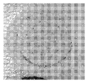
도 2a 및 도 2b는 염색에 의한, 중공 마이크로니들 패치 제거 후의 무모 기니아 피그(guinea pig) 피부의 이미지.
<도 3a 및 도 3b>
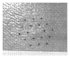
도 3a 및 도 3b는 메틸렌 블루를 나타내는 마이크로니들 주입 부위의 이미지.
<도 4>
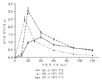
도 4는 전달 경로에 의한, 시간에 대한 날록손 혈중 수준의 비교 그래프.
<도 5>
도 5는 특정한 주입 카테고리에 대하여 주입 통증을 도시한 도면.
<도 6>
도 6은 특정한 주입 카테고리에 대하여 최대 주입 압력을 도시한 도면.
<도 7>
도 7은 특정한 주입 카테고리에 대하여 최대 주입 속도를 도시한 도면.
<도 8>
도 8은 특정한 주입 카테고리에 대하여 주입 용량을 도시한 도면.
<도 9>
도 9는 최대 주입 압력에 대하여 주입 통증을 도시한 도면.
<도 10>
도 10은 최대 주입 속도에 대하여 주입 통증을 도시한 도면.
<도 11>
도 11은 주입 용량에 대하여 주입 통증을 도시한 도면.
상기 도면은 본 발명의 일부 실시 형태를 설명하지만, 논의에서 알 수 있는 바와 같이 다른 실시 형태가 또한 고려된다. 모든 경우에서, 본 개시 내용은 예시적이고 비제한적으로 본 발명을 나타낸다. 본 기술 분야의 당업자라면 본 발명의 원리의 범주 및 사상 내에 속하는 여러 가지 다른 변형예 및 실시 형태들을 고안할 수 있을 것으로 이해해야 한다. 도면은 축척대로 도시되지 않을 수도 있다. 도면 전체에 걸쳐서 동일한 부분을 지시하기 위해 동일한 도면 부호가 사용되었다.DESCRIPTION OF THE PREFERRED EMBODIMENTS Preferred embodiments of the present invention will now be described in more detail with reference to the accompanying drawings.
1A and 1B,
Figures 1a and 1b are perspective views of a microneedle array embodiment, also showing that the individual hollow micro needles are seen closer.
2A and 2B,
Figures 2a and 2b are images of guinea pig skin after hair micro-needle patch removal by dyeing.
3A and 3B,
Figures 3a and 3b are images of the micro needle injection site showing methylene blue.
<Fig. 4>
FIG. 4 is a graph comparing the levels of naloxone levels versus time by the delivery route.
5,
Figure 5 shows injection pain for a particular injection category;
6,
Figure 6 shows the maximum injection pressure for a particular injection category;
7,
Figure 7 shows the maximum infusion rate for a particular infusion category;
8,
Figure 8 shows injection capacity for a particular injection category;
9,
Figure 9 shows injection pain versus maximum injection pressure.
<Fig. 10>
Figure 10 shows injection pain for a maximum infusion rate.
11)
Figure 11 shows injection pain versus injection volume.
While the above drawings illustrate some embodiments of the invention, other embodiments are also contemplated, as will be seen in the discussion. In all cases, the disclosure is exemplary and non-limiting. It should be understood by those skilled in the art that various other modifications and embodiments may be devised which fall within the spirit and scope of the principles of the invention. The drawings may not be drawn to scale. The same reference numerals are used to denote the same parts throughout the drawings.
이제 본 발명을 하기의 비제한적인 실시 형태를 참고로 하여 설명할 것이다.The present invention will now be described with reference to the following non-limiting embodiments.
마이크로니들 어레이Micro needle array
마이크로니들 기구(10)는 기재(12)를 포함하는 마이크로니들 어레이(11)를 가지며, 상기 기재로부터 복수의 18개의 마이크로니들(14)이 연장한다. 각각의 마이크로니들(14)은 그의 기부(16)로부터 그의 팁(18)까지 대략 500 ㎛의 높이를 갖는다. 중공 채널(도시되어 있지 않음)은 기재(12) 및 마이크로니들(14)을 관통하여 연장하여, 마이크로니들의 팁 근처의 채널 개구(20)에서 빠져나간다. 이는 각각의 마이크로니들(14)을 통한 어레이의 후방으로부터의 (예를 들어, 도시되어 있지 않은 저장부로부터의) 유체 연통을 가능하게 한다. 채널은 마이크로니들(14)의 중심축을 따라 이어지지만, 마이크로니들의 경사진 측벽 상에서 피하 주사 니들과 유사하게 빠져나가 삽입시 조직에 의한 차단을 방지하는 것을 돕는다. 채널은 평균 단면적이 약 20-50 ㎛2이다.The
마이크로니들(14)은 인접 마이크로니들(14)들 사이의 거리 d가 2 ㎜가 되도록 이격된다. 디스크형 기재(12)는 면적이 약 1.27 ㎠이며, 마이크로니들(14)은 마이크로니들(14)의 최외측 열의 주변부를 이용하여 측정할 때 약 0.42 ㎠의 면적에 걸쳐 펼쳐진다. 이는 ㎠당 약 14개의 마이크로니들에 해당하는 마이크로니들 밀도를 제공한다.The micro needles 14 are spaced such that the distance d between adjacent
마이크로니들 어레이(11)는 의료 등급의 폴리카르보네이트와 같은 중합체의 열사이클링 사출 성형(thermocycled injection molding)에 의해 이어서 레이저 드릴링(laser drilling)하여 마이크로니들 채널을 형성함으로써 제조된다.The
어레이 림(rim) 구조체(22)는 접착 디스크 (도시되어 있지 않음) (쓰리엠(3M) 1513 메디칼 테이프(Medical Tape), 미국 미네소타주 세인트 폴 소재의 쓰리엠 코포레이션(3M Corp.))를 포함하는 배킹(backing) 부재 (도시되어 있지 않음)를 마이크로니들 기재(12)에 부착시키는 데 사용되며, 이는 주입 동안 피부에 중공 마이크로니들 어레이(11)를 고정하기 위하여 기재(12)의 주변부(24)로부터 바깥으로 연장할 것이다. 접착 링을 포함하는 전체 마이크로니들 기구(10)의 피부 접촉면은 약 5.5 ㎠일 것이다.The
전형적으로 마이크로니들 기구(10)는 외용 어플리케이터 (도시되어 있지 않음)를 사용하여 피부에 적용된다. 어플리케이터는 예를 들어 스프링 메커니즘을 이용하여 원하는 속도를 달성하도록 디자인되어서, 마이크로니들이 단순히 피부를 변형시키기보다는 오히려 피부 내에 침투하도록 한다. 일단 적용되면, 접착 링은 마이크로니들 기구를 피부에 대하여 고정시킨다. 다양한 어플리케이터 기구가 예를 들어 국제특허 공개 WO2005/123173호, 국제특허 공개 WO2006/055802호, 국제특허 공개 WO2006/05579호, 국제특허 공개 WO2006/055771호, 국제특허 공개 WO2006/108185호, 국제특허 공개 WO2007/002521호 및 국제특허 공개 WO2007/002522호 (이들 전부는 본 명세서에 참고로 포함됨)에 개시되어 있다.Typically, the
마이크로니들 어레이를 통하여 전달될 유체는 유체를 함유하는 저장부(도시되어 있지 않음)에 함유될 수 있거나, 또는 예를 들어 배관에 의해 또는 루어 커넥터(luer connector)를 사용함으로써 연결될 수 있는 시린지 또는 기타 용기와 같은 외부 공급부로부터 유체가 펌핑되게 하는 것일 수 있다. 약물은 제형 중에 용해 또는 현탁될 수 있으며 전형적인 제형은 피하 주사 니들로부터 주사될 수 있는 유형의 것이다.The fluid to be delivered through the array of microneedles may be contained in a reservoir (not shown) containing fluid or may be contained in a syringe or other that may be connected, for example, by piping or by using a luer connector Or may be to cause fluid to be pumped from an external supply such as a vessel. The drug may be dissolved or suspended in the formulation and a typical formulation is of the type that can be injected from a hypodermic needle.
제형화되어 피하 주사를 통하여 전달될 수 있는 임의의 물질이 사용될 수 있으며, 이는 임의의 의약품, 건강 기능 식품(nutraceutical), 미용제약품(cosmaceutical), 진단제 및 치료제 (본 명세서에서 편의상 "약물"로 총칭됨)를 포함한다. 본 발명에서 주사가능 제형에서 유용할 수 있는 약물의 예에는 ACTH (예를 들어 코르티코트로핀 주사제), 황체 형성 호르몬 방출 호르몬 (예를 들어, 고나도렐린 염산염(Gonadorelin Hydrochloride)), 성장 호르몬 방출 호르몬 (예를 들어, 세르모렐린 아세테이트(Sermorelin Acetate)), 콜레시스토키닌 (신칼라이드(Sincalide)), 부갑상선 호르몬 및 이의 단편 (예를 들어, 테리파라타이드 아세테이트(Teriparatide Acetate)), 갑상샘 호르몬 분비 호르몬 및 이의 유사체 (예를 들어, 프로티렐린), 세크레틴 등, 알파-1 항-트립신, 혈관신생 억제제, 안티센스, 부토르파놀, 칼시토닌 및 유사체, 세레다아제(Ceredase), COX-II 저해제, 피부과학적 제제(dermatological agent), 다이하이드로에르고타민, 도파민 작용제 및 길항제, 엔케팔린 및 기타 오피오이드 펩티드, 표피 성장 인자, 에리트로포이에틴 및 유사체, 난포 자극 호르몬, G-CSF, 글루카곤, GM-CSF, 그라니세트론, 성장 호르몬 및 유사체 (성장 호르몬 방출 호르몬을 포함함), 성장 호르몬 길항제, 히루딘 및 히루딘 유사체, 예를 들어 히룰로그(Hirulog), IgE 억제제, 인슐린, 인슐리노트로핀(insulinotropin) 및 유사체, 인슐린-유사 성장 인자, 인터페론, 인터류킨, 황체 형성 호르몬, 황체 형성 호르몬 방출 호르몬 및 유사체, 헤파린, 저분자량 헤파린 및 기타 천연, 개질 또는 합성 글리코아미노글리칸, M-CSF, 메토클로프라미드, 미다졸람, 단클론 항체, 페길화(Peglyated) 항체, 페길화 단백질 또는 친수성 또는 소수성 중합체 또는 추가의 작용기로 개질된 임의의 단백질, 융합 단백질, 단쇄 항체 단편 또는 임의의 조합의 부착 단백질들을 포함하는 단쇄 항체 단편, 거대분자, 또는 이의 추가의 기능성 군, 마약성 진통제, 니코틴, 비스테로이드계 항염증제, 올리고당류, 온단세트론, 부갑상선 호르몬 및 유사체, 부갑상선 호르몬 길항제, 프로스타글란딘 길항제, 프로스타글란딘, 재조합 용해성 수용체, 스코폴라민, 세로토닌 작용제 및 길항제, 실데나필, 테르부탈린, 혈전용해제, 조직 플라스미노겐 활성자, TNF-, 및 TNF-길항제, 중독, 관절염, 콜레라, 코카인 중독, 디프테리아, 파상풍, HIB, 라임병(Lyme disease), 수막염구균, 홍역, 볼거리, 풍진, 수두, 황열병, 호흡기 세포융합 바이러스, 진드기 매개 일본 뇌염, 폐렴 쌍구균, 연쇄구균, 장티푸스, 인플루엔자, A형 간염, B형 간염, C형 간염 및 E형 간염을 포함하는 간염, 중이염, 광견병, 폴리오, HIV, 파라인플루엔자, 로타바이러스, 엡스타인 바 바이러스(Epstein Barr Virsu), CMV, 클라미디아, 비피막형 헤모필루스, 모락셀라 카타랄리스(moraxella catarrhalis), 인간 유두종 바이러스, BCG를 포함하는 결핵, 임질, 천식, 아테롬성 동맥 경화증 말라리아, 이. 콜라이(E-coli), 알츠하이머병(Alzheimer's Disesase), 에이치. 파일로리(H. Pylori), 살모넬라, 당뇨병, 암, 단순 포진, 인간 유두종과 관련된, 예방 및 치료 항원을 포함하는, 담체/아쥬반트를 포함하거나 포함하지 않는 백신 (서브유닛(subunit) 단백질, 펩티드 및 다당류, 다당류 콘쥬게이트(conjugate), 유독소, 유전자 기반의 백신, 생 약독화, 재조합, 불활성화 전 세포, 바이러스 및 박테리아 벡터를 포함하지만 이에 한정되지 않음), 및 보통 감기용 제제, 항중독제, 항알러지제, 항구토제, 항비만제, 골다공증 방지제(antiosteoporeteic), 항감염제, 진통제, 마취제, 식욕감퇴제, 항관절염제, 항천식제, 항경련제, 항우울제, 항당뇨제, 항히스타민제, 항염증제, 항편두통 제제, 멀미 방지 제제, 항오심제, 항신생물제, 항파킨슨증 약물, 가려움 방지제, 항정신병제, 해열제, 항콜린제, 벤조다이아제핀 길항제, 혈관확장제 - 일반 혈관, 관상 혈관, 말초 혈관 및 뇌 혈관을 포함함 - , 뼈 자극제, 중추 신경계 자극제, 호르몬, 수면제, 면역억제제, 근육 이완제, 부교감신경 차단제, 프로스타글란딘, 단백질, 펩티드, 폴리펩티드 및 기타 거대분자, 정신자극제, 진정제, 및 성기능 저하제 및 안정제(tranquilizer)와 같은 주요 치료제 전부를 포함하는 기타 다른 물질을 포함하지만, 이에 한정되지 않는다.Any material that can be formulated and delivered via subcutaneous injection can be used and can be any pharmaceutical, nutraceutical, cosmaceutical, diagnostic and therapeutic agent (referred to herein as "drug" Quot; generically "). Examples of drugs that may be useful in the present injectable formulations include ACTH (e.g., a corticotropin injection), luteinizing hormone releasing hormone (e.g., Gonadorelin Hydrochloride), growth hormone releasing hormone (E.g., Sermorelin Acetate), cholestyokinin (Sincalide), parathyroid hormone and its fragments (e.g., Teriparatide Acetate), thyroid hormone releasing hormone and Anti-angiogenesis inhibitors, antisense, butorphanol, calcitonin and analogues, Ceredase, COX-II inhibitors, dermatological Dermatological agents, dihydroergotamine, dopamine agonists and antagonists, enkephalin and other opioid peptides, epidermal growth factor, erythropoietin And analogs, follicle stimulating hormone, G-CSF, glucagon, GM-CSF, ganicitorone, growth hormone and analogs (including growth hormone releasing hormone), growth hormone antagonists, hirudin and hirudin analogs, An insulinotropic and analogue, an insulin-like growth factor, an interferon, an interleukin, a luteinizing hormone, a luteinizing hormone releasing hormone and analogue, a heparin, a low molecular weight heparin and others Natural, modified or synthetic glycosaminoglycans, M-CSF, methocloframide, midazolam, monoclonal antibodies, pegylated antibodies, pegylated proteins or any protein modified with hydrophilic or hydrophobic polymers or further functional groups , A fusion protein, a single chain antibody fragment, or a single chain antibody fragment comprising any combination of attachment proteins, a macromolecule, or an additional functional group thereof, The present invention relates to the use of a compound of formula (I) or a pharmaceutically acceptable salt thereof in the manufacture of a medicament for the treatment and / or prophylaxis of pain, analgesic, nicotine, nonsteroidal antiinflammatory agent, oligosaccharide, ondansetron, parathyroid hormone and analog, parathyroid hormone antagonist, prostaglandin antagonist, prostaglandin, recombinant soluble receptor, scopolamine, serotonin agonist and antagonist, sildenafil, Tryptophan, Lyme disease, meningococcal disease, measles, mumps, rubella, chicken pox, yellow fever, rheumatoid arthritis, arthritis, cholera, Hepatitis, otitis media, rabies, polio, HIV, parainfluenza including hepatitis B, hepatitis C and hepatitis E, respiratory syncytial virus, tick-borne encephalitis, pneumoniae, streptococcus, typhoid, influenza, hepatitis A, , Rotavirus, Epstein Barr Virsu, CMV, chlamydia, non-coated hemophilus, Lease (moraxella catarrhalis), human papilloma virus, tuberculosis, including the BCG, gonorrhea, asthma, atherosclerosis, malaria, the. E-coli, Alzheimer ' s Disesase, H. Vaccines (including subunit proteins, peptides, and antigens), including, without limitation, carriers / adjuvants, including prophylactic and therapeutic antigens related to H. pylori, Salmonella, diabetes, cancer, herpes zoster, Including but not limited to polysaccharides, polysaccharide conjugates, toxins, gene based vaccines, live attenuated, recombinant, pre-inactivated cells, viruses and bacterial vectors, and common cold preparations, An anti-inflammatory agent, an anti-arthritic agent, an anti-asthmatic agent, an anticonvulsant, an antidepressant, an antidiabetic agent, an antihistamine, an anti-inflammatory agent, an anti-inflammatory agent, Antihistamines, antihypertensives, anticholinergics, benzodiazepine antagonists, vasodilators - generalized blood vessels, anti-inflammatory drugs, anti-inflammatory drugs, Including, but not limited to, peripheral blood vessels, blood vessels, peripheral blood vessels and cerebral vessels, bone stimulators, central nervous system stimulants, hormones, sleeping pills, immunosuppressants, muscle relaxants, parasympathetic blockers, prostaglandins, proteins, peptides, polypeptides and other macromolecules, , And other substances including all of the major therapeutic agents such as sexual dysfunction agents and tranquilizers.
광범위한 중공 마이크로니들 형상, 예를 들어 원뿔형, 원통형, 피라미드형, 절두형, 비대칭형 및 이의 조합이 사용될 수 있음이 이해될 것이다. 다양한 물질, 예를 들어 중합체, 금속, 및 규소 기반의 것이 또한 사용될 수 있으며, 이는 임의의 적합한 방식, 예를 들어 사출 성형, 스탬핑으로, 그리고 사진석판술(photolithography)을 이용하여 제조될 수 있다. 기재 상에서의 마이크로니들의 배열은 (어레이의 피부 접촉 표면을 향하여 볼 때) 랜덤 패턴, 다각형 패턴, 정사각형 패턴, 및 원형 패턴과 같은 임의의 패턴의 것일 수 있다.It will be appreciated that a wide variety of hollow microneedle shapes may be used, for example, conical, cylindrical, pyramidal, hemispherical, asymmetric, and combinations thereof. A variety of materials such as polymeric, metal, and silicon-based materials can also be used and can be prepared in any suitable manner, such as injection molding, stamping, and using photolithography. The arrangement of the microneedles on the substrate may be of any pattern such as a random pattern, a polygonal pattern, a square pattern, and a circular pattern (as viewed toward the skin-contacting surface of the array).
상기 설명에 더하여, 하기의 특허 문헌은 본 발명에 따른 사용에 유용하거나 적합할 수 있는 마이크로니들 기구, 재료, 제작, 어플리케이터, 및 용도를 개시한다: 미국 특허 제6,881,203호; 미국 특허 제6,908,453호; 미국 특허 출원 공개 2005-0261631호; 국제특허 공개 WO2005/065765호; 국제특허 공개 WO2005/082596호; 국제특허 공개 WO2006/062974호; 국제특허 공개 WO2006/135794호; 미국 특허 출원 공개 제2006/048640호; 미국 가특허 출원 제60/793611호; 미국 특허 출원 공개 제2007/064789호; 국제특허 공개 WO2006/062848호; 국제특허 공개 WO2007/002523호; 및 미국 가특허 출원 제60/793564호.In addition to the above description, the following patent documents disclose microneedle instruments, materials, fabrication, applicators, and applications that may be useful or suitable for use in accordance with the present invention: U.S. Patent No. 6,881,203; U. S. Patent No. 6,908, 453; U.S. Patent Application Publication 2005-0261631; International Patent Publication No. WO2005 / 065765; International Patent Publication No. WO2005 / 082596; International Patent Publication No. WO 2006/062974; International Patent Publication No. WO2006 / 135794; U.S. Patent Application Publication No. 2006/048640; U.S. Provisional Patent Application No. 60/793611; U.S. Patent Application Publication 2007/064789; WO2006 / 062848; International Patent Publication WO2007 / 002523; And U.S. Provisional Patent Application No. 60/793564.
실험Experiment
도 1a와 관련하여 상기에 설명한 마이크로니들 어레이 기구를 하기 실험 및 실시예에 사용하였다.The microneedle array mechanism described above with reference to FIG. 1A is used in the following experiments and examples.
동물 모델 및 피부 준비Animal Models and Skin Preparation
무모 기니아 피그(Hairless Guinea Pig; HGP)Hairless Guinea Pig (HGP)
수컷 HGP를 쓰리엠 IACUC-승인 동물 사용 용도로 찰스 리버 래보러토리즈(Charles River Laboratories; 미국 매사추세츠주 윌밍턴 소재)에 주문하고 그 프로토콜에 따라 사용하였다. 이 연구에서 사용한 모든 동물은 체중이 0.8-1 ㎏이었다.The male HGP was ordered from Charles River Laboratories (Wilmington, Mass., USA) for 3M IACUC-approved animal use and used according to the protocol. All animals used in this study weighed 0.8-1 kg.
집돼지Boar
나이가 대략 6-18주이고 체중이 대략 10-30 ㎏이며 쓰리엠 IACUC-승인 동물 사용 용도로 획득한 암컷 집돼지에서 시험을 행하였다. 주입 동안 그리고 연구 전체에 걸쳐, 상기 돼지를 아이소플루란 (2-5%) 산소 믹스를 이용하여 마취 하에 유지하였다. 돼지 둔부의 상부 부분을 먼저 외과용 제모기(surgical clipper; 클립 블레이드 #50)를 사용하여 그리고 이어서 소량의 질레트 폼(Gillette Foam) 면도 크림을 사용하여 쉬크 쓰리(Schick 3) 면도기를 이용하여 면도하였다. 면도 후, 그 부위를 물로 헹구고, 가볍게 두드려 건조시키고, 이어서 아이소프로필 알코올 (미국 미주리주 세인트 조젭 소재의 피닉스 파마슈티칼, 인크.(Phoenix Pharmaceutical, Inc.))로 닦아주었다(wiped).Tests were conducted on female piglets that were approximately 6 to 18 weeks of age and weighing approximately 10 to 30 kilograms and were obtained for use in 3 IACUC-approved animals. During the infusion and throughout the study, the pigs were maintained under anesthesia with isoflurane (2-5%) oxygen mix. The upper part of the pig's hips was first shaved using a surgical clipper (Clip Blade # 50) and then a small amount of Gillette Foam shaving cream using a
혈청중 날록손의 측정Measurement of naloxone in serum
각 시점에서, 1.5-2 ㎖의 전혈을 배큐테이너 콜렉션즈 세트(Vacutainer Collections Set) (미국 뉴저지주 프랭클린 레익스 소재의 벡톤 디킨슨 앤드 컴퍼니(Becton Dickenson & Co.))를 이용하여 돼지의 귀 정맥으로부터 수집하였다. 전혈을 실온에서 30분 이상 동안 응고시킨 후 1500 rpm에서 10분 동안 원심분리하였다. 원심분리 후, 혈청을 전혈로부터 분리하고, 추출할 때까지 냉보관하였다.At each time point, 1.5-2 ml of whole blood was collected from the ear vein of the pig using the Vacutainer Collections Set (Becton Dickenson & Co., Franklin Lakes, NJ) Respectively. Whole blood was allowed to coagulate at room temperature for more than 30 minutes and then centrifuged at 1500 rpm for 10 minutes. After centrifugation, the serum was separated from whole blood and kept in cold storage until extraction.
실온 혈청 샘플을 고상 추출 카트리지 (미국 캘리포니아주 토런스 소재의 피노미넥스(Phenomenex))를 사용하여 준비하였다. 카트리지를 메탄올 (미국 뉴저지주 깁스타운 소재의 이엠디 케미칼스, 인크.(EMD Chemicals, Inc.))로 컨디셔닝하고, 시약 등급 물로 평형화한 후 혈청 샘플을 로딩하였다. 혈청을 시약 등급 물 중 5% 메탄올 2 ㎖로 세척하고, 날록손을 100% 메탄올로 용출시켰다. 용리액을 14 ㎖ 유리관 또는 16x100 ㎜ 튜브에 수집하고, 37C 수조에서 103.4 ㎪(15psi)의 질소 하에 건조시켰다.A room temperature serum sample was prepared using a solid phase extraction cartridge (Phenomenex, Torrance, CA). The cartridge was conditioned with methanol (EMD Chemicals, Inc., Gipston, NJ), equilibrated with reagent grade water, and loaded with a serum sample. Serum was washed with 2 ml of 5% methanol in reagent grade and naloxone was eluted with 100% methanol. The eluate was collected in 14 ml glass tubes or 16x100 mm tubes and dried under nitrogen at 15.4 psi in a 37 C water bath.
추출물을 5% 아세토니트릴/95%의 물 중 0.1% 포름산(미국 매사추세츠주 워드 힐 소재의 알파 에이사(Alfa Aesar))을 이용하여 재구성하고, 마이크로원심분리 튜브 (미국 뉴욕주 웨스트베리 소재의 에펜도르프(Eppendorf))로 옮기고, 14000 rpm에서 10분 동안 원심분리하였다.The extracts were reconstituted using 0.1% formic acid (Alfa Aesar, Ward Hill, Mass.) In 5% acetonitrile / 95% water and resuspended in microcentrifuge tubes (Epfen, Westbury, (Eppendorf)) and centrifuged at 14000 rpm for 10 minutes.
추출물을 LCMSMS를 사용하여 정량적으로 분석하였다. 피노미넥스 C18 가드 컬럼 (미국 캘리포니아주 토렌스 소재의 피노미넥스)과 함께 차례대로 애질런트 이클립스(Agilent Eclipse) XDB-C18 컬럼 (미국 델라웨어주 윌밍턴 소재의 애질런트 테크놀로지즈(Agilent Technologies))을 이용하여 분리시켰으며; 이동상은 0.1% 포름산 및 아세토니트릴이었고; 포름산은 1분에 걸쳐 95%로부터 10%로 감소되었다. 터보 이온스프레이 인터페이스(Turbo IonSpray interface)를 사용하여 양이온 모드로 실행되는 사이엑스(Sciex) API3000 트리플 쿼드(triple quad) 질량 분석계 (미국 캘리포니아주 포스터 시티 소재의 어플라이드 바이오시스템즈(Applied Biosystems))를 사용하여 하기 m/z 전이(transition)에서 생기는 생성물 이온을 정량적으로 모니터링하였다: 328.17 → 310.10 및 342.16 → 324.30. 날록손에 대한 선형 범위는 1/x 곡선 웨이팅(curve weighting)을 이용하여 평가할 경우 0.1 내지 100 ng/㎖이었다.The extracts were quantitatively analyzed using LCMSMS. Using Agilent Eclipse XDB-C18 column (Agilent Technologies, Wilmington, Delaware, USA) in turn with the Pinominex C18 Guard column (Pinominex, Torrance, CA) Lt; / RTI > The mobile phase was 0.1% formic acid and acetonitrile; Formic acid was reduced from 95% to 10% over 1 minute. A Sciex API 3000 triple quad mass spectrometer (Applied Biosystems, Foster City, CA) running in positive ion mode using a Turbo IonSpray interface Product ions resulting from the following m / z transitions were monitored quantitatively: 328.17 -> 310.10 and 342.16 -> 324.30. The linear range for naloxone was 0.1 to 100 ng / ml when evaluated using 1 / x curve weighting.
다양한 크기의 돼지에게 투약하여 돼지 체중과 관련한 혈중 날록손 수준을 정규화하였으며, 혈중 날록손 수준에 돼지 체중 1 ㎏당 혈액 62 ㎖의 변환 인자를 곱하고 이어서 투약시 돼지의 체중(㎏)을 곱하였다. 최종 결과를 돼지당 날록손 ㎍으로서 도시한다.Plasma naloxone levels related to pig weight were normalized by dosing pigs of varying sizes and the naloxone level in the blood was multiplied by the conversion factor of 62 ml of blood per kg of pig weight and then multiplied by the body weight of the pig (kg) at the time of dosing. The final result is shown as the triglyceride per pig.
HGP 및 돼지에서의 침투 깊이Penetration depth in HGP and pigs
기술 문헌을 기반으로 하면, 그리고 미세구조체의 크기를 고려하면, 미세구조체당 0.004-0.16 N의 힘이 각질층의 침투에 필요한 것으로 추정되었다 (문헌[S.P. David, B.J. Landis, Z.H. Adams, M.G. Allen, M.R. Prausnitz. Insertion of microneedles into skin: measurement and prediction of insertion force and needle fracture force. Journal of Biomechanics. 37:115-116 (2004)]). 본 발명에서 사용한 미세구조체의 충분한 내구성을 보장하기 위하여, 어레이를 비-탄성 표면에 대하여 눌렀으며, 대략 245 N의 힘이 어레이에 가해졌을 때 팁 굽힘(tip bending)이 일어났다.Based on the technical literature, and considering the size of the microstructure, it was estimated that a force of 0.004-0.16 N per microstructure is required for penetration of the stratum corneum (SP David, BJ Landis, ZH Adams, MG Allen, MR Journal of Biomechanics 37: 115-116 (2004)]). ≪ / RTI > In order to ensure sufficient durability of the microstructure used in the present invention, the array was pressed against the non-elastic surface and tip bending occurred when a force of approximately 245 N was applied to the array.
일반적으로 돼지 피부는 한선을 전혀 포함하지 않는다는 것을 제외하고는 두께, 모 밀도 및 하부 조직에의 부착에 있어서 인간 피부와 유사한 것으로 간주된다. 이들 연구에 사용한 돼지의 표피의 깊이가 인간에게서 발견되는 것과 대략적으로 유사할 경우, 침투 깊이 데이터는 본 발명에서 사용한 중공 마이크로니들 기구 (도 1a 참조)의 가능한 주입 깊이가 180-280 ㎛ (평균 250 ㎛)임을 나타내는데, 이는 주입 동안 있게 되는 배압의 크기에 영향을 줄 수 있는 진피 또는 표피에 상응할 수 있는 깊이이다. 따라서, 마이크로니들 높이가 약 500 ㎛이기는 하지만 실제 침투 깊이는 대략적으로 그것의 절반이었음이 이해될 것이다.Pig skin is generally considered to be similar to human skin in thickness, parent density, and adherence to underlying tissue except that it does not contain any solids. If the depth of the epidermis of the pigs used in these studies is approximately similar to that found in humans, the penetration depth data indicates that the possible injection depths of the hollow microneedle instrument used in the present invention (see FIG. 1A) are 180-280 μm ㎛), which is the depth that can correspond to the epidermis or the dermis, which can affect the magnitude of the back pressure that occurs during injection. Thus, it will be appreciated that the actual penetration depth was roughly half that, although the microneedle height was about 500 microns.
침투 깊이(depth of penetration; DOP) 실험을 HGP 및 집돼지 둘 모두에서 완료하였으며; 그 결과가 표 I에 요약되어 있다.The depth of penetration (DOP) experiment was completed in both the HGP and the domestic pigs; The results are summarized in Table I.
[표 I] [Table I]

힘 시험 실험에서 미세구조체의 파쇄가 관찰되지도 않았고 DOP 시험 이후에 임의의 파손된 니들이 관찰되지도 않았다.No fracture of the microstructure was observed in the force test, and no broken needle was observed after the DOP test.
도 2a 및 도 2b는 패치 제거 후 HGP 상의 적용 부위를 예시한다. 도 2a는 적용 이전에 마이크로니들 상에 코팅된 로다민 B 염료에 의해 만들어진 표시(marking)를 예시한다. 도 2b는 마이크로니들 어레이를 제거한 후 메틸렌 블루에 의한 염색에 의해 만들어진 표시를 예시한다. 18개의 미세구조체 각각에 의한 각질층 침투가 도 2b에서의 메틸렌 블루 도트 패턴으로부터 명백하다. 혈액은 적용 동안 또는 적용 후 관찰되지 않았다.Figures 2a and 2b illustrate the application site on the HGP after patch removal. Figure 2a illustrates the marking made by rhodamine B dye coated on micro needles prior to application. Figure 2b illustrates a representation made by dyeing with methylene blue after removal of the microneedle array. The penetration of the stratum corneum by each of the eighteen microstructures is evident from the methylene blue dot pattern in Figure 2b. Blood was not observed during or after application.
돼지에서, 최대 1 ㎖의 몇몇 주입을 살균 5% 덱스트로스 또는 0.001% 메틸렌 블루 용액을 이용하여 행하였다. 일단 제형을 전달하였으면, 당해 시스템에서의 배압을 주입전 수준으로 되돌리면서 당해 기구를 최대 10분 동안 적소에 머무르게 하였다. 도 2는 0.001% 메틸렌 블루 제형 800 ㎕를 돼지 내로 피내 주입한 결과를 예시한다. 피부는 패치 제거 후 촉감이 건조하며; 주입한 제형의 짙은 청색은 이 처리의 시각적 평가를 제공한다.In pigs, up to 1 ml of several injections were made with sterile 5% dextrose or 0.001% methylene blue solution. Once the formulation was delivered, the back pressure in the system was returned to the pre-injection level, allowing the device to remain in place for up to 10 minutes. FIG. 2 illustrates the results of intracutaneous infusion of 800 μl of a 0.001% methylene blue formulation into a pig. Skin is dry after patch removal; The deep blue color of the injected formulation provides a visual assessment of this treatment.
도 3a 및 도 3b는 패치 제거 후 각각 T= 0 및 T= 9 min에서 돼지에서의 0.05% 메틸렌 블루 제형의 피내 주입의 이미지를 예시한다. 피부는 촉감이 건조하였다.Figures 3a and 3b illustrate images of intradermal injection of 0.05% methylene blue formulations in pigs at T = 0 and T = 9 min, respectively, after patch removal. The skin was dry and tactile.
피부 상의 각각의 청색 스폿은 어레이 상의 18개의 중공 미세구조체 중 하나에 상응한다. 염료는 9분 후 약간 번진 (확산된) 것처럼 보이지만, 청색 얼룩은 1시간 미만 내에 그 자국이 사라진다 해도 본질적으로 24시간 후에 여전히 변화되지 않은 채 남아있었다. 염료는 실제로 조직 내에 착색되거나 침전될 가능성이 있으며, 이러한 의미에서, 염료는 아마도 주입 후 장기간에 걸친 피내 주입 패턴의 효과적인 지시자(indicator)는 아닌 것 같다.Each blue spot on the skin corresponds to one of the 18 hollow microstructures on the array. The dye appears to have slightly diffused after 9 minutes, but the blue stain remained unchanged after essentially 24 hours, even if the mark disappeared in less than an hour. Dyes are indeed likely to be pigmented or precipitated in the tissues, and in this sense the dyes probably do not appear to be an effective indicator of the intradermal pattern over time after injection.
주입 후 중공 마이크로니들 패치의 제거시에, 전형적으로 소량 (1-3 ㎕)의 제형이 피부 표면 상에서 관찰된다. 이 유체를 티슈로 부드럽게 닦아냄으로써 제거할 때, 추가의 유체는 관찰되지 않는다. 중공 마이크로니들 어레이의 크기인 연분홍색 반점이 패치 제거시에 전형적으로 보이지만, 상기 반점은 5분 이내에 거의 구별할 수 없게 되도록 희미해진다. 다시 대략적으로 중공 마이크로니들 어레이의 크기인 작은 돔(dome)이 돼지 피부에서 또한 관찰되었다. 약간의 압력 하에서 돔을 눌러보았더니 "누출"(leak)되지 않았다. 적용 패치를 제거한지 40분 이내에 돔을 시각적으로 그리고 촉각적으로 분석하였다. 적용한지 24시간 및 48시간 후에 적용 부위를 관찰해 보면 홍반 또는 부종의 증거가 전혀 나타나지 않았다.Upon removal of the hollow micro needle patch after injection, a small amount (1-3 l) of formulation is typically observed on the skin surface. When the fluid is removed by gently wiping it with a tissue, no additional fluid is observed. The pink spot, which is the size of the hollow microneedle array, typically appears at the time of patch removal, but the spot becomes blurred to be almost indistinguishable within 5 minutes. Again, a small dome, approximately the size of a hollow microneedle array, was also observed in pig skin. I pressed the dome under some pressure and it did not leak. The dome was visually and tactually analyzed within 40 minutes of removal of the applied patch. No evidence of erythema or edema was observed at 24 and 48 hours after application.
실시예 1. 돼지에서의 고용량 덱스트로스 주입Example 1. High dose dextrose infusion in pigs
고용량 주입을 집돼지에 대해 입증하였다. 적용 후 중공 마이크로니들 어레이 패치에 연결되는 경우, 돼지에 사용된 주입 시스템은 제형을 전달하기 위하여 표준 의료 장비를 이용한다. 중공 마이크로니들 적용 패치를 시판용 사전 살균 폴리에틸렌 IV 익스텐션 세트(Extension Set; 프랑스 에쿠앙 소재의 비공 코포레이션(Vygon Corporation))를 통하여 메드퓨전(Medfusion) 3500 시린지 펌프 (미국 미네소타주 세인트 폴 소재의 스미스 메디칼(Smiths Medical))에 결합시키는데, 이 시린지 펌프는 인라인(in-line) 사전 살균 DTX 플러스(Plus) TNF-R 압력 변환기 (미국 유타주 샌디 소재의 비디 인퓨전 테라피 시스템즈, 인크.(BD Infusion Therapy Systems, Inc.))를 포함한다. 메드퓨전 3500 펌프는 병원 설비에서 일반적으로 사용되며 미리 조절된 안전 정지 특징부(pre-set safety stop feature)를 갖는다. 압력 판독치를 매 2초마다 대략 1회 측정하는 비율로 기록하였다. 주사용 용액인 5% 덱스트로스, USP (미국 일리노이주 디어필드 소재의 박스터 헬스케어(Baxter Healthcare))를 수령한 대로 주입에 사용하였다. 0.001% 메틸렌 블루 용액을 살균수를 사용하여 제조하고, 여과한 후 투여하였다.A high dose infusion was proven for the boar. When applied to a hollow microneedle array patch after application, the infusion system used in the pig uses standard medical equipment to deliver the formulation. The hollow micro needle application patch was applied to a Medfusion 3500 syringe pump (Smith Medical, St. Paul, Minn.) Via a commercially available pre-sterilized polyethylene IV extension set (Vygon Corporation, Smiths Medical), which is equipped with an in-line pre-sterilized DTX Plus TNF-R pressure transducer (BD Infusion Therapy Systems, Inc., Sandy, Utah, USA) .)). The MedFusion 3500 pump is commonly used in hospital facilities and has a pre-set safety stop feature. The pressure readings were recorded at a rate of approximately one measurement every 2 seconds. 5% dextrose, USP (Baxter Healthcare, Deerfield, Ill.) Was used for the injection as received. A 0.001% methylene blue solution was prepared using sterile water, filtered and then administered.
나이가 대략 6-18주이고 체중이 대략 10-30 ㎏이며 쓰리엠 IACUC-승인 동물 사용 용도로 획득한 암컷 집돼지에서 시험을 행하였다. 주입 동안 그리고 연구 전체에 걸쳐, 상기 돼지를 아이소플루란 (2-5%) 및 산소 믹스를 이용하여 마취 하에 유지하였다. 돼지 둔부의 상부 부분을 먼저 외과용 제모기(클립 블레이드 #50)를 사용하여 그리고 이어서 소량의 질레트 폼 면도 크림을 사용하여 쉬크 쓰리 면도기를 이용하여 면도하였다. 면도 후, 그 부위를 물로 헹구고, 가볍게 두드려 건조시키고, 이어서 아이소프로필 알코올 (미국 미주리주 세인트 조젭 소재의 피닉스 파마슈티칼, 인크.)로 닦아주었다.Tests were conducted on female piglets that were approximately 6 to 18 weeks of age and weighing approximately 10 to 30 kilograms and were obtained for use in 3 IACUC-approved animals. During the infusion and throughout the study, the pigs were maintained under anesthesia with isoflurane (2-5%) and oxygen mix. The upper part of the pig's hips was first shaved using a surgical epilator (Clip Blade # 50) and then with a small amount of gillette foam shaving cream using a sheepskin shaver. After shaving, the area was rinsed with water, patted dry, and then wiped with isopropyl alcohol (Phoenix Farmsteak, Inc., St. John's, MO, USA).
물 중 5% 덱스트로스 최대 1 ㎖을, 또는 날록손 최대 425 mcL를 돼지의 상부 둔부 부분에 전달하였다. 주입 동안 계속적으로 배압을 모니터링하여 주입 시스템에서의 누출의 부재를 검증하였다. 돼지에서 이용한 전형적인 주입 속도 프로파일을 하기 표 II에 제공한다.Up to 1 ml of 5% dextrose in water, or up to 425 mcL of naloxone, was delivered to the upper hind paw portion of the pig. The back pressure was continuously monitored during injection to verify the absence of leakage in the injection system. A typical infusion rate profile used in pigs is provided in Table II below.
[표 II][Table II]

주입 후, 중공 마이크로니들 어레이를 제거하였으며, 이에 의해 피부 밑에 작은 수포가 남겨졌다. 이 수포는 40분 이내에 완전히 사라졌다. 패치 제거 후 48시간 내내 관찰하는 동안 돼지에서 부위 반응이 전혀 관찰되지 않았다.After injection, the hollow micro needle array was removed, leaving a small blister beneath the skin. This blister disappeared completely within 40 minutes. No site reaction was observed in pigs during the 48 hour observation period after patch removal.
덱스트로스 및 메틸렌 블루 주입 동안 계속적으로 배압을 모니터링하여 기록하였다. 3회의 주입에 있어서 측정한 최대 배압을 주입 조건과 함께 표 III에 제공한다.Continuous back pressure monitoring during dextrose and methylene blue infusion was recorded. The maximum back pressure measured for the three injections is given in Table III with the injection conditions.
[표 III][Table III]

실시예 2. 생성된 PK 프로파일을 이용한 날록손 주입Example 2. Naloxone Injection Using the Generated PK Profile
주입을 더욱 잘 정량화하려는 노력으로, 1 mg/㎖의 시판용 날록손 제형을 중공 마이크로니들 POC 기구를 이용하여 돼지 내로 주입하였다. 날록손은 헤로인과 같은 약물의 과다복용에 주로 대항하기 위하여 사용되는 μ-오피오이드 수용체의 경쟁적 길항제이다. 빠른 반응을 위하여 전형적으로 정맥내 투여되는 날록손은 경구 투여될 때 단지 약 2%의 생체이용률을 갖는다. 날록손은 잘 흡수되지만, 제1 패스(pass) 동안 거의 90%가 제거된다. 문헌 개관에 의하면 성인에서의 날록손의 반감기는 30-81분이며 아동에서는 상당히 더 길다 (대략 3시간)는 것이 나타났다. 날록손은 대사산물로서 소변으로 배설된다.In an effort to better quantify the infusion, a 1 mg / ml commercial naloxone formulation was injected into the pig using a hollow micro needle POC instrument. Naloxone is a competitive antagonist of μ-opioid receptors, which is used primarily to counter the overdose of drugs such as heroin. For rapid response, naloxone, typically administered intravenously, has only about 2% bioavailability when administered orally. Naloxone is absorbed well, but almost 90% is removed during the first pass. According to literature review, the half-life of naloxone in adults is 30-81 min and is considerably longer in children (approximately 3 h). Naloxone is excreted in the urine as a metabolite.
주입 전에 그리고 주입 이후 최대 2시간의 특정한 시점에서 돼지의 귀 정맥으로부터 혈액 샘플을 수집하였다. 샘플을 준비 및 분석하여 혈청중 날록손 수준을 결정하였다. 비교를 위하여, 피하 또는 정맥내 주사를 이용하여 동일한 시판용 날록손 제형을 나이브(naive) 돼지에 투약하였다. 피내 주입에서와 같이, 혈액 샘플을 수집하여 날록손 수준에 대하여 분석하였다.Blood samples were collected from the ear veins of the pigs prior to injection and at a particular time point up to 2 hours after injection. Samples were prepared and analyzed to determine the level of naloxone in the serum. For comparison, the same commercial naloxone formulations were dosed into naive pigs using subcutaneous or intravenous injection. As in intradermal injection, blood samples were collected and analyzed for naloxone levels.
중공 마이크로니들 주입, 피하 주사 및 IV 주사 후 생성된 PK 프로파일을 비교하는 연구에 3마리의 상이한 동물을 사용하였다. 돼지는 투약 시점에서 체중이 10-22 ㎏이었고 연령 범위는 1.5-3개월이었다. 날록손 염산염의 시판용 제형 (1 mg/㎖) (미국 캘리포니아주 사우스 엘 몬테 소재의 인터내셔널 메디케이션 시스템즈, 리미티드.(International Medication Systems, Ltd.)을 주입에 사용하였다. 표 IV는 중공 마이크로니들 기구를 이용하여 실시한 날록손 투여에서 사용한 주입 프로파일을 예시한다.Three different animals were used in the study to compare PK profiles generated after hollow micro needle injection, subcutaneous injection and IV injection. The pigs weighed 10-22 kg at the time of dosing and the age range was 1.5-3 months. A commercially available formulation of naloxone hydrochloride (1 mg / ml) (International Medication Systems, Ltd., South El Monte, Calif., USA) was used for the infusion. The injection profile used in naloxone administration is illustrated.
[표 IV][Table IV]

전달 경로에 의한 시간에 대한 날록손 혈중 수준의 비교 그래프가 도 4에 예시되어 있다. 날록손을 피하 주사에 의해 돼지에 또한 투여하였다. 이들 돼지는 체중 및 연령 면에서 중공 마이크로니들 기구를 통하여 날록손을 투여한 것과 유사하였다. 이들 결과는 중공 마이크로니들 및 피하 주사를 통한 날록손의 비견되는 전달을 나타낸다. 주입을 개시한지 최대 2시간 후에 수집한 혈액 샘플을 기반으로 하면, 중공 마이크로니들 기술에 의해 투여한 날록손의 생체이용률은 피하 투여에서 생기는 것의 107+/-35%인 것으로 추정된다.A comparative graph of naloxone blood levels versus time by the delivery route is illustrated in FIG. Naloxone was also administered to pigs by subcutaneous injection. These pigs were similar in weight and age to naloxone administered through a hollow microneedle device. These results indicate comparable delivery of naloxone through the hollow micro needle and subcutaneous injection. Based on blood samples collected up to two hours after initiation of injection, bioavailability of naloxone administered by hollow micro needle technology is estimated to be 107 +/- 35% of that occurring in subcutaneous administration.
실시예 3. 덱스트로스를 이용한 인간 주입 연구Example 3. Human Injection Study Using Dextrose
상기에 설명한 동일 장치를 이용하여 고용량, 고속 주입의 입증을 인간에 대해 행하였다. 인간 임상 시험 동안, 28명의 대상에게 그의 상완 및/또는 허벅지에 4-6가지의 순차적 중공 마이크로니들 위약 주입물을 투여하였다. 주입 전체에 걸쳐 배압을 계속적으로 모니터링하였다. 10점의 통증 기준 (도 4 참조)을 이용하여, 각각의 대상에게 중공 마이크로니들 패치의 적용 및 제거와 관련된 통증을 등급화하도록 요청하였으며; 또한 대상에게 주입 동안 매 10분마다 또는 10분 미만의 시간 내에 주입이 끝날 경우 주입 마지막에 주입과 관련된 통증을 등급화하도록 요청하였다.Proof of high-dose, high-speed injection was performed for humans using the same apparatus described above. During human clinical trials, 28 subjects received 4-6 sequential hollow micro needle placement injections in their upper arms and / or thighs. Backpressure was continuously monitored throughout the injection. Using 10 pain criteria (see FIG. 4), each subject was asked to grade the pain associated with the application and removal of a hollow micro needle patch; The subjects were also asked to grade the pain associated with the infusion at the end of the infusion every 10 minutes or less if less than 10 minutes after infusion.
도 5, 도 9, 도 10 및 도 11은 하기 통증 기준을 기반으로 하여 통증과 관련된 데이터를 도시한다.Figures 5, 9, 10 and 11 illustrate pain-related data based on the following pain criteria.

개시한 125가지의 주입물 중, 750 ㎕ 이상인 46가지의 주입물을 투여하였다. 연구 동안 상이한 주입 속도 프로파일을 이용하였으며, 이는 10-433 ㎕/min의 주입 속도를 포함하였다. 대상이 감지하는 통증과 주입물 용량 사이에 통계적으로 유의한 차이는 없었다. 표 V에는 고용량 (카테고리 3, > 750 ㎕) 주입물을 받은 대상에 대하여 카테고리별로 주입 동안의 최고 주입 속도, 주입 용량 및 최대 불편이 요약되어 있다.Among the 125 injections that were disclosed, 46 injections were administered at 750 쨉 l or more. Different injection rate profiles were used during the study, which included an injection rate of 10-433 [mu] l / min. There was no statistically significant difference between the pain sensed by the subject and the volume of the infusion. Table V summarizes the maximum infusion rate, infusion dose, and maximum inconvenience during infusion by category for subjects receiving a high dose (
[표 V][Table V]

도 5 내지 도 8은 카테고리에 의해 분류되는 주입 파라미터의 분포에 대한 요약을 제공한다. 도 5는 카테고리에 대한 주입 통증을 도시한다. 도 6은 카테고리에 대한 최대 주입 압력을 도시한다. 도 7은 카테고리에 대한 최대 주입 속도를 도시한다. 도 8은 카테고리에 대한 주입 용량을 도시한다.Figures 5-8 provide a summary of the distribution of injection parameters categorized. Figure 5 shows injection pain for a category. Figure 6 shows the maximum injection pressure for the category. Figure 7 shows the maximum infusion rate for a category. Figure 8 shows injection capacity for a category.
표 VI은 모든 카테고리 3 주입물에 있어서의 주입 파라미터의 요약을 제공한다.Table VI provides a summary of injection parameters for all
[표 V1][Table V1]

도 9 내지 도 11은 단지 카테고리 3 (750-1000 ㎕) 주입물에 있어서의 주입 통증과 다양한 주입 파라미터 사이의 관계를 도시한다. 도 9는 최대 주입 압력에 대한 주입 통증을 도시한다. 도 10은 최대 주입 속도에 대한 주입 통증을 도시한다. 도 11은 주입 용량에 대한 주입 통증을 도시한다.Figures 9-11 illustrate the relationship between injection pain and various injection parameters only in Category 3 (750-1000 mu l) injections. Figure 9 shows injection pain versus maximum injection pressure. Figure 10 shows injection pain for a maximum infusion rate. Figure 11 shows injection pain versus injection volume.
본 발명의 범주 및 사상을 벗어나지 않고도 본 발명에 대한 다양한 예측할 수 없는 변형 및 변경이 당업자에게 명백하게 됨이 이해될 것이다. 본 발명은 본 명세서에 개시된 예시적 실시 형태 및 실시예로 부당하게 제한하고자 하는 것이 아니며, 그러한 실시예 및 실시 형태는 단지 예시의 목적으로 제시되고, 본 발명의 범주는 이하의 본 명세서에 개시된 특허청구범위로만 제한하고자 함을 이해하여야 한다.It will be appreciated that various unpredictable variations and modifications of the present invention will become apparent to those skilled in the art without departing from the scope and spirit of the present invention. It is not intended that the invention be unduly limited to the illustrative embodiments and examples disclosed herein, but such embodiments and embodiments are presented by way of example only, and the scope of the present invention is not limited to the patents But is intended to be limited only by the scope of the claims.
10: 마이크로니들 기구
11: 마이크로니들 어레이
12: 기재
14: 마이크로니들
16: 기부
20: 채널 개구10: Micro needle instrument
11: Micro needle array
12: substrate
14: Micro needle
16: donation
20: channel opening
Claims (10)
중공 마이크로니들의 어레이에 연결된 외부 저장부, 시린지 또는 용기;
외부 저장부, 시린지 또는 용기 내의 유체
를 포함하고,
중공 마이크로니들의 어레이는 중공 마이크로니들을 통하여 20 uL/min 초과의 속도로 200 uL 초과의 유체를 주입하도록 구성되며, 주입 동안의 배압(back pressure)이 172.4 ㎪ (25 psi) 이하인,
마이크로니들 기구. An array of 10 to 30 hollow micro needles having a length greater than 100 um and less than 1 mm wherein the microneedles are spaced an average of at least 1.5 mm between adjacent microneedles and at least a portion of the microneedles comprises a channel outlet Comprising an inclined side wall;
An external reservoir, syringe or container connected to an array of hollow micro needles;
Fluid in an external reservoir, syringe or container
Lt; / RTI >
The array of hollow microneedles is configured to inject more than 200 uL of fluid through the hollow microneedles at a rate of more than 20 uL / min, and the back pressure during injection is less than or equal to 172.4 kPa (25 psi)
Micro needle instrument.
중공 마이크로니들의 어레이는 13개 내지 20개의 마이크로니들을 갖는,
마이크로니들 기구. The method according to claim 1,
The array of hollow micro needles has 13 to 20 microneedles,
Micro needle instrument.
마이크로니들은 금속, 규소 또는 폴리카르보네이트를 포함하는,
마이크로니들 기구.3. The method according to claim 1 or 2,
The microneedles may comprise metal, silicon or polycarbonate,
Micro needle instrument.
중공 마이크로니들의 어레이의 마이크로니들은 각각의 마이크로니들의 측벽 상에 위치한 배출 구멍(exit hole)을 갖는,
마이크로니들 기구.3. The method according to claim 1 or 2,
The microneedles of the array of hollow microneedles have exit holes located on the side walls of each microneedle,
Micro needle instrument.
마이크로니들은 서로 평균 2 ㎜ 이상 이격되는,
마이크로니들 기구.3. The method according to claim 1 or 2,
The micro needles are spaced at least 2 mm apart from each other,
Micro needle instrument.
저장부에 연결된 중공 마이크로니들의 어레이를 통하여 20 uL/min 초과의 속도로 저장부로부터 200 uL 초과의 유체를 펌핑하는 단계를 포함하고,
중공 마이크로니들의 어레이는 길이가 100 um 초과 내지 1 ㎜ 미만인 10개 내지 30개의 마이크로니들을 갖고, 마이크로니들은 인접 마이크로니들들 사이에서 평균 1.5 ㎜ 이상 이격되고, 마이크로니들의 적어도 일부는 채널 출구를 포함하는 경사진 측벽을 포함하고,
배압은 172.4 ㎪ (25 psi) 이하인,
낮은 배압을 얻는 방법. A method of obtaining a low back pressure,
Pumping fluid from the reservoir at a rate in excess of 20 uL / min through the array of hollow microneedles connected to the reservoir,
The array of hollow microneedles has 10 to 30 micronees with lengths greater than 100 um and less than 1 mm, the microneedles being spaced an average of 1.5 mm or more between adjacent microneedles, and at least a portion of the microneedles And a sloped side wall,
Backpressure is less than 172.4 ㎪ (25 psi)
How to obtain low back pressure.
중공 마이크로니들은 20 내지 50 ㎛2의 단면적의 평균 채널 보어(bore)를 갖는,
낮은 배압을 얻는 방법. The method according to claim 6,
The hollow microneedles have an average channel bore of a cross-sectional area of 20 to 50 占 퐉 2 ,
How to obtain low back pressure.
마이크로니들은 금속, 규소 또는 폴리카르보네이트를 포함하는,
낮은 배압을 얻는 방법. 8. The method according to claim 6 or 7,
The microneedles may comprise metal, silicon or polycarbonate,
How to obtain low back pressure.
마이크로니들들은 서로 평균 2 ㎜ 이상 이격되는,
낮은 배압을 얻는 방법.8. The method according to claim 6 or 7,
The microneedles are spaced at least 2 mm apart from each other,
How to obtain low back pressure.
중공 마이크로니들의 어레이의 마이크로니들은 각각의 마이크로니들의 측벽 상에 위치한 배출 구멍(exit hole)을 갖는,
낮은 배압을 얻는 방법.8. The method according to claim 6 or 7,
The microneedles of the array of hollow microneedles have exit holes located on the side walls of each microneedle,
How to obtain low back pressure.
Applications Claiming Priority (3)
| Application Number | Priority Date | Filing Date | Title |
|---|---|---|---|
| US11584008P | 2008-11-18 | 2008-11-18 | |
| US61/115,840 | 2008-11-18 | ||
| PCT/US2009/064742 WO2010059605A2 (en) | 2008-11-18 | 2009-11-17 | Hollow microneedle array and method |
Related Parent Applications (1)
| Application Number | Title | Priority Date | Filing Date |
|---|---|---|---|
| KR1020167035474A Division KR20160150109A (en) | 2008-11-18 | 2009-11-17 | Hollow microneedle array |
Publications (1)
| Publication Number | Publication Date |
|---|---|
| KR20190064676A true KR20190064676A (en) | 2019-06-10 |
Family
ID=42198765
Family Applications (3)
| Application Number | Title | Priority Date | Filing Date |
|---|---|---|---|
| KR1020117013673A KR20110086854A (en) | 2008-11-18 | 2009-11-17 | Hollow microneedle array and method |
| KR1020167035474A KR20160150109A (en) | 2008-11-18 | 2009-11-17 | Hollow microneedle array |
| KR1020197015594A KR20190064676A (en) | 2008-11-18 | 2009-11-17 | Hollow microneedle array |
Family Applications Before (2)
| Application Number | Title | Priority Date | Filing Date |
|---|---|---|---|
| KR1020117013673A KR20110086854A (en) | 2008-11-18 | 2009-11-17 | Hollow microneedle array and method |
| KR1020167035474A KR20160150109A (en) | 2008-11-18 | 2009-11-17 | Hollow microneedle array |
Country Status (13)
| Country | Link |
|---|---|
| US (1) | US20110213335A1 (en) |
| EP (2) | EP2355887B1 (en) |
| JP (2) | JP2012509106A (en) |
| KR (3) | KR20110086854A (en) |
| CN (2) | CN105999538A (en) |
| AU (1) | AU2009316789B2 (en) |
| BR (1) | BRPI0916150B1 (en) |
| CA (1) | CA2742853C (en) |
| ES (1) | ES2643606T3 (en) |
| MX (1) | MX349292B (en) |
| RU (1) | RU2494769C2 (en) |
| SG (1) | SG10201500415VA (en) |
| WO (1) | WO2010059605A2 (en) |
Families Citing this family (76)
| Publication number | Priority date | Publication date | Assignee | Title |
|---|---|---|---|---|
| ES2661063T3 (en) | 2009-07-31 | 2018-03-27 | 3M Innovative Properties Company | Hollow Micro Needle Matrices |
| EP3225247B1 (en) | 2010-05-28 | 2020-09-02 | 3M Innovative Properties Company | Aqueous formulations for coating microneedle arrays |
| US20120143119A1 (en) * | 2010-12-02 | 2012-06-07 | Lanco Biosciences, Inc. | Delivery of Serotonin Receptor Antagonists By Microinjection Systems |
| WO2012098503A1 (en) * | 2011-01-18 | 2012-07-26 | Nanopass Technologies Ltd. | Medication delivery assembly |
| CN103764197B (en) | 2011-09-07 | 2017-03-15 | 3M创新有限公司 | Delivery system for hollow microneedle arrays |
| AU2012328448B2 (en) * | 2011-10-28 | 2017-02-02 | Sung-Yun Kwon | Dissolving solid solution perforator patch for migraine treatment |
| EP2785327B1 (en) | 2011-11-30 | 2023-11-22 | Kindeva Drug Delivery L.P. | Microneedle device having a peptide therapeutic agent and an amino acid, methods of making and using the same |
| WO2014058746A1 (en) | 2012-10-10 | 2014-04-17 | 3M Innovative Properties Company | Force-controlled applicator for applying a microneedle device to skin |
| WO2014059104A1 (en) | 2012-10-10 | 2014-04-17 | 3M Innovative Properties Company | Applicator and method for applying a microneedle device to skin |
| JP6398721B2 (en) * | 2012-10-17 | 2018-10-03 | 凸版印刷株式会社 | Multi-needle device using fluid injector |
| EP2919849B1 (en) | 2012-11-16 | 2021-01-06 | Kindeva Drug Delivery L.P. | Force-controlled applicator for applying a microneedle device to skin |
| SG11201504909SA (en) | 2012-12-21 | 2015-07-30 | 3M Innovative Properties Co | Adhesive assemblies and microneedle injection apparatuses comprising same |
| KR102219636B1 (en) * | 2012-12-27 | 2021-02-23 | 쓰리엠 이노베이티브 프로퍼티즈 컴파니 | Article with hollow microneedles and method of making |
| CN104902943B (en) | 2013-01-08 | 2017-10-31 | 3M创新有限公司 | For the application device to dermal administration microneedle devices |
| JP6395800B2 (en) | 2013-03-15 | 2018-09-26 | インキューブ ラブズ, エルエルシー | Multi-stage biodegradable drug delivery platform |
| WO2014153447A2 (en) | 2013-03-22 | 2014-09-25 | 3M Innovative Properties Company | Microneedle applicator comprising a counter assembly |
| US20140350514A1 (en) * | 2013-05-22 | 2014-11-27 | Nanopass Technologies Ltd. | Systems and methods for intradermal delivery of therapeutics using microneedles |
| AU2014270014A1 (en) * | 2013-05-23 | 2015-12-17 | Kimberly-Clark Worldwide, Inc. | Microneedles with improved open channel cross-sectional geometries |
| PL3003459T3 (en) | 2013-05-31 | 2018-11-30 | 3M Innovative Properties Company | Microneedle injection apparatus comprising an inverted actuator |
| CN105246541B (en) | 2013-05-31 | 2018-01-16 | 3M创新有限公司 | Microneedle injection equipment including double coverings |
| SG11201509810TA (en) | 2013-05-31 | 2015-12-30 | 3M Innovative Properties Co | Microneedle injection and infusion apparatus and method of using same |
| WO2015009530A1 (en) | 2013-07-16 | 2015-01-22 | 3M Innovative Properties Company | Hollow microneedle array article |
| EP3021929B1 (en) | 2013-07-16 | 2020-02-26 | 3M Innovative Properties Company | Hollow microneedle with bevel opening |
| WO2015009531A1 (en) | 2013-07-16 | 2015-01-22 | 3M Innovative Properties Company | Article comprising a microneedle |
| US10232157B2 (en) | 2013-07-16 | 2019-03-19 | 3M Innovative Properties Company | Hollow microneedle with beveled tip |
| US10350289B2 (en) | 2013-09-05 | 2019-07-16 | Merck Sharp & Dohme Corp. | Methods of immunization with varicella zoster virus antigen |
| DE102013219432A1 (en) * | 2013-09-26 | 2015-03-26 | Peter Röhr | Blood collection device and method for withdrawing blood |
| GB201403773D0 (en) | 2014-03-04 | 2014-04-16 | Univ Cardiff | Microneedle based cell delivery |
| DK3117867T3 (en) | 2014-03-12 | 2019-10-21 | Labo Juversa Co Ltd | Micro-needle preparation administration member for intradermal placement of target substance and apparatus for rapid administration of micro-needle preparation |
| EP3134149A4 (en) * | 2014-04-24 | 2017-12-27 | Georgia Tech Research Corporation | Microneedles and methods of manufacture thereof |
| CN107206221B (en) | 2015-01-21 | 2021-02-09 | 3M创新有限公司 | Microneedle arrays and methods of use |
| SG11201706081SA (en) | 2015-01-27 | 2017-08-30 | 3M Innovative Properties Co | Alum-containing coating formulations for microneedle vaccine patches |
| EP3543339A1 (en) * | 2015-02-13 | 2019-09-25 | Factor Bioscience Inc. | Nucleic acid products and methods of administration thereof |
| RU2737496C2 (en) | 2015-04-29 | 2020-12-01 | Радиус Фармасьютикалз, Инк. | Methods of treating cancer |
| ES2909043T3 (en) | 2015-10-09 | 2022-05-05 | Kindeva Drug Delivery Lp | Zinc compositions for coated microneedle systems |
| US10287543B2 (en) | 2015-11-19 | 2019-05-14 | Miltenyi Biotec, Gmbh | Process and device for isolating cells from biological tissue |
| JP2017176652A (en) | 2016-03-31 | 2017-10-05 | 花王株式会社 | Minute hollow projection tool |
| JP6732373B2 (en) | 2016-03-31 | 2020-07-29 | 花王株式会社 | Method of manufacturing fine hollow protrusion tool, and fine hollow protrusion tool |
| JP6920341B2 (en) | 2016-04-18 | 2021-08-18 | ラジウス ヘルス,インコーポレイテッド | Avaloparatide formulation, its transdermal patch, and its use |
| CN107569238A (en) * | 2016-07-04 | 2018-01-12 | 中山大学 | A kind of adhesive type chimney type microneedle array and its manufacture method |
| FR3054137B1 (en) * | 2016-07-21 | 2021-08-27 | Univ Angers | LOCOREGIONAL INJECTION IMPLANTABLE MEDICAL DEVICE |
| CN116115629A (en) | 2016-08-17 | 2023-05-16 | 菲克特生物科学股份有限公司 | Nucleic acid products and methods of administration thereof |
| US11266344B2 (en) | 2016-09-21 | 2022-03-08 | Samsung Electronics Co., Ltd. | Method for measuring skin condition and electronic device therefor |
| EP3318300A1 (en) | 2016-11-02 | 2018-05-09 | Miltenyi Biotec GmbH | Perfusion device for biological tissue |
| MX2019002993A (en) * | 2016-12-29 | 2019-07-01 | Labnpeople Co Ltd | Micro needle. |
| FI3565542T3 (en) | 2017-01-05 | 2024-06-24 | Radius Pharmaceuticals Inc | Polymorphic forms of rad1901-2hcl |
| WO2018132430A1 (en) | 2017-01-10 | 2018-07-19 | A.T. Still Universiy | Fluid infusion system |
| JP7008684B2 (en) | 2017-02-24 | 2022-01-25 | 久光製薬株式会社 | Microneedle device |
| WO2018181700A1 (en) * | 2017-03-31 | 2018-10-04 | 凸版印刷株式会社 | Percutaneous administration device |
| FR3065648B1 (en) * | 2017-04-28 | 2020-08-21 | Line Paradis | APPLICATOR OF A COSMETIC SKIN OR LIP TREATMENT |
| CN109394236B (en) * | 2017-08-17 | 2022-04-01 | 李泉 | Blood sampling device and control equipment thereof |
| EP3697490A4 (en) | 2017-10-17 | 2021-09-08 | 3M Innovative Properties Company | Applicator for applying a microneedle array to skin |
| WO2019166572A1 (en) | 2018-02-28 | 2019-09-06 | Pharming Intellectual Property B.V. | Pharmaceutical system for transdermal administration of a c1 -esterase inhibitor |
| KR102137760B1 (en) | 2018-03-23 | 2020-07-24 | (주)지엘캄퍼니 | Array Including Hollow Type Micro Needles For Injector and Injector Having the Same and Manufacturing Method for Thereof |
| KR102146704B1 (en) | 2018-04-13 | 2020-08-21 | 가천대학교 산학협력단 | Cyclosporin a containing microstructures for transdermal and intradermal drug delivery |
| US20210228119A1 (en) | 2018-05-16 | 2021-07-29 | Kindeva Drug Delivery L.P. | Microneedle biosensor |
| US11643385B2 (en) | 2018-07-04 | 2023-05-09 | Radius Pharmaceuticals, Inc. | Polymorphic forms of RAD1901-2HCl |
| WO2020064082A1 (en) | 2018-09-24 | 2020-04-02 | L'oreal | Device comprising microneedles for skin-coloring |
| CN112770804A (en) | 2018-09-24 | 2021-05-07 | 欧莱雅 | Device comprising microneedles for cosmetic filler delivery |
| EP3856322A1 (en) | 2018-09-24 | 2021-08-04 | L'oreal | Device comprising microneedles for in-situ reaction of a skin |
| JP7385352B2 (en) * | 2018-11-30 | 2023-11-22 | 花王株式会社 | Inspection method and manufacturing method for fine protrusions |
| CN113423381A (en) * | 2018-12-03 | 2021-09-21 | 艾里奥治疗公司 | Improved delivery of large agents |
| KR102187439B1 (en) * | 2019-01-30 | 2020-12-07 | 주식회사 라파스 | Minimally invasive skin biopsy method using microneedle patch |
| KR20200120231A (en) | 2019-04-12 | 2020-10-21 | 연세대학교 산학협력단 | Avf micro-needle for the treatment of renal fisease |
| US10501404B1 (en) | 2019-07-30 | 2019-12-10 | Factor Bioscience Inc. | Cationic lipids and transfection methods |
| CN111803278A (en) * | 2020-07-20 | 2020-10-23 | 中国人民解放军陆军军医大学 | Combined type wound first-aid dressing with built-in loading microcapsule and microneedle injection function |
| KR102297883B1 (en) | 2021-02-17 | 2021-09-03 | 주식회사 지엘캄퍼니 | Micro Needle Assembly |
| US20220273924A1 (en) * | 2021-03-01 | 2022-09-01 | Deka Products Limited Partnership | Medical Agent Dispensing Systems, Methods, and Apparatuses |
| US11452474B1 (en) | 2021-04-14 | 2022-09-27 | Satio, Inc. | Dual lever dermal patch system |
| US12048543B2 (en) | 2021-11-08 | 2024-07-30 | Satio, Inc. | Dermal patch for collecting a physiological sample with removable vial |
| US12023156B2 (en) | 2021-10-13 | 2024-07-02 | Satio, Inc. | Dermal patch for collecting a physiological sample |
| US12053284B2 (en) | 2021-11-08 | 2024-08-06 | Satio, Inc. | Dermal patch for collecting a physiological sample |
| US11964121B2 (en) | 2021-10-13 | 2024-04-23 | Satio, Inc. | Mono dose dermal patch for pharmaceutical delivery |
| US11877848B2 (en) | 2021-11-08 | 2024-01-23 | Satio, Inc. | Dermal patch for collecting a physiological sample |
| WO2024097385A1 (en) | 2022-11-05 | 2024-05-10 | Kindeva Drug Delivery L.P. | Microneedle array applicator and system |
| WO2024129424A1 (en) | 2022-12-16 | 2024-06-20 | Kindeva Drug Delivery L.P. | Drug delivery device |
Family Cites Families (49)
| Publication number | Priority date | Publication date | Assignee | Title |
|---|---|---|---|---|
| US5457A (en) * | 1848-02-22 | Behch-vise | ||
| JP4118356B2 (en) * | 1996-12-12 | 2008-07-16 | 日本ペイント株式会社 | Antifouling paint composition |
| US6200289B1 (en) * | 1998-04-10 | 2001-03-13 | Milestone Scientific, Inc. | Pressure/force computer controlled drug delivery system and the like |
| US6503231B1 (en) * | 1998-06-10 | 2003-01-07 | Georgia Tech Research Corporation | Microneedle device for transport of molecules across tissue |
| GB9815820D0 (en) * | 1998-07-22 | 1998-09-16 | Secr Defence | Improvements relating to micro-machining |
| US7048723B1 (en) * | 1998-09-18 | 2006-05-23 | The University Of Utah Research Foundation | Surface micromachined microneedles |
| US6611707B1 (en) * | 1999-06-04 | 2003-08-26 | Georgia Tech Research Corporation | Microneedle drug delivery device |
| US6256533B1 (en) * | 1999-06-09 | 2001-07-03 | The Procter & Gamble Company | Apparatus and method for using an intracutaneous microneedle array |
| US6623457B1 (en) * | 1999-09-22 | 2003-09-23 | Becton, Dickinson And Company | Method and apparatus for the transdermal administration of a substance |
| US8465468B1 (en) * | 2000-06-29 | 2013-06-18 | Becton, Dickinson And Company | Intradermal delivery of substances |
| IL134997A0 (en) * | 2000-03-09 | 2001-05-20 | Yehoshua Yeshurun | Health care system based on micro device |
| US6558361B1 (en) * | 2000-03-09 | 2003-05-06 | Nanopass Ltd. | Systems and methods for the transport of fluids through a biological barrier and production techniques for such systems |
| AU9682801A (en) * | 2000-10-13 | 2002-04-22 | Alza Corp | Apparatus and method for piercing skin with microprotrusions |
| US6881203B2 (en) * | 2001-09-05 | 2005-04-19 | 3M Innovative Properties Company | Microneedle arrays and methods of manufacturing the same |
| US6908453B2 (en) * | 2002-01-15 | 2005-06-21 | 3M Innovative Properties Company | Microneedle devices and methods of manufacture |
| US20070005017A1 (en) * | 2002-02-04 | 2007-01-04 | Becton, Dickinson And Company | Intradermal delivery device with crenellated skin engaging surface geometry |
| AU2003209645A1 (en) * | 2002-03-04 | 2003-09-16 | Nano Pass Technologies Ltd. | Devices and methods for transporting fluid across a biological barrier |
| CN102872526A (en) | 2002-07-19 | 2013-01-16 | 3M创新有限公司 | Microneedle devices and microneedle delivery apparatus |
| WO2004022868A2 (en) * | 2002-09-03 | 2004-03-18 | University Of Virginia Patent Foundation | Blast and ballistic protection systems and method of making the same |
| US7060059B2 (en) * | 2002-10-11 | 2006-06-13 | Becton, Dickinson And Company | System and method for initiating and maintaining continuous, long-term control of a concentration of a substance in a patient using a feedback or model-based controller coupled to a single-needle or multi-needle intradermal (ID) delivery device |
| US20060184101A1 (en) * | 2003-04-21 | 2006-08-17 | Ravi Srinivasan | Microjet devices and methods for drug delivery |
| JP2005021677A (en) * | 2003-06-10 | 2005-01-27 | Medorekkusu:Kk | Pad base for percutaneous administration and injection needle |
| CA2528512A1 (en) * | 2003-06-10 | 2004-12-16 | Medrx Co., Ltd. | Process for producing pad base for transdermal drug administration, pad base for transdermal drug administration and needle |
| GB2416203B (en) | 2004-07-13 | 2007-03-07 | Microsulis Ltd | Motion rate sensor |
| US8016811B2 (en) * | 2003-10-24 | 2011-09-13 | Altea Therapeutics Corporation | Method for transdermal delivery of permeant substances |
| WO2005065765A1 (en) | 2003-12-29 | 2005-07-21 | 3M Innovative Properties Company | Medical devices and kits including same |
| JP2007523771A (en) * | 2004-02-23 | 2007-08-23 | スリーエム イノベイティブ プロパティズ カンパニー | Microneedle array molding method |
| WO2005091922A2 (en) * | 2004-03-03 | 2005-10-06 | Becton, Dickinson And Company | Methods and devices for improving delivery of a substance to skin |
| ATE448777T1 (en) * | 2004-03-19 | 2009-12-15 | Mcneil Ab | AGENT FOR TRANSDERMAL ADMINISTRATION OF NICOTINE |
| EP1773444B1 (en) | 2004-06-10 | 2017-09-20 | 3M Innovative Properties Company | Patch application device and kit |
| GB0427762D0 (en) * | 2004-12-17 | 2005-01-19 | Functional Microstructures Ltd | Device and method for transport across barrier |
| US20080009811A1 (en) | 2004-11-18 | 2008-01-10 | 3M Innovative Properties Company | Non-Skin-Contacting Microneedle Array Applicator |
| AU2005306429B2 (en) | 2004-11-18 | 2011-04-14 | 3M Innovative Properties Company | Microneedle array applicator and retainer |
| CA2589733C (en) | 2004-12-07 | 2014-02-11 | 3M Innovative Properties Company | Method of molding a microneedle |
| MX2007006742A (en) | 2004-12-10 | 2007-07-25 | 3M Innovative Properties Co | Medical device. |
| WO2006101459A1 (en) * | 2005-03-23 | 2006-09-28 | Agency For Science, Technology And Research | Microneedles |
| JP5301985B2 (en) | 2005-04-07 | 2013-09-25 | スリーエム イノベイティブ プロパティズ カンパニー | System and method for tool feedback sensing |
| US20080269666A1 (en) * | 2005-05-25 | 2008-10-30 | Georgia Tech Research Corporation | Microneedles and Methods for Microinfusion |
| US20100193997A1 (en) | 2005-06-10 | 2010-08-05 | Frederickson Franklyn L | Method of making a mold and molded article |
| WO2007002523A2 (en) | 2005-06-24 | 2007-01-04 | 3M Innovative Properties Company | Collapsible patch with microneedle array |
| EP2474338B1 (en) | 2005-06-27 | 2013-07-24 | 3M Innovative Properties Company | Microneedle array applicator device |
| AU2006261899B2 (en) | 2005-06-27 | 2012-05-10 | Kindeva Drug Delivery L.P. | Microneedle cartridge assembly and method of applying |
| US20070185432A1 (en) * | 2005-09-19 | 2007-08-09 | Transport Pharmaceuticals, Inc. | Electrokinetic system and method for delivering methotrexate |
| WO2008024141A2 (en) * | 2006-05-09 | 2008-02-28 | Apogee Technology, Inc. | Nanofiber structures on asperities for sequestering, carrying and transferring substances |
| US20080008745A1 (en) * | 2006-06-21 | 2008-01-10 | University Of Kentucky Research Foundation | Transdermal delivery of naltrexone hydrochloride, naltrexol hydrochloride, and bis(hydroxy-methyl)propionyl-3-0 ester naltrexone using microneedles |
| WO2008020632A1 (en) * | 2006-08-18 | 2008-02-21 | Toppan Printing Co., Ltd. | Microneedle and microneedle patch |
| DE102006040642A1 (en) * | 2006-08-30 | 2008-03-13 | Robert Bosch Gmbh | Microneedles for placement in the skin for transdermal application of pharmaceuticals |
| WO2008053481A1 (en) * | 2006-11-01 | 2008-05-08 | Svip 6 Llc | Microneedle arrays |
| US20090082713A1 (en) * | 2007-09-20 | 2009-03-26 | Friden Phillip M | Method of enhancing iontophoretic delivery of a peptide |
-
2009
- 2009-11-17 EP EP09828101.7A patent/EP2355887B1/en active Active
- 2009-11-17 KR KR1020117013673A patent/KR20110086854A/en active Application Filing
- 2009-11-17 CN CN201610438597.7A patent/CN105999538A/en active Pending
- 2009-11-17 US US13/128,066 patent/US20110213335A1/en not_active Abandoned
- 2009-11-17 CA CA2742853A patent/CA2742853C/en active Active
- 2009-11-17 JP JP2011536589A patent/JP2012509106A/en active Pending
- 2009-11-17 KR KR1020167035474A patent/KR20160150109A/en not_active Application Discontinuation
- 2009-11-17 BR BRPI0916150-3A patent/BRPI0916150B1/en not_active IP Right Cessation
- 2009-11-17 ES ES09828101.7T patent/ES2643606T3/en active Active
- 2009-11-17 AU AU2009316789A patent/AU2009316789B2/en active Active
- 2009-11-17 KR KR1020197015594A patent/KR20190064676A/en not_active Application Discontinuation
- 2009-11-17 RU RU2011120269/14A patent/RU2494769C2/en active
- 2009-11-17 WO PCT/US2009/064742 patent/WO2010059605A2/en active Application Filing
- 2009-11-17 EP EP17184337.8A patent/EP3300765A1/en not_active Withdrawn
- 2009-11-17 CN CN2009801461498A patent/CN102215902A/en active Pending
- 2009-11-17 MX MX2011005165A patent/MX349292B/en active IP Right Grant
- 2009-11-17 SG SG10201500415VA patent/SG10201500415VA/en unknown
-
2016
- 2016-04-08 JP JP2016077924A patent/JP6334592B2/en active Active
Also Published As
| Publication number | Publication date |
|---|---|
| JP2012509106A (en) | 2012-04-19 |
| BRPI0916150B1 (en) | 2019-09-24 |
| RU2494769C2 (en) | 2013-10-10 |
| US20110213335A1 (en) | 2011-09-01 |
| EP3300765A1 (en) | 2018-04-04 |
| KR20160150109A (en) | 2016-12-28 |
| MX349292B (en) | 2017-07-21 |
| MX2011005165A (en) | 2011-06-01 |
| CN102215902A (en) | 2011-10-12 |
| WO2010059605A9 (en) | 2010-09-30 |
| EP2355887B1 (en) | 2017-08-02 |
| WO2010059605A2 (en) | 2010-05-27 |
| JP6334592B2 (en) | 2018-05-30 |
| SG10201500415VA (en) | 2015-03-30 |
| AU2009316789B2 (en) | 2013-09-19 |
| ES2643606T3 (en) | 2017-11-23 |
| JP2016144690A (en) | 2016-08-12 |
| BRPI0916150A2 (en) | 2015-11-03 |
| KR20110086854A (en) | 2011-08-01 |
| RU2011120269A (en) | 2012-12-27 |
| CA2742853C (en) | 2019-01-08 |
| CN105999538A (en) | 2016-10-12 |
| EP2355887A4 (en) | 2012-04-25 |
| EP2355887A2 (en) | 2011-08-17 |
| AU2009316789A1 (en) | 2010-05-27 |
| CA2742853A1 (en) | 2010-05-27 |
Similar Documents
| Publication | Publication Date | Title |
|---|---|---|
| JP6334592B2 (en) | Hollow microneedle array and method | |
| KR100897148B1 (en) | Microneedle for delivering a substance into the dermis | |
| EP3003458B1 (en) | Microneedle injection apparatus comprising a dual cover | |
| KR101808635B1 (en) | Applicators for microneedles | |
| EP1392389B1 (en) | Microprojection array having a beneficial agent containing coating | |
| EP2056921B1 (en) | High-aspect-ratio microdevices for transdermal delivery and sampling of active substances | |
| TW200539907A (en) | Delivery of polymer conjugates of therapeutic peptides and proteins via coated microprojections | |
| KR102483597B1 (en) | Microneedle array with applied drug | |
| JP2004531578A (en) | Intradermal delivery of vaccines and gene therapeutics by microcannula | |
| WO2004060473A2 (en) | Active agent delivery device having composite members | |
| JP2008510577A (en) | Method and apparatus for supplying a substance containing a coating | |
| KR20040022438A (en) | Enhanced systemic absorption of intradermally delivered substances | |
| GB2404865A (en) | A method and device for injecting a therapeutic compound into a patient | |
| Sharma | Microneedles: an approach in transdermal drug delivery: a Review | |
| JP2006506103A (en) | Method and apparatus for reducing therapeutic dose | |
| Kulkarni et al. | The microneedle patches: an innovative approach | |
| EP3251722B1 (en) | Microprojection array having a beneficial agent containing coating and method of forming the coating thereon | |
| CN214260363U (en) | Nano microneedle | |
| Quinn et al. | Microneedle technology | |
| Rathor et al. | Advancement in transdermal drug delivery system: Microneedles | |
| Tom et al. | Microneedles In Drug Delivery and Therapeutics | |
| Umamaheswari et al. | PHARMACEUTICAL SCIENCES |
Legal Events
| Date | Code | Title | Description |
|---|---|---|---|
| A107 | Divisional application of patent | ||
| E902 | Notification of reason for refusal | ||
| E902 | Notification of reason for refusal | ||
| E601 | Decision to refuse application |