KR20180098586A - 해부학적 관절에 대한 손상을 표시하는 의사결정 지원 자료를 생성하기 위한 시스템 그리고 방법 (system and method for creating a decision support material indicating damage to an anatomical joint) - Google Patents
해부학적 관절에 대한 손상을 표시하는 의사결정 지원 자료를 생성하기 위한 시스템 그리고 방법 (system and method for creating a decision support material indicating damage to an anatomical joint) Download PDFInfo
- Publication number
- KR20180098586A KR20180098586A KR1020187020676A KR20187020676A KR20180098586A KR 20180098586 A KR20180098586 A KR 20180098586A KR 1020187020676 A KR1020187020676 A KR 1020187020676A KR 20187020676 A KR20187020676 A KR 20187020676A KR 20180098586 A KR20180098586 A KR 20180098586A
- Authority
- KR
- South Korea
- Prior art keywords
- anatomical joint
- damage
- images
- anatomical
- decision support
- Prior art date
- Legal status (The legal status is an assumption and is not a legal conclusion. Google has not performed a legal analysis and makes no representation as to the accuracy of the status listed.)
- Granted
Links
Images


Classifications
-
- G—PHYSICS
- G06—COMPUTING OR CALCULATING; COUNTING
- G06T—IMAGE DATA PROCESSING OR GENERATION, IN GENERAL
- G06T7/00—Image analysis
- G06T7/0002—Inspection of images, e.g. flaw detection
- G06T7/0012—Biomedical image inspection
-
- G—PHYSICS
- G06—COMPUTING OR CALCULATING; COUNTING
- G06T—IMAGE DATA PROCESSING OR GENERATION, IN GENERAL
- G06T7/00—Image analysis
- G06T7/0002—Inspection of images, e.g. flaw detection
- G06T7/0012—Biomedical image inspection
- G06T7/0014—Biomedical image inspection using an image reference approach
-
- A—HUMAN NECESSITIES
- A61—MEDICAL OR VETERINARY SCIENCE; HYGIENE
- A61B—DIAGNOSIS; SURGERY; IDENTIFICATION
- A61B34/00—Computer-aided surgery; Manipulators or robots specially adapted for use in surgery
- A61B34/10—Computer-aided planning, simulation or modelling of surgical operations
-
- A—HUMAN NECESSITIES
- A61—MEDICAL OR VETERINARY SCIENCE; HYGIENE
- A61B—DIAGNOSIS; SURGERY; IDENTIFICATION
- A61B6/00—Apparatus or devices for radiation diagnosis; Apparatus or devices for radiation diagnosis combined with radiation therapy equipment
- A61B6/46—Arrangements for interfacing with the operator or the patient
- A61B6/461—Displaying means of special interest
- A61B6/466—Displaying means of special interest adapted to display 3D data
-
- A—HUMAN NECESSITIES
- A61—MEDICAL OR VETERINARY SCIENCE; HYGIENE
- A61B—DIAGNOSIS; SURGERY; IDENTIFICATION
- A61B6/00—Apparatus or devices for radiation diagnosis; Apparatus or devices for radiation diagnosis combined with radiation therapy equipment
- A61B6/50—Apparatus or devices for radiation diagnosis; Apparatus or devices for radiation diagnosis combined with radiation therapy equipment specially adapted for specific body parts; specially adapted for specific clinical applications
- A61B6/505—Apparatus or devices for radiation diagnosis; Apparatus or devices for radiation diagnosis combined with radiation therapy equipment specially adapted for specific body parts; specially adapted for specific clinical applications for diagnosis of bone
-
- A—HUMAN NECESSITIES
- A61—MEDICAL OR VETERINARY SCIENCE; HYGIENE
- A61B—DIAGNOSIS; SURGERY; IDENTIFICATION
- A61B6/00—Apparatus or devices for radiation diagnosis; Apparatus or devices for radiation diagnosis combined with radiation therapy equipment
- A61B6/52—Devices using data or image processing specially adapted for radiation diagnosis
- A61B6/5211—Devices using data or image processing specially adapted for radiation diagnosis involving processing of medical diagnostic data
- A61B6/5217—Devices using data or image processing specially adapted for radiation diagnosis involving processing of medical diagnostic data extracting a diagnostic or physiological parameter from medical diagnostic data
-
- A—HUMAN NECESSITIES
- A61—MEDICAL OR VETERINARY SCIENCE; HYGIENE
- A61F—FILTERS IMPLANTABLE INTO BLOOD VESSELS; PROSTHESES; DEVICES PROVIDING PATENCY TO, OR PREVENTING COLLAPSING OF, TUBULAR STRUCTURES OF THE BODY, e.g. STENTS; ORTHOPAEDIC, NURSING OR CONTRACEPTIVE DEVICES; FOMENTATION; TREATMENT OR PROTECTION OF EYES OR EARS; BANDAGES, DRESSINGS OR ABSORBENT PADS; FIRST-AID KITS
- A61F2/00—Filters implantable into blood vessels; Prostheses, i.e. artificial substitutes or replacements for parts of the body; Appliances for connecting them with the body; Devices providing patency to, or preventing collapsing of, tubular structures of the body, e.g. stents
- A61F2/02—Prostheses implantable into the body
- A61F2/30—Joints
- A61F2/3094—Designing or manufacturing processes
- A61F2/30942—Designing or manufacturing processes for designing or making customized prostheses, e.g. using templates, CT or NMR scans, finite-element analysis or CAD-CAM techniques
-
- G—PHYSICS
- G06—COMPUTING OR CALCULATING; COUNTING
- G06F—ELECTRIC DIGITAL DATA PROCESSING
- G06F18/00—Pattern recognition
- G06F18/20—Analysing
- G06F18/22—Matching criteria, e.g. proximity measures
-
- G—PHYSICS
- G06—COMPUTING OR CALCULATING; COUNTING
- G06F—ELECTRIC DIGITAL DATA PROCESSING
- G06F18/00—Pattern recognition
- G06F18/20—Analysing
- G06F18/24—Classification techniques
-
- G—PHYSICS
- G06—COMPUTING OR CALCULATING; COUNTING
- G06T—IMAGE DATA PROCESSING OR GENERATION, IN GENERAL
- G06T15/00—Three-dimensional [3D] image rendering
-
- G—PHYSICS
- G06—COMPUTING OR CALCULATING; COUNTING
- G06T—IMAGE DATA PROCESSING OR GENERATION, IN GENERAL
- G06T17/00—Three-dimensional [3D] modelling for computer graphics
-
- G—PHYSICS
- G06—COMPUTING OR CALCULATING; COUNTING
- G06T—IMAGE DATA PROCESSING OR GENERATION, IN GENERAL
- G06T7/00—Image analysis
- G06T7/10—Segmentation; Edge detection
- G06T7/11—Region-based segmentation
-
- G—PHYSICS
- G06—COMPUTING OR CALCULATING; COUNTING
- G06T—IMAGE DATA PROCESSING OR GENERATION, IN GENERAL
- G06T7/00—Image analysis
- G06T7/10—Segmentation; Edge detection
- G06T7/13—Edge detection
-
- G—PHYSICS
- G06—COMPUTING OR CALCULATING; COUNTING
- G06T—IMAGE DATA PROCESSING OR GENERATION, IN GENERAL
- G06T7/00—Image analysis
- G06T7/50—Depth or shape recovery
- G06T7/55—Depth or shape recovery from multiple images
-
- G—PHYSICS
- G16—INFORMATION AND COMMUNICATION TECHNOLOGY [ICT] SPECIALLY ADAPTED FOR SPECIFIC APPLICATION FIELDS
- G16H—HEALTHCARE INFORMATICS, i.e. INFORMATION AND COMMUNICATION TECHNOLOGY [ICT] SPECIALLY ADAPTED FOR THE HANDLING OR PROCESSING OF MEDICAL OR HEALTHCARE DATA
- G16H30/00—ICT specially adapted for the handling or processing of medical images
- G16H30/20—ICT specially adapted for the handling or processing of medical images for handling medical images, e.g. DICOM, HL7 or PACS
-
- G—PHYSICS
- G16—INFORMATION AND COMMUNICATION TECHNOLOGY [ICT] SPECIALLY ADAPTED FOR SPECIFIC APPLICATION FIELDS
- G16H—HEALTHCARE INFORMATICS, i.e. INFORMATION AND COMMUNICATION TECHNOLOGY [ICT] SPECIALLY ADAPTED FOR THE HANDLING OR PROCESSING OF MEDICAL OR HEALTHCARE DATA
- G16H30/00—ICT specially adapted for the handling or processing of medical images
- G16H30/40—ICT specially adapted for the handling or processing of medical images for processing medical images, e.g. editing
-
- G—PHYSICS
- G16—INFORMATION AND COMMUNICATION TECHNOLOGY [ICT] SPECIALLY ADAPTED FOR SPECIFIC APPLICATION FIELDS
- G16H—HEALTHCARE INFORMATICS, i.e. INFORMATION AND COMMUNICATION TECHNOLOGY [ICT] SPECIALLY ADAPTED FOR THE HANDLING OR PROCESSING OF MEDICAL OR HEALTHCARE DATA
- G16H50/00—ICT specially adapted for medical diagnosis, medical simulation or medical data mining; ICT specially adapted for detecting, monitoring or modelling epidemics or pandemics
- G16H50/20—ICT specially adapted for medical diagnosis, medical simulation or medical data mining; ICT specially adapted for detecting, monitoring or modelling epidemics or pandemics for computer-aided diagnosis, e.g. based on medical expert systems
-
- G—PHYSICS
- G16—INFORMATION AND COMMUNICATION TECHNOLOGY [ICT] SPECIALLY ADAPTED FOR SPECIFIC APPLICATION FIELDS
- G16H—HEALTHCARE INFORMATICS, i.e. INFORMATION AND COMMUNICATION TECHNOLOGY [ICT] SPECIALLY ADAPTED FOR THE HANDLING OR PROCESSING OF MEDICAL OR HEALTHCARE DATA
- G16H50/00—ICT specially adapted for medical diagnosis, medical simulation or medical data mining; ICT specially adapted for detecting, monitoring or modelling epidemics or pandemics
- G16H50/70—ICT specially adapted for medical diagnosis, medical simulation or medical data mining; ICT specially adapted for detecting, monitoring or modelling epidemics or pandemics for mining of medical data, e.g. analysing previous cases of other patients
-
- A—HUMAN NECESSITIES
- A61—MEDICAL OR VETERINARY SCIENCE; HYGIENE
- A61B—DIAGNOSIS; SURGERY; IDENTIFICATION
- A61B34/00—Computer-aided surgery; Manipulators or robots specially adapted for use in surgery
- A61B34/10—Computer-aided planning, simulation or modelling of surgical operations
- A61B2034/101—Computer-aided simulation of surgical operations
- A61B2034/102—Modelling of surgical devices, implants or prosthesis
-
- A—HUMAN NECESSITIES
- A61—MEDICAL OR VETERINARY SCIENCE; HYGIENE
- A61B—DIAGNOSIS; SURGERY; IDENTIFICATION
- A61B34/00—Computer-aided surgery; Manipulators or robots specially adapted for use in surgery
- A61B34/10—Computer-aided planning, simulation or modelling of surgical operations
- A61B2034/101—Computer-aided simulation of surgical operations
- A61B2034/105—Modelling of the patient, e.g. for ligaments or bones
-
- A—HUMAN NECESSITIES
- A61—MEDICAL OR VETERINARY SCIENCE; HYGIENE
- A61B—DIAGNOSIS; SURGERY; IDENTIFICATION
- A61B34/00—Computer-aided surgery; Manipulators or robots specially adapted for use in surgery
- A61B34/10—Computer-aided planning, simulation or modelling of surgical operations
- A61B2034/107—Visualisation of planned trajectories or target regions
-
- A—HUMAN NECESSITIES
- A61—MEDICAL OR VETERINARY SCIENCE; HYGIENE
- A61B—DIAGNOSIS; SURGERY; IDENTIFICATION
- A61B34/00—Computer-aided surgery; Manipulators or robots specially adapted for use in surgery
- A61B34/10—Computer-aided planning, simulation or modelling of surgical operations
- A61B2034/108—Computer aided selection or customisation of medical implants or cutting guides
-
- A—HUMAN NECESSITIES
- A61—MEDICAL OR VETERINARY SCIENCE; HYGIENE
- A61F—FILTERS IMPLANTABLE INTO BLOOD VESSELS; PROSTHESES; DEVICES PROVIDING PATENCY TO, OR PREVENTING COLLAPSING OF, TUBULAR STRUCTURES OF THE BODY, e.g. STENTS; ORTHOPAEDIC, NURSING OR CONTRACEPTIVE DEVICES; FOMENTATION; TREATMENT OR PROTECTION OF EYES OR EARS; BANDAGES, DRESSINGS OR ABSORBENT PADS; FIRST-AID KITS
- A61F2/00—Filters implantable into blood vessels; Prostheses, i.e. artificial substitutes or replacements for parts of the body; Appliances for connecting them with the body; Devices providing patency to, or preventing collapsing of, tubular structures of the body, e.g. stents
- A61F2/02—Prostheses implantable into the body
- A61F2/30—Joints
- A61F2/30756—Cartilage endoprostheses
-
- G—PHYSICS
- G06—COMPUTING OR CALCULATING; COUNTING
- G06T—IMAGE DATA PROCESSING OR GENERATION, IN GENERAL
- G06T2207/00—Indexing scheme for image analysis or image enhancement
- G06T2207/10—Image acquisition modality
- G06T2207/10116—X-ray image
-
- G—PHYSICS
- G06—COMPUTING OR CALCULATING; COUNTING
- G06T—IMAGE DATA PROCESSING OR GENERATION, IN GENERAL
- G06T2207/00—Indexing scheme for image analysis or image enhancement
- G06T2207/30—Subject of image; Context of image processing
- G06T2207/30004—Biomedical image processing
- G06T2207/30008—Bone
-
- G—PHYSICS
- G06—COMPUTING OR CALCULATING; COUNTING
- G06T—IMAGE DATA PROCESSING OR GENERATION, IN GENERAL
- G06T2207/00—Indexing scheme for image analysis or image enhancement
- G06T2207/30—Subject of image; Context of image processing
- G06T2207/30204—Marker
-
- G—PHYSICS
- G06—COMPUTING OR CALCULATING; COUNTING
- G06T—IMAGE DATA PROCESSING OR GENERATION, IN GENERAL
- G06T2210/00—Indexing scheme for image generation or computer graphics
- G06T2210/41—Medical
Landscapes
- Engineering & Computer Science (AREA)
- Health & Medical Sciences (AREA)
- Life Sciences & Earth Sciences (AREA)
- Medical Informatics (AREA)
- General Health & Medical Sciences (AREA)
- Public Health (AREA)
- Physics & Mathematics (AREA)
- Biomedical Technology (AREA)
- Nuclear Medicine, Radiotherapy & Molecular Imaging (AREA)
- Radiology & Medical Imaging (AREA)
- Animal Behavior & Ethology (AREA)
- Veterinary Medicine (AREA)
- Heart & Thoracic Surgery (AREA)
- Surgery (AREA)
- Pathology (AREA)
- Molecular Biology (AREA)
- Computer Vision & Pattern Recognition (AREA)
- Optics & Photonics (AREA)
- High Energy & Nuclear Physics (AREA)
- Biophysics (AREA)
- Theoretical Computer Science (AREA)
- General Physics & Mathematics (AREA)
- Data Mining & Analysis (AREA)
- Orthopedic Medicine & Surgery (AREA)
- Oral & Maxillofacial Surgery (AREA)
- Epidemiology (AREA)
- Primary Health Care (AREA)
- Geometry (AREA)
- Dentistry (AREA)
- Quality & Reliability (AREA)
- Databases & Information Systems (AREA)
- Cardiology (AREA)
- Transplantation (AREA)
- Vascular Medicine (AREA)
- Human Computer Interaction (AREA)
- Manufacturing & Machinery (AREA)
- Computer Graphics (AREA)
- Robotics (AREA)
- Physiology (AREA)
- Evolutionary Computation (AREA)
Abstract
Description
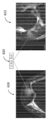
도 2는 여기에서 설명되는 하나 이상의 실시예들에 따라, 해부학적 관절의 일부분의 손상 이미지를 생성하기 위한 방법에 대한 순서도이다.
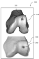
도 3은 여기에서 설명되는 하나 이상의 실시예들에 따라, 손상 이미지의 형태로 의사결정 지원 자료의 일례를 도시하며, 여기서 해부학적 관절에 대한 손상은 그래픽(graphics)을 사용하여 마킹된다.
도 4는 여기에서 사용되는 하나 이상의 실시예들에 따라, 해부학적 관절의 일부분의 손상 이미지를 생성하기 위한 방법에 대한 순서도이다.
도 5는 여기에서 사용되는 하나 이상의 실시예들에 따라, 다수의 손상 이미지들을 포함하는 손상 보고(report) 형태의 의사결정 지원 자료의 일례를 도시하며, 여기서 해부학적 관절에 대한 손상이 마킹되고/마킹되거나 적절한 임플란트의 유형 그리고 자리가 표시된다.
도 6는 여기에서 설명되는 하나 이상의 실시예들에 따라, 손상 이미지의 형태로 의사결정 지원 자료의 일례를 도시하며, 여기서 해부학적 관절에 대한 손상은 주석(annotation)을 사용하여 마킹된다.
도 7는 여기에서 설명되는 하나 이상의 실시예들에 따라, 의학적 이미지 데이터를 획득하는 단계부터, 손상 마킹 그리고 손상 마킹 이미지의 생성 단계를 포함하는, 해부학적 관절에 대한 결정된 손상을 수리하기 위한 임플란트 그리고/또는 가이드 툴을 설계하고 제조하는 단계를 예시하는 순서도이다.
본 발명의 실시예들 그리고 그 장점들은 다음의 상세한 설명을 참조함으로써 가장 잘 이해될 수 있다. 하나 이상의 도면들에 도시된 유사한 요소들을 식별하기 위해 동일한 참조 번호들이 사용된다는 것을 이해해야한다.
Claims (24)
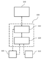
- 환자의 해부학적 관절의 적어도 일부에 대한 손상(damage)을 표시하는 의사결정 지원 자료(decision support material)를 생성하기 위한 시스템(100)으로서,
상기 생성된 의사결정 지원 자료는 하나 이상의 손상 이미지들을 포함하고, 그리고,
상기 시스템(100)은:
저장 매체(110) 및 프로세서(120)를 포함하고,
상기 프로세서(120)는:
i) 상기 저장 매체(110)로부터 상기 해부학적 관절의 적어도 일부에 대한 일련의 방사선 이미지(radiology image)들을 수신하고;
ii) 저장 매체(110)로부터 3차원 이미지 표현을 수신하거나 또는 상기 일련의 방사선 이미지들에 기초하여 이미지 분할(segmentation) 프로세스에서 상기 3차원 이미지 표현을 생성함으로써 상기 일련의 방사선 이미지들에 기초하는 해부학적 관절의 적어도 일부에 대한 3차원 이미지 표현을 획득하고;
iii) 이미지 분석을 사용하여 일련의 방사선 이미지들 및 상기 3차원 이미지 표현 중 적어도 하나에서, 연골, 힘줄 및 인대 중 적어도 하나를 포함하는 해부학적 관절의 조직 부분을 식별하고;
iv) 해부학적 관절의 적어도 일부에 대한 3차원 이미지 표현 및 일련의 방사선 이미지들 중 적어도 하나를 분석함으로써 상기 해부학적 관절에서 식별된 조직 부분들에 대한 손상을 결정하고,
여기서 상기 분석은 상기 식별된 조직 부분들을 사용하며 이하의 단계들 중 적어도 하나의 단계에 대한 선택을 포함하며, 상기 단계는:
상기 해부학적 관절의 적어도 하나의 조직 부분의 윤곽의 불규칙한 형상을 검출하는 단계;
상기 해부학적 관절의 뼈 부분 및 연골 부분 중 적어도 하나의 내부 또는 인접한 영역의 강도(intensity)가 사전결정된값 보다 높거나 낮은지를 검출하는 단계; 및
해부학적 관절에 대한 사전정의된 손상 패턴을 나타내는 템플릿(template)과 적어도 하나의 식별된 조직 부분을 비교하는 단계;
를 포함하며,
v) 상기 해부학적 관절의 획득된 3차원 이미지 표현에서 상기 해부학적 관절에 대한 손상을 마킹하고(mark); 그리고
vi) 의사결정 지원 자료를 생성하도록 구성되며,
여기서 상기 해부학적 관절의 일부에 대한 결정된 손상은 상기 의사결정 지원 자료의 하나 이상의 손상 이미지들 중 적어도 하나에 마킹되고, 그리고 상기 해부학적 관절의 적어도 일부에 대해 상기 획득된 3차원 이미지 표현을 기초로 하여 상기 손상 이미지들 중 적어도 하나가 생성되는;
시스템. - 제 1 항에 있어서,
상기 프로세서(120)는:
상기 방사선 이미지에서의 모서리(edge)들 또는 윤곽(contour)들을 포함하는 경조 영역(high contrast area)들을 검출하는 단계; 및
상기 검출된 모서리들 또는 윤곽들을 사전정의된 템플릿들과 비교함으로써 상기 방사선 이미지에서 뼈 및 연골 중 적어도 하나를 포함하는 구조체(structure)들을 식별하는 단계;
에 의하여,
상기 적어도 하나의 방사선 이미지에서 상기 관절의 뼈 부분 및 연골 부분 중 적어도 하나를 식별하도록 구성되는,
시스템. - 제 1 항 또는 제 2 항에 있어서,
상기 프로세서(120)는, 상기 방사선 이미지와 상기 3차원 이미지 표현을 연관시키도록 추가로 구성되어, 상기 이미지들 중 하나에서 생성된 마킹(marking)이 다른 이미지의 동일한 위치에 나타나도록 하는,
시스템. - 제 1 항 또는 제 2 항에 있어서,
상기 3차원 이미지 표현은 분할 프로세스 제어 파라미터 세트(segmentaion process contro parameter set)에 의존하는 이미지 분할 프로세스에서 생성되는,
시스템. - 제 1 항 또는 제 2 항에 있어서,
상기 이미지 분석은 상기 해부학적 관절 또는 그 일부에 대한 뼈 부분들 및 연골 부분들 모두를 식별하고, 그리고 손상은 상기 뼈 부분들 및 연골 부분들 모두에 대해 결정되는,
시스템. - 제 1 항 또는 제 2 항에 있어서,
상기 해부학적 관절은 무릎 또는 발목인,
시스템. - 제 1 항 또는 제 2 항에 있어서,
상기 프로세서(120)는 상기 해부학적 관절의 적어도 일부에 대한 3차원 이미지 표현 및 상기 방사선 이미지로부터의 데이터에 기초하여 사전정의된 치료 세트로부터 적절한 치료를 선택하도록 추가적으로 구성되는,
시스템. - 제 7 항에 있어서,
상기 프로세서(120)는:
가변 치수를 갖는 사전정의된 임플란트 세트로부터 적절한 임플란트를 선택하는 것; 및
적어도 하나의 골연골 자가이식 플러그에 대한 적절한 수확(harvesting) 위치, 적절한 임플란트 위치 및 적절한 크기 중 적어도 하나를 포함하는 골연골 자가이식 이송을 위한 이송 가이드 툴(transfer guide tool)을 제안하는 것;
중 적어도 하나를 수행하도록 구성되는,
시스템. - 제 8 항에 있어서,
상기 프로세서(120)는,
상기 하나 이상의 손상 이미지들 중 적어도 하나에서 적어도 하나의 골연골 자가이식 플러그에 대한 적절한 임플란트 위치, 적절한 수확 위치, 이송 가이드 툴 및 선택된 임플란트 중 적어도 하나를 시각화하도록 추가로 구성되는,
시스템. - 제 1 항 또는 제 2 항에 있어서,
상기 의사결정 지원 자료는 의료진에게 사용하도록 적응되는,
시스템. - 제 10 항에 있어서, 상기 의사결정 지원 자료는 상기 결정된 손상의 수리를 위한 적절한 치료를 위한 권고사항(recommendation)을 포함하는,
시스템. - 환자의 해부학적 관절의 적어도 일부에 대한 손상을 표시하는 의사결정 지원 자료를 생성하기 위한 방법으로서,
상기 생성된 의사결정 지원 자료는 하나 이상의 손상 이미지들을 포함하며,
상기 방법은:
i) 상기 해부학적 관절의 적어도 일부에 대한 일련의 방사선 이미지들을 수신하는 단계(210);
ii) 저장 매체(110)로부터 3차원 이미지 표현을 수신하거나 또는 상기 방사선 이미지들에 기초하여 이미지 분할 프로세스에서 상기 3차원 이미지 표현을 생성함으로써 상기 일련의 방사선 이미지들에 기초하는 상기 해부학적 관절의 적어도 일부에 대한 3차원 이미지 표현을 확득하는 단계(220);
iii) 이미지 분석을 사용하여 상기 일련의 방사선 이미지들 중 적어도 하나에서, 연골, 힘줄 및 인대 중 적어도 하나를 포함하는, 상기 해부학적 관절의 조직 부분들을 식별하는 단계(230);
iv) 이하의 서브 단계들 중 적어도 하나에 대한 선택 및 상기 식별된 조직 부분들을 사용하여 상기 해부학적 관절의 적어도 일부에 대한 3차원 이미지 표현 및 상기 일련의 방사선 이미지들 중 적어도 하나를 분석함으로써 상기 해부학적 관절에서 상기 식별된 조직 부분들에 대한 손상을 결정하는 단계(240);
여기서 상기 서브 단계들은:
상기 해부학적 관절의 적어도 하나의 조직 부분의 윤곽의 불규칙한 형상을 검출하는 단계;
상기 해부학적 관절의 뼈 부분 및 연골 부분 중 적어도 하나의 내부의 또는 인접한 영역의 강도가 사전결정된 값보다 높거나 낮은지를 검출하는 단계; 및
해부학적 관절에 대하여 사전정의된 손상 패턴을 나타내는 템플릿과 적어도 하나의 식별된 조직 부분을 비교하는 단계;
를 포함하며,
v) 상기 해부학적 관절의 적어도 일부에 대하여 획득된 3차원 이미지 표현에서 상기 해부학적 관절에 대한 손상을 마킹하는 단계(250); 및
vi) 의사결정 지원 자료를 생성하는 단계(260);
를 포함하며,
상기 해부학적 관절의 일부에 대한 결정된 손상은 상기 의사결정 지원 자료의 하나 이상의 손상 이미지들 중 적어도 하나에 마킹되고, 그리고 상기 해부학적 관절의 적어도 일부에 대해 상기 획득된 3차원 이미지 표현을 기초로 하여 상기 손상 이미지들 중 적어도 하나가 생성되는,
방법. - 제 12 항에 있어서,
상기 이미지 분석은:
상기 방사선 이미지에서의 모서리들 또는 윤곽들을 포함하는 경조 영역들을 검출하는 단계; 및
상기 검출된 모서리들 또는 윤곽들을 사전정의된 템플릿들과 비교함으로써 상기 방사선 이미지에서 뼈 및 연골 중 적어도 하나를 포함하는 구조체들을 식별하는 단계;
에 의하여,
적어도 하나의 방사선 이미지에서 상기 관절의 뼈 부분 및 연골 부분 중 적어도 하나를 식별하는,
방법. - 제 12 항 또는 제 13 항에 있어서,
상기 방사선 이미지와 상기 3차원 이미지 표현는 연관되어, 상기 이미지들 중 하나의 마킹이 다른 이미지의 동일한 위치에 나타나도록 하는,
방법. - 제 12 항 또는 제 13 항에 있어서,
상기 3차원 이미지 표현은 분할 프로세스 제어 파라미터 세트에 의존하는 이미지 분할 프로세스에서 생성되는,
방법. - 제 12 항 또는 제 13 항에 있어서,
상기 이미지 분석은 상기 해부학적 관절 또는 그 일부의 뼈 부분들 및 연골 부분들 모두를 식별하고 그리고 손상은 상기 뼈 부분들과 연골 부분들 모두에 대해 결정되는,
방법. - 제 12 항 또는 제 13 항에 있어서,
해부학적 관절은 무릎 또는 발목인,
방법. - 제 12 항 또는 제 13 항에 있어서,
상기 해부학적 관절의 적어도 일부에 대한 3차원 이미지 표현 및 상기 방사선 이미지로부터의 데이터에 기초하여 사전정의된 치료 세트로부터 적절한 치료를 선택하는 단계;
를 더 포함하는,
방법. - 제 18 항에 있어서,
가변 치수를 갖는 사전정의된 임플란트 세트로부터 적절한 임플란트를 선택하는 단계; 및
적어도 하나의 골연골 자가이식 플러그에 대한 적절한 수확 위치, 적절한 임플란트 위치 및 적절한 크기 중 적어도 하나를 포함하는 골연골 자가이식 이송을 위한 이송 가이드 툴을 제안하는 단계;
중 적어도 하나의 단계를 더 포함하는,
방법. - 제 19 항에 있어서,
하나 이상의 손상 이미지들 중 적어도 하나에서 적어도 하나의 골연골 자가이식 플러그에 대한 적절한 임플란트 위치, 적절한 수확 위치, 이송 가이드 툴 및 선택된 임플란트 중 적어도 하나를 시각화하는 단계;
를 더 포함하는,
방법. - 제 12 항 또는 제 13 항에 있어서,
상기 의사결정 지원 자료는 의료진에게 사용하도록 적응되는,
방법 - 제 21 항에 있어서,
상기 의사결정 지원 자료는 상기 결정된 손상의 수리를 위한 적절한 치료를 위한 권고사항을 포함하는,
방법. - 환자의 해부학적 관절의 적어도 일부에 대한 손상을 표시하는 의사결정 지원 자료로서,
상기 의사결정 지원 자료는, 제 12 항 또는 제 13 항의 방법에 의해 생성된 하나 이상의 손상 이미지들을 포함하는,
의사결정 지원 자료 - 프로세서에 의해 실행될 때 프로세서로 하여금 제 12 항 또는 제 13 항의 방법을 수행하도록 제어하는 머신-판독 가능 코드가 저장된,
비-일시적 머신-판독 가능 매체.
Applications Claiming Priority (3)
| Application Number | Priority Date | Filing Date | Title |
|---|---|---|---|
| EP15201361.1 | 2015-12-18 | ||
| EP15201361.1A EP3181050B1 (en) | 2015-12-18 | 2015-12-18 | System and method for creating a decision support material indicating damage to an anatomical joint |
| PCT/EP2016/081483 WO2017103146A1 (en) | 2015-12-18 | 2016-12-16 | System and method for creating a decision support material indicating damage to an anatomical joint |
Publications (2)
| Publication Number | Publication Date |
|---|---|
| KR20180098586A true KR20180098586A (ko) | 2018-09-04 |
| KR102119542B1 KR102119542B1 (ko) | 2020-06-05 |
Family
ID=54850458
Family Applications (1)
| Application Number | Title | Priority Date | Filing Date |
|---|---|---|---|
| KR1020187020676A Active KR102119542B1 (ko) | 2015-12-18 | 2016-12-16 | 해부학적 관절에 대한 손상을 표시하는 의사결정 지원 자료를 생성하기 위한 시스템 그리고 방법 (system and method for creating a decision support material indicating damage to an anatomical joint) |
Country Status (12)
| Country | Link |
|---|---|
| US (4) | US9697601B1 (ko) |
| EP (2) | EP3181050B1 (ko) |
| JP (1) | JP6736832B2 (ko) |
| KR (1) | KR102119542B1 (ko) |
| CN (1) | CN108366770B (ko) |
| AU (1) | AU2016374552B2 (ko) |
| CA (1) | CA3003809C (ko) |
| DE (1) | DE112016005277T5 (ko) |
| DK (1) | DK3181050T3 (ko) |
| ES (1) | ES2842498T3 (ko) |
| GB (1) | GB2560666B (ko) |
| WO (1) | WO2017103146A1 (ko) |
Cited By (1)
| Publication number | Priority date | Publication date | Assignee | Title |
|---|---|---|---|---|
| KR102758047B1 (ko) * | 2024-11-01 | 2025-01-22 | 주식회사 디픽스 | 인공 지능 기반의 관절 상태 진단 및 치료 시스템 |
Families Citing this family (18)
| Publication number | Priority date | Publication date | Assignee | Title |
|---|---|---|---|---|
| CN108352184B (zh) * | 2015-10-29 | 2022-10-04 | 佳能株式会社 | 医用图像处理装置、可安装到医用图像处理装置中的程序和医用图像处理方法 |
| US11526988B2 (en) | 2015-12-18 | 2022-12-13 | Episurf Ip-Management Ab | System and method for creating a decision support material indicating damage to an anatomical joint |
| EP3181050B1 (en) | 2015-12-18 | 2020-02-12 | Episurf IP Management AB | System and method for creating a decision support material indicating damage to an anatomical joint |
| US11250561B2 (en) | 2017-06-16 | 2022-02-15 | Episurf Ip-Management Ab | Determination and visualization of damage to an anatomical joint |
| EP3416131A1 (en) * | 2017-06-16 | 2018-12-19 | Episurf IP-Management AB | System and method for creating a decision support material indicating damage to an anatomical joint |
| KR102792526B1 (ko) * | 2018-07-10 | 2025-04-09 | 애니메디솔루션 주식회사 | 연골을 모델링하는 방법 및 상기 방법을 이용한 코연골을 모델링하는 방법 |
| JP7479062B2 (ja) * | 2018-10-01 | 2024-05-08 | オーピー ソリューションズ, エルエルシー | 指数関数的分割の方法およびシステム |
| USD918258S1 (en) | 2018-10-08 | 2021-05-04 | Episurf Ip Management Ab | Display screen with epioscopy icon |
| USD908733S1 (en) * | 2018-10-15 | 2021-01-26 | Episurf Ip Management Ab | Display screen with epioscopy icon |
| WO2020119930A1 (en) | 2018-12-14 | 2020-06-18 | Episurf Ip-Management Ab | Determination and visualization of damage to an anatomical joint |
| US11645749B2 (en) | 2018-12-14 | 2023-05-09 | Episurf Ip-Management Ab | Determination and visualization of damage to an anatomical joint |
| EP3895175A1 (en) * | 2018-12-14 | 2021-10-20 | Episurf IP Management AB | Determination and visualization of damage to an anatomical joint |
| US12562253B2 (en) * | 2019-12-06 | 2026-02-24 | The Boeing Company | Soft tissue material cumulative damage model for reducing repetitive stress injuries in performing a process |
| US20230200826A1 (en) * | 2020-05-25 | 2023-06-29 | Orthopaedic Innovations Pty Ltd | A surgical method |
| US11621086B2 (en) | 2020-06-04 | 2023-04-04 | Episurf Ip-Management Ab | Customization of individualized implant |
| KR102716587B1 (ko) * | 2022-02-08 | 2024-10-15 | 주식회사 크레스콤 | 관절 상태를 정량화하기 위한 의료 영상 분석 방법, 의료 영상 분석 장치, 및 의료 영상 분석 시스템 |
| AU2024213417A1 (en) * | 2023-02-02 | 2025-08-07 | Mako Surgical Corporation | Method of assessment of a joint |
| JP2024121523A (ja) * | 2023-02-27 | 2024-09-06 | キヤノンメディカルシステムズ株式会社 | 医用情報処理装置、及び、医用情報処理システム |
Citations (2)
| Publication number | Priority date | Publication date | Assignee | Title |
|---|---|---|---|---|
| WO2002087444A1 (en) * | 2001-04-26 | 2002-11-07 | Teijin Limited | Three-dimensional joint structure measuring method |
| JP2014000425A (ja) * | 2002-11-27 | 2014-01-09 | Conformis Inc | 全体的または部分的関節形成において正確さ、速度及び単純さの増加を容易にする患者により選択可能な関節形成装置と手術ツール |
Family Cites Families (61)
| Publication number | Priority date | Publication date | Assignee | Title |
|---|---|---|---|---|
| US8083745B2 (en) | 2001-05-25 | 2011-12-27 | Conformis, Inc. | Surgical tools for arthroplasty |
| US8556983B2 (en) | 2001-05-25 | 2013-10-15 | Conformis, Inc. | Patient-adapted and improved orthopedic implants, designs and related tools |
| US8882847B2 (en) | 2001-05-25 | 2014-11-11 | Conformis, Inc. | Patient selectable knee joint arthroplasty devices |
| US8480754B2 (en) | 2001-05-25 | 2013-07-09 | Conformis, Inc. | Patient-adapted and improved articular implants, designs and related guide tools |
| US8545569B2 (en) | 2001-05-25 | 2013-10-01 | Conformis, Inc. | Patient selectable knee arthroplasty devices |
| JPH11104072A (ja) | 1997-10-03 | 1999-04-20 | Mitsubishi Electric Corp | 医療支援システム |
| US7239908B1 (en) | 1998-09-14 | 2007-07-03 | The Board Of Trustees Of The Leland Stanford Junior University | Assessing the condition of a joint and devising treatment |
| JP2002532126A (ja) | 1998-09-14 | 2002-10-02 | スタンフォード ユニバーシティ | 関節状態の評価及び損傷防止装置 |
| US9289153B2 (en) | 1998-09-14 | 2016-03-22 | The Board Of Trustees Of The Leland Stanford Junior University | Joint and cartilage diagnosis, assessment and modeling |
| DE19922279A1 (de) | 1999-05-11 | 2000-11-16 | Friedrich Schiller Uni Jena Bu | Verfahren zur Generierung patientenspezifischer Implantate |
| EP1319217B1 (en) | 2000-09-14 | 2008-11-12 | The Board Of Trustees Of The Leland Stanford Junior University | Technique for manipulating medical images |
| AU2001290887B2 (en) | 2000-09-14 | 2006-06-08 | The Board Of Trustees Of The Leland Stanford Junior University | Assessing condition of a joint and cartilage loss |
| EP1322225B1 (en) | 2000-09-14 | 2009-03-25 | The Board Of Trustees Of The Leland Stanford Junior University | Assessing the condition of a joint and devising treatment |
| EP1389980B1 (en) | 2001-05-25 | 2011-04-06 | Conformis, Inc. | Methods and compositions for articular resurfacing |
| FR2831794B1 (fr) | 2001-11-05 | 2004-02-13 | Depuy France | Procede de selection d'elements de prothese de genou et dispositif pour sa mise en oeuvre |
| JP2003144454A (ja) * | 2001-11-16 | 2003-05-20 | Yoshio Koga | 関節手術支援情報算出方法、関節手術支援情報算出プログラム、及び関節手術支援情報算出システム |
| GB2393625C (en) | 2002-09-26 | 2004-08-18 | Meridian Tech Ltd | Orthopaedic surgery planning |
| AU2003290757A1 (en) | 2002-11-07 | 2004-06-03 | Conformis, Inc. | Methods for determing meniscal size and shape and for devising treatment |
| WO2005017806A1 (en) | 2003-08-13 | 2005-02-24 | Siemens Medical Solutions Usa, Inc. | Computer-aided decision support systems and methods |
| US20080232658A1 (en) | 2005-01-11 | 2008-09-25 | Kiminobu Sugaya | Interactive Multiple Gene Expression Map System |
| DE102006015452A1 (de) * | 2006-03-31 | 2007-10-11 | Siemens Ag | Verfahren und Vorrichtung zur Detektion von chemischen Anomalien und/oder Auffälligkeiten in Weichgewebe eines Objektbereiches |
| US8300910B2 (en) | 2006-09-19 | 2012-10-30 | Synarc Inc. | Pathology indicating measure related to cartilage structure and automatic quantification thereof |
| JP4934786B2 (ja) * | 2006-10-13 | 2012-05-16 | 国立大学法人 東京大学 | 膝関節診断支援方法及び装置並びにプログラム |
| WO2008101090A2 (en) | 2007-02-14 | 2008-08-21 | Conformis, Inc. | Implant device and method for manufacture |
| CA2696584C (en) * | 2007-08-17 | 2016-11-29 | Mohamed Rashwan Mahfouz | Implant design analysis suite |
| US8617171B2 (en) | 2007-12-18 | 2013-12-31 | Otismed Corporation | Preoperatively planning an arthroplasty procedure and generating a corresponding patient specific arthroplasty resection guide |
| US8715291B2 (en) | 2007-12-18 | 2014-05-06 | Otismed Corporation | Arthroplasty system and related methods |
| US8777875B2 (en) | 2008-07-23 | 2014-07-15 | Otismed Corporation | System and method for manufacturing arthroplasty jigs having improved mating accuracy |
| US8160345B2 (en) * | 2008-04-30 | 2012-04-17 | Otismed Corporation | System and method for image segmentation in generating computer models of a joint to undergo arthroplasty |
| US8737700B2 (en) | 2007-12-18 | 2014-05-27 | Otismed Corporation | Preoperatively planning an arthroplasty procedure and generating a corresponding patient specific arthroplasty resection guide |
| US8480679B2 (en) * | 2008-04-29 | 2013-07-09 | Otismed Corporation | Generation of a computerized bone model representative of a pre-degenerated state and useable in the design and manufacture of arthroplasty devices |
| US8545509B2 (en) | 2007-12-18 | 2013-10-01 | Otismed Corporation | Arthroplasty system and related methods |
| US8221430B2 (en) | 2007-12-18 | 2012-07-17 | Otismed Corporation | System and method for manufacturing arthroplasty jigs |
| CN102014800B (zh) | 2008-02-28 | 2014-04-30 | 比奥波利公司 | 局部关节表面置换植入物、仪器和方法 |
| US9216085B2 (en) | 2008-02-28 | 2015-12-22 | Biopoly, Llc | Partial joint resurfacing implant, instrumentation, and method |
| WO2009132340A1 (en) | 2008-04-25 | 2009-10-29 | Stratovan Corporation | Analysis of anatomic regions delineated from image data |
| US9020577B2 (en) * | 2008-05-29 | 2015-04-28 | Yale University | Systems, devices and methods for cartilage and bone grafting |
| US8613937B2 (en) * | 2008-10-31 | 2013-12-24 | The Invention Science Fund I, Llc | Compositions and methods for biological remodeling with frozen particle compositions |
| WO2010099231A2 (en) | 2009-02-24 | 2010-09-02 | Conformis, Inc. | Automated systems for manufacturing patient-specific orthopedic implants and instrumentation |
| US9078755B2 (en) | 2009-02-25 | 2015-07-14 | Zimmer, Inc. | Ethnic-specific orthopaedic implants and custom cutting jigs |
| EP2400934A4 (en) | 2009-02-25 | 2015-07-08 | Zimmer Inc | TAILOR-MADE ORTHOPEDIC IMPLANTS AND APPROPRIATE PROCEDURES |
| JP5346859B2 (ja) * | 2009-04-15 | 2013-11-20 | 富士フイルム株式会社 | 医用画像管理装置および方法並びにプログラム |
| US20110054486A1 (en) | 2009-08-25 | 2011-03-03 | Active Implants Corporation | Devices, Methods, and Systems for Prosthetic Meniscus Selection, Trialing, and Implantation |
| EP2389901B8 (en) * | 2010-05-24 | 2013-05-15 | Episurf IP Management AB | An implant for cartilage repair |
| EP2389905B1 (en) | 2010-05-24 | 2012-05-23 | Episurf Medical AB | Method of designing a surgical kit for cartilage repair in a joint |
| EP2389899B1 (en) | 2010-05-24 | 2015-04-29 | Episurf IP Management AB | Method of manufacturing a surgical kit for cartilage repair in a joint |
| WO2012021861A2 (en) | 2010-08-13 | 2012-02-16 | Mckinnon, Brian William | Detection of anatomical landmarks |
| EP2514373B1 (en) * | 2011-04-21 | 2013-12-04 | Episurf IP Management AB | Guide tool for cartilage repair |
| FR2974286B1 (fr) * | 2011-04-22 | 2013-05-17 | Genourob | Appareil et procede de detection d'une lesion partielle de ligament croise anterieur du genou |
| US20170100253A1 (en) * | 2011-09-02 | 2017-04-13 | Episurf Ip-Management Ab | Design of an implant for cartilage repair |
| WO2013056036A1 (en) | 2011-10-14 | 2013-04-18 | Conformis, Inc. | Methods and systems for identification, assessment, modeling, and repair of anatomical disparities in joint replacement |
| US20140039454A1 (en) * | 2012-08-03 | 2014-02-06 | Zimmer Gmbh, Inc. | Methods of treating subchondral bone to prevent the progression of osteoarthritis in joints |
| EP2967813A4 (en) | 2013-03-15 | 2016-11-09 | Conformis Inc | KINEMATIC AND PARAMETRIZED MODELING FOR PATIENT-IMPLANTED IMPLANTS, TOOLS AND SURGICAL PROCEDURES |
| EP3013256B1 (en) | 2013-06-28 | 2018-11-28 | Episurf IP Management AB | Guide tool for bone and/or cartilage repair or joint remodeling |
| WO2014206498A1 (en) | 2013-06-28 | 2014-12-31 | Episurf Ip-Management Ab | Guide tool for cartilage and/or bone repair or joint remodeling |
| EP3080619B1 (en) | 2013-12-09 | 2019-02-13 | Mohamed R. Mahfouz | Bone reconstruction and orthopedic implants |
| JP6442530B2 (ja) * | 2014-01-27 | 2018-12-19 | マテリアライズ・ナムローゼ・フエンノートシャップMaterialise Nv | 形状の予測 |
| EP3102154B1 (en) * | 2014-02-07 | 2018-05-30 | Episurf IP Management AB | Method for manufacturing a surgical kit for cartilage repair |
| WO2016004992A1 (en) | 2014-07-09 | 2016-01-14 | Episurf Ip-Management Ab | A surgical joint implant and a bone-mountable rig |
| WO2016004991A1 (en) | 2014-07-09 | 2016-01-14 | Episurf Ip-Management Ab | Customized implant for cartilage repair and corresponding method of design |
| EP3181050B1 (en) | 2015-12-18 | 2020-02-12 | Episurf IP Management AB | System and method for creating a decision support material indicating damage to an anatomical joint |
-
2015
- 2015-12-18 EP EP15201361.1A patent/EP3181050B1/en active Active
- 2015-12-18 DK DK15201361.1T patent/DK3181050T3/da active
-
2016
- 2016-12-16 WO PCT/EP2016/081483 patent/WO2017103146A1/en not_active Ceased
- 2016-12-16 CA CA3003809A patent/CA3003809C/en active Active
- 2016-12-16 KR KR1020187020676A patent/KR102119542B1/ko active Active
- 2016-12-16 GB GB1809147.0A patent/GB2560666B/en active Active
- 2016-12-16 JP JP2018525771A patent/JP6736832B2/ja active Active
- 2016-12-16 ES ES16819868T patent/ES2842498T3/es active Active
- 2016-12-16 DE DE112016005277.4T patent/DE112016005277T5/de not_active Withdrawn
- 2016-12-16 EP EP16819868.7A patent/EP3389495B1/en active Active
- 2016-12-16 CN CN201680072508.XA patent/CN108366770B/zh active Active
- 2016-12-16 US US15/382,523 patent/US9697601B1/en active Active
- 2016-12-16 AU AU2016374552A patent/AU2016374552B2/en active Active
-
2017
- 2017-06-01 US US15/611,685 patent/US9990720B2/en active Active
-
2018
- 2018-05-04 US US15/971,849 patent/US10672124B2/en active Active
-
2020
- 2020-05-28 US US16/886,650 patent/US11282195B2/en active Active
Patent Citations (2)
| Publication number | Priority date | Publication date | Assignee | Title |
|---|---|---|---|---|
| WO2002087444A1 (en) * | 2001-04-26 | 2002-11-07 | Teijin Limited | Three-dimensional joint structure measuring method |
| JP2014000425A (ja) * | 2002-11-27 | 2014-01-09 | Conformis Inc | 全体的または部分的関節形成において正確さ、速度及び単純さの増加を容易にする患者により選択可能な関節形成装置と手術ツール |
Cited By (1)
| Publication number | Priority date | Publication date | Assignee | Title |
|---|---|---|---|---|
| KR102758047B1 (ko) * | 2024-11-01 | 2025-01-22 | 주식회사 디픽스 | 인공 지능 기반의 관절 상태 진단 및 치료 시스템 |
Also Published As
| Publication number | Publication date |
|---|---|
| CA3003809A1 (en) | 2017-06-22 |
| JP6736832B2 (ja) | 2020-08-05 |
| US20200294232A1 (en) | 2020-09-17 |
| US20180253847A1 (en) | 2018-09-06 |
| EP3181050A1 (en) | 2017-06-21 |
| DE112016005277T5 (de) | 2018-09-13 |
| JP2019505247A (ja) | 2019-02-28 |
| CN108366770A (zh) | 2018-08-03 |
| US20170270665A1 (en) | 2017-09-21 |
| AU2016374552A1 (en) | 2018-05-17 |
| CN108366770B (zh) | 2021-11-12 |
| US11282195B2 (en) | 2022-03-22 |
| KR102119542B1 (ko) | 2020-06-05 |
| GB2560666A (en) | 2018-09-19 |
| EP3181050B1 (en) | 2020-02-12 |
| US10672124B2 (en) | 2020-06-02 |
| GB201809147D0 (en) | 2018-07-18 |
| DK3181050T3 (da) | 2020-05-11 |
| ES2842498T3 (es) | 2021-07-14 |
| EP3389495B1 (en) | 2020-10-07 |
| US9990720B2 (en) | 2018-06-05 |
| GB2560666B (en) | 2019-12-04 |
| US9697601B1 (en) | 2017-07-04 |
| CA3003809C (en) | 2023-06-06 |
| EP3389495A1 (en) | 2018-10-24 |
| WO2017103146A1 (en) | 2017-06-22 |
| AU2016374552B2 (en) | 2019-02-14 |
| US20170178323A1 (en) | 2017-06-22 |
Similar Documents
| Publication | Publication Date | Title |
|---|---|---|
| US11282195B2 (en) | System and method for creating a decision support material indicating damage to an anatomical joint | |
| US11526988B2 (en) | System and method for creating a decision support material indicating damage to an anatomical joint | |
| US11250561B2 (en) | Determination and visualization of damage to an anatomical joint | |
| US20230317298A1 (en) | Customization of individualized implant | |
| US20180365827A1 (en) | Creation of a decision support material indicating damage to an anatomical joint | |
| US20180360540A1 (en) | System and method for creating a decision support material indicating damage to an anatomical joint | |
| US11645749B2 (en) | Determination and visualization of damage to an anatomical joint | |
| TWI805591B (zh) | 用於建立指示解剖學關節損傷之決策支持材料的系統及方法 | |
| HK1239490A1 (en) | System and method for creating a decision support material indicating damage to an anatomical joint | |
| HK1239490A (en) | System and method for creating a decision support material indicating damage to an anatomical joint | |
| EP3895175A1 (en) | Determination and visualization of damage to an anatomical joint | |
| US20180177553A1 (en) | System and method for optimizing an implant position in an anatomical joint | |
| WO2020119930A1 (en) | Determination and visualization of damage to an anatomical joint | |
| Kallonen et al. | ATLAS-BASED TEMPLATE MATCHING TO DETERMINE KNEE ALIGNMENT FROM PLAIN LONG-LEG RADIOGRAPHS |
Legal Events
| Date | Code | Title | Description |
|---|---|---|---|
| A201 | Request for examination | ||
| PA0105 | International application |
Patent event date: 20180718 Patent event code: PA01051R01D Comment text: International Patent Application |
|
| PA0201 | Request for examination | ||
| PG1501 | Laying open of application | ||
| E902 | Notification of reason for refusal | ||
| PE0902 | Notice of grounds for rejection |
Comment text: Notification of reason for refusal Patent event date: 20191122 Patent event code: PE09021S01D |
|
| E701 | Decision to grant or registration of patent right | ||
| PE0701 | Decision of registration |
Patent event code: PE07011S01D Comment text: Decision to Grant Registration Patent event date: 20200527 |
|
| GRNT | Written decision to grant | ||
| PR0701 | Registration of establishment |
Comment text: Registration of Establishment Patent event date: 20200601 Patent event code: PR07011E01D |
|
| PR1002 | Payment of registration fee |
Payment date: 20200601 End annual number: 3 Start annual number: 1 |
|
| PG1601 | Publication of registration | ||
| PR1001 | Payment of annual fee |
Payment date: 20240509 Start annual number: 5 End annual number: 5 |
|
| PR1001 | Payment of annual fee |
Payment date: 20250507 Start annual number: 6 End annual number: 6 |